JP2005334219A - Image diagnosis support apparatus and method - Google Patents
Image diagnosis support apparatus and methodDownload PDFInfo
- Publication number
- JP2005334219A JP2005334219AJP2004155941AJP2004155941AJP2005334219AJP 2005334219 AJP2005334219 AJP 2005334219AJP 2004155941 AJP2004155941 AJP 2004155941AJP 2004155941 AJP2004155941 AJP 2004155941AJP 2005334219 AJP2005334219 AJP 2005334219A
- Authority
- JP
- Japan
- Prior art keywords
- past
- subject image
- marker
- image
- subject
- Prior art date
- Legal status (The legal status is an assumption and is not a legal conclusion. Google has not performed a legal analysis and makes no representation as to the accuracy of the status listed.)
- Granted
Links
- 238000003745diagnosisMethods0.000titleclaimsabstractdescription39
- 238000000034methodMethods0.000titleclaimsabstractdescription11
- 239000003550markerSubstances0.000claimsabstractdescription118
- 230000002159abnormal effectEffects0.000claimsabstractdescription50
- 238000002059diagnostic imagingMethods0.000claimsabstractdescription11
- 238000000605extractionMethods0.000claimsdescription3
- 230000000052comparative effectEffects0.000abstractdescription5
- 238000010586diagramMethods0.000description10
- 238000001514detection methodMethods0.000description3
- 239000000284extractSubstances0.000description2
- 238000003384imaging methodMethods0.000description2
- 210000004072lungAnatomy0.000description2
- 238000002360preparation methodMethods0.000description2
- 230000004397blinkingEffects0.000description1
- 210000000481breastAnatomy0.000description1
- 210000000621bronchiAnatomy0.000description1
- 238000012217deletionMethods0.000description1
- 230000037430deletionEffects0.000description1
- 238000007689inspectionMethods0.000description1
- 210000002429large intestineAnatomy0.000description1
- 230000002194synthesizing effectEffects0.000description1
Images


Classifications
- G06F19/00—
Landscapes
- Image Processing (AREA)
- Information Retrieval, Db Structures And Fs Structures Therefor (AREA)
- Image Analysis (AREA)
- Apparatus For Radiation Diagnosis (AREA)
- Magnetic Resonance Imaging Apparatus (AREA)
- Medical Treatment And Welfare Office Work (AREA)
- Measuring And Recording Apparatus For Diagnosis (AREA)
- Processing Or Creating Images (AREA)
Abstract
Translated fromJapaneseDescription
Translated fromJapanese本発明は、コンピュータにより医用画像から異常陰影候補を抽出して表示する画像診断支援装置及び方法に関する。 The present invention relates to an image diagnosis support apparatus and method for extracting and displaying abnormal shadow candidates from a medical image by a computer.
医用画像撮影装置により取得された被検体の画像からコンピュータにより異常陰影候補を検出するCAD(Computer Aided Detection) を行う画像診断支援装置では、被検体画像から多値化処理により切り出された陰影領域の特徴量を基準値と比較することなどにより異常陰影候補を抽出し、被検体画像に異常陰影候補を示すマーカを付して表示している(特許文献1参照)。コンピュータにより抽出された異常陰影候補には、偽陽性陰影と呼ばれる実際には異常ではない陰影が含まれ、偽陽性陰影は、画像診断を行う操作者の判断により異常陰影候補から除外される。更に、操作者が被検体画像の所望の箇所にマーカを追加することが可能である。 In an image diagnosis support apparatus that performs CAD (Computer Aided Detection) to detect abnormal shadow candidates from a subject image acquired by a medical imaging apparatus, a shadow region cut out from the subject image by multi-value processing is used. An abnormal shadow candidate is extracted by comparing the feature amount with a reference value, and the subject image is displayed with a marker indicating the abnormal shadow candidate (see Patent Document 1). The abnormal shadow candidates extracted by the computer include shadows that are not actually abnormal, called false positive shadows, and false positive shadows are excluded from the abnormal shadow candidates according to the judgment of the operator who performs the image diagnosis. Furthermore, the operator can add a marker at a desired location in the subject image.
また、画像診断の対象とする被検体画像と同一の被検体の同一部位から過去(例えば一年前)に取得された被検体画像が存在するときには、画像診断の対象とする被検体画像と過去の被検体画像とを並列表示して、比較読影を可能としている。
しかしながら、従来の比較読影では、過去の画像に付されたマーカは表示されないので、例えば過去の画像診断において経過観察を要すると判断された領域があっても、その領域に注目せずに画像診断を終えてしまう可能性がある。 However, in conventional comparative interpretation, markers attached to past images are not displayed. For example, even if there is an area determined to require follow-up in past image diagnosis, image diagnosis is performed without paying attention to that area. May end.
本発明はこのような事情に鑑みてなされたもので、比較読影において過去のマーカを有効利用することができる画像診断支援装置及び方法を提供することを目的とする。 The present invention has been made in view of such circumstances, and an object thereof is to provide an image diagnosis support apparatus and method that can effectively use past markers in comparative interpretation.
前記目的を達成するために、本発明による画像診断支援装置は、医用画像撮影装置により取得された被検体画像を表す画像データを読み込む画像データ読み込み手段と、前記被検体画像から異常陰影候補を抽出する異常陰影候補抽出手段と、前記異常陰影候補を示すマーカと前記被検体画像とを表示手段に重畳表示させる表示制御手段と、前記表示手段上で前記マーカを選択する操作を受け入れる操作手段と、前記選択されたマーカの情報を格納する格納手段と、前記表示制御手段により表示対象とされている被検体画像と同一被検体の同一部位から過去に取得された被検体画像に付された過去のマーカの情報を前記格納手段において検索する過去マーカ検索手段と、を備え、前記過去マーカ検索手段により前記過去のマーカの情報が得られた場合、前記表示制御手段は、前記表示対象とされている被検体画像から抽出された前記異常陰影候補を示す前記マーカと前記表示対象とされている被検体画像と前記過去のマーカとを、前記表示手段に重畳表示させることを特徴とする。 In order to achieve the above object, an image diagnosis support apparatus according to the present invention extracts image data reading means for reading image data representing a subject image acquired by a medical image photographing device, and abnormal shadow candidates from the subject image. An abnormal shadow candidate extracting means, a display control means for superimposing and displaying a marker indicating the abnormal shadow candidate and the subject image on a display means, an operating means for accepting an operation of selecting the marker on the display means, Storage means for storing information on the selected marker, and past images attached to the subject image acquired in the past from the same part of the same subject as the subject image to be displayed by the display control means Past marker search means for searching for marker information in the storage means, and the past marker search means obtains the past marker information. In this case, the display control means includes the marker indicating the abnormal shadow candidate extracted from the subject image to be displayed, the subject image to be displayed, and the past marker. It is characterized by being superimposed on the display means.
好ましくは、前記表示制御手段は、前記表示対象とされている被検体画像から抽出された前記異常陰影候補を示す前記マーカと前記過去のマーカとを、互いに異なる表示形態により前記表示手段に表示させる。 Preferably, the display control unit causes the display unit to display the marker indicating the abnormal shadow candidate extracted from the subject image to be displayed and the past marker in different display forms. .
また好ましくは、前記操作手段は、前記表示手段に表示されている前記被検体画像において所望の箇所に新たなマーカを付す操作を受け入れ、前記格納手段は、前記新たなマーカの情報を格納する。 Preferably, the operation means accepts an operation for attaching a new marker to a desired location in the subject image displayed on the display means, and the storage means stores information on the new marker.
また、本発明による画像診断支援装置は、医用画像撮影装置により取得された被検体画像を表す画像データを読み込む画像データ読み込み手段と、前記被検体画像から異常陰影候補を抽出する異常陰影候補抽出手段と、前記異常陰影候補を示すマーカと前記被検体画像とを表示手段に重畳表示させる表示制御手段と、前記表示手段上で前記マーカを選択する操作を受け入れる操作手段と、前記選択されたマーカと前記被検体画像とを関連付けて格納する格納手段と、前記表示制御手段により表示対象とされている被検体画像と同一被検体の同一部位から過去に取得された被検体画像を前記格納手段において検索し、前記表示対象とされている被検体画像と前記過去の被検体画像との相関値を算出して、該相関値が所定値以上である前記過去の被検体画像を選び出す過去画像検索手段と、を備え、前記過去画像検索手段により前記過去の被検体画像が選び出された場合、前記表示制御手段は、前記選び出された過去の被検体画像に関連付けられたマーカと前記選び出された過去の被検体画像とを、前記表示対象とされている被検体画像と並列に、前記表示手段に重畳表示させることを特徴とする。 The image diagnosis support apparatus according to the present invention includes an image data reading unit that reads image data representing a subject image acquired by a medical image photographing device, and an abnormal shadow candidate extraction unit that extracts an abnormal shadow candidate from the subject image. Display control means for superimposing and displaying a marker indicating the abnormal shadow candidate and the subject image on a display means, operation means for accepting an operation for selecting the marker on the display means, and the selected marker Storage means for associating and storing the subject image, and search for a subject image acquired in the past from the same part of the same subject as the subject image to be displayed by the display control means in the storage means And calculating a correlation value between the subject image that is the display target and the past subject image, and the correlation value is not less than a predetermined value. Past image search means for selecting a specimen image, and when the past subject image is selected by the past image search means, the display control means associates with the selected past subject image. The selected marker and the selected past subject image are superimposed on the display unit in parallel with the subject image to be displayed.
好ましくは、前記操作手段は、前記表示手段に表示されている前記被検体画像において所望の箇所に新たなマーカを付す操作を受け入れ、前記格納手段は、前記新たなマーカと前記被検体画像とを関連付けて格納する。 Preferably, the operation unit accepts an operation of attaching a new marker to a desired location in the subject image displayed on the display unit, and the storage unit stores the new marker and the subject image. Store in association.
また、本発明による画像診断支援方法は、医用画像撮影装置により取得された被検体画像を表す画像データを読み込む画像データ読み込み工程と、前記被検体画像から異常陰影候補を抽出する異常陰影候補抽出工程と、前記異常陰影候補を示すマーカと前記被検体画像とを表示手段に重畳表示させる表示制御工程と、前記表示手段上で前記マーカを選択する操作を受け入れる操作工程と、前記選択されたマーカの情報を格納手段に格納する格納工程と、前記表示制御工程により表示対象とされている被検体画像と同一被検体の同一部位から過去に取得された被検体画像に付された過去のマーカの情報を前記格納手段において検索する過去マーカ検索工程と、を含み、前記過去マーカ検索工程により前記過去のマーカの情報が得られた場合、前記表示制御工程は、前記表示対象とされている被検体画像から抽出された前記異常陰影候補を示す前記マーカと前記表示対象とされている被検体画像と前記過去のマーカとを、前記表示手段に重畳表示させることを特徴とする。 The image diagnosis support method according to the present invention includes an image data reading step of reading image data representing a subject image acquired by a medical image photographing device, and an abnormal shadow candidate extracting step of extracting an abnormal shadow candidate from the subject image. A display control step of superimposing and displaying a marker indicating the abnormal shadow candidate and the subject image on a display unit, an operation step of accepting an operation of selecting the marker on the display unit, and the selected marker A storage step of storing information in the storage means, and information on past markers attached to the subject image acquired in the past from the same part of the same subject as the subject image to be displayed by the display control step Past marker search step of searching in the storage means, and when the past marker information is obtained by the past marker search step, In the display control step, the display means displays the marker indicating the abnormal shadow candidate extracted from the subject image that is the display target, the subject image that is the display target, and the past marker. It is characterized by being displayed in a superimposed manner.
好ましくは、前記表示制御工程は、前記表示対象とされている被検体画像から抽出された前記異常陰影候補を示す前記マーカと前記過去のマーカとを、互いに異なる表示形態により前記表示手段に表示させる。 Preferably, the display control step causes the display unit to display the marker indicating the abnormal shadow candidate extracted from the subject image to be displayed and the past marker in different display forms. .
また好ましくは、前記操作工程は、前記表示手段に表示されている前記被検体画像において所望の箇所に新たなマーカを付す操作を受け入れ、前記格納工程は、前記新たなマーカの情報を格納する。 Preferably, the operation step accepts an operation of attaching a new marker to a desired location in the subject image displayed on the display means, and the storage step stores information on the new marker.
また、本発明による画像診断支援方法は、医用画像撮影装置により取得された被検体画像を表す画像データを読み込む画像データ読み込み工程と、前記被検体画像から異常陰影候補を抽出する異常陰影候補抽出工程と、前記異常陰影候補を示すマーカと前記被検体画像とを表示手段に重畳表示させる表示制御工程と、前記表示手段上で前記マーカを選択する操作を受け入れる操作工程と、前記選択されたマーカと前記被検体画像とを関連付けて格納手段に格納する格納工程と、前記表示制御工程により表示対象とされている被検体画像と同一被検体の同一部位から過去に取得された被検体画像を前記格納手段において検索し、前記表示対象とされている被検体画像と前記過去の被検体画像との相関値を算出して、該相関値が所定値以上である前記過去の被検体画像を選び出す過去画像検索工程と、を含み、前記過去画像検索工程により前記過去の被検体画像が選び出された場合、前記表示制御工程は、前記選び出された過去の被検体画像に関連付けられたマーカと前記選び出された過去の被検体画像とを、前記表示対象とされている被検体画像と並列に、前記表示手段に重畳表示させることを特徴とする。 The image diagnosis support method according to the present invention includes an image data reading step of reading image data representing a subject image acquired by a medical image photographing device, and an abnormal shadow candidate extracting step of extracting an abnormal shadow candidate from the subject image. A display control step of superimposing and displaying a marker indicating the abnormal shadow candidate and the subject image on a display unit, an operation step of accepting an operation of selecting the marker on the display unit, and the selected marker A storage step of associating the subject image with the storage unit, and storing the subject image acquired in the past from the same part of the same subject as the subject image to be displayed by the display control step. The correlation value between the subject image to be displayed and the past subject image is calculated by the means, and the correlation value is not less than a predetermined value. A past image search step for selecting a past subject image, and when the past subject image is selected by the past image search step, the display control step includes a step of selecting the past subject image. A marker associated with a sample image and the selected past subject image are superimposed on the display unit in parallel with the subject image to be displayed.
好ましくは、前記操作工程は、前記表示手段に表示されている前記被検体画像において所望の箇所に新たなマーカを付す操作を受け入れ、前記格納工程は、前記新たなマーカと前記被検体画像とを関連付けて格納する。 Preferably, the operation step accepts an operation of attaching a new marker to a desired location in the subject image displayed on the display means, and the storing step includes the new marker and the subject image. Store in association.
本発明によれば、比較読影において過去のマーカを有効利用することができる。 According to the present invention, it is possible to effectively use past markers in comparative interpretation.
以下、添付図面に従って本発明に係る画像診断支援装置の好ましい実施の形態について詳説する。 Hereinafter, preferred embodiments of an image diagnosis support apparatus according to the present invention will be described in detail with reference to the accompanying drawings.
図1は、本発明の一実施の形態による画像診断支援装置の概略構成図である。画像診断支援装置10は、主として、各構成要素の動作を制御する中央処理装置(CPU)12と、画像診断支援装置10の制御プログラムが格納された主メモリ14と、被検体の画像データ及び動作プログラム等が格納されたデータ記録装置16と、表示用の画像データを一時記憶する表示メモリ18と、この表示メモリ18からの画像データに基づいて画像を表示するディスプレイ20と、ディスプレイ20の画面上のソフトスイッチを操作するためのマウス、トラックボール、タッチパネル等のポインティングデバイス22及びポインティングデバイスコントローラ24と、各種パラメータ設定用のキーやスイッチを備えたキーボード26と、画像診断支援装置10をローカルエリアネットワーク、電話回線、インターネット等のネットワークNに接続するためのネットワークアダプタ28と、上記各構成要素を接続するデータバス30とから構成される。データ記録装置16は、画像診断支援装置10に内蔵又は外付けされたメモリ、磁気ディスク等の記憶装置や、取り出し可能な外部メディアに対してデータの書き込み及び読み出しを行う装置や、外部記憶装置とネットワークを介してデータを送受信する装置などでも良い。画像診断支援装置10は、ネットワークアダプタ28及びネットワークNを介して、外部の医用画像撮影装置50や画像データベース60と接続してそれらとの間で画像データを送受信する。この画像データは、被検体の肺野、気管支、乳房、大腸などの様々な撮影対象部位から、X線撮影装置、X線CT装置、MRI装置などの様々な医用画像撮影装置によって得られた画像データであって良く、特に限定されないが、以下では、画像データがX線CT装置によって被検体の肺野から得られたCT画像データである例を説明する。データ記録装置16及び又は画像データベース60には、過去の画像診断の結果を被検体ごとに記録した検査履歴データベース62(図3、4、6及び7参照)が構築されている。 FIG. 1 is a schematic configuration diagram of an image diagnosis support apparatus according to an embodiment of the present invention. The diagnostic
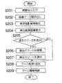
図2は、以上のように構成された画像診断支援装置10によって異常陰影候補を検出して表示すると共に過去のマーカを検索して表示する処理を示すフローチャートである。CPU12は、このフローチャートに従って画像診断支援装置10を制御する。 FIG. 2 is a flowchart showing processing for detecting and displaying an abnormal shadow candidate and searching for and displaying a past marker by the diagnostic
まず、ディスプレイ20に被検体のID入力画面が表示され、操作者は診断処理の対象とする被検体のID番号を入力する(S201)。入力された被検体のID番号に基づいて、データ記録装置16、医用画像撮影装置50又は画像データベース60から異常陰影候補検出処理の対象とする画像データが読み出され(S202)、読み出された画像データに対して異常陰影候補検出処理(S203)が行われる。そして、S203の結果に基づいて、対象画像と検出された異常陰影候補を示すマーカとがディスプレイ20に重畳表示される(S204)。 First, the subject ID input screen is displayed on the
次に、ディスプレイ20に表示されている被検体画像の画像診断の参考とするために、同一被検体の過去のマーカを表示するか否かの選択が行われる(S205)。過去のマーカを表示することが選択された場合には、検査履歴データベース62において同一被検体の同一部位についての記録が検索され(S206)、過去のマーカの情報が得られたら、過去のマーカもディスプレイ20に表示される(S207)。 Next, in order to refer to the image diagnosis of the subject image displayed on the
画像診断支援装置10によって付されたマーカが偽陽性陰影を示している場合や、読影者が異常陰影の疑いを持ったり経過観察が必要だと判断したりした関心領域にマーカが付されていない場合などには、ポインティングデバイス22やキーボード26を介して、マーカの削除及び又は追加が行われる(S208)。そして、選択されたマーカの情報が検査履歴データベース62に格納される(S209)。 When the marker attached by the image
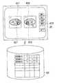
以下、図2のいくつかの工程について詳述する。図3は、ディスプレイ20の表示画面の例及び選択されたマーカの情報が検査履歴データベース62に格納される例を示す概念図である。操作者は、S204においてディスプレイ20に表示された被検体画像n1、n2、…を参照しながら、ポインティングデバイス22やキーボード26を介して、偽陽性陰影を示すマーカや経過観察が不必要だと判断されるマーカを削除したり、まだマーカが付されていない関心領域に新たなマーカを付したりする操作を行う(S208)。その結果、画像診断支援装置10によって自動的に付されて操作者によって削除されなかったマーカ、及び操作者によって新たに付されたマーカが現存することになる。これらの現存するマーカ(301、302)は全て、当該被検体について将来行われる画像診断の参考とするために情報を記録する対象として操作者によって選択されたマーカであるとみなされる。そして、選択されたマーカの情報(マーカが付されている画像の番号や被検体における位置、画像上のマーカの座標、マーカの半径など)が検査履歴データベース62に格納される(S209)。また、現存するマーカのうち、情報を記録する対象として操作者によって特に指定されたマーカのみについて、情報を検査履歴データベース62に格納することとしても良い。 Hereinafter, some steps of FIG. 2 will be described in detail. FIG. 3 is a conceptual diagram illustrating an example of the display screen of the
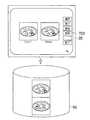
図4は、過去のマーカがディスプレイ20に表示される例を示す概念図である。図3で説明したようにマーカ301、302の情報が格納された後(例えば数ヶ月から数年後)に同一被検体について画像診断が行われる場合、過去のマーカ301、302が表示されると、読影者にその領域に注目させることができる。S204までの工程で被検体画像n1、n4及びマーカ401、402が表示され、操作者が参照ボタン403を選択すると(S205)、同一被検体の同一部位から過去に取得された被検体画像に付された過去のマーカの情報が検査履歴データベース62において検索される(S206)。なお、この検索において、被検体が同一であることは当然必須の要件である。しかしながら、部位については、厳密に同一な過去画像が存在しないこともあるので、例えばスライス位置にして数mmから数cm程度異なる部位から取得された画像は同一部位から取得された画像とみなすように、所定の許容範囲を設けることが好ましい。 FIG. 4 is a conceptual diagram illustrating an example in which past markers are displayed on the
この検索により過去のマーカ301、302の情報が得られた場合、画像診断の対象としてディスプレイ20に表示されている被検体画像n1、n4及び該被検体画像から抽出された異常陰影候補を示すマーカ401、402と共に、過去のマーカ301、302が表示される(S207)。このとき、マーカ401、402と過去のマーカ301、302とを、互いに異なる表示形態で表示することが好ましい。図4の例ではマーカ401、402を円形とし過去のマーカ301、302を四角形としているが、マーカの形状に限らず他の表示形態(線の種類(実線、破線など)や太さ、色、明るさ、点滅周期など)を違えても良い。 When the information of the
以上のように表示されたマーカを参考にしながら、被検体画像についての画像診断が行われる。そして、将来の画像診断に備えて、マーカの削除、追加、格納が行われる(S208及びS209)。 Image diagnosis for the subject image is performed with reference to the marker displayed as described above. Then, in preparation for future image diagnosis, the marker is deleted, added, and stored (S208 and S209).
図5は、異常陰影候補を検出して表示すると共に過去の被検体画像及びマーカを検索して表示する処理を示すフローチャートである。図2と同様の処理については、同様の符号を付して説明を省略する。図6は、選択されたマーカ及び該マーカが付された被検体画像が検査履歴データベース62に格納される例を示す概念図である。マーカの削除及び又は追加(S208)の後、選択されたマーカ(601、602)と該マーカが付されている被検体画像(n1、n2)とが関連付けられて検査履歴データベース62に格納される(S509)。このとき、情報を記録する対象として操作者によって特に指定された被検体画像及びマーカのみについて、情報を検査履歴データベース62に格納することとしても良い。また、図6の例では、被検体画像n1にマーカ601の画像を合成することによってマーカ付き被検体画像n1’を作成して格納しているが、それに限らず、被検体画像n1をそのまま格納すると共にマーカ601の被検体画像n1上における座標や半径などの情報を被検体画像n1と関連付けて格納することによって、関連付け及び格納を行っても良い。 FIG. 5 is a flowchart showing processing for detecting and displaying abnormal shadow candidates and searching for and displaying past subject images and markers. The processes similar to those in FIG. 2 are denoted by the same reference numerals and the description thereof is omitted. FIG. 6 is a conceptual diagram illustrating an example in which the selected marker and the subject image to which the marker is attached are stored in the
図7は、過去の被検体画像及びマーカが、ディスプレイ20に表示される例を示す概念図である。図6で説明したように被検体画像及びマーカが格納された後(例えば数ヶ月から数年後)に同一被検体について画像診断が行われる場合、過去のマーカ付き被検体画像が表示されると、読影者に過去にマーカが付された領域に注目させることができる。操作者が参照ボタン703を選択すると(S505)、同一被検体の同一部位から過去に取得された被検体画像が検査履歴データベース62において検索される(S506)。この検索においては、画像診断の対象としてディスプレイ20に表示されている被検体画像と過去の被検体画像との相関値が算出され、該相関値が所定値以上である過去の被検体画像が選び出される。そして、選び出された過去の被検体画像及びそれに関連付けられたマーカとが、ディスプレイ20に重畳表示される(S507)。このとき、マーカの画像が合成済みであるマーカ付き被検体画像が検査履歴データベース62に格納されている場合は、選び出された過去のマーカ付き被検体画像をそのままディスプレイ20に表示すれば良い。また、過去の被検体画像とそれに関連付けられたマーカの情報とが検査履歴データベース62に格納されている場合は、過去の被検体画像とそれに関連付けられたマーカとをディスプレイ20において重畳表示すれば良い。 FIG. 7 is a conceptual diagram illustrating an example in which past subject images and markers are displayed on the
以上の処理により、図7の例では、2003年8月7日に取得された被検体画像について画像診断が行われる際に、1998年8月8日に取得された被検体画像及びそれに付されたマーカがディスプレイ20に重畳表示されている。これにより、S204までの工程で得られた2003年8月7日のマーカ付き被検体画像と、同一被検体の同一部位から取得された1998年8月8日のマーカ付き被検体画像とが、ディスプレイ20に並列表示されている。そして、2003年8月7日の被検体画像についての画像診断が行われ、将来の画像診断に備えて、2003年8月7日の被検体画像についてマーカの削除及び又は追加(S208)、及び被検体画像及びマーカの格納が行われる(S509)。 With the above processing, in the example of FIG. 7, when the image diagnosis is performed on the subject image acquired on August 7, 2003, the subject image acquired on August 8, 1998, and the image attached thereto. The marker is superimposed on the
10…画像診断支援装置、12…CPU、14…主メモリ、16…データ記録装置、18…表示メモリ、20…ディスプレイ、22…ポインティングデバイス、24…ポインティングデバイスコントローラ、26…キーボード、28…ネットワークアダプタ、30…データバス、50…医用画像撮影装置、60…画像データベース、62…検査履歴データベース、N…ネットワーク DESCRIPTION OF
Claims (4)
Translated fromJapanese前記被検体画像から異常陰影候補を抽出する異常陰影候補抽出手段と、
前記異常陰影候補を示すマーカと前記被検体画像とを表示手段に重畳表示させる表示制御手段と、
前記表示手段上で前記マーカを選択する操作を受け入れる操作手段と、
前記選択されたマーカの情報を格納する格納手段と、
前記表示制御手段により表示対象とされている被検体画像と同一被検体の同一部位から過去に取得された被検体画像に付された過去のマーカの情報を前記格納手段において検索する過去マーカ検索手段と、
を備え、
前記過去マーカ検索手段により前記過去のマーカの情報が得られた場合、前記表示制御手段は、前記表示対象とされている被検体画像から抽出された前記異常陰影候補を示す前記マーカと前記表示対象とされている被検体画像と前記過去のマーカとを、前記表示手段に重畳表示させることを特徴とする画像診断支援装置。Image data reading means for reading image data representing the subject image acquired by the medical imaging apparatus;
Abnormal shadow candidate extraction means for extracting abnormal shadow candidates from the subject image;
Display control means for displaying a marker indicating the abnormal shadow candidate and the subject image on a display means;
Operation means for accepting an operation of selecting the marker on the display means;
Storage means for storing information of the selected marker;
Past marker search means for searching in the storage means for past marker information attached to the subject image acquired in the past from the same part of the same subject as the subject image to be displayed by the display control means. When,
With
When the past marker information is obtained by the past marker search unit, the display control unit is configured to display the marker indicating the abnormal shadow candidate extracted from the subject image to be displayed and the display target. An image diagnosis support apparatus, wherein the subject image and the past marker are displayed in a superimposed manner on the display means.
前記被検体画像から異常陰影候補を抽出する異常陰影候補抽出手段と、
前記異常陰影候補を示すマーカと前記被検体画像とを表示手段に重畳表示させる表示制御手段と、
前記表示手段上で前記マーカを選択する操作を受け入れる操作手段と、
前記選択されたマーカと前記被検体画像とを関連付けて格納する格納手段と、
前記表示制御手段により表示対象とされている被検体画像と同一被検体の同一部位から過去に取得された被検体画像を前記格納手段において検索し、前記表示対象とされている被検体画像と前記過去の被検体画像との相関値を算出して、該相関値が所定値以上である前記過去の被検体画像を選び出す過去画像検索手段と、
を備え、
前記過去画像検索手段により前記過去の被検体画像が選び出された場合、前記表示制御手段は、前記選び出された過去の被検体画像に関連付けられたマーカと前記選び出された過去の被検体画像とを、前記表示対象とされている被検体画像と並列に、前記表示手段に重畳表示させることを特徴とする画像診断支援装置。Image data reading means for reading image data representing the subject image acquired by the medical imaging apparatus;
Abnormal shadow candidate extraction means for extracting abnormal shadow candidates from the subject image;
Display control means for displaying a marker indicating the abnormal shadow candidate and the subject image on a display means;
Operation means for accepting an operation of selecting the marker on the display means;
Storage means for associating and storing the selected marker and the subject image;
The storage means searches for a subject image acquired in the past from the same part of the same subject as the subject image to be displayed by the display control means, and the subject image to be displayed and the A past image search means for calculating a correlation value with a past subject image and selecting the past subject image whose correlation value is equal to or greater than a predetermined value;
With
When the past subject image is selected by the past image search unit, the display control unit is configured to display a marker associated with the selected past subject image and the selected past subject. An image diagnosis support apparatus, wherein an image is superimposed on the display unit in parallel with the subject image to be displayed.
前記被検体画像から異常陰影候補を抽出する異常陰影候補抽出工程と、
前記異常陰影候補を示すマーカと前記被検体画像とを表示手段に重畳表示させる表示制御工程と、
前記表示手段上で前記マーカを選択する操作を受け入れる操作工程と、
前記選択されたマーカの情報を格納手段に格納する格納工程と、
前記表示制御工程により表示対象とされている被検体画像と同一被検体の同一部位から過去に取得された被検体画像に付された過去のマーカの情報を前記格納手段において検索する過去マーカ検索工程と、
を含み、
前記過去マーカ検索工程により前記過去のマーカの情報が得られた場合、前記表示制御工程は、前記表示対象とされている被検体画像から抽出された前記異常陰影候補を示す前記マーカと前記表示対象とされている被検体画像と前記過去のマーカとを、前記表示手段に重畳表示させることを特徴とする画像診断支援方法。An image data reading step of reading image data representing the subject image acquired by the medical image capturing apparatus;
An abnormal shadow candidate extracting step of extracting an abnormal shadow candidate from the subject image;
A display control step of displaying a marker indicating the abnormal shadow candidate and the subject image on a display unit;
An operation step of accepting an operation of selecting the marker on the display means;
A storage step of storing information of the selected marker in a storage means;
Past marker search step of searching in the storage means for past marker information attached to the subject image acquired in the past from the same part of the same subject as the subject image to be displayed by the display control step. When,
Including
When the past marker information is obtained by the past marker search step, the display control step includes the marker indicating the abnormal shadow candidate extracted from the subject image to be displayed and the display target. An image diagnosis support method, comprising: superimposing and displaying the subject image and the past marker, which are defined as above, on the display means.
前記被検体画像から異常陰影候補を抽出する異常陰影候補抽出工程と、
前記異常陰影候補を示すマーカと前記被検体画像とを表示手段に重畳表示させる表示制御工程と、
前記表示手段上で前記マーカを選択する操作を受け入れる操作工程と、
前記選択されたマーカと前記被検体画像とを関連付けて格納手段に格納する格納工程と、
前記表示制御工程により表示対象とされている被検体画像と同一被検体の同一部位から過去に取得された被検体画像を前記格納手段において検索し、前記表示対象とされている被検体画像と前記過去の被検体画像との相関値を算出して、該相関値が所定値以上である前記過去の被検体画像を選び出す過去画像検索工程と、
を含み、
前記過去画像検索工程により前記過去の被検体画像が選び出された場合、前記表示制御工程は、前記選び出された過去の被検体画像に関連付けられたマーカと前記選び出された過去の被検体画像とを、前記表示対象とされている被検体画像と並列に、前記表示手段に重畳表示させることを特徴とする画像診断支援方法。An image data reading step of reading image data representing the subject image acquired by the medical image capturing apparatus;
An abnormal shadow candidate extracting step of extracting an abnormal shadow candidate from the subject image;
A display control step of displaying a marker indicating the abnormal shadow candidate and the subject image on a display unit;
An operation step of accepting an operation of selecting the marker on the display means;
A storage step of storing the selected marker and the subject image in association with each other in a storage unit;
The storage means searches for a subject image acquired in the past from the same part of the same subject as the subject image to be displayed by the display control step, and the subject image to be displayed and the A past image search step of calculating a correlation value with a past subject image and selecting the past subject image whose correlation value is equal to or greater than a predetermined value;
Including
When the past subject image is selected by the past image search step, the display control step includes the marker associated with the selected past subject image and the selected past subject. An image diagnosis support method, wherein an image is superimposed and displayed on the display means in parallel with the subject image to be displayed.
Priority Applications (1)
| Application Number | Priority Date | Filing Date | Title |
|---|---|---|---|
| JP2004155941AJP4731127B2 (en) | 2004-05-26 | 2004-05-26 | Image diagnosis support apparatus and method |
Applications Claiming Priority (1)
| Application Number | Priority Date | Filing Date | Title |
|---|---|---|---|
| JP2004155941AJP4731127B2 (en) | 2004-05-26 | 2004-05-26 | Image diagnosis support apparatus and method |
Publications (3)
| Publication Number | Publication Date |
|---|---|
| JP2005334219Atrue JP2005334219A (en) | 2005-12-08 |
| JP2005334219A5 JP2005334219A5 (en) | 2007-06-21 |
| JP4731127B2 JP4731127B2 (en) | 2011-07-20 |
Family
ID=35488427
Family Applications (1)
| Application Number | Title | Priority Date | Filing Date |
|---|---|---|---|
| JP2004155941AExpired - Fee RelatedJP4731127B2 (en) | 2004-05-26 | 2004-05-26 | Image diagnosis support apparatus and method |
Country Status (1)
| Country | Link |
|---|---|
| JP (1) | JP4731127B2 (en) |
Cited By (19)
| Publication number | Priority date | Publication date | Assignee | Title |
|---|---|---|---|---|
| JP2008212396A (en)* | 2007-03-05 | 2008-09-18 | Fujifilm Corp | Image processing apparatus and program thereof |
| JP2009011828A (en)* | 2007-07-02 | 2009-01-22 | General Electric Co <Ge> | Method and system for detection of obstruction in vasculature |
| JP2009045131A (en)* | 2007-08-15 | 2009-03-05 | Fujifilm Corp | Medical information processing system, medical information processing method, and program |
| WO2009050962A1 (en)* | 2007-10-18 | 2009-04-23 | Canon Kabushiki Kaisha | Diagnosis support device, method for controlling diagnosis support device, and its program |
| JP2010124843A (en)* | 2008-11-25 | 2010-06-10 | Toshiba Corp | Image display device |
| JP2010237552A (en)* | 2009-03-31 | 2010-10-21 | Hitachi Medical Corp | Image display device and image display system |
| WO2011132468A1 (en)* | 2010-04-21 | 2011-10-27 | コニカミノルタエムジー株式会社 | Medical-image displaying device and program |
| EP1979856A4 (en)* | 2006-01-31 | 2011-12-07 | Mevis Medical Solutions Inc | IMPROVED NAVIGATION TOOLS TO COMPARE MEDICAL IMAGES |
| US8160316B2 (en) | 2006-07-18 | 2012-04-17 | Kabushiki Kaisha Toshiba | Medical image-processing apparatus and a method for processing medical images |
| JP2012157557A (en)* | 2011-02-01 | 2012-08-23 | Konica Minolta Medical & Graphic Inc | Abnormal shadow candidate detection system, server unit and program |
| JP2013010009A (en)* | 2012-10-03 | 2013-01-17 | Canon Inc | Diagnosis support apparatus, method for controlling diagnosis support apparatus, and program of the same |
| US8374410B2 (en) | 2008-02-19 | 2013-02-12 | Kabushiki Kaisha Toshiba | Medical image display device and image displaying method |
| JP2013514117A (en)* | 2009-12-18 | 2013-04-25 | コーニンクレッカ フィリップス エレクトロニクス エヌ ヴィ | Associating acquired images with objects |
| JP2013526998A (en)* | 2010-06-02 | 2013-06-27 | コーニンクレッカ フィリップス エレクトロニクス エヌ ヴィ | Automatic quantification of the success of endovascular embolism |
| JP2014521137A (en)* | 2011-06-27 | 2014-08-25 | コーニンクレッカ フィリップス エヌ ヴェ | Anatomical tagging of findings in image data |
| US20150002538A1 (en)* | 2013-06-26 | 2015-01-01 | Samsung Electronics Co., Ltd. | Ultrasound image display method and apparatus |
| JP2016105285A (en)* | 2015-12-17 | 2016-06-09 | ソニー株式会社 | Information processing device, information processing system information processing method, and program |
| JP2016202722A (en)* | 2015-04-27 | 2016-12-08 | コニカミノルタ株式会社 | Medical image display apparatus and program |
| JP2023182189A (en)* | 2022-06-14 | 2023-12-26 | コニカミノルタ株式会社 | Program, information processing device, information processing system, and information processing method |
Citations (13)
| Publication number | Priority date | Publication date | Assignee | Title |
|---|---|---|---|---|
| JPH04129538A (en)* | 1990-09-20 | 1992-04-30 | Fuji Photo Film Co Ltd | Output of image |
| JPH04333972A (en)* | 1991-05-10 | 1992-11-20 | Toshiba Corp | Medical diagnosis supporting system |
| JPH0736935A (en)* | 1993-07-23 | 1995-02-07 | Toshiba Corp | Reference image preparation support device |
| JPH08166995A (en)* | 1994-12-13 | 1996-06-25 | Toshiba Corp | Medical diagnosis support system |
| JPH08186762A (en)* | 1994-12-27 | 1996-07-16 | Toshiba Medical Eng Co Ltd | Mammography equipment |
| JPH09516A (en)* | 1995-06-23 | 1997-01-07 | Toshiba Medical Eng Co Ltd | X-ray computed tomography apparatus |
| JPH0935043A (en)* | 1995-07-17 | 1997-02-07 | Toshiba Medical Eng Co Ltd | Diagnosis support device |
| JPH10155746A (en)* | 1996-11-29 | 1998-06-16 | Arch Dev Corp | Medical image display method and system |
| JPH11306264A (en)* | 1998-04-27 | 1999-11-05 | Toshiba Iyo System Engineering Kk | Computer-aided diagnostic equipment |
| JP2001137230A (en)* | 1999-11-10 | 2001-05-22 | Toshiba Corp | Computer-aided diagnostic system |
| JP2002291733A (en)* | 2001-04-03 | 2002-10-08 | Hitachi Medical Corp | Image diagnosis support method and system therefor |
| JP2002325761A (en)* | 2000-06-30 | 2002-11-12 | Hitachi Medical Corp | Image diagnosis supporting device |
| JP2003150714A (en)* | 2001-11-14 | 2003-05-23 | Hitachi Medical Corp | Diagnostic imaging support system |
- 2004
- 2004-05-26JPJP2004155941Apatent/JP4731127B2/ennot_activeExpired - Fee Related
Patent Citations (13)
| Publication number | Priority date | Publication date | Assignee | Title |
|---|---|---|---|---|
| JPH04129538A (en)* | 1990-09-20 | 1992-04-30 | Fuji Photo Film Co Ltd | Output of image |
| JPH04333972A (en)* | 1991-05-10 | 1992-11-20 | Toshiba Corp | Medical diagnosis supporting system |
| JPH0736935A (en)* | 1993-07-23 | 1995-02-07 | Toshiba Corp | Reference image preparation support device |
| JPH08166995A (en)* | 1994-12-13 | 1996-06-25 | Toshiba Corp | Medical diagnosis support system |
| JPH08186762A (en)* | 1994-12-27 | 1996-07-16 | Toshiba Medical Eng Co Ltd | Mammography equipment |
| JPH09516A (en)* | 1995-06-23 | 1997-01-07 | Toshiba Medical Eng Co Ltd | X-ray computed tomography apparatus |
| JPH0935043A (en)* | 1995-07-17 | 1997-02-07 | Toshiba Medical Eng Co Ltd | Diagnosis support device |
| JPH10155746A (en)* | 1996-11-29 | 1998-06-16 | Arch Dev Corp | Medical image display method and system |
| JPH11306264A (en)* | 1998-04-27 | 1999-11-05 | Toshiba Iyo System Engineering Kk | Computer-aided diagnostic equipment |
| JP2001137230A (en)* | 1999-11-10 | 2001-05-22 | Toshiba Corp | Computer-aided diagnostic system |
| JP2002325761A (en)* | 2000-06-30 | 2002-11-12 | Hitachi Medical Corp | Image diagnosis supporting device |
| JP2002291733A (en)* | 2001-04-03 | 2002-10-08 | Hitachi Medical Corp | Image diagnosis support method and system therefor |
| JP2003150714A (en)* | 2001-11-14 | 2003-05-23 | Hitachi Medical Corp | Diagnostic imaging support system |
Cited By (24)
| Publication number | Priority date | Publication date | Assignee | Title |
|---|---|---|---|---|
| EP1979856A4 (en)* | 2006-01-31 | 2011-12-07 | Mevis Medical Solutions Inc | IMPROVED NAVIGATION TOOLS TO COMPARE MEDICAL IMAGES |
| US8160316B2 (en) | 2006-07-18 | 2012-04-17 | Kabushiki Kaisha Toshiba | Medical image-processing apparatus and a method for processing medical images |
| JP2008212396A (en)* | 2007-03-05 | 2008-09-18 | Fujifilm Corp | Image processing apparatus and program thereof |
| JP2009011828A (en)* | 2007-07-02 | 2009-01-22 | General Electric Co <Ge> | Method and system for detection of obstruction in vasculature |
| JP2009045131A (en)* | 2007-08-15 | 2009-03-05 | Fujifilm Corp | Medical information processing system, medical information processing method, and program |
| WO2009050962A1 (en)* | 2007-10-18 | 2009-04-23 | Canon Kabushiki Kaisha | Diagnosis support device, method for controlling diagnosis support device, and its program |
| JP2009095550A (en)* | 2007-10-18 | 2009-05-07 | Canon Inc | Diagnosis support device, diagnosis support device control method, and program thereof |
| US10297352B2 (en) | 2007-10-18 | 2019-05-21 | Canon Kabushiki Kaisha | Diagnosis support apparatus, method of controlling diagnosis support apparatus, and program therefor |
| US9852269B2 (en) | 2007-10-18 | 2017-12-26 | Canon Kabushiki Kaisha | Diagnosis support apparatus, method of controlling diagnosis support apparatus, and program therefor |
| US8374410B2 (en) | 2008-02-19 | 2013-02-12 | Kabushiki Kaisha Toshiba | Medical image display device and image displaying method |
| JP2010124843A (en)* | 2008-11-25 | 2010-06-10 | Toshiba Corp | Image display device |
| JP2010237552A (en)* | 2009-03-31 | 2010-10-21 | Hitachi Medical Corp | Image display device and image display system |
| JP2013514117A (en)* | 2009-12-18 | 2013-04-25 | コーニンクレッカ フィリップス エレクトロニクス エヌ ヴィ | Associating acquired images with objects |
| WO2011132468A1 (en)* | 2010-04-21 | 2011-10-27 | コニカミノルタエムジー株式会社 | Medical-image displaying device and program |
| JP2013526998A (en)* | 2010-06-02 | 2013-06-27 | コーニンクレッカ フィリップス エレクトロニクス エヌ ヴィ | Automatic quantification of the success of endovascular embolism |
| JP2012157557A (en)* | 2011-02-01 | 2012-08-23 | Konica Minolta Medical & Graphic Inc | Abnormal shadow candidate detection system, server unit and program |
| JP2014521137A (en)* | 2011-06-27 | 2014-08-25 | コーニンクレッカ フィリップス エヌ ヴェ | Anatomical tagging of findings in image data |
| JP2013010009A (en)* | 2012-10-03 | 2013-01-17 | Canon Inc | Diagnosis support apparatus, method for controlling diagnosis support apparatus, and program of the same |
| US20150002538A1 (en)* | 2013-06-26 | 2015-01-01 | Samsung Electronics Co., Ltd. | Ultrasound image display method and apparatus |
| JP2016202722A (en)* | 2015-04-27 | 2016-12-08 | コニカミノルタ株式会社 | Medical image display apparatus and program |
| JP2016105285A (en)* | 2015-12-17 | 2016-06-09 | ソニー株式会社 | Information processing device, information processing system information processing method, and program |
| JP2023182189A (en)* | 2022-06-14 | 2023-12-26 | コニカミノルタ株式会社 | Program, information processing device, information processing system, and information processing method |
| JP7605184B2 (en) | 2022-06-14 | 2024-12-24 | コニカミノルタ株式会社 | PROGRAM, INFORMATION PROCESSING APPARATUS, INFORMATION PROCESSING SYSTEM, AND INFORMATION PROCESSING METHOD |
| JP2025023329A (en)* | 2022-06-14 | 2025-02-14 | コニカミノルタ株式会社 | PROGRAM, INFORMATION PROCESSING APPARATUS, INFORMATION PROCESSING SYSTEM, AND INFORMATION PROCESSING METHOD |
Also Published As
| Publication number | Publication date |
|---|---|
| JP4731127B2 (en) | 2011-07-20 |
Similar Documents
| Publication | Publication Date | Title |
|---|---|---|
| JP4731127B2 (en) | Image diagnosis support apparatus and method | |
| CN102138827B (en) | Image display device | |
| JP5462414B2 (en) | Similar case retrieval device, relevance degree database creation device, similar case retrieval method, and relevance degree database creation method | |
| JP3083606B2 (en) | Medical diagnosis support system | |
| CN102741849B (en) | Cause in misdiagnosis pick-up unit and cause in misdiagnosis detection method | |
| JP5927591B2 (en) | CASE DISPLAY DEVICE, CASE DISPLAY METHOD, AND PROGRAM | |
| JP4616872B2 (en) | Image display device and image display program | |
| US20130195339A1 (en) | Image processing apparatus, imaging system, and image processing method | |
| JP2007319327A (en) | Diagnostic reading support system | |
| JP2006155002A (en) | Report preparation support system | |
| JP2009082465A (en) | Image display device and image display program | |
| JP2001198123A (en) | Method and device for data management | |
| JP5744631B2 (en) | Medical support device, medical support method | |
| JP2011103095A (en) | Medical image display system and program | |
| JPH05205018A (en) | Image preservation communication system | |
| JP6128204B2 (en) | Diagnosis support system, diagnosis support method and program thereof | |
| JP2015156122A (en) | Information processing system and program | |
| JP2006187412A (en) | Medical image-based diagnosis supporting apparatus | |
| US20240233312A1 (en) | Information processing apparatus, information processing method, and information processing program | |
| JP2003299645A (en) | Image diagnosis supporting device | |
| JPWO2015015698A1 (en) | Lesion determination device, similar case search device, drive method for lesion determination device, drive method for similar case search device, and program | |
| JP2007097740A (en) | Fundus image diagnosis support device | |
| JP6336252B2 (en) | Report creation support apparatus, control method thereof, and program | |
| JP2005102784A5 (en) | ||
| JP5784082B2 (en) | Diagnosis support apparatus and diagnosis support method |
Legal Events
| Date | Code | Title | Description |
|---|---|---|---|
| A521 | Request for written amendment filed | Free format text:JAPANESE INTERMEDIATE CODE: A523 Effective date:20070507 | |
| A621 | Written request for application examination | Free format text:JAPANESE INTERMEDIATE CODE: A621 Effective date:20070507 | |
| RD04 | Notification of resignation of power of attorney | Free format text:JAPANESE INTERMEDIATE CODE: A7424 Effective date:20090714 | |
| RD02 | Notification of acceptance of power of attorney | Free format text:JAPANESE INTERMEDIATE CODE: A7422 Effective date:20090716 | |
| A131 | Notification of reasons for refusal | Free format text:JAPANESE INTERMEDIATE CODE: A131 Effective date:20100105 | |
| A521 | Request for written amendment filed | Free format text:JAPANESE INTERMEDIATE CODE: A523 Effective date:20100303 | |
| A131 | Notification of reasons for refusal | Free format text:JAPANESE INTERMEDIATE CODE: A131 Effective date:20100518 | |
| A521 | Request for written amendment filed | Free format text:JAPANESE INTERMEDIATE CODE: A523 Effective date:20100712 | |
| A131 | Notification of reasons for refusal | Free format text:JAPANESE INTERMEDIATE CODE: A131 Effective date:20100907 | |
| A521 | Request for written amendment filed | Free format text:JAPANESE INTERMEDIATE CODE: A523 Effective date:20101029 | |
| TRDD | Decision of grant or rejection written | ||
| A01 | Written decision to grant a patent or to grant a registration (utility model) | Free format text:JAPANESE INTERMEDIATE CODE: A01 Effective date:20110419 | |
| A01 | Written decision to grant a patent or to grant a registration (utility model) | Free format text:JAPANESE INTERMEDIATE CODE: A01 | |
| A61 | First payment of annual fees (during grant procedure) | Free format text:JAPANESE INTERMEDIATE CODE: A61 Effective date:20110419 | |
| FPAY | Renewal fee payment (event date is renewal date of database) | Free format text:PAYMENT UNTIL: 20140428 Year of fee payment:3 | |
| R150 | Certificate of patent or registration of utility model | Ref document number:4731127 Country of ref document:JP Free format text:JAPANESE INTERMEDIATE CODE: R150 Free format text:JAPANESE INTERMEDIATE CODE: R150 | |
| S111 | Request for change of ownership or part of ownership | Free format text:JAPANESE INTERMEDIATE CODE: R313111 | |
| S533 | Written request for registration of change of name | Free format text:JAPANESE INTERMEDIATE CODE: R313533 | |
| R350 | Written notification of registration of transfer | Free format text:JAPANESE INTERMEDIATE CODE: R350 | |
| S111 | Request for change of ownership or part of ownership | Free format text:JAPANESE INTERMEDIATE CODE: R313111 | |
| R350 | Written notification of registration of transfer | Free format text:JAPANESE INTERMEDIATE CODE: R350 | |
| R250 | Receipt of annual fees | Free format text:JAPANESE INTERMEDIATE CODE: R250 | |
| R250 | Receipt of annual fees | Free format text:JAPANESE INTERMEDIATE CODE: R250 | |
| LAPS | Cancellation because of no payment of annual fees |