JP2005224787A - Method and device for separating insoluble substance - Google Patents
Method and device for separating insoluble substanceDownload PDFInfo
- Publication number
- JP2005224787A JP2005224787AJP2004073451AJP2004073451AJP2005224787AJP 2005224787 AJP2005224787 AJP 2005224787AJP 2004073451 AJP2004073451 AJP 2004073451AJP 2004073451 AJP2004073451 AJP 2004073451AJP 2005224787 AJP2005224787 AJP 2005224787A
- Authority
- JP
- Japan
- Prior art keywords
- phase
- insoluble substance
- cell
- liquid
- cells
- Prior art date
- Legal status (The legal status is an assumption and is not a legal conclusion. Google has not performed a legal analysis and makes no representation as to the accuracy of the status listed.)
- Pending
Links
Images


Landscapes
- Apparatus Associated With Microorganisms And Enzymes (AREA)
- Micro-Organisms Or Cultivation Processes Thereof (AREA)
- Medicines Containing Material From Animals Or Micro-Organisms (AREA)
- Extraction Or Liquid Replacement (AREA)
- Separation Of Solids By Using Liquids Or Pneumatic Power (AREA)
Abstract
Description
Translated fromJapanese本発明は、二相分配法を応用した新規な不溶性物質の分離方法に関するものであり、さらには、例えば細胞分離デバイスや血液分離デバイスとして使用可能な不溶性物質分離デバイスに関する。 The present invention relates to a novel method for separating an insoluble substance using a two-phase distribution method, and further relates to an insoluble substance separating device that can be used, for example, as a cell separation device or a blood separation device.
生理学、医学をはじめとするバイオサイエンスの分野における最終的な研究目標の一つに、生命現象のメカニズムを解明することが挙げられる。さまざまな機能物質により生じる複雑な生命現象を、詳細に解明していくために、各々の研究分野において、対象とする物質を単離精製するための分離システムが数多く用いられており、細胞を扱う研究分野においても、細胞の分離技術が数多く用いられている。 One of the final research goals in the fields of physiology, medicine and other biosciences is to elucidate the mechanism of life phenomena. In order to elucidate in detail the complex life phenomena caused by various functional substances, each research field uses many separation systems for isolating and purifying the target substances, and handles cells. Many cell separation techniques are also used in the research field.
ただし、細胞は生きている材料であるため、独自の分離効率の評価系が存在する。具体的には、「純度」、「生存率(活性)」、「回収率」の三つが評価対象に挙げられ、それぞれが高いことが理想的であるとされている。また、同じ細胞株であっても、個々の細胞の性質が少しずつ異なっており、性質の均一な分子を分離精製する場合以上に操作が困難なものになっている。さらに、細胞は圧力等の外部からのわずかな刺激によっても性質が変化する場合があるため、より温和な条件での分離技術が要求される。 However, since cells are living materials, there is an original system for evaluating separation efficiency. Specifically, “purity”, “survival rate (activity)”, and “recovery rate” are listed as evaluation targets, and it is ideal that each of them is high. Moreover, even in the same cell line, the properties of individual cells are slightly different, making operation more difficult than when separating and purifying molecules with uniform properties. Furthermore, since the properties of cells may be changed by a slight external stimulus such as pressure, a separation technique under milder conditions is required.
現在、主に用いられている細胞の分離技術は大きく二つに分けられる。一つは細胞が本来有する性質の違い(大きさ、比重、膜表面の特性、電荷等)を利用して分離するものである。例えば、密度勾配遠心分離法や単位重力沈降法等、細胞の大きさや密度の違いにより分離する手法や、細胞表面のわずかな性質の違いで分離を行う水性二相分配法、細胞懸濁液に電場を印加して細胞毎の誘電率の違いにより分離する電気泳動法等である(例えば、非特許文献1〜非特許文献4等を参照)。 Currently, the cell separation techniques mainly used are roughly divided into two. One is to separate cells by utilizing differences in properties inherent in cells (size, specific gravity, membrane surface characteristics, charge, etc.). For example, methods such as density gradient centrifugation and unit gravity sedimentation methods that separate cells according to differences in cell size and density, aqueous two-phase partitioning methods that perform separation based on slight differences in cell surface properties, and cell suspensions. For example, electrophoresis is performed by applying an electric field to separate cells based on a difference in dielectric constant between cells (for example, see Non-Patent Documents 1 to 4).
もう一つは、細胞へ抗体や化学物質を結合させ、強制的に細胞の性質を変化させる手法である。これには、アフィニティークロマトグラフィー法や、蛍光標識細胞分離システム(Fluorescence−Activated Cell Sorting:FACS)、磁気細胞分離システム(Magnetic−Activated Cell Sorting:MACS)等がある(例えば、非特許文献5〜非特許文献7等を参照)。これらの方法では、蛍光物質または磁気微粒子で修飾した抗体と細胞を特異的に結合させるため、迅速性や分取純度に優れている。そのため様々な手法がある中で、近年では、抗体を利用したFACS、MACSが分離技術の主流となりつつある。 The other is a technique that forcibly changes the properties of cells by binding antibodies and chemicals to the cells. These include affinity chromatography methods, fluorescence-labeled cell sorting systems (Fluorescence-Activated Cell Sorting: FACS), magnetic cell separation systems (Magnetic-Activated Cell Sorting: MACS), etc. (See Patent Document 7). In these methods, an antibody modified with a fluorescent substance or magnetic fine particles is specifically bound to a cell, so that it is excellent in rapidity and preparative purity. Therefore, among various methods, in recent years, FACS and MACS using antibodies are becoming mainstream separation techniques.
以下、現在用いられているいくつかの分離技術について詳細を述べる。先ず、水性二相分配法(Aqueous Two−Phase Systems)であるが、化学合成物質の精製方法の技術として、二種類の混合しない溶液を用いることで、物質の溶解度の違いにより分離を行う液−液抽出分離法が存在する。この手法で用いられる溶液は、主に水溶液と有機溶媒の組み合わせによるもので、目的物質の各溶媒への溶解度の違いにより分離を行う。例えば、脂質が水溶液よりも有機溶媒によく溶けるという性質から、合成した物質を溶液から取り出すために、古くから頻繁に利用されていた。この原理を、1956年にP.A.Albertssonらが、有機溶媒を使用せずに、水溶液のみを使用した分離技術として開発したものが水性二相分配法(ATPS)の始まりである。その後、H.Walterらによってタンパク質等の生体物質の分離に応用されるようになり、さらには細胞分離技術へと応用された。これがATPSによる細胞分離の初の試みであった。当時は密度勾配遠心分離法が主流であり、哺乳類動物細胞のほぼ全てが狭い範囲の密度(1.005〜1.01g/mL)であるため、多種類の細胞を分離することは不可能であった。そのためATPSによる分離は、従来法では分離できなかった細胞の分離が可能となり、バイオサイエンスにおける新たな細胞研究の発展の一端を担う技術となった。 Details of some separation techniques currently used are described below. First, the aqueous two-phase distribution method (Aqueous Two-Phase Systems) is used as a technique for the purification method of chemically synthesized substances. Liquid extraction separation methods exist. The solution used in this method is mainly a combination of an aqueous solution and an organic solvent, and separation is performed based on the difference in solubility of the target substance in each solvent. For example, it has been frequently used for a long time to take out a synthesized substance from a solution because of its property that lipids dissolve better in an organic solvent than in an aqueous solution. This principle was developed in 1956. A. Albertsson et al. Developed the aqueous two-phase partitioning method (ATPS) as a separation technique using only an aqueous solution without using an organic solvent. Then H. Walter et al. Applied to the separation of biological materials such as proteins, and further applied to cell separation technology. This was the first attempt of cell separation by ATPS. At that time, density gradient centrifugation was the mainstream, and almost all mammalian cells have a narrow density (1.005 to 1.01 g / mL), so it is impossible to separate many types of cells. there were. Therefore, separation by ATPS has made it possible to separate cells that could not be separated by conventional methods, and has become a technology that plays a part in the development of new cell research in bioscience.
ATPSの詳細は以下のとおりである。二種類の水溶液を、溶液の臨界濃度(溶液が均一に混じりあう限界濃度)を超える比率で混合した場合、水溶液の溶質の結合力により界面を形成し上下の二相に分かれることが知られている。細胞をこの系に添加すると、各相に対する親和力の相違により、上相または二相界面に分布する。そして、水溶液条件(温度、濃度、イオン強度、電荷等)を変化させることで、これらの分布の状態が変化し、至適条件で分離を行うことができる。 Details of ATPS are as follows. It is known that when two kinds of aqueous solutions are mixed at a ratio exceeding the critical concentration of the solution (the limit concentration at which the solution is uniformly mixed), an interface is formed by the binding force of the solute of the aqueous solution and the two phases are divided into upper and lower phases. Yes. When cells are added to this system, they are distributed at the upper or biphasic interface due to differences in affinity for each phase. Then, by changing the aqueous solution conditions (temperature, concentration, ionic strength, charge, etc.), the state of these distributions changes, and separation can be performed under optimum conditions.
この原理を応用し、条件を最適化することで、細胞の表面特性の微妙な違いから細胞を分離することが可能となる。しかしながら、細胞の種類の違いが細胞表面の性質の違いにそのまま反映するわけではないので、細胞の分配比率に極端な差が生じることはまれである。さらに、遠沈管を用いて操作を行うため、水溶液だけでなく細胞までもが重力の影響を受け、本来上相にあるべき細胞が二相界面または下相へと移動してしまう。そのため、一度の操作のみでは目的とする細胞と、そうでない細胞とが混じった分画となる。したがって、目的とする細胞の純度を高めるには同様の操作を複数回繰り返す必要があり、それに伴い試薬を多量に消費するといった問題等がある。 By applying this principle and optimizing the conditions, it becomes possible to separate cells from subtle differences in cell surface properties. However, since differences in cell types do not directly reflect differences in cell surface properties, extreme differences in cell distribution ratios are rare. Furthermore, since the operation is performed using the centrifuge tube, not only the aqueous solution but also the cells are affected by gravity, and the cells that should originally be in the upper phase move to the two-phase interface or the lower phase. Therefore, only a single operation results in a fraction in which target cells are mixed with cells that are not. Therefore, in order to increase the purity of the target cell, it is necessary to repeat the same operation a plurality of times, resulting in a problem that a large amount of reagent is consumed.
次に、蛍光標識細胞分離システム(FACS)は、1969年にアメリカ合衆国国立予防衛生研究所(National Institutes of Health:NIH)を中心に、カリフォルニア大学Los Alamos科学研究所のN.A.Van Dillaらのグループと、スタンフォード大学のL.A.Herzenbergらが並行して細胞免疫系研究のための技術開発を行ったのがきっかけである。1965年に発表されたFulwylerの原理を利用した超音波振動子(ultra sonic nozzle vibrator)を応用することで、細胞を含む溶液を微細な液滴にし、細胞を一つずつ操作することが可能となり、細胞分離装置として1972年にBecton−Dickinson社から発売された。 Next, a fluorescence-labeled cell separation system (FACS) was developed in 1969 at the National Institute of Preventive Health (NIH) in the United States at the Los Alamos Research Institute of the University of California. A. Van Dilla et al. And Stanford University A. Herzenberg et al. Developed the technology for cell immune system research in parallel. By applying an ultrasonic sonic nozzle vibrator that utilizes the principle of Fullyler announced in 1965, it becomes possible to make a solution containing cells into fine droplets and manipulate the cells one by one. As a cell separation device, Beton-Dickinson was released in 1972.
FACSの原理の詳細は以下の通りである。蛍光染色した細胞懸濁液を微細管に通し、レーザー光線を細胞に照射し散乱光と蛍光を検出する。散乱光のうち直進方向のものにより細胞の大きさを、直角方向のものにより細胞内構造を検出する。蛍光により目的とするDNAや細胞表面分子等の生物学的特徴を分析する。さらに、細胞分取機能として、細胞懸濁液に超音波振動を与え液滴に変換し、荷電された後、陰陽極板間を通過させることで各液滴の軌道を変化させ、種類別に異なる収集管に収集される。こうして二種類以上の細胞に分類される。 Details of the FACS principle are as follows. The cell suspension that has been fluorescently stained is passed through a microtube, and the cells are irradiated with a laser beam to detect scattered light and fluorescence. Among the scattered light, the size of the cell is detected by the straight direction, and the intracellular structure is detected by the right angle direction. Analyzes biological characteristics such as target DNA and cell surface molecules by fluorescence. Furthermore, as the cell sorting function, ultrasonic vibration is applied to the cell suspension to convert it into droplets, and after charging, the trajectory of each droplet is changed by passing between the negative and anode plates, and it varies depending on the type. Collected in a collection tube. Thus, it is classified into two or more types of cells.
当初は相対的DNA量測定に用いられていたが、細胞工学分野において細胞表面抗原の解析に利用されるようになり、さらには染色体の分取、ガン遺伝子の分析、幹細胞の分取等、広く応用されるようになった。この手法は、蛍光修飾した抗体を細胞と結合させるため、抗体の持つ高い分子認識能を利用でき、純度の高い分離精製を行うことが可能である。 Initially used for relative DNA content measurement, it has been used for cell surface antigen analysis in the field of cell engineering, and has been widely used for chromosome sorting, oncogene analysis, stem cell sorting, etc. Applied. In this method, since the fluorescently modified antibody is bound to the cell, the high molecular recognition ability of the antibody can be used, and separation and purification with high purity can be performed.
しかしながら、蛍光物質は細胞に対し毒性を示すことが知られており、これが原因で生存率の低下を生ずることもある。また、抗体を結合させることにより細胞内シグナル伝達等の生理的反応を生じさせる恐れがあるため、全ての細胞への適用が必ずしもできないという問題もある。さらに、装置が高価でかつ大型であるため、汎用性に欠けるという問題もある。 However, fluorescent substances are known to be toxic to cells, and this may cause a decrease in survival rate. Moreover, since there exists a possibility of producing physiological reactions, such as intracellular signal transmission, by couple | bonding an antibody, there also exists a problem that it cannot necessarily apply to all the cells. Furthermore, since the apparatus is expensive and large, there is a problem that it lacks versatility.
磁気細胞分離システム(MACS)は、1984年にOwen C.S.らにより開発された分離手法である。FACSの欠点のひとつである蛍光物質の使用による細胞へのダメージの軽減を目的として開発された。 Magnetic cell separation system (MACS) was developed in 1984 by Owen C. S. This is a separation method developed by the authors. It was developed for the purpose of reducing damage to cells due to the use of fluorescent substances, which is one of the disadvantages of FACS.
MACSの詳細は以下のとおりである。目的とする細胞表面分子を特異的に認識する抗体に磁性を示す微粒子を修飾し、細胞と抗原抗体反応を起こさせる。これにより目的の細胞が磁力を帯び、磁場の発生しているカラムに通すことで磁力により誘導し分離するというものである。抗体へ修飾させる物質としては、細胞内ライソゾームにより分解が可能であるとされる直径50nm程度の磁気微粒子が用いられている。強力な磁場を用いることで、より迅速な分離が可能であると言われている。 Details of MACS are as follows. The antibody that specifically recognizes the target cell surface molecule is modified with magnetic fine particles to cause an antigen-antibody reaction with the cell. As a result, the target cell is magnetically induced and separated by being guided by the magnetic force by passing through the column in which the magnetic field is generated. As a substance to be modified into an antibody, magnetic fine particles having a diameter of about 50 nm that can be decomposed by intracellular lysosomes are used. It is said that more rapid separation is possible by using a strong magnetic field.
しかしながら、この手法は磁気微粒子との結合が可能な抗体に限られ、また対象とする細胞が磁気微粒子を非特異的に吸着しない細胞に対してのみ有効であること等、使用に制約がある。現在この手法が用いられている細胞は、造血前駆細胞、Tリンパ球、Bリンパ球、NK細胞、単球、顆粒球等の非接着性の浮遊細胞がほとんどである。これは、接着細胞の場合、磁気微粒子やカラムに非特異的に吸着してしまうのが原因である。
以上のように、従来、細胞分離に用いられている技術を見てみると、特異性の高いものや、迅速に行えるもの等、複数の手法がある。しかしながら、煩雑な操作による作業時間の増加、多量の必要試薬量、大型装置によるコスト、分離可能細胞種の限定等、様々な問題点も数多く存在し、そのため生理現象の解明に関わる研究における発展の妨げの要因となっている。このような状況から、より優れた細胞の分離技術の登場が期待されている。新たな分離技術が確立されれば、細胞生物学、免疫学、創薬、医療等、幅広い研究分野での応用にもつながることが期待される。 As described above, when looking at the techniques conventionally used for cell separation, there are a plurality of methods such as those with high specificity and those that can be performed quickly. However, there are many problems such as an increase in working time due to complicated operations, a large amount of necessary reagents, a cost due to a large apparatus, and a limitation on separable cell types. This is a hindrance. Under such circumstances, the appearance of better cell separation technology is expected. If a new separation technology is established, it is expected to lead to applications in a wide range of research fields such as cell biology, immunology, drug discovery, and medicine.
そこで、本発明は、例えば抗体による標識を行わず細胞への影響を軽減しながら細胞分離を高効率で行うことができ、しかもシステムの小型化や簡便化を実現することが可能な新規な不溶性物質の分離方法、不溶性物質分離デバイスを提供することを目的とする。 Therefore, the present invention is a novel insoluble that can perform cell separation with high efficiency while reducing the influence on cells without labeling with, for example, an antibody, and can realize downsizing and simplification of the system. An object of the present invention is to provide a substance separation method and an insoluble substance separation device.
上述の目的を達成するために、本発明の不溶性物質の分離方法は、微小流路中において生ずる層流により形成される液相二相界面により不溶性物質を分離することを特徴とする。また、本発明の不溶性物質分離デバイスは、基板上にそれぞれ異なる液相が供給される微小流路が形成されるとともに、これら微小流路が互いに合流する合流領域を有し、前記合流領域において各液相の層流により液相二相界面が形成され、当該液相二相界面において不溶性物質の分離が行われることを特徴とする。 In order to achieve the above-mentioned object, the method for separating an insoluble substance of the present invention is characterized in that the insoluble substance is separated by a liquid-phase two-phase interface formed by a laminar flow generated in a microchannel. Further, the insoluble substance separation device of the present invention has a micro flow path to which different liquid phases are supplied on a substrate, and has a merge region where the micro flow channels merge with each other. A liquid phase two-phase interface is formed by the laminar flow of the liquid phase, and insoluble substances are separated at the liquid phase two-phase interface.
細胞等の不溶性物質の分離技術は、生物化学、医学等の研究分野のみならず、医療等の身近な分野においても必要かつ重要な技術であることは明白である。しかしながら、現在の細胞分離技術を見てみると、さまざまな問題があり、研究の技術的な律速段階を迎えているものもある。このため、新たな分離技術の登場が望まれている。 It is apparent that separation techniques for insoluble substances such as cells are necessary and important techniques not only in research fields such as biochemistry and medicine but also in familiar fields such as medicine. However, looking at the current cell separation technology, there are various problems, and some have reached the technical rate-limiting stage of research. For this reason, the appearance of a new separation technique is desired.
そこで、本発明においては、細胞への修飾等がないために負担の少ない水性二相分配法と、反応効率の上昇を期待できる微細加工技術の二つに注目した。本発明は、これらの二つの技術を組み合わせることで互いの長所を生かした、全く新しい分離方法、分離デバイスである。 Therefore, in the present invention, attention has been paid to an aqueous two-phase partitioning method that has less burden because there is no modification to cells and the like, and a fine processing technology that can be expected to increase reaction efficiency. The present invention is a completely new separation method and separation device that takes advantage of each other by combining these two technologies.
本発明の分離方法、分離デバイスでは、二相分配をフローシステムで行うが、これによりサンプル調製及び分離が単純な操作で済むようになり、解析へ容易に移行できるため、時間、労力、コストの削減へとつながる。これらの特徴は汎用機器に必須のものであり、例えば、これらを満たす細胞分離装置は研究の新たな一歩へ大きく貢献できると考えられる。さらに、このデバイスでは、生体内の状態により近い状態を保った細胞を分離することが可能となるので、再生医療に用いられる幹細胞の高純度化等、細胞生物学的な研究に貢献できると考えられる。また、デバイスの流路をマルチ化させれば、より多くの細胞を同時に分離することも可能である。 In the separation method and separation device of the present invention, two-phase partitioning is performed by a flow system, but this makes sample preparation and separation simple, and allows easy transition to analysis, which saves time, labor, and cost. It leads to reduction. These characteristics are indispensable for general-purpose equipment. For example, a cell separation device that satisfies these characteristics is considered to contribute greatly to a new step in research. In addition, this device can separate cells that are more similar to those in the living body, which can contribute to cell biological research such as high purity of stem cells used in regenerative medicine. It is done. In addition, if the device channels are multi-channeled, more cells can be separated at the same time.
本発明によれば、重力の影響を受けることなく、不溶性物質を二相分配法により効率的に分離することができる。したがって、例えば抗体による標識を行わず細胞への影響を軽減しながら細胞分離を高効率で行うことが可能である。また、本発明は、微細加工技術により形成される微小流路を利用しているので、システムの小型化や簡便化を実現することが可能である。これらのことより、本発明により、例えば、学術的な利用から臨床等の実用的な利用まで、幅広く応用することが可能な新規細胞分離システムを実現することが可能である。 According to the present invention, insoluble substances can be efficiently separated by the two-phase distribution method without being affected by gravity. Therefore, for example, cell separation can be performed with high efficiency while reducing the influence on cells without labeling with an antibody. Further, since the present invention uses a micro flow path formed by a microfabrication technique, it is possible to realize a reduction in size and simplification of the system. For these reasons, according to the present invention, it is possible to realize a novel cell separation system that can be widely applied from academic use to practical use such as clinical use.
以下、本発明を適用した不溶性物質の分離方法、不溶性物質分離デバイスについて、図面を参照して詳細に説明する。 Hereinafter, an insoluble substance separation method and an insoluble substance separation device to which the present invention is applied will be described in detail with reference to the drawings.
本発明の不溶性物質の分離方法は、微細加工技術により形成された微小流路中において、層流により形成される液相二相界面における二相分配により不溶性物質を分離するというのが基本的な考えである。 The method for separating an insoluble substance of the present invention basically separates the insoluble substance by two-phase distribution at a liquid-phase two-phase interface formed by a laminar flow in a microchannel formed by a microfabrication technique. Is an idea.
微細加工技術は1950年代から半導体素子等の電子回路の分野で発展を遂げた技術である。その後、光感受性樹脂を用いたフォトリソグラフィー技術が開発され、1980年代にはマイクロマシン技術へと発展し、今日のマイクロデバイスへと応用されるようになった。微細加工技術をさまざまな装置の作製に応用することで、装置や反応槽の小型化、検出系における総試薬量の減少、さらには高感度化が得られることもあり、近年、ますます脚光を浴びるようになった技術である。最近では、微細加工技術が医療現場等にも応用されるようになり、例えば、無痛針の開発、極微PCRを用いた遺伝子診断等、様々な手法が新たに登場してきている。 The microfabrication technology has been developed in the field of electronic circuits such as semiconductor elements since the 1950s. Later, photolithographic technology using photosensitive resin was developed, and in the 1980s, it evolved into micromachine technology and came to be applied to today's microdevices. By applying microfabrication technology to the production of various devices, it is possible to reduce the size of the devices and reaction vessels, reduce the total amount of reagents in the detection system, and increase the sensitivity. It is a technology that has come to be bathed. Recently, microfabrication technology has been applied to medical sites and the like, and various techniques such as development of painless needles and genetic diagnosis using micro PCR have been newly introduced.
微細加工技術は細胞の分離技術にも既に応用され始めており、新たなデバイスも登場してきている。微小流路に電極パターンを蒸着し誘電泳動力により細胞を分離するデバイス、微小流路全体に電場をかけることで電気泳動により細胞分離を行うデバイス、微小流路で蛍光標識抗体と結合させた細胞を識別し分離するデバイス、微小流路に磁場をかけ磁気粒子標識抗体と結合させた細胞を分離するデバイス等である。しかしながら、これら従来のデバイスは分離システムの反応槽のみが小型化されているにすぎず、原理に関しては従来のFACSやMACS等と同様であり、検出器を含めた装置全体としては依然として大型のものとなってしまっている。また、デバイスの制御装置の操作や分離のための前処理に関しても、やはり高度な専門知識と熟練した技術を必要とし、従来法における問題点の根本的な解決には至っていない。そのため、依然として分離可能な細胞種の限定、細胞へのダメージ、煩雑な作業による経過時間、大型で高価な分析装置であること等、以前と状況は変わっていない。 Microfabrication technology has already begun to be applied to cell separation technology, and new devices are emerging. A device that deposits an electrode pattern in a microchannel and separates cells by dielectrophoretic force, a device that separates cells by electrophoresis by applying an electric field to the entire microchannel, and a cell that is bound to a fluorescently labeled antibody in the microchannel And a device for separating cells bound to magnetic particle-labeled antibodies by applying a magnetic field to a microchannel. However, in these conventional devices, only the reaction tank of the separation system is miniaturized, and the principle is the same as that of conventional FACS, MACS, etc., and the entire apparatus including the detector is still large. It has become. Also, pre-processing for operation and separation of the device control apparatus still requires advanced expertise and skill, and has not led to a fundamental solution to the problems in the conventional methods. Therefore, the situation remains the same as before, such as the limitation of cell types that can be separated, the damage to the cells, the elapsed time due to complicated operations, and the fact that the analyzer is large and expensive.
そこで本発明では、微細加工技術を応用した新規フロー型デバイスを作製し、分離から検出、及び採取までを一連の作業で行うことを可能とした。本発明は、二相分配法を応用し、蛍光物質や抗体等による標識を必要としない分離方法であるため、分離に伴う前処理を必要とせず、例えば細胞活性の低下を抑えることができるシステムである。さらに、煩雑な作業による時間経過を抑えることが見込めるだけでなく、装置全体の小型化、簡便化も見込むことができ、より汎用性の高い分離システムの作製が可能である。したがって、本発明の分離方法、分離デバイスによれば、従来法の問題点を根本的に回避しうるシステムを確立することが可能である。 Therefore, in the present invention, a novel flow type device applying a microfabrication technology is manufactured, and it is possible to perform a series of operations from separation to detection and collection. Since the present invention is a separation method that applies a two-phase partitioning method and does not require labeling with a fluorescent substance, an antibody, or the like, a system that does not require pretreatment accompanying the separation and can suppress, for example, a decrease in cell activity It is. Furthermore, not only can the time lapse due to complicated operations be expected, but also the overall apparatus can be reduced in size and simplified, and a more versatile separation system can be manufactured. Therefore, according to the separation method and separation device of the present invention, it is possible to establish a system that can fundamentally avoid the problems of the conventional method.
図1は、本発明の分離方法、分離デバイスにおいて不溶性物質を分離する原理を示すものである。本発明の分離方法、分離デバイスにおいては、微小流路1と微小流路2を形成し、これら微小流路1,2にそれぞれ異なる液相A,Bを供給する。そして、これら微小流路1,2は、領域1b,2bにおいて合流させ、この領域1b,2bにおいて、層流により液相A,B間に液相二相界面Cを形成する。 FIG. 1 shows the principle of separating insoluble substances in the separation method and separation device of the present invention. In the separation method and separation device of the present invention, the micro flow channel 1 and the
一方、これら微小流路1,2の合流地点には、各種不溶性物質を含む懸濁液を前記液相二相界面に供給する懸濁液供給路3を設ける。この懸濁液供給路3から懸濁液を供給すると、不溶性物質は、各液相A,Bに対する親和力の相違により相移動し、いずれか1相に分配される On the other hand, a
例えば、前記懸濁液供給路3から供給される懸濁液中に、親水性の不溶性物質△、疎水性の不溶性物質□、両親媒性の不溶性物質○が混在しているとする。微小流路1では、親水性の液相Aが、供給路1aから前記液相二相界面Cを形成する領域1bを通って、排出路1cにおいて液相Bと分離されて排出される。微小流路2では、疎水性の液相Bが、供給路2aから前記液相二相界面Cを形成する領域2bを通って、排出路2cにおいて液相Aと分離されて排出される。 For example, it is assumed that a hydrophilic insoluble substance Δ, a hydrophobic insoluble substance □, and an amphiphilic insoluble substance ◯ are mixed in the suspension supplied from the
このとき、懸濁液中の親水性の不溶性物質△は、二相分配によって親水性の液相A側に移行し、そのまま液相Aとともに排出路1cにおいて他の不溶性物質と分離されて排出される。同様に、懸濁液中の疎水性の不溶性物質□は、二相分配によって疎水性の液相B側に移行し、そのまま液相Bとともに排出路2cにおいて他の不溶性物質と分離されて排出される。懸濁液中の両親媒性の不溶性物質○は、液相Aや液相Bのいずれか一方に移行することなく、そのまま懸濁液供給路3から排出される。以上により、重力の影響を受けることなく、懸濁液中の不溶性物質の分離が行われる。 At this time, the hydrophilic insoluble substance Δ in the suspension moves to the hydrophilic liquid phase A side by two-phase distribution, and is separated from the insoluble substance and other insoluble substances together with the liquid phase A in the discharge path 1c and discharged. The Similarly, the hydrophobic insoluble substance □ in the suspension moves to the hydrophobic liquid phase B side by two-phase distribution, and is separated from the other insoluble substances in the discharge path 2c together with the liquid phase B and discharged. The The amphiphilic insoluble substance ◯ in the suspension is discharged from the
前述の分離方法において、液相A,Bの組み合わせは任意であり、不溶性物質に対する親和力が異なればよい。例えば、水性の液相と油性の液相の組み合わせ、水性の液相同士の組み合わせ、油性の液相同士の組み合わせ等を挙げることができる。後述の細胞、例えば血液の分離等においては、水性の液相同士の組み合わせによる分配、すなわち水性二相分配が好適である。具体的には、水性二相として、ポリエチレングリコール水溶液相とデキストラン水溶液相の組み合わせ等を用いる。 In the above-described separation method, the combination of the liquid phases A and B is arbitrary, and the affinity for the insoluble substance may be different. For example, a combination of an aqueous liquid phase and an oily liquid phase, a combination of aqueous liquid phases, a combination of oily liquid phases, and the like can be given. In the later-described cell, for example, blood separation, distribution by combination of aqueous liquid phases, that is, aqueous two-phase distribution is preferable. Specifically, a combination of a polyethylene glycol aqueous solution phase and a dextran aqueous solution phase is used as the aqueous two-phase.
分離対象となる不溶性物質としても、あらゆる種類の不溶性物質に適用することが可能であり、例えば細胞、特に血液細胞(赤血球、白血球)の分離等に応用することができる。細胞の分離に応用する場合には、懸濁液として細胞懸濁液、例えば血液を二相界面に供給することになる。 As an insoluble substance to be separated, it can be applied to all kinds of insoluble substances. For example, it can be applied to separation of cells, particularly blood cells (red blood cells, white blood cells). When applied to the separation of cells, a cell suspension such as blood is supplied to the two-phase interface as a suspension.
本発明の分離デバイスは、前述の微小流路を基板上に形成したものであり、基板上にそれぞれ異なる液相が供給される微小流路が形成されるとともに、これら微小流路が互いに合流する合流領域を有し、前記合流領域において各液相の層流により液相二相界面が形成され、当該液相二相界面において不溶性物質の分離が行われるものである。分離対象となる不溶性物質が細胞である場合、細胞分離デバイスであり、血液である場合には、血液分離デバイスである。 In the separation device of the present invention, the above-described microchannels are formed on a substrate, and microchannels to which different liquid phases are supplied are formed on the substrate, and these microchannels merge with each other. A liquid phase two-phase interface is formed by laminar flow of each liquid phase in the merge region, and insoluble substances are separated at the liquid phase two-phase interface. When the insoluble substance to be separated is a cell, it is a cell separation device, and when it is blood, it is a blood separation device.
前記基板には、液相に応じて任意の材料を使用することができるが、例えばポリジメチルシロキサン等のシリコンゴム材料が汎用性が高い。また、微小流路が形成された基板の表面を、酸素プラズマや、ウシ血清アルブミン(Bovine Serum Albumin:BSA)、2−メタクリロイルオキシエチルホスホリルコリン(MPC)ポリマー等により表面改質することも可能である。 Any material can be used for the substrate depending on the liquid phase. For example, a silicon rubber material such as polydimethylsiloxane is highly versatile. In addition, the surface of the substrate on which the microchannel is formed can be surface-modified with oxygen plasma, bovine serum albumin (BSA), 2-methacryloyloxyethyl phosphorylcholine (MPC) polymer, or the like. .
以上が本発明の分離方法、分離デバイスの基本構成であるが、さらに様々な変形、応用が可能である。例えば、前記液相二相界面を構成する微小流路対を複数組形成し、より多くの種類の不溶性物質を分離することも可能である。図2は、基板11上に、液相二相界面を構成する微小流路12を、いわばトーナメント表のように形成し、懸濁液中の不溶性物質を下流に行くにしたがって次々に分離するようにしたものである。図2の例では、例えばA〜Jの10種類の不溶性物質の分離が可能である。 The above is the basic configuration of the separation method and separation device of the present invention, but various modifications and applications are possible. For example, it is possible to separate a larger number of insoluble substances by forming a plurality of pairs of microchannels constituting the liquid-phase / two-phase interface. FIG. 2 shows that a
以下、本発明の分離方法、分離デバイスについて、具体的な実験結果に基づいて説明する。 Hereinafter, the separation method and separation device of the present invention will be described based on specific experimental results.
<微小流路における微小粒子の分配挙動>
本発明において用いる細胞の分離技術は、水性二相分配法(Aqueous Two−Phase Systems:ATPS)である。用いる二種類の水溶液は、種類、分子量、濃度、塩濃度、温度等の条件により、二相界面の形成の有無が生じる。また、細胞は表面の性質の違いに依存してPEG相またはDex相及び二相界面に分布する。そのため、細胞の分離条件を最適化するためには、溶液の分配による二相界面の形成条件、物質表面の性質の違いと溶液における分布の相関関係を検討する必要がある。<Distribution behavior of microparticles in microchannel>
The cell separation technique used in the present invention is an aqueous two-phase system (ATPS). The two types of aqueous solutions used may or may not form a two-phase interface depending on conditions such as type, molecular weight, concentration, salt concentration, and temperature. Also, the cells are distributed at the PEG phase or Dex phase and the biphasic interface depending on the difference in surface properties. Therefore, in order to optimize the cell separation conditions, it is necessary to examine the correlation between the formation conditions of the two-phase interface by the distribution of the solution, the difference in the properties of the substance surface, and the distribution in the solution.
そこで本実験では、先ず、溶液の分配による二相界面の形成の有無を検証するため、PEG及びDexの分子量、濃度をさまざまに変えることで、二相界面の形成条件の検討を行った。なお、本実験では、基本原理の最適化の段階から細胞を想定した実験を行った。細胞のモデルとしては、細胞と同程度の大きさと密度を有する微粒子を用いた。これらの結果からATPSによる微粒子の分配における最適条件を導き出すことを試みた。また、従来法のATPSにおける欠点である重力の影響を軽減するために、微細加工技術を用いて微小流路を作製し、微小流路中において生じる層流により形成される二相界面により、ATPSをおこない微粒子を分離することを試みた。微小流路においては、通常の試験管で行う系とは異なり、流路の形状や送液する液量、組成等により、溶液の二相界面の形成の有無に変化が生じることが推測されるため、流路の形状及び送液流量の検討を行うことにより、フロー型ATPS及び微粒子の分配の最適化を行った。 Therefore, in this experiment, first, in order to verify the presence or absence of the formation of the two-phase interface due to the distribution of the solution, the formation conditions of the two-phase interface were examined by variously changing the molecular weight and concentration of PEG and Dex. In this experiment, an experiment was performed assuming cells from the stage of optimization of the basic principle. As a cell model, fine particles having the same size and density as the cells were used. From these results, an attempt was made to derive the optimum conditions for fine particle distribution by ATPS. In addition, in order to reduce the influence of gravity, which is a drawback in ATPS of the conventional method, a microchannel is produced using a microfabrication technique, and the ATPS is formed by a two-phase interface formed by laminar flow generated in the microchannel. And tried to separate the fine particles. In a microchannel, unlike a system that uses a normal test tube, it is speculated that the presence or absence of the formation of a two-phase interface of the solution will vary depending on the shape of the channel, the amount of liquid to be fed, the composition, etc. Therefore, the flow type ATPS and fine particle distribution were optimized by examining the shape of the flow path and the flow rate of the liquid.
試料、試薬
試料及び試薬は、従来法である試験管におけるATPS及び微小流路におけるフロー型ATPSのいずれにおいても、同様の試薬を用いた。二相系を形成する水溶性高分子の組み合わせはいくつかあるが、本実験では、細胞の分配によく用いられているポリエチレングリコール(Poly Ethylene Glycol:PEG)及びデキストラン(Dextran:Dex)を用いて行った。ただし、PEGは分子量600以下のものは細胞毒性(発ガン促進作用)を持つことが知られており、分子量600以上のもので検討の実験を行った。PEGは平均分子量3350(Sigma−Aldrich社製)、6000(ナカライテスク社製)、及び8000(ICN Pharmaceuticals社製)を用いた。Dexは平均分子量40,000(商品名#40000,和光純薬社製)、及び500000(商品名T−500,Amersham Bioscience社製)のものを用いた。なお、試薬の溶解、希釈はリン酸緩衝液(PBS(−),pH7.4)を用いて行った。調製した試薬は、メディウム瓶に入れオートクレーブ(SD−320,トミー精工社製)にて滅菌(121℃,2atm)した後、使用した。As the sample, the reagent sample, and the reagent, the same reagent was used for both the ATPS in the test tube and the flow-type ATPS in the microchannel, which are conventional methods. There are several combinations of water-soluble polymers that form a two-phase system. In this experiment, polyethylene glycol (Poly Ethylene Glycol: PEG) and dextran (Dextran: Dex), which are often used for cell distribution, are used. went. However, PEG having a molecular weight of 600 or less is known to have cytotoxicity (carcinogenesis promoting action), and an experiment was conducted with a molecular weight of 600 or more. PEG used average molecular weight 3350 (made by Sigma-Aldrich), 6000 (made by Nacalai Tesque), and 8000 (made by ICN Pharmaceuticals). Dex having an average molecular weight of 40,000 (trade name # 40000, manufactured by Wako Pure Chemical Industries, Ltd.) and 500000 (trade name T-500, manufactured by Amersham Bioscience) was used. The reagent was dissolved and diluted with a phosphate buffer (PBS (−), pH 7.4). The prepared reagent was placed in a medium bottle and sterilized (121 ° C., 2 atm) with an autoclave (SD-320, manufactured by Tommy Seiko Co., Ltd.) and then used.
微小流路の作製は、一般的な細胞の大きさが6〜20μmであるため、その大きさに流路形状を合わせ、フォトリソグラフィーにより作製した。デバイスの作製は、特別な記述のない限り電子工業用(EL)を用いて行った。また、デバイスには透明で、蛍光観察等の際に光の吸収及び自家蛍光が少なく、生体試料への毒性がないとされている材料である、シリコン系樹脂のpolydimethylsiloxane(PDMS,商品名SYLGARD 184 silicone elastomer,Dow corning Toray silicone社製)を用いた。細胞のモデルとして用いる微小粒子は、ポリスチレン製のものを選択した。大きさは直径1μm及び6μmのビーズ[PolybeadTMPolybeads6μm,1μm(Polyscience Inc.)]を用いて実験を行った。 The microchannels were manufactured by photolithography because the general cell size was 6 to 20 μm, and the channel shape was adjusted to the size. The device was manufactured using the electronics industry (EL) unless otherwise specified. In addition, the device is transparent and has a light absorption and autofluorescence upon fluorescence observation, etc., and is a material that is considered to be non-toxic to biological samples, such as polydimethylsiloxane (PDMS, trade name SYLGARD 184). silicon elastomer, manufactured by Dow Corning Toray Silicon). The microparticles used as a cell model were selected from polystyrene. The experiment was conducted using beads having a diameter of 1 μm and 6 μm [Polybead TMPolybeads 6 μm, 1 μm (Polyscience Inc.)].
器具、装置
二相界面の形成条件の検討においては、調製した溶液を丸底チューブ(5mL,Becton and Dickinson社製)へ分注し、ボルテックス(MS1−S1、IKA Works社製)を用いて均一となるように攪拌を行った。また、遠心分離を行う必要がある場合には、遠心分離機(SRX−201,トミー精工社製)を用いて行った。In the examination of the formation conditions of the two-phase interface of theinstrument and apparatus , the prepared solution was dispensed into a round bottom tube (5 mL, manufactured by Becton and Dickinson), and uniformly using a vortex (MS1-S1, manufactured by IKA Works). Stirring was performed so that Moreover, when it was necessary to perform centrifugation, it performed using the centrifuge (SRX-201, the product made by Tommy Seiko Co., Ltd.).
フォトリソグラフィーにおいては、ホットプレート(DATAPLATE,井内盛栄堂社製)による加熱と、紫外線照射装置(MJB3,Karl Suss社製)によるリソグラフィーを行った。 In photolithography, heating by a hot plate (DATAPLATE, manufactured by Inoue Seieido) and lithography by an ultraviolet irradiation device (MJB3, manufactured by Karl Suss) were performed.
微小流路における送液に関しては、最大容量が1mL及び500μLのガラスシリンジ(テフロンルアーロック型,HAMILTON社製)とマイクロシリンジポンプ(ESP−64,エイコム社製)、FEPチューブ(直径0.15±0.05mm,BAS社製)、チューブコネクタ(BAS社製)を用いた。 Regarding liquid feeding in a micro flow channel, a glass syringe (Teflon luer lock type, manufactured by HAMILTON) having a maximum capacity of 1 mL and 500 μL, a micro syringe pump (ESP-64, manufactured by Aicom), an FEP tube (diameter 0.15 ±) 0.05 mm, manufactured by BAS) and a tube connector (manufactured by BAS) were used.
ATPSにおける微小粒子の分配
本実験では、従来法によるATPSにおける、表面特性の異なる微小粒子の分離のための条件検討を行った。この実験において、二相界面を形成させるための水溶液は、PEG水溶液とDex水溶液を用いて検討を行った。PEG水溶液とDex水溶液の混合体積比率は同率とし、濃度を1〜10%の間を1%刻みで検討した。薬剤として用いられるDexの濃度が10%であることを参考に10%を越えない濃度での条件検討とした。また、分子量についてはPEGを3種類(M.W.3,350,6,000,8,000)、Dexを2種類(M.W.40,000,500,000)用いて、合計6通りの組み合わせで検討した。また、微小粒子は親水性及び疎水性の2種類を用いて実験を行った。ポリスチレンは本来疎水性の特性を有する。そのため、親水性の微小粒子には、粒子表面にカルボキシル基が修飾されたポリスチレンビーズを用いて実験を行った。従来法のATPSは重力の影響を受けやすいので、微小粒子がコロイド溶液となる直径1μmのものを用いて検討を行った。また、観察の利便性のために、親水性ビーズはFluorescein isothiocyanate(FITC)がとり込まれたポリスチレンビーズを用いた。なお、二相界面の形成及び、微粒子の分離の観察は、1時間静置後及び24時間静置後に目視で行った。Distribution of fine particles in ATPS In this experiment, conditions for separation of fine particles having different surface characteristics in ATPS by the conventional method were examined. In this experiment, the aqueous solution for forming the two-phase interface was examined using a PEG aqueous solution and a Dex aqueous solution. The mixing volume ratio of PEG aqueous solution and Dex aqueous solution was made into the same rate, and the density | concentration was examined in 1% increments between 1-10%. Considering that the concentration of Dex used as a drug was 10%, the conditions were examined at a concentration not exceeding 10%. Further, regarding the molecular weight, three types of PEG (M.W. 3,350,6,000,8,000) and two types of Dex (M.W. 40,000,500,000) were used, for a total of six types. Considered in combination. In addition, experiments were performed using two types of fine particles, hydrophilic and hydrophobic. Polystyrene inherently has hydrophobic properties. For this reason, experiments were performed using polystyrene beads having a carboxyl group modified on the surface of the hydrophilic microparticles. Since ATPS of the conventional method is susceptible to the influence of gravity, examination was performed using a particle having a diameter of 1 μm in which fine particles become a colloidal solution. For the convenience of observation, polystyrene beads incorporating fluorescein isothiocyanate (FITC) were used as hydrophilic beads. Note that the formation of the two-phase interface and the observation of the separation of the fine particles were performed visually after standing for 1 hour and after standing for 24 hours.
送液条件の検討
流路の作製
微小流路の作製工程はクリーンルームにおいて行った。手順を以下に示す。先ず、4インチ半導体シリコンウェハー(結晶面1,0,0)片面鏡面研磨(信越化学工業社製)を超音波洗浄器(2510J−MTH,日本エマソン社製)により超純水の超音波洗浄(5min)で表面の埃等を洗浄した。次いで、アセトン煮沸洗浄(5min)により表面の有機物を除去し、最後にフッ化水素酸(関東化学社製)・フッ化アンモニウム(関東化学社製)(混合比1:6)を用いた洗浄(5min)により自然酸化膜の除去を行い、表面を乾燥させたものを基板とした。Examination of liquid feeding conditions Production of flow path The production process of the micro flow path was performed in a clean room. The procedure is shown below. First, a 4 inch semiconductor silicon wafer (crystal planes 1, 0, 0) single-sided mirror polishing (manufactured by Shin-Etsu Chemical Co., Ltd.) is subjected to ultrasonic cleaning of ultrapure water using an ultrasonic cleaner (2510J-MTH, manufactured by Emerson Japan). The surface dust and the like were cleaned in 5 minutes. Next, organic substances on the surface are removed by acetone boiling cleaning (5 min), and finally cleaning using hydrofluoric acid (manufactured by Kanto Chemical Co., Inc.) and ammonium fluoride (manufactured by Kanto Chemical Co., Ltd.) (mixing ratio 1: 6) ( 5 minutes), the natural oxide film was removed and the surface was dried to obtain a substrate.
基板上に光硬化性樹脂(商品名NANOTM SU−8 100、Microchem社製)を、厚みが100μmになるようにプログラム(1000rpm,30sec)したスピンコート装置(1H−DX2,ミカサ社製)で塗布した。次いで、ホットプレート上で、露光前段階硬化(65℃:10min,95℃:30min)を行った。OHPシートに印刷したフォトマスク(山田写真製版社製)と、紫外線露光装置(MJB−3,Karl Suss社製)を用いて、紫外線照射(10sec)を行い、流路形状に樹脂を硬化させた。さらに、ホットプレート上で、露光後段階硬化(65℃:1min,95℃:10min)を行った。 A photo-curing resin (trade name NANOTM SU-8 100, manufactured by Microchem) is applied onto the substrate with a spin coater (1H-DX2, manufactured by Mikasa) programmed to have a thickness of 100 μm (1000 rpm, 30 sec). did. Next, pre-exposure curing (65 ° C .: 10 min, 95 ° C .: 30 min) was performed on a hot plate. Using a photomask (manufactured by Yamada Photo Engraving Co., Ltd.) printed on an OHP sheet and an ultraviolet exposure device (MJB-3, manufactured by Karl Suss), ultraviolet irradiation (10 sec) was performed to cure the resin in the flow path shape. . Furthermore, post-exposure stage curing (65 ° C .: 1 min, 95 ° C .: 10 min) was performed on a hot plate.
現像液として商品名NANOTM SU−8 Developerに浸潤(10min)させ、樹脂の非硬化部を除去し、鋳型とした。シリコン系樹脂(PDMS)の商品名SYLGARD 184 Silicone Elastomer主剤と架橋剤とを均一になるように混合(混合比10:1)し、デシケータで減圧脱泡した後、鋳型の上に流し込んだ。オーブン(DKN 301,ヤマト科学社製)で加熱(80℃,150min)しPDMSを硬化させた。鋳型からゆっくりとはがし、流路構造を有するPDMSを得た。PDMSを超純水で濯いだ後、沸騰したアセトンに浸漬(10sec)し、表面の有機物を除去した。表面を乾燥させた後、反応性イオンエッチング装置(RIE−10NR,Samco社製)によりプラズマ照射(O2,100SCCM,8.8Pa,100W,10sec)して表面改質した。表面改質により流路と平板を不可逆的に貼り合せて微小流路デバイスとした。The developer was infiltrated (10 min) with the trade name NANOTM SU-8 Developer as a developer, and the uncured portion of the resin was removed to obtain a mold. Silicone resin (PDMS) trade name SYLGARD 184 Silicone Elastomer base agent and cross-linking agent were mixed uniformly (mixing ratio 10: 1), degassed under reduced pressure with a desiccator, and then poured onto a mold. PDMS was cured by heating (80 ° C., 150 min) in an oven (DKN 301, manufactured by Yamato Scientific Co., Ltd.). The mold was slowly peeled off to obtain PDMS having a channel structure. PDMS was rinsed with ultrapure water and then immersed in boiling acetone (10 sec) to remove organic substances on the surface. After the surface was dried, the surface was modified by plasma irradiation (O2 , 100 SCCM, 8.8 Pa, 100 W, 10 sec) with a reactive ion etching apparatus (RIE-10NR, manufactured by Samco). A flow channel and a flat plate were irreversibly bonded by surface modification to obtain a micro flow channel device.
流路形状による二相界面の形成条件の検討
微小流路に溶液を二種類以上の導入部から送液し、合流させた場合に、層流現象により界面が形成される。本実験では、この層流現象を利用して、微小流路中に二相界面を形成するための条件の検討を行った。PEG水溶液及びDex水溶液はともに粘性が高いため、流路形状に伴い、層流現象による二相界面の形成位置に変化が生じると考えられる。そこで、導入部と回収部の合流部位の角度を検討することで、形成される二相界面の安定化が図れると考え実験を行った。具体的には、先に検討したATPSで微小粒子が分離する組成の水溶液をもとに、作製したデバイスへ溶液を送液した。なお送液には、微量でも正確に送液を行うことが可能なシリンジポンプと、ガスタイトシリンジを使用した。層流現象は流速の速い場合に起こる現象であるため、3μL/minで送液した。微小流路の合流部位の角度は、10,15,30,45度のそれぞれで検討を行った。Examination of formation conditions of two-phase interface by flow channel shape When a solution is fed from two or more kinds of introduction parts into a micro flow channel and merged, an interface is formed by a laminar flow phenomenon. In this experiment, we investigated the conditions for forming a two-phase interface in a microchannel using this laminar flow phenomenon. Since both the PEG aqueous solution and the Dex aqueous solution are high in viscosity, it is considered that the formation position of the two-phase interface due to the laminar flow phenomenon changes with the flow path shape. Thus, an experiment was conducted on the assumption that the formed two-phase interface could be stabilized by examining the angle of the confluence portion of the introduction part and the recovery part. Specifically, the solution was fed to the fabricated device based on the aqueous solution having a composition in which microparticles were separated by the ATPS studied above. In addition, the syringe pump and the gas tight syringe which can perform liquid feeding accurately even if it was very small were used for liquid feeding. Since the laminar flow phenomenon occurs when the flow velocity is high, the liquid was fed at 3 μL / min. The angle of the confluence portion of the microchannel was examined at 10, 15, 30, and 45 degrees, respectively.
流速による二相界面の形成条件の検討
微小流路で起こる層流現象において、長時間二相の界面が接することで溶質の拡散が起こり、二相界面が不明瞭なものになる場合がある。そこで本項では、層流現象により形成されたPEG水溶液とDex水溶液の二相界面が、流路終末部においても安定して形成されるための送液条件の検討を0.1〜3μL/minの範囲で行った。評価は送液量を固定して1時間後に、顕微鏡下で観察を行った。Examination of formation conditions of two-phase interface by flow velocity In a laminar flow phenomenon that occurs in a microchannel, solute diffusion may occur when the two-phase interface contacts for a long time, and the two-phase interface may become unclear. Therefore, in this section, the examination of the liquid feeding conditions for the stable formation of the two-phase interface between the PEG aqueous solution and the Dex aqueous solution formed by the laminar flow phenomenon at the end of the flow path is 0.1 to 3 μL / min. Went in the range. The evaluation was carried out under a microscope after 1 hour after fixing the liquid feeding amount.
微小流路デバイスにおける微小粒子の分配挙動
本実験では、先に検討した層流現象による二相界面形成のための流路形状、及び送液量をもとに、微小流路中で起こるPEG水溶液及びDex水溶液による二相界面を用いて、表面特性の異なる微小粒子の分離を試みた。具体的には、前項までで最適であると考えた条件をもとに送液を行った。流路形状は、導入部の合流部が15°のものを用いた。組成はPEG(M.W.6,000;6%)−Dex(M.W.40,000;10%)をもとに、送液量は2〜3μL/minの間で検討を行った。微小粒子は細胞と比重が近いポリスチレンのビーズ(1.05g/cm3)で、細胞と同程度の大きさの直径6μmのものを用いて実験を行った。表面特性としては、疎水性の非修飾ポリスチレンビーズ、及び親水性のカルボキシル基修飾ポリスチレンビーズを用いて実験を行った。微小粒子の挙動は、親水性ポリスチレンビーズとしてFITC含有のものを用い、蛍光顕微鏡により観察した。サンプルの送液は、二相界面形成のためのPEG水溶液とDex水溶液の流速と等しくなるように算出した送液量(0.1〜0.4μL/min)をもとに考察を行った。Dispersion behavior of microparticles in a microchannel device In this experiment, an aqueous PEG solution that occurs in a microchannel based on the channel shape and liquid flow rate for the formation of a two-phase interface due to the laminar flow phenomenon discussed above. In addition, separation of microparticles having different surface characteristics was attempted using a two-phase interface with an aqueous Dex solution. Specifically, liquid feeding was performed based on the conditions considered optimal up to the previous section. As the flow path shape, the one where the joining portion of the introduction portion is 15 ° was used. Based on PEG (M.W. 6,000; 6%)-Dex (M.W. 40,000; 10%), the composition was examined between 2 and 3 μL / min. . The microparticles were polystyrene beads (1.05 g / cm3 ) having a specific gravity close to that of the cells, and experiments were performed using particles having a size of about 6 μm, which is the same size as the cells. As surface characteristics, experiments were performed using hydrophobic unmodified polystyrene beads and hydrophilic carboxyl group-modified polystyrene beads. The behavior of the microparticles was observed with a fluorescence polystyrene microscope using hydrophilic polystyrene beads containing FITC. The sample feeding was considered based on the feeding amount (0.1 to 0.4 μL / min) calculated so as to be equal to the flow rates of the aqueous PEG solution and the aqueous Dex solution for forming the two-phase interface.
結果
ATPSにおける微小粒子の分配
ATPSにおいて、微小粒子の表面特性と二相界面形成水溶液との相互作用の様子を観察したところ、二相界面を形成する濃度条件、分子量の組み合わせのうち、表面特性に依存して微小粒子を分離することが可能なものは、PEG(M.W.6,000;6%)−Dex(M.W.40,000;10%)であった。懸濁後1時間静置すると、微小粒子のうち、表面にカルボキシル基が修飾された親水性のポリスチレンビーズは、Dex相へ分布することが観察された。また、表面になにも修飾されていない疎水性のポリスチレンビーズは、PEG相へ分布することが観察された。ただし、懸濁後24時間静置すると、親水性ビーズ及び疎水性ビーズが、共に二相界面に分布することが観察された。Results Distribution of microparticles in ATPS In ATPS, we observed the surface characteristics of microparticles and how they interacted with the aqueous solution forming the two-phase interface. Among the combinations of concentration conditions and molecular weights that form the two-phase interface, It was PEG (M.W. 6,000; 6%)-Dex (M.W. 40,000; 10%) that could separate the microparticles depending on it. When the mixture was allowed to stand for 1 hour after suspension, it was observed that among the fine particles, hydrophilic polystyrene beads having a carboxyl group modified on the surface were distributed to the Dex phase. It was also observed that hydrophobic polystyrene beads with no surface modification were distributed in the PEG phase. However, when allowed to stand for 24 hours after suspension, both hydrophilic beads and hydrophobic beads were observed to be distributed at the two-phase interface.
微小粒子の表面特性に依存して分離を行うことができたのは、以下の二点のように考えられる。先ず、PEG水溶液とDex水溶液は、相対的にみるとPEG水溶液が疎水性でDex水溶液が親水性を示す。そのため、疎水性のポリスチレンビーズがより疎水性のPEG相に分布し、親水性のカルボキシル基修飾ポリスチレンビーズがより親水性のDex相に分布したと考えられる。次に、親水性ビーズに修飾されているカルボキシル基は、水酸基と水素結合を示すことが知られており、PEG分子に比べDex分子には水酸基が多く存在するためにDex相へ分布する。しかしながら、疎水性のポリスチレンビーズは、表面に官能基がないために極性を持たない。よって、本来のコロイド溶液の形態をとるが、比重によりPEG相に分布すると考えられる。 The following two points can be considered that the separation can be performed depending on the surface characteristics of the fine particles. First, when viewed relatively, the PEG aqueous solution and the Dex aqueous solution are hydrophobic and the Dex aqueous solution is hydrophilic. Therefore, it is considered that the hydrophobic polystyrene beads are distributed in the more hydrophobic PEG phase, and the hydrophilic carboxyl group-modified polystyrene beads are distributed in the more hydrophilic Dex phase. Next, it is known that the carboxyl group modified in the hydrophilic bead exhibits a hydrogen bond with a hydroxyl group, and since there are more hydroxyl groups in the Dex molecule than in the PEG molecule, it is distributed to the Dex phase. However, hydrophobic polystyrene beads do not have polarity because there are no functional groups on the surface. Therefore, although it takes the form of the original colloidal solution, it is thought that it is distributed in the PEG phase by specific gravity.
一方、24時間の静置により、それぞれのビーズが二相界面に移動した理由は、ポリスチレンの比重(1.05g/cm3)がDexの比重(1.077g/cm3)よりも小さく、PEGの比重(1.046〜1.047g/cm3)よりも大きいからであると考えられる。このような比重の影響による粒子の移動は、細胞のような密度分布が狭い範囲に集中する物質においては、回収が困難となり分離精度を低下させる大きな要素となると考えられる。On the other hand, stand for 24 hours, why each bead is moved to the two-phase interface, polystyrene density (1.05g / cm3) is smaller than the specific gravity ofDex (1.077g / cm 3), PEG This is considered to be because it is larger than the specific gravity of 1.046 to 1.047 g / cm3 . Such movement of particles due to the influence of specific gravity is considered to be a major factor that makes it difficult to recover and lowers the separation accuracy of substances such as cells that are concentrated in a narrow range of density distribution.
流路形状による二相界面の形成条件
ATPSにおいて、表面特性に依存した分離が可能である組成PEG(M.W.6,000;6%)−Dex(M.W.40,000;10%)をもとに、合流部流路形状の異なるものを用いて、微小流路における層流の形成を試みた。合流部の相対角度が30°及び45°のものでは、送液した場合にPEG水溶液とDex水溶液が導入部において混じりあい、白濁した溶液となることが観察された。また、10°のものでは、合流部に空間のようなものが観察された。15°のものでは、回収部まで安定して二相界面が形成された。その結果、形状により二相界面の形成に違いが観察された。PEG水溶液及びDex水溶液の導入部形状は、反応槽流路に対して15°程度の角度があるものが、最も安定して二相界面を形成することができたことから、以後のフロー型ATPSでは、この条件で設定した流路を用いることとした。Formation conditions of the two-phase interface depending on the channel shape Composition at which the separation depending on the surface characteristics is possible in ATPS PEG (MW 6,000; 6%)-Dex (MW 40,000; 10%) ) Was used to form a laminar flow in a micro flow channel using different flow channel shapes. It was observed that when the relative angle of the merged portion was 30 ° and 45 °, the PEG aqueous solution and the Dex aqueous solution were mixed in the introduction portion when the solution was fed, resulting in a cloudy solution. In the case of 10 °, something like a space was observed at the junction. In the case of 15 °, a two-phase interface was stably formed up to the recovery part. As a result, a difference was observed in the formation of the two-phase interface depending on the shape. The shape of the introduction part of the PEG aqueous solution and the Dex aqueous solution has an angle of about 15 ° with respect to the reaction vessel channel, and since the two-phase interface could be formed most stably, the subsequent flow type ATPS In this case, the flow path set under these conditions is used.
この現象に関しては以下のことが考えられる。30°及び45°の流路においては、PEG水溶液及びDex水溶液が反応槽に流れ込み、回収部へ流れていく力と、PEG水溶液がDex水溶液導入部へ流れていく力が近いために、懸濁され白濁溶液になると考えられる。一方10°の流路においては、フォトリソグラフィーによる流路作製の精度限界によるものであると考えられる。SU−8を用いた技術で作製が可能な流路は20μm程度以上であるとされているが、合流部の角度が小さいと流路同士の隔壁が正確に作製されないために、流れの影響により空間が形成されると考えられる。 The following can be considered for this phenomenon. In the 30 ° and 45 ° flow paths, the PEG aqueous solution and the Dex aqueous solution flow into the reaction tank, and the force that flows to the recovery unit is close to the force that the PEG aqueous solution flows to the Dex aqueous solution introduction unit. It is thought that it becomes a cloudy solution. On the other hand, the 10 ° channel is considered to be due to the accuracy limit of channel fabrication by photolithography. The flow path that can be manufactured by the technique using SU-8 is said to be about 20 μm or more. However, if the angle of the merging portion is small, the partition walls between the flow paths cannot be accurately manufactured. It is thought that a space is formed.
流速による二相界面の形成条件
最適化した流路形状をもとに、微小流路における二相界面形成のための最適送液量の検討を行った。その結果、送液量が少ない0.1〜0.9μL/minの場合は、流路の終末部においてDex相にエマルジョンが形成され、二相界面の維持が不安定であることが観察された。送液量が1〜3μL/min以上の条件においては、長時間の送液を行った場合でも安定した二相界面が形成されていることが確認された。エマルジョンは、Dex水溶液中に混じりあったPEG水溶液が、安定な形状をとるために発生する液胞である。この現象により、PEG相に分布していた物質が、エマルジョンが形成される際にPEG水溶液に取り込まれ、Dex相のエマルジョンに含まれてしまう。これにより、Dex相から本来PEG相へ分布するべき物質が回収され分離能が低下することも考えられる。よって、1μL/min以上の送液量を最適な送液量とした。Formation conditions of the two-phase interface by the flow velocity Based on the optimized channel shape, the optimum amount of liquid for forming the two-phase interface in the microchannel was investigated. As a result, it was observed that an emulsion was formed in the Dex phase at the end of the flow path and the maintenance of the two-phase interface was unstable when the amount of liquid fed was 0.1 to 0.9 μL / min. . It was confirmed that a stable two-phase interface was formed even when the liquid feeding was performed for a long time under the condition that the liquid feeding amount was 1 to 3 μL / min or more. An emulsion is a vacuole generated because a PEG aqueous solution mixed in a Dex aqueous solution takes a stable shape. Due to this phenomenon, the substance distributed in the PEG phase is taken into the aqueous PEG solution when the emulsion is formed, and is contained in the Dex phase emulsion. As a result, the substance that should originally be distributed from the Dex phase to the PEG phase may be recovered and the separation ability may be reduced. Therefore, the liquid feeding amount of 1 μL / min or more was set as the optimum liquid feeding amount.
微小粒子の分配
最適化した溶液の送液条件をもとに、微小流路において形成された層流による二相界面を用いて、微小粒子の分離を試みた。その結果、PEG水溶液及びDex水溶液の送液量が2.1〜3μL/minの場合では、同時に送液するポリスチレンビーズのサンプル溶液と層流現象を起こし、三相界面を形成してしまうため分離が行えなかった。PEG水溶液及びDex水溶液の送液量が1〜2μL/minの場合には、ポリスチレンビーズの溶媒がPEG水溶液及びDex水溶液に拡散し、反応槽中で二相界面を形成した。水溶液の組成は、PEG(M.W.6,000;7%)−Dex(M.W.40,000;10%)の場合において、ポリスチレンビーズの表面の特性に依存した分布の違いが生じた。試験管で行うATPSによるものよりもPEG水溶液の濃度が1%高いが、これは、同時に送液を行うポリスチレンビーズのサンプル溶液の溶媒により、全体的な濃度が希釈されているからであると考えられる。送液量は、迅速性を求めるため、PEG水溶液及びDex水溶液の送液量を2μL/minとした。また、ビーズサンプル溶液の送液量は0.3μL/minで行った。これは、算出したPEG水溶液及びDex水溶液の送液速度よりも若干速い送液速度になる送液量であるが、溶液を合流させるための力として、送液量が多くなったと考えられる。Distribution of microparticles Based on the optimized solution delivery conditions, we attempted to separate microparticles using a two-phase interface with laminar flow formed in a microchannel. As a result, when the feeding amount of the PEG aqueous solution and the Dex aqueous solution is 2.1 to 3 μL / min, a laminar flow phenomenon occurs with the sample solution of polystyrene beads that are fed simultaneously, and a three-phase interface is formed. Could not be done. When the feeding amount of the PEG aqueous solution and the Dex aqueous solution was 1 to 2 μL / min, the solvent of polystyrene beads diffused into the PEG aqueous solution and the Dex aqueous solution to form a two-phase interface in the reaction vessel. In the case of PEG (M.W. 6,000; 7%)-Dex (M.W. 40,000; 10%), the composition of the aqueous solution has a distribution difference depending on the surface characteristics of the polystyrene beads. It was. The concentration of the PEG aqueous solution is 1% higher than that by ATPS performed in a test tube, but this is thought to be due to the fact that the overall concentration is diluted with the solvent of the sample solution of polystyrene beads that are simultaneously fed. It is done. In order to determine the rapidity of the liquid feeding amount, the feeding amount of the PEG aqueous solution and the Dex aqueous solution was set to 2 μL / min. The bead sample solution was fed at a rate of 0.3 μL / min. This is a liquid feeding amount that is slightly higher than the liquid feeding speed of the calculated PEG aqueous solution and Dex aqueous solution, but it is considered that the liquid feeding amount has increased as a force for joining the solutions.
微小粒子の分離は、ATPSの場合と同様に、表面にカルボキシル基を修飾した親水性のポリスチレンビーズが、Dex相へ移動することが観察された(図3)。また、非修飾の疎水性のポリスチレンビーズは、二相界面の位置から移動しないことが観察された。これは、ATPSによる分離と同様に、親水性のポリスチレンビーズの表面に修飾されたカルボキシル基とDexとの親和性がPEGと比べて高いからであると考えられる。疎水性のポリスチレンビーズは、表面に官能基がないため、どちらの水溶液とも引力が働かないために、二相界面から移動しなかったと考えられる。フロー型ATPSにおける微小粒子の移動は、重力方向と直交する方向への移動であるため、比重等の影響によるものではなく、表面特性に依存した移動であると考えられる。 As in the case of ATPS, separation of microparticles was observed in which hydrophilic polystyrene beads whose surface was modified with a carboxyl group migrated to the Dex phase (FIG. 3). It was also observed that unmodified hydrophobic polystyrene beads did not migrate from the position of the two-phase interface. This is considered to be because the affinity between the carboxyl group modified on the surface of the hydrophilic polystyrene beads and Dex is higher than that of PEG, similarly to the separation by ATPS. It is considered that the hydrophobic polystyrene beads did not move from the two-phase interface because there was no functional group on the surface, and neither of the aqueous solutions attracted. The movement of fine particles in the flow-type ATPS is a movement in a direction perpendicular to the direction of gravity, and is therefore not dependent on the influence of specific gravity or the like but is considered to be movement depending on surface characteristics.
<微小流路における細胞の分配挙動>
生きている材料である細胞は、細胞周期等により個々の性質が微妙に異なる集団であるため、同種の細胞であっても均一な集団ではない。そのため、種別に分離することが困難なものになっている。ATPSは、細胞表面に存在する脂質や糖鎖等の分子のわずかな違いでも分離することが可能であるため、原理上、表面特性の類似した異種細胞を分離することが可能であるとされている。しかしながら、細胞そのものの比重により混在してしまい、正確な分離が困難となっているのが現状である。<Partition behavior of cells in microchannel>
Cells, which are living materials, are groups with slightly different individual properties depending on the cell cycle and the like, so even if they are the same type of cells, they are not a uniform group. For this reason, it is difficult to separate them into types. Since ATPS can be separated even by slight differences in molecules such as lipids and sugar chains present on the cell surface, in principle, it is said that it can separate heterogeneous cells with similar surface characteristics. Yes. However, the current situation is that the cells are mixed due to the specific gravity of the cells themselves, making accurate separation difficult.
先の実験においては、PEGとDexによるATPSにより、微小粒子が表面特性(親水性、疎水性)に依存して、異なった相に分布することが観察されたが、比重による微小粒子の移動が観察され、ATPSにおける問題点も浮き彫りにされた。一方、重力の影響を大幅に軽減することが可能である微小流路において、送液による層流現象を発生させ、二相界面を形成させることが可能であることを観察した。さらに、フロー型ATPSにおいて、重力の方向と直交する方向への微小粒子の移動が観察され、フロー型ATPSの有効性が確認された。 In the previous experiment, it was observed that the fine particles were distributed in different phases depending on the surface characteristics (hydrophilicity, hydrophobicity) by ATPS with PEG and Dex. Observed and highlighted problems in ATPS. On the other hand, it was observed that a laminar flow phenomenon caused by liquid feeding can be generated and a two-phase interface can be formed in a micro flow channel that can greatly reduce the influence of gravity. Furthermore, in the flow type ATPS, the movement of fine particles in a direction perpendicular to the direction of gravity was observed, confirming the effectiveness of the flow type ATPS.
そこで、本実験においては、先に得られた知見をもとに、フロー型ATPSにおいて実際の細胞サンプルを分離する実験を行った。先ず、ATPSにおける赤血球細胞とヒト急性リンパ性白血病T細胞(Jurkat)の分離において、PEG水溶液、及びDex水溶液の濃度を検討した。次いで、フロー型ATPSにおいて、赤血球及びJurkatのそれぞれの細胞と二種類の水溶液との相互関係を観察した。最後に、血液から白血球細胞及び赤血球細胞の分離を試みた。 Therefore, in this experiment, an experiment was performed to separate an actual cell sample in the flow-type ATPS based on the knowledge obtained previously. First, in separation of red blood cells and human acute lymphoblastic leukemia T cells (Jurkat) in ATPS, the concentrations of PEG aqueous solution and Dex aqueous solution were examined. Next, in flow-type ATPS, the correlation between each cell of red blood cells and Jurkat and two types of aqueous solutions was observed. Finally, separation of white blood cells and red blood cells from blood was attempted.
試料、試薬
本実験では、先ず、実際のサンプルとして、操作が容易である非接着性血球細胞を用いることとした。ヒトTリンパ球細胞は、正常成人で500〜2,000個/μLであるため、実験等に用いるのに適当な細胞数(106〜107個/mL)を用意するのには、ヒト血液を10〜30mL必要とする。一方、ヒトの全血液量は体重の7〜8%であり、また血球成分の回復には時間が必要である。そのため、本実験のサンプルには、増殖能を有するJurkat(Clone E6−1,American Type Culture Collection)を選択し、用いることとした。培地には10%ウシ胎児血清(GIBCO)−1%合成抗生物質(GIBCO)−RPMI1640(GIBCO)を孔径0.2μmのフィルターで濾過滅菌して使用した。Samples and Reagents In this experiment, first, non-adherent blood cells that are easy to operate were used as actual samples. Since human T lymphocyte cells are 500 to 2,000 cells / μL in normal adults, it is necessary to prepare human cells (106 to 107 cells / mL) suitable for experiments. Requires 10-30 mL of blood. On the other hand, human total blood volume is 7-8% of body weight, and it takes time to recover blood cell components. Therefore, Jurkat (Clone E6-1, American Type Culture Collection) having proliferation ability was selected and used as a sample for this experiment. As the medium, 10% fetal calf serum (GIBCO) -1% synthetic antibiotic (GIBCO) -RPMI1640 (GIBCO) was used after being sterilized by filtration with a filter having a pore size of 0.2 μm.
ATPS及びフロー型ATPSの条件検討に用いる試薬は、先の実験と同様のPEG水溶液及びDex水溶液を使用して行った。また、フロー型ATPSに用いる微小流路は、先の実験において最適化されたものを用いた。 The reagents used for the examination of the conditions of ATPS and flow-type ATPS were the same PEG aqueous solution and Dex aqueous solution as in the previous experiment. In addition, the microchannel used in the flow type ATPS was optimized in the previous experiment.
器具、装置
Jurkatの培養操作は、全てクリーンベンチ(CCV−E,日立産機システム社製)内で行い、培養はインキュベーター(5400Series Water−Jacketed CO2 Incubator,National Appliance社製)を用いて、37℃、CO2濃度5%雰囲気下で行った。細胞の観察は蛍光顕微鏡(IX 70,Olympus社製)を用いて行った。また、撮影にはCCDカメラ(Axio Cam,Carl Zeiss社製)及び解析ソフトウェア(Axio Vision Version 3.0.6.20 Service Pack 2,Carl Zeiss社製)を用いた。フロー型ATPSのための送液は、先の実験と同様、ガラスシリンジ及びシリンジポンプを用いて行った。The culturing operation of theinstrument and apparatus Jurkat was all carried out in a clean bench (CCV-E, manufactured by Hitachi Industrial Systems Co., Ltd.), and the culture was performed at 37 ° C. using an incubator (5400 Series Water-Jacked CO2 Incubator, manufactured by National Appliance). , In an atmosphere of 5% CO2 concentration. The cells were observed using a fluorescence microscope (IX 70, manufactured by Olympus). For photographing, a CCD camera (Axio Cam, manufactured by Carl Zeiss) and analysis software (Axio Vision Version 3.0.6.20
従来法におけるヒト血球細胞の分離
ヒト急性リンパ性白血病T細胞(Jurkat)の培養
本実験では、実サンプルとしての、ヒト急性リンパ性白血病T細胞(Jurkat,Clone E6−1,American Type Culture Collection)の培養を行った。培養手順は以下のとおりである。先ず、コンフルエントとなった細胞培養液(5mL)を遠心(2000rpm,30min,4℃)し、上清を除去した。次に、培地を10mL加え懸濁し、遠心(2000rpm,30min,4℃)し、細胞を洗浄した。上清を除去し、培地1mLを加え懸濁して細胞懸濁液とした。血球計算板を用いて細胞密度を計算した。スラントネック培養フラスコ(50mL,Becton and Dickinson Company)に培地5mLを加えた後、細胞を1×10⁵個/mLになるように調整して播種した。2〜3日でコンフルエント(107個/mL)になるので、継代培養を行った。Isolation of human blood cells in the conventional method Culture of human acute lymphocytic leukemia T cells (Jurkat) In this experiment, human acute lymphocytic leukemia T cells (Jurkat, Clone E6-1, American Type) were used as actual samples. Culture Collection) was performed. The culture procedure is as follows. First, the confluent cell culture solution (5 mL) was centrifuged (2000 rpm, 30 min, 4 ° C.), and the supernatant was removed. Next, 10 mL of the medium was added and suspended, and centrifuged (2000 rpm, 30 min, 4 ° C.) to wash the cells. The supernatant was removed, 1 mL of medium was added and suspended to obtain a cell suspension. Cell density was calculated using a hemocytometer. After 5 mL of medium was added to a slant neck culture flask (50 mL, Becton and Dickinson Company), cells were adjusted to 1 × 10⁵ cells / mL and seeded. Since it became confluent (107 cells / mL) in 2 to 3 days, subculture was performed.
ATPSによるヒト赤血球とJurkatの分離条件の検討
ここでは、ATPSにおける赤血球と白血球の二種類の水溶液との相互関係を観察するために実験を行った。実サンプルは、Jurkat及びヘパリン採血した赤血球を用いた。ATPSに用いる水溶液は、先の実験で最適化したPEG(M.W.6,000;6%)−Dex(M.W.40,000;10%)をもとに、検討を行った。手順は以下のとおりである。培養したJurkatを遠沈管に回収し、遠心(2000rpm,30min,4℃)した。上清を除去した後、PBS(−)を5mL添加し懸濁し、細胞を洗浄した。遠心(2000rpm,30min,4℃)した後、上清を除去し再度洗浄した。PBS(−)を添加し、1×10⁵個/mLに調整し、Jurkatサンプルとした。ヘパリン採血をした血液1mLをPBS(−)で倍希釈した後、遠心(2000rpm,30min,4℃)した。上清及びペレット上部の白血球を除去した後、PBS(−)を添加し、1×105個/mLに調整し、赤血球サンプルとした。遠沈管にPEG水溶液及びDex水溶液をそれぞれ1mL入れ、懸濁した溶液に、Jurkatサンプル及び赤血球サンプルをそれぞれ100μL添加した。静かに懸濁した後、1時間静置し、二相界面を形成させた。二相界面の形成を確認した後、PEG相とDex相から溶液をそれぞれ10μLずつ採取し、顕微鏡下で血球計算板を用いて細胞密度を計算し、ATPSによる細胞の分離効率を観察した。Examination of conditions for separating human erythrocytes and Jurkat by ATPS Here, an experiment was conducted to observe the mutual relationship between two types of aqueous solutions of erythrocytes and leukocytes in ATPS. As the actual sample, erythrocytes collected from Jurkat and heparin were used. The aqueous solution used for ATPS was examined based on PEG (MW 6,000; 6%)-Dex (MW 40,000; 10%) optimized in the previous experiment. The procedure is as follows. The cultured Jurkat was collected in a centrifuge tube and centrifuged (2000 rpm, 30 min, 4 ° C.). After removing the supernatant, 5 mL of PBS (-) was added and suspended, and the cells were washed. After centrifugation (2000 rpm, 30 min, 4 ° C.), the supernatant was removed and washed again. PBS (-) was added and adjusted to 1 x 10⁵ cells / mL to prepare a Jurkat sample. 1 mL of heparinized blood was diluted twice with PBS (−) and then centrifuged (2000 rpm, 30 min, 4 ° C.). After removing the leukocytes from the supernatant and the top of the pellet, PBS (−) was added to adjust to 1 × 10⁵ cells / mL to obtain red blood cell samples. 1 mL each of PEG aqueous solution and Dex aqueous solution was put into the centrifuge tube, and 100 μL each of the Jurkat sample and the red blood cell sample was added to the suspended solution. After gently suspending, the mixture was allowed to stand for 1 hour to form a two-phase interface. After confirming the formation of the two-phase interface, 10 μL of each solution was collected from the PEG phase and the Dex phase, the cell density was calculated using a hemocytometer under a microscope, and the cell separation efficiency by ATPS was observed.
密度勾配遠心分離法による血球細胞の分離
ATPSによる血球細胞の分離能との比較のために、血球細胞の分離の際に一般的に用いられる密度勾配遠心分離法を用いた分離を行った。手順は以下のとおりである。ヘパリン採血を行ったヒト血液0.5mLに、PBS(−)を0.5mL添加し倍希釈し、血液サンプルとした。ここで、血球計算板を用いて、母集団の白血球の密度を観察した。ヒトリンパ球比重分離液(リンホセパールI,免疫生物研究所社製)を2mL入れた遠沈管に、血液サンプルを溶液が混ざらないように静かに重層し、遠心(2,000rpm,30min,4℃)した。パスツールピペットを用いて、多層に分離したサンプルのうち、白血球層のみを回収した。PBS(−)で5倍希釈し細胞を洗浄した後、遠心(2,000rpm,30min,4℃)した。PBS(−)を1mL添加し、血球計算板で白血球密度を観察した。Separation of Blood Cells by Density Gradient Centrifugation For comparison with the ability to separate blood cells by ATPS, separation was performed using a density gradient centrifugation method generally used for separation of blood cells. The procedure is as follows. A blood sample was prepared by adding 0.5 mL of PBS (-) to 0.5 mL of human blood from which heparin blood was collected, and doubling it. Here, the density of leukocytes in the population was observed using a hemocytometer. A centrifuge tube containing 2 mL of a human lymphocyte specific gravity separation solution (Limphos Separ I, manufactured by Immunobiological Laboratories) was gently layered so that the solution was not mixed and centrifuged (2,000 rpm, 30 min, 4 ° C.). . Using a Pasteur pipette, only the leukocyte layer was collected from the samples separated into multiple layers. The cells were washed 5 times with PBS (−) and washed, and then centrifuged (2,000 rpm, 30 min, 4 ° C.). 1 mL of PBS (-) was added, and the white blood cell density was observed with a hemocytometer.
本発明法(フロー型ATPS)による血球細胞の分離挙動
フロー型ATPSによるJurkatの分離挙動
先に最適化した形状の微小流路において、微小流路中で起こるPEG水溶液及びDex水溶液による二相界面を用いて、ヒト白血球のモデル細胞としてのJurkatの挙動を観察した。水溶液の組成は、先の「ATPSによるヒト赤血球とJurkatの分離条件の検討」の項において最適化したPEG(M.W.6,000;6%)−Dex(M.W.40,000;10%)及びPEG(M.W.6,000;10%)−Dex(M.W.40,000;6%)をもとに実験を行った。送液量は、微小粒子の分離を行った際の条件をもとに送液を行った。PEG水溶液及びDex水溶液は2μL/minでおこない、Jurkatサンプルは0.3μL/minをもとに送液を行った。また、用いた試薬及び実験の手順は、先に行った微小流路における操作と同じ手順で検討を行った。なお、送液するJurkat懸濁液は、細胞数が105個/mLになるようにPBS(−)で調整したものを使用した。Separation behavior of blood cells by the method of the present invention (flow type ATPS) Separation behavior of Jurkat by flow type ATPS In the microchannel of the shape optimized earlier, by the aqueous PEG solution and Dex aqueous solution that occurs in the microchannel The behavior of Jurkat as a model cell of human leukocytes was observed using a two-phase interface. The composition of the aqueous solution was PEG (MW 6,000; 6%)-Dex (MW 40,000; optimized in the previous section “Examination of separation conditions of human erythrocytes and Jurkat by ATPS”). 10%) and PEG (M.W. 6,000; 10%)-Dex (M.W. 40,000; 6%). As for the amount of liquid to be fed, the liquid was fed based on the conditions when fine particles were separated. The PEG aqueous solution and the Dex aqueous solution were carried out at 2 μL / min, and the Jurkat sample was fed based on 0.3 μL / min. Moreover, the used reagent and the procedure of experiment were examined in the same procedure as the operation in the microchannel performed previously. Incidentally, Jurkat suspension to feed, as the number of cells is105 cells / mL PBS - was used after adjusted in ().
フロー型ATPSによるヒト赤血球細胞の分離挙動
Jurkatのフロー型ATPSにおける分離の最適化条件をもとに、同様の微小流路におけるフロー型ATPSによるヒト赤血球細胞の挙動を観察した。水溶液の組成はPEG(M.W.6,000;7%)−Dex(M.W.40,000;10%)として実験を行った。なお、実験用サンプルは、ヘパリン採血を行ったヒト血液から赤血球のみを採取し、赤血球サンプルとして用いた。また、用いた試薬及び実験の手順は、「フロー型ATPSによるJurkatの分離挙動」の場合と同様である。なお、送液する赤血球懸濁液は、細胞数が105個/mLになるようにPBS(−)で調整したものを使用した。Separation behavior of human red blood cells by flow-type ATPS The behavior of human red blood cells by flow-type ATPS in the same microchannel was observed based on the optimum conditions for separation in Jurkat flow-type ATPS. The composition of the aqueous solution was PEG (M.W. 6,000; 7%)-Dex (M.W. 40,000; 10%). As the experimental sample, only red blood cells were collected from human blood from which heparin blood was collected and used as red blood cell samples. The reagents used and the experimental procedure are the same as in the case of “Jurkat separation behavior by flow-type ATPS”. Incidentally, erythrocyte suspension to feed, as the number of cells is105 cells / mL PBS - it was used after adjusted in ().
フロー型ATPSによるヒト全血細胞の分離挙動
「フロー型ATPSによるJurkatの分離挙動」及び「フロー型ATPSによるヒト赤血球細胞の分離挙動」で得られた知見をもとに、ヒト全血からの赤血球及び白血球の分離を試みた。なお、送液する全血サンプルは、ヘパリン採血を行った全血をPBS(−)で10倍希釈したものを用いて行った。また、用いた試薬及び実験の手順は、「フロー型ATPSによるJurkatの分離挙動」の場合と同様にして行った。Separation behavior of human whole blood cells by flow-type ATPS Based on the findings obtained in "Separation behavior of Jurkat by flow-type ATPS" and "Separation behavior of human red blood cells by flow-type ATPS", red blood cells from human whole blood And separation of leukocytes. The whole blood sample to be fed was obtained by diluting whole blood collected with
結果
従来法におけるヒト血球細胞の分離
先ず、ATPSにおける血球細胞と、水溶液との相互関係について観察した。赤血球とJurkatが異なる相に分離する組成は、PEG(M.W.6,000;6%)−Dex(M.W.40,000;10%)、及びPEG(M.W.6,000;10%)−Dex(M.W.40,000;6%)であった。PEG(M.W.6,000;6%)−Dex(M.W.40,000;10%)の組成の場合は、JurkatがPEG水溶液とDex水溶液の二相界面に分布し、赤血球がDex相に分布することが観察された。分離能については、赤血球サンプルの97%がDex相で観察され、Jurkatサンプルの68%が界面付近で観察された。Jurkatサンプルは、17%がDex相でも観察された。また、PEG(M.W.6,000;10%)−Dex(M.W.40,000;6%)の組成の場合は、JurkatがPEG相に分布し、赤血球がPEG水溶液とDex水溶液の二相界面に分布することが観察された。分離能については、赤血球サンプルの82%が界面付近で観察され、Jurkatサンプルの51%がPEG相で観察された。赤血球サンプルは11%がDex相でも観察され、Jurkatサンプルは43%が界面付近で観察された。PEG(M.W.6,000;10%)−Dex(M.W.40,000;10%)の組成の場合は、赤血球とJurkatがともに二相界面に分布することが観察された。これらの結果から、ATPSにおける血球成分の分離は、細胞が比重の影響を受けて沈降するため、効率のよい分離が行えないことがわかる。Results Separation of human blood cells in the conventional method First, the correlation between blood cells in ATPS and aqueous solution was observed. The composition that separates red blood cells and Jurkat into different phases is PEG (M.W. 6,000; 6%)-Dex (M.W. 40,000; 10%), and PEG (M.W. 6,000 10%)-Dex (MW 40,000; 6%). In the case of the composition of PEG (M.W. 6,000; 6%)-Dex (M.W. 40,000; 10%), Jurkat is distributed at the two-phase interface between the PEG aqueous solution and the Dex aqueous solution, It was observed that it was distributed in the Dex phase. For resolution, 97% of red blood cell samples were observed in the Dex phase and 68% of Jurkat samples were observed near the interface. 17% of Jurkat samples were also observed in the Dex phase. In the case of a composition of PEG (M.W. 6,000; 10%)-Dex (M.W. 40,000; 6%), Jurkat is distributed in the PEG phase, and red blood cells are PEG aqueous solution and Dex aqueous solution. It was observed that it was distributed at the two-phase interface. For resolution, 82% of red blood cell samples were observed near the interface and 51% of Jurkat samples were observed in the PEG phase. 11% of red blood cell samples were also observed in the Dex phase, and 43% of Jurkat samples were observed near the interface. In the case of the composition of PEG (M.W. 6,000; 10%)-Dex (M.W. 40,000; 10%), it was observed that both red blood cells and Jurkat were distributed at the two-phase interface. From these results, it is understood that the separation of blood cell components in ATPS cannot be performed efficiently because the cells settle due to the influence of specific gravity.
次に、血球成分の分離に頻繁に使用される密度勾配遠心分離法であるが、全血に含まれる白血球の約75%を分離することができた。しかしながら、分離した溶液中に全血の約1%の赤血球が含まれていた。この結果から、現在主流である分離手法においても、各血球を単離することが困難であることが示唆された。 Next, density gradient centrifugation, which is frequently used for separation of blood cell components, was able to separate about 75% of leukocytes contained in whole blood. However, the separated solution contained approximately 1% red blood cells of whole blood. From these results, it was suggested that it is difficult to isolate each blood cell even in the separation method which is currently mainstream.
本発明法(フロー型ATPS)による血球細胞の分離
フロー型ATPSによるJurkatの分離
前項の結果をもとに、PEG(M.W.6,000;6%)−Dex(M.W.40,000;10%)及びPEG(M.W.6,000;10%)−Dex(M.W.40,000;6%)の組成をもとにした送液を行った。しかしながら、層流現象による二相界面が安定に形成された組成は、PEG(M.W.6,000;6%)−Dex(M.W.40,000;10%)だけであった。そして、フロー型ATPSにおけるJurkatの移動が観察されたのは、PEG(M.W.6,000;7%)−Dex(M.W.40,000;10%)の組成のものであり、図4に示すように、JurkatはPEG相へ移動することが観察された。さらに、PEG側の回収部から回収した溶液の細胞密度は、送液したサンプルの96%であった。分離を行うPEG水溶液の濃度が若干高くなったが、これは、ビーズを用いて行ったフロー型ATPSにおける微小粒子の分離と同様に、送液するサンプルの溶媒により二種類の水溶液が希釈されるために、若干高濃度になったと考えられる。ATPSとフロー型ATPSにおいて、Jurkatの分布が異なることが観察されたが、これは、Jurkatは本来PEGと弱い親和性を有する表面特性の細胞であるが、比重の影響により二相界面に分布していたのが、重力の影響を無視できる環境下であるためにPEG相へ移動したと考えられる。Separation of blood cells by the method of the present invention (flow-type ATPS) Separation of Jurkat by flow-type ATPS Based on the results in the previous section, PEG (M.W. 6,000; 6%)-Dex (M.W. 40, 000; 10%) and PEG (M.W. 6,000; 10%)-Dex (M.W. 40,000; 6%). However, PEG (MW 6,000; 6%)-Dex (MW 40,000; 10%) was the only composition that stably formed the two-phase interface due to the laminar flow phenomenon. And the movement of Jurkat in the flow type ATPS was observed in the composition of PEG (MW 6,000; 7%)-Dex (MW 40,000; 10%), As shown in FIG. 4, Jurkat was observed to migrate to the PEG phase. Furthermore, the cell density of the solution collected from the PEG-side collection part was 96% of the fed sample. The concentration of the aqueous PEG solution used for separation was slightly higher. This is because the two types of aqueous solutions were diluted with the solvent of the sample to be fed, similar to the separation of fine particles in flow-type ATPS performed using beads. Therefore, it is considered that the concentration became slightly high. It was observed that the distribution of Jurkat is different between ATPS and flow-type ATPS. This is because Jurkat is originally a cell with surface properties that have a weak affinity with PEG, but it is distributed at the two-phase interface due to the influence of specific gravity. It was thought that it moved to the PEG phase because it was in an environment where the influence of gravity could be ignored.
フロー型ATPSによるヒト赤血球細胞の分離
フロー型ATPSによるヒト赤血球細胞懸濁液の挙動を観察した結果、図5に示すように、赤血球細胞がDex相へ移動することが観察された。さらに、Dex側の回収部から回収した溶液の細胞密度は、送液したサンプルの99%であった。これは、赤血球細胞の表面にある糖鎖が、Dex分子と相互作用することで水素結合を起こした結果、赤血球に働くDex相方向の引力が増加するためであると考えられる。Separation of human red blood cells by flow-type ATPS As a result of observing the behavior of human red blood cell suspensions by flow-type ATPS, it was observed that red blood cells migrate to the Dex phase as shown in FIG. Furthermore, the cell density of the solution collected from the collection unit on the Dex side was 99% of the fed sample. This is presumably because sugar chains on the surface of red blood cells interact with Dex molecules to cause hydrogen bonding, resulting in an increase in attractive force in the direction of the Dex phase acting on red blood cells.
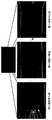
フロー型ATPSによるヒト全血血球細胞の分離
フロー型ATPSによるヒト全血血球細胞の分離を試みた結果、図6に示すように、赤血球細胞がDex相へ移動し、白血球細胞がPEG相へ移動することが観察された。Separation of human whole blood cells by flow type ATPS As a result of separation of human whole blood cells by flow type ATPS, red blood cells moved to Dex phase and white blood cells moved to PEG phase as shown in FIG. To be observed.
以上のように、赤血球細胞及びJurkatがそれぞれ99%、96%という高い分離能を示したこと、また全血サンプルにおいても赤血球及び白血球が異なった相へ移動することが観察されたことから、本発明で提案したフロー型ATPSという新規分離システムの有効性が示されたものと考える。 As described above, red blood cells and Jurkat showed high resolution of 99% and 96%, respectively, and it was observed that red blood cells and white blood cells migrate to different phases in whole blood samples. It is considered that the effectiveness of the new separation system called flow-type ATPS proposed in the invention was shown.
1,2 微小流路、3 懸濁液供給路、A,B 液相、C 液相二相界面、11 基板、12 微小流路1, 2 micro flow path, 3 suspension supply path, A, B liquid phase, C liquid phase two phase interface, 11 substrate, 12 micro flow path
Claims (16)
Translated fromJapanese前記合流領域において各液相の層流により液相二相界面が形成され、当該液相二相界面において不溶性物質の分離が行われることを特徴とする不溶性物質分離デバイス。A micro flow path to which different liquid phases are supplied is formed on the substrate, and the micro flow path has a merging region where the micro flow paths merge with each other,
A liquid phase two-phase interface is formed by laminar flow of each liquid phase in the merge region, and an insoluble substance separation is performed at the liquid phase two-phase interface.
Priority Applications (1)
| Application Number | Priority Date | Filing Date | Title |
|---|---|---|---|
| JP2004073451AJP2005224787A (en) | 2004-02-15 | 2004-02-15 | Method and device for separating insoluble substance |
Applications Claiming Priority (1)
| Application Number | Priority Date | Filing Date | Title |
|---|---|---|---|
| JP2004073451AJP2005224787A (en) | 2004-02-15 | 2004-02-15 | Method and device for separating insoluble substance |
Publications (1)
| Publication Number | Publication Date |
|---|---|
| JP2005224787Atrue JP2005224787A (en) | 2005-08-25 |
Family
ID=34999910
Family Applications (1)
| Application Number | Title | Priority Date | Filing Date |
|---|---|---|---|
| JP2004073451APendingJP2005224787A (en) | 2004-02-15 | 2004-02-15 | Method and device for separating insoluble substance |
Country Status (1)
| Country | Link |
|---|---|
| JP (1) | JP2005224787A (en) |
Cited By (9)
| Publication number | Priority date | Publication date | Assignee | Title |
|---|---|---|---|---|
| WO2008001691A1 (en)* | 2006-06-29 | 2008-01-03 | Olympus Corporation | Method of treating sample |
| CN102179064A (en)* | 2011-03-24 | 2011-09-14 | 西南交通大学 | Micro flow control aqueous two-phase annular space extraction technology and device |
| WO2012029877A1 (en)* | 2010-08-31 | 2012-03-08 | 株式会社セルシード | Temperature-responsive cell culture beads and method for manufacturing same |
| WO2014042177A1 (en)* | 2012-09-11 | 2014-03-20 | 国立大学法人北陸先端科学技術大学院大学 | Liquid supply method, centrifugal separation method, liquid supply device and centrifugal separation device |
| US9649424B2 (en) | 2009-06-30 | 2017-05-16 | Kaneka Corporation | Blood component separation system and separation material |
| WO2020066471A1 (en)* | 2018-09-28 | 2020-04-02 | 株式会社マンダム | Cosmetic |
| JP2021065118A (en)* | 2019-10-18 | 2021-04-30 | 学校法人東京電機大学 | Suspension culture device and suspension culture method |
| CN113056319A (en)* | 2018-11-19 | 2021-06-29 | 浦项工科大学校产学协力团 | Aqueous solution two-phase system nano-filter and separation method using same |
| CN113499812A (en)* | 2021-08-06 | 2021-10-15 | 大连海事大学 | Microalgae multistage sorting device and method based on double-aqueous-phase microfluidics |
Citations (1)
| Publication number | Priority date | Publication date | Assignee | Title |
|---|---|---|---|---|
| JP2001511520A (en)* | 1997-07-25 | 2001-08-14 | ユニバーシティ オブ ワシントン | Simultaneous analyte measurement and reference equilibrium with reference T-sensor device |
- 2004
- 2004-02-15JPJP2004073451Apatent/JP2005224787A/enactivePending
Patent Citations (1)
| Publication number | Priority date | Publication date | Assignee | Title |
|---|---|---|---|---|
| JP2001511520A (en)* | 1997-07-25 | 2001-08-14 | ユニバーシティ オブ ワシントン | Simultaneous analyte measurement and reference equilibrium with reference T-sensor device |
Cited By (15)
| Publication number | Priority date | Publication date | Assignee | Title |
|---|---|---|---|---|
| WO2008001691A1 (en)* | 2006-06-29 | 2008-01-03 | Olympus Corporation | Method of treating sample |
| US9649424B2 (en) | 2009-06-30 | 2017-05-16 | Kaneka Corporation | Blood component separation system and separation material |
| WO2012029877A1 (en)* | 2010-08-31 | 2012-03-08 | 株式会社セルシード | Temperature-responsive cell culture beads and method for manufacturing same |
| CN102179064A (en)* | 2011-03-24 | 2011-09-14 | 西南交通大学 | Micro flow control aqueous two-phase annular space extraction technology and device |
| WO2014042177A1 (en)* | 2012-09-11 | 2014-03-20 | 国立大学法人北陸先端科学技術大学院大学 | Liquid supply method, centrifugal separation method, liquid supply device and centrifugal separation device |
| JPWO2014042177A1 (en)* | 2012-09-11 | 2016-08-18 | 国立大学法人山梨大学 | Liquid feeding method, centrifugal separation method, liquid feeding device and centrifugal separator |
| WO2020066471A1 (en)* | 2018-09-28 | 2020-04-02 | 株式会社マンダム | Cosmetic |
| JPWO2020066471A1 (en)* | 2018-09-28 | 2021-04-08 | 株式会社マンダム | Cosmetics |
| CN113056319A (en)* | 2018-11-19 | 2021-06-29 | 浦项工科大学校产学协力团 | Aqueous solution two-phase system nano-filter and separation method using same |
| JP2022508122A (en)* | 2018-11-19 | 2022-01-19 | ポステック アカデミー-インダストリー ファウンデーション | Aqueous two-phase nanofilter and separation method using this |
| CN113056319B (en)* | 2018-11-19 | 2022-11-22 | 浦项工科大学校产学协力团 | Aqueous solution two-phase system nano-filter and separation method using same |
| JP7347851B2 (en) | 2018-11-19 | 2023-09-20 | ポステック アカデミー-インダストリー ファウンデーション | Aqueous two-phase nanofilter and separation method using it |
| JP2021065118A (en)* | 2019-10-18 | 2021-04-30 | 学校法人東京電機大学 | Suspension culture device and suspension culture method |
| JP7488544B2 (en) | 2019-10-18 | 2024-05-22 | 学校法人東京電機大学 | Suspension culture device and suspension culture method |
| CN113499812A (en)* | 2021-08-06 | 2021-10-15 | 大连海事大学 | Microalgae multistage sorting device and method based on double-aqueous-phase microfluidics |
Similar Documents
| Publication | Publication Date | Title |
|---|---|---|
| US20210370298A1 (en) | Microfluidic Device For Cell Separation And Uses Thereof | |
| Yu et al. | Microfluidic blood cell sorting: now and beyond | |
| Tanaka et al. | Separation of cancer cells from a red blood cell suspension using inertial force | |
| TWI588262B (en) | Methods and compositions for separating or enriching cells | |
| Yang et al. | Cell separation on microfabricated electrodes using dielectrophoretic/gravitational field-flow fractionation | |
| Tsukamoto et al. | Cell separation by an aqueous two-phase system in a microfluidic device | |
| US20160237397A1 (en) | Methods and devices for breaking cell aggregation and separating or enriching cells | |
| WO2018191534A1 (en) | Methods, compositions, and devices for separating and/or enriching cells | |
| JP2005224787A (en) | Method and device for separating insoluble substance | |
| Szydzik et al. | A review of design considerations for hemocompatibility within microfluidic systems | |
| Petersson | On acoustic particle and cell manipulation in microfluidic systems | |
| Urbansky | Acoustofluidic preparation of whole blood components | |
| Wu | Acoustofluidic separation technology for advancing health care | |
| Baz | Lab-on-a-chip for the séparation circulating adipose stromal cells | |
| Chen et al. | Microfluidic Chips for Blood Cell Separation | |
| Kumar | Inertio-and Elasto-Magnetic Fractionation of Multiple Microparticles in Newtonian and Non-Newtonian Fluid | |
| Javadpour et al. | Multiscale experimental study of selective blood-cell filtration in fibrous porous media | |
| Ali | Dielectrophoresis-Assisted Deterministic Lateral Displacement for Particle and Cell Sorting | |
| Srivastava | Dielectrophoretic characterization of particles and erythrocytes | |
| Shields IV | Acoustic and Magnetic Techniques for the Isolation and Analysis of Cells in Microfluidic Platforms | |
| Aw Yong et al. | Microfluidic Blood Cell Sorting: Now and Beyond | |
| Li | Lab-on-a-chip systems for blood cell separation, counting, and characterization |
Legal Events
| Date | Code | Title | Description |
|---|---|---|---|
| A621 | Written request for application examination | Free format text:JAPANESE INTERMEDIATE CODE: A621 Effective date:20070213 | |
| A711 | Notification of change in applicant | Free format text:JAPANESE INTERMEDIATE CODE: A711 Effective date:20070625 | |
| A131 | Notification of reasons for refusal | Free format text:JAPANESE INTERMEDIATE CODE: A131 Effective date:20100217 | |
| A02 | Decision of refusal | Free format text:JAPANESE INTERMEDIATE CODE: A02 Effective date:20100809 |