JP2005177500A - High strength suture and suture anchor combination with an absorbent core - Google Patents
High strength suture and suture anchor combination with an absorbent coreDownload PDFInfo
- Publication number
- JP2005177500A JP2005177500AJP2004366444AJP2004366444AJP2005177500AJP 2005177500 AJP2005177500 AJP 2005177500AJP 2004366444 AJP2004366444 AJP 2004366444AJP 2004366444 AJP2004366444 AJP 2004366444AJP 2005177500 AJP2005177500 AJP 2005177500A
- Authority
- JP
- Japan
- Prior art keywords
- suture
- yarn
- bioabsorbable
- nodule
- anchor
- Prior art date
- Legal status (The legal status is an assumption and is not a legal conclusion. Google has not performed a legal analysis and makes no representation as to the accuracy of the status listed.)
- Granted
Links
- 230000002745absorbentEffects0.000titleabstractdescription9
- 239000002250absorbentSubstances0.000titleabstractdescription9
- 229920000785ultra high molecular weight polyethylenePolymers0.000claimsabstractdescription45
- 239000004699Ultra-high molecular weight polyethyleneSubstances0.000claimsabstractdescription43
- 229920000642polymerPolymers0.000claimsabstractdescription34
- 238000000034methodMethods0.000claimsdescription45
- 229920002463poly(p-dioxanone) polymerPolymers0.000claimsdescription43
- 239000000622polydioxanoneSubstances0.000claimsdescription43
- 210000001519tissueAnatomy0.000claimsdescription38
- 239000000835fiberSubstances0.000claimsdescription33
- 210000000988bone and boneAnatomy0.000claimsdescription27
- 210000004872soft tissueAnatomy0.000claimsdescription16
- 238000000576coating methodMethods0.000claimsdescription15
- 239000011248coating agentSubstances0.000claimsdescription13
- -1polyethylenePolymers0.000claimsdescription13
- 238000001727in vivoMethods0.000claimsdescription10
- 239000004698PolyethyleneSubstances0.000claimsdescription8
- 229920000573polyethylenePolymers0.000claimsdescription8
- VPVXHAANQNHFSF-UHFFFAOYSA-N1,4-dioxan-2-oneChemical compoundO=C1COCCO1VPVXHAANQNHFSF-UHFFFAOYSA-N0.000claimsdescription7
- RKDVKSZUMVYZHH-UHFFFAOYSA-N1,4-dioxane-2,5-dioneChemical compoundO=C1COC(=O)CO1RKDVKSZUMVYZHH-UHFFFAOYSA-N0.000claimsdescription6
- PAPBSGBWRJIAAV-UHFFFAOYSA-Nε-CaprolactoneChemical compoundO=C1CCCCCO1PAPBSGBWRJIAAV-UHFFFAOYSA-N0.000claimsdescription6
- JJTUDXZGHPGLLC-QWWZWVQMSA-N(3r,6r)-3,6-dimethyl-1,4-dioxane-2,5-dioneChemical compoundC[C@H]1OC(=O)[C@@H](C)OC1=OJJTUDXZGHPGLLC-QWWZWVQMSA-N0.000claimsdescription5
- JJTUDXZGHPGLLC-IMJSIDKUSA-N4511-42-6Chemical compoundC[C@@H]1OC(=O)[C@H](C)OC1=OJJTUDXZGHPGLLC-IMJSIDKUSA-N0.000claimsdescription5
- 239000003242anti bacterial agentSubstances0.000claimsdescription5
- 239000000178monomerSubstances0.000claimsdescription5
- 239000008194pharmaceutical compositionSubstances0.000claimsdescription5
- YFHICDDUDORKJB-UHFFFAOYSA-Ntrimethylene carbonateChemical compoundO=C1OCCCO1YFHICDDUDORKJB-UHFFFAOYSA-N0.000claimsdescription5
- 229940035676analgesicsDrugs0.000claimsdescription4
- 239000000730antalgic agentSubstances0.000claimsdescription4
- 150000002596lactonesChemical class0.000claimsdescription4
- 229940124599anti-inflammatory drugDrugs0.000claimsdescription3
- 229940088710antibiotic agentDrugs0.000claimsdescription3
- 229940124641pain relieverDrugs0.000claimsdescription3
- JJTUDXZGHPGLLC-ZXZARUISSA-N(3r,6s)-3,6-dimethyl-1,4-dioxane-2,5-dioneChemical compoundC[C@H]1OC(=O)[C@H](C)OC1=OJJTUDXZGHPGLLC-ZXZARUISSA-N0.000claimsdescription2
- 239000000463materialSubstances0.000abstractdescription19
- 239000002131composite materialSubstances0.000abstractdescription11
- 239000011162core materialSubstances0.000description30
- 238000012360testing methodMethods0.000description15
- 230000008569processEffects0.000description14
- 229920000728polyesterPolymers0.000description12
- 125000003118aryl groupChemical group0.000description10
- 238000001356surgical procedureMethods0.000description10
- 238000000338in vitroMethods0.000description9
- 239000013543active substanceSubstances0.000description8
- 230000035876healingEffects0.000description8
- 238000004519manufacturing processMethods0.000description7
- HEMHJVSKTPXQMS-UHFFFAOYSA-MSodium hydroxideChemical compound[OH-].[Na+]HEMHJVSKTPXQMS-UHFFFAOYSA-M0.000description6
- 208000027418Wounds and injuryDiseases0.000description6
- 238000009940knittingMethods0.000description6
- 229920000139polyethylene terephthalatePolymers0.000description6
- 239000005020polyethylene terephthalateSubstances0.000description6
- 238000000137annealingMethods0.000description5
- 206010052428WoundDiseases0.000description4
- 238000010521absorption reactionMethods0.000description4
- 230000008901benefitEffects0.000description4
- 239000003462bioceramicSubstances0.000description4
- 239000000872bufferSubstances0.000description4
- 229920001577copolymerPolymers0.000description4
- 238000012423maintenanceMethods0.000description4
- 239000000203mixtureSubstances0.000description4
- 238000009991scouringMethods0.000description4
- XEKOWRVHYACXOJ-UHFFFAOYSA-NEthyl acetateChemical compoundCCOC(C)=OXEKOWRVHYACXOJ-UHFFFAOYSA-N0.000description3
- 239000004599antimicrobialSubstances0.000description3
- 238000009835boilingMethods0.000description3
- 239000001506calcium phosphateSubstances0.000description3
- 230000006378damageEffects0.000description3
- JJTUDXZGHPGLLC-UHFFFAOYSA-NlactideChemical compoundCC1OC(=O)C(C)OC1=OJJTUDXZGHPGLLC-UHFFFAOYSA-N0.000description3
- 229910052751metalInorganic materials0.000description3
- 239000002184metalSubstances0.000description3
- XYJRXVWERLGGKC-UHFFFAOYSA-Dpentacalcium;hydroxide;triphosphateChemical compound[OH-].[Ca+2].[Ca+2].[Ca+2].[Ca+2].[Ca+2].[O-]P([O-])([O-])=O.[O-]P([O-])([O-])=O.[O-]P([O-])([O-])=OXYJRXVWERLGGKC-UHFFFAOYSA-D0.000description3
- 229920005594polymer fiberPolymers0.000description3
- QORWJWZARLRLPR-UHFFFAOYSA-Htricalcium bis(phosphate)Chemical compound[Ca+2].[Ca+2].[Ca+2].[O-]P([O-])([O-])=O.[O-]P([O-])([O-])=OQORWJWZARLRLPR-UHFFFAOYSA-H0.000description3
- 229940078499tricalcium phosphateDrugs0.000description3
- 229910000391tricalcium phosphateInorganic materials0.000description3
- 235000019731tricalcium phosphateNutrition0.000description3
- CSCPPACGZOOCGX-UHFFFAOYSA-NAcetoneChemical compoundCC(C)=OCSCPPACGZOOCGX-UHFFFAOYSA-N0.000description2
- XEFQLINVKFYRCS-UHFFFAOYSA-NTriclosanChemical compoundOC1=CC(Cl)=CC=C1OC1=CC=C(Cl)C=C1ClXEFQLINVKFYRCS-UHFFFAOYSA-N0.000description2
- 230000002411adverseEffects0.000description2
- 238000005452bendingMethods0.000description2
- 229920002988biodegradable polymerPolymers0.000description2
- 239000004621biodegradable polymerSubstances0.000description2
- 238000000354decomposition reactionMethods0.000description2
- 230000007423decreaseEffects0.000description2
- 229940079593drugDrugs0.000description2
- 239000003814drugSubstances0.000description2
- 230000002526effect on cardiovascular systemEffects0.000description2
- 238000002474experimental methodMethods0.000description2
- 238000001125extrusionMethods0.000description2
- 229920001519homopolymerPolymers0.000description2
- 229910052588hydroxylapatiteInorganic materials0.000description2
- 239000007943implantSubstances0.000description2
- 238000002513implantationMethods0.000description2
- 208000014674injuryDiseases0.000description2
- 238000002156mixingMethods0.000description2
- 231100000989no adverse effectToxicity0.000description2
- 230000002138osteoinductive effectEffects0.000description2
- 238000004806packaging method and processMethods0.000description2
- 230000009467reductionEffects0.000description2
- 230000008439repair processEffects0.000description2
- 210000000513rotator cuffAnatomy0.000description2
- 239000002904solventSubstances0.000description2
- 230000001954sterilising effectEffects0.000description2
- 238000004659sterilization and disinfectionMethods0.000description2
- 210000002435tendonAnatomy0.000description2
- 229960003500triclosanDrugs0.000description2
- XLYOFNOQVPJJNP-UHFFFAOYSA-NwaterSubstancesOXLYOFNOQVPJJNP-UHFFFAOYSA-N0.000description2
- 230000004580weight lossEffects0.000description2
- RDJGLLICXDHJDY-NSHDSACASA-N(2s)-2-(3-phenoxyphenyl)propanoic acidChemical compoundOC(=O)[C@@H](C)C1=CC=CC(OC=2C=CC=CC=2)=C1RDJGLLICXDHJDY-NSHDSACASA-N0.000description1
- OZJPLYNZGCXSJM-UHFFFAOYSA-N5-valerolactoneChemical compoundO=C1CCCCO1OZJPLYNZGCXSJM-UHFFFAOYSA-N0.000description1
- 241000208140AcerSpecies0.000description1
- RZVAJINKPMORJF-UHFFFAOYSA-NAcetaminophenChemical compoundCC(=O)NC1=CC=C(O)C=C1RZVAJINKPMORJF-UHFFFAOYSA-N0.000description1
- 206010002091AnaesthesiaDiseases0.000description1
- IAYPIBMASNFSPL-UHFFFAOYSA-NEthylene oxideChemical compoundC1CO1IAYPIBMASNFSPL-UHFFFAOYSA-N0.000description1
- AEMRFAOFKBGASW-UHFFFAOYSA-NGlycolic acidPolymersOCC(O)=OAEMRFAOFKBGASW-UHFFFAOYSA-N0.000description1
- NNJVILVZKWQKPM-UHFFFAOYSA-NLidocaineChemical compoundCCN(CC)CC(=O)NC1=C(C)C=CC=C1CNNJVILVZKWQKPM-UHFFFAOYSA-N0.000description1
- CMWTZPSULFXXJA-UHFFFAOYSA-NNaproxenNatural productsC1=C(C(C)C(O)=O)C=CC2=CC(OC)=CC=C21CMWTZPSULFXXJA-UHFFFAOYSA-N0.000description1
- 229920002292Nylon 6Polymers0.000description1
- 229920002302Nylon 6,6Polymers0.000description1
- 229920000954PolyglycolidePolymers0.000description1
- 239000004743PolypropyleneSubstances0.000description1
- 241000700159RattusSpecies0.000description1
- 208000026137Soft tissue injuryDiseases0.000description1
- 206010066902Surgical failureDiseases0.000description1
- 241001080929Zeugopterus punctatusSpecies0.000description1
- 239000004480active ingredientSubstances0.000description1
- 239000012080ambient airSubstances0.000description1
- 230000037005anaesthesiaEffects0.000description1
- 239000002260anti-inflammatory agentSubstances0.000description1
- 229940121363anti-inflammatory agentDrugs0.000description1
- 229960005475antiinfective agentDrugs0.000description1
- 238000013459approachMethods0.000description1
- 230000000975bioactive effectEffects0.000description1
- 229960000074biopharmaceuticalDrugs0.000description1
- 239000003795chemical substances by applicationSubstances0.000description1
- 150000001875compoundsChemical class0.000description1
- 238000007796conventional methodMethods0.000description1
- 238000002316cosmetic surgeryMethods0.000description1
- 238000010586diagramMethods0.000description1
- DCOPUUMXTXDBNB-UHFFFAOYSA-NdiclofenacChemical compoundOC(=O)CC1=CC=CC=C1NC1=C(Cl)C=CC=C1ClDCOPUUMXTXDBNB-UHFFFAOYSA-N0.000description1
- 229960001259diclofenacDrugs0.000description1
- 238000006073displacement reactionMethods0.000description1
- 239000000428dustSubstances0.000description1
- 230000000694effectsEffects0.000description1
- 210000003195fasciaAnatomy0.000description1
- 229960001419fenoprofenDrugs0.000description1
- 230000002496gastric effectEffects0.000description1
- 210000003709heart valveAnatomy0.000description1
- 238000013007heat curingMethods0.000description1
- 238000010438heat treatmentMethods0.000description1
- 210000004095humeral headAnatomy0.000description1
- 229940095990inderalDrugs0.000description1
- 230000000053inderal effectEffects0.000description1
- DKYWVDODHFEZIM-UHFFFAOYSA-NketoprofenChemical compoundOC(=O)C(C)C1=CC=CC(C(=O)C=2C=CC=CC=2)=C1DKYWVDODHFEZIM-UHFFFAOYSA-N0.000description1
- 229960000991ketoprofenDrugs0.000description1
- 229960004194lidocaineDrugs0.000description1
- 238000011068loading methodMethods0.000description1
- 230000007774longtermEffects0.000description1
- 230000014759maintenance of locationEffects0.000description1
- INWLQCZOYSRPNW-UHFFFAOYSA-NmepivacaineChemical compoundCN1CCCCC1C(=O)NC1=C(C)C=CC=C1CINWLQCZOYSRPNW-UHFFFAOYSA-N0.000description1
- 229960002009naproxenDrugs0.000description1
- CMWTZPSULFXXJA-VIFPVBQESA-NnaproxenChemical compoundC1=C([C@H](C)C(O)=O)C=CC2=CC(OC)=CC=C21CMWTZPSULFXXJA-VIFPVBQESA-N0.000description1
- 229920001778nylonPolymers0.000description1
- 230000008816organ damageEffects0.000description1
- 230000000399orthopedic effectEffects0.000description1
- RARSHUDCJQSEFJ-UHFFFAOYSA-Np-HydroxypropiophenoneChemical compoundCCC(=O)C1=CC=C(O)C=C1RARSHUDCJQSEFJ-UHFFFAOYSA-N0.000description1
- 230000036407painEffects0.000description1
- 206010033675panniculitisDiseases0.000description1
- 239000002245particleSubstances0.000description1
- 230000007170pathologyEffects0.000description1
- 239000008363phosphate bufferSubstances0.000description1
- 230000000704physical effectEffects0.000description1
- 229920000747poly(lactic acid)Polymers0.000description1
- 229920001610polycaprolactonePolymers0.000description1
- 239000004632polycaprolactoneSubstances0.000description1
- 229920000098polyolefinPolymers0.000description1
- 229920001155polypropylenePolymers0.000description1
- 239000000843powderSubstances0.000description1
- 238000012545processingMethods0.000description1
- AQHHHDLHHXJYJD-UHFFFAOYSA-NpropranololChemical compoundC1=CC=C2C(OCC(O)CNC(C)C)=CC=CC2=C1AQHHHDLHHXJYJD-UHFFFAOYSA-N0.000description1
- JTIGKVIOEQASGT-UHFFFAOYSA-NproquazoneChemical compoundN=1C(=O)N(C(C)C)C2=CC(C)=CC=C2C=1C1=CC=CC=C1JTIGKVIOEQASGT-UHFFFAOYSA-N0.000description1
- 239000005297pyrexSubstances0.000description1
- 230000005855radiationEffects0.000description1
- 239000011347resinSubstances0.000description1
- 229920005989resinPolymers0.000description1
- 210000004304subcutaneous tissueAnatomy0.000description1
- 239000000126substanceSubstances0.000description1
- 239000003356suture materialSubstances0.000description1
- 239000003106tissue adhesiveSubstances0.000description1
- 230000008467tissue growthEffects0.000description1
- 230000005944tissue migrationEffects0.000description1
- 238000002054transplantationMethods0.000description1
- 230000002792vascularEffects0.000description1
- 238000004804windingMethods0.000description1
- 230000029663wound healingEffects0.000description1
Images


Classifications
- A—HUMAN NECESSITIES
- A61—MEDICAL OR VETERINARY SCIENCE; HYGIENE
- A61B—DIAGNOSIS; SURGERY; IDENTIFICATION
- A61B17/00—Surgical instruments, devices or methods
- A61B17/04—Surgical instruments, devices or methods for suturing wounds; Holders or packages for needles or suture materials
- A61B17/06—Needles ; Sutures; Needle-suture combinations; Holders or packages for needles or suture materials
- A61B17/06166—Sutures
- A—HUMAN NECESSITIES
- A61—MEDICAL OR VETERINARY SCIENCE; HYGIENE
- A61B—DIAGNOSIS; SURGERY; IDENTIFICATION
- A61B17/00—Surgical instruments, devices or methods
- A61B17/04—Surgical instruments, devices or methods for suturing wounds; Holders or packages for needles or suture materials
- A61B17/0401—Suture anchors, buttons or pledgets, i.e. means for attaching sutures to bone, cartilage or soft tissue; Instruments for applying or removing suture anchors
- A—HUMAN NECESSITIES
- A61—MEDICAL OR VETERINARY SCIENCE; HYGIENE
- A61B—DIAGNOSIS; SURGERY; IDENTIFICATION
- A61B17/00—Surgical instruments, devices or methods
- A61B17/04—Surgical instruments, devices or methods for suturing wounds; Holders or packages for needles or suture materials
- A61B17/06—Needles ; Sutures; Needle-suture combinations; Holders or packages for needles or suture materials
- A61B17/06066—Needles, e.g. needle tip configurations
- A—HUMAN NECESSITIES
- A61—MEDICAL OR VETERINARY SCIENCE; HYGIENE
- A61L—METHODS OR APPARATUS FOR STERILISING MATERIALS OR OBJECTS IN GENERAL; DISINFECTION, STERILISATION OR DEODORISATION OF AIR; CHEMICAL ASPECTS OF BANDAGES, DRESSINGS, ABSORBENT PADS OR SURGICAL ARTICLES; MATERIALS FOR BANDAGES, DRESSINGS, ABSORBENT PADS OR SURGICAL ARTICLES
- A61L17/00—Materials for surgical sutures or for ligaturing blood vessels ; Materials for prostheses or catheters
- A61L17/005—Materials for surgical sutures or for ligaturing blood vessels ; Materials for prostheses or catheters containing a biologically active substance, e.g. a medicament or a biocide
- A—HUMAN NECESSITIES
- A61—MEDICAL OR VETERINARY SCIENCE; HYGIENE
- A61L—METHODS OR APPARATUS FOR STERILISING MATERIALS OR OBJECTS IN GENERAL; DISINFECTION, STERILISATION OR DEODORISATION OF AIR; CHEMICAL ASPECTS OF BANDAGES, DRESSINGS, ABSORBENT PADS OR SURGICAL ARTICLES; MATERIALS FOR BANDAGES, DRESSINGS, ABSORBENT PADS OR SURGICAL ARTICLES
- A61L17/00—Materials for surgical sutures or for ligaturing blood vessels ; Materials for prostheses or catheters
- A61L17/04—Non-resorbable materials
- A—HUMAN NECESSITIES
- A61—MEDICAL OR VETERINARY SCIENCE; HYGIENE
- A61L—METHODS OR APPARATUS FOR STERILISING MATERIALS OR OBJECTS IN GENERAL; DISINFECTION, STERILISATION OR DEODORISATION OF AIR; CHEMICAL ASPECTS OF BANDAGES, DRESSINGS, ABSORBENT PADS OR SURGICAL ARTICLES; MATERIALS FOR BANDAGES, DRESSINGS, ABSORBENT PADS OR SURGICAL ARTICLES
- A61L17/00—Materials for surgical sutures or for ligaturing blood vessels ; Materials for prostheses or catheters
- A61L17/06—At least partially resorbable materials
- A61L17/10—At least partially resorbable materials containing macromolecular materials
- A61L17/105—Polyesters not covered by A61L17/12
- A—HUMAN NECESSITIES
- A61—MEDICAL OR VETERINARY SCIENCE; HYGIENE
- A61L—METHODS OR APPARATUS FOR STERILISING MATERIALS OR OBJECTS IN GENERAL; DISINFECTION, STERILISATION OR DEODORISATION OF AIR; CHEMICAL ASPECTS OF BANDAGES, DRESSINGS, ABSORBENT PADS OR SURGICAL ARTICLES; MATERIALS FOR BANDAGES, DRESSINGS, ABSORBENT PADS OR SURGICAL ARTICLES
- A61L17/00—Materials for surgical sutures or for ligaturing blood vessels ; Materials for prostheses or catheters
- A61L17/14—Post-treatment to improve physical properties
- A61L17/145—Coating
- D—TEXTILES; PAPER
- D04—BRAIDING; LACE-MAKING; KNITTING; TRIMMINGS; NON-WOVEN FABRICS
- D04C—BRAIDING OR MANUFACTURE OF LACE, INCLUDING BOBBIN-NET OR CARBONISED LACE; BRAIDING MACHINES; BRAID; LACE
- D04C1/00—Braid or lace, e.g. pillow-lace; Processes for the manufacture thereof
- D04C1/06—Braid or lace serving particular purposes
- D04C1/12—Cords, lines, or tows
- A—HUMAN NECESSITIES
- A61—MEDICAL OR VETERINARY SCIENCE; HYGIENE
- A61B—DIAGNOSIS; SURGERY; IDENTIFICATION
- A61B17/00—Surgical instruments, devices or methods
- A61B2017/00004—(bio)absorbable, (bio)resorbable or resorptive
- A—HUMAN NECESSITIES
- A61—MEDICAL OR VETERINARY SCIENCE; HYGIENE
- A61B—DIAGNOSIS; SURGERY; IDENTIFICATION
- A61B17/00—Surgical instruments, devices or methods
- A61B2017/00831—Material properties
- A61B2017/00893—Material properties pharmaceutically effective
- A—HUMAN NECESSITIES
- A61—MEDICAL OR VETERINARY SCIENCE; HYGIENE
- A61B—DIAGNOSIS; SURGERY; IDENTIFICATION
- A61B17/00—Surgical instruments, devices or methods
- A61B17/04—Surgical instruments, devices or methods for suturing wounds; Holders or packages for needles or suture materials
- A61B17/0401—Suture anchors, buttons or pledgets, i.e. means for attaching sutures to bone, cartilage or soft tissue; Instruments for applying or removing suture anchors
- A61B2017/0414—Suture anchors, buttons or pledgets, i.e. means for attaching sutures to bone, cartilage or soft tissue; Instruments for applying or removing suture anchors having a suture-receiving opening, e.g. lateral opening
- A—HUMAN NECESSITIES
- A61—MEDICAL OR VETERINARY SCIENCE; HYGIENE
- A61B—DIAGNOSIS; SURGERY; IDENTIFICATION
- A61B17/00—Surgical instruments, devices or methods
- A61B17/04—Surgical instruments, devices or methods for suturing wounds; Holders or packages for needles or suture materials
- A61B17/0401—Suture anchors, buttons or pledgets, i.e. means for attaching sutures to bone, cartilage or soft tissue; Instruments for applying or removing suture anchors
- A61B2017/0446—Means for attaching and blocking the suture in the suture anchor
- A61B2017/0458—Longitudinal through hole, e.g. suture blocked by a distal suture knot
- A—HUMAN NECESSITIES
- A61—MEDICAL OR VETERINARY SCIENCE; HYGIENE
- A61B—DIAGNOSIS; SURGERY; IDENTIFICATION
- A61B17/00—Surgical instruments, devices or methods
- A61B17/04—Surgical instruments, devices or methods for suturing wounds; Holders or packages for needles or suture materials
- A61B17/06—Needles ; Sutures; Needle-suture combinations; Holders or packages for needles or suture materials
- A61B17/06166—Sutures
- A61B2017/06171—Sutures helically or spirally coiled
- A—HUMAN NECESSITIES
- A61—MEDICAL OR VETERINARY SCIENCE; HYGIENE
- A61L—METHODS OR APPARATUS FOR STERILISING MATERIALS OR OBJECTS IN GENERAL; DISINFECTION, STERILISATION OR DEODORISATION OF AIR; CHEMICAL ASPECTS OF BANDAGES, DRESSINGS, ABSORBENT PADS OR SURGICAL ARTICLES; MATERIALS FOR BANDAGES, DRESSINGS, ABSORBENT PADS OR SURGICAL ARTICLES
- A61L2300/00—Biologically active materials used in bandages, wound dressings, absorbent pads or medical devices
- A61L2300/40—Biologically active materials used in bandages, wound dressings, absorbent pads or medical devices characterised by a specific therapeutic activity or mode of action
- A61L2300/402—Anaestetics, analgesics, e.g. lidocaine
- A—HUMAN NECESSITIES
- A61—MEDICAL OR VETERINARY SCIENCE; HYGIENE
- A61L—METHODS OR APPARATUS FOR STERILISING MATERIALS OR OBJECTS IN GENERAL; DISINFECTION, STERILISATION OR DEODORISATION OF AIR; CHEMICAL ASPECTS OF BANDAGES, DRESSINGS, ABSORBENT PADS OR SURGICAL ARTICLES; MATERIALS FOR BANDAGES, DRESSINGS, ABSORBENT PADS OR SURGICAL ARTICLES
- A61L2300/00—Biologically active materials used in bandages, wound dressings, absorbent pads or medical devices
- A61L2300/40—Biologically active materials used in bandages, wound dressings, absorbent pads or medical devices characterised by a specific therapeutic activity or mode of action
- A61L2300/404—Biocides, antimicrobial agents, antiseptic agents
- A61L2300/406—Antibiotics
- A—HUMAN NECESSITIES
- A61—MEDICAL OR VETERINARY SCIENCE; HYGIENE
- A61L—METHODS OR APPARATUS FOR STERILISING MATERIALS OR OBJECTS IN GENERAL; DISINFECTION, STERILISATION OR DEODORISATION OF AIR; CHEMICAL ASPECTS OF BANDAGES, DRESSINGS, ABSORBENT PADS OR SURGICAL ARTICLES; MATERIALS FOR BANDAGES, DRESSINGS, ABSORBENT PADS OR SURGICAL ARTICLES
- A61L2300/00—Biologically active materials used in bandages, wound dressings, absorbent pads or medical devices
- A61L2300/40—Biologically active materials used in bandages, wound dressings, absorbent pads or medical devices characterised by a specific therapeutic activity or mode of action
- A61L2300/41—Anti-inflammatory agents, e.g. NSAIDs
- D—TEXTILES; PAPER
- D10—INDEXING SCHEME ASSOCIATED WITH SUBLASSES OF SECTION D, RELATING TO TEXTILES
- D10B—INDEXING SCHEME ASSOCIATED WITH SUBLASSES OF SECTION D, RELATING TO TEXTILES
- D10B2509/00—Medical; Hygiene
- D10B2509/04—Sutures
Landscapes
- Health & Medical Sciences (AREA)
- Life Sciences & Earth Sciences (AREA)
- Engineering & Computer Science (AREA)
- Surgery (AREA)
- Animal Behavior & Ethology (AREA)
- Veterinary Medicine (AREA)
- Public Health (AREA)
- General Health & Medical Sciences (AREA)
- Materials Engineering (AREA)
- Epidemiology (AREA)
- Vascular Medicine (AREA)
- Chemical & Material Sciences (AREA)
- Molecular Biology (AREA)
- Biomedical Technology (AREA)
- Nuclear Medicine, Radiotherapy & Molecular Imaging (AREA)
- Heart & Thoracic Surgery (AREA)
- Medical Informatics (AREA)
- Rheumatology (AREA)
- Manufacturing & Machinery (AREA)
- Textile Engineering (AREA)
- Materials For Medical Uses (AREA)
- Medicinal Preparation (AREA)
- Medicines That Contain Protein Lipid Enzymes And Other Medicines (AREA)
- Surgical Instruments (AREA)
Abstract
Translated fromJapaneseDescription
Translated fromJapanese本発明の分野は外科用縫合糸であり、詳細には、生体吸収性部分及び非生体吸収性部分を含む外科用縫合糸である。 The field of the invention is surgical sutures, and in particular surgical sutures that include a bioabsorbable portion and a non-bioabsorbable portion.
外科用縫合糸は、医療分野では周知のものである。外科用縫合糸は、編み構造またはモノフィラメント構造とすることができる。外科用縫合糸はまた、その一端または両端に縫合針が付いた一端針構造または両端針構造とすることができ、縫合針が付いていない構造にもできる。縫合糸は、組織を近接させるため及び移植片を組織に固定するためなどの従来の様々な医療及び外科処置に用いられている。外科用縫合糸は、様々な既知の生体吸収性材料及び非生体吸収性材料から形成することができる。例えば、ポリエチレンテレフタレートなどの芳香族ポリエステル、ナイロン6及びナイロン66などのナイロン、ポリプロピレンなどのポリオレフィン、絹、及び他の非吸収性ポリマーから形成できることが知られている。加えて、縫合糸は、p‐ジオキサノン(1,4‐ジオキサン‐2‐オンとしても知られている)、ε‐カプロラクトン、グリコリド(glycolide)、L(‐)‐ラクチド、D(+)‐ラクチド、Meso‐ラクチド、トリメチレンカーボネート、及びこれらの組合せのポリマー及びコポリマーから形成することができる。特に、ポリジオキサノンホモポリマーが実用的である。 Surgical sutures are well known in the medical field. The surgical suture can be a knitted structure or a monofilament structure. Surgical sutures can also have a one-end or double-end needle structure with a suture needle at one or both ends, or a structure without a suture needle. Sutures are used in various conventional medical and surgical procedures, such as to bring tissue close together and to secure a graft to tissue. Surgical sutures can be formed from a variety of known bioabsorbable and non-bioabsorbable materials. For example, it is known that it can be formed from aromatic polyesters such as polyethylene terephthalate, nylons such as nylon 6 and nylon 66, polyolefins such as polypropylene, silk, and other non-absorbable polymers. In addition, sutures include p-dioxanone (also known as 1,4-dioxane-2-one), ε-caprolactone, glycolide, L (-)-lactide, D (+)-lactide , Meso-lactide, trimethylene carbonate, and combinations thereof, and polymers and copolymers. In particular, polydioxanone homopolymer is practical.
一般に、従来の様々な外科処置に合った様々なサイズの外科用縫合糸を入手することができる。外科医が特定の処置に用いる縫合糸のサイズは、縫合する組織の種類、組織構造の相対的な大きさ、並びに外科処置が終了した後に近接した組織による縫合糸への力によって決まる。同様に、選択する縫合糸の種類も外科処置によって決まる。非吸収性縫合糸は通常、例えば人工心臓弁の移植などの治癒期間に永久または長期の固定が必要であることから非吸収性縫合糸が必要或いは望ましい心血管、血管、整形外科、胃腸など外科処置に用いられる。生体吸収性縫合糸は通常、形成外科、皮膚の固定、及び或る種の軟組織の近接などの処置に用いられる。生体吸収性縫合糸は、治癒期間に縫合糸が十分な強度を有するのであれば、長期に亘って組織を近接或いは固定させるが必要ない場合に用いることができる。縫合糸は、治癒過程の間に皮膚や軟組織などの自家組織に置き換えられるのが望ましい。 In general, various sizes of surgical sutures are available to suit various conventional surgical procedures. The size of the suture that the surgeon uses for a particular procedure depends on the type of tissue to be sutured, the relative size of the tissue structure, and the force on the suture by the adjacent tissue after the surgical procedure is complete. Similarly, the type of suture selected will depend on the surgical procedure. Non-absorbable sutures usually require cardiovascular, vascular, orthopedic, gastrointestinal and other surgical procedures where non-absorbable sutures are necessary or desirable because they require permanent or long-term fixation during healing such as, for example, implantation of an artificial heart valve Used for treatment. Bioabsorbable sutures are typically used in procedures such as plastic surgery, skin fixation, and some soft tissue proximity. The bioabsorbable suture can be used when it is not necessary to approach or fix the tissue over a long period of time as long as the suture has sufficient strength during the healing period. The suture is preferably replaced with autologous tissue such as skin or soft tissue during the healing process.
相当な引張力が縫合糸にかかるような特定の処置では、サイズすなわち断面積が最小で大きな抗張力を有する材料を用いるのが望ましい。縫合糸の抗張力は、材料、縫合糸サイズ、フィラメント直径、構造の種類(すなわち、編み型とモノフィラメント型)、編み構造におけるコアに対するシースの割合、及びコアの材料の種類などを含む様々な因子に相関することが知られている。具体的には、縫合糸の製造に超高分子ポリエチレン(以降、UHMWPEと呼ぶ)が使用されていることが知られている。超高分子ポリエチレンからなる縫合糸は、小さいサイズでも大きな抗張力を有するという利点がある。このような縫合糸の欠点は、超高分子ポリエチレンが比較的滑り易い材料であることである。この滑り易さから、縫合糸の結節の完全性に影響を及ぼす。一般的な外科処置では、組織が適切に近接して縫合されるように外科医が縫合糸を組織に何回か通した後、外科医が1または複数の従来の結節を所定の位置に形成して縫合糸が所定の位置に維持され緩まないようにする。縫合糸が緩むと、近接した組織が分離し、外科処置の失敗や破局的な影響(例えば、吻合移植片が修復心臓血管内に流れる、移植片のずれ、器官の障害、軟組織の骨からの離脱など)によって治癒が妨げられる恐れがある。 In certain procedures where a significant tensile force is applied to the suture, it is desirable to use a material with a minimum size or cross-sectional area and a high tensile strength. The tensile strength of the suture depends on a variety of factors, including material, suture size, filament diameter, type of structure (ie, knitted and monofilament type), ratio of sheath to core in the knitted structure, and type of core material. It is known to correlate. Specifically, it is known that ultra high molecular weight polyethylene (hereinafter referred to as UHMWPE) is used in the manufacture of sutures. Sutures made of ultra high molecular weight polyethylene have the advantage of having a high tensile strength even at a small size. A drawback of such sutures is that ultra high molecular weight polyethylene is a relatively slippery material. This slipperiness affects the integrity of the suture knot. In a typical surgical procedure, the surgeon forms one or more conventional nodules in place after the surgeon has passed the suture several times through the tissue so that the tissue is sutured in close proximity. The suture is held in place to prevent it from loosening. When sutures are loosened, adjacent tissue separates, causing surgical failure and catastrophic effects (eg, anastomotic grafts flowing into the repair cardiovascular, graft displacement, organ damage, soft tissue bones Healing may be hindered by withdrawal).
超高分子ポリエチレン縫合糸の結節保持特性(例えば、結節強度)を改善するために、超高分子ポリエチレンヤーンと、超高分子ポリエチレンヤーンよりも表面摩擦の大きい別のヤーンとを組み合わせることが知られている。例えば、超高分子ポリエチレン繊維のヤーンと芳香族ポリエステルなどの非吸収性材料の繊維からなるヤーンから編まれた外側シースが非吸収性のコア、超高分子ポリエチレンのコア、または芳香族ポリエステルのコアを覆っている超高分子ポリエチレン縫合糸が知られている。また、超高分子ポリエチレン繊維からなるヤーンと生体吸収性ポリマー繊維からなるヤーンから編まれた外側編みシースによって非吸収性超高分子ポリエチレンのコアまたは芳香族ポリエステルのコアが覆われた縫合糸も知られている。 It is known to combine ultra high molecular weight polyethylene yarns with another yarn that has a higher surface friction than ultra high molecular weight polyethylene yarns in order to improve the knot retention properties (eg, knot strength) of ultra high molecular weight polyethylene sutures. ing. For example, an outer sheath knitted from a yarn made of ultra high molecular weight polyethylene fiber and a fiber made of non-absorbable material such as aromatic polyester has a non-absorbent core, a core of ultra high molecular weight polyethylene, or a core of aromatic polyester Ultra high molecular weight polyethylene sutures covering the skin are known. Also known are sutures in which a non-absorbable ultra-high molecular weight polyethylene core or an aromatic polyester core is covered with an outer knitted sheath knitted from a yarn made of ultra-high molecular weight polyethylene fiber and a yarn made of bioabsorbable polymer fiber. It has been.
このような複合超高分子ポリエステル縫合糸は大きな抗張力を有するが、使用に関連して複数の問題がある。1つの問題は、超高分子ポリエステル縫合糸では結節特性が殆ど失われている。一般に、このような縫合糸では、結節の質量、サイズ、及び/体積が増大してしまう。非吸収性縫合糸の結節は、組織が近接して治癒した後も体内に留置されるため、軟組織を刺激したり、患者に不快感や苦痛を与え得る。医学的見地から、組織における移植片の大きさを最小にするのが望ましいことが知られている。加えて、自家組織の遊走や内植を可能にして治癒を早める移植片が有利である。吸収性縫合糸などの生体吸収性移植片は、その大部分が患者の体内で吸収及び分解されるため、組織の内植を可能にして治癒を早める。この治癒過程では、移植片すなわち縫合糸の吸収性部分が吸収/分解され、構造的一体性及び強度が失われるに連れ、患者の組織が組織負荷を担うようになる。従来技術の超高分子ポリエチレン縫合糸は、たとえ生体低吸収性部分と組み合わせた場合であっても、組織が治癒した後も殆どが残る高密度の非吸収性体積及び結節の大きさを有するため上記した問題が起きる。 Although such composite ultra-high molecular weight polyester sutures have high tensile strength, there are several problems associated with use. One problem is that the knot properties are almost lost in ultra high molecular weight polyester sutures. In general, such sutures increase the mass, size, and / or volume of the nodule. Non-absorbable suture nodules remain in the body after the tissue has healed in close proximity, which can irritate soft tissue and cause discomfort and pain to the patient. From a medical standpoint, it is known that it is desirable to minimize the size of the graft in the tissue. In addition, grafts that allow autologous tissue migration and ingrowth to speed healing are advantageous. Most bioabsorbable implants, such as absorbable sutures, are absorbed and degraded in the patient's body, allowing tissue to be implanted and speeding healing. During this healing process, the patient's tissue becomes responsible for tissue loading as the absorbable portion of the graft or suture is absorbed / degraded and structural integrity and strength are lost. Prior art ultra-high molecular weight polyethylene sutures have a high density of non-absorbable volume and nodule size that remains mostly after tissue healing, even when combined with bioabsorbable parts. The above problems occur.
従って、高い抗張力を提供する材料と生体吸収性材料から形成した新規の複合縫合糸が要望されている。この複合縫合糸では、最小量の高抗張力材料で最適な抗張力が得られ、良好な結節強度や結節安全性が達成され、時間経過によって生体内の結節のプロフィールが縮小して組織の内植が可能になる。 Accordingly, there is a need for new composite sutures formed from materials that provide high tensile strength and bioabsorbable materials. With this composite suture, optimum tensile strength can be obtained with the minimum amount of high tensile strength material, good nodule strength and nodule safety can be achieved, and the in vivo nodule profile can be reduced over time, allowing tissue in-growth to occur. It becomes possible.
従って、新規の外科用縫合糸を開示する。この縫合糸は、生体吸収性ポリマーのフィラメントからなるヤーンのコアを含む。コアは、超高分子ポリエチレンのフィラメントからなるヤーンと生体吸収性ポリマー繊維からなるヤーンから編まれたシースによって覆われている。 Accordingly, a novel surgical suture is disclosed. The suture includes a yarn core made of bioabsorbable polymer filaments. The core is covered with a sheath knitted from a yarn made of ultra high molecular weight polyethylene filaments and a yarn made of bioabsorbable polymer fibers.
本発明の別の態様は外科用縫合糸である。この外科用縫合糸は、コアとそのコアを覆う編みシースを含む。このコアは、生体吸収性ポリマーのフィラメントからなるヤーンから形成されている。このシースは、超高分子ポリエチレン繊維からなるヤーンと生体吸収性ポリマーからなるヤーンから編まれている。生体吸収性コア及び生体吸収性繊維は、薬物、バイオセラミックス、及び抗菌薬を含め、1または複数の活性物質を含む。 Another aspect of the invention is a surgical suture. The surgical suture includes a core and a knitted sheath covering the core. The core is formed from a yarn composed of filaments of bioabsorbable polymer. The sheath is knitted from a yarn made of ultra high molecular weight polyethylene fiber and a yarn made of a bioabsorbable polymer. The bioabsorbable core and the bioabsorbable fiber include one or more active substances, including drugs, bioceramics, and antimicrobial agents.
本発明の更に別の態様は、組織を近接させる方法である。この方法では、まず外科用縫合糸を用意する。この外科用縫合糸は、コアとそのコアを覆う外側編みシースを含む。このコアは、生体吸収性ポリマーのフィラメントからなる少なくとも1本のヤーンからなる。このシースは、超高分子ポリエチレンのフィラメントからなる少なくとも1本のヤーンと生体吸収性ポリマーのフィラメントからなる少なくとも1本のヤーンとから編まれている。外科用縫合糸を組織に通して組織を近接させ、任意で、外科用縫合糸を結んで少なくとも1つの結節を形成する。 Yet another aspect of the invention is a method of bringing tissue in close proximity. In this method, a surgical suture is first prepared. The surgical suture includes a core and an outer knitted sheath covering the core. The core consists of at least one yarn made of bioabsorbable polymer filaments. The sheath is knitted from at least one yarn made of ultra high molecular weight polyethylene filaments and at least one yarn made of bioabsorbable polymer filaments. A surgical suture is passed through the tissue to bring the tissue into close proximity and, optionally, the surgical suture is tied to form at least one knot.
本発明の更に別の態様は、縫合糸の吸収性成分の吸収プロフィールを改善する方法である。 Yet another aspect of the present invention is a method for improving the absorption profile of an absorbent component of a suture.
本発明の更に別の態様は、外科用縫合糸の結節の結節プロフィールを縮小させる方法である。この方法では、まず外科用縫合糸を用意する。この外科用縫合糸は、コアとそのコアを覆う外側編みシースを含む。このコアは、生体吸収性ポリマーのフィラメントからなる少なくとも1本のヤーンからなる。このシースは、超高分子ポリエチレンのフィラメントからなる少なくとも1本のヤーンと生体吸収性ポリマーのフィラメントからなる少なくとも1本のヤーンから編まれている。外科用縫合糸を組織に通して組織を近接させる。結節を形成して外科用縫合糸を固定する。この結節は、生体吸収性成分が時間経過によって生体内で吸収或いは分解されるに連れて縮小する結節プロフィールを有する。 Yet another aspect of the present invention is a method for reducing the nodule profile of a surgical suture nodule. In this method, a surgical suture is first prepared. The surgical suture includes a core and an outer knitted sheath covering the core. The core consists of at least one yarn made of bioabsorbable polymer filaments. The sheath is knitted from at least one yarn made of ultra high molecular weight polyethylene filaments and at least one yarn made of bioabsorbable polymer filaments. A surgical suture is passed through the tissue to bring the tissue into close proximity. A nodule is formed to secure the surgical suture. This nodule has a nodule profile that shrinks as the bioabsorbable component is absorbed or decomposed in vivo over time.
本発明の更なる態様は、外科用縫合糸に設けたコーティングから薬物及び抗菌薬を含む活性成分を放出させる方法である。 A further aspect of the present invention is a method for releasing active ingredients, including drugs and antimicrobial agents, from a coating on a surgical suture.
本発明の別の態様は、縫合糸アンカーと上記した外科用縫合糸の組合せ体である。縫合糸アンカーは、アンカー本体及び縫合糸取付け孔を有する。縫合糸は縫合糸取付け孔に取り付けられている。 Another aspect of the present invention is a combination of a suture anchor and the surgical suture described above. The suture anchor has an anchor body and a suture attachment hole. The suture is attached to the suture attachment hole.
本発明の更なる態様は、本発明の縫合糸アンカーと縫合糸の組合せ体を用いて、骨の表面に軟組織を取り付ける方法である。アンカーを骨に取り付け、縫合糸で軟組織を表の表面に固定する。 A further aspect of the present invention is a method for attaching soft tissue to the surface of a bone using the suture anchor and suture combination of the present invention. An anchor is attached to the bone and the soft tissue is secured to the surface of the surface with a suture.
本発明のこれら及び他の態様並びに利点は、添付の図面を参照しながら以下の説明を読めば明らかになるであろう。 These and other aspects and advantages of the present invention will become apparent upon reading the following description with reference to the accompanying drawings.
最小量の高抗張力材料で最適な抗張力が得られ、良好な結節強度や結節安全性が達成され、時間経過によって生体内の結節のプロフィールが縮小して組織の内植が可能になる新規の複合縫合糸と縫合糸アンカーの組合せ体が提供される。 A new composite that achieves optimal tensile strength with the minimum amount of high tensile material, achieves good nodule strength and safety, and reduces the nodule profile in vivo over time to allow tissue ingrowth A combination suture and suture anchor is provided.
本発明の縫合糸を製造するために用いる超高分子ポリエチレンは、一般的には約500,000g/モル〜約5,000,000g/モル、より一般的には約1,000,000g/モル〜約3,000,000g/モル、好ましくは約2,000,000g/モル〜約2,500,000g/モルの範囲である。超高分子ポリエチレンは、一般的には約20cN/dtex〜約40cN/dtex、より一般的には約27.5cN/dtex〜約38.7cN/dtexの引張強度を有する。 The ultra high molecular weight polyethylene used to produce the sutures of the present invention is typically from about 500,000 g / mole to about 5,000,000 g / mole, more typically from about 1,000,000 g / mole. To about 3,000,000 g / mol, preferably in the range of about 2,000,000 g / mol to about 2,500,000 g / mol. Ultra-high molecular weight polyethylene typically has a tensile strength of from about 20 cN / dtex to about 40 cN / dtex, more typically from about 27.5 cN / dtex to about 38.7 cN / dtex.
超高分子ポリエチレンは、複数のフィラメントを有する従来のヤーンの形態であるのが好ましい。このようなヤーンは市販されており、外科用縫合糸などの医療具の製造に用いられる。例えば、本発明の縫合糸に有用な超高分子ポリエチレンヤーンは、オランダ王国のDSMが販売する110dtex ダイニーマ純度ヤーン(110dtex Dyneema Purity yarn)である。 The ultra high molecular weight polyethylene is preferably in the form of a conventional yarn having a plurality of filaments. Such yarns are commercially available and are used in the manufacture of medical devices such as surgical sutures. For example, an ultra-high molecular weight polyethylene yarn useful for the suture of the present invention is a 110 dtex Dyneema Purity yarn sold by DSM in the Netherlands.
本発明の縫合糸の製造に有用な生体吸収性ポリマーは、従来の生体吸収性及び生体分解性のポリマー及びコポリマーである。特に有用なものは、p‐ジオキサノン(1,4‐ジオキサン‐2‐オンとしても知られている)、ε‐カプロラクトン、グリコリド(glycolide)、L(‐)‐ラクチド、D(+)ラクチド、Meso‐ラクチド、トリメチレンカーボネート、及びこれらの組合せからなる群から選択されるラクトンモノマーから製造した合成生体吸収性ポリマーである。このようなポリマーとして、ポリグリコリド(polyglicolide)、ポリ(カプロラクトン‐コグリコリド)(poly(caprolactone-co-glycolide)、ポリラクチド、ポリカプロラクトン、及びそれらの等価物などを挙げることができる。有利な生体吸収性ポリマーはポリジオキサノンホモポリマーである。ここで用いる用語「生体吸収性」は、生体吸収性と生体分解性の両方を意味すると定義する。本発明の縫合糸の生体吸収性部分には、特にポリジオキサノンポリマーを使用するのが好ましい。縫合糸の生体吸収性部分は、生体吸収性ポリマーからなる複数のフィラメントを含むヤーンからなるのが好ましい。このようなヤーンの製造方法は当分野で周知である。好ましくはないが、所望に応じて、生体吸収性のヤーン部分を、2種類以上の生体吸収性ポリマー及び/コポリマーのフィラメントから製造することができる。 Bioabsorbable polymers useful in the manufacture of the sutures of the present invention are conventional bioabsorbable and biodegradable polymers and copolymers. Particularly useful are p-dioxanone (also known as 1,4-dioxane-2-one), ε-caprolactone, glycolide, L (−)-lactide, D (+) lactide, Meso A synthetic bioabsorbable polymer made from a lactone monomer selected from the group consisting of lactide, trimethylene carbonate, and combinations thereof. Such polymers include polyglycolide, poly (caprolactone-co-glycolide), polylactide, polycaprolactone, and equivalents, etc. Advantageous bioabsorbability The polymer is a polydioxanone homopolymer, and the term “bioabsorbable” as used herein is defined to mean both bioabsorbable and biodegradable. Oxanone polymers are preferably used, and the bioabsorbable portion of the suture preferably comprises a yarn comprising a plurality of filaments comprising a bioabsorbable polymer, and methods for making such yarns are well known in the art. Although not preferred, if desired, the bioabsorbable yarn portion may be replaced with two or more bioabsorbable polymers. And / or copolymer filaments.
本発明の超高分子ポリエチレン複合縫合糸は、一般的には約40wt%〜約70wt%、より一般的には約50wt%〜65wt%、好ましくは約52wt%〜約62wt%、より好ましくは約62wt%の生体吸収性ポリマーを含む。例えば、編みシースを45wt%の超高分子ポリエチレンと55wt%のポリジオキサノンから形成し、コアを100wt%のポリジオキサノンから形成して、約38wt%の超高分子ポリエチレンと62wt%のポリジオキサノンからなる本発明の縫合糸を製造することができる。 The ultra high molecular weight polyethylene composite sutures of the present invention are typically from about 40 wt% to about 70 wt%, more typically from about 50 wt% to 65 wt%, preferably from about 52 wt% to about 62 wt%, more preferably about Contains 62 wt% bioabsorbable polymer. For example, the knitted sheath is formed of 45 wt% ultra high molecular weight polyethylene and 55 wt% polydioxanone, the core is formed of 100 wt% polydioxanone, and the present invention comprises about 38 wt% ultra high molecular weight polyethylene and 62 wt% polydioxanone. A suture can be produced.
本発明の複合超高分子ポリエチレン縫合糸は、限定するものではないが米国標準サイズである9/0〜5の従来の縫合糸の標準範囲を有することができる。外科医による特定の縫合糸サイズの選択は、組織の種類、近接させる創傷の大きさ、組織負荷、術後に縫合糸にかかる応力、縫合糸の位置、及び外科処置の種類などを含む様々な因子によって決まる。 The composite ultra-high molecular weight polyethylene suture of the present invention can have a standard range of conventional sutures of 9 / 0-5, which is, but not limited to, US standard size. The choice of a particular suture size by the surgeon will depend on a variety of factors, including the type of tissue, the size of the wound being approached, the tissue load, the stress applied to the suture after surgery, the location of the suture, and the type of surgical procedure, etc. It depends on.
本発明の縫合糸の生体吸収性部分は、様々な活性物質を含むことができる。本発明の縫合糸の生体吸収性部分に添加することができる活性物質には、医薬組成物、骨誘導組成物、生物製剤、及び生物活性物質が含まれる。活性物質には、限定するものではないが、既知の抗炎症剤、抗感染薬、及び鎮痛薬すなわち痛み止めが含まれる。特定の医薬化合物の例として、インデラル、ジクロフェナク、フェノプロフェン、ケトプロフェン、ケトロラック(ketrolac)、ナプロキセン、ブピビカイン(bupivicaine)、リドカイン、及びメピビカイン(mepivicaine)を挙げることができる。抗菌薬の例として、トリクロサン及びジグルコン酸クロルヘキシジンを挙げることができる。骨誘導組成物の例として、リン酸三カルシウム、バイオセラミックス、カルシウムヒドロキシアパタイト、及びこれらの組合せなどを挙げることができる。所望に応じて、本発明の縫合糸に用いるコーティングにこのような活性物質を含めることができる。 The bioabsorbable portion of the suture of the present invention can include various active substances. Active substances that can be added to the bioabsorbable portion of the suture of the present invention include pharmaceutical compositions, osteoinductive compositions, biologics, and bioactive substances. Active substances include, but are not limited to, known anti-inflammatory agents, anti-infective agents, and analgesics or painkillers. As examples of specific pharmaceutical compounds, mention may be made of inderal, diclofenac, fenoprofen, ketoprofen, ketrolac, naproxen, bupivicaine, lidocaine and mepivicaine. Examples of antibacterial agents include triclosan and chlorhexidine digluconate. Examples of osteoinductive compositions include tricalcium phosphate, bioceramics, calcium hydroxyapatite, and combinations thereof. If desired, such actives can be included in the coatings used in the sutures of the present invention.
本発明の縫合糸の生体吸収性部分に含めるこのような活性物質の量は、治療効果のある用量を十分に提供することができる。同様に、コーティングに用いる活性物質の量も、治療効果のある用量を十分に提供することができる。 The amount of such active agent included in the bioabsorbable portion of the suture of the present invention can sufficiently provide a therapeutically effective dose. Similarly, the amount of active agent used in the coating can provide a sufficient therapeutically effective dose.
本発明の縫合糸は通常、従来のブレーダ(編組機)を用いて従来の繊維含有ヤーンから編むことができる。外科用縫合糸の製造工程及び製造方法は当分野で周知であり、例えば、言及することを以って本明細書の一部とする米国特許第5,314,446号及び同第6,045,071号に開示されている。編み工程に加えて、縫合糸の製造には、通常は編み工程の後に、限定するものではないが、延伸、スカーリング、アニーリング、コーティング、チッピング、切断、針通し、取付け、パッケージング、及び滅菌を含む1または複数の従来の工程が必要である。 The sutures of the present invention can usually be knitted from conventional fiber-containing yarns using a conventional braider. The manufacturing processes and methods of surgical sutures are well known in the art, for example, US Pat. Nos. 5,314,446 and 6,045, which are hereby incorporated by reference. , 071. In addition to the knitting process, the manufacture of sutures is usually, but not limited to, stretching, scouring, annealing, coating, chipping, cutting, threading, attachment, packaging, and sterilization after the knitting process. One or more conventional processes including:
熱延伸工程は、編み直径を小さくするため、並びに切断した時に編み端部がほつれるまたは解ける「ブルーミング(brooming)」を防止するべくヤーンを編みに熱硬化させるために必要である。スカーリング工程は、押出し工程中にヤーンに対するスピン仕上げをしなくて済むようにするために必要である。アニーリング工程は、残ったモノマーを除去するため及びその結晶度を上げるために必要である。コーティングの1つの目的は、結節の安全性を著しく損なわずに縫合糸の滑りを良くすることである。チッピング工程は、縫合糸によるが針を取付け易くするために場合によっては必要である。スカーリング工程は、スピン仕上げを省略できるだけではなく、編み工程中に混入し得る塵粒や油滴を排除するのにも役立つ。加えて、スカーリング剤は、縫合糸の特性に著しい悪影響を及ぼすものであってはならない。 The hot drawing process is necessary to reduce the knitting diameter and to heat cure the yarn to the knitting to prevent "brooming" where the knitting ends fray or unravel when cut. The scouring process is necessary to avoid having to spin finish the yarn during the extrusion process. An annealing step is necessary to remove residual monomer and increase its crystallinity. One purpose of the coating is to improve the sliding of the suture without significantly impairing the safety of the nodule. A chipping step is necessary depending on the suture, but may be necessary to facilitate attachment of the needle. The scouring process not only eliminates the spin finish, but also helps to eliminate dust particles and oil droplets that can be incorporated during the knitting process. In addition, the scouring agent should not significantly adversely affect the properties of the suture.
コーティングすることにより、例えば組織通過などの縫合糸の通過性が良好になるのが普通である。また、コーティングにより、締め付け及び結節に必要な潤滑性が得られる。この潤滑性は締め付けに必要であるが、結節の安全性を維持するためにはコーティングのレベルを正確に制御しなければならない。 Coating typically results in better suture passage, such as through tissue. The coating provides the lubricity required for tightening and knotting. This lubricity is necessary for tightening, but the coating level must be precisely controlled to maintain knot safety.
任意で従来の方法でチッピングして所定の長さに縫合糸をカットした後に針を取り付ける。次いで、針/縫合糸をパッケージングし、次いで、材料やパッケージングの種類に応じて、例えばエチレンオキシド処置や、γ線またはオートクレービングなどの他の従来の滅菌工程で滅菌する。 Optionally, the needle is attached after chipping by a conventional method to cut the suture to a predetermined length. The needle / suture is then packaged and then sterilized with other conventional sterilization processes, such as ethylene oxide treatment, gamma radiation or autoclaving, depending on the material and type of packaging.
ここで図1を参照すると、本発明の縫合糸の好適な実施形態の断面図が示されている。縫合糸10は、図示されているように外側シース20及び内側コア60を有する。シース20は、図示されているように2種類のヤーンが相互に編み合わせられた編み構造である。ヤーン30は、超高分子ポリエチレンのフィラメントからなる。ヤーン35は、ポリジオキサノンなどの生体吸収性ポリマーのフィラメントからなる。コア60は、図示されているように生体吸収性ポリマー繊維からなるヤーン70を含む。ヤーン70は、ヤーン35と同じ生体吸収性ポリマーから製造するのが好ましいが、代替の実施形態では、別の生体吸収性ポリマーから製造することもできる。 Referring now to FIG. 1, a cross-sectional view of a preferred embodiment of the suture of the present invention is shown. The
本発明の縫合糸が外科処置に用いる場合、通常は組織を縫合した後、外科医が最後に結節して縫合糸を固定する。この目的のために使用する外科用結節は外科医及び医師によく知られており、外科医結び(surgeion's throw)、Revo結節、SNC結節、テネシースライダー(Tennessee Slider)、修正ローダー(Modified Roeder)、こま結び、ハーフヒッチ/逆ハーフヒッチ、及びダンカンループ(Duncan loop)などがある。図2Aに示されているように、本発明の縫合糸10が、創傷110の縁115を固定及び近接させるために組織100に取り付けられている。ステッチ125を固定するために、ハーフヒッチ/逆ハーフヒッチ結節130が縫合糸10に形成されている。結節130は、図示されているように結節プロフィール特性140を有する。用語「結節プロフィール特性」は、空間的形状または構造を含む縫合糸結節の全体の質量及び体積と定義する。用語「結節プロフィール寸法」は、例えば結節した結節の耳部が近接した組織の平面に平行な場合の結節の厚みを指す。図2Aの結節130は、移植直後の結節プロフィール特性140である。生体内で長期間が経過して生体吸収性成分が十分に吸収された図2Aの縫合糸10が図2Bに例示されている。結節130は、図示されているように、吸収性部分の質量及び体積が減少した後の縮小した結節プロフィール特性140を有する。意外かつ予想に反して、縫合糸10の生体吸収性部分が縫合糸10から喪失しても、結節130の強度は著しく低下しなかった。加えて、組織内植が起こっているのが示されている。 When the suture of the present invention is used in a surgical procedure, typically after the tissue is sutured, the surgeon is finally knotted to secure the suture. Surgical nodules used for this purpose are well known to surgeons and physicians, including surgeon's throw, Revo nodule, SNC nodule, Tennessee Slider, Modified Roeder, top knot , Half-hitch / reverse half-hitch, and Duncan loop. As shown in FIG. 2A, the
本発明の縫合糸は、軟組織傷害を修復するために縫合糸アンカーと共に用いるのが好ましい。縫合糸アンカー及びその処置は、言及することを以って本明細書の一部とする米国特許第4,632,100号、同第4,999,074号、同第5,814,051号、同第5,709,708号、同第5,782,864号、同第6,270,518号、同第5,540,718号、同第6,264,674号、同第6,270,518号、同第6,306,158号、同第5,961,538号、同第5,782,863号、同第5,683,418号、同第5,554,171号、同第5,078,730号、同第4,632,100号、同第5,217,486号、同第5,011,473号、同第4,898,156号、同第4,899,743号、同第4,946,468号、同第4,968,315号、同第5,002,550号、同第5,046,513号、同第5,192,303号、同第5,207,679号、及び同第5,358,511号に開示されている。図3A及び図3Bに示されているように、本発明の縫合糸200が従来の生体吸収性縫合糸アンカー250に取り付けられている。このアンカー250は、図示されているようにアンカー本体255を有する。縫合糸200は、アンカー本体255の縫合糸取付け孔260に通されて取り付けられている。アンカー250は、図示されているように骨270に穿孔された骨孔275に取り付けられている。アンカー250は、骨270の皮質285の下側の海綿質部分280に取り付けられ、海綿質部分280に係合している。軟組織290が、図示されているように、縫合糸200によって骨表面287に近接している。縫合糸200は、図示されているように、その両端に従来の外科用縫合針340が取り付けられている。軟組織は、縫合糸10のハーフヒッチ/逆ハーフヒッチ結節310によって固定されている。結節310は、図示されているように結節プロフィール特性315を有する。外科用縫合針340は、処置が終了する前に縫合糸200から切り落とされる。 The suture of the present invention is preferably used with a suture anchor to repair soft tissue injury. Suture anchors and their treatment are described in US Pat. Nos. 4,632,100, 4,999,074, and 5,814,051, which are hereby incorporated by reference. No. 5,709,708, No. 5,782,864, No. 6,270,518, No. 5,540,718, No. 6,264,674, No. 6, 270,518, 6,306,158, 5,961,538, 5,782,863, 5,683,418, 5,554,171, 5,078,730, 4,632,100, 5,217,486, 5,011,473, 4,898,156, 4,899 743, 4,946,468, 4,968,315, 5,002,5 No. 0, the No. 5,046,513, the No. 5,192,303, discloses the No. 5,207,679, and the same No. 5,358,511. As shown in FIGS. 3A and 3B, the
当業者には、フィラメントの直径が比較的小さい(フィラメント当たり約3デニールまたはそれ以下)ポリジオキサノン吸収性ヤーンが、フィラメント当たりの直径が相当大きい(例えば、フィラメント当たり12デニール)フィラメントを有する総デニールが同じヤーンよりも強度が早く低下することが知られている。様々な外科的知見から、重要な創傷治癒過程の間、実質的な強度を維持する必要がある。従って、直径が大きいポリジオキサノンフィラメント(フィラメント当たりのデニールが大きい)からなるヤーンを使用すべきである。一方、円柱のコラム強度と曲げ強度の両方が直径に依存することは周知の原則である。従って、意外かつ予想に反して、フィラメント当たりのデニールが12(dpf)よりも大きい直径のヤーンから製造された2成分縫合糸が硬くならずに、フィラメント直径が大幅に小さいヤーン(3dpf)から製造された縫合糸と同様の所望の操作特性を維持する。 For those skilled in the art, polydioxanone absorbent yarns with relatively small filament diameters (about 3 denier per filament or less) have the same total denier with filaments with a fairly large diameter per filament (eg, 12 denier per filament). It is known that the strength decreases faster than the yarn. From various surgical findings, substantial strength needs to be maintained during an important wound healing process. Therefore, yarns consisting of polydioxanone filaments with a large diameter (high denier per filament) should be used. On the other hand, it is a well-known principle that both column strength and bending strength of a cylinder depend on the diameter. Thus, surprisingly and unexpectedly, a two-component suture made from a yarn with a diameter greater than 12 (dpf) per filament is not stiff and made from a yarn with a much smaller filament diameter (3 dpf) Maintaining desired operating characteristics similar to the sutures made.
以下に示す例は、本発明の原理及び実施を例示するものであって本発明を限定するものではない。 The following examples are illustrative of the principles and practice of the present invention and are not intended to limit the invention.
例1
本発明の縫合糸は、以下に記載する方法で製造した。縫合糸は、メープル(中空)ブレーダ(maple (hollow) braider)を用いて製造した。この複合縫合糸は、超高分子ポリエチレンヤーンとポリジオキサノンヤーンからなるシースを含む。このシースは、45wt%の超高分子ポリエチレンと55wt%のポリジオキサノンを含む。この縫合糸のコアは、100wt%のポリジオキサノンヤーンからなる。全体として、縫合糸は、38wt%の超高分子ポリエチレンと62wt%のポリジオキサノンからなる。縫合糸構造の非吸収性部分は、反時計回り方向にブレーダに導入される8本の100デニール・ダイニーマ純度・超高分子ポリエチレン・マルチフィラメント・ヤーン(DSM社製)からなり、ポリジオキサノン部分は、ニュージャージー州ソマービル(Somerville)に所在のエシコン・インコーポレイテッド(Ethicon, Inc.)がポリジオキサノン縫合糸の製造に使用するような従来のマルチフィラメント・ポリジオキサノン・ヤーンである。ポリジオキサノン部分は、時計回り方向にブレーダに導入される8本のポリジオキサノン60×2デニール着色マルチフィラメントヤーンからなる。この編み縫合糸は、着色ポリジオキサノン60×2/3マルチフィラメントヤーンからなるコア構造を有する。ピックカウントは、約47〜48ppiに選択した。吸収性コアは、吸収された後の体内での縫合糸の大きさを最小にし、かつ縫合糸の切断が容易になるように選択した。この編み縫合糸の構造は、表1の通りである。Example 1
The suture of the present invention was produced by the method described below. The suture was produced using a maple (hollow) braider. The composite suture includes a sheath made of ultra high molecular weight polyethylene yarn and polydioxanone yarn. The sheath contains 45 wt% ultra high molecular weight polyethylene and 55 wt% polydioxanone. The suture core consists of 100 wt% polydioxanone yarn. Overall, the suture consists of 38 wt% ultra high molecular weight polyethylene and 62 wt% polydioxanone. The non-absorbable portion of the suture structure is composed of eight 100 denier, Dyneema purity, ultra-high molecular weight polyethylene, multifilament yarn (manufactured by DSM) introduced into the braider in the counterclockwise direction, and the polydioxanone portion is A conventional multifilament polydioxanone yarn such as that used by Ethicon, Inc., Somerville, NJ, for the production of polydioxanone sutures. The polydioxanone portion consists of eight polydioxanone 60 × 2 denier colored multifilament yarns introduced into the brader in the clockwise direction. This knitted suture has a core structure made of colored polydioxanone 60 × 2/3 multifilament yarn. The pick count was selected to be about 47-48 ppi. The absorbent core was selected to minimize the size of the suture in the body after it was absorbed and to facilitate the cutting of the suture. The structure of this knitted suture is as shown in Table 1.

オフブレーダ材料は、好適な縫合糸材料にするために更なる従来の工程が必要であった。工程の順序は、熱延伸、アニーリング、スカーリング、コーティング、チッピング、及び切断とすることができる。 Off-blader materials required additional conventional processes to make them suitable suture materials. The sequence of steps can be hot drawing, annealing, scurling, coating, chipping, and cutting.
縫合糸を、アニーリング温度よりも5℃高い90℃で熱延伸した。このような処置は、熱延伸中に編み構造が獲得したヒートセット記憶を維持するために必要である。ダイニーマヤーンの伸びが小さいため(約4%)、延伸率を8%に設定した。このような比較的小さい延伸率は、編み構造に損傷を与えず、編み構造を一体化するのに十分な力を提供する。 The suture was hot drawn at 90 ° C., 5 ° C. above the annealing temperature. Such a procedure is necessary to maintain the heat set memory acquired by the knitted structure during hot drawing. Since the elongation of Dyney yarn was small (about 4%), the stretch ratio was set to 8%. Such a relatively low stretch rate does not damage the knitted structure and provides sufficient force to integrate the knitted structure.
材料をラックに巻き付け、アセトン中で約80分間、スカーリングした。スピン仕上げの残渣が許容範囲まで低下した。 The material was wrapped in a rack and scoured in acetone for about 80 minutes. Spin finish residue was reduced to an acceptable level.
材料を、滴下工程(drip process)により、酢酸エチルに溶かした濃度4.5%(wt/wt)のカプロラクトンとグリコリドのコポリマー(90:10)を用いて従来の吸収性コーティングでコーティングした。縫合糸のユニークな吸収性/非吸収性構造により、吸収性コーティングと非吸収性コーティングの両方の評価を行った。 The material was coated with a conventional absorbent coating by a drip process using a copolymer of caprolactone and glycolide (90:10) at a concentration of 4.5% (wt / wt) dissolved in ethyl acetate. The unique absorbable / non-absorbable structure of the sutures evaluated both the absorbent and non-absorbable coatings.
特定の外科環境では、縫合糸のポリジオキサノン部分の吸収の前にコーティングが吸収されるのが好ましいであろう。 In certain surgical environments it may be preferred that the coating be absorbed prior to absorption of the polydioxanone portion of the suture.
一般に、ポリジオキサノン(以降、PDSと呼ぶ)材料はUV光及び周囲空気への露出に弱いため、縫合中の露出時間を制限しなければならない。 In general, polydioxanone (hereinafter referred to as PDS) materials are sensitive to exposure to UV light and ambient air, so the exposure time during suturing must be limited.
例2
例1の縫合糸の抗張力は、定速度の伸長法を用いて米国薬局法(United States Pharmocopoeia)に従って試験した。加えて、ニュージャージー州ソマービル(Somerville)に所在のエシコン・インコーポレイテッド(Ethicon, Inc.)が製造するような芳香族ポリエステル(以降、PETと呼ぶ)や、フロリダ州ネイプルズ(Naples)に所在のアースレックス・インコーポレイテッド(Arthrex Inc.)が製造するファイバーワイヤー(FIBERWIRE)などの超高分子ポリエチレン/PETなどの市販の縫合糸も試験した。試験結果を表2に示した。Example 2
The tensile strength of the suture of Example 1 was tested according to the United States Pharmocopoeia using a constant speed extension method. In addition, aromatic polyesters (hereinafter referred to as PET), such as those manufactured by Ethicon, Inc., located in Somerville, New Jersey, and Earth Rex, located in Naples, Florida. -Commercial sutures such as ultra high molecular weight polyethylene / PET such as FIBERWIRE manufactured by Arthrex Inc. were also tested. The test results are shown in Table 2.
この例2では、ファイバーワイヤー(FiberWire)縫合糸及びPET縫合糸に対する本発明の縫合糸の直線時の抗張力及び結節時の抗張力を比較した。この比較から、本発明の縫合糸のコアにポリジオキサノンを使用してもこの縫合糸の物理的な特性に悪影響を与えないことが示された。直径が僅かに異なる縫合糸の抗張力を比較するために、縫合糸の断面積に対して直線時の抗張力及び結節の抗張力を標準化し、Kpsi(及びmPa)で表わした。 In Example 2, the tensile strength at the straight line and the tensile strength at the time of knot of the suture of the present invention with respect to the fiber wire (FiberWire) suture and the PET suture were compared. This comparison showed that the use of polydioxanone in the suture core of the present invention did not adversely affect the physical properties of the suture. In order to compare the tensile strength of sutures with slightly different diameters, the linear tensile strength and the nodal tensile strength were normalized to the suture cross-sectional area and expressed in Kpsi (and mPa).
試験結果から、本発明の縫合糸の直線時の抗張力及び結節破壊の抗張力が、超高分子ポリエチレン/芳香族ポリエステル非吸収性縫合糸及び芳香族ポリエステル縫合糸と同等或いはそれ以上であることが分かった。 From the test results, it was found that the tensile strength of the suture of the present invention when straight and the tensile strength of nodule fracture were equal to or higher than those of the ultrahigh molecular weight polyethylene / aromatic polyester non-absorbable suture and aromatic polyester suture. It was.
例3
インビトロでの結節の破壊強さ維持(BSR)&時間経過による質量の減少
インビトロでの結節の安全性を調べるために、例1の縫合糸サンプルをpH7.27の緩衝液及び57℃の水槽で分解させた。5個の結び目からなる結節を緩衝液容器内に入れた。各ロットの5個の結び目からなる結節をそれぞれの時間に試験した。破壊強さを調べるために、ハルマンループ取付け具(Harmann's loop fixture)を利用するインストロン引張試験機(Instron Tensile Tester)を用いて、2インチ(5.08mm)/分のXH速度で分解の前後で縫合糸を試験した。この試験はループで行ったため、一般的なUSP結節強度試験の場合は結節強度が約2倍になることに留意されたい。緩衝液中でそれぞれの時間浸した後に縫合糸を乾燥させて、残存質量を測定した。Example 3
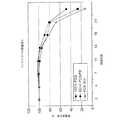
In Vitro Nodule BreakingStrength Maintenance (BSR) & Mass Loss over Time To examine the nodule safety in vitro, the suture sample of Example 1 was washed with a pH 7.27 buffer and a 57 ° C. water bath. Decomposed. A knot consisting of 5 knots was placed in a buffer container. A nodule consisting of 5 knots in each lot was tested at each time. Before and after decomposition at an XH speed of 2 inches (5.08 mm) / min using an Instron Tensile Tester utilizing a Harmann's loop fixture to determine the fracture strength The suture was tested at Note that because this test was done in a loop, the nodule strength was approximately doubled in the case of a typical USP nodule strength test. The suture was dried after being immersed in the buffer for each time, and the residual mass was measured.
約40wt%の高分子ポリエチレンと約60wt%のポリジオキサノンからなる例1の縫合糸に加えて、100wt%のポリジオキサノンからなる別の縫合糸も試験した。表3に、緩衝液中での経過日数に対するインビトロでの結節強度の結果を示した。ポリジオキサノン/超高分子ポリエチレン(60/40)縫合糸の強度が、18日後に30ポンド(約13.64kg)になり、その後は比較的一定であった。非吸収性の芳香族ポリエステル縫合糸の強度に一致するように、ポリジオキサノンが喪失した後の超高分子ポリエチレンの強度が約30ポンド(約13.64kg)となるようにデザインした。従って、ポリジオキサノン/超高分子ポリエチレン縫合糸のポリジオキサノン成分の強度維持は、縫合糸の強度から30ポンド(約13.64kg)を減じて求めた。このデータを用いて、表3及び図4に残留強度(%)を示した。 In addition to the suture of Example 1 consisting of about 40 wt% polymeric polyethylene and about 60 wt% polydioxanone, another suture consisting of 100 wt% polydioxanone was also tested. Table 3 shows the results of in vitro nodule strength versus days elapsed in buffer. The strength of the polydioxanone / ultra high molecular weight polyethylene (60/40) suture reached 30 pounds after 18 days and remained relatively constant thereafter. To match the strength of the non-absorbable aromatic polyester suture, the strength of the ultra-high molecular weight polyethylene after loss of polydioxanone was designed to be about 30 pounds (about 13.64 kg). Accordingly, maintaining the strength of the polydioxanone component of the polydioxanone / ultra high molecular weight polyethylene suture was determined by subtracting 30 pounds (about 13.64 kg) from the strength of the suture. Using this data, the residual strength (%) is shown in Table 3 and FIG.
ポリジオキサノン/ポリエチレン縫合糸のポリジオキサノンが100%ポリジオキサノン編み縫合糸よりも長期に亘って高い強度を維持したことは予想外であった。 It was unexpected that the polydioxanone of the polydioxanone / polyethylene suture maintained higher strength over a longer period than the 100% polydioxanone braided suture.
表4及び図5に、結節した縫合糸の残存質量が示されている。2成分縫合糸のポリジオキサノン成分の残留質量(%)の計算は、縫合糸が40wt%の超高分子ポリエチレンと60wt%のポリジオキサノンからなることに基づいて行った。従って、ポリジオキサノン成分の残留質量(wt%)は、2成分縫合糸から40を減じてから0.60で除して求めた。 Table 4 and FIG. 5 show the remaining mass of the tied suture. The residual mass (%) of the polydioxanone component of the two-component suture was calculated based on the fact that the suture was composed of 40 wt% ultrahigh molecular weight polyethylene and 60 wt% polydioxanone. Therefore, the residual mass (wt%) of the polydioxanone component was obtained by subtracting 40 from the two-component suture and dividing by 0.60.
2成分縫合糸のポリジオキサノン成分の質量減少が100%ポリジオキサノン縫合糸とは異なることに留意されたい。従って、驚くべきことに、2成分縫合糸のポリジオキサノン成分の質量減少は予想通りであったが、その結節強さ維持は予想よりも大きかった。これは、質量が減少しても良好な強度を維持させるのに極めて望ましい。 Note that the weight loss of the polydioxanone component of the two component suture is different from the 100% polydioxanone suture. Thus, surprisingly, the weight loss of the polydioxanone component of the two-component suture was as expected, but its knot strength maintenance was greater than expected. This is highly desirable to maintain good strength as the mass decreases.
例4
生体内での結節の破壊強さ維持(BSR)
生体内での破壊強さ維持は、例1の縫合糸を用いて以下の要領で実施した。Example 4
Maintaining fracture strength of nodules in vivo (BSR)
Maintenance of the breaking strength in vivo was performed using the suture of Example 1 as follows.
生体内での結節強さの試験に用いた方法は、ラットを用いてそれぞれの期間の間、各ロットからの4本の結び目を設けた縫合糸を移植した以外はインビトロ試験と同様である。結び目が設けられた縫合糸のそれぞれを、左及び右後の背側皮下組織に移植した。取り出した縫合糸を肉眼及び顕微鏡で調べた。縫合糸組成ではなく時間経過による縫合糸の周りへの組織の僅かな成長が確認された。破壊強さを調べるために、ハルマンループ取付け具(Harmann's loop fixture)を利用するインストロン引張試験機(Instron Tensile Tester)を用いて、2インチ(5.08mm)/分のXH速度で分解の前後で縫合糸を試験した。 The method used for in vivo knot strength testing was similar to the in vitro test except that rats were used to implant sutures with four knots from each lot for each period. Each suture with a knot was implanted into the left and right back dorsal subcutaneous tissues. The removed suture was examined with the naked eye and under a microscope. Slight tissue growth around the suture was observed over time, not the suture composition. Before and after decomposition at an XH speed of 2 inches (5.08 mm) / min using an Instron Tensile Tester utilizing a Harmann's loop fixture to determine the breaking strength The suture was tested at
インビトロ試験結果に極めて類似したこの試験結果を表5及び図6に示した。100%PDSに対する複合編み縫合糸のPDS成分の結節強さ維持の差は、インビトロ試験よりも生体内移植の場合の方が顕著であった。これは予想していなかったが、極めて望ましいものである。 The test results, which are very similar to the in vitro test results, are shown in Table 5 and FIG. The difference in maintaining the knot strength of the PDS component of the composite braided suture relative to 100% PDS was more pronounced for in vivo transplantation than for in vitro testing. This was unexpected but highly desirable.
例5
結節プロフィールの縮小(煮沸試験)
0.05N 水酸化ナトリウム溶液中での加速吸収試験(煮沸)を例1のコーティング縫合糸を用いて実施し、ポリジオキサノン部分が吸収された後の縫合糸の結節プロフィールの縮小を検証した。Example 5
Reduction of nodule profile (boiling test)
An accelerated absorption test (boiling) in 0.05N sodium hydroxide solution was performed using the coated suture of Example 1 to verify the reduction in suture nodule profile after the polydioxanone moiety was absorbed.
縫合糸にループを設けて5つのサンプルを用意した。それぞれのサンプルは5つの外科医結びからなる結節を有していた。適宜それぞれのサンプルに標識付けした。各結節の厚みを、ミツトヨ(Mitutoyo)デジタル測定器で測定し記録した。アンビルに配置した縫合糸ループと、アンビルの中心とマイクロメーターの脚部の間に配置した結節を測定した。0.05N 水酸化ナトリウム溶液の入った500ml パイレックス(Pyrex)(登録商標)の瓶を用意した。結節をこの瓶の中に入れて蓋を閉めて密閉した。次いで、瓶を90℃の水槽に入れ、約24時間放置した。 Five samples were prepared by providing a loop on the suture. Each sample had a nodule consisting of 5 surgeon knots. Each sample was labeled as appropriate. The thickness of each nodule was measured and recorded with a Mitutoyo digital measuring instrument. The suture loop placed on the anvil and the nodule placed between the center of the anvil and the leg of the micrometer were measured. A 500 ml Pyrex® bottle with 0.05N sodium hydroxide solution was prepared. The nodule was placed in the bottle and sealed with the lid closed. The bottle was then placed in a 90 ° C. water bath and left for about 24 hours.
24時間煮沸した後、サンプルを瓶から取り出し1時間乾燥させた。各結節の厚みを再び測定した。この結果を以下の表6に示す。 After boiling for 24 hours, the sample was removed from the bottle and dried for 1 hour. The thickness of each nodule was measured again. The results are shown in Table 6 below.
この実験から、生体吸収性ポリジオキサノン成分が吸収されると結節プロフィールが約50%縮小することが分かった。縫合糸は平坦になり、結節を触知しにくくなり、組織への刺激が少なくなる。 From this experiment, it was found that the nodule profile was reduced by about 50% when the bioabsorbable polydioxanone component was absorbed. The suture will be flat, making the nodule difficult to touch and less irritating to the tissue.
別の実験では、例1のコーティング縫合糸を用いてインビトロ吸収試験を実施した。 In another experiment, an in vitro absorption test was performed using the coated suture of Example 1.
縫合糸にループを設けてサンプルを用意した。それぞれのサンプルは、結び目が交互し、ポストが交互した6個のハーフヒッチ結節からなる結節を有する。適宜それぞれのサンプルに標識付けした。サンプルをリン酸緩衝液に入れ、約57℃で26週間放置した。サンプルの質量を測定し、サンプルの体積を計算で求めた。この結果を経過日数が0日のサンプルと比較した。この結果を表7に示す。 A sample was prepared by providing a loop in the suture thread. Each sample has a nodule consisting of six half-hitch nodules with alternating knots and alternating posts. Each sample was labeled as appropriate. Samples were placed in phosphate buffer and left at about 57 ° C. for 26 weeks. The sample mass was measured and the sample volume was calculated. This result was compared with a sample having an elapsed time of 0 days. The results are shown in Table 7.
例6
この例では、抗菌薬トリクロサンをポリジオキサノンなどの生体分解性ポリマーの粉末に混合した。この混合は、ブラベンダー(Brabender)、ベーカー・パーキンス・ブレンダー(Baker Perkins blender)、またはVブレンダー(V-blender)を用いて、必ずしも必要ではないが加熱して行った。所望に応じて溶媒を添加して、混合を容易にすることができる。次いで、通常は分散溶媒を押出し工程の前に除去する。得られた組成物を一軸または二軸の押出機で押出しするか或いはペレット化する。混合したら、その材料を、モノフィラメント繊維またはマルチフィラメント繊維を形成できるダイを用いて従来の一軸または二軸のスクリュー押出機で再び押出しした。この繊維は、他の処理を行わないでスピナレットから直接収集する、或いはこの工程の後にアニーリングやドローイング(引抜き)を実施することもできる。樹脂のスループット、巻取り速度、及びドローイングの程度により、繊維の直径や向きが決まる。この繊維をヤーンに紡績する。Example 6
In this example, the antibacterial agent triclosan was mixed with a powder of a biodegradable polymer such as polydioxanone. This mixing was performed using a Brabender, Baker Perkins blender, or V-blender with heating, although not necessary. Solvents can be added as desired to facilitate mixing. The dispersed solvent is then usually removed prior to the extrusion process. The resulting composition is extruded or pelletized with a single or twin screw extruder. Once mixed, the material was re-extruded on a conventional single or twin screw extruder using a die capable of forming monofilament or multifilament fibers. The fibers can be collected directly from the spinneret without further processing, or annealing or drawing can be performed after this step. The diameter and direction of the fiber are determined by the resin throughput, the winding speed, and the degree of drawing. This fiber is spun into a yarn.
例7
例6の吸収性マルチフィラメントヤーンを例1の生体吸収性ヤーンの代わりに用いて、例1の工程に従って本発明の複合縫合糸を製造した。Example 7
Using the absorbable multifilament yarn of Example 6 in place of the bioabsorbable yarn of Example 1, a composite suture of the present invention was made according to the process of Example 1.
例8
曲げ剛性試験
試験するために、長さが1.5インチ(38.1mm)の5本の縫合糸を各ロットから用意した。特殊な取付け具(後述)をインストロン引張試験機(モデル4201)と共に用いて、取付け具の開口に通された縫合糸を引張るために必要な抵抗力を測定した。この試験では、500gのロードセルを用いた。クロスヘッド速度は1インチ(25.4mm)/分とした。この試験の情報は電子的に収集した。Example 8
Five sutures, 1.5 inches (38.1 mm) long, were prepared from each lot for theflexural rigidity test . A special fixture (described below) was used with an Instron tensile tester (model 4201) to measure the resistance required to pull the suture threaded through the fixture opening. In this test, a 500 g load cell was used. The crosshead speed was 1 inch (25.4 mm) / min. Information on this trial was collected electronically.
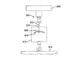
この取付け具の上側部分が図7に例示されている。取付け具502は、縫合糸510を通すために端部に通路508を有する金属ロッド506からなる。ロッドの上端は、ロードセル505に結合されている。取付け具の下側部分は、装置のベース514に取り付けられたブラケット512からなる。このブラケットは、ロッド506の直径よりも僅かに大きい10mmの開口516を有する。この開口516の中心に金属ロッドの中心を合わせた。 The upper portion of the fixture is illustrated in FIG. The
例1の縫合糸から1.5インチ(38.1mm)の5本のサンプルを用意した。それぞれのサンプルを、開口の両側に延びた縫合糸の長さがほぼ同じになるまで金属ロッドの通路508に通した。次いで、クロスヘッドを始動させ、縫合糸をブラケットの開口を介して引張った。縫合糸サンプルを開口を介して引張って、抵抗をグラム(g)で記録した。5つのサンプルを測定し、その結果を報告するために平均した。 Five samples of 1.5 inches (38.1 mm) were prepared from the suture of Example 1. Each sample was passed through a
表8に示されている結果から、本発明のポリエチレン/ポリジオキサノン縫合糸が、ファイバーワイヤー(FIBERWIRE)ポリエチレン/芳香族ポリエステル縫合糸よりも曲げ剛性が優れており、芳香族ポリエステル縫合糸に類似した特性を有していることが分かった。 From the results shown in Table 8, the polyethylene / polydioxanone suture of the present invention is superior in bending rigidity to the fiber wire (FIBERWIRE) polyethylene / aromatic polyester suture, and has similar characteristics to the aromatic polyester suture. It turns out that it has.
例9
回旋腱板手術のために、患者の準備を従来の要領で行う。患者を従来の麻酔法で麻酔する。従来のトロカール及び従来の関節鏡を用いて病理を確認し診断する。外科医は、傷害部位に近接した肩にあるカニューレを介して挿入された従来の関節鏡を用いて障害部位を観察する。外科医は、あらゆる癒着を分離させ、回旋腱板の外側縁を除去し、腱を固定し、そして骨層の準備をする。外科医は、患者の上腕骨頭の大粗面の下側における最適な縫合糸アンカー配置部位を決定し、骨孔を穿孔して以下の要領でアンカーを配置する。ドリルガイドをトロカールから挿入する。ドリルガイドを介してドリルピットを配置し、縫合糸アンカー用の骨孔を穿孔する。本発明の縫合糸が取り付けられた図3に例示されている縫合糸アンカーなどの従来の縫合糸アンカーを、骨孔に挿入し、骨皮質の下側の海綿骨に係合するように配置する。縫合糸の端部に取り付けた外科用縫合針を用いて、軟組織(例えば、腱)が骨の表面に近接するように軟組織に刺入する。次いで、縫合糸を、従来の要領で従来の外科用結節を作って結節する。次いで、縫合針を縫合糸から切り離して処置を終了し、縫合または外科用テープや外科用接着剤を用いて従来の要領で患者の外筋膜と皮膚を近接させる。Example 9
The patient is prepared in a conventional manner for rotator cuff surgery. The patient is anesthetized with conventional anesthesia. Pathology is confirmed and diagnosed using a conventional trocar and a conventional arthroscope. The surgeon observes the site of injury using a conventional arthroscope inserted through a cannula on the shoulder adjacent to the site of injury. The surgeon separates any adhesions, removes the outer edge of the rotator cuff, fixes the tendon, and prepares the bone layer. The surgeon determines the optimal suture anchor placement site under the rough surface of the patient's humeral head, drills the bone hole and places the anchor as follows. Insert the drill guide through the trocar. A drill pit is placed through the drill guide to drill a bone hole for the suture anchor. A conventional suture anchor, such as the suture anchor illustrated in FIG. 3 with the suture of the present invention attached, is inserted into the bone hole and positioned to engage the cancellous bone below the bone cortex. . Using a surgical suture needle attached to the end of the suture, the soft tissue (eg, tendon) is inserted into the soft tissue so that it is close to the surface of the bone. The suture is then knotted in a conventional manner, making a conventional surgical knot. The suture needle is then disconnected from the suture to complete the procedure, and the patient's external fascia and skin are brought into close proximity using a suture or surgical tape or surgical adhesive in the conventional manner.
本発明の縫合糸には多数の利点がある。このような利点には、時間経過による縫合糸質量の減少の最大化、時間経過による結節プロフィールの縮小、改善されたBSRプロフィール、高い抗張力、及び改善された操作性が含まれる。 The suture of the present invention has a number of advantages. Such advantages include maximizing the loss of suture mass over time, reducing the nodule profile over time, improved BSR profile, high tensile strength, and improved maneuverability.
詳細な実施形態を用いて本発明を説明してきたが、当業者であれば、請求する本発明の範囲及び概念から逸脱することなく、形態及び細部の様々な変更が可能であることを理解できよう。 While the invention has been described in terms of detailed embodiments, those skilled in the art will recognize that various changes in form and detail are possible without departing from the scope and concept of the claimed invention. Like.
本発明の実施態様は以下の通りである。
(1)外科用縫合糸と縫合糸アンカーの組合せ体であって、
(a)アンカー本体を備え、縫合糸取付け孔が形成された縫合糸アンカーと、
(b)生体吸収性ポリマーを含む少なくとも1本の繊維からなる少なくとも1本のヤーンを含む内側コアと、少なくとも1本の非吸収性繊維からなる第2のヤーン及び少なくとも1本の生体吸収性繊維を含む第3のヤーンを有する外側編みシースとを備えた、前記第1のヤーンと前記第2のヤーンが編み合わさって接触している外科用縫合糸とを含み、
前記縫合糸が前記縫合糸アンカーの前記縫合糸取付け孔に取り付けられていることを特徴とする外科用縫合糸と縫合糸アンカーの組合せ体。
(2)前記第1の生体吸収性ヤーン及び前記第2の生体吸収性ヤーンが、p‐ジオキサノン、ε‐カプロラクトン、グリコリド(glycolide)、L(‐)‐ラクチド、D(+)‐ラクチド、Meso‐ラクチド、トリメチレンカーボネート、及びこれらの組合せからなる群から選択されるラクトンモノマーから製造されたポリマーを含むことを特徴とする実施態様(1)に記載の組合せ体。
(3)前記非吸収性繊維が超高分子ポリエチレンを含むことを特徴とする実施態様(1)に記載の組合せ体。
(4)前記縫合糸が約55wt%〜約70wt%の生体吸収性ポリマーを含むことを特徴とする実施態様(1)に記載の組合せ体。
(5)前記生体吸収性ヤーンがポリジオキサノンを含むことを特徴とする実施態様(2)に記載の組合せ体。Embodiments of the present invention are as follows.
(1) A combination of a surgical suture and a suture anchor,
(A) a suture anchor provided with an anchor body and having a suture attachment hole formed thereon;
(B) an inner core comprising at least one yarn comprising at least one fiber comprising a bioabsorbable polymer, a second yarn comprising at least one nonabsorbable fiber and at least one bioabsorbable fiber A surgical suture with the first yarn and the second yarn interwoven and in contact with each other, and an outer knitted sheath having a third yarn comprising:
A combination of a surgical suture and a suture anchor, wherein the suture is attached to the suture attachment hole of the suture anchor.
(2) The first bioabsorbable yarn and the second bioabsorbable yarn are p-dioxanone, ε-caprolactone, glycolide, L (−)-lactide, D (+)-lactide, Meso The combination according to embodiment (1), comprising a polymer made from a lactone monomer selected from the group consisting of lactide, trimethylene carbonate, and combinations thereof.
(3) The combination according to the embodiment (1), wherein the non-absorbable fiber includes ultra high molecular weight polyethylene.
(4) The combination according to embodiment (1), wherein the suture includes about 55 wt% to about 70 wt% of a bioabsorbable polymer.
(5) The combination according to embodiment (2), wherein the bioabsorbable yarn contains polydioxanone.
(6)前記第1の生体吸収性ヤーン及び前記第2の生体吸収性ヤーンが医薬組成物を含むことを特徴とする実施態様(1)に記載の組合せ体。
(7)前記第1の生体吸収性ヤーン及び前記第2の生体吸収性ヤーンが、鎮痛薬、抗生物質、抗炎症薬、及び痛み止めからなる群から選択される医薬組成物を含むことを特徴とする実施態様(6)に記載の組合せ体。
(8)前記ポリエチレンヤーンが、約500,000g/モル〜約5,000,000g/モルの平均分子量を有する超高分子ポリエチレンを含むことを特徴とする実施態様(1)に記載の組合せ体。
(9)更に縫合糸のコーティングを含むことを特徴とする実施態様(1)に記載の組合せ体。
(10)更に、前記縫合糸の一端に取り付けられた外科用縫合針を含むことを特徴とする実施態様(1)に記載の組合せ体。(6) The combination according to embodiment (1), wherein the first bioabsorbable yarn and the second bioabsorbable yarn contain a pharmaceutical composition.
(7) The first bioabsorbable yarn and the second bioabsorbable yarn comprise a pharmaceutical composition selected from the group consisting of analgesics, antibiotics, anti-inflammatory drugs, and pain relievers. A combination according to embodiment (6).
(8) The combination according to embodiment (1), wherein the polyethylene yarn comprises ultra-high molecular weight polyethylene having an average molecular weight of about 500,000 g / mol to about 5,000,000 g / mol.
(9) The combination according to embodiment (1), further comprising a suture coating.
(10) The combined body according to the embodiment (1), further comprising a surgical suture needle attached to one end of the suture.
(11)軟組織を骨に固定する方法であって、
(a)アンカー本体を備え、縫合糸取付け孔が形成された縫合糸アンカーと、(b)生体吸収性ポリマーを含む少なくとも1本の繊維からなる少なくとも1本のヤーンを含む内側コアと、少なくとも1本の非吸収性繊維からなる第2のヤーン及び少なくとも1本の生体吸収性繊維を含む第3のヤーンを有する外側編みシースとを備えた、前記第1のヤーンと前記第2のヤーンが編み合わさって接触している外科用縫合糸とを含み、前記縫合糸が前記縫合糸アンカーの前記縫合糸取付け孔に取り付けられている、外科用縫合糸と縫合糸アンカーの組合せ体を用意するステップと、
外面を有する骨に前記アンカーを取り付けるステップと、
前記縫合糸を用いて軟組織を前記骨の外面に固定するステップと、
前記縫合糸を結んで結節を形成するステップとを含むことを特徴とする方法。
(12)前記第1の生体吸収性ヤーン及び前記第2の生体吸収性ヤーンが、p‐ジオキサノン、ε‐カプロラクトン、グリコリド(glycolide)、L(‐)‐ラクチド、D(+)‐ラクチド、Meso‐ラクチド、トリメチレンカーボネート、及びこれらの組合せからなる群から選択されるラクトンモノマーから製造されたポリマーを含むことを特徴とする実施態様(11)に記載の方法。
(13)前記非吸収性繊維が超高分子ポリエチレンを含むことを特徴とする実施態様(11)に記載の方法。
(14)前記縫合糸が約55wt%〜約70wt%の生体吸収性ポリマーを含むことを特徴とする実施態様(11)に記載の方法。
(15)前記ヤーンがポリジオキサノンを含むことを特徴とする実施態様(12)に記載の方法。(11) A method of fixing soft tissue to bone,
(A) a suture anchor having an anchor body and having a suture attachment hole formed; (b) an inner core comprising at least one yarn comprising at least one fiber comprising a bioabsorbable polymer; The first yarn and the second yarn are knitted with an outer knitted sheath having a second yarn made of non-absorbable fibers and a third yarn containing at least one bioabsorbable fiber. Providing a surgical suture and suture anchor combination comprising: a surgical suture in contact with each other, wherein the suture is attached to the suture attachment hole of the suture anchor; ,
Attaching the anchor to a bone having an outer surface;
Fixing the soft tissue to the outer surface of the bone using the suture;
Tying the suture to form a nodule.
(12) The first bioabsorbable yarn and the second bioabsorbable yarn are p-dioxanone, ε-caprolactone, glycolide, L (−)-lactide, D (+)-lactide, Meso The method according to embodiment (11), comprising a polymer made from a lactone monomer selected from the group consisting of lactide, trimethylene carbonate, and combinations thereof.
(13) The method according to embodiment (11), wherein the non-absorbable fiber comprises ultra high molecular weight polyethylene.
(14) The method of embodiment (11), wherein the suture comprises from about 55 wt% to about 70 wt% bioabsorbable polymer.
(15) The method according to embodiment (12), wherein the yarn contains polydioxanone.
(16)前記第1の生体吸収性ヤーン及び前記第2の生体吸収性ヤーンが活性物質を含むことを特徴とする実施態様(11)に記載の方法。
(17)前記第1の生体吸収性ヤーン及び前記第2の生体吸収性ヤーンが、鎮痛薬、抗生物質、抗炎症薬、及び痛み止めからなる群から選択される活性物質を含むことを特徴とする実施態様(16)に記載の方法。
(18)前記ポリエチレンヤーンが、約500,000g/モル〜約5,000,000g/モルの分子量を有する超高分子ポリエチレンを含むことを特徴とする実施態様(11)に記載の方法。
(19)更に、縫合糸のコーティングを含むことを特徴とする実施態様(11)に記載の方法。
(20)更に、前記縫合糸の一端に取り付けられた外科用縫合針を含むことを特徴とする実施態様(11)に記載の方法。(16) The method according to embodiment (11), wherein the first bioabsorbable yarn and the second bioabsorbable yarn contain an active substance.
(17) The first bioabsorbable yarn and the second bioabsorbable yarn contain an active substance selected from the group consisting of analgesics, antibiotics, anti-inflammatory drugs, and pain relievers. A method according to embodiment (16).
(18) The method of embodiment (11), wherein the polyethylene yarn comprises ultra high molecular weight polyethylene having a molecular weight of about 500,000 g / mol to about 5,000,000 g / mol.
(19) The method according to embodiment (11), further comprising a suture coating.
(20) The method according to embodiment (11), further comprising a surgical suture needle attached to one end of the suture.
(21)前記第1のヤーン及び前記第2のヤーンが更に、バイオセラミックス材料を含むことを特徴とする実施態様(1)に記載の組合せ体。
(22)前記第1のヤーン及び前記第2のヤーンが、リン酸三カルシウム及びヒドロキシアパタイトからなる群から選択される材料を含むことを特徴とする実施態様(1)に記載の組合せ体。
(23)前記第1のヤーン及び前記第2のヤーンが更に、バイオセラミックス材料を含むことを特徴とする実施態様(11)に記載の方法。
(24)前記第1のヤーン及び前記第2のヤーンが、リン酸三カルシウム及びヒドロキシアパタイトからなる群から選択される材料を含むことを特徴とする実施態様(11)に記載の方法。
(25)外科用縫合糸と縫合糸アンカーの組合せ体の結節の結節プロフィールを縮小させる方法であって、
アンカー本体及び縫合糸取付け孔を有する縫合糸アンカーを用意するステップと、
生体吸収性ポリマーを含む少なくとも1本の繊維からなる少なくとも1本のヤーンを含む内側コアと、少なくとも1本の非吸収性繊維からなる第2のヤーン及び少なくとも1本の生体吸収性繊維を含む第3のヤーンを有する外側編みシースとを備えた、前記第1のヤーンと前記第2のヤーンが編み合わさって接触している縫合糸を用意するステップと、
外面を有する骨に前記アンカーを挿入するステップと、
前記縫合糸を組織に通して、前記縫合糸で前記骨の外面に組織を取り付けるステップと、
前記縫合糸を結んで結節プロフィールを有する結節を形成するステップと、
前記結節プロフィールが効果的に縮小するように十分な時間、前記縫合糸の前記結節を生体内に維持するステップとを含むことを特徴とする方法。(21) The combination according to embodiment (1), wherein the first yarn and the second yarn further contain a bioceramic material.
(22) The combination according to embodiment (1), wherein the first yarn and the second yarn contain a material selected from the group consisting of tricalcium phosphate and hydroxyapatite.
(23) The method according to embodiment (11), wherein the first yarn and the second yarn further comprise a bioceramic material.
(24) The method according to embodiment (11), wherein the first yarn and the second yarn contain a material selected from the group consisting of tricalcium phosphate and hydroxyapatite.
(25) A method for reducing a nodule profile of a nodule of a combination of a surgical suture and a suture anchor,
Providing a suture anchor having an anchor body and a suture attachment hole;
An inner core comprising at least one yarn comprising at least one fiber comprising a bioabsorbable polymer, a second yarn comprising at least one non-absorbable fiber and a first comprising at least one bioabsorbable fiber. Providing a suture comprising an outer knitted sheath having three yarns, wherein the first yarn and the second yarn are interwoven and in contact;
Inserting the anchor into a bone having an outer surface;
Passing the suture through tissue and attaching the tissue to the outer surface of the bone with the suture;
Tying the suture to form a nodule having a nodule profile;
Maintaining the nodule of the suture in vivo for a time sufficient to effectively reduce the nodule profile.
(26)前記ヤーンが、フィラメント当たり3デニールよりも大きいデニールを有するフィラメントを含むことを特徴とする実施態様(5)に記載の組合せ体。
(27)前記ヤーンが、超高分子ポリエチレン/PETから製造された市販の縫合糸と比べて縫合糸の操作性及び剛性に実質的に悪影響がない、フィラメント当たり12デニールよりも大きいデニールを有するフィラメントを含むことを特徴とする実施態様(5)に記載の組合せ体。
(28)前記ヤーンが、フィラメント当たりのデニールが3未満のポリエチレンテレフタレートから製造された縫合糸に比べて縫合糸の操作性及び剛性に実質的に悪影響がない、フィラメント当たり12デニールよりも大きいデニールを有するフィラメントを含むことを特徴とする実施態様(5)に記載の組合せ体。(26) The combination according to embodiment (5), wherein the yarn comprises a filament having a denier greater than 3 denier per filament.
(27) A filament having a denier greater than 12 denier per filament, wherein the yarn has substantially no adverse effect on suture operability and stiffness compared to commercially available sutures made from ultra-high molecular weight polyethylene / PET The combination according to embodiment (5), comprising:
(28) The yarn has a denier greater than 12 denier per filament, with substantially no adverse effect on the operability and stiffness of the suture compared to a suture made from polyethylene terephthalate having a denier per filament of less than 3. The combination according to embodiment (5), comprising a filament having the same.
10 縫合糸
20 外側シース
30、35、70 ヤーン
60 内側コア
100 組織
110 創傷
115 創傷の縁
130 結節
140 結節プロフィール特性
200 縫合糸
250 縫合糸アンカー
255 縫合糸アンカー本体
260 縫合糸取付け孔
270 骨
280 海綿質部分
285 皮質
290 軟組織
310 結節
315 結節プロフィール特性
340 外科用縫合針
502 取付け具
505 ロードセル
506 ロッド
508 通路
510 縫合糸
512 ブラケット
514 ベース
516 開口
DESCRIPTION OF
Claims (12)
Translated fromJapanese(a)アンカー本体を備え、縫合糸取付け孔が形成された縫合糸アンカーと、
(b)生体吸収性ポリマーを含む少なくとも1本の繊維からなる少なくとも1本のヤーンを含む内側コアと、少なくとも1本の非吸収性繊維からなる第2のヤーン及び少なくとも1本の生体吸収性繊維を含む第3のヤーンを有する外側編みシースとを備えた、前記第1のヤーンと前記第2のヤーンが編み合わさって接触している外科用縫合糸とを含み、
前記縫合糸が前記縫合糸アンカーの前記縫合糸取付け孔に取り付けられていることを特徴とする外科用縫合糸と縫合糸アンカーの組合せ体。A combination of a surgical suture and a suture anchor,
(A) a suture anchor provided with an anchor body and having a suture attachment hole formed thereon;
(B) an inner core comprising at least one yarn comprising at least one fiber comprising a bioabsorbable polymer, a second yarn comprising at least one nonabsorbable fiber and at least one bioabsorbable fiber A surgical suture with the first yarn and the second yarn interwoven and in contact with each other, and an outer knitted sheath having a third yarn comprising:
A combination of a surgical suture and a suture anchor, wherein the suture is attached to the suture attachment hole of the suture anchor.
(a)アンカー本体を備え、縫合糸取付け孔が形成された縫合糸アンカーと、(b)生体吸収性ポリマーを含む少なくとも1本の繊維からなる少なくとも1本のヤーンを含む内側コアと、少なくとも1本の非吸収性繊維からなる第2のヤーン及び少なくとも1本の生体吸収性繊維を含む第3のヤーンを有する外側編みシースとを備えた、前記第1のヤーンと前記第2のヤーンが編み合わさって接触している外科用縫合糸とを含み、前記縫合糸が前記縫合糸アンカーの前記縫合糸取付け孔に取り付けられている、外科用縫合糸と縫合糸アンカーの組合せ体を用意するステップと、
外面を有する骨に前記アンカーを取り付けるステップと、
前記縫合糸を用いて軟組織を前記骨の外面に固定するステップと、
前記縫合糸を結んで結節を形成するステップとを含むことを特徴とする方法。A method of fixing soft tissue to bone,
(A) a suture anchor having an anchor body and having a suture attachment hole formed; (b) an inner core comprising at least one yarn comprising at least one fiber comprising a bioabsorbable polymer; The first yarn and the second yarn are knitted with an outer knitted sheath having a second yarn made of non-absorbable fibers and a third yarn containing at least one bioabsorbable fiber. Providing a surgical suture and suture anchor combination comprising: a surgical suture in contact with each other, wherein the suture is attached to the suture attachment hole of the suture anchor; ,
Attaching the anchor to a bone having an outer surface;
Fixing the soft tissue to the outer surface of the bone using the suture;
Tying the suture to form a nodule.
アンカー本体及び縫合糸取付け孔を有する縫合糸アンカーを用意するステップと、
生体吸収性ポリマーを含む少なくとも1本の繊維からなる少なくとも1本のヤーンを含む内側コアと、少なくとも1本の非吸収性繊維からなる第2のヤーン及び少なくとも1本の生体吸収性繊維を含む第3のヤーンを有する外側編みシースとを備えた、前記第1のヤーンと前記第2のヤーンが編み合わさって接触している縫合糸を用意するステップと、
外面を有する骨に前記アンカーを挿入するステップと、
前記縫合糸を組織に通して、前記縫合糸で前記骨の外面に組織を取り付けるステップと、
前記縫合糸を結んで結節プロフィールを有する結節を形成するステップと、
前記結節プロフィールが効果的に縮小するように十分な時間、前記縫合糸の前記結節を生体内に維持するステップとを含むことを特徴とする方法。
A method for reducing the nodule profile of a nodule of a combination of a surgical suture and a suture anchor, comprising:
Providing a suture anchor having an anchor body and a suture attachment hole;
An inner core comprising at least one yarn comprising at least one fiber comprising a bioabsorbable polymer, a second yarn comprising at least one non-absorbable fiber and a first comprising at least one bioabsorbable fiber. Providing a suture comprising an outer knitted sheath having three yarns, wherein the first yarn and the second yarn are interwoven and in contact;
Inserting the anchor into a bone having an outer surface;
Passing the suture through tissue and attaching the tissue to the outer surface of the bone with the suture;
Tying the suture to form a nodule having a nodule profile;
Maintaining the nodule of the suture in vivo for a time sufficient to effectively reduce the nodule profile.
Applications Claiming Priority (1)
| Application Number | Priority Date | Filing Date | Title |
|---|---|---|---|
| US10/740,023US7357810B2 (en) | 2003-12-18 | 2003-12-18 | High strength suture with absorbable core and suture anchor combination |
Publications (3)
| Publication Number | Publication Date |
|---|---|
| JP2005177500Atrue JP2005177500A (en) | 2005-07-07 |
| JP2005177500A5 JP2005177500A5 (en) | 2011-01-13 |
| JP4667853B2 JP4667853B2 (en) | 2011-04-13 |
Family
ID=34523200
Family Applications (1)
| Application Number | Title | Priority Date | Filing Date |
|---|---|---|---|
| JP2004366444AExpired - LifetimeJP4667853B2 (en) | 2003-12-18 | 2004-12-17 | High strength suture and suture anchor combination with an absorbent core |
Country Status (5)
| Country | Link |
|---|---|
| US (6) | US7357810B2 (en) |
| EP (1) | EP1543848B1 (en) |
| JP (1) | JP4667853B2 (en) |
| AU (1) | AU2004235664B2 (en) |
| CA (1) | CA2490175A1 (en) |
Cited By (15)
| Publication number | Priority date | Publication date | Assignee | Title |
|---|---|---|---|---|
| JP2005177499A (en)* | 2003-12-18 | 2005-07-07 | Ethicon Inc | High-strength suture with absorbent core |
| JP2007089653A (en)* | 2005-09-27 | 2007-04-12 | Teijin Fibers Ltd | Suture and manufacturing method thereof |
| JP2010255175A (en)* | 2009-04-27 | 2010-11-11 | Teleflex Medical Inc | Colored suture construction |
| JP2011503114A (en)* | 2008-07-23 | 2011-01-27 | ウォーソー・オーソペディック・インコーポレーテッド | Drug depot having one or more fixation members |
| JP2011525411A (en)* | 2008-06-24 | 2011-09-22 | バイオアクティブ サージカル インコーポレイテッド | Surgical sutures incorporating stem cells or other bioactive materials |
| US8568449B2 (en) | 2003-12-18 | 2013-10-29 | Depuy Mitek, Llc | High strength suture with absorbable core and suture anchor combination |
| JP2015504722A (en)* | 2011-12-27 | 2015-02-16 | ジョン ジェイコブス メディカル インコーポレーテッド | A suture that does not require a knot work and a kit including the suture |
| US9808234B2 (en) | 2011-03-07 | 2017-11-07 | Y. Jacobs Medical Inc. | Suture thread |
| JP2017535387A (en)* | 2014-11-04 | 2017-11-30 | サージカル スペシャルティーズ コーポレーション | Knitted suture coat |
| US10010317B2 (en) | 2012-12-05 | 2018-07-03 | Young Jae Kim | Method of improving elasticity of tissue of living body |
| US10178990B2 (en) | 2012-12-05 | 2019-01-15 | Y. Jacobs Medical Inc. | Apparatus for inserting surgical thread, and surgical procedure kit for inserting surgical thread comprising same |
| US10226320B2 (en) | 2013-12-06 | 2019-03-12 | Y.Jacobs Medical Inc. | Apparatus for inserting medical tube and surgical procedure kit for inserting medical tube, having same |
| JP2020508125A (en)* | 2017-02-17 | 2020-03-19 | エシコン エルエルシーEthicon LLC | Surgical end effector auxiliary fixture |
| JP2020198942A (en)* | 2019-06-06 | 2020-12-17 | 株式会社プロメット | anchor |
| JP2021519165A (en)* | 2018-03-29 | 2021-08-10 | アテックス テクノロジーズ インコーポレイテッド | Small diameter, high strength, low elongation, creep and wear resistant braided structure |
Families Citing this family (157)
| Publication number | Priority date | Publication date | Assignee | Title |
|---|---|---|---|---|
| US8795332B2 (en) | 2002-09-30 | 2014-08-05 | Ethicon, Inc. | Barbed sutures |
| US6241747B1 (en) | 1993-05-03 | 2001-06-05 | Quill Medical, Inc. | Barbed Bodily tissue connector |
| US5931855A (en) | 1997-05-21 | 1999-08-03 | Frank Hoffman | Surgical methods using one-way suture |
| US6527794B1 (en) | 1999-08-10 | 2003-03-04 | Ethicon, Inc. | Self-locking suture anchor |
| US6733506B1 (en) | 2000-11-16 | 2004-05-11 | Ethicon, Inc. | Apparatus and method for attaching soft tissue to bone |
| US7056331B2 (en) | 2001-06-29 | 2006-06-06 | Quill Medical, Inc. | Suture method |
| US6848152B2 (en) | 2001-08-31 | 2005-02-01 | Quill Medical, Inc. | Method of forming barbs on a suture and apparatus for performing same |
| US7029490B2 (en) | 2001-09-13 | 2006-04-18 | Arthrex, Inc. | High strength suture with coating and colored trace |
| US6773450B2 (en) | 2002-08-09 | 2004-08-10 | Quill Medical, Inc. | Suture anchor and method |
| US8100940B2 (en) | 2002-09-30 | 2012-01-24 | Quill Medical, Inc. | Barb configurations for barbed sutures |
| US20040088003A1 (en) | 2002-09-30 | 2004-05-06 | Leung Jeffrey C. | Barbed suture in combination with surgical needle |
| US7624487B2 (en) | 2003-05-13 | 2009-12-01 | Quill Medical, Inc. | Apparatus and method for forming barbs on a suture |
| US10548592B2 (en) | 2004-05-14 | 2020-02-04 | Ethicon, Inc. | Suture methods and devices |
| US7658751B2 (en) | 2006-09-29 | 2010-02-09 | Biomet Sports Medicine, Llc | Method for implanting soft tissue |
| US9801708B2 (en) | 2004-11-05 | 2017-10-31 | Biomet Sports Medicine, Llc | Method and apparatus for coupling soft tissue to a bone |
| US8128658B2 (en) | 2004-11-05 | 2012-03-06 | Biomet Sports Medicine, Llc | Method and apparatus for coupling soft tissue to bone |
| US8361113B2 (en)* | 2006-02-03 | 2013-01-29 | Biomet Sports Medicine, Llc | Method and apparatus for coupling soft tissue to a bone |
| US8137382B2 (en) | 2004-11-05 | 2012-03-20 | Biomet Sports Medicine, Llc | Method and apparatus for coupling anatomical features |
| US9017381B2 (en) | 2007-04-10 | 2015-04-28 | Biomet Sports Medicine, Llc | Adjustable knotless loops |
| US7749250B2 (en) | 2006-02-03 | 2010-07-06 | Biomet Sports Medicine, Llc | Soft tissue repair assembly and associated method |
| US8118836B2 (en) | 2004-11-05 | 2012-02-21 | Biomet Sports Medicine, Llc | Method and apparatus for coupling soft tissue to a bone |
| US8298262B2 (en) | 2006-02-03 | 2012-10-30 | Biomet Sports Medicine, Llc | Method for tissue fixation |
| US7909851B2 (en) | 2006-02-03 | 2011-03-22 | Biomet Sports Medicine, Llc | Soft tissue repair device and associated methods |
| US8303604B2 (en) | 2004-11-05 | 2012-11-06 | Biomet Sports Medicine, Llc | Soft tissue repair device and method |
| US7905904B2 (en) | 2006-02-03 | 2011-03-15 | Biomet Sports Medicine, Llc | Soft tissue repair device and associated methods |
| US8088130B2 (en) | 2006-02-03 | 2012-01-03 | Biomet Sports Medicine, Llc | Method and apparatus for coupling soft tissue to a bone |
| EP2238915B1 (en)* | 2005-03-30 | 2012-10-17 | Arthrex, Inc. | Apparatus for knotless tissue fixation |
| US20060286289A1 (en)* | 2005-06-15 | 2006-12-21 | Rita Prajapati | Method of intraoperative coating therapeutic agents onto sutures |
| US20060287675A1 (en)* | 2005-06-15 | 2006-12-21 | Prajapati Rita T | Method of intra-operative coating therapeutic agents onto sutures composite sutures and methods of use |
| US20060287676A1 (en)* | 2005-06-15 | 2006-12-21 | Rita Prajapati | Method of intra-operative coating therapeutic agents onto sutures, composite sutures and methods of use |
| WO2007089878A2 (en)* | 2006-01-31 | 2007-08-09 | Angiotech Pharmaceuticals, Inc. | Sutures and anti-scarring agents |
| US9538998B2 (en) | 2006-02-03 | 2017-01-10 | Biomet Sports Medicine, Llc | Method and apparatus for fracture fixation |
| US8562645B2 (en) | 2006-09-29 | 2013-10-22 | Biomet Sports Medicine, Llc | Method and apparatus for forming a self-locking adjustable loop |
| US9408599B2 (en) | 2006-02-03 | 2016-08-09 | Biomet Sports Medicine, Llc | Method and apparatus for coupling soft tissue to a bone |
| US8562647B2 (en) | 2006-09-29 | 2013-10-22 | Biomet Sports Medicine, Llc | Method and apparatus for securing soft tissue to bone |
| US9468433B2 (en) | 2006-02-03 | 2016-10-18 | Biomet Sports Medicine, Llc | Method and apparatus for forming a self-locking adjustable loop |
| US8801783B2 (en)* | 2006-09-29 | 2014-08-12 | Biomet Sports Medicine, Llc | Prosthetic ligament system for knee joint |
| US8597327B2 (en) | 2006-02-03 | 2013-12-03 | Biomet Manufacturing, Llc | Method and apparatus for sternal closure |
| US10517587B2 (en) | 2006-02-03 | 2019-12-31 | Biomet Sports Medicine, Llc | Method and apparatus for forming a self-locking adjustable loop |
| US11311287B2 (en) | 2006-02-03 | 2022-04-26 | Biomet Sports Medicine, Llc | Method for tissue fixation |
| US8652172B2 (en) | 2006-02-03 | 2014-02-18 | Biomet Sports Medicine, Llc | Flexible anchors for tissue fixation |
| US8968364B2 (en) | 2006-02-03 | 2015-03-03 | Biomet Sports Medicine, Llc | Method and apparatus for fixation of an ACL graft |
| US11259792B2 (en) | 2006-02-03 | 2022-03-01 | Biomet Sports Medicine, Llc | Method and apparatus for coupling anatomical features |
| US9078644B2 (en) | 2006-09-29 | 2015-07-14 | Biomet Sports Medicine, Llc | Fracture fixation device |
| US8652171B2 (en) | 2006-02-03 | 2014-02-18 | Biomet Sports Medicine, Llc | Method and apparatus for soft tissue fixation |
| US9149267B2 (en) | 2006-02-03 | 2015-10-06 | Biomet Sports Medicine, Llc | Method and apparatus for coupling soft tissue to a bone |
| AU2007201213A1 (en)* | 2006-04-06 | 2007-10-25 | Tyco Healthcare Group Lp | Yarns containing thermoplastic elastomer copolymer and polyolefin filaments |
| US20090035572A1 (en)* | 2006-04-06 | 2009-02-05 | Tyco Healthcare Group Lp | Yarns containing thermoplastic elastomer copolymer and polyolefin filaments |
| EP2010104B1 (en)* | 2006-04-25 | 2018-09-05 | Teleflex Medical Incorporated | Calcium phosphate polymer composite and method |
| US9788825B2 (en) | 2006-08-04 | 2017-10-17 | Depuy Mitek, Llc | Suture anchor with relief mechanism |
| US9750492B2 (en) | 2006-08-04 | 2017-09-05 | Depuy Mitek, Llc | Suture anchor system with tension relief mechanism |
| US9918826B2 (en) | 2006-09-29 | 2018-03-20 | Biomet Sports Medicine, Llc | Scaffold for spring ligament repair |
| US11259794B2 (en) | 2006-09-29 | 2022-03-01 | Biomet Sports Medicine, Llc | Method for implanting soft tissue |
| US8672969B2 (en) | 2006-09-29 | 2014-03-18 | Biomet Sports Medicine, Llc | Fracture fixation device |
| US8500818B2 (en) | 2006-09-29 | 2013-08-06 | Biomet Manufacturing, Llc | Knee prosthesis assembly with ligament link |
| US9011439B2 (en)* | 2006-11-20 | 2015-04-21 | Poly-Med, Inc. | Selectively absorbable/biodegradable, fibrous composite constructs and applications thereof |
| US20080234819A1 (en)* | 2007-02-15 | 2008-09-25 | Reinhold Schmieding | All-inside double-bundle acl reconstruction |
| US8915943B2 (en) | 2007-04-13 | 2014-12-23 | Ethicon, Inc. | Self-retaining systems for surgical procedures |
| US8663324B2 (en)* | 2007-06-29 | 2014-03-04 | Arthrex, Inc. | Double socket ACL reconstruction |
| ES2398779T3 (en) | 2007-09-27 | 2013-03-21 | Ethicon Llc | Self-retaining sutures that include tissue retention elements with enhanced strength |
| DE102007058256A1 (en)* | 2007-11-26 | 2009-05-28 | Aesculap Ag | Surgical thread mesh |
| US8916077B1 (en) | 2007-12-19 | 2014-12-23 | Ethicon, Inc. | Self-retaining sutures with retainers formed from molten material |
| WO2009086172A2 (en) | 2007-12-19 | 2009-07-09 | Angiotech Pharmaceuticals, Inc. | Self-retaining sutures with heat-contact mediated retainers |
| US8118834B1 (en) | 2007-12-20 | 2012-02-21 | Angiotech Pharmaceuticals, Inc. | Composite self-retaining sutures and method |
| US8615856B1 (en) | 2008-01-30 | 2013-12-31 | Ethicon, Inc. | Apparatus and method for forming self-retaining sutures |
| US8875607B2 (en) | 2008-01-30 | 2014-11-04 | Ethicon, Inc. | Apparatus and method for forming self-retaining sutures |
| ES2706295T3 (en) | 2008-02-21 | 2019-03-28 | Ethicon Llc | Method and apparatus for raising retainers in self-retaining sutures |
| US8216273B1 (en) | 2008-02-25 | 2012-07-10 | Ethicon, Inc. | Self-retainers with supporting structures on a suture |
| US8641732B1 (en) | 2008-02-26 | 2014-02-04 | Ethicon, Inc. | Self-retaining suture with variable dimension filament and method |
| SG188784A1 (en) | 2008-04-15 | 2013-04-30 | Ethicon Llc | Self-retaining sutures with bi-directional retainers or uni-directional retainers |
| US20090264925A1 (en)* | 2008-04-17 | 2009-10-22 | Joseph Hotter | Poly(Trimethylene)Terephthalate Filaments And Articles Made Therefrom |
| US20090263451A1 (en)* | 2008-04-18 | 2009-10-22 | Warsaw Orthopedic, Inc. | Anti-Inflammatory and/or Analgesic Agents for Treatment of Myofascial Pain |
| US8961560B2 (en) | 2008-05-16 | 2015-02-24 | Ethicon, Inc. | Bidirectional self-retaining sutures with laser-marked and/or non-laser marked indicia and methods |
| US12419632B2 (en) | 2008-08-22 | 2025-09-23 | Biomet Sports Medicine, Llc | Method and apparatus for coupling anatomical features |
| US12245759B2 (en) | 2008-08-22 | 2025-03-11 | Biomet Sports Medicine, Llc | Method and apparatus for coupling soft tissue to bone |
| DE102008052837A1 (en)* | 2008-10-13 | 2010-04-15 | Aesculap Ag | Textile implant with core-shell construction and method for its production |
| US20100121355A1 (en) | 2008-10-24 | 2010-05-13 | The Foundry, Llc | Methods and devices for suture anchor delivery |
| EP2352440B1 (en) | 2008-11-03 | 2019-02-20 | Ethicon LLC | Length of self-retaining suture and device for using the same |
| GB2468307A (en)* | 2009-03-04 | 2010-09-08 | Xiros Plc | Suture having a core and a braided mantle including strands running parallel to the axis of the suture |
| US20100249854A1 (en)* | 2009-03-31 | 2010-09-30 | Thomas Jonathan D | Multizone Implants |
| WO2010132310A1 (en) | 2009-05-12 | 2010-11-18 | Foundry Newco Xi, Inc. | Methods and devices to treat diseased or injured musculoskeletal tissue |
| US20100292733A1 (en) | 2009-05-12 | 2010-11-18 | Foundry Newco Xi, Inc. | Knotless suture anchor and methods of use |
| US12096928B2 (en) | 2009-05-29 | 2024-09-24 | Biomet Sports Medicine, Llc | Method and apparatus for coupling soft tissue to a bone |
| EP2263608B1 (en)* | 2009-06-19 | 2016-09-07 | Arthrex, Inc. | Bone-tendon-bone suture button construct |
| US8517073B2 (en)* | 2009-07-16 | 2013-08-27 | Covidien Lp | Apparatus and method for joining similar or dissimilar suture products |
| US8790369B2 (en)* | 2009-07-24 | 2014-07-29 | Depuy Mitek, Llc | Apparatus and method for repairing tissue |
| US8889193B2 (en) | 2010-02-25 | 2014-11-18 | The Johns Hopkins University | Sustained delivery of therapeutic agents to an eye compartment |
| NZ626274A (en) | 2010-05-04 | 2015-03-27 | Ethicon Llc | Laser cutting system and methods for creating self-retaining sutures |
| EP3155978B1 (en) | 2010-06-11 | 2022-04-13 | Cilag GmbH International | Suture delivery tools for endoscopic and robot-assisted surgery |
| DE102010034471A1 (en) | 2010-08-06 | 2012-02-09 | Aesculap Ag | Thread, in particular surgical thread, an implant comprising the thread and a method for producing the thread and the implant |
| US11007296B2 (en) | 2010-11-03 | 2021-05-18 | Ethicon, Inc. | Drug-eluting self-retaining sutures and methods relating thereto |
| US8414612B2 (en) | 2010-11-08 | 2013-04-09 | Covidien Lp | Multifilament barbed suture |
| JP6013352B2 (en) | 2010-11-09 | 2016-10-25 | エシコン・エルエルシーEthicon LLC | Emergency indwelling suture and package |
| US8728159B2 (en)* | 2010-12-01 | 2014-05-20 | Northwestern University | Prosthetic breast support |
| US8795334B2 (en) | 2011-01-28 | 2014-08-05 | Smith & Nephew, Inc. | Tissue repair |
| US9327037B2 (en) | 2011-02-08 | 2016-05-03 | The Johns Hopkins University | Mucus penetrating gene carriers |
| CN103889340B (en) | 2011-03-23 | 2018-09-28 | 伊西康有限责任公司 | Self-retaining variable loop suture |
| US12329373B2 (en) | 2011-05-02 | 2025-06-17 | Biomet Sports Medicine, Llc | Method and apparatus for soft tissue fixation |
| US8926661B2 (en)* | 2011-06-02 | 2015-01-06 | Smith & Nephew, Inc. | Surgical fastening |
| US20130172931A1 (en) | 2011-06-06 | 2013-07-04 | Jeffrey M. Gross | Methods and devices for soft palate tissue elevation procedures |
| US20130178898A1 (en)* | 2011-07-06 | 2013-07-11 | Imds Corporation | Tissue approximation |
| US9357991B2 (en) | 2011-11-03 | 2016-06-07 | Biomet Sports Medicine, Llc | Method and apparatus for stitching tendons |
| US9314241B2 (en) | 2011-11-10 | 2016-04-19 | Biomet Sports Medicine, Llc | Apparatus for coupling soft tissue to a bone |
| US9381013B2 (en) | 2011-11-10 | 2016-07-05 | Biomet Sports Medicine, Llc | Method for coupling soft tissue to a bone |
| US8926662B2 (en) | 2012-02-01 | 2015-01-06 | Smith & Nephew, Inc. | Tissue graft anchoring |
| US9080263B2 (en)* | 2012-02-10 | 2015-07-14 | Novus Scientific Ab | Multifilaments with time-dependent characteristics, and medical products made from such multifilaments |
| CN111658037A (en)* | 2012-03-01 | 2020-09-15 | 新特斯有限责任公司 | Surgical suture with soft core |
| US9084597B2 (en) | 2012-03-09 | 2015-07-21 | Smith & Nephew, Inc. | Suture-based knotless repair |
| EP2825207B1 (en) | 2012-03-16 | 2020-08-19 | The Johns Hopkins University | Non-linear multiblock copolymer-drug conjugates for the delivery of active agents |
| US8962577B2 (en) | 2012-03-16 | 2015-02-24 | The Johns Hopkins University | Controlled release formulations for the delivery of HIF-1 inhibitors |
| US10070856B1 (en) | 2012-05-03 | 2018-09-11 | Wayne Jay Black | Soft suture anchor |
| US11419598B1 (en) | 2012-05-03 | 2022-08-23 | Riverpoint Medical, Llc | Soft suture anchor |
| US9533068B2 (en)* | 2012-05-04 | 2017-01-03 | The Johns Hopkins University | Drug loaded microfiber sutures for ophthalmic application |
| US9237888B2 (en)* | 2012-09-20 | 2016-01-19 | Medos International Sarl | Methods and devices for threading sutures |
| US8986327B2 (en) | 2012-10-18 | 2015-03-24 | Smith & Nephew, Inc. | Flexible anchor delivery system |
| JP2015536726A (en)* | 2012-11-14 | 2015-12-24 | オーソペディック イノヴェイション センター インコーポレイテッド | Antibacterial articles manufactured by additive manufacturing |
| US20140180326A1 (en)* | 2012-12-20 | 2014-06-26 | Empire Technology Development Llc | Inflatable balloon for protecting blood vessel |
| WO2014124006A1 (en) | 2013-02-05 | 2014-08-14 | The Johns Hopkins University | Nanoparticles for magnetic resonance imaging tracking and methods of making and using thereof |
| US9757119B2 (en) | 2013-03-08 | 2017-09-12 | Biomet Sports Medicine, Llc | Visual aid for identifying suture limbs arthroscopically |
| US9974643B2 (en)* | 2013-03-11 | 2018-05-22 | Medos International Sàrl | Implant having adjustable filament coils |
| US9918827B2 (en) | 2013-03-14 | 2018-03-20 | Biomet Sports Medicine, Llc | Scaffold for spring ligament repair |
| EP3052203A4 (en) | 2013-09-30 | 2017-08-23 | Silk Therapeutics Inc. | Silk protein fragment compositions and articles manufactured therefrom |
| US9226817B2 (en) | 2014-03-03 | 2016-01-05 | Tenjin LLC | Implant placement systems, devices, and methods |
| US11504224B2 (en) | 2014-03-03 | 2022-11-22 | Tenjin LLC | Implant placement systems and one-handed methods for tissue fixation using same |
| US9717587B2 (en) | 2014-03-03 | 2017-08-01 | Tenjin LLC | Multiple implant constructions and fixation methods associated therewith |
| US20150258238A1 (en)* | 2014-03-13 | 2015-09-17 | River Point, Llc | Suture material having beneficial physical properties |
| US20170189172A1 (en)* | 2014-05-06 | 2017-07-06 | Dsm Ip Assets B.V. | Method of making a prosthetic valve and valve obtained therewith |
| WO2015169868A1 (en) | 2014-05-06 | 2015-11-12 | Dsm Ip Assets B.V. | Method of making a prosthetic valve and valve obtained therewith |
| JP6690834B2 (en) | 2014-05-06 | 2020-04-28 | ディーエスエム アイピー アセッツ ビー.ブイ.Dsm Ip Assets B.V. | Artificial valve and method of manufacturing artificial valve |
| WO2015171962A1 (en) | 2014-05-07 | 2015-11-12 | Bart Bracy | Multipart suture |
| TW201542253A (en)* | 2014-05-09 | 2015-11-16 | Bio Line Medical Tech Co Ltd | Composite thread and manufacture process thereof |
| EP3180040B1 (en) | 2014-08-15 | 2020-05-13 | Tepha, Inc. | Self-retaining sutures of poly-4-hydroxybutyrate and copolymers thereof |
| JP6846351B2 (en) | 2015-01-27 | 2021-03-24 | ザ・ジョンズ・ホプキンス・ユニバーシティー | Hypotonic hydrogel formulation for enhanced transport of active agents on mucosal surfaces |
| US10524774B2 (en)* | 2015-04-02 | 2020-01-07 | Arthrex, Inc. | Method of repairing cartilage defects |
| US10524775B2 (en) | 2015-07-02 | 2020-01-07 | Arthrex, Inc. | Methods of repairing cartilage defects |
| EP3322434B1 (en) | 2015-07-14 | 2024-12-11 | Evolved by Nature, Inc. | Silk performance apparel and products and methods of preparing the same |
| US11331191B2 (en) | 2015-08-12 | 2022-05-17 | Howmedica Osteonics Corp. | Bioactive soft tissue implant and methods of manufacture and use thereof |
| US10729548B2 (en)* | 2016-05-02 | 2020-08-04 | Howmedica Osteonics Corp. | Bioactive soft tissue implant and methods of manufacture and use thereof |
| US10172703B2 (en)* | 2015-10-01 | 2019-01-08 | Arthrex, Inc. | Joint kinematic reconstruction techniques |
| EP3180472B1 (en)* | 2015-10-21 | 2018-03-21 | Liebherr-Components Biberach GmbH | Device for detecting the replacement state of wear of a high-strength fibre cable for hoisting devices |
| AU2016344019B2 (en) | 2015-10-30 | 2021-07-15 | New York Society For The Relief Of The Ruptured And Crippled, Maintaining The Hospital For Special Surgery | Suture sleeve patch and methods of delivery within an existing arthroscopic workflow |
| EP3383281B1 (en) | 2015-12-04 | 2024-01-24 | Crossroads Extremity Systems, LLC | Devices for anchoring tissue |
| US11523812B2 (en) | 2016-02-01 | 2022-12-13 | Medos International Sarl | Soft tissue fixation repair methods using tissue augmentation constructs |
| US11484401B2 (en) | 2016-02-01 | 2022-11-01 | Medos International Sarl | Tissue augmentation scaffolds for use in soft tissue fixation repair |
| US20170273680A1 (en) | 2016-02-01 | 2017-09-28 | DePuy Synthes Products, Inc. | Tissue augmentation tacks for use with soft tissue fixation repair systems and methods |
| US10167582B1 (en)* | 2016-05-13 | 2019-01-01 | Stryker Corporation | Braided filament with particularized strand compositions and methods of manufacturing and using same |
| KR102092145B1 (en)* | 2017-04-20 | 2020-03-24 | 퇴펠베르게르 피베르 로페 게엠베하 | High-strength fibre rope for hoisting equipment such as cranes |
| US11390988B2 (en) | 2017-09-27 | 2022-07-19 | Evolved By Nature, Inc. | Silk coated fabrics and products and methods of preparing the same |
| US10595983B1 (en) | 2018-02-05 | 2020-03-24 | Riverpoint Medical, Llc | Constructs and methods for repairing a tendon with a reduced risk of reinjury |
| US12151050B2 (en)* | 2018-03-01 | 2024-11-26 | Tepha, Inc. | Medical devices containing compositions of poly(butylene succinate) and copolymers thereof |
| WO2020132679A1 (en) | 2018-12-21 | 2020-06-25 | Khodagholy Dion | Internal-ion gated electrochemical transistors |
| US11523813B1 (en) | 2019-03-26 | 2022-12-13 | Riverpoint Medical, Llc | Soft button assembly and procedure |
| US12201741B2 (en) | 2019-09-15 | 2025-01-21 | The Trustees Of Columbia University In The City Of New York | Composites and devices for interfacing electronics to biological tissue |
| JP6896241B2 (en)* | 2019-12-11 | 2021-06-30 | ケイセイ医科工業株式会社 | Medical suture |
| JP6896240B2 (en)* | 2019-12-11 | 2021-06-30 | ケイセイ医科工業株式会社 | Medical suture |
| EP4171671A4 (en)* | 2020-06-24 | 2024-07-24 | Anika Therapeutics, Inc. | MEDICAL COMPOSITE TEXTILE WITH NON-RESORBABLE FIBERS AND BIORESORBABLE HYALURONATE-BASED FIBERS |
| RU2763702C1 (en)* | 2020-12-04 | 2021-12-30 | Общество с ограниченной ответственностью "Медин-Н" | Multicomponent surgical thread |
Citations (6)
| Publication number | Priority date | Publication date | Assignee | Title |
|---|---|---|---|---|
| US3463158A (en)* | 1963-10-31 | 1969-08-26 | American Cyanamid Co | Polyglycolic acid prosthetic devices |
| JPS544493A (en)* | 1977-06-10 | 1979-01-13 | Ethicon Inc | Poly*alkyleneoxalate* absorptive coating for suturing yarn |
| JPS59130316A (en)* | 1982-09-30 | 1984-07-26 | アライド・コ−ポレ−ション | High toughness, high modulus polyethylene fiber and its manufacturing method |
| JPS6395060A (en)* | 1986-10-09 | 1988-04-26 | 日本商事株式会社 | Yarn for fixing backbone |
| JPH0380868A (en)* | 1989-08-23 | 1991-04-05 | Descente Ltd | drug sustained release suture |
| JP2002540901A (en)* | 1999-04-14 | 2002-12-03 | エシコン・インコーポレイテッド | Improved multifilament surgical thread |
Family Cites Families (45)
| Publication number | Priority date | Publication date | Assignee | Title |
|---|---|---|---|---|
| DE934686C (en)* | 1952-11-27 | 1955-11-03 | Nsu Automobil Ag | Drive device for motor vehicles with independent suspension of the driven wheels and a horizontal motor |
| ZA724131B (en) | 1972-04-12 | 1973-03-28 | Sutures Inc | Sutures having long-lasting biocidal properties |
| BE806869A (en)* | 1972-11-03 | 1974-04-30 | Ethicon Inc | IMPROVED BRAID TIE WIRE |
| US4141087A (en)* | 1977-01-19 | 1979-02-27 | Ethicon, Inc. | Isomorphic copolyoxalates and sutures thereof |
| US4632100A (en)* | 1985-08-29 | 1986-12-30 | Marlowe E. Goble | Suture anchor assembly |
| US4624256A (en)* | 1985-09-11 | 1986-11-25 | Pfizer Hospital Products Group, Inc. | Caprolactone polymers for suture coating |
| US4898156A (en)* | 1987-05-18 | 1990-02-06 | Mitek Surgical Products, Inc. | Suture anchor |
| US5261886A (en)* | 1987-08-26 | 1993-11-16 | United States Surgical Corporation | Cabled core and braided suture made therefrom |
| US5306289A (en)* | 1987-08-26 | 1994-04-26 | United States Surgical Corporation | Braided suture of improved characteristics |
| US5183701A (en)* | 1987-10-02 | 1993-02-02 | Dyneema V.O.F. | Articles of highly oriented polyolefins of ultrahigh molecular weight, process for their manufacture, and their use |
| US4899743A (en)* | 1987-12-15 | 1990-02-13 | Mitek Surgical Products, Inc. | Suture anchor installation tool |
| US4968315A (en)* | 1987-12-15 | 1990-11-06 | Mitek Surgical Products, Inc. | Suture anchor and suture anchor installation tool |
| US4999074A (en)* | 1989-01-31 | 1991-03-12 | Afeyan Industries Inc. | Method for the production of an air mattress |
| US4946468A (en)* | 1989-06-06 | 1990-08-07 | Mitek Surgical Products, Inc. | Suture anchor and suture anchor installation tool |
| US5002550A (en)* | 1989-06-06 | 1991-03-26 | Mitek Surgical Products, Inc. | Suture anchor installation tool |
| US5011473A (en)* | 1989-06-06 | 1991-04-30 | Mitek Surgical Products Inc. | Device for securing and positioning a wire to a needle |
| US5078730A (en)* | 1990-11-06 | 1992-01-07 | Mitek Surgical Products, Inc. | Holder for suture anchor assembly |
| US5207679A (en)* | 1991-09-26 | 1993-05-04 | Mitek Surgical Products, Inc. | Suture anchor and installation tool |
| US5318575A (en) | 1992-02-03 | 1994-06-07 | United States Surgical Corporation | Method of using a surgical repair suture product |
| US5217486A (en)* | 1992-02-18 | 1993-06-08 | Mitek Surgical Products, Inc. | Suture anchor and installation tool |
| US5314446A (en)* | 1992-02-19 | 1994-05-24 | Ethicon, Inc. | Sterilized heterogeneous braids |
| US5540718A (en)* | 1993-09-20 | 1996-07-30 | Bartlett; Edwin C. | Apparatus and method for anchoring sutures |
| US5584836A (en)* | 1994-04-07 | 1996-12-17 | Smith & Nephew Richards, Inc. | Cannulated medical suture anchor |
| US5683418A (en)* | 1994-04-29 | 1997-11-04 | Mitek Surgical Products, Inc. | Wedge shaped suture anchor and method of implantation |
| US5961538A (en)* | 1996-04-10 | 1999-10-05 | Mitek Surgical Products, Inc. | Wedge shaped suture anchor and method of implantation |
| US5718717A (en) | 1996-08-19 | 1998-02-17 | Bonutti; Peter M. | Suture anchor |
| FR2754207B1 (en)* | 1996-10-07 | 1998-11-06 | Emsens Michel | MACHINE FOR CUTTING CALIBERATED PIECES OF FOOD PRODUCTS |
| US5709708A (en)* | 1997-01-31 | 1998-01-20 | Thal; Raymond | Captured-loop knotless suture anchor assembly |
| US5782864A (en)* | 1997-04-03 | 1998-07-21 | Mitek Surgical Products, Inc. | Knotless suture system and method |
| US5980524A (en) | 1997-06-02 | 1999-11-09 | Innovasive Devices, Inc. | Device for repairing a meniscal tear in a knee and method |
| US5814051A (en)* | 1997-06-06 | 1998-09-29 | Mitex Surgical Products, Inc. | Suture anchor insertion system |
| DE69834375T2 (en)* | 1997-10-10 | 2007-03-15 | Ethicon, Inc. | Braided suture with improved knot strength |
| US6264674B1 (en)* | 1998-11-09 | 2001-07-24 | Robert L. Washington | Process for hot stretching braided ligatures |
| US6306158B1 (en)* | 1999-03-02 | 2001-10-23 | Edwin C. Bartlett | Suture anchor and associated method of implantation |
| US6689153B1 (en)* | 1999-04-16 | 2004-02-10 | Orthopaedic Biosystems Ltd, Inc. | Methods and apparatus for a coated anchoring device and/or suture |
| US7029490B2 (en) | 2001-09-13 | 2006-04-18 | Arthrex, Inc. | High strength suture with coating and colored trace |
| US6716234B2 (en)* | 2001-09-13 | 2004-04-06 | Arthrex, Inc. | High strength suture material |
| US7070610B2 (en) | 2002-03-30 | 2006-07-04 | Samyang Corporation | Monofilament suture and manufacturing method thereof |
| EP3311759B1 (en)* | 2002-04-18 | 2020-12-09 | Helmholtz-Zentrum Geesthacht Zentrum für Material- und Küstenforschung GmbH | Shape memory polymeric sutures |
| DE10219860A1 (en)* | 2002-05-03 | 2003-11-20 | Ethicon Gmbh | Surgical thread and surgical implant with such a thread |
| WO2004082466A2 (en) | 2003-03-18 | 2004-09-30 | Opus Medical, Inc. | High performance suture |
| US20050125036A1 (en) | 2003-08-14 | 2005-06-09 | Mark Roby | Heterogeneous yarns for surgical articles |
| US7357810B2 (en) | 2003-12-18 | 2008-04-15 | Ethicon, Inc. | High strength suture with absorbable core and suture anchor combination |
| US7329271B2 (en)* | 2003-12-18 | 2008-02-12 | Ethicon, Inc. | High strength suture with absorbable core |
| JP4914800B2 (en) | 2007-10-22 | 2012-04-11 | 三洋電機株式会社 | Electronic equipment cooling device |
- 2003
- 2003-12-18USUS10/740,023patent/US7357810B2/ennot_activeExpired - Lifetime
- 2004
- 2004-12-06AUAU2004235664Apatent/AU2004235664B2/ennot_activeCeased
- 2004-12-13CACA002490175Apatent/CA2490175A1/ennot_activeWithdrawn
- 2004-12-17JPJP2004366444Apatent/JP4667853B2/ennot_activeExpired - Lifetime
- 2004-12-17EPEP04257893Apatent/EP1543848B1/ennot_activeExpired - Lifetime
- 2008
- 2008-04-14USUS12/102,477patent/US8109967B2/ennot_activeExpired - Lifetime
- 2011
- 2011-12-20USUS13/330,741patent/US8568449B2/ennot_activeExpired - Lifetime
- 2013
- 2013-10-24USUS14/062,090patent/US8940018B2/ennot_activeExpired - Lifetime
- 2015
- 2015-01-22USUS14/602,601patent/US9642930B2/ennot_activeExpired - Fee Related
- 2017
- 2017-05-08USUS15/588,780patent/US10624632B2/ennot_activeExpired - Lifetime
Patent Citations (6)
| Publication number | Priority date | Publication date | Assignee | Title |
|---|---|---|---|---|
| US3463158A (en)* | 1963-10-31 | 1969-08-26 | American Cyanamid Co | Polyglycolic acid prosthetic devices |
| JPS544493A (en)* | 1977-06-10 | 1979-01-13 | Ethicon Inc | Poly*alkyleneoxalate* absorptive coating for suturing yarn |
| JPS59130316A (en)* | 1982-09-30 | 1984-07-26 | アライド・コ−ポレ−ション | High toughness, high modulus polyethylene fiber and its manufacturing method |
| JPS6395060A (en)* | 1986-10-09 | 1988-04-26 | 日本商事株式会社 | Yarn for fixing backbone |
| JPH0380868A (en)* | 1989-08-23 | 1991-04-05 | Descente Ltd | drug sustained release suture |
| JP2002540901A (en)* | 1999-04-14 | 2002-12-03 | エシコン・インコーポレイテッド | Improved multifilament surgical thread |
Cited By (31)
| Publication number | Priority date | Publication date | Assignee | Title |
|---|---|---|---|---|
| US10624632B2 (en) | 2003-12-18 | 2020-04-21 | DePuy Synthes Products, Inc. | High strength suture with absorbable core and suture anchor combination |
| US8940018B2 (en) | 2003-12-18 | 2015-01-27 | Depuy Mitek, Llc | High strength suture with absorbable core and suture anchor combination |
| JP2005177499A (en)* | 2003-12-18 | 2005-07-07 | Ethicon Inc | High-strength suture with absorbent core |
| US9642930B2 (en) | 2003-12-18 | 2017-05-09 | Depuy Mitek, Llc | High strength suture with absorbable core and suture anchor combination |
| US8568449B2 (en) | 2003-12-18 | 2013-10-29 | Depuy Mitek, Llc | High strength suture with absorbable core and suture anchor combination |
| JP2007089653A (en)* | 2005-09-27 | 2007-04-12 | Teijin Fibers Ltd | Suture and manufacturing method thereof |
| JP2011525411A (en)* | 2008-06-24 | 2011-09-22 | バイオアクティブ サージカル インコーポレイテッド | Surgical sutures incorporating stem cells or other bioactive materials |
| US8876864B2 (en) | 2008-06-24 | 2014-11-04 | Bioactive Surgical, Inc | Surgical sutures incorporated with stem cells or other bioactive materials |
| JP2015107344A (en)* | 2008-06-24 | 2015-06-11 | バイオアクティブ サージカル インコーポレイテッド | Surgical sutures incorporated with stem cells or other bioactive materials |
| US9446164B2 (en) | 2008-06-24 | 2016-09-20 | Bioactive Surgical, Inc. | Surgical sutures incorporated with stem cells or other bioactive materials |
| US10632225B2 (en) | 2008-06-24 | 2020-04-28 | Bioactive Surgical, Inc. | Surgical sutures incorporated with stem cells or other bioactive materials |
| JP2011503114A (en)* | 2008-07-23 | 2011-01-27 | ウォーソー・オーソペディック・インコーポレーテッド | Drug depot having one or more fixation members |
| JP2010255175A (en)* | 2009-04-27 | 2010-11-11 | Teleflex Medical Inc | Colored suture construction |
| US11103230B2 (en) | 2011-03-07 | 2021-08-31 | Y.Jacobs Medical Inc. | Suture thread |
| US9848865B2 (en) | 2011-03-07 | 2017-12-26 | Y.Jacobs Medical Inc. | Suture thread |
| US9808234B2 (en) | 2011-03-07 | 2017-11-07 | Y. Jacobs Medical Inc. | Suture thread |
| JP2015504722A (en)* | 2011-12-27 | 2015-02-16 | ジョン ジェイコブス メディカル インコーポレーテッド | A suture that does not require a knot work and a kit including the suture |
| US11103232B2 (en) | 2011-12-27 | 2021-08-31 | Y.Jacobs Medical Inc. | Knotless suture, and kit containing same |
| US9924937B2 (en) | 2011-12-27 | 2018-03-27 | Y.Jacobs Medical Inc. | Knotless suture, and kit containing same |
| US9480473B2 (en) | 2011-12-27 | 2016-11-01 | Y.Jacobs Medical Inc. | Knotless suture, and kit containing same |
| US10178990B2 (en) | 2012-12-05 | 2019-01-15 | Y. Jacobs Medical Inc. | Apparatus for inserting surgical thread, and surgical procedure kit for inserting surgical thread comprising same |
| US10010317B2 (en) | 2012-12-05 | 2018-07-03 | Young Jae Kim | Method of improving elasticity of tissue of living body |
| US10226320B2 (en) | 2013-12-06 | 2019-03-12 | Y.Jacobs Medical Inc. | Apparatus for inserting medical tube and surgical procedure kit for inserting medical tube, having same |
| US11241303B2 (en) | 2013-12-06 | 2022-02-08 | Y.Jacobs Medical Inc. | Apparatus for inserting medical tube and surgical procedure kit for inserting medical tube, having same |
| JP2017535387A (en)* | 2014-11-04 | 2017-11-30 | サージカル スペシャルティーズ コーポレーション | Knitted suture coat |
| JP2020508125A (en)* | 2017-02-17 | 2020-03-19 | エシコン エルエルシーEthicon LLC | Surgical end effector auxiliary fixture |
| JP7155131B2 (en) | 2017-02-17 | 2022-10-18 | エシコン エルエルシー | Surgical end effector support fixture |
| JP2021519165A (en)* | 2018-03-29 | 2021-08-10 | アテックス テクノロジーズ インコーポレイテッド | Small diameter, high strength, low elongation, creep and wear resistant braided structure |
| JP7429645B2 (en) | 2018-03-29 | 2024-02-08 | アテックス テクノロジーズ インコーポレイテッド | Small diameter, high strength, low elongation, creep and wear resistant braided structure |
| US11913169B2 (en) | 2018-03-29 | 2024-02-27 | Atex Technologies, Inc. | Small diameter, high strength, low elongation, creep resistant, abrasion resistant braided structure |
| JP2020198942A (en)* | 2019-06-06 | 2020-12-17 | 株式会社プロメット | anchor |
Also Published As
| Publication number | Publication date |
|---|---|
| US8940018B2 (en) | 2015-01-27 |
| US20140052175A1 (en) | 2014-02-20 |
| US20170239383A1 (en) | 2017-08-24 |
| EP1543848B1 (en) | 2011-09-28 |
| CA2490175A1 (en) | 2005-06-18 |
| US20080255557A1 (en) | 2008-10-16 |
| EP1543848A1 (en) | 2005-06-22 |
| US20120323273A1 (en) | 2012-12-20 |
| US7357810B2 (en) | 2008-04-15 |
| AU2004235664A1 (en) | 2005-07-07 |
| US20150133868A1 (en) | 2015-05-14 |
| US9642930B2 (en) | 2017-05-09 |
| AU2004235664B2 (en) | 2011-08-04 |
| JP4667853B2 (en) | 2011-04-13 |
| US10624632B2 (en) | 2020-04-21 |
| US8109967B2 (en) | 2012-02-07 |
| US20050149118A1 (en) | 2005-07-07 |
| US8568449B2 (en) | 2013-10-29 |
Similar Documents
| Publication | Publication Date | Title |
|---|---|---|
| JP4667853B2 (en) | High strength suture and suture anchor combination with an absorbent core | |
| JP4850413B2 (en) | High-strength suture with absorbent core | |
| CA2477471C (en) | Heterogeneous yarns for surgical articles | |
| EP2110146B1 (en) | Poly(trimethylene) terephthalate filaments and articles made therefrom | |
| EP2415488B1 (en) | Thread, especially surgical thread, an implant comprising the thread and also a process for producing the thread and the implant | |
| US20150266221A1 (en) | Absorbable Poly (p-dioxanone-co-glycolide) Monofilament Fibers Pocessing Mid-Term Strength Retention Post-Implantation | |
| Padmakumar et al. | Engineering aspects of suture fabrication | |
| Harloff | Application of Polymers for Surgical Sutures | |
| DiRuscio | Characteristics, In Vitro, and In Vivo Degradation Behaviors of Synthetic Biodegradable Polyaxial and Amphiphilic Copolyesters Used for Surgical Sutures |
Legal Events
| Date | Code | Title | Description |
|---|---|---|---|
| RD04 | Notification of resignation of power of attorney | Free format text:JAPANESE INTERMEDIATE CODE: A7424 Effective date:20071128 | |
| A621 | Written request for application examination | Free format text:JAPANESE INTERMEDIATE CODE: A621 Effective date:20071214 | |
| RD04 | Notification of resignation of power of attorney | Free format text:JAPANESE INTERMEDIATE CODE: A7424 Effective date:20080912 | |
| A977 | Report on retrieval | Free format text:JAPANESE INTERMEDIATE CODE: A971007 Effective date:20100520 | |
| A131 | Notification of reasons for refusal | Free format text:JAPANESE INTERMEDIATE CODE: A131 Effective date:20100525 | |
| A601 | Written request for extension of time | Free format text:JAPANESE INTERMEDIATE CODE: A601 Effective date:20100825 | |
| A602 | Written permission of extension of time | Free format text:JAPANESE INTERMEDIATE CODE: A602 Effective date:20100830 | |
| A601 | Written request for extension of time | Free format text:JAPANESE INTERMEDIATE CODE: A601 Effective date:20100924 | |
| A602 | Written permission of extension of time | Free format text:JAPANESE INTERMEDIATE CODE: A602 Effective date:20100929 | |
| A601 | Written request for extension of time | Free format text:JAPANESE INTERMEDIATE CODE: A601 Effective date:20101025 | |
| A602 | Written permission of extension of time | Free format text:JAPANESE INTERMEDIATE CODE: A602 Effective date:20101028 | |
| A524 | Written submission of copy of amendment under article 19 pct | Free format text:JAPANESE INTERMEDIATE CODE: A524 Effective date:20101124 | |
| A521 | Request for written amendment filed | Free format text:JAPANESE INTERMEDIATE CODE: A523 Effective date:20101125 | |
| TRDD | Decision of grant or rejection written | ||
| A01 | Written decision to grant a patent or to grant a registration (utility model) | Free format text:JAPANESE INTERMEDIATE CODE: A01 Effective date:20101221 | |
| A01 | Written decision to grant a patent or to grant a registration (utility model) | Free format text:JAPANESE INTERMEDIATE CODE: A01 | |
| A61 | First payment of annual fees (during grant procedure) | Free format text:JAPANESE INTERMEDIATE CODE: A61 Effective date:20110112 | |
| FPAY | Renewal fee payment (event date is renewal date of database) | Free format text:PAYMENT UNTIL: 20140121 Year of fee payment:3 | |
| R150 | Certificate of patent or registration of utility model | Ref document number:4667853 Country of ref document:JP Free format text:JAPANESE INTERMEDIATE CODE: R150 Free format text:JAPANESE INTERMEDIATE CODE: R150 | |
| R250 | Receipt of annual fees | Free format text:JAPANESE INTERMEDIATE CODE: R250 | |
| R250 | Receipt of annual fees | Free format text:JAPANESE INTERMEDIATE CODE: R250 | |
| R250 | Receipt of annual fees | Free format text:JAPANESE INTERMEDIATE CODE: R250 | |
| R250 | Receipt of annual fees | Free format text:JAPANESE INTERMEDIATE CODE: R250 | |
| R250 | Receipt of annual fees | Free format text:JAPANESE INTERMEDIATE CODE: R250 | |
| R250 | Receipt of annual fees | Free format text:JAPANESE INTERMEDIATE CODE: R250 | |
| R250 | Receipt of annual fees | Free format text:JAPANESE INTERMEDIATE CODE: R250 | |
| R250 | Receipt of annual fees | Free format text:JAPANESE INTERMEDIATE CODE: R250 | |
| R250 | Receipt of annual fees | Free format text:JAPANESE INTERMEDIATE CODE: R250 | |
| R250 | Receipt of annual fees | Free format text:JAPANESE INTERMEDIATE CODE: R250 | |
| R250 | Receipt of annual fees | Free format text:JAPANESE INTERMEDIATE CODE: R250 | |
| EXPY | Cancellation because of completion of term |