JP2004501981A - Enhancement of antibody-cytokine fusion protein-mediated immune response by combined treatment with immune cytokine uptake enhancers - Google Patents
Enhancement of antibody-cytokine fusion protein-mediated immune response by combined treatment with immune cytokine uptake enhancersDownload PDFInfo
- Publication number
- JP2004501981A JP2004501981AJP2002506764AJP2002506764AJP2004501981AJP 2004501981 AJP2004501981 AJP 2004501981AJP 2002506764 AJP2002506764 AJP 2002506764AJP 2002506764 AJP2002506764 AJP 2002506764AJP 2004501981 AJP2004501981 AJP 2004501981A
- Authority
- JP
- Japan
- Prior art keywords
- cytokine
- immune
- domain
- immunocytokine
- tumor
- Prior art date
- Legal status (The legal status is an assumption and is not a legal conclusion. Google has not performed a legal analysis and makes no representation as to the accuracy of the status listed.)
- Granted
Links
Images


Classifications
- A—HUMAN NECESSITIES
- A61—MEDICAL OR VETERINARY SCIENCE; HYGIENE
- A61K—PREPARATIONS FOR MEDICAL, DENTAL OR TOILETRY PURPOSES
- A61K38/00—Medicinal preparations containing peptides
- A61K38/16—Peptides having more than 20 amino acids; Gastrins; Somatostatins; Melanotropins; Derivatives thereof
- A61K38/17—Peptides having more than 20 amino acids; Gastrins; Somatostatins; Melanotropins; Derivatives thereof from animals; from humans
- A61K38/19—Cytokines; Lymphokines; Interferons
- C—CHEMISTRY; METALLURGY
- C07—ORGANIC CHEMISTRY
- C07K—PEPTIDES
- C07K16/00—Immunoglobulins [IGs], e.g. monoclonal or polyclonal antibodies
- C07K16/18—Immunoglobulins [IGs], e.g. monoclonal or polyclonal antibodies against material from animals or humans
- C07K16/28—Immunoglobulins [IGs], e.g. monoclonal or polyclonal antibodies against material from animals or humans against receptors, cell surface antigens or cell surface determinants
- C07K16/30—Immunoglobulins [IGs], e.g. monoclonal or polyclonal antibodies against material from animals or humans against receptors, cell surface antigens or cell surface determinants from tumour cells
- A—HUMAN NECESSITIES
- A61—MEDICAL OR VETERINARY SCIENCE; HYGIENE
- A61K—PREPARATIONS FOR MEDICAL, DENTAL OR TOILETRY PURPOSES
- A61K31/00—Medicinal preparations containing organic active ingredients
- A61K31/28—Compounds containing heavy metals
- A61K31/282—Platinum compounds
- A—HUMAN NECESSITIES
- A61—MEDICAL OR VETERINARY SCIENCE; HYGIENE
- A61K—PREPARATIONS FOR MEDICAL, DENTAL OR TOILETRY PURPOSES
- A61K31/00—Medicinal preparations containing organic active ingredients
- A61K31/33—Heterocyclic compounds
- A61K31/335—Heterocyclic compounds having oxygen as the only ring hetero atom, e.g. fungichromin
- A61K31/337—Heterocyclic compounds having oxygen as the only ring hetero atom, e.g. fungichromin having four-membered rings, e.g. taxol
- A—HUMAN NECESSITIES
- A61—MEDICAL OR VETERINARY SCIENCE; HYGIENE
- A61K—PREPARATIONS FOR MEDICAL, DENTAL OR TOILETRY PURPOSES
- A61K31/00—Medicinal preparations containing organic active ingredients
- A61K31/66—Phosphorus compounds
- A61K31/675—Phosphorus compounds having nitrogen as a ring hetero atom, e.g. pyridoxal phosphate
- A—HUMAN NECESSITIES
- A61—MEDICAL OR VETERINARY SCIENCE; HYGIENE
- A61K—PREPARATIONS FOR MEDICAL, DENTAL OR TOILETRY PURPOSES
- A61K38/00—Medicinal preparations containing peptides
- A61K38/16—Peptides having more than 20 amino acids; Gastrins; Somatostatins; Melanotropins; Derivatives thereof
- A61K38/17—Peptides having more than 20 amino acids; Gastrins; Somatostatins; Melanotropins; Derivatives thereof from animals; from humans
- A61K38/19—Cytokines; Lymphokines; Interferons
- A61K38/191—Tumor necrosis factors [TNF], e.g. lymphotoxin [LT], i.e. TNF-beta
- A—HUMAN NECESSITIES
- A61—MEDICAL OR VETERINARY SCIENCE; HYGIENE
- A61K—PREPARATIONS FOR MEDICAL, DENTAL OR TOILETRY PURPOSES
- A61K38/00—Medicinal preparations containing peptides
- A61K38/16—Peptides having more than 20 amino acids; Gastrins; Somatostatins; Melanotropins; Derivatives thereof
- A61K38/17—Peptides having more than 20 amino acids; Gastrins; Somatostatins; Melanotropins; Derivatives thereof from animals; from humans
- A61K38/19—Cytokines; Lymphokines; Interferons
- A61K38/193—Colony stimulating factors [CSF]
- A—HUMAN NECESSITIES
- A61—MEDICAL OR VETERINARY SCIENCE; HYGIENE
- A61K—PREPARATIONS FOR MEDICAL, DENTAL OR TOILETRY PURPOSES
- A61K38/00—Medicinal preparations containing peptides
- A61K38/16—Peptides having more than 20 amino acids; Gastrins; Somatostatins; Melanotropins; Derivatives thereof
- A61K38/17—Peptides having more than 20 amino acids; Gastrins; Somatostatins; Melanotropins; Derivatives thereof from animals; from humans
- A61K38/19—Cytokines; Lymphokines; Interferons
- A61K38/20—Interleukins [IL]
- A61K38/2013—IL-2
- A—HUMAN NECESSITIES
- A61—MEDICAL OR VETERINARY SCIENCE; HYGIENE
- A61K—PREPARATIONS FOR MEDICAL, DENTAL OR TOILETRY PURPOSES
- A61K38/00—Medicinal preparations containing peptides
- A61K38/16—Peptides having more than 20 amino acids; Gastrins; Somatostatins; Melanotropins; Derivatives thereof
- A61K38/17—Peptides having more than 20 amino acids; Gastrins; Somatostatins; Melanotropins; Derivatives thereof from animals; from humans
- A61K38/19—Cytokines; Lymphokines; Interferons
- A61K38/20—Interleukins [IL]
- A61K38/208—IL-12
- A—HUMAN NECESSITIES
- A61—MEDICAL OR VETERINARY SCIENCE; HYGIENE
- A61K—PREPARATIONS FOR MEDICAL, DENTAL OR TOILETRY PURPOSES
- A61K39/00—Medicinal preparations containing antigens or antibodies
- A61K39/395—Antibodies; Immunoglobulins; Immune serum, e.g. antilymphocytic serum
- A61K39/39533—Antibodies; Immunoglobulins; Immune serum, e.g. antilymphocytic serum against materials from animals
- A61K39/39558—Antibodies; Immunoglobulins; Immune serum, e.g. antilymphocytic serum against materials from animals against tumor tissues, cells, antigens
- A—HUMAN NECESSITIES
- A61—MEDICAL OR VETERINARY SCIENCE; HYGIENE
- A61K—PREPARATIONS FOR MEDICAL, DENTAL OR TOILETRY PURPOSES
- A61K45/00—Medicinal preparations containing active ingredients not provided for in groups A61K31/00 - A61K41/00
- A61K45/06—Mixtures of active ingredients without chemical characterisation, e.g. antiphlogistics and cardiaca
- A—HUMAN NECESSITIES
- A61—MEDICAL OR VETERINARY SCIENCE; HYGIENE
- A61K—PREPARATIONS FOR MEDICAL, DENTAL OR TOILETRY PURPOSES
- A61K47/00—Medicinal preparations characterised by the non-active ingredients used, e.g. carriers or inert additives; Targeting or modifying agents chemically bound to the active ingredient
- A61K47/50—Medicinal preparations characterised by the non-active ingredients used, e.g. carriers or inert additives; Targeting or modifying agents chemically bound to the active ingredient the non-active ingredient being chemically bound to the active ingredient, e.g. polymer-drug conjugates
- A61K47/51—Medicinal preparations characterised by the non-active ingredients used, e.g. carriers or inert additives; Targeting or modifying agents chemically bound to the active ingredient the non-active ingredient being chemically bound to the active ingredient, e.g. polymer-drug conjugates the non-active ingredient being a modifying agent
- A61K47/68—Medicinal preparations characterised by the non-active ingredients used, e.g. carriers or inert additives; Targeting or modifying agents chemically bound to the active ingredient the non-active ingredient being chemically bound to the active ingredient, e.g. polymer-drug conjugates the non-active ingredient being a modifying agent the modifying agent being an antibody, an immunoglobulin or a fragment thereof, e.g. an Fc-fragment
- A61K47/6801—Drug-antibody or immunoglobulin conjugates defined by the pharmacologically or therapeutically active agent
- A61K47/6803—Drugs conjugated to an antibody or immunoglobulin, e.g. cisplatin-antibody conjugates
- A61K47/6811—Drugs conjugated to an antibody or immunoglobulin, e.g. cisplatin-antibody conjugates the drug being a protein or peptide, e.g. transferrin or bleomycin
- A61K47/6813—Drugs conjugated to an antibody or immunoglobulin, e.g. cisplatin-antibody conjugates the drug being a protein or peptide, e.g. transferrin or bleomycin the drug being a peptidic cytokine, e.g. an interleukin or interferon
- A—HUMAN NECESSITIES
- A61—MEDICAL OR VETERINARY SCIENCE; HYGIENE
- A61K—PREPARATIONS FOR MEDICAL, DENTAL OR TOILETRY PURPOSES
- A61K47/00—Medicinal preparations characterised by the non-active ingredients used, e.g. carriers or inert additives; Targeting or modifying agents chemically bound to the active ingredient
- A61K47/50—Medicinal preparations characterised by the non-active ingredients used, e.g. carriers or inert additives; Targeting or modifying agents chemically bound to the active ingredient the non-active ingredient being chemically bound to the active ingredient, e.g. polymer-drug conjugates
- A61K47/51—Medicinal preparations characterised by the non-active ingredients used, e.g. carriers or inert additives; Targeting or modifying agents chemically bound to the active ingredient the non-active ingredient being chemically bound to the active ingredient, e.g. polymer-drug conjugates the non-active ingredient being a modifying agent
- A61K47/68—Medicinal preparations characterised by the non-active ingredients used, e.g. carriers or inert additives; Targeting or modifying agents chemically bound to the active ingredient the non-active ingredient being chemically bound to the active ingredient, e.g. polymer-drug conjugates the non-active ingredient being a modifying agent the modifying agent being an antibody, an immunoglobulin or a fragment thereof, e.g. an Fc-fragment
- A61K47/6835—Medicinal preparations characterised by the non-active ingredients used, e.g. carriers or inert additives; Targeting or modifying agents chemically bound to the active ingredient the non-active ingredient being chemically bound to the active ingredient, e.g. polymer-drug conjugates the non-active ingredient being a modifying agent the modifying agent being an antibody, an immunoglobulin or a fragment thereof, e.g. an Fc-fragment the modifying agent being an antibody or an immunoglobulin bearing at least one antigen-binding site
- A61K47/6851—Medicinal preparations characterised by the non-active ingredients used, e.g. carriers or inert additives; Targeting or modifying agents chemically bound to the active ingredient the non-active ingredient being chemically bound to the active ingredient, e.g. polymer-drug conjugates the non-active ingredient being a modifying agent the modifying agent being an antibody, an immunoglobulin or a fragment thereof, e.g. an Fc-fragment the modifying agent being an antibody or an immunoglobulin bearing at least one antigen-binding site the antibody targeting a determinant of a tumour cell
- A—HUMAN NECESSITIES
- A61—MEDICAL OR VETERINARY SCIENCE; HYGIENE
- A61P—SPECIFIC THERAPEUTIC ACTIVITY OF CHEMICAL COMPOUNDS OR MEDICINAL PREPARATIONS
- A61P35/00—Antineoplastic agents
- A—HUMAN NECESSITIES
- A61—MEDICAL OR VETERINARY SCIENCE; HYGIENE
- A61P—SPECIFIC THERAPEUTIC ACTIVITY OF CHEMICAL COMPOUNDS OR MEDICINAL PREPARATIONS
- A61P37/00—Drugs for immunological or allergic disorders
- A61P37/02—Immunomodulators
- A61P37/04—Immunostimulants
- A—HUMAN NECESSITIES
- A61—MEDICAL OR VETERINARY SCIENCE; HYGIENE
- A61P—SPECIFIC THERAPEUTIC ACTIVITY OF CHEMICAL COMPOUNDS OR MEDICINAL PREPARATIONS
- A61P43/00—Drugs for specific purposes, not provided for in groups A61P1/00-A61P41/00
- A—HUMAN NECESSITIES
- A61—MEDICAL OR VETERINARY SCIENCE; HYGIENE
- A61K—PREPARATIONS FOR MEDICAL, DENTAL OR TOILETRY PURPOSES
- A61K39/00—Medicinal preparations containing antigens or antibodies
- A61K2039/505—Medicinal preparations containing antigens or antibodies comprising antibodies
- C—CHEMISTRY; METALLURGY
- C07—ORGANIC CHEMISTRY
- C07K—PEPTIDES
- C07K2317/00—Immunoglobulins specific features
- C07K2317/20—Immunoglobulins specific features characterized by taxonomic origin
- C07K2317/24—Immunoglobulins specific features characterized by taxonomic origin containing regions, domains or residues from different species, e.g. chimeric, humanized or veneered
- C—CHEMISTRY; METALLURGY
- C07—ORGANIC CHEMISTRY
- C07K—PEPTIDES
- C07K2319/00—Fusion polypeptide
Landscapes
- Health & Medical Sciences (AREA)
- Life Sciences & Earth Sciences (AREA)
- Chemical & Material Sciences (AREA)
- Medicinal Chemistry (AREA)
- General Health & Medical Sciences (AREA)
- Engineering & Computer Science (AREA)
- Veterinary Medicine (AREA)
- Pharmacology & Pharmacy (AREA)
- Animal Behavior & Ethology (AREA)
- Public Health (AREA)
- Bioinformatics & Cheminformatics (AREA)
- Epidemiology (AREA)
- Immunology (AREA)
- Proteomics, Peptides & Aminoacids (AREA)
- Zoology (AREA)
- Gastroenterology & Hepatology (AREA)
- Organic Chemistry (AREA)
- Cell Biology (AREA)
- Molecular Biology (AREA)
- Nuclear Medicine, Radiotherapy & Molecular Imaging (AREA)
- General Chemical & Material Sciences (AREA)
- Chemical Kinetics & Catalysis (AREA)
- Genetics & Genomics (AREA)
- Biophysics (AREA)
- Biochemistry (AREA)
- Biomedical Technology (AREA)
- Oncology (AREA)
- Microbiology (AREA)
- Mycology (AREA)
- Medicines That Contain Protein Lipid Enzymes And Other Medicines (AREA)
- Pharmaceuticals Containing Other Organic And Inorganic Compounds (AREA)
- Medicines Containing Antibodies Or Antigens For Use As Internal Diagnostic Agents (AREA)
- Peptides Or Proteins (AREA)
- Acyclic And Carbocyclic Compounds In Medicinal Compositions (AREA)
Abstract
Translated fromJapaneseDescription
Translated fromJapanese【0001】
(関連する出願)
本出願は、2000年6月29日に提出された60/215038の優先権、ならびにその利益を主張するものであり、参照により、その開示を本明細書中に組み入れられる。
【0002】
(発明の分野)
本発明は、標的を定めた免疫療法に有用な、抗体−サイトカイン融合タンパク質に関する。一般に、本発明は、予め選択された標的、例えば、腫瘍中の細胞に対する、抗体−サイトカイン融合タンパク質媒介免疫応答を増強するための、併用療法における免疫サイトカイン取り込み増強剤の使用に関する。特には、本発明は、腫瘍細胞、および他の癌性または疾患細胞を治療するための、タキサンおよび/またはアルキル化剤のような化学療法剤と組み合わせた、抗体−サイトカイン融合タンパク質の投与に関する。
【0003】
(発明の背景)
癌などの疾患の有効な治療には、ナチュラルキラー(NK)細胞、マクロファージ、およびTリンパ球などの一種または複数のエフェクター細胞型による、強固な免疫応答を必要とする。腫瘍を患っている動物および患者においては、主として、免疫応答を抑制するために、腫瘍が作り上げた特異的な機構のため、免疫機構は、増殖している腫瘍には、有効に処置できていなかった。多くの場合、潜在的に腫瘍破壊性の単球細胞、例えば、マクロファージは、増殖腫瘍床中に移動するが、プロスタグランジン、TGF−β、およびIL−10などの因子の腫瘍細胞による分泌が、それらの細胞傷害活性を和らげている(例えば、Sharma等、 1999、 J. IMMUNOL. 163:5020〜5028を参照)。同様に、NKならびにT細胞などの、腫瘍中に移動するリンパ球細胞は、該免疫細胞のアポトーシスを活性化する腫瘍細胞の表面に発現したレセプタとの相互作用、ならびに、腫瘍に分泌された因子によって、抑制され得る(例えば、Villunger等、 1997、 BLOOD 90:12〜20を参照)。腫瘍床内の免疫抑制性単球細胞に対する、これらのリンパ球の暴露は、有効な抗腫瘍応答を誘起する能力をさらに低減する可能性がある。
【0004】
局所的な腫瘍微小環境の免疫抑制作用を克服するため、達成された成果には、腫瘍特異的抗体−サイトカイン融合タンパク質による治療などの、標的を定めた免疫刺激が含まれる。この方法を用いる有効な治療は、いくつかのマウス腫瘍転移モデルにおいて実証されてきたが、しかしながら、腫瘍のサイズが増大している限り、治療はさほど有効ではない。これは、多分、腫瘍塊によって分泌された抑制因子の濃度増大、ならびに、例えば、腫瘍間質液圧の上昇(Griffon−Etienne等、 1999、 CANCER RES.59:3776〜3782)、治療薬による固形腫瘍への浸透に対する障壁などの他の因子に起因している。
【0005】
大半の癌患者は、依然として一種または複数の化学療法過程によって治療されているものの、癌の細胞傷害性療法は、免疫機構にも損傷を及ぼしていることはよく知られている。免疫細胞は、人体において、最も急速に分裂している細胞の1つであって、分裂細胞を殺傷する治療はいずれも、免疫細胞をも殺傷する。従って、放射線、DNA損傷性化学薬品、DNA合成阻害剤、および微小管機能阻害剤を含む治療は、いずれも、免疫機構に対する損傷の原因となる。まさに、免疫機構が損傷され、補充が必要であるために、癌治療の付随として、骨髄移植が必要である。メトトレキサート、および他の抗癌剤は、しばしば、免疫抑制剤として用いられる。抗癌治療は、T細胞機能を特異的に阻害し得るという証拠もある。例えば、全身照射による、ホジキン病に対する治療を受けていた患者は、幼若T細胞の外見上の永続的な減損に悩まされる(Watanabe等、 1997、 Blood 90:3662)。
【0006】
現在の知識に基づくと、標準的な治療(化学療法および放射線)および局所的な免疫刺激は、癌の有効な治療に対して、有用な併用手段となることは見込み薄のようである。従って、当分野においては、予め選択された細胞型、例えば腫瘍細胞に対する、抗体−サイトカイン融合タンパク質媒介免疫応答を増強する方法、ならびに、かかる方法で用いられる組成物に対する要望がある。
【0007】
(発明の概要)
腫瘍または腫瘍転移を患っている哺乳動物に対して、抗体−サイトカイン融合タンパク質(免疫サイトカイン)を投与する際、腫瘍による取り込みを増強または増進することによって、抗体−サイトカイン融合タンパク質の治療効果を増強または増大する免疫サイトカイン取り込み増強剤による、哺乳動物の処置の前、同時に、あるいは後に、それも投与すれば、より効力の高い抗腫瘍応答を創製できることが発見された。有用な免疫サイトカイン取り込み増強剤には、アルキル化化学療法剤、ならびに、パクリタキセルなどのタキサンが含まれることが見出された。特には、かかる組み合わせは、腫瘍細胞やウイルス感染細胞などの、予め選択された細胞型の免疫破壊を媒介する上で有用であることが見出されている。
【0008】
一つの形態において、本発明は、哺乳動物中において、予め選択された細胞型に対する細胞破壊性免疫応答を誘導する方法を提供する。該方法は、(i)該予め選択された細胞型に結合可能な抗体結合部位と、該予め選択された細胞型に対する、かかる免疫応答を誘導可能なサイトカインとを含んでなる免疫サイトカイン、ならびに(ii)該免疫サイトカイン単独によって刺激される免疫応答と比較して、該免疫応答を増強するのに充分な量で、免疫サイトカイン取り込み増強剤を、哺乳動物に投与することを含んでなる。
【0009】
好ましい態様では、該予め選択された細胞型は、例えば、固形腫瘍中に、より好ましくは、より大きな固形腫瘍(すなわち、約100mm3超)中に存在する癌細胞であることができる。あるいは、該予め選択された細胞型は、小さな転移巣の形態で存在する癌細胞であることができる。
【0010】
他の好ましい態様では、該免疫サイトカイン取り込み増強剤は、免疫サイトカインと同時に投与することができる。あるいは、免疫サイトカイン取り込み増強剤は、免疫サイトカインの投与の先立って投与することができる。さらには、該免疫サイトカインは、複数の異なる免疫サイトカイン取り込み増強剤と共に投与できることが予期される。あるいは、免疫サイトカイン取り込み増強剤は、複数の異なる免疫サイトカインと共に投与できることも予期される。
【0011】
他の形態において、本発明は、哺乳動物中において、予め選択された細胞型に対する細胞破壊性免疫応答を誘導するための組成物を提供する。該組成物は、(i)該予め選択された細胞型を結合可能な抗体結合部位と、該哺乳動物中において、該予め選択された細胞型に対する、かかる免疫応答を誘導することのできるサイトカインとを含んでなる免疫サイトカイン、ならびに(ii)該免疫サイトカイン単独によって刺激される免疫応答と比較して、該免疫応答を増強するのに充分な量で、免疫サイトカイン取り込み増強剤を、組み合わせて含んでなる。
【0012】
好ましい態様では、該免疫サイトカインの抗体結合部位は、好ましくは、免疫グロブリンH鎖、またはその抗原結合断片を含んでいる。免疫グロブリンH鎖は、好ましくは、アミノ末端からカルボキシ末端の方向に、予め選択された抗原を結合可能な免疫グロブリン可変(VH)領域ドメイン、免疫グロブリン定常H1(CH1)ドメイン、免疫グロブリン定常H2(CH2)ドメインを含んでなり、場合によっては、さらに免疫グロブリン定常H3(CH3)ドメインを含むでもよい。より好ましい態様では、該免疫サイトカインは、ポリペプチド結合を介して、サイトカインと融合された、免疫グロブリンH鎖またはその抗原結合断片を含んでなる融合タンパク質である。従って、好ましい抗体−サイトカイン融合タンパク質は、アミノ末端からカルボキシ末端の方向に、(i)該予め選択された細胞型上の細胞表面抗原を結合可能な免疫グロブリン可変領域、免疫グロブリンCH1ドメイン、免疫グロブリンCH2ドメイン、(場合によってCH3ドメイン)を含んでなる抗体結合部位と、(ii)サイトカインとを含んでなる。かかる融合タンパク質の作製および使用のための手法は、Gillies等、 (1992)、 Proc. Natl. Acad. Sci. USA 89:1428〜1432; Gillies等、 (1998)、 J. Immunol. 160:6195〜6203;および米国特許第5650150号中に詳しく記載されている。
【0013】
該免疫グロブリン定常領域ドメイン(すなわち、CH1、CH2、および/またはCH3ドメイン)は、通常、天然由来の抗体中で、該可変領域ドメインと関連している定常領域ドメインであってもよい。あるいは、該免疫グロブリン定常領域ドメインの1つまたは複数は、該可変領域ドメインの起源として利用した抗体とは異なる抗体に由来していてもよい。換言すれば、該免疫グロブリンの可変および定常領域ドメインは、異なる抗体、例えば、異なる種に由来する抗体に由来していてもよい。例えば、米国特許第4816567号を参照されたい。さらには、該免疫グロブリン可変領域は、1つの種、例えば、ヒトに由来するフレームワーク領域(FR)配列と、第2の異なる種、例えば、マウスに由来する、FR間に挿入された相補性決定領域(CDR)配列とを含んでなってもよい。かかるキメラ免疫グロブリン可変領域の作製および使用のための手法は、例えば、米国特許第5225539号、第5585089号中に開示されている。
【0014】
好ましくは、該抗体を主体した免疫サイトカインは、例えば、ジスルフィド結合によって、好ましくは該免疫グロブリンH鎖と共有結合している、免疫グロブリンL鎖をさらに含む。連結された免疫グロブリンH鎖およびL鎖の該可変領域は共同して、予め選択された抗原を結合するための、単一でかつ完全な結合部位を定める。他の実施形態においては、免疫サイトカインは、サイトカインに融合した免疫グロブリンH鎖の少なくとも一部分をそれぞれが含む2つのキメラ鎖を含む。該2つのキメラ鎖は、好ましくは、例えば、1つまたは複数の鎖間ジスルフィド結合によって共有結合されている。
【0015】
従って、本発明は、その内に、抗体の抗原結合特異性および活性が、サイトカインの有効な生物活性に組み合わされている、融合タンパク質を提供する。本発明の融合タンパク質は、in vivoで、サイトカインを標的細胞に選択的に送達するために用いることができ、その結果、該サイトカインは標的細胞の近傍で限局的な生物効果を発揮することができる。好ましい実施形態において、該融合タンパク質の抗体成分は、癌細胞上、またはその内の抗原に特異的に結合し、その結果として、該融合タンパク質は限局的な抗癌活性を発揮する。別の好ましい実施形態において、該融合タンパク質の抗体成分は、HIV感染細胞などのウイルス感染細胞に特異的に結合し、その結果として、該融合タンパク質は限局的な抗ウイルス活性を発揮する。
【0016】
本発明の免疫サイトカイン中に組み込むことのできるサイトカインには、例えば、腫瘍壊死因子、インターロイキン、コロニー刺激因子、およびリンホカイン、ならびに当分野で既知の他のものが含まれる。好ましい腫瘍壊死因子には、例えば、組織壊死因子α(TNFα)が含まれる。好ましいインターロイキンには、例えば、インターロイキン−2(IL−2)、インターロイキン−4(IL−4)、インターロイキン−5(IL−5)、インターロイキン−7(IL−7)、インターロイキン−12(IL−12)、インターロイキン−15(IL−15)、およびインターロイキン−18(IL−18)が含まれる。好ましいコロニー刺激因子には、例えば、顆粒球マクロファージ・コロニー刺激因子(GM−CSF)、およびマクロファージ・コロニー刺激因子(M−CSF)が含まれる。好ましいリンホカインには、例えば、リンホトキシン(LT)が含まれる。他の有用なサイトカインには、IFN−α、IFN−β、およびIFN−γを含むインターフェロンが含まれ、これらは全て、免疫作用、ならびに、その抗ウイルス活性とは独立している、抗脈管形成作用を有する。
【0017】
いくつかの型の化学療法剤は、有効な免疫サイトカイン取り込み増強剤であることが見出されている。特に、有用な免疫サイトカイン取り込み増強剤には、タキサン、およびアルキル化化学療法剤が含まれる。いくつかのタキサンが当分野で知られている(BisseryおよびLavelle、 1997、 Cancer Therapeutics: Experimental and Clinical Agents、 第8章、 B.Teicher編を参照)。好ましい実施形態において、該タキサンは、パクリタキセルとしても知られる、タキソール(Taxol)である。他の実施形態は、ある種の腫瘍モデルおよび臨床的適用においては、パクリタキセルよりも効験のある半合成タキサン、ドセタキセルを含む。さらなる実施形態は、ヨーロッパ・イチイの木の針葉から抽出される、天然出発原料10−デアセチル バッカチンIIIから誘導されたものなど、更なるタキサン誘導体を含む。かかる一例は、経口利用可能な化合物、IDN5109であり、これは、P糖タンパク質に対する弱い基質であって、一般に、多剤耐性腫瘍に対して、より活性である。それは、経口的に生物学的利用可能であることに加えて、より高い耐量を有し、より低い神経毒性副作用を示す(Polizzi等、 1999、 Cancer Res.59:1036〜1040)。
【0018】
免疫サイトカイン取り込み増強剤と組み合わせて、該免疫サイトカインを投与するための好ましい投薬量および投与方式も提供される。
【0019】
(発明の詳細な説明)
研究によって、大きな固形腫瘍は、播種性の転移性病巣に比べて、抗体媒介治療的介入に対して、および一般の免疫療法に対して、遥かに治療抵抗性であることが示している(Sulitzeanu等、 1993、 Adv. Cancer Res. 60:247〜267)。抗体に基づく療法に対する低い反応性は、部分的には、腫瘍による免疫抑制因子の産生に基づくと考えられている。
【0020】
該腫瘍根絶の機構は、完全には理解されてはいないものの、細胞傷害性Tリンパ球(CTL)応答は、癌細胞の破壊をもたらし、免疫記憶を提供し得ることが予期されている。さらには、特定の状況下では、ナチュラルキラー(NK)細胞は、CTLの存在しない際、腫瘍根絶の任を引き受けていることが予期される。該異なる免疫応答は、ある種の腫瘍は、T細胞をダウン・レギュレート可能な、異なる型または量の物質を産生するという事実に起因している可能性がある。これは、微小な転移性病巣ではなく、既に臨界的な塊に達しており、該腫瘍に対する免疫応答を調節する上で充分な濃度で、免疫抑制因子を産生および分泌することのできる、固形腫瘍において、特に当てはまる。
【0021】
ここに、予め選択された細胞型に対する免疫サイトカインによって誘起される細胞破壊性免疫応答は、該免疫サイトカインを免疫サイトカイン取り込み増強剤と共に投与することによって著しく増強できることを発見した。該併用療法は、定着腫瘍などの、疾患組織の免疫破壊の媒介において特に有効である。学説に囚われることを望むものではないが、免疫サイトカイン取り込み増強剤は、免疫サイトカインの腫瘍の微環境への浸透を増大させ、それによって、該免疫抑制作用にそれが打ち勝つことを可能にし、腫瘍に対する細胞性免疫応答の活性化において、より有効にすることが予期される。同様に、かかる方法は、例えば、HIV感染のように、有効な細胞性免疫を類似の免疫抑制機構が妨げている、特定のウイルス性疾患の治療に対して、有用かもしれないと予期される。免疫サイトカイン取り込み増強剤は、定着腫瘍またはウイルス感染細胞などの疾患組織の免疫性破壊を媒介する上で、免疫サイトカインと相乗的に作用することが予期される。本発明はまた、有用な免疫サイトカインを作製および使用する方法、ならびに適切な免疫サイトカイン取り込み増強剤と組み合わせた際、前臨床のin vivo動物モデルにおける、それらの薬物動態活性を試験するために有用なアッセイをも記載する。
【0022】
本明細書では、「免疫サイトカイン取り込み増強剤」の用語は、免疫サイトカインによって誘導される、予め選択された細胞型に対する細胞破壊性免疫応答を増強する、何れの薬剤をも意味するものと理解される。より具体的には、好ましい免疫サイトカイン取り込み増強剤は、腫瘍中への免疫サイトカインの浸透を増大する、腫瘍取り込み増強剤である。これらに限定されはしないものの、免疫サイトカイン取り込み増強剤の例には、タキサンなどの化学療法剤、アルキル化化学療法剤を含むDNA損傷剤、放射線治療剤、および血圧を調節する薬剤が含まれる。好ましいタキサンは、タキソール、ドセタキセル、10−デアセチル バッカチンIII、ならびにそれらの誘導体である。好ましいアルキル化剤は、シクロホスファミド、カルボプラチン、シスプラチン、ならびにそれらの誘導体である。放射線の好ましい形態は、γ線照射である。好ましい血圧調節剤は、アンギオテンシンII アゴニスト、例えばアンギオテンシンIIそのものなどであり、好ましくは、Netti等(Cancer Research [1995] 55:5451〜8)やNetti等(Proc. Nat. Acad. Sci. [1999] 96:3137〜3142)に記載される、一般的指針に従って周期的に投与される。免疫応答は、当分野の技術者に既知の、および/または本明細書中に記載の方法によって判定することができる。
【0023】
本明細書では、「細胞破壊性免疫応答」の用語は、免疫サイトカインによって刺激され、また、哺乳動物中において、予め選択された細胞型を殺す、それに代えて、その生存能力を低減させる、体液性または細胞性の性質を有する、哺乳動物における免疫応答の何れをも意味するものと理解される。該免疫応答は、T細胞、NK細胞、およびマクロファージを含む一種またはそれ以上の細胞型を含んでもよい。
【0024】
本明細書では、「免疫サイトカイン」の用語は、(i)予め選択された抗原、例えば、細胞型特異抗原に対して結合特異性を有し、結合可能な抗体結合部位と、(ii)典型的には、癌またはウイルス感染細胞に対する、細胞破壊性免疫応答の誘導または、刺激することのできるサイトカインとの融合体を意味するものと理解される。予め選択された抗原の例には、癌細胞またはウイルス感染細胞上などの細胞表面抗原、ならびに、該細胞膜に付着したままでいることのできる、例えば、壊死性細胞の不溶性細胞内抗原が含まれる。好ましい抗原は、腫瘍特異抗原などの、腫瘍細胞に特有の標的抗原である。従って、該免疫サイトカインは、in vivoにおいて、標的(典型的に細胞である)へ選択的に該サイトカインを送達することができ、そのため、該サイトカインは、標的細胞に対する限局的な免疫応答を媒介することができる。例えば、該免疫サイトカインの抗体成分が、固形腫瘍、特には、約100mm3を超えるより大きい固形腫瘍内の癌細胞などの、癌細胞上の抗原を選択的に結合するならば、該免疫サイトカインは、限局的な抗癌活性を発揮する。あるいは、該免疫サイトカインの抗体成分が、HIV感染細胞などのウイルス感染細胞上の抗原を選択的に結合するならが、該免疫サイトカインは、限局的な抗ウイルス活性を発揮する。
【0025】
本明細書では、「抗体結合部位」の用語は、少なくとも免疫グロブリンH鎖の一部分、例えば、細胞型などの予め選択された抗原の結合能を有する、免疫グロブリン可変領域などを意味するものと理解される。該抗体結合部位はさらに、好ましくは、例えば、CH1ドメイン、CH2ドメイン、および場合によってCH3ドメイン、あるいは、少なくとも、CH2ドメイン、ないしは、1つまたは複数のそれらの部分が含まれる、免疫グロブリン定常領域の一部分を少なくとも含む。さらには、該免疫グロブリンH鎖は、例えば、免疫グロブリンL鎖可変領域、および場合によってL鎖の定常領域を含んでなる免疫グロブリンL鎖に、共有的または非共有的に結合してもよい。従って、抗体結合部位は、予め選択された抗原を結合可能な、完全な抗体またはその断片、あるいは1本鎖抗体を包含してもよいと期待される。
【0026】
該免疫サイトカインに関して、抗体断片は、当分野の技術者に周知の多様な方法によって、サイトカインに連結できることが予期される。例えば、抗体結合部位は、好ましくは、融合タンパク質構成体中で、ポリペプチド結合またはリンカーを介してサイトカインに連結される。あるいは、抗体結合部位は、抗体結合部位およびサイトカイン内に存在する、アミノ酸側鎖内の反応性基、例えばスルフヒドリル基を介して、サイトカインに化学的に結合されてもよい。
【0027】
本明細書では、「サイトカイン」の用語は、哺乳動物中において、予め選択された細胞型、例えば癌細胞またはウイルス感染細胞に対する細胞破壊性免疫応答を刺激または誘導することのできる、何らかのタンパク質またはペプチド、それらのアナログまたは機能性断片を意味するものと理解される。従って、多様なサイトカインが、本発明の免疫サイトカインに組み込み得ることが予期される。有用なサイトカインには、例えば、腫瘍壊死因子(TNF)、インターロイキン(IL)、リンホカイン(L)、コロニー刺激因子(CSF)、種変異体を含むインターフェロン(IFN)、かかる細胞破壊性免疫応答を刺激または誘導することのできるそれらの切断型アナログが含まれる。有用な腫瘍壊死因子には、例えば、TNFαが含まれる。有用なリンホカインには、例えば、LTが含まれる。有用なコロニー刺激因子には、例えば、GM−CSFやM−CSFが含まれる。有用なインターロイキンには、例えば、IL−2、IL−4、IL−5、IL−7、IL−12、IL−15、およびIL−18が含まれる。有用なインターフェロンには、例えば、IFN−α、IFN−β、およびIFN−γが含まれる。
【0028】
重要な特定のサイトカインをエンコードする遺伝子は、デノボでクローン化する、市販の供給源から入手する、あるいは既知の塩基配列から標準的なDNA合成によって合成することができる。例えば、LTのDNA配列は知られており(例えば、Nedwin等 (1985) NUCLEIC ACIDS RES. 13:6361を参照)、IL−2(例えば、Taniguchi等 (1983) NATURE 302:305〜318を参照)、GM−CSF(例えば、Gasson等 (1984) SCIENCE 266:1339〜1342を参照)、およびTNFα(例えば、Nedwin等 (1985) NUCLEIC ACIDS RES. 13:6361を参照)の配列も同様である。
【0029】
好ましい実施形態において、該免疫サイトカインは、通常の組換えDNA手法、すなわち、キメラ型免疫サイトカインをエンコードする核酸構築体を形成することによって、作製される組換え融合タンパク質である。組換え抗体−サイトカイン融合タンパク質の構築は、従来技術に記載されている。例えば、Gillies等 (1992) Proc. Natl. Acad. Sci. USA 89:1428〜1432、 Gillies等 (1998) J. Immunol. 160:6195〜6203、および米国特許第5650150号を参照されたい。好ましくは、本発明の免疫サイトカインをエンコードする遺伝子構築体には、5’から3’の配向に、免疫グロブリンH鎖可変領域ドメインをエンコードするDNAセグメント、免疫グロブリンH鎖定常領域をエンコードするDNAセグメント、ならびにサイトカインをエンコードするDNAが含まれる。該融合遺伝子は、その融合遺伝子が発現する、適当なレシピエント細胞中へのトランスフェクション用の、発現ベクターにアセンブルされるか、挿入される。ハイブリッド・ポリペプチド鎖は、好ましくは、免疫グロブリンH鎖の可変領域(VH)および免疫グロブリンL鎖の可変領域(VL)が組合されて、予め選択された抗原を結合するための単一かつ完全な部位を生成するように、免疫グロブリンL鎖と組み合わされる。好ましい実施形態において、該免疫グロブリンH鎖とL鎖とは、例えば、鎖間ジスルフィド結合によって共有結合される。さらに、その1つまたは両方がサイトカインと融合している、2つの免疫グロブリンH鎖は、例えば、1つまたは複数の鎖間ジスルフィド結合によって共有結合されてもよい。
【0030】
従って、本発明の方法は、腫瘍を治療するための治療法に用いられる、免疫サイトカインの抗腫瘍活性を増強するのに有用であり、WO99/29732、WO99/43713、WO99/52562、WO99/53958、およびWO01/10912に記載の免疫サイトカイン組成物および方法、ならびに連結領域に改変アミノ酸配列を伴う、抗体を基礎とした融合タンパク質をも含む。一実施形態において、本発明の方法は、Fc−インターフェロン−αなどのFc融合タンパク質との組み合わせにおいて、有用である。
【0031】
図1は、代表的な免疫サイトカイン1の模式的な一例を示す。この実施形態において、サイトカイン分子2および4は、抗体H鎖14および16の、CH3領域10および12のカルボキシ末端6および8に、ペプチド結合している。VL領域26および28は、VH領域18および20と対をとって、典型的なIgG構造として示されており、それによって、免疫サイトカイン1のアミノ末端には、2つの抗原結合部位30および32、ならびに免疫サイトカイン1のカルボキシ末端には、2つのサイトカイン受容体との結合部位40および42が提供される。当然ながら、より広範な態様においては、免疫サイトカインは、例示したように対である必要はなく、また、2つの免疫グロブリンH鎖の1つがサイトカイン分子と融合している必要があるのみである。
【0032】
本発明の免疫サイトカインは、それらの構造の2つの面から、キメラとみなすことができる。第一に、該免疫サイトカインは、所与のサイトカインに連結された、抗原結合特異性を有する免疫グロブリンH鎖を含むという点でキメラである。第二に、本発明の免疫サイトカインは、結果として生じるタンパク質がV/Cキメラであるように、その両方が異なる抗体に由来する、免疫グロブリン可変領域(V)および免疫グロブリン定常領域(C)を含むという意味で、キメラであってもよい。例えば、該可変領域および定常領域は、異なる種から単離可能な天然抗体分子に由来していてもよい。例えば、米国特許第4816567号を参照されたい。さらに、免疫グロブリン可変領域のいずれかまたは両方が、異なる種に由来する、フレームワーク領域(FR)配列と相補性決定領域(CDR)配列とを含んでなる構成体も包含される。かかる構成体は、例えば、Jones等 (1986) Nature 321:522〜525、 Verhoyen等 (1988)SCIENCE 239:1534〜1535、ならびに米国特許第5225539号および第5585089号に開示されている。さらには、該可変領域配列は、所望の親和性で予め選択された抗原を結合する可変領域配列に対する、ライブラリー、例えばファージ・ディスプレイ・ライブラリーのスクリーニングによって、取得されてもよいことが予期される。ファージ・ディスプレイ・ライブラリーの作製およびスクリーニング方法は、例えば、Huse等 (1989) Science 246:1275〜1281、およびKang等 (1991) Proc. Natl. Acad. Sci. USA 88:11120〜11123に開示されている。
【0033】
該免疫サイトカインの免疫グロブリンH鎖定常領域ドメインは、IgA(Igα)、IgD(Igδ)、IgE(Igε)、IgG(Igγ)、およびIgM(Igμ)と称される、5つの免疫グロブリンクラスの何れかから選択することができる。しかしながら、IgGクラス由来の免疫グロブリンH鎖定常領域が好ましい。さらには、該免疫グロブリンH鎖は、当分野では、IgG1、IgG2、IgG3、およびIgG4と称される、IgG抗体サブクラスの何れから、取得されてもよいことが予期される。知られているように、各免疫グロブリンH鎖定常領域は、4つまたは5つのドメインを含んでいる。該ドメインは、順に、次のように命名されている:CH1−ヒンジ−CH2−CH3−(−CH4)。CH4は、ヒンジ領域を有さない、IgM中に存在している。H鎖ドメインのDNA配列は、免疫グロブリンクラスの中で交差相同性を有し、例えば、IgGのCH2ドメインは、IgAおよびIgDのCH2ドメインと、また、IgMおよびIgEのCH3ドメインと相同的である。免疫グロブリンL鎖は、カッパ(κ)またはラムダ(λ)定常鎖のいずれかを有することができる。これらの免疫グロブリン領域の配列および配列アライメントは、当分野においてよく知られている(例えば、 Kabat等 「Sequences of Proteins of Immunological Interest」 米国保健福祉省、 第3版、 1983、 第4版、 1987、およびHuck等 (1986) NUC. ACIDS. RES. 14:1779〜1789を参照)。
【0034】
好ましい実施形態において、該可変領域は、予め選択された細胞表面抗原(癌細胞またはウイルス感染細胞などの疾患細胞と関連している抗原)に特異的な抗体に由来し、また、定常領域は、該可変領域の供給源である抗体と同一、または異なる抗体由来の、CH1、およびCH2(ならびに、場合によってCH3)ドメインを含む。本発明の実施に際し、該免疫サイトカインの抗体部分は、好ましくは、予定される投与対象において、非免疫原性である、ないしは、弱い免疫原性である。従って、該抗体部分は、可能な限り、好ましくは、予定される投与対象と同一の種に由来する。例えば、該免疫サイトカインがヒトに投与される場合には、該定常領域ドメインは、好ましくはヒト起源である。例えば、米国特許第4816567号を参照されたい。さらに、該免疫グロブリン可変領域は、予定される投与対象以外の種に由来する際には、例えば、該可変領域配列がマウス起源であって、予定される投与対象がヒトである際には、その可変領域は、好ましくは、予定される宿主における、免疫反応性を最小としながらも、予め選択された抗原に対して結合特異性を有する、キメラ可変領域を生成するために、該FR配列間に挿入されているマウスCDR配列を伴う、ヒトFR配列を含んでなる。かかるキメラ可変領域の設計および組立ては、Jones等 (1986) Nature 321:522〜525、Verhoyen等 (1988) SCIENCE 239:1534〜1535、ならびに米国特許第5225539号および第5585089号に開示されている。ヒト化抗体−サイトカイン融合タンパク質、KS−1/4 抗EpCAM抗体−IL−12融合タンパク質のクローニングと発現、ならびに定着した結腸癌腫転移巣の根絶に対する、その可能性は、Gillies等 (1998) J. Immunol. 160:6195〜6203に記載されている。
【0035】
該サイトカインをエンコードする遺伝子は、直接、またはリンカーによって、例えば、読み枠を合わせた(Gly4−Ser)3リンカーをエンコードするDNAによって、該免疫グロブリン定常領域をエンコードする遺伝子(例えば、CH2またはCH3エキソン)の3’末端に結合される。特定の実施態様においては、該リンカーは、タンパク分解性開裂部位をエンコードする塩基配列を含むことができる。この部位は、免疫グロブリン定常領域とサイトカインとの間に挿入した際、標的部位での該サイトカインのタンパク分解性放出がもたらすように設計することができる。例えば、プラスミンおよびトリプシンは、該プロテアーゼに接近可能な部位において、リシンおよびアルギニン残基後で開裂することがよく知られている。他の多くの部位特異エンドプロテアーゼ、およびそれらが開裂するアミノ酸配列は、当分野でよく知られている。好ましいタンパク分解性開裂部位、およびそのような開裂部位に反応性のタンパク分解酵素は、米国特許第5541087号、および第5726044号に開示されている。
【0036】
該核酸構築体は、場合によっては、キメラ免疫グロブリン鎖の発現を調節するために、該可変領域エンコード遺伝子に対する内因性プロモーターおよびエンハンサーを含むことができる。例えば、該可変領域エンコード遺伝子は、リーダーペプチド、L鎖に対するVJ遺伝子(連結(J)セグメントを伴う、機能的に再配置された可変(V)領域)、あるいはH鎖に対するVDJ遺伝子、ならびにこれらの遺伝子に対する内因性プロモーターおよびエンハンサーを含んでなるDNA断片として取得することができる。あるいは、該可変領域をエンコードする遺伝子は、内因性調節因子とは別に取得した上で、これらの因子を備えている発現ベクター中で使用することもできる。
【0037】
可変領域遺伝子は、標準的なDNAクローニング手順によって、所望の抗体を産生する細胞から取得することができる。機能的に再配置された特定の可変領域のための、ゲノム・ライブラリーのスクリーニングは、J領域DNA配列と下流の配列を含んでいるDNAセグメントなどの、適切なDNAプローブの利用によって、達成することができる。適正なクローンの特定と確認は、クローンされた遺伝子の配列決定、ならびに適切にスプライシングされている、完全長mRNAの対応する配列との該配列の対比によって成し遂げられる。
【0038】
標的抗原は、腫瘍または癌細胞、ウイルス感染細胞、あるいは別の疾患細胞の細胞表面抗原であることができる。標的抗原は、また、壊死細胞の不溶性細胞内抗原であってよい(例えば、米国特許第5019368号を参照)。適切な可変領域をエンコードする遺伝子は、一般に、免疫グロブリン産生リンパ系細胞株から取得することができる。例えば、腫瘍関連抗原またはウイルス抗原に特異的な免疫グロブリンを産生するハイブリドーマ細胞株は、当分野でよく知られている標準的な体細胞ハイブリダイゼーション技術によって作製することができる(例えば、米国特許第4196265号を参照)。これらの免疫グロブリン産生細胞株は、機能的に再配置された形態の可変領域遺伝子の供給源を提供する。マウス系は、所望の特異性を有する多様な免疫グロブリンの産生に適しているので、典型的には、該可変領域遺伝子は、マウス起源とすることができる。さらに、可変領域配列は、所望の親和性で予め選択された抗原を結合する可変領域配列について、ライブラリー、例えば、ファージ・ディスプレイ・ライブラリーのスクリーニングによって採取されてもよい。ファージ・ディスプレイ・ライブラリーの作製およびスクリーニング方法は、例えば、Huse等 (1989) Science 246:1275〜1281、およびKang等 (1991) Proc. Natl. Acad. Sci. USA 88:11120〜11123に開示されている。
【0039】
機能的に活性な可変領域遺伝子をエンコードするDNA断片は、所望の定常領域(または、その一部分)をエンコードする遺伝子を含んでいるDNA断片に連結される。免疫グロブリン定常領域(H鎖およびL鎖)は、標準的な遺伝子クローニング技術によって、抗体産生細胞から取得することができる。2クラスのヒトL鎖(κおよびλ)、ならびに5クラスのヒトH鎖(α、δ、ε、γおよびμ)の遺伝子は、クローン化されており、従って、ヒト起源の定常領域は、これらのクローンから容易に入手可能である。
【0040】
該ハイブリッド免疫グロブリンH鎖をエンコードする融合遺伝子は、レシピエント細胞中への取り込み用の発現ベクター内にアセンブルされるか、挿入される。該遺伝子構築体のプラスミドベクターへの導入は、標準的な遺伝子スプライシング手順によって達成することができる。該キメラ免疫グロブリンH鎖は、対応する免疫グロブリンL鎖と同一の細胞中で共発現させることができ、そして、完全な免疫グロブリンが、発現と同時にアセンブルさせることができる。この目的のために、該H鎖とL鎖の構築体は、同一の、または別のベクター中に配置することができる。
【0041】
レシピエント細胞株は、一般にリンパ系細胞である。好ましいレシピエント細胞は、ミエローマ(またはハイブリドーマ)である。ミエローマは、トランスフェクトされた遺伝子よってエンコードされている免疫グロブリンを合成、アセンブル、および分泌することができ、また、タンパク質をグリコシル化することができる。特に好ましいレシピエント細胞または宿主細胞には、通常は内因性免疫グロブリンを産生しないSp2/0ミエローマ、およびマウス・ミエローマNS/0細胞が含まれる。トランスフェクトされた際、該細胞は、トランスフェクトされた遺伝子構築体によってエンコードされている免疫グロブリンのみを産生する。トランスフェクトされたミエローマは、培養中で、あるいは、分泌された免疫サイトカインを腹水から回収することのできる、マウスの腹膜内で、増殖させることができる。Bリンパ球などの他のリンパ系細胞を、レシピエント細胞として使用することもできる。
【0042】
該キメラ免疫グロブリン鎖をエンコードする核酸構築体を含有しているベクターで、リンパ系細胞をトランスフェクトする幾つかの方法がある。例えば、ベクターを、スフェロブラスト融合によってリンパ系細胞中に導入することができる(例えば、Gillies等 (1989) BIOTECHNOL. 7:798〜804を参照)。その他の有用な方法には、エレクトロポレーション、またはリン酸カルシウム沈降法が含まれる(例えば、Sambrook等編 (1989)「Molecular Cloning:A Laboratory Manual」、Cold Spring Harbor Pressを参照のこと)。
【0043】
該免疫サイトカインを産生する他の有用な方法には、その構築体をエンコードするRNA配列の調製、および適切なin vivoまたはin vitro発現系におけるその翻訳が含まれる。抗体−サイトカイン融合タンパク質をエンコードする遺伝子の組立て、該遺伝子の宿主細胞中への導入、該遺伝子の宿主中での発現、ならびに、得られる融合タンパク質の採取のため、組換えDNA手法は、当分野において周知であって、また、完全に記録されていることが予期される。
特定のプロトコルは、例えば、Sambrook等編 (1989) [Molecular Cloning:A Laboratory Manual] Cold Spring Harbor Pressに記載されている。
【0044】
化学的に連結された免疫サイトカインは、当分野の技術者に周知の多様な方法を用いて、生産されてもよいと理解される。例えば、抗体または抗体断片は、該抗体または抗体断片とサイトカインとの中の、化学的に反応性のアミノ酸側基を用いて、サイトカインに化学的に連結することができる。該アミノ酸側基は、例えば、ジスルフィド結合を介して、あるいは、例えば、何れも、Pierce、Rockford、ILから、商業的に入手可能な、N−スクシンイミジル3(−2−ピリジイルジチオ)プロピオネート、m−マレイミドベンゾイル−N−ヒドロキシスクシネート・エステル、m−マレイミドベンゾイル−N−ヒドロキシスルホスクシンイミド・エステル、および1,4−ジ−[3’(2’−ピリジルチオ)プロピオンアミド]ブタンが含まれる、ホモ−またはヘテロ二官能性架橋試薬によって、共有結合することができる。
【0045】
本発明の方法によれば、免疫サイトカインと免疫サイトカイン取り込み増強剤との組み合わせは、免疫機構の刺激増強に有用であり、それによって、標的化細胞型、例えば腫瘍または他の疾患細胞の部位において、細胞傷害性応答をもたらす。免疫サイトカイン自体は非細胞傷害性であるので、免疫サイトカインと免疫サイトカイン取り込み増強剤との組み合わせは、in vitroでは複合的または相乗的な抗腫瘍効果を有さないことが予期されるであろう。
【0046】
特定の理論に囚われることを望むものではないが、in vivoでの併用療法の効果には、(1)化学療法細胞傷害性の増大(免疫サイトカインが、化学療法免疫サイトカイン取り込み増強剤の腫瘍細胞中への取り込みを増大する場合)、および/または(2)免疫刺激の増大(免疫サイトカイン取り込み増強剤が、ある範囲で免疫サイトカインの腫瘍中への取り込みを増大する場合)のいずれか、または両方を引き起こす、他方の薬剤の作用による、一方の薬剤の取り込み増強が含まれる可能性があると思われる。(1)の機構に関して、局所脈管漏出を誘起する、高用量の抗体−IL2免疫複合体による事前処置によって、腫瘍中への放射性標識抗体(および、おそらくは、小分子薬剤)の取り込みが増大される可能性があることを、初期の研究が示している(例えば、Hornick等、 1999、 CLIN. CANCER RES. 5:51〜60を参照)。この特定の機構が、免疫サイトカインと免疫サイトカイン取り込み増強剤との併用療法においても、最も肝要であるとすれば、腫瘍を患っている動物を先ず免疫サイトカインで処置することが必要であろう。しかしながら、免疫サイトカインによる治療に先立って与えられた免疫サイトカイン取り込み増強剤の単回投与が抗腫瘍活性に相乗効果をもたらすならば、そこでは、かかる機構が、最も肝要ではあり得ない。むしろ、より可能性の高い解釈は、免疫サイトカイン取り込み増強剤による処置が、(2)の機構によって、免疫サイトカインの取り込みを増大したというものであろう。この仮説は、免疫サイトカイン取り込み増強剤との共投与が、放射性標識免疫サイトカインの固形腫瘍中への取り込みを増大することを実証することによって、さらに支持されるであろう。
【0047】
本発明の方法によれば、この組み合わせ療法の利点は、免疫サイトカインの投与は、免疫サイトカイン取り込み増強剤として作用する、化学療法剤の細胞傷害作用を増強することである。そのため、より低用量の化学療法剤が、患者に投与されてもよい。従って、化学療法剤を用いる治療にしばしば伴う、患者の免疫機構での、いくつかの局面における抑制が低減される。本発明の一実施態様においては、免疫サイトカインの投与に先立って、患者に、化学療法免疫サイトカイン取り込み増強剤の単回投与がなさえる。この化学療法免疫サイトカイン取り込み増強剤は、好ましくは、免疫サイトカインの、約4日から約4時間、最も好ましくは約24〜48時間前に投与される。本発明の他の実施態様においては、免疫サイトカインを投与する前に、患者に、化学療法免疫サイトカイン取り込み増強剤の数回投与がなされる。本発明のさらなる実施態様においては、免疫サイトカインの前、同時に、および/または後に、化学療法免疫サイトカイン取り込み増強剤が、投与されてもよい。
【0048】
パクリタキセルは、患者の免疫機構の諸相を抑制する、または悪影響を与えることができる、化学療法免疫サイトカイン取り込み増強剤の一例である。パクリタキセルの免疫増強作用の大部分は、マクロファージ/単球細胞によって媒介されるが、リンパ球機能に関する多くの研究は、このサブセットに対するパクリタキセルの損傷作用を示唆している。例えば、パクリタキセル治療は、正常な、ならびに腫瘍を患っているマウスの両方において、リンパ球の増殖能に激しい悪影響を与えること(Mullins等、 1998、 IMMUNOPHARMACOL IMMUNOTOXICOL 20:473〜492)、ならびにNK細胞の細胞傷害性、およびIL−2を含有する細胞培養物中におけるリンホカイン活性化細胞傷害性の発生の両方を減じること(Chuang等、 1993、 GYNECOL ONCOL 49:291〜298)が見出された。実際、利用可能な証拠は、免疫サイトカインの抗腫瘍活性における必須エフェクター集団として、リンパ球サブセット細胞を示している(Lode等、 1998、 PHARMACOL THER 80:277〜292)。本発明に包含される実験証拠は、特には、薬剤投与の順序に関して、従来技術によっては、予測できないであろう、いくつかの新規な知見を明らかにした。
【0049】
タキサンは、免疫サイトカインと同時に共投与することも、あるいは、異なる投与経路によって別に投与してもよい。本発明の組成物は、該特定の分子に適合性である、何れの経路によって投与してもよい。従って、適宜、投与は、静脈内および腹腔内投与経路を含む非経口的または経口的であってよい。
【0050】
本発明の組成物は、直接的(例えば、組織部位への注射、移植、または局所投与などによった、局所的な)あるいは全身的(例えば、非経口的または経口的な)に、任意の適切な手段によって、動物に供することができる。該組成物が、静脈内、皮下、眼、腹腔内、筋肉内、頬、直腸、膣、眼窩内、大脳内、頭蓋内、脊椎内、心室内、鞘内、槽内、嚢内、鼻腔内、またはエアロゾル投与などによって、非経口的に供される際、該組成物は、好ましくは、水性または生理学的に適合性の液性の、懸濁液または溶液の一部を含む。従って、担体またはビヒクルは、生理学的に許容され、その結果、患者への所望の組成物の送達に加えて、その他、患者の電解質および/または体積バランスに悪い影響を及ぼさない。従って、該薬剤の液性媒質は、通常の生理食塩水(例えば、9.85%NaCl水溶液、0.15M、pH7〜7.4)を含むことができる。多くのタキサンの場合、それらの一般に不向きな溶解特性のために、調剤は一般により複雑である。例えば、パクリタキセル用の標準的な配合は、Cremophor10%、エタノール10%、および食塩水(0.9%NaCl)80%であり、ドセタキセル用の配合は、エタノール:ポリソルベート80の1:1溶液であって、これは投与前に5%グルコース溶液中に1:10に希釈される(BisseryおよびLavelle、 1999)。しかしながら、タキサンならびに新しく合成されたアナログを含む他の配合は、当分野の技術者によって認識される、ならびに/または、型どおりのやり方で開発されるであろう。
【0051】
投与当たりの免疫サイトカインの好ましい投薬量は、0.1mg/m2〜100mg/m2、より好ましくは1mg/m2〜20mg/m2、最も好ましくは2mg/m2〜6mg/m2の範囲内である。免疫サイトカイン取り込み増強剤の好ましい投薬量は、一般に、利用される免疫サイトカイン取り込み増強剤の種類に依存するであろうが、しかしながら、最適投薬量は、型どおりの実験を用いて決定することができる。免疫サイトカインおよび/または免疫サイトカイン取り込み増強剤の投与は、周期的なボーラス注射によるか、あるいは外部リザーバーから(例えば、静注用バッグから)または内部(例えば、生分解性インプラントから)の連続静脈内または腹腔内投与によることもできる。さらに、本発明の免疫サイトカインは、複数の異なる免疫サイトカイン取り込み増強剤と共に予定される投与対象に投与してもよいことが予期される。しかしながら、免疫サイトカインと免疫サイトカイン取り込み増強剤の最適な組み合わせ、投与様式、投薬量は、充分に当分野の技術レベル内での、型どおりの実験によって決定してもよいことが予期される。
【0052】
抗体−サイトカイン融合タンパク質および免疫サイトカイン取り込み増強剤を用いる併用療法の、免疫応答に対する有効性を評価する上では、種々の方法を使用することができる。例えば、以下の実施例に記載の動物モデル、または他の適切な動物モデルは、何れの免疫サイトカイン取り込み増強剤、または免疫サイトカイン取り込み増強剤の組み合わせが、免疫サイトカイン(例えば、抗体−IL2融合タンパク質)と相乗的に作用して、定着腫瘍の免疫破壊を増強するのに、最も有効であるかを試験するために、当業者にとって利用することができる。該免疫サイトカイン取り込み増強剤、または免疫サイトカイン取り込み増強剤の組み合わせは、免疫サイトカイン療法過程の前、または同時に投与することができ、そして、腫瘍に対する効果は、体積測定によって簡便にモニターすることができる。さらに、新規な免疫サイトカイン取り込み増強剤が同定された場合、抗体−サイトカイン融合たんぱく質の抗癌活性を増強または改善する、それらの新規な化合物の能力を評価するために、当業者は、本明細書に記載の方法を用いることができるであろう。
【0053】
あるいは、治療に続いて、免疫応答に対する該併用療法の効果を評価するために、腫瘍を切除し、切片にし、そして、標準的な組織学的方法、または特異的免疫組織学的試薬によって、染色することができる。例えば、ヘマトキソリンおよびエオシンによる単純染色は、細胞性免疫応答の指標である、固形腫瘍中へのリンパ球浸潤における差異を明らかにすることができる。さらに、免疫細胞の特定のクラスに対する抗体による、切片の免疫染色は、誘導された応答の性質を明らかにすることができる。例えば、CD45(一般的な白血球マーカー)、CD4、およびCD8(T細胞サブクラスの同定用)、ならびにNK1.1(NK細胞上のマーカー)に結合する抗体は、本発明の免疫サイトカインによって媒介された免疫応答のタイプを評価するために用いることができる。
【0054】
あるいは、該免疫サイトカインによって媒介された免疫応答のタイプは、例えば、Lode等 (1998) Blood 91:1706〜1715に記載されている、常用の細胞サブセット枯渇研究によって評価することができる。枯渇化抗体の例には、T細胞マーカーCD4およびCD8と反応するもの、ならびにNKマーカーNK1.1およびアシアロGMを結合するものが含まれる。簡潔に述べれば、これらの抗体を、抗体−サイトカイン治療を開始する前に、かなり高用量で(例えば、マウス当たり約0.5mgの用量で)哺乳動物に注射し、その後、実験が完了するまで、1週間間隔で与える。この手法は、該哺乳動物において認められた免疫応答を引き出すために必要な細胞型を同定することができる。
【0055】
別のアプローチにおいては、該併用療法によって処置された動物から単離された、脾臓細胞の細胞傷害活性を、他の処置群から得たものと比較することができる。脾臓細胞培養物は、大多数の免疫学実験マニュアルに見出される標準的な技法によった、回収した無菌脾臓の機械的な細断によって調製される。例えば、Coligan等(編) (1988) 「Current Protocols in Immunology」、 John Wiley&Sons,Inc.を参照されたい。次いで、得られた細胞を、血清、抗体、および低濃度のIL−2(〜10U/ml)を含有する適切な細胞培地(例えば、GIBCOのDMEM)中で培養する。例えば、NK活性を比較するためには、通常、3日の培養が最適であり、それに対して、T細胞の細胞傷害活性を比較するためには、通常、5日の培養が最適である。細胞傷害活性は、腫瘍標的細胞(例えば、LLC細胞)を、51Crを用いて30分間放射性標識することによって測定できる。余剰の放射標識を除去した後、標識された細胞を、多様な濃度の培養脾臓細胞と4時間混合する。インキュベーションの最後に、細胞から放出された51Crをγ線計数器によって測定し、次いで、それは、免疫細胞によって誘導された細胞溶解の程度を定量化するために用いる。伝統的な細胞傷害性Tリンパ球(またはCTL)の活性は、この方法で測定される。
【0056】
本発明を、以下の非限定的な実施例によって、さらに実例を挙げて説明する。
【0057】
実施例1. 動物モデル
腫瘍に対する有効な細胞傷害性応答の媒介において、免疫サイトカインおよびタキサンを組み合わせる効果を研究するために、マウス癌モデルを開発した。以下の実施例で用いる免疫サイトカインは、ほとんどの上皮由来腫瘍上に見出されるヒト腫瘍抗原EpCAMを結合する(PerezおよびWalker (1989) J. Immunol. 142:3662〜3667)。免疫応答性マウスモデルにおける有効性を試験するためには、マウス宿主と同系であるマウス腫瘍細胞の表面にヒト抗原を発現させることが必要であった。よく知られているマウス肺癌細胞株、ルイス(Lewis)肺癌(LLC)細胞が、この目的のために選択した第1の細胞株であった。この細胞株は、高レベルの免疫機構阻害因子を産生すること、腫瘍の微環境において、免疫細胞からのIL−10産生を誘導し、限局的な免疫抑制をもたらすことが知られている(Sharma等、 1999、 J. IMMUNOL. 163:5020〜5028)。ヒト腫瘍抗原EpCAM(KSAとも呼ばれる)を、in vivoにおいてマウス抗EpCAM抗体、KS1/4由来の免疫サイトカインによって標的化され得るように、LLC細胞の表面に発現させた。これは、(Gillies、米国特許出願第09/293042号)に記載のとおり、組換えレトロウイルス・ベクターを用いてEpCAM cDNA配列を形質導入し、LLC/KSAと称する細胞株を生ずることによって達成した。これらの細胞は、10%加熱不活化ウシ胎児血清、L−グルタミン、ペニシリン/ストレプトマイシン、およびGeneticin(GIBCO)を補ったDMEM中、37℃、7.0%CO2で保持した。
【0058】
異なる組織起源の癌腫を代表する追加の細胞株を、類似の方法で遺伝子操作した。4T1、非免疫原性マウス乳癌腫細胞株は、Dr.Paul Sondel(Univ.of Wisconsin)から提供された。この株は、1次性腫瘍を外科手術的に除去する前であっても、皮下移植後にゆっくりと、進行的に増殖し、多くの器官に自然に転移する。さらに、静脈内注射によって肺に実験的転移を誘導することが可能である。CT26、BALB/CマウスにN−ニトロソ−N−メチルウレタンを直腸内注射することによって得られたマウス結腸細胞株は、Dr. I.J. Fidler(MD Anderson Cancer Center、 Houston、 TX)から提供された。4T1およびCT26は、(Gillies等、 J. IMMUNOL. 160:6195〜6203)に記載のとおり、Ep−CAMでトランスフェクトした。4T1/KSA細胞は、10%加熱不活化ウシ胎児血清、L−グルタミン、ペニシリン/ストレプトマイシン、およびGeneticin(GIBCO)を補ったRPMI中、37℃、7.0%CO2で保持した。CT26/KSA細胞は、10%加熱不活化ウシ胎児血清、L−グルタミン、ビタミン、ピルビン酸ナトリウム、可欠アミノ酸、ペニシリン/ストレプトマイシン、およびGeneticin(GIBCO、 Gaithersberg、 MD)を補ったDMEM中、37℃、7.0%CO2で保持した。KSA発現を維持するために、トランスフェクトされた細胞にGeneticinを添加した。これらのトランスフェクト細胞株は全て、皮膚腫瘍として(皮下注射後)、または転移巣として(静脈内注射後)進行的に増殖し、それらの細胞表面におけるヒトEpCAM分子(潜在性異種抗原)の発現にもかかわらずマウスを殺す。
【0059】
腫瘍増殖の研究のために、LLC/KSAまたはCT26/KSA腫瘍のいずれかを、マウスの背部に皮下移植した。LLC/KSAの研究のために、PBS100μl中1×106細胞の単細胞懸濁液を注射した、いくつかの保存腫瘍から、腫瘍を移植した。約2週間後、腫瘍を無菌的に採取し、150μmスクリーンを取り付けた篩に通した。次いで、細胞を、注射器および23ゲージの針に2または3回通し、2回洗浄、PBSに再懸濁した。PBS100μl中1×106LLC/KSA細胞の単細胞懸濁液を、301/2ゲージの針を用いて、マウスの背部に皮下注射した。CT26/KSA研究のために、培養において指数的に増殖した細胞を、PBS100μl中1×106細胞の単一細胞懸濁液として注射した。移植後約2週間、腫瘍が定着した後に、第0日に投与を開始した。腫瘍は、週に2回、カリパスを用いて3次元で測定した。腫瘍の体積は、以下の式を用いて算出した。
【0060】
体積=1/2×4/3π(L/2×W/2×H)
式中、L=腫瘍の長さ、W=幅、H=高さである。
研究の経過中、動物は秤量し、全身の健康状態をモニターした。腫瘍が壊死となるか、あるいは動物が瀕死となったとき、CO2窒息によって安楽死させた。
【0061】
データはグラフ形式で表す。グラフは、投与中および投与後の、個々の、または平均の腫瘍体積(±標準誤差)を示す。データはさらに、ビヒクル処置マウスと比較した、処置マウスの平均腫瘍体積の対照に対するパーセントとして表す。有意差を求めるために、個々の腫瘍体積に関して、スチューデントt検定を行った。
【0062】
実験的肝臓転移巣の研究のために、80mg/kgのケタミンHCL(Fort Dodge Animal Health、Fort Dodge、IA)、および5mg/kgのキシラジン(Bayer、Shawnee Mission、KS)を用いて、マウスを麻酔した。25mMのHEPES(GIBCO)を含有するDMEM100μl中1×10⁵CT26/KSA細胞の単細胞懸濁液を、第0日に、60秒間かけて、271/2ゲージの針を用いて脾嚢の下に注射した。さらに2分後、脾脈管を焼灼器ユニット(Roboz、Rockville、MD)を用いて焼灼し、脾臓を摘出した。オートクリップを用いて動物を縫合した。接種3週間後、動物を屠殺し、肝臓を摘出して、秤量した。その後、肝臓を固定し、ブワン溶液(Sigma、St.Louis、MO)中で染色した。
【0063】
データはグラフ形式で表す。グラフは、屠殺時における平均腫瘍組織量(±標準誤差)を示す。腫瘍組織量は、実験肝臓の重量から正常肝臓の重量を減じることによって求めた。データはさらに、ビヒクル処置マウスと比較した、処置マウスの平均腫瘍組織量の対照に対するパーセントとして表す。有意差を求めるために、個々の全身腫瘍組織量に関してスチューデントt検定を行った。
【0064】
実験的肺転移巣の研究のために、PBS100μl中2.5×1054T1/KSA細胞の単一細胞懸濁液を、第0日に、271/2ゲージの針を用いて、外側尾静脈にゆっくり注射した。接種3週間後、動物を屠殺し、肺を摘出して、秤量した。その後、肺を固定し、ブワン溶液(Sigma)中で染色した。データはグラフ形式で表す。グラフは、屠殺時における平均腫瘍組織量(±標準誤差)を示す。腫瘍組織量は、実験肺の重量から正常肺の重量を減じることによって求めた。データはさらに、ビヒクル処置マウスと比較した、処置マウスの平均腫瘍組織量の対照に対するパーセントとして表す。有意差を求めるために、個々の全身腫瘍組織量に関してスチューデントt検定を行った。
【0065】
実施例2. 抗体−融合タンパク質(免疫サイトカイン)の調製
以下の実施例においていくつかの抗体−サイトカイン融合タンパク質を論じる。
【0066】
huKS−huγ1−huIL2(KS−IL2と略記する)
huKS−huγ1−huIL2融合タンパク質をエンコードする遺伝子を、本質的にGillies等 (1998) J. Immunol. 160:6195〜6203、および米国特許第5650150号に記載のとおり、調製し、発現させた。簡潔に述べれば、マウスKS1/4抗体のヒト化可変領域(Varki等 (1984) Cancer Res. 44:681〜687)を、各KS1/4可変領域のCDRを最高度の相同性を有するヒト可変領域のコンセンサスフレームワーク配列に挿入することを含む、Jones等 (1986) Nature 321:522〜525に記載の方法を用いてモデル化した。BioSymソフトウェアを実行するSilicon Graphics Indigoワークステーションを用いた分子モデリングによって、CDRの形状が維持されていることを確認した。次いで、タンパク質配列を逆翻訳し、重複オリゴヌクレオチドのライゲーションによって遺伝子を構成した。
【0067】
結果として生じた可変領域を、ヒトκL鎖およびヒトCγ1H鎖を発現させるために、メタロチオネインプロモーターおよび免疫グロブリンH鎖エンハンサーを、CMVプロモーター/エンハンサーに置き換えたことを除いて、本質的にGillies等 (1992) Proc. Natl. Acad. Sci. USA 89:1428〜1432に記載のとおり、ヒトκL鎖およびヒトCγ1H鎖の定常領域を含有する発現ベクターに挿入した。IL−2の成熟配列のヒトH鎖カルボキシ末端への融合体を、IL−2遺伝子の3’非翻訳領域をSV40ポリ(A)領域から誘導したことを除いて、Gillies等 (1992) Proc. Natl. Acad. Sci. USA 89:1428〜1432に記載のとおり調製した。
【0068】
0.1μMメトトレキサート(MTX)を含有する選択培地を用いて、結果として生じたプラスミドをNS/0ミエローマ細胞株にトランスフェクトすることによって、IL−2融合タンパク質を発現させた。簡潔に述べれば、安定にトランスフェクトされたクローンを得るために、プラスミドDNAをエレクトロポレーションによってマウスミエローマNS/0細胞に導入した。NS/0細胞を、10%ウシ胎児血清を補ったダルベッコ改変イーグル培地で増殖させた。約5×106の細胞を、PBSで1回洗浄し、0.5mlのPBSに再懸濁した。次いで、10μgの線状化プラスミドDNAを、その細胞と共に、Gene Pulser Cuvette(電極間隙0.4cm、BioRad)中、氷上で10分間インキュベートした。0.25V、500μFに調節したGene Pulser(BioRad、 Hercules、CA)を用いて、エレクトロポレーションを行った。細胞を10分間、氷上で回復し、その後、増殖培地に再懸濁し、次いで、2つの96ウェルプレートに平板培養した。トランスフェクトの2日後に導入した100nMメトトレキサートの存在下での増殖によって、安定にトランスフェクトされたクローンを選択した。この細胞を3日毎にさらに3回供給し、MTX耐性クローンを2から3週間で出現させた。
【0069】
発現クローンを、適切な抗体を用いて、FcまたはサイトカインELISAによって同定した(例えば、Gillies等 (1989) Biotechnol. 7:798〜804を参照のこと)。生じた融合タンパク質を、製造者の指示書に従って、プロテインA Sepharose(Pharmacia)に結合し、そこから溶出することによって精製した。
【0070】
huKS−huγ4−huIL2
huKS−huγ4−huIL2融合タンパク質をエンコードする遺伝子を、本質的に1998年2月25日に出願の米国特許出願第60/075887号の優先権を主張する1999年2月24日出願の米国特許出願第09/256156号に記載のとおり、構成し、発現させた。
【0071】
簡潔に述べれば、上述のhuKS−huγ1−huIL2融合タンパク質のIgγ4変形を、huKS−huγ1−huIL2発現ベクターから免疫グロブリン定常領域Cγ1遺伝子断片を除去し、それをヒトCγ4遺伝子の対応する配列で置き換えることによって調製した。ヒトH鎖定常領域Cγ1、Cγ2、Cγ3、およびCγ4の配列および配列アライメントは、Huck等 (1986) NUC. ACIDS RES. 14:1779〜1789に開示されている。
【0072】
Cγ1断片とCγ4断片との交換は、元のCγ1含有プラスミドDNAを、HindIIIおよびXhoIを用いて消化し、アガロースゲル電気泳動で大きな7.8kb断片を精製することによって達成した。Cγ4遺伝子を含有する第2のプラスミドDNAを、HindIIIおよびNsiIを用いて消化し、1.75kbの断片を精製した。ヒトCγ1遺伝子のカルボキシル末端に融合した、ヒトIL−2cDNAおよびSV40ポリA部位を含有する第3のプラスミドをXhoIおよびNsiIを用いて消化し、小さい470bpの断片を精製した。これら3つの断片をすべて、ほぼ等しいモル量でライゲートした。このライゲーション産物を用いてコンピテントE.coliを形質転換し、アンピシリンを含有するプレート上での増殖によってコロニーを選択した。適正にアセンブルされた組換えプラスミドを、単離された形質転換体から得たプラスミドDNA試料の制限分析によって同定し、FspIによる消化を用いて、Cγ1(FspIを含まない)遺伝子挿入物とCγ4(1部位)遺伝子挿入物とを識別した。
【0073】
Cγ4−IL2H鎖置換を含む最終ベクターを、エレクトロポレーション(0.25V、500μF)によってNS/0マウスミエローマ細胞に導入し、トランスフェクタントを、メトトレキサート(0.1μM)を含有する培地中の増殖によって選択した。高レベルのhuKS−huγ4−huIL2融合タンパク質を発現する細胞クローンを同定、増殖し、その融合タンパク質を、プロテインA Sepharoseクロマトグラフィーを用いて培養上澄み液から精製した。Cγ4融合タンパク質の純度および完全性を、SDS−ポリアクリルアミドゲル電気泳動によって求めた。IL−2活性をT細胞増殖アッセイ(Gillis等 (1978) J. Immunol. 120:2027〜2032)において測定し、γ1構成体の活性に等しいことが見出された。
【0074】
huKS−muγ2a−muIL2
huKS−muγ2a−muIL2融合タンパク質をエンコードする遺伝子を、上述のhuKS−huγ1−huIL2融合タンパク質のヒト抗体定常領域およびヒトIL−2を対応するマウス配列で置き換えることによって構成した。詳細には、ヒトCγ1−IL2DNAを、マウスIL−2をエンコードするDNAに融合したマウスCγ2acDNAで置き換えた。簡潔に述べれば、huKSのVH領域を、以下の重複するオリゴヌクレオチドプライマーを用いて重複PCRを実行することによって、フレーム内でマウスγ2acDNAに連結した。
(センス)5’CC GTC TCC TCA GCC AAA ACA ACA GCC CCA TCG GTC(配列番号:3)
(アンチセンス)5’GG GGC TGT TGT TTT GGC TGA GGA GAC GGT GAC TGA CG(配列番号:4)
(センス)5’C TTA AGC CAG ATC CAG TTG GTG CAG(配列番号:5)、および
(アンチセンス)5’CC CGG GGT CCG GGA GAA GCT CTT AGT C(配列番号:6)
配列番号:3および4のオリゴヌクレオチドは、huKSのVHドメインとマウスγ2acDNAの定常領域との連結部(斜字体)にハイブリダイズするように設計された。初回のPCRには2つの別個の反応が存在した。1つの反応において、huKSDNAのVHをテンプレートとして、配列番号:4および5のオリゴヌクレオチドと共に用いた。配列番号:5のプライマーは、huKSVHの成熟アミノ末端をエンコードする配列(太字体)の上流にAflII(CTTAAG)制限部位を導入した。別の反応において、マウスγ2acDNAをテンプレートとして、オリゴヌクレオチド配列番号:3および6と共に用いた。配列番号6のプライマーは、γ2aのC末端周囲の領域をエンコードするcDNAにハイブリダイズし、次のmuIL2cDNAへのライゲーションのためのXmaI(CCCGGG)制限部位を導入した。2つの反応から得られたPCR産物を混合し、配列番号:5および6のオリゴヌクレオチドを用いて2回目のPCRに供した。生じたPCR産物をクローン化し、配列確認において、huKSのVHおよびマウスγ2a定常領域をエンコードするAflII−XmaI断片を、AflII部位でシグナルペプチドをエンコードし、XmaI部位でmuIL2cDNAをエンコードするDNAへのライゲーションのために用いた。
【0075】
マウスIL2cDNAを、以下の配列番号:7および:8に示したオリゴヌクレオチドを用いて、マウス末梢血単核細胞のmRNAからクローン化した。
(センス)5’GGC CCG GGT AAA GCA CCC ACT TCA AGC TCC(配列番号:7)、および
(アンチセンス)5’CCCTCGAGTTATTGAGGGCTTGTTG(配列番号:8)
配列番号:7のプライマーは、XmaI制限部位(CCCGGG)でmuγ2aに連結するようにmuIL2(太字体の配列)を適合させた。配列番号:8のプライマーは、翻訳終止コドン(太字体のアンチセンス)の直後にXhoI制限部位(CTCGAG)を導入した。
【0076】
同様に、huKSの可変L(VL)ドメインを、重複PCRによって、muκcDNA配列に連結した。用いた重複オリゴヌクレオチドには以下が含まれる。(センス)5’G GAA ATA AAA CGG GCT GAT GCT GCA CCA ACT G(配列番号:9)
(アンチセンス)5’GC AGC ATC AGC CCGTT TTA TTT CCA GCT TGG TCC(配列番号:10)
(センス)5’C TTA AGC GAG ATC GTG CTG ACC CAG(配列番号:11)、および
(アンチセンス)5’CTC GAG CTA ACA CTC ATT CCT GTT GAA GC(配列番号:12)
これらのオリゴヌクレオチドは、huKSのVLとマウスκcDNAの定常領域との連結部(斜字体)にハイブリダイズするように設計された。初回のPCRには2つの別個の反応が存在した。1つの反応において、huKSDNAのVLをテンプレートとして、配列番号:10、およびhuKSVLの成熟アミノ末端をエンコードする配列(太字体)の上流にAflII(CTTAAG)制限部位を導入した配列番号:11に示したオリゴヌクレオチドと共に用いた。他方の反応において、マウスκcDNAをテンプレートとして、配列番号:9、および翻訳終止コドン(太字体のアンチセンス)の後にXhoI制限部位を導入した配列番号:12に示したオリゴヌクレオチドと共に用いた。
【0077】
2つの反応から得られたPCR産物を混合し、配列番号:11および12に示したオリゴヌクレオチドプライマーを用いて、2回目のPCRに供した。結果として生じたPCR産物をクローン化し、配列確認において、huKSのVLおよびマウスκ定常領域をエンコードするAflII−XmaI断片を、AflII部位でシグナルペプチドをエンコードするDNAにライゲートした。
【0078】
マウスH鎖配列およびL鎖配列の両方を、pdHL7にヒト配列を置換するために用いた。dhfr選択マーカー遺伝子を含有する生じた抗体発現ベクターを、マウスNS/0ミエローマ細胞にエレクトロポレーション(6.25V、500μF)し0.1μMのメトトレキサートを含有する培地で培養することによってクローンを選択した。メトトレキサートに耐性のトランスフェクトされたクローンを、標準的なELISA法によって抗体決定基の分泌に関して試験した。この融合タンパク質を、製造者の指示書に従って、プロテインA Sepharoseクロマトグラフィーによって精製した。
【0079】
huKS−muγ2a−muIL12
huKS−muγ2a−muIL12融合タンパク質をエンコードする遺伝子を、本質的に1997年12月8日出願の米国特許出願第08/986997号、およびGillies等 (1998) J. Immunol. 160:6195〜6203に記載のとおり、構成し、発現させた。簡潔に述べれば、マウスp35IL−12サブユニットcDNAを前に調製したhuKS−muγ2aH鎖コード領域に融合することによって、これを達成した。次いで、生じたベクターを、p40IL−12サブユニットで予めトランスフェクトされ、p40IL−12サブユニットを発現することのできるNS/0ミエローマ細胞株にトランスフェクトした。換言すれば、細胞株をp40単独でトランスフェクトし、安定な高発現細胞を選択し、次いでそれを、融合タンパク質を含有するp35によるトランスフェクションのレシピエントとして用いた(すなわち、逐次的トランスフェクション)。
【0080】
このマウスp35およびp40IL−12サブユニットを、コンカナバリンA(3日間、培地中5μg/ml)で活性化した脾臓細胞から調製したmRNAから、PCRによって単離した。核酸配列をエンコードするp35を単離するために用いられ、さらにXmaI−XhoI制限断片としてp35cDNAを適合させたPCRプライマーには、以下が含まれる。
5’CCCCGGGTAGGGTCATTCCAGTCTCTGG(配列番号:13)、および
5’CTCGAGTCAGGCGGAGCTCAGATAGC(配列番号:14)
核酸配列をエンコードするp40を単離するために用いたPCRプライマーには、以下が含まれる。
5’TCTAGACCATGTGTCCTCAGAAGCTAAC(配列番号:15)、および
5’CTCGAGCTAGGATCGGACCCTGCAG(配列番号:16)
プラスミドベクター(pdHL7−huKS−muγ2a−p35)を(Gillies等、 J. Immunol. Methods 125:191)に記載のとおり構成し、これはdhfr選択マーカー遺伝子、ヒト化KS抗体L鎖をエンコードする転写単位、およびマウスIL−12のp35サブユニットに融合したマウスH鎖をエンコードする転写単位を含有した。適合されたp35サブユニットcDNAのXmaI−XhoI断片を、前に調製したマウスγ2a遺伝子のCH3エキソン末端の特異XmaI部位にライゲーションすることによってこの融合を達成した。H鎖転写単位およびL鎖転写単位は共に、5’末端にサイトメガロウイルス(CMV)プロモーター(参照におけるメタロチオネインの代わりに)、および3’にポリアデニル化部位を含有した。
【0081】
類似のベクター(pNC−p40)を遊離p40サブユニットの発現のために構成し、これは選択マーカー遺伝子(ネオマイシン耐性遺伝子)を含有したが、依然として転写にはCMVプロモーターを用いた。この場合のコード領域は、適切な小胞体への輸送、および融合タンパク質とのアセンブリーのためのp40サブユニットの天然リーダー配列を含有した。プラスミドpNC−p40を細胞にエレクトロポレーションし、さらに細胞をG418含有培地で平板培養し、選択した。この場合、薬剤耐性クローンの培養上澄みを、p40サブユニットの産生に関してELISAによって試験した。
【0082】
pdHL7−huKS−muγ2a−p35発現ベクターを、Gillies等 (1998) J. Immunol. 160:6195〜6203に記載のとおり、すでにマウスp40を発現しているNS/0細胞株にエレクトロポレーションした。メトトレキサートに耐性のトランスフェクトされたクローンを、抗体決定基およびマウスIL−12の分泌に関して標準的なELISA法によって試験した。生じたタンパク質を、製造者の指示書に従って、プロテインA Sepharoseカラムに結合し、そこから溶出することによって精製した。
【0083】
実施例3. 併用療法のin vivo細胞傷害活性
動物モデル(実施例1)において用いるために遺伝子操作した細胞株を、H鎖のカルボキシル末端でヒトIL−2に融合したKS−1/4のヒト化形態からなるIL−2主体の免疫サイトカイン(huKS−huγ1−huIL2、以下KS−IL2と略す)の存在下または不在下での、細胞培養中のタキサン誘導細胞傷害性に対する感受性に関して試験した。細胞を96ウェル平底プレートに1000細胞/ウェルで接種し、37℃、7%CO2で24時間インキュベートした。パクリタキセル、200ng/mlから3.125ng/mlの2倍希釈、KS−IL2、200ng/ml、およびIL−2、33.3ng/ml(KS−IL2中のIL−2と等量)を、細胞培養プレートに2重で添加し、37℃、7%CO2で6日間インキュベートした。テトラゾニウム塩の細胞性転換に基づく細胞生存能力の測定MTS比色アッセイ(Promega)を、96ウェルプレート内で直接行った。プレートを読み取り、記録した後、生存接着細胞をクリスタルバイオレット(Sigma、St.Louis、MO)で染色した。クリスタルバイオレットで染色したプレートを、MTSアッセイの結果を確認するために用いた。結果を表形式で表す。IC50は、対照の50%のレベルで細胞傷害性を生じた薬剤の濃度である。
【0084】
細胞傷害性アッセイを、CT26/KSA、LLC/KSA、および4T1/KSA細胞に対して、パクリタキセル(3から200ng/ml)単独で、あるいはKS−IL2(200ng/ml)またはIL−2(33.3ng/ml、KS−IL2中のIL−2と等量)と組み合わせて行った。試験された3種の細胞株に対してKS−IL2またはIL−2単独による細胞傷害性は、ほとんどないからまったくないレベルであった(対照の81%から101%、表1)。KS−IL2、IL−2の添加はいずれも、パクリタキセルの細胞傷害性に影響を及ぼさなかった。従って、KS−IL2、IL−2のいずれもパクリタキセルの細胞傷害性に影響を及ぼさないので、併用療法によるマウスにおける抗腫瘍活性の増強は、腫瘍を有する動物においてのみ生じる他の機構によるものでなければならない。
【0085】
【表1】
実施例4. LLC皮膚癌のKS−IL2およびタキサンによる併用療法
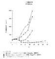
腫瘍増殖後退アッセイを、急速進行的に増殖する腫瘍LLS/KSAを用いて行い、パクリタキセル(80mg/kg)の単回投与に続いて、1週間後、KS−IL2(20μg)を5日間、尾静脈に静脈内投与した(図2)。単独で投与されたパクリタキセル、KS−IL2のいずれも、効果は認められなかった(第0日〜4日)。しかしながら、KS−IL2をパクリタキセルの1週間後に投与したとき、平均腫瘍体積の大きな低減(対照の41%)、および約8日の腫瘍増殖遅延(TGD)が観察され、これはパクリタキセル単独と比べて有意に異なった(p=0.0023)。パクリタキセル処置群における<5%の重量喪失を除いて、薬剤に関連した肉眼的毒性は認められなかった。
【0087】
次に、一般により有効な化学療法スケジュールであるとみなされている多回投与の効果を、スケジュールが増強にどのように影響を及ぼすのかを求めるために、KS−IL2と組み合わせたパクリタキセルの単回投与と比較した。KS−IL2(20μg、第0日〜4日)単独は、ここでもLLS/KSA腫瘍増殖に対する効果が認められなかったが、多回投与(50mg/kg、隔日)で与えたとき、対照の63%に平均腫瘍体積を低減し、4日の腫瘍増殖遅延(TGD)をもたらした(図3)。KS−IL2免疫サイトカインをパクリタキセル処置1週間後に投与したとき、腫瘍体積の対照の27%への低減、および10日のTGDが観察され、これはパクリタキセル単独と比べて有意に異なった(p=0.016)。パクリタキセル処置群における<5%の重量喪失を除いて、薬剤に関連した肉眼的毒性は認められなかった。併用療法群は体重喪失も少なかった。これらの肯定的な併用療法の結果は、化学療法(および潜在的に免疫に損傷を与える)処置と、リンパ球増殖および細胞傷害性を刺激する能力に基づく処置の開始との間の比較的短い間隔を考慮すれば驚くべきことである。
【0088】
増殖細胞塊の一部のタキサン誘導アポトーシスが間質圧を低減し、次にそれが腫瘍へのKS−IL2の取り込みを増大したというのが、この併用効果の一解釈である。最近の研究(Griffon−Etienne等、 1999、 CANCER RES. 59:3776〜3782)は、パクリタキセルの単回投与の作用は間質液圧を有効に低減し、最大効果は24から48時間に認められることを示唆している(Griffon−Etienne等、 1999、 CANCER RES.59:3776〜3782)。これが免疫サイトカインを腫瘍に取り込む最善の時期である可能性があるが、化学療法後非常に短い時間間隔でもある。それにもかかわらず、本出願者等は、パクリタキセルの単回投与を受けた24時間後に開始して連続5日間、LLS/KSAを有するマウスをKS−IL2で処置した。パクリタキセルの単回投与後、1週間よりも早く免疫サイトカイン処置を開始したとき、この腫瘍株ならびに結腸癌腫CT26(以下を参照)でよりよい併用応答があることを結果は示唆している。
【0089】
実施例5. 4T1転移巣のKS−IL2およびタキサンによる併用療法
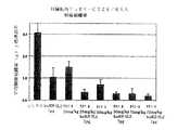
本出願者等は、タキサンと免疫サイトカインとの処置間隔が予期したものより短期間であり得ることを見出したので、タキサンと免疫サイトカインを同日に投与する併用治療プログラムを試験し、パクリタキセルの単回投与(75mg/kg)を、KS−IL2処置(15μg/投与×3日、パクリタキセルの4時間後)と同時に与えられた分割投与(25mg/kg×3日)と比較した。この実験のために、本出願者等は、4T1/KSA乳癌腫細胞によって誘導された実験的肺転移モデルを用いた。薬剤の投与量は、潜在的な相加活性または相乗活性が認められるように、それらだけで準最適となるように選択される。
【0090】
単独で投与された各剤は、類似した程度に有意に(p<0.02)平均肺重量を低減し、パクリタキセルの単回投与では43%減少、パクリタキセル単独の多回投与では49%減少、KS−IL2単独では39%減少であった(図4)。パクリタキセルとKS−IL2との組み合わせは、さらに肺転移巣をわずかに低減したが、相加的ではなく、KS−IL2と組み合わせたパクリタキセルの単回投与では58%減少、KS−IL2と組み合わせたパクリタキセルの多回投与では68%減少であった。相乗作用は観察されなかったが、KS−IL2と組み合わせたパクリタキセルの単回投与は、単独で投与されたパクリタキセルに比べて有意な差異をもたらした(p=0.047)。
【0091】
すべての群において10%未満の重量喪失が認められたが、隔日で3回投与された25mg/kgのパクリタキセルでより大きな重量喪失が認められた。これらのデータに基づいて、併用療法の最大効果に関してこの4T1肺転移アッセイにおける最良の治療プログラムは、パクリタキセルの単回投与、それに続くKS−IL2の投与であり、LLS/KSA腫瘍増殖後退モデルでも同様であった。この場合、投与間隔はわずか4時間であったので、この結果は有効な腫瘍取り込みに関して最適でなかった可能性がある。
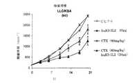
【0092】
実施例6. CT26皮膚腫瘍のKS−IL2およびタキサンによる併用療法 実施例5で述べた結果は、2つの薬剤を投与する間の4時間という時間間隔が短すぎるかもしれないことを示唆した。ことによると、KS−IL2の投与時に、依然として動物に残存しているパクリタキセルのレベルが、直接リンパ球活性化を妨げ、それによって併用条件での潜在的な抗腫瘍活性を低減した可能性がある。さらに、4時間の時点では、腫瘍間質圧に対する最大効果に到達していなかったであろう。従って本出願者等は、今度はCT26/KSA結腸癌腫の定着皮膚腫瘍を用いる別の実験を立案し、その実験において本出願者等は、パクリタキセル(75mg/kg)の単回投与を、タキサンの投与から24時間後に開始するKS−IL2の5日の投与と組み合わせた。パクリタキセル単独では、腫瘍増殖に効果を示さなかった(図5)。KS−IL2の準最適投与量(10μg、第1日から5日)による治療によって、腫瘍体積は対照の71%となった。腫瘍体積が対照の8%という劇的かつ相乗的な減少が、パクリタキセルとKS−IL2との組み合わせで認められ、これはパクリタキセル単独治療とは有意に異なっていた(p<0.001)。〜5%の最小重量喪失が、両方のパクリタキセル処置群で認められた。
【0093】
第2の実験はCT26/KSAモデルを用いて行い、今度は定着肝転移巣に対する併用療法の効果を試験し、ここでもパクリタキセル投与とKS−IL2処置との間は24時間の遅延時間を用いた。本出願者等はさらに、この併用療法におけるパクリタキセルの用量反応を比較した。転移の誘導後第5日に、マウスにパクリタキセル25、50、または75mg/kgを注射し、これを単独で行うか、あるいは1日後に5日間KS−IL2(7μg)を注射した。パクリタキセル単独投与に関して用量反応効果を観察し、25、50、75mg/kgはそれぞれ、腫瘍組織量が対照の49%、23%、10%となった(図6)。パクリタキセルとKS−IL2の組み合わせは、それぞれ同じパクリタキセル投与量で、対照の12%、9%、および6%に肺転移巣をさらに低減した。KS−IL2と組み合わせた最低投与量のパクリタキセル(25mg/kg)は、KS−IL2と組み合わせた高投与量のパクリタキセルに比べて、腫瘍組織量に最大かつ最も有意な(p<0.001)低減をもたらした。従って、パクリタキセルを先行させるKS−IL2の組み合わせが、いずれか単独の薬剤に比べて、より大きな抗腫瘍効果をもたらした。さらに、KS−IL2と組み合わせた最低投与量のパクリタキセルが、パクリタキセル単独の最高投与量と同様の抗腫瘍効果をもたらした。従って、KS−IL2と組み合わせた、より低用量のパクリタキセルの使用は、良好な効能を維持しながら毒性を低減するであろう。
【0094】
実施例7. KS−IL2の腫瘍への取り込みの測定
免疫サイトカイン療法に先立つ細胞傷害性薬剤治療の単回投与の作用が、腫瘍間質圧を減少し、腫瘍の浸透を増大するのであれば、これは放射性標識免疫サイトカイン、例えばKS−IL2を用いて測定可能なはずである。精製したKS−IL2を、商業的供給者(New England Nuclear、Billerica、MA)との約定によって標準的な手順(参照)で、125Iで標識した。CT26/KSAの皮膚腫瘍を、実施例1に記載のとおり皮下に移植し、100〜200mm3に達するまで増殖させた。それぞれ4匹の2群のマウスに、ビヒクル中のパクリタキセル(50mg/kg)、またはビヒクルを単独で注射し、1時間(実験1)または24時間(実験2)後に、10μgの125I−KS−IL2(95μCi)を注射した。放射性標識免疫サイトカインを注射して6時間後、マウスを屠殺し、腫瘍を外科的に摘出した。対照として、それらの動物の肝臓も採取し、すべての組織を秤量、次いでγ線計数器で計数した。結果は、組織中の総CPMを重量で割ることによって、組織1g当たりの計数毎分(CPM)で表した。
【0095】
標識KS−IL2をパクリタキセル処置の1時間後に注射したとき(図7A)、薬剤投与動物から得た切除組織において、わずか少量の放射能の増加が見られた。対照的に、標識KS−IL2をパクリタキセル処置の24時間後に注射したとき、ビヒクル対照に比べて、劇的な取り込みの増加が見られた(>200パーセント)(図7B)。1時間と24時間との間の腫瘍取り込みにおける大きな差異は、間質圧のタキサン誘発性変化に関するデータと一致しており(Griffon−Etienne等、 1999、 CANCER RES. 59:3776〜3782)、パクリタキセル後24時間に開始した処置が、早期の処置(4時間)に比べてより有効であることを示す本発明の腫瘍モデルのデータとも矛盾しない。
【0096】
本出願者等はさらに、他の類の薬剤が固形腫瘍への標識免疫サイトカインの取り込みを増大できるのかどうかを試験した。この場合、実験の24時間、または3日前に、シクロホスファミド(40mg/kg)を単回投与でマウスに注射した。125I標識KS−IL2を、PBSで前処置した対照マウスを含むすべてのマウスに注射し、16時間後に切除腫瘍において放射能の量を求めた。結果(図8)は、24時間前に処置したマウスでは48%、3日前に処置したマウスでは70%、シクロホスファミドによる前処置がKS−IL2の取り込みを増大したことを示す。
【0097】
実施例8. huKS−huγ4−IL2およびタキサンによる併用療法
Fcレセプターに対する低減された親和性のために、循環半減期が増大し、有効性が向上した新しい形態の免疫サイトカインが最近記載された(Gillies等、 1999、 CANCER RES. 59:2159〜2166を参照のこと)。これらの改良型IL−2免疫サイトカインの代表例の1つ、huKS−huγ4−IL2を、パクリタキセルの単回投与との併用療法において試験した。ここでも、CT26/KSA皮膚腫瘍を有するマウスに2つの薬剤を連続的に投与したとき、有効性が向上した。
【0098】
実施例9. huKS−muγ2a−muIL12およびタキサンによる併用療法
相乗的な治療効果がIL−2主体の免疫サイトカインのみに特異的なものなのかどうかを試験するために、本出願者等は、定着したCT26/KSA大型腫瘍を、最初にパクリタキセル(単回投与、75mg/kg)で処置し、次いで24時間後に5日の投与期間のhuKS−muγ2a−muIL12(1日当たり5μg)で処置した。この免疫サイトカインは、huKS抗体のマウス型(すなわち、定常領域がマウスCカッパおよびCガンマ2aに復帰したもの)とマウスIL−12の融合体に相当する。IL−2とは異なり、このサイトカインは種特異性が高く、ヒト型はマウスにおいてあまり活性ではないために、マウスIL−12配列を用いる必要があった。結果は、パクリタキセル単独での処置が腫瘍増殖にほとんど効果を及ぼさなかったことを示す。huKS−muγ2a−muIL12の準最適投与量による処置は抗腫瘍効果を有し、この効果は最初にパクリタキセルの単回投与によって処置したマウスにおいて増大した。
【0099】
実施例10. huKS−IL2およびアルキル化剤による併用療法
i.huKS−IL2と、アルキル化剤類の化学療法剤シクロホスファミドとの組み合わせによる治療効果の向上も実証された。処置の3日前に、4T1乳癌腫細胞を免疫応答性マウスに静脈内注射して、肺転移巣を定着させた。マウスをシクロホスファミドの単回投与(15、40、または80mg/kg)で処置し、次いで3日後に5日の投与期間のhuKS−IL2(15μg/日)で処置した。2種の最低投与量単独では、肺転移巣腫瘍組織量にわずかな低減が生じただけであったが、huKS−IL2との組み合わせでは、シクロホスファミド単独と比べて、腫瘍組織量に有意な大きい低減がもたらされた(p<0.05、図9)。しかしながら、最高投与量(80mg/kg)において、相乗作用は生じない。
【0100】
ii.huKS−IL2とシクロホスファミドの組み合わせによる治療効果の向上はさらに、定着した乳癌種皮下腫瘍を有する免疫応答性マウスにおいて、腫瘍増殖アッセイで実証された。マウスを、シクロホスファミド80mg/kgの単回投与によって、単独で、またはシクロホスファミド処置3日後のhuKS−IL2(30μg)の5日間投与と組み合わせて処置した。huKS−IL2、および80mg/kgのシクロホスファミド単独の場合、平均腫瘍体積は、それぞれ31%、および69%減少した(図9B)。併用療法は、第25日に平均腫瘍体積を100%減少し、これはhuKS−IL2単独、シクロホスファミド単独のいずれとも有意に異なり(p<0.05)、初回処置後12週までに、8匹中6匹のマウスにおいて、完全に腫瘍を除去した。動物は、すべての群において認められた10%未満の重量喪失を伴い、これらの処置に充分に耐性であった。
【0101】
iii.huKS−IL2とシクロホスファミドの組み合わせによる治療効果の向上はさらに、定着した肺癌種皮下腫瘍を有する免疫応答性マウスにおいて、腫瘍増殖アッセイで実証された。マウスを、シクロホスファミド80mg/kgの単回投与によって、単独で、またはシクロホスファミド処置3日後のhuKS−IL2(20μg)の5日間投与と組み合わせて処置した。huKS−IL2、および80mg/kgのシクロホスファミド単独の場合、平均腫瘍体積は、それぞれ2%、および27%減少した(図9C)。併用療法は、第20日に平均腫瘍体積を48%減少し、これはhuKS−IL2単独、シクロホスファミド単独のいずれとも有意に異なった(p<0.05)。動物は、すべての群において認められた10%未満の重量喪失を伴い、これらの処置に充分に耐性であった。
【0102】
実施例11. huKS−IL2およびアルキル化剤による併用療法
huKS−IL2と、アルキル化剤類の別の化学療法剤カルボプラチンとの組み合わせによる治療効果の向上も実証された。定着した非小細胞肺癌皮下腫瘍(LLC/KSA)を有するマウスを、第0日にカルボプラチン(75mg/kg)で処置し、次いで3日後に、5日の投与期間のKS−IL2(1日当たり20μg)で処置した。カルボプラチンおよびKS−IL2単独の処置はそれぞれ腫瘍増殖にわずかな低減をもたらしたが、しかしながら併用療法だけは第20日に平均腫瘍体積を有意に低減した(p<0.05、図10)。さらに、この組み合わせによって処置されたマウスの腫瘍の増殖は、カルボプラチン単独処置に比べて、有意に異なった(p<0.05)。
【0103】
本発明は、その思想または本質的特徴を逸脱することなく、他の特定の形態で具体化してもよい。従って、上述の実施態様は、全ての点において、本明細書中に記載の本発明に限定を加えるものではなく、具体例による説明であると考えられる。従って、本発明の範囲は、上述の説明によってではなく、添付の請求の範囲によって示され、また、該請求の範囲に等価の意味および範囲の内においてなされる、全ての変更は、本発明に包含されることを意味する。
【0104】
本明細書中において、先に開示した、特許文献および科学的刊行物のそれぞれは、参照により本明細書中に取り込まれる。
【図面の簡単な説明】
【図1】
図1は、サイトカインの模式図である。
【図2】
図2は、LLC/KSA腫瘍体積に対するパクリタキセルおよび免疫サイトカインの効果を、時間に対してプロットした図である。
【図3】
図3は、パクリタキセルおよび免疫サイトカインの多回投与の、各時間の平均腫瘍体積に対する効果を示す図である。
【図4】
図4は、肺転移アッセイにおける、腫瘍重量に対するパクリタキセルおよび免疫サイトカインの効果を示す図である。
【図5】
図5は、CT26/KSA腫瘍体積に対するパクリタキセルおよび免疫サイトカインの効果を、時間に対してプロットした図である。
【図6】
図6は、肝臓転移アッセイにおける、腫瘍重量に対するパクリタキセルおよび免疫サイトカインの効果を示す図である。
【図7A】
図7Aは、腫瘍による免疫サイトカイン取り込みに対するパクリタキセルの効果を示す図である。
【図7B】
図7Bは、腫瘍による免疫サイトカイン取り込みに対するパクリタキセルの効果を示す図である。
【図8】
図8は、腫瘍による免疫サイトカイン取り込みに対するシクロホスファミドの効果を示す図である。
【図9】
図9は、肺転移アッセイにおける、腫瘍重量に対するシクロホスファミドおよび免疫サイトカインの効果を示す図である。
【図9B】
図9Bは、腫瘍増殖アッセイにおける、腫瘍体積に対するシクロホスファミドおよび免疫サイトカインの効果を示す図である。
【図9C】
図9Cは、腫瘍増殖アッセイにおける、腫瘍体積に対するシクロホスファミドおよび免疫サイトカインの効果を示す図である。
【図10】
図10は、腫瘍増殖アッセイにおける、腫瘍体積に対するカルボプラチンおよび免疫サイトカインの効果を示す図である。[0001]
(Related application)
This application claims the benefit of 60/215038, filed June 29, 2000, as well as its benefits, the disclosure of which is incorporated herein by reference.
[0002]
(Field of the Invention)
The present invention relates to antibody-cytokine fusion proteins useful for targeted immunotherapy. In general, the present invention relates to the use of immunocytokine uptake enhancers in combination therapy to enhance an antibody-cytokine fusion protein-mediated immune response against a preselected target, eg, a cell in a tumor. In particular, the present invention relates to the administration of antibody-cytokine fusion proteins in combination with chemotherapeutic agents such as taxanes and / or alkylating agents to treat tumor cells and other cancerous or diseased cells.
[0003]
(Background of the Invention)
Effective treatment of diseases such as cancer requires a robust immune response by one or more effector cell types, such as natural killer (NK) cells, macrophages, and T lymphocytes. In animals and patients suffering from tumors, the immune mechanism has not been able to effectively treat growing tumors, mainly because of the specific mechanisms that tumors have created to suppress the immune response. Was. In many cases, potentially tumorigenic monocyte cells, eg, macrophages, migrate into the growing tumor bed, but secretion by the tumor cells of factors such as prostaglandins, TGF-β, and IL-10. Attenuates their cytotoxic activity (see, for example, Sharma et al., {1999, {J.} IMMUNOL. # 163: 5020-5028). Similarly, lymphocytic cells migrating into tumors, such as NK and T cells, interact with receptors expressed on the surface of tumor cells that activate the apoptosis of the immune cells, as well as factors secreted by the tumor. (See, e.g., Villunger et al., {1997, {BLOOD} 90: 12-20). Exposure of these lymphocytes to immunosuppressive monocytes in the tumor bed may further reduce their ability to elicit an effective anti-tumor response.
[0004]
To overcome the immunosuppressive effects of the local tumor microenvironment, the results achieved include targeted immunostimulation, such as treatment with tumor-specific antibody-cytokine fusion proteins. Effective treatment using this method has been demonstrated in several mouse tumor metastasis models, however, as long as the size of the tumor is increasing, the treatment is not very effective. This may be due to increased concentrations of inhibitors secreted by the tumor mass, as well as, for example, increased tumor interstitial fluid pressure (Griffon-Etienne et al., {1999, {Cancer} RES. 59: 3776-3782), solids with therapeutic agents. It is due to other factors such as barriers to tumor penetration.
[0005]
Although most cancer patients are still being treated by one or more courses of chemotherapy, it is well known that cytotoxic therapy of cancer also damages the immune system. Immune cells are one of the fastest dividing cells in the human body, and any treatment that kills dividing cells also kills immune cells. Thus, any treatment including radiation, DNA damaging chemicals, DNA synthesis inhibitors, and microtubule function inhibitors causes damage to the immune system. Indeed, bone marrow transplantation is needed as an adjunct to cancer treatment because the immune system is compromised and needs replacement. Methotrexate, and other anticancer drugs, are often used as immunosuppressants. There is also evidence that anti-cancer treatment can specifically inhibit T cell function. For example, patients who have been treated for Hodgkin's disease by total body irradiation suffer from a permanent and permanent loss of immature T cells (Watanabe et al., 1997, {Blood} 90: 3662).
[0006]
Based on current knowledge, standard treatments (chemotherapy and radiation) and local immunostimulation seem unlikely to be useful combinational tools for effective treatment of cancer. Accordingly, there is a need in the art for a method of enhancing an antibody-cytokine fusion protein-mediated immune response to a pre-selected cell type, eg, a tumor cell, as well as for compositions used in such a method.
[0007]
(Summary of the Invention)
When administering an antibody-cytokine fusion protein (immunocytokine) to a mammal suffering from a tumor or tumor metastasis, the therapeutic effect of the antibody-cytokine fusion protein is enhanced or enhanced by enhancing or enhancing uptake by the tumor. It has been discovered that prior to, concurrently with, or after treatment of a mammal with an increasing immune cytokine uptake enhancer, it can also be administered to create a more potent anti-tumor response. Useful immune cytokine uptake enhancers have been found to include alkylating chemotherapeutic agents, as well as taxanes such as paclitaxel. In particular, such combinations have been found to be useful in mediating immune destruction of preselected cell types, such as tumor cells and virus infected cells.
[0008]
In one aspect, the invention provides a method of inducing a cytotoxic immune response in a mammal against a preselected cell type. The method comprises: (i) an immunocytokine comprising: an antibody binding site capable of binding to the preselected cell type; and a cytokine capable of inducing such an immune response against the preselected cell type; ii) administering to a mammal an immune cytokine uptake enhancer in an amount sufficient to enhance the immune response as compared to the immune response stimulated by the immune cytokine alone.
[0009]
In a preferred embodiment, the preselected cell type is, for example, in a solid tumor, more preferably a larger solid tumor (ie, about 100 mm3(Super) cancer cells. Alternatively, the pre-selected cell type can be a cancer cell that exists in the form of a small metastatic focus.
[0010]
In another preferred embodiment, the immunocytokine uptake enhancer can be administered simultaneously with the immunocytokine. Alternatively, the immunocytokine uptake enhancer can be administered prior to the administration of the immunocytokine. Furthermore, it is envisioned that the immune cytokine can be administered with a plurality of different immune cytokine uptake enhancers. Alternatively, it is envisioned that the immune cytokine uptake enhancer can be administered with a plurality of different immune cytokines.
[0011]
In another aspect, the invention provides a composition for inducing a cytocidal immune response in a mammal against a preselected cell type. The composition comprises (i) an antibody binding site capable of binding the preselected cell type and a cytokine capable of inducing such an immune response against the preselected cell type in the mammal. And (ii) an immune cytokine uptake enhancer in an amount sufficient to enhance the immune response as compared to the immune response stimulated by the immune cytokine alone. Become.
[0012]
In a preferred embodiment, the antibody binding site of the immune cytokine preferably comprises an immunoglobulin heavy chain, or an antigen-binding fragment thereof. The immunoglobulin heavy chain is preferably an immunoglobulin variable (V) capable of binding a preselected antigen in the direction from the amino terminus to the carboxy terminus.HA) a region domain, an immunoglobulin constant H1 (CH1) domain, an immunoglobulin constant H2 (CH2) domain, and optionally a immunoglobulin constant H3 (CH3) domain. In a more preferred embodiment, the immune cytokine is a fusion protein comprising an immunoglobulin heavy chain or an antigen-binding fragment thereof fused to the cytokine via a polypeptide bond. Thus, preferred antibody-cytokine fusion proteins include, in the direction from the amino terminus to the carboxy terminus, (i) an immunoglobulin variable region, an immunoglobulin CH1 domain, an immunoglobulin capable of binding a cell surface antigen on the preselected cell type. An antibody binding site comprising a CH2 domain, (optionally a CH3 domain); and (ii) a cytokine. Techniques for making and using such fusion proteins are described in Gillies et al., (1992), Proc. Natl. Acad. Sci. USA 89: 1428-1432; Gillies et al., (1998), J. Am. Immunol. 160: 6195-6203; and U.S. Patent No. 5,650,150.
[0013]
The immunoglobulin constant region domain (ie, CH1, CH2, and / or CH3 domain) may be a constant region domain that is normally associated with the variable region domain in naturally occurring antibodies. Alternatively, one or more of the immunoglobulin constant region domains may be derived from an antibody different from the antibody used as the source of the variable region domains. In other words, the variable and constant region domains of the immunoglobulin may be derived from different antibodies, for example, antibodies from different species. See, for example, U.S. Pat. No. 4,816,567. Further, the immunoglobulin variable region may comprise a framework region (FR) sequence from one species, eg, human, and complementarity inserted between FRs from a second, different species, eg, mouse. A decision region (CDR) sequence. Techniques for making and using such chimeric immunoglobulin variable regions are disclosed, for example, in U.S. Patent Nos. 5,225,539 and 5,585,089.
[0014]
Preferably, the antibody-based immunocytokine further comprises an immunoglobulin light chain, preferably covalently linked to the immunoglobulin heavy chain, for example by a disulfide bond. The variable regions of the linked immunoglobulin heavy and light chains together define a single and complete binding site for binding the preselected antigen. In another embodiment, the immune cytokine comprises two chimeric chains, each comprising at least a portion of an immunoglobulin heavy chain fused to the cytokine. The two chimeric chains are preferably covalently linked, for example, by one or more interchain disulfide bonds.
[0015]
Accordingly, the present invention provides for fusion proteins within which the antigen binding specificity and activity of the antibody are combined with the effective biological activity of the cytokine. The fusion proteins of the invention can be used to selectively deliver cytokines to target cells in vivo, so that the cytokines can exert a localized biological effect in the vicinity of the target cells . In a preferred embodiment, the antibody component of the fusion protein specifically binds to an antigen on or within a cancer cell, such that the fusion protein exerts localized anti-cancer activity. In another preferred embodiment, the antibody component of the fusion protein specifically binds to a virus-infected cell, such as an HIV-infected cell, such that the fusion protein exerts localized antiviral activity.
[0016]
Cytokines that can be incorporated into the immune cytokines of the present invention include, for example, tumor necrosis factor, interleukins, colony stimulating factors, and lymphokines, and others known in the art. Preferred tumor necrosis factors include, for example, tissue necrosis factor α (TNFα). Preferred interleukins include, for example, interleukin-2 (IL-2), interleukin-4 (IL-4), interleukin-5 (IL-5), interleukin-7 (IL-7), interleukin -12 (IL-12), interleukin-15 (IL-15), and interleukin-18 (IL-18). Preferred colony stimulating factors include, for example, granulocyte macrophage colony stimulating factor (GM-CSF), and macrophage colony stimulating factor (M-CSF). Preferred lymphokines include, for example, lymphotoxin (LT). Other useful cytokines include interferons, including IFN-α, IFN-β, and IFN-γ, all of which are independent of immunity and their antiviral activity, independent of their antiviral activity. Has a forming action.
[0017]
Several types of chemotherapeutic agents have been found to be effective immune cytokine uptake enhancers. In particular, useful immune cytokine uptake enhancers include taxanes and alkylating chemotherapeutics. Several taxanes are known in the art (see Bissery and Lavelle, 1997, Cancer Therapeutics: Experimental and Clinical Agents, Chapter 8, eds. B. Teicher). In a preferred embodiment, the taxane is Taxol, also known as paclitaxel. Other embodiments include docetaxel, a semi-synthetic taxane that is more potent than paclitaxel in certain tumor models and clinical applications. Further embodiments include additional taxane derivatives, such as those derived from the natural starting material 10-deacetylbaccatin III, extracted from the needles of the European yew tree. One such example is the orally available compound, IDN5109, which is a weak substrate for P-glycoprotein and is generally more active against multidrug resistant tumors. It has a higher tolerated dose and exhibits lower neurotoxic side effects in addition to being orally bioavailable (Polizzi et al., 1999, Cancer Res. 59: 1036-1040).
[0018]
Preferred dosages and modes of administration for administering the immune cytokine in combination with the immune cytokine uptake enhancer are also provided.
[0019]
(Detailed description of the invention)
Studies have shown that large solid tumors are much more resistant to antibody-mediated therapeutic intervention and to general immunotherapy compared to disseminated metastatic lesions (Sulitzeanu) 1993, Adv. Cancer Res. 60: 247-267). The low responsiveness to antibody-based therapies is believed to be in part due to the production of immunosuppressive factors by the tumor.
[0020]
Although the mechanism of the tumor eradication is not fully understood, it is expected that a cytotoxic T lymphocyte (CTL) response can lead to the destruction of cancer cells and provide immune memory. Furthermore, under certain circumstances, natural killer (NK) cells are expected to assume tumor eradication in the absence of CTL. The different immune responses may be due to the fact that certain tumors produce different types or amounts of substances that can down-regulate T cells. This is not a small metastatic lesion, but a solid tumor that has already reached a critical mass and is capable of producing and secreting immunosuppressive factors at concentrations sufficient to modulate the immune response to the tumor. In particular, this is true.
[0021]
It has now been discovered that the cytotoxic immune response induced by an immune cytokine against a preselected cell type can be significantly enhanced by administering the immune cytokine together with an immunocytokine uptake enhancer. The combination therapy is particularly effective in mediating immune destruction of diseased tissues, such as established tumors. While not wishing to be bound by theory, immunocytokine uptake enhancers increase the penetration of immunocytokines into the tumor microenvironment, thereby allowing it to overcome the immunosuppressive effects, It is expected to be more effective in activating a cellular immune response. Similarly, it is anticipated that such methods may be useful for the treatment of certain viral diseases, where similar cellular suppressive mechanisms are preventing effective cellular immunity, such as HIV infection. . Immune cytokine uptake enhancers are expected to act synergistically with immune cytokines in mediating immune destruction of diseased tissues such as established tumors or virus-infected cells. The present invention also provides methods for making and using useful immune cytokines, and for testing their pharmacokinetic activity in preclinical in vivo animal models when combined with appropriate immune cytokine uptake enhancers. The assay is also described.
[0022]
As used herein, the term "immunocytokine uptake enhancer" is understood to mean any agent that enhances the cytotoxic immune response to a preselected cell type induced by an immune cytokine. You. More specifically, preferred immune cytokine uptake enhancers are tumor uptake enhancers that increase the penetration of immune cytokines into tumors. Examples of immunocytokine uptake enhancers include, but are not limited to, chemotherapeutic agents such as taxanes, DNA damaging agents including alkylated chemotherapeutic agents, radiation therapeutic agents, and agents that regulate blood pressure. Preferred taxanes are taxol, docetaxel, 10-deacetylbaccatin III, and derivatives thereof. Preferred alkylating agents are cyclophosphamide, carboplatin, cisplatin, and derivatives thereof. A preferred form of radiation is gamma irradiation. Preferred blood pressure regulators are angiotensin II agonists, such as angiotensin II itself, and preferably Netti et al. (Cancer Research [1995] 55: 5451-8) and Netti et al. (Proc. Nat. Acad. Sci. [1999]). 96: 3137-3142), according to the general guidelines. An immune response can be determined by methods known to those of skill in the art and / or described herein.
[0023]
As used herein, the term "cytotoxic immune response" refers to a fluid that is stimulated by immune cytokines and that, in a mammal, kills a preselected cell type, and instead reduces its viability. It is understood to mean any immune response in a mammal that has a sexual or cellular nature. The immune response may include one or more cell types, including T cells, NK cells, and macrophages.
[0024]
As used herein, the term "immunocytokine" refers to (i) an antibody binding site capable of binding and binding to a preselected antigen, eg, a cell type specific antigen; Specifically, it is understood to mean a fusion with a cytokine capable of inducing or stimulating a cytotoxic immune response against cancer or virus infected cells. Examples of preselected antigens include cell surface antigens, such as on cancer cells or virus-infected cells, as well as insoluble intracellular antigens of necrotic cells, for example, that can remain attached to the cell membrane. . Preferred antigens are target antigens specific to tumor cells, such as tumor-specific antigens. Thus, the immune cytokine is capable of selectively delivering the cytokine to a target (typically a cell) in vivo, such that the cytokine mediates a localized immune response against the target cell. be able to. For example, when the antibody component of the immune cytokine is a solid tumor, in particular, about 100 mm3The immune cytokine exerts localized anti-cancer activity if it selectively binds antigens on cancer cells, such as those in larger solid tumors. Alternatively, if the antibody component of the immune cytokine selectively binds an antigen on a virus-infected cell, such as an HIV-infected cell, the immune cytokine exerts localized antiviral activity.
[0025]
As used herein, the term "antibody binding site" is understood to mean at least a portion of an immunoglobulin heavy chain, for example, an immunoglobulin variable region capable of binding a preselected antigen such as a cell type. Is done. The antibody binding site further preferably comprises, for example, a CH1 domain, a CH2 domain, and optionally a CH3 domain, or at least a CH2 domain, or an immunoglobulin constant region comprising one or more portions thereof. Including at least a portion. Furthermore, the immunoglobulin heavy chain may be covalently or non-covalently linked to, for example, an immunoglobulin light chain comprising an immunoglobulin light chain variable region and optionally a light chain constant region. Accordingly, it is expected that the antibody binding site may include a complete antibody or a fragment thereof, or a single chain antibody, capable of binding a preselected antigen.
[0026]
With respect to the immune cytokine, it is expected that the antibody fragment can be linked to the cytokine by various methods well known to those skilled in the art. For example, the antibody binding site is preferably linked to the cytokine via a polypeptide bond or linker in the fusion protein construct. Alternatively, the antibody binding site may be chemically linked to the cytokine via a reactive group in the amino acid side chain, such as a sulfhydryl group, present in the antibody binding site and the cytokine.
[0027]
As used herein, the term “cytokine” refers to any protein or peptide capable of stimulating or inducing a cytotoxic immune response in a mammal against a preselected cell type, eg, a cancer cell or a virus-infected cell. , Their analog or functional fragments. Thus, it is expected that a variety of cytokines can be incorporated into the immune cytokines of the present invention. Useful cytokines include, for example, tumor necrosis factor (TNF), interleukins (IL), lymphokines (L), colony stimulating factors (CSF), interferons including species variants (IFN), such cytotoxic immune responses. Included are those truncated analogs that can be stimulated or induced. Useful tumor necrosis factors include, for example, TNFα. Useful lymphokines include, for example, LT. Useful colony stimulating factors include, for example, GM-CSF and M-CSF. Useful interleukins include, for example, IL-2, IL-4, IL-5, IL-7, IL-12, IL-15, and IL-18. Useful interferons include, for example, IFN-α, IFN-β, and IFN-γ.
[0028]
Genes encoding particular cytokines of interest can be cloned de novo, obtained from commercial sources, or synthesized from known sequences by standard DNA synthesis. For example, the DNA sequence of LT is known (see, eg, Nedwin et al. (1985) NUCLEIC ACIDS RES. 13: 6361) and IL-2 (see, eg, Taniguchi et al. (1983) NATURE 302: 305-318). , GM-CSF (see, for example, Gasson et al. (1984) SCIENCE 266: 1339-1342), and TNFα (see, for example, Nedwin et al. (1985) NUCLEIC ACIDS RES. 13: 6361).
[0029]
In a preferred embodiment, the immune cytokine is a recombinant fusion protein made by conventional recombinant DNA techniques, ie, by forming a nucleic acid construct encoding a chimeric immune cytokine. Construction of recombinant antibody-cytokine fusion proteins has been described in the prior art. See, for example, Gillies et al. (1992) Proc. Natl. Acad. Sci. USA 89: 1428-1432, Gillies et al. (1998) J. Am. Immunol. 160: 6195-6203 and U.S. Patent No. 5,650,150. Preferably, in the gene construct encoding the immunocytokine of the present invention, a DNA segment encoding an immunoglobulin heavy chain variable region domain, a DNA segment encoding an immunoglobulin heavy chain constant region in a 5 ′ to 3 ′ orientation And DNA encoding the cytokine. The fusion gene is assembled or inserted into an expression vector for transfection into a suitable recipient cell in which the fusion gene is expressed. The hybrid polypeptide chain is preferably the variable region (VH) And the variable region of the immunoglobulin light chain (VL) Are combined with immunoglobulin light chains to create a single and complete site for binding the preselected antigen. In a preferred embodiment, the immunoglobulin heavy and light chains are covalently linked, for example, by an interchain disulfide bond. Further, two immunoglobulin heavy chains, one or both of which are fused to a cytokine, may be covalently linked, for example, by one or more interchain disulfide bonds.
[0030]
Therefore, the method of the present invention is useful for enhancing the antitumor activity of immune cytokines used in therapeutic methods for treating tumors, and is described in WO99 / 29732, WO99 / 43713, WO99 / 52562, WO99 / 53958. And WO 01/10912, as well as antibody-based fusion proteins with altered amino acid sequences in the joining region. In one embodiment, the methods of the invention are useful in combination with an Fc fusion protein, such as Fc-interferon-α.
[0031]
FIG. 1 shows a schematic example of a typical immune cytokine 1. In this embodiment,
[0032]
The immune cytokines of the present invention can be considered chimeras in two aspects of their structure. First, the immune cytokine is chimeric in that it comprises an immunoglobulin heavy chain with antigen binding specificity linked to a given cytokine. Second, the immunocytokine of the present invention comprises an immunoglobulin variable region (V) and an immunoglobulin constant region (C), both of which are derived from different antibodies, such that the resulting protein is a V / C chimera. It may be a chimera in the sense of including. For example, the variable and constant regions may be from a native antibody molecule that can be isolated from different species. See, for example, U.S. Pat. No. 4,816,567. Also included are constructs wherein either or both of the immunoglobulin variable regions are derived from different species and comprise framework region (FR) sequences and complementarity determining region (CDR) sequences. Such constructs are disclosed, for example, in Jones et al. (1986) Nature 321: 522-525, Verhoyen et al. (1988) SCIENCE 239: 1534-1535, and U.S. Patent Nos. 5,225,539 and 5,585,089. It is further envisioned that the variable region sequences may be obtained by screening a library, eg, a phage display library, for variable region sequences that bind a preselected antigen with a desired affinity. You. Methods for making and screening phage display libraries are described, for example, in Huse et al. (1989) Science 246: 1275-1281, and Kang et al. (1991) Proc. Natl. Acad. Sci. USA 88: 1112-11123.
[0033]
The immunoglobulin heavy chain constant region domain of the immunocytokine may be any of the five immunoglobulin classes called IgA (Igα), IgD (Igδ), IgE (Igε), IgG (Igγ), and IgM (Igμ). You can choose from. However, immunoglobulin heavy chain constant regions derived from the IgG class are preferred. Furthermore, it is anticipated that the immunoglobulin heavy chains may be obtained from any of the IgG antibody subclasses, referred to in the art as IgG1, IgG2, IgG3, and IgG4. As is known, each immunoglobulin heavy chain constant region contains four or five domains. The domains are named in order as follows: CH1-hinge-CH2-CH3-(-CH4). CH4 is present in IgM without a hinge region. The DNA sequence of the heavy chain domain has cross homology among the immunoglobulin classes, for example, the IgG CH2 domain is homologous to the IgA and IgD CH2 domains, and also to the IgM and IgE CH3 domains. . An immunoglobulin light chain can have either a kappa (κ) or lambda (λ) constant chain. Sequences and sequence alignments of these immunoglobulin regions are well known in the art (see, for example, Kabat et al., "Sequences of Proteins of Immunological Interest" U.S. Department of Health and Welfare, Third Edition, 1983, Fourth Edition, 1987, And Huck et al. (1986) NUC.ACIDS.RES. 14: 1779-1789).
[0034]
In a preferred embodiment, the variable region is derived from an antibody specific for a preselected cell surface antigen (an antigen associated with a diseased cell, such as a cancer cell or a virus-infected cell), and the constant region is CH1 and CH2 (and optionally CH3) domains from the same or a different antibody than the source of the variable region. In practicing the present invention, the antibody portion of the immunocytokine is preferably non-immunogenic or weakly immunogenic in the intended subject. Thus, the antibody portion is preferably, as far as possible, derived from the same species as the intended subject. For example, if the immune cytokine is administered to a human, the constant region domains are preferably of human origin. See, for example, U.S. Pat. No. 4,816,567. Furthermore, when the immunoglobulin variable region is derived from a species other than the intended administration target, for example, when the variable region sequence is of mouse origin and the intended administration target is human, The variable region is preferably located between the FR sequences to generate a chimeric variable region in the intended host, with minimal immunoreactivity, but with binding specificity for a preselected antigen. A human FR sequence, with the mouse CDR sequence inserted into The design and assembly of such chimeric variable regions is disclosed in Jones et al. (1986) Nature 321: 522-525, Verhoyen et al. (1988) SCIENCE 239: 1534-1535, and U.S. Patent Nos. 5,255,539 and 5,585,089. The cloning and expression of humanized antibody-cytokine fusion proteins, KS-1 / 4 anti-EpCAM antibody-IL-12 fusion proteins, and their potential for eradication of established colon carcinoma metastases have been described by Gillies et al. (1998) J. Am. Immunol. 160: 6195-6203.
[0035]
The gene encoding the cytokine can be directly or by a linker, for example, in reading frame (Gly4-Ser)3The DNA encoding the linker is linked to the 3 'end of the gene (e.g., CH2 or CH3 exon) encoding the immunoglobulin constant region. In certain embodiments, the linker can include a base sequence that encodes a proteolytic cleavage site. The site can be designed such that upon insertion between the immunoglobulin constant region and the cytokine, proteolytic release of the cytokine at the target site results. For example, it is well known that plasmin and trypsin cleave after lysine and arginine residues at sites accessible to the protease. Many other site-specific endoproteases, and the amino acid sequences they cleave, are well known in the art. Preferred proteolytic cleavage sites and proteolytic enzymes reactive with such cleavage sites are disclosed in U.S. Patent Nos. 5,541,087 and 5,726,044.
[0036]
The nucleic acid construct can optionally include an endogenous promoter and enhancer for the variable region encoding gene to regulate expression of the chimeric immunoglobulin chain. For example, the variable region encoding gene may be a leader peptide, a VJ gene for a light chain (a functionally rearranged variable (V) region with a joining (J) segment), or a VDJ gene for a heavy chain, and these. It can be obtained as a DNA fragment comprising an endogenous promoter and enhancer for the gene. Alternatively, the gene encoding the variable region can be obtained separately from endogenous regulatory factors, and then used in an expression vector having these factors.
[0037]
Variable region genes can be obtained from cells producing the desired antibody by standard DNA cloning procedures. Screening of genomic libraries for specific functionally rearranged variable regions is achieved through the use of appropriate DNA probes, such as DNA segments containing J region DNA sequences and downstream sequences. be able to. Identification and confirmation of the correct clone is accomplished by sequencing the cloned gene and comparing it to the corresponding sequence of a properly spliced, full-length mRNA.
[0038]
The target antigen can be a cell surface antigen of a tumor or cancer cell, a virus infected cell, or another diseased cell. The target antigen may also be an insoluble intracellular antigen of necrotic cells (see, eg, US Pat. No. 5,019,368). Genes encoding appropriate variable regions can generally be obtained from immunoglobulin producing lymphoid cell lines. For example, hybridoma cell lines that produce immunoglobulins specific for tumor-associated or viral antigens can be generated by standard somatic cell hybridization techniques well known in the art (eg, US Pat. 4196265). These immunoglobulin producing cell lines provide a source of functionally rearranged forms of the variable region genes. Typically, the variable region gene can be of murine origin, as the mouse system is suitable for producing a variety of immunoglobulins with the desired specificity. In addition, variable region sequences may be obtained by screening a library, eg, a phage display library, for variable region sequences that bind a preselected antigen with a desired affinity. Methods for making and screening phage display libraries are described, for example, in Huse et al. (1989) Science 246: 1275-1281, and Kang et al. (1991) Proc. Natl. Acad. Sci. USA 88: 1112-11123.
[0039]
The DNA fragment encoding the functionally active variable region gene is ligated to a DNA fragment containing the gene encoding the desired constant region (or a portion thereof). Immunoglobulin constant regions (H and L chains) can be obtained from antibody-producing cells by standard gene cloning techniques. The genes for the two classes of human light chains (κ and λ) and the five classes of human heavy chains (α, δ, ε, γ and μ) have been cloned, and thus the constant regions of human origin It is readily available from clones of
[0040]
The fusion gene encoding the hybrid immunoglobulin heavy chain is assembled or inserted into an expression vector for uptake into recipient cells. Introduction of the gene construct into a plasmid vector can be accomplished by standard gene splicing procedures. The chimeric immunoglobulin heavy chain can be co-expressed in the same cell as the corresponding immunoglobulin light chain, and the complete immunoglobulin can be assembled simultaneously with expression. For this purpose, the heavy and light chain constructs can be located in the same or different vectors.
[0041]
The recipient cell line is generally a lymphoid cell. A preferred recipient cell is a myeloma (or hybridoma). Myeloma can synthesize, assemble, and secrete immunoglobulins encoded by the transfected genes, and can glycosylate proteins. Particularly preferred recipient or host cells include Sp2 / 0 myeloma, which does not normally produce endogenous immunoglobulin, and mouse myeloma NS / 0 cells. When transfected, the cells produce only immunoglobulins encoded by the transfected gene construct. The transfected myeloma can be grown in culture or in the peritoneum of mice where secreted immune cytokines can be recovered from ascites. Other lymphoid cells, such as B lymphocytes, can also be used as recipient cells.
[0042]
There are several methods for transfecting lymphoid cells with a vector containing a nucleic acid construct encoding the chimeric immunoglobulin chain. For example, vectors can be introduced into lymphoid cells by spheroblast fusion (see, eg, Gillies et al. (1989) BIOTECHNOL. 7: 798-804). Other useful methods include electroporation or calcium phosphate precipitation (see, eg, Sambrook et al. (1989) "Molecular Cloning: A Laboratory Manual", Cold Spring Harbor Press).
[0043]
Other useful methods of producing the immune cytokine include the preparation of an RNA sequence encoding the construct and its translation in a suitable in vivo or in vitro expression system. Recombinant DNA techniques are known in the art for assembling a gene encoding an antibody-cytokine fusion protein, introducing the gene into a host cell, expressing the gene in a host, and harvesting the resulting fusion protein. And is expected to be fully documented.
A specific protocol is described in, for example, Sambrook et al. (1989) [Molecular Cloning: A Laboratory Manual] Cold Spring Harbor Press.
[0044]
It is understood that chemically linked immune cytokines may be produced using a variety of methods well known to those skilled in the art. For example, an antibody or antibody fragment can be chemically linked to a cytokine using a chemically reactive amino acid side group between the antibody or antibody fragment and the cytokine. The amino acid side group may be, for example, via a disulfide bond or, for example, N-succinimidyl 3 (-2-pyridyldithio) propionate, m-maleimide, all commercially available from Pierce, Rockford, IL. Homo-, including benzoyl-N-hydroxysuccinate ester, m-maleimidobenzoyl-N-hydroxysulfosuccinimide ester, and 1,4-di- [3 '(2'-pyridylthio) propionamido] butane. Alternatively, they can be covalently linked by a heterobifunctional crosslinking reagent.
[0045]
According to the method of the present invention, the combination of an immunocytokine and an immunocytokine uptake enhancer is useful for enhancing the stimulation of the immune system, whereby at the site of a targeted cell type, such as a tumor or other diseased cell, Produces a cytotoxic response. Since the immunocytokine itself is non-cytotoxic, it would be expected that the combination of the immunocytokine and the immunocytokine uptake enhancer would not have a combined or synergistic anti-tumor effect in vitro.
[0046]
While not wishing to be bound by any particular theory, the effects of the combination therapy in vivo include (1) increased chemotherapy cytotoxicity (where the immunocytokine is associated with a tumor cell of a chemotherapeutic immunocytokine uptake enhancer). And / or (2) increased immune stimulation (when the immune cytokine uptake enhancer increases the uptake of immune cytokines into tumors to a certain extent), or both. It is likely that enhanced action of one drug caused by the action of the other drug could be included. With respect to the mechanism of (1), pretreatment with high doses of the antibody-IL2 immune complex, which induces local vascular leakage, increases the uptake of radiolabeled antibody (and possibly small molecule drugs) into the tumor. Earlier studies have shown that this may be the case (see, for example, Hornick et al., 1999, CLIN. CANCER RES. 5: 51-60). If this particular mechanism is most important in combination therapy of immunocytokines and immunocytokine uptake enhancers, it may be necessary to first treat the animal suffering from the tumor with the immunocytokine. However, if a single administration of an immunocytokine uptake enhancer given prior to treatment with the immunocytokine results in a synergistic effect on antitumor activity, then such a mechanism may not be the most critical. Rather, a more probable interpretation would be that treatment with immune cytokine uptake enhancers increased immune cytokine uptake by the mechanism of (2). This hypothesis may be further supported by demonstrating that co-administration with immune cytokine uptake enhancers increases the uptake of radiolabeled immunocytokine into solid tumors.
[0047]
According to the method of the present invention, an advantage of this combination therapy is that administration of the immunocytokine enhances the cytotoxic effect of the chemotherapeutic agent, which acts as an immunocytokine uptake enhancer. Thus, lower doses of the chemotherapeutic agent may be administered to the patient. Thus, suppression in some aspects of the patient's immune system, often associated with treatment with chemotherapeutic agents, is reduced. In one embodiment of the invention, the patient is given a single dose of a chemotherapeutic immunocytokine uptake enhancer prior to administration of the immunocytokine. The chemotherapeutic immunocytokine uptake enhancer is preferably administered from about 4 days to about 4 hours, most preferably about 24-48 hours, prior to the immunocytokine. In another embodiment of the invention, the patient is administered several doses of a chemotherapeutic immunocytokine uptake enhancer prior to administering the immunocytokine. In a further embodiment of the invention, a chemotherapeutic immunocytokine uptake enhancer may be administered before, simultaneously with, and / or after the immunocytokine.
[0048]
Paclitaxel is an example of a chemotherapeutic immune cytokine uptake enhancer that can suppress or adversely affect aspects of a patient's immune system. Most of the immunopotentiating effects of paclitaxel are mediated by macrophages / monocytes, but many studies on lymphocyte function suggest paclitaxel's damaging effects on this subset. For example, paclitaxel treatment has a severe adverse effect on the proliferative potential of lymphocytes in both normal as well as tumor-bearing mice (Mullins et al., 1998, IMMUNOPHARMACOLOL IMMUNOTOXICOL 20: 473-492), and in the treatment of NK cells. It was found to reduce both cytotoxicity and the occurrence of lymphokine-activated cytotoxicity in cell cultures containing IL-2 (Chuang et al., 1993, GYNECOL ONCOL 49: 291-298). Indeed, available evidence indicates lymphocyte subset cells as an essential effector population in the anti-tumor activity of immune cytokines (Lode et al., 1998, PHARMACOL THER 80: 277-292). The experimental evidence encompassed by the present invention has revealed some novel findings that would not be predictable by the prior art, especially with respect to the order of drug administration.
[0049]
The taxane may be co-administered with the immune cytokine, or may be administered separately by different routes of administration. The compositions of the present invention may be administered by any route that is compatible with the particular molecule. Thus, where appropriate, administration can be parenteral or oral, including intravenous and intraperitoneal routes of administration.
[0050]
The compositions of the present invention may be administered directly (eg, locally, such as by injection, implantation, or local administration to a tissue site) or systemically (eg, parenterally or orally) by any The animal can be provided by appropriate means. The composition is intravenous, subcutaneous, ocular, intraperitoneal, intramuscular, buccal, rectal, vaginal, orbital, intracerebral, intracranial, intravertebral, intraventricular, intrathecal, intracisternal, intracapsular, intranasal, Or when provided parenterally, such as by aerosol administration, the composition preferably comprises a portion of an aqueous or physiologically compatible liquid, suspension or solution. Thus, the carrier or vehicle is physiologically acceptable so that, in addition to delivering the desired composition to the patient, it does not otherwise adversely affect the patient's electrolyte and / or volume balance. Thus, the liquid medium of the drug can include normal saline (e.g., a 9.85% aqueous NaCl solution, 0.15 M, pH 7-7.4). For many taxanes, preparations are generally more complex due to their generally unsuitable solubility properties. For example, the standard formulation for paclitaxel is
[0051]
The preferred dosage of immune cytokine per dose is 0.1 mg / m2~ 100mg / m2, More preferably 1 mg / m2~ 20mg / m2, Most preferably 2 mg / m2~ 6mg / m2Is within the range. The preferred dosage of the immune cytokine uptake enhancer will generally depend on the type of immune cytokine uptake enhancer utilized, however, the optimal dosage can be determined using routine experimentation. . Administration of the immunocytokine and / or immunocytokine uptake enhancer can be by periodic bolus injection or by continuous intravenous administration from an external reservoir (eg, from an intravenous bag) or internal (eg, from a biodegradable implant). Or it can be by intraperitoneal administration. Further, it is envisioned that the immunocytokine of the present invention may be administered to the intended subject together with a plurality of different immunocytokine uptake enhancers. It is anticipated, however, that the optimal combination, mode of administration, and dosage of immunocytokine and immunocytokine uptake enhancer may be determined by routine experimentation, well within the level of skill in the art.
[0052]
Various methods can be used to evaluate the efficacy of the combination therapy using the antibody-cytokine fusion protein and the immunocytokine uptake enhancer on the immune response. For example, the animal models described in the Examples below, or other suitable animal models, may be any immunocytokine uptake enhancer, or combination of immunocytokine uptake enhancers, wherein the immunocytokine uptake enhancer is an immunocytokine (eg, an antibody-IL2 fusion protein). And can be used by those skilled in the art to test whether it is most effective in enhancing immune destruction of established tumors by acting synergistically. The immunocytokine uptake enhancer, or a combination of immunocytokine uptake enhancers, can be administered prior to or simultaneously with the immunocytokine therapy course, and the effect on the tumor can be conveniently monitored by volumetric measurements. In addition, if new immunocytokine uptake enhancers were identified, one of skill in the art would appreciate the ability of those skilled in the art to evaluate the ability of these novel compounds to enhance or improve the anti-cancer activity of antibody-cytokine fusion proteins. Could be used.
[0053]
Alternatively, following treatment, tumors are excised, sectioned, and stained by standard histological methods or specific immunohistological reagents to evaluate the effect of the combination therapy on the immune response. can do. For example, simple staining with hematoxolin and eosin can reveal differences in lymphocyte infiltration into solid tumors, indicative of a cellular immune response. In addition, immunostaining of sections with antibodies to specific classes of immune cells can reveal the nature of the induced response. For example, antibodies that bind to CD45 (a common leukocyte marker), CD4, and CD8 (for identification of T cell subclasses), and NK1.1 (a marker on NK cells) were mediated by the immune cytokines of the invention. It can be used to assess the type of immune response.
[0054]
Alternatively, the type of immune response mediated by the immune cytokine can be assessed by routine cell subset depletion studies described, for example, in Lode et al. (1998) Blood 91: 1706-1715. Examples of depleted antibodies include those that react with the T cell markers CD4 and CD8, and those that bind the NK markers NK1.1 and asialo GM. Briefly, these antibodies are injected into mammals at very high doses (eg, at a dose of about 0.5 mg per mouse) prior to initiating antibody-cytokine therapy, and then until the experiment is complete. Give at weekly intervals. This technique can identify the cell types required to elicit the immune response seen in the mammal.
[0055]
In another approach, the cytotoxic activity of spleen cells isolated from animals treated by the combination therapy can be compared to those obtained from other treatment groups. Spleen cell cultures are prepared by mechanical shredding of harvested sterile spleens according to standard techniques found in most immunological laboratory manuals. See, eg, Colligan et al. (Eds.) (1988) "Current Protocols in Immunology", John Wiley & Sons, Inc. Please refer to. The resulting cells are then cultured in a suitable cell culture medium containing serum, antibodies, and low concentrations of IL-2 (〜1010 U / ml) (eg, DMEM from GIBCO). For example, to compare NK activity, culture for 3 days is usually optimal, whereas to compare cytotoxic activity of T cells, culture for 5 days is usually optimal. Cytotoxic activity can be measured by targeting tumor target cells (eg, LLC cells)51It can be measured by radiolabeling with Cr for 30 minutes. After removing excess radiolabel, the labeled cells are mixed with various concentrations of cultured spleen cells for 4 hours. Released from cells at the end of incubation51Cr is measured by a gamma counter, which is then used to quantify the degree of cell lysis induced by immune cells. The activity of traditional cytotoxic T lymphocytes (or CTLs) is measured in this way.
[0056]
The invention is further illustrated by way of the following non-limiting examples.
[0057]
Embodiment 1 FIG. Animal model
A mouse cancer model was developed to study the effects of combining immune cytokines and taxanes in mediating an effective cytotoxic response to tumors. The immune cytokines used in the examples below bind the human tumor antigen EpCAM found on most epithelial-derived tumors (Perez and Walker (1989) J. Immunol. 142: 3662-3667). To test for efficacy in an immune-responsive mouse model, it was necessary to express human antigens on the surface of mouse tumor cells syngeneic with the mouse host. The well-known mouse lung cancer cell line, Lewis Lung Cancer (LLC) cells, was the first cell line selected for this purpose. This cell line is known to produce high levels of immune mechanism inhibitors and to induce IL-10 production from immune cells in the tumor microenvironment, resulting in localized immunosuppression (Sharma Et al., 1999, J. IMMUNOL. 163: 5020-5028). The human tumor antigen EpCAM (also called KSA) was expressed on the surface of LLC cells so that it could be targeted in vivo by a murine anti-EpCAM antibody, an immune cytokine derived from KS1 / 4. This was accomplished by transducing the EpCAM cDNA sequence using a recombinant retroviral vector and generating a cell line designated LLC / KSA, as described in (Gillies, US patent application Ser. No. 09 / 293,042). . These cells were incubated at 37 ° C., 7.0
[0058]
Additional cell lines representing carcinomas of different tissue origin were engineered in a similar manner. 4T1, a non-immunogenic mouse mammary carcinoma cell line was obtained from Dr. Paul Sondel (Univ. Of Wisconsin). This strain grows slowly and progressively after subcutaneous implantation, even before the surgical removal of the primary tumor, and metastasizes naturally to many organs. In addition, it is possible to induce experimental metastases in the lung by intravenous injection. CT26, a mouse colon cell line obtained by intrarectally injecting N-nitroso-N-methylurethane into BALB / C mice was obtained from Dr. I. J. Fidler (MD Anderson Cancer Center, Houston, TX). 4T1 and CT26 were transfected with Ep-CAM as described in (Gillies et al., J. IMMUNOL. 160: 6195-6203). 4T1 / KSA cells were incubated at 37 ° C., 7.0
[0059]
For tumor growth studies, either LLC / KSA or CT26 / KSA tumors were implanted subcutaneously on the back of mice. For LLC / KSA studies, 1 × 10 5 in 100 μl PBS6Tumors were implanted from several archival tumors that were injected with a single cell suspension of cells. After about two weeks, tumors were aseptically harvested and passed through a sieve fitted with a 150 μm screen. The cells were then passed two or three times through a syringe and a 23 gauge needle, washed twice, and resuspended in PBS. 1 × 10 in 100 μl of PBS6A single cell suspension of LLC / KSA cells was injected subcutaneously into the back of the mouse using a 301/2 gauge needle. For CT26 / KSA studies, exponentially growing cells in culture were cultured at 1 × 10 6 in 100 μl of PBS.6The cells were injected as a single cell suspension. About two weeks after transplantation, administration started on
[0060]
Volume = 1/2 × 4 / 3π (L / 2 × W / 2 × H)
Where L = tumor length, W = width, H = height.
During the course of the study, the animals were weighed and their general health was monitored. When the tumor becomes necrotic or the animal becomes moribund, CO2Euthanized by suffocation.
[0061]
Data is represented in graphical format. The graph shows individual or mean tumor volumes (± standard error) during and after administration. The data is further expressed as a percentage of the mean tumor volume of treated mice compared to vehicle treated mice relative to controls. Student t-test was performed on individual tumor volumes to determine significant differences.
[0062]
Mice were anesthetized with 80 mg / kg ketamine HCL (Fort Dodge Animal Health, Fort Dodge, IA) and 5 mg / kg xylazine (Bayer, Shawnee Mission, KS) for experimental liver metastasis studies. did. 1 × 10 in 100 μl of DMEM containing 25 mM HEPES (GIBCO)5A single cell suspension of CT26 / KSA cells was injected under the splenic sac on
[0063]
Data is represented in graphical format. The graph shows the average tumor tissue amount (± standard error) at the time of sacrifice. Tumor burden was determined by subtracting the weight of normal liver from the weight of experimental liver. The data is further expressed as a percentage of the mean tumor burden of treated mice compared to vehicle treated mice relative to controls. Student t-test was performed on individual tumor burdens to determine significant differences.
[0064]
For the study of experimental lung metastases, 2.5 × 10 5 in 100 μl of PBS5A single cell suspension of 4T1 / KSA cells was slowly injected on
[0065]
Several antibody-cytokine fusion proteins are discussed in the examples below.
[0066]
huKS-huγ1-huIL2 (abbreviated as KS-IL2)
The gene encoding the huKS-huγ1-huIL2 fusion protein was essentially described by Gillies et al. {(1998) {J. {Immunol. 160: 6195-6203 and U.S. Patent No. 5,650,150 were prepared and expressed. Briefly, the humanized variable region of the mouse KS1 / 4 antibody (Varki et al. (1984) Cancer Res. # 44: 681-687) is replaced by the human variable with the highest degree of homology to the CDRs of each KS1 / 4 variable region. The region was modeled using the method described in Jones et al. (1986) {Nature} 321: 522-525, including insertion into the consensus framework sequence. The CDR shape was maintained by molecular modeling using a Silicon Graphics Indigo workstation running BioSym software. The protein sequence was then back-translated and the gene was constructed by ligation of overlapping oligonucleotides.
[0067]
Gillies et al. (1992), except that the resulting variable region was replaced with a CMV promoter / enhancer to replace the metallothionein promoter and immunoglobulin heavy chain enhancer to express human kappa light chain and human Cγ1 heavy chain. ) @Proc. {Natl. {Acad. {Sci. As described in {USA} 89: 1428-1432, it was inserted into an expression vector containing human κ light chain and human Cγ1 heavy chain constant region. A fusion of the mature sequence of IL-2 to the carboxy terminus of the human heavy chain was performed using the method of Gillies et al {(1992)} Proc. Except that the 3 'untranslated region of the IL-2 gene was derived from the SV40 poly (A) region. {Natl. {Acad. {Sci. USA 89: 1428-1432.
[0068]
The IL-2 fusion protein was expressed by transfecting the resulting plasmid into NS / 0 myeloma cell line using selective media containing 0.1 μM methotrexate (MTX). Briefly, plasmid DNA was introduced into mouse myeloma NS / 0 cells by electroporation to obtain stably transfected clones. NS / 0 cells were grown in Dulbecco's modified Eagle's medium supplemented with 10% fetal calf serum. About 5 × 10⁶Cells were washed once with PBS and resuspended in 0.5 ml of PBS. Then, 10 μg of the linearized plasmid DNA was incubated with the cells for 10 minutes on ice in a Gene Pulser Cuvette (0.4 cm electrode gap, BioRad). Electroporation was performed using a Gene Pulser (BioRad, Hercules, CA) adjusted to 0.25V, 500 μF. Cells were allowed to recover for 10 minutes on ice before being resuspended in growth media and then plated into two 96-well plates. Stably transfected clones were selected by growth in the presence of 100 nM methotrexate introduced two days after transfection. The cells were fed three more times every three days, and MTX-resistant clones developed in 2-3 weeks.
[0069]
Expression clones were identified by Fc or cytokine ELISA using appropriate antibodies (see, eg, Gillies et al. (1989) Biotechnol. 7: 798-804). The resulting fusion protein was purified by binding to and eluting from Protein A Sepharose (Pharmacia) according to the manufacturer's instructions.
[0070]
huKS-huγ4-huIL2
The gene encoding the huKS-huγ4-huIL2 fusion protein was identified as a US patent application filed on February 24, 1999, claiming essentially the priority of US Patent Application No. 60/075887, filed February 25, 1998. It was constructed and expressed as described in 09 / 256,156.
[0071]
Briefly, the Igγ4 variant of the huKS-huγ1-huIL2 fusion protein described above is obtained by removing the immunoglobulin constant region Cγ1 gene fragment from the huKS-huγ1-huIL2 expression vector and replacing it with the corresponding sequence of the human Cγ4 gene. Prepared by The sequences and sequence alignments of the human heavy chain constant regions Cγ1, Cγ2, Cγ3, and Cγ4 can be found in Huck et al. {ACIDS} RES. # 14: 1779-1789.
[0072]
Exchange of the Cγ1 fragment with the Cγ4 fragment was accomplished by digesting the original Cγ1-containing plasmid DNA with HindIII and XhoI and purifying the large 7.8 kb fragment by agarose gel electrophoresis. A second plasmid DNA containing the Cγ4 gene was digested with HindIII and NsiI and the 1.75 kb fragment was purified. A third plasmid containing the human IL-2 cDNA and the SV40 polyA site fused to the carboxyl terminus of the human Cγ1 gene was digested with XhoI and NsiI and the small 470 bp fragment was purified. All three fragments were ligated in approximately equal molar amounts. Using this ligation product, competent E. E. coli were transformed and colonies were selected by growth on plates containing ampicillin. Properly assembled recombinant plasmids were identified by restriction analysis of plasmid DNA samples obtained from the isolated transformants and digested with FspI using the Cγ1 (FspI-free) gene insert and Cγ4 ( 1) gene insert.
[0073]
The final vector containing the Cγ4-IL2 heavy chain substitution was introduced into NS / 0 mouse myeloma cells by electroporation (0.25 V, 500 μF), and the transfectants were transfected in a medium containing methotrexate (0.1 μM). Selected by growth. Cell clones expressing high levels of the huKS-huγ4-huIL2 fusion protein were identified and expanded, and the fusion protein was purified from the culture supernatant using Protein A @ Sepharose chromatography. The purity and integrity of the Cγ4 fusion protein was determined by SDS-polyacrylamide gel electrophoresis. IL-2 activity was measured in a T cell proliferation assay (Gillis et al. {(1978)} J. Immunol. {120: 2027-2032) and found to be equivalent to the activity of the γ1 construct.
[0074]
huKS-muγ2a-muIL2
The gene encoding the huKS-muγ2a-muIL2 fusion protein was constructed by replacing the human antibody constant region of the huKS-huγ1-huIL2 fusion protein described above and the corresponding mouse sequence with human IL-2. Specifically, human Cγ1-IL2 DNA was replaced with mouse Cγ2a cDNA fused to DNA encoding mouse IL-2. In short, huKS VHThe region was ligated in-frame to the mouse γ2a cDNA by performing overlapping PCR using the following overlapping oligonucleotide primers.
(Sense) 5 'CC GTC TCC TCA GCC AAA ACA ACA GCC CCA TCG GTC (SEQ ID NO: 3)
(Antisense) 5'GG GGC TGT TGT TTT GGC TGA GGA GAC GGT GAC TGA CG (SEQ ID NO: 4)
(Sense) 5'C TTA AGC CAG ATC CAG TTG GTG CAG (SEQ ID NO: 5), and
(Antisense) 5'CC CGG GGT CCG GGA GAA GCT CTT AGT C (SEQ ID NO: 6)
The oligonucleotides of SEQ ID NOs: 3 and 4 are huKS VHIt was designed to hybridize to the junction (italics) between the domain and the constant region of mouse γ2a cDNA. There were two separate reactions in the initial PCR. In one reaction, the huKS DNA VHWas used as a template with the oligonucleotides of SEQ ID NOs: 4 and 5. The primer of SEQ ID NO: 5 was prepared using huKSVHAn AflII (CTTAAG) restriction site was introduced upstream of the sequence encoding the mature amino terminus of B. (bold). In another reaction, mouse γ2a cDNA was used as a template with oligonucleotides SEQ ID NOs: 3 and 6. The primer of SEQ ID NO: 6 hybridized to the cDNA encoding the region around the C-terminus of γ2a, and introduced an Xmal (CCCGGG) restriction site for subsequent ligation to muIL2 cDNA. The PCR products from the two reactions were mixed and subjected to a second round of PCR using the oligonucleotides of SEQ ID NOs: 5 and 6. The resulting PCR product is cloned, and the sequence of huKS VHAnd the AflII-XmaI fragment encoding the mouse γ2a constant region was used for ligation to DNA encoding the signal peptide at the AflII site and the muIL2 cDNA at the Xmal site.
[0075]
Mouse IL2 cDNA was cloned from mouse peripheral blood mononuclear cell mRNA using the oligonucleotides shown in SEQ ID NOs: 7 and 8 below.
(Sense) 5'GGC CCG GGT AAA GCA CCC ACT TCA AGC TCC (SEQ ID NO: 7), and
(Antisense) 5'CCCTCGAGTTATTGAGGGCTTGTTG (SEQ ID NO: 8)
The primer of SEQ ID NO: 7 adapted muIL2 (boldface sequence) to ligate to muγ2a at the Xmal restriction site (CCCGGG). The primer of SEQ ID NO: 8 introduced an XhoI restriction site (CTCGAG) immediately after the translation stop codon (bold antisense).
[0076]
Similarly, the huKS variable L (VL) The domain was ligated to the muκ cDNA sequence by overlapping PCR. The overlapping oligonucleotides used include: (Sense) 5'G GAA ATA AAA CGG GCT GAT GCT GCA CCA ACT G (SEQ ID NO: 9)
(Antisense) 5 'GC AGC ATC AGC CCGTT TTA TTT CCA GCT TGG TCC (SEQ ID NO: 10)
(Sense) 5'C TTA AGC GAG ATC GTG CTG ACC CAG (SEQ ID NO: 11), and
(Antisense) 5 'CTC GAG CTA ACA CTC ATT CCT GTT GAA GC (SEQ ID NO: 12)
These oligonucleotides are based on the huKS VLIt was designed to hybridize to the junction (Italics) between the mouse and the constant region of mouse κ cDNA. There were two separate reactions in the initial PCR. In one reaction, the huKS DNA VLWas used as a template together with the oligonucleotides shown in SEQ ID NO: 10 and SEQ ID NO: 11 in which an AflII (CTTAAG) restriction site was introduced upstream of the sequence encoding the mature amino terminus of huKSVL (bold). In the other reaction, mouse kappa cDNA was used as a template along with the oligonucleotides shown in SEQ ID NO: 9 and the SEQ ID NO: 12 with the XhoI restriction site introduced after the translation stop codon (bold antisense).
[0077]
The PCR products obtained from the two reactions were mixed and subjected to a second PCR using the oligonucleotide primers shown in SEQ ID NOs: 11 and 12. The resulting PCR product was cloned and, in sequence confirmation, the huKS VLAnd the AflII-XmaI fragment encoding the mouse kappa constant region was ligated to the DNA encoding the signal peptide at the AflII site.
[0078]
Both mouse heavy and light chain sequences were used to replace the human sequence for pdHL7. Clones were selected by electroporating mouse NS / 0 myeloma cells containing the dhfr selectable marker gene into mouse NS / 0 myeloma cells (6.25 V, 500 μF) and culturing in a medium containing 0.1 μM methotrexate. . Transfected clones resistant to methotrexate were tested for secretion of antibody determinants by standard ELISA methods. The fusion protein was purified by Protein A Sepharose chromatography according to the manufacturer's instructions.
[0079]
huKS-muγ2a-muIL12
The gene encoding the huKS-muγ2a-muIL12 fusion protein has been described in U.S. patent application Ser. No. 08 / 986,979, filed essentially on Dec. 8, 1997, and in Gillies et al. {(1998) {J. {Immunol. # 160: 6195-6203, were constructed and expressed. Briefly, this was achieved by fusing the mouse p35IL-12 subunit cDNA to the previously prepared huKS-muγ2a heavy chain coding region. The resulting vector was then transfected into an NS / 0 myeloma cell line that had been previously transfected with the p40IL-12 subunit and capable of expressing the p40IL-12 subunit. In other words, the cell line was transfected with p40 alone, stable high expressing cells were selected, which were then used as recipients for transfection with p35 containing the fusion protein (ie, sequential transfection). .
[0080]
The mouse p35 and p40 IL-12 subunits were isolated by PCR from mRNA prepared from spleen cells activated with Concanavalin A (3 μg / ml in medium for 3 days). PCR primers used to isolate p35 encoding the nucleic acid sequence, and further adapting the p35 cDNA as an Xmal-Xhol restriction fragment, include:
5 'CCCCGGGGTAGGGTCATTCCAGTCCTCTGG (SEQ ID NO: 13), and
5 'CTCGAGTCAGGCGGAGCTCAGATAGC (SEQ ID NO: 14)
PCR primers used to isolate p40 encoding a nucleic acid sequence include:
5'TCTAGACCATGTGTCCTCCAGAAGCTAAC (SEQ ID NO: 15), and
5'CTCGAGCTAGGATCGGACCCTGCAG (SEQ ID NO: 16)
The plasmid vector (pdHL7-huKS-muγ2a-p35) was constructed as described in (Gillies et al., J. mmImmunol. Methods 125: 191), which is a transcription unit encoding the dhfr selectable marker gene, a humanized KS antibody L chain. And a transcription unit encoding a mouse heavy chain fused to the p35 subunit of mouse IL-12. This fusion was achieved by ligating the Xmal-Xhol fragment of the adapted p35 subunit cDNA to a unique Xmal site at the exon end of the CH3 exon of the mouse y2a gene prepared above. Both the heavy and light chain transcription units contained a cytomegalovirus (CMV) promoter (instead of metallothionein in the reference) at the 5 'end and a polyadenylation site at the 3'.
[0081]
A similar vector (pNC-p40) was constructed for expression of the free p40 subunit, which contained a selectable marker gene (neomycin resistance gene), but still used the CMV promoter for transcription. The coding region in this case contained the native leader sequence of the p40 subunit for transport to the appropriate endoplasmic reticulum and assembly with the fusion protein. Plasmid pNC-p40 was electroporated into cells, and the cells were plated on G418-containing medium and selected. In this case, culture supernatants of drug resistant clones were tested by ELISA for production of the p40 subunit.
[0082]
The pdHL7-huKS-muγ2a-p35 expression vector was constructed using the method of Gillies et al. {(1998)} J. {Immunol. Electroporation into the NS / 0 cell line already expressing mouse p40 as described in $ 160: 6195-6203. Transfected clones resistant to methotrexate were tested for antibody determinants and mouse IL-12 secretion by standard ELISA methods. The resulting protein was purified by binding to and eluting from a Protein A @ Sepharose column according to the manufacturer's instructions.
[0083]
A cell line engineered for use in an animal model (Example 1) was constructed using an IL-2-based immune cytokine consisting of a humanized form of KS-1 / 4 fused to human IL-2 at the carboxyl terminus of the heavy chain ( huKS-huγ1-huIL2 (hereinafter abbreviated as KS-IL2) or in the absence of huKS-huγ1-huIL2) were tested for susceptibility to taxane-induced cytotoxicity in cell culture. Cells are seeded at 1000 cells / well in a 96-well flat bottom plate at 37 ° C., 7% CO 2.2For 24 hours. Paclitaxel, 2-fold dilution from 200 ng / ml to 3.125 ng / ml, KS-IL2, 200 ng / ml, and IL-2, 33.3 ng / ml (equivalent to IL-2 in KS-IL2) Add to the culture plate in duplicate, 37 ° C, 7% CO2For 6 days. Determination of Cell Viability Based on Cellular Transformation of Tetrazonium Salt An MTS colorimetric assay (Promega) was performed directly in a 96-well plate. After reading and recording the plate, viable adherent cells were stained with crystal violet (Sigma, St. Louis, MO). Plates stained with crystal violet were used to confirm the results of the MTS assay. The results are tabulated. IC50Is the concentration of drug that caused cytotoxicity at the 50% level of the control.
[0084]
Cytotoxicity assays were performed on CT26 / KSA, LLC / KSA, and 4T1 / KSA cells, paclitaxel (3-200 ng / ml) alone, or KS-IL2 (200 ng / ml) or IL-2 (33. 3 ng / ml, equivalent to IL-2 in KS-IL2). Cytotoxicity of KS-IL2 or IL-2 alone was minimal to negligible for the three cell lines tested (81% to 101% of controls, Table 1). Neither addition of KS-IL2 nor IL-2 affected the cytotoxicity of paclitaxel. Therefore, since neither KS-IL2 nor IL-2 affects the cytotoxicity of paclitaxel, the enhancement of antitumor activity in mice by combination therapy must be due to other mechanisms that occur only in tumor-bearing animals. Must.
[0085]
[Table 1]
Tumor growth regression assays were performed with rapidly growing tumor LLS / KSA, followed by a single dose of paclitaxel (80 mg / kg) and one week later, KS-IL2 (20 μg) was applied to the tail for 5 days. It was administered intravenously intravenously (FIG. 2). Neither paclitaxel nor KS-IL2 administered alone had any effect (Days 0-4). However, when KS-IL2 was administered one week after paclitaxel, a large reduction in mean tumor volume (41% of control) and a tumor growth delay (TGD) of about 8 days were observed, which was lower than paclitaxel alone. Significantly different (p = 0.0023). No drug-related gross toxicity was observed except for <5% weight loss in the paclitaxel treated group.
[0087]
Next, the effects of multiple doses, which are generally considered to be more effective chemotherapy schedules, can be combined with a single dose of paclitaxel in combination with KS-IL2 to determine how the schedules affect potentiation. Compared to administration. KS-IL2 (20 μg, days 0-4) alone had no effect on LLS / KSA tumor growth again, but when given in multiple doses (50 mg / kg, every other day), 63% of control. %, Resulting in a 4 day tumor growth delay (TGD) (FIG. 3). When the KS-IL2 immune cytokine was administered one week after paclitaxel treatment, a reduction in tumor volume to 27% of controls and a TGD of 10 days were observed, which were significantly different compared to paclitaxel alone (p = 0 .016). No drug-related gross toxicity was observed except for <5% weight loss in the paclitaxel treated group. The combination treatment group also lost less weight. The consequences of these positive combination therapies are relatively short between chemotherapy (and potentially immune-compromising) treatment and initiation of treatment based on the ability to stimulate lymphocyte proliferation and cytotoxicity. This is surprising considering the spacing.
[0088]
One explanation for this combination effect is that taxane-induced apoptosis of some of the proliferating cell mass reduced interstitial pressure, which in turn increased KS-IL2 uptake into tumors. A recent study (Griffon-Etienne et al., $ 1999, $ CANCER RES. $ 59: 3776-3782) shows that the effect of a single dose of paclitaxel effectively reduces interstitial fluid pressure, with a maximal effect seen between 24 and 48 hours (Griffon-Etienne et al., 1999, CANCER RES. 59: 3776-3782). This may be the best time to incorporate immune cytokines into the tumor, but it is also a very short time interval after chemotherapy. Nevertheless, we treated mice with LLS / KSA with KS-IL2 for 5 consecutive days starting 24 hours after receiving a single dose of paclitaxel. Results suggest that there is a better concomitant response in this tumor line as well as colon carcinoma CT26 (see below) when immunocytokine treatment is initiated more than one week after a single dose of paclitaxel.
[0089]
Applicants have found that the treatment interval between taxanes and immune cytokines can be shorter than expected, so we tested a combination therapy program that administers taxanes and immune cytokines on the same day and found that a single dose of paclitaxel Dosing (75 mg / kg) was compared to divided doses (25 mg / kg x 3 days) given concurrently with KS-IL2 treatment (15 μg /
[0090]
Each agent administered alone significantly (p <0.02) reduced mean lung weight to a similar extent, with a 43% reduction with a single dose of paclitaxel, a 49% reduction with multiple doses of paclitaxel alone, KS-IL2 alone had a 39% reduction (FIG. 4). The combination of paclitaxel and KS-IL2 further reduced pulmonary metastases slightly, but not additively, with a 58% reduction in a single dose of paclitaxel in combination with KS-IL2, paclitaxel in combination with KS-IL2 Was reduced by 68%. No synergy was observed, but a single administration of paclitaxel in combination with KS-IL2 resulted in a significant difference compared to paclitaxel administered alone (p = 0.047).
[0091]
Less than 10% weight loss was observed in all groups, but greater weight loss was observed with 25 mg / kg paclitaxel administered three times every other day. Based on these data, the best treatment program in this 4T1 lung metastasis assay for the maximal effect of the combination therapy is a single dose of paclitaxel followed by KS-IL2, as well as in the LLS / KSA tumor regression model Met. In this case, since the dosing interval was only 4 hours, this result may not be optimal for effective tumor uptake.
[0092]
[0093]
A second experiment was performed using the CT26 / KSA model, this time testing the effect of the combination therapy on established liver metastases, again using a 24-hour delay between paclitaxel administration and KS-IL2 treatment. . We further compared the dose response of paclitaxel in this combination therapy. On the fifth day after induction of metastasis, mice were injected with
[0094]
If the effect of a single dose of cytotoxic drug treatment prior to immunocytokine therapy reduces tumor interstitial pressure and increases tumor penetration, this may be achieved by using a radiolabeled immunocytokine such as KS-IL2. It should be measurable. Purified KS-IL2 was purified by standard procedures (see) by contract with a commercial supplier (New England Nuclear, Billerica, MA).125Labeled with I. CT26 / KSA skin tumors were implanted subcutaneously as described in Example 1 and 100-200 mm3Until they reached. Two groups of four mice each were injected with paclitaxel (50 mg / kg) or vehicle alone in vehicle and 10 μg after 1 hour (Experiment 1) or 24 hours (
[0095]
When labeled KS-IL2 was injected 1 hour after paclitaxel treatment (FIG. 7A), only a small increase in radioactivity was seen in excised tissue from drug-treated animals. In contrast, when labeled KS-IL2 was injected 24 hours after paclitaxel treatment, a dramatic increase in uptake was seen (> 200 percent) compared to the vehicle control (FIG. 7B). The large difference in tumor uptake between 1 and 24 hours is consistent with data on taxane-induced changes in interstitial pressure (Griffon-Etienne et al., 1999, CANCER RES. 59: 3776-3782), and paclitaxel Consistent with data from the tumor model of the present invention, showing that treatment started 24 hours later is more effective than earlier treatment (4 hours).
[0096]
Applicants have further tested whether other classes of agents can increase the uptake of labeled immune cytokines into solid tumors. In this case, mice were injected with a single dose of cyclophosphamide (40 mg / kg) 24 hours or 3 days before the experiment.125I-labeled KS-IL2 was injected into all mice, including control mice pre-treated with PBS, and the amount of radioactivity determined in resected tumors 16 hours later. The results (FIG. 8) show that pretreatment with cyclophosphamide increased KS-IL2 uptake by 48% in mice treated 24 hours ago and 70% in mice treated 3 days ago.
[0097]
Embodiment 8 FIG. Combination therapy with huKS-huγ4-IL2 and taxane
New forms of immune cytokines with increased circulating half-life and increased efficacy due to reduced affinity for Fc receptors have recently been described (see Gillies et al., 1999, CANCER RES. 59: 2159-2166). That). One of these representatives of the improved IL-2 immune cytokine, huKS-huγ4-IL2, was tested in combination therapy with a single dose of paclitaxel. Again, efficacy was improved when the two drugs were administered sequentially to mice with CT26 / KSA skin tumors.
[0098]
To test whether the synergistic therapeutic effect was specific to only IL-2 based immunocytokines, we applied established CT26 / KSA large tumors to paclitaxel (single dose) first. , 75 mg / kg) and then 24 hours later with a 5 day dosing period of huKS-muγ2a-muIL12 (5 μg per day). This immune cytokine corresponds to a fusion of the murine version of the huKS antibody (i.e., the constant region reverted to mouse C kappa and C gamma 2a) and mouse IL-12. Unlike IL-2, this cytokine was highly species-specific and the human form was not very active in mice, which required the use of mouse IL-12 sequences. The results show that treatment with paclitaxel alone had little effect on tumor growth. Treatment with a suboptimal dose of huKS-muγ2a-muIL12 had an antitumor effect, which was increased in mice initially treated with a single dose of paclitaxel.
[0099]
i. Improvement of the therapeutic effect by combining huKS-IL2 with the chemotherapeutic agent cyclophosphamide of alkylating agents has also been demonstrated. Three days prior to treatment, 4T1 breast carcinoma cells were injected intravenously into immunocompetent mice to establish lung metastatic foci. Mice were treated with a single dose of cyclophosphamide (15, 40, or 80 mg / kg) and then 3 days later with huKS-IL2 (15 μg / day) for a 5 day dosing period. The two lowest doses alone resulted in only a small reduction in lung metastatic tumor burden, but in combination with huKS-IL2, there was a significant reduction in tumor burden compared to cyclophosphamide alone. A very large reduction (p <0.05, FIG. 9). However, at the highest dose (80 mg / kg), no synergy occurs.
[0100]
ii. The enhanced therapeutic effect of the combination of huKS-IL2 and cyclophosphamide was further demonstrated in immunoreactive mice bearing established subcutaneous tumors of breast carcinoma in tumor growth assays. Mice were treated with a single dose of
[0101]
iii. The improved therapeutic effect of the combination of huKS-IL2 and cyclophosphamide was further demonstrated in a tumor growth assay in immunoreactive mice bearing established subcutaneous tumors of lung carcinoma. Mice were treated with a single dose of
[0102]
Embodiment 11 FIG. Combination therapy with huKS-IL2 and alkylating agent
The combination of huKS-IL2 and the alkylating agents carboplatin, another chemotherapeutic agent, has also been demonstrated to improve the therapeutic effect. Mice bearing established non-small cell lung carcinoma subcutaneous tumors (LLC / KSA) were treated with carboplatin (75 mg / kg) on
[0103]
The present invention may be embodied in other specific forms without departing from its spirit or essential characteristics. Accordingly, the embodiments described above are not to be considered in all respects as limiting the invention described herein, but rather as illustrative examples. Therefore, the scope of the present invention is defined not by the above description but by the appended claims, and all changes made within the meaning and range equivalent to the claims are included in the present invention. Included.
[0104]
In this specification, each of the patent documents and scientific publications disclosed above is hereby incorporated by reference.
[Brief description of the drawings]
FIG.
FIG. 1 is a schematic diagram of cytokines.
FIG. 2
FIG. 2 is a plot of the effects of paclitaxel and immune cytokines on LLC / KSA tumor volume versus time.
FIG. 3
FIG. 3 shows the effect of multiple administrations of paclitaxel and immune cytokine on the average tumor volume at each time.
FIG. 4
FIG. 4 shows the effect of paclitaxel and immune cytokine on tumor weight in a lung metastasis assay.
FIG. 5
FIG. 5 is a plot of the effect of paclitaxel and immune cytokines on CT26 / KSA tumor volume versus time.
FIG. 6
FIG. 6 shows the effect of paclitaxel and immune cytokine on tumor weight in a liver metastasis assay.
FIG. 7A
FIG. 7A shows the effect of paclitaxel on the uptake of immune cytokines by tumors.
FIG. 7B
FIG. 7B shows the effect of paclitaxel on the uptake of immune cytokines by tumors.
FIG. 8
FIG. 8 shows the effect of cyclophosphamide on the uptake of immune cytokines by tumors.
FIG. 9
FIG. 9 shows the effect of cyclophosphamide and immune cytokine on tumor weight in a lung metastasis assay.
FIG. 9B
FIG. 9B shows the effect of cyclophosphamide and immune cytokines on tumor volume in a tumor growth assay.
FIG. 9C
FIG. 9C shows the effect of cyclophosphamide and immune cytokines on tumor volume in a tumor growth assay.
FIG. 10
FIG. 10 shows the effect of carboplatin and immune cytokine on tumor volume in a tumor growth assay.
Claims (27)
Translated fromJapanese(i)抗体結合部位を含む免疫サイトカイン、および
(ii)該免疫サイトカインによって誘導される免疫応答を増強する、免疫サイトカインの取り込み増強剤を、
哺乳動物に投与する工程を含んでなる方法。A method for inducing a cytotoxic immune response to a tumor in a mammal, comprising:
(I) an immunocytokine comprising an antibody binding site, and (ii) an immunocytokine uptake enhancer that enhances an immune response induced by the immunocytokine.
Administering to a mammal.
(i)予め選択された細胞型上の細胞表面抗原との結合が可能な免疫グロブリン可変領域、免疫グロブリンCH1ドメイン、免疫グロブリンCH2ドメインを含んでなる抗体結合部位、および(ii)サイトカインを、
アミノ末端からカルボキシ末端の方向に、含んでなる融合タンパク質であることを特徴とする請求項1に記載の方法。The immune cytokine is
(I) an antibody binding site comprising an immunoglobulin variable region capable of binding to a cell surface antigen on a preselected cell type, an immunoglobulin CH1 domain, an immunoglobulin CH2 domain, and (ii) a cytokine.
2. The method of claim 1, wherein the fusion protein comprises from the amino terminus to the carboxy terminus.
(i)抗体結合部位とサイトカインとを含んでなる免疫サイトカイン、および
(ii)免疫サイトカイン取り込み増強剤を
含んでなることを特徴とする組成物。A composition for inducing an immune response against a tumor in a mammal, comprising:
A composition comprising (i) an immunocytokine comprising an antibody binding site and a cytokine, and (ii) an immunocytokine uptake enhancer.
(i)予め選択された細胞型上の細胞表面抗原に結合可能な免疫グロブリン可変領域、免疫グロブリンCH1ドメイン、免疫グロブリンCH2ドメインを含んでなる抗体結合部位、および(ii)サイトカインを、
アミノ末端からカルボキシ末端の方向に、含んでなる融合タンパク質であることを特徴とする請求項18に記載の組成物。The immune cytokine is
(I) an immunoglobulin variable region capable of binding to a cell surface antigen on a preselected cell type, an immunoglobulin CH1 domain, an antibody binding site comprising an immunoglobulin CH2 domain, and (ii) a cytokine.
19. The composition according to claim 18, which is a fusion protein comprising in the direction from the amino terminus to the carboxy terminus.
Applications Claiming Priority (3)
| Application Number | Priority Date | Filing Date | Title |
|---|---|---|---|
| US21503800P | 2000-06-29 | 2000-06-29 | |
| US60/215,038 | 2000-06-29 | ||
| PCT/US2001/020958WO2002002143A2 (en) | 2000-06-29 | 2001-06-29 | Enhancement of antibody-cytokine fusion protein mediated immune responses by combined treatment with immunocytokine uptake enhancing agents |
Publications (2)
| Publication Number | Publication Date |
|---|---|
| JP2004501981Atrue JP2004501981A (en) | 2004-01-22 |
| JP5390055B2 JP5390055B2 (en) | 2014-01-15 |
Family
ID=22801380
Family Applications (1)
| Application Number | Title | Priority Date | Filing Date |
|---|---|---|---|
| JP2002506764AExpired - Fee RelatedJP5390055B2 (en) | 2000-06-29 | 2001-06-29 | Enhancement of antibody-cytokine fusion protein-mediated immune response by combined therapy with immune cytokine uptake enhancer |
Country Status (22)
| Country | Link |
|---|---|
| US (2) | US7517526B2 (en) |
| EP (1) | EP1294401B1 (en) |
| JP (1) | JP5390055B2 (en) |
| KR (1) | KR20030064275A (en) |
| CN (1) | CN1270775C (en) |
| AT (1) | ATE368475T1 (en) |
| AU (2) | AU2001271729B2 (en) |
| BR (1) | BR0112111A (en) |
| CA (1) | CA2412845C (en) |
| CZ (1) | CZ2003214A3 (en) |
| DE (1) | DE60129695T2 (en) |
| DK (1) | DK1294401T3 (en) |
| ES (1) | ES2288967T3 (en) |
| HU (1) | HUP0300868A3 (en) |
| MX (1) | MXPA02012734A (en) |
| NO (1) | NO20026245L (en) |
| PL (1) | PL358582A1 (en) |
| PT (1) | PT1294401E (en) |
| RU (1) | RU2272644C2 (en) |
| SK (1) | SK982003A3 (en) |
| WO (1) | WO2002002143A2 (en) |
| ZA (1) | ZA200300759B (en) |
Cited By (1)
| Publication number | Priority date | Publication date | Assignee | Title |
|---|---|---|---|---|
| JP2012506394A (en)* | 2008-10-21 | 2012-03-15 | メルク パテント ゲゼルシャフト ミット ベシュレンクテル ハフツング | Cancer treatment with radiation and immune cytokines |
Families Citing this family (57)
| Publication number | Priority date | Publication date | Assignee | Title |
|---|---|---|---|---|
| CA2312188C (en) | 1997-12-08 | 2010-06-29 | Lexigen Pharmaceuticals Corp. | Heterodimeric fusion proteins useful for targeted immune therapy and general immune stimulation |
| US20030105294A1 (en)* | 1998-02-25 | 2003-06-05 | Stephen Gillies | Enhancing the circulating half life of antibody-based fusion proteins |
| AU758860B2 (en)* | 1998-04-15 | 2003-04-03 | Merck Patent Gmbh | Enhancement of antibody-cytokine fusion protein mediated immune responses by co-administration with angiogenesis inhibitor |
| EP1187852B1 (en)* | 1999-05-19 | 2007-08-08 | EMD Lexigen Research Center Corp. | EXPRESSION AND EXPORT OF INTERFERON-ALPHA PROTEINS AS Fc FUSION PROTEINS |
| US7067110B1 (en) | 1999-07-21 | 2006-06-27 | Emd Lexigen Research Center Corp. | Fc fusion proteins for enhancing the immunogenicity of protein and peptide antigens |
| SK782002A3 (en)* | 1999-07-21 | 2003-08-05 | Lexigen Pharm Corp | FC fusion proteins for enhancing the immunogenicity of protein and peptide antigens |
| JP4793971B2 (en)* | 1999-08-09 | 2011-10-12 | メルク パテント ゲーエムベーハー | Complex cytokine-antibody complex |
| JP2003514552A (en) | 1999-11-12 | 2003-04-22 | メルク パテント ゲゼルシャフト ミット ベシュレンクテル ハフトング | Erythropoietin morphology with improved properties |
| US20050202538A1 (en)* | 1999-11-12 | 2005-09-15 | Merck Patent Gmbh | Fc-erythropoietin fusion protein with improved pharmacokinetics |
| DE60122286T2 (en)* | 2000-02-11 | 2007-08-02 | Merck Patent Gmbh | INCREASING THE CIRCULATORY HALF-TIME OF ANTIBODY-BASED FUSION PROTEINS |
| CZ2003214A3 (en)* | 2000-06-29 | 2003-08-13 | Merck Patent Gmbh | Enhancement of antibody-cytokine fusion protein-mediated response by combined treatment with immunocytokine uptake enhancers |
| JP4234438B2 (en)* | 2001-03-07 | 2009-03-04 | メルク パテント ゲゼルシャフト ミット ベシュレンクテル ハフトング | Expression technology of protein containing hybrid isotype antibody part |
| WO2002079415A2 (en)* | 2001-03-30 | 2002-10-10 | Lexigen Pharmaceuticals Corp. | Reducing the immunogenicity of fusion proteins |
| DK1383785T3 (en) | 2001-05-03 | 2011-05-23 | Merck Patent Gmbh | Recombinant tumor-specific antibody and its use |
| US20030190705A1 (en) | 2001-10-29 | 2003-10-09 | Sunol Molecular Corporation | Method of humanizing immune system molecules |
| MXPA04005266A (en) | 2001-12-04 | 2004-10-11 | Merck Patent Gmbh | Immunocytokines with modulated selectivity. |
| RU2366664C2 (en) | 2002-12-17 | 2009-09-10 | Мерк Патент Гмбх | Humanised antibody (h14,18) based on mouse antibody 14,18, contacting gd2, and its fusion with il-2 |
| US20050069521A1 (en)* | 2003-08-28 | 2005-03-31 | Emd Lexigen Research Center Corp. | Enhancing the circulating half-life of interleukin-2 proteins |
| PT1699822E (en) | 2003-12-30 | 2008-07-30 | Merck Patent Gmbh | Il-7 fusion proteins with antibody portions, their preparation and their use |
| BRPI0417916A (en)* | 2003-12-31 | 2007-04-10 | Merck Patent Gmbh | fc-erythropoietin fusion protein with improved pharmacokinetics |
| EP1702069A2 (en)* | 2004-01-05 | 2006-09-20 | EMD Lexigen Research Center Corp. | Interleukin-12 targeted to oncofoetal fibronectin |
| JP4987484B2 (en)* | 2004-01-22 | 2012-07-25 | メルク パテント ゲゼルシャフト ミット ベシュレンクテル ハフツング | Anticancer antibodies with reduced complement binding |
| US7670595B2 (en)* | 2004-06-28 | 2010-03-02 | Merck Patent Gmbh | Fc-interferon-beta fusion proteins |
| ES2342964T3 (en)* | 2004-12-09 | 2010-07-20 | Merck Patent Gmbh | VARIATIONS OF INTERLEUCINE-7 WITH REDUCED IMMUNOGENICITY. |
| US20070104689A1 (en)* | 2005-09-27 | 2007-05-10 | Merck Patent Gmbh | Compositions and methods for treating tumors presenting survivin antigens |
| ATE505552T1 (en)* | 2005-10-21 | 2011-04-15 | Hoffmann La Roche | METHOD FOR RECOMBINANT EXPRESSION OF A POLYEPTIDE |
| JP2009519024A (en)* | 2005-12-15 | 2009-05-14 | マイクロメット アクツィエン ゲゼルシャフト | Domain-grafted antibody |
| CA2635623C (en) | 2005-12-30 | 2015-02-17 | Michael Super | Anti-cd19 antibodies with reduced immunogenicity |
| CA2635618C (en)* | 2005-12-30 | 2015-10-06 | Merck Patent Gesellschaft Mit Beschraekter Haftung | Interleukin-12p40 variants with improved stability |
| TW200817438A (en)* | 2006-08-17 | 2008-04-16 | Hoffmann La Roche | A conjugate of an antibody against CCR5 and an antifusogenic peptide |
| US11535673B2 (en)* | 2007-04-05 | 2022-12-27 | President and Fellows of Harvard CoHege | Chimeric activators: quantitatively designed protein therapeutics and uses thereof |
| MX2009013752A (en)* | 2007-06-28 | 2010-02-01 | Philogen Spa | Immunocytokines for cancer treatment in combination with chemotherapeutic agents. |
| CL2008002092A1 (en) | 2007-07-20 | 2009-05-29 | Hoffmann La Roche | Conjugate containing two or more antifusogenic peptides and an anti-cd-4 antibody; Method of production; pharmaceutical composition comprising it; antifusogenic polypeptides and use of the conjugate to treat viral infections. |
| EP2044949A1 (en)* | 2007-10-05 | 2009-04-08 | Immutep | Use of recombinant lag-3 or the derivatives thereof for eliciting monocyte immune response |
| EA201171259A1 (en)* | 2009-04-22 | 2012-05-30 | Мерк Патент Гмбх | ANTIBODY HYBRID PROTEINS WITH MODIFIED FCRN BINDING SITES |
| WO2011020783A2 (en)* | 2009-08-17 | 2011-02-24 | Roche Glycart Ag | Targeted immunoconjugates |
| US20120244145A1 (en)* | 2009-11-16 | 2012-09-27 | Duke University | Enhanced immunological responses |
| EP3626739A1 (en) | 2011-06-24 | 2020-03-25 | Stephen D. Gillies | Light chain immunoglobulin fusion proteins and methods of use thereof |
| GB201403775D0 (en) | 2014-03-04 | 2014-04-16 | Kymab Ltd | Antibodies, uses & methods |
| GB201419184D0 (en) | 2014-10-28 | 2014-12-10 | Adc Biotechnology Ltd | Method of synthesising biomolecule-effector/reporter-conjugates using affinity resins |
| US10676723B2 (en) | 2015-05-11 | 2020-06-09 | David Gordon Bermudes | Chimeric protein toxins for expression by therapeutic bacteria |
| US9567399B1 (en) | 2016-06-20 | 2017-02-14 | Kymab Limited | Antibodies and immunocytokines |
| WO2017220989A1 (en) | 2016-06-20 | 2017-12-28 | Kymab Limited | Anti-pd-l1 and il-2 cytokines |
| EP3534947A1 (en) | 2016-11-03 | 2019-09-11 | Kymab Limited | Antibodies, combinations comprising antibodies, biomarkers, uses & methods |
| US11129906B1 (en) | 2016-12-07 | 2021-09-28 | David Gordon Bermudes | Chimeric protein toxins for expression by therapeutic bacteria |
| WO2019010224A1 (en) | 2017-07-03 | 2019-01-10 | Torque Therapeutics, Inc. | Fusion molecules targeting immune regulatory cells and uses thereof |
| SG11202005732XA (en) | 2017-12-19 | 2020-07-29 | Xencor Inc | Engineered il-2 fc fusion proteins |
| MA53822A (en) | 2018-10-03 | 2021-08-11 | Xencor Inc | IL-12 HETERODIMER FC FUSION PROTEINS |
| WO2020172631A2 (en) | 2019-02-21 | 2020-08-27 | Xencor, Inc. | Untargeted and targeted il-10 fc-fusion proteins |
| WO2020236655A1 (en) | 2019-05-17 | 2020-11-26 | Xencor, Inc. | Il-7-fc-fusi0n proteins |
| US11851466B2 (en) | 2019-10-03 | 2023-12-26 | Xencor, Inc. | Targeted IL-12 heterodimeric Fc-fusion proteins |
| AU2021206449A1 (en) | 2020-01-10 | 2022-07-21 | Bright Peak Therapeutics Ag | Modified IL-2 polypeptides and uses thereof |
| US11365233B2 (en)* | 2020-04-10 | 2022-06-21 | Cytomx Therapeutics, Inc. | Activatable cytokine constructs and related compositions and methods |
| AU2022237504A1 (en) | 2021-03-16 | 2023-10-05 | Cytomx Therapeutics, Inc. | Masked activatable cytokine constructs and related compositions and methods |
| CA3222358A1 (en)* | 2021-07-09 | 2023-01-12 | Vijaya Raghavan PATTABIRAMAN | Checkpoint inhibitors conjugated to il-2, and uses thereof |
| EP4367132A1 (en) | 2021-07-09 | 2024-05-15 | Bright Peak Therapeutics AG | Modified il-2 polypeptides for treatment of inflammatory and autoimmune diseases |
| EP4366778A1 (en)* | 2021-07-09 | 2024-05-15 | Bright Peak Therapeutics AG | Antibody conjugates and manufacture thereof |
Citations (2)
| Publication number | Priority date | Publication date | Assignee | Title |
|---|---|---|---|---|
| WO1998016246A1 (en)* | 1996-10-16 | 1998-04-23 | Johns Hopkins University | Cytokine enhanced immunotherapy for brain tumors |
| WO1999052562A2 (en)* | 1998-04-15 | 1999-10-21 | Lexigen Pharmaceuticals Corp. | Enhancement of antibody-cytokine fusion protein mediated immune responses by co-administration with angiogenesis inhibitor |
Family Cites Families (203)
| Publication number | Priority date | Publication date | Assignee | Title |
|---|---|---|---|---|
| US4196265A (en) | 1977-06-15 | 1980-04-01 | The Wistar Institute | Method of producing antibodies |
| DE2952486A1 (en)* | 1978-12-26 | 1980-07-17 | Canon Kk | ELECTROPHOTOGRAPHIC METHOD AND DEVICE FOR IMPLEMENTING IT |
| US4469797A (en) | 1982-09-23 | 1984-09-04 | Miles Laboratories, Inc. | Digoxigenin immunogens, antibodies, labeled conjugates, and related derivatives |
| US4816567A (en) | 1983-04-08 | 1989-03-28 | Genentech, Inc. | Recombinant immunoglobin preparations |
| NZ210501A (en) | 1983-12-13 | 1991-08-27 | Kirin Amgen Inc | Erythropoietin produced by procaryotic or eucaryotic expression of an exogenous dna sequence |
| KR850004274A (en) | 1983-12-13 | 1985-07-11 | 원본미기재 | Method for preparing erythropoietin |
| US4703008A (en) | 1983-12-13 | 1987-10-27 | Kiren-Amgen, Inc. | DNA sequences encoding erythropoietin |
| US5082658A (en) | 1984-01-16 | 1992-01-21 | Genentech, Inc. | Gamma interferon-interleukin-2 synergism |
| EP0158198A1 (en) | 1984-03-29 | 1985-10-16 | Takeda Chemical Industries, Ltd. | DNA and use thereof |
| US5807715A (en) | 1984-08-27 | 1998-09-15 | The Board Of Trustees Of The Leland Stanford Junior University | Methods and transformed mammalian lymphocyte cells for producing functional antigen-binding protein including chimeric immunoglobulin |
| GB8422238D0 (en) | 1984-09-03 | 1984-10-10 | Neuberger M S | Chimeric proteins |
| US4690915A (en) | 1985-08-08 | 1987-09-01 | The United States Of America As Represented By The Department Of Health And Human Services | Adoptive immunotherapy as a treatment modality in humans |
| US5679543A (en) | 1985-08-29 | 1997-10-21 | Genencor International, Inc. | DNA sequences, vectors and fusion polypeptides to increase secretion of desired polypeptides from filamentous fungi |
| US5643565A (en) | 1985-09-20 | 1997-07-01 | Chiron Corporation | Human IL-2 as a vaccine adjuvant |
| US4676980A (en) | 1985-09-23 | 1987-06-30 | The United States Of America As Represented By The Secretary Of The Department Of Health And Human Services | Target specific cross-linked heteroantibodies |
| US4935233A (en) | 1985-12-02 | 1990-06-19 | G. D. Searle And Company | Covalently linked polypeptide cell modulators |
| DE3712985A1 (en) | 1987-04-16 | 1988-11-03 | Hoechst Ag | BIFUNCTIONAL PROTEINS |
| US5359035A (en) | 1985-12-21 | 1994-10-25 | Hoechst Aktiengesellschaft | Bifunctional proteins including interleukin-2 (IL-2) and granuloctyte macrophage colony stimulating factor (GM-CSF) |
| JPS63267278A (en) | 1986-03-14 | 1988-11-04 | Toray Ind Inc | Base sequence coding bonded interferon |
| EP0237019A3 (en) | 1986-03-14 | 1988-03-09 | Toray Industries, Inc. | Interferon conjugate and production thereof using recombinant gene |
| US5225539A (en) | 1986-03-27 | 1993-07-06 | Medical Research Council | Recombinant altered antibodies and methods of making altered antibodies |
| GB8607679D0 (en) | 1986-03-27 | 1986-04-30 | Winter G P | Recombinant dna product |
| DK173067B1 (en) | 1986-06-27 | 1999-12-13 | Univ Washington | Human erythropoietin gene, method of expression thereof in transfected cell lines, the transfected cell lines |
| US4954617A (en) | 1986-07-07 | 1990-09-04 | Trustees Of Dartmouth College | Monoclonal antibodies to FC receptors for immunoglobulin G on human mononuclear phagocytes |
| US4894227A (en) | 1986-08-01 | 1990-01-16 | Cetus Corporation | Composition of immunotoxins with interleukin-2 |
| US4946778A (en) | 1987-09-21 | 1990-08-07 | Genex Corporation | Single polypeptide chain binding molecules |
| US5508031A (en) | 1986-11-21 | 1996-04-16 | Cetus Oncology Corporation | Method for treating biological damage using a free-radial scavenger and interleukin-2 |
| US5019368A (en) | 1989-02-23 | 1991-05-28 | Cancer Biologics, Inc. | Detection of necrotic malignant tissue and associated therapy |
| EP0307434B2 (en) | 1987-03-18 | 1998-07-29 | Scotgen Biopharmaceuticals, Inc. | Altered antibodies |
| US5091513A (en) | 1987-05-21 | 1992-02-25 | Creative Biomolecules, Inc. | Biosynthetic antibody binding sites |
| WO1988009344A1 (en) | 1987-05-21 | 1988-12-01 | Creative Biomolecules, Inc. | Targeted multifunctional proteins |
| US5258498A (en) | 1987-05-21 | 1993-11-02 | Creative Biomolecules, Inc. | Polypeptide linkers for production of biosynthetic proteins |
| EP0294703B1 (en) | 1987-06-10 | 1995-05-10 | Dana-Farber Cancer Institute, Inc. | Bifunctional antibody constructs and method for selectively destroying cell populations |
| US5064646A (en) | 1988-08-02 | 1991-11-12 | The University Of Maryland | Novel infectious bursal disease virus |
| ES2054753T3 (en) | 1987-09-02 | 1994-08-16 | Ciba Geigy Ag | CONJUGATES OF CYTOKINES WITH IMMUNOGLOBULINS. |
| ATE108068T1 (en) | 1987-09-23 | 1994-07-15 | Bristol Myers Squibb Co | ANTIBODY HETEROCONJUGATES FOR KILLING HIV-INFECTED CELLS. |
| NZ226414A (en) | 1987-10-02 | 1992-07-28 | Genentech Inc | Cd4 peptide adhesion variants and their preparation and use |
| ZA888978B (en) | 1987-12-04 | 1990-07-25 | Du Pont | Immobilized interleukin 2 and interleukin 2 containing a carboxylterminal extension |
| WO1989006692A1 (en) | 1988-01-12 | 1989-07-27 | Genentech, Inc. | Method of treating tumor cells by inhibiting growth factor receptor function |
| CA1341588C (en) | 1988-01-26 | 2009-01-06 | Michel Revel | Human ifn-beta2/i1-6, its purification and use |
| US5120525A (en) | 1988-03-29 | 1992-06-09 | Immunomedics, Inc. | Radiolabeled antibody cytotoxic therapy of cancer |
| DE3812605A1 (en) | 1988-04-15 | 1990-06-07 | Leskovar Peter Dipl Ing Dr Hab | IMMUNEGULATIVE SUBSTANCES AND SUBSTANCE MIXTURES FOR ACTIVE INFLUENCING OF THE DISEASE PROCESS |
| US4975369A (en) | 1988-04-21 | 1990-12-04 | Eli Lilly And Company | Recombinant and chimeric KS1/4 antibodies directed against a human adenocarcinoma antigen |
| IT1217724B (en) | 1988-05-26 | 1990-03-30 | Ist Naz Ric Sul Cancro | SPECIFIC MONOCLONAL ANTIBODY FOR A SEQUENCE OF FIBRONETIN EXPRESSED IN TRANSFORMED HYBRID CELLS ACCORDING TO SUCH ANTIBODY AND USE OF THE MONOCLONAL ANTIBODY FOR THE DIAGNOSIS OF TUMORS |
| IE62463B1 (en) | 1988-07-07 | 1995-02-08 | Res Dev Foundation | Immunoconjugates for cancer diagnosis and therapy |
| US5601819A (en) | 1988-08-11 | 1997-02-11 | The General Hospital Corporation | Bispecific antibodies for selective immune regulation and for selective immune cell binding |
| IL91933A (en) | 1988-10-11 | 1994-12-29 | Univ Southern California | Vasoactive immunoconjugates useful for increasing the vascular permeability or the blood supply to neoplastic or otherwise diseased tissues |
| US5457038A (en) | 1988-11-10 | 1995-10-10 | Genetics Institute, Inc. | Natural killer stimulatory factor |
| US5242824A (en) | 1988-12-22 | 1993-09-07 | Oncogen | Monoclonal antibody to human carcinomas |
| US5530101A (en) | 1988-12-28 | 1996-06-25 | Protein Design Labs, Inc. | Humanized immunoglobulins |
| US5116964A (en) | 1989-02-23 | 1992-05-26 | Genentech, Inc. | Hybrid immunoglobulins |
| US5225538A (en) | 1989-02-23 | 1993-07-06 | Genentech, Inc. | Lymphocyte homing receptor/immunoglobulin fusion proteins |
| US5166322A (en) | 1989-04-21 | 1992-11-24 | Genetics Institute | Cysteine added variants of interleukin-3 and chemical modifications thereof |
| US6750329B1 (en) | 1989-05-05 | 2004-06-15 | Research Development Foundation | Antibody delivery system for biological response modifiers |
| ZA902949B (en) | 1989-05-05 | 1992-02-26 | Res Dev Foundation | A novel antibody delivery system for biological response modifiers |
| US6291158B1 (en) | 1989-05-16 | 2001-09-18 | Scripps Research Institute | Method for tapping the immunological repertoire |
| US5399346A (en) | 1989-06-14 | 1995-03-21 | The United States Of America As Represented By The Department Of Health And Human Services | Gene therapy |
| ATE144793T1 (en) | 1989-06-29 | 1996-11-15 | Medarex Inc | BISPECIFIC REAGENTS FOR AIDS THERAPY |
| ATE123065T1 (en) | 1989-07-07 | 1995-06-15 | Takeda Chemical Industries Ltd | PROTEINS AND THEIR PRODUCTION. |
| US5073627A (en) | 1989-08-22 | 1991-12-17 | Immunex Corporation | Fusion proteins comprising GM-CSF and IL-3 |
| JP3051162B2 (en) | 1989-09-20 | 2000-06-12 | アボット ラボラトリーズ | Method for producing fusion protein |
| US5856298A (en) | 1989-10-13 | 1999-01-05 | Amgen Inc. | Erythropoietin isoforms |
| DK0790255T3 (en) | 1989-12-22 | 2007-11-26 | Hoffmann La Roche | Monoclonal antibodies directed against the cytotoxic lymphocyte maturation factor |
| US5314995A (en) | 1990-01-22 | 1994-05-24 | Oncogen | Therapeutic interleukin-2-antibody based fusion proteins |
| JPH07507440A (en) | 1990-03-02 | 1995-08-24 | レプリゲン・コーポレーション | Antibody constructs with increased binding affinity |
| ATE158615T1 (en) | 1990-03-20 | 1997-10-15 | Univ Columbia | CHIMERIC ANTIBODIES WITH RECEPTOR-BINDING LIGANDS IN PLACE OF THEIR CONSTANT REGION |
| US5349053A (en) | 1990-06-01 | 1994-09-20 | Protein Design Labs, Inc. | Chimeric ligand/immunoglobulin molecules and their uses |
| US7253264B1 (en) | 1990-06-28 | 2007-08-07 | Sanofi-Arentideutschland GmbH | Immunoglobulin fusion proteins, their production and use |
| CA2082804A1 (en) | 1990-07-27 | 1992-01-28 | Theodore Maione | Methods and compositions for treatment of angiogenic diseases |
| US5650150A (en) | 1990-11-09 | 1997-07-22 | Gillies; Stephen D. | Recombinant antibody cytokine fusion proteins |
| DK0574395T3 (en) | 1990-11-09 | 2002-10-07 | Stephen D Gillies | Cytokine immunoconjugates |
| WO1992008801A1 (en) | 1990-11-09 | 1992-05-29 | Abbott Laboratories | Bridging antibody fusion constructs |
| AU9052891A (en) | 1990-12-05 | 1992-07-08 | Novo Nordisk A/S | Proteins with changed epitopes and methods for the production thereof |
| US5709859A (en) | 1991-01-24 | 1998-01-20 | Bristol-Myers Squibb Company | Mixed specificity fusion proteins |
| GB9105245D0 (en) | 1991-03-12 | 1991-04-24 | Lynxvale Ltd | Binding molecules |
| US6072039A (en) | 1991-04-19 | 2000-06-06 | Rohm And Haas Company | Hybrid polypeptide comparing a biotinylated avidin binding polypeptide fused to a polypeptide of interest |
| DE69233482T2 (en) | 1991-05-17 | 2006-01-12 | Merck & Co., Inc. | Method for reducing the immunogenicity of antibody variable domains |
| US5199942A (en) | 1991-06-07 | 1993-04-06 | Immunex Corporation | Method for improving autologous transplantation |
| WO1993003157A1 (en) | 1991-07-29 | 1993-02-18 | Dana Farber Cancer Institute | Plasmids for the rapid preparation of modified proteins |
| AU666866B2 (en) | 1991-08-30 | 1996-02-29 | Fred Hutchinson Cancer Research Center | Hybrid cytokines |
| US20020037558A1 (en) | 1991-10-23 | 2002-03-28 | Kin-Ming Lo | E.coli produced immunoglobulin constructs |
| US5376367A (en) | 1991-11-22 | 1994-12-27 | Immunex Corporation | Fusion proteins comprising MGF and IL-3 |
| US6627615B1 (en) | 1991-12-17 | 2003-09-30 | The Regents Of The University Of California | Methods and compositions for in vivo gene therapy |
| EP0633929B1 (en) | 1992-04-01 | 2004-03-03 | The Rockefeller University | METHOD FOR $i(IN VITRO) PROLIFERATION OF DENDRITIC CELL PRECURSORS AND THEIR USE TO PRODUCE IMMUNOGENS |
| DK0615451T3 (en) | 1992-05-26 | 2006-04-24 | Immunex Corp | Hitherto unknown cytokine that binds to CD30 |
| WO1993025673A1 (en) | 1992-06-04 | 1993-12-23 | The Regents Of The University Of California | In vivo gene therapy with intron-free sequence of interest |
| US5614184A (en) | 1992-07-28 | 1997-03-25 | New England Deaconess Hospital | Recombinant human erythropoietin mutants and therapeutic methods employing them |
| CA2142007C (en) | 1992-08-11 | 2007-10-30 | Robert Glen Urban | Immunomodulatory peptides |
| DE4228839A1 (en) | 1992-08-29 | 1994-03-03 | Behringwerke Ag | Methods for the detection and determination of mediators |
| CA2126967A1 (en) | 1992-11-04 | 1994-05-11 | Anna M. Wu | Novel antibody construct |
| EP1757695A3 (en) | 1992-11-05 | 2008-02-27 | Sloan Kettering Institute For Cancer Research | Prostate-specific membrane antigen |
| US5738849A (en) | 1992-11-24 | 1998-04-14 | G. D. Searle & Co. | Interleukin-3 (IL-3) variant fusion proteins, their recombinant production, and therapeutic compositions comprising them |
| US5543297A (en) | 1992-12-22 | 1996-08-06 | Merck Frosst Canada, Inc. | Human cyclooxygenase-2 cDNA and assays for evaluating cyclooxygenase-2 activity |
| US6096331A (en)* | 1993-02-22 | 2000-08-01 | Vivorx Pharmaceuticals, Inc. | Methods and compositions useful for administration of chemotherapeutic agents |
| AU6709794A (en) | 1993-04-21 | 1994-11-08 | Brigham And Women's Hospital | Erythropoietin muteins with enhanced activity |
| US5759551A (en) | 1993-04-27 | 1998-06-02 | United Biomedical, Inc. | Immunogenic LHRH peptide constructs and synthetic universal immune stimulators for vaccines |
| AU6776194A (en) | 1993-04-28 | 1994-11-21 | Hybritech Incorporated | Method for creating optimized regulatory regions affecting protein expression and protein trafficking |
| US5888772A (en) | 1993-04-29 | 1999-03-30 | Abbott Laboratories | DNA encoding human a erythropoietin analog |
| US5554512A (en) | 1993-05-24 | 1996-09-10 | Immunex Corporation | Ligands for flt3 receptors |
| CA2125763C (en) | 1993-07-02 | 2007-08-28 | Maurice Kent Gately | P40 homodimer of interleukin-12 |
| GB9316989D0 (en) | 1993-08-16 | 1993-09-29 | Lynxvale Ltd | Binding molecules |
| IL110669A (en) | 1993-08-17 | 2008-11-26 | Kirin Amgen Inc | Erythropoietin analogs |
| WO1995021258A1 (en) | 1994-02-01 | 1995-08-10 | United States Of America, Represented By The Secretary, Department Of Health And Human Services | Fusion proteins that include antibody and nonantibody portions |
| WO1995028427A1 (en) | 1994-04-15 | 1995-10-26 | Imclone Systems Incorporated | Chimeric interleukin-3/mutein interleukin-6 lymphokine |
| US5639725A (en) | 1994-04-26 | 1997-06-17 | Children's Hospital Medical Center Corp. | Angiostatin protein |
| MX9605109A (en) | 1994-04-26 | 1997-08-30 | Childrens Medical Center | Angiostatin and method of use for inhibition of angiogenesis. |
| US5837682A (en) | 1996-03-08 | 1998-11-17 | The Children's Medical Center Corporation | Angiostatin fragments and method of use |
| WO1995031483A1 (en) | 1994-05-13 | 1995-11-23 | Eclagen Limited | Improvements in or relating to peptide delivery |
| CU22615A1 (en) | 1994-06-30 | 2000-02-10 | Centro Inmunologia Molecular | PROCEDURE FOR OBTAINING LESS IMMUNOGENIC MONOCLONAL ANTIBODIES. MONOCLONAL ANTIBODIES OBTAINED |
| JPH10503371A (en) | 1994-07-29 | 1998-03-31 | スミスクライン・ビーチャム・パブリック・リミテッド・カンパニー | New compound |
| US6309853B1 (en) | 1994-08-17 | 2001-10-30 | The Rockfeller University | Modulators of body weight, corresponding nucleic acids and proteins, and diagnostic and therapeutic uses thereof |
| US5888773A (en) | 1994-08-17 | 1999-03-30 | The United States Of America As Represented By The Department Of Health And Human Services | Method of producing single-chain Fv molecules |
| US5541087A (en) | 1994-09-14 | 1996-07-30 | Fuji Immunopharmaceuticals Corporation | Expression and export technology of proteins as immunofusins |
| ATE208633T1 (en) | 1994-09-16 | 2001-11-15 | Merck Patent Gmbh | IMMUNOCONJUGATES |
| DE69534265T2 (en) | 1994-12-12 | 2006-05-04 | Beth Israel Deaconess Medical Center, Inc., Boston | CHIMERIC CYTOKINS AND ITS USE |
| US6485726B1 (en) | 1995-01-17 | 2002-11-26 | The Brigham And Women's Hospital, Inc. | Receptor specific transepithelial transport of therapeutics |
| US6086875A (en) | 1995-01-17 | 2000-07-11 | The Brigham And Women's Hospital, Inc. | Receptor specific transepithelial transport of immunogens |
| US5691309A (en) | 1995-01-31 | 1997-11-25 | Eli Lilly And Company | Anti-obesity proteins |
| US5552524A (en) | 1995-01-31 | 1996-09-03 | Eli Lilly And Company | Anti-obesity proteins |
| US5891680A (en) | 1995-02-08 | 1999-04-06 | Whitehead Institute For Biomedical Research | Bioactive fusion proteins comprising the p35 and p40 subunits of IL-12 |
| DE69632949T2 (en) | 1995-03-10 | 2005-07-28 | Genentech, Inc., South San Francisco | THE ACTIVATION OF RECEPTORS BY GAS6 |
| US5719266A (en) | 1995-03-17 | 1998-02-17 | Eli Lilly And Company | Anti-obesity proteins |
| AU5539596A (en) | 1995-04-06 | 1996-10-23 | Amylin Pharmaceuticals, Inc. | Anti-obesity agents |
| US6281010B1 (en) | 1995-06-05 | 2001-08-28 | The Trustees Of The University Of Pennsylvania | Adenovirus gene therapy vehicle and cell line |
| AU5893796A (en) | 1995-06-07 | 1996-12-30 | Novo Nordisk A/S | Modification of polypeptides |
| WO1997000317A1 (en) | 1995-06-07 | 1997-01-03 | Osteosa Inc. | Osteoclast growth regulatory factor |
| GB9511935D0 (en) | 1995-06-13 | 1995-08-09 | Smithkline Beecham Plc | Novel compound |
| CA2225454A1 (en) | 1995-06-30 | 1997-01-23 | Thomas Wesley Stephens | Methods for treating diabetes |
| US6406689B1 (en) | 1995-10-03 | 2002-06-18 | Frank W. Falkenberg | Compositions and methods for treatment of tumors and metastatic diseases |
| DE69629826T2 (en) | 1995-10-23 | 2004-07-01 | Children's Medical Center Corp., Boston | THERAPEUTIC ANTIANGIOGENIC COMPOSITIONS AND METHODS |
| US5854205A (en) | 1995-10-23 | 1998-12-29 | The Children's Medical Center Corporation | Therapeutic antiangiogenic compositions and methods |
| US5678543A (en)* | 1995-11-16 | 1997-10-21 | Portable Hyperbarics, Inc. | Hyperbaric chamber |
| WO1997020062A1 (en) | 1995-12-01 | 1997-06-05 | University Of Massachusetts | Il-12 p40 subunit fusion polypeptides and uses thereof |
| ATE267255T1 (en) | 1995-12-27 | 2004-06-15 | Genentech Inc | WHETHER PROTEIN DERIVATIVES WITH EXTENDED HALF-LIFE |
| US6080409A (en) | 1995-12-28 | 2000-06-27 | Dendreon Corporation | Immunostimulatory method |
| US5723125A (en) | 1995-12-28 | 1998-03-03 | Tanox Biosystems, Inc. | Hybrid with interferon-alpha and an immunoglobulin Fc linked through a non-immunogenic peptide |
| US7063958B1 (en) | 1996-01-16 | 2006-06-20 | The Rockefeller University | Nucleic acids db, the receptor for leptin |
| US6750334B1 (en) | 1996-02-02 | 2004-06-15 | Repligen Corporation | CTLA4-immunoglobulin fusion proteins having modified effector functions and uses therefor |
| WO1997030089A1 (en) | 1996-02-13 | 1997-08-21 | Regents Of The University Of California | Novel antibody-cytokine fusion protein, and methods of making and using the same |
| US5756541A (en) | 1996-03-11 | 1998-05-26 | Qlt Phototherapeutics Inc | Vision through photodynamic therapy of the eye |
| US6046310A (en) | 1996-03-13 | 2000-04-04 | Protein Design Labs., Inc. | FAS ligand fusion proteins and their uses |
| WO1997034631A1 (en) | 1996-03-18 | 1997-09-25 | Board Of Regents, The University Of Texas System | Immunoglobin-like domains with increased half lives |
| WO1997043316A1 (en) | 1996-05-10 | 1997-11-20 | Beth Israel Deaconess Medical Center, Inc. | Physiologically active molecules with extended half-lives and methods of using same |
| CA2205757C (en) | 1996-05-30 | 2006-01-24 | F. Hoffmann-La Roche Ag | Pyridazinone derivatives and their use as inhibitors of prostaglandin g/h synthase i and ii(cox i and ii) |
| US5922685A (en) | 1996-06-05 | 1999-07-13 | Powderject Vaccines, Inc. | IL-12 gene therapy of tumors |
| WO1998000127A1 (en) | 1996-07-02 | 1998-01-08 | Bar-Ilan University | Retinoyloxy(substituted)alkylene butyrates useful for the treatment of cancer and other proliferative diseases |
| AU4075897A (en) | 1996-08-16 | 1998-03-06 | Roger Lallone | A leptin binding protein and its use in methods for diagnosing and treating abnormalities of the endogenous leptin pathway |
| DK0826696T3 (en) | 1996-09-03 | 2002-09-23 | Gsf Forschungszentrum Umwelt | Use of bi- and trispecific antibodies to induce a tumor immunity |
| US5994104A (en) | 1996-11-08 | 1999-11-30 | Royal Free Hospital School Of Medicine | Interleukin-12 fusion protein |
| EP1835030A1 (en) | 1996-12-20 | 2007-09-19 | Amgen, Inc. | OB fusion protein compositions and methods |
| US6737057B1 (en) | 1997-01-07 | 2004-05-18 | The University Of Tennessee Research Corporation | Compounds, compositions and methods for the endocytic presentation of immunosuppressive factors |
| US6100387A (en) | 1997-02-28 | 2000-08-08 | Genetics Institute, Inc. | Chimeric polypeptides containing chemokine domains |
| US6277375B1 (en) | 1997-03-03 | 2001-08-21 | Board Of Regents, The University Of Texas System | Immunoglobulin-like domains with increased half-lives |
| JP2001521520A (en) | 1997-04-11 | 2001-11-06 | ジー.ディー.サール アンド カンパニー | Anti-α-low v ▼ β-low 3 ▼ Integrin antibody antagonist |
| ATE223229T1 (en) | 1997-04-17 | 2002-09-15 | Amgen Inc | COMPOSITIONS OF CONJUGATES OF THE STABLE, ACTIVE, HUMAN OB PROTEIN WITH THE FC CHAIN OF IMMUNOGLOBULINS AND RELATED METHODS |
| ATE319745T1 (en) | 1997-05-21 | 2006-03-15 | Biovation Ltd | METHOD FOR PRODUCING NON-IMMUNOGENIC PROTEINS |
| GB9712892D0 (en) | 1997-06-20 | 1997-08-20 | Eclagen Ltd | Identification of mhc binding peptides |
| WO1999002709A1 (en) | 1997-07-10 | 1999-01-21 | Beth Israel Deaconess Medical Center | Recombinant erythropoietin / immunoglobulin fusion proteins |
| WO1999003887A1 (en) | 1997-07-14 | 1999-01-28 | Bolder Biotechnology, Inc. | Derivatives of growth hormone and related proteins |
| CA2312188C (en) | 1997-12-08 | 2010-06-29 | Lexigen Pharmaceuticals Corp. | Heterodimeric fusion proteins useful for targeted immune therapy and general immune stimulation |
| US20030105294A1 (en) | 1998-02-25 | 2003-06-05 | Stephen Gillies | Enhancing the circulating half life of antibody-based fusion proteins |
| CN1204147C (en) | 1998-02-25 | 2005-06-01 | 利思进药品公司 | Methods of increasing the circulating half-life of antibody-based fusion proteins |
| WO1999053958A2 (en) | 1998-04-17 | 1999-10-28 | Lexigen Pharmaceuticals Corporation | Enhancement of antibody-cytokine fusion protein mediated immune responses by co-administration with prostaglandin inhibitor |
| WO1999054484A1 (en) | 1998-04-20 | 1999-10-28 | The Regents Of The University Of California | Modified immunoglobulin molecules and methods for use thereof |
| EP1088888A4 (en) | 1998-05-14 | 2005-03-16 | Merck Patent Gmbh | Fused protein |
| DZ2788A1 (en) | 1998-05-15 | 2003-12-01 | Bayer Ag | Selective IL-2 agonists and antagonists. |
| US6620382B1 (en)* | 1998-05-22 | 2003-09-16 | Biopheresis Technologies, Llc. | Method and compositions for treatment of cancers |
| AU4414099A (en) | 1998-06-03 | 1999-12-20 | Children's Medical Center Corporation | Protein oligomer compositions comprising endostatin protein and methods of usingthe same |
| AU775422B2 (en) | 1998-06-15 | 2004-07-29 | Gtc Biotherapeutics, Inc. | Erythropoietin analog-human serum albumin fusion |
| GB9814383D0 (en) | 1998-07-02 | 1998-09-02 | Cambridge Antibody Tech | Improvements relating to antibodies |
| EP1105427A2 (en) | 1998-08-17 | 2001-06-13 | Abgenix, Inc. | Generation of modified molecules with increased serum half-lives |
| BR9913331A (en) | 1998-08-25 | 2001-05-15 | Lexigen Pharm Corp | Expression and export of angiogenesis inhibitors as immunofusins |
| US6646113B1 (en) | 1998-09-17 | 2003-11-11 | The Trustees Of The University Of Pennsylvania | Nucleic acid molecule encoding human survival of motor neuron-interacting protein 1 (SIP1) deletion mutants |
| US6335176B1 (en) | 1998-10-16 | 2002-01-01 | Pharmacopeia, Inc. | Incorporation of phosphorylation sites |
| DK1123313T3 (en) | 1998-10-23 | 2007-06-18 | Amgen Inc | Methods and compositions for the prevention and treatment of anemia |
| AU776910B2 (en) | 1998-12-08 | 2004-09-23 | Merck Patent Gesellschaft Mit Beschrankter Haftung | Modifying protein immunogenicity |
| KR20020007287A (en) | 1999-01-07 | 2002-01-26 | 추후보정 | EXPRESSION AND EXPORT OF ANTI-OBESITY PROTEINS AS Fc FUSION PROTEINS |
| EP1178785B1 (en) | 1999-05-06 | 2008-12-24 | Wake Forest University | Compositions and methods for identifying antigens which elicit an immune response |
| JP3660880B2 (en) | 1999-05-07 | 2005-06-15 | ジェネンテック・インコーポレーテッド | Novel chimpanzee erythropoietin (CHEPO) polypeptide and nucleic acid encoding the same |
| US6348192B1 (en) | 1999-05-11 | 2002-02-19 | Bayer Corporation | Interleukin-2 mutein expressed from mammalian cells |
| EP1187852B1 (en) | 1999-05-19 | 2007-08-08 | EMD Lexigen Research Center Corp. | EXPRESSION AND EXPORT OF INTERFERON-ALPHA PROTEINS AS Fc FUSION PROTEINS |
| WO2000078334A1 (en) | 1999-06-17 | 2000-12-28 | University Of Maryland Biotechnology Institute | Chimeric chemokine-antigen polypeptides and uses therefor |
| CZ299516B6 (en) | 1999-07-02 | 2008-08-20 | F. Hoffmann-La Roche Ag | Erythropoietin glycoprotein conjugate, process for its preparation and use and pharmaceutical composition containing thereof |
| JO2291B1 (en) | 1999-07-02 | 2005-09-12 | اف . هوفمان لاروش ايه جي | Erythopintin derivatives |
| SK782002A3 (en) | 1999-07-21 | 2003-08-05 | Lexigen Pharm Corp | FC fusion proteins for enhancing the immunogenicity of protein and peptide antigens |
| JP4793971B2 (en) | 1999-08-09 | 2011-10-12 | メルク パテント ゲーエムベーハー | Complex cytokine-antibody complex |
| JP2003514552A (en) | 1999-11-12 | 2003-04-22 | メルク パテント ゲゼルシャフト ミット ベシュレンクテル ハフトング | Erythropoietin morphology with improved properties |
| DE60122286T2 (en) | 2000-02-11 | 2007-08-02 | Merck Patent Gmbh | INCREASING THE CIRCULATORY HALF-TIME OF ANTIBODY-BASED FUSION PROTEINS |
| WO2001062298A2 (en) | 2000-02-24 | 2001-08-30 | Philogen S.R.L. | Compositions and methods for treatment of angiogenesis in pathological lesions |
| US6586398B1 (en) | 2000-04-07 | 2003-07-01 | Amgen, Inc. | Chemically modified novel erythropoietin stimulating protein compositions and methods |
| HUP0302299A2 (en) | 2000-05-12 | 2003-10-28 | Neose Technologies, Inc. | In vitro fucosylation recombinant glycopeptides |
| CZ2003214A3 (en) | 2000-06-29 | 2003-08-13 | Merck Patent Gmbh | Enhancement of antibody-cytokine fusion protein-mediated response by combined treatment with immunocytokine uptake enhancers |
| KR20030067755A (en) | 2001-01-18 | 2003-08-14 | 메르크 파텐트 게엠베하 | Bifunctional fusion proteins with glucocerebrosidase activity |
| EP1366455B1 (en) | 2001-02-19 | 2008-07-02 | MERCK PATENT GmbH | Method for identification of t-cell epitopes and use for preparing molecules with reduced immunogenicity |
| KR20040074587A (en) | 2001-02-19 | 2004-08-25 | 메르크 파텐트 게엠베하 | Artificial proteins with reduced immunogenicity |
| JP4234438B2 (en) | 2001-03-07 | 2009-03-04 | メルク パテント ゲゼルシャフト ミット ベシュレンクテル ハフトング | Expression technology of protein containing hybrid isotype antibody part |
| WO2002079415A2 (en) | 2001-03-30 | 2002-10-10 | Lexigen Pharmaceuticals Corp. | Reducing the immunogenicity of fusion proteins |
| DK1383785T3 (en) | 2001-05-03 | 2011-05-23 | Merck Patent Gmbh | Recombinant tumor-specific antibody and its use |
| CA2456470A1 (en) | 2001-08-13 | 2003-02-27 | University Of Southern California | Interleukin-2 mutants with reduced toxicity |
| MXPA04005266A (en) | 2001-12-04 | 2004-10-11 | Merck Patent Gmbh | Immunocytokines with modulated selectivity. |
| CA2479212A1 (en) | 2002-03-15 | 2003-09-25 | Brigham And Women's Hospital, Inc. | Central airway administration for systemic delivery of therapeutics |
| US6996009B2 (en)* | 2002-06-21 | 2006-02-07 | Micron Technology, Inc. | NOR flash memory cell with high storage density |
| RU2366664C2 (en) | 2002-12-17 | 2009-09-10 | Мерк Патент Гмбх | Humanised antibody (h14,18) based on mouse antibody 14,18, contacting gd2, and its fusion with il-2 |
- 2001
- 2001-06-29CZCZ2003214Apatent/CZ2003214A3/enunknown
- 2001-06-29DKDK01950766Tpatent/DK1294401T3/enactive
- 2001-06-29PLPL01358582Apatent/PL358582A1/enunknown
- 2001-06-29JPJP2002506764Apatent/JP5390055B2/ennot_activeExpired - Fee Related
- 2001-06-29PTPT01950766Tpatent/PT1294401E/enunknown
- 2001-06-29SKSK98-2003Apatent/SK982003A3/enunknown
- 2001-06-29WOPCT/US2001/020958patent/WO2002002143A2/enactiveIP Right Grant
- 2001-06-29AUAU2001271729Apatent/AU2001271729B2/ennot_activeCeased
- 2001-06-29AUAU7172901Apatent/AU7172901A/enactivePending
- 2001-06-29CACA2412845Apatent/CA2412845C/ennot_activeExpired - Lifetime
- 2001-06-29BRBR0112111-1Apatent/BR0112111A/ennot_activeIP Right Cessation
- 2001-06-29KRKR1020027017778Apatent/KR20030064275A/ennot_activeCeased
- 2001-06-29EPEP01950766Apatent/EP1294401B1/ennot_activeExpired - Lifetime
- 2001-06-29MXMXPA02012734Apatent/MXPA02012734A/enactiveIP Right Grant
- 2001-06-29ESES01950766Tpatent/ES2288967T3/ennot_activeExpired - Lifetime
- 2001-06-29HUHU0300868Apatent/HUP0300868A3/enunknown
- 2001-06-29ATAT01950766Tpatent/ATE368475T1/enactive
- 2001-06-29USUS09/896,909patent/US7517526B2/ennot_activeExpired - Fee Related
- 2001-06-29CNCNB018119247Apatent/CN1270775C/ennot_activeExpired - Fee Related
- 2001-06-29RURU2003100873/15Apatent/RU2272644C2/ennot_activeIP Right Cessation
- 2001-06-29DEDE60129695Tpatent/DE60129695T2/ennot_activeExpired - Lifetime
- 2002
- 2002-12-27NONO20026245Apatent/NO20026245L/ennot_activeApplication Discontinuation
- 2003
- 2003-01-28ZAZA200300759Apatent/ZA200300759B/enunknown
- 2009
- 2009-02-27USUS12/395,159patent/US20100297060A1/ennot_activeAbandoned
Patent Citations (2)
| Publication number | Priority date | Publication date | Assignee | Title |
|---|---|---|---|---|
| WO1998016246A1 (en)* | 1996-10-16 | 1998-04-23 | Johns Hopkins University | Cytokine enhanced immunotherapy for brain tumors |
| WO1999052562A2 (en)* | 1998-04-15 | 1999-10-21 | Lexigen Pharmaceuticals Corp. | Enhancement of antibody-cytokine fusion protein mediated immune responses by co-administration with angiogenesis inhibitor |
Non-Patent Citations (2)
| Title |
|---|
| JPN6011028723; Cancer Research Vol.59, No.15, 1999, p.3776-3782* |
| JPN6012040280; 近畿大学医学雑誌 第18巻,第1号, 1993, p.137-148* |
Cited By (1)
| Publication number | Priority date | Publication date | Assignee | Title |
|---|---|---|---|---|
| JP2012506394A (en)* | 2008-10-21 | 2012-03-15 | メルク パテント ゲゼルシャフト ミット ベシュレンクテル ハフツング | Cancer treatment with radiation and immune cytokines |
Also Published As
| Publication number | Publication date |
|---|---|
| RU2272644C2 (en) | 2006-03-27 |
| WO2002002143A2 (en) | 2002-01-10 |
| DE60129695T2 (en) | 2008-06-05 |
| AU2001271729B2 (en) | 2007-01-04 |
| CZ2003214A3 (en) | 2003-08-13 |
| NO20026245L (en) | 2003-02-26 |
| CN1270775C (en) | 2006-08-23 |
| CA2412845C (en) | 2014-07-22 |
| CA2412845A1 (en) | 2002-01-10 |
| PT1294401E (en) | 2007-11-09 |
| EP1294401A2 (en) | 2003-03-26 |
| AU7172901A (en) | 2002-01-14 |
| US7517526B2 (en) | 2009-04-14 |
| DK1294401T3 (en) | 2007-10-08 |
| JP5390055B2 (en) | 2014-01-15 |
| US20100297060A1 (en) | 2010-11-25 |
| ATE368475T1 (en) | 2007-08-15 |
| KR20030064275A (en) | 2003-07-31 |
| WO2002002143A3 (en) | 2002-07-18 |
| BR0112111A (en) | 2003-05-06 |
| MXPA02012734A (en) | 2003-04-25 |
| HUP0300868A2 (en) | 2003-07-28 |
| ES2288967T3 (en) | 2008-02-01 |
| NO20026245D0 (en) | 2002-12-27 |
| CN1529619A (en) | 2004-09-15 |
| PL358582A1 (en) | 2004-08-09 |
| ZA200300759B (en) | 2004-04-28 |
| EP1294401B1 (en) | 2007-08-01 |
| DE60129695D1 (en) | 2007-09-13 |
| HUP0300868A3 (en) | 2005-11-28 |
| SK982003A3 (en) | 2004-05-04 |
| US20030049227A1 (en) | 2003-03-13 |
Similar Documents
| Publication | Publication Date | Title |
|---|---|---|
| JP5390055B2 (en) | Enhancement of antibody-cytokine fusion protein-mediated immune response by combined therapy with immune cytokine uptake enhancer | |
| AU2001271729A1 (en) | Enhancement of antibody-cytokine fusion protein mediated immune responses by combined treatment with immunocytokine uptake enhancing agents | |
| EP1071468B1 (en) | Enhancement of antibody-cytokine fusion protein mediated immune responses by co-administration with angiogenesis inhibitor | |
| AU758851B2 (en) | Enhancement of antibody-cytokine fusion protein mediated immune responses by co-administration with prostaglandin inhibitor | |
| EP3307301A1 (en) | Focused interferon immunotherapy for treatment of cancer | |
| Schanzer et al. | Antitumor activity of a dual cytokine/single-chain antibody fusion protein for simultaneous delivery of GM-CSF and IL-2 to Ep-CAM expressing tumor cells | |
| HK1065262A (en) | Enhancement of antibody-cytokine fusion protein mediated immune responses by combined treatment with immunocytokine uptake enhancing agents |
Legal Events
| Date | Code | Title | Description |
|---|---|---|---|
| A621 | Written request for application examination | Free format text:JAPANESE INTERMEDIATE CODE: A621 Effective date:20080626 | |
| RD03 | Notification of appointment of power of attorney | Free format text:JAPANESE INTERMEDIATE CODE: A7423 Effective date:20080626 | |
| A131 | Notification of reasons for refusal | Free format text:JAPANESE INTERMEDIATE CODE: A131 Effective date:20110608 | |
| A601 | Written request for extension of time | Free format text:JAPANESE INTERMEDIATE CODE: A601 Effective date:20110729 | |
| A602 | Written permission of extension of time | Free format text:JAPANESE INTERMEDIATE CODE: A602 Effective date:20110805 | |
| A521 | Request for written amendment filed | Free format text:JAPANESE INTERMEDIATE CODE: A523 Effective date:20111129 | |
| A131 | Notification of reasons for refusal | Free format text:JAPANESE INTERMEDIATE CODE: A131 Effective date:20120807 | |
| A601 | Written request for extension of time | Free format text:JAPANESE INTERMEDIATE CODE: A601 Effective date:20121018 | |
| A602 | Written permission of extension of time | Free format text:JAPANESE INTERMEDIATE CODE: A602 Effective date:20121025 | |
| A521 | Request for written amendment filed | Free format text:JAPANESE INTERMEDIATE CODE: A523 Effective date:20130123 | |
| TRDD | Decision of grant or rejection written | ||
| A01 | Written decision to grant a patent or to grant a registration (utility model) | Free format text:JAPANESE INTERMEDIATE CODE: A01 Effective date:20130910 | |
| A61 | First payment of annual fees (during grant procedure) | Free format text:JAPANESE INTERMEDIATE CODE: A61 Effective date:20131010 | |
| R150 | Certificate of patent or registration of utility model | Ref document number:5390055 Country of ref document:JP Free format text:JAPANESE INTERMEDIATE CODE: R150 Free format text:JAPANESE INTERMEDIATE CODE: R150 | |
| R250 | Receipt of annual fees | Free format text:JAPANESE INTERMEDIATE CODE: R250 | |
| R250 | Receipt of annual fees | Free format text:JAPANESE INTERMEDIATE CODE: R250 | |
| R250 | Receipt of annual fees | Free format text:JAPANESE INTERMEDIATE CODE: R250 | |
| R250 | Receipt of annual fees | Free format text:JAPANESE INTERMEDIATE CODE: R250 | |
| LAPS | Cancellation because of no payment of annual fees |