JP2004105732A - Radiation visible hydrogel intervertebral disk nucleus - Google Patents
Radiation visible hydrogel intervertebral disk nucleusDownload PDFInfo
- Publication number
- JP2004105732A JP2004105732AJP2003321067AJP2003321067AJP2004105732AJP 2004105732 AJP2004105732 AJP 2004105732AJP 2003321067 AJP2003321067 AJP 2003321067AJP 2003321067 AJP2003321067 AJP 2003321067AJP 2004105732 AJP2004105732 AJP 2004105732A
- Authority
- JP
- Japan
- Prior art keywords
- metal
- implant
- polymer
- hydrogel
- spinal implant
- Prior art date
- Legal status (The legal status is an assumption and is not a legal conclusion. Google has not performed a legal analysis and makes no representation as to the accuracy of the status listed.)
- Granted
Links
- 239000000017hydrogelSubstances0.000titleclaimsabstractdescription102
- 230000005855radiationEffects0.000titleabstractdescription10
- 239000007943implantSubstances0.000claimsabstractdescription82
- 229920000642polymerPolymers0.000claimsabstractdescription76
- 229910052751metalInorganic materials0.000claimsabstractdescription61
- 239000002184metalSubstances0.000claimsabstractdescription59
- 239000000463materialSubstances0.000claimsabstractdescription38
- 239000000843powderSubstances0.000claimsabstractdescription37
- 239000011888foilSubstances0.000claimsabstractdescription26
- PCHJSUWPFVWCPO-UHFFFAOYSA-NgoldChemical compound[Au]PCHJSUWPFVWCPO-UHFFFAOYSA-N0.000claimsabstractdescription25
- 239000010931goldSubstances0.000claimsabstractdescription15
- 229910052737goldInorganic materials0.000claimsabstractdescription15
- BASFCYQUMIYNBI-UHFFFAOYSA-NplatinumChemical compound[Pt]BASFCYQUMIYNBI-UHFFFAOYSA-N0.000claimsabstractdescription15
- WFKWXMTUELFFGS-UHFFFAOYSA-NtungstenChemical compound[W]WFKWXMTUELFFGS-UHFFFAOYSA-N0.000claimsabstractdescription10
- 229910052721tungstenInorganic materials0.000claimsabstractdescription10
- 239000010937tungstenSubstances0.000claimsabstractdescription10
- 239000004814polyurethaneSubstances0.000claimsabstractdescription9
- 229920002635polyurethanePolymers0.000claimsabstractdescription9
- GUVRBAGPIYLISA-UHFFFAOYSA-Ntantalum atomChemical compound[Ta]GUVRBAGPIYLISA-UHFFFAOYSA-N0.000claimsabstractdescription8
- RTAQQCXQSZGOHL-UHFFFAOYSA-NTitaniumChemical compound[Ti]RTAQQCXQSZGOHL-UHFFFAOYSA-N0.000claimsabstractdescription7
- 229910052697platinumInorganic materials0.000claimsabstractdescription7
- 229910052715tantalumInorganic materials0.000claimsabstractdescription7
- 229910052719titaniumInorganic materials0.000claimsabstractdescription6
- 239000010936titaniumSubstances0.000claimsabstractdescription6
- 239000000243solutionSubstances0.000claimsdescription44
- 229920002451polyvinyl alcoholPolymers0.000claimsdescription30
- 238000000034methodMethods0.000claimsdescription25
- 239000000203mixtureSubstances0.000claimsdescription15
- 239000007791liquid phaseSubstances0.000claimsdescription12
- 239000007787solidSubstances0.000claimsdescription8
- 238000002156mixingMethods0.000claimsdescription7
- 238000004519manufacturing processMethods0.000claimsdescription5
- 238000001816coolingMethods0.000claimsdescription3
- 229920002554vinyl polymerPolymers0.000claimsdescription2
- 239000004372Polyvinyl alcoholSubstances0.000claims5
- 239000011259mixed solutionSubstances0.000claims1
- 102000004169proteins and genesHuman genes0.000claims1
- 108090000623proteins and genesProteins0.000claims1
- 239000000126substanceSubstances0.000abstractdescription12
- 238000005520cutting processMethods0.000abstractdescription5
- -1for instanceSubstances0.000abstractdescription3
- 230000000149penetrating effectEffects0.000abstract2
- 210000004940nucleusAnatomy0.000description80
- XLYOFNOQVPJJNP-UHFFFAOYSA-NwaterSubstancesOXLYOFNOQVPJJNP-UHFFFAOYSA-N0.000description32
- IAZDPXIOMUYVGZ-UHFFFAOYSA-NDimethylsulphoxideChemical compoundCS(C)=OIAZDPXIOMUYVGZ-UHFFFAOYSA-N0.000description20
- 239000000499gelSubstances0.000description14
- 239000007788liquidSubstances0.000description14
- 230000008961swellingEffects0.000description12
- 102000016611ProteoglycansHuman genes0.000description10
- 108010067787ProteoglycansProteins0.000description10
- 210000001519tissueAnatomy0.000description10
- 102000008186CollagenHuman genes0.000description9
- 108010035532CollagenProteins0.000description9
- 229920001436collagenPolymers0.000description9
- 239000000835fiberSubstances0.000description9
- 239000012620biological materialSubstances0.000description8
- 230000008901benefitEffects0.000description7
- 230000008859changeEffects0.000description7
- 238000007906compressionMethods0.000description7
- 230000006835compressionEffects0.000description7
- 230000006870functionEffects0.000description7
- 230000008569processEffects0.000description7
- 239000002904solventSubstances0.000description7
- 208000008035Back PainDiseases0.000description6
- 238000002513implantationMethods0.000description6
- 238000011068loading methodMethods0.000description6
- 238000001356surgical procedureMethods0.000description6
- 230000007423decreaseEffects0.000description5
- 229920001971elastomerPolymers0.000description5
- 230000001965increasing effectEffects0.000description5
- 239000011159matrix materialSubstances0.000description5
- 238000002844meltingMethods0.000description5
- 230000008018meltingEffects0.000description5
- 239000002923metal particleSubstances0.000description5
- 239000000178monomerSubstances0.000description5
- 208000003618Intervertebral Disc DisplacementDiseases0.000description4
- QAOWNCQODCNURD-UHFFFAOYSA-LSulfateChemical compound[O-]S([O-])(=O)=OQAOWNCQODCNURD-UHFFFAOYSA-L0.000description4
- 239000000560biocompatible materialSubstances0.000description4
- 239000002775capsuleSubstances0.000description4
- 210000000845cartilageAnatomy0.000description4
- 229920001577copolymerPolymers0.000description4
- 230000018044dehydrationEffects0.000description4
- 238000006297dehydration reactionMethods0.000description4
- 230000032798delaminationEffects0.000description4
- 239000000806elastomerSubstances0.000description4
- 230000008014freezingEffects0.000description4
- 238000007710freezingMethods0.000description4
- 238000001879gelationMethods0.000description4
- 230000036571hydrationEffects0.000description4
- 238000006703hydration reactionMethods0.000description4
- 235000015097nutrientsNutrition0.000description4
- 230000035882stressEffects0.000description4
- SJIXRGNQPBQWMK-UHFFFAOYSA-N2-(diethylamino)ethyl 2-methylprop-2-enoateChemical compoundCCN(CC)CCOC(=O)C(C)=CSJIXRGNQPBQWMK-UHFFFAOYSA-N0.000description3
- SQDAZGGFXASXDW-UHFFFAOYSA-N5-bromo-2-(trifluoromethoxy)pyridineChemical compoundFC(F)(F)OC1=CC=C(Br)C=N1SQDAZGGFXASXDW-UHFFFAOYSA-N0.000description3
- 229920001287Chondroitin sulfatePolymers0.000description3
- LYCAIKOWRPUZTN-UHFFFAOYSA-NEthylene glycolChemical compoundOCCOLYCAIKOWRPUZTN-UHFFFAOYSA-N0.000description3
- 206010061246Intervertebral disc degenerationDiseases0.000description3
- 102000011782KeratinsHuman genes0.000description3
- 108010076876KeratinsProteins0.000description3
- 208000008930Low Back PainDiseases0.000description3
- WYURNTSHIVDZCO-UHFFFAOYSA-NTetrahydrofuranChemical compoundC1CCOC1WYURNTSHIVDZCO-UHFFFAOYSA-N0.000description3
- 239000003795chemical substances by applicationSubstances0.000description3
- 229940059329chondroitin sulfateDrugs0.000description3
- 239000003431cross linking reagentSubstances0.000description3
- 230000007850degenerationEffects0.000description3
- 238000009792diffusion processMethods0.000description3
- 238000011049fillingMethods0.000description3
- 239000012530fluidSubstances0.000description3
- 230000002209hydrophobic effectEffects0.000description3
- 238000011065in-situ storageMethods0.000description3
- 150000002500ionsChemical class0.000description3
- 230000007774longtermEffects0.000description3
- 230000003204osmotic effectEffects0.000description3
- 239000002245particleSubstances0.000description3
- 230000035699permeabilityEffects0.000description3
- 230000002829reductive effectEffects0.000description3
- 238000002560therapeutic procedureMethods0.000description3
- 238000011282treatmentMethods0.000description3
- UPMLOUAZCHDJJD-UHFFFAOYSA-N4,4'-Diphenylmethane DiisocyanateChemical compoundC1=CC(N=C=O)=CC=C1CC1=CC=C(N=C=O)C=C1UPMLOUAZCHDJJD-UHFFFAOYSA-N0.000description2
- 239000004971Cross linkerSubstances0.000description2
- WOBHKFSMXKNTIM-UHFFFAOYSA-NHydroxyethyl methacrylateChemical compoundCC(=C)C(=O)OCCOWOBHKFSMXKNTIM-UHFFFAOYSA-N0.000description2
- 241000446313LamellaSpecies0.000description2
- CERQOIWHTDAKMF-UHFFFAOYSA-NMethacrylic acidChemical compoundCC(=C)C(O)=OCERQOIWHTDAKMF-UHFFFAOYSA-N0.000description2
- VVQNEPGJFQJSBK-UHFFFAOYSA-NMethyl methacrylateChemical compoundCOC(=O)C(C)=CVVQNEPGJFQJSBK-UHFFFAOYSA-N0.000description2
- 239000004677NylonSubstances0.000description2
- 239000002253acidSubstances0.000description2
- 238000013459approachMethods0.000description2
- 239000012736aqueous mediumSubstances0.000description2
- 210000001188articular cartilageAnatomy0.000description2
- 230000006399behaviorEffects0.000description2
- 210000001124body fluidAnatomy0.000description2
- 239000010839body fluidSubstances0.000description2
- WERYXYBDKMZEQL-UHFFFAOYSA-Nbutane-1,4-diolChemical compoundOCCCCOWERYXYBDKMZEQL-UHFFFAOYSA-N0.000description2
- 238000005266castingMethods0.000description2
- 239000002131composite materialSubstances0.000description2
- 238000007796conventional methodMethods0.000description2
- 230000006378damageEffects0.000description2
- 208000018180degenerative disc diseaseDiseases0.000description2
- 230000001419dependent effectEffects0.000description2
- 238000009826distributionMethods0.000description2
- 239000003814drugSubstances0.000description2
- 230000007062hydrolysisEffects0.000description2
- 238000006460hydrolysis reactionMethods0.000description2
- 239000003999initiatorSubstances0.000description2
- 208000014674injuryDiseases0.000description2
- 208000021600intervertebral disc degenerative diseaseDiseases0.000description2
- 239000012948isocyanateSubstances0.000description2
- 150000002513isocyanatesChemical class0.000description2
- 230000003902lesionEffects0.000description2
- 239000000155meltSubstances0.000description2
- 230000003278mimic effectEffects0.000description2
- 238000000465mouldingMethods0.000description2
- 229920001778nylonPolymers0.000description2
- 239000012071phaseSubstances0.000description2
- 229920003224poly(trimethylene oxide)Polymers0.000description2
- 229920002239polyacrylonitrilePolymers0.000description2
- 229920005862polyolPolymers0.000description2
- 150000003077polyolsChemical class0.000description2
- 210000001032spinal nerveAnatomy0.000description2
- 230000003068static effectEffects0.000description2
- 208000024891symptomDiseases0.000description2
- 229940124597therapeutic agentDrugs0.000description2
- 229920001169thermoplasticPolymers0.000description2
- DBCAQXHNJOFNGC-UHFFFAOYSA-N4-bromo-1,1,1-trifluorobutaneChemical compoundFC(F)(F)CCCBrDBCAQXHNJOFNGC-UHFFFAOYSA-N0.000description1
- 239000004342Benzoyl peroxideSubstances0.000description1
- OMPJBNCRMGITSC-UHFFFAOYSA-NBenzoylperoxideChemical compoundC=1C=CC=CC=1C(=O)OOC(=O)C1=CC=CC=C1OMPJBNCRMGITSC-UHFFFAOYSA-N0.000description1
- 101710132601Capsid proteinProteins0.000description1
- 239000004606Fillers/ExtendersSubstances0.000description1
- 229920002683GlycosaminoglycanPolymers0.000description1
- 206010050296Intervertebral disc protrusionDiseases0.000description1
- 229920000288Keratan sulfatePolymers0.000description1
- WHNWPMSKXPGLAX-UHFFFAOYSA-NN-Vinyl-2-pyrrolidoneChemical compoundC=CN1CCCC1=OWHNWPMSKXPGLAX-UHFFFAOYSA-N0.000description1
- 102000008297Nuclear Matrix-Associated ProteinsHuman genes0.000description1
- 108010035916Nuclear Matrix-Associated ProteinsProteins0.000description1
- 208000002193PainDiseases0.000description1
- 206010033799ParalysisDiseases0.000description1
- 229920001616PolymaconPolymers0.000description1
- 208000012287ProlapseDiseases0.000description1
- 102000014961Protein PrecursorsHuman genes0.000description1
- 108010078762Protein PrecursorsProteins0.000description1
- ZJCCRDAZUWHFQH-UHFFFAOYSA-NTrimethylolpropaneChemical compoundCCC(CO)(CO)COZJCCRDAZUWHFQH-UHFFFAOYSA-N0.000description1
- 208000027418Wounds and injuryDiseases0.000description1
- 150000007513acidsChemical class0.000description1
- 239000000853adhesiveSubstances0.000description1
- 230000001070adhesive effectEffects0.000description1
- 230000003321amplificationEffects0.000description1
- 229940035676analgesicsDrugs0.000description1
- 239000000730antalgic agentSubstances0.000description1
- 239000002260anti-inflammatory agentSubstances0.000description1
- 229940121363anti-inflammatory agentDrugs0.000description1
- 210000001367arteryAnatomy0.000description1
- 239000011324beadSubstances0.000description1
- 235000019400benzoyl peroxideNutrition0.000description1
- 230000005540biological transmissionEffects0.000description1
- 230000015572biosynthetic processEffects0.000description1
- 229920001400block copolymerPolymers0.000description1
- 230000015556catabolic processEffects0.000description1
- 239000003153chemical reaction reagentSubstances0.000description1
- 239000011248coating agentSubstances0.000description1
- 238000000576coating methodMethods0.000description1
- 238000013267controlled drug releaseMethods0.000description1
- 229920006037cross link polymerPolymers0.000description1
- 238000004132cross linkingMethods0.000description1
- 239000013078crystalSubstances0.000description1
- 238000002425crystallisationMethods0.000description1
- 230000008025crystallizationEffects0.000description1
- 230000003247decreasing effectEffects0.000description1
- 230000002950deficientEffects0.000description1
- 238000006731degradation reactionMethods0.000description1
- 238000013461designMethods0.000description1
- 238000001514detection methodMethods0.000description1
- 230000006866deteriorationEffects0.000description1
- 230000008034disappearanceEffects0.000description1
- 201000010099diseaseDiseases0.000description1
- 208000037265diseases, disorders, signs and symptomsDiseases0.000description1
- 229920006240drawn fiberPolymers0.000description1
- 238000012377drug deliveryMethods0.000description1
- 230000000694effectsEffects0.000description1
- 230000002708enhancing effectEffects0.000description1
- STVZJERGLQHEKB-UHFFFAOYSA-Nethylene glycol dimethacrylateSubstancesCC(=C)C(=O)OCCOC(=O)C(C)=CSTVZJERGLQHEKB-UHFFFAOYSA-N0.000description1
- 238000011156evaluationMethods0.000description1
- 230000006355external stressEffects0.000description1
- 230000009969flowable effectEffects0.000description1
- 238000009472formulationMethods0.000description1
- 230000004927fusionEffects0.000description1
- 239000011521glassSubstances0.000description1
- 239000003102growth factorSubstances0.000description1
- 230000035876healingEffects0.000description1
- 238000010438heat treatmentMethods0.000description1
- 239000012456homogeneous solutionSubstances0.000description1
- 229920001519homopolymerPolymers0.000description1
- 210000003035hyaline cartilageAnatomy0.000description1
- 150000004677hydratesChemical class0.000description1
- 239000001257hydrogenSubstances0.000description1
- 229910052739hydrogenInorganic materials0.000description1
- 229920001477hydrophilic polymerPolymers0.000description1
- 230000002706hydrostatic effectEffects0.000description1
- 238000010348incorporationMethods0.000description1
- 238000002347injectionMethods0.000description1
- 239000007924injectionSubstances0.000description1
- 230000001788irregularEffects0.000description1
- 230000002427irreversible effectEffects0.000description1
- KXCLCNHUUKTANI-RBIYJLQWSA-NkeratanChemical compoundCC(=O)N[C@@H]1[C@@H](O)C[C@@H](COS(O)(=O)=O)O[C@H]1O[C@@H]1[C@@H](O)[C@H](O[C@@H]2[C@H](O[C@@H](O[C@H]3[C@H]([C@@H](COS(O)(=O)=O)O[C@@H](O)[C@@H]3O)O)[C@H](NC(C)=O)[C@H]2O)COS(O)(=O)=O)O[C@H](COS(O)(=O)=O)[C@@H]1OKXCLCNHUUKTANI-RBIYJLQWSA-N0.000description1
- 238000010030laminatingMethods0.000description1
- 210000003041ligamentAnatomy0.000description1
- 230000000670limiting effectEffects0.000description1
- 210000004705lumbosacral regionAnatomy0.000description1
- 239000012528membraneSubstances0.000description1
- 230000004060metabolic processEffects0.000description1
- 239000002207metaboliteSubstances0.000description1
- 150000002734metacrylic acid derivativesChemical class0.000description1
- 150000002739metalsChemical class0.000description1
- 230000005012migrationEffects0.000description1
- 238000013508migrationMethods0.000description1
- 230000004048modificationEffects0.000description1
- 238000012986modificationMethods0.000description1
- 210000003205muscleAnatomy0.000description1
- ZIUHHBKFKCYYJD-UHFFFAOYSA-Nn,n'-methylenebisacrylamideChemical compoundC=CC(=O)NCNC(=O)C=CZIUHHBKFKCYYJD-UHFFFAOYSA-N0.000description1
- 210000005036nerveAnatomy0.000description1
- 150000002825nitrilesChemical class0.000description1
- 239000011824nuclear materialSubstances0.000description1
- 210000000299nuclear matrixAnatomy0.000description1
- 238000003199nucleic acid amplification methodMethods0.000description1
- 235000016709nutritionNutrition0.000description1
- 230000035764nutritionEffects0.000description1
- 210000000056organAnatomy0.000description1
- 239000003960organic solventSubstances0.000description1
- 201000008482osteoarthritisDiseases0.000description1
- 230000036961partial effectEffects0.000description1
- 229920003023plasticPolymers0.000description1
- 239000004033plasticSubstances0.000description1
- 239000004014plasticizerSubstances0.000description1
- 239000002861polymer materialSubstances0.000description1
- 238000002360preparation methodMethods0.000description1
- 238000012545processingMethods0.000description1
- 230000002787reinforcementEffects0.000description1
- 210000003497sciatic nerveAnatomy0.000description1
- 125000006850spacer groupChemical group0.000description1
- 239000012798spherical particleSubstances0.000description1
- 230000000087stabilizing effectEffects0.000description1
- 230000000638stimulationEffects0.000description1
- YLQBMQCUIZJEEH-UHFFFAOYSA-NtetrahydrofuranNatural productsC=1C=COC=1YLQBMQCUIZJEEH-UHFFFAOYSA-N0.000description1
- 238000010257thawingMethods0.000description1
- 229920001187thermosetting polymerPolymers0.000description1
- 230000009974thixotropic effectEffects0.000description1
- 230000007704transitionEffects0.000description1
- 230000008733traumaEffects0.000description1
- 210000003462veinAnatomy0.000description1
- 230000037303wrinklesEffects0.000description1
- 210000002517zygapophyseal jointAnatomy0.000description1
Images


Classifications
- A—HUMAN NECESSITIES
- A61—MEDICAL OR VETERINARY SCIENCE; HYGIENE
- A61L—METHODS OR APPARATUS FOR STERILISING MATERIALS OR OBJECTS IN GENERAL; DISINFECTION, STERILISATION OR DEODORISATION OF AIR; CHEMICAL ASPECTS OF BANDAGES, DRESSINGS, ABSORBENT PADS OR SURGICAL ARTICLES; MATERIALS FOR BANDAGES, DRESSINGS, ABSORBENT PADS OR SURGICAL ARTICLES
- A61L27/00—Materials for grafts or prostheses or for coating grafts or prostheses
- A61L27/50—Materials characterised by their function or physical properties, e.g. injectable or lubricating compositions, shape-memory materials, surface modified materials
- A61L27/52—Hydrogels or hydrocolloids
- A—HUMAN NECESSITIES
- A61—MEDICAL OR VETERINARY SCIENCE; HYGIENE
- A61F—FILTERS IMPLANTABLE INTO BLOOD VESSELS; PROSTHESES; DEVICES PROVIDING PATENCY TO, OR PREVENTING COLLAPSING OF, TUBULAR STRUCTURES OF THE BODY, e.g. STENTS; ORTHOPAEDIC, NURSING OR CONTRACEPTIVE DEVICES; FOMENTATION; TREATMENT OR PROTECTION OF EYES OR EARS; BANDAGES, DRESSINGS OR ABSORBENT PADS; FIRST-AID KITS
- A61F2/00—Filters implantable into blood vessels; Prostheses, i.e. artificial substitutes or replacements for parts of the body; Appliances for connecting them with the body; Devices providing patency to, or preventing collapsing of, tubular structures of the body, e.g. stents
- A61F2/02—Prostheses implantable into the body
- A61F2/30—Joints
- A61F2/44—Joints for the spine, e.g. vertebrae, spinal discs
- A61F2/442—Intervertebral or spinal discs, e.g. resilient
- A—HUMAN NECESSITIES
- A61—MEDICAL OR VETERINARY SCIENCE; HYGIENE
- A61L—METHODS OR APPARATUS FOR STERILISING MATERIALS OR OBJECTS IN GENERAL; DISINFECTION, STERILISATION OR DEODORISATION OF AIR; CHEMICAL ASPECTS OF BANDAGES, DRESSINGS, ABSORBENT PADS OR SURGICAL ARTICLES; MATERIALS FOR BANDAGES, DRESSINGS, ABSORBENT PADS OR SURGICAL ARTICLES
- A61L27/00—Materials for grafts or prostheses or for coating grafts or prostheses
- A61L27/50—Materials characterised by their function or physical properties, e.g. injectable or lubricating compositions, shape-memory materials, surface modified materials
- A—HUMAN NECESSITIES
- A61—MEDICAL OR VETERINARY SCIENCE; HYGIENE
- A61F—FILTERS IMPLANTABLE INTO BLOOD VESSELS; PROSTHESES; DEVICES PROVIDING PATENCY TO, OR PREVENTING COLLAPSING OF, TUBULAR STRUCTURES OF THE BODY, e.g. STENTS; ORTHOPAEDIC, NURSING OR CONTRACEPTIVE DEVICES; FOMENTATION; TREATMENT OR PROTECTION OF EYES OR EARS; BANDAGES, DRESSINGS OR ABSORBENT PADS; FIRST-AID KITS
- A61F2/00—Filters implantable into blood vessels; Prostheses, i.e. artificial substitutes or replacements for parts of the body; Appliances for connecting them with the body; Devices providing patency to, or preventing collapsing of, tubular structures of the body, e.g. stents
- A61F2/02—Prostheses implantable into the body
- A61F2/30—Joints
- A61F2/3094—Designing or manufacturing processes
- A61F2/30965—Reinforcing the prosthesis by embedding particles or fibres during moulding or dipping
- A—HUMAN NECESSITIES
- A61—MEDICAL OR VETERINARY SCIENCE; HYGIENE
- A61F—FILTERS IMPLANTABLE INTO BLOOD VESSELS; PROSTHESES; DEVICES PROVIDING PATENCY TO, OR PREVENTING COLLAPSING OF, TUBULAR STRUCTURES OF THE BODY, e.g. STENTS; ORTHOPAEDIC, NURSING OR CONTRACEPTIVE DEVICES; FOMENTATION; TREATMENT OR PROTECTION OF EYES OR EARS; BANDAGES, DRESSINGS OR ABSORBENT PADS; FIRST-AID KITS
- A61F2/00—Filters implantable into blood vessels; Prostheses, i.e. artificial substitutes or replacements for parts of the body; Appliances for connecting them with the body; Devices providing patency to, or preventing collapsing of, tubular structures of the body, e.g. stents
- A61F2/02—Prostheses implantable into the body
- A61F2/30—Joints
- A61F2/44—Joints for the spine, e.g. vertebrae, spinal discs
- A61F2/441—Joints for the spine, e.g. vertebrae, spinal discs made of inflatable pockets or chambers filled with fluid, e.g. with hydrogel
- A—HUMAN NECESSITIES
- A61—MEDICAL OR VETERINARY SCIENCE; HYGIENE
- A61F—FILTERS IMPLANTABLE INTO BLOOD VESSELS; PROSTHESES; DEVICES PROVIDING PATENCY TO, OR PREVENTING COLLAPSING OF, TUBULAR STRUCTURES OF THE BODY, e.g. STENTS; ORTHOPAEDIC, NURSING OR CONTRACEPTIVE DEVICES; FOMENTATION; TREATMENT OR PROTECTION OF EYES OR EARS; BANDAGES, DRESSINGS OR ABSORBENT PADS; FIRST-AID KITS
- A61F2/00—Filters implantable into blood vessels; Prostheses, i.e. artificial substitutes or replacements for parts of the body; Appliances for connecting them with the body; Devices providing patency to, or preventing collapsing of, tubular structures of the body, e.g. stents
- A61F2/02—Prostheses implantable into the body
- A61F2/30—Joints
- A61F2002/30001—Additional features of subject-matter classified in A61F2/28, A61F2/30 and subgroups thereof
- A61F2002/30003—Material related properties of the prosthesis or of a coating on the prosthesis
- A61F2002/3006—Properties of materials and coating materials
- A61F2002/3008—Properties of materials and coating materials radio-opaque, e.g. radio-opaque markers
- A—HUMAN NECESSITIES
- A61—MEDICAL OR VETERINARY SCIENCE; HYGIENE
- A61F—FILTERS IMPLANTABLE INTO BLOOD VESSELS; PROSTHESES; DEVICES PROVIDING PATENCY TO, OR PREVENTING COLLAPSING OF, TUBULAR STRUCTURES OF THE BODY, e.g. STENTS; ORTHOPAEDIC, NURSING OR CONTRACEPTIVE DEVICES; FOMENTATION; TREATMENT OR PROTECTION OF EYES OR EARS; BANDAGES, DRESSINGS OR ABSORBENT PADS; FIRST-AID KITS
- A61F2/00—Filters implantable into blood vessels; Prostheses, i.e. artificial substitutes or replacements for parts of the body; Appliances for connecting them with the body; Devices providing patency to, or preventing collapsing of, tubular structures of the body, e.g. stents
- A61F2/02—Prostheses implantable into the body
- A61F2/30—Joints
- A61F2002/30001—Additional features of subject-matter classified in A61F2/28, A61F2/30 and subgroups thereof
- A61F2002/30316—The prosthesis having different structural features at different locations within the same prosthesis; Connections between prosthetic parts; Special structural features of bone or joint prostheses not otherwise provided for
- A61F2002/30535—Special structural features of bone or joint prostheses not otherwise provided for
- A61F2002/30604—Special structural features of bone or joint prostheses not otherwise provided for modular
- A61F2002/30616—Sets comprising a plurality of prosthetic parts of different sizes or orientations
- A—HUMAN NECESSITIES
- A61—MEDICAL OR VETERINARY SCIENCE; HYGIENE
- A61F—FILTERS IMPLANTABLE INTO BLOOD VESSELS; PROSTHESES; DEVICES PROVIDING PATENCY TO, OR PREVENTING COLLAPSING OF, TUBULAR STRUCTURES OF THE BODY, e.g. STENTS; ORTHOPAEDIC, NURSING OR CONTRACEPTIVE DEVICES; FOMENTATION; TREATMENT OR PROTECTION OF EYES OR EARS; BANDAGES, DRESSINGS OR ABSORBENT PADS; FIRST-AID KITS
- A61F2/00—Filters implantable into blood vessels; Prostheses, i.e. artificial substitutes or replacements for parts of the body; Appliances for connecting them with the body; Devices providing patency to, or preventing collapsing of, tubular structures of the body, e.g. stents
- A61F2/02—Prostheses implantable into the body
- A61F2/30—Joints
- A61F2/3094—Designing or manufacturing processes
- A61F2002/30971—Laminates, i.e. layered products
- A—HUMAN NECESSITIES
- A61—MEDICAL OR VETERINARY SCIENCE; HYGIENE
- A61F—FILTERS IMPLANTABLE INTO BLOOD VESSELS; PROSTHESES; DEVICES PROVIDING PATENCY TO, OR PREVENTING COLLAPSING OF, TUBULAR STRUCTURES OF THE BODY, e.g. STENTS; ORTHOPAEDIC, NURSING OR CONTRACEPTIVE DEVICES; FOMENTATION; TREATMENT OR PROTECTION OF EYES OR EARS; BANDAGES, DRESSINGS OR ABSORBENT PADS; FIRST-AID KITS
- A61F2/00—Filters implantable into blood vessels; Prostheses, i.e. artificial substitutes or replacements for parts of the body; Appliances for connecting them with the body; Devices providing patency to, or preventing collapsing of, tubular structures of the body, e.g. stents
- A61F2/02—Prostheses implantable into the body
- A61F2/30—Joints
- A61F2/44—Joints for the spine, e.g. vertebrae, spinal discs
- A61F2/442—Intervertebral or spinal discs, e.g. resilient
- A61F2002/444—Intervertebral or spinal discs, e.g. resilient for replacing the nucleus pulposus
- A—HUMAN NECESSITIES
- A61—MEDICAL OR VETERINARY SCIENCE; HYGIENE
- A61F—FILTERS IMPLANTABLE INTO BLOOD VESSELS; PROSTHESES; DEVICES PROVIDING PATENCY TO, OR PREVENTING COLLAPSING OF, TUBULAR STRUCTURES OF THE BODY, e.g. STENTS; ORTHOPAEDIC, NURSING OR CONTRACEPTIVE DEVICES; FOMENTATION; TREATMENT OR PROTECTION OF EYES OR EARS; BANDAGES, DRESSINGS OR ABSORBENT PADS; FIRST-AID KITS
- A61F2250/00—Special features of prostheses classified in groups A61F2/00 - A61F2/26 or A61F2/82 or A61F9/00 or A61F11/00 or subgroups thereof
- A61F2250/0058—Additional features; Implant or prostheses properties not otherwise provided for
- A61F2250/006—Additional features; Implant or prostheses properties not otherwise provided for modular
- A61F2250/0064—Sets comprising a plurality of prosthetic parts of different sizes
- A—HUMAN NECESSITIES
- A61—MEDICAL OR VETERINARY SCIENCE; HYGIENE
- A61F—FILTERS IMPLANTABLE INTO BLOOD VESSELS; PROSTHESES; DEVICES PROVIDING PATENCY TO, OR PREVENTING COLLAPSING OF, TUBULAR STRUCTURES OF THE BODY, e.g. STENTS; ORTHOPAEDIC, NURSING OR CONTRACEPTIVE DEVICES; FOMENTATION; TREATMENT OR PROTECTION OF EYES OR EARS; BANDAGES, DRESSINGS OR ABSORBENT PADS; FIRST-AID KITS
- A61F2250/00—Special features of prostheses classified in groups A61F2/00 - A61F2/26 or A61F2/82 or A61F9/00 or A61F11/00 or subgroups thereof
- A61F2250/0058—Additional features; Implant or prostheses properties not otherwise provided for
- A61F2250/0096—Markers and sensors for detecting a position or changes of a position of an implant, e.g. RF sensors, ultrasound markers
- A61F2250/0098—Markers and sensors for detecting a position or changes of a position of an implant, e.g. RF sensors, ultrasound markers radio-opaque, e.g. radio-opaque markers
- A—HUMAN NECESSITIES
- A61—MEDICAL OR VETERINARY SCIENCE; HYGIENE
- A61F—FILTERS IMPLANTABLE INTO BLOOD VESSELS; PROSTHESES; DEVICES PROVIDING PATENCY TO, OR PREVENTING COLLAPSING OF, TUBULAR STRUCTURES OF THE BODY, e.g. STENTS; ORTHOPAEDIC, NURSING OR CONTRACEPTIVE DEVICES; FOMENTATION; TREATMENT OR PROTECTION OF EYES OR EARS; BANDAGES, DRESSINGS OR ABSORBENT PADS; FIRST-AID KITS
- A61F2310/00—Prostheses classified in A61F2/28 or A61F2/30 - A61F2/44 being constructed from or coated with a particular material
- A61F2310/00005—The prosthesis being constructed from a particular material
- A61F2310/00011—Metals or alloys
- A61F2310/00023—Titanium or titanium-based alloys, e.g. Ti-Ni alloys
- A—HUMAN NECESSITIES
- A61—MEDICAL OR VETERINARY SCIENCE; HYGIENE
- A61F—FILTERS IMPLANTABLE INTO BLOOD VESSELS; PROSTHESES; DEVICES PROVIDING PATENCY TO, OR PREVENTING COLLAPSING OF, TUBULAR STRUCTURES OF THE BODY, e.g. STENTS; ORTHOPAEDIC, NURSING OR CONTRACEPTIVE DEVICES; FOMENTATION; TREATMENT OR PROTECTION OF EYES OR EARS; BANDAGES, DRESSINGS OR ABSORBENT PADS; FIRST-AID KITS
- A61F2310/00—Prostheses classified in A61F2/28 or A61F2/30 - A61F2/44 being constructed from or coated with a particular material
- A61F2310/00005—The prosthesis being constructed from a particular material
- A61F2310/00011—Metals or alloys
- A61F2310/00035—Other metals or alloys
- A61F2310/00131—Tantalum or Ta-based alloys
- A—HUMAN NECESSITIES
- A61—MEDICAL OR VETERINARY SCIENCE; HYGIENE
- A61F—FILTERS IMPLANTABLE INTO BLOOD VESSELS; PROSTHESES; DEVICES PROVIDING PATENCY TO, OR PREVENTING COLLAPSING OF, TUBULAR STRUCTURES OF THE BODY, e.g. STENTS; ORTHOPAEDIC, NURSING OR CONTRACEPTIVE DEVICES; FOMENTATION; TREATMENT OR PROTECTION OF EYES OR EARS; BANDAGES, DRESSINGS OR ABSORBENT PADS; FIRST-AID KITS
- A61F2310/00—Prostheses classified in A61F2/28 or A61F2/30 - A61F2/44 being constructed from or coated with a particular material
- A61F2310/00005—The prosthesis being constructed from a particular material
- A61F2310/00011—Metals or alloys
- A61F2310/00035—Other metals or alloys
- A61F2310/00137—Tungsten or W-based alloys
- A—HUMAN NECESSITIES
- A61—MEDICAL OR VETERINARY SCIENCE; HYGIENE
- A61F—FILTERS IMPLANTABLE INTO BLOOD VESSELS; PROSTHESES; DEVICES PROVIDING PATENCY TO, OR PREVENTING COLLAPSING OF, TUBULAR STRUCTURES OF THE BODY, e.g. STENTS; ORTHOPAEDIC, NURSING OR CONTRACEPTIVE DEVICES; FOMENTATION; TREATMENT OR PROTECTION OF EYES OR EARS; BANDAGES, DRESSINGS OR ABSORBENT PADS; FIRST-AID KITS
- A61F2310/00—Prostheses classified in A61F2/28 or A61F2/30 - A61F2/44 being constructed from or coated with a particular material
- A61F2310/00005—The prosthesis being constructed from a particular material
- A61F2310/00011—Metals or alloys
- A61F2310/00035—Other metals or alloys
- A61F2310/00149—Platinum or Pt-based alloys
- A—HUMAN NECESSITIES
- A61—MEDICAL OR VETERINARY SCIENCE; HYGIENE
- A61F—FILTERS IMPLANTABLE INTO BLOOD VESSELS; PROSTHESES; DEVICES PROVIDING PATENCY TO, OR PREVENTING COLLAPSING OF, TUBULAR STRUCTURES OF THE BODY, e.g. STENTS; ORTHOPAEDIC, NURSING OR CONTRACEPTIVE DEVICES; FOMENTATION; TREATMENT OR PROTECTION OF EYES OR EARS; BANDAGES, DRESSINGS OR ABSORBENT PADS; FIRST-AID KITS
- A61F2310/00—Prostheses classified in A61F2/28 or A61F2/30 - A61F2/44 being constructed from or coated with a particular material
- A61F2310/00005—The prosthesis being constructed from a particular material
- A61F2310/00011—Metals or alloys
- A61F2310/00035—Other metals or alloys
- A61F2310/00155—Gold or Au-based alloys
- A—HUMAN NECESSITIES
- A61—MEDICAL OR VETERINARY SCIENCE; HYGIENE
- A61L—METHODS OR APPARATUS FOR STERILISING MATERIALS OR OBJECTS IN GENERAL; DISINFECTION, STERILISATION OR DEODORISATION OF AIR; CHEMICAL ASPECTS OF BANDAGES, DRESSINGS, ABSORBENT PADS OR SURGICAL ARTICLES; MATERIALS FOR BANDAGES, DRESSINGS, ABSORBENT PADS OR SURGICAL ARTICLES
- A61L2430/00—Materials or treatment for tissue regeneration
- A61L2430/38—Materials or treatment for tissue regeneration for reconstruction of the spine, vertebrae or intervertebral discs
Landscapes
- Health & Medical Sciences (AREA)
- General Health & Medical Sciences (AREA)
- Veterinary Medicine (AREA)
- Biomedical Technology (AREA)
- Chemical & Material Sciences (AREA)
- Oral & Maxillofacial Surgery (AREA)
- Transplantation (AREA)
- Public Health (AREA)
- Life Sciences & Earth Sciences (AREA)
- Animal Behavior & Ethology (AREA)
- Engineering & Computer Science (AREA)
- Epidemiology (AREA)
- Medicinal Chemistry (AREA)
- Dermatology (AREA)
- Neurology (AREA)
- Orthopedic Medicine & Surgery (AREA)
- Dispersion Chemistry (AREA)
- Cardiology (AREA)
- Heart & Thoracic Surgery (AREA)
- Vascular Medicine (AREA)
- Prostheses (AREA)
- Materials For Medical Uses (AREA)
- Colloid Chemistry (AREA)
Abstract
Description
Translated fromJapanese本発明は、プロテーゼ椎間円板核に関する。更に詳細には、その中に放射線可視物質(radiovisible material)を有するヒドロゲル材料から製造された人工円板核に関する。The present invention relates to a prosthetic intervertebral disc nucleus. More particularly, it relates to an artificial disc nucleus made from a hydrogel material having a radiovisible material therein.
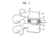
椎間円板は、解剖学的及び機能的に複雑な関節である。3要素構造:髄核(核)、線維輪(輪)及び椎骨終板、から構成される。生化学組成及びこれらの要素構造内の解剖学的配置は、円板の生体力学的機能に関連する。間 The intervertebral disc is an anatomically and functionally complex joint. It is composed of three elements: nucleus pulposus (nucleus), annulus fibrosus (ring) and vertebral endplates. The biochemical composition and anatomical arrangement within these elementary structures is related to the biomechanical function of the disc.
核は、円板総断面積の約25〜40%を占める。これは、主として、主にプロテオグリカンと少量のコラーゲンを含むムコイド材料からなっている。プロテオグリカンは、マイナス電荷の鎖を有するコア蛋白質及びそこに共有結合したケラチンスルフェート(ケラタン硫酸)及びコンドロイチン硫酸からなる。これらの構成要素のために、核は、通常、水約70〜90重量%を含有する、ルーズ(loose)なヒドロゲルである。核は、円板の生体力学的機能において重要な役割を演じるが、主に核のルーズなヒドロゲル性質の故に、円板の機械的特性はよく知られていない。The nucleus occupies about 25 to 40% of the total cross-sectional area of the disk. It is mainly composed of a mucoid material containing mainly proteoglycans and small amounts of collagen. Proteoglycans consist of a core protein having a negatively charged chain and keratin sulfate (keratan sulfate) and chondroitin sulfate covalently bonded thereto. For these components, the core is a loose hydrogel, usually containing about 70-90% by weight of water. The nucleus plays an important role in the biomechanical function of the disc, but the mechanical properties of the disc are not well known, mainly due to the loose hydrogel nature of the nucleus.
核は輪及び椎骨終板により包囲され、マイナスに帯電したスルフェート基は、ポリマーマトリックスに結合しているため固定されているので、マトリックスはその周囲よりもより高い対イオン(counter ion)濃度を有する。このイオン濃度は、輪より高い浸透圧、例えば約0.1〜約0.3MPaの範囲の浸透圧を生じる。プロテオグリカンの高い固定電荷密度の結果として、マトリックスは、車の重量を支えるタイヤの中の空気圧と全く同じ方法で、適用された荷重を支持できる浸透膨潤圧を及ぼす。Since the nucleus is surrounded by an annulus and vertebral endplates, and the negatively charged sulfate groups are immobilized by binding to the polymer matrix, the matrix has a higher counter ion concentration than its surroundings . This ion concentration produces an osmotic pressure higher than the ring, for example, in the range of about 0.1 to about 0.3 MPa. As a result of the high fixed charge density of proteoglycans, the matrix exerts an osmotic swelling pressure that can support the applied load in exactly the same way as the air pressure in the tires that support the weight of the car.
コラーゲンネットワークの張力による内的抵抗応力及び筋肉及び靭帯伸張により適用される荷重による外的応力が均衡するまで、核に液体を吸収する能力を与えるのは、核マトリックスの浸透膨潤圧及び親水性である。核の膨潤圧(Ps)は、プロテオグリカンの濃度及び固定電荷密度に直接に依存する。即ち、プロテオグリカンの濃度及び固定電荷密度が高いほど、核の膨潤圧は高くなる。外部圧は、体位と共に変化する。人の体が仰向けの時には、第三腰椎円板上の圧縮荷重は、300ニュートン(N)であり、これは、直立姿勢の場合には700Nまで上昇する。体が前方に20°だけ曲げられると、圧縮荷重は、再び1200Nにまで増加する。外部圧(Pa)が増加すると、直前のバランス、即ちPs=Pa、はこわれる。新しいバランスに達するために、膨潤圧は増加せねばならない。この増加は、核中のプロテオグリカン濃度を増加させることにより達成され、これは、核中の液体を減じることにより得られる。このことが、クリープの結果として、円板がその高さを日中に約10%失う理由である。外部荷重が解除されると、即ちPsがPaより大である場合には、核は新しい平衡値に達するためにその周囲から液体を吸収する。円板の圧縮特性は、主に、この核の特性による。Until the internal resistive stress due to the tension of the collagen network and the external stress due to the load applied by muscle and ligament extension are balanced, it is the osmotic swelling pressure and hydrophilicity of the nuclear matrix that give the nucleus the ability to absorb liquid. is there. The swelling pressure (Ps) of the nucleus is directly dependent on the concentration of proteoglycans and the fixed charge density. That is, the higher the concentration of proteoglycan and the higher the fixed charge density, the higher the swelling pressure of the nucleus. External pressure changes with body position. When the person is lying on his back, the compressive load on the third lumbar disc is 300 Newtons (N), which rises to 700 N in the upright position. When the body is bent forward by 20 °, the compressive load increases again to 1200N. When the external pressure (Pa) increases, the balance immediately before, that is, Ps = Pa, breaks. In order to reach a new balance, the swelling pressure must be increased. This increase is achieved by increasing the proteoglycan concentration in the nucleus, which is obtained by reducing the liquid in the nucleus. This is why the disc loses about 10% of its height during the day as a result of creep. When the external load is released, ie when Ps is greater than Pa, the nucleus will absorb liquid from its surroundings to reach a new equilibrium value. The compression properties of the disk are mainly due to the properties of this core.
輪は、円板の外側を限定する境界を形成する。これは、また、水及びプロテオグリカンからなる無定形のベース物質に埋め込まれた高度に組織化されたコラーゲン繊維からなる。プロテオグリカン量は、核中より輪中のほうが低い。輪のコラーゲン繊維は、同心円でラミネートされたバンド又はラメラ(約8〜12層厚)で配置され、より厚い前側の壁とより薄い後方の壁を有する。各ラメラにおいて、繊維は並行であり、円板の水平面から約30°の角度で、上及び下の椎体に両方向で結合されている。1方向に立てられた(cocked)繊維の半分が、椎骨が、相互に対し他の方向に回転するにつれ締まるので、このデザインは、特に、ねじりに対して抵抗する。半径方向軸に沿った輪の組成は、均一ではない。輪の内側から外側部へとコラーゲンの割合は、一様に増加する。組成におけるこの差は、構造の強度を保持しながら、輪の内側領域と外側の領域が、非常に異なる組織中へ混和すべきことを表す。内部ラメラのみが終板に係留して、核のための封入容器を形成する。輪のコラーゲンネットワークは、周囲組織から水を吸収し、膨潤する核ゲルの傾向を抑制する。このように、輪中のコラーゲン繊維は常に緊張状態にあり、核ゲルは常に圧縮状態にある。The oval forms a boundary that defines the outside of the disk. It also consists of highly organized collagen fibers embedded in an amorphous base material consisting of water and proteoglycans. Proteoglycan levels are lower in rings than in nuclei. The collagen fibers of the annulus are arranged in concentrically laminated bands or lamellae (approximately 8-12 layers thick) with a thicker front wall and a thinner rear wall. In each lamella, the fibers are parallel and are attached in both directions to the upper and lower vertebral bodies at an angle of about 30 ° from the horizontal plane of the disc. This design is particularly resistant to torsion because half of the fibers that are cocked in one direction tighten as the vertebrae rotate in the other direction relative to each other. The composition of the ring along the radial axis is not uniform. The proportion of collagen increases uniformly from the inside to the outside of the annulus. This difference in composition indicates that the inner and outer regions of the annulus should mix into very different tissues while retaining the strength of the structure. Only the inner lamella is anchored to the endplate, forming an enclosure for the nucleus. The annular collagen network absorbs water from surrounding tissue and suppresses the tendency of the swelling nuclear gel. Thus, the collagen fibers in the annulus are always in tension and the nuclear gel is always in compression.
2つの椎骨終板は、透明で「ガラス様」組織である硝子軟骨からなり、円板を隣接椎体から分離させておく。この層は、硬い骨質椎体と軟質円板との間の推移域として作用する。椎間円板は、無血管なので、円板が代謝のために要する大抵の養分は、終板領域を通る拡散により円板に輸送される。The two vertebral endplates consist of hyaline cartilage, a transparent, “glass-like” tissue, keeping the disc separate from the adjacent vertebral bodies. This layer acts as a transition zone between the hard bony vertebral body and the soft disc. Since the intervertebral disc is avascular, most of the nutrients the disc requires for metabolism are transported to the disc by diffusion through the endplate area.
椎骨間の関節は、弾性及び粘性挙動の両方を示す。よって、円板へ荷重を適用する間、円板の即座の「歪み」又は「変形」がおこり、しばしば「即時変形」と呼ばれる。圧縮の間にそれにより円板から水が失われる主要な通路は、軟骨終板を通ると報告された。終板の水透過性は、約0.20〜約0.85×10⁻¹⁷m4N-1sec-1であるので、荷重が適用されている間、荷重下に、円板の初期容積が一定であると憶測するのは道理に合っている。円板の生来の核は、ルーズなヒドロゲル、即ち、水に可溶の親水性ポリマー材料、の形状であるので、容易く変形し、円板の変形の範囲は輪の伸長性に大きく依存する。一般に、核の静水挙動(hydrostatic behavior)が、円板の通常の静的及び動的荷重分担能力において重要な役割を果たし、かつ輪の延伸繊維の復元力が核膨潤圧の影響と均衡すると信じられている。輪による拘束がないと、核の環状膨張は、相当に増加したであろう。荷重が一定レベルで保たれるならば、関節高さの徐々の変化(一般に「クリープ」と呼ばれる)は、時間の関数として起こるだろう。結局、クリープは、安定化され、関節は「平衡」状態にあるといわれる。荷重が除かれると、関節は、荷重前の元の高さに徐々に「回復」するであろう。クリープ及び緩和率(relation rate)は、適用された荷重量、終板の透過性及び核ヒドロゲルの水結合力(water binding capability)に依存する。クリープ及び緩和は、液体を円板の内外にポンピングする点において本質的なプロセスである。Joints between vertebrae exhibit both elastic and viscous behavior. Thus, during the application of a load to the disk, an immediate "strain" or "deformation" of the disk occurs, often referred to as "immediate deformation". The primary path by which water was lost from the disc during compression was reported to pass through the cartilage endplate. Since the water permeability of the end plate is about 0.20 to about 0.85 × 10⁻¹⁷ m4 N−1 sec−1 , the initial volume of the disc under the load while the load is applied It makes sense to speculate that is constant. The natural nucleus of the disc is in the form of a loose hydrogel, a hydrophilic polymer material that is soluble in water, and therefore easily deforms, and the extent of deformation of the disc is highly dependent on the extensibility of the annulus. In general, it is believed that the hydrostatic behavior of the nucleus plays an important role in the normal static and dynamic load-sharing capacity of the disc, and that the restoring force of the drawn fibers of the rings balances the effect of the swelling pressure of the nucleus. Have been. Without ring restraint, the annular expansion of the nucleus would have increased significantly. If the load is kept at a constant level, a gradual change in joint height (commonly called "creep") will occur as a function of time. Eventually, the creep is stabilized and the joint is said to be in "balance". When the load is removed, the joint will gradually "recover" to its original height before the load. Creep and relation rates depend on the applied load, endplate permeability and the water binding capability of the nuclear hydrogel. Creep and relaxation are essential processes in that they pump the liquid in and out of the disk.
椎間円板の退化は、最終的病変及び背の痛みの一般的原因であると信じられる。椎間円板が老化するにつれ、退化を被る。生じる変化は、多くの点で、核の組成が内側輪の組成に接近するように見えることである。椎間円板退化は、少なくとも部分的に、核内の組成変化の結果である。特に退化円板中の核内のプロテオグリカンの分子量及び量の双方が年齢と共に減少し、かつ核中のケラチンスルフェート対コンドロイチン硫酸の割合が増加することが判明している。ケラチンスルフェート対コンドロイチン硫酸の割合の増加及びプロテオグリカン含分の減少は、核の固定電荷密度を約0.28meq/mlから約0.18〜0.20meq/mlまで減じる。これらの変化は、核に作用して、その水結合力部分を失わせ、それが及ぼし得る最大膨潤圧を減じる。結果として、最大水含分は、思春期直前の約85%以上から、中年での約70〜75%まで降下する。匹敵する年齢の通常の円板のものよりも、脱出円板のグリコサミノグリカン含分はより低下し、コラーゲン含分はより高くなることが発見された。円板L−4−L−5及びL−5−S−1は、通常、最も退化した円板である。退 Intervertebral disc degeneration is believed to be a common cause of eventual lesions and back pain. As the intervertebral disc ages, it undergoes degeneration. The resulting change is that in many respects the composition of the nucleus appears to approach that of the inner annulus. Intervertebral disc degeneration is the result, at least in part, of a change in composition within the nucleus. In particular, it has been found that both the molecular weight and amount of proteoglycans in the nucleus in degenerate discs decrease with age and that the ratio of keratin sulfate to chondroitin sulfate in the nucleus increases. Increasing the ratio of keratin sulfate to chondroitin sulfate and decreasing proteoglycan content reduces the fixed charge density of the nucleus from about 0.28 meq / ml to about 0.18-0.20 meq / ml. These changes act on the nucleus, causing it to lose its water binding portion, reducing the maximum swelling pressure it can exert. As a result, the maximum water content drops from about 85% or more just before puberty to about 70-75% in middle age. It has been found that the glycosaminoglycan content of the escaped disc is lower and the collagen content is higher than that of a normal disc of comparable age. The discs L-4-L-5 and L-5-S-1 are usually the most degenerated discs.
核は円板の全面積の約1/3のみを占めるが、通常の円板で、全荷重の約70%を担うことは、公知である。このように、適度に退化した円板の核上の圧縮荷重は、匹敵する通常の円板におけるよりも約30%低いが、輪上の圧縮荷重は退化円板において100%増加することが判明している。この荷重変化は、前記したように、主に、円板での構造的変化により引き起こされる。退化円板の輪上の過剰荷重は、円板高さの減少及び脊椎分節の過度の動きを引き起こす。円板の柔軟性はコラーゲン繊維の過剰運動を生じ、これは、次に繊維結合を損ない、輪リングの十分に組織された繊維の層間剥離を引き起こす。層間剥離輪は、輪上の応力により更に弱化され得、厳しい場合は、この応力が輪を引き裂いてしまう。この全プロセスは、ぺちゃんこのタイヤで運転すると、補強層がついには層間剥離する、ことに非常に類似する。輪の厚さが、後部分が前部分より薄く、均一ではないので、層間剥離及び傷害が、通常、後部分で最初に起こる。The nucleus occupies only about 1/3 of the total area of the disc, but it is known that a normal disc carries about 70% of the total load. Thus, the compressive load on the nucleus of a moderately degenerated disk is found to be about 30% lower than on a comparable normal disk, but the compressive load on the annulus is found to increase by 100% in the degenerated disk. are doing. This load change is mainly caused by structural changes in the disk, as described above. Excessive loading on the annulus of the degenerated disc causes a decrease in disc height and excessive movement of the spinal segment. The flexibility of the disc causes excessive movement of the collagen fibers, which in turn impairs fiber bonding and causes delamination of the well-organized fibers of the annulus. The delamination wheel can be further weakened by stress on the wheel, which, in severe cases, can tear the wheel. The whole process is very similar to running on this tire, where the reinforcement layer eventually delaminates. Since the thickness of the hoop is less uniform in the posterior portion than in the anterior portion, delamination and injury usually occur first in the posterior portion.
外傷又は疾病のため、脊椎円板は、ずらされるか又は損傷されることもある。これらの場合に、また、円板の退化の場合に、核は、脊柱管又は椎間孔に、脱出及び/又は突出することがあり、これは、椎間板ヘルニア又は「椎間板脱出症」として公知である。この円板は、次いで、部分的に塞がれた孔を通り、脊柱管を出て行く脊椎神経を圧迫し、その分布の領域に疼痛又は麻痺を引き起こすこともある。椎間板ヘルニアが最も頻繁に起こる場所は、下部腰椎領域である。この領域での円板ヘルニアは、しばしば、坐骨神経を圧迫することにより下端(inferior extremity)を巻き込む。脊 Due to trauma or disease, the spinal disc may be displaced or damaged. In these cases, and in the case of disc degeneration, the nucleus may protrude and / or protrude into the spinal canal or intervertebral foramen, which is known as a herniated disc or "prolapse of the disc". is there. The disc then passes through the partially obstructed hole and compresses the spinal nerves exiting the spinal canal, which may cause pain or paralysis in the area of its distribution. The place where disc herniation most frequently occurs is in the lower lumbar region. Disc herniations in this area often involve the inferior extremity by compressing the sciatic nerve.
損傷又は退化円板により引き起こされた腰痛を治療するために現在使用される基本的に3つのタイプの治療:保存療法、椎間板切除及び融合、がある。これらの治療のそれぞれは、利点及び限界を有する。腰痛を有する大多数の患者、特に初めて腰痛のエピソードを有する患者は、保存療法でよくなるであろう。しかしながら、保存療法が腰痛を解決する最も効果的及び経済的方法であるとは、必ずしも真実ではない。There are basically three types of treatment currently used to treat low back pain caused by injured or degenerated discs: conservative therapy, discectomy and fusion. Each of these treatments has advantages and limitations. The majority of patients with low back pain, especially those who have episodes of low back pain for the first time, will benefit from conservative therapy. However, it is not always true that conservative therapy is the most effective and economical way to solve back pain.
椎間板切除は、脊椎神経の圧迫又は化学的刺激により腰痛を引き起こす椎間板ヘルニア物質、通常は核を除去することにより、通常、臨床的症候を和らげる優れた短期間の結果を提供する。椎間板切除が、生体力学的見地から望ましくないのは明らかである。健康な円板では核は最圧縮荷重を担い、退化円板では、この荷重は主に輪リング上に分配されて、前記したように、輪の引裂き及び層間剥離を引き起こす。椎間板切除での核の除去は、実際に、輪リング上への圧縮荷重の分布を引き起こして、円板スペースを狭窄する。長期の円板高の減少は、椎間関節(facet joint)で不可逆的変形性関節症様病変を引き起こすことが予期されると報告されている。椎間板切除が長期的に利益が乏しく、再ヘルニア形成の高い発症を引き起こすからである。Discectomy provides excellent short-term results that usually relieve clinical symptoms by removing the disc herniation material, usually the nucleus, that causes back pain by compression or chemical stimulation of the spinal nerves. Clearly, discectomy is undesirable from a biomechanical point of view. In a healthy disc, the nucleus carries the most compressive load, and in a degenerated disc, this load is mainly distributed on the ring, causing tearing and delamination of the ring, as described above. Removal of the nucleus in a discectomy actually causes a distribution of the compressive load on the annulus ring, which narrows the disc space. It has been reported that long-term loss of disc height is expected to cause irreversible osteoarthritis-like lesions at facet joints. This is because discectomy has a poor long-term benefit and causes a high incidence of reherniation.
融解は、一般に、症状を除去し、関節を安定化させるのに、良い仕事をする。しかしながら、融解分節の動きが制限されるので、隣接椎骨円板の動きの範囲が増加し、おそらくそれらの退化プロセスを増進する。Thawing generally does a good job of relieving symptoms and stabilizing the joints. However, as the movement of the melting segments is limited, the range of movement of the adjacent vertebral discs is increased, possibly enhancing their degeneration process.
これらの欠点のために、損傷又は退化した椎間円板を代替できるのみならず、代替すべき円板の生理的、生体力学的機能を模倣でき、周囲組織のいっそうの退化を予防する、プロテーゼ関節装置を使用することが望ましい。Because of these drawbacks, a prosthesis that can not only replace a damaged or degenerated disc but also mimic the physiological and biomechanical functions of the disc to be replaced and prevent further degeneration of surrounding tissue It is desirable to use an articulation device.
人工円板は、従来技術でよく知られている。米国特許No.3867728(Stubstad et al.)は、円板全体を代替する装置に関する。この装置は、弾性ポリマーの垂直、水平又は軸方向シートをラミネートすることにより製造される。米国特許No.4309777(Patil)は、金属バネ及びカップを利用するプロテーゼに関する。その表面部分に多孔質被膜を有する硬質固体を含む脊椎インプラントは、米国特許No.4714469(Kenna)に示されている。退化円板に取り替わる1対の堅いプラグからなる椎間円板プロテーゼは、米国特許No.4349921(Kuntz)により参照される。米国特許No.4772287及び4904260(Ray et al.)は、治療薬含有又は不含の一対のシリンダー状プロテーゼ椎間円板カプセルの使用を教示する。米国特許No.4911718(Lee et al.)は、異なる材料の異なる3つの部分;核、輪及び終板を含むエラストマー円板スペーサーに関する。今日、これらの発明の何れも、脊椎医療市場の製品となってはいない。米国特許No.5047055及び5192326(Bao et al.)(この発明の譲受人に譲渡され、参考として本明細書中に組み入れられている)に、完全に水和されると、大きなピースに造形され、一般に、円板キャビティに適合する形状のヒドロゲルを含むか又は多孔質外皮(envelope)内のヒドロゲルビーズを含む人工核が記載されている。ヒドロゲルは、平衡水含有量(EWC)少なくとも約30%を有し、かつ輪及び円板の終板の圧迫を受けると、1平方メートル当たり少なくとも約1メガニュートン(1MNm-2)の圧縮強さを有する。有利には、核の圧縮強さは約4MNm-2以上である。Artificial discs are well known in the prior art. U.S. Pat. 3867728 (Stubstad et al.) Relates to a device for replacing an entire disc. The device is manufactured by laminating vertical, horizontal or axial sheets of an elastic polymer. U.S. Pat. 4309777 (Patil) relates to a prosthesis utilizing a metal spring and a cup. A spinal implant comprising a hard solid having a porous coating on its surface is described in US Pat. No. 4,714,469 (Kenna). An intervertebral disc prosthesis consisting of a pair of rigid plugs replacing a degenerated disc is disclosed in US Pat. 4349921 (Kuntz). U.S. Pat. Nos. 4,772,287 and 4,904,260 (Ray et al.) Teach the use of a pair of cylindrical prosthetic disc capsules with or without a therapeutic agent. U.S. Pat. 4911718 (Lee et al.) Relates to an elastomeric disc spacer comprising three different parts of different materials; a nucleus, an annulus and an endplate. Today, none of these inventions has become a product in the spine medical market. U.S. Pat. 5050755 and 5192326 (Bao et al.) (Assigned to the assignee of the present invention and incorporated herein by reference), when fully hydrated, are shaped into large pieces and are generally circular. Artificial nuclei have been described that include hydrogels of a shape that conforms to the plate cavity or that include hydrogel beads within a porous envelope. The hydrogel has an equilibrium water content (EWC) of at least about 30% and, upon compression of the annulus and disc endplates, has a compressive strength of at least about 1 meganewton per square meter (1 MNm-2 ). Have. Advantageously, the compressive strength of the nucleus is about 4 MNm-2 or more.
Substad et al.,Patil,Kenna及びLee et al.の発明の第一の欠点は、彼らのプロテーゼの使用が生来の円板の完全な交換を要することであり、多数の外科的難点を含む。第二に、椎間円板は、そのおのおのがそれ自体ユニークな構造的特長を有する前記3つの部品構造を含む、解剖学的かつ機能的に複雑な関節である。認容性材料から、生来の円板の機能を模倣するそのような複雑なプロテーゼをデザイン及び二次加工することは、非常に困難である。もう1つの問題は、プロテーゼの移動を防ぐのが難しいことである。第四に、核と交換するためにのみ意図されたプロテーゼにとってすら、主要な障害は、天然の物に類似し、核の通常の機能を回復できる材料を発見することであった。疎水性エラストマー及び熱可塑性ポリマーは、生来の核との重要な固有の差、例えば、エラストマーでは親水性の欠如、熱可塑性ポリマーでは柔軟性の欠如、のために、プロテーゼ核に使用するのは望ましくない。{Substad et al. , Patil, Kenna and Lee et al. A first disadvantage of the invention is that the use of their prosthesis requires a complete replacement of the native disc, which involves a number of surgical difficulties. Second, the intervertebral disc is an anatomically and functionally complex joint that includes the three-part structure, each of which has its own unique structural features. From an acceptable material, designing and fabricating such a complex prosthesis that mimics the function of the native disc is very difficult. Another problem is that it is difficult to prevent movement of the prosthesis. Fourth, even for prostheses intended solely for exchange with the nucleus, a major obstacle has been to find materials that resemble natural ones and can restore the normal function of the nucleus. Hydrophobic elastomers and thermoplastic polymers are desirable for use in prosthetic cores because of important inherent differences from the native core, for example, lack of hydrophilicity in elastomers and lack of flexibility in thermoplastic polymers. Absent.
これらの問題は、弾性ゴムプラグを使用するKuntzにより、又は、液体又はチキソトロープゲルでそれぞれ充填した嚢又はカプセルを使用するFroning及びRay et al.により解決されてはいない。Ray及びFroningの特許により、液体がカプセル及び嚢にそれぞれ充填するのに使用された。そのため、膜は完全にシールされて液体の漏れを防ぐことが必要である。結果として、体液を、反復荷重の間、内外に拡散させ、そのことにより円板が必要とする栄養を提供する核機能は、これらの装置により完全には復元できない。{These problems have been addressed by Kuntz using elastic rubber plugs or Froning and Ray et al. Al. Using capsules or capsules filled with liquids or thixotropic gels, respectively. Has not been resolved. According to the Ray and Froning patent, liquids were used to fill capsules and sacs, respectively. Therefore, the membrane needs to be completely sealed to prevent liquid leakage. As a result, the body's ability to diffuse body fluids in and out during repeated loading, thereby providing the nutrition needed by the disc, cannot be completely restored by these devices.
Bao et al.は、プロテーゼ腰椎円板核をヒドロゲルから製造した。ヒドロゲルは、生医学的適用、例えばコンタクトレンズに使用されていた。ヒドロゲルの利点の中に、疎水性エラストマー及び金属よりも生体適合性であるという点がある。この生体適合性は、主に、周囲の組織のように柔らかで、水を含み、周囲組織に対して相対的に低い摩擦係数を有するという、ヒドロゲルのユニークな特性による。ヒドロゲルの生体適合性は、体内でいっそう容易に我慢される核プロテーゼをもたらす。更に、疎水性エラストマー及び金属ゲルは、水性組成物及びその溶質がそれを通って拡散することを可能にしない。{Bao et al. Manufactured prosthetic lumbar disc nuclei from hydrogels. Hydrogels have been used in biomedical applications, such as contact lenses. Among the advantages of hydrogels is that they are more biocompatible than hydrophobic elastomers and metals. This biocompatibility is primarily due to the unique properties of hydrogels, being as soft as the surrounding tissue, containing water, and having a relatively low coefficient of friction with surrounding tissue. The biocompatibility of the hydrogel results in a nuclear prosthesis that is more easily tolerated in the body. Furthermore, hydrophobic elastomers and metal gels do not allow aqueous compositions and solutes to diffuse therethrough.
ある種のヒドロゲルのその他の利点は、円板の上の荷重に耐え、椎体間の正常のスペースを復元することを可能にする良好な機械的強度である。Bao et al.の前記核は、高い機械的強度を有し、身体荷重に耐えることができ、かつ欠陥のある輪の治癒に助力する。Another advantage of certain hydrogels is their good mechanical strength, which allows them to withstand loads on the disc and restore normal space between the vertebral bodies. Bao et al. The nucleus has high mechanical strength, can withstand body loads, and assists in healing of defective rings.
Bao et al.で使用されるヒドロゲル核の他の利点は、その優れた粘弾性特性及び形状記憶である。ヒドロゲルは大量の水を含有し、この水は可塑剤として作用する。水部は、ヒドロゲルが、機械圧下に部分的に脱水される場合には、より自由にヒドロゲルから去る自由水として役立つ。このヒドロゲルの特徴は、生来の核と同じようにして圧縮下で、クリープを可能にし、任意の著しい退化又はその弾性の損失無しで、長期間、反復荷重に耐えることを可能にする。このことは、ヒドロゲル中の水がクッションの様にふるまい、それによって、高い平衡水含有量(EWC)を有するヒドロゲルのポリマーネットワークが、機械的荷重下で損傷を受けにくくなるからである。{Bao et al. Another advantage of the hydrogel core used in is their excellent viscoelastic properties and shape memory. Hydrogels contain large amounts of water, which acts as a plasticizer. The water portion serves as free water that leaves the hydrogel more freely if the hydrogel is partially dehydrated under mechanical pressure. This feature of the hydrogel allows for creep under compression in the same manner as the native core, allowing it to withstand repeated loads for extended periods of time without any significant degradation or loss of its elasticity. This is because the water in the hydrogel behaves like a cushion, thereby making the polymer network of the hydrogel with a high equilibrium water content (EWC) less susceptible to damage under mechanical loading.
ヒドロゲルのもう1つの利点は、水及び水可溶性物質、例えば栄養分、代謝産物等に対する透過性である。反復荷重下での体液拡散が、生来の円板への主な栄養源であることは公知である。この栄養分拡散ルートが、例えば水不透過性核により、妨害されるならば、円板のいっそうの劣化が確実となる。Another advantage of hydrogels is their permeability to water and water-soluble substances such as nutrients, metabolites and the like. It is known that body fluid diffusion under repeated loading is a major source of nutrients to the native disc. If this nutrient diffusion route is interrupted, for example by water impermeable nuclei, further deterioration of the disc is ensured.
ヒドロゲルは、脱水され、生じたキセロゲルは、ヒドロゲルの特性を変化させずに再び水和される。ヒドロゲルが脱水されると、その容積は減じ、そのため、円板中の核キャビティへのプロテーゼ核の移植を容易にする。次いで、移植プロテーゼ核は、体内で、そのEWCまで体液を吸収することにより膨潤する。ヒドロゲルのEWCは、そこに適用される圧縮荷重に依存する。従って、開放容器中の特定のヒドロゲルのEWCは、閉鎖容器、例えば椎間円板、中の同一ヒドロゲルのEWCとは異なる。以下に記載のEWC値は、椎間円板中に見られる条件下で圧縮荷重を受けたヒドロゲルについての値である。次に、脱水ヒドロゲルの膨張因子が、そのEWCに依存する。従って、EWC38%のヒドロゲルについての1.19から、EWC80%のヒドロゲルについての1.73まで変動しうる。EWC80%のヒドロゲルについては、脱水プロテーゼ核の容積は、通常、水和されたものの約20%である。脱水され、次いで、そのEWCまでの水和時にそのオリジナルの形状に戻る能力は、手術の間、装置を後方外側(posterior-laterally)に移植することを可能にし、そのようにして、伝統的に用いられる脊椎内手術の複雑さ及び危険性を減じる。神経、硬膜嚢、動脈及び他の器官を穿孔する危険性も減じる。更に、輪上の切開領域を減じ、そのことにより、輪を治癒しやすくし、円板の再ヘルニア形成を防止する。ヒドロゲルは、その制御された薬物放出能力のゆえに、円板への薬物供給にも有用である。多種多様の治療薬、例えば成長因子、長期間鎮痛薬及び抗炎症剤は、プロテーゼ核に取り付け、核を円板に移植後、制御可能な速度で放出させることができる。The hydrogel is dehydrated and the resulting xerogel is rehydrated without changing the properties of the hydrogel. As the hydrogel is dehydrated, its volume decreases, thereby facilitating implantation of the prosthetic nucleus into the nuclear cavity in the disc. The implant prosthesis nucleus then swells in the body by absorbing fluid to its EWC. The EWC of a hydrogel depends on the compressive load applied thereto. Thus, the EWC of a particular hydrogel in an open container is different from the EWC of the same hydrogel in a closed container, eg, an intervertebral disc. The EWC values described below are for hydrogels that have been subjected to compressive loading under conditions found in the intervertebral disc. Second, the swelling factor of the dehydrated hydrogel depends on its EWC. Thus, it can range from 1.19 for a 38% EWC hydrogel to 1.73 for a 80% EWC hydrogel. For an 80% EWC hydrogel, the volume of the dehydrated prosthesis core is typically about 20% of the hydrated one. The ability to be dehydrated and then return to its original shape upon hydration to EWC allows the device to be implanted posterior-laterally during surgery, thus traditionally Reduces the complexity and risk of the intravertebral surgery used. The risk of perforating nerves, dural sac, arteries and other organs is also reduced. In addition, the incision area on the annulus is reduced, thereby making the annulus easier to heal and preventing re-herniation of the disc. Hydrogels are also useful for drug delivery to discs due to their controlled drug release ability. A wide variety of therapeutic agents, such as growth factors, long term analgesics and anti-inflammatory agents, can be attached to the prosthetic nucleus and released at a controllable rate after the nucleus is implanted in the disc.
更に、寸法保全性(dimensional integrity)は、約90%までの水含有量を有するヒドロゲルを用いて保持することができる。この寸法保全性は、核が正確に設計されているならば、輪リング上の大きな領域への椎骨荷重の分散を助け、プロテーゼ核の膨張及びヘルニア形成を防止する。In addition, dimensional integrity can be maintained with hydrogels having a water content of up to about 90%. This dimensional integrity, if the nucleus is designed correctly, will help distribute the vertebral load over a large area on the annulus ring and prevent the prosthesis nucleus from expanding and herniating.
しかしながら、完全に水和された状態でのその嵩高さ(bulkiness)によって、特に経皮手術で、ヘルニア化された核を除去するために、円板に設けられた小さな窓を通して、完全に水和されたヒドロゲルプロテーゼを、円板のキャビティに移植することは、通常、難しい。それゆえ、そのようなプロテーゼは、低い表面積のために、EWCに達するのに長期間を要する相対的に脱水された状態で、円板中に移植されねばならない。高い表面積を有する他のヒドロゲルは、核キャビティの形状に完全に一致はしない。その教示が、参考として本明細書中に組み入れられている、WO97/268407(PCT/US9700457)に開示されたような他のポリマーも、円板核を充填するのに使用することができる。However, due to its bulkiness in the fully hydrated state, especially in percutaneous surgery, the hydration through a small window in the disc to remove herniated nuclei. It is usually difficult to implant the prepared hydrogel prosthesis into the disc cavity. Therefore, due to the low surface area, such prostheses must be implanted in the disc in a relatively dehydrated state that requires a long time to reach the EWC. Other hydrogels with high surface areas do not perfectly match the shape of the nuclear cavity. Other polymers, such as those disclosed in WO 97/268407 (PCT / US9700457), the teachings of which are incorporated herein by reference, can also be used to fill the disk core.
脊椎核の除去により生じたキャビティへのインプラントの配置を、外科医が見ることができるように、固有の放射線不透過の、即ちレントゲンで見れるヒドロゲルインプラントを提供することが望ましい。放射線可視物質が、プロテーゼ核インプラントを製造するポリマー又はヒドロゲル材料中に混入されるならば、それは有利である。インプラントの機械的完全性を傷つけずに、加工の間及び移植後に、ヒドロゲルの寸法の変化を許す、ヒドロゲル又はポリマーを放射線不透過にする方法を有するのは望ましい。It is desirable to provide a unique radiopaque, ie, radiographic, hydrogel implant so that the surgeon can see the placement of the implant in the cavity created by removal of the spinal nucleus. It would be advantageous if the radiovisible material was incorporated into the polymer or hydrogel material making the prosthetic nuclear implant. It would be desirable to have a method of making a hydrogel or polymer radiopaque that would allow the hydrogel to change dimensions during processing and after implantation without compromising the mechanical integrity of the implant.
種々の方法が、ヒドロゲル又は他のポリマー核インプラントを移植するために使用される。そのような方法は、その教示が参考として本明細書中に組み入れられている、米国特許No.5800549に示される。Various methods are used to implant hydrogels or other polymer core implants. Such a method is described in U.S. Pat. 580549.
本発明の課題は、切除された脊椎円板の生来の核に取って代わる新規のヒドロゲル又は他のポリマーインプラントを提供することである。It is an object of the present invention to provide a novel hydrogel or other polymer implant that replaces the native nucleus of a resected spinal disc.
本発明のもう1つの課題は、その中に放射線可視物質を含む、新規ヒドロゲル又は他のポリマープロテーゼ核を提供することである。Another object of the present invention is to provide a novel hydrogel or other polymer prosthesis nucleus that contains a radiation visible material therein.
更に本発明のもう1つの課題は、インプラント全体にわたり割り当てられる(disbursed)か又はばらばらの位置で存在する放射線可視物質をヒドロゲル又はポリマー内に混入するための方法を提供することである。It is yet another object of the present invention to provide a method for incorporating radiovisible material into a hydrogel or polymer that is disbursed or present at discrete locations throughout the implant.
そのような課題は、ヒドロゲル内に位置する放射線可視物質を有するヒドロゲルから製造された、円板の生来の核を代替する脊椎インプラントにより解決される。物質は、金属、例えば、金、タングステン、タンタル、白金、チタン又はこれらの組み合わせであってよい。課題 Such a problem is solved by a spinal implant, which replaces the natural nucleus of a disc, made from a hydrogel having a radiovisible substance located within the hydrogel. The material may be a metal, for example, gold, tungsten, tantalum, platinum, titanium or a combination thereof.
物質は粉末形であってよく、均一にヒドロゲルにわたって分布しているか又は粉末形で、ヒドロゲル又はポリマーインプラント中で、別々の層又は別々の位置
に配置されていてよい。放射線不透過粉末は、有利には最大直径10〜100μmを有し、更に有利には、粉末は直径約75μmを有する。The substance may be in powder form, distributed evenly throughout the hydrogel or in powder form, in a hydrogel or polymer implant, arranged in separate layers or at discrete locations. The radiopaque powder preferably has a maximum diameter of 10 to 100 μm, more preferably the powder has a diameter of about 75 μm.
あるいは(alternately)、金属は、細片形又は薄片形のホイルの形でインプラント全体に散乱していても良い。ホイルが細片形であれば、相対的に薄く、即ち1〜100μm厚さの範囲内であるべきで、そのため、ヒドロゲルと一緒に使用されると、ヒドロゲルが水和及び脱水されるにつれ、これは、膨張しかつ収縮する。有利な態様では、この範囲の下端での厚さが望ましく、例えば2μm厚さである。Alternatively, the metal may be scattered throughout the implant in the form of strips or flakes of foil. If the foil is in the form of a strip, it should be relatively thin, i.e., in the range of 1-100 [mu] m thickness, so that when used with a hydrogel, this will increase as the hydrogel hydrates and dehydrates. Expand and contract. In an advantageous embodiment, a thickness at the lower end of this range is desirable, for example 2 μm.
追加の態様では、インプラントは、その形成の間に、ヒドロゲルインプラント中に装入された金属ワイヤ又はコイルの形状であってもよい。ワイヤは、ヒドロゲルの水和及び脱水の間、自身の上に折り重ねられるような直径である。In an additional aspect, the implant may be in the form of a metal wire or coil loaded into the hydrogel implant during its formation. The wire is of a diameter such that it folds over itself during hydration and dehydration of the hydrogel.
使用ポリマーが現場形成されるならば、インプラントに放射線可視性を与える金属は、注入及び硬化の前に、ポリマー全体に割り当てられる。If the used polymer is formed in situ, the metal that will give the implant radiological visibility is assigned to the entire polymer prior to injection and curing.
ポリマーが、融液に加工されるならば、その融点以上である時に、金属は、ポリマー中にブレンドされる。な ら ば If the polymer is processed into a melt, the metal is blended into the polymer when it is above its melting point.
放射線不透過物質を含むヒドロゲルの製法は、ポリマー粉末を溶解し、均一溶液を形成し、次いで、溶液がまだ液体の間に金属粉末を金属薄片と一緒にその中に混入し、次いで、溶液を冷凍して、固体を形成することを含む。通常、溶液を型に流入し、ヒドロゲルを形成し、次いで型を冷凍庫に入れる。The process of making a hydrogel containing radiopaque materials involves dissolving the polymer powder to form a homogeneous solution, then mixing the metal powder with the metal flakes therein while the solution is still liquid, and then mixing the solution. Including freezing to form a solid. Typically, the solution is poured into a mold to form a hydrogel, and then the mold is placed in a freezer.
あるいは、ヒドロゲルインプラントは、放射線不透過物質を有する均一溶液を型の1部のみを充たして、型に装入し、溶液を冷凍し、放射線不透過金属を含むヒドロゲルの追加層を、固化したヒドロゲルの第一層上に装入し、その後、第二の層を冷凍して、固体を形成することにより、連続的に製造することができる。この工程は、放射線半透過性又は放射線不透過であるプロテーゼ核中の交互の層を形成するために繰り返すことができる。Alternatively, a hydrogel implant may be prepared by filling a mold with a uniform solution having a radiopaque material, filling only one portion of the mold, freezing the solution, and solidifying an additional layer of hydrogel containing a radiopaque metal into a solidified hydrogel. Can be manufactured continuously by charging over the first layer of the solid and then freezing the second layer to form a solid. This process can be repeated to form alternating layers in the prosthesis nucleus that are radiopaque or radiopaque.
更に他の態様では、ポリマーインプラントは、ポリマー、例えばポリ(アクリロニトリル)を融解し、インプラントの成形前に放射線不透過(radiopacifying)剤を混入することにより形成することができる。1つのインプラントで実施された連続成形操作は、放射線不透過剤をインプラントの別々の領域に局在化させることができる。ポリマーインプラントは、架橋性材料(例えばモノマー及び/又はプレポリマー)及び架橋剤の両方を注入し、次いで架橋性材料を、ポリウレタンのように現場硬化させることにより形成することもできる。放射線可視性は、放射線不透過剤を架橋性材料、架橋剤又は両方に添加することにより、このインプラントに賦与することができる。In yet another aspect, a polymer implant can be formed by melting a polymer, such as poly (acrylonitrile), and incorporating a radiopacifying agent prior to molding the implant. A continuous molding operation performed on a single implant can localize the radiopaque agent to separate areas of the implant. Polymer implants can also be formed by injecting both a crosslinkable material (eg, a monomer and / or prepolymer) and a crosslinking agent, and then curing the crosslinkable material in place, such as polyurethane. Radiation visibility can be imparted to the implant by adding a radiopaque agent to the crosslinkable material, the crosslinking agent, or both.
コイル又はホイルをヒドロゲルの放射線不透過部分を製造するために使用するならば、ホイル又はコイルは、冷却して固化させる前に液体ヒドロゲルに挿入するとよい。 If a coil or foil is used to make the radiopaque portion of the hydrogel, the foil or coil may be inserted into the liquid hydrogel before it cools and solidifies.
図1から図6に関して、有利な態様での、一般に10で示された本発明のヒドロゲルプロテーゼ核は、そのEWCまで水和された場合、一般に生来の核の形状と一致する。あるいは、ヒドロゲルは、ポリマージャケット中に束縛されていても良い。そのようなことが、米国特許No.5674295及び6132465に教示されている。プロテーゼ核は、椎骨14の円板12のキャビティ11に移植され、生来の輪16に包囲されている。図1に示されるように、椎骨終板20と22は、核10の上及び下面をそれぞれ覆っている。インプラントは、輪12中の開口62を通ってはめ込まれる。1 to 6, the hydrogel prosthesis nucleus of the present invention, generally designated 10, in an advantageous manner, when hydrated to its EWC, generally conforms to the shape of the native nucleus. Alternatively, the hydrogel may be constrained in a polymer jacket. Such is the case in U.S. Pat. 5,674,295 and 6,132,465. The prosthetic nucleus is implanted in the
図3に関しては、金属粒子30が均一に全体に割り当てられている本発明のプロテーゼ核が示される。均一は、相対的意味で使用され、粒子がヒドロゲル中で正確に離間されていることを示さない。に 関 し て Referring to FIG. 3, a prosthetic nucleus of the present invention is shown in which metal particles 30 are uniformly distributed throughout. Homogeneity is used in a relative sense and does not indicate that the particles are precisely spaced in the hydrogel.
図4に関しては、放射線不透過ホイル細片40がその中に位置する本発明のプロテーゼ核が示される。4 Referring to FIG. 4, the prosthetic nucleus of the present invention with radiopaque foil strip 40 positioned therein is shown.
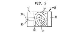
図5に関しては、放射線不透過又は放射線可視コイル50がその中に位置する本発明のポリマーインプラントが示される。5 Referring to FIG. 5, there is shown a polymer implant of the present invention having a radiopaque or radiovisible coil 50 located therein.
図6に関しては、その中に金属粒子又はホイル粒子60の放射線不透過層を有する本発明のプロテーゼ円板核10が示される。図6では、3層が示されるが、1層又は2層又は3層以上を利用しても良い。Referring to FIG. 6, there is shown a
本発明の実施で有用なヒドロゲルは、親水性モノマー、例えば、2−ヒドロキシアルキルアクリレート及びメタクリレート、例えば2−ヒドロキシエチルメタクリレート(HEMA);N−ビニルモノマー、例えばN−ビニル−2−ピロリドン(N−VP);エチレン性不飽和酸、例えばメタクリル酸(MA)及びエチレン性不飽和塩基、例えば2−(ジエチルアミノ)エチルメタクリレート(DEAEMA)の少し架橋された生体適合性のホモポリマー及びコポリマーを含む。コポリマーは、更に、非−親水性モノマー、例えばアルキルメタクリレート、例えばメチルメタクリレート(MMA)等からの残分を含んでよい。架橋ポリマーは、公知の方法により、架橋剤、例えばエチレングリコールジメタクリレート及びメチレンビス(アクリルアミド)、及び開始剤、例えば2,2−アゾビス(イソブチロニトリル)、ベンゾイルペルオキシド等及び放射線、例えばUV及びγ線の存在下で、形成される。Hydrogels useful in the practice of the present invention include hydrophilic monomers such as 2-hydroxyalkyl acrylates and methacrylates such as 2-hydroxyethyl methacrylate (HEMA); N-vinyl monomers such as N-vinyl-2-pyrrolidone (N- VP); including slightly crosslinked, biocompatible homopolymers and copolymers of ethylenically unsaturated acids such as methacrylic acid (MA) and ethylenically unsaturated bases such as 2- (diethylamino) ethyl methacrylate (DEAEMA). The copolymer may further comprise a residue from a non-hydrophilic monomer, such as an alkyl methacrylate, such as methyl methacrylate (MMA). The crosslinked polymers can be prepared in a known manner by means of crosslinking agents such as ethylene glycol dimethacrylate and methylenebis (acrylamide), and initiators such as 2,2-azobis (isobutyronitrile), benzoyl peroxide and the like and radiations such as UV and γ. Formed in the presence of the line.
これらのポリマー及びコポリマーの製法は、技術上公知である。これらのヒドロゲルのEWCは、周囲条件下で、例えば、Polymacon(登録商標名:HEMAのポリマー)についての約38%からLindofilcon(登録商標名:N−VP及びMMAのコポリマー)についての約79%まで変化しうる。法 Methods for making these polymers and copolymers are well known in the art. The EWC of these hydrogels can be under ambient conditions, for example, from about 38% for Polymacon® (a polymer of HEMA) to about 79% for Lindofilcon® (a copolymer of N-VP and MMA). Can change.
本発明の実施に有用な他タイプのヒドロゲルは、HYPAN(登録商標名)及びポリ(ビニルアルコール)(PVA)ヒドロゲルにより説明される。これらのヒドロゲルは、前記ヒドロゲルトとは異なり、架橋されていない。水性媒質中へのこれらの不溶性は、これらの部分的な結晶構造による。HYPANは、部分的に加水分解されたポリアクリロニトリルである。これは、ヒドロゲルに良好な機械特性を提供する硬質結晶ニトリルブロックと、ヒドロゲルに良好な水結合力を提供する軟質無定形親水性ブロックとを含んでなるマルチブロックコポリマー(MBC)構造を有する。異なる水含有率及び機械特性のHYPANヒドロゲルの製法は、米国特許No.4337327、4370451、4331783、4369294、4420589、4379874及び4631188に開示されていた。本発明で使用するための、この材料の前−核形(pre-nuclear form)は、融解酸として、溶剤、例えばDMF及びDMSOを使用する融解工程又は溶解工程により製造することができる。Other types of hydrogels useful in the practice of the present invention are described by HYPAN® and poly (vinyl alcohol) (PVA) hydrogels. These hydrogels, unlike the hydrogels, are not cross-linked. Their insolubility in aqueous media is due to their partial crystal structure. HYPAN is a partially hydrolyzed polyacrylonitrile. It has a multi-block copolymer (MBC) structure comprising a hard crystalline nitrile block that provides good mechanical properties to the hydrogel and a soft amorphous hydrophilic block that provides good water binding to the hydrogel. The preparation of HYPAN hydrogels of different water content and mechanical properties is described in US Pat. No. 4,337,327, 4370451, 4331793, 4369294, 4420589, 4379874 and 4631188. A pre-nuclear form of this material for use in the present invention can be prepared by a melting or dissolving step using a solvent such as DMF and DMSO as the molten acid.
本発明の実施に有用な他タイプのポリマーは、医療等級のポリウレタンと、架橋性蛋白質前駆体より形成された材料とを含む。これらの材料は、その最終形でヒドロゲルを形成又は不形成であってよいが、プロテーゼ核代替物を形成する材料としてなお有用である。そのような材料は、その教示が参考として本明細書中に組み入れられている、米国特許No.5888220、6189048、6183518及び公告US20020049498A1に示されている。Other types of polymers useful in the practice of the present invention include medical grade polyurethanes and materials formed from crosslinkable protein precursors. These materials may or may not form hydrogels in their final form, but are still useful as materials for forming prosthetic core substitutes. Such materials are disclosed in U.S. Pat. No. 5,028,087, the teachings of which are incorporated herein by reference. Nos. 5,888,220, 6,189,048, 6,183,518 and publication US20020049498A1.
本発明の実施に使用するための有利なヒドロゲルは、高度に加水分解された結晶ポリ(ビニルアルコール)(PVA)である。加水分解の量は、約60%〜約90%である所望のEWCに応じて、95〜100%であってよい。一般に、結晶度の減少をもたらす最初のPVAの加水分解の減少と共に、最終ヒドロゲル水含有率は増加する。An advantageous hydrogel for use in the practice of the present invention is highly hydrolyzed crystalline poly (vinyl alcohol) (PVA). The amount of hydrolysis may be 95-100%, depending on the desired EWC which is about 60% to about 90%. Generally, the final hydrogel water content increases with a decrease in the initial hydrolysis of PVA resulting in a decrease in crystallinity.
部分的に結晶性のPVAヒドロゲルは、その教示が本明細書中に参考として組み入れられている、米国特許No.4663358に開示される方法のどれかにより、市販のPVA粉末から製造することができる。典型的には、PVA粉末10〜15%が溶剤、例えば水、ジメチルスルホキシド(DMSO)、エチレングリコール及びこれらの混合物と混合される。有利な溶剤は、DMSO中の15%水である。次いで、混合物を、粘性溶液が生じるまで、約100〜約120℃の温度で加熱する。次いで、溶液を、金属、ガラス又はプラスチックのチューブラ型(tubular mold)に注入又は射出し、−10℃以下、有利に約−20℃まで冷却する。Partially crystalline PVA hydrogels are disclosed in US Pat. It can be made from commercially available PVA powder by any of the methods disclosed in US Pat. Typically, 10-15% of the PVA powder is mixed with a solvent such as water, dimethylsulfoxide (DMSO), ethylene glycol and mixtures thereof. A preferred solvent is 15% water in DMSO. The mixture is then heated at a temperature from about 100 to about 120C until a viscous solution results. The solution is then poured or injected into a metallic, glass or plastic tubular mold and cooled to below -10C, preferably to about -20C.
溶液を数時間、有利には約20時間、その温度に保持し、その間、PVAの結晶化、それゆえ、ゲル化が起こる。形状ゲル(shaped gel)は、ゲル中の有機溶剤全部が水に置換されるまで、少なくとも2日の期間に渡り、水数部に浸漬され、この水は、定期的に交換される。次いで、水和ゲルを、移植のために、部分的又は完全に脱水することができる。このようにして製造されたヒドロゲルは、60〜90%のEWCを有し、かつ椎間円板中の本来の核と同じ拘束を受けると、少なくとも1MNm-2、有利に約4MNm-2の圧縮強さを有する。一般に、生医学の目的のために使用できる任意のポリマーは、ポリマーが所望の剛性特性を示す限り、使用することができる。The solution is kept at that temperature for several hours, preferably about 20 hours, during which time crystallization of the PVA and thus gelation takes place. The shaped gel is immersed in a few parts of water for at least two days until all of the organic solvent in the gel is replaced by water, and this water is changed periodically. The hydrated gel can then be partially or completely dehydrated for implantation. The hydrogel thus produced has an EWC of 60-90% and, given the same constraints as the original nucleus in the intervertebral disc, a compression of at least 1 MNm-2 , preferably about 4 MNm-2 . Has strength. In general, any polymer that can be used for biomedical purposes can be used as long as the polymer exhibits the desired stiffness properties.
溶剤交換の完了は、公知の方法により決定される。例えば、溶剤がDMSOの場合には、ゲルからのその除去は、以下のようにして決定される:
0.01N KMnO4 50μlをゲルから分離された水の50mlアリコートに添加する。水中でDMSOの存在は、KMnO4の特徴的なピンク色の消失により示されるであろう。DMSOが完全に除去されると、ピンク色は消失しないであろう。この方法は、DMSOに関しては、ブランク及び0.3ppm水性DMSO標準と比較すると、0.3ppmの検出限度を有する。Completion of the solvent exchange is determined by a known method. For example, if the solvent is DMSO, its removal from the gel is determined as follows:
50 μl of 0.01 N KMnO4 is added to a 50 ml aliquot of water separated from the gel. The presence of DMSO in water, will be indicated by the characteristic pink disappearance of KMnO4. If DMSO is completely removed, the pink color will not disappear. This method has a detection limit of 0.3 ppm for DMSO when compared to blank and 0.3 ppm aqueous DMSO standard.
一般に、生医学目的用に使用できるヒドロゲルはどれも、ヒドロゲルが、約30〜90%のEWCを示し、かつ円板の輪と終板の拘束を受けると、少なくとも約1MNm-2、有利に4MNm-2の圧縮強さをしめす間、使用することができる。脱水状態の形状、即ちキセロゲルとしての形状で、これらの材料から製造されるロッド又はチューブは、流し込み成形又は旋盤切断により製造することができる。流し込み成形では、開始剤を含む液体モノマー混合物を、所定の形状及びサイズの型に流入し、硬化させる。所望の場合には、流し込み混合物は、水又は他の水性媒体を含んでも良い。In general, any hydrogel that can be used for biomedical purposes, when the hydrogel exhibits an EWC of about 30-90% and is constrained by the disc annulus and endplate, is at least about 1 MNm−2 , preferably 4 MNm−2 . Can be used while showing a compressive strength of-2 . Rods or tubes made from these materials in the dehydrated form, i.e., as xerogels, can be made by casting or lathe cutting. In casting, a liquid monomer mixture containing an initiator flows into a mold of a predetermined shape and size and is cured. If desired, the pouring mixture may include water or other aqueous media.
これらの環境下で生じたロッド又はチューブは、部分的に加水分解されており、即ちヒドロゲルである。旋盤切断の場合には、キセロゲルは、前記と同様の方法で、プロテーゼ核を形成するのに必要なものより大きいブロック又はロッドの形状で、製造することができる。次いで、キセロゲルは、円板キャビティ中に移植するのに必要な形状及び寸法に切断する。両方の場合に、型のデザインの際又はブロック、ロッド又はチューブの切断の際に、水和時のポリマー膨潤のために、ヒドロゲル膨張因子が考慮されねばならない。ロ ッ ド Rods or tubes formed under these circumstances are partially hydrolyzed, ie, hydrogels. In the case of lathe cutting, xerogels can be manufactured in the same manner as described above, in the form of blocks or rods larger than necessary to form the prosthesis nucleus. The xerogel is then cut into the shape and dimensions necessary to be implanted into the disc cavity. In both cases, the hydrogel swelling factor must be taken into account for the polymer swelling upon hydration when designing the mold or cutting the block, rod or tube.
そのEWCでのプロテーゼ核の正確なサイズは、種々の個人に関して、変化させ得る。大人の核の代表的サイズは、半短軸約2cm、半長軸(semi-major axis)約四cm及び厚さ約1.2cmである。正確 The exact size of the prosthetic nucleus at the EWC can vary for different individuals. The typical size of an adult nucleus is about 2 cm semi-minor axis, about 4 cm semi-major axis and about 1.2 cm thick.
体内で硬化可能なポリマーは、生来の核と交換し、かつ軟骨製の輪を強化するために使用することもできる。生来の軟骨は、体の種々の部分に見られる無血管構造である。関節軟骨は、関節表面上に厚い外被を形成する、細かくざらざらしたマトリックスとして存在する傾向にある。関節軟骨の生来の弾性は、震動力を弱めることを可能にし、一方その滑らかさは、運動の容易さ及び自由を与える。そのため、有利な生体適合物質(biomaterial)は、生来の軟骨の物理−化学特性の多くを模倣するよう意図される。生体適合物質は、供給前又は供給の間に又は治療の場で、混合できる1成分系又は2つ以上の成分系として提供することができる。一般に、そのような生体適合物質は、その未硬化形では、流動性であり、このことは、約2mm〜約6mmの内径、有利には約3mm〜約5mmの内径のオーダーのカニューレを通して供給できるのに十分な粘性があることを意味する。そのような生体適合物質も硬化性であり、このことは、所望の位置及び形態を保持するのに十分な相変化又は化学変化を受けるために、これらが、現場、組織の場で硬化又はさもなければ変性され得ることを意味する。で The polymer that can be cured in the body can be used to replace the natural core and to strengthen cartilage rings. Native cartilage is an avascular structure found in various parts of the body. Articular cartilage tends to exist as a fine-grained matrix that forms a thick mantle on the joint surface. The natural elasticity of articular cartilage makes it possible to weaken the seismic power, while its smoothness provides ease and freedom of movement. As such, advantageous biomaterials are intended to mimic many of the physico-chemical properties of native cartilage. The biocompatible material can be provided as a one-component system or two or more component systems that can be mixed before or during delivery or at the point of treatment. Generally, such a biocompatible material, in its uncured form, is flowable, which can be supplied through a cannula on the order of about 2 mm to about 6 mm inside diameter, preferably about 3 mm to about 5 mm inside diameter. Means that it is viscous enough. Such biomaterials are also curable, which means that they undergo a phase or chemical change sufficient to retain the desired position and morphology, so that they harden or re-form at the site, tissue site. If not, it can be denatured.
硬化の際に、有利な材料は、均一であるか(即ち、全体に同一の化学−物理パラメータを提供する)又は不均一であってよい。円板代替物として使用するための不均一な生体適合物質の例は、より剛性の外側外皮(輪に類似)及びより流動性の内側コア(核に類似)を備えることにより、生来の円板を模倣する生体適合物質である。もう1つの態様では、変動するか又は異なる物理−化学特性の変動範囲を有するインプラントを提供する生体適合物質を使用することができる。円板代替物にあっては、例えば生体適合物質を、より剛性の輪様外側域及びより流動性の核様コアを提供するために使用することができる。そのような2以上の相に硬化した材料は、単独の生体適合物質、例えば、変動する硬化状態を受ける材料を使用するか又は複数の生体適合物質を使用して製造することができる。好適な生体適合物質の例は、ポリウレタンポリマーを含むが、これに限定はされない。Upon curing, advantageous materials may be uniform (ie, provide the same overall chemical-physical parameters) or non-uniform. An example of a heterogeneous biomaterial for use as a disc replacement is the natural disc by providing a more rigid outer shell (similar to an annulus) and a more fluid inner core (similar to a nucleus). Is a biocompatible substance that mimics In another aspect, biomaterials can be used that provide implants that have varying or varying ranges of physico-chemical properties. In disk replacements, for example, biomaterials can be used to provide a more rigid annular outer zone and a more fluid nucleus-like core. Such two or more phase cured materials can be manufactured using a single biocompatible material, for example, a material that undergoes a varying curing state, or using multiple biomaterials. Examples of suitable biomaterials include, but are not limited to, polyurethane polymers.
現場硬化ポリマーは、イソシアネートと、長鎖ポリオール及び短鎖(低分子量)と、延長剤及び/又は架橋剤との好適な組み合わせを基礎とする熱硬化性ポリウレタンポリマーを含んでも良い。好適な成分、例えば試薬、は市販されており、できるだけ最高の等級、有利には分析的等級以上、でそれぞれ有利に使用される。好適なイソシアネートの例は、4,4’−ジフェニルメタンジイソシアネート(“MDI”)及び4,2’−ジフェニルメタンジイソシアネート(“TDI”)を含む。好適な長鎖ポリオールの例は、テトラヒドロフランポリマー、例えばポリ(テトラメチレンオキシド)(“PTMO”)を含む。特に有利であるのは、約1:1から約1:3部までの割合で、分子量250及び1000をそれぞれ有するPTMOの組み合わせである。好適な延伸剤/架橋剤の例は、1,4−ブタンジオール及びトリメチロールプロパン、及び有利には、約1:1から約1:7部までの割合で有利に使用されるこれらのブレンドである。そのような実施は、生来の組織及び関節の評価並びに生体適合物質の向上(elevation)のために一般に認容される手順を使用して評価することができる。The in-situ cured polymer may include a thermosetting polyurethane polymer based on a suitable combination of isocyanate, long chain polyols and short chains (low molecular weight), and extenders and / or crosslinkers. Suitable components, such as reagents, are commercially available and are each advantageously used in the highest possible grade, preferably in analytical grade or higher. Examples of suitable isocyanates include 4,4'-diphenylmethane diisocyanate ("MDI") and 4,2'-diphenylmethane diisocyanate ("TDI"). Examples of suitable long chain polyols include tetrahydrofuran polymers, such as poly (tetramethylene oxide) ("PTMO"). Particularly advantageous are PTMO combinations having a molecular weight of 250 and 1000, respectively, in a ratio from about 1: 1 to about 1: 3 parts. Examples of suitable stretchers / crosslinkers are 1,4-butanediol and trimethylolpropane, and their blends, which are advantageously used in a ratio of preferably from about 1: 1 to about 1: 7 parts. is there. Such performance can be evaluated using commonly accepted procedures for evaluation of native tissue and joints as well as for biomaterial evolution.
特に、現場硬化されたポリマー形は、代替することが意図される生来の組織の機械特性に近似する機械特性を示す。例えば、荷重を担う適用のために、有利に硬化された複合材料は、耐荷重強度約50〜約200psi(1平方インチ当たりのポンド)、有利に約100〜約150psiを示す。そのような複合材料は、剪断応力約10〜100psi、有利に約30〜50psiも示す。生来の組織及び関節の評価において、そのような単位が代表的に決定される。In particular, the in-situ cured polymer form exhibits mechanical properties that approximate those of the native tissue that is intended to be replaced. For example, for load bearing applications, advantageously cured composite materials exhibit a load bearing strength of about 50 to about 200 psi (pounds per square inch), preferably about 100 to about 150 psi. Such composites also exhibit a shear stress of about 10-100 psi, preferably about 30-50 psi. In assessing native tissue and joints, such units are typically determined.
複数の成分として提供された生体適合物質、例えば2成分ポリウレタン系は、例えば2成分接着剤処方物の供給のために一般に使用されている、好適な混合技術を使用して、使用時に、放射線不透過又は放射線可視金属粉末と混合することができる。更に有利には、金属粉末をポリマー融解工程の間に添加することができる。好適な混合装置は、例えば、静的ミキサ(static mixer)を含み、これは、その内腔を通り抜ける文節されたラセン状脈(helical vein)を備えた中空チューブを有する。2成分ポリウレタン系は、各成分を、加圧下に、内腔を通らせることで、混合することができる。Biocompatible materials provided as a plurality of components, such as two-component polyurethane systems, can be radiation-insensitive at the time of use, using, for example, suitable mixing techniques commonly used for the delivery of two-component adhesive formulations. It can be mixed with transmission or radiation visible metal powder. More advantageously, the metal powder can be added during the polymer melting step. Suitable mixing devices include, for example, a static mixer, which has a hollow tube with articulated helical veins passing through its lumen. Two-component polyurethane systems can be mixed by passing the components under pressure through a lumen.
本発明のヒドロゲル及びポリマーは、生来の核よりはるかに高い構造保全性(structural integrity)を有し、即ち、機械的圧縮荷重下で変形させるのは非常に困難である(造形ゲル対ルーズなゲル)。生来の核のルーズなゲルとは異なり、造形ゲルは、ポリマーマトリックス中の架橋又は強い水素結合のために、形状記憶を有するからである。しかしながら、変形を限定する境界がない場合は、高い圧縮荷重下では、広範囲の側部膨張をまだ有する。本発明の使用は、円板輪及び/又は終板の除去を含まないので、ヒドロゲル核の側部膨張は、伸縮繊維の復元力により制限されるだろう。また、その優れた構造保全性のため、ヒドロゲル核は、以前に脱出した領域又は退化核を除去するために形成された切開部をとおり脱出又は突き出ることは無いであろう。The hydrogels and polymers of the present invention have much higher structural integrity than the native core, i.e., they are very difficult to deform under mechanical compressive loads (shaped gels vs. loose gels). ). Unlike native core loose gels, shaped gels have shape memory due to cross-linking or strong hydrogen bonding in the polymer matrix. However, if there are no boundaries limiting the deformation, under high compressive loads, they still have extensive lateral expansion. Since the use of the present invention does not involve removal of the disc annulus and / or endplate, the lateral expansion of the hydrogel core will be limited by the restoring force of the stretch fibers. Also, due to its excellent structural integrity, the hydrogel nucleus will not escape or protrude through previously evacuated areas or incisions made to remove degenerate nuclei.
生来の核は主にヒドロゲルなので、ヒドロゲルを使用するならば、移植されたプロテーゼ核は、除去された核の生物力学機能全てを容易に復元することができる。従来技術のプロテーゼ円板と異なり、本発明のヒドロゲル核は、プロテーゼヒドロゲルの水結合力のために、円板の粘弾性特性を復元するであろう。な の で Because the native nucleus is primarily a hydrogel, the use of a hydrogel allows the implanted prosthetic nucleus to easily restore all of the biomechanical functions of the removed nucleus. Unlike prior art prosthetic discs, the hydrogel core of the present invention will restore the viscoelastic properties of the disc due to the water binding of the prosthetic hydrogel.
プロテーゼ核10の移植は、椎間板切除又は化学的髄核分解と共に実施することができる。本発明のプロテーゼ核の特性が、核材料のそれと類似するので、脱出核は、部分的または全体にヒドロゲルプロテーゼ核と置き換えることができる。プロテーゼのサイズが小さいために、後方側面からのアプローチにより円板内に移植でき、そのため、手術の困難さ及び危険性を著しく減じる。移植 Implantation of
EWC約80%のヒドロゲル核の体積は、脱水すると、約80%まで減じられるだろう(もとの体積の約20%)。結果的に、外科手術は、例えば米国特許No.4772287に開示された装置により要求されるように、損傷円板に隣接する椎骨を引き上げて離す必要はない。脱水プロテーゼ核の高さは、挿入時には、円板スペースより小さい。更に、脱水プロテーゼ核の剛性は、手術の間、外科医が、プロテーゼ核を巧みに扱うのを助ける。移植後、本発明のヒドロゲル核は、椎体間のスペースを保持するのに十分である所定高さまで、体内で膨潤する。膨潤過程は、通常、プロテーゼ核のサイズ及びヒドロゲルのタイプに依存して、数時間から2日までかかる。The volume of the hydrogel core of about 80% EWC will be reduced to about 80% upon dehydration (about 20% of the original volume). Consequently, surgery has been described, for example, in US Pat. There is no need to lift and release the vertebrae adjacent to the injured disc as required by the device disclosed in 4772287. The height of the dehydration prosthesis nucleus is smaller than the disc space when inserted. In addition, the rigidity of the dehydration prosthesis nucleus helps surgeons manipulate the prosthesis nucleus during surgery. After implantation, the hydrogel nuclei of the present invention swell in the body to a predetermined height sufficient to maintain the space between the vertebral bodies. The swelling process usually takes from several hours to two days, depending on the size of the prosthesis core and the type of hydrogel.
放射線不透過ヒドロゲル又はポリマーの有利な製法は、金属元素をインプラント構造内に組み込むことを含む。金属元素は、インプラントが寸法及び/又は形態を変えるにつれ、ポリマー構造と一緒に移動(move)できる形状である。金属成分を動的(即ち不融性)脊椎インプラント内に組み入れることにより引き起こされる任意の内部応力増幅を最小にするので、この特性は重要である。An advantageous method of making a radiopaque hydrogel or polymer involves incorporating a metallic element into the implant structure. The metallic element is in a shape that can move with the polymer structure as the implant changes size and / or morphology. This property is important because it minimizes any internal stress amplification caused by the incorporation of the metal component into the dynamic (ie, infusible) spinal implant.
本発明の3つの態様が記載されている。第一の態様は、溶剤の使用又はポリマーを融液に加工できる場合は加熱により、液体である間に、又は硬化段階前のプレポリマー形である間に、放射線不透過物質、例えば金属粉末、例えば金又はタングステンを、ポリマー中に混入することを含む。金属粉末は、公称直径10〜100μm、有利に最大サイズ75μmを有する。粉末は、液体ポリマー溶液/融液中に、有利にポリマー1cc当たり0.02〜0.5g、有利に0.1g/ccの濃度で混入される。粉末は、全インプラントにわたり、平等に分散されるか又はインプラント内の特定領域のみに混入してよい。例えば、型内の金属粉末不含液相ポリマーの使用と、金属粉末含有液体ポリマーとを組み合わせて、種々の形態(例えば、ライン、円板、球体)のインプラントの放射線可視領域を形成することができる。層状ポリマーインプラントの1つの態様は、放射線可視ポリマーからなる1つ以上の面をインプラント中に有する。インプラントを横切る任意の面に、粉末充填ポリマーの1〜5以上のバンドを装入することが可能であり、このインプラントは、後述するように液相ポリマーが固体を形成するまで、又は金属粉末が全インプラントにわたり平均に分散するまで、粉末が移動するのを防止する技術を使用して金属粉末が出て行かないポリマー域も有する。3Three aspects of the invention have been described. The first aspect is that the use of a solvent or heating if the polymer can be processed into a melt, while liquid, or while in prepolymer form prior to the curing step, a radiopaque material, such as a metal powder, For example, including incorporating gold or tungsten into the polymer. The metal powder has a nominal diameter of 10 to 100 μm, preferably a maximum size of 75 μm. The powder is incorporated into the liquid polymer solution / melt, preferably at a concentration of 0.02-0.5 g / cc of polymer, preferably 0.1 g / cc. The powder may be distributed evenly over the entire implant or may be incorporated only in certain areas within the implant. For example, the use of a metal powder-free liquid phase polymer in a mold and a metal powder-containing liquid polymer can be combined to form the radiation visible region of implants in various forms (eg, lines, disks, spheres). it can. One aspect of the layered polymer implant has one or more surfaces in the implant that are comprised of a radiation visible polymer. Any surface across the implant can be loaded with one to five or more bands of powder-filled polymer, which can be used until the liquid-phase polymer forms a solid, as described below, or when metal powder is applied. There are also polymer areas where the metal powder does not exit using techniques to prevent powder migration until dispersed evenly across all implants.
第二の態様は、厚さ0.001〜0.1mmの金属ホイル、例えば金、タンタル又はタングステンホイルの使用を含む。ホイルは、シート又は細片の形状で、液体ポリマー中に装入できるか又は細かく切り刻んで、金属粉末について記載した方法でインプラント中に混入することができる。ホイル各片は、金属粒子より質量が小さく、液相ポリマーを通って移動する傾向が小さいので、ホイルの小片は、液相で、低い粘度を有する幾つかのポリマー系にとり有利であり得る。更に、複数の不規則なひだのあるホイル片の形態は、例えば滑らかな球形粒子よりも、液相ポリマーを通り移動する傾向が少ない。例は、インプラントの任意の面に金属ホイルの細片1〜5以上を吊るす(suspend)か、又は細断ホイルの小片(粉末について前に記載したように「しわがよる(crinkled)」か又はしわがよらなくてもよい)を含むポリマーバンドの1〜5以上を装入することを含み、かつ細断金属ホイル片(「しわがよる」か又はしわがよらなくてもよい)をインプラント全体にわたり一様に分散させることを含む。The second embodiment involves the use of a 0.001-0.1 mm thick metal foil, such as gold, tantalum or tungsten foil. The foil, in the form of sheets or strips, can be loaded into the liquid polymer or minced and incorporated into the implant in the manner described for the metal powder. Foil pieces can be advantageous for some polymer systems that have a low viscosity in the liquid phase, as each piece of foil has a lower mass than metal particles and less tendency to migrate through the liquid phase polymer. Further, the morphology of the irregular pleated foil pieces is less prone to travel through the liquid phase polymer than, for example, smooth spherical particles. Examples include suspending one to five or more strips of metal foil on any side of the implant, or pieces of chopped foil ("crinkled" as described above for the powder) or Including charging 1-5 or more of the polymer bands, including (wrinkles may not be wrinkled), and cutting the shredded metal foil pieces (which may be "wrinkled" or unwrinkled) throughout the implant Including uniformly distributed over the entire surface.
第三の態様は、有利に、長さ0.01〜1.0mmの金属ワイヤ又はコイル、例えば金ワイヤ、タングステンワイヤ又は白金ワイヤ、の使用を含む。金属ワイヤは、製造工程の最後に固体ポリマーにより完全にワイヤが封入されるようにして、液相ポリマー中に吊るす。あるいは、ワイヤは、インプラントについての、より良いラジオグラフィーによる情報を提供でき、固体ポリマーを通って移動する能力が少ない他の形状又はコイルに形成できる。例は、インプラント表面に露出しないようにして、液相ポリマー中に、ワイヤ片1〜5以上を吊るすか又は細断ワイヤの小片を含有するポリマーバンド1〜5以上を装入するか又は後記の技術を使用して、細断金属ワイヤ片をインプラント全体に均一に分散させることを含む。The third aspect advantageously involves the use of metal wires or coils 0.01 to 1.0 mm in length, such as gold wires, tungsten wires or platinum wires. The metal wire is suspended in the liquid phase polymer such that the wire is completely encapsulated by the solid polymer at the end of the manufacturing process. Alternatively, the wire can be formed into other shapes or coils that can provide better radiographic information about the implant and have less ability to move through the solid polymer. Examples include suspending 1 to 5 or more wire pieces or loading polymer bands 1 to 5 or more containing small pieces of chopped wire in a liquid phase polymer without exposing to the implant surface or as described below. Using techniques to distribute the chopped metal wire pieces evenly throughout the implant.
[例I]
分子量約78000を有し、約99.7%加水分解された(Cat. No. 15129, Polysciences Inc., Warrington, PA.)PVA粉末(Kuraray又は等価物)15gを、DMSO中15%水を含む溶剤85mlと混合することにより、PVA溶液を形成した。混合物は、均一な粘性溶液が形成されるまで、約110℃で加熱した。[Example I]
15 g of PVA powder (Kuraray or equivalent) having a molecular weight of about 78,000 and about 99.7% hydrolyzed (Cat. No. 15129, Polysciences Inc., Warrington, PA.), Containing 15% water in DMSO A PVA solution was formed by mixing with 85 ml of solvent. The mixture was heated at about 110 ° C. until a homogeneous viscous solution was formed.
[例II]
例IのPVA溶液の液相1cc当たり0.1gの金粉末(最大直径75μm)を混合した。2成分を以下の方法で結合させて、金属充填ポリマー溶液を生成した。第一の5ccシリンジからプランジャーを外し、第一シリンジにPVA溶液をゆっくりと半分充たした。金粉末0.05gをシリンジに注入した。次いで、第一シリンジを、PVA溶液で完全に充たし、プランジャを戻した。2方向luerコネクターを第一シリンジの先端にねじ止めし、コネクターをシリンジからのPVA溶液で充たした(prime)。コネクターを用いて、第一シリンジと同じサイズの第二シリンジとを接続した。第一シリンジからの金粉末溶液を空のシリンジ中に圧入した。これを、溶液が均一に混合されるまで繰り返した。第三のシリンジは、金を含まないPVA溶液で充たした。第三シリンジのPVA溶液5ccを、全容積約20cc、直径約1.5cmの核型中に注入した。型をフリーザー(4℃)中で約20分間冷却した。金粉末充填PVA溶液1ccを、型中の冷却溶液の上に注入した。PVA溶液9ccを、型中の金粉末溶液上にゆっくりと注入した。型を、再びフリーザー中で約15分間冷却した。金粉末充填溶液約5ccを再び型中の冷却溶液の上に注入し、第二の放射線不透過層を形成した。次いで、型を、第三シリンジからのPVA溶液で完全に充たした。[Example II]
0.1 g of gold powder (maximum diameter 75 μm) was mixed per 1 cc of the liquid phase of the PVA solution of Example I. The two components were combined in the following manner to produce a metal-filled polymer solution. The plunger was removed from the first 5 cc syringe, and the first syringe was slowly half-filled with the PVA solution. 0.05 g of gold powder was injected into the syringe. The first syringe was then completely filled with the PVA solution and the plunger was returned. The two-way luer connector was screwed onto the tip of the first syringe and the connector was primed with the PVA solution from the syringe. The first syringe was connected to a second syringe of the same size using a connector. The gold powder solution from the first syringe was pressed into an empty syringe. This was repeated until the solution was uniformly mixed. The third syringe was filled with a gold-free PVA solution. 5 cc of the third syringe PVA solution was injected into a karyotype having a total volume of about 20 cc and a diameter of about 1.5 cm. The mold was cooled in a freezer (4 ° C.) for about 20 minutes. One cc of the gold powder-filled PVA solution was poured over the cooled solution in the mold. 9 cc of the PVA solution was slowly poured over the gold powder solution in the mold. The mold was again cooled in the freezer for about 15 minutes. About 5 cc of the gold powder filling solution was again injected over the cooling solution in the mold to form a second radiopaque layer. The mold was then completely filled with the PVA solution from the third syringe.
[例III]
例IIに記載のようにして、2本の20ccシリンジと金粉末2gを用いて、金溶液約20ccを製造した。金充填ポリマー溶液をゆっくりと20ccインプラント型(#5サイズインプラント用)に注入し、完全に型を充たした。型レゼバにキャップをして、型が逆転しても、金属充填PVA溶液が型から漏れないようにした。型に加圧し、これをプログラム可能なフリーザー中に配置されたトルブラ・ミキサー(Turbula Mixer)中に置いた。使用ミキサーは、金属充填PVA溶液のゲル化の間、型を回転及び傾斜でき、溶液中の粉末を均一に分布させておくことができねばならない。ミキサーは、中心軸の周りに型を回転させ、中心軸に垂直な軸の周りに90°の円弧を描いて、前後に傾斜することができる。これは、ゲルになるまで、金属粒子を均一に溶液中に懸濁させたままにした。[Example III]
Approximately 20 cc of a gold solution was prepared using two 20 cc syringes and 2 g of gold powder as described in Example II. The gold-filled polymer solution was slowly injected into a 20 cc implant mold (for # 5 size implants) to completely fill the mold. The mold reservoir was capped to prevent the metal-filled PVA solution from leaking out of the mold when the mold was inverted. The mold was pressurized and placed in a Turbula Mixer placed in a programmable freezer. The mixer used must be able to rotate and tilt the mold during the gelation of the metal-filled PVA solution and to keep the powder in the solution evenly distributed. The mixer can rotate the mold about a central axis and tilt back and forth in a 90 ° arc about an axis perpendicular to the central axis. This kept the metal particles uniformly suspended in the solution until a gel was formed.
[例IV]
金粉末を含まない例IIの第三シリンジ用に製造されたPVA溶液をこの例で使用した。金金属ホイル細片が、型のほぼ中心に位置するために、ナイロンの薄いモノフィラメントを使用して、金金属ホイルの細片を空の型中につるした。ナイロンフィラメントは平滑で、無孔であった。型にゆっくりとPVA溶液を充填した。次いで、型をフリーザー中に置き、工程完了後に、金属ホイルの細片をそのまま残して、モノフィラメントをインプラントから引き出した。[Example IV]
The PVA solution prepared for the third syringe of Example II without gold powder was used in this example. The gold metal foil strip was hung into an empty mold using a thin monofilament of nylon so that the gold metal foil strip was approximately centered in the mold. The nylon filament was smooth and non-porous. The mold was slowly filled with the PVA solution. The mold was then placed in the freezer and after the process was completed, the monofilament was pulled out of the implant, leaving the metal foil strips intact.
[例V]
例IVの金ホイルを、フードプロセッサー又は他の慣用の方法を使用して、細片に切断し、金属薄片を生成する。例Iの液相ポリマー1cc当たり0.03gの金属薄片を有する溶液20ccを型中に挿入し、次いで、溶液を前記のように凍らせ、インプラントを形成する。[Example V]
The gold foil of Example IV is cut into strips using a food processor or other conventional method to produce metal flakes. 20 cc of a solution having 0.03 g of metal flakes per cc of the liquid phase polymer of Example I is inserted into a mold, then the solution is frozen as described above to form an implant.
[例VI]
金コイルが、型のほぼ中心に位置するために、薄いモノフィラメントを使用して、金コイルを空の20cc型中に吊るした。フィラメントは平滑で、無孔であった。金属コイル頂上に小さい輪を通してフィットさせるあご(barb)が、フィラメントとコイルを接続する慣用の方法である。例IのPVA溶液を型にゆっくりと充填する。冷凍によりゲル化を完了させた後、金属コイルをそのまま残して、モノフィラメントをインプラントから引き出した。[Example VI]
The gold coil was suspended in an empty 20 cc mold using a thin monofilament because the gold coil was located approximately in the center of the mold. The filament was smooth and non-porous. A barb that fits through a small loop on top of a metal coil is a conventional method of connecting the filament to the coil. The mold is slowly filled with the PVA solution of Example I. After the gelation was completed by freezing, the monofilament was pulled out of the implant, leaving the metal coil in place.
ゲル化の後、インプラントを、イオン処理ヒドロゲル(Ion Treated Hydrogel)、同時係属出願、米国特許シリーズNo.10/020389(発明者)に記載されるようにして殺菌し、次いで包装した。After gelation, implants were treated with Ion Treated Hydrogel, co-pending application, US Pat. Sterilized as described in 10/020389 (inventor) and then packaged.
インプラントは、将来の移植のために、サイズ範囲1.1から5.2ccまでの容積を有する、10の異なるサイズに形成した。Implants were formed in 10 different sizes with volumes in the size range 1.1 to 5.2 cc for future implantation.
明細書中の発明は、特別な態様に関し記載されているが、これらの態様は、本発明の原則及び適用の単なる説明であると理解すべきである。それゆえ、態様の説明のために多数の変更ができ、かつ添付クレームにより定義される本発明の精神及び範囲から逸脱せずに他のアレンジメントを考案してよいことは理解すべきである。While the invention herein has been described with reference to particular embodiments, it is to be understood that these embodiments are merely illustrative of the principles and applications of the present invention. Therefore, it should be understood that many modifications may be made for the description of embodiments and other arrangements may be devised without departing from the spirit and scope of the invention as defined by the appended claims.
10 プロテーゼ核
11 キャビティ
12 円板
14 椎骨
16 輪
20、22 椎骨終板
62 開口
30 金属粒子
40 ホイル細片
50 コイル
60 金属粒子又はホイル粒子
DESCRIPTION OF
Claims (26)
Translated fromJapanese液相でポリマーを得(例えば溶液、融液又はプレポリマー)、
放射線不透過物質と前記ポリマー液相とを混合し、
前記混合液相を椎間円板内に生じたスペースの輪郭により限定された型又はキャビティに装入し、
前記混合溶液を冷却して、固体インプラントを形成する
ことを含む、製造法。A method of making a radiopaque polymer spinal nucleus implant, comprising:
Obtaining the polymer in the liquid phase (eg solution, melt or prepolymer),
Mixing a radiopaque material and the polymer liquid phase,
Charging the mixed liquid phase into a mold or cavity defined by the contour of the space created in the intervertebral disc;
A method of manufacturing, comprising cooling the mixed solution to form a solid implant.
The method according to claim 15, wherein the polymer is a polyurethane.
Applications Claiming Priority (1)
| Application Number | Priority Date | Filing Date | Title |
|---|---|---|---|
| US10/244,306US20040054413A1 (en) | 2002-09-16 | 2002-09-16 | Radiovisible hydrogel intervertebral disc nucleus |
Publications (2)
| Publication Number | Publication Date |
|---|---|
| JP2004105732Atrue JP2004105732A (en) | 2004-04-08 |
| JP3909049B2 JP3909049B2 (en) | 2007-04-25 |
Family
ID=31991880
Family Applications (1)
| Application Number | Title | Priority Date | Filing Date |
|---|---|---|---|
| JP2003321067AExpired - Fee RelatedJP3909049B2 (en) | 2002-09-16 | 2003-09-12 | Radiovisible hydrogel intervertebral disc nucleus |
Country Status (7)
| Country | Link |
|---|---|
| US (3) | US20040054413A1 (en) |
| EP (1) | EP1407729B1 (en) |
| JP (1) | JP3909049B2 (en) |
| AT (1) | ATE310476T1 (en) |
| AU (1) | AU2003204940B2 (en) |
| CA (1) | CA2430821C (en) |
| DE (1) | DE60302412T2 (en) |
Cited By (3)
| Publication number | Priority date | Publication date | Assignee | Title |
|---|---|---|---|---|
| WO2007010907A1 (en)* | 2005-07-21 | 2007-01-25 | Olympus Biomaterial Corp. | Bone prosthetic material and bone prosthetic unit |
| JP2008512174A (en)* | 2004-09-10 | 2008-04-24 | スティッチング ダッチ ポリマー インスティテュート | Radiopaque prosthetic disc nucleus |
| JP2011516138A (en)* | 2008-04-04 | 2011-05-26 | クラリアンス | Nucleus pulposus implant |
Families Citing this family (55)
| Publication number | Priority date | Publication date | Assignee | Title |
|---|---|---|---|---|
| US6805695B2 (en) | 2000-04-04 | 2004-10-19 | Spinalabs, Llc | Devices and methods for annular repair of intervertebral discs |
| US6723335B1 (en)* | 2000-04-07 | 2004-04-20 | Jeffrey William Moehlenbruck | Methods and compositions for treating intervertebral disc degeneration |
| DE60139262D1 (en)* | 2000-08-28 | 2009-08-27 | Disc Dynamics Inc | SYSTEM FOR RECONSTRUCTING JOINT SURFACES OF MAMMALS |
| US20040054413A1 (en)* | 2002-09-16 | 2004-03-18 | Howmedica Osteonics Corp. | Radiovisible hydrogel intervertebral disc nucleus |
| US20040054414A1 (en) | 2002-09-18 | 2004-03-18 | Trieu Hai H. | Collagen-based materials and methods for augmenting intervertebral discs |
| US7744651B2 (en) | 2002-09-18 | 2010-06-29 | Warsaw Orthopedic, Inc | Compositions and methods for treating intervertebral discs with collagen-based materials |
| GB0223327D0 (en)* | 2002-10-08 | 2002-11-13 | Ranier Ltd | Artificial spinal disc |
| CA2735324A1 (en)* | 2002-11-05 | 2004-05-21 | Spineology, Inc. | A semi-biological intervertebral disc replacement system |
| JP2006515765A (en) | 2002-11-15 | 2006-06-08 | エスディージーアイ・ホールディングス・インコーポレーテッド | Collagen-based materials and methods for treating synovial joints |
| AU2004208821B2 (en)* | 2003-01-31 | 2009-01-15 | Zimmer Orthobiologics Inc. | Hydrogel compositions comprising nucleus pulposus tissue |
| EP1610740A4 (en)* | 2003-04-04 | 2009-04-08 | Theken Disc Llc | Artificial disc prosthesis |
| WO2004098756A2 (en)* | 2003-04-30 | 2004-11-18 | Drexel University | Thermogelling polymer blends for biomaterial applications |
| US20040220672A1 (en)* | 2003-05-03 | 2004-11-04 | Shadduck John H. | Orthopedic implants, methods of use and methods of fabrication |
| US7008452B2 (en)* | 2003-06-26 | 2006-03-07 | Depuy Acromed, Inc. | Dual durometer elastomer artificial disc |
| WO2005112833A1 (en)* | 2004-05-20 | 2005-12-01 | Pearsalls Limited | Improvements in and relating to surgical implants |
| WO2006050106A1 (en)* | 2004-10-28 | 2006-05-11 | Microchips, Inc. | Orthopedic and dental implant devices providing controlled drug delivery |
| US20060095134A1 (en)* | 2004-10-28 | 2006-05-04 | Sdgi Holdings, Inc. | Materials, devices and methods for implantation of transformable implants |
| WO2006066223A2 (en) | 2004-12-16 | 2006-06-22 | Replication Medical, Inc. | Radially compressed dehydrated spinal nucleus implants |
| AU2006203906A1 (en)* | 2005-01-08 | 2006-07-13 | Alphaspine, Inc. | Modular disc device |
| US7267690B2 (en)* | 2005-03-09 | 2007-09-11 | Vertebral Technologies, Inc. | Interlocked modular disc nucleus prosthesis |
| US7857856B2 (en)* | 2005-03-15 | 2010-12-28 | Warsaw Ortho Pedic, Inc. | Composite spinal nucleus implant with water absorption and swelling capabilities |
| US7988735B2 (en)* | 2005-06-15 | 2011-08-02 | Matthew Yurek | Mechanical apparatus and method for delivering materials into the inter-vertebral body space for nucleus replacement |
| US20070016163A1 (en)* | 2005-06-28 | 2007-01-18 | Microchips, Inc. | Medical and dental implant devices for controlled drug delivery |
| US20070244562A1 (en)* | 2005-08-26 | 2007-10-18 | Magellan Spine Technologies, Inc. | Spinal implants and methods of providing dynamic stability to the spine |
| US20070050028A1 (en)* | 2005-08-26 | 2007-03-01 | Conner E S | Spinal implants and methods of providing dynamic stability to the spine |
| US8052714B2 (en) | 2005-11-22 | 2011-11-08 | Medtronic Vascular, Inc. | Radiopaque fibers and filtration matrices |
| US20070179617A1 (en)* | 2006-01-25 | 2007-08-02 | Spinemedica Corporation | Prosthetic wide range motion facets and methods of fabricating same |
| US7918889B2 (en)* | 2006-02-27 | 2011-04-05 | Warsaw Orthopedic, Inc. | Expandable spinal prosthetic devices and associated methods |
| US9101505B2 (en)* | 2006-04-27 | 2015-08-11 | Brs Holdings, Llc | Composite stent |
| US20070276492A1 (en)* | 2006-05-09 | 2007-11-29 | Ranier Limited | Artificial spinal disc implant |
| US20080071379A1 (en)* | 2006-05-10 | 2008-03-20 | Mark Rydell | Intervertebral disc replacement |
| US20070276491A1 (en)* | 2006-05-24 | 2007-11-29 | Disc Dynamics, Inc. | Mold assembly for intervertebral prosthesis |
| US8092536B2 (en)* | 2006-05-24 | 2012-01-10 | Disc Dynamics, Inc. | Retention structure for in situ formation of an intervertebral prosthesis |
| US8399619B2 (en) | 2006-06-30 | 2013-03-19 | Warsaw Orthopedic, Inc. | Injectable collagen material |
| US8118779B2 (en) | 2006-06-30 | 2012-02-21 | Warsaw Orthopedic, Inc. | Collagen delivery device |
| US9737414B2 (en) | 2006-11-21 | 2017-08-22 | Vertebral Technologies, Inc. | Methods and apparatus for minimally invasive modular interbody fusion devices |
| WO2008075303A1 (en)* | 2006-12-21 | 2008-06-26 | Koninklijke Philips Electronics N.V. | Anatomically and functionally accurate soft tissue phantoms and method for generating same |
| US8409270B2 (en)* | 2007-04-16 | 2013-04-02 | Boston Scientific Scimed, Inc. | Radiopaque compositions, stents and methods of preparation |
| DE102007034580B4 (en)* | 2007-07-13 | 2012-11-08 | NMI Naturwissenschaftliches und Medizinisches Institut an der Universität Tübingen | Biomaterial based on a hydrophilic polymeric carrier |
| US20090299479A1 (en)* | 2007-10-19 | 2009-12-03 | Jones Robert J | Suture guided implant |
| WO2009067568A1 (en)* | 2007-11-19 | 2009-05-28 | Magellan Spine Technologies, Inc. | Spinal implants and methods |
| DE102008008071A1 (en)* | 2008-01-28 | 2009-08-06 | NMI Naturwissenschaftliches und Medizinisches Institut an der Universität Tübingen | Injectable biocompatible composition |
| US20090270988A1 (en)* | 2008-04-24 | 2009-10-29 | Ranier Limited | Artificial spinal disc implant |
| US9364338B2 (en) | 2008-07-23 | 2016-06-14 | Resspond Spinal Systems | Modular nucleus pulposus prosthesis |
| EP2303196B1 (en)* | 2008-07-23 | 2018-10-24 | Marc I. Malberg | Modular nucleus pulposus prosthesis |
| EP2177237A1 (en)* | 2008-10-20 | 2010-04-21 | Hoc Age CTA | Core fillings for prostheses |
| US20100137908A1 (en)* | 2008-12-01 | 2010-06-03 | Zimmer Spine, Inc. | Dynamic Stabilization System Components Including Readily Visualized Polymeric Compositions |
| US10016278B2 (en)* | 2009-06-30 | 2018-07-10 | Dsm Ip Assets B.V. | Biphasic implant device providing joint fluid therapy |
| US9132207B2 (en)* | 2009-10-27 | 2015-09-15 | Spine Wave, Inc. | Radiopaque injectable nucleus hydrogel compositions |
| US9510953B2 (en) | 2012-03-16 | 2016-12-06 | Vertebral Technologies, Inc. | Modular segmented disc nucleus implant |
| WO2014081940A1 (en) | 2012-11-21 | 2014-05-30 | Trustees Of Boston University | Tissue markers and uses thereof |
| CN103099689B (en)* | 2013-02-19 | 2015-08-26 | 哈尔滨朝阳滨辉实业发展集团有限公司 | The Minimally Invasive Surgery preparation method of spiral nucleus prosthesis and application |
| KR102235612B1 (en) | 2015-01-29 | 2021-04-02 | 삼성전자주식회사 | Semiconductor device having work-function metal and method of forming the same |
| EP3481444A1 (en)* | 2016-07-11 | 2019-05-15 | Ecole Polytechnique Federale de Lausanne (EPFL) | Curable filler material for tubular structures |
| US11741025B2 (en)* | 2020-12-01 | 2023-08-29 | Western Digital Technologies, Inc. | Storage system and method for providing a dual-priority credit system |
Family Cites Families (54)
| Publication number | Priority date | Publication date | Assignee | Title |
|---|---|---|---|---|
| SE391122B (en)* | 1971-01-25 | 1977-02-07 | Cutter Lab | PROTESTS IN THE FORM OF A SPINE BONIC DISC AND PROCEDURES FOR MANUFACTURE THEREOF |
| DE3018966A1 (en) | 1980-05-17 | 1981-12-10 | Bayer Ag, 5090 Leverkusen | BASO (ARROW DOWN) 4 (ARROW DOWN) POWDER COATED WITH ORGANIC MATERIAL |
| US4337327A (en)* | 1980-07-07 | 1982-06-29 | S.K.Y. Polymers, Inc. | Novel block copolymers including acrylonitrile sequences and sequences including units derived from glutarimide units and processes for preparing same |
| US4379874A (en)* | 1980-07-07 | 1983-04-12 | Stoy Vladimir A | Polymer composition comprising polyacrylonitrile polymer and multi-block copolymer |
| US4331783A (en)* | 1980-09-17 | 1982-05-25 | S.K.Y. Polymers | Novel block copolymers including acrylonitrile sequences and glutarimide units and processes for preparing same |
| US4430083A (en)* | 1981-03-06 | 1984-02-07 | American Hospital Supply Corporation | Infusion catheter |
| US4631188A (en)* | 1983-08-31 | 1986-12-23 | S.K.Y. Polymers, Ltd. (Kingston Technologies) | Injectable physiologically-acceptable polymeric composition |
| JPH0678460B2 (en)* | 1985-05-01 | 1994-10-05 | 株式会社バイオマテリアル・ユニバース | Porous transparent polyvinyl alcohol gel |
| JPS62153229A (en)* | 1985-12-27 | 1987-07-08 | Nippon Oil Co Ltd | skin marker |
| FI80605C (en)* | 1986-11-03 | 1990-07-10 | Biocon Oy | BENKIRURGISK BIOKOMPOSITMATERIAL. |
| US4935019A (en)* | 1986-12-22 | 1990-06-19 | Johnson & Johnson Medical, Inc. | Radiopaque polymeric composition |
| US4772287A (en)* | 1987-08-20 | 1988-09-20 | Cedar Surgical, Inc. | Prosthetic disc and method of implanting |
| US4911718A (en)* | 1988-06-10 | 1990-03-27 | University Of Medicine & Dentistry Of N.J. | Functional and biocompatible intervertebral disc spacer |
| US5088927A (en) | 1990-06-18 | 1992-02-18 | Lee Howard G | Radio opaque plastics and process of making |
| US5192326A (en)* | 1990-12-21 | 1993-03-09 | Pfizer Hospital Products Group, Inc. | Hydrogel bead intervertebral disc nucleus |
| US5047055A (en)* | 1990-12-21 | 1991-09-10 | Pfizer Hospital Products Group, Inc. | Hydrogel intervertebral disc nucleus |
| AU642066B2 (en)* | 1991-01-25 | 1993-10-07 | Nanosystems L.L.C. | X-ray contrast compositions useful in medical imaging |
| US5534028A (en)* | 1993-04-20 | 1996-07-09 | Howmedica, Inc. | Hydrogel intervertebral disc nucleus with diminished lateral bulging |
| EP0621020A1 (en)* | 1993-04-21 | 1994-10-26 | SULZER Medizinaltechnik AG | Intervertebral prosthesis and method of implanting such a prosthesis |
| US5300048A (en)* | 1993-05-12 | 1994-04-05 | Sabin Corporation | Flexible, highly radiopaque plastic material catheter |
| US5888220A (en)* | 1994-05-06 | 1999-03-30 | Advanced Bio Surfaces, Inc. | Articulating joint repair |
| AU2621295A (en)* | 1994-05-24 | 1995-12-18 | Smith & Nephew Plc | Intervertebral disc implant |
| ATE203885T1 (en)* | 1994-09-08 | 2001-08-15 | Stryker Technologies Corp | HYDROGEL DISC CORE |
| JPH10507386A (en)* | 1994-10-17 | 1998-07-21 | レイメディカ, インコーポレイテッド | Artificial spinal disc nucleus |
| US5824093A (en)* | 1994-10-17 | 1998-10-20 | Raymedica, Inc. | Prosthetic spinal disc nucleus |
| US5562736A (en) | 1994-10-17 | 1996-10-08 | Raymedica, Inc. | Method for surgical implantation of a prosthetic spinal disc nucleus |
| US5595998A (en)* | 1994-12-15 | 1997-01-21 | Wolin; Ronald L. | Pyrazoloquinolines |
| US5795922A (en)* | 1995-06-06 | 1998-08-18 | Clemson University | Bone cement composistion containing microencapsulated radiopacifier and method of making same |
| DE59510855D1 (en)* | 1995-11-08 | 2004-03-18 | Ct Pulse Orthopedics Ltd | Intervertebral prosthesis |
| US5607442A (en)* | 1995-11-13 | 1997-03-04 | Isostent, Inc. | Stent with improved radiopacity and appearance characteristics |
| US5785642A (en)* | 1996-10-18 | 1998-07-28 | Micro Therapeutics, Inc. | Methods for treating urinary incontinence in mammals |
| GB2318757B (en)* | 1996-11-01 | 2000-07-12 | Univ Brunel | Moulding process and article produced by the process |
| EP0873145A2 (en)* | 1996-11-15 | 1998-10-28 | Advanced Bio Surfaces, Inc. | Biomaterial system for in situ tissue repair |
| US5759174A (en)* | 1997-01-29 | 1998-06-02 | Cathco, Inc. | Angioplasty balloon with an expandable external radiopaque marker band |
| US5782742A (en)* | 1997-01-31 | 1998-07-21 | Cardiovascular Dynamics, Inc. | Radiation delivery balloon |
| US5800549A (en)* | 1997-04-30 | 1998-09-01 | Howmedica Inc. | Method and apparatus for injecting an elastic spinal implant |
| US6340367B1 (en)* | 1997-08-01 | 2002-01-22 | Boston Scientific Scimed, Inc. | Radiopaque markers and methods of using the same |
| US6077880A (en)* | 1997-08-08 | 2000-06-20 | Cordis Corporation | Highly radiopaque polyolefins and method for making the same |
| US6309420B1 (en)* | 1997-10-14 | 2001-10-30 | Parallax Medical, Inc. | Enhanced visibility materials for implantation in hard tissue |
| US6093157A (en)* | 1997-10-22 | 2000-07-25 | Scimed Life Systems, Inc. | Radiopaque guide wire |
| US6197324B1 (en) | 1997-12-18 | 2001-03-06 | C. R. Bard, Inc. | System and methods for local delivery of an agent |
| US6113629A (en)* | 1998-05-01 | 2000-09-05 | Micrus Corporation | Hydrogel for the therapeutic treatment of aneurysms |
| US6132465A (en)* | 1998-06-04 | 2000-10-17 | Raymedica, Inc. | Tapered prosthetic spinal disc nucleus |
| JP3541930B2 (en)* | 1998-08-13 | 2004-07-14 | 富士通株式会社 | Encoding device and decoding device |
| US6183518B1 (en)* | 1999-02-22 | 2001-02-06 | Anthony C. Ross | Method of replacing nucleus pulposus and repairing the intervertebral disk |
| US6264695B1 (en)* | 1999-09-30 | 2001-07-24 | Replication Medical, Inc. | Spinal nucleus implant |
| CN1192750C (en) | 2000-08-28 | 2005-03-16 | 迪斯科动力学公司 | Prosthesis of vertebral disc |
| US20020026244A1 (en)* | 2000-08-30 | 2002-02-28 | Trieu Hai H. | Intervertebral disc nucleus implants and methods |
| US6595998B2 (en)* | 2001-03-08 | 2003-07-22 | Spinewave, Inc. | Tissue distraction device |
| WO2003045274A2 (en) | 2001-11-21 | 2003-06-05 | Nuvasive, Inc. | Thermopolymer composition and related methods |
| US6840961B2 (en)* | 2001-12-21 | 2005-01-11 | Etex Corporation | Machinable preformed calcium phosphate bone substitute material implants |
| US20040054413A1 (en) | 2002-09-16 | 2004-03-18 | Howmedica Osteonics Corp. | Radiovisible hydrogel intervertebral disc nucleus |
| EP2327429B1 (en)* | 2005-03-23 | 2014-09-17 | Abbott Laboratories | Delivery of highly lipophilic agents via medical devices |
| US20070191946A1 (en)* | 2006-01-31 | 2007-08-16 | Sdgi Holdings, Inc. | Intervertebral spinal implant devices and methods of use |
- 2002
- 2002-09-16USUS10/244,306patent/US20040054413A1/ennot_activeAbandoned
- 2003
- 2003-06-02DEDE60302412Tpatent/DE60302412T2/ennot_activeExpired - Lifetime
- 2003-06-02EPEP03012490Apatent/EP1407729B1/ennot_activeExpired - Lifetime
- 2003-06-02ATAT03012490Tpatent/ATE310476T1/ennot_activeIP Right Cessation
- 2003-06-03CACA002430821Apatent/CA2430821C/ennot_activeExpired - Fee Related
- 2003-06-25AUAU2003204940Apatent/AU2003204940B2/ennot_activeCeased
- 2003-09-12JPJP2003321067Apatent/JP3909049B2/ennot_activeExpired - Fee Related
- 2005
- 2005-07-12USUS11/179,387patent/US8197547B2/ennot_activeExpired - Fee Related
- 2006
- 2006-07-24USUS11/491,703patent/US20060255503A1/ennot_activeAbandoned
Cited By (3)
| Publication number | Priority date | Publication date | Assignee | Title |
|---|---|---|---|---|
| JP2008512174A (en)* | 2004-09-10 | 2008-04-24 | スティッチング ダッチ ポリマー インスティテュート | Radiopaque prosthetic disc nucleus |
| WO2007010907A1 (en)* | 2005-07-21 | 2007-01-25 | Olympus Biomaterial Corp. | Bone prosthetic material and bone prosthetic unit |
| JP2011516138A (en)* | 2008-04-04 | 2011-05-26 | クラリアンス | Nucleus pulposus implant |
Also Published As
| Publication number | Publication date |
|---|---|
| US20040054413A1 (en) | 2004-03-18 |
| EP1407729B1 (en) | 2005-11-23 |
| ATE310476T1 (en) | 2005-12-15 |
| AU2003204940A1 (en) | 2004-04-01 |
| EP1407729A1 (en) | 2004-04-14 |
| CA2430821C (en) | 2008-01-29 |
| DE60302412D1 (en) | 2005-12-29 |
| US20050267583A1 (en) | 2005-12-01 |
| DE60302412T2 (en) | 2006-08-10 |
| JP3909049B2 (en) | 2007-04-25 |
| US8197547B2 (en) | 2012-06-12 |
| US20060255503A1 (en) | 2006-11-16 |
| AU2003204940B2 (en) | 2008-03-06 |
| CA2430821A1 (en) | 2004-03-16 |
Similar Documents
| Publication | Publication Date | Title |
|---|---|---|
| JP3909049B2 (en) | Radiovisible hydrogel intervertebral disc nucleus | |
| EP0662309B1 (en) | Hydrogel intervertebral disc nucleus | |
| US6280475B1 (en) | Hydrogel intervertebral disc nucleus implantation method | |
| US5047055A (en) | Hydrogel intervertebral disc nucleus | |
| US8038718B2 (en) | Multi-composite disc prosthesis | |
| JP2007504899A (en) | Flexible spine | |
| KR20010041891A (en) | Percutaneous Prosthetic Spinal Disc Nucleus and Method of Manufacture | |
| US20070173943A1 (en) | Artificial nucleus pulposus and method of injecting same | |
| WO2003037161A2 (en) | Ion treated hydrogel | |
| EP1906886A2 (en) | Multi-composite disc prosthesis |
Legal Events
| Date | Code | Title | Description |
|---|---|---|---|
| A621 | Written request for application examination | Free format text:JAPANESE INTERMEDIATE CODE: A621 Effective date:20040709 | |
| A977 | Report on retrieval | Free format text:JAPANESE INTERMEDIATE CODE: A971007 Effective date:20060309 | |
| A131 | Notification of reasons for refusal | Free format text:JAPANESE INTERMEDIATE CODE: A131 Effective date:20060314 | |
| A601 | Written request for extension of time | Free format text:JAPANESE INTERMEDIATE CODE: A601 Effective date:20060613 | |
| A602 | Written permission of extension of time | Free format text:JAPANESE INTERMEDIATE CODE: A602 Effective date:20060619 | |
| A521 | Request for written amendment filed | Free format text:JAPANESE INTERMEDIATE CODE: A523 Effective date:20060914 | |
| A521 | Request for written amendment filed | Free format text:JAPANESE INTERMEDIATE CODE: A523 Effective date:20061003 | |
| TRDD | Decision of grant or rejection written | ||
| A01 | Written decision to grant a patent or to grant a registration (utility model) | Free format text:JAPANESE INTERMEDIATE CODE: A01 Effective date:20061222 | |
| A61 | First payment of annual fees (during grant procedure) | Free format text:JAPANESE INTERMEDIATE CODE: A61 Effective date:20070119 | |
| R150 | Certificate of patent or registration of utility model | Free format text:JAPANESE INTERMEDIATE CODE: R150 | |
| R154 | Certificate of patent or utility model (reissue) | Free format text:JAPANESE INTERMEDIATE CODE: R154 | |
| FPAY | Renewal fee payment (event date is renewal date of database) | Free format text:PAYMENT UNTIL: 20110126 Year of fee payment:4 | |
| FPAY | Renewal fee payment (event date is renewal date of database) | Free format text:PAYMENT UNTIL: 20120126 Year of fee payment:5 | |
| FPAY | Renewal fee payment (event date is renewal date of database) | Free format text:PAYMENT UNTIL: 20120126 Year of fee payment:5 | |
| FPAY | Renewal fee payment (event date is renewal date of database) | Free format text:PAYMENT UNTIL: 20130126 Year of fee payment:6 | |
| FPAY | Renewal fee payment (event date is renewal date of database) | Free format text:PAYMENT UNTIL: 20140126 Year of fee payment:7 | |
| R250 | Receipt of annual fees | Free format text:JAPANESE INTERMEDIATE CODE: R250 | |
| R250 | Receipt of annual fees | Free format text:JAPANESE INTERMEDIATE CODE: R250 | |
| R250 | Receipt of annual fees | Free format text:JAPANESE INTERMEDIATE CODE: R250 | |
| LAPS | Cancellation because of no payment of annual fees |