EP3901281A1 - Spatially distinguished, multiplex nucleic acid analysis of biological specimens - Google Patents
Spatially distinguished, multiplex nucleic acid analysis of biological specimensDownload PDFInfo
- Publication number
- EP3901281A1 EP3901281A1EP21164281.4AEP21164281AEP3901281A1EP 3901281 A1EP3901281 A1EP 3901281A1EP 21164281 AEP21164281 AEP 21164281AEP 3901281 A1EP3901281 A1EP 3901281A1
- Authority
- EP
- European Patent Office
- Prior art keywords
- solid support
- nucleic acid
- probes
- nucleic acids
- biological specimen
- Prior art date
- Legal status (The legal status is an assumption and is not a legal conclusion. Google has not performed a legal analysis and makes no representation as to the accuracy of the status listed.)
- Granted
Links
Images


Classifications
- C—CHEMISTRY; METALLURGY
- C12—BIOCHEMISTRY; BEER; SPIRITS; WINE; VINEGAR; MICROBIOLOGY; ENZYMOLOGY; MUTATION OR GENETIC ENGINEERING
- C12Q—MEASURING OR TESTING PROCESSES INVOLVING ENZYMES, NUCLEIC ACIDS OR MICROORGANISMS; COMPOSITIONS OR TEST PAPERS THEREFOR; PROCESSES OF PREPARING SUCH COMPOSITIONS; CONDITION-RESPONSIVE CONTROL IN MICROBIOLOGICAL OR ENZYMOLOGICAL PROCESSES
- C12Q1/00—Measuring or testing processes involving enzymes, nucleic acids or microorganisms; Compositions therefor; Processes of preparing such compositions
- C12Q1/68—Measuring or testing processes involving enzymes, nucleic acids or microorganisms; Compositions therefor; Processes of preparing such compositions involving nucleic acids
- C12Q1/6813—Hybridisation assays
- C12Q1/6834—Enzymatic or biochemical coupling of nucleic acids to a solid phase
- C—CHEMISTRY; METALLURGY
- C12—BIOCHEMISTRY; BEER; SPIRITS; WINE; VINEGAR; MICROBIOLOGY; ENZYMOLOGY; MUTATION OR GENETIC ENGINEERING
- C12Q—MEASURING OR TESTING PROCESSES INVOLVING ENZYMES, NUCLEIC ACIDS OR MICROORGANISMS; COMPOSITIONS OR TEST PAPERS THEREFOR; PROCESSES OF PREPARING SUCH COMPOSITIONS; CONDITION-RESPONSIVE CONTROL IN MICROBIOLOGICAL OR ENZYMOLOGICAL PROCESSES
- C12Q1/00—Measuring or testing processes involving enzymes, nucleic acids or microorganisms; Compositions therefor; Processes of preparing such compositions
- C12Q1/68—Measuring or testing processes involving enzymes, nucleic acids or microorganisms; Compositions therefor; Processes of preparing such compositions involving nucleic acids
- C12Q1/6813—Hybridisation assays
- C12Q1/6841—In situ hybridisation
- C—CHEMISTRY; METALLURGY
- C12—BIOCHEMISTRY; BEER; SPIRITS; WINE; VINEGAR; MICROBIOLOGY; ENZYMOLOGY; MUTATION OR GENETIC ENGINEERING
- C12N—MICROORGANISMS OR ENZYMES; COMPOSITIONS THEREOF; PROPAGATING, PRESERVING, OR MAINTAINING MICROORGANISMS; MUTATION OR GENETIC ENGINEERING; CULTURE MEDIA
- C12N15/00—Mutation or genetic engineering; DNA or RNA concerning genetic engineering, vectors, e.g. plasmids, or their isolation, preparation or purification; Use of hosts therefor
- C12N15/09—Recombinant DNA-technology
- C12N15/10—Processes for the isolation, preparation or purification of DNA or RNA
- C12N15/1034—Isolating an individual clone by screening libraries
- C12N15/1065—Preparation or screening of tagged libraries, e.g. tagged microorganisms by STM-mutagenesis, tagged polynucleotides, gene tags
- C—CHEMISTRY; METALLURGY
- C12—BIOCHEMISTRY; BEER; SPIRITS; WINE; VINEGAR; MICROBIOLOGY; ENZYMOLOGY; MUTATION OR GENETIC ENGINEERING
- C12Q—MEASURING OR TESTING PROCESSES INVOLVING ENZYMES, NUCLEIC ACIDS OR MICROORGANISMS; COMPOSITIONS OR TEST PAPERS THEREFOR; PROCESSES OF PREPARING SUCH COMPOSITIONS; CONDITION-RESPONSIVE CONTROL IN MICROBIOLOGICAL OR ENZYMOLOGICAL PROCESSES
- C12Q1/00—Measuring or testing processes involving enzymes, nucleic acids or microorganisms; Compositions therefor; Processes of preparing such compositions
- C12Q1/68—Measuring or testing processes involving enzymes, nucleic acids or microorganisms; Compositions therefor; Processes of preparing such compositions involving nucleic acids
- C12Q1/6869—Methods for sequencing
- C12Q1/6874—Methods for sequencing involving nucleic acid arrays, e.g. sequencing by hybridisation
- C—CHEMISTRY; METALLURGY
- C12—BIOCHEMISTRY; BEER; SPIRITS; WINE; VINEGAR; MICROBIOLOGY; ENZYMOLOGY; MUTATION OR GENETIC ENGINEERING
- C12Q—MEASURING OR TESTING PROCESSES INVOLVING ENZYMES, NUCLEIC ACIDS OR MICROORGANISMS; COMPOSITIONS OR TEST PAPERS THEREFOR; PROCESSES OF PREPARING SUCH COMPOSITIONS; CONDITION-RESPONSIVE CONTROL IN MICROBIOLOGICAL OR ENZYMOLOGICAL PROCESSES
- C12Q1/00—Measuring or testing processes involving enzymes, nucleic acids or microorganisms; Compositions therefor; Processes of preparing such compositions
- C12Q1/68—Measuring or testing processes involving enzymes, nucleic acids or microorganisms; Compositions therefor; Processes of preparing such compositions involving nucleic acids
- C12Q1/6876—Nucleic acid products used in the analysis of nucleic acids, e.g. primers or probes
- B—PERFORMING OPERATIONS; TRANSPORTING
- B01—PHYSICAL OR CHEMICAL PROCESSES OR APPARATUS IN GENERAL
- B01L—CHEMICAL OR PHYSICAL LABORATORY APPARATUS FOR GENERAL USE
- B01L2300/00—Additional constructional details
- B01L2300/08—Geometry, shape and general structure
- B01L2300/0861—Configuration of multiple channels and/or chambers in a single devices
- B01L2300/0877—Flow chambers
- B—PERFORMING OPERATIONS; TRANSPORTING
- B01—PHYSICAL OR CHEMICAL PROCESSES OR APPARATUS IN GENERAL
- B01L—CHEMICAL OR PHYSICAL LABORATORY APPARATUS FOR GENERAL USE
- B01L2300/00—Additional constructional details
- B01L2300/16—Surface properties and coatings
- B01L2300/161—Control and use of surface tension forces, e.g. hydrophobic, hydrophilic
- B01L2300/163—Biocompatibility
- B—PERFORMING OPERATIONS; TRANSPORTING
- B01—PHYSICAL OR CHEMICAL PROCESSES OR APPARATUS IN GENERAL
- B01L—CHEMICAL OR PHYSICAL LABORATORY APPARATUS FOR GENERAL USE
- B01L3/00—Containers or dishes for laboratory use, e.g. laboratory glassware; Droppers
- B01L3/50—Containers for the purpose of retaining a material to be analysed, e.g. test tubes
- B01L3/502—Containers for the purpose of retaining a material to be analysed, e.g. test tubes with fluid transport, e.g. in multi-compartment structures
- B—PERFORMING OPERATIONS; TRANSPORTING
- B01—PHYSICAL OR CHEMICAL PROCESSES OR APPARATUS IN GENERAL
- B01L—CHEMICAL OR PHYSICAL LABORATORY APPARATUS FOR GENERAL USE
- B01L3/00—Containers or dishes for laboratory use, e.g. laboratory glassware; Droppers
- B01L3/50—Containers for the purpose of retaining a material to be analysed, e.g. test tubes
- B01L3/505—Containers for the purpose of retaining a material to be analysed, e.g. test tubes flexible containers not provided for above
- B01L3/5055—Hinged, e.g. opposable surfaces
Definitions
- Canceris now said to be a disease of the genome. Many oncologists and cancer researchers hope that advances in genomic analysis tools will provide early detection and a path to treatment. However, these tools are more prominent in research labs having not yet matured to the level of being readily available to the vast majority of oncologists. Improvements are needed.
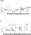
- the present disclosureprovides a method for spatially tagging nucleic acids of a biological specimen.
- the methodcan include steps of (a) providing a solid support comprising a plurality of different nucleic acid probes that are randomly located on the solid support, wherein the different nucleic acid probes each includes a barcode sequence that is different from the barcode sequence of other randomly located probes on the solid support; (b) performing a nucleic acid detection reaction on the solid support to locate the barcode sequences on the solid support; (c) contacting a biological specimen with the solid support that has the randomly located probes; (d) hybridizing the randomly located probes to target nucleic acids from portions of the biological specimen that are proximal to the randomly located probes; and (e) modifying the randomly located probes that are hybridized to the target nucleic acids, thereby producing modified probes that include the barcode sequences and a target specific modification, thereby spatially tagging the nucleic acids of the biological specimen.
- This disclosurefurther provides a method for spatially tagging nucleic acids of a biological specimen, the method including steps of (a) attaching different nucleic acid probes to a solid support to produce randomly located probes on the solid support, wherein the different nucleic acid probes each includes a barcode sequence, and wherein each of the randomly located probes includes different barcode sequences from other randomly located probes on the solid support; (b) performing a nucleic acid detection reaction on the solid support to determine the barcode sequences of the randomly located probes on the solid support; (c) contacting a biological specimen with the solid support that has the randomly located probes; (d) hybridizing the randomly located probes to target nucleic acids from portions of the biological specimen that are proximal to the randomly located probes; and (e) extending the randomly located probes to produce extended probes that include the barcode sequences and sequences from the target nucleic acids, thereby spatially tagging the nucleic acids of the biological specimen.
- a method for spatially tagging nucleic acids of a biological specimenincludes the steps of (a) providing a plurality of nucleic acid primers attached to a solid support, wherein the nucleic acid primers in the plurality include a universal primer sequence that is common to the nucleic acid primers in the plurality; (b) binding a population of nucleic acid probes to the plurality of nucleic acid primers, wherein the nucleic acid probes include a universal primer binding sequence that hybridizes to the universal primer sequence, a target capture sequence and a barcode sequence that differs from barcode sequences of other nucleic acid probes in the population, thereby attaching the different nucleic acid probes at randomly located positions on the solid support; (c) amplifying the different nucleic acid probes by extension of the nucleic acid primers, thereby producing nucleic acid clusters having copies of the barcode sequence and target capture sequence at the randomly located positions on the solid support; (d) performing a sequencing reaction to determine the barcode
- This disclosurefurther provides a method for spatially tagging nucleic acids of a biological specimen, the method including steps of (a) providing an array of beads on a solid support, wherein different nucleic acid probes are attached to different beads in the array, wherein the different nucleic acid probes each include a barcode sequence, wherein each bead includes a different barcode sequence from other beads on the solid support, and wherein each of the different nucleic acid probes includes a target capture sequence; (b) performing a decoder probe hybridization reaction on the solid support to determine the barcode sequences at the randomly located probes on the solid support; (c) contacting a biological specimen with the array of beads; (d) hybridizing the different nucleic acid probes to target nucleic acids from portions of the biological specimen that are proximal to the beads; and (e) extending the different nucleic acid probes to produce extended probes that include sequences from the target nucleic acids and the barcode sequences, thereby tagging the nucleic acids of the biological
- the present disclosureprovides compositions, apparatus and methods for preserving spatial information when performing multiplex nucleic acid analyses of biological specimens.
- a variety of toolsare available for multiplex nucleic acid analyses including, for example, nucleic acid microarrays and so-called "next generation” sequencing platforms.
- Such toolsallow for parallel detection of very large and complex collections of nucleic acids, including for example, DNA collections that represent all or nearly all of the genetic material of an organism (i.e. the 'genome'), RNA (or cDNA) collections that represent all or nearly all of the complement of expressed genes (i.e. the 'transcriptome') for an organism, and in some cases the collections can include several genomes and/or transcriptomes from several different organisms (e.g.
- a metabolome or biome from a community or ecosystemprovide a vast amount of information about what nucleic acid sequences are present in a biological specimen being evaluated, they do not inherently distinguish where any particular nucleic acid resided in the biological specimen. Indeed the vast majority of samples applied to multiplex nucleic acid analysis tools are homogenates derived from mixtures of many different cells from a biological specimen. As a result, spatial information is lost and the results obtained from these tools constitute an average transcriptome or average genome for the specimen, important differences between individual cells being lost.
- the present disclosureprovides new and useful modifications to existing multiplex nucleic acid analysis tools to allow for the preservation of spatial information for biological specimens from which the nucleic acids are obtained.
- solid supportsthat are usually used for multiplex sequencing-by-synthesis (SBS) techniques can be modified for use in capturing and spatially tagging nucleic acids from a biological specimen.
- arrays of beadssuch as those used for genotyping or gene expression analysis, can be used for capturing and spatially tagging nucleic acids from a biological specimen.
- the solid supports used for an SBS or BeadArrayTM platform commercialized by Illumina (San Diego, CA)can be modified for spatial tagging.
- any of a variety of solid supportscan be made and used in accordance with the teaching herein.
- the spatially tagged nucleic acidscan be removed from the solid support, pooled together and attached to a second solid support for detection in any of a variety of multiplex nucleic acid analysis systems including, for example, a sequencing platform or microarray platform set forth herein.
- the spatial information provided by a method, composition or apparatus hereincan include, for example, the location of one or more cells in a tissue (or other specimen) that has a particular allele at one or more locus (e.g. a genotype), has a particular structural variation in the genome (e.g. fusion, insertion, deletion, rearrangement etc.), has a particular epigenetic signature (e.g. methylation), expresses a particular gene, expresses a particular allele of a gene, expresses a particular splice variant of a gene or the like.
- a method, composition or apparatus of the present disclosurecan be used to quantify one or more nucleic acids according to spatial location.
- the spatial information for one or more cells in a tissuecan include the amount of a particular allele or chromosomal region in a genome (e.g. ploidy); the amount of epigenetic modification of a genetic locus (e.g. methylation); expression level for a particular gene, allele or splice variant; or the like.
- the amountscan be absolute amounts or relative amounts in accordance with similar measurements obtained in the art for mixed or non-spatially tagged samples.
- a method set forth hereincan be used for localized detection of a nucleic acid in a biological specimen.
- a methodcan be used for identifying or characterizing all of the transcriptome or genome of a biological specimen.
- a methodcan be used to identify or characterize only a part of a specimen's transcriptome or genome.
- a subset of transcripts or genes evaluated in a method hereincan be related to a particular disease or condition.
- a method set forth hereincan be used for localized or spatial detection of nucleic acids, whether DNA or RNA, in a biological specimen.
- one or more RNA or DNA moleculescan be located with respect to its native position or location within a cell or tissue or other biological specimen.
- one or more nucleic acidscan be localized to a cell or group of adjacent cells, or type of cell, or to particular regions of areas within a tissue sample.
- the native location or position of individual RNA or DNA moleculescan be determined using a method, apparatus or composition of the present disclosure.
- ampliconwhen used in reference to a nucleic acid, means the product of copying the nucleic acid, wherein the product has a nucleotide sequence that is the same as or complementary to at least a portion of the nucleotide sequence of the nucleic acid.
- An ampliconcan be produced by any of a variety of amplification methods that use the nucleic acid, or an amplicon thereof, as a template including, for example, polymerase extension, polymerase chain reaction (PCR), rolling circle amplification (RCA), multiple displacement amplification (MDA), ligation extension, or ligation chain reaction.
- An ampliconcan be a nucleic acid molecule having a single copy of a particular nucleotide sequence (e.g. a PCR product) or multiple copies of the nucleotide sequence (e.g. a concatameric product of RCA).
- a first amplicon of a target nucleic acidis typically a complementary copy.
- Subsequent ampliconsare copies that are created, after generation of the first amplicon, from the target nucleic acid or from the first amplicon.
- a subsequent ampliconcan have a sequence that is substantially complementary to the target nucleic acid or substantially identical to the target nucleic acid.
- the term "array”refers to a population of features or sites that can be differentiated from each other according to relative location. Different molecules that are at different sites of an array can be differentiated from each other according to the locations of the sites in the array.
- An individual site of an arraycan include one or more molecules of a particular type. For example, a site can include a single target nucleic acid molecule having a particular sequence or a site can include several nucleic acid molecules having the same sequence (and/or complementary sequence, thereof).
- the sites of an arraycan be different features located on the same substrate. Exemplary features include without limitation, wells in a substrate, beads (or other particles) in or on a substrate, projections from a substrate, ridges on a substrate or channels in a substrate.
- the sites of an arraycan be separate substrates each bearing a different molecule. Different molecules attached to separate substrates can be identified according to the locations of the substrates on a surface to which the substrates are associated or according to the locations of the substrates in a liquid or gel. Exemplary arrays in which separate substrates are located on a surface include, without limitation, those having beads in wells.
- an analytesuch as a nucleic acid
- a materialsuch as a gel or solid support
- a covalent bondis characterized by the sharing of pairs of electrons between atoms.
- a non-covalent bondis a chemical bond that does not involve the sharing of pairs of electrons and can include, for example, hydrogen bonds, ionic bonds, van der Waals forces, hydrophilic interactions and hydrophobic interactions.
- barcode sequenceis intended to mean a series of nucleotides in a nucleic acid that can be used to identify the nucleic acid, a characteristic of the nucleic acid, or a manipulation that has been carried out on the nucleic acid.
- the barcode sequencecan be a naturally occurring sequence or a sequence that does not occur naturally in the organism from which the barcoded nucleic acid was obtained.
- a barcode sequencecan be unique to a single nucleic acid species in a population or a barcode sequence can be shared by several different nucleic acid species in a population. For example, each nucleic acid probe in a population can include different barcode sequences from all other nucleic acid probes in the population.
- each nucleic acid probe in a populationcan include different barcode sequences from some or most other nucleic acid probes in a population.
- each probe in a populationcan have a barcode that is present for several different probes in the population even though the probes with the common barcode differ from each other at other sequence regions along their length.
- one or more barcode sequences that are used with a biological specimenare not present in the genome, transcriptome or other nucleic acids of the biological specimen.
- barcode sequencescan have less than 80%, 70%, 60%, 50% or 40% sequence identity to the nucleic acid sequences in a particular biological specimen.
- biological specimenis intended to mean one or more cell, tissue, organism or portion thereof.
- a biological specimencan be obtained from any of a variety of organisms. Exemplary organisms include, but are not limited to, a mammal such as a rodent, mouse, rat, rabbit, guinea pig, ungulate, horse, sheep, pig, goat, cow, cat, dog, primate (i.e.
- a plantsuch as Arabidopsis thaliana, corn, sorghum, oat, wheat, rice, canola, or soybean; an algae such as Chlamydomonas reinhardtii; a nematode such as Caenorhabditis elegans; an insect such as Drosophila melanogaster, mosquito, fruit fly, honey bee or spider; a fish such as zebrafish; a reptile; an amphibian such as a frog or Xenopus laevis; a Dictyostelium discoideum; a fungi such as Pneumocystis carinii, Takifugu rubripes, yeast, Saccharamoyces cerevisiae or Schizosaccharomyces pombe; or a Plasmodium falciparum.
- a plantsuch as Arabidopsis thaliana, corn, sorghum, oat, wheat, rice, canola, or soybean;
- Target nucleic acidscan also be derived from a prokaryote such as a bacterium, Escherichia coli, Staphylococci or Mycoplasma pneumoniae; an archae; a virus such as Hepatitis C virus or human immunodeficiency virus; or a viroid.
- Specimenscan be derived from a homogeneous culture or population of the above organisms or alternatively from a collection of several different organisms, for example, in a community or ecosystem.
- cleavage siteis intended to mean a location in a nucleic acid molecule that is susceptible to bond breakage.
- the locationcan be specific to a particular chemical, enzymatic or physical process that results in bond breakage.
- the locationcan be a nucleotide that is abasic or a nucleotide that has a base that is susceptible to being removed to create an abasic site. Examples of nucleotides that are susceptible to being removed include uracil and 8-oxo-guanine as set forth in further detail herein below.
- the locationcan also be at or near a recognition sequence for a restriction endonuclease such as a nicking enzyme.
- the term "cluster,” when used in reference to nucleic acids,refers to a population of the nucleic acids that is attached to a solid support to form a feature or site.
- the nucleic acidsare generally members of a single species, thereby forming a monoclonal cluster.
- a "monoclonal population" of nucleic acidsis a population that is homogeneous with respect to a particular nucleotide sequence. Clusters need not be monoclonal. Rather, for some applications, a cluster can be predominantly populated with amplicons from a first nucleic acid and can also have a low level of contaminating amplicons from a second nucleic acid.
- an acceptable level of contaminationwould be a level that does not impact signal to noise or resolution of the detection technique in an unacceptable way. Accordingly, apparent clonality will generally be relevant to a particular use or application of an array made by the methods set forth herein.
- Exemplary levels of contamination that can be acceptable at an individual clusterinclude, but are not limited to, at most 0.1%, 0.5%, 1%, 5%, 10%, 5 25%, or 35% contaminating amplicons.
- the nucleic acids in a clusterare generally covalently attached to a solid support, for example, via their 5' ends, but in some cases other attachment means are possible.
- the nucleic acids in a clustercan be single stranded or double stranded.
- clustersare made by a solid-phase amplification method known as bridge amplification.
- Exemplary configurations for clusters and methods for their productionare set forth, for example, in U.S. Pat. No. 5,641,658 ; U.S. Patent Publ. No. 2002/0055100 ; U.S. Pat. No. 7,115,400 ; U.S. Patent Publ. No. 2004/0096853 ; U.S. Patent Publ. No. 2004/0002090 ; U.S. Patent Publ. No. 2007/0128624 ; and U.S. Patent Publ. No. 2008/0009420 , each of which is incorporated herein by reference.
- nucleic acidsAs used herein, the term "different", when used in reference to nucleic acids, means that the nucleic acids have nucleotide sequences that are not the same as each other. Two or more nucleic acids can have nucleotide sequences that are different along their entire length. Alternatively, two or more nucleic acids can have nucleotide sequences that are different along a substantial portion of their length. For example, two or more nucleic acids can have target nucleotide sequence portions that are different for the two or more molecules while also having a universal sequence portion that is the same on the two or more molecules. Two beads can be different from each other by virtue of being attached to different nucleic acids.
- eachwhen used in reference to a collection of items, is intended to identify an individual item in the collection but does not necessarily refer to every item in the collection. Exceptions can occur if explicit disclosure or context clearly dictates otherwise.
- the term "extend,” when used in reference to a nucleic acid,is intended to mean addition of at least one nucleotide or oligonucleotide to the nucleic acid.
- one or more nucleotidescan be added to the 3' end of a nucleic acid, for example, via polymerase catalysis (e.g. DNA polymerase, RNA polymerase or reverse transcriptase). Chemical or enzymatic methods can be used to add one or more nucleotide to the 3' or 5' end of a nucleic acid.
- One or more oligonucleotidescan be added to the 3' or 5' end of a nucleic acid, for example, via chemical or enzymatic (e.g. ligase catalysis) methods.
- a nucleic acidcan be extended in a template directed manner, whereby the product of extension is complementary to a template nucleic acid that is hybridized to the nucleic acid that is extended.
- featuremeans a location in an array for a particular species of molecule.
- a featurecan contain only a single molecule or it can contain a population of several molecules of the same species.
- Features of an arrayare typically discrete. The discrete features can be contiguous or they can have spaces between each other. The size of the features and/or spacing between the features can vary such that arrays can be high density, medium density or lower density. High density arrays are characterized as having sites separated by less than about 15 ⁇ m. Medium density arrays have sites separated by about 15 to 30 ⁇ m, while low density arrays have sites separated by greater than 30 ⁇ m.
- An array useful hereincan have, for example, sites that are separated by less than 100 ⁇ m, 50 ⁇ m, 10 ⁇ m, 5 ⁇ m, 1 ⁇ m, or 0.5 ⁇ m.
- An apparatus or method of the present disclosurecan be used to detect an array at a resolution sufficient to distinguish sites at the above densities or density ranges.
- the term "fluidic mixture”is intended to mean two or more different items that are simultaneously present in a solution. Typically, the two or more items are freely diffusible in the solution.
- the two or more itemscan be different types of items (e.g. a nucleic acid and a protein which are different types of molecules) or they can be different species of the same type of items (e.g. two nucleic acid molecules having different sequences).
- Exemplary items that can be in a fluidic mixtureinclude, but are not limited to, molecules, cells or beads.
- the term "flow cell”is intended to mean a vessel having a chamber where a reaction can be carried out, an inlet for delivering reagents to the chamber and an outlet for removing reagents from the chamber.
- the chamberis configured for detection of the reaction that occurs in the chamber.
- the chambercan include one or more transparent surfaces allowing optical detection of biological specimens, optically labeled molecules, or the like in the chamber.
- Exemplary flow cellsinclude, but are not limited to those used in a nucleic acid sequencing apparatus such as flow cells for the Genome Analyzer®, MiSeq®, NextSeq® or HiSeq® platforms commercialized by Illumina, Inc.
- gelis intended to mean a semi-rigid material that is permeable to liquids and gases. Typically, gel material can swell when liquid is taken up and can contract when liquid is removed by drying.
- Exemplary gelsinclude, but are not limited to those having a colloidal structure, such as agarose; polymer mesh structure, such as gelatin; or cross-linked polymer structure, such as polyacrylamide, SFA (see, for example, US Pat. App. Pub. No. 2011/0059865 A1 , which is incorporated herein by reference) or PAZAM (see, for example, US Pat. App. Publ. No. 2014/0079923 A1 , which is incorporated herein by reference).
- Particularly useful gel materialwill conform to the shape of a well or other concave feature where it resides.
- nucleic acidand “nucleotide” are intended to be consistent with their use in the art and to include naturally occurring species or functional analogs thereof. Particularly useful functional analogs of nucleic acids are capable of hybridizing to a nucleic acid in a sequence specific fashion or capable of being used as a template for replication of a particular nucleotide sequence.
- Naturally occurring nucleic acidsgenerally have a backbone containing phosphodiester bonds. An analog structure can have an alternate backbone linkage including any of a variety of those known in the art.
- Naturally occurring nucleic acidsgenerally have a deoxyribose sugar (e.g. found in deoxyribonucleic acid (DNA)) or a ribose sugar (e.g.
- a nucleic acidcan contain nucleotides having any of a variety of analogs of these sugar moieties that are known in the art.
- a nucleic acidcan include native or non-native nucleotides.
- a native deoxyribonucleic acidcan have one or more bases selected from the group consisting of adenine, thymine, cytosine or guanine and a ribonucleic acid can have one or more bases selected from the group consisting of uracil, adenine, cytosine or guanine.
- Useful non-native bases that can be included in a nucleic acid or nucleotideare known in the art.
- probeor “target,” when used in reference to a nucleic acid or sequence of a nucleic acid, are intended as semantic identifiers for the nucleic acid or sequence in the context of a method or composition set forth herein and does not necessarily limit the structure or function of the nucleic acid or sequence beyond what is otherwise explicitly indicated.
- probe and targetcan be similarly applied to other analytes such as proteins, small molecules, cells or the like.
- the term "pitch,” when used in reference to features of an array,is intended to refer to the center-to-center spacing for adjacent features.
- a pattern of featurescan be characterized in terms of average pitch. The pattern can be ordered such that the coefficient of variation around the average pitch is small or the pattern can be random in which case the coefficient of variation can be relatively large.
- the average pitchcan be, for example, at least about 10 nm, 0.1 ⁇ m, 0.5 ⁇ m, 1 ⁇ m, 5 ⁇ m, 10 ⁇ m, 100 ⁇ m or more. Alternatively or additionally, the average pitch can be, for example, at most about 100 ⁇ m, 10 ⁇ m, 5 ⁇ m, 1 ⁇ m, 0.5 ⁇ m 0.1 ⁇ m or less.
- the average pitch for a particular pattern of featurescan be between one of the lower values and one of the upper values selected from the ranges above.
- poly T or poly Awhen used in reference to a nucleic acid sequence, is intended to mean a series of two or more thiamine (T) or adenine (A) bases, respectively.
- a poly T or poly Acan include at least about 2, 5, 8, 10, 12, 15, 18, 20 or more of the T or A bases, respectively.
- a poly T or poly Acan include at most about, 30, 20, 18, 15, 12, 10, 8, 5 or 2 of the T or A bases, respectively.
- the term "random"can be used to refer to the spatial arrangement or composition of locations on a surface.
- the firstrelating to the spacing and relative location of features (also called “sites") and the second relating to identity or predetermined knowledge of the particular species of molecule that is present at a particular feature.
- features of an arraycan be randomly spaced such that nearest neighbor features have variable spacing between each other.
- the spacing between featurescan be ordered, for example, forming a regular pattern such as a rectilinear grid or hexagonal grid.
- features of an arraycan be random with respect to the identity or predetermined knowledge of the species of analyte (e.g.
- nucleic acid of a particular sequencethat occupies each feature independent of whether spacing produces a random pattern or ordered pattern.
- An array set forth hereincan be ordered in one respect and random in another.
- a surfaceis contacted with a population of nucleic acids under conditions where the nucleic acids attach at sites that are ordered with respect to their relative locations but 'randomly located' with respect to knowledge of the sequence for the nucleic acid species present at any particular site.
- Reference to "randomly distributing" nucleic acids at locations on a surfaceis intended to refer to the absence of knowledge or absence of predetermination regarding which nucleic acid will be captured at which location (regardless of whether the locations are arranged in an ordered pattern or not).
- solid supportrefers to a rigid substrate that is insoluble in aqueous liquid.
- the substratecan be non-porous or porous.
- the substratecan optionally be capable of taking up a liquid (e.g. due to porosity) but will typically be sufficiently rigid that the substrate does not swell substantially when taking up the liquid and does not contract substantially when the liquid is removed by drying.
- a nonporous solid supportis generally impermeable to liquids or gases.
- Exemplary solid supportsinclude, but are not limited to, glass and modified or functionalized glass, plastics (including acrylics, polystyrene and copolymers of styrene and other materials, polypropylene, polyethylene, polybutylene, polyurethanes, TeflonTM, cyclic olefins, polyimides etc.), nylon, ceramics, resins, Zeonor, silica or silica-based materials including silicon and modified silicon, carbon, metals, inorganic glasses, optical fiber bundles, and polymers. Particularly useful solid supports for some embodiments are located within a flow cell apparatus. Exemplary flow cells are set forth in further detail herein.
- the term "spatial tag"is intended to mean a nucleic acid having a sequence that is indicative of a location.
- the nucleic acidis a synthetic molecule having a sequence that is not found in one or more biological specimen that will be used with the nucleic acid.
- the nucleic acid moleculecan be naturally derived or the sequence of the nucleic acid can be naturally occurring, for example, in a biological specimen that is used with the nucleic acid.
- the location indicated by a spatial tagcan be a location in or on a biological specimen, in or on a solid support or a combination thereof.
- a barcode sequencecan function as a spatial tag.
- tissueis intended to mean an aggregation of cells, and, optionally, intercellular matter. Typically the cells in a tissue are not free floating in solution and instead are attached to each other to form a multicellular structure. Exemplary tissue types include muscle, nerve, epidermal and connective tissues.
- the term "universal sequence”refers to a series of nucleotides that is common to two or more nucleic acid molecules even if the molecules also have regions of sequence that differ from each other.
- a universal sequence that is present in different members of a collection of moleculescan allow capture of multiple different nucleic acids using a population of universal capture nucleic acids that are complementary to the universal sequence.
- a universal sequence present in different members of a collection of moleculescan allow the replication or amplification of multiple different nucleic acids using a population of universal primers that are complementary to the universal sequence.
- a universal capture nucleic acid or a universal primerincludes a sequence that can hybridize specifically to a universal sequence.
- Target nucleic acid moleculesmay be modified to attach universal adapters, for example, at one or both ends of the different target sequences.
- the present disclosureprovides a method for spatially tagging nucleic acids of a biological specimen.
- the methodcan include the steps of (a) attaching different nucleic acid probes to a solid support to produce randomly located probes on the solid support, wherein the different nucleic acid probes each includes a barcode sequence, and wherein each of the randomly located probes includes different barcode sequences from other randomly located probes on the solid support; (b) performing a nucleic acid detection reaction on the solid support to determine the barcode sequences of the randomly located probes on the solid support; (c) contacting a biological specimen with the solid support that has the randomly located probes; (d) hybridizing the randomly located probes to target nucleic acids from portions of the biological specimen that are proximal to the randomly located probes; and (e) extending the randomly located probes to produce extended probes that include the barcode sequences and sequences from the target nucleic acids, thereby spatially tagging the nucleic acids of the biological specimen.
- solid supportscan be used in a method, composition or apparatus of the present disclosure.
- Particularly useful solid supportsare those used for nucleic acid arrays. Examples include glass, modified glass, functionalized glass, inorganic glasses, microspheres (e.g. inert and/or magnetic particles), plastics, polysaccharides, nylon, nitrocellulose, ceramics, resins, silica, silica-based materials, carbon, metals, an optical fiber or optical fiber bundles, polymers and multiwell (e.g. microtiter) plates.
- Exemplary plasticsinclude acrylics, polystyrene, copolymers of styrene and other materials, polypropylene, polyethylene, polybutylene, polyurethanes and TeflonTM.
- Exemplary silica-based materialsinclude silicon and various forms of modified silicon.
- a solid supportcan be within or part of a vessel such as a well, tube, channel, cuvette, Petri plate, bottle or the like.
- a particularly useful vesselis a flow-cell, for example, as described in WO 2014/142841 A1 ; U.S. Pat. App. Pub. No. 2010/0111768 A1 and U.S. Pat. No. 8,951,781 or Bentley et al., Nature 456:53-59 (2008 ), each of which is incorporated herein by reference.
- Exemplary flow-cellsare those that are commercially available from Illumina, Inc.
- a sequencing platformsuch as a Genome Analyzer®, MiSeq®, NextSeq® or HiSeq® platform.
- a sequencing platformsuch as a Genome Analyzer®, MiSeq®, NextSeq® or HiSeq® platform.
- Another particularly useful vesselis a well in a multiwell plate or microtiter plate.
- a solid supportcan include a gel coating. Attachment of nucleic acids to a solid support via a gel is exemplified by flow cells available commercially from Illumina Inc. (San Diego, CA) or described in US Pat. App. Pub. Nos. 2011/0059865 A1 , 2014/0079923 A1 , or 2015/0005447 A1 ; or PCT Publ. No. WO 2008/093098 , each of which is incorporated herein by reference.
- Exemplary gelsthat can be used in the methods and apparatus set forth herein include, but are not limited to, those having a colloidal structure, such as agarose; polymer mesh structure, such as gelatin; or cross-linked polymer structure, such as polyacrylamide, SFA (see, for example, US Pat. App. Pub. No. 2011/0059865 A1 , which is incorporated herein by reference) or PAZAM (see, for example, US Pat. App. Publ. Nos. 2014/0079923 A1 , or 2015/0005447 A1 , each of which is incorporated herein by reference).
- a colloidal structuresuch as agarose
- polymer mesh structuresuch as gelatin
- cross-linked polymer structuresuch as polyacrylamide, SFA
- SFAsee, for example, US Pat. App. Pub. No. 2011/0059865 A1 , which is incorporated herein by reference
- PAZAMsee, for example, US Pat. App. Publ. Nos. 2014/0079923 A1
- a solid supportcan be configured as an array of features to which nucleic acids can be attached.
- the featurescan be present in any of a variety of desired formats.
- the featurescan be wells, pits, channels, ridges, raised regions, pegs, posts or the like.
- the featurescan contain beads.
- the featuresneed not contain a bead or particle.
- Exemplary featuresinclude wells that are present in substrates used for commercial sequencing platforms sold by 454 LifeSciences (a subsidiary of Roche, Basel Switzerland) or Ion Torrent (a subsidiary of Life Technologies, Carlsbad California).
- Other substrates having wellsinclude, for example, etched fiber optics and other substrates described in US Pat Nos.
- wells of a substratecan include gel material (with or without beads) as set forth in US Pat. App. Publ. No. 2014/0243224 A1 , which is incorporated herein by reference.
- the features on a solid supportcan be metal features on a non-metallic surface such as glass, plastic or other materials exemplified above.
- a metal layercan be deposited on a surface using methods known in the art such as wet plasma etching, dry plasma etching, atomic layer deposition, ion beam etching, chemical vapor deposition, vacuum sputtering or the like. Any of a variety of commercial instruments can be used as appropriate including, for example, the FlexAL®, OpAL®, lonfab 300plus®, or Optofab 3000® systems (Oxford Instruments, UK).
- a metal layercan also be deposited by e-beam evaporation or sputtering as set forth in Thornton, Ann. Rev. Mater. Sci.
- Metal layer deposition techniquessuch as those exemplified above, can be combined with photolithography techniques to create metal regions or patches on a surface. Exemplary methods for combining metal layer deposition techniques and photolithography techniques are provided in US Pat. No. 8,895,249 or US Pat App. Pub. No. 2014/0243224 A1 , each of which is incorporated herein by reference.
- Featurescan appear on a solid support as a grid of spots or patches.
- the featurescan be located in a repeating pattern or in an irregular, non-repeating pattern.
- Particularly useful repeating patternsare hexagonal patterns, rectilinear patterns, grid patterns, patterns having reflective symmetry, patterns having rotational symmetry, or the like.
- Asymmetric patternscan also be useful.
- the pitchcan be the same between different pairs of nearest neighbor features or the pitch can vary between different pairs of nearest neighbor features.
- High density arraysare characterized as having average pitch of less than about 15 ⁇ m.
- Medium density arrayshave average pitch of about 15 to 30 ⁇ m, while low density arrays have average pitch greater than 30 ⁇ m.
- An array useful in the inventioncan have average pitch that is less than 100 ⁇ m, 50 ⁇ m, 10 ⁇ m, 5 ⁇ m, 1 ⁇ m or 0.5 ⁇ m.
- the average pitch values and ranges set forth above or elsewhere hereinare intended to be applicable to ordered arrays or random arrays.
- features on a solid supportcan each have an area that is larger than about 100 nm 2 , 250 nm 2 , 500 nm 2 , 1 ⁇ m 2 , 2.5 ⁇ m 2 , 5 ⁇ m 2 , 10 ⁇ m 2 ,100 ⁇ m 2 ,or 500 ⁇ m 2 .
- featurescan each have an area that is smaller than about 1 mm 2 , 500 ⁇ m 2 ,100 ⁇ m 2 , 25 ⁇ m2, 10 ⁇ m 2 , 5 ⁇ m 2 ,1 ⁇ m 2 , 500 nm 2 , or 100 nm 2 .
- the above rangescan describe the apparent area of a bead or other particle on a solid support when viewed or imaged from above.
- a solid supportcan include a collection of beads or other particles.
- the particlescan be suspended in a solution or they can be located on the surface of a substrate.
- arrays having beads located on a surfaceinclude those wherein beads are located in wells such as a BeadChip array (Illumina Inc., San Diego CA), substrates used in sequencing platforms from 454 LifeSciences (a subsidiary of Roche, Basel Switzerland) or substrates used in sequencing platforms from Ion Torrent (a subsidiary of Life Technologies, Carlsbad California).
- Other solid supports having beads located on a surfaceare described in US Pat. Nos.
- the beadscan be made to include universal primers, and the beads can then be loaded onto an array, thereby forming universal arrays for use in a method set forth herein.
- the solid supports typically used for bead arrayscan be used without beads.
- nucleic acids, such as probes or primerscan be attached directly to the wells or to gel material in wells.
- a solid support used in a method set forth hereincan include an array of beads, wherein different nucleic acid probes are attached to different beads in the array.
- each beadcan be attached to a different nucleic acid probe and the beads can be randomly distributed on the solid support in order to effectively attach the different nucleic acid probes to the solid support.
- the solid supportcan include wells having dimensions that accommodate no more than a single bead.
- the beadsmay be attached to the wells due to forces resulting from the fit of the beads in the wells. It is also possible to use attachment chemistries or adhesives to hold the beads in the wells.
- Nucleic acid probes that are attached to beadscan include barcode sequences.
- a population of the beadscan be configured such that each bead is attached to only one type of barcode and many different beads each with a different barcode are present in the population.
- randomly distributing the beads to a solid supportwill result in randomly locating the nucleic acid probes (and their respective barcode sequences) on the solid support.
- there can be multiple beads with the same barcode sequencesuch that there is redundancy in the population. Randomly distributing a redundant population of beads on a solid support that has a capacity that is greater than the number of unique barcodes in the bead population will result in redundancy of barcodes on the solid support.
- the number of different barcodes in a population of beadscan exceed the capacity of the solid support in order to produce an array that is not redundant with respect to the population of barcodes on the solid support.
- the capacity of the solid supportwill be determined in some embodiments by the number of features (e.g. single-bead occupancy wells) that attach or otherwise accommodate a bead.
- a solid supportcan include, or can be made by the methods set forth herein to attach, a plurality of different nucleic acid probes.
- a solid supportcan include at least 10, 100, 1 x 10 3 , 1 x 10 4 , 1 x 10 5 , 1 x 10 6 , 1 x 10 7 , 1 x 10 8 , 1 x 10 9 or more different probes.
- a solid supportcan include at most 1 x 10 9 , 1 x 10 8 , 1 x 10 7 , 1 x 10 6 , 1 x 10 5 , 1 x 10 4 , 1 x 10 3 , 100, or fewer different probes.
- each of the different probescan be present in several copies, for example, when the probes have been amplified to form a cluster.
- the above rangescan describe the number of different nucleic acid clusters on a solid support. It will also be understood that the above ranges can describe the number of different barcodes, target capture sequences, or other sequence elements set forth herein as being unique to particular nucleic acid probes. Alternatively or additionally, the ranges can describe the number of extended probes or modified probes created on a solid support using a method set forth herein.
- nucleic acid probesmay be present on a solid support prior to contacting the solid support with nucleic acid probes.
- the primerscan be attached at the features, whereas interstitial areas outside of the features substantially lack any of the primers.
- Nucleic acid probescan be captured at preformed features on a solid support, and optionally amplified on the solid support, using methods set forth in US Pat. No. 8,895,249 , US Pat. No. 8,778,849 , or US Pat App. Pub. No. 2014/0243224 A1 , each of which is incorporated herein by reference.
- a solid supportmay have a lawn of primers or may otherwise lack features.
- a featurecan be formed by virtue of attachment of a nucleic acid probe on the solid support.
- the captured nucleic acid probecan be amplified on the solid support such that the resulting cluster becomes a feature.
- attachmentis exemplified above as capture between a primer and a complementary portion of a probe, it will be understood that capture moieties other than primers can be present at pre-formed features or as a lawn.
- Other exemplary capture moietiesinclude, but are not limited to, chemical moieties capable of reacting with a nucleic acid probe to create a covalent bond or receptors capable of biding non-covalently to a ligand on a nucleic acid probe.
- a step of attaching nucleic acid probes to a solid supportcan be carried out by providing a fluid that contains a mixture of different nucleic acid probes and contacting this fluidic mixture with the solid support.
- the contactcan result in the fluidic mixture being in contact with a surface to which many different nucleic acid probes from the fluidic mixture will attach.
- the probeshave random access to the surface (whether the surface has pre-formed features configured to attach the probes or a uniform surface configured for attachment). Accordingly, the probes can be randomly located on the solid support.
- the total number and variety of different probes that end up attached to a surfacecan be selected for a particular application or use.
- the number of different probe speciescan exceed the occupancy of the solid support for probes.
- the number and variety of different probes that attach to the solid supportcan be equivalent to the probe occupancy of the solid support.
- the number and variety of different probe species on the solid supportcan be less than the occupancy (i.e. there will be redundancy of probe species such that the solid support may contain multiple features having the same probe species). Such redundancy can be achieved, for example, by contacting the solid support with a fluidic mixture that contains a number and variety of probe species that is substantially lower than the probe occupancy of the solid support.
- Attachment of the nucleic acid probescan be mediated by hybridization of the nucleic acid probes to complementary primers that are attached to the solid support, chemical bond formation between a reactive moiety on the nucleic acid probe and the solid support (examples are set forth in US Pat. No. 8,895,249 , US Pat. No. 8,778,849 , or US Pat App. Pub. No. 2014/0243224 A1 , each of which is incorporated herein by reference), affinity interactions of a moiety on the nucleic acid probe with a solid support-bound moiety (e.g.
- nucleic acid probese.g. hydrogen bonding, ionic forces, van der Waals forces and the like, or other interactions known in the art to attach nucleic acids to surfaces.
- attachment of a nucleic acid probeis non-specific with regard to any sequence differences between the nucleic acid probe and other nucleic acid probes that are or will be attached to the solid support.
- different probescan have a universal sequence that complements surface-attached primers or the different probes can have a common moiety that mediates attachment to the surface.
- each of the different probes (or a subpopulation of different probes)can have a unique sequence that complements a unique primer on the solid support or they can have a unique moiety that interacts with one or more different reactive moiety on the solid support.
- the unique primers or unique moietiescan, optionally, be attached at predefined locations in order to selectively capture particular probes, or particular types of probes, at the respective predefined locations.
- One or more features on a solid supportcan each include a single molecule of a particular probe.
- the featurescan be configured, in some embodiments, to accommodate no more than a single nucleic acid probe molecule. However, whether or not the feature can accommodate more than one nucleic acid probe molecule, the feature may nonetheless include no more than a single nucleic acid probe molecule.
- an individual featurecan include a plurality of nucleic acid probe molecules, for example, an ensemble of nucleic acid probe molecules having the same sequence as each other.
- the ensemblecan be produced by amplification from a single nucleic acid probe template to produce amplicons, for example, as a cluster attached to the surface.
- a method set forth hereincan use any of a variety of amplification techniques.
- Exemplary techniques that can be usedinclude, but are not limited to, polymerase chain reaction (PCR), rolling circle amplification (RCA), multiple displacement amplification (MDA), or random prime amplification (RPA).
- PCRpolymerase chain reaction
- RCArolling circle amplification
- MDAmultiple displacement amplification
- RPArandom prime amplification
- the amplificationcan be carried out in solution, for example, when features of an array are capable of containing amplicons in a volume having a desired capacity.
- an amplification technique used in a method of the present disclosurewill be carried out on solid phase.
- one or more primer speciese.g. universal primers for one or more universal primer binding site present in a nucleic acid probe
- one or both of the primers used for amplificationcan be attached to a solid support (e.g. via a gel).
- a solid supporte.g. via a gel
- Formats that utilize two species of primers attached to a solid supportare often referred to as bridge amplification because double stranded amplicons form a bridge-like structure between the two surface attached primers that flank the template sequence that has been copied.
- Exemplary reagents and conditions that can be used for bridge amplificationare described, for example, in U.S. Pat. Nos. 5,641,658 , 7,115,400 , or 8,895,249 ; or U.S. Pat. Publ. Nos.
- Solid-phase PCR amplificationcan also be carried out with one of the amplification primers attached to a solid support and the second primer in solution.
- An exemplary format that uses a combination of a surface attached primer and soluble primeris the format used in emulsion PCR as described, for example, in Dressman et al., Proc. Natl. Acad. Sci. USA 100:8817-8822 (2003 ), WO 05/010145 , or U.S. Pat. App. Publ. Nos.
- Emulsion PCRis illustrative of the format and it will be understood that for purposes of the methods set forth herein the use of an emulsion is optional and indeed for several embodiments an emulsion is not used.
- RCA techniquescan be modified for use in a method of the present disclosure.
- Exemplary components that can be used in an RCA reaction and principles by which RCA produces ampliconsare described, for example, in Lizardi et al., Nat. Genet. 19:225-232 (1998 ) and US Pat. App. Publ. No. 2007/0099208 A1 , each of which is incorporated herein by reference.
- Primers used for RCAcan be in solution or attached to a solid support.
- the primerscan be one or more of the universal primers described herein.
- MDA techniquescan be modified for use in a method of the present disclosure. Some basic principles and useful conditions for MDA are described, for example, in Dean et al., Proc Natl. Acad. Sci. USA 99:5261-66 (2002 ); Lü et al., Genome Research 13:294-307 (2003 ); Walker et al., Molecular Methods for Virus Detection, Academic Press, Inc., 1995 ; Walker et al., Nucl. Acids Res. 20:1691-96 (1992 ); US 5,455,166 ; US 5,130,238 ; and US 6,214,587 , each of which is incorporated herein by reference.
- Primers used for MDAcan be in solution or attached to a solid support at an amplification site. Again, the primers can be one or more of the universal primers described herein.
- a combination of the above-exemplified amplification techniquescan be used.
- RCA and MDAcan be used in a combination wherein RCA is used to generate a concatameric amplicon in solution (e.g. using solution-phase primers).
- the ampliconcan then be used as a template for MDA using primers that are attached to a solid support (e.g. universal primers).
- primerse.g. universal primers
- Nucleic acid probes that are used in a method set forth herein or present in an apparatus or composition of the present disclosurecan include barcode sequences, and for embodiments that include a plurality of different nucleic acid probes, each of the probes can include a different barcode sequence from other probes in the plurality.
- Barcode sequencescan be any of a variety of lengths. Longer sequences can generally accommodate a larger number and variety of barcodes for a population. Generally, all probes in a plurality will have the same length barcode (albeit with different sequences), but it is also possible to use different length barcodes for different probes.
- a barcode sequencecan be at least 2, 4, 6, 8, 10, 12, 15, 20 or more nucleotides in length.
- the length of the barcode sequencecan be at most 20, 15, 12, 10, 8, 6, 4 or fewer nucleotides. Examples of barcode sequences that can be used are set forth, for example in, US Pat. App. Publ. No. 2014/0342921 A1 and US Pat. No. 8,460,865 , each of which is incorporated herein by reference.
- a method of the present disclosurecan include a step of performing a nucleic acid detection reaction on a solid support to determine barcode sequences of nucleic acid probes that are located on the solid support.
- the probesare randomly located on the solid support and the nucleic acid detection reaction provides information to locate each of the different probes.
- nucleic acid detection methodsinclude, but are not limited to nucleic acid sequencing of a probe, hybridization of nucleic acids to a probe, ligation of nucleic acids that are hybridized to a probe, extension of nucleic acids that are hybridized to a probe, extension of a first nucleic acid that is hybridized to a probe followed by ligation of the extended nucleic acid to a second nucleic acid that is hybridized to the probe, or other methods known in the art such as those set forth in US Pat. No. 8,288,103 or 8,486,625 , each of which is incorporated herein by reference.
- Sequencing techniquessuch as sequencing-by-synthesis (SBS) techniques, are a particularly useful method for determining barcode sequences.
- SBScan be carried out as follows. To initiate a first SBS cycle, one or more labeled nucleotides, DNA polymerase, SBS primers etc., can be contacted with one or more features on a solid support (e.g. feature(s) where nucleic acid probes are attached to the solid support). Those features where SBS primer extension causes a labeled nucleotide to be incorporated can be detected.
- the nucleotidescan include a reversible termination moiety that terminates further primer extension once a nucleotide has been added to the SBS primer.
- a nucleotide analog having a reversible terminator moietycan be added to a primer such that subsequent extension cannot occur until a deblocking agent is delivered to remove the moiety.
- a deblocking reagentcan be delivered to the solid support (before or after detection occurs). Washes can be carried out between the various delivery steps. The cycle can then be repeated n times to extend the primer by n nucleotides, thereby detecting a sequence of length n.
- Pyrosequencingdetects the release of inorganic pyrophosphate (PPi) as particular nucleotides are incorporated into a nascent nucleic acid strand ( Ronaghi, et al., Analytical Biochemistry 242(1), 84-9 (1996 ); Ronaghi, Genome Res. 11(1), 3-11 (2001 ); Ronaghi et al. Science 281(5375), 363 (1998 ); or US Pat. Nos. 6,210,891 , 6,258,568 or 6,274,320 , each of which is incorporated herein by reference).
- PPiinorganic pyrophosphate
- pyrosequencingIn pyrosequencing, released PPi can be detected by being immediately converted to adenosine triphosphate (ATP) by ATP sulfurylase, and the level of ATP generated can be detected via luciferase-produced photons. Thus, the sequencing reaction can be monitored via a luminescence detection system. Excitation radiation sources used for fluorescence based detection systems are not necessary for pyrosequencing procedures. Useful fluidic systems, detectors and procedures that can be used for application of pyrosequencing to apparatus, compositions or methods of the present disclosure are described, for example, in PCT Pat. App. Publ. No. WO2012/058096 , US Pat. App. Publ. No. 2005/0191698 A1 , or US Pat. Nos. 7,595,883 or 7,244,559 , each of which is incorporated herein by reference.
- Sequencing-by-ligation reactionsare also useful including, for example, those described in Shendure et al. Science 309:1728-1732 (2005 ); or US Pat. Nos. 5,599,675 or 5,750,341 , each of which is incorporated herein by reference.
- Some embodimentscan include sequencing-by-hybridization procedures as described, for example, in Bains et al., Journal of Theoretical Biology 135(3), 303-7 (1988 ); Drmanac et al., Nature Biotechnology 16, 54-58 (1998 ); Fodor et al., Science 251(4995), 767-773 (1995 ); or PCT Pat. App. Publ. No.
- WO 1989/10977each of which is incorporated herein by reference.
- target nucleic acidsor amplicons thereof
- Compositions, apparatus or methods set forth herein or in references cited hereincan be readily adapted for sequencing-by-ligation or sequencing-by-hybridization procedures.
- the oligonucleotidesare fluorescently labeled and can be detected using fluorescence detectors similar to those described with regard to SBS procedures herein or in references cited herein.
- Some sequencing embodimentscan utilize methods involving the real-time monitoring of DNA polymerase activity. For example, nucleotide incorporations can be detected through fluorescence resonance energy transfer (FRET) interactions between a fluorophore-bearing polymerase and ⁇ -phosphate-labeled nucleotides, or with zeromode waveguides (ZMWs).
- FRETfluorescence resonance energy transfer
- ZMWszeromode waveguides
- sequencing embodimentsinclude detection of a proton released upon incorporation of a nucleotide into an extension product.
- sequencing based on detection of released protonscan use an electrical detector and associated techniques that are commercially available from Ion Torrent (Guilford, CT, a Life Technologies and Thermo Fisher subsidiary) or sequencing methods and systems described in US Pat app. Publ. Nos. 2009/0026082 A1 ; 2009/0127589 A1 ; 2010/0137143 A1 ; or US 2010/0282617 A1 , each of which is incorporated herein by reference.
- Nucleic acid hybridization techniquesare also useful method for determining barcode sequences.
- combinatorial hybridization methodscan be used such as those used for decoding of multiplex bead arrays (see e.g. US Pat. No. 8,460,865 , which is incorporated herein by reference).
- Such methodsutilize labelled nucleic acid decoder probes that are complementary to at least a portion of a barcode sequence.
- a hybridization reactioncan be carried out using decoder probes having known labels such that the location where the labels end up on the solid support identifies the nucleic acid probes according to rules of nucleic acid complementarity. In some cases, pools of many different probes with distinguishable labels are used, thereby allowing a multiplex decoding operation.
- the number of different barcodes determined in a decoding operationcan exceed the number of labels used for the decoding operation.
- decodingcan be carried out in several stages where each stage constitutes hybridization with a different pool of decoder probes. The same decoder probes can be present in different pools but the label that is present on each decoder probe can differ from pool to pool (i.e. each decoder probe is in a different "state" when in different pools).
- Various combinations of these states and stagescan be used to expand the number of barcodes that can be decoded well beyond the number of distinct labels available for decoding.
- combinatorial methodsare set forth in further detail in US Pat. No. 8,460,865 or Gunderson et al., Genome Research 14:870-877 (2004 ), each of which is incorporated herein by reference.
- a method of the present disclosurecan include a step of contacting a biological specimen with a solid support that has nucleic acid probes attached thereto.
- the nucleic acid probesare randomly located on the solid support. The identity and location of the nucleic acid probes may have been decoded prior to contacting the biological specimen with the solid support. Alternatively, the identity and location of the nucleic acid probes can be determined after contacting the solid support with the biological specimen.
- the biological specimenis one or more cells.
- the cell(s)can be individual and free from any tissue or multicellular structure at the time contact is made with the solid support.
- the cell(s)can be present in a fluid (e.g. when a plurality of different cells are present the fluid can be a fluidic mixture of the different cells) and the fluid can be contacted with the solid support to which the different probes are attached.
- a fluide.g. when a plurality of different cells are present the fluid can be a fluidic mixture of the different cells
- Any of a variety of cellscan be used including, for example, those from a prokaryote, archae or eukaryote.
- One or more cells used in a method, composition or apparatus of the present disclosurecan be a single celled organisms or from a multicellular organism.
- Exemplary organisms from which one or more cell can be obtainedinclude, but are not limited to a mammal, plant, algae, nematode, insect, fish, reptile, amphibian, fungi or Plasmodium falciparum. Exemplary species are set forth previously herein or known in the art.
- Embodiments of the present disclosurecan also use one or more subcellular components as a biological specimen.
- a fluidic mixturecan include one or more nuclei, golgi apparatus, mitochondria, chloroplasts, membrane fractions, vesicles, endoplasmic reticulum, or other components known in the art.
- Other useful types of biological specimensare one or more viruses or a viroids.
- a biological specimencan be a homogeneous culture or population of the above cells, subcellular components, viruses or viroids.
- the biological specimencan be a non-homogenous collection of cells, subcellular components, viruses or viroids, for example, derived from several different organisms in a community or ecosystem.
- An exemplary communityis the collection of bacteria present in the digestive system, lung or other organ of a multicellular organism such as a mammal.
- One or more cells, subcellular components, viruses or viroids that are contacted with a solid support in a method set forth hereincan be attached to the solid support. Attachment can be achieved using methods known in the art such as those exemplified herein with respect to attachment of nucleic acids to a solid support. In some embodiments, attachment is selective for specific types of cells, subcellular components, viruses or viroids.
- the solid supportcan include antibodies or other receptors that are selective for epitopes or ligands present on one or a subset of different cells, subcellular components, viruses or viroids present in a fluidic mixture.
- the attachment of cells, subcellular components, viruses or viroidscan be mediated by non-selective moieties such as chemical moieties that are broadly reactive.
- one or more cells, subcellular components, viruses or viroids that have been contacted with a solid supportcan be lysed to release target nucleic acids. Lysis can be carried out using methods known in the art such as those that employ one or more of chemical treatment, enzymatic treatment, electroporation, heat, hypotonic treatment, sonication or the like. Exemplary lysis techniques are set forth in Sambrook et al., Molecular Cloning: A Laboratory Manual, Third Ed., Cold Spring Harbor Laboratory, New York (2001 ) and in Ansubel et al., Current Protocols in Molecular Biology, John Wiley and Sons, Baltimore, Md. (1999 ).
- the biological specimenis a tissue section.
- the tissuecan be derived from a multicellular organism such as those exemplified above in regard to cells.
- a tissue sectioncan be contacted with a solid support, for example, by laying the tissue on the surface of the solid support.
- the tissuecan be freshly excised from an organism or it may have been previously preserved for example by freezing, embedding in a material such as paraffin (e.g. formalin fixed paraffin embedded samples), formalin fixation, infiltration, dehydration or the like.
- a tissue sectioncan be attached to a solid support, for example, using techniques and compositions exemplified herein with regard to attaching nucleic acids, cells, viruses, beads or the like to a solid support.
- a tissuecan be permeabilized and the cells of the tissue lysed when the tissue is in contact with a solid support. Any of a variety of treatments can be used such as those set forth above in regard to lysing cells. Target nucleic acids that are released from a tissue that is permeabilized can be captured by nucleic acid probes on the surface.
- a tissuecan be prepared in any convenient or desired way for its use in a method, composition or apparatus herein. Fresh, frozen, fixed or unfixed tissues can be used. A tissue can be fixed or embedded using methods described herein or known in the art.
- a tissue sample for use hereincan be fixed by deep freezing at temperature suitable to maintain or preserve the integrity of the tissue structure, e.g. less than -20° C.
- a tissuecan be prepared using formalin-fixation and paraffin embedding (FFPE) methods which are known in the art. Other fixatives and/or embedding materials can be used as desired.
- FFPEformalin-fixation and paraffin embedding
- Other fixatives and/or embedding materialscan be used as desired.
- a fixed or embedded tissue samplecan be sectioned, i.e. thinly sliced, using known methods.
- a tissue samplecan be sectioned using a chilled microtome or cryostat, set at a temperature suitable to maintain both the structural integrity of the tissue sample and the chemical properties of the nucleic acids in the sample.
- a tissue samplewill be treated to remove embedding material (e.g. to remove paraffin or formalin) from the sample prior to release, capture or modification of nucleic acids.
- Thiscan be achieved by contacting the sample with an appropriate solvent (e.g. xylene and ethanol washes). Treatment can occur prior to contacting the tissue sample with a solid support set forth herein or the treatment can occur while the tissue sample is on the solid support.
- an appropriate solvente.g. xylene and ethanol washes.
- Treatmentcan occur prior to contacting the tissue sample with a solid support set forth herein or the treatment can occur while the tissue sample is on the solid support.
- Exemplary methods for manipulating tissues for use with solid supports to which nucleic acids are attachedare set forth in US Pat. App. Publ. No. 2014/0066318 A1 , which is incorporated herein by reference.
- the thickness of a tissue sample or other biological specimen that is contacted with a solid support in a method, composition or apparatus set forth hereincan be any suitable thickness desired.
- the thicknesswill be at least 0.1 ⁇ m, 0.25 ⁇ m, 0.5 ⁇ m, 0.75 ⁇ m, 1 ⁇ m, 5 ⁇ m, 10 ⁇ m, 50 ⁇ m, 100 ⁇ m or thicker.
- the thickness of a biological specimen that is contacted with a solid supportwill be no more than 100 ⁇ m, 50 ⁇ m, 10 ⁇ m, 5 ⁇ m, 1 ⁇ m, 0.5 ⁇ m, 0.25 ⁇ m, 0.1 ⁇ m or thinner.
- a particularly relevant source for a biological specimenis a human being.
- the specimencan be derived from an organ, including for example, an organ of the musculoskeletal system such as muscle, bone, tendon or ligament; an organ of the digestive system such as salivary gland, pharynx, esophagus, stomach, small intestine, large intestine, liver, gallbladder or pancreas; an organ of the respiratory system such as larynx, trachea, bronchi, lungs or diaphragm; an organ of the urinary system such as kidney, ureter, bladder or urethra; a reproductive organ such as ovary, fallopian tube, uterus, vagina, placenta, testicle, epididymis, vas deferens, seminal vesicle, prostate, penis or scrotum; an organ of the endocrine system such as pituitary gland, pineal gland, thyroid gland, parathyroid gland, or adrenal gland; an organ
- a specimen from a humancan be considered (or suspected) healthy or diseased when used. In some cases, two specimens can be used: a first being considered diseased and a second being considered as healthy (e.g. for use as a healthy control).
- Any of a variety of conditionscan be evaluated, including but not limited to, an autoimmune disease, cancer, cystic fibrosis, aneuploidy, pathogenic infection, psychological condition, hepatitis, diabetes, sexually transmitted disease, heart disease, stroke, cardiovascular disease, multiple sclerosis or muscular dystrophy.
- Particularly relevant conditionsare genetic conditions or conditions associated with pathogens having identifiable genetic signatures.
- a flow cellprovides a convenient apparatus for use in a method set forth herein.
- a flow cellis a convenient apparatus for housing a solid support that will be treated with multiple fluidic reagents such as the repeated fluidic deliveries used for some nucleic acid sequencing protocols or some nucleic acid hybridization protocols.
- a biological specimencan be delivered to a solid support in a flow cell, for example, when a fluidic mixture of cells, subcellular components, viruses or viroids is delivered to the solid support.
- opening the flow cell or removing the solid supportcan allow a user or robotic device to lay a tissue section on the solid support.
- the opening of a flow cell or removal of a solid support from a flow cellcan be temporary.
- the flow cellcan subsequently be closed or the solid support returned to the flow cell to proceed with one or more subsequent steps of a method set forth herein.
- a flow cellcan have a construction that allows it to be opened or taken apart.
- the flow cellcan be in a closed state while performing a sequencing reaction, for example to decode barcodes. Then the flow cell can be taken apart so that tissue can be placed on the flow cell surface.
- the flow cellcan be held together by adhesive such that one or more surface can be removed to open it.
- a flow cellcan have a spacer with adhesive surfaces on the top or bottom (akin to single-sided or double-sided sticky tape) and this spacer can occur between two solid supports.
- One or both of the solid supportscan be configured to attach nucleic acids and support a biological specimen as set forth herein.
- the spacercan have open regions (e.g.
- one or both of the solid supportscan be non-permanently adhered to the spacer to allow one or both of them to be removed to allow access to the surface when placing a tissue or other specimen thereon.
- a nucleic acid probe used in a composition, apparatus or method set forth hereincan include a target capture moiety.
- the target capture moietyis a target capture sequence.
- the target capture sequenceis generally complementary to a target sequence such that target capture occurs by formation of a probe-target hybrid complex.
- a target capture sequencecan be any of a variety of lengths including, for example, lengths exemplified above in the context of barcode sequences.
- a plurality of different nucleic acid probescan include different target capture sequences that hybridize to different target nucleic acid sequences from a biological specimen. Different target capture sequences can be used to selectively bind to one or more desired target nucleic acids from a biological specimen.
- the different nucleic acid probescan include a target capture sequence that is common to all or a subset of the probes on a solid support.
- the nucleic acid probes on a solid supportcan have a poly A or poly T sequence.
- Such probes or amplicons thereofcan hybridize to mRNA molecules, cDNA molecules or amplicons thereof that have poly A or poly T tails. Although the mRNA or cDNA species will have different target sequences, capture will be mediated by the common poly A or poly T sequence regions.
- target nucleic acidscan be captured and analyzed in a method set forth herein including, but not limited to, messenger RNA (mRNA), copy DNA (cDNA), genomic DNA (gDNA), ribosomal RNA (rRNA) or transfer RNA (tRNA).
- mRNAmessenger RNA
- cDNAcopy DNA
- gDNAgenomic DNA
- rRNAribosomal RNA
- tRNAtransfer RNA
- target capture moietiesthat are useful include, for example, the moieties set forth herein as useful for attaching nucleic acid probes to a solid support.
- a method set forth hereincan include a step of hybridizing nucleic acid probes, that are on a solid support, to target nucleic acids that are from portions of the biological specimen that are proximal to the probes.
- a target nucleic acidwill diffuse from a region of the biological specimen to an area of the solid support that is in proximity with that region of the specimen.
- the target nucleic acidwill interact with nucleic acid probes that are proximal to the region of the specimen from which the target nucleic acid was released.
- a target-probe hybrid complexcan form where the target nucleic acid encounters a complementary target capture sequence on a nucleic acid probe.
- the location of the target-probe hybrid complexwill generally correlate with the region of the biological specimen from where the target nucleic acid was derived.
- the solid supportwill include a plurality of nucleic acid probes
- the biological specimenwill release a plurality of target nucleic acids and a plurality of target-probe hybrids will be formed on the solid support.
- the sequences of the target nucleic acids and their locations on the supportwill provide spatial information about the nucleic acid content of the biological specimen.
- the target nucleic acidsneed not be released. Rather, the target nucleic acids may remain in contact with the biological specimen, for example, when they are attached to an exposed surface of the biological specimen in a way that the target nucleic acids can also bind to appropriate nucleic acid probes on the solid support.
- a method of the present disclosurecan include a step of extending solid support-attached probes to which target nucleic acids are hybridized.
- the resulting extended probeswill include the barcode sequences and sequences from the target nucleic acids (albeit in complementary form).
- the extended probesare thus spatially tagged versions of the target nucleic acids from the biological specimen.
- the sequences of the extended probesidentify what nucleic acids are in the biological specimen and where in the biological specimen the target nucleic acids are located. It will be understood that other sequence elements that are present in the nucleic acid probes can also be included in the extended probes. Such elements include, for example, primer binding sites, cleavage sites, other tag sequences (e.g. sample identification tags), capture sequences, recognition sites for nucleic acid binding proteins or nucleic acid enzymes, or the like.
- nucleotidescan be added to the 3' end of a nucleic acid, for example, via polymerase catalysis (e.g. DNA polymerase, RNA polymerase or reverse transcriptase). Chemical or enzymatic methods can be used to add one or more nucleotide to the 3' or 5' end of a nucleic acid.
- polymerase catalysise.g. DNA polymerase, RNA polymerase or reverse transcriptase
- Chemical or enzymatic methodscan be used to add one or more nucleotide to the 3' or 5' end of a nucleic acid.
- oligonucleotidescan be added to the 3' or 5' end of a nucleic acid, for example, via chemical or enzymatic (e.g. ligase catalysis) methods.
- a nucleic acidcan be extended in a template directed manner, whereby the product of extension is complementary to a template nucleic acid that is hybridized to the nucleic acid that is extended.
- a DNA primeris extended by a reverse transcriptase using an RNA template, thereby producing a cDNA.
- an extended probe made in a method set forth hereincan be a reverse transcribed DNA molecule. Exemplary methods for extending nucleic acids are set forth in US Pat. App. Publ. No. US 2005/0037393 A1 or US Pat. No. 8,288,103 or 8,486,625 , each of which is incorporated herein by reference.
- an extended probecan include at least, 1, 2, 5,10, 25, 50, 100, 200, 500, 1000 or more nucleotides that are copied from a target nucleic acid.
- the length of the extension productcan be controlled, for example, using reversibly terminated nucleotides in the extension reaction and running a limited number of extension cycles. The cycles can be run as exemplified for SBS techniques and the use of labeled nucleotides is not necessary.
- an extended probe produced in a method set forth hereincan include no more than 1000, 500, 200, 100, 50, 25, 10, 5, 2 or 1 nucleotides that are copied from a target nucleic acid.
- extended probescan be any length within or outside of the ranges set forth above.
- probes that are hybridized to target nucleic acidsare extended to copy at least a portion of the target nucleic acid
- the probes that are hybridized to target nucleic acidscan be subjected to a reaction that creates a target specific modification of the probe.
- a target specific modificationwill result only when the probe interacts with a target nucleic acid, for example, via complementary based hybridization.
- the target specific modificationwill be specific to the sequence of the particular target nucleic acid that interacts with the probe.
- Examples of useful target specific modificationsinclude but are not limited to, insertion or addition of a sequence by ligation or transposition (see, for example, US Pat. App. Publ. No. 2010/0120098 A1 , incorporated herein by reference), chemical modifications such as psoralen crosslinking or addition of a detectable tag moiety, modifications by nucleic acid enzymes, ligation of a hairpin linker, or other modifications set forth in the nucleic acid assays of US Pat. App. Publ. No. US 2005/0037393 A1 or US Pat. No. 8,288,103 or 8,486,625 , each of which is incorporated herein by reference.
- probes used in a method, composition or apparatus set forth hereinneed not be nucleic acids.
- Other moleculescan be used such as proteins, carbohydrates, small molecules, particles or the like.
- Probescan be a combination of a nucleic acid component (e.g. having a barcode, primer binding site, cleavage site and/or other sequence element set forth herein) and another moiety (e.g. a moiety that captures or modifies a target nucleic acid).
- a method set forth hereincan further include a step of acquiring an image of a biological specimen that is in contact with a solid support.
- the solid supportcan be in any of a variety of states set forth herein.
- the solid supportcan include attached nucleic acid probes or clusters derived from attached nucleic acid probes.
- the solid supportmay not include nucleic acid probes, instead being in a state that precedes attachment of nucleic acid probes or in a state that follows removal of nucleic acid probes from the solid support. Accordingly, an image can be obtained at any of a variety of points in a method set forth herein.
- An imagecan be obtained using detection devices known in the art. Examples include microscopes configured for light, bright field, dark field, phase contrast, fluorescence, reflection, interference, or confocal imaging.
- a biological specimencan be stained prior to imaging to provide contrast between different regions or cells. In some embodiments, more than one stain can be used to image different aspects of the specimen (e.g. different regions of a tissue, different cells, specific subcellular components or the like). In other embodiments, a biological specimen can be imaged without staining.
- a fluorescence microscopee.g. a confocal fluorescent microscope
- a biological specimenthat is fluorescent, for example, by virtue of a fluorescent label.
- Fluorescent specimenscan also be imaged using a nucleic acid sequencing device having optics for fluorescent detection such as a Genome Analyzer®, MiSeq®, NextSeq® or HiSeq® platform device commercialized by Illumina, Inc. (San Diego, CA); or a SOLiDTM sequencing platform commercialized by Life Technologies (Carlsbad, CA).
- Other imaging optics that can be usedinclude those that are found in the detection devices described in Bentley et al., Nature 456:53-59 (2008 ), PCT Publ. Nos.
- An image of a biological specimencan be obtained at a desired resolution, for example, to distinguish tissues, cells or subcellular components. Accordingly, the resolution can be sufficient to distinguish components of a biological specimen that are separated by at least 0.5 ⁇ m, 1 ⁇ m, 5 ⁇ m, 10 ⁇ m, 50 ⁇ m, 100 ⁇ m, 500 ⁇ m, 1 mm or more. Alternatively or additionally, the resolution can be set to distinguish components of a biological specimen that are separated by at least 1 mm, 500 ⁇ m, 100 ⁇ m, 50 ⁇ m, 10 ⁇ m, 5 ⁇ m, 1 ⁇ m, 0.5 ⁇ m or less.
- a method set forth hereincan include a step of correlating locations in an image of a biological specimen with barcode sequences of nucleic acid probes that are attached to a surface to which the biological specimen is, was or will be contacted. Accordingly, characteristics of the biological specimen that are identifiable in the image can be correlated with the nucleic acids that are found to be present in their proximity. Any of a variety of morphological characteristics can be used in such a correlation, including for example, cell shape, cell size, tissue shape, staining patterns, presence of particular proteins (e.g. as detected by immunohistochemical stains) or other characteristics that are routinely evaluated in pathology or research applications. Accordingly, the biological state of a tissue or its components as determined by visual observation can be correlated with molecular biological characteristics as determined by spatially resolved nucleic acid analysis.
- a solid support upon which a biological specimen is imagedcan include fiducial markers to facilitate determination of the orientation of the specimen or the image thereof in relation to probes that are attached to the solid support.
- exemplary fiducialsinclude, but are not limited to beads (with or without fluorescent moieties or moieties such as nucleic acids to which labeled probes can be bound), fluorescent molecules attached at known or determinable features, or structures that combine morphological shapes with fluorescent moieties.
- Exemplary fiducialsare set forth in US Pat. App. Publ. No. 2002/0150909 A1 or US Pat. App. Ser. No. 14/530,299 , each of which is incorporated herein by reference.
- One or more fiducialsare preferably visible while obtaining an image of a biological specimen.
- the solid supportincludes at least 2, 3, 4, 5, 10, 25, 50, 100 or more fiducial markers.
- the fiducialscan be provided in a pattern, for example, along an outer edge of a solid support or perimeter of a location where a biological specimen resides.
- one or more fiducialsare detected using the same imaging conditions used to visualize a biological specimen. However if desired separate images can be obtained (e.g. one image of the biological specimen and another image of the fiducials) and the images can be aligned to each other.
- a biological specimencan be removed from a solid support after an image has been obtained and after target nucleic acids have been captured by nucleic acid probes on the solid support.
- a method of the present disclosurecan include a step of washing a solid support to remove cells, tissue or other materials from a biological specimen. Removal of the specimen can be performed using any suitable technique and will be dependent on the tissue sample.
- the solid supportcan be washed with water.
- the watercan contain various additives, such as surfactants (e.g. detergents), enzymes (e.g. proteases and collagenases), cleavage reagents, or the like, to facilitate removal of the specimen.
- the solid supportis treated with a solution comprising a proteinase enzyme.
- the solutioncan include cellulase, hemicelluase or chitinase enzymes (e.g. if desiring to remove a tissue sample from a plant or fungal source).
- the temperature of a wash solutionwill be at least 30°C, 35°C, 50°C, 60°C or 90°C. Conditions can be selected for removal of a biological specimen while not denaturing hybrid complexes formed between target nucleic acids and solid support-attached nucleic acid probes.
- a method of the present disclosurecan further include a step of removing one or more extended probes from a solid support.
- the probeswill have included a cleavage site such that the product of extending the probes will also include the cleavage site.
- a cleavage sitecan be introduced into a probe during a modification step.
- a cleavage sitecan be introduced into an extended probe during the extension step.
- Exemplary cleavage sitesinclude, but are not limited to, moieties that are susceptible to a chemical, enzymatic or physical process that results in bond breakage.
- the locationcan be a nucleotide sequence that is recognized by an endonuclease.
- Suitable endonucleases and their recognition sequencesare well known in the art and in many cases are even commercially available (e.g. from New England Biolabs, Beverley MA; ThermoFisher, Waltham, MA or Sigma Aldrich, St. Louis MO).
- a particularly useful endonucleasewill break a bond in a nucleic acid strand at a site that is 3'-remote to its binding site in the nucleic acid, examples of which include Type II or Type IIs restriction endonucleases.
- an endonucleasewill cut only one strand in a duplex nucleic acid (e.g. a nicking enzyme).
- Examples of endonucleases that cleave only one strandinclude Nt.BstNBI and Nt.Alwl.
- a cleavage siteis an abasic site or a nucleotide that has a base that is susceptible to being removed to create an abasic site.
- nucleotides that are susceptible to being removed to form an abasic siteinclude uracil and 8-oxo-guanine.
- Abasic sitescan be created by hydrolysis of nucleotide residues using chemical or enzymatic reagents. Once formed, abasic sites may be cleaved (e.g. by treatment with an endonuclease or other single-stranded cleaving enzyme, exposure to heat or alkali), providing a means for site-specific cleavage of a nucleic acid.
- An abasic sitemay be created at a uracil nucleotide on one strand of a nucleic acid.
- the enzyme uracil DNA glycosylase (UDG)may be used to remove the uracil base, generating an abasic site on the strand.
- the nucleic acid strand that has the abasic sitemay then be cleaved at the abasic site by treatment with endonuclease (e.g. EndolV endonuclease, AP lyase, FPG glycosylase/AP lyase, EndoVIII glycosylase/AP lyase), heat or alkali.
- endonucleasee.g. EndolV endonuclease, AP lyase, FPG glycosylase/AP lyase, EndoVIII glycosylase/AP lyase
- Abasic sitesmay also be generated at non-natural/modified deoxyribonucleotides other than uracil and cleaved in an analogous manner by treatment with endonuclease, heat or alkali.
- endonucleaseheat or alkali.
- 8-oxo-guaninecan be converted to an abasic site by exposure to FPG glycosylase.
- Deoxyinosinecan be converted to an abasic site by exposure to AlkA glycosylase.
- the abasic sites thus generatedmay then be cleaved, typically by treatment with a suitable endonuclease (e.g. EndolV or AP lyase).
- Modified nucleic acid probesthat are released from a solid support can be pooled to form a fluidic mixture.
- the mixturecan include, for example, at least 10, 100, 1 x 10 3 , 1 x 10 4 , 1 x 10 5 , 1 x 10 6 , 1 x 10 7 , 1 x 10 8 , 1 x 10 9 or more different modified probes.
- a fluidic mixturecan include at most 1 x 10 9 , 1 x 10 8 , 1 x 10 7 , 1 x 10 6 , 1 x 10 5 , 1 x 10 4 , 1 x 10 3 , 100, 10 or fewer different modified probes.
- the fluidic mixturecan be manipulated to allow detection of the modified nucleic acid probes.
- the modified nucleic acid probescan be separated spatially on a second solid support (i.e. different from the solid support from which the nucleic acid probes were released after having been contacted with a biological specimen and modified), or the probes can be separated temporally in a fluid stream.
- Modified nucleic acid probescan be separated on a solid support in a capture or detection method commonly employed for microarray-based techniques or nucleic acid sequencing techniques such as those set forth previously herein.
- modified probescan be attached to a microarray by hybridization to complementary nucleic acids.
- the modified probescan be attached to beads or to a flow cell surface and optionally amplified as is carried out in many nucleic acid sequencing platforms.
- Modified probescan be separated in a fluid stream using a microfluidic device, droplet manipulation device, or flow cytometer. Typically, detection is carried out on these separation devices, but detection is not necessary in all embodiments.
- a particularly useful droplet manipulation deviceis a droplet actuator as described for example in US Pat. No. 8,637,242 , US Pat. No. 6,911,132 , entitled “Apparatus for Manipulating Droplets by Electrowetting-Based Techniques,” issued on June 28, 2005; Pamula et al., U.S. Patent Pub. No. 20060194331 , entitled “Apparatuses and Methods for Manipulating Droplets on a Printed Circuit Board,” published on August 31, 2006; Pollack et al., International Patent Pub. No. WO/2007/120241 , entitled “Droplet-Based Biochemistry,” published on October 25, 2007; Shenderov, U.S. Patent No.
- 20070023292entitled “Small Object Moving on Printed Circuit Board,” published on February 1, 2007; Shah et al., U.S. Patent Pub. No. 20090283407 , entitled “Method for Using Magnetic Particles in Droplet Microfluidics,” published on November 19, 2009; Kim et al., U.S. Patent Pub. No. 20100096266 , entitled “Method and Apparatus for Real-time Feedback Control of Electrical Manipulation of Droplets on Chip,” published on April 22, 2010; Velev, U.S. Patent No. 7,547,380 , entitled “Droplet Transportation Devices and Methods Having a Fluid Surface,” issued on June 16, 2009; Sterling et al., U.S. Patent No.
- Modified probescan be detected, for example, following separation from a fluidic mixture using methods set forth above or known in the art.
- modified probes that are separated on a second solid supporti.e. a solid support that is different from the first solid support where contact was made between probes and biological specimen
- a second solid supporti.e. a solid support that is different from the first solid support where contact was made between probes and biological specimen
- probes that are separated in a fluid streamcan be detected using optical, electrical or other detectors that are outfitted in known microfluidic devices, droplet manipulation devices, or flow cytometers.
- a detection methodcan be used to determine target nucleic acid sequences, barcode sequences or other sequence regions of extended probes.
- probes on a solid supportcan be contacted with a biological specimen, modified on the solid support in the presence of target nucleic acids from the specimen and then the modified probes can be detected on the solid support.
- the biological specimencan be removed from the solid support prior to the detection step.
- the present disclosureprovides a method for spatially tagging nucleic acids of a biological specimen that includes the steps of (a) providing a plurality of nucleic acid primers attached to a solid support, wherein the nucleic acid primers in the plurality include a universal primer sequence that is common to the nucleic acid primers in the plurality; (b) binding a population of nucleic acid probes to the plurality of nucleic acid primers, wherein the nucleic acid probes include a universal primer binding sequence that hybridizes to the universal primer sequence, a target capture sequence and a barcode sequence that differs from barcode sequences of other nucleic acid probes in the population, thereby attaching the different nucleic acid probes at randomly located positions on the solid support; (c) amplifying the different nucleic acid probes by extension of the nucleic acid primers, thereby producing nucleic acid clusters having copies of the barcode sequence and target capture sequence at the randomly located positions on the solid support; (d) performing a sequencing reaction
- a plurality of nucleic acid primerscan be attached to a solid support, wherein the nucleic acid primers in the plurality include a universal primer sequence that is common to the nucleic acid primers in the plurality.
- a second plurality of nucleic acid primerscan be attached to the solid support, and the nucleic acid primers in the second plurality can have a second universal primer sequence that is common to the nucleic acid primers in the second plurality.
- a plurality of different nucleic acid probes that is contacted with the supportcan include a universal primer binding sequence that hybridizes to the universal primer on the solid support, as set forth above, and the different nucleic acid probes can also include a second universal primer binding sequence that hybridizes to the second universal primer sequence.
- This configuration of universal primers and universal primer binding sitescan be particularly useful for amplifying the different nucleic acid probes via bridge amplification, wherein the nucleic acid primers in the first and second plurality are extended.
- nucleic acid probeswhen a nucleic acid probe contains first and second universal primer binding sites, they will be located at the ends of the probe. In some embodiments it may be desirable to remove at least one of the primer binding sites from the nucleic acid probe or from amplicons produced from the probe. Accordingly, the nucleic acid probes can optionally include a cleavage site between the target capture sequence and one of the universal primer binding sequence. In this case, a cleavage reaction can be performed to separate the universal primer binding site from the target capture sequence. Generally, the portion of the probe (or its amplicons) that contains the target capture sequence will be attached to the solid support resulting in removal of the primer binding site from the solid support and retention of the target capture sequence. Thus, the cleaved probe can be used for hybridizing target nucleic acids and the cleaved probe can be extended using method set forth previously herein.
- a nucleic acid probewill include two different cleavage sites.
- a first cleavage sitewill be located between a first primer binding site and one or more other sequence elements of the probe.
- a second cleavage sitecan be located between a second primer binding site and the one or more other sequence elements of the probe.
- the cleavage sitescan be reactive to different cleavage reactions such that each one can be selectively cleaved without necessarily cleaving the other.
- the first cleavage sitecan be cleaved prior to modifying the probe (for example, prior to producing an extended probe), thereby separating the first primer binding site from the one or more other sequence elements that remain attached to a solid support.
- the second cleavage sitecan be cleaved after modifying the probe (for example, after producing the extended probe), thereby releasing the modified probe for subsequent detection.
- a nucleic acid probecan include the first cleavage site and a primer that is used to capture or amplify the nucleic acid probe can include the second cleavage site.
- the first cleavage sitecan be located between a first primer binding site and one or more other sequence elements of the probe such that cleavage separates the first primer binding site from one or more other sequence elements of the probe that remain attached to a solid support.
- this first cleavage stepwill typically be carried out prior to modifying the probe (for example, prior to producing an extended probe).
- a second cleavage stepcan be carried out to cleave the second cleavage site after modifying the probe (for example, after producing the extended probe), thereby releasing the modified probe for subsequent detection.
- cleavage sitelocated between a point of attachment of a nucleic acid probe (or modified nucleic acid probe) and one or more sequences of the probe (or modified probe) that contain information such as a spatial barcode or target sequence.
- this cleavage siteis useful for release of modified probes (e.g. extended probes) to detect the sequence information and determine what sequences are present in a biological specimen and where the sequences are present in the specimen.
- one or more probes that are contacted with a solid support in a method set forth hereincan include a sequencing primer binding site.
- a modified probee.g. extended probe
- the sequencing primer binding sitecan be located in the probe such that cleavage of a modified version of the probe (e.g. an extended probe) will yield a released probe that includes the sequencing primer binding site.
- the sequencing primer binding sitecan be a universal sequencing primer binding site such that a plurality of different probes (e.g. having different barcode and/or target sequences) will have the same sequencing primer binding site.
- This disclosurefurther provides a method for spatially tagging nucleic acids of a biological specimen, the method including steps of (a) providing an array of beads on a solid support, wherein different nucleic acid probes are attached to different beads in the array, wherein the different nucleic acid probes each include a barcode sequence, wherein each bead includes a different barcode sequence from other beads on the solid support, and wherein each of the different nucleic acid probes includes a target capture sequence; (b) performing a decoder probe hybridization reaction on the solid support to determine the barcode sequences at the randomly located probes on the solid support; (c) contacting a biological specimen with the array of beads; (d) hybridizing the different nucleic acid probes to target nucleic acids from portions of the biological specimen that are proximal to the beads; and (e) extending the different nucleic acid probes to produce extended probes that include sequences from the target nucleic acids and the barcode sequences, thereby tagging the nucleic acids of the biological
- nucleic acid probescan be captured on beads before or after the beads are distributed on an array, nucleic acid probes can be amplified to create amplicons on beads before or after the beads are distributed on an array etc.
- FIG. 1A method for generating barcoded oligo-dT containing clusters, then revealing the barcoded oligo-dT with a restriction enzyme digest followed by sequencing is described in Fig. 1 .
- a library of fragments containing a single stranded, barcoded oligo-dA, P5',P7, SBS3 sequencing primer binding site and a BspHI restriction enzyme site(shown in the top panel of Fig. 1 ) were prepared by oligo synthesis (Integrated DNA Technologies). The barcodes were 27mers and were randomly generated during synthesis. The binding site for the SBS3 sequencing primer was included for decoding of the barcode by sequencing.
- An oligo-dA stretchwas included to generate an oligo dT site upon clustering and linearization. Bridge amplification and clustering were performed according to standard cluster chemistry (Illumina TruSeq PE Cluster Kit v3 cBot P/N: 15037931) on an Illumina GA flow cell using manufacture's recommended protocol.
- oligo-dT sequences in the clusterwere used to capture poly A+ RNA after decoding of the barcode.
- Barcoded cDNAwas produced by extension of the oligo-dT strand of the cluster using TruSeq RNA Sample Prep Kit (Illumina P/N: 15012997) and MMLV Reverse Transcriptase 1st-Strand cDNA Synthesis Kit (Epicentre P/N: MM070150) according to the manufacturer's recommended conditions. The captured RNA was used as a template.
- Barcoded cDNAwas released from the P5 sequence of the flow cell using Illumina's Uracil Specific Excision reagents (USER) (Illumina's TruSeq PE cluster kit) liberating a barcoded cDNA library that was used for sequencing on a second Illumina flow cell.
- USRIllumina's Uracil Specific Excision reagents
- oligo dT capture sequence after the restriction enzyme digest with BspH1was confirmed by hybridizing the linearized clusters with a Cy5 labeled poly A (24mer) as diagrammed in panel A of Fig. 2 . Briefly, after the restriction enzyme digestion, the clusters were treated with 0.1 N NaOH and washed with HT2 low salt buffer to remove the second strand on the flow cell. Then, 500nM of Cy5 oligo-dA (24mer) was flowed over the linearized and denatured clusters at 30 ⁇ l / min rate and incubated at 40°C for 5min and then imaged.
- Hybridization of Cy5 labeled poly A to the oligo dTwas detected in lanes 2-7 of the GA flow cell where the oligo dT containing BODT-1 libraries were present (see the image of the flow cell shown in Fig. 2 , Panel B). As evident from the flow cell image (Panel B), and the bargraph (Panel C), the control PhiX libraries (lanes 1 and 8 of the flow cell) were shown to have very low fluorescence in the Cy5 signal. These results demonstrated that an oligo-dT site can be created in the cluster that upon linearization can bind specifically to Cy5 poly A (24mer).
- Fig. 3The sequencing metrics of the flow cell described above with 3.2pM of BODT-1 library is given in the table shown in Fig. 3 . Millions of reads were detected in 21 tiles from GA sequencing. Following sequencing, the number of unique barcodes were determined as plotted in Fig. 4 . This was done by assuming that every passing filter (PF) read was a barcode and determining the number of unique reads (barcodes) in each lane. Between 5 and 11 million unique barcoded clusters were detected after sequencing tiles compared to the PhiX control libraries. These results demonstrated that sequence decoding of a library of barcoded oligo-dT sequences is feasible and generates millions of unique barcodes.
- PFpassing filter
- the adhesiveswere incubated on the flow cell lanes for 1 hr, followed by a 1X PBS + 0.5% Pluronic F-68 (Life Technologies #24040-032) wash.
- the cellswere adhered to the coated flow cells by flowing 5 to 50 cells / ⁇ l or approximately 100 - 1000 cells per lane at a rate of 100 ⁇ l / min, followed by an incubation step for 60min to bind the cells.
- the flow cellwas washed with 1X PBS/0.5% pluronic at a rate of 75 ⁇ l / min.
- FIG. 5Panel A shows an image of cells captured on the patterned flow cell.
- the cell count data shown in Fig. 5Panel B confirmed that the poly D Lysine coated flow cells aided cell adherence compared to the BSA coated or no adhesive treated control.
- the adhered cellscan be successfully fixed with 1% PFA.
- This exampledescribes creation of a lawn of poly T probes on a gel coated slide, placement of tissue slices on top of a lawn of poly T probes, release of RNA from the tissue sections, capture of the released mRNA by the poly T probes, reverse transcription to Cy 3 label the poly T probes, removal of the tissue and imaging of the slide.
- Panel Ashows a diagrammatic representation of steps and reagents used to create probes attached to a gel. Briefly, a microscope slide was coated with silane free acrylamide (SFA), P5 and P7 primers were attached (see US Pat. App. Pub. No. 2011/0059865 A1 , which is incorporated herein by reference), probes having a poly A sequence and either a P5 or P7 complementary sequence were hybridized to the P5 and P7 primers, respectively, and the P5 and P7 primers were extended to produce poly T sequence extensions.
- a quality control stepwas performed by hybridizing Cy5 labeled polyA oligonucleotides to the extended primers and imaging the surface using an Axon Imager.
- a tissue sectionwas placed on the gel having the polyT extended primers.
- the tissuewas treated to release mRNA and poly A tails of the released mRNA were hybridized to poly T sequences of the extended primers.
- the poly T sequenceswere extended using the captured mRNAs as templates and the extended primers were selectively labeled with Cy3.
- the tissuewas removed from the gel and the surface was imaged to detect Cy3 flourescence.
- This exampledescribes placement of tissue slices on top of a BeadArrayTM having poly T probes, release of RNAfrom the tissue sections, capture of the released mRNA by the poly T probes, reverse transcription to Cy5 label the poly T probes, removal of the tissue and imaging of the BeadArrayTM.
- a mouse olfactory tissue sectionwas placed on a BeadArrayTM having polyT probes.
- the tissuewas treated to release mRNA and poly A tails of the released mRNA were hybridized to poly T sequences of the probes.
- the poly T sequenceswere extended using the captured mRNAs as templates and the extended primers were selectively labeled with Cy5.
- the tissuewas removed from the BeadArrayTM and the BeadArrayTM was imaged to detect Cy5 flourescence.
Landscapes
- Chemical & Material Sciences (AREA)
- Life Sciences & Earth Sciences (AREA)
- Organic Chemistry (AREA)
- Proteomics, Peptides & Aminoacids (AREA)
- Health & Medical Sciences (AREA)
- Engineering & Computer Science (AREA)
- Zoology (AREA)
- Wood Science & Technology (AREA)
- Genetics & Genomics (AREA)
- General Engineering & Computer Science (AREA)
- Biotechnology (AREA)
- Bioinformatics & Cheminformatics (AREA)
- Molecular Biology (AREA)
- Microbiology (AREA)
- General Health & Medical Sciences (AREA)
- Biochemistry (AREA)
- Analytical Chemistry (AREA)
- Physics & Mathematics (AREA)
- Biophysics (AREA)
- Immunology (AREA)
- Biomedical Technology (AREA)
- Bioinformatics & Computational Biology (AREA)
- Crystallography & Structural Chemistry (AREA)
- Plant Pathology (AREA)
- Measuring Or Testing Involving Enzymes Or Micro-Organisms (AREA)
- Chemical Kinetics & Catalysis (AREA)
- Apparatus Associated With Microorganisms And Enzymes (AREA)
- Cell Biology (AREA)
Abstract
Description
- One of every four men will die of cancer. Further statistics from the American Cancer Society predict that one of every five women will suffer the same fate. Treatments are available for many cancers. However, success for most relies on early detection.
- Cancer is now said to be a disease of the genome. Many oncologists and cancer researchers hope that advances in genomic analysis tools will provide early detection and a path to treatment. However, these tools are more prominent in research labs having not yet matured to the level of being readily available to the vast majority of oncologists. Improvements are needed.
- It has been said that at the time of diagnosis, all cancer patients are mosaics. They are mosaics because they have at least two distinct genomes: the genome they were born with, and the genome that they unwillingly acquired via cancer. Furthermore, as tumors grow, distinct populations of cancer cells become apparent. Leading to even more complex mosaics within the tumor. This cancer cell heterogeneity often results in subpopulations of cells that respond differently to cancer therapies. The end result is often an initial positive response of one subpopulation of cells, resulting in the observation of the patient's tumor shrinking, only to be followed by regrowth of tumor tissue, and in some cases metastasis. Despite early detection of the tumor, an inability to identify the subpopulation of cells that are resistant to the treatment can result in loss of time needed to treat an aggressive cancer. This creates adverse consequences for the patient both emotionally and physically.
- There is a need for genomic tools that can distinguish subpopulations of cancer cells in tumors. The present disclosure addresses this need and provides other advantages as well.
- The present disclosure provides a method for spatially tagging nucleic acids of a biological specimen. The method can include steps of (a) providing a solid support comprising a plurality of different nucleic acid probes that are randomly located on the solid support, wherein the different nucleic acid probes each includes a barcode sequence that is different from the barcode sequence of other randomly located probes on the solid support; (b) performing a nucleic acid detection reaction on the solid support to locate the barcode sequences on the solid support; (c) contacting a biological specimen with the solid support that has the randomly located probes; (d) hybridizing the randomly located probes to target nucleic acids from portions of the biological specimen that are proximal to the randomly located probes; and (e) modifying the randomly located probes that are hybridized to the target nucleic acids, thereby producing modified probes that include the barcode sequences and a target specific modification, thereby spatially tagging the nucleic acids of the biological specimen.
- This disclosure further provides a method for spatially tagging nucleic acids of a biological specimen, the method including steps of (a) attaching different nucleic acid probes to a solid support to produce randomly located probes on the solid support, wherein the different nucleic acid probes each includes a barcode sequence, and wherein each of the randomly located probes includes different barcode sequences from other randomly located probes on the solid support; (b) performing a nucleic acid detection reaction on the solid support to determine the barcode sequences of the randomly located probes on the solid support; (c) contacting a biological specimen with the solid support that has the randomly located probes; (d) hybridizing the randomly located probes to target nucleic acids from portions of the biological specimen that are proximal to the randomly located probes; and (e) extending the randomly located probes to produce extended probes that include the barcode sequences and sequences from the target nucleic acids, thereby spatially tagging the nucleic acids of the biological specimen.
- Also provided is a method for spatially tagging nucleic acids of a biological specimen that includes the steps of (a) providing a plurality of nucleic acid primers attached to a solid support, wherein the nucleic acid primers in the plurality include a universal primer sequence that is common to the nucleic acid primers in the plurality; (b) binding a population of nucleic acid probes to the plurality of nucleic acid primers, wherein the nucleic acid probes include a universal primer binding sequence that hybridizes to the universal primer sequence, a target capture sequence and a barcode sequence that differs from barcode sequences of other nucleic acid probes in the population, thereby attaching the different nucleic acid probes at randomly located positions on the solid support; (c) amplifying the different nucleic acid probes by extension of the nucleic acid primers, thereby producing nucleic acid clusters having copies of the barcode sequence and target capture sequence at the randomly located positions on the solid support; (d) performing a sequencing reaction to determine the barcode sequences at the randomly located positions on the solid support; (e) contacting a biological specimen with the nucleic acid clusters on the solid support; (f) hybridizing the target capture sequences of the clusters to target nucleic acids from portions of the biological specimen that are proximal to the clusters; and (g) extending the target capture sequences to produce extended probes that include sequences from the target nucleic acids and the copies of the barcode sequences, thereby tagging the nucleic acids of the biological specimen.
- This disclosure further provides a method for spatially tagging nucleic acids of a biological specimen, the method including steps of (a) providing an array of beads on a solid support, wherein different nucleic acid probes are attached to different beads in the array, wherein the different nucleic acid probes each include a barcode sequence, wherein each bead includes a different barcode sequence from other beads on the solid support, and wherein each of the different nucleic acid probes includes a target capture sequence; (b) performing a decoder probe hybridization reaction on the solid support to determine the barcode sequences at the randomly located probes on the solid support; (c) contacting a biological specimen with the array of beads; (d) hybridizing the different nucleic acid probes to target nucleic acids from portions of the biological specimen that are proximal to the beads; and (e) extending the different nucleic acid probes to produce extended probes that include sequences from the target nucleic acids and the barcode sequences, thereby tagging the nucleic acids of the biological specimen.
Fig. 1 shows a diagrammatic representation of steps and reagents that can be used to generate barcoded oligo dT probes on an Illumina flow cell, create extended barcoded probes having mRNA sequences and releasing the extended probes from the flow cell.Fig. 2 shows data indicating the availability of oligo dT capture sequences on probes after bridge amplification of the probes and restriction enzyme digest with BspH1 to remove one of the primer binding sites used for bridge amplification.Fig. 3 shows sequencing metrics of the flow cell described in Example 1 and shown inFig. 2 .Fig. 4 the number of unique barcodes determined in 21 tiles of the flow cell described in Example 1 and shown inFig. 2 .Fig. 5 shows an image of cells captured on a patterned flow cell (Panel A) and cell count data (Panel B).Fig. 6 shows cells that remain adhered to a flow cell in different conditions.Fig. 7 shows a diagrammatic representation of steps and reagents used to create probes attached to a gel (Panel A), a diagrammatic representation of steps and reagents used to capture target nucleic acids using the gel-attached probes and fluorescently label the probes (Panel B), and an image created by the fluorescently labeled target nucleic acids following capture by the probes and removal of the tissue from the gel.Fig. 8 shows a diagrammatic representation of steps and reagents used to capture target nucleic acids using BeadArray™ -attached probes and fluorescently label the probes (Panel A), and an image created by the fluorescently labeled target nucleic acids following capture by the probes and removal of the tissue from the BeadArray ™.- The present disclosure provides compositions, apparatus and methods for preserving spatial information when performing multiplex nucleic acid analyses of biological specimens. A variety of tools are available for multiplex nucleic acid analyses including, for example, nucleic acid microarrays and so-called "next generation" sequencing platforms. Such tools allow for parallel detection of very large and complex collections of nucleic acids, including for example, DNA collections that represent all or nearly all of the genetic material of an organism (i.e. the 'genome'), RNA (or cDNA) collections that represent all or nearly all of the complement of expressed genes (i.e. the 'transcriptome') for an organism, and in some cases the collections can include several genomes and/or transcriptomes from several different organisms (e.g. a metabolome or biome from a community or ecosystem). Although these tools provide a vast amount of information aboutwhat nucleic acid sequences are present in a biological specimen being evaluated, they do not inherently distinguishwhere any particular nucleic acid resided in the biological specimen. Indeed the vast majority of samples applied to multiplex nucleic acid analysis tools are homogenates derived from mixtures of many different cells from a biological specimen. As a result, spatial information is lost and the results obtained from these tools constitute an average transcriptome or average genome for the specimen, important differences between individual cells being lost.
- In particular embodiments, the present disclosure provides new and useful modifications to existing multiplex nucleic acid analysis tools to allow for the preservation of spatial information for biological specimens from which the nucleic acids are obtained. For example, solid supports that are usually used for multiplex sequencing-by-synthesis (SBS) techniques can be modified for use in capturing and spatially tagging nucleic acids from a biological specimen. In an alternative example, arrays of beads, such as those used for genotyping or gene expression analysis, can be used for capturing and spatially tagging nucleic acids from a biological specimen. As set forth in examples below, the solid supports used for an SBS or BeadArray™ platform commercialized by Illumina (San Diego, CA) can be modified for spatial tagging. However, it will be understood that any of a variety of solid supports can be made and used in accordance with the teaching herein. The spatially tagged nucleic acids can be removed from the solid support, pooled together and attached to a second solid support for detection in any of a variety of multiplex nucleic acid analysis systems including, for example, a sequencing platform or microarray platform set forth herein.
- The spatial information provided by a method, composition or apparatus herein can include, for example, the location of one or more cells in a tissue (or other specimen) that has a particular allele at one or more locus (e.g. a genotype), has a particular structural variation in the genome (e.g. fusion, insertion, deletion, rearrangement etc.), has a particular epigenetic signature (e.g. methylation), expresses a particular gene, expresses a particular allele of a gene, expresses a particular splice variant of a gene or the like. In addition to identifying nucleic acids according to their spatial location in a biological specimen, a method, composition or apparatus of the present disclosure can be used to quantify one or more nucleic acids according to spatial location. For example, the spatial information for one or more cells in a tissue (or other specimen) can include the amount of a particular allele or chromosomal region in a genome (e.g. ploidy); the amount of epigenetic modification of a genetic locus (e.g. methylation); expression level for a particular gene, allele or splice variant; or the like. The amounts can be absolute amounts or relative amounts in accordance with similar measurements obtained in the art for mixed or non-spatially tagged samples.
- A method set forth herein can be used for localized detection of a nucleic acid in a biological specimen. In some embodiments, a method can be used for identifying or characterizing all of the transcriptome or genome of a biological specimen. Alternatively, a method can be used to identify or characterize only a part of a specimen's transcriptome or genome. A subset of transcripts or genes evaluated in a method herein can be related to a particular disease or condition.
- A method set forth herein can be used for localized or spatial detection of nucleic acids, whether DNA or RNA, in a biological specimen. Thus one or more RNA or DNA molecules can be located with respect to its native position or location within a cell or tissue or other biological specimen. For example, one or more nucleic acids can be localized to a cell or group of adjacent cells, or type of cell, or to particular regions of areas within a tissue sample. The native location or position of individual RNA or DNA molecules can be determined using a method, apparatus or composition of the present disclosure.
- Terms used herein will be understood to take on their ordinary meaning in the relevant art unless specified otherwise. Several terms used herein and their meanings are set forth below.
- As used herein, the term "amplicon," when used in reference to a nucleic acid, means the product of copying the nucleic acid, wherein the product has a nucleotide sequence that is the same as or complementary to at least a portion of the nucleotide sequence of the nucleic acid. An amplicon can be produced by any of a variety of amplification methods that use the nucleic acid, or an amplicon thereof, as a template including, for example, polymerase extension, polymerase chain reaction (PCR), rolling circle amplification (RCA), multiple displacement amplification (MDA), ligation extension, or ligation chain reaction. An amplicon can be a nucleic acid molecule having a single copy of a particular nucleotide sequence (e.g. a PCR product) or multiple copies of the nucleotide sequence (e.g. a concatameric product of RCA). A first amplicon of a target nucleic acid is typically a complementary copy. Subsequent amplicons are copies that are created, after generation of the first amplicon, from the target nucleic acid or from the first amplicon. A subsequent amplicon can have a sequence that is substantially complementary to the target nucleic acid or substantially identical to the target nucleic acid.
- As used herein, the term "array" refers to a population of features or sites that can be differentiated from each other according to relative location. Different molecules that are at different sites of an array can be differentiated from each other according to the locations of the sites in the array. An individual site of an array can include one or more molecules of a particular type. For example, a site can include a single target nucleic acid molecule having a particular sequence or a site can include several nucleic acid molecules having the same sequence (and/or complementary sequence, thereof). The sites of an array can be different features located on the same substrate. Exemplary features include without limitation, wells in a substrate, beads (or other particles) in or on a substrate, projections from a substrate, ridges on a substrate or channels in a substrate. The sites of an array can be separate substrates each bearing a different molecule. Different molecules attached to separate substrates can be identified according to the locations of the substrates on a surface to which the substrates are associated or according to the locations of the substrates in a liquid or gel. Exemplary arrays in which separate substrates are located on a surface include, without limitation, those having beads in wells.
- As used herein, the term "attached" refers to the state of two things being joined, fastened, adhered, connected or bound to each other. For example, an analyte, such as a nucleic acid, can be attached to a material, such as a gel or solid support, by a covalent or non-covalent bond. A covalent bond is characterized by the sharing of pairs of electrons between atoms. A non-covalent bond is a chemical bond that does not involve the sharing of pairs of electrons and can include, for example, hydrogen bonds, ionic bonds, van der Waals forces, hydrophilic interactions and hydrophobic interactions.
- As used herein, the term "barcode sequence" is intended to mean a series of nucleotides in a nucleic acid that can be used to identify the nucleic acid, a characteristic of the nucleic acid, or a manipulation that has been carried out on the nucleic acid. The barcode sequence can be a naturally occurring sequence or a sequence that does not occur naturally in the organism from which the barcoded nucleic acid was obtained. A barcode sequence can be unique to a single nucleic acid species in a population or a barcode sequence can be shared by several different nucleic acid species in a population. For example, each nucleic acid probe in a population can include different barcode sequences from all other nucleic acid probes in the population. Alternatively, each nucleic acid probe in a population can include different barcode sequences from some or most other nucleic acid probes in a population. For example, each probe in a population can have a barcode that is present for several different probes in the population even though the probes with the common barcode differ from each other at other sequence regions along their length. In particular embodiments, one or more barcode sequences that are used with a biological specimen are not present in the genome, transcriptome or other nucleic acids of the biological specimen. For example, barcode sequences can have less than 80%, 70%, 60%, 50% or 40% sequence identity to the nucleic acid sequences in a particular biological specimen.
- As used herein, the term "biological specimen" is intended to mean one or more cell, tissue, organism or portion thereof. A biological specimen can be obtained from any of a variety of organisms. Exemplary organisms include, but are not limited to, a mammal such as a rodent, mouse, rat, rabbit, guinea pig, ungulate, horse, sheep, pig, goat, cow, cat, dog, primate (i.e. human or non-human primate); a plant such asArabidopsis thaliana, corn, sorghum, oat, wheat, rice, canola, or soybean; an algae such asChlamydomonas reinhardtii; a nematode such asCaenorhabditis elegans; an insect such asDrosophila melanogaster, mosquito, fruit fly, honey bee or spider; a fish such as zebrafish; a reptile; an amphibian such as a frog orXenopus laevis; aDictyostelium discoideum; a fungi such asPneumocystis carinii, Takifugu rubripes, yeast,Saccharamoyces cerevisiae orSchizosaccharomyces pombe; or a Plasmodium falciparum. Target nucleic acids can also be derived from a prokaryote such as a bacterium,Escherichia coli, Staphylococci orMycoplasma pneumoniae; an archae; a virus such as Hepatitis C virus or human immunodeficiency virus; or a viroid. Specimens can be derived from a homogeneous culture or population of the above organisms or alternatively from a collection of several different organisms, for example, in a community or ecosystem.
- As used herein, the term "cleavage site" is intended to mean a location in a nucleic acid molecule that is susceptible to bond breakage. The location can be specific to a particular chemical, enzymatic or physical process that results in bond breakage. For example, the location can be a nucleotide that is abasic or a nucleotide that has a base that is susceptible to being removed to create an abasic site. Examples of nucleotides that are susceptible to being removed include uracil and 8-oxo-guanine as set forth in further detail herein below. The location can also be at or near a recognition sequence for a restriction endonuclease such as a nicking enzyme.
- As used herein, the term "cluster," when used in reference to nucleic acids, refers to a population of the nucleic acids that is attached to a solid support to form a feature or site. The nucleic acids are generally members of a single species, thereby forming a monoclonal cluster. A "monoclonal population" of nucleic acids is a population that is homogeneous with respect to a particular nucleotide sequence. Clusters need not be monoclonal. Rather, for some applications, a cluster can be predominantly populated with amplicons from a first nucleic acid and can also have a low level of contaminating amplicons from a second nucleic acid. For example, when an array of clusters is to be used in a detection application, an acceptable level of contamination would be a level that does not impact signal to noise or resolution of the detection technique in an unacceptable way. Accordingly, apparent clonality will generally be relevant to a particular use or application of an array made by the methods set forth herein. Exemplary levels of contamination that can be acceptable at an individual cluster include, but are not limited to, at most 0.1%, 0.5%, 1%, 5%, 10%, 5 25%, or 35% contaminating amplicons. The nucleic acids in a cluster are generally covalently attached to a solid support, for example, via their 5' ends, but in some cases other attachment means are possible. The nucleic acids in a cluster can be single stranded or double stranded. In some but not all embodiments, clusters are made by a solid-phase amplification method known as bridge amplification. Exemplary configurations for clusters and methods for their production are set forth, for example, in
U.S. Pat. No. 5,641,658 ;U.S. Patent Publ. No. 2002/0055100 ;U.S. Pat. No. 7,115,400 ;U.S. Patent Publ. No. 2004/0096853 ;U.S. Patent Publ. No. 2004/0002090 ;U.S. Patent Publ. No. 2007/0128624 ; andU.S. Patent Publ. No. 2008/0009420 , each of which is incorporated herein by reference. - As used herein, the term "different", when used in reference to nucleic acids, means that the nucleic acids have nucleotide sequences that are not the same as each other. Two or more nucleic acids can have nucleotide sequences that are different along their entire length. Alternatively, two or more nucleic acids can have nucleotide sequences that are different along a substantial portion of their length. For example, two or more nucleic acids can have target nucleotide sequence portions that are different for the two or more molecules while also having a universal sequence portion that is the same on the two or more molecules. Two beads can be different from each other by virtue of being attached to different nucleic acids.
- As used herein, the term "each," when used in reference to a collection of items, is intended to identify an individual item in the collection but does not necessarily refer to every item in the collection. Exceptions can occur if explicit disclosure or context clearly dictates otherwise.
- As used herein, the term "extend," when used in reference to a nucleic acid, is intended to mean addition of at least one nucleotide or oligonucleotide to the nucleic acid. In particular embodiments one or more nucleotides can be added to the 3' end of a nucleic acid, for example, via polymerase catalysis (e.g. DNA polymerase, RNA polymerase or reverse transcriptase). Chemical or enzymatic methods can be used to add one or more nucleotide to the 3' or 5' end of a nucleic acid. One or more oligonucleotides can be added to the 3' or 5' end of a nucleic acid, for example, via chemical or enzymatic (e.g. ligase catalysis) methods. A nucleic acid can be extended in a template directed manner, whereby the product of extension is complementary to a template nucleic acid that is hybridized to the nucleic acid that is extended.
- As used herein, the term "feature" means a location in an array for a particular species of molecule. A feature can contain only a single molecule or it can contain a population of several molecules of the same species. Features of an array are typically discrete. The discrete features can be contiguous or they can have spaces between each other. The size of the features and/or spacing between the features can vary such that arrays can be high density, medium density or lower density. High density arrays are characterized as having sites separated by less than about 15 µm. Medium density arrays have sites separated by about 15 to 30 µm, while low density arrays have sites separated by greater than 30 µm. An array useful herein can have, for example, sites that are separated by less than 100 µm, 50 µm, 10 µm, 5 µm, 1 µm, or 0.5 µm. An apparatus or method of the present disclosure can be used to detect an array at a resolution sufficient to distinguish sites at the above densities or density ranges.
- As used herein, the term "fluidic mixture" is intended to mean two or more different items that are simultaneously present in a solution. Typically, the two or more items are freely diffusible in the solution. The two or more items can be different types of items (e.g. a nucleic acid and a protein which are different types of molecules) or they can be different species of the same type of items (e.g. two nucleic acid molecules having different sequences). Exemplary items that can be in a fluidic mixture include, but are not limited to, molecules, cells or beads.
- As used herein, the term "flow cell" is intended to mean a vessel having a chamber where a reaction can be carried out, an inlet for delivering reagents to the chamber and an outlet for removing reagents from the chamber. In some embodiments the chamber is configured for detection of the reaction that occurs in the chamber. For example, the chamber can include one or more transparent surfaces allowing optical detection of biological specimens, optically labeled molecules, or the like in the chamber. Exemplary flow cells include, but are not limited to those used in a nucleic acid sequencing apparatus such as flow cells for the Genome Analyzer®, MiSeq®, NextSeq® or HiSeq® platforms commercialized by Illumina, Inc. (San Diego, CA); or for the SOLiD™ or Ion Torrent™ sequencing platform commercialized by Life Technologies (Carlsbad, CA). Exemplary flow cells and methods for their manufacture and use are also described, for example, in
WO 2014/142841 A1 ;U.S. Pat. App. Pub. No. 2010/0111768 A1 andU.S. Pat. No. 8,951,781 , each of which is incorporated herein by reference. - As used herein, the term "gel" is intended to mean a semi-rigid material that is permeable to liquids and gases. Typically, gel material can swell when liquid is taken up and can contract when liquid is removed by drying. Exemplary gels include, but are not limited to those having a colloidal structure, such as agarose; polymer mesh structure, such as gelatin; or cross-linked polymer structure, such as polyacrylamide, SFA (see, for example,
US Pat. App. Pub. No. 2011/0059865 A1 , which is incorporated herein by reference) or PAZAM (see, for example,US Pat. App. Publ. No. 2014/0079923 A1 , which is incorporated herein by reference). Particularly useful gel material will conform to the shape of a well or other concave feature where it resides. - As used herein, the terms "nucleic acid" and "nucleotide" are intended to be consistent with their use in the art and to include naturally occurring species or functional analogs thereof. Particularly useful functional analogs of nucleic acids are capable of hybridizing to a nucleic acid in a sequence specific fashion or capable of being used as a template for replication of a particular nucleotide sequence. Naturally occurring nucleic acids generally have a backbone containing phosphodiester bonds. An analog structure can have an alternate backbone linkage including any of a variety of those known in the art. Naturally occurring nucleic acids generally have a deoxyribose sugar (e.g. found in deoxyribonucleic acid (DNA)) or a ribose sugar (e.g. found in ribonucleic acid (RNA)). A nucleic acid can contain nucleotides having any of a variety of analogs of these sugar moieties that are known in the art. A nucleic acid can include native or non-native nucleotides. In this regard, a native deoxyribonucleic acid can have one or more bases selected from the group consisting of adenine, thymine, cytosine or guanine and a ribonucleic acid can have one or more bases selected from the group consisting of uracil, adenine, cytosine or guanine. Useful non-native bases that can be included in a nucleic acid or nucleotide are known in the art. The terms "probe" or "target," when used in reference to a nucleic acid or sequence of a nucleic acid, are intended as semantic identifiers for the nucleic acid or sequence in the context of a method or composition set forth herein and does not necessarily limit the structure or function of the nucleic acid or sequence beyond what is otherwise explicitly indicated. The terms "probe" and "target" can be similarly applied to other analytes such as proteins, small molecules, cells or the like.
- As used herein, the term "pitch," when used in reference to features of an array, is intended to refer to the center-to-center spacing for adjacent features. A pattern of features can be characterized in terms of average pitch. The pattern can be ordered such that the coefficient of variation around the average pitch is small or the pattern can be random in which case the coefficient of variation can be relatively large. In either case, the average pitch can be, for example, at least about 10 nm, 0.1 µm, 0.5 µm, 1 µm, 5 µm, 10 µm, 100 µm or more. Alternatively or additionally, the average pitch can be, for example, at most about 100 µm, 10 µm, 5 µm, 1 µm, 0.5 µm 0.1 µm or less. Of course, the average pitch for a particular pattern of features can be between one of the lower values and one of the upper values selected from the ranges above.
- As used herein, the term "poly T or poly A," when used in reference to a nucleic acid sequence, is intended to mean a series of two or more thiamine (T) or adenine (A) bases, respectively. A poly T or poly A can include at least about 2, 5, 8, 10, 12, 15, 18, 20 or more of the T or A bases, respectively. Alternatively or additionally, a poly T or poly A can include at most about, 30, 20, 18, 15, 12, 10, 8, 5 or 2 of the T or A bases, respectively.
- As used herein, the term "random" can be used to refer to the spatial arrangement or composition of locations on a surface. For example, there are at least two types of order for an array described herein, the first relating to the spacing and relative location of features (also called "sites") and the second relating to identity or predetermined knowledge of the particular species of molecule that is present at a particular feature. Accordingly, features of an array can be randomly spaced such that nearest neighbor features have variable spacing between each other. Alternatively, the spacing between features can be ordered, for example, forming a regular pattern such as a rectilinear grid or hexagonal grid. In another respect, features of an array can be random with respect to the identity or predetermined knowledge of the species of analyte (e.g. nucleic acid of a particular sequence) that occupies each feature independent of whether spacing produces a random pattern or ordered pattern. An array set forth herein can be ordered in one respect and random in another. For example, in some embodiments set forth herein a surface is contacted with a population of nucleic acids under conditions where the nucleic acids attach at sites that are ordered with respect to their relative locations but 'randomly located' with respect to knowledge of the sequence for the nucleic acid species present at any particular site. Reference to "randomly distributing" nucleic acids at locations on a surface is intended to refer to the absence of knowledge or absence of predetermination regarding which nucleic acid will be captured at which location (regardless of whether the locations are arranged in an ordered pattern or not).
- As used herein, the term "solid support" refers to a rigid substrate that is insoluble in aqueous liquid. The substrate can be non-porous or porous. The substrate can optionally be capable of taking up a liquid (e.g. due to porosity) but will typically be sufficiently rigid that the substrate does not swell substantially when taking up the liquid and does not contract substantially when the liquid is removed by drying. A nonporous solid support is generally impermeable to liquids or gases. Exemplary solid supports include, but are not limited to, glass and modified or functionalized glass, plastics (including acrylics, polystyrene and copolymers of styrene and other materials, polypropylene, polyethylene, polybutylene, polyurethanes, Teflon™, cyclic olefins, polyimides etc.), nylon, ceramics, resins, Zeonor, silica or silica-based materials including silicon and modified silicon, carbon, metals, inorganic glasses, optical fiber bundles, and polymers. Particularly useful solid supports for some embodiments are located within a flow cell apparatus. Exemplary flow cells are set forth in further detail herein.
- As used herein, the term "spatial tag" is intended to mean a nucleic acid having a sequence that is indicative of a location. Typically, the nucleic acid is a synthetic molecule having a sequence that is not found in one or more biological specimen that will be used with the nucleic acid. However, in some embodiments the nucleic acid molecule can be naturally derived or the sequence of the nucleic acid can be naturally occurring, for example, in a biological specimen that is used with the nucleic acid. The location indicated by a spatial tag can be a location in or on a biological specimen, in or on a solid support or a combination thereof. A barcode sequence can function as a spatial tag.
- As used herein, the term "tissue" is intended to mean an aggregation of cells, and, optionally, intercellular matter. Typically the cells in a tissue are not free floating in solution and instead are attached to each other to form a multicellular structure. Exemplary tissue types include muscle, nerve, epidermal and connective tissues.
- As used herein, the term "universal sequence" refers to a series of nucleotides that is common to two or more nucleic acid molecules even if the molecules also have regions of sequence that differ from each other. A universal sequence that is present in different members of a collection of molecules can allow capture of multiple different nucleic acids using a population of universal capture nucleic acids that are complementary to the universal sequence. Similarly, a universal sequence present in different members of a collection of molecules can allow the replication or amplification of multiple different nucleic acids using a population of universal primers that are complementary to the universal sequence. Thus, a universal capture nucleic acid or a universal primer includes a sequence that can hybridize specifically to a universal sequence. Target nucleic acid molecules may be modified to attach universal adapters, for example, at one or both ends of the different target sequences.
- The embodiments set forth below and recited in the claims can be understood in view of the above definitions.
- The present disclosure provides a method for spatially tagging nucleic acids of a biological specimen. The method can include the steps of (a) attaching different nucleic acid probes to a solid support to produce randomly located probes on the solid support, wherein the different nucleic acid probes each includes a barcode sequence, and wherein each of the randomly located probes includes different barcode sequences from other randomly located probes on the solid support; (b) performing a nucleic acid detection reaction on the solid support to determine the barcode sequences of the randomly located probes on the solid support; (c) contacting a biological specimen with the solid support that has the randomly located probes; (d) hybridizing the randomly located probes to target nucleic acids from portions of the biological specimen that are proximal to the randomly located probes; and (e) extending the randomly located probes to produce extended probes that include the barcode sequences and sequences from the target nucleic acids, thereby spatially tagging the nucleic acids of the biological specimen.
- Any of a variety of solid supports can be used in a method, composition or apparatus of the present disclosure. Particularly useful solid supports are those used for nucleic acid arrays. Examples include glass, modified glass, functionalized glass, inorganic glasses, microspheres (e.g. inert and/or magnetic particles), plastics, polysaccharides, nylon, nitrocellulose, ceramics, resins, silica, silica-based materials, carbon, metals, an optical fiber or optical fiber bundles, polymers and multiwell (e.g. microtiter) plates. Exemplary plastics include acrylics, polystyrene, copolymers of styrene and other materials, polypropylene, polyethylene, polybutylene, polyurethanes and Teflon™. Exemplary silica-based materials include silicon and various forms of modified silicon.
- In particular embodiments, a solid support can be within or part of a vessel such as a well, tube, channel, cuvette, Petri plate, bottle or the like. A particularly useful vessel is a flow-cell, for example, as described in
WO 2014/142841 A1 ;U.S. Pat. App. Pub. No. 2010/0111768 A1 andU.S. Pat. No. 8,951,781 orBentley et al., Nature 456:53-59 (2008), each of which is incorporated herein by reference. Exemplary flow-cells are those that are commercially available from Illumina, Inc. (San Diego, CA) for use with a sequencing platform such as a Genome Analyzer®, MiSeq®, NextSeq® or HiSeq® platform. Another particularly useful vessel is a well in a multiwell plate or microtiter plate. - Optionally, a solid support can include a gel coating. Attachment of nucleic acids to a solid support via a gel is exemplified by flow cells available commercially from Illumina Inc. (San Diego, CA) or described in
US Pat. App. Pub. Nos. 2011/0059865 A1 ,2014/0079923 A1 , or2015/0005447 A1 ; orPCT Publ. No. WO 2008/093098 , each of which is incorporated herein by reference. Exemplary gels that can be used in the methods and apparatus set forth herein include, but are not limited to, those having a colloidal structure, such as agarose; polymer mesh structure, such as gelatin; or cross-linked polymer structure, such as polyacrylamide, SFA (see, for example,US Pat. App. Pub. No. 2011/0059865 A1 , which is incorporated herein by reference) or PAZAM (see, for example,US Pat. App. Publ. Nos. 2014/0079923 A1 , or2015/0005447 A1 , each of which is incorporated herein by reference). - In some embodiments, a solid support can be configured as an array of features to which nucleic acids can be attached. The features can be present in any of a variety of desired formats. For example, the features can be wells, pits, channels, ridges, raised regions, pegs, posts or the like. In some embodiments, the features can contain beads. However, in particular embodiments the features need not contain a bead or particle. Exemplary features include wells that are present in substrates used for commercial sequencing platforms sold by 454 LifeSciences (a subsidiary of Roche, Basel Switzerland) or Ion Torrent (a subsidiary of Life Technologies, Carlsbad California). Other substrates having wells include, for example, etched fiber optics and other substrates described in
US Pat Nos. 6,266,459 ;6,355,431 ;6,770,441 ;6,859,570 ;6,210,891 ;6,258,568 ;6,274,320 ;US Pat app. Publ. Nos. 2009/0026082 A1 ;2009/0127589 A1 ;2010/0137143 A1 ;2010/0282617 A1 orPCT Publication No. WO 00/63437 US Pat. App. Publ. No. 2014/0243224 A1 , which is incorporated herein by reference. - The features on a solid support can be metal features on a non-metallic surface such as glass, plastic or other materials exemplified above. A metal layer can be deposited on a surface using methods known in the art such as wet plasma etching, dry plasma etching, atomic layer deposition, ion beam etching, chemical vapor deposition, vacuum sputtering or the like. Any of a variety of commercial instruments can be used as appropriate including, for example, the FlexAL®, OpAL®, lonfab 300plus®, or Optofab 3000® systems (Oxford Instruments, UK). A metal layer can also be deposited by e-beam evaporation or sputtering as set forth inThornton, Ann. Rev. Mater. Sci. 7:239-60 (1977), which is incorporated herein by reference. Metal layer deposition techniques, such as those exemplified above, can be combined with photolithography techniques to create metal regions or patches on a surface. Exemplary methods for combining metal layer deposition techniques and photolithography techniques are provided in
US Pat. No. 8,895,249 orUS Pat App. Pub. No. 2014/0243224 A1 , each of which is incorporated herein by reference. - Features can appear on a solid support as a grid of spots or patches. The features can be located in a repeating pattern or in an irregular, non-repeating pattern. Particularly useful repeating patterns are hexagonal patterns, rectilinear patterns, grid patterns, patterns having reflective symmetry, patterns having rotational symmetry, or the like. Asymmetric patterns can also be useful. The pitch can be the same between different pairs of nearest neighbor features or the pitch can vary between different pairs of nearest neighbor features.
- High density arrays are characterized as having average pitch of less than about 15 µm. Medium density arrays have average pitch of about 15 to 30 µm, while low density arrays have average pitch greater than 30 µm. An array useful in the invention can have average pitch that is less than 100 µm, 50 µm, 10 µm, 5 µm, 1 µm or 0.5 µm. The average pitch values and ranges set forth above or elsewhere herein are intended to be applicable to ordered arrays or random arrays.
- In particular embodiments, features on a solid support can each have an area that is larger than about 100 nm2, 250 nm2, 500 nm2, 1 µm2, 2.5 µm2, 5 µm2, 10 µm2,100 µm2,or 500 µm2. Alternatively or additionally, features can each have an area that is smaller than about 1 mm2, 500 µm2,100 µm2, 25 µm2, 10 µm2, 5 µm2,1 µm2, 500 nm2, or 100 nm2. The above ranges can describe the apparent area of a bead or other particle on a solid support when viewed or imaged from above.
- In particular embodiments, a solid support can include a collection of beads or other particles. The particles can be suspended in a solution or they can be located on the surface of a substrate. Examples of arrays having beads located on a surface include those wherein beads are located in wells such as a BeadChip array (Illumina Inc., San Diego CA), substrates used in sequencing platforms from 454 LifeSciences (a subsidiary of Roche, Basel Switzerland) or substrates used in sequencing platforms from Ion Torrent (a subsidiary of Life Technologies, Carlsbad California). Other solid supports having beads located on a surface are described in
US Pat. Nos. 6,266,459 ;6,355,431 ;6,770,441 ;6,859,570 ;6,210,891 ;6,258,568 ; or6,274,320 ;US Pat. App. Publ. Nos. 2009/0026082 A1 ;2009/0127589 A1 ;2010/0137143 A1 ; or2010/0282617 A1 orPCT Publication No. WO 00/63437 - Accordingly, a solid support used in a method set forth herein can include an array of beads, wherein different nucleic acid probes are attached to different beads in the array. In this embodiment, each bead can be attached to a different nucleic acid probe and the beads can be randomly distributed on the solid support in order to effectively attach the different nucleic acid probes to the solid support. Optionally, the solid support can include wells having dimensions that accommodate no more than a single bead. In such a configuration, the beads may be attached to the wells due to forces resulting from the fit of the beads in the wells. It is also possible to use attachment chemistries or adhesives to hold the beads in the wells.
- Nucleic acid probes that are attached to beads can include barcode sequences. A population of the beads can be configured such that each bead is attached to only one type of barcode and many different beads each with a different barcode are present in the population. In this embodiment, randomly distributing the beads to a solid support will result in randomly locating the nucleic acid probes (and their respective barcode sequences) on the solid support. In some cases there can be multiple beads with the same barcode sequence such that there is redundancy in the population. Randomly distributing a redundant population of beads on a solid support that has a capacity that is greater than the number of unique barcodes in the bead population will result in redundancy of barcodes on the solid support. Alternatively, the number of different barcodes in a population of beads can exceed the capacity of the solid support in order to produce an array that is not redundant with respect to the population of barcodes on the solid support. The capacity of the solid support will be determined in some embodiments by the number of features (e.g. single-bead occupancy wells) that attach or otherwise accommodate a bead.
- A solid support can include, or can be made by the methods set forth herein to attach, a plurality of different nucleic acid probes. For example, a solid support can include at least 10, 100, 1 x 10³, 1 x 10⁴, 1 x 10⁵, 1 x 10⁶, 1 x 10⁷, 1 x 10⁸, 1 x 10⁹ or more different probes. Alternatively or additionally, a solid support can include at most 1 x 10⁹, 1 x 10⁸, 1 x 10⁷, 1 x 10⁶, 1 x 10⁵, 1 x 10⁴, 1 x 10³, 100, or fewer different probes. It will be understood that each of the different probes can be present in several copies, for example, when the probes have been amplified to form a cluster. Thus, the above ranges can describe the number of different nucleic acid clusters on a solid support. It will also be understood that the above ranges can describe the number of different barcodes, target capture sequences, or other sequence elements set forth herein as being unique to particular nucleic acid probes. Alternatively or additionally, the ranges can describe the number of extended probes or modified probes created on a solid support using a method set forth herein.
- Features, may be present on a solid support prior to contacting the solid support with nucleic acid probes. For example, in embodiments where probes are attached to a support via hybridization to primers, the primers can be attached at the features, whereas interstitial areas outside of the features substantially lack any of the primers. Nucleic acid probes can be captured at preformed features on a solid support, and optionally amplified on the solid support, using methods set forth in
US Pat. No. 8,895,249 ,US Pat. No. 8,778,849 , orUS Pat App. Pub. No. 2014/0243224 A1 , each of which is incorporated herein by reference. Alternatively, a solid support may have a lawn of primers or may otherwise lack features. In this case, a feature can be formed by virtue of attachment of a nucleic acid probe on the solid support. Optionally, the captured nucleic acid probe can be amplified on the solid support such that the resulting cluster becomes a feature. Although attachment is exemplified above as capture between a primer and a complementary portion of a probe, it will be understood that capture moieties other than primers can be present at pre-formed features or as a lawn. Other exemplary capture moieties include, but are not limited to, chemical moieties capable of reacting with a nucleic acid probe to create a covalent bond or receptors capable of biding non-covalently to a ligand on a nucleic acid probe. - A step of attaching nucleic acid probes to a solid support can be carried out by providing a fluid that contains a mixture of different nucleic acid probes and contacting this fluidic mixture with the solid support. The contact can result in the fluidic mixture being in contact with a surface to which many different nucleic acid probes from the fluidic mixture will attach. Thus, the probes have random access to the surface (whether the surface has pre-formed features configured to attach the probes or a uniform surface configured for attachment). Accordingly, the probes can be randomly located on the solid support.
- The total number and variety of different probes that end up attached to a surface can be selected for a particular application or use. For example, in embodiments where a fluidic mixture of different nucleic acid probes is contacted with a solid support for purposes of attaching the probes to the support, the number of different probe species can exceed the occupancy of the solid support for probes. Thus, the number and variety of different probes that attach to the solid support can be equivalent to the probe occupancy of the solid support. Alternatively, the number and variety of different probe species on the solid support can be less than the occupancy (i.e. there will be redundancy of probe species such that the solid support may contain multiple features having the same probe species). Such redundancy can be achieved, for example, by contacting the solid support with a fluidic mixture that contains a number and variety of probe species that is substantially lower than the probe occupancy of the solid support.
- Attachment of the nucleic acid probes can be mediated by hybridization of the nucleic acid probes to complementary primers that are attached to the solid support, chemical bond formation between a reactive moiety on the nucleic acid probe and the solid support (examples are set forth in
US Pat. No. 8,895,249 ,US Pat. No. 8,778,849 , orUS Pat App. Pub. No. 2014/0243224 A1 , each of which is incorporated herein by reference), affinity interactions of a moiety on the nucleic acid probe with a solid support-bound moiety (e.g. between known receptor-ligand pairs such as streptavidinbiotin, antibody-epitope, lectin-carbohydrate and the like), physical interactions of the nucleic acid probes with the solid support (e.g. hydrogen bonding, ionic forces, van der Waals forces and the like), or other interactions known in the art to attach nucleic acids to surfaces. - In some embodiments, attachment of a nucleic acid probe is non-specific with regard to any sequence differences between the nucleic acid probe and other nucleic acid probes that are or will be attached to the solid support. For example, different probes can have a universal sequence that complements surface-attached primers or the different probes can have a common moiety that mediates attachment to the surface. Alternatively, each of the different probes (or a subpopulation of different probes) can have a unique sequence that complements a unique primer on the solid support or they can have a unique moiety that interacts with one or more different reactive moiety on the solid support. In such cases, the unique primers or unique moieties can, optionally, be attached at predefined locations in order to selectively capture particular probes, or particular types of probes, at the respective predefined locations.
- One or more features on a solid support can each include a single molecule of a particular probe. The features can be configured, in some embodiments, to accommodate no more than a single nucleic acid probe molecule. However, whether or not the feature can accommodate more than one nucleic acid probe molecule, the feature may nonetheless include no more than a single nucleic acid probe molecule. Alternatively, an individual feature can include a plurality of nucleic acid probe molecules, for example, an ensemble of nucleic acid probe molecules having the same sequence as each other. In particular embodiments, the ensemble can be produced by amplification from a single nucleic acid probe template to produce amplicons, for example, as a cluster attached to the surface.
- A method set forth herein can use any of a variety of amplification techniques. Exemplary techniques that can be used include, but are not limited to, polymerase chain reaction (PCR), rolling circle amplification (RCA), multiple displacement amplification (MDA), or random prime amplification (RPA). In some embodiments the amplification can be carried out in solution, for example, when features of an array are capable of containing amplicons in a volume having a desired capacity. Preferably, an amplification technique used in a method of the present disclosure will be carried out on solid phase. For example, one or more primer species (e.g. universal primers for one or more universal primer binding site present in a nucleic acid probe) can be attached to a solid support. In PCR embodiments, one or both of the primers used for amplification can be attached to a solid support (e.g. via a gel). Formats that utilize two species of primers attached to a solid support are often referred to as bridge amplification because double stranded amplicons form a bridge-like structure between the two surface attached primers that flank the template sequence that has been copied. Exemplary reagents and conditions that can be used for bridge amplification are described, for example, in
U.S. Pat. Nos. 5,641,658 ,7,115,400 , or8,895,249 ; orU.S. Pat. Publ. Nos. 2002/0055100 A1 ,2004/0096853 A1 ,2004/0002090 A1 ,2007/0128624 A1 or2008/0009420 A1 , each of which is incorporated herein by reference. Solid-phase PCR amplification can also be carried out with one of the amplification primers attached to a solid support and the second primer in solution. An exemplary format that uses a combination of a surface attached primer and soluble primer is the format used in emulsion PCR as described, for example, inDressman et al., Proc. Natl. Acad. Sci. USA 100:8817-8822 (2003),WO 05/010145 U.S. Pat. App. Publ. Nos. 2005/0130173 A1 or2005/0064460 A1 , each of which is incorporated herein by reference. Emulsion PCR is illustrative of the format and it will be understood that for purposes of the methods set forth herein the use of an emulsion is optional and indeed for several embodiments an emulsion is not used. - RCA techniques can be modified for use in a method of the present disclosure. Exemplary components that can be used in an RCA reaction and principles by which RCA produces amplicons are described, for example, inLizardi et al., Nat. Genet. 19:225-232 (1998) and
US Pat. App. Publ. No. 2007/0099208 A1 , each of which is incorporated herein by reference. Primers used for RCA can be in solution or attached to a solid support. The primers can be one or more of the universal primers described herein. - MDA techniques can be modified for use in a method of the present disclosure. Some basic principles and useful conditions for MDA are described, for example, inDean et al., Proc Natl. Acad. Sci. USA 99:5261-66 (2002);Lage et al., Genome Research 13:294-307 (2003); Walker et al., Molecular Methods for Virus Detection, Academic Press, Inc., 1995;Walker et al., Nucl. Acids Res. 20:1691-96 (1992);
US 5,455,166 ;US 5,130,238 ; andUS 6,214,587 , each of which is incorporated herein by reference. Primers used for MDA can be in solution or attached to a solid support at an amplification site. Again, the primers can be one or more of the universal primers described herein. - In particular embodiments a combination of the above-exemplified amplification techniques can be used. For example, RCA and MDA can be used in a combination wherein RCA is used to generate a concatameric amplicon in solution (e.g. using solution-phase primers). The amplicon can then be used as a template for MDA using primers that are attached to a solid support (e.g. universal primers). In this example, amplicons produced after the combined RCA and MDA steps will be attached to the solid support.
- Nucleic acid probes that are used in a method set forth herein or present in an apparatus or composition of the present disclosure can include barcode sequences, and for embodiments that include a plurality of different nucleic acid probes, each of the probes can include a different barcode sequence from other probes in the plurality. Barcode sequences can be any of a variety of lengths. Longer sequences can generally accommodate a larger number and variety of barcodes for a population. Generally, all probes in a plurality will have the same length barcode (albeit with different sequences), but it is also possible to use different length barcodes for different probes. A barcode sequence can be at least 2, 4, 6, 8, 10, 12, 15, 20 or more nucleotides in length. Alternatively or additionally, the length of the barcode sequence can be at most 20, 15, 12, 10, 8, 6, 4 or fewer nucleotides. Examples of barcode sequences that can be used are set forth, for example in,
US Pat. App. Publ. No. 2014/0342921 A1 andUS Pat. No. 8,460,865 , each of which is incorporated herein by reference. - A method of the present disclosure can include a step of performing a nucleic acid detection reaction on a solid support to determine barcode sequences of nucleic acid probes that are located on the solid support. In many embodiments the probes are randomly located on the solid support and the nucleic acid detection reaction provides information to locate each of the different probes. Exemplary nucleic acid detection methods include, but are not limited to nucleic acid sequencing of a probe, hybridization of nucleic acids to a probe, ligation of nucleic acids that are hybridized to a probe, extension of nucleic acids that are hybridized to a probe, extension of a first nucleic acid that is hybridized to a probe followed by ligation of the extended nucleic acid to a second nucleic acid that is hybridized to the probe, or other methods known in the art such as those set forth in
US Pat. No. 8,288,103 or8,486,625 , each of which is incorporated herein by reference. - Sequencing techniques, such as sequencing-by-synthesis (SBS) techniques, are a particularly useful method for determining barcode sequences. SBS can be carried out as follows. To initiate a first SBS cycle, one or more labeled nucleotides, DNA polymerase, SBS primers etc., can be contacted with one or more features on a solid support (e.g. feature(s) where nucleic acid probes are attached to the solid support). Those features where SBS primer extension causes a labeled nucleotide to be incorporated can be detected. Optionally, the nucleotides can include a reversible termination moiety that terminates further primer extension once a nucleotide has been added to the SBS primer. For example, a nucleotide analog having a reversible terminator moiety can be added to a primer such that subsequent extension cannot occur until a deblocking agent is delivered to remove the moiety. Thus, for embodiments that use reversible termination, a deblocking reagent can be delivered to the solid support (before or after detection occurs). Washes can be carried out between the various delivery steps. The cycle can then be repeated n times to extend the primer by n nucleotides, thereby detecting a sequence of length n. Exemplary SBS procedures, fluidic systems and detection platforms that can be readily adapted for use with a composition, apparatus or method of the present disclosure are described, for example, inBentley et al., Nature 456:53-59 (2008),
PCT Publ. Nos. WO 91/06678 WO 04/018497 WO 07/123744 US Pat. Nos. 7,057,026 ,7,329,492 ,7,211,414 ,7,315,019 or7,405,281 , andUS Pat. App. Publ. No. 2008/0108082 , each of which is incorporated herein by reference. - Other sequencing procedures that use cyclic reactions can be used, such as pyrosequencing. Pyrosequencing detects the release of inorganic pyrophosphate (PPi) as particular nucleotides are incorporated into a nascent nucleic acid strand (Ronaghi, et al., Analytical Biochemistry 242(1), 84-9 (1996);Ronaghi, Genome Res. 11(1), 3-11 (2001);Ronaghi et al. Science 281(5375), 363 (1998); or
US Pat. Nos. 6,210,891 ,6,258,568 or6,274,320 , each of which is incorporated herein by reference). In pyrosequencing, released PPi can be detected by being immediately converted to adenosine triphosphate (ATP) by ATP sulfurylase, and the level of ATP generated can be detected via luciferase-produced photons. Thus, the sequencing reaction can be monitored via a luminescence detection system. Excitation radiation sources used for fluorescence based detection systems are not necessary for pyrosequencing procedures. Useful fluidic systems, detectors and procedures that can be used for application of pyrosequencing to apparatus, compositions or methods of the present disclosure are described, for example, in PCT Pat. App. Publ. No.WO2012/058096 ,US Pat. App. Publ. No. 2005/0191698 A1 , orUS Pat. Nos. 7,595,883 or7,244,559 , each of which is incorporated herein by reference. - Sequencing-by-ligation reactions are also useful including, for example, those described inShendure et al. Science 309:1728-1732 (2005); or
US Pat. Nos. 5,599,675 or5,750,341 , each of which is incorporated herein by reference. Some embodiments can include sequencing-by-hybridization procedures as described, for example, inBains et al., Journal of Theoretical Biology 135(3), 303-7 (1988);Drmanac et al., Nature Biotechnology 16, 54-58 (1998); Fodor et al., Science 251(4995), 767-773 (1995); orPCT Pat. App. Publ. No. WO 1989/10977 - Some sequencing embodiments can utilize methods involving the real-time monitoring of DNA polymerase activity. For example, nucleotide incorporations can be detected through fluorescence resonance energy transfer (FRET) interactions between a fluorophore-bearing polymerase and γ-phosphate-labeled nucleotides, or with zeromode waveguides (ZMWs). Techniques and reagents for FRET-based sequencing are described, for example, inLevene et al. Science 299, 682-686 (2003);Lundquist et al. Opt. Lett. 33, 1026-1028 (2008);Korlach et al. Proc. Natl. Acad. Sci. USA 105, 1176-1181 (2008), each of which is incorporated herein by reference.
- Some sequencing embodiments include detection of a proton released upon incorporation of a nucleotide into an extension product. For example, sequencing based on detection of released protons can use an electrical detector and associated techniques that are commercially available from Ion Torrent (Guilford, CT, a Life Technologies and Thermo Fisher subsidiary) or sequencing methods and systems described in
US Pat app. Publ. Nos. 2009/0026082 A1 ;2009/0127589 A1 ;2010/0137143 A1 ; orUS 2010/0282617 A1 , each of which is incorporated herein by reference. - Nucleic acid hybridization techniques are also useful method for determining barcode sequences. In some cases combinatorial hybridization methods can be used such as those used for decoding of multiplex bead arrays (see e.g.
US Pat. No. 8,460,865 , which is incorporated herein by reference). Such methods utilize labelled nucleic acid decoder probes that are complementary to at least a portion of a barcode sequence. A hybridization reaction can be carried out using decoder probes having known labels such that the location where the labels end up on the solid support identifies the nucleic acid probes according to rules of nucleic acid complementarity. In some cases, pools of many different probes with distinguishable labels are used, thereby allowing a multiplex decoding operation. The number of different barcodes determined in a decoding operation can exceed the number of labels used for the decoding operation. For example, decoding can be carried out in several stages where each stage constitutes hybridization with a different pool of decoder probes. The same decoder probes can be present in different pools but the label that is present on each decoder probe can differ from pool to pool (i.e. each decoder probe is in a different "state" when in different pools). Various combinations of these states and stages can be used to expand the number of barcodes that can be decoded well beyond the number of distinct labels available for decoding. Such combinatorial methods are set forth in further detail inUS Pat. No. 8,460,865 orGunderson et al., Genome Research 14:870-877 (2004), each of which is incorporated herein by reference. - A method of the present disclosure can include a step of contacting a biological specimen with a solid support that has nucleic acid probes attached thereto. In some embodiments the nucleic acid probes are randomly located on the solid support. The identity and location of the nucleic acid probes may have been decoded prior to contacting the biological specimen with the solid support. Alternatively, the identity and location of the nucleic acid probes can be determined after contacting the solid support with the biological specimen.
- In some embodiments the biological specimen is one or more cells. The cell(s) can be individual and free from any tissue or multicellular structure at the time contact is made with the solid support. For example, the cell(s) can be present in a fluid (e.g. when a plurality of different cells are present the fluid can be a fluidic mixture of the different cells) and the fluid can be contacted with the solid support to which the different probes are attached. Any of a variety of cells can be used including, for example, those from a prokaryote, archae or eukaryote. One or more cells used in a method, composition or apparatus of the present disclosure can be a single celled organisms or from a multicellular organism. Exemplary organisms from which one or more cell can be obtained include, but are not limited to a mammal, plant, algae, nematode, insect, fish, reptile, amphibian, fungi or Plasmodium falciparum. Exemplary species are set forth previously herein or known in the art.
- Embodiments of the present disclosure can also use one or more subcellular components as a biological specimen. For example a fluidic mixture can include one or more nuclei, golgi apparatus, mitochondria, chloroplasts, membrane fractions, vesicles, endoplasmic reticulum, or other components known in the art. Other useful types of biological specimens are one or more viruses or a viroids.
- It will be understood that a biological specimen can be a homogeneous culture or population of the above cells, subcellular components, viruses or viroids. Alternatively the biological specimen can be a non-homogenous collection of cells, subcellular components, viruses or viroids, for example, derived from several different organisms in a community or ecosystem. An exemplary community is the collection of bacteria present in the digestive system, lung or other organ of a multicellular organism such as a mammal.
- One or more cells, subcellular components, viruses or viroids that are contacted with a solid support in a method set forth herein can be attached to the solid support. Attachment can be achieved using methods known in the art such as those exemplified herein with respect to attachment of nucleic acids to a solid support. In some embodiments, attachment is selective for specific types of cells, subcellular components, viruses or viroids. For example, the solid support can include antibodies or other receptors that are selective for epitopes or ligands present on one or a subset of different cells, subcellular components, viruses or viroids present in a fluidic mixture. In other embodiments, the attachment of cells, subcellular components, viruses or viroids can be mediated by non-selective moieties such as chemical moieties that are broadly reactive.
- In particular embodiments, one or more cells, subcellular components, viruses or viroids that have been contacted with a solid support can be lysed to release target nucleic acids. Lysis can be carried out using methods known in the art such as those that employ one or more of chemical treatment, enzymatic treatment, electroporation, heat, hypotonic treatment, sonication or the like. Exemplary lysis techniques are set forth inSambrook et al., Molecular Cloning: A Laboratory Manual, Third Ed., Cold Spring Harbor Laboratory, New York (2001) and inAnsubel et al., Current Protocols in Molecular Biology, John Wiley and Sons, Baltimore, Md. (1999).
- In some embodiments the biological specimen is a tissue section. The tissue can be derived from a multicellular organism such as those exemplified above in regard to cells. A tissue section can be contacted with a solid support, for example, by laying the tissue on the surface of the solid support. The tissue can be freshly excised from an organism or it may have been previously preserved for example by freezing, embedding in a material such as paraffin (e.g. formalin fixed paraffin embedded samples), formalin fixation, infiltration, dehydration or the like. Optionally, a tissue section can be attached to a solid support, for example, using techniques and compositions exemplified herein with regard to attaching nucleic acids, cells, viruses, beads or the like to a solid support. As a further option, a tissue can be permeabilized and the cells of the tissue lysed when the tissue is in contact with a solid support. Any of a variety of treatments can be used such as those set forth above in regard to lysing cells. Target nucleic acids that are released from a tissue that is permeabilized can be captured by nucleic acid probes on the surface.
- A tissue can be prepared in any convenient or desired way for its use in a method, composition or apparatus herein. Fresh, frozen, fixed or unfixed tissues can be used. A tissue can be fixed or embedded using methods described herein or known in the art.
- A tissue sample for use herein, can be fixed by deep freezing at temperature suitable to maintain or preserve the integrity of the tissue structure, e.g. less than -20° C. In another example, a tissue can be prepared using formalin-fixation and paraffin embedding (FFPE) methods which are known in the art. Other fixatives and/or embedding materials can be used as desired. A fixed or embedded tissue sample can be sectioned, i.e. thinly sliced, using known methods. For example, a tissue sample can be sectioned using a chilled microtome or cryostat, set at a temperature suitable to maintain both the structural integrity of the tissue sample and the chemical properties of the nucleic acids in the sample.
- In some embodiments, a tissue sample will be treated to remove embedding material (e.g. to remove paraffin or formalin) from the sample prior to release, capture or modification of nucleic acids. This can be achieved by contacting the sample with an appropriate solvent (e.g. xylene and ethanol washes). Treatment can occur prior to contacting the tissue sample with a solid support set forth herein or the treatment can occur while the tissue sample is on the solid support. Exemplary methods for manipulating tissues for use with solid supports to which nucleic acids are attached are set forth in
US Pat. App. Publ. No. 2014/0066318 A1 , which is incorporated herein by reference. - The thickness of a tissue sample or other biological specimen that is contacted with a solid support in a method, composition or apparatus set forth herein can be any suitable thickness desired. In representative embodiments, the thickness will be at least 0.1 µm, 0.25 µm, 0.5 µm, 0.75 µm, 1 µm, 5 µm, 10 µm, 50 µm, 100 µm or thicker. Alternatively or additionally, the thickness of a biological specimen that is contacted with a solid support will be no more than 100 µm, 50 µm, 10 µm, 5 µm, 1 µm, 0.5 µm, 0.25 µm, 0.1 µm or thinner.
- A particularly relevant source for a biological specimen is a human being. The specimen can be derived from an organ, including for example, an organ of the musculoskeletal system such as muscle, bone, tendon or ligament; an organ of the digestive system such as salivary gland, pharynx, esophagus, stomach, small intestine, large intestine, liver, gallbladder or pancreas; an organ of the respiratory system such as larynx, trachea, bronchi, lungs or diaphragm; an organ of the urinary system such as kidney, ureter, bladder or urethra; a reproductive organ such as ovary, fallopian tube, uterus, vagina, placenta, testicle, epididymis, vas deferens, seminal vesicle, prostate, penis or scrotum; an organ of the endocrine system such as pituitary gland, pineal gland, thyroid gland, parathyroid gland, or adrenal gland; an organ of the circulatory system such as heart, artery, vein or capillary; an organ of the lymphatic system such as lymphatic vessel, lymph node, bone marrow, thymus or spleen; an organ of the central nervous system such as brain, brainstem, cerebellum, spinal cord, cranial nerve, or spinal nerve; a sensory organ such as eye, ear, nose, or tongue; or an organ of the integument such as skin, subcutaneous tissue or mammary gland. In some embodiments, a biological specimen is obtained from a bodily fluid or excreta such as blood, lymph, tears, sweat, saliva, semen, vaginal secretion, ear wax, fecal matter or urine.
- A specimen from a human can be considered (or suspected) healthy or diseased when used. In some cases, two specimens can be used: a first being considered diseased and a second being considered as healthy (e.g. for use as a healthy control). Any of a variety of conditions can be evaluated, including but not limited to, an autoimmune disease, cancer, cystic fibrosis, aneuploidy, pathogenic infection, psychological condition, hepatitis, diabetes, sexually transmitted disease, heart disease, stroke, cardiovascular disease, multiple sclerosis or muscular dystrophy. Particularly relevant conditions are genetic conditions or conditions associated with pathogens having identifiable genetic signatures.
- As set forth above, a flow cell provides a convenient apparatus for use in a method set forth herein. For example, a flow cell is a convenient apparatus for housing a solid support that will be treated with multiple fluidic reagents such as the repeated fluidic deliveries used for some nucleic acid sequencing protocols or some nucleic acid hybridization protocols. In some embodiments, a biological specimen can be delivered to a solid support in a flow cell, for example, when a fluidic mixture of cells, subcellular components, viruses or viroids is delivered to the solid support. In some embodiments it may be preferable to open a flow cell to expose a solid support inside or to remove the solid support from the flow cell in order to allow convenient delivery of a biological specimen to the solid support. For example, opening the flow cell or removing the solid support can allow a user or robotic device to lay a tissue section on the solid support. The opening of a flow cell or removal of a solid support from a flow cell can be temporary. Thus, the flow cell can subsequently be closed or the solid support returned to the flow cell to proceed with one or more subsequent steps of a method set forth herein.
- In some embodiments, a flow cell can have a construction that allows it to be opened or taken apart. For example the flow cell can be in a closed state while performing a sequencing reaction, for example to decode barcodes. Then the flow cell can be taken apart so that tissue can be placed on the flow cell surface. The flow cell can be held together by adhesive such that one or more surface can be removed to open it. For example, a flow cell can have a spacer with adhesive surfaces on the top or bottom (akin to single-sided or double-sided sticky tape) and this spacer can occur between two solid supports. One or both of the solid supports can be configured to attach nucleic acids and support a biological specimen as set forth herein. The spacer can have open regions (e.g. created by laser cutting of the spacer material) that create fluidic channels bound by the two solid supports and the spacer. Thus, one or both of the solid supports can be non-permanently adhered to the spacer to allow one or both of them to be removed to allow access to the surface when placing a tissue or other specimen thereon.
- A nucleic acid probe used in a composition, apparatus or method set forth herein can include a target capture moiety. In particular embodiments, the target capture moiety is a target capture sequence. The target capture sequence is generally complementary to a target sequence such that target capture occurs by formation of a probe-target hybrid complex. A target capture sequence can be any of a variety of lengths including, for example, lengths exemplified above in the context of barcode sequences.
- In multiplex embodiments, a plurality of different nucleic acid probes can include different target capture sequences that hybridize to different target nucleic acid sequences from a biological specimen. Different target capture sequences can be used to selectively bind to one or more desired target nucleic acids from a biological specimen. In some cases, the different nucleic acid probes can include a target capture sequence that is common to all or a subset of the probes on a solid support. For example, the nucleic acid probes on a solid support can have a poly A or poly T sequence. Such probes or amplicons thereof can hybridize to mRNA molecules, cDNA molecules or amplicons thereof that have poly A or poly T tails. Although the mRNA or cDNA species will have different target sequences, capture will be mediated by the common poly A or poly T sequence regions.
- Any of a variety of target nucleic acids can be captured and analyzed in a method set forth herein including, but not limited to, messenger RNA (mRNA), copy DNA (cDNA), genomic DNA (gDNA), ribosomal RNA (rRNA) or transfer RNA (tRNA). Particular target sequences can be selected from databases and appropriate capture sequences designed using techniques and databases known in the art.
- Other target capture moieties that are useful include, for example, the moieties set forth herein as useful for attaching nucleic acid probes to a solid support.
- A method set forth herein can include a step of hybridizing nucleic acid probes, that are on a solid support, to target nucleic acids that are from portions of the biological specimen that are proximal to the probes. Generally, a target nucleic acid will diffuse from a region of the biological specimen to an area of the solid support that is in proximity with that region of the specimen. Here the target nucleic acid will interact with nucleic acid probes that are proximal to the region of the specimen from which the target nucleic acid was released. A target-probe hybrid complex can form where the target nucleic acid encounters a complementary target capture sequence on a nucleic acid probe. The location of the target-probe hybrid complex will generally correlate with the region of the biological specimen from where the target nucleic acid was derived. In multiplex embodiments, the solid support will include a plurality of nucleic acid probes, the biological specimen will release a plurality of target nucleic acids and a plurality of target-probe hybrids will be formed on the solid support. The sequences of the target nucleic acids and their locations on the support will provide spatial information about the nucleic acid content of the biological specimen. Although the example above is described in the context of target nucleic acids that are released from a biological specimen, it will be understood that the target nucleic acids need not be released. Rather, the target nucleic acids may remain in contact with the biological specimen, for example, when they are attached to an exposed surface of the biological specimen in a way that the target nucleic acids can also bind to appropriate nucleic acid probes on the solid support.
- A method of the present disclosure can include a step of extending solid support-attached probes to which target nucleic acids are hybridized. In embodiments where the probes include barcode sequences, the resulting extended probes will include the barcode sequences and sequences from the target nucleic acids (albeit in complementary form). The extended probes are thus spatially tagged versions of the target nucleic acids from the biological specimen. The sequences of the extended probes identify what nucleic acids are in the biological specimen and where in the biological specimen the target nucleic acids are located. It will be understood that other sequence elements that are present in the nucleic acid probes can also be included in the extended probes. Such elements include, for example, primer binding sites, cleavage sites, other tag sequences (e.g. sample identification tags), capture sequences, recognition sites for nucleic acid binding proteins or nucleic acid enzymes, or the like.
- Extension of probes can be carried out using methods exemplified herein or otherwise known in the art for amplification of nucleic acids or sequencing of nucleic acids. In particular embodiments one or more nucleotides can be added to the 3' end of a nucleic acid, for example, via polymerase catalysis (e.g. DNA polymerase, RNA polymerase or reverse transcriptase). Chemical or enzymatic methods can be used to add one or more nucleotide to the 3' or 5' end of a nucleic acid. One or more oligonucleotides can be added to the 3' or 5' end of a nucleic acid, for example, via chemical or enzymatic (e.g. ligase catalysis) methods. A nucleic acid can be extended in a template directed manner, whereby the product of extension is complementary to a template nucleic acid that is hybridized to the nucleic acid that is extended. In some embodiments, a DNA primer is extended by a reverse transcriptase using an RNA template, thereby producing a cDNA. Thus, an extended probe made in a method set forth herein can be a reverse transcribed DNA molecule. Exemplary methods for extending nucleic acids are set forth in US Pat. App. Publ. No.
US 2005/0037393 A1 orUS Pat. No. 8,288,103 or8,486,625 , each of which is incorporated herein by reference. - All or part of a target nucleic acid that is hybridized to a nucleic acid probe can be copied by extension. For example, an extended probe can include at least, 1, 2, 5,10, 25, 50, 100, 200, 500, 1000 or more nucleotides that are copied from a target nucleic acid. The length of the extension product can be controlled, for example, using reversibly terminated nucleotides in the extension reaction and running a limited number of extension cycles. The cycles can be run as exemplified for SBS techniques and the use of labeled nucleotides is not necessary. Accordingly, an extended probe produced in a method set forth herein can include no more than 1000, 500, 200, 100, 50, 25, 10, 5, 2 or 1 nucleotides that are copied from a target nucleic acid. Of course extended probes can be any length within or outside of the ranges set forth above.
- Although the methods of the present disclosure are exemplified by an embodiment where probes that are hybridized to target nucleic acids are extended to copy at least a portion of the target nucleic acid, it will be understood that the probes can be modified in alternative ways. The probes that are hybridized to target nucleic acids can be subjected to a reaction that creates a target specific modification of the probe. A target specific modification will result only when the probe interacts with a target nucleic acid, for example, via complementary based hybridization. In many embodiments, the target specific modification will be specific to the sequence of the particular target nucleic acid that interacts with the probe. Examples of useful target specific modifications, include but are not limited to, insertion or addition of a sequence by ligation or transposition (see, for example,
US Pat. App. Publ. No. 2010/0120098 A1 , incorporated herein by reference), chemical modifications such as psoralen crosslinking or addition of a detectable tag moiety, modifications by nucleic acid enzymes, ligation of a hairpin linker, or other modifications set forth in the nucleic acid assays of US Pat. App. Publ. No.US 2005/0037393 A1 orUS Pat. No. 8,288,103 or8,486,625 , each of which is incorporated herein by reference. - It will be understood that probes used in a method, composition or apparatus set forth herein need not be nucleic acids. Other molecules can be used such as proteins, carbohydrates, small molecules, particles or the like. Probes can be a combination of a nucleic acid component (e.g. having a barcode, primer binding site, cleavage site and/or other sequence element set forth herein) and another moiety (e.g. a moiety that captures or modifies a target nucleic acid).
- A method set forth herein can further include a step of acquiring an image of a biological specimen that is in contact with a solid support. The solid support can be in any of a variety of states set forth herein. For example, the solid support can include attached nucleic acid probes or clusters derived from attached nucleic acid probes. Alternatively, the solid support may not include nucleic acid probes, instead being in a state that precedes attachment of nucleic acid probes or in a state that follows removal of nucleic acid probes from the solid support. Accordingly, an image can be obtained at any of a variety of points in a method set forth herein.
- An image can be obtained using detection devices known in the art. Examples include microscopes configured for light, bright field, dark field, phase contrast, fluorescence, reflection, interference, or confocal imaging. A biological specimen can be stained prior to imaging to provide contrast between different regions or cells. In some embodiments, more than one stain can be used to image different aspects of the specimen (e.g. different regions of a tissue, different cells, specific subcellular components or the like). In other embodiments, a biological specimen can be imaged without staining.
- In particular embodiments, a fluorescence microscope (e.g. a confocal fluorescent microscope) can be used to detect a biological specimen that is fluorescent, for example, by virtue of a fluorescent label. Fluorescent specimens can also be imaged using a nucleic acid sequencing device having optics for fluorescent detection such as a Genome Analyzer®, MiSeq®, NextSeq® or HiSeq® platform device commercialized by Illumina, Inc. (San Diego, CA); or a SOLiD™ sequencing platform commercialized by Life Technologies (Carlsbad, CA). Other imaging optics that can be used include those that are found in the detection devices described inBentley et al., Nature 456:53-59 (2008),
PCT Publ. Nos. WO 91/06678 WO 04/018497 WO 07/123744 US Pat. Nos. 7,057,026 7,329,492 7,211,414 7,315,019 7,405,281 US Pat. App. Publ. No. 2008/0108082 , each of which is incorporated herein by reference. - An image of a biological specimen can be obtained at a desired resolution, for example, to distinguish tissues, cells or subcellular components. Accordingly, the resolution can be sufficient to distinguish components of a biological specimen that are separated by at least 0.5 µm, 1 µm, 5 µm, 10 µm, 50 µm, 100 µm, 500 µm, 1 mm or more. Alternatively or additionally, the resolution can be set to distinguish components of a biological specimen that are separated by at least 1 mm, 500 µm, 100 µm, 50 µm, 10 µm, 5 µm, 1 µm, 0.5 µm or less.
- A method set forth herein can include a step of correlating locations in an image of a biological specimen with barcode sequences of nucleic acid probes that are attached to a surface to which the biological specimen is, was or will be contacted. Accordingly, characteristics of the biological specimen that are identifiable in the image can be correlated with the nucleic acids that are found to be present in their proximity. Any of a variety of morphological characteristics can be used in such a correlation, including for example, cell shape, cell size, tissue shape, staining patterns, presence of particular proteins (e.g. as detected by immunohistochemical stains) or other characteristics that are routinely evaluated in pathology or research applications. Accordingly, the biological state of a tissue or its components as determined by visual observation can be correlated with molecular biological characteristics as determined by spatially resolved nucleic acid analysis.
- A solid support upon which a biological specimen is imaged can include fiducial markers to facilitate determination of the orientation of the specimen or the image thereof in relation to probes that are attached to the solid support. Exemplary fiducials include, but are not limited to beads (with or without fluorescent moieties or moieties such as nucleic acids to which labeled probes can be bound), fluorescent molecules attached at known or determinable features, or structures that combine morphological shapes with fluorescent moieties. Exemplary fiducials are set forth in
US Pat. App. Publ. No. 2002/0150909 A1 orUS Pat. App. Ser. No. 14/530,299 , each of which is incorporated herein by reference. One or more fiducials are preferably visible while obtaining an image of a biological specimen. Preferably, the solid support includes at least 2, 3, 4, 5, 10, 25, 50, 100 or more fiducial markers. The fiducials can be provided in a pattern, for example, along an outer edge of a solid support or perimeter of a location where a biological specimen resides. In a preferred embodiment, one or more fiducials are detected using the same imaging conditions used to visualize a biological specimen. However if desired separate images can be obtained (e.g. one image of the biological specimen and another image of the fiducials) and the images can be aligned to each other. - Optionally, a biological specimen, can be removed from a solid support after an image has been obtained and after target nucleic acids have been captured by nucleic acid probes on the solid support. Thus, a method of the present disclosure can include a step of washing a solid support to remove cells, tissue or other materials from a biological specimen. Removal of the specimen can be performed using any suitable technique and will be dependent on the tissue sample. In some cases, the solid support can be washed with water. The water can contain various additives, such as surfactants (e.g. detergents), enzymes (e.g. proteases and collagenases), cleavage reagents, or the like, to facilitate removal of the specimen. In some embodiments, the solid support is treated with a solution comprising a proteinase enzyme. Alternatively or additionally, the solution can include cellulase, hemicelluase or chitinase enzymes (e.g. if desiring to remove a tissue sample from a plant or fungal source). In some cases, the temperature of a wash solution will be at least 30°C, 35°C, 50°C, 60°C or 90°C. Conditions can be selected for removal of a biological specimen while not denaturing hybrid complexes formed between target nucleic acids and solid support-attached nucleic acid probes.
- A method of the present disclosure can further include a step of removing one or more extended probes from a solid support. In particular embodiments, the probes will have included a cleavage site such that the product of extending the probes will also include the cleavage site. Alternatively, a cleavage site can be introduced into a probe during a modification step. For example a cleavage site can be introduced into an extended probe during the extension step.
- Exemplary cleavage sites include, but are not limited to, moieties that are susceptible to a chemical, enzymatic or physical process that results in bond breakage. For example, the location can be a nucleotide sequence that is recognized by an endonuclease. Suitable endonucleases and their recognition sequences are well known in the art and in many cases are even commercially available (e.g. from New England Biolabs, Beverley MA; ThermoFisher, Waltham, MA or Sigma Aldrich, St. Louis MO). A particularly useful endonuclease will break a bond in a nucleic acid strand at a site that is 3'-remote to its binding site in the nucleic acid, examples of which include Type II or Type IIs restriction endonucleases. In some embodiments an endonuclease will cut only one strand in a duplex nucleic acid (e.g. a nicking enzyme). Examples of endonucleases that cleave only one strand include Nt.BstNBI and Nt.Alwl.
- In some embodiments, a cleavage site is an abasic site or a nucleotide that has a base that is susceptible to being removed to create an abasic site. Examples of nucleotides that are susceptible to being removed to form an abasic site include uracil and 8-oxo-guanine. Abasic sites can be created by hydrolysis of nucleotide residues using chemical or enzymatic reagents. Once formed, abasic sites may be cleaved (e.g. by treatment with an endonuclease or other single-stranded cleaving enzyme, exposure to heat or alkali), providing a means for site-specific cleavage of a nucleic acid. An abasic site may be created at a uracil nucleotide on one strand of a nucleic acid. The enzyme uracil DNA glycosylase (UDG) may be used to remove the uracil base, generating an abasic site on the strand. The nucleic acid strand that has the abasic site may then be cleaved at the abasic site by treatment with endonuclease (e.g. EndolV endonuclease, AP lyase, FPG glycosylase/AP lyase, EndoVIII glycosylase/AP lyase), heat or alkali. In a particular embodiment, the USER™ reagent available from New England Biolabs is used for the creation of a single nucleotide gap at a uracil base in a nucleic acid.
- Abasic sites may also be generated at non-natural/modified deoxyribonucleotides other than uracil and cleaved in an analogous manner by treatment with endonuclease, heat or alkali. For example, 8-oxo-guanine can be converted to an abasic site by exposure to FPG glycosylase. Deoxyinosine can be converted to an abasic site by exposure to AlkA glycosylase. The abasic sites thus generated may then be cleaved, typically by treatment with a suitable endonuclease (e.g. EndolV or AP lyase).
- Other examples of cleavage sites and methods that can be used to cleave nucleic acids are set forth, for example, in
US Pat. No. 7,960,120 , which is incorporated herein by reference. - Modified nucleic acid probes (e.g. extended nucleic acid probes) that are released from a solid support can be pooled to form a fluidic mixture. The mixture can include, for example, at least 10, 100, 1 x 10³, 1 x 10⁴, 1 x 10⁵, 1 x 10⁶, 1 x 10⁷, 1 x 10⁸, 1 x 10⁹ or more different modified probes. Alternatively or additionally, a fluidic mixture can include at most 1 x 10⁹, 1 x 10⁸, 1 x 10⁷, 1 x 10⁶, 1 x 10⁵, 1 x 10⁴, 1 x 10³, 100, 10 or fewer different modified probes. The fluidic mixture can be manipulated to allow detection of the modified nucleic acid probes. For example, the modified nucleic acid probes can be separated spatially on a second solid support (i.e. different from the solid support from which the nucleic acid probes were released after having been contacted with a biological specimen and modified), or the probes can be separated temporally in a fluid stream.
- Modified nucleic acid probes (e.g. extended nucleic acid probes) can be separated on a solid support in a capture or detection method commonly employed for microarray-based techniques or nucleic acid sequencing techniques such as those set forth previously herein. For example, modified probes can be attached to a microarray by hybridization to complementary nucleic acids. The modified probes can be attached to beads or to a flow cell surface and optionally amplified as is carried out in many nucleic acid sequencing platforms. Modified probes can be separated in a fluid stream using a microfluidic device, droplet manipulation device, or flow cytometer. Typically, detection is carried out on these separation devices, but detection is not necessary in all embodiments.
- A particularly useful droplet manipulation device is a droplet actuator as described for example in
US Pat. No. 8,637,242 ,US Pat. No. 6,911,132 , entitled "Apparatus for Manipulating Droplets by Electrowetting-Based Techniques," issued on June 28, 2005;Pamula et al., U.S. Patent Pub. No. 20060194331 , entitled "Apparatuses and Methods for Manipulating Droplets on a Printed Circuit Board," published on August 31, 2006;Pollack et al., International Patent Pub. No. WO/2007/120241 , entitled "Droplet-Based Biochemistry," published on October 25, 2007; Shenderov,U.S. Patent No. 6,773,566 , entitled "Electrostatic Actuators for Microfluidics and Methods for Using Same," issued on August 10, 2004; Shenderov,U.S. Patent No. 25 6,565,727 , entitled "Actuators for Microfluidics Without Moving Parts," issued on May 20, 2003;Kim et al., U.S. Patent Pub. No. 20030205632 , entitled "Electrowettingdriven Micropumping," published on November 6, 2003;Kim et al., U.S. Patent Pub. No. 20060164490 , entitled "Method and Apparatus for Promoting the Complete Transfer of Liquid Drops from a Nozzle," published on July 27, 2006;Kim et al., U.S. Patent Pub. No. 20070023292 , entitled "Small Object Moving on Printed Circuit Board," published on February 1, 2007;Shah et al., U.S. Patent Pub. No. 20090283407 , entitled "Method for Using Magnetic Particles in Droplet Microfluidics," published on November 19, 2009;Kim et al., U.S. Patent Pub. No. 20100096266 , entitled "Method and Apparatus for Real-time Feedback Control of Electrical Manipulation of Droplets on Chip," published on April 22, 2010;Velev, U.S. Patent No. 7,547,380 , entitled "Droplet Transportation Devices and Methods Having a Fluid Surface," issued on June 16, 2009;Sterling et al., U.S. Patent No. 7,163,612 , entitled "Method, Apparatus and Article for Microfluidic Control via Electrowetting, for Chemical, Biochemical and Biological Assays and the Like," issued on January 16, 2007;Becker et al., U.S. Patent No. 7,641,779 , entitled "Method and Apparatus for Programmable Fluidic Processing," issued on January 5, 2010;Becker et al., U.S. Patent No. 6,977,033 , entitled "Method and Apparatus for Programmable Fluidic Processing," issued on December 20, 2005;Decre et al., U.S. Patent No. 7,328,979 , entitled "System for Manipulation of a Body of Fluid," issued on February 12, 2008;Yamakawa et al., U.S. Patent Pub. No. 15 20060039823 Wu, U.S. Patent Pub. No. 20110048951 , entitled "Digital Microfluidics Based Apparatus for Heat-exchanging Chemical Processes," published on March 3, 2011;Fouillet et al., U.S. Patent Pub. No. 20090192044 , entitled "Electrode Addressing Method," published on July 30, 2009;Fouillet et al., U.S. Patent No. 7,052,244 , entitled "Device for Displacement of Small Liquid Volumes Along a Micro-catenary Line by Electrostatic Forces," issued on May 30, 2006;Marchand et al., U.S. Patent Pub. No. 20080124252 , entitled "Droplet Microreactor," published on May 29, 2008;Adachi et al., U.S. Patent Pub. No. 20090321262 , entitled "Liquid Transfer Device," published on December 31, 2009;Roux et al., U.S. Patent Pub. No. 20050179746 , entitled "Device for Controlling the Displacement of a Drop Between Two or Several Solid Substrates," published on August 18, 2005; andDhindsa et al., "Virtual Electrowetting Channels: Electronic Liquid Transport with Continuous Channel Functionality," Lab Chip,10:832-836 (2010), each of which is incorporated herein by reference. - Modified probes (e.g. extended nucleic acid probes) can be detected, for example, following separation from a fluidic mixture using methods set forth above or known in the art. In particular embodiments, modified probes that are separated on a second solid support (i.e. a solid support that is different from the first solid support where contact was made between probes and biological specimen) can be detected using microarray-based techniques or nucleic acid sequencing techniques such as those set forth previously herein. Probes that are separated in a fluid stream can be detected using optical, electrical or other detectors that are outfitted in known microfluidic devices, droplet manipulation devices, or flow cytometers. A detection method can be used to determine target nucleic acid sequences, barcode sequences or other sequence regions of extended probes.
- Several embodiments have been exemplified with regard to removing modified probes from the solid support where the probes were produced. However, it will be understood that probes on a solid support can be contacted with a biological specimen, modified on the solid support in the presence of target nucleic acids from the specimen and then the modified probes can be detected on the solid support. In such an embodiment, the biological specimen can be removed from the solid support prior to the detection step.
- In particular embodiments the present disclosure provides a method for spatially tagging nucleic acids of a biological specimen that includes the steps of (a) providing a plurality of nucleic acid primers attached to a solid support, wherein the nucleic acid primers in the plurality include a universal primer sequence that is common to the nucleic acid primers in the plurality; (b) binding a population of nucleic acid probes to the plurality of nucleic acid primers, wherein the nucleic acid probes include a universal primer binding sequence that hybridizes to the universal primer sequence, a target capture sequence and a barcode sequence that differs from barcode sequences of other nucleic acid probes in the population, thereby attaching the different nucleic acid probes at randomly located positions on the solid support; (c) amplifying the different nucleic acid probes by extension of the nucleic acid primers, thereby producing nucleic acid clusters having copies of the barcode sequence and target capture sequence at the randomly located positions on the solid support; (d) performing a sequencing reaction to determine the barcode sequences at the randomly located positions on the solid support; (e) contacting a biological specimen with the nucleic acid clusters on the solid support; (f) hybridizing the target capture sequences of the clusters to target nucleic acids from portions of the biological specimen that are proximal to the clusters; and (g) extending the target capture sequences to produce extended probes that include sequences from the target nucleic acids and the copies of the barcode sequences, thereby tagging the nucleic acids of the biological specimen.
- As exemplified previously herein, a plurality of nucleic acid primers can be attached to a solid support, wherein the nucleic acid primers in the plurality include a universal primer sequence that is common to the nucleic acid primers in the plurality. In this embodiment, a second plurality of nucleic acid primers can be attached to the solid support, and the nucleic acid primers in the second plurality can have a second universal primer sequence that is common to the nucleic acid primers in the second plurality. In this embodiment, a plurality of different nucleic acid probes that is contacted with the support can include a universal primer binding sequence that hybridizes to the universal primer on the solid support, as set forth above, and the different nucleic acid probes can also include a second universal primer binding sequence that hybridizes to the second universal primer sequence. This configuration of universal primers and universal primer binding sites can be particularly useful for amplifying the different nucleic acid probes via bridge amplification, wherein the nucleic acid primers in the first and second plurality are extended.
- Typically, when a nucleic acid probe contains first and second universal primer binding sites, they will be located at the ends of the probe. In some embodiments it may be desirable to remove at least one of the primer binding sites from the nucleic acid probe or from amplicons produced from the probe. Accordingly, the nucleic acid probes can optionally include a cleavage site between the target capture sequence and one of the universal primer binding sequence. In this case, a cleavage reaction can be performed to separate the universal primer binding site from the target capture sequence. Generally, the portion of the probe (or its amplicons) that contains the target capture sequence will be attached to the solid support resulting in removal of the primer binding site from the solid support and retention of the target capture sequence. Thus, the cleaved probe can be used for hybridizing target nucleic acids and the cleaved probe can be extended using method set forth previously herein.
- In some embodiments, a nucleic acid probe will include two different cleavage sites. A first cleavage site will be located between a first primer binding site and one or more other sequence elements of the probe. A second cleavage site can be located between a second primer binding site and the one or more other sequence elements of the probe. The cleavage sites can be reactive to different cleavage reactions such that each one can be selectively cleaved without necessarily cleaving the other. Accordingly, the first cleavage site can be cleaved prior to modifying the probe (for example, prior to producing an extended probe), thereby separating the first primer binding site from the one or more other sequence elements that remain attached to a solid support. The second cleavage site can be cleaved after modifying the probe (for example, after producing the extended probe), thereby releasing the modified probe for subsequent detection.
- Alternatively, a nucleic acid probe can include the first cleavage site and a primer that is used to capture or amplify the nucleic acid probe can include the second cleavage site. In this configuration, the first cleavage site can be located between a first primer binding site and one or more other sequence elements of the probe such that cleavage separates the first primer binding site from one or more other sequence elements of the probe that remain attached to a solid support. Again, this first cleavage step will typically be carried out prior to modifying the probe (for example, prior to producing an extended probe). A second cleavage step can be carried out to cleave the second cleavage site after modifying the probe (for example, after producing the extended probe), thereby releasing the modified probe for subsequent detection.
- The two embodiments above exemplify a cleavage site located between a point of attachment of a nucleic acid probe (or modified nucleic acid probe) and one or more sequences of the probe (or modified probe) that contain information such as a spatial barcode or target sequence. Thus, this cleavage site is useful for release of modified probes (e.g. extended probes) to detect the sequence information and determine what sequences are present in a biological specimen and where the sequences are present in the specimen.
- In some embodiments, one or more probes that are contacted with a solid support in a method set forth herein can include a sequencing primer binding site. Accordingly, a modified probe (e.g. extended probe) can be detected in a sequencing technique that includes a step of hybridizing a sequencing primer to the sequencing primer binding site. The sequencing primer binding site can be located in the probe such that cleavage of a modified version of the probe (e.g. an extended probe) will yield a released probe that includes the sequencing primer binding site. The sequencing primer binding site can be a universal sequencing primer binding site such that a plurality of different probes (e.g. having different barcode and/or target sequences) will have the same sequencing primer binding site.
- This disclosure further provides a method for spatially tagging nucleic acids of a biological specimen, the method including steps of (a) providing an array of beads on a solid support, wherein different nucleic acid probes are attached to different beads in the array, wherein the different nucleic acid probes each include a barcode sequence, wherein each bead includes a different barcode sequence from other beads on the solid support, and wherein each of the different nucleic acid probes includes a target capture sequence; (b) performing a decoder probe hybridization reaction on the solid support to determine the barcode sequences at the randomly located probes on the solid support; (c) contacting a biological specimen with the array of beads; (d) hybridizing the different nucleic acid probes to target nucleic acids from portions of the biological specimen that are proximal to the beads; and (e) extending the different nucleic acid probes to produce extended probes that include sequences from the target nucleic acids and the barcode sequences, thereby tagging the nucleic acids of the biological specimen.
- It will be understood that manipulations of solid supports or of nucleic acids attached to solid supports can be carried out using beads as solid supports. The beads can be attached to a surface (e.g. an array of wells as in a BeadArray™ from Illumina) before or after such manipulations are carried out. For example, nucleic acid probes can be captured on beads before or after the beads are distributed on an array, nucleic acid probes can be amplified to create amplicons on beads before or after the beads are distributed on an array etc.
- A method for generating barcoded oligo-dT containing clusters, then revealing the barcoded oligo-dT with a restriction enzyme digest followed by sequencing is described in
Fig. 1 . A library of fragments containing a single stranded, barcoded oligo-dA, P5',P7, SBS3 sequencing primer binding site and a BspHI restriction enzyme site (shown in the top panel ofFig. 1 ) were prepared by oligo synthesis (Integrated DNA Technologies). The barcodes were 27mers and were randomly generated during synthesis. The binding site for the SBS3 sequencing primer was included for decoding of the barcode by sequencing. An oligo-dA stretch was included to generate an oligo dT site upon clustering and linearization. Bridge amplification and clustering were performed according to standard cluster chemistry (Illumina TruSeq PE Cluster Kit v3 cBot P/N: 15037931) on an Illumina GA flow cell using manufacture's recommended protocol. - Following bridge amplification and clustering the clusters were linearized by cleavage of 8-oxo-G in P7 primer using Formamidopyrimidine DNA glycosylase (Fpg) enzyme provided in the TruSeq PE Cluster kit. This was followed by restriction enzyme digest with 200 Units/mL BspH1 (NEB Cat# R0517L at 37°C for 15min to remove P7' from the P5 adapter anchored strand of the cluster to unveil the oligo-dT stretch for subsequent extension in the presence of an mRNA. Enzyme concentrations in the range of 100-400U/mL have been tested for 15 or 30min. The de-coding of the barcode was initiated by the SBS3 sequencing primer.
- As shown in the bottom panel of
Fig. 1 , oligo-dT sequences in the cluster were used to capture poly A+ RNA after decoding of the barcode. Barcoded cDNA was produced by extension of the oligo-dT strand of the cluster using TruSeq RNA Sample Prep Kit (Illumina P/N: 15012997) and MMLV Reverse Transcriptase 1st-Strand cDNA Synthesis Kit (Epicentre P/N: MM070150) according to the manufacturer's recommended conditions. The captured RNA was used as a template. Barcoded cDNA was released from the P5 sequence of the flow cell using Illumina's Uracil Specific Excision reagents (USER) (Illumina's TruSeq PE cluster kit) liberating a barcoded cDNA library that was used for sequencing on a second Illumina flow cell. - The availability of oligo dT capture sequence after the restriction enzyme digest with BspH1 was confirmed by hybridizing the linearized clusters with a Cy5 labeled poly A (24mer) as diagrammed in panel A of
Fig. 2 . Briefly, after the restriction enzyme digestion, the clusters were treated with 0.1 N NaOH and washed with HT2 low salt buffer to remove the second strand on the flow cell. Then, 500nM of Cy5 oligo-dA (24mer) was flowed over the linearized and denatured clusters at 30µl / min rate and incubated at 40°C for 5min and then imaged. Hybridization of Cy5 labeled poly A to the oligo dT was detected in lanes 2-7 of the GA flow cell where the oligo dT containing BODT-1 libraries were present (see the image of the flow cell shown inFig. 2 , Panel B). As evident from the flow cell image (Panel B), and the bargraph (Panel C), the control PhiX libraries (lanes - The sequencing metrics of the flow cell described above with 3.2pM of BODT-1 library is given in the table shown in
Fig. 3 . Millions of reads were detected in 21 tiles from GA sequencing. Following sequencing, the number of unique barcodes were determined as plotted inFig. 4 . This was done by assuming that every passing filter (PF) read was a barcode and determining the number of unique reads (barcodes) in each lane. Between 5 and 11 million unique barcoded clusters were detected after sequencing tiles compared to the PhiX control libraries. These results demonstrated that sequence decoding of a library of barcoded oligo-dT sequences is feasible and generates millions of unique barcodes. - Single cells were captured on a patterned flow cell (HiSeq X10 flow cell, Illumina). All reagent flow steps were performed using a peristaltic pump or the cBOT cluster generation instrument (Illumina). Briefly, nuclease free water was flowed on all lanes of the patterned flow cell followed by 30-70K Poly D Lysine Solution (100 µg/ml and 20 µg/ml) at a flow rate of 100 µl/min for 8 min. Heat inactivated Fetal Bovine Serum (Life Technologies #10082-139) was also tested as an adhesive. The adhesives were incubated on the flow cell lanes for 1 hr, followed by a 1X PBS + 0.5% Pluronic F-68 (Life Technologies #24040-032) wash. Next, the cells were adhered to the coated flow cells by flowing 5 to 50 cells / µl or approximately 100 - 1000 cells per lane at a rate of 100 µl / min, followed by an incubation step for 60min to bind the cells. The flow cell was washed with 1X PBS/0.5% pluronic at a rate of 75 µl / min. If cells were fixed on the flow cell, 1% Paraformaldehyde (PFA) was flowed on the flow cell after flowing the cells as described above and incubated for 15min followed by the 1X PBS/0.5% pluronic was step. The flow cell was removed and the number of cells per lane counted using a microscope.
Fig. 5 , Panel A shows an image of cells captured on the patterned flow cell. The cell count data shown inFig. 5 , Panel B confirmed that the poly D Lysine coated flow cells aided cell adherence compared to the BSA coated or no adhesive treated control. As shown inFig. 6 , the adhered cells can be successfully fixed with 1% PFA.- This example describes creation of a lawn of poly T probes on a gel coated slide, placement of tissue slices on top of a lawn of poly T probes, release of RNA from the tissue sections, capture of the released mRNA by the poly T probes, reverse transcription to
Cy 3 label the poly T probes, removal of the tissue and imaging of the slide. Fig. 7 , Panel A shows a diagrammatic representation of steps and reagents used to create probes attached to a gel. Briefly, a microscope slide was coated with silane free acrylamide (SFA), P5 and P7 primers were attached (seeUS Pat. App. Pub. No. 2011/0059865 A1 , which is incorporated herein by reference), probes having a poly A sequence and either a P5 or P7 complementary sequence were hybridized to the P5 and P7 primers, respectively, and the P5 and P7 primers were extended to produce poly T sequence extensions. A quality control step was performed by hybridizing Cy5 labeled polyA oligonucleotides to the extended primers and imaging the surface using an Axon Imager.- As shown in Panel B of
Fig. 7 , a tissue section was placed on the gel having the polyT extended primers. The tissue was treated to release mRNA and poly A tails of the released mRNA were hybridized to poly T sequences of the extended primers. The poly T sequences were extended using the captured mRNAs as templates and the extended primers were selectively labeled with Cy3. The tissue was removed from the gel and the surface was imaged to detect Cy3 flourescence. - As shown in the image of
Fig. 7 , areas of the gel that were proximal to areas of the tissue that released mRNA species appeared fluorescent while areas that did not release mRNA appeared dark in the image. Thus, the captured mRNA created a fingerprint-like image of the tissue. - This example describes placement of tissue slices on top of a BeadArray™ having poly T probes, release of RNAfrom the tissue sections, capture of the released mRNA by the poly T probes, reverse transcription to Cy5 label the poly T probes, removal of the tissue and imaging of the BeadArray™.
- As shown in Panel A of
Fig. 8 , a mouse olfactory tissue section was placed on a BeadArray™ having polyT probes. The tissue was treated to release mRNA and poly A tails of the released mRNA were hybridized to poly T sequences of the probes. The poly T sequences were extended using the captured mRNAs as templates and the extended primers were selectively labeled with Cy5. The tissue was removed from the BeadArray™ and the BeadArray™ was imaged to detect Cy5 flourescence. - As shown in Panel B of
Fig. 7 , areas of the BeadArray™ that were proximal to areas of the tissue that released mRNA species appeared fluorescent while areas that did not release mRNA appeared dark in the image. Thus, the captured mRNA created a fingerprint-like image of the tissue. - Throughout this application various publications, patents or patent applications have been referenced. The disclosure of these publications in their entireties are hereby incorporated by reference in this application.
- The term "comprising" is intended herein to be open-ended, including not only the recited elements, but further encompassing any additional elements.
- A number of embodiments have been described. Nevertheless, it will be understood that various modifications may be made. Accordingly, other embodiments are within the scope of the following claims.
- Various preferred features and embodiments of the present invention will now be described with reference to the following numbered paragraphs:
- 1. A method for spatially tagging nucleic acids of a biological specimen, comprising:
- (a) attaching different nucleic acid probes to a solid support to produce randomly located probes on the solid support, wherein the different nucleic acid probes each comprise a barcode sequence, and wherein each of the randomly located probes comprise different barcode sequences from other randomly located probes on the solid support;
- (b) performing a nucleic acid detection reaction on the solid support to determine the barcode sequences of the randomly located probes on the solid support;
- (c) contacting a biological specimen with the solid support that comprises the randomly located probes;
- (d) hybridizing the randomly located probes to target nucleic acids from portions of the biological specimen that are proximal to the randomly located probes; and
- (e) extending the randomly located probes to produce extended probes that comprise the barcode sequences and sequences from the target nucleic acids, thereby spatially tagging the nucleic acids of the biological specimen.
- 2. The method of
paragraph 1, wherein step (a) further comprises amplifying each of the randomly located probes on the solid support to form a cluster that comprises multiple amplicons of the nucleic acid probes on the solid support. - 3. The method of
paragraph 2, wherein the solid support comprises a plurality of the clusters and the average pitch for the plurality of clusters is less than 10 microns. - 4. The method of
paragraph - 5. The method of any one of the preceding paragraphs, wherein the nucleic acid detection reaction comprises a sequencing reaction.
- 6. The method of
paragraph 1, wherein the nucleic acid detection reaction comprises a decoder probe hybridization reaction. - 7. The method of any one of the preceding paragraphs, wherein the solid support comprises an array of beads, wherein the different nucleic acid probes are attached to different beads in the array.
- 8. The method of
paragraph 7, wherein step (a) comprises randomly distributing the different beads on the solid support. - 9. The method of
paragraph 7, wherein the solid support comprises wells having dimensions that accommodate no more than a single bead of said plurality of beads. - 10. The method of paragraph 9, wherein the different beads are attached to different barcode sequences and wherein the number of different barcode sequences exceeds the number of wells.
- 11. The method of any one of
paragraphs 7 through 10, wherein the solid support comprises at least 1 x 10⁶ of the different beads that comprise the different nucleic acid probes. - 12. The method of any one of
paragraphs 7 through 11, wherein the array of beads comprises an average pitch of less than 10 microns - 13. The method of any one of
paragraphs 7 through 12, wherein the beads comprise an average diameter of less than 10 microns. - 14. The method of any one of the preceding paragraphs, wherein the attaching of the different nucleic acid probes to the solid support comprises contacting a fluidic mixture of the different nucleic acid probes with the solid support.
- 15. The method of paragraph 14, wherein the solid support comprises a pattern of discrete features and wherein the different nucleic acid probes are randomly distributed to discrete features in the pattern.
- 16. The method of paragraph 15, wherein the number of different barcode sequences exceeds the number of features that are contacted by the fluidic mixture.
- 17. The method of any one of
paragraphs 1 or 14 through 16, wherein the solid support comprises at least 1 x 10⁶ of the different nucleic acid probes. - 18. The method of any one of the preceding paragraphs, wherein the solid support comprises a gel coating.
- 19. The method of paragraph 18, wherein a plurality of nucleic acid primers is attached to the gel coating, wherein the nucleic acid primers in the plurality comprise a universal primer sequence that is common to the nucleic acid primers in the plurality.
- 20. The method of paragraph 19, wherein the different nucleic acid probes comprise a universal primer binding sequence that hybridizes to the universal primer sequence.
- 21. The method of
paragraph 20, wherein step (a) further comprises amplifying the different nucleic acid probes by extension of the nucleic acid primers, thereby producing nucleic acid clusters that comprise multiple amplicons of the nucleic acid probes on the solid support. - 22. The method of
paragraph 21, wherein a second plurality of nucleic acid primers is attached to the gel coating, and wherein the nucleic acid primers in the second plurality comprise a second universal primer sequence that is common to the nucleic acid primers in the second plurality. - 23. The method of paragraph 22, wherein the different nucleic acid probes comprise a second universal primer binding sequence that hybridizes to the second universal primer sequence and wherein the amplifying comprises bridge amplification.
- 24. The method of any one of the preceding paragraphs, wherein the different nucleic acid probes comprise different target capture sequences that hybridize to different target nucleic acid sequences from the biological specimen.
- 25. The method of any one of
paragraphs 1 through 23, wherein the different nucleic acid probes comprise a target capture sequence that is common to the randomly located probes on the solid support. - 26. The method of paragraph 25, wherein the target capture sequence comprises a poly T or poly A sequence.
- 27. The method of any one of the preceding paragraphs, further comprising a step of acquiring an image of the biological specimen in contact with the solid support.
- 28. The method of paragraph 27, further comprising a step of correlating the barcode sequences determined at the randomly located probes with locations in the image of the biological specimen.
- 29. The method of
paragraph - 30. The method of paragraph 29, further comprising pooling the extended probes to form a mixture of the extended probes that have been removed from the solid support.
- 31. The method of
paragraph 29 or 30, further comprising determining the sequences from the target nucleic acids and the barcode sequences for the extended probes that have been removed from the solid support. - 32. The method of
paragraph 29, 30 or 31, further comprising attaching the extended probes that have been removed from the solid support to a second solid support. - 33. The method of paragraph 31 or 32, wherein the sequences from the target nucleic acids and the barcode sequences are determined by a sequencing-by-synthesis technique.
- 34. The method of any one of the preceding paragraphs, wherein the solid support is located in or on a flow cell during step (b).
- 35. The method of paragraph 34, wherein the solid support is removed from the flow cell during step (c).
- 36. The method of paragraph 34, wherein the flow cell is opened to expose the solid support during step (c).
- 37. The method of any one of the preceding paragraphs, wherein the biological specimen that is contacted with the solid support is a mixture of cells.
- 38. The method of
paragraph 37, wherein step (c) further comprises attaching the cells to the solid support. - 39. The method of
paragraph 37 or 38, wherein step (c) further comprises lysing the cells to release the target nucleic acids from the cells. - 40. The method of any one of
paragraphs 1 through 36, wherein the biological specimen that is contacted with the solid support is a tissue. - 41. The method of paragraph 40, wherein step (c) further comprises attaching the tissue to the solid support.
- 42. The method of
paragraph 40 or 41, wherein step (c) further comprises permeabilizing the tissue to release the target nucleic acids from the tissue. - 43. The method of any one of the preceding paragraphs, wherein the target nucleic acid is selected from the group consisting of mRNA, gDNA, rRNA or tRNA.
- 44. A method for spatially tagging nucleic acids of a biological specimen, comprising:
- (a) providing a plurality of nucleic acid primers attached to a solid support, wherein the nucleic acid primers in the plurality comprise a universal primer sequence that is common to the nucleic acid primers in the plurality;
- (b) binding a population of nucleic acid probes to the plurality of nucleic acid primers, wherein the nucleic acid probes comprise a universal primer binding sequence that hybridizes to the universal primer sequence, a target capture sequence and a barcode sequence that differs from barcode sequences of other nucleic acid probes in the population, thereby attaching the different nucleic acid probes at randomly located positions on the solid support;
- (c) amplifying the different nucleic acid probes by extension of the nucleic acid primers, thereby producing nucleic acid clusters having copies of the barcode sequence and target capture sequence at the randomly located positions on the solid support;
- (d) performing a sequencing reaction to determine the barcode sequences at the randomly located positions on the solid support;
- (e) contacting a biological specimen with the nucleic acid clusters on the solid support;
- (f) hybridizing the target capture sequences of the clusters to target nucleic acids from portions of the biological specimen that are proximal to the clusters; and
- (g) extending the target capture sequences to produce extended probes that comprise sequences from the target nucleic acids and the copies of the barcode sequences, thereby tagging the nucleic acids of the biological specimen.
- 45. The method of paragraph 44, wherein a second plurality of nucleic acid primers is attached to the solid support, wherein the nucleic acid primers in the second plurality comprise a second universal primer sequence that is common to the nucleic acid primers in the second plurality.
- 46. The method of paragraph 45, wherein the different nucleic acid probes comprise a second universal primer binding sequence that hybridizes to the second universal primer sequence.
- 47. The method of paragraph 46, wherein the amplifying of the different nucleic acid probes comprises bridge amplification wherein nucleic acid primers in the first and second plurality are extended.
- 48. The method of any one of paragraphs 45 through 47, wherein the nucleic acid probes further comprise a cleavage site between the target capture sequence and the second universal primer binding sequence.
- 49. The method of paragraph 48, further comprising a step of cleaving the cleavage site between the target capture sequence and the second universal primer binding sequence prior to step (g).
- 50. The method of any one of paragraphs 44 through 49, wherein the different nucleic acid probes comprise different target capture sequences that hybridize to different target nucleic acid sequences from the biological specimen.
- 51. The method of any one of paragraphs 44 through 49, wherein the different nucleic acid probes comprise a target complement sequence that is common to the randomly located probes on the solid support.
- 52. The method of paragraph 51, wherein the target complement sequence comprises a poly T or poly A sequence.
- 53. The method of paragraph 51, wherein the target nucleic acids comprise mRNA molecules having polyA tails that hybridize to the target complement sequences in step (f).
- 54. The method of any one of paragraphs 44 through 53, wherein each of the nucleic acid probes comprises a cleavage site located between the barcode sequence and a point of attachment of the nucleic acid probe to the solid support.
- 55. The method of paragraph 54, further comprising removing the extended probes from the solid support by cleaving at the cleavage site located between the barcode sequence and a point of attachment of the nucleic acid probe to the solid support.
- 56. The method of paragraph 55, further comprising pooling the extended probes to form a mixture of the extended probes that have been removed from the solid support.
- 57. The method of paragraph 55 or 56, further comprising determining the sequences from the target nucleic acids and the barcode sequences for the extended probes that have been removed from the solid support.
- 58. The method of paragraph 55, 56 or 57, further comprising attaching the extended probes that have been removed from the solid support to a second solid support.
- 59. The method of paragraph 57 or 58, wherein the sequences from the target nucleic acids and the barcode sequences are determined by a sequencing-by-synthesis technique.
- 60. The method of paragraph 59, wherein the different nucleic acid probes further comprise a sequencing primer binding site and the sequencing-by-synthesis technique comprises hybridizing a sequencing primer to the sequencing primer binding site.
- 61. The method of
paragraph 60, wherein the sequence of the sequencing primer binding site is the same for the different nucleic acid probes. - 62. The method of any one of paragraphs 44 through 61, wherein the solid support comprises a gel coating.
- 63. The method of
paragraph 62, wherein the nucleic acid primers are attached to the solid support via the gel. - 64. The method of any one of paragraphs 44 through 63, wherein the solid support comprises a plurality of the clusters and the average pitch for the plurality of clusters is less than 10 microns.
- 65. The method of any one of paragraphs 44 through 64, wherein the solid support comprises a plurality of the clusters and the average diameter of the clusters is less than 100 µm2.
- 66. The method of any one of paragraphs 44 through 65, wherein step (b) comprises contacting a fluidic mixture of the different nucleic acid probes with the solid support.
- 67. The method of any one of paragraphs 44 through 66, wherein the solid support comprises a pattern of discrete features and wherein the different nucleic acid probes are randomly distributed to discrete features in the pattern.
- 68. The method of paragraph 67, wherein the number of different barcode sequences exceeds the number of features that are contacted by the fluidic mixture.
- 69. The method of any one of paragraphs 44 through 68, wherein the solid support comprises at least 1 x 10⁶ of the different nucleic acid probes.
- 70. The method of any one of paragraphs 44 through 66, further comprising a step of acquiring an image of the biological specimen in contact with the solid support.
- 71. The method of
paragraph 70, further comprising a step of correlating the barcode sequences determined at the randomly located positions with locations in the image of the biological specimen. - 72. The method of any one of paragraphs 44 through 71, wherein the solid support is located in or on a flow cell during step (d).
- 73. The method of paragraph 72, wherein the solid support is removed from the flow cell during step (e).
- 74. The method of paragraph 72, wherein the flow cell is opened to expose the solid support during step (e).
- 75. The method of any one of paragraphs 44 through 74, wherein the biological specimen that is contacted with the solid support is a mixture of cells.
- 76. The method of paragraph 75, wherein step (e) further comprises attaching the cells to the solid support.
- 77. The method of paragraph 75 or 76, wherein step (e) further comprises lysing the cells to release the target nucleic acids from the cells.
- 78. The method of any one of paragraphs 44 through 77, wherein the biological specimen that is contacted with the solid support is a tissue.
- 79. The method of paragraph 78, wherein step (e) further comprises attaching the tissue to the solid support.
- 80. The method of paragraph 78 or 79, wherein step (e) further comprises permeabilizing the tissue to release the target nucleic acids from the tissue.
- 81. The method of any one of paragraphs 44 through 80, wherein the target nucleic acid is selected from the group consisting of mRNA, gDNA, rRNA ortRNA.
- 82. A method for spatially tagging nucleic acids of a biological specimen, comprising:
- (a) providing an array of beads on a solid support, wherein different nucleic acid probes are attached to different beads in the array, wherein the different nucleic acid probes each comprise a barcode sequence, wherein each bead comprises a different barcode sequences from other beads on the solid support, and wherein each of the different nucleic acid probes comprises a target capture sequence;
- (b) performing a decoder probe hybridization reaction on the solid support to determine the barcode sequences at the randomly located probes on the solid support;
- (c) contacting a biological specimen with the array of beads;
- (d) hybridizing the different nucleic acid probes to target nucleic acids from portions of the biological specimen that are proximal to the beads; and
- (e) extending the different nucleic acid probes to produce extended probes that comprise sequences from the target nucleic acids and the barcode sequences, thereby tagging the nucleic acids of the biological specimen.
- 83. The method of paragraph 82, wherein step (a) comprises randomly distributing the different beads on the solid support.
- 84. The method of paragraph 82 or 83, wherein the solid support comprises wells having dimensions that accommodate no more than a single bead of said plurality of beads.
- 85. The method of paragraph 84, wherein the number of different barcode sequences exceeds the number of wells.
- 86. The method of any one of paragraphs 82 through 85, wherein the array of beads comprises at least 1 x 10⁶ of the different beads that comprise the different nucleic acid probes.
- 87. The method of any one of paragraphs 82 through 86, wherein the array of beads comprises an average pitch of less than 10 microns
- 88. The method of any one of paragraphs 82 through 87, wherein the beads comprise an average diameter of less than 10 microns.
- 89. The method of any one of paragraphs 82 through 88, wherein the different nucleic acid probes comprise different target capture sequences that hybridize to different target nucleic acid sequences from the biological specimen.
- 90. The method of any one of paragraphs 82 through 88, wherein the different nucleic acid probes comprise a target capture sequence that is common to the different nucleic acid probes in the array of beads.
- 91. The method of paragraph 90, wherein the target capture sequence comprises a poly T or poly A sequence.
- 92. The method of any one of paragraphs 82 through 91 , further comprising a step of acquiring an image of the biological specimen in contact with the array of beads.
- 93. The method of paragraph 92, further comprising a step of correlating the barcode sequences of the beads with locations in the image of the biological specimen.
- 94. The method of paragraph 82, 92 or 93, further comprising removing the extended probes from the array of beads.
- 95. The method of
paragraph 94, further comprising pooling the extended probes to form a mixture of the extended probes that have been removed from the array of beads. - 96. The method of
paragraph 94 or 95, further comprising determining the sequences from the target nucleic acids and the barcode sequences for the extended probes that have been removed from the solid support. - 97. The method of
paragraph 94, 95 or 96, further comprising attaching the extended probes that have been removed from the solid support to a second solid support. - 98. The method of any one of paragraphs 82 through 97, wherein the sequences from the target nucleic acids and the barcode sequences are determined by a sequencing-by-synthesis technique.
- 99. The method of any one of paragraphs 82 through 98, wherein the biological specimen that is contacted with the solid support is a mixture of cells.
- 100. The method of paragraph 99, wherein step (c) further comprises attaching the cells to the solid support.
- 101. The method of
paragraph 99 or 100, wherein step (c) further comprises lysing the cells to release the target nucleic acids from the cells. - 102. The method of paragraph 82, wherein the biological specimen that is contacted with the solid support is a tissue.
- 103. The method of paragraph 102, wherein step (c) further comprises attaching the tissue to the solid support.
- 104. The method of paragraph 102 or 103, wherein step (c) further comprises permeabilizing the tissue to release the target nucleic acids from the tissue.
- 105. The method of any one of paragraphs 82 through 104, wherein the target acid is selected from the group consisting of mRNA, gDNA, rRNA or tRNA.
Claims (15)
- A method for spatially tagging target nucleic acids of a biological specimen, comprising:(a) providing a solid support comprising a population of nucleic acids randomly located across a population of features of said solid support, wherein the nucleic acids comprise a barcode sequence, and wherein the barcode sequence differs from barcode sequences of other nucleic acids in the population;(b) performing a sequencing reaction to determine the barcode sequences, or complements thereof, of the nucleic acids randomly located across the population of features on the solid support, thereby determining the position of the barcode sequences on the solid support;(c) contacting a biological specimen with the solid support that comprises the nucleic acids;(d) hybridizing a target nucleic acid from the biological specimen to a capture sequence on a nucleic acid that is proximal to the target nucleic acid; and(e) extending the nucleic acid to produce an extended probe that comprises the barcode sequence, or a complement thereof, and all or a portion of the target nucleic acid, or a complement thereof, thereby spatially tagging the target nucleic acid from the biological specimen.
- The method of claim 1, wherein the nucleic acids further comprise a primer binding sequence, wherein the primer binding sequence is the same for the population of nucleic acids.
- The method of any of the preceding claims, wherein the capture sequences on the nucleic acids comprise different capture sequences that hybridize to different target nucleic acids from the biological specimen.
- The method of any of claims 1-3, wherein the capture sequences on the nucleic acids comprise a common capture sequence and the common capture sequence comprises a polyT or polyA sequence.
- The method of any of the preceding claims, wherein the population of features is selected from the group consisting of pits, wells, channels, ridges, raised regions, pegs, posts and beads, and optionally the solid support comprises a gel coating.
- The method of claim 5, wherein the population of features have an average pitch of less than 10 µm, less than 5 µm or less than 1 µm.
- The method of any of the preceding claims, wherein the solid support comprises one or more fiducial markers.
- The method of any of the preceding claims, wherein the target nucleic acid is selected from the group consisting of mRNA, rRNA, gDNA, and cDNA.
- The method of any of the preceding claims, further comprising removing the extended probes from the solid support, amplifying the extended probes to produce a plurality of amplified extended probes, providing the amplified extended probes to a second solid support and determining the sequence of the spatially tagged target nucleic acids, or complements thereof, and the barcode sequences, or complements thereof, on the second solid support, thereby determining the location of the spatially tagged target nucleic acids on the solid support.
- The method of claim 9, wherein amplifying is polymerase chain reaction, rolling circle amplification, multiple strand displacement amplification, or random primer amplification.
- The method of any of the preceding claims, wherein the biological specimen is a mixture of cells, optionally wherein step (c) further comprises contacting the cells to the solid support and/or lysing the cells to release the target nucleic acids from the cells.
- The method of any of claims 1-10, wherein the biological specimen is a tissue, optionally wherein step (c) further comprises contacting the tissue to the solid support and/or permeabilizing the tissue to release the nucleic acids from the tissue.
- The method of any of the preceding claims, further comprising a step of acquiring an image of the biological specimen in contact with the solid support, optionally further comprising a step of correlating the barcode sequences, or complements thereof, from the randomly located nucleic acids with locations in the image of the biological specimen.
- The method of any of the preceding claims, wherein the solid support, and optionally the second solid support, is a flow cell.
- The method of any of the preceding claims, wherein the method further comprises staining the biological specimen.
Priority Applications (4)
| Application Number | Priority Date | Filing Date | Title |
|---|---|---|---|
| EP22190953.4AEP4151748B1 (en) | 2015-04-10 | 2016-04-04 | Spatially distinguished, multiplex nucleic acid analysis of biological specimens |
| EP22190961.7AEP4119677B1 (en) | 2015-04-10 | 2016-04-04 | Spatially distinguished, multiplex nucleic acid analysis of biological specimens |
| EP23195081.7AEP4282977B1 (en) | 2015-04-10 | 2016-04-04 | Spatially distinguished, multiplex nucleic acid analysis of biological specimens |
| EP23212100.4AEP4321627A3 (en) | 2015-04-10 | 2016-04-04 | Spatially distinguished, multiplex nucleic acid analysis of biological specimens |
Applications Claiming Priority (4)
| Application Number | Priority Date | Filing Date | Title |
|---|---|---|---|
| US201562145874P | 2015-04-10 | 2015-04-10 | |
| PCT/EP2016/057355WO2016162309A1 (en) | 2015-04-10 | 2016-04-04 | Spatially distinguished, multiplex nucleic acid analysis of biological specimens |
| EP16721643.1AEP3280820B1 (en) | 2015-04-10 | 2016-04-04 | Spatially distinguished, multiplex nucleic acid analysis of biological specimens |
| EP19159432.4AEP3530752B1 (en) | 2015-04-10 | 2016-04-04 | Spatially distinguished, multiplex nucleic acid analysis of biological specimens |
Related Parent Applications (2)
| Application Number | Title | Priority Date | Filing Date |
|---|---|---|---|
| EP19159432.4ADivisionEP3530752B1 (en) | 2015-04-10 | 2016-04-04 | Spatially distinguished, multiplex nucleic acid analysis of biological specimens |
| EP16721643.1ADivisionEP3280820B1 (en) | 2015-04-10 | 2016-04-04 | Spatially distinguished, multiplex nucleic acid analysis of biological specimens |
Related Child Applications (8)
| Application Number | Title | Priority Date | Filing Date |
|---|---|---|---|
| EP23212100.4ADivisionEP4321627A3 (en) | 2015-04-10 | 2016-04-04 | Spatially distinguished, multiplex nucleic acid analysis of biological specimens |
| EP23212100.4ADivision-IntoEP4321627A3 (en) | 2015-04-10 | 2016-04-04 | Spatially distinguished, multiplex nucleic acid analysis of biological specimens |
| EP22190953.4ADivisionEP4151748B1 (en) | 2015-04-10 | 2016-04-04 | Spatially distinguished, multiplex nucleic acid analysis of biological specimens |
| EP22190953.4ADivision-IntoEP4151748B1 (en) | 2015-04-10 | 2016-04-04 | Spatially distinguished, multiplex nucleic acid analysis of biological specimens |
| EP23195081.7ADivisionEP4282977B1 (en) | 2015-04-10 | 2016-04-04 | Spatially distinguished, multiplex nucleic acid analysis of biological specimens |
| EP23195081.7ADivision-IntoEP4282977B1 (en) | 2015-04-10 | 2016-04-04 | Spatially distinguished, multiplex nucleic acid analysis of biological specimens |
| EP22190961.7ADivisionEP4119677B1 (en) | 2015-04-10 | 2016-04-04 | Spatially distinguished, multiplex nucleic acid analysis of biological specimens |
| EP22190961.7ADivision-IntoEP4119677B1 (en) | 2015-04-10 | 2016-04-04 | Spatially distinguished, multiplex nucleic acid analysis of biological specimens |
Publications (3)
| Publication Number | Publication Date |
|---|---|
| EP3901281A1true EP3901281A1 (en) | 2021-10-27 |
| EP3901281B1 EP3901281B1 (en) | 2022-11-23 |
| EP3901281B2 EP3901281B2 (en) | 2025-07-30 |
Family
ID=55963288
Family Applications (8)
| Application Number | Title | Priority Date | Filing Date |
|---|---|---|---|
| EP22190961.7AActiveEP4119677B1 (en) | 2015-04-10 | 2016-04-04 | Spatially distinguished, multiplex nucleic acid analysis of biological specimens |
| EP19159432.4AActiveEP3530752B1 (en) | 2015-04-10 | 2016-04-04 | Spatially distinguished, multiplex nucleic acid analysis of biological specimens |
| EP16721643.1AActiveEP3280820B1 (en) | 2015-04-10 | 2016-04-04 | Spatially distinguished, multiplex nucleic acid analysis of biological specimens |
| EP21164322.6AActiveEP3901282B1 (en) | 2015-04-10 | 2016-04-04 | Spatially distinguished, multiplex nucleic acid analysis of biological specimens |
| EP21164281.4AActiveEP3901281B2 (en) | 2015-04-10 | 2016-04-04 | Spatially distinguished, multiplex nucleic acid analysis of biological specimens |
| EP22190953.4AActiveEP4151748B1 (en) | 2015-04-10 | 2016-04-04 | Spatially distinguished, multiplex nucleic acid analysis of biological specimens |
| EP23212100.4APendingEP4321627A3 (en) | 2015-04-10 | 2016-04-04 | Spatially distinguished, multiplex nucleic acid analysis of biological specimens |
| EP23195081.7AActiveEP4282977B1 (en) | 2015-04-10 | 2016-04-04 | Spatially distinguished, multiplex nucleic acid analysis of biological specimens |
Family Applications Before (4)
| Application Number | Title | Priority Date | Filing Date |
|---|---|---|---|
| EP22190961.7AActiveEP4119677B1 (en) | 2015-04-10 | 2016-04-04 | Spatially distinguished, multiplex nucleic acid analysis of biological specimens |
| EP19159432.4AActiveEP3530752B1 (en) | 2015-04-10 | 2016-04-04 | Spatially distinguished, multiplex nucleic acid analysis of biological specimens |
| EP16721643.1AActiveEP3280820B1 (en) | 2015-04-10 | 2016-04-04 | Spatially distinguished, multiplex nucleic acid analysis of biological specimens |
| EP21164322.6AActiveEP3901282B1 (en) | 2015-04-10 | 2016-04-04 | Spatially distinguished, multiplex nucleic acid analysis of biological specimens |
Family Applications After (3)
| Application Number | Title | Priority Date | Filing Date |
|---|---|---|---|
| EP22190953.4AActiveEP4151748B1 (en) | 2015-04-10 | 2016-04-04 | Spatially distinguished, multiplex nucleic acid analysis of biological specimens |
| EP23212100.4APendingEP4321627A3 (en) | 2015-04-10 | 2016-04-04 | Spatially distinguished, multiplex nucleic acid analysis of biological specimens |
| EP23195081.7AActiveEP4282977B1 (en) | 2015-04-10 | 2016-04-04 | Spatially distinguished, multiplex nucleic acid analysis of biological specimens |
Country Status (11)
| Country | Link |
|---|---|
| US (10) | US10774374B2 (en) |
| EP (8) | EP4119677B1 (en) |
| JP (1) | JP6828007B2 (en) |
| CN (3) | CN107532207B (en) |
| CA (1) | CA2982146A1 (en) |
| DK (2) | DK3901281T3 (en) |
| ES (5) | ES2972835T3 (en) |
| FI (1) | FI3901281T3 (en) |
| HK (1) | HK1250245A1 (en) |
| SG (1) | SG11201707515SA (en) |
| WO (1) | WO2016162309A1 (en) |
Cited By (1)
| Publication number | Priority date | Publication date | Assignee | Title |
|---|---|---|---|---|
| EP4119677B1 (en) | 2015-04-10 | 2023-06-28 | Spatial Transcriptomics AB | Spatially distinguished, multiplex nucleic acid analysis of biological specimens |
Families Citing this family (287)
| Publication number | Priority date | Publication date | Assignee | Title |
|---|---|---|---|---|
| US20190300945A1 (en) | 2010-04-05 | 2019-10-03 | Prognosys Biosciences, Inc. | Spatially Encoded Biological Assays |
| US10787701B2 (en) | 2010-04-05 | 2020-09-29 | Prognosys Biosciences, Inc. | Spatially encoded biological assays |
| HUE026666T2 (en) | 2010-04-05 | 2016-07-28 | Prognosys Biosciences Inc | Spatially encoded biological assays |
| GB201106254D0 (en) | 2011-04-13 | 2011-05-25 | Frisen Jonas | Method and product |
| ES2991004T3 (en) | 2011-12-22 | 2024-12-02 | Harvard College | Methods for the detection of analytes |
| US9914967B2 (en) | 2012-06-05 | 2018-03-13 | President And Fellows Of Harvard College | Spatial sequencing of nucleic acids using DNA origami probes |
| CN113528634A (en) | 2012-08-14 | 2021-10-22 | 10X基因组学有限公司 | Microcapsule compositions and methods |
| US10323279B2 (en) | 2012-08-14 | 2019-06-18 | 10X Genomics, Inc. | Methods and systems for processing polynucleotides |
| US9701998B2 (en) | 2012-12-14 | 2017-07-11 | 10X Genomics, Inc. | Methods and systems for processing polynucleotides |
| US11591637B2 (en) | 2012-08-14 | 2023-02-28 | 10X Genomics, Inc. | Compositions and methods for sample processing |
| US10400280B2 (en) | 2012-08-14 | 2019-09-03 | 10X Genomics, Inc. | Methods and systems for processing polynucleotides |
| WO2014060483A1 (en) | 2012-10-17 | 2014-04-24 | Spatial Transcriptomics Ab | Methods and product for optimising localised or spatial detection of gene expression in a tissue sample |
| US10533221B2 (en) | 2012-12-14 | 2020-01-14 | 10X Genomics, Inc. | Methods and systems for processing polynucleotides |
| EP3578666A1 (en) | 2013-03-12 | 2019-12-11 | President and Fellows of Harvard College | Method of generating a three-dimensional nucleic acid containing matrix |
| CN105849275B (en) | 2013-06-25 | 2020-03-17 | 普罗格诺西斯生物科学公司 | Method and system for detecting spatial distribution of biological targets in a sample |
| US10395758B2 (en) | 2013-08-30 | 2019-08-27 | 10X Genomics, Inc. | Sequencing methods |
| US9824068B2 (en) | 2013-12-16 | 2017-11-21 | 10X Genomics, Inc. | Methods and apparatus for sorting data |
| KR102755843B1 (en) | 2014-06-26 | 2025-01-15 | 10엑스 제노믹스, 인크. | Methods of analyzing nucleic acids from individual cells or cell populations |
| AU2015279617A1 (en) | 2014-06-26 | 2017-01-12 | 10X Genomics, Inc. | Analysis of nucleic acid sequences |
| US12312640B2 (en) | 2014-06-26 | 2025-05-27 | 10X Genomics, Inc. | Analysis of nucleic acid sequences |
| US10179932B2 (en) | 2014-07-11 | 2019-01-15 | President And Fellows Of Harvard College | Methods for high-throughput labelling and detection of biological features in situ using microscopy |
| WO2016090148A1 (en) | 2014-12-03 | 2016-06-09 | IsoPlexis Corporation | Analysis and screening of cell secretion profiles |
| US10221436B2 (en) | 2015-01-12 | 2019-03-05 | 10X Genomics, Inc. | Processes and systems for preparation of nucleic acid sequencing libraries and libraries prepared using same |
| EP3283656A4 (en)* | 2015-04-17 | 2018-12-05 | Centrillion Technology Holdings Corporation | Methods for performing spatial profiling of biological molecules |
| US10672505B2 (en)* | 2015-06-03 | 2020-06-02 | General Electric Company | Biological data annotation and visualization |
| CN108350486B (en) | 2015-07-17 | 2021-09-03 | 纳米线科技公司 | Simultaneous quantification of gene expression in user-defined regions of cross-sectional tissue |
| AU2016297513B2 (en) | 2015-07-17 | 2021-02-25 | Board Of Regents, The University Of Texas System | Simultaneous quantification of a plurality of proteins in a user-defined region of a cross-sectioned tissue |
| HK1252878A1 (en)* | 2015-11-03 | 2019-06-06 | President And Fellows Of Harvard College | Method and apparatus for volumetric imaging of a three-dimensional nucleic acid containing matrix |
| US11371094B2 (en) | 2015-11-19 | 2022-06-28 | 10X Genomics, Inc. | Systems and methods for nucleic acid processing using degenerate nucleotides |
| PT3882357T (en) | 2015-12-04 | 2022-09-05 | 10X Genomics Inc | Methods and compositions for nucleic acid analysis |
| CA3022290A1 (en) | 2016-04-25 | 2017-11-02 | President And Fellows Of Harvard College | Hybridization chain reaction methods for in situ molecular detection |
| JP7046007B2 (en) | 2016-05-26 | 2022-04-01 | ベクトン・ディキンソン・アンド・カンパニー | How to adjust the molecular label count |
| EP3472359B1 (en) | 2016-06-21 | 2022-03-16 | 10X Genomics, Inc. | Nucleic acid sequencing |
| WO2018049418A1 (en) | 2016-09-12 | 2018-03-15 | IsoPlexis Corporation | System and methods for multiplexed analysis of cellular and other immunotherapeutics |
| CN117594126A (en)* | 2016-11-08 | 2024-02-23 | 贝克顿迪金森公司 | Method for classifying expression profiles |
| KR102790039B1 (en) | 2016-11-08 | 2025-04-04 | 벡톤 디킨슨 앤드 컴퍼니 | Cell marker classification method |
| EP3538891B1 (en) | 2016-11-11 | 2022-01-05 | Isoplexis Corporation | Compositions and methods for the simultaneous genomic, transcriptomic and proteomic analysis of single cells |
| GB201619458D0 (en) | 2016-11-17 | 2017-01-04 | Spatial Transcriptomics Ab | Method for spatial tagging and analysing nucleic acids in a biological specimen |
| WO2018098372A1 (en) | 2016-11-22 | 2018-05-31 | IsoPlexis Corporation | Systems, devices and methods for cell capture and methods of manufacture thereof |
| US10550429B2 (en) | 2016-12-22 | 2020-02-04 | 10X Genomics, Inc. | Methods and systems for processing polynucleotides |
| US10815525B2 (en) | 2016-12-22 | 2020-10-27 | 10X Genomics, Inc. | Methods and systems for processing polynucleotides |
| US12264411B2 (en) | 2017-01-30 | 2025-04-01 | 10X Genomics, Inc. | Methods and systems for analysis |
| CN117512066A (en) | 2017-01-30 | 2024-02-06 | 10X基因组学有限公司 | Methods and systems for droplet-based single cell barcoding |
| US10995333B2 (en) | 2017-02-06 | 2021-05-04 | 10X Genomics, Inc. | Systems and methods for nucleic acid preparation |
| US20200370105A1 (en)* | 2017-05-23 | 2020-11-26 | Centrillion Technology Holdings Corporation | Methods for performing spatial profiling of biological molecules |
| US10837047B2 (en) | 2017-10-04 | 2020-11-17 | 10X Genomics, Inc. | Compositions, methods, and systems for bead formation using improved polymers |
| CA3078158A1 (en) | 2017-10-06 | 2019-04-11 | Cartana Ab | Rna templated ligation |
| WO2019071471A1 (en)* | 2017-10-11 | 2019-04-18 | 深圳华大智造科技有限公司 | Method for improving loading and stability of nucleic acid on solid support |
| CN114525273B (en) | 2017-10-27 | 2025-01-28 | 10X基因组学有限公司 | Methods and systems for sample preparation and analysis |
| EP3954782A1 (en) | 2017-11-15 | 2022-02-16 | 10X Genomics, Inc. | Functionalized gel beads |
| CN118818037A (en) | 2017-12-12 | 2024-10-22 | 10X基因组学有限公司 | Systems and methods for single cell processing |
| WO2019126789A1 (en) | 2017-12-22 | 2019-06-27 | 10X Genomics, Inc. | Systems and methods for processing nucleic acid molecules from one or more cells |
| CN110079592B (en)* | 2018-01-26 | 2021-02-12 | 厦门艾德生物医药科技股份有限公司 | High throughput sequencing-targeted capture of target regions for detection of genetic mutations and known, unknown gene fusion types |
| WO2019157529A1 (en) | 2018-02-12 | 2019-08-15 | 10X Genomics, Inc. | Methods characterizing multiple analytes from individual cells or cell populations |
| SG11202007501SA (en) | 2018-02-12 | 2020-09-29 | Nanostring Technologies Inc | Biomolecular probes and methods of detecting gene and protein expression |
| US11639928B2 (en) | 2018-02-22 | 2023-05-02 | 10X Genomics, Inc. | Methods and systems for characterizing analytes from individual cells or cell populations |
| WO2019169028A1 (en) | 2018-02-28 | 2019-09-06 | 10X Genomics, Inc. | Transcriptome sequencing through random ligation |
| EP3788146A4 (en)* | 2018-05-02 | 2022-06-01 | The General Hospital Corporation | High-resolution spatial macromolecule abundance assessment |
| WO2019217758A1 (en) | 2018-05-10 | 2019-11-14 | 10X Genomics, Inc. | Methods and systems for molecular library generation |
| US11932899B2 (en) | 2018-06-07 | 2024-03-19 | 10X Genomics, Inc. | Methods and systems for characterizing nucleic acid molecules |
| US11703427B2 (en) | 2018-06-25 | 2023-07-18 | 10X Genomics, Inc. | Methods and systems for cell and bead processing |
| US12188014B1 (en) | 2018-07-25 | 2025-01-07 | 10X Genomics, Inc. | Compositions and methods for nucleic acid processing using blocking agents |
| US20200032335A1 (en) | 2018-07-27 | 2020-01-30 | 10X Genomics, Inc. | Systems and methods for metabolome analysis |
| SG11202101164TA (en) | 2018-08-03 | 2021-03-30 | 10X Genomics Inc | Methods and systems to minimize barcode exchange |
| WO2020041148A1 (en) | 2018-08-20 | 2020-02-27 | 10X Genomics, Inc. | Methods and systems for detection of protein-dna interactions using proximity ligation |
| US12065688B2 (en) | 2018-08-20 | 2024-08-20 | 10X Genomics, Inc. | Compositions and methods for cellular processing |
| US11519033B2 (en) | 2018-08-28 | 2022-12-06 | 10X Genomics, Inc. | Method for transposase-mediated spatial tagging and analyzing genomic DNA in a biological sample |
| US20210332425A1 (en) | 2018-08-28 | 2021-10-28 | Rajiv Bharadwaj | Increasing spatial array resolution |
| US20210317524A1 (en) | 2018-08-28 | 2021-10-14 | 10X Genomics, Inc. | Resolving spatial arrays |
| WO2020076976A1 (en) | 2018-10-10 | 2020-04-16 | Readcoor, Inc. | Three-dimensional spatial molecular indexing |
| EP3870348A1 (en)* | 2018-10-23 | 2021-09-01 | Meso Scale Technologies, LLC | Methods for isolating surface marker displaying agents |
| CA3113270A1 (en)* | 2018-10-25 | 2020-04-30 | Illumina, Inc. | Methods and compositions for identifying ligands on arrays using indexes and barcodes |
| US11208648B2 (en) | 2018-11-16 | 2021-12-28 | International Business Machines Corporation | Determining position and transcriptomes of biological cells |
| JP2022509868A (en)* | 2018-11-30 | 2022-01-24 | ジェネインフォセック、インコーポレイテッド | Random oligonucleotide generation method and its sequencing method |
| US11459607B1 (en) | 2018-12-10 | 2022-10-04 | 10X Genomics, Inc. | Systems and methods for processing-nucleic acid molecules from a single cell using sequential co-partitioning and composite barcodes |
| WO2020123319A2 (en) | 2018-12-10 | 2020-06-18 | 10X Genomics, Inc. | Methods of using master / copy arrays for spatial detection |
| GB201820300D0 (en) | 2018-12-13 | 2019-01-30 | 10X Genomics Inc | Method for spatial tagging and analysing genomic DNA in a biological specimen |
| GB201820341D0 (en) | 2018-12-13 | 2019-01-30 | 10X Genomics Inc | Method for transposase-mediated spatial tagging and analysing genomic DNA in a biological specimen |
| US11926867B2 (en) | 2019-01-06 | 2024-03-12 | 10X Genomics, Inc. | Generating capture probes for spatial analysis |
| US11649485B2 (en) | 2019-01-06 | 2023-05-16 | 10X Genomics, Inc. | Generating capture probes for spatial analysis |
| US12169198B2 (en) | 2019-01-08 | 2024-12-17 | 10X Genomics, Inc. | Systems and methods for sample analysis |
| US11845983B1 (en) | 2019-01-09 | 2023-12-19 | 10X Genomics, Inc. | Methods and systems for multiplexing of droplet based assays |
| US11851683B1 (en) | 2019-02-12 | 2023-12-26 | 10X Genomics, Inc. | Methods and systems for selective analysis of cellular samples |
| US12275993B2 (en) | 2019-02-12 | 2025-04-15 | 10X Genomics, Inc. | Analysis of nucleic acid sequences |
| EP3924505A1 (en) | 2019-02-12 | 2021-12-22 | 10X Genomics, Inc. | Methods for processing nucleic acid molecules |
| WO2020167862A1 (en) | 2019-02-12 | 2020-08-20 | 10X Genomics, Inc. | Systems and methods for transfer of reagents between droplets |
| US11467153B2 (en) | 2019-02-12 | 2022-10-11 | 10X Genomics, Inc. | Methods for processing nucleic acid molecules |
| WO2020167866A1 (en) | 2019-02-12 | 2020-08-20 | 10X Genomics, Inc. | Systems and methods for transposon loading |
| US12305239B2 (en) | 2019-02-12 | 2025-05-20 | 10X Genomics, Inc. | Analysis of nucleic acid sequences |
| US11655499B1 (en) | 2019-02-25 | 2023-05-23 | 10X Genomics, Inc. | Detection of sequence elements in nucleic acid molecules |
| CN114174531A (en) | 2019-02-28 | 2022-03-11 | 10X基因组学有限公司 | Profiling of biological analytes with spatially barcoded oligonucleotide arrays |
| EP3931353A4 (en)* | 2019-03-01 | 2022-11-16 | Revere Biosensors, LLC | DECODED NETWORK SEPARATION SYSTEMS AND METHODS |
| EP3938537A1 (en) | 2019-03-11 | 2022-01-19 | 10X Genomics, Inc. | Systems and methods for processing optically tagged beads |
| EP3938538A1 (en) | 2019-03-15 | 2022-01-19 | 10X Genomics, Inc. | Methods for using spatial arrays for single cell sequencing |
| EP3887542A1 (en) | 2019-03-22 | 2021-10-06 | 10X Genomics, Inc. | Three-dimensional spatial analysis |
| CN114207105B (en)* | 2019-05-15 | 2024-12-20 | 深圳华大生命科学研究院 | Array and detection method for detecting nucleic acid spatial information |
| EP3976820A1 (en) | 2019-05-30 | 2022-04-06 | 10X Genomics, Inc. | Methods of detecting spatial heterogeneity of a biological sample |
| EP3976817A1 (en) | 2019-05-31 | 2022-04-06 | 10X Genomics, Inc. | Method of detecting target nucleic acid molecules |
| EP3976826A4 (en)* | 2019-05-31 | 2023-08-23 | Illumina, Inc. | SYSTEMS AND METHODS FOR INFORMATION STORAGE AND RETRIEVING USING FLOW CELLS |
| WO2020257797A1 (en) | 2019-06-21 | 2020-12-24 | Thermo Fisher Scientific Baltics Uab | Oligonucleotide-tethered triphosphate nucleotides useful for labelling nucleic acids for preparing next-generation sequencing libraries |
| US12235262B1 (en) | 2019-09-09 | 2025-02-25 | 10X Genomics, Inc. | Methods and systems for single cell protein analysis |
| EP4038546B1 (en) | 2019-10-01 | 2024-08-21 | 10X Genomics, Inc. | Systems and methods for identifying morphological patterns in tissue samples |
| US12157124B2 (en) | 2019-11-06 | 2024-12-03 | 10X Genomics, Inc. | Imaging system hardware |
| EP4055159A4 (en) | 2019-11-06 | 2024-04-17 | Adaptive Biotechnologies Corporation | SYNTHETIC STRANDS FOR SEQUENCING NUCLEIC ACIDS AND RELATED METHODS AND SYSTEMS |
| WO2021091611A1 (en) | 2019-11-08 | 2021-05-14 | 10X Genomics, Inc. | Spatially-tagged analyte capture agents for analyte multiplexing |
| WO2021092433A2 (en) | 2019-11-08 | 2021-05-14 | 10X Genomics, Inc. | Enhancing specificity of analyte binding |
| WO2021096814A1 (en)* | 2019-11-11 | 2021-05-20 | The Broad Institute, Inc. | High-resolution spatial and quantitative dna assessment |
| US20230002812A1 (en) | 2019-11-13 | 2023-01-05 | 10X Genomics, Inc. | Generating capture probes for spatial analysis |
| WO2021102003A1 (en) | 2019-11-18 | 2021-05-27 | 10X Genomics, Inc. | Systems and methods for tissue classification |
| EP4062373B1 (en) | 2019-11-21 | 2025-01-22 | 10X Genomics, Inc. | Spatial analysis of analytes |
| CA3158891A1 (en) | 2019-11-22 | 2021-05-27 | Neil Ira WEISENFELD | Systems and methods for spatial analysis of analytes using fiducial alignment |
| AU2020405034A1 (en)* | 2019-12-19 | 2021-09-30 | Illumina, Inc. | Designing probes for depleting abundant transcripts |
| GB201919032D0 (en) | 2019-12-20 | 2020-02-05 | Cartana Ab | Method of detecting an analyte |
| WO2021133842A1 (en) | 2019-12-23 | 2021-07-01 | 10X Genomics, Inc. | Compositions and methods for using fixed biological samples in partition-based assays |
| AU2020412766C1 (en) | 2019-12-23 | 2025-01-02 | 10X Genomics, Inc. | Methods for spatial analysis using RNA-templated ligation |
| EP4087945B1 (en) | 2020-01-10 | 2024-03-06 | 10X Genomics, Inc. | Methods for determining a location of a target nucleic acid in a biological sample |
| US12365942B2 (en) | 2020-01-13 | 2025-07-22 | 10X Genomics, Inc. | Methods of decreasing background on a spatial array |
| US12405264B2 (en) | 2020-01-17 | 2025-09-02 | 10X Genomics, Inc. | Electrophoretic system and method for analyte capture |
| US20210222253A1 (en)* | 2020-01-21 | 2021-07-22 | 10X Genomics, Inc. | Identification of biomarkers of glioblastoma and methods of using the same |
| US11702693B2 (en) | 2020-01-21 | 2023-07-18 | 10X Genomics, Inc. | Methods for printing cells and generating arrays of barcoded cells |
| US11732299B2 (en) | 2020-01-21 | 2023-08-22 | 10X Genomics, Inc. | Spatial assays with perturbed cells |
| US20210230681A1 (en) | 2020-01-24 | 2021-07-29 | 10X Genomics, Inc. | Methods for spatial analysis using proximity ligation |
| US11821035B1 (en) | 2020-01-29 | 2023-11-21 | 10X Genomics, Inc. | Compositions and methods of making gene expression libraries |
| US12076701B2 (en) | 2020-01-31 | 2024-09-03 | 10X Genomics, Inc. | Capturing oligonucleotides in spatial transcriptomics |
| US12110548B2 (en) | 2020-02-03 | 2024-10-08 | 10X Genomics, Inc. | Bi-directional in situ analysis |
| US11898205B2 (en) | 2020-02-03 | 2024-02-13 | 10X Genomics, Inc. | Increasing capture efficiency of spatial assays |
| US12110541B2 (en) | 2020-02-03 | 2024-10-08 | 10X Genomics, Inc. | Methods for preparing high-resolution spatial arrays |
| US12112833B2 (en) | 2020-02-04 | 2024-10-08 | 10X Genomics, Inc. | Systems and methods for index hopping filtering |
| US11732300B2 (en) | 2020-02-05 | 2023-08-22 | 10X Genomics, Inc. | Increasing efficiency of spatial analysis in a biological sample |
| WO2021158925A1 (en) | 2020-02-07 | 2021-08-12 | 10X Genomics, Inc. | Quantitative and automated permeabilization performance evaluation for spatial transcriptomics |
| US11835462B2 (en) | 2020-02-11 | 2023-12-05 | 10X Genomics, Inc. | Methods and compositions for partitioning a biological sample |
| US12421558B2 (en) | 2020-02-13 | 2025-09-23 | 10X Genomics, Inc. | Systems and methods for joint interactive visualization of gene expression and DNA chromatin accessibility |
| US12281357B1 (en) | 2020-02-14 | 2025-04-22 | 10X Genomics, Inc. | In situ spatial barcoding |
| US12399123B1 (en) | 2020-02-14 | 2025-08-26 | 10X Genomics, Inc. | Spatial targeting of analytes |
| EP4107284A1 (en) | 2020-02-17 | 2022-12-28 | 10X Genomics, Inc. | In situ analysis of chromatin interaction |
| US20230081381A1 (en) | 2020-02-20 | 2023-03-16 | 10X Genomics, Inc. | METHODS TO COMBINE FIRST AND SECOND STRAND cDNA SYNTHESIS FOR SPATIAL ANALYSIS |
| EP4107282A1 (en) | 2020-02-21 | 2022-12-28 | 10X Genomics, Inc. | Capturing genetic targets using a hybridization approach |
| CA3168202A1 (en) | 2020-02-21 | 2021-08-26 | Felice Alessio BAVA | Methods and compositions for integrated in situ spatial assay |
| US11891654B2 (en) | 2020-02-24 | 2024-02-06 | 10X Genomics, Inc. | Methods of making gene expression libraries |
| ES3031453T3 (en)* | 2020-02-26 | 2025-07-08 | Illumina Inc | Kits for genotyping |
| EP4527941A3 (en)* | 2020-02-26 | 2025-05-14 | Illumina, Inc. | Kits for genotyping |
| US11926863B1 (en) | 2020-02-27 | 2024-03-12 | 10X Genomics, Inc. | Solid state single cell method for analyzing fixed biological cells |
| US11768175B1 (en) | 2020-03-04 | 2023-09-26 | 10X Genomics, Inc. | Electrophoretic methods for spatial analysis |
| US12188085B2 (en) | 2020-03-05 | 2025-01-07 | 10X Genomics, Inc. | Three-dimensional spatial transcriptomics with sequencing readout |
| ES3014967T3 (en) | 2020-04-22 | 2025-04-28 | 10X Genomics Inc | Methods for spatial analysis using targeted rna depletion |
| CN115803454A (en) | 2020-05-04 | 2023-03-14 | 10X基因组学有限公司 | Spatial transcriptomics transfer mode |
| US11851700B1 (en) | 2020-05-13 | 2023-12-26 | 10X Genomics, Inc. | Methods, kits, and compositions for processing extracellular molecules |
| WO2021236625A1 (en) | 2020-05-19 | 2021-11-25 | 10X Genomics, Inc. | Electrophoresis cassettes and instrumentation |
| WO2021237056A1 (en) | 2020-05-22 | 2021-11-25 | 10X Genomics, Inc. | Rna integrity analysis in a biological sample |
| EP4414459B1 (en) | 2020-05-22 | 2025-09-03 | 10X Genomics, Inc. | Simultaneous spatio-temporal measurement of gene expression and cellular activity |
| AU2021275906A1 (en) | 2020-05-22 | 2022-12-22 | 10X Genomics, Inc. | Spatial analysis to detect sequence variants |
| WO2021242834A1 (en) | 2020-05-26 | 2021-12-02 | 10X Genomics, Inc. | Method for resetting an array |
| US12265079B1 (en) | 2020-06-02 | 2025-04-01 | 10X Genomics, Inc. | Systems and methods for detecting analytes from captured single biological particles |
| EP4025692A2 (en) | 2020-06-02 | 2022-07-13 | 10X Genomics, Inc. | Nucleic acid library methods |
| EP4158054B1 (en) | 2020-06-02 | 2025-04-16 | 10X Genomics, Inc. | Spatial transcriptomics for antigen-receptors |
| US12031177B1 (en) | 2020-06-04 | 2024-07-09 | 10X Genomics, Inc. | Methods of enhancing spatial resolution of transcripts |
| EP4162074B1 (en) | 2020-06-08 | 2024-04-24 | 10X Genomics, Inc. | Methods of determining a surgical margin and methods of use thereof |
| WO2021252576A1 (en) | 2020-06-10 | 2021-12-16 | 10X Genomics, Inc. | Methods for spatial analysis using blocker oligonucleotides |
| AU2021288090A1 (en) | 2020-06-10 | 2023-01-19 | 10X Genomics, Inc. | Fluid delivery methods |
| EP4446430A3 (en) | 2020-06-10 | 2024-12-18 | 10X Genomics, Inc. | Methods for determining a location of an analyte in a biological sample |
| US12435363B1 (en) | 2020-06-10 | 2025-10-07 | 10X Genomics, Inc. | Materials and methods for spatial transcriptomics |
| US12209273B2 (en) | 2020-06-12 | 2025-01-28 | 10X Genomics, Inc. | Nucleic acid assays using click chemistry bioconjugation |
| AU2021294334A1 (en) | 2020-06-25 | 2023-02-02 | 10X Genomics, Inc. | Spatial analysis of DNA methylation |
| US12168801B1 (en) | 2020-07-02 | 2024-12-17 | 10X Genomics, Inc. | Hybrid/capture probe designs for full-length cDNA |
| US11761038B1 (en) | 2020-07-06 | 2023-09-19 | 10X Genomics, Inc. | Methods for identifying a location of an RNA in a biological sample |
| US12209280B1 (en) | 2020-07-06 | 2025-01-28 | 10X Genomics, Inc. | Methods of identifying abundance and location of an analyte in a biological sample using second strand synthesis |
| US11981960B1 (en) | 2020-07-06 | 2024-05-14 | 10X Genomics, Inc. | Spatial analysis utilizing degradable hydrogels |
| WO2022008649A1 (en)* | 2020-07-08 | 2022-01-13 | F. Hoffmann-La Roche Ag | Spatial analysis of multiple targets in tissue samples |
| EP4182462A4 (en)* | 2020-07-17 | 2024-09-25 | The Regents Of The University Of Michigan | MATERIALS AND METHODS FOR LOCALIZED DETECTION OF NUCLEIC ACIDS IN A TISSUE SAMPLE |
| CN116323968A (en)* | 2020-07-31 | 2023-06-23 | 10X基因组学有限公司 | Decrosslinking compounds for spatial analysis and methods of use |
| EP4121557A4 (en) | 2020-08-06 | 2024-05-01 | Singular Genomics Systems, Inc. | SPATIAL SEQUENCING |
| WO2022032194A1 (en) | 2020-08-06 | 2022-02-10 | Singular Genomics Systems, Inc. | Methods for in situ transcriptomics and proteomics |
| US12297499B2 (en) | 2020-08-17 | 2025-05-13 | 10X Genomics, Inc. | Multicomponent nucleic acid probes for sample analysis |
| US20220049303A1 (en) | 2020-08-17 | 2022-02-17 | Readcoor, Llc | Methods and systems for spatial mapping of genetic variants |
| US11981958B1 (en) | 2020-08-20 | 2024-05-14 | 10X Genomics, Inc. | Methods for spatial analysis using DNA capture |
| EP4530358A3 (en) | 2020-09-15 | 2025-06-18 | 10x Genomics, Inc. | Methods of releasing an extended capture probe from a substrate and uses of the same |
| CN116507739A (en) | 2020-09-16 | 2023-07-28 | 10X基因组学有限公司 | Method for determining the location of an analyte in a biological sample using multiple wells |
| EP4491742A3 (en) | 2020-09-18 | 2025-05-21 | 10x Genomics, Inc. | Sample handling apparatus and image registration methods |
| WO2022061152A2 (en) | 2020-09-18 | 2022-03-24 | 10X Genomics, Inc. | Sample handling apparatus and fluid delivery methods |
| US11926822B1 (en) | 2020-09-23 | 2024-03-12 | 10X Genomics, Inc. | Three-dimensional spatial analysis |
| WO2022081643A2 (en) | 2020-10-13 | 2022-04-21 | 10X Genomics, Inc. | Compositions and methods for generating recombinant antigen binding molecules from single cells |
| EP4575500A3 (en) | 2020-10-22 | 2025-08-13 | 10x Genomics, Inc. | Methods for spatial analysis using rolling circle amplification |
| US12071667B2 (en) | 2020-11-04 | 2024-08-27 | 10X Genomics, Inc. | Sequence analysis using meta-stable nucleic acid molecules |
| US12084715B1 (en) | 2020-11-05 | 2024-09-10 | 10X Genomics, Inc. | Methods and systems for reducing artifactual antisense products |
| WO2022099037A1 (en) | 2020-11-06 | 2022-05-12 | 10X Genomics, Inc. | Compositions and methods for binding an analyte to a capture probe |
| EP4247978A1 (en) | 2020-11-18 | 2023-09-27 | 10X Genomics, Inc. | Methods and compositions for analyzing immune infiltration in cancer stroma to predict clinical outcome |
| US11827935B1 (en) | 2020-11-19 | 2023-11-28 | 10X Genomics, Inc. | Methods for spatial analysis using rolling circle amplification and detection probes |
| EP4012046A1 (en) | 2020-12-11 | 2022-06-15 | 10X Genomics, Inc. | Methods and compositions for multimodal in situ analysis |
| AU2021409136A1 (en) | 2020-12-21 | 2023-06-29 | 10X Genomics, Inc. | Methods, compositions, and systems for capturing probes and/or barcodes |
| WO2022147005A1 (en) | 2020-12-30 | 2022-07-07 | 10X Genomics, Inc. | Methods for analyte capture determination |
| WO2022147140A1 (en) | 2020-12-30 | 2022-07-07 | 10X Genomics, Inc. | Molecular array generation using photoresist |
| US20240076723A1 (en) | 2020-12-30 | 2024-03-07 | 10X Genomics, Inc. | Cleavage of capture probes for spatial analysis |
| US12060603B2 (en) | 2021-01-19 | 2024-08-13 | 10X Genomics, Inc. | Methods for internally controlled in situ assays using padlock probes |
| US12398262B1 (en) | 2021-01-22 | 2025-08-26 | 10X Genomics, Inc. | Triblock copolymer-based cell stabilization and fixation system and methods of use thereof |
| WO2022164615A1 (en) | 2021-01-29 | 2022-08-04 | 10X Genomics, Inc. | Method for transposase mediated spatial tagging and analyzing genomic dna in a biological sample |
| WO2022178267A2 (en) | 2021-02-19 | 2022-08-25 | 10X Genomics, Inc. | Modular assay support devices |
| EP4298244A1 (en) | 2021-02-23 | 2024-01-03 | 10X Genomics, Inc. | Probe-based analysis of nucleic acids and proteins |
| US12275984B2 (en) | 2021-03-02 | 2025-04-15 | 10X Genomics, Inc. | Sequential hybridization and quenching |
| EP4524261A3 (en) | 2021-03-18 | 2025-05-14 | 10X Genomics, Inc. | Multiplex capture of gene and protein expression from a biological sample |
| EP4319792B1 (en) | 2021-04-05 | 2025-09-03 | 10X Genomics, Inc. | Recombinant ligase composition and uses thereof |
| EP4305196B1 (en) | 2021-04-14 | 2025-04-02 | 10X Genomics, Inc. | Methods of measuring mislocalization of an analyte |
| US20240218432A1 (en) | 2021-04-20 | 2024-07-04 | 10X Genomics, Inc. | Methods for assessing sample quality prior to spatial analysis using templated ligation |
| EP4334706A4 (en) | 2021-05-05 | 2025-08-06 | Singular Genomics Systems Inc | Multiomic analysis device and methods of use thereof |
| EP4320271B1 (en) | 2021-05-06 | 2025-03-19 | 10X Genomics, Inc. | Methods for increasing resolution of spatial analysis |
| EP4347879B1 (en) | 2021-06-03 | 2025-02-19 | 10X Genomics, Inc. | Methods, compositions, kits, and systems for enhancing analyte capture for spatial analysis |
| US20220403462A1 (en) | 2021-06-18 | 2022-12-22 | Miltenyi Biotec B.V. & Co. KG | Method of spatial sequencing of genes from tissue using padlocks with gaps on substrate |
| WO2022271820A1 (en) | 2021-06-22 | 2022-12-29 | 10X Genomics, Inc. | Spatial detection of sars-cov-2 using templated ligation |
| WO2023288225A1 (en) | 2021-07-13 | 2023-01-19 | 10X Genomics, Inc. | Methods for preparing polymerized matrix with controllable thickness |
| US20240263218A1 (en) | 2021-07-13 | 2024-08-08 | 10X Genomics, Inc. | Methods for spatial analysis using targeted probe silencing |
| US12139751B2 (en) | 2021-07-30 | 2024-11-12 | 10X Genomics, Inc. | Circularizable probes for in situ analysis |
| WO2023009842A1 (en) | 2021-07-30 | 2023-02-02 | 10X Genomics, Inc. | Methods and compositions for synchronizing reactions in situ |
| US12391984B2 (en) | 2021-08-03 | 2025-08-19 | 10X Genomics, Inc. | Compositions and methods for rolling circle amplification |
| US20250019689A1 (en) | 2021-08-12 | 2025-01-16 | 10X Genomics, Inc. | Methods, compositions, and systems for identifying antigen-binding molecules |
| CN117858958A (en) | 2021-08-16 | 2024-04-09 | 10X基因组学有限公司 | Probes comprising segmented barcode regions and methods of use |
| EP4196605B1 (en) | 2021-09-01 | 2024-12-04 | 10X Genomics, Inc. | Methods for blocking a capture probe on a spatial array |
| WO2023044071A1 (en) | 2021-09-17 | 2023-03-23 | 10X Genomics, Inc. | Systems and methods for image registration or alignment |
| EP4155416B1 (en) | 2021-09-23 | 2024-01-10 | Miltenyi Biotec B.V. & Co. KG | Method for obtaining spatial and sequencing information of m-rna from tissue |
| WO2023076345A1 (en) | 2021-10-26 | 2023-05-04 | 10X Genomics, Inc. | Methods for spatial analysis using targeted rna capture |
| EP4419707A1 (en) | 2021-11-10 | 2024-08-28 | 10X Genomics, Inc. | Methods, compositions, and kits for determining the location of an analyte in a biological sample |
| AU2022387613A1 (en) | 2021-11-10 | 2024-05-30 | 10X Genomics, Inc. | Methods for identification of antigen-binding molecules |
| US20230167495A1 (en) | 2021-11-30 | 2023-06-01 | 10X Genomics, Inc. | Systems and methods for identifying regions of aneuploidy in a tissue |
| EP4305195A2 (en) | 2021-12-01 | 2024-01-17 | 10X Genomics, Inc. | Methods, compositions, and systems for improved in situ detection of analytes and spatial analysis |
| EP4441711A1 (en) | 2021-12-20 | 2024-10-09 | 10X Genomics, Inc. | Self-test for pathology/histology slide imaging device |
| WO2023150171A1 (en) | 2022-02-01 | 2023-08-10 | 10X Genomics, Inc. | Methods, compositions, and systems for capturing analytes from glioblastoma samples |
| WO2023150163A1 (en) | 2022-02-01 | 2023-08-10 | 10X Genomics, Inc. | Methods, compositions, and systems for capturing analytes from lymphatic tissue |
| US20250163501A1 (en) | 2022-02-01 | 2025-05-22 | 10X Genomics, Inc. | Methods, kits, compositions, and systems for spatial analysis |
| EP4479933A1 (en) | 2022-02-15 | 2024-12-25 | 10x Genomics, Inc. | Systems and methods for spatial analysis of analytes using fiducial alignment |
| CN118974836A (en) | 2022-03-08 | 2024-11-15 | 10X基因组学有限公司 | In-situ code design method to minimize optical crowding |
| WO2023172670A2 (en) | 2022-03-11 | 2023-09-14 | 10X Genomics, Inc. | Sample handling apparatus and fluid delivery methods |
| EP4505177A1 (en) | 2022-04-01 | 2025-02-12 | 10x Genomics, Inc. | Compositions and methods for targeted masking of autofluorescence |
| WO2023201235A2 (en) | 2022-04-12 | 2023-10-19 | 10X Genomics, Inc. | Compositions and methods for generating and characterizing recombinant antigen binding molecules |
| EP4519674A1 (en) | 2022-05-06 | 2025-03-12 | 10X Genomics, Inc. | Analysis of antigen and antigen receptor interactions |
| WO2023215552A1 (en) | 2022-05-06 | 2023-11-09 | 10X Genomics, Inc. | Molecular barcode readers for analyte detection |
| EP4522769A1 (en) | 2022-05-11 | 2025-03-19 | 10X Genomics, Inc. | Compositions and methods for in situ sequencing |
| WO2023225519A1 (en) | 2022-05-17 | 2023-11-23 | 10X Genomics, Inc. | Modified transposons, compositions and uses thereof |
| CN119585428A (en)* | 2022-05-20 | 2025-03-07 | 赛拉诺姆公司 | Method for constructing spatially barcoded surfaces |
| WO2023229988A1 (en) | 2022-05-23 | 2023-11-30 | 10X Genomics, Inc. | Tissue sample mold |
| US20250312793A1 (en) | 2022-05-24 | 2025-10-09 | 10X Genomics, Inc. | Porous structure confinement for convection suppression |
| EP4540607A1 (en) | 2022-06-17 | 2025-04-23 | 10X Genomics, Inc. | Catalytic de-crosslinking of samples for in situ analysis |
| WO2023250077A1 (en) | 2022-06-22 | 2023-12-28 | 10X Genomics, Inc. | Methods, compositions, and systems for capturing probes and/or barcodes |
| WO2024006830A1 (en) | 2022-06-29 | 2024-01-04 | 10X Genomics, Inc. | Methods and compositions for patterned molecular array generation by directed bead delivery |
| EP4370242A1 (en) | 2022-06-29 | 2024-05-22 | 10X Genomics, Inc. | Compositions and methods for generating molecular arrays using oligonucleotide printing and photolithography |
| WO2024006814A1 (en) | 2022-06-29 | 2024-01-04 | 10X Genomics, Inc. | Method of generating arrays using microfluidics and photolithography |
| US20240167077A1 (en) | 2022-06-29 | 2024-05-23 | 10X Genomics, Inc. | Covalent attachment of splint oligonucleotides for molecular array generation using ligation |
| WO2024006816A1 (en) | 2022-06-29 | 2024-01-04 | 10X Genomics, Inc. | Compositions and methods for oligonucleotide inversion on arrays |
| WO2024006797A1 (en) | 2022-06-29 | 2024-01-04 | 10X Genomics, Inc. | Methods and compositions for refining feature boundaries in molecular arrays |
| US20240060127A1 (en) | 2022-06-29 | 2024-02-22 | 10X Genomics, Inc. | Methods and systems for light-controlled surface patterning using photomasks |
| ES3005088T3 (en) | 2022-06-29 | 2025-03-13 | 10X Genomics Inc | High definition molecular array feature generation using photoresist |
| EP4554973A1 (en) | 2022-07-13 | 2025-05-21 | 10X Genomics, Inc. | Methods for characterization of antigen-binding molecules from biological samples |
| WO2024015578A1 (en) | 2022-07-15 | 2024-01-18 | 10X Genomics, Inc. | Methods for determining a location of a target nucleic acid in a biological sample |
| WO2024031068A1 (en) | 2022-08-05 | 2024-02-08 | 10X Genomics, Inc. | Systems and methods for immunofluorescence quantification |
| WO2024036191A1 (en) | 2022-08-10 | 2024-02-15 | 10X Genomics, Inc. | Systems and methods for colocalization |
| WO2024035844A1 (en) | 2022-08-12 | 2024-02-15 | 10X Genomics, Inc. | Methods for reducing capture of analytes |
| CN119677873A (en) | 2022-08-16 | 2025-03-21 | 10X基因组学有限公司 | AP50 polymerase and its use |
| EP4577670A1 (en) | 2022-08-24 | 2025-07-02 | 10X Genomics, Inc. | Compositions and methods for antigenic epitope mapping in biological samples |
| WO2024081212A1 (en) | 2022-10-10 | 2024-04-18 | 10X Genomics, Inc. | In vitro transcription of spatially captured nucleic acids |
| CN117512070B (en)* | 2022-10-14 | 2024-08-16 | 深圳赛陆医疗科技有限公司 | Method for generating probe on carrier and method for detecting target substance in sample |
| CN120129825A (en) | 2022-10-14 | 2025-06-10 | 10X基因组学有限公司 | Methods, compositions and systems for assessing the quality of biological samples |
| WO2024086167A2 (en) | 2022-10-17 | 2024-04-25 | 10X Genomics, Inc. | Methods, compositions, and kits for determining the location of an analyte in a biological sample |
| EP4482979B1 (en) | 2022-11-09 | 2025-10-01 | 10X Genomics, Inc. | Methods, compositions, and kits for determining the location of multiple analytes in a biological sample |
| CN120435554A (en) | 2022-11-23 | 2025-08-05 | 阿丽达生物科学公司 | Chromatin analysis compositions and methods |
| WO2024130203A1 (en)* | 2022-12-16 | 2024-06-20 | 10X Genomics, Inc. | Methods and compositions for assessing performance |
| WO2024137826A1 (en) | 2022-12-21 | 2024-06-27 | 10X Genomics, Inc. | Analysis of analytes and spatial gene expression |
| WO2024145224A1 (en) | 2022-12-29 | 2024-07-04 | 10X Genomics, Inc. | Compositions, methods, and systems for high resolution spatial analysis |
| WO2024145441A1 (en) | 2022-12-29 | 2024-07-04 | 10X Genomics, Inc. | Methods, compositions, and kits for determining a location of a target nucleic acid in a fixed biological sample |
| WO2024145445A1 (en) | 2022-12-30 | 2024-07-04 | 10X Genomics, Inc. | Methods of capturing target analytes |
| WO2024145491A1 (en) | 2022-12-30 | 2024-07-04 | 10X Genomics, Inc. | Methods, compositions, and kits for multiple barcoding and/or high-density spatial barcoding |
| WO2024206603A1 (en) | 2023-03-28 | 2024-10-03 | 10X Genomics, Inc. | Methods, compositions, and kits for reducing analyte mislocalization |
| WO2024220882A1 (en) | 2023-04-21 | 2024-10-24 | 10X Genomics, Inc. | Methods and compositions for detecting nucleic acids in a fixed biological sample |
| WO2024238625A1 (en) | 2023-05-15 | 2024-11-21 | 10X Genomics, Inc. | Spatial antibody data normalization |
| WO2024238900A1 (en) | 2023-05-17 | 2024-11-21 | 10X Genomics, Inc. | Methods of reducing mislocalization of an analyte |
| WO2024238992A1 (en) | 2023-05-18 | 2024-11-21 | 10X Genomics, Inc. | Engineered non-strand displacing family b polymerases for reverse transcription and gap-fill applications |
| WO2024249961A1 (en) | 2023-06-01 | 2024-12-05 | Singular Genomics Systems, Inc. | Methods and probes for detecting polynucleotide sequences in cells and tissues |
| WO2024254316A1 (en) | 2023-06-07 | 2024-12-12 | 10X Genomics, Inc. | Compositions and methods for identifying blood clots in biological samples |
| WO2024258911A1 (en) | 2023-06-13 | 2024-12-19 | 10X Genomics, Inc. | Sequence specific dna binding proteins |
| WO2025029605A1 (en) | 2023-07-28 | 2025-02-06 | 10X Genomics, Inc. | Methods and compositions for detecting genetic variants using capture probes and primer extension |
| WO2025029627A1 (en) | 2023-07-31 | 2025-02-06 | 10X Genomics, Inc. | Methods, compositions, and kits for spatial detection of genetic variants |
| WO2025029831A1 (en) | 2023-07-31 | 2025-02-06 | 10X Genomics, Inc. | Methods and systems for targeted rna cleavage and target rna-primed rolling circle amplification |
| EP4581160A1 (en) | 2023-08-24 | 2025-07-09 | 10X Genomics, Inc. | Methods, kits, and compositions for spatial detection of genetic variants |
| WO2025072119A1 (en) | 2023-09-26 | 2025-04-03 | 10X Genomics, Inc. | Methods, compositions, and kits for detecting spatial chromosomal interactions |
| WO2025090912A1 (en) | 2023-10-26 | 2025-05-01 | 10X Genomics, Inc. | Spatial arrays containing reusable master probes |
| WO2025096581A1 (en) | 2023-10-31 | 2025-05-08 | 10X Genomics, Inc. | Methods of spatial analysis and single nuclei sequencing |
| WO2025101377A1 (en) | 2023-11-09 | 2025-05-15 | 10X Genomics, Inc. | Matrix-assisted spatial analysis of biological samples |
| WO2025101864A1 (en) | 2023-11-09 | 2025-05-15 | 10X Genomics, Inc. | Methods, compositions, and kits for reducing mislocalization of analytes in spatial analysis assays |
| WO2025144904A1 (en)* | 2023-12-29 | 2025-07-03 | Illumina, Inc. | Spatial mapping of nucleic acid methylation |
| WO2025145050A2 (en)* | 2023-12-29 | 2025-07-03 | Illumina, Inc. | Reusable surface chemistry for spatial substrates |
| SE2450041A1 (en)* | 2024-01-15 | 2025-07-16 | Erik Benson | Capture surface of metapolonies |
Citations (84)
| Publication number | Priority date | Publication date | Assignee | Title |
|---|---|---|---|---|
| WO1989010977A1 (en) | 1988-05-03 | 1989-11-16 | Isis Innovation Limited | Analysing polynucleotide sequences |
| WO1991006678A1 (en) | 1989-10-26 | 1991-05-16 | Sri International | Dna sequencing |
| US5130238A (en) | 1988-06-24 | 1992-07-14 | Cangene Corporation | Enhanced nucleic acid amplification process |
| US5455166A (en) | 1991-01-31 | 1995-10-03 | Becton, Dickinson And Company | Strand displacement amplification |
| US5599675A (en) | 1994-04-04 | 1997-02-04 | Spectragen, Inc. | DNA sequencing by stepwise ligation and cleavage |
| US5641658A (en) | 1994-08-03 | 1997-06-24 | Mosaic Technologies, Inc. | Method for performing amplification of nucleic acid with two primers bound to a single solid support |
| US5750341A (en) | 1995-04-17 | 1998-05-12 | Lynx Therapeutics, Inc. | DNA sequencing by parallel oligonucleotide extensions |
| WO2000063437A2 (en) | 1999-04-20 | 2000-10-26 | Illumina, Inc. | Detection of nucleic acid reactions on bead arrays |
| US6210891B1 (en) | 1996-09-27 | 2001-04-03 | Pyrosequencing Ab | Method of sequencing DNA |
| US6214587B1 (en) | 1994-03-16 | 2001-04-10 | Gen-Probe Incorporated | Isothermal strand displacement nucleic acid amplification |
| US6258568B1 (en) | 1996-12-23 | 2001-07-10 | Pyrosequencing Ab | Method of sequencing DNA based on the detection of the release of pyrophosphate and enzymatic nucleotide degradation |
| US6266459B1 (en) | 1997-03-14 | 2001-07-24 | Trustees Of Tufts College | Fiber optic sensor with encoded microspheres |
| US6274320B1 (en) | 1999-09-16 | 2001-08-14 | Curagen Corporation | Method of sequencing a nucleic acid |
| US6355431B1 (en) | 1999-04-20 | 2002-03-12 | Illumina, Inc. | Detection of nucleic acid amplification reactions using bead arrays |
| US20020055100A1 (en) | 1997-04-01 | 2002-05-09 | Kawashima Eric H. | Method of nucleic acid sequencing |
| US20020150909A1 (en) | 1999-02-09 | 2002-10-17 | Stuelpnagel John R. | Automated information processing in randomly ordered arrays |
| US6565727B1 (en) | 1999-01-25 | 2003-05-20 | Nanolytics, Inc. | Actuators for microfluidics without moving parts |
| US20030205632A1 (en) | 2000-07-25 | 2003-11-06 | Chang-Jin Kim | Electrowetting-driven micropumping |
| US20040002090A1 (en) | 2002-03-05 | 2004-01-01 | Pascal Mayer | Methods for detecting genome-wide sequence variations associated with a phenotype |
| WO2004018497A2 (en) | 2002-08-23 | 2004-03-04 | Solexa Limited | Modified nucleotides for polynucleotide sequencing |
| US20040096853A1 (en) | 2000-12-08 | 2004-05-20 | Pascal Mayer | Isothermal amplification of nucleic acids on a solid support |
| US6770441B2 (en) | 2000-02-10 | 2004-08-03 | Illumina, Inc. | Array compositions and methods of making same |
| US6773566B2 (en) | 2000-08-31 | 2004-08-10 | Nanolytics, Inc. | Electrostatic actuators for microfluidics and methods for using same |
| WO2005010145A2 (en) | 2003-07-05 | 2005-02-03 | The Johns Hopkins University | Method and compositions for detection and enumeration of genetic variations |
| US20050037393A1 (en) | 2003-06-20 | 2005-02-17 | Illumina, Inc. | Methods and compositions for whole genome amplification and genotyping |
| US6859570B2 (en) | 1997-03-14 | 2005-02-22 | Trustees Of Tufts College, Tufts University | Target analyte sensors utilizing microspheres |
| US20050064460A1 (en) | 2001-11-16 | 2005-03-24 | Medical Research Council | Emulsion compositions |
| US20050130173A1 (en) | 2003-01-29 | 2005-06-16 | Leamon John H. | Methods of amplifying and sequencing nucleic acids |
| US6911132B2 (en) | 2002-09-24 | 2005-06-28 | Duke University | Apparatus for manipulating droplets by electrowetting-based techniques |
| US20050179746A1 (en) | 2004-02-16 | 2005-08-18 | Commissariat A L'energie Atomique | Device for controlling the displacement of a drop between two or several solid substrates |
| US20050191698A1 (en) | 1999-04-20 | 2005-09-01 | Illumina, Inc. | Nucleic acid sequencing using microsphere arrays |
| US6977033B2 (en) | 1999-02-12 | 2005-12-20 | Board Of Regents, The University Of Texas System | Method and apparatus for programmable fluidic processing |
| US20060039823A1 (en) | 2004-08-17 | 2006-02-23 | Hironobu Yamakawa | Chemical analysis apparatus |
| US7052244B2 (en) | 2002-06-18 | 2006-05-30 | Commissariat A L'energie Atomique | Device for displacement of small liquid volumes along a micro-catenary line by electrostatic forces |
| US7057026B2 (en) | 2001-12-04 | 2006-06-06 | Solexa Limited | Labelled nucleotides |
| US20060164490A1 (en) | 2005-01-25 | 2006-07-27 | Chang-Jin Kim | Method and apparatus for promoting the complete transfer of liquid drops from a nozzle |
| US20060194331A1 (en) | 2002-09-24 | 2006-08-31 | Duke University | Apparatuses and methods for manipulating droplets on a printed circuit board |
| US7115400B1 (en) | 1998-09-30 | 2006-10-03 | Solexa Ltd. | Methods of nucleic acid amplification and sequencing |
| US7163612B2 (en) | 2001-11-26 | 2007-01-16 | Keck Graduate Institute | Method, apparatus and article for microfluidic control via electrowetting, for chemical, biochemical and biological assays and the like |
| US20070023292A1 (en) | 2005-07-26 | 2007-02-01 | The Regents Of The University Of California | Small object moving on printed circuit board |
| US7211414B2 (en) | 2000-12-01 | 2007-05-01 | Visigen Biotechnologies, Inc. | Enzymatic nucleic acid synthesis: compositions and methods for altering monomer incorporation fidelity |
| US20070099208A1 (en) | 2005-06-15 | 2007-05-03 | Radoje Drmanac | Single molecule arrays for genetic and chemical analysis |
| US20070128624A1 (en) | 2005-11-01 | 2007-06-07 | Gormley Niall A | Method of preparing libraries of template polynucleotides |
| US7244559B2 (en) | 1999-09-16 | 2007-07-17 | 454 Life Sciences Corporation | Method of sequencing a nucleic acid |
| WO2007120241A2 (en) | 2006-04-18 | 2007-10-25 | Advanced Liquid Logic, Inc. | Droplet-based biochemistry |
| WO2007123744A2 (en) | 2006-03-31 | 2007-11-01 | Solexa, Inc. | Systems and devices for sequence by synthesis analysis |
| US7315019B2 (en) | 2004-09-17 | 2008-01-01 | Pacific Biosciences Of California, Inc. | Arrays of optical confinements and uses thereof |
| US20080009420A1 (en) | 2006-03-17 | 2008-01-10 | Schroth Gary P | Isothermal methods for creating clonal single molecule arrays |
| US7328979B2 (en) | 2003-11-17 | 2008-02-12 | Koninklijke Philips Electronics N.V. | System for manipulation of a body of fluid |
| US7329492B2 (en) | 2000-07-07 | 2008-02-12 | Visigen Biotechnologies, Inc. | Methods for real-time single molecule sequence determination |
| US20080108082A1 (en) | 2006-10-23 | 2008-05-08 | Pacific Biosciences Of California, Inc. | Polymerase enzymes and reagents for enhanced nucleic acid sequencing |
| US20080124252A1 (en) | 2004-07-08 | 2008-05-29 | Commissariat A L'energie Atomique | Droplet Microreactor |
| US7405281B2 (en) | 2005-09-29 | 2008-07-29 | Pacific Biosciences Of California, Inc. | Fluorescent nucleotide analogs and uses therefor |
| WO2008093098A2 (en) | 2007-02-02 | 2008-08-07 | Illumina Cambridge Limited | Methods for indexing samples and sequencing multiple nucleotide templates |
| US20090026082A1 (en) | 2006-12-14 | 2009-01-29 | Ion Torrent Systems Incorporated | Methods and apparatus for measuring analytes using large scale FET arrays |
| US20090127589A1 (en) | 2006-12-14 | 2009-05-21 | Ion Torrent Systems Incorporated | Methods and apparatus for measuring analytes using large scale FET arrays |
| US7547380B2 (en) | 2003-01-13 | 2009-06-16 | North Carolina State University | Droplet transportation devices and methods having a fluid surface |
| US20090192044A1 (en) | 2004-07-09 | 2009-07-30 | Commissariat A L'energie Atomique | Electrode addressing method |
| US7595883B1 (en) | 2002-09-16 | 2009-09-29 | The Board Of Trustees Of The Leland Stanford Junior University | Biological analysis arrangement and approach therefor |
| US20090283407A1 (en) | 2008-05-15 | 2009-11-19 | Gaurav Jitendra Shah | Method for using magnetic particles in droplet microfluidics |
| US20090321262A1 (en) | 2006-07-10 | 2009-12-31 | Sakuichiro Adachi | Liquid transfer device |
| US20100096266A1 (en) | 2006-11-02 | 2010-04-22 | The Regents Of The University Of California | Method and apparatus for real-time feedback control of electrical manipulation of droplets on chip |
| US20100120098A1 (en) | 2008-10-24 | 2010-05-13 | Epicentre Technologies Corporation | Transposon end compositions and methods for modifying nucleic acids |
| US20100137143A1 (en) | 2008-10-22 | 2010-06-03 | Ion Torrent Systems Incorporated | Methods and apparatus for measuring analytes |
| US20100282617A1 (en) | 2006-12-14 | 2010-11-11 | Ion Torrent Systems Incorporated | Methods and apparatus for detecting molecular interactions using fet arrays |
| US20110048951A1 (en) | 2007-06-27 | 2011-03-03 | Digital Biosystems | Digital microfluidics based apparatus for heat-exchanging chemical processes |
| US20110059865A1 (en) | 2004-01-07 | 2011-03-10 | Mark Edward Brennan Smith | Modified Molecular Arrays |
| US7960120B2 (en) | 2006-10-06 | 2011-06-14 | Illumina Cambridge Ltd. | Method for pair-wise sequencing a plurality of double stranded target polynucleotides |
| WO2012058096A1 (en) | 2010-10-27 | 2012-05-03 | Illumina, Inc. | Microdevices and biosensor cartridges for biological or chemical analysis and systems and methods for the same |
| US8288103B2 (en) | 2000-02-07 | 2012-10-16 | Illumina, Inc. | Multiplex nucleic acid reactions |
| US8460865B2 (en) | 1998-06-24 | 2013-06-11 | Illumina, Inc. | Multiplex decoding of array sensors with microspheres |
| US8486625B2 (en) | 1999-04-20 | 2013-07-16 | Illumina, Inc. | Detection of nucleic acid reactions on bead arrays |
| WO2013150083A1 (en)* | 2012-04-03 | 2013-10-10 | MAX-PLANCK-Gesellschaft zur Förderung der Wissenschaften e.V. | Analysis of nucleic acid molecules distributed on a surface or within a layer by sequencing with position identification |
| US8637242B2 (en) | 2011-11-07 | 2014-01-28 | Illumina, Inc. | Integrated sequencing apparatuses and methods of use |
| US20140066318A1 (en) | 2011-04-13 | 2014-03-06 | Spatial Transcriptomics Ab | Method and product for localized or spatial detection of nucleic acid in a tissue sample |
| US20140079923A1 (en) | 2012-06-08 | 2014-03-20 | Wayne N. George | Polymer coatings |
| WO2014060483A1 (en)* | 2012-10-17 | 2014-04-24 | Spatial Transcriptomics Ab | Methods and product for optimising localised or spatial detection of gene expression in a tissue sample |
| US8778849B2 (en) | 2011-10-28 | 2014-07-15 | Illumina, Inc. | Microarray fabrication system and method |
| US20140243224A1 (en) | 2013-02-26 | 2014-08-28 | Illumina, Inc. | Gel patterned surfaces |
| WO2014142841A1 (en) | 2013-03-13 | 2014-09-18 | Illumina, Inc. | Multilayer fluidic devices and methods for their fabrication |
| US20140342921A1 (en) | 2011-12-09 | 2014-11-20 | Illumina, Inc. | Expanded radix for polymeric tags |
| US8895249B2 (en) | 2012-06-15 | 2014-11-25 | Illumina, Inc. | Kinetic exclusion amplification of nucleic acid libraries |
| US20150005447A1 (en) | 2013-07-01 | 2015-01-01 | Illumina, Inc. | Catalyst-free surface functionalization and polymer grafting |
| US8951781B2 (en) | 2011-01-10 | 2015-02-10 | Illumina, Inc. | Systems, methods, and apparatuses to image a sample for biological or chemical analysis |
Family Cites Families (873)
| Publication number | Priority date | Publication date | Assignee | Title |
|---|---|---|---|---|
| US3982306A (en) | 1975-07-07 | 1976-09-28 | Curry Walter J | Fastener for plastic film sheeting and the like |
| US4514388A (en) | 1983-03-22 | 1985-04-30 | Psaledakis Nicholas G | Proteolytic enzymes in the zymogen form to treat sarcoma cells |
| US4574729A (en) | 1984-08-06 | 1986-03-11 | E. I. Du Pont De Nemours & Co. | Chamber block for a cytocentrifuge having centrifugal force responsive supernatant withdrawal means |
| US5061049A (en) | 1984-08-31 | 1991-10-29 | Texas Instruments Incorporated | Spatial light modulator and method |
| US4683195A (en) | 1986-01-30 | 1987-07-28 | Cetus Corporation | Process for amplifying, detecting, and/or-cloning nucleic acid sequences |
| US4965188A (en) | 1986-08-22 | 1990-10-23 | Cetus Corporation | Process for amplifying, detecting, and/or cloning nucleic acid sequences using a thermostable enzyme |
| US4683202A (en) | 1985-03-28 | 1987-07-28 | Cetus Corporation | Process for amplifying nucleic acid sequences |
| US4883867A (en) | 1985-11-01 | 1989-11-28 | Becton, Dickinson And Company | Detection of reticulocytes, RNA or DNA |
| US4800159A (en) | 1986-02-07 | 1989-01-24 | Cetus Corporation | Process for amplifying, detecting, and/or cloning nucleic acid sequences |
| US5589173A (en) | 1986-11-04 | 1996-12-31 | Genentech, Inc. | Method and therapeutic compositions for the treatment of myocardial infarction |
| US5525464A (en) | 1987-04-01 | 1996-06-11 | Hyseq, Inc. | Method of sequencing by hybridization of oligonucleotide probes |
| US4968601A (en) | 1988-02-09 | 1990-11-06 | The United States Of America As Represented By The Dept. Of Health & Human Services | Method for diagnosing latent viral infection |
| US4988617A (en) | 1988-03-25 | 1991-01-29 | California Institute Of Technology | Method of detecting a nucleotide change in nucleic acids |
| AU2684488A (en) | 1988-06-27 | 1990-01-04 | Carter-Wallace, Inc. | Test device and method for colored particle immunoassay |
| US5512439A (en) | 1988-11-21 | 1996-04-30 | Dynal As | Oligonucleotide-linked magnetic particles and uses thereof |
| US5002882A (en) | 1989-04-27 | 1991-03-26 | New England Biolabs, Inc. | Method for producing the XmaI restriction endonuclease and methylase |
| RU2145635C1 (en) | 1989-05-18 | 2000-02-20 | Чирон Корпорейшн | Oligomer (variants), method of detection of hcv sequence (variants), set for detection, method of blood preparing |
| US5494810A (en) | 1990-05-03 | 1996-02-27 | Cornell Research Foundation, Inc. | Thermostable ligase-mediated DNA amplifications system for the detection of genetic disease |
| US5559032A (en) | 1990-06-29 | 1996-09-24 | Pomeroy; Patrick C. | Method and apparatus for post-transfer assaying of material on solid support |
| US5183053A (en) | 1991-04-12 | 1993-02-02 | Acuderm, Inc. | Elliptical biopsy punch |
| WO1993004199A2 (en) | 1991-08-20 | 1993-03-04 | Scientific Generics Limited | Methods of detecting or quantitating nucleic acids and of producing labelled immobilised nucleic acids |
| US5474796A (en) | 1991-09-04 | 1995-12-12 | Protogene Laboratories, Inc. | Method and apparatus for conducting an array of chemical reactions on a support surface |
| US6872816B1 (en) | 1996-01-24 | 2005-03-29 | Third Wave Technologies, Inc. | Nucleic acid detection kits |
| US6759226B1 (en) | 2000-05-24 | 2004-07-06 | Third Wave Technologies, Inc. | Enzymes for the detection of specific nucleic acid sequences |
| ATE152831T1 (en) | 1991-09-16 | 1997-05-15 | Molecular Probes Inc | DIMER UNSYMMETRIC CYANINE DYES |
| US5321130A (en) | 1992-02-10 | 1994-06-14 | Molecular Probes, Inc. | Unsymmetrical cyanine dyes with a cationic side chain |
| US5308751A (en) | 1992-03-23 | 1994-05-03 | General Atomics | Method for sequencing double-stranded DNA |
| WO2004031211A2 (en) | 2002-10-03 | 2004-04-15 | Epimmune Inc. | Hla binding peptides and their uses |
| US5503980A (en) | 1992-11-06 | 1996-04-02 | Trustees Of Boston University | Positional sequencing by hybridization |
| US5472881A (en) | 1992-11-12 | 1995-12-05 | University Of Utah Research Foundation | Thiol labeling of DNA for attachment to gold surfaces |
| US5410030A (en) | 1993-04-05 | 1995-04-25 | Molecular Probes, Inc. | Dimers of unsymmetrical cyanine dyes containing pyridinium moieties |
| US5658751A (en) | 1993-04-13 | 1997-08-19 | Molecular Probes, Inc. | Substituted unsymmetrical cyanine dyes with selected permeability |
| US5436134A (en) | 1993-04-13 | 1995-07-25 | Molecular Probes, Inc. | Cyclic-substituted unsymmetrical cyanine dyes |
| US5837832A (en) | 1993-06-25 | 1998-11-17 | Affymetrix, Inc. | Arrays of nucleic acid probes on biological chips |
| DE69434520T3 (en) | 1993-07-30 | 2009-10-15 | Affymax, Inc., Palo Alto | BIOTINYLATION OF PROTEINS |
| US6401267B1 (en) | 1993-09-27 | 2002-06-11 | Radoje Drmanac | Methods and compositions for efficient nucleic acid sequencing |
| US5610287A (en) | 1993-12-06 | 1997-03-11 | Molecular Tool, Inc. | Method for immobilizing nucleic acid molecules |
| SE9400522D0 (en) | 1994-02-16 | 1994-02-16 | Ulf Landegren | Method and reagent for detecting specific nucleotide sequences |
| US5512462A (en) | 1994-02-25 | 1996-04-30 | Hoffmann-La Roche Inc. | Methods and reagents for the polymerase chain reaction amplification of long DNA sequences |
| US5677170A (en) | 1994-03-02 | 1997-10-14 | The Johns Hopkins University | In vitro transposition of artificial transposons |
| US6015880A (en) | 1994-03-16 | 2000-01-18 | California Institute Of Technology | Method and substrate for performing multiple sequential reactions on a matrix |
| US5807522A (en) | 1994-06-17 | 1998-09-15 | The Board Of Trustees Of The Leland Stanford Junior University | Methods for fabricating microarrays of biological samples |
| WO1996006190A2 (en) | 1994-08-19 | 1996-02-29 | Perkin-Elmer Corporation | Coupled amplification and ligation method |
| US5846719A (en) | 1994-10-13 | 1998-12-08 | Lynx Therapeutics, Inc. | Oligonucleotide tags for sorting and identification |
| KR960022566A (en) | 1994-12-30 | 1996-07-18 | 김충환 | Novel aminooligosaccharide derivatives and preparation method thereof |
| US5736351A (en) | 1995-01-09 | 1998-04-07 | New Horizons Diagnostics Corporation | Method for detection of contaminants |
| US5959098A (en) | 1996-04-17 | 1999-09-28 | Affymetrix, Inc. | Substrate preparation process |
| US5648245A (en) | 1995-05-09 | 1997-07-15 | Carnegie Institution Of Washington | Method for constructing an oligonucleotide concatamer library by rolling circle replication |
| WO1996038757A1 (en) | 1995-06-02 | 1996-12-05 | Matsushita Electric Industrial Co., Ltd. | Optical device, laser beam source, laser apparatus and method of producing optical device |
| US5928928A (en) | 1995-06-07 | 1999-07-27 | Universiteit Van Amsterdam | Human chitinase, its recombinant production, its use for decomposing chitin, its use in therapy or prophylaxis against infection diseases |
| SE504798C2 (en) | 1995-06-16 | 1997-04-28 | Ulf Landegren | Immunoassay and test kits with two reagents that can be cross-linked if adsorbed to the analyte |
| CA2232230C (en) | 1995-10-13 | 2005-06-28 | Ralph H. Lambalot | Phosphopantetheinyl transferases and uses thereof |
| US5716825A (en) | 1995-11-01 | 1998-02-10 | Hewlett Packard Company | Integrated nucleic acid analysis system for MALDI-TOF MS |
| US5763175A (en) | 1995-11-17 | 1998-06-09 | Lynx Therapeutics, Inc. | Simultaneous sequencing of tagged polynucleotides |
| US5854033A (en) | 1995-11-21 | 1998-12-29 | Yale University | Rolling circle replication reporter systems |
| US6300063B1 (en) | 1995-11-29 | 2001-10-09 | Affymetrix, Inc. | Polymorphism detection |
| US5962271A (en) | 1996-01-03 | 1999-10-05 | Cloutech Laboratories, Inc. | Methods and compositions for generating full-length cDNA having arbitrary nucleotide sequence at the 3'-end |
| EP0880598A4 (en) | 1996-01-23 | 2005-02-23 | Affymetrix Inc | Nucleic acid analysis techniques |
| US6913881B1 (en) | 1996-01-24 | 2005-07-05 | Third Wave Technologies, Inc. | Methods and compositions for detecting target sequences |
| US5985557A (en) | 1996-01-24 | 1999-11-16 | Third Wave Technologies, Inc. | Invasive cleavage of nucleic acids |
| US6875572B2 (en) | 1996-01-24 | 2005-04-05 | Third Wave Technologies, Inc. | Nucleic acid detection assays |
| EP2574617B1 (en) | 1996-02-09 | 2016-04-20 | Cornell Research Foundation, Inc. | Detection of nucleic acid sequence differences using the ligase detection reaction with addressable arrays |
| US6852487B1 (en) | 1996-02-09 | 2005-02-08 | Cornell Research Foundation, Inc. | Detection of nucleic acid sequence differences using the ligase detection reaction with addressable arrays |
| US6013440A (en) | 1996-03-11 | 2000-01-11 | Affymetrix, Inc. | Nucleic acid affinity columns |
| US5928906A (en) | 1996-05-09 | 1999-07-27 | Sequenom, Inc. | Process for direct sequencing during template amplification |
| KR100471341B1 (en) | 1996-05-23 | 2005-07-21 | 제네시스 테크놀로지 가부시키가이샤 | Contact Probe and Probe Device with It |
| EP1736554B1 (en) | 1996-05-29 | 2013-10-09 | Cornell Research Foundation, Inc. | Detection of nucleic acid sequence differences using coupled ligase detection and polymerase chain reactions |
| US20050003431A1 (en) | 1996-08-16 | 2005-01-06 | Wucherpfennig Kai W. | Monovalent, multivalent, and multimeric MHC binding domain fusion proteins and conjugates, and uses therefor |
| US5965443A (en) | 1996-09-09 | 1999-10-12 | Wisconsin Alumni Research Foundation | System for in vitro transposition |
| US5925545A (en) | 1996-09-09 | 1999-07-20 | Wisconsin Alumni Research Foundation | System for in vitro transposition |
| US6221654B1 (en) | 1996-09-25 | 2001-04-24 | California Institute Of Technology | Method and apparatus for analysis and sorting of polynucleotides based on size |
| DE19639673A1 (en) | 1996-09-27 | 1998-04-09 | Daimler Benz Ag | Display arranged in a motor vehicle in the region of the windscreen |
| US6083761A (en) | 1996-12-02 | 2000-07-04 | Glaxo Wellcome Inc. | Method and apparatus for transferring and combining reagents |
| US6060240A (en) | 1996-12-13 | 2000-05-09 | Arcaris, Inc. | Methods for measuring relative amounts of nucleic acids in a complex mixture and retrieval of specific sequences therefrom |
| US5837466A (en) | 1996-12-16 | 1998-11-17 | Vysis, Inc. | Devices and methods for detecting nucleic acid analytes in samples |
| JP2001509828A (en) | 1997-01-08 | 2001-07-24 | プロリゴ・エルエルシー | Bioconjugation of macromolecules |
| US6309824B1 (en) | 1997-01-16 | 2001-10-30 | Hyseq, Inc. | Methods for analyzing a target nucleic acid using immobilized heterogeneous mixtures of oligonucleotide probes |
| ES2268763T3 (en) | 1997-01-21 | 2007-03-16 | The General Hospital Corporation | SELECTION OF PROTEINS USING ARN-PROTEIN FUSIONS. |
| US6261804B1 (en) | 1997-01-21 | 2001-07-17 | The General Hospital Corporation | Selection of proteins using RNA-protein fusions |
| US8207093B2 (en) | 1997-01-21 | 2012-06-26 | The General Hospital Corporation | Selection of proteins using RNA-protein fusions |
| US5837860A (en) | 1997-03-05 | 1998-11-17 | Molecular Tool, Inc. | Covalent attachment of nucleic acid molecules onto solid-phases via disulfide bonds |
| US7622294B2 (en) | 1997-03-14 | 2009-11-24 | Trustees Of Tufts College | Methods for detecting target analytes and enzymatic reactions |
| HUP0000668A3 (en) | 1997-03-21 | 2002-01-28 | Firth Greg Hitchin | Extraction and utilisation of vntr alleles |
| ATE545710T1 (en) | 1997-04-01 | 2012-03-15 | Illumina Cambridge Ltd | METHOD FOR THE DUPLICATION OF NUCLEIC ACIDS |
| US6143496A (en) | 1997-04-17 | 2000-11-07 | Cytonix Corporation | Method of sampling, amplifying and quantifying segment of nucleic acid, polymerase chain reaction assembly having nanoliter-sized sample chambers, and method of filling assembly |
| US6969488B2 (en) | 1998-05-22 | 2005-11-29 | Solexa, Inc. | System and apparatus for sequential processing of analytes |
| US5919626A (en) | 1997-06-06 | 1999-07-06 | Orchid Bio Computer, Inc. | Attachment of unmodified nucleic acids to silanized solid phase surfaces |
| WO1999005295A1 (en) | 1997-07-25 | 1999-02-04 | Thomas Jefferson University | Composition and method for targeted integration into cells |
| AU8908198A (en) | 1997-08-15 | 1999-03-08 | Hyseq, Inc. | Methods and compositions for detection or quantification of nucleic acid species |
| GB9718455D0 (en) | 1997-09-02 | 1997-11-05 | Mcgregor Duncan P | Chimeric binding peptide library screening method |
| AU737174B2 (en) | 1997-10-10 | 2001-08-09 | President & Fellows Of Harvard College | Replica amplification of nucleic acid arrays |
| US6054274A (en) | 1997-11-12 | 2000-04-25 | Hewlett-Packard Company | Method of amplifying the signal of target nucleic acid sequence analyte |
| US6242246B1 (en) | 1997-12-15 | 2001-06-05 | Somalogic, Inc. | Nucleic acid ligand diagnostic Biochip |
| AU2003200718B2 (en) | 1997-12-15 | 2006-10-19 | Somalogic, Inc. | Nucleic acid ligand diagnostic biochip |
| ATE397086T1 (en) | 1997-12-22 | 2008-06-15 | Hitachi Chemical Co Ltd | PURIFICATION OF MRNA ON MICROPLATES |
| US6844158B1 (en) | 1997-12-22 | 2005-01-18 | Hitachi Chemical Co., Ltd. | Direct RT-PCR on oligonucleotide-immobilized PCR microplates |
| US7427678B2 (en) | 1998-01-08 | 2008-09-23 | Sigma-Aldrich Co. | Method for immobilizing oligonucleotides employing the cycloaddition bioconjugation method |
| ES2344772T3 (en) | 1998-02-23 | 2010-09-06 | Wisconsin Alumni Research Foundation | METHOD AND APPARATUS FOR SYNTHESIS OF MATRICES OF DNA PROBES. |
| US6699710B1 (en) | 1998-02-25 | 2004-03-02 | The United States Of America As Represented By The Department Of Health And Human Services | Tumor tissue microarrays for rapid molecular profiling |
| DE69934222T2 (en) | 1998-02-25 | 2007-10-25 | The United States Government As Represented By The Department Human Services | CELLULAR ARRANGEMENTS FOR FAST MOLECULAR PROFILE IDENTIFICATION |
| WO1999044063A2 (en) | 1998-02-25 | 1999-09-02 | The United States Of America, Represented By The Secretary, Department Of Health And Human Services | Tumor tissue microarrays for rapid molecular profiling |
| AU3567099A (en) | 1998-04-16 | 1999-11-01 | Packard Bioscience Company | Analysis of polynucleotide sequence |
| US6906245B1 (en) | 1998-04-30 | 2005-06-14 | Sumitomo Chemical Company, Limited | Method for producing transgenic plants resistant to weed control compounds which disrupt the porphyrin pathways of plants |
| US6358475B1 (en) | 1998-05-27 | 2002-03-19 | Becton, Dickinson And Company | Device for preparing thin liquid for microscopic analysis |
| AU4333799A (en) | 1998-06-04 | 1999-12-20 | Board Of Regents, The University Of Texas System | Digital optical chemistry micromirror imager |
| CA2321821A1 (en) | 1998-06-26 | 2000-01-06 | Visible Genetics Inc. | Method for sequencing nucleic acids with reduced errors |
| US6787308B2 (en) | 1998-07-30 | 2004-09-07 | Solexa Ltd. | Arrayed biomolecules and their use in sequencing |
| US20030022207A1 (en) | 1998-10-16 | 2003-01-30 | Solexa, Ltd. | Arrayed polynucleotides and their use in genome analysis |
| US20040106110A1 (en) | 1998-07-30 | 2004-06-03 | Solexa, Ltd. | Preparation of polynucleotide arrays |
| DE69900949T2 (en) | 1998-09-18 | 2002-10-02 | Micromet Ag | DNA AMPLIFICATION FROM SINGLE CELLS |
| US6159736A (en) | 1998-09-23 | 2000-12-12 | Wisconsin Alumni Research Foundation | Method for making insertional mutations using a Tn5 synaptic complex |
| US6573043B1 (en) | 1998-10-07 | 2003-06-03 | Genentech, Inc. | Tissue analysis and kits therefor |
| US6337472B1 (en) | 1998-10-19 | 2002-01-08 | The University Of Texas System Board Of Regents | Light imaging microscope having spatially resolved images |
| JP5213202B2 (en) | 1998-10-28 | 2013-06-19 | バイシス・インコーポレーテツド | Detection and use of cell arrays and markers of genetic disorders |
| US6391937B1 (en) | 1998-11-25 | 2002-05-21 | Motorola, Inc. | Polyacrylamide hydrogels and hydrogel arrays made from polyacrylamide reactive prepolymers |
| AU775997B2 (en) | 1998-12-02 | 2004-08-19 | Bristol-Myers Squibb Company | DNA-protein fusions and uses thereof |
| US6818418B1 (en) | 1998-12-10 | 2004-11-16 | Compound Therapeutics, Inc. | Protein scaffolds for antibody mimics and other binding proteins |
| US6232075B1 (en) | 1998-12-14 | 2001-05-15 | Li-Cor, Inc. | Heterogeneous assay for pyrophosphate detection |
| US6830884B1 (en) | 1998-12-15 | 2004-12-14 | Molecular Staging Inc. | Method of amplification |
| WO2000040758A2 (en) | 1999-01-06 | 2000-07-13 | Hyseq Inc. | Enhanced sequencing by hybridization using pools of probes |
| GB9901475D0 (en) | 1999-01-22 | 1999-03-17 | Pyrosequencing Ab | A method of DNA sequencing |
| EP1024201B1 (en) | 1999-01-27 | 2003-11-26 | Commissariat A L'energie Atomique | Microassay for serial analysis of gene expression and applications thereof |
| US6153389A (en) | 1999-02-22 | 2000-11-28 | Haarer; Brian K. | DNA additives as a mechanism for unambiguously marking biological samples |
| US6673620B1 (en) | 1999-04-20 | 2004-01-06 | Cytologix Corporation | Fluid exchange in a chamber on a microscope slide |
| WO2000065094A2 (en) | 1999-04-22 | 2000-11-02 | The Albert Einstein College Of Medicine Of Yeshiva University | Assay of gene expression patterns by multi-fluor fish |
| US7276336B1 (en) | 1999-07-22 | 2007-10-02 | Agilent Technologies, Inc. | Methods of fabricating an addressable array of biopolymer probes |
| US20010055764A1 (en) | 1999-05-07 | 2001-12-27 | Empedocles Stephen A. | Microarray methods utilizing semiconductor nanocrystals |
| EP1190100B1 (en) | 1999-05-20 | 2012-07-25 | Illumina, Inc. | Combinatorial decoding of random nucleic acid arrays |
| US6544732B1 (en) | 1999-05-20 | 2003-04-08 | Illumina, Inc. | Encoding and decoding of array sensors utilizing nanocrystals |
| US6136592A (en) | 1999-06-25 | 2000-10-24 | Leighton; Stephen B. | Multiple micro-arrays |
| US6818395B1 (en) | 1999-06-28 | 2004-11-16 | California Institute Of Technology | Methods and apparatus for analyzing polynucleotide sequences |
| US7501245B2 (en) | 1999-06-28 | 2009-03-10 | Helicos Biosciences Corp. | Methods and apparatuses for analyzing polynucleotide sequences |
| US6465183B2 (en) | 1999-07-01 | 2002-10-15 | Agilent Technologies, Inc. | Multidentate arrays |
| CA2379132C (en) | 1999-07-14 | 2011-05-31 | Packard Bioscience Company | Method for multiplexed analysis of a plurality of target nucleic acid sequences in a sample |
| JP4137444B2 (en) | 1999-07-26 | 2008-08-20 | ザ ガバメント オブ ザ ユナイテッドステイツ オブ アメリカ アズ リプレゼンテッド バイ ザ セクレタリー デパートメント オブ ヘルス アンド ヒューマン サービシーズ ザ ナショナル インステ | Layered device with capture area for cell analysis |
| JP2003505104A (en) | 1999-08-02 | 2003-02-12 | ウイスコンシン アラムニ リサーチ ファンデーション | Mutant TN5 transposase enzyme and method of using the same |
| US6383754B1 (en) | 1999-08-13 | 2002-05-07 | Yale University | Binary encoded sequence tags |
| ATE542916T1 (en) | 1999-08-18 | 2012-02-15 | Illumina Inc | METHODS FOR GENERATING OLIGONUCLEOTIDE SOLUTIONS |
| EP1212599A2 (en) | 1999-08-30 | 2002-06-12 | Illumina, Inc. | Methods for improving signal detection from an array |
| PT1218542E (en) | 1999-09-13 | 2004-08-31 | Nugen Technologies Inc | METHODS AND COMPOSITIONS FOR LINEAR ISOTHERMAL AMPLIFICATION OF POLYNUCLEOTIDIC SEQUENCES |
| JP2003509060A (en) | 1999-09-17 | 2003-03-11 | ホワイトヘッド・インスティテュート・フォー・バイオメディカル・リサーチ | Reverse transfection method |
| US6291180B1 (en) | 1999-09-29 | 2001-09-18 | American Registry Of Pathology | Ultrasound-mediated high-speed biological reaction and tissue processing |
| AU7537200A (en) | 1999-09-29 | 2001-04-30 | Solexa Ltd. | Polynucleotide sequencing |
| US6677160B1 (en) | 1999-09-29 | 2004-01-13 | Pharmacia & Upjohn Company | Methods for creating a compound library and identifying lead chemical templates and ligands for target molecules |
| ATE432993T1 (en) | 1999-10-04 | 2009-06-15 | Univ New Jersey Med | TAR RNA BINDING PEPTIDES |
| EP1235932A2 (en) | 1999-10-08 | 2002-09-04 | Protogene Laboratories, Inc. | Method and apparatus for performing large numbers of reactions using array assembly |
| KR20020071853A (en) | 1999-10-13 | 2002-09-13 | 시그나츄어 바이오사이언스 인코포레이티드 | System and method for detecting and identifying molecular events in a test sample |
| US7585632B2 (en) | 1999-10-29 | 2009-09-08 | Hologic, Inc. | Compositions and methods for the detection of a nucleic acid using a cleavage reaction |
| US6569674B1 (en) | 1999-12-15 | 2003-05-27 | Amersham Biosciences Ab | Method and apparatus for performing biological reactions on a substrate surface |
| EP1238286A1 (en) | 1999-12-13 | 2002-09-11 | THE GOVERNMENT OF THE UNITED STATES OF AMERICA, as represented by THE SECRETARY, DEPARTMENT OF HEALTH AND HUMAN SERVICES | High-throughput tissue microarray technology and applications |
| US6794138B1 (en)* | 1999-12-16 | 2004-09-21 | Affymetrix, Inc. | Methods of small sample amplification |
| US6248535B1 (en) | 1999-12-20 | 2001-06-19 | University Of Southern California | Method for isolation of RNA from formalin-fixed paraffin-embedded tissue specimens |
| US6485926B2 (en) | 1999-12-22 | 2002-11-26 | Fuji Photo Film Co., Ltd. | Method for measuring protease activity |
| WO2001055702A1 (en) | 2000-01-31 | 2001-08-02 | Board Of Regents, The University Of Texas System | Portable sensor array system |
| GB0002389D0 (en) | 2000-02-02 | 2000-03-22 | Solexa Ltd | Molecular arrays |
| US8076063B2 (en) | 2000-02-07 | 2011-12-13 | Illumina, Inc. | Multiplexed methylation detection methods |
| US7361488B2 (en) | 2000-02-07 | 2008-04-22 | Illumina, Inc. | Nucleic acid detection methods using universal priming |
| CA2401962A1 (en) | 2000-02-07 | 2001-08-09 | Illumina, Inc. | Nucleic acid detection methods using universal priming |
| DK1259643T3 (en) | 2000-02-07 | 2009-02-23 | Illumina Inc | Method for Detecting Nucleic Acid Using Universal Priming |
| US7611869B2 (en) | 2000-02-07 | 2009-11-03 | Illumina, Inc. | Multiplexed methylation detection methods |
| US7955794B2 (en) | 2000-09-21 | 2011-06-07 | Illumina, Inc. | Multiplex nucleic acid reactions |
| US20010031468A1 (en) | 2000-02-08 | 2001-10-18 | Alex Chenchik | Analyte assays employing universal arrays |
| WO2001061044A1 (en) | 2000-02-15 | 2001-08-23 | Lynx Therapeutics, Inc. | Data analysis and display system for ligation-based dna sequencing |
| US7306904B2 (en) | 2000-02-18 | 2007-12-11 | Olink Ab | Methods and kits for proximity probing |
| JP2001284267A (en) | 2000-04-03 | 2001-10-12 | Canon Inc | Exhaust processing method, plasma processing method and plasma processing apparatus |
| CA2403296A1 (en) | 2000-04-10 | 2001-10-18 | Benjamin F. Cravatt | Proteomic analysis using activity-based probe libraries |
| US6368801B1 (en) | 2000-04-12 | 2002-04-09 | Molecular Staging, Inc. | Detection and amplification of RNA using target-mediated ligation of DNA by RNA ligase |
| US7001792B2 (en) | 2000-04-24 | 2006-02-21 | Eagle Research & Development, Llc | Ultra-fast nucleic acid sequencing device and a method for making and using the same |
| US6291187B1 (en) | 2000-05-12 | 2001-09-18 | Molecular Staging, Inc. | Poly-primed amplification of nucleic acid sequences |
| AU2001274869A1 (en) | 2000-05-20 | 2001-12-03 | The Regents Of The University Of Michigan | Method of producing a dna library using positional amplification |
| US6511809B2 (en) | 2000-06-13 | 2003-01-28 | E. I. Du Pont De Nemours And Company | Method for the detection of an analyte by means of a nucleic acid reporter |
| US7439016B1 (en) | 2000-06-15 | 2008-10-21 | Digene Corporation | Detection of nucleic acids by type-specific hybrid capture method |
| EP1311839B1 (en) | 2000-06-21 | 2006-03-01 | Bioarray Solutions Ltd | Multianalyte molecular analysis using application-specific random particle arrays |
| CA2414062A1 (en) | 2000-06-22 | 2001-12-27 | Clinomics Laboratories, Inc. | Frozen tissue microarrays and methods for using the same |
| US6323009B1 (en) | 2000-06-28 | 2001-11-27 | Molecular Staging, Inc. | Multiply-primed amplification of nucleic acid sequences |
| US6581627B2 (en) | 2000-06-30 | 2003-06-24 | Jack R. Dillon | Dialysis wall station |
| GB0018120D0 (en) | 2000-07-24 | 2000-09-13 | Fermentas Ab | Nuclease |
| US7613571B2 (en) | 2000-07-28 | 2009-11-03 | Doyle Michael D | Method and system for the multidimensional morphological reconstruction of genome expression activity |
| CA2419490C (en) | 2000-08-15 | 2010-01-26 | Discerna Limited | Functional protein arrays |
| MXPA03001606A (en) | 2000-08-21 | 2004-11-01 | Apitope Technology Bristol Ltd | Peptide selection method. |
| US6713257B2 (en) | 2000-08-25 | 2004-03-30 | Rosetta Inpharmatics Llc | Gene discovery using microarrays |
| US6942970B2 (en) | 2000-09-14 | 2005-09-13 | Zymed Laboratories, Inc. | Identifying subjects suitable for topoisomerase II inhibitor treatment |
| US20020168639A1 (en) | 2000-09-22 | 2002-11-14 | Muraca Patrick J. | Profile array substrates |
| WO2002027029A2 (en) | 2000-09-27 | 2002-04-04 | Lynx Therapeutics, Inc. | Method for determining relative abundance of nucleic acid sequences |
| AT411066B (en) | 2000-10-24 | 2003-09-25 | Steiner Georg E | METHOD AND ARRANGEMENT FOR THE INVESTIGATION OF CELLS |
| EP1348034B1 (en) | 2000-11-15 | 2016-07-20 | Minerva Biotechnologies Corporation | Oligonucleotide identifiers |
| AU2002230524A1 (en) | 2000-11-16 | 2002-05-27 | California Institute Of Technology | Apparatus and methods for conducting assays and high throughput screening |
| US20030215936A1 (en) | 2000-12-13 | 2003-11-20 | Olli Kallioniemi | High-throughput tissue microarray technology and applications |
| US20030017451A1 (en) | 2000-12-21 | 2003-01-23 | Hui Wang | Methods for detecting transcripts |
| US7135296B2 (en) | 2000-12-28 | 2006-11-14 | Mds Inc. | Elemental analysis of tagged biologically active materials |
| JP4061043B2 (en) | 2000-12-28 | 2008-03-12 | 株式会社ポストゲノム研究所 | Method for producing peptide etc. by in vitro transcription / translation system |
| WO2002059601A1 (en) | 2001-01-23 | 2002-08-01 | President And Fellows Of Harvard College | Nucleic-acid programmable protein arrays |
| AU2002227829C1 (en) | 2001-01-25 | 2009-03-05 | Luminex Molecular Diagnostics, Inc | Families of non-cross-hybridizing polynucleotides for use as tags and tag complements, manufacture and use thereof |
| US20030087232A1 (en) | 2001-01-25 | 2003-05-08 | Fred Christians | Methods for screening polypeptides |
| KR20020063359A (en) | 2001-01-27 | 2002-08-03 | 일렉트론 바이오 (주) | nucleic hybridization assay method and device using a cleavage technique responsive to the specific sequences of the complementary double strand of nucleic acids or oligonucleotides |
| US20070020625A1 (en) | 2001-02-07 | 2007-01-25 | Eric Duchaud | Sequence of the photorhabdus luminescens strain tt01 genome and uses |
| US6573051B2 (en) | 2001-03-09 | 2003-06-03 | Molecular Staging, Inc. | Open circle probes with intramolecular stem structures |
| CA2440754A1 (en) | 2001-03-12 | 2002-09-19 | Stephen Quake | Methods and apparatus for analyzing polynucleotide sequences by asynchronous base extension |
| AU785425B2 (en) | 2001-03-30 | 2007-05-17 | Genetic Technologies Limited | Methods of genomic analysis |
| ES2400448T3 (en) | 2001-04-30 | 2013-04-10 | Ventana Medical Systems, Inc. | Reagents and methods for automated in situ hybridization or microarray |
| WO2003002979A2 (en) | 2001-06-28 | 2003-01-09 | Illumina, Inc. | Multiplex decoding of array sensors with microspheres |
| WO2003002927A1 (en) | 2001-06-28 | 2003-01-09 | Race Gun Products Aps | A trigger assembly |
| US7473767B2 (en) | 2001-07-03 | 2009-01-06 | The Institute For Systems Biology | Methods for detection and quantification of analytes in complex mixtures |
| WO2003008968A2 (en) | 2001-07-19 | 2003-01-30 | Signet Laboratories, Inc. | Human tissue specific drug screening procedure |
| US20040091857A1 (en) | 2001-07-20 | 2004-05-13 | Nallur Girish N. | Gene expression profiling |
| GB0118031D0 (en) | 2001-07-24 | 2001-09-19 | Oxford Glycosciences Uk Ltd | Self assembled protein nucleic acid complexes and self assembled protein arrays |
| US7297778B2 (en) | 2001-07-25 | 2007-11-20 | Affymetrix, Inc. | Complexity management of genomic DNA |
| EP1281771B1 (en) | 2001-07-31 | 2006-03-22 | Pfizer Products Inc. | Phosphodiesterase 10A cell-based assay and sequences |
| US6696271B2 (en) | 2001-08-23 | 2004-02-24 | The Regents Of The University Of California | Frozen tissue microarray technology for analysis of RNA, DNA, and proteins |
| WO2003022028A2 (en) | 2001-09-10 | 2003-03-20 | Meso Scale Technologies, Llc | Methods, reagents, kits and apparatus for protein function analysis |
| US20060188875A1 (en) | 2001-09-18 | 2006-08-24 | Perlegen Sciences, Inc. | Human genomic polymorphisms |
| US20040019005A1 (en) | 2001-10-01 | 2004-01-29 | Jeffrey Van Ness | Methods for parallel measurement of genetic variations |
| US7195913B2 (en) | 2001-10-05 | 2007-03-27 | Surmodics, Inc. | Randomly ordered arrays and methods of making and using |
| AU2002360272A1 (en) | 2001-10-10 | 2003-04-22 | Superarray Bioscience Corporation | Detecting targets by unique identifier nucleotide tags |
| US7244611B2 (en)* | 2001-10-23 | 2007-07-17 | Nikon Research Corporation Of America | Methods and devices for hybridization and binding assays using thermophoresis |
| US6942972B2 (en) | 2001-10-24 | 2005-09-13 | Beckman Coulter, Inc. | Efficient synthesis of protein-oligonucleotide conjugates |
| US20030175947A1 (en) | 2001-11-05 | 2003-09-18 | Liu Robin Hui | Enhanced mixing in microfluidic devices |
| US20030124595A1 (en) | 2001-11-06 | 2003-07-03 | Lizardi Paul M. | Sensitive coded detection systems |
| AU2002359436A1 (en) | 2001-11-13 | 2003-06-23 | Rubicon Genomics Inc. | Dna amplification and sequencing using dna molecules generated by random fragmentation |
| US7098041B2 (en) | 2001-12-11 | 2006-08-29 | Kimberly-Clark Worldwide, Inc. | Methods to view and analyze the results from diffraction-based diagnostics |
| AU2003232881A1 (en) | 2002-01-16 | 2003-09-09 | Mayo Foundation For Medical Education And Research | Method and apparatus for image-guided therapy |
| US7731909B1 (en) | 2002-01-22 | 2010-06-08 | Grace Bio-Labs, Inc. | Reaction surface array diagnostic apparatus |
| US7499806B2 (en) | 2002-02-14 | 2009-03-03 | Illumina, Inc. | Image processing in microsphere arrays |
| US20030170637A1 (en) | 2002-03-06 | 2003-09-11 | Pirrung Michael C. | Method of analyzing mRNA splice variants |
| US7166441B2 (en) | 2002-03-12 | 2007-01-23 | Perseptive Biosystems Inc. | Method and apparatus for the identification and quantification of biomolecules |
| US7223371B2 (en) | 2002-03-14 | 2007-05-29 | Micronics, Inc. | Microfluidic channel network device |
| EP1575658A4 (en) | 2002-04-15 | 2007-07-11 | Univ California | SCREENING AND TREATMENT METHODS FOR TREATING CIRCADIAN RHYTHM DISORDERS |
| AU2003225085A1 (en) | 2002-04-19 | 2003-11-03 | California Institute Of Technology | Unnatural amino acid containing display libraries |
| DE60330135D1 (en) | 2002-05-06 | 2009-12-31 | Endocyte Inc | Folate receptor directed imaging conjugates |
| EP1540017A4 (en) | 2002-05-09 | 2006-05-24 | Us Genomics Inc | Methods for analyzing a nucleic acid |
| KR101048279B1 (en) | 2002-05-30 | 2011-07-13 | 더 스크립스 리서치 인스티튜트 | Ligation of Azides with Acetylene under Copper Catalysis |
| JP2006501817A (en) | 2002-06-03 | 2006-01-19 | パムジーン ビー.ブイ. | New high-density array and sample analysis method |
| US7108976B2 (en) | 2002-06-17 | 2006-09-19 | Affymetrix, Inc. | Complexity management of genomic DNA by locus specific amplification |
| EP1525464A2 (en) | 2002-06-18 | 2005-04-27 | Invitrogen Corporation | Methods and apparatus for low resistance electrophoresis of prior-cast, hydratable separation media |
| US20050019776A1 (en) | 2002-06-28 | 2005-01-27 | Callow Matthew James | Universal selective genome amplification and universal genotyping system |
| US20050118616A1 (en) | 2002-08-16 | 2005-06-02 | Kawashima Tadashi R. | Amplification of target nucleotide sequence without polymerase chain reaction |
| US7205128B2 (en) | 2002-08-16 | 2007-04-17 | Agilent Technologies, Inc. | Method for synthesis of the second strand of cDNA |
| US20040038385A1 (en) | 2002-08-26 | 2004-02-26 | Langlois Richard G. | System for autonomous monitoring of bioagents |
| US20070166725A1 (en) | 2006-01-18 | 2007-07-19 | The Regents Of The University Of California | Multiplexed diagnostic platform for point-of care pathogen detection |
| EP1537412B1 (en) | 2002-09-11 | 2013-01-09 | Temple University - Of The Commonwealth System of Higher Education | Automated system for high-throughput electrophoretic separations |
| JP4632788B2 (en) | 2002-09-20 | 2011-02-16 | ニュー・イングランド・バイオラブズ・インコーポレイティッド | Helicase-dependent amplification of nucleic acids |
| US7662594B2 (en) | 2002-09-20 | 2010-02-16 | New England Biolabs, Inc. | Helicase-dependent amplification of RNA |
| JP2006501056A (en) | 2002-09-25 | 2006-01-12 | カリフォルニア インスティテュート オブ テクノロジー | Micro fluid large scale integration |
| US20040259105A1 (en) | 2002-10-03 | 2004-12-23 | Jian-Bing Fan | Multiplex nucleic acid analysis using archived or fixed samples |
| US20040067492A1 (en) | 2002-10-04 | 2004-04-08 | Allan Peng | Reverse transcription on microarrays |
| DK1563055T3 (en) | 2002-10-31 | 2011-11-21 | Univ Massachusetts | Rapid cell block embedding method and apparatus |
| US7122384B2 (en) | 2002-11-06 | 2006-10-17 | E. I. Du Pont De Nemours And Company | Resonant light scattering microparticle methods |
| US20030134410A1 (en)* | 2002-11-14 | 2003-07-17 | Silva Robin M. | Compositions and methods for performing biological reactions |
| AU2003297996A1 (en) | 2002-12-12 | 2004-07-09 | Chiron Corporation | Identification of oligonucleotides for the capture, detection and quantitation of west nile virus |
| WO2004059283A2 (en) | 2002-12-18 | 2004-07-15 | West Virginia University Research Corporation | Apparatus and method for edman degradation using a microfluidic system |
| KR20040062847A (en) | 2003-01-03 | 2004-07-09 | 삼성전자주식회사 | Method for replicating nucleic acid array |
| GB0302058D0 (en) | 2003-01-29 | 2003-02-26 | Univ Cranfield | Replication of nucleic acid arrays |
| CA2930400A1 (en) | 2003-02-26 | 2004-09-10 | Callida Genomics, Inc. | Apparatus with pixel alignment system for analyzing nucleic acid |
| JP4773338B2 (en) | 2003-03-07 | 2011-09-14 | ルビコン ゲノミクス, インコーポレイテッド | Amplification and analysis of whole genome and whole transcriptome libraries generated by the DNA polymerization process |
| PL1601450T3 (en) | 2003-03-10 | 2014-03-31 | Expression Pathology Inc | Liquid tissue preparation from histopatologically processed biological samples, tissues and cells |
| FR2852317B1 (en) | 2003-03-13 | 2006-08-04 | PROBE BIOPUCES AND METHODS OF USE | |
| WO2004093645A2 (en) | 2003-04-17 | 2004-11-04 | Wisconsin Alumni Research Foundation | Tn5 transposase mutants and the use thereof |
| CN1300333C (en) | 2003-04-17 | 2007-02-14 | 中国人民解放军军事医学科学院放射与辐射医学研究所 | Preparation of gene chip for digagnosingantrax baiuus and its application |
| US7267994B2 (en) | 2003-04-28 | 2007-09-11 | Regents Of The University Of California | Element-coded affinity tags |
| US20040219588A1 (en) | 2003-04-30 | 2004-11-04 | Masaru Furuta | Method for dispensing reagents onto biological samples and method for analyzing biological samples |
| CN1826527B (en) | 2003-05-23 | 2010-11-24 | 瑞士联邦理工大学.洛桑(Epfl) | Protein labeling method based on acyl carrier protein |
| JP2007502435A (en) | 2003-05-30 | 2007-02-08 | アプレラ コーポレイション | Apparatus and method for hybridization and SPR detection |
| US20050026188A1 (en) | 2003-05-30 | 2005-02-03 | Van Kessel Andrew G. | Methods of identifying, characterizing and comparing organism communities |
| US7255994B2 (en) | 2003-06-10 | 2007-08-14 | Applera Corporation | Ligation assay |
| WO2005003307A2 (en) | 2003-06-17 | 2005-01-13 | The Regents Of The University Of California | Compositions and methods for analysis and manipulation of enzymes in biosynthetic proteomes |
| US20070128656A1 (en) | 2003-06-26 | 2007-06-07 | University Of South Florida | Direct Fluorescent Label Incorporation Via 1st Strand cDNA Synthesis |
| WO2005007814A2 (en) | 2003-07-03 | 2005-01-27 | The Regents Of The University Of California | Genome mapping of functional dna elements and cellular proteins |
| WO2005010494A2 (en) | 2003-07-21 | 2005-02-03 | Jie Wu | Multiplexed analyte detection |
| US20050031176A1 (en) | 2003-08-08 | 2005-02-10 | Hertel Sarah R. | Method and apparatus of multi-modality image fusion |
| AU2004265635A1 (en) | 2003-08-08 | 2005-02-24 | Thomas Jefferson University | Method for rapid identification of alternative splicing |
| US20050037362A1 (en) | 2003-08-11 | 2005-02-17 | Eppendorf Array Technologies, S.A. | Detection and quantification of siRNA on microarrays |
| EP1664345B1 (en) | 2003-09-02 | 2015-04-01 | Keygene N.V. | Ola-based methods for the detection of target nucleic acid sequences |
| US20050095627A1 (en) | 2003-09-03 | 2005-05-05 | The Salk Institute For Biological Studies | Multiple antigen detection assays and reagents |
| CA2536565A1 (en) | 2003-09-10 | 2005-05-12 | Althea Technologies, Inc. | Expression profiling using microarrays |
| GB0321306D0 (en) | 2003-09-11 | 2003-10-15 | Solexa Ltd | Modified polymerases for improved incorporation of nucleotide analogues |
| US20050266417A1 (en) | 2003-09-12 | 2005-12-01 | Francis Barany | Methods for identifying target nucleic acid molecules |
| ATE447626T1 (en) | 2003-09-18 | 2009-11-15 | Nuevolution As | METHOD FOR OBTAINING STRUCTURAL INFORMATION FROM ENCODED MOLECULES AND FOR SELECTING COMPOUNDS |
| US7541166B2 (en) | 2003-09-19 | 2009-06-02 | Microfluidic Systems, Inc. | Sonication to selectively lyse different cell types |
| US20050064435A1 (en) | 2003-09-24 | 2005-03-24 | Xing Su | Programmable molecular barcodes |
| WO2005036132A2 (en) | 2003-10-10 | 2005-04-21 | Protein Discovery, Inc. | Methods and devices for concentration and purification of analytes for chemical analysis including matrix-assisted laser desorption/ionization (maldi) mass spectrometry (ms) |
| JP2008503201A (en) | 2003-11-21 | 2008-02-07 | アリーナ ファーマシューティカルズ, インコーポレイテッド | Method for producing an olfactory GPCR |
| WO2005059509A2 (en) | 2003-12-12 | 2005-06-30 | Saint Louis University | Biosensors for detecting macromolecules and other analytes |
| US7259258B2 (en) | 2003-12-17 | 2007-08-21 | Illumina, Inc. | Methods of attaching biological compounds to solid supports using triazine |
| US20050136414A1 (en) | 2003-12-23 | 2005-06-23 | Kevin Gunderson | Methods and compositions for making locus-specific arrays |
| US20050147976A1 (en) | 2003-12-29 | 2005-07-07 | Xing Su | Methods for determining nucleotide sequence information |
| CA2552508A1 (en) | 2003-12-31 | 2005-09-29 | Pharmexa Inc. | Inducing cellular immune responses to human papillomavirus using peptide and nucleic acid compositions |
| WO2005067648A2 (en) | 2004-01-08 | 2005-07-28 | Vanderbilt University | Multiplex spatial profiling of gene expression |
| US7569392B2 (en) | 2004-01-08 | 2009-08-04 | Vanderbilt University | Multiplex spatial profiling of gene expression |
| JP2007524410A (en) | 2004-01-23 | 2007-08-30 | リングヴィテ エーエス | Improved polynucleotide ligation reaction |
| US7378242B2 (en) | 2004-03-18 | 2008-05-27 | Atom Sciences, Inc. | DNA sequence detection by limited primer extension |
| FR2868638B1 (en) | 2004-03-30 | 2006-05-19 | Sagem | METHOD OF EXCHANGING INFORMATION BETWEEN TWO NETWORKS OPERATING UNDER DIFFERENT ROUTING PROTOCOLS |
| KR100624420B1 (en) | 2004-04-10 | 2006-09-19 | 삼성전자주식회사 | A microarray in which information about the microarray is stored in the form of spots, a method of manufacturing the same, and a method of using the same. |
| AU2005241003B2 (en) | 2004-04-14 | 2011-05-19 | President And Fellows Of Harvard College | Nucleic-acid programmable protein arrays |
| JP4592060B2 (en) | 2004-04-26 | 2010-12-01 | キヤノン株式会社 | PCR amplification reaction apparatus and PCR amplification reaction method using the apparatus |
| RU2270254C2 (en) | 2004-04-30 | 2006-02-20 | Институт Молекулярной Биологии Им. В.А. Энгельгардта Российской Академии Наук | Identification of transgenic dna sequences in plant material and products made of the same, oligonucleotide kit and bioarray therefor |
| DE102004022263A1 (en) | 2004-05-06 | 2005-12-15 | Clondiag Chip Technologies Gmbh | Apparatus and method for detecting molecular interactions |
| EP1756307A1 (en) | 2004-05-20 | 2007-02-28 | Trillion Genomics Limited | Use of mass labelled probes to detect target nucleic acids using mass spectrometry |
| US7906276B2 (en) | 2004-06-30 | 2011-03-15 | Kimberly-Clark Worldwide, Inc. | Enzymatic detection techniques |
| US7608434B2 (en) | 2004-08-04 | 2009-10-27 | Wisconsin Alumni Research Foundation | Mutated Tn5 transposase proteins and the use thereof |
| WO2006073504A2 (en) | 2004-08-04 | 2006-07-13 | President And Fellows Of Harvard College | Wobble sequencing |
| US7776547B2 (en) | 2004-08-26 | 2010-08-17 | Intel Corporation | Cellular analysis using Raman surface scanning |
| US20060228758A1 (en) | 2004-09-13 | 2006-10-12 | Xencor, Inc. | Analysis of MHC-peptide binding interactions |
| CN100396790C (en) | 2004-09-17 | 2008-06-25 | 北京大学 | Solution identification, surface addressing protein chip and its preparation and detection method |
| US7524672B2 (en) | 2004-09-22 | 2009-04-28 | Sandia Corporation | Microfluidic microarray systems and methods thereof |
| WO2006041194A1 (en) | 2004-10-15 | 2006-04-20 | Japan Science And Technology Agency | LINKER FOR CONSTRUCTING mRNA-PUROMYCIN-PROTEIN CONJUGATE |
| US7527970B2 (en) | 2004-10-15 | 2009-05-05 | The United States Of America As Represented By The Department Of Health And Human Services | Method of identifying active chromatin domains |
| EP2284265B1 (en) | 2004-11-12 | 2015-01-07 | Asuragen, Inc. | Methods and compositions involving miRNA and miRNA inhibitor molecules |
| US7183119B2 (en) | 2004-11-15 | 2007-02-27 | Eastman Kodak Company | Method for sensitive detection of multiple biological analytes |
| US7745143B2 (en) | 2004-11-19 | 2010-06-29 | Plexera, Llc | Plasmon resonance biosensor and method |
| WO2006053571A2 (en) | 2004-11-22 | 2006-05-26 | Peter Birk Rasmussen | Template directed split and mix systhesis of small molecule libraries |
| ITPD20040301A1 (en) | 2004-11-26 | 2005-02-26 | Dimensional Srl P | METHOD AND APPARATUS FOR THE SIMULTANEOUS SEPARATION OF BIOLOGICAL MOLECULES BY BIDIMENSIONAL ELECTROPHORESIS |
| WO2006064199A1 (en) | 2004-12-13 | 2006-06-22 | Solexa Limited | Improved method of nucleotide detection |
| GB0427236D0 (en) | 2004-12-13 | 2005-01-12 | Solexa Ltd | Improved method of nucleotide detection |
| US20060292586A1 (en) | 2004-12-17 | 2006-12-28 | Schroth Gary P | ID-tag complexes, arrays, and methods of use thereof |
| WO2006074351A2 (en) | 2005-01-05 | 2006-07-13 | Agencourt Personal Genomics | Reversible nucleotide terminators and uses thereof |
| KR100682920B1 (en) | 2005-01-20 | 2007-02-15 | 삼성전자주식회사 | Multi-bioassay microfluidic chip and its manufacturing method |
| US7579153B2 (en) | 2005-01-25 | 2009-08-25 | Population Genetics Technologies, Ltd. | Isothermal DNA amplification |
| ATE527619T1 (en) | 2005-01-27 | 2011-10-15 | Cambridge Res & Instrumentation Inc | CLASSIFICATION OF IMAGE PROPERTIES |
| EP2233581A1 (en) | 2005-02-01 | 2010-09-29 | AB Advanced Genetic Analysis Corporation | Nucleic acid sequencing by performing successive cycles of duplex extension |
| US20070207555A1 (en) | 2005-02-03 | 2007-09-06 | Cesar Guerra | Ultra-sensitive detection systems using multidimension signals |
| US7407757B2 (en) | 2005-02-10 | 2008-08-05 | Population Genetics Technologies | Genetic analysis by sequence-specific sorting |
| US7393665B2 (en) | 2005-02-10 | 2008-07-01 | Population Genetics Technologies Ltd | Methods and compositions for tagging and identifying polynucleotides |
| US20060199207A1 (en) | 2005-02-24 | 2006-09-07 | Matysiak Stefan M | Self-assembly of molecules using combinatorial hybridization |
| US7727721B2 (en) | 2005-03-08 | 2010-06-01 | California Institute Of Technology | Hybridization chain reaction amplification for in situ imaging |
| GB0504774D0 (en) | 2005-03-08 | 2005-04-13 | Lingvitae As | Method |
| US7601498B2 (en) | 2005-03-17 | 2009-10-13 | Biotium, Inc. | Methods of using dyes in association with nucleic acid staining or detection and associated technology |
| US7776567B2 (en) | 2005-03-17 | 2010-08-17 | Biotium, Inc. | Dimeric and trimeric nucleic acid dyes, and associated systems and methods |
| US7303880B2 (en) | 2005-03-18 | 2007-12-04 | Wisconsin Alumni Research Foundation | Microdissection-based methods for determining genomic features of single chromosomes |
| US7229769B2 (en) | 2005-03-25 | 2007-06-12 | Illumina, Inc. | Compositions and methods for detecting protease activity |
| PL1712639T3 (en) | 2005-04-06 | 2009-02-27 | Maurice Stroun | Method for the diagnosis of cancer by detecting circulating DNA and RNA |
| GB0508983D0 (en) | 2005-05-03 | 2005-06-08 | Oxford Gene Tech Ip Ltd | Cell analyser |
| US8623628B2 (en) | 2005-05-10 | 2014-01-07 | Illumina, Inc. | Polymerases |
| US20070003954A1 (en) | 2005-05-12 | 2007-01-04 | The Board Of Regents Of The University Of Texas System | Protein and antibody profiling using small molecule microarrays |
| EP1880025B1 (en) | 2005-05-12 | 2011-03-16 | Affymetrix, Inc. | Multiplex branched-chain dna assays |
| GB0509833D0 (en) | 2005-05-16 | 2005-06-22 | Isis Innovation | Cell analysis |
| AU2006247067B2 (en) | 2005-05-18 | 2012-06-07 | Novartis Ag | Methods for diagnosis and treatment of proliferative disorders mediated by CD40 signaling |
| US20060263789A1 (en) | 2005-05-19 | 2006-11-23 | Robert Kincaid | Unique identifiers for indicating properties associated with entities to which they are attached, and methods for using |
| CN100526453C (en) | 2005-05-20 | 2009-08-12 | 麦克奥迪实业集团有限公司 | Cell collection method after laser microdissection |
| WO2006128010A2 (en) | 2005-05-26 | 2006-11-30 | The Trustees Of Boston University | Quantification of nucleic acids and proteins using oligonucleotide mass tags |
| US8486629B2 (en) | 2005-06-01 | 2013-07-16 | Bioarray Solutions, Ltd. | Creation of functionalized microparticle libraries by oligonucleotide ligation or elongation |
| WO2007145612A1 (en) | 2005-06-06 | 2007-12-21 | 454 Life Sciences Corporation | Paired end sequencing |
| ES2371203T3 (en) | 2005-06-07 | 2011-12-28 | Centre National De La Recherche Scientifique (Cnrs) | USE OF CONJUGATES WITH CUTTING CONNECTORS THROUGH PHOTODISOCIATION OR FRAGMENTATION FOR THE MASS SPECTROMETRY ANALYSIS OF THE FABRIC SECTIONS. |
| GB0511717D0 (en) | 2005-06-09 | 2005-07-13 | Babraham Inst | Repeatable protein arrays |
| US20090264299A1 (en) | 2006-02-24 | 2009-10-22 | Complete Genomics, Inc. | High throughput genome sequencing on DNA arrays |
| US20070087360A1 (en) | 2005-06-20 | 2007-04-19 | Boyd Victoria L | Methods and compositions for detecting nucleotides |
| EP2500439B2 (en) | 2005-06-20 | 2017-08-16 | Advanced Cell Diagnostics, Inc. | kits and products for detecting nucleic acids in individual cells and of identifying rare cells from large heterogeneous cell populations. |
| US7873193B2 (en) | 2005-06-21 | 2011-01-18 | Carl Zeiss Microimaging Gmbh | Serial section analysis for computer-controlled microscopic imaging |
| EP2292788B1 (en) | 2005-06-23 | 2012-05-09 | Keygene N.V. | Strategies for high throughput identification and detection of polymorphisms |
| US20070026430A1 (en) | 2005-06-30 | 2007-02-01 | Applera Corporation | Proximity probing of target proteins comprising restriction and/or extension |
| US7883848B2 (en) | 2005-07-08 | 2011-02-08 | Olink Ab | Regulation analysis by cis reactivity, RACR |
| JP4822753B2 (en) | 2005-07-11 | 2011-11-24 | 一般社団法人オンチップ・セロミクス・コンソーシアム | Cell component sorting chip, cell component analysis system, and cell component analysis method using them |
| GB0514936D0 (en) | 2005-07-20 | 2005-08-24 | Solexa Ltd | Preparation of templates for nucleic acid sequencing |
| US20070020640A1 (en) | 2005-07-21 | 2007-01-25 | Mccloskey Megan L | Molecular encoding of nucleic acid templates for PCR and other forms of sequence analysis |
| US7805081B2 (en) | 2005-08-11 | 2010-09-28 | Pacific Biosciences Of California, Inc. | Methods and systems for monitoring multiple optical signals from a single source |
| JP2009506772A (en) | 2005-09-01 | 2009-02-19 | プロメガ コーポレイション | Cell-based luminescent and non-luminescent proteasome assays |
| WO2007030373A2 (en) | 2005-09-07 | 2007-03-15 | St. Jude Children's Research Hospital | Method for in situ hybridization analysis |
| JP2007074967A (en) | 2005-09-13 | 2007-03-29 | Canon Inc | Identifier probe and nucleic acid amplification method using the same |
| DE602006011486D1 (en) | 2005-09-29 | 2010-02-11 | Keygene Nv | SCREENING MUTAGENIZED POPULATIONS WITH HIGH THROUGHPUT |
| WO2007041689A2 (en) | 2005-10-04 | 2007-04-12 | President And Fellows Of Harvard College | Methods of site-specific labeling of molecules and molecules produced thereby |
| WO2007133831A2 (en) | 2006-02-24 | 2007-11-22 | Callida Genomics, Inc. | High throughput genome sequencing on dna arrays |
| US7320600B2 (en) | 2005-10-25 | 2008-01-22 | Research In Motion Limited | Device opener and vibration mechanism |
| US20070116612A1 (en) | 2005-11-02 | 2007-05-24 | Biopath Automation, L.L.C. | Prefix tissue cassette |
| WO2007058898A2 (en) | 2005-11-10 | 2007-05-24 | Panomics, Inc. | Detection of nucleic acids through amplification of surrogate nucleic acids |
| WO2007120208A2 (en) | 2005-11-14 | 2007-10-25 | President And Fellows Of Harvard College | Nanogrid rolling circle dna sequencing |
| WO2007061284A1 (en) | 2005-11-22 | 2007-05-31 | Plant Research International B.V. | Multiplex nucleic acid detection |
| JP2009517651A (en) | 2005-11-25 | 2009-04-30 | コーニンクレッカ フィリップス エレクトロニクス エヌ ヴィ | Magnetic biosensor for determining enzyme activity |
| US9056291B2 (en)* | 2005-11-30 | 2015-06-16 | Micronics, Inc. | Microfluidic reactor system |
| US8092784B2 (en) | 2005-11-30 | 2012-01-10 | Biotium, Inc. | Enzyme substrate comprising a functional dye and associated technology and methods |
| US20070161029A1 (en) | 2005-12-05 | 2007-07-12 | Panomics, Inc. | High throughput profiling of methylation status of promoter regions of genes |
| US7803751B2 (en) | 2005-12-09 | 2010-09-28 | Illumina, Inc. | Compositions and methods for detecting phosphomonoester |
| DE102005060738A1 (en) | 2005-12-16 | 2007-06-21 | Qiagen Gmbh | Method for extraction of biomolecules from fixed tissues |
| US20070178503A1 (en) | 2005-12-19 | 2007-08-02 | Feng Jiang | In-situ genomic DNA chip for detection of cancer |
| US20070141718A1 (en) | 2005-12-19 | 2007-06-21 | Bui Huy A | Reduction of scan time in imaging mass spectrometry |
| EP3032257B1 (en) | 2005-12-21 | 2018-10-10 | Meso Scale Technologies, LLC. | Assay modules having assay reagents and methods of making and using same |
| US20090247415A1 (en) | 2005-12-22 | 2009-10-01 | Keygene N.V. | Strategies for trranscript profiling using high throughput sequencing technologies |
| ES2882401T3 (en) | 2005-12-22 | 2021-12-01 | Keygene Nv | High-throughput AFLP-based polymorphism detection method |
| CN101346971B (en) | 2005-12-23 | 2011-05-18 | 艾利森电话股份有限公司 | Method and device for solving data grouping service congestion |
| WO2007076129A2 (en) | 2005-12-23 | 2007-07-05 | Nanostring Technologies, Inc. | Nanoreporters and methods of manufacturing and use thereof |
| JP5700911B2 (en) | 2005-12-23 | 2015-04-15 | ナノストリング テクノロジーズ,インコーポレーテッド | Composition comprising oriented and immobilized macromolecules and method for producing the same |
| WO2007076726A1 (en) | 2006-01-04 | 2007-07-12 | Si Lok | Methods for nucleic acid mapping and identification of fine-structural-variations in nucleic acids and utilities |
| US7544473B2 (en) | 2006-01-23 | 2009-06-09 | Population Genetics Technologies Ltd. | Nucleic acid analysis using sequence tokens |
| WO2007087312A2 (en) | 2006-01-23 | 2007-08-02 | Population Genetics Technologies Ltd. | Molecular counting |
| WO2007087339A2 (en) | 2006-01-24 | 2007-08-02 | Perkinelmer Las, Inc. | Multiplexed analyte quantitation by two-dimensional planar electrochromatography |
| WO2007092538A2 (en) | 2006-02-07 | 2007-08-16 | President And Fellows Of Harvard College | Methods for making nucleotide probes for sequencing and synthesis |
| DK1991698T3 (en) | 2006-03-01 | 2014-03-10 | Keygene Nv | "High-throughput" -sekvensbaseret detection of SNPs using ligeringsassays |
| CN101460633A (en)* | 2006-03-14 | 2009-06-17 | 基尼宗生物科学公司 | Methods and means for nucleic acid sequencing |
| JP2007248396A (en) | 2006-03-17 | 2007-09-27 | Toshiba Corp | Nucleic acid detection device and nucleic acid detection apparatus |
| GB0605584D0 (en) | 2006-03-20 | 2006-04-26 | Olink Ab | Method for analyte detection using proximity probes |
| WO2007111937A1 (en) | 2006-03-23 | 2007-10-04 | Applera Corporation | Directed enrichment of genomic dna for high-throughput sequencing |
| US8975216B2 (en) | 2006-03-30 | 2015-03-10 | Pacific Biosciences Of California | Articles having localized molecules disposed thereon and methods of producing same |
| DK2002017T3 (en) | 2006-04-04 | 2015-09-07 | Keygene Nv | High-capacity detection of molecular markers based on restriction fragments |
| US7439014B2 (en) | 2006-04-18 | 2008-10-21 | Advanced Liquid Logic, Inc. | Droplet-based surface modification and washing |
| US8383338B2 (en) | 2006-04-24 | 2013-02-26 | Roche Nimblegen, Inc. | Methods and systems for uniform enrichment of genomic regions |
| US20070254305A1 (en) | 2006-04-28 | 2007-11-01 | Nsabp Foundation, Inc. | Methods of whole genome or microarray expression profiling using nucleic acids prepared from formalin fixed paraffin embedded tissue |
| EP2677039B8 (en) | 2006-05-10 | 2022-10-05 | DxTerity Diagnostics Incorporated | Detection of nucleic acid targets using chemically reactive oligonucleotide probes |
| JP2007304043A (en) | 2006-05-15 | 2007-11-22 | Canon Inc | Manufacturing method of probe fixing carrier |
| JP5081232B2 (en) | 2006-05-22 | 2012-11-28 | ナノストリング テクノロジーズ, インコーポレイテッド | System and method for analyzing nanoreporters |
| US20080132429A1 (en) | 2006-05-23 | 2008-06-05 | Uchicago Argonne | Biological microarrays with enhanced signal yield |
| WO2007137418A1 (en) | 2006-05-27 | 2007-12-06 | Winnik Mitchell A | Polymer backbone element tags |
| US9149564B2 (en) | 2006-06-23 | 2015-10-06 | The Regents Of The University Of California | Articles comprising large-surface-area bio-compatible materials and methods for making and using them |
| WO2008002502A2 (en)* | 2006-06-23 | 2008-01-03 | Illumina, Inc. | Devices and systems for creation of dna cluster arrays |
| US8178316B2 (en) | 2006-06-29 | 2012-05-15 | President And Fellows Of Harvard College | Evaluating proteins |
| JP2009542222A (en) | 2006-06-29 | 2009-12-03 | ジーイー・ヘルスケア・バイオサイエンス・アクチボラグ | Chamber device |
| US7858305B2 (en) | 2006-06-30 | 2010-12-28 | The Invention Science Fund I, Llc | Method of combing a nucleic acid |
| US8362242B2 (en) | 2006-06-30 | 2013-01-29 | Dh Technologies Development Pte. Ltd. | Analyte determination utilizing mass tagging reagents comprising a non-encoded detectable label |
| AT503862B1 (en) | 2006-07-05 | 2010-11-15 | Arc Austrian Res Centers Gmbh | PATHOGENIC IDENTIFICATION DUE TO A 16S OR 18S RRNA MICROARRAY |
| EP2081869B1 (en) | 2006-07-10 | 2020-11-04 | California Institute of Technology | Method for selectively anchoring large numbers of nanoscale structures |
| EP1878502A1 (en) | 2006-07-14 | 2008-01-16 | Roche Diagnostics GmbH | Instrument for heating and cooling |
| CN101522915A (en) | 2006-08-02 | 2009-09-02 | 加州理工学院 | Methods and systems for detecting and/or sorting targets |
| EP3536396B1 (en) | 2006-08-07 | 2022-03-30 | The President and Fellows of Harvard College | Fluorocarbon emulsion stabilizing surfactants |
| WO2008022332A2 (en) | 2006-08-18 | 2008-02-21 | Board Of Regents, The University Of Texas System | System, method and kit for replicating a dna array |
| US20080047835A1 (en) | 2006-08-22 | 2008-02-28 | Macconnell William P | Genomic DNA Purifier |
| US7820983B2 (en) | 2006-09-01 | 2010-10-26 | Pacific Biosciences Of California, Inc. | Substrates, systems and methods for analyzing materials |
| WO2008045158A1 (en) | 2006-10-10 | 2008-04-17 | Illumina, Inc. | Compositions and methods for representational selection of nucleic acids fro complex mixtures using hybridization |
| US20080108804A1 (en) | 2006-11-02 | 2008-05-08 | Kabushiki Kaisha Dnaform | Method for modifying RNAS and preparing DNAS from RNAS |
| US20110045462A1 (en) | 2006-11-14 | 2011-02-24 | The Regents Of The University Of California | Digital analysis of gene expression |
| US9201063B2 (en) | 2006-11-16 | 2015-12-01 | General Electric Company | Sequential analysis of biological samples |
| US7655898B2 (en) | 2006-11-30 | 2010-02-02 | Cambridge Research & Instrumentation, Inc. | Optical filter assembly with selectable bandwidth and rejection |
| EP2099930B1 (en) | 2006-12-13 | 2015-02-18 | Luminex Corporation | Systems and methods for multiplex analysis of pcr in real time |
| CN101221182A (en) | 2007-01-08 | 2008-07-16 | 山东司马特生物芯片有限公司 | Novel method for blood serum tumor series diagnosis by fluorescent protein chip |
| EP2109689A4 (en) | 2007-02-07 | 2010-02-10 | Perscitus Biosciences Llc | Detection of molecule proximity |
| AU2008216303A1 (en) | 2007-02-12 | 2008-08-21 | Proteonova, Inc. | Generation of library of soluble random polypeptides linked to mRNA |
| KR100819006B1 (en) | 2007-02-13 | 2008-04-03 | 삼성전자주식회사 | Mask set for microarray, manufacturing method thereof, and manufacturing method of microarray using mask set |
| US7919631B2 (en) | 2007-02-14 | 2011-04-05 | Eastman Chemical Company | Production of ionic liquids |
| KR101017808B1 (en) | 2007-04-04 | 2011-02-28 | 엔에이치엔(주) | Automatic file creation method and device |
| JP5555157B2 (en) | 2007-04-10 | 2014-07-23 | ナノストリング テクノロジーズ, インコーポレイテッド | Method and computer system for identifying target-specific sequences for use in nanoreporters |
| AU2008240143B2 (en) | 2007-04-13 | 2013-10-03 | Agena Bioscience, Inc. | Comparative sequence analysis processes and systems |
| CA2687804A1 (en) | 2007-05-23 | 2008-12-04 | Oregon Health & Science University | Microarray systems and methods for identifying dna-binding proteins |
| EP2164985A4 (en) | 2007-06-01 | 2014-05-14 | 454 Life Sciences Corp | SYSTEM AND METHOD FOR IDENTIFYING INDIVIDUAL SAMPLES FROM A MULTIPLEX MIXTURE |
| WO2008151127A1 (en) | 2007-06-04 | 2008-12-11 | President And Fellows Of Harvard College | Compounds and methods for chemical ligation |
| EP2465609B1 (en) | 2007-06-21 | 2016-12-28 | Gen-Probe Incorporated | Method for mixing the contents of a detection chamber |
| US8331387B2 (en) | 2007-06-22 | 2012-12-11 | Broadcom Corporation | Data switching flow control with virtual output queuing |
| US7934568B2 (en) | 2007-06-27 | 2011-05-03 | Myrick Thomas M | Rock core removal method and apparatus |
| WO2009031054A2 (en) | 2007-06-29 | 2009-03-12 | Population Genetics Technologies Ltd. | Methods and compositions for isolating nucleic acid sequence variants |
| US7534991B2 (en) | 2007-07-10 | 2009-05-19 | Cambridge Research & Instrumentation, Inc. | Athermalized birefringent filter apparatus and method |
| US12180549B2 (en) | 2007-07-23 | 2024-12-31 | The Chinese University Of Hong Kong | Diagnosing fetal chromosomal aneuploidy using genomic sequencing |
| EP2180850B1 (en) | 2007-07-25 | 2018-11-21 | Depuy Spine, Inc. | Expandable bone filler materials and methods of using same |
| AU2008282557A1 (en) | 2007-07-27 | 2009-02-05 | Ensemble Discovery Corporation | Detection assays and use thereof |
| JP2009036694A (en) | 2007-08-03 | 2009-02-19 | Tokyo Medical & Dental Univ | Method for analyzing intracellular biological material with spatial distribution |
| WO2009032167A1 (en) | 2007-08-29 | 2009-03-12 | Illumina Cambridge | Method for sequencing a polynucleotide template |
| US9388457B2 (en) | 2007-09-14 | 2016-07-12 | Affymetrix, Inc. | Locus specific amplification using array probes |
| ITBO20070627A1 (en) | 2007-09-14 | 2009-03-15 | Twof Inc | METHOD FOR THE PREPARATION OF MICROARRAY DNA WITH HIGH LINEAR DENSITY PROBES |
| WO2009036525A2 (en) | 2007-09-21 | 2009-03-26 | Katholieke Universiteit Leuven | Tools and methods for genetic tests using next generation sequencing |
| EP2051051B1 (en) | 2007-10-16 | 2020-06-03 | Cambridge Research & Instrumentation, Inc. | Spectral imaging system with dynamic optical correction |
| EP2053132A1 (en) | 2007-10-23 | 2009-04-29 | Roche Diagnostics GmbH | Enrichment and sequence analysis of geomic regions |
| US8518640B2 (en) | 2007-10-29 | 2013-08-27 | Complete Genomics, Inc. | Nucleic acid sequencing and process |
| US8906700B2 (en) | 2007-11-06 | 2014-12-09 | Ambergen, Inc. | Methods and compositions for phototransfer |
| CN101205560A (en)* | 2007-11-30 | 2008-06-25 | 李松 | High-throughput single nucleotide polymorphism detecting method based on magnetic nano-particles and universal label technique |
| US8592150B2 (en) | 2007-12-05 | 2013-11-26 | Complete Genomics, Inc. | Methods and compositions for long fragment read sequencing |
| EP2245191A1 (en) | 2008-01-17 | 2010-11-03 | Sequenom, Inc. | Single molecule nucleic acid sequence analysis processes and compositions |
| KR20090081260A (en) | 2008-01-23 | 2009-07-28 | 삼성전자주식회사 | Micro array hybridization detection method |
| CN101952412B (en) | 2008-02-15 | 2014-07-23 | 生物辐射实验室股份有限公司 | Thermal cycler with self-adjusting lid |
| DE102008014687A1 (en) | 2008-03-18 | 2009-09-24 | Smartrac Ip B.V. | Layer assembly for a card body and method for producing the layer composite |
| US7846666B2 (en) | 2008-03-21 | 2010-12-07 | Nugen Technologies, Inc. | Methods of RNA amplification in the presence of DNA |
| US20090253163A1 (en) | 2008-04-02 | 2009-10-08 | General Electric Company | Iterative staining of biological samples |
| WO2009132028A1 (en)* | 2008-04-21 | 2009-10-29 | Complete Genomics, Inc. | Array structures for nucleic acid detection |
| EP2294214A2 (en) | 2008-05-07 | 2011-03-16 | Illumina, Inc. | Compositions and methods for providing substances to and from an array |
| DE102008023438B4 (en) | 2008-05-14 | 2011-06-30 | Bruker Daltonik GmbH, 28359 | Method for analyzing tissue sections |
| DE102008025656B4 (en) | 2008-05-28 | 2016-07-28 | Genxpro Gmbh | Method for the quantitative analysis of nucleic acids, markers therefor and their use |
| WO2009148560A2 (en) | 2008-05-30 | 2009-12-10 | Board Of Regents, The Universtiy Of Texas System | Methods and compositions for nucleic acid sequencing |
| US8199999B2 (en) | 2008-06-17 | 2012-06-12 | Cambridge Research & Instrumentation, Inc. | Image classifier training |
| GB0811574D0 (en) | 2008-06-24 | 2008-07-30 | Trillion Genomics Ltd | Characterising planar samples by mass spectrometry |
| EP2291533B2 (en) | 2008-07-02 | 2020-09-30 | Illumina Cambridge Limited | Using populations of beads for the fabrication of arrays on surfaces |
| US20100035249A1 (en) | 2008-08-05 | 2010-02-11 | Kabushiki Kaisha Dnaform | Rna sequencing and analysis using solid support |
| CA2733609C (en) | 2008-08-14 | 2018-03-06 | Nanostring Technologies, Inc. | Stable nanoreporters |
| US8697363B2 (en) | 2008-08-26 | 2014-04-15 | Fluidigm Corporation | Methods for detecting multiple target nucleic acids in multiple samples by use nucleotide tags |
| EP2163900A1 (en) | 2008-09-04 | 2010-03-17 | Commissariat A L'energie Atomique | New method of imaging by mass spectrometry and new mass tag associated trityl derivatives |
| US20100055733A1 (en) | 2008-09-04 | 2010-03-04 | Lutolf Matthias P | Manufacture and uses of reactive microcontact printing of biomolecules on soft hydrogels |
| US8586310B2 (en) | 2008-09-05 | 2013-11-19 | Washington University | Method for multiplexed nucleic acid patch polymerase chain reaction |
| WO2010026950A1 (en) | 2008-09-08 | 2010-03-11 | 株式会社日立製作所 | Dna detection apparatus, dna detection device and dna detection method |
| US8383345B2 (en) | 2008-09-12 | 2013-02-26 | University Of Washington | Sequence tag directed subassembly of short sequencing reads into long sequencing reads |
| BRPI0823076A2 (en) | 2008-09-19 | 2015-06-16 | Prysmian Spa | Telecommunication cable, buffered microstructured optical fiber. |
| CN104328109A (en) | 2008-09-22 | 2015-02-04 | 霍夫曼-拉罗奇有限公司 | Selective processing of biological material on a microarray substrate |
| US8404445B2 (en) | 2008-09-30 | 2013-03-26 | Abbvie Inc. | Antibody libraries |
| US8361298B2 (en) | 2008-10-08 | 2013-01-29 | Sage Science, Inc. | Multichannel preparative electrophoresis system |
| US9751084B2 (en) | 2008-10-28 | 2017-09-05 | Emd Millipore Corporation | Biological culture assembly |
| EP2356259A4 (en) | 2008-10-30 | 2012-11-14 | Sequenom Inc | PRODUCTS AND METHODS FOR IDENTIFICATION OF MULTIPLEXED NUCLEIC ACID |
| US9365901B2 (en) | 2008-11-07 | 2016-06-14 | Adaptive Biotechnologies Corp. | Monitoring immunoglobulin heavy chain evolution in B-cell acute lymphoblastic leukemia |
| GB2467704B (en) | 2008-11-07 | 2011-08-24 | Mlc Dx Inc | A method for determining a profile of recombined DNA sequences in T-cells and/or B-cells |
| US9394567B2 (en) | 2008-11-07 | 2016-07-19 | Adaptive Biotechnologies Corporation | Detection and quantification of sample contamination in immune repertoire analysis |
| US8748103B2 (en) | 2008-11-07 | 2014-06-10 | Sequenta, Inc. | Monitoring health and disease status using clonotype profiles |
| EP2370803A2 (en) | 2008-12-03 | 2011-10-05 | The United States Government As Represented By The Department Of Veterans Affairs | Pressure-assisted molecular recovery (pamr) of biomolecules, pressure-assisted antigen retrieval (paar), and pressure-assisted tissue histology (path) |
| US20110275077A1 (en) | 2009-01-12 | 2011-11-10 | William James | Oligonucleotide-Coated Affinity Membranes and Uses Thereof |
| US8790873B2 (en) | 2009-01-16 | 2014-07-29 | Affymetrix, Inc. | DNA ligation on RNA template |
| KR101059565B1 (en) | 2009-02-11 | 2011-08-26 | 어플라이드 프레시젼, 인코포레이티드 | Microarrays with bright reference point labels and methods of collecting optical data therefrom |
| US8481698B2 (en) | 2009-03-19 | 2013-07-09 | The President And Fellows Of Harvard College | Parallel proximity ligation event analysis |
| EP2411807B1 (en) | 2009-03-23 | 2014-08-20 | Base Pair Biotechnologies, Inc. | Methods for simultaneous generation of functional aptamers |
| CN102439177B (en) | 2009-04-02 | 2014-10-01 | 弗卢伊蒂格姆公司 | Multi-primer amplification method for barcoding target nucleic acids |
| CN104404134B (en)* | 2009-04-03 | 2017-05-10 | 莱弗斯基因股份有限公司 | Multiplex nucleic acid detection methods and systems |
| WO2010127186A1 (en) | 2009-04-30 | 2010-11-04 | Prognosys Biosciences, Inc. | Nucleic acid constructs and methods of use |
| EP2425240A4 (en) | 2009-04-30 | 2012-12-12 | Good Start Genetics Inc | Methods and compositions for evaluating genetic markers |
| WO2011008502A2 (en) | 2009-06-29 | 2011-01-20 | California Institute Of Technology | Isolation of unknown rearranged t-cell receptors from single cells |
| RU2410439C1 (en) | 2009-07-06 | 2011-01-27 | Российская Федерация, От Имени Которой Выступает Министерство Образования И Науки Российской Федерации | Method for ablation of target dna from surface of dna biochips |
| US20120196297A1 (en) | 2009-07-14 | 2012-08-02 | Yost Richard A | Mass tags for mass spectrometric analysis of immunoglobulins |
| GB0912909D0 (en) | 2009-07-23 | 2009-08-26 | Olink Genomics Ab | Probes for specific analysis of nucleic acids |
| KR101029343B1 (en) | 2009-07-30 | 2011-04-13 | 한국과학기술연구원 | Immunoassay-based Antigen Detection Kit and Antigen Detection Method |
| AU2010278710A1 (en) | 2009-07-31 | 2012-03-15 | Prognosys Biosciences, Inc. | Assay tools and methods of use |
| US9416409B2 (en) | 2009-07-31 | 2016-08-16 | Ibis Biosciences, Inc. | Capture primers and capture sequence linked solid supports for molecular diagnostic tests |
| ES2562159T3 (en) | 2009-08-20 | 2016-03-02 | Population Genetics Technologies Ltd. | Compositions and methods for the rearrangement of molecular nucleic acid |
| NZ598908A (en) | 2009-09-01 | 2013-10-25 | Reversible current gel electrophoresis device for separating biological macromolecules | |
| SG169918A1 (en) | 2009-10-02 | 2011-04-29 | Fluidigm Corp | Microfluidic devices with removable cover and methods of fabrication and application |
| WO2011044574A1 (en) | 2009-10-09 | 2011-04-14 | Invisible Sentinel | Device for detection of antigens and uses thereof |
| WO2011047087A2 (en) | 2009-10-13 | 2011-04-21 | Nanostring Technologies, Inc. | Protein detection via nanoreporters |
| US9005891B2 (en) | 2009-11-10 | 2015-04-14 | Genomic Health, Inc. | Methods for depleting RNA from nucleic acid samples |
| IN2012DN03245A (en) | 2009-11-13 | 2015-10-23 | Ventana Med Syst Inc | |
| CN102549170B (en) | 2009-11-18 | 2013-07-17 | 广州瑞博奥生物科技有限公司 | Chip-based proximity ligation technnologys |
| US20120245053A1 (en)* | 2009-12-04 | 2012-09-27 | Hitachi, Ltd. | GENE EXPRESSION ANALYSIS METHOD USING TWO DIMENSIONAL cDNA LIBRARY |
| SG10201407883PA (en) | 2009-12-07 | 2015-01-29 | Illumina Inc | Multi-sample indexing for multiplex genotyping |
| CN102648294A (en) | 2009-12-07 | 2012-08-22 | 普罗格诺西斯生物科学公司 | Peptide Display Array |
| SG10201408300PA (en) | 2009-12-15 | 2015-01-29 | Agency Science Tech & Res | Processing of amplified dna fragments for sequencing |
| US8835358B2 (en) | 2009-12-15 | 2014-09-16 | Cellular Research, Inc. | Digital counting of individual molecules by stochastic attachment of diverse labels |
| FR2954907B1 (en) | 2010-01-04 | 2012-02-24 | Oreal | COSMETIC COMPOSITION, COSMETIC PROCESSING METHOD, AND KIT |
| US8889416B2 (en) | 2010-01-21 | 2014-11-18 | California Institute Of Technology | Methods and devices for micro-isolation, extraction, and/or analysis of microscale components |
| EP2529030B1 (en) | 2010-01-29 | 2019-03-13 | Advanced Cell Diagnostics, Inc. | Methods of in situ detection of nucleic acids |
| EP2354242A1 (en) | 2010-02-03 | 2011-08-10 | Epiontis GmbH | Assay for determining the type and/or status of a cell based on the epigenetic pattern and the chromatin structure |
| EP2516681B1 (en) | 2010-02-11 | 2017-10-18 | Nanostring Technologies, Inc | Compositions and methods for the detection of preferably small rnas by bridge hybridisation and ligation |
| GB2492268B (en) | 2010-02-18 | 2019-02-06 | Bima Ltd | Immobilised-bead multiplex assay |
| US10267808B2 (en) | 2010-03-08 | 2019-04-23 | California Institute Of Technology | Molecular indicia of cellular constituents and resolving the same by super-resolution technologies in single cells |
| JP5665021B2 (en) | 2010-03-08 | 2015-02-04 | 国立大学法人東京農工大学 | Fusion MHC molecule-linked magnetic fine particles, antigen peptide screening method, recombinant vector, and transformant of magnetic bacteria |
| US10266876B2 (en) | 2010-03-08 | 2019-04-23 | California Institute Of Technology | Multiplex detection of molecular species in cells by super-resolution imaging and combinatorial labeling |
| US20110245101A1 (en) | 2010-04-05 | 2011-10-06 | Prognosys Biosciences, Inc. | Co-localization affinity assays |
| HUE026666T2 (en) | 2010-04-05 | 2016-07-28 | Prognosys Biosciences Inc | Spatially encoded biological assays |
| US10787701B2 (en) | 2010-04-05 | 2020-09-29 | Prognosys Biosciences, Inc. | Spatially encoded biological assays |
| US20190300945A1 (en) | 2010-04-05 | 2019-10-03 | Prognosys Biosciences, Inc. | Spatially Encoded Biological Assays |
| US8462981B2 (en) | 2010-04-07 | 2013-06-11 | Cambridge Research & Instrumentation, Inc. | Spectral unmixing for visualization of samples |
| US20130059741A1 (en) | 2010-05-13 | 2013-03-07 | Illumina, Inc. | Binding assays for markers |
| WO2011143556A1 (en) | 2010-05-13 | 2011-11-17 | Gen9, Inc. | Methods for nucleotide sequencing and high fidelity polynucleotide synthesis |
| US8828688B2 (en) | 2010-05-27 | 2014-09-09 | Affymetrix, Inc. | Multiplex amplification methods |
| CN102933721B (en) | 2010-06-09 | 2015-12-02 | 凯津公司 | Combinatorial sequence barcoding for high-throughput screening |
| WO2012003478A2 (en) | 2010-07-02 | 2012-01-05 | Ventana Medical Systems, Inc. | Detecting targets using mass tags and mass spectrometry |
| US20130211249A1 (en) | 2010-07-22 | 2013-08-15 | The Johns Hopkins University | Drug eluting hydrogels for catheter delivery |
| WO2012019133A1 (en) | 2010-08-05 | 2012-02-09 | Cambridge Research & Instrumentation, Inc. | Enhancing visual assessment of samples |
| US11203786B2 (en) | 2010-08-06 | 2021-12-21 | Ariosa Diagnostics, Inc. | Detection of target nucleic acids using hybridization |
| GB201013767D0 (en) | 2010-08-17 | 2010-09-29 | Isis Innovation | Identification of ligands and their use |
| EP3115468B1 (en) | 2010-09-21 | 2018-07-25 | Agilent Technologies, Inc. | Increasing confidence of allele calls with molecular counting |
| AU2011305445B2 (en)* | 2010-09-24 | 2017-03-16 | The Board Of Trustees Of The Leland Stanford Junior University | Direct capture, amplification and sequencing of target DNA using immobilized primers |
| US9110079B2 (en) | 2010-09-29 | 2015-08-18 | Biomerieux | Method and kit for establishing an in vitro prognosis on a patient exhibiting SIRS |
| WO2012048341A1 (en) | 2010-10-08 | 2012-04-12 | President And Fellows Of Harvard College | High-throughput single cell barcoding |
| WO2012049316A1 (en) | 2010-10-15 | 2012-04-19 | Olink Ab | Dynamic range methods |
| US8658361B2 (en) | 2010-10-21 | 2014-02-25 | Advanced Cell Diagnostics, Inc. | Ultra sensitive method for in situ detection of nucleic acids |
| EP2630263B2 (en) | 2010-10-22 | 2021-11-10 | Cold Spring Harbor Laboratory | Varietal counting of nucleic acids for obtaining genomic copy number information |
| US10024796B2 (en) | 2010-10-29 | 2018-07-17 | President And Fellows Of Harvard College | Nucleic acid nanostructure barcode probes |
| US20130184184A1 (en) | 2010-11-22 | 2013-07-18 | The University Of Chicago | Methods and/or Use of Oligonucleotide Conjugates Having Varied Degrees of Labeling for Assays and Detections |
| US20140121118A1 (en) | 2010-11-23 | 2014-05-01 | Opx Biotechnologies, Inc. | Methods, systems and compositions regarding multiplex construction protein amino-acid substitutions and identification of sequence-activity relationships, to provide gene replacement such as with tagged mutant genes, such as via efficient homologous recombination |
| EP2652155B1 (en) | 2010-12-16 | 2016-11-16 | Gigagen, Inc. | Methods for massively parallel analysis of nucleic acids in single cells |
| CN118086471A (en) | 2010-12-17 | 2024-05-28 | 生命技术公司 | Methods, compositions, systems, instruments and kits for nucleic acid amplification |
| US9163281B2 (en) | 2010-12-23 | 2015-10-20 | Good Start Genetics, Inc. | Methods for maintaining the integrity and identification of a nucleic acid template in a multiplex sequencing reaction |
| IL290139B2 (en) | 2010-12-30 | 2025-02-01 | Found Medicine Inc | Optimization of multigene analysis of tumor samples |
| CN202401049U (en)* | 2011-01-10 | 2012-08-29 | 伊鲁米那股份有限公司 | Clamp plate and system for reagent storage and transfer |
| CN103403543B9 (en) | 2011-01-25 | 2017-05-17 | 阿尔玛克诊断有限公司 | colon cancer gene expression signatures and methods of use |
| CA2827497C (en) | 2011-02-15 | 2014-12-02 | Leica Biosystems Newcastle Ltd. | Method for localized in situ detection of mrna |
| EP2689028B1 (en) | 2011-03-23 | 2017-08-30 | Pacific Biosciences Of California, Inc. | Isolation of polymerase-nucleic acid complexes and loading onto substrates |
| WO2012129363A2 (en) | 2011-03-24 | 2012-09-27 | President And Fellows Of Harvard College | Single cell nucleic acid detection and analysis |
| US20120258871A1 (en) | 2011-04-08 | 2012-10-11 | Prognosys Biosciences, Inc. | Peptide constructs and assay systems |
| CN110016499B (en) | 2011-04-15 | 2023-11-14 | 约翰·霍普金斯大学 | Safe sequencing system |
| US8946389B2 (en) | 2011-04-25 | 2015-02-03 | University Of Washington | Compositions and methods for multiplex biomarker profiling |
| CA2834976C (en) | 2011-05-04 | 2016-03-15 | Htg Molecular Diagnostics, Inc. | Quantitative nuclease protection assay (qnpa) and sequencing (qnps) improvements |
| EP2707507B1 (en) | 2011-05-09 | 2017-11-01 | Fluidigm Corporation | Probe based nucleic acid detection |
| EP2710145B1 (en) | 2011-05-17 | 2015-12-09 | Dxterity Diagnostics Incorporated | Methods and compositions for detecting target nucleic acids |
| WO2012159089A1 (en) | 2011-05-19 | 2012-11-22 | Sequenom, Inc. | Products and processes for multiplex nucleic acid identification |
| US9005935B2 (en) | 2011-05-23 | 2015-04-14 | Agilent Technologies, Inc. | Methods and compositions for DNA fragmentation and tagging by transposases |
| GB201108678D0 (en) | 2011-05-24 | 2011-07-06 | Olink Ab | Multiplexed proximity ligation assay |
| CN103518132B (en) | 2011-06-06 | 2015-11-25 | 拜奥卡蒂斯股份有限公司 | Selective lysis of cells by ionic surfactants |
| US8445284B2 (en) | 2011-06-17 | 2013-05-21 | Constitution Medical, Inc. | Fixative and staining solutions |
| US8728987B2 (en) | 2011-08-03 | 2014-05-20 | Bio-Rad Laboratories, Inc. | Filtering small nucleic acids using permeabilized cells |
| WO2013033271A2 (en) | 2011-08-29 | 2013-03-07 | Derren Barken | Method to augment immune system in response to disease or injury |
| US10385475B2 (en) | 2011-09-12 | 2019-08-20 | Adaptive Biotechnologies Corp. | Random array sequencing of low-complexity libraries |
| ES2895184T3 (en) | 2011-09-23 | 2022-02-17 | Illumina Inc | Nucleic Acid Sequencing Compositions |
| JP2014531908A (en) | 2011-10-14 | 2014-12-04 | プレジデント アンド フェローズ オブ ハーバード カレッジ | Sequencing by structural assembly |
| US8987174B2 (en) | 2011-10-28 | 2015-03-24 | Prognosys Biosciences, Inc. | Methods for manufacturing molecular arrays |
| US20150031553A1 (en) | 2011-12-13 | 2015-01-29 | Sequenta, Inc. | Method of measuring immune activation |
| ES2991004T3 (en) | 2011-12-22 | 2024-12-02 | Harvard College | Methods for the detection of analytes |
| EP4249605B1 (en) | 2011-12-22 | 2024-08-28 | President And Fellows Of Harvard College | Methods for analyte detection |
| WO2013096838A2 (en) | 2011-12-22 | 2013-06-27 | Ibis Biosciences, Inc. | Systems and methods for isolating nucleic acids |
| CA2863257C (en) | 2012-02-14 | 2021-12-14 | Cornell University | Method for relative quantification of nucleic acid sequence, expression, or copy changes, using combined nuclease, ligation, and polymerase reactions |
| JP5889665B2 (en)* | 2012-02-14 | 2016-03-22 | シャープ株式会社 | Toner for electrostatic charge development, image forming apparatus using the same, and image forming method |
| EP2814959B1 (en) | 2012-02-17 | 2018-01-17 | Fred Hutchinson Cancer Research Center | Compositions and methods for accurately identifying mutations |
| NO2694769T3 (en) | 2012-03-06 | 2018-03-03 | ||
| US9896709B2 (en) | 2012-03-13 | 2018-02-20 | Swift Biosciences, Inc. | Methods and compositions for size-controlled homopolymer tailing of substrate polynucleotides by a nucleic acid polymerase |
| WO2013138510A1 (en) | 2012-03-13 | 2013-09-19 | Patel Abhijit Ajit | Measurement of nucleic acid variants using highly-multiplexed error-suppressed deep sequencing |
| EP3744857A1 (en) | 2012-03-20 | 2020-12-02 | University Of Washington Through Its Center For Commercialization | Methods of lowering the error rate of massively parallel dna sequencing using duplex consensus sequencing |
| CN111073962A (en) | 2012-03-26 | 2020-04-28 | 约翰霍普金斯大学 | Rapid aneuploidy detection |
| IN2014DN07992A (en) | 2012-04-03 | 2015-05-01 | Illumina Inc | |
| WO2013155119A1 (en) | 2012-04-13 | 2013-10-17 | Sequenta, Inc. | Detection and quantitation of sample contamination in immune repertoire analysis |
| WO2013158936A1 (en) | 2012-04-20 | 2013-10-24 | Sequenta, Inc | Monitoring immunoglobulin heavy chain evolution in b-cell acute lymphoblastic leukemia |
| US8900529B2 (en)* | 2012-04-27 | 2014-12-02 | General Electric Company | Microfluidic chamber device and fabrication |
| EP2659977B1 (en) | 2012-05-02 | 2019-04-24 | IMEC vzw | Microfluidics system for sequencing |
| US20150132754A1 (en) | 2012-05-14 | 2015-05-14 | Cb Biotechnologies, Inc. | Method for increasing accuracy in quantitative detection of polynucleotides |
| BR112014029300A2 (en) | 2012-05-22 | 2017-07-25 | British Columbia Cancer Agency Branch | nano46 genes and methods to predict breast cancer outcome |
| US9914967B2 (en) | 2012-06-05 | 2018-03-13 | President And Fellows Of Harvard College | Spatial sequencing of nucleic acids using DNA origami probes |
| US20150219618A1 (en) | 2012-07-18 | 2015-08-06 | Biological Dynamics, Inc. | Manipulation of microparticles in low field dielectrophoretic regions |
| CN104508128A (en) | 2012-07-30 | 2015-04-08 | 株式会社日立制作所 | 2-dimensional cDNA library device with tag sequence, gene expression analysis method and gene expression analysis device using the same |
| JP6228205B2 (en) | 2012-08-09 | 2017-11-08 | ザ ボード オブ トラスティーズ オブ ザ レランド スタンフォード ジュニア ユニバーシティー | Methods and compositions for preparing biological specimens for microanalysis |
| US10273541B2 (en) | 2012-08-14 | 2019-04-30 | 10X Genomics, Inc. | Methods and systems for processing polynucleotides |
| US20140378345A1 (en) | 2012-08-14 | 2014-12-25 | 10X Technologies, Inc. | Compositions and methods for sample processing |
| US10323279B2 (en) | 2012-08-14 | 2019-06-18 | 10X Genomics, Inc. | Methods and systems for processing polynucleotides |
| CN113528634A (en) | 2012-08-14 | 2021-10-22 | 10X基因组学有限公司 | Microcapsule compositions and methods |
| WO2014031735A1 (en) | 2012-08-21 | 2014-02-27 | Cambridge Research & Instrumentation, Inc. | Visualization and measurement of cell compartments |
| WO2014030066A2 (en)* | 2012-08-22 | 2014-02-27 | Bernitz Mats Nilsson | Methods for identifying nucleic acid sequences |
| JP6324962B2 (en) | 2012-09-18 | 2018-05-23 | キアゲン ゲーエムベーハー | Methods and kits for preparing target RNA depleted compositions |
| US9732390B2 (en) | 2012-09-20 | 2017-08-15 | The Chinese University Of Hong Kong | Non-invasive determination of methylome of fetus or tumor from plasma |
| CA2886647A1 (en) | 2012-10-01 | 2014-04-10 | Adaptive Biotechnologies Corporation | Immunocompetence assessment by adaptive immune receptor diversity and clonality characterization |
| US9783841B2 (en) | 2012-10-04 | 2017-10-10 | The Board Of Trustees Of The Leland Stanford Junior University | Detection of target nucleic acids in a cellular sample |
| US9518980B2 (en) | 2012-10-10 | 2016-12-13 | Howard Hughes Medical Institute | Genetically encoded calcium indicators |
| EP2722105A1 (en) | 2012-10-22 | 2014-04-23 | Universität Wien | Method of in situ synthesizing microarrays |
| CN104919057B (en) | 2012-11-14 | 2018-09-25 | 欧凌科公司 | Local expansion method based on RCA |
| WO2014085725A1 (en) | 2012-11-27 | 2014-06-05 | Tufts University | Biopolymer-based inks and use thereof |
| WO2014093330A1 (en) | 2012-12-10 | 2014-06-19 | Clearfork Bioscience, Inc. | Methods for targeted genomic analysis |
| US10580128B2 (en) | 2013-01-10 | 2020-03-03 | Akoya Biosciences, Inc. | Whole slide multispectral imaging systems and methods |
| CN105102945B (en) | 2013-01-10 | 2018-07-10 | 加利珀生命科学股份有限公司 | Multi-optical spectrum imaging system and method |
| US9758828B2 (en) | 2013-01-31 | 2017-09-12 | Cornell University | Methods to detect, treat and prevent acute cellular rejection in kidney allografts |
| AU2014214682B2 (en) | 2013-02-08 | 2018-07-26 | 10X Genomics, Inc. | Polynucleotide barcode generation |
| WO2014130576A1 (en) | 2013-02-19 | 2014-08-28 | Biodot, Inc. | Automated fish analysis of tissue and cell samples using an isolating barrier for precise dispensing of probe and other reagents on regions of interest |
| CA2901641C (en) | 2013-02-25 | 2021-02-09 | Biocartis N.V. | Isolation of nucleic acids |
| EP3578666A1 (en) | 2013-03-12 | 2019-12-11 | President and Fellows of Harvard College | Method of generating a three-dimensional nucleic acid containing matrix |
| US9273349B2 (en) | 2013-03-14 | 2016-03-01 | Affymetrix, Inc. | Detection of nucleic acids |
| WO2014152397A2 (en) | 2013-03-14 | 2014-09-25 | The Broad Institute, Inc. | Selective purification of rna and rna-bound molecular complexes |
| US10656149B2 (en) | 2013-03-15 | 2020-05-19 | The Trustees Of Princeton University | Analyte detection enhancement by targeted immobilization, surface amplification, and pixelated reading and analysis |
| EP2971284A4 (en) | 2013-03-15 | 2017-01-18 | HTG Molecular Diagnostics, Inc. | Subtyping lung cancers |
| WO2014152281A2 (en) | 2013-03-15 | 2014-09-25 | The Broad Institute, Inc. | Ribosomal ribonucleic acid hybridization for organism identification |
| US9330295B2 (en) | 2013-03-15 | 2016-05-03 | Brown University | Spatial sequencing/gene expression camera |
| WO2014151117A1 (en) | 2013-03-15 | 2014-09-25 | The Board Of Trustees Of The Leland Stanford Junior University | Identification and use of circulating nucleic acid tumor markers |
| EP3327123B1 (en) | 2013-03-15 | 2019-08-28 | Lineage Biosciences, Inc. | Methods of sequencing the immune repertoire |
| US8808989B1 (en) | 2013-04-02 | 2014-08-19 | Molecular Assemblies, Inc. | Methods and apparatus for synthesizing nucleic acids |
| WO2014176435A2 (en) | 2013-04-25 | 2014-10-30 | Bergo Vladislav B | Microarray compositions and methods of their use |
| US10510435B2 (en) | 2013-04-30 | 2019-12-17 | California Institute Of Technology | Error correction of multiplex imaging analysis by sequential hybridization |
| US20160369329A1 (en) | 2013-04-30 | 2016-12-22 | California Institute Of Technology | Multiplex labeling of molecules by sequential hybridization barcoding using probes with cleavable linkers |
| WO2014182528A2 (en) | 2013-04-30 | 2014-11-13 | California Institute Of Technology | Multiplex labeling of molecules by sequential hybridization barcoding |
| US10059989B2 (en) | 2013-05-23 | 2018-08-28 | The Board Of Trustees Of The Leland Stanford Junior University | Transposition of native chromatin for personal epigenomics |
| WO2014201273A1 (en) | 2013-06-12 | 2014-12-18 | The Broad Institute, Inc. | High-throughput rna-seq |
| EP3008201B1 (en) | 2013-06-12 | 2019-08-07 | The General Hospital Corporation | Methods for multiplexed detection of target molecules and uses thereof |
| CN105849275B (en) | 2013-06-25 | 2020-03-17 | 普罗格诺西斯生物科学公司 | Method and system for detecting spatial distribution of biological targets in a sample |
| US20150000854A1 (en) | 2013-06-27 | 2015-01-01 | The Procter & Gamble Company | Sheet products bearing designs that vary among successive sheets, and apparatus and methods for producing the same |
| GB2525104B (en) | 2013-08-28 | 2016-09-28 | Cellular Res Inc | Massively Parallel Single Cell Nucleic Acid Analysis |
| JP6546177B2 (en) | 2013-09-13 | 2019-07-17 | ザ ボード オブ トラスティーズ オブ ザ レランド スタンフォード ジュニア ユニバーシティー | Multiplexed imaging of tissue using mass tags and secondary ion mass spectrometers |
| US10794802B2 (en) | 2013-09-20 | 2020-10-06 | California Institute Of Technology | Whole-body tissue stabilization and selective extractions via tissue-hydrogel hybrids for high resolution intact circuit mapping and phenotyping |
| JP2016538569A (en) | 2013-09-20 | 2016-12-08 | カリフォルニア インスティチュート オブ テクノロジー | A method for phenotyping of intact whole tissue |
| EP3055676A1 (en) | 2013-10-07 | 2016-08-17 | Cellular Research, Inc. | Methods and systems for digitally counting features on arrays |
| US10540783B2 (en) | 2013-11-01 | 2020-01-21 | Illumina, Inc. | Image analysis useful for patterned objects |
| EP3066114B1 (en) | 2013-11-07 | 2019-11-13 | Agilent Technologies, Inc. | Plurality of transposase adapters for dna manipulations |
| US9834814B2 (en) | 2013-11-22 | 2017-12-05 | Agilent Technologies, Inc. | Spatial molecular barcoding of in situ nucleic acids |
| GB2520765A (en) | 2013-12-02 | 2015-06-03 | Vanadis Diagnostics Ab | Multiplex detection of nucleic acids |
| EP3628747B1 (en) | 2013-12-05 | 2022-10-05 | Centrillion Technology Holdings Corporation | Fabrication of patterned arrays |
| WO2015117163A2 (en) | 2014-02-03 | 2015-08-06 | Integrated Dna Technologies, Inc. | Methods to capture and/or remove highly abundant rnas from a heterogeneous rna sample |
| GB201401885D0 (en) | 2014-02-04 | 2014-03-19 | Olink Ab | Proximity assay with detection based on hybridisation chain reaction (HCR) |
| EP3108218A4 (en) | 2014-02-21 | 2017-11-15 | Massachusetts Institute Of Technology | Expansion microscopy |
| JP6608381B2 (en) | 2014-02-26 | 2019-11-20 | ヴェンタナ メディカル システムズ, インク. | Photoselective methods for biological sample analysis |
| WO2015148606A2 (en) | 2014-03-25 | 2015-10-01 | President And Fellows Of Harvard College | Barcoded protein array for multiplex single-molecule interaction profiling |
| CN114534806B (en) | 2014-04-10 | 2024-03-29 | 10X基因组学有限公司 | Fluidic devices, systems and methods for packaging and partitioning reagents and uses thereof |
| US20160299165A1 (en) | 2014-04-11 | 2016-10-13 | Zhuhai Dl Biotech. Co., Ltd. | Biochip detection system |
| WO2015160895A2 (en) | 2014-04-15 | 2015-10-22 | Illumina, Inc. | Modified transposases for improved insertion sequence bias and increased dna input tolerance |
| WO2015161173A1 (en) | 2014-04-18 | 2015-10-22 | William Marsh Rice University | Competitive compositions of nucleic acid molecules for enrichment of rare-allele-bearing species |
| CN106164736A (en) | 2014-05-30 | 2016-11-23 | 斯坦福大学托管董事会 | For big intact tissue sample being carried out the method and device of imaging |
| EP3149476B1 (en) | 2014-05-30 | 2022-02-16 | The Regents of The University of California | Subcellular western blotting of single cells |
| AU2015271324B2 (en) | 2014-06-06 | 2019-09-12 | Herlev Hospital | Determining antigen recognition through barcoding of MHC multimers |
| US11585806B2 (en) | 2014-06-13 | 2023-02-21 | Immudex Aps | General detection and isolation of specific cells by binding of labeled molecules |
| US9909167B2 (en) | 2014-06-23 | 2018-03-06 | The Board Of Trustees Of The Leland Stanford Junior University | On-slide staining by primer extension |
| KR102755843B1 (en) | 2014-06-26 | 2025-01-15 | 10엑스 제노믹스, 인크. | Methods of analyzing nucleic acids from individual cells or cell populations |
| SG11201610910QA (en) | 2014-06-30 | 2017-01-27 | Illumina Inc | Methods and compositions using one-sided transposition |
| WO2016007739A1 (en) | 2014-07-09 | 2016-01-14 | Caliper Life Sciences, Inc. | Pure spectrum extraction from biological samples |
| US10179932B2 (en) | 2014-07-11 | 2019-01-15 | President And Fellows Of Harvard College | Methods for high-throughput labelling and detection of biological features in situ using microscopy |
| EP3169810B1 (en) | 2014-07-17 | 2024-09-04 | California Institute of Technology | Multiplex analysis of molecules in single cells by image correlation |
| CN112029826B (en) | 2014-07-30 | 2025-03-14 | 哈佛学院院长及董事 | Systems and methods for measuring nucleic acids |
| AU2015305570C1 (en) | 2014-08-19 | 2020-07-23 | President And Fellows Of Harvard College | RNA-guided systems for probing and mapping of nucleic acids |
| US9856521B2 (en) | 2015-01-27 | 2018-01-02 | BioSpyder Technologies, Inc. | Ligation assays in liquid phase |
| US11091810B2 (en) | 2015-01-27 | 2021-08-17 | BioSpyder Technologies, Inc. | Focal gene expression profiling of stained FFPE tissues with spatial correlation to morphology |
| US9938566B2 (en) | 2014-09-08 | 2018-04-10 | BioSpyder Technologies, Inc. | Profiling expression at transcriptome scale |
| US9957550B2 (en) | 2014-09-08 | 2018-05-01 | BioSpyder Technologies, Inc. | Attenuators |
| CN107873054B (en) | 2014-09-09 | 2022-07-12 | 博德研究所 | Droplet-based method and device for complex single-cell nucleic acid analysis |
| WO2016044313A1 (en) | 2014-09-16 | 2016-03-24 | The Board Of Trustees Of The Leland Stanford Junior University | Methods and compositions for the removal of aldehyde adducts and crosslinks from biomolecules |
| IL251274B1 (en) | 2014-09-26 | 2025-09-01 | Ontera Inc | Target sequence detection by nanopore sensing of synthetic probes |
| US10405750B2 (en) | 2014-09-30 | 2019-09-10 | Washington University | Nonradiative voltage-sensitive dye for imaging neuronal action potential |
| US20160108458A1 (en) | 2014-10-06 | 2016-04-21 | The Board Of Trustees Of The Leland Stanford Junior University | Multiplexed detection and quantification of nucleic acids in single-cells |
| CA2964472A1 (en) | 2014-10-29 | 2016-05-06 | 10X Genomics, Inc. | Methods and compositions for targeted nucleic acid sequencing |
| US9975122B2 (en) | 2014-11-05 | 2018-05-22 | 10X Genomics, Inc. | Instrument systems for integrated sample processing |
| WO2016077763A1 (en) | 2014-11-13 | 2016-05-19 | The Board Of Trustees Of The University Of Illinois | Bio-engineered hyper-functional "super" helicases |
| US20160194701A1 (en) | 2014-11-21 | 2016-07-07 | Nanostring Technologies, Inc. | Enzyme- and amplification-free sequencing |
| CA2968519C (en) | 2014-11-24 | 2024-01-09 | Nanostring Technologies, Inc. | Methods and apparatuses for gene purification and imaging |
| CN104513785B (en) | 2014-12-17 | 2017-07-18 | 华中科技大学 | A kind of cell addressing micro-fluidic chip, cell analysis apparatus and method |
| WO2016100974A1 (en) | 2014-12-19 | 2016-06-23 | The Broad Institute Inc. | Unbiased identification of double-strand breaks and genomic rearrangement by genome-wide insert capture sequencing |
| EP3271480B8 (en) | 2015-01-06 | 2022-09-28 | Molecular Loop Biosciences, Inc. | Screening for structural variants |
| EP3248018B1 (en) | 2015-01-22 | 2020-01-08 | Becton, Dickinson and Company | Devices and systems for molecular barcoding of nucleic acid targets in single cells |
| US10669304B2 (en) | 2015-02-04 | 2020-06-02 | Twist Bioscience Corporation | Methods and devices for de novo oligonucleic acid assembly |
| CN107530654A (en) | 2015-02-04 | 2018-01-02 | 加利福尼亚大学董事会 | Sequence nucleic acids by barcoding in discrete entities |
| EP3262192B1 (en) | 2015-02-27 | 2020-09-16 | Becton, Dickinson and Company | Spatially addressable molecular barcoding |
| CN107208157B (en) | 2015-02-27 | 2022-04-05 | 贝克顿迪金森公司 | Methods and compositions for bar-encoded nucleic acids for sequencing |
| WO2016154302A1 (en) | 2015-03-23 | 2016-09-29 | The University Of North Carolina At Chapel Hill | Universal molecular processor for precision medicine |
| JP7508191B2 (en) | 2015-03-30 | 2024-07-01 | ベクトン・ディキンソン・アンド・カンパニー | Methods and compositions for combinatorial barcoding |
| US20180095067A1 (en) | 2015-04-03 | 2018-04-05 | Abbott Laboratories | Devices and methods for sample analysis |
| RU2712610C2 (en) | 2015-04-03 | 2020-01-29 | Эбботт Лэборетриз | Devices and methods for sample analysis |
| CA2982146A1 (en) | 2015-04-10 | 2016-10-13 | Spatial Transcriptomics Ab | Spatially distinguished, multiplex nucleic acid analysis of biological specimens |
| US11408890B2 (en) | 2015-04-14 | 2022-08-09 | Massachusetts Institute Of Technology | Iterative expansion microscopy |
| US10059990B2 (en) | 2015-04-14 | 2018-08-28 | Massachusetts Institute Of Technology | In situ nucleic acid sequencing of expanded biological samples |
| CN107709574B (en) | 2015-04-14 | 2021-10-01 | 皇家飞利浦有限公司 | Spatial mapping of molecular profiles of biological tissue samples |
| US10526649B2 (en) | 2015-04-14 | 2020-01-07 | Massachusetts Institute Of Technology | Augmenting in situ nucleic acid sequencing of expanded biological samples with in vitro sequence information |
| EP3283656A4 (en) | 2015-04-17 | 2018-12-05 | Centrillion Technology Holdings Corporation | Methods for performing spatial profiling of biological molecules |
| SG11201708579WA (en) | 2015-04-21 | 2017-11-29 | Gen Automation Lab Tech Inc | High resolution systems, kits, apparatus, and methods for high throughput microbiology applications |
| WO2016187224A1 (en) | 2015-05-21 | 2016-11-24 | Becton, Dickinson And Company | Methods of amplifying nucleic acids and compositions for practicing the same |
| AU2016297513B2 (en) | 2015-07-17 | 2021-02-25 | Board Of Regents, The University Of Texas System | Simultaneous quantification of a plurality of proteins in a user-defined region of a cross-sectioned tissue |
| CN108350486B (en) | 2015-07-17 | 2021-09-03 | 纳米线科技公司 | Simultaneous quantification of gene expression in user-defined regions of cross-sectional tissue |
| EP4249604A3 (en) | 2015-07-24 | 2024-01-17 | The Johns Hopkins University | Compositions and methods of rna analysis |
| CN108138225B (en) | 2015-07-27 | 2022-10-14 | 亿明达股份有限公司 | Spatial localization of nucleic acid sequence information |
| US10364457B2 (en) | 2015-08-07 | 2019-07-30 | Massachusetts Institute Of Technology | Nanoscale imaging of proteins and nucleic acids via expansion microscopy |
| WO2017027368A1 (en) | 2015-08-07 | 2017-02-16 | Massachusetts Institute Of Technology | Protein retention expansion microscopy |
| WO2017027549A1 (en) | 2015-08-10 | 2017-02-16 | Duke University | Magnetic single cell arrays for probing cell-drug and cell-cell communication |
| US11118216B2 (en) | 2015-09-08 | 2021-09-14 | Affymetrix, Inc. | Nucleic acid analysis by joining barcoded polynucleotide probes |
| WO2017044614A1 (en) | 2015-09-11 | 2017-03-16 | The Regents Of The University Of California | Isoelectric focusing arrays and methods of use thereof |
| KR20190057445A (en) | 2015-09-14 | 2019-05-28 | 에센릭스 코프. | Device and system for analyzing a sample, particularly blood, as well as methods of using the same |
| US11214661B2 (en) | 2015-09-17 | 2022-01-04 | Massachusetts Institute Of Technology | Three-dimensional nanofabrication by patterning of hydrogels |
| CN108368543B (en) | 2015-10-12 | 2023-06-13 | 领先细胞医疗诊断有限公司 | In situ detection of nucleotide variants in noisy samples and related compositions and methods |
| US11092607B2 (en) | 2015-10-28 | 2021-08-17 | The Board Institute, Inc. | Multiplex analysis of single cell constituents |
| EP3368668B1 (en) | 2015-10-28 | 2023-11-29 | Silicon Valley Scientific, Inc. | Method and apparatus for encoding cellular spatial position information |
| WO2017075294A1 (en) | 2015-10-28 | 2017-05-04 | The Board Institute Inc. | Assays for massively combinatorial perturbation profiling and cellular circuit reconstruction |
| WO2017075275A1 (en) | 2015-10-29 | 2017-05-04 | The Board Of Trustees Of The Leland Stanford Junior University | Methods and systems for imaging a biological sample |
| HK1252878A1 (en) | 2015-11-03 | 2019-06-06 | President And Fellows Of Harvard College | Method and apparatus for volumetric imaging of a three-dimensional nucleic acid containing matrix |
| EP3371691A4 (en) | 2015-11-03 | 2019-06-12 | President and Fellows of Harvard College | SYSTEMS AND METHODS FOR PROCESSING SPATIALLY LINKED SEQUENCE DATA RECEIVED FROM A SEQUENCING DEVICE |
| IL290309B2 (en) | 2015-11-06 | 2024-04-01 | Ventana Med Syst Inc | Representative diagnostics |
| PT3882357T (en) | 2015-12-04 | 2022-09-05 | 10X Genomics Inc | Methods and compositions for nucleic acid analysis |
| WO2017112957A1 (en) | 2015-12-23 | 2017-06-29 | VOR, Inc. | Dual-image based bioimaging devices and techniques |
| US10049770B2 (en) | 2015-12-30 | 2018-08-14 | Case Western Reserve University | Prediction of recurrence of non-small cell lung cancer |
| US12071663B2 (en) | 2016-01-15 | 2024-08-27 | Massachusetts Institute Of Technology | Semi-permeable arrays for analyzing biological systems and methods of using same |
| WO2017139501A1 (en) | 2016-02-10 | 2017-08-17 | The Board Of Trustees Of The Leland Stanford Junior University | Rna fixation and detection in clarity-based hydrogel tissue |
| US10633648B2 (en) | 2016-02-12 | 2020-04-28 | University Of Washington | Combinatorial photo-controlled spatial sequencing and labeling |
| US20170241911A1 (en) | 2016-02-22 | 2017-08-24 | Miltenyi Biotec Gmbh | Automated analysis tool for biological specimens |
| US11360025B2 (en) | 2016-02-22 | 2022-06-14 | Miltenyi Biotec B.V. & Co. KG | Automated analysis tool for biological specimens |
| DK4015647T3 (en) | 2016-02-26 | 2023-12-04 | Univ Leland Stanford Junior | Multiplexed single-molecule RNA visualization with a two-probe proximity ligation system |
| US20170253918A1 (en) | 2016-03-01 | 2017-09-07 | Expansion Technologies | Combining protein barcoding with expansion microscopy for in-situ, spatially-resolved proteomics |
| EP3426774A4 (en) | 2016-03-10 | 2019-08-14 | The Board of Trustees of the Leland Stanford Junior University | IMAGING MEDIATED BY A TRANSPOSASE OF THE ACCESSIBLE GENOME |
| US11007297B2 (en) | 2016-03-11 | 2021-05-18 | The Johns Hopkins University | Process for making aligned or twisted electrospun fibers |
| US10844426B2 (en) | 2016-03-17 | 2020-11-24 | President And Fellows Of Harvard College | Methods for detecting and identifying genomic nucleic acids |
| WO2017164936A1 (en) | 2016-03-21 | 2017-09-28 | The Broad Institute, Inc. | Methods for determining spatial and temporal gene expression dynamics in single cells |
| WO2017172860A1 (en) | 2016-03-31 | 2017-10-05 | President And Fellows Of Harvard College | Methods and compositions for the single tube preparation of sequencing libraries using cas9 |
| WO2017177308A1 (en) | 2016-04-15 | 2017-10-19 | University Health Network (Uhn) | Hybrid-capture sequencing for determining immune cell clonality |
| WO2017184984A1 (en) | 2016-04-21 | 2017-10-26 | Cell Data Sciences, Inc. | Biomolecule processing from fixed biological samples |
| CA3022290A1 (en) | 2016-04-25 | 2017-11-02 | President And Fellows Of Harvard College | Hybridization chain reaction methods for in situ molecular detection |
| KR102379048B1 (en) | 2016-05-02 | 2022-03-28 | 엔코디아, 인코포레이티드 | Macromolecular Analysis Using Encoding Nucleic Acids |
| US20180052081A1 (en) | 2016-05-11 | 2018-02-22 | Expansion Technologies | Combining modified antibodies with expansion microscopy for in-situ, spatially-resolved proteomics |
| AU2017268257B2 (en) | 2016-05-16 | 2023-09-07 | Bruker Spatial Biology, Inc. | Methods for detecting target nucleic acids in a sample |
| US10894990B2 (en) | 2016-05-17 | 2021-01-19 | Shoreline Biome, Llc | High throughput method for identification and sequencing of unknown microbial and eukaryotic genomes from complex mixtures |
| EP3252452A1 (en) | 2016-05-25 | 2017-12-06 | The Board of Trustees of the Leland Stanford Junior University | Method for imaging and analysis of a biological specimen |
| EP3472359B1 (en) | 2016-06-21 | 2022-03-16 | 10X Genomics, Inc. | Nucleic acid sequencing |
| EP3481843B1 (en) | 2016-07-05 | 2022-01-26 | California Institute of Technology | Fractional initiator hybridization chain reaction |
| CN109789228B (en) | 2016-07-27 | 2022-10-21 | 斯坦福大学托管董事会 | Highly Multiplexed Fluorescence Imaging |
| WO2018023068A1 (en) | 2016-07-29 | 2018-02-01 | New England Biolabs, Inc. | Methods and compositions for preventing concatemerization during template- switching |
| US12421540B2 (en) | 2016-08-01 | 2025-09-23 | California Institute Of Technology | Sequential probing of molecular targets based on pseudo-color barcodes with embedded error correction mechanism |
| AU2017321492A1 (en) | 2016-08-30 | 2019-03-14 | California Institute Of Technology | Immunohistochemistry via hybridization chain reaction |
| EP3507364A4 (en) | 2016-08-31 | 2020-05-20 | President and Fellows of Harvard College | METHOD FOR CREATING LIBRARIES OF NUCLEIC ACID SEQUENCES FOR DETECTION BY MEANS OF IN-SITU FLUORESCENCE SEQUENCING |
| CN109923216B (en) | 2016-08-31 | 2024-08-02 | 哈佛学院董事及会员团体 | Methods for combining detection of biomolecules into a single assay using fluorescent in situ sequencing |
| CN110352252B (en) | 2016-09-22 | 2024-06-25 | 威廉马歇莱思大学 | Molecular hybridization probes for complex sequence capture and analysis |
| DK3519612T3 (en) | 2016-10-01 | 2022-06-20 | Berkeley Lights Inc | DNA BARCODE COMPOSITIONS AND METHODS FOR IN SITUATION IDENTIFICATION IN A MICROFLUID DEVICE |
| JP2018062131A (en) | 2016-10-13 | 2018-04-19 | コニカミノルタ株式会社 | Optical writing device and image formation apparatus |
| WO2018075436A1 (en) | 2016-10-17 | 2018-04-26 | West Jason A A | High resolution spatial genomic analysis of tissues and cell aggregates |
| CN109983126A (en) | 2016-10-19 | 2019-07-05 | 10X基因组学有限公司 | Methods and systems for barcoding nucleic acid molecules of single cells or cell populations |
| US10515450B2 (en) | 2016-10-20 | 2019-12-24 | University Of Florida Research Foundation, Inc. | Systems and methods for using a single-cell to create chromosomal spreads |
| AU2017355460B2 (en) | 2016-11-02 | 2022-12-08 | Archerdx, Llc | Methods of nucleic acid sample preparation for immune repertoire sequencing |
| EP4386761A3 (en) | 2016-11-08 | 2024-09-04 | President and Fellows of Harvard College | Matrix imprinting and clearing |
| WO2018089438A1 (en) | 2016-11-08 | 2018-05-17 | President And Fellows Of Harvard College | Multiplexed imaging using merfish, expansion microscopy, and related technologies |
| GB201619458D0 (en) | 2016-11-17 | 2017-01-04 | Spatial Transcriptomics Ab | Method for spatial tagging and analysing nucleic acids in a biological specimen |
| EP4556575A3 (en) | 2016-11-21 | 2025-10-08 | Bruker Spatial Biology, Inc. | A method for sequencing nucleic acids |
| JP2020514683A (en) | 2016-11-30 | 2020-05-21 | ハワード ヒューズ メディカル インスティテュート | Chemogenetic potential indicator |
| US10656144B2 (en) | 2016-12-02 | 2020-05-19 | The Charlotte Mecklenburg Hospital Authority | Immune profiling and minimal residue disease following stem cell transplantation in multiple myeloma |
| CN110050071A (en) | 2016-12-09 | 2019-07-23 | 乌尔蒂维尤股份有限公司 | Improved method for multiplexed imaging using labeled nucleic acid imaging agents |
| US10011872B1 (en) | 2016-12-22 | 2018-07-03 | 10X Genomics, Inc. | Methods and systems for processing polynucleotides |
| US20190177800A1 (en) | 2017-12-08 | 2019-06-13 | 10X Genomics, Inc. | Methods and compositions for labeling cells |
| US10815525B2 (en) | 2016-12-22 | 2020-10-27 | 10X Genomics, Inc. | Methods and systems for processing polynucleotides |
| US10550429B2 (en) | 2016-12-22 | 2020-02-04 | 10X Genomics, Inc. | Methods and systems for processing polynucleotides |
| US10711269B2 (en) | 2017-01-18 | 2020-07-14 | Agilent Technologies, Inc. | Method for making an asymmetrically-tagged sequencing library |
| WO2018136856A1 (en) | 2017-01-23 | 2018-07-26 | Massachusetts Institute Of Technology | Multiplexed signal amplified fish via splinted ligation amplification and sequencing |
| CN117512066A (en) | 2017-01-30 | 2024-02-06 | 10X基因组学有限公司 | Methods and systems for droplet-based single cell barcoding |
| GB201701691D0 (en) | 2017-02-01 | 2017-03-15 | Illumina Inc | System and method with reflective fiducials |
| EP3583214B1 (en) | 2017-02-02 | 2023-11-22 | New York Genome Center, Inc. | Methods and compositions for identifying or quantifying targets in a biological sample |
| WO2018148471A2 (en) | 2017-02-08 | 2018-08-16 | Essenlix Corporation | Optics, device, and system for assaying |
| US11723907B2 (en) | 2017-02-23 | 2023-08-15 | The Cleveland Clinic Foundation | Anti-aging compounds |
| JP7248368B2 (en) | 2017-03-01 | 2023-03-29 | ザ ボード オブ トラスティーズ オブ ザ レランド スタンフォード ジュニア ユニバーシティー | Highly specific circular proximity ligation assay |
| US10610865B2 (en) | 2017-08-22 | 2020-04-07 | 10X Genomics, Inc. | Droplet forming devices and system with differential surface properties |
| CA3078158A1 (en) | 2017-10-06 | 2019-04-11 | Cartana Ab | Rna templated ligation |
| US11753676B2 (en) | 2017-10-11 | 2023-09-12 | Expansion Technologies | Multiplexed in situ hybridization of tissue sections for spatially resolved transcriptomics with expansion microscopy |
| WO2019157529A1 (en) | 2018-02-12 | 2019-08-15 | 10X Genomics, Inc. | Methods characterizing multiple analytes from individual cells or cell populations |
| SG11202009889VA (en) | 2018-04-06 | 2020-11-27 | 10X Genomics Inc | Systems and methods for quality control in single cell processing |
| US20210332425A1 (en) | 2018-08-28 | 2021-10-28 | Rajiv Bharadwaj | Increasing spatial array resolution |
| US20210317524A1 (en) | 2018-08-28 | 2021-10-14 | 10X Genomics, Inc. | Resolving spatial arrays |
| US20210324457A1 (en) | 2018-08-28 | 2021-10-21 | Eswar Prasad Ramachandran Iyer | Methods for Generating Spatially Barcoded Arrays |
| US11519033B2 (en) | 2018-08-28 | 2022-12-06 | 10X Genomics, Inc. | Method for transposase-mediated spatial tagging and analyzing genomic DNA in a biological sample |
| TWI816881B (en) | 2018-09-13 | 2023-10-01 | 大陸商恒翼生物醫藥(上海)股份有限公司 | Combination therapy for the treatment of triple-negative breast cancer |
| US11694779B2 (en) | 2018-09-17 | 2023-07-04 | Labsavvy Health, Llc | Systems and methods for automated reporting and education for laboratory test results |
| EP3853802A4 (en) | 2018-09-17 | 2022-06-01 | Piggy LLC | SYSTEMS, METHODS AND COMPUTER PROGRAMS FOR PROVIDING MAXIMUM BENEFITS TO ELECTRONIC COMMERCIAL USERS |
| EP3834401B1 (en) | 2018-09-17 | 2023-04-05 | Schneider Electric Systems USA, Inc. | Industrial system event detection and corresponding response |
| WO2020076979A1 (en) | 2018-10-10 | 2020-04-16 | Readcoor, Inc. | Surface capture of targets |
| US20220049293A1 (en) | 2018-12-10 | 2022-02-17 | 10X Genomics, Inc. | Methods for determining a location of a biological analyte in a biological sample |
| WO2020123319A2 (en) | 2018-12-10 | 2020-06-18 | 10X Genomics, Inc. | Methods of using master / copy arrays for spatial detection |
| US20210189475A1 (en) | 2018-12-10 | 2021-06-24 | 10X Genomics, Inc. | Imaging system hardware |
| US20220267844A1 (en) | 2019-11-27 | 2022-08-25 | 10X Genomics, Inc. | Methods for determining a location of a biological analyte in a biological sample |
| US11649485B2 (en) | 2019-01-06 | 2023-05-16 | 10X Genomics, Inc. | Generating capture probes for spatial analysis |
| WO2020167862A1 (en) | 2019-02-12 | 2020-08-20 | 10X Genomics, Inc. | Systems and methods for transfer of reagents between droplets |
| CN114174531A (en) | 2019-02-28 | 2022-03-11 | 10X基因组学有限公司 | Profiling of biological analytes with spatially barcoded oligonucleotide arrays |
| US20230159995A1 (en) | 2019-02-28 | 2023-05-25 | 10X Genomics, Inc. | Profiling of biological analytes with spatially barcoded oligonucleotide arrays |
| EP3930900A1 (en) | 2019-02-28 | 2022-01-05 | 10X Genomics, Inc. | Devices, systems, and methods for increasing droplet formation efficiency |
| US11554031B2 (en) | 2019-03-01 | 2023-01-17 | Anclote Manufacturing Company, Inc. | Attachment plate for a prosthetic socket |
| US20220145361A1 (en) | 2019-03-15 | 2022-05-12 | 10X Genomics, Inc. | Methods for using spatial arrays for single cell sequencing |
| EP3938538A1 (en) | 2019-03-15 | 2022-01-19 | 10X Genomics, Inc. | Methods for using spatial arrays for single cell sequencing |
| EP3887542A1 (en) | 2019-03-22 | 2021-10-06 | 10X Genomics, Inc. | Three-dimensional spatial analysis |
| US20220017951A1 (en) | 2019-03-22 | 2022-01-20 | 10X Genomics, Inc. | Three-dimensional spatial analysis |
| WO2020219901A1 (en) | 2019-04-26 | 2020-10-29 | 10X Genomics, Inc. | Imaging support devices |
| EP3976820A1 (en) | 2019-05-30 | 2022-04-06 | 10X Genomics, Inc. | Methods of detecting spatial heterogeneity of a biological sample |
| US20210140982A1 (en) | 2019-10-18 | 2021-05-13 | 10X Genomics, Inc. | Identification of spatial biomarkers of brain disorders and methods of using the same |
| WO2021091611A1 (en) | 2019-11-08 | 2021-05-14 | 10X Genomics, Inc. | Spatially-tagged analyte capture agents for analyte multiplexing |
| WO2021092433A2 (en) | 2019-11-08 | 2021-05-14 | 10X Genomics, Inc. | Enhancing specificity of analyte binding |
| US20230002812A1 (en) | 2019-11-13 | 2023-01-05 | 10X Genomics, Inc. | Generating capture probes for spatial analysis |
| US20210199660A1 (en) | 2019-11-22 | 2021-07-01 | 10X Genomics, Inc. | Biomarkers of breast cancer |
| AU2020412766C1 (en) | 2019-12-23 | 2025-01-02 | 10X Genomics, Inc. | Methods for spatial analysis using RNA-templated ligation |
| WO2021133842A1 (en) | 2019-12-23 | 2021-07-01 | 10X Genomics, Inc. | Compositions and methods for using fixed biological samples in partition-based assays |
| WO2021133845A1 (en) | 2019-12-23 | 2021-07-01 | 10X Genomics, Inc. | Reversible fixing reagents and methods of use thereof |
| US20210198741A1 (en) | 2019-12-30 | 2021-07-01 | 10X Genomics, Inc. | Identification of spatial biomarkers of heart disorders and methods of using the same |
| US20220348992A1 (en) | 2020-01-10 | 2022-11-03 | 10X Genomics, Inc. | Methods for determining a location of a target nucleic acid in a biological sample |
| EP4087945B1 (en) | 2020-01-10 | 2024-03-06 | 10X Genomics, Inc. | Methods for determining a location of a target nucleic acid in a biological sample |
| US12365942B2 (en) | 2020-01-13 | 2025-07-22 | 10X Genomics, Inc. | Methods of decreasing background on a spatial array |
| US12405264B2 (en) | 2020-01-17 | 2025-09-02 | 10X Genomics, Inc. | Electrophoretic system and method for analyte capture |
| US11732299B2 (en) | 2020-01-21 | 2023-08-22 | 10X Genomics, Inc. | Spatial assays with perturbed cells |
| US20210222253A1 (en) | 2020-01-21 | 2021-07-22 | 10X Genomics, Inc. | Identification of biomarkers of glioblastoma and methods of using the same |
| US11702693B2 (en) | 2020-01-21 | 2023-07-18 | 10X Genomics, Inc. | Methods for printing cells and generating arrays of barcoded cells |
| US20210230681A1 (en) | 2020-01-24 | 2021-07-29 | 10X Genomics, Inc. | Methods for spatial analysis using proximity ligation |
| US12076701B2 (en) | 2020-01-31 | 2024-09-03 | 10X Genomics, Inc. | Capturing oligonucleotides in spatial transcriptomics |
| US11898205B2 (en) | 2020-02-03 | 2024-02-13 | 10X Genomics, Inc. | Increasing capture efficiency of spatial assays |
| US12110541B2 (en) | 2020-02-03 | 2024-10-08 | 10X Genomics, Inc. | Methods for preparing high-resolution spatial arrays |
| US11732300B2 (en) | 2020-02-05 | 2023-08-22 | 10X Genomics, Inc. | Increasing efficiency of spatial analysis in a biological sample |
| WO2021158925A1 (en) | 2020-02-07 | 2021-08-12 | 10X Genomics, Inc. | Quantitative and automated permeabilization performance evaluation for spatial transcriptomics |
| US11835462B2 (en) | 2020-02-11 | 2023-12-05 | 10X Genomics, Inc. | Methods and compositions for partitioning a biological sample |
| US20230081381A1 (en) | 2020-02-20 | 2023-03-16 | 10X Genomics, Inc. | METHODS TO COMBINE FIRST AND SECOND STRAND cDNA SYNTHESIS FOR SPATIAL ANALYSIS |
| EP4107282A1 (en) | 2020-02-21 | 2022-12-28 | 10X Genomics, Inc. | Capturing genetic targets using a hybridization approach |
| US11891654B2 (en) | 2020-02-24 | 2024-02-06 | 10X Genomics, Inc. | Methods of making gene expression libraries |
| WO2021207610A1 (en) | 2020-04-10 | 2021-10-14 | 10X Genomics, Inc. | Cold protease treatment method for preparing biological samples |
| ES3014967T3 (en) | 2020-04-22 | 2025-04-28 | 10X Genomics Inc | Methods for spatial analysis using targeted rna depletion |
| CN115803454A (en) | 2020-05-04 | 2023-03-14 | 10X基因组学有限公司 | Spatial transcriptomics transfer mode |
| WO2021236625A1 (en) | 2020-05-19 | 2021-11-25 | 10X Genomics, Inc. | Electrophoresis cassettes and instrumentation |
| AU2021275906A1 (en) | 2020-05-22 | 2022-12-22 | 10X Genomics, Inc. | Spatial analysis to detect sequence variants |
| WO2021237056A1 (en) | 2020-05-22 | 2021-11-25 | 10X Genomics, Inc. | Rna integrity analysis in a biological sample |
| EP4414459B1 (en) | 2020-05-22 | 2025-09-03 | 10X Genomics, Inc. | Simultaneous spatio-temporal measurement of gene expression and cellular activity |
| WO2021242834A1 (en) | 2020-05-26 | 2021-12-02 | 10X Genomics, Inc. | Method for resetting an array |
| EP4025692A2 (en) | 2020-06-02 | 2022-07-13 | 10X Genomics, Inc. | Nucleic acid library methods |
| EP4158054B1 (en) | 2020-06-02 | 2025-04-16 | 10X Genomics, Inc. | Spatial transcriptomics for antigen-receptors |
| EP4162074B1 (en) | 2020-06-08 | 2024-04-24 | 10X Genomics, Inc. | Methods of determining a surgical margin and methods of use thereof |
| WO2021252576A1 (en) | 2020-06-10 | 2021-12-16 | 10X Genomics, Inc. | Methods for spatial analysis using blocker oligonucleotides |
| EP4446430A3 (en) | 2020-06-10 | 2024-12-18 | 10X Genomics, Inc. | Methods for determining a location of an analyte in a biological sample |
| AU2021294334A1 (en) | 2020-06-25 | 2023-02-02 | 10X Genomics, Inc. | Spatial analysis of DNA methylation |
| CN116323968A (en) | 2020-07-31 | 2023-06-23 | 10X基因组学有限公司 | Decrosslinking compounds for spatial analysis and methods of use |
| EP4530358A3 (en) | 2020-09-15 | 2025-06-18 | 10x Genomics, Inc. | Methods of releasing an extended capture probe from a substrate and uses of the same |
| CN116507739A (en) | 2020-09-16 | 2023-07-28 | 10X基因组学有限公司 | Method for determining the location of an analyte in a biological sample using multiple wells |
| EP4575500A3 (en) | 2020-10-22 | 2025-08-13 | 10x Genomics, Inc. | Methods for spatial analysis using rolling circle amplification |
| WO2022099037A1 (en) | 2020-11-06 | 2022-05-12 | 10X Genomics, Inc. | Compositions and methods for binding an analyte to a capture probe |
| WO2022098810A1 (en) | 2020-11-06 | 2022-05-12 | 10X Genomics, Inc. | Assay support devices |
| EP4244379A1 (en) | 2020-11-13 | 2023-09-20 | 10X Genomics, Inc. | Nano-partitions (encapsulated nucleic acid processing enzymes) for cell-lysis and multiple reactions in partition-based assays |
| EP4247978A1 (en) | 2020-11-18 | 2023-09-27 | 10X Genomics, Inc. | Methods and compositions for analyzing immune infiltration in cancer stroma to predict clinical outcome |
| AU2021409136A1 (en) | 2020-12-21 | 2023-06-29 | 10X Genomics, Inc. | Methods, compositions, and systems for capturing probes and/or barcodes |
| US20240076723A1 (en) | 2020-12-30 | 2024-03-07 | 10X Genomics, Inc. | Cleavage of capture probes for spatial analysis |
| WO2022147005A1 (en) | 2020-12-30 | 2022-07-07 | 10X Genomics, Inc. | Methods for analyte capture determination |
| WO2022164615A1 (en) | 2021-01-29 | 2022-08-04 | 10X Genomics, Inc. | Method for transposase mediated spatial tagging and analyzing genomic dna in a biological sample |
| WO2022178267A2 (en) | 2021-02-19 | 2022-08-25 | 10X Genomics, Inc. | Modular assay support devices |
| EP4524261A3 (en) | 2021-03-18 | 2025-05-14 | 10X Genomics, Inc. | Multiplex capture of gene and protein expression from a biological sample |
| EP4305196B1 (en) | 2021-04-14 | 2025-04-02 | 10X Genomics, Inc. | Methods of measuring mislocalization of an analyte |
| US20240218432A1 (en) | 2021-04-20 | 2024-07-04 | 10X Genomics, Inc. | Methods for assessing sample quality prior to spatial analysis using templated ligation |
| US20220333192A1 (en) | 2021-04-20 | 2022-10-20 | 10X Genomics, Inc. | Methods and devices for spatial assessment of rna quality |
| EP4320271B1 (en) | 2021-05-06 | 2025-03-19 | 10X Genomics, Inc. | Methods for increasing resolution of spatial analysis |
| EP4347879B1 (en) | 2021-06-03 | 2025-02-19 | 10X Genomics, Inc. | Methods, compositions, kits, and systems for enhancing analyte capture for spatial analysis |
| WO2022271820A1 (en) | 2021-06-22 | 2022-12-29 | 10X Genomics, Inc. | Spatial detection of sars-cov-2 using templated ligation |
| US20240263218A1 (en) | 2021-07-13 | 2024-08-08 | 10X Genomics, Inc. | Methods for spatial analysis using targeted probe silencing |
| US20230014008A1 (en) | 2021-07-13 | 2023-01-19 | 10X Genomics, Inc. | Methods for improving spatial performance |
| US20230034216A1 (en) | 2021-07-28 | 2023-02-02 | 10X Genomics, Inc. | Multiplexed spatial capture of analytes |
| US20230034039A1 (en) | 2021-08-02 | 2023-02-02 | 10X Genomics, Inc. | Methods of preserving a biological sample |
| US20230042817A1 (en) | 2021-08-04 | 2023-02-09 | 10X Genomics, Inc. | Analyte capture from an embedded biological sample |
| US20250019689A1 (en) | 2021-08-12 | 2025-01-16 | 10X Genomics, Inc. | Methods, compositions, and systems for identifying antigen-binding molecules |
| EP4196605B1 (en) | 2021-09-01 | 2024-12-04 | 10X Genomics, Inc. | Methods for blocking a capture probe on a spatial array |
| US20230135010A1 (en) | 2021-11-03 | 2023-05-04 | 10X Genomics, Inc. | Sequential analyte capture |
| EP4419707A1 (en) | 2021-11-10 | 2024-08-28 | 10X Genomics, Inc. | Methods, compositions, and kits for determining the location of an analyte in a biological sample |
- 2016
- 2016-04-04CACA2982146Apatent/CA2982146A1/enactivePending
- 2016-04-04USUS15/565,637patent/US10774374B2/enactiveActive
- 2016-04-04CNCN201680020206.8Apatent/CN107532207B/enactiveActive
- 2016-04-04ESES22190953Tpatent/ES2972835T3/enactiveActive
- 2016-04-04ESES23195081Tpatent/ES2994053T3/enactiveActive
- 2016-04-04EPEP22190961.7Apatent/EP4119677B1/enactiveActive
- 2016-04-04EPEP19159432.4Apatent/EP3530752B1/enactiveActive
- 2016-04-04FIFIEP21164281.4Tpatent/FI3901281T3/enactive
- 2016-04-04EPEP16721643.1Apatent/EP3280820B1/enactiveActive
- 2016-04-04CNCN202110417606.5Apatent/CN113186256B/enactiveActive
- 2016-04-04CNCN202510576032.4Apatent/CN120425026A/enactivePending
- 2016-04-04EPEP21164322.6Apatent/EP3901282B1/enactiveActive
- 2016-04-04DKDK21164281.4Tpatent/DK3901281T3/enactive
- 2016-04-04EPEP21164281.4Apatent/EP3901281B2/enactiveActive
- 2016-04-04EPEP22190953.4Apatent/EP4151748B1/enactiveActive
- 2016-04-04ESES21164322Tpatent/ES2955488T3/enactiveActive
- 2016-04-04JPJP2018503707Apatent/JP6828007B2/enactiveActive
- 2016-04-04DKDK19159432.4Tpatent/DK3530752T3/enactive
- 2016-04-04SGSG11201707515SApatent/SG11201707515SA/enunknown
- 2016-04-04EPEP23212100.4Apatent/EP4321627A3/enactivePending
- 2016-04-04HKHK18109651.1Apatent/HK1250245A1/enunknown
- 2016-04-04WOPCT/EP2016/057355patent/WO2016162309A1/ennot_activeCeased
- 2016-04-04ESES22190961Tpatent/ES2955916T3/enactiveActive
- 2016-04-04EPEP23195081.7Apatent/EP4282977B1/enactiveActive
- 2016-04-04ESES21164281Tpatent/ES2935860T3/enactiveActive
- 2020
- 2020-09-03USUS17/011,923patent/US20200399687A1/ennot_activeAbandoned
- 2021
- 2021-04-22USUS17/237,670patent/US11162132B2/enactiveActive
- 2021-04-22USUS17/237,652patent/US20210292748A1/ennot_activeAbandoned
- 2021-09-20USUS17/479,718patent/US11299774B2/enactiveActive
- 2022
- 2022-03-11USUS17/693,116patent/US11390912B2/enactiveActive
- 2022-06-07USUS17/834,474patent/US11613773B2/enactiveActive
- 2023
- 2023-02-09USUS18/166,853patent/US11739372B2/enactiveActive
- 2023-05-09USUS18/314,437patent/US20230304078A1/enactivePending
- 2023-11-09USUS18/505,624patent/US20240068016A1/enactivePending
Patent Citations (86)
| Publication number | Priority date | Publication date | Assignee | Title |
|---|---|---|---|---|
| WO1989010977A1 (en) | 1988-05-03 | 1989-11-16 | Isis Innovation Limited | Analysing polynucleotide sequences |
| US5130238A (en) | 1988-06-24 | 1992-07-14 | Cangene Corporation | Enhanced nucleic acid amplification process |
| WO1991006678A1 (en) | 1989-10-26 | 1991-05-16 | Sri International | Dna sequencing |
| US5455166A (en) | 1991-01-31 | 1995-10-03 | Becton, Dickinson And Company | Strand displacement amplification |
| US6214587B1 (en) | 1994-03-16 | 2001-04-10 | Gen-Probe Incorporated | Isothermal strand displacement nucleic acid amplification |
| US5599675A (en) | 1994-04-04 | 1997-02-04 | Spectragen, Inc. | DNA sequencing by stepwise ligation and cleavage |
| US5641658A (en) | 1994-08-03 | 1997-06-24 | Mosaic Technologies, Inc. | Method for performing amplification of nucleic acid with two primers bound to a single solid support |
| US5750341A (en) | 1995-04-17 | 1998-05-12 | Lynx Therapeutics, Inc. | DNA sequencing by parallel oligonucleotide extensions |
| US6210891B1 (en) | 1996-09-27 | 2001-04-03 | Pyrosequencing Ab | Method of sequencing DNA |
| US6258568B1 (en) | 1996-12-23 | 2001-07-10 | Pyrosequencing Ab | Method of sequencing DNA based on the detection of the release of pyrophosphate and enzymatic nucleotide degradation |
| US6266459B1 (en) | 1997-03-14 | 2001-07-24 | Trustees Of Tufts College | Fiber optic sensor with encoded microspheres |
| US6859570B2 (en) | 1997-03-14 | 2005-02-22 | Trustees Of Tufts College, Tufts University | Target analyte sensors utilizing microspheres |
| US20020055100A1 (en) | 1997-04-01 | 2002-05-09 | Kawashima Eric H. | Method of nucleic acid sequencing |
| US8460865B2 (en) | 1998-06-24 | 2013-06-11 | Illumina, Inc. | Multiplex decoding of array sensors with microspheres |
| US7115400B1 (en) | 1998-09-30 | 2006-10-03 | Solexa Ltd. | Methods of nucleic acid amplification and sequencing |
| US6565727B1 (en) | 1999-01-25 | 2003-05-20 | Nanolytics, Inc. | Actuators for microfluidics without moving parts |
| US20020150909A1 (en) | 1999-02-09 | 2002-10-17 | Stuelpnagel John R. | Automated information processing in randomly ordered arrays |
| US7641779B2 (en) | 1999-02-12 | 2010-01-05 | Board Of Regents, The University Of Texas System | Method and apparatus for programmable fluidic processing |
| US6977033B2 (en) | 1999-02-12 | 2005-12-20 | Board Of Regents, The University Of Texas System | Method and apparatus for programmable fluidic processing |
| WO2000063437A2 (en) | 1999-04-20 | 2000-10-26 | Illumina, Inc. | Detection of nucleic acid reactions on bead arrays |
| US6355431B1 (en) | 1999-04-20 | 2002-03-12 | Illumina, Inc. | Detection of nucleic acid amplification reactions using bead arrays |
| US8486625B2 (en) | 1999-04-20 | 2013-07-16 | Illumina, Inc. | Detection of nucleic acid reactions on bead arrays |
| US20050191698A1 (en) | 1999-04-20 | 2005-09-01 | Illumina, Inc. | Nucleic acid sequencing using microsphere arrays |
| US7244559B2 (en) | 1999-09-16 | 2007-07-17 | 454 Life Sciences Corporation | Method of sequencing a nucleic acid |
| US6274320B1 (en) | 1999-09-16 | 2001-08-14 | Curagen Corporation | Method of sequencing a nucleic acid |
| US8288103B2 (en) | 2000-02-07 | 2012-10-16 | Illumina, Inc. | Multiplex nucleic acid reactions |
| US6770441B2 (en) | 2000-02-10 | 2004-08-03 | Illumina, Inc. | Array compositions and methods of making same |
| US7329492B2 (en) | 2000-07-07 | 2008-02-12 | Visigen Biotechnologies, Inc. | Methods for real-time single molecule sequence determination |
| US20030205632A1 (en) | 2000-07-25 | 2003-11-06 | Chang-Jin Kim | Electrowetting-driven micropumping |
| US6773566B2 (en) | 2000-08-31 | 2004-08-10 | Nanolytics, Inc. | Electrostatic actuators for microfluidics and methods for using same |
| US7211414B2 (en) | 2000-12-01 | 2007-05-01 | Visigen Biotechnologies, Inc. | Enzymatic nucleic acid synthesis: compositions and methods for altering monomer incorporation fidelity |
| US20040096853A1 (en) | 2000-12-08 | 2004-05-20 | Pascal Mayer | Isothermal amplification of nucleic acids on a solid support |
| US20050064460A1 (en) | 2001-11-16 | 2005-03-24 | Medical Research Council | Emulsion compositions |
| US7163612B2 (en) | 2001-11-26 | 2007-01-16 | Keck Graduate Institute | Method, apparatus and article for microfluidic control via electrowetting, for chemical, biochemical and biological assays and the like |
| US7057026B2 (en) | 2001-12-04 | 2006-06-06 | Solexa Limited | Labelled nucleotides |
| US20040002090A1 (en) | 2002-03-05 | 2004-01-01 | Pascal Mayer | Methods for detecting genome-wide sequence variations associated with a phenotype |
| US7052244B2 (en) | 2002-06-18 | 2006-05-30 | Commissariat A L'energie Atomique | Device for displacement of small liquid volumes along a micro-catenary line by electrostatic forces |
| WO2004018497A2 (en) | 2002-08-23 | 2004-03-04 | Solexa Limited | Modified nucleotides for polynucleotide sequencing |
| US7595883B1 (en) | 2002-09-16 | 2009-09-29 | The Board Of Trustees Of The Leland Stanford Junior University | Biological analysis arrangement and approach therefor |
| US20060194331A1 (en) | 2002-09-24 | 2006-08-31 | Duke University | Apparatuses and methods for manipulating droplets on a printed circuit board |
| US6911132B2 (en) | 2002-09-24 | 2005-06-28 | Duke University | Apparatus for manipulating droplets by electrowetting-based techniques |
| US7547380B2 (en) | 2003-01-13 | 2009-06-16 | North Carolina State University | Droplet transportation devices and methods having a fluid surface |
| US20050130173A1 (en) | 2003-01-29 | 2005-06-16 | Leamon John H. | Methods of amplifying and sequencing nucleic acids |
| US20050037393A1 (en) | 2003-06-20 | 2005-02-17 | Illumina, Inc. | Methods and compositions for whole genome amplification and genotyping |
| WO2005010145A2 (en) | 2003-07-05 | 2005-02-03 | The Johns Hopkins University | Method and compositions for detection and enumeration of genetic variations |
| US7328979B2 (en) | 2003-11-17 | 2008-02-12 | Koninklijke Philips Electronics N.V. | System for manipulation of a body of fluid |
| US20110059865A1 (en) | 2004-01-07 | 2011-03-10 | Mark Edward Brennan Smith | Modified Molecular Arrays |
| US20050179746A1 (en) | 2004-02-16 | 2005-08-18 | Commissariat A L'energie Atomique | Device for controlling the displacement of a drop between two or several solid substrates |
| US20080124252A1 (en) | 2004-07-08 | 2008-05-29 | Commissariat A L'energie Atomique | Droplet Microreactor |
| US20090192044A1 (en) | 2004-07-09 | 2009-07-30 | Commissariat A L'energie Atomique | Electrode addressing method |
| US20060039823A1 (en) | 2004-08-17 | 2006-02-23 | Hironobu Yamakawa | Chemical analysis apparatus |
| US7315019B2 (en) | 2004-09-17 | 2008-01-01 | Pacific Biosciences Of California, Inc. | Arrays of optical confinements and uses thereof |
| US20060164490A1 (en) | 2005-01-25 | 2006-07-27 | Chang-Jin Kim | Method and apparatus for promoting the complete transfer of liquid drops from a nozzle |
| US20070099208A1 (en) | 2005-06-15 | 2007-05-03 | Radoje Drmanac | Single molecule arrays for genetic and chemical analysis |
| US20070023292A1 (en) | 2005-07-26 | 2007-02-01 | The Regents Of The University Of California | Small object moving on printed circuit board |
| US7405281B2 (en) | 2005-09-29 | 2008-07-29 | Pacific Biosciences Of California, Inc. | Fluorescent nucleotide analogs and uses therefor |
| US20070128624A1 (en) | 2005-11-01 | 2007-06-07 | Gormley Niall A | Method of preparing libraries of template polynucleotides |
| US20080009420A1 (en) | 2006-03-17 | 2008-01-10 | Schroth Gary P | Isothermal methods for creating clonal single molecule arrays |
| WO2007123744A2 (en) | 2006-03-31 | 2007-11-01 | Solexa, Inc. | Systems and devices for sequence by synthesis analysis |
| US20100111768A1 (en) | 2006-03-31 | 2010-05-06 | Solexa, Inc. | Systems and devices for sequence by synthesis analysis |
| WO2007120241A2 (en) | 2006-04-18 | 2007-10-25 | Advanced Liquid Logic, Inc. | Droplet-based biochemistry |
| US20090321262A1 (en) | 2006-07-10 | 2009-12-31 | Sakuichiro Adachi | Liquid transfer device |
| US7960120B2 (en) | 2006-10-06 | 2011-06-14 | Illumina Cambridge Ltd. | Method for pair-wise sequencing a plurality of double stranded target polynucleotides |
| US20080108082A1 (en) | 2006-10-23 | 2008-05-08 | Pacific Biosciences Of California, Inc. | Polymerase enzymes and reagents for enhanced nucleic acid sequencing |
| US20100096266A1 (en) | 2006-11-02 | 2010-04-22 | The Regents Of The University Of California | Method and apparatus for real-time feedback control of electrical manipulation of droplets on chip |
| US20100282617A1 (en) | 2006-12-14 | 2010-11-11 | Ion Torrent Systems Incorporated | Methods and apparatus for detecting molecular interactions using fet arrays |
| US20090026082A1 (en) | 2006-12-14 | 2009-01-29 | Ion Torrent Systems Incorporated | Methods and apparatus for measuring analytes using large scale FET arrays |
| US20090127589A1 (en) | 2006-12-14 | 2009-05-21 | Ion Torrent Systems Incorporated | Methods and apparatus for measuring analytes using large scale FET arrays |
| WO2008093098A2 (en) | 2007-02-02 | 2008-08-07 | Illumina Cambridge Limited | Methods for indexing samples and sequencing multiple nucleotide templates |
| US20110048951A1 (en) | 2007-06-27 | 2011-03-03 | Digital Biosystems | Digital microfluidics based apparatus for heat-exchanging chemical processes |
| US20090283407A1 (en) | 2008-05-15 | 2009-11-19 | Gaurav Jitendra Shah | Method for using magnetic particles in droplet microfluidics |
| US20100137143A1 (en) | 2008-10-22 | 2010-06-03 | Ion Torrent Systems Incorporated | Methods and apparatus for measuring analytes |
| US20100120098A1 (en) | 2008-10-24 | 2010-05-13 | Epicentre Technologies Corporation | Transposon end compositions and methods for modifying nucleic acids |
| WO2012058096A1 (en) | 2010-10-27 | 2012-05-03 | Illumina, Inc. | Microdevices and biosensor cartridges for biological or chemical analysis and systems and methods for the same |
| US8951781B2 (en) | 2011-01-10 | 2015-02-10 | Illumina, Inc. | Systems, methods, and apparatuses to image a sample for biological or chemical analysis |
| US20140066318A1 (en) | 2011-04-13 | 2014-03-06 | Spatial Transcriptomics Ab | Method and product for localized or spatial detection of nucleic acid in a tissue sample |
| US8778849B2 (en) | 2011-10-28 | 2014-07-15 | Illumina, Inc. | Microarray fabrication system and method |
| US8637242B2 (en) | 2011-11-07 | 2014-01-28 | Illumina, Inc. | Integrated sequencing apparatuses and methods of use |
| US20140342921A1 (en) | 2011-12-09 | 2014-11-20 | Illumina, Inc. | Expanded radix for polymeric tags |
| WO2013150083A1 (en)* | 2012-04-03 | 2013-10-10 | MAX-PLANCK-Gesellschaft zur Förderung der Wissenschaften e.V. | Analysis of nucleic acid molecules distributed on a surface or within a layer by sequencing with position identification |
| US20140079923A1 (en) | 2012-06-08 | 2014-03-20 | Wayne N. George | Polymer coatings |
| US8895249B2 (en) | 2012-06-15 | 2014-11-25 | Illumina, Inc. | Kinetic exclusion amplification of nucleic acid libraries |
| WO2014060483A1 (en)* | 2012-10-17 | 2014-04-24 | Spatial Transcriptomics Ab | Methods and product for optimising localised or spatial detection of gene expression in a tissue sample |
| US20140243224A1 (en) | 2013-02-26 | 2014-08-28 | Illumina, Inc. | Gel patterned surfaces |
| WO2014142841A1 (en) | 2013-03-13 | 2014-09-18 | Illumina, Inc. | Multilayer fluidic devices and methods for their fabrication |
| US20150005447A1 (en) | 2013-07-01 | 2015-01-01 | Illumina, Inc. | Catalyst-free surface functionalization and polymer grafting |
Non-Patent Citations (21)
| Title |
|---|
| ANSUBEL ET AL.: "Current Protocols in Molecular Biology", 1999, JOHN WILEY AND SONS |
| BAINS ET AL., JOURNAL OF THEORETICAL BIOLOGY, vol. 135, no. 3, 1988, pages 303 - 7 |
| BENTLEY ET AL., NATURE, vol. 456, 2008, pages 53 - 59 |
| DEAN ET AL., PROC NATL. ACAD. SCI. USA, vol. 99, 2002, pages 5261 - 66 |
| DHINDSA ET AL.: "Virtual Electrowetting Channels: Electronic Liquid Transport with Continuous Channel Functionality", LAB CHIP, vol. 10, 2010, pages 832 - 836, XP007912321, DOI: 10.1039/b925278a |
| DRESSMAN ET AL., PROC. NATL. ACAD. SCI. USA, vol. 100, 2003, pages 8817 - 8822 |
| DRMANAC ET AL., NATURE BIOTECHNOLOGY, vol. 16, 1998, pages 54 - 58 |
| FODOR ET AL., SCIENCE, vol. 251, no. 4995, 1995, pages 767 - 773 |
| GUNDERSON ET AL., GENOME RESEARCH, vol. 14, 2004, pages 870 - 877 |
| KORLACH ET AL., PROC. NATL. ACAD. SCI. USA, vol. 105, 2008, pages 1176 - 1181 |
| LAGE ET AL., GENOME RESEARCH, vol. 13, 2003, pages 294 - 307 |
| LEVENE ET AL., SCIENCE, vol. 299, 2003, pages 682 - 686 |
| LIZARDI ET AL., NAT. GENET., vol. 19, 1998, pages 225 - 232 |
| LUNDQUIST ET AL., OPT. LETT., vol. 33, 2008, pages 1026 - 1028 |
| RONAGHI ET AL., ANALYTICAL BIOCHEMISTRY, vol. 242, no. 1, 1996, pages 84 - 9 |
| RONAGHI ET AL., SCIENCE, vol. 281, no. 5375, 1998, pages 363 |
| RONAGHI, GENOME RES, vol. 11, no. 1, 2001, pages 3 - 11 |
| SAMBROOK ET AL.: "Molecular Cloning: A Laboratory Manual,", 2001, COLD SPRING HARBOR LABORATORY |
| SHENDURE ET AL., SCIENCE, vol. 309, 2005, pages 1728 - 1732 |
| THORNTON, ANN. REV. MATER. SCI., vol. 7, 1977, pages 239 - 60 |
| WALKER ET AL., NUCL. ACIDS RES, vol. 20, 1992, pages 1691 - 96 |
Cited By (1)
| Publication number | Priority date | Publication date | Assignee | Title |
|---|---|---|---|---|
| EP4119677B1 (en) | 2015-04-10 | 2023-06-28 | Spatial Transcriptomics AB | Spatially distinguished, multiplex nucleic acid analysis of biological specimens |
Also Published As
Similar Documents
| Publication | Publication Date | Title |
|---|---|---|
| US11739372B2 (en) | Spatially distinguished, multiplex nucleic acid analysis of biological specimens | |
| HK40102430A (en) | Spatially distinguished, multiplex nucleic acid analysis of biological specimens | |
| HK40102430B (en) | Spatially distinguished, multiplex nucleic acid analysis of biological specimens | |
| HK40062808A (en) | Spatially distinguished, multiplex nucleic acid analysis of biological specimens | |
| HK40062809A (en) | Spatially distinguished, multiplex nucleic acid analysis of biological specimens | |
| HK40080403A (en) | Spatially distinguished, multiplex nucleic acid analysis of biological specimens | |
| HK40081866A (en) | Spatially distinguished, multiplex nucleic acid analysis of biological specimens | |
| HK40062808B (en) | Spatially distinguished, multiplex nucleic acid analysis of biological specimens | |
| HK40080403B (en) | Spatially distinguished, multiplex nucleic acid analysis of biological specimens | |
| HK40062809B (en) | Spatially distinguished, multiplex nucleic acid analysis of biological specimens | |
| HK40081866B (en) | Spatially distinguished, multiplex nucleic acid analysis of biological specimens |
Legal Events
| Date | Code | Title | Description |
|---|---|---|---|
| PUAI | Public reference made under article 153(3) epc to a published international application that has entered the european phase | Free format text:ORIGINAL CODE: 0009012 | |
| STAA | Information on the status of an ep patent application or granted ep patent | Free format text:STATUS: THE APPLICATION HAS BEEN PUBLISHED | |
| AC | Divisional application: reference to earlier application | Ref document number:3280820 Country of ref document:EP Kind code of ref document:P Ref document number:3530752 Country of ref document:EP Kind code of ref document:P | |
| AK | Designated contracting states | Kind code of ref document:A1 Designated state(s):AL AT BE BG CH CY CZ DE DK EE ES FI FR GB GR HR HU IE IS IT LI LT LU LV MC MK MT NL NO PL PT RO RS SE SI SK SM TR | |
| B565 | Issuance of search results under rule 164(2) epc | Effective date:20210909 | |
| STAA | Information on the status of an ep patent application or granted ep patent | Free format text:STATUS: REQUEST FOR EXAMINATION WAS MADE | |
| 17P | Request for examination filed | Effective date:20220331 | |
| RBV | Designated contracting states (corrected) | Designated state(s):AL AT BE BG CH CY CZ DE DK EE ES FI FR GB GR HR HU IE IS IT LI LT LU LV MC MK MT NL NO PL PT RO RS SE SI SK SM TR | |
| REG | Reference to a national code | Ref country code:HK Ref legal event code:DE Ref document number:40062808 Country of ref document:HK | |
| GRAP | Despatch of communication of intention to grant a patent | Free format text:ORIGINAL CODE: EPIDOSNIGR1 | |
| STAA | Information on the status of an ep patent application or granted ep patent | Free format text:STATUS: GRANT OF PATENT IS INTENDED | |
| INTG | Intention to grant announced | Effective date:20220705 | |
| RAP3 | Party data changed (applicant data changed or rights of an application transferred) | Owner name:ILLUMINA, INC. Owner name:SPATIAL TRANSCRIPTOMICS AB | |
| GRAS | Grant fee paid | Free format text:ORIGINAL CODE: EPIDOSNIGR3 | |
| GRAA | (expected) grant | Free format text:ORIGINAL CODE: 0009210 | |
| STAA | Information on the status of an ep patent application or granted ep patent | Free format text:STATUS: THE PATENT HAS BEEN GRANTED | |
| AC | Divisional application: reference to earlier application | Ref document number:3280820 Country of ref document:EP Kind code of ref document:P Ref document number:3530752 Country of ref document:EP Kind code of ref document:P | |
| AK | Designated contracting states | Kind code of ref document:B1 Designated state(s):AL AT BE BG CH CY CZ DE DK EE ES FI FR GB GR HR HU IE IS IT LI LT LU LV MC MK MT NL NO PL PT RO RS SE SI SK SM TR | |
| REG | Reference to a national code | Ref country code:GB Ref legal event code:FG4D | |
| REG | Reference to a national code | Ref country code:CH Ref legal event code:EP | |
| REG | Reference to a national code | Ref country code:AT Ref legal event code:REF Ref document number:1533170 Country of ref document:AT Kind code of ref document:T Effective date:20221215 Ref country code:DE Ref legal event code:R096 Ref document number:602016076561 Country of ref document:DE | |
| REG | Reference to a national code | Ref country code:IE Ref legal event code:FG4D | |
| REG | Reference to a national code | Ref country code:FI Ref legal event code:FGE | |
| REG | Reference to a national code | Ref country code:NL Ref legal event code:FP | |
| REG | Reference to a national code | Ref country code:DK Ref legal event code:T3 Effective date:20230118 | |
| REG | Reference to a national code | Ref country code:SE Ref legal event code:TRGR | |
| REG | Reference to a national code | Ref country code:NO Ref legal event code:T2 Effective date:20221123 | |
| REG | Reference to a national code | Ref country code:LT Ref legal event code:MG9D | |
| REG | Reference to a national code | Ref country code:ES Ref legal event code:FG2A Ref document number:2935860 Country of ref document:ES Kind code of ref document:T3 Effective date:20230313 | |
| PG25 | Lapsed in a contracting state [announced via postgrant information from national office to epo] | Ref country code:PT Free format text:LAPSE BECAUSE OF FAILURE TO SUBMIT A TRANSLATION OF THE DESCRIPTION OR TO PAY THE FEE WITHIN THE PRESCRIBED TIME-LIMIT Effective date:20230323 Ref country code:LT Free format text:LAPSE BECAUSE OF FAILURE TO SUBMIT A TRANSLATION OF THE DESCRIPTION OR TO PAY THE FEE WITHIN THE PRESCRIBED TIME-LIMIT Effective date:20221123 | |
| PG25 | Lapsed in a contracting state [announced via postgrant information from national office to epo] | Ref country code:RS Free format text:LAPSE BECAUSE OF FAILURE TO SUBMIT A TRANSLATION OF THE DESCRIPTION OR TO PAY THE FEE WITHIN THE PRESCRIBED TIME-LIMIT Effective date:20221123 Ref country code:PL Free format text:LAPSE BECAUSE OF FAILURE TO SUBMIT A TRANSLATION OF THE DESCRIPTION OR TO PAY THE FEE WITHIN THE PRESCRIBED TIME-LIMIT Effective date:20221123 Ref country code:HR Free format text:LAPSE BECAUSE OF FAILURE TO SUBMIT A TRANSLATION OF THE DESCRIPTION OR TO PAY THE FEE WITHIN THE PRESCRIBED TIME-LIMIT Effective date:20221123 Ref country code:GR Free format text:LAPSE BECAUSE OF FAILURE TO SUBMIT A TRANSLATION OF THE DESCRIPTION OR TO PAY THE FEE WITHIN THE PRESCRIBED TIME-LIMIT Effective date:20230224 | |
| REG | Reference to a national code | Ref country code:DE Ref legal event code:R082 Ref document number:602016076561 Country of ref document:DE Representative=s name:GRUND INTELLECTUAL PROPERTY GROUP PATENTANWAEL, DE | |
| PG25 | Lapsed in a contracting state [announced via postgrant information from national office to epo] | Ref country code:SM Free format text:LAPSE BECAUSE OF FAILURE TO SUBMIT A TRANSLATION OF THE DESCRIPTION OR TO PAY THE FEE WITHIN THE PRESCRIBED TIME-LIMIT Effective date:20221123 Ref country code:RO Free format text:LAPSE BECAUSE OF FAILURE TO SUBMIT A TRANSLATION OF THE DESCRIPTION OR TO PAY THE FEE WITHIN THE PRESCRIBED TIME-LIMIT Effective date:20221123 Ref country code:EE Free format text:LAPSE BECAUSE OF FAILURE TO SUBMIT A TRANSLATION OF THE DESCRIPTION OR TO PAY THE FEE WITHIN THE PRESCRIBED TIME-LIMIT Effective date:20221123 Ref country code:CZ Free format text:LAPSE BECAUSE OF FAILURE TO SUBMIT A TRANSLATION OF THE DESCRIPTION OR TO PAY THE FEE WITHIN THE PRESCRIBED TIME-LIMIT Effective date:20221123 | |
| REG | Reference to a national code | Ref country code:DE Ref legal event code:R026 Ref document number:602016076561 Country of ref document:DE | |
| PLBI | Opposition filed | Free format text:ORIGINAL CODE: 0009260 | |
| PG25 | Lapsed in a contracting state [announced via postgrant information from national office to epo] | Ref country code:SK Free format text:LAPSE BECAUSE OF FAILURE TO SUBMIT A TRANSLATION OF THE DESCRIPTION OR TO PAY THE FEE WITHIN THE PRESCRIBED TIME-LIMIT Effective date:20221123 Ref country code:AL Free format text:LAPSE BECAUSE OF FAILURE TO SUBMIT A TRANSLATION OF THE DESCRIPTION OR TO PAY THE FEE WITHIN THE PRESCRIBED TIME-LIMIT Effective date:20221123 | |
| PLAX | Notice of opposition and request to file observation + time limit sent | Free format text:ORIGINAL CODE: EPIDOSNOBS2 | |
| 26 | Opposition filed | Opponent name:JAMES POOLE LIMITED Effective date:20230823 | |
| REG | Reference to a national code | Ref country code:DE Ref legal event code:R081 Ref document number:602016076561 Country of ref document:DE Owner name:ILLUMINA, INC., SAN DIEGO, US Free format text:FORMER OWNERS: ILLUMINA, INC., SAN DIEGO, CA, US; SPATIAL TRANSCRIPTOMICS AB, STOCKHOLM, SE Ref country code:DE Ref legal event code:R081 Ref document number:602016076561 Country of ref document:DE Owner name:10X GENOMICS SWEDEN AB, SE Free format text:FORMER OWNERS: ILLUMINA, INC., SAN DIEGO, CA, US; SPATIAL TRANSCRIPTOMICS AB, STOCKHOLM, SE | |
| REG | Reference to a national code | Ref country code:DE Ref legal event code:R081 Ref document number:602016076561 Country of ref document:DE Owner name:ILLUMINA, INC., SAN DIEGO, US Free format text:FORMER OWNERS: 10X GENDMIES SWEDEN AB, STOCKHOLM, SE; ILLUMINA, INC., SAN DIEGO, CA, US Ref country code:DE Ref legal event code:R081 Ref document number:602016076561 Country of ref document:DE Owner name:10X GENOMICS SWEDEN AB, SE Free format text:FORMER OWNERS: 10X GENDMIES SWEDEN AB, STOCKHOLM, SE; ILLUMINA, INC., SAN DIEGO, CA, US | |
| REG | Reference to a national code | Ref country code:NL Ref legal event code:HC Owner name:10X GENOMICS SWEDEN AB; SE Free format text:DETAILS ASSIGNMENT: CHANGE OF OWNER(S), CHANGE OF OWNER(S) NAME; FORMER OWNER NAME: SPATIAL TRANSCRIPTOMICS AB Effective date:20231106 | |
| PG25 | Lapsed in a contracting state [announced via postgrant information from national office to epo] | Ref country code:SI Free format text:LAPSE BECAUSE OF FAILURE TO SUBMIT A TRANSLATION OF THE DESCRIPTION OR TO PAY THE FEE WITHIN THE PRESCRIBED TIME-LIMIT Effective date:20221123 | |
| REG | Reference to a national code | Ref country code:BE Ref legal event code:HC Owner name:10X GENOMICS SWEDEN AB; SE Free format text:DETAILS ASSIGNMENT: CHANGE OF OWNER(S), CHANGE OF OWNER(S) NAME; FORMER OWNER NAME: SPATIAL TRANSCRIPTOMICS AB Effective date:20231113 | |
| REG | Reference to a national code | Ref country code:NO Ref legal event code:CHAD Owner name:10X GENOMICS SWEDEN AB, SE | |
| PG25 | Lapsed in a contracting state [announced via postgrant information from national office to epo] | Ref country code:LU Free format text:LAPSE BECAUSE OF NON-PAYMENT OF DUE FEES Effective date:20230404 | |
| PLBB | Reply of patent proprietor to notice(s) of opposition received | Free format text:ORIGINAL CODE: EPIDOSNOBS3 | |
| PG25 | Lapsed in a contracting state [announced via postgrant information from national office to epo] | Ref country code:MC Free format text:LAPSE BECAUSE OF FAILURE TO SUBMIT A TRANSLATION OF THE DESCRIPTION OR TO PAY THE FEE WITHIN THE PRESCRIBED TIME-LIMIT Effective date:20221123 | |
| PG25 | Lapsed in a contracting state [announced via postgrant information from national office to epo] | Ref country code:MC Free format text:LAPSE BECAUSE OF FAILURE TO SUBMIT A TRANSLATION OF THE DESCRIPTION OR TO PAY THE FEE WITHIN THE PRESCRIBED TIME-LIMIT Effective date:20221123 | |
| REG | Reference to a national code | Ref country code:IE Ref legal event code:MM4A | |
| REG | Reference to a national code | Ref country code:AT Ref legal event code:UEP Ref document number:1533170 Country of ref document:AT Kind code of ref document:T Effective date:20221123 | |
| PG25 | Lapsed in a contracting state [announced via postgrant information from national office to epo] | Ref country code:IE Free format text:LAPSE BECAUSE OF NON-PAYMENT OF DUE FEES Effective date:20230404 | |
| PG25 | Lapsed in a contracting state [announced via postgrant information from national office to epo] | Ref country code:IE Free format text:LAPSE BECAUSE OF NON-PAYMENT OF DUE FEES Effective date:20230404 | |
| REG | Reference to a national code | Ref country code:ES Ref legal event code:PC2A Owner name:10X GENOMICS SWEDEN AB Effective date:20240923 | |
| PG25 | Lapsed in a contracting state [announced via postgrant information from national office to epo] | Ref country code:BG Free format text:LAPSE BECAUSE OF FAILURE TO SUBMIT A TRANSLATION OF THE DESCRIPTION OR TO PAY THE FEE WITHIN THE PRESCRIBED TIME-LIMIT Effective date:20221123 | |
| PG25 | Lapsed in a contracting state [announced via postgrant information from national office to epo] | Ref country code:BG Free format text:LAPSE BECAUSE OF FAILURE TO SUBMIT A TRANSLATION OF THE DESCRIPTION OR TO PAY THE FEE WITHIN THE PRESCRIBED TIME-LIMIT Effective date:20221123 | |
| REG | Reference to a national code | Ref country code:AT Ref legal event code:HC Ref document number:1533170 Country of ref document:AT Kind code of ref document:T Owner name:ILLUMINA, INC., US Effective date:20241016 Ref country code:AT Ref legal event code:HC Ref document number:1533170 Country of ref document:AT Kind code of ref document:T Owner name:10X GENOMICS SWEDEN AB, SE Effective date:20241016 | |
| PGFP | Annual fee paid to national office [announced via postgrant information from national office to epo] | Ref country code:LV Payment date:20250318 Year of fee payment:10 | |
| PGFP | Annual fee paid to national office [announced via postgrant information from national office to epo] | Ref country code:NL Payment date:20250427 Year of fee payment:10 | |
| PUAH | Patent maintained in amended form | Free format text:ORIGINAL CODE: 0009272 | |
| STAA | Information on the status of an ep patent application or granted ep patent | Free format text:STATUS: PATENT MAINTAINED AS AMENDED | |
| PGFP | Annual fee paid to national office [announced via postgrant information from national office to epo] | Ref country code:FI Payment date:20250425 Year of fee payment:10 | |
| PGFP | Annual fee paid to national office [announced via postgrant information from national office to epo] | Ref country code:DE Payment date:20250429 Year of fee payment:10 | |
| PGFP | Annual fee paid to national office [announced via postgrant information from national office to epo] | Ref country code:GB Payment date:20250428 Year of fee payment:10 Ref country code:ES Payment date:20250505 Year of fee payment:10 Ref country code:DK Payment date:20250425 Year of fee payment:10 | |
| PGFP | Annual fee paid to national office [announced via postgrant information from national office to epo] | Ref country code:IS Payment date:20250402 Year of fee payment:10 Ref country code:NO Payment date:20250429 Year of fee payment:10 | |
| PGFP | Annual fee paid to national office [announced via postgrant information from national office to epo] | Ref country code:BE Payment date:20250428 Year of fee payment:10 Ref country code:IT Payment date:20250422 Year of fee payment:10 | |
| PGFP | Annual fee paid to national office [announced via postgrant information from national office to epo] | Ref country code:FR Payment date:20250425 Year of fee payment:10 | |
| PGFP | Annual fee paid to national office [announced via postgrant information from national office to epo] | Ref country code:CH Payment date:20250501 Year of fee payment:10 | |
| PGFP | Annual fee paid to national office [announced via postgrant information from national office to epo] | Ref country code:AT Payment date:20250319 Year of fee payment:10 | |
| PG25 | Lapsed in a contracting state [announced via postgrant information from national office to epo] | Ref country code:CY Free format text:LAPSE BECAUSE OF FAILURE TO SUBMIT A TRANSLATION OF THE DESCRIPTION OR TO PAY THE FEE WITHIN THE PRESCRIBED TIME-LIMIT; INVALID AB INITIO Effective date:20160404 | |
| 27A | Patent maintained in amended form | Effective date:20250730 | |
| AK | Designated contracting states | Kind code of ref document:B2 Designated state(s):AL AT BE BG CH CY CZ DE DK EE ES FI FR GB GR HR HU IE IS IT LI LT LU LV MC MK MT NL NO PL PT RO RS SE SI SK SM TR | |
| PGFP | Annual fee paid to national office [announced via postgrant information from national office to epo] | Ref country code:SE Payment date:20250507 Year of fee payment:10 | |
| RAP2 | Party data changed (patent owner data changed or rights of a patent transferred) | Owner name:10X GENOMICS, INC. Owner name:ILLUMINA, INC. | |
| REG | Reference to a national code | Ref country code:DE Ref legal event code:R102 Ref document number:602016076561 Country of ref document:DE | |
| PG25 | Lapsed in a contracting state [announced via postgrant information from national office to epo] | Ref country code:HU Free format text:LAPSE BECAUSE OF FAILURE TO SUBMIT A TRANSLATION OF THE DESCRIPTION OR TO PAY THE FEE WITHIN THE PRESCRIBED TIME-LIMIT; INVALID AB INITIO Effective date:20160404 |