EP3768206B1 - Intracanalicular dissolvable punctum plug inserter and method of assembling of the same - Google Patents
Intracanalicular dissolvable punctum plug inserter and method of assembling of the sameDownload PDFInfo
- Publication number
- EP3768206B1 EP3768206B1EP19771603.8AEP19771603AEP3768206B1EP 3768206 B1EP3768206 B1EP 3768206B1EP 19771603 AEP19771603 AEP 19771603AEP 3768206 B1EP3768206 B1EP 3768206B1
- Authority
- EP
- European Patent Office
- Prior art keywords
- plug
- inserter
- slider
- intracanalicular
- button
- Prior art date
- Legal status (The legal status is an assumption and is not a legal conclusion. Google has not performed a legal analysis and makes no representation as to the accuracy of the status listed.)
- Active
Links
Images


Classifications
- A—HUMAN NECESSITIES
- A61—MEDICAL OR VETERINARY SCIENCE; HYGIENE
- A61F—FILTERS IMPLANTABLE INTO BLOOD VESSELS; PROSTHESES; DEVICES PROVIDING PATENCY TO, OR PREVENTING COLLAPSING OF, TUBULAR STRUCTURES OF THE BODY, e.g. STENTS; ORTHOPAEDIC, NURSING OR CONTRACEPTIVE DEVICES; FOMENTATION; TREATMENT OR PROTECTION OF EYES OR EARS; BANDAGES, DRESSINGS OR ABSORBENT PADS; FIRST-AID KITS
- A61F9/00—Methods or devices for treatment of the eyes; Devices for putting in contact-lenses; Devices to correct squinting; Apparatus to guide the blind; Protective devices for the eyes, carried on the body or in the hand
- A61F9/007—Methods or devices for eye surgery
- A61F9/00772—Apparatus for restoration of tear ducts
- A—HUMAN NECESSITIES
- A61—MEDICAL OR VETERINARY SCIENCE; HYGIENE
- A61F—FILTERS IMPLANTABLE INTO BLOOD VESSELS; PROSTHESES; DEVICES PROVIDING PATENCY TO, OR PREVENTING COLLAPSING OF, TUBULAR STRUCTURES OF THE BODY, e.g. STENTS; ORTHOPAEDIC, NURSING OR CONTRACEPTIVE DEVICES; FOMENTATION; TREATMENT OR PROTECTION OF EYES OR EARS; BANDAGES, DRESSINGS OR ABSORBENT PADS; FIRST-AID KITS
- A61F9/00—Methods or devices for treatment of the eyes; Devices for putting in contact-lenses; Devices to correct squinting; Apparatus to guide the blind; Protective devices for the eyes, carried on the body or in the hand
- A61F9/0008—Introducing ophthalmic products into the ocular cavity or retaining products therein
- A61F9/0017—Introducing ophthalmic products into the ocular cavity or retaining products therein implantable in, or in contact with, the eye, e.g. ocular inserts
- A—HUMAN NECESSITIES
- A61—MEDICAL OR VETERINARY SCIENCE; HYGIENE
- A61B—DIAGNOSIS; SURGERY; IDENTIFICATION
- A61B17/00—Surgical instruments, devices or methods
- A61B17/0057—Implements for plugging an opening in the wall of a hollow or tubular organ, e.g. for sealing a vessel puncture or closing a cardiac septal defect
- A—HUMAN NECESSITIES
- A61—MEDICAL OR VETERINARY SCIENCE; HYGIENE
- A61F—FILTERS IMPLANTABLE INTO BLOOD VESSELS; PROSTHESES; DEVICES PROVIDING PATENCY TO, OR PREVENTING COLLAPSING OF, TUBULAR STRUCTURES OF THE BODY, e.g. STENTS; ORTHOPAEDIC, NURSING OR CONTRACEPTIVE DEVICES; FOMENTATION; TREATMENT OR PROTECTION OF EYES OR EARS; BANDAGES, DRESSINGS OR ABSORBENT PADS; FIRST-AID KITS
- A61F2230/00—Geometry of prostheses classified in groups A61F2/00 - A61F2/26 or A61F2/82 or A61F9/00 or A61F11/00 or subgroups thereof
- A61F2230/0063—Three-dimensional shapes
- A61F2230/0067—Three-dimensional shapes conical
- A—HUMAN NECESSITIES
- A61—MEDICAL OR VETERINARY SCIENCE; HYGIENE
- A61F—FILTERS IMPLANTABLE INTO BLOOD VESSELS; PROSTHESES; DEVICES PROVIDING PATENCY TO, OR PREVENTING COLLAPSING OF, TUBULAR STRUCTURES OF THE BODY, e.g. STENTS; ORTHOPAEDIC, NURSING OR CONTRACEPTIVE DEVICES; FOMENTATION; TREATMENT OR PROTECTION OF EYES OR EARS; BANDAGES, DRESSINGS OR ABSORBENT PADS; FIRST-AID KITS
- A61F2230/00—Geometry of prostheses classified in groups A61F2/00 - A61F2/26 or A61F2/82 or A61F9/00 or A61F11/00 or subgroups thereof
- A61F2230/0063—Three-dimensional shapes
- A61F2230/0069—Three-dimensional shapes cylindrical
Definitions
- the inventionrelates generally to devices and a kit for the treatment of dry eyes.
- Dry eye syndromeaffects millions of people each year, causing discomfort, redness, corneal irritation, and contact lens intolerance. Tears normally drain by passing through two lacrimal orifices or puncta (upper and lower) on the medial surface of each eyelid, then through vertical and horizontal canaliculi into the nasal cavity. Dry eye syndrome can be treated by occluding the puncta using punctal occluders or by placing implants into the canaliculi.
- Plugsare very tiny, biocompatible devices that can be used to treat dry eyes. There are two common types of plugs depending on their location. Surface plugs sit at the surface of the tear duct and they may be visible just outside the tear duct. Canalicular or intracanalicular plugs, on the other hand, are placed deep inside/within the canaliculus (either the vertical or the horizontal canaliculus).
- intracanalicular plugsare pushed or inserted into the canaliculi using a pair of forceps.
- This procedureis very cumbersome - for example, an eye doctor (such as, an optometrist, ophthalmologist or another eye care professional), while performing a slit-lamp examination, has to first open a package containing the canalicular plug. This is followed by picking up and inserting the plug into the canaliculus using forceps. Since the plug is a tiny device, it presents a likely scenario where the eye doctor inadvertently drops the plug before it is inserted into the canaliculus. Some eye doctors may also lack the experience to perform the procedure and may be intimidated when trying to insert the plug into the canaliculus because of its small size.
- U.S. Pat. No. 8,591,484discloses a device for inserting a surface plug.
- the deviceincludes a metal or plastic wire or any other wiring capable of being flexed.
- the plugis positioned at a tip of the wire.
- the wireis embedded within a trough having a backstop for holding the wire in place.
- An adhesiveis applied to the area of contact between the wire and the trough for holding the wire in place.
- a buttonWhen a button is depressed, it applies a downward force on the wire proximate the backstop. Because one end of the wire is held in place at the backstop, the downward force applied by the button produces a tensile force on the wire, pulling it inwards.
- a drawback with the deviceis that it involves the use of a wire for holding the plug.
- the patented deviceis configured for the placement of a surface plug. Since the device is not configured to enter the opening of the canaliculus, it will not push the plug into the canaliculus.
- Relevant prior artis exemplified by US 2013/023837 A1 , US 2012/065601 A1 , SG 184 727 A1 , US 6 344 047 B1 , and TW 201 242 582 A .
- the deviceis provided with pre-loaded plugs and does not rely on the eye doctor to load them onto the device.
- the present inventioninvolves a device for inserting an intracanalicular plug into the canaliculus and a method of assembling an intracanalicular plug inserter device.
- the plugmay be made of a suitable biocompatible material, such as, polydioxanone or any other suitable material. Conveniently, the plug can be pre-loaded (or pre-mounted) on the device.
- the present inventionfacilitates a one-step treatment process for inserting the plug into the canaliculus to temporarily restrict the natural lubricating tears from draining off the eye by using a single device that is pre-loaded with a dissolvable intracanalicular plug.
- Thisavoids the need for an eye doctor to remove the plug from separate package and eliminates the cumbersome process of using forceps to hold and insert the plug into the canaliculus or punctum.
- eye doctorswith little or no experience are able to easily insert the dissolvable punctum plugs into the canaliculus.
- the treatmentcan be used for long-term treatment of certain eye conditions commonly referred to as dry eye syndrome, as well as the dry eye component of ocular surface diseases and other conditions of tear insufficiency.
- an intracanalicular plug inserter deviceincludes: (a) an elongate body having a longitudinal axis, the body having: an inserter end, wherein the inserter end has an opening therein; and a distal end, wherein the distal end is longitudinally opposing the inserter end; and (b) a plug ejector, wherein the plug injector comprises: a slider; and a rod coupled to a first end of the slider, wherein the plug ejector is configured to be moveable between a first position adjacent an opening in the inserter end and a second position that is further from the opening.
- the plugis mounted within the body and abuts first end of the rod adjacent the opening in the inserter end.
- the rodis configured to eject the plug from the opening in the inserter end when the first end of the slider is moved toward the inserter end.
- the The plug ejectorfurther includes a depressible button having a first (upper) side positioned outside of the body and a second side mounted on the slider.
- the buttonis flanked by a first sidewall and a second sidewall.
- the second (base) side of the buttonincludes a pair of legs which are clipped to a first arm of the slider.
- the buttonis substantially locked in a first position by a locking means that includes a protrusion and an indentation on the first arm of the slider. The protrusion is adjacent the first sidewall.
- the buttonis configured to be moved along the longitudinal axis of the body from its first position to a second position adjacent the second sidewall by depressing the button to release it from the indentation and then sliding it along the slide toward the second position.
- the devicefurther includes a removable cap fitted to cover plug mounted at the inserter end of the body.
- the devicealso includes integral means for dilating a lacrimal punctum disposed at the distal end of the body. The means for dilating the punctum comprises a fine tip.
- kitscomprising the intracanalicular plug inserter device.
- the kitfurther includes a tray for receiving the device and instructions for using the device.
- a method of assembling an intracanalicular plug inserter deviceinvolves providing an intracanalicular plug; mounting the intracanalicular plug within the intracanalicular plug inserter device and compressing a cap on the inserter end of the device to hold the intracanalicular plug securely in position on the device.
- the methodalso involves sealing the device in a pre-molded tray with a sterile barrier lid. The method further comprises subjecting the tray to sterilization.
- a method of treating dry eyesinvolves providing the intracanalicular plug inserter device disclosed herein; inserting the inserter end of the device into a patient's canaliculus; and actuating the plug ejector of the device to cause the plug to be ejected out of the opening at the inserter end.
- the methodfurther comprises dilating the patient's punctum prior to inserting the plug.
- the distal end of the devicecan be used to push the plug further into the canaliculus.
- US 2013/023837 A1teaches a surgical tool for inserting a spile or plug into the punctal opening of a meatus such as a lacrimo-nasal canaliculus.
- Similar US 2012/065601 A1discloses a handheld device for the treatment of dry eyes comprising means for measuring a lacrimal punctum disposed at a first shaft end and for inserting a punctal plug disposed at a second shaft end.
- SG 184727 A1suggests an insertion and extraction tool for lacrimal implants, where the distal portion has an inner lumen with an internal depth stop and a slidable plunger having a stop limiting the depth of insertion of the implant into the punctum.
- the deviceincludes a metal or plastic wire or any other wiring capable of being flexed.
- the plugis positioned at a tip of the wire.
- the wireis embedded within a trough having a backstop for holding the wire in place.
- An adhesiveis applied to the area of contact between the wire and the trough for holding the wire in place.
- a drawback with the deviceis that it involves the use of a wire for holding the plug.
- the patented deviceis configured for the placement of a surface plug. Since the device is not configured to enter the opening of the canaliculus, it will not push the plug into the canaliculus.
- the deviceis provided with pre-loaded plugs and does not rely on the eye doctor to load them onto the device.
- the present inventioninvolves a device for inserting an intracanalicular plug into the canaliculus and methods for treatment of dry eyes.
- the plugmay be made of a suitable biocompatible material, such as, polydioxanone or any other suitable material. Conveniently, the plug can be pre-loaded (or pre-mounted) on the device.
- the present inventionfacilitates a one-step treatment process for inserting the plug into the canaliculus to temporarily restrict the natural lubricating tears from draining off the eye by using a single device that is pre-loaded with a dissolvable intracanalicular plug. This avoids the need for an eye doctor to remove the plug from separate package and understood by those of skill in the art.
- the term "substantially” and its variationsare defined as being largely but not necessarily wholly what is specified as understood by one of ordinary skill in the art, and in one non-limiting embodiment substantially refers to ranges within 0.5% - 5%.
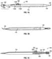
- FIGs. 1A-1Cillustrate different views of embodiment of a device 100 for inserting a pre-loaded intracanalicular plug (referred to interchangeably as "plug” hereinafter) for the treatment of dry eyes.
- the device 100is configured as a single-use inserter tool for the plug 140.
- the plug 140is configured to temporarily restrict natural lubricating tears from draining off from the eye.
- the device 100also has an integral dilator means 130.
- the device 100has an ergonomic design and is pencil-sized for ease of handling. In certain embodiments, the device 100 can be between 5 cm - 25 cm in length. Also, similar to a pencil, the device 100 can be held and manipulated between the thumb, forefinger and a side of the middle finger of one hand by a user.
- the lightweight, handheld device 100is convenient to use and it facilitates efficiency and cost-savings which may ultimately be passed down to the patients.
- the device 100can be manufactured from stainless steel, polycarbonate, plastic, any combination of these, or another suitable material.
- the device 100includes an elongate housing or body 110, wherein the body has an inserter end 120 and a distal end 130 longitudinally opposing the inserter end, and a plug ejector 200 mounted inside the body.
- the plug ejector 200is configured with an integral slider 210 and a button 300 to eject a pre-loaded intracanalicular plug 140 into the canaliculus.
- the device 100further includes a cover or cap 400 for protecting the plug 140 within the inserter end 120.
- the elongate body 110has a substantially longitudinal axis. A first end of the body 110 terminates in a plug inserter end 120 while a second end of the body terminates in a dilator tip 135.
- the body 110can have raised ridges, scoring, or a roughened surface to facilitate a stable grip in the hands of an eye doctor.
- the body 110may have a hexagonal cross-section. In other embodiments, the body 110 may have a circular or polygonal cross-section.
- a bottom portion of the body 110may include an elongate groove 115. The groove 115 may extend from substantially a first end 120 of the body to substantially a second end 130 of the body.
- the groove 115ensures that the device 100 is light weight and it can also facilitate the insertion of the plug ejector 200 and the plug 140 during the manufacture/assembly process.
- the body 110may have a larger diameter toward its middle or it may have a uniform diameter.
- the plug 140may be made of a biocompatible material.
- the plugmay be formed from a water-soluble, dissolvable material, such as collagen, or a polydioxanone plug, however, it can also include other types of dissolvable plugs that are medically-compatible and made of a suitable material.
- the plug 140is opaque and cylindrical in shape.
- the plug 140is designed to fit snugly inside the canaliculus to block the flow of tears. Since the punctal diameter of most patients is around 0.4 mm - 0.5 mm in diameter, a typical plug will range between about 0.4 mm to about 0.5 mm in diameter as well. Consequently, once the plug has been inserted, the tears can stay on the surface of the eye for a longer duration which in turn, ensures natural lubrication of the eye. As a result, the eye stays moist and comfortable.
- a first end 120 of the bodyis configured for inserting a plug into the canaliculus.
- the plug inserter end 120includes an inserter tip 125.
- the inserter tip 125is configured to firmly retain the plug 140.
- the inserter tip 125has a "crimp"-type design.
- the inserter tip 125involves a substantially silo- or conical-shaped portion 125A, a channel 125B that tapers inward and an opening 127.
- the channel 125Bprovides a close fit to the outer surface of the plug 140 and it is configured to frictionably hold the plug 140 until it is ejected from opening 127.
- the diameter of the opening 127may be adjusted to substantially match that of the plug 140.
- the device 110further includes a cover or cap 400.
- the cap 400protects the plug 140 at the inserter tip 125.
- the cap 400includes a dome-shaped housing 410 and an opening 420 for receiving the plug and inserter tip 125.
- the cap 400may be made of polycarbonate polymer or any other suitable material.
- the cap 400is configured to snap on the inserter tip 125.
- the capincludes a compression means for compressing the plug 140 to about a 1000 th of an inch.
- the plug ejector 200includes an integral slider 210 and an ejector means 300.
- the slider 210is mounted within the longitudinal groove 115 of the body 110.
- the slider 210has a first arm 210A and a second arm 210B which are separated by an opening 210D at a first end.
- the arms 210A, 210Bare coupled together at a tapering second end 210C.
- the slider 210is configured to be flexed (that is, it is not made of a rigid material).
- the two arms 210A, 210B of the slidercan be flexed toward a midline of the slider when it is inserted into the body 110 during the assembly of the device 110.
- the arms 210A, 210Bare configured to flex back (away from the midline) to their original position once the slider is inserted within the body 110.
- the plug ejector 200further includes a plunger or rod 220.
- a first end 230 of the rod 220is affixed to the second end 210C of the slider.
- the opposing end 240 of the rod 220is configured to abut a first end of the plug 140 (as shown in FIG. 1C ) inside the channel 125B.
- the rod 220is precision molded such that it can fit inside the channel 125B.
- a second end of the plug 140is configured to slightly extend out from the opening 127 of the inserter tip.
- the plug ejector 200further includes an ejector means 300.
- the ejector means 300can include a button 310 or any other suitable mechanism such as, a lever.
- the button 310is interposed on an upper portion of the body 110 and it can be configured to have any suitable shape.
- the base of the buttonincludes a pair of legs 320.
- the legs 320are clipped to the first arm 210A of the slider through slots 215.
- a raised portion 330 of the buttonprotrudes outwardly from an upper portion of the body.
- the raised portion 330can include ridges or grooves for facilitating a stable grip.
- the button 310can be located within a channel 110B on an upper portion of the body 110.
- the channel 110Bis flanked by sidewalls 110B' and 110B".
- the button 310is locked in a first position by sidewall 110B" by the interaction of a locking means, such as, protrusion 250 and an indentation 150 of the first arm 210A.
- the button 310is originally compressed in position. To release the button 310, it can be depressed to remove the protrusion 250 from the indention 150, and it can be then moved from the first position to a second position proximal to the opposing sidewall 110B' or to any position therebetween within the channel 110B.
- the eye doctorcan actuate the plug ejector 200 by gently depressing the top/raised portion 330 of the button to release it from its locked position. Pressing the top of the button 330 forces the base 320 of the button to push the first slider arm 210B toward the second slider arm 210B.
- the button 300can then be moved along the axis of the body 110 from a first position along sidewall 110B" to a second position along sidewall 110B'. This causes the slider arms 210A and 210B and the rod 220 to slide forward toward the opening 127 at the inserter end.
- the rod 220moves the plug 140 through the channel 125B and ejects it out from the opening 127 into the canaliculus.
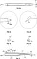
- the tip 220A of the rodhas a slightly smaller diameter in comparison to the opening 127. As shown in FIG. 5 , after the plug 140 has been ejected, a small portion of the tip 220A of the rod may protrude through the opening 127.
- the rod 220is configured to be retractable and can slide back within the body 110 when the button 300 is pushed backward (that is, when the button is moved away from the inserter tip).
- the inserter endcan also include a collar. The collar may be cushioned to facilitate a firm grip.
- a distal end 130 of the bodyis located longitudinally opposite the inserter end 120. As shown in FIGs. 1A and 2E , the distal end 130 can taper into a very small and fine tip/point 135.
- the tip 135may be angled with respect to the longitudinal axis of the body 110. For example, the tip 135 may be configured to point upward or downward. In certain embodiments, the tip can also have a longitudinal axis substantially coincident with the longitudinal axis of the body 110.
- the patient's punctummay have to be dilated prior to inserting a plug. Dilation may involve the use of forceps or other specialized dilation tools. This adds to the complexity/cumbersomeness and expense of the treatment procedure.
- the tip 135can be conveniently used to dilate the lacrimal punctum.
- the size and shape of the dilator tip 135can be customized.
- the size of the dilator tip 135can be customized to approximate the punctal diameter.
- the device 100combines a means for inserting a pre-loaded plug into the punctum with a means for dilating the punctum.
- the tip 135can also be configured to push the inserted plug further into the canaliculus after it has been ejected into the canaliculus by the device.
- the inventionis not limited to the particular design of the device 100 shown in figures and variations in shape, size and configuration are within the scope of the invention.
- the device 100may be manufactured in multiple colors. Each color may be associated with a particular size or diameter of the pre-loaded plug.
- the device 100may include the diameter information for the plug proximate the plug inserter end 120. In other embodiments, the diameter information may be coupled with a color-coded system.
- the device 100may be sold in separately wrapped sterile or non-sterile packages.
- the device 100When sold as a sterile package, the device 100 may be sold as a sterile kit with two sealed trays each of which securely holds a single device 100 having a preloaded plug 140.
- the traycan have a barrier lid.
- the tray and the device 100can be pre-sterilized using a suitable agent, such as, ethylene oxide.
- Ethylene oxide sterilizationinvolves exposing the tray and device 100 to ethylene oxide gas under vacuum in a sealed chamber. The sterilization can ensure that a safe and sterile device 100 is provided to the eye care professional.
- a kit 500includes one or more sterile trays 510 each having a device 100 pre-loaded with a plug, and instructions for use ("IFU").
- the one or more trays and the IFUcan be vacuum sealed in a pouch.
- the pouchcan then be positioned inside a container, such as, a box.
- the one or more trays and the boxcan include product indicia and other necessary information, such as, plug size, on its surface.
- a method of assembling the deviceinvolves providing an intracanalicular plug (such as, plug 140).
- the plugmay be a sterile or non-sterile plug which may be provided in a sealed pouch.
- the methodfurther involves providing the body 110 of the device 100 disclosed herein.
- the methodinvolves removing the plug from the pouch and mounting it in the channel 125B of the inserter tip 125. This may be followed by snapping on the plug ejector 200 to the body 110 such that the tip of the rod abuts one end of the plug and the other end of the plug protrudes out of the opening of the inserter tip.
- the methodthen involves compressing the cap on the inserter tip to hold the plug securely in position on the device 100.
- the device 100is then sealed in a pre-molded tray/container with a sterile barrier lid. The tray and the device 100 can be sterilized using ethylene oxide.
- a method of treating dry eyesinvolves providing a device 100 pre-loaded with an intracanalicular plug, as disclosed herein.
- the methodinvolves removal of the protective cap from the inserter tip.
- An eye doctorcan then position the device such that the inserter tip faces the patient's punctum.
- the inserter tipis then inserted into the canaliculus. This is followed by gently depressing the button and then moving the button forward toward the inserter tip. This causes the rod to eject the plug out of the opening at the inserter tip and into the canaliculus.
- the eye doctorcan use the dilator end of the device 100 to further push the plug into a desired position inside the canaliculus.
- a punctal diameter of 0.5 mmis common in many patients.
- the eye doctormay use the dilator end of the device 100 to dilate the punctum prior to inserting the inserter tip into the canaliculus. This allows for easier insertion of the plug. However, in certain embodiments, the dilation step may be optional. Once the punctum is dilated, the doctor can turn the device 180° such that the inserter tip is facing the patient's punctum to insert the pre-loaded plug into the canaliculus.
- the embodiments of the inventioninvolve a single step insertion process since the plug is already pre-loaded in the device.
- the device 100conveniently does not involve the use of any wires to release the plug. Further, the assembly of the inserter does not involve any adhesives or epoxy.
- the various components, such as, the plug and capcan snap together to make it a fully functional inserter too. Conveniently, the device 100 can be configured to accommodate plugs of different sizes.
- the device 100may be used by eye care doctors and professionals, such as, ophthalmologists, optometrists and other healthcare professionals.
- the device 100is a medical device that may require a prescription.
- the one or more embodiments of the inventionallow an eye doctor to perform the functions of dilating the punctum and inserting the punctum plug utilizing a unitary or single device.
- the devicemay be handheld and may be capable of being manipulated with one hand by the eye doctor, thereby ensuring convenience and efficiency.
- the utilization of a single apparatusmay also result in cost-savings which may ultimately be passed down to the patients.
Landscapes
- Health & Medical Sciences (AREA)
- Ophthalmology & Optometry (AREA)
- Life Sciences & Earth Sciences (AREA)
- Animal Behavior & Ethology (AREA)
- Veterinary Medicine (AREA)
- Public Health (AREA)
- General Health & Medical Sciences (AREA)
- Engineering & Computer Science (AREA)
- Biomedical Technology (AREA)
- Heart & Thoracic Surgery (AREA)
- Vascular Medicine (AREA)
- Surgery (AREA)
- Nuclear Medicine, Radiotherapy & Molecular Imaging (AREA)
- Plastic & Reconstructive Surgery (AREA)
- Prostheses (AREA)
- Media Introduction/Drainage Providing Device (AREA)
- Cardiology (AREA)
- Medical Informatics (AREA)
- Molecular Biology (AREA)
Description
- The invention relates generally to devices and a kit for the treatment of dry eyes.
- Dry eye syndrome affects millions of people each year, causing discomfort, redness, corneal irritation, and contact lens intolerance. Tears normally drain by passing through two lacrimal orifices or puncta (upper and lower) on the medial surface of each eyelid, then through vertical and horizontal canaliculi into the nasal cavity. Dry eye syndrome can be treated by occluding the puncta using punctal occluders or by placing implants into the canaliculi.
- Plugs are very tiny, biocompatible devices that can be used to treat dry eyes. There are two common types of plugs depending on their location. Surface plugs sit at the surface of the tear duct and they may be visible just outside the tear duct. Canalicular or intracanalicular plugs, on the other hand, are placed deep inside/within the canaliculus (either the vertical or the horizontal canaliculus).
- Currently, intracanalicular plugs are pushed or inserted into the canaliculi using a pair of forceps. This procedure is very cumbersome - for example, an eye doctor (such as, an optometrist, ophthalmologist or another eye care professional), while performing a slit-lamp examination, has to first open a package containing the canalicular plug. This is followed by picking up and inserting the plug into the canaliculus using forceps. Since the plug is a tiny device, it presents a likely scenario where the eye doctor inadvertently drops the plug before it is inserted into the canaliculus. Some eye doctors may also lack the experience to perform the procedure and may be intimidated when trying to insert the plug into the canaliculus because of its small size.
U.S. Pat. No. 8,591,484 discloses a device for inserting a surface plug. The device includes a metal or plastic wire or any other wiring capable of being flexed. The plug is positioned at a tip of the wire. The wire is embedded within a trough having a backstop for holding the wire in place. An adhesive is applied to the area of contact between the wire and the trough for holding the wire in place. When a button is depressed, it applies a downward force on the wire proximate the backstop. Because one end of the wire is held in place at the backstop, the downward force applied by the button produces a tensile force on the wire, pulling it inwards. Complete depression of the plug ejector causes the withdrawal of the tip of the wire from the plug, thereby releasing the plug. A drawback with the device is that it involves the use of a wire for holding the plug. The patented device is configured for the placement of a surface plug. Since the device is not configured to enter the opening of the canaliculus, it will not push the plug into the canaliculus. Relevant prior art is exemplified byUS 2013/023837 A1 ,US 2012/065601 A1 ,SG 184 727 A1 US 6 344 047 B1 , andTW 201 242 582 A - Accordingly, there is a need for a convenient device that can facilitate the placement of an intracanalicular plug into the canaliculus without using forceps or without the use of insertion devices that include wires for holding the plug. Ideally, the device is provided with pre-loaded plugs and does not rely on the eye doctor to load them onto the device.
- The present invention is set out in the appended claims. The present invention involves a device for inserting an intracanalicular plug into the canaliculus and a method of assembling an intracanalicular plug inserter device. The plug may be made of a suitable biocompatible material, such as, polydioxanone or any other suitable material. Conveniently, the plug can be pre-loaded (or pre-mounted) on the device.
- The present invention facilitates a one-step treatment process for inserting the plug into the canaliculus to temporarily restrict the natural lubricating tears from draining off the eye by using a single device that is pre-loaded with a dissolvable intracanalicular plug. This avoids the need for an eye doctor to remove the plug from separate package and eliminates the cumbersome process of using forceps to hold and insert the plug into the canaliculus or punctum. Advantageously, even eye doctors with little or no experience are able to easily insert the dissolvable punctum plugs into the canaliculus. The treatment can be used for long-term treatment of certain eye conditions commonly referred to as dry eye syndrome, as well as the dry eye component of ocular surface diseases and other conditions of tear insufficiency.
- In one embodiment, an intracanalicular plug inserter device includes: (a) an elongate body having a longitudinal axis, the body having: an inserter end, wherein the inserter end has an opening therein; and a distal end, wherein the distal end is longitudinally opposing the inserter end; and (b) a plug ejector, wherein the plug injector comprises: a slider; and a rod coupled to a first end of the slider, wherein the plug ejector is configured to be moveable between a first position adjacent an opening in the inserter end and a second position that is further from the opening. The plug is mounted within the body and abuts first end of the rod adjacent the opening in the inserter end. The rod is configured to eject the plug from the opening in the inserter end when the first end of the slider is moved toward the inserter end. According to the present invention, the The plug ejector further includes a depressible button having a first (upper) side positioned outside of the body and a second side mounted on the slider. The button is flanked by a first sidewall and a second sidewall. According to the present invention, the second (base) side of the button includes a pair of legs which are clipped to a first arm of the slider. According to the present invention, the button is substantially locked in a first position by a locking means that includes a protrusion and an indentation on the first arm of the slider. The protrusion is adjacent the first sidewall. The button is configured to be moved along the longitudinal axis of the body from its first position to a second position adjacent the second sidewall by depressing the button to release it from the indentation and then sliding it along the slide toward the second position. The device further includes a removable cap fitted to cover plug mounted at the inserter end of the body. The device also includes integral means for dilating a lacrimal punctum disposed at the distal end of the body. The means for dilating the punctum comprises a fine tip.
- In another embodiment, a kit comprising the intracanalicular plug inserter device is provided. The kit further includes a tray for receiving the device and instructions for using the device.
- In yet another embodiment, a method of assembling an intracanalicular plug inserter device is disclosed. The method involves providing an intracanalicular plug; mounting the intracanalicular plug within the intracanalicular plug inserter device and compressing a cap on the inserter end of the device to hold the intracanalicular plug securely in position on the device. The method also involves sealing the device in a pre-molded tray with a sterile barrier lid. The method further comprises subjecting the tray to sterilization.
- In another example which is not part of the present invention, a method of treating dry eyes involves providing the intracanalicular plug inserter device disclosed herein; inserting the inserter end of the device into a patient's canaliculus; and actuating the plug ejector of the device to cause the plug to be ejected out of the opening at the inserter end. The method further comprises dilating the patient's punctum prior to inserting the plug. The distal end of the device can be used to push the plug further into the canaliculus.
FIGs. 1A-1C illustrate various views of an intracanalicular plug inserter device in accordance with one or more embodiments of the invention.FIGs. 2A-2E illustrate various views of the device and its components in accordance with one or more embodiments of the invention.FIG. 3 illustrates a slider in accordance with one or more embodiments of the invention.FIG. 4 illustrates a button in accordance with one or more embodiments of the invention.US 2013/023837 A1 teaches a surgical tool for inserting a spile or plug into the punctal opening of a meatus such as a lacrimo-nasal canaliculus. SimilarUS 2012/065601 A1 discloses a handheld device for the treatment of dry eyes comprising means for measuring a lacrimal punctum disposed at a first shaft end and for inserting a punctal plug disposed at a second shaft end.SG 184727 A1 U.S. Pat. No. 8,591,484 discloses a device for inserting a surface plug. The device includes a metal or plastic wire or any other wiring capable of being flexed. The plug is positioned at a tip of the wire. The wire is embedded within a trough having a backstop for holding the wire in place. An adhesive is applied to the area of contact between the wire and the trough for holding the wire in place. When a button is depressed, it applies a downward force on the wire proximate the backstop. Because one end of the wire is held in place at the backstop, the downward force applied by the button produces a tensile force on the wire, pulling it inwards. Complete depression of the plug ejector causes the withdrawal of the tip of the wire from the plug, thereby releasing the plug. A drawback with the device is that it involves the use of a wire for holding the plug. The patented device is configured for the placement of a surface plug. Since the device is not configured to enter the opening of the canaliculus, it will not push the plug into the canaliculus.- Accordingly, there is a need for a convenient device that can facilitate the placement of an intracanalicular plug into the canaliculus without using forceps or without the use of insertion devices that include wires for holding the plug. Ideally, the device is provided with pre-loaded plugs and does not rely on the eye doctor to load them onto the device.
- The present invention involves a device for inserting an intracanalicular plug into the canaliculus and methods for treatment of dry eyes. The plug may be made of a suitable biocompatible material, such as, polydioxanone or any other suitable material. Conveniently, the plug can be pre-loaded (or pre-mounted) on the device.
- The present invention facilitates a one-step treatment process for inserting the plug into the canaliculus to temporarily restrict the natural lubricating tears from draining off the eye by using a single device that is pre-loaded with a dissolvable intracanalicular plug. This avoids the need for an eye doctor to remove the plug from separate package and understood by those of skill in the art. The term "substantially" and its variations are defined as being largely but not necessarily wholly what is specified as understood by one of ordinary skill in the art, and in one non-limiting embodiment substantially refers to ranges within 0.5% - 5%.
FIGs. 1A-1C illustrate different views of embodiment of adevice 100 for inserting a pre-loaded intracanalicular plug (referred to interchangeably as "plug" hereinafter) for the treatment of dry eyes. Thedevice 100 is configured as a single-use inserter tool for theplug 140. Theplug 140 is configured to temporarily restrict natural lubricating tears from draining off from the eye. Thedevice 100 also has an integral dilator means130. Thedevice 100 has an ergonomic design and is pencil-sized for ease of handling. In certain embodiments, thedevice 100 can be between 5 cm - 25 cm in length. Also, similar to a pencil, thedevice 100 can be held and manipulated between the thumb, forefinger and a side of the middle finger of one hand by a user. The lightweight,handheld device 100 is convenient to use and it facilitates efficiency and cost-savings which may ultimately be passed down to the patients.- The
device 100 can be manufactured from stainless steel, polycarbonate, plastic, any combination of these, or another suitable material. Thedevice 100 includes an elongate housing orbody 110, wherein the body has aninserter end 120 and adistal end 130 longitudinally opposing the inserter end, and aplug ejector 200 mounted inside the body. Theplug ejector 200 is configured with anintegral slider 210 and abutton 300 to eject apre-loaded intracanalicular plug 140 into the canaliculus. Thedevice 100 further includes a cover or cap400 for protecting theplug 140 within theinserter end 120. - The
elongate body 110 has a substantially longitudinal axis. A first end of thebody 110 terminates in aplug inserter end 120 while a second end of the body terminates in adilator tip 135. Thebody 110 can have raised ridges, scoring, or a roughened surface to facilitate a stable grip in the hands of an eye doctor. In one or more embodiments, thebody 110 may have a hexagonal cross-section. In other embodiments, thebody 110 may have a circular or polygonal cross-section. Optionally, a bottom portion of thebody 110 may include anelongate groove 115. Thegroove 115 may extend from substantially afirst end 120 of the body to substantially asecond end 130 of the body. Thegroove 115 ensures that thedevice 100 is light weight and it can also facilitate the insertion of theplug ejector 200 and theplug 140 during the manufacture/assembly process. Thebody 110 may have a larger diameter toward its middle or it may have a uniform diameter. - The
plug 140 may be made of a biocompatible material. Preferably, the plug may be formed from a water-soluble, dissolvable material, such as collagen, or a polydioxanone plug, however, it can also include other types of dissolvable plugs that are medically-compatible and made of a suitable material. In some embodiments, theplug 140 is opaque and cylindrical in shape. Theplug 140 is designed to fit snugly inside the canaliculus to block the flow of tears. Since the punctal diameter of most patients is around 0.4 mm - 0.5 mm in diameter, a typical plug will range between about 0.4 mm to about 0.5 mm in diameter as well. Consequently, once the plug has been inserted, the tears can stay on the surface of the eye for a longer duration which in turn, ensures natural lubrication of the eye. As a result, the eye stays moist and comfortable. - A
first end 120 of the body is configured for inserting a plug into the canaliculus. Theplug inserter end 120 includes aninserter tip 125. According to the present invention, theinserter tip 125 is configured to firmly retain theplug 140. In one non-limiting embodiment, as shown inFIGs. 2A-2D , theinserter tip 125 has a "crimp"-type design. For instance, theinserter tip 125 involves a substantially silo- or conical-shapedportion 125A, achannel 125B that tapers inward and anopening 127. Thechannel 125B provides a close fit to the outer surface of theplug 140 and it is configured to frictionably hold theplug 140 until it is ejected from opening127. The diameter of theopening 127 may be adjusted to substantially match that of theplug 140. - As shown in
FIG. 1A and 1C , thedevice 110 further includes a cover orcap 400. Thecap 400 protects theplug 140 at theinserter tip 125. Thecap 400 includes a dome-shapedhousing 410 and anopening 420 for receiving the plug andinserter tip 125. Thecap 400 may be made of polycarbonate polymer or any other suitable material. Thecap 400 is configured to snap on theinserter tip 125. In certain embodiments, the cap includes a compression means for compressing theplug 140 to about a 1000th of an inch. - A cross-sectional view of the
plug ejector 200 is shown inFIG. 1C . Theplug ejector 200 includes anintegral slider 210 and an ejector means300. According to the present invention, theslider 210 is mounted within thelongitudinal groove 115 of thebody 110. According to the present invention, theslider 210 has afirst arm 210A and asecond arm 210B which are separated by anopening 210D at a first end. According to the present invention, thearms second end 210C. According to the present invention, theslider 210 is configured to be flexed (that is, it is not made of a rigid material). The twoarms body 110 during the assembly of thedevice 110. Thearms body 110. - The
plug ejector 200 further includes a plunger orrod 220. Afirst end 230 of therod 220 is affixed to thesecond end 210C of the slider. Theopposing end 240 of therod 220 is configured to abut a first end of the plug140 (as shown inFIG. 1C ) inside thechannel 125B. Advantageously, therod 220 is precision molded such that it can fit inside thechannel 125B. A second end of theplug 140 is configured to slightly extend out from theopening 127 of the inserter tip. - According to the present invention, the
plug ejector 200 further includes an ejector means300. As further shown inFIGs. 4 and 5 , the ejector means300 can include abutton 310 or any other suitable mechanism such as, a lever. Thebutton 310 is interposed on an upper portion of thebody 110 and it can be configured to have any suitable shape. In one embodiment the base of the button includes a pair oflegs 320. Thelegs 320 are clipped to thefirst arm 210A of the slider throughslots 215. A raisedportion 330 of the button protrudes outwardly from an upper portion of the body. The raisedportion 330 can include ridges or grooves for facilitating a stable grip. Thebutton 310 can be located within achannel 110B on an upper portion of thebody 110. Thechannel 110B is flanked by sidewalls110B' and110B". Thebutton 310 is locked in a first position bysidewall 110B" by the interaction of a locking means, such as,protrusion 250 and anindentation 150 of thefirst arm 210A. Thebutton 310 is originally compressed in position. To release thebutton 310, it can be depressed to remove theprotrusion 250 from theindention 150, and it can be then moved from the first position to a second position proximal to the opposingsidewall 110B' or to any position therebetween within thechannel 110B. - In use, the eye doctor can actuate the
plug ejector 200 by gently depressing the top/raisedportion 330 of the button to release it from its locked position. Pressing the top of thebutton 330 forces thebase 320 of the button to push thefirst slider arm 210B toward thesecond slider arm 210B. Thebutton 300 can then be moved along the axis of thebody 110 from a first position alongsidewall 110B" to a second position alongsidewall 110B'. This causes theslider arms rod 220 to slide forward toward theopening 127 at the inserter end. Therod 220 moves theplug 140 through thechannel 125B and ejects it out from theopening 127 into the canaliculus. Thetip 220A of the rod has a slightly smaller diameter in comparison to theopening 127. As shown inFIG. 5 , after theplug 140 has been ejected, a small portion of thetip 220A of the rod may protrude through theopening 127. - The
rod 220 is configured to be retractable and can slide back within thebody 110 when thebutton 300 is pushed backward (that is, when the button is moved away from the inserter tip). The inserter end can also include a collar. The collar may be cushioned to facilitate a firm grip. - A
distal end 130 of the body is located longitudinally opposite theinserter end 120. As shown inFIGs. 1A and2E , thedistal end 130 can taper into a very small and fine tip/point 135. Thetip 135 may be angled with respect to the longitudinal axis of thebody 110. For example, thetip 135 may be configured to point upward or downward. In certain embodiments, the tip can also have a longitudinal axis substantially coincident with the longitudinal axis of thebody 110. - In some instances, the patient's punctum may have to be dilated prior to inserting a plug. Dilation may involve the use of forceps or other specialized dilation tools. This adds to the complexity/cumbersomeness and expense of the treatment procedure.
- The
tip 135 can be conveniently used to dilate the lacrimal punctum. The size and shape of thedilator tip 135 can be customized. For instance, the size of thedilator tip 135 can be customized to approximate the punctal diameter. Accordingly, thedevice 100 combines a means for inserting a pre-loaded plug into the punctum with a means for dilating the punctum. Conveniently, thetip 135 can also be configured to push the inserted plug further into the canaliculus after it has been ejected into the canaliculus by the device. - It is understood, however, that while a combination of a dilator and inserter eliminates the wasteful use of multiple devices during the treatment process, a device that does not include a dilator tip is also within the scope of the present invention.
- The invention is not limited to the particular design of the
device 100 shown in figures and variations in shape, size and configuration are within the scope of the invention. - In one or more embodiments, the
device 100 may be manufactured in multiple colors. Each color may be associated with a particular size or diameter of the pre-loaded plug. In yet another embodiment, thedevice 100 may include the diameter information for the plug proximate theplug inserter end 120. In other embodiments, the diameter information may be coupled with a color-coded system. - The
device 100 may be sold in separately wrapped sterile or non-sterile packages. When sold as a sterile package, thedevice 100 may be sold as a sterile kit with two sealed trays each of which securely holds asingle device 100 having apreloaded plug 140. The tray can have a barrier lid. The tray and thedevice 100 can be pre-sterilized using a suitable agent, such as, ethylene oxide. Ethylene oxide sterilization involves exposing the tray anddevice 100 to ethylene oxide gas under vacuum in a sealed chamber. The sterilization can ensure that a safe andsterile device 100 is provided to the eye care professional. - In one specific embodiment, as shown in
FIGs. 6A and6B , a kit500 includes one or moresterile trays 510 each having adevice 100 pre-loaded with a plug, and instructions for use ("IFU"). The one or more trays and the IFU can be vacuum sealed in a pouch. The pouch can then be positioned inside a container, such as, a box. The one or more trays and the box can include product indicia and other necessary information, such as, plug size, on its surface. - According to an embodiment, a method of assembling the device is disclosed. The method involves providing an intracanalicular plug (such as, plug140). The plug may be a sterile or non-sterile plug which may be provided in a sealed pouch. The method further involves providing the
body 110 of thedevice 100 disclosed herein. The method involves removing the plug from the pouch and mounting it in thechannel 125B of theinserter tip 125. This may be followed by snapping on theplug ejector 200 to thebody 110 such that the tip of the rod abuts one end of the plug and the other end of the plug protrudes out of the opening of the inserter tip. The method then involves compressing the cap on the inserter tip to hold the plug securely in position on thedevice 100. Thedevice 100 is then sealed in a pre-molded tray/container with a sterile barrier lid. The tray and thedevice 100 can be sterilized using ethylene oxide. - According to another embodiment, a method of treating dry eyes is disclosed herein. The method involves providing a
device 100 pre-loaded with an intracanalicular plug, as disclosed herein. The method involves removal of the protective cap from the inserter tip. An eye doctor can then position the device such that the inserter tip faces the patient's punctum. The inserter tip is then inserted into the canaliculus. This is followed by gently depressing the button and then moving the button forward toward the inserter tip. This causes the rod to eject the plug out of the opening at the inserter tip and into the canaliculus. The eye doctor can use the dilator end of thedevice 100 to further push the plug into a desired position inside the canaliculus. - A punctal diameter of 0.5 mm is common in many patients. In some embodiments, the eye doctor may use the dilator end of the
device 100 to dilate the punctum prior to inserting the inserter tip into the canaliculus. This allows for easier insertion of the plug. However, in certain embodiments, the dilation step may be optional. Once the punctum is dilated, the doctor can turn the device 180° such that the inserter tip is facing the patient's punctum to insert the pre-loaded plug into the canaliculus. - The embodiments of the invention involve a single step insertion process since the plug is already pre-loaded in the device. The
device 100 conveniently does not involve the use of any wires to release the plug. Further, the assembly of the inserter does not involve any adhesives or epoxy. The various components, such as, the plug and cap can snap together to make it a fully functional inserter too. Conveniently, thedevice 100 can be configured to accommodate plugs of different sizes. - The
device 100 may be used by eye care doctors and professionals, such as, ophthalmologists, optometrists and other healthcare professionals. Thedevice 100 is a medical device that may require a prescription. The one or more embodiments of the invention allow an eye doctor to perform the functions of dilating the punctum and inserting the punctum plug utilizing a unitary or single device. The device may be handheld and may be capable of being manipulated with one hand by the eye doctor, thereby ensuring convenience and efficiency. The utilization of a single apparatus may also result in cost-savings which may ultimately be passed down to the patients. - While the invention has been described in connection with a preferred embodiment, it is not intended to limit the scope of the invention which is defined by the appended claims.
Claims (8)
- An intracanalicular plug inserter device, comprising:(a) an elongate body (110) having a longitudinal axis, the body having:an inserter end (120), the inserter end including an inserter tip (125) which has an opening (127) therein; anda distal end (130), the distal end longitudinally opposing the inserter end;(b) a plug ejector (200), wherein the plug ejector comprises:a slider (210) and an ejector means (300), wherein the slider (210) has a first end (210D) and a second end (210C) and the slider (210) is configured to be flexed and is mounted within a longitudinal groove (115) of the body (110), and the slider having a first arm (210A) and a second arm (210B) which are configured to flex toward a midline of the slider when the plug ejector (200) is inserted into the body (110) during the assembly of the plug inserter device and to flex back to their original position once the slider is inserted within the body (110), wherein the first and second arm (210A, 210B) are coupled together at the second end (210C) of the slider;a rod (220) coupled to the second_end (210C) of the slider (210);
anda depressible button (310) having a first side protruding outside of the body (110) and a second side mounted to the first arm of the slider (210), and wherein the second side of the button (310) comprises a pair of legs (320) which are clipped to the first arm (210A) of the slider(c) a plug (140) mounted on the inserter tip (125), wherein the inserter tip is configured to firmly retain the plug, and wherein the plug is configured for insertion into the canaliculus in the eye,wherein the rod (220) is configured to eject the plug (140) from the opening (127) in the inserter end (120) when the first end of the slider is moved toward the inserter end (120),wherein the plug ejector (200) is configured to be moveable between a first position adjacent the opening (127) in the inserter end and a second position that is further from the opening. - The device of claim 1, wherein the button (310) is substantially locked in a first position by a locking means, wherein the locking means comprises a protrusion (250) on the first arm of the slider and an indentation (150) on the upper portion of the body (110).
- The device of claim 2, wherein the button (310) is flanked by a first sidewall (110B") and a second sidewall (110B') of the channel (110B).
- The device of claim 3, wherein the protrusion (250) and indentation (150) are adjacent the first sidewall (110B").
- A kit comprising:the intracanalicular plug inserter device of claim 1;a tray for receiving the device;a barrier lid; andinstructions for using the device.
- A method of assembling an intracanalicular plug inserter device, comprising:providing an intracanalicular plug;mounting the intracanalicular plug within the intracanalicular plug inserter device according to claim 1; andcompressing a cap over the inserter end to hold the intracanalicular plug securely in position on the device.
- The method of claim 6, further comprising sealing the device in a pre-molded tray with a sterile barrier lid.
- The method of claim 6, further comprising subjecting the tray to sterilization.
Applications Claiming Priority (2)
| Application Number | Priority Date | Filing Date | Title |
|---|---|---|---|
| US201862646538P | 2018-03-22 | 2018-03-22 | |
| PCT/US2019/023323WO2019183322A1 (en) | 2018-03-22 | 2019-03-21 | Intracanalicular dissolvable punctum plug inserter |
Publications (4)
| Publication Number | Publication Date |
|---|---|
| EP3768206A1 EP3768206A1 (en) | 2021-01-27 |
| EP3768206A4 EP3768206A4 (en) | 2022-01-05 |
| EP3768206B1true EP3768206B1 (en) | 2024-09-04 |
| EP3768206C0 EP3768206C0 (en) | 2024-09-04 |
Family
ID=67984493
Family Applications (1)
| Application Number | Title | Priority Date | Filing Date |
|---|---|---|---|
| EP19771603.8AActiveEP3768206B1 (en) | 2018-03-22 | 2019-03-21 | Intracanalicular dissolvable punctum plug inserter and method of assembling of the same |
Country Status (17)
| Country | Link |
|---|---|
| US (1) | US11497649B2 (en) |
| EP (1) | EP3768206B1 (en) |
| JP (1) | JP7296446B2 (en) |
| KR (1) | KR102765111B1 (en) |
| CN (1) | CN111936093A (en) |
| AR (1) | AR115010A1 (en) |
| AU (1) | AU2019240207B2 (en) |
| CA (1) | CA3094359C (en) |
| ES (1) | ES2989955T3 (en) |
| GB (1) | GB2587132B (en) |
| MX (1) | MX2020009754A (en) |
| MY (1) | MY204381A (en) |
| NZ (1) | NZ769069A (en) |
| SG (1) | SG11202009056PA (en) |
| TW (1) | TWI730307B (en) |
| UY (1) | UY38150A (en) |
| WO (1) | WO2019183322A1 (en) |
Families Citing this family (1)
| Publication number | Priority date | Publication date | Assignee | Title |
|---|---|---|---|---|
| US12023276B2 (en) | 2021-02-24 | 2024-07-02 | Ocular Therapeutix, Inc. | Intracanalicular depot inserter device |
Family Cites Families (20)
| Publication number | Priority date | Publication date | Assignee | Title |
|---|---|---|---|---|
| US5669501A (en)* | 1996-06-05 | 1997-09-23 | Xomed Surgical Products, Inc. | Package and method for delivering a medical implant |
| US5921990A (en)* | 1997-08-06 | 1999-07-13 | Eagle Vision | Collagen forceps |
| US6344047B1 (en)* | 2000-02-02 | 2002-02-05 | Eagle Vision | Instrument for inserting a punctum plug and method for manufacturing the instrument |
| US20040068235A1 (en)* | 2002-10-07 | 2004-04-08 | Hallam Clive T. | Packaging system for a medical device and particularly a punctum plug insertion device |
| US20040068286A1 (en)* | 2002-10-07 | 2004-04-08 | Mendius Richard W. | Punctum dilating and punctum plug insertion instrument |
| TWI369970B (en)* | 2004-10-20 | 2012-08-11 | Beaver Visitec Int Us Inc | Surgical knife safety handle having user operable lock |
| NZ572193A (en)* | 2006-03-31 | 2011-10-28 | Quadra Logic Tech Inc | Nasolacrimal drainage system implants for drug therapy with non-fluid swellable retention structure around drug core |
| WO2009035567A2 (en) | 2007-09-07 | 2009-03-19 | Qlt Plug Delivery, Inc | Insertion and extraction tools for lacrimal implants |
| JP5330401B2 (en)* | 2007-11-08 | 2013-10-30 | アリメラ・サイエンシーズ,インコーポレーテッド | Implant device for the eye and kit comprising the device |
| AU2010271274B2 (en)* | 2009-07-09 | 2015-05-21 | Alcon Inc. | Single operator device for delivering an ocular implant |
| US8591484B2 (en) | 2010-09-15 | 2013-11-26 | AlphaMed, Inc. | Lacrimal punctum measurement and occlusion |
| CN201890013U (en)* | 2010-10-25 | 2011-07-06 | 周新潮 | Pressing type pen |
| TW201242582A (en)* | 2011-04-21 | 2012-11-01 | Johnson & Johnson Vision Care | Implantation instruments, system, and kit for punctal implants |
| US20130013207A1 (en) | 2011-07-08 | 2013-01-10 | Artic Ice Management Ab | Support system for use when managing ice |
| US9254225B2 (en)* | 2011-07-20 | 2016-02-09 | Bruce B. Becker | Punctal plug inserter and method |
| CN102848803B (en)* | 2012-10-15 | 2014-08-20 | 青岛点石文具用品有限公司 | Slidable type pen |
| US20140243763A1 (en) | 2013-02-25 | 2014-08-28 | Sara Heikali | Devices for the Placement of Medical Compounds in Natural Orifices of a Body |
| US9592151B2 (en)* | 2013-03-15 | 2017-03-14 | Glaukos Corporation | Systems and methods for delivering an ocular implant to the suprachoroidal space within an eye |
| EP3068354B1 (en)* | 2013-11-14 | 2023-06-28 | Aquesys, Inc. | Intraocular shunt inserter |
| KR102381775B1 (en) | 2016-03-16 | 2022-04-04 | 옥슬러 리미티드 | Ophthalmic delivery device and ophthalmic drug composition |
- 2019
- 2019-03-21ESES19771603Tpatent/ES2989955T3/enactiveActive
- 2019-03-21SGSG11202009056PApatent/SG11202009056PA/enunknown
- 2019-03-21MYMYPI2020004846Apatent/MY204381A/enunknown
- 2019-03-21AUAU2019240207Apatent/AU2019240207B2/enactiveActive
- 2019-03-21WOPCT/US2019/023323patent/WO2019183322A1/ennot_activeCeased
- 2019-03-21KRKR1020207030281Apatent/KR102765111B1/enactiveActive
- 2019-03-21CNCN201980020943.1Apatent/CN111936093A/enactivePending
- 2019-03-21ARARP190100719Apatent/AR115010A1/enactiveIP Right Grant
- 2019-03-21GBGB2016681.5Apatent/GB2587132B/enactiveActive
- 2019-03-21USUS16/360,287patent/US11497649B2/enactiveActive
- 2019-03-21NZNZ769069Apatent/NZ769069A/enunknown
- 2019-03-21JPJP2021500491Apatent/JP7296446B2/enactiveActive
- 2019-03-21MXMX2020009754Apatent/MX2020009754A/enunknown
- 2019-03-21TWTW108109792Apatent/TWI730307B/enactive
- 2019-03-21UYUY38150Apatent/UY38150A/enactiveIP Right Grant
- 2019-03-21CACA3094359Apatent/CA3094359C/enactiveActive
- 2019-03-21EPEP19771603.8Apatent/EP3768206B1/enactiveActive
Also Published As
| Publication number | Publication date |
|---|---|
| WO2019183322A1 (en) | 2019-09-26 |
| ES2989955T3 (en) | 2024-11-28 |
| NZ769069A (en) | 2024-03-22 |
| TW201940141A (en) | 2019-10-16 |
| SG11202009056PA (en) | 2020-10-29 |
| CA3094359A1 (en) | 2019-09-26 |
| AU2019240207B2 (en) | 2023-05-18 |
| JP7296446B2 (en) | 2023-06-22 |
| UY38150A (en) | 2019-10-01 |
| US20190290488A1 (en) | 2019-09-26 |
| AR115010A1 (en) | 2020-11-18 |
| MY204381A (en) | 2024-08-27 |
| CA3094359C (en) | 2023-07-25 |
| GB2587132A (en) | 2021-03-17 |
| GB202016681D0 (en) | 2020-12-02 |
| US11497649B2 (en) | 2022-11-15 |
| CN111936093A (en) | 2020-11-13 |
| GB2587132B (en) | 2022-04-13 |
| EP3768206A1 (en) | 2021-01-27 |
| TWI730307B (en) | 2021-06-11 |
| EP3768206A4 (en) | 2022-01-05 |
| KR102765111B1 (en) | 2025-02-07 |
| AU2019240207A1 (en) | 2020-11-12 |
| JP2021518250A (en) | 2021-08-02 |
| MX2020009754A (en) | 2020-10-08 |
| EP3768206C0 (en) | 2024-09-04 |
| KR20200134280A (en) | 2020-12-01 |
Similar Documents
| Publication | Publication Date | Title |
|---|---|---|
| US6344047B1 (en) | Instrument for inserting a punctum plug and method for manufacturing the instrument | |
| US4763650A (en) | Instrument for inserting a deformable lens into the eye | |
| KR100669869B1 (en) | Incision aids with lancet devices that are not reused | |
| CN104540472B (en) | Delivery system for ocular implants | |
| US6440065B1 (en) | Single-use disposable eyelid speculum, eye examination kit, and method for examining a patient's eye | |
| US6648819B2 (en) | Pupil dilator | |
| CA2586013A1 (en) | Blood expression device | |
| KR20160021201A (en) | Inserter for tubular medical implant devices | |
| WO2007080868A1 (en) | Instrument for inserting intraocular lens | |
| US9265655B2 (en) | Punctum plug insertion device and device packaging | |
| WO2001028475A1 (en) | Deformable intraocular lens injecting apparatus and method | |
| US20100057095A1 (en) | Method of Refraction Surgery of the Eye and a Tool for Implanting Intraocular Refractive Lens | |
| WO2010102121A1 (en) | Injector for intraocular lens | |
| CN108366875A (en) | Patch and relevant apparatus, system for sealing retinal hole and method | |
| EP1661533A1 (en) | Method for eye refractive surgery and device for implanting an intraocular refractive lens | |
| US20040068286A1 (en) | Punctum dilating and punctum plug insertion instrument | |
| KR20190058868A (en) | hair implanter | |
| EP3768206B1 (en) | Intracanalicular dissolvable punctum plug inserter and method of assembling of the same | |
| US8591484B2 (en) | Lacrimal punctum measurement and occlusion | |
| US20040068235A1 (en) | Packaging system for a medical device and particularly a punctum plug insertion device | |
| HK40038421A (en) | Intracanalicular dissolvable punctum plug inserter | |
| US9849028B2 (en) | Contact lens applicator | |
| US20140243763A1 (en) | Devices for the Placement of Medical Compounds in Natural Orifices of a Body | |
| JP2018175573A (en) | Micro forceps | |
| JP2014050552A (en) | Surgical instrument |
Legal Events
| Date | Code | Title | Description |
|---|---|---|---|
| STAA | Information on the status of an ep patent application or granted ep patent | Free format text:STATUS: THE INTERNATIONAL PUBLICATION HAS BEEN MADE | |
| PUAI | Public reference made under article 153(3) epc to a published international application that has entered the european phase | Free format text:ORIGINAL CODE: 0009012 | |
| STAA | Information on the status of an ep patent application or granted ep patent | Free format text:STATUS: REQUEST FOR EXAMINATION WAS MADE | |
| 17P | Request for examination filed | Effective date:20201015 | |
| AK | Designated contracting states | Kind code of ref document:A1 Designated state(s):AL AT BE BG CH CY CZ DE DK EE ES FI FR GB GR HR HU IE IS IT LI LT LU LV MC MK MT NL NO PL PT RO RS SE SI SK SM TR | |
| AX | Request for extension of the european patent | Extension state:BA ME | |
| DAV | Request for validation of the european patent (deleted) | ||
| DAX | Request for extension of the european patent (deleted) | ||
| A4 | Supplementary search report drawn up and despatched | Effective date:20211206 | |
| RIC1 | Information provided on ipc code assigned before grant | Ipc:A61F 9/013 20060101ALI20211130BHEP Ipc:A61F 9/00 20060101ALI20211130BHEP Ipc:A61F 9/007 20060101AFI20211130BHEP | |
| STAA | Information on the status of an ep patent application or granted ep patent | Free format text:STATUS: EXAMINATION IS IN PROGRESS | |
| 17Q | First examination report despatched | Effective date:20230213 | |
| GRAP | Despatch of communication of intention to grant a patent | Free format text:ORIGINAL CODE: EPIDOSNIGR1 | |
| STAA | Information on the status of an ep patent application or granted ep patent | Free format text:STATUS: GRANT OF PATENT IS INTENDED | |
| INTG | Intention to grant announced | Effective date:20240417 | |
| GRAS | Grant fee paid | Free format text:ORIGINAL CODE: EPIDOSNIGR3 | |
| GRAA | (expected) grant | Free format text:ORIGINAL CODE: 0009210 | |
| STAA | Information on the status of an ep patent application or granted ep patent | Free format text:STATUS: THE PATENT HAS BEEN GRANTED | |
| AK | Designated contracting states | Kind code of ref document:B1 Designated state(s):AL AT BE BG CH CY CZ DE DK EE ES FI FR GB GR HR HU IE IS IT LI LT LU LV MC MK MT NL NO PL PT RO RS SE SI SK SM TR | |
| REG | Reference to a national code | Ref country code:GB Ref legal event code:FG4D | |
| REG | Reference to a national code | Ref country code:CH Ref legal event code:EP | |
| REG | Reference to a national code | Ref country code:IE Ref legal event code:FG4D | |
| REG | Reference to a national code | Ref country code:DE Ref legal event code:R096 Ref document number:602019058331 Country of ref document:DE | |
| U01 | Request for unitary effect filed | Effective date:20241002 | |
| U07 | Unitary effect registered | Designated state(s):AT BE BG DE DK EE FI FR IT LT LU LV MT NL PT RO SE SI Effective date:20241025 | |
| REG | Reference to a national code | Ref country code:ES Ref legal event code:FG2A Ref document number:2989955 Country of ref document:ES Kind code of ref document:T3 Effective date:20241128 | |
| PG25 | Lapsed in a contracting state [announced via postgrant information from national office to epo] | Ref country code:NO Free format text:LAPSE BECAUSE OF FAILURE TO SUBMIT A TRANSLATION OF THE DESCRIPTION OR TO PAY THE FEE WITHIN THE PRESCRIBED TIME-LIMIT Effective date:20241204 | |
| PG25 | Lapsed in a contracting state [announced via postgrant information from national office to epo] | Ref country code:GR Free format text:LAPSE BECAUSE OF FAILURE TO SUBMIT A TRANSLATION OF THE DESCRIPTION OR TO PAY THE FEE WITHIN THE PRESCRIBED TIME-LIMIT Effective date:20241205 Ref country code:PL Free format text:LAPSE BECAUSE OF FAILURE TO SUBMIT A TRANSLATION OF THE DESCRIPTION OR TO PAY THE FEE WITHIN THE PRESCRIBED TIME-LIMIT Effective date:20240904 | |
| PG25 | Lapsed in a contracting state [announced via postgrant information from national office to epo] | Ref country code:HR Free format text:LAPSE BECAUSE OF FAILURE TO SUBMIT A TRANSLATION OF THE DESCRIPTION OR TO PAY THE FEE WITHIN THE PRESCRIBED TIME-LIMIT Effective date:20240904 | |
| PG25 | Lapsed in a contracting state [announced via postgrant information from national office to epo] | Ref country code:RS Free format text:LAPSE BECAUSE OF FAILURE TO SUBMIT A TRANSLATION OF THE DESCRIPTION OR TO PAY THE FEE WITHIN THE PRESCRIBED TIME-LIMIT Effective date:20241204 | |
| PG25 | Lapsed in a contracting state [announced via postgrant information from national office to epo] | Ref country code:RS Free format text:LAPSE BECAUSE OF FAILURE TO SUBMIT A TRANSLATION OF THE DESCRIPTION OR TO PAY THE FEE WITHIN THE PRESCRIBED TIME-LIMIT Effective date:20241204 Ref country code:PL Free format text:LAPSE BECAUSE OF FAILURE TO SUBMIT A TRANSLATION OF THE DESCRIPTION OR TO PAY THE FEE WITHIN THE PRESCRIBED TIME-LIMIT Effective date:20240904 Ref country code:NO Free format text:LAPSE BECAUSE OF FAILURE TO SUBMIT A TRANSLATION OF THE DESCRIPTION OR TO PAY THE FEE WITHIN THE PRESCRIBED TIME-LIMIT Effective date:20241204 Ref country code:HR Free format text:LAPSE BECAUSE OF FAILURE TO SUBMIT A TRANSLATION OF THE DESCRIPTION OR TO PAY THE FEE WITHIN THE PRESCRIBED TIME-LIMIT Effective date:20240904 Ref country code:GR Free format text:LAPSE BECAUSE OF FAILURE TO SUBMIT A TRANSLATION OF THE DESCRIPTION OR TO PAY THE FEE WITHIN THE PRESCRIBED TIME-LIMIT Effective date:20241205 | |
| PG25 | Lapsed in a contracting state [announced via postgrant information from national office to epo] | Ref country code:IS Free format text:LAPSE BECAUSE OF FAILURE TO SUBMIT A TRANSLATION OF THE DESCRIPTION OR TO PAY THE FEE WITHIN THE PRESCRIBED TIME-LIMIT Effective date:20250104 | |
| PG25 | Lapsed in a contracting state [announced via postgrant information from national office to epo] | Ref country code:SM Free format text:LAPSE BECAUSE OF FAILURE TO SUBMIT A TRANSLATION OF THE DESCRIPTION OR TO PAY THE FEE WITHIN THE PRESCRIBED TIME-LIMIT Effective date:20240904 | |
| PG25 | Lapsed in a contracting state [announced via postgrant information from national office to epo] | Ref country code:CZ Free format text:LAPSE BECAUSE OF FAILURE TO SUBMIT A TRANSLATION OF THE DESCRIPTION OR TO PAY THE FEE WITHIN THE PRESCRIBED TIME-LIMIT Effective date:20240904 | |
| PG25 | Lapsed in a contracting state [announced via postgrant information from national office to epo] | Ref country code:SK Free format text:LAPSE BECAUSE OF FAILURE TO SUBMIT A TRANSLATION OF THE DESCRIPTION OR TO PAY THE FEE WITHIN THE PRESCRIBED TIME-LIMIT Effective date:20240904 | |
| PGFP | Annual fee paid to national office [announced via postgrant information from national office to epo] | Ref country code:GB Payment date:20250325 Year of fee payment:7 | |
| U20 | Renewal fee for the european patent with unitary effect paid | Year of fee payment:7 Effective date:20250328 | |
| PGFP | Annual fee paid to national office [announced via postgrant information from national office to epo] | Ref country code:ES Payment date:20250404 Year of fee payment:7 | |
| PLBE | No opposition filed within time limit | Free format text:ORIGINAL CODE: 0009261 | |
| STAA | Information on the status of an ep patent application or granted ep patent | Free format text:STATUS: NO OPPOSITION FILED WITHIN TIME LIMIT | |
| PGFP | Annual fee paid to national office [announced via postgrant information from national office to epo] | Ref country code:CH Payment date:20250401 Year of fee payment:7 | |
| 26N | No opposition filed | Effective date:20250605 |