EP3682946A1 - System for noninvasive tissue treatment - Google Patents
System for noninvasive tissue treatmentDownload PDFInfo
- Publication number
- EP3682946A1 EP3682946A1EP20161271.0AEP20161271AEP3682946A1EP 3682946 A1EP3682946 A1EP 3682946A1EP 20161271 AEP20161271 AEP 20161271AEP 3682946 A1EP3682946 A1EP 3682946A1
- Authority
- EP
- European Patent Office
- Prior art keywords
- treatment
- ultrasound
- smas
- imaging
- tissue
- Prior art date
- Legal status (The legal status is an assumption and is not a legal conclusion. Google has not performed a legal analysis and makes no representation as to the accuracy of the status listed.)
- Pending
Links
Images


Classifications
- A—HUMAN NECESSITIES
- A61—MEDICAL OR VETERINARY SCIENCE; HYGIENE
- A61N—ELECTROTHERAPY; MAGNETOTHERAPY; RADIATION THERAPY; ULTRASOUND THERAPY
- A61N7/00—Ultrasound therapy
- A61N7/02—Localised ultrasound hyperthermia
- A—HUMAN NECESSITIES
- A61—MEDICAL OR VETERINARY SCIENCE; HYGIENE
- A61B—DIAGNOSIS; SURGERY; IDENTIFICATION
- A61B5/00—Measuring for diagnostic purposes; Identification of persons
- A61B5/68—Arrangements of detecting, measuring or recording means, e.g. sensors, in relation to patient
- A61B5/6801—Arrangements of detecting, measuring or recording means, e.g. sensors, in relation to patient specially adapted to be attached to or worn on the body surface
- A61B5/6813—Specially adapted to be attached to a specific body part
- A61B5/6814—Head
- A61B5/682—Mouth, e.g., oral cavity; tongue; Lips; Teeth
- A—HUMAN NECESSITIES
- A61—MEDICAL OR VETERINARY SCIENCE; HYGIENE
- A61B—DIAGNOSIS; SURGERY; IDENTIFICATION
- A61B5/00—Measuring for diagnostic purposes; Identification of persons
- A61B5/68—Arrangements of detecting, measuring or recording means, e.g. sensors, in relation to patient
- A61B5/6801—Arrangements of detecting, measuring or recording means, e.g. sensors, in relation to patient specially adapted to be attached to or worn on the body surface
- A61B5/684—Indicating the position of the sensor on the body
- A61B5/6842—Indicating the position of the sensor on the body by marking the skin
- A—HUMAN NECESSITIES
- A61—MEDICAL OR VETERINARY SCIENCE; HYGIENE
- A61B—DIAGNOSIS; SURGERY; IDENTIFICATION
- A61B8/00—Diagnosis using ultrasonic, sonic or infrasonic waves
- A61B8/08—Clinical applications
- A—HUMAN NECESSITIES
- A61—MEDICAL OR VETERINARY SCIENCE; HYGIENE
- A61B—DIAGNOSIS; SURGERY; IDENTIFICATION
- A61B8/00—Diagnosis using ultrasonic, sonic or infrasonic waves
- A61B8/08—Clinical applications
- A61B8/0858—Clinical applications involving measuring tissue layers, e.g. skin, interfaces
- A—HUMAN NECESSITIES
- A61—MEDICAL OR VETERINARY SCIENCE; HYGIENE
- A61B—DIAGNOSIS; SURGERY; IDENTIFICATION
- A61B8/00—Diagnosis using ultrasonic, sonic or infrasonic waves
- A61B8/12—Diagnosis using ultrasonic, sonic or infrasonic waves in body cavities or body tracts, e.g. by using catheters
- A—HUMAN NECESSITIES
- A61—MEDICAL OR VETERINARY SCIENCE; HYGIENE
- A61B—DIAGNOSIS; SURGERY; IDENTIFICATION
- A61B8/00—Diagnosis using ultrasonic, sonic or infrasonic waves
- A61B8/13—Tomography
- A—HUMAN NECESSITIES
- A61—MEDICAL OR VETERINARY SCIENCE; HYGIENE
- A61B—DIAGNOSIS; SURGERY; IDENTIFICATION
- A61B8/00—Diagnosis using ultrasonic, sonic or infrasonic waves
- A61B8/44—Constructional features of the ultrasonic, sonic or infrasonic diagnostic device
- A61B8/4483—Constructional features of the ultrasonic, sonic or infrasonic diagnostic device characterised by features of the ultrasound transducer
- A—HUMAN NECESSITIES
- A61—MEDICAL OR VETERINARY SCIENCE; HYGIENE
- A61B—DIAGNOSIS; SURGERY; IDENTIFICATION
- A61B8/00—Diagnosis using ultrasonic, sonic or infrasonic waves
- A61B8/46—Ultrasonic, sonic or infrasonic diagnostic devices with special arrangements for interfacing with the operator or the patient
- A61B8/461—Displaying means of special interest
- A—HUMAN NECESSITIES
- A61—MEDICAL OR VETERINARY SCIENCE; HYGIENE
- A61B—DIAGNOSIS; SURGERY; IDENTIFICATION
- A61B8/00—Diagnosis using ultrasonic, sonic or infrasonic waves
- A61B8/48—Diagnostic techniques
- A61B8/483—Diagnostic techniques involving the acquisition of a 3D volume of data
- A—HUMAN NECESSITIES
- A61—MEDICAL OR VETERINARY SCIENCE; HYGIENE
- A61B—DIAGNOSIS; SURGERY; IDENTIFICATION
- A61B8/00—Diagnosis using ultrasonic, sonic or infrasonic waves
- A61B8/54—Control of the diagnostic device
- A61B8/546—Control of the diagnostic device involving monitoring or regulation of device temperature
- A—HUMAN NECESSITIES
- A61—MEDICAL OR VETERINARY SCIENCE; HYGIENE
- A61F—FILTERS IMPLANTABLE INTO BLOOD VESSELS; PROSTHESES; DEVICES PROVIDING PATENCY TO, OR PREVENTING COLLAPSING OF, TUBULAR STRUCTURES OF THE BODY, e.g. STENTS; ORTHOPAEDIC, NURSING OR CONTRACEPTIVE DEVICES; FOMENTATION; TREATMENT OR PROTECTION OF EYES OR EARS; BANDAGES, DRESSINGS OR ABSORBENT PADS; FIRST-AID KITS
- A61F7/00—Heating or cooling appliances for medical or therapeutic treatment of the human body
- A—HUMAN NECESSITIES
- A61—MEDICAL OR VETERINARY SCIENCE; HYGIENE
- A61H—PHYSICAL THERAPY APPARATUS, e.g. DEVICES FOR LOCATING OR STIMULATING REFLEX POINTS IN THE BODY; ARTIFICIAL RESPIRATION; MASSAGE; BATHING DEVICES FOR SPECIAL THERAPEUTIC OR HYGIENIC PURPOSES OR SPECIFIC PARTS OF THE BODY
- A61H23/00—Percussion or vibration massage, e.g. using supersonic vibration; Suction-vibration massage; Massage with moving diaphragms
- A61H23/02—Percussion or vibration massage, e.g. using supersonic vibration; Suction-vibration massage; Massage with moving diaphragms with electric or magnetic drive
- A61H23/0245—Percussion or vibration massage, e.g. using supersonic vibration; Suction-vibration massage; Massage with moving diaphragms with electric or magnetic drive with ultrasonic transducers, e.g. piezoelectric
- A—HUMAN NECESSITIES
- A61—MEDICAL OR VETERINARY SCIENCE; HYGIENE
- A61N—ELECTROTHERAPY; MAGNETOTHERAPY; RADIATION THERAPY; ULTRASOUND THERAPY
- A61N7/00—Ultrasound therapy
- G—PHYSICS
- G01—MEASURING; TESTING
- G01S—RADIO DIRECTION-FINDING; RADIO NAVIGATION; DETERMINING DISTANCE OR VELOCITY BY USE OF RADIO WAVES; LOCATING OR PRESENCE-DETECTING BY USE OF THE REFLECTION OR RERADIATION OF RADIO WAVES; ANALOGOUS ARRANGEMENTS USING OTHER WAVES
- G01S15/00—Systems using the reflection or reradiation of acoustic waves, e.g. sonar systems
- G01S15/88—Sonar systems specially adapted for specific applications
- G01S15/89—Sonar systems specially adapted for specific applications for mapping or imaging
- G01S15/8906—Short-range imaging systems; Acoustic microscope systems using pulse-echo techniques
- G01S15/8909—Short-range imaging systems; Acoustic microscope systems using pulse-echo techniques using a static transducer configuration
- A—HUMAN NECESSITIES
- A61—MEDICAL OR VETERINARY SCIENCE; HYGIENE
- A61B—DIAGNOSIS; SURGERY; IDENTIFICATION
- A61B17/00—Surgical instruments, devices or methods
- A61B17/32—Surgical cutting instruments
- A61B17/320068—Surgical cutting instruments using mechanical vibrations, e.g. ultrasonic
- A61B2017/320069—Surgical cutting instruments using mechanical vibrations, e.g. ultrasonic for ablating tissue
- A—HUMAN NECESSITIES
- A61—MEDICAL OR VETERINARY SCIENCE; HYGIENE
- A61B—DIAGNOSIS; SURGERY; IDENTIFICATION
- A61B8/00—Diagnosis using ultrasonic, sonic or infrasonic waves
- A61B8/42—Details of probe positioning or probe attachment to the patient
- A61B8/4209—Details of probe positioning or probe attachment to the patient by using holders, e.g. positioning frames
- A—HUMAN NECESSITIES
- A61—MEDICAL OR VETERINARY SCIENCE; HYGIENE
- A61B—DIAGNOSIS; SURGERY; IDENTIFICATION
- A61B8/00—Diagnosis using ultrasonic, sonic or infrasonic waves
- A61B8/42—Details of probe positioning or probe attachment to the patient
- A61B8/4272—Details of probe positioning or probe attachment to the patient involving the acoustic interface between the transducer and the tissue
- A61B8/4281—Details of probe positioning or probe attachment to the patient involving the acoustic interface between the transducer and the tissue characterised by sound-transmitting media or devices for coupling the transducer to the tissue
- A—HUMAN NECESSITIES
- A61—MEDICAL OR VETERINARY SCIENCE; HYGIENE
- A61B—DIAGNOSIS; SURGERY; IDENTIFICATION
- A61B8/00—Diagnosis using ultrasonic, sonic or infrasonic waves
- A61B8/44—Constructional features of the ultrasonic, sonic or infrasonic diagnostic device
- A61B8/4444—Constructional features of the ultrasonic, sonic or infrasonic diagnostic device related to the probe
- A61B8/4455—Features of the external shape of the probe, e.g. ergonomic aspects
- A—HUMAN NECESSITIES
- A61—MEDICAL OR VETERINARY SCIENCE; HYGIENE
- A61F—FILTERS IMPLANTABLE INTO BLOOD VESSELS; PROSTHESES; DEVICES PROVIDING PATENCY TO, OR PREVENTING COLLAPSING OF, TUBULAR STRUCTURES OF THE BODY, e.g. STENTS; ORTHOPAEDIC, NURSING OR CONTRACEPTIVE DEVICES; FOMENTATION; TREATMENT OR PROTECTION OF EYES OR EARS; BANDAGES, DRESSINGS OR ABSORBENT PADS; FIRST-AID KITS
- A61F7/00—Heating or cooling appliances for medical or therapeutic treatment of the human body
- A61F2007/0001—Body part
- A61F2007/0052—Body part for treatment of skin or hair
- A—HUMAN NECESSITIES
- A61—MEDICAL OR VETERINARY SCIENCE; HYGIENE
- A61F—FILTERS IMPLANTABLE INTO BLOOD VESSELS; PROSTHESES; DEVICES PROVIDING PATENCY TO, OR PREVENTING COLLAPSING OF, TUBULAR STRUCTURES OF THE BODY, e.g. STENTS; ORTHOPAEDIC, NURSING OR CONTRACEPTIVE DEVICES; FOMENTATION; TREATMENT OR PROTECTION OF EYES OR EARS; BANDAGES, DRESSINGS OR ABSORBENT PADS; FIRST-AID KITS
- A61F7/00—Heating or cooling appliances for medical or therapeutic treatment of the human body
- A61F2007/0054—Heating or cooling appliances for medical or therapeutic treatment of the human body with a closed fluid circuit, e.g. hot water
- A61F2007/0056—Heating or cooling appliances for medical or therapeutic treatment of the human body with a closed fluid circuit, e.g. hot water for cooling
- A—HUMAN NECESSITIES
- A61—MEDICAL OR VETERINARY SCIENCE; HYGIENE
- A61F—FILTERS IMPLANTABLE INTO BLOOD VESSELS; PROSTHESES; DEVICES PROVIDING PATENCY TO, OR PREVENTING COLLAPSING OF, TUBULAR STRUCTURES OF THE BODY, e.g. STENTS; ORTHOPAEDIC, NURSING OR CONTRACEPTIVE DEVICES; FOMENTATION; TREATMENT OR PROTECTION OF EYES OR EARS; BANDAGES, DRESSINGS OR ABSORBENT PADS; FIRST-AID KITS
- A61F7/00—Heating or cooling appliances for medical or therapeutic treatment of the human body
- A61F2007/0086—Heating or cooling appliances for medical or therapeutic treatment of the human body with a thermostat
- A—HUMAN NECESSITIES
- A61—MEDICAL OR VETERINARY SCIENCE; HYGIENE
- A61H—PHYSICAL THERAPY APPARATUS, e.g. DEVICES FOR LOCATING OR STIMULATING REFLEX POINTS IN THE BODY; ARTIFICIAL RESPIRATION; MASSAGE; BATHING DEVICES FOR SPECIAL THERAPEUTIC OR HYGIENIC PURPOSES OR SPECIFIC PARTS OF THE BODY
- A61H2201/00—Characteristics of apparatus not provided for in the preceding codes
- A61H2201/50—Control means thereof
- A61H2201/5007—Control means thereof computer controlled
- A—HUMAN NECESSITIES
- A61—MEDICAL OR VETERINARY SCIENCE; HYGIENE
- A61N—ELECTROTHERAPY; MAGNETOTHERAPY; RADIATION THERAPY; ULTRASOUND THERAPY
- A61N7/00—Ultrasound therapy
- A61N2007/0004—Applications of ultrasound therapy
- A61N2007/0008—Destruction of fat cells
- A—HUMAN NECESSITIES
- A61—MEDICAL OR VETERINARY SCIENCE; HYGIENE
- A61N—ELECTROTHERAPY; MAGNETOTHERAPY; RADIATION THERAPY; ULTRASOUND THERAPY
- A61N7/00—Ultrasound therapy
- A61N2007/0004—Applications of ultrasound therapy
- A61N2007/0034—Skin treatment
- A—HUMAN NECESSITIES
- A61—MEDICAL OR VETERINARY SCIENCE; HYGIENE
- A61N—ELECTROTHERAPY; MAGNETOTHERAPY; RADIATION THERAPY; ULTRASOUND THERAPY
- A61N7/00—Ultrasound therapy
- A61N2007/0052—Ultrasound therapy using the same transducer for therapy and imaging
- A—HUMAN NECESSITIES
- A61—MEDICAL OR VETERINARY SCIENCE; HYGIENE
- A61N—ELECTROTHERAPY; MAGNETOTHERAPY; RADIATION THERAPY; ULTRASOUND THERAPY
- A61N7/00—Ultrasound therapy
- A61N7/02—Localised ultrasound hyperthermia
- A61N2007/027—Localised ultrasound hyperthermia with multiple foci created simultaneously
Definitions
- the present inventionrelates to ultrasound therapy and imaging systems, and in particular to a method and system for noninvasive tissue treatment, such as for use in face lifts and deep tissue tightening, and/or in treatment of photoaged tissue, acne and sebaceous glands, and sweat glands.
- Coarse sagging of the skin and facial musculatureoccurs gradually over time due to gravity and chronic changes in connective tissue generally associated with aging.
- Invasive surgical treatment to tighten such tissuesis common, for example by facelift procedures.
- a portion of the tissueis usually removed, and sutures or other fasteners are used to suspend the sagging tissue structures.
- the Superficial Muscular Aponeurosis Systemforms a continuous layer superficial to the muscles of facial expression and beneath the skin and subcutaneous fat.
- Conventional face lift operationsinvolve suspension of the SMAS through such suture and fastener procedures.
- UV wavelengthsare thought to be mainly responsible. Both of the primary skin layers, epidermis and dermis, are affected. Epidermal photoaging includes pigmentary lesions called ephilides (freckles) and solar lentigines (larger pigmented spots), plus pre-cancerous clonal lesions of keratinocytes called actinic keratoses. Thermal destruction of part or all of the epidermis, the outermost cellular layer of skin about 0.1 mm thick, is an effective treatment for epidermal photoaging.
- lasers that vaporize epidermisare highly effective in a treatment called laser resurfacing.
- laser resurfacingcreates a significant skin wound with risk of infection, and prolonged healing.
- Dermal changes of photoaginginclude solar elastosis (an accumulation of abnormally-formed elastin fibers in the upper reticular layer of the dermis), laxity, loss of elasticity, fine and coarse wrinkles.
- Laser resurfacing to a depth below the dermo-epidermal junctioncan be highly effective for improving dermal photoaging, through a process of stimulated wound healing. Deep chemical peels, dermabrasion and other methods of destruction of epidermis and/or dermis are also effective, and also produce a significant open skin wound with risk of infection and delayed healing.
- Patterns of stimulated thermal damage to epidermis and/or dermisare also effective for treatment of photoaging.
- fractional photothermolysisusing mid-infrared lasers to produce a microscopic array of thermal injury zones that include both epidermis and dermis was reported to be effective and well-tolerated for treatment of photoaging (D. Manstein et al. "Fractional Photothermolysis: a new concept for cutaneous remodeling using microscopic patterns of thermal injury.” Lasers Surg Med 34:426-438, 2004 ).
- a primary advantage of fractional photothermolysisis that each zone of thermal injury is smaller than can be easily seen with the unaided eye, and surrounded by a zone of healthy tissue that initiates a rapid healing response.
- the epidermisis stimulated to heal rapidly and without creating an open wound.
- the microscopic zones of thermally injured epidermisslough harmlessly from the skin surface after several days to several weeks, leaving a rejuvenated epidermis with less photoaging changes. Repeat treatments, which are well tolerated, can be performed until a desired result is obtained.
- the microscopic zones of thermal injury with fractional photothermolysisextend well into the dermis, as well. Dermis does not heal as rapidly as epidermis, in general. Over weeks to months following treatment, some of the abnormal dermis due to photoaging is remodeled, however, leading to improvement in laxity, wrinkles and skin texture.
- Fractional photothermolysisis intrinsically limited to regions of approximately the upper 1-millimeter of skin.
- the basic concept of producing well-controlled arrays of thermal injuryis therefore limited with fractional photothermolysis, to superficial aspects of photoaging.
- Agingwhich also causes laxity of the skin, and photoaging involve deeper layers of the dermis.
- Solar elastosiscan extend throughout the dermis, to approximately 3 mm deep or more. Laxity and loss of elasticity due to aging are bulk problems of the dermis.
- the source of radiant energycan be used, as in fractional photothermolysis. However, light that propagates more than about 1 mm through skin has been multiplied scattered, and can no longer be focused or delivered.
- Acne vulgarisis the most common skin disorder. Acne causes temporary and permanent disfigurement. Acne typically appears on the face, back and/or chest at the onset of adrenarchy, i.e. when sex hormone activity increases in both boys and girls near puberty. Acne is a disorder of hair follicles, in which a plug forms within the outflow tract of the hair follicle. Sebum, an oily product of sebaceous glands attached to each hair follicle, and cellular debris builds in the plug. Inflammation and often rupture of the hair follicles ensues, leading to gross inflammation, pus (a "whitehead”), pain, bleeding, and/or eventually scarring.
- the acne lesionconsists of an accumulated unruptured plug within the hair follicle, a "blackhead" forms. If the follicle ruptures superficially, a small pustule forms that often heals after a few weeks without scarring. If the follicle ruptures within the mid or deep dermis, a painful cystic abscess forms. Cystic acne usually heals with permanent and disfiguring scars.
- P acnesProprionobacteria acnes
- Topical retinoidsmild acids and benzoyl peroxide are used as treatments to decrease follicular plugging.
- Antibiotics effective against P acnesare given either topically or orally; the prevalence of antibiotic-resistant P acnes is increasing.
- inflammationis part of the process that breaks down the wall of a follicle containing plugs, leading to rupture of the follicle with release of irritating materials into the skin, abscess formation, and scarring.
- Anti-inflammatory agents including some antibioticsare helpful in treating acne.
- the sweat glands in the bodyare of divided into apocrine and eccrine glands.
- Apocrine glandsare similar to sebaceous glands, and are present mainly in the axillae. These glands, like sebaceous glands, secrete an oily proteinaceous product into the follicles. Bacterial digestion of apocrine sweat is largely responsible for underarm "body odor”.
- eccrine sweat glandsare present deep in the dermis in the palms, soles and armpits and are responsible for temperature regulation resulting from sweating. Excessive activity of these glands also results in copious amounts of abnormal sweating ("hyperhidrosis”), primarily under autonomic neuronal control.
- a method and system for noninvasive tissue treatmentsuch as for use in face lifts and deep tissue tightening, and/or in treatment of photoaged tissue, acne and sebaceous glands, and/or sweat glands, are provided.
- an exemplary method and treatment systemcan be configured for the imaging, monitoring, and thermal injury to treat the SMAS region.
- the exemplary method and systemare configured for treating the SMAS region by first, imaging of the region of interest for localization of the treatment area and surrounding structures, second, delivery of ultrasound energy at a depth, distribution, timing, and energy level to achieve the desired therapeutic effect, and third to monitor the treatment area before, during, and after therapy to plan and assess the results and/or provide feedback.
- an exemplary treatment systemcomprises an imaging/therapy probe, a control system and display system.
- the imaging/therapy probecan comprise various probe and/or transducer configurations.
- the probecan be configured for a combined dual-mode imaging/therapy transducer, coupled or co-housed imaging/therapy transducers, or simply a therapy probe and an imaging probe.
- the control system and display systemcan also comprise various configurations for controlling probe and system functionality, including for example a microprocessor with software and a plurality of input/output devices, a system for controlling electronic and/or mechanical scanning and/or multiplexing of transducers, a system for power delivery, systems for monitoring, systems for sensing the spatial position of the probe and/or transducers, and systems for handling user input and recording treatment results, among others.
- a microprocessor with software and a plurality of input/output devicesa system for controlling electronic and/or mechanical scanning and/or multiplexing of transducers, a system for power delivery, systems for monitoring, systems for sensing the spatial position of the probe and/or transducers, and systems for handling user input and recording treatment results, among others.
- ultrasound imagingcan be utilized for safety purposes, such as to avoid injuring vital structures such as the facial nerve (motor nerve), parotid gland, facial artery, and trigeminal nerve (for sensory functions) among others.
- vital structuressuch as the facial nerve (motor nerve), parotid gland, facial artery, and trigeminal nerve (for sensory functions) among others.
- ultrasound imagingcan be used to identify SMAS as the superficial layer well defined by echoes overlying the facial muscles. Such muscles can be readily seen and better identified by moving them, and their image may be further enhanced via signal and image processing.
- ultrasound therapy via focused ultrasound, an array of foci, a locus of foci, a line focus, and/or diffraction patterns from single element, multiple elements, annular array, one-, two-, or three-dimensional arrays, broadband transducers, and/or combinations thereof, with or without lenses, acoustic components, mechanical and/or electronic focusingare utilized to treat the SMAS region at fixed and/or variable depth or dynamically controllable depths and positions.
- a method and system for ultrasound treatment of photoaged tissuecan be provided.
- an exemplary method and systemcan be configured for first, ultrasound imaging of the region of interest for localization of the treatment area, second, delivery of ultrasound energy at a depth and pattern to achieve the desired therapeutic effects, and third to monitor the treatment area during and after therapy to assess the results and/or provide feedback.
- the exemplary treatment method and systemcan be configured for producing arrays of sub-millimeter and larger zones of thermal ablation to treat the epidermal, superficial dermal, mid-dermal and deep dermal components of photoaged tissue.
- the treatment method and systemuse focused, unfocused, and/or defocused ultrasound for treatment of epidermal, superficial dermal, dermal, mid-dermal, and/or deep dermal components of photoaged tissue by adjusting the strength, depth, and/or type of focusing, energy levels and timing cadence.
- focused ultrasoundcan be used to create precise arrays of microscopic thermal damage much deeper into the skin or even into subcutaneous structures. Detection of changes in the reflection of ultrasound can be used for feedback control to detect a desired effect on the tissue and used to control the exposure intensity, time, and/or position.
- an exemplary treatment systemcomprises an imaging/therapy probe, a control system and display system.
- the imaging/therapy probecan comprise various probe and/or transducer configurations.
- the probecan be configured for a combined dual-mode imaging/therapy transducer, coupled or co-housed imaging/therapy transducers, a separate therapy probe and imaging probe, or a single therapy probe.
- the control system and display systemcan also comprise various configurations for controlling probe and system functionality, including for example a microprocessor with software and a plurality of input/output and communication devices, a system for controlling electronic and/or mechanical scanning and/or multiplexing of transducers, a system for power delivery, systems for monitoring, systems for sensing the spatial position of the probe and/or temporal parameters of the transducers, and systems for handling user input and recording treatment input and results, among others.
- a microprocessor with software and a plurality of input/output and communication devicesa system for controlling electronic and/or mechanical scanning and/or multiplexing of transducers, a system for power delivery, systems for monitoring, systems for sensing the spatial position of the probe and/or temporal parameters of the transducers, and systems for handling user input and recording treatment input and results, among others.
- a method and system for ultrasound treatment of acne and sebaceous glandsare provided.
- An exemplary method and systemare configured for targeted treatment of sebaceous glands in various manners, such as through use of therapy only, therapy and monitoring, imaging and therapy, or therapy, imaging, and monitoring.
- Targeted therapy of sebaceous glandscan be provided through use of focused, unfocused, or defocused ultrasound at various spatial and temporal energy settings.
- An exemplary method and systemare configured to produce regions of heating and damage in various manners.
- an exemplary method and systemcan be configured to produce regions of heating and damage by destroying the function of sebaceous glands within a user-specified treatment layer depth associated with the glands to be treated.
- an exemplary method and systemcan be configured to produce regions of heating and damage within the treatment layer in spatially defined patterns, rather than heating and destroying the entire volume of the target layer of tissue.
- an exemplary method and systemcan be configured to specifically aim such regions of heating and damage within the treatment layer, to occur at the same location as the secretory portion of sebaceous glands.
- an exemplary treatment systemcomprises a control system, an imaging/therapy probe, and display system.
- the imaging/therapy probecan comprise various probe and/or transducer configurations.
- the probecan be configured for a combined dual-mode imaging/therapy transducer, coupled or co-housed imaging/therapy transducers, a therapy probe, or simply a therapy probe and an imaging probe.
- the control system and display systemcan also comprise various configurations for controlling probe and system functionality, including for example a microprocessor with software and a plurality of input/output devices, a system for controlling electronic and/or mechanical scanning and/or multiplexing of transducers, a system for power delivery, systems for monitoring, systems for sensing the spatial position of the probe and/or transducers, and systems for handling user input and recording treatment results, among others.
- a microprocessor with software and a plurality of input/output devicesa system for controlling electronic and/or mechanical scanning and/or multiplexing of transducers, a system for power delivery, systems for monitoring, systems for sensing the spatial position of the probe and/or transducers, and systems for handling user input and recording treatment results, among others.
- ultrasound imagingcan be used for safety purposes, namely, to avoid injuring vital structures.
- ultrasound imagingcan be used to define the position of a sebaceous gland and/or the depth of sebaceous glands over a region of interest. Such glands can be seen lying along hair follicles and their image may be further enhanced via signal and image processing.
- ultrasound therapy via focused, unfocused, or defocused ultrasounddelivered via an array of foci or array of treatment zones, a locus of foci or locus treatment zones, a line focus or linear treatment zone, a surface or volume focus or surface or volume treatment zone, and/or diffraction patterns from single element, multiple elements, annular array, one-, two-, or three-dimensional arrays, broadband transducers, and/or combinations thereof, with or without lenses, acoustic components, mechanical and/or electronic focusing or defocusing are utilized to treat sebaceous glands at fixed and/or variable depth or dynamically controllable depths and positions.
- the present inventiondescribes a non-invasive method and system for using therapeutic ultrasound energy for the treatment of conditions resulting from sweat gland disorders.
- An ultrasound system and methodcomprises a transducer probe and control system configured to deliver ultrasound energy to the regions of the superficial tissue (e.g., skin) such that the energy can be deposited at the particular depth at which the aberrant sweat gland population is located below the skin surface.
- the ultrasound transducercan be driven at a number of different frequency regimes such that the depth and shape of energy concentration can match the region of treatment.
- the ultrasound source or beam radiated from the transducercan be highly focused, weakly focused, or divergent, each in a cylindrical or spherical geometric configuration, and/or can also be planar to radiate a directive beam through the tissue, or various other configurations.
- the ultrasound fieldcan be varied spatially and temporally in a suitable manner to achieve the optimal tissue effect and/or type of conformal lesion for treating the sweat glands.
- the present inventionmay be described herein in terms of various functional components and processing steps. It should be appreciated that such components and steps may be realized by any number of hardware components configured to perform the specified functions.
- the present inventionmay employ various medical treatment devices, visual imaging and display devices, input terminals and the like, which may carry out a variety of functions under the control of one or more control systems or other control devices.
- the present inventionmay be practiced in any number of medical contexts and that the exemplary embodiments relating to a method and system for noninvasive face lift and deep tissue tightening, photoaged tissue, acne and sebaceous glands, and sweat glands. as described herein are merely indicative of exemplary applications for the invention.
- the principles, features and methods discussedmay be applied to any muscular fascia, gland or other tissue region or any other medical application.
- various aspects of the present inventionmay be suitably applied to other applications.
- an exemplary treatment system 100configured to treat a region of interest 106 comprises a control system 102, an imaging/therapy probe with acoustic coupling 104, and a display system 108.
- Control system 102 and display system 108can comprise various configurations for controlling probe 102 and overall system 100 functionality, such as, for example, a microprocessor with software and a plurality of input/output devices, system and devices for controlling electronic and/or mechanical scanning and/or multiplexing of transducers, a system for power delivery, systems for monitoring, systems for sensing the spatial position of the probe and/or transducers, and/or systems for handling user input and recording treatment results, among others.
- Imaging/therapy probe 104can comprise various probe and/or transducer configurations.
- probe 104can be configured for a combined dual-mode imaging/therapy transducer, coupled or co-housed imaging/therapy transducers, or simply a separate therapy probe and an imaging probe.
- treatment system 100is configured for treating the tissue region by first, imaging of region of interest 106 for localization of the treatment area and surrounding structures, second, delivery of ultrasound energy at a depth, distribution, timing, and energy level to achieve the desired therapeutic effect, and third to monitor the treatment area before, during, and after therapy to plan and assess the results and/or provide feedback.
- the SMAS region and connective tissuecan be permanently tightened by thermal treatment to temperatures about 60 degrees C or higher.
- collagen fibersshrink immediately by approximately 30% of their length.
- the shrunken fiberscan produce tightening of the tissue, wherein the shrinkage should occur along the dominant direction of the collagen fibers.
- collagen fibersare laid down in connective tissues along the lines of chronic stress (tension).
- the collagen fibers of the SMAS regionare predominantly oriented along the lines of gravitational tension. Shrinkage of these fibers results in tightening of the SMAS in the direction desired for correction of laxity and sagging due to aging.
- the treatmentcomprises the ablation of specific regions of the SMAS region and similar suspensory connective tissues.
- the SMAS regionvaries in depth and thickness at different locations, e.g., between 0.5 mm to 5mm or more.
- important structuressuch as nerves, parotid gland, arteries and veins are present over, under or near the SMAS region. Tightening of the SMAS in certain locations, such as the preauricular region associated with sagging of the cheek to create jowls, the frontal region to associated with sagging brows, mandibular region associated with sagging neck, can be conducted.
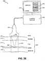
- imaging of a region of interest 206such as by imaging a region 222 and displaying images 224 of the region of interest 206 on a display 208, to facilitate localization of the treatment area and surrounding structures can initially be conducted.
- delivery of ultrasound energy 220 at a suitably depth, distribution, timing, and energy level to achieve the desired therapeutic effect of thermal injury or ablation to treat SMAS region 216can be suitably provided by probe 204 through control by control system 202.
- Monitoring of the treatment area and surrounding structures before, during, and after therapyi.e., before, during, and after the delivery of ultrasound energy to SMAS region 216, can be provided to plan and assess the results and/or provide feedback to control system 202 and a system user.
- Ultrasound imaging and providing of images 224can facilitate safe targeting of the SMAS layer 216.
- specific targeting for the delivery of energycan be better facilitated to avoid heating vital structures such as the facial nerve (motor nerve) 234, parotid gland (which makes saliva) 236, facial artery 238, and trigeminal nerve (for sensory functions) 232 among other regions.
- use of imaging with targeted energy delivery to provide a limited and controlled depth of treatmentcan minimize the chance of damaging deep structures, such as for example, the facial nerve that lies below the parotid, which is typically 10 mm thick.
- ultrasound imaging of region 222 of the region of interest 206can also be used to delineate SMAS layer 216 as the superficial, echo-dense layer overlying facial muscles 218.
- Such musclescan be seen via imaging region 222 by moving muscles 218, for example by extensional flexing of muscle layer 218 generally towards directions 250 and 252.
- imaging of region 222may be further enhanced via signal and image processing.
- the delivery of ultrasound energy 220 at a suitably depth, distribution, timing, and energy levelis provided by probe 204 through controlled operation by control system 202 to achieve the desired therapeutic effect of thermal injury to treat SMAS region 216.
- probe 204can also be mechanically and/or electronically scanned within tissue surface region 226 to treat an extended area.
- spatial control of a treatment depth 220can be suitably adjusted in various ranges, such as between a wide range of approximately 0 to 15 mm, suitably fixed to a few discrete depths, with an adjustment limited to a fine range, e.g. approximately between 3 mm to 9 mm, and/or dynamically adjusted during treatment, to treat SMAS layer 216 that typically lies at a depth between approximately 5 mm to 7 mm.
- monitoring of the treatment area and surrounding structurescan be provided to plan and assess the results and/or provide feedback to control system 202 and a system user.
- ultrasound imaging of region 222can be used to monitor treatment by watching the amount of shrinkage of SMAS layer 216 in direction of areas 260 and 262, such as in real time or quasi-real time, during and after energy delivery to region 220.
- the onset of substantially immediate shrinkage of SMAS layer 216is detectable by ultrasound imaging of region 222 and may be further enhanced via image and signal processing.
- the monitoring of such shrinkagecan be ideal because it can confirm the intended therapeutic goal of noninvasive lifting and tissue tightening; in addition, such monitoring may be used for system feedback.
- additional treatment parameters that can be suitably monitored in accordance with various other exemplary embodimentsmay include temperature, video, profilometry, strain imaging and/or gauges or any other suitable spatial, temporal and/or other tissue parameters.
- an exemplary monitoring method and system 200may suitably monitor the temperature profile or other tissue parameters of the region of interest 206, such as attenuation or speed of sound of treatment region 222 and suitably adjust the spatial and/or temporal characteristics and energy levels of ultrasound therapy transducer probe 204.
- the results of such monitoring techniquesmay be indicated on display 208 in various manners, such as, for example, by way of one-, two-, or three-dimensional images of monitoring results 270, or may comprise an indicator 272, such as a success, fail and/or completed/done type of indication, or combinations thereof.
- the targeting of particular region 220 within SMAS layer 216can be suitably be expanded within region of interest 206 to include a combination of tissues, such as skin 210, dermis 212, fat /adipose tissue 214, SMAS / muscular fascia / and/or other suspensory tissue 216, and muscle 218.
- Treatment of a combination of such tissues and/or fasciamay be treated including at least one of SMAS layer 216 or other layers of muscular fascia in combination with at least one of muscle tissue, adipose tissue, SMAS and/or other muscular fascia, skin, and dermis, can be suitably achieved by treatment system 200.
- treatment of SMAS layer 216may be performed in combination with treatment of dermis 280 by suitable adjustment of the spatial and temporal parameters of probe 204 within treatment system 200.
- ultrasound energypropagates as a wave with relatively little scattering, over depths up to many centimeters in tissue depending on the ultrasound frequency.
- the focal spot size achievable with any propagating wave energydepends on wavelength.
- Ultrasound wavelengthis equal to the acoustic velocity divided by the ultrasound frequency.
- Attenuation (absorption, mainly) of ultrasound by tissuealso depends on frequency.
- focused ultrasoundcan be used to create precise arrays of microscopic thermal ablation zones which have several advantages over fractional photothermolysis (FP).
- FPfractional photothermolysis
- ultrasound ablationcan mimic FP but utilize a simpler ablation device.
- ultrasoundcan produce an array of ablation zones much deeper into the skin or even into subcutaneous structures. Detection of changes in the reflection of ultrasound can be used for feedback control to detect a desired effect on the tissue and used to control the exposure intensity, time, and/or position.
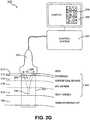
- an exemplary method and systemare configured for initially imaging a region 222 of a region of interest 206 and displaying that region 224 during the localization of the treatment area and surrounding structures. After localization, delivery of ultrasound energy 220 at a depth, distribution, timing, and energy level to achieve the desired therapeutic effect of thermal ablation to treat an epidermis layer 212, superficial dermis layer 214, mid-dermis layer 216, and/or deep dermis layer 218 can be provided.
- exemplary method and system 200can suitably monitor the treatment area and surrounding structures to plan and assess the results and/or provide feedback to control system 202 and/or a system user.
- an imaging functionmay be configured within control system 202 to facilitate imaging a region of interest
- an exemplary treatment system 200may also be configured for therapy only or therapy and monitoring, without imaging functions.
- prior known depth of the region of interestapproximately 0 to 5 mm or less, is employed to achieve treatment zones in photoaged skin.
- Probe 204 and/or transducers withincan be mechanically and/or electronically scanned in a direction 226 to place treatment zones 260 over an extended area, such as a line to generate a matrix of closely spaced treatment spots.
- Treatment depth 220can be adjusted between a range of approximately 0 to 5 mm, or otherwise until the depth of the deep dermis. Treatment may be confined to a fixed depth or a few discrete depths, or can be adjustment limited to a fine range, e.g. from approximately between 0 to 5 mm or the greatest depth of the deep dermis, or can be dynamically adjusted during treatment, to the treat region of interest 206 that lies above subcutaneous fat region 250.
- a treated zone 260may extend throughout regions of the dermis, and may even extend to the epidermis, 262.
- its cross sectionmay increase from small size 264 (sub millimeter) in a shallow region near or at the epidermis, to medium size 266 (sub millimeter to millimeter sized) in a middle zone near or at the mid dermis, to large size 268 (millimeter sized) in deep zones near or at the deep dermis.
- a single treated zonecan have a shape expanding in cross section with depth, and/or be composed of the fusion of several smaller treatment zones.
- the ultrasound beamcan be spatially and/or temporally controlled by changing the position of the transducer, its frequency, treatment depth, drive amplitude, and timing via the control system.
- the ultrasound beamcan be controlled as set forth in U.S. Patent Application Serial No.__________, filed October 6, 2005, and entitled METHOD AND SYSTEM FOR COTROLLED THERMAL INJURY OF HUMAN SUPERFICIAL TISSUE, and hereby incorporated by reference.
- an exemplary treatment method and system 200may be configured to monitor the temperature profile or other tissue parameters of region of interest 206, such as attenuation or speed of sound of the treatment region and suitably adjust the spatial and/or temporal characteristics and energy levels of the ultrasound therapy transducer.
- the results of such monitoring techniquesmay be indicated on display 208, such as through display of one-, two-, or three-dimensional images of monitoring results 270, or may comprise an indicator 272, such as a success, fail and/or completed/done type of indication, or combinations thereof.
- Additional treatment monitoring methodsmay be based on one or more of temperature, video, profilometry, strain imaging and/or gauges or any other suitable sensing method.
- an expanded region of interest 280can suitably include a combination of tissues, such as subcutaneous fat / adipose tissue 250.
- a combination of such tissuesincludes at least one of epidermis 212, superficial dermis 214, mid dermis 216, or deep dermis 218, in combination with at least one of muscle tissue, adipose tissue, or other tissues useful for treatment.
- treatment 260 of superficial dermismay be performed in combination with treatment 220 of subcutaneous fat 250 by suitable adjustment of the spatial and temporal parameters of transducers in probe 204.
- an exemplary treatment system 100configured to treat a region of interest (ROI) 106 comprises a control system 102, an imaging/therapy probe with acoustic coupling 104, and display system 108.
- ROIregion of interest
- Control system 102 and display 108can comprise various configurations for controlling functionality of probe 104 and system 100, including for example a microprocessor with software and a plurality of input/output and communication devices, a system for controlling electronic and/or mechanical scanning and/or multiplexing of transducers, a system for power delivery, systems for monitoring, systems for sensing the spatial position of the probe and/or temporal parameters of the transducers, and/or systems for handling user input and recording treatment input and results, among others.
- Imaging/therapy probe 104can comprise various probe and/or transducer configurations.
- probe 104can be configured for a combined dual-mode imaging/therapy transducer, coupled or co-housed imaging/therapy transducers, a separate therapy probe and separate imaging probe, or a single therapy probe.
- imaging transducersmay operate at frequencies from approximately 2 to 75 MHz or more, while therapy energy can be delivered at frequencies from approximately 2 to 50 MHz, with 2 MHz to 25 MHz being typical.
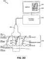
- an exemplary treatment method and systemare configured for initially imaging a region 222 within a region of interest 206 and displaying that region 224 on a display 208 to facilitate localization of the treatment area and surrounding structures, e.g., identification of sebaceous glands 232.
- delivery of ultrasound energy 220 at a depth, distribution, timing, and energy level to achieve the desired therapeutic effect of thermal ablation to treat a sebaceous gland 232is provided.
- monitoring of the treatment area and surrounding structurescan be conducted to further planning and assessing of the results and/or providing feedback to control system 202 and a system operator.
- localizationcan be facilitated through ultrasound imaging that can be used to define the position of a sebaceous gland and/or the depth of sebaceous glands over a region of interest. Such glands can be seen lying along hair follicles and their image may be further enhanced via signal and image processing. Ultrasound imaging can also be used for safety purposes, namely, to avoid injuring vital structures. In accordance with other exemplary embodiments, localization can also be accomplished without imaging region 222, but instead can be based on prior known depths of sebaceous glands or other target regions.
- probe 204 and/or imaging/therapy transducerscan be mechanically and/or electronically scanned, for example along direction 226, to place treatment zones over an extended area.
- a treatment depth 220can be adjusted between a range of approximately 1 to 7 mm, and/or the greatest depth of sebaceous glands 232.
- Such delivery of energycan occur through a repeated "image and burn” technique, i.e., imaging of the targeted sebaceous gland and then applying ultrasound energy, or through a "carpet bomb” technique, i.e., applying ultrasound energy at known depths over an extended area without initial or ongoing imaging.
- a treated zone 242may extend over a line, plane, or surface, or over an extended zone across the sebaceous gland depth 240 that typically ranges from approximately 1 to 7 mm.
- Probe 204can be mechanically and/or electronically scanned, for example directionally along 226, to extend treatment zone 242 over a large area.
- Probe 204can be further scanned or moved along a longer directional line 228 to further enlarge treatment zone 242.
- the cross sectional area of treated zone 242may increase in size from small to medium to large, i.e., at greater depths, the size of the treated lesion will increase.
- a treated zone 242can have a lesion shape expanding in cross section with depth, and/or be composed of the fusion of several smaller treatment zones.
- a "cross-stitched" series of lesions, a wedge shaped series of lesions, or any suitably formed conformal lesionscan be crated along treated zone 242.
- the ultrasound beam from probe 204can be spatially and/or temporally controlled by changing the spatial parameters of the transducer, such as the placement, distance, treatment depth and transducer structure, as well as by changing the temporal parameters of transducer, such as the frequency, drive amplitude, and timing, with such control handled via control system 202.
- Such spatial and temporal parameterscan also be suitably monitored and/or utilized in open-loop and/or closed-loop feedback systems within treatment system 200.
- conformal lesions of various, specifically targeted, shapes, sizes and orientationscan be configured along treatment zone 242.
- one or more treated zones 242can be configured to produce regions of heating and damage within the treatment layer in spatially defined patterns, such as a discrete locus of spaced treatment spots or two- or three- dimensional matrix of damage or destroyed tissue, e.g., a matrix of cross-stitched, ellipsoidal/cigar-shaped, wedge-shaped, mushroom-shaped or any other conformal lesions, rather than heating and destroying the entire volume of the target layer of tissue.
- the surrounding undamaged tissueaids rapid healing and recovery.
- an exemplary monitoring methodmay comprise monitoring the temperature profile or other tissue parameters of the region of interest 206, such as attenuation, speed of sound, or mechanical properties such as stiffness and strain of the treatment region and suitably adjust the spatial and/or temporal characteristics and energy levels of the ultrasound therapy transducer of probe 204.
- the results of such monitoring techniquesmay be indicated on display 208 by means of one-, two-, or three-dimensional images of monitoring results 250, or may simply comprise a success or fail-type indicator 252, or combinations thereof.
- Additional treatment monitoring techniquesmay be based on one or more of temperature, video, profilometry, and/or stiffness or strain gauges or any other suitable sensing technique.
- a treatment system 200can be configured for treatment over an expanded treatment region of interest 252 that includes a combination of tissues, such as subcutaneous fat / adipose tissue 216 and muscle 218, among others.
- a multiple of such tissuesmay be treated including sebaceous glands in combination with at least one of epidermis 212, dermis 214, adipose tissue 216, muscular fascia lying atop muscle tissue 218, mucous membrane, hair bulb 230, hair shaft 234, hair follicle between hair bulb 230 and epidermis 212, blood vessels, apocrine sweat glands, eccrine glands lying within dermis 214, fat 216 or muscle 218, and/or any other tissue of interest.
- a treatment to region 220 of sebaceous gland 232may be performed in combination with treatment to a region 260 of hair by suitable adjustment of the treatment spatial and/or temporal parameters of the transducers in probe 204.
- an ultrasound transducer probe and control systemare configured to deliver ultrasound energy to a targeted/specified depth and zone where the sweat gland population is required to be treated.
- the ultrasound beam from the transducer probecan be spatially and/or temporally adjusted, modified or otherwise controlled to match the adequate treatment of the sweat glands in the region of interest.
- imaging transducersmay operate at frequencies from approximately 2 MHz to 75 MHz or more, while therapy energy can be delivered at frequencies from approximately 500 kHz to 15 MHz, with 2 MHz to 25 MHz being typical.
- sweat glands 230are generally located within a dermis layer 214 at a depth close to hair bulbs 236.
- an ultrasound transducer probecan be coupled to the skin tissue using one of the numerous coupling media, such as water, mineral oils, gels, and the like.
- an exemplary treatment method and systemare configured for initially imaging a region 222 within a region of interest 206 and displaying that region 224 on a display 208 to facilitate localization of the treatment area and surrounding structures, e.g., identification of sweat glands 230.
- delivery of ultrasound energy 220 at a depth, distribution, timing, and energy level to achieve the desired therapeutic effect of thermal ablation to treat a sweat gland 230is provided.
- monitoring of the treatment area and surrounding structurescan be conducted to further planning and assessing of the results and/or providing feedback to control system 202 and a system operator.
- localizationcan be facilitated through ultrasound imaging that can be used to define the position of a sweat gland 230 and/or the depth of sweat glands 230 over a region of interest before depositing in a defined pattern at a target region 220.
- ultrasound imagingcan also be used for safety purposes, namely, to avoid injuring vital structures, such as nerve endings 240.
- localizationcan also be accomplished without imaging region 222, but instead can be based on prior known depths of sweat glands or other target regions, and thus be configured geometrically and/or electronically to selectively deposit energy at a particular known depth below skin surface 210 to a target region 220.
- the ultrasound beam from probe 204can be spatially and/or temporally controlled by changing the spatial parameters of the transducer, such as the placement, distance, treatment depth and transducer structure, as well as by changing the temporal parameters of transducer, such as the frequency, drive amplitude, and timing, with such control handled via control system 202.
- the temporal energy exposure at one locationmay range from approximately to 40 ms to 40 seconds, while the corresponding source frequency can suitably range from approximately 500 kHz to 15 MHz.
- Such spatial and temporal parameterscan also be suitably monitored and/or utilized in open-loop and/or closed-loop feedback systems within treatment system 200.
- conformal lesions of various, specifically targeted, shapes, sizes and orientationscan be configured within target region 220.
- the treatment resulting from ultrasound energy delivery in the region of sweat glands 230can be used to achieve selective ablation of regions of sub-epidermal region (0.5 - 10 mm diameter zones).
- one or more treated zones 242can be configured to produce regions of ablative damage in spatially defined patterns, such as a discrete locus of spaced treatment spots or two- or three- dimensional matrix of damage or destroyed tissue, e.g., a matrix of cross-stitched, ellipsoidal/cigar-shaped, wedge-shaped, mushroom-shaped or any other conformal lesions, rather than heating and destroying the entire volume of the target layer of tissue.
- the surrounding undamaged tissueaids rapid healing and recovery.
- treatment system 200could be configured to "carpet bomb" the fat layer at 1-7 mm depth, e.g., up to 90% of the sweat glands in the armpit can be ablated without any physiologic issues.
- an exemplary monitoring methodmay comprise monitoring the temperature profile or other tissue parameters of the region of interest 206, such as attenuation, speed of sound, or mechanical properties such as stiffness and strain of the treatment region and suitably adjust the spatial and/or temporal characteristics and energy levels of the ultrasound therapy transducer of probe 204.
- the results of such monitoring techniquesmay be indicated on display 208 by means of one-, two-, or three-dimensional images of monitoring results 250, or may simply comprise a success or fail-type indicator 252, or combinations thereof.
- Additional treatment monitoring techniquesmay be based on one or more of temperature, video, profilometry, and/or stiffness or strain gauges or any other suitable sensing technique.
- the non-thermal effects from an acoustic fieldcan also "shock" the sweat producing apocrine and eccrine cells in to reduced activity.
- These effects mentioned here as examplesare, but not limited to, acoustic cavitation, acoustic streaming, inter-cellular shear effects, cell resonant effects, and the like.
- focused or directive ultrasound energycan be used for the treatment of sweat glands in the armpit (without the combination of pharmacological formulations).
- a clinical indicationwould be to use in the management of Hidradenitis suppurativa.
- Ultrasound energy deposited at a selective depthcan also be used in combination with a number of pharmaceutical formulations that are currently prescribed for the treatment of sweat gland hyperactivity in the axillary region, palms and soles.
- the ultrasound energy delivered to the target region in combination with the pharmaceutical agents such as BOTOX® or retinoidscan help synergistically treat the sweat gland region by, (1) increasing activity of the agents due to the thermal and non-thermal mechanisms, (2) reduced requirement of overall drug dosage, as well as reducing the drug toxicity, (3) increase local effect of drug in a site selective manner.
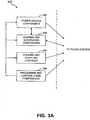
- an exemplary control system 300can be configured for coordination and control of the entire therapeutic treatment process for tissue treatment.
- control system 300can suitably comprise power source components 302, sensing and monitoring components 304, cooling and coupling controls 306, and/or processing and control logic components 308.
- Control system 300can be configured and optimized in a variety of ways with more or less subsystems and components to implement the therapeutic system for tissue treatment, and the embodiments in FIGS. 3A and 3B are merely for illustration purposes.
- control system 300can comprise one or more direct current (DC) power supplies 303 configured to provide electrical energy for entire control system 300, including power required by a transducer electronic amplifier/driver 312.
- a DC current sense device 305can also be provided to confirm the level of power going into amplifiers/drivers 312 for safety and monitoring purposes.
- Amplifiers/drivers 312can comprise multi-channel or single channel power amplifiers and/or drivers. In accordance with an exemplary embodiment for transducer array configurations, amplifiers/drivers 312 can also be configured with a beamformer to facilitate array focusing. An exemplary beamformer can be electrically excited by an oscillator/digitally controlled waveform synthesizer 310 with related switching logic.
- the power sourcing componentscan also include various filtering configurations 314. For example, switchable harmonic filters and/or matching may be used at the output of amplifier/driver 312 to increase the drive efficiency and effectiveness.
- Power detection components 316may also be included to confirm appropriate operation and calibration. For example, electric power and other energy detection components 316 may be used to monitor the amount of power going to an exemplary probe system.
- sensing and monitoring components 304may also be suitably implemented within control system 300.
- monitoring, sensing and interface control components 324may be configured to operate with various motion detection systems implemented within transducer probe 204 to receive and process information such as acoustic or other spatial and temporal information from a region of interest.
- Sensing and monitoring componentscan also include various controls, interfacing and switches 309 and/or power detectors 316.
- Such sensing and monitoring components 304can facilitate open-loop and/or closed-loop feedback systems within treatment system 200.
- Cooling/coupling control systems 306may be provided to remove waste heat from an exemplary probe 204, provide a controlled temperature at the superficial tissue interface and deeper into tissue, and/or provide acoustic coupling from transducer probe 204 to region-of-interest 206. Such cooling/coupling control systems 306 can also be configured to operate in both open-loop and/or closed-loop feedback arrangements with various coupling and feedback components.
- Processing and control logic components 308can comprise various system processors and digital control logic 307, such as one or more of microcontrollers, microprocessors, field-programmable gate arrays (FPGAs), computer boards, and associated components, including firmware and control software 326, which interfaces to user controls and interfacing circuits as well as input/output circuits and systems for communications, displays, interfacing, storage, documentation, and other useful functions.
- firmware and control software 326which interfaces to user controls and interfacing circuits as well as input/output circuits and systems for communications, displays, interfacing, storage, documentation, and other useful functions.
- System software and firmware 326controls all initialization, timing, level setting, monitoring, safety monitoring, and all other system functions required to accomplish user-defined treatment objectives.
- various control switches 308can also be suitably configured to control operation.
- transducer probe 204can also be configured in various manners and comprise a number of reusable and/or disposable components and parts in various embodiments to facilitate its operation.
- transducer probe 204can be configured within any type of transducer probe housing or arrangement for facilitating the coupling of transducer to a tissue interface, with such housing comprising various shapes, contours and configurations.
- Transducer probe 204can comprise any type of matching, such as for example, electric matching, which may be electrically switchable; multiplexer circuits and/or aperture/element selection circuits; and/or probe identification devices, to certify probe handle, electric matching, transducer usage history and calibration, such as one or more serial EEPROM (memories).
- Transducer probe 204may also comprise cables and connectors; motion mechanisms, motion sensors and encoders; thermal monitoring sensors; and/or user control and status related switches, and indicators such as LEDs.
- a motion mechanism in probe 204may be used to controllably create multiple lesions, or sensing of probe motion itself may be used to controllably create multiple lesions and/or stop creation of lesions, e.g. for safety reasons if probe 204 is suddenly jerked or is dropped.
- an external motion encoder armmay be used to hold the probe during use, whereby the spatial position and attitude of probe 104 is sent to the control system to help controllably create lesions.
- the therapy contemplated hereincan also be produced, for example, by transducers disclosed in U.S. Application Serial No. 10/944,499, filed on September 16, 2004 , entitled METHOD AND SYSTEM FOR ULTRASOUND TREATMENT WITH A MULTI-DIRECTIONAL TRANSDUCER and U.S. Application Serial No. 10/944,500, filed on September 16, 2004 , and entitled SYSTEM AND METHOD FOR VARIABLE DEPTH ULTRASOUND TREATMENT, both hereby incorporated by reference.
- a transducer probe 400can comprise a control interface 402, a transducer 404, coupling components 406, and monitoring/sensing components 408, and/or motion mechanism 410.
- transducer probe 400can be configured and optimized in a variety of ways with more or less parts and components to provide ultrasound energy for controlled thermal injury, and the embodiment in FIGS. 4A and 4B are merely for illustration purposes.
- Control interface 402is configured for interfacing with control system 300 to facilitate control of transducer probe 400.
- Control interface components 402can comprise multiplexer/aperture select 424, switchable electric matching networks 426, serial EEPROMs and/or other processing components and matching and probe usage information 430 and interface connectors 432.
- Coupling components 406can comprise various devices to facilitate coupling of transducer probe 400 to a region of interest.
- coupling components 406can comprise cooling and acoustic coupling system 420 configured for acoustic coupling of ultrasound energy and signals.
- Acoustic cooling/coupling system 420 with possible connections such as manifoldsmay be utilized to couple sound into the region-of-interest, control temperature at the interface and deeper into tissue, provide liquid-filled lens focusing, and/or to remove transducer waste heat.
- Coupling system 420may facilitate such coupling through use of various coupling mediums, including air and other gases, water and other fluids, gels, solids, and/or any combination thereof, or any other medium that allows for signals to be transmitted between transducer active elements 412 and a region of interest.
- coupling system 420can also be configured for providing temperature control during the treatment application.
- coupling system 420can be configured for controlled cooling of an interface surface or region between transducer probe 400 and a region of interest and beyond by suitably controlling the temperature of the coupling medium.
- the suitable temperature for such coupling mediumcan be achieved in various manners, and utilize various feedback systems, such as thermocouples, thermistors or any other device or system configured for temperature measurement of a coupling medium.
- Such controlled coolingcan be configured to further facilitate spatial and/or thermal energy control of transducer probe 400.
- acoustic coupling and cooling 1140can be provided to acoustically couple energy and imaging signals from transducer probe 1104 to and from the region of interest 1106, to provide thermal control at the probe to region-of-interest interface 1110 and deeper into tissue, and to remove potential waste heat from the transducer probe at region 1144.
- Temperature monitoringcan be provided at the coupling interface via a thermal sensor 1146 to provides a mechanism of temperature measurement 1148 and control via control system 1102 and a thermal control system 1142.
- Thermal controlmay consist of passive cooling such as via heat sinks or natural conduction and convection or via active cooling such as with peltier thermoelectric coolers, refrigerants, or fluid-based systems comprised of pump, fluid reservoir, bubble detection, flow sensor, flow channels/tubing 1144 and thermal control 1142.
- monitoring and sensing components 408can comprise various motion and/or position sensors 416, temperature monitoring sensors 418, user control and feedback switches 414 and other like components for facilitating control by control system 300, e.g., to facilitate spatial and/or temporal control through open-loop and closed-loop feedback arrangements that monitor various spatial and temporal characteristics.
- Motion mechanism 410can comprise manual operation, mechanical arrangements, or some combination thereof.
- a motion mechanism 422can be suitably controlled by control system 300, such as through the use of accelerometers, encoders or other position/orientation devices 416 to determine and enable movement and positions of transducer probe 400. Linear, rotational or variable movement can be facilitated, e.g., those depending on the treatment application and tissue contour surface.
- Transducer 404can comprise one or more transducers configured for treating of SMAS layers and targeted regions. Transducer 404 can also comprise one or more transduction elements and/or lenses 412.
- the transduction elementscan comprise a piezoelectrically active material, such as lead zirconante titanate (PZT), or any other piezoelectrically active material, such as a piezoelectric ceramic, crystal, plastic, and/or composite materials, as well as lithium niobate, lead titanate, barium titanate, and/or lead metaniobate.
- PZTlead zirconante titanate
- transducer 404can comprise any other materials configured for generating radiation and/or acoustical energy.
- Transducer 404can also comprise one or more matching layers configured along with the transduction element such as coupled to the piezoelectrically active material. Acoustic matching layers and/or damping may be employed as necessary to achieve the desired electroacoustic response.
- the thickness of the transduction element of transducer 404can be configured to be uniform. That is, a transduction element 412 can be configured to have a thickness that is substantially the same throughout. In accordance with another exemplary embodiment, the thickness of a transduction element 412 can also be configured to be variable. For example, transduction element(s) 412 of transducer 404 can be configured to have a first thickness selected to provide a center operating frequency of approximately 2 kHz to 75 MHz, such as for imaging applications. Transduction element 412 can also be configured with a second thickness selected to provide a center operating frequency of approximately 2 to 400 MHz, and typically between 4 MHz and 15 MHz for therapy application.
- Transducer 404can be configured as a single broadband transducer excited with at least two or more frequencies to provide an adequate output for generating a desired response. Transducer 404 can also be configured as two or more individual transducers, wherein each transducer comprises one or more transduction element. The thickness of the transduction elements can be configured to provide center-operating frequencies in a desired treatment range.
- Transducer 404may be composed of one or more individual transducers in any combination of focused, planar, or unfocused single-element, multi-element, or array transducers, including 1-D, 2-D, and annular arrays; linear, curvilinear, sector, or spherical arrays; spherically, cylindrically, and/or electronically focused, defocused, and/or lensed sources.
- transducer 500can be configured as an acoustic array to facilitate phase focusing. That is, transducer 500 can be configured as an array of electronic apertures that may be operated by a variety of phases via variable electronic time delays.
- transducer 500may be manipulated, driven, used, and/or configured to produce and/or deliver an energy beam corresponding to the phase variation caused by the electronic time delay.
- phase variationscan be used to deliver defocused beams, planar beams, and/or focused beams, each of which may be used in combination to achieve different physiological effects in a region of interest 510.
- Transducer 500may additionally comprise any software and/or other hardware for generating, producing and or driving a phased aperture array with one or more electronic time delays.
- Transducer 500can also be configured to provide focused treatment to one or more regions of interest using various frequencies.
- transducer 500can be configured with one or more variable depth devices to facilitate treatment.
- transducer 500may be configured with variable depth devices disclosed in U.S. Patent Application 10/944,500 , entitled “System and Method for Variable Depth Ultrasound", filed on September 16, 2004, having at least one common inventor and a common Assignee as the present application, and incorporated herein by reference.
- transducer 500can also be configured to treat one or more additional ROI 510 through the enabling of sub-harmonics or pulse-echo imaging, as disclosed in U.S.
- Patent Application 10/944,499entitled “Method and System for Ultrasound Treatment with a Multi-directional Transducer", filed on September 16, 2004, having at least one common inventor and a common Assignee as the present application, and also incorporated herein by reference.
- transducer 600may also be configured with an electronic focusing array 604 in combination with one or more transduction elements 606 to facilitate increased flexibility in treating ROI 610.
- Array 604may be configured in a manner similar to transducer 502. That is, array 604 can be configured as an array of electronic apertures that may be operated by a variety of phases via variable electronic time delays, for example, T 1 , T 2 ...T j .
- the electronic apertures of array 604may be manipulated, driven, used, and/or configured to produce and/or deliver energy in a manner corresponding to the phase variation caused by the electronic time delay.
- these phase variationscan be used to deliver defocused beams, planar beams, and/or focused beams, each of which may be used in combination to achieve different physiological effects in ROI 610.
- Transduction elements 606may be configured to be concave, convex, and/or planar.
- transduction elements 606Aare configured to be concave in order to provide focused energy for treatment of ROI 610. Additional embodiments are disclosed in U.S. Patent Application 10/944,500 , entitled “Variable Depth Transducer System and Method", and again incorporated herein by reference.
- transduction elements 606Bcan be configured to be substantially flat in order to provide substantially uniform energy to ROI 610. While Figs. 6A and 6B depict exemplary embodiments with transduction elements 604 configured as concave and substantially flat, respectively, transduction elements 604 can be configured to be concave, convex, and/or substantially flat. In addition, transduction elements 604 can be configured to be any combination of concave, convex, and/or substantially flat structures. For example, a first transduction element can be configured to be concave, while a second transduction element can be configured to be substantially flat.
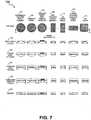
- transducer 404can be configured as single-element arrays, wherein a single-element 802, e.g., a transduction element of various structures and materials, can be configured with a plurality of masks 804, such masks comprising ceramic, metal or any other material or structure for masking or altering energy distribution from element 802, creating an array of energy distributions 808.
- Masks 804can be coupled directly to element 802 or separated by a standoff 806, such as any suitably solid or liquid material.
- An exemplary transducer 404can also be configured as an annular array to provide planar, focused and/or defocused acoustical energy.
- an annular array 1000can comprise a plurality of rings 1012, 1014, 1016 to N. Rings 1012, 1014, 1016 to N can be mechanically and electrically isolated into a set of individual elements, and can create planar, focused, or defocused waves. For example, such waves can be centered on-axis, such as by methods of adjusting corresponding transmit and/or receive delays, ⁇ 1 , ⁇ 2 , ⁇ 3 ... ⁇ N .
- An electronic focuscan be suitably moved along various depth positions, and can enable variable strength or beam tightness, while an electronic defocus can have varying amounts of defocusing.
- a lens and/or convex or concave shaped annular array 1000can also be provided to aid focusing or defocusing such that any time differential delays can be reduced. Movement of annular array 800 in one, two or three-dimensions, or along any path, such as through use of probes and/or any conventional robotic arm mechanisms, may be implemented to scan and/or treat a volume or any corresponding space within a region of interest.
- Transducer 404can also be configured in other annular or non-array configurations for imaging/therapy functions.
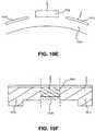
- a transducercan comprise an imaging element 1012 configured with therapy element(s) 1014.
- Elements 1012 and 1014can comprise a single-transduction element, e.g., a combined imaging/transducer element, or separate elements, can be electrically isolated 1022 within the same transduction element or between separate imaging and therapy elements, and/or can comprise standoff 1024 or other matching layers, or any combination thereof.
- a transducercan comprise an imaging element 1012 having a surface 1028 configured for focusing, defocusing or planar energy distribution, with therapy elements 1014 including a stepped-configuration lens configured for focusing, defocusing, or planar energy distribution.
- transducer 404may be configured to provide one, two and/or three-dimensional treatment applications for focusing acoustic energy to one or more regions of interest.
- transducer 404can be suitably diced to form a one-dimensional array, e.g., transducer 602 comprising a single array of sub-transduction elements.
- transducer 404may be suitably diced in two-dimensions to form a two-dimensional array.
- an exemplary two-dimensional array 900can be suitably diced into a plurality of two-dimensional portions 902.
- Two-dimensional portions 902can be suitably configured to focus on the treatment region at a certain depth, and thus provide respective slices 904 of the treatment region.
- the two-dimensional array 900can provide a two-dimensional slicing of the image place of a treatment region, thus providing two-dimensional treatment.
- transducer 404may be suitably configured to provide three-dimensional treatment.
- a three-dimensional systemcan comprise a transducer within probe 104 configured with an adaptive algorithm, such as, for example, one utilizing three-dimensional graphic software, contained in a control system, such as control system 102.
- the adaptive algorithmis suitably configured to receive two-dimensional imaging, temperature and/or treatment or other tissue parameter information relating to the region of interest, process the received information, and then provide corresponding three-dimensional imaging, temperature and/or treatment information.
- an exemplary three-dimensional systemcan comprise a two-dimensional array 900 configured with an adaptive algorithm to suitably receive 904 slices from different image planes of the treatment region, process the received information, and then provide volumetric information 906, e.g., three-dimensional imaging, temperature and/or treatment information. Moreover, after processing the received information with the adaptive algorithm, the two-dimensional array 900 may suitably provide therapeutic heating to the volumetric region 906 as desired.
- an adaptive algorithmto suitably receive 904 slices from different image planes of the treatment region, process the received information, and then provide volumetric information 906, e.g., three-dimensional imaging, temperature and/or treatment information.
- the two-dimensional array 900may suitably provide therapeutic heating to the volumetric region 906 as desired.
- an exemplary three-dimensional systemcan comprise a single transducer 404 configured within a probe arrangement to operate from various rotational and/or translational positions relative to a target region.
- ultrasound therapy transducer 700can be configured for a single focus, an array of foci, a locus of foci, a line focus, and/or diffraction patterns.
- Transducer 700can also comprise single elements, multiple elements, annular arrays, one-, two-, or three-dimensional arrays, broadband transducers, and/or combinations thereof, with or without lenses, acoustic components, and mechanical and/or electronic focusing.
- Transducers configured as spherically focused single elements 702, annular arrays 704, annular arrays with damped regions 706, line focused single elements 708, 1-D linear arrays 710, 1-D curvilinear arrays in concave or convex form, with or without elevation focusing, 2-D arrays, and 3-D spatial arrangements of transducersmay be used to perform therapy and/or imaging and acoustic monitoring functions.
- focusing and/or defocusingmay be in one plane or two planes via mechanical focus 720, convex lens 722, concave lens 724, compound or multiple lenses 726, planar form 728, or stepped form, such as illustrated in Fig, 10F .
- Any transducer or combination of transducersmay be utilized for treatment.
- an annular transducermay be used with an outer portion dedicated to therapy and the inner disk dedicated to broadband imaging wherein such imaging transducer and therapy transducer have different acoustic lenses and design, such as illustrated in Fig. 10C-10F .
- transduction elements 700may comprise a piezoelectrically active material, such as lead zirconante titanate (PZT), or any other piezoelectrically active material, such as a piezoelectric ceramic, crystal, plastic, and/or composite materials, as well as lithium niobate, lead titanate, barium titanate, and/or lead metaniobate.
- Transduction elements 700may also comprise one or more matching layers configured along with the piezoelectrically active material.
- transduction elements 700can comprise any other materials configured for generating radiation and/or acoustical energy. A means of transferring energy to and from the transducer to the region of interest is provided.
- an exemplary treatment system 200can be configured with and/or combined with various auxiliary systems to provide additional functions.
- an exemplary treatment system 1200 for treating a region of interest 1206can comprise a control system 1202, a probe 1204, and a display 1208.
- Treatment system 1200further comprises an auxiliary imaging modality 1274 and/or auxiliary monitoring modality 1272 may be based upon at least one of photography and other visual optical methods, magnetic resonance imaging (MRI), computed tomography (CT), optical coherence tomography (OCT), electromagnetic, microwave, or radio frequency (RF) methods, positron emission tomography (PET), infrared, ultrasound, acoustic, or any other suitable method of visualization, localization, or monitoring of SMAS layers within region-of-interest 1206, including imaging/monitoring enhancements.
- MRImagnetic resonance imaging
- CTcomputed tomography
- OCToptical coherence tomography
- RFradio frequency
- Such imaging/monitoring enhancement for ultrasound imaging via probe 1204 and control system 1202could comprise M-mode, persistence, filtering, color, Doppler, and harmonic imaging among others; furthermore an ultrasound treatment system 1270, as a primary source of treatment, may be combined with a secondary source of treatment 1276, including radio frequency (RF), intense pulsed light (IPL), laser, infrared laser, microwave, or any other suitable energy source.
- RFradio frequency
- IPLintense pulsed light
- laserinfrared laser
- microwaveor any other suitable energy source.
- treatment composed of imaging, monitoring, and/or therapy to a region of interestmay be further aided, augmented, and/or delivered with passive or active devices 1304 within the oral cavity.
- passive or active device 1304is a second transducer or acoustic reflector acoustically coupled to the cheek lining it is possible to obtain through transmission, tomographic, or round-trip acoustic waves which are useful for treatment monitoring, such as in measuring acoustic speed of sound and attenuation, which are temperature dependent; furthermore such a transducer could be used to treat and/or image.
- an active, passive, or active/passive object 1304may be used to flatten the skin, and/or may be used as an imaging grid, marker, or beacon, to aid determination of position.
- a passive or active device 1304may also be used to aid cooling or temperature control.
- Natural air in the oral cavitymay also be used as passive device 1304 whereby it may be utilized to as an acoustic reflector to aid thickness measurement and monitoring function.
Landscapes
- Health & Medical Sciences (AREA)
- Life Sciences & Earth Sciences (AREA)
- Engineering & Computer Science (AREA)
- General Health & Medical Sciences (AREA)
- Veterinary Medicine (AREA)
- Public Health (AREA)
- Animal Behavior & Ethology (AREA)
- Biomedical Technology (AREA)
- Physics & Mathematics (AREA)
- Nuclear Medicine, Radiotherapy & Molecular Imaging (AREA)
- Radiology & Medical Imaging (AREA)
- Heart & Thoracic Surgery (AREA)
- Surgery (AREA)
- Molecular Biology (AREA)
- Medical Informatics (AREA)
- Pathology (AREA)
- Biophysics (AREA)
- Remote Sensing (AREA)
- Radar, Positioning & Navigation (AREA)
- Acoustics & Sound (AREA)
- Dentistry (AREA)
- Oral & Maxillofacial Surgery (AREA)
- Gynecology & Obstetrics (AREA)
- General Physics & Mathematics (AREA)
- Epidemiology (AREA)
- Rehabilitation Therapy (AREA)
- Pain & Pain Management (AREA)
- Computer Networks & Wireless Communication (AREA)
- Physical Education & Sports Medicine (AREA)
- Vascular Medicine (AREA)
- Surgical Instruments (AREA)
- Thermotherapy And Cooling Therapy Devices (AREA)
- Ultra Sonic Daignosis Equipment (AREA)
- Mechanical Engineering (AREA)
- Cosmetics (AREA)
Abstract
Description
- The present invention relates to ultrasound therapy and imaging systems, and in particular to a method and system for noninvasive tissue treatment, such as for use in face lifts and deep tissue tightening, and/or in treatment of photoaged tissue, acne and sebaceous glands, and sweat glands.
- Coarse sagging of the skin and facial musculature occurs gradually over time due to gravity and chronic changes in connective tissue generally associated with aging. Invasive surgical treatment to tighten such tissues is common, for example by facelift procedures. In these treatments for connective tissue sagging, a portion of the tissue is usually removed, and sutures or other fasteners are used to suspend the sagging tissue structures. On the face, the Superficial Muscular Aponeurosis System (SMAS) forms a continuous layer superficial to the muscles of facial expression and beneath the skin and subcutaneous fat. Conventional face lift operations involve suspension of the SMAS through such suture and fastener procedures.
- No present procedures have been developed yet, which provide the combination of targeted, precise, local heating to a specified temperature region capable of inducing ablation (thermal injury) to underlying skin and subcutaneous fat. Attempts have included the use of radio frequency (RF) devices that have been used to produce heating and shrinkage of skin on the face with some limited success as a non-invasive alternative to surgical lifting procedures. However, RF is a dispersive form of energy deposition. RF energy is impossible to control precisely within the heated tissue volume and depth, because resistive heating of tissues by RF energy occurs along the entire path of electrical conduction through tissues. Another restriction of RF energy for non-invasive tightening of the SMAS is unwanted destruction of the overlying fat and skin layers. The electric impedance to RF within fat, overlying the suspensory connective structures intended for shrinking, leads to higher temperatures in the fat than in the target suspensory structures. Similarly, mid-infrared lasers and other light sources have been used to non-invasively heat and shrink connective tissues of the dermis, again with limited success. However, light is not capable of non-invasive treatment of SMAS because light does not penetrate deeply enough to produce local heating there. Below a depth of approximately 1 mm, light energy is multiply scattered and cannot be focused to achieve precise local heating.
- In addition to sagging of skin and facial musculature regions being a concern to aging individuals, photoaging of human skin is a complex response due to inflammation, oxidative injury, cellular and extracellular changes induced by decades of sunlight exposure. UV wavelengths are thought to be mainly responsible. Both of the primary skin layers, epidermis and dermis, are affected. Epidermal photoaging includes pigmentary lesions called ephilides (freckles) and solar lentigines (larger pigmented spots), plus pre-cancerous clonal lesions of keratinocytes called actinic keratoses. Thermal destruction of part or all of the epidermis, the outermost cellular layer of skin about 0.1 mm thick, is an effective treatment for epidermal photoaging. For example, lasers that vaporize epidermis are highly effective in a treatment called laser resurfacing. However laser resurfacing creates a significant skin wound with risk of infection, and prolonged healing. Dermal changes of photoaging include solar elastosis (an accumulation of abnormally-formed elastin fibers in the upper reticular layer of the dermis), laxity, loss of elasticity, fine and coarse wrinkles. Laser resurfacing to a depth below the dermo-epidermal junction can be highly effective for improving dermal photoaging, through a process of stimulated wound healing. Deep chemical peels, dermabrasion and other methods of destruction of epidermis and/or dermis are also effective, and also produce a significant open skin wound with risk of infection and delayed healing.
- Patterns of stimulated thermal damage to epidermis and/or dermis are also effective for treatment of photoaging. Recently, "fractional photothermolysis" using mid-infrared lasers to produce a microscopic array of thermal injury zones that include both epidermis and dermis was reported to be effective and well-tolerated for treatment of photoaging (D.Manstein et al. "Fractional Photothermolysis: a new concept for cutaneous remodeling using microscopic patterns of thermal injury." Lasers Surg Med 34:426-438, 2004). A primary advantage of fractional photothermolysis is that each zone of thermal injury is smaller than can be easily seen with the unaided eye, and surrounded by a zone of healthy tissue that initiates a rapid healing response. As described Manstein, the epidermis is stimulated to heal rapidly and without creating an open wound. The microscopic zones of thermally injured epidermis slough harmlessly from the skin surface after several days to several weeks, leaving a rejuvenated epidermis with less photoaging changes. Repeat treatments, which are well tolerated, can be performed until a desired result is obtained. The microscopic zones of thermal injury with fractional photothermolysis extend well into the dermis, as well. Dermis does not heal as rapidly as epidermis, in general. Over weeks to months following treatment, some of the abnormal dermis due to photoaging is remodeled, however, leading to improvement in laxity, wrinkles and skin texture.
- Fractional photothermolysis (FP) is intrinsically limited to regions of approximately the upper 1-millimeter of skin. The basic concept of producing well-controlled arrays of thermal injury is therefore limited with fractional photothermolysis, to superficial aspects of photoaging. Aging, which also causes laxity of the skin, and photoaging involve deeper layers of the dermis. Solar elastosis can extend throughout the dermis, to approximately 3 mm deep or more. Laxity and loss of elasticity due to aging are bulk problems of the dermis.
- A fundamental requirement for producing arrays of small thermal injury zones using a source of radiant energy that propagates and is absorbed within tissue, is that the source of radiant energy be capable of being adequately delivered to the tissue depth for which the array is desired. Near the skin surface, light can be used, as in fractional photothermolysis. However, light that propagates more than about 1 mm through skin has been multiplied scattered, and can no longer be focused or delivered.
- Acne vulgaris is the most common skin disorder. Acne causes temporary and permanent disfigurement. Acne typically appears on the face, back and/or chest at the onset of adrenarchy, i.e. when sex hormone activity increases in both boys and girls near puberty. Acne is a disorder of hair follicles, in which a plug forms within the outflow tract of the hair follicle. Sebum, an oily product of sebaceous glands attached to each hair follicle, and cellular debris builds in the plug. Inflammation and often rupture of the hair follicles ensues, leading to gross inflammation, pus (a "whitehead"), pain, bleeding, and/or eventually scarring. If the acne lesion consists of an accumulated unruptured plug within the hair follicle, a "blackhead" forms. If the follicle ruptures superficially, a small pustule forms that often heals after a few weeks without scarring. If the follicle ruptures within the mid or deep dermis, a painful cystic abscess forms. Cystic acne usually heals with permanent and disfiguring scars.
- The exact pathophysiology of acne is complex and is not fully understood. However, several basic elements are necessary to produce an acne lesion, and acne therapies are based on attacking one or more of these basic elements. First, an active sebaceous gland is necessary. The most potent treatments for acne are oral retinoids such as retinoic acid (Accutane), which inhibit sebaceous gland function. Sebaceous gland activity is driven by androgen and other sex steroid hormones. Women often experience cycle-dependent acne that may respond to treatment with birth control pills containing low amounts of progestins. Second, a plug must form in the outflow tract of the follicle, called the infundibulum. Bacteria, particularlyProprionobacteria acnes (P acnes) that digest sebum and follicular debris, contribute to plugging. Topical retinoids, mild acids and benzoyl peroxide are used as treatments to decrease follicular plugging. Antibiotics effective against P acnes are given either topically or orally; the prevalence of antibiotic-resistant P acnes is increasing. Third, inflammation is part of the process that breaks down the wall of a follicle containing plugs, leading to rupture of the follicle with release of irritating materials into the skin, abscess formation, and scarring. Anti-inflammatory agents including some antibiotics are helpful in treating acne.
- The most potent treatment for acne at present is oral retinoid therapy. Unfortunately, this is a toxic and teratogenic treatment. Unplanned pregnancies in women taking Accutane lead to a high rate of fetal malformations. An aggressive program to prevent this in the US was implemented, but has failed to prevent the problem. Systemic retinoid treatment also causes major side effects including extreme dryness during treatment, risk of hepatitis, bone changes, mood changes, and others. The high effectiveness and high toxicity of oral retinoids for treatment of cystic acne strongly suggests that an alternative treatment that targets sebaceous glands is needed.
- The sweat glands in the body are of divided into apocrine and eccrine glands. Apocrine glands are similar to sebaceous glands, and are present mainly in the axillae. These glands, like sebaceous glands, secrete an oily proteinaceous product into the follicles. Bacterial digestion of apocrine sweat is largely responsible for underarm "body odor". Similarly, eccrine sweat glands are present deep in the dermis in the palms, soles and armpits and are responsible for temperature regulation resulting from sweating. Excessive activity of these glands also results in copious amounts of abnormal sweating ("hyperhidrosis"), primarily under autonomic neuronal control. Reduction of sweating from under the armpits and other regions is a particularly desirable effect within the modern society. Presently, chemical antiperspirants and deodorants are used frequently as a matter of personal hygiene. Antiperspirants are aluminum based salts that block the sweat gland ducts. The deodorant changes the pH of the skinmilieu thereby minimizing the presence of (smell inducing) bacteria. The effects with both these components however, are temporary and these chemicals are known to irritate the skin in a good percentage of users.
- Further, there is currently a significant unmet need in managing the excessive sweating and concomitant issues with odor as a result ofHydradenitis suppurativa (irritable infected armpit). This acne-like process in apocine follicles also causes hydradenitis suppurativa, which is often a devastating condition in which very painful cysts and scarring occurs repeatedly in the axillae. The etiology (causes) of this clinical condition is not well understood. However, there are a number of marginally effective approaches to manage this condition. Retinoid drug therapy works marginally but is associated with severe toxicity. Some prescription formulations of antiperspirants can be used, but they are not particularly effective. These preparations can be applied with the addition of an iontophoretic device. This technique however, is not known to be any more effective than the formulation. The sweat glands can be surgically removed from the armpits and/or the sympathetic nerve supply can be interrupted surgically. This approach is fraught with its own drawbacks in terms of morbidity, scarring and cost. BOTOX® is being used ever more for paralyzing the nerve connections that induce excessive sweating in the armpits. However, this is a new approach yet to be completely validated. This technique requires multiple injections (painful) and the results last a few months only (3-4 months), hence need to be repeated. This technique does not get rid of the odor associated with the condition.
- A method and system for noninvasive tissue treatment, such as for use in face lifts and deep tissue tightening, and/or in treatment of photoaged tissue, acne and sebaceous glands, and/or sweat glands, are provided.
- For example, for facilitating face lifts and deep tissue tightening, an exemplary method and treatment system can be configured for the imaging, monitoring, and thermal injury to treat the SMAS region. In accordance with an exemplary embodiment, the exemplary method and system are configured for treating the SMAS region by first, imaging of the region of interest for localization of the treatment area and surrounding structures, second, delivery of ultrasound energy at a depth, distribution, timing, and energy level to achieve the desired therapeutic effect, and third to monitor the treatment area before, during, and after therapy to plan and assess the results and/or provide feedback.
- In accordance with an exemplary embodiment, an exemplary treatment system comprises an imaging/therapy probe, a control system and display system. The imaging/therapy probe can comprise various probe and/or transducer configurations. For example, the probe can be configured for a combined dual-mode imaging/therapy transducer, coupled or co-housed imaging/therapy transducers, or simply a therapy probe and an imaging probe. The control system and display system can also comprise various configurations for controlling probe and system functionality, including for example a microprocessor with software and a plurality of input/output devices, a system for controlling electronic and/or mechanical scanning and/or multiplexing of transducers, a system for power delivery, systems for monitoring, systems for sensing the spatial position of the probe and/or transducers, and systems for handling user input and recording treatment results, among others.
- In accordance with an exemplary embodiment, ultrasound imaging can be utilized for safety purposes, such as to avoid injuring vital structures such as the facial nerve (motor nerve), parotid gland, facial artery, and trigeminal nerve (for sensory functions) among others. For example, ultrasound imaging can be used to identify SMAS as the superficial layer well defined by echoes overlying the facial muscles. Such muscles can be readily seen and better identified by moving them, and their image may be further enhanced via signal and image processing.
- In accordance with an exemplary embodiment, ultrasound therapy via focused ultrasound, an array of foci, a locus of foci, a line focus, and/or diffraction patterns from single element, multiple elements, annular array, one-, two-, or three-dimensional arrays, broadband transducers, and/or combinations thereof, with or without lenses, acoustic components, mechanical and/or electronic focusing are utilized to treat the SMAS region at fixed and/or variable depth or dynamically controllable depths and positions.
- In addition to face lifts and deep tissue tightening, a method and system for ultrasound treatment of photoaged tissue can be provided. For example, an exemplary method and system can be configured for first, ultrasound imaging of the region of interest for localization of the treatment area, second, delivery of ultrasound energy at a depth and pattern to achieve the desired therapeutic effects, and third to monitor the treatment area during and after therapy to assess the results and/or provide feedback. The exemplary treatment method and system can be configured for producing arrays of sub-millimeter and larger zones of thermal ablation to treat the epidermal, superficial dermal, mid-dermal and deep dermal components of photoaged tissue.
- In accordance with an exemplary embodiment, the treatment method and system use focused, unfocused, and/or defocused ultrasound for treatment of epidermal, superficial dermal, dermal, mid-dermal, and/or deep dermal components of photoaged tissue by adjusting the strength, depth, and/or type of focusing, energy levels and timing cadence. For example, focused ultrasound can be used to create precise arrays of microscopic thermal damage much deeper into the skin or even into subcutaneous structures. Detection of changes in the reflection of ultrasound can be used for feedback control to detect a desired effect on the tissue and used to control the exposure intensity, time, and/or position.
- In accordance with an exemplary embodiment, an exemplary treatment system comprises an imaging/therapy probe, a control system and display system. The imaging/therapy probe can comprise various probe and/or transducer configurations. For example, the probe can be configured for a combined dual-mode imaging/therapy transducer, coupled or co-housed imaging/therapy transducers, a separate therapy probe and imaging probe, or a single therapy probe. The control system and display system can also comprise various configurations for controlling probe and system functionality, including for example a microprocessor with software and a plurality of input/output and communication devices, a system for controlling electronic and/or mechanical scanning and/or multiplexing of transducers, a system for power delivery, systems for monitoring, systems for sensing the spatial position of the probe and/or temporal parameters of the transducers, and systems for handling user input and recording treatment input and results, among others.
- A method and system for ultrasound treatment of acne and sebaceous glands are provided. An exemplary method and system are configured for targeted treatment of sebaceous glands in various manners, such as through use of therapy only, therapy and monitoring, imaging and therapy, or therapy, imaging, and monitoring. Targeted therapy of sebaceous glands can be provided through use of focused, unfocused, or defocused ultrasound at various spatial and temporal energy settings.
- An exemplary method and system are configured to produce regions of heating and damage in various manners. For example, an exemplary method and system can be configured to produce regions of heating and damage by destroying the function of sebaceous glands within a user-specified treatment layer depth associated with the glands to be treated. In addition, an exemplary method and system can be configured to produce regions of heating and damage within the treatment layer in spatially defined patterns, rather than heating and destroying the entire volume of the target layer of tissue. Further, an exemplary method and system can be configured to specifically aim such regions of heating and damage within the treatment layer, to occur at the same location as the secretory portion of sebaceous glands.
- In accordance with an exemplary embodiment, an exemplary treatment system comprises a control system, an imaging/therapy probe, and display system. The imaging/therapy probe can comprise various probe and/or transducer configurations. For example, the probe can be configured for a combined dual-mode imaging/therapy transducer, coupled or co-housed imaging/therapy transducers, a therapy probe, or simply a therapy probe and an imaging probe. The control system and display system can also comprise various configurations for controlling probe and system functionality, including for example a microprocessor with software and a plurality of input/output devices, a system for controlling electronic and/or mechanical scanning and/or multiplexing of transducers, a system for power delivery, systems for monitoring, systems for sensing the spatial position of the probe and/or transducers, and systems for handling user input and recording treatment results, among others.
- In accordance with an exemplary embodiment, ultrasound imaging can be used for safety purposes, namely, to avoid injuring vital structures. In accordance with another exemplary embodiment, ultrasound imaging can be used to define the position of a sebaceous gland and/or the depth of sebaceous glands over a region of interest. Such glands can be seen lying along hair follicles and their image may be further enhanced via signal and image processing.
- In accordance with an exemplary embodiment, ultrasound therapy via focused, unfocused, or defocused ultrasound, delivered via an array of foci or array of treatment zones, a locus of foci or locus treatment zones, a line focus or linear treatment zone, a surface or volume focus or surface or volume treatment zone, and/or diffraction patterns from single element, multiple elements, annular array, one-, two-, or three-dimensional arrays, broadband transducers, and/or combinations thereof, with or without lenses, acoustic components, mechanical and/or electronic focusing or defocusing are utilized to treat sebaceous glands at fixed and/or variable depth or dynamically controllable depths and positions.
- The present invention describes a non-invasive method and system for using therapeutic ultrasound energy for the treatment of conditions resulting from sweat gland disorders. An ultrasound system and method comprises a transducer probe and control system configured to deliver ultrasound energy to the regions of the superficial tissue (e.g., skin) such that the energy can be deposited at the particular depth at which the aberrant sweat gland population is located below the skin surface.
- In accordance with various exemplary embodiments, the ultrasound transducer can be driven at a number of different frequency regimes such that the depth and shape of energy concentration can match the region of treatment. In addition, the ultrasound source or beam radiated from the transducer can be highly focused, weakly focused, or divergent, each in a cylindrical or spherical geometric configuration, and/or can also be planar to radiate a directive beam through the tissue, or various other configurations. Further, the ultrasound field can be varied spatially and temporally in a suitable manner to achieve the optimal tissue effect and/or type of conformal lesion for treating the sweat glands.
- The invention also extends to the following statements:
- 1. An ultrasound treatment system configured for noninvasive face lifts and deep tissue tightening, said ultrasound treatment system comprising:
- a control system for facilitating control of the ultrasound treatment system;
- a display system for displaying an image of a SMAS layer within a region of interest, said display system coupled to said control system; and
- a probe system configured for targeted delivery of ultrasound energy into said SMAS layer for destruction of tissue and to cause shrinkage of said SMAS layer.
- 2. The ultrasound treatment system according to
statement 1, wherein said control system is configured for localization of said SMAS layer through imaging prior to energy delivery, for controlling targeted delivery of energy through said probe system to said SMAS layer, and monitoring of said SMAS layer. - 3. The ultrasound treatment system of
statement 2, wherein said control system comprises an imaging system configured for monitoring of said SMAS layer prior to, during and after delivery of ultrasound energy into said SMAS layer - 4. The ultrasound treatment system of
statement 3, wherein said imaging system is configured to eliminate the damaging of deep tissue structures proximal said SMAS layer. - 5. The ultrasound treatment system of
statement 3, wherein said imaging system is configured to generate three-dimensional imaging information. - 6. The ultrasound treatment system according to
statement 1, wherein said display system comprises a display of images corresponding to said SMAS layer. - 7. The ultrasound treatment system according to
statement 1, wherein said probe system comprises an imaging and therapy transducer configured for targeted delivery of ablative ultrasound within said SMAS layer. - 8. The ultrasound treatment system according to statement 7, wherein said imaging and therapy transducer comprise a combined transducer within a single transduction element.
- 9. The ultrasound treatment system according to statement 7, wherein said imaging and therapy transducer comprises an array of electronic apertures configured for electronic phase delays to achieve one of focused, defocused and planar ultrasound energy distribution.
- 10. The ultrasound treatment system according to statement 8, wherein said treatment system comprises an imaging, therapy, and treatment monitoring system combined with auxiliary imaging and treatment monitoring apparatus and secondary therapy systems.
- 11. The ultrasound treatment system according to statement 10, wherein said auxiliary imaging apparatus comprises at least one of a photographic device and an optical modality.
- 12. The ultrasound treatment system according to
statement 1, wherein said probe system is configured for targeted delivery of ultrasound energy into at least one of skin, dermis, muscular facia, and adipose in addition to said SMAS layer for destruction of tissue and to cause shrinkage of said SMAS layer - 13. A method for providing noninvasive face lifts and deep tissue tightening, said method comprising:
- Localizing of a SMAS-like muscular fascia layer within a region of interest;
- targeting of delivery of ablative ultrasound energy to said SMAS-like muscular fascia layer; and
- monitoring of results of said targeted delivery within said SMAS-like muscular fascia layer during and after said targeted delivery to continue planning of treatment.
- 14. The method of statement 13, wherein said localizing comprises imaging a region of interest to delineate said SMAS-like muscular fascia layer.
- 15. The method of statement 14, said localizing of said SMAS-like muscular fascia layer comprises moving facial muscles below such SMAS-like muscular fascia layer.
- 16. The method of statement 13, wherein said targeting of delivery comprises adjustment of spatial and temporal parameters based on imaging of said SMAS-like muscular fascia layer location.
- 17. The method of statement 13, wherein said monitoring of results comprises detecting of shrinkage of said SMAS-like muscular fascia layer as visualized during after said target delivery of ablative ultrasound.
- 18. The method of statement 13, wherein said targeting of delivery of ablative ultrasound energy to said SMAS-like muscular fascia layer further comprises targeting of delivery of ablative ultrasound into at least one of skin, dermis, muscular facia, and adipose in addition to said SMAS-like muscular fascia layer.
- 19. The method of statement 13, wherein said targeting of delivery of ablative ultrasound energy to said SMAS-like muscular fascia layer comprises targeting of delivery of ablative ultrasound into at least one of platysma, temporal fascia, and/or occipital fascia.
- 20. The method according to statement 13, wherein said targeting of delivery further comprises cooling through any tissue regions between and including skin and said SMAS-like muscular fascia layer to facilitate treatment.
- 21. An ultrasound treatment system configured for treatment of the photoaged effects in tissue, said ultrasound treatment system comprising:
- a control system for facilitating control of the ultrasound treatment system;
- a display system for displaying an image of at least one of epidermal, superficial dermal, mid-dermal and deep dermal tissues within a region of interest, said display system coupled to said control system; and
- a probe system configured for targeted delivery of ultrasound energy producing arrays of sub-millimeter and larger zones of thermal ablation to treat said at least one of said epidermal, superficial dermal, mid-dermal and deep dermal tissue to cause reduction of the photoaged effects in tissue.
- 22. The ultrasound treatment system according to statement 21, wherein said control system is configured for localization of said at least one of said epidermal, superficial dermal, mid-dermal and deep dermal tissues through imaging prior to energy delivery, for controlling targeted delivery of energy through said probe system to said at least one of said epidermal, superficial dermal, mid-dermal and deep dermal tissues, and monitoring of said at least one of said epidermal, superficial dermal, mid-dermal and deep dermal tissues after said target delivery.
- 23. The ultrasound treatment system of statement 22, wherein said control system comprises an imaging system configured for monitoring of said at least one of said epidermal, superficial dermal, mid-dermal and deep dermal tissues prior to, during and after delivery of ultrasound energy into said at least one of said epidermal, superficial dermal, mid-dermal and deep dermal tissues.
- 24. The ultrasound treatment system of statement 23, wherein said imaging system is configured to provide feedback to facilitate said targeted delivery.
- 25. The ultrasound treatment system according to statement 21, wherein said display system comprises a display of images corresponding to said at least one of said epidermal, superficial dermal, mid-dermal and deep dermal tissues.
- 26. The ultrasound treatment system according to statement 21, wherein said probe system comprises an imaging and therapy transducer configured for targeted delivery of ablative ultrasound within said at least one of said epidermal, superficial dermal, mid-dermal and deep dermal tissues.
- 27. The ultrasound treatment system according to statement 26, wherein said imaging and therapy transducer comprise a combined transducer within a single transduction element.
- 28. The ultrasound treatment system according to statement 26, wherein said imaging and therapy transducer comprises a single element array comprising a single transduction element and a plurality of masks configured for generating a plurality of ablative zones with each of said plurality of ablative zones providing at least one of focused, defocused and planar ultrasound energy distribution.
- 29. The ultrasound treatment system according to statement 21, wherein said treatment system comprises an imaging, therapy, and treatment monitoring system combined with at least one of an auxiliary imaging and treatment monitoring apparatus and secondary therapy systems.
- 30. The ultrasound treatment system according to statement 21, wherein said auxiliary imaging comprises at least one of a photographic instrument and an optical modality.
- 31. A method for providing treatment of photoaged tissue, said method comprising:
- localizing of at least one of epidermal, superficial dermal, mid-dermal and deep dermal tissues within a region of interest;
- targeting of delivery of ablative ultrasound energy from a transducer probe to said at least one of said epidermal, superficial dermal, mid-dermal and deep dermal tissues; and
- monitoring of results of said targeted delivery within said at least one of said epidermal, superficial dermal, mid-dermal and deep dermal tissue during and after said targeted delivery to continue planning of treatment.
- 32. The method of statement 31, wherein said localizing comprises imaging a region of interest to identify said at least one of said epidermal, superficial dermal, mid-dermal and deep dermal tissue.
- 33. The method of statement 31, said targeting of delivery of ablative ultrasound energy comprises producing arrays of sub-millimeter and larger zones of thermal ablation within said at least one of said epidermal, superficial dermal, mid-dermal and deep dermal tissue.
- 34. The method of statement 31, wherein said targeting of delivery comprises adjustment of spatial and temporal parameters based on imaging of said at least one of said epidermal, superficial dermal, mid-dermal and deep dermal tissue location.
- 35. The method of statement 31, wherein said monitoring of results comprises measurement of results of treatment of said at least one of said epidermal, superficial dermal, mid-dermal and deep dermal tissue as visualized during and after said target delivery of ablative ultrasound.
- 36. The method of statement 31, wherein said producing arrays of sub-millimeter and larger zones of thermal ablation comprises scanning said transducer probe over a line above said photoaged tissue to produce a matrix of spaced treatment spots.
- 37. The method of statement 36, wherein said producing of a matrix of spaced treatment spots comprises adjusting of treatment depths within said region of interest.
- 38. The method of statement 31, wherein said localizing comprises generating three-dimensional imaging information.
- 39. The method of statement 31, wherein said targeting comprises treating three-dimensional treatment region.
- 40. The method according to statement 31, wherein said targeting of delivery further comprises cooling through tissue regions comprising said at least one of said epidermal, superficial dermal, mid-dermal and deep dermal tissue to facilitate treatment.
- 41. An ultrasound treatment system configured for treatment of acne and sebaceous glands, said ultrasound treatment system comprising:
- a control system for facilitating control of the ultrasound treatment system; and
- a probe system configured for targeted delivery of ultrasound energy to produce a treatment region for destroying the function of a sebaceous gland.
- 42. The ultrasound treatment system according to statement 41, wherein said probe system is configured for destroying the function of said sebaceous gland within a specified treatment depth identified through localization of said sebaceous gland.
- 43. The ultrasound treatment system according to statement 41, wherein said probe is configured to produce said treatment regions in spatially defined patterns to facilitate healing of tissue.
- 44. The ultrasound treatment system according to statement 43, wherein said spatially defined patterns comprise a discrete locus of spaced lesions comprising at least one of cross-stitch, cigar-shaped, ellipsoidal, mushroom-shaped and wedge-shaped lesions.
- 45. The ultrasound treatment system of statement 41, wherein said control system comprises an imaging system configured for monitoring of said treatment region prior to, during and after delivery of ultrasound energy into said sebaceous gland.
- 46. The ultrasound treatment system of statement 45, wherein said imaging system is configured to provide feedback to facilitate said targeted delivery.
- 47. The ultrasound treatment system according to statement 41, wherein said display system comprises a display of images corresponding to a temperature profile of said treatment region.
- 48. The ultrasound treatment system according to statement 41, wherein said probe system comprises an imaging and therapy transducer configured for targeted delivery of ablative ultrasound within said treatment region.
- 49. The ultrasound treatment system according to statement 48, wherein said imaging and therapy transducer comprise a combined transducer within an electronic array of transduction elements.
- 50. The ultrasound treatment system according to statement 48, wherein said imaging and therapy transducer comprises a single element array comprising a single transduction element and a plurality of masks configured for generating a plurality of ablative zones with each of said plurality of ablative zones providing at least one of focused, defocused and planar ultrasound energy distribution.
- 51. The ultrasound treatment system according to statement 41, wherein said probe is configured for targeted delivery of ultrasound energy through adjustable control of spatial parameters and temporal parameters of said probe to generate conformal lesions of specifically targeted shapes, sizes and orientations.
- 52. The ultrasound treatment system according to statement 41, wherein said treatment system comprises an imaging, therapy, and treatment monitoring system combined with auxiliary imaging and treatment monitoring apparatus and secondary therapy systems.
- 53. The ultrasound treatment system according to statement 52, wherein said auxiliary imaging apparatus comprises at least one of a photographic instrument and an optical modality.
- 54. A method for providing treatment of acne and sebaceous glands, said method comprising:
- localizing of at least one targeted region within a region of interest, said localizing configured for identifying at least one sebaceous gland;
- targeting of delivery of ablative ultrasound energy from a transducer probe to said at least one sebaceous gland; and
- monitoring of results of said targeted delivery within said at least one sebaceous gland during and after said targeted delivery to continue planning of treatment.
- 55. The method of statement 54, wherein said localizing comprises imaging a region of interest to identify said at least one sebaceous gland.
- 56. The method of statement 54, said targeting of delivery of ultrasound energy comprises destroying the function of said sebaceous gland within a specified treatment depth identified through localization of said sebaceous gland.
- 57. The method of statement 54, wherein said targeting of delivery comprises adjustable control of spatial parameters and temporal parameters of said transducer probe to generate conformal lesions of specifically targeted shapes, sizes and orientations.
- 58. The method of statement 54, wherein said targeting of delivery comprises producing said treatment regions in spatially defined patterns to facilitate healing of tissue.
- 59. The method according to statement 58, wherein producing said spatially defined patterns comprises producing a discrete locus of spaced lesions comprising at least one of cross-stitch, cigar-shaped, or wedge-shaped lesions.
- 60. The method of statement 54, wherein said monitoring of results comprises measurement of results of treatment of said at least one sebaceous gland as visualized during and after said target delivery of ablative ultrasound.
- 61. The method of statement 54, wherein said monitoring comprises monitoring the temperature profile of said targeted region.
- 62. The method of statement 54, wherein said targeted delivery comprises producing of a matrix of spaced treatment spots comprising at least one of two-dimensional and three-dimensional matrix of lesions along a scanned pattern created by scanning of said transducer probe.
- 63. The method according to statement 58, wherein producing said spatially defined patterns comprises producing a discrete locus of spaced conformal lesions based on control of spatial and temporal parameters.
- 64. The method according to statement 54, wherein said localizing comprises generating three-dimensional imaging information and said targeting comprises treating three-dimensional treatment region.
- 65. The method according to statement 53, wherein said targeting of delivery further comprises cooling through any tissue regions between and including skin and said at least one sebaceous gland to facilitate treatment.
- 66. An ultrasound treatment system configured for treatment of sweat glands, said ultrasound treatment system comprising:
- a control system for facilitating control of the ultrasound treatment system; and
- a probe system configured for targeted delivery of ultrasound energy to produce a treatment region for destroying said sweat gland.
- 67. The ultrasound treatment system according to statement 66, wherein said probe system is configured for destroying the sweat gland within a specified treatment depth identified through localization of said sweat gland.
- 68. The ultrasound treatment system according to statement 66, wherein said spatially defined patterns comprise a discrete locus of spaced lesions comprising at least one of cross-stitch, cigar-shaped, ellipsoidal, mushroom-shaped and wedge-shaped lesions.
- 69. The ultrasound treatment system of statement 66, wherein said control system comprises an imaging system configured for monitoring of said treatment region prior to, during and after delivery of ultrasound energy into said sweat gland.
- 70. The ultrasound treatment system of statement 69, wherein said imaging system is configured to provide feedback to facilitate said targeted delivery.
- 71. The ultrasound treatment system according to statement 66, wherein said display system comprises a display of images corresponding to a temperature profile of said treatment region.
- 72. The ultrasound treatment system according to statement 66, wherein said probe system comprises an imaging and therapy transducer configured for targeted delivery of ablative ultrasound within said sweat gland.
- 73. The ultrasound treatment system according to statement 72, wherein said imaging and therapy transducer comprise a combined transducer within an electronic array of transduction elements.
- 74. The ultrasound treatment system according to statement 72, wherein said imaging and therapy transducer comprises a single element array comprising a single transduction element and a plurality of masks configured for generating a plurality of ablative zones with each of said plurality of ablative zones providing at least one of focused, defocused and planar ultrasound energy distribution.
- 75. The ultrasound treatment system according to statement 66, wherein said probe is configured for targeted delivery of ultrasound energy through adjustable control of spatial parameters and temporal parameters of said probe to generate conformal lesions of specifically targeted shapes, sizes and orientations.
- 76. The ultrasound treatment system according to statement 66, wherein said treatment system comprises an imaging, therapy, and treatment monitoring system combined with auxiliary imaging and treatment monitoring apparatus and secondary therapy systems, and wherein said auxiliary imaging apparatus comprises at least one of a photographic instrument and an optical modality.
- 77. The ultrasound treatment system according to statement 66, wherein said control system comprises an imaging system configured for facilitating at least one of one-dimensional, two-dimensional and three-dimensional imaging or therapy.
- 78. A method for providing treatment of acne and sweat glands, said method comprising:
- localizing of at least one targeted region within a region of interest, said localizing configured for identifying at least one sweat gland;
- targeting of delivery of ablative ultrasound energy from a transducer probe to said at least one sweat gland; and
- monitoring of results of said targeted delivery within said at least one sweat gland during and after said targeted delivery to continue planning of treatment.
- 79. The method of statement 78, wherein said localizing comprises imaging a region of interest to identify said at least one sweat gland.
- 80. The method of statement 78, said targeting of delivery of ultrasound energy comprises destroying said sweat gland within a specified treatment depth identified through localization of said sweat gland.
- 81. The method of statement 78, wherein said targeting of delivery comprises adjustable control of spatial parameters and temporal parameters of said transducer probe to generate conformal lesions of specifically targeted shapes, sizes and orientations.
- 82. The method of statement 78, wherein said monitoring of results comprises measurement of results of treatment of said at least one sweat gland as visualized during and after said target delivery of ablative ultrasound.
- 83. The method of statement 78, wherein said monitoring comprises monitoring the temperature profile of said targeted region.
- 84. The method of statement 78, wherein said targeted delivery comprises producing of a matrix of spaced treatment spots comprising at least one of two-dimensional and three-dimensional matrix of lesions along a scanned pattern created by scanning of said transducer probe.
- 85. The method according to statement 84, wherein producing said spatially defined patterns comprises producing a discrete locus of spaced conformal lesions based on control of spatial and temporal parameters.
- 86. The method according to statement 78, wherein said localizing comprises generating three-dimensional imaging information and said targeting comprises treating three-dimensional treatment region.
- 87. The method according to statement 78, wherein said targeting of delivery further comprises cooling through any tissue regions between and including skin and said at least one sweat gland to facilitate treatment.
- The subject matter of the invention is particularly pointed out in the concluding portion of the specification. The invention, however, both as to organization and method of operation, may best be understood by reference to the following description taken in conjunction with the accompanying drawing figures, in which like parts may be referred to by like numerals:
FIG. 1 illustrates a block diagram of a treatment system in accordance with an exemplary embodiment of the present invention;FIGS. 2A-2Q illustrates schematic diagrams of an ultrasound imaging/therapy and monitoring system for treating tissue in accordance with various exemplary embodiments of the present invention;FIGS. 3A and3B illustrate block diagrams of an exemplary control system in accordance with exemplary embodiments of the present invention;FIGS. 4A and4B illustrate block diagrams of an exemplary probe system in accordance with exemplary embodiments of the present invention;FIG. 5 illustrates a cross-sectional diagram of an exemplary transducer in accordance with an exemplary embodiment of the present invention;FIGS. 6A and6B illustrate cross-sectional diagrams of an exemplary transducer in accordance with exemplary embodiments of the present invention;FIG. 7 illustrates exemplary transducer configurations for ultrasound treatment in accordance with various exemplary embodiments of the present invention;FIGS. 8A and 8B illustrate cross-sectional diagrams of an exemplary transducer in accordance with another exemplary embodiment of the present invention;FIG. 9 illustrates an exemplary transducer configured as a two-dimensional array for ultrasound treatment in accordance with an exemplary embodiment of the present invention;FIGS. 10A-10F illustrate cross-sectional diagrams of exemplary transducers in accordance with other exemplary embodiments of the present invention;FIG. 11 illustrates a schematic diagram of an acoustic coupling and cooling system in accordance with an exemplary embodiment of the present invention;FIG. 12 illustrates a block diagram of a treatment system comprising an ultrasound treatment subsystem combined with additional subsystems and methods of treatment monitoring and/or treatment imaging as well as a secondary treatment subsystem in accordance with an exemplary embodiment of the present invention; andFIG. 13 illustrates a schematic diagram with imaging, therapy, or monitoring being provided with one or more active or passive oral inserts in accordance with an exemplary embodiment of the present invention.- The present invention may be described herein in terms of various functional components and processing steps. It should be appreciated that such components and steps may be realized by any number of hardware components configured to perform the specified functions. For example, the present invention may employ various medical treatment devices, visual imaging and display devices, input terminals and the like, which may carry out a variety of functions under the control of one or more control systems or other control devices. In addition, the present invention may be practiced in any number of medical contexts and that the exemplary embodiments relating to a method and system for noninvasive face lift and deep tissue tightening, photoaged tissue, acne and sebaceous glands, and sweat glands. as described herein are merely indicative of exemplary applications for the invention. For example, the principles, features and methods discussed may be applied to any muscular fascia, gland or other tissue region or any other medical application. Further, various aspects of the present invention may be suitably applied to other applications.
- In accordance with various aspects of the present invention, a method and system for tissue treatment are provided. For example, in accordance with an exemplary embodiment, with reference to
Figure 1 , anexemplary treatment system 100 configured to treat a region ofinterest 106 comprises acontrol system 102, an imaging/therapy probe withacoustic coupling 104, and adisplay system 108.Control system 102 anddisplay system 108 can comprise various configurations for controllingprobe 102 andoverall system 100 functionality, such as, for example, a microprocessor with software and a plurality of input/output devices, system and devices for controlling electronic and/or mechanical scanning and/or multiplexing of transducers, a system for power delivery, systems for monitoring, systems for sensing the spatial position of the probe and/or transducers, and/or systems for handling user input and recording treatment results, among others. Imaging/therapy probe 104 can comprise various probe and/or transducer configurations. For example, probe 104 can be configured for a combined dual-mode imaging/therapy transducer, coupled or co-housed imaging/therapy transducers, or simply a separate therapy probe and an imaging probe. - In accordance with an exemplary embodiment,
treatment system 100 is configured for treating the tissue region by first, imaging of region ofinterest 106 for localization of the treatment area and surrounding structures, second, delivery of ultrasound energy at a depth, distribution, timing, and energy level to achieve the desired therapeutic effect, and third to monitor the treatment area before, during, and after therapy to plan and assess the results and/or provide feedback. - As to the treatment of face lifts, the SMAS region and connective tissue can be permanently tightened by thermal treatment to temperatures about 60 degrees C or higher. Upon ablating, collagen fibers shrink immediately by approximately 30% of their length. The shrunken fibers can produce tightening of the tissue, wherein the shrinkage should occur along the dominant direction of the collagen fibers. Throughout the body, collagen fibers are laid down in connective tissues along the lines of chronic stress (tension). On the aged face, the collagen fibers of the SMAS region are predominantly oriented along the lines of gravitational tension. Shrinkage of these fibers results in tightening of the SMAS in the direction desired for correction of laxity and sagging due to aging. The treatment comprises the ablation of specific regions of the SMAS region and similar suspensory connective tissues.
- In addition, the SMAS region varies in depth and thickness at different locations, e.g., between 0.5 mm to 5mm or more. On the face, important structures such as nerves, parotid gland, arteries and veins are present over, under or near the SMAS region. Tightening of the SMAS in certain locations, such as the preauricular region associated with sagging of the cheek to create jowls, the frontal region to associated with sagging brows, mandibular region associated with sagging neck, can be conducted. Treating through localized heating of regions of the SMAS or other suspensory subcutaneous connective tissue structures to temperatures of about 60-90 °C, without significant damage to overlying or distal/underlying tissue, i.e., proximal tissue, as well as the precise delivery of therapeutic energy to SMAS regions, and obtaining feedback from the region of interest before, during, and after treatment can be suitably accomplished through
treatment system 100. - To further illustrate an exemplary method and
system 200, with reference toFigure 2 , imaging of a region ofinterest 206, such as by imaging aregion 222 and displayingimages 224 of the region ofinterest 206 on adisplay 208, to facilitate localization of the treatment area and surrounding structures can initially be conducted. Next, delivery ofultrasound energy 220 at a suitably depth, distribution, timing, and energy level to achieve the desired therapeutic effect of thermal injury or ablation to treatSMAS region 216 can be suitably provided byprobe 204 through control bycontrol system 202. Monitoring of the treatment area and surrounding structures before, during, and after therapy, i.e., before, during, and after the delivery of ultrasound energy toSMAS region 216, can be provided to plan and assess the results and/or provide feedback to controlsystem 202 and a system user. - Ultrasound imaging and providing of
images 224 can facilitate safe targeting of theSMAS layer 216. For example, with reference toFig. 2B , specific targeting for the delivery of energy can be better facilitated to avoid heating vital structures such as the facial nerve (motor nerve) 234, parotid gland (which makes saliva) 236,facial artery 238, and trigeminal nerve (for sensory functions) 232 among other regions. Further, use of imaging with targeted energy delivery to provide a limited and controlled depth of treatment can minimize the chance of damaging deep structures, such as for example, the facial nerve that lies below the parotid, which is typically 10 mm thick. - In accordance with an exemplary embodiment, with reference to
Figure 2C , ultrasound imaging ofregion 222 of the region ofinterest 206 can also be used to delineateSMAS layer 216 as the superficial, echo-dense layer overlyingfacial muscles 218. Such muscles can be seen viaimaging region 222 by movingmuscles 218, for example by extensional flexing ofmuscle layer 218 generally towardsdirections region 222 may be further enhanced via signal and image processing. OnceSMAS layer 216 is localized and/or identified,SMAS layer 216 is ready for treatment. - The delivery of
ultrasound energy 220 at a suitably depth, distribution, timing, and energy level is provided byprobe 204 through controlled operation bycontrol system 202 to achieve the desired therapeutic effect of thermal injury to treatSMAS region 216. During operation, probe 204 can also be mechanically and/or electronically scanned withintissue surface region 226 to treat an extended area. In addition, spatial control of atreatment depth 220 can be suitably adjusted in various ranges, such as between a wide range of approximately 0 to 15 mm, suitably fixed to a few discrete depths, with an adjustment limited to a fine range, e.g. approximately between 3 mm to 9 mm, and/or dynamically adjusted during treatment, to treatSMAS layer 216 that typically lies at a depth between approximately 5 mm to 7 mm. Before, during, and after the delivery of ultrasound energy toSMAS region 216, monitoring of the treatment area and surrounding structures can be provided to plan and assess the results and/or provide feedback to controlsystem 202 and a system user. - For example, in accordance with an exemplary embodiment, with additional reference to
Figure 2D , ultrasound imaging ofregion 222 can be used to monitor treatment by watching the amount of shrinkage ofSMAS layer 216 in direction ofareas region 220. The onset of substantially immediate shrinkage ofSMAS layer 216 is detectable by ultrasound imaging ofregion 222 and may be further enhanced via image and signal processing. The monitoring of such shrinkage can be ideal because it can confirm the intended therapeutic goal of noninvasive lifting and tissue tightening; in addition, such monitoring may be used for system feedback. In addition to image monitoring, additional treatment parameters that can be suitably monitored in accordance with various other exemplary embodiments may include temperature, video, profilometry, strain imaging and/or gauges or any other suitable spatial, temporal and/or other tissue parameters. - For example, in accordance with an exemplary embodiment of the present invention, with additional reference to
Figure 2E , an exemplary monitoring method andsystem 200 may suitably monitor the temperature profile or other tissue parameters of the region ofinterest 206, such as attenuation or speed of sound oftreatment region 222 and suitably adjust the spatial and/or temporal characteristics and energy levels of ultrasoundtherapy transducer probe 204. The results of such monitoring techniques may be indicated ondisplay 208 in various manners, such as, for example, by way of one-, two-, or three-dimensional images ofmonitoring results 270, or may comprise anindicator 272, such as a success, fail and/or completed/done type of indication, or combinations thereof. - In accordance with another exemplary embodiment, with reference to
Figure 2F , the targeting ofparticular region 220 withinSMAS layer 216 can be suitably be expanded within region ofinterest 206 to include a combination of tissues, such asskin 210,dermis 212, fat /adipose tissue 214, SMAS / muscular fascia / and/or othersuspensory tissue 216, andmuscle 218. Treatment of a combination of such tissues and/or fascia may be treated including at least one ofSMAS layer 216 or other layers of muscular fascia in combination with at least one of muscle tissue, adipose tissue, SMAS and/or other muscular fascia, skin, and dermis, can be suitably achieved bytreatment system 200. For example, treatment ofSMAS layer 216 may be performed in combination with treatment ofdermis 280 by suitable adjustment of the spatial and temporal parameters ofprobe 204 withintreatment system 200. - As to the treatment of photoaged tissue, it is desirable to be able to produce well-controlled arrays of microscopic zones of thermal injury not only near the surface of skin, but in the mid-dermis, and/or in the deep dermis. Thermal ablation of dermis at temperatures greater than about 60 °C, capable of producing denaturation of tissue, is also desirable in such arrays of thermal lesions. Shrinkage of dermis due to thermal action results from tightening of the skin during laser resurfacing.
- In contrast to optical or RF approaches, ultrasound energy propagates as a wave with relatively little scattering, over depths up to many centimeters in tissue depending on the ultrasound frequency. The focal spot size achievable with any propagating wave energy, depends on wavelength. Ultrasound wavelength is equal to the acoustic velocity divided by the ultrasound frequency. Attenuation (absorption, mainly) of ultrasound by tissue also depends on frequency.
- In accordance with an exemplary embodiment, the use of focused, unfocused, or defocused ultrasound for treatment of epidermal, superficial dermal, dermal, mid-dermal, and deep dermal components of photoaged tissue through adjustment of the strength, depth, and type of focusing, energy levels and timing cadence. For example, focused ultrasound can be used to create precise arrays of microscopic thermal ablation zones which have several advantages over fractional photothermolysis (FP). At high frequency and with superficial focusing or diffraction pattern, ultrasound ablation can mimic FP but utilize a simpler ablation device. Unlike fractional photothermolysis, ultrasound can produce an array of ablation zones much deeper into the skin or even into subcutaneous structures. Detection of changes in the reflection of ultrasound can be used for feedback control to detect a desired effect on the tissue and used to control the exposure intensity, time, and/or position.
- To further illustrate the use of ultrasound for the treatment of photoaged tissue, with reference to
Fig. 2G , an exemplary method and system are configured for initially imaging aregion 222 of a region ofinterest 206 and displaying thatregion 224 during the localization of the treatment area and surrounding structures. After localization, delivery ofultrasound energy 220 at a depth, distribution, timing, and energy level to achieve the desired therapeutic effect of thermal ablation to treat anepidermis layer 212,superficial dermis layer 214,mid-dermis layer 216, and/ordeep dermis layer 218 can be provided. Before, during, and after therapy, i.e., before, during, and after the delivery ofultrasound energy 220, exemplary method andsystem 200 can suitably monitor the treatment area and surrounding structures to plan and assess the results and/or provide feedback to controlsystem 202 and/or a system user. - While an imaging function may be configured within
control system 202 to facilitate imaging a region of interest, in accordance with another exemplary embodiment, anexemplary treatment system 200 may also be configured for therapy only or therapy and monitoring, without imaging functions. In such a case prior known depth of the region of interest, approximately 0 to 5 mm or less, is employed to achieve treatment zones in photoaged skin. Probe 204 and/or transducers within can be mechanically and/or electronically scanned in adirection 226 to placetreatment zones 260 over an extended area, such as a line to generate a matrix of closely spaced treatment spots.Treatment depth 220 can be adjusted between a range of approximately 0 to 5 mm, or otherwise until the depth of the deep dermis. Treatment may be confined to a fixed depth or a few discrete depths, or can be adjustment limited to a fine range, e.g. from approximately between 0 to 5 mm or the greatest depth of the deep dermis, or can be dynamically adjusted during treatment, to the treat region ofinterest 206 that lies above subcutaneousfat region 250.- In accordance with another exemplary embodiment of the present invention, with reference to
Figure 2H , a treatedzone 260 may extend throughout regions of the dermis, and may even extend to the epidermis, 262. In addition, as a treated zone increases in depth its cross section may increase from small size 264 (sub millimeter) in a shallow region near or at the epidermis, to medium size 266 (sub millimeter to millimeter sized) in a middle zone near or at the mid dermis, to large size 268 (millimeter sized) in deep zones near or at the deep dermis. Furthermore a single treated zone can have a shape expanding in cross section with depth, and/or be composed of the fusion of several smaller treatment zones. Spacing of treatment zones can be on the order of the treatment zone size. The ultrasound beam can be spatially and/or temporally controlled by changing the position of the transducer, its frequency, treatment depth, drive amplitude, and timing via the control system. For example, the ultrasound beam can be controlled as set forth in U.S. Patent Application Serial No.__________, filed October 6, 2005, and entitledMETHOD AND SYSTEM FOR COTROLLED THERMAL INJURY OF HUMAN SUPERFICIAL TISSUE, and hereby incorporated by reference. - In accordance with another exemplary embodiment of the present invention, with reference to
Fig. 2I , an exemplary treatment method andsystem 200 may be configured to monitor the temperature profile or other tissue parameters of region ofinterest 206, such as attenuation or speed of sound of the treatment region and suitably adjust the spatial and/or temporal characteristics and energy levels of the ultrasound therapy transducer. The results of such monitoring techniques may be indicated ondisplay 208, such as through display of one-, two-, or three-dimensional images ofmonitoring results 270, or may comprise anindicator 272, such as a success, fail and/or completed/done type of indication, or combinations thereof. Additional treatment monitoring methods may be based on one or more of temperature, video, profilometry, strain imaging and/or gauges or any other suitable sensing method. - In accordance with another exemplary embodiment, with reference to
Figure 2J , an expanded region ofinterest 280 can suitably include a combination of tissues, such as subcutaneous fat /adipose tissue 250. A combination of such tissues includes at least one ofepidermis 212,superficial dermis 214,mid dermis 216, ordeep dermis 218, in combination with at least one of muscle tissue, adipose tissue, or other tissues useful for treatment. For example,treatment 260 of superficial dermis may be performed in combination withtreatment 220 ofsubcutaneous fat 250 by suitable adjustment of the spatial and temporal parameters of transducers inprobe 204. - As to treatment of acne and sebaceous glands, in patients with acne it is desirable to temporarily or permanently destroy sebaceous glands. The depth at which these glands occur is approximately 1-7 mm, depending on skin thickness and body site. In accordance with various aspects of the present invention, a method and system for treating acne and sebaceous glands are provided. For example, in accordance with an exemplary embodiment, with reference to
Figure 1 , anexemplary treatment system 100 configured to treat a region of interest (ROI) 106 comprises acontrol system 102, an imaging/therapy probe withacoustic coupling 104, anddisplay system 108. Control system 102 and display 108 can comprise various configurations for controlling functionality ofprobe 104 andsystem 100, including for example a microprocessor with software and a plurality of input/output and communication devices, a system for controlling electronic and/or mechanical scanning and/or multiplexing of transducers, a system for power delivery, systems for monitoring, systems for sensing the spatial position of the probe and/or temporal parameters of the transducers, and/or systems for handling user input and recording treatment input and results, among others. Imaging/therapy probe 104 can comprise various probe and/or transducer configurations. For example, probe 104 can be configured for a combined dual-mode imaging/therapy transducer, coupled or co-housed imaging/therapy transducers, a separate therapy probe and separate imaging probe, or a single therapy probe. In accordance with exemplary embodiments, imaging transducers may operate at frequencies from approximately 2 to 75 MHz or more, while therapy energy can be delivered at frequencies from approximately 2 to 50 MHz, with 2 MHz to 25 MHz being typical.- With reference to
Fig. 2A , an exemplary treatment method and system are configured for initially imaging aregion 222 within a region ofinterest 206 and displaying thatregion 224 on adisplay 208 to facilitate localization of the treatment area and surrounding structures, e.g., identification ofsebaceous glands 232. After localization, delivery ofultrasound energy 220 at a depth, distribution, timing, and energy level to achieve the desired therapeutic effect of thermal ablation to treat asebaceous gland 232 is provided. Before, during, and/or after therapy, i.e., before, during and/or after delivery of ultrasound energy, monitoring of the treatment area and surrounding structures can be conducted to further planning and assessing of the results and/or providing feedback to controlsystem 202 and a system operator. - In accordance with an exemplary embodiment, localization can be facilitated through ultrasound imaging that can be used to define the position of a sebaceous gland and/or the depth of sebaceous glands over a region of interest. Such glands can be seen lying along hair follicles and their image may be further enhanced via signal and image processing. Ultrasound imaging can also be used for safety purposes, namely, to avoid injuring vital structures. In accordance with other exemplary embodiments, localization can also be accomplished without
imaging region 222, but instead can be based on prior known depths of sebaceous glands or other target regions. - For ultrasound energy delivery,
probe 204 and/or imaging/therapy transducers can be mechanically and/or electronically scanned, for example alongdirection 226, to place treatment zones over an extended area. Atreatment depth 220 can be adjusted between a range of approximately 1 to 7 mm, and/or the greatest depth ofsebaceous glands 232. Such delivery of energy can occur through a repeated "image and burn" technique, i.e., imaging of the targeted sebaceous gland and then applying ultrasound energy, or through a "carpet bomb" technique, i.e., applying ultrasound energy at known depths over an extended area without initial or ongoing imaging. - With reference to
Figure 2B , a treatedzone 242 may extend over a line, plane, or surface, or over an extended zone across thesebaceous gland depth 240 that typically ranges from approximately 1 to 7 mm. Probe 204 can be mechanically and/or electronically scanned, for example directionally along 226, to extendtreatment zone 242 over a large area. Probe 204 can be further scanned or moved along a longerdirectional line 228 to further enlargetreatment zone 242. For any treatedzone 242, as treatedzone 242 increases in depth within region ofinterest 206, the cross sectional area of treatedzone 242 may increase in size from small to medium to large, i.e., at greater depths, the size of the treated lesion will increase. Furthermore a treatedzone 242 can have a lesion shape expanding in cross section with depth, and/or be composed of the fusion of several smaller treatment zones. For example, a "cross-stitched" series of lesions, a wedge shaped series of lesions, or any suitably formed conformal lesions can be crated along treatedzone 242. - The ultrasound beam from
probe 204 can be spatially and/or temporally controlled by changing the spatial parameters of the transducer, such as the placement, distance, treatment depth and transducer structure, as well as by changing the temporal parameters of transducer, such as the frequency, drive amplitude, and timing, with such control handled viacontrol system 202. Such spatial and temporal parameters can also be suitably monitored and/or utilized in open-loop and/or closed-loop feedback systems withintreatment system 200. As a result of such spatial and/or temporal control, conformal lesions of various, specifically targeted, shapes, sizes and orientations can be configured alongtreatment zone 242. - In accordance with an exemplary embodiment, with reference to
Figure 2C , one or more treatedzones 242 can be configured to produce regions of heating and damage within the treatment layer in spatially defined patterns, such as a discrete locus of spaced treatment spots or two- or three- dimensional matrix of damage or destroyed tissue, e.g., a matrix of cross-stitched, ellipsoidal/cigar-shaped, wedge-shaped, mushroom-shaped or any other conformal lesions, rather than heating and destroying the entire volume of the target layer of tissue. In such a treatment where surrounding regions are spared of damage, the surrounding undamaged tissue aids rapid healing and recovery. - In accordance with another exemplary embodiment of the present invention, with reference to
Figure 2D , an exemplary monitoring method may comprise monitoring the temperature profile or other tissue parameters of the region ofinterest 206, such as attenuation, speed of sound, or mechanical properties such as stiffness and strain of the treatment region and suitably adjust the spatial and/or temporal characteristics and energy levels of the ultrasound therapy transducer ofprobe 204. The results of such monitoring techniques may be indicated ondisplay 208 by means of one-, two-, or three-dimensional images ofmonitoring results 250, or may simply comprise a success or fail-type indicator 252, or combinations thereof. Additional treatment monitoring techniques may be based on one or more of temperature, video, profilometry, and/or stiffness or strain gauges or any other suitable sensing technique. - In accordance with another exemplary embodiment, with reference to
Figure 2E , atreatment system 200 can be configured for treatment over an expanded treatment region ofinterest 252 that includes a combination of tissues, such as subcutaneous fat /adipose tissue 216 andmuscle 218, among others. A multiple of such tissues may be treated including sebaceous glands in combination with at least one ofepidermis 212,dermis 214,adipose tissue 216, muscular fascia lying atopmuscle tissue 218, mucous membrane,hair bulb 230,hair shaft 234, hair follicle betweenhair bulb 230 andepidermis 212, blood vessels, apocrine sweat glands, eccrine glands lying withindermis 214, fat 216 ormuscle 218, and/or any other tissue of interest. For example, a treatment toregion 220 ofsebaceous gland 232 may be performed in combination with treatment to aregion 260 of hair by suitable adjustment of the treatment spatial and/or temporal parameters of the transducers inprobe 204. - As to a non-invasive method and system for the treatment of sweat gland, in accordance with an exemplary embodiment, an ultrasound transducer probe and control system are configured to deliver ultrasound energy to a targeted/specified depth and zone where the sweat gland population is required to be treated. The ultrasound beam from the transducer probe can be spatially and/or temporally adjusted, modified or otherwise controlled to match the adequate treatment of the sweat glands in the region of interest.
- In accordance with exemplary embodiments, imaging transducers may operate at frequencies from approximately 2 MHz to 75 MHz or more, while therapy energy can be delivered at frequencies from approximately 500 kHz to 15 MHz, with 2 MHz to 25 MHz being typical.
- With reference to
Fig. 2A ,sweat glands 230 are generally located within adermis layer 214 at a depth close tohair bulbs 236. In order to treat sweat glands that require treatment in particular anatomical sites, such as, for example but not limited to, the axillary region (armpit), the palms and soles, an ultrasound transducer probe can be coupled to the skin tissue using one of the numerous coupling media, such as water, mineral oils, gels, and the like. - For example, with reference to
Fig. 2B , in accordance with an exemplary embodiment an exemplary treatment method and system are configured for initially imaging aregion 222 within a region ofinterest 206 and displaying thatregion 224 on adisplay 208 to facilitate localization of the treatment area and surrounding structures, e.g., identification ofsweat glands 230. After localization, delivery ofultrasound energy 220 at a depth, distribution, timing, and energy level to achieve the desired therapeutic effect of thermal ablation to treat asweat gland 230 is provided. Before, during, and/or after therapy, i.e., before, during and/or after delivery of ultrasound energy, monitoring of the treatment area and surrounding structures can be conducted to further planning and assessing of the results and/or providing feedback to controlsystem 202 and a system operator. - In accordance with an exemplary embodiment, localization can be facilitated through ultrasound imaging that can be used to define the position of a
sweat gland 230 and/or the depth ofsweat glands 230 over a region of interest before depositing in a defined pattern at atarget region 220. Such glands can be seen lying alonghair follicles 232 andbulbs 236 and their image may be further enhanced via signal and image processing. Ultrasound imaging can also be used for safety purposes, namely, to avoid injuring vital structures, such asnerve endings 240. In accordance with other exemplary embodiments, localization can also be accomplished withoutimaging region 222, but instead can be based on prior known depths of sweat glands or other target regions, and thus be configured geometrically and/or electronically to selectively deposit energy at a particular known depth belowskin surface 210 to atarget region 220. - The ultrasound beam from
probe 204 can be spatially and/or temporally controlled by changing the spatial parameters of the transducer, such as the placement, distance, treatment depth and transducer structure, as well as by changing the temporal parameters of transducer, such as the frequency, drive amplitude, and timing, with such control handled viacontrol system 202. For example, in some applications, the temporal energy exposure at one location may range from approximately to 40 ms to 40 seconds, while the corresponding source frequency can suitably range from approximately 500 kHz to 15 MHz. Such spatial and temporal parameters can also be suitably monitored and/or utilized in open-loop and/or closed-loop feedback systems withintreatment system 200. As a result of such spatial and/or temporal control, conformal lesions of various, specifically targeted, shapes, sizes and orientations can be configured withintarget region 220. - In accordance with an exemplary embodiment, the treatment resulting from ultrasound energy delivery in the region of
sweat glands 230 can be used to achieve selective ablation of regions of sub-epidermal region (0.5 - 10 mm diameter zones). For example, one or more treatedzones 242 can be configured to produce regions of ablative damage in spatially defined patterns, such as a discrete locus of spaced treatment spots or two- or three- dimensional matrix of damage or destroyed tissue, e.g., a matrix of cross-stitched, ellipsoidal/cigar-shaped, wedge-shaped, mushroom-shaped or any other conformal lesions, rather than heating and destroying the entire volume of the target layer of tissue. In such a treatment where surrounding regions are spared of damage, the surrounding undamaged tissue aids rapid healing and recovery. - In accordance with another exemplary embodiment, a whole contiguous sheet of treatment area can be achieved, whereby all the sweat glands within the said area are ablated. In addition to selective treatment of sweat gland regions, in accordance with another exemplary embodiment,
treatment system 200 could be configured to "carpet bomb" the fat layer at 1-7 mm depth, e.g., up to 90% of the sweat glands in the armpit can be ablated without any physiologic issues. - In accordance with another exemplary embodiment of the present invention, an exemplary monitoring method may comprise monitoring the temperature profile or other tissue parameters of the region of
interest 206, such as attenuation, speed of sound, or mechanical properties such as stiffness and strain of the treatment region and suitably adjust the spatial and/or temporal characteristics and energy levels of the ultrasound therapy transducer ofprobe 204. The results of such monitoring techniques may be indicated ondisplay 208 by means of one-, two-, or three-dimensional images ofmonitoring results 250, or may simply comprise a success or fail-type indicator 252, or combinations thereof. Additional treatment monitoring techniques may be based on one or more of temperature, video, profilometry, and/or stiffness or strain gauges or any other suitable sensing technique. The non-thermal effects from an acoustic field can also "shock" the sweat producing apocrine and eccrine cells in to reduced activity. These effects mentioned here as examples are, but not limited to, acoustic cavitation, acoustic streaming, inter-cellular shear effects, cell resonant effects, and the like. - In accordance with an exemplary embodiment, focused or directive ultrasound energy can be used for the treatment of sweat glands in the armpit (without the combination of pharmacological formulations). For example, a clinical indication would be to use in the management ofHidradenitis suppurativa. Ultrasound energy deposited at a selective depth can also be used in combination with a number of pharmaceutical formulations that are currently prescribed for the treatment of sweat gland hyperactivity in the axillary region, palms and soles. The ultrasound energy delivered to the target region in combination with the pharmaceutical agents such as BOTOX® or retinoids can help synergistically treat the sweat gland region by, (1) increasing activity of the agents due to the thermal and non-thermal mechanisms, (2) reduced requirement of overall drug dosage, as well as reducing the drug toxicity, (3) increase local effect of drug in a site selective manner.
- An
exemplary control system 202 anddisplay system 208 may be configured in various manners for controlling probe and system functionality. With reference toFIGS. 3A and3B , in accordance with exemplary embodiments, anexemplary control system 300 can be configured for coordination and control of the entire therapeutic treatment process for tissue treatment. For example,control system 300 can suitably comprisepower source components 302, sensing andmonitoring components 304, cooling and coupling controls 306, and/or processing andcontrol logic components 308.Control system 300 can be configured and optimized in a variety of ways with more or less subsystems and components to implement the therapeutic system for tissue treatment, and the embodiments inFIGS. 3A and3B are merely for illustration purposes. - For example, for
power sourcing components 302,control system 300 can comprise one or more direct current (DC)power supplies 303 configured to provide electrical energy forentire control system 300, including power required by a transducer electronic amplifier/driver 312. A DCcurrent sense device 305 can also be provided to confirm the level of power going into amplifiers/drivers 312 for safety and monitoring purposes. - Amplifiers/
drivers 312 can comprise multi-channel or single channel power amplifiers and/or drivers. In accordance with an exemplary embodiment for transducer array configurations, amplifiers/drivers 312 can also be configured with a beamformer to facilitate array focusing. An exemplary beamformer can be electrically excited by an oscillator/digitally controlledwaveform synthesizer 310 with related switching logic. - The power sourcing components can also include
various filtering configurations 314. For example, switchable harmonic filters and/or matching may be used at the output of amplifier/driver 312 to increase the drive efficiency and effectiveness.Power detection components 316 may also be included to confirm appropriate operation and calibration. For example, electric power and otherenergy detection components 316 may be used to monitor the amount of power going to an exemplary probe system. - Various sensing and
monitoring components 304 may also be suitably implemented withincontrol system 300. For example, in accordance with an exemplary embodiment, monitoring, sensing andinterface control components 324 may be configured to operate with various motion detection systems implemented withintransducer probe 204 to receive and process information such as acoustic or other spatial and temporal information from a region of interest. Sensing and monitoring components can also include various controls, interfacing andswitches 309 and/orpower detectors 316. Such sensing andmonitoring components 304 can facilitate open-loop and/or closed-loop feedback systems withintreatment system 200. - Cooling/
coupling control systems 306 may be provided to remove waste heat from anexemplary probe 204, provide a controlled temperature at the superficial tissue interface and deeper into tissue, and/or provide acoustic coupling fromtransducer probe 204 to region-of-interest 206. Such cooling/coupling control systems 306 can also be configured to operate in both open-loop and/or closed-loop feedback arrangements with various coupling and feedback components. - Processing and
control logic components 308 can comprise various system processors anddigital control logic 307, such as one or more of microcontrollers, microprocessors, field-programmable gate arrays (FPGAs), computer boards, and associated components, including firmware andcontrol software 326, which interfaces to user controls and interfacing circuits as well as input/output circuits and systems for communications, displays, interfacing, storage, documentation, and other useful functions. System software andfirmware 326 controls all initialization, timing, level setting, monitoring, safety monitoring, and all other system functions required to accomplish user-defined treatment objectives. Further,various control switches 308 can also be suitably configured to control operation. - An
exemplary transducer probe 204 can also be configured in various manners and comprise a number of reusable and/or disposable components and parts in various embodiments to facilitate its operation. For example,transducer probe 204 can be configured within any type of transducer probe housing or arrangement for facilitating the coupling of transducer to a tissue interface, with such housing comprising various shapes, contours and configurations.Transducer probe 204 can comprise any type of matching, such as for example, electric matching, which may be electrically switchable; multiplexer circuits and/or aperture/element selection circuits; and/or probe identification devices, to certify probe handle, electric matching, transducer usage history and calibration, such as one or more serial EEPROM (memories).Transducer probe 204 may also comprise cables and connectors; motion mechanisms, motion sensors and encoders; thermal monitoring sensors; and/or user control and status related switches, and indicators such as LEDs. For example, a motion mechanism inprobe 204 may be used to controllably create multiple lesions, or sensing of probe motion itself may be used to controllably create multiple lesions and/or stop creation of lesions, e.g. for safety reasons ifprobe 204 is suddenly jerked or is dropped. In addition, an external motion encoder arm may be used to hold the probe during use, whereby the spatial position and attitude ofprobe 104 is sent to the control system to help controllably create lesions. Furthermore, other sensing functionality such as profilometers or other imaging modalities may be integrated into the probe in accordance with various exemplary embodiments. Moreover, the therapy contemplated herein can also be produced, for example, by transducers disclosed inU.S. Application Serial No. 10/944,499, filed on September 16, 2004 U.S. Application Serial No. 10/944,500, filed on September 16, 2004 - With reference to
FIGS. 4A and4B , in accordance with an exemplary embodiment, atransducer probe 400 can comprise acontrol interface 402, atransducer 404,coupling components 406, and monitoring/sensing components 408, and/ormotion mechanism 410. However,transducer probe 400 can be configured and optimized in a variety of ways with more or less parts and components to provide ultrasound energy for controlled thermal injury, and the embodiment inFIGS. 4A and4B are merely for illustration purposes. Control interface 402 is configured for interfacing withcontrol system 300 to facilitate control oftransducer probe 400.Control interface components 402 can comprise multiplexer/aperture select 424, switchableelectric matching networks 426, serial EEPROMs and/or other processing components and matching and probe usage information 430 andinterface connectors 432.- Coupling
components 406 can comprise various devices to facilitate coupling oftransducer probe 400 to a region of interest. For example,coupling components 406 can comprise cooling andacoustic coupling system 420 configured for acoustic coupling of ultrasound energy and signals. Acoustic cooling/coupling system 420 with possible connections such as manifolds may be utilized to couple sound into the region-of-interest, control temperature at the interface and deeper into tissue, provide liquid-filled lens focusing, and/or to remove transducer waste heat.Coupling system 420 may facilitate such coupling through use of various coupling mediums, including air and other gases, water and other fluids, gels, solids, and/or any combination thereof, or any other medium that allows for signals to be transmitted between transduceractive elements 412 and a region of interest. In addition to providing a coupling function, in accordance with an exemplary embodiment,coupling system 420 can also be configured for providing temperature control during the treatment application. For example,coupling system 420 can be configured for controlled cooling of an interface surface or region betweentransducer probe 400 and a region of interest and beyond by suitably controlling the temperature of the coupling medium. The suitable temperature for such coupling medium can be achieved in various manners, and utilize various feedback systems, such as thermocouples, thermistors or any other device or system configured for temperature measurement of a coupling medium. Such controlled cooling can be configured to further facilitate spatial and/or thermal energy control oftransducer probe 400. - In accordance with an exemplary embodiment, with additional reference to
Fig. 11 , acoustic coupling and cooling 1140 can be provided to acoustically couple energy and imaging signals fromtransducer probe 1104 to and from the region ofinterest 1106, to provide thermal control at the probe to region-of-interest interface 1110 and deeper into tissue, and to remove potential waste heat from the transducer probe atregion 1144. Temperature monitoring can be provided at the coupling interface via athermal sensor 1146 to provides a mechanism oftemperature measurement 1148 and control viacontrol system 1102 and athermal control system 1142. Thermal control may consist of passive cooling such as via heat sinks or natural conduction and convection or via active cooling such as with peltier thermoelectric coolers, refrigerants, or fluid-based systems comprised of pump, fluid reservoir, bubble detection, flow sensor, flow channels/tubing 1144 andthermal control 1142. - With continued reference to
Fig. 4 , monitoring andsensing components 408 can comprise various motion and/orposition sensors 416,temperature monitoring sensors 418, user control and feedback switches 414 and other like components for facilitating control bycontrol system 300, e.g., to facilitate spatial and/or temporal control through open-loop and closed-loop feedback arrangements that monitor various spatial and temporal characteristics. Motion mechanism 410 can comprise manual operation, mechanical arrangements, or some combination thereof. For example, amotion mechanism 422 can be suitably controlled bycontrol system 300, such as through the use of accelerometers, encoders or other position/orientation devices 416 to determine and enable movement and positions oftransducer probe 400. Linear, rotational or variable movement can be facilitated, e.g., those depending on the treatment application and tissue contour surface.Transducer 404 can comprise one or more transducers configured for treating of SMAS layers and targeted regions.Transducer 404 can also comprise one or more transduction elements and/orlenses 412. The transduction elements can comprise a piezoelectrically active material, such as lead zirconante titanate (PZT), or any other piezoelectrically active material, such as a piezoelectric ceramic, crystal, plastic, and/or composite materials, as well as lithium niobate, lead titanate, barium titanate, and/or lead metaniobate. In addition to, or instead of, a piezoelectrically active material,transducer 404 can comprise any other materials configured for generating radiation and/or acoustical energy.Transducer 404 can also comprise one or more matching layers configured along with the transduction element such as coupled to the piezoelectrically active material. Acoustic matching layers and/or damping may be employed as necessary to achieve the desired electroacoustic response.- In accordance with an exemplary embodiment, the thickness of the transduction element of
transducer 404 can be configured to be uniform. That is, atransduction element 412 can be configured to have a thickness that is substantially the same throughout. In accordance with another exemplary embodiment, the thickness of atransduction element 412 can also be configured to be variable. For example, transduction element(s) 412 oftransducer 404 can be configured to have a first thickness selected to provide a center operating frequency of approximately 2 kHz to 75 MHz, such as for imaging applications.Transduction element 412 can also be configured with a second thickness selected to provide a center operating frequency of approximately 2 to 400 MHz, and typically between 4 MHz and 15 MHz for therapy application.Transducer 404 can be configured as a single broadband transducer excited with at least two or more frequencies to provide an adequate output for generating a desired response.Transducer 404 can also be configured as two or more individual transducers, wherein each transducer comprises one or more transduction element. The thickness of the transduction elements can be configured to provide center-operating frequencies in a desired treatment range. Transducer 404 may be composed of one or more individual transducers in any combination of focused, planar, or unfocused single-element, multi-element, or array transducers, including 1-D, 2-D, and annular arrays; linear, curvilinear, sector, or spherical arrays; spherically, cylindrically, and/or electronically focused, defocused, and/or lensed sources. For example, with reference to an exemplary embodiment depicted inFig. 5 ,transducer 500 can be configured as an acoustic array to facilitate phase focusing. That is,transducer 500 can be configured as an array of electronic apertures that may be operated by a variety of phases via variable electronic time delays. By the term "operated," the electronic apertures oftransducer 500 may be manipulated, driven, used, and/or configured to produce and/or deliver an energy beam corresponding to the phase variation caused by the electronic time delay. For example, these phase variations can be used to deliver defocused beams, planar beams, and/or focused beams, each of which may be used in combination to achieve different physiological effects in a region ofinterest 510.Transducer 500 may additionally comprise any software and/or other hardware for generating, producing and or driving a phased aperture array with one or more electronic time delays.Transducer 500 can also be configured to provide focused treatment to one or more regions of interest using various frequencies. In order to provide focused treatment,transducer 500 can be configured with one or more variable depth devices to facilitate treatment. For example,transducer 500 may be configured with variable depth devices disclosed inU.S. Patent Application 10/944,500 transducer 500 can also be configured to treat one or moreadditional ROI 510 through the enabling of sub-harmonics or pulse-echo imaging, as disclosed inU.S. Patent Application 10/944,499 - Moreover, any variety of mechanical lenses or variable focus lenses, e.g. liquid-filled lenses, may also be used to focus and or defocus the sound field. For example, with reference to exemplary embodiments depicted in
Figs. 6A and6B ,transducer 600 may also be configured with an electronic focusingarray 604 in combination with one ormore transduction elements 606 to facilitate increased flexibility in treatingROI 610.Array 604 may be configured in a manner similar totransducer 502. That is,array 604 can be configured as an array of electronic apertures that may be operated by a variety of phases via variable electronic time delays, for example, T1, T2...Tj. By the term "operated," the electronic apertures ofarray 604 may be manipulated, driven, used, and/or configured to produce and/or deliver energy in a manner corresponding to the phase variation caused by the electronic time delay. For example, these phase variations can be used to deliver defocused beams, planar beams, and/or focused beams, each of which may be used in combination to achieve different physiological effects inROI 610. Transduction elements 606 may be configured to be concave, convex, and/or planar. For example, in an exemplary embodiment depicted inFig. 6A , transduction elements 606A are configured to be concave in order to provide focused energy for treatment ofROI 610. Additional embodiments are disclosed inU.S. Patent Application 10/944,500 , entitled "Variable Depth Transducer System and Method", and again incorporated herein by reference.- In another exemplary embodiment, depicted in
Fig. 6B , transduction elements 606B can be configured to be substantially flat in order to provide substantially uniform energy toROI 610. WhileFigs. 6A and6B depict exemplary embodiments withtransduction elements 604 configured as concave and substantially flat, respectively,transduction elements 604 can be configured to be concave, convex, and/or substantially flat. In addition,transduction elements 604 can be configured to be any combination of concave, convex, and/or substantially flat structures. For example, a first transduction element can be configured to be concave, while a second transduction element can be configured to be substantially flat. - With reference to
Figs. 8A and 8B ,transducer 404 can be configured as single-element arrays, wherein a single-element 802, e.g., a transduction element of various structures and materials, can be configured with a plurality ofmasks 804, such masks comprising ceramic, metal or any other material or structure for masking or altering energy distribution fromelement 802, creating an array ofenergy distributions 808.Masks 804 can be coupled directly toelement 802 or separated by astandoff 806, such as any suitably solid or liquid material. - An
exemplary transducer 404 can also be configured as an annular array to provide planar, focused and/or defocused acoustical energy. For example, with reference toFigs. 10A and10B , in accordance with an exemplary embodiment, anannular array 1000 can comprise a plurality ofrings N. Rings annular array 1000 can also be provided to aid focusing or defocusing such that any time differential delays can be reduced. Movement ofannular array 800 in one, two or three-dimensions, or along any path, such as through use of probes and/or any conventional robotic arm mechanisms, may be implemented to scan and/or treat a volume or any corresponding space within a region of interest. Transducer 404 can also be configured in other annular or non-array configurations for imaging/therapy functions. For example, with reference toFigs. 10C-10F , a transducer can comprise animaging element 1012 configured with therapy element(s) 1014.Elements standoff 1024 or other matching layers, or any combination thereof. For example, with particular reference toFig. 10F , a transducer can comprise animaging element 1012 having asurface 1028 configured for focusing, defocusing or planar energy distribution, withtherapy elements 1014 including a stepped-configuration lens configured for focusing, defocusing, or planar energy distribution.- In accordance with various exemplary embodiments of the present invention,
transducer 404 may be configured to provide one, two and/or three-dimensional treatment applications for focusing acoustic energy to one or more regions of interest. For example, as discussed above,transducer 404 can be suitably diced to form a one-dimensional array, e.g.,transducer 602 comprising a single array of sub-transduction elements. - In accordance with another exemplary embodiment,
transducer 404 may be suitably diced in two-dimensions to form a two-dimensional array. For example, with reference toFig. 9 , an exemplary two-dimensional array 900 can be suitably diced into a plurality of two-dimensional portions 902. Two-dimensional portions 902 can be suitably configured to focus on the treatment region at a certain depth, and thus providerespective slices 904 of the treatment region. As a result, the two-dimensional array 900 can provide a two-dimensional slicing of the image place of a treatment region, thus providing two-dimensional treatment. - In accordance with another exemplary embodiment,
transducer 404 may be suitably configured to provide three-dimensional treatment. For example, to provide-three dimensional treatment of a region of interest, with reference again toFig. 1 , a three-dimensional system can comprise a transducer withinprobe 104 configured with an adaptive algorithm, such as, for example, one utilizing three-dimensional graphic software, contained in a control system, such ascontrol system 102. The adaptive algorithm is suitably configured to receive two-dimensional imaging, temperature and/or treatment or other tissue parameter information relating to the region of interest, process the received information, and then provide corresponding three-dimensional imaging, temperature and/or treatment information. - In accordance with an exemplary embodiment, with reference again to
Fig. 9 , an exemplary three-dimensional system can comprise a two-dimensional array 900 configured with an adaptive algorithm to suitably receive 904 slices from different image planes of the treatment region, process the received information, and then providevolumetric information 906, e.g., three-dimensional imaging, temperature and/or treatment information. Moreover, after processing the received information with the adaptive algorithm, the two-dimensional array 900 may suitably provide therapeutic heating to thevolumetric region 906 as desired. In accordance with other exemplary embodiments, rather than utilizing an adaptive algorithm, such as three-dimensional software, to provide three-dimensional imaging and/or temperature information, an exemplary three-dimensional system can comprise asingle transducer 404 configured within a probe arrangement to operate from various rotational and/or translational positions relative to a target region. - To further illustrate the various structures for
transducer 404, with reference toFigure 7 ,ultrasound therapy transducer 700 can be configured for a single focus, an array of foci, a locus of foci, a line focus, and/or diffraction patterns.Transducer 700 can also comprise single elements, multiple elements, annular arrays, one-, two-, or three-dimensional arrays, broadband transducers, and/or combinations thereof, with or without lenses, acoustic components, and mechanical and/or electronic focusing. Transducers configured as spherically focusedsingle elements 702,annular arrays 704, annular arrays withdamped regions 706, line focusedsingle elements 708, 1-Dlinear arrays 710, 1-D curvilinear arrays in concave or convex form, with or without elevation focusing, 2-D arrays, and 3-D spatial arrangements of transducers may be used to perform therapy and/or imaging and acoustic monitoring functions. For any transducer configuration, focusing and/or defocusing may be in one plane or two planes viamechanical focus 720,convex lens 722,concave lens 724, compound ormultiple lenses 726, planar form 728, or stepped form, such as illustrated inFig, 10F . Any transducer or combination of transducers may be utilized for treatment. For example, an annular transducer may be used with an outer portion dedicated to therapy and the inner disk dedicated to broadband imaging wherein such imaging transducer and therapy transducer have different acoustic lenses and design, such as illustrated inFig. 10C-10F . - Moreover,
such transduction elements 700 may comprise a piezoelectrically active material, such as lead zirconante titanate (PZT), or any other piezoelectrically active material, such as a piezoelectric ceramic, crystal, plastic, and/or composite materials, as well as lithium niobate, lead titanate, barium titanate, and/or lead metaniobate.Transduction elements 700 may also comprise one or more matching layers configured along with the piezoelectrically active material. In addition to or instead of piezoelectrically active material,transduction elements 700 can comprise any other materials configured for generating radiation and/or acoustical energy. A means of transferring energy to and from the transducer to the region of interest is provided. - In accordance with another exemplary embodiment, with reference to
Fig. 12 , anexemplary treatment system 200 can be configured with and/or combined with various auxiliary systems to provide additional functions. For example, anexemplary treatment system 1200 for treating a region ofinterest 1206 can comprise acontrol system 1202, aprobe 1204, and adisplay 1208.Treatment system 1200 further comprises anauxiliary imaging modality 1274 and/orauxiliary monitoring modality 1272 may be based upon at least one of photography and other visual optical methods, magnetic resonance imaging (MRI), computed tomography (CT), optical coherence tomography (OCT), electromagnetic, microwave, or radio frequency (RF) methods, positron emission tomography (PET), infrared, ultrasound, acoustic, or any other suitable method of visualization, localization, or monitoring of SMAS layers within region-of-interest 1206, including imaging/monitoring enhancements. Such imaging/monitoring enhancement for ultrasound imaging viaprobe 1204 andcontrol system 1202 could comprise M-mode, persistence, filtering, color, Doppler, and harmonic imaging among others; furthermore anultrasound treatment system 1270, as a primary source of treatment, may be combined with a secondary source oftreatment 1276, including radio frequency (RF), intense pulsed light (IPL), laser, infrared laser, microwave, or any other suitable energy source. - In accordance with another exemplary embodiment, with reference to
Figure 13 , treatment composed of imaging, monitoring, and/or therapy to a region of interest may be further aided, augmented, and/or delivered with passive oractive devices 1304 within the oral cavity. For example, if passive oractive device 1304 is a second transducer or acoustic reflector acoustically coupled to the cheek lining it is possible to obtain through transmission, tomographic, or round-trip acoustic waves which are useful for treatment monitoring, such as in measuring acoustic speed of sound and attenuation, which are temperature dependent; furthermore such a transducer could be used to treat and/or image. In addition an active, passive, or active/passive object 1304 may be used to flatten the skin, and/or may be used as an imaging grid, marker, or beacon, to aid determination of position. A passive oractive device 1304 may also be used to aid cooling or temperature control. Natural air in the oral cavity may also be used aspassive device 1304 whereby it may be utilized to as an acoustic reflector to aid thickness measurement and monitoring function. - The present invention has been described above with reference to various exemplary embodiments. However, those skilled in the art will recognize that changes and modifications may be made to the exemplary embodiments without departing from the scope of the present invention. For example, the various operational steps, as well as the components for carrying out the operational steps, may be implemented in alternate ways depending upon the particular application or in consideration of any number of cost functions associated with the operation of the system, e.g., various of the steps may be deleted, modified, or combined with other steps. These and other changes or modifications are intended to be included within the scope of the present invention, as set forth in the following claims.
Claims (15)
- An ultrasound treatment system configured for noninvasive face lifts and deep tissue tightening, said ultrasound treatment system comprising:a probe system (204) configured for targeted delivery of ultrasound energy into a Superficial Muscular Aponeurosis System (SMAS) layer for destruction of tissue and to cause shrinkage of said SMAS layer, wherein the treatment is provided by one individual single-element therapy transducer configured for single focus and for targeted delivery of ablative ultrasound energy (220) into the depth of said SMAS layer (216) below a skin surface for thermal injury of tissue and to cause shrinkage of said SMAS layer (216);wherein the probe system (204) is configured within a transducer probe housing or arrangement for allowing the coupling of the therapy transducer to a tissue interface,a control system (202) for facilitating control of the ultrasound treatment system, the control system being configured for controlling a mechanical scanning of the therapy transducer;a motion mechanism for controllably creating multiple lesions, wherein the therapy transducer within the probe system (204) is mechanically scanned in a direction (226) to place treatment zones (260) over an extended area to produce the lesions within a treatment layer in a spatially defined pattern.
- The ultrasound treatment system according to claim 1, wherein said control system is configured for localization of said SMAS layer through imaging prior to energy delivery and monitoring of said SMAS layer.
- The ultrasound treatment system according to one of the previous claims, which comprises a display system for displaying an image of a SMAS layer within a region of interest, said display system coupled to said control system, wherein said display system comprises a display of images corresponding to said SMAS layer.
- The ultrasound treatment system according to one of the previous claims, wherein said probe system comprises an imaging and therapy transducer configured for targeted delivery of ablative ultrasound within said SMAS layer, wherein said imaging and therapy transducer comprise a combined transducer within a single transduction element.
- The ultrasound treatment system according to one of the previous claims, wherein said probe system is configured for targeted delivery of ultrasound energy into at least one of skin, dermis, muscular facia, and adipose in addition to said SMAS layer for destruction of tissue and to cause shrinkage of said SMAS layer.
- The ultrasound treatment system according to any of the previous claims, wherein the therapy transducer within the probe system (204) is mechanically scanned to generate a matrix of closely spaced treatment spots, the extended area being, in particular, a line.
- The ultrasound treatment system according to any of the previous claims, wherein spacing of treatment zones (260) is on the order of a treatment zone size.
- The ultrasound treatment system according to any of the previous claims, wherein said one individual single-element therapy transducer comprises at least one of a spherical focus and a cylindrical focus.
- The ultrasound treatment system according to any of the previous claims, wherein the temporal energy exposure at one location may range from approximately to 40 ms to 40 seconds, while the corresponding source frequency can suitably range from approximately 500 kHz to 15 MHz.
- The ultrasound treatment system according to any of the previous claims, said spatially defined pattern being a discrete locus of spaced treatment spots or two- or three- dimensional matrix of damage or destroyed tissue, e.g., a matrix of cross-stitched, ellipsoidal/cigar-shaped, wedge-shaped, mushroom-shaped or any other conformal lesions.
- The ultrasound treatment system according to any of the previous claims, wherein said transducer is configured for targeted delivery of ablative ultrasound energy (220) into the depth of between 0 to 15 mm, in particular between 3 to 9 mm, or in particular between 5 to 7 mm below the skin surface.
- Method of using the ultrasound treatment system according to any of the previous claims for a noninvasive tissue treatment.
- Method for noninvasive tissue treatment and deep tissue tightening, said method comprising:localizing of a SMAS-like muscular fascia layer within a region of interest;targeting of delivery of ablative ultrasound energy to said SMAS-like muscular fascia layer,wherein the step of localizing is accomplished with imaging the region of interest, or is accomplished without imaging region of interest, but instead can be based on prior known depths of the target regions.
- The method of claim 13, wherein said localizing comprises imaging a region of interest to delineate said SMAS-like muscular fascia layer or wherein said localizing of said SMAS-like muscular fascia layer comprises moving facial muscles below such SMAS-like muscular fascia layer.
- The method of claim 13, wherein said targeting of delivery of ablative ultrasound energy to said SMAS-like muscular fascia layer further comprises targeting of delivery of ablative ultrasound into at least one of skin, dermis, muscular facia, and adipose in addition to said SMAS-like muscular fascia layer, or wherein said targeting of delivery of ablative ultrasound energy to said SMAS-like muscular fascia layer comprises targeting of delivery of ablative ultrasound into at least one of platysma, temporal fascia, and/or occipital fascia.
Applications Claiming Priority (8)
| Application Number | Priority Date | Filing Date | Title |
|---|---|---|---|
| US61675204P | 2004-10-06 | 2004-10-06 | |
| US61675504P | 2004-10-06 | 2004-10-06 | |
| US61720304P | 2004-10-07 | 2004-10-07 | |
| US61729504P | 2004-10-07 | 2004-10-07 | |
| EP17192351.9AEP3305369B1 (en) | 2004-10-06 | 2005-10-06 | System for ultrasound tissue treatment |
| PCT/US2005/036377WO2006042201A1 (en) | 2004-10-06 | 2005-10-06 | Method and system for ultrasound tissue treatment |
| EP11172024.9AEP2409728B1 (en) | 2004-10-06 | 2005-10-06 | System for ultrasound tissue treatment |
| EP05810308.6AEP1855759B1 (en) | 2004-10-06 | 2005-10-06 | System for ultrasound tissue treatment |
Related Parent Applications (3)
| Application Number | Title | Priority Date | Filing Date |
|---|---|---|---|
| EP05810308.6ADivisionEP1855759B1 (en) | 2004-10-06 | 2005-10-06 | System for ultrasound tissue treatment |
| EP11172024.9ADivisionEP2409728B1 (en) | 2004-10-06 | 2005-10-06 | System for ultrasound tissue treatment |
| EP17192351.9ADivisionEP3305369B1 (en) | 2004-10-06 | 2005-10-06 | System for ultrasound tissue treatment |
Publications (1)
| Publication Number | Publication Date |
|---|---|
| EP3682946A1true EP3682946A1 (en) | 2020-07-22 |
Family
ID=38137374
Family Applications (6)
| Application Number | Title | Priority Date | Filing Date |
|---|---|---|---|
| EP11172024.9AExpired - LifetimeEP2409728B1 (en) | 2004-10-06 | 2005-10-06 | System for ultrasound tissue treatment |
| EP11172026AWithdrawnEP2409730A1 (en) | 2004-10-06 | 2005-10-06 | Method and system for ultrasound tissue treatment |
| EP05810308.6AExpired - LifetimeEP1855759B1 (en) | 2004-10-06 | 2005-10-06 | System for ultrasound tissue treatment |
| EP11172025AWithdrawnEP2409729A1 (en) | 2004-10-06 | 2005-10-06 | Method and system for ultrasound tissue treatment |
| EP20161271.0APendingEP3682946A1 (en) | 2004-10-06 | 2005-10-06 | System for noninvasive tissue treatment |
| EP17192351.9AExpired - LifetimeEP3305369B1 (en) | 2004-10-06 | 2005-10-06 | System for ultrasound tissue treatment |
Family Applications Before (4)
| Application Number | Title | Priority Date | Filing Date |
|---|---|---|---|
| EP11172024.9AExpired - LifetimeEP2409728B1 (en) | 2004-10-06 | 2005-10-06 | System for ultrasound tissue treatment |
| EP11172026AWithdrawnEP2409730A1 (en) | 2004-10-06 | 2005-10-06 | Method and system for ultrasound tissue treatment |
| EP05810308.6AExpired - LifetimeEP1855759B1 (en) | 2004-10-06 | 2005-10-06 | System for ultrasound tissue treatment |
| EP11172025AWithdrawnEP2409729A1 (en) | 2004-10-06 | 2005-10-06 | Method and system for ultrasound tissue treatment |
Family Applications After (1)
| Application Number | Title | Priority Date | Filing Date |
|---|---|---|---|
| EP17192351.9AExpired - LifetimeEP3305369B1 (en) | 2004-10-06 | 2005-10-06 | System for ultrasound tissue treatment |
Country Status (12)
| Country | Link |
|---|---|
| US (11) | US8066641B2 (en) |
| EP (6) | EP2409728B1 (en) |
| JP (9) | JP5094402B2 (en) |
| KR (14) | KR101605527B1 (en) |
| CA (1) | CA2583522C (en) |
| DE (2) | DE202005022062U1 (en) |
| DK (2) | DK1855759T3 (en) |
| ES (3) | ES2630221T3 (en) |
| IL (2) | IL182187A (en) |
| PL (1) | PL2409728T3 (en) |
| PT (1) | PT2409728T (en) |
| WO (1) | WO2006042201A1 (en) |
Families Citing this family (254)
| Publication number | Priority date | Publication date | Assignee | Title |
|---|---|---|---|---|
| US6104959A (en) | 1997-07-31 | 2000-08-15 | Microwave Medical Corp. | Method and apparatus for treating subcutaneous histological features |
| US6050943A (en) | 1997-10-14 | 2000-04-18 | Guided Therapy Systems, Inc. | Imaging, therapy, and temperature monitoring ultrasonic system |
| US7914453B2 (en) | 2000-12-28 | 2011-03-29 | Ardent Sound, Inc. | Visual imaging system for ultrasonic probe |
| US20030069502A1 (en) | 2001-05-29 | 2003-04-10 | Makin Inder Raj. S. | Ultrasound feedback in medically-treated patients |
| US7846096B2 (en) | 2001-05-29 | 2010-12-07 | Ethicon Endo-Surgery, Inc. | Method for monitoring of medical treatment using pulse-echo ultrasound |
| DE60225343T2 (en)* | 2002-02-22 | 2009-02-19 | Bombardier Recreational Products Inc., Valcourt | 3-WHEELED VEHICLE WITH TWO-WAY COOLER AND INTERIOR STORAGE BOX |
| EP1917935B1 (en)* | 2002-03-15 | 2011-01-12 | The General Hospital Corporation | Method for selective disruption of fatty tissue by controlled cooling |
| US8840608B2 (en) | 2002-03-15 | 2014-09-23 | The General Hospital Corporation | Methods and devices for selective disruption of fatty tissue by controlled cooling |
| US7617005B2 (en)* | 2002-04-08 | 2009-11-10 | Ardian, Inc. | Methods and apparatus for thermally-induced renal neuromodulation |
| US8235909B2 (en) | 2004-05-12 | 2012-08-07 | Guided Therapy Systems, L.L.C. | Method and system for controlled scanning, imaging and/or therapy |
| US7806839B2 (en) | 2004-06-14 | 2010-10-05 | Ethicon Endo-Surgery, Inc. | System and method for ultrasound therapy using grating lobes |
| EP1788950A4 (en)* | 2004-09-16 | 2009-12-23 | Univ Washington | ACOUSTIC COUPLER WITH AN INDEPENDENT WATER CUSHION WITH CIRCULATION FOR COOLING A CONVERTER |
| US7393325B2 (en) | 2004-09-16 | 2008-07-01 | Guided Therapy Systems, L.L.C. | Method and system for ultrasound treatment with a multi-directional transducer |
| US7824348B2 (en) | 2004-09-16 | 2010-11-02 | Guided Therapy Systems, L.L.C. | System and method for variable depth ultrasound treatment |
| US9011336B2 (en) | 2004-09-16 | 2015-04-21 | Guided Therapy Systems, Llc | Method and system for combined energy therapy profile |
| US10864385B2 (en) | 2004-09-24 | 2020-12-15 | Guided Therapy Systems, Llc | Rejuvenating skin by heating tissue for cosmetic treatment of the face and body |
| US20120165848A1 (en) | 2010-08-02 | 2012-06-28 | Guided Therapy Systems, Llc | System and method for treating cartilage |
| US8535228B2 (en) | 2004-10-06 | 2013-09-17 | Guided Therapy Systems, Llc | Method and system for noninvasive face lifts and deep tissue tightening |
| US8444562B2 (en) | 2004-10-06 | 2013-05-21 | Guided Therapy Systems, Llc | System and method for treating muscle, tendon, ligament and cartilage tissue |
| US7530958B2 (en) | 2004-09-24 | 2009-05-12 | Guided Therapy Systems, Inc. | Method and system for combined ultrasound treatment |
| US11235179B2 (en) | 2004-10-06 | 2022-02-01 | Guided Therapy Systems, Llc | Energy based skin gland treatment |
| US20060111744A1 (en)* | 2004-10-13 | 2006-05-25 | Guided Therapy Systems, L.L.C. | Method and system for treatment of sweat glands |
| US7758524B2 (en) | 2004-10-06 | 2010-07-20 | Guided Therapy Systems, L.L.C. | Method and system for ultra-high frequency ultrasound treatment |
| US9827449B2 (en) | 2004-10-06 | 2017-11-28 | Guided Therapy Systems, L.L.C. | Systems for treating skin laxity |
| US8133180B2 (en) | 2004-10-06 | 2012-03-13 | Guided Therapy Systems, L.L.C. | Method and system for treating cellulite |
| JP2008522642A (en) | 2004-10-06 | 2008-07-03 | ガイデッド セラピー システムズ, エル.エル.シー. | Method and system for beauty enhancement |
| US9694212B2 (en) | 2004-10-06 | 2017-07-04 | Guided Therapy Systems, Llc | Method and system for ultrasound treatment of skin |
| US11883688B2 (en) | 2004-10-06 | 2024-01-30 | Guided Therapy Systems, Llc | Energy based fat reduction |
| US8690779B2 (en) | 2004-10-06 | 2014-04-08 | Guided Therapy Systems, Llc | Noninvasive aesthetic treatment for tightening tissue |
| US7530356B2 (en) | 2004-10-06 | 2009-05-12 | Guided Therapy Systems, Inc. | Method and system for noninvasive mastopexy |
| JP5094402B2 (en) | 2004-10-06 | 2012-12-12 | ガイデッド セラピー システムズ, エル.エル.シー. | Method and system for ultrasonic tissue processing |
| US11724133B2 (en) | 2004-10-07 | 2023-08-15 | Guided Therapy Systems, Llc | Ultrasound probe for treatment of skin |
| US11207548B2 (en) | 2004-10-07 | 2021-12-28 | Guided Therapy Systems, L.L.C. | Ultrasound probe for treating skin laxity |
| US7771418B2 (en)* | 2005-03-09 | 2010-08-10 | Sunnybrook Health Sciences Centre | Treatment of diseased tissue using controlled ultrasonic heating |
| US8801701B2 (en)* | 2005-03-09 | 2014-08-12 | Sunnybrook Health Sciences Centre | Method and apparatus for obtaining quantitative temperature measurements in prostate and other tissue undergoing thermal therapy treatment |
| US20060224090A1 (en)* | 2005-03-29 | 2006-10-05 | Isaac Ostrovsky | Apparatus and method for stiffening tissue |
| WO2006116480A2 (en) | 2005-04-25 | 2006-11-02 | Guided Therapy Systems, L.L.C. | Method and system for enhancing computer peripheral saftey |
| WO2006121031A1 (en)* | 2005-05-09 | 2006-11-16 | Hitachi Medical Corporation | Ultrasonograph and ultrasonic image display method |
| US8128618B2 (en)* | 2005-08-03 | 2012-03-06 | Massachusetts Eye & Ear Infirmary | Targeted muscle ablation for reducing signs of aging |
| US10219815B2 (en) | 2005-09-22 | 2019-03-05 | The Regents Of The University Of Michigan | Histotripsy for thrombolysis |
| US20070213705A1 (en)* | 2006-03-08 | 2007-09-13 | Schmid Peter M | Insulated needle and system |
| US9078680B2 (en) | 2006-04-12 | 2015-07-14 | Lumenis Ltd. | System and method for microablation of tissue |
| EP2007303B1 (en) | 2006-04-12 | 2014-03-05 | Lumenis Ltd. | System for microablation of tissue |
| KR101039758B1 (en) | 2006-04-28 | 2011-06-09 | 젤티크 애스세틱스, 인코포레이티드. | Cryoprotectants for use with therapeutic devices for improved cooling of subcutaneous lipid-rich cells |
| EP2540246B8 (en)* | 2006-05-12 | 2020-10-07 | Vytronus, Inc. | Device for ablating body tissue |
| US8262591B2 (en)* | 2006-09-07 | 2012-09-11 | Nivasonix, Llc | External ultrasound lipoplasty |
| US7955281B2 (en)* | 2006-09-07 | 2011-06-07 | Nivasonix, Llc | External ultrasound lipoplasty |
| US9566454B2 (en)* | 2006-09-18 | 2017-02-14 | Guided Therapy Systems, Llc | Method and sysem for non-ablative acne treatment and prevention |
| ES2579765T3 (en)* | 2006-09-19 | 2016-08-16 | Guided Therapy Systems, L.L.C. | System for the treatment of muscle, tendon, ligamentous and cartilaginous tissue |
| US20080077201A1 (en) | 2006-09-26 | 2008-03-27 | Juniper Medical, Inc. | Cooling devices with flexible sensors |
| US9132031B2 (en) | 2006-09-26 | 2015-09-15 | Zeltiq Aesthetics, Inc. | Cooling device having a plurality of controllable cooling elements to provide a predetermined cooling profile |
| US8192474B2 (en) | 2006-09-26 | 2012-06-05 | Zeltiq Aesthetics, Inc. | Tissue treatment methods |
| US9241683B2 (en) | 2006-10-04 | 2016-01-26 | Ardent Sound Inc. | Ultrasound system and method for imaging and/or measuring displacement of moving tissue and fluid |
| WO2008051639A2 (en) | 2006-10-25 | 2008-05-02 | Maui Imaging, Inc. | Method and apparatus to produce ultrasonic images using multiple apertures |
| EP2091438B1 (en)* | 2006-11-28 | 2012-01-11 | Koninklijke Philips Electronics N.V. | Apparatus for 3d ultrasound imaging and therapy |
| US9492686B2 (en)* | 2006-12-04 | 2016-11-15 | Koninklijke Philips N.V. | Devices and methods for treatment of skin conditions |
| US20080161692A1 (en)* | 2006-12-29 | 2008-07-03 | Podmore Jonathan L | Devices and methods for ablation |
| ES2636973T3 (en)* | 2007-03-02 | 2017-10-10 | Candela Corporation | Variable depth skin heating with lasers |
| EP3391844A1 (en)* | 2007-04-19 | 2018-10-24 | Miramar Labs, Inc. | Apparatus for reducing sweat production |
| WO2008131306A1 (en) | 2007-04-19 | 2008-10-30 | The Foundry, Inc. | Systems and methods for creating an effect using microwave energy to specified tissue |
| EP2767308B1 (en) | 2007-04-19 | 2016-04-13 | Miramar Labs, Inc. | Devices, and systems for non-invasive delivery of microwave therapy |
| BRPI0810066A2 (en) | 2007-04-19 | 2015-05-05 | The Foundry Inc | Systems and methods for creating an effect using microwave energy for specific tissue |
| DK2152167T3 (en)* | 2007-05-07 | 2018-12-10 | Guided Therapy Systems Llc | Methods and systems for coupling and focusing acoustic energy using a coupling element |
| ES2685745T3 (en)* | 2007-05-07 | 2018-10-11 | Guided Therapy Systems, L.L.C. | System for a combined energy therapy profile |
| JP2010526589A (en) | 2007-05-07 | 2010-08-05 | ガイデッド セラピー システムズ, エル.エル.シー. | Method and system for modulating a mediant using acoustic energy |
| US20150174388A1 (en) | 2007-05-07 | 2015-06-25 | Guided Therapy Systems, Llc | Methods and Systems for Ultrasound Assisted Delivery of a Medicant to Tissue |
| US20080287839A1 (en) | 2007-05-18 | 2008-11-20 | Juniper Medical, Inc. | Method of enhanced removal of heat from subcutaneous lipid-rich cells and treatment apparatus having an actuator |
| US8523927B2 (en) | 2007-07-13 | 2013-09-03 | Zeltiq Aesthetics, Inc. | System for treating lipid-rich regions |
| WO2009026471A1 (en) | 2007-08-21 | 2009-02-26 | Zeltiq Aesthetics, Inc. | Monitoring the cooling of subcutaneous lipid-rich cells, such as the cooling of adipose tissue |
| US20090062724A1 (en)* | 2007-08-31 | 2009-03-05 | Rixen Chen | System and apparatus for sonodynamic therapy |
| US9282945B2 (en) | 2009-04-14 | 2016-03-15 | Maui Imaging, Inc. | Calibration of ultrasound probes |
| US9788813B2 (en) | 2010-10-13 | 2017-10-17 | Maui Imaging, Inc. | Multiple aperture probe internal apparatus and cable assemblies |
| US10226234B2 (en) | 2011-12-01 | 2019-03-12 | Maui Imaging, Inc. | Motion detection using ping-based and multiple aperture doppler ultrasound |
| KR101826243B1 (en) | 2007-12-12 | 2018-02-06 | 미라마 랩스 인코포레이티드 | Systems, apparatus, methods and procedures for the noninvasive treatment of tissue using microwave energy |
| EP2762199B1 (en)* | 2007-12-12 | 2018-03-14 | Miramar Labs, Inc. | Systems, apparatus, methods and procedures for the noninvasive treatment of tissue using microwave energy |
| US9079219B2 (en)* | 2008-02-29 | 2015-07-14 | Stc.Unm | Therapeutic ultrasound transducer chip with integrated ultrasound imager and methods of making and using the same |
| US20100076314A1 (en)* | 2008-03-25 | 2010-03-25 | Robert Muratore | System and method for creating virtual force field |
| JP5181791B2 (en)* | 2008-04-03 | 2013-04-10 | ソニー株式会社 | Voltage controlled variable frequency oscillation circuit and signal processing circuit |
| EP2271276A4 (en)* | 2008-04-17 | 2013-01-23 | Miramar Labs Inc | Systems, apparatus, methods and procedures for the noninvasive treatment of tissue using microwave energy |
| US12102473B2 (en) | 2008-06-06 | 2024-10-01 | Ulthera, Inc. | Systems for ultrasound treatment |
| KR20110091832A (en) | 2008-06-06 | 2011-08-12 | 얼테라, 인크 | Tissue Imaging and Treatment Systems |
| US9155588B2 (en) | 2008-06-13 | 2015-10-13 | Vytronus, Inc. | System and method for positioning an elongate member with respect to an anatomical structure |
| US20090312693A1 (en)* | 2008-06-13 | 2009-12-17 | Vytronus, Inc. | System and method for delivering energy to tissue |
| US20100152582A1 (en)* | 2008-06-13 | 2010-06-17 | Vytronus, Inc. | Handheld system and method for delivering energy to tissue |
| AU2009257370B2 (en)* | 2008-06-14 | 2015-01-22 | Auris Health, Inc. | System and method for delivering energy to tissue |
| US10363057B2 (en) | 2008-07-18 | 2019-07-30 | Vytronus, Inc. | System and method for delivering energy to tissue |
| US20100049099A1 (en)* | 2008-07-18 | 2010-02-25 | Vytronus, Inc. | Method and system for positioning an energy source |
| WO2010017445A2 (en)* | 2008-08-08 | 2010-02-11 | Maui Imaging, Inc. | Imaging with multiple aperture medical ultrasound and synchronization of add-on systems |
| US9033885B2 (en)* | 2008-10-30 | 2015-05-19 | Vytronus, Inc. | System and method for energy delivery to tissue while monitoring position, lesion depth, and wall motion |
| US9220924B2 (en) | 2008-10-30 | 2015-12-29 | Vytronus, Inc. | System and method for energy delivery to tissue while monitoring position, lesion depth, and wall motion |
| US8414508B2 (en)* | 2008-10-30 | 2013-04-09 | Vytronus, Inc. | System and method for delivery of energy to tissue while compensating for collateral tissue |
| US11298568B2 (en) | 2008-10-30 | 2022-04-12 | Auris Health, Inc. | System and method for energy delivery to tissue while monitoring position, lesion depth, and wall motion |
| US9192789B2 (en) | 2008-10-30 | 2015-11-24 | Vytronus, Inc. | System and method for anatomical mapping of tissue and planning ablation paths therein |
| US9795442B2 (en) | 2008-11-11 | 2017-10-24 | Shifamed Holdings, Llc | Ablation catheters |
| AU2009313687B2 (en) | 2008-11-17 | 2015-11-26 | Vytronus, Inc. | Systems and methods for ablating body tissue |
| US8475379B2 (en)* | 2008-11-17 | 2013-07-02 | Vytronus, Inc. | Systems and methods for ablating body tissue |
| KR101027599B1 (en)* | 2008-11-18 | 2011-04-06 | (주)메디슨 | Ultrasound diagnostic system and method for providing high frame rate elastic ultrasound images |
| US8603073B2 (en) | 2008-12-17 | 2013-12-10 | Zeltiq Aesthetics, Inc. | Systems and methods with interrupt/resume capabilities for treating subcutaneous lipid-rich cells |
| CA2748362A1 (en) | 2008-12-24 | 2010-07-01 | Michael H. Slayton | Methods and systems for fat reduction and/or cellulite treatment |
| US20100211060A1 (en)* | 2009-02-13 | 2010-08-19 | Cutera, Inc. | Radio frequency treatment of subcutaneous fat |
| US20100228126A1 (en)* | 2009-03-06 | 2010-09-09 | Mirabilis Medica Inc. | Ultrasound treatment and imaging applicator |
| US8914245B2 (en)* | 2009-03-20 | 2014-12-16 | Andrew David Hopkins | Ultrasound probe with accelerometer |
| EP2419023A4 (en)* | 2009-04-14 | 2013-01-16 | Maui Imaging Inc | Universal multiple aperture medical ultrasound probe |
| KR101659723B1 (en) | 2009-04-14 | 2016-09-26 | 마우이 이미징, 인코포레이티드 | Multiple aperture ultrasound array alignment fixture |
| CA2760610C (en) | 2009-04-30 | 2017-09-19 | Zeltiq Aesthetics, Inc. | Device, system and method of removing heat from subcutaneous lipid-rich cells |
| US20100286519A1 (en)* | 2009-05-11 | 2010-11-11 | General Electric Company | Ultrasound system and method to automatically identify and treat adipose tissue |
| US20100286518A1 (en)* | 2009-05-11 | 2010-11-11 | General Electric Company | Ultrasound system and method to deliver therapy based on user defined treatment spaces |
| US20100286520A1 (en)* | 2009-05-11 | 2010-11-11 | General Electric Company | Ultrasound system and method to determine mechanical properties of a target region |
| JP5654007B2 (en)* | 2009-06-02 | 2015-01-14 | コーニンクレッカ フィリップス エヌ ヴェ | MR image guided therapy |
| RU2513151C2 (en)* | 2009-08-18 | 2014-04-20 | Ай Тек Кэар | Parameters of ultrasonic device with high-intensity ultrasonic beam generators |
| CN102481143B (en)* | 2009-09-04 | 2014-10-15 | 株式会社日立医疗器械 | Ultrasonic Diagnostic Device |
| US20110066162A1 (en)* | 2009-09-16 | 2011-03-17 | Vandolay, Inc. | Cryo-micro-dermabrasion |
| JP2013508065A (en)* | 2009-10-24 | 2013-03-07 | シネロン メディカル リミテッド | Method and apparatus for real-time monitoring of organizational layers |
| KR20120087962A (en)* | 2009-10-30 | 2012-08-07 | 사운드 인터벤션스, 인코포레이티드 | Method and apparatus for non-invasive treatment of hypertension through ultrasound renal denervation |
| US8715186B2 (en) | 2009-11-24 | 2014-05-06 | Guided Therapy Systems, Llc | Methods and systems for generating thermal bubbles for improved ultrasound imaging and therapy |
| CA2787374A1 (en) | 2010-01-25 | 2011-07-28 | Zeltiq Aesthetics, Inc. | Home-use applicators for non-invasively removing heat from subcutaneous lipid-rich cells via phase change coolants, and associated devices, systems and methods |
| JP6274724B2 (en) | 2010-02-18 | 2018-02-07 | マウイ イマギング,インコーポレーテッド | Point source transmission and sound velocity correction using multi-aperture ultrasound imaging |
| US11027154B2 (en) | 2010-03-09 | 2021-06-08 | Profound Medical Inc. | Ultrasonic therapy applicator and method of determining position of ultrasonic transducers |
| WO2011112251A1 (en) | 2010-03-09 | 2011-09-15 | Profound Medical Inc. | Fluid circuits for temperature control in a thermal therapy system |
| US9707413B2 (en) | 2010-03-09 | 2017-07-18 | Profound Medical Inc. | Controllable rotating ultrasound therapy applicator |
| ES2714390T3 (en) | 2010-03-09 | 2019-05-28 | Profound Medical Inc | Ulttrasound therapy applicator |
| WO2011115664A2 (en)* | 2010-03-14 | 2011-09-22 | Profound Medical Inc. | Mri-compatible motor and positioning system |
| AU2011252976A1 (en) | 2010-05-12 | 2012-11-08 | Shifamed Holdings, Llc | Low profile electrode assembly |
| US9655677B2 (en) | 2010-05-12 | 2017-05-23 | Shifamed Holdings, Llc | Ablation catheters including a balloon and electrodes |
| WO2011156624A2 (en)* | 2010-06-09 | 2011-12-15 | Regents Of The University Of Minnesota | Dual mode ultrasound transducer (dmut) system and method for controlling delivery of ultrasound therapy |
| US8676338B2 (en) | 2010-07-20 | 2014-03-18 | Zeltiq Aesthetics, Inc. | Combined modality treatment systems, methods and apparatus for body contouring applications |
| KR101066852B1 (en)* | 2010-07-21 | 2011-09-26 | 원테크놀로지 주식회사 | Probe and its probe controller for simultaneous implementation of ultrasonic image processing and high power hype |
| US9504446B2 (en) | 2010-08-02 | 2016-11-29 | Guided Therapy Systems, Llc | Systems and methods for coupling an ultrasound source to tissue |
| KR101906838B1 (en) | 2010-10-13 | 2018-10-11 | 마우이 이미징, 인코포레이티드 | Concave ultrasound transducers and 3d arrays |
| US8585601B2 (en) | 2010-10-18 | 2013-11-19 | CardioSonic Ltd. | Ultrasound transducer |
| WO2012052925A1 (en) | 2010-10-18 | 2012-04-26 | CardioSonic Ltd. | An ultrasound transceiver and control of a thermal damage process |
| US9028417B2 (en) | 2010-10-18 | 2015-05-12 | CardioSonic Ltd. | Ultrasound emission element |
| US9566456B2 (en) | 2010-10-18 | 2017-02-14 | CardioSonic Ltd. | Ultrasound transceiver and cooling thereof |
| US8857438B2 (en) | 2010-11-08 | 2014-10-14 | Ulthera, Inc. | Devices and methods for acoustic shielding |
| WO2012072250A1 (en)* | 2010-11-30 | 2012-06-07 | Afschin Fatemi | Apparatus for the treatment of hyperhidrosis |
| US10722395B2 (en) | 2011-01-25 | 2020-07-28 | Zeltiq Aesthetics, Inc. | Devices, application systems and methods with localized heat flux zones for removing heat from subcutaneous lipid-rich cells |
| KR101644011B1 (en)* | 2011-02-01 | 2016-08-01 | 주식회사 하이로닉 | High Intensity Focused Ultrasonic Medical Instrument with Dual Transducers |
| CN103764225B (en)* | 2011-03-04 | 2017-06-09 | 彩虹医疗公司 | By applying the instrument that energy is treated and monitored to tissue |
| US20140024923A1 (en)* | 2011-04-05 | 2014-01-23 | INSERM (Inslitut National de la Sante et de la Recherche Medicale) | Transoesophageal device using high intensity focused ultrasounds for cardiac thermal ablation |
| AU2012242639B2 (en) | 2011-04-14 | 2016-09-01 | Regents Of The University Of Minnesota | Vascular characterization using ultrasound imaging |
| CN102784005A (en)* | 2011-05-17 | 2012-11-21 | 海罗尼克株式会社 | High intensity focused ultrasound generating device |
| US20130012816A1 (en) | 2011-07-10 | 2013-01-10 | Guided Therapy Systems, Llc | Methods and systems for controlling acoustic energy deposition into a medium |
| WO2013012641A1 (en) | 2011-07-11 | 2013-01-24 | Guided Therapy Systems, Llc | Systems and methods for coupling an ultrasound source to tissue |
| US9314301B2 (en) | 2011-08-01 | 2016-04-19 | Miramar Labs, Inc. | Applicator and tissue interface module for dermatological device |
| EP3689250B1 (en) | 2011-10-17 | 2022-12-07 | BFLY Operations, Inc. | Transmissive imaging and related apparatus and methods |
| JP6441679B2 (en) | 2011-12-09 | 2018-12-19 | メタベンション インコーポレイテッド | Therapeutic neuromodulation of the liver system |
| JP2015503404A (en) | 2011-12-29 | 2015-02-02 | マウイ イマギング,インコーポレーテッド | Arbitrary path M-mode ultrasound imaging |
| JP6438769B2 (en) | 2012-02-21 | 2018-12-19 | マウイ イマギング,インコーポレーテッド | Determination of material hardness using multiple aperture ultrasound. |
| US10379071B2 (en)* | 2012-02-28 | 2019-08-13 | Cidra Corporate Services Inc. | Dual acoustic and electrical analysis technique for the tomographic determination of multiphase flows in pipes and/or liquid/froth interfaces in process tanks |
| WO2013148673A1 (en) | 2012-03-26 | 2013-10-03 | Maui Imaging, Inc. | Systems and methods for improving ultrasound image quality by applying weighting factors |
| US9263663B2 (en)* | 2012-04-13 | 2016-02-16 | Ardent Sound, Inc. | Method of making thick film transducer arrays |
| US10357304B2 (en) | 2012-04-18 | 2019-07-23 | CardioSonic Ltd. | Tissue treatment |
| KR101984021B1 (en)* | 2012-05-01 | 2019-05-30 | 마사노리 사에키 | Perspiration alleviation device |
| US11357447B2 (en) | 2012-05-31 | 2022-06-14 | Sonivie Ltd. | Method and/or apparatus for measuring renal denervation effectiveness |
| JP6270843B2 (en) | 2012-08-10 | 2018-01-31 | マウイ イマギング,インコーポレーテッド | Calibration of multiple aperture ultrasonic probes |
| WO2014031642A1 (en) | 2012-08-21 | 2014-02-27 | Maui Imaging, Inc. | Ultrasound imaging system memory architecture |
| US20140064513A1 (en) | 2012-09-06 | 2014-03-06 | MUSIC Group IP Ltd. | System and method for remotely controlling audio equipment |
| JP2015529131A (en)* | 2012-09-20 | 2015-10-05 | コーニンクレッカ フィリップス エヌ ヴェ | Skin treatment method and apparatus |
| US9510802B2 (en) | 2012-09-21 | 2016-12-06 | Guided Therapy Systems, Llc | Reflective ultrasound technology for dermatological treatments |
| KR20140091176A (en) | 2013-01-10 | 2014-07-21 | 삼성전자주식회사 | Apparatus and method for lesion diagnosis |
| CN104027893B (en) | 2013-03-08 | 2021-08-31 | 奥赛拉公司 | Apparatus and method for multifocal ultrasound therapy |
| WO2014160291A1 (en) | 2013-03-13 | 2014-10-02 | Maui Imaging, Inc. | Alignment of ultrasound transducer arrays and multiple aperture probe assembly |
| US9844460B2 (en) | 2013-03-14 | 2017-12-19 | Zeltiq Aesthetics, Inc. | Treatment systems with fluid mixing systems and fluid-cooled applicators and methods of using the same |
| US9545523B2 (en) | 2013-03-14 | 2017-01-17 | Zeltiq Aesthetics, Inc. | Multi-modality treatment systems, methods and apparatus for altering subcutaneous lipid-rich tissue |
| EP2968919B1 (en) | 2013-03-15 | 2021-08-25 | Medtronic Ardian Luxembourg S.à.r.l. | Controlled neuromodulation systems |
| WO2014146022A2 (en)* | 2013-03-15 | 2014-09-18 | Guided Therapy Systems Llc | Ultrasound treatment device and methods of use |
| US9667889B2 (en) | 2013-04-03 | 2017-05-30 | Butterfly Network, Inc. | Portable electronic devices with integrated imaging capabilities |
| KR20150140760A (en) | 2013-04-08 | 2015-12-16 | 아파마 메디칼, 인크. | Cardiac ablation catheters and methods of use thereof |
| US20140303608A1 (en) | 2013-04-08 | 2014-10-09 | Scientific Image Center Management, Inc. | System and method for providing treatment feedback for a thermal treatment device |
| US10098694B2 (en) | 2013-04-08 | 2018-10-16 | Apama Medical, Inc. | Tissue ablation and monitoring thereof |
| US10349824B2 (en) | 2013-04-08 | 2019-07-16 | Apama Medical, Inc. | Tissue mapping and visualization systems |
| WO2014188430A2 (en) | 2013-05-23 | 2014-11-27 | CardioSonic Ltd. | Devices and methods for renal denervation and assessment thereof |
| AU2014274903B2 (en) | 2013-06-05 | 2019-03-07 | Medtronic Ireland Manufacturing Unlimited Company | Modulation of targeted nerve fibers |
| JP6161447B2 (en)* | 2013-07-17 | 2017-07-12 | オリンパス株式会社 | Ultrasonic therapy device |
| CN105793729B (en) | 2013-07-23 | 2018-07-31 | 明尼苏达大学评议会 | Ultrasound image formation and/or reconstruction using multi-frequency waveforms |
| US10779885B2 (en) | 2013-07-24 | 2020-09-22 | Miradry. Inc. | Apparatus and methods for the treatment of tissue using microwave energy |
| US10780298B2 (en) | 2013-08-22 | 2020-09-22 | The Regents Of The University Of Michigan | Histotripsy using very short monopolar ultrasound pulses |
| US20160184614A1 (en)* | 2013-08-27 | 2016-06-30 | University Of Washington Through Its Center For Commercialization | Systems and methods for treating abscesses and infected fluid collections |
| JP6200246B2 (en)* | 2013-09-08 | 2017-09-20 | キヤノン株式会社 | Probe |
| US9883848B2 (en) | 2013-09-13 | 2018-02-06 | Maui Imaging, Inc. | Ultrasound imaging using apparent point-source transmit transducer |
| US20160022490A1 (en)* | 2013-11-12 | 2016-01-28 | Sabanci University | Device and method for performing thermal keratoplasty using high intensity focused ultrasounds |
| US20170020487A1 (en)* | 2013-11-29 | 2017-01-26 | Alpinion Medical Systems Co., Ltd. | Method and apparatus for compounding ultrasonic images |
| US20150216719A1 (en) | 2014-01-31 | 2015-08-06 | Zeltiq Aesthetics, Inc | Treatment systems and methods for treating cellulite and for providing other treatments |
| KR102244069B1 (en) | 2014-02-26 | 2021-04-23 | 삼성메디슨 주식회사 | Method and ultrasound apparatus for displaying location information of a bursa |
| KR101563500B1 (en)* | 2014-02-28 | 2015-10-27 | 삼성메디슨 주식회사 | Gel patch for probe and Ultrasonic diagnostic apparatus comprising the same |
| CN205235196U (en) | 2014-03-18 | 2016-05-18 | 海罗尼克株式会社 | High Intensity Focused Ultrasound Surgical Device |
| US10675176B1 (en) | 2014-03-19 | 2020-06-09 | Zeltiq Aesthetics, Inc. | Treatment systems, devices, and methods for cooling targeted tissue |
| USD777338S1 (en) | 2014-03-20 | 2017-01-24 | Zeltiq Aesthetics, Inc. | Cryotherapy applicator for cooling tissue |
| EP3125837A4 (en)* | 2014-04-04 | 2017-11-08 | Aesthetics Biomedical, Inc. | System and method for providing treatment feedback for a thermal treatment device |
| WO2015160708A1 (en) | 2014-04-18 | 2015-10-22 | Ulthera, Inc. | Band transducer ultrasound therapy |
| US10952891B1 (en) | 2014-05-13 | 2021-03-23 | Zeltiq Aesthetics, Inc. | Treatment systems with adjustable gap applicators and methods for cooling tissue |
| HK1232111A1 (en)* | 2014-06-24 | 2018-01-05 | Apama Medical, Inc. | Tissue ablation and monitoring thereof |
| CN106794007B (en) | 2014-08-18 | 2021-03-09 | 毛伊图像公司 | Network-based ultrasound imaging system |
| US10935174B2 (en) | 2014-08-19 | 2021-03-02 | Zeltiq Aesthetics, Inc. | Stress relief couplings for cryotherapy apparatuses |
| US10568759B2 (en) | 2014-08-19 | 2020-02-25 | Zeltiq Aesthetics, Inc. | Treatment systems, small volume applicators, and methods for treating submental tissue |
| CA2969129A1 (en) | 2014-12-03 | 2016-06-09 | Metavention, Inc. | Systems and methods for modulating nerves or other tissue |
| AR103333A1 (en)* | 2014-12-31 | 2017-05-03 | Valent Usa Corp | FORMULATIONS OF STABLE INSECTICIDE PREMIXES |
| US11969594B2 (en) | 2014-12-31 | 2024-04-30 | Ziip, Inc. | Electrical current stimulator apparatus |
| US9861818B2 (en) | 2014-12-31 | 2018-01-09 | Ziip, Llc | Method and device for electrical current treatment |
| KR102681141B1 (en) | 2015-03-30 | 2024-07-02 | 마우이 이미징, 인코포레이티드 | Ultrasonic imaging systems and methods for detecting object motion |
| WO2016168435A1 (en) | 2015-04-14 | 2016-10-20 | Crysanthe, Inc. | System and method for selective treatment of skin and subcutaneous fat using a single frequency dual mode radio frequency antenna device |
| CN105310760A (en)* | 2015-05-26 | 2016-02-10 | 梅亮亮 | Superconducting pinhole minimally-invasive ablation |
| JP6979882B2 (en) | 2015-06-24 | 2021-12-15 | ザ リージェンツ オブ ザ ユニヴァシティ オブ ミシガン | Tissue disruption therapy systems and methods for the treatment of brain tissue |
| WO2017027654A1 (en) | 2015-08-13 | 2017-02-16 | Access Business Group International Llc | Acoustic module and control system for handheld ultrasound device |
| JP2017070488A (en)* | 2015-10-07 | 2017-04-13 | キヤノン株式会社 | Analyte information acquisition device |
| WO2017063743A1 (en) | 2015-10-14 | 2017-04-20 | Merz Pharma Gmbh & Co. Kgaa | Improvements to ultrasound-based therapy of photoaged tissue |
| US11154418B2 (en) | 2015-10-19 | 2021-10-26 | Zeltiq Aesthetics, Inc. | Vascular treatment systems, cooling devices, and methods for cooling vascular structures |
| EP4302713A3 (en) | 2015-11-16 | 2024-03-13 | Boston Scientific Scimed, Inc. | Energy delivery devices |
| US10470790B2 (en) | 2015-12-16 | 2019-11-12 | Ethicon Llc | Surgical instrument with selector |
| HK1259174A1 (en) | 2016-01-07 | 2019-11-29 | Zeltiq Aesthetics, Inc. | Temperature-dependent adhesion between applicator and skin during cooling of tissue |
| ES2939604T3 (en) | 2016-01-18 | 2023-04-25 | Ulthera Inc | Compact ultrasonic device having an annular ultrasonic array peripherally electrically connected to a flexible printed circuit board |
| CN108778530B (en) | 2016-01-27 | 2021-07-27 | 毛伊图像公司 | Ultrasound imaging with sparse array detectors |
| US10765552B2 (en) | 2016-02-18 | 2020-09-08 | Zeltiq Aesthetics, Inc. | Cooling cup applicators with contoured heads and liner assemblies |
| US10555831B2 (en) | 2016-05-10 | 2020-02-11 | Zeltiq Aesthetics, Inc. | Hydrogel substances and methods of cryotherapy |
| US10682297B2 (en) | 2016-05-10 | 2020-06-16 | Zeltiq Aesthetics, Inc. | Liposomes, emulsions, and methods for cryotherapy |
| US11382790B2 (en) | 2016-05-10 | 2022-07-12 | Zeltiq Aesthetics, Inc. | Skin freezing systems for treating acne and skin conditions |
| AU2017278615B2 (en)* | 2016-06-06 | 2022-06-16 | Sofwave Medical Ltd. | Ultrasound transducer and system |
| US10524859B2 (en) | 2016-06-07 | 2020-01-07 | Metavention, Inc. | Therapeutic tissue modulation devices and methods |
| US11262299B2 (en)* | 2016-06-22 | 2022-03-01 | 3Dt Holdings, Llc | Method and apparatus for non-invasive condition detection using an all fiber portable terahertz imaging system |
| US20180028159A1 (en)* | 2016-07-29 | 2018-02-01 | Butterfly Network, Inc. | Rearward acoustic diffusion for ultrasound-on-a-chip transducer array |
| PL3981466T3 (en) | 2016-08-16 | 2023-11-20 | Ulthera, Inc. | Systems and methods for cosmetic ultrasound treatment of skin |
| JP2018075322A (en) | 2016-11-12 | 2018-05-17 | 株式会社クレイツ | Ultrasonic/minute-current combined apparatus for skin care |
| WO2018158355A1 (en)* | 2017-03-01 | 2018-09-07 | Toosonix A/S | Acoustic device for skin treatment and methods of using the same |
| WO2018173053A1 (en) | 2017-03-20 | 2018-09-27 | Sonievie Ltd. | Pulmonary hypertension treatment method and/or system |
| US11076879B2 (en) | 2017-04-26 | 2021-08-03 | Zeltiq Aesthetics, Inc. | Shallow surface cryotherapy applicators and related technology |
| CN107041762A (en)* | 2017-05-05 | 2017-08-15 | 中聚科技股份有限公司 | A kind of supersonic beauty equipment of compatible fetal rhythm monitoring function |
| US11458337B2 (en) | 2017-11-28 | 2022-10-04 | Regents Of The University Of Minnesota | Adaptive refocusing of ultrasound transducer arrays using image data |
| TWI797235B (en) | 2018-01-26 | 2023-04-01 | 美商奧賽拉公司 | Systems and methods for simultaneous multi-focus ultrasound therapy in multiple dimensions |
| WO2019161038A1 (en)* | 2018-02-16 | 2019-08-22 | Exploramed V Llc | Acne treatment system and methods |
| IL276085B2 (en) | 2018-02-17 | 2024-11-01 | Sonnext Ltd | An ultrasound device that effectively applies ultrasound waves mechanically |
| US11944849B2 (en) | 2018-02-20 | 2024-04-02 | Ulthera, Inc. | Systems and methods for combined cosmetic treatment of cellulite with ultrasound |
| WO2019169172A1 (en)* | 2018-03-02 | 2019-09-06 | The Schepens Eye Research Institute | System and method for treating meibomian gland dysfunction |
| KR102700308B1 (en)* | 2018-03-02 | 2024-08-30 | 라이온 가부시키가이샤 | Test method for determining oral indicators |
| US11596812B2 (en) | 2018-04-06 | 2023-03-07 | Regents Of The University Of Minnesota | Wearable transcranial dual-mode ultrasound transducers for neuromodulation |
| WO2020028472A1 (en) | 2018-07-31 | 2020-02-06 | Zeltiq Aesthetics, Inc. | Methods, devices, and systems for improving skin characteristics |
| EP3829500A4 (en)* | 2018-08-02 | 2022-05-04 | Sofwave Medical Ltd. | Devices and methods for vaginal treatments |
| EP3829713B1 (en) | 2018-08-02 | 2025-10-01 | Sofwave Medical Ltd. | System for fat tissue treatment |
| AU2019389001B2 (en) | 2018-11-28 | 2025-08-14 | Histosonics, Inc. | Histotripsy systems and methods |
| CN109820537B (en)* | 2018-12-05 | 2022-02-01 | 郭林 | Calculus distribution detection platform |
| TW202042860A (en)* | 2019-01-12 | 2020-12-01 | 佐伯正典 | Ultrasonic image diagnostic device, and device for treating underarm odor or excessive perspiration |
| US12377293B2 (en) | 2019-07-15 | 2025-08-05 | Ulthera, Inc. | Systems and methods for measuring elasticity with imaging of ultrasound multi-focus shearwaves in multiple dimensions |
| DE102019124685A1 (en)* | 2019-09-13 | 2021-03-18 | Sonictherm UG (haftungsbeschränkt) | Ultrasonic device for subcutaneous heating |
| US11813485B2 (en) | 2020-01-28 | 2023-11-14 | The Regents Of The University Of Michigan | Systems and methods for histotripsy immunosensitization |
| US11636591B2 (en) | 2020-06-18 | 2023-04-25 | Alibaba Group Holding Limited | Surface imaging using high incident angle of light rays |
| AU2021293589A1 (en)* | 2020-06-18 | 2023-02-02 | Histosonics, Inc. | Histotripsy acoustic and patient coupling systems and methods |
| JP2023540482A (en) | 2020-08-27 | 2023-09-25 | ザ リージェンツ オブ ザ ユニバーシティー オブ ミシガン | Ultrasonic transducer with transmitting and receiving functions for histotripsy |
| CN116803216A (en) | 2020-12-31 | 2023-09-22 | 苏维夫医疗有限公司 | Cooling of multiple ultrasonic exciters mounted on multiple printed circuit boards |
| AU2022226603A1 (en)* | 2021-02-23 | 2023-09-07 | Waveclear Inc. | Method for controlling a therapeutic ultrasonic interventional system |
| WO2022221105A1 (en)* | 2021-04-12 | 2022-10-20 | Ziip Inc. | Electrical current stimulator apparatus background |
| IL308698A (en)* | 2021-06-01 | 2024-01-01 | Orchard Ultrasound Innovation Llc | Tissue treatment system |
| EP4608504A1 (en) | 2022-10-28 | 2025-09-03 | Histosonics, Inc. | Histotripsy systems and methods |
| LT7067B (en) | 2022-11-24 | 2024-06-25 | Kauno technologijos universitetas | Vacuum therapy apparatus with ultrasonic stimulation and method of controlling such apparatus |
| USD1084369S1 (en) | 2023-02-10 | 2025-07-15 | Hydrafacial Llc | Skin treatment tip |
| KR20250062065A (en)* | 2023-10-30 | 2025-05-08 | 주식회사 제이시스메디칼 | Skin monitoring device, skin management device, and skin monitoring-based skin management system |
Citations (6)
| Publication number | Priority date | Publication date | Assignee | Title |
|---|---|---|---|---|
| GB2113099A (en)* | 1982-01-07 | 1983-08-03 | Technicare Corp | Apparatus for imaging and thermally treating tissue using ultrasound |
| US5143074A (en)* | 1983-12-14 | 1992-09-01 | Edap International | Ultrasonic treatment device using a focussing and oscillating piezoelectric element |
| US5520188A (en)* | 1994-11-02 | 1996-05-28 | Focus Surgery Inc. | Annular array transducer |
| US5558092A (en)* | 1995-06-06 | 1996-09-24 | Imarx Pharmaceutical Corp. | Methods and apparatus for performing diagnostic and therapeutic ultrasound simultaneously |
| US6623430B1 (en)* | 1997-10-14 | 2003-09-23 | Guided Therapy Systems, Inc. | Method and apparatus for safety delivering medicants to a region of tissue using imaging, therapy and temperature monitoring ultrasonic system |
| DE10219297A1 (en)* | 2002-04-25 | 2003-11-06 | Laser & Med Tech Gmbh | Medical instrument for generation of scar tissue to stiffen soft tissue, combines an ultrasound generator with a laser so that electromagnetic and or ultrasound energy can be coupled into the tissue via an opto-acoustic coupler |
Family Cites Families (888)
| Publication number | Priority date | Publication date | Assignee | Title |
|---|---|---|---|---|
| US784096A (en)* | 1904-10-04 | 1905-03-07 | Standard Car Truck Co | Lateral-motion device for car-trucks. |
| US2427348A (en) | 1941-08-19 | 1947-09-16 | Bell Telephone Labor Inc | Piezoelectric vibrator |
| US2792829A (en) | 1952-02-06 | 1957-05-21 | Raytheon Mfg Co | Frequency modulated ultrasonic therapeutic apparatus |
| FR2190364B1 (en)* | 1972-07-04 | 1975-06-13 | Patru Marcel | |
| FR2214378A5 (en)* | 1973-01-16 | 1974-08-09 | Commissariat Energie Atomique | |
| FR2254030B1 (en)* | 1973-12-10 | 1977-08-19 | Philips Massiot Mat Medic | |
| US3965455A (en)* | 1974-04-25 | 1976-06-22 | The United States Of America As Represented By The Secretary Of The Navy | Focused arc beam transducer-reflector |
| US4059098A (en)* | 1975-07-21 | 1977-11-22 | Stanford Research Institute | Flexible ultrasound coupling system |
| JPS5343987A (en) | 1976-09-30 | 1978-04-20 | Tokyo Shibaura Electric Co | Ultrasonic diagnostic device |
| AT353506B (en) | 1976-10-19 | 1979-11-26 | List Hans | PIEZOELECTRIC RESONATOR |
| JPS5353393A (en)* | 1976-10-25 | 1978-05-15 | Matsushita Electric Ind Co Ltd | Ultrasonic probe |
| US4213344A (en)* | 1978-10-16 | 1980-07-22 | Krautkramer-Branson, Incorporated | Method and apparatus for providing dynamic focussing and beam steering in an ultrasonic apparatus |
| US4211949A (en) | 1978-11-08 | 1980-07-08 | General Electric Company | Wear plate for piezoelectric ultrasonic transducer arrays |
| US4211948A (en) | 1978-11-08 | 1980-07-08 | General Electric Company | Front surface matched piezoelectric ultrasonic transducer array with wide field of view |
| US4276491A (en)* | 1979-10-02 | 1981-06-30 | Ausonics Pty. Limited | Focusing piezoelectric ultrasonic medical diagnostic system |
| US4343301A (en)* | 1979-10-04 | 1982-08-10 | Robert Indech | Subcutaneous neural stimulation or local tissue destruction |
| US4325381A (en)* | 1979-11-21 | 1982-04-20 | New York Institute Of Technology | Ultrasonic scanning head with reduced geometrical distortion |
| JPS5686121A (en)* | 1979-12-14 | 1981-07-13 | Teijin Ltd | Antitumor proten complex and its preparation |
| US4315514A (en)* | 1980-05-08 | 1982-02-16 | William Drewes | Method and apparatus for selective cell destruction |
| US4381787A (en)* | 1980-08-15 | 1983-05-03 | Technicare Corporation | Ultrasound imaging system combining static B-scan and real-time sector scanning capability |
| US4372296A (en)* | 1980-11-26 | 1983-02-08 | Fahim Mostafa S | Treatment of acne and skin disorders and compositions therefor |
| JPS6336171Y2 (en) | 1981-03-12 | 1988-09-26 | ||
| US4484569A (en)* | 1981-03-13 | 1984-11-27 | Riverside Research Institute | Ultrasonic diagnostic and therapeutic transducer assembly and method for using |
| US4381007A (en)* | 1981-04-30 | 1983-04-26 | The United States Of America As Represented By The United States Department Of Energy | Multipolar corneal-shaping electrode with flexible removable skirt |
| EP0068961A3 (en) | 1981-06-26 | 1983-02-02 | Thomson-Csf | Apparatus for the local heating of biological tissue |
| US4409839A (en)* | 1981-07-01 | 1983-10-18 | Siemens Ag | Ultrasound camera |
| US4397314A (en)* | 1981-08-03 | 1983-08-09 | Clini-Therm Corporation | Method and apparatus for controlling and optimizing the heating pattern for a hyperthermia system |
| US4622972A (en) | 1981-10-05 | 1986-11-18 | Varian Associates, Inc. | Ultrasound hyperthermia applicator with variable coherence by multi-spiral focusing |
| JPS5870649A (en) | 1981-10-22 | 1983-04-27 | Toyota Motor Corp | Diversity reception system |
| US4441486A (en)* | 1981-10-27 | 1984-04-10 | Board Of Trustees Of Leland Stanford Jr. University | Hyperthermia system |
| US4528979A (en) | 1982-03-18 | 1985-07-16 | Kievsky Nauchno-Issledovatelsky Institut Otolaringologii Imeni Professora A.S. Kolomiiobenka | Cryo-ultrasonic surgical instrument |
| US4431008A (en) | 1982-06-24 | 1984-02-14 | Wanner James F | Ultrasonic measurement system using a perturbing field, multiple sense beams and receivers |
| US4534221A (en) | 1982-09-27 | 1985-08-13 | Technicare Corporation | Ultrasonic diagnostic imaging systems for varying depths of field |
| US4507582A (en) | 1982-09-29 | 1985-03-26 | New York Institute Of Technology | Matching region for damped piezoelectric ultrasonic apparatus |
| US4452084A (en)* | 1982-10-25 | 1984-06-05 | Sri International | Inherent delay line ultrasonic transducer and systems |
| DE3374522D1 (en)* | 1982-10-26 | 1987-12-23 | University Of Aberdeen | |
| US4513749A (en)* | 1982-11-18 | 1985-04-30 | Board Of Trustees Of Leland Stanford University | Three-dimensional temperature probe |
| US4527550A (en)* | 1983-01-28 | 1985-07-09 | The United States Of America As Represented By The Department Of Health And Human Services | Helical coil for diathermy apparatus |
| JPH064074B2 (en) | 1983-02-14 | 1994-01-19 | 株式会社日立製作所 | Ultrasonic diagnostic device and sound velocity measuring method using the same |
| FR2543437B1 (en)* | 1983-03-30 | 1987-07-10 | Duraffourd Alain | COMPOSITION FOR REGENERATING COLLAGEN OF CONNECTIVE TISSUE OF THE SKIN AND METHOD FOR PREPARING SAME |
| EP0142215A3 (en) | 1983-05-26 | 1987-03-11 | Advanced Technology Laboratories, Inc. | Ultrasound transducer with improved vibrational modes |
| US4900540A (en) | 1983-06-20 | 1990-02-13 | Trustees Of The University Of Massachusetts | Lipisomes containing gas for ultrasound detection |
| US4637256A (en) | 1983-06-23 | 1987-01-20 | Matsushita Electric Industrial Co., Ltd. | Ultrasonic probe having dual-motion transducer |
| FR2551611B1 (en) | 1983-08-31 | 1986-10-24 | Labo Electronique Physique | NOVEL ULTRASONIC TRANSDUCER STRUCTURE AND ULTRASONIC ECHOGRAPHY MEDIA EXAMINATION APPARATUS COMPRISING SUCH A STRUCTURE |
| US4601296A (en)* | 1983-10-07 | 1986-07-22 | Yeda Research And Development Co., Ltd. | Hyperthermia apparatus |
| US5150711A (en) | 1983-12-14 | 1992-09-29 | Edap International, S.A. | Ultra-high-speed extracorporeal ultrasound hyperthermia treatment device |
| US4513750A (en) | 1984-02-22 | 1985-04-30 | The United States Of America As Represented By The Administrator Of The National Aeronautics And Space Administration | Method for thermal monitoring subcutaneous tissue |
| US4567895A (en)* | 1984-04-02 | 1986-02-04 | Advanced Technology Laboratories, Inc. | Fully wetted mechanical ultrasound scanhead |
| US4620546A (en) | 1984-06-30 | 1986-11-04 | Kabushiki Kaisha Toshiba | Ultrasound hyperthermia apparatus |
| DE3447440A1 (en)* | 1984-12-27 | 1986-07-03 | Siemens AG, 1000 Berlin und 8000 München | SHOCK SHAFT PIPE FOR THE CRUSHING OF CONCRETE |
| DE3501808A1 (en) | 1985-01-21 | 1986-07-24 | Siemens AG, 1000 Berlin und 8000 München | ULTRASONIC CONVERTER |
| JPS61209643A (en)* | 1985-03-15 | 1986-09-17 | 株式会社東芝 | Ultrasound diagnostic treatment device |
| DE3611669A1 (en) | 1985-04-10 | 1986-10-16 | Hitachi Medical Corp., Tokio/Tokyo | ULTRASONIC CONVERTER |
| JPH0678460B2 (en)* | 1985-05-01 | 1994-10-05 | 株式会社バイオマテリアル・ユニバース | Porous transparent polyvinyl alcohol gel |
| EP0210723B1 (en) | 1985-05-20 | 1991-04-10 | Matsushita Electric Industrial Co., Ltd. | Ultrasonic probe |
| US4865042A (en)* | 1985-08-16 | 1989-09-12 | Hitachi, Ltd. | Ultrasonic irradiation system |
| US5054310A (en) | 1985-09-13 | 1991-10-08 | The California Province Of The Society Of Jesus | Test object and method of measurement of an ultrasonic beam |
| US5304169A (en) | 1985-09-27 | 1994-04-19 | Laser Biotech, Inc. | Method for collagen shrinkage |
| US4976709A (en) | 1988-12-15 | 1990-12-11 | Sand Bruce J | Method for collagen treatment |
| US4817615A (en) | 1985-12-13 | 1989-04-04 | Matsushita Electric Industrial Co., Ltd. | Ultrasonic temperature measurement apparatus |
| JPS6323126A (en)* | 1986-02-13 | 1988-01-30 | Bio Material Yunibaasu:Kk | Soft contact lens and its production |
| JPS62249644A (en)* | 1986-04-22 | 1987-10-30 | 日石三菱株式会社 | pseudo biological structure |
| JPS62258597A (en) | 1986-04-25 | 1987-11-11 | Yokogawa Medical Syst Ltd | Ultrasonic transducer |
| US4875487A (en) | 1986-05-02 | 1989-10-24 | Varian Associates, Inc. | Compressional wave hyperthermia treating method and apparatus |
| US4807633A (en)* | 1986-05-21 | 1989-02-28 | Indianapolis Center For Advanced Research | Non-invasive tissue thermometry system and method |
| US4803625A (en) | 1986-06-30 | 1989-02-07 | Buddy Systems, Inc. | Personal health monitor |
| US4867169A (en)* | 1986-07-29 | 1989-09-19 | Kaoru Machida | Attachment attached to ultrasound probe for clinical application |
| US4801459A (en) | 1986-08-05 | 1989-01-31 | Liburdy Robert P | Technique for drug and chemical delivery |
| JPS63122923A (en) | 1986-11-13 | 1988-05-26 | Agency Of Ind Science & Technol | Ultrasonic thermometric apparatus |
| JPS63165633A (en) | 1986-12-26 | 1988-07-08 | 株式会社オ−エム研究所 | Solar system house |
| US4865041A (en)* | 1987-02-04 | 1989-09-12 | Siemens Aktiengesellschaft | Lithotripter having an ultrasound locating system integrated therewith |
| JPS63220847A (en) | 1987-03-10 | 1988-09-14 | 松下電器産業株式会社 | ultrasonic probe |
| US5178135A (en) | 1987-04-16 | 1993-01-12 | Olympus Optical Co., Ltd. | Therapeutical apparatus of extracorporeal type |
| BG46024A1 (en) | 1987-05-19 | 1989-10-16 | Min Na Narodnata Otbrana | Method and device for treatment of bone patology |
| US4891043A (en) | 1987-05-28 | 1990-01-02 | Board Of Trustees Of The University Of Illinois | System for selective release of liposome encapsulated material via laser radiation |
| JPH0348299Y2 (en) | 1987-05-29 | 1991-10-15 | ||
| USD306965S (en) | 1987-09-04 | 1990-04-03 | Mark Jaworski | Dispenser for soap, lotions, or condiments |
| US4932414A (en) | 1987-11-02 | 1990-06-12 | Cornell Research Foundation, Inc. | System of therapeutic ultrasound and real-time ultrasonic scanning |
| US5040537A (en) | 1987-11-24 | 1991-08-20 | Hitachi, Ltd. | Method and apparatus for the measurement and medical treatment using an ultrasonic wave |
| US4917096A (en)* | 1987-11-25 | 1990-04-17 | Laboratory Equipment, Corp. | Portable ultrasonic probe |
| US4860732A (en)* | 1987-11-25 | 1989-08-29 | Olympus Optical Co., Ltd. | Endoscope apparatus provided with endoscope insertion aid |
| US5163421A (en) | 1988-01-22 | 1992-11-17 | Angiosonics, Inc. | In vivo ultrasonic system with angioplasty and ultrasonic contrast imaging |
| US5251127A (en) | 1988-02-01 | 1993-10-05 | Faro Medical Technologies Inc. | Computer-aided surgery apparatus |
| US5143063A (en)* | 1988-02-09 | 1992-09-01 | Fellner Donald G | Method of removing adipose tissue from the body |
| US4955365A (en)* | 1988-03-02 | 1990-09-11 | Laboratory Equipment, Corp. | Localization and therapy system for treatment of spatially oriented focal disease |
| US4951653A (en)* | 1988-03-02 | 1990-08-28 | Laboratory Equipment, Corp. | Ultrasound brain lesioning system |
| US4858613A (en) | 1988-03-02 | 1989-08-22 | Laboratory Equipment, Corp. | Localization and therapy system for treatment of spatially oriented focal disease |
| US5054470A (en) | 1988-03-02 | 1991-10-08 | Laboratory Equipment, Corp. | Ultrasonic treatment transducer with pressurized acoustic coupling |
| US5036855A (en) | 1988-03-02 | 1991-08-06 | Laboratory Equipment, Corp. | Localization and therapy system for treatment of spatially oriented focal disease |
| US5665141A (en) | 1988-03-30 | 1997-09-09 | Arjo Hospital Equipment Ab | Ultrasonic treatment process |
| JP2615132B2 (en) | 1988-05-19 | 1997-05-28 | 富士通株式会社 | Ultrasonic probe |
| US4947046A (en)* | 1988-05-27 | 1990-08-07 | Konica Corporation | Method for preparation of radiographic image conversion panel and radiographic image conversion panel thereby |
| US4966953A (en) | 1988-06-02 | 1990-10-30 | Takiron Co., Ltd. | Liquid segment polyurethane gel and couplers for ultrasonic diagnostic probe comprising the same |
| US5018508A (en) | 1988-06-03 | 1991-05-28 | Fry Francis J | System and method using chemicals and ultrasound or ultrasound alone to replace more conventional surgery |
| US4893624A (en)* | 1988-06-21 | 1990-01-16 | Massachusetts Institute Of Technology | Diffuse focus ultrasound hyperthermia system |
| US4938217A (en)* | 1988-06-21 | 1990-07-03 | Massachusetts Institute Of Technology | Electronically-controlled variable focus ultrasound hyperthermia system |
| US4938216A (en)* | 1988-06-21 | 1990-07-03 | Massachusetts Institute Of Technology | Mechanically scanned line-focus ultrasound hyperthermia system |
| US4896673A (en) | 1988-07-15 | 1990-01-30 | Medstone International, Inc. | Method and apparatus for stone localization using ultrasound imaging |
| WO1990001902A1 (en) | 1988-08-30 | 1990-03-08 | Fujitsu Limited | Acoustic coupler |
| US5054491A (en) | 1988-10-17 | 1991-10-08 | Olympus Optical Co., Ltd. | Ultrasonic endoscope apparatus |
| US5159931A (en) | 1988-11-25 | 1992-11-03 | Riccardo Pini | Apparatus for obtaining a three-dimensional reconstruction of anatomic structures through the acquisition of echographic images |
| FR2643770B1 (en) | 1989-02-28 | 1991-06-21 | Centre Nat Rech Scient | MICROECHOGRAPHIC ULTRASONIC COLLIMATION PROBE THROUGH A DEFORMABLE SURFACE |
| JP2745147B2 (en) | 1989-03-27 | 1998-04-28 | 三菱マテリアル 株式会社 | Piezoelectric transducer |
| US5088495A (en) | 1989-03-27 | 1992-02-18 | Kabushiki Kaisha Toshiba | Mechanical ultrasonic scanner |
| DE3914619A1 (en) | 1989-05-03 | 1990-11-08 | Kontron Elektronik | DEVICE FOR TRANSOESOPHAGEAL ECHOCARDIOGRAPHY |
| US6016255A (en)* | 1990-11-19 | 2000-01-18 | Dallas Semiconductor Corp. | Portable data carrier mounting system |
| US5057104A (en) | 1989-05-30 | 1991-10-15 | Cyrus Chess | Method and apparatus for treating cutaneous vascular lesions |
| US5212671A (en) | 1989-06-22 | 1993-05-18 | Terumo Kabushiki Kaisha | Ultrasonic probe having backing material layer of uneven thickness |
| US5435311A (en) | 1989-06-27 | 1995-07-25 | Hitachi, Ltd. | Ultrasound therapeutic system |
| US5115814A (en)* | 1989-08-18 | 1992-05-26 | Intertherapy, Inc. | Intravascular ultrasonic imaging probe and methods of using same |
| US4973096A (en)* | 1989-08-21 | 1990-11-27 | Joyce Patrick H | Shoe transporting device |
| AU650845B2 (en)* | 1989-08-28 | 1994-07-07 | K. Michael Sekins | Lung cancer hyperthermia via ultrasound and/or convection with perfluorocarbon liquids |
| JPH03123559A (en) | 1989-10-09 | 1991-05-27 | Ya Man Ltd | Ultrasonic beauty apparatus |
| US5240003A (en) | 1989-10-16 | 1993-08-31 | Du-Med B.V. | Ultrasonic instrument with a micro motor having stator coils on a flexible circuit board |
| US5156144A (en) | 1989-10-20 | 1992-10-20 | Olympus Optical Co., Ltd. | Ultrasonic wave therapeutic device |
| JPH03136642A (en) | 1989-10-20 | 1991-06-11 | Olympus Optical Co Ltd | Ultrasonic treatment device |
| DE69019289T2 (en)* | 1989-10-27 | 1996-02-01 | Storz Instr Co | Method for driving an ultrasonic transducer. |
| DE69026196T2 (en) | 1989-11-08 | 1996-09-05 | George S Allen | Mechanical arm for an interactive, image-controlled, surgical system |
| US5070879A (en) | 1989-11-30 | 1991-12-10 | Acoustic Imaging Technologies Corp. | Ultrasound imaging method and apparatus |
| EP0432771B1 (en) | 1989-12-14 | 1996-06-05 | Aloka Co. Ltd. | Three-dimensional ultrasonic scanner |
| US5149319A (en) | 1990-09-11 | 1992-09-22 | Unger Evan C | Methods for providing localized therapeutic heat to biological tissues and fluids |
| US5209720A (en)* | 1989-12-22 | 1993-05-11 | Unger Evan C | Methods for providing localized therapeutic heat to biological tissues and fluids using gas filled liposomes |
| US5469854A (en) | 1989-12-22 | 1995-11-28 | Imarx Pharmaceutical Corp. | Methods of preparing gas-filled liposomes |
| US5305757A (en) | 1989-12-22 | 1994-04-26 | Unger Evan C | Gas filled liposomes and their use as ultrasonic contrast agents |
| US5580575A (en) | 1989-12-22 | 1996-12-03 | Imarx Pharmaceutical Corp. | Therapeutic drug delivery systems |
| US5012797A (en)* | 1990-01-08 | 1991-05-07 | Montefiore Hospital Association Of Western Pennsylvania | Method for removing skin wrinkles |
| JP3015481B2 (en) | 1990-03-28 | 2000-03-06 | 株式会社東芝 | Ultrasonic probe system |
| IN172208B (en) | 1990-04-02 | 1993-05-01 | Sint Sa | |
| JPH03297475A (en) | 1990-04-16 | 1991-12-27 | Ken Ishihara | Controlling method for emission of medicine by means of resonance sound wave |
| US5205287A (en) | 1990-04-26 | 1993-04-27 | Hoechst Aktiengesellschaft | Ultrasonic contrast agents, processes for their preparation and the use thereof as diagnostic and therapeutic agents |
| DE4117638A1 (en) | 1990-05-30 | 1991-12-05 | Toshiba Kawasaki Kk | SHOCK WAVE GENERATOR WITH A PIEZOELECTRIC ELEMENT |
| US5215680A (en) | 1990-07-10 | 1993-06-01 | Cavitation-Control Technology, Inc. | Method for the production of medical-grade lipid-coated microbubbles, paramagnetic labeling of such microbubbles and therapeutic uses of microbubbles |
| US5191880A (en)* | 1990-07-31 | 1993-03-09 | Mcleod Kenneth J | Method for the promotion of growth, ingrowth and healing of bone tissue and the prevention of osteopenia by mechanical loading of the bone tissue |
| JP3044054B2 (en) | 1990-07-31 | 2000-05-22 | ヤーマン株式会社 | Ultrasonic beauty device with variable contact temperature |
| US5174929A (en) | 1990-08-31 | 1992-12-29 | Ciba-Geigy Corporation | Preparation of stable polyvinyl alcohol hydrogel contact lens |
| DE4029175C2 (en) | 1990-09-13 | 1993-10-28 | Lauerer Friedrich | Electrical protection device |
| SE501045C2 (en)* | 1990-09-17 | 1994-10-24 | Roofer Int Ab | Method of laying roofing board and device for carrying out the procedure |
| US5117832A (en)* | 1990-09-21 | 1992-06-02 | Diasonics, Inc. | Curved rectangular/elliptical transducer |
| JPH04150847A (en)* | 1990-10-12 | 1992-05-25 | Katsuya Takasu | Armpit smell surgical apparatus and chip for operation |
| US5685820A (en) | 1990-11-06 | 1997-11-11 | Partomed Medizintechnik Gmbh | Instrument for the penetration of body tissue |
| GB9025431D0 (en)* | 1990-11-22 | 1991-01-09 | Advanced Tech Lab | Three dimensional ultrasonic imaging |
| US5997497A (en) | 1991-01-11 | 1999-12-07 | Advanced Cardiovascular Systems | Ultrasound catheter having integrated drug delivery system and methods of using same |
| US5957882A (en) | 1991-01-11 | 1999-09-28 | Advanced Cardiovascular Systems, Inc. | Ultrasound devices for ablating and removing obstructive matter from anatomical passageways and blood vessels |
| FR2672486A1 (en) | 1991-02-11 | 1992-08-14 | Technomed Int Sa | Ultrasound apparatus for extracorporeal therapeutic treatment of superficial varicose veins |
| FR2679125B1 (en) | 1991-07-19 | 1993-11-26 | Technomed International | USE OF AT LEAST ONE COMPOSITE PIEZOELECTRIC TRANSDUCER FOR THE MANUFACTURE OF AN ULTRASONIC THERAPY APPARATUS FOR THERAPY IN PARTICULAR OF CONCRETIONS, FABRICS OR BONES OF A LIVING BEING. |
| US5255681A (en) | 1991-03-20 | 1993-10-26 | Olympus Optical Co., Ltd. | Ultrasonic wave diagnosing apparatus having an ultrasonic wave transmitting and receiving part transmitting and receiving ultrasonic waves |
| DE69208141T2 (en)* | 1991-04-15 | 1996-07-18 | Toshiba Kawasaki Kk | Device for destroying concretions |
| US5150714A (en)* | 1991-05-10 | 1992-09-29 | Sri International | Ultrasonic inspection method and apparatus with audible output |
| US5429582A (en) | 1991-06-14 | 1995-07-04 | Williams; Jeffery A. | Tumor treatment |
| JP3123559B2 (en) | 1991-06-29 | 2001-01-15 | 東芝ライテック株式会社 | Lighting equipment |
| US5383917A (en) | 1991-07-05 | 1995-01-24 | Jawahar M. Desai | Device and method for multi-phase radio-frequency ablation |
| US5327895A (en) | 1991-07-10 | 1994-07-12 | Kabushiki Kaisha Toshiba | Ultrasonic probe and ultrasonic diagnosing system using ultrasonic probe |
| JPH0576538A (en)* | 1991-09-13 | 1993-03-30 | Shimadzu Corp | Ultrasonic treating device |
| JP3095835B2 (en)* | 1991-10-30 | 2000-10-10 | 株式会社町田製作所 | Gravity direction indicator for endoscopes |
| US5704361A (en) | 1991-11-08 | 1998-01-06 | Mayo Foundation For Medical Education And Research | Volumetric image ultrasound transducer underfluid catheter system |
| US5524620A (en) | 1991-11-12 | 1996-06-11 | November Technologies Ltd. | Ablation of blood thrombi by means of acoustic energy |
| CA2126080A1 (en) | 1991-12-20 | 1993-07-08 | Jean-Yves Chapelon | Ultrasound therapy apparatus delivering ultrasound waves having thermal and cavitation effects |
| FR2685872A1 (en) | 1992-01-07 | 1993-07-09 | Edap Int | APPARATUS OF EXTRACORPOREAL ULTRASONIC HYPERTHERMIA WITH VERY HIGH POWER AND ITS OPERATING METHOD. |
| US5230334A (en) | 1992-01-22 | 1993-07-27 | Summit Technology, Inc. | Method and apparatus for generating localized hyperthermia |
| WO1993016641A1 (en) | 1992-02-21 | 1993-09-02 | Diasonics, Inc. | Ultrasound intracavity system for imaging therapy planning and treatment of focal disease |
| US5269297A (en) | 1992-02-27 | 1993-12-14 | Angiosonics Inc. | Ultrasonic transmission apparatus |
| JP3386488B2 (en)* | 1992-03-10 | 2003-03-17 | 株式会社東芝 | Ultrasound therapy equipment |
| WO1993019705A1 (en) | 1992-03-31 | 1993-10-14 | Massachusetts Institute Of Technology | Apparatus and method for acoustic heat generation and hyperthermia |
| US5690608A (en) | 1992-04-08 | 1997-11-25 | Asec Co., Ltd. | Ultrasonic apparatus for health and beauty |
| US5257970A (en) | 1992-04-09 | 1993-11-02 | Health Research, Inc. | In situ photodynamic therapy |
| US5295484A (en) | 1992-05-19 | 1994-03-22 | Arizona Board Of Regents For And On Behalf Of The University Of Arizona | Apparatus and method for intra-cardiac ablation of arrhythmias |
| JPH0773576B2 (en) | 1992-05-27 | 1995-08-09 | アロカ株式会社 | Ultrasonic probe for 3D data acquisition |
| JP3257640B2 (en) | 1992-06-09 | 2002-02-18 | オリンパス光学工業株式会社 | Stereoscopic endoscope device |
| US5321520A (en) | 1992-07-20 | 1994-06-14 | Automated Medical Access Corporation | Automated high definition/resolution image storage, retrieval and transmission system |
| DE4229817C2 (en) | 1992-09-07 | 1996-09-12 | Siemens Ag | Method for the non-destructive and / or non-invasive measurement of a temperature change in the interior of a living object in particular |
| EP0619104B1 (en)* | 1992-09-16 | 2002-03-13 | Hitachi, Ltd. | Ultrasonic irradiation apparatus |
| US5626631A (en) | 1992-10-20 | 1997-05-06 | Esc Medical Systems Ltd. | Method and apparatus for therapeutic electromagnetic treatment |
| JP3224286B2 (en) | 1992-11-02 | 2001-10-29 | 株式会社日本自動車部品総合研究所 | Temperature measurement device using ultrasonic waves |
| US5391197A (en) | 1992-11-13 | 1995-02-21 | Dornier Medical Systems, Inc. | Ultrasound thermotherapy probe |
| US6537306B1 (en) | 1992-11-13 | 2003-03-25 | The Regents Of The University Of California | Method of manufacture of a transurethral ultrasound applicator for prostate gland thermal therapy |
| US5620479A (en) | 1992-11-13 | 1997-04-15 | The Regents Of The University Of California | Method and apparatus for thermal therapy of tumors |
| US5370122A (en) | 1992-11-18 | 1994-12-06 | Kunig; Horst E. | Method and apparatus for measuring myocardial impairment, dysfunctions, sufficiency, and insufficiency |
| DE4241161C2 (en) | 1992-12-07 | 1995-04-13 | Siemens Ag | Acoustic therapy facility |
| JP3272792B2 (en) | 1992-12-15 | 2002-04-08 | フクダ電子株式会社 | Ultrasonic coupler manufacturing method |
| US5573497A (en) | 1994-11-30 | 1996-11-12 | Technomed Medical Systems And Institut National | High-intensity ultrasound therapy method and apparatus with controlled cavitation effect and reduced side lobes |
| FR2717942B1 (en) | 1994-03-01 | 1996-05-31 | Technomed Int Sa | Method and apparatus for therapy generating high intensity ultrasound with controlled cavitation effect. |
| DE4302538C1 (en) | 1993-01-29 | 1994-04-07 | Siemens Ag | Ultrasonic therapy device for tumour treatment lithotripsy or osteorestoration - with ultrasonic imaging and ultrasonic treatment modes using respective acoustic wave frequencies |
| DE4302537C1 (en)* | 1993-01-29 | 1994-04-28 | Siemens Ag | Ultrasound imaging and therapy device - generates imaging waves and focussed treatment waves having two differing frequencies for location and treatment of e.g tumours |
| US5423220A (en) | 1993-01-29 | 1995-06-13 | Parallel Design | Ultrasonic transducer array and manufacturing method thereof |
| US5453575A (en) | 1993-02-01 | 1995-09-26 | Endosonics Corporation | Apparatus and method for detecting blood flow in intravascular ultrasonic imaging |
| US5267985A (en) | 1993-02-11 | 1993-12-07 | Trancell, Inc. | Drug delivery by multiple frequency phonophoresis |
| DE69431741T2 (en) | 1993-03-12 | 2003-09-11 | Kabushiki Kaisha Toshiba, Kawasaki | Device for medical treatment with ultrasound |
| US5307812A (en) | 1993-03-26 | 1994-05-03 | General Electric Company | Heat surgery system monitored by real-time magnetic resonance profiling |
| DE4310924C2 (en) | 1993-04-02 | 1995-01-26 | Siemens Ag | Therapy device for the treatment of pathological tissue with ultrasound waves and a catheter |
| US5305756A (en) | 1993-04-05 | 1994-04-26 | Advanced Technology Laboratories, Inc. | Volumetric ultrasonic imaging with diverging elevational ultrasound beams |
| WO1994023793A1 (en) | 1993-04-15 | 1994-10-27 | Siemens Aktiengesellschaft | Therapeutic appliance for the treatment of conditions of the heart and of blood vessels in the vicinity of the heart |
| DE9422172U1 (en) | 1993-04-26 | 1998-08-06 | St. Louis University, St. Louis, Mo. | Specify the location of a surgical probe |
| DE4318237A1 (en) | 1993-06-01 | 1994-12-08 | Storz Medical Ag | Device for the treatment of biological tissue and body concretions |
| US5460595A (en) | 1993-06-01 | 1995-10-24 | Dynatronics Laser Corporation | Multi-frequency ultrasound therapy systems and methods |
| US5392259A (en) | 1993-06-15 | 1995-02-21 | Bolorforosh; Mir S. S. | Micro-grooves for the design of wideband clinical ultrasonic transducers |
| US5398689A (en) | 1993-06-16 | 1995-03-21 | Hewlett-Packard Company | Ultrasonic probe assembly and cable therefor |
| US5526812A (en) | 1993-06-21 | 1996-06-18 | General Electric Company | Display system for enhancing visualization of body structures during medical procedures |
| US5413550A (en)* | 1993-07-21 | 1995-05-09 | Pti, Inc. | Ultrasound therapy system with automatic dose control |
| ATE172370T1 (en) | 1993-07-26 | 1998-11-15 | Technomed Medical Systems | ENDOSCOPIC IMAGING AND THERAPY PROBE AND ITS TREATMENT SYSTEM |
| JP2998505B2 (en) | 1993-07-29 | 2000-01-11 | 富士写真光機株式会社 | Radial ultrasonic scanner |
| US5503320A (en) | 1993-08-19 | 1996-04-02 | United States Surgical Corporation | Surgical apparatus with indicator |
| US5792058A (en) | 1993-09-07 | 1998-08-11 | Acuson Corporation | Broadband phased array transducer with wide bandwidth, high sensitivity and reduced cross-talk and method for manufacture thereof |
| US5438998A (en) | 1993-09-07 | 1995-08-08 | Acuson Corporation | Broadband phased array transducer design with frequency controlled two dimension capability and methods for manufacture thereof |
| JPH0780087A (en) | 1993-09-16 | 1995-03-28 | Aaku Techno Res Kk | Face wrinkle remover |
| US5379773A (en) | 1993-09-17 | 1995-01-10 | Hornsby; James J. | Echographic suction cannula and electronics therefor |
| US5661235A (en)* | 1993-10-01 | 1997-08-26 | Hysitron Incorporated | Multi-dimensional capacitive transducer |
| US20050288748A1 (en) | 1993-10-04 | 2005-12-29 | Huan-Chen Li | Medical device for treating skin problems |
| IL107523A (en) | 1993-11-07 | 2000-01-31 | Ultraguide Ltd | Articulated needle guide for ultrasound imaging and method of using same |
| US5526814A (en) | 1993-11-09 | 1996-06-18 | General Electric Company | Automatically positioned focussed energy system guided by medical imaging |
| US5380280A (en) | 1993-11-12 | 1995-01-10 | Peterson; Erik W. | Aspiration system having pressure-controlled and flow-controlled modes |
| US5814599A (en) | 1995-08-04 | 1998-09-29 | Massachusetts Insitiute Of Technology | Transdermal delivery of encapsulated drugs |
| US5445611A (en) | 1993-12-08 | 1995-08-29 | Non-Invasive Monitoring Company (Nimco) | Enhancement of transdermal delivery with ultrasound and chemical enhancers |
| US20020169394A1 (en) | 1993-11-15 | 2002-11-14 | Eppstein Jonathan A. | Integrated tissue poration, fluid harvesting and analysis device, and method therefor |
| US5609562A (en) | 1993-11-16 | 1997-03-11 | Worldwide Optical Trocar Licensing Corporation | Visually directed trocar and method |
| JPH07136162A (en) | 1993-11-17 | 1995-05-30 | Fujitsu Ltd | Ultrasonic coupler |
| US5842473A (en) | 1993-11-29 | 1998-12-01 | Life Imaging Systems | Three-dimensional imaging system |
| US5371483A (en) | 1993-12-20 | 1994-12-06 | Bhardwaj; Mahesh C. | High intensity guided ultrasound source |
| DE69432510T2 (en) | 1993-12-24 | 2003-12-24 | Olympus Optical Co., Ltd. | Device for ultrasound diagnosis and treatment, wherein the focal point of the therapeutic ultrasound wave is locked in a predetermined position within the ultrasound observation area |
| US5471988A (en) | 1993-12-24 | 1995-12-05 | Olympus Optical Co., Ltd. | Ultrasonic diagnosis and therapy system in which focusing point of therapeutic ultrasonic wave is locked at predetermined position within observation ultrasonic scanning range |
| JPH07184907A (en) | 1993-12-28 | 1995-07-25 | Toshiba Corp | Ultrasonic therapy equipment |
| DE4443947B4 (en) | 1994-01-14 | 2005-09-22 | Siemens Ag | endoscope |
| FR2715313B1 (en) | 1994-01-27 | 1996-05-31 | Edap Int | Method for controlling a hyperthermia treatment device using ultrasound. |
| JP3378336B2 (en) | 1994-02-08 | 2003-02-17 | 株式会社アバン | Beauty equipment |
| AU1889595A (en) | 1994-03-07 | 1995-09-25 | Medisonic A/S | Apparatus for non-invasive tissue destruction by means of ultrasound |
| US5507790A (en)* | 1994-03-21 | 1996-04-16 | Weiss; William V. | Method of non-invasive reduction of human site-specific subcutaneous fat tissue deposits by accelerated lipolysis metabolism |
| US5511296A (en) | 1994-04-08 | 1996-04-30 | Hewlett Packard Company | Method for making integrated matching layer for ultrasonic transducers |
| US5492126A (en) | 1994-05-02 | 1996-02-20 | Focal Surgery | Probe for medical imaging and therapy using ultrasound |
| AU2373695A (en) | 1994-05-03 | 1995-11-29 | Board Of Regents, The University Of Texas System | Apparatus and method for noninvasive doppler ultrasound-guided real-time control of tissue damage in thermal therapy |
| US5524624A (en) | 1994-05-05 | 1996-06-11 | Amei Technologies Inc. | Apparatus and method for stimulating tissue growth with ultrasound |
| US5458596A (en) | 1994-05-06 | 1995-10-17 | Dorsal Orthopedic Corporation | Method and apparatus for controlled contraction of soft tissue |
| US5549638A (en) | 1994-05-17 | 1996-08-27 | Burdette; Everette C. | Ultrasound device for use in a thermotherapy apparatus |
| US5396143A (en) | 1994-05-20 | 1995-03-07 | Hewlett-Packard Company | Elevation aperture control of an ultrasonic transducer |
| US5496256A (en) | 1994-06-09 | 1996-03-05 | Sonex International Corporation | Ultrasonic bone healing device for dental application |
| US5575807A (en) | 1994-06-10 | 1996-11-19 | Zmd Corporation | Medical device power supply with AC disconnect alarm and method of supplying power to a medical device |
| US5560362A (en) | 1994-06-13 | 1996-10-01 | Acuson Corporation | Active thermal control of ultrasound transducers |
| US5714599A (en)* | 1994-06-24 | 1998-02-03 | Novartis Corporation | Process for the preparation of ste specific 1'-spiro-nucleosides |
| US5540235A (en) | 1994-06-30 | 1996-07-30 | Wilson; John R. | Adaptor for neurophysiological monitoring with a personal computer |
| FR2722358B1 (en) | 1994-07-08 | 1996-08-14 | Thomson Csf | BROADBAND MULTI-FREQUENCY ACOUSTIC TRANSDUCER |
| NO300407B1 (en) | 1994-08-30 | 1997-05-26 | Vingmed Sound As | Apparatus for endoscope or gastroscope examination of patients |
| US5829444A (en) | 1994-09-15 | 1998-11-03 | Visualization Technology, Inc. | Position tracking and imaging system for use in medical applications |
| US5694936A (en) | 1994-09-17 | 1997-12-09 | Kabushiki Kaisha Toshiba | Ultrasonic apparatus for thermotherapy with variable frequency for suppressing cavitation |
| US5443068A (en)* | 1994-09-26 | 1995-08-22 | General Electric Company | Mechanical positioner for magnetic resonance guided ultrasound therapy |
| US5810009A (en) | 1994-09-27 | 1998-09-22 | Kabushiki Kaisha Toshiba | Ultrasonic probe, ultrasonic probe device having the ultrasonic probe, and method of manufacturing the ultrasonic probe |
| US5503152A (en) | 1994-09-28 | 1996-04-02 | Tetrad Corporation | Ultrasonic transducer assembly and method for three-dimensional imaging |
| US5487388A (en)* | 1994-11-01 | 1996-01-30 | Interspec. Inc. | Three dimensional ultrasonic scanning devices and techniques |
| US5577507A (en) | 1994-11-21 | 1996-11-26 | General Electric Company | Compound lens for ultrasound transducer probe |
| DE4446429C1 (en) | 1994-12-23 | 1996-08-22 | Siemens Ag | Device for treating an object with focused ultrasound waves |
| US5999843A (en) | 1995-01-03 | 1999-12-07 | Omnicorder Technologies, Inc. | Detection of cancerous lesions by their effect on the spatial homogeneity of skin temperature |
| US5626554A (en) | 1995-02-21 | 1997-05-06 | Exogen, Inc. | Gel containment structure |
| US6019724A (en) | 1995-02-22 | 2000-02-01 | Gronningsaeter; Aage | Method for ultrasound guidance during clinical procedures |
| EP0813393B1 (en) | 1995-03-10 | 1998-09-16 | Forschungszentrum Karlsruhe GmbH | Device for guiding surgical instruments for endoscopic surgery |
| US6246898B1 (en) | 1995-03-28 | 2001-06-12 | Sonometrics Corporation | Method for carrying out a medical procedure using a three-dimensional tracking and imaging system |
| US5658328A (en) | 1995-03-30 | 1997-08-19 | Johnson; Gerald W. | Endoscopic assisted mastopexy |
| US5984881A (en) | 1995-03-31 | 1999-11-16 | Kabushiki Kaisha Toshiba | Ultrasound therapeutic apparatus using a therapeutic ultrasonic wave source and an ultrasonic probe |
| US5873902A (en) | 1995-03-31 | 1999-02-23 | Focus Surgery, Inc. | Ultrasound intensity determining method and apparatus |
| US5655535A (en) | 1996-03-29 | 1997-08-12 | Siemens Medical Systems, Inc. | 3-Dimensional compound ultrasound field of view |
| US5899861A (en) | 1995-03-31 | 1999-05-04 | Siemens Medical Systems, Inc. | 3-dimensional volume by aggregating ultrasound fields of view |
| US5577502A (en) | 1995-04-03 | 1996-11-26 | General Electric Company | Imaging of interventional devices during medical procedures |
| US5924989A (en) | 1995-04-03 | 1999-07-20 | Polz; Hans | Method and device for capturing diagnostically acceptable three-dimensional ultrasound image data records |
| US5644085A (en) | 1995-04-03 | 1997-07-01 | General Electric Company | High density integrated ultrasonic phased array transducer and a method for making |
| US5701900A (en) | 1995-05-01 | 1997-12-30 | Cedars-Sinai Medical Center | Ultrasonic transducer orientation sensing and display apparatus and method |
| US5735280A (en) | 1995-05-02 | 1998-04-07 | Heart Rhythm Technologies, Inc. | Ultrasound energy delivery system and method |
| US6470216B1 (en) | 1995-05-05 | 2002-10-22 | Thermage, Inc. | Method for smoothing contour irregularities of skin surface |
| US6241753B1 (en) | 1995-05-05 | 2001-06-05 | Thermage, Inc. | Method for scar collagen formation and contraction |
| US5755753A (en) | 1995-05-05 | 1998-05-26 | Thermage, Inc. | Method for controlled contraction of collagen tissue |
| US5660836A (en) | 1995-05-05 | 1997-08-26 | Knowlton; Edward W. | Method and apparatus for controlled contraction of collagen tissue |
| US6425912B1 (en) | 1995-05-05 | 2002-07-30 | Thermage, Inc. | Method and apparatus for modifying skin surface and soft tissue structure |
| US5605154A (en)* | 1995-06-06 | 1997-02-25 | Duke University | Two-dimensional phase correction using a deformable ultrasonic transducer array |
| US5755228A (en) | 1995-06-07 | 1998-05-26 | Hologic, Inc. | Equipment and method for calibration and quality assurance of an ultrasonic bone anaylsis apparatus |
| DE19681455T1 (en) | 1995-06-15 | 1998-07-02 | Regent Of The University Of Mi | Method and device for a composition and a representation of a three-dimensional image of two-dimensional ultrasound (scanning data) |
| US5655538A (en) | 1995-06-19 | 1997-08-12 | General Electric Company | Ultrasonic phased array transducer with an ultralow impedance backfill and a method for making |
| US6248073B1 (en) | 1995-06-29 | 2001-06-19 | Teratech Corporation | Ultrasound scan conversion with spatial dithering |
| KR19990029038A (en) | 1995-07-16 | 1999-04-15 | 요아브 빨띠에리 | Free aiming of needle ceramic |
| US5706564A (en) | 1995-07-27 | 1998-01-13 | General Electric Company | Method for designing ultrasonic transducers using constraints on feasibility and transitional Butterworth-Thompson spectrum |
| DE19528754A1 (en) | 1995-08-04 | 1997-02-06 | Trw Repa Gmbh | Airbag restraint module |
| JPH0947458A (en) | 1995-08-09 | 1997-02-18 | Toshiba Corp | Ultrasonic treatment device and applicator |
| US5638819A (en) | 1995-08-29 | 1997-06-17 | Manwaring; Kim H. | Method and apparatus for guiding an instrument to a target |
| US5662116A (en) | 1995-09-12 | 1997-09-02 | Fuji Photo Optical Co., Ltd. | Multi-plane electronic scan ultrasound probe |
| US5964749A (en) | 1995-09-15 | 1999-10-12 | Esc Medical Systems Ltd. | Method and apparatus for skin rejuvenation and wrinkle smoothing |
| US5622175A (en) | 1995-09-29 | 1997-04-22 | Hewlett-Packard Company | Miniaturization of a rotatable sensor |
| US5615091A (en) | 1995-10-11 | 1997-03-25 | Biochem International, Inc. | Isolation transformer for medical equipment |
| JP2741493B2 (en) | 1995-10-18 | 1998-04-15 | 勝男 曽我 | Ultrasonic diffusion oscillator for beauty |
| US5618275A (en) | 1995-10-27 | 1997-04-08 | Sonex International Corporation | Ultrasonic method and apparatus for cosmetic and dermatological applications |
| US6135971A (en) | 1995-11-09 | 2000-10-24 | Brigham And Women's Hospital | Apparatus for deposition of ultrasound energy in body tissue |
| US5895356A (en) | 1995-11-15 | 1999-04-20 | American Medical Systems, Inc. | Apparatus and method for transurethral focussed ultrasound therapy |
| US20040000316A1 (en) | 1996-01-05 | 2004-01-01 | Knowlton Edward W. | Methods for creating tissue effect utilizing electromagnetic energy and a reverse thermal gradient |
| US6413255B1 (en) | 1999-03-09 | 2002-07-02 | Thermage, Inc. | Apparatus and method for treatment of tissue |
| US7473251B2 (en) | 1996-01-05 | 2009-01-06 | Thermage, Inc. | Methods for creating tissue effect utilizing electromagnetic energy and a reverse thermal gradient |
| US20030212393A1 (en) | 1996-01-05 | 2003-11-13 | Knowlton Edward W. | Handpiece with RF electrode and non-volatile memory |
| US7115123B2 (en)* | 1996-01-05 | 2006-10-03 | Thermage, Inc. | Handpiece with electrode and non-volatile memory |
| US7189230B2 (en) | 1996-01-05 | 2007-03-13 | Thermage, Inc. | Method for treating skin and underlying tissue |
| US6350276B1 (en) | 1996-01-05 | 2002-02-26 | Thermage, Inc. | Tissue remodeling apparatus containing cooling fluid |
| US7006874B2 (en) | 1996-01-05 | 2006-02-28 | Thermage, Inc. | Treatment apparatus with electromagnetic energy delivery device and non-volatile memory |
| US5603323A (en) | 1996-02-27 | 1997-02-18 | Advanced Technology Laboratories, Inc. | Medical ultrasonic diagnostic system with upgradeable transducer probes and other features |
| US5715823A (en) | 1996-02-27 | 1998-02-10 | Atlantis Diagnostics International, L.L.C. | Ultrasonic diagnostic imaging system with universal access to diagnostic information and images |
| WO1997032277A1 (en) | 1996-02-29 | 1997-09-04 | Acuson Corporation | Multiple ultrasound image registration system, method and transducer |
| US6190323B1 (en) | 1996-03-13 | 2001-02-20 | Agielnt Technologies | Direct contact scanner and related method |
| US5817013A (en) | 1996-03-19 | 1998-10-06 | Enable Medical Corporation | Method and apparatus for the minimally invasive harvesting of a saphenous vein and the like |
| US5676692A (en) | 1996-03-28 | 1997-10-14 | Indianapolis Center For Advanced Research, Inc. | Focussed ultrasound tissue treatment method |
| US5673699A (en) | 1996-05-31 | 1997-10-07 | Duke University | Method and apparatus for abberation correction in the presence of a distributed aberrator |
| US5749364A (en) | 1996-06-21 | 1998-05-12 | Acuson Corporation | Method and apparatus for mapping pressure and tissue properties |
| US5746762A (en)* | 1996-06-24 | 1998-05-05 | Bass; Lawrence S. | Device and method for surgical flap dissection |
| EP0925088A2 (en) | 1996-06-28 | 1999-06-30 | Sontra Medical, L.P. | Ultrasound enhancement of transdermal transport |
| US5671746A (en) | 1996-07-29 | 1997-09-30 | Acuson Corporation | Elevation steerable ultrasound transducer array |
| US5763886A (en) | 1996-08-07 | 1998-06-09 | Northrop Grumman Corporation | Two-dimensional imaging backscatter probe |
| US5971949A (en) | 1996-08-19 | 1999-10-26 | Angiosonics Inc. | Ultrasound transmission apparatus and method of using same |
| US5984882A (en) | 1996-08-19 | 1999-11-16 | Angiosonics Inc. | Methods for prevention and treatment of cancer and other proliferative diseases with ultrasonic energy |
| US6605041B2 (en) | 1996-08-22 | 2003-08-12 | Synthes (U.S.A.) | 3-D ultrasound recording device |
| EP0929347B1 (en)* | 1996-08-22 | 2004-10-27 | Storz Medical Ag | Device for treating the heart |
| US5844140A (en) | 1996-08-27 | 1998-12-01 | Seale; Joseph B. | Ultrasound beam alignment servo |
| DE19635593C1 (en) | 1996-09-02 | 1998-04-23 | Siemens Ag | Ultrasound transducer for diagnostic and therapeutic use |
| US5795297A (en) | 1996-09-12 | 1998-08-18 | Atlantis Diagnostics International, L.L.C. | Ultrasonic diagnostic imaging system with personal computer architecture |
| US5727554A (en) | 1996-09-19 | 1998-03-17 | University Of Pittsburgh Of The Commonwealth System Of Higher Education | Apparatus responsive to movement of a patient during treatment/diagnosis |
| US5879303A (en) | 1996-09-27 | 1999-03-09 | Atl Ultrasound | Ultrasonic diagnostic imaging of response frequency differing from transmit frequency |
| US5665053A (en) | 1996-09-27 | 1997-09-09 | Jacobs; Robert A. | Apparatus for performing endermology with ultrasound |
| US5957941A (en) | 1996-09-27 | 1999-09-28 | Boston Scientific Corporation | Catheter system and drive assembly thereof |
| US6283919B1 (en) | 1996-11-26 | 2001-09-04 | Atl Ultrasound | Ultrasonic diagnostic imaging with blended tissue harmonic signals |
| US5746005A (en) | 1996-10-22 | 1998-05-05 | Powerhorse Corporation | Angular position sensor |
| US6719755B2 (en) | 1996-10-22 | 2004-04-13 | Epicor Medical, Inc. | Methods and devices for ablation |
| US5769790A (en) | 1996-10-25 | 1998-06-23 | General Electric Company | Focused ultrasound surgery system guided by ultrasound imaging |
| EP0840139B1 (en) | 1996-10-29 | 2005-02-16 | Koninklijke Philips Electronics N.V. | Processing method for signals from objects having moving parts and echographic device carrying out the method |
| US5827204A (en) | 1996-11-26 | 1998-10-27 | Grandia; Willem | Medical noninvasive operations using focused modulated high power ultrasound |
| US5810008A (en) | 1996-12-03 | 1998-09-22 | Isg Technologies Inc. | Apparatus and method for visualizing ultrasonic images |
| FR2756741B1 (en)* | 1996-12-05 | 1999-01-08 | Cird Galderma | USE OF A CHROMOPHORE IN A COMPOSITION INTENDED TO BE APPLIED TO THE SKIN BEFORE LASER TREATMENT |
| US5820564A (en) | 1996-12-16 | 1998-10-13 | Albatross Technologies, Inc. | Method and apparatus for surface ultrasound imaging |
| IL120079A (en) | 1997-01-27 | 2001-03-19 | Technion Res & Dev Foundation | Ultrasound system and cosmetic methods utilizing same |
| US7108663B2 (en) | 1997-02-06 | 2006-09-19 | Exogen, Inc. | Method and apparatus for cartilage growth stimulation |
| US7789841B2 (en) | 1997-02-06 | 2010-09-07 | Exogen, Inc. | Method and apparatus for connective tissue treatment |
| US5904659A (en) | 1997-02-14 | 1999-05-18 | Exogen, Inc. | Ultrasonic treatment for wounds |
| JPH10248850A (en) | 1997-03-11 | 1998-09-22 | Olympus Optical Co Ltd | Ultrasonic probe |
| US5853367A (en) | 1997-03-17 | 1998-12-29 | General Electric Company | Task-interface and communications system and method for ultrasound imager control |
| JP4322322B2 (en)* | 1997-03-31 | 2009-08-26 | 株式会社東芝 | Ultrasonic therapy device |
| US5938612A (en) | 1997-05-05 | 1999-08-17 | Creare Inc. | Multilayer ultrasonic transducer array including very thin layer of transducer elements |
| US5840032A (en) | 1997-05-07 | 1998-11-24 | General Electric Company | Method and apparatus for three-dimensional ultrasound imaging using transducer array having uniform elevation beamwidth |
| TW480172B (en) | 1997-05-15 | 2002-03-21 | Matsushita Electric Works Ltd | Ultrasonic device |
| WO1998052465A1 (en) | 1997-05-23 | 1998-11-26 | Transurgical, Inc. | Mri-guided therapeutic unit and methods |
| US5931805A (en) | 1997-06-02 | 1999-08-03 | Pharmasonics, Inc. | Catheters comprising bending transducers and methods for their use |
| JP3783339B2 (en) | 1997-06-13 | 2006-06-07 | 松下電工株式会社 | Ultrasonic beauty device |
| US5857970A (en)* | 1997-06-20 | 1999-01-12 | Siemens Medical Systems, Inc. | Method and apparatus for cardiac-synchronized peripheral magnetic resonance angiography |
| EP1018955A4 (en) | 1997-06-24 | 2001-06-20 | Laser Aesthetics Inc | Pulsed filament lamp for dermatological treatment |
| US5810888A (en) | 1997-06-26 | 1998-09-22 | Massachusetts Institute Of Technology | Thermodynamic adaptive phased array system for activating thermosensitive liposomes in targeted drug delivery |
| US5876341A (en) | 1997-06-30 | 1999-03-02 | Siemens Medical Systems, Inc. | Removing beam interleave effect on doppler spectrum in ultrasound imaging |
| US6547788B1 (en) | 1997-07-08 | 2003-04-15 | Atrionx, Inc. | Medical device with sensor cooperating with expandable member |
| US6093883A (en) | 1997-07-15 | 2000-07-25 | Focus Surgery, Inc. | Ultrasound intensity determining method and apparatus |
| TW370458B (en) | 1997-08-11 | 1999-09-21 | Matsushita Electric Works Ltd | Ultrasonic facial apparatus |
| US7981112B1 (en) | 1997-08-12 | 2011-07-19 | Joseph Neev | Home use device and methods for treating skin conditions |
| US20020169442A1 (en) | 1997-08-12 | 2002-11-14 | Joseph Neev | Device and a method for treating skin conditions |
| JP2001514921A (en) | 1997-08-13 | 2001-09-18 | サークス, インコーポレイテッド | Non-invasive devices, methods, and systems for tissue contraction |
| US6413253B1 (en) | 1997-08-16 | 2002-07-02 | Cooltouch Corporation | Subsurface heating of material |
| US6126619A (en) | 1997-09-02 | 2000-10-03 | Transon Llc | Multiple transducer assembly and method for coupling ultrasound energy to a body |
| US5990598A (en) | 1997-09-23 | 1999-11-23 | Hewlett-Packard Company | Segment connections for multiple elevation transducers |
| US6113558A (en) | 1997-09-29 | 2000-09-05 | Angiosonics Inc. | Pulsed mode lysis method |
| US5923099A (en) | 1997-09-30 | 1999-07-13 | Lam Research Corporation | Intelligent backup power controller |
| US6049159A (en) | 1997-10-06 | 2000-04-11 | Albatros Technologies, Inc. | Wideband acoustic transducer |
| US6050943A (en) | 1997-10-14 | 2000-04-18 | Guided Therapy Systems, Inc. | Imaging, therapy, and temperature monitoring ultrasonic system |
| US6500121B1 (en) | 1997-10-14 | 2002-12-31 | Guided Therapy Systems, Inc. | Imaging, therapy, and temperature monitoring ultrasonic system |
| JPH11123226A (en) | 1997-10-21 | 1999-05-11 | Prism Rira:Kk | Aesthetic probe using pure titanium |
| US6071239A (en) | 1997-10-27 | 2000-06-06 | Cribbs; Robert W. | Method and apparatus for lipolytic therapy using ultrasound energy |
| US6325758B1 (en) | 1997-10-27 | 2001-12-04 | Nomos Corporation | Method and apparatus for target position verification |
| US6007499A (en) | 1997-10-31 | 1999-12-28 | University Of Washington | Method and apparatus for medical procedures using high-intensity focused ultrasound |
| US20060184071A1 (en) | 1997-12-29 | 2006-08-17 | Julia Therapeutics, Llc | Treatment of skin with acoustic energy |
| US6325769B1 (en)* | 1998-12-29 | 2001-12-04 | Collapeutics, Llc | Method and apparatus for therapeutic treatment of skin |
| US6113559A (en) | 1997-12-29 | 2000-09-05 | Klopotek; Peter J. | Method and apparatus for therapeutic treatment of skin with ultrasound |
| US20020040199A1 (en) | 1997-12-29 | 2002-04-04 | Klopotek Peter J. | Method and apparatus for therapeutic treatment of skin |
| US20080027328A1 (en)* | 1997-12-29 | 2008-01-31 | Julia Therapeutics, Llc | Multi-focal treatment of skin with acoustic energy |
| US6575956B1 (en) | 1997-12-31 | 2003-06-10 | Pharmasonics, Inc. | Methods and apparatus for uniform transcutaneous therapeutic ultrasound |
| US6171244B1 (en) | 1997-12-31 | 2001-01-09 | Acuson Corporation | Ultrasonic system and method for storing data |
| JPH11244386A (en) | 1998-01-01 | 1999-09-14 | Ge Yokogawa Medical Systems Ltd | Method for stopping blood circulation and heater |
| DE19800416C2 (en) | 1998-01-08 | 2002-09-19 | Storz Karl Gmbh & Co Kg | Device for the treatment of body tissue, in particular soft tissue close to the surface, by means of ultrasound |
| CN1058905C (en) | 1998-01-25 | 2000-11-29 | 重庆海扶(Hifu)技术有限公司 | High-intensity focus supersonic tumor scanning therapy system |
| EP1060728B9 (en)* | 1998-02-05 | 2010-02-24 | Miwa Science Laboratory Inc. | Ultrasonic wave irradiation apparatus and non-therapeutic method of treatment |
| US20020055702A1 (en) | 1998-02-10 | 2002-05-09 | Anthony Atala | Ultrasound-mediated drug delivery |
| CA2286107C (en) | 1998-02-10 | 2007-01-09 | Biosense, Inc. | Improved catheter calibration |
| US6101407A (en) | 1998-02-13 | 2000-08-08 | Eastman Kodak Company | Method and system for remotely viewing and configuring output from a medical imaging device |
| JPH11226046A (en)* | 1998-02-18 | 1999-08-24 | Toshiba Corp | Ultrasound therapy equipment |
| US6325798B1 (en) | 1998-02-19 | 2001-12-04 | Curon Medical, Inc. | Vacuum-assisted systems and methods for treating sphincters and adjoining tissue regions |
| US6143113A (en)* | 1998-03-02 | 2000-11-07 | Le Groupe Recherche I.D. Inc. | Repulpable corrugated boxboard |
| US6039689A (en) | 1998-03-11 | 2000-03-21 | Riverside Research Institute | Stripe electrode transducer for use with therapeutic ultrasonic radiation treatment |
| US6013032A (en) | 1998-03-13 | 2000-01-11 | Hewlett-Packard Company | Beamforming methods and apparatus for three-dimensional ultrasound imaging using two-dimensional transducer array |
| CA2326120C (en) | 1998-03-27 | 2015-01-13 | The General Hospital Corporation | Method and apparatus for the selective targeting of lipid-rich tissues |
| WO1999049788A1 (en) | 1998-03-30 | 1999-10-07 | Focus Surgery, Inc. | Ablation system |
| US6685640B1 (en) | 1998-03-30 | 2004-02-03 | Focus Surgery, Inc. | Ablation system |
| US6432057B1 (en) | 1998-03-31 | 2002-08-13 | Lunar Corporation | Stabilizing acoustic coupler for limb densitometry |
| US6030374A (en)* | 1998-05-29 | 2000-02-29 | Mcdaniel; David H. | Ultrasound enhancement of percutaneous drug absorption |
| JPH11290312A (en)* | 1998-04-07 | 1999-10-26 | Pola Chem Ind Inc | Method for discerning skin |
| US6039048A (en) | 1998-04-08 | 2000-03-21 | Silberg; Barry | External ultrasound treatment of connective tissue |
| US6004262A (en) | 1998-05-04 | 1999-12-21 | Ad-Tech Medical Instrument Corp. | Visually-positioned electrical monitoring apparatus |
| US6022327A (en) | 1998-05-04 | 2000-02-08 | Chang; Henry Ping | Facial steamer machine with detachable function units |
| US5977538A (en) | 1998-05-11 | 1999-11-02 | Imarx Pharmaceutical Corp. | Optoacoustic imaging system |
| US6186951B1 (en) | 1998-05-26 | 2001-02-13 | Riverside Research Institute | Ultrasonic systems and methods for fluid perfusion and flow rate measurement |
| US6974450B2 (en)* | 1999-12-30 | 2005-12-13 | Pearl Technology Holdings, Llc | Face-lifting device |
| US6440121B1 (en) | 1998-05-28 | 2002-08-27 | Pearl Technology Holdings, Llc. | Surgical device for performing face-lifting surgery using radiofrequency energy |
| US7494488B2 (en) | 1998-05-28 | 2009-02-24 | Pearl Technology Holdings, Llc | Facial tissue strengthening and tightening device and methods |
| US6432101B1 (en) | 1998-05-28 | 2002-08-13 | Pearl Technology Holdings, Llc | Surgical device for performing face-lifting using electromagnetic radiation |
| US6077294A (en)* | 1998-06-11 | 2000-06-20 | Cynosure, Inc. | Method for non-invasive wrinkle removal and skin treatment |
| US6425865B1 (en) | 1998-06-12 | 2002-07-30 | The University Of British Columbia | Robotically assisted medical ultrasound |
| US6322532B1 (en) | 1998-06-24 | 2001-11-27 | 3M Innovative Properties Company | Sonophoresis method and apparatus |
| US6036646A (en) | 1998-07-10 | 2000-03-14 | Guided Therapy Systems, Inc. | Method and apparatus for three dimensional ultrasound imaging |
| US6889089B2 (en)* | 1998-07-28 | 2005-05-03 | Scimed Life Systems, Inc. | Apparatus and method for treating tumors near the surface of an organ |
| EP1107702A4 (en) | 1998-07-29 | 2002-10-09 | Pharmasonics Inc | Ultrasonic enhancement of drug injection |
| US20030009153A1 (en) | 1998-07-29 | 2003-01-09 | Pharmasonics, Inc. | Ultrasonic enhancement of drug injection |
| US6443914B1 (en) | 1998-08-10 | 2002-09-03 | Lysonix, Inc. | Apparatus and method for preventing and treating cellulite |
| US6042556A (en) | 1998-09-04 | 2000-03-28 | University Of Washington | Method for determining phase advancement of transducer elements in high intensity focused ultrasound |
| EP1112029A4 (en)* | 1998-09-11 | 2007-06-06 | Gr Intellectual Reserve Llc | Methods for using resonant acoustic and/or resonant acousto-em energy to detect and/or effect structures |
| IL126236A0 (en) | 1998-09-16 | 1999-05-09 | Ultra Cure Ltd | A method device and system for skin peeling |
| US7686763B2 (en) | 1998-09-18 | 2010-03-30 | University Of Washington | Use of contrast agents to increase the effectiveness of high intensity focused ultrasound therapy |
| US6425867B1 (en)* | 1998-09-18 | 2002-07-30 | University Of Washington | Noise-free real time ultrasonic imaging of a treatment site undergoing high intensity focused ultrasound therapy |
| JP3330092B2 (en) | 1998-09-30 | 2002-09-30 | 松下電器産業株式会社 | Ultrasound diagnostic equipment |
| JP4460691B2 (en) | 1998-09-30 | 2010-05-12 | 株式会社東芝 | Ultrasonic therapy device |
| IL126505A0 (en) | 1998-10-09 | 1999-08-17 | Ultra Cure Ltd | A method and device for hair removal |
| US6302848B1 (en) | 1999-07-01 | 2001-10-16 | Sonotech, Inc. | In vivo biocompatible acoustic coupling media |
| US6540700B1 (en)* | 1998-10-26 | 2003-04-01 | Kabushiki Kaisha Toshiba | Ultrasound treatment apparatus |
| JP4095729B2 (en)* | 1998-10-26 | 2008-06-04 | 株式会社日立製作所 | Therapeutic ultrasound system |
| JP2000126310A (en) | 1998-10-26 | 2000-05-09 | Ya Man Ltd | Ultrasonic friction cosmetic therapy device |
| US6948843B2 (en)* | 1998-10-28 | 2005-09-27 | Covaris, Inc. | Method and apparatus for acoustically controlling liquid solutions in microfluidic devices |
| EP1875960A3 (en) | 1998-10-28 | 2008-01-30 | Covaris, Inc. | Controlling sonic treatment |
| US6080108A (en) | 1998-11-17 | 2000-06-27 | Atl Ultrasound, Inc. | Scanning aid for quantified three dimensional ultrasonic diagnostic imaging |
| US6645145B1 (en)* | 1998-11-19 | 2003-11-11 | Siemens Medical Solutions Usa, Inc. | Diagnostic medical ultrasound systems and transducers utilizing micro-mechanical components |
| US6142946A (en) | 1998-11-20 | 2000-11-07 | Atl Ultrasound, Inc. | Ultrasonic diagnostic imaging system with cordless scanheads |
| US6159150A (en) | 1998-11-20 | 2000-12-12 | Acuson Corporation | Medical diagnostic ultrasonic imaging system with auxiliary processor |
| AU1128600A (en) | 1998-11-20 | 2000-06-13 | Joie P. Jones | Methods for selectively dissolving and removing materials using ultra-high frequency ultrasound |
| US6676655B2 (en) | 1998-11-30 | 2004-01-13 | Light Bioscience L.L.C. | Low intensity light therapy for the manipulation of fibroblast, and fibroblast-derived mammalian cells and collagen |
| US6887260B1 (en) | 1998-11-30 | 2005-05-03 | Light Bioscience, Llc | Method and apparatus for acne treatment |
| US6936044B2 (en) | 1998-11-30 | 2005-08-30 | Light Bioscience, Llc | Method and apparatus for the stimulation of hair growth |
| JP4089058B2 (en) | 1998-12-10 | 2008-05-21 | ソニー株式会社 | Cleaning device and cleaning method for printing screen |
| US6309355B1 (en) | 1998-12-22 | 2001-10-30 | The Regents Of The University Of Michigan | Method and assembly for performing ultrasound surgery using cavitation |
| US6296619B1 (en) | 1998-12-30 | 2001-10-02 | Pharmasonics, Inc. | Therapeutic ultrasonic catheter for delivering a uniform energy dose |
| US6428532B1 (en) | 1998-12-30 | 2002-08-06 | The General Hospital Corporation | Selective tissue targeting by difference frequency of two wavelengths |
| US6183773B1 (en) | 1999-01-04 | 2001-02-06 | The General Hospital Corporation | Targeting of sebaceous follicles as a treatment of sebaceous gland disorders |
| JP2000214966A (en) | 1999-01-20 | 2000-08-04 | Ricoh Co Ltd | Portable information processing device |
| US6200308B1 (en) | 1999-01-29 | 2001-03-13 | Candela Corporation | Dynamic cooling of tissue for radiation treatment |
| JP2000233009A (en) | 1999-02-16 | 2000-08-29 | Ya Man Ltd | Temperature-controlled probe of ultrasonic cosmetic unit |
| US6139499A (en) | 1999-02-22 | 2000-10-31 | Wilk; Peter J. | Ultrasonic medical system and associated method |
| KR20000059516A (en) | 1999-03-04 | 2000-10-05 | 임영환 | Method of transmitting and executing multimedia presentation mail and apparatus thereby |
| US6508774B1 (en) | 1999-03-09 | 2003-01-21 | Transurgical, Inc. | Hifu applications with feedback control |
| US6775404B1 (en) | 1999-03-18 | 2004-08-10 | University Of Washington | Apparatus and method for interactive 3D registration of ultrasound and magnetic resonance images based on a magnetic position sensor |
| US6375672B1 (en) | 1999-03-22 | 2002-04-23 | Board Of Trustees Of Michigan State University | Method for controlling the chemical and heat induced responses of collagenous materials |
| US6461304B1 (en) | 1999-03-30 | 2002-10-08 | Fuji Photo Optical Co., Ltd. | Ultrasound inspection apparatus detachably connected to endoscope |
| US6488626B1 (en) | 1999-04-07 | 2002-12-03 | Riverside Research Institute | Ultrasonic sensing by induced tissue motion |
| US6408212B1 (en) | 1999-04-13 | 2002-06-18 | Joseph Neev | Method for treating acne |
| US6210327B1 (en) | 1999-04-28 | 2001-04-03 | General Electric Company | Method and apparatus for sending ultrasound image data to remotely located device |
| US6268405B1 (en) | 1999-05-04 | 2001-07-31 | Porex Surgical, Inc. | Hydrogels and methods of making and using same |
| US6251088B1 (en) | 1999-05-12 | 2001-06-26 | Jonathan J. Kaufman | Ultrasonic plantar fasciitis therapy: apparatus and method |
| US20030060736A1 (en) | 1999-05-14 | 2003-03-27 | Martin Roy W. | Lens-focused ultrasonic applicator for medical applications |
| US6217530B1 (en) | 1999-05-14 | 2001-04-17 | University Of Washington | Ultrasonic applicator for medical applications |
| US6666835B2 (en) | 1999-05-14 | 2003-12-23 | University Of Washington | Self-cooled ultrasonic applicator for medical applications |
| US6233476B1 (en) | 1999-05-18 | 2001-05-15 | Mediguide Ltd. | Medical positioning system |
| US7399279B2 (en) | 1999-05-28 | 2008-07-15 | Physiosonics, Inc | Transmitter patterns for multi beam reception |
| US20040015079A1 (en) | 1999-06-22 | 2004-01-22 | Teratech Corporation | Ultrasound probe with integrated electronics |
| US6193658B1 (en) | 1999-06-24 | 2001-02-27 | Martin E Wendelken | Method and kit for wound evaluation |
| US6287257B1 (en) | 1999-06-29 | 2001-09-11 | Acuson Corporation | Method and system for configuring a medical diagnostic ultrasound imaging system |
| WO2003053266A2 (en)* | 1999-06-30 | 2003-07-03 | Thermage, Inc. | Liquid cooled rf handpiece |
| GB9915707D0 (en) | 1999-07-05 | 1999-09-08 | Young Michael J R | Method and apparatus for focused treatment of subcutaneous blood vessels |
| US20030216795A1 (en) | 1999-07-07 | 2003-11-20 | Yoram Harth | Apparatus and method for high energy photodynamic therapy of acne vulgaris, seborrhea and other skin disorders |
| EP1207788A4 (en) | 1999-07-19 | 2009-12-09 | St Jude Medical Atrial Fibrill | Apparatus and method for ablating tissue |
| WO2001006924A1 (en) | 1999-07-23 | 2001-02-01 | University Of Florida | Ultrasonic guidance of target structures for medical procedures |
| US6307302B1 (en) | 1999-07-23 | 2001-10-23 | Measurement Specialities, Inc. | Ultrasonic transducer having impedance matching layer |
| US6451007B1 (en) | 1999-07-29 | 2002-09-17 | Dale E. Koop | Thermal quenching of tissue |
| US6533726B1 (en) | 1999-08-09 | 2003-03-18 | Riverside Research Institute | System and method for ultrasonic harmonic imaging for therapy guidance and monitoring |
| US20020173721A1 (en) | 1999-08-20 | 2002-11-21 | Novasonics, Inc. | User interface for handheld imaging devices |
| JP3848572B2 (en) | 1999-09-10 | 2006-11-22 | プロリズム,インコーポレイテッド | Device for occluding anatomic tissue |
| US7510536B2 (en)* | 1999-09-17 | 2009-03-31 | University Of Washington | Ultrasound guided high intensity focused ultrasound treatment of nerves |
| US6123081A (en) | 1999-09-22 | 2000-09-26 | Durette; Jean-Francois | Ocular surgical protective shield |
| US6301989B1 (en) | 1999-09-30 | 2001-10-16 | Civco Medical Instruments, Inc. | Medical imaging instrument positioning device |
| US20040158150A1 (en) | 1999-10-05 | 2004-08-12 | Omnisonics Medical Technologies, Inc. | Apparatus and method for an ultrasonic medical device for tissue remodeling |
| US6287304B1 (en) | 1999-10-15 | 2001-09-11 | Neothermia Corporation | Interstitial cauterization of tissue volumes with electrosurgically deployed electrodes |
| EP1227763A4 (en) | 1999-10-18 | 2005-06-15 | Focus Surgery Inc | Split beam transducer |
| US6440071B1 (en) | 1999-10-18 | 2002-08-27 | Guided Therapy Systems, Inc. | Peripheral ultrasound imaging system |
| JP2003513691A (en) | 1999-10-25 | 2003-04-15 | シーラス、コーポレイション | Use of focused ultrasound to seal blood vessels |
| US20050240170A1 (en) | 1999-10-25 | 2005-10-27 | Therus Corporation | Insertable ultrasound probes, systems, and methods for thermal therapy |
| US20030229331A1 (en) | 1999-11-05 | 2003-12-11 | Pharmasonics, Inc. | Methods and apparatus for uniform transcutaneous therapeutic ultrasound |
| US6338716B1 (en) | 1999-11-24 | 2002-01-15 | Acuson Corporation | Medical diagnostic ultrasonic transducer probe and imaging system for use with a position and orientation sensor |
| US6626855B1 (en) | 1999-11-26 | 2003-09-30 | Therus Corpoation | Controlled high efficiency lesion formation using high intensity ultrasound |
| US6325540B1 (en) | 1999-11-29 | 2001-12-04 | General Electric Company | Method and apparatus for remotely configuring and servicing a field replaceable unit in a medical diagnostic system |
| US6356780B1 (en) | 1999-12-22 | 2002-03-12 | General Electric Company | Method and apparatus for managing peripheral devices in a medical imaging system |
| WO2001045550A2 (en) | 1999-12-23 | 2001-06-28 | Therus Corporation | Ultrasound transducers for imaging and therapy |
| US6436061B1 (en) | 1999-12-29 | 2002-08-20 | Peter D. Costantino | Ultrasound treatment of varicose veins |
| US6699237B2 (en) | 1999-12-30 | 2004-03-02 | Pearl Technology Holdings, Llc | Tissue-lifting device |
| US6413254B1 (en)* | 2000-01-19 | 2002-07-02 | Medtronic Xomed, Inc. | Method of tongue reduction by thermal ablation using high intensity focused ultrasound |
| US6595934B1 (en)* | 2000-01-19 | 2003-07-22 | Medtronic Xomed, Inc. | Methods of skin rejuvenation using high intensity focused ultrasound to form an ablated tissue area containing a plurality of lesions |
| US6447443B1 (en) | 2001-01-13 | 2002-09-10 | Medtronic, Inc. | Method for organ positioning and stabilization |
| US6409720B1 (en) | 2000-01-19 | 2002-06-25 | Medtronic Xomed, Inc. | Methods of tongue reduction using high intensity focused ultrasound to form an ablated tissue area containing a plurality of lesions |
| US8241274B2 (en) | 2000-01-19 | 2012-08-14 | Medtronic, Inc. | Method for guiding a medical device |
| US6692450B1 (en) | 2000-01-19 | 2004-02-17 | Medtronic Xomed, Inc. | Focused ultrasound ablation devices having selectively actuatable ultrasound emitting elements and methods of using the same |
| US7706882B2 (en) | 2000-01-19 | 2010-04-27 | Medtronic, Inc. | Methods of using high intensity focused ultrasound to form an ablated tissue area |
| US7338434B1 (en) | 2002-08-21 | 2008-03-04 | Medtronic, Inc. | Method and system for organ positioning and stabilization |
| US6451013B1 (en) | 2000-01-19 | 2002-09-17 | Medtronic Xomed, Inc. | Methods of tonsil reduction using high intensity focused ultrasound to form an ablated tissue area containing a plurality of lesions |
| US6361531B1 (en) | 2000-01-21 | 2002-03-26 | Medtronic Xomed, Inc. | Focused ultrasound ablation devices having malleable handle shafts and methods of using the same |
| US6517484B1 (en) | 2000-02-28 | 2003-02-11 | Wilk Patent Development Corporation | Ultrasonic imaging system and associated method |
| US6428477B1 (en) | 2000-03-10 | 2002-08-06 | Koninklijke Philips Electronics, N.V. | Delivery of theraputic ultrasound by two dimensional ultrasound array |
| US6511427B1 (en)* | 2000-03-10 | 2003-01-28 | Acuson Corporation | System and method for assessing body-tissue properties using a medical ultrasound transducer probe with a body-tissue parameter measurement mechanism |
| US20020006214A1 (en)* | 2000-03-21 | 2002-01-17 | Karlsson Sven Olof | Secure signature checking system |
| US6419648B1 (en) | 2000-04-21 | 2002-07-16 | Insightec-Txsonics Ltd. | Systems and methods for reducing secondary hot spots in a phased array focused ultrasound system |
| US6613004B1 (en) | 2000-04-21 | 2003-09-02 | Insightec-Txsonics, Ltd. | Systems and methods for creating longer necrosed volumes using a phased array focused ultrasound system |
| AU2001257328A1 (en) | 2000-04-28 | 2001-11-12 | Focus Surgery, Inc. | Ablation system with visualization |
| AU2001255724A1 (en) | 2000-04-29 | 2001-11-12 | Focus Surgery, Inc. | Non-invasive tissue characterization |
| US6312385B1 (en) | 2000-05-01 | 2001-11-06 | Ge Medical Systems Global Technology Company, Llc | Method and apparatus for automatic detection and sizing of cystic objects |
| KR100730845B1 (en) | 2000-05-22 | 2007-06-20 | 유겐가이샤 미와 사이언스 겐큐쇼 | Ultrasonic irradiation device |
| AU2001271586A1 (en) | 2000-07-10 | 2002-01-21 | The Government Of The United States Of America, Represented By The Secretary Department Of Health And Human Services National Institutes Of Health | Radiofrequency probes for tissue treatment and methods of use |
| US6506171B1 (en) | 2000-07-27 | 2003-01-14 | Insightec-Txsonics, Ltd | System and methods for controlling distribution of acoustic energy around a focal point using a focused ultrasound system |
| WO2002009813A1 (en) | 2000-07-31 | 2002-02-07 | El. En. S.P.A. | Method and device for epilation by ultrasound |
| JP3556582B2 (en)* | 2000-08-02 | 2004-08-18 | 松下電器産業株式会社 | Ultrasound diagnostic equipment |
| AU2001291260A1 (en) | 2000-08-16 | 2002-02-25 | The General Hospital Corporation D/B/A Massachusetts General Hospital | Topical aminolevulinic acid-photodynamic therapy for acne vulgaris |
| US20020082529A1 (en) | 2000-08-24 | 2002-06-27 | Timi 3 Systems, Inc. | Systems and methods for applying pulsed ultrasonic energy |
| US6790187B2 (en) | 2000-08-24 | 2004-09-14 | Timi 3 Systems, Inc. | Systems and methods for applying ultrasonic energy |
| US7335169B2 (en) | 2000-08-24 | 2008-02-26 | Timi 3 Systems, Inc. | Systems and methods for delivering ultrasound energy at an output power level that remains essentially constant despite variations in transducer impedance |
| US20020072691A1 (en) | 2000-08-24 | 2002-06-13 | Timi 3 Systems, Inc. | Systems and methods for applying ultrasonic energy to the thoracic cavity |
| WO2002015768A2 (en) | 2000-08-24 | 2002-02-28 | Timi 3 Systems, Inc. | Systems and method for applying ultrasonic energy |
| US20040073115A1 (en) | 2000-08-24 | 2004-04-15 | Timi 3 Systems, Inc. | Systems and methods for applying ultrasound energy to increase tissue perfusion and/or vasodilation without substantial deep heating of tissue |
| JP2002078764A (en) | 2000-09-06 | 2002-03-19 | Purotec Fuji:Kk | Portable cosmetic massage machine |
| US6524250B1 (en) | 2000-09-19 | 2003-02-25 | Pearl Technology Holdings, Llc | Fat layer thickness mapping system to guide liposuction surgery |
| JP2004508867A (en) | 2000-09-19 | 2004-03-25 | フォーカス サージェリー,インコーポレイテッド | Tissue therapy and devices |
| US6910139B2 (en) | 2000-10-02 | 2005-06-21 | Fujitsu Limited | Software processing apparatus with a switching processing unit for displaying animation images in an environment operating base on type of power supply |
| KR100400870B1 (en) | 2000-10-10 | 2003-10-08 | 김영애 | remote dermal diagnosing and curing device |
| US6882884B1 (en) | 2000-10-13 | 2005-04-19 | Soundskin, L.L.C. | Process for the stimulation of production of extracellular dermal proteins in human tissue |
| JP2001170068A (en) | 2000-10-16 | 2001-06-26 | Toshiba Corp | Ultrasound therapy equipment |
| ATE493070T1 (en) | 2000-10-18 | 2011-01-15 | Paieon Inc | SYSTEM FOR POSITIONING A DEVICE IN A TUBULAR ORGAN |
| US6540685B1 (en)* | 2000-11-09 | 2003-04-01 | Koninklijke Philips Electronics N.V. | Ultrasound diagnostic device |
| US6821274B2 (en) | 2001-03-07 | 2004-11-23 | Gendel Ltd. | Ultrasound therapy for selective cell ablation |
| AU2002239360A1 (en) | 2000-11-28 | 2002-06-11 | Allez Physionix Limited | Systems and methods for making non-invasive physiological assessments |
| US6618620B1 (en) | 2000-11-28 | 2003-09-09 | Txsonics Ltd. | Apparatus for controlling thermal dosing in an thermal treatment system |
| GB0030449D0 (en) | 2000-12-13 | 2001-01-24 | Deltex Guernsey Ltd | Improvements in or relating to doppler haemodynamic monitors |
| US6746444B2 (en)* | 2000-12-18 | 2004-06-08 | Douglas J. Key | Method of amplifying a beneficial selective skin response to light energy |
| US6761729B2 (en) | 2000-12-22 | 2004-07-13 | Advanced Medicalapplications, Inc. | Wound treatment method and device with combination of ultrasound and laser energy |
| US6645162B2 (en) | 2000-12-27 | 2003-11-11 | Insightec - Txsonics Ltd. | Systems and methods for ultrasound assisted lipolysis |
| US6626854B2 (en) | 2000-12-27 | 2003-09-30 | Insightec - Txsonics Ltd. | Systems and methods for ultrasound assisted lipolysis |
| US6540679B2 (en) | 2000-12-28 | 2003-04-01 | Guided Therapy Systems, Inc. | Visual imaging system for ultrasonic probe |
| EP1700573A3 (en) | 2000-12-28 | 2010-12-01 | Palomar Medical Technologies, Inc. | Apparatus for therapeutic EMR treatment of the skin |
| US7914453B2 (en) | 2000-12-28 | 2011-03-29 | Ardent Sound, Inc. | Visual imaging system for ultrasonic probe |
| US6607498B2 (en) | 2001-01-03 | 2003-08-19 | Uitra Shape, Inc. | Method and apparatus for non-invasive body contouring by lysing adipose tissue |
| US7347855B2 (en) | 2001-10-29 | 2008-03-25 | Ultrashape Ltd. | Non-invasive ultrasonic body contouring |
| EP1362223B1 (en) | 2001-01-03 | 2008-05-21 | Ultrashape Inc. | Non-invasive ultrasonic body contouring |
| CN1666568A (en) | 2001-01-05 | 2005-09-07 | 比约恩·A·J·安杰尔森 | broadband transducer |
| US6569099B1 (en)* | 2001-01-12 | 2003-05-27 | Eilaz Babaev | Ultrasonic method and device for wound treatment |
| JP2002209905A (en) | 2001-01-22 | 2002-07-30 | Hitachi Medical Corp | Ultrasonic therapy probe and ultrasonic therapy apparatus |
| US6626834B2 (en) | 2001-01-25 | 2003-09-30 | Shane Dunne | Spiral scanner with electronic control |
| US6740040B1 (en) | 2001-01-30 | 2004-05-25 | Advanced Cardiovascular Systems, Inc. | Ultrasound energy driven intraventricular catheter to treat ischemia |
| JP2002238919A (en) | 2001-02-20 | 2002-08-27 | Olympus Optical Co Ltd | Control apparatus for medical care system and medical care system |
| JP2002248153A (en) | 2001-02-23 | 2002-09-03 | Matsushita Electric Works Ltd | Ultrasonic cosmetic device |
| WO2003099382A1 (en) | 2002-05-23 | 2003-12-04 | Gendel Limited | Ablation device |
| US6569108B2 (en)* | 2001-03-28 | 2003-05-27 | Profile, Llc | Real time mechanical imaging of the prostate |
| US6804327B2 (en) | 2001-04-03 | 2004-10-12 | Lambda Physik Ag | Method and apparatus for generating high output power gas discharge based source of extreme ultraviolet radiation and/or soft x-rays |
| US20020165529A1 (en) | 2001-04-05 | 2002-11-07 | Danek Christopher James | Method and apparatus for non-invasive energy delivery |
| US6478754B1 (en)* | 2001-04-23 | 2002-11-12 | Advanced Medical Applications, Inc. | Ultrasonic method and device for wound treatment |
| WO2002087692A1 (en) | 2001-04-26 | 2002-11-07 | The Procter & Gamble Company | A method and apparatus for the treatment of cosmetic skin conditioins |
| US6663627B2 (en) | 2001-04-26 | 2003-12-16 | Medtronic, Inc. | Ablation system and method of use |
| GB0111440D0 (en) | 2001-05-10 | 2001-07-04 | Procter & Gamble | Method and kit for the treatment or prevention of cosmetic skin conditions |
| DE60110214T2 (en) | 2001-05-17 | 2006-03-09 | Agfa-Gevaert | Production process for a negative pressure plate |
| JP3937755B2 (en) | 2001-05-28 | 2007-06-27 | 松下電工株式会社 | Ultrasonic beauty device |
| US7846096B2 (en) | 2001-05-29 | 2010-12-07 | Ethicon Endo-Surgery, Inc. | Method for monitoring of medical treatment using pulse-echo ultrasound |
| US20030069502A1 (en) | 2001-05-29 | 2003-04-10 | Makin Inder Raj. S. | Ultrasound feedback in medically-treated patients |
| US7058440B2 (en)* | 2001-06-28 | 2006-06-06 | Koninklijke Philips Electronics N.V. | Dynamic computed tomography imaging using positional state modeling |
| US6659956B2 (en) | 2001-06-29 | 2003-12-09 | Barzell-Whitmore Maroon Bells, Inc. | Medical instrument positioner |
| US7056331B2 (en) | 2001-06-29 | 2006-06-06 | Quill Medical, Inc. | Suture method |
| US6932771B2 (en)* | 2001-07-09 | 2005-08-23 | Civco Medical Instruments Co., Inc. | Tissue warming device and method |
| JP5041636B2 (en) | 2001-07-12 | 2012-10-03 | 株式会社Adeka | Antimicrobial composition for medical devices |
| FR2827149B1 (en) | 2001-07-13 | 2003-10-10 | Technomed Medical Systems | FOCUSED ULTRASOUND TREATMENT PROBE |
| JP2003050298A (en) | 2001-08-06 | 2003-02-21 | Fuji Photo Film Co Ltd | Radiographic image conversion panel and its manufacturing method |
| US7018396B2 (en) | 2001-08-07 | 2006-03-28 | New England Medical Center Hospitals, Inc. | Method of treating acne |
| US20030032900A1 (en) | 2001-08-08 | 2003-02-13 | Engii (2001) Ltd. | System and method for facial treatment |
| DE10140064A1 (en) | 2001-08-16 | 2003-03-13 | Rainer Weismueller | Cosmetic or medical treatment of the skin using ultrasound waves, e.g. permanent hair removal using a simple device comprising a mechanical oscillator and focussing lenses with a spacer for varying the distance to the skin |
| US7094252B2 (en)* | 2001-08-21 | 2006-08-22 | Cooltouch Incorporated | Enhanced noninvasive collagen remodeling |
| US6773409B2 (en) | 2001-09-19 | 2004-08-10 | Surgrx Llc | Surgical system for applying ultrasonic energy to tissue |
| US6638226B2 (en) | 2001-09-28 | 2003-10-28 | Teratech Corporation | Ultrasound imaging system |
| US6659223B2 (en) | 2001-10-05 | 2003-12-09 | Collins & Aikman Products Co. | Sound attenuating material for use within vehicles and methods of making same |
| US6974417B2 (en) | 2001-10-05 | 2005-12-13 | Queen's University At Kingston | Ultrasound transducer array |
| US6709397B2 (en) | 2001-10-16 | 2004-03-23 | Envisioneering, L.L.C. | Scanning probe |
| JP2003123559A (en) | 2001-10-19 | 2003-04-25 | Seiko Epson Corp | Method for forming transparent conductive film, apparatus therefor, transparent conductive film, and electronic paper |
| JP4156226B2 (en) | 2001-11-02 | 2008-09-24 | 大日本印刷株式会社 | Decorative sheet |
| US6920883B2 (en) | 2001-11-08 | 2005-07-26 | Arthrocare Corporation | Methods and apparatus for skin treatment |
| US7115093B2 (en) | 2001-11-21 | 2006-10-03 | Ge Medical Systems Global Technology Company, Llc | Method and system for PDA-based ultrasound system |
| US7317818B2 (en)* | 2001-11-26 | 2008-01-08 | L'ORéAL S.A. | Method of enabling an analysis of an external body portion |
| ES2314108T3 (en) | 2001-11-30 | 2009-03-16 | Petro Moilanen | METHOD FOR NON-INVASIVE EVALUATION OF BONES. |
| US6554771B1 (en) | 2001-12-18 | 2003-04-29 | Koninklijke Philips Electronics N.V. | Position sensor in ultrasound transducer probe |
| US6746402B2 (en) | 2002-01-02 | 2004-06-08 | E. Tuncay Ustuner | Ultrasound system and method |
| JP2003204982A (en) | 2002-01-09 | 2003-07-22 | Byeong Gon Kim | Abdomen warming and vibrating belt |
| EP1465701A4 (en) | 2002-01-15 | 2008-08-13 | Univ California | SYSTEM AND METHOD FOR DIRECTIONAL ULTRASONIC THERAPY OF SKELETAL JOINTS |
| SE520857C2 (en) | 2002-01-15 | 2003-09-02 | Ultrazonix Dnt Ab | Device with both therapeutic and diagnostic sensors for mini-invasive ultrasound treatment of an object, where the therapeutic sensor is thermally insulated |
| TWI220386B (en)* | 2002-01-21 | 2004-08-21 | Matsushita Electric Works Ltd | Ultrasonic transdermal permeation device |
| US7819826B2 (en) | 2002-01-23 | 2010-10-26 | The Regents Of The University Of California | Implantable thermal treatment method and apparatus |
| WO2003065347A1 (en)* | 2002-01-29 | 2003-08-07 | Michael John Radley Young | Method and apparatus for focussing ultrasonic energy |
| US6755789B2 (en)* | 2002-02-05 | 2004-06-29 | Inceptio Medical Technologies, Llc | Ultrasonic vascular imaging system and method of blood vessel cannulation |
| JP4265139B2 (en) | 2002-02-18 | 2009-05-20 | コニカミノルタホールディングス株式会社 | Radiation image conversion panel and radiation image reading apparatus |
| AU2003219843B2 (en)* | 2002-02-20 | 2009-04-23 | Medicis Technologies Corporation | Ultrasonic treatment and imaging of adipose tissue |
| JP2003248097A (en) | 2002-02-25 | 2003-09-05 | Konica Corp | Radiation image conversion panel and its production method |
| US6648839B2 (en)* | 2002-02-28 | 2003-11-18 | Misonix, Incorporated | Ultrasonic medical treatment device for RF cauterization and related method |
| US20030171701A1 (en) | 2002-03-06 | 2003-09-11 | Eilaz Babaev | Ultrasonic method and device for lypolytic therapy |
| US6824516B2 (en) | 2002-03-11 | 2004-11-30 | Medsci Technologies, Inc. | System for examining, mapping, diagnosing, and treating diseases of the prostate |
| US8840608B2 (en) | 2002-03-15 | 2014-09-23 | The General Hospital Corporation | Methods and devices for selective disruption of fatty tissue by controlled cooling |
| IL148791A0 (en) | 2002-03-20 | 2002-09-12 | Yoni Iger | Method and apparatus for altering activity of tissue layers |
| US6662054B2 (en) | 2002-03-26 | 2003-12-09 | Syneron Medical Ltd. | Method and system for treating skin |
| US7534211B2 (en) | 2002-03-29 | 2009-05-19 | Sonosite, Inc. | Modular apparatus for diagnostic ultrasound |
| JP2003309890A (en) | 2002-04-17 | 2003-10-31 | Matsushita Electric Ind Co Ltd | Ultrasonic probe |
| JP2003305050A (en)* | 2002-04-17 | 2003-10-28 | Olympus Optical Co Ltd | Ultrasonic operation apparatus |
| US6887239B2 (en) | 2002-04-17 | 2005-05-03 | Sontra Medical Inc. | Preparation for transmission and reception of electrical signals |
| US7000126B2 (en) | 2002-04-18 | 2006-02-14 | Intel Corporation | Method for media content presentation in consideration of system power |
| DE10219217B3 (en) | 2002-04-29 | 2004-02-12 | Creative-Line Gmbh | Object with picture built up from lines, e.g. for decoration, has line pattern eroded into main surface |
| US20030236487A1 (en) | 2002-04-29 | 2003-12-25 | Knowlton Edward W. | Method for treatment of tissue with feedback |
| US6992305B2 (en) | 2002-05-08 | 2006-01-31 | Konica Corporation | Radiation image converting panel and production method of the same |
| US20030212129A1 (en) | 2002-05-13 | 2003-11-13 | Liu Kay Miyakawa | System and method for revitalizing human skin |
| US6846290B2 (en) | 2002-05-14 | 2005-01-25 | Riverside Research Institute | Ultrasound method and system |
| US7359745B2 (en) | 2002-05-15 | 2008-04-15 | Case Western Reserve University | Method to correct magnetic field/phase variations in proton resonance frequency shift thermometry in magnetic resonance imaging |
| EP1551303A4 (en) | 2002-05-16 | 2009-03-18 | Karmanos B A Cancer Inst | COMBINED DIAGNOSTIC METHOD AND SYSTEM AND ULTRASONIC TREATMENT SYSTEM INCLUDING NON-INVASIVE THERMOMETRY, CONTROL AND AUTOMATION OF ABLATION |
| US7967839B2 (en) | 2002-05-20 | 2011-06-28 | Rocky Mountain Biosystems, Inc. | Electromagnetic treatment of tissues and cells |
| US6958043B2 (en) | 2002-05-21 | 2005-10-25 | Medtronic Xomed, Inc. | Apparatus and method for displacing the partition between the middle ear and the inner ear using a manually powered device |
| US7179238B2 (en) | 2002-05-21 | 2007-02-20 | Medtronic Xomed, Inc. | Apparatus and methods for directly displacing the partition between the middle ear and inner ear at an infrasonic frequency |
| US20070239142A1 (en) | 2006-03-10 | 2007-10-11 | Palomar Medical Technologies, Inc. | Photocosmetic device |
| AU2003232433A1 (en) | 2002-05-30 | 2003-12-19 | University Of Washington | Solid hydrogel coupling for ultrasound imaging and therapy |
| US20030233085A1 (en) | 2002-06-18 | 2003-12-18 | Pedro Giammarusti | Optimization of transcutaneous active permeation of compounds through the synergistic use of ultrasonically generated mechanical abrasion of the skin, chemical enhancers and simultaneous application of sonophoresis, iontophoresis, electroporation, mechanical vibrations and magnetophoresis through single application devices |
| WO2004000098A2 (en)* | 2002-06-19 | 2003-12-31 | Palomar Medical Technologies, Inc. | Method and apparatus for treatment of cutaneous and subcutaneous conditions |
| JP2005530548A (en) | 2002-06-25 | 2005-10-13 | ウルトラシェイプ インコーポレイティド | Apparatus and method effective for body aesthetics |
| US20040001809A1 (en) | 2002-06-26 | 2004-01-01 | Pharmasonics, Inc. | Methods and apparatus for enhancing a response to nucleic acid vaccines |
| US7022080B2 (en) | 2002-06-27 | 2006-04-04 | Acuson Corporation | Electrical and mechanical enhancements for a modular transducer system |
| US20040082859A1 (en)* | 2002-07-01 | 2004-04-29 | Alan Schaer | Method and apparatus employing ultrasound energy to treat body sphincters |
| US20040049134A1 (en)* | 2002-07-02 | 2004-03-11 | Tosaya Carol A. | System and methods for treatment of alzheimer's and other deposition-related disorders of the brain |
| KR100872242B1 (en)* | 2002-08-29 | 2008-12-05 | 엘지전자 주식회사 | Portable hybrid computer |
| JP3728283B2 (en) | 2002-08-30 | 2005-12-21 | キヤノン株式会社 | Recording device |
| JP3668875B2 (en) | 2002-08-30 | 2005-07-06 | 山根海産 株式会社 | Method for producing oyster with frozen shell and oyster product with frozen shell produced by the method |
| CN1494933A (en)* | 2002-09-09 | 2004-05-12 | 株式会社东芝 | Ultrasonic radiation equipment |
| JP2004147719A (en)* | 2002-10-29 | 2004-05-27 | Toshiba Corp | Ultrasonic irradiation equipment |
| US7234106B2 (en)* | 2002-09-10 | 2007-06-19 | Simske Steven J | System for and method of generating image annotation information |
| US20070219605A1 (en) | 2006-03-20 | 2007-09-20 | Palomar Medical Technologies, Inc. | Treatment of tissue volume with radiant energy |
| US6709392B1 (en) | 2002-10-10 | 2004-03-23 | Koninklijke Philips Electronics N.V. | Imaging ultrasound transducer temperature control system and method using feedback |
| US7004940B2 (en) | 2002-10-10 | 2006-02-28 | Ethicon, Inc. | Devices for performing thermal ablation having movable ultrasound transducers |
| US6669638B1 (en) | 2002-10-10 | 2003-12-30 | Koninklijke Philips Electronics N.V. | Imaging ultrasound transducer temperature control system and method |
| US6921371B2 (en) | 2002-10-14 | 2005-07-26 | Ekos Corporation | Ultrasound radiating members for catheter |
| US6860852B2 (en)* | 2002-10-25 | 2005-03-01 | Compex Medical S.A. | Ultrasound therapeutic device |
| JP4212333B2 (en) | 2002-10-29 | 2009-01-21 | 三洋電機株式会社 | Batteries, equipment, and chargers |
| JP2006505321A (en) | 2002-11-06 | 2006-02-16 | コーニンクレッカ フィリップス エレクトロニクス エヌ ヴィ | Phased array acoustic system for 3D imaging of moving parts |
| US7676047B2 (en) | 2002-12-03 | 2010-03-09 | Bose Corporation | Electroacoustical transducing with low frequency augmenting devices |
| US8088067B2 (en) | 2002-12-23 | 2012-01-03 | Insightec Ltd. | Tissue aberration corrections in ultrasound therapy |
| US20040143297A1 (en) | 2003-01-21 | 2004-07-22 | Maynard Ramsey | Advanced automatic external defibrillator powered by alternative and optionally multiple electrical power sources and a new business method for single use AED distribution and refurbishment |
| US7150716B2 (en) | 2003-02-20 | 2006-12-19 | Siemens Medical Solutions Usa, Inc. | Measuring transducer movement methods and systems for multi-dimensional ultrasound imaging |
| US20030191396A1 (en) | 2003-03-10 | 2003-10-09 | Sanghvi Narendra T | Tissue treatment method and apparatus |
| US20120035473A1 (en) | 2003-03-10 | 2012-02-09 | Focus Surgery, Inc. | Laparoscopic hifu probe |
| RU2389471C2 (en) | 2003-03-13 | 2010-05-20 | Альфатек Медикал Системз Лтд. | Ultrasonic cellulitis treatment |
| US6918907B2 (en)* | 2003-03-13 | 2005-07-19 | Boston Scientific Scimed, Inc. | Surface electrode multiple mode operation |
| US6733449B1 (en) | 2003-03-20 | 2004-05-11 | Siemens Medical Solutions Usa, Inc. | System and method for real-time streaming of ultrasound data to a diagnostic medical ultrasound streaming application |
| JP2004297951A (en) | 2003-03-27 | 2004-10-21 | Olympus Corp | Ultrasonic vibrator and ultrasonic motor |
| US9149322B2 (en) | 2003-03-31 | 2015-10-06 | Edward Wells Knowlton | Method for treatment of tissue |
| WO2004089188A2 (en) | 2003-03-31 | 2004-10-21 | Liposonix, Inc. | Vortex transducer |
| US20040206365A1 (en) | 2003-03-31 | 2004-10-21 | Knowlton Edward Wells | Method for treatment of tissue |
| ATE411836T1 (en) | 2003-05-19 | 2008-11-15 | Ust Inc | GEOMETRIC SHAPED HYDROGEL COUPLING BODY FOR HIGH-INTENSITY FOCUSED ULTRASOUND TREATMENT |
| WO2004103183A2 (en) | 2003-05-21 | 2004-12-02 | Dietrich Rene H | Ultrasound coupling medium for use in medical diagnostics |
| ITSV20030023A1 (en)* | 2003-05-22 | 2004-11-23 | Esaote Spa | METHOD FOR THE OPTIMIZATION OF ULTRASONIC IMPULSES IN |
| US6896657B2 (en)* | 2003-05-23 | 2005-05-24 | Scimed Life Systems, Inc. | Method and system for registering ultrasound image in three-dimensional coordinate system |
| JP4041014B2 (en) | 2003-06-06 | 2008-01-30 | オリンパス株式会社 | Ultrasonic surgical device |
| EP1635709B1 (en) | 2003-06-12 | 2013-10-30 | Bracco Suisse SA | Blood flow estimates through replenishment curve fitting in ultrasound contrast imaging |
| ATE347920T1 (en) | 2003-06-13 | 2007-01-15 | Matsushita Electric Works Ltd | SKIN TREATMENT DEVICE USING ULTRASOUND |
| US7303555B2 (en) | 2003-06-30 | 2007-12-04 | Depuy Products, Inc. | Imaging and therapeutic procedure for carpal tunnel syndrome |
| US7074218B2 (en)* | 2003-06-30 | 2006-07-11 | Ethicon, Inc. | Multi-modality ablation device |
| US20050033316A1 (en)* | 2003-07-14 | 2005-02-10 | M. Glen Kertz | Ultrasonic skin cleaner |
| US20050070961A1 (en) | 2003-07-15 | 2005-03-31 | Terumo Kabushiki Kaisha | Energy treatment apparatus |
| US20050102009A1 (en) | 2003-07-31 | 2005-05-12 | Peter Costantino | Ultrasound treatment and imaging system |
| JP4472395B2 (en) | 2003-08-07 | 2010-06-02 | オリンパス株式会社 | Ultrasonic surgery system |
| US7375455B2 (en) | 2003-08-08 | 2008-05-20 | Matsushita Electric Industrial Co., Ltd. | Ultrasonic motor driving device and ultrasonic diagnosis apparatus |
| US7398116B2 (en) | 2003-08-11 | 2008-07-08 | Veran Medical Technologies, Inc. | Methods, apparatuses, and systems useful in conducting image guided interventions |
| US6938612B2 (en)* | 2003-08-21 | 2005-09-06 | General Motors Corporation | Auxiliary fuel dispensing system |
| US7294125B2 (en) | 2003-08-22 | 2007-11-13 | Scimed Life Systems, Inc. | Methods of delivering energy to body portions to produce a therapeutic response |
| US20080086056A1 (en) | 2003-08-25 | 2008-04-10 | Industrial Technology Research Institute | Micro ultrasonic transducers |
| US20050080469A1 (en) | 2003-09-04 | 2005-04-14 | Larson Eugene A. | Treatment of cardiac arrhythmia utilizing ultrasound |
| US20050055018A1 (en) | 2003-09-08 | 2005-03-10 | Michael Kreindel | Method and device for sub-dermal tissue treatment |
| PL1663394T3 (en) | 2003-09-08 | 2014-10-31 | Univ Arkansas | Ultrasound apparatus for augmented clot lysis |
| DE20314479U1 (en) | 2003-09-13 | 2004-02-12 | Peter Krauth Gmbh | Low frequency ultrasound treatment unit for wet use has electronic unit with detachable connection to sealed titanium or stainless steel membrane ultrasound head |
| FR2859983B1 (en) | 2003-09-22 | 2006-03-10 | Valois Sas | FIXING DEVICE AND MOUNTING METHOD FOR FIXING A DISTRIBUTION MEMBER ON A TANK OPENING |
| US20050074407A1 (en) | 2003-10-01 | 2005-04-07 | Sonotech, Inc. | PVP and PVA as in vivo biocompatible acoustic coupling medium |
| US7358831B2 (en) | 2003-10-30 | 2008-04-15 | Avago Technologies Wireless Ip (Singapore) Pte. Ltd. | Film bulk acoustic resonator (FBAR) devices with simplified packaging |
| JP4732355B2 (en)* | 2003-11-04 | 2011-07-27 | ユニバーシティ オブ ワシントン | Toothbrush using acoustic wave guide |
| US20050113689A1 (en) | 2003-11-21 | 2005-05-26 | Arthur Gritzky | Method and apparatus for performing multi-mode imaging |
| US7070585B2 (en)* | 2003-11-21 | 2006-07-04 | The Proctor & Gamble Company | Tampon having recessed portions |
| US20050131302A1 (en) | 2003-12-16 | 2005-06-16 | Poland Mckee D. | Ultrasonic probe having a selector switch |
| US20110040171A1 (en) | 2003-12-16 | 2011-02-17 | University Of Washington | Image guided high intensity focused ultrasound treatment of nerves |
| US20050137656A1 (en) | 2003-12-23 | 2005-06-23 | American Environmental Systems, Inc. | Acoustic-optical therapeutical devices and methods |
| US7695437B2 (en) | 2003-12-30 | 2010-04-13 | Medicis Technologies Corporation | Ultrasound therapy head with movement control |
| KR20060113930A (en) | 2003-12-30 | 2006-11-03 | 리포소닉스 인코포레이티드 | Systems and devices for the destruction of adipose tissue |
| US20050193451A1 (en) | 2003-12-30 | 2005-09-01 | Liposonix, Inc. | Articulating arm for medical procedures |
| US20050154308A1 (en) | 2003-12-30 | 2005-07-14 | Liposonix, Inc. | Disposable transducer seal |
| US8926533B2 (en) | 2003-12-30 | 2015-01-06 | Liposonix, Inc. | Therapy head for use with an ultrasound system |
| US8343051B2 (en) | 2003-12-30 | 2013-01-01 | Liposonix, Inc. | Apparatus and methods for the destruction of adipose tissue |
| US8337407B2 (en) | 2003-12-30 | 2012-12-25 | Liposonix, Inc. | Articulating arm for medical procedures |
| US7532201B2 (en) | 2003-12-30 | 2009-05-12 | Liposonix, Inc. | Position tracking device |
| EP1699360A4 (en) | 2003-12-30 | 2009-05-06 | Liposonix Inc | Component ultrasound transducer |
| US7857773B2 (en) | 2003-12-30 | 2010-12-28 | Medicis Technologies Corporation | Apparatus and methods for the destruction of adipose tissue |
| US20050154332A1 (en) | 2004-01-12 | 2005-07-14 | Onda | Methods and systems for removing hair using focused acoustic energy |
| WO2005074365A2 (en) | 2004-02-06 | 2005-08-18 | Technion Research And Development Foundation Ltd. | Localized production of microbubbles and control of cavitational and heating effects by use of enhanced ultrasound |
| JP2007520285A (en) | 2004-02-06 | 2007-07-26 | バロレ,ダニエル | Method and apparatus for treating mammalian tissue |
| JP2005245521A (en) | 2004-03-01 | 2005-09-15 | Japan Natural Laboratory Co Ltd | Beautiful skin or beauty system using iontophoresis device, ultrasonic facial device and cosmetic additives. |
| US7662114B2 (en) | 2004-03-02 | 2010-02-16 | Focus Surgery, Inc. | Ultrasound phased arrays |
| JP4453701B2 (en) | 2004-03-02 | 2010-04-21 | 株式会社村田製作所 | Surface acoustic wave device |
| US20050193820A1 (en) | 2004-03-04 | 2005-09-08 | Siemens Medical Solutions Usa, Inc. | Integrated sensor and motion sensing for ultrasound and other devices |
| EP2239574A1 (en) | 2004-03-12 | 2010-10-13 | University Of Virginia Patent Foundation | Electron transfer dissociation for biopolymer sequence analysis |
| US20050228281A1 (en) | 2004-03-31 | 2005-10-13 | Nefos Thomas P | Handheld diagnostic ultrasound system with head mounted display |
| JP2008500846A (en) | 2004-04-09 | 2008-01-17 | パロマー メディカル テクノロジーズ,インク. | Method and product for making a grid of EMR-treated isolated points in tissue and use thereof |
| JP4100372B2 (en) | 2004-05-10 | 2008-06-11 | 松下電工株式会社 | Ultrasonic beauty equipment |
| US8235909B2 (en)* | 2004-05-12 | 2012-08-07 | Guided Therapy Systems, L.L.C. | Method and system for controlled scanning, imaging and/or therapy |
| CA2569413A1 (en) | 2004-05-14 | 2005-12-01 | Medtronic, Inc. | Methods of using high intensity focused ultrasound to form an ablated tissue area |
| US7951095B2 (en) | 2004-05-20 | 2011-05-31 | Ethicon Endo-Surgery, Inc. | Ultrasound medical system |
| US7806839B2 (en) | 2004-06-14 | 2010-10-05 | Ethicon Endo-Surgery, Inc. | System and method for ultrasound therapy using grating lobes |
| US7837675B2 (en)* | 2004-07-22 | 2010-11-23 | Shaser, Inc. | Method and device for skin treatment with replaceable photosensitive window |
| WO2006021651A1 (en) | 2004-07-23 | 2006-03-02 | Inserm | Ultrasound treating device and method |
| US7699780B2 (en) | 2004-08-11 | 2010-04-20 | Insightec—Image-Guided Treatment Ltd. | Focused ultrasound system with adaptive anatomical aperture shaping |
| US7310928B2 (en) | 2004-08-24 | 2007-12-25 | Curry Janine V | Retractable spurs |
| US7105986B2 (en) | 2004-08-27 | 2006-09-12 | General Electric Company | Ultrasound transducer with enhanced thermal conductivity |
| US7824348B2 (en)* | 2004-09-16 | 2010-11-02 | Guided Therapy Systems, L.L.C. | System and method for variable depth ultrasound treatment |
| US7393325B2 (en)* | 2004-09-16 | 2008-07-01 | Guided Therapy Systems, L.L.C. | Method and system for ultrasound treatment with a multi-directional transducer |
| US9011336B2 (en) | 2004-09-16 | 2015-04-21 | Guided Therapy Systems, Llc | Method and system for combined energy therapy profile |
| US20080114254A1 (en) | 2004-09-19 | 2008-05-15 | Bioscan Ltd. | Intravascular Ultrasound Imaging Device |
| US20130096471A1 (en) | 2010-08-02 | 2013-04-18 | Guided Therapy Systems, Llc | Systems and methods for treating injuries to joints and connective tissue |
| US8535228B2 (en) | 2004-10-06 | 2013-09-17 | Guided Therapy Systems, Llc | Method and system for noninvasive face lifts and deep tissue tightening |
| US7530958B2 (en) | 2004-09-24 | 2009-05-12 | Guided Therapy Systems, Inc. | Method and system for combined ultrasound treatment |
| US20120165848A1 (en)* | 2010-08-02 | 2012-06-28 | Guided Therapy Systems, Llc | System and method for treating cartilage |
| US8444562B2 (en) | 2004-10-06 | 2013-05-21 | Guided Therapy Systems, Llc | System and method for treating muscle, tendon, ligament and cartilage tissue |
| US20150165243A1 (en) | 2004-09-24 | 2015-06-18 | Guided Therapy Systems, Llc | System and Method for Treating Cartilage and Injuries to Joints and Connective Tissue |
| US8690779B2 (en) | 2004-10-06 | 2014-04-08 | Guided Therapy Systems, Llc | Noninvasive aesthetic treatment for tightening tissue |
| EP2409731B1 (en) | 2004-10-06 | 2017-09-13 | Guided Therapy Systems, L.L.C. | System for controlled thermal treatment of human superficial tissue |
| US20150217141A1 (en) | 2004-10-06 | 2015-08-06 | Guided Therapy Systems, Llc | Energy-based tissue tightening system |
| US20120016239A1 (en)* | 2004-10-06 | 2012-01-19 | Guided Therapy Systems, Llc | Systems for cosmetic treatment |
| US20060111744A1 (en) | 2004-10-13 | 2006-05-25 | Guided Therapy Systems, L.L.C. | Method and system for treatment of sweat glands |
| JP2008522642A (en)* | 2004-10-06 | 2008-07-03 | ガイデッド セラピー システムズ, エル.エル.シー. | Method and system for beauty enhancement |
| JP5094402B2 (en) | 2004-10-06 | 2012-12-12 | ガイデッド セラピー システムズ, エル.エル.シー. | Method and system for ultrasonic tissue processing |
| US8133180B2 (en) | 2004-10-06 | 2012-03-13 | Guided Therapy Systems, L.L.C. | Method and system for treating cellulite |
| US7758524B2 (en) | 2004-10-06 | 2010-07-20 | Guided Therapy Systems, L.L.C. | Method and system for ultra-high frequency ultrasound treatment |
| US7530356B2 (en) | 2004-10-06 | 2009-05-12 | Guided Therapy Systems, Inc. | Method and system for noninvasive mastopexy |
| US20060079868A1 (en) | 2004-10-07 | 2006-04-13 | Guided Therapy Systems, L.L.C. | Method and system for treatment of blood vessel disorders |
| US7235592B2 (en)* | 2004-10-12 | 2007-06-26 | Zimmer Gmbh | PVA hydrogel |
| US20060089688A1 (en) | 2004-10-25 | 2006-04-27 | Dorin Panescu | Method and apparatus to reduce wrinkles through application of radio frequency energy to nerves |
| US20060094988A1 (en)* | 2004-10-28 | 2006-05-04 | Tosaya Carol A | Ultrasonic apparatus and method for treating obesity or fat-deposits or for delivering cosmetic or other bodily therapy |
| US20060122509A1 (en) | 2004-11-24 | 2006-06-08 | Liposonix, Inc. | System and methods for destroying adipose tissue |
| US20060116583A1 (en) | 2004-11-26 | 2006-06-01 | Yoichi Ogasawara | Ultrasonic diagnostic apparatus and control method thereof |
| US8162858B2 (en) | 2004-12-13 | 2012-04-24 | Us Hifu, Llc | Ultrasonic medical treatment device with variable focal zone |
| CN100542635C (en) | 2005-01-10 | 2009-09-23 | 重庆海扶(Hifu)技术有限公司 | High intensity focused ultrasound therapy device and method |
| US7918795B2 (en) | 2005-02-02 | 2011-04-05 | Gynesonics, Inc. | Method and device for uterine fibroid treatment |
| US7553284B2 (en) | 2005-02-02 | 2009-06-30 | Vaitekunas Jeffrey J | Focused ultrasound for pain reduction |
| CN101146574A (en) | 2005-02-06 | 2008-03-19 | 超形态公司 | Non-thermal acoustic tissue modification |
| US20060241440A1 (en) | 2005-02-07 | 2006-10-26 | Yoram Eshel | Non-thermal acoustic tissue modification |
| US7537240B2 (en) | 2005-02-22 | 2009-05-26 | Automotive Systems Laboratory, Inc. | Gas generating system |
| US7771418B2 (en) | 2005-03-09 | 2010-08-10 | Sunnybrook Health Sciences Centre | Treatment of diseased tissue using controlled ultrasonic heating |
| US7931611B2 (en) | 2005-03-23 | 2011-04-26 | Misonix, Incorporated | Ultrasonic wound debrider probe and method of use |
| US20060224090A1 (en) | 2005-03-29 | 2006-10-05 | Isaac Ostrovsky | Apparatus and method for stiffening tissue |
| US7335997B2 (en) | 2005-03-31 | 2008-02-26 | Ethicon Endo-Surgery, Inc. | System for controlling ultrasonic clamping and cutting instruments |
| WO2006116480A2 (en)* | 2005-04-25 | 2006-11-02 | Guided Therapy Systems, L.L.C. | Method and system for enhancing computer peripheral saftey |
| US7785277B2 (en)* | 2005-06-23 | 2010-08-31 | Celleration, Inc. | Removable applicator nozzle for ultrasound wound therapy device |
| US7330578B2 (en) | 2005-06-23 | 2008-02-12 | Accuray Inc. | DRR generation and enhancement using a dedicated graphics device |
| US8182428B2 (en) | 2005-07-26 | 2012-05-22 | Surf Technology As | Dual frequency band ultrasound transducer arrays |
| US7955262B2 (en) | 2005-07-26 | 2011-06-07 | Syneron Medical Ltd. | Method and apparatus for treatment of skin using RF and ultrasound energies |
| WO2007013814A2 (en) | 2005-07-26 | 2007-02-01 | Angelsen Bjoern A J | Dual frequency band ultrasound transducer arrays |
| US8128618B2 (en) | 2005-08-03 | 2012-03-06 | Massachusetts Eye & Ear Infirmary | Targeted muscle ablation for reducing signs of aging |
| US7621873B2 (en) | 2005-08-17 | 2009-11-24 | University Of Washington | Method and system to synchronize acoustic therapy with ultrasound imaging |
| US20070065420A1 (en) | 2005-08-23 | 2007-03-22 | Johnson Lanny L | Ultrasound Therapy Resulting in Bone Marrow Rejuvenation |
| US7517315B2 (en) | 2005-08-26 | 2009-04-14 | Boston Scientific Scimed, Inc. | System and method for determining the proximity between a medical probe and a tissue surface |
| JP4470848B2 (en) | 2005-09-15 | 2010-06-02 | パナソニック電工株式会社 | Facial part extraction method and face authentication device |
| US20070083120A1 (en) | 2005-09-22 | 2007-04-12 | Cain Charles A | Pulsed cavitational ultrasound therapy |
| US8057408B2 (en) | 2005-09-22 | 2011-11-15 | The Regents Of The University Of Michigan | Pulsed cavitational ultrasound therapy |
| WO2007037619A1 (en) | 2005-09-27 | 2007-04-05 | Medison Co., Ltd. | Probe for ultrasound diagnosis and ultrasound diagnostic system using the same |
| US20070088346A1 (en) | 2005-10-14 | 2007-04-19 | Mirizzi Michael S | Method and apparatus for varicose vein treatment using acoustic hemostasis |
| US8357095B2 (en) | 2005-10-20 | 2013-01-22 | The General Hospital Corporation | Non-invasive treatment of fascia |
| NZ568721A (en) | 2005-11-07 | 2011-01-28 | Signostics Ltd | Portable ultrasound system with probe and handheld display linked by a cable to locate around a user's neck |
| DE102005053918A1 (en) | 2005-11-11 | 2007-05-16 | Zimmer Elektromedizin Gmbh | Method and device for irradiating ultrasound in tissue |
| US20080146970A1 (en) | 2005-12-06 | 2008-06-19 | Julia Therapeutics, Llc | Gel dispensers for treatment of skin with acoustic energy |
| US8287337B2 (en) | 2006-01-11 | 2012-10-16 | Hcr Incorporated | Cold storage doorway with airflow control system and method |
| US9017717B2 (en) | 2006-01-16 | 2015-04-28 | Peach Technologies Llc | Bandage for facilitating transdermal respiration and healing |
| WO2007099460A2 (en) | 2006-01-17 | 2007-09-07 | Endymion Medical Ltd. | Electrosurgical methods and devices employing phase-controlled radiofrequency energy |
| US8133191B2 (en) | 2006-02-16 | 2012-03-13 | Syneron Medical Ltd. | Method and apparatus for treatment of adipose tissue |
| JP4756641B2 (en) | 2006-02-23 | 2011-08-24 | 大日本塗料株式会社 | Manufacturing method of design building materials |
| US20090048514A1 (en) | 2006-03-09 | 2009-02-19 | Slender Medical Ltd. | Device for ultrasound monitored tissue treatment |
| US9107798B2 (en) | 2006-03-09 | 2015-08-18 | Slender Medical Ltd. | Method and system for lipolysis and body contouring |
| US7828734B2 (en) | 2006-03-09 | 2010-11-09 | Slender Medical Ltd. | Device for ultrasound monitored tissue treatment |
| US20110251524A1 (en) | 2006-03-09 | 2011-10-13 | Slender Medical, Ltd. | Device for ultrasound treatment and monitoring tissue treatment |
| US8920320B2 (en) | 2006-03-10 | 2014-12-30 | Liposonix, Inc. | Methods and apparatus for coupling a HIFU transducer to a skin surface |
| ITBO20060221A1 (en) | 2006-03-30 | 2006-06-29 | Massimo Santangelo | METHOD AND EQUIPMENT TO INDUCE OSTEOGENESIS IN A BONE REGION OF THE PATIENT. |
| EP2010288A2 (en) | 2006-04-07 | 2009-01-07 | The General Hospital Corporation | Method and apparatus for selective treatment of biological tissue using ultrasound energy |
| JP3123559U (en) | 2006-05-10 | 2006-07-20 | ニチハ株式会社 | Makeup corner material |
| US20070264625A1 (en) | 2006-05-11 | 2007-11-15 | Reliant Technologies, Inc. | Apparatus and Method for Ablation-Related Dermatological Treatment of Selected Targets |
| FR2903316B1 (en) | 2006-07-05 | 2009-06-26 | Edap S A | THERAPY PROBE AND THERAPY APPARATUS INCLUDING SUCH A PROBE |
| US20100030076A1 (en) | 2006-08-01 | 2010-02-04 | Kobi Vortman | Systems and Methods for Simultaneously Treating Multiple Target Sites |
| US20080039724A1 (en) | 2006-08-10 | 2008-02-14 | Ralf Seip | Ultrasound transducer with improved imaging |
| FR2905277B1 (en) | 2006-08-29 | 2009-04-17 | Centre Nat Rech Scient | DEVICE FOR THE VOLUMIC TREATMENT OF BIOLOGICAL TISSUES |
| US20080097214A1 (en) | 2006-09-05 | 2008-04-24 | Capistrano Labs, Inc. | Ophthalmic ultrasound probe assembly |
| US20080183110A1 (en) | 2006-09-06 | 2008-07-31 | Davenport Scott A | Ultrasound system and method for hair removal |
| US20080195000A1 (en) | 2006-09-06 | 2008-08-14 | Spooner Gregory J R | System and Method for Dermatological Treatment Using Ultrasound |
| US8262591B2 (en) | 2006-09-07 | 2012-09-11 | Nivasonix, Llc | External ultrasound lipoplasty |
| US7955281B2 (en) | 2006-09-07 | 2011-06-07 | Nivasonix, Llc | External ultrasound lipoplasty |
| US9566454B2 (en) | 2006-09-18 | 2017-02-14 | Guided Therapy Systems, Llc | Method and sysem for non-ablative acne treatment and prevention |
| US8334637B2 (en) | 2006-09-18 | 2012-12-18 | Liposonix, Inc. | Transducer with shield |
| US7652411B2 (en) | 2006-09-18 | 2010-01-26 | Medicis Technologies Corporation | Transducer with shield |
| ES2579765T3 (en) | 2006-09-19 | 2016-08-16 | Guided Therapy Systems, L.L.C. | System for the treatment of muscle, tendon, ligamentous and cartilaginous tissue |
| US9241683B2 (en) | 2006-10-04 | 2016-01-26 | Ardent Sound Inc. | Ultrasound system and method for imaging and/or measuring displacement of moving tissue and fluid |
| US20080183077A1 (en) | 2006-10-19 | 2008-07-31 | Siemens Corporate Research, Inc. | High intensity focused ultrasound path determination |
| US8758253B2 (en) | 2006-11-08 | 2014-06-24 | Hitachi Medical Corporation | Ultrasonic probe and ultrasonic diagnostic apparatus using the same |
| US20080114250A1 (en) | 2006-11-10 | 2008-05-15 | Penrith Corporation | Transducer array imaging system |
| US20100056925A1 (en) | 2006-11-28 | 2010-03-04 | Chongqing Ronghai Medical Ultrasound Industry Ltd. | Ultrasonic Therapeutic Device Capable of Multipoint Transmitting |
| US9492686B2 (en)* | 2006-12-04 | 2016-11-15 | Koninklijke Philips N.V. | Devices and methods for treatment of skin conditions |
| US8382689B2 (en) | 2007-02-08 | 2013-02-26 | St. Jude Medical, Atrial Fibrillation Division, Inc. | Device and method for high intensity focused ultrasound ablation with acoustic lens |
| US20120046553A9 (en) | 2007-01-18 | 2012-02-23 | General Electric Company | Ultrasound catheter housing with electromagnetic shielding properties and methods of manufacture |
| US9706976B2 (en) | 2007-02-08 | 2017-07-18 | Siemens Medical Solutions Usa, Inc. | Ultrasound imaging systems and methods of performing ultrasound procedures |
| US8231533B2 (en) | 2007-02-16 | 2012-07-31 | Buchalter Neal | Ultrasound coupling device |
| ATE449602T1 (en) | 2007-03-12 | 2009-12-15 | Dobavet Gmbh | MEDICINAL PRODUCTS WITH DOBESILATE CALCIUM FOR THE TREATMENT AND PROPHYLAXIS OF TENDON DISEASES |
| EP2139560B1 (en) | 2007-03-19 | 2012-05-30 | Syneron Medical Ltd. | Device for soft tissue destruction |
| US8142200B2 (en) | 2007-03-26 | 2012-03-27 | Liposonix, Inc. | Slip ring spacer and method for its use |
| EP3391844A1 (en)* | 2007-04-19 | 2018-10-24 | Miramar Labs, Inc. | Apparatus for reducing sweat production |
| US20090012394A1 (en) | 2007-04-30 | 2009-01-08 | General Electric Company | User interface for ultrasound system |
| US20150174388A1 (en) | 2007-05-07 | 2015-06-25 | Guided Therapy Systems, Llc | Methods and Systems for Ultrasound Assisted Delivery of a Medicant to Tissue |
| DK2152167T3 (en) | 2007-05-07 | 2018-12-10 | Guided Therapy Systems Llc | Methods and systems for coupling and focusing acoustic energy using a coupling element |
| JP2010526589A (en) | 2007-05-07 | 2010-08-05 | ガイデッド セラピー システムズ, エル.エル.シー. | Method and system for modulating a mediant using acoustic energy |
| WO2008144274A2 (en) | 2007-05-14 | 2008-11-27 | Sono Esthetx, Inc. | Method, system, and apparatus for line-focused ultrasound therapy |
| EP2164396A2 (en) | 2007-06-01 | 2010-03-24 | Koninklijke Philips Electronics N.V. | Light weight wireless ultrasound probe |
| WO2009009786A2 (en) | 2007-07-12 | 2009-01-15 | Board Of Regents, The University Of Texas System | Magneto-motive ultrasound detection of magnetic nanoparticles |
| BRPI0812502A2 (en) | 2007-07-26 | 2015-06-16 | Syneron Medical Ltd | Method and apparatus for ultrasound tissue treatment |
| AU2008286980A1 (en)* | 2007-08-10 | 2009-02-19 | Eleme Medical Inc. | Multi-module skin or body treatment device and the method of using |
| US8235902B2 (en)* | 2007-09-11 | 2012-08-07 | Focus Surgery, Inc. | System and method for tissue change monitoring during HIFU treatment |
| EP2254477B1 (en) | 2007-09-28 | 2013-05-29 | Nivasonix, LLC. | Handheld transducer scanning speed guides and position detectors |
| EP2209424A1 (en) | 2007-10-09 | 2010-07-28 | Cabochon Aesthetics, Inc. | Ultrasound apparatus with treatment lens |
| US20100274161A1 (en) | 2007-10-15 | 2010-10-28 | Slender Medical, Ltd. | Implosion techniques for ultrasound |
| WO2009073884A1 (en) | 2007-12-06 | 2009-06-11 | Measurement Specialties, Inc. | Multilayer backing absorber for ultrasonic transducer |
| CA2706452A1 (en) | 2007-12-06 | 2009-06-11 | The Lubrizol Corporation | Lubricating composition containing borated phospholipid |
| US20090163807A1 (en) | 2007-12-21 | 2009-06-25 | Sliwa John W | Finger-mounted or robot-mounted transducer device |
| EP2231279A1 (en) | 2007-12-28 | 2010-09-29 | Celleration, Inc. | Methods for treating inflammatory skin disorders |
| US20090177123A1 (en) | 2007-12-28 | 2009-07-09 | Celleration, Inc. | Methods for treating inflammatory disorders |
| US20090198157A1 (en) | 2008-02-01 | 2009-08-06 | Eilaz Babaev | Ultrasound moxibustion method and device |
| US8461108B2 (en) | 2008-03-07 | 2013-06-11 | Myoscience, Inc. | Subdermal tissue remodeling using myostatin, methods and related systems |
| US20090230823A1 (en) | 2008-03-13 | 2009-09-17 | Leonid Kushculey | Operation of patterned ultrasonic transducers |
| KR20110091832A (en) | 2008-06-06 | 2011-08-12 | 얼테라, 인크 | Tissue Imaging and Treatment Systems |
| US20090312693A1 (en) | 2008-06-13 | 2009-12-17 | Vytronus, Inc. | System and method for delivering energy to tissue |
| US20100022919A1 (en) | 2008-07-22 | 2010-01-28 | Celleration, Inc. | Methods of Skin Grafting Using Ultrasound |
| US20100042020A1 (en)* | 2008-08-13 | 2010-02-18 | Shmuel Ben-Ezra | Focused energy delivery apparatus method and system |
| US20110178541A1 (en) | 2008-09-12 | 2011-07-21 | Slender Medical, Ltd. | Virtual ultrasonic scissors |
| US20100113983A1 (en) | 2008-10-31 | 2010-05-06 | Microsoft Corporation | Utilizing ultrasound to disrupt pathogens |
| US20100130891A1 (en) | 2008-11-21 | 2010-05-27 | Taggart Rebecca M | Wearable Therapeutic Ultrasound Article |
| US8585618B2 (en)* | 2008-12-22 | 2013-11-19 | Cutera, Inc. | Broad-area irradiation of small near-field targets using ultrasound |
| CA2748362A1 (en) | 2008-12-24 | 2010-07-01 | Michael H. Slayton | Methods and systems for fat reduction and/or cellulite treatment |
| US20100191120A1 (en) | 2009-01-28 | 2010-07-29 | General Electric Company | Apparatus and method for controlling an ultrasound system based on contact with an ultrasound probe |
| JP2012519549A (en) | 2009-03-04 | 2012-08-30 | ライポソニックス, インコーポレイテッド | Ultrasound treatment of adipose tissue at multiple depths |
| US7905007B2 (en) | 2009-03-18 | 2011-03-15 | General Electric Company | Method for forming a matching layer structure of an acoustic stack |
| US8208346B2 (en) | 2009-03-23 | 2012-06-26 | Liposonix, Inc. | Selectable tuning transformer |
| US8292835B1 (en) | 2009-05-01 | 2012-10-23 | Body Beam Research Inc. | Non-invasive ultrasonic soft-tissue treatment method |
| US20100286518A1 (en) | 2009-05-11 | 2010-11-11 | General Electric Company | Ultrasound system and method to deliver therapy based on user defined treatment spaces |
| EP2442869A2 (en) | 2009-06-16 | 2012-04-25 | Wavomed Ltd. | Moving standing waves |
| WO2011017665A2 (en) | 2009-08-07 | 2011-02-10 | Thayer Intellectual Property, Inc. | Systems and methods for treatment of compressed nerves |
| US20120143100A1 (en) | 2009-08-14 | 2012-06-07 | University Of Southern California | Extended depth-of-focus high intensity ultrasonic transducer |
| WO2011022411A2 (en)* | 2009-08-17 | 2011-02-24 | Histosonics, Inc. | Disposable acoustic coupling medium container |
| US8264126B2 (en) | 2009-09-01 | 2012-09-11 | Measurement Specialties, Inc. | Multilayer acoustic impedance converter for ultrasonic transducers |
| US8932238B2 (en) | 2009-09-29 | 2015-01-13 | Liposonix, Inc. | Medical ultrasound device with liquid dispensing device coupled to a therapy head |
| US8715186B2 (en) | 2009-11-24 | 2014-05-06 | Guided Therapy Systems, Llc | Methods and systems for generating thermal bubbles for improved ultrasound imaging and therapy |
| US20110190745A1 (en) | 2009-12-04 | 2011-08-04 | Uebelhoer Nathan S | Treatment of sweat glands |
| US20110144490A1 (en) | 2009-12-10 | 2011-06-16 | General Electric Company | Devices and methods for adipose tissue reduction and skin contour irregularity smoothing |
| US20110270137A1 (en) | 2010-04-29 | 2011-11-03 | Applisonix Ltd. | Method and system for treating skin tissue |
| US20130051178A1 (en) | 2010-05-03 | 2013-02-28 | Wavomed Ltd. | Resonantly amplified shear waves |
| FR2960789B1 (en) | 2010-06-07 | 2013-07-19 | Image Guided Therapy | ULTRASOUND TRANSDUCER FOR MEDICAL USE |
| US10576304B2 (en) | 2010-06-29 | 2020-03-03 | Sunnybrook Research Institute | Thermal therapy apparatus and method using focused ultrasonic sound fields |
| US9504446B2 (en)* | 2010-08-02 | 2016-11-29 | Guided Therapy Systems, Llc | Systems and methods for coupling an ultrasound source to tissue |
| US8686335B2 (en) | 2011-12-31 | 2014-04-01 | Seno Medical Instruments, Inc. | System and method for adjusting the light output of an optoacoustic imaging system |
| US8573392B2 (en) | 2010-09-22 | 2013-11-05 | Liposonix, Inc. | Modified atmosphere packaging for ultrasound transducer cartridge |
| US8857438B2 (en)* | 2010-11-08 | 2014-10-14 | Ulthera, Inc. | Devices and methods for acoustic shielding |
| US20120191020A1 (en) | 2011-01-25 | 2012-07-26 | Shuki Vitek | Uniform thermal treatment of tissue interfaces |
| US8968205B2 (en) | 2011-02-10 | 2015-03-03 | Siemens Medical Solutions Usa, Inc. | Sub-aperture control in high intensity focused ultrasound |
| US20120271202A1 (en) | 2011-03-23 | 2012-10-25 | Cutera, Inc. | Ultrasonic therapy device with diffractive focusing |
| FR2973250B1 (en) | 2011-03-29 | 2015-01-02 | Edap Tms France | THERAPY PROBE FOR TREATING TISSUE THROUGH CROSS-FOCUSED ULTRASONIC WAVE |
| US20120296240A1 (en) | 2011-05-20 | 2012-11-22 | Slender Medical Ltd. | Ultrasound eye bag treatment |
| KR20120131552A (en) | 2011-05-25 | 2012-12-05 | 삼성전자주식회사 | Method and system for diagnosis and treatment using ultrasound |
| US20120330284A1 (en) | 2011-06-23 | 2012-12-27 | Elwha LLC, a limited liability corporation of the State of Delaware | Systems, devices, and methods to induce programmed cell death in adipose tissue |
| US8752467B2 (en) | 2011-06-30 | 2014-06-17 | Elwha Llc | Wearable air blast protection device having at least two attenuating regions |
| US20130012816A1 (en)* | 2011-07-10 | 2013-01-10 | Guided Therapy Systems, Llc | Methods and systems for controlling acoustic energy deposition into a medium |
| WO2013012641A1 (en)* | 2011-07-11 | 2013-01-24 | Guided Therapy Systems, Llc | Systems and methods for coupling an ultrasound source to tissue |
| KR20130009138A (en) | 2011-07-14 | 2013-01-23 | 삼성전자주식회사 | Focused ultrasound therapy apparatus and focal point controlling method thereof |
| US8583211B2 (en) | 2011-08-10 | 2013-11-12 | Siemens Aktiengesellschaft | Method for temperature control in magnetic resonance-guided volumetric ultrasound therapy |
| KR20130026327A (en) | 2011-09-05 | 2013-03-13 | 삼성전자주식회사 | Medical treatment apparutus using ultrasound and controlling method thereof |
| US20130066237A1 (en) | 2011-09-09 | 2013-03-14 | Palomar Medical Technologies, Inc. | Methods and devices for inflammation treatment |
| US20130338475A1 (en) | 2012-06-13 | 2013-12-19 | Seno Medical Instruments, Inc. | Optoacoustic imaging system with fiber optic cable |
| US9392992B2 (en) | 2012-02-28 | 2016-07-19 | Siemens Medical Solutions Usa, Inc. | High intensity focused ultrasound registration with imaging |
| US8836203B2 (en) | 2012-03-30 | 2014-09-16 | Measurement Specialties, Inc. | Signal return for ultrasonic transducers |
| US9263663B2 (en) | 2012-04-13 | 2016-02-16 | Ardent Sound, Inc. | Method of making thick film transducer arrays |
| JP2015529131A (en) | 2012-09-20 | 2015-10-05 | コーニンクレッカ フィリップス エヌ ヴェ | Skin treatment method and apparatus |
| US9510802B2 (en) | 2012-09-21 | 2016-12-06 | Guided Therapy Systems, Llc | Reflective ultrasound technology for dermatological treatments |
| WO2014055708A1 (en) | 2012-10-02 | 2014-04-10 | Ardent Sound, Inc. | Motion mechanisms for ultrasound transducer modules |
| ES2732575T3 (en) | 2012-10-12 | 2019-11-25 | Profound Medical Inc | Multifocal sonication for hyperthermia treatments using magnetic resonance-focused focused ultrasound |
| KR102189678B1 (en) | 2013-02-15 | 2020-12-11 | 삼성전자주식회사 | A method, apparatus and HIFU system for generating ultrasound forming multi-focuses using medical image in region of interest |
| CN104027893B (en) | 2013-03-08 | 2021-08-31 | 奥赛拉公司 | Apparatus and method for multifocal ultrasound therapy |
| US20150164734A1 (en) | 2013-12-12 | 2015-06-18 | Guided Therapy Systems, Llc | System and Method for Cosmetic Enhancement of Lips |
| US10143861B2 (en) | 2013-12-13 | 2018-12-04 | Guided Therapy Systems, Llc | System and method for non-invasive treatment with improved efficiency |
| WO2015109300A1 (en) | 2014-01-20 | 2015-07-23 | Guided Therapy Systems, Llc | Methods and systems for controlling acoustic energy deposition in various media |
| CN205235196U (en) | 2014-03-18 | 2016-05-18 | 海罗尼克株式会社 | High Intensity Focused Ultrasound Surgical Device |
| KR20170018413A (en) | 2014-06-13 | 2017-02-17 | 가이디드 테라피 시스템스, 엘.엘.씨. | System for fast ultrasound treatment |
| EP3160588A1 (en) | 2014-06-27 | 2017-05-03 | Guided Therapy Systems, LLC | Methods and systems for tattoo removal |
| EP3247460A1 (en) | 2015-01-20 | 2017-11-29 | Guided Therapy Systems, LLC | Methods and systems for removal of a targeted tissue from the body |
| US10624660B2 (en) | 2015-01-20 | 2020-04-21 | Guided Therapy Systems, Llc | Methods and systems for removal of a foreign object from tissue |
| US10765851B2 (en) | 2015-03-03 | 2020-09-08 | Guided Therapy Systems Llc | Methods and systems for material transport across an impermeable or semi-permeable membrane via artificially created microchannels |
| US20160296769A1 (en) | 2015-04-08 | 2016-10-13 | Guided Therapy Systems, Llc | System and Method for Increased Control of Ultrasound Treatments |
| JP7080087B2 (en) | 2018-03-30 | 2022-06-03 | 太平洋セメント株式会社 | Ridge creation method |
- 2005
- 2005-10-06JPJP2007535880Apatent/JP5094402B2/ennot_activeExpired - Lifetime
- 2005-10-06EPEP11172024.9Apatent/EP2409728B1/ennot_activeExpired - Lifetime
- 2005-10-06KRKR1020147033661Apatent/KR101605527B1/ennot_activeExpired - Lifetime
- 2005-10-06ESES05810308.6Tpatent/ES2630221T3/ennot_activeExpired - Lifetime
- 2005-10-06KRKR1020247018599Apatent/KR20240113495A/ennot_activeCeased
- 2005-10-06USUS11/163,150patent/US8066641B2/enactiveActive
- 2005-10-06EPEP11172026Apatent/EP2409730A1/ennot_activeWithdrawn
- 2005-10-06KRKR1020137013794Apatent/KR20130080477A/ennot_activeCeased
- 2005-10-06KRKR1020207004976Apatent/KR20200021102A/ennot_activeCeased
- 2005-10-06KRKR1020147002699Apatent/KR101732144B1/ennot_activeExpired - Lifetime
- 2005-10-06CACA2583522Apatent/CA2583522C/ennot_activeExpired - Lifetime
- 2005-10-06ESES11172024.9Tpatent/ES2643864T3/ennot_activeExpired - Lifetime
- 2005-10-06KRKR1020177002060Apatent/KR20170012594A/ennot_activeCeased
- 2005-10-06KRKR1020217016639Apatent/KR20210068607A/ennot_activeCeased
- 2005-10-06EPEP05810308.6Apatent/EP1855759B1/ennot_activeExpired - Lifetime
- 2005-10-06PLPL11172024Tpatent/PL2409728T3/enunknown
- 2005-10-06WOPCT/US2005/036377patent/WO2006042201A1/enactiveApplication Filing
- 2005-10-06DEDE202005022062Upatent/DE202005022062U1/ennot_activeExpired - Lifetime
- 2005-10-06KRKR1020117017978Apatent/KR20110091828A/ennot_activeCeased
- 2005-10-06KRKR1020177027512Apatent/KR20170117205A/ennot_activeCeased
- 2005-10-06KRKR1020167006991Apatent/KR20160036080A/ennot_activeCeased
- 2005-10-06EPEP11172025Apatent/EP2409729A1/ennot_activeWithdrawn
- 2005-10-06USUS11/163,151patent/US20060122508A1/ennot_activeAbandoned
- 2005-10-06EPEP20161271.0Apatent/EP3682946A1/enactivePending
- 2005-10-06DKDK05810308.6Tpatent/DK1855759T3/enactive
- 2005-10-06DKDK11172024.9Tpatent/DK2409728T3/enactive
- 2005-10-06KRKR1020197010214Apatent/KR20190040105A/ennot_activeCeased
- 2005-10-06KRKR1020177027582Apatent/KR20170117599A/ennot_activeCeased
- 2005-10-06PTPT111720249Tpatent/PT2409728T/enunknown
- 2005-10-06DEDE202005022028Upatent/DE202005022028U1/ennot_activeExpired - Lifetime
- 2005-10-06ESES17192351Tpatent/ES2797784T3/ennot_activeExpired - Lifetime
- 2005-10-06KRKR1020077007969Apatent/KR20070106972A/ennot_activeCeased
- 2005-10-06EPEP17192351.9Apatent/EP3305369B1/ennot_activeExpired - Lifetime
- 2005-10-06KRKR1020187024784Apatent/KR20180099931A/ennot_activeCeased
- 2005-10-07USUS11/163,177patent/US7491171B2/enactiveActive
- 2007
- 2007-03-26ILIL182187Apatent/IL182187A/enactiveIP Right Grant
- 2009
- 2009-01-08USUS12/350,383patent/US20090182231A1/ennot_activeAbandoned
- 2011
- 2011-08-02JPJP2011169651Apatent/JP2011212490A/enactivePending
- 2011-09-12USUS13/230,498patent/US8641622B2/enactiveActive
- 2012
- 2012-04-11USUS13/444,336patent/US8366622B2/ennot_activeExpired - Lifetime
- 2012-08-27ILIL221649Apatent/IL221649A0/enactiveIP Right Grant
- 2012-11-16USUS13/679,430patent/US8506486B2/ennot_activeExpired - Lifetime
- 2013
- 2013-05-02JPJP2013096848Apatent/JP6023645B2/ennot_activeExpired - Lifetime
- 2013-06-21USUS13/924,376patent/US8690780B2/ennot_activeExpired - Lifetime
- 2014
- 2014-01-31USUS14/169,709patent/US9039619B2/ennot_activeExpired - Lifetime
- 2014-09-19JPJP2014190905Apatent/JP2014237054A/ennot_activeWithdrawn
- 2015
- 2015-04-21USUS14/692,114patent/US9427600B2/ennot_activeExpired - Lifetime
- 2016
- 2016-08-26USUS15/248,407patent/US9694211B2/ennot_activeExpired - Lifetime
- 2017
- 2017-02-06JPJP2017019264Apatent/JP6449926B2/ennot_activeExpired - Lifetime
- 2017-02-06JPJP2017019263Apatent/JP6284663B2/ennot_activeExpired - Lifetime
- 2018
- 2018-04-16JPJP2018078483Apatent/JP6550499B2/ennot_activeExpired - Lifetime
- 2018-12-25JPJP2018241100Apatent/JP2019048175A/ennot_activeWithdrawn
- 2019
- 2019-07-29JPJP2019138714Apatent/JP2019177286A/enactivePending
Patent Citations (6)
| Publication number | Priority date | Publication date | Assignee | Title |
|---|---|---|---|---|
| GB2113099A (en)* | 1982-01-07 | 1983-08-03 | Technicare Corp | Apparatus for imaging and thermally treating tissue using ultrasound |
| US5143074A (en)* | 1983-12-14 | 1992-09-01 | Edap International | Ultrasonic treatment device using a focussing and oscillating piezoelectric element |
| US5520188A (en)* | 1994-11-02 | 1996-05-28 | Focus Surgery Inc. | Annular array transducer |
| US5558092A (en)* | 1995-06-06 | 1996-09-24 | Imarx Pharmaceutical Corp. | Methods and apparatus for performing diagnostic and therapeutic ultrasound simultaneously |
| US6623430B1 (en)* | 1997-10-14 | 2003-09-23 | Guided Therapy Systems, Inc. | Method and apparatus for safety delivering medicants to a region of tissue using imaging, therapy and temperature monitoring ultrasonic system |
| DE10219297A1 (en)* | 2002-04-25 | 2003-11-06 | Laser & Med Tech Gmbh | Medical instrument for generation of scar tissue to stiffen soft tissue, combines an ultrasound generator with a laser so that electromagnetic and or ultrasound energy can be coupled into the tissue via an opto-acoustic coupler |
Non-Patent Citations (1)
| Title |
|---|
| D. MANSTEIN ET AL.: "Fractional Photothermolysis: a new concept for cutaneous remodeling using microscopic patterns of thermal injury", LASERS SURG MED, vol. 34, 2004, pages 426 - 438 |
Also Published As
Similar Documents
| Publication | Publication Date | Title |
|---|---|---|
| EP1855759B1 (en) | System for ultrasound tissue treatment | |
| HK40025290A (en) | System for noninvasive tissue treatment | |
| HK1249741B (en) | System for ultrasound tissue treatment | |
| HK1166619A (en) | System for ultrasound tissue treatment | |
| HK1116713B (en) | System for ultrasound tissue treatment | |
| HK1116713A (en) | System for ultrasound tissue treatment | |
| HK1166619B (en) | System for ultrasound tissue treatment |
Legal Events
| Date | Code | Title | Description |
|---|---|---|---|
| PUAI | Public reference made under article 153(3) epc to a published international application that has entered the european phase | Free format text:ORIGINAL CODE: 0009012 | |
| STAA | Information on the status of an ep patent application or granted ep patent | Free format text:STATUS: THE APPLICATION HAS BEEN PUBLISHED | |
| AC | Divisional application: reference to earlier application | Ref document number:2409728 Country of ref document:EP Kind code of ref document:P Ref document number:3305369 Country of ref document:EP Kind code of ref document:P Ref document number:1855759 Country of ref document:EP Kind code of ref document:P | |
| AK | Designated contracting states | Kind code of ref document:A1 Designated state(s):AT BE BG CH CY CZ DE DK EE ES FI FR GB GR HU IE IS IT LI LT LU LV MC NL PL PT RO SE SI SK TR | |
| REG | Reference to a national code | Ref country code:HK Ref legal event code:DE Ref document number:40025290 Country of ref document:HK | |
| STAA | Information on the status of an ep patent application or granted ep patent | Free format text:STATUS: REQUEST FOR EXAMINATION WAS MADE | |
| 17P | Request for examination filed | Effective date:20210118 | |
| RBV | Designated contracting states (corrected) | Designated state(s):AT BE BG CH CY CZ DE DK EE ES FI FR GB GR HU IE IS IT LI LT LU LV MC NL PL PT RO SE SI SK TR | |
| STAA | Information on the status of an ep patent application or granted ep patent | Free format text:STATUS: EXAMINATION IS IN PROGRESS | |
| 17Q | First examination report despatched | Effective date:20221013 | |
| GRAP | Despatch of communication of intention to grant a patent | Free format text:ORIGINAL CODE: EPIDOSNIGR1 | |
| STAA | Information on the status of an ep patent application or granted ep patent | Free format text:STATUS: GRANT OF PATENT IS INTENDED | |
| INTG | Intention to grant announced | Effective date:20241205 | |
| GRAJ | Information related to disapproval of communication of intention to grant by the applicant or resumption of examination proceedings by the epo deleted | Free format text:ORIGINAL CODE: EPIDOSDIGR1 | |
| STAA | Information on the status of an ep patent application or granted ep patent | Free format text:STATUS: EXAMINATION IS IN PROGRESS | |
| INTC | Intention to grant announced (deleted) | ||
| APBN | Date of receipt of notice of appeal recorded | Free format text:ORIGINAL CODE: EPIDOSNNOA2E |