EP3463061B1 - Acoustic reflectometry device in catheters - Google Patents
Acoustic reflectometry device in cathetersDownload PDFInfo
- Publication number
- EP3463061B1 EP3463061B1EP17807434.0AEP17807434AEP3463061B1EP 3463061 B1EP3463061 B1EP 3463061B1EP 17807434 AEP17807434 AEP 17807434AEP 3463061 B1EP3463061 B1EP 3463061B1
- Authority
- EP
- European Patent Office
- Prior art keywords
- access device
- enteral access
- distal end
- nasogastric enteral
- acoustic
- Prior art date
- Legal status (The legal status is an assumption and is not a legal conclusion. Google has not performed a legal analysis and makes no representation as to the accuracy of the status listed.)
- Active
Links
Images


Classifications
- A—HUMAN NECESSITIES
- A61—MEDICAL OR VETERINARY SCIENCE; HYGIENE
- A61J—CONTAINERS SPECIALLY ADAPTED FOR MEDICAL OR PHARMACEUTICAL PURPOSES; DEVICES OR METHODS SPECIALLY ADAPTED FOR BRINGING PHARMACEUTICAL PRODUCTS INTO PARTICULAR PHYSICAL OR ADMINISTERING FORMS; DEVICES FOR ADMINISTERING FOOD OR MEDICINES ORALLY; BABY COMFORTERS; DEVICES FOR RECEIVING SPITTLE
- A61J15/00—Feeding-tubes for therapeutic purposes
- A61J15/0026—Parts, details or accessories for feeding-tubes
- A61J15/008—Sensor means, e.g. for sensing reflux, acidity or pressure
- A61J15/0088—Sensor means, e.g. for sensing reflux, acidity or pressure for sensing parameters related to the device
- A—HUMAN NECESSITIES
- A61—MEDICAL OR VETERINARY SCIENCE; HYGIENE
- A61B—DIAGNOSIS; SURGERY; IDENTIFICATION
- A61B7/00—Instruments for auscultation
- A—HUMAN NECESSITIES
- A61—MEDICAL OR VETERINARY SCIENCE; HYGIENE
- A61B—DIAGNOSIS; SURGERY; IDENTIFICATION
- A61B7/00—Instruments for auscultation
- A61B7/008—Detecting noise of gastric tract, e.g. caused by voiding
- A—HUMAN NECESSITIES
- A61—MEDICAL OR VETERINARY SCIENCE; HYGIENE
- A61B—DIAGNOSIS; SURGERY; IDENTIFICATION
- A61B7/00—Instruments for auscultation
- A61B7/02—Stethoscopes
- A61B7/023—Stethoscopes for introduction into the body, e.g. into the oesophagus
- A—HUMAN NECESSITIES
- A61—MEDICAL OR VETERINARY SCIENCE; HYGIENE
- A61B—DIAGNOSIS; SURGERY; IDENTIFICATION
- A61B8/00—Diagnosis using ultrasonic, sonic or infrasonic waves
- A61B8/12—Diagnosis using ultrasonic, sonic or infrasonic waves in body cavities or body tracts, e.g. by using catheters
- A—HUMAN NECESSITIES
- A61—MEDICAL OR VETERINARY SCIENCE; HYGIENE
- A61B—DIAGNOSIS; SURGERY; IDENTIFICATION
- A61B90/00—Instruments, implements or accessories specially adapted for surgery or diagnosis and not covered by any of the groups A61B1/00 - A61B50/00, e.g. for luxation treatment or for protecting wound edges
- A61B90/06—Measuring instruments not otherwise provided for
- A61B2090/062—Measuring instruments not otherwise provided for penetration depth
- A—HUMAN NECESSITIES
- A61—MEDICAL OR VETERINARY SCIENCE; HYGIENE
- A61B—DIAGNOSIS; SURGERY; IDENTIFICATION
- A61B90/00—Instruments, implements or accessories specially adapted for surgery or diagnosis and not covered by any of the groups A61B1/00 - A61B50/00, e.g. for luxation treatment or for protecting wound edges
- A61B90/36—Image-producing devices or illumination devices not otherwise provided for
- A61B90/37—Surgical systems with images on a monitor during operation
- A61B2090/378—Surgical systems with images on a monitor during operation using ultrasound
- A61B2090/3782—Surgical systems with images on a monitor during operation using ultrasound transmitter or receiver in catheter or minimal invasive instrument
- A61B2090/3784—Surgical systems with images on a monitor during operation using ultrasound transmitter or receiver in catheter or minimal invasive instrument both receiver and transmitter being in the instrument or receiver being also transmitter
- A—HUMAN NECESSITIES
- A61—MEDICAL OR VETERINARY SCIENCE; HYGIENE
- A61J—CONTAINERS SPECIALLY ADAPTED FOR MEDICAL OR PHARMACEUTICAL PURPOSES; DEVICES OR METHODS SPECIALLY ADAPTED FOR BRINGING PHARMACEUTICAL PRODUCTS INTO PARTICULAR PHYSICAL OR ADMINISTERING FORMS; DEVICES FOR ADMINISTERING FOOD OR MEDICINES ORALLY; BABY COMFORTERS; DEVICES FOR RECEIVING SPITTLE
- A61J15/00—Feeding-tubes for therapeutic purposes
- A61J15/0003—Nasal or oral feeding-tubes, e.g. tube entering body through nose or mouth
- A—HUMAN NECESSITIES
- A61—MEDICAL OR VETERINARY SCIENCE; HYGIENE
- A61M—DEVICES FOR INTRODUCING MEDIA INTO, OR ONTO, THE BODY; DEVICES FOR TRANSDUCING BODY MEDIA OR FOR TAKING MEDIA FROM THE BODY; DEVICES FOR PRODUCING OR ENDING SLEEP OR STUPOR
- A61M2205/00—General characteristics of the apparatus
- A61M2205/33—Controlling, regulating or measuring
- A61M2205/3375—Acoustical, e.g. ultrasonic, measuring means
Definitions
- Nasogastric enteral access devicesfor example, NG-EADs, enteral feeding tubes, nasogastric tubes, NG tubes, feeding tubes
- NG-EADsenteral feeding tubes
- nasogastric tubesNG tubes
- feeding tubesare widely used in patients who require nutrition to be delivered directly to the stomach due to an inability to swallow foods on their own.
- a patient who is being mechanically ventilated through an endotracheal tube (ETT)requires an NG tube because the ETT prevents the patient from swallowing without the risk of food being aspirated into the airways.
- ETTendotracheal tube
- the tube tipWhen properly positioning an NG tube in a patient, the tube tip is inserted through the nose into the esophagus and advanced into the stomach or farther into the duodenum.
- a variety of methodsare used according to the prior art, including chest x-rays and testing of aspirates for a pH reading between 1 and 5.5.
- errors in placementstill occur either due to misinterpretation of the results (e.g. misreading of chest x-ray), unintentionally introducing substances in the NG tube that may cause false positive pH readings (flushing tube with water prior to placement), or failing to positively confirm placement in the first place.
- a sound pulseis introduced into a wave guide and is recorded as it passes by one or more microphones located in the wave guide wall. As the sound pulse is propagating down the tube, reflected sound pulses arise from changes in cross-sectional area due to constrictions that may exist in the tube.
- the sound pulseis then emitted through the distal tip of the ETT into the airway (or wherever in the body the tip of the ETT is located) and an acoustic reflection propagates back up the ETT to the wave guide for measurement by the same microphone(s).
- the amplitude and the polarity of the incident and reflected sound pulseare used to estimate the characteristics of the airway and the ETT, and thereby guide the ETT placement or monitor the ETT for patency.
- Rasmussendescribes an acoustic reflectometer attached to a flexible closed-ended hose which is introduced into a cavity with the distal end of the hose placed past the zone of the passage to be examined.
- a transducerconverts an activation signal from a signal generator to an excitation signal which is sent into the interior of the hose.
- a response signalwhich depends on the local deformation of the hose in the examined zone is picked up by a transducer and subjected to analysis in relation to the excitation signal.
- An analysis circuit and computergive an image on screen indicating the results of the examination.
- Rasmussenteaches and claims a hose having only a closed distal end.
- Rasmussenteaches and claims determining the internal cross-sectional shape of the hose from the excitation and response signals while the present disclosure directly uses the reflection response signal to determine the location and degree of constrictions within the hose.
- the internal cross-sectional shape, or the cross-sectional area vs. distance profile, of the hoserequires the additional step of calculating the profile using the Ware-Aki or similar algorithm as discussed in U.S. Pat. No. 4,326,416 , which is cited by Rasmussen.
- US 5445144describes an apparatus and method for acoustically guiding, positioning and monitoring a tube within a body.
- the apparatusgenerates an incident sound pulse in the tube.
- the sound pulsepropagates into the body and the apparatus detects sound pulses resulting from the incident sound pulse and from reflected sound pulses from within the body and processes the detected sound pulses to guide insertion of the distal end of the tube within the body.
- a method for use of acoustic reflectometry in nasogastric enteral access devicesis disclosed.
- the methodmay include inserting a distal end of a nasogastric enteral access device through the nares a distance into a body and emitting sound waves from a sound generator into a proximal end of a nasogastric enteral access device.
- the methodmay also include detecting timings of returning acoustic reflections with at least one sound receiver, the acoustic reflections may include a first acoustic reflection of a first deformation in a wall of the nasogastric enteral access device from a first esophageal sphincter and using a reflectometry device having at least one processor and a memory that is accessible to the processor for analyzing timings of a first acoustic reflection to determine the distance the distal end of a nasogastric enteral access device is inserted into the body.
- the methodmay include determining a length of the nasogastric enteral access device. In some embodiments the method may include determining the distance the distal end of the nasogastric enteral access device is inserted into the body based on the determined length of the nasogastric enteral access device and the timing of the returning acoustic reflections.
- first esophageal sphincteris the lower esophageal sphincter. In some embodiments the first esophageal sphincter is the upper esophageal sphincter.
- the returning acoustic reflectionsmay include a second acoustic reflection of a second deformation in the wall of the nasogastric enteral access device from a second esophageal sphincter and may also include using the reflectometry device having the at least one processor and the memory that is accessible to the processor for analyzing timings of the returning acoustic reflections to determine a distance the distal end of a nasogastric enteral access device is inserted past the second esophageal sphincter and indicating the distal end of the nasogastric enteral access device is in the stomach when the distal end of the nasogastric enteral access device is the distance passed the second e
- the first esophageal sphincteris a lower esophageal sphincter and may include using the reflectometry device having the at least one processor and the memory that is accessible to the processor for analyzing timings of the returning acoustic reflections to determine a distance the distal end of a nasogastric enteral access device is inserted past the lower esophageal sphincter.
- the methodmay include indicating that the distal end of the nasogastric enteral access device is in the stomach when the distal end of the nasogastric enteral access device is the distance passed the lower esophageal sphincter.
- determining a constant distance between the first deformation and the second deformationmay include detecting a plurality of timings between the first and second acoustic reflections over a time period and comparing the plurality of timings and determining that that timings vary by less than 5% over the time period.
- the nasogastric enteral access devicemay be advanced or withdrawn within the esophagus or stomach during the time period.
- detecting amplitudes of the returning acoustic reflections with the at least one sound receivermay occur over a time period and include detecting a base and dynamic component of the amplitude over the time period.
- the dynamic componentcoincides with a respiratory cycle of the patient.
- the methodmay include clearing the nasogastric enteral access device by providing positive pressure into the nasogastric enteral access device to push fluids out the distal end of the nasogastric enteral access device.
- a method for use of acoustic reflectometry in nasogastric enteral access devicesmay include inserting a distal end of a nasogastric enteral access device through the nares a distance into a body and emitting sound waves from a sound generator into a nasogastric enteral access device.
- the methodmay also include detecting amplitudes and timings of returning acoustic reflections with at least one sound receiver at a plurality positions of the distal end of the nasogastric enteral access device within the body and using a reflectometry device having at least one processor and a memory that is accessible to the processor for analyzing amplitudes and timings of the returning acoustic reflections to detect a positive amplitude deflection in the acoustic reflections at a first position of the distal end of the nasogastric enteral access device within the body, the positive amplitude deflection in the acoustic reflections being from the distal end of the nasogastric enteral access device.
- the methodmay include indicating, based on the detection of a positive amplitude deflection in the acoustic reflections at a first position of the distal end of the nasogastric enteral access device within the body, that the distal end of the nasogastric enteral access device is above or at the lower esophageal sphincter, the positive amplitude deflection in the acoustic reflections being from the distal end of the nasogastric enteral access device.
- the methodmay include using the reflectometry device having the at least one processor and the memory that is accessible to the processor for analyzing amplitudes and timings of the returning acoustic reflections to detect a negative amplitude deflection in the acoustic reflections at a second position of the distal end of the nasogastric enteral access device within the body, the second position being further advanced into the body as compared to the first position and the negative amplitude deflection in the acoustic reflections being from the distal end of the nasogastric enteral access device.
- the methodmay include indicating, based on the detection of a positive amplitude deflection at the first position and the negative amplitude at the second position, that the distal end of the nasogastric enteral access device is within a stomach.
- the detectingmay occur as the distal end of the nasogastric enteral access device is advancing in the body. In some embodiments, the detecting occurs while the distal end of the nasogastric enteral access device is stationary within the body.
- the methodmay include estimating a distance to the lower esophageal sphincter prior to inserting the nasogastric enteral access device into the stomach and indicating, based on the detection and timings of a positive amplitude deflection in the acoustic reflections at a first position of the distal end of the nasogastric enteral access device within the body and the estimated distance to the lower esophageal sphincter, that the distal end of the nasogastric enteral access device is at the lower esophageal sphincter.
- the nasogastric enteral access devicecomprises a plurality of ports
- the methodmay include acoustically coupling an acoustic reflectometer, including the sound generator and a sound receiver, to a first of the plurality of ports and occluding a second or more of the plurality of ports not coupled to the sound generator.
- the methodmay include calibrating the nasogastric enteral access device using a reflectometry device having at least one processor and a memory that is accessible to the processor by determining the amplitude of an acoustic reflection arising from the distal end of the nasogastric enteral access device being open to air.
- a method for use of acoustic reflectometry in nasogastric enteral access devicesis also disclosed.
- the methodmay include estimating a distance to the lower esophageal sphincter prior to inserting the enteral access device into the stomach; inserting a distal end of a nasogastric enteral access device through the nares a distance into a body as indicated by a distance marking on the outside of the nasogastric enteral access device that is visible at the nares and emitting sound waves from a sound generator into a nasogastric enteral access device.
- the methodmay also include detecting amplitudes and timings of returning acoustic reflections with at least one sound receiver at a plurality positions of the distal end of the nasogastric enteral access device within the body and using a reflectometry device having at least one processor and a memory that is accessible to the processor for analyzing amplitudes and timings of the returning acoustic reflections to detect a negative amplitude deflection in the acoustic reflections at a first position of the distal end of the nasogastric enteral access device within the body, the negative amplitude deflection in the acoustic reflection being from the distal end of the nasogastric enteral access device.
- the first and second negative amplitude deflectionsmay be consecutive.
- the methodmay include indicating, based on the detection of the first and second negative amplitude deflections at the first position, that the distal end of the nasogastric enteral access device is within a trachea.
- the methodmay include indicating, based on the detection of a negative amplitude deflection at a first position and the insertion distance of the first position being less than the estimated distance to the lower esophageal sphincter, that the distal end of the nasogastric enteral access device is within a trachea.
- the methodmay include indicating that the distal end of the nasogastric enteral access device is within a trachea upon detection of a second negative amplitude deflection in the acoustic reflections.
- a system for use of acoustic reflectometry in nasogastric enteral access devicesmay include a nasogastric enteral access device and a sound generator acoustically coupled to the proximal end of a nasogastric enteral access device to emit sound waves into the nasogastric enteral access device.
- the systemmay also include at least one sound receiver to detect timings of returning acoustic reflections, the acoustic reflections including a first acoustic reflection of a first deformation in a wall of the nasogastric enteral access device from a first esophageal sphincter and a reflectometry device having at least one processor and a memory that is accessible to the processor for analyzing timings of the returning acoustic reflections to determine the distance the distal end of a nasogastric enteral access device is inserted into the body.
- the reflectometry deviceis configured to determine the distance the distal end of the nasogastric enteral access device is inserted into the body based on a length of the nasogastric enteral access device and the timing of the first acoustic reflection.
- the first esophageal sphincteris a lower esophageal sphincter; and the reflectometry device is configured to indicate that the distal end of the nasogastric enteral access device is in the stomach when the distal end of the nasogastric enteral access device is a distance passed the lower esophageal sphincter.
- the returning acoustic reflectionsinclude a second acoustic reflection of a second deformation in the wall of the nasogastric enteral access device from a second esophageal sphincter and the reflectometry device may be configured to determine the distal end of the nasogastric enteral access device is in the stomach when the distal end of the nasogastric enteral access device is a distance passed the second esophageal sphincter.
- the reflectometry devicemay be configured to determine a constant distance between the first deformation and the second deformation by detecting a plurality of timings between the first and second acoustic reflections over a time period and comparing the plurality of timings and determining that that timings vary by less than 5% over the time period.
- the reflectometry devicemay be configured to detect amplitudes of the returning acoustic reflections with the at least one sound receiver over a time period and detect a base and dynamic component of the amplitude over the time period.
- a system for use of acoustic reflectometry in nasogastric enteral access devicesis also disclosed.
- the systemmay include a nasogastric enteral access device and a sound generator to emit sound into the nasogastric enteral access device.
- the systemmay include at least one sound receiver to detect amplitudes and timings of returning acoustic reflections at a plurality positions of a distal end of the nasogastric enteral access device within a body and a reflectometry device having at least one processor and a memory that is accessible to the processor configured to analyze amplitudes and timings of the returning acoustic reflections and to detect a positive amplitude deflection in the acoustic reflections at a first position of the distal end of the nasogastric enteral access device within the body, the positive amplitude deflection in the acoustic reflections being from the distal end of the nasogastric enteral access device.
- the reflectometry devicemay be configured to indicate, based on the detection of a positive amplitude deflection in the acoustic reflections at a first position of the distal end of the nasogastric enteral access device within the body, that the distal end of the nasogastric enteral access device is above or at the lower esophageal sphincter, the positive amplitude deflection in the acoustic reflections being from the distal end of the nasogastric enteral access device.
- the reflectometry devicemay be configured to analyze amplitudes and timings of the returning acoustic reflections to detect a negative amplitude deflection in the acoustic reflections at a second position of the distal end of the nasogastric enteral access device within the body, the second position being further advanced into the body as compared to the first position and the negative amplitude deflection in acoustic reflections being from the distal end of the nasogastric enteral access device.
- the reflectometry deviceis configured to indicate, based on the detection of a positive amplitude deflection at the first position and the negative amplitude at the second position, that the distal end of the nasogastric enteral access device is within a stomach.
- the sound receiveris configured to detect the reflections as the distal end of the nasogastric enteral access device advances in the body. In some embodiments, the sound receiver is configured to detect the reflections while the distal end of the nasogastric enteral access device is stationary within the body.
- the reflectometry deviceis configured with an estimate of a distance to the lower esophageal sphincter prior to inserting the nasogastric enteral access device into the stomach and to indicate, based on the detection and timings of a positive amplitude deflection in the acoustic reflections at a first position of the distal end of the nasogastric enteral access device within the body and the estimated distance to the lower esophageal sphincter, that the distal end of the nasogastric enteral access device is at the lower esophageal sphincter.
- the nasogastric enteral access devicecomprises a plurality of ports
- the acoustic reflectometeris acoustically coupled to a first of the plurality of ports and a second or more of the plurality of ports not coupled to the sound generator are occluded.
- a system for use of acoustic reflectometry in nasogastric enteral access devicesmay include a nasogastric enteral access device having a proximal end and a distal end and a sound generator to emit sound waves into a nasogastric enteral access device.
- the systemmay also include at least one sound receiver to detect amplitudes and timings of returning acoustic reflections a plurality positions of the distal end of the nasogastric enteral access device within a body and a reflectometry device having at least one processor and a memory that is accessible to the processor for analyzing amplitudes and timings of the returning acoustic reflections to detect first and second negative amplitude deflections in the acoustic reflections at a first position of the distal end of the nasogastric enteral access device within the body.
- the first and second negative amplitude deflectionsare consecutive.
- the reflectometry deviceis configured to indicate, based on the detection of the first and second negative amplitude deflections at the first position, that the distal end of the nasogastric enteral access device is within a trachea.
- the reflectometry deviceis further configured to indicate, based on the detection of a negative amplitude deflection at a first position and the insertion distance of the first position being less than an estimated distance to the lower esophageal sphincter, that the distal end of the nasogastric enteral access device is within a lower airway.
- the reflectometry deviceis configured to indicate that the distal end of the nasogastric enteral access device is within a trachea upon detection of a second negative amplitude deflection in the acoustic reflections, the first negative amplitude deflection being from the distal end of the nasogastric enteral access device and the second negative amplitude deflection being from a location within an airway, distal to the distal end of the nasogastric enteral access device.
- the present disclosureincludes disclosure of devices and methods for verifying the proper position of catheters in a human body by means of acoustic reflectometry.
- the devicecomprises a sound source, one or more sound receivers, and a tube with compliant walls and open distal end to be introduced through an entrance to a body cavity.
- the sound source and receiver(s)are coupled to the proximal end of the tube.
- the deviceincludes a processor for causing the sound source to generate an acoustic excitation signal.
- the processorthen processes the acoustic signals sensed by the sound receiver(s), generates an approximation of the acoustic impulse response of the tube, and analyzes the acoustic impulse response to determine the position of the tube in the body cavity.
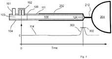
- FIG. 1uses an acoustic reflectometry device 100 which includes a sound generator 101 and one or more sound receivers 102 embedded into the wall of a wave tube 104.
- the sound generator 101 and sound receiver(s) 102are in communication with a processor 105 with attached display 107.
- Two sound receiversmay be used as described in Wodicka 6,705,319 for a compact acoustic reflectometer that provides the means to separate the incident and reflected acoustic signals and thereby simplifies the calculation of an approximation of the acoustic impulse response (reflection waveform) 114.
- the acoustic impulse response 114can be calculated using an excitation signal from the sound generator 101 that comprises acoustic energy over the frequencies of interest (e.g. 0.1-10 kHz), including a broadband sound pulse or white noise.
- the distal wave tube end 106is coupled to the proximal end of an open-ended NG tube 108.
- NG tube 108When NG tube 108 is inserted into a body cavity 109, there may arise one or more local deformations 110 of the tube wall 111 due to a narrowed or constricted region 112 within the passageway 109 such as from a sphincter or other structure that applies pressure on the compliant tube wall 111.
- the corresponding acoustic impulse response 114 derived by the processor from the sound receiver 102 signalscontains a sound reflection 116 that arises from the constriction 110 within the tube 108.
- a second distance 122 ( d 2 ) between the constriction 110 and the distal tube end 124is calculated by subtracting the first distance 120 ( d 1 ) from the tube length 126 ( d tube ).
- the tube length 126may either be known in advance, provided by the user, or determined during a calibration step to estimate the tube length 126.
- the calibration step to estimate the tube length 126could consist of obtaining an acoustical measurement prior to inserting the tube 108 into the body passageway 109 and calculating tube length 126 by using the time delay of the sound reflection from the distal tube end 124 obtained from the corresponding acoustic reflection waveform 114.
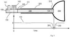
- FIG. 2depicts one embodiment of the acoustic reflectometer 100 attached to NG tube 108 which is inserted into a body cavity comprising a nasal cavity 200, esophagus 202, and stomach 204.
- the NG tube 108may have a first local deformation 206 at the upper esophageal sphincter (UES) 208 and a second local deformation 210 at the lower esophageal sphincter (LES) 212.
- the locations of the first and second local deformations 206 and 210 along the NG tube 108are estimated using the delay times of their respective sound reflections using the equation previously described.
- the distance 122 by which the distal tube end 124 is past the LES 212 extending into the stomach 204is calculated as previously described using the distance of the second local deformation 210 and the NG tube length 126 (from FIG. 1 ).
- This distance 122can be used to guide the position of the distal tube end 124 into the desired location in or past the stomach 204. For example, if it is desired to position the tube end 124 through the stomach 204 into the duodenum 205, then distance 122 as reported by the system can be increased an amount determined by the user to put the tube end 124 approximately into the duodenum 205.
- fluidssuch as from the nasal passageway 200, esophagus 202, stomach 204, or elsewhere, or any combination thereof, may enter the distal tube end 124 and result in a false positive detection of a constriction of the NG tube wall 111.
- a devicesuch as an air filled syringe to the wave tube proximal end 212 and provide a bolus of positive pressure air with the syringe to flow air though the NG tube and push fluids through the NG tube 108 and out of the distal tube end 124.
- Positive confirmation of the distal tube end 124 into the stomach 204is provided when the device detects the second local deformation 210 arising from the LES 212 constricting the tube wall 111.
- An additional positive confirmation of the NG tube 108 traversing the length of the esophagus 202 with the distal tube end 124 located past the LES 212is the presence of the first and second local deformations 206 and 210 that arise from the UES 208 and LES 212, respectively.
- Further confirmation that the NG tube 108 is inserted fully through the esophagus 202is the observation of a constant distance between the deformations 206 and 210 in the NG tube 108 as it is advanced into or withdrawn from the stomach 204.
- a constant distancemay be determined based on multiple distance observations being within a threshold value of each other. For example, the distance variance may be within 1%, 5%, or 10% of each other, or within 0.5 cm, 1 cm, 2 cm, or 3 cm of each other.
- nasopharynx 214may close voluntarily by the patient or involuntarily during swallowing.
- nasopharynx 214may close voluntarily by the patient or involuntarily during swallowing.
- the lower esophageal sphincter 212has several characteristics that allow discrimination of the deformation of the tube wall 111 due to the LES 212 from deformation due to other structures.
- the LES base pressureis typically between 6-20 mmHg and has a dynamic component that increases 15-20 mmHg during the inspiratory phase of tidal inspiration, and with forceful inspiration the increase can be 100-150 mmHg.
- the dynamic componentmay be periodic. This varying pressure on the tube wall 111 causes the tube wall 111 deformation to also vary such that the degree of constriction of the NG tube 108 is correlated to the pressure.
- This varying NG tube constriction due to the LES 212can be observed as a change in amplitude of the sound reflection 116 ( Figure 1 ) arising from the constriction. If the acoustic reflectometry device 100 is configured to collect a complete acoustic reflection waveform 114 multiple times per second, then the change in constriction size as a function of time (over seconds) can be observed and used as a positive confirmation that the constriction in the NG tube 108 is from the LES 212.
- LES 212Another characteristic of the LES 212, as well as the UES 208, is the relaxation that occurs during swallowing. During swallowing, the LES relaxation lasts 6-10 seconds. Again, if the acoustic reflectometry device 100 is configured to collect a complete acoustic reflection waveform 116 multiple times per second, then the change in constriction size as a function of time (over seconds) can be observed and used to confirm the presence of the relaxation period that is synchronized with swallowing. If this relaxation period is observed, then it can be used as another indicator of positive confirmation that the constriction in the NG tube 108 is from the LES 212.
- the abovementioned characteristics of the LES 212 that are observable in the acoustic reflection signal 114may be used individually or in some combination to positively confirm that the NG distal tube end 124 is extended past the LES 212 into the stomach 204.
- FIG. 3depicts the NG tube 108 erroneously advanced past the vocal folds 216 into the trachea 218.
- the tube wall deformations due to compression by the UES 208 and LES 212, respectively, along with their individual characteristics discussed above,will not be observed.
- closure of the vocal folds 216may pinch the tube 108 and be detected as a constriction by the acoustic reflectometer 100, but this constriction will not have the same characteristics as those arising from the structures 208 and 212 within the esophagus 202.
- the lower airwaymay be the airway below the vocal folds.
- the NG tube wall 111is not sufficiently compliant to deform from a narrowed or constricted region within the esophagus 202, then there are alternate means to verify that the distal tube end 124 is past the LES 212 and extending into the stomach 204.
- a calmay be obtained a priori for catheters of a specified diameter, length, manufacturer, and model, and stored within a lookup table. It may be necessary to know the manufacturer and model of a catheter because the sound attenuation through the catheter may be affected by the catheter wall mechanical properties which may vary between manufacturers and models of catheters.
- a calmay be calculated from an equation, A cal (d, l) that is empirically derived using data points for A cal that are obtained experimentally over varying catheter diameters, d , and lengths, l , (and manufacturers and models, if necessary).
- the esophageal tissuewill close off the distal tube end 124 by virtue of the tissue's collapsible nature over the tip opening and the resulting acoustic reflection 300 arising from the closed distal tube end 124 will be detected as a positive deflection 302 in the reflection waveform 114.
- the resulting acoustic reflection 300arising from the closed distal tube end 124 will be detected as a positive deflection 302 in the reflection waveform 114.
- the distal tube end 124if the distal tube end 124 is located distal to the LES 212 within the stomach 204, then the distal tube end 124 will be open into the cavity formed by the stomach 204, and the resulting acoustic reflection 300 arising from the open distal tube end 124 will be detected as a negative deflection 304 in the reflection waveform 114.
- the proper insertion distance of the NG tube 108 into a patientis estimated using a commonly employed method of measuring the total distance from the nose to the ear lobe to the xiphoid process.
- the timings and amplitudes of the reflections of the waveform 114are detected.
- placement of the distal end of the NG tube at the LESmay occur based on the detection and timings of a positive amplitude deflection in the acoustic reflections at a first position of the distal end of the nasogastric enteral access device within the body, for example, when the distal end of the NG tube is at the LES, and the estimated distance to the LES, that the distal end of the nasogastric enteral access device is at the lower esophageal sphincter.
- the markings showing distance from the distal tube end 124 to the nares or mouthtypically provided along the outside of the NG tube 108
- a collapsed cavitye.g. the esophagus, stomach, or trachea
- the proper insertion distance of the NG tube 108 into a patientis estimated using a commonly employed method of measuring the total distance from the nose to the ear lobe to the xiphoid process.
- guidance of the distal tube end 124is provided by detecting the amplitude and timings of an acoustic reflection 300 that has either a positive deflection (collapsed esophagus) or small negative deflection (partially collapsed esophagus) while the distal tube end is advancing in the esophagus 202.
- a positive deflectioncollapsed esophagus
- small negative deflectionpartially collapsed esophagus
- Improper placement of the NG tube 108 into the trachea 218is indicated by detecting an acoustic reflection 300 that has a significant negative deflection 306 (the trachea) at an insertion distance of the NG tube 108 significantly smaller than the estimated proper insertion distance to the stomach. This small insertion distance is due to the distal tube end 124 entering the trachea 218 where it would have instead started entering the esophagus just below the upper airway if proper placement had occurred.
- the NG tube diameter 310is of comparable size to that of the trachea 218, there may be adequate acoustic energy transmission between the tube 108, trachea 218, and airways 312 to observe the characteristic echo 308 from the airways that arises from the branching generations where the total cross-sectional area grows rapidly.
- the presence of this negative going airway echo 308 in the acoustic reflection waveform 114may provide an additional confirmation that the distal tube end 124 is improperly placed in the trachea 218.
- the proximal ports for NG tubescan vary in number depending on the intended use for the tube. For example, some NG tubes may have two or more ports to allow administration of both food and medications simultaneously. If an NG tube is used that contains two or more ports, it may be necessary to occlude all of the ports with plugs except for the one acoustically coupled to the acoustic reflectometer. This will prevent extraneous acoustic reflections arising from the ports from interfering with the reflections arising from within the NG tube and cavities in which the tube is inserted.
- the plug diametersare made to fit the inner diameter of the ports and the plug lengths are made to extend into the port far enough to completely fill the port and, therefore, minimize the increase in cross-sectional area of the NG tube resulting from the port.
- a calibration procedureis used to measure the reflection echoes arising from the ports in their open or closed states and remove their effects on the entire acoustic reflection signal either through methods such as subtraction or deconvolution.
Landscapes
- Health & Medical Sciences (AREA)
- Life Sciences & Earth Sciences (AREA)
- Animal Behavior & Ethology (AREA)
- Veterinary Medicine (AREA)
- Public Health (AREA)
- General Health & Medical Sciences (AREA)
- Medical Informatics (AREA)
- Surgery (AREA)
- Molecular Biology (AREA)
- Heart & Thoracic Surgery (AREA)
- Biomedical Technology (AREA)
- Engineering & Computer Science (AREA)
- Physics & Mathematics (AREA)
- Acoustics & Sound (AREA)
- Biophysics (AREA)
- Nuclear Medicine, Radiotherapy & Molecular Imaging (AREA)
- Pathology (AREA)
- Radiology & Medical Imaging (AREA)
- Otolaryngology (AREA)
- Pulmonology (AREA)
- Media Introduction/Drainage Providing Device (AREA)
Description
- This application claims the benefit of
U.S. Provisional Application No. 62/343,476, filed May 31, 2016 - Nasogastric enteral access devices (for example, NG-EADs, enteral feeding tubes, nasogastric tubes, NG tubes, feeding tubes) are widely used in patients who require nutrition to be delivered directly to the stomach due to an inability to swallow foods on their own. For example, a patient who is being mechanically ventilated through an endotracheal tube (ETT) requires an NG tube because the ETT prevents the patient from swallowing without the risk of food being aspirated into the airways.
- When properly positioning an NG tube in a patient, the tube tip is inserted through the nose into the esophagus and advanced into the stomach or farther into the duodenum. To confirm proper placement, a variety of methods are used according to the prior art, including chest x-rays and testing of aspirates for a pH reading between 1 and 5.5. However, errors in placement still occur either due to misinterpretation of the results (e.g. misreading of chest x-ray), unintentionally introducing substances in the NG tube that may cause false positive pH readings (flushing tube with water prior to placement), or failing to positively confirm placement in the first place. Accidental placement of the NG tube into the airway also is an ongoing concern and can lead to catastrophic consequences when fluids that are intended for the stomach are delivered directly into the lungs. This aspiration of fluids into the lungs frequently leads to pneumonia which carries risk of serious and potentially lethal complications. Accordingly, there is a need for an improved method and system for assisting in the proper placement of NG tubes.
- Several apparatuses and methods for acoustically guiding, positioning, and monitoring tubes within a body are known. See, for example,
U.S. Pat. Nos. 5,445,144 and6,705,319 to Wodicka et al. , which disclose an apparatus and method for acoustically monitoring the position of a tube (e.g., ETT) within an anatomical conduit. In various embodiments, a sound pulse is introduced into a wave guide and is recorded as it passes by one or more microphones located in the wave guide wall. As the sound pulse is propagating down the tube, reflected sound pulses arise from changes in cross-sectional area due to constrictions that may exist in the tube. The sound pulse is then emitted through the distal tip of the ETT into the airway (or wherever in the body the tip of the ETT is located) and an acoustic reflection propagates back up the ETT to the wave guide for measurement by the same microphone(s). The amplitude and the polarity of the incident and reflected sound pulse are used to estimate the characteristics of the airway and the ETT, and thereby guide the ETT placement or monitor the ETT for patency. - Another apparatus and method for examination and measurement of constrictions of passages in a cavity by means of acoustic reflectometry is described in
U.S. Pat. No. 5,823,965 to Rasmussen. Rasmussen describes an acoustic reflectometer attached to a flexible closed-ended hose which is introduced into a cavity with the distal end of the hose placed past the zone of the passage to be examined. A transducer converts an activation signal from a signal generator to an excitation signal which is sent into the interior of the hose. A response signal which depends on the local deformation of the hose in the examined zone is picked up by a transducer and subjected to analysis in relation to the excitation signal. An analysis circuit and computer give an image on screen indicating the results of the examination. - Notably, Rasmussen teaches and claims a hose having only a closed distal end. Secondly, Rasmussen teaches and claims determining the internal cross-sectional shape of the hose from the excitation and response signals while the present disclosure directly uses the reflection response signal to determine the location and degree of constrictions within the hose. The internal cross-sectional shape, or the cross-sectional area vs. distance profile, of the hose requires the additional step of calculating the profile using the Ware-Aki or similar algorithm as discussed in
U.S. Pat. No. 4,326,416 , which is cited by Rasmussen. US 5445144 describes an apparatus and method for acoustically guiding, positioning and monitoring a tube within a body. The apparatus generates an incident sound pulse in the tube. The sound pulse propagates into the body and the apparatus detects sound pulses resulting from the incident sound pulse and from reflected sound pulses from within the body and processes the detected sound pulses to guide insertion of the distal end of the tube within the body.- The present invention is set out in the appended claims. A method for use of acoustic reflectometry in nasogastric enteral access devices is disclosed. The method may include inserting a distal end of a nasogastric enteral access device through the nares a distance into a body and emitting sound waves from a sound generator into a proximal end of a nasogastric enteral access device. The method may also include detecting timings of returning acoustic reflections with at least one sound receiver, the acoustic reflections may include a first acoustic reflection of a first deformation in a wall of the nasogastric enteral access device from a first esophageal sphincter and using a reflectometry device having at least one processor and a memory that is accessible to the processor for analyzing timings of a first acoustic reflection to determine the distance the distal end of a nasogastric enteral access device is inserted into the body.
- In some embodiments the method may include determining a length of the nasogastric enteral access device. In some embodiments the method may include determining the distance the distal end of the nasogastric enteral access device is inserted into the body based on the determined length of the nasogastric enteral access device and the timing of the returning acoustic reflections.
- In some embodiments first esophageal sphincter is the lower esophageal sphincter. In some embodiments the first esophageal sphincter is the upper esophageal sphincter. In some embodiments, the returning acoustic reflections may include a second acoustic reflection of a second deformation in the wall of the nasogastric enteral access device from a second esophageal sphincter and may also include using the reflectometry device having the at least one processor and the memory that is accessible to the processor for analyzing timings of the returning acoustic reflections to determine a distance the distal end of a nasogastric enteral access device is inserted past the second esophageal sphincter and indicating the distal end of the nasogastric enteral access device is in the stomach when the distal end of the nasogastric enteral access device is the distance passed the second esophageal sphincter.
- In some embodiments, the first esophageal sphincter is a lower esophageal sphincter and may include using the reflectometry device having the at least one processor and the memory that is accessible to the processor for analyzing timings of the returning acoustic reflections to determine a distance the distal end of a nasogastric enteral access device is inserted past the lower esophageal sphincter. In some embodiments the method may include indicating that the distal end of the nasogastric enteral access device is in the stomach when the distal end of the nasogastric enteral access device is the distance passed the lower esophageal sphincter.
- In some embodiments, determining a constant distance between the first deformation and the second deformation may include detecting a plurality of timings between the first and second acoustic reflections over a time period and comparing the plurality of timings and determining that that timings vary by less than 5% over the time period.
- In some embodiments, the nasogastric enteral access device may be advanced or withdrawn within the esophagus or stomach during the time period.
- In some embodiments, detecting amplitudes of the returning acoustic reflections with the at least one sound receiver may occur over a time period and include detecting a base and dynamic component of the amplitude over the time period. In some embodiments, the dynamic component coincides with a respiratory cycle of the patient.
- In some embodiments, the method may include clearing the nasogastric enteral access device by providing positive pressure into the nasogastric enteral access device to push fluids out the distal end of the nasogastric enteral access device.
- A method for use of acoustic reflectometry in nasogastric enteral access devices is disclosed. The method may include inserting a distal end of a nasogastric enteral access device through the nares a distance into a body and emitting sound waves from a sound generator into a nasogastric enteral access device. The method may also include detecting amplitudes and timings of returning acoustic reflections with at least one sound receiver at a plurality positions of the distal end of the nasogastric enteral access device within the body and using a reflectometry device having at least one processor and a memory that is accessible to the processor for analyzing amplitudes and timings of the returning acoustic reflections to detect a positive amplitude deflection in the acoustic reflections at a first position of the distal end of the nasogastric enteral access device within the body, the positive amplitude deflection in the acoustic reflections being from the distal end of the nasogastric enteral access device.
- In some embodiments, the method may include indicating, based on the detection of a positive amplitude deflection in the acoustic reflections at a first position of the distal end of the nasogastric enteral access device within the body, that the distal end of the nasogastric enteral access device is above or at the lower esophageal sphincter, the positive amplitude deflection in the acoustic reflections being from the distal end of the nasogastric enteral access device.
- In some embodiments, the method may include using the reflectometry device having the at least one processor and the memory that is accessible to the processor for analyzing amplitudes and timings of the returning acoustic reflections to detect a negative amplitude deflection in the acoustic reflections at a second position of the distal end of the nasogastric enteral access device within the body, the second position being further advanced into the body as compared to the first position and the negative amplitude deflection in the acoustic reflections being from the distal end of the nasogastric enteral access device.
- In some embodiments, the method may include indicating, based on the detection of a positive amplitude deflection at the first position and the negative amplitude at the second position, that the distal end of the nasogastric enteral access device is within a stomach.
- In some embodiments, the detecting may occur as the distal end of the nasogastric enteral access device is advancing in the body. In some embodiments, the detecting occurs while the distal end of the nasogastric enteral access device is stationary within the body.
- In some embodiments, the method may include estimating a distance to the lower esophageal sphincter prior to inserting the nasogastric enteral access device into the stomach and indicating, based on the detection and timings of a positive amplitude deflection in the acoustic reflections at a first position of the distal end of the nasogastric enteral access device within the body and the estimated distance to the lower esophageal sphincter, that the distal end of the nasogastric enteral access device is at the lower esophageal sphincter.
- In some embodiments, the nasogastric enteral access device comprises a plurality of ports, and the method may include acoustically coupling an acoustic reflectometer, including the sound generator and a sound receiver, to a first of the plurality of ports and occluding a second or more of the plurality of ports not coupled to the sound generator.
- In some embodiments, the method may include calibrating the nasogastric enteral access device using a reflectometry device having at least one processor and a memory that is accessible to the processor by determining the amplitude of an acoustic reflection arising from the distal end of the nasogastric enteral access device being open to air.
- A method for use of acoustic reflectometry in nasogastric enteral access devices is also disclosed. The method may include estimating a distance to the lower esophageal sphincter prior to inserting the enteral access device into the stomach; inserting a distal end of a nasogastric enteral access device through the nares a distance into a body as indicated by a distance marking on the outside of the nasogastric enteral access device that is visible at the nares and emitting sound waves from a sound generator into a nasogastric enteral access device. The method may also include detecting amplitudes and timings of returning acoustic reflections with at least one sound receiver at a plurality positions of the distal end of the nasogastric enteral access device within the body and using a reflectometry device having at least one processor and a memory that is accessible to the processor for analyzing amplitudes and timings of the returning acoustic reflections to detect a negative amplitude deflection in the acoustic reflections at a first position of the distal end of the nasogastric enteral access device within the body, the negative amplitude deflection in the acoustic reflection being from the distal end of the nasogastric enteral access device. In some embodiments, the first and second negative amplitude deflections may be consecutive.
- In some embodiments, the method may include indicating, based on the detection of the first and second negative amplitude deflections at the first position, that the distal end of the nasogastric enteral access device is within a trachea.
- In some embodiments, the method may include indicating, based on the detection of a negative amplitude deflection at a first position and the insertion distance of the first position being less than the estimated distance to the lower esophageal sphincter, that the distal end of the nasogastric enteral access device is within a trachea.
- In some embodiments, the method may include indicating that the distal end of the nasogastric enteral access device is within a trachea upon detection of a second negative amplitude deflection in the acoustic reflections.
- A system for use of acoustic reflectometry in nasogastric enteral access devices is also disclosed. The system may include a nasogastric enteral access device and a sound generator acoustically coupled to the proximal end of a nasogastric enteral access device to emit sound waves into the nasogastric enteral access device. The system may also include at least one sound receiver to detect timings of returning acoustic reflections, the acoustic reflections including a first acoustic reflection of a first deformation in a wall of the nasogastric enteral access device from a first esophageal sphincter and a reflectometry device having at least one processor and a memory that is accessible to the processor for analyzing timings of the returning acoustic reflections to determine the distance the distal end of a nasogastric enteral access device is inserted into the body. In some embodiments, the reflectometry device is configured to determine the distance the distal end of the nasogastric enteral access device is inserted into the body based on a length of the nasogastric enteral access device and the timing of the first acoustic reflection.
- In some embodiments, the first esophageal sphincter is a lower esophageal sphincter; and the reflectometry device is configured to indicate that the distal end of the nasogastric enteral access device is in the stomach when the distal end of the nasogastric enteral access device is a distance passed the lower esophageal sphincter.
- In some embodiments, the returning acoustic reflections include a second acoustic reflection of a second deformation in the wall of the nasogastric enteral access device from a second esophageal sphincter and the reflectometry device may be configured to determine the distal end of the nasogastric enteral access device is in the stomach when the distal end of the nasogastric enteral access device is a distance passed the second esophageal sphincter.
- In some embodiments, the reflectometry device may configured to determine a constant distance between the first deformation and the second deformation by detecting a plurality of timings between the first and second acoustic reflections over a time period and comparing the plurality of timings and determining that that timings vary by less than 5% over the time period.
- In some embodiments, the reflectometry device may be configured to detect amplitudes of the returning acoustic reflections with the at least one sound receiver over a time period and detect a base and dynamic component of the amplitude over the time period.
- A system for use of acoustic reflectometry in nasogastric enteral access devices is also disclosed. The system may include a nasogastric enteral access device and a sound generator to emit sound into the nasogastric enteral access device. The system may include at least one sound receiver to detect amplitudes and timings of returning acoustic reflections at a plurality positions of a distal end of the nasogastric enteral access device within a body and a reflectometry device having at least one processor and a memory that is accessible to the processor configured to analyze amplitudes and timings of the returning acoustic reflections and to detect a positive amplitude deflection in the acoustic reflections at a first position of the distal end of the nasogastric enteral access device within the body, the positive amplitude deflection in the acoustic reflections being from the distal end of the nasogastric enteral access device.
- In some embodiments, the reflectometry device may be configured to indicate, based on the detection of a positive amplitude deflection in the acoustic reflections at a first position of the distal end of the nasogastric enteral access device within the body, that the distal end of the nasogastric enteral access device is above or at the lower esophageal sphincter, the positive amplitude deflection in the acoustic reflections being from the distal end of the nasogastric enteral access device.
- In some embodiments, the reflectometry device may be configured to analyze amplitudes and timings of the returning acoustic reflections to detect a negative amplitude deflection in the acoustic reflections at a second position of the distal end of the nasogastric enteral access device within the body, the second position being further advanced into the body as compared to the first position and the negative amplitude deflection in acoustic reflections being from the distal end of the nasogastric enteral access device.
- In some embodiments, the reflectometry device is configured to indicate, based on the detection of a positive amplitude deflection at the first position and the negative amplitude at the second position, that the distal end of the nasogastric enteral access device is within a stomach.
- In some embodiments, the sound receiver is configured to detect the reflections as the distal end of the nasogastric enteral access device advances in the body. In some embodiments, the sound receiver is configured to detect the reflections while the distal end of the nasogastric enteral access device is stationary within the body.
- In some embodiments, the reflectometry device is configured with an estimate of a distance to the lower esophageal sphincter prior to inserting the nasogastric enteral access device into the stomach and to indicate, based on the detection and timings of a positive amplitude deflection in the acoustic reflections at a first position of the distal end of the nasogastric enteral access device within the body and the estimated distance to the lower esophageal sphincter, that the distal end of the nasogastric enteral access device is at the lower esophageal sphincter.
- In some embodiments, the nasogastric enteral access device comprises a plurality of ports, and the acoustic reflectometer is acoustically coupled to a first of the plurality of ports and a second or more of the plurality of ports not coupled to the sound generator are occluded.
- A system for use of acoustic reflectometry in nasogastric enteral access devices is disclosed. The system may include a nasogastric enteral access device having a proximal end and a distal end and a sound generator to emit sound waves into a nasogastric enteral access device. The system may also include at least one sound receiver to detect amplitudes and timings of returning acoustic reflections a plurality positions of the distal end of the nasogastric enteral access device within a body and a reflectometry device having at least one processor and a memory that is accessible to the processor for analyzing amplitudes and timings of the returning acoustic reflections to detect first and second negative amplitude deflections in the acoustic reflections at a first position of the distal end of the nasogastric enteral access device within the body.
- In some embodiments, the first and second negative amplitude deflections are consecutive.
- In some embodiments, the reflectometry device is configured to indicate, based on the detection of the first and second negative amplitude deflections at the first position, that the distal end of the nasogastric enteral access device is within a trachea.
- In some embodiments, the reflectometry device is further configured to indicate, based on the detection of a negative amplitude deflection at a first position and the insertion distance of the first position being less than an estimated distance to the lower esophageal sphincter, that the distal end of the nasogastric enteral access device is within a lower airway.
- In some embodiments, the reflectometry device is configured to indicate that the distal end of the nasogastric enteral access device is within a trachea upon detection of a second negative amplitude deflection in the acoustic reflections, the first negative amplitude deflection being from the distal end of the nasogastric enteral access device and the second negative amplitude deflection being from a location within an airway, distal to the distal end of the nasogastric enteral access device.
- The features and advantages of this disclosure, and the manner of attaining them, will be more apparent and better understood by reference to the following descriptions of the disclosed methods and systems, taken in conjunction with the accompanying drawings, wherein:
FIG. 1 shows a drawing of the acoustic reflectometry system connected to a tube which is inserted into a body cavity containing a constriction.FIG. 2 illustrates the placing of an NG tube with attached acoustic reflectometer into body passages leading to the stomach.FIG. 3 illustrates the placing of an NG tube with attached acoustic reflectometer into body passages leading to the lungs.FIG. 4 shows a drawing of the acoustic reflectometry device connected to a tube with the distal tube end inserted into an esophagus.FIG. 5 shows a drawing of the acoustic reflectometry device connected to a tube with the distal tube end inserted into a stomach.FIG. 6 shows a drawing of the acoustic reflectometry device connected to a tube with the distal tube end inserted into a trachea.- For the purposes of promoting an understanding of the principles of the present disclosure, reference will now be made to the embodiments illustrated in the drawings, and specific language will be used to describe the same. It will nevertheless be understood that no limitation of the scope of this disclosure is thereby intended.
- The present disclosure includes disclosure of devices and methods for verifying the proper position of catheters in a human body by means of acoustic reflectometry. The device comprises a sound source, one or more sound receivers, and a tube with compliant walls and open distal end to be introduced through an entrance to a body cavity. The sound source and receiver(s) are coupled to the proximal end of the tube. The device includes a processor for causing the sound source to generate an acoustic excitation signal. The processor then processes the acoustic signals sensed by the sound receiver(s), generates an approximation of the acoustic impulse response of the tube, and analyzes the acoustic impulse response to determine the position of the tube in the body cavity.
- The embodiment of the present disclosure shown in
FIG.1 uses anacoustic reflectometry device 100 which includes asound generator 101 and one or moresound receivers 102 embedded into the wall of awave tube 104. Thesound generator 101 and sound receiver(s)102 are in communication with aprocessor 105 with attacheddisplay 107. Two sound receivers may be used as described in Wodicka 6,705,319 for a compact acoustic reflectometer that provides the means to separate the incident and reflected acoustic signals and thereby simplifies the calculation of an approximation of the acoustic impulse response (reflection waveform)114. Theacoustic impulse response 114 can be calculated using an excitation signal from thesound generator 101 that comprises acoustic energy over the frequencies of interest (e.g. 0.1-10 kHz), including a broadband sound pulse or white noise. The distalwave tube end 106 is coupled to the proximal end of an open-endedNG tube 108. WhenNG tube 108 is inserted into abody cavity 109, there may arise one or morelocal deformations 110 of thetube wall 111 due to a narrowed or constrictedregion 112 within thepassageway 109 such as from a sphincter or other structure that applies pressure on thecompliant tube wall 111. The correspondingacoustic impulse response 114 derived by the processor from thesound receiver 102 signals contains asound reflection 116 that arises from theconstriction 110 within thetube 108. The time delay118 (tc) of thesound reflection 116 and the speed of sound c in air are used to calculate a first distance120 (d1) between the sound receiver(s)102 and thetube constriction 110 using the equation,
tube constriction 110. A second distance122 (d2) between theconstriction 110 and thedistal tube end 124 is calculated by subtracting the first distance120 (d1) from the tube length126 (dtube). Thetube length 126 may either be known in advance, provided by the user, or determined during a calibration step to estimate thetube length 126. The calibration step to estimate thetube length 126 could consist of obtaining an acoustical measurement prior to inserting thetube 108 into thebody passageway 109 and calculatingtube length 126 by using the time delay of the sound reflection from thedistal tube end 124 obtained from the correspondingacoustic reflection waveform 114. FIG.2 depicts one embodiment of theacoustic reflectometer 100 attached toNG tube 108 which is inserted into a body cavity comprising anasal cavity 200,esophagus 202, andstomach 204. TheNG tube 108 may have a firstlocal deformation 206 at the upper esophageal sphincter (UES)208 and a secondlocal deformation 210 at the lower esophageal sphincter (LES)212. The locations of the first and secondlocal deformations NG tube 108 are estimated using the delay times of their respective sound reflections using the equation previously described. Thedistance 122 by which thedistal tube end 124 is past theLES 212 extending into thestomach 204 is calculated as previously described using the distance of the secondlocal deformation 210 and the NG tube length126 (fromFIG.1 ). Thisdistance 122 can be used to guide the position of thedistal tube end 124 into the desired location in or past thestomach 204. For example, if it is desired to position thetube end 124 through thestomach 204 into theduodenum 205, then distance122 as reported by the system can be increased an amount determined by the user to put thetube end 124 approximately into theduodenum 205.- It is possible that fluids such as from the
nasal passageway 200,esophagus 202,stomach 204, or elsewhere, or any combination thereof, may enter thedistal tube end 124 and result in a false positive detection of a constriction of theNG tube wall 111. As a preventative measure, it may be necessary to connect a device such as an air filled syringe to the wave tubeproximal end 212 and provide a bolus of positive pressure air with the syringe to flow air though the NG tube and push fluids through theNG tube 108 and out of thedistal tube end 124. - Positive confirmation of the
distal tube end 124 into thestomach 204 is provided when the device detects the secondlocal deformation 210 arising from theLES 212 constricting thetube wall 111. An additional positive confirmation of theNG tube 108 traversing the length of theesophagus 202 with thedistal tube end 124 located past theLES 212 is the presence of the first and secondlocal deformations UES 208 andLES 212, respectively. Further confirmation that theNG tube 108 is inserted fully through theesophagus 202 is the observation of a constant distance between thedeformations NG tube 108 as it is advanced into or withdrawn from thestomach 204. A constant distance may be determined based on multiple distance observations being within a threshold value of each other. For example, the distance variance may be within 1%, 5%, or 10% of each other, or within 0.5 cm, 1 cm, 2 cm, or 3 cm of each other. - It is possible that additional structures within the body cavities traversed by the
NG tube 108 may cause temporary local deformations in thetube wall 111. These structures may include thenasopharynx 214 which may close voluntarily by the patient or involuntarily during swallowing. There may also be other structures within the upper airway that may cause local deformations in thetube wall 111. - The lower
esophageal sphincter 212 has several characteristics that allow discrimination of the deformation of thetube wall 111 due to theLES 212 from deformation due to other structures. The LES base pressure is typically between 6-20 mmHg and has a dynamic component that increases 15-20 mmHg during the inspiratory phase of tidal inspiration, and with forceful inspiration the increase can be 100-150 mmHg. The dynamic component may be periodic. This varying pressure on thetube wall 111 causes thetube wall 111 deformation to also vary such that the degree of constriction of theNG tube 108 is correlated to the pressure. This varying NG tube constriction due to theLES 212 can be observed as a change in amplitude of the sound reflection116 (Figure 1 ) arising from the constriction. If theacoustic reflectometry device 100 is configured to collect a completeacoustic reflection waveform 114 multiple times per second, then the change in constriction size as a function of time (over seconds) can be observed and used as a positive confirmation that the constriction in theNG tube 108 is from theLES 212. - Another characteristic of the
LES 212, as well as theUES 208, is the relaxation that occurs during swallowing. During swallowing, the LES relaxation lasts 6-10 seconds. Again, if theacoustic reflectometry device 100 is configured to collect a completeacoustic reflection waveform 116 multiple times per second, then the change in constriction size as a function of time (over seconds) can be observed and used to confirm the presence of the relaxation period that is synchronized with swallowing. If this relaxation period is observed, then it can be used as another indicator of positive confirmation that the constriction in theNG tube 108 is from theLES 212. - The abovementioned characteristics of the
LES 212 that are observable in theacoustic reflection signal 114 may be used individually or in some combination to positively confirm that the NGdistal tube end 124 is extended past theLES 212 into thestomach 204. FIG.3 depicts theNG tube 108 erroneously advanced past thevocal folds 216 into thetrachea 218. In this case, the tube wall deformations due to compression by theUES 208 andLES 212, respectively, along with their individual characteristics discussed above, will not be observed. It is possible that closure of thevocal folds 216 may pinch thetube 108 and be detected as a constriction by theacoustic reflectometer 100, but this constriction will not have the same characteristics as those arising from thestructures esophagus 202. The lower airway may be the airway below the vocal folds.- Referring to
FIG. 4 , if theNG tube wall 111 is not sufficiently compliant to deform from a narrowed or constricted region within theesophagus 202, then there are alternate means to verify that thedistal tube end 124 is past theLES 212 and extending into thestomach 204. - The
acoustic reflection 300 arising from thedistal tube end 124 is related to the cross-sectional areas of the catheter lumen and the passageway immediately around the opening of thedistal tube end 124. This relationship is described as
distal tube end 124, and R is the dimensionless reflection coefficient (-1 ≤R ≤ 1) related to theamplitude 302 of theacoustic reflection 300 arising from thedistal tube end 124. The value ofR for theacoustic reflection 300 can be determined by measuring the acoustic reflection amplitude,Acal, arising for a knownS0 andS1 during a calibration step and using this value to calculate R. For example, upon connection of thedevice 100 to theNG tube 108 and prior to insertion of thetube 108 into the patient, an acoustic measurement can be obtained while thedistal tube end 124 is open to air (case where S1 ≈ ∞). The amplitude of the resulting acoustic reflection arising from thedistal tube end 124,Acal, would represent the case forRopen = -1. Then, all subsequent amplitude measurements, A302, of theacoustic reflection 300 arising from thedistal tube end 124 can be converted into a reflection coefficient using
distal tube end 124. - In an alternate embodiment,Acal may be obtaineda priori for catheters of a specified diameter, length, manufacturer, and model, and stored within a lookup table. It may be necessary to know the manufacturer and model of a catheter because the sound attenuation through the catheter may be affected by the catheter wall mechanical properties which may vary between manufacturers and models of catheters. In yet another embodiment,Acal may be calculated from an equation,Acal(d, l) that is empirically derived using data points forAcal that are obtained experimentally over varying catheter diameters,d, and lengths,l, (and manufacturers and models, if necessary).
- Again, referring to
FIG. 4 , if thedistal tube end 124 is located proximally to or at theLES 212 within theesophagus 202, then the esophageal tissue will close off thedistal tube end 124 by virtue of the tissue's collapsible nature over the tip opening and the resultingacoustic reflection 300 arising from the closeddistal tube end 124 will be detected as apositive deflection 302 in thereflection waveform 114. In contrast, as shown inFIG. 5 , if thedistal tube end 124 is located distal to theLES 212 within thestomach 204, then thedistal tube end 124 will be open into the cavity formed by thestomach 204, and the resultingacoustic reflection 300 arising from the opendistal tube end 124 will be detected as anegative deflection 304 in thereflection waveform 114. - In some embodiments, prior to insertion, the proper insertion distance of the
NG tube 108 into a patient is estimated using a commonly employed method of measuring the total distance from the nose to the ear lobe to the xiphoid process. During advancement of the NG tube into the body, the timings and amplitudes of the reflections of thewaveform 114 are detected. In some embodiments, placement of the distal end of the NG tube at the LES may occur based on the detection and timings of a positive amplitude deflection in the acoustic reflections at a first position of the distal end of the nasogastric enteral access device within the body, for example, when the distal end of the NG tube is at the LES, and the estimated distance to the LES, that the distal end of the nasogastric enteral access device is at the lower esophageal sphincter. - By using the markings showing distance from the
distal tube end 124 to the nares or mouth, typically provided along the outside of theNG tube 108, one can note the presence or absence of a collapsed cavity (e.g. the esophagus, stomach, or trachea) around thedistal tube end 124 while advancing theNG tube 108 by detecting the amplitude and polarity of thereflection wave 300 arising from thedistal tube end 124. Prior to insertion, the proper insertion distance of theNG tube 108 into a patient is estimated using a commonly employed method of measuring the total distance from the nose to the ear lobe to the xiphoid process. During insertion of theNG tube 108, guidance of thedistal tube end 124 is provided by detecting the amplitude and timings of anacoustic reflection 300 that has either a positive deflection (collapsed esophagus) or small negative deflection (partially collapsed esophagus) while the distal tube end is advancing in theesophagus 202. When thedistal tube end 124 enters thestomach 204, this is confirmed by detecting a largenegative deflection 304 arising from thedistal tube end 124. The estimated proper insertion distance should approximately agree with the insertion distance at which the cavity around thedistal tube end 124 transitioned from collapsed or partially collapsed (esophagus) to open (stomach). - Improper placement of the
NG tube 108 into the trachea218 (FIG. 6 ) is indicated by detecting anacoustic reflection 300 that has a significant negative deflection306 (the trachea) at an insertion distance of theNG tube 108 significantly smaller than the estimated proper insertion distance to the stomach. This small insertion distance is due to thedistal tube end 124 entering thetrachea 218 where it would have instead started entering the esophagus just below the upper airway if proper placement had occurred. In addition, if theNG tube diameter 310 is of comparable size to that of thetrachea 218, there may be adequate acoustic energy transmission between thetube 108,trachea 218, andairways 312 to observe thecharacteristic echo 308 from the airways that arises from the branching generations where the total cross-sectional area grows rapidly. The presence of this negative goingairway echo 308 in theacoustic reflection waveform 114 may provide an additional confirmation that thedistal tube end 124 is improperly placed in thetrachea 218. - The proximal ports for NG tubes can vary in number depending on the intended use for the tube. For example, some NG tubes may have two or more ports to allow administration of both food and medications simultaneously. If an NG tube is used that contains two or more ports, it may be necessary to occlude all of the ports with plugs except for the one acoustically coupled to the acoustic reflectometer. This will prevent extraneous acoustic reflections arising from the ports from interfering with the reflections arising from within the NG tube and cavities in which the tube is inserted. In one embodiment, the plug diameters are made to fit the inner diameter of the ports and the plug lengths are made to extend into the port far enough to completely fill the port and, therefore, minimize the increase in cross-sectional area of the NG tube resulting from the port. In another embodiment, a calibration procedure is used to measure the reflection echoes arising from the ports in their open or closed states and remove their effects on the entire acoustic reflection signal either through methods such as subtraction or deconvolution.
- While this disclosure has been described as having preferred designs, the apparatus and methods according to the present disclosure can be further modified within the scope of the appended claims.


Claims (6)
- A system for use of acoustic reflectometry in nasogastric enteral access devices, the system comprising:a nasogastric enteral access device (108);a sound generator (101) to emit sound into the nasogastric enteral access device (108);at least one sound receiver (102) to detect amplitudes and timings of returning acoustic reflections at a plurality positions of a distal end of the nasogastric enteral access device (108) within a body; anda reflectometry device having at least one processor (105) and a memory that is accessible to the processor (105) configured to analyze amplitudes and timings of the returning acoustic reflections and to detect a positive amplitude deflection in the acoustic reflections at a first position of the distal end of the nasogastric enteral access device (108) within the body, the positive amplitude deflection in the acoustic reflections being from the distal end of the nasogastric enteral access device (108), wherein the reflectometry device configured to indicate, based on the detection of a positive amplitude deflection in the acoustic reflections at a first position of the distal end of the nasogastric enteral access device (108) within the body, that the distal end of the nasogastric enteral access device (108) is above or at the lower esophageal sphincter, the positive amplitude deflection in the acoustic reflections being from the distal end of the nasogastric enteral access device (108); wherein the nasogastric enteral access device (108) comprises a plurality of ports, and wherein the acoustic reflectometer is acoustically coupled to a first of the plurality of ports and a second or more of the plurality of ports not coupled to the sound generator (101) are occluded by plugs to prevent extraneous acoustic reflections arising from the ports from interfering with the reflections arising from within the nasogastric enteral access device and cavities in which the nasogastric enteral access device is inserted.
- The system of claim 1, wherein:
the reflectometry device is configured to analyzing amplitudes and timings of the returning acoustic reflections to detect a negative amplitude deflection in the acoustic reflections at a second position of the distal end of the nasogastric enteral access device (108) within the body, the second position being further advanced into the body as compared to the first position and the negative amplitude deflection in acoustic reflections being from the distal end of the nasogastric enteral access device (108). - The system of claim 1 or 2, wherein:
the reflectometry device is configured to indicate, based on the detection of a positive amplitude deflection at the first position and the negative amplitude at the second position, that the distal end of the nasogastric enteral access device (108) is within a stomach. - The system of any of preceding claims 1 to 3, wherein the sound receiver (102) is configured to detect the reflections as the distal end of the nasogastric enteral access device (108) advances in the body.
- The system of any of preceding claims 1 to 4, wherein the sound receiver (102) is configured to detect the reflections while the distal end of the nasogastric enteral access device (108) is stationary within the body.
- The system of any of preceding claims 1 to 5, wherein:
the reflectometry device is configured with an estimate of a distance to the lower esophageal sphincter prior to inserting the nasogastric enteral access device into the stomach and to indicate, based on the detection and timings of a positive amplitude deflection in the acoustic reflections at a first position of the distal end of the nasogastric enteral access device (108) within the body and the estimated distance to the lower esophageal sphincter, that the distal end of the nasogastric enteral access device (108) is at the lower esophageal sphincter.
Priority Applications (1)
| Application Number | Priority Date | Filing Date | Title |
|---|---|---|---|
| EP21160757.7AEP3900616B1 (en) | 2016-05-31 | 2017-05-31 | Acoustic reflectometry device in catheters |
Applications Claiming Priority (2)
| Application Number | Priority Date | Filing Date | Title |
|---|---|---|---|
| US201662343476P | 2016-05-31 | 2016-05-31 | |
| PCT/US2017/035299WO2017210353A1 (en) | 2016-05-31 | 2017-05-31 | Acoustic reflectometry device in catheters |
Related Child Applications (2)
| Application Number | Title | Priority Date | Filing Date |
|---|---|---|---|
| EP21160757.7ADivision-IntoEP3900616B1 (en) | 2016-05-31 | 2017-05-31 | Acoustic reflectometry device in catheters |
| EP21160757.7ADivisionEP3900616B1 (en) | 2016-05-31 | 2017-05-31 | Acoustic reflectometry device in catheters |
Publications (3)
| Publication Number | Publication Date |
|---|---|
| EP3463061A1 EP3463061A1 (en) | 2019-04-10 |
| EP3463061A4 EP3463061A4 (en) | 2019-12-11 |
| EP3463061B1true EP3463061B1 (en) | 2021-04-07 |
Family
ID=60420735
Family Applications (2)
| Application Number | Title | Priority Date | Filing Date |
|---|---|---|---|
| EP17807434.0AActiveEP3463061B1 (en) | 2016-05-31 | 2017-05-31 | Acoustic reflectometry device in catheters |
| EP21160757.7AActiveEP3900616B1 (en) | 2016-05-31 | 2017-05-31 | Acoustic reflectometry device in catheters |
Family Applications After (1)
| Application Number | Title | Priority Date | Filing Date |
|---|---|---|---|
| EP21160757.7AActiveEP3900616B1 (en) | 2016-05-31 | 2017-05-31 | Acoustic reflectometry device in catheters |
Country Status (3)
| Country | Link |
|---|---|
| US (3) | US10729621B2 (en) |
| EP (2) | EP3463061B1 (en) |
| WO (1) | WO2017210353A1 (en) |
Families Citing this family (9)
| Publication number | Priority date | Publication date | Assignee | Title |
|---|---|---|---|---|
| US9707363B2 (en) | 2012-03-29 | 2017-07-18 | Sonarmed Inc. | System and method for use of acoustic reflectometry information in ventilation devices |
| AU2016238177B2 (en) | 2015-03-26 | 2021-02-18 | Covidien Lp | Improved acoustical guidance and monitoring system |
| EP3463061B1 (en)* | 2016-05-31 | 2021-04-07 | Sonarmed Inc. | Acoustic reflectometry device in catheters |
| WO2019028420A1 (en) | 2017-08-04 | 2019-02-07 | Sonarmed Inc. | Acoustic guided suction systems, devices, and methods |
| WO2021010902A1 (en)* | 2019-07-15 | 2021-01-21 | Nasotrak Medical Pte. Ltd. | A nasogastric tube positioning system and detection method |
| US11890415B2 (en) | 2020-04-13 | 2024-02-06 | Covidien Lp | Airway management systems for pulmonary disorder treatment |
| CN115916308A (en)* | 2020-04-16 | 2023-04-04 | 瑞思迈私人有限公司 | Acoustic detection and/or analysis in respiratory therapy apparatus |
| US12220544B2 (en)* | 2020-07-31 | 2025-02-11 | Avent, Inc. | Airway detection using ultrasound |
| US12036063B2 (en) | 2020-07-31 | 2024-07-16 | Avent, Inc. | Airway detection using acoustic signals |
Family Cites Families (84)
| Publication number | Priority date | Publication date | Assignee | Title |
|---|---|---|---|---|
| US2209944A (en) | 1939-09-25 | 1940-07-30 | Cranford P Walker | Method of measuring location of obstructions in deep wells |
| US4326416A (en) | 1978-08-08 | 1982-04-27 | Cambridge Collaborative, Inc. | Acoustic pulse response measuring |
| GB2068735B (en) | 1980-01-16 | 1983-08-10 | Kubota Y | Device for determining location of the tip of catheter and pathological condition in the thorax |
| GB8416219D0 (en) | 1984-06-26 | 1984-08-01 | Antec Systems | Patient monitoring apparatus |
| US4501273A (en) | 1982-09-30 | 1985-02-26 | Mcginnis Gerald E | Endotracheal tube with pressure controlled inflatable cuff |
| SE443483B (en) | 1983-01-14 | 1986-02-24 | Gustav Georg Arne Bolin | RECEIVER MAGNETIC FOR SOUND WAVES CONSISTING OF A RESONANCE DISC AND ANTICULAR CAUSED BY THE SOUND MOVES REACTING PIEZOELECTRIC MICROPHONE TYPE MICROPHONE |
| DE3327342A1 (en) | 1983-07-29 | 1985-02-07 | Peter 7800 Freiburg Pedersen | DEVICE FOR DETECTING AND EVALUATING THE PRESSURE IN THE BALLOON CUFF OF A CLOSED TRACHEAL TUBE |
| US5575310A (en) | 1986-03-04 | 1996-11-19 | Deka Products Limited Partnership | Flow control system with volume-measuring system using a resonatable mass |
| US6629527B1 (en) | 1991-10-17 | 2003-10-07 | Respironics, Inc. | Sleep apnea treatment apparatus |
| US5882314A (en) | 1991-12-17 | 1999-03-16 | Biomechanics, Inc. | Airway geometry imaging |
| US5445144A (en) | 1993-12-16 | 1995-08-29 | Purdue Research Foundation | Apparatus and method for acoustically guiding, positioning, and monitoring a tube within a body |
| DK172149B1 (en) | 1994-11-15 | 1997-12-01 | Rhinometrics A S | Apparatus for examining and measuring constrictions or passages in organic cavities by means of acoustic reflectometry |
| US5655518A (en) | 1995-08-22 | 1997-08-12 | Burden; Brant S. | Coupling device for a stethoscope and an endotracheal tube |
| US6463930B2 (en) | 1995-12-08 | 2002-10-15 | James W. Biondi | System for automatically weaning a patient from a ventilator, and method thereof |
| US5853005A (en) | 1996-05-02 | 1998-12-29 | The United States Of America As Represented By The Secretary Of The Army | Acoustic monitoring system |
| US6257234B1 (en) | 1998-08-21 | 2001-07-10 | Respironics, Inc. | Apparatus and method for determining respiratory mechanics of a patient and for controlling a ventilator based thereon |
| US6390091B1 (en) | 1999-02-03 | 2002-05-21 | University Of Florida | Method and apparatus for controlling a medical ventilator |
| US6051016A (en) | 1999-03-29 | 2000-04-18 | Instrumed, Inc. | System and method of controlling pressure in a surgical tourniquet |
| DE60013645T2 (en) | 1999-11-02 | 2005-09-22 | Rhinometrics A/S | DEVICE FOR DETECTING THE OPENING OF A PASSAGE IN A BODY HEIGHT |
| WO2001080741A1 (en) | 2000-04-20 | 2001-11-01 | Pulmosonix Pty Ltd | Method and apparatus for determining conditions of biological tissues |
| US7708697B2 (en) | 2000-04-20 | 2010-05-04 | Pulmosonix Pty Ltd | Method and apparatus for determining conditions of biological tissues |
| US20060070623A1 (en) | 2000-04-20 | 2006-04-06 | Wilkinson Malcolm H | Method and apparatus for determining a bodily characteristic or condition |
| US6705319B1 (en)* | 2000-05-26 | 2004-03-16 | Purdue Research Foundation | Miniature acoustical guidance and monitoring system for tube or catheter placement |
| SE0002449D0 (en) | 2000-06-29 | 2000-06-29 | Siemens Elema Ab | Method and arrangement for evaluating effective flow resistance of a patient breathing circuit |
| US8394031B2 (en) | 2000-10-06 | 2013-03-12 | Biomedical Acoustic Research, Corp. | Acoustic detection of endotracheal tube location |
| US6443907B1 (en) | 2000-10-06 | 2002-09-03 | Biomedical Acoustic Research, Inc. | Acoustic detection of respiratory conditions |
| US20030034035A1 (en)* | 2001-08-14 | 2003-02-20 | Alfred E.Mann Institute For Biomedical Engineering At The University Of Southern California | Determining endotracheal tube placement using acoustic reflectometry |
| US20050005935A1 (en) | 2001-09-18 | 2005-01-13 | Gradon Lewis George | Respiratory apparatus and methods of respiratory treatment |
| ATE258764T1 (en) | 2001-11-29 | 2004-02-15 | Iacopo Comanducci | DEVICE FOR DETECTING THE PRESENCE OF SECRETIONS IN A PATIENT'S RESPIRATORY ORGANS |
| US20030128125A1 (en) | 2002-01-04 | 2003-07-10 | Burbank Jeffrey H. | Method and apparatus for machine error detection by combining multiple sensor inputs |
| EP1581108A1 (en)* | 2002-11-19 | 2005-10-05 | Rhinometrics A/S | Device and method for measuring in body cavities |
| US8764725B2 (en) | 2004-02-09 | 2014-07-01 | Covidien Lp | Directional anchoring mechanism, method and applications thereof |
| US20060081255A1 (en) | 2004-04-02 | 2006-04-20 | Michael Miller | Ultrasonic placement and monitoring of an endotracheal tube |
| US8042544B2 (en) | 2004-09-03 | 2011-10-25 | Virginia Commonwealth University | Prevention of ventilator associated pneumonia (VAP) |
| US7717110B2 (en) | 2004-10-01 | 2010-05-18 | Ric Investments, Llc | Method and apparatus for treating Cheyne-Stokes respiration |
| JP5011298B2 (en) | 2005-08-24 | 2012-08-29 | ホスピテック レスピレーション リミテッド | Adjustment of endotracheal tube cuff filling |
| US7543586B2 (en) | 2005-12-16 | 2009-06-09 | University Of Medicine & Denistry Of New Jersey | System and method for transcutaneous monitoring of endotracheal tube placement |
| EP1993977A4 (en) | 2006-02-13 | 2010-01-20 | Halkey Roberts Corp | Apparatus and method for using tetrazine-based energetic material |
| US20090187164A1 (en)* | 2006-05-03 | 2009-07-23 | Rowe Philip S | Nasogastric tube placement and monitoring system |
| US7551078B2 (en) | 2006-05-08 | 2009-06-23 | Ihc Intellectual Asset Management, Llc | Device alert system and method |
| US20080078390A1 (en) | 2006-09-29 | 2008-04-03 | Nellcor Puritan Bennett Incorporated | Providing predetermined groups of trending parameters for display in a breathing assistance system |
| US7891354B2 (en) | 2006-09-29 | 2011-02-22 | Nellcor Puritan Bennett Llc | Systems and methods for providing active noise control in a breathing assistance system |
| US8210174B2 (en) | 2006-09-29 | 2012-07-03 | Nellcor Puritan Bennett Llc | Systems and methods for providing noise leveling in a breathing assistance system |
| US8312879B2 (en) | 2006-10-16 | 2012-11-20 | General Electric Company | Method and apparatus for airway compensation control |
| US8152751B2 (en) | 2007-02-09 | 2012-04-10 | Baxter International Inc. | Acoustic access disconnection systems and methods |
| US9295395B2 (en)* | 2007-03-02 | 2016-03-29 | Art Healthcare Ltd. | Interactive NGT system |
| US20090025728A1 (en) | 2007-03-19 | 2009-01-29 | Lungport Corporation C/O Rodney Perkins And Associates | Methods and systems for monitoring breathing tube movement |
| WO2008135985A1 (en) | 2007-05-02 | 2008-11-13 | Earlysense Ltd | Monitoring, predicting and treating clinical episodes |
| GB2452776A (en) | 2007-09-17 | 2009-03-18 | Internat Patents Inc | Method for monitoring an airway device such as an endotrachael tube |
| US8197431B2 (en) | 2007-09-21 | 2012-06-12 | Baxter International Inc. | Acoustic access disconnect detection system |
| US8038629B2 (en) | 2007-10-02 | 2011-10-18 | Board Of Regents, The University Of Texas System | Digital endotracheal tube sound acquisition and localization device |
| US20090120439A1 (en) | 2007-11-08 | 2009-05-14 | Fred Goebel | Method of triggering a ventilator |
| US8307827B2 (en) | 2008-03-10 | 2012-11-13 | University Of Florida Research Foundation, Inc. | Automated inspiratory muscle training for patients receiving mechanical ventilation |
| US8776793B2 (en) | 2008-04-18 | 2014-07-15 | Breathe Technologies, Inc. | Methods and devices for sensing respiration and controlling ventilator functions |
| WO2009149357A1 (en) | 2008-06-06 | 2009-12-10 | Nellcor Puritan Bennett Llc | Systems and methods for ventilation in proportion to patient effort |
| US8409096B2 (en) | 2008-06-20 | 2013-04-02 | University Of Southern California | Breathing circuit with embedded acoustic reflectometer |
| US8905029B2 (en) | 2008-09-29 | 2014-12-09 | Covidien Lp | Airway system with carbon dioxide sensor for determining tracheal cuff inflation and technique for using the same |
| WO2010046874A2 (en) | 2008-10-24 | 2010-04-29 | Hospitech Respiration Ltd. | Method and system for ventilation |
| US10773038B2 (en) | 2009-02-11 | 2020-09-15 | ResMed Pty Ltd | Acoustic detection for respiratory treatment apparatus |
| US8611984B2 (en) | 2009-04-08 | 2013-12-17 | Covidien Lp | Locatable catheter |
| US8280489B2 (en) | 2009-04-08 | 2012-10-02 | Nellcor Puritan Bennett Llc | Method and system for determining placement of a tracheal tube in a subject |
| US8361041B2 (en)* | 2009-04-09 | 2013-01-29 | University Of Utah Research Foundation | Optically guided feeding tube, catheters and associated methods |
| US8603003B2 (en) | 2009-06-03 | 2013-12-10 | Covidien Lp | Trachea pressure determination method and device |
| US8985106B2 (en) | 2009-06-05 | 2015-03-24 | Resmed Limited | Methods and devices for the detection of hypopnoea |
| US8844534B2 (en) | 2009-06-30 | 2014-09-30 | Covidien Lp | Tracheal tube with lumen for tracheal pressure measurement and technique for using the same |
| US8522787B2 (en) | 2009-07-29 | 2013-09-03 | Covidien Lp | Ultrasound-based tracheal tube placement device and method |
| WO2011017528A1 (en) | 2009-08-05 | 2011-02-10 | Anesthetech Inc. | System and method for imaging endotrachteal tube placement and measuring airway occlusion cuff pressure |
| EP2482918B1 (en) | 2009-10-02 | 2016-06-29 | Medtronic Xomed, Inc. | Endotracheal tube apparatus |
| EP2498678B1 (en) | 2009-11-09 | 2016-03-23 | Swisstom AG | Pressure measuring system, pressure measuring sensor assembly and a method of measuring a pressure |
| US8499252B2 (en) | 2009-12-18 | 2013-07-30 | Covidien Lp | Display of respiratory data graphs on a ventilator graphical user interface |
| CA2797852A1 (en) | 2010-04-30 | 2011-11-03 | Vito Forte | Endotracheal cuff pressure regulation circuit and method |
| US8753292B2 (en) | 2010-10-01 | 2014-06-17 | Angiodynamics, Inc. | Method for locating a catheter tip using audio detection |
| WO2012082715A2 (en) | 2010-12-13 | 2012-06-21 | Case Western Reserve University | Device with external pressure sensors for enhancing patient care and methods of using same |
| US20140051989A1 (en) | 2011-04-19 | 2014-02-20 | Drexel University | Devices adapted for ultrasound location in patients and method of use |
| WO2012149519A1 (en) | 2011-04-29 | 2012-11-01 | Board Of Regents The University Of Texas System | Methods and apparatus for optoacoustic guidance and confirmation of placement of indwelling medical apparatus |
| EP2800599B1 (en) | 2012-01-03 | 2017-05-31 | Hospitech Respiration Ltd. | System for controlling and monitoring flow in an endotracheal tube |
| US9498590B2 (en) | 2012-03-05 | 2016-11-22 | Sonarmed, Inc. | Leak detection system and method for tube or catheter placement |
| US9707363B2 (en) | 2012-03-29 | 2017-07-18 | Sonarmed Inc. | System and method for use of acoustic reflectometry information in ventilation devices |
| EP2830497B1 (en) | 2012-03-29 | 2018-11-07 | Jeffrey P. Mansfield | System for use of acoustic reflectometry information in ventilation devices |
| AU2016238177B2 (en) | 2015-03-26 | 2021-02-18 | Covidien Lp | Improved acoustical guidance and monitoring system |
| WO2016190807A1 (en)* | 2015-05-27 | 2016-12-01 | Institute Of Technical Education | Apparatus and method of determining placement of a tube |
| EP3463061B1 (en)* | 2016-05-31 | 2021-04-07 | Sonarmed Inc. | Acoustic reflectometry device in catheters |
| WO2019028420A1 (en) | 2017-08-04 | 2019-02-07 | Sonarmed Inc. | Acoustic guided suction systems, devices, and methods |
| US12171722B2 (en)* | 2019-07-30 | 2024-12-24 | Avent, Inc. | System and method for acoustic sensing to verify proper nasogastric tube placement |
- 2017
- 2017-05-31EPEP17807434.0Apatent/EP3463061B1/enactiveActive
- 2017-05-31WOPCT/US2017/035299patent/WO2017210353A1/ennot_activeCeased
- 2017-05-31EPEP21160757.7Apatent/EP3900616B1/enactiveActive
- 2017-05-31USUS15/610,360patent/US10729621B2/enactiveActive
- 2020
- 2020-06-26USUS16/913,990patent/US11654087B2/enactiveActive
- 2023
- 2023-04-13USUS18/300,025patent/US20230248615A1/ennot_activeAbandoned
Non-Patent Citations (1)
| Title |
|---|
| None* |
Also Published As
| Publication number | Publication date |
|---|---|
| US11654087B2 (en) | 2023-05-23 |
| US20200323741A1 (en) | 2020-10-15 |
| EP3463061A4 (en) | 2019-12-11 |
| EP3463061A1 (en) | 2019-04-10 |
| US20170340522A1 (en) | 2017-11-30 |
| EP3900616A1 (en) | 2021-10-27 |
| EP3900616B1 (en) | 2025-06-25 |
| WO2017210353A1 (en) | 2017-12-07 |
| US20230248615A1 (en) | 2023-08-10 |
| US10729621B2 (en) | 2020-08-04 |
Similar Documents
| Publication | Publication Date | Title |
|---|---|---|
| US11654087B2 (en) | Acoustic reflectometry device in catheters | |
| EP1289594B1 (en) | Acoustical guidance and monitoring system for endotrachea | |
| US5445144A (en) | Apparatus and method for acoustically guiding, positioning, and monitoring a tube within a body | |
| US20250177670A1 (en) | Acoustic detection for respiratory treatment apparatus | |
| RU2175526C2 (en) | Device and method foe reflectometric examinations and measurements of cavities | |
| US20030034035A1 (en) | Determining endotracheal tube placement using acoustic reflectometry | |
| US9498590B2 (en) | Leak detection system and method for tube or catheter placement | |
| JP2002521116A (en) | Apparatus and method for acoustic nasal measurement | |
| US20210030629A1 (en) | System and Method for Acoustic Sensing to Verify Proper Nasogastric Tube Placement | |
| WO2001032082A1 (en) | Device and method for detecting opening of passage in bodily cavity | |
| Mansfield et al. | Using acoustic reflectometry to determine breathing tube position and patency | |
| US12220544B2 (en) | Airway detection using ultrasound | |
| Juan et al. | Miniature acoustic guidance system for endotracheal tubes | |
| CN115998336A (en) | Systems and methods for displaying the position of an echoneedle | |
| US20250169791A1 (en) | Device for measuring obstruction of tracheostomy tube, trachea, and bronchus of patient with tracheostomy tube | |
| Mansy et al. | Breath sound changes associated with malpositioned endotracheal tubes | |
| JPH09276273A (en) | Ultrasonic diagnostic device | |
| EP1581108A1 (en) | Device and method for measuring in body cavities | |
| Juan et al. | In-line acoustic system to position and monitor infant-sized endotracheal tubes | |
| CN110731745A (en) | endoscope shape estimation device integrating pressure sensing technology |
Legal Events
| Date | Code | Title | Description |
|---|---|---|---|
| STAA | Information on the status of an ep patent application or granted ep patent | Free format text:STATUS: THE INTERNATIONAL PUBLICATION HAS BEEN MADE | |
| PUAI | Public reference made under article 153(3) epc to a published international application that has entered the european phase | Free format text:ORIGINAL CODE: 0009012 | |
| STAA | Information on the status of an ep patent application or granted ep patent | Free format text:STATUS: REQUEST FOR EXAMINATION WAS MADE | |
| 17P | Request for examination filed | Effective date:20181214 | |
| AK | Designated contracting states | Kind code of ref document:A1 Designated state(s):AL AT BE BG CH CY CZ DE DK EE ES FI FR GB GR HR HU IE IS IT LI LT LU LV MC MK MT NL NO PL PT RO RS SE SI SK SM TR | |
| AX | Request for extension of the european patent | Extension state:BA ME | |
| DAV | Request for validation of the european patent (deleted) | ||
| DAX | Request for extension of the european patent (deleted) | ||
| A4 | Supplementary search report drawn up and despatched | Effective date:20191112 | |
| RIC1 | Information provided on ipc code assigned before grant | Ipc:A61M 16/00 20060101ALI20191106BHEP Ipc:A61M 16/04 20060101ALI20191106BHEP Ipc:A61M 25/01 20060101ALI20191106BHEP Ipc:A61B 5/06 20060101ALI20191106BHEP Ipc:A61B 5/00 20060101AFI20191106BHEP | |
| REG | Reference to a national code | Ref country code:DE Ref legal event code:R079 Ref document number:602017036351 Country of ref document:DE Free format text:PREVIOUS MAIN CLASS: A61B0005000000 Ipc:A61J0015000000 | |
| GRAP | Despatch of communication of intention to grant a patent | Free format text:ORIGINAL CODE: EPIDOSNIGR1 | |
| RIC1 | Information provided on ipc code assigned before grant | Ipc:A61B 5/00 20060101ALI20200921BHEP Ipc:A61M 16/04 20060101ALI20200921BHEP Ipc:A61B 7/02 20060101ALI20200921BHEP Ipc:A61B 7/00 20060101ALI20200921BHEP Ipc:A61M 16/00 20060101ALI20200921BHEP Ipc:A61B 5/06 20060101ALI20200921BHEP Ipc:A61J 15/00 20060101AFI20200921BHEP | |
| STAA | Information on the status of an ep patent application or granted ep patent | Free format text:STATUS: GRANT OF PATENT IS INTENDED | |
| INTG | Intention to grant announced | Effective date:20201029 | |
| GRAS | Grant fee paid | Free format text:ORIGINAL CODE: EPIDOSNIGR3 | |
| GRAA | (expected) grant | Free format text:ORIGINAL CODE: 0009210 | |
| STAA | Information on the status of an ep patent application or granted ep patent | Free format text:STATUS: THE PATENT HAS BEEN GRANTED | |
| AK | Designated contracting states | Kind code of ref document:B1 Designated state(s):AL AT BE BG CH CY CZ DE DK EE ES FI FR GB GR HR HU IE IS IT LI LT LU LV MC MK MT NL NO PL PT RO RS SE SI SK SM TR | |
| REG | Reference to a national code | Ref country code:GB Ref legal event code:FG4D | |
| REG | Reference to a national code | Ref country code:AT Ref legal event code:REF Ref document number:1378730 Country of ref document:AT Kind code of ref document:T Effective date:20210415 Ref country code:CH Ref legal event code:EP | |
| REG | Reference to a national code | Ref country code:DE Ref legal event code:R096 Ref document number:602017036351 Country of ref document:DE | |
| REG | Reference to a national code | Ref country code:IE Ref legal event code:FG4D | |
| REG | Reference to a national code | Ref country code:LT Ref legal event code:MG9D | |
| PGFP | Annual fee paid to national office [announced via postgrant information from national office to epo] | Ref country code:FR Payment date:20210519 Year of fee payment:5 | |
| REG | Reference to a national code | Ref country code:NL Ref legal event code:MP Effective date:20210407 Ref country code:AT Ref legal event code:MK05 Ref document number:1378730 Country of ref document:AT Kind code of ref document:T Effective date:20210407 | |
| PG25 | Lapsed in a contracting state [announced via postgrant information from national office to epo] | Ref country code:LT Free format text:LAPSE BECAUSE OF FAILURE TO SUBMIT A TRANSLATION OF THE DESCRIPTION OR TO PAY THE FEE WITHIN THE PRESCRIBED TIME-LIMIT Effective date:20210407 Ref country code:FI Free format text:LAPSE BECAUSE OF FAILURE TO SUBMIT A TRANSLATION OF THE DESCRIPTION OR TO PAY THE FEE WITHIN THE PRESCRIBED TIME-LIMIT Effective date:20210407 Ref country code:HR Free format text:LAPSE BECAUSE OF FAILURE TO SUBMIT A TRANSLATION OF THE DESCRIPTION OR TO PAY THE FEE WITHIN THE PRESCRIBED TIME-LIMIT Effective date:20210407 Ref country code:AT Free format text:LAPSE BECAUSE OF FAILURE TO SUBMIT A TRANSLATION OF THE DESCRIPTION OR TO PAY THE FEE WITHIN THE PRESCRIBED TIME-LIMIT Effective date:20210407 Ref country code:BG Free format text:LAPSE BECAUSE OF FAILURE TO SUBMIT A TRANSLATION OF THE DESCRIPTION OR TO PAY THE FEE WITHIN THE PRESCRIBED TIME-LIMIT Effective date:20210707 Ref country code:NL Free format text:LAPSE BECAUSE OF FAILURE TO SUBMIT A TRANSLATION OF THE DESCRIPTION OR TO PAY THE FEE WITHIN THE PRESCRIBED TIME-LIMIT Effective date:20210407 | |
| PG25 | Lapsed in a contracting state [announced via postgrant information from national office to epo] | Ref country code:PT Free format text:LAPSE BECAUSE OF FAILURE TO SUBMIT A TRANSLATION OF THE DESCRIPTION OR TO PAY THE FEE WITHIN THE PRESCRIBED TIME-LIMIT Effective date:20210809 Ref country code:NO Free format text:LAPSE BECAUSE OF FAILURE TO SUBMIT A TRANSLATION OF THE DESCRIPTION OR TO PAY THE FEE WITHIN THE PRESCRIBED TIME-LIMIT Effective date:20210707 Ref country code:LV Free format text:LAPSE BECAUSE OF FAILURE TO SUBMIT A TRANSLATION OF THE DESCRIPTION OR TO PAY THE FEE WITHIN THE PRESCRIBED TIME-LIMIT Effective date:20210407 Ref country code:PL Free format text:LAPSE BECAUSE OF FAILURE TO SUBMIT A TRANSLATION OF THE DESCRIPTION OR TO PAY THE FEE WITHIN THE PRESCRIBED TIME-LIMIT Effective date:20210407 Ref country code:RS Free format text:LAPSE BECAUSE OF FAILURE TO SUBMIT A TRANSLATION OF THE DESCRIPTION OR TO PAY THE FEE WITHIN THE PRESCRIBED TIME-LIMIT Effective date:20210407 Ref country code:SE Free format text:LAPSE BECAUSE OF FAILURE TO SUBMIT A TRANSLATION OF THE DESCRIPTION OR TO PAY THE FEE WITHIN THE PRESCRIBED TIME-LIMIT Effective date:20210407 Ref country code:IS Free format text:LAPSE BECAUSE OF FAILURE TO SUBMIT A TRANSLATION OF THE DESCRIPTION OR TO PAY THE FEE WITHIN THE PRESCRIBED TIME-LIMIT Effective date:20210807 Ref country code:GR Free format text:LAPSE BECAUSE OF FAILURE TO SUBMIT A TRANSLATION OF THE DESCRIPTION OR TO PAY THE FEE WITHIN THE PRESCRIBED TIME-LIMIT Effective date:20210708 | |
| REG | Reference to a national code | Ref country code:CH Ref legal event code:PL | |
| REG | Reference to a national code | Ref country code:DE Ref legal event code:R097 Ref document number:602017036351 Country of ref document:DE | |
| PG25 | Lapsed in a contracting state [announced via postgrant information from national office to epo] | Ref country code:SK Free format text:LAPSE BECAUSE OF FAILURE TO SUBMIT A TRANSLATION OF THE DESCRIPTION OR TO PAY THE FEE WITHIN THE PRESCRIBED TIME-LIMIT Effective date:20210407 Ref country code:SM Free format text:LAPSE BECAUSE OF FAILURE TO SUBMIT A TRANSLATION OF THE DESCRIPTION OR TO PAY THE FEE WITHIN THE PRESCRIBED TIME-LIMIT Effective date:20210407 Ref country code:ES Free format text:LAPSE BECAUSE OF FAILURE TO SUBMIT A TRANSLATION OF THE DESCRIPTION OR TO PAY THE FEE WITHIN THE PRESCRIBED TIME-LIMIT Effective date:20210407 Ref country code:RO Free format text:LAPSE BECAUSE OF FAILURE TO SUBMIT A TRANSLATION OF THE DESCRIPTION OR TO PAY THE FEE WITHIN THE PRESCRIBED TIME-LIMIT Effective date:20210407 Ref country code:LU Free format text:LAPSE BECAUSE OF NON-PAYMENT OF DUE FEES Effective date:20210531 Ref country code:LI Free format text:LAPSE BECAUSE OF NON-PAYMENT OF DUE FEES Effective date:20210531 Ref country code:MC Free format text:LAPSE BECAUSE OF FAILURE TO SUBMIT A TRANSLATION OF THE DESCRIPTION OR TO PAY THE FEE WITHIN THE PRESCRIBED TIME-LIMIT Effective date:20210407 Ref country code:CZ Free format text:LAPSE BECAUSE OF FAILURE TO SUBMIT A TRANSLATION OF THE DESCRIPTION OR TO PAY THE FEE WITHIN THE PRESCRIBED TIME-LIMIT Effective date:20210407 Ref country code:DK Free format text:LAPSE BECAUSE OF FAILURE TO SUBMIT A TRANSLATION OF THE DESCRIPTION OR TO PAY THE FEE WITHIN THE PRESCRIBED TIME-LIMIT Effective date:20210407 Ref country code:CH Free format text:LAPSE BECAUSE OF NON-PAYMENT OF DUE FEES Effective date:20210531 Ref country code:EE Free format text:LAPSE BECAUSE OF FAILURE TO SUBMIT A TRANSLATION OF THE DESCRIPTION OR TO PAY THE FEE WITHIN THE PRESCRIBED TIME-LIMIT Effective date:20210407 | |
| REG | Reference to a national code | Ref country code:BE Ref legal event code:MM Effective date:20210531 | |
| PLBE | No opposition filed within time limit | Free format text:ORIGINAL CODE: 0009261 | |
| STAA | Information on the status of an ep patent application or granted ep patent | Free format text:STATUS: NO OPPOSITION FILED WITHIN TIME LIMIT | |
| 26N | No opposition filed | Effective date:20220110 | |
| PG25 | Lapsed in a contracting state [announced via postgrant information from national office to epo] | Ref country code:IE Free format text:LAPSE BECAUSE OF NON-PAYMENT OF DUE FEES Effective date:20210531 | |
| PG25 | Lapsed in a contracting state [announced via postgrant information from national office to epo] | Ref country code:IS Free format text:LAPSE BECAUSE OF FAILURE TO SUBMIT A TRANSLATION OF THE DESCRIPTION OR TO PAY THE FEE WITHIN THE PRESCRIBED TIME-LIMIT Effective date:20210807 Ref country code:AL Free format text:LAPSE BECAUSE OF FAILURE TO SUBMIT A TRANSLATION OF THE DESCRIPTION OR TO PAY THE FEE WITHIN THE PRESCRIBED TIME-LIMIT Effective date:20210407 | |
| PG25 | Lapsed in a contracting state [announced via postgrant information from national office to epo] | Ref country code:IT Free format text:LAPSE BECAUSE OF FAILURE TO SUBMIT A TRANSLATION OF THE DESCRIPTION OR TO PAY THE FEE WITHIN THE PRESCRIBED TIME-LIMIT Effective date:20210407 Ref country code:BE Free format text:LAPSE BECAUSE OF NON-PAYMENT OF DUE FEES Effective date:20210531 | |
| PG25 | Lapsed in a contracting state [announced via postgrant information from national office to epo] | Ref country code:FR Free format text:LAPSE BECAUSE OF NON-PAYMENT OF DUE FEES Effective date:20220531 | |
| PG25 | Lapsed in a contracting state [announced via postgrant information from national office to epo] | Ref country code:CY Free format text:LAPSE BECAUSE OF FAILURE TO SUBMIT A TRANSLATION OF THE DESCRIPTION OR TO PAY THE FEE WITHIN THE PRESCRIBED TIME-LIMIT Effective date:20210407 | |
| PG25 | Lapsed in a contracting state [announced via postgrant information from national office to epo] | Ref country code:HU Free format text:LAPSE BECAUSE OF FAILURE TO SUBMIT A TRANSLATION OF THE DESCRIPTION OR TO PAY THE FEE WITHIN THE PRESCRIBED TIME-LIMIT; INVALID AB INITIO Effective date:20170531 | |
| REG | Reference to a national code | Ref country code:GB Ref legal event code:732E Free format text:REGISTERED BETWEEN 20230720 AND 20230726 | |
| REG | Reference to a national code | Ref country code:DE Ref legal event code:R081 Ref document number:602017036351 Country of ref document:DE Owner name:COVIDIEN LP (N.D.GES.D.STAATES DELAWARE), MANS, US Free format text:FORMER OWNER: SONARMED INC., CARMEL, IN, US Ref country code:DE Ref legal event code:R082 Ref document number:602017036351 Country of ref document:DE Representative=s name:POLVERARI, LUCA, DIPL.-ING., LU | |
| PG25 | Lapsed in a contracting state [announced via postgrant information from national office to epo] | Ref country code:MK Free format text:LAPSE BECAUSE OF FAILURE TO SUBMIT A TRANSLATION OF THE DESCRIPTION OR TO PAY THE FEE WITHIN THE PRESCRIBED TIME-LIMIT Effective date:20210407 | |
| PG25 | Lapsed in a contracting state [announced via postgrant information from national office to epo] | Ref country code:TR Free format text:LAPSE BECAUSE OF FAILURE TO SUBMIT A TRANSLATION OF THE DESCRIPTION OR TO PAY THE FEE WITHIN THE PRESCRIBED TIME-LIMIT Effective date:20210407 | |
| PG25 | Lapsed in a contracting state [announced via postgrant information from national office to epo] | Ref country code:MT Free format text:LAPSE BECAUSE OF FAILURE TO SUBMIT A TRANSLATION OF THE DESCRIPTION OR TO PAY THE FEE WITHIN THE PRESCRIBED TIME-LIMIT Effective date:20210407 | |
| PGFP | Annual fee paid to national office [announced via postgrant information from national office to epo] | Ref country code:DE Payment date:20250423 Year of fee payment:9 | |
| PGFP | Annual fee paid to national office [announced via postgrant information from national office to epo] | Ref country code:GB Payment date:20250423 Year of fee payment:9 |