EP3459467A1 - Apparatus for sealing a vascular puncture - Google Patents
Apparatus for sealing a vascular punctureDownload PDFInfo
- Publication number
- EP3459467A1 EP3459467A1EP18185071.0AEP18185071AEP3459467A1EP 3459467 A1EP3459467 A1EP 3459467A1EP 18185071 AEP18185071 AEP 18185071AEP 3459467 A1EP3459467 A1EP 3459467A1
- Authority
- EP
- European Patent Office
- Prior art keywords
- plug
- puncture
- cartridge
- pusher member
- positioning
- Prior art date
- Legal status (The legal status is an assumption and is not a legal conclusion. Google has not performed a legal analysis and makes no representation as to the accuracy of the status listed.)
- Granted
Links
- 238000007789sealingMethods0.000titleclaimsabstractdescription51
- 230000002792vascularEffects0.000titledescription3
- 238000003780insertionMethods0.000claimsabstractdescription6
- 230000037431insertionEffects0.000claimsabstractdescription6
- 239000000017hydrogelSubstances0.000claimsdescription84
- 239000002243precursorSubstances0.000description69
- 238000000034methodMethods0.000description53
- 230000001464adherent effectEffects0.000description52
- 239000002202Polyethylene glycolSubstances0.000description51
- 229920001223polyethylene glycolPolymers0.000description51
- 229920000642polymerPolymers0.000description50
- 239000000463materialSubstances0.000description37
- 239000000203mixtureSubstances0.000description30
- 239000003795chemical substances by applicationSubstances0.000description26
- 230000003213activating effectEffects0.000description22
- 239000007788liquidSubstances0.000description20
- 150000001875compoundsChemical class0.000description19
- 239000008280bloodSubstances0.000description17
- 210000004369bloodAnatomy0.000description17
- 210000001124body fluidAnatomy0.000description17
- 239000007787solidSubstances0.000description17
- 238000000576coating methodMethods0.000description16
- 238000006243chemical reactionMethods0.000description15
- 239000003002pH adjusting agentSubstances0.000description15
- 239000011248coating agentSubstances0.000description14
- 230000023597hemostasisEffects0.000description13
- 210000004204blood vesselAnatomy0.000description9
- 239000000843powderSubstances0.000description9
- 239000000853adhesiveSubstances0.000description7
- 230000001070adhesive effectEffects0.000description7
- 239000013078crystalSubstances0.000description7
- 239000012530fluidSubstances0.000description7
- 235000010339sodium tetraborateNutrition0.000description6
- 230000001225therapeutic effectEffects0.000description6
- BTBUEUYNUDRHOZ-UHFFFAOYSA-NBorateChemical compound[O-]B([O-])[O-]BTBUEUYNUDRHOZ-UHFFFAOYSA-N0.000description5
- 229910021538boraxInorganic materials0.000description5
- 239000003814drugSubstances0.000description5
- 239000008177pharmaceutical agentSubstances0.000description5
- BSVBQGMMJUBVOD-UHFFFAOYSA-Ntrisodium borateChemical compound[Na+].[Na+].[Na+].[O-]B([O-])[O-]BSVBQGMMJUBVOD-UHFFFAOYSA-N0.000description5
- PVVTWNMXEHROIA-UHFFFAOYSA-N2-(3-hydroxypropyl)-1h-quinazolin-4-oneChemical compoundC1=CC=C2NC(CCCO)=NC(=O)C2=C1PVVTWNMXEHROIA-UHFFFAOYSA-N0.000description4
- 208000032843HemorrhageDiseases0.000description4
- 208000034158bleedingDiseases0.000description4
- 230000000740bleeding effectEffects0.000description4
- 125000003636chemical groupChemical group0.000description4
- 238000004132cross linkingMethods0.000description4
- 238000005520cutting processMethods0.000description4
- 238000006703hydration reactionMethods0.000description4
- 238000000465mouldingMethods0.000description4
- 239000002245particleSubstances0.000description4
- 230000003331prothrombotic effectEffects0.000description4
- 238000005096rolling processMethods0.000description4
- 102000008186CollagenHuman genes0.000description3
- 108010035532CollagenProteins0.000description3
- YMWUJEATGCHHMB-UHFFFAOYSA-NDichloromethaneChemical compoundClCClYMWUJEATGCHHMB-UHFFFAOYSA-N0.000description3
- 229920000954PolyglycolidePolymers0.000description3
- 239000010839body fluidSubstances0.000description3
- 239000012876carrier materialSubstances0.000description3
- 229920001436collagenPolymers0.000description3
- 150000002148estersChemical group0.000description3
- 230000035876healingEffects0.000description3
- 230000036571hydrationEffects0.000description3
- 230000008961swellingEffects0.000description3
- CSCPPACGZOOCGX-UHFFFAOYSA-NAcetoneChemical compoundCC(C)=OCSCPPACGZOOCGX-UHFFFAOYSA-N0.000description2
- IAZDPXIOMUYVGZ-UHFFFAOYSA-NDimethylsulphoxideChemical compoundCS(C)=OIAZDPXIOMUYVGZ-UHFFFAOYSA-N0.000description2
- 206010018852HaematomaDiseases0.000description2
- KDXKERNSBIXSRK-YFKPBYRVSA-NL-lysineChemical compoundNCCCC[C@H](N)C(O)=OKDXKERNSBIXSRK-YFKPBYRVSA-N0.000description2
- NVGBPTNZLWRQSY-UWVGGRQHSA-NLys-LysChemical compoundNCCCC[C@H](N)C(=O)N[C@H](C(O)=O)CCCCNNVGBPTNZLWRQSY-UWVGGRQHSA-N0.000description2
- WBSCNDJQPKSPII-KKUMJFAQSA-NLys-Lys-LysChemical compoundNCCCC[C@H](N)C(=O)N[C@@H](CCCCN)C(=O)N[C@@H](CCCCN)C(O)=OWBSCNDJQPKSPII-KKUMJFAQSA-N0.000description2
- KDXKERNSBIXSRK-UHFFFAOYSA-NLysineNatural productsNCCCCC(N)C(O)=OKDXKERNSBIXSRK-UHFFFAOYSA-N0.000description2
- 239000004472LysineSubstances0.000description2
- UIIMBOGNXHQVGW-DEQYMQKBSA-MSodium bicarbonate-14CChemical compound[Na+].O[14C]([O-])=OUIIMBOGNXHQVGW-DEQYMQKBSA-M0.000description2
- 230000002411adverseEffects0.000description2
- 150000001413amino acidsChemical class0.000description2
- 230000015556catabolic processEffects0.000description2
- 230000000295complement effectEffects0.000description2
- 238000000748compression mouldingMethods0.000description2
- 238000006731degradation reactionMethods0.000description2
- 238000007598dipping methodMethods0.000description2
- -1e.g.Substances0.000description2
- 230000002708enhancing effectEffects0.000description2
- 238000000227grindingMethods0.000description2
- 230000002439hemostatic effectEffects0.000description2
- 208000015181infectious diseaseDiseases0.000description2
- 230000000977initiatory effectEffects0.000description2
- 238000012792lyophilization processMethods0.000description2
- 108010054155lysyllysineProteins0.000description2
- 238000003754machiningMethods0.000description2
- 239000003550markerSubstances0.000description2
- 230000005012migrationEffects0.000description2
- 238000013508migrationMethods0.000description2
- 238000002156mixingMethods0.000description2
- 229920000747poly(lactic acid)Polymers0.000description2
- 239000004626polylactic acidSubstances0.000description2
- 150000003839saltsChemical class0.000description2
- 239000002904solventSubstances0.000description2
- 229920002994synthetic fiberPolymers0.000description2
- 210000005166vasculatureAnatomy0.000description2
- 229920002134Carboxymethyl cellulosePolymers0.000description1
- 208000005189EmbolismDiseases0.000description1
- 102000009123FibrinHuman genes0.000description1
- 108010073385FibrinProteins0.000description1
- BWGVNKXGVNDBDI-UHFFFAOYSA-NFibrin monomerChemical compoundCNC(=O)CNC(=O)CNBWGVNKXGVNDBDI-UHFFFAOYSA-N0.000description1
- 108010010803GelatinProteins0.000description1
- 229920002201Oxidized cellulosePolymers0.000description1
- 239000004372Polyvinyl alcoholSubstances0.000description1
- 206010052428WoundDiseases0.000description1
- 239000011358absorbing materialSubstances0.000description1
- 210000001789adipocyteAnatomy0.000description1
- 239000000443aerosolSubstances0.000description1
- 238000012387aerosolizationMethods0.000description1
- 229920000615alginic acidPolymers0.000description1
- 235000010443alginic acidNutrition0.000description1
- 150000001412aminesChemical class0.000description1
- 238000000889atomisationMethods0.000description1
- 230000003190augmentative effectEffects0.000description1
- 239000000560biocompatible materialSubstances0.000description1
- 238000001574biopsyMethods0.000description1
- 230000015572biosynthetic processEffects0.000description1
- 239000001768carboxy methyl celluloseSubstances0.000description1
- 235000010948carboxy methyl celluloseNutrition0.000description1
- 239000008112carboxymethyl-celluloseSubstances0.000description1
- 229940105329carboxymethylcelluloseDrugs0.000description1
- 210000001715carotid arteryAnatomy0.000description1
- 230000015271coagulationEffects0.000description1
- 238000005345coagulationMethods0.000description1
- 229960005188collagenDrugs0.000description1
- 239000000515collagen spongeSubstances0.000description1
- 239000002131composite materialSubstances0.000description1
- 239000000470constituentSubstances0.000description1
- 238000001816coolingMethods0.000description1
- 238000006297dehydration reactionMethods0.000description1
- 238000002405diagnostic procedureMethods0.000description1
- 238000009826distributionMethods0.000description1
- 238000012377drug deliveryMethods0.000description1
- 210000003195fasciaAnatomy0.000description1
- 210000001105femoral arteryAnatomy0.000description1
- 229950003499fibrinDrugs0.000description1
- 239000010408filmSubstances0.000description1
- 210000003811fingerAnatomy0.000description1
- 239000006260foamSubstances0.000description1
- 238000004108freeze dryingMethods0.000description1
- 229920000159gelatinPolymers0.000description1
- 239000008273gelatinSubstances0.000description1
- 235000019322gelatineNutrition0.000description1
- 235000011852gelatine dessertsNutrition0.000description1
- 238000001727in vivoMethods0.000description1
- 208000014674injuryDiseases0.000description1
- 238000013152interventional procedureMethods0.000description1
- 239000010410layerSubstances0.000description1
- 238000011068loading methodMethods0.000description1
- 238000002844meltingMethods0.000description1
- 230000008018meltingEffects0.000description1
- 229940107304oxidized celluloseDrugs0.000description1
- 238000010422paintingMethods0.000description1
- 239000012466permeateSubstances0.000description1
- 239000004633polyglycolic acidSubstances0.000description1
- 229920002451polyvinyl alcoholPolymers0.000description1
- 239000011148porous materialSubstances0.000description1
- 238000010944pre-mature reactionyMethods0.000description1
- 238000003825pressingMethods0.000description1
- 108090000623proteins and genesProteins0.000description1
- 102000004169proteins and genesHuman genes0.000description1
- 229920006395saturated elastomerPolymers0.000description1
- 239000000565sealantSubstances0.000description1
- 238000007493shaping processMethods0.000description1
- 238000004513sizingMethods0.000description1
- 239000011343solid materialSubstances0.000description1
- 238000005507sprayingMethods0.000description1
- 239000010409thin filmSubstances0.000description1
- 210000003813thumbAnatomy0.000description1
- 230000008733traumaEffects0.000description1
- 239000011800void materialSubstances0.000description1
Images


Classifications
- A—HUMAN NECESSITIES
- A61—MEDICAL OR VETERINARY SCIENCE; HYGIENE
- A61B—DIAGNOSIS; SURGERY; IDENTIFICATION
- A61B17/00—Surgical instruments, devices or methods
- A61B17/0057—Implements for plugging an opening in the wall of a hollow or tubular organ, e.g. for sealing a vessel puncture or closing a cardiac septal defect
- A—HUMAN NECESSITIES
- A61—MEDICAL OR VETERINARY SCIENCE; HYGIENE
- A61B—DIAGNOSIS; SURGERY; IDENTIFICATION
- A61B17/00—Surgical instruments, devices or methods
- A61B17/00491—Surgical glue applicators
- A—HUMAN NECESSITIES
- A61—MEDICAL OR VETERINARY SCIENCE; HYGIENE
- A61K—PREPARATIONS FOR MEDICAL, DENTAL OR TOILETRY PURPOSES
- A61K9/00—Medicinal preparations characterised by special physical form
- A61K9/0012—Galenical forms characterised by the site of application
- A61K9/0019—Injectable compositions; Intramuscular, intravenous, arterial, subcutaneous administration; Compositions to be administered through the skin in an invasive manner
- A61K9/0024—Solid, semi-solid or solidifying implants, which are implanted or injected in body tissue
- A—HUMAN NECESSITIES
- A61—MEDICAL OR VETERINARY SCIENCE; HYGIENE
- A61L—METHODS OR APPARATUS FOR STERILISING MATERIALS OR OBJECTS IN GENERAL; DISINFECTION, STERILISATION OR DEODORISATION OF AIR; CHEMICAL ASPECTS OF BANDAGES, DRESSINGS, ABSORBENT PADS OR SURGICAL ARTICLES; MATERIALS FOR BANDAGES, DRESSINGS, ABSORBENT PADS OR SURGICAL ARTICLES
- A61L26/00—Chemical aspects of, or use of materials for, wound dressings or bandages in liquid, gel or powder form
- A61L26/0009—Chemical aspects of, or use of materials for, wound dressings or bandages in liquid, gel or powder form containing macromolecular materials
- A61L26/0052—Mixtures of macromolecular compounds
- A—HUMAN NECESSITIES
- A61—MEDICAL OR VETERINARY SCIENCE; HYGIENE
- A61L—METHODS OR APPARATUS FOR STERILISING MATERIALS OR OBJECTS IN GENERAL; DISINFECTION, STERILISATION OR DEODORISATION OF AIR; CHEMICAL ASPECTS OF BANDAGES, DRESSINGS, ABSORBENT PADS OR SURGICAL ARTICLES; MATERIALS FOR BANDAGES, DRESSINGS, ABSORBENT PADS OR SURGICAL ARTICLES
- A61L26/00—Chemical aspects of, or use of materials for, wound dressings or bandages in liquid, gel or powder form
- A61L26/0061—Use of materials characterised by their function or physical properties
- A61L26/008—Hydrogels or hydrocolloids
- A—HUMAN NECESSITIES
- A61—MEDICAL OR VETERINARY SCIENCE; HYGIENE
- A61L—METHODS OR APPARATUS FOR STERILISING MATERIALS OR OBJECTS IN GENERAL; DISINFECTION, STERILISATION OR DEODORISATION OF AIR; CHEMICAL ASPECTS OF BANDAGES, DRESSINGS, ABSORBENT PADS OR SURGICAL ARTICLES; MATERIALS FOR BANDAGES, DRESSINGS, ABSORBENT PADS OR SURGICAL ARTICLES
- A61L27/00—Materials for grafts or prostheses or for coating grafts or prostheses
- A61L27/50—Materials characterised by their function or physical properties, e.g. injectable or lubricating compositions, shape-memory materials, surface modified materials
- A61L27/58—Materials at least partially resorbable by the body
- A—HUMAN NECESSITIES
- A61—MEDICAL OR VETERINARY SCIENCE; HYGIENE
- A61L—METHODS OR APPARATUS FOR STERILISING MATERIALS OR OBJECTS IN GENERAL; DISINFECTION, STERILISATION OR DEODORISATION OF AIR; CHEMICAL ASPECTS OF BANDAGES, DRESSINGS, ABSORBENT PADS OR SURGICAL ARTICLES; MATERIALS FOR BANDAGES, DRESSINGS, ABSORBENT PADS OR SURGICAL ARTICLES
- A61L31/00—Materials for other surgical articles, e.g. stents, stent-grafts, shunts, surgical drapes, guide wires, materials for adhesion prevention, occluding devices, surgical gloves, tissue fixation devices
- A61L31/04—Macromolecular materials
- A61L31/042—Polysaccharides
- A—HUMAN NECESSITIES
- A61—MEDICAL OR VETERINARY SCIENCE; HYGIENE
- A61L—METHODS OR APPARATUS FOR STERILISING MATERIALS OR OBJECTS IN GENERAL; DISINFECTION, STERILISATION OR DEODORISATION OF AIR; CHEMICAL ASPECTS OF BANDAGES, DRESSINGS, ABSORBENT PADS OR SURGICAL ARTICLES; MATERIALS FOR BANDAGES, DRESSINGS, ABSORBENT PADS OR SURGICAL ARTICLES
- A61L31/00—Materials for other surgical articles, e.g. stents, stent-grafts, shunts, surgical drapes, guide wires, materials for adhesion prevention, occluding devices, surgical gloves, tissue fixation devices
- A61L31/04—Macromolecular materials
- A61L31/043—Proteins; Polypeptides; Degradation products thereof
- A61L31/044—Collagen
- A—HUMAN NECESSITIES
- A61—MEDICAL OR VETERINARY SCIENCE; HYGIENE
- A61L—METHODS OR APPARATUS FOR STERILISING MATERIALS OR OBJECTS IN GENERAL; DISINFECTION, STERILISATION OR DEODORISATION OF AIR; CHEMICAL ASPECTS OF BANDAGES, DRESSINGS, ABSORBENT PADS OR SURGICAL ARTICLES; MATERIALS FOR BANDAGES, DRESSINGS, ABSORBENT PADS OR SURGICAL ARTICLES
- A61L31/00—Materials for other surgical articles, e.g. stents, stent-grafts, shunts, surgical drapes, guide wires, materials for adhesion prevention, occluding devices, surgical gloves, tissue fixation devices
- A61L31/04—Macromolecular materials
- A61L31/043—Proteins; Polypeptides; Degradation products thereof
- A61L31/046—Fibrin; Fibrinogen
- A—HUMAN NECESSITIES
- A61—MEDICAL OR VETERINARY SCIENCE; HYGIENE
- A61L—METHODS OR APPARATUS FOR STERILISING MATERIALS OR OBJECTS IN GENERAL; DISINFECTION, STERILISATION OR DEODORISATION OF AIR; CHEMICAL ASPECTS OF BANDAGES, DRESSINGS, ABSORBENT PADS OR SURGICAL ARTICLES; MATERIALS FOR BANDAGES, DRESSINGS, ABSORBENT PADS OR SURGICAL ARTICLES
- A61L31/00—Materials for other surgical articles, e.g. stents, stent-grafts, shunts, surgical drapes, guide wires, materials for adhesion prevention, occluding devices, surgical gloves, tissue fixation devices
- A61L31/04—Macromolecular materials
- A61L31/048—Macromolecular materials obtained by reactions only involving carbon-to-carbon unsaturated bonds
- A—HUMAN NECESSITIES
- A61—MEDICAL OR VETERINARY SCIENCE; HYGIENE
- A61L—METHODS OR APPARATUS FOR STERILISING MATERIALS OR OBJECTS IN GENERAL; DISINFECTION, STERILISATION OR DEODORISATION OF AIR; CHEMICAL ASPECTS OF BANDAGES, DRESSINGS, ABSORBENT PADS OR SURGICAL ARTICLES; MATERIALS FOR BANDAGES, DRESSINGS, ABSORBENT PADS OR SURGICAL ARTICLES
- A61L31/00—Materials for other surgical articles, e.g. stents, stent-grafts, shunts, surgical drapes, guide wires, materials for adhesion prevention, occluding devices, surgical gloves, tissue fixation devices
- A61L31/04—Macromolecular materials
- A61L31/06—Macromolecular materials obtained otherwise than by reactions only involving carbon-to-carbon unsaturated bonds
- A—HUMAN NECESSITIES
- A61—MEDICAL OR VETERINARY SCIENCE; HYGIENE
- A61L—METHODS OR APPARATUS FOR STERILISING MATERIALS OR OBJECTS IN GENERAL; DISINFECTION, STERILISATION OR DEODORISATION OF AIR; CHEMICAL ASPECTS OF BANDAGES, DRESSINGS, ABSORBENT PADS OR SURGICAL ARTICLES; MATERIALS FOR BANDAGES, DRESSINGS, ABSORBENT PADS OR SURGICAL ARTICLES
- A61L31/00—Materials for other surgical articles, e.g. stents, stent-grafts, shunts, surgical drapes, guide wires, materials for adhesion prevention, occluding devices, surgical gloves, tissue fixation devices
- A61L31/14—Materials characterised by their function or physical properties, e.g. injectable or lubricating compositions, shape-memory materials, surface modified materials
- A61L31/145—Hydrogels or hydrocolloids
- A—HUMAN NECESSITIES
- A61—MEDICAL OR VETERINARY SCIENCE; HYGIENE
- A61L—METHODS OR APPARATUS FOR STERILISING MATERIALS OR OBJECTS IN GENERAL; DISINFECTION, STERILISATION OR DEODORISATION OF AIR; CHEMICAL ASPECTS OF BANDAGES, DRESSINGS, ABSORBENT PADS OR SURGICAL ARTICLES; MATERIALS FOR BANDAGES, DRESSINGS, ABSORBENT PADS OR SURGICAL ARTICLES
- A61L31/00—Materials for other surgical articles, e.g. stents, stent-grafts, shunts, surgical drapes, guide wires, materials for adhesion prevention, occluding devices, surgical gloves, tissue fixation devices
- A61L31/14—Materials characterised by their function or physical properties, e.g. injectable or lubricating compositions, shape-memory materials, surface modified materials
- A61L31/148—Materials at least partially resorbable by the body
- A—HUMAN NECESSITIES
- A61—MEDICAL OR VETERINARY SCIENCE; HYGIENE
- A61B—DIAGNOSIS; SURGERY; IDENTIFICATION
- A61B17/00—Surgical instruments, devices or methods
- A61B2017/00004—(bio)absorbable, (bio)resorbable or resorptive
- A—HUMAN NECESSITIES
- A61—MEDICAL OR VETERINARY SCIENCE; HYGIENE
- A61B—DIAGNOSIS; SURGERY; IDENTIFICATION
- A61B17/00—Surgical instruments, devices or methods
- A61B17/00491—Surgical glue applicators
- A61B2017/00495—Surgical glue applicators for two-component glue
- A—HUMAN NECESSITIES
- A61—MEDICAL OR VETERINARY SCIENCE; HYGIENE
- A61B—DIAGNOSIS; SURGERY; IDENTIFICATION
- A61B17/00—Surgical instruments, devices or methods
- A61B17/0057—Implements for plugging an opening in the wall of a hollow or tubular organ, e.g. for sealing a vessel puncture or closing a cardiac septal defect
- A61B2017/00637—Implements for plugging an opening in the wall of a hollow or tubular organ, e.g. for sealing a vessel puncture or closing a cardiac septal defect for sealing trocar wounds through abdominal wall
- A—HUMAN NECESSITIES
- A61—MEDICAL OR VETERINARY SCIENCE; HYGIENE
- A61B—DIAGNOSIS; SURGERY; IDENTIFICATION
- A61B17/00—Surgical instruments, devices or methods
- A61B17/0057—Implements for plugging an opening in the wall of a hollow or tubular organ, e.g. for sealing a vessel puncture or closing a cardiac septal defect
- A61B2017/00646—Type of implements
- A61B2017/0065—Type of implements the implement being an adhesive
- A—HUMAN NECESSITIES
- A61—MEDICAL OR VETERINARY SCIENCE; HYGIENE
- A61B—DIAGNOSIS; SURGERY; IDENTIFICATION
- A61B17/00—Surgical instruments, devices or methods
- A61B17/0057—Implements for plugging an opening in the wall of a hollow or tubular organ, e.g. for sealing a vessel puncture or closing a cardiac septal defect
- A61B2017/00646—Type of implements
- A61B2017/00654—Type of implements entirely comprised between the two sides of the opening
- A—HUMAN NECESSITIES
- A61—MEDICAL OR VETERINARY SCIENCE; HYGIENE
- A61L—METHODS OR APPARATUS FOR STERILISING MATERIALS OR OBJECTS IN GENERAL; DISINFECTION, STERILISATION OR DEODORISATION OF AIR; CHEMICAL ASPECTS OF BANDAGES, DRESSINGS, ABSORBENT PADS OR SURGICAL ARTICLES; MATERIALS FOR BANDAGES, DRESSINGS, ABSORBENT PADS OR SURGICAL ARTICLES
- A61L2300/00—Biologically active materials used in bandages, wound dressings, absorbent pads or medical devices
- A61L2300/20—Biologically active materials used in bandages, wound dressings, absorbent pads or medical devices containing or releasing organic materials
- A61L2300/23—Carbohydrates
- A61L2300/232—Monosaccharides, disaccharides, polysaccharides, lipopolysaccharides
- A—HUMAN NECESSITIES
- A61—MEDICAL OR VETERINARY SCIENCE; HYGIENE
- A61L—METHODS OR APPARATUS FOR STERILISING MATERIALS OR OBJECTS IN GENERAL; DISINFECTION, STERILISATION OR DEODORISATION OF AIR; CHEMICAL ASPECTS OF BANDAGES, DRESSINGS, ABSORBENT PADS OR SURGICAL ARTICLES; MATERIALS FOR BANDAGES, DRESSINGS, ABSORBENT PADS OR SURGICAL ARTICLES
- A61L2300/00—Biologically active materials used in bandages, wound dressings, absorbent pads or medical devices
- A61L2300/20—Biologically active materials used in bandages, wound dressings, absorbent pads or medical devices containing or releasing organic materials
- A61L2300/252—Polypeptides, proteins, e.g. glycoproteins, lipoproteins, cytokines
- A—HUMAN NECESSITIES
- A61—MEDICAL OR VETERINARY SCIENCE; HYGIENE
- A61L—METHODS OR APPARATUS FOR STERILISING MATERIALS OR OBJECTS IN GENERAL; DISINFECTION, STERILISATION OR DEODORISATION OF AIR; CHEMICAL ASPECTS OF BANDAGES, DRESSINGS, ABSORBENT PADS OR SURGICAL ARTICLES; MATERIALS FOR BANDAGES, DRESSINGS, ABSORBENT PADS OR SURGICAL ARTICLES
- A61L2300/00—Biologically active materials used in bandages, wound dressings, absorbent pads or medical devices
- A61L2300/40—Biologically active materials used in bandages, wound dressings, absorbent pads or medical devices characterised by a specific therapeutic activity or mode of action
- A61L2300/418—Agents promoting blood coagulation, blood-clotting agents, embolising agents
- A—HUMAN NECESSITIES
- A61—MEDICAL OR VETERINARY SCIENCE; HYGIENE
- A61L—METHODS OR APPARATUS FOR STERILISING MATERIALS OR OBJECTS IN GENERAL; DISINFECTION, STERILISATION OR DEODORISATION OF AIR; CHEMICAL ASPECTS OF BANDAGES, DRESSINGS, ABSORBENT PADS OR SURGICAL ARTICLES; MATERIALS FOR BANDAGES, DRESSINGS, ABSORBENT PADS OR SURGICAL ARTICLES
- A61L2300/00—Biologically active materials used in bandages, wound dressings, absorbent pads or medical devices
- A61L2300/60—Biologically active materials used in bandages, wound dressings, absorbent pads or medical devices characterised by a special physical form
- A61L2300/606—Coatings
- A—HUMAN NECESSITIES
- A61—MEDICAL OR VETERINARY SCIENCE; HYGIENE
- A61L—METHODS OR APPARATUS FOR STERILISING MATERIALS OR OBJECTS IN GENERAL; DISINFECTION, STERILISATION OR DEODORISATION OF AIR; CHEMICAL ASPECTS OF BANDAGES, DRESSINGS, ABSORBENT PADS OR SURGICAL ARTICLES; MATERIALS FOR BANDAGES, DRESSINGS, ABSORBENT PADS OR SURGICAL ARTICLES
- A61L2400/00—Materials characterised by their function or physical properties
- A61L2400/04—Materials for stopping bleeding
- A—HUMAN NECESSITIES
- A61—MEDICAL OR VETERINARY SCIENCE; HYGIENE
- A61L—METHODS OR APPARATUS FOR STERILISING MATERIALS OR OBJECTS IN GENERAL; DISINFECTION, STERILISATION OR DEODORISATION OF AIR; CHEMICAL ASPECTS OF BANDAGES, DRESSINGS, ABSORBENT PADS OR SURGICAL ARTICLES; MATERIALS FOR BANDAGES, DRESSINGS, ABSORBENT PADS OR SURGICAL ARTICLES
- A61L2420/00—Materials or methods for coatings medical devices
- A61L2420/06—Coatings containing a mixture of two or more compounds
Definitions
- This inventionrelates to apparatus for sealing punctures in a body, and more particularly, to apparatus for sealing a vascular puncture extending through tissue into a blood vessel, and to apparatus for delivering a plug into a percutaneous puncture extending from a patient's skin to a blood vessel or other body lumen to seal the puncture.
- Apparatus and methodsare known for accessing a patient's vasculature percutaneously, e.g., to perform a procedure within the vasculature, and for sealing the puncture that results after completing the procedure.
- a hollow needlemay be inserted through a patient's skin and overlying tissue into a blood vessel.
- a guide wiremay be passed through the needle lumen into the blood vessel, whereupon the needle may be removed.
- An introducer sheathmay then be advanced over the guide wire into the vessel, e.g., in conjunction with or subsequent to one or more dilators.
- a catheter or other devicemay be advanced through the introducer sheath and over the guide wire into a position for performing a medical procedure.
- the introducer sheathmay facilitate accessing and/or introducing various devices into the vessel, while minimizing trauma to the vessel wall and/or minimizing blood loss.
- the device(s) and introducer sheathmay be removed, leaving a puncture extending between the skin and the vessel wall.
- U.S. Patent No. 5,108,421 to Fowlerdiscloses a plug that may be delivered into a puncture through tissue.
- the plugis a cylindrical rod-shaped member which is constructed of a porous, bioabsorbable and expandable hemostatic collagen sponge or a polymerized polylactic acid or polyglycolic acid.
- a catheteris inserted through the puncture into the blood vessel.
- a balloon on the catheteris expanded and retracted until the balloon is disposed adjacent the puncture at the wall of the vessel.
- the plugmay be advanced into the puncture until the plug contacts the balloon.
- the balloonOnce the plug is positioned within the puncture, the balloon may be deflated and withdrawn, leaving the plug within the puncture to expand and seal the puncture and/or to promote hemostasis.
- U.S. Patent Nos. 5,192,302 and 5,222,974 issued to Kensey et al.describe a bioabsorbable collagen plug that may be delivered through an introducer sheath into a puncture site.
- the disclosed plugmay be difficult to position properly with respect to the vessel, which may be significant since it is generally undesirable to expose the collagen material within the bloodstream where it may float downstream and cause an embolism.
- U.S. Patent No. 6,605,295describes rods, plugs, crushed or irregularly shaped pieces of substantially dehydrated hydrogel that may be introduced into a lumen or void in a patient's body to seal or plug a biopsy needle track, reinforce weak tissue, or deliver a therapeutic compound.
- a plug of dehydrated hydrogelmay be deployed into the site of an arteriotomy and allowed to hydrate in the presence of the tissue fluids and blood, to fill the track of the catheter sheath and prevent further bleeding. By swelling to equilibrium hydration, the plug may lock itself firmly in place and thus reduce the risk of formation of a large hematoma at the site of the puncture.
- U.S. Patent No. 6,703,047discloses dehydrated hydrogel precursor-based, tissue adherent compositions.
- the hydrogelsmay be used, for example, for sealing fluid leaks from tissue , as adherent drug delivery depots, and as means for augmenting and/or supporting tissue.
- the hydrogelsmay be administered directly to an open wound site or may be dispensed, e.g., using a non-adhesive backing material, an absorbable backing material, a syringe applicator, a powder atomization or aerosolization system, or a needle-less injector.
- the inventionis directed to apparatus for sealing a puncture in a body, and, more particularly, to apparatus for providing temporary or permanent hemostasis within a vascular puncture extending into a blood vessel, and/or to apparatus for delivering a sealing plug into a percutaneous puncture extending from a patient's skin to a blood vessel or other body lumen.
- a devicefor sealing a puncture extending through tissue including a carrier having a predetermined shape, e.g., a disk, cylinder, or other plug.
- a first hydrogel precursoris disposed on the carrier.
- a second hydrogel precursoris also disposed on the carrier. The first and second hydrogel precursors are disposed on the carrier in an unreactive state before exposure to an aqueous physiological environment.
- an apparatusfor sealing a puncture extending through tissue that includes a tubular member and a plug carried by the tubular member.
- the plugmay include first and second hydrogel precursors disposed thereon, the first and second hydrogel precursors being in an unreactive state prior to exposure to an aqueous physiological environment in the tissue.
- the devicemay include a pusher member for deploying the plug from the tubular member.
- the plugmay include a lumen extending therethrough.
- the devicemay also include a pusher member and a positioning member adapted to slide and/or pass through the tubular member.
- the positioning membermay include an elongate member and an expandable element on one end, e.g., an expandable mesh, balloon, expandable frame, and the like, on a guidewire.
- the positioning membermay include a bioabsorbable foot plate or other element on one end, e.g., for providing tactile feedback to the user during a sealing procedure and/or sealing the puncture.
- a devicefor sealing a puncture extending through tissue including a lyophilized hydrogel, e.g., polyethylene glycol (PEG), or other polymer carrier.
- a lyophilized hydrogele.g., polyethylene glycol (PEG), or other polymer carrier.
- the polymer used in the carrierincludes hydrolytically degradable chemical groups, thereby permitting in vivo degradation.
- lyophilized PEG carrieris pre-formed into a desired shape or geometry before the lyophilization process. In another embodiment, the lyophilized PEG carrier is formed into the desired shape or geometry after the lyophilization process.
- "raw" lyophilized PEG carrier materialmay be shaped or otherwise modified by processes such as die cutting, rolling, flattening, compression molding, and the like.
- any of the devices described abovemay include an adherent "sticky" coating or layer disposed on an exposed surface of the polymer carrier.
- the adherent coatingmay be formed from a mixture of un-cross-linked PEG polymers and a pH adjusting agent such as, sodium borate crystals.
- the adherent coating mixturemay be heated to melt the polymer components and then applied to the lyophilized PEG carrier.
- an apparatusfor sealing a puncture extending through tissue that includes a cartridge and a plug device formed from a lyophilized PEG carrier.
- the plugmay include first and second PEG polymers disposed thereon, the first and second PEG polymers being in an unreactive state prior to exposure to an aqueous physiological environment in the tissue.
- the apparatusmay include a pusher member for deploying the plug from the cartridge, a positioning member, and/or an occlusion member.
- FIGS. 1A-1Dillustrate a device 2 for sealing a puncture extending through tissue (not shown).
- the device 2includes a carrier or core 4, e.g., in the shape of a plug, having disposed thereon a first hydrogel precursor 6 and a second hydrogel precursor 7.
- the first and second hydrogel precursors 6, 7are disposed on the carrier 4 in an unreactive state.
- the first and second hydrogel precursors 6, 7may remain in the unreactive state, e.g., before or until exposure to an aqueous physiological environment.
- An aqueous physiological environmentmay exist, for example, inside a puncture track extending through tissue.
- Blood or other bodily fluids that contact the precursor-laden carrier 4may initiate a hydrogel forming reaction between the two precursors 6, 7.
- the reaction of the hydrogel precursorsmay form a cross-linked adhesive or tacky coating that may aid in retaining the plug device 2 within a puncture after deployment and/or in facilitating hemostasis within the puncture.
- an activating agente.g., a pH adjusting material 8
- FIG. 1Aillustrates a carrier 4 in the shape of a circular cylindrical plug.
- the carrier 4may have other cross-sections or shapes, such as elliptical, triangular, square, conical, disk, polygonic shapes, etc.
- the carrier 4may be formed from a biocompatible and/or bioabsorbable material, for example, a porous, bioabsorbable foam or other solid material.
- the carrier 4may be formed from a biocompatible and/or bioabsorbable hydrogel, e.g., polyethylene glycol (“PEG”), or other synthetic material.
- PEGpolyethylene glycol
- the carrier 4may include pro-thrombotic material, e.g., including one or more biological pro-thrombotics, such as collagen, fibrin, carboxymethylcellulose, oxidized cellulose, alginates, gelatin, or other protein-based material, and/or synthetic materials, such as polyglycolic acids (PGA's), polyactides (PLA's), polyvinyl alcohol, and the like.
- the material of the carrier 4may be at least partially absorbed by the body over time, e.g., over a period of days, weeks, or months.
- the carrier 4may include therapeutic and/or pharmaceutical agents, e.g., to promote healing, prevent infection and/or other adverse medical events, and the like.
- Such agentsmay be embedded in the carrier material and/or applied as one or more coatings or layers.
- the material of the carrier 4may have a substantially uniform composition or the composition may be varied, e.g., along its length and/or within underlying layers within the carrier 4.
- the carrier 4includes a lumen 10 extending between proximal and distal ends 14, 16, thereby defining a longitudinal axis 18.
- the lumen 10may be created when the carrier 4 is formed, e.g., if the carrier 4 is rolled from one or more sheets or layers of material or formed by molding. Alternatively, the lumen 10 may formed by boring into or otherwise removing material from an already formed solid carrier 4.
- the lumen 10is dimensioned such that a guide wire or other elongate member, such as a portion of a positioning member 40 (described in more detail below) may slide or otherwise pass through the carrier 4, e.g., while delivering the plug device 2.
- FIG. 1Billustrates the carrier 4 loaded with first and second hydrogel precursors 6, 7 thereon.
- the first and second hydrogel precursors 6, 7are loaded onto the carrier 4 by wicking a mixture of the liquid hydrogel precursors 6, 7 onto the carrier 4.
- the hydrogel precursors 6, 7may initially be a solid dehydrated material, e.g., a powder, that may be heated above its melting point to form a liquid suitable for wicking.
- the first and second hydrogel precursors 6, 7may be sufficiently mixed before being loaded onto the carrier 4.
- the first and second precursor materials 6, 7may be provided in a liquid form into which the carrier 4 may be dipped, that may be poured onto the carrier 4, and/or otherwise applied to the carrier 4 together or successively.
- the first and second precursorsmay be dissolved in a solvent that may then be applied to the carrier 4.
- the first and second hydrogel precursors 6, 7may be in a solid or semi-solid state.
- the first hydrogel precursor 6may include any number of hydrogel precursor materials, such as those disclosed in U.S. Patent Nos. 6,152,943 , 6,165,201 , 6,179,862 , 6,514,534 , 6,379,373 , 6,703,047 , and in U.S. Patent Applications publication Nos. 2003-0012734 , 2002-0114775 , and 2004-0249342 .
- the first hydrogel precursor 6may include a four arm, 10 kDalton PEG with reactive ester end groups or an eight arm, 20 kDalton PEG amine.
- the first hydrogel precursor 6may include a bioabsorbable star polymer having a complementary cross-linking species such as, for example, an amino acid with reactive end groups, e.g., lysine, dilysine, trilysine, etc.
- a bioabsorbable star polymer having a complementary cross-linking speciessuch as, for example, an amino acid with reactive end groups, e.g., lysine, dilysine, trilysine, etc.
- the second hydrogel precursor 7may include any number of hydrogel precursor materials, e.g., a material reactive with the first precursor material 6 once exposed within a hydrous or aqueous environment, such as those materials disclosed above.
- the second precursor 7may be the other of an eight arm, 20 kDalton PEG amine or a four arm, 10 kDalton PEG ester.
- the second precursor 7may be the complementary cross-linking species of a bioabsorbable star polymer, such as an amino acid with reactive end groups, e.g., lysine, dilysine, trilysine, etc.
- a pH activating agent 8is also loaded onto the carrier 4.
- the pH activating agent 8may create a localized change in pH after exposure to a hydrous or aqueous environment, e.g., to initiate or accelerate the hydrogel-forming reaction.
- the pH activating agent 8includes solid borate crystals, such as Na2B407 ⁇ 10H2O, although different salt-based or other materials that alter the localized pH value may be employed.
- other pH altering agentsmay be used, such as sodium borate, sodium bicarbonate, and the like.
- the pH activating agent 8is loaded onto the carrier 4 by physically contacting solid borate crystals, powder, or other particles onto the precursor-laden (first and second hydrogel precursors 6, 7) carrier 4.
- the carrier 4may simply be rolled over a pH activating agent 8 with sufficient force to embed the pH activating agent 8 into the exterior surface 12 of the carrier 4.
- the pH activating agent 8may be adhered to the exterior surface 12 of the carrier 4, e.g., by pressing particles of the pH activating agent 8 into the exterior surface 12, by using an adhesive (e.g., that is substantially inert or unreactive with the first or second precursors 6, 7), and the like.
- FIG. 1Dillustrates a magnified cross-sectional view of the exterior surface 12 of the precursor-laden carrier 4 of FIG. 1D .
- a layer of the mixed first and second hydrogel precursors 6, 7substantially coats the exterior surface 12 of the carrier 4 in a relatively thin film or coating. Because the first and second hydrogel precursors 6, 7 are preferably in liquid form during the wicking process, the first and second hydrogel precursors 6, 7 may penetrate into the exterior surface 12 of the porous carrier 4, e.g., into pores or other recesses to substantially coat all or a significant portion of the carrier 4.
- FIG. 1Dfurther shows the pH activating agent 8 loaded onto the carrier 4.
- the pH activating agent 8is in the form of a solid (e.g., borate crystals) with individual particles populated on top of the layer of first and second hydrogel precursors 6, 7. It should be understood, however, that the pH activating agent 8 may be loaded onto the carrier 4 in a melted or other liquid form that remains unreactive with the first and second hydrogel precursors 6, 7 in which case the pH activating agent 8 may form a film, coating, or layer much like that shown of the first and second hydrogel precursors 6, 7 in FIG. 1D .
- a flowchartshows an exemplary method for making a sealing device, such as plug device 2 described above.
- a carrier 4is provided (step A), e.g., by forming a plug or other body from a porous, pro-thrombotic, and/or biocompatible material.
- the carrier 4may be formed by rolling material into a desired shape, by molding, by cutting individual devices from a larger mass of material, machining, grinding, and the like.
- a mixture of first and second hydrogel precursors 6, 7is provided (step B) in a predetermined ratio, e.g., an equimolar ratio.
- first and second precursors 6, 7may be hydrogel precursors in liquid form.
- the carrier 4may be loaded with one or more additional layers of hydrogel precursor material (step D).
- hydrogel precursor materiale.g., more than two precursors needed to initiate the hydrogel reaction.
- one or more therapeutic and/or pharmaceutical agentsmay be applied to the carrier 4, e.g., before or after coating the carrier 4 with the first and second precursors 6, 7.
- an optional pH activating agent 8may be loaded on the carrier 4 (step E).
- the pH activating agent 8is in crystalline or other particle form that may be physically adhered to the carrier 4, e.g., on top of the first and second precursors 6, 7.
- an apparatus 1for sealing a puncture through tissue.
- the apparatus 1may include a delivery sheath or other tubular member 20 and a plug device 2, such as those described elsewhere herein.
- the apparatus 1may include a plunger or other pusher member 30, and/or a positioning member 40.
- the delivery sheath 20may be a substantially rigid, semi-rigid, and/or flexible tubular body, including a proximal end 22, a distal end 24 having a size and shape for insertion into the puncture 90, and a lumen 26 extending therebetween.
- the distal end 24may be tapered and/or may include a substantially atraumatic tip 28 to facilitate advancement through a puncture.
- the delivery sheath 20may include a handle (not shown), and/or one or more seals, e.g., a hemostatic seal (also not shown), on the proximal end 22.
- the plug device 2may be disposed within the lumen 26 proximate to the distal end 24.
- the lumen 26may be sized such that the plug device 2 is slidable therein, e.g., able to traverse distally from the delivery sheath 20 during delivery, as described further below.
- the pusher member 30may be an elongate member, e.g., a plunger, catheter, and the like, including a proximal end (not shown), and a distal end 34 having a size for slidable insertion into the lumen 26 of the delivery sheath 20.
- the distal end 34 of the pusher member 30may be substantially blunt to facilitate contacting, pushing, and/or "cinching" the plug device 2 within the delivery sheath 20 and/or puncture, as described further below.
- the pusher member 30may be substantially rigid, semi-rigid, and/or substantially flexible, having sufficient column strength to allow movement of the delivery sheath 20 relative to the plug device 2 without buckling the pusher member 30.
- the pusher member 30may also include a lumen 36 extending between the proximal end and the distal end 34, e.g., to accommodate the positioning member 40 and/or a guidewire (not shown).
- the positioning member 40e.g., a guidewire, and/or other solid or hollow elongate body, may include a proximal end 42, a distal end 44, and a positioning element 46 on the distal end 44.
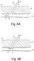
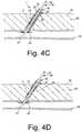
- the positioning element 46may be an expandable element, such as a wire mesh structure, as shown in FIG. 3 , an expandable frame 46,' as shown in FIGS. 4A-4C , and/or a balloon (not shown).
- the positioning element 46 or 46'may include a skin or other covering (not shown) on at least a proximal portion thereof, thereby making the positioning element 46 or 46' substantially nonporous.
- the positioning element 46 or 46'may be biased to an enlarged condition, such as that shown in FIGS. 3 and 4A -4C , but may be compressed to a contracted condition, e.g., by an overlying sleeve or other constraint (not shown).
- the constraintmay be removed to expose the expandable element, allowing the expandable element to automatically expand to the enlarged condition.
- the expandable elementmay be selectively expandable, e.g., using a pullwire, source of inflation media (e.g., coupled to a lumen (not shown) extending through the positioning member 40 to an inflatable positioning element, not shown), or other actuator (also not shown) operable from the proximal end of the position member 40.
- the puncture 90extends from a patient's skin 92 through intervening tissue 96, e.g., to a body lumen 94.
- the puncture 90may be a percutaneous puncture communicating with a blood vessel 94, such as a femoral artery, carotid artery, and the like.
- the puncture 90may be created using known procedures, e.g., using a needle, guidewire, one or more dilators, and the like (not shown).
- An introducer sheath(also not shown) may be advanced through the puncture 90 into the vessel 94, e.g., to provide access into the vessel 90 for one or more instruments, and/or allow one or more diagnostic and/or interventional procedures to be performed via the vessel 90, as is known in the art.
- any instruments and/or the introducer sheathmay be removed from the puncture 90.
- the positioning member 40may be advanced through the puncture 90 until the positioning element 46 is disposed within the vessel 94, whereupon the positioning element 46 may be expanded to the enlarged condition shown in FIG. 4B .
- the positioning member 40may be advanced through a previously placed introducer sheath (not shown), e.g., before the introducer sheath is removed from the puncture 90.
- the positioning member 40may be advanced directly through the puncture 90 after the introducer sheath is removed.
- the positioning element 46may be maintained in the contracted condition (shown in FIG. 4A ) as it is advanced through the puncture 90, e.g., by an overlying sheath or other constraint (not shown). Once the positioning element 46 is disposed within the vessel 94, the constraint may be removed, allowing the positioning element 46 to expand automatically to the enlarged condition (shown in FIG. 4B ). Alternatively, the positioning element 46 may be expanded to the enlarged condition via an actuator (not shown) on the proximal end 42 of the positioning member 40.
- the positioning member 40may be partially withdrawn from the puncture 90 until the positioning element 46 contacts the wall of the vessel 94, as shown in FIG. 4B . If the positioning element 46 is substantially nonporous, the positioning element 46 may substantially seal the puncture 90 from the vessel 94.
- the apparatus 1may be introduced into the puncture 90, e.g., before or after the positioning element 46 is directed into contact with the wall of the vessel 94.
- the proximal end 42 of the positioning member 40may be backloaded into the distal end 24 of the delivery sheath 20, e.g., through the lumens 26, 10, 36 of the delivery sheath 20, plug device 2, and pusher member 30, respectively.
- the delivery sheath 20may then be advanced over the positioning member 40, e.g., until the distal end 24 is disposed adjacent the vessel 94.
- the proximal end 42 of the positioning member 40may be pulled to draw the positioning element 46 against the distal end 24 of the delivery sheath 20 (providing a tactile feedback). The positioning member 40 may then be pulled further until the positioning element 46 contacts the wall of the vessel 94 (providing another tactile feedback), thereby partially in retracting the delivery sheath 20 back into the puncture 90.
- the delivery sheath 20may be advanced until the distal end 24 contacts the positioning element 46, thereby providing a tactile indication that the distal end 24, and consequently the plug device 2, are disposed adjacent the vessel 94. If the positioning element 46 substantially seals the puncture 90 from the vessel 94, this may prevent or minimize blood within the vessel 94 from entering the puncture 90, where it may seep into the lumen 26 of the delivery sheath 20 and contact the plug device 2. This may be desirable to reduce any premature reaction between the first and second precursors on the plug device 2.
- the positioning member 40may be carried initially within the delivery sheath 20.
- the positioning member 40"may include a foot plate 46" on a distal end 44" thereof that may be stored within the lumen 26 of the delivery sheath 20 distal to the plug device 2.
- the delivery sheath 20may be advanced into the puncture 90, e.g., directly or through the introducer sheath (before its removal) with the foot plate 46" therein.
- the positioning member 40"may be advanced to expose the foot plate 46" within the vessel 94.
- the foot plate 46"may change orientation once exposed and/or may expand radially. Thereafter, the positioning member 40" may be partially retracted to direct the foot plate 46" into contact with the wall of the vessel 94, preventing the positioning member 40" from being withdrawn further. If the foot plate 46" has sufficient width, it may substantially seal the puncture 90 from the vessel 94.
- the delivery sheath 20may be advanced into the puncture 90, e.g., over a guidewire (not shown), which may remain after removing the introducer sheath, through the introducer sheath (before its removal), or directly through the puncture 90.
- the positioning member 40may be advanced into the proximal end 22 of the delivery sheath 20 and through the lumen 10 of the plug device 2, e.g., with the positioning element 46 in the contracted condition.
- the distal end 24 of the positioning member 40may be advanced distally until the positioning element 46 is disposed within the vessel 94. Once within the vessel 94, the positioning element 46 may be expanded and directed into contact with the wall of the vessel 94, similar to the procedure described above.
- the plug device 2may then be deployed from the delivery sheath 20.
- the delivery sheath 20may include a pusher member 30 within the lumen 26 and disposed proximal to the plug device 2.
- the delivery sheath 20With the distal end 24 of the delivery sheath 20, and consequently the distal end 16 of the plug device 2, located proximal to the vessel 94, the delivery sheath 20 may be retracted proximally, while maintaining the pusher member 30 substantially stationary.
- the pusher member 30may retain the plug device 2 in position within the puncture 90 while the delivery sheath 20 is retracted from around the plug device 2.
- the plug device 2may be offset proximally from the distal end 24 of the delivery sheath 20 a predetermined distance, e.g., between about two millimeters (2 mm) and ten millimeters (10 mm), and in an exemplary embodiment, about five millimeters (5 mm), such that the plug device 2 is delivered within the puncture 90 offset proximally from the vessel 94.
- the plug device 2may be located immediately adjacent the distal end 24 of the delivery sheath 20.
- the pusher member 30may be advanced distally relative to the delivery sheath 20 to deliver the plug device 2 into the puncture 90.
- the pusher member 30may be advanced until the plug device 2 abuts the positioning element 46 of the positioning member 40. This may ensure that the plug device 2 is delivered adjacent to the vessel 94, providing tactile feedback when the plug device 2 abuts the positioning element 46.
- the pusher member 30may be used to deploy the positioning element 46" and plug device 2 sequentially.
- the pusher member 30may be used to compress, pack, or cinch the plug device 2 within the puncture 90.
- the pusher member 30may be advanced to push the plug device 2 distally against the positioning element 46.' This may place the distal end 16 of the plug device 2 adjacent to or against the wall of the vessel 94, which may enhance hemostasis in the arteriotomy between the vessel 94 and the puncture 90.
- the pusher member 30may be advanced further, thereby compressing the plug device 2 axially, which may enhance the plug device 2 expanding radially to fill the puncture 90 and/or permeate outwardly against or into the surrounding tissue.
- additional sealing compoundmay be delivered into the puncture 90, e.g., to fill all or a portion of the puncture 90 above and/or around the plug device 2.
- the delivery sheath 20 or the pusher member 30may be used to deliver liquid sealing compound, e.g., hydrogel precursors (not shown), into the puncture 90, e.g., through the lumen 26 (of the delivery sheath 20) or lumen 36 of the pusher member 30 (or through a separate lumen (not shown) in either device).
- the delivery sheath 20may include one or more side ports (not shown) on the proximal end of the delivery sheath 20 that may be coupled to a source of sealing compound, such as a syringe assembly storing hydrogel precursors (not shown). If the delivery sheath 20 has not been removed entirely from the puncture 90, the delivery sheath 20 may be advanced into the puncture 90 until the distal end 24 is disposed adjacent the plug device 2, whereupon the sealing compound may be delivered into the puncture 90.
- a source of sealing compoundsuch as a syringe assembly storing hydrogel precursors
- the delivery sheath 20may be retracted as the sealing compound is delivered, e.g., to at least partially fill the puncture 90.
- the pusher member 30may be used to deliver sealing compound in a similar manner to those just described.
- a separate sheath or other delivery device(not shown) may be introduced into the puncture 90 to deliver the liquid sealing compound above and/or around the plug device 2. Exemplary apparatus for delivering such sealing compounds into the puncture 90 are disclosed in U.S. Patent Applications Publication Nos. 2004-0249342 and 2004-0267308 .
- the positioning member 40, pusher member 30, and the delivery sheath 20may then be removed, leaving the plug device 2 within the puncture 90.
- the components of the apparatus 1may be removed in any desired order.
- the positioning member 40may be withdrawn through the plug device 2 and the lumen 36 of the pusher member 30.
- the pusher member 30may restrain the plug device 2 from moving proximally as the positioning member 40 is removed. Once the positioning member 30 is removed, the pusher member 30 (and the delivery sheath 20, if not already removed) may then be removed.
- the delivery sheath 20 and pusher member 30may be withdrawn first followed by the positioning member 40.
- the positioning elementsuch as the foot plate 46" may remain within the vessel 94 after the plug device 2 is delivered.
- the foot plate 46"(or other positioning element) may be made at least partially from a bioabsorbable material, e:g., a relatively fast absorbing material, such as that disclosed in U.S. application Serial No. 10/928,744 .
- the positioning element 46may be collapsed to allow the positioning member 40 to be removed through the lumen 10 of the plug device 2 without substantially moving or disrupting the plug device 2.
- a sleeve or other constraint(not shown) may be advanced over the positioning member 40 until it contacts and forces the positioning element 46 to collapse as it enters the sleeve.
- the positioning element 46is controlled by an actuator (not shown)
- the actuatormay be manipulated to collapse the positioning element 46 before the positioning member 40 is removed.
- the positioning member 40may simply be pulled proximally until the positioning element 46 contacts the plug device 2 and forces the positioning element 46 to collapse as it enters the lumen 10 of the plug device 2.
- blood and/or other fluid within the vessel 94may enter the puncture 90, thereby exposing the plug device 2 to an aqueous physiological environment.
- the aqueous physiological environmentwhich may include blood or other bodily fluids from the vessel 94 (or other body lumen) may wet the plug device 2, thereby initiating a reaction between the first and second precursors thereon.
- the fluidmay dissolve the activating agent 8, changing the pH of the fluid to initiate the first and second hydrogel precursors 6, 8 reacting with one another.
- the reaction of the first and second hydrogel precursors 6, 7may form an adhesive or "sticky" hydrogel coating 38 that may bond or otherwise attach to tissue surrounding the puncture 90, which may facilitate retaining the plug device 2 in place within the puncture 90.
- hydrogel coating 38may also expand or swell to further aid in retaining the plug device 2 within the puncture 90 and/or enhance sealing the puncture 90. It will be appreciated that, although hydrogel precursors are described herein, other multiple component adhesives and/or reactive components may be applied to the carrier 4 to create an adhesive or other coating around the carrier 4 when the plug device 2 is exposed to fluid within the patient's body.
- the porous carrier 4may be exposed to an aqueous physiological environment, e.g., blood within the puncture 90, e.g., as the first and second precursors 6, 8 dissolve and/or react.
- the carrier 4includes pro - thrombotic material, the material may cause and/or accel erate coagulation of the blood within the puncture 90, thereby enhancing hemostasis.
- the carrier 4may expand to substantially occlude the lumen 10, although alternatively, the lumen 10 may be sufficiently small to seal by natural hemostasis of the blood.
- the carrier 4includes therapeutic and/or pharmaceutical agent(s), the blood and/or surrounding tissue may become exposed to the agent(s), thereby enhancing hemostasis, patient comfort, healing, and the like.
- FIGS. 6A-6Canother embodiment of a plug device 102 is shown for sealing a puncture extending through tissue (not shown).
- the device 102includes a carrier or core 104, e.g., in a predetermined shape.
- the carrier 104is formed from a lyophilized (i.e., freeze-dried) PEG polymer that contains hydrolytically degradable chemical groups.
- FIGS. 6A and 6Billustrate a carrier 104 in the shape of a cylindrical plug having proximal and distal ends 114, 116, it will be appreciated that the carrier 104 may have other cross-sections or shapes, such as elliptical, triangular, square, conical, disk, polygonic shapes, etc. (not shown).
- the carrier 104is formed from a lyophilized PEG polymer without any surface adherent layer or sticky coating.
- the carrier 104 or plug device 102may be secured within a puncture simply due to expansion of the carrier 104 within the puncture, e.g., upon exposure to blood or other bodily fluids.
- the lyophilized PEG polymere.g., including a macroporous polymer network, may uptake fluid and expand when exposed to an aqueous environment.
- the magnitude of expansion or swellingmay be significant, e.g., between about two and ten times (2X-10X) its lyophilized size based on volume.
- the lyophilized hydrogelmay absorb between about two and ten times its weight in liquid, causing the carrier 104 to expand substantially.
- the hydrogelmay absorb liquid until it is substantially saturated, e.g., within a few minutes, e.g., not more than about two minutes.
- a surface adherent layer or coating 106may be provided on all or a portion of the carrier 104.
- the adherent layer 106may be a mixture of un-cross-linked PEG polymers, similar to the previous embodiments, including first and second PEG polymers 107 in an initially unreactive state and admixed with a pH adjusting agent 108.
- the first PEG polymermay be formed from an amine - terminated PEG polymer while the second PEG polymer may be formed from an ester-terminated, hydrolytically degradable PEG polymer.
- the first and second PEG polymers 107may include any number of PEG polymer precursor materials, such as those disclosed in U.S. Patent Nos. 6,152,943 , 6,165,201 , 6,179,862 , 6,514,534 , 6,379,373 , 6,703,047 , and in U.S. Patent Applications Publication Nos. 2003-0012734 , 2002-0114775 , and 2004-0249342 .
- the pH adjusting agent 108may include, for example, sodium borate, such as Na2B4O7 ⁇ 10H2O in crystalline or powder form, similar to the previous embodiments, sodium bicarbonate, or other salt-based materials, and the like that may alter the localized pH on or around the carrier 104.
- the first and second PEG polymers 107 and pH adjusting agent 108may be carried on all or a portion of the carrier 104, e.g., dispersed on an outer surface or within the carrier 104.
- the first and second PEG polymers 107may remain in the unreactive state, e.g., before or until exposure to an aqueous physiological environment, which may exist, for example, inside a puncture or other passage through tissue.
- Blood or other bodily fluids that contact the PEG polymer-laden carrier 104may initiate a cross-link forming reaction between the two PEG polymers 107 carried in the adherent layer 106.
- the reaction of the PEG polymers 107may create a cross-linked adhesive or tacky hydrogel, which may aid in retaining the plug device 102 within a puncture after deployment and/or in facilitating hemostasis within the puncture.
- the cross-linking reactionmay occur, for example, when the plug device 104 is in intimate contact with tissue surrounding the puncture, such as fat cells within the fascia or other tissue layers.
- This cross-linking reactionmay mechanically lock or otherwise secure the plug device 102 within the puncture, e.g., to maintain its position post-deployment.
- This securing propertymay be particularly advantageous in situatio ns where the patient will ambulate shortly after completing the procedure, which otherwise may increase the potential of plug migration and, consequently, of bleeding complications.
- the target deployment locationmay be maintained within the patient while the puncture site heals.
- the lyophilized PEG polymer forming the carrier 104may hydrate rapidly after contacting blood or other bodily fluids. Consequently, any blood or other bodily fluid that leaks from the puncture site and/or surrounding tissue before significant degradation of the carrier 104 may immediately re-trigger the hydration reaction of the carrier 104 material, thereby improving the potential for puncture closure.
- the material of the plug device 102i.e., the carrier 104 and/or adherent layer 106, may be at least partially absorbed by the body over time, e.g., over a period of days, weeks, or months.
- the carrier 104 and/or adherent layer 106may include therapeutic and/or pharmaceutical agents, e.g., to promote healing, prevent infection and/or other adverse medical events, and the like. Such agents may be embedded in the carrier material and/or adherent layer 106 and/or applied as one or more coatings or layers.
- the material of the carrier 104may have a substantially uniform composition or the composition may be varied, e.g., along its length and/or within underlying layers within the carrier 104.
- the carrier 104includes proximal and distal ends 114, 116, and a lumen 110 extending between the proximal and distal ends 114, 116, thereby defining a longitudinal axis 118.
- the lumen 110may be created when the carrier 104 is formed, e.g., if the carrier 104 is rolled from one or more sheets or layers of material or formed by molding. Alternatively, the lumen 110 may be formed by boring into or otherwise removing material from an already formed solid carrier 104.
- the lumen 110may be dimensioned and/or sized for receiving a catheter, guide wire, or other elongate member, therethrough. For example, as described further below, a portion of a positioning member 140 may slide or otherwise pass through the lumen 110 of the carrier 104, e.g., while delivering the plug device 102.
- the shape of the lyophilized PEG polymer forming the carrier 104may be fixed at the time of lyophilization.
- the lyophilized PEG polymermay be formed in various pre-formed shapes, such as sheets and/or blocks, which may then be formed post-dehydration into a desired geometry, e.g., to facilitate placement within a delivery system, such as the apparatus 101 described below.
- Various shaping/sizing processesmay be employed to transform the lyophilized PEG polymer into the desired size and/or geometry, such as die cutting, rolling, flattening, compression molding, and the like.
- FIG. 6Billustrates the carrier 104 loaded with a mixture of first and second PEG polymers 107 and a pH adjusting agent 108.
- a powdered form of an amine-terminated polymermay be used as the first PEG polymer while an ester terminated, hydrolytically degradable PEG polymer in powder form is used as the second PEG polymer, similar to the previous embodiments.
- the two powdersmay be mixed in a mixing container while in powder form. Powdered sodium borate crystals, e.g., milled into a fine powder to reduce granularity and to better enable mixing, may be added to the first and second PEG polymer 107 mixture.
- the resulting mixture(first and second PEG polymers 107 and pH adjusting agent 108) may then be heated to about 40°C to melt the first and second PEG polymers 107 and/or the pH adjusting agent 108.
- the melted mixtureis preferably thoroughly mixed, e.g., to ensure a substantially uniform or otherwise desired distribution of the constituents.
- the melted mixture(first and second PEG polymers 107 and pH adjusting agent 108) may then be applied to all or a portion of an exposed surface of the carrier 104.
- the mixturemay be applied by any number of known methods, for example, by painting the heated liquid mixture onto the carrier 104 with a brush or other applicator, by spraying an aerosol of the heated liquid mixture onto the carrier 104, or by dipping or wicking the heated liquid mixture onto the carrier 104 using a bath and the like containing the heated liquid mixture.
- the mixturemay be allowed to cool, e.g., to solidify and/or otherwise form the adherent layer 106. After cooling, a solid or semi-solid adherent layer 106 may surround the lyophilized carrier 102.
- the proximal end 114 and distal end 116 of the carrier 104are not covered with an adherent layer 106, as shown in FIG. 6B .
- the lyophilized PEG polymer at the proximal and distal ends 114, 116 of the carrier 104may remain exposed, e.g., to facilitate subsequent hydration.
- This particular embodimenthas excellent swelling/expansion characteristics, while substantially maintaining the position of the plug device 102 at a desired target location.
- FIG. 6Cshows a magnified cross-sectional view of the exterior surface of the adherent layer 106 disposed on an exposed surface of a lyophilized carrier 104.
- the first and second PEG polymers 107, as well as the pH adjusting agent 108,are all well mixed within the entire adherent layer 106.
- the relative concentration of the components of the adherent layer 106may vary along the carrier 104.
- a lyophilized polymer carrier 104is provided, e.g., by forming a plug or other body from a PEG polymer that contains hydrolytically degradable chemical groups.
- the carrier 104may be formed by rolling one or more sheets of material into a desired shape, by molding, by cutting individual devices from a larger mass of material, machining, grinding, and the like.
- a mixture of first and second PEG polymers 107is provided in a predetermined ratio, e.g., in an equimolar ratio.
- a pH activating agent 108such as solid sodium borate, may be added to the mixture created in step B.
- the pH activating agent 108is milled into a fine powder before being added to the mixture of first and second PEG polymers 107.
- the resulting mixture formed in step Cmay then be heated to a predetermined temperature to melt the first and second PEG polymers 107.
- the mixtureis heated to a temperature of about forty degrees Celsius (40 °C). After the first and second PEG polymers 107 have melted (while the borate crystals remain solid), the entire mixture may be thoroughly mixed.
- the heated liquid mixturemay then be applied to the carrier 104, e.g., to one or more exposed surfaces of carrier 104 using one of the methods described above, to form the adherent layer 106.
- the first and second precursorsmay be dissolved in one or more solvents that allow the precursors to be mixed and/or applied to the carrier 104, while remaining in an unreactive state relative to one another, e.g., methylene chloride, dimethyl sulfoxide, hot acetone, and the like.
- one or more therapeutic and/or pharmaceutical agentsmay be applied to the carrier 104 and/or adherent layer 106.
- the adherent layer 106may be applied or otherwise dispersed within the carrier 104, e.g., by dipping or wicking or by creating multiple layers for the carrier 104 that are coated and successively formed together to create the final carrier 104.
- a sheet including multiple layers of different componentssuch as one or more of the components described above, may be formed, and the sheet may be rolled into a tubular or solid cylindrical structure.
- An exemplary embodiment of such a sheetmay include three layers, e.g., a first layer of lyophilized hydrogel, a second layer of two-part hydrogel adherent material, and a third layer of lyophilized hydrogel.
- the adherent layere.g., including two hydrogel precursors in an initially unreactive state, may be sandwiched between layers of lyophilized hydrogel.
- a layer of lyophilized hydrogelmay be provided, and an adherent layer, e.g., including two hydrogel precursors in an initially unreactive state, may be applied to one surface of the layer of lyophilized hydrogel.
- a pH adjusting agente.g., borate crystals
- the pH adjusting agentmay be embedded or otherwise applied to the opposite surface of the layer of lyophilized hydrogel.
- the pH adjusting agentmay be substantially segregated from the adherent layer. This may be desirable to prevent the pH adjusting agent from initiating reaction of the materials of the adherent layer prematurely, which may otherwise occur to some degree, even absent an aqueous environment.
- the resulting composite materialmay then be folded or rolled into a desired plug configuration.
- a delivery apparatus 101for sealing a puncture 90 through tissue 96, e.g., to a vessel 94 or other body lumen, similar to the previous embodiments.
- the apparatus 101includes an introducer or delivery sheath or other tubular member 2 0 and a positioning member 140, e.g., similar to the previous embodiments.
- the delivery apparatus 101also includes a cartridge 120 carrying a plug device 102, such as one of those described above, and a plunger, cincher, or other pusher member 130.
- the cartridge 120generally includes an elongate tubular body including a proximal end 122, a distal end 124, and a lumen 126 extending between the proximal and distal ends 122, 124 within which the plug device 102 may be carried.
- the pusher member 130may also be an elongate tubular body including a proximal end 132, a distal end 134, and a lumen 136 extending between the proximal and distal ends 132, 134.
- the positioning member 140may include an elongate member having a proximal end 142, a distal end 144, and an expandable positioning element 146 on the distal end 144, such as an expandable mesh (as shown in FIG. 8 ), a mechanically expandable structure, or a balloon (not shown).
- the delivery apparatus 101may be used to position and deliver the plug device 102 within a puncture 90, e.g., extravascularly just above or otherwise adjacent to the arteriotomy in a vessel 94 communicating with the puncture 90.
- the cartridge 120may be insertable or otherwise slidable within lumen 26 of the delivery sheath 20, and the pusher member 130 may be slidable within the lumen 126 of the cartridge 120.
- the plug device 102may be compressed or otherwise disposed within the lumen 126 of the cartridge 120 distal to the pusher member 130.
- the positioning member 140may insertable through the cartridge 120, e.g., through the pusher member 130 and plug device 102.
- the plug device 102may be disposed between an inner wall of the cartridge 120 and an exterior surface of the positioning member 140.
- the cartridge 120may be used to shuttle the plug device 102 into position for deployment, i.e., through the delivery sheath 20.
- the pusher member 130may be positioned proximal to the plug device 102 for positioning and/or maintaining the plug device 102 in a predetermined location during deployment.
- the delivery apparatus 101may be used to deliver the plug device 102 and/or otherwise facilitate hemostasis within a puncture 90 through tissue 94.
- delivery sheath 20may be placed within the puncture 90, e.g., to provide access to vessel 94, similar to the previous embodiments.
- a positioning member 140may be introduced into and/or through the lumen 26 of the delivery sheath 20, e.g., with the expandable frame or other positioning element 146 thereon in a collapsed condition.
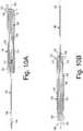
- the cartridge 120(along with the plug device 102 and pusher member 130) may be provided initially on the proximal end 142 of the positioning member 140, as shown in FIG. 10A .
- the cartridge 120may initially be located outside the puncture 90 as the positioning member 130 is advanced into the puncture 90.
- the cartridge 120may be carried on the distal end 144 of the positioning member 140, e.g., such that the cartridge 120 (along with the plug device 102 and pusher member 130) are introduced simultaneously with the positioning member 140.
- the cartridge 120may be provided separate from the positioning member 140.
- the shaft of the positioning member 140may extend proximally from the proximal end 22 of the delivery sheath 20, and may be later back-loaded into the cartridge 120, e.g., through the lumen 136 of the pusher member 130 and/or the lumen 110 of the plug device 102.
- the distal end 144 of the positioning member 140may be inserted through the puncture 90 and the arteriotomy into the vessel 94.
- the positioning element 146 on the distal end 144 of the positioning member 140may be expanded or otherwise deployed, similar to the previous embodiments.
- the expandable positioning element 146 on the positioning member 140may be mechanically expanded or inflated to an enlarged condition.
- the positioning member 140may be at least partially withdrawn until the positioning element 146 contacts the wall of the vessel 94, e.g., to substantially seal the vessel 94 from the puncture 90.
- Thismay involve a two-step, tactile process, similar to the previous embodiments, in which the positioning member 140 with expanded positioning element 146 is withdrawn until it contacts the distal end 24 of the delivery sheath 20 and then until the positioning element 146 contacts the wall of the vessel 94.
- Tension in the proximal directionmay be applied and/or maintained on the positioning member 140 to retract the positioning element 146, e.g., to seal the puncture 90.
- the proximal tensionmay be maintained manually or using a tensioner device (not shown), such as that disclosed in U.S. Patent Application Publication No. 2004-0267308 , to provide temporary hemostasis, e.g., during the subsequent steps.
- the cartridge 120 carrying the plug device 102may be advanced distally over the positioning member 140 into the puncture 90.
- the cartridge 120 (and plug device 102)may be advanced through the delivery sheath 20 until a hub 123 of the cartridge 120 abuts a hub 23 on the delivery sheath 20 (shown in FIG. 9C ).
- the positioning member 140 and/or pusher member 130may include one or more cooperating detents that may engage when the cartridge 120 reaches a predetermined location along the positioning member 140, e.g., to limit subsequent movement of the pusher member 130 relative to the positioning member 140.
- the positioning member 140may include a ring, tab, or other raised element 145
- the pusher member 130may include a living hinge, tab, or other latch element 135, e.g., on proximal end 132.
- the latch element 135may simply be an annular notch in the proximal end 132 of the pusher member 130 to bias the proximal end inwardly. As the cartridge 120 (and consequently the pusher member 130) is advanced, the latch element 135 that may pass freely over the raised element 145. The latch element 135 then may prevent the pusher member 130 from being retracted again, the blunt edge of the latch element 135 abutting the ring 145 on the positioning member 140.
- the cartridge member 120 and pusher member 130may be provided initially on the positioning member 140, as shown in FIG. 10B .
- the pusher member 130 and positioning member 140may include the cooperating detents 133, 145 to prevent proximal movement of the pusher member 130 relative to the positioning member 140.
- the pusher member 130may be otherwise fixed relative to the positioning member 140, e.g., to fix the distal end 134 of the pusher member 130 a predetermined distance proximal to the positioning element 146, e.g., to position the plug device 102 immediately adjacent the positioning element 146, as shown in FIG. 10B . While advanced into the delivery sheath 20 or otherwise within the puncture 90, the plug device 102 may remain out of direct or indirect contact with blood or other bodily fluids along the blood path.
- the pusher member 130may be advanced distally into the lumen 126 of the cartridge 120, e.g., until a marker 137 on the pusher member 130 is located adjacent to the hub 123 of the cartridge 120. As seen in FIG. 9C , this marker location may place the distal end 134 of the pusher member 130 proximally adjacent to the proximal end 114 of the plug device 102.
- the pusher member 130 and plug device 102may be initially positioned within the cartridge 120 as shown in FIG. 9C , i.e., with the plug device 102 adjacent the distal end 124 the cartridge 120, thereby eliminating the need to advance the pusher member 130.
- the position of the pusher member 130is maintained, and the delivery sheath 20 and cartridge 120 are retracted proximally to expose or otherwise deploy the plug device 102 within the puncture 90.
- the pusher member 130may serve as a stop that prevents the plug device 102 from moving proximally while the delivery sheath 20 and cartridge 120 are withdrawn.
- the user of the delivery apparatus 101may position his or her thumb on hub 133 of the pusher member 130 to maintain its position while the delivery sheath 20 and cartridge 120 are retracted, e.g., using his or her index and middle fingers.
- the delivery sheath 20may be held and withdrawn, thereby causing the cartridge 120 to be withdrawn simultaneously.
- the cartridge 120may be removed first, and then the delivery sheath 20 may be removed. The cartridge 120 and delivery sheath 20 may be removed entirely from the puncture 90 or only to expose the plug device 102.
- the plug device 102may be tamped or otherwise compressed within the puncture 90, e.g., by advancing the pusher member 130 distally to press the plug device 102 against the wall of the vessel 94 and/or against the positioning element 146, similar to the previous embodiments. This may cinch the plug device 102, which may cause the plug device 102 to expand radially outwardly and/or press the plug device 102 against the arteriotomy, e.g., to enhance sealing the puncture 90 from the vessel 94.
- the proximal tension on the positioning member 140may be released and/or the positioning element 146 may be collapsed to its collapsed state.
- the positioning element 146may be mechanically collapsed or deflated.
- the positioning member 140(and consequently the positioning element 146) may be slowly withdrawn through the lumen 110 of the plug 102.
- the positioning element 146may have a profile not more than about 0.875 millimeter (035 inch) to facilitate removal of the positioning member 140 without substantially disturbing the deployed plug device 100. While the positioning member 140 is withdrawn, the pusher member 130 may be maintained to serve as a stop and prevent proximal migration of the plug device 102 within the puncture 90. In addition, in embodiments where the plug device 102 includes an adherent layer (not shown in FIG. 9D ), the "sticky" adherent layer may also aid in securing the plug device 102 to the surrounding tissue.
- the pusher member 130may be withdrawn, leaving the plug device 102 in place.
- liquid hydrogel or other sealing compoundmay be delivered into the puncture 90 above and/or around the plug device 102, similar to the previous embodiments, to assist in achieving permanent hemostasis.
- a source of sealing compound(not shown) may be coupled to the proximal end 132 of the pusher member 130 and sealing compound may be delivered into the puncture above and/or around the plug device 102.
- the pusher member 130may be retracted proximally as the sealing compound is delivered to at least partially fill the puncture 90 with the sealing compound
Landscapes
- Health & Medical Sciences (AREA)
- Life Sciences & Earth Sciences (AREA)
- Animal Behavior & Ethology (AREA)
- General Health & Medical Sciences (AREA)
- Public Health (AREA)
- Veterinary Medicine (AREA)
- Surgery (AREA)
- Epidemiology (AREA)
- Heart & Thoracic Surgery (AREA)
- Chemical & Material Sciences (AREA)
- Vascular Medicine (AREA)
- Engineering & Computer Science (AREA)
- Biomedical Technology (AREA)
- Medical Informatics (AREA)
- Nuclear Medicine, Radiotherapy & Molecular Imaging (AREA)
- Molecular Biology (AREA)
- Medicinal Chemistry (AREA)
- Dispersion Chemistry (AREA)
- Dermatology (AREA)
- Chemical Kinetics & Catalysis (AREA)
- Cardiology (AREA)
- Materials Engineering (AREA)
- Neurosurgery (AREA)
- Pharmacology & Pharmacy (AREA)
- Oral & Maxillofacial Surgery (AREA)
- Transplantation (AREA)
- Surgical Instruments (AREA)
- Polymers & Plastics (AREA)
- Organic Chemistry (AREA)
- Materials For Medical Uses (AREA)
Abstract
Description
- This invention relates to apparatus for sealing punctures in a body, and more particularly, to apparatus for sealing a vascular puncture extending through tissue into a blood vessel, and to apparatus for delivering a plug into a percutaneous puncture extending from a patient's skin to a blood vessel or other body lumen to seal the puncture.
- Apparatus and methods are known for accessing a patient's vasculature percutaneously, e.g., to perform a procedure within the vasculature, and for sealing the puncture that results after completing the procedure. For example, a hollow needle may be inserted through a patient's skin and overlying tissue into a blood vessel. A guide wire may be passed through the needle lumen into the blood vessel, whereupon the needle may be removed. An introducer sheath may then be advanced over the guide wire into the vessel, e.g., in conjunction with or subsequent to one or more dilators. A catheter or other device may be advanced through the introducer sheath and over the guide wire into a position for performing a medical procedure. Thus, the introducer sheath may facilitate accessing and/or introducing various devices into the vessel, while minimizing trauma to the vessel wall and/or minimizing blood loss. Upon completing the procedure, the device(s) and introducer sheath may be removed, leaving a puncture extending between the skin and the vessel wall.
- To seal the puncture, external pressure may be applied to the overlying tissue, e.g., manually and/or using sandbags, until hemostasis occurs. This procedure, however, may be time consuming and expensive, requiring as much as an hour of a medical professional's time. It is also uncomfortable for the patient, and may require the patient to remain immobilized in the operating room, catheter lab, or holding area. In addition, a risk of hematoma exists from bleeding before hemostasis occurs.
U.S. Patent No. 5,108,421 to Fowler discloses a plug that may be delivered into a puncture through tissue. The plug is a cylindrical rod-shaped member which is constructed of a porous, bioabsorbable and expandable hemostatic collagen sponge or a polymerized polylactic acid or polyglycolic acid. In one embodiment, a catheter is inserted through the puncture into the blood vessel. A balloon on the catheter is expanded and retracted until the balloon is disposed adjacent the puncture at the wall of the vessel. The plug may be advanced into the puncture until the plug contacts the balloon. Once the plug is positioned within the puncture, the balloon may be deflated and withdrawn, leaving the plug within the puncture to expand and seal the puncture and/or to promote hemostasis.- Alternatively,
U.S. Patent Nos. 5,192,302 and5,222,974 issued to Kensey et al. describe a bioabsorbable collagen plug that may be delivered through an introducer sheath into a puncture site. The disclosed plug, however, may be difficult to position properly with respect to the vessel, which may be significant since it is generally undesirable to expose the collagen material within the bloodstream where it may float downstream and cause an embolism. U.S. Patent No. 6,605,295 describes rods, plugs, crushed or irregularly shaped pieces of substantially dehydrated hydrogel that may be introduced into a lumen or void in a patient's body to seal or plug a biopsy needle track, reinforce weak tissue, or deliver a therapeutic compound. In one embodiment, a plug of dehydrated hydrogel may be deployed into the site of an arteriotomy and allowed to hydrate in the presence of the tissue fluids and blood, to fill the track of the catheter sheath and prevent further bleeding. By swelling to equilibrium hydration, the plug may lock itself firmly in place and thus reduce the risk of formation of a large hematoma at the site of the puncture.U.S. Patent No. 6,703,047 discloses dehydrated hydrogel precursor-based, tissue adherent compositions. The hydrogels may be used, for example, for sealing fluid leaks from tissue , as adherent drug delivery depots, and as means for augmenting and/or supporting tissue. The hydrogels may be administered directly to an open wound site or may be dispensed, e.g., using a non-adhesive backing material, an absorbable backing material, a syringe applicator, a powder atomization or aerosolization system, or a needle-less injector.- The invention is directed to apparatus for sealing a puncture in a body, and, more particularly, to apparatus for providing temporary or permanent hemostasis within a vascular puncture extending into a blood vessel, and/or to apparatus for delivering a sealing plug into a percutaneous puncture extending from a patient's skin to a blood vessel or other body lumen.
- In accordance with one embodiment, a device is provided for sealing a puncture extending through tissue including a carrier having a predetermined shape, e.g., a disk, cylinder, or other plug. A first hydrogel precursor is disposed on the carrier. A second hydrogel precursor is also disposed on the carrier. The first and second hydrogel precursors are disposed on the carrier in an unreactive state before exposure to an aqueous physiological environment.
- In accordance with another embodiment, an apparatus is provided for sealing a puncture extending through tissue that includes a tubular member and a plug carried by the tubular member. The plug may include first and second hydrogel precursors disposed thereon, the first and second hydrogel precursors being in an unreactive state prior to exposure to an aqueous physiological environment in the tissue. The device may include a pusher member for deploying the plug from the tubular member.
- In one embodiment, the plug may include a lumen extending therethrough. The device may also include a pusher member and a positioning member adapted to slide and/or pass through the tubular member. The positioning member may include an elongate member and an expandable element on one end, e.g., an expandable mesh, balloon, expandable frame, and the like, on a guidewire. In an alternative embodiment, the positioning member may include a bioabsorbable foot plate or other element on one end, e.g., for providing tactile feedback to the user during a sealing procedure and/or sealing the puncture.
- In accordance with still another embodiment, a device is provided for sealing a puncture extending through tissue including a lyophilized hydrogel, e.g., polyethylene glycol (PEG), or other polymer carrier. The polymer used in the carrier includes hydrolytically degradable chemical groups, thereby permitting in vivo degradation.
- In one embodiment, lyophilized PEG carrier is pre-formed into a desired shape or geometry before the lyophilization process. In another embodiment, the lyophilized PEG carrier is formed into the desired shape or geometry after the lyophilization process. For example, "raw" lyophilized PEG carrier material may be shaped or otherwise modified by processes such as die cutting, rolling, flattening, compression molding, and the like.
- In accordance with another embodiment, any of the devices described above may include an adherent "sticky" coating or layer disposed on an exposed surface of the polymer carrier. The adherent coating may be formed from a mixture of un-cross-linked PEG polymers and a pH adjusting agent such as, sodium borate crystals. In an exemplary process, the adherent coating mixture may be heated to melt the polymer components and then applied to the lyophilized PEG carrier.
- In accordance with another embodiment, an apparatus is provided for sealing a puncture extending through tissue that includes a cartridge and a plug device formed from a lyophilized PEG carrier. The plug may include first and second PEG polymers disposed thereon, the first and second PEG polymers being in an unreactive state prior to exposure to an aqueous physiological environment in the tissue. The apparatus may include a pusher member for deploying the plug from the cartridge, a positioning member, and/or an occlusion member.
FIG. 1A is a perspective view of a porous carrier in the shape of a plug.FIG. 1B is a perspective view of the porous carrier ofFIG. 1A having first and second hydrogel precursors disposed thereon.FIG. 1C is a perspective view of the porous carrier ofFIG. 1B having a pH activating agent disposed thereon.FIG. 1D is a magnified cross-sectional view of the porous carrier shown inFIG. 1C , including the first and second hydrogel precursors and the pH activating agent.FIG. 2 is a flowchart showing a method for loading two or more hydrogel precursors on a porous carrier.FIG. 3 is an exploded side view of an apparatus for delivering a plug device into a puncture through tissue.FIGS. 4A-4F are cross-sectional views of a patient's body, showing a procedure for using devices according to embodiments of the invention for sealing a puncture extending from the patient's skin through intervening tissue to a body lumen.FIGS. 5A and 5B are cross-sectional views of a patient's body, showing another apparatus for sealing a puncture extending from a patient's skin through intervening tissue to a body lumen.FIG. 6A is a perspective view of a lyophilized carrier in the shape of a plug.FIG. 6B is a perspective view of the lyophilized carrier ofFIG. 6A having an adherent layer disposed thereon to provide a plug device for sealing a puncture through tissue.FIG. 6C is a magnified cross-sectional view of the plug device ofFIG. 6B , showing first and second polymers and pH activating agent carried on the plug device.FIG. 7 is a flowchart showing a procedure for providing an adherent "sticky" layer on a lyophilized carrier.FIG. 8 is an exploded side view of an apparatus for delivering a plug device into a puncture through tissue.FIGS. 9A-9D are cross-sectional views of a patient's body, showing a procedure for sealing a puncture extending from the patient's skin to a blood vessel using the apparatus ofFIG.8 .FIGS. 10A-B are cross-sectional views of variation of the apparatus ofFIG. 8 .- Turning to the drawings,
FIGS. 1A-1D illustrate adevice 2 for sealing a puncture extending through tissue (not shown). Generally, thedevice 2 includes a carrier orcore 4, e.g., in the shape of a plug, having disposed thereon afirst hydrogel precursor 6 and asecond hydrogel precursor 7. The first andsecond hydrogel precursors carrier 4 in an unreactive state. The first andsecond hydrogel precursors - Blood or other bodily fluids that contact the precursor-
laden carrier 4 may initiate a hydrogel forming reaction between the twoprecursors plug device 2 within a puncture after deployment and/or in facilitating hemostasis within the puncture. Optionally, as described below, an activating agent, e.g., apH adjusting material 8, may also be disposed on thecarrier 4 to initiate, accelerate, or otherwise enhance the reaction of theprecursors FIG. 1A illustrates acarrier 4 in the shape of a circular cylindrical plug. It will be appreciated that thecarrier 4 may have other cross-sections or shapes, such as elliptical, triangular, square, conical, disk, polygonic shapes, etc. Thecarrier 4 may be formed from a biocompatible and/or bioabsorbable material, for example, a porous, bioabsorbable foam or other solid material. In one embodiment, thecarrier 4 may be formed from a biocompatible and/or bioabsorbable hydrogel, e.g., polyethylene glycol ("PEG"), or other synthetic material. In addition or alternatively, thecarrier 4 may include pro-thrombotic material, e.g., including one or more biological pro-thrombotics, such as collagen, fibrin, carboxymethylcellulose, oxidized cellulose, alginates, gelatin, or other protein-based material, and/or synthetic materials, such as polyglycolic acids (PGA's), polyactides (PLA's), polyvinyl alcohol, and the like. The material of thecarrier 4 may be at least partially absorbed by the body over time, e.g., over a period of days, weeks, or months. Optionally, thecarrier 4 may include therapeutic and/or pharmaceutical agents, e.g., to promote healing, prevent infection and/or other adverse medical events, and the like. Such agents may be embedded in the carrier material and/or applied as one or more coatings or layers. In addition, the material of thecarrier 4 may have a substantially uniform composition or the composition may be varied, e.g., along its length and/or within underlying layers within thecarrier 4.- In the embodiment shown, the
carrier 4 includes alumen 10 extending between proximal and distal ends 14, 16, thereby defining alongitudinal axis 18. Thelumen 10 may be created when thecarrier 4 is formed, e.g., if thecarrier 4 is rolled from one or more sheets or layers of material or formed by molding. Alternatively, thelumen 10 may formed by boring into or otherwise removing material from an already formedsolid carrier 4. Thelumen 10 is dimensioned such that a guide wire or other elongate member, such as a portion of a positioning member 40 (described in more detail below) may slide or otherwise pass through thecarrier 4, e.g., while delivering theplug device 2. FIG. 1B illustrates thecarrier 4 loaded with first andsecond hydrogel precursors second hydrogel precursors carrier 4 by wicking a mixture of theliquid hydrogel precursors carrier 4. Depending on the material used, thehydrogel precursors second hydrogel precursors carrier 4.- Alternatively, the first and
second precursor materials carrier 4 may be dipped, that may be poured onto thecarrier 4, and/or otherwise applied to thecarrier 4 together or successively. For example, the first and second precursors may be dissolved in a solvent that may then be applied to thecarrier 4. In either case, once the first andsecond hydrogel precursors carrier 4, the first andsecond hydrogel precursors - The
first hydrogel precursor 6 may include any number of hydrogel precursor materials, such as those disclosed inU.S. Patent Nos. 6,152,943 ,6,165,201 ,6,179,862 ,6,514,534 ,6,379,373 ,6,703,047 , and inU.S. Patent Applications publication Nos. 2003-0012734 ,2002-0114775 , and2004-0249342 . For example, in one embodiment, thefirst hydrogel precursor 6 may include a four arm, 10 kDalton PEG with reactive ester end groups or an eight arm, 20 kDalton PEG amine. Alternatively, thefirst hydrogel precursor 6 may include a bioabsorbable star polymer having a complementary cross-linking species such as, for example, an amino acid with reactive end groups, e.g., lysine, dilysine, trilysine, etc. - The
second hydrogel precursor 7 may include any number of hydrogel precursor materials, e.g., a material reactive with thefirst precursor material 6 once exposed within a hydrous or aqueous environment, such as those materials disclosed above. For example, thesecond precursor 7 may be the other of an eight arm, 20 kDalton PEG amine or a four arm, 10 kDalton PEG ester. Alternatively, thesecond precursor 7 may be the complementary cross-linking species of a bioabsorbable star polymer, such as an amino acid with reactive end groups, e.g., lysine, dilysine, trilysine, etc. - Referring to
FIG. 1C , apH activating agent 8 is also loaded onto thecarrier 4. ThepH activating agent 8 may create a localized change in pH after exposure to a hydrous or aqueous environment, e.g., to initiate or accelerate the hydrogel-forming reaction. In an exemplary embodiment, thepH activating agent 8 includes solid borate crystals, such as Na2B407·10H2O, although different salt-based or other materials that alter the localized pH value may be employed. Alternatively, other pH altering agents may be used, such as sodium borate, sodium bicarbonate, and the like. In one embodiment, thepH activating agent 8 is loaded onto thecarrier 4 by physically contacting solid borate crystals, powder, or other particles onto the precursor-laden (first andsecond hydrogel precursors 6, 7)carrier 4. For example, thecarrier 4 may simply be rolled over apH activating agent 8 with sufficient force to embed thepH activating agent 8 into theexterior surface 12 of thecarrier 4. Alternatively, thepH activating agent 8 may be adhered to theexterior surface 12 of thecarrier 4, e.g., by pressing particles of thepH activating agent 8 into theexterior surface 12, by using an adhesive (e.g., that is substantially inert or unreactive with the first orsecond precursors 6, 7), and the like. FIG. 1D illustrates a magnified cross-sectional view of theexterior surface 12 of the precursor-laden carrier 4 ofFIG. 1D . As shown, a layer of the mixed first andsecond hydrogel precursors exterior surface 12 of thecarrier 4 in a relatively thin film or coating. Because the first andsecond hydrogel precursors second hydrogel precursors exterior surface 12 of theporous carrier 4, e.g., into pores or other recesses to substantially coat all or a significant portion of thecarrier 4.FIG. 1D further shows thepH activating agent 8 loaded onto thecarrier 4. InFIG. 1D , thepH activating agent 8 is in the form of a solid (e.g., borate crystals) with individual particles populated on top of the layer of first andsecond hydrogel precursors pH activating agent 8 may be loaded onto thecarrier 4 in a melted or other liquid form that remains unreactive with the first andsecond hydrogel precursors pH activating agent 8 may form a film, coating, or layer much like that shown of the first andsecond hydrogel precursors FIG. 1D .- Turning to
FIG. 2 , a flowchart shows an exemplary method for making a sealing device, such asplug device 2 described above. First, acarrier 4 is provided (step A), e.g., by forming a plug or other body from a porous, pro-thrombotic, and/or biocompatible material. As described above, thecarrier 4 may be formed by rolling material into a desired shape, by molding, by cutting individual devices from a larger mass of material, machining, grinding, and the like. Next, a mixture of first andsecond hydrogel precursors carrier 4 is then loaded with first andsecond precursors 6, 7 (step C), which, as described above, may be hydrogel precursors in liquid form. Optionally, as shown inFIG. 2 , thecarrier 4 may be loaded with one or more additional layers of hydrogel precursor material (step D). Depending on the hydrogel employed in theplug device 2, there may be multiple hydrogel (e.g., more than two) precursors needed to initiate the hydrogel reaction. In further options, one or more therapeutic and/or pharmaceutical agents may be applied to thecarrier 4, e.g., before or after coating thecarrier 4 with the first andsecond precursors - Finally, an optional
pH activating agent 8 may be loaded on the carrier 4 (step E). In one embodiment, thepH activating agent 8 is in crystalline or other particle form that may be physically adhered to thecarrier 4, e.g., on top of the first andsecond precursors - Turning to
FIG. 3 , anapparatus 1 is shown for sealing a puncture through tissue. Generally, theapparatus 1 may include a delivery sheath or othertubular member 20 and aplug device 2, such as those described elsewhere herein. In addition, theapparatus 1 may include a plunger orother pusher member 30, and/or a positioningmember 40. - The
delivery sheath 20 may be a substantially rigid, semi-rigid, and/or flexible tubular body, including aproximal end 22, adistal end 24 having a size and shape for insertion into thepuncture 90, and alumen 26 extending therebetween. Thedistal end 24 may be tapered and/or may include a substantiallyatraumatic tip 28 to facilitate advancement through a puncture. Thedelivery sheath 20 may include a handle (not shown), and/or one or more seals, e.g., a hemostatic seal (also not shown), on theproximal end 22. Theplug device 2 may be disposed within thelumen 26 proximate to thedistal end 24. Thelumen 26 may be sized such that theplug device 2 is slidable therein, e.g., able to traverse distally from thedelivery sheath 20 during delivery, as described further below. - The
pusher member 30 may be an elongate member, e.g., a plunger, catheter, and the like, including a proximal end (not shown), and adistal end 34 having a size for slidable insertion into thelumen 26 of thedelivery sheath 20. Thedistal end 34 of thepusher member 30 may be substantially blunt to facilitate contacting, pushing, and/or "cinching" theplug device 2 within thedelivery sheath 20 and/or puncture, as described further below. Thepusher member 30 may be substantially rigid, semi-rigid, and/or substantially flexible, having sufficient column strength to allow movement of thedelivery sheath 20 relative to theplug device 2 without buckling thepusher member 30. Thepusher member 30 may also include alumen 36 extending between the proximal end and thedistal end 34, e.g., to accommodate the positioningmember 40 and/or a guidewire (not shown). - In the embodiment shown in
FIG. 3 , the positioningmember 40, e.g., a guidewire, and/or other solid or hollow elongate body, may include aproximal end 42, adistal end 44, and apositioning element 46 on thedistal end 44. Thepositioning element 46 may be an expandable element, such as a wire mesh structure, as shown inFIG. 3 , anexpandable frame 46,' as shown inFIGS. 4A-4C , and/or a balloon (not shown). Optionally, thepositioning element 46 or 46' may include a skin or other covering (not shown) on at least a proximal portion thereof, thereby making thepositioning element 46 or 46' substantially nonporous. - The
positioning element 46 or 46' may be biased to an enlarged condition, such as that shown inFIGS. 3 and4A -4C , but may be compressed to a contracted condition, e.g., by an overlying sleeve or other constraint (not shown). The constraint may be removed to expose the expandable element, allowing the expandable element to automatically expand to the enlarged condition. Alternatively, the expandable element may be selectively expandable, e.g., using a pullwire, source of inflation media (e.g., coupled to a lumen (not shown) extending through the positioningmember 40 to an inflatable positioning element, not shown), or other actuator (also not shown) operable from the proximal end of theposition member 40. Additional information on expandable structures that may be incorporated into positioningmember 40 may be found inU.S. Patent Nos. 6,238,412 and6,635,068 , in U.S. Patent Applications Publication No.US 2003/0078616 , andU.S. Patent Application Serial No. 10/975,205 . - Turning to
FIGS. 4A-4F , an exemplary procedure is shown for sealing apuncture 90 using anapparatus 1. Generally, thepuncture 90 extends from a patient'sskin 92 through interveningtissue 96, e.g., to abody lumen 94. In an exemplary embodiment, thepuncture 90 may be a percutaneous puncture communicating with ablood vessel 94, such as a femoral artery, carotid artery, and the like. - In an exemplary procedure, the
puncture 90 may be created using known procedures, e.g., using a needle, guidewire, one or more dilators, and the like (not shown). An introducer sheath (also not shown) may be advanced through thepuncture 90 into thevessel 94, e.g., to provide access into thevessel 90 for one or more instruments, and/or allow one or more diagnostic and/or interventional procedures to be performed via thevessel 90, as is known in the art. Upon completing the procedure(s) via thevessel 94, any instruments and/or the introducer sheath (not shown) may be removed from thepuncture 90. - Turning to
FIG. 4A , with thepositioning element 46 collapsed, the positioningmember 40 may be advanced through thepuncture 90 until thepositioning element 46 is disposed within thevessel 94, whereupon thepositioning element 46 may be expanded to the enlarged condition shown inFIG. 4B . In one embodiment, the positioningmember 40 may be advanced through a previously placed introducer sheath (not shown), e.g., before the introducer sheath is removed from thepuncture 90. Alternatively, the positioningmember 40 may be advanced directly through thepuncture 90 after the introducer sheath is removed. - The
positioning element 46 may be maintained in the contracted condition (shown inFIG. 4A ) as it is advanced through thepuncture 90, e.g., by an overlying sheath or other constraint (not shown). Once thepositioning element 46 is disposed within thevessel 94, the constraint may be removed, allowing thepositioning element 46 to expand automatically to the enlarged condition (shown inFIG. 4B ). Alternatively, thepositioning element 46 may be expanded to the enlarged condition via an actuator (not shown) on theproximal end 42 of the positioningmember 40. - As shown in
FIG. 4B , once thepositioning element 46 is expanded, the positioningmember 40 may be partially withdrawn from thepuncture 90 until thepositioning element 46 contacts the wall of thevessel 94, as shown inFIG. 4B . If thepositioning element 46 is substantially nonporous, thepositioning element 46 may substantially seal thepuncture 90 from thevessel 94. - Turning to
FIG. 4C , theapparatus 1 may be introduced into thepuncture 90, e.g., before or after thepositioning element 46 is directed into contact with the wall of thevessel 94. For example, theproximal end 42 of the positioningmember 40 may be backloaded into thedistal end 24 of thedelivery sheath 20, e.g., through thelumens delivery sheath 20,plug device 2, andpusher member 30, respectively. Thedelivery sheath 20 may then be advanced over the positioningmember 40, e.g., until thedistal end 24 is disposed adjacent thevessel 94. - If the
positioning element 46 has not yet been retracted, theproximal end 42 of the positioningmember 40 may be pulled to draw thepositioning element 46 against thedistal end 24 of the delivery sheath 20 (providing a tactile feedback). The positioningmember 40 may then be pulled further until thepositioning element 46 contacts the wall of the vessel 94 (providing another tactile feedback), thereby partially in retracting thedelivery sheath 20 back into thepuncture 90. - Alternatively, if the
positioning element 46 is already against the wall of thevessel 94, thedelivery sheath 20 may be advanced until thedistal end 24 contacts thepositioning element 46, thereby providing a tactile indication that thedistal end 24, and consequently theplug device 2, are disposed adjacent thevessel 94. If thepositioning element 46 substantially seals thepuncture 90 from thevessel 94, this may prevent or minimize blood within thevessel 94 from entering thepuncture 90, where it may seep into thelumen 26 of thedelivery sheath 20 and contact theplug device 2. This may be desirable to reduce any premature reaction between the first and second precursors on theplug device 2. - Alternatively, the positioning
member 40 may be carried initially within thedelivery sheath 20. For example, as shown inFIGS. 5A and 5B , the positioningmember 40" may include afoot plate 46" on adistal end 44" thereof that may be stored within thelumen 26 of thedelivery sheath 20 distal to theplug device 2. As shown inFIG. 5A , thedelivery sheath 20 may be advanced into thepuncture 90, e.g., directly or through the introducer sheath (before its removal) with thefoot plate 46" therein. Once thedistal end 24 of thedelivery sheath 20 is disposed within thevessel 94, the positioningmember 40" may be advanced to expose thefoot plate 46" within thevessel 94. Thefoot plate 46" may change orientation once exposed and/or may expand radially. Thereafter, the positioningmember 40" may be partially retracted to direct thefoot plate 46" into contact with the wall of thevessel 94, preventing the positioningmember 40" from being withdrawn further. If thefoot plate 46" has sufficient width, it may substantially seal thepuncture 90 from thevessel 94. - In yet another alternative, before introducing the positioning
member 40, thedelivery sheath 20 may be advanced into thepuncture 90, e.g., over a guidewire (not shown), which may remain after removing the introducer sheath, through the introducer sheath (before its removal), or directly through thepuncture 90. After removing any guidewire, the positioningmember 40 may be advanced into theproximal end 22 of thedelivery sheath 20 and through thelumen 10 of theplug device 2, e.g., with thepositioning element 46 in the contracted condition. Thedistal end 24 of the positioningmember 40 may be advanced distally until thepositioning element 46 is disposed within thevessel 94. Once within thevessel 94, thepositioning element 46 may be expanded and directed into contact with the wall of thevessel 94, similar to the procedure described above. - Turning now to
FIG. 4D , theplug device 2 may then be deployed from thedelivery sheath 20. For example, as described above with respect toFIG. 3 , thedelivery sheath 20 may include apusher member 30 within thelumen 26 and disposed proximal to theplug device 2. With thedistal end 24 of thedelivery sheath 20, and consequently thedistal end 16 of theplug device 2, located proximal to thevessel 94, thedelivery sheath 20 may be retracted proximally, while maintaining thepusher member 30 substantially stationary. Thus, thepusher member 30 may retain theplug device 2 in position within thepuncture 90 while thedelivery sheath 20 is retracted from around theplug device 2. - In one embodiment, the
plug device 2 may be offset proximally from thedistal end 24 of the delivery sheath 20 a predetermined distance, e.g., between about two millimeters (2 mm) and ten millimeters (10 mm), and in an exemplary embodiment, about five millimeters (5 mm), such that theplug device 2 is delivered within thepuncture 90 offset proximally from thevessel 94. Alternatively, theplug device 2 may be located immediately adjacent thedistal end 24 of thedelivery sheath 20. - Alternatively or in addition, the
pusher member 30 may be advanced distally relative to thedelivery sheath 20 to deliver theplug device 2 into thepuncture 90. For example, thepusher member 30 may be advanced until theplug device 2 abuts thepositioning element 46 of the positioningmember 40. This may ensure that theplug device 2 is delivered adjacent to thevessel 94, providing tactile feedback when theplug device 2 abuts thepositioning element 46. Alternatively, as shown inFIG. 5B , if theplug device 2 is disposed within thedelivery sheath 20 along with thepositioning element 46," thepusher member 30 may be used to deploy thepositioning element 46" and plugdevice 2 sequentially. - As shown in
FIG. 4E , if desired, thepusher member 30 may be used to compress, pack, or cinch theplug device 2 within thepuncture 90. For example, after theplug device 2 is exposed within the puncture 90 (e.g., using one of the procedures described above), thepusher member 30 may be advanced to push theplug device 2 distally against thepositioning element 46.' This may place thedistal end 16 of theplug device 2 adjacent to or against the wall of thevessel 94, which may enhance hemostasis in the arteriotomy between thevessel 94 and thepuncture 90. Optionally, thepusher member 30 may be advanced further, thereby compressing theplug device 2 axially, which may enhance theplug device 2 expanding radially to fill thepuncture 90 and/or permeate outwardly against or into the surrounding tissue. - Optionally, after the
plug device 2 is deployed within thepuncture 90, additional sealing compound may be delivered into thepuncture 90, e.g., to fill all or a portion of thepuncture 90 above and/or around theplug device 2. For example, thedelivery sheath 20 or thepusher member 30 may be used to deliver liquid sealing compound, e.g., hydrogel precursors (not shown), into thepuncture 90, e.g., through the lumen 26 (of the delivery sheath 20) orlumen 36 of the pusher member 30 (or through a separate lumen (not shown) in either device). - In one embodiment, the
delivery sheath 20 may include one or more side ports (not shown) on the proximal end of thedelivery sheath 20 that may be coupled to a source of sealing compound, such as a syringe assembly storing hydrogel precursors (not shown). If thedelivery sheath 20 has not been removed entirely from thepuncture 90, thedelivery sheath 20 may be advanced into thepuncture 90 until thedistal end 24 is disposed adjacent theplug device 2, whereupon the sealing compound may be delivered into thepuncture 90. - Alternatively, the
delivery sheath 20 may be retracted as the sealing compound is delivered, e.g., to at least partially fill thepuncture 90. In a further alternative, e.g., if thedelivery sheath 20 has been removed, thepusher member 30 may be used to deliver sealing compound in a similar manner to those just described. In still another alternative, a separate sheath or other delivery device (not shown) may be introduced into thepuncture 90 to deliver the liquid sealing compound above and/or around theplug device 2. Exemplary apparatus for delivering such sealing compounds into thepuncture 90 are disclosed inU.S. Patent Applications Publication Nos. 2004-0249342 and2004-0267308 . - Turning to
FIG. 4F , the positioningmember 40,pusher member 30, and the delivery sheath 20 (if thedistal end 24 still extends into the puncture 90) may then be removed, leaving theplug device 2 within thepuncture 90. The components of theapparatus 1 may be removed in any desired order. For example, in one procedure, the positioningmember 40 may be withdrawn through theplug device 2 and thelumen 36 of thepusher member 30. Thepusher member 30 may restrain theplug device 2 from moving proximally as the positioningmember 40 is removed. Once the positioningmember 30 is removed, the pusher member 30 (and thedelivery sheath 20, if not already removed) may then be removed. - Alternatively, the
delivery sheath 20 andpusher member 30 may be withdrawn first followed by the positioningmember 40. In yet another alternative, the positioning element, such as thefoot plate 46" may remain within thevessel 94 after theplug device 2 is delivered. In this alternative, thefoot plate 46" (or other positioning element) may be made at least partially from a bioabsorbable material, e:g., a relatively fast absorbing material, such as that disclosed inU.S. application Serial No. 10/928,744 . - If the positioning
member 40 is removed, thepositioning element 46 may be collapsed to allow thepositioning member 40 to be removed through thelumen 10 of theplug device 2 without substantially moving or disrupting theplug device 2. For example, a sleeve or other constraint (not shown) may be advanced over the positioningmember 40 until it contacts and forces thepositioning element 46 to collapse as it enters the sleeve. Alternatively, if thepositioning element 46 is controlled by an actuator (not shown), the actuator may be manipulated to collapse thepositioning element 46 before the positioningmember 40 is removed. In another alternative, the positioningmember 40 may simply be pulled proximally until thepositioning element 46 contacts theplug device 2 and forces thepositioning element 46 to collapse as it enters thelumen 10 of theplug device 2. - With the
positioning element 46 collapsed, blood and/or other fluid within thevessel 94 may enter thepuncture 90, thereby exposing theplug device 2 to an aqueous physiological environment. The aqueous physiological environment, which may include blood or other bodily fluids from the vessel 94 (or other body lumen) may wet theplug device 2, thereby initiating a reaction between the first and second precursors thereon. For example, the fluid may dissolve the activatingagent 8, changing the pH of the fluid to initiate the first andsecond hydrogel precursors second hydrogel precursors puncture 90, which may facilitate retaining theplug device 2 in place within thepuncture 90. In addition, the hydrogel coating 38 may also expand or swell to further aid in retaining theplug device 2 within thepuncture 90 and/or enhance sealing thepuncture 90. It will be appreciated that, although hydrogel precursors are described herein, other multiple component adhesives and/or reactive components may be applied to thecarrier 4 to create an adhesive or other coating around thecarrier 4 when theplug device 2 is exposed to fluid within the patient's body. - Optionally, upon reaction of the first and
second hydrogel precursors porous carrier 4 may be exposed to an aqueous physiological environment, e.g., blood within thepuncture 90, e.g., as the first andsecond precursors carrier 4 includes pro - thrombotic material, the material may cause and/or accel erate coagulation of the blood within thepuncture 90, thereby enhancing hemostasis. Optionally, as thecarrier 4 contacts blood, thecarrier 4 may expand to substantially occlude thelumen 10, although alternatively, thelumen 10 may be sufficiently small to seal by natural hemostasis of the blood. In addition, if thecarrier 4 includes therapeutic and/or pharmaceutical agent(s), the blood and/or surrounding tissue may become exposed to the agent(s), thereby enhancing hemostasis, patient comfort, healing, and the like. - Turning to
FIGS. 6A-6C , another embodiment of aplug device 102 is shown for sealing a puncture extending through tissue (not shown). Generally, thedevice 102 includes a carrier orcore 104, e.g., in a predetermined shape. Thecarrier 104 is formed from a lyophilized (i.e., freeze-dried) PEG polymer that contains hydrolytically degradable chemical groups. WhileFIGS. 6A and 6B illustrate acarrier 104 in the shape of a cylindrical plug having proximal anddistal ends carrier 104 may have other cross-sections or shapes, such as elliptical, triangular, square, conical, disk, polygonic shapes, etc. (not shown). - In one embodiment, the
carrier 104 is formed from a lyophilized PEG polymer without any surface adherent layer or sticky coating. In this embodiment, thecarrier 104 orplug device 102 may be secured within a puncture simply due to expansion of thecarrier 104 within the puncture, e.g., upon exposure to blood or other bodily fluids. The lyophilized PEG polymer, e.g., including a macroporous polymer network, may uptake fluid and expand when exposed to an aqueous environment. The magnitude of expansion or swelling (pre to post hydration) may be significant, e.g., between about two and ten times (2X-10X) its lyophilized size based on volume. In addition or alternatively, the lyophilized hydrogel may absorb between about two and ten times its weight in liquid, causing thecarrier 104 to expand substantially. The hydrogel may absorb liquid until it is substantially saturated, e.g., within a few minutes, e.g., not more than about two minutes. - Optionally, with additional reference to
FIGS. 6B and 6C , a surface adherent layer orcoating 106 may be provided on all or a portion of thecarrier 104. For example, theadherent layer 106 may be a mixture of un-cross-linked PEG polymers, similar to the previous embodiments, including first andsecond PEG polymers 107 in an initially unreactive state and admixed with apH adjusting agent 108. In an exemplary embodiment, the first PEG polymer may be formed from an amine - terminated PEG polymer while the second PEG polymer may be formed from an ester-terminated, hydrolytically degradable PEG polymer. - The first and
second PEG polymers 107 may include any number of PEG polymer precursor materials, such as those disclosed inU.S. Patent Nos. 6,152,943 ,6,165,201 ,6,179,862 ,6,514,534 ,6,379,373 ,6,703,047 , and inU.S. Patent Applications Publication Nos. 2003-0012734 ,2002-0114775 , and2004-0249342 . ThepH adjusting agent 108 may include, for example, sodium borate, such as Na2B4O7·10H2O in crystalline or powder form, similar to the previous embodiments, sodium bicarbonate, or other salt-based materials, and the like that may alter the localized pH on or around thecarrier 104. - The first and
second PEG polymers 107 andpH adjusting agent 108 may be carried on all or a portion of thecarrier 104, e.g., dispersed on an outer surface or within thecarrier 104. In particular, the first andsecond PEG polymers 107 may remain in the unreactive state, e.g., before or until exposure to an aqueous physiological environment, which may exist, for example, inside a puncture or other passage through tissue. - Blood or other bodily fluids that contact the PEG polymer-
laden carrier 104 may initiate a cross-link forming reaction between the twoPEG polymers 107 carried in theadherent layer 106. The reaction of thePEG polymers 107 may create a cross-linked adhesive or tacky hydrogel, which may aid in retaining theplug device 102 within a puncture after deployment and/or in facilitating hemostasis within the puncture. The cross-linking reaction may occur, for example, when theplug device 104 is in intimate contact with tissue surrounding the puncture, such as fat cells within the fascia or other tissue layers. - This cross-linking reaction may mechanically lock or otherwise secure the
plug device 102 within the puncture, e.g., to maintain its position post-deployment. This securing property may be particularly advantageous in situatio ns where the patient will ambulate shortly after completing the procedure, which otherwise may increase the potential of plug migration and, consequently, of bleeding complications. By substantially securing theplug device 102 in place locally within the puncture, the target deployment location may be maintained within the patient while the puncture site heals. - In addition, the lyophilized PEG polymer forming the
carrier 104 may hydrate rapidly after contacting blood or other bodily fluids. Consequently, any blood or other bodily fluid that leaks from the puncture site and/or surrounding tissue before significant degradation of thecarrier 104 may immediately re-trigger the hydration reaction of thecarrier 104 material, thereby improving the potential for puncture closure. - The material of the
plug device 102, i.e., thecarrier 104 and/oradherent layer 106, may be at least partially absorbed by the body over time, e.g., over a period of days, weeks, or months. Optionally, thecarrier 104 and/oradherent layer 106 may include therapeutic and/or pharmaceutical agents, e.g., to promote healing, prevent infection and/or other adverse medical events, and the like. Such agents may be embedded in the carrier material and/oradherent layer 106 and/or applied as one or more coatings or layers. In addition, the material of thecarrier 104 may have a substantially uniform composition or the composition may be varied, e.g., along its length and/or within underlying layers within thecarrier 104. - Returning to
FIGS. 6A and 6B , in the embodiment shown, thecarrier 104 includes proximal anddistal ends lumen 110 extending between the proximal anddistal ends longitudinal axis 118. Thelumen 110 may be created when thecarrier 104 is formed, e.g., if thecarrier 104 is rolled from one or more sheets or layers of material or formed by molding. Alternatively, thelumen 110 may be formed by boring into or otherwise removing material from an already formedsolid carrier 104. Thelumen 110 may be dimensioned and/or sized for receiving a catheter, guide wire, or other elongate member, therethrough. For example, as described further below, a portion of apositioning member 140 may slide or otherwise pass through thelumen 110 of thecarrier 104, e.g., while delivering theplug device 102. - The shape of the lyophilized PEG polymer forming the
carrier 104 may be fixed at the time of lyophilization. Alternatively, the lyophilized PEG polymer may be formed in various pre-formed shapes, such as sheets and/or blocks, which may then be formed post-dehydration into a desired geometry, e.g., to facilitate placement within a delivery system, such as theapparatus 101 described below. Various shaping/sizing processes may be employed to transform the lyophilized PEG polymer into the desired size and/or geometry, such as die cutting, rolling, flattening, compression molding, and the like. FIG. 6B illustrates thecarrier 104 loaded with a mixture of first andsecond PEG polymers 107 and apH adjusting agent 108. In one embodiment, a powdered form of an amine-terminated polymer may be used as the first PEG polymer while an ester terminated, hydrolytically degradable PEG polymer in powder form is used as the second PEG polymer, similar to the previous embodiments. Unlike the previous embodiments, the two powders may be mixed in a mixing container while in powder form. Powdered sodium borate crystals, e.g., milled into a fine powder to reduce granularity and to better enable mixing, may be added to the first andsecond PEG polymer 107 mixture.- The resulting mixture (first and
second PEG polymers 107 and pH adjusting agent 108) may then be heated to about 40°C to melt the first andsecond PEG polymers 107 and/or thepH adjusting agent 108. The melted mixture is preferably thoroughly mixed, e.g., to ensure a substantially uniform or otherwise desired distribution of the constituents. - The melted mixture (first and
second PEG polymers 107 and pH adjusting agent 108) may then be applied to all or a portion of an exposed surface of thecarrier 104. The mixture may be applied by any number of known methods, for example, by painting the heated liquid mixture onto thecarrier 104 with a brush or other applicator, by spraying an aerosol of the heated liquid mixture onto thecarrier 104, or by dipping or wicking the heated liquid mixture onto thecarrier 104 using a bath and the like containing the heated liquid mixture. Once the heated liquid mixture has been sufficiently applied to thecarrier 104, the mixture may be allowed to cool, e.g., to solidify and/or otherwise form theadherent layer 106. After cooling, a solid or semi-solidadherent layer 106 may surround thelyophilized carrier 102. - In one embodiment, the
proximal end 114 anddistal end 116 of thecarrier 104 are not covered with anadherent layer 106, as shown inFIG. 6B . In this regard, the lyophilized PEG polymer at the proximal anddistal ends carrier 104 may remain exposed, e.g., to facilitate subsequent hydration. This particular embodiment has excellent swelling/expansion characteristics, while substantially maintaining the position of theplug device 102 at a desired target location. FIG. 6C shows a magnified cross-sectional view of the exterior surface of theadherent layer 106 disposed on an exposed surface of alyophilized carrier 104. As shown, the first andsecond PEG polymers 107, as well as thepH adjusting agent 108, are all well mixed within the entireadherent layer 106. Alternatively, the relative concentration of the components of theadherent layer 106 may vary along thecarrier 104.- Turning to
FIG. 7 , an exemplary method is shown for making a sealing device, such asplug device 102 described above. First, at step A, alyophilized polymer carrier 104 is provided, e.g., by forming a plug or other body from a PEG polymer that contains hydrolytically degradable chemical groups. As described above, thecarrier 104 may be formed by rolling one or more sheets of material into a desired shape, by molding, by cutting individual devices from a larger mass of material, machining, grinding, and the like. - Next, at step B, a mixture of first and second PEG polymers 107 (uncross-linked) is provided in a predetermined ratio, e.g., in an equimolar ratio. Next, at step C, a
pH activating agent 108, such as solid sodium borate, may be added to the mixture created in step B. In one embodiment, thepH activating agent 108 is milled into a fine powder before being added to the mixture of first andsecond PEG polymers 107. At step D, the resulting mixture formed in step C may then be heated to a predetermined temperature to melt the first andsecond PEG polymers 107. In one embodiment, the mixture is heated to a temperature of about forty degrees Celsius (40 °C). After the first andsecond PEG polymers 107 have melted (while the borate crystals remain solid), the entire mixture may be thoroughly mixed. - At step E, the heated liquid mixture may then be applied to the
carrier 104, e.g., to one or more exposed surfaces ofcarrier 104 using one of the methods described above, to form theadherent layer 106. In an alternative embodiment, the first and second precursors may be dissolved in one or more solvents that allow the precursors to be mixed and/or applied to thecarrier 104, while remaining in an unreactive state relative to one another, e.g., methylene chloride, dimethyl sulfoxide, hot acetone, and the like. Optionally, one or more therapeutic and/or pharmaceutical agents may be applied to thecarrier 104 and/oradherent layer 106. Alternatively, theadherent layer 106 may be applied or otherwise dispersed within thecarrier 104, e.g., by dipping or wicking or by creating multiple layers for thecarrier 104 that are coated and successively formed together to create thefinal carrier 104. - In alternative embodiments, other laminate structures may be provided for the
plug device 102. For example, a sheet including multiple layers of different components, such as one or more of the components described above, may be formed, and the sheet may be rolled into a tubular or solid cylindrical structure. An exemplary embodiment of such a sheet may include three layers, e.g., a first layer of lyophilized hydrogel, a second layer of two-part hydrogel adherent material, and a third layer of lyophilized hydrogel. Thus, in this embodiment, the adherent layer, e.g., including two hydrogel precursors in an initially unreactive state, may be sandwiched between layers of lyophilized hydrogel. - In another embodiment, a layer of lyophilized hydrogel may be provided, and an adherent layer, e.g., including two hydrogel precursors in an initially unreactive state, may be applied to one surface of the layer of lyophilized hydrogel.
- A pH adjusting agent, e.g., borate crystals, may be embedded or otherwise applied to the opposite surface of the layer of lyophilized hydrogel. Thus, in this embodiment, the pH adjusting agent may be substantially segregated from the adherent layer. This may be desirable to prevent the pH adjusting agent from initiating reaction of the materials of the adherent layer prematurely, which may otherwise occur to some degree, even absent an aqueous environment. The resulting composite material may then be folded or rolled into a desired plug configuration.
- Turning to
FIGS. 8 and9A-9D , adelivery apparatus 101 is shown for sealing apuncture 90 throughtissue 96, e.g., to avessel 94 or other body lumen, similar to the previous embodiments. Generally, theapparatus 101 includes an introducer or delivery sheath or othertubular member 2 0 and apositioning member 140, e.g., similar to the previous embodiments. Thedelivery apparatus 101 also includes acartridge 120 carrying aplug device 102, such as one of those described above, and a plunger, cincher, orother pusher member 130. - The
cartridge 120 generally includes an elongate tubular body including aproximal end 122, adistal end 124, and alumen 126 extending between the proximal anddistal ends plug device 102 may be carried. Thepusher member 130 may also be an elongate tubular body including aproximal end 132, adistal end 134, and alumen 136 extending between the proximal anddistal ends member 140 may include an elongate member having aproximal end 142, adistal end 144, and anexpandable positioning element 146 on thedistal end 144, such as an expandable mesh (as shown inFIG. 8 ), a mechanically expandable structure, or a balloon (not shown). - The
delivery apparatus 101 may be used to position and deliver theplug device 102 within apuncture 90, e.g., extravascularly just above or otherwise adjacent to the arteriotomy in avessel 94 communicating with thepuncture 90. In one embodiment, thecartridge 120 may be insertable or otherwise slidable withinlumen 26 of thedelivery sheath 20, and thepusher member 130 may be slidable within thelumen 126 of thecartridge 120. Theplug device 102 may be compressed or otherwise disposed within thelumen 126 of thecartridge 120 distal to thepusher member 130. The positioningmember 140 may insertable through thecartridge 120, e.g., through thepusher member 130 and plugdevice 102. - Thus, the
plug device 102 may be disposed between an inner wall of thecartridge 120 and an exterior surface of thepositioning member 140. As explained below, thecartridge 120 may be used to shuttle theplug device 102 into position for deployment, i.e., through thedelivery sheath 20. Thepusher member 130 may be positioned proximal to theplug device 102 for positioning and/or maintaining theplug device 102 in a predetermined location during deployment. - With reference to
FIGS. 9A-9D and10A-10B , thedelivery apparatus 101 may be used to deliver theplug device 102 and/or otherwise facilitate hemostasis within apuncture 90 throughtissue 94. Initially,delivery sheath 20 may be placed within thepuncture 90, e.g., to provide access tovessel 94, similar to the previous embodiments. With reference toFIG. 9A , apositioning member 140 may be introduced into and/or through thelumen 26 of thedelivery sheath 20, e.g., with the expandable frame orother positioning element 146 thereon in a collapsed condition. - The cartridge 120 (along with the
plug device 102 and pusher member 130) may be provided initially on theproximal end 142 of thepositioning member 140, as shown inFIG. 10A . Thus, thecartridge 120 may initially be located outside thepuncture 90 as thepositioning member 130 is advanced into thepuncture 90. Alternatively, thecartridge 120 may be carried on thedistal end 144 of thepositioning member 140, e.g., such that the cartridge 120 (along with theplug device 102 and pusher member 130) are introduced simultaneously with the positioningmember 140. In a further alternative, thecartridge 120 may be provided separate from the positioningmember 140. When thepositioning member 140 is advanced into thepuncture 90, the shaft of thepositioning member 140 may extend proximally from theproximal end 22 of thedelivery sheath 20, and may be later back-loaded into thecartridge 120, e.g., through thelumen 136 of thepusher member 130 and/or thelumen 110 of theplug device 102. - Still referring to
FIG. 9A , thedistal end 144 of thepositioning member 140 may be inserted through thepuncture 90 and the arteriotomy into thevessel 94. Thepositioning element 146 on thedistal end 144 of thepositioning member 140 may be expanded or otherwise deployed, similar to the previous embodiments. As shown inFIG 9A , theexpandable positioning element 146 on thepositioning member 140 may be mechanically expanded or inflated to an enlarged condition. - After expanding the
positioning element 146, the positioningmember 140 may be at least partially withdrawn until thepositioning element 146 contacts the wall of thevessel 94, e.g., to substantially seal thevessel 94 from thepuncture 90. This may involve a two-step, tactile process, similar to the previous embodiments, in which thepositioning member 140 with expandedpositioning element 146 is withdrawn until it contacts thedistal end 24 of thedelivery sheath 20 and then until thepositioning element 146 contacts the wall of thevessel 94. Tension in the proximal direction may be applied and/or maintained on thepositioning member 140 to retract thepositioning element 146, e.g., to seal thepuncture 90. The proximal tension may be maintained manually or using a tensioner device (not shown), such as that disclosed inU.S. Patent Application Publication No. 2004-0267308 , to provide temporary hemostasis, e.g., during the subsequent steps. - Turning to
FIG. 9B , thecartridge 120 carrying theplug device 102 may be advanced distally over the positioningmember 140 into thepuncture 90. In one embodiment, the cartridge 120 (and plug device 102) may be advanced through thedelivery sheath 20 until ahub 123 of thecartridge 120 abuts ahub 23 on the delivery sheath 20 (shown inFIG. 9C ). Optionally, the positioningmember 140 and/orpusher member 130 may include one or more cooperating detents that may engage when thecartridge 120 reaches a predetermined location along the positioningmember 140, e.g., to limit subsequent movement of thepusher member 130 relative to thepositioning member 140. - For example, as shown in
FIGS. 10A and 10B , the positioningmember 140 may include a ring, tab, or other raisedelement 145, and thepusher member 130 may include a living hinge, tab, or other latch element 135, e.g., onproximal end 132. For example, the latch element 135 may simply be an annular notch in theproximal end 132 of thepusher member 130 to bias the proximal end inwardly. As the cartridge 120 (and consequently the pusher member 130) is advanced, the latch element 135 that may pass freely over the raisedelement 145. The latch element 135 then may prevent thepusher member 130 from being retracted again, the blunt edge of the latch element 135 abutting thering 145 on thepositioning member 140. - Alternatively, the
cartridge member 120 andpusher member 130 may be provided initially on thepositioning member 140, as shown inFIG. 10B . In this alternative, thepusher member 130 andpositioning member 140 may include the cooperatingdetents pusher member 130 relative to thepositioning member 140. Alternatively, thepusher member 130 may be otherwise fixed relative to thepositioning member 140, e.g., to fix thedistal end 134 of the pusher member 130 a predetermined distance proximal to thepositioning element 146, e.g., to position theplug device 102 immediately adjacent thepositioning element 146, as shown inFIG. 10B . While advanced into thedelivery sheath 20 or otherwise within thepuncture 90, theplug device 102 may remain out of direct or indirect contact with blood or other bodily fluids along the blood path. - Now referring to
FIG. 9C , if thepusher member 130 is not already provided within thecartridge 120, thepusher member 130 may be advanced distally into thelumen 126 of thecartridge 120, e.g., until a marker 137 on thepusher member 130 is located adjacent to thehub 123 of thecartridge 120. As seen inFIG. 9C , this marker location may place thedistal end 134 of thepusher member 130 proximally adjacent to theproximal end 114 of theplug device 102. Alternatively, thepusher member 130 and plugdevice 102 may be initially positioned within thecartridge 120 as shown inFIG. 9C , i.e., with theplug device 102 adjacent thedistal end 124 thecartridge 120, thereby eliminating the need to advance thepusher member 130. - Next, as shown in
FIG. 9D , while proximal tension on thepositioning member 140 is used to seal thevessel 94 from thepuncture 90, the position of thepusher member 130 is maintained, and thedelivery sheath 20 andcartridge 120 are retracted proximally to expose or otherwise deploy theplug device 102 within thepuncture 90. Thepusher member 130 may serve as a stop that prevents theplug device 102 from moving proximally while thedelivery sheath 20 andcartridge 120 are withdrawn. - In one embodiment, the user of the
delivery apparatus 101 may position his or her thumb onhub 133 of thepusher member 130 to maintain its position while thedelivery sheath 20 andcartridge 120 are retracted, e.g., using his or her index and middle fingers. For example, as shown inFIG. 9D , where thehub 123 of thecartridge 120 abuts thehub 23 of thedelivery sheath 20, thedelivery sheath 20 may be held and withdrawn, thereby causing thecartridge 120 to be withdrawn simultaneously. Alternatively, thecartridge 120 may be removed first, and then thedelivery sheath 20 may be removed. Thecartridge 120 anddelivery sheath 20 may be removed entirely from thepuncture 90 or only to expose theplug device 102. - Optionally, the
plug device 102 may be tamped or otherwise compressed within thepuncture 90, e.g., by advancing thepusher member 130 distally to press theplug device 102 against the wall of thevessel 94 and/or against thepositioning element 146, similar to the previous embodiments. This may cinch theplug device 102, which may cause theplug device 102 to expand radially outwardly and/or press theplug device 102 against the arteriotomy, e.g., to enhance sealing thepuncture 90 from thevessel 94. - After delivering the
plug device 102, the proximal tension on thepositioning member 140 may be released and/or thepositioning element 146 may be collapsed to its collapsed state. For example, thepositioning element 146 may be mechanically collapsed or deflated. After thepositioning element 146 is collapsed, the positioning member 140 (and consequently the positioning element 146) may be slowly withdrawn through thelumen 110 of theplug 102. - In an exemplary embodiment, the
positioning element 146 may have a profile not more than about 0.875 millimeter (035 inch) to facilitate removal of thepositioning member 140 without substantially disturbing the deployed plug device 100. While the positioningmember 140 is withdrawn, thepusher member 130 may be maintained to serve as a stop and prevent proximal migration of theplug device 102 within thepuncture 90. In addition, in embodiments where theplug device 102 includes an adherent layer (not shown inFIG. 9D ), the "sticky" adherent layer may also aid in securing theplug device 102 to the surrounding tissue. - After removing the
positioning member 140, thepusher member 130 may be withdrawn, leaving theplug device 102 in place. If desired, e.g., if bleeding occurs proximally through thelumen 136 of thepusher member 130, liquid hydrogel or other sealing compound may be delivered into thepuncture 90 above and/or around theplug device 102, similar to the previous embodiments, to assist in achieving permanent hemostasis. For example, a source of sealing compound (not shown) may be coupled to theproximal end 132 of thepusher member 130 and sealing compound may be delivered into the puncture above and/or around theplug device 102. Optionally, thepusher member 130 may be retracted proximally as the sealing compound is delivered to at least partially fill thepuncture 90 with the sealing compound - Further embodiments are set out in the following clauses:
- 1. A device for sealing a puncture extending through tissue, comprising a carrier in a predetermined shape insertable into a puncture through tissue, the carrier comprising lyophilized hydrogel comprising hydrolytically degradable bonds, the hydrogel expanding when exposed to an aqueous physiological environment, the carrier having a lumen extending between proximal and distal ends thereof.
- 2. The device of
clause 1, wherein the predetermined shape comprises a disk shape, a solid cylindrical shape, a rolled sheet cylindrical shape, or a folded sheet cylindrical shape. - 3. The device of
clause 1, further comprising a positioning member comprising an elongate body slidable through the lumen of the carrier and an expandable member on a distal end of the elongate body. - 4. The device of
clause 1, further comprising a cartridge having a lumen therein, the carrier slidably disposed within the cartridge lumen. - 5. The device of
clause 4, further comprising a pusher member slidable within the lumen of the cartridge for deploying the carrier from the cartridge lumen. - 6. The device of
clause 1, wherein the carrier further comprises an adherent layer on at least a portion of an exposed surface of the carrier. - 7. The device of
clause 6, wherein proximal and distal ends of the carrier do not include an adherent layer. - 8. A device for sealing a puncture extending through tissue, comprising:
- a carrier comprising lyophilized hydrogel in a predetermined shape insertable into a puncture through tissue, the hydrogel expanding when exposed to an aqueous physiological environment, the carrier comprising a lumen extending between proximal and distal ends thereof; and
- an adherent layer on at least a portion of an exposed surface of the carrier, the adherent layer comprising a mixture of first and second hydrogel polymers that remain in an unreactive state until exposed to an aqueous physiological environment.
- 9. The device of
clause 8, wherein the carrier comprises a side wall, the adherent layer being disposed on the side wall. - 10. The device of clause 9, wherein the adherent layer is absent from the proximal and distal ends of the carrier.
- 11. The device of
clause 8, wherein the hydrogel comprises PEG polymer. - 12. The device of
clause 8, wherein the adherent layer further comprises a pH activating agent. - 13. The device of
clause 8, wherein the carrier comprises an elongate body having a disk shape, a solid cylindrical shape, a rolled sheet cylindrical shape, or a folded sheet cylindrical shape. - 14. An apparatus for sealing a puncture extending through tissue, comprising:
- a cartridge comprising a proximal end, a distal end sized for insertion into a puncture through tissue, and a lumen extending between the proximal and distal ends;
- a plug slidably disposed within the cartridge lumen, the plug formed from a lyophilized hydrogel comprising hydrolytically degradable bonds, the hydrogel expanding when exposed to an aqueous physiological environment, the plug comprising a lumen extending between proximal and distal ends thereof; and
- a pusher member slidable within the cartridge lumen for deploying the plug from the cartridge.
- 15. The apparatus of
clause 14, the plug further comprising an adherent layer disposed on at least a portion of an exposed surface thereof, the adherent layer comprising first and second precursors remaining in an unreactive state until exposed to an aqueous physiological environment. - 16. The apparatus of clause 15, wherein the adherent layer further comprises a pH adjusting agent.
- 17. The apparatus of clause 15, wherein the proximal and distal ends of the carrier do not include an adherent layer.
- 18. The apparatus of
clause 14, further comprising a positioning member comprising an elongate member slidable through the lumen of the plug and an expandable occlusion element carried on a distal end of the elongate member. - 19. The apparatus of
clause 14, further comprising a delivery sheath comprising a proximal end, a distal end sized for insertion into a puncture through tissue, and a lumen extending between the proximal and distal ends for receiving the cartridge therethrough. - 20. The apparatus of
clause 14, wherein the plug comprises an elongate body having a disk shape, a solid cylindrical shape, a rolled sheet cylindrical shape, or a folded sheet cylindrical shape. - 21. A method for sealing a puncture extending through tissue to a body lumen, comprising:
- delivering a plug comprising lyophilized hydrogel into the puncture; and
- exposing the plug to bodily fluid, whereupon the lyophilized hydrogel expands to substantially seal the puncture.
- 22. The method of clause 21, wherein the lyophilized hydrogel comprises a PEG polymer containing a hydrolytically degradable chemical group.
- 23. The method of clause 21, further comprising:
- introducing an occlusion member into the puncture until an expandable element on the occlusion member is disposed within the body lumen;
- expanding the expandable element within the body lumen; and
- at least partially withdrawing the occlusion member from the puncture until the expandable element substantially seals the body lumen from the puncture.
- 24. The method of
clause 23, wherein the plug comprises a lumen therethrough, and wherein the occlusion member is delivered into the puncture by directing the occlusion member through the plug lumen. - 25. The method of clause 21, wherein the plug further comprises an adherent layer on at least a portion of an exposed surface thereof, the adherent layer securing the plug to tissue surrounding the puncture when exposed to bodily fluid.
- 26. The method of clause 25, wherein the adherent layer forms a tacky coating to adhere the plug to tissue surrounding the puncture when exposed to bodily fluid.
- 27. The method of clause 25, wherein the adherent layer comprises first and second precursors that remain in an unreactive state until exposed to the bodily fluid.
- 28. The method of clause 27, wherein the adherent layer further comprises a pH adjusting agent that modifies a local pH when the adherent layer is exposed to bodily fluid to enhance reaction of the first and second precursors.
- 29. The method of clause 21, wherein the plug is delivered into the puncture within a tubular cartridge.
- 30. The method of clause 29, wherein the cartridge comprises a pusher member proximal to the plug, and wherein the plug is delivered by retracting the cartridge while maintaining the pusher member substantially stationary to deploy the plug from the cartridge.
- 31. The method of
clause 30, further comprising advancing the pusher member within the puncture to compress the plug within the puncture. - 32. The method of clause 31, further comprising deploying an occlusion element within the body lumen to substantially seal the body lumen from the puncture, and wherein the plug is compressed between the pusher member and the occlusion element.
- 33. The method of clause 21, further comprising delivering a sealing compound into the puncture after delivering the plug into the puncture.
- 34. The method of clause 33, wherein the sealing compound comprises liquid hydrogel precursors that flow into the puncture at least partially above or around the plug.
- 35. A method for sealing a puncture extending through tissue to a body lumen, comprising:
- introducing a tubular sheath into the puncture until a distal end of the sheath extends into the body lumen;
- introducing an occlusion member into the puncture until an positioning element on the occlusion member is disposed within the body lumen;
- at least partially withdrawing the occlusion member from the puncture until the positioning element substantially seals the body lumen from the puncture;
- introducing a cartridge into the puncture through the sheath, the cartridge carrying a plug therein and a pusher member proximal to the plug;
- at least partially withdrawing the cartridge and the sheath while maintaining the pusher member substantially stationary to expose the plug to bodily fluid within the puncture;
- advancing the pusher member to compress the plug between the pusher member and the positioning element; and
- withdrawing the occlusion member and the pusher member from the puncture, leaving the plug within the puncture adjacent the body lumen.
- 36. The method of clause 35, further comprising delivering a sealing compound into the puncture after delivering the plug into the puncture.
- 37. The method of
clause 36, wherein the sealing compound comprises a liquid sealant that is delivered through the pusher member into the puncture. - 38. The method of
clause 36, wherein the sealing compound comprises liquid hydrogel precursors that flow into the puncture at least partially above or around the plug. - 39. The method of clause 35, wherein the plug comprises lyophilized hydrogel, the plug expanding when exposed to bodily fluid.
- 40. The method of clause 35, wherein the plug comprises an adherent layer on at least a portion of an exposed surface thereof, the adherent layer securing the plug to tissue surrounding the puncture when exposed to body fluid.
- 41. The method of
clause 40, wherein the adherent layer forms a tacky coating to adhere the plug to tissue surrounding the puncture when exposed to body fluid. - 42. The method of
clause 40, wherein the adherent layer comprises a pH adjusting agent that modifies a local pH when the adherent layer is exposed to body fluid to enhance reaction of the first and second precursors. - 43. The method of clause 35, wherein the plug comprises a lumen therethrough, and wherein the occlusion member is delivered into the puncture by directing the occlusion member through the plug lumen.
- 44. The method of clause 35, further comprising expanding the positioning element within the body lumen before withdrawing the occlusion member.
- 45. The method of clause 35, wherein the occlusion member is withdrawn from the puncture before the pusher member is withdrawn from the puncture such that pusher member prevents the plug from moving proximally while the occlusion member is withdrawn.
Claims (12)
- An apparatus for sealing a puncture extending through tissue, comprising:a cartridge comprising a proximal end, a distal end sized for insertion into a puncture through tissue, and a lumen extending between the proximal and distal ends;a plug disposed within the cartridge lumen, the plug comprising a lumen extending between proximal and distal ends thereof;a pusher member disposed within the cartridge lumen for deploying the plug from the cartridge when the cartridge is retracted relative to the pusher member;a positioning member comprising an elongate member and an expandable positioning element carried on a distal end of the elongate member, the elongate member receivable through the lumens of the plug and the cartridge such that, in a first configuration, the cartridge, plug, and pusher member are located in a proximal position on the positioning member and the plug and pusher member are disposed within the cartridge, the cartridge, plug, and pusher member advanceable together from the proximal position to a second configuration in a distal position wherein the plug is disposed adjacent the expandable positioning element; andcooperating elements on the pusher member and the positioning member that limit subsequent movement of the pusher member relative to the positioning member when the pusher member is advanced to the distal position.
- The apparatus of claim 1, wherein the cooperating elements prevent the pusher member from being retracted proximally from the distal position.
- The apparatus of claim 1, wherein the cooperating elements comprise first and second elements on the pusher member and the positioning member, the first element passing freely over the second element when the cartridge, plug, and pusher member are advanced to the distal position, the second element subsequently preventing the first element from passing proximally back over the second element, thereby preventing the pusher member from being retracted proximally from the distal position.
- The apparatus of claim 3, wherein the first element comprises a latch element.
- The apparatus of claim 4, wherein the second element comprises a raised element.
- The apparatus of claim 1, wherein the cartridge distal end and plug are spaced apart proximally from the expandable positioning element when the apparatus is in the first configuration.
- The apparatus of claim 1, wherein the plug is disposed immediately adjacent the expandable positioning element in the distal position.
- The apparatus of claim 1, wherein the cartridge is retractable relative to the pusher member, the pusher member comprising a distal end that prevents the plug from moving proximally while the cartridge is retracted.
- The apparatus of claim 1, wherein the positioning member is removable through the pusher member with the expandable positioning element collapsed, thereby allowing the positioning member to be withdrawn from a puncture before the pusher member is withdrawn such that pusher member prevents the plug from moving proximally while the positioning member is withdrawn.
- The apparatus of claim 1, wherein the cooperating elements allow the pusher member to be directed distally from the distal position such that the plug may be compressed between the pusher member and the expandable positioning element when the expandable positioning element is expanded.
- The apparatus of claim 1, wherein the plug is formed from a lyophilized hydrogel comprising hydrolytically degradable bonds, the hydrogel expanding when exposed to an aqueous physiological environment.
- The apparatus of claim 1, wherein the positioning member has a length such that, in the proximal position, the cartridge is disposed outside a puncture when the expandable positioning element is advanced into the puncture.
Applications Claiming Priority (4)
| Application Number | Priority Date | Filing Date | Title |
|---|---|---|---|
| US10/982,384US7335220B2 (en) | 2004-11-05 | 2004-11-05 | Apparatus and methods for sealing a vascular puncture |
| US10/982,387US7790192B2 (en) | 1998-08-14 | 2004-11-05 | Apparatus and methods for sealing a vascular puncture |
| PCT/US2005/039692WO2006052611A1 (en) | 2004-11-05 | 2005-11-01 | Apparatus for sealing a vascular puncture |
| EP05824218.1AEP1807132B1 (en) | 2004-11-05 | 2005-11-01 | Apparatus for sealing a vascular puncture |
Related Parent Applications (1)
| Application Number | Title | Priority Date | Filing Date |
|---|---|---|---|
| EP05824218.1ADivisionEP1807132B1 (en) | 2004-11-05 | 2005-11-01 | Apparatus for sealing a vascular puncture |
Publications (2)
| Publication Number | Publication Date |
|---|---|
| EP3459467A1true EP3459467A1 (en) | 2019-03-27 |
| EP3459467B1 EP3459467B1 (en) | 2020-04-15 |
Family
ID=36316586
Family Applications (4)
| Application Number | Title | Priority Date | Filing Date |
|---|---|---|---|
| EP05824218.1AActiveEP1807132B1 (en) | 2004-11-05 | 2005-11-01 | Apparatus for sealing a vascular puncture |
| EP18185079.3AActiveEP3461420B1 (en) | 2004-11-05 | 2005-11-01 | Apparatus for sealing a vascular puncture |
| EP18185071.0AActiveEP3459467B1 (en) | 2004-11-05 | 2005-11-01 | Apparatus for sealing a vascular puncture |
| EP14173284.2AActiveEP2845613B1 (en) | 2004-11-05 | 2005-11-01 | Apparatus for sealing a vascular puncture |
Family Applications Before (2)
| Application Number | Title | Priority Date | Filing Date |
|---|---|---|---|
| EP05824218.1AActiveEP1807132B1 (en) | 2004-11-05 | 2005-11-01 | Apparatus for sealing a vascular puncture |
| EP18185079.3AActiveEP3461420B1 (en) | 2004-11-05 | 2005-11-01 | Apparatus for sealing a vascular puncture |
Family Applications After (1)
| Application Number | Title | Priority Date | Filing Date |
|---|---|---|---|
| EP14173284.2AActiveEP2845613B1 (en) | 2004-11-05 | 2005-11-01 | Apparatus for sealing a vascular puncture |
Country Status (5)
| Country | Link |
|---|---|
| US (11) | US7335220B2 (en) |
| EP (4) | EP1807132B1 (en) |
| JP (6) | JP5243036B2 (en) |
| CA (1) | CA2583235C (en) |
| WO (1) | WO2006052611A1 (en) |
Families Citing this family (330)
| Publication number | Priority date | Publication date | Assignee | Title |
|---|---|---|---|---|
| US20020095164A1 (en)* | 1997-06-26 | 2002-07-18 | Andreas Bernard H. | Device and method for suturing tissue |
| US7018392B2 (en)* | 1998-04-08 | 2006-03-28 | Arthrocare Corporation | Hemostatic system for body cavities |
| EP2289423A1 (en)* | 1998-05-14 | 2011-03-02 | David N. Krag | System for bracketing tissue |
| US7335220B2 (en) | 2004-11-05 | 2008-02-26 | Access Closure, Inc. | Apparatus and methods for sealing a vascular puncture |
| US20040092964A1 (en)* | 1999-03-04 | 2004-05-13 | Modesitt D. Bruce | Articulating suturing device and method |
| US7001400B1 (en) | 1999-03-04 | 2006-02-21 | Abbott Laboratories | Articulating suturing device and method |
| US7235087B2 (en)* | 1999-03-04 | 2007-06-26 | Abbott Park | Articulating suturing device and method |
| US7842048B2 (en) | 2006-08-18 | 2010-11-30 | Abbott Laboratories | Articulating suture device and method |
| US6964668B2 (en) | 1999-03-04 | 2005-11-15 | Abbott Laboratories | Articulating suturing device and method |
| US8137364B2 (en)* | 2003-09-11 | 2012-03-20 | Abbott Laboratories | Articulating suturing device and method |
| US6488689B1 (en) | 1999-05-20 | 2002-12-03 | Aaron V. Kaplan | Methods and apparatus for transpericardial left atrial appendage closure |
| US6964685B2 (en) | 1999-06-22 | 2005-11-15 | The Brigham And Women's Hospital, Inc. | Biologic replacement for fibrin clot |
| US6461364B1 (en)* | 2000-01-05 | 2002-10-08 | Integrated Vascular Systems, Inc. | Vascular sheath with bioabsorbable puncture site closure apparatus and methods of use |
| US9579091B2 (en) | 2000-01-05 | 2017-02-28 | Integrated Vascular Systems, Inc. | Closure system and methods of use |
| US6391048B1 (en) | 2000-01-05 | 2002-05-21 | Integrated Vascular Systems, Inc. | Integrated vascular device with puncture site closure component and sealant and methods of use |
| US8758400B2 (en) | 2000-01-05 | 2014-06-24 | Integrated Vascular Systems, Inc. | Closure system and methods of use |
| US7842068B2 (en)* | 2000-12-07 | 2010-11-30 | Integrated Vascular Systems, Inc. | Apparatus and methods for providing tactile feedback while delivering a closure device |
| US8088060B2 (en) | 2000-03-15 | 2012-01-03 | Orbusneich Medical, Inc. | Progenitor endothelial cell capturing with a drug eluting implantable medical device |
| US9522217B2 (en) | 2000-03-15 | 2016-12-20 | Orbusneich Medical, Inc. | Medical device with coating for capturing genetically-altered cells and methods for using same |
| GB0011052D0 (en)* | 2000-05-09 | 2000-06-28 | Hudson John O | Medical device and use thereof |
| GB0011053D0 (en)* | 2000-05-09 | 2000-06-28 | Hudson John O | Medical device and use thereof |
| DE60144328D1 (en)* | 2000-09-08 | 2011-05-12 | Abbott Vascular Inc | Surgical clamp |
| US6626918B1 (en) | 2000-10-06 | 2003-09-30 | Medical Technology Group | Apparatus and methods for positioning a vascular sheath |
| EP2130511A1 (en) | 2000-11-17 | 2009-12-09 | Calypso Medical, Inc | System for locating and defining a target location within a human body |
| US7211101B2 (en) | 2000-12-07 | 2007-05-01 | Abbott Vascular Devices | Methods for manufacturing a clip and clip |
| US6695867B2 (en) | 2002-02-21 | 2004-02-24 | Integrated Vascular Systems, Inc. | Plunger apparatus and methods for delivering a closure device |
| US8690910B2 (en) | 2000-12-07 | 2014-04-08 | Integrated Vascular Systems, Inc. | Closure device and methods for making and using them |
| US7806904B2 (en)* | 2000-12-07 | 2010-10-05 | Integrated Vascular Systems, Inc. | Closure device |
| US6623510B2 (en) | 2000-12-07 | 2003-09-23 | Integrated Vascular Systems, Inc. | Closure device and methods for making and using them |
| US7905900B2 (en)* | 2003-01-30 | 2011-03-15 | Integrated Vascular Systems, Inc. | Clip applier and methods of use |
| GB0030794D0 (en)* | 2000-12-16 | 2001-01-31 | Hudson John O | Medical device and use thereof |
| US7029480B2 (en)* | 2001-01-24 | 2006-04-18 | Abott Laboratories | Device and method for suturing of internal puncture sites |
| US20080109030A1 (en) | 2001-04-24 | 2008-05-08 | Houser Russell A | Arteriotomy closure devices and techniques |
| US8961541B2 (en)* | 2007-12-03 | 2015-02-24 | Cardio Vascular Technologies Inc. | Vascular closure devices, systems, and methods of use |
| US20090143808A1 (en)* | 2001-04-24 | 2009-06-04 | Houser Russell A | Guided Tissue Cutting Device, Method of Use and Kits Therefor |
| US8992567B1 (en) | 2001-04-24 | 2015-03-31 | Cardiovascular Technologies Inc. | Compressible, deformable, or deflectable tissue closure devices and method of manufacture |
| IES20010547A2 (en)* | 2001-06-07 | 2002-12-11 | Christy Cummins | Surgical Staple |
| US20060004408A1 (en)* | 2001-06-08 | 2006-01-05 | Morris Edward J | Method and apparatus for sealing access |
| US7329414B2 (en)* | 2002-05-03 | 2008-02-12 | Biopsy Sciences, Llc | Biodegradable polymer for marking tissue and sealing tracts |
| IES20030424A2 (en)* | 2002-06-04 | 2003-12-10 | Robert Stevenson | Blood vessel closure clip and delivery device |
| AU2003297665A1 (en)* | 2002-12-06 | 2004-06-30 | Fast Country, Inc. | Systems and methods for providing interactive guest resources |
| US8709038B2 (en)* | 2002-12-20 | 2014-04-29 | Boston Scientific Scimed, Inc. | Puncture hole sealing device |
| US7160309B2 (en) | 2002-12-31 | 2007-01-09 | Laveille Kao Voss | Systems for anchoring a medical device in a body lumen |
| US8758398B2 (en) | 2006-09-08 | 2014-06-24 | Integrated Vascular Systems, Inc. | Apparatus and method for delivering a closure element |
| US7857828B2 (en)* | 2003-01-30 | 2010-12-28 | Integrated Vascular Systems, Inc. | Clip applier and methods of use |
| US8905937B2 (en) | 2009-02-26 | 2014-12-09 | Integrated Vascular Systems, Inc. | Methods and apparatus for locating a surface of a body lumen |
| US8202293B2 (en) | 2003-01-30 | 2012-06-19 | Integrated Vascular Systems, Inc. | Clip applier and methods of use |
| US8821534B2 (en) | 2010-12-06 | 2014-09-02 | Integrated Vascular Systems, Inc. | Clip applier having improved hemostasis and methods of use |
| US8398656B2 (en) | 2003-01-30 | 2013-03-19 | Integrated Vascular Systems, Inc. | Clip applier and methods of use |
| US9289195B2 (en) | 2003-06-04 | 2016-03-22 | Access Closure, Inc. | Auto-retraction apparatus and methods for sealing a vascular puncture |
| US7462188B2 (en)* | 2003-09-26 | 2008-12-09 | Abbott Laboratories | Device and method for suturing intracardiac defects |
| US7846168B2 (en)* | 2003-10-09 | 2010-12-07 | Sentreheart, Inc. | Apparatus and method for the ligation of tissue |
| US7449024B2 (en) | 2003-12-23 | 2008-11-11 | Abbott Laboratories | Suturing device with split arm and method of suturing tissue |
| US20050267520A1 (en)* | 2004-05-12 | 2005-12-01 | Modesitt D B | Access and closure device and method |
| IES20040368A2 (en) | 2004-05-25 | 2005-11-30 | James E Coleman | Surgical stapler |
| US7678133B2 (en) | 2004-07-10 | 2010-03-16 | Arstasis, Inc. | Biological tissue closure device and method |
| ES2377118T3 (en)* | 2004-09-21 | 2012-03-22 | Trobio Ab | Stabilized protease composition comprising a serine protease, morpholino derivatives and reverse inhibitors of said serine protease |
| US20070179600A1 (en)* | 2004-10-04 | 2007-08-02 | Gil Vardi | Stent graft including expandable cuff |
| US20060074481A1 (en)* | 2004-10-04 | 2006-04-06 | Gil Vardi | Graft including expandable cuff |
| US7918865B2 (en)* | 2005-04-07 | 2011-04-05 | Sentreheart, Inc. | Apparatus and method for the ligation of tissue |
| CA2904939C (en) | 2005-04-22 | 2018-01-16 | Celso J. Bagaoisan | Apparatus and methods for sealing a puncture in tissue |
| US7806856B2 (en) | 2005-04-22 | 2010-10-05 | Accessclosure, Inc. | Apparatus and method for temporary hemostasis |
| DE102005035795A1 (en)* | 2005-05-03 | 2006-11-09 | Rheinisch-Westfälisch Technische Hochschule Aachen | Device for detecting physiological parameters inside the body |
| US20060276840A1 (en)* | 2005-05-09 | 2006-12-07 | Yakov Perper | Methods and devices for treating dural puncture |
| CN103190942A (en)* | 2005-05-12 | 2013-07-10 | 阿尔斯塔西斯公司 | Access and closure device and method |
| US8926633B2 (en) | 2005-06-24 | 2015-01-06 | Abbott Laboratories | Apparatus and method for delivering a closure element |
| CA2612130C (en)* | 2005-06-30 | 2014-05-06 | Rox Medical, Inc. | Devices, systems, and methods for creation of a peripherally located fistula |
| US8313497B2 (en) | 2005-07-01 | 2012-11-20 | Abbott Laboratories | Clip applier and methods of use |
| US8083754B2 (en) | 2005-08-08 | 2011-12-27 | Abbott Laboratories | Vascular suturing device with needle capture |
| US7883517B2 (en) | 2005-08-08 | 2011-02-08 | Abbott Laboratories | Vascular suturing device |
| US8267947B2 (en) | 2005-08-08 | 2012-09-18 | Abbott Laboratories | Vascular suturing device |
| DE102005038381A1 (en)* | 2005-08-13 | 2007-02-15 | Amedo Gmbh | Spongy implant |
| US8920442B2 (en) | 2005-08-24 | 2014-12-30 | Abbott Vascular Inc. | Vascular opening edge eversion methods and apparatuses |
| US20070060895A1 (en)* | 2005-08-24 | 2007-03-15 | Sibbitt Wilmer L Jr | Vascular closure methods and apparatuses |
| US8758397B2 (en)* | 2005-08-24 | 2014-06-24 | Abbott Vascular Inc. | Vascular closure methods and apparatuses |
| US9456811B2 (en) | 2005-08-24 | 2016-10-04 | Abbott Vascular Inc. | Vascular closure methods and apparatuses |
| US20070129757A1 (en)* | 2005-12-02 | 2007-06-07 | Cook Incorporated | Devices, systems, and methods for occluding a defect |
| US9179897B2 (en) | 2005-12-13 | 2015-11-10 | Cardiva Medical, Inc. | Vascular closure devices and methods providing hemostatic enhancement |
| US8911472B2 (en) | 2005-12-13 | 2014-12-16 | Cardiva Medical, Inc. | Apparatus and methods for delivering hemostatic materials for blood vessel closure |
| US20100168767A1 (en) | 2008-06-30 | 2010-07-01 | Cardiva Medical, Inc. | Apparatus and methods for delivering hemostatic materials for blood vessel closure |
| US9427216B2 (en)* | 2005-12-23 | 2016-08-30 | CARDINAL HEALTH SWITZERLAND 515 GmbH | Systems and methods for closing a vessel wound |
| AU2007208243B2 (en) | 2006-01-25 | 2013-09-19 | Children's Medical Center Corporation | Methods and procedures for ligament repair |
| US8795709B2 (en) | 2006-03-29 | 2014-08-05 | Incept Llc | Superabsorbent, freeze dried hydrogels for medical applications |
| US8808310B2 (en) | 2006-04-20 | 2014-08-19 | Integrated Vascular Systems, Inc. | Resettable clip applier and reset tools |
| US7758598B2 (en)* | 2006-05-19 | 2010-07-20 | Ethicon Endo-Surgery, Inc. | Combination knotting element and suture anchor applicator |
| CA2662901A1 (en) | 2006-06-21 | 2007-12-27 | Cook Incorporated | Fistula grafts and related methods and systems useful for treating gastrointestinal fistulae |
| US8556930B2 (en) | 2006-06-28 | 2013-10-15 | Abbott Laboratories | Vessel closure device |
| US7789893B2 (en)* | 2006-09-12 | 2010-09-07 | Boston Scientific Scimed, Inc. | Method and apparatus for promoting hemostasis of a blood vessel puncture |
| WO2008033964A2 (en)* | 2006-09-13 | 2008-03-20 | Accessclosure, Inc. | Apparatus for sealing a vascular puncture |
| CA2664866C (en)* | 2006-09-28 | 2018-08-14 | Children's Medical Center Coporation | Methods and collagen products for tissue repair |
| CN101662999B (en)* | 2006-09-28 | 2016-01-20 | 心叶科技公司 | Delivery tools for percutaneous delivery of prostheses |
| US8430906B2 (en)* | 2006-09-29 | 2013-04-30 | St. Jude Medical, Cardiology Division, Inc. | Method and apparatus to promote hemostasis |
| US9427217B2 (en)* | 2007-02-05 | 2016-08-30 | Boston Scientific Scimed Inc. | Apparatus and method for closing an opening in a blood vessel using memory metal and collagen |
| US20080312645A1 (en)* | 2007-02-05 | 2008-12-18 | Boston Scientific Scimed, Inc. | Vascular Sealing Device and Method Using Clot Enhancing Balloon and Electric Field Generation |
| JP2010517641A (en)* | 2007-02-05 | 2010-05-27 | ボストン サイエンティフィック サイムド,インコーポレイテッド | Device and method for closing a vascular opening using a permanent implant |
| EP2121057A4 (en)* | 2007-02-06 | 2012-10-10 | Incept Llc | Polymerization with precipitation of proteins for elution in physiological solution |
| US7655004B2 (en) | 2007-02-15 | 2010-02-02 | Ethicon Endo-Surgery, Inc. | Electroporation ablation apparatus, system, and method |
| CA2679027C (en)* | 2007-02-22 | 2016-05-03 | Pluromed, Inc. | Use of reverse thermosensitive polymers to control biological fluid flow following a medical procedure |
| SI2142107T1 (en)* | 2007-03-30 | 2013-05-31 | Sentreheart, Inc. | Devices and systems for closing the left atrial appendage |
| US8226681B2 (en)* | 2007-06-25 | 2012-07-24 | Abbott Laboratories | Methods, devices, and apparatus for managing access through tissue |
| US8574244B2 (en)* | 2007-06-25 | 2013-11-05 | Abbott Laboratories | System for closing a puncture in a vessel wall |
| US8579897B2 (en) | 2007-11-21 | 2013-11-12 | Ethicon Endo-Surgery, Inc. | Bipolar forceps |
| US8172871B2 (en)* | 2007-08-31 | 2012-05-08 | Ken Christopher G M | Closure medical device |
| US20090069843A1 (en)* | 2007-09-10 | 2009-03-12 | Agnew Charles W | Fistula plugs including a hydration resistant component |
| WO2009039191A2 (en)* | 2007-09-20 | 2009-03-26 | Sentreheart, Inc. | Devices and methods for remote suture management |
| US7993367B2 (en)* | 2007-09-28 | 2011-08-09 | Accessclosure, Inc. | Apparatus and methods for sealing a vascular puncture |
| WO2009045793A1 (en)* | 2007-09-28 | 2009-04-09 | Access Closure, Inc. | Apparatus and methods for sealing a vascular puncture |
| US20090105744A1 (en)* | 2007-10-17 | 2009-04-23 | Modesitt D Bruce | Methods for forming tracts in tissue |
| US20090112059A1 (en)* | 2007-10-31 | 2009-04-30 | Nobis Rudolph H | Apparatus and methods for closing a gastrotomy |
| US8480657B2 (en)* | 2007-10-31 | 2013-07-09 | Ethicon Endo-Surgery, Inc. | Detachable distal overtube section and methods for forming a sealable opening in the wall of an organ |
| EP2209426A4 (en)* | 2007-11-02 | 2015-04-22 | Incept Llc | Apparatus and methods for sealing a vascular puncture |
| FI124190B (en)* | 2007-12-05 | 2014-04-30 | Bioretec Oy | Medical agent and preparation thereof |
| US8893947B2 (en) | 2007-12-17 | 2014-11-25 | Abbott Laboratories | Clip applier and methods of use |
| US20090157101A1 (en) | 2007-12-17 | 2009-06-18 | Abbott Laboratories | Tissue closure system and methods of use |
| US7841502B2 (en) | 2007-12-18 | 2010-11-30 | Abbott Laboratories | Modular clip applier |
| US8241324B2 (en)* | 2008-03-03 | 2012-08-14 | Eilaz Babaev | Ultrasonic vascular closure device |
| US8361100B2 (en)* | 2008-03-17 | 2013-01-29 | Ethicon, Inc. | Applicator instruments for the delivery, deployment, and tamponade of hemostats and methods therefor |
| US8366733B2 (en)* | 2008-03-28 | 2013-02-05 | Ethicon, Inc. | Applicator instruments for controlling bleeding at surgical sites and methods therefor |
| JP5346922B2 (en)* | 2008-03-31 | 2013-11-20 | テルモ株式会社 | Medical obturator |
| US8029533B2 (en) | 2008-04-04 | 2011-10-04 | Accessclosure, Inc. | Apparatus and methods for sealing a vascular puncture |
| US9364206B2 (en) | 2008-04-04 | 2016-06-14 | Access Closure, Inc. | Apparatus and methods for sealing a vascular puncture |
| US9282965B2 (en) | 2008-05-16 | 2016-03-15 | Abbott Laboratories | Apparatus and methods for engaging tissue |
| US8771260B2 (en)* | 2008-05-30 | 2014-07-08 | Ethicon Endo-Surgery, Inc. | Actuating and articulating surgical device |
| US8906035B2 (en) | 2008-06-04 | 2014-12-09 | Ethicon Endo-Surgery, Inc. | Endoscopic drop off bag |
| US8425527B2 (en) | 2008-07-07 | 2013-04-23 | Medtronic Xomed, Inc. | Cavitation depth, perforation confirmation and implant delivery tool |
| US20100010298A1 (en)* | 2008-07-14 | 2010-01-14 | Ethicon Endo-Surgery, Inc. | Endoscopic translumenal flexible overtube |
| US8888792B2 (en) | 2008-07-14 | 2014-11-18 | Ethicon Endo-Surgery, Inc. | Tissue apposition clip application devices and methods |
| JP2011528605A (en)* | 2008-07-21 | 2011-11-24 | アルスタシス,インコーポレイテッド | Device, method, and kit for forming a tube in tissue |
| JP2011528606A (en)* | 2008-07-21 | 2011-11-24 | アルスタシス,インコーポレイテッド | Apparatus and method for forming a tract in tissue |
| US9271706B2 (en)* | 2008-08-12 | 2016-03-01 | Covidien Lp | Medical device for wound closure and method of use |
| US7942843B2 (en)* | 2008-08-18 | 2011-05-17 | Navotek Medical Ltd. | Implantation device for soft tissue markers and other implants |
| EP2330981B1 (en)* | 2008-08-26 | 2013-11-27 | St. Jude Medical, Inc. | System for sealing percutaneous punctures |
| US20100100123A1 (en)* | 2008-10-17 | 2010-04-22 | Confluent Surgical, Inc. | Hemostatic implant |
| US9889230B2 (en)* | 2008-10-17 | 2018-02-13 | Covidien Lp | Hemostatic implant |
| US8398676B2 (en) | 2008-10-30 | 2013-03-19 | Abbott Vascular Inc. | Closure device |
| WO2010056915A1 (en) | 2008-11-12 | 2010-05-20 | Accessclosure, Inc. | Apparatus and methods for sealing a vascular puncture |
| US8157834B2 (en) | 2008-11-25 | 2012-04-17 | Ethicon Endo-Surgery, Inc. | Rotational coupling device for surgical instrument with flexible actuators |
| US20100152748A1 (en)* | 2008-12-12 | 2010-06-17 | E-Pacing, Inc. | Devices, Systems, and Methods Providing Body Lumen Access |
| US8323312B2 (en) | 2008-12-22 | 2012-12-04 | Abbott Laboratories | Closure device |
| US8858594B2 (en) | 2008-12-22 | 2014-10-14 | Abbott Laboratories | Curved closure device |
| US8517979B2 (en)* | 2008-12-22 | 2013-08-27 | Abbott Laboratories | Carriers for hemostatic tract treatment |
| US9414820B2 (en) | 2009-01-09 | 2016-08-16 | Abbott Vascular Inc. | Closure devices, systems, and methods |
| US20100179589A1 (en) | 2009-01-09 | 2010-07-15 | Abbott Vascular Inc. | Rapidly eroding anchor |
| US9173644B2 (en)* | 2009-01-09 | 2015-11-03 | Abbott Vascular Inc. | Closure devices, systems, and methods |
| US9089311B2 (en) | 2009-01-09 | 2015-07-28 | Abbott Vascular Inc. | Vessel closure devices and methods |
| US20100179567A1 (en)* | 2009-01-09 | 2010-07-15 | Abbott Vascular Inc. | Closure devices, systems, and methods |
| US9486191B2 (en) | 2009-01-09 | 2016-11-08 | Abbott Vascular, Inc. | Closure devices |
| US8361066B2 (en) | 2009-01-12 | 2013-01-29 | Ethicon Endo-Surgery, Inc. | Electrical ablation devices |
| US20100185234A1 (en) | 2009-01-16 | 2010-07-22 | Abbott Vascular Inc. | Closure devices, systems, and methods |
| US20100198248A1 (en)* | 2009-02-02 | 2010-08-05 | Ethicon Endo-Surgery, Inc. | Surgical dissector |
| JP5890182B2 (en) | 2009-02-12 | 2016-03-22 | インセプト エルエルシー | Drug delivery with hydrogel plugs |
| US8529598B2 (en) | 2009-02-20 | 2013-09-10 | Boston Scientific Scimed, Inc. | Tissue puncture closure device |
| US8052914B2 (en) | 2009-02-20 | 2011-11-08 | Boston Scientific Scimed, Inc. | Modified plug for arteriotomy closure |
| US20100217309A1 (en)* | 2009-02-20 | 2010-08-26 | Boston Scientific Scimed, Inc. | Plug for arteriotomy closure and method of use |
| US8317824B2 (en)* | 2009-02-20 | 2012-11-27 | Boston Scientific Scimed, Inc. | Tissue puncture closure device |
| US9913634B2 (en)* | 2009-02-20 | 2018-03-13 | Boston Scientific Scimed, Inc. | Locking element for vascular closure device |
| US8375553B2 (en) | 2009-02-20 | 2013-02-19 | Boston Scientific Scimed, Inc. | Locking element for vascular closure device |
| US8292918B2 (en)* | 2009-02-20 | 2012-10-23 | Boston Scientific Scimed, Inc. | Composite plug for arteriotomy closure and method of use |
| AU2010232589B2 (en) | 2009-04-01 | 2014-11-27 | Atricure, Inc. | Tissue ligation devices and controls therefor |
| EP2416711A4 (en) | 2009-04-09 | 2017-06-07 | Cardiovascular Technologies, Inc. | Tissue closure devices, device and systems for delivery, kits and methods therefor |
| WO2010129510A2 (en)* | 2009-05-04 | 2010-11-11 | Incept. Llc | Biomaterials for track and puncture closure |
| US20110125178A1 (en)* | 2009-05-15 | 2011-05-26 | Michael Drews | Devices, methods and kits for forming tracts in tissue |
| US9820727B2 (en) | 2009-08-24 | 2017-11-21 | St. Jude Medical Puerto Rico Llc | Single piece, dual component sealing pad and methods |
| US20110054492A1 (en) | 2009-08-26 | 2011-03-03 | Abbott Laboratories | Medical device for repairing a fistula |
| AU2010298315A1 (en)* | 2009-09-22 | 2012-04-19 | Arstasis, Inc. | Devices, methods, and kits for forming tracts in tissue |
| US8470355B2 (en)* | 2009-10-01 | 2013-06-25 | Covidien Lp | Mesh implant |
| US20110087274A1 (en)* | 2009-10-08 | 2011-04-14 | Tyco Healtcare Group LP, New Haven, Ct | Wound Closure Device |
| US9833225B2 (en)* | 2009-10-08 | 2017-12-05 | Covidien Lp | Wound closure device |
| US20110087273A1 (en)* | 2009-10-08 | 2011-04-14 | Tyco Healthcare Group Lp | Wound Closure Device |
| US8617206B2 (en)* | 2009-10-08 | 2013-12-31 | Covidien Lp | Wound closure device |
| US8845682B2 (en) | 2009-10-13 | 2014-09-30 | E-Pacing, Inc. | Vasculature closure devices and methods |
| US9445795B2 (en)* | 2009-10-16 | 2016-09-20 | Confluent Surgical, Inc. | Prevention of premature gelling of delivery devices for pH dependent forming materials |
| US8434489B2 (en) | 2009-10-23 | 2013-05-07 | Conceptus, Inc. | Contraceptive devices and methods |
| US20110098704A1 (en) | 2009-10-28 | 2011-04-28 | Ethicon Endo-Surgery, Inc. | Electrical ablation devices |
| US8608652B2 (en)* | 2009-11-05 | 2013-12-17 | Ethicon Endo-Surgery, Inc. | Vaginal entry surgical devices, kit, system, and method |
| US20110112434A1 (en)* | 2009-11-06 | 2011-05-12 | Ethicon Endo-Surgery, Inc. | Kits and procedures for natural orifice translumenal endoscopic surgery |
| AU2010314992B2 (en) | 2009-11-09 | 2016-09-15 | Spotlight Technology Partners Llc | Polysaccharide based hydrogels |
| US20110108199A1 (en)* | 2009-11-10 | 2011-05-12 | Tyco Healthcare Group Lp | Hemostatic Tapes and Dispensers Therefor |
| US20110112572A1 (en)* | 2009-11-10 | 2011-05-12 | Tyco Healthcare Group Lp | Hemostatic Tapes and Dispensers Therefor |
| US8858592B2 (en) | 2009-11-24 | 2014-10-14 | Covidien Lp | Wound plugs |
| US8496574B2 (en) | 2009-12-17 | 2013-07-30 | Ethicon Endo-Surgery, Inc. | Selectively positionable camera for surgical guide tube assembly |
| US8506564B2 (en) | 2009-12-18 | 2013-08-13 | Ethicon Endo-Surgery, Inc. | Surgical instrument comprising an electrode |
| US9028483B2 (en) | 2009-12-18 | 2015-05-12 | Ethicon Endo-Surgery, Inc. | Surgical instrument comprising an electrode |
| US20110152923A1 (en)* | 2009-12-18 | 2011-06-23 | Ethicon Endo-Surgery, Inc. | Incision closure device |
| US9005198B2 (en) | 2010-01-29 | 2015-04-14 | Ethicon Endo-Surgery, Inc. | Surgical instrument comprising an electrode |
| EP2533698B1 (en) | 2010-02-11 | 2018-03-28 | Boston Scientific Scimed, Inc. | Automatic vascular closure deployment devices |
| US8303624B2 (en) | 2010-03-15 | 2012-11-06 | Abbott Cardiovascular Systems, Inc. | Bioabsorbable plug |
| AU2011241103A1 (en) | 2010-04-13 | 2012-11-08 | Sentreheart, Inc. | Methods and devices for treating atrial fibrillation |
| JP6300524B2 (en)* | 2010-07-12 | 2018-03-28 | テルモ プエルトリコ エルエルシー | Tissue puncture closure device that does not require consolidation |
| US8758399B2 (en) | 2010-08-02 | 2014-06-24 | Abbott Cardiovascular Systems, Inc. | Expandable bioabsorbable plug apparatus and method |
| US8603116B2 (en) | 2010-08-04 | 2013-12-10 | Abbott Cardiovascular Systems, Inc. | Closure device with long tines |
| US20130226288A1 (en) | 2010-08-25 | 2013-08-29 | Cardiapex Ltd. | Minimally invasive surgical techniques |
| US8663252B2 (en) | 2010-09-01 | 2014-03-04 | Abbott Cardiovascular Systems, Inc. | Suturing devices and methods |
| US9370353B2 (en) | 2010-09-01 | 2016-06-21 | Abbott Cardiovascular Systems, Inc. | Suturing devices and methods |
| US8597340B2 (en) | 2010-09-17 | 2013-12-03 | Boston Scientific Scimed, Inc. | Torque mechanism actuated bioabsorbable vascular closure device |
| US20120101519A1 (en)* | 2010-10-25 | 2012-04-26 | Boston Scientific Scimed, Inc. | Porous vascular closure plug with starch powder |
| US8758402B2 (en) | 2010-12-17 | 2014-06-24 | Boston Scientific Scimed, Inc. | Tissue puncture closure device |
| US10112045B2 (en) | 2010-12-29 | 2018-10-30 | Medtronic, Inc. | Implantable medical device fixation |
| US9775982B2 (en) | 2010-12-29 | 2017-10-03 | Medtronic, Inc. | Implantable medical device fixation |
| US9375208B2 (en)* | 2010-12-30 | 2016-06-28 | Covidien Lp | Wound closure device |
| US8360765B2 (en) | 2011-01-07 | 2013-01-29 | Covidien Lp | Systems and method for forming a coaxial implant |
| US9820728B2 (en) | 2011-01-19 | 2017-11-21 | Access Closure, Inc. | Apparatus and methods for sealing a vascular puncture |
| EP3461422B1 (en) | 2011-01-19 | 2020-12-30 | Access Closure, Inc. | Methods for sealing a vascular puncture |
| US10092291B2 (en) | 2011-01-25 | 2018-10-09 | Ethicon Endo-Surgery, Inc. | Surgical instrument with selectively rigidizable features |
| US9084602B2 (en) | 2011-01-26 | 2015-07-21 | Covidien Lp | Buttress film with hemostatic action for surgical stapling apparatus |
| US9314620B2 (en) | 2011-02-28 | 2016-04-19 | Ethicon Endo-Surgery, Inc. | Electrical ablation devices and methods |
| US9254169B2 (en) | 2011-02-28 | 2016-02-09 | Ethicon Endo-Surgery, Inc. | Electrical ablation devices and methods |
| US9233241B2 (en) | 2011-02-28 | 2016-01-12 | Ethicon Endo-Surgery, Inc. | Electrical ablation devices and methods |
| US8831741B2 (en) | 2011-03-14 | 2014-09-09 | Medtronic Vascular, Inc. | Catheter with deflectable cap |
| US9049987B2 (en) | 2011-03-17 | 2015-06-09 | Ethicon Endo-Surgery, Inc. | Hand held surgical device for manipulating an internal magnet assembly within a patient |
| US9149276B2 (en) | 2011-03-21 | 2015-10-06 | Abbott Cardiovascular Systems, Inc. | Clip and deployment apparatus for tissue closure |
| US9770233B2 (en) | 2011-04-25 | 2017-09-26 | St. Jude Medical Puerto Rico Llc | Method and device for locating a vessel and for closing a vascular puncture site |
| US9386968B2 (en) | 2011-05-11 | 2016-07-12 | Access Closure, Inc. | Apparatus and methods for sealing a vascular puncture |
| US9592039B2 (en)* | 2011-05-16 | 2017-03-14 | St. Jude Medical Puerto Rico Llc | Filled balloon arteriotomy locator for vascular closure devices and methods |
| US9414822B2 (en) | 2011-05-19 | 2016-08-16 | Abbott Cardiovascular Systems, Inc. | Tissue eversion apparatus and tissue closure device and methods for use thereof |
| ES2671928T3 (en) | 2011-06-08 | 2018-06-11 | Sentreheart, Inc. | Tissue ligation devices and tension devices for them |
| US20130046376A1 (en) | 2011-06-24 | 2013-02-21 | Ali Hassan | Method and devices for flow occlusion during device exchanges |
| WO2012178133A1 (en)* | 2011-06-24 | 2012-12-27 | Accessclosure, Inc. | Transapical closure devices and methods for use |
| US10434292B2 (en) | 2011-06-24 | 2019-10-08 | Access Closure | Method and devices for flow occlusion during device exchanges |
| US8752230B2 (en)* | 2011-08-01 | 2014-06-17 | Misder, Llc | Device with handle actuated element |
| US20130060279A1 (en) | 2011-09-02 | 2013-03-07 | Cardiva Medical, Inc. | Catheter with sealed hydratable hemostatic occlusion element |
| ES2619877T3 (en)* | 2011-09-21 | 2017-06-27 | Jnc Corporation | Cosmetic implements, cosmetic retention tool and cosmetic implement manufacturing method |
| US9332976B2 (en) | 2011-11-30 | 2016-05-10 | Abbott Cardiovascular Systems, Inc. | Tissue closure device |
| US8758427B2 (en) | 2011-12-02 | 2014-06-24 | Vascular Solutions, Inc. | Elongated expandable member for occluding varicose veins |
| EP2809340B1 (en) | 2012-02-01 | 2021-11-24 | Children's Medical Center Corporation | Biomaterial for articular cartilage maintenance and treatment of arthritis |
| US20130202656A1 (en) | 2012-02-03 | 2013-08-08 | Dynasil Biomedical Corporation | Systems and kits for the fabrication of tissue patches |
| NZ722910A (en) | 2012-03-23 | 2018-03-23 | Accessclosure Inc | Apparatus and methods for sealing a vascular puncture |
| US9757105B2 (en) | 2012-03-23 | 2017-09-12 | Accessclosure, Inc. | Apparatus and methods for sealing a vascular puncture |
| US8721680B2 (en) | 2012-03-23 | 2014-05-13 | Accessclosure, Inc. | Apparatus and methods for sealing a vascular puncture |
| US9833625B2 (en) | 2012-03-26 | 2017-12-05 | Medtronic, Inc. | Implantable medical device delivery with inner and outer sheaths |
| US9717421B2 (en) | 2012-03-26 | 2017-08-01 | Medtronic, Inc. | Implantable medical device delivery catheter with tether |
| US9339197B2 (en) | 2012-03-26 | 2016-05-17 | Medtronic, Inc. | Intravascular implantable medical device introduction |
| US10485435B2 (en) | 2012-03-26 | 2019-11-26 | Medtronic, Inc. | Pass-through implantable medical device delivery catheter with removeable distal tip |
| US9854982B2 (en) | 2012-03-26 | 2018-01-02 | Medtronic, Inc. | Implantable medical device deployment within a vessel |
| US9220906B2 (en) | 2012-03-26 | 2015-12-29 | Medtronic, Inc. | Tethered implantable medical device deployment |
| US8864778B2 (en) | 2012-04-10 | 2014-10-21 | Abbott Cardiovascular Systems, Inc. | Apparatus and method for suturing body lumens |
| US8858573B2 (en) | 2012-04-10 | 2014-10-14 | Abbott Cardiovascular Systems, Inc. | Apparatus and method for suturing body lumens |
| US9532785B2 (en) | 2012-05-09 | 2017-01-03 | Access Closure, Inc. | Method and devices for flow occlusion during device exchanges |
| US9427255B2 (en) | 2012-05-14 | 2016-08-30 | Ethicon Endo-Surgery, Inc. | Apparatus for introducing a steerable camera assembly into a patient |
| US20130317438A1 (en) | 2012-05-25 | 2013-11-28 | Arstasis, Inc. | Vascular access configuration |
| US20130317481A1 (en) | 2012-05-25 | 2013-11-28 | Arstasis, Inc. | Vascular access configuration |
| US9241707B2 (en) | 2012-05-31 | 2016-01-26 | Abbott Cardiovascular Systems, Inc. | Systems, methods, and devices for closing holes in body lumens |
| US9078662B2 (en) | 2012-07-03 | 2015-07-14 | Ethicon Endo-Surgery, Inc. | Endoscopic cap electrode and method for using the same |
| US9545290B2 (en) | 2012-07-30 | 2017-01-17 | Ethicon Endo-Surgery, Inc. | Needle probe guide |
| US9572623B2 (en) | 2012-08-02 | 2017-02-21 | Ethicon Endo-Surgery, Inc. | Reusable electrode and disposable sheath |
| US10314649B2 (en) | 2012-08-02 | 2019-06-11 | Ethicon Endo-Surgery, Inc. | Flexible expandable electrode and method of intraluminal delivery of pulsed power |
| US9277957B2 (en) | 2012-08-15 | 2016-03-08 | Ethicon Endo-Surgery, Inc. | Electrosurgical devices and methods |
| US9468429B2 (en) | 2012-08-21 | 2016-10-18 | St. Jude Medical Puerto Rico Llc | Sealing mechanism for closure devices |
| US20140172012A1 (en) | 2012-12-13 | 2014-06-19 | Cook Medical Technologies Llc | Vascular closure device suture tension mechanism |
| US9364209B2 (en) | 2012-12-21 | 2016-06-14 | Abbott Cardiovascular Systems, Inc. | Articulating suturing device |
| US9486194B2 (en) | 2013-01-04 | 2016-11-08 | St. Jude Medical Puerto Rico Llc | Vascular closure device with improved side loading |
| US11253242B2 (en) | 2013-01-21 | 2022-02-22 | Cyndrx, Llc | Vessel sealing device |
| US9138215B2 (en) | 2013-01-21 | 2015-09-22 | Vi Bravoseal, Llc | Vessel sealing device |
| US9131931B2 (en) | 2013-01-21 | 2015-09-15 | Vi Bravoseal, Llc | Vessel sealing device with automatic deployment |
| US12239302B2 (en) | 2013-01-21 | 2025-03-04 | Cyndrx Llc | Vessel sealing device |
| US10307145B2 (en) | 2013-01-21 | 2019-06-04 | Cyndrx, Llc | Vessel sealing device |
| WO2014121000A1 (en)* | 2013-02-01 | 2014-08-07 | Xcede Technologies, Inc. | Minimally invasive surgery, including vascular closure, and associated sealants |
| US9757495B2 (en) | 2013-02-01 | 2017-09-12 | Children's Medical Center Corporation | Collagen scaffolds |
| US20160220235A1 (en) | 2013-02-14 | 2016-08-04 | Access Closure, Inc. | Vascular closure apparatus and related method |
| US10098527B2 (en) | 2013-02-27 | 2018-10-16 | Ethidcon Endo-Surgery, Inc. | System for performing a minimally invasive surgical procedure |
| BR112015019887A2 (en) | 2013-03-12 | 2017-07-18 | Sentreheart Inc | device for closing a target tissue |
| US10154835B2 (en) | 2013-05-09 | 2018-12-18 | Essential Medical, Inc. | Vascular closure device with conforming plug member |
| EP3021762B1 (en) | 2013-07-15 | 2020-03-04 | E-Pacing, Inc. | Vasculature closure devices |
| US10842969B2 (en)* | 2013-10-25 | 2020-11-24 | Mercator Medsystems, Inc. | Systems and methods of treating malacia by local delivery of hydrogel to augment tissue |
| EP4226881A1 (en) | 2013-10-31 | 2023-08-16 | AtriCure, Inc. | Device for left atrial appendage closure |
| EP3091938B1 (en)* | 2014-01-10 | 2021-04-14 | Nanyang Technological University | Embolic device, an apparatus for embolizing a target vascular site and a method thereof |
| KR20240135881A (en) | 2014-05-29 | 2024-09-12 | 액세스클로저, 아이엔씨. | Chitosan and polyethylene glycol copolymers and methods and devices for using same for sealing a vascular puncture |
| WO2016004283A1 (en)* | 2014-07-02 | 2016-01-07 | The Cleveland Clinic Foundation | Anastomosis devices and methods of using same |
| MA40946A (en) | 2014-11-14 | 2017-09-19 | Access Closure Inc | APPARATUS AND METHODS FOR MAKING A VASCULAR PUNCTURE WATERTIGHT |
| US10595840B2 (en) | 2015-02-27 | 2020-03-24 | Surgical Innovations Llc | Wound closure apparatus and method |
| US11382731B2 (en) | 2015-02-27 | 2022-07-12 | Covidien Lp | Medical devices with sealing properties |
| US9615817B2 (en) | 2015-02-27 | 2017-04-11 | Surgical Innovations Llc | Wound closure apparatus and method |
| US10441259B2 (en) | 2015-02-27 | 2019-10-15 | Surgical Innovations Llc | Wound closure apparatus and method |
| US9936956B2 (en) | 2015-03-24 | 2018-04-10 | Sentreheart, Inc. | Devices and methods for left atrial appendage closure |
| ES2972395T3 (en) | 2015-03-24 | 2024-06-12 | Atricure Inc | Tissue ligation devices |
| US9943314B2 (en) | 2015-04-14 | 2018-04-17 | Teleflex Innovations S.À.R.L. | Magnetically-driven delivery assembly and method |
| CN107921237A (en) | 2015-04-27 | 2018-04-17 | 反射医学公司 | Sympathetic nerve cardiopulmonary neural modulation system and method |
| JP6564639B2 (en)* | 2015-07-16 | 2019-08-21 | テルモ株式会社 | Hemostatic device |
| JP6609433B2 (en)* | 2015-07-17 | 2019-11-20 | テルモ株式会社 | Hemostatic device |
| CN107949354B (en) | 2015-07-22 | 2022-02-18 | 因赛普特有限责任公司 | Coated punctal plugs |
| CA2994959A1 (en) | 2015-08-07 | 2017-02-16 | Xcede Technologies, Inc. | Adhesive compositions and related methods |
| US9833538B2 (en) | 2015-08-07 | 2017-12-05 | Xcede Technologies, Inc. | Adhesive compositions and related methods |
| US9540548B1 (en) | 2015-08-07 | 2017-01-10 | Xcede Technologies, Inc. | Adhesive compositions and related methods |
| US10639020B2 (en) | 2015-09-28 | 2020-05-05 | M-V Arterica AB | Vascular closure device |
| US10155063B2 (en) | 2015-11-12 | 2018-12-18 | University Of Virginia Patent Foundation | Methods for vas-occlusive contraception and reversal thereof |
| USD843573S1 (en) | 2015-11-13 | 2019-03-19 | Access Closure, Inc. | Vascular closure apparatus |
| USD865166S1 (en) | 2015-11-13 | 2019-10-29 | Access Closure, Inc. | Sheath adapter |
| USD847988S1 (en) | 2015-11-13 | 2019-05-07 | Access Closure, Inc. | Handle grip |
| US11246879B2 (en) | 2016-02-09 | 2022-02-15 | Tulai Therapeutics, Inc. | Methods, agents, and devices for local neuromodulation of autonomic nerves |
| EP4331509A3 (en) | 2016-02-26 | 2024-05-15 | AtriCure, Inc. | Devices for left atrial appendage closure |
| EP3478287A4 (en) | 2016-06-29 | 2020-04-08 | Tulavi Therapeutics, Inc. | TREATMENT OF SEPSIS AND RELATED INFLAMMATION STATES BY LOCAL NEUROMODULATION OF THE AUTONOMOUS NERVOUS SYSTEM |
| CA3024196A1 (en) | 2016-07-06 | 2018-01-11 | Children's Medical Center Corporation | Indirect method of articular tissue repair |
| TWI635840B (en)* | 2016-08-30 | 2018-09-21 | 周佳蓉 | Vascular puncture sealing device |
| US12383421B2 (en) | 2017-01-05 | 2025-08-12 | Contraline, Inc. | Contraceptive devices |
| CN110381854A (en) | 2017-01-05 | 2019-10-25 | 考恩特兰有限公司 | Methods for implanting and reversing stimulus-responsive implants |
| US10426449B2 (en) | 2017-02-16 | 2019-10-01 | Abbott Cardiovascular Systems, Inc. | Articulating suturing device with improved actuation and alignment mechanisms |
| US10478221B2 (en) | 2017-06-19 | 2019-11-19 | Cook Medical Technologies Llc | Introducer for introduction of a prosthesis into a lumen of a patient |
| JP6862606B2 (en)* | 2017-09-14 | 2021-04-21 | アクセスクロージャー,インク. | Device to place expandable support |
| US11179145B2 (en) | 2017-11-16 | 2021-11-23 | M-V Arterica AB | Collapsible tube for hemostasis |
| US10531868B2 (en) | 2017-12-01 | 2020-01-14 | Cardiva Medical, Inc. | Apparatus and methods for accessing and closing multiple penetrations on a blood vessel |
| EP3723629B1 (en)* | 2017-12-14 | 2024-11-20 | Merit Medical Systems, Inc. | Medical plugs, devices for delivering the plugs to voids, and related kits |
| CN111315306B (en) | 2017-12-26 | 2023-06-27 | 株式会社戈德曼 | Hemostatic instrument |
| WO2019152845A1 (en)* | 2018-02-01 | 2019-08-08 | Transluminal Technologies, Llc | Trans-radial closure device, deployment apparatus, and method of deploying a trans-radial closure device |
| WO2019188663A1 (en)* | 2018-03-29 | 2019-10-03 | テルモ株式会社 | Embolic material and method of manufacturing same |
| US12263275B2 (en) | 2018-06-13 | 2025-04-01 | Miromatrix Medical Inc. | Fistula filler and deployment system |
| CN108815569A (en)* | 2018-06-20 | 2018-11-16 | 北京点域科技有限公司 | A kind of medical type wound repairs the preparation method of liquid |
| AU2019299519B2 (en) | 2018-07-02 | 2025-03-06 | Incept Llc | Methods and devices for in situ formed nerve cap |
| US20210315587A1 (en) | 2018-07-02 | 2021-10-14 | Tulavi Therapeutics, Inc. | Methods and devices for in situ formed nerve cap with rapid release |
| US10874850B2 (en) | 2018-09-28 | 2020-12-29 | Medtronic, Inc. | Impedance-based verification for delivery of implantable medical devices |
| EP3870076A4 (en) | 2018-10-24 | 2022-08-10 | Arterica Inc. | SELF-EXPANDING HEMOSTATIC DEVICES AND METHODS FOR FASCIAL AND VASCULAR PASSAGES |
| WO2020102234A1 (en) | 2018-11-13 | 2020-05-22 | Contraline, Inc. | Systems and methods for delivering biomaterials |
| US12151100B2 (en) | 2019-05-07 | 2024-11-26 | Medtronic, Inc. | Tether assemblies for medical device delivery systems |
| US11331475B2 (en) | 2019-05-07 | 2022-05-17 | Medtronic, Inc. | Tether assemblies for medical device delivery systems |
| US20220211912A1 (en)* | 2019-05-09 | 2022-07-07 | Sherif Soliman | Hydrogel retinal tamponade agent |
| EP4017410A1 (en)* | 2019-08-20 | 2022-06-29 | HoliStick Medical | Positioning device and method |
| EP4017384A1 (en) | 2019-08-22 | 2022-06-29 | Edwards Lifesciences Corporation | Puncture needles |
| JP7333030B2 (en)* | 2019-09-24 | 2023-08-24 | 国立大学法人 長崎大学 | fistula treatment instrument |
| CR20220218A (en) | 2019-11-14 | 2022-08-22 | Edwards Lifesciences Corp | Transcatheter medical implant delivery |
| EP4061244A4 (en) | 2019-11-19 | 2024-06-19 | Arterica Inc. | VASCULAR CLOSURE DEVICES AND METHODS |
| WO2021126213A1 (en)* | 2019-12-19 | 2021-06-24 | Bard Peripheral Vascular, Inc. | Introducer cannula having a pleural access liner for use in crossing pleural layers |
| US11739166B2 (en) | 2020-07-02 | 2023-08-29 | Davol Inc. | Reactive polysaccharide-based hemostatic agent |
| US12161777B2 (en) | 2020-07-02 | 2024-12-10 | Davol Inc. | Flowable hemostatic suspension |
| US12357722B2 (en) | 2020-07-21 | 2025-07-15 | Ethicon, Inc. | Sealant dressing with removable intermediate separating layer |
| US12377185B2 (en) | 2020-08-31 | 2025-08-05 | Ethicon, Inc. | Sealant dressing with protected reactive components |
| US12383246B2 (en) | 2020-10-12 | 2025-08-12 | Abbott Cardiovascular Systems, Inc. | Vessel closure device with improved safety and tract hemostasis |
| WO2022146917A1 (en) | 2020-12-28 | 2022-07-07 | Davol Inc. | Reactive dry powdered hemostatic materials comprising a protein and a multifunctionalized modified polyethylene glycol based crosslinking agent |
| WO2022144433A1 (en)* | 2020-12-30 | 2022-07-07 | Stichting Radboud Universitair Medisch Centrum | Expandable plug |
| KR102625332B1 (en)* | 2021-10-14 | 2024-01-15 | 재단법인 아산사회복지재단 | Hemostasis device |
| EP4448028A1 (en)* | 2021-12-17 | 2024-10-23 | Medskin Solutions Dr. Suwelack AG | Composition comprising a biomaterial-based porous material coated with a powder |
| US20240424276A1 (en)* | 2023-06-21 | 2024-12-26 | Reprise Biomedical, Inc. | Wound management assemblies, kits, and methods |
Citations (18)
| Publication number | Priority date | Publication date | Assignee | Title |
|---|---|---|---|---|
| US5108421A (en) | 1990-10-01 | 1992-04-28 | Quinton Instrument Company | Insertion assembly and method of inserting a vessel plug into the body of a patient |
| US5192302A (en) | 1989-12-04 | 1993-03-09 | Kensey Nash Corporation | Plug devices for sealing punctures and methods of use |
| US5222974A (en) | 1991-11-08 | 1993-06-29 | Kensey Nash Corporation | Hemostatic puncture closure system and method of use |
| US6056768A (en)* | 1992-01-07 | 2000-05-02 | Cates; Christopher U. | Blood vessel sealing system |
| US6152943A (en) | 1998-08-14 | 2000-11-28 | Incept Llc | Methods and apparatus for intraluminal deposition of hydrogels |
| US6165201A (en) | 1998-08-14 | 2000-12-26 | Incept Llc | Method and apparatus for in situ formation of hydrogels |
| US6179862B1 (en) | 1998-08-14 | 2001-01-30 | Incept Llc | Methods and apparatus for in situ formation of hydrogels |
| US6238412B1 (en) | 1997-11-12 | 2001-05-29 | William Dubrul | Biological passageway occlusion removal |
| US20020062104A1 (en)* | 1999-09-23 | 2002-05-23 | Mark Ashby | Depth and puncture control for blood vessel hemostasis system |
| US20020114775A1 (en) | 1996-09-23 | 2002-08-22 | Incept Llc | Crosslinking agents and methods of use |
| US20030012734A1 (en) | 1996-09-23 | 2003-01-16 | Incept Llc. | Biocompatible crosslinked polymers |
| US6514534B1 (en) | 1998-08-14 | 2003-02-04 | Incept Llc | Methods for forming regional tissue adherent barriers and drug delivery systems |
| US20030078616A1 (en) | 2000-12-14 | 2003-04-24 | Core Medical, Inc. | Plug with detachable guidewire element and methods for use |
| US6605295B1 (en) | 1997-07-11 | 2003-08-12 | Bausch & Lomb Incorporated | Storage-stable ophthalmic compositions comprising diclofenac and ofloxacin |
| US6635068B1 (en) | 1998-02-10 | 2003-10-21 | Artemis Medical, Inc. | Occlusion, anchoring, tensioning and flow direction apparatus and methods for use |
| US6703047B2 (en) | 2001-02-02 | 2004-03-09 | Incept Llc | Dehydrated hydrogel precursor-based, tissue adherent compositions and methods of use |
| US20040249342A1 (en) | 2003-06-04 | 2004-12-09 | Access Closure, Inc. | Apparatus and methods for sealing a vascular puncture |
| US20040267308A1 (en) | 2003-06-04 | 2004-12-30 | Accessclosure, Inc. | Auto-retraction apparatus and methods for sealing a vascular puncture |
Family Cites Families (210)
| Publication number | Priority date | Publication date | Assignee | Title |
|---|---|---|---|---|
| US34866A (en)* | 1862-04-01 | Improvement in files | ||
| US2115492A (en)* | 1936-04-27 | 1938-04-26 | Searle & Co | Pharmaceutical products adapted for injection into the human body |
| US2365039A (en)* | 1941-09-09 | 1944-12-12 | Case Pomeroy & Company | Method of treating oil wells |
| US3765419A (en)* | 1971-05-14 | 1973-10-16 | Int Paper Co | Amylose acetate |
| US4002173A (en) | 1974-07-23 | 1977-01-11 | International Paper Company | Diester crosslinked polyglucan hydrogels and reticulated sponges thereof |
| US4327709A (en) | 1978-03-06 | 1982-05-04 | Datascope Corp. | Apparatus and method for the percutaneous introduction of intra-aortic balloons into the human body |
| US4260077A (en) | 1979-10-04 | 1981-04-07 | Aelco Corporation | Dual separable dispenser |
| US4362150A (en)* | 1980-09-10 | 1982-12-07 | Kontron Cardiovascular Inc. | Percutaneous intra-aortic balloon apparatus |
| CA1181915A (en)* | 1981-02-05 | 1985-02-05 | Nippon Oil Co., Ltd. | Process for preparing a hydrogel |
| US4734097A (en)* | 1981-09-25 | 1988-03-29 | Nippon Oil Company, Ltd. | Medical material of polyvinyl alcohol and process of making |
| JPS5930881A (en) | 1982-08-13 | 1984-02-18 | Nippon Oil Co Ltd | Manufacture of low-temperature insulating gel |
| US4540404A (en) | 1983-01-19 | 1985-09-10 | Datascope Corp. | Balloon catheter with intrinsic introducer for percutaneous insertion into a blood vessel over a guide wire, and method of use |
| JPS6144825A (en) | 1984-08-09 | 1986-03-04 | Unitika Ltd | Hemostatic agent |
| US4738658A (en) | 1986-09-19 | 1988-04-19 | Aries Medical Incorporated | Tapered hemostatic device for use in conjunction with a catheter for alleviating blood leakage and method for using same |
| USRE34866E (en) | 1987-02-17 | 1995-02-21 | Kensey Nash Corporation | Device for sealing percutaneous puncture in a vessel |
| US4890612A (en) | 1987-02-17 | 1990-01-02 | Kensey Nash Corporation | Device for sealing percutaneous puncture in a vessel |
| US4852568A (en)* | 1987-02-17 | 1989-08-01 | Kensey Nash Corporation | Method and apparatus for sealing an opening in tissue of a living being |
| US4790819A (en) | 1987-08-24 | 1988-12-13 | American Cyanamid Company | Fibrin clot delivery device and method |
| US4838280A (en)* | 1988-05-26 | 1989-06-13 | Haaga John R | Hemostatic sheath for a biopsy needle and method of use |
| US5643464A (en)* | 1988-11-21 | 1997-07-01 | Collagen Corporation | Process for preparing a sterile, dry crosslinking agent |
| US5550187A (en) | 1988-11-21 | 1996-08-27 | Collagen Corporation | Method of preparing crosslinked biomaterial compositions for use in tissue augmentation |
| NL8901350A (en) | 1989-05-29 | 1990-12-17 | Wouter Matthijs Muijs Van De M | CLOSURE ASSEMBLY. |
| US5487897A (en) | 1989-07-24 | 1996-01-30 | Atrix Laboratories, Inc. | Biodegradable implant precursor |
| US5104375A (en) | 1989-10-16 | 1992-04-14 | Johnson & Johnson Medical, Inc. | Locking holder for a pair of syringes and method of use |
| US5061274A (en)* | 1989-12-04 | 1991-10-29 | Kensey Nash Corporation | Plug device for sealing openings and method of use |
| US5021059A (en)* | 1990-05-07 | 1991-06-04 | Kensey Nash Corporation | Plug device with pulley for sealing punctures in tissue and methods of use |
| ES2030639T1 (en) | 1990-09-21 | 1992-11-16 | Bioplex Medical B.V. | DEVICE FOR PLACING STYPTIC MATERIAL, IN CONCRETE IN A FIBROUS OR SPONJIFORM FORM ON PERFORATED BLOOD VESSELS, IN CONCRETE ARTERIES. |
| US5391183A (en) | 1990-09-21 | 1995-02-21 | Datascope Investment Corp | Device and method sealing puncture wounds |
| US5192300A (en)* | 1990-10-01 | 1993-03-09 | Quinton Instrument Company | Insertion assembly and method of inserting a vessel plug into the body of a patient |
| US6325789B1 (en)* | 1990-12-27 | 2001-12-04 | Datascope Investment Corporation | Device and method for sealing puncture wounds |
| US5419765A (en)* | 1990-12-27 | 1995-05-30 | Novoste Corporation | Wound treating device and method for treating wounds |
| US5221259A (en) | 1990-12-27 | 1993-06-22 | Novoste Corporation | Wound treating device and method of using same |
| US5310407A (en) | 1991-06-17 | 1994-05-10 | Datascope Investment Corp. | Laparoscopic hemostat delivery system and method for using said system |
| NL9101051A (en) | 1991-06-18 | 1993-01-18 | Ashridge Ag | CLOSING DEVICE FOR A VESSEL OR THE LIKE. |
| US5104389A (en) | 1991-06-27 | 1992-04-14 | Cordis Corporation | Medical instrument valve with foam partition member having vapor permeable skin |
| US5259835A (en)* | 1991-08-29 | 1993-11-09 | Tri-Point Medical L.P. | Wound closure means and method using flowable adhesive |
| US5228851A (en) | 1991-10-01 | 1993-07-20 | Burton Clarence E | Single-use disposable prophylactic elastic sleeve |
| US5290310A (en) | 1991-10-30 | 1994-03-01 | Howmedica, Inc. | Hemostatic implant introducer |
| US5282827A (en)* | 1991-11-08 | 1994-02-01 | Kensey Nash Corporation | Hemostatic puncture closure system and method of use |
| US5275618A (en)* | 1991-11-13 | 1994-01-04 | United States Surgical Corporation | Jet entangled suture yarn and method for making same |
| US5258042A (en)* | 1991-12-16 | 1993-11-02 | Henry Ford Health System | Intravascular hydrogel implant |
| US6699261B1 (en) | 1992-01-07 | 2004-03-02 | Cch Associates, Inc. | Blood vessel sealing system |
| US6818008B1 (en)* | 1992-01-07 | 2004-11-16 | Cch Associates, Inc. | Percutaneous puncture sealing method |
| US5271278A (en) | 1992-02-14 | 1993-12-21 | Salgues Michel J | Champagne pressure gauge |
| GB9206504D0 (en)* | 1992-03-25 | 1992-05-06 | Jevco Ltd | Heteromorphic sponges as wound implants |
| US6063085A (en)* | 1992-04-23 | 2000-05-16 | Scimed Life Systems, Inc. | Apparatus and method for sealing vascular punctures |
| US5326350A (en) | 1992-05-11 | 1994-07-05 | Li Shu Tung | Soft tissue closure systems |
| US6350274B1 (en) | 1992-05-11 | 2002-02-26 | Regen Biologics, Inc. | Soft tissue closure systems |
| US5413571A (en) | 1992-07-16 | 1995-05-09 | Sherwood Medical Company | Device for sealing hemostatic incisions |
| US5292332A (en)* | 1992-07-27 | 1994-03-08 | Lee Benjamin I | Methods and device for percutanceous sealing of arterial puncture sites |
| US5443481A (en) | 1992-07-27 | 1995-08-22 | Lee; Benjamin I. | Methods and device for percutaneous sealing of arterial puncture sites |
| CA2103846A1 (en)* | 1992-08-13 | 1994-02-14 | Patricia-Ann Truter | Hydrogel composition and methods of making it |
| US5306254A (en) | 1992-10-01 | 1994-04-26 | Kensey Nash Corporation | Vessel position locating device and method of use |
| US5334216A (en) | 1992-12-10 | 1994-08-02 | Howmedica Inc. | Hemostatic plug |
| US5514158A (en) | 1992-12-28 | 1996-05-07 | Kanesaka; Nozomu | Sealing device for a percutaneous puncture |
| US5320639A (en) | 1993-03-12 | 1994-06-14 | Meadox Medicals, Inc. | Vascular plug delivery system |
| US6017359A (en) | 1993-05-25 | 2000-01-25 | Vascular Solutions, Inc. | Vascular sealing apparatus |
| US5951583A (en) | 1993-05-25 | 1999-09-14 | Vascular Solutions, Inc. | Thrombin and collagen procoagulant and process for making the same |
| US5383896A (en) | 1993-05-25 | 1995-01-24 | Gershony; Gary | Vascular sealing device |
| US5868778A (en)* | 1995-10-27 | 1999-02-09 | Vascular Solutions, Inc. | Vascular sealing apparatus and method |
| US5626601A (en) | 1995-10-27 | 1997-05-06 | Gary Gershony | Vascular sealing apparatus and method |
| WO1994028800A1 (en) | 1993-06-04 | 1994-12-22 | Kensey Nash Corporation | Hemostatic vessel puncture closure with filament lock |
| US5409703A (en) | 1993-06-24 | 1995-04-25 | Carrington Laboratories, Inc. | Dried hydrogel from hydrophilic-hygroscopic polymer |
| US5486195A (en) | 1993-07-26 | 1996-01-23 | Myers; Gene | Method and apparatus for arteriotomy closure |
| US5725551A (en) | 1993-07-26 | 1998-03-10 | Myers; Gene | Method and apparatus for arteriotomy closure |
| US5403291A (en) | 1993-08-02 | 1995-04-04 | Quinton Instrument Company | Catheter with elongated side holes |
| US5431639A (en) | 1993-08-12 | 1995-07-11 | Boston Scientific Corporation | Treating wounds caused by medical procedures |
| WO1995008289A2 (en) | 1993-09-16 | 1995-03-30 | Scimed Life Systems, Inc. | Percutaneous repair of cardiovascular anomalies and repair compositions |
| US5383899A (en) | 1993-09-28 | 1995-01-24 | Hammerslag; Julius G. | Method of using a surface opening adhesive sealer |
| US5843124A (en) | 1993-09-28 | 1998-12-01 | Hemodynamics, Inc. | Surface opening adhesive sealer |
| AT400675B (en) | 1993-10-18 | 1996-02-26 | Immuno Ag | SYRINGE SET FOR STORAGE AND APPLICATION OF A MULTI-COMPONENT MATERIAL, SYRINGE DEVICE AND ACTUATING DEVICE THEREFOR, AND METHOD FOR PRODUCING A FILLED, STERILE SYRINGE DEVICE |
| US5370660A (en)* | 1993-11-01 | 1994-12-06 | Cordis Corporation | Apparatus and method for delivering a vessel plug into the body of a patient |
| US5437292A (en)* | 1993-11-19 | 1995-08-01 | Bioseal, Llc | Method for sealing blood vessel puncture sites |
| US5728122A (en) | 1994-01-18 | 1998-03-17 | Datascope Investment Corp. | Guide wire with releaseable barb anchor |
| US5795331A (en) | 1994-01-24 | 1998-08-18 | Micro Therapeutics, Inc. | Balloon catheter for occluding aneurysms of branch vessels |
| PT754032E (en) | 1994-04-08 | 2002-05-31 | Atrix Lab Inc | LIQUID COMPOSITIONS FOR DIFFUSE |
| WO1995029729A1 (en) | 1994-04-29 | 1995-11-09 | Boston Scientific Corporation | Novel micro occlusion balloon catheter |
| US5531759A (en) | 1994-04-29 | 1996-07-02 | Kensey Nash Corporation | System for closing a percutaneous puncture formed by a trocar to prevent tissue at the puncture from herniating |
| US6033401A (en) | 1997-03-12 | 2000-03-07 | Advanced Closure Systems, Inc. | Vascular sealing device with microwave antenna |
| US6302898B1 (en) | 1994-06-24 | 2001-10-16 | Advanced Closure Systems, Inc. | Devices for sealing punctures in body vessels |
| US5871429A (en) | 1994-07-22 | 1999-02-16 | Ranpak Corp. | Cushioning conversion machine including a probe for sensing packaging requirements |
| US5709934A (en) | 1994-11-22 | 1998-01-20 | Tissue Engineering, Inc. | Bipolymer foams having extracellular matrix particulates |
| WO1996022123A1 (en) | 1995-01-18 | 1996-07-25 | Medchem Products, Inc. | Apparatus and method for applying a hemostatic agent onto a tissue |
| US5580923A (en) | 1995-03-14 | 1996-12-03 | Collagen Corporation | Anti-adhesion films and compositions for medical use |
| CA2165728A1 (en) | 1995-03-14 | 1996-09-15 | Woonza M. Rhee | Use of hydrophobic crosslinking agents to prepare crosslinked biomaterial compositions |
| US5900245A (en) | 1996-03-22 | 1999-05-04 | Focal, Inc. | Compliant tissue sealants |
| US5785679A (en) | 1995-07-19 | 1998-07-28 | Endotex Interventional Systems, Inc. | Methods and apparatus for treating aneurysms and arterio-venous fistulas |
| JPH11510837A (en) | 1995-07-28 | 1999-09-21 | フォーカル,インコーポレイテッド | Multi-block biodegradable hydrogels for use as controlled release and tissue treatment agents for drug delivery |
| US5731368A (en) | 1995-09-01 | 1998-03-24 | Union Carbide Chemicals & Plastics Technology Corporation | Aoueous vinyl polymer dispersions |
| US5645566A (en)* | 1995-09-15 | 1997-07-08 | Sub Q Inc. | Apparatus and method for percutaneous sealing of blood vessel punctures |
| WO1997013461A1 (en)* | 1995-10-11 | 1997-04-17 | Fusion Medical Technologies, Inc. | Device and method for sealing tissue |
| US5752974A (en) | 1995-12-18 | 1998-05-19 | Collagen Corporation | Injectable or implantable biomaterials for filling or blocking lumens and voids of the body |
| DK0876165T3 (en) | 1995-12-18 | 2007-08-06 | Angiotech Biomaterials Corp | Crosslinked polymer compositions and methods for their preparation |
| DE19612628A1 (en) | 1996-03-29 | 1997-10-02 | Hoechst Ag | Process for the production of porous, hydrophilic, highly swellable hydrogels |
| GB9608222D0 (en) | 1996-04-20 | 1996-06-26 | Innovative Tech Ltd | Dehydrated hydrogels |
| US5728133A (en) | 1996-07-09 | 1998-03-17 | Cardiologics, L.L.C. | Anchoring device and method for sealing percutaneous punctures in vessels |
| US5836970A (en) | 1996-08-02 | 1998-11-17 | The Kendall Company | Hemostatic wound dressing |
| US6093388A (en) | 1996-08-12 | 2000-07-25 | Btg International Limited | Mannose-6-phosphate composition and its use in treating fibrotic disorders |
| US6063061A (en) | 1996-08-27 | 2000-05-16 | Fusion Medical Technologies, Inc. | Fragmented polymeric compositions and methods for their use |
| US6258351B1 (en) | 1996-11-06 | 2001-07-10 | Shearwater Corporation | Delivery of poly(ethylene glycol)-modified molecules from degradable hydrogels |
| US6083522A (en)* | 1997-01-09 | 2000-07-04 | Neucoll, Inc. | Devices for tissue repair and methods for preparation and use thereof |
| WO1998031287A1 (en) | 1997-01-21 | 1998-07-23 | Tyco Group S.A.R.L. | Bioabsorbable hemostatic sealing assembly |
| US5718916A (en) | 1997-02-03 | 1998-02-17 | Scherr; George H. | Alginate foam products |
| US5782860A (en) | 1997-02-11 | 1998-07-21 | Biointerventional Corporation | Closure device for percutaneous occlusion of puncture sites and tracts in the human body and method |
| US6464712B1 (en)* | 1997-02-11 | 2002-10-15 | Biointerventional Corporation | Expansile device for use in blood vessels and tracts in the body and method |
| US5951589A (en) | 1997-02-11 | 1999-09-14 | Biointerventional Corporation | Expansile device for use in blood vessels and tracts in the body and tension application device for use therewith and method |
| US6045570A (en) | 1997-02-11 | 2000-04-04 | Biointerventional Corporation | Biological sealant mixture and system for use in percutaneous occlusion of puncture sites and tracts in the human body and method |
| US6056769A (en) | 1997-02-11 | 2000-05-02 | Biointerventional Corporation | Expansile device for use in blood vessels and tracts in the body and tension application device for use therewith and method |
| AU6169998A (en) | 1997-02-14 | 1998-09-08 | Chandrashekar Pathak | Biocompatible polymers and methods for their use |
| US5814064A (en) | 1997-03-06 | 1998-09-29 | Scimed Life Systems, Inc. | Distal protection device |
| US6371975B2 (en) | 1998-11-06 | 2002-04-16 | Neomend, Inc. | Compositions, systems, and methods for creating in situ, chemically cross-linked, mechanical barriers |
| US6271278B1 (en)* | 1997-05-13 | 2001-08-07 | Purdue Research Foundation | Hydrogel composites and superporous hydrogel composites having fast swelling, high mechanical strength, and superabsorbent properties |
| JP2001510151A (en) | 1997-07-18 | 2001-07-31 | インファイムド,インク. | Biodegradable macromers for controlled release of bioactive substances |
| US6162241A (en) | 1997-08-06 | 2000-12-19 | Focal, Inc. | Hemostatic tissue sealants |
| US5854382A (en) | 1997-08-18 | 1998-12-29 | Meadox Medicals, Inc. | Bioresorbable compositions for implantable prostheses |
| WO1999022646A1 (en) | 1997-10-31 | 1999-05-14 | Sherwood Services Ag | Hemostatic puncture closure device |
| US5948829A (en)* | 1997-11-25 | 1999-09-07 | Kimberly-Clark Worldwide, Inc. | Process for preparing an absorbent foam |
| CA2316834C (en) | 1998-01-07 | 2006-01-03 | Shearwater Polymers, Inc. | Degradable heterobifunctional poly(ethylene glycol) acrylates and gels and conjugates derived therefrom |
| US5941847A (en) | 1998-02-06 | 1999-08-24 | Medela Holding Ag | Breast shield with vacuum isolation element |
| US6626861B1 (en)* | 1998-04-22 | 2003-09-30 | Applied Medical Resources | Balloon catheter apparatus and method |
| US6610026B2 (en)* | 1998-05-01 | 2003-08-26 | Sub-Q, Inc. | Method of hydrating a sponge material for delivery to a body |
| US6315753B1 (en) | 1998-05-01 | 2001-11-13 | Sub-Q, Inc. | System and method for facilitating hemostasis of blood vessel punctures with absorbable sponge |
| JP2002519391A (en) | 1998-07-06 | 2002-07-02 | オー. メッシング,ロバート | Use of inhibitors of protein kinase C epsilon for treating pain |
| US6048358A (en) | 1998-07-13 | 2000-04-11 | Barak; Shlomo | Method and apparatus for hemostasis following arterial catheterization |
| EP1109498A4 (en) | 1998-08-04 | 2003-01-08 | Fusion Medical Technologies | Percutaneous tissue track closure assembly and method |
| US6613070B2 (en)* | 1998-08-04 | 2003-09-02 | Baxter International Inc. | System and method for sealing vascular penetrations with hemostatic gels |
| US20020015724A1 (en) | 1998-08-10 | 2002-02-07 | Chunlin Yang | Collagen type i and type iii hemostatic compositions for use as a vascular sealant and wound dressing |
| US6632457B1 (en) | 1998-08-14 | 2003-10-14 | Incept Llc | Composite hydrogel drug delivery systems |
| US6818018B1 (en) | 1998-08-14 | 2004-11-16 | Incept Llc | In situ polymerizable hydrogels |
| US7335220B2 (en) | 2004-11-05 | 2008-02-26 | Access Closure, Inc. | Apparatus and methods for sealing a vascular puncture |
| US6605294B2 (en)* | 1998-08-14 | 2003-08-12 | Incept Llc | Methods of using in situ hydration of hydrogel articles for sealing or augmentation of tissue or vessels |
| US6994686B2 (en) | 1998-08-26 | 2006-02-07 | Neomend, Inc. | Systems for applying cross-linked mechanical barriers |
| JP4860817B2 (en) | 1998-08-26 | 2012-01-25 | ネオメンド、インク. | Compositions, systems and methods for in situ creation of chemically cross-linked mechanical barriers or coating structures |
| US6458147B1 (en) | 1998-11-06 | 2002-10-01 | Neomend, Inc. | Compositions, systems, and methods for arresting or controlling bleeding or fluid leakage in body tissue |
| GB9819461D0 (en) | 1998-09-08 | 1998-10-28 | Univ Strathclyde | Hydrogels |
| AUPP633798A0 (en) | 1998-10-02 | 1998-10-29 | White, Geoffrey H. | Device for the occlusion of a puncture in a bodily duct |
| US6022361A (en) | 1998-10-09 | 2000-02-08 | Biointerventional Corporation | Device for introducing and polymerizing polymeric biomaterials in the human body and method |
| US7279001B2 (en) | 1998-11-06 | 2007-10-09 | Neomend, Inc. | Systems, methods, and compositions for achieving closure of vascular puncture sites |
| US6949114B2 (en) | 1998-11-06 | 2005-09-27 | Neomend, Inc. | Systems, methods, and compositions for achieving closure of vascular puncture sites |
| US6830756B2 (en) | 1998-11-06 | 2004-12-14 | Neomend, Inc. | Systems, methods, and compositions for achieving closure of vascular puncture sites |
| WO2000030553A1 (en)* | 1998-11-20 | 2000-06-02 | Medical Industries Corp. | Hemostatic agent inserting device |
| AU2707500A (en) | 1998-12-04 | 2000-06-26 | Incept Llc | Biocompatible crosslinked polymers |
| US6482179B1 (en) | 1999-05-28 | 2002-11-19 | Cohesion Technologies, Inc. | Apparatuses, methods and compositions for closing tissue puncture openings |
| USRE44297E1 (en) | 1999-06-18 | 2013-06-11 | Radi Medical Systems Ab | Tool, a sealing device, a system and a method for closing a wound |
| AU6953300A (en) | 1999-07-07 | 2001-01-22 | Cardiac Pacemakers, Inc. | Endocardial electrode assembly having conductive fixation features |
| SE518736C2 (en) | 1999-08-30 | 2002-11-12 | Sca Hygiene Prod Ab | Absorbent, open-celled foam material with good liquid storage capacity and absorbent structure in an absorbent article |
| US7842068B2 (en)* | 2000-12-07 | 2010-11-30 | Integrated Vascular Systems, Inc. | Apparatus and methods for providing tactile feedback while delivering a closure device |
| US20050119737A1 (en) | 2000-01-12 | 2005-06-02 | Bene Eric A. | Ocular implant and methods for making and using same |
| AU769900B2 (en) | 2000-03-03 | 2004-02-05 | Cook Medical Technologies Llc | Endovascular device having a stent |
| US6695865B2 (en) | 2000-03-20 | 2004-02-24 | Advanced Bio Prosthetic Surfaces, Ltd. | Embolic protection device |
| US20020120228A1 (en) | 2000-06-08 | 2002-08-29 | Yuh-Fun Maa | Powder compositions |
| US20020022822A1 (en) | 2000-07-14 | 2002-02-21 | Cragg Andrew H. | Sheath-mounted arterial plug delivery device |
| US6890342B2 (en) | 2000-08-02 | 2005-05-10 | Loma Linda University | Method and apparatus for closing vascular puncture using hemostatic material |
| US6969397B2 (en)* | 2000-12-14 | 2005-11-29 | Ensure Medical, Inc. | Guide wire element for positioning vascular closure devices and methods for use |
| US6523375B2 (en)* | 2001-02-09 | 2003-02-25 | Schlage Lock Company | Deadbolt thumbturn assembly |
| US6537569B2 (en) | 2001-02-14 | 2003-03-25 | Microvention, Inc. | Radiation cross-linked hydrogels |
| US6569185B2 (en) | 2001-02-15 | 2003-05-27 | Scimed Life Systems Inc | Continuous infusion technique for arterial sealing |
| US6608117B1 (en)* | 2001-05-11 | 2003-08-19 | Nanosystems Research Inc. | Methods for the preparation of cellular hydrogels |
| US6863680B2 (en) | 2001-11-08 | 2005-03-08 | Sub-Q, Inc. | System and method for delivering hemostasis promoting material to a blood vessel puncture site by fluid pressure |
| CA2448900C (en) | 2001-06-08 | 2011-05-17 | Morris Innovative Research, Inc. | Method and apparatus for sealing access |
| AU2001100654B4 (en) | 2001-07-03 | 2002-05-09 | Ann Margaret Duncan | A template to assist the process of circular sewing, embroidery and the like |
| US20030014075A1 (en) | 2001-07-16 | 2003-01-16 | Microvention, Inc. | Methods, materials and apparatus for deterring or preventing endoleaks following endovascular graft implanation |
| US9861517B2 (en) | 2001-07-26 | 2018-01-09 | Cook Medical Technologies Llc | Vessel closure member, delivery apparatus, and method of inserting the member |
| US6592608B2 (en) | 2001-12-07 | 2003-07-15 | Biopsy Sciences, Llc | Bioabsorbable sealant |
| US6814743B2 (en)* | 2001-12-26 | 2004-11-09 | Origin Medsystems, Inc. | Temporary seal and method for facilitating anastomosis |
| FR2838748B1 (en) | 2002-04-17 | 2004-07-09 | Urgo Laboratoires | NEW HYDROPHILIC THERMAL MELT ADHESIVE COMPOSITIONS |
| WO2003089506A1 (en) | 2002-04-22 | 2003-10-30 | Purdue Research Foundation | Hydrogels having enhanced elasticity and mechanical strength properties |
| AU2003224567A1 (en)* | 2002-05-08 | 2003-11-11 | Radi Medical Systems Ab | Dissolvable medical sealing device |
| DE60200515T2 (en) | 2002-06-12 | 2004-09-30 | Radi Medical Systems Ab | closure device |
| US7303575B2 (en) | 2002-08-01 | 2007-12-04 | Lumen Biomedical, Inc. | Embolism protection devices |
| US20060258560A1 (en)* | 2002-09-30 | 2006-11-16 | Chunlin Yang | Dry tissue sealant compositions |
| US20040147016A1 (en)* | 2002-09-30 | 2004-07-29 | Rowley Jonathan A. | Programmable scaffold and methods for making and using the same |
| US20040063206A1 (en) | 2002-09-30 | 2004-04-01 | Rowley Jon A. | Programmable scaffold and method for making and using the same |
| US7955353B1 (en) | 2002-11-04 | 2011-06-07 | Sub-Q, Inc. | Dissolvable closure device |
| US8709038B2 (en) | 2002-12-20 | 2014-04-29 | Boston Scientific Scimed, Inc. | Puncture hole sealing device |
| US6945956B2 (en)* | 2002-12-23 | 2005-09-20 | Medtronic, Inc. | Steerable catheter |
| US6863924B2 (en) | 2002-12-23 | 2005-03-08 | Kimberly-Clark Worldwide, Inc. | Method of making an absorbent composite |
| US7008442B2 (en) | 2003-01-20 | 2006-03-07 | Medtronic Vascular, Inc. | Vascular sealant delivery device and sheath introducer and method |
| WO2004085712A2 (en) | 2003-03-24 | 2004-10-07 | Penn State Research Foundation | Multi-functional polymeric materials and their uses |
| EP1617766B1 (en) | 2003-04-22 | 2015-08-05 | Patrick Leahy | A device for use in parietal surgery |
| DE602004029159D1 (en) | 2003-05-28 | 2010-10-28 | Cook Inc | |
| US8337522B2 (en)* | 2003-10-15 | 2012-12-25 | St. Jude Medical Puerto Rico Llc | Vascular sealing device with locking hub |
| US8007514B2 (en) | 2003-10-17 | 2011-08-30 | St. Jude Medical Puerto Rico Llc | Automatic suture locking device |
| US7993366B2 (en) | 2004-05-27 | 2011-08-09 | Cardiva Medical, Inc. | Self-tensioning vascular occlusion device and method for its use |
| WO2006031388A2 (en) | 2004-08-20 | 2006-03-23 | Hyperbranch Medical Technology, Inc. | Dentritic polymers, crosslinked gels, and their uses in orthopedic applications |
| WO2006026325A2 (en) | 2004-08-26 | 2006-03-09 | Pathak Chandrashekhar P | Implantable tissue compositions and method |
| US8348971B2 (en) | 2004-08-27 | 2013-01-08 | Accessclosure, Inc. | Apparatus and methods for facilitating hemostasis within a vascular puncture |
| US8790632B2 (en) | 2004-10-07 | 2014-07-29 | Actamax Surgical Materials, Llc | Polymer-based tissue-adhesive form medical use |
| US8262693B2 (en) | 2004-11-05 | 2012-09-11 | Accessclosure, Inc. | Apparatus and methods for sealing a vascular puncture |
| US7713283B2 (en) | 2005-04-11 | 2010-05-11 | St. Jude Medical Puerto Rico, Llc | Tissue puncture closure device with magazine fed tamping system |
| US7837705B2 (en) | 2005-05-17 | 2010-11-23 | St. Jude Medical Puerto Rico Llc | Tissue puncture closure system with retractable sheath |
| US7618438B2 (en) | 2005-05-17 | 2009-11-17 | St. Jude Medical Puerto Rico Llc | Tissue puncture closure device with disengagable automatic tamping system |
| JP4955328B2 (en) | 2005-09-29 | 2012-06-20 | テルモ株式会社 | In vivo tissue closure device |
| US9179897B2 (en) | 2005-12-13 | 2015-11-10 | Cardiva Medical, Inc. | Vascular closure devices and methods providing hemostatic enhancement |
| US7691127B2 (en) | 2005-12-13 | 2010-04-06 | Cardiva Medical, Inc. | Drug eluting vascular closure devices and methods |
| US8911472B2 (en) | 2005-12-13 | 2014-12-16 | Cardiva Medical, Inc. | Apparatus and methods for delivering hemostatic materials for blood vessel closure |
| US20100168767A1 (en) | 2008-06-30 | 2010-07-01 | Cardiva Medical, Inc. | Apparatus and methods for delivering hemostatic materials for blood vessel closure |
| US8795709B2 (en) | 2006-03-29 | 2014-08-05 | Incept Llc | Superabsorbent, freeze dried hydrogels for medical applications |
| US7850710B2 (en) | 2006-05-23 | 2010-12-14 | St. Jude Medical Puerto Rico Llc | Puncture closure apparatuses, sealing plugs, and related methods |
| WO2008033964A2 (en) | 2006-09-13 | 2008-03-20 | Accessclosure, Inc. | Apparatus for sealing a vascular puncture |
| US8080034B2 (en) | 2007-03-29 | 2011-12-20 | St. Jude Medical, Inc. | Vascular hemostasis device and deployment apparatus |
| JP5290717B2 (en) | 2008-02-21 | 2013-09-18 | テルモ株式会社 | In vivo tissue closure device |
| EP2330981B1 (en) | 2008-08-26 | 2013-11-27 | St. Jude Medical, Inc. | System for sealing percutaneous punctures |
| WO2010056915A1 (en) | 2008-11-12 | 2010-05-20 | Accessclosure, Inc. | Apparatus and methods for sealing a vascular puncture |
| WO2010129042A1 (en) | 2009-05-05 | 2010-11-11 | St. Jude Medical Puerto Rico Llc | Tissue puncture closure device with actuatable automatic spool driven compaction system |
| US9820726B2 (en) | 2009-08-24 | 2017-11-21 | St. Jude Medical Puerto Rico Llc | Polymer membrane locator with built-in stress relief structure |
| US9757105B2 (en) | 2012-03-23 | 2017-09-12 | Accessclosure, Inc. | Apparatus and methods for sealing a vascular puncture |
| US8721680B2 (en) | 2012-03-23 | 2014-05-13 | Accessclosure, Inc. | Apparatus and methods for sealing a vascular puncture |
| NZ722910A (en) | 2012-03-23 | 2018-03-23 | Accessclosure Inc | Apparatus and methods for sealing a vascular puncture |
| WO2014058797A1 (en) | 2012-10-09 | 2014-04-17 | Accessclosure, Inc. | Method and devices for flow occlusion during device exchanges |
- 2004
- 2004-11-05USUS10/982,384patent/US7335220B2/ennot_activeExpired - Lifetime
- 2004-11-05USUS10/982,387patent/US7790192B2/enactiveActive
- 2005
- 2005-11-01WOPCT/US2005/039692patent/WO2006052611A1/enactiveApplication Filing
- 2005-11-01EPEP05824218.1Apatent/EP1807132B1/enactiveActive
- 2005-11-01JPJP2007540404Apatent/JP5243036B2/enactiveActive
- 2005-11-01CACA2583235Apatent/CA2583235C/enactiveActive
- 2005-11-01EPEP18185079.3Apatent/EP3461420B1/enactiveActive
- 2005-11-01EPEP18185071.0Apatent/EP3459467B1/enactiveActive
- 2005-11-01EPEP14173284.2Apatent/EP2845613B1/enactiveActive
- 2007
- 2007-01-25USUS11/627,318patent/US7803172B2/enactiveActive
- 2007-10-30USUS11/929,972patent/US8951283B2/enactiveActive
- 2010
- 2010-09-07USUS12/876,980patent/US8470362B2/ennot_activeExpired - Lifetime
- 2012
- 2012-10-26JPJP2012237135Apatent/JP5777110B2/ennot_activeExpired - Fee Related
- 2013
- 2013-05-29USUS13/904,994patent/US8986730B2/ennot_activeExpired - Lifetime
- 2013-12-06USUS14/099,809patent/US20140100605A1/ennot_activeAbandoned
- 2014
- 2014-02-14USUS14/181,490patent/US9039735B2/ennot_activeExpired - Lifetime
- 2015
- 2015-02-02JPJP2015018455Apatent/JP6293071B2/enactiveActive
- 2015-02-23USUS14/629,429patent/US9386969B2/ennot_activeExpired - Lifetime
- 2015-04-23USUS14/694,457patent/US10149670B2/enactiveActive
- 2015-07-28JPJP2015148223Apatent/JP6491562B2/enactiveActive
- 2016
- 2016-06-28USUS15/195,509patent/US9687216B2/ennot_activeExpired - Fee Related
- 2017
- 2017-05-22JPJP2017100990Apatent/JP6186536B1/ennot_activeExpired - Fee Related
- 2017-12-21JPJP2017245417Apatent/JP7082873B2/enactiveActive
Patent Citations (19)
| Publication number | Priority date | Publication date | Assignee | Title |
|---|---|---|---|---|
| US5192302A (en) | 1989-12-04 | 1993-03-09 | Kensey Nash Corporation | Plug devices for sealing punctures and methods of use |
| US5108421A (en) | 1990-10-01 | 1992-04-28 | Quinton Instrument Company | Insertion assembly and method of inserting a vessel plug into the body of a patient |
| US5222974A (en) | 1991-11-08 | 1993-06-29 | Kensey Nash Corporation | Hemostatic puncture closure system and method of use |
| US6056768A (en)* | 1992-01-07 | 2000-05-02 | Cates; Christopher U. | Blood vessel sealing system |
| US20020114775A1 (en) | 1996-09-23 | 2002-08-22 | Incept Llc | Crosslinking agents and methods of use |
| US20030012734A1 (en) | 1996-09-23 | 2003-01-16 | Incept Llc. | Biocompatible crosslinked polymers |
| US6605295B1 (en) | 1997-07-11 | 2003-08-12 | Bausch & Lomb Incorporated | Storage-stable ophthalmic compositions comprising diclofenac and ofloxacin |
| US6238412B1 (en) | 1997-11-12 | 2001-05-29 | William Dubrul | Biological passageway occlusion removal |
| US6635068B1 (en) | 1998-02-10 | 2003-10-21 | Artemis Medical, Inc. | Occlusion, anchoring, tensioning and flow direction apparatus and methods for use |
| US6379373B1 (en) | 1998-08-14 | 2002-04-30 | Confluent Surgical, Inc. | Methods and apparatus for intraluminal deposition of hydrogels |
| US6179862B1 (en) | 1998-08-14 | 2001-01-30 | Incept Llc | Methods and apparatus for in situ formation of hydrogels |
| US6514534B1 (en) | 1998-08-14 | 2003-02-04 | Incept Llc | Methods for forming regional tissue adherent barriers and drug delivery systems |
| US6165201A (en) | 1998-08-14 | 2000-12-26 | Incept Llc | Method and apparatus for in situ formation of hydrogels |
| US6152943A (en) | 1998-08-14 | 2000-11-28 | Incept Llc | Methods and apparatus for intraluminal deposition of hydrogels |
| US20020062104A1 (en)* | 1999-09-23 | 2002-05-23 | Mark Ashby | Depth and puncture control for blood vessel hemostasis system |
| US20030078616A1 (en) | 2000-12-14 | 2003-04-24 | Core Medical, Inc. | Plug with detachable guidewire element and methods for use |
| US6703047B2 (en) | 2001-02-02 | 2004-03-09 | Incept Llc | Dehydrated hydrogel precursor-based, tissue adherent compositions and methods of use |
| US20040249342A1 (en) | 2003-06-04 | 2004-12-09 | Access Closure, Inc. | Apparatus and methods for sealing a vascular puncture |
| US20040267308A1 (en) | 2003-06-04 | 2004-12-30 | Accessclosure, Inc. | Auto-retraction apparatus and methods for sealing a vascular puncture |
Also Published As
Similar Documents
| Publication | Publication Date | Title |
|---|---|---|
| US9687216B2 (en) | Methods for sealing a vascular puncture | |
| CA2583238C (en) | Apparatus for sealing a vascular puncture |
Legal Events
| Date | Code | Title | Description |
|---|---|---|---|
| PUAI | Public reference made under article 153(3) epc to a published international application that has entered the european phase | Free format text:ORIGINAL CODE: 0009012 | |
| STAA | Information on the status of an ep patent application or granted ep patent | Free format text:STATUS: REQUEST FOR EXAMINATION WAS MADE | |
| 17P | Request for examination filed | Effective date:20180822 | |
| AC | Divisional application: reference to earlier application | Ref document number:1807132 Country of ref document:EP Kind code of ref document:P | |
| AK | Designated contracting states | Kind code of ref document:A1 Designated state(s):AT BE BG CH CY CZ DE DK EE ES FI FR GB GR HU IE IS IT LI LT LU LV MC NL PL PT RO SE SI SK TR | |
| RIC1 | Information provided on ipc code assigned before grant | Ipc:A61B 17/00 20060101AFI20190830BHEP Ipc:A61L 31/14 20060101ALI20190830BHEP | |
| GRAP | Despatch of communication of intention to grant a patent | Free format text:ORIGINAL CODE: EPIDOSNIGR1 | |
| STAA | Information on the status of an ep patent application or granted ep patent | Free format text:STATUS: GRANT OF PATENT IS INTENDED | |
| INTG | Intention to grant announced | Effective date:20191025 | |
| GRAS | Grant fee paid | Free format text:ORIGINAL CODE: EPIDOSNIGR3 | |
| GRAA | (expected) grant | Free format text:ORIGINAL CODE: 0009210 | |
| STAA | Information on the status of an ep patent application or granted ep patent | Free format text:STATUS: THE PATENT HAS BEEN GRANTED | |
| AC | Divisional application: reference to earlier application | Ref document number:1807132 Country of ref document:EP Kind code of ref document:P | |
| AK | Designated contracting states | Kind code of ref document:B1 Designated state(s):AT BE BG CH CY CZ DE DK EE ES FI FR GB GR HU IE IS IT LI LT LU LV MC NL PL PT RO SE SI SK TR | |
| REG | Reference to a national code | Ref country code:CH Ref legal event code:EP | |
| REG | Reference to a national code | Ref country code:DE Ref legal event code:R096 Ref document number:602005056776 Country of ref document:DE | |
| REG | Reference to a national code | Ref country code:IE Ref legal event code:FG4D | |
| REG | Reference to a national code | Ref country code:AT Ref legal event code:REF Ref document number:1256363 Country of ref document:AT Kind code of ref document:T Effective date:20200515 | |
| REG | Reference to a national code | Ref country code:CH Ref legal event code:NV Representative=s name:SERVOPATENT GMBH, CH | |
| REG | Reference to a national code | Ref country code:NL Ref legal event code:FP | |
| REG | Reference to a national code | Ref country code:LT Ref legal event code:MG4D | |
| PG25 | Lapsed in a contracting state [announced via postgrant information from national office to epo] | Ref country code:LT Free format text:LAPSE BECAUSE OF FAILURE TO SUBMIT A TRANSLATION OF THE DESCRIPTION OR TO PAY THE FEE WITHIN THE PRESCRIBED TIME-LIMIT Effective date:20200415 Ref country code:FI Free format text:LAPSE BECAUSE OF FAILURE TO SUBMIT A TRANSLATION OF THE DESCRIPTION OR TO PAY THE FEE WITHIN THE PRESCRIBED TIME-LIMIT Effective date:20200415 Ref country code:GR Free format text:LAPSE BECAUSE OF FAILURE TO SUBMIT A TRANSLATION OF THE DESCRIPTION OR TO PAY THE FEE WITHIN THE PRESCRIBED TIME-LIMIT Effective date:20200716 Ref country code:IS Free format text:LAPSE BECAUSE OF FAILURE TO SUBMIT A TRANSLATION OF THE DESCRIPTION OR TO PAY THE FEE WITHIN THE PRESCRIBED TIME-LIMIT Effective date:20200815 Ref country code:SE Free format text:LAPSE BECAUSE OF FAILURE TO SUBMIT A TRANSLATION OF THE DESCRIPTION OR TO PAY THE FEE WITHIN THE PRESCRIBED TIME-LIMIT Effective date:20200415 Ref country code:PT Free format text:LAPSE BECAUSE OF FAILURE TO SUBMIT A TRANSLATION OF THE DESCRIPTION OR TO PAY THE FEE WITHIN THE PRESCRIBED TIME-LIMIT Effective date:20200817 | |
| REG | Reference to a national code | Ref country code:AT Ref legal event code:MK05 Ref document number:1256363 Country of ref document:AT Kind code of ref document:T Effective date:20200415 | |
| PG25 | Lapsed in a contracting state [announced via postgrant information from national office to epo] | Ref country code:LV Free format text:LAPSE BECAUSE OF FAILURE TO SUBMIT A TRANSLATION OF THE DESCRIPTION OR TO PAY THE FEE WITHIN THE PRESCRIBED TIME-LIMIT Effective date:20200415 Ref country code:BG Free format text:LAPSE BECAUSE OF FAILURE TO SUBMIT A TRANSLATION OF THE DESCRIPTION OR TO PAY THE FEE WITHIN THE PRESCRIBED TIME-LIMIT Effective date:20200715 | |
| REG | Reference to a national code | Ref country code:DE Ref legal event code:R097 Ref document number:602005056776 Country of ref document:DE | |
| PG25 | Lapsed in a contracting state [announced via postgrant information from national office to epo] | Ref country code:DK Free format text:LAPSE BECAUSE OF FAILURE TO SUBMIT A TRANSLATION OF THE DESCRIPTION OR TO PAY THE FEE WITHIN THE PRESCRIBED TIME-LIMIT Effective date:20200415 Ref country code:AT Free format text:LAPSE BECAUSE OF FAILURE TO SUBMIT A TRANSLATION OF THE DESCRIPTION OR TO PAY THE FEE WITHIN THE PRESCRIBED TIME-LIMIT Effective date:20200415 Ref country code:EE Free format text:LAPSE BECAUSE OF FAILURE TO SUBMIT A TRANSLATION OF THE DESCRIPTION OR TO PAY THE FEE WITHIN THE PRESCRIBED TIME-LIMIT Effective date:20200415 Ref country code:ES Free format text:LAPSE BECAUSE OF FAILURE TO SUBMIT A TRANSLATION OF THE DESCRIPTION OR TO PAY THE FEE WITHIN THE PRESCRIBED TIME-LIMIT Effective date:20200415 Ref country code:RO Free format text:LAPSE BECAUSE OF FAILURE TO SUBMIT A TRANSLATION OF THE DESCRIPTION OR TO PAY THE FEE WITHIN THE PRESCRIBED TIME-LIMIT Effective date:20200415 Ref country code:CZ Free format text:LAPSE BECAUSE OF FAILURE TO SUBMIT A TRANSLATION OF THE DESCRIPTION OR TO PAY THE FEE WITHIN THE PRESCRIBED TIME-LIMIT Effective date:20200415 | |
| PLBE | No opposition filed within time limit | Free format text:ORIGINAL CODE: 0009261 | |
| STAA | Information on the status of an ep patent application or granted ep patent | Free format text:STATUS: NO OPPOSITION FILED WITHIN TIME LIMIT | |
| PG25 | Lapsed in a contracting state [announced via postgrant information from national office to epo] | Ref country code:SK Free format text:LAPSE BECAUSE OF FAILURE TO SUBMIT A TRANSLATION OF THE DESCRIPTION OR TO PAY THE FEE WITHIN THE PRESCRIBED TIME-LIMIT Effective date:20200415 Ref country code:PL Free format text:LAPSE BECAUSE OF FAILURE TO SUBMIT A TRANSLATION OF THE DESCRIPTION OR TO PAY THE FEE WITHIN THE PRESCRIBED TIME-LIMIT Effective date:20200415 | |
| 26N | No opposition filed | Effective date:20210118 | |
| PG25 | Lapsed in a contracting state [announced via postgrant information from national office to epo] | Ref country code:SI Free format text:LAPSE BECAUSE OF FAILURE TO SUBMIT A TRANSLATION OF THE DESCRIPTION OR TO PAY THE FEE WITHIN THE PRESCRIBED TIME-LIMIT Effective date:20200415 | |
| PG25 | Lapsed in a contracting state [announced via postgrant information from national office to epo] | Ref country code:MC Free format text:LAPSE BECAUSE OF FAILURE TO SUBMIT A TRANSLATION OF THE DESCRIPTION OR TO PAY THE FEE WITHIN THE PRESCRIBED TIME-LIMIT Effective date:20200415 | |
| PG25 | Lapsed in a contracting state [announced via postgrant information from national office to epo] | Ref country code:LU Free format text:LAPSE BECAUSE OF NON-PAYMENT OF DUE FEES Effective date:20201101 | |
| PG25 | Lapsed in a contracting state [announced via postgrant information from national office to epo] | Ref country code:TR Free format text:LAPSE BECAUSE OF FAILURE TO SUBMIT A TRANSLATION OF THE DESCRIPTION OR TO PAY THE FEE WITHIN THE PRESCRIBED TIME-LIMIT Effective date:20200415 Ref country code:CY Free format text:LAPSE BECAUSE OF FAILURE TO SUBMIT A TRANSLATION OF THE DESCRIPTION OR TO PAY THE FEE WITHIN THE PRESCRIBED TIME-LIMIT Effective date:20200415 | |
| PGFP | Annual fee paid to national office [announced via postgrant information from national office to epo] | Ref country code:NL Payment date:20241126 Year of fee payment:20 | |
| PGFP | Annual fee paid to national office [announced via postgrant information from national office to epo] | Ref country code:DE Payment date:20241127 Year of fee payment:20 | |
| PGFP | Annual fee paid to national office [announced via postgrant information from national office to epo] | Ref country code:BE Payment date:20241127 Year of fee payment:20 | |
| PGFP | Annual fee paid to national office [announced via postgrant information from national office to epo] | Ref country code:GB Payment date:20241127 Year of fee payment:20 | |
| PGFP | Annual fee paid to national office [announced via postgrant information from national office to epo] | Ref country code:FR Payment date:20241126 Year of fee payment:20 | |
| PGFP | Annual fee paid to national office [announced via postgrant information from national office to epo] | Ref country code:IE Payment date:20241127 Year of fee payment:20 | |
| PGFP | Annual fee paid to national office [announced via postgrant information from national office to epo] | Ref country code:IT Payment date:20241122 Year of fee payment:20 | |
| PGFP | Annual fee paid to national office [announced via postgrant information from national office to epo] | Ref country code:CH Payment date:20241201 Year of fee payment:20 |