EP3441479A1 - Digital sequence analysis of dna methylation - Google Patents
Digital sequence analysis of dna methylationDownload PDFInfo
- Publication number
- EP3441479A1 EP3441479A1EP18179746.5AEP18179746AEP3441479A1EP 3441479 A1EP3441479 A1EP 3441479A1EP 18179746 AEP18179746 AEP 18179746AEP 3441479 A1EP3441479 A1EP 3441479A1
- Authority
- EP
- European Patent Office
- Prior art keywords
- keshet
- nature genetics
- methylation
- www
- loci
- Prior art date
- Legal status (The legal status is an assumption and is not a legal conclusion. Google has not performed a legal analysis and makes no representation as to the accuracy of the status listed.)
- Granted
Links
- 230000007067DNA methylationEffects0.000titledescription30
- 238000012300Sequence AnalysisMethods0.000title1
- 238000007069methylation reactionMethods0.000claimsabstractdescription245
- 230000011987methylationEffects0.000claimsabstractdescription244
- 206010028980NeoplasmDiseases0.000claimsabstractdescription134
- 239000003550markerSubstances0.000claimsabstractdescription133
- 201000011510cancerDiseases0.000claimsabstractdescription107
- 208000003200AdenomaDiseases0.000claimsabstractdescription87
- 206010001233Adenoma benignDiseases0.000claimsabstractdescription83
- 238000000034methodMethods0.000claimsabstractdescription83
- 238000003556assayMethods0.000claimsabstractdescription56
- 150000007523nucleic acidsChemical class0.000claimsdescription113
- 102000039446nucleic acidsHuman genes0.000claimsdescription111
- 108020004707nucleic acidsProteins0.000claimsdescription111
- 238000012163sequencing techniqueMethods0.000claimsdescription32
- 230000035945sensitivityEffects0.000claimsdescription23
- 238000001514detection methodMethods0.000claimsdescription20
- LSNNMFCWUKXFEE-UHFFFAOYSA-MBisulfiteChemical compoundOS([O-])=OLSNNMFCWUKXFEE-UHFFFAOYSA-M0.000claimsdescription14
- 238000007847digital PCRMethods0.000claimsdescription7
- 238000010252digital analysisMethods0.000claimsdescription7
- 230000001976improved effectEffects0.000claimsdescription7
- 238000001574biopsyMethods0.000claimsdescription3
- 210000001124body fluidAnatomy0.000claimsdescription2
- 239000010839body fluidSubstances0.000claimsdescription2
- 108020004414DNAProteins0.000abstractdescription75
- 238000004458analytical methodMethods0.000abstractdescription68
- 239000000203mixtureSubstances0.000abstractdescription15
- 238000013461designMethods0.000abstractdescription7
- 201000009030CarcinomaDiseases0.000abstractdescription4
- 108090000623proteins and genesProteins0.000description78
- 210000004027cellAnatomy0.000description70
- 239000000523sampleSubstances0.000description54
- 239000013615primerSubstances0.000description38
- 238000003199nucleic acid amplification methodMethods0.000description34
- 230000003321amplificationEffects0.000description33
- 208000001333Colorectal NeoplasmsDiseases0.000description31
- 238000011275oncology therapyMethods0.000description28
- 206010009944Colon cancerDiseases0.000description24
- 108091029523CpG islandProteins0.000description21
- 238000006243chemical reactionMethods0.000description20
- 238000010790dilutionMethods0.000description20
- 239000012895dilutionSubstances0.000description20
- 238000003752polymerase chain reactionMethods0.000description20
- 108091093088AmpliconProteins0.000description18
- 101000601993Homo sapiens Protocadherin gamma-C3Proteins0.000description17
- 102100037560Protocadherin gamma-C3Human genes0.000description17
- 102100035071VimentinHuman genes0.000description14
- 201000010099diseaseDiseases0.000description14
- 208000037265diseases, disorders, signs and symptomsDiseases0.000description14
- 210000001519tissueAnatomy0.000description14
- 102100024504Bone morphogenetic protein 3Human genes0.000description13
- 101000762375Homo sapiens Bone morphogenetic protein 3Proteins0.000description13
- 108010065472VimentinProteins0.000description13
- 210000005048vimentinAnatomy0.000description13
- 125000003729nucleotide groupChemical group0.000description12
- 101001116931Homo sapiens Protocadherin alpha-6Proteins0.000description11
- 108091034117OligonucleotideProteins0.000description11
- 102100024278Protocadherin alpha-6Human genes0.000description11
- 102000012060Septin 9Human genes0.000description11
- 239000002773nucleotideSubstances0.000description11
- 108091029430CpG siteProteins0.000description10
- 101000835083Homo sapiens Tissue factor pathway inhibitor 2Proteins0.000description10
- 102100026134Tissue factor pathway inhibitor 2Human genes0.000description10
- 102000040430polynucleotideHuman genes0.000description10
- 108091033319polynucleotideProteins0.000description10
- 239000002157polynucleotideSubstances0.000description10
- 102100031441Cell cycle checkpoint protein RAD17Human genes0.000description8
- 101001130422Homo sapiens Cell cycle checkpoint protein RAD17Proteins0.000description8
- 108091092195IntronProteins0.000description8
- 230000000295complement effectEffects0.000description8
- OPTASPLRGRRNAP-UHFFFAOYSA-NcytosineChemical compoundNC=1C=CNC(=O)N=1OPTASPLRGRRNAP-UHFFFAOYSA-N0.000description8
- 108091032973(ribonucleotides)n+mProteins0.000description7
- 108700028369AllelesProteins0.000description7
- 108091026890Coding regionProteins0.000description7
- 101000938422Homo sapiens Eyes absent homolog 4Proteins0.000description7
- 101000613490Homo sapiens Paired box protein Pax-3Proteins0.000description7
- 102100040891Paired box protein Pax-3Human genes0.000description7
- 238000003776cleavage reactionMethods0.000description7
- 108020004999messenger RNAProteins0.000description7
- 230000007017scissionEffects0.000description7
- 101000591211Homo sapiens Receptor-type tyrosine-protein phosphatase OProteins0.000description6
- 101000695838Homo sapiens Receptor-type tyrosine-protein phosphatase UProteins0.000description6
- 102100034086Receptor-type tyrosine-protein phosphatase OHuman genes0.000description6
- 108050002584Septin 9Proteins0.000description6
- ISAKRJDGNUQOIC-UHFFFAOYSA-NUracilChemical compoundO=C1C=CNC(=O)N1ISAKRJDGNUQOIC-UHFFFAOYSA-N0.000description6
- JLCPHMBAVCMARE-UHFFFAOYSA-N[3-[[3-[[3-[[3-[[3-[[3-[[3-[[3-[[3-[[3-[[3-[[5-(2-amino-6-oxo-1H-purin-9-yl)-3-[[3-[[3-[[3-[[3-[[3-[[5-(2-amino-6-oxo-1H-purin-9-yl)-3-[[5-(2-amino-6-oxo-1H-purin-9-yl)-3-hydroxyoxolan-2-yl]methoxy-hydroxyphosphoryl]oxyoxolan-2-yl]methoxy-hydroxyphosphoryl]oxy-5-(5-methyl-2,4-dioxopyrimidin-1-yl)oxolan-2-yl]methoxy-hydroxyphosphoryl]oxy-5-(6-aminopurin-9-yl)oxolan-2-yl]methoxy-hydroxyphosphoryl]oxy-5-(6-aminopurin-9-yl)oxolan-2-yl]methoxy-hydroxyphosphoryl]oxy-5-(6-aminopurin-9-yl)oxolan-2-yl]methoxy-hydroxyphosphoryl]oxy-5-(6-aminopurin-9-yl)oxolan-2-yl]methoxy-hydroxyphosphoryl]oxyoxolan-2-yl]methoxy-hydroxyphosphoryl]oxy-5-(5-methyl-2,4-dioxopyrimidin-1-yl)oxolan-2-yl]methoxy-hydroxyphosphoryl]oxy-5-(4-amino-2-oxopyrimidin-1-yl)oxolan-2-yl]methoxy-hydroxyphosphoryl]oxy-5-(5-methyl-2,4-dioxopyrimidin-1-yl)oxolan-2-yl]methoxy-hydroxyphosphoryl]oxy-5-(5-methyl-2,4-dioxopyrimidin-1-yl)oxolan-2-yl]methoxy-hydroxyphosphoryl]oxy-5-(6-aminopurin-9-yl)oxolan-2-yl]methoxy-hydroxyphosphoryl]oxy-5-(6-aminopurin-9-yl)oxolan-2-yl]methoxy-hydroxyphosphoryl]oxy-5-(4-amino-2-oxopyrimidin-1-yl)oxolan-2-yl]methoxy-hydroxyphosphoryl]oxy-5-(4-amino-2-oxopyrimidin-1-yl)oxolan-2-yl]methoxy-hydroxyphosphoryl]oxy-5-(4-amino-2-oxopyrimidin-1-yl)oxolan-2-yl]methoxy-hydroxyphosphoryl]oxy-5-(6-aminopurin-9-yl)oxolan-2-yl]methoxy-hydroxyphosphoryl]oxy-5-(4-amino-2-oxopyrimidin-1-yl)oxolan-2-yl]methyl [5-(6-aminopurin-9-yl)-2-(hydroxymethyl)oxolan-3-yl] hydrogen phosphatePolymersCc1cn(C2CC(OP(O)(=O)OCC3OC(CC3OP(O)(=O)OCC3OC(CC3O)n3cnc4c3nc(N)[nH]c4=O)n3cnc4c3nc(N)[nH]c4=O)C(COP(O)(=O)OC3CC(OC3COP(O)(=O)OC3CC(OC3COP(O)(=O)OC3CC(OC3COP(O)(=O)OC3CC(OC3COP(O)(=O)OC3CC(OC3COP(O)(=O)OC3CC(OC3COP(O)(=O)OC3CC(OC3COP(O)(=O)OC3CC(OC3COP(O)(=O)OC3CC(OC3COP(O)(=O)OC3CC(OC3COP(O)(=O)OC3CC(OC3COP(O)(=O)OC3CC(OC3COP(O)(=O)OC3CC(OC3COP(O)(=O)OC3CC(OC3COP(O)(=O)OC3CC(OC3COP(O)(=O)OC3CC(OC3COP(O)(=O)OC3CC(OC3CO)n3cnc4c(N)ncnc34)n3ccc(N)nc3=O)n3cnc4c(N)ncnc34)n3ccc(N)nc3=O)n3ccc(N)nc3=O)n3ccc(N)nc3=O)n3cnc4c(N)ncnc34)n3cnc4c(N)ncnc34)n3cc(C)c(=O)[nH]c3=O)n3cc(C)c(=O)[nH]c3=O)n3ccc(N)nc3=O)n3cc(C)c(=O)[nH]c3=O)n3cnc4c3nc(N)[nH]c4=O)n3cnc4c(N)ncnc34)n3cnc4c(N)ncnc34)n3cnc4c(N)ncnc34)n3cnc4c(N)ncnc34)O2)c(=O)[nH]c1=OJLCPHMBAVCMARE-UHFFFAOYSA-N0.000description6
- 208000035269cancer or benign tumorDiseases0.000description6
- 201000002758colorectal adenomaDiseases0.000description6
- 238000005516engineering processMethods0.000description6
- 238000011160researchMethods0.000description6
- 102100021266Alpha-(1,6)-fucosyltransferaseHuman genes0.000description5
- -1BAT-26Proteins0.000description5
- 102100040002Eukaryotic translation initiation factor 6Human genes0.000description5
- 102100030910Eyes absent homolog 4Human genes0.000description5
- 101000819490Homo sapiens Alpha-(1,6)-fucosyltransferaseProteins0.000description5
- 101000959746Homo sapiens Eukaryotic translation initiation factor 6Proteins0.000description5
- 101000954798Homo sapiens WD repeat domain phosphoinositide-interacting protein 2Proteins0.000description5
- 101000964574Homo sapiens Zinc finger protein 64Proteins0.000description5
- 101150042012SEPTIN9 geneProteins0.000description5
- 102100037050WD repeat domain phosphoinositide-interacting protein 2Human genes0.000description5
- 102100040798Zinc finger protein 64Human genes0.000description5
- 230000001413cellular effectEffects0.000description5
- 210000000349chromosomeAnatomy0.000description5
- 230000000694effectsEffects0.000description5
- 230000004048modificationEffects0.000description5
- 238000012986modificationMethods0.000description5
- 239000000178monomerSubstances0.000description5
- 230000035772mutationEffects0.000description5
- 230000009826neoplastic cell growthEffects0.000description5
- 229920001184polypeptidePolymers0.000description5
- 102000004196processed proteins & peptidesHuman genes0.000description5
- 108090000765processed proteins & peptidesProteins0.000description5
- 239000007787solidSubstances0.000description5
- 102100024401Alpha-1D adrenergic receptorHuman genes0.000description4
- 102000017930EDNRBHuman genes0.000description4
- 102000004190EnzymesHuman genes0.000description4
- 108090000790EnzymesProteins0.000description4
- 102100038595Estrogen receptorHuman genes0.000description4
- 102100034523Histone H4Human genes0.000description4
- 102100033798Homeobox protein aristaless-like 4Human genes0.000description4
- 101000689685Homo sapiens Alpha-1A adrenergic receptorProteins0.000description4
- 101000689696Homo sapiens Alpha-1D adrenergic receptorProteins0.000description4
- 101000967299Homo sapiens Endothelin receptor type BProteins0.000description4
- 101000882584Homo sapiens Estrogen receptorProteins0.000description4
- 101001067880Homo sapiens Histone H4Proteins0.000description4
- 101000779608Homo sapiens Homeobox protein aristaless-like 4Proteins0.000description4
- 101001122476Homo sapiens Mu-type opioid receptorProteins0.000description4
- 101000613366Homo sapiens Protocadherin-11 X-linkedProteins0.000description4
- 102100028647Mu-type opioid receptorHuman genes0.000description4
- 102100040913Protocadherin-11 X-linkedHuman genes0.000description4
- 238000011529RT qPCRMethods0.000description4
- 239000000090biomarkerSubstances0.000description4
- 230000002068genetic effectEffects0.000description4
- 238000009396hybridizationMethods0.000description4
- 239000000463materialSubstances0.000description4
- 230000008569processEffects0.000description4
- 230000001105regulatory effectEffects0.000description4
- 238000003786synthesis reactionMethods0.000description4
- 229940035893uracilDrugs0.000description4
- 102100038460CDK5 regulatory subunit-associated protein 3Human genes0.000description3
- 102000038594Cdh1/Fizzy-relatedHuman genes0.000description3
- 108091007854Cdh1/Fizzy-relatedProteins0.000description3
- 108010009392Cyclin-Dependent Kinase Inhibitor p16Proteins0.000description3
- 102100024458Cyclin-dependent kinase inhibitor 2AHuman genes0.000description3
- 102100039608Epidermal growth factor receptor kinase substrate 8-like protein 1Human genes0.000description3
- 102100034553Fanconi anemia group J proteinHuman genes0.000description3
- 102000034615Glial cell line-derived neurotrophic factorHuman genes0.000description3
- 108091010837Glial cell line-derived neurotrophic factorProteins0.000description3
- 102100039611Glutamine synthetaseHuman genes0.000description3
- 102100030943Glutathione S-transferase PHuman genes0.000description3
- 102100035108High affinity nerve growth factor receptorHuman genes0.000description3
- 101000882982Homo sapiens CDK5 regulatory subunit-associated protein 3Proteins0.000description3
- 101000813988Homo sapiens Epidermal growth factor receptor kinase substrate 8-like protein 1Proteins0.000description3
- 101000848171Homo sapiens Fanconi anemia group J proteinProteins0.000description3
- 101000888841Homo sapiens Glutamine synthetaseProteins0.000description3
- 101001010139Homo sapiens Glutathione S-transferase PProteins0.000description3
- 101000596894Homo sapiens High affinity nerve growth factor receptorProteins0.000description3
- 101000993455Homo sapiens Metal transporter CNNM2Proteins0.000description3
- 101000712958Homo sapiens Ras association domain-containing protein 1Proteins0.000description3
- 101000890554Homo sapiens Retinal dehydrogenase 2Proteins0.000description3
- 101001132698Homo sapiens Retinoic acid receptor betaProteins0.000description3
- 101000832248Homo sapiens STAM-binding proteinProteins0.000description3
- 101000894871Homo sapiens Transcription regulator protein BACH1Proteins0.000description3
- 102100031677Metal transporter CNNM2Human genes0.000description3
- 102100026261Metalloproteinase inhibitor 3Human genes0.000description3
- 108010032788PAX6 Transcription FactorProteins0.000description3
- 102100037506Paired box protein Pax-6Human genes0.000description3
- 102100033243Ras association domain-containing protein 1Human genes0.000description3
- 102100040070Retinal dehydrogenase 2Human genes0.000description3
- 102100033909Retinoic acid receptor betaHuman genes0.000description3
- 102100030680SH3 and multiple ankyrin repeat domains protein 2Human genes0.000description3
- 101710067890SHANK2Proteins0.000description3
- 102100024472STAM-binding proteinHuman genes0.000description3
- 102000004399TNF receptor-associated factor 3Human genes0.000description3
- 108090000922TNF receptor-associated factor 3Proteins0.000description3
- 108010031429Tissue Inhibitor of Metalloproteinase-3Proteins0.000description3
- 238000000137annealingMethods0.000description3
- 230000008901benefitEffects0.000description3
- 230000027455bindingEffects0.000description3
- 239000012620biological materialSubstances0.000description3
- 230000015572biosynthetic processEffects0.000description3
- 239000008280bloodSubstances0.000description3
- 210000004369bloodAnatomy0.000description3
- 239000003153chemical reaction reagentSubstances0.000description3
- 238000010367cloningMethods0.000description3
- 238000007796conventional methodMethods0.000description3
- 229940104302cytosineDrugs0.000description3
- 229940088598enzymeDrugs0.000description3
- 230000014509gene expressionEffects0.000description3
- 230000000762glandularEffects0.000description3
- 230000006607hypermethylationEffects0.000description3
- 238000004519manufacturing processMethods0.000description3
- 238000007855methylation-specific PCRMethods0.000description3
- XEBWQGVWTUSTLN-UHFFFAOYSA-Mphenylmercury acetateChemical compoundCC(=O)O[Hg]C1=CC=CC=C1XEBWQGVWTUSTLN-UHFFFAOYSA-M0.000description3
- 238000002360preparation methodMethods0.000description3
- 230000002829reductive effectEffects0.000description3
- 210000000813small intestineAnatomy0.000description3
- 239000000243solutionSubstances0.000description3
- 238000012360testing methodMethods0.000description3
- HPZMWTNATZPBIH-UHFFFAOYSA-N1-methyladenineChemical compoundCN1C=NC2=NC=NC2=C1NHPZMWTNATZPBIH-UHFFFAOYSA-N0.000description2
- RFLVMTUMFYRZCB-UHFFFAOYSA-N1-methylguanineChemical compoundO=C1N(C)C(N)=NC2=C1N=CN2RFLVMTUMFYRZCB-UHFFFAOYSA-N0.000description2
- YSAJFXWTVFGPAX-UHFFFAOYSA-N2-[(2,4-dioxo-1h-pyrimidin-5-yl)oxy]acetic acidChemical compoundOC(=O)COC1=CNC(=O)NC1=OYSAJFXWTVFGPAX-UHFFFAOYSA-N0.000description2
- FZWGECJQACGGTI-UHFFFAOYSA-N2-amino-7-methyl-1,7-dihydro-6H-purin-6-oneChemical compoundNC1=NC(O)=C2N(C)C=NC2=N1FZWGECJQACGGTI-UHFFFAOYSA-N0.000description2
- 10210002709028S ribosomal protein S21, mitochondrialHuman genes0.000description2
- OVONXEQGWXGFJD-UHFFFAOYSA-N4-sulfanylidene-1h-pyrimidin-2-oneChemical compoundSC=1C=CNC(=O)N=1OVONXEQGWXGFJD-UHFFFAOYSA-N0.000description2
- OIVLITBTBDPEFK-UHFFFAOYSA-N5,6-dihydrouracilChemical compoundO=C1CCNC(=O)N1OIVLITBTBDPEFK-UHFFFAOYSA-N0.000description2
- LRSASMSXMSNRBT-UHFFFAOYSA-N5-methylcytosineChemical groupCC1=CNC(=O)N=C1NLRSASMSXMSNRBT-UHFFFAOYSA-N0.000description2
- DCPSTSVLRXOYGS-UHFFFAOYSA-N6-amino-1h-pyrimidine-2-thioneChemical compoundNC1=CC=NC(S)=N1DCPSTSVLRXOYGS-UHFFFAOYSA-N0.000description2
- 102100033824A-kinase anchor protein 12Human genes0.000description2
- 102100031315AP-2 complex subunit muHuman genes0.000description2
- 102100027211AlbuminHuman genes0.000description2
- 108010088751AlbuminsProteins0.000description2
- 102100033830AmphiphysinHuman genes0.000description2
- 102100033395Ankyrin repeat and MYND domain-containing protein 1Human genes0.000description2
- 102100026442Arrestin domain-containing protein 2Human genes0.000description2
- 108700020463BRCA1Proteins0.000description2
- 102000036365BRCA1Human genes0.000description2
- 101150072950BRCA1 geneProteins0.000description2
- 101150013553CD40 geneProteins0.000description2
- 102100032912CD44 antigenHuman genes0.000description2
- 102100021786CMP-N-acetylneuraminate-poly-alpha-2,8-sialyltransferaseHuman genes0.000description2
- 102100024123Calcineurin-binding protein cabin-1Human genes0.000description2
- 102100035888Caveolin-1Human genes0.000description2
- 102100036568Cell cycle and apoptosis regulator protein 2Human genes0.000description2
- 102100040512Collagen alpha-1(IX) chainHuman genes0.000description2
- 102100040495Contactin-associated protein-like 5Human genes0.000description2
- 102000009512Cyclin-Dependent Kinase Inhibitor p15Human genes0.000description2
- 108010009356Cyclin-Dependent Kinase Inhibitor p15Proteins0.000description2
- 102100030960DNA replication licensing factor MCM2Human genes0.000description2
- 102000016928DNA-directed DNA polymeraseHuman genes0.000description2
- 108010014303DNA-directed DNA polymeraseProteins0.000description2
- 102100031133Docking protein 5Human genes0.000description2
- 102100033363Dual specificity tyrosine-phosphorylation-regulated kinase 1BHuman genes0.000description2
- 102100029674E3 ubiquitin-protein ligase TRIM9Human genes0.000description2
- 102100035078ETS-related transcription factor Elf-2Human genes0.000description2
- 102100038029F-box only protein 21Human genes0.000description2
- 102100028417Fibroblast growth factor 12Human genes0.000description2
- 102100031734Fibroblast growth factor 19Human genes0.000description2
- 102100028073Fibroblast growth factor 5Human genes0.000description2
- 102100037042Forkhead box protein E1Human genes0.000description2
- 102000017701GABRB2Human genes0.000description2
- 101710113436GTPase KRasProteins0.000description2
- 108010016122Ghrelin ReceptorsProteins0.000description2
- 102100022197Glutamate receptor ionotropic, kainate 1Human genes0.000description2
- 102100022758Glutamate receptor ionotropic, kainate 2Human genes0.000description2
- 102100039256Growth hormone secretagogue receptor type 1Human genes0.000description2
- NYHBQMYGNKIUIF-UUOKFMHZSA-NGuanosineChemical compoundC1=NC=2C(=O)NC(N)=NC=2N1[C@@H]1O[C@H](CO)[C@@H](O)[C@H]1ONYHBQMYGNKIUIF-UUOKFMHZSA-N0.000description2
- 102000001554HemoglobinsHuman genes0.000description2
- 108010054147HemoglobinsProteins0.000description2
- 101000694359Homo sapiens 28S ribosomal protein S21, mitochondrialProteins0.000description2
- 101000779382Homo sapiens A-kinase anchor protein 12Proteins0.000description2
- 101000796047Homo sapiens AP-2 complex subunit muProteins0.000description2
- 101000779845Homo sapiens AmphiphysinProteins0.000description2
- 101000732621Homo sapiens Ankyrin repeat and MYND domain-containing protein 1Proteins0.000description2
- 101000785765Homo sapiens Arrestin domain-containing protein 2Proteins0.000description2
- 101000933342Homo sapiens BMP/retinoic acid-inducible neural-specific protein 1Proteins0.000description2
- 101000868273Homo sapiens CD44 antigenProteins0.000description2
- 101000616698Homo sapiens CMP-N-acetylneuraminate-poly-alpha-2,8-sialyltransferaseProteins0.000description2
- 101000910452Homo sapiens Calcineurin-binding protein cabin-1Proteins0.000description2
- 101000715467Homo sapiens Caveolin-1Proteins0.000description2
- 101000715194Homo sapiens Cell cycle and apoptosis regulator protein 2Proteins0.000description2
- 101000749901Homo sapiens Collagen alpha-1(IX) chainProteins0.000description2
- 101000749883Homo sapiens Contactin-associated protein-like 5Proteins0.000description2
- 101000583807Homo sapiens DNA replication licensing factor MCM2Proteins0.000description2
- 101001018431Homo sapiens DNA replication licensing factor MCM7Proteins0.000description2
- 101000845689Homo sapiens Docking protein 5Proteins0.000description2
- 101000926738Homo sapiens Dual specificity tyrosine-phosphorylation-regulated kinase 1BProteins0.000description2
- 101000795280Homo sapiens E3 ubiquitin-protein ligase TRIM9Proteins0.000description2
- 101000877377Homo sapiens ETS-related transcription factor Elf-2Proteins0.000description2
- 101000878583Homo sapiens F-box only protein 21Proteins0.000description2
- 101000917234Homo sapiens Fibroblast growth factor 12Proteins0.000description2
- 101000846394Homo sapiens Fibroblast growth factor 19Proteins0.000description2
- 101001060267Homo sapiens Fibroblast growth factor 5Proteins0.000description2
- 101001029304Homo sapiens Forkhead box protein E1Proteins0.000description2
- 101001001378Homo sapiens Gamma-aminobutyric acid receptor subunit beta-2Proteins0.000description2
- 101000900515Homo sapiens Glutamate receptor ionotropic, kainate 1Proteins0.000description2
- 101000903346Homo sapiens Glutamate receptor ionotropic, kainate 2Proteins0.000description2
- 101000903313Homo sapiens Glutamate receptor ionotropic, kainate 5Proteins0.000description2
- 101000993380Homo sapiens Hypermethylated in cancer 1 proteinProteins0.000description2
- 101001001418Homo sapiens Inhibitor of growth protein 4Proteins0.000description2
- 101000960245Homo sapiens Isocitrate dehydrogenase [NAD] subunit gamma, mitochondrialProteins0.000description2
- 101000966290Homo sapiens Lethal(3)malignant brain tumor-like protein 2Proteins0.000description2
- 101000998596Homo sapiens NADH-cytochrome b5 reductase 2Proteins0.000description2
- 101000604197Homo sapiens NeuronatinProteins0.000description2
- 101000605639Homo sapiens Phosphatidylinositol 4,5-bisphosphate 3-kinase catalytic subunit alpha isoformProteins0.000description2
- 101000611892Homo sapiens Platelet-derived growth factor DProteins0.000description2
- 101000911769Homo sapiens Protein FAM110AProteins0.000description2
- 101000995332Homo sapiens Protein NDRG4Proteins0.000description2
- 101000686996Homo sapiens Protein phosphatase 1 regulatory subunit 1BProteins0.000description2
- 101001072247Homo sapiens Protocadherin-10Proteins0.000description2
- 101000616974Homo sapiens Pumilio homolog 1Proteins0.000description2
- 101000905936Homo sapiens RAS guanyl-releasing protein 2Proteins0.000description2
- 101000712009Homo sapiens RING finger protein 17Proteins0.000description2
- 101000640882Homo sapiens Retinoic acid receptor RXR-gammaProteins0.000description2
- 101000864786Homo sapiens Secreted frizzled-related protein 2Proteins0.000description2
- 101000984753Homo sapiens Serine/threonine-protein kinase B-rafProteins0.000description2
- 101001026230Homo sapiens Small conductance calcium-activated potassium channel protein 2Proteins0.000description2
- 101000946843Homo sapiens T-cell surface glycoprotein CD8 alpha chainProteins0.000description2
- 101000834948Homo sapiens Tomoregulin-2Proteins0.000description2
- 101000662690Homo sapiens Trafficking protein particle complex subunit 10Proteins0.000description2
- 101000635958Homo sapiens Transforming growth factor beta-2 proproteinProteins0.000description2
- 101000597785Homo sapiens Tumor necrosis factor receptor superfamily member 6BProteins0.000description2
- 101000854875Homo sapiens V-type proton ATPase 116 kDa subunit a 3Proteins0.000description2
- 101000854936Homo sapiens Visual system homeobox 1Proteins0.000description2
- 102100031612Hypermethylated in cancer 1 proteinHuman genes0.000description2
- 102100035677Inhibitor of growth protein 4Human genes0.000description2
- 102100039906Isocitrate dehydrogenase [NAD] subunit gamma, mitochondrialHuman genes0.000description2
- PWOLHTNHGNWQMH-UHFFFAOYSA-NLGPVTQENatural productsCC(C)CC(N)C(=O)NCC(=O)N1CCCC1C(=O)NC(C(C)C)C(=O)NC(C(C)O)C(=O)NC(CCC(N)=O)C(=O)NC(CCC(O)=O)C(O)=OPWOLHTNHGNWQMH-UHFFFAOYSA-N0.000description2
- 108010063045LactoferrinProteins0.000description2
- 102100032241LactotransferrinHuman genes0.000description2
- 102100033356Lecithin retinol acyltransferaseHuman genes0.000description2
- 102100040546Lethal(3)malignant brain tumor-like protein 2Human genes0.000description2
- 102000001109Leukocyte L1 Antigen ComplexHuman genes0.000description2
- 108010069316Leukocyte L1 Antigen ComplexProteins0.000description2
- 102100024131Matrix metalloproteinase-25Human genes0.000description2
- 206010027476MetastasesDiseases0.000description2
- 102000016943MuramidaseHuman genes0.000description2
- 108010014251MuramidaseProteins0.000description2
- HYVABZIGRDEKCD-UHFFFAOYSA-NN(6)-dimethylallyladenineChemical compoundCC(C)=CCNC1=NC=NC2=C1N=CN2HYVABZIGRDEKCD-UHFFFAOYSA-N0.000description2
- 108010062010N-Acetylmuramoyl-L-alanine AmidaseProteins0.000description2
- 102100033168NADH-cytochrome b5 reductase 2Human genes0.000description2
- 102100023057Neurofilament light polypeptideHuman genes0.000description2
- 102100038816NeuronatinHuman genes0.000description2
- 102100038332Phosphatidylinositol 4,5-bisphosphate 3-kinase catalytic subunit alpha isoformHuman genes0.000description2
- 102100040682Platelet-derived growth factor DHuman genes0.000description2
- 102100027039Protein FAM110AHuman genes0.000description2
- 102100034432Protein NDRG4Human genes0.000description2
- 102100024556Protein phosphatase 1 regulatory subunit 1BHuman genes0.000description2
- 102100036386Protocadherin-10Human genes0.000description2
- 102100021672Pumilio homolog 1Human genes0.000description2
- 102100023488RAS guanyl-releasing protein 2Human genes0.000description2
- 102100034188RING finger protein 17Human genes0.000description2
- 101100218949Rattus norvegicus Bmp3 geneProteins0.000description2
- 102100034262Retinoic acid receptor RXR-gammaHuman genes0.000description2
- 108091006520SLC26A10Proteins0.000description2
- 108091006519SLC26A8Proteins0.000description2
- 108091006273SLC5A5Proteins0.000description2
- 108060007760SLC6A20Proteins0.000description2
- 102000005027SLC6A20Human genes0.000description2
- 102100030054Secreted frizzled-related protein 2Human genes0.000description2
- 102100027103Serine/threonine-protein kinase B-rafHuman genes0.000description2
- 102100037446Small conductance calcium-activated potassium channel protein 2Human genes0.000description2
- 102100020886Sodium/iodide cotransporterHuman genes0.000description2
- 102100030110Solute carrier family 26 member 10Human genes0.000description2
- 102100034922T-cell surface glycoprotein CD8 alpha chainHuman genes0.000description2
- 102100035265Testis anion transporter 1Human genes0.000description2
- 102100026160Tomoregulin-2Human genes0.000description2
- 102100037456Trafficking protein particle complex subunit 10Human genes0.000description2
- 102000004338TransferrinHuman genes0.000description2
- 108090000901TransferrinProteins0.000description2
- 102100030737Transforming growth factor beta-2 proproteinHuman genes0.000description2
- 102100040245Tumor necrosis factor receptor superfamily member 5Human genes0.000description2
- 102100035284Tumor necrosis factor receptor superfamily member 6BHuman genes0.000description2
- 102100020738V-type proton ATPase 116 kDa subunit a 3Human genes0.000description2
- 102100020673Visual system homeobox 1Human genes0.000description2
- 230000001594aberrant effectEffects0.000description2
- 238000002835absorbanceMethods0.000description2
- 102000015395alpha 1-AntitrypsinHuman genes0.000description2
- 108010050122alpha 1-AntitrypsinProteins0.000description2
- 229940024142alpha 1-antitrypsinDrugs0.000description2
- 102000018568alpha-DefensinHuman genes0.000description2
- 108050007802alpha-defensinProteins0.000description2
- 150000001413amino acidsChemical class0.000description2
- 239000000427antigenSubstances0.000description2
- 108091007433antigensProteins0.000description2
- 102000036639antigensHuman genes0.000description2
- 210000001815ascending colonAnatomy0.000description2
- 229920001222biopolymerPolymers0.000description2
- 101150067309bmp4 geneProteins0.000description2
- 239000000872bufferSubstances0.000description2
- 210000004534cecumAnatomy0.000description2
- 239000003795chemical substances by applicationSubstances0.000description2
- 210000001072colonAnatomy0.000description2
- 239000002299complementary DNASubstances0.000description2
- 230000002596correlated effectEffects0.000description2
- 230000000875corresponding effectEffects0.000description2
- 230000007423decreaseEffects0.000description2
- 238000004925denaturationMethods0.000description2
- 230000036425denaturationEffects0.000description2
- 210000001731descending colonAnatomy0.000description2
- 238000011161developmentMethods0.000description2
- 230000018109developmental processEffects0.000description2
- 210000001198duodenumAnatomy0.000description2
- 239000000839emulsionSubstances0.000description2
- 239000003623enhancerSubstances0.000description2
- 230000002255enzymatic effectEffects0.000description2
- 210000000981epitheliumAnatomy0.000description2
- 230000002550fecal effectEffects0.000description2
- 239000012530fluidSubstances0.000description2
- 239000012634fragmentSubstances0.000description2
- 210000001035gastrointestinal tractAnatomy0.000description2
- 230000012010growthEffects0.000description2
- 210000003405ileumAnatomy0.000description2
- 230000001965increasing effectEffects0.000description2
- 230000001939inductive effectEffects0.000description2
- 150000002500ionsChemical class0.000description2
- 210000001630jejunumAnatomy0.000description2
- CSSYQJWUGATIHM-IKGCZBKSSA-Nl-phenylalanyl-l-lysyl-l-cysteinyl-l-arginyl-l-arginyl-l-tryptophyl-l-glutaminyl-l-tryptophyl-l-arginyl-l-methionyl-l-lysyl-l-lysyl-l-leucylglycyl-l-alanyl-l-prolyl-l-seryl-l-isoleucyl-l-threonyl-l-cysteinyl-l-valyl-l-arginyl-l-arginyl-l-alanyl-l-phenylalChemical compoundC([C@H](N)C(=O)N[C@@H](CCCCN)C(=O)N[C@@H](CS)C(=O)N[C@@H](CCCNC(N)=N)C(=O)N[C@@H](CCCNC(N)=N)C(=O)N[C@@H](CC=1C2=CC=CC=C2NC=1)C(=O)N[C@@H](CCC(N)=O)C(=O)N[C@@H](CC=1C2=CC=CC=C2NC=1)C(=O)N[C@@H](CCCNC(N)=N)C(=O)N[C@@H](CCSC)C(=O)N[C@@H](CCCCN)C(=O)N[C@@H](CCCCN)C(=O)N[C@@H](CC(C)C)C(=O)NCC(=O)N[C@@H](C)C(=O)N1CCC[C@H]1C(=O)N[C@@H](CO)C(=O)N[C@@H]([C@@H](C)CC)C(=O)N[C@@H]([C@@H](C)O)C(=O)N[C@@H](CS)C(=O)N[C@@H](C(C)C)C(=O)N[C@@H](CCCNC(N)=N)C(=O)N[C@@H](CCCNC(N)=N)C(=O)N[C@@H](C)C(=O)N[C@@H](CC=1C=CC=CC=1)C(O)=O)C1=CC=CC=C1CSSYQJWUGATIHM-IKGCZBKSSA-N0.000description2
- 229940078795lactoferrinDrugs0.000description2
- 235000021242lactoferrinNutrition0.000description2
- 210000002429large intestineAnatomy0.000description2
- 108010084957lecithin-retinol acyltransferaseProteins0.000description2
- 238000007834ligase chain reactionMethods0.000description2
- 229960000274lysozymeDrugs0.000description2
- 235000010335lysozymeNutrition0.000description2
- 239000004325lysozymeSubstances0.000description2
- 108090000440matrix metalloproteinase 25Proteins0.000description2
- 201000001441melanomaDiseases0.000description2
- 230000009401metastasisEffects0.000description2
- MYWUZJCMWCOHBA-VIFPVBQESA-NmethamphetamineChemical compoundCN[C@@H](C)CC1=CC=CC=C1MYWUZJCMWCOHBA-VIFPVBQESA-N0.000description2
- 239000003068molecular probeSubstances0.000description2
- 210000004877mucosaAnatomy0.000description2
- 238000007857nested PCRMethods0.000description2
- 108010090677neurofilament protein LProteins0.000description2
- 230000003287optical effectEffects0.000description2
- 108700025694p53 GenesProteins0.000description2
- 238000003753real-time PCRMethods0.000description2
- 210000000664rectumAnatomy0.000description2
- 230000009467reductionEffects0.000description2
- 230000002441reversible effectEffects0.000description2
- 238000005070samplingMethods0.000description2
- 238000013207serial dilutionMethods0.000description2
- 210000001599sigmoid colonAnatomy0.000description2
- 239000000126substanceSubstances0.000description2
- 230000008093supporting effectEffects0.000description2
- RWQNBRDOKXIBIV-UHFFFAOYSA-NthymineChemical compoundCC1=CNC(=O)NC1=ORWQNBRDOKXIBIV-UHFFFAOYSA-N0.000description2
- 238000013518transcriptionMethods0.000description2
- 230000035897transcriptionEffects0.000description2
- 239000012581transferrinSubstances0.000description2
- 210000003384transverse colonAnatomy0.000description2
- 239000001226triphosphateSubstances0.000description2
- 235000011178triphosphateNutrition0.000description2
- UNXRWKVEANCORM-UHFFFAOYSA-Ntriphosphoric acidChemical compoundOP(O)(=O)OP(O)(=O)OP(O)(O)=OUNXRWKVEANCORM-UHFFFAOYSA-N0.000description2
- 239000000439tumor markerSubstances0.000description2
- 102100022582(3R)-3-hydroxyacyl-CoA dehydrogenaseHuman genes0.000description1
- 101710120738(3R)-3-hydroxyacyl-CoA dehydrogenaseProteins0.000description1
- SATCOUWSAZBIJO-UHFFFAOYSA-N1-methyladenineNatural productsN=C1N(C)C=NC2=C1NC=N2SATCOUWSAZBIJO-UHFFFAOYSA-N0.000description1
- WJNGQIYEQLPJMN-IOSLPCCCSA-N1-methylinosineChemical compoundC1=NC=2C(=O)N(C)C=NC=2N1[C@@H]1O[C@H](CO)[C@@H](O)[C@H]1OWJNGQIYEQLPJMN-IOSLPCCCSA-N0.000description1
- HLYBTPMYFWWNJN-UHFFFAOYSA-N2-(2,4-dioxo-1h-pyrimidin-5-yl)-2-hydroxyacetic acidChemical compoundOC(=O)C(O)C1=CNC(=O)NC1=OHLYBTPMYFWWNJN-UHFFFAOYSA-N0.000description1
- SVBOROZXXYRWJL-UHFFFAOYSA-N2-[(4-oxo-2-sulfanylidene-1h-pyrimidin-5-yl)methylamino]acetic acidChemical compoundOC(=O)CNCC1=CNC(=S)NC1=OSVBOROZXXYRWJL-UHFFFAOYSA-N0.000description1
- LLWPKTDSDUQBFY-UHFFFAOYSA-N2-[6-(aminomethyl)-2,4-dioxo-1H-pyrimidin-5-yl]acetic acidChemical compoundC(=O)(O)CC=1C(NC(NC=1CN)=O)=OLLWPKTDSDUQBFY-UHFFFAOYSA-N0.000description1
- 1021000253162-acylglycerol O-acyltransferase 1Human genes0.000description1
- XMSMHKMPBNTBOD-UHFFFAOYSA-N2-dimethylamino-6-hydroxypurineChemical compoundN1C(N(C)C)=NC(=O)C2=C1N=CN2XMSMHKMPBNTBOD-UHFFFAOYSA-N0.000description1
- SMADWRYCYBUIKH-UHFFFAOYSA-N2-methyl-7h-purin-6-amineChemical compoundCC1=NC(N)=C2NC=NC2=N1SMADWRYCYBUIKH-UHFFFAOYSA-N0.000description1
- 10210004097326S proteasome non-ATPase regulatory subunit 1Human genes0.000description1
- 1080200050653' Flanking RegionProteins0.000description1
- KOLPWZCZXAMXKS-UHFFFAOYSA-N3-methylcytosineChemical compoundCN1C(N)=CC=NC1=OKOLPWZCZXAMXKS-UHFFFAOYSA-N0.000description1
- 10210003448839S ribosomal protein S18a, mitochondrialHuman genes0.000description1
- GJAKJCICANKRFD-UHFFFAOYSA-N4-acetyl-4-amino-1,3-dihydropyrimidin-2-oneChemical compoundCC(=O)C1(N)NC(=O)NC=C1GJAKJCICANKRFD-UHFFFAOYSA-N0.000description1
- 10210003250040S ribosomal protein S27-likeHuman genes0.000description1
- 1080200050295' Flanking RegionProteins0.000description1
- 1021000386935'(3')-deoxyribonucleotidase, mitochondrialHuman genes0.000description1
- MQJSSLBGAQJNER-UHFFFAOYSA-N5-(methylaminomethyl)-1h-pyrimidine-2,4-dioneChemical compoundCNCC1=CNC(=O)NC1=OMQJSSLBGAQJNER-UHFFFAOYSA-N0.000description1
- LQLQRFGHAALLLE-UHFFFAOYSA-N5-bromouracilChemical compoundBrC1=CNC(=O)NC1=OLQLQRFGHAALLLE-UHFFFAOYSA-N0.000description1
- 1021000274995-hydroxytryptamine receptor 1BHuman genes0.000description1
- 1021000403705-hydroxytryptamine receptor 5AHuman genes0.000description1
- KELXHQACBIUYSE-UHFFFAOYSA-N5-methoxy-1h-pyrimidine-2,4-dioneChemical compoundCOC1=CNC(=O)NC1=OKELXHQACBIUYSE-UHFFFAOYSA-N0.000description1
- ZLAQATDNGLKIEV-UHFFFAOYSA-N5-methyl-2-sulfanylidene-1h-pyrimidin-4-oneChemical compoundCC1=CNC(=S)NC1=OZLAQATDNGLKIEV-UHFFFAOYSA-N0.000description1
- UDZRZGNQQSUDNP-UHFFFAOYSA-N6-(aminomethyl)-5-methoxy-2-sulfanylidene-1H-pyrimidin-4-oneChemical compoundCOC=1C(NC(NC=1CN)=S)=OUDZRZGNQQSUDNP-UHFFFAOYSA-N0.000description1
- HSPHKCOAUOJLIO-UHFFFAOYSA-N6-(aziridin-1-ylamino)-1h-pyrimidin-2-oneChemical compoundN1C(=O)N=CC=C1NN1CC1HSPHKCOAUOJLIO-UHFFFAOYSA-N0.000description1
- CKOMXBHMKXXTNW-UHFFFAOYSA-N6-methyladenineChemical compoundCNC1=NC=NC2=C1N=CN2CKOMXBHMKXXTNW-UHFFFAOYSA-N0.000description1
- 10210004013160S ribosomal protein L37Human genes0.000description1
- 10210002680272 kDa type IV collagenaseHuman genes0.000description1
- SWJYOKZMYFJUOY-KQYNXXCUSA-N9-[(2r,3r,4s,5r)-3,4-dihydroxy-5-(hydroxymethyl)oxolan-2-yl]-6-(methylamino)-7h-purin-8-oneChemical compoundOC1=NC=2C(NC)=NC=NC=2N1[C@@H]1O[C@H](CO)[C@@H](O)[C@H]1OSWJYOKZMYFJUOY-KQYNXXCUSA-N0.000description1
- MSSXOMSJDRHRMC-UHFFFAOYSA-N9H-purine-2,6-diamineChemical compoundNC1=NC(N)=C2NC=NC2=N1MSSXOMSJDRHRMC-UHFFFAOYSA-N0.000description1
- 108091007507ADAM12Proteins0.000description1
- 102100033094ATP-binding cassette sub-family G member 4Human genes0.000description1
- 102100025339ATP-dependent DNA helicase DDX11Human genes0.000description1
- 102100033350ATP-dependent translocase ABCB1Human genes0.000description1
- 102100033404Acidic leucine-rich nuclear phosphoprotein 32 family member EHuman genes0.000description1
- 102100039819Actin, alpha cardiac muscle 1Human genes0.000description1
- 102100026656Actin, alpha skeletal muscleHuman genes0.000description1
- 102100027265Aldo-keto reductase family 1 member B1Human genes0.000description1
- 102100037232Amiloride-sensitive sodium channel subunit betaHuman genes0.000description1
- 102000004392Aquaporin 5Human genes0.000description1
- 108090000976Aquaporin 5Proteins0.000description1
- 102100037293Atrial natriuretic peptide-converting enzymeHuman genes0.000description1
- 101710133555Atrial natriuretic peptide-converting enzymeProteins0.000description1
- 102100027393AugurinHuman genes0.000description1
- 101710115121AugurinProteins0.000description1
- 102100025985BMP/retinoic acid-inducible neural-specific protein 3Human genes0.000description1
- 102100037674Bis(5'-adenosyl)-triphosphataseHuman genes0.000description1
- 102100022525Bone morphogenetic protein 6Human genes0.000description1
- 102100025422Bone morphogenetic protein receptor type-2Human genes0.000description1
- 102100036539BrorinHuman genes0.000description1
- 102100025250C-X-C motif chemokine 14Human genes0.000description1
- 102100031478C-type natriuretic peptideHuman genes0.000description1
- 102000014811CACNA1EHuman genes0.000description1
- 102100027217CD82 antigenHuman genes0.000description1
- 108091005470CRHR2Proteins0.000description1
- 102100025589CaM kinase-like vesicle-associated proteinHuman genes0.000description1
- 102100024154Cadherin-13Human genes0.000description1
- 102100021851CalbindinHuman genes0.000description1
- 102100025588Calcitonin gene-related peptide 1Human genes0.000description1
- 102100038781Carbohydrate sulfotransferase 2Human genes0.000description1
- 102100023060Casein kinase I isoform gamma-2Human genes0.000description1
- 102100026548Caspase-8Human genes0.000description1
- 102100021391Cationic amino acid transporter 3Human genes0.000description1
- 102100024478Cell division cycle-associated protein 2Human genes0.000description1
- 102100038503Cellular retinoic acid-binding protein 1Human genes0.000description1
- 102100035812Cerebellin-4Human genes0.000description1
- 241001227713ChironSpecies0.000description1
- 102100037327ChondrolectinHuman genes0.000description1
- 102100026680Chromobox protein homolog 7Human genes0.000description1
- 102100040836Claudin-1Human genes0.000description1
- 102100038446Claudin-5Human genes0.000description1
- 102100038641Cleavage and polyadenylation specificity factor subunit 1Human genes0.000description1
- 102100040269Cleavage stimulation factor subunit 2Human genes0.000description1
- 102100030507Coiled-coil domain-containing protein 181Human genes0.000description1
- 102100033601Collagen alpha-1(I) chainHuman genes0.000description1
- 108091035707Consensus sequenceProteins0.000description1
- 102100041022Coronin-1CHuman genes0.000description1
- 102100038019Corticotropin-releasing factor receptor 2Human genes0.000description1
- MIKUYHXYGGJMLM-GIMIYPNGSA-NCrotonosideNatural productsC1=NC2=C(N)NC(=O)N=C2N1[C@H]1O[C@@H](CO)[C@H](O)[C@@H]1OMIKUYHXYGGJMLM-GIMIYPNGSA-N0.000description1
- 102100025176Cyclin-A1Human genes0.000description1
- 102000000577Cyclin-Dependent Kinase Inhibitor p27Human genes0.000description1
- 108010016777Cyclin-Dependent Kinase Inhibitor p27Proteins0.000description1
- 102000004480Cyclin-Dependent Kinase Inhibitor p57Human genes0.000description1
- 108010017222Cyclin-Dependent Kinase Inhibitor p57Proteins0.000description1
- 102100025675Cysteine and tyrosine-rich protein 1Human genes0.000description1
- 102100035342Cysteine dioxygenase type 1Human genes0.000description1
- 102100028180Cysteine-rich and transmembrane domain-containing protein 1Human genes0.000description1
- 102100027417Cytochrome P450 1B1Human genes0.000description1
- 102100028530Cytoplasmic dynein 1 intermediate chain 1Human genes0.000description1
- NYHBQMYGNKIUIF-UHFFFAOYSA-ND-guanosineNatural productsC1=2NC(N)=NC(=O)C=2N=CN1C1OC(CO)C(O)C1ONYHBQMYGNKIUIF-UHFFFAOYSA-N0.000description1
- 108010014790DAX-1 Orphan Nuclear ReceptorProteins0.000description1
- 102000053602DNAHuman genes0.000description1
- 238000007399DNA isolationMethods0.000description1
- 238000000018DNA microarrayMethods0.000description1
- 239000003155DNA primerSubstances0.000description1
- 230000004543DNA replicationEffects0.000description1
- 238000001712DNA sequencingMethods0.000description1
- 102100038587Death-associated protein kinase 1Human genes0.000description1
- 102100036238DihydropyrimidinaseHuman genes0.000description1
- 102100031112Disintegrin and metalloproteinase domain-containing protein 12Human genes0.000description1
- 102100024364Disintegrin and metalloproteinase domain-containing protein 8Human genes0.000description1
- 108010083068Dual OxidasesProteins0.000description1
- 102100021217Dual oxidase 2Human genes0.000description1
- 102100034427Dual specificity protein phosphatase 21Human genes0.000description1
- 102100028554Dual specificity tyrosine-phosphorylation-regulated kinase 1AHuman genes0.000description1
- 102100032300Dynein axonemal heavy chain 11Human genes0.000description1
- 102100037660EF-hand calcium-binding domain-containing protein 1Human genes0.000description1
- 101150016325EPHA3 geneProteins0.000description1
- 102100029108Elongation factor 1-alpha 2Human genes0.000description1
- 102100024240Endophilin-A3Human genes0.000description1
- 102100021598Endoplasmic reticulum aminopeptidase 1Human genes0.000description1
- 101710168245Endoplasmic reticulum aminopeptidase 1Proteins0.000description1
- 102100031702Endoplasmic reticulum membrane sensor NFE2L1Human genes0.000description1
- 102100040708Endothelial zinc finger protein induced by tumor necrosis factor alphaHuman genes0.000description1
- 102100030727Enkurin domain-containing protein 1Human genes0.000description1
- 102100030324Ephrin type-A receptor 3Human genes0.000description1
- 102100030779Ephrin type-B receptor 1Human genes0.000description1
- 102100032029Epidermal growth factor-like protein 6Human genes0.000description1
- 102100036725Epithelial discoidin domain-containing receptor 1Human genes0.000description1
- 101710131668Epithelial discoidin domain-containing receptor 1Proteins0.000description1
- 241000206602EukaryotaSpecies0.000description1
- 102100030862Eyes absent homolog 2Human genes0.000description1
- 102100036089FascinHuman genes0.000description1
- 102100027297Fatty acid 2-hydroxylaseHuman genes0.000description1
- 102100037738Fatty acid-binding protein, heartHuman genes0.000description1
- 102100028072Fibroblast growth factor 4Human genes0.000description1
- GHASVSINZRGABV-UHFFFAOYSA-NFluorouracilChemical compoundFC1=CNC(=O)NC1=OGHASVSINZRGABV-UHFFFAOYSA-N0.000description1
- 102100020856Forkhead box protein F1Human genes0.000description1
- 102100041002Forkhead box protein H1Human genes0.000description1
- 102100041006Forkhead box protein J1Human genes0.000description1
- 102100021261Frizzled-10Human genes0.000description1
- 102100028461Frizzled-9Human genes0.000description1
- 102100033861G-protein coupled receptor 6Human genes0.000description1
- 102100036001G-protein coupled receptor-associated sorting protein 1Human genes0.000description1
- 102100033884GPN-loop GTPase 3Human genes0.000description1
- 102100027933Galactosylgalactosylxylosylprotein 3-beta-glucuronosyltransferase 2Human genes0.000description1
- 102100027959Galactosylgalactosylxylosylprotein 3-beta-glucuronosyltransferase 3Human genes0.000description1
- 102100028447Galanin receptor type 1Human genes0.000description1
- 102100028652Gamma-enolaseHuman genes0.000description1
- 102100037156Gap junction beta-2 proteinHuman genes0.000description1
- 102100025623Gap junction delta-2 proteinHuman genes0.000description1
- 108700039691Genetic Promoter RegionsProteins0.000description1
- 102100035857Glutamate decarboxylase 2Human genes0.000description1
- 102100030668Glutamate receptor 4Human genes0.000description1
- 102100022630Glutamate receptor ionotropic, NMDA 2BHuman genes0.000description1
- 102100027978Glutamine-rich protein 2Human genes0.000description1
- 102100032530Glypican-3Human genes0.000description1
- 102100025326Golgin-45Human genes0.000description1
- 102100038353Gremlin-2Human genes0.000description1
- 102100035909Guanine nucleotide-binding protein G(I)/G(S)/G(O) subunit gamma-3Human genes0.000description1
- 102100023952Guanine nucleotide-binding protein subunit alpha-14Human genes0.000description1
- 102100028966HLA class I histocompatibility antigen, alpha chain FHuman genes0.000description1
- 102100028967HLA class I histocompatibility antigen, alpha chain GHuman genes0.000description1
- 108010024164HLA-G AntigensProteins0.000description1
- 102000017911HTR1AHuman genes0.000description1
- 102100040352Heat shock 70 kDa protein 1AHuman genes0.000description1
- 102100028761Heat shock 70 kDa protein 6Human genes0.000description1
- 102100034047Heat shock factor protein 4Human genes0.000description1
- 102100027685Hemoglobin subunit alphaHuman genes0.000description1
- 102100023934Heparan sulfate glucosamine 3-O-sulfotransferase 2Human genes0.000description1
- 102100023926Heparan sulfate glucosamine 3-O-sulfotransferase 3B1Human genes0.000description1
- 102100038807Histone H2A type 3Human genes0.000description1
- 102100030941Homeobox even-skipped homolog protein 1Human genes0.000description1
- 102100030308Homeobox protein Hox-A11Human genes0.000description1
- 102100039542Homeobox protein Hox-A2Human genes0.000description1
- 102100025110Homeobox protein Hox-A5Human genes0.000description1
- 102100039544Homeobox protein Hox-D10Human genes0.000description1
- 102100040205Homeobox protein Hox-D12Human genes0.000description1
- 102100040228Homeobox protein Hox-D3Human genes0.000description1
- 102100027875Homeobox protein Nkx-2.5Human genes0.000description1
- 102100030634Homeobox protein OTX2Human genes0.000description1
- 102100039704Homeobox protein VENTXHuman genes0.000description1
- 101000577109Homo sapiens 2-acylglycerol O-acyltransferase 1Proteins0.000description1
- 101000612655Homo sapiens 26S proteasome non-ATPase regulatory subunit 1Proteins0.000description1
- 101000639842Homo sapiens 39S ribosomal protein S18a, mitochondrialProteins0.000description1
- 101000731896Homo sapiens 40S ribosomal protein S27-likeProteins0.000description1
- 101000604437Homo sapiens 5'(3')-deoxyribonucleotidase, mitochondrialProteins0.000description1
- 101000822895Homo sapiens 5-hydroxytryptamine receptor 1AProteins0.000description1
- 101000724725Homo sapiens 5-hydroxytryptamine receptor 1BProteins0.000description1
- 101000964048Homo sapiens 5-hydroxytryptamine receptor 5AProteins0.000description1
- 101000671735Homo sapiens 60S ribosomal protein L37Proteins0.000description1
- 101000627872Homo sapiens 72 kDa type IV collagenaseProteins0.000description1
- 101000800393Homo sapiens ATP-binding cassette sub-family G member 4Proteins0.000description1
- 101000722210Homo sapiens ATP-dependent DNA helicase DDX11Proteins0.000description1
- 101000732665Homo sapiens Acidic leucine-rich nuclear phosphoprotein 32 family member EProteins0.000description1
- 101000959247Homo sapiens Actin, alpha cardiac muscle 1Proteins0.000description1
- 101000834207Homo sapiens Actin, alpha skeletal muscleProteins0.000description1
- 101000836540Homo sapiens Aldo-keto reductase family 1 member B1Proteins0.000description1
- 101000740426Homo sapiens Amiloride-sensitive sodium channel subunit betaProteins0.000description1
- 101000933354Homo sapiens BMP/retinoic acid-inducible neural-specific protein 3Proteins0.000description1
- 101000899390Homo sapiens Bone morphogenetic protein 6Proteins0.000description1
- 101000934635Homo sapiens Bone morphogenetic protein receptor type-2Proteins0.000description1
- 101000782224Homo sapiens BrorinProteins0.000description1
- 101000858068Homo sapiens C-X-C motif chemokine 14Proteins0.000description1
- 101000796277Homo sapiens C-type natriuretic peptideProteins0.000description1
- 101000914469Homo sapiens CD82 antigenProteins0.000description1
- 101000932896Homo sapiens CaM kinase-like vesicle-associated proteinProteins0.000description1
- 101000762243Homo sapiens Cadherin-13Proteins0.000description1
- 101000898082Homo sapiens CalbindinProteins0.000description1
- 101000741445Homo sapiens CalcitoninProteins0.000description1
- 101000932890Homo sapiens Calcitonin gene-related peptide 1Proteins0.000description1
- 101000883009Homo sapiens Carbohydrate sulfotransferase 2Proteins0.000description1
- 101001049881Homo sapiens Casein kinase I isoform gamma-2Proteins0.000description1
- 101000983528Homo sapiens Caspase-8Proteins0.000description1
- 101000980905Homo sapiens Cell division cycle-associated protein 2Proteins0.000description1
- 101001099865Homo sapiens Cellular retinoic acid-binding protein 1Proteins0.000description1
- 101000715385Homo sapiens Cerebellin-4Proteins0.000description1
- 101000879734Homo sapiens ChondrolectinProteins0.000description1
- 101000910835Homo sapiens Chromobox protein homolog 7Proteins0.000description1
- 101000749331Homo sapiens Claudin-1Proteins0.000description1
- 101000882896Homo sapiens Claudin-5Proteins0.000description1
- 101000957603Homo sapiens Cleavage and polyadenylation specificity factor subunit 1Proteins0.000description1
- 101000891793Homo sapiens Cleavage stimulation factor subunit 2Proteins0.000description1
- 101000772632Homo sapiens Coiled-coil domain-containing protein 181Proteins0.000description1
- 101000748856Homo sapiens Coronin-1CProteins0.000description1
- 101000934314Homo sapiens Cyclin-A1Proteins0.000description1
- 101000856064Homo sapiens Cysteine and tyrosine-rich protein 1Proteins0.000description1
- 101000737778Homo sapiens Cysteine dioxygenase type 1Proteins0.000description1
- 101000916674Homo sapiens Cysteine-rich and transmembrane domain-containing protein 1Proteins0.000description1
- 101000725164Homo sapiens Cytochrome P450 1B1Proteins0.000description1
- 101000915295Homo sapiens Cytoplasmic dynein 1 intermediate chain 1Proteins0.000description1
- 101000956145Homo sapiens Death-associated protein kinase 1Proteins0.000description1
- 101000930818Homo sapiens DihydropyrimidinaseProteins0.000description1
- 101000832767Homo sapiens Disintegrin and metalloproteinase domain-containing protein 8Proteins0.000description1
- 101000924011Homo sapiens Dual specificity protein phosphatase 21Proteins0.000description1
- 101000838016Homo sapiens Dual specificity tyrosine-phosphorylation-regulated kinase 1AProteins0.000description1
- 101001016208Homo sapiens Dynein axonemal heavy chain 11Proteins0.000description1
- 101000880372Homo sapiens EF-hand calcium-binding domain-containing protein 1Proteins0.000description1
- 101000841231Homo sapiens Elongation factor 1-alpha 2Proteins0.000description1
- 101000688572Homo sapiens Endophilin-A3Proteins0.000description1
- 101000964728Homo sapiens Endothelial zinc finger protein induced by tumor necrosis factor alphaProteins0.000description1
- 101001064111Homo sapiens Enkurin domain-containing protein 1Proteins0.000description1
- 101001064150Homo sapiens Ephrin type-B receptor 1Proteins0.000description1
- 101000921196Homo sapiens Epidermal growth factor-like protein 6Proteins0.000description1
- 101000938438Homo sapiens Eyes absent homolog 2Proteins0.000description1
- 101001021925Homo sapiens FascinProteins0.000description1
- 101000937693Homo sapiens Fatty acid 2-hydroxylaseProteins0.000description1
- 101001027663Homo sapiens Fatty acid-binding protein, heartProteins0.000description1
- 101001060274Homo sapiens Fibroblast growth factor 4Proteins0.000description1
- 101000931494Homo sapiens Forkhead box protein F1Proteins0.000description1
- 101000892840Homo sapiens Forkhead box protein H1Proteins0.000description1
- 101000892910Homo sapiens Forkhead box protein J1Proteins0.000description1
- 101000819451Homo sapiens Frizzled-10Proteins0.000description1
- 101001061405Homo sapiens Frizzled-9Proteins0.000description1
- 101001069613Homo sapiens G-protein coupled receptor 6Proteins0.000description1
- 101001021410Homo sapiens G-protein coupled receptor-associated sorting protein 1Proteins0.000description1
- 101001068964Homo sapiens GPN-loop GTPase 3Proteins0.000description1
- 101000697917Homo sapiens Galactosylgalactosylxylosylprotein 3-beta-glucuronosyltransferase 2Proteins0.000description1
- 101000697879Homo sapiens Galactosylgalactosylxylosylprotein 3-beta-glucuronosyltransferase 3Proteins0.000description1
- 101001061554Homo sapiens Galanin receptor type 1Proteins0.000description1
- 101001058231Homo sapiens Gamma-enolaseProteins0.000description1
- 101000954092Homo sapiens Gap junction beta-2 proteinProteins0.000description1
- 101000873786Homo sapiens Glutamate decarboxylase 2Proteins0.000description1
- 101001010438Homo sapiens Glutamate receptor 4Proteins0.000description1
- 101000972850Homo sapiens Glutamate receptor ionotropic, NMDA 2BProteins0.000description1
- 101001060579Homo sapiens Glutamine-rich protein 2Proteins0.000description1
- 101001014668Homo sapiens Glypican-3Proteins0.000description1
- 101000857912Homo sapiens Golgin-45Proteins0.000description1
- 101001032861Homo sapiens Gremlin-2Proteins0.000description1
- 101001073268Homo sapiens Guanine nucleotide-binding protein G(I)/G(S)/G(O) subunit gamma-3Proteins0.000description1
- 101000904077Homo sapiens Guanine nucleotide-binding protein subunit alpha-14Proteins0.000description1
- 101000986080Homo sapiens HLA class I histocompatibility antigen, alpha chain FProteins0.000description1
- 101001037759Homo sapiens Heat shock 70 kDa protein 1AProteins0.000description1
- 101001078680Homo sapiens Heat shock 70 kDa protein 6Proteins0.000description1
- 101001016879Homo sapiens Heat shock factor protein 4Proteins0.000description1
- 101001009007Homo sapiens Hemoglobin subunit alphaProteins0.000description1
- 101001048053Homo sapiens Heparan sulfate glucosamine 3-O-sulfotransferase 2Proteins0.000description1
- 101001048112Homo sapiens Heparan sulfate glucosamine 3-O-sulfotransferase 3B1Proteins0.000description1
- 101001031346Homo sapiens Histone H2A type 3Proteins0.000description1
- 101000938552Homo sapiens Homeobox even-skipped homolog protein 1Proteins0.000description1
- 101001083158Homo sapiens Homeobox protein Hox-A11Proteins0.000description1
- 101000962636Homo sapiens Homeobox protein Hox-A2Proteins0.000description1
- 101001077568Homo sapiens Homeobox protein Hox-A5Proteins0.000description1
- 101000962573Homo sapiens Homeobox protein Hox-D10Proteins0.000description1
- 101001037169Homo sapiens Homeobox protein Hox-D12Proteins0.000description1
- 101001037158Homo sapiens Homeobox protein Hox-D3Proteins0.000description1
- 101000632197Homo sapiens Homeobox protein Nkx-2.5Proteins0.000description1
- 101000584400Homo sapiens Homeobox protein OTX2Proteins0.000description1
- 101000667986Homo sapiens Homeobox protein VENTXProteins0.000description1
- 101001079904Homo sapiens Hyaluronan and proteoglycan link protein 1Proteins0.000description1
- 101001042781Homo sapiens Hydroxysteroid dehydrogenase-like protein 2Proteins0.000description1
- 101000993376Homo sapiens Hypermethylated in cancer 2 proteinProteins0.000description1
- 101001076604Homo sapiens Inhibin alpha chainProteins0.000description1
- 101000852593Homo sapiens Inositol-trisphosphate 3-kinase BProteins0.000description1
- 101001076292Homo sapiens Insulin-like growth factor IIProteins0.000description1
- 101000840577Homo sapiens Insulin-like growth factor-binding protein 7Proteins0.000description1
- 101001032342Homo sapiens Interferon regulatory factor 7Proteins0.000description1
- 101000975502Homo sapiens Keratin, type II cytoskeletal 7Proteins0.000description1
- 101000941892Homo sapiens Leucine-rich repeat and calponin homology domain-containing protein 4Proteins0.000description1
- 101000941871Homo sapiens Leucine-rich repeat neuronal protein 1Proteins0.000description1
- 101001017968Homo sapiens Leukotriene B4 receptor 1Proteins0.000description1
- 101000841267Homo sapiens Long chain 3-hydroxyacyl-CoA dehydrogenaseProteins0.000description1
- 101001043562Homo sapiens Low-density lipoprotein receptor-related protein 2Proteins0.000description1
- 101001122938Homo sapiens Lysosomal protective proteinProteins0.000description1
- 101000605006Homo sapiens Lysosome-associated membrane glycoprotein 5Proteins0.000description1
- 101001043354Homo sapiens Lysyl oxidase homolog 3Proteins0.000description1
- 101001013208Homo sapiens Mediator of RNA polymerase II transcription subunit 15Proteins0.000description1
- 101000825217Homo sapiens Meiotic recombination protein SPO11Proteins0.000description1
- 101001005728Homo sapiens Melanoma-associated antigen 1Proteins0.000description1
- 101001055386Homo sapiens MelanophilinProteins0.000description1
- 101001116395Homo sapiens Melatonin receptor type 1BProteins0.000description1
- 101001032837Homo sapiens Metabotropic glutamate receptor 6Proteins0.000description1
- 101000945411Homo sapiens Metal transporter CNNM1Proteins0.000description1
- 101000587539Homo sapiens Metallothionein-1AProteins0.000description1
- 101001056160Homo sapiens Methylcrotonoyl-CoA carboxylase subunit alpha, mitochondrialProteins0.000description1
- 101000637710Homo sapiens Mitochondrial import inner membrane translocase subunit Tim13Proteins0.000description1
- 101001059991Homo sapiens Mitogen-activated protein kinase kinase kinase kinase 1Proteins0.000description1
- 101001133081Homo sapiens Mucin-2Proteins0.000description1
- 101001013158Homo sapiens Myeloid leukemia factor 1Proteins0.000description1
- 101001023043Homo sapiens Myoblast determination protein 1Proteins0.000description1
- 101000709248Homo sapiens NAD-dependent protein deacetylase sirtuin-7Proteins0.000description1
- 101001112222Homo sapiens Neural cell adhesion molecule L1-like proteinProteins0.000description1
- 101000979321Homo sapiens Neurofilament medium polypeptideProteins0.000description1
- 101000603763Homo sapiens Neurogenin-1Proteins0.000description1
- 101000603702Homo sapiens Neurogenin-3Proteins0.000description1
- 101001125071Homo sapiens Neuromedin-K receptorProteins0.000description1
- 101000603407Homo sapiens Neuropeptides B/W receptor type 1Proteins0.000description1
- 101000603411Homo sapiens Neuropeptides B/W receptor type 2Proteins0.000description1
- 101000851058Homo sapiens Neutrophil elastaseProteins0.000description1
- 101000601048Homo sapiens Nidogen-2Proteins0.000description1
- 101000974343Homo sapiens Nuclear receptor coactivator 4Proteins0.000description1
- 101000586302Homo sapiens Oncostatin-M-specific receptor subunit betaProteins0.000description1
- 101000730866Homo sapiens PGAP2-interacting proteinProteins0.000description1
- 101001094737Homo sapiens POU domain, class 4, transcription factor 3Proteins0.000description1
- 101001123298Homo sapiens PR domain zinc finger protein 14Proteins0.000description1
- 101001124900Homo sapiens PR domain zinc finger protein 8Proteins0.000description1
- 101000735484Homo sapiens Paired box protein Pax-9Proteins0.000description1
- 101001084266Homo sapiens Parathyroid hormone 2 receptorProteins0.000description1
- 101000955481Homo sapiens Phosphatidylcholine translocator ABCB4Proteins0.000description1
- 101000595868Homo sapiens Phosphatidylinositol transfer protein beta isoformProteins0.000description1
- 101001096142Homo sapiens Phospholipase A and acyltransferase 5Proteins0.000description1
- 101000596119Homo sapiens Plastin-3Proteins0.000description1
- 101001067184Homo sapiens Plexin-A3Proteins0.000description1
- 101000605623Homo sapiens Polycystic kidney disease 2-like 2 proteinProteins0.000description1
- 101001026192Homo sapiens Potassium voltage-gated channel subfamily A member 6Proteins0.000description1
- 101000997292Homo sapiens Potassium voltage-gated channel subfamily B member 1Proteins0.000description1
- 101000997283Homo sapiens Potassium voltage-gated channel subfamily C member 1Proteins0.000description1
- 101000974710Homo sapiens Potassium voltage-gated channel subfamily E member 4Proteins0.000description1
- 101000943994Homo sapiens Potassium voltage-gated channel subfamily V member 1Proteins0.000description1
- 101001124937Homo sapiens Pre-mRNA-splicing factor 38BProteins0.000description1
- 101001070479Homo sapiens Probable G-protein coupled receptor 101Proteins0.000description1
- 101001069595Homo sapiens Probable G-protein coupled receptor 83Proteins0.000description1
- 101000903791Homo sapiens Procollagen galactosyltransferase 2Proteins0.000description1
- 101001123492Homo sapiens Prolactin-releasing peptide receptorProteins0.000description1
- 101000705921Homo sapiens Proline-rich protein 3Proteins0.000description1
- 101000579300Homo sapiens Prostaglandin F2-alpha receptorProteins0.000description1
- 101001027850Homo sapiens Protein FAM53CProteins0.000description1
- 101001061041Homo sapiens Protein FRG1Proteins0.000description1
- 101000804792Homo sapiens Protein Wnt-5aProteins0.000description1
- 101001133607Homo sapiens Protein kinase C and casein kinase substrate in neurons protein 3Proteins0.000description1
- 101000613617Homo sapiens Protein mono-ADP-ribosyltransferase PARP12Proteins0.000description1
- 101000597553Homo sapiens Protein odr-4 homologProteins0.000description1
- 101001122995Homo sapiens Protein phosphatase 1 regulatory subunit 3CProteins0.000description1
- 101001072231Homo sapiens Protocadherin-17Proteins0.000description1
- 101000957337Homo sapiens Putative nucleotidyltransferase MAB21L1Proteins0.000description1
- 101001132314Homo sapiens Putative transmenbrane protein RNF32-DTProteins0.000description1
- 101000657536Homo sapiens Putative tubulin-like protein alpha-4BProteins0.000description1
- 101000776455Homo sapiens Putative uncharacterized protein encoded by LINC00472Proteins0.000description1
- 101001066905Homo sapiens Pyridoxine-5'-phosphate oxidaseProteins0.000description1
- 101000823203Homo sapiens RUN domain-containing protein 3BProteins0.000description1
- 101001095989Homo sapiens RalBP1-associated Eps domain-containing protein 1Proteins0.000description1
- 101000677110Homo sapiens Ras-like protein family member 11AProteins0.000description1
- 101001060852Homo sapiens Ras-related protein Rab-34Proteins0.000description1
- 101001128094Homo sapiens Ras-related protein Rab-34, isoform NARRProteins0.000description1
- 101001110357Homo sapiens Relaxin-3 receptor 1Proteins0.000description1
- 101001078082Homo sapiens Reticulocalbin-3Proteins0.000description1
- 101000856702Homo sapiens Rho GDP-dissociation inhibitor 3Proteins0.000description1
- 101000731728Homo sapiens Rho guanine nucleotide exchange factor 17Proteins0.000description1
- 101000663831Homo sapiens SH3 and PX domain-containing protein 2AProteins0.000description1
- 101000880310Homo sapiens SH3 and cysteine-rich domain-containing proteinProteins0.000description1
- 101000632056Homo sapiens Septin-9Proteins0.000description1
- 101000693082Homo sapiens Serine/threonine-protein kinase 11-interacting proteinProteins0.000description1
- 101000697600Homo sapiens Serine/threonine-protein kinase 32BProteins0.000description1
- 101001006988Homo sapiens Serine/threonine-protein kinase H2Proteins0.000description1
- 101000760716Homo sapiens Short-chain specific acyl-CoA dehydrogenase, mitochondrialProteins0.000description1
- 101000651890Homo sapiens Slit homolog 2 proteinProteins0.000description1
- 101000651893Homo sapiens Slit homolog 3 proteinProteins0.000description1
- 101001090074Homo sapiens Small nuclear protein PRAC1Proteins0.000description1
- 101000829153Homo sapiens Somatostatin receptor type 5Proteins0.000description1
- 101000653757Homo sapiens Sphingosine 1-phosphate receptor 4Proteins0.000description1
- 101000653759Homo sapiens Sphingosine 1-phosphate receptor 5Proteins0.000description1
- 101000708895Homo sapiens Splicing factor YJU2Proteins0.000description1
- 101000585019Homo sapiens Striatin-3Proteins0.000description1
- 101000633429Homo sapiens Structural maintenance of chromosomes protein 1AProteins0.000description1
- 101000700835Homo sapiens Suppressor of SWI4 1 homologProteins0.000description1
- 101000880098Homo sapiens Sushi repeat-containing protein SRPXProteins0.000description1
- 101000640303Homo sapiens Synapse differentiation-inducing gene protein 1Proteins0.000description1
- 101000828537Homo sapiens Synaptic functional regulator FMR1Proteins0.000description1
- 101000584382Homo sapiens Synaptic vesicle glycoprotein 2CProteins0.000description1
- 101000626390Homo sapiens Synaptotagmin-15Proteins0.000description1
- 101000713600Homo sapiens T-box transcription factor TBX22Proteins0.000description1
- 101000653503Homo sapiens TATA box-binding protein-like 1Proteins0.000description1
- 101000788527Homo sapiens TBC1 domain family member 30Proteins0.000description1
- 101000766253Homo sapiens TLR4 interactor with leucine rich repeatsProteins0.000description1
- 101000679555Homo sapiens TOX high mobility group box family member 2Proteins0.000description1
- 101000800061Homo sapiens Testican-3Proteins0.000description1
- 101000658654Homo sapiens Tetratricopeptide repeat protein 23-likeProteins0.000description1
- 101000659879Homo sapiens Thrombospondin-1Proteins0.000description1
- 101000649064Homo sapiens Thyrotropin-releasing hormone-degrading ectoenzymeProteins0.000description1
- 101000785523Homo sapiens Tight junction protein ZO-2Proteins0.000description1
- 101000637031Homo sapiens Trafficking protein particle complex subunit 9Proteins0.000description1
- 101000819111Homo sapiens Trans-acting T-cell-specific transcription factor GATA-3Proteins0.000description1
- 101000596772Homo sapiens Transcription factor 7-like 1Proteins0.000description1
- 101000666379Homo sapiens Transcription factor Dp family member 3Proteins0.000description1
- 101001057127Homo sapiens Transcription factor ETV7Proteins0.000description1
- 101000740968Homo sapiens Transcription factor IIIB 50 kDa subunitProteins0.000description1
- 101000642528Homo sapiens Transcription factor SOX-8Proteins0.000description1
- 101000764872Homo sapiens Transient receptor potential cation channel subfamily A member 1Proteins0.000description1
- 101000653679Homo sapiens Translationally-controlled tumor proteinProteins0.000description1
- 101000598047Homo sapiens Transmembrane protein 117Proteins0.000description1
- 101000637888Homo sapiens Transmembrane protein 177Proteins0.000description1
- 101000763433Homo sapiens Transmembrane protein 204Proteins0.000description1
- 101000597922Homo sapiens Transmembrane protein 74BProteins0.000description1
- 101000649004Homo sapiens Tripartite motif-containing protein 46Proteins0.000description1
- 101000788548Homo sapiens Tubulin alpha-4A chainProteins0.000description1
- 101000762128Homo sapiens Tumor suppressor candidate 3Proteins0.000description1
- 101000823316Homo sapiens Tyrosine-protein kinase ABL1Proteins0.000description1
- 101000607306Homo sapiens UL16-binding protein 1Proteins0.000description1
- 101000607314Homo sapiens UL16-binding protein 6Proteins0.000description1
- 101000794517Homo sapiens Uncharacterized protein C1orf21Proteins0.000description1
- 101000715330Homo sapiens Uncharacterized protein C3orf14Proteins0.000description1
- 101000860430Homo sapiens Versican core proteinProteins0.000description1
- 101000852166Homo sapiens Vesicle-associated membrane protein 7Proteins0.000description1
- 101000867844Homo sapiens Voltage-dependent R-type calcium channel subunit alpha-1EProteins0.000description1
- 101000910745Homo sapiens Voltage-dependent calcium channel gamma-3 subunitProteins0.000description1
- 101000941898Homo sapiens Volume-regulated anion channel subunit LRRC8EProteins0.000description1
- 101000781608Homo sapiens WSC domain-containing protein 2Proteins0.000description1
- 101000650162Homo sapiens WW domain-containing transcription regulator protein 1Proteins0.000description1
- 101000744742Homo sapiens YTH domain-containing family protein 1Proteins0.000description1
- 101000916535Homo sapiens Zinc finger and BTB domain-containing protein 44Proteins0.000description1
- 101000784535Homo sapiens Zinc finger and SCAN domain-containing protein 12Proteins0.000description1
- 101000784545Homo sapiens Zinc finger and SCAN domain-containing protein 18Proteins0.000description1
- 101000976581Homo sapiens Zinc finger protein 134Proteins0.000description1
- 101000964589Homo sapiens Zinc finger protein 177Proteins0.000description1
- 101000744937Homo sapiens Zinc finger protein 215Proteins0.000description1
- 101000818690Homo sapiens Zinc finger protein 236Proteins0.000description1
- 101000818806Homo sapiens Zinc finger protein 264Proteins0.000description1
- 101000723909Homo sapiens Zinc finger protein 304Proteins0.000description1
- 101000976630Homo sapiens Zinc finger protein 37 homologProteins0.000description1
- 101000802338Homo sapiens Zinc finger protein 382Proteins0.000description1
- 101000964699Homo sapiens Zinc finger protein 566Proteins0.000description1
- 101000760268Homo sapiens Zinc finger protein 581Proteins0.000description1
- 101000743808Homo sapiens Zinc finger protein 677Proteins0.000description1
- 101000964727Homo sapiens Zinc finger protein 74Proteins0.000description1
- 101000802101Homo sapiens mRNA decay activator protein ZFP36L2Proteins0.000description1
- 102100028084Hyaluronan and proteoglycan link protein 1Human genes0.000description1
- 102100021656Hydroxysteroid dehydrogenase-like protein 2Human genes0.000description1
- 102100031613Hypermethylated in cancer 2 proteinHuman genes0.000description1
- 102100025885Inhibin alpha chainHuman genes0.000description1
- 229930010555InosineNatural products0.000description1
- UGQMRVRMYYASKQ-KQYNXXCUSA-NInosineChemical compoundO[C@@H]1[C@H](O)[C@@H](CO)O[C@H]1N1C2=NC=NC(O)=C2N=C1UGQMRVRMYYASKQ-KQYNXXCUSA-N0.000description1
- 102100036404Inositol-trisphosphate 3-kinase BHuman genes0.000description1
- 102100025947Insulin-like growth factor IIHuman genes0.000description1
- 102100029228Insulin-like growth factor-binding protein 7Human genes0.000description1
- 102100038070Interferon regulatory factor 7Human genes0.000description1
- 101710059804KIAA1217Proteins0.000description1
- 102100023974Keratin, type II cytoskeletal 7Human genes0.000description1
- 102100032655Leucine-rich repeat neuronal protein 1Human genes0.000description1
- 102100033374Leukotriene B4 receptor 1Human genes0.000description1
- 102100029107Long chain 3-hydroxyacyl-CoA dehydrogenaseHuman genes0.000description1
- 102100021922Low-density lipoprotein receptor-related protein 2Human genes0.000description1
- 102100028524Lysosomal protective proteinHuman genes0.000description1
- 102100038212Lysosome-associated membrane glycoprotein 5Human genes0.000description1
- 102100021949Lysyl oxidase homolog 3Human genes0.000description1
- 102100029663Mediator of RNA polymerase II transcription subunit 15Human genes0.000description1
- 102100022253Meiotic recombination protein SPO11Human genes0.000description1
- 102100025050Melanoma-associated antigen 1Human genes0.000description1
- 102100026158MelanophilinHuman genes0.000description1
- 102100024970Melatonin receptor type 1BHuman genes0.000description1
- 108010047230Member 1 Subfamily B ATP Binding Cassette TransporterProteins0.000description1
- 102100038300Metabotropic glutamate receptor 6Human genes0.000description1
- 102100033593Metal transporter CNNM1Human genes0.000description1
- 102100029698Metallothionein-1AHuman genes0.000description1
- 241001465754MetazoaSpecies0.000description1
- 102100025825Methylated-DNA-protein-cysteine methyltransferaseHuman genes0.000description1
- 102100026552Methylcrotonoyl-CoA carboxylase subunit alpha, mitochondrialHuman genes0.000description1
- 108060004795MethyltransferaseProteins0.000description1
- 102100032125Mitochondrial import inner membrane translocase subunit Tim13Human genes0.000description1
- 102100028199Mitogen-activated protein kinase kinase kinase kinase 1Human genes0.000description1
- 102100034263Mucin-2Human genes0.000description1
- 102100029691Myeloid leukemia factor 1Human genes0.000description1
- 102100035077Myoblast determination protein 1Human genes0.000description1
- SGSSKEDGVONRGC-UHFFFAOYSA-NN(2)-methylguanineChemical compoundO=C1NC(NC)=NC2=C1N=CN2SGSSKEDGVONRGC-UHFFFAOYSA-N0.000description1
- ZBZXYUYUUDZCNB-UHFFFAOYSA-NN-cyclohexa-1,3-dien-1-yl-N-phenyl-4-[4-(N-[4-[4-(N-[4-[4-(N-phenylanilino)phenyl]phenyl]anilino)phenyl]phenyl]anilino)phenyl]anilineChemical compoundC1=CCCC(N(C=2C=CC=CC=2)C=2C=CC(=CC=2)C=2C=CC(=CC=2)N(C=2C=CC=CC=2)C=2C=CC(=CC=2)C=2C=CC(=CC=2)N(C=2C=CC=CC=2)C=2C=CC(=CC=2)C=2C=CC(=CC=2)N(C=2C=CC=CC=2)C=2C=CC=CC=2)=C1ZBZXYUYUUDZCNB-UHFFFAOYSA-N0.000description1
- 102100034376NAD-dependent protein deacetylase sirtuin-7Human genes0.000description1
- 108010082699NADPH Oxidase 4Proteins0.000description1
- 102100021872NADPH oxidase 4Human genes0.000description1
- 108010071380NF-E2-Related Factor 1Proteins0.000description1
- 102100023055Neurofilament medium polypeptideHuman genes0.000description1
- 102100038550Neurogenin-1Human genes0.000description1
- 102100038553Neurogenin-3Human genes0.000description1
- 102100029409Neuromedin-K receptorHuman genes0.000description1
- 102100038847Neuropeptides B/W receptor type 1Human genes0.000description1
- 102100038843Neuropeptides B/W receptor type 2Human genes0.000description1
- 102100033174Neutrophil elastaseHuman genes0.000description1
- 102100037371Nidogen-2Human genes0.000description1
- 108010008858Nitric Oxide Synthase Type IProteins0.000description1
- 102100022397Nitric oxide synthase, brainHuman genes0.000description1
- 102000008092Norepinephrine Plasma Membrane Transport ProteinsHuman genes0.000description1
- 108010049586Norepinephrine Plasma Membrane Transport ProteinsProteins0.000description1
- 238000000636Northern blottingMethods0.000description1
- 108020003217Nuclear RNAProteins0.000description1
- 102000043141Nuclear RNAHuman genes0.000description1
- 102100022927Nuclear receptor coactivator 4Human genes0.000description1
- 102100039019Nuclear receptor subfamily 0 group B member 1Human genes0.000description1
- 101710163270NucleaseProteins0.000description1
- 102100030098Oncostatin-M-specific receptor subunit betaHuman genes0.000description1
- 102100032940PGAP2-interacting proteinHuman genes0.000description1
- 102100035398POU domain, class 4, transcription factor 3Human genes0.000description1
- 102100028974PR domain zinc finger protein 14Human genes0.000description1
- 102100029128PR domain zinc finger protein 8Human genes0.000description1
- 108010011536PTEN PhosphohydrolaseProteins0.000description1
- 102100034901Paired box protein Pax-9Human genes0.000description1
- 102100030869Parathyroid hormone 2 receptorHuman genes0.000description1
- 102000046014Peptide Transporter 1Human genes0.000description1
- 102100039032Phosphatidylcholine translocator ABCB4Human genes0.000description1
- 102100032543Phosphatidylinositol 3,4,5-trisphosphate 3-phosphatase and dual-specificity protein phosphatase PTENHuman genes0.000description1
- 102100036063Phosphatidylinositol transfer protein beta isoformHuman genes0.000description1
- 102100037903Phospholipase A and acyltransferase 5Human genes0.000description1
- ZYFVNVRFVHJEIU-UHFFFAOYSA-NPicoGreenChemical compoundCN(C)CCCN(CCCN(C)C)C1=CC(=CC2=[N+](C3=CC=CC=C3S2)C)C2=CC=CC=C2N1C1=CC=CC=C1ZYFVNVRFVHJEIU-UHFFFAOYSA-N0.000description1
- 102100035220Plastin-3Human genes0.000description1
- 102100034386Plexin-A3Human genes0.000description1
- 102100038335Polycystic kidney disease 2-like 2 proteinHuman genes0.000description1
- 102100037448Potassium voltage-gated channel subfamily A member 6Human genes0.000description1
- 102100034310Potassium voltage-gated channel subfamily B member 1Human genes0.000description1
- 102100034308Potassium voltage-gated channel subfamily C member 1Human genes0.000description1
- 102100022751Potassium voltage-gated channel subfamily E member 4Human genes0.000description1
- 101710163354Potassium voltage-gated channel subfamily H member 2Proteins0.000description1
- 102100033522Potassium voltage-gated channel subfamily V member 1Human genes0.000description1
- 102100029436Pre-mRNA-splicing factor 38BHuman genes0.000description1
- 102100034137Probable G-protein coupled receptor 101Human genes0.000description1
- 102100033865Probable G-protein coupled receptor 83Human genes0.000description1
- 102100022973Procollagen galactosyltransferase 2Human genes0.000description1
- 102100029002Prolactin-releasing peptide receptorHuman genes0.000description1
- 102100031053Proline-rich protein 3Human genes0.000description1
- 102100028248Prostaglandin F2-alpha receptorHuman genes0.000description1
- 102100037526Protein FAM53CHuman genes0.000description1
- 102100028387Protein FRG1Human genes0.000description1
- 102100034315Protein kinase C and casein kinase substrate in neurons protein 3Human genes0.000description1
- 102100040845Protein mono-ADP-ribosyltransferase PARP12Human genes0.000description1
- 102100035450Protein odr-4 homologHuman genes0.000description1
- 102100028506Protein phosphatase 1 regulatory subunit 3CHuman genes0.000description1
- 102100036391Protocadherin-17Human genes0.000description1
- 102100038753Putative nucleotidyltransferase MAB21L1Human genes0.000description1
- 102100033923Putative transmenbrane protein RNF32-DTHuman genes0.000description1
- 102100034805Putative tubulin-like protein alpha-4BHuman genes0.000description1
- 102100031223Putative uncharacterized protein encoded by LINC00472Human genes0.000description1
- 102100034407Pyridoxine-5'-phosphate oxidaseHuman genes0.000description1
- 102100022666RUN domain-containing protein 3BHuman genes0.000description1
- 102000004914RYR3Human genes0.000description1
- 108060007242RYR3Proteins0.000description1
- 102100038914RalA-binding protein 1Human genes0.000description1
- 102100037881RalBP1-associated Eps domain-containing protein 1Human genes0.000description1
- 101150041852Ralbp1 geneProteins0.000description1
- 102100021586Ras-like protein family member 11AHuman genes0.000description1
- 102100028429Ras-related and estrogen-regulated growth inhibitorHuman genes0.000description1
- 102100031921Ras-related protein Rab-34, isoform NARRHuman genes0.000description1
- 102100021258Regulator of G-protein signaling 2Human genes0.000description1
- 101710140412Regulator of G-protein signaling 2Proteins0.000description1
- 102100022105Relaxin-3 receptor 1Human genes0.000description1
- 102100025343Reticulocalbin-3Human genes0.000description1
- 102100025619Rho GDP-dissociation inhibitor 3Human genes0.000description1
- 102100032437Rho guanine nucleotide exchange factor 17Human genes0.000description1
- 102100038875SH3 and PX domain-containing protein 2AHuman genes0.000description1
- 102100037646SH3 and cysteine-rich domain-containing proteinHuman genes0.000description1
- 108091006629SLC13A2Proteins0.000description1
- 108091006594SLC15A1Proteins0.000description1
- 108091006569SLC32A1Proteins0.000description1
- 108091006920SLC38A2Proteins0.000description1
- 102000016681SLC4A ProteinsHuman genes0.000description1
- 108091006267SLC4A11Proteins0.000description1
- 102000005042SLC6A7Human genes0.000description1
- 108060007766SLC6A7Proteins0.000description1
- 108091006230SLC7A3Proteins0.000description1
- 101100100680Schizosaccharomyces pombe (strain 972 / ATCC 24843) trp4 geneProteins0.000description1
- 102100028024Septin-9Human genes0.000description1
- 102100025667Serine/threonine-protein kinase 11-interacting proteinHuman genes0.000description1
- 102100028030Serine/threonine-protein kinase 32BHuman genes0.000description1
- 102100028475Serine/threonine-protein kinase H2Human genes0.000description1
- 102100024639Short-chain specific acyl-CoA dehydrogenase, mitochondrialHuman genes0.000description1
- 102100021400Sickle tail protein homologHuman genes0.000description1
- 102100027340Slit homolog 2 proteinHuman genes0.000description1
- 102100034766Small nuclear protein PRAC1Human genes0.000description1
- 102100034803Small nuclear ribonucleoprotein-associated protein NHuman genes0.000description1
- 102100033774Sodium-coupled neutral amino acid transporter 2Human genes0.000description1
- 102100036804Solute carrier family 13 member 2Human genes0.000description1
- 102100023806Somatostatin receptor type 5Human genes0.000description1
- 238000002105Southern blottingMethods0.000description1
- 102100029803Sphingosine 1-phosphate receptor 4Human genes0.000description1
- 102100029802Sphingosine 1-phosphate receptor 5Human genes0.000description1
- 102000011011Sphingosine 1-phosphate receptorsHuman genes0.000description1
- 108050001083Sphingosine 1-phosphate receptorsProteins0.000description1
- 102100032708Splicing factor YJU2Human genes0.000description1
- 102100029955Striatin-3Human genes0.000description1
- 102100029538Structural maintenance of chromosomes protein 1AHuman genes0.000description1
- 102100029338Suppressor of SWI4 1 homologHuman genes0.000description1
- 102100037352Sushi repeat-containing protein SRPXHuman genes0.000description1
- 102100033922Synapse differentiation-inducing gene protein 1Human genes0.000description1
- 102100023532Synaptic functional regulator FMR1Human genes0.000description1
- 102100030637Synaptic vesicle glycoprotein 2CHuman genes0.000description1
- 102100024613Synaptotagmin-15Human genes0.000description1
- 102100036839T-box transcription factor TBX22Human genes0.000description1
- 102100030633TATA box-binding protein-like 1Human genes0.000description1
- 102100025226TBC1 domain family member 30Human genes0.000description1
- 102100033456TGF-beta receptor type-1Human genes0.000description1
- 102100026308TLR4 interactor with leucine rich repeatsHuman genes0.000description1
- 102100022611TOX high mobility group box family member 2Human genes0.000description1
- 102000003622TRPC4Human genes0.000description1
- 102000003623TRPC6Human genes0.000description1
- 102100033386Testican-3Human genes0.000description1
- 102100034910Tetratricopeptide repeat protein 23-likeHuman genes0.000description1
- 102100036034Thrombospondin-1Human genes0.000description1
- 102100028088Thyrotropin-releasing hormone-degrading ectoenzymeHuman genes0.000description1
- 102100026637Tight junction protein ZO-2Human genes0.000description1
- 102100031926Trafficking protein particle complex subunit 9Human genes0.000description1
- 102100021386Trans-acting T-cell-specific transcription factor GATA-3Human genes0.000description1
- 102100035097Transcription factor 7-like 1Human genes0.000description1
- 102100038129Transcription factor Dp family member 3Human genes0.000description1
- 102100027263Transcription factor ETV7Human genes0.000description1
- 102100039038Transcription factor IIIB 50 kDa subunitHuman genes0.000description1
- 102100036731Transcription factor SOX-8Human genes0.000description1
- 108010011702Transforming Growth Factor-beta Type I ReceptorProteins0.000description1
- 102100026186Transient receptor potential cation channel subfamily A member 1Human genes0.000description1
- 108050001421Transient receptor potential channel, canonical 6Proteins0.000description1
- 102100029887Translationally-controlled tumor proteinHuman genes0.000description1
- 102100036989Transmembrane protein 117Human genes0.000description1
- 102100032000Transmembrane protein 177Human genes0.000description1
- 102100027027Transmembrane protein 204Human genes0.000description1
- 102100035338Transmembrane protein 74BHuman genes0.000description1
- 102100028015Tripartite motif-containing protein 46Human genes0.000description1
- 101150099990Trpc4 geneProteins0.000description1
- 102100025239Tubulin alpha-4A chainHuman genes0.000description1
- 108010091356Tumor Protein p73Proteins0.000description1
- 108700025716Tumor Suppressor GenesProteins0.000description1
- 102000044209Tumor Suppressor GenesHuman genes0.000description1
- 102100030018Tumor protein p73Human genes0.000description1
- 102100024248Tumor suppressor candidate 3Human genes0.000description1
- 102100022596Tyrosine-protein kinase ABL1Human genes0.000description1
- 102100040012UL16-binding protein 1Human genes0.000description1
- 102100040013UL16-binding protein 6Human genes0.000description1
- 108010005656Ubiquitin ThiolesteraseProteins0.000description1
- 102000005918Ubiquitin ThiolesteraseHuman genes0.000description1
- 102100030065Uncharacterized protein C1orf21Human genes0.000description1
- 102100035821Uncharacterized protein C3orf14Human genes0.000description1
- 102100028437Versican core proteinHuman genes0.000description1
- 102100036499Vesicle-associated membrane protein 7Human genes0.000description1
- 102100038170Vesicular inhibitory amino acid transporterHuman genes0.000description1
- 102000013387Vitamin D3 24-HydroxylaseHuman genes0.000description1
- 108010026102Vitamin D3 24-HydroxylaseProteins0.000description1
- 102100024138Voltage-dependent calcium channel gamma-3 subunitHuman genes0.000description1
- 102100032675Volume-regulated anion channel subunit LRRC8EHuman genes0.000description1
- 101150019524WNT2 geneProteins0.000description1
- 102100036847WSC domain-containing protein 2Human genes0.000description1
- 102100027548WW domain-containing transcription regulator protein 1Human genes0.000description1
- 102000052556Wnt-2Human genes0.000description1
- 108700020986Wnt-2Proteins0.000description1
- 102000043366Wnt-5aHuman genes0.000description1
- 101100485099Xenopus laevis wnt2b-b geneProteins0.000description1
- 102100039647YTH domain-containing family protein 1Human genes0.000description1
- 102100028132Zinc finger and BTB domain-containing protein 44Human genes0.000description1
- 102100020922Zinc finger and SCAN domain-containing protein 12Human genes0.000description1
- 102100020915Zinc finger and SCAN domain-containing protein 18Human genes0.000description1
- 102100023574Zinc finger protein 134Human genes0.000description1
- 102100040813Zinc finger protein 177Human genes0.000description1
- 102100039974Zinc finger protein 215Human genes0.000description1
- 102100021120Zinc finger protein 236Human genes0.000description1
- 102100021367Zinc finger protein 264Human genes0.000description1
- 102100028422Zinc finger protein 304Human genes0.000description1
- 102100023552Zinc finger protein 37 homologHuman genes0.000description1
- 102100034659Zinc finger protein 382Human genes0.000description1
- 102100040787Zinc finger protein 566Human genes0.000description1
- 102100024712Zinc finger protein 581Human genes0.000description1
- 102100039055Zinc finger protein 677Human genes0.000description1
- 102100040711Zinc finger protein 74Human genes0.000description1
- SWPYNTWPIAZGLT-UHFFFAOYSA-N[amino(ethoxy)phosphanyl]oxyethaneChemical compoundCCOP(N)OCCSWPYNTWPIAZGLT-UHFFFAOYSA-N0.000description1
- 230000002159abnormal effectEffects0.000description1
- 239000002253acidSubstances0.000description1
- 230000032683agingEffects0.000description1
- 238000007844allele-specific PCRMethods0.000description1
- 108010029483alpha 1 Chain Collagen Type IProteins0.000description1
- 239000012491analyteSubstances0.000description1
- 238000013459approachMethods0.000description1
- 238000007845assembly PCRMethods0.000description1
- 239000011324beadSubstances0.000description1
- 230000003542behavioural effectEffects0.000description1
- 208000013489benign neoplasm of large intestineDiseases0.000description1
- 239000011942biocatalystSubstances0.000description1
- 108010005713bis(5'-adenosyl)triphosphataseProteins0.000description1
- 238000004364calculation methodMethods0.000description1
- 150000001720carbohydratesChemical class0.000description1
- 235000014633carbohydratesNutrition0.000description1
- 210000000170cell membraneAnatomy0.000description1
- 230000000112colonic effectEffects0.000description1
- 239000002537cosmeticSubstances0.000description1
- 230000001351cycling effectEffects0.000description1
- 230000001419dependent effectEffects0.000description1
- 235000014113dietary fatty acidsNutrition0.000description1
- 230000029087digestionEffects0.000description1
- PGUYAANYCROBRT-UHFFFAOYSA-Ndihydroxy-selanyl-selanylidene-lambda5-phosphaneChemical compoundOP(O)([SeH])=[Se]PGUYAANYCROBRT-UHFFFAOYSA-N0.000description1
- NAGJZTKCGNOGPW-UHFFFAOYSA-Kdioxido-sulfanylidene-sulfido-$l^{5}-phosphaneChemical compound[O-]P([O-])([S-])=SNAGJZTKCGNOGPW-UHFFFAOYSA-K0.000description1
- 229940079593drugDrugs0.000description1
- 239000003814drugSubstances0.000description1
- 230000008030eliminationEffects0.000description1
- 238000003379elimination reactionMethods0.000description1
- 238000010828elutionMethods0.000description1
- 238000001704evaporationMethods0.000description1
- 230000008020evaporationEffects0.000description1
- 230000001747exhibiting effectEffects0.000description1
- 229930195729fatty acidNatural products0.000description1
- 239000000194fatty acidSubstances0.000description1
- 150000004665fatty acidsChemical class0.000description1
- 230000002349favourable effectEffects0.000description1
- 229960002949fluorouracilDrugs0.000description1
- 235000013305foodNutrition0.000description1
- 102000054767gene variantHuman genes0.000description1
- 229940029575guanosineDrugs0.000description1
- 238000007849hot-start PCRMethods0.000description1
- 210000005260human cellAnatomy0.000description1
- 210000003917human chromosomeAnatomy0.000description1
- 230000005847immunogenicityEffects0.000description1
- 230000006872improvementEffects0.000description1
- 230000002779inactivationEffects0.000description1
- 230000000977initiatory effectEffects0.000description1
- 229960003786inosineDrugs0.000description1
- 238000007852inverse PCRMethods0.000description1
- 238000002955isolationMethods0.000description1
- 239000003446ligandSubstances0.000description1
- 238000007854ligation-mediated PCRMethods0.000description1
- 230000000670limiting effectEffects0.000description1
- 238000007403mPCRMethods0.000description1
- 230000036210malignancyEffects0.000description1
- 230000003211malignant effectEffects0.000description1
- 230000031864metaphaseEffects0.000description1
- IZAGSTRIDUNNOY-UHFFFAOYSA-Nmethyl 2-[(2,4-dioxo-1h-pyrimidin-5-yl)oxy]acetateChemical compoundCOC(=O)COC1=CNC(=O)NC1=OIZAGSTRIDUNNOY-UHFFFAOYSA-N0.000description1
- 150000004702methyl estersChemical class0.000description1
- 125000002496methyl groupChemical group[H]C([H])([H])*0.000description1
- 108040008770methylated-DNA-[protein]-cysteine S-methyltransferase activity proteinsProteins0.000description1
- 238000007856miniprimer PCRMethods0.000description1
- 210000003470mitochondriaAnatomy0.000description1
- 238000007838multiplex ligation-dependent probe amplificationMethods0.000description1
- XJVXMWNLQRTRGH-UHFFFAOYSA-Nn-(3-methylbut-3-enyl)-2-methylsulfanyl-7h-purin-6-amineChemical compoundCSC1=NC(NCCC(C)=C)=C2NC=NC2=N1XJVXMWNLQRTRGH-UHFFFAOYSA-N0.000description1
- 238000007481next generation sequencingMethods0.000description1
- 239000002777nucleosideSubstances0.000description1
- 150000003833nucleoside derivativesChemical class0.000description1
- 210000000056organAnatomy0.000description1
- NAIXASFEPQPICN-UHFFFAOYSA-Op-nitrophenylphosphocholineChemical compoundC[N+](C)(C)CCOP(O)(=O)OC1=CC=C([N+]([O-])=O)C=C1NAIXASFEPQPICN-UHFFFAOYSA-O0.000description1
- 230000036961partial effectEffects0.000description1
- 238000005192partitionMethods0.000description1
- 150000004713phosphodiestersChemical class0.000description1
- PTMHPRAIXMAOOB-UHFFFAOYSA-LphosphoramidateChemical compoundNP([O-])([O-])=OPTMHPRAIXMAOOB-UHFFFAOYSA-L0.000description1
- 230000008488polyadenylationEffects0.000description1
- 230000001124posttranscriptional effectEffects0.000description1
- 201000007271pre-malignant neoplasmDiseases0.000description1
- 239000002243precursorSubstances0.000description1
- 102000004169proteins and genesHuman genes0.000description1
- 238000000746purificationMethods0.000description1
- 238000003908quality control methodMethods0.000description1
- 238000004445quantitative analysisMethods0.000description1
- 239000011541reaction mixtureSubstances0.000description1
- 238000011084recoveryMethods0.000description1
- 230000010076replicationEffects0.000description1
- 230000000717retained effectEffects0.000description1
- 238000010839reverse transcriptionMethods0.000description1
- 238000003757reverse transcription PCRMethods0.000description1
- 238000005096rolling processMethods0.000description1
- 238000007480sanger sequencingMethods0.000description1
- 238000012216screeningMethods0.000description1
- 238000007789sealingMethods0.000description1
- JRPHGDYSKGJTKZ-UHFFFAOYSA-KselenophosphateChemical compound[O-]P([O-])([O-])=[Se]JRPHGDYSKGJTKZ-UHFFFAOYSA-K0.000description1
- 230000019491signal transductionEffects0.000description1
- 108010039827snRNP Core ProteinsProteins0.000description1
- 239000007790solid phaseSubstances0.000description1
- 241000894007speciesSpecies0.000description1
- 238000003860storageMethods0.000description1
- 238000001308synthesis methodMethods0.000description1
- 238000007861thermal asymmetric interlaced PCRMethods0.000description1
- 238000005382thermal cyclingMethods0.000description1
- RYYWUUFWQRZTIU-UHFFFAOYSA-KthiophosphateChemical compound[O-]P([O-])([O-])=SRYYWUUFWQRZTIU-UHFFFAOYSA-K0.000description1
- 238000007862touchdown PCRMethods0.000description1
- 230000002103transcriptional effectEffects0.000description1
- 238000013519translationMethods0.000description1
- 150000005691triestersChemical class0.000description1
Images


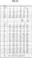


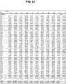

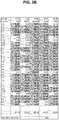


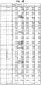


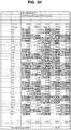

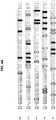

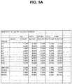


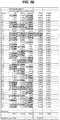


Classifications
- C—CHEMISTRY; METALLURGY
- C12—BIOCHEMISTRY; BEER; SPIRITS; WINE; VINEGAR; MICROBIOLOGY; ENZYMOLOGY; MUTATION OR GENETIC ENGINEERING
- C12Q—MEASURING OR TESTING PROCESSES INVOLVING ENZYMES, NUCLEIC ACIDS OR MICROORGANISMS; COMPOSITIONS OR TEST PAPERS THEREFOR; PROCESSES OF PREPARING SUCH COMPOSITIONS; CONDITION-RESPONSIVE CONTROL IN MICROBIOLOGICAL OR ENZYMOLOGICAL PROCESSES
- C12Q1/00—Measuring or testing processes involving enzymes, nucleic acids or microorganisms; Compositions therefor; Processes of preparing such compositions
- C12Q1/68—Measuring or testing processes involving enzymes, nucleic acids or microorganisms; Compositions therefor; Processes of preparing such compositions involving nucleic acids
- C12Q1/6876—Nucleic acid products used in the analysis of nucleic acids, e.g. primers or probes
- C12Q1/6883—Nucleic acid products used in the analysis of nucleic acids, e.g. primers or probes for diseases caused by alterations of genetic material
- C12Q1/6886—Nucleic acid products used in the analysis of nucleic acids, e.g. primers or probes for diseases caused by alterations of genetic material for cancer
- C—CHEMISTRY; METALLURGY
- C12—BIOCHEMISTRY; BEER; SPIRITS; WINE; VINEGAR; MICROBIOLOGY; ENZYMOLOGY; MUTATION OR GENETIC ENGINEERING
- C12Q—MEASURING OR TESTING PROCESSES INVOLVING ENZYMES, NUCLEIC ACIDS OR MICROORGANISMS; COMPOSITIONS OR TEST PAPERS THEREFOR; PROCESSES OF PREPARING SUCH COMPOSITIONS; CONDITION-RESPONSIVE CONTROL IN MICROBIOLOGICAL OR ENZYMOLOGICAL PROCESSES
- C12Q1/00—Measuring or testing processes involving enzymes, nucleic acids or microorganisms; Compositions therefor; Processes of preparing such compositions
- C12Q1/68—Measuring or testing processes involving enzymes, nucleic acids or microorganisms; Compositions therefor; Processes of preparing such compositions involving nucleic acids
- C12Q1/6813—Hybridisation assays
- C12Q1/6827—Hybridisation assays for detection of mutation or polymorphism
- C—CHEMISTRY; METALLURGY
- C12—BIOCHEMISTRY; BEER; SPIRITS; WINE; VINEGAR; MICROBIOLOGY; ENZYMOLOGY; MUTATION OR GENETIC ENGINEERING
- C12Q—MEASURING OR TESTING PROCESSES INVOLVING ENZYMES, NUCLEIC ACIDS OR MICROORGANISMS; COMPOSITIONS OR TEST PAPERS THEREFOR; PROCESSES OF PREPARING SUCH COMPOSITIONS; CONDITION-RESPONSIVE CONTROL IN MICROBIOLOGICAL OR ENZYMOLOGICAL PROCESSES
- C12Q1/00—Measuring or testing processes involving enzymes, nucleic acids or microorganisms; Compositions therefor; Processes of preparing such compositions
- C12Q1/68—Measuring or testing processes involving enzymes, nucleic acids or microorganisms; Compositions therefor; Processes of preparing such compositions involving nucleic acids
- C12Q1/6844—Nucleic acid amplification reactions
- C12Q1/6858—Allele-specific amplification
- C—CHEMISTRY; METALLURGY
- C12—BIOCHEMISTRY; BEER; SPIRITS; WINE; VINEGAR; MICROBIOLOGY; ENZYMOLOGY; MUTATION OR GENETIC ENGINEERING
- C12Q—MEASURING OR TESTING PROCESSES INVOLVING ENZYMES, NUCLEIC ACIDS OR MICROORGANISMS; COMPOSITIONS OR TEST PAPERS THEREFOR; PROCESSES OF PREPARING SUCH COMPOSITIONS; CONDITION-RESPONSIVE CONTROL IN MICROBIOLOGICAL OR ENZYMOLOGICAL PROCESSES
- C12Q2600/00—Oligonucleotides characterized by their use
- C12Q2600/154—Methylation markers
Definitions
- the present inventionrelates to methods and compositions for determination of and uses of specific methylation patterns indicative of adenoma and carcinoma.
- the inventionrelates to analysis of defined CpG loci that are coordinately methylated in DNAs from cancer and adenoma samples, methods for identifying coordinately methylated loci, and methods of using analysis of coordinately methylated loci in one or more marker regions in the design of assays for adenoma and cancer having improved sensitivity and specificity.
- DNAmay be methylated at cytosines located 5' to guanosine in CpG dinucleotides. This modification has important regulatory effects on gene expression, especially when involving CpG rich areas, known as CpG islands, often found in the promoter regions of genes. While approximately 75% of the CpG sites throughout the human genome are methylated, CpG sites within CpG islands are normally unmethylated, and aberrant methylation of CpG islands has been associated with certain diseases, including cancers. For example, CpG island hypermethylation is associated with transcriptional inactivation of defined tumor suppressor genes in human cancers, e.g ., colorectal cancer. Therefore, detection of hypermethylated nucleic acid could indicate susceptibility or onset of various forms of cancers.
- CIMPCpG island methylator phenotype
- Issastates that "methylation events (alone) may not provide the ideal universal cancer marker they were once thought to be because CIMP target genes will not be useful to screen for all colorectal cancers (many false negatives are predicted), and non-CIMP target genes will likely yield a high rate of false-positives because they are also methylated in normal appearing mucosa of older individuals without tumors" (Issa, et al., supra ) .
- methylation analyses in cancer detectionis to look at multiple marker genes. For example, Zou, et al., examined the methylation status of BMP3, EYA2, ALX4, and vimentin in cancer samples. While methylation levels were significantly higher in both cancer and adenoma than in normal epithelium, for each of the four genes, the sensitivity as determined by receiver operating curves was not significantly improved by combining any or all markers compared with the best single marker. ( Zou, et al., Cancer Epidemiol Biomarkers Prev 2007;16(12):2686 ).
- Zoualso looked at neoplasims showing methylation in more than one of the marker genes and found that co-methylation was frequent, with 72% of the cancers and 84% of the adenomas tested showing hypermehtylation in two or more of the genes.
- Zoureported that methylation of one or more of four (at least one), two or more of four, three or more of four, or four of four of these marker genes was noted in 88%, 72%, 53%, and 41% of 74 cancers and 98%, 84%, 60%, and 39% of 62 adenomas, compared with 24%, 7%, 3%, and 0% of 70 normal epithelia, respectively, demonstrating that although the assay gets progressively more specific as when more genes are included in the comethylation set, the sensitivity declines precipitously.
- the present inventionrelates to the methods of identifying regions of specific genes and specific regions of genomic nucleic acid useful in the detection of methylation associated with colorectal cancer.
- Methodscomprise, e.g ., detecting methylated sequences in, for example, tissue biopsy, stool extract, or other body fluids with improved sensitivity and specificity.
- the present inventionprovides methods of methylation analysis comprising identifying methylation loci showing advantageous methylation ratios when methylation in non-normal cells, e.g., cancer or adenoma cells is compared to background methylation in normal cells.
- the present inventionrelates to methods of analyzing methylation at each of several loci in a set of possible methylation sites within a marker sequence, wherein the presence of methylation at all of the loci within the defined set of sites occurs more frequently in cancer and adenoma cells than in normal cells, such that a finding of methylation at all of the loci in the defined subset of loci in a sample is indicative of adenoma or cancer.
- the present inventionprovides a method of identifying a set of methylated CpG loci in a marker nucleic acid wherein methylation is indicative of adenoma, comprising:
- the defined subset of CpG lociconsists of the same loci in the defined set of CpG loci.
- Determination of the methylation status of the set of CpG locimay be accomplished by any method known to those of skill in the art.
- the methodcomprises treating DNA from the samples with bisulfite.
- Bisulfite modification treatmentis described, e.g ., in U.S. Pat. No. 6,017,704 , the entire disclosure of which is incorporated herein by reference.
- determining the methylation status of the defined set of CpG locicomprises digital analysis of each of a plurality of CpG loci in a plurality of individual copies of a marker nucleic acid.
- digital analysiscomprise digital sequencing, and/or digital PCR.
- non-normal samplecomprises an adenoma sample, and in particular preferred embodiments, comprises a colorectal adenoma sample.
- a non-normal sampledcomprises a cancer sample, and in certain preferred embodiments, comprises a colorectal cancer sample.
- the present inventionprovides methods of detecting cancer or adenoma in a sample, e.g., from a subject.
- the present inventionprovides methods comprising determining the methylation status of each CpG locus in a defined subset of CpG loci in at least one cancer or adenoma marker nucleic acid molecule, wherein methylation at each of the CpG loci in the defined subset of CpG loci in the cancer or adenoma marker nucleic acid molecule is indicative of cancer or adenoma in the sample.
- the defined subsetcomprises at least three CpG loci, while in some preferred embodiments, the defined subset comprises at least four CpG loci or at least five CpG loci.
- the determiningcomprises analysis of the CpG loci in a nucleic acid detection assay configured to determine the methylation status of each of the loci in a single nucleic acid detection assay. In some preferred embodiments, the determining comprises analysis of the CpG loci in a nucleic acid detection assay configured to determine the methylation status of each of said loci in a single reaction mixture. In some embodiments, the nucleic acid detection assay comprises a primer extension assay.
- the nucleic acid detection assaymay comprise one or more of a nucleic acid amplification assay, a nucleic acid sequencing assay, a structure-specific cleavage assay, a 5' nuclease cleavage assay, an invasive cleavage assay and/or a ligation assay.
- the methods of the present inventionare not limited to the analysis of a single cancer or adenoma marker nucleic acid.
- the methylation status of each CpG locus in a defined subset of CpG loci in at least one cancer or adenoma marker nucleic acid moleculecomprises analysis of nucleic acid molecules from a plurality of cancer or adenoma markers.
- the plurality of cancer or adenoma markerscomprises at least three cancer or adenoma markers, while in some embodiments, the plurality comprises at least four cancer or adenoma markers.
- the cancer or adenoma markers and nucleic acid moleculesare selected from the group comprising Vimentin, BMP3, Septin 9, TFPI2, 2 regions of LRAT, and EYA4 markers and nucleic acid molecules.
- the assay methods of the present inventionare combined with the analysis of one or more other cancer markers, such as fecal occult blood markers (e.g. , hemoglobin, alpha-defensin, calprotectin, ⁇ 1-antitrypsin, albumin, MCM2, transferrin, lactoferrin, and lysozyme).
- fecal occult blood markerse.g. , hemoglobin, alpha-defensin, calprotectin, ⁇ 1-antitrypsin, albumin, MCM2, transferrin, lactoferrin, and lysozyme.
- a cancer or adenoma marker nucleic acid moleculecomprises a vimentin nucleic acid molecule, and in some particularly preferred embodiments, the defined subset of CpG loci in the vimentin nucleic acid molecule comprises loci 37, 40, and 45.
- a cancer or adenoma marker nucleic acid moleculecomprises a BMP3 nucleic acid molecule, and in some particularly preferred embodiments, the defined subset of CpG loci in the BMP3 nucleic acid molecule comprises loci 34, 53, and 61.
- a cancer or adenoma marker nucleic acid moleculecomprises a Septin9 nucleic acid molecule, and in some particularly preferred embodiments, the defined subset of CpG loci in said Septin9 nucleic acid molecule comprises loci 59, 61, 68, and 70.
- a cancer or adenoma marker nucleic acid moleculecomprises a TFPI2 nucleic acid molecule and in some particularly preferred embodiments, the defined subset of CpG loci in said TFPI2 nucleic acid molecule comprises loci 55, 59, 63, and 67.
- a cancer or adenoma marker nucleic acid moleculecomprises an EYA4 nucleic acid molecule, and in some particularly preferred embodiments, the defined subset of CpG loci in said EYA4 nucleic acid molecule comprises loci 31, 34, 37, and 44.
- the at least one cancer or adenoma marker or nucleic acid moleculecomprises a plurality markers or nucleic acid molecules comprising Vimentin, BMP3, Septin9, and TFPI2 markers or nucleic acid molecules.
- the present inventionfurther provides methods of selecting a defined set of CpG loci in a marker nucleic acid wherein methylation is indicative of non-normal status, e.g ., adenoma or cancer, comprising a) determining the methylation status of a plurality of CpG loci in each of a plurality of individual copies of a marker nucleic acid from a plurality of normal samples; b) determining the methylation status of the plurality of CpG loci in each of a plurality of individual copies of said marker nucleic acid from a plurality of non-normal (e.g.
- adenoma or cancer samplesc) determining methylation ratios for each locus in the plurality of said CpG loci in the marker nucleic acid; and d) selecting a defined set of CpG loci in the marker nucleic acid, wherein the defined set of CpG loci comprises a plurality of CpG loci having advantageous methylation ratios correlating with non-normal status ( e.g. , adenoma or cancer).
- determining the methylation ratioscomprises determining the ratio between the mean methylation at each of the plurality of CpG loci in the normal samples to the mean methylation at each corresponding CpG locus in said plurality of CpG loci in the non-normal samples.
- the plurality of individual copies of a marker nucleic acid analyzed in the normal and non-normal (e.g. , adenoma or cancer) samplescomprises at least 10, preferably at least 100, more preferably at least 1000, still more preferably at least 10,000 and still more preferably at least 100,000 copies.
- the number of copies analyzedis not limited to these whole numbers, but may be any integer above about 10. The number of copies from different sample types, e.g ., normal and non-normal need not be equal.
- the plurality of normal and non-normal (e.g. , adenoma or cancer) samples comparedcomprises at least 10, preferably at least 25, still more preferably at least 100 samples.
- the defined set of CpG locicomprises at least three CpG loci, preferably at least four CpG loci, more preferably at least five CpG loci.
- Determination of the methylation status of the plurality of CpG locimay be accomplished by any method known to those of skill in the art, including those described in more detail, below.
- the methodcomprises treating DNA from the samples with bisulfite.
- determining the methylation status of the defined set of CpG locicomprises digital analysis of each of a plurality of CpG loci in a plurality of individual copies of a marker nucleic acid.
- digital analysiscomprises digital sequencing, and/or digital PCR.
- the terms “digital sequencing” and “single molecule sequencing”are used interchangeably and refer to determining the nucleotide sequence of individual nucleic acid molecules.
- Systems for individual molecule sequencinginclude but are not limited to the 454 FLXTM or 454 TITANIUMTM (Roche), the SOLEXATM/ Illumina Genome Analyzer (Illumina), the HELISCOPETM Single Molecule Sequencer (Helicos Biosciences), and the SOLIDTM DNA Sequencer (Life Technologies/Applied Biosystems) instruments), as well as other platforms still under development by companies such as Intelligent Biosystems and Pacific Biosystems.
- backgroundrefers to methylation observed in a normal cell or sample at a nucleic acid locus or region that is generally unmethylated in normal cells.
- CpG islandsare generally considered unmethylated in normal human cells but methylation is not completely absent in the CpG islands of normal cells.
- methylationor “methylated,” as used in reference to the methylation status of a cytosine, e.g ., in a CpG locus, generally refers to the presence or absence of a methyl group at position 5 of the cytosine residue ( i.e., whether a particular cytosine is 5-methylcytosine). Methylation may be determined directly, e.g., as evidenced by routine methods for analysis of methylation status of cytosines, e.g ., by determining the sensitivity (or lack thereof) of a particular C-residue to conversion to uracil by treatment with bisulfite.
- a cytosine residue in a sample that is not converted to uracil when the sample is treated with bisulfite in a manner that would be expected to convert that residue if non-methylatedmay generally be deemed "methylated".
- the terms "digital PCR”, “single molecule PCR” and “single molecule amplification”refer to PCR and other nucleic acid amplification methods that are configured to provide amplification product or signal from a single starting molecule.
- samplesare divided, e.g., by serial dilution or by partition into small enough portions (e.g., in microchambers or in emulsions) such that each portion or dilution has, on average, no more than a single copy of the target nucleic acid.
- sensitivityrefers to clinical sensitivity - the proportion of positive samples that give a positive result using a diagnostic assay. Sensitivity is generally calculated as the number of true positives identified by the assay, divided by the sum of the number of true positives and the number of false negatives determined by the assay on known positive samples. Similarly, the term “specificity” refers to the proportion or number of true negatives determined by the assay divided by the sum of the number of true negatives and the number of false positives determined by the assay on known negative sample(s).
- the term "complementary"refers to different assays that, when used together, provide a more sensitive and/or specific result than can be provided by any one of the different assays used alone.
- informativeor “informativeness” refers to a quality of a marker or panel of markers, and specifically to the likelihood of finding a marker (or panel of markers) in a positive sample.
- sampleas used herein is used in its broadest sense.
- a sample suspected of containing a human gene or chromosome or sequences associated with a human chromosomemay comprise a cell, chromosomes isolated from a cell (e.g., a spread of metaphase chromosomes), genomic DNA (in solution or bound to a solid support such as for Southern blot analysis), RNA (in solution or bound to a solid support such as for Northern blot analysis), cDNA (in solution or bound to a solid support) and the like.
- CpG islandrefers to a genomic DNA region that contains a high percentage of CpG sites relative to the average genomic CpG incidence (per same species, per same individual, or per subpopulation ( e.g ., strain, ethnic subpopulation, or the like).
- CpG islandsare defined as having a GC percentage that is greater than 50% and with an observed/expected CpG ratio that is greater than 60% ( Gardiner-Garden et al. (1987) J Mol. Biol. 196:261-282 ; Baylin et al. (2006) Nat. Rev. Cancer 6:107-116 ; Irizarry et al. (2009) Nat.
- CpG islandsmay have a GC content >55% and observed CpG/expected CpG of 0.65 ( Takai et al. (2007) PNAS 99:3740-3745 ; herein incorporated by reference in its entirety).
- Various parametersalso exist regarding the length of CpG islands.
- CpG islandsmay be less than 100 bp; 100-200 bp, 200-300 bp, 300-500 bp, 500-750 bp; 750-1000 bp; 1000 or more bp in length.
- CpG islandsshow altered methylation patterns relative to controls ( e.g.
- altered methylation in cancer subjects relative to subjects without cancertissue-specific altered methylation patterns; altered methylation in stool from subjects with colorectal neoplasia (e.g. , colorectal cancer, colorectal adenoma) relative to subjects without colorectal neoplasia).
- altered methylationinvolves hypermethylation.
- altered methylationinvolves hypomethylation.
- CpG shoreor “CpG island shore” refers to a genomic region external to a CpG island that is or that has potential to have altered methylation patterns (see, e.g., Irizarry et al. (2009) Nat. Genetics 41:178-186 ; herein incorporated by reference in its entirety).
- CpG island shoresmay show altered methylation patterns relative to controls ( e.g. , altered methylation in cancer subjects relative to subjects without cancer; tissue-specific altered methylation patterns; altered methylation in stool from subjects with colorectal neoplasia ( e.g.
- CpG island shoresmay be located in various regions relative to CpG islands (see, e.g., Irizarry et al. (2009) Nat. Genetics 41;178-186 ; herein incorporated by reference in its entirety).
- CpG island shoresare located less than 100 bp; 100-250 bp; 250-500 bp; 500-1000 bp; 1000-1500 bp; 1500-2000 bp; 2000-3000 bp; 3000 bp or more away from a CpG island.
- targetwhen used in reference to a nucleic acid detection or analysis method, refers to a nucleic acid having a particular sequence of nucleotides to be detected or analyzed, e.g., in a sample suspected of containing the target nucleic acid.
- a targetis a nucleic acid having a particular sequence for which it is desirable to determine a methylation status.
- targetWhen used in reference to the polymerase chain reaction, “target” generally refers to the region of nucleic acid bounded by the primers used for polymerase chain reaction. Thus, the "target” is sought to be sorted out from other nucleic acid sequences that may be present in a sample.
- a “segment”is defined as a region of nucleic acid within the target sequence.
- sample templaterefers to nucleic acid originating from a sample that is analyzed for the presence of a target.
- locusrefers to a particular position, e.g., of a mutation, polymorphism, or a C residue in a CpG dinucleotide, within a defined region or segment of nucleic acid, such as a gene or any other characterized sequence on a chromosome or RNA molecule.
- a locusis not limited to any particular size or length, and may refer to a portion of a chromosome, a gene, functional genetic element, or a single nucleotide or basepair.
- a locusrefers to the C residue in the CpG dinucleotide.
- methylation ratiorefers to the amount or degree of methylation observed for particular methylation region or locus (e.g., a CpG locus in a marker gene or region) in a plurality of non-normal cells (e.g., cells in a particular disease state, such as cancerous or pre-cancerous cells) compared to the amount or degree of methylation observed for the same region or locus in a plurality of normal cells (e.g., cells that are not in the particular disease state of interest).
- non-normal cellse.g., cells in a particular disease state, such as cancerous or pre-cancerous cells
- a methylation ratiomay be expressed as the ratio of the means determined for normal cells:adenoma cells, or 0.11348.
- a methylation rationeed not be expressed in any particular manner or by any particular calculation.
- the methylation ratio abovemay alternatively be expressed, e.g ., as 8.39889:74.0771; 8.39889/74.0771; 74.0771:8.39889; as a calculated "fold methylation over background" 8.81987, etc.
- the term "advantageous methylation ratio”refers to a methylation ratio for a locus at which methylation correlates with a cellular status, e.g., a particular disease state (for example, normal, precancerous, cancerous) that, when compared to other methylation loci correlated with the same disease state, displays a higher percentage methylation in a population of non-normal cells compared to background levels of methylation at the same locus in a population of normal cells.
- certain CpG locie.g., within a methylation marker sequence, display a much greater signal-to-noise, i.e., degree in methylation compared to background than other loci in the same marker sequence.
- certain disease-associated marker genes or regionsdisplay advantageous methylation ratios at some or all loci compared to the methylation ratios observed for some or all loci within another marker sequence.
- methylation locie.g., CpG loci in a marker sequence
- a particular pattern of methylationthat correlates with a cellular status, e.g., a particular disease state (for example, normal, precancerous, cancerous).
- methylation loci that are all methylated in a manner correlated with a disease statemay be deemed to be coordinately methylated in cells having that disease state.
- Coordinat methylationis not limited to situations in which all of the coordinated loci are methylated.
- Any pattern of methylation among a particular set of loci that correlates with a cellular statusincluding patterns in which all of the coordinate loci are methylated, patterns in which the loci exhibit a reproducible pattern of methylation and non-methylation, and patterns in which none of the loci within the set are methylated are all included within the meaning of "coordinately methylated.”
- coordinate methylation analysisis used interchangeably with “multimethylation analysis” and refers to an assay in which the methylation statuses of a plurality of individual methylation loci in a marker sequence, e.g ., CpG loci, are determined together.
- coordinate methylation analysisis performed using a digital/single copy method (e.g ., digital sequencing) or an assay method configured to interrogate all of the selected CpG loci on each molecule tested, such that the methylation pattern in each single molecule tested is revealed.
- defined set of CpG locirefers to the set of CpG loci in a marker gene or region selected for methylation analysis.
- a defined set of CpG loci in a marker gene or regionmay comprise all CpG loci in the gene or region, or it may comprise fewer than all of the loci in that gene or region.
- defined subset of CpG locirefers to a subset of the defined set of CpG loci in a marker gene or region, the methylation of which has been determined to be indicative of a non-normal state, e.g ., adenoma or cancer.
- a non-normal statee.g ., adenoma or cancer.
- the methylation status of a defined subset of CpG loci in at least one cancer marker nucleic acid moleculeis determined, with simultaneous methylation at all of said CpG loci in the defined subset being indicative of cancer in the sample.
- a defined subset of CpG loci in a marker gene or regionmay comprise all CpG loci in the defined set, or it may comprise fewer than all of the loci in the defined set of loci in that gene or region.
- colonal canceris meant to include the well-accepted medical definition that defines colorectal cancer as a medical condition characterized by cancer of cells of the intestinal tract below the small intestine (e.g ., the large intestine (colon), including the cecum, ascending colon, transverse colon, descending colon, and sigmoid colon, and rectum). Additionally, as used herein, the term “colorectal cancer” is meant to further include medical conditions which are characterized by cancer of cells of the duodenum and small intestine (jejunum and ileum).
- Metastasisis meant to refer to the process in which cancer cells originating in one organ or part of the body relocate to another part of the body and continue to replicate. Metastasized cells subsequently form tumors which may further metastasize. Metastasis thus refers to the spread of cancer from the part of the body where it originally occurs to other parts of the body.
- metaastasized colorectal cancer cellsis meant to refer to colorectal cancer cells which have metastasized; colorectal cancer cells localized in a part of the body other than the duodenum, small intestine (jejunum and ileum), large intestine (colon), including the cecum, ascending colon, transverse colon, descending colon, and sigmoid colon, and rectum.
- an individual is suspected of being susceptible to metastasized colorectal canceris meant to refer to an individual who is at an above-average risk of developing metastasized colorectal cancer.
- individuals at a particular risk of developing metastasized colorectal cancerare those whose family medical history indicates above average incidence of colorectal cancer among family members and/or those who have already developed colorectal cancer and have been effectively treated who therefore face a risk of relapse and recurrence.
- Other factors which may contribute to an above-average risk of developing metastasized colorectal cancer which would thereby lead to the classification of an individual as being suspected of being susceptible to metastasized colorectal cancermay be based upon an individual's specific genetic, medical and/or behavioral background and characteristics.
- neoplasmrefers to any new and abnormal growth of tissue.
- a neoplasmcan be a premalignant neoplasm or a malignant neoplasm.
- neoplasm-specific markerrefers to any biological material that can be used to indicate the presence of a neoplasm.
- biological materialsinclude, without limitation, nucleic acids, polypeptides, carbohydrates, fatty acids, cellular components ( e.g ., cell membranes and mitochondria), and whole cells.
- markersare particular nucleic acid regions, e.g., genes, intragenic regions, etc. Regions of nucleic acid that are markers may be referred to, e.g., as “marker genes,” “marker regions,” “marker sequences,” etc.
- colonal neoplasm-specific markerrefers to any biological material that can be used to indicate the presence of a colorectal neoplasm (e.g. , a premalignant colorectal neoplasm; a malignant colorectal neoplasm).
- colorectal neoplasm-specific markersinclude, but are not limited to, exfoliated epithelial markers (e.g ., bmp-3, bmp-4, SFRP2, vimentin, septin9, ALX4, EYA4, TFPI2, NDRG4, FOXE1, long DNA, BAT-26, K-ras, APC, melanoma antigen gene, p53, BRAF, and PIK3CA) and fecal occult blood markers (e.g., hemoglobin, alpha-defensin, calprotectin, ⁇ 1-antitrypsin, albumin, MCM2, transferrin, lactoferrin, and lysozyme).
- exfoliated epithelial markerse.g ., bmp-3, bmp-4, SFRP2, vimentin, septin9, ALX4, EYA4, TFPI2, NDRG4, FOXE1, long DNA, BAT-26, K-ras
- Additional markersinclude but are not limited those in Table 1, below: Table 1 Accession Symbol GeneID Reference NM_000038 APC 324 DNA Methylation And Cancer Therapy, Austin Bioscience 2005, ed. Moshe Szyf NM_000044 AR 367 DNA Methylation And Cancer Therapy, Austin Bioscience 2005, ed.
- adenomarefers to a benign tumor of glandular origin. Although these growths are benign, over time they may progress to become malignant.
- colonal adenomarefers to a benign colorectal tumor in which the cells form recognizable glandular structures or in which the cells are clearly derived from glandular epithelium.
- amplifyingor “amplification” in the context of nucleic acids refers to the production of multiple copies of a polynucleotide, or a portion of the polynucleotide, typically starting from a small amount of the polynucleotide (e.g ., a single polynucleotide molecule), where the amplification products or amplicons are generally detectable.
- Amplification of polynucleotidesencompasses a variety of chemical and enzymatic processes. The generation of multiple DNA copies from one or a few copies of a target or template DNA molecule during a polymerase chain reaction (PCR) or a ligase chain reaction (LCR; see, e.g., U.S. Patent No.
- Polynucleotide amplificationalso can be accomplished using digital PCR (see, e.g., Kalinina, et al., Nucleic Acids Research. 25; 1999-2004, (1997 ); Vogelstein and Kinzler, Proc Natl Acad Sci USA. 96; 9236-41, (1999 ); International Patent Publication No. WO05023091A2 ; US Patent Application Publication No. 20070202525 ; each of which are incorporated herein by reference in their entireties).
- digital PCRsee, e.g., Kalinina, et al., Nucleic Acids Research. 25; 1999-2004, (1997 ); Vogelstein and Kinzler, Proc Natl Acad Sci USA. 96; 9236-41, (1999 ); International Patent Publication No. WO05023091A2 ; US Patent Application Publication No. 20070202525 ; each of which are incorporated herein by reference in their entireties).
- PCRpolymerase chain reaction
- the mixtureis denatured and the primers then annealed to their complementary sequences within the target molecule.
- the primersare extended with a polymerase so as to form a new pair of complementary strands.
- the steps of denaturation, primer annealing, and polymerase extensioncan be repeated many times (i.e ., denaturation, annealing and extension constitute one "cycle”; there can be numerous "cycles") to obtain a high concentration of an amplified segment of the desired target sequence.
- the length of the amplified segment of the desired target sequenceis determined by the relative positions of the primers with respect to each other, and therefore, this length is a controllable parameter.
- PCRpolymerase chain reaction
- nucleic acid detection assayrefers to any method of determining the nucleotide composition of a nucleic acid of interest.
- Nucleic acid detection assayinclude but are not limited to, DNA sequencing methods, probe hybridization methods, structure specific cleavage assays (e.g., the INVADER assay, (Hologic, Inc.) and are described, e.g ., in U.S. Patent Nos. 5,846,717 , 5,985,557 , 5,994,069 , 6,001,567 , 6,090,543 , and 6,872,816 ; Lyamichev et al., Nat.
- the terms "complementary” or “complementarity” used in reference to polynucleotidesrefers to polynucleotides related by the base-pairing rules. For example, the sequence “5'-A-G-T-3',” is complementary to the sequence “3'-T-C-A-5'.”
- Complementaritymay be “partial,” in which only some of the nucleic acids' bases are matched according to the base pairing rules. Or, there may be “complete” or “total” complementarity between the nucleic acids. The degree of complementarity between nucleic acid strands has significant effects on the efficiency and strength of hybridization between nucleic acid strands. This is of particular importance in amplification reactions, as well as detection methods that depend upon binding between nucleic acids.
- the term "primer”refers to an oligonucleotide, whether occurring naturally, as in a purified restriction digest, or produced synthetically, that is capable of acting as a point of initiation of synthesis when placed under conditions in which synthesis of a primer extension product that is complementary to a nucleic acid strand is induced (e.g ., in the presence of nucleotides and an inducing agent such as a biocatalyst (e.g., a DNA polymerase or the like).
- the primeris typically single stranded for maximum efficiency in amplification, but may alternatively be partially or completely double stranded.
- the portion of the primer that hybridizes to a template nucleic acidis sufficiently long to prime the synthesis of extension products in the presence of the inducing agent.
- the exact lengths of the primerswill depend on many factors, including temperature, source of primer and the use of the method. Primers may comprise labels, tags, capture moieties, etc.
- nucleic acid moleculerefers to any nucleic acid containing molecule, including but not limited to, DNA or RNA.
- the termencompasses sequences that include any of the known base analogs of DNA and RNA including, but not limited to, 4 acetylcytosine, 8-hydroxy-N6-methyladenosine, aziridinylcytosine, pseudoisocytosine, 5-(carboxyhydroxyl-methyl) uracil, 5-fluorouracil, 5-bromouracil, 5-carboxymethylaminomethyl-2-thiouracil, 5-carboxymethyl-aminomethyluracil, dihydrouracil, inosine, N6-isopentenyladenine, 1-methyladenine, 1-methylpseudo-uracil, 1-methylguanine, 1-methylinosine, 2,2-dimethyl-guanine, 2-methyladenine, 2-methylguanine, 3-methyl-cytosine, 5-methylcytosine, N
- nucleobaseis synonymous with other terms in use in the art including “nucleotide,” “deoxynucleotide,” “nucleotide residue,” “deoxynucleotide residue,” “nucleotide triphosphate (NTP),” or deoxynucleotide triphosphate (dNTP).
- oligonucleotiderefers to a nucleic acid that includes at least two nucleic acid monomer units (e.g., nucleotides), typically more than three monomer units, and more typically greater than ten monomer units.
- nucleic acid monomer unitse.g., nucleotides
- the exact size of an oligonucleotidegenerally depends on various factors, including the ultimate function or use of the oligonucleotide. To further illustrate, oligonucleotides are typically less than 200 residues long (e.g., between 15 and 100), however, as used herein, the term is also intended to encompass longer polynucleotide chains. Oligonucleotides are often referred to by their length.
- oligonucleotideFor example a 24 residue oligonucleotide is referred to as a "24-mer".
- the nucleoside monomersare linked by phosphodiester bonds or analogs thereof, including phosphorothioate, phosphorodithioate, phosphoroselenoate, phosphorodiselenoate, phosphoroanilothioate, phosphoranilidate, phosphoramidate, and the like, including associated counterions, e.g ., H + , NH 4 + , Na + , and the like, if such counterions are present.
- oligonucleotidesare typically single-stranded.
- Oligonucleotidesare optionally prepared by any suitable method, including, but not limited to, isolation of an existing or natural sequence, DNA replication or amplification, reverse transcription, cloning and restriction digestion of appropriate sequences, or direct chemical synthesis by a method such as the phosphotriester method of Narang et al. (1979) Meth Enzymol. 68: 90-99 ; the phosphodiester method of Brown et al. (1979) Meth Enzymol. 68: 109-151 ; the diethylphosphoramidite method of Beaucage et al. (1981) Tetrahedron Lett. 22: 1859-1862 ; the triester method of Matteucci et al. (1981) J Am Chem Soc.
- a “sequence” of a biopolymerrefers to the order and identity of monomer units (e.g., nucleotides, amino acids, etc.) in the biopolymer.
- the sequence (e.g., base sequence) of a nucleic acidis typically read in the 5' to 3' direction.
- wild-typerefers to a gene or gene product that has the characteristics of that gene or gene product when isolated from a naturally occurring source.
- a wild-type geneis that which is most frequently observed in a population and is thus arbitrarily designed the "normal” or “wild-type” form of the gene.
- modifiedrefers to a gene or gene product that displays modifications in sequence and or functional properties (i.e ., altered characteristics) when compared to the wild-type gene or gene product. It is noted that naturally occurring mutants can be isolated; these are identified by the fact that they have altered characteristics when compared to the wild-type gene or gene product.
- the term "gene”refers to a nucleic acid (e.g., DNA) sequence that comprises coding sequences necessary for the production of a polypeptide, precursor, or RNA (e.g., rRNA, tRNA).
- the polypeptidecan be encoded by a full length coding sequence or by any portion of the coding sequence so long as the desired activity or functional properties (e.g., enzymatic activity, ligand binding, signal transduction, immunogenicity, etc.) of the full-length or fragment polypeptide are retained.
- the termalso encompasses the coding region of a structural gene and the sequences located adjacent to the coding region on both the 5' and 3' ends for a distance of about 1 kb or more on either end such that the gene corresponds to the length of the full-length mRNA. Sequences located 5' of the coding region and present on the mRNA are referred to as 5' non-translated sequences. Sequences located 3' or downstream of the coding region and present on the mRNA are referred to as 3' non-translated sequences.
- the term "gene”encompasses both cDNA and genomic forms of a gene.
- a genomic form or clone of a genecontains the coding region interrupted with noncoding sequences termed "introns” or “intervening regions” or “intervening sequences.”
- Intronsare segments of a gene that are transcribed into nuclear RNA (e.g ., hnRNA); introns may contain regulatory elements (e.g ., enhancers). Introns are removed or "spliced out” from the nuclear or primary transcript; introns therefore are absent in the messenger RNA (mRNA) transcript.
- mRNAmessenger RNA
- genomic forms of a genemay also include sequences located on both the 5' and 3' end of the sequences that are present on the RNA transcript. These sequences are referred to as "flanking" sequences or regions (these flanking sequences are located 5' or 3' to the non-translated sequences present on the mRNA transcript).
- the 5' flanking regionmay contain regulatory sequences such as promoters and enhancers that control or influence the transcription of the gene.
- the 3' flanking regionmay contain sequences that direct the termination of transcription, post-transcriptional cleavage and polyadenylation.
- multimethylationAs used herein, the terms “multimethylation,” “series methylation” and “specific methylation” are used interchangeably to refer to defined combinations of CpG sites or loci in a marker sequence must be methylated to call that sequence methylated in a coordinate or multimethylation assay.
- a specific methylation assay of the CpG sites for BMP3might require that the CpG positions at 23, 34, 53, 61, 70, and 74, numbered by reference to Figures 1A and 1B , all be methylated in order for a sample to be classified as methylated at the BMP3 marker.
- Specific methylation of BMP3is not limited to this set of particular loci, but may include more, fewer, or a different collection of CpG loci.
- the CpG loci selected for co-analysis in a multimethylation assayare preferably selected. e.g., by analysis of normal (non-adenoma, non-cancer) samples to identify CpG methylation combinations that are less frequently represented in normal samples.
- combinations of methylation sitesare selected to produce good signal-to-noise in cancer and adenoma samples ( i.e ., the mean multimethylation at a particular combination of loci in cancer samples divided by the mean multimethylation in at those loci in normal samples is high).
- the terms "individual” and “average” methylationare used interchangeably to refer to analyses in which each CpG locus is analyzed individually, such that all molecules in which that base is methylated are included in a count, regardless of the methylation status of other loci, e.g., in the same marker. Generally, the methylation percentages of all the loci in a marker/region are then averaged, to produce a percent methylation figure for that marker.
- kitsrefers to any delivery system for delivering materials.
- delivery systemsinclude systems that allow for the storage, transport, or delivery of reaction reagents (e.g., oligonucleotides, enzymes, etc. in the appropriate containers) and/or supporting materials (e.g., buffers, written instructions for performing the assay etc.) from one location to another.
- reaction reagentse.g., oligonucleotides, enzymes, etc. in the appropriate containers
- supporting materialse.g., buffers, written instructions for performing the assay etc.
- kitsinclude one or more enclosures (e.g., boxes) containing the relevant reaction reagents and/or supporting materials.
- fragment kitrefers to a delivery systems comprising two or more separate containers that each contain a subportion of the total kit components. The containers may be delivered to the intended recipient together or separately.
- a first containermay contain an enzyme for use in an assay, while a second container contains oligonucleotides.
- fragment kitis intended to encompass kits containing Analyte specific reagents (ASR's) regulated under section 520(e) of the Federal Food, Drug, and Cosmetic Act, but are not limited thereto. Indeed, any delivery system comprising two or more separate containers that each contains a subportion of the total kit components are included in the term “fragmented kit.”
- a “combined kit”refers to a delivery system containing all of the components of a reaction assay in a single container (e.g., in a single box housing each of the desired components).
- kitincludes both fragmented and combined kits.
- the term "information"refers to any collection of facts or data. In reference to information stored or processed using a computer system(s), including but not limited to internets, the term refers to any data stored in any format (e.g., analog, digital, optical, etc.).
- the term “information related to a subject”refers to facts or data pertaining to a subject (e.g., a human, plant, or animal).
- the term “genomic information”refers to information pertaining to a genome including, but not limited to, nucleic acid sequences, genes, allele frequencies, RNA expression levels, protein expression, phenotypes correlating to genotypes, etc.
- Allele frequency informationrefers to facts or data pertaining to allele frequencies, including, but not limited to, allele identities, statistical correlations between the presence of an allele and a characteristic of a subject (e.g., a human subject), the presence or absence of an allele in an individual or population, the percentage likelihood of an allele being present in an individual having one or more particular characteristics, etc.
- the present inventionrelates to methods and compositions for determination of, and uses of, specific methylation patterns indicative of adenoma and carcinoma.
- the inventionrelates to analysis of defined subsets of CpG loci that are coordinately methylated in DNAs from cancer and adenoma samples, methods for identifying coordinately methylated loci, and methods of using analysis of coordinately methylated loci in one or more markers or regions in the design of assays for adenoma and cancer having improved sensitivity and specificity.
- the present inventionrelates to the observation that, within marker nucleic acids for which methylation status is indicative of cellular status, e.g ., cancerous, pre-cancerous, normal, etc., a subset of the individual methylation loci, e.g., CpG loci, in non-normal cells generally displays a greater degree of methylation relative to the background levels of methylation observed at corresponding loci in normal cells, while other methylation loci in the non-normal cells may exhibit levels of methylation that are closer to background levels.
- the degree of methylation observed for a particular locus in plurality of cancerous or pre-cancerous cells relative to normal cellsis expressed as a methylation ratio.
- Some embodiments of the present inventionrelate to screening known or suspected marker genes to identify specific methylation loci that exhibit greater ratios of disease-associated methylation relative to background methylation, as compared to other marker genes or other loci in the same marker gene.
- the present inventionrelates to coordinate methylation analysis, to measure the degree to which a marker molecule or a sample exhibits methylation at all of a plurality of selected loci.
- the present inventionrelates to analyzing methylation statuses of a defined set of individual CpG loci in methylation markers (or target regions within such markers) in a significant enough number of individual DNA molecules in adenoma samples or cancer samples to identify defined subsets of CpG loci that have advantageous methylation ratios compared to other loci in the same adenoma or cancer samples.
- a defined subset of CpG loci that have advantageous methylation ratios in a samplemay comprise the entirety of a set of CpG loci in a particular marker or target region of a marker, or it may be fewer than all of the CpG loci in the characterized region of the marker.
- amplification of a marker nucleic acid from a samplegenerally produces a mixture of amplicons coming from many copies of a target molecule. If the amplification conditions are not selective for a gene variant, the amplicon product contains a mixture of the variant and the normal or wildtype DNA. Even if primers are specific for a mutation or for a particular methylation site, when DNA is amplified from many copies of target DNA derived from many cells, there can be heterogeneity in other base positions in the resulting amplicon.
- An aspect of the present inventionis based on the observation that collecting methylation ratio information from a very large number of individual molecules in both normal and non-normal samples reveals that some methylation loci in marker regions or sequences exhibit a greater degree of methylation in non-normal cells compared to background (methylation at the same loci in normal cells) than do other individual loci in the same marker region or gene.
- These loci in non-normal sequences that have a greater level of methylation compared to backgroundcan be viewed as being particularly advantageous in that they are easier to identify over the background level of methylation observed in normal cells.
- One aspect of this advantageis that analysis of these particular loci permits identification of cancer-associated methylation with more sensitivity, and in a greater background of normal cells.
- the present inventionalso relates to the observation that coordinated analysis of multiple loci provides a significantly enhanced level of sensitivity in the identification of cancerous or precancerous cells, especially in samples that may also comprise a significant number of normal cells.
- Figure 6compares the sensitivity of detecting adenoma and cancer cells.
- the methylationwas either determined as an average across the marker region (e.g., the mean methylation in the vimentin marker across all of loci 26, 37, 40, 45, 52, 54, 59, 63, and 74; see Figures 3A-I ), indicated as "Individual" average methylation, or as a percentage of molecules displaying methylation at all of a subset of selected loci (e.g., methylation in the vimentin marker at all three of loci 37, 40 and 45; see Figures 5A-I ), i.e ., coordinate methylation analysis of multiple individual loci, indicated as "multi".
- sensitivities for the same samplesare also shown in calculated 5, 10 or 20-fold dilutions into normal DNA.
- Figure 6shows that, while assay sensitivities may be similar in DNA analyzed directly from tissue without dilution, as the amount background from normal DNA is increased at the larger dilutions, the coordinate methylation analysis is shown to be far more sensitive than the average methylation analysis.
- the adenoma and cancer samplescan be detected above background only in the undiluted and 5-fold dilution profiles when average methylation across the marker is analyzed, while these same samples can be detected with over about 69-74% sensitivity at 20-fold dilution, and 90-93% sensitivity at 10-fold dilution, when coordinate methylation analysis of loci 37, 40 and 45 is used.
- the present inventionprovides a method for designing a methylation assay to identify a disease state, comprising I) selecting at least one sequence for analysis; II) determining the methylation status of a plurality of loci in the sequence in a population of normal cells and a population of non-normal cells to determine an average rate of methylation for each of the plurality of loci each both normal and non-normal cells; and III) identifying at least two loci in said plurality of loci having advantageous methylation ratios.
- Methylation markers associated with particular disease stateshave been identified for a number of disease states.
- colorectal neoplasm-specific markerinclude, e.g ., bmp-3, bmp-4, SFRP2, vimentin, septin9, ALX4, EYA4, TFPI2, NDRG4, FOXE1, long DNA, BAT-26, K-ras, APC, melanoma antigen gene, p53, BRAF, and PIK3CA.
- Additional markersinclude but are not limited those in Table 1, above.
- Analysis of candidate methylation loci to identify those with advantageous methylation ratiosmay comprise analysis of every locus in a target sequence (e.g., every CpG) or it may comprise analysis of a subset of the methylation loci.
- CpGsare selected for analysis by their location in particular methylation hotspots, while in other embodiments, loci for analysis may be conveniently located with respect to primer binding sites or other sequence features.
- Figure 1Aprovides an exemplary selection of marker neoplasm-associated markers with each of the C residues in the CpG loci indicated by a box.
- the native sequence of the regionis shown in the top line and the sequences for unmethylated and methulated DNA as they would appear following bisulfite conversion and amplification are shown below. Unmethylated C-residues that would be converted by bisulfite and amplification to T residues are shown as Ts.
- the present inventionprovides use of a nucleic acid detection assay to coordinately analyze a plurality of the advantageous loci in a sample, thereby determining the disease state of cells in sample.
- determining a methylation ratio for a locuscomprises determining an average rate of methylation for that locus in a population of normal cells and determining the average rate of methylation at the same locus in a population of non-normal cells.
- commonly used methods of methylation analysis of marker genesare performed on mixed nucleic acids, e.g ., amplicons produced from unfractionated DNA from a mixed population of cells (such as DNA purified from a multicellular tissue sample).
- the present inventioncomprises large-scale analysis of individual DNA molecules , for example, by direct sequencing of individual DNA molecules, or by sequencing of clonally amplified DNA.
- methods of clonally amplifying individual copies of nucleic acidscan be used in the rapid analysis of large numbers of individual markers from normal and non-normal samples.
- Single-molecule amplification methodsmay comprise use of microchambers, emulsion reactions, "bridge PCR" on solid supports, or any of a number of established methods of segregating the amplification products arising from individual target molecules. Following single molecule amplification, amplicons can be sequenced.
- Platforms for individual molecule sequencinginclude the 454 FLXTM or 454 TITANIUMTM (Roche), the SOLEXATM / Illumina Genome Analyzer (Illumina), the HELISCOPETM Single Molecule Sequencer (Helicos Biosciences), the Ion Personal Genome Machine (Ion Torrent), and the SOLIDTM DNA Sequencer (Life Technologies/Applied Biosystems) instruments, as well as other platforms still under development by companies such as Intelligent Biosystems and Pacific Biosystems.
- sequence informationis generated varies for the different next-generation sequencing platforms, all of them share the common feature of generating sequence data from a very large number of individual sequencing templates, in sequencing reactions that are run simultaneously. Data from the reactions are collected using, e.g., a flow cell, a chemical or optical sensor, and/or scanner, and sequences are assembled and analyzed using bioinformatics software.
- the present inventionprovides methods of analysis of methylation markers using digital sequencing to identify neoplasm-associated methylation loci that have methylation ratios that are statistically significantly advantageous compared to other loci in the same markers.
- digital sequencingis done in a highly or massively parallel fashion, providing higher precision in identifying CpG methylation sites having advantageous methylation ratios.
- each moleculeis analyzed for methylation at each CpG locus, so the percentage of DNA copies having methylation at any combination of the CpG loci can be analyzed after the experimental run.
- each particular marker sequencee.g ., each target nucleic acid molecule, or clonal amplicon may be interrogated many, many times, e.g., at least 100 times, sometimes over 1000 times, and in some instances over 100,000 times, or as many as 500,000 times.
- patterns of coordinate methylation indicative of cancer or adenomathat would be undetectable in analysis of a handful of individual target molecules may be revealed.
- methylation status of a set of CpG loci in a large number of copies marker DNA from both normal samples and non-normal samplese.g ., adenoma or cancer samples
- design of nucleic acid detection assays to interrogate a plurality of CpG loci for which coordinate methylation is indicative of adenoma or cancer in a samplecan provide an assay that has improved signal-to-noise compared to assays that survey average percent methylation across entire marker genes.
- selecting a subset of CpG locicomprises selecting loci that have been determined to be coordinately methylated by use, e.g., of digital analysis methods. Another aspect comprises selecting CpG loci determined to have advantageous methylation ratios when normal DNA is compared to adenoma or cancer DNA. Assay designs may, but need not, make use of a CpG locus having the most advantageous methylation ratio compared to other loci in the same marker. In some embodiments, selection of a plurality of CpG loci as a subset comprises selecting the plurality of loci having the most advantageous methylation ratios.
- selection of a plurality of CpG loci as a subsetcomprises selecting the locus having the most advantageous methylation ratio, then selecting at least additional CpG loci that are conveniently situated with respect to the first selected locus for the configuration of a particular nucleic acid detection assay (e.g., the selection of CpG loci having particular proximity to each other for configuring an invasive cleavage assay, ligation assay, amplification assay, etc .) in order to interrogate all of the selected CpG loci on copies of the target DNA in a single assay.
- a candidate subset of CpG lociis further analyzed to determine the percentage of copies of marker DNA from non-normal samples that are coordinately methylated at those candidate loci, and that have little or no coordinated methylation in normal samples.
- methylation analysistypically analyze in a non-digital fashion, e.g ., analyzing a mixture of co-amplified molecules derived from a mixture of DNA target nucleic acids, so that analysis of the amplified products provides sequence information that reflects that aggregate or average methylation status in the amplicon population, but does not provide information on the percentage of starting molecules having coordinated methylation at all of a plurality of CpG loci.
- the reduction in number of DNA copies in normal DNA displaying methylation at all of the selected sitesdrops to a greater degree than it does in the DNA from cancer and/or adenoma sample, resulting in an significantly enhanced ratio of specific signal to background noise.
- the background from normal DNAis dramatically reduced by using multimethylation (coordinate methylation) analysis, while no equivalent reduction in signal from cancer and adenoma DNA is seen.
- the background in normal samplesis less reduced and/or the signal from cancer DNA also decreases with multimethylation analysis, such that there is less or no net improvement in the signal-to-noise and the advantages of using a multimethylation analysis approached are less.
- Genes having favorable signal to noise in multimethylation analysesare readily determined empirically.
- DNA extracted from frozen tissue sampleswas treated with an EPITECT bisulfite conversion kit (Qiagen) to convert non-methylated cytosines to uracil. Methylated cytosines remain unconverted. Primers for each gene region were designed for each sequence such that the composition of the amplification products remained the same as the original target sequences and methylated and non methylated sequences were amplified with equal efficiencies. Amplification of the dU-containing converted DNA produced amplicons having T-residues in place of the dU residues. The amplicons were then prepared for sequencing on the Illumina instrument. For each tissue sample, the amplification reaction for each target was prepared from the same sample of bisulfite-treated DNA.
- QiagenEPITECT bisulfite conversion kit
- the Illumina procedurecomprises a) preparation of a library from sample DNA by attachment of known sequence tags that permit indexing, flow cell attachment, amplification, and sequencing; b) attachment of the library to a flow cell surface; c) bridge amplification to produce clusters of DNA fragments derived from single molecules, and d) sequencing in using iterative primer extension reactions using labeled reversible terminators to determine the nucleotide sequence of each cluster of amplicons.
- a flow cellis composed of 8 lanes, one of which is dedicated to a phiX quality control.
- Tissue-extracted DNA from patientswas bisulfite-treated and a 2-step amplification using approximately 10,000 genome copies of initial material was carried out.
- the first roundused tailed (T1)(Illumina) primers specific for marker sequences. These tails were Illumina-derived sequences needed for round two.
- T2)(Illumina) PCRuses primers specific for the Illmuna tails added in T1, and incorporates the index, sequencing primer, and flow cell attachment sequences.
- multiple qPCR checkswere run on the samples to ensure equimolar representations of all amplicons in the libraries.
- non-CpG cytosinesForward and reverse primers specific for regions with converted, non-CpG cytosines are designed (using, e.g ., MethPrimer software) to amplify each of the specific biomarker sites in a non-methylation specific manner.
- C/Tdegenerate mixtures
- G/Adegenerate mixtures
- additional primersmay be designed. If CpGs in the target sequence cannot be avoided, the primers may incorporate degenerate bases at CpG sites (BiSearch software).
- Primers for second round PCRcomprise sequences for Illumina flow cell attachment (bridge amplification sites), sequencing primer sites (for the sample read), index sites, and sequencing primers sites (for the indexing read).
- Each of the primer sets (x)has 12 different index tags, for a total of 12x sets.
- the control DNAsare amplified, purified (e.g, using AMPURE treatment (Agencourt)), and run on an Agilent 2100 Bioanalyzer to assess the size and quantity of the amplified nucleic acids.
Landscapes
- Chemical & Material Sciences (AREA)
- Life Sciences & Earth Sciences (AREA)
- Organic Chemistry (AREA)
- Health & Medical Sciences (AREA)
- Proteomics, Peptides & Aminoacids (AREA)
- Engineering & Computer Science (AREA)
- Zoology (AREA)
- Wood Science & Technology (AREA)
- Genetics & Genomics (AREA)
- Immunology (AREA)
- Analytical Chemistry (AREA)
- Molecular Biology (AREA)
- General Health & Medical Sciences (AREA)
- General Engineering & Computer Science (AREA)
- Bioinformatics & Cheminformatics (AREA)
- Biotechnology (AREA)
- Microbiology (AREA)
- Biochemistry (AREA)
- Biophysics (AREA)
- Physics & Mathematics (AREA)
- Pathology (AREA)
- Chemical Kinetics & Catalysis (AREA)
- Hospice & Palliative Care (AREA)
- Oncology (AREA)
- Measuring Or Testing Involving Enzymes Or Micro-Organisms (AREA)
- General Chemical & Material Sciences (AREA)
Abstract
Description
- The present application claims the benefit of
U.S. Provisional Patent Application Serial Number 61/438,649, filed February 2, 2011 - The present invention relates to methods and compositions for determination of and uses of specific methylation patterns indicative of adenoma and carcinoma. In particular, the invention relates to analysis of defined CpG loci that are coordinately methylated in DNAs from cancer and adenoma samples, methods for identifying coordinately methylated loci, and methods of using analysis of coordinately methylated loci in one or more marker regions in the design of assays for adenoma and cancer having improved sensitivity and specificity.
- In higher order eukaryotes, DNA may be methylated at cytosines located 5' to guanosine in CpG dinucleotides. This modification has important regulatory effects on gene expression, especially when involving CpG rich areas, known as CpG islands, often found in the promoter regions of genes. While approximately 75% of the CpG sites throughout the human genome are methylated, CpG sites within CpG islands are normally unmethylated, and aberrant methylation of CpG islands has been associated with certain diseases, including cancers. For example, CpG island hypermethylation is associated with transcriptional inactivation of defined tumor suppressor genes in human cancers,e.g., colorectal cancer. Therefore, detection of hypermethylated nucleic acid could indicate susceptibility or onset of various forms of cancers.
- Despite indications suggesting a link between CpG island methylator phenotype (CIMP) and cancers (see,e.g.,Baylin SB, et al., Adv Cancer Res 1998;72:141-196 andJones PA, et al., Nat Rev Genet 2002;3:415-428), the idea that analysis of methylation status alone could be a useful diagnostic or prognostic tool has been controversial. As discussed byIssa, et al. in an editorial in Gastroenterology 179(3):2005, researchers had mixed results in confirming the link between CI,MP and cancers. Although CIMP was reportedly demonstrated in multiple other malignancies (Shen, I., et al. J Natl Cancer Inst 2002;94:755-761;Garcia-Manero G, et al., Clin Cancer Res 2002;8:2217-2224;Toyota M, et al., Blood 2001;97:2823-2829;Ueki T, et al., Cancer Res 2000;60:1835-1839;Toyota M, et al., Cancer Res 1999;59:5438-5442;Strathdee G, et al., Am J Pathol 2001;158:1121-1127;Abe M, et al., Cancer Res 2005;65:828-834) and several groups confirmed the original findings using similar markers and technology (Whitehall VL, et al., Cancer Res 2002;62:6011-6014;van Rijnsoever M, et al., Gut 2002;51:797-802) other groups were not able to establish such links (Eads CA, et al., Cancer Res 2001;61:3410-3418;Esteller M, et al., Cancer Res 2000;60:129-133). As late as 2003, a publication concluded that all methylation events in colorectal cancer were related to aging rather than neoplasia (Yamashita K, et al., Cancer Cell 2003;4:121-131).
- The discrepant results have been attributed in part to the fact that it has been demonstrated that 70% to 80% of aberrant DNA methylation events in colorectal cancer are age-related (Toyota M, et al., Proc Natl Acad Sci U S A 1999;96:8681-8686) and that cancer-linked phenotypes are only clear when these are filtered out. It has also been noted that overly sensitive, non-quantitative methods can overestimate methylation and mask the distinctions between methylation that is associated with cancer and that which is not. Issa states that "methylation events (alone) may not provide the ideal universal cancer marker they were once thought to be because CIMP target genes will not be useful to screen for all colorectal cancers (many false negatives are predicted), and non-CIMP target genes will likely yield a high rate of false-positives because they are also methylated in normal appearing mucosa of older individuals without tumors" (Issa,et al., supra).
- One approach to increase the clinical specificity of methylation analyses in cancer detection is to look at multiple marker genes. For example, Zou,et al., examined the methylation status of BMP3, EYA2, ALX4, and vimentin in cancer samples. While methylation levels were significantly higher in both cancer and adenoma than in normal epithelium, for each of the four genes, the sensitivity as determined by receiver operating curves was not significantly improved by combining any or all markers compared with the best single marker. (Zou, et al., Cancer Epidemiol Biomarkers Prev 2007;16(12):2686).
- Zou also looked at neoplasims showing methylation in more than one of the marker genes and found that co-methylation was frequent, with 72% of the cancers and 84% of the adenomas tested showing hypermehtylation in two or more of the genes. Zou reported that methylation of one or more of four (at least one), two or more of four, three or more of four, or four of four of these marker genes was noted in 88%, 72%, 53%, and 41% of 74 cancers and 98%, 84%, 60%, and 39% of 62 adenomas, compared with 24%, 7%, 3%, and 0% of 70 normal epithelia, respectively, demonstrating that although the assay gets progressively more specific as when more genes are included in the comethylation set, the sensitivity declines precipitously.
- The present invention relates to the methods of identifying regions of specific genes and specific regions of genomic nucleic acid useful in the detection of methylation associated with colorectal cancer. Methods comprise,e.g., detecting methylated sequences in, for example, tissue biopsy, stool extract, or other body fluids with improved sensitivity and specificity. In preferred embodiments, the present invention provides methods of methylation analysis comprising identifying methylation loci showing advantageous methylation ratios when methylation in non-normal cells,e.g., cancer or adenoma cells is compared to background methylation in normal cells. In some embodiments, the present invention relates to methods of analyzing methylation at each of several loci in a set of possible methylation sites within a marker sequence, wherein the presence of methylation at all of the loci within the defined set of sites occurs more frequently in cancer and adenoma cells than in normal cells, such that a finding of methylation at all of the loci in the defined subset of loci in a sample is indicative of adenoma or cancer.
- In some embodiments, the present invention provides a method of identifying a set of methylated CpG loci in a marker nucleic acid wherein methylation is indicative of adenoma, comprising:
- a) determining the methylation status of a defined set of CpG loci in each of a plurality of individual copies of a marker nucleic acid from a plurality of normal samples;
- b) determining the methylation status of said defined set of CpG loci in each of a plurality of individual copies of said marker nucleic acid from a plurality of non-normal (e.g., adenoma or cancer) samples to identify a defined subset of CpG loci from within said defined set,
- In some embodiments, the defined subset of CpG loci consists of the same loci in the defined set of CpG loci.
- Determination of the methylation status of the set of CpG loci may be accomplished by any method known to those of skill in the art. In some embodiments, the method comprises treating DNA from the samples with bisulfite. Bisulfite modification treatment is described,e.g., in
U.S. Pat. No. 6,017,704 , the entire disclosure of which is incorporated herein by reference. In some embodiments, determining the methylation status of the defined set of CpG loci comprises digital analysis of each of a plurality of CpG loci in a plurality of individual copies of a marker nucleic acid. In some preferred embodiments, digital analysis comprise digital sequencing, and/or digital PCR. - In certain preferred embodiments, non-normal sample comprises an adenoma sample, and in particular preferred embodiments, comprises a colorectal adenoma sample. In some preferred embodiments, a non-normal sampled comprises a cancer sample, and in certain preferred embodiments, comprises a colorectal cancer sample.
- The present invention provides methods of detecting cancer or adenoma in a sample,e.g., from a subject. In some embodiments, the present invention provides methods comprising determining the methylation status of each CpG locus in a defined subset of CpG loci in at least one cancer or adenoma marker nucleic acid molecule, wherein methylation at each of the CpG loci in the defined subset of CpG loci in the cancer or adenoma marker nucleic acid molecule is indicative of cancer or adenoma in the sample. In certain preferred embodiments, the defined subset comprises at least three CpG loci, while in some preferred embodiments, the defined subset comprises at least four CpG loci or at least five CpG loci.
- In certain embodiments, the determining comprises analysis of the CpG loci in a nucleic acid detection assay configured to determine the methylation status of each of the loci in a single nucleic acid detection assay. In some preferred embodiments, the determining comprises analysis of the CpG loci in a nucleic acid detection assay configured to determine the methylation status of each of said loci in a single reaction mixture. In some embodiments, the nucleic acid detection assay comprises a primer extension assay. In certain preferred embodiments, the nucleic acid detection assay may comprise one or more of a nucleic acid amplification assay, a nucleic acid sequencing assay, a structure-specific cleavage assay, a 5' nuclease cleavage assay, an invasive cleavage assay and/or a ligation assay.
- The methods of the present invention are not limited to the analysis of a single cancer or adenoma marker nucleic acid. For example, in some embodiments, the methylation status of each CpG locus in a defined subset of CpG loci in at least one cancer or adenoma marker nucleic acid molecule comprises analysis of nucleic acid molecules from a plurality of cancer or adenoma markers. In some embodiments, the plurality of cancer or adenoma markers comprises at least three cancer or adenoma markers, while in some embodiments, the plurality comprises at least four cancer or adenoma markers. In some preferred embodiments, the cancer or adenoma markers and nucleic acid molecules are selected from the group comprising Vimentin, BMP3, Septin 9, TFPI2, 2 regions of LRAT, and EYA4 markers and nucleic acid molecules. In some embodiments, the assay methods of the present invention are combined with the analysis of one or more other cancer markers, such as fecal occult blood markers (e.g., hemoglobin, alpha-defensin, calprotectin, α1-antitrypsin, albumin, MCM2, transferrin, lactoferrin, and lysozyme).
- In certain preferred embodiments of the method described herein, a cancer or adenoma marker nucleic acid molecule comprises a vimentin nucleic acid molecule, and in some particularly preferred embodiments, the defined subset of CpG loci in the vimentin nucleic acid molecule comprises
loci - In certain preferred embodiments of the method described herein, a cancer or adenoma marker nucleic acid molecule comprises a BMP3 nucleic acid molecule, and in some particularly preferred embodiments, the defined subset of CpG loci in the BMP3 nucleic acid molecule comprises
loci - In certain preferred embodiments of the method described herein, a cancer or adenoma marker nucleic acid molecule comprises a Septin9 nucleic acid molecule, and in some particularly preferred embodiments, the defined subset of CpG loci in said Septin9 nucleic acid molecule comprises
loci - In certain preferred embodiments of the method described herein, a cancer or adenoma marker nucleic acid molecule comprises a TFPI2 nucleic acid molecule and in some particularly preferred embodiments, the defined subset of CpG loci in said TFPI2 nucleic acid molecule comprises
loci - In certain preferred embodiments of the method described herein, a cancer or adenoma marker nucleic acid molecule comprises an EYA4 nucleic acid molecule, and in some particularly preferred embodiments, the defined subset of CpG loci in said EYA4 nucleic acid molecule comprises
loci - In certain preferred embodiments of the method described herein, the at least one cancer or adenoma marker or nucleic acid molecule comprises a plurality markers or nucleic acid molecules comprising Vimentin, BMP3, Septin9, and TFPI2 markers or nucleic acid molecules.
- The present invention further provides methods of selecting a defined set of CpG loci in a marker nucleic acid wherein methylation is indicative of non-normal status,e.g., adenoma or cancer, comprising a) determining the methylation status of a plurality of CpG loci in each of a plurality of individual copies of a marker nucleic acid from a plurality of normal samples; b) determining the methylation status of the plurality of CpG loci in each of a plurality of individual copies of said marker nucleic acid from a plurality of non-normal (e.g., adenoma or cancer) samples; c) determining methylation ratios for each locus in the plurality of said CpG loci in the marker nucleic acid; and d) selecting a defined set of CpG loci in the marker nucleic acid, wherein the defined set of CpG loci comprises a plurality of CpG loci having advantageous methylation ratios correlating with non-normal status (e.g., adenoma or cancer).
- In some embodiments, determining the methylation ratios comprises determining the ratio between the mean methylation at each of the plurality of CpG loci in the normal samples to the mean methylation at each corresponding CpG locus in said plurality of CpG loci in the non-normal samples. In preferred embodiments, the plurality of individual copies of a marker nucleic acid analyzed in the normal and non-normal (e.g., adenoma or cancer) samples comprises at least 10, preferably at least 100, more preferably at least 1000, still more preferably at least 10,000 and still more preferably at least 100,000 copies. The number of copies analyzed is not limited to these whole numbers, but may be any integer above about 10. The number of copies from different sample types,e.g., normal and non-normal need not be equal.
- In certain preferred embodiments of the methods of selecting a defined set of CpG loci in a marker nucleic acid described herein, the plurality of normal and non-normal (e.g., adenoma or cancer) samples compared comprises at least 10, preferably at least 25, still more preferably at least 100 samples. The number of samples analyzed in not limited to these whole numbers, but may be any integer above about 10. The number of different samples of the different sample types,e.g., normal and non-normal, need not be equal.
- In certain embodiments, the defined set of CpG loci comprises at least three CpG loci, preferably at least four CpG loci, more preferably at least five CpG loci.
- Determination of the methylation status of the plurality of CpG loci may be accomplished by any method known to those of skill in the art, including those described in more detail, below. In some embodiments, the method comprises treating DNA from the samples with bisulfite. In some embodiments, determining the methylation status of the defined set of CpG loci comprises digital analysis of each of a plurality of CpG loci in a plurality of individual copies of a marker nucleic acid. In some preferred embodiments, digital analysis comprises digital sequencing, and/or digital PCR. Methods of preparing samples,e.g., stool samples, for analysis are also known in the art. See,e.g.,
US7005266 ;6,303,304 ;5,741,650 ;5,952,178 ; and6,268,136 , each incorporated herein by reference. - To facilitate an understanding of the present invention, a number of terms and phrases are defined below.
- As used herein, the terms "digital sequencing" and "single molecule sequencing" are used interchangeably and refer to determining the nucleotide sequence of individual nucleic acid molecules. Systems for individual molecule sequencing include but are not limited to the 454 FLX™ or 454 TITANIUM™ (Roche), the SOLEXA™/ Illumina Genome Analyzer (Illumina), the HELISCOPE™ Single Molecule Sequencer (Helicos Biosciences), and the SOLID™ DNA Sequencer (Life Technologies/Applied Biosystems) instruments), as well as other platforms still under development by companies such as Intelligent Biosystems and Pacific Biosystems.
- As used herein, the term "background" as used in reference to methylation of a locus or region refers to methylation observed in a normal cell or sample at a nucleic acid locus or region that is generally unmethylated in normal cells. For example, CpG islands are generally considered unmethylated in normal human cells but methylation is not completely absent in the CpG islands of normal cells.
- As used herein, "methylation" or "methylated," as used in reference to the methylation status of a cytosine,e.g., in a CpG locus, generally refers to the presence or absence of a methyl group at
position 5 of the cytosine residue (i.e., whether a particular cytosine is 5-methylcytosine). Methylation may be determined directly,e.g., as evidenced by routine methods for analysis of methylation status of cytosines,e.g., by determining the sensitivity (or lack thereof) of a particular C-residue to conversion to uracil by treatment with bisulfite. For example, a cytosine residue in a sample that is not converted to uracil when the sample is treated with bisulfite in a manner that would be expected to convert that residue if non-methylated (e.g., under conditions in which a majority or all of the non-methylated cytosines in the sample are converted to uracils) may generally be deemed "methylated". - As used herein, the terms "digital PCR", "single molecule PCR" and "single molecule amplification" refer to PCR and other nucleic acid amplification methods that are configured to provide amplification product or signal from a single starting molecule. Typically, samples are divided,e.g., by serial dilution or by partition into small enough portions(e.g., in microchambers or in emulsions) such that each portion or dilution has, on average, no more than a single copy of the target nucleic acid. Methods of single molecule PCR are described,e.g., in
US 6,143,496 , which relates to a method comprising dividing a sample into multiple chambers such that at least one chamber has at least one target, and amplifying the target to determine how many chambers had a target molecule;US 6,391,559 ; which relates to an assembly for containing and portioning fluid; andUS7,459,315 , which relates to a method of dividing a sample into an assembly with sample chambers where the samples are partitioned by surface affinity to the chambers, then sealing the chambers with a curable "displacing fluid." See alsoUS 6,440,706 andUS 6,753,147 , andVogelstein, et al., Proc. Natl. Acad. Sci. USA Vol. 96, pp. 9236-9241, August 1999. See alsoUS 20080254474 , describing a combination of digital PCR combined with methylation detection. - As used herein, "sensitivity" as used in reference to a diagnostic assay,e.g., a methylation assay, refers to clinical sensitivity - the proportion of positive samples that give a positive result using a diagnostic assay. Sensitivity is generally calculated as the number of true positives identified by the assay, divided by the sum of the number of true positives and the number of false negatives determined by the assay on known positive samples. Similarly, the term "specificity" refers to the proportion or number of true negatives determined by the assay divided by the sum of the number of true negatives and the number of false positives determined by the assay on known negative sample(s).
- As used herein in reference to diagnostic or analysis assays, the term "complementary" refers to different assays that, when used together, provide a more sensitive and/or specific result than can be provided by any one of the different assays used alone.
- As used herein, the term "informative" or "informativeness" refers to a quality of a marker or panel of markers, and specifically to the likelihood of finding a marker (or panel of markers) in a positive sample.
- The term "sample" as used herein is used in its broadest sense. For example, a sample suspected of containing a human gene or chromosome or sequences associated with a human chromosome may comprise a cell, chromosomes isolated from a cell (e.g., a spread of metaphase chromosomes), genomic DNA (in solution or bound to a solid support such as for Southern blot analysis), RNA (in solution or bound to a solid support such as for Northern blot analysis), cDNA (in solution or bound to a solid support) and the like.
- As used herein, the term "CpG island" refers to a genomic DNA region that contains a high percentage of CpG sites relative to the average genomic CpG incidence (per same species, per same individual, or per subpopulation (e.g., strain, ethnic subpopulation, or the like). Various parameters and definitions for CpG islands exist; for example, in some embodiments, CpG islands are defined as having a GC percentage that is greater than 50% and with an observed/expected CpG ratio that is greater than 60% (Gardiner-Garden et al. (1987) J Mol. Biol. 196:261-282;Baylin et al. (2006) Nat. Rev. Cancer 6:107-116;Irizarry et al. (2009) Nat. Genetics 41:178-186; each herein incorporated by reference in its entirety). In some embodiments, CpG islands may have a GC content >55% and observed CpG/expected CpG of 0.65 (Takai et al. (2007) PNAS 99:3740-3745; herein incorporated by reference in its entirety). Various parameters also exist regarding the length of CpG islands. As used herein, CpG islands may be less than 100 bp; 100-200 bp, 200-300 bp, 300-500 bp, 500-750 bp; 750-1000 bp; 1000 or more bp in length. In some embodiments, CpG islands show altered methylation patterns relative to controls (e.g., altered methylation in cancer subjects relative to subjects without cancer; tissue-specific altered methylation patterns; altered methylation in stool from subjects with colorectal neoplasia (e.g., colorectal cancer, colorectal adenoma) relative to subjects without colorectal neoplasia). In some embodiments, altered methylation involves hypermethylation. In some embodiments, altered methylation involves hypomethylation.
- As used herein, the term "CpG shore" or "CpG island shore" refers to a genomic region external to a CpG island that is or that has potential to have altered methylation patterns (see,e.g.,Irizarry et al. (2009) Nat. Genetics 41:178-186; herein incorporated by reference in its entirety). CpG island shores may show altered methylation patterns relative to controls (e.g., altered methylation in cancer subjects relative to subjects without cancer; tissue-specific altered methylation patterns; altered methylation in stool from subjects with colorectal neoplasia (e.g., colorectal cancer, colorectal adenoma) relative to subjects without colorectal neoplasia). In some embodiments, altered methylation involves hypermethylation. In some embodiments, altered methylation involves hypomethylation. CpG island shores may be located in various regions relative to CpG islands (see,e.g.,Irizarry et al. (2009) Nat.; herein incorporated by reference in its entirety). Accordingly, in some embodiments, CpG island shores are located less than 100 bp; 100-250 bp; 250-500 bp; 500-1000 bp; 1000-1500 bp; 1500-2000 bp; 2000-3000 bp; 3000 bp or more away from a CpG island.
- The term "target," when used in reference to a nucleic acid detection or analysis method, refers to a nucleic acid having a particular sequence of nucleotides to be detected or analyzed,e.g., in a sample suspected of containing the target nucleic acid. In some embodiments, a target is a nucleic acid having a particular sequence for which it is desirable to determine a methylation status. When used in reference to the polymerase chain reaction, "target" generally refers to the region of nucleic acid bounded by the primers used for polymerase chain reaction. Thus, the "target" is sought to be sorted out from other nucleic acid sequences that may be present in a sample. A "segment" is defined as a region of nucleic acid within the target sequence. The term "sample template" refers to nucleic acid originating from a sample that is analyzed for the presence of a target.
- As used herein, the term "locus" refers to a particular position,e.g., of a mutation, polymorphism, or a C residue in a CpG dinucleotide, within a defined region or segment of nucleic acid, such as a gene or any other characterized sequence on a chromosome or RNA molecule. A locus is not limited to any particular size or length, and may refer to a portion of a chromosome, a gene, functional genetic element, or a single nucleotide or basepair. As used herein in reference to CpG sites that may be methylated, a locus refers to the C residue in the CpG dinucleotide.
- As used herein, the term "methylation ratio" refers to the amount or degree of methylation observed for particular methylation region or locus (e.g., a CpG locus in a marker gene or region) in a plurality of non-normal cells (e.g., cells in a particular disease state, such as cancerous or pre-cancerous cells) compared to the amount or degree of methylation observed for the same region or locus in a plurality of normal cells (e.g., cells that are not in the particular disease state of interest). For example, for a CpG locus showing mean methylation of 8.39889% in a sampling of normal cells and a mean methylation of 74.0771% in a sampling of a plurality of adenoma cells, a methylation ratio may be expressed as the ratio of the means determined for normal cells:adenoma cells, or 0.11348. A methylation ratio need not be expressed in any particular manner or by any particular calculation. By way of example and not limitation, the methylation ratio above may alternatively be expressed,e.g., as 8.39889:74.0771; 8.39889/74.0771; 74.0771:8.39889; as a calculated "fold methylation over background" 8.81987,etc.
- As used herein, the term "advantageous methylation ratio" refers to a methylation ratio for a locus at which methylation correlates with a cellular status,e.g., a particular disease state (for example, normal, precancerous, cancerous) that, when compared to other methylation loci correlated with the same disease state, displays a higher percentage methylation in a population of non-normal cells compared to background levels of methylation at the same locus in a population of normal cells. In some instances, certain CpG locie.g., within a methylation marker sequence, display a much greater signal-to-noise,i.e., degree in methylation compared to background than other loci in the same marker sequence. In other instances, certain disease-associated marker genes or regions display advantageous methylation ratios at some or all loci compared to the methylation ratios observed for some or all loci within another marker sequence.
- As used herein, the term "coordinately methylated" is used in reference to methylation loci,e.g., CpG loci in a marker sequence, that exhibit a particular pattern of methylation that correlates with a cellular status,e.g., a particular disease state (for example, normal, precancerous, cancerous). In preferred embodiments, methylation loci that are all methylated in a manner correlated with a disease state may be deemed to be coordinately methylated in cells having that disease state. "Coordinate methylation" is not limited to situations in which all of the coordinated loci are methylated. Any pattern of methylation among a particular set of loci that correlates with a cellular status, including patterns in which all of the coordinate loci are methylated, patterns in which the loci exhibit a reproducible pattern of methylation and non-methylation, and patterns in which none of the loci within the set are methylated are all included within the meaning of "coordinately methylated."
- As used herein, the term "coordinate methylation analysis" is used interchangeably with "multimethylation analysis" and refers to an assay in which the methylation statuses of a plurality of individual methylation loci in a marker sequence,e.g., CpG loci, are determined together. In preferred embodiments, coordinate methylation analysis is performed using a digital/single copy method (e.g., digital sequencing) or an assay method configured to interrogate all of the selected CpG loci on each molecule tested, such that the methylation pattern in each single molecule tested is revealed.
- As used herein, the term "defined set" of CpG loci (or other methylation loci) refers to the set of CpG loci in a marker gene or region selected for methylation analysis. A defined set of CpG loci in a marker gene or region may comprise all CpG loci in the gene or region, or it may comprise fewer than all of the loci in that gene or region.
- As used herein the term "defined subset" of CpG loci (or other methylation loci) refers to a subset of the defined set of CpG loci in a marker gene or region, the methylation of which has been determined to be indicative of a non-normal state,e.g., adenoma or cancer. For example, in coordinate methylation analysis to determine the presence of colorectal cancer, the methylation status of a defined subset of CpG loci in at least one cancer marker nucleic acid molecule is determined, with simultaneous methylation at all of said CpG loci in the defined subset being indicative of cancer in the sample. A defined subset of CpG loci in a marker gene or region may comprise all CpG loci in the defined set, or it may comprise fewer than all of the loci in the defined set of loci in that gene or region.
- As used herein, the term "colorectal cancer" is meant to include the well-accepted medical definition that defines colorectal cancer as a medical condition characterized by cancer of cells of the intestinal tract below the small intestine (e.g., the large intestine (colon), including the cecum, ascending colon, transverse colon, descending colon, and sigmoid colon, and rectum). Additionally, as used herein, the term "colorectal cancer" is meant to further include medical conditions which are characterized by cancer of cells of the duodenum and small intestine (jejunum and ileum).
- As used herein, the term "metastasis" is meant to refer to the process in which cancer cells originating in one organ or part of the body relocate to another part of the body and continue to replicate. Metastasized cells subsequently form tumors which may further metastasize. Metastasis thus refers to the spread of cancer from the part of the body where it originally occurs to other parts of the body. As used herein, the term "metastasized colorectal cancer cells" is meant to refer to colorectal cancer cells which have metastasized; colorectal cancer cells localized in a part of the body other than the duodenum, small intestine (jejunum and ileum), large intestine (colon), including the cecum, ascending colon, transverse colon, descending colon, and sigmoid colon, and rectum.
- As used herein, "an individual is suspected of being susceptible to metastasized colorectal cancer" is meant to refer to an individual who is at an above-average risk of developing metastasized colorectal cancer. Examples of individuals at a particular risk of developing metastasized colorectal cancer are those whose family medical history indicates above average incidence of colorectal cancer among family members and/or those who have already developed colorectal cancer and have been effectively treated who therefore face a risk of relapse and recurrence. Other factors which may contribute to an above-average risk of developing metastasized colorectal cancer which would thereby lead to the classification of an individual as being suspected of being susceptible to metastasized colorectal cancer may be based upon an individual's specific genetic, medical and/or behavioral background and characteristics.
- The term "neoplasm" as used herein refers to any new and abnormal growth of tissue. Thus, a neoplasm can be a premalignant neoplasm or a malignant neoplasm.
- The term "neoplasm-specific marker" refers to any biological material that can be used to indicate the presence of a neoplasm. Examples of biological materials include, without limitation, nucleic acids, polypeptides, carbohydrates, fatty acids, cellular components (e.g., cell membranes and mitochondria), and whole cells. In some instances, markers are particular nucleic acid regions,e.g., genes, intragenic regions,etc. Regions of nucleic acid that are markers may be referred to,e.g., as "marker genes," "marker regions," "marker sequences,"etc.
- The term "colorectal neoplasm-specific marker" refers to any biological material that can be used to indicate the presence of a colorectal neoplasm (e.g., a premalignant colorectal neoplasm; a malignant colorectal neoplasm). Examples of colorectal neoplasm-specific markers include, but are not limited to, exfoliated epithelial markers (e.g., bmp-3, bmp-4, SFRP2, vimentin, septin9, ALX4, EYA4, TFPI2, NDRG4, FOXE1, long DNA, BAT-26, K-ras, APC, melanoma antigen gene, p53, BRAF, and PIK3CA) and fecal occult blood markers (e.g., hemoglobin, alpha-defensin, calprotectin, α1-antitrypsin, albumin, MCM2, transferrin, lactoferrin, and lysozyme). See also
US 7485420 ;US7432050 ;US5352775 ;US5648212 ;USRE36713 ;US5527676 ;US5955263 ;US6090566 ;US6245515 ;US6677312 ;US6800617 ;US7087583 ; andUS7267955 , each incorporated herein by reference. - Additional markers include but are not limited those in Table 1, below:
Table 1 Accession Symbol GeneID Reference NM_000038 APC 324 DNA Methylation And Cancer Therapy, Landes Bioscience 2005, ed. Moshe Szyf NM_000044 AR 367 DNA Methylation And Cancer Therapy, Landes Bioscience 2005, ed. Moshe Szyf AB033043 KIAA1217 56243 www.methdb.de/ and/or www.mdanderson.org/ AK055404 KIAA0984 23329 www.methdb.de/ and/or www.mdanderson.org/ AK090480 www.methdb.de/ and/or www.mdanderson.org/ BC041476 www.methdb.de/ and/or www.mdanderson.org/ BX648962 DKFZp686K1684 440034 www.methdb.de/ and/or www.mdanderson.org/ NM_000017 ACADS 35 www.methdb.de/ and/or www.mdanderson.org/ NM_000022 ADA 100; 79015 www.methdb.de/ and/or www.mdanderson.org/ NM_000038 APC 324 www.methdb.de/ and/or www.mdanderson.org/ NM_000038 APC 324 Weber et al. Nature Genetics 37(8), 2005, 853-862 NM_000043 FAS 355; 819114 Weber et al. Nature Genetics 37(8), 2005, 853-862 NM_000044 AR 367 www.methdb.de/ and/or www.mdanderson.org/ NM_000044 AR 367 Weber et al. Nature Genetics 37(8), 2005, 853-862 NM_000076 CDKN1C 1028 www.methdb.de/ and/or www.mdanderson.org/ NM_000077 CDKN2A 1029; 51198 DNA Methylation And Cancer Therapy, Landes Bioscience 2005, ed. Moshe Szyf NM_000077 CDKN2A 1029; 51198 www.methdb.de/ and/or www.mdanderson.org/ NM_000077 CDKN2A 1029; 51198 Weber et al. Nature Genetics 37(8), 2005, 853-862 NM_000088 COL1A1 1277 www.methdb.de/ and/or www.mdanderson.org/ NM_000095 COMP 1311 www.methdb.de/ and/or www.mdanderson.org/ NM_000104 CYP1B1 1545 www.methdb.de/ and/or www.mdanderson.org/ NM_000115 EDNRB 1910 www.methdb.de/ and/or www.mdanderson.org/ NM_000115 EDNRB 1910 Weber et al. Nature Genetics 37(8), 2005, 853-862 NM_000115 EDNRB 1910 www.methdb.de/ and/or www.mdanderson.org/ NM_000125 ESR1 2099 DNA Methylation And Cancer Therapy, Landes Bioscience 2005, ed. Moshe Szyf NM_000125 ESR1 2099 DNA Methylation And Cancer Therapy, Landes Bioscience 2005, ed. Moshe Szyf NM_000125 ESR1 2099 www.methdb.de/ and/or www.mdanderson.org/ NM_000125 ESR1 2099 Weber et al. Nature Genetics 37(8), 2005, 853-862 NM_000182 HADHA 3030 www.methdb.de/ and/or www.mdanderson.org/ NM_000193 SHH 6469 Keshet et al. Nature Genetics 38(2), 2006, 149-153 NM_000249 MLH1 4292 DNA Methylation And Cancer Therapy, Landes Bioscience 2005, ed. Moshe Szyf NM_000249 MLH1 4292 www.methdb.de/ and/or www.mdanderson.org/ NM_000280 PAX6 5080 www.methdb.de/ and/or www.mdanderson.org/ NM_000280 PAX6 5080 Keshet et al. Nature Genetics 38(2), 2006, 149-153 NM_000280 PAX6 5080 Weber et al. Nature Genetics 37(8), 2005, 853-862 NM_000308 PPGB 5476 www.methdb.de/ and/or www.mdanderson.org/ NM_000314 PTEN 5728 DNA Methylation And Cancer Therapy, Landes Bioscience 2005, ed. Moshe Szyf NM_000321 RB1 5925 DNA Methylation And Cancer Therapy, Landes Bioscience 2005, ed. Moshe Szyf NM_000321 RB1 5925 www.methdb.de/ and/or www.mdanderson.org/ NM_000336 SCNN1B 6338 www.methdb.de/ and/or www.mdanderson.org/ NM_000362 TIMP3 7078 DNA Methylation And Cancer Therapy, Landes Bioscience 2005, ed. Moshe Szyf NM_000362 TIMP3 7078 www.methdb.de/ and/or www.mdanderson.org/ NM_000362 TIMP3 7078 Weber et al. Nature Genetics 37(8), 2005, 853-862 NM_000378 WT1 7490 www.methdb.de/ and/or www.mdanderson.org/ NM_000402 G6PD 2539 www.methdb.de/ and/or www.mdanderson.org/ NM_000438 PAX3 5077 www.methdb.de/ and/or www.mdanderson.org/ NM_000443 ABCB4 5244 www.methdb.de/ and/or www.mdanderson.org/ NM_000453 SLC5A5 6528 www.methdb.de/ and/or www.mdanderson.org/ NM_000453 SLC5A5 6528 www.methdb.de/ and/or www.mdanderson.org/ NM_000475 NR0B1 190 www.methdb.de/ and/or www.mdanderson.org/ NM_000492 CFTR 1080 www.methdb.de/ and/or www.mdanderson.org/ NM_000492 CFTR 1080 Keshet et al. Nature Genetics 38(2), 2006, 149-153 NM_000514 GDNF 2668 Keshet et al. Nature Genetics 38(2), 2006, 149-153 NM_000517 HBA2 3040 Keshet et al. Nature Genetics 38(2), 2006, 149-153 NM_000520 HEXA 3073; 80072 Keshet et al. Nature Genetics 38(2), 2006, 149-153 NM_000524 HTR1A 3350 Keshet et al. Nature Genetics 38(2), 2006, 149-153 NM_000551 VHL 7428 DNA Methylation And Cancer Therapy, Landes Bioscience 2005, ed. Moshe Szyf NM_000551 VHL 7428 www.methdb.de/ and/or www.mdanderson.org/ NM_000551 VHL 7428 Weber et al. Nature Genetics 37(8), 2005, 853-862 NM_000610 CD44 960 Weber et al. Nature Genetics 37(8), 2005, 853-862 NM_000610 CD44 960 www.methdb.de/ and/or www.mdanderson.org/ NM_000612 IGF2 3481; 492304 www.methdb.de/ and/or www.mdanderson.org/ NM_000620 NOS1 4842 Keshet et al. Nature Genetics 38(2), 2006, 149-153 NM_000680 ADRA1A 148 Keshet et al. Nature Genetics 38(2), 2006, 149-153 NM_000717 CA4 762 Keshet et al. Nature Genetics 38(2), 2006, 149-153 NM_000721 CACNA1E 777 Keshet et al. Nature Genetics 38(2), 2006, 149-153 NM_000782 CYP24A1 1591 Keshet et al. Nature Genetics 38(2), 2006, 149-153 NM_000799 EPO 2056 www.methdb.de/ and/or www.mdanderson.org/ NM_000813 GABRB2 2561 Keshet et al. Nature Genetics 38(2), 2006, 149-153 NM_000818 GAD2 2572 Keshet et al. Nature Genetics 38(2), 2006, 149-153 NM_000829 GRIA4 2893 Keshet et al. Nature Genetics 38(2), 2006, 149-153 NM_000830 GRIK1 2897 Keshet et al. Nature Genetics 38(2), 2006, 149-153 NM_000834 GRIN2B 2904 Keshet et al. Nature Genetics 38(2), 2006, 149-153 NM_000843 GRM6 2916 Keshet et al. Nature Genetics 38(2), 2006, 149-153 NM_000852 GSTP1 2950 DNA Methylation And Cancer Therapy, Landes Bioscience 2005, ed. Moshe Szyf NM_000852 GSTP1 2950 www.methdb.de/ and/or www.mdanderson.org/ NM_000852 GSTP1 2950 Weber et al. Nature Genetics 37(8), 2005, 853-862 NM_000857 GUCY1 B3 2983 Keshet et al. Nature Genetics 38(2), 2006, 149-153 NM_000863 HTR1B 3351 Keshet et al. Nature Genetics 38(2), 2006, 149-153 NM_000902 MME 4311 www.methdb.de/ and/or www.mdanderson.org/ NM_000914 OPRM1 4988 Keshet et al. Nature Genetics 38(2), 2006, 149-153 NM_000915 OXT 5020 Keshet et al. Nature Genetics 38(2), 2006, 149-153 NM_000926 PGR 5241 www.methdb.de/ and/or www.mdanderson.org/ NM_000927 ABCB1 5243 www.methdb.de/ and/or www.mdanderson.org/ NM_000959 PTGFR 5737 Keshet et al. Nature Genetics 38(2), 2006, 149-153 NM_000965 RARB 5915 DNA Methylation And Cancer Therapy, Landes Bioscience 2005, ed. Moshe Szyf NM_000965 RARB 5915 www.methdb.de/ and/or www.mdanderson.org/ NM_000965 RARB 5915 Weber et al. Nature Genetics 37(8), 2005, 853-862 NM_000997 RPL37 6167 Keshet et al. Nature Genetics 38(2), 2006, 149-153 NM_001001336 CYB5R2 51700 Keshet et al. Nature Genetics 38(2), 2006, 149-153 NM_001001723 TMEM1 7109 Keshet et al. Nature Genetics 38(2), 2006, 149-153 NM_001002295 GATA3 2625 Keshet et al. Nature Genetics 38(2), 2006, 149-153 NM_001003689 L3MBTL2 83746 Keshet et al. Nature Genetics 38(2), 2006, 149-153 NM_001003891 PCQAP 51586 DNA Methylation And Cancer Therapy, Landes Bioscience 2005, ed. Moshe Szyf NM_001007792 NTRK1 4914 Keshet et al. Nature Genetics 38(2), 2006, 149-153 NM_001008503 OPRM1 4988 Keshet et al. Nature Genetics 38(2), 2006, 149-153 NM_001008504 OPRM1 4988 Keshet et al. Nature Genetics 38(2), 2006, 149-153 NM_001008505 OPRM1 4988 Keshet et al. Nature Genetics 38(2), 2006, 149-153 NM_001009598 RXRG 6258 Keshet et al. Nature Genetics 38(2), 2006, 149-153 NM_001011545 BACH1 571 Keshet et al. Nature Genetics 38(2), 2006, 149-153 NM_001012331 NTRK1 4914 Keshet et al. Nature Genetics 38(2), 2006, 149-153 NM_001013464 LOC401363 401363; 441242; 402532 www.methdb.de/ and/or www.mdanderson.org/ NM_001018084 SLC26A10 65012 Keshet et al. Nature Genetics 38(2), 2006, 149-153 NM_001020658 PUM1 9698; 28997 Keshet et al. Nature Genetics 38(2), 2006, 149-153 NM_001024844 CD82 3732 www.methdb.de/ and/or www.mdanderson.org/ NM_001025205 AP2M1 1173 Keshet et al. Nature Genetics 38(2), 2006, 149-153 NM_001025604 ARRDC2 27106 Keshet et al. Nature Genetics 38(2), 2006, 149-153 NM_001033044 GLUL 2752 Keshet et al. Nature Genetics 38(2), 2006, 149-153 NM_001033056 GLUL 2752 Keshet et al. Nature Genetics 38(2), 2006, 149-153 NM_001033518 WIPI2 26100 Keshet et al. Nature Genetics 38(2), 2006, 149-153 NM_001033519 WIPI2 26100 Keshet et al. Nature Genetics 38(2), 2006, 149-153 NM_001033520 WIPI2 26100 Keshet et al. Nature Genetics 38(2), 2006, 149-153 NM_001033952 CALCA 796 www.methdb.de/ and/or www.mdanderson.org/ NM_001036 RYR3 6263 Keshet et al. Nature Genetics 38(2), 2006, 149-153 NM_001043 SLC6A2 6530 Keshet et al. Nature Genetics 38(2), 2006, 149-153 NM_001053 SSTR5 6755 Keshet et al. Nature Genetics 38(2), 2006, 149-153 NM_001059 TACR3 6870 Keshet et al. Nature Genetics 38(2), 2006, 149-153 NM_001063 TF 7018 Keshet et al. Nature Genetics 38(2), 2006, 149-153 NM_001100 ACTA1 58 Keshet et al. Nature Genetics 38(2), 2006, 149-153 NM_001109 ADAM8 101 Keshet et al. Nature Genetics 38(2), 2006, 149-153 NM_001176 ARHGDIG 398 Keshet et al. Nature Genetics 38(2), 2006, 149-153 NM_001186 BACH1 571 Keshet et al. Nature Genetics 38(2), 2006, 149-153 NM_001204 BMPR2 659 Keshet et al. Nature Genetics 38(2), 2006, 149-153 NM_001228 CASP8 841 www.methdb.de/ and/or www.mdanderson.org/ NM_001250 CD40 958 Keshet et al. Nature Genetics 38(2), 2006, 149-153 NM_001257 CDH13 1012 www.methdb.de/ and/or www.mdanderson.org/ NM_001319 CSNK1G2 1455 Keshet et al. Nature Genetics 38(2), 2006, 149-153 NM_001325 CSTF2 1478 Keshet et al. Nature Genetics 38(2), 2006, 149-153 NM_001385 DPYS 1807; 55412 Keshet et al. Nature Genetics 38(2), 2006, 149-153 NM_001451 FOXF1 2294 Keshet et al. Nature Genetics 38(2), 2006, 149-153 NM_001454 FOXJ1 2302 Keshet et al. Nature Genetics 38(2), 2006, 149-153 NM_001458 FLNC 2318 Keshet et al. Nature Genetics 38(2), 2006, 149-153 NM_001480 GALR1 2587 Keshet et al. Nature Genetics 38(2), 2006, 149-153 NM_001538 HSF4 3299 Keshet et al. Nature Genetics 38(2), 2006, 149-153 NM_001553 IGFBP7 3490; 818325 www.methdb.de/ and/or www.mdanderson.org/ NM_001572 IRF7 3665 www.methdb.de/ and/or www.mdanderson.org/ NM_001628 AKR1B1 231 Keshet et al. Nature Genetics 38(2), 2006, 149-153 NM_001635 AMPH 273 Keshet et al. Nature Genetics 38(2), 2006, 149-153 NM_001651 AQP5 362 Keshet et al. Nature Genetics 38(2), 2006, 149-153 NM_001718 BMP6 654 DNA Methylation And Cancer Therapy, Landes Bioscience 2005, ed. Moshe Szyf NM_001753 CAV1 857 DNA Methylation And Cancer Therapy, Landes Bioscience 2005, ed. Moshe Szyf NM_001753 CAV1 857 www.methdb.de/ and/or www.mdanderson.org/ NM_001768 CD8A 925 Keshet et al. Nature Genetics 38(2), 2006, 149-153 NM_001801 CDO1 1036 Keshet et al. Nature Genetics 38(2), 2006, 149-153 NM_001851 COL9A1 1297 Keshet et al. Nature Genetics 38(2), 2006, 149-153 NM_001883 CRHR2 1395 Keshet et al. Nature Genetics 38(2), 2006, 149-153 NM_001884 HAPLN1 1404 Keshet et al. Nature Genetics 38(2), 2006, 149-153 NM_001927 DES 1674 Keshet et al. Nature Genetics 38(2), 2006, 149-153 NM_001954 DDR1 780 DNA Methylation And Cancer Therapy, Landes Bioscience 2005, ed. Moshe Szyf NM_001958 EEF1A2 1917 Keshet et al. Nature Genetics 38(2), 2006, 149-153 NM_001972 ELA2 1991 Keshet et al. Nature Genetics 38(2), 2006, 149-153 NM_001975 ENO2 2026 Keshet et al. Nature Genetics 38(2), 2006, 149-153 NM_001989 EVX1 2128 Keshet et al. Nature Genetics 38(2), 2006, 149-153 NM_002007 FGF4 2249 Keshet et al. Nature Genetics 38(2), 2006, 149-153 NM_002012 FHIT 2272; 246734 www.methdb.de/ and/or www.mdanderson.org/ NM_002024 FMR1 2332 www.methdb.de/ and/or www.mdanderson.org/ NM_002065 GLUL 2752 Keshet et al. Nature Genetics 38(2), 2006, 149-153 NM_002110 HCK 3055 Keshet et al. Nature Genetics 38(2), 2006, 149-153 NM_002127 HLA-G 3135 Keshet et al. Nature Genetics 38(2), 2006, 149-153 NM_002148 HOXD10 3236 Keshet et al. Nature Genetics 38(2), 2006, 149-153 NM_002155 HSPA6 3310 Keshet et al. Nature Genetics 38(2), 2006, 149-153 NM_002191 INHA 3623 DNA Methylation And Cancer Therapy, Landes Bioscience 2005, ed. Moshe Szyf NM_002212 ITGB4BP 3692 Keshet et al. Nature Genetics 38(2), 2006, 149-153 NM_002221 ITPKB 3707 Keshet et al. Nature Genetics 38(2), 2006, 149-153 NM_002235 KCNA6 3742 Keshet et al. Nature Genetics 38(2), 2006, 149-153 NM_002253 KDR 3791 Keshet et al. Nature Genetics 38(2), 2006, 149-153 NM_002344 LTK 4058 Keshet et al. Nature Genetics 38(2), 2006, 149-153 NM_002412 MGMT 4255 www.methdb.de/and/orwww.mdanderson.org/ NM_002457 MUC2 4583 www.methdb.de/ and/or www.mdanderson.org/ NM_002478 MYOD1 4654 www.methdb.de/ and/or www.mdanderson.org/ NM_002529 NTRK1 4914 Keshet et al. Nature Genetics 38(2), 2006, 149-153 NM_002588 PCDHGC3 5098; 26025; 56108; 56112; 9708; 56109 Keshet et al. Nature Genetics 38(2), 2006, 149-153 NM_002658 PLAU 5328; 414236 www.methdb.de/ and/or www.mdanderson.org/ NM_002700 POU4F3 5459 Keshet et al. Nature Genetics 38(2), 2006, 149-153 NM_002807 PSMD1 5707; 7410 Keshet et al. Nature Genetics 38(2), 2006, 149-153 NM_002848 PTPRO 5800 Keshet et al. Nature Genetics 38(2), 2006, 149-153 NM_002873 RAD17 5884 Keshet et al. Nature Genetics 38(2), 2006, 149-153 NM_002923 RGS2 5997 Keshet et al. Nature Genetics 38(2), 2006, 149-153 NM_003027 SH3GL3 6457 Keshet et al. Nature Genetics 38(2), 2006, 149-153 NM_003088 FSCN1 6624 Keshet et al. Nature Genetics 38(2), 2006, 149-153 NM_003097 SNRPN 6638; 8926; 145624; 8123; 63968; 3653 www.methdb.de/ and/or www.mdanderson.org/ NM_003149 STAC 6769 Keshet et al. Nature Genetics 38(2), 2006, 149-153 NM_003204 NFE2L1 4779 Keshet et al. Nature Genetics 38(2), 2006, 149-153 NM_003219 TERT 7015 www.methdb.de/and/or www.mdanderson.org/ NM_003238 TGFB2 7042 Keshet et al. Nature Genetics 38(2), 2006, 149-153 NM_003238 TGFB2 7042 Keshet et al. Nature Genetics 38(2), 2006, 149-153 NM_003246 THBS1 7057 www.methdb.de/and/or www.mdanderson.org/ NM_003274 TMEM1 7109 Keshet et al. Nature Genetics 38(2), 2006, 149-153 NM_003277 CLDN5 7122 Keshet et al. Nature Genetics 38(2), 2006, 149-153 NM_003295 TPT1 7178; 51447; 2982 Keshet et al. Nature Genetics 38(2), 2006, 149-153 NM_003300 TRAF3 7187 Keshet et al. Nature Genetics 38(2), 2006, 149-153 NM_003391 WNT2 7472 Keshet et al. Nature Genetics 38(2), 2006, 149-153 NM_003392 WNT5A 7474 Keshet et al. Nature Genetics 38(2), 2006, 149-153 NM_003408 ZFP37 7539; 7551 Keshet et al. Nature Genetics 38(2), 2006, 149-153 NM_003417 ZNF264 9422 Keshet et al. Nature Genetics 38(2), 2006, 149-153 NM_003426 ZNF74 7625 Keshet et al. Nature Genetics 38(2), 2006, 149-153 NM_003435 ZNF134 7693 Keshet et al. Nature Genetics 38(2), 2006, 149-153 NM_003451 ZNF177 7730 Keshet et al. Nature Genetics 38(2), 2006, 149-153 NM_003474 ADAM12 8038 Keshet et al. Nature Genetics 38(2), 2006, 149-153 NM_003508 FZD9 8326 Keshet et al. Nature Genetics 38(2), 2006, 149-153 NM_003539 HIST1H4D 8360 Keshet et al. Nature Genetics 38(2), 2006, 149-153 NM_003540 HIST1H4F 8361 Keshet et al. Nature Genetics 38(2), 2006, 149-153 NM_003541 HIST1H4K 8362 Keshet et al. Nature Genetics 38(2), 2006, 149-153 NM_003546 HIST1H4L 8368 Keshet et al. Nature Genetics 38(2), 2006, 149-153 NM_003666 BLZF1 8548 Keshet et al. Nature Genetics 38(2), 2006, 149-153 NM_003735 PCDHGC3 5098; 26025; 56108; 56112; 9708; 56109 Keshet et al. Nature Genetics 38(2), 2006, 149-153 NM_003736 PCDHGC3 5098; 26025; 56108; 56112; 9708; 56109 Keshet et al. Nature Genetics 38(2), 2006, 149-153 NM_003775 EDG6 8698 Keshet et al. Nature Genetics 38(2), 2006, 149-153 NM_003777 DNAH11 8701; 9026 Keshet et al. Nature Genetics 38(2), 2006, 149-153 NM_003806 HRK 8739 www.methdb.de/and/or www.mdanderson.org/ NM_003823 TNFRSF6B 8771; 51750; 10139 Keshet et al. Nature Genetics 38(2), 2006, 149-153 NM_003888 ALDH1A2 8854 Keshet et al. Nature Genetics 38(2), 2006, 149-153 NM_003914 CCNA1 8900 www.methdb.de/ and/or www.mdanderson.org/ NM_003923 FOXH1 8928 Keshet et al. Nature Genetics 38(2), 2006, 149-153 NM_003984 SLC13A2 9058 www.methdb.de/ and/or www.mdanderson.org/ NM_003991 EDNRB 1910 Keshet et al. Nature Genetics 38(2), 2006, 149-153 NM_003999 OSMR 9180 Keshet et al. Nature Genetics 38(2), 2006, 149-153 NM_004004 GJB2 2706 www.methdb.de/ and/or www.mdanderson.org/ NM_004064 CDKN1B 1027 www.methdb.de/ and/or www.mdanderson.org/ NM_004068 AP2M1 1173 Keshet et al. Nature Genetics 38(2), 2006, 149-153 NM_004102 FABP3 2170 www.methdb.de/ and/or www.mdanderson.org/ NM_004113 FGF12 2257 Keshet et al. Nature Genetics 38(2), 2006, 149-153 NM_004122 GHSR 2693 Keshet et al. Nature Genetics 38(2), 2006, 149-153 NM_004135 IDH3G 3421 Keshet et al. Nature Genetics 38(2), 2006, 149-153 NM_004181 UCHL1 7345 Keshet et al. Nature Genetics 38(2), 2006, 149-153 NM_004230 EDG5 9294 Keshet et al. Nature Genetics 38(2), 2006, 149-153 NM_004248 PRLHR 2834 Keshet et al. Nature Genetics 38(2), 2006, 149-153 NM_004267 CHST2 9435 Keshet et al. Nature Genetics 38(2), 2006, 149-153 NM_004291 CART 9607 Keshet et al. Nature Genetics 38(2), 2006, 149-153 NM_004297 GNA14 9630 Keshet et al. Nature Genetics 38(2), 2006, 149-153 NM_004327 BCR 613; 26226 www.methdb.de/ and/or www.mdanderson.org/ NM_004360 CDH1 999 DNA Methylation And Cancer Therapy, Landes Bioscience 2005, ed. Moshe Szyf NM_004360 CDH1 999 www.methdb.de/and/orwww.mdanderson.org/ NM_004360 CDH1 999 DNA Methylation And Cancer Therapy, Landes Bioscience 2005, ed. Moshe Szyf NM_004378 CRABP1 1381 Keshet et al. Nature Genetics 38(2), 2006, 149-153 NM_004385 CSPG2 1462 www.methdb.de/ and/or www.mdanderson.org/ NM_004387 NKX2-5 1482 www.methdb.de/ and/or www.mdanderson.org/ NM_004394 DAP 1611 Keshet et al. Nature Genetics 38(2), 2006, 149-153 NM_004411 DYNC1I1 1780 Keshet et al. Nature Genetics 38(2), 2006, 149-153 NM_004441 EPHB1 2047 Keshet et al. Nature Genetics 38(2), 2006, 149-153 NM_004464 FGF5 2250 Keshet et al. Nature Genetics 38(2), 2006, 149-153 NM_004477 FRG1 2483 Keshet et al. Nature Genetics 38(2), 2006, 149-153 NM_004480 FUT8 2530 Keshet et al. Nature Genetics 38(2), 2006, 149-153 NM_004484 GPC3 2719 www.methdb.de/ and/or www.mdanderson.org/ NM_004525 LRP2 4036 www.methdb.de/ and/or www.mdanderson.org/ NM_004530 MMP2 4313 Keshet et al. Nature Genetics 38(2), 2006, 149-153 NM_004612 TGFBR1 7046 www.methdb.de/ and/or www.mdanderson.org/ NM_004621 TRPC6 7225 Keshet et al. Nature Genetics 38(2), 2006, 149-153 NM_004714 DYRK1 B 9149 Keshet et al. Nature Genetics 38(2), 2006, 149-153 NM_004737 LARGE 9215 Keshet et al. Nature Genetics 38(2), 2006, 149-153 NM_004787 SLIT2 9353 Keshet et al. Nature Genetics 38(2), 2006, 149-153 NM_004817 TJP2 9414 www.methdb.de/and/orwww.mdanderson.org/ NM_004865 TBPL1 9519 Keshet et al. Nature Genetics 38(2), 2006, 149-153 NM_004887 CXCL14 9547 Keshet et al. Nature Genetics 38(2), 2006, 149-153 NM_004929 CALB1 793 Keshet et al. Nature Genetics 38(2), 2006, 149-153 NM_004936 CDKN2B 1030 DNA Methylation And Cancer Therapy, Landes Bioscience 2005, ed. Moshe Szyf NM_004936 CDKN2B 1030 www.methdb.de/ and/or www.mdanderson.org/ NM_004938 DAPK1 1612 DNA Methylation And Cancer Therapy, Landes Bioscience 2005, ed. Moshe Szyf NM_004975 KCNB1 3745 Keshet et al. Nature Genetics 38(2), 2006, 149-153 NM_004976 KCNC1 3746 Keshet et al. Nature Genetics 38(2), 2006, 149-153 NM_004988 MAGEA1 4100 www.methdb.de/and/orwww.mdanderson.org/ NM_005032 PLS3 5358 www.methdb.de/ and/or www.mdanderson.org/ NM_005048 PTHR2 5746 Keshet et al. Nature Genetics 38(2), 2006, 149-153 NM_005073 SLC15A1 6564 Keshet et al. Nature Genetics 38(2), 2006, 149-153 NM_005100 AKAP12 9590 Keshet et al. Nature Genetics 38(2), 2006, 149-153 NM_005117 FGF19 9965 www.methdb.de/ and/or www.mdanderson.org/ NM_005117 FGF19 9965 Keshet et al. Nature Genetics 38(2), 2006, 149-153 NM_005157 ABL1 25 www.methdb.de/ and/or www.mdanderson.org/ NM_005159 ACTC 70 Keshet et al. Nature Genetics 38(2), 2006, 149-153 NM_005181 CA3 761 Keshet et al. Nature Genetics 38(2), 2006, 149-153 NM_005233 EPHA3 2042 www.methdb.de/ and/or www.mdanderson.org/ NM_005284 GPR6 2830 Keshet et al. Nature Genetics 38(2), 2006, 149-153 NM_005285 NPBWR1 2831 Keshet et al. Nature Genetics 38(2), 2006, 149-153 NM_005286 NPBWR2 2832 Keshet et al. Nature Genetics 38(2), 2006, 149-153 NM_005346 HSPA1 B 3304 Keshet et al. Nature Genetics 38(2), 2006, 149-153 NM_005382 NEF3 4741 Keshet et al. Nature Genetics 38(2), 2006, 149-153 NM_005386 NNAT 4826 Keshet et al. Nature Genetics 38(2), 2006, 149-153 NM_005398 PPP1R3C 5507 Keshet et al. Nature Genetics 38(2), 2006, 149-153 NM_005427 TP73 7161 DNA Methylation And Cancer Therapy, Landes Bioscience 2005, ed. Moshe Szyf NM_005437 NCOA4 8031 DNA Methylation And Cancer Therapy, Landes Bioscience 2005, ed. Moshe Szyf NM_005523 HOXA11 3207 Keshet et al. Nature Genetics 38(2), 2006, 149-153 NM_005556 KRT7 3855 Keshet et al. Nature Genetics 38(2), 2006, 149-153 NM_005584 MAB21L1 4081 Keshet et al. Nature Genetics 38(2), 2006, 149-153 NM_005638 SYBL1 6845 www.methdb.de/ and/or www.mdanderson.org/ NM_005668 ST8SIA4 7903 Keshet et al. Nature Genetics 38(2), 2006, 149-153 NM_005806 OLIG2 10215 Keshet et al. Nature Genetics 38(2), 2006, 149-153 NM_005825 RASGRP2 10235 Keshet et al. Nature Genetics 38(2), 2006, 149-153 NM_005946 MT1A 4489 www.methdb.de/and/orwww.mdanderson.org/ NM_005959 MTNR1B 4544 Keshet et al. Nature Genetics 38(2), 2006, 149-153 NM_006000 TUBA1 7277; 84854 www.methdb.de/ and/or www.mdanderson.org/ NM_006019 TCIRG1 10312 Keshet et al. Nature Genetics 38(2), 2006, 149-153 NM_006041 HS3ST3B1 9953; 84815 Keshet et al. Nature Genetics 38(2), 2006, 149-153 NM_006043 HS3ST2 9956 Keshet et al. Nature Genetics 38(2), 2006, 149-153 NM_006053 TCIRG1 10312 Keshet et al. Nature Genetics 38(2), 2006, 149-153 NM_006142 SFN 2810 www.methdb.de/ and/or www.mdanderson.org/ NM_006158 NEFL 4747 Keshet et al. Nature Genetics 38(2), 2006, 149-153 NM_006158 NEFL 4747 www.methdb.de/ and/or www.mdanderson.org/ NM_006161 NEUROG1 4762 Keshet et al. Nature Genetics 38(2), 2006, 149-153 NM_006194 PAX9 5083 Keshet et al. Nature Genetics 38(2), 2006, 149-153 NM_006211 PENK 5179 Keshet et al. Nature Genetics 38(2), 2006, 149-153 NM_006306 SMC1L1 8243 Keshet et al. Nature Genetics 38(2), 2006, 149-153 NM_006307 SRPX 8406 Keshet et al. Nature Genetics 38(2), 2006, 149-153 NM_006463 STAMBP 10617 Keshet et al. Nature Genetics 38(2), 2006, 149-153 NM_006483 DYRK1B 9149 Keshet et al. Nature Genetics 38(2), 2006, 149-153 NM_006484 DYRK1B 9149 Keshet et al. Nature Genetics 38(2), 2006, 149-153 NM_006497 HIC1 3090 DNA Methylation And Cancer Therapy, Landes Bioscience 2005, ed. Moshe Szyf NM_006497 HIC1 3090 www.methdb.de/ and/or www.mdanderson.org/ NM_006539 CACNG3 10368 Keshet et al. Nature Genetics 38(2), 2006, 149-153 NM_006587 CORIN 10699 Keshet et al. Nature Genetics 38(2), 2006, 149-153 NM_006614 CHL1 10752 Keshet et al. Nature Genetics 38(2), 2006, 149-153 NM_006735 HOXA2 3199 Keshet et al. Nature Genetics 38(2), 2006, 149-153 NM_006765 TUSC3 7991 www.methdb.de/ and/or www.mdanderson.org/ NM_006788 RALBP1 10928 Keshet et al. Nature Genetics 38(2), 2006, 149-153 NM_006874 ELF2 1998; 26472 Keshet et al. Nature Genetics 38(2), 2006, 149-153 NM_006898 HOXD3 3232 Keshet et al. Nature Genetics 38(2), 2006, 149-153 NM_006917 RXRG 6258 Keshet et al. Nature Genetics 38(2), 2006, 149-153 NM_007117 TRH 7200 Keshet et al. Nature Genetics 38(2), 2006, 149-153 NM_007181 MAP4K1 11184 Keshet et al. Nature Genetics 38(2), 2006, 149-153 NM_007182 RASSF1 11186 DNA Methylation And Cancer Therapy, Landes Bioscience 2005, ed. Moshe Szyf NM_007182 RASSF1 11186 DNA Methylation And Cancer Therapy, Landes Bioscience 2005, ed. Moshe Szyf NM_007182 RASSF1 11186 www.methdb.de/ and/or www.mdanderson.org/ NM_007197 FZD10 11211 Keshet et al. Nature Genetics 38(2), 2006, 149-153 NM_007294 BRCA1 672 DNA Methylation And Cancer Therapy, Landes Bioscience 2005, ed. Moshe Szyf NM_007294 BRCA1 672 www.methdb.de/ and/or www.mdanderson.org/ NM_007332 TRPA1 8989 Keshet et al. Nature Genetics 38(2), 2006, 149-153 NM_007345 ZNF236 7776 Keshet et al. Nature Genetics 38(2), 2006, 149-153 NM_007361 NID2 22795 Keshet et al. Nature Genetics 38(2), 2006, 149-153 NM_012200 B3GAT3 26229 Keshet et al. Nature Genetics 38(2), 2006, 149-153 NM_012202 GNG3 2785 Keshet et al. Nature Genetics 38(2), 2006, 149-153 NM_012261 C20orf103 24141 Keshet et al. Nature Genetics 38(2), 2006, 149-153 NM_012295 CABIN1 23523 www.methdb.de/ and/or www.mdanderson.org/ NM_012295 CABIN1 23523 Keshet et al. Nature Genetics 38(2), 2006, 149-153 NM_012301 MAG12 9863 Keshet et al. Nature Genetics 38(2), 2006, 149-153 NM_012309 SHANK2 22941 www.methdb.de/ and/or www.mdanderson.org/ NM_012309 SHANK2 22941 Keshet et al. Nature Genetics 38(2), 2006, 149-153 NM_012399 PITPNB 23760 Keshet et al. Nature Genetics 38(2), 2006, 149-153 NM_012444 SP011 23626 Keshet et al. Nature Genetics 38(2), 2006, 149-153 NM_012458 TIMM13 26517 Keshet et al. Nature Genetics 38(2), 2006, 149-153 NM_013250 ZNF215 7762 Keshet et al. Nature Genetics 38(2), 2006, 149-153 NM_013291 CPSF1 29894 Keshet et al. Nature Genetics 38(2), 2006, 149-153 NM_013381 TRHDE 29953 Keshet et al. Nature Genetics 38(2), 2006, 149-153 NM_013435 RAX 30062 Keshet et al. Nature Genetics 38(2), 2006, 149-153 NM_013942 PAX3 5077 Keshet et al. Nature Genetics 38(2), 2006, 149-153 NM_014020 LR8 28959 Keshet et al. Nature Genetics 38(2), 2006, 149-153 NM_014080 DUOX2 50506 Keshet et al. Nature Genetics 38(2), 2006, 149-153 NM_014155 BTBD15 29068 Keshet et al. Nature Genetics 38(2), 2006, 149-153 NM_014228 SLC6A7 6534 Keshet et al. Nature Genetics 38(2), 2006, 149-153 NM_014234 HSD17B8 7923 Keshet et al. Nature Genetics 38(2), 2006, 149-153 NM_014325 CORO1C 23603 Keshet et al. Nature Genetics 38(2), 2006, 149-153 NM_014379 KCNV1 27012 Keshet et al. Nature Genetics 38(2), 2006, 149-153 NM_014386 PKD2L2 27039 Keshet et al. Nature Genetics 38(2), 2006, 149-153 NM_014459 PCDH17 27253; 144997 Keshet et al. Nature Genetics 38(2), 2006, 149-153 NM_014468 VENTX 27287 Keshet et al. Nature Genetics 38(2), 2006, 149-153 NM_014522 PCDH11X 27328; 83259 Keshet et al. Nature Genetics 38(2), 2006, 149-153 NM_014574 STRN3 29966 Keshet et al. Nature Genetics 38(2), 2006, 149-153 NM_014587 SOX8 30812 Keshet et al. Nature Genetics 38(2), 2006, 149-153 NM_014588 VSX1 30813 Keshet et al. Nature Genetics 38(2), 2006, 149-153 NM_014618 DBC1 1620 Keshet et al. Nature Genetics 38(2), 2006, 149-153 NM_014618 DBC1 1620 DNA Methylation And Cancer Therapy, Landes Bioscience 2005, ed. Moshe Szyf NM_014631 SH3PXD2A 9644 Keshet et al. Nature Genetics 38(2), 2006, 149-153 NM_014653 KIAA0789 9671 Keshet et al. Nature Genetics 38(2), 2006, 149-153 NM_014676 PUM1 9698; 28997 Keshet et al. Nature Genetics 38(2), 2006, 149-153 NM_014710 GPRASP1 9737 Keshet et al. Nature Genetics 38(2), 2006, 149-153 NM_014724 ZNF96 9753 Keshet et al. Nature Genetics 38(2), 2006, 149-153 NM_014786 ARHGEF17 9828 Keshet et al. Nature Genetics 38(2), 2006, 149-153 NM_014817 KIAA0644 9865 Keshet et al. Nature Genetics 38(2), 2006, 149-153 NM_014979 SV2C 22987 www.methdb.de/ and/or www.mdanderson.org/ NM_015002 FBXO21 23014 Keshet et al. Nature Genetics 38(2), 2006, 149-153 NM_015094 HIC2 23119 www.methdb.de/ and/or www.mdanderson.org/ NM_015101 GLT25D2 23127 Keshet et al. Nature Genetics 38(2), 2006, 149-153 NM_015163 TRIM9 114088 Keshet et al. Nature Genetics 38(2), 2006, 149-153 NM_015472 WWTR1 25937 Keshet et al. Nature Genetics 38(2), 2006, 149-153 NM_015507 EGFL6 25975 Keshet et al. Nature Genetics 38(2), 2006, 149-153 NM_015610 WIPI2 26100 Keshet et al. Nature Genetics 38(2), 2006, 149-153 NM_015641 TES 26136 www.methdb.de/ and/or www.mdanderson.org/ NM_015683 ARRDC2 27106 Keshet et al. Nature Genetics 38(2), 2006, 149-153 NM_015722 DRD11P 50632 Keshet et al. Nature Genetics 38(2), 2006, 149-153 NM_015920 RPS27L 51065 Keshet et al. Nature Genetics 38(2), 2006, 149-153 NM_016003 WIPI2 26100 Keshet et al. Nature Genetics 38(2), 2006, 149-153 NM_016135 ETV7 51513 Keshet et al. Nature Genetics 38(2), 2006, 149-153 NM_016157 TRO 7216 Keshet et al. Nature Genetics 38(2), 2006, 149-153 NM_016162 ING4 51147 Keshet et al. Nature Genetics 38(2), 2006, 149-153 NM_016179 TRPC4 7223 Keshet et al. Nature Genetics 38(2), 2006, 149-153 NM_016192 TMEFF2 23671 Keshet et al. Nature Genetics 38(2), 2006, 149-153 NM_016192 TMEFF2 23671 www.methdb.de/ and/or www.mdanderson.org/ NM_016223 PACSIN3 29763 Keshet et al. Nature Genetics 38(2), 2006, 149-153 NM_016229 CYB5R2 51700 Keshet et al. Nature Genetics 38(2), 2006, 149-153 NM_016301 ATPBD1C 51184 Keshet et al. Nature Genetics 38(2), 2006, 149-153 NM_016442 ARTS-1 51752 Keshet et al. Nature Genetics 38(2), 2006, 149-153 NM_016521 TFDP3 51270 Keshet et al. Nature Genetics 38(2), 2006, 149-153 NM_016535 ZNF581 51545 Keshet et al. Nature Genetics 38(2), 2006, 149-153 NM_016538 SIRT7 51547 Keshet et al. Nature Genetics 38(2), 2006, 149-153 NM_016540 GPR83 10888 Keshet et al. Nature Genetics 38(2), 2006, 149-153 NM_016552 ANKMY1 51281 Keshet et al. Nature Genetics 38(2), 2006, 149-153 NM_016568 RLN3R1 51289 Keshet et al. Nature Genetics 38(2), 2006, 149-153 NM_016605 FAM53C 51307; 995 Keshet et al. Nature Genetics 38(2), 2006, 149-153 NM_016931 NOX4 50507 Keshet et al. Nature Genetics 38(2), 2006, 149-153 NM_016950 SPOCK3 50859 Keshet et al. Nature Genetics 38(2), 2006, 149-153 NM_016954 TBX22 50945 Keshet et al. Nature Genetics 38(2), 2006, 149-153 NM_017514 PLXNA3 55558 Keshet et al. Nature Genetics 38(2), 2006, 149-153 NM_017649 CNNM2 54805 Keshet et al. Nature Genetics 38(2), 2006, 149-153 NM_017729 EPS8L1 54869 Keshet et al. Nature Genetics 38(2), 2006, 149-153 NM_017798 YTHDF1 54915 Keshet et al. Nature Genetics 38(2), 2006, 149-153 NM_017844 ANKMY1 51281 Keshet et al. Nature Genetics 38(2), 2006, 149-153 NM_017847 C1orf27 54953 Keshet et al. Nature Genetics 38(2), 2006, 149-153 NM_018061 PRPF38B 55119 Keshet et al. Nature Genetics 38(2), 2006, 149-153 NM_018074 FLJ10374 55702 Keshet et al. Nature Genetics 38(2), 2006, 149-153 NM_018129 PNPO 55163 Keshet et al. Nature Genetics 38(2), 2006, 149-153 NM_018135 MRPS18A 55168 Keshet et al. Nature Genetics 38(2), 2006, 149-153 NM_018197 ZFP64 55734 Keshet et al. Nature Genetics 38(2), 2006, 149-153 NM_018310 BRF2 55290 Keshet et al. Nature Genetics 38(2), 2006, 149-153 NM_018354 C20orf46 55321 Keshet et al. Nature Genetics 38(2), 2006, 149-153 NM_018401 STK32B 55351 Keshet et al. Nature Genetics 38(2), 2006, 149-153 NM_018431 DOK5 55816 Keshet et al. Nature Genetics 38(2), 2006, 149-153 NM_018898 PCDHA6 56142; 56145; 56134; 56147; 56146; 56139 Keshet et al. Nature Genetics 38(2), 2006, 149-153 NM_018899 PCDHA6 56142; 56145; 56134; 56147; 56146; 56139 Keshet et al. Nature Genetics 38(2), 2006, 149-153 NM_018901 PCDHA6 56142; 56145; 56134; 56147; 56146; 56139 Keshet et al. Nature Genetics 38(2), 2006, 149-153 NM_018906 PCDHA6 56142; 56145; 56134; 56147; 56146; 56139 Keshet et al. Nature Genetics 38(2), 2006, 149-153 NM_018911 PCDHA6 56142; 56145; 56134; 56147; 56146; 56139 Keshet et al. Nature Genetics 38(2), 2006, 149-153 NM_018920 PCDHGC3 5098; 26025; 56108; 56112; 9708; 56109 Keshet et al. Nature Genetics 38(2), 2006, 149-153 NM_018925 PCDHGC3 5098; 26025; 56108; 56112; 9708; 56109 Keshet et al. Nature Genetics 38(2), 2006, 149-153 NM_018926 PCDHGC3 5098; 26025; 56108; 56112; 9708; 56109 Keshet et al. Nature Genetics 38(2), 2006, 149-153 NM_018927 PCDHGC3 5098; 26025; 56108; 56112; 9708; 56109 Keshet et al. Nature Genetics 38(2), 2006, 149-153 NM_018928 PCDHGC3 5098; 26025; 56108; 56112; 9708; 56109 Keshet et al. Nature Genetics 38(2), 2006, 149-153 NM_018950 HLA-F 3134 Keshet et al. Nature Genetics 38(2), 2006, 149-153 NM_018976 SLC38A2 54407 Keshet et al. Nature Genetics 38(2), 2006, 149-153 NM_018997 MRPS21 54460 Keshet et al. Nature Genetics 38(2), 2006, 149-153 NM_019043 APBB11P 54518 Keshet et al. Nature Genetics 38(2), 2006, 149-153 NM_019102 HOXA5 3202 www.methdb.de/ and/or www.mdanderson.org/ NM_020166 MCCC1 56922 Keshet et al. Nature Genetics 38(2), 2006, 149-153 NM_020201 NT5M 56953 Keshet et al. Nature Genetics 38(2), 2006, 149-153 NM_020208 SLC6A20 54716 Keshet et al. Nature Genetics 38(2), 2006, 149-153 NM_020226 PRDM8 56978 Keshet et al. Nature Genetics 38(2), 2006, 149-153 NM_020230 PPAN 56342 Keshet et al. Nature Genetics 38(2), 2006, 149-153 NM_020348 CNNM1 26507 Keshet et al. Nature Genetics 38(2), 2006, 149-153 NM_020469 ABO 28 www.methdb.de/ and/or www.mdanderson.org/ NM_020549 CHAT 1103 Keshet et al. Nature Genetics 38(2), 2006, 149-153 NM_020630 RET 5979 www.methdb.de/ and/or www.mdanderson.org/ NM_020650 RCN3 57333 Keshet et al. Nature Genetics 38(2), 2006, 149-153 NM_020657 ZNF304 57343 Keshet et al. Nature Genetics 38(2), 2006, 149-153 NM_020660 CX36 57369 Keshet et al. Nature Genetics 38(2), 2006, 149-153 NM_020685 C3orf14 57415 Keshet et al. Nature Genetics 38(2), 2006, 149-153 NM_020815 PCDH10 57575 Keshet et al. Nature Genetics 38(2), 2006, 149-153 NM_020873 LRRN1 57633 www.methdb.de/ and/or www.mdanderson.org/ NM_020984 CHAT 1103 Keshet et al. Nature Genetics 38(2), 2006, 149-153 NM_020985 CHAT 1103 Keshet et al. Nature Genetics 38(2), 2006, 149-153 NM_020986 CHAT 1103 Keshet et al. Nature Genetics 38(2), 2006, 149-153 NM_020999 NEUROG3 50674 Keshet et al. Nature Genetics 38(2), 2006, 149-153 NM_021032 FGF12 2257 Keshet et al. Nature Genetics 38(2), 2006, 149-153 NM_021101 CLDN1 9076 Keshet et al. Nature Genetics 38(2), 2006, 149-153 NM_021179 C1orf114 57821 Keshet et al. Nature Genetics 38(2), 2006, 149-153 NM_021193 HOXD12 3238 Keshet et al. Nature Genetics 38(2), 2006, 149-153 NM_021216 ZNF71 58491 Keshet et al. Nature Genetics 38(2), 2006, 149-153 NM_021257 NGB 58157 Keshet et al. Nature Genetics 38(2), 2006, 149-153 NM_021614 KCNN2 3781 Keshet et al. Nature Genetics 38(2), 2006, 149-153 NM_021911 GABRB2 2561 Keshet et al. Nature Genetics 38(2), 2006, 149-153 NM_021926 ALX4 60529 Keshet et al. Nature Genetics 38(2), 2006, 149-153 NM_021956 GRIK2 2898 Keshet et al. Nature Genetics 38(2), 2006, 149-153 NM_022076 DUSP21 63904 Keshet et al. Nature Genetics 38(2), 2006, 149-153 NM_022088 ZFP64 55734 Keshet et al. Nature Genetics 38(2), 2006, 149-153 NM_022169 ABCG4 64137 Keshet et al. Nature Genetics 38(2), 2006, 149-153 NM_022405 SLC6A20 54716 Keshet et al. Nature Genetics 38(2), 2006, 149-153 NM_022443 MLF1 4291 Keshet et al. Nature Genetics 38(2), 2006, 149-153 NM_022468 MMP25 64386; 4328 Keshet et al. Nature Genetics 38(2), 2006, 149-153 NM_022469 GREM2 64388 Keshet et al. Nature Genetics 38(2), 2006, 149-153 NM_022718 MMP25 64386; 4328 Keshet et al. Nature Genetics 38(2), 2006, 149-153 NM_022750 PARP12 64761 Keshet et al. Nature Genetics 38(2), 2006, 149-153 NM_023926 ZNF447 65982 Keshet et al. Nature Genetics 38(2), 2006, 149-153 NM_024012 HTR5A 3361 Keshet et al. Nature Genetics 38(2), 2006, 149-153 NM_024046 CAMKV 79012 Keshet et al. Nature Genetics 38(2), 2006, 149-153 NM_024101 MLPH 79083 Keshet et al. Nature Genetics 38(2), 2006, 149-153 NM_024306 FA2H 79152 Keshet et al. Nature Genetics 38(2), 2006, 149-153 NM_024409 NPPC 4880 Keshet et al. Nature Genetics 38(2), 2006, 149-153 NM_024504 PRDM14 63978 Keshet et al. Nature Genetics 38(2), 2006, 149-153 NM_024593 EFCAB1 79645 Keshet et al. Nature Genetics 38(2), 2006, 149-153 NM_024600 C16orf30 79652 Keshet et al. Nature Genetics 38(2), 2006, 149-153 NM_024826 ASAP 79884 Keshet et al. Nature Genetics 38(2), 2006, 149-153 NM_024882 C6orf155 79940 Keshet et al. Nature Genetics 38(2), 2006, 149-153 NM_024893 C20orf39 79953 Keshet et al. Nature Genetics 38(2), 2006, 149-153 NM_024944 CHODL 140578 Keshet et al. Nature Genetics 38(2), 2006, 149-153 NM_025019 TUBA4 80086 Keshet et al. Nature Genetics 38(2), 2006, 149-153 NM_025058 TRIM46 80128 Keshet et al. Nature Genetics 38(2), 2006, 149-153 NM_025061 LRRC8E 80131 Keshet et al. Nature Genetics 38(2), 2006, 149-153 NM_025087 FLJ21511 80157 Keshet et al. Nature Genetics 38(2), 2006, 149-153 NM_025197 CDK5RAP3 80279 Keshet et al. Nature Genetics 38(2), 2006, 149-153 NM_025204 RP3-402G11.12 80305 Keshet et al. Nature Genetics 38(2), 2006, 149-153 NM_025208 PDGFD 80310; 414301 Keshet et al. Nature Genetics 38(2), 2006, 149-153 NM_025218 ULBP1 80329 Keshet et al. Nature Genetics 38(2), 2006, 149-153 NM_025263 PRR3 80742 Keshet et al. Nature Genetics 38(2), 2006, 149-153 NM_030577 MGC10993 80775 Keshet et al. Nature Genetics 38(2), 2006, 149-153 NM_030667 PTPRO 5800 Keshet et al. Nature Genetics 38(2), 2006, 149-153 NM_030668 PTPRO 5800 Keshet et al. Nature Genetics 38(2), 2006, 149-153 NM_030669 PTPRO 5800 Keshet et al. Nature Genetics 38(2), 2006, 149-153 NM_030670 PTPRO 5800 Keshet et al. Nature Genetics 38(2), 2006, 149-153 NM_030671 PTPRO 5800 Keshet et al. Nature Genetics 38(2), 2006, 149-153 NM_030760 EDG8 53637 Keshet et al. Nature Genetics 38(2), 2006, 149-153 NM_030806 C1orf21 81563; 116492 Keshet et al. Nature Genetics 38(2), 2006, 149-153 NM_030920 ANP32E 81611 Keshet et al. Nature Genetics 38(2), 2006, 149-153 NM_031277 RNF17 56163 Keshet et al. Nature Genetics 38(2), 2006, 149-153 NM_031283 TCF7L1 83439 Keshet et al. Nature Genetics 38(2), 2006, 149-153 NM_031424 C20orf55 83541 Keshet et al. Nature Genetics 38(2), 2006, 149-153 NM_031466 NIBP 83696 Keshet et al. Nature Genetics 38(2), 2006, 149-153 NM_031488 L3MBTL2 83746 Keshet et al. Nature Genetics 38(2), 2006, 149-153 NM_031497 PCDHA6 56142; 56145; 56134; 56147; 56146; 56139 Keshet et al. Nature Genetics 38(2), 2006, 149-153 NM_031856 PCDHA6 56142; 56145; 56134; 56147; 56146; 56139 Keshet et al. Nature Genetics 38(2), 2006, 149-153 NM_031859 PCDHA6 56142; 56145; 56134; 56147; 56146; 56139 Keshet et al. Nature Genetics 38(2), 2006, 149-153 NM_031860 PCDHA6 56142; 56145; 56134; 56147; 56146; 56139 Keshet et al. Nature Genetics 38(2), 2006, 149-153 NM_031882 PCDHA6 56142; 56145; 56134; 56147; 56146; 56139 Keshet et al. Nature Genetics 38(2), 2006, 149-153 NM_031883 PCDHA6 56142; 56145; 56134; 56147; 56146; 56139 Keshet et al. Nature Genetics 38(2), 2006, 149-153 NM_031901 MRPS21 54460 Keshet et al. Nature Genetics 38(2), 2006, 149-153 NM_031912 SYT15 83849 www.methdb.de/ and/or www.mdanderson.org/ NM_031922 REPS1 85021 Keshet et al. Nature Genetics 38(2), 2006, 149-153 NM_031934 RAB34 83871 Keshet et al. Nature Genetics 38(2), 2006, 149-153 NM_031994 RNF17 56163 Keshet et al. Nature Genetics 38(2), 2006, 149-153 NM_032034 SLC4A11 83959 Keshet et al. Nature Genetics 38(2), 2006, 149-153 NM_032087 PCDHGC3 5098; 26025; 56108; 56112; 9708; 56109 Keshet et al. Nature Genetics 38(2), 2006, 149-153 NM_032094 PCDHGC3 5098; 26025; 56108; 56112; 9708; 56109 Keshet et al. Nature Genetics 38(2), 2006, 149-153 NM_032098 PCDHGC3 5098; 26025; 56108; 56112; 9708; 56109 Keshet et al. Nature Genetics 38(2), 2006, 149-153 NM_032099 PCDHGC3 5098; 26025; 56108; 56112; 9708; 56109 Keshet et al. Nature Genetics 38(2), 2006, 149-153 NM_032100 PCDHGC3 5098; 26025; 56108; 56112; 9708; 56109 Keshet et al. Nature Genetics 38(2), 2006, 149-153 NM_032101 PCDHGC3 5098; 26025; 56108; 56112; 9708; 56109 Keshet et al. Nature Genetics 38(2), 2006, 149-153 NM_032109 OTP 23440 www.methdb.de/ and/or www.mdanderson.org/ NM_032134 QRICH2 84074 Keshet et al. Nature Genetics 38(2), 2006, 149-153 NM_032140 C16orf48 84080 Keshet et al. Nature Genetics 38(2), 2006, 149-153 NM_032192 PPP1R1B 84152 Keshet et al. Nature Genetics 38(2), 2006, 149-153 NM_032256 TMEM117 84216 Keshet et al. Nature Genetics 38(2), 2006, 149-153 NM_032303 HSDL2 84263 Keshet et al. Nature Genetics 38(2), 2006, 149-153 NM_032391 PRAC 84366 Keshet et al. Nature Genetics 38(2), 2006, 149-153 NM_032402 PCDHGC3 5098; 26025; 56108; 56112; 9708; 56109 Keshet et al. Nature Genetics 38(2), 2006, 149-153 NM_032403 PCDHGC3 5098; 26025; 56108; 56112; 9708; 56109 Keshet et al. Nature Genetics 38(2), 2006, 149-153 NM_032406 PCDHGC3 5098; 26025; 56108; 56112; 9708; 56109 Keshet et al. Nature Genetics 38(2), 2006, 149-153 NM_032411 ECRG4 84417 Keshet et al. Nature Genetics 38(2), 2006, 149-153 NM_032412 ORF1-FL49 84418 Keshet et al. Nature Genetics 38(2), 2006, 149-153 NM_032603 LOXL3 84695 Keshet et al. Nature Genetics 38(2), 2006, 149-153 NM_032625 C7orf13 129790 Keshet et al. Nature Genetics 38(2), 2006, 149-153 NM_032803 SLC7A3 84889 Keshet et al. Nature Genetics 38(2), 2006, 149-153 NM_032825 ZNF382 84911 Keshet et al. Nature Genetics 38(2), 2006, 149-153 NM_032838 ZNF566 84924 Keshet et al. Nature Genetics 38(2), 2006, 149-153 NM_032883 C20orf100 84969 Keshet et al. Nature Genetics 38(2), 2006, 149-153 NM_032918 RERG 85004 Keshet et al. Nature Genetics 38(2), 2006, 149-153 NM_032945 TNFRSF6B 8771; 51750; 10139 Keshet et al. Nature Genetics 38(2), 2006, 149-153 NM_032961 PCDH10 57575 Keshet et al. Nature Genetics 38(2), 2006, 149-153 NM_032967 PCDH11X 27328; 83259 Keshet et al. Nature Genetics 38(2), 2006, 149-153 NM_032968 PCDH11X 27328; 83259 Keshet et al. Nature Genetics 38(2), 2006, 149-153 NM_032969 PCDH11X 27328; 83259 Keshet et al. Nature Genetics 38(2), 2006, 149-153 NM_033126 PSKH2 85481 Keshet et al. Nature Genetics 38(2), 2006, 149-153 NM_ 033135 PDGFD 80310; 414301 Keshet et al. Nature Genetics 38(2), 2006, 149-153 NM_033143 FGF5 2250 Keshet et al. Nature Genetics 38(2), 2006, 149-153 NM_033224 PURB 5814 Keshet et al. Nature Genetics 38(2), 2006, 149-153 NM_033302 ADRA1A 148 Keshet et al. Nature Genetics 38(2), 2006, 149-153 NM_033303 ADRA1A 148 Keshet et al. Nature Genetics 38(2), 2006, 149-153 NM_033304 ADRA1A 148 Keshet et al. Nature Genetics 38(2), 2006, 149-153 NM_033445 HIST3H2A 92815 Keshet et al. Nature Genetics 38(2), 2006, 149-153 NM_033624 FBXO21 23014 Keshet et al. Nature Genetics 38(2), 2006, 149-153 NM_052902 STK11IP 114790 www.methdb.de/ and/or www.mdanderson.org/ NM_052954 CYYR1 116159 Keshet et al. Nature Genetics 38(2), 2006, 149-153 NM_052961 SLC26A8 116369 Keshet et al. Nature Genetics 38(2), 2006, 149-153 NM_052978 TRIM9 114088 Keshet et al. Nature Genetics 38(2), 2006, 149-153 NM_054021 GPR101 83550 Keshet et al. Nature Genetics 38(2), 2006, 149-153 NM_054108 HRASLS5 117245 Keshet et al. Nature Genetics 38(2), 2006, 149-153 NM_058165 MOGAT1 116255 Keshet et al. Nature Genetics 38(2), 2006, 149-153 NM_078485 COL9A1 1297 Keshet et al. Nature Genetics 38(2), 2006, 149-153 NM_080552 SLC32A1 140679 Keshet et al. Nature Genetics 38(2), 2006, 149-153 NM_080617 CBLN4 140689 Keshet et al. Nature Genetics 38(2), 2006, 149-153 NM_080671 KCNE4 23704 Keshet et al. Nature Genetics 38(2), 2006, 149-153 NM_080742 B3GAT2 135152 Keshet et al. Nature Genetics 38(2), 2006, 149-153 NM_130773 CNTNAP5 129684 Keshet et al. Nature Genetics 38(2), 2006, 149-153 NM_130900 RAET1L 154064 Keshet et al. Nature Genetics 38(2), 2006, 149-153 NM_133180 EPS8L1 54869 Keshet et al. Nature Genetics 38(2), 2006, 149-153 NM_133266 SHANK2 22941 Keshet et al. Nature Genetics 38(2), 2006, 149-153 NM_133338 RAD17 5884 Keshet et al. Nature Genetics 38(2), 2006, 149-153 NM_133339 RAD17 5884 Keshet et al. Nature Genetics 38(2), 2006, 149-153 NM_133340 RAD17 5884 Keshet et al. Nature Genetics 38(2), 2006, 149-153 NM_133341 RAD17 5884 Keshet et al. Nature Genetics 38(2), 2006, 149-153 NM_133342 RAD17 5884 Keshet et al. Nature Genetics 38(2), 2006, 149-153 NM_133343 RAD17 5884 Keshet et al. Nature Genetics 38(2), 2006, 149-153 NM_133344 RAD17 5884 Keshet et al. Nature Genetics 38(2), 2006, 149-153 NM_133489 SLC26A10 65012 Keshet et al. Nature Genetics 38(2), 2006, 149-153 NM_133493 CD109 135228 Keshet et al. Nature Genetics 38(2), 2006, 149-153 NM_133642 LARGE 9215 Keshet et al. Nature Genetics 38(2), 2006, 149-153 NM_138290 RPIB9 154661 Keshet et al. Nature Genetics 38(2), 2006, 149-153 NM_138718 SLC26A8 116369 Keshet et al. Nature Genetics 38(2), 2006, 149-153 NM_138996 CNTNAP5 129684 Keshet et al. Nature Genetics 38(2), 2006, 149-153 NM_139204 EPS8L1 54869 Keshet et al. Nature Genetics 38(2), 2006, 149-153 NM_139316 AMPH 273 Keshet et al. Nature Genetics 38(2), 2006, 149-153 NM_144497 AKAP12 9590 Keshet et al. Nature Genetics 38(2), 2006, 149-153 NM_144725 FLJ25439 153657 Weber et al. Nature Genetics 37(8), 2005, 853-862 NM_145725 TRAF3 7187 Keshet et al. Nature Genetics 38(2), 2006, 149-153 NM_145726 TRAF3 7187 Keshet et al. Nature Genetics 38(2), 2006, 149-153 NM_152562 CDCA2 157313 Weber et al. Nature Genetics 37(8), 2005, 853-862 NM_152854 CD40 958 Keshet et al. Nature Genetics 38(2), 2006, 149-153 NM_153819 RASGRP2 10235 Keshet et al. Nature Genetics 38(2), 2006, 149-153 NM_170696 ALDH1A2 8854 Keshet et al. Nature Genetics 38(2), 2006, 149-153 NM_170697 ALDH1A2 8854 Keshet et al. Nature Genetics 38(2), 2006, 149-153 NM_170775 KCNN2 3781 Keshet et al. Nature Genetics 38(2), 2006, 149-153 NM_171827 CD8A 925 Keshet et al. Nature Genetics 38(2), 2006, 149-153 NM_172337 OTX2 5015 www.methdb.de/ and/or www.mdanderson.org/ NM_173479 LOC126248 126248 www.methdb.de/ and/or www.mdanderson.org/ NM_174869 IDH3G 3421 Keshet et al. Nature Genetics 38(2), 2006, 149-153 NM_175052 ST8SIA4 7903 Keshet et al. Nature Genetics 38(2), 2006, 149-153 NM_175611 GRIK1 2897 Keshet et al. Nature Genetics 38(2), 2006, 149-153 NM_175709 CBX7 23492 www.methdb.de/ and/or www.mdanderson.org/ NM_175768 GRIK2 2898 Keshet et al. Nature Genetics 38(2), 2006, 149-153 NM_176095 CDK5RAP3 80279 Keshet et al. Nature Genetics 38(2), 2006, 149-153 NM_176096 CDK5RAP3 80279 Keshet et al. Nature Genetics 38(2), 2006, 149-153 NM_177555 TRO 7216 Keshet et al. Nature Genetics 38(2), 2006, 149-153 NM_177556 TRO 7216 Keshet et al. Nature Genetics 38(2), 2006, 149-153 NM_177557 TRO 7216 Keshet et al. Nature Genetics 38(2), 2006, 149-153 NM_177558 TRO 7216 Keshet et al. Nature Genetics 38(2), 2006, 149-153 NM_177959 DOK5 55816 Keshet et al. Nature Genetics 38(2), 2006, 149-153 NM_178154 FUT8 2530 Keshet et al. Nature Genetics 38(2), 2006, 149-153 NM_178155 FUT8 2530 Keshet et al. Nature Genetics 38(2), 2006, 149-153 NM_178156 FUT8 2530 Keshet et al. Nature Genetics 38(2), 2006, 149-153 NM_178157 FUT8 2530 Keshet et al. Nature Genetics 38(2), 2006, 149-153 NM_181457 PAX3 5077 Keshet et al. Nature Genetics 38(2), 2006, 149-153 NM_181458 PAX3 5077 Keshet et al. Nature Genetics 38(2), 2006, 149-153 NM_181459 PAX3 5077 Keshet et al. Nature Genetics 38(2), 2006, 149-153 NM_181460 PAX3 5077 Keshet et al. Nature Genetics 38(2), 2006, 149-153 NM_181461 PAX3 5077 Keshet et al. Nature Genetics 38(2), 2006, 149-153 NM_181466 ITGB4BP 3692 Keshet et al. Nature Genetics 38(2), 2006, 149-153 NM_181467 ITGB4BP 3692 Keshet et al. Nature Genetics 38(2), 2006, 149-153 NM_181468 ITGB4BP 3692 Keshet et al. Nature Genetics 38(2), 2006, 149-153 NM_181469 ITGB4BP 3692 Keshet et al. Nature Genetics 38(2), 2006, 149-153 NM_181505 PPP1R1B 84152 Keshet et al. Nature Genetics 38(2), 2006, 149-153 NM_181657 LTB4R 1241 www.methdb.de/ and/or www.mdanderson.org/ NM_181689 NNAT 4826 Keshet et al. Nature Genetics 38(2), 2006, 149-153 NM_182609 ZNF677 342926 Weber et al. Nature Genetics 37(8), 2005, 853-862 NM_198265 SPO11 23626 Keshet et al. Nature Genetics 38(2), 2006, 149-153 NM_198287 ING4 51147 Keshet et al. Nature Genetics 38(2), 2006, 149-153 NM_198407 GHSR 2693 Keshet et al. Nature Genetics 38(2), 2006, 149-153 NM_198570 UNQ739 375567 www.methdb.de/ and/or www.mdanderson.org/ NM_198849 LOC283514 283514 Weber et al. Nature Genetics 37(8), 2005, 853-862 NM_199051 FAM5C 339479 www.methdb.de/ and/or www.mdanderson.org/ NM_199076 CNNM2 54805 Keshet et al. Nature Genetics 38(2), 2006, 149-153 NM_199077 CNNM2 54805 Keshet et al. Nature Genetics 38(2), 2006, 149-153 NM_199231 GDNF 2668 Keshet et al. Nature Genetics 38(2), 2006, 149-153 NM_199234 GDNF 2668 Keshet et al. Nature Genetics 38(2), 2006, 149-153 NM_199425 VSX1 30813 Keshet et al. Nature Genetics 38(2), 2006, 149-153 NM_199426 ZFP64 55734 Keshet et al. Nature Genetics 38(2), 2006, 149-153 NM_199427 ZFP64 55734 www.methdb.de/ and/or www.mdanderson.org/ NM_199427 ZFP64 55734 Keshet et al. Nature Genetics 38(2), 2006, 149-153 NM_201647 STAMBP 10617 Keshet et al. Nature Genetics 38(2), 2006, 149-153 NM_201999 ELF2 1998; 26472 Keshet et al. Nature Genetics 38(2), 2006, 149-153 NM_206827 RASL11A 387496 Weber et al. Nature Genetics 37(8), 2005, 853-862 NM_206866 BACH1 571 Keshet et al. Nature Genetics 38(2), 2006, 149-153 NM_206961 LTK 4058 Keshet et al. Nature Genetics 38(2), 2006, 149-153 NM_207121 C20orf55 83541 Keshet et al. Nature Genetics 38(2), 2006, 149-153 NM_213622 STAMBP 10617 Keshet et al. Nature Genetics 38(2), 2006, 149-153 NP_536846 www.methdb.de/ and/or www.mdanderson.org/ NR_002196 www.methdb.de/ and/or www.mdanderson.org/ - See alsoIlana Keshet, et al.,) andGerd P Pfeifer, et al., Expert Opinion on Medical Diagnostics, September 2007, Vol. 1, No. 1, Pages 99-108, each of which is incorporated herein by reference.
- As used herein, the term "adenoma" refers to a benign tumor of glandular origin. Although these growths are benign, over time they may progress to become malignant. As used herein the term "colorectal adenoma" refers to a benign colorectal tumor in which the cells form recognizable glandular structures or in which the cells are clearly derived from glandular epithelium.
- The term "amplifying" or "amplification" in the context of nucleic acids refers to the production of multiple copies of a polynucleotide, or a portion of the polynucleotide, typically starting from a small amount of the polynucleotide (e.g., a single polynucleotide molecule), where the amplification products or amplicons are generally detectable. Amplification of polynucleotides encompasses a variety of chemical and enzymatic processes. The generation of multiple DNA copies from one or a few copies of a target or template DNA molecule during a polymerase chain reaction (PCR) or a ligase chain reaction (LCR; see,e.g.,
U.S. Patent No. 5,494,810 ; herein incorporated by reference in its entirety) are forms of amplification. Additional types of amplification include, but are not limited to, allele-specific PCR (see,e.g.,U.S. Patent No. 5,639,611 ; herein incorporated by reference in its entirety), assembly PCR (see,e.g.,U.S. Patent No. 5,965,408 ; herein incorporated by reference in its entirety), helicase-dependent amplification (see,e.g.,U.S. Patent No. 7,662,594 ; herein incorporated by reference in its entirety), Hot-start PCR (see,e.g.,U.S. Patent Nos. 5,773,258 and5,338,671 ; each herein incorporated by reference in their entireties), intersequence-specfic PCR, inverse PCR (see,e.g.,Triglia, et al et al. (1988) Nucleic Acids Res., 16:8186; herein incorporated by reference in its entirety), ligation-mediated PCR (see,e.g.,Guilfoyle, R. et alet al., Nucleic Acids Research, 25:1854-1858 (1997);U.S. Patent No. 5,508,169 ; each of which are herein incorporated by reference in their entireties), methylation-specific PCR (see,e.g.,Herman, et al., (1996) PNAS 93(13) 9821-9826; herein incorporated by reference in its entirety), miniprimer PCR, multiplex ligation-dependent probe amplification (see,e.g.,Schouten, et al., (2002) Nucleic Acids Research 30(12): e57; herein incorporated by reference in its entirety), multiplex PCR (see,e.g.,Chamberlain, et al., (1988) Nucleic Acids Research 16(23) 11141-11156;Ballabio, et al., (1990) Human Genetics 84(6) 571-573;Hayden, et al., (2008) BMC Genetics 9:80; each of which are herein incorporated by reference in their entireties), nested PCR, overlap-extension PCR (see,e.g.,Higuchi, et al., (1988) Nucleic Acids Research 16(15) 7351-7367; herein incorporated by reference in its entirety), real time PCR (see,e.g.,Higuchi, et al et al., (1992) Biotechnology 10:413-417;Higuchi, et al., (1993) Biotechnology 11:1026-1030; each of which are herein incorporated by reference in their entireties), reverse transcription PCR (see,e.g.,Bustin, S.A. (2000) J. Molecular Endocrinology 25:169-193; herein incorporated by reference in its entirety), solid phase PCR, thermal asymmetric interlaced PCR, and Touchdown PCR (see,e.g.,Don, et al., Nucleic Acids Research (1991) 19(14) 4008;Roux, K. (1994) Biotechniques 16(5) 812-814;Hecker, et al., (1996) Biotechniques 20(3) 478-485; each of which are herein incorporated by reference in their entireties). Polynucleotide amplification also can be accomplished using digital PCR (see,e.g.,Kalinina, et al., Nucleic Acids Research. 25; 1999-2004, (1997);Vogelstein and Kinzler, Proc Natl Acad Sci USA. 96; 9236-41, (1999); International Patent Publication No.WO05023091A2 US Patent Application Publication No. 20070202525 ; each of which are incorporated herein by reference in their entireties). - The term "polymerase chain reaction" ("PCR") refers to the method of K.B. Mullis
U.S. Patent Nos. 4,683,195 ,4,683,202 , and4,965,188 , that describe a method for increasing the concentration of a segment of a target sequence in a mixture of genomic DNA without cloning or purification. This process for amplifying the target sequence consists of introducing a large excess of two oligonucleotide primers to the DNA mixture containing the desired target sequence, followed by a precise sequence of thermal cycling in the presence of a DNA polymerase. The two primers are complementary to their respective strands of the double stranded target sequence. To effect amplification, the mixture is denatured and the primers then annealed to their complementary sequences within the target molecule. Following annealing, the primers are extended with a polymerase so as to form a new pair of complementary strands. The steps of denaturation, primer annealing, and polymerase extension can be repeated many times (i.e., denaturation, annealing and extension constitute one "cycle"; there can be numerous "cycles") to obtain a high concentration of an amplified segment of the desired target sequence. The length of the amplified segment of the desired target sequence is determined by the relative positions of the primers with respect to each other, and therefore, this length is a controllable parameter. By virtue of the repeating aspect of the process, the method is referred to as the "polymerase chain reaction" ("PCR"). Because the desired amplified segments of the target sequence become the predominant sequences (in terms of concentration) in the mixture, they are said to be "PCR amplified" and are "PCR products" or "amplicons." - As used herein, the term "nucleic acid detection assay" refers to any method of determining the nucleotide composition of a nucleic acid of interest. Nucleic acid detection assay include but are not limited to, DNA sequencing methods, probe hybridization methods, structure specific cleavage assays(e.g., the INVADER assay, (Hologic, Inc.) and are described,e.g., in
U.S. Patent Nos. 5,846,717 ,5,985,557 ,5,994,069 ,6,001,567 ,6,090,543 , and6,872,816 ;Lyamichev et al., Nat. Biotech., 17:292 (1999),Hall et al., PNAS, USA, 97:8272 (2000), andUS 2009/0253142 , each of which is herein incorporated by reference in its entirety for all purposes); enzyme mismatch cleavage methods (e.g., Variagenics,U.S. Pat. Nos. 6,110,684 ,5,958,692 ,5,851,770 , herein incorporated by reference in their entireties); polymerase chain reaction; branched hybridization methods (e.g., Chiron,U.S. Pat. Nos. 5,849,481 ,5,710,264 ,5,124,246 , and5,624,802 , herein incorporated by reference in their entireties); rolling circle replication (e.g.,U.S. Pat. Nos. 6,210,884 ,6,183,960 and6,235,502 , herein incorporated by reference in their entireties); NASBA (e.g.,U.S. Pat. No. 5,409,818 , herein incorporated by reference in its entirety); molecular beacon technology (e.g.,U.S. Pat. No. 6,150,097 , herein incorporated by reference in its entirety); E-sensor technology (Motorola,U.S. Pat. Nos. 6,248,229 ,6,221,583 ,6,013,170 , and6,063,573 , herein incorporated by reference in their entireties); cycling probe technology (e.g.,U.S. Pat. Nos. 5,403,711 ,5,011,769 , and5,660,988 , herein incorporated by reference in their entireties); Dade Behring signal amplification methods (e.g.,U.S. Pat. Nos. 6,121,001 ,6,110,677 ,5,914,230 ,5,882,867 , and5,792,614 , herein incorporated by reference in their entireties); ligase chain reaction (e.g.,Barnay Proc. Natl.)); and sandwich hybridization methods (e.g.,U.S. Pat. No. 5,288,609 , herein incorporated by reference in its entirety). - As used herein, the terms "complementary" or "complementarity" used in reference to polynucleotides (i.e., a sequence of nucleotides) refers to polynucleotides related by the base-pairing rules. For example, the sequence "5'-A-G-T-3'," is complementary to the sequence "3'-T-C-A-5'." Complementarity may be "partial," in which only some of the nucleic acids' bases are matched according to the base pairing rules. Or, there may be "complete" or "total" complementarity between the nucleic acids. The degree of complementarity between nucleic acid strands has significant effects on the efficiency and strength of hybridization between nucleic acid strands. This is of particular importance in amplification reactions, as well as detection methods that depend upon binding between nucleic acids.
- As used herein, the term "primer" refers to an oligonucleotide, whether occurring naturally, as in a purified restriction digest, or produced synthetically, that is capable of acting as a point of initiation of synthesis when placed under conditions in which synthesis of a primer extension product that is complementary to a nucleic acid strand is induced (e.g., in the presence of nucleotides and an inducing agent such as a biocatalyst (e.g., a DNA polymerase or the like). The primer is typically single stranded for maximum efficiency in amplification, but may alternatively be partially or completely double stranded. The portion of the primer that hybridizes to a template nucleic acid is sufficiently long to prime the synthesis of extension products in the presence of the inducing agent. The exact lengths of the primers will depend on many factors, including temperature, source of primer and the use of the method. Primers may comprise labels, tags, capture moieties,etc.
- As used herein, the term "nucleic acid molecule" refers to any nucleic acid containing molecule, including but not limited to, DNA or RNA. The term encompasses sequences that include any of the known base analogs of DNA and RNA including, but not limited to, 4 acetylcytosine, 8-hydroxy-N6-methyladenosine, aziridinylcytosine, pseudoisocytosine, 5-(carboxyhydroxyl-methyl) uracil, 5-fluorouracil, 5-bromouracil, 5-carboxymethylaminomethyl-2-thiouracil, 5-carboxymethyl-aminomethyluracil, dihydrouracil, inosine, N6-isopentenyladenine, 1-methyladenine, 1-methylpseudo-uracil, 1-methylguanine, 1-methylinosine, 2,2-dimethyl-guanine, 2-methyladenine, 2-methylguanine, 3-methyl-cytosine, 5-methylcytosine, N6-methyladenine, 7-methylguanine, 5-methylaminomethyluracil, 5-methoxy-amino-methyl-2-thiouracil, beta-D-mannosylqueosine, 5'-methoxycarbonylmethyluracil, 5-methoxyuracil, 2-methylthio-N- isopentenyladenine, uracil-5-oxyacetic acid methylester, uracil-5-oxyacetic acid, oxybutoxosine, pseudouracil, queosine, 2-thiocytosine, 5-methyl-2-thiouracil, 2-thiouracil, 4-thiouracil, 5-methyluracil, N-uracil-5-oxyacetic acid methylester, uracil-5-oxyacetic acid, pseudouracil, queosine, 2-thiocytosine, and 2,6-diaminopurine.
- As used herein, the term "nucleobase" is synonymous with other terms in use in the art including "nucleotide," "deoxynucleotide," "nucleotide residue," "deoxynucleotide residue," "nucleotide triphosphate (NTP)," or deoxynucleotide triphosphate (dNTP).
- An "oligonucleotide" refers to a nucleic acid that includes at least two nucleic acid monomer units (e.g., nucleotides), typically more than three monomer units, and more typically greater than ten monomer units. The exact size of an oligonucleotide generally depends on various factors, including the ultimate function or use of the oligonucleotide. To further illustrate, oligonucleotides are typically less than 200 residues long (e.g., between 15 and 100), however, as used herein, the term is also intended to encompass longer polynucleotide chains. Oligonucleotides are often referred to by their length. For example a 24 residue oligonucleotide is referred to as a "24-mer". Typically, the nucleoside monomers are linked by phosphodiester bonds or analogs thereof, including phosphorothioate, phosphorodithioate, phosphoroselenoate, phosphorodiselenoate, phosphoroanilothioate, phosphoranilidate, phosphoramidate, and the like, including associated counterions,e.g., H+, NH4+, Na+, and the like, if such counterions are present. Further, oligonucleotides are typically single-stranded. Oligonucleotides are optionally prepared by any suitable method, including, but not limited to, isolation of an existing or natural sequence, DNA replication or amplification, reverse transcription, cloning and restriction digestion of appropriate sequences, or direct chemical synthesis by a method such as the phosphotriester method ofNarang et al. (1979) Meth Enzymol. 68: 90-99; the phosphodiester method ofBrown et al. (1979) Meth Enzymol. 68: 109-151; the diethylphosphoramidite method ofBeaucage et al. (1981) Tetrahedron Lett. 22: 1859-1862; the triester method ofMatteucci et al. (1981) J Am Chem Soc. 103:3185-3191; automated synthesis methods; or the solid support method of
U.S. Pat. No. 4,458,066 , entitled "PROCESS FOR PREPARING POLYNUCLEOTIDES," issued Jul. 3, 1984 to Carutherset al., or other methods known to those skilled in the art. All of these references are incorporated by reference. - A "sequence" of a biopolymer refers to the order and identity of monomer units (e.g., nucleotides, amino acids,etc.) in the biopolymer. The sequence(e.g., base sequence) of a nucleic acid is typically read in the 5' to 3' direction.
- The term "wild-type" refers to a gene or gene product that has the characteristics of that gene or gene product when isolated from a naturally occurring source. A wild-type gene is that which is most frequently observed in a population and is thus arbitrarily designed the "normal" or "wild-type" form of the gene. In contrast, the terms "modified," "mutant," and "variant" refer to a gene or gene product that displays modifications in sequence and or functional properties (i.e., altered characteristics) when compared to the wild-type gene or gene product. It is noted that naturally occurring mutants can be isolated; these are identified by the fact that they have altered characteristics when compared to the wild-type gene or gene product.
- As used herein, the term "gene" refers to a nucleic acid(e.g., DNA) sequence that comprises coding sequences necessary for the production of a polypeptide, precursor, or RNA (e.g., rRNA, tRNA). The polypeptide can be encoded by a full length coding sequence or by any portion of the coding sequence so long as the desired activity or functional properties(e.g., enzymatic activity, ligand binding, signal transduction, immunogenicity,etc.) of the full-length or fragment polypeptide are retained. The term also encompasses the coding region of a structural gene and the sequences located adjacent to the coding region on both the 5' and 3' ends for a distance of about 1 kb or more on either end such that the gene corresponds to the length of the full-length mRNA. Sequences located 5' of the coding region and present on the mRNA are referred to as 5' non-translated sequences. Sequences located 3' or downstream of the coding region and present on the mRNA are referred to as 3' non-translated sequences. The term "gene" encompasses both cDNA and genomic forms of a gene. A genomic form or clone of a gene contains the coding region interrupted with noncoding sequences termed "introns" or "intervening regions" or "intervening sequences." Introns are segments of a gene that are transcribed into nuclear RNA (e.g., hnRNA); introns may contain regulatory elements (e.g., enhancers). Introns are removed or "spliced out" from the nuclear or primary transcript; introns therefore are absent in the messenger RNA (mRNA) transcript. The mRNA functions during translation to specify the sequence or order of amino acids in a nascent polypeptide.
- In addition to containing introns, genomic forms of a gene may also include sequences located on both the 5' and 3' end of the sequences that are present on the RNA transcript. These sequences are referred to as "flanking" sequences or regions (these flanking sequences are located 5' or 3' to the non-translated sequences present on the mRNA transcript). The 5' flanking region may contain regulatory sequences such as promoters and enhancers that control or influence the transcription of the gene. The 3' flanking region may contain sequences that direct the termination of transcription, post-transcriptional cleavage and polyadenylation.
- As used herein, the terms "multimethylation," "series methylation" and "specific methylation" are used interchangeably to refer to defined combinations of CpG sites or loci in a marker sequence must be methylated to call that sequence methylated in a coordinate or multimethylation assay. For example, a specific methylation assay of the CpG sites for BMP3 might require that the CpG positions at 23, 34, 53, 61, 70, and 74, numbered by reference to
Figures 1A and1B , all be methylated in order for a sample to be classified as methylated at the BMP3 marker. Specific methylation of BMP3 is not limited to this set of particular loci, but may include more, fewer, or a different collection of CpG loci. The CpG loci selected for co-analysis in a multimethylation assay are preferably selected.e.g., by analysis of normal (non-adenoma, non-cancer) samples to identify CpG methylation combinations that are less frequently represented in normal samples. In preferred embodiments, combinations of methylation sites are selected to produce good signal-to-noise in cancer and adenoma samples (i.e., the mean multimethylation at a particular combination of loci in cancer samples divided by the mean multimethylation in at those loci in normal samples is high). - As used herein, the terms "individual" and "average" methylation are used interchangeably to refer to analyses in which each CpG locus is analyzed individually, such that all molecules in which that base is methylated are included in a count, regardless of the methylation status of other loci,e.g., in the same marker. Generally, the methylation percentages of all the loci in a marker/region are then averaged, to produce a percent methylation figure for that marker.
- As used herein, the term "kit" refers to any delivery system for delivering materials. In the context of reaction assays, such delivery systems include systems that allow for the storage, transport, or delivery of reaction reagents(e.g., oligonucleotides, enzymes,etc. in the appropriate containers) and/or supporting materials (e.g., buffers, written instructions for performing the assayetc.) from one location to another. For example, kits include one or more enclosures (e.g., boxes) containing the relevant reaction reagents and/or supporting materials. As used herein, the term "fragmented kit" refers to a delivery systems comprising two or more separate containers that each contain a subportion of the total kit components. The containers may be delivered to the intended recipient together or separately. For example, a first container may contain an enzyme for use in an assay, while a second container contains oligonucleotides. The term "fragmented kit" is intended to encompass kits containing Analyte specific reagents (ASR's) regulated under section 520(e) of the Federal Food, Drug, and Cosmetic Act, but are not limited thereto. Indeed, any delivery system comprising two or more separate containers that each contains a subportion of the total kit components are included in the term "fragmented kit." In contrast, a "combined kit" refers to a delivery system containing all of the components of a reaction assay in a single container(e.g., in a single box housing each of the desired components). The term "kit" includes both fragmented and combined kits.
- As used herein, the term "information" refers to any collection of facts or data. In reference to information stored or processed using a computer system(s), including but not limited to internets, the term refers to any data stored in any format (e.g., analog, digital, optical,etc.). As used herein, the term "information related to a subject" refers to facts or data pertaining to a subject(e.g., a human, plant, or animal). The term "genomic information" refers to information pertaining to a genome including, but not limited to, nucleic acid sequences, genes, allele frequencies, RNA expression levels, protein expression, phenotypes correlating to genotypes,etc. "Allele frequency information" refers to facts or data pertaining to allele frequencies, including, but not limited to, allele identities, statistical correlations between the presence of an allele and a characteristic of a subject(e.g., a human subject), the presence or absence of an allele in an individual or population, the percentage likelihood of an allele being present in an individual having one or more particular characteristics,etc.
Figures 1A and1B provide sequence and CpG information for exemplary marker regions used in the present analysis. For each target gene, the native sequence of the region is shown in the top line. Unmethylated C-residues that would be converted by bisulfite and amplification to Ts are shown as T residues. Candidate methylation positions are shown boxed. Reference numbering for base and CpG positions is shown above each native sequence. Primer locations for amplification are shown as a row of underlined base positions.Figures 2 A-J provide tables showing analyses of normal, adenoma, and cancer samples in which the average methylation was determined at each of the indicated CpG positions, in the indicated marker regions. For each marker, the numbered CpG positions are as indicated by the reference numbers inFigures 1A and1B . The Mean methylation at each specific locus is shown at the bottom of each column for normal, adenoma, and cancer samples. The ratio of normal/mutant methylation for each locus (a methylation ratio at each locus) is shown at the bottom of each column of the Adenoma and Cancer sample data. The Mean columns on the right of each table indicate the average of methylation across all indicated CpG loci for each of the samples. The Mean and SD values across all normal samples at all loci are as indicated below each table of values from normal samples.Figures 3 A-I provide tables showing the analyses of normal, adenoma, and cancer samples in which average methylation across all of the CpG loci indicated inFigures 2A-J were calculated for each marker in each sample. For the normal samples inFig 3A , the average, standard deviation and themean plus Figs. 3B and3C indicate a positive result, reflected as an average methylation value for that marker that is greater than the mean methylation + 3 standard deviations determined for that marker in the normal samples.Figures3D and3E show the calculated effect of a 20-fold dilution of adenoma and cancer DNA into normal DNA,Figs. 3F and3G show a calculated 10-fold dilution, andFigs. 3H and3I show a calculated 5-fold dilution. In each of the calculated dilutions, the average methylation for a marker is divided by the 20, 10, or 5, added to the mean methylation of the normal DNA for that marker. Shaded cells inFigs. 3D-3I indicate an average methylation value for that marker that is greater than the mean methylation + 2 standard deviations (specificity of 97.5%) determined for that marker in the normal samples.- Below each of
Figures 3B-3I , the percentage of positive values for each marker in the sample type and dilution for that panel are indicated. The percentage of samples giving a positive signal for at least one of the Vimentin, BMP3,Septin 9 and TFPI2 markers are indicated at the bottom of each panel. Figures 4A and4B provide sequence and CpG information for exemplary genes used in the present analysis. The CpG loci in each marker gene included in the defined subsets of CpG loci for coordinate methylation analysis in colorectal adenoma and cancer samples are shown with a black background and in white typeface.Figures 5 A-I provide tables showing the analyses of normal, adenoma, and cancer samples in which methylation was determined at each of the indicated CpG positions in the indicated marker regions (i.e., samples were assayed for the percentage of DNA copies that displayed methylation atall of the CpG loci in the defined subset). Each marker was tested at each of the CpG loci in the defined subsets indicated inFigures 4A and4B and the percentage methylation data reflects the percentage of marker copies having methylation at all of the tested CpG loci (coordinate methylation or "multimethylation" analysis). For the normal samples inFig. 5A , the mean multimethylation, standard deviation and themean plus Figs. 5B and5C indicate a positive result, reflected as multimethylation value for that marker that is greater than the mean multimethylation + 3 standard deviations determined for that marker in the normal samples.Figures 5D and5E show the calculated effect of a 20-fold dilution of adenoma and cancer DNA into normal DNA,Figs. 5F and5G show a calculated 10-fold dilution, and 5H and 5I show a calculated 5-fold dilution. In each of the calculated dilutions, the average multimethylation for a marker is divide by the 20, 10, or 5, added to the mean multimethylation of the normal DNA for that marker. Shaded cells inFigs. 5D-5I indicate an average multimethylation value for that marker that is greater than the mean multimethylation + 2 standard deviations (specificity of 97.5%) determined for that marker in the normal samples.- Below each of
Figs. 5B-5I , the percentage of positive values for each marker in the sample type and dilution for that panel are indicated. The percentage of samples giving a positive signal for at least one of the vimentin, BMP3,Septin 9 and TFPI2 markers are indicated at the bottom of each panel. Figure 6 shows a table and graph comparing the percent positive values calculated for each marker in adenoma and cancer samples, as indicated, using either individual/average methylation or multimethylation analysis methods to test each of the indicated markers, at each of the indicated calculated dilutions.Figure 7 shows a table and graph comparing the percent positive values determined in adenoma and cancer samples, determined using the four markers with the lowest mean background in these samples (vimentin, BMP3, Septin9, TFPI2), using either the individual/average methylation or the multimethylation analysis method, at each of the indicated calculated dilutions into normal DNA.- Embodiments of the invention are described in this summary, and in the Summary of the Invention, above, which is incorporated here by reference. Although the invention has been described in connection with specific embodiments, it should be understood that the invention as claimed should not be unduly limited to such specific embodiments.
- The present invention relates to methods and compositions for determination of, and uses of, specific methylation patterns indicative of adenoma and carcinoma. In particular, the invention relates to analysis of defined subsets of CpG loci that are coordinately methylated in DNAs from cancer and adenoma samples, methods for identifying coordinately methylated loci, and methods of using analysis of coordinately methylated loci in one or more markers or regions in the design of assays for adenoma and cancer having improved sensitivity and specificity.
- The present invention relates to the observation that, within marker nucleic acids for which methylation status is indicative of cellular status,e.g., cancerous, pre-cancerous, normal,etc., a subset of the individual methylation loci,e.g., CpG loci, in non-normal cells generally displays a greater degree of methylation relative to the background levels of methylation observed at corresponding loci in normal cells, while other methylation loci in the non-normal cells may exhibit levels of methylation that are closer to background levels. In some embodiments, the degree of methylation observed for a particular locus in plurality of cancerous or pre-cancerous cells relative to normal cells is expressed as a methylation ratio.
- Some embodiments of the present invention relate to screening known or suspected marker genes to identify specific methylation loci that exhibit greater ratios of disease-associated methylation relative to background methylation, as compared to other marker genes or other loci in the same marker gene. In some preferred embodiments, the present invention relates to coordinate methylation analysis, to measure the degree to which a marker molecule or a sample exhibits methylation at all of a plurality of selected loci.
- The present invention relates to analyzing methylation statuses of a defined set of individual CpG loci in methylation markers (or target regions within such markers) in a significant enough number of individual DNA molecules in adenoma samples or cancer samples to identify defined subsets of CpG loci that have advantageous methylation ratios compared to other loci in the same adenoma or cancer samples. A defined subset of CpG loci that have advantageous methylation ratios in a sample may comprise the entirety of a set of CpG loci in a particular marker or target region of a marker, or it may be fewer than all of the CpG loci in the characterized region of the marker.
- Conventional methods of analyzing methylation status of a marker generally involve analysis of a mixed population of molecules. For example, amplification of a marker nucleic acid from a sample generally produces a mixture of amplicons coming from many copies of a target molecule. If the amplification conditions are not selective for a gene variant, the amplicon product contains a mixture of the variant and the normal or wildtype DNA. Even if primers are specific for a mutation or for a particular methylation site, when DNA is amplified from many copies of target DNA derived from many cells, there can be heterogeneity in other base positions in the resulting amplicon. If these mixed amplicons are sequenced directly, the resulting sequence traces reveal the consensus sequence of the mixed population, and particular sequences or mutations present in a small portion of the population are essentially undetectable. Although some researchers have sequenced individual clones from such amplifications to examine sequence information for individual molecules from the mixture, only small numbers of molecules have been analyzed and the data gathered did not suggest any that any specific loci within the markers predictably exhibited advantageous methylation ratios compared to other methylated loci within the same target, or that coordindated analysis of loci having advantageous methylation ratios could be useful in improving the specificity and sensitivity of assays in detection of neoplasms. An aspect of the present invention is based on the observation that collecting methylation ratio information from a very large number of individual molecules in both normal and non-normal samples reveals that some methylation loci in marker regions or sequences exhibit a greater degree of methylation in non-normal cells compared to background (methylation at the same loci in normal cells) than do other individual loci in the same marker region or gene. These loci in non-normal sequences that have a greater level of methylation compared to background can be viewed as being particularly advantageous in that they are easier to identify over the background level of methylation observed in normal cells. One aspect of this advantage is that analysis of these particular loci permits identification of cancer-associated methylation with more sensitivity, and in a greater background of normal cells.
- The present invention also relates to the observation that coordinated analysis of multiple loci provides a significantly enhanced level of sensitivity in the identification of cancerous or precancerous cells, especially in samples that may also comprise a significant number of normal cells. For example,
Figure 6 compares the sensitivity of detecting adenoma and cancer cells. For each of the indicated marker genes, the methylation was either determined as an average across the marker region(e.g., the mean methylation in the vimentin marker across all ofloci Figures 3A-I ), indicated as "Individual" average methylation, or as a percentage of molecules displaying methylation atall of a subset of selected loci(e.g., methylation in the vimentin marker at all three ofloci Figures 5A-I ),i.e., coordinate methylation analysis of multiple individual loci, indicated as "multi". The sensitivities for the same samples are also shown in calculated 5, 10 or 20-fold dilutions into normal DNA.Figure 6 shows that, while assay sensitivities may be similar in DNA analyzed directly from tissue without dilution, as the amount background from normal DNA is increased at the larger dilutions, the coordinate methylation analysis is shown to be far more sensitive than the average methylation analysis. For example, in the analysis of Septin9, the adenoma and cancer samples can be detected above background only in the undiluted and 5-fold dilution profiles when average methylation across the marker is analyzed, while these same samples can be detected with over about 69-74% sensitivity at 20-fold dilution, and 90-93% sensitivity at 10-fold dilution, when coordinate methylation analysis ofloci - In some embodiments, the present invention provides a method for designing a methylation assay to identify a disease state, comprising I) selecting at least one sequence for analysis; II) determining the methylation status of a plurality of loci in the sequence in a population of normal cells and a population of non-normal cells to determine an average rate of methylation for each of the plurality of loci each both normal and non-normal cells; and III) identifying at least two loci in said plurality of loci having advantageous methylation ratios.
- I. Selection of sequence(s). Methylation markers associated with particular disease states have been identified for a number of disease states. For example, colorectal neoplasm-specific marker include,e.g., bmp-3, bmp-4, SFRP2, vimentin, septin9, ALX4, EYA4, TFPI2, NDRG4, FOXE1, long DNA, BAT-26, K-ras, APC, melanoma antigen gene, p53, BRAF, and PIK3CA. Additional markers include but are not limited those in Table 1, above. Analysis of candidate methylation loci to identify those with advantageous methylation ratios may comprise analysis of every locus in a target sequence (e.g., every CpG) or it may comprise analysis of a subset of the methylation loci. In some embodiments, CpGs are selected for analysis by their location in particular methylation hotspots, while in other embodiments, loci for analysis may be conveniently located with respect to primer binding sites or other sequence features.
Figure 1A provides an exemplary selection of marker neoplasm-associated markers with each of the C residues in the CpG loci indicated by a box. For each target gene, the native sequence of the region is shown in the top line and the sequences for unmethylated and methulated DNA as they would appear following bisulfite conversion and amplification are shown below. Unmethylated C-residues that would be converted by bisulfite and amplification to T residues are shown as Ts. - In some embodiments, the present invention provides use of a nucleic acid detection assay to coordinately analyze a plurality of the advantageous loci in a sample, thereby determining the disease state of cells in sample.
- II. Determining methylation ratios for loci in the selected sequence(s). As discussed above, determining a methylation ratio for a locus comprises determining an average rate of methylation for that locus in a population of normal cells and determining the average rate of methylation at the same locus in a population of non-normal cells. As noted above, commonly used methods of methylation analysis of marker genes are performed on mixed nucleic acids,e.g., amplicons produced from unfractionated DNA from a mixed population of cells (such as DNA purified from a multicellular tissue sample). While some studies have analyzed individual clones of amplicons made from unfractionated sample DNA, the numbers of clones analyzed has typically been too small to reveal significant or reproducible differences in methylation ratios at individual CpG loci within the sequences. For example, in their comparison of highly rmethylated genes in colorectal cancer, Zou,et al., analyzed only six clones from each sample. (Zou, et al., Cancer Epidemiol Biomarkers Prev 2007;16(12):2686), whileWeisenberg, et al. used The present invention comprises large-scale analysis of individual DNA molecules, for example, by direct sequencing of individual DNA molecules, or by sequencing of clonally amplified DNA.
- While not limiting the present invention to any particular methods, methods of clonally amplifying individual copies of nucleic acids (e.g., using PCR) can be used in the rapid analysis of large numbers of individual markers from normal and non-normal samples. Single-molecule amplification methods may comprise use of microchambers, emulsion reactions, "bridge PCR" on solid supports, or any of a number of established methods of segregating the amplification products arising from individual target molecules. Following single molecule amplification, amplicons can be sequenced.
- Improved methods of sequencing individual molecules directly obviate the need to clone molecules into cells or, in some methods, the need to clonally amplify prior to sequencing. Elimination of cloning into cells makes analysis of much larger collections of molecules dramatically more efficient. Platforms for individual molecule sequencing include the 454 FLX™ or 454 TITANIUM™ (Roche), the SOLEXA™ / Illumina Genome Analyzer (Illumina), the HELISCOPE™ Single Molecule Sequencer (Helicos Biosciences), the Ion Personal Genome Machine (Ion Torrent), and the SOLID™ DNA Sequencer (Life Technologies/Applied Biosystems) instruments, as well as other platforms still under development by companies such as Intelligent Biosystems and Pacific Biosystems. Although the chemistry by which sequence information is generated varies for the different next-generation sequencing platforms, all of them share the common feature of generating sequence data from a very large number of individual sequencing templates, in sequencing reactions that are run simultaneously. Data from the reactions are collected using,e.g., a flow cell, a chemical or optical sensor, and/or scanner, and sequences are assembled and analyzed using bioinformatics software.
- In certain preferred embodiments, the present invention provides methods of analysis of methylation markers using digital sequencing to identify neoplasm-associated methylation loci that have methylation ratios that are statistically significantly advantageous compared to other loci in the same markers. In preferred embodiments, digital sequencing is done in a highly or massively parallel fashion, providing higher precision in identifying CpG methylation sites having advantageous methylation ratios.
- For the massively parallel digital sequencing methods mentioned above, each molecule is analyzed for methylation at each CpG locus, so the percentage of DNA copies having methylation at any combination of the CpG loci can be analyzed after the experimental run. Further, each particular marker sequence,e.g., each target nucleic acid molecule, or clonal amplicon may be interrogated many, many times,e.g., at least 100 times, sometimes over 1000 times, and in some instances over 100,000 times, or as many as 500,000 times. Thus, patterns of coordinate methylation indicative of cancer or adenoma that would be undetectable in analysis of a handful of individual target molecules may be revealed.
- As noted above, determination of the methylation status of a set of CpG loci in a large number of copies marker DNA from both normal samples and non-normal samples (e.g., adenoma or cancer samples) reveals that certain certain CpG loci in marker genes or regions may tend to be coordinately methylated. Further, design of nucleic acid detection assays to interrogate a plurality of CpG loci for which coordinate methylation is indicative of adenoma or cancer in a sample can provide an assay that has improved signal-to-noise compared to assays that survey average percent methylation across entire marker genes.
- One aspect of selecting a subset of CpG loci comprises selecting loci that have been determined to be coordinately methylated by use,e.g., of digital analysis methods. Another aspect comprises selecting CpG loci determined to have advantageous methylation ratios when normal DNA is compared to adenoma or cancer DNA. Assay designs may, but need not, make use of a CpG locus having the most advantageous methylation ratio compared to other loci in the same marker. In some embodiments, selection of a plurality of CpG loci as a subset comprises selecting the plurality of loci having the most advantageous methylation ratios. In other embodiments, selection of a plurality of CpG loci as a subset comprises selecting the locus having the most advantageous methylation ratio, then selecting at least additional CpG loci that are conveniently situated with respect to the first selected locus for the configuration of a particular nucleic acid detection assay (e.g., the selection of CpG loci having particular proximity to each other for configuring an invasive cleavage assay, ligation assay, amplification assay,etc.) in order to interrogate all of the selected CpG loci on copies of the target DNA in a single assay. In some embodiments, a candidate subset of CpG loci is further analyzed to determine the percentage of copies of marker DNA from non-normal samples that are coordinately methylated at those candidate loci, and that have little or no coordinated methylation in normal samples.
- Conventional methods of methylation analysis, (e.g., conventional methylation-specific PCR, real time methylation-specific PCR, see,e.g.,
US 5,786,146 ,6,017,704 ,6,200,756 ,6,265,171 ,), typically analyze in a non-digital fashion,e.g., analyzing a mixture of co-amplified molecules derived from a mixture of DNA target nucleic acids, so that analysis of the amplified products provides sequence information that reflects that aggregate or average methylation status in the amplicon population, but does not provide information on the percentage of starting molecules having coordinated methylation at all of a plurality of CpG loci. In some instances researchers have analyzed a number of cloned amplicons, which can reveal the diversity in methylation in the CpG loci within a target marker gene. However, sequencing individual clones has not provided enough data to reveal statistically significant coordinate methylation of specific subsets of CpG loci. - In contrast to conventional methods, we sought to analyze methylation marker genes in a massively parallel digital sequencing fashion to identify statistically significant coordinate methylation of specific CpG loci associated with neoplasms (adenoma and carcinoma). This method of analysis allowed us to:
- 1. Analyze samples for coordinate methylation in a marker gene as a means of detecting neoplasms without the need for testing any genetic (mutation) markers
- 2. Analyze samples for coordinate methylation in a plurality of marker genes as a means of detecting neoplasms without the need for testing any genetic (mutation) markers
- We decided to use "digital" sequencing on a larger number of tissue samples obtained by biopsy from colorectal adenomas, colorectal cancers normal colorectal epithelia and other GI cancers and sequence a number of specific regions within several genes. This type of sequencing provides a methylation pattern for each individual methylated gene. For the first run we have 9 normal tissues, 38 adenomas and 36 cancer samples with the following markers - Vimentin, BMP3,
Septin 9, TFPI2, 2 regions of LRAT, and EYA4. - Surprisingly we found that in some of the genes, the background observed as methylation in normal samples is randomly distributed in the sequences, while the methylation associated with cancer and adenoma is not. Thus, if certain rules are appliede.g. if all of C residues a, b, and c have to methylated in a diagnostic assay, then the number of DNA copies presenting methylation at all three positions in that sequence is reduced compared to the number of DNA copies displaying methylation at a subset of the positions. In some of the marker genes or regions tested, the reduction in number of DNA copies in normal DNA displaying methylation at all of the selected sites drops to a greater degree than it does in the DNA from cancer and/or adenoma sample, resulting in an significantly enhanced ratio of specific signal to background noise. For certain genes, the background from normal DNA is dramatically reduced by using multimethylation (coordinate methylation) analysis, while no equivalent reduction in signal from cancer and adenoma DNA is seen. For other genes the background in normal samples is less reduced and/or the signal from cancer DNA also decreases with multimethylation analysis, such that there is less or no net improvement in the signal-to-noise and the advantages of using a multimethylation analysis approached are less. Genes having favorable signal to noise in multimethylation analyses are readily determined empirically.
- From the multimethyation data presented herein (see,e.g.,
Figures 2A-J ) it is possible to: - a. Identify regions within the gene sequences that give higher discrimination between normal and non-normal (e.g., cancer and adenoma) cells;
- b. Identify particular genes that have greater signal-to-noise (non-normal cell signal compared to normal cell background);
- c. Identify particular methylation loci that have greater signal-to-noise;
- d. Identify particular methylation loci that are coordinately methylation in adenoma and cancer samples but not in normal samples, such that detection coordinate methylation at these loci is a sensitive indicator of adenoma or cancer;
- e. Identify genes with very low background methylation allowing for greater dilution of methylated DNA in normal DNA with less decrease in assay sensitivity;
- f. Identify genes that are diagnostically complementary to each other,i.e., that when analyzed in combination produce diagnostic information of elevated sensitivity and/or elevated specificity compared to the genes analyzed alone.
- g. Identify combinations of genes that give elevated sensitivity at elevated specificity,e.g., 100% sensitivity for cancer and adenoma at 100% specificity.
- DNA extracted from frozen tissue samples was treated with an EPITECT bisulfite conversion kit (Qiagen) to convert non-methylated cytosines to uracil. Methylated cytosines remain unconverted. Primers for each gene region were designed for each sequence such that the composition of the amplification products remained the same as the original target sequences and methylated and non methylated sequences were amplified with equal efficiencies. Amplification of the dU-containing converted DNA produced amplicons having T-residues in place of the dU residues. The amplicons were then prepared for sequencing on the Illumina instrument. For each tissue sample, the amplification reaction for each target was prepared from the same sample of bisulfite-treated DNA.
- After sequencing, the data was analyzed quantitatively as an average methylation similar to Sanger sequencing, but at a higher precision and resolution in that the combined signal at each position is calculated from individual molecules. For each amplicon sequence, a set of CpG loci was evaluated for percent methylation in the different tissues, to identify a subset the loci that were co-methylated more frequently in cancer and/or adenoma samples than in normal tissues.
- Sequencing was conducted according to the procedure recommended for the Illumina Genome Analyzer IIx, GAIIx, Data collection software ver. 2.5, and Pipeline analysis software ver 1.5. Briefly, the Illumina procedure comprises a) preparation of a library from sample DNA by attachment of known sequence tags that permit indexing, flow cell attachment, amplification, and sequencing; b) attachment of the library to a flow cell surface; c) bridge amplification to produce clusters of DNA fragments derived from single molecules, and d) sequencing in using iterative primer extension reactions using labeled reversible terminators to determine the nucleotide sequence of each cluster of amplicons. See,e.g.,Bentley, et al, Nature 456, 53-59 (6 Nov.r 2008)/ doi:10.1038/nature07517 with supplementary methods and data, incorporated herein by reference. Use of unique tag sequences for indexing permits analysis of multiple samples in a single flow cell. See,e.g.,Craig, et al., Nat. Methods Nat Methods. 2008 Oct;5(10):887-93 (Epub 14 Sept. 2008), incorporated herein by reference.
- Sample Set: N=82, composed of tissue DNA extracted from 42 colorectal cancers, 31 pre-cancerous adenomas and 9 normal colonic mucosa.
- Samples were indexed at 12 per lane for a total of 7 lanes. A flow cell is composed of 8 lanes, one of which is dedicated to a phiX quality control.
- Tissue-extracted DNA from patients was bisulfite-treated and a 2-step amplification using approximately 10,000 genome copies of initial material was carried out. The first round used tailed (T1)(Illumina) primers specific for marker sequences. These tails were Illumina-derived sequences needed for round two. The second round (T2)(Illumina) PCR uses primers specific for the Illmuna tails added in T1, and incorporates the index, sequencing primer, and flow cell attachment sequences. During the library preparation, multiple qPCR checks were run on the samples to ensure equimolar representations of all amplicons in the libraries.
- Forward and reverse primers specific for regions with converted, non-CpG cytosines are designed (using,e.g., MethPrimer software) to amplify each of the specific biomarker sites in a non-methylation specific manner. When CpG cytosines cannotbe avoided in the primer design, degenerate mixtures (C/T; G/A) are used those sites in the primers. If additional sequences outside the primary amplicon need to be queried, additional primers may be designed. If CpGs in the target sequence cannot be avoided, the primers may incorporate degenerate bases at CpG sites (BiSearch software).
- Primers for second round PCR comprise sequences for Illumina flow cell attachment (bridge amplification sites), sequencing primer sites (for the sample read), index sites, and sequencing primers sites (for the indexing read). Each of the primer sets (x) has 12 different index tags, for a total of 12x sets. Index-independent primer sets (n=x) are optimized on converted non-methylated DNA (e.g., human DNA) and converted methylated DNA. For example, the control DNAs are amplified, purified (e.g, using AMPURE treatment (Agencourt)), and run on an Agilent 2100 Bioanalyzer to assess the size and quantity of the amplified nucleic acids.
- 1) DNA was extracted and purified from tissue using either DNAZOL (Invitrogen), or QIAAMP kits (Qiagen) and the concentration and purity was measured by absorbance (A230/A260/A280) using a Nanodrop ND-1000 spectrophotometer (Thermo Scientific). PICOGREEN fluorescence (Molecular Probes) was used in conjunction with a TECAN F-200 (Tecan) plate-reader for high samples exhibiting high A230 values.
- 2) Samples were adjusted to a concentration of at least 200ng/uL using a Speedvac evaporation concentrator (Thermo Scientific), as necessary.
- 3) For each sample, 2ug of DNA was bisulfite-treated using EPITECT 96-well plates (Qiagen).
- 4) Recovery was assessed by absorbance and OLIGOGREEN fluorescence (Molecular Probes) and the conversion efficiency was assessed with quantitative PCR using cytosine-containing, non-CpG primers specific for unconverted DNA. Conversion efficiency was determined to be greater than 99%
- 5) The 84 samples were amplified in reactions with 30ng DNA using marker-specific (T1) primer sets. The number of cycles used was specific for each marker set and was empirically determined by the initial control reactions on both methylated and unmethylated DNA. The number of cycles is approximately set at the mean calculated Ct value. For example, the following numbers of cycles were used for the indicated markers:
- TFPI12; 26 cycles
- SEPT9; 27 cycles
- BMP3; 28 cycles
- VIM; 28 cycles
- EYA4; 29 cycles
- 6) The amplified product from each reaction was purified using AMPURE beads (Agencourt), with elution in EB buffer (Qiagen).
- 7) The product for each marker was quantified by qPCR as described above, using a T2 primer set.Master plates were prepared containing equal amounts of each biomarker for each sample.
- 8) The first-round samples were then amplified with the 12 T2 indexed primers.
- 9) The product for each reaction was again purified and concentrations measured with qPCR, this time with flow-cell-specific primers and a standard curve created using serial dilution of PhiX control DNA.
- 10) The 12 columns of each plate were combined into 1 master column in equimolar proportions. 1uL of each library was loaded on a high sensitivity DNA chip (Agilent) and run on the Bioanalyzer. A final qPCR was also performed on the 480 LightCycler (Roche) with the PhiX standards.
- 11) The libraries were sequenced on the Illumina Instrument and the sequence data obtained for each marker for each sample.
- 12) Average methylation at each CpG site was calculated for each marker for each sample. See
Figures 2A-J . - 13) The percentage of molecules methylated at all of the CpG loci in a defined subset of the CpG loci in each marker was calculated for each marker for each sample. See
Figures 5A-I . - All publications and patents mentioned in the above specification are herein incorporated by reference. Various modifications and variations of the described methods and systems of the invention will be apparent to those skilled in the art without departing from the scope and spirit of the invention. Although the invention has been described in connection with specific preferred embodiments, it should be understood that the invention as claimed should not be unduly limited to such specific embodiments. Indeed, various modifications of the described modes for carrying out the invention that are obvious to those skilled in the relevant fields are intended to be within the scope of the following claims.


Claims (9)
- A method of selecting a defined subset of CpG loci in a marker nucleic acid for use in a nucleic acid detection assay detecting coordinate methylation of the defined subset of CpG loci, wherein coordinate methylation of the defined subset of CpG loci is indicative of adenoma or cancer, the method comprising:a) determining the methylation status of a plurality of CpG loci in each of a plurality of individual copies of a marker nucleic acid from a plurality of normal samples;b) determining the methylation status of said plurality of CpG loci in each of a plurality of individual copies of said marker nucleic acid from a plurality of non-normal samples from subjects having adenoma or cancer; wherein the plurality of individual copies of said marker nucleic acid analysed in each of said normal samples and said adenoma samples or said cancer samples comprises at least 1,000;c) determining methylation ratios for each CpG locus in said plurality of said CpG loci in said marker nucleic acid, wherein determining said methylation ratios comprises determining the ratio between the mean methylation at each of said plurality of CpG loci in said normal samples to the mean methylation at each corresponding CpG locus in said plurality of CpG loci in said non-normal samples;d) selecting a defined set of CpG loci in said marker nucleic acid, wherein said defined set of CpG loci comprises at least three CpG loci having advantageous methylation ratios correlating with adenoma or cancer; ande) selecting a defined subset of CpG loci from within said defined set, wherein said defined subset of CpG loci comprises at least three CpG loci, and wherein said defined subset is selected such that the percentage of individual copies of said marker nucleic acid from said plurality of normal samples that are methylated at all of said at least three CpG loci in said defined subset is less than the percentage of individual copies of said marker nucleic acid from said plurality of non-normal samples from subjects having adenoma or cancer that are methylated at all of said at least three CpG loci in said defined subset.
- The method of claim 1, wherein the plurality of individual copies of a marker nucleic acid analyzed in said normal samples and non-normal samples from subjects having adenoma or cancer comprises at least 10,000, more preferably at least 100,000.
- The method of any one of claim 1 or claim 2, wherein the plurality of normal samples and non-normal samples from subjects having adenoma or cancer comprises at least 10, preferably at least 25, more preferably at least 100.
- The method of any one of claims 1 to 3, wherein said defined set of CpG loci comprises at least four CpG loci, preferably at least five CpG loci.
- The method of any one of claims 1 to 4, wherein said determining the methylation status of said plurality of CpG loci comprises treating DNA from said samples with bisulfite.
- The method of any one of claims 1 to 5, wherein said determining the methylation status of said plurality of CpG loci comprises digital analysis of said plurality of CpG loci in said marker nucleic acid.
- The method of Claim 6, wherein said digital analysis comprise at least one of digital sequencing and digital PCR.
- The method of any one of claims 1 to 7, wherein said non-normal samples from subjects having adenoma or cancer comprise colorectal samples.
- The method of claim 1, wherein said samples are selected from the group consisting of tissue biopsy, stool extract, or other body fluids with improved sensitivity and specificity.
Priority Applications (1)
| Application Number | Priority Date | Filing Date | Title |
|---|---|---|---|
| EP20193533.5AEP3795699B1 (en) | 2011-02-02 | 2012-02-02 | Digital sequence analysis of dna methylation |
Applications Claiming Priority (3)
| Application Number | Priority Date | Filing Date | Title |
|---|---|---|---|
| US201161438649P | 2011-02-02 | 2011-02-02 | |
| EP12742758.1AEP2670893B1 (en) | 2011-02-02 | 2012-02-02 | Digital sequence analysis of dna methylation |
| PCT/US2012/023646WO2012106525A2 (en) | 2011-02-02 | 2012-02-02 | Digital sequence analysis of dna methylation |
Related Parent Applications (1)
| Application Number | Title | Priority Date | Filing Date |
|---|---|---|---|
| EP12742758.1ADivisionEP2670893B1 (en) | 2011-02-02 | 2012-02-02 | Digital sequence analysis of dna methylation |
Related Child Applications (1)
| Application Number | Title | Priority Date | Filing Date |
|---|---|---|---|
| EP20193533.5ADivisionEP3795699B1 (en) | 2011-02-02 | 2012-02-02 | Digital sequence analysis of dna methylation |
Publications (2)
| Publication Number | Publication Date |
|---|---|
| EP3441479A1true EP3441479A1 (en) | 2019-02-13 |
| EP3441479B1 EP3441479B1 (en) | 2020-09-02 |
Family
ID=46577813
Family Applications (3)
| Application Number | Title | Priority Date | Filing Date |
|---|---|---|---|
| EP18179746.5AActiveEP3441479B1 (en) | 2011-02-02 | 2012-02-02 | Digital sequence analysis of dna methylation |
| EP12742758.1AActiveEP2670893B1 (en) | 2011-02-02 | 2012-02-02 | Digital sequence analysis of dna methylation |
| EP20193533.5AActiveEP3795699B1 (en) | 2011-02-02 | 2012-02-02 | Digital sequence analysis of dna methylation |
Family Applications After (2)
| Application Number | Title | Priority Date | Filing Date |
|---|---|---|---|
| EP12742758.1AActiveEP2670893B1 (en) | 2011-02-02 | 2012-02-02 | Digital sequence analysis of dna methylation |
| EP20193533.5AActiveEP3795699B1 (en) | 2011-02-02 | 2012-02-02 | Digital sequence analysis of dna methylation |
Country Status (8)
| Country | Link |
|---|---|
| US (5) | US9637792B2 (en) |
| EP (3) | EP3441479B1 (en) |
| JP (1) | JP2014519310A (en) |
| CN (2) | CN110129436A (en) |
| AU (1) | AU2012212127B2 (en) |
| CA (1) | CA2826696C (en) |
| ES (2) | ES2686309T3 (en) |
| WO (1) | WO2012106525A2 (en) |
Families Citing this family (25)
| Publication number | Priority date | Publication date | Assignee | Title |
|---|---|---|---|---|
| ES2522542T3 (en) | 2008-02-15 | 2014-11-17 | Mayo Foundation For Medical Education And Research | Neoplasia detection from a stool sample |
| US8361720B2 (en) | 2010-11-15 | 2013-01-29 | Exact Sciences Corporation | Real time cleavage assay |
| ES2686309T3 (en) | 2011-02-02 | 2018-10-17 | Exact Sciences Development Company, Llc | Digital sequence analysis of DNA methylation |
| EP2891720B1 (en)* | 2012-08-31 | 2020-04-08 | National Defense Medical Center | Method for screening cancer |
| WO2014046197A1 (en)* | 2012-09-19 | 2014-03-27 | シスメックス株式会社 | Method for obtaining information about colorectal cancer and marker and kit for obtaining information about colorectal cancer |
| US10294512B2 (en) | 2013-10-21 | 2019-05-21 | Sung-Chun Kim | Method and apparatus for analyzing biomolecules by using oligonucleotide |
| US10253358B2 (en) | 2013-11-04 | 2019-04-09 | Exact Sciences Development Company, Llc | Multiple-control calibrators for DNA quantitation |
| EP3084004A4 (en) | 2013-12-19 | 2017-08-16 | Exact Sciences Corporation | Synthetic nucleic acid control molecules |
| EP3126519A4 (en) | 2014-03-31 | 2018-01-17 | Mayo Foundation for Medical Education and Research | Detecting colorectal neoplasm |
| US10184154B2 (en) | 2014-09-26 | 2019-01-22 | Mayo Foundation For Medical Education And Research | Detecting cholangiocarcinoma |
| EP3274440A4 (en) | 2015-03-27 | 2019-03-06 | Exact Sciences Corporation | DETECTION OF SOPHAGE DISORDERS |
| CA3233310A1 (en) | 2015-10-30 | 2017-05-04 | Exact Sciences Development Company, Llc | Multiplex amplification detection assay and isolation and detection of dna from plasma |
| CN105603061A (en)* | 2015-12-11 | 2016-05-25 | 宁云山 | Method, probe, primer and kit for detecting methylation of human Septin 9 gene based on digital PCR technology |
| DE102015226843B3 (en)* | 2015-12-30 | 2017-04-27 | Technische Universität Dresden | Method and means for the diagnosis of tumors |
| WO2017201400A1 (en)* | 2016-05-19 | 2017-11-23 | The Regents Of The University Of California | Determination of cell types in mixtures using targeted bisulfite sequencing |
| US11118228B2 (en) | 2017-01-27 | 2021-09-14 | Exact Sciences Development Company, Llc | Detection of colon neoplasia by analysis of methylated DNA |
| EP3612641A4 (en) | 2017-04-19 | 2021-01-20 | Singlera Genomics, Inc. | Compositions and methods for library construction and sequence analysis |
| US10975443B2 (en) | 2017-11-30 | 2021-04-13 | Mayo Foundation For Medical Education And Research | Detecting breast cancer |
| US10648025B2 (en) | 2017-12-13 | 2020-05-12 | Exact Sciences Development Company, Llc | Multiplex amplification detection assay II |
| TW202434742A (en) | 2018-03-13 | 2024-09-01 | 美商格瑞爾有限責任公司 | Anomalous fragment detection and classification |
| CN108676878B (en)* | 2018-05-23 | 2019-12-17 | 杭州诺辉健康科技有限公司 | Application of product for detecting NDRG4 gene methylation site in preparation of product for early detection of colorectal cancer |
| EP3899953A1 (en) | 2018-12-21 | 2021-10-27 | Grail, Inc. | Source of origin deconvolution based on methylation fragments in cell-free-dna samples |
| CN112195243A (en)* | 2020-09-22 | 2021-01-08 | 北京华大吉比爱生物技术有限公司 | Kit for detecting polygene methylation and application thereof |
| CN114561464A (en)* | 2021-12-20 | 2022-05-31 | 上海锐翌生物科技有限公司 | Primer probe set and kit for diagnosing, detecting or screening advanced adenoma |
| CN114317775B (en)* | 2022-01-12 | 2023-06-30 | 中国人民解放军军事科学院军事医学研究院 | Application of RNA m6A modification of NCOA4 as gamma-ray radiation marker |
Citations (59)
| Publication number | Priority date | Publication date | Assignee | Title |
|---|---|---|---|---|
| US4458066A (en) | 1980-02-29 | 1984-07-03 | University Patents, Inc. | Process for preparing polynucleotides |
| US4683202A (en) | 1985-03-28 | 1987-07-28 | Cetus Corporation | Process for amplifying nucleic acid sequences |
| US4683195A (en) | 1986-01-30 | 1987-07-28 | Cetus Corporation | Process for amplifying, detecting, and/or-cloning nucleic acid sequences |
| US4965188A (en) | 1986-08-22 | 1990-10-23 | Cetus Corporation | Process for amplifying, detecting, and/or cloning nucleic acid sequences using a thermostable enzyme |
| US5011769A (en) | 1985-12-05 | 1991-04-30 | Meiogenics U.S. Limited Partnership | Methods for detecting nucleic acid sequences |
| US5124246A (en) | 1987-10-15 | 1992-06-23 | Chiron Corporation | Nucleic acid multimers and amplified nucleic acid hybridization assays using same |
| US5288609A (en) | 1984-04-27 | 1994-02-22 | Enzo Diagnostics, Inc. | Capture sandwich hybridization method and composition |
| US5338671A (en) | 1992-10-07 | 1994-08-16 | Eastman Kodak Company | DNA amplification with thermostable DNA polymerase and polymerase inhibiting antibody |
| US5352775A (en) | 1991-01-16 | 1994-10-04 | The Johns Hopkins Univ. | APC gene and nucleic acid probes derived therefrom |
| US5403711A (en) | 1987-11-30 | 1995-04-04 | University Of Iowa Research Foundation | Nucleic acid hybridization and amplification method for detection of specific sequences in which a complementary labeled nucleic acid probe is cleaved |
| US5409818A (en) | 1988-02-24 | 1995-04-25 | Cangene Corporation | Nucleic acid amplification process |
| US5494810A (en) | 1990-05-03 | 1996-02-27 | Cornell Research Foundation, Inc. | Thermostable ligase-mediated DNA amplifications system for the detection of genetic disease |
| US5508169A (en) | 1990-04-06 | 1996-04-16 | Queen's University At Kingston | Indexing linkers |
| US5527676A (en) | 1989-03-29 | 1996-06-18 | The Johns Hopkins University | Detection of loss of the wild-type P53 gene and kits therefor |
| US5624802A (en) | 1987-10-15 | 1997-04-29 | Chiron Corporation | Nucleic acid multimers and amplified nucleic acid hybridization assays using same |
| US5639611A (en) | 1988-12-12 | 1997-06-17 | City Of Hope | Allele specific polymerase chain reaction |
| US5660988A (en) | 1993-11-17 | 1997-08-26 | Id Biomedical Corporation | Cycling probe cleavage detection of nucleic acid sequences |
| US5710264A (en) | 1990-07-27 | 1998-01-20 | Chiron Corporation | Large comb type branched polynucleotides |
| US5741650A (en) | 1996-01-30 | 1998-04-21 | Exact Laboratories, Inc. | Methods for detecting colon cancer from stool samples |
| US5773258A (en) | 1995-08-25 | 1998-06-30 | Roche Molecular Systems, Inc. | Nucleic acid amplification using a reversibly inactivated thermostable enzyme |
| US5786146A (en) | 1996-06-03 | 1998-07-28 | The Johns Hopkins University School Of Medicine | Method of detection of methylated nucleic acid using agents which modify unmethylated cytosine and distinguishing modified methylated and non-methylated nucleic acids |
| US5792614A (en) | 1994-12-23 | 1998-08-11 | Dade Behring Marburg Gmbh | Detection of nucleic acids by target-catalyzed product formation |
| US5846717A (en) | 1996-01-24 | 1998-12-08 | Third Wave Technologies, Inc. | Detection of nucleic acid sequences by invader-directed cleavage |
| US5851770A (en) | 1994-04-25 | 1998-12-22 | Variagenics, Inc. | Detection of mismatches by resolvase cleavage using a magnetic bead support |
| US5882867A (en) | 1995-06-07 | 1999-03-16 | Dade Behring Marburg Gmbh | Detection of nucleic acids by formation of template-dependent product |
| US5914230A (en) | 1995-12-22 | 1999-06-22 | Dade Behring Inc. | Homogeneous amplification and detection of nucleic acids |
| US5952178A (en) | 1996-08-14 | 1999-09-14 | Exact Laboratories | Methods for disease diagnosis from stool samples |
| US5955263A (en) | 1991-06-14 | 1999-09-21 | Johns Hopkins University | Sequence specific DNA binding by p53 |
| US5958692A (en) | 1994-04-25 | 1999-09-28 | Variagenics, Inc. | Detection of mutation by resolvase cleavage |
| US5965408A (en) | 1996-07-09 | 1999-10-12 | Diversa Corporation | Method of DNA reassembly by interrupting synthesis |
| US5985557A (en) | 1996-01-24 | 1999-11-16 | Third Wave Technologies, Inc. | Invasive cleavage of nucleic acids |
| US5994069A (en) | 1996-01-24 | 1999-11-30 | Third Wave Technologies, Inc. | Detection of nucleic acids by multiple sequential invasive cleavages |
| US6013170A (en) | 1997-06-12 | 2000-01-11 | Clinical Micro Sensors, Inc. | Detection of analytes using reorganization energy |
| US6017704A (en) | 1996-06-03 | 2000-01-25 | The Johns Hopkins University School Of Medicine | Method of detection of methylated nucleic acid using agents which modify unmethylated cytosine and distinguishing modified methylated and non-methylated nucleic acids |
| US6063573A (en) | 1998-01-27 | 2000-05-16 | Clinical Micro Sensors, Inc. | Cycling probe technology using electron transfer detection |
| US6110684A (en) | 1998-02-04 | 2000-08-29 | Variagenics, Inc. | Mismatch detection techniques |
| US6143496A (en) | 1997-04-17 | 2000-11-07 | Cytonix Corporation | Method of sampling, amplifying and quantifying segment of nucleic acid, polymerase chain reaction assembly having nanoliter-sized sample chambers, and method of filling assembly |
| US6150097A (en) | 1996-04-12 | 2000-11-21 | The Public Health Research Institute Of The City Of New York, Inc. | Nucleic acid detection probes having non-FRET fluorescence quenching and kits and assays including such probes |
| US6183960B1 (en) | 1995-11-21 | 2001-02-06 | Yale University | Rolling circle replication reporter systems |
| US6221583B1 (en) | 1996-11-05 | 2001-04-24 | Clinical Micro Sensors, Inc. | Methods of detecting nucleic acids using electrodes |
| US6235502B1 (en) | 1998-09-18 | 2001-05-22 | Molecular Staging Inc. | Methods for selectively isolating DNA using rolling circle amplification |
| US6268136B1 (en) | 1997-06-16 | 2001-07-31 | Exact Science Corporation | Methods for stool sample preparation |
| US6440706B1 (en) | 1999-08-02 | 2002-08-27 | Johns Hopkins University | Digital amplification |
| WO2003044232A1 (en)* | 2001-11-16 | 2003-05-30 | The Johns Hopkins University School Of Medicine | Method of detection of prostate cancer |
| WO2003064701A2 (en)* | 2002-01-30 | 2003-08-07 | Epigenomics Ag | Method for the analysis of cytosine methylation patterns |
| US6677312B1 (en) | 1989-03-29 | 2004-01-13 | The Johns Hopkins University | Methods for restoring wild-type p53 gene function |
| US6800617B1 (en) | 1989-03-29 | 2004-10-05 | The Johns Hopkins University | Methods for restoring wild-type p53 gene function |
| WO2005023091A2 (en) | 2003-09-05 | 2005-03-17 | The Trustees Of Boston University | Method for non-invasive prenatal diagnosis |
| US6872816B1 (en) | 1996-01-24 | 2005-03-29 | Third Wave Technologies, Inc. | Nucleic acid detection kits |
| US7005266B2 (en) | 2000-02-04 | 2006-02-28 | Qiagen Gmbh | Nucleic acid isolation from stool samples and other inhibitor-rich biological materials |
| US20070202525A1 (en) | 2006-02-02 | 2007-08-30 | The Board Of Trustees Of The Leland Stanford Junior University | Non-invasive fetal genetic screening by digital analysis |
| US7432050B2 (en) | 2001-10-05 | 2008-10-07 | Case Western Reserve University | Methods and compositions for detecting colon cancers |
| US20080254474A1 (en) | 2007-04-12 | 2008-10-16 | University Of Southern California | Dna methylation analysis by digital bisulfite genomic sequencing and digital methylight |
| US7485420B2 (en) | 2003-08-14 | 2009-02-03 | Case Western Reserve University | Methods and compositions for detecting colon cancers |
| US20090253142A1 (en) | 2008-03-15 | 2009-10-08 | Hologic, Inc. | Compositions and methods for analysis of nucleic acid molecules during amplification reactions |
| US7662594B2 (en) | 2002-09-20 | 2010-02-16 | New England Biolabs, Inc. | Helicase-dependent amplification of RNA |
| US20100124747A1 (en)* | 2008-11-03 | 2010-05-20 | University Of Southern California | Compositions and methods for diagnosis or prognosis of testicular cancer |
| WO2010118016A2 (en)* | 2009-04-06 | 2010-10-14 | The Johns Hopkins University | Digital quantification of dna methylation |
| US20100273164A1 (en)* | 2009-03-24 | 2010-10-28 | President And Fellows Of Harvard College | Targeted and Whole-Genome Technologies to Profile DNA Cytosine Methylation |
Family Cites Families (11)
| Publication number | Priority date | Publication date | Assignee | Title |
|---|---|---|---|---|
| US563961A (en) | 1896-07-14 | Alfred john keys | ||
| US7611869B2 (en) | 2000-02-07 | 2009-11-03 | Illumina, Inc. | Multiplexed methylation detection methods |
| EP1340818A1 (en) | 2002-02-27 | 2003-09-03 | Epigenomics AG | Method and nucleic acids for the analysis of a colon cell proliferative disorder |
| EP2339025B1 (en)* | 2002-06-26 | 2013-10-09 | Cold Spring Harbor Laboratory | Methods for determining the methylation profiles |
| US20070059753A1 (en)* | 2005-09-15 | 2007-03-15 | Tatiana Vener | Detecting gene methylation |
| US20100092981A1 (en)* | 2006-04-03 | 2010-04-15 | Genzyme Corporation | Methods of detecting hypermethylation |
| CA2675245A1 (en)* | 2006-12-19 | 2008-06-26 | Cornell Research Foundation, Inc. | Use of lecithin:retinol acyl transferase gene promoter methylation in evaluating the cancer state of a subject |
| GB0700374D0 (en)* | 2007-01-09 | 2007-02-14 | Oncomethylome Sciences S A | NDRG family methylation markers |
| US8609327B2 (en)* | 2008-07-10 | 2013-12-17 | International Business Machines Corporation | Forming sub-lithographic patterns using double exposure |
| US8916344B2 (en)* | 2010-11-15 | 2014-12-23 | Exact Sciences Corporation | Methylation assay |
| ES2686309T3 (en) | 2011-02-02 | 2018-10-17 | Exact Sciences Development Company, Llc | Digital sequence analysis of DNA methylation |
- 2012
- 2012-02-02ESES12742758.1Tpatent/ES2686309T3/enactiveActive
- 2012-02-02CNCN201910145167.XApatent/CN110129436A/enactivePending
- 2012-02-02EPEP18179746.5Apatent/EP3441479B1/enactiveActive
- 2012-02-02CACA2826696Apatent/CA2826696C/enactiveActive
- 2012-02-02ESES18179746Tpatent/ES2829198T3/enactiveActive
- 2012-02-02WOPCT/US2012/023646patent/WO2012106525A2/enactiveApplication Filing
- 2012-02-02USUS13/364,978patent/US9637792B2/enactiveActive
- 2012-02-02JPJP2013552632Apatent/JP2014519310A/enactivePending
- 2012-02-02CNCN201280016745.6Apatent/CN103703173A/enactivePending
- 2012-02-02EPEP12742758.1Apatent/EP2670893B1/enactiveActive
- 2012-02-02EPEP20193533.5Apatent/EP3795699B1/enactiveActive
- 2012-02-02AUAU2012212127Apatent/AU2012212127B2/enactiveActive
- 2016
- 2016-09-28USUS15/278,697patent/US10519510B2/enactiveActive
- 2019
- 2019-10-28USUS16/665,738patent/US10870893B2/enactiveActive
- 2020
- 2020-11-23USUS17/101,904patent/US11952633B2/enactiveActive
- 2024
- 2024-04-05USUS18/628,011patent/US20240368702A1/enactivePending
Patent Citations (80)
| Publication number | Priority date | Publication date | Assignee | Title |
|---|---|---|---|---|
| US4458066A (en) | 1980-02-29 | 1984-07-03 | University Patents, Inc. | Process for preparing polynucleotides |
| US5288609A (en) | 1984-04-27 | 1994-02-22 | Enzo Diagnostics, Inc. | Capture sandwich hybridization method and composition |
| US4683202B1 (en) | 1985-03-28 | 1990-11-27 | Cetus Corp | |
| US4683202A (en) | 1985-03-28 | 1987-07-28 | Cetus Corporation | Process for amplifying nucleic acid sequences |
| US5011769A (en) | 1985-12-05 | 1991-04-30 | Meiogenics U.S. Limited Partnership | Methods for detecting nucleic acid sequences |
| US4683195B1 (en) | 1986-01-30 | 1990-11-27 | Cetus Corp | |
| US4683195A (en) | 1986-01-30 | 1987-07-28 | Cetus Corporation | Process for amplifying, detecting, and/or-cloning nucleic acid sequences |
| US4965188A (en) | 1986-08-22 | 1990-10-23 | Cetus Corporation | Process for amplifying, detecting, and/or cloning nucleic acid sequences using a thermostable enzyme |
| US5624802A (en) | 1987-10-15 | 1997-04-29 | Chiron Corporation | Nucleic acid multimers and amplified nucleic acid hybridization assays using same |
| US5124246A (en) | 1987-10-15 | 1992-06-23 | Chiron Corporation | Nucleic acid multimers and amplified nucleic acid hybridization assays using same |
| US5403711A (en) | 1987-11-30 | 1995-04-04 | University Of Iowa Research Foundation | Nucleic acid hybridization and amplification method for detection of specific sequences in which a complementary labeled nucleic acid probe is cleaved |
| US5409818A (en) | 1988-02-24 | 1995-04-25 | Cangene Corporation | Nucleic acid amplification process |
| US5639611A (en) | 1988-12-12 | 1997-06-17 | City Of Hope | Allele specific polymerase chain reaction |
| US6090566A (en) | 1989-03-29 | 2000-07-18 | Johns Hopkins University | Diagnostic method detecting loss of wild-type p53 |
| US5527676A (en) | 1989-03-29 | 1996-06-18 | The Johns Hopkins University | Detection of loss of the wild-type P53 gene and kits therefor |
| US7267955B2 (en) | 1989-03-29 | 2007-09-11 | The Johns Hopkins University | Method for detecting loss of wild-type p53 |
| US6677312B1 (en) | 1989-03-29 | 2004-01-13 | The Johns Hopkins University | Methods for restoring wild-type p53 gene function |
| US6800617B1 (en) | 1989-03-29 | 2004-10-05 | The Johns Hopkins University | Methods for restoring wild-type p53 gene function |
| US5508169A (en) | 1990-04-06 | 1996-04-16 | Queen's University At Kingston | Indexing linkers |
| US5494810A (en) | 1990-05-03 | 1996-02-27 | Cornell Research Foundation, Inc. | Thermostable ligase-mediated DNA amplifications system for the detection of genetic disease |
| US5710264A (en) | 1990-07-27 | 1998-01-20 | Chiron Corporation | Large comb type branched polynucleotides |
| US5849481A (en) | 1990-07-27 | 1998-12-15 | Chiron Corporation | Nucleic acid hybridization assays employing large comb-type branched polynucleotides |
| US5648212A (en) | 1991-01-16 | 1997-07-15 | The John Hopkins University | Detection of inherited and somatic mutations of APC gene in colorectal cancer of humans |
| US5352775A (en) | 1991-01-16 | 1994-10-04 | The Johns Hopkins Univ. | APC gene and nucleic acid probes derived therefrom |
| USRE36713E (en) | 1991-01-16 | 2000-05-23 | The Johns Hopkins University | APC gene and nucleic acid probes derived therefrom |
| US7087583B2 (en) | 1991-06-14 | 2006-08-08 | Johns Hopkins University | Sequence specific DNA binding by p53 |
| US6245515B1 (en) | 1991-06-14 | 2001-06-12 | The Johns Hopkins University | Sequence specific DNA binding p53 |
| US5955263A (en) | 1991-06-14 | 1999-09-21 | Johns Hopkins University | Sequence specific DNA binding by p53 |
| US5338671A (en) | 1992-10-07 | 1994-08-16 | Eastman Kodak Company | DNA amplification with thermostable DNA polymerase and polymerase inhibiting antibody |
| US5660988A (en) | 1993-11-17 | 1997-08-26 | Id Biomedical Corporation | Cycling probe cleavage detection of nucleic acid sequences |
| US5958692A (en) | 1994-04-25 | 1999-09-28 | Variagenics, Inc. | Detection of mutation by resolvase cleavage |
| US5851770A (en) | 1994-04-25 | 1998-12-22 | Variagenics, Inc. | Detection of mismatches by resolvase cleavage using a magnetic bead support |
| US6121001A (en) | 1994-12-23 | 2000-09-19 | Dade Behring Marburg Gmbh | Detection of nucleic acids by target-catalyzed product formation |
| US6110677A (en) | 1994-12-23 | 2000-08-29 | Dade Behring Marburg Gmbh | Oligonucleotide modification, signal amplification, and nucleic acid detection by target-catalyzed product formation |
| US5792614A (en) | 1994-12-23 | 1998-08-11 | Dade Behring Marburg Gmbh | Detection of nucleic acids by target-catalyzed product formation |
| US5882867A (en) | 1995-06-07 | 1999-03-16 | Dade Behring Marburg Gmbh | Detection of nucleic acids by formation of template-dependent product |
| US5773258A (en) | 1995-08-25 | 1998-06-30 | Roche Molecular Systems, Inc. | Nucleic acid amplification using a reversibly inactivated thermostable enzyme |
| US6210884B1 (en) | 1995-11-21 | 2001-04-03 | Yale University | Rolling circle replication reporter systems |
| US6183960B1 (en) | 1995-11-21 | 2001-02-06 | Yale University | Rolling circle replication reporter systems |
| US5914230A (en) | 1995-12-22 | 1999-06-22 | Dade Behring Inc. | Homogeneous amplification and detection of nucleic acids |
| US6090543A (en) | 1996-01-24 | 2000-07-18 | Third Wave Technologies, Inc. | Cleavage of nucleic acids |
| US5994069A (en) | 1996-01-24 | 1999-11-30 | Third Wave Technologies, Inc. | Detection of nucleic acids by multiple sequential invasive cleavages |
| US6872816B1 (en) | 1996-01-24 | 2005-03-29 | Third Wave Technologies, Inc. | Nucleic acid detection kits |
| US5846717A (en) | 1996-01-24 | 1998-12-08 | Third Wave Technologies, Inc. | Detection of nucleic acid sequences by invader-directed cleavage |
| US6001567A (en) | 1996-01-24 | 1999-12-14 | Third Wave Technologies, Inc. | Detection of nucleic acid sequences by invader-directed cleavage |
| US5985557A (en) | 1996-01-24 | 1999-11-16 | Third Wave Technologies, Inc. | Invasive cleavage of nucleic acids |
| US5741650A (en) | 1996-01-30 | 1998-04-21 | Exact Laboratories, Inc. | Methods for detecting colon cancer from stool samples |
| US6150097A (en) | 1996-04-12 | 2000-11-21 | The Public Health Research Institute Of The City Of New York, Inc. | Nucleic acid detection probes having non-FRET fluorescence quenching and kits and assays including such probes |
| US5786146A (en) | 1996-06-03 | 1998-07-28 | The Johns Hopkins University School Of Medicine | Method of detection of methylated nucleic acid using agents which modify unmethylated cytosine and distinguishing modified methylated and non-methylated nucleic acids |
| US6265171B1 (en) | 1996-06-03 | 2001-07-24 | The Johns Hopkins University School Of Medicine | Method of detection of methylated nucleic acid using agents which modify unmethylated cytosine and distinguish modified methylated and non-methylated nucleic acids |
| US6200756B1 (en) | 1996-06-03 | 2001-03-13 | The Johns Hopkins University School Of Medicine | Methods for identifying methylation patterns in a CpG-containing nucleic acid |
| US6017704A (en) | 1996-06-03 | 2000-01-25 | The Johns Hopkins University School Of Medicine | Method of detection of methylated nucleic acid using agents which modify unmethylated cytosine and distinguishing modified methylated and non-methylated nucleic acids |
| US5965408A (en) | 1996-07-09 | 1999-10-12 | Diversa Corporation | Method of DNA reassembly by interrupting synthesis |
| US5952178A (en) | 1996-08-14 | 1999-09-14 | Exact Laboratories | Methods for disease diagnosis from stool samples |
| US6303304B1 (en) | 1996-08-14 | 2001-10-16 | Exact Laboratories, Inc. | Methods for disease diagnosis from stool samples |
| US6221583B1 (en) | 1996-11-05 | 2001-04-24 | Clinical Micro Sensors, Inc. | Methods of detecting nucleic acids using electrodes |
| US6143496A (en) | 1997-04-17 | 2000-11-07 | Cytonix Corporation | Method of sampling, amplifying and quantifying segment of nucleic acid, polymerase chain reaction assembly having nanoliter-sized sample chambers, and method of filling assembly |
| US7459315B2 (en) | 1997-04-17 | 2008-12-02 | Cytonix Corporation | Miniaturized assembly and method of filling assembly |
| US6391559B1 (en) | 1997-04-17 | 2002-05-21 | Cytonix Corporation | Method of sampling, amplifying and quantifying segment of nucleic acid, polymerase chain reaction assembly having nanoliter-sized sample chambers, and method of filling assembly |
| US6248229B1 (en) | 1997-06-12 | 2001-06-19 | Clinical Micro Sensors, Inc. | Detection of analytes using reorganization energy |
| US6013170A (en) | 1997-06-12 | 2000-01-11 | Clinical Micro Sensors, Inc. | Detection of analytes using reorganization energy |
| US6268136B1 (en) | 1997-06-16 | 2001-07-31 | Exact Science Corporation | Methods for stool sample preparation |
| US6063573A (en) | 1998-01-27 | 2000-05-16 | Clinical Micro Sensors, Inc. | Cycling probe technology using electron transfer detection |
| US6110684A (en) | 1998-02-04 | 2000-08-29 | Variagenics, Inc. | Mismatch detection techniques |
| US6235502B1 (en) | 1998-09-18 | 2001-05-22 | Molecular Staging Inc. | Methods for selectively isolating DNA using rolling circle amplification |
| US6440706B1 (en) | 1999-08-02 | 2002-08-27 | Johns Hopkins University | Digital amplification |
| US6753147B2 (en) | 1999-08-02 | 2004-06-22 | The Johns Hopkins University | Digital amplification |
| US7005266B2 (en) | 2000-02-04 | 2006-02-28 | Qiagen Gmbh | Nucleic acid isolation from stool samples and other inhibitor-rich biological materials |
| US7432050B2 (en) | 2001-10-05 | 2008-10-07 | Case Western Reserve University | Methods and compositions for detecting colon cancers |
| WO2003044232A1 (en)* | 2001-11-16 | 2003-05-30 | The Johns Hopkins University School Of Medicine | Method of detection of prostate cancer |
| WO2003064701A2 (en)* | 2002-01-30 | 2003-08-07 | Epigenomics Ag | Method for the analysis of cytosine methylation patterns |
| US7662594B2 (en) | 2002-09-20 | 2010-02-16 | New England Biolabs, Inc. | Helicase-dependent amplification of RNA |
| US7485420B2 (en) | 2003-08-14 | 2009-02-03 | Case Western Reserve University | Methods and compositions for detecting colon cancers |
| WO2005023091A2 (en) | 2003-09-05 | 2005-03-17 | The Trustees Of Boston University | Method for non-invasive prenatal diagnosis |
| US20070202525A1 (en) | 2006-02-02 | 2007-08-30 | The Board Of Trustees Of The Leland Stanford Junior University | Non-invasive fetal genetic screening by digital analysis |
| US20080254474A1 (en) | 2007-04-12 | 2008-10-16 | University Of Southern California | Dna methylation analysis by digital bisulfite genomic sequencing and digital methylight |
| US20090253142A1 (en) | 2008-03-15 | 2009-10-08 | Hologic, Inc. | Compositions and methods for analysis of nucleic acid molecules during amplification reactions |
| US20100124747A1 (en)* | 2008-11-03 | 2010-05-20 | University Of Southern California | Compositions and methods for diagnosis or prognosis of testicular cancer |
| US20100273164A1 (en)* | 2009-03-24 | 2010-10-28 | President And Fellows Of Harvard College | Targeted and Whole-Genome Technologies to Profile DNA Cytosine Methylation |
| WO2010118016A2 (en)* | 2009-04-06 | 2010-10-14 | The Johns Hopkins University | Digital quantification of dna methylation |
Non-Patent Citations (52)
| Title |
|---|
| ABE M ET AL., CANCER RES, vol. 65, 2005, pages 828 - 834 |
| AHLQUIST ET AL: "Molecular Detection of Colorectal Neoplasia", GASTROENTEROLOGY, ELSEVIER, PHILADELPHIA, PA, vol. 138, no. 6, 1 May 2010 (2010-05-01), pages 2127 - 2139, XP027032391, ISSN: 0016-5085, [retrieved on 20100423]* |
| BALLABIO ET AL., HUMAN GENETICS, vol. 84, no. 6, 1990, pages 571 - 573 |
| BARNAY, PROC. NATL. ACAD. SCI USA, vol. 88, 1991, pages 189 - 93 |
| BAYLIN ET AL., NAT. REV. CANCER, vol. 6, 2006, pages 107 - 116 |
| BAYLIN SB ET AL., ADV CANCER RES, vol. 72, 1998, pages 141 - 196 |
| BEAUCAGE ET AL., TETRAHEDRON LETT., vol. 22, 1981, pages 1859 - 1862 |
| BENTLEY ET AL., NATURE, vol. 456, 6 November 2008 (2008-11-06), pages 53 - 59 |
| BROWN ET AL., METH ENZYMOL., vol. 68, 1979, pages 109 - 151 |
| BUSTIN, S.A., J. MOLECULAR ENDOCRINOLOGY, vol. 25, 2000, pages 169 - 193 |
| CHAMBERLAIN ET AL., NUCLEIC ACIDS RESEARCH, vol. 16, no. 23, 1988, pages 11141 - 11156 |
| CRAIG ET AL., NAT. METHODS NAT METHODS, vol. 5, no. 10, October 2008 (2008-10-01), pages 887 - 93 |
| DON ET AL., NUCLEIC ACIDS RESEARCH, vol. 19, no. 14, 1991, pages 4008 |
| EADS CA ET AL., CANCER RES, vol. 61, 2001, pages 3410 - 3418 |
| ESTELLER M ET AL., CANCER RES, vol. 60, 2000, pages 129 - 133 |
| GARCIA-MANERO G ET AL., CLIN CANCER RES, vol. 8, 2002, pages 2217 - 2224 |
| GARDINER-GARDEN ET AL., J MOL. BIOL., vol. 196, 1987, pages 261 - 282 |
| GERD P PFEIFER ET AL., EXPERT OPINION ON MEDICAL DIAGNOSTICS, vol. 1, no. 1, September 2007 (2007-09-01), pages 99 - 108 |
| GUILFOYLE, R., NUCLEIC ACIDS RESEARCH, vol. 25, 1997, pages 1854 - 1858 |
| H ZOU ET AL: "Sensitive quantification of vimentin methylation with a novel methylation specific qInvader technology", AACC ANNUAL MEETING 2010, ABSTRACT NO. D-144, 28 July 2010 (2010-07-28), pages A199, XP055194395, Retrieved from the Internet <URL:https://www.aacc.org/~/media/files/aacc_10_abstractbook.pdf?la=en> [retrieved on 20150609]* |
| HALL ET AL., PNAS, USA, vol. 97, 2000, pages 8272 |
| HAYDEN ET AL., BMC GENETICS, vol. 9, 2008, pages 80 |
| HECKER ET AL., BIOTECHNIQUES, vol. 20, no. 3, 1996, pages 478 - 485 |
| HERMAN ET AL., PNAS, vol. 93, no. 13, 1996, pages 9821 - 9826 |
| HIGUCHI ET AL., BIOTECHNOLOGY, vol. 11, 1993, pages 1026 - 1030 |
| HIGUCHI ET AL., NUCLEIC ACIDS RESEARCH, vol. 16, no. 15, 1988, pages 7351 - 7367 |
| HIGUCHI, BIOTECHNOLOGY, vol. 10, 1992, pages 413 - 417 |
| IRIZARRY ET AL., NAT. GENETICS, vol. 41, 2009, pages 178 - 186 |
| ISSA ET AL., GASTROENTEROLOGY, vol. 179, no. 3, 2005 |
| JONES PA ET AL., NAT REV GENET, vol. 3, 2002, pages 415 - 428 |
| KALININA ET AL., NUCLEIC ACIDS RESEARCH, vol. 25, 1997, pages 1999 - 2004 |
| KESHET ET AL., NATURE GENETICS, vol. 38, 1 February 2006 (2006-02-01), pages 149 - 153 |
| LYAMICHEV ET AL., NAT. BIOTECH., vol. 17, 1999, pages 292 |
| MATTEUCCI ET AL., J AM CHEM SOC., vol. 103, 1981, pages 3185 - 3191 |
| NARANG ET AL., METH ENZYMOL., vol. 68, 1979, pages 90 - 99 |
| ROUX, K., BIOTECHNIQUES, vol. 16, no. 5, 1994, pages 812 - 814 |
| SCHOUTEN ET AL., NUCLEIC ACIDS RESEARCH, vol. 30, no. 12, 2002, pages e57 |
| SHEN, I. ET AL., J NATL CANCER INST, vol. 94, 2002, pages 755 - 761 |
| STRATHDEE G ET AL., AM J PATHOL, vol. 158, 2001, pages 1121 - 1127 |
| TAKAI ET AL., PNAS, vol. 99, 2007, pages 3740 - 3745 |
| TOYOTA M ET AL., BLOOD, vol. 97, 2001, pages 2823 - 2829 |
| TOYOTA M ET AL., CANCER RES, vol. 59, 1999, pages 5438 - 5442 |
| TOYOTA M ET AL., PROC NATL ACAD SCI U S A, vol. 96, 1999, pages 8681 - 8686 |
| TRIGLIA, NUCLEIC ACIDS RES., vol. 16, 1988, pages 8186 |
| UEKI T ET AL., CANCER RES, vol. 60, 2000, pages 1835 - 1839 |
| VAN RIJNSOEVER M ET AL., GUT, vol. 51, 2002, pages 797 - 802 |
| VOGELSTEIN ET AL., PROC. NATL. ACAD. SCI. USA, vol. 96, August 1999 (1999-08-01), pages 9236 - 9241 |
| VOGELSTEIN; KINZLER, PROC NATL ACAD SCI USA, vol. 96, 1999, pages 9236 - 41 |
| WEISENBERGER DANIEL J ET AL: "DNA methylation analysis by digital bisulfite genomic sequencing and digital MethyLight", NUCLEIC ACIDS RESEARCH, OXFORD UNIVERSITY PRESS, GB, vol. 36, no. 14, 1 August 2008 (2008-08-01), pages 4689 - 4698, XP002585338, ISSN: 1362-4962, [retrieved on 20080715], DOI: 10.1093/NAR/GKN455* |
| WHITEHALL VL ET AL., CANCER RES, vol. 62, 2002, pages 6011 - 6014 |
| YAMASHITA K ET AL., CANCER CELL, vol. 4, 2003, pages 121 - 131 |
| ZOU ET AL., CANCER EPIDEMIOL BIOMARKERS PREV, vol. 16, no. 12, 2007, pages 2686 |
Also Published As
| Publication number | Publication date |
|---|---|
| CN103703173A (en) | 2014-04-02 |
| US10519510B2 (en) | 2019-12-31 |
| US20170073771A1 (en) | 2017-03-16 |
| EP2670893A2 (en) | 2013-12-11 |
| US20210095350A1 (en) | 2021-04-01 |
| ES2686309T3 (en) | 2018-10-17 |
| EP3795699B1 (en) | 2023-12-20 |
| JP2014519310A (en) | 2014-08-14 |
| CA2826696A1 (en) | 2012-08-09 |
| EP2670893B1 (en) | 2018-06-27 |
| US20120196756A1 (en) | 2012-08-02 |
| AU2012212127A1 (en) | 2013-08-22 |
| US20240368702A1 (en) | 2024-11-07 |
| US20200048720A1 (en) | 2020-02-13 |
| AU2012212127B2 (en) | 2016-06-02 |
| EP3441479B1 (en) | 2020-09-02 |
| CA2826696C (en) | 2019-12-03 |
| US11952633B2 (en) | 2024-04-09 |
| US9637792B2 (en) | 2017-05-02 |
| US10870893B2 (en) | 2020-12-22 |
| CN110129436A (en) | 2019-08-16 |
| WO2012106525A3 (en) | 2014-02-20 |
| WO2012106525A2 (en) | 2012-08-09 |
| EP2670893A4 (en) | 2015-11-11 |
| ES2829198T3 (en) | 2021-05-31 |
| EP3795699A1 (en) | 2021-03-24 |
Similar Documents
| Publication | Publication Date | Title |
|---|---|---|
| US11952633B2 (en) | Digital sequence analysis of DNA methylation | |
| JP7704792B2 (en) | Detection of hepatocellular carcinoma | |
| US11384401B2 (en) | Detecting gastrointestinal neoplasms | |
| AU2015360352B2 (en) | Compositions and methods for performing methylation detection assays | |
| CN107847515B (en) | Solid tumor methylation marker and application thereof | |
| CN102686744B (en) | SDC2 methylation for detection of colorectal cancer | |
| KR102223014B1 (en) | Method for detecting precancerous lesions | |
| WO2018087129A1 (en) | Colorectal cancer methylation markers | |
| EP1340818A1 (en) | Method and nucleic acids for the analysis of a colon cell proliferative disorder | |
| JP7383051B2 (en) | Tumor marker STAMP-EP8 based on methylation modification and its application | |
| HK40050531A (en) | Digital sequence analysis of dna methylation | |
| JP7381606B2 (en) | Tumor marker STAMP-EP9 based on methylation modification and its application | |
| CN111020034A (en) | Novel marker for diagnosing tumor and application thereof | |
| HK40056939A (en) | Solid tumor methylation markers and uses thereof |
Legal Events
| Date | Code | Title | Description |
|---|---|---|---|
| PUAI | Public reference made under article 153(3) epc to a published international application that has entered the european phase | Free format text:ORIGINAL CODE: 0009012 | |
| STAA | Information on the status of an ep patent application or granted ep patent | Free format text:STATUS: THE APPLICATION HAS BEEN PUBLISHED | |
| AC | Divisional application: reference to earlier application | Ref document number:2670893 Country of ref document:EP Kind code of ref document:P | |
| AK | Designated contracting states | Kind code of ref document:A1 Designated state(s):AL AT BE BG CH CY CZ DE DK EE ES FI FR GB GR HR HU IE IS IT LI LT LU LV MC MK MT NL NO PL PT RO RS SE SI SK SM TR | |
| STAA | Information on the status of an ep patent application or granted ep patent | Free format text:STATUS: REQUEST FOR EXAMINATION WAS MADE | |
| 17P | Request for examination filed | Effective date:20190509 | |
| RBV | Designated contracting states (corrected) | Designated state(s):AL AT BE BG CH CY CZ DE DK EE ES FI FR GB GR HR HU IE IS IT LI LT LU LV MC MK MT NL NO PL PT RO RS SE SI SK SM TR | |
| GRAP | Despatch of communication of intention to grant a patent | Free format text:ORIGINAL CODE: EPIDOSNIGR1 | |
| STAA | Information on the status of an ep patent application or granted ep patent | Free format text:STATUS: GRANT OF PATENT IS INTENDED | |
| INTG | Intention to grant announced | Effective date:20200507 | |
| GRAS | Grant fee paid | Free format text:ORIGINAL CODE: EPIDOSNIGR3 | |
| GRAA | (expected) grant | Free format text:ORIGINAL CODE: 0009210 | |
| STAA | Information on the status of an ep patent application or granted ep patent | Free format text:STATUS: THE PATENT HAS BEEN GRANTED | |
| AC | Divisional application: reference to earlier application | Ref document number:2670893 Country of ref document:EP Kind code of ref document:P | |
| AK | Designated contracting states | Kind code of ref document:B1 Designated state(s):AL AT BE BG CH CY CZ DE DK EE ES FI FR GB GR HR HU IE IS IT LI LT LU LV MC MK MT NL NO PL PT RO RS SE SI SK SM TR | |
| REG | Reference to a national code | Ref country code:GB Ref legal event code:FG4D | |
| REG | Reference to a national code | Ref country code:AT Ref legal event code:REF Ref document number:1308850 Country of ref document:AT Kind code of ref document:T Effective date:20200915 Ref country code:CH Ref legal event code:EP | |
| REG | Reference to a national code | Ref country code:DE Ref legal event code:R096 Ref document number:602012072182 Country of ref document:DE | |
| REG | Reference to a national code | Ref country code:IE Ref legal event code:FG4D | |
| RAP2 | Party data changed (patent owner data changed or rights of a patent transferred) | Owner name:MAYO FOUNDATION FOR MEDICAL EDUCATION AND RESEARCH Owner name:EXACT SCIENCES DEVELOPMENT COMPANY, LLC | |
| REG | Reference to a national code | Ref country code:LT Ref legal event code:MG4D | |
| PG25 | Lapsed in a contracting state [announced via postgrant information from national office to epo] | Ref country code:NO Free format text:LAPSE BECAUSE OF FAILURE TO SUBMIT A TRANSLATION OF THE DESCRIPTION OR TO PAY THE FEE WITHIN THE PRESCRIBED TIME-LIMIT Effective date:20201202 Ref country code:GR Free format text:LAPSE BECAUSE OF FAILURE TO SUBMIT A TRANSLATION OF THE DESCRIPTION OR TO PAY THE FEE WITHIN THE PRESCRIBED TIME-LIMIT Effective date:20201203 Ref country code:FI Free format text:LAPSE BECAUSE OF FAILURE TO SUBMIT A TRANSLATION OF THE DESCRIPTION OR TO PAY THE FEE WITHIN THE PRESCRIBED TIME-LIMIT Effective date:20200902 Ref country code:LT Free format text:LAPSE BECAUSE OF FAILURE TO SUBMIT A TRANSLATION OF THE DESCRIPTION OR TO PAY THE FEE WITHIN THE PRESCRIBED TIME-LIMIT Effective date:20200902 Ref country code:HR Free format text:LAPSE BECAUSE OF FAILURE TO SUBMIT A TRANSLATION OF THE DESCRIPTION OR TO PAY THE FEE WITHIN THE PRESCRIBED TIME-LIMIT Effective date:20200902 Ref country code:BG Free format text:LAPSE BECAUSE OF FAILURE TO SUBMIT A TRANSLATION OF THE DESCRIPTION OR TO PAY THE FEE WITHIN THE PRESCRIBED TIME-LIMIT Effective date:20201202 Ref country code:SE Free format text:LAPSE BECAUSE OF FAILURE TO SUBMIT A TRANSLATION OF THE DESCRIPTION OR TO PAY THE FEE WITHIN THE PRESCRIBED TIME-LIMIT Effective date:20200902 | |
| REG | Reference to a national code | Ref country code:NL Ref legal event code:MP Effective date:20200902 | |
| REG | Reference to a national code | Ref country code:AT Ref legal event code:MK05 Ref document number:1308850 Country of ref document:AT Kind code of ref document:T Effective date:20200902 | |
| PG25 | Lapsed in a contracting state [announced via postgrant information from national office to epo] | Ref country code:LV Free format text:LAPSE BECAUSE OF FAILURE TO SUBMIT A TRANSLATION OF THE DESCRIPTION OR TO PAY THE FEE WITHIN THE PRESCRIBED TIME-LIMIT Effective date:20200902 Ref country code:RS Free format text:LAPSE BECAUSE OF FAILURE TO SUBMIT A TRANSLATION OF THE DESCRIPTION OR TO PAY THE FEE WITHIN THE PRESCRIBED TIME-LIMIT Effective date:20200902 Ref country code:PL Free format text:LAPSE BECAUSE OF FAILURE TO SUBMIT A TRANSLATION OF THE DESCRIPTION OR TO PAY THE FEE WITHIN THE PRESCRIBED TIME-LIMIT Effective date:20200902 | |
| PG25 | Lapsed in a contracting state [announced via postgrant information from national office to epo] | Ref country code:SM Free format text:LAPSE BECAUSE OF FAILURE TO SUBMIT A TRANSLATION OF THE DESCRIPTION OR TO PAY THE FEE WITHIN THE PRESCRIBED TIME-LIMIT Effective date:20200902 Ref country code:EE Free format text:LAPSE BECAUSE OF FAILURE TO SUBMIT A TRANSLATION OF THE DESCRIPTION OR TO PAY THE FEE WITHIN THE PRESCRIBED TIME-LIMIT Effective date:20200902 Ref country code:RO Free format text:LAPSE BECAUSE OF FAILURE TO SUBMIT A TRANSLATION OF THE DESCRIPTION OR TO PAY THE FEE WITHIN THE PRESCRIBED TIME-LIMIT Effective date:20200902 Ref country code:PT Free format text:LAPSE BECAUSE OF FAILURE TO SUBMIT A TRANSLATION OF THE DESCRIPTION OR TO PAY THE FEE WITHIN THE PRESCRIBED TIME-LIMIT Effective date:20210104 Ref country code:CZ Free format text:LAPSE BECAUSE OF FAILURE TO SUBMIT A TRANSLATION OF THE DESCRIPTION OR TO PAY THE FEE WITHIN THE PRESCRIBED TIME-LIMIT Effective date:20200902 | |
| PG25 | Lapsed in a contracting state [announced via postgrant information from national office to epo] | Ref country code:AT Free format text:LAPSE BECAUSE OF FAILURE TO SUBMIT A TRANSLATION OF THE DESCRIPTION OR TO PAY THE FEE WITHIN THE PRESCRIBED TIME-LIMIT Effective date:20200902 Ref country code:AL Free format text:LAPSE BECAUSE OF FAILURE TO SUBMIT A TRANSLATION OF THE DESCRIPTION OR TO PAY THE FEE WITHIN THE PRESCRIBED TIME-LIMIT Effective date:20200902 Ref country code:IS Free format text:LAPSE BECAUSE OF FAILURE TO SUBMIT A TRANSLATION OF THE DESCRIPTION OR TO PAY THE FEE WITHIN THE PRESCRIBED TIME-LIMIT Effective date:20210102 | |
| REG | Reference to a national code | Ref country code:ES Ref legal event code:FG2A Ref document number:2829198 Country of ref document:ES Kind code of ref document:T3 Effective date:20210531 | |
| REG | Reference to a national code | Ref country code:DE Ref legal event code:R097 Ref document number:602012072182 Country of ref document:DE | |
| PG25 | Lapsed in a contracting state [announced via postgrant information from national office to epo] | Ref country code:SK Free format text:LAPSE BECAUSE OF FAILURE TO SUBMIT A TRANSLATION OF THE DESCRIPTION OR TO PAY THE FEE WITHIN THE PRESCRIBED TIME-LIMIT Effective date:20200902 | |
| PLBE | No opposition filed within time limit | Free format text:ORIGINAL CODE: 0009261 | |
| STAA | Information on the status of an ep patent application or granted ep patent | Free format text:STATUS: NO OPPOSITION FILED WITHIN TIME LIMIT | |
| 26N | No opposition filed | Effective date:20210603 | |
| PG25 | Lapsed in a contracting state [announced via postgrant information from national office to epo] | Ref country code:SI Free format text:LAPSE BECAUSE OF FAILURE TO SUBMIT A TRANSLATION OF THE DESCRIPTION OR TO PAY THE FEE WITHIN THE PRESCRIBED TIME-LIMIT Effective date:20200902 Ref country code:DK Free format text:LAPSE BECAUSE OF FAILURE TO SUBMIT A TRANSLATION OF THE DESCRIPTION OR TO PAY THE FEE WITHIN THE PRESCRIBED TIME-LIMIT Effective date:20200902 | |
| PG25 | Lapsed in a contracting state [announced via postgrant information from national office to epo] | Ref country code:MC Free format text:LAPSE BECAUSE OF FAILURE TO SUBMIT A TRANSLATION OF THE DESCRIPTION OR TO PAY THE FEE WITHIN THE PRESCRIBED TIME-LIMIT Effective date:20200902 | |
| REG | Reference to a national code | Ref country code:BE Ref legal event code:MM Effective date:20210228 | |
| PG25 | Lapsed in a contracting state [announced via postgrant information from national office to epo] | Ref country code:LU Free format text:LAPSE BECAUSE OF NON-PAYMENT OF DUE FEES Effective date:20210202 Ref country code:LI Free format text:LAPSE BECAUSE OF NON-PAYMENT OF DUE FEES Effective date:20210228 Ref country code:CH Free format text:LAPSE BECAUSE OF NON-PAYMENT OF DUE FEES Effective date:20210228 | |
| REG | Reference to a national code | Ref country code:DE Ref legal event code:R081 Ref document number:602012072182 Country of ref document:DE Owner name:MAYO FOUNDATION FOR MEDICAL EDUCATION AND RESE, US Free format text:FORMER OWNERS: EXACT SCIENCES DEVELOPMENT COMPANY, LLC, MADISON, WIS., US; MAYO FOUNDATION FOR MEDICAL EDUCATION AND RESEARCH, ROCHESTER, MINN., US Ref country code:DE Ref legal event code:R081 Ref document number:602012072182 Country of ref document:DE Owner name:EXACT SCIENCES CORPORATION (N.D.GES.D.STAATES , US Free format text:FORMER OWNERS: EXACT SCIENCES DEVELOPMENT COMPANY, LLC, MADISON, WIS., US; MAYO FOUNDATION FOR MEDICAL EDUCATION AND RESEARCH, ROCHESTER, MINN., US | |
| PG25 | Lapsed in a contracting state [announced via postgrant information from national office to epo] | Ref country code:BE Free format text:LAPSE BECAUSE OF NON-PAYMENT OF DUE FEES Effective date:20210228 | |
| REG | Reference to a national code | Ref country code:DE Ref legal event code:R082 Ref document number:602012072182 Country of ref document:DE Representative=s name:FORRESTERS IP LLP, DE Ref country code:DE Ref legal event code:R082 Ref document number:602012072182 Country of ref document:DE Representative=s name:KUEHR, VERA, DIPL.-BIOL., DE | |
| REG | Reference to a national code | Ref country code:GB Ref legal event code:732E Free format text:REGISTERED BETWEEN 20230302 AND 20230308 | |
| PG25 | Lapsed in a contracting state [announced via postgrant information from national office to epo] | Ref country code:CY Free format text:LAPSE BECAUSE OF FAILURE TO SUBMIT A TRANSLATION OF THE DESCRIPTION OR TO PAY THE FEE WITHIN THE PRESCRIBED TIME-LIMIT Effective date:20200902 | |
| PG25 | Lapsed in a contracting state [announced via postgrant information from national office to epo] | Ref country code:NL Free format text:LAPSE BECAUSE OF NON-PAYMENT OF DUE FEES Effective date:20200923 | |
| P01 | Opt-out of the competence of the unified patent court (upc) registered | Effective date:20230613 | |
| PG25 | Lapsed in a contracting state [announced via postgrant information from national office to epo] | Ref country code:HU Free format text:LAPSE BECAUSE OF FAILURE TO SUBMIT A TRANSLATION OF THE DESCRIPTION OR TO PAY THE FEE WITHIN THE PRESCRIBED TIME-LIMIT; INVALID AB INITIO Effective date:20120202 | |
| REG | Reference to a national code | Ref country code:DE Ref legal event code:R082 Ref document number:602012072182 Country of ref document:DE Representative=s name:FORRESTERS IP LLP, DE | |
| REG | Reference to a national code | Ref country code:ES Ref legal event code:PC2A Owner name:EXACT SCIENCES CORPORATION Effective date:20240321 | |
| PG25 | Lapsed in a contracting state [announced via postgrant information from national office to epo] | Ref country code:MK Free format text:LAPSE BECAUSE OF FAILURE TO SUBMIT A TRANSLATION OF THE DESCRIPTION OR TO PAY THE FEE WITHIN THE PRESCRIBED TIME-LIMIT Effective date:20200902 | |
| PG25 | Lapsed in a contracting state [announced via postgrant information from national office to epo] | Ref country code:MT Free format text:LAPSE BECAUSE OF FAILURE TO SUBMIT A TRANSLATION OF THE DESCRIPTION OR TO PAY THE FEE WITHIN THE PRESCRIBED TIME-LIMIT Effective date:20200902 | |
| PGFP | Annual fee paid to national office [announced via postgrant information from national office to epo] | Ref country code:DE Payment date:20250227 Year of fee payment:14 | |
| PGFP | Annual fee paid to national office [announced via postgrant information from national office to epo] | Ref country code:ES Payment date:20250303 Year of fee payment:14 | |
| PGFP | Annual fee paid to national office [announced via postgrant information from national office to epo] | Ref country code:IE Payment date:20250227 Year of fee payment:14 | |
| PGFP | Annual fee paid to national office [announced via postgrant information from national office to epo] | Ref country code:FR Payment date:20250225 Year of fee payment:14 | |
| PGFP | Annual fee paid to national office [announced via postgrant information from national office to epo] | Ref country code:IT Payment date:20250220 Year of fee payment:14 Ref country code:GB Payment date:20250227 Year of fee payment:14 |