EP3402543B1 - Cardiosphere-derived cells and exosomes secreted by such cells in the treatment of heart failure with preserved ejection fraction - Google Patents
Cardiosphere-derived cells and exosomes secreted by such cells in the treatment of heart failure with preserved ejection fractionDownload PDFInfo
- Publication number
- EP3402543B1 EP3402543B1EP17738878.2AEP17738878AEP3402543B1EP 3402543 B1EP3402543 B1EP 3402543B1EP 17738878 AEP17738878 AEP 17738878AEP 3402543 B1EP3402543 B1EP 3402543B1
- Authority
- EP
- European Patent Office
- Prior art keywords
- cdcs
- composition
- hfpef
- cardiosphere
- therapeutically effective
- Prior art date
- Legal status (The legal status is an assumption and is not a legal conclusion. Google has not performed a legal analysis and makes no representation as to the accuracy of the status listed.)
- Active
Links
- 0CC*1=CCCC1Chemical compoundCC*1=CCCC10.000description1
Images


Classifications
- A—HUMAN NECESSITIES
- A61—MEDICAL OR VETERINARY SCIENCE; HYGIENE
- A61K—PREPARATIONS FOR MEDICAL, DENTAL OR TOILETRY PURPOSES
- A61K35/00—Medicinal preparations containing materials or reaction products thereof with undetermined constitution
- A61K35/12—Materials from mammals; Compositions comprising non-specified tissues or cells; Compositions comprising non-embryonic stem cells; Genetically modified cells
- A61K35/34—Muscles; Smooth muscle cells; Heart; Cardiac stem cells; Myoblasts; Myocytes; Cardiomyocytes
- A—HUMAN NECESSITIES
- A61—MEDICAL OR VETERINARY SCIENCE; HYGIENE
- A61K—PREPARATIONS FOR MEDICAL, DENTAL OR TOILETRY PURPOSES
- A61K9/00—Medicinal preparations characterised by special physical form
- A61K9/0012—Galenical forms characterised by the site of application
- A61K9/0019—Injectable compositions; Intramuscular, intravenous, arterial, subcutaneous administration; Compositions to be administered through the skin in an invasive manner
- A—HUMAN NECESSITIES
- A61—MEDICAL OR VETERINARY SCIENCE; HYGIENE
- A61P—SPECIFIC THERAPEUTIC ACTIVITY OF CHEMICAL COMPOUNDS OR MEDICINAL PREPARATIONS
- A61P9/00—Drugs for disorders of the cardiovascular system
- A61P9/04—Inotropic agents, i.e. stimulants of cardiac contraction; Drugs for heart failure
- C—CHEMISTRY; METALLURGY
- C12—BIOCHEMISTRY; BEER; SPIRITS; WINE; VINEGAR; MICROBIOLOGY; ENZYMOLOGY; MUTATION OR GENETIC ENGINEERING
- C12N—MICROORGANISMS OR ENZYMES; COMPOSITIONS THEREOF; PROPAGATING, PRESERVING, OR MAINTAINING MICROORGANISMS; MUTATION OR GENETIC ENGINEERING; CULTURE MEDIA
- C12N5/00—Undifferentiated human, animal or plant cells, e.g. cell lines; Tissues; Cultivation or maintenance thereof; Culture media therefor
- C12N5/06—Animal cells or tissues; Human cells or tissues
- C12N5/0602—Vertebrate cells
- C12N5/0652—Cells of skeletal and connective tissues; Mesenchyme
- C12N5/0657—Cardiomyocytes; Heart cells
Definitions
- Described hereinare methods and compositions related to use of cells and their exosomes in regenerative medicine application for cardiac disease.
- HFpEFheart failure with preserved ejection fraction
- HFrEFheart failure with reduced ejection fraction
- HFpEFHF and reduced ejection fraction
- EFreduced ejection fraction
- No effective treatmenthas been identified and HFpEF has become a major public health concern. Its increasing prevalence, rising at a rate of ⁇ 1% per year, now exceeds that of heart failure with reduced ejection fraction (HFrEF). Outcomes of HFpEF are poor, and, so far, no treatment has been shown to decrease morbidity or mortality.
- treatment to datehas focused on the renin-angiotensin-aldosterone system and the adrenergic nervous system, but clinical trials have failed to show any significant benefit to their blockade.
- HFpEFsometimes referred to as diastolic HF
- cardiovascular risk factorsespecially hypertension
- extra-cardiac comorbiditiesand aging.
- the net resultis impaired diastolic relaxation and filling of the left ventricle (LV), increased myocardial stiffness, impaired vascular compliance and increased diastolic pressure.
- HFpEF populationscan consist of patients with limited myocardial infarction at risk for unfavorable eccentric LV remodeling. Cardiac hypertrophy indeed has little in common with limited myocardial infarction, and in both conditions, mechanisms driving LV remodeling are likely to be dissimilar. There is a great need in the art for therapeutic approaches for HFpEF.
- a therapeutically effective amount of a compositioncomprising cardiosphere-derived cells (CDCs) for use in a method of treating heart failure with preserved ejection fraction, the method comprising: administering a therapeutically effective amount of a composition including cardiosphere-derived cells (CDCs) to a subject in need of treatment for heart failure with preserved ejection fraction (HFpEF), thereby treating HFpEF in the subject.
- the CDCsare allogeneic.
- the CDCsare autologous.
- the compositionincludes about 10x10 6 to about 100x10 6 CDCs in a single dose.
- treating HFpEFincludes an improvement in cardiac performance.
- the improvement in cardiac performanceincludes an improvement in early-to-late ventricular filling (E/A) ratio, left ventricle (LV) relaxation, LV end-diastolic pressure and/or lung congestion.
- the improvement in cardiac performanceincludes a reduction in Tau and/or end-diastolic pressure volume relationship.
- treating HFpEFincludes a reduction in fibrosis.
- the reduction in fibrosisincludes a reduction in collagen expression.
- collagenincludes collagen 1A1 and collagen 3.
- treating HFpEFincludes a reduction in inflammation.
- the reduction in inflammationincludes a reduction in inflammatory cells.
- the inflammatory cellsare CD68+ and/or CD45+ cells.
- the reduction in inflammationincludes a reduction in inflammatory cytokines in serum.
- the inflammatory cytokinesare MCP-1, IL-6 and TNF- ⁇ .
- treating HFpEFincludes an increase in myocardial blood flow.
- administering a therapeutically effective amount of a composition including CDCs to the subjectis primary therapy.
- administering a therapeutically effective amount of a composition including a plurality of CDCs to the subjectis adjunctive to standard therapy for heart failure.
- administering a compositionconsists of one or more of: intra-arterial infusion, intravenous infusion, and percutaneous injection.
- a therapeutically effective amount of a compositioncomprising a plurality of exosomes isolated from cardiosphere-derived cells (CDCs) grown in a serum-free media for use in a method of treating heart failure with preserved ejection fraction, the method comprising administering a therapeutically effective amount of a composition comprising a plurality of exosomes isolated from cardiosphere-derived cells (CDCs) grown in serum-free media to a subject in need of treatment for heart failure with preserved ejection fraction (HFpEF), thereby treating HFpEF in the subject.
- the plurality of exosomescomprise exosomes with a diameter of about 90 nm to about 200 nm and are CD8 1 +, CD63+, or both.
- treating HFpEFincludes an improvement in cardiac performance.
- the improvement in cardiac performanceincludes an improvement in early-to-late ventricular filling (E/A) ratio, left ventricle (LV) relaxation, LV end-diastolic pressure and/or lung congestion.
- the improvement in cardiac performanceincludes a reduction in Tau and/or end-diastolic pressure volume relationship.
- treating HFpEFincludes a reduction in fibrosis.
- treating HFpEFincludes a reduction in inflammation.
- administering a therapeutically effective amount of a composition including a plurality of exosomes to the subjectis primary therapy.
- administering a therapeutically effective amount of a composition including a plurality of exosomes to the subjectis adjunctive to standard therapy for heart failure.
- HFpEFcardiac, vascular, and renal systems
- Myocardial fibrosis and inflammationhave been associated with HFpEF and with the transition from hypertensive LV hypertrophy without HFpEF to hypertensive LV hypertrophy with HFpEF.
- CDCscardiosphere-derived cells
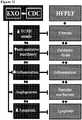
- CDC-derived exosomeswhich exhibit anti-fibrotic, anti-inflammatory and angiogenic properties.
- Positive factorsexist in cellular exosomes produced by CDCs, the lipid bilayer nanovesicles secreted by cells when multivesicular endosomes fuse with the plasma membrane.
- compositions and methods demonstrating CDC and CDC-derived exosome disease-modifying activity in HFpEFwere capable of normalizing LV relaxation and LV diastolic pressure and improving survival in HFpEF despite persistent hypertension and hypertrophy. Lower risk of arrhythmias in HFpEF was also observed following CDC administration. Improvement of diastolic dysfunction following administration of CDC-derived exosomes was observed, along with decreased mortality. CDCs and CDC-derived exosomes are beneficial in the treatment of HFpEF.
- HFpEFis a complex syndrome characterized by HF signs and symptoms and a normal or near-normal left ventricular ejection fraction (LVEF). More specifically, signs and symptoms of heart failure are present, including a decrease in left ventricular compliance, increased pressure at end-diastole in the left ventricle, increased left atrial pressure, but with an ejection fraction greater than 50%. Increased left atrial size is often seen with HFpEF as a result of the slow or incomplete left ventricular relaxation. Arrhythmias, including those affecting atrium (atrial fibrillation, AF) are coincident with HFpEF. A recent study found frequencies of preexisting and incident AF were 43.2% and 9.5%, respectively, in those with HFpEF.
- AFatrial fibrillation
- Ventricular dysfunction in HFpEFincreases risk of imminently life-threatening ventricular fibrillation.
- the AHA staging system for HFcan be applied to patients with HFpEF, to classify disease severity and to track the progression of the disease.
- Stage Aare at high risk of developing HF
- stage Bincludes patients with known structural disease, such as a history of myocardial infarction or systolic or diastolic dysfunction, but no overt symptoms of HF.
- Patients at Stage Chave evidence of structural disease and symptoms of HF, such as fatigue, shortness of breath, or reduced exercise tolerance, with Stage D including refractory HF, with marked symptoms even at rest despite medical therapy.
- diastolic LV dysfunctionresults from increased myocardial stiffness.
- Myocardial diastolic stiffness of the extracellular matrixis determined by collagen through regulation of its total amount, relative abundance of collagen type I, and degree of cross-linking.
- all three mechanismsappear to be involved, including excessive collagen type I deposition resulting from an imbalance between an exaggerated synthesis and a depressed degradation.
- MMPsmatrix metalloproteinases
- TIMPstissue inhibitors of matrix metalloproteinases
- HFpEFconstitutes a distinct clinical syndrome refractory to routine medical approaches. Randomized clinical trials (RCTs), epidemiologic studies, and mechanistic studies in patients with chronic heart failure (HF) have generally divided HF into two clinical syndromes: HFrEF (EF ⁇ 50%) and HFpEF (EF ⁇ 50%). All of these patients, regardless of EF, have the clinical syndrome of HF; nevertheless, distinct differences in several clinical and pathophysiologic features make HFpEF particularly challenging. Table 1 summarizes differences in structural and functional remodeling, molecular and cellular mechanisms, demographics and antecedent illnesses, and responsiveness to various classes of drugs. Note the utter lack of efficacy of the routine HF armamentarium. To date, because no management strategies have been demonstrated to decrease morbidity and mortality in HFpEF patients, HFpEF represents a critical unmet medical need.
- HFpEFhas proven refractory because so little is known about its pathophysiology and pathogenesis. There are two prevalent hypotheses for the pathogenesis of HFpEF. According to a first model, cardiac hypertrophy secondary to hypertension impairs relaxation and leads to HFpEF. If this first model is true, antihypertensive and antihypertrophic measures would be the key to treating HFpEF, but agents that act in this manner have failed in HFpEF clinical trials (Table 1). Table 1: Differences Between HFpEF and HFrEF.
- a contrasting concept as a second modelhas emerged from several recent studies in animal models and humans that have implicated inflammation and collagen infiltration. Hypertension and other comorbidities can favor a pro-inflammatory state with high circulating cytokine levels, including IL-6, TNF- ⁇ and MCP-1. Inflammation leads to activation, recruitment and trans-endothelial migration of leukocytes and monocytes/macrophages into the heart. These inflammatory cells contribute to myocardial fibrosis by promoting the differentiation of fibroblasts into myofibroblasts with activation of the TGF ⁇ pathway.
- LV collagen contentis the main contributor to the increase in passive myocardial fiber stiffness, a major component of diastolic impairment in HFpEF.
- Abnormalities of calcium cycling and the myofilamentse.g., cardiomyocyte remodeling
- cardiomyocyte remodelinghave been associated with hypertrophy and HFpEF, but a causal link with either model has not been solidly established. It is important to understand the nature and origin of the abnormalities in cardiomyocyte behavior in HFpEF, as these are the contractile cells of the heart and presumably underlie the central functional abnormalities.
- Fibrosisoccurs in both the heart and vascular system with impacts on both diastolic and systolic function leading to myocardial stiffening, retarded suction and filling, and the loss of early diastolic suction as mediated by alterations in the amount and composition of collagen within the extracellular matrix. It is understood that collagen synthesis is enhanced in the setting of increased load or activation of the renin-angiotensin-aldosterone system (RAAS), and it has further been observed that down-regulation of enzymes that degrade collagen occurs in patients with HFpEF.
- RAASrenin-angiotensin-aldosterone system
- HFpEFThe pathophysiology of HFpEF is fundamentally different from heart failure with reduced ejection fraction. Drugs used to treat heart failure with reduced ejection fraction are not effective in HFpEF and there are currently no effective treatments for HFpEF (Table 1).
- the Inventorshave administered to patients with HFrEF, cardiosphere-derived cells (CDCs) that are capable of mediating therapeutic regeneration in humans as a candidate cell type for regenerative therapy.
- CDCscardiosphere-derived cells
- These heart-derived cellsexhibit multilineage potential and clonogenicity, and work primarily through indirect mechanisms. These cells exert salutary anti-inflammatory and anti-fibrotic effects in HFrEF and by the secretion of nanovesicles called exosomes, which mediate cell-cell transfer of genetic payloads.
- CDC-derived exosome therapycan provide broad benefit, based on several factors including superior dosage regimes (e.g., concentration, persistence in local tissue milieu, repeat dosages), and reduced or obviated safety concerns as non-viable entities. Particular advantages may be dramatic for those conditions that currently lack any treatment modality such as HFpEF.
- Dahl salt-sensitive ratsDS rats
- DS ratsdevelop hypertension, hypertrophy and, eventually, HFpEF on a high-salt diet.
- Increased fibrosis and inflammationunderlie the development of HFpEF, with resultant cachexia, pulmonary congestion and accelerated mortality.
- CDCsimprove LV structure and function and overall outcome.
- High-salt fed ratsdeveloped hypertension associated with left ventricular (LV) hypertrophy and diastolic dysfunction, without impairment of ejection fraction.
- E/A ratioEarly-to-late ventricular filling (E/A) ratio returned to normal in CDC-treated rats and remained depressed in placebo. Tau and end-diastolic pressure volume relationship were higher in placebo-treated rats than in CDC-treated rats and control, and improved LV relaxation was also associated with lower LV end-diastolic pressure, decreased lung congestion and enhanced survival (80% vs. 48%) in CDC-treated rats.
- CDC treatmentdecreased LV fibrosis, including collagen 1A1 and 3 mRNA expression levels, decreased inflammatory infiltrates in the LV, and reduced serum inflammatory cytokines. Lower risk of arrhythmias in HFpEF was also observed following CDC administration. Improvement of diastolic dysfunction following administration of CDC-derived exosomes was observed, along with decreased mortality.
- Cardiospheresare undifferentiated cardiac cells that grow as self-adherent clusters as described in WO 2005/012510 , and Messina et al., "Isolation and Expansion of Adult Cardiac Stem Cells From Human and Murine Heart,” Circulation Research, 95:911-921 (2004 ).
- heart tissuecan be collected from a patient during surgery or cardiac biopsy.
- the heart tissuecan be harvested from the left ventricle, right ventricle, septum, left atrium, right atrium, crista terminalis, right ventricular endocardium, septal or ventricle wall, atrial appendages, or combinations thereof.
- a biopsycan be obtained, e.g., by using a percutaneous bioptome as described in, e.g., U.S. Patent Application Publication Nos. 2009/012422 and 2012/0039857 .
- the tissuecan then be cultured directly, or alternatively, the heart tissue can be frozen, thawed, and then cultured.
- the tissuecan be digested with protease enzymes such as collagenase, trypsin and the like.
- the heart tissuecan be cultured as an explant such that cells including fibroblast-like cells and cardiosphere-forming cells grow out from the explant.
- an explantis cultured on a culture vessel coated with one or more components of the extracellular matrix (e.g., fibronectin, laminin, collagen, elastin, or other extracellular matrix proteins).
- the tissue explantcan be cultured for about 1, 2, 3, 4, or more weeks prior to collecting the cardiosphere-forming cells.
- a layer of fibroblast-like cellscan grow from the explant onto which cardiosphere-forming cells appear.
- Cardiosphere-forming cellscan appear as small, round, phase-bright cells under phase contrast microscopy.
- Cells surrounding the explant including cardiosphere-forming cellscan be collected by manual methods or by enzymatic digestion.
- the collected cardiosphere-forming cellscan be cultured under conditions to promote the formation of cardiospheres.
- the cellsare cultured in cardiosphere-growth medium including buffered media, amino acids, nutrients, serum or serum replacement, growth factors including but not limited to EGF and bFGF, cytokines including but not limited to cardiotrophin, and other cardiosphere promoting factors such as but not limited to thrombin.
- Cardiosphere-forming cellscan be plated at an appropriate density necessary for cardiosphere formation, such as about 20,000-100,000 cells/mL.
- the cellscan be cultured on sterile dishes coated with poly-D-lysine, or other natural or synthetic molecules that hinder the cells from attaching to the surface of the dish. Cardiospheres can appear spontaneously about 2-7 days or more after cardiosphere-forming cells are plated.
- CDCsare a population of cells generated by manipulating cardiospheres in the manner as described in, e.g., U.S. Patent Application Publication No. 2012/0315252 .
- CDCscan be generated by plating cardiospheres on a solid surface which is coated with a substance which encourages adherence of cells to a solid surface of a culture vessel, e.g., fibronectin, a hydrogel, a polymer, laminin, serum, collagen, gelatin, or poly-D-lysine, and expanding same as an adherent monolayer culture.
- CDCscan be repeatedly passaged, e.g ., passaged two times or more, according to standard cell culturing methods.
- Exosomesare vesicles formed via a specific intracellular pathway involving multivesicular bodies or endosomal-related regions of the plasma membrane of a cell. Exosomes can range in size from approximately 20-150 nm in diameter. In some cases, they have a characteristic buoyant density of approximately 1.1-1.2 g/mL, and a characteristic lipid composition. Their lipid membrane is typically rich in cholesterol and contains sphingomyelin, ceramide, lipid rafts and exposed phosphatidylserine. Exosomes express certain marker proteins, such as integrins and cell adhesion molecules, but generally lack markers of lysosomes, mitochondria, or caveolae.
- the exosomescontain cell-derived components, such as but not limited to, proteins, DNA and RNA (e.g., microRNA and noncoding RNA).
- exosomescan be obtained from cells obtained from a source that is allogeneic, autologous, xenogeneic, or syngeneic with respect to the recipient of the exosomes.
- miRNAsfunction as post-transcriptional regulators, often through binding to complementary sequences on target messenger RNA transcripts (mRNAs), thereby resulting in translational repression, target mRNA degradation and/or gene silencing.
- miR146aexhibits over a 250-fold increased expression in CDCs, and miR210 is upregulated approximately 30-fold, as compared to the exosomes isolated from normal human dermal fibroblasts.
- Exosomes derived from cardiospheres and CDCsare described in, e.g ., WO 2014/028493 .
- Methods for preparing exosomescan include the steps of: culturing cardiospheres or CDCs in conditioned media, isolating the cells from the conditioned media, purifying the exosome by, e.g., sequential centrifugation, and optionally, clarifying the exosomes on a density gradient, e.g., sucrose density gradient.
- the isolated and purified exosomesare essentially free of non-exosome components, such as components of cardiospheres or CDCs.
- Exosomescan be resuspended in a buffer such as a sterile PBS buffer containing 0.01-1% human serum albumin. The exosomes may be frozen and stored for future use.
- Exosomescan be prepared using a commercial kit such as, but not limited to the ExoSpinTM Exosome Purification Kit, Invitrogen® Total Exosome Purification Kit, PureExo® Exosome Isolation Kit, and ExoCapTM Exosome Isolation kit.
- Methods for isolating exosome from stem cellsare found in, e.g., Tan et al., Journal of Extracellular Vesicles, 2:22614 (2013 ); Ono et al., Sci Signal, 7(332):ra63 (2014 ) and U.S. Application Publication Nos. 2012/0093885 and 2014/0004601 .
- differential ultracentrifugationhas become a leading technique wherein secreted exosomes are isolated from the supernatants of cultured cells.
- This approachallows for separation of exosomes from nonmembranous particles, by exploiting their relatively low buoyant density.
- Size exclusionallows for their separation from biochemically similar, but biophysically different microvesicles, which possess larger diameters of up to 1,000 nm. Differences in flotation velocity further allows for separation of differentially sized exosomes.
- exosome sizeswill possess a diameter ranging from 30-200 nm, including sizes of 40-100 nm. Further purification may rely on specific properties of the particular exosomes of interest. This includes, e.g., use of immunoadsorption with a protein of interest to select specific vesicles with exoplasmic or outward orientations.
- differential centrifugationis the most commonly used for exosome isolation.
- This techniqueutilizes increasing centrifugal force from 2000xg to 10,000xg to separate the medium- and larger-sized particles and cell debris from the exosome pellet at 100,000xg. Centrifugation alone allows for significant separation/collection of exosomes from a conditioned medium, although it is insufficient to remove various protein aggregates, genetic materials, particulates from media and cell debris that are common contaminants.
- Enhanced specificity of exosome purificationmay deploy sequential centrifugation in combination with ultrafiltration, or equilibrium density gradient centrifugation in a sucrose density gradient, to provide for the greater purity of the exosome preparation (flotation density 1.1-1.2g/mL) or application of a discrete sugar cushion in preparation.
- ultrafiltrationcan be used to purify exosomes without compromising their biological activity.
- Membranes with different pore sizes -such as 100 kDa molecular weight cut-off (MWCO) and gel filtration to eliminate smaller particles - have been used to avoid the use of a nonneutral pH or non-physiological salt concentration.
- MWCOmolecular weight cut-off
- THFtangential flow filtration
- HPLCcan also be used to purify exosomes to homogeneouslysized particles and preserve their biological activity as the preparation is maintained at a physiological pH and salt concentration.
- exosomesfor precipitation techniques, addition to volume-excluding polymers (e.g., polyethylene glycols (PEGs)), possibly combined additional rounds of centrifugation or filtration.
- a precipitation reagente.g., ExoQuick®
- ExoQuick®can be added to conditioned cell media to quickly and rapidly precipitate a population of exosomes, although re-suspension of pellets prepared via this technique may be difficult.
- Flow field-flow fractionationis an elution-based technique that is used to separate and characterize macromolecules (e.g., proteins) and nano-to micro-sized particles (e.g., organelles and cells) and which has been successfully applied to fractionate exosomes from culture media.
- macromoleculese.g., proteins
- nano-to micro-sized particlese.g., organelles and cells
- exosomesmay be isolated from specific exosomes of interest. This includes relying on antibody immunoaffinity to recognizing certain exosome-associated antigens. As described, exosomes further express the extracellular domain of membrane-bound receptors at the surface of the membrane. This presents a ripe opportunity for isolating and segregating exosomes in connections with their parental cellular origin, based on a shared antigenic profile. Conjugation to magnetic beads, chromatography matrices, plates or microfluidic devices allows isolating of specific exosome populations of interest as may be related to their production from a parent cell of interest or associated cellular regulatory state. Other affinity-capture methods use lectins which bind to specific saccharide residues on the exosome surface.
- compositions and methodsproviding significant benefits in the treatment of heart failure with preserved ejection fraction (HFpEF), including repair or regeneration of damaged or diseased tissues via CDCs and CDC-derived exosomes.
- HFpEFpreserved ejection fraction
- Further examples and informationare described in, for example, U.S. App. No. 11/666,685 , 12/622,143 , 12/622,106 , 14/421,355 , PCT App. No. PCT/US2013/054732 , PCT/US2015/053853 , PCT/US2015/054301 and PCT/US2016/035561 .
- therapeutically effective amounts of the compositions comprising cardiosphere-derived cells (CDCs)may be used in the disclosed methods. Any embodiments falling outside of the scope of the claims are provided for reference only.
- a method for treatment for heart failure with preserved ejection fractionincluding administering a therapeutically effective amount of cardiosphere-derived cells (CDCs) to a subject, thereby treating the subject.
- the method for treatment for heart failure with preserved ejection fractionmay include administering a therapeutically effective amount of a composition including cardiosphere-derived cells (CDCs) to a subject, thereby treating the subject.
- the methodincludes selecting a subject in need of treatment, and administering a composition including cardiosphere-derived cells (CDCs) to the subject, wherein administration of the composition treat the subject.
- the method for treatment for heart failure with preserved ejection fractionmay include identifying heart failure with preserved ejection fraction in a subject and administering a therapeutically effective amount of a composition including cardiosphere-derived cells (CDCs).
- the method for treatment for heart failure with preserved ejection fractionmay include treatment in a subject diagnosed with heart failure with preserved ejection fraction.
- the method for treatment for heart failure with preserved ejection fractionmay include treatment in a subject where standard therapy for heart failure has failed.
- HFpEFmay be characterized by one or more parameters including: unchanged end diastolic volume (EDV), unchanged end systolic volume (ESV), increased wall thickness, increased LV mass, increased mass/EDV ratio, concentric remodeling, unchanged ejection fraction (at rest), unchanged stroke work (at rest), unchanged end systolic elastance, increased vascular stiffness, and increased end diastolic stiffness and combinations thereof.
- EDVunchanged end diastolic volume
- ESVunchanged end systolic volume
- ESVunchanged end systolic volume
- increased wall thicknessincreased LV mass, increased mass/EDV ratio
- concentric remodelingunchanged ejection fraction (at rest), unchanged stroke work (at rest), unchanged end systolic elastance, increased vascular stiffness, and increased end diastolic stiffness and combinations thereof.
- HFpEFis characterized by one or more of: unchanged cardiomyocyte length, increased cardiomyocyte diameter, diffuse myocardial fibrosis
- HFpEFis refractory to treatment using any of B-blockers, Angiotensin Converting Enzyme Inhibitor (ACE-I), Angiotensin Receptor Blocker (ARB), Digitalis, Hydralazine/Nitrates, or PDE-5 Inhibitor.
- ACE-IAngiotensin Converting Enzyme Inhibitor
- ARBAngiotensin Receptor Blocker
- DigitalisHydralazine/Nitrates
- PDE-5 InhibitorAlternatively, CDCs may be administered as a primary therapy. Heart failure with preserved ejection fraction may involve tissue damage or dysfunction. In other embodiments, heart failure with preserved ejection fraction involves fibrosis. In other embodiments, heart failure with preserved ejection fraction involves inflammation. In certain embodiments, CDCs are administered as adjuvant to standard therapy for HF.
- an effective dose of CDCsinclude 1x10 5 to 1x10 6 and 1x10 6 to 100x10 6 cells, such as between 10x10 6 and 50x10 6 cells. Depending on the size of the damaged region of the heart, more or less cells can be used. A larger region of damage may require a larger dose of cells, and a small region of damage may require a smaller dose of cells. On the basis of body weight of the recipient, an effective dose may be between 1 and 10x10 6 per kg of body weight, preferably between 1x10 6 and 5x10 6 cells per kg of body weight. Patient age, general condition, and immunological status may be used as factors in determining the dose administered. For example, it has been demonstrated that 3mL / 3 x 10 5 CDCs, is capable of providing therapeutic benefit in intracoronary administration. In various embodiments, administration of the composition to the subject occurs through any of known techniques in the art.
- Cardiosphere derived cellscan be delivered systemically or locally to the heart. Local administration can be by catheter or during surgery. Systemic administration can be by intravenous or intraarterial injections, perfusion, or infusion.
- the populations of cells of the inventionare administered systemically, they migrate to the appropriate organ, e.g., the heart, if the cells are derived from resident heart stem cells.
- the beneficial effects which are observed upon administration of the cells to a mammalmay be due to the cells per se, or due to products which are expressed by the cells. For example, it is possible that the engraftment of cells produces a favorable outcome. It is also possible that cytokines or chemokines or other diffusible factors stimulate resident cells to grow, reproduce, or perform better.
- myocardial infusionis used, for example, the use of intracoronary catheters.
- Deliverycan be intra-arterial or intravenous. Additional delivery sites include any one or more compartments of the heart, such as myocardium, associated arterial, venous, and/or ventricular locations.
- administrationincludes percutaneous delivery, and/or injection into heart.
- administrationcan include delivery to a tissue or organ site that is the same as the site of diseased and/or dysfunctional tissue.
- Administrationcan include delivery to a tissue or organ site that is different from the site or diseased and/or dysfunctional tissue.
- Administration of the compositioncan include combinations of multiple delivery techniques, such as intravenous, intracoronary, and intramyocardial delivery.
- the compositionalters gene expression in the damaged or dysfunctional tissue, improves viability of the damaged tissue, and/or enhances regeneration or production of new tissue in the individual.
- administration of the compositionresults in functional improvement in the tissue.
- the damaged tissuecan be pulmonary, arterial or capillary tissue.
- the damaged or dysfunctional tissueincludes cardiac tissue.
- CDC treatmentdecreases fibrosis, including left ventricle fibrosis.
- decreased fibrosisincludes reduction in collagen 1A1 and 3 expression levels, such as protein and mRNA.
- decreased fibrosisincludes increased microvascular density.
- CDC treatmentreduces inflammation.
- thisincludes reduction in inflammatory infiltrates, such as CD68+ and CD45+ cells in the left ventricle. In other embodiments, this includes reduction in inflammatory cytokines in serum, including for example MCP-1, IL-6 and TNF- ⁇ . In other embodiments, CDC treatment results in an increase in myocardial blood flow.
- CDCslower risk of arrhythmias as can be exhibited, by for example, a lower arrhythmogenicity index and/or a decrease in action potential duration dispersion in the heart.
- CDCsnormalize left ventricle relaxation and diastolic pressure, improve survival, and/or confer functional benefits despite persistent hypertension and hypertrophy.
- functional improvementmay include increased cardiac output, contractility, ventricular function and/or reduction in arrhythmia (among other functional improvements). For example, this may include a decrease in right ventricle systolic pressure.
- improved functionmay be realized as well, such as enhanced cognition in response to treatment of neural damage, improved blood-oxygen transfer in response to treatment of lung damage, improved immune function in response to treatment of damaged immunological-related tissues.
- compositions comprising cardiosphere-derived cellsmay be used in the disclosed methods. Any embodiments falling outside of the scope of the claims are provided for reference only.
- a method of treatment for heart failure with preserved ejection fractionincluding administering a therapeutically effective amount of plurality of exosomes to a subject, thereby treating the subject.
- a method of treatment for heart failure with preserved ejection fractionincludes administering a therapeutically effective amount of a composition including a plurality of exosomes to a subject, thereby treating the subject.
- the methodincludes selecting a subject in need of treatment and administering a composition which may include a plurality of exosomes to the subject, wherein administration of the composition treats the subject.
- the method for treatment for heart failure with preserved ejection fractionmay include identifying heart failure with preserved ejection fraction in a subject and administering a therapeutically effective amount of a composition including a plurality of exosomes.
- the method for treatment for heart failure with preserved ejection fractionmay include treatment in a subj ect diagnosed with heart failure with preserved ejection fraction.
- the method for treatment for heart failure with preserved ejection fractionmay include treatment in a subject where standard therapy for heart failure has failed.
- the plurality of the exosomesare isolated from cardiosphere-derived cells (CDCs) grown in serum-free media, and may include exosomes with a diameter of about 90 nm to about 200 nm and are CD81+, CD63+, or both.
- CDCscardiosphere-derived cells
- HFpEFmay be characterized by one or more parameters including: unchanged end diastolic volume (EDV), unchanged end systolic volume (ESV), increased wall thickness, increased LV mass, increased mass/EDV ratio, concentric remodeling, unchanged ejection fraction (at rest), unchanged stroke work (at rest), unchanged end systolic elastance, increased vascular stiffness, and increased end diastolic stiffness and combinations thereof.
- EDVunchanged end diastolic volume
- ESVunchanged end systolic volume
- ESVunchanged end systolic volume
- increased wall thicknessincreased LV mass, increased mass/EDV ratio
- concentric remodelingunchanged ejection fraction (at rest), unchanged stroke work (at rest), unchanged end systolic elastance, increased vascular stiffness, and increased end diastolic stiffness and combinations thereof.
- HFpEFmay be characterized by one or more of: unchanged cardiomyocyte length, increased cardiomyocyte diameter, diffuse myocardial fibros
- HFpEFmay be refractory to treatment using any of B-blockers, Angiotensin Converting Enzyme Inhibitor (ACE-I), Angiotensin Receptor Blocker (ARB), Digitalis, Hydralazine/Nitrates, or PDE-5 Inhibitor.
- ACE-IAngiotensin Converting Enzyme Inhibitor
- ARBAngiotensin Receptor Blocker
- DigitalisHydralazine/Nitrates
- PDE-5 InhibitorPDE-5 Inhibitor.
- CDCsare administered as a primary therapy.
- CDC-derived exosomesare administered as adjuvant to standard therapy for HF.
- the method of treatmentmay include modulation of fibrosis and/or inflammation.
- Administering a compositionmay include about 1 to about 100 mg exosome protein in a single dose. In other options, a single dose is administered multiple times to the subject.
- Administering a compositionmay include injection. The injection may include percutaneous injection. The injection may be directly into heart muscle.
- administering a compositionincludes myocardial infusion. Myocardial infusion may be intra-arterial or intravenous.
- treatment of the subjectresults in decreased fibrosis, decreased inflammation, increased mitochondrial function and/or increased cardiomyogenesis.
- decreased fibrosisincludes a reduction in collagen accumulation.
- collagenincludes collagen I and/or collagen III.
- Decreased inflammationmay include an increase in cytoplasmic nuclear factor (erythroid-derived 2)-like 2 (Nrf2), reduction in fatty acid peroxidation end products, reduced numbers of inflammatory cells, and/or upregulated expression of antioxidants.
- Antioxidantsmay include heme oxygenase-1 (HO-1), catalase, superoxide dismutase-2 (SOD-2), and glutamate-cystein ligase catalytic (GCLC) subunit.
- inflammatory cellsinclude CD68+ macrophages and CD3+ T-cells.
- Increased mitochondrial functionmay include increased mitochondrial ultrastructure and/or increased mitochondrial biogenesis.
- Increased mitochondrial functionmay include increased nuclear PPAR- ⁇ co-activator-1 (PGC-1) expression.
- the exosomesmay include one or more RNAs such as 2, 3, 4, 5, 6, 7, 8, 9, 10 or more RNAs, including non-coding RNAs.
- the non- coding RNAsmay include tRNAs, yRNAs, rTNAs, mirRNAs, IncRNAs, piRNAs, snRNAs, snoRNAs, further including fragments thereof, among others.
- the exosomesmay include one or more microRNAs selected from the group consisting of: microRNAs miR-146a, miR-148a, miR-22, miR-24, miR-210, miR-150, miR-140-3p, miR-19a, miR-27b, miR-19b, miR-27a, miR-376c, miR-128, miR-320a, miR-143, miR-21, miR-130a, miR-9, miR-185, and miR-23a.
- microRNAsselected from the group consisting of: microRNAs miR-146a, miR-148a, miR-22, miR-24, miR-210, miR-150, miR-140-3p, miR-19a, miR-27b, miR-19b, miR-27a, miR-376c, miR-128, miR-320a, miR-143, miR-21, miR-130a, miR-9, miR-185, and miR-23a.
- CDC-derived exosome treatmentdecreases fibrosis, including left ventricle fibrosis.
- decreased fibrosisincludes reduction in collagen 1A1 and 3 expression levels, such as protein and mRNA.
- Decreased fibrosisincludes increased microvascular density.
- CDC-derived exosome treatmentreduces inflammation.
- thisincludes reduction in inflammatory infiltrates, such as CD68+ and CD45+ cells in the left ventricle.
- thisincludes reduction in inflammatory cytokines in serum, including for example MCP-1, IL-6 and TNF- ⁇ .
- CDC-derived exosome treatmentresults in an increase in myocardial blood flow.
- CDC-derived exosomeslower risk of arrhythmias as can be exhibited, by for example, a lower arrhythmogenicity index and/or a decrease in action potential duration dispersion in the heart.
- CDC-derived exosome treatmentnormalizes left ventricle relaxation and diastolic pressure, improve survival, and/or confer functional benefits despite persistent hypertension and hypertrophy.
- functional improvementmay include increased cardiac output, contractility, ventricular function and/or reduction in arrhythmia (among other functional improvements). For example, this may include a decrease in right ventricle systolic pressure.
- improved functionmay be realized as well, such as enhanced cognition in response to treatment of neural damage, improved blood-oxygen transfer in response to treatment of lung damage, improved immune function in response to treatment of damaged immunological-related tissues.
- CDCswere prepared as described in U.S. Patent Application Publication No. 2012/0315252 .
- heart biopsieswere minced into small fragments and briefly digested with collagenase. Explants were then cultured on 20 mg/mL fibronectin-coated dishes. Stromallike flat cells and phase-bright round cells grew out spontaneously from tissue fragments and reached confluency by 2-3 weeks. These cells were harvested using 0.25% trypsin and were cultured in suspension on 20 mg/mL poly-d-lysine to form self-aggregating cardiospheres. CDCs were obtained by plating and expanding the cardiospheres on a fibronectin-coated flask as an adherent monolayer culture.
- CDCsWhen the CDCs reached the desired confluency, the flask was washed three times with PBS. CDCs were treated with serum-free medium (IMDM) and were incubated at 37°C at 5% O 2 , 5% CO 2 for 15 days. After 15 days, the conditioned medium was collected in 225mL BD Falcon polypropylene conical tubes (BD 352075 - Blue Top) and centrifuged at 2,000 rpm for 20 minutes at 4°C to remove cells and debris (care was taken not to disturb the pellet). The conditioned medium was run through a 0.45 ⁇ m membrane filter. The conditioned medium was concentrated using centrifugal filter.
- IMDMserum-free medium
- a 3KDa Centricon Plus-70 Centrifugal Filterwas pre-rinsed with 10-25 mL of molecular grade water and was centrifuged at 3220g for five minutes at 18°C. Once the filter was rinsed, all remaining water was carefully removed without touching the filter. 15 mL of the conditioned medium was added to the filter and was centrifuged at 3220g for 45 minutes at 18°C. After the initial spin, the remaining medium was mixed by pipetting and then spun again until the desired concentration was reached. The final sample was then run through a 0.22 ⁇ m syringe filter. 25 ⁇ L of the concentrated conditioned medium was diluted in 975 ⁇ L of PBS for particle count using the Nanosight.
- the appropriate volume of 25% PEGwas added to the filtered concentrated conditioned medium.
- the sampleswere incuated at 4°C for 12-16 hours on an orbital shaker. Once incubation was complete, the samples were centrifuged at 1500g for 30 minutes at 4°C. The supernatant was carefully removed without disrupting the pellet. The pellet was resuspended in the desired volume of serum-free medium and sampled for particle count.
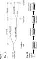
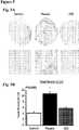
- FIG 1Adepicts the experimental protocol.
- rats on a high-salt dietwere randomized to receive allogeneic rat CDCs (5X10 5 suspended in 100 ⁇ L PBS) or placebo via a left thoracotomy under general anesthesia (Isofluorane 4-5% for induction followed by 2%).
- Cells (or PBS control, 100 ⁇ L)were injected into the LV cavity during aortic cross-clamp, over a period of 20 seconds to achieve intra-coronary delivery.
- CDCswere grown from a freshly-explanted Wistar-Kyoto rat heart as described ( Figure 1B ).
- heartswere minced, subjected to enzymatic digestion and then plated on adherent (fibronectin-coated) culture dishes. These explants spontaneously yield monolayer adherent cells (explant-derived cells) which were harvested and plated in suspension culture (10 5 cells/mL on poly-D-lysine-coated dishes) to enable the self-assembly of three-dimensional cardiospheres. Subsequent replating of these cardiospheres on adherent culture dishes yielded CDCs. CDCs at passage 2 were used for all experiments.
- Echocardiographywas performed at baseline, before treatment, and 1 and 4 weeks after treatment to assess systolic and diastolic functions (Vevo 770, Visual Sonics, Toronto, Ontario, Canada), under general anesthesia (Isoflurane 4-5% for induction followed by 2%).
- Two-dimensional long axis and short axis (at the papillary muscle level) LV imageswere obtained.
- M-mode tracingswere recorded through the anterior and posterior LV walls at the papillary muscle level to measure LV dimension, and LV anterior and posterior wall thickness at end diastole.
- Pulse-wave Doppler spectra (E and A waves) of mitral inflowwere recorded from the apical 4-chamber view, with the sample volume placed near the tips of the mitral leaflets and adjusted to the position at which velocity was maximal and the flow pattern laminar. E/A ratio was used to assess diastolic function as described. Systolic function was assessed by LV ejection fraction (LVEF) calculated from the long axis view and fractional area change (FAC) calculated from the short axis view.
- LVEFLV ejection fraction
- FACfractional area change
- RNA and protein stabilization reagentAllprotect, Qiagen, Venlo, Netherlands
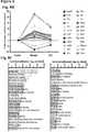
- Inflammatory cytokineswere quantified in the serum using a commercially available cytokine array (Raybiotech, Norcross, Georgia, USA). Sera from 4 control rats, 4 CDC-treated and 4 placebo-treated animals were used. All values were normalized to the positive control.
- tissueswere minced, suspended in T-PER (with HALT protease and phosphatase inhibitors, Thermo Scientific), homogenized with a bead ruptor and then centrifuged at 10,000 g for 15 minutes at 4°C. The supernatant was collected and protein concentration measured (BCA protein assay, Thermo Scientific). Protein samples were prepared under non-denaturing conditions for gel electrophoresis (NuPAGE 4%-12% Bis-Tris, Invitrogen). Thirty ⁇ g of protein was loaded into each well for separation.
- Proteinswere then transferred to a polyvinylidene fluoride membrane (Bio-Rad) for immunoblotting with collagen 1A1 (Santa Cruz sc-25974) and collagen 3 (Santa Cruz sc-8780) antibodies. Bands were visualized following activation with ECL (Thermo Scientific) and exposure on film (Kodak Carestream Biomax, Sigma-Aldrich).
- RNA was isolated from LV samples (n7 in each group) (RNA easy kit, Qiagen, Venlo, Netherlands). Then, cDNA was synthesized from mRNA using the High-Capacity cDNA Reverse Transcription Kit (Life Technologies, Carlsbad, California, USA) according to the manufacturer's protocol. The resulting cDNA was standardized across samples and then mixed with master mix and designated primer sets (Life Technologies, Carlsbad, California, USA). Predesigned TaqMan primer sets for collagen-lAl and collagen-3 were used. Relative mRNA expression of target genes was normalized to the endogenous GAPDH gene.
- RNA was isolated from LV samples (n3 in each group) (RNA easy kit, Qiagen, Venlo, Netherlands). rRNA was removed using the Ribo-Zero rRNA Removal Kit from Illumina. Libraries for RNA-Seq were prepared with KAPA Stranded RNA-Seq Kit. The workflow consists of mRNA enrichment, cDNA generation, and end repair to generate blunt ends, A-tailing, adaptor ligation and PCR amplification. Different adaptors were used for multiplexing samples in one lane. Sequencing was performed on Illumina NextSeq 500 for a single read of 75 run. Data quality check was done on Illumina SAV. Demultiplexing was performed with Illumina Bcl2fastq2 v 2.17 program.
- the readswere first mapped to the latest UCSC transcript set using Bowtie2 version 2.1.0 and the gene expression level was estimated using RSEM v1.2.15. TMM (trimmed mean of M-values) was used to normalize the gene expression. Differentially expressed genes were identified using the edgeR program. Genes showing altered expression with p ⁇ 0.05 and more than 1.5 fold changes were considered differentially expressed.
- the pathway and network analysiswas performed using Ingenuity (IPA).
- IPAIngenuity computes a score for each network according to the fit of the set of supplied focus genes. These scores indicate the likelihood of focus genes to belong to a network versus those obtained by chance. A score > 2 indicates a ⁇ 99% confidence that a focus gene network was not generated by chance alone.
- the canonical pathways generated by IPAare the most significant for the uploaded data set. Fischer's exact test with FDR option was used to calculate the significance of the canonical pathway.
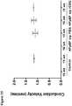
- Continuous variablesare presented as mean ⁇ standard deviation in the text and mean ⁇ standard error in the figures.
- Table 2shows characteristics of the high-salt and control animals at baseline and after 6 weeks of diet (13 weeks of age). As expected, rats fed a high-salt diet developed hypertension and cardiac hypertrophy after 6 weeks, but low-salt controls did not. Those changes were associated with diastolic dysfunction as shown by a decreased E/A ratio by echocardiography (1.7 ⁇ 0.2 vs. 1.2 ⁇ 0.2, P ⁇ 0.0001), without any changes in LV volumes, LVEF or FAC (Table 2). Table 2: Characteristics of high-salt and low-salt fed rats before (baseline) and after 6 weeks of diet. * P ⁇ 0.05 between high-salt and low-salt group at 6 weeks.
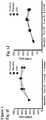
- FIG. 1Cshows representative images of trans-mitral flow at endpoint in control, placebo- and CDC-treated animals.
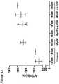
- Figure 1DPooled data reveal that, after 6 weeks of diet but before treatment, Early-to-late ventricular filling (E/A) ratio was similar in placebo and CDC groups, but lower than in control.
- E/A ratioincreased over time ( Figure ID), a change which was evident as soon as 1 week after treatment.
- E/A ratiohad returned to control levels in CDC-treated animals (1.7 ⁇ 0.2 for CDC vs.
- FIG 2Ashows representative recordings of PV loop families at endpoint.
- the normalization of LVEDP and Tau in CDC-treated ratsconfirms that the increase of E/A ratio over time in this group was due to normalization of LV relaxation rather than to progression toward a pseudo-normal pattern of trans-mitral flow (which would have been associated with increased LVEDP and Tau).
- LV hypertrophycan occur with or without diastolic dysfunction.
- the Inventorsquantified cardiac hypertrophy using LV wall thickness by echocardiography, heart weight and cardiomyocyte cross sectional area.
- the CDCrelated improvement in diastolic functionwas not due to a decrease in cardiac hypertrophy: wall thickness by echocardiography ( Figure 4A ), as well as post-mortem heart weight and cardiomyocyte cross-sectional area ( Figure 4B ), remained equivalent in CDC and placebo groups.
- CDCswere salutary without decreasing cardiac hypertrophy.
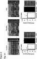
- cardiac myofibroblastsincreased dramatically in placebo-treated, but not in CDC-treated, DS rats ( Figure 5E ).
- transcript levels of MMP-2, MMP-7, MMP-9 and TIMP-1 as well as collagen 1A1 and collagen 3were higher in the placebo treated animals compared to the control and CDC-treated animals (which had similar levels; Figure 9 ). These increased transcript levels are suggesting of increased extracellular matrix turnover associated with HFpEF which is normalized by CDC treatment.
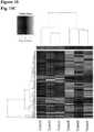
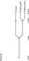
- FIG. 10A-Cshows head-to-head pairwise comparison of gene expression in the 3 groups.
- the heat mapsreveal that the HFpEF phenotype is associated with major global changes in gene expression as shown by the placebo vs. control comparison ( Figure 10A ).
- the CDC vs. placebo comparisonFigure 10B reveals that CDC-treatment dramatically changed gene expression.
- >300 geneswhose expression was up- or down regulated in HFpEF (i.e., in high-salt placebo hearts) had their expression levels "rescued” by CDC treatment ( Figure 8A ).
- CDCsnormalized LV relaxation and improved survival in a rat model of HFpEF, without blunting hypertension or hypertrophy.
- the selective correction of functional HFpEF abnormalitiescreates an unprecedented opportunity for mechanistic insights.
- Potentially causal pathwaysi.e., those that accompany the abnormalities in HFpEF
- fibrosis and inflammationare causative in HFpEF: reductions in those two pathophysiological processes underlie the resolution of HFpEF, while hypertrophy and hypertension remain unchanged.
- LVleft ventricular
- diastolic dysfunctionwas observed, without impairment of ejection fraction.
- intracoronary CDCs5x10 5
- placeboplacebo for 4 more weeks
- E/Aearly--to-late ventricular filling
- Tau and end-diastolic pressure volume relationshipwere higher in placebo-treated rats than in CDC-treated rats and control.
- Improved LV relaxationwas also associated with lower LV end-diastolic pressure, decreased lung congestion and enhanced survival (80% vs. 48%) in CDC-treated rats.
- CDC treatmentdecreased LV fibrosis and collagen 1A1 and 3 mRNA expression levels, while increasing microvascular density. Inflammatory infiltrates (CD68+ and CD45+ cells) in the LV, and serum inflammatory cytokines (MCP-1, IL-6 and TNF- ⁇ ), were decreased after CDC treatment. As such, CDCs normalized LV relaxation and LV diastolic pressure while improving survival in a rat model of HFpEF. These functional benefits occurred despite persistent hypertension and hypertrophy. By reversing inflammation and fibrosis, CDCs are beneficial in the treatment of HFpEF.
- the Inventorshave demonstrated that cell therapy by CDCs can reverse the functional abnormalities of HFpEF and improve survival in a rat model of hypertension-induced HFpEF.
- the CDC-induced reversal of HFpEFoccurred without reductions in either blood pressure or cardiac hypertrophy.
- the Inventors' findingsindicate fibrosis and inflammation are causative in HFpEF: reductions in those two pathophysiological processes underlie the resolution of HFpEF, while hypertrophy and hypertension remain unchanged.
- Cardiac hypertrophyhas long been thought to be the linchpin in HFpEF.
- inflammation and collagen infiltrationcan favor a systemic pro-inflammatory state with high circulating cytokine levels, including IL-6, TNF- ⁇ and MCP-.
- Inflammationleads to activation, recruitment and trans-endothelial migration of leukocytes and monocytes/macrophages into the heart. These inflammatory cells contribute to LV fibrosis by promoting the differentiation of fibroblasts into myofibroblasts.
- the resulting increase in LV collagen contentis the main contributor to the increase in passive myocardial fiber stiffness, a major component of diastolic impairment in HFpEF.
- the observed phenotypic improvements after CDC treatmentwere associated with decreases in circulating inflammatory cytokines (including IL-6, TNF- ⁇ and MCP-1) and less myocardial inflammation.
- collagen productionwas increased in placebo treated animals but fell markedly after CDC treatment.
- the parallel decrease in TIMP-1suggests that collagen clearance was increased in CDC-treated animals.
- myofibroblast infiltration, collagen content, and collagen productionwere increased in placebo-treated animals but fell markedly after CDC treatment.
- the parallel decrease in transcripts for MMPs and TIMPs( Fig.
- arrhythmogenicity index(AI) was calculated as (number of arrhythmia beats + coupling interval of last stimuli - 40) / square root of number of extra stimuli. AI was decreased in rCDC-injected rats compared to PBS-injected animals at 18 weeks of age (30.4 vs. 61.5).
- action potential durationAPD90
- ADPD 90 dispersionwas checked in different regions of the heart using optical mapping system and CDC administration appeared to decrease APD dispersion as shown in Fig. 14 .
- conduction velocity(depolarization) was checked during depolarization using optical mapping system and as shown in Fig. 16 conduction velocity (repolarization) was checked during repolarization using optical mapping system.
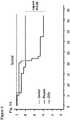
- Fig. 17Bshows that intramyocardial injection of human CDC-derived exosomes (350 ⁇ g protein divided among 4 LV sites) tends to normalize the E/A ratio measured by echo. Exoquick® and/or ultracentrifugation will be used to isolate exosomes from conditioned media.
- Nanosight® particle counterif needed, 0.45 ⁇ m filters or sucrose gradients can be applied to enrich the exosome fraction. Purity is additionally be checked by quantification of exosome-characteristic tetraspanin proteins. As exosomes are likely mediators of CDC benefits in HFpEF, and permeabilized exosomes turn out not to be bioactive, one can search for individual miRs that confer key therapeutic properties.
- CDC-derived exosome experimentsDahl salt-sensitive rats were fed high-salt (HS) diet from 7 weeks of age to induce HFpEF.
- Normal-salt (NS) fed ratsserved as controls.
- diastolic dysfunctionwas confirmed with echocardiogram.
- Human CDC exosomes(CDC-derived exosomes) (300-400 ug) were injected intra-myocardially (4 different sites) to HFpEF rats.
- follow-up experimentswere performed 4 weeks after the injection.
- PBS-injected HFpEF ratsserved as placebo.
- E/A ratiowas decreased in HS-fed rats compared to NS-fed animals (1.26 ⁇ 0.20 vs. 1.56 ⁇ 0.13, p ⁇ 0.001) and E/E' ratio was increased in HS group compared to NS rats (16.82 ⁇ 2.54 vs. 13.53 ⁇ 1.87, p ⁇ 0.001), which indicate the development of diastolic dysfunction.
- cardiosphere derived cellsare sources of cardiosphere derived cells, the use of alternative sources such as cells derived directly from heart biopsies (explant-derived cells), or from self-assembling clusters of heart-derived cells (cardiospheres), method of isolating, characterizing or altering such cells, and the particular use of the products created through the teachings of the invention.
- Various embodiments of the inventioncan specifically include or exclude any of these variations or elements.
- the numbers expressing quantities of ingredients, properties such as concentration, reaction conditions, and so forth, used to describe and claim certain embodiments of the inventionare to be understood as being modified in some instances by the term "about.” Accordingly, in some embodiments, the numerical parameters set forth in the written description and attached claims are approximations that can vary depending upon the desired properties sought to be obtained by a particular embodiment. In some embodiments, the numerical parameters should be construed in light of the number of reported significant digits and by applying ordinary rounding techniques. Notwithstanding that the numerical ranges and parameters setting forth the broad scope of some embodiments of the invention are approximations, the numerical values set forth in the specific examples are reported as precisely as practicable. The numerical values presented in some embodiments of the invention may contain certain errors necessarily resulting from the standard deviation found in their respective testing measurements.
Landscapes
- Health & Medical Sciences (AREA)
- Life Sciences & Earth Sciences (AREA)
- Engineering & Computer Science (AREA)
- Chemical & Material Sciences (AREA)
- Cardiology (AREA)
- Bioinformatics & Cheminformatics (AREA)
- Biomedical Technology (AREA)
- General Health & Medical Sciences (AREA)
- Cell Biology (AREA)
- Zoology (AREA)
- Organic Chemistry (AREA)
- Biotechnology (AREA)
- Veterinary Medicine (AREA)
- Public Health (AREA)
- Animal Behavior & Ethology (AREA)
- Pharmacology & Pharmacy (AREA)
- Medicinal Chemistry (AREA)
- Developmental Biology & Embryology (AREA)
- Genetics & Genomics (AREA)
- Wood Science & Technology (AREA)
- Epidemiology (AREA)
- Chemical Kinetics & Catalysis (AREA)
- Hospice & Palliative Care (AREA)
- Nuclear Medicine, Radiotherapy & Molecular Imaging (AREA)
- General Chemical & Material Sciences (AREA)
- Heart & Thoracic Surgery (AREA)
- Vascular Medicine (AREA)
- Immunology (AREA)
- Virology (AREA)
- Microbiology (AREA)
- Biochemistry (AREA)
- Rheumatology (AREA)
- General Engineering & Computer Science (AREA)
- Dermatology (AREA)
- Medicines Containing Material From Animals Or Micro-Organisms (AREA)
- Medicines That Contain Protein Lipid Enzymes And Other Medicines (AREA)
Description
- This invention was made with government support under HL083109 awarded by the National Institutes of Health. The government has certain rights in the invention.
- Described herein are methods and compositions related to use of cells and their exosomes in regenerative medicine application for cardiac disease.
- Half of patients with heart failure (HF) have a preserved left ventricular ejection fraction. Sometimes referred to as diastolic HF, heart failure with preserved ejection fraction (HFpEF) represents ∼50% of heart failure worldwide. Disability and frequent hospitalization are hallmarks of the disease. Associated comorbidities are common and notably include hypertension, diabetes, and obesity. Women are affected more frequently than men (by as much as a 2:1 preponderance). Unlike heart failure with reduced ejection fraction (HFrEF), where numerous pharmacological and device options have been proven to be effective, no treatments have been proven to reduce morbidity and mortality in HFpEF. The challenge of HFpEF is increasing as the population ages and comorbidities become more prevalent. The HFpEF hospitalization rate is now greater than that for heart failure with HFrEF. Compounding the problem is the fact that underlying pathophysiological mechanisms have not been completely elucidated.
- Morbidity and mortality in HFpEF are similar to values observed in patients with HF and reduced ejection fraction (EF). However, HFpEF constitutes a distinct clinical syndrome refractory to routine medical approaches. No effective treatment has been identified and HFpEF has become a major public health concern. Its increasing prevalence, rising at a rate of ∼1% per year, now exceeds that of heart failure with reduced ejection fraction (HFrEF). Outcomes of HFpEF are poor, and, so far, no treatment has been shown to decrease morbidity or mortality. For example, treatment to date has focused on the renin-angiotensin-aldosterone system and the adrenergic nervous system, but clinical trials have failed to show any significant benefit to their blockade. HFpEF, sometimes referred to as diastolic HF, is associated with various cardiovascular risk factors (especially hypertension), extra-cardiac comorbidities and aging. The net result is impaired diastolic relaxation and filling of the left ventricle (LV), increased myocardial stiffness, impaired vascular compliance and increased diastolic pressure. Perhaps underlying different responses to pharmacological intervention, HFpEF populations can consist of patients with limited myocardial infarction at risk for unfavorable eccentric LV remodeling. Cardiac hypertrophy indeed has little in common with limited myocardial infarction, and in both conditions, mechanisms driving LV remodeling are likely to be dissimilar. There is a great need in the art for therapeutic approaches for HFpEF.
- According to an aspect of the invention there is provided a therapeutically effective amount of a composition comprising cardiosphere-derived cells (CDCs) for use in a method of treating heart failure with preserved ejection fraction, the method comprising: administering a therapeutically effective amount of a composition including cardiosphere-derived cells (CDCs) to a subject in need of treatment for heart failure with preserved ejection fraction (HFpEF), thereby treating HFpEF in the subject. In various embodiments, the CDCs are allogeneic. In various embodiments, the CDCs are autologous. In various embodiments, the composition includes about 10x106 to about 100x106 CDCs in a single dose. In various embodiments, treating HFpEF includes an improvement in cardiac performance. In various embodiments, the improvement in cardiac performance includes an improvement in early-to-late ventricular filling (E/A) ratio, left ventricle (LV) relaxation, LV end-diastolic pressure and/or lung congestion. In various embodiments, the improvement in cardiac performance includes a reduction in Tau and/or end-diastolic pressure volume relationship. In various embodiments, treating HFpEF includes a reduction in fibrosis. In various embodiments, the reduction in fibrosis includes a reduction in collagen expression. In various embodiments, collagen includes collagen 1A1 and
collagen 3. In various embodiments, treating HFpEF includes a reduction in inflammation. In various embodiments, the reduction in inflammation includes a reduction in inflammatory cells. In various embodiments, the inflammatory cells are CD68+ and/or CD45+ cells. In various embodiments, the reduction in inflammation includes a reduction in inflammatory cytokines in serum. In various embodiments, the inflammatory cytokines are MCP-1, IL-6 and TNF-α. - In various embodiments, treating HFpEF includes an increase in myocardial blood flow. In various embodiments, administering a therapeutically effective amount of a composition including CDCs to the subject is primary therapy. In various embodiments, administering a therapeutically effective amount of a composition including a plurality of CDCs to the subject is adjunctive to standard therapy for heart failure. In various embodiments, administering a composition consists of one or more of: intra-arterial infusion, intravenous infusion, and percutaneous injection.
- According to a further embodiment there is provided a therapeutically effective amount of a composition comprising a plurality of exosomes isolated from cardiosphere-derived cells (CDCs) grown in a serum-free media for use in a method of treating heart failure with preserved ejection fraction, the method comprising administering a therapeutically effective amount of a composition comprising a plurality of exosomes isolated from cardiosphere-derived cells (CDCs) grown in serum-free media to a subject in need of treatment for heart failure with preserved ejection fraction (HFpEF), thereby treating HFpEF in the subject. In various disclosures, the plurality of exosomes comprise exosomes with a diameter of about 90 nm to about 200 nm and are
CD8 1 +, CD63+, or both. In various embodiments, treating HFpEF includes an improvement in cardiac performance. In various embodiments, the improvement in cardiac performance includes an improvement in early-to-late ventricular filling (E/A) ratio, left ventricle (LV) relaxation, LV end-diastolic pressure and/or lung congestion. In various embodiments, the improvement in cardiac performance includes a reduction in Tau and/or end-diastolic pressure volume relationship. In various embodiments, treating HFpEF includes a reduction in fibrosis. In various embodiments, treating HFpEF includes a reduction in inflammation. In various embodiments, administering a therapeutically effective amount of a composition including a plurality of exosomes to the subject is primary therapy. In various embodiments, administering a therapeutically effective amount of a composition including a plurality of exosomes to the subject is adjunctive to standard therapy for heart failure. Figure 1: (Fig. 1A ) Study design.(Fig. 1B ) Cardiosphere-derived cell manufacturing protocol.(Fig. 1C ) Representative images of trans-mitral flow by Doppler echocardiography at endpoint in control, placebo- and CDC-treated rats.(Fig. 1D ) CDC treatment normalizes E/A ratio at 4 weeks while placebo remains depressed. Systolic function assessed by LVEF(Fig. 1E and Fig. 1 F ) and by fractional area change(Fig. 1G and Fig. 1H ) is equivalent in all groups. L V end-diastolic(Fig. 11 ) and end systolic volumes(Fig. 1J ) are equivalent in all groups. CDC treatment halts HFpEF related left atrial enlargement while placebo does not. (Baseline and pre-treatment, n=10 for controls and n=24 for placebo and CDC; 1 week post treatment, n=10 for controls, n=21 for placebo and CDC; at endpoint, n=10 for controls, n=15 for placebo and n=18 for CDC). *P<0.05 vs. placebo and CDC; † forP<0.05 vs. placebo, both by ANOVAFigure 2: (Fig. 2A ) Representative PV loop recordings in control, placebo- and CDC-treated rats. CDCs normalize Tau(Fig. 2B ) and -dP/dt min(Fig. 2C ) in rats with HFpEF without change in dP/dt max(Fig. 2D ). PV loop analysis reveals normalization of the slope of the end-diastolic pressure volume relationship (EDPVR)(Fig. 2E ) with no change in end-systolic pressure volume relationship (ESPVR)(Fig. 2F ). LV end diastolic pressure (LVEDP) is normal in the CDC but not in placebo-treated animals(Fig. 2G ). The differences between CDC- and placebo-treated rats are not related to changes in systolic(Fig. 2H ) or diastolic(Fig. 21 ) blood pressure or heart rate(Fig. 2J ) (n=8 for control and n=12 for placebo and CDC; for PV loop families, n=6 for control, n=7 for placebo and n=8 for CDC). *P<0.05 vs. placebo and CDC. *P<0.05 vs. placebo and CDC. †P<0.05 vs. control and CDC, ‡ P<0.05, all by ANOVA.Figure 3 : CDC treatment improves survival(Fig. 3A ) and pulmonary congestion(Fig. 3B , lung weight [left] andFig. 3C , lung weight/body weight [right]) in rats with HFpEF.(Fig. 3D ) At endpoint, body weight loss induced by HFpEF is partially restored by CDC treatment ((N=10 for controls, n=15-20 for placebo and CDC groups). Log-rank for CDC vs. placebo. *P<0.05 vs. placebo and CDC; †P<0.05 vs. control and CDC; ‡ P<0.05 vs. placebo, all by ANOVA.Figure 4 : Benefit of CDC treatment is not related to decreased cardiac hypertrophy. Cardiac anterior wall (AW) and posterior wall (PW) thickness by echocardiography(Fig. 4A ), heart weight and heart weight/body weight ratio(Fig. 4B ) and cross-sectional cardiomyocyte area(Fig. 4C ) are equally elevated in placebo- and CDC-treated rats relative to control. (n=10 for controls and n=15 for placebo and n=18 for CDC in A; n=6 for control, n=11 for placebo and n=14 for CDC in B; n=5 in each group forFig. 4C ). * P<0.05 vs. placebo and CDC by ANOVA.Figure 5: (Fig. 5A ) Representative LV sections stained with picrosirius red in control, placebo- and CDC-treated rats.(Fig. 5B ) LV fibrosis quantified from such images is higher in placebo- than in CDC-treated and control rats.(Fig. 5C ) mRNA expression for collagen 1A1 andcollagen 3 is higher in placebo- than in CDC-treated and control rats. (n=6-8 in each group). †P<0.05 vs. control and CDC by ANOVA.(Fig. 5D ) Collagen 1A1 andcollagen 3 content is higher in placebo- than in CDC-treated and control rats.(Fig. 5E ) Immunostaining for α-smooth muscle actin in control, placebo- and CDC-treated rats. Myofibroblast infiltration into the heart is higher in placebo- than in CDC-treated and control rats. (n=6-8 in each group). †P<0.05 vs. control and CDC by ANOVA.Figure 6: (Fig. 6A ) CDC treatment normalizes the expression of pro-inflammatory and pro-fibrotic cytokines in serum including TNF-α, IL-6, MCP-1 and TIMP-1 (n=4 in each group).(Fig. 6B ) CDC treatment decreases myocardial infiltration by macrophages (CD68) and leukocytes (CD45) in the LV (n=5 in each group). *P<0.05 vs. placebo and CDC; †P<0.05 vs. control and CDC, by non-parametric tests forFig. 6A and by ANOVA forFig. 6B .Figure 7: (Fig. 7A ) Immunostaining for von Willebrand factor and smooth muscle actin in control, placebo- and CDC-treated rats. CDC treatment increases arteriolar(Fig. 7B ) and capillary(Fig. 7C ) density in the LV.(Fig. 7D ) Immunostaining for Ki67 and α-actin in control, placebo- and CDC-treated rats. CDC treatment increased cardiomyocyte proliferation(Fig. 7E ) and decreased the proliferation of fibroblasts(Fig. 7F ) compared to placebo. (n=5 in each group). †P<0.05 vs. control and CDC, both by ANOVA; ‡P<0.05 vs. control and placebo.Figure 8: (Fig. 8A ) Heat map showing the transcripts which are up- or down regulated by HFpEF and normalized partially or completely by CDC-treatment.(Fig. 8B ) Selected genes involved in inflammation and fibrosis or associated with HFpEF which are rescued by CDC-treatment.(Fig. 8C ) Selected pathways modified by CDC-treatment compared to placebo; blue: inhibited, orange: activated, white: not clear from the database (literature missing).Figure 9: (Fig. 9A ) mRNA expression for collagen A1 andcollagen 3 is higher in placebo- than in CDC-treated and control rats.(Fig. 9B ) mRNA expression for MMP-2, MMP-9 and MMP-7 is higher in placebo- than in CDC-treated and control rats.(Fig. 9C ) mRNA expression for TIMP 1 (but not TIMP-2, TIMP3 and TIMP4) is higher in placebo-than in CDC-treated and control rats (n=7 in each group).Figure 10 : Heat map comparing gene expression in placebo-treated and control rat hearts(Fig. 10A ), CDC- and placebo-treated rat hearts(Fig. 10B ) and control and CDC-treated rat hearts(Fig. 10C ).Figure 11 : How known features of CDCs might improve HFpEF, including secreted exosomes and their RNA payloads.Figure 12 : Arrhythmogenicity index (AI) was calculated as (number of arrhythmia beats + coupling interval of last stimuli - 40) / square root of number of extra stimuli. AI was decreased in rCDC-injected rats compared to PBS-injected animals at 18 weeks of age (30.4 vs. 61.5). (P value was non-significant due to low N)Figure 13 : Action potential duration (APD90) can be checked with optical mapping system.Figure 14 : APD 90 dispersion was checked in different regions of the heart using optical mapping system. CDCs appear to decrease APD dispersion.Figure 15 : Conduction Velocity (Depolarization) Conduction velocity was checked during depolarization using optical mapping system.Figure 16 : Conduction Velocity (Repolarization) Conduction velocity was checked during repolarization using optical mapping system.Figure 17: (Fig. 17A ) Exemplary model for exosome activity in HFpEF.(Fig. 17B ) CDC-derived exosomes (XO) reverse E/A ratio abnormalities in DS rats with HFpEF. *P < 0.05 vs. placebo, and exosomes; †P < 0.05 vs. placebo.Figure 18 : For CDC-derived exosome experiments, Dahl salt-sensitive rats were fed high-salt (HS) diet from 7 weeks of age to induce HFpEF. Normal-salt (NS) fed rats served as controls. At 14 weeks of age, diastolic dysfunction was confirmed with echocardiogram. Human CDC-derived exosomes (CDCexo) (300-400 ug) were injected intra-myocardially (4 different sites) to HFpEF rats. Follow-up experiments were performed 4 weeks after the injection. PBS-injected HFpEF rats served as placebo.Figure 19 : Echocardiogram At 14 weeks of age, echocardiogram was performed to check systolic and diastolic functions. Early-to-late ventricular filling E/A ratio was decreased in HS-fed rats compared to NS-fed animals (1.26±0.20 vs. 1.56±0.13, p<0.001) and Early filling (E) to early diastolic mitral annular velocity (E') E/E' ratio was increased in HS group compared to NS rats (16.82±2.54 vs. 13.53±1.87, p<0.001), which indicate the development of diastolic dysfunction. Ejection fraction (EF) did not differ between the two groups (67.30±3.96 vs. 69.74±4.04, p=0.075).Figure 20 : Early-to-late ventricular filling (E/A) ratio four weeks after the injection of CDC-derived exosome (CDCexo) and PBS, echocardiogram was performed to check the systolic and diastolic functions. E/A ratio normalized in both of CDC-derived exosome and PBS-treated groups (1.57±0.16 vs. 1.46±0.17, p=0.189).Figure 21 : Early filling (E) to early diastolic mitral annular velocity (E') E/E' ratio was decreased in CDC-derived exosome (CDCexo)-treated rats compared to PBS-injected animals (15.62±0.93 vs. 19.71±4.25, p=0.031). These indicate improvement of diastolic dysfunction in CDCexo-injected HFpEF rats compared to placebo animals.Figure 22 : Ejection fraction (EF) did not change between CDC-derived exosome (CDCexo) and PBS-treated rats (68.42±2.14 vs. 69.12±6.21, p= 0.161).Figure 23 : Kaplan-Meier survival analysis showed a trend towards decreased mortality in CDC-derived exosome (CDCexo)-treated rats compared to PBS-injected animals (4/17=23.5% vs. 4/9=44.4%, Log rank 0.176).- Unless defined otherwise, technical and scientific terms used herein have the same meaning as commonly understood by one of ordinary skill in the art to which this invention belongs.Allen et al., Remington: The Science and Practice of Pharmacy 22nd ed., Pharmaceutical Press (September 15, 2012);Hornyak et al., Introduction to Nanoscience and Nanotechnology, CRC Press (2008);Singleton and Sainsbury, Dictionary of Microbiology and Molecular Biology 3rd ed., revised ed., J. Wiley & Sons (New York, NY 2006);Smith, March's Advanced Organic Chemistry Reactions, Mechanisms and Structure 7th ed., J. Wiley & Sons (New York, NY 2013);Singleton, Dictionary of DNA and Genome Technology 3rd ed., Wiley-Blackwell (November 28, 2012); andGreen and Sambrook, Molecular Cloning: A Laboratory Manual 4th ed., Cold Spring Harbor Laboratory Press (Cold Spring Harbor, NY 2012), provide one skilled in the art with a general guide to many of the terms used in the present application.
- One skilled in the art will recognize many methods and materials similar or equivalent to those described herein, which could be used in the practice of the present invention. Indeed, the present invention is in no way limited to the methods and materials described.
- As used in the description herein and throughout the claims that follow, the meaning of "a," "an," and "the" includes plural reference unless the context clearly dictates otherwise. Also, as used in the description herein, the meaning of "in" includes "in" and "on" unless the context clearly dictates otherwise.
- There is no clear pathophysiologic mechanism to HFpEF, but many elements of the cardiac, vascular, and renal systems have been implicated. Myocardial fibrosis and inflammation have been associated with HFpEF and with the transition from hypertensive LV hypertrophy without HFpEF to hypertensive LV hypertrophy with HFpEF. Here, the Inventors describe use of cardiosphere-derived cells (CDCs) and exosomes derived from CDCs (CDC-derived exosomes), which exhibit anti-fibrotic, anti-inflammatory and angiogenic properties. Positive factors exist in cellular exosomes produced by CDCs, the lipid bilayer nanovesicles secreted by cells when multivesicular endosomes fuse with the plasma membrane.
- Described herein are compositions and methods demonstrating CDC and CDC-derived exosome disease-modifying activity in HFpEF. CDCs were capable of normalizing LV relaxation and LV diastolic pressure and improving survival in HFpEF despite persistent hypertension and hypertrophy. Lower risk of arrhythmias in HFpEF was also observed following CDC administration. Improvement of diastolic dysfunction following administration of CDC-derived exosomes was observed, along with decreased mortality. CDCs and CDC-derived exosomes are beneficial in the treatment of HFpEF.
- HFpEF is a complex syndrome characterized by HF signs and symptoms and a normal or near-normal left ventricular ejection fraction (LVEF). More specifically, signs and symptoms of heart failure are present, including a decrease in left ventricular compliance, increased pressure at end-diastole in the left ventricle, increased left atrial pressure, but with an ejection fraction greater than 50%. Increased left atrial size is often seen with HFpEF as a result of the slow or incomplete left ventricular relaxation. Arrhythmias, including those affecting atrium (atrial fibrillation, AF) are coincident with HFpEF. A recent study found frequencies of preexisting and incident AF were 43.2% and 9.5%, respectively, in those with HFpEF. Ventricular dysfunction in HFpEF increases risk of imminently life-threatening ventricular fibrillation. The AHA staging system for HF can be applied to patients with HFpEF, to classify disease severity and to track the progression of the disease. Stage A are at high risk of developing HF, stage B includes patients with known structural disease, such as a history of myocardial infarction or systolic or diastolic dysfunction, but no overt symptoms of HF. Patients at Stage C have evidence of structural disease and symptoms of HF, such as fatigue, shortness of breath, or reduced exercise tolerance, with Stage D including refractory HF, with marked symptoms even at rest despite medical therapy.
- In the absence of endocardial or pericardial disease, diastolic LV dysfunction results from increased myocardial stiffness. Myocardial diastolic stiffness of the extracellular matrix is determined by collagen through regulation of its total amount, relative abundance of collagen type I, and degree of cross-linking. In HFpEF patients, all three mechanisms appear to be involved, including excessive collagen type I deposition resulting from an imbalance between an exaggerated synthesis and a depressed degradation. In hypertensive patients with HFpEF, there is decreased matrix degradation because of downregulation of matrix metalloproteinases (MMPs) and upregulation of tissue inhibitors of matrix metalloproteinases (TIMPs). As a result, a characteristic feature of HFpEF is slow LV relaxation, which may reduce LV stroke volume, especially at high heart rates, whereas normal heart function, accelerates LV relaxation at high heart rates.
- It is emphasized that HFpEF constitutes a distinct clinical syndrome refractory to routine medical approaches. Randomized clinical trials (RCTs), epidemiologic studies, and mechanistic studies in patients with chronic heart failure (HF) have generally divided HF into two clinical syndromes: HFrEF (EF < 50%) and HFpEF (EF ≥ 50%). All of these patients, regardless of EF, have the clinical syndrome of HF; nevertheless, distinct differences in several clinical and pathophysiologic features make HFpEF particularly challenging. Table 1 summarizes differences in structural and functional remodeling, molecular and cellular mechanisms, demographics and antecedent illnesses, and responsiveness to various classes of drugs. Note the utter lack of efficacy of the routine HF armamentarium. To date, because no management strategies have been demonstrated to decrease morbidity and mortality in HFpEF patients, HFpEF represents a critical unmet medical need.
- HFpEF has proven refractory because so little is known about its pathophysiology and pathogenesis. There are two prevalent hypotheses for the pathogenesis of HFpEF. According to a first model, cardiac hypertrophy secondary to hypertension impairs relaxation and leads to HFpEF. If this first model is true, antihypertensive and antihypertrophic measures would be the key to treating HFpEF, but agents that act in this manner have failed in HFpEF clinical trials (Table 1).
Table 1: Differences Between HFpEF and HFrEF. Left Ventricle HFpEF HFrEF End Diastolic Volume (EDV) ↔ ↑ End Systolic Volume (ESV) ↔ ↑ Wall Thickness ↑ ↔ LV Mass ↑ ↑ Mass/EDV ratio ↑ ↓ Remodeling Concentric Eccentric Ejection Fraction (at rest) ↔ ↓ Stroke Work (at rest) ↔ ↓ End Systolic Elastance ↔ ↓ Vascular Stiffness ↑ ↓ End Diastolic Stiffness ↑ ↓ Myocardial Ultrastructure Cardiomyocyte Length ↔ ↑ Cardiomyocyte Diameter ↑ ↔ Remodeling Concentric Eccentric Myocardial Fibrosis Diffuse Patchy, Focal Effect on HF Hospitalization and CV Death B-blockers No Effect Improved Angiotensin Converting Enzyme Inhibitor (ACE-I) No Effect Improved Angiotensin Receptor Blocker (ARB) No Effect Improved Digitalis No Effect Improved Hydralazine/Nitrates No Effect Improved Mineralocorticoid Receptor Blocker (MRA) Uncertain Improved PDE-5 Inhibitor No Effect Not Studied ↔ = unchanged compared to control, ↑ = increased compared to control, ↓ = decreased compared to control - A contrasting concept as a second model has emerged from several recent studies in animal models and humans that have implicated inflammation and collagen infiltration. Hypertension and other comorbidities can favor a pro-inflammatory state with high circulating cytokine levels, including IL-6, TNF-α and MCP-1. Inflammation leads to activation, recruitment and trans-endothelial migration of leukocytes and monocytes/macrophages into the heart. These inflammatory cells contribute to myocardial fibrosis by promoting the differentiation of fibroblasts into myofibroblasts with activation of the TGFβ pathway. The resulting increase in left ventricular (LV) collagen content is the main contributor to the increase in passive myocardial fiber stiffness, a major component of diastolic impairment in HFpEF. Abnormalities of calcium cycling and the myofilaments (e.g., cardiomyocyte remodeling) have been associated with hypertrophy and HFpEF, but a causal link with either model has not been solidly established. It is important to understand the nature and origin of the abnormalities in cardiomyocyte behavior in HFpEF, as these are the contractile cells of the heart and presumably underlie the central functional abnormalities.
- Fibrosis occurs in both the heart and vascular system with impacts on both diastolic and systolic function leading to myocardial stiffening, retarded suction and filling, and the loss of early diastolic suction as mediated by alterations in the amount and composition of collagen within the extracellular matrix. It is understood that collagen synthesis is enhanced in the setting of increased load or activation of the renin-angiotensin-aldosterone system (RAAS), and it has further been observed that down-regulation of enzymes that degrade collagen occurs in patients with HFpEF. Additionally, in aldosterone-mediated fibrosis, stimulation of cardiac fibroblasts increases collagen synthesis and deposition, leading to myocardial fibrosis, increased LV stiffness, inflammation and oxidative stress. Animal models suggest that high dietary sodium intake in the setting of abnormal renal sodium handling may be a stimulus for the development and progression of HFpEF through increased oxidative stress, perivascular inflammation, and increased 'local' renal and cardiac angiotensin II and aldosterone.
- The pathophysiology of HFpEF is fundamentally different from heart failure with reduced ejection fraction. Drugs used to treat heart failure with reduced ejection fraction are not effective in HFpEF and there are currently no effective treatments for HFpEF (Table 1). The Inventors have administered to patients with HFrEF, cardiosphere-derived cells (CDCs) that are capable of mediating therapeutic regeneration in humans as a candidate cell type for regenerative therapy. These heart-derived cells exhibit multilineage potential and clonogenicity, and work primarily through indirect mechanisms. These cells exert salutary anti-inflammatory and anti-fibrotic effects in HFrEF and by the secretion of nanovesicles called exosomes, which mediate cell-cell transfer of genetic payloads. As described herein, CDC-derived exosome therapy can provide broad benefit, based on several factors including superior dosage regimes (e.g., concentration, persistence in local tissue milieu, repeat dosages), and reduced or obviated safety concerns as non-viable entities. Particular advantages may be dramatic for those conditions that currently lack any treatment modality such as HFpEF.
- The Inventors investigated whether allogeneic rat CDCs are able to decrease manifestations of HFpEF. Dahl salt-sensitive rats (DS rats) develop hypertension, hypertrophy and, eventually, HFpEF on a high-salt diet. Increased fibrosis and inflammation underlie the development of HFpEF, with resultant cachexia, pulmonary congestion and accelerated mortality. As described herein, the Inventors tested the efficacy of CDCs in improving LV structure and function and overall outcome in DS rats with HFpEF. CDCs improve LV structure and function and overall outcome. High-salt fed rats developed hypertension associated with left ventricular (LV) hypertrophy and diastolic dysfunction, without impairment of ejection fraction. Early-to-late ventricular filling (E/A) ratio returned to normal in CDC-treated rats and remained depressed in placebo. Tau and end-diastolic pressure volume relationship were higher in placebo-treated rats than in CDC-treated rats and control, and improved LV relaxation was also associated with lower LV end-diastolic pressure, decreased lung congestion and enhanced survival (80% vs. 48%) in CDC-treated rats. CDC treatment decreased LV fibrosis, including collagen 1A1 and 3 mRNA expression levels, decreased inflammatory infiltrates in the LV, and reduced serum inflammatory cytokines. Lower risk of arrhythmias in HFpEF was also observed following CDC administration. Improvement of diastolic dysfunction following administration of CDC-derived exosomes was observed, along with decreased mortality.
- Cardiospheres are undifferentiated cardiac cells that grow as self-adherent clusters as described in
WO 2005/012510 , andMessina et al., "Isolation and Expansion of Adult Cardiac Stem Cells From Human and Murine Heart," Circulation Research, 95:911-921 (2004). - Briefly, heart tissue can be collected from a patient during surgery or cardiac biopsy. The heart tissue can be harvested from the left ventricle, right ventricle, septum, left atrium, right atrium, crista terminalis, right ventricular endocardium, septal or ventricle wall, atrial appendages, or combinations thereof. A biopsy can be obtained, e.g., by using a percutaneous bioptome as described in,e.g.,
U.S. Patent Application Publication Nos. 2009/012422 and2012/0039857 . The tissue can then be cultured directly, or alternatively, the heart tissue can be frozen, thawed, and then cultured. The tissue can be digested with protease enzymes such as collagenase, trypsin and the like. The heart tissue can be cultured as an explant such that cells including fibroblast-like cells and cardiosphere-forming cells grow out from the explant. In some instances, an explant is cultured on a culture vessel coated with one or more components of the extracellular matrix (e.g., fibronectin, laminin, collagen, elastin, or other extracellular matrix proteins). The tissue explant can be cultured for about 1, 2, 3, 4, or more weeks prior to collecting the cardiosphere-forming cells. A layer of fibroblast-like cells can grow from the explant onto which cardiosphere-forming cells appear. Cardiosphere-forming cells can appear as small, round, phase-bright cells under phase contrast microscopy. Cells surrounding the explant including cardiosphere-forming cells can be collected by manual methods or by enzymatic digestion. The collected cardiosphere-forming cells can be cultured under conditions to promote the formation of cardiospheres. In some aspects, the cells are cultured in cardiosphere-growth medium including buffered media, amino acids, nutrients, serum or serum replacement, growth factors including but not limited to EGF and bFGF, cytokines including but not limited to cardiotrophin, and other cardiosphere promoting factors such as but not limited to thrombin. Cardiosphere-forming cells can be plated at an appropriate density necessary for cardiosphere formation, such as about 20,000-100,000 cells/mL. The cells can be cultured on sterile dishes coated with poly-D-lysine, or other natural or synthetic molecules that hinder the cells from attaching to the surface of the dish. Cardiospheres can appear spontaneously about 2-7 days or more after cardiosphere-forming cells are plated. - CDCs are a population of cells generated by manipulating cardiospheres in the manner as described in,e.g.,
U.S. Patent Application Publication No. 2012/0315252 .
For example, CDCs can be generated by plating cardiospheres on a solid surface which is coated with a substance which encourages adherence of cells to a solid surface of a culture vessel, e.g., fibronectin, a hydrogel, a polymer, laminin, serum, collagen, gelatin, or poly-D-lysine, and expanding same as an adherent monolayer culture. CDCs can be repeatedly passaged,e.g., passaged two times or more, according to standard cell culturing methods. - Exosomes are vesicles formed via a specific intracellular pathway involving multivesicular bodies or endosomal-related regions of the plasma membrane of a cell. Exosomes can range in size from approximately 20-150 nm in diameter. In some cases, they have a characteristic buoyant density of approximately 1.1-1.2 g/mL, and a characteristic lipid composition. Their lipid membrane is typically rich in cholesterol and contains sphingomyelin, ceramide, lipid rafts and exposed phosphatidylserine. Exosomes express certain marker proteins, such as integrins and cell adhesion molecules, but generally lack markers of lysosomes, mitochondria, or caveolae. In some embodiments, the exosomes contain cell-derived components, such as but not limited to, proteins, DNA and RNA (e.g., microRNA and noncoding RNA). In some embodiments, exosomes can be obtained from cells obtained from a source that is allogeneic, autologous, xenogeneic, or syngeneic with respect to the recipient of the exosomes.
- Certain types of RNA,e.g., microRNA (miRNA), are known to be carried by exosomes. miRNAs function as post-transcriptional regulators, often through binding to complementary sequences on target messenger RNA transcripts (mRNAs), thereby resulting in translational repression, target mRNA degradation and/or gene silencing. For example, as described in
WO 2014/028493 , miR146a exhibits over a 250-fold increased expression in CDCs, and miR210 is upregulated approximately 30-fold, as compared to the exosomes isolated from normal human dermal fibroblasts. - Exosomes derived from cardiospheres and CDCs are described in,e.g.,
WO 2014/028493 . Methods for preparing exosomes can include the steps of: culturing cardiospheres or CDCs in conditioned media, isolating the cells from the conditioned media, purifying the exosome by,e.g., sequential centrifugation, and optionally, clarifying the exosomes on a density gradient, e.g., sucrose density gradient. In some instances, the isolated and purified exosomes are essentially free of non-exosome components, such as components of cardiospheres or CDCs.
Exosomes can be resuspended in a buffer such as a sterile PBS buffer containing 0.01-1% human serum albumin. The exosomes may be frozen and stored for future use. - Exosomes can be prepared using a commercial kit such as, but not limited to the ExoSpin™ Exosome Purification Kit, Invitrogen® Total Exosome Purification Kit, PureExo® Exosome Isolation Kit, and ExoCap™ Exosome Isolation kit. Methods for isolating exosome from stem cells are found in,e.g.,Tan et al., Journal of Extracellular Vesicles, 2:22614 (2013); Ono et al., Sci Signal, 7(332):ra63 (2014) and
U.S. Application Publication Nos. 2012/0093885 and2014/0004601 . Methods for isolating exosome from cardiosphere-derived cells are found in,e.g.,Ibrahim et al., Stem Cell Reports, 2:606-619 (2014). Collected exosomes can be concentrated and/or purified using methods known in the art. Specific methodologies include ultracentrifugation, density gradient, HPLC, adherence to substrate based on affinity, or filtration based on size exclusion. - For example, differential ultracentrifugation has become a leading technique wherein secreted exosomes are isolated from the supernatants of cultured cells. This approach allows for separation of exosomes from nonmembranous particles, by exploiting their relatively low buoyant density. Size exclusion allows for their separation from biochemically similar, but biophysically different microvesicles, which possess larger diameters of up to 1,000 nm. Differences in flotation velocity further allows for separation of differentially sized exosomes. In general, exosome sizes will possess a diameter ranging from 30-200 nm, including sizes of 40-100 nm. Further purification may rely on specific properties of the particular exosomes of interest. This includes, e.g., use of immunoadsorption with a protein of interest to select specific vesicles with exoplasmic or outward orientations.
- Among current methods,e.g., differential centrifugation, discontinuous density gradients, immunoaffinity, ultrafiltration and high performance liquid chromatography (HPLC), differential ultracentrifugation is the most commonly used for exosome isolation. This technique utilizes increasing centrifugal force from 2000xg to 10,000xg to separate the medium- and larger-sized particles and cell debris from the exosome pellet at 100,000xg. Centrifugation alone allows for significant separation/collection of exosomes from a conditioned medium, although it is insufficient to remove various protein aggregates, genetic materials, particulates from media and cell debris that are common contaminants. Enhanced specificity of exosome purification may deploy sequential centrifugation in combination with ultrafiltration, or equilibrium density gradient centrifugation in a sucrose density gradient, to provide for the greater purity of the exosome preparation (flotation density 1.1-1.2g/mL) or application of a discrete sugar cushion in preparation.
- Importantly, ultrafiltration can be used to purify exosomes without compromising their biological activity. Membranes with different pore sizes - such as 100 kDa molecular weight cut-off (MWCO) and gel filtration to eliminate smaller particles - have been used to avoid the use of a nonneutral pH or non-physiological salt concentration. Currently available tangential flow filtration (TFF) systems are scalable (to >10,000L), allowing one to not only purify, but concentrate the exosome fractions, and such approaches are less time consuming than differential centrifugation. HPLC can also be used to purify exosomes to homogeneouslysized particles and preserve their biological activity as the preparation is maintained at a physiological pH and salt concentration.
- Other chemical methods have exploit differential solubility of exosomes for precipitation techniques, addition to volume-excluding polymers (e.g., polyethylene glycols (PEGs)), possibly combined additional rounds of centrifugation or filtration. For example, a precipitation reagent, ExoQuick®, can be added to conditioned cell media to quickly and rapidly precipitate a population of exosomes, although re-suspension of pellets prepared via this technique may be difficult. Flow field-flow fractionation (FlFFF) is an elution-based technique that is used to separate and characterize macromolecules (e.g., proteins) and nano-to micro-sized particles (e.g., organelles and cells) and which has been successfully applied to fractionate exosomes from culture media.
- Beyond these techniques relying on general biochemical and biophysical features, focused techniques may be applied to isolate specific exosomes of interest. This includes relying on antibody immunoaffinity to recognizing certain exosome-associated antigens. As described, exosomes further express the extracellular domain of membrane-bound receptors at the surface of the membrane. This presents a ripe opportunity for isolating and segregating exosomes in connections with their parental cellular origin, based on a shared antigenic profile. Conjugation to magnetic beads, chromatography matrices, plates or microfluidic devices allows isolating of specific exosome populations of interest as may be related to their production from a parent cell of interest or associated cellular regulatory state. Other affinity-capture methods use lectins which bind to specific saccharide residues on the exosome surface.
- Described herein are compositions and methods providing significant benefits in the treatment of heart failure with preserved ejection fraction (HFpEF), including repair or regeneration of damaged or diseased tissues via CDCs and CDC-derived exosomes. Further examples and information are described in, for example,
U.S. App. No. 11/666,685 ,12/622,143 12/622,106 14/421,355 PCT App. No. PCT/US2013/054732 ,PCT/US2015/053853 ,PCT/US2015/054301 andPCT/US2016/035561 . In some embodiments, therapeutically effective amounts of the compositions comprising cardiosphere-derived cells (CDCs) may be used in the disclosed methods. Any embodiments falling outside of the scope of the claims are provided for reference only. - Described herein is a method for treatment for heart failure with preserved ejection fraction, including administering a therapeutically effective amount of cardiosphere-derived cells (CDCs) to a subject, thereby treating the subject. The method for treatment for heart failure with preserved ejection fraction, may include administering a therapeutically effective amount of a composition including cardiosphere-derived cells (CDCs) to a subject, thereby treating the subject. In other variants, the method includes selecting a subject in need of treatment, and administering a composition including cardiosphere-derived cells (CDCs) to the subject, wherein administration of the composition treat the subject. The method for treatment for heart failure with preserved ejection fraction may include identifying heart failure with preserved ejection fraction in a subject and administering a therapeutically effective amount of a composition including cardiosphere-derived cells (CDCs). The method for treatment for heart failure with preserved ejection fraction may include treatment in a subject diagnosed with heart failure with preserved ejection fraction. The method for treatment for heart failure with preserved ejection fraction may include treatment in a subject where standard therapy for heart failure has failed.
- HFpEF may be characterized by one or more parameters including: unchanged end diastolic volume (EDV), unchanged end systolic volume (ESV), increased wall thickness, increased LV mass, increased mass/EDV ratio, concentric remodeling, unchanged ejection fraction (at rest), unchanged stroke work (at rest), unchanged end systolic elastance, increased vascular stiffness, and increased end diastolic stiffness and combinations thereof. One of ordinary skill in the art would know how to identify and/or diagnose HFpEF by considering any one or more of the aforementioned exemplary parameters. In other options, HFpEF is characterized by one or more of: unchanged cardiomyocyte length, increased cardiomyocyte diameter, diffuse myocardial fibrosis, and combinations thereof. In various options, HFpEF is refractory to treatment using any of B-blockers, Angiotensin Converting Enzyme Inhibitor (ACE-I), Angiotensin Receptor Blocker (ARB), Digitalis, Hydralazine/Nitrates, or PDE-5 Inhibitor. Alternatively, CDCs may be administered as a primary therapy. Heart failure with preserved ejection fraction may involve tissue damage or dysfunction. In other embodiments, heart failure with preserved ejection fraction involves fibrosis. In other embodiments, heart failure with preserved ejection fraction involves inflammation. In certain embodiments, CDCs are administered as adjuvant to standard therapy for HF.
- In various embodiments, an effective dose of CDCs include 1x10⁵ to 1x10⁶ and 1x10⁶ to 100x106 cells, such as between 10x106 and 50x106 cells. Depending on the size of the damaged region of the heart, more or less cells can be used. A larger region of damage may require a larger dose of cells, and a small region of damage may require a smaller dose of cells. On the basis of body weight of the recipient, an effective dose may be between 1 and 10x106 per kg of body weight, preferably between 1x10⁶ and 5x10⁶ cells per kg of body weight. Patient age, general condition, and immunological status may be used as factors in determining the dose administered. For example, it has been demonstrated that 3mL / 3 x 10⁵ CDCs, is capable of providing therapeutic benefit in intracoronary administration. In various embodiments, administration of the composition to the subject occurs through any of known techniques in the art.
- Cardiosphere derived cells (CDCs) can be delivered systemically or locally to the heart. Local administration can be by catheter or during surgery. Systemic administration can be by intravenous or intraarterial injections, perfusion, or infusion. When the populations of cells of the invention are administered systemically, they migrate to the appropriate organ, e.g., the heart, if the cells are derived from resident heart stem cells. The beneficial effects which are observed upon administration of the cells to a mammal may be due to the cells per se, or due to products which are expressed by the cells. For example, it is possible that the engraftment of cells produces a favorable outcome. It is also possible that cytokines or chemokines or other diffusible factors stimulate resident cells to grow, reproduce, or perform better.
- In other options, myocardial infusion is used, for example, the use of intracoronary catheters. Delivery can be intra-arterial or intravenous. Additional delivery sites include any one or more compartments of the heart, such as myocardium, associated arterial, venous, and/or ventricular locations. In various options, administration includes percutaneous delivery, and/or injection into heart. In certain embodiments, administration can include delivery to a tissue or organ site that is the same as the site of diseased and/or dysfunctional tissue. Administration can include delivery to a tissue or organ site that is different from the site or diseased and/or dysfunctional tissue. Administration of the composition can include combinations of multiple delivery techniques, such as intravenous, intracoronary, and intramyocardial delivery.
- In various disclosures, the composition alters gene expression in the damaged or dysfunctional tissue, improves viability of the damaged tissue, and/or enhances regeneration or production of new tissue in the individual. In various embodiments, administration of the composition results in functional improvement in the tissue. The damaged tissue can be pulmonary, arterial or capillary tissue. In several embodiments, the damaged or dysfunctional tissue includes cardiac tissue.
In various embodiments, CDC treatment decreases fibrosis, including left ventricle fibrosis. In other embodiments, decreased fibrosis includes reduction in collagen 1A1 and 3 expression levels, such as protein and mRNA. In other embodiments, decreased fibrosis includes increased microvascular density. In various embodiments, CDC treatment reduces inflammation. In various embodiments, this includes reduction in inflammatory infiltrates, such as CD68+ and CD45+ cells in the left ventricle. In other embodiments, this includes reduction in inflammatory cytokines in serum, including for example MCP-1, IL-6 and TNF-α. In other embodiments, CDC treatment results in an increase in myocardial blood flow. - In various embodiments, CDCs lower risk of arrhythmias as can be exhibited, by for example, a lower arrhythmogenicity index and/or a decrease in action potential duration dispersion in the heart. In various embodiments, CDCs normalize left ventricle relaxation and diastolic pressure, improve survival, and/or confer functional benefits despite persistent hypertension and hypertrophy. For example, in certain embodiments in which pulmonary, arterial, capillary, or cardiac tissue is damaged or dysfunctional, functional improvement may include increased cardiac output, contractility, ventricular function and/or reduction in arrhythmia (among other functional improvements). For example, this may include a decrease in right ventricle systolic pressure. For other tissues, improved function may be realized as well, such as enhanced cognition in response to treatment of neural damage, improved blood-oxygen transfer in response to treatment of lung damage, improved immune function in response to treatment of damaged immunological-related tissues.
- In some embodiments, therapeutically effective amounts of the compositions comprising cardiosphere-derived cells (CDCs) may be used in the disclosed methods. Any embodiments falling outside of the scope of the claims are provided for reference only.
- Described herein is a method of treatment for heart failure with preserved ejection fraction, including administering a therapeutically effective amount of plurality of exosomes to a subject, thereby treating the subject. In other variants, a method of treatment for heart failure with preserved ejection fraction, includes administering a therapeutically effective amount of a composition including a plurality of exosomes to a subject, thereby treating the subject. The method includes selecting a subject in need of treatment and administering a composition which may include a plurality of exosomes to the subject, wherein administration of the composition treats the subject.
- The method for treatment for heart failure with preserved ejection fraction may include identifying heart failure with preserved ejection fraction in a subject and administering a therapeutically effective amount of a composition including a plurality of exosomes. The method for treatment for heart failure with preserved ejection fraction may include treatment in a subj ect diagnosed with heart failure with preserved ejection fraction. The method for treatment for heart failure with preserved ejection fraction may include treatment in a subject where standard therapy for heart failure has failed.
- The plurality of the exosomes are isolated from cardiosphere-derived cells (CDCs) grown in serum-free media, and may include exosomes with a diameter of about 90 nm to about 200 nm and are CD81+, CD63+, or both.
- HFpEF may be characterized by one or more parameters including: unchanged end diastolic volume (EDV), unchanged end systolic volume (ESV), increased wall thickness, increased LV mass, increased mass/EDV ratio, concentric remodeling, unchanged ejection fraction (at rest), unchanged stroke work (at rest), unchanged end systolic elastance, increased vascular stiffness, and increased end diastolic stiffness and combinations thereof. One of ordinary skill in the art would know how to identify and/or diagnose HFpEF by considering any one or more of the aforementioned exemplary parameters. HFpEF may be characterized by one or more of: unchanged cardiomyocyte length, increased cardiomyocyte diameter, diffuse myocardial fibrosis, and combinations thereof. HFpEF may be refractory to treatment using any of B-blockers, Angiotensin Converting Enzyme Inhibitor (ACE-I), Angiotensin Receptor Blocker (ARB), Digitalis, Hydralazine/Nitrates, or PDE-5 Inhibitor. In other embodiments, CDCs are administered as a primary therapy. In certain embodiments, CDC-derived exosomes are administered as adjuvant to standard therapy for HF.
- The method of treatment may include modulation of fibrosis and/or inflammation. Administering a composition may include about 1 to about 100 mg exosome protein in a single dose. In other options, a single dose is administered multiple times to the subject. Administering a composition may include injection. The injection may include percutaneous injection. The injection may be directly into heart muscle. In options, administering a composition includes myocardial infusion. Myocardial infusion may be intra-arterial or intravenous. In other embodiments, treatment of the subject results in decreased fibrosis, decreased inflammation, increased mitochondrial function and/or increased cardiomyogenesis. In other embodiments, decreased fibrosis includes a reduction in collagen accumulation. In other embodiments, collagen includes collagen I and/or collagen III. Decreased inflammation may include an increase in cytoplasmic nuclear factor (erythroid-derived 2)-like 2 (Nrf2), reduction in fatty acid peroxidation end products, reduced numbers of inflammatory cells, and/or upregulated expression of antioxidants. Antioxidants may include heme oxygenase-1 (HO-1), catalase, superoxide dismutase-2 (SOD-2), and glutamate-cystein ligase catalytic (GCLC) subunit. In other embodiments, inflammatory cells include CD68+ macrophages and CD3+ T-cells. Increased mitochondrial function may include increased mitochondrial ultrastructure and/or increased mitochondrial biogenesis. Increased mitochondrial function may include increased nuclear PPAR-γ co-activator-1 (PGC-1) expression.
- The exosomes may include one or more RNAs such as 2, 3, 4, 5, 6, 7, 8, 9, 10 or more RNAs, including non-coding RNAs. The non- coding RNAs may include tRNAs, yRNAs, rTNAs, mirRNAs, IncRNAs, piRNAs, snRNAs, snoRNAs, further including fragments thereof, among others. The exosomes may include one or more microRNAs selected from the group consisting of: microRNAs miR-146a, miR-148a, miR-22, miR-24, miR-210, miR-150, miR-140-3p, miR-19a, miR-27b, miR-19b, miR-27a, miR-376c, miR-128, miR-320a, miR-143, miR-21, miR-130a, miR-9, miR-185, and miR-23a.
- In various embodiments, CDC-derived exosome treatment decreases fibrosis, including left ventricle fibrosis. In other embodiments, decreased fibrosis includes reduction in collagen 1A1 and 3 expression levels, such as protein and mRNA. Decreased fibrosis includes increased microvascular density. In various embodiments, CDC-derived exosome treatment reduces inflammation. In various embodiments, this includes reduction in inflammatory infiltrates, such as CD68+ and CD45+ cells in the left ventricle. In other embodiments, this includes reduction in inflammatory cytokines in serum, including for example MCP-1, IL-6 and TNF-α. In other embodiments, CDC-derived exosome treatment results in an increase in myocardial blood flow.
- In various embodiments, CDC-derived exosomes lower risk of arrhythmias as can be exhibited, by for example, a lower arrhythmogenicity index and/or a decrease in action potential duration dispersion in the heart. In various embodiments, CDC-derived exosome treatment normalizes left ventricle relaxation and diastolic pressure, improve survival, and/or confer functional benefits despite persistent hypertension and hypertrophy. For example, in certain embodiments in which pulmonary, arterial, capillary, or cardiac tissue is damaged or dysfunctional, functional improvement may include increased cardiac output, contractility, ventricular function and/or reduction in arrhythmia (among other functional improvements). For example, this may include a decrease in right ventricle systolic pressure. For other tissues, improved function may be realized as well, such as enhanced cognition in response to treatment of neural damage, improved blood-oxygen transfer in response to treatment of lung damage, improved immune function in response to treatment of damaged immunological-related tissues.
- The present invention is further described with reference to the following non-limiting examples.
- CDCs were prepared as described in
U.S. Patent Application Publication No. 2012/0315252 . - In brief, heart biopsies were minced into small fragments and briefly digested with collagenase. Explants were then cultured on 20 mg/mL fibronectin-coated dishes. Stromallike flat cells and phase-bright round cells grew out spontaneously from tissue fragments and reached confluency by 2-3 weeks. These cells were harvested using 0.25% trypsin and were cultured in suspension on 20 mg/mL poly-d-lysine to form self-aggregating cardiospheres. CDCs were obtained by plating and expanding the cardiospheres on a fibronectin-coated flask as an adherent monolayer culture. All cultures were maintained at 5% O2, 5% CO2 at 37°C, using IMDM basic medium supplemented with 10% FBS, 1% penicillin/streptomycin, and 0.1 mL 2-mercaptoethanol. CDCs were grown to 100% confluency on a fibronectin-coated flask to
passage 5. - When the CDCs reached the desired confluency, the flask was washed three times with PBS. CDCs were treated with serum-free medium (IMDM) and were incubated at 37°C at 5% O2, 5% CO2 for 15 days. After 15 days, the conditioned medium was collected in 225mL BD Falcon polypropylene conical tubes (BD 352075 - Blue Top) and centrifuged at 2,000 rpm for 20 minutes at 4°C to remove cells and debris (care was taken not to disturb the pellet). The conditioned medium was run through a 0.45µm membrane filter. The conditioned medium was concentrated using centrifugal filter. A 3KDa Centricon Plus-70 Centrifugal Filter was pre-rinsed with 10-25 mL of molecular grade water and was centrifuged at 3220g for five minutes at 18°C. Once the filter was rinsed, all remaining water was carefully removed without touching the filter. 15 mL of the conditioned medium was added to the filter and was centrifuged at 3220g for 45 minutes at 18°C. After the initial spin, the remaining medium was mixed by pipetting and then spun again until the desired concentration was reached. The final sample was then run through a 0.22µm syringe filter. 25 µL of the concentrated conditioned medium was diluted in 975 µL of PBS for particle count using the Nanosight. Another 100 µL of the concentrated conditioned medium was used to measure protein concentration. Protein was quantified using the DC protein Assay. In some cases, historical data was used to calculate the concentration of protein in the ultra-filtration by centrifugation (UFC) sample. The concentrated conditioned medium was used immediately or was stored at -80°C.
- The appropriate volume of 25% PEG was added to the filtered concentrated conditioned medium. The samples were incuated at 4°C for 12-16 hours on an orbital shaker. Once incubation was complete, the samples were centrifuged at 1500g for 30 minutes at 4°C. The supernatant was carefully removed without disrupting the pellet. The pellet was resuspended in the desired volume of serum-free medium and sampled for particle count.
- All experimental protocols were approved by the Institutional Animal Care and Use Committee and conformed to Position of the American Heart Association on Research Animal Use. DS rats (Charles River, Wilmington, MA) were used for all in vivo experiments. Rats were fed with a 0.3% NaCl (low salt) diet until 7 weeks of age. At 7 weeks of age, diet was switched to an 8% NaCl diet (high salt diet) in 54 rats by random assignment. When fed with a high-salt diet, DS rats develop concentric LV hypertrophy with compensated diastolic dysfunction at 12 weeks of age, followed by an increase in LV end diastolic pressure, pulmonary edema and death after 19 weeks. DS rats fed normal diet comprised a control group (n=14).
Figure 1A depicts the experimental protocol. At 13-14 weeks of age, rats on a high-salt diet (for 6-7 weeks) were randomized to receive allogeneic rat CDCs (5X10⁵ suspended in 100µL PBS) or placebo via a left thoracotomy under general anesthesia (Isofluorane 4-5% for induction followed by 2%). Cells (or PBS control, 100µL) were injected into the LV cavity during aortic cross-clamp, over a period of 20 seconds to achieve intra-coronary delivery. CDCs were grown from a freshly-explanted Wistar-Kyoto rat heart as described (Figure 1B ). Briefly, hearts were minced, subjected to enzymatic digestion and then plated on adherent (fibronectin-coated) culture dishes. These explants spontaneously yield monolayer adherent cells (explant-derived cells) which were harvested and plated in suspension culture (105 cells/mL on poly-D-lysine-coated dishes) to enable the self-assembly of three-dimensional cardiospheres. Subsequent replating of these cardiospheres on adherent culture dishes yielded CDCs. CDCs atpassage 2 were used for all experiments.- Echocardiography was performed at baseline, before treatment, and 1 and 4 weeks after treatment to assess systolic and diastolic functions (Vevo 770, Visual Sonics, Toronto, Ontario, Canada), under general anesthesia (Isoflurane 4-5% for induction followed by 2%). Two-dimensional long axis and short axis (at the papillary muscle level) LV images were obtained. M-mode tracings were recorded through the anterior and posterior LV walls at the papillary muscle level to measure LV dimension, and LV anterior and posterior wall thickness at end diastole. Pulse-wave Doppler spectra (E and A waves) of mitral inflow were recorded from the apical 4-chamber view, with the sample volume placed near the tips of the mitral leaflets and adjusted to the position at which velocity was maximal and the flow pattern laminar. E/A ratio was used to assess diastolic function as described. Systolic function was assessed by LV ejection fraction (LVEF) calculated from the long axis view and fractional area change (FAC) calculated from the short axis view.
- Hemodynamic measurements were performed at endpoint just before euthanasia. Under general anesthesia (Isoflurane 4-5% for induction followed by 2%) the right carotid artery was cannulated using a 2 french conductance catheter (SPR-838, Millar, Houston, Texas, USA). Blood pressure was recorded and then the catheter was advanced across the aortic valve into the LV to record end systolic and end diastolic pressures and volumes. Data for determination of LV end-diastolic and end-systolic pressure-volume relationships (EDPVR and ESPVR, respectively) were obtained by temporary inferior vena cava occlusion. The time constant of isovolumic LV pressure fall (Tau) was calculated as described. All data were collected and analyzed using pressure-volume analysis software (LabChart, ADInstruments, Colorado Springs, Colorado, USA).
- At endpoint, after hemodynamic measurements, blood was withdrawn by direct apical heart puncture and serum was collected and frozen. Then, hearts were arrested in diastole (intraventricular injection of KCl) and excised. For histology, heart slices were embedded in OCT compound (Sakura Finetek, Torrance, California, USA) and frozen at -80°C. For protein and RNA quantification, tissue samples were maintained in RNA and protein stabilization reagent (Allprotect, Qiagen, Venlo, Netherlands) and frozen at -80°C.
- To measure fibrosis, 8µm heart sections were stained with 0.1% picrosirius red (Sigma Aldrich, St. Louis, Missouri) for collagen. Images were obtained at x80 magnification and analyzed using Image J software. The collagen content was calculated as a percentage of the area of each image.
- For vessel density, myocardial samples underwent immunostaining for von Willebrand factor (Abcam 6994) and α-smooth muscle actin (Abcam 32575). Arterioles were identified by von Willebrand factor and α-smooth muscle actin positive staining and capillaries by von Willebrand factor positive staining only. For cardiomyocyte cross sectional area, slides were immunostained with wheat-germ agglutinin (
Alexa Fluor 488 conjugated, Invitrogen) and αsarcomeric actin (a-SA) (Abcam 72592). Cross-sectional area was measured only in regions where cardiomyocytes met the following 3 criteria: cellular cross-section present; visible nuclei located in the center of the cell; and intact cell borders. Cell proliferation was assessed by staining with Ki67 (Abcam 66155); slides were stained with α-SA (Abcam 72592) and the percent of Ki67-positive cardiomyocytes (a-SA positive) over the total number of cardiomyocytes was calculated. The percent of non-cardiomyocyte Ki-67 positive cells (a-SA negative) was also measured. White blood cells and macrophages in the heart were quantified using CD45 Staining (BD Pharmingen) and CD68 staining (AbD Serotec) respectively. The appropriate fluorescently-conjugated secondary antibodies (Invitrogen) were applied and all slides were counterstained for DAPI (Molecular Probes). Five to 10 images per slide were imaged using a confocal laser microscope and analyzed using Image-J software. - Inflammatory cytokines were quantified in the serum using a commercially available cytokine array (Raybiotech, Norcross, Georgia, USA). Sera from 4 control rats, 4 CDC-treated and 4 placebo-treated animals were used. All values were normalized to the positive control.
- For protein isolation, tissues were minced, suspended in T-PER (with HALT protease and phosphatase inhibitors, Thermo Scientific), homogenized with a bead ruptor and then centrifuged at 10,000 g for 15 minutes at 4°C. The supernatant was collected and protein concentration measured (BCA protein assay, Thermo Scientific). Protein samples were prepared under non-denaturing conditions for gel electrophoresis (
NuPAGE 4%-12% Bis-Tris, Invitrogen). Thirty µg of protein was loaded into each well for separation. Proteins were then transferred to a polyvinylidene fluoride membrane (Bio-Rad) for immunoblotting with collagen 1A1 (Santa Cruz sc-25974) and collagen 3 (Santa Cruz sc-8780) antibodies. Bands were visualized following activation with ECL (Thermo Scientific) and exposure on film (Kodak Carestream Biomax, Sigma-Aldrich). - RNA was isolated from LV samples (n=7 in each group) (RNA easy kit, Qiagen, Venlo, Netherlands). Then, cDNA was synthesized from mRNA using the High-Capacity cDNA Reverse Transcription Kit (Life Technologies, Carlsbad, California, USA) according to the manufacturer's protocol. The resulting cDNA was standardized across samples and then mixed with master mix and designated primer sets (Life Technologies, Carlsbad, California, USA). Predesigned TaqMan primer sets for collagen-lAl and collagen-3 were used. Relative mRNA expression of target genes was normalized to the endogenous GAPDH gene.
- RNA was isolated from LV samples (n=3 in each group) (RNA easy kit, Qiagen, Venlo, Netherlands). rRNA was removed using the Ribo-Zero rRNA Removal Kit from Illumina. Libraries for RNA-Seq were prepared with KAPA Stranded RNA-Seq Kit. The workflow consists of mRNA enrichment, cDNA generation, and end repair to generate blunt ends, A-tailing, adaptor ligation and PCR amplification. Different adaptors were used for multiplexing samples in one lane. Sequencing was performed on
Illumina NextSeq 500 for a single read of 75 run. Data quality check was done on Illumina SAV. Demultiplexing was performed with Illumina Bcl2fastq2 v 2.17 program. The reads were first mapped to the latest UCSC transcript set using Bowtie2 version 2.1.0 and the gene expression level was estimated using RSEM v1.2.15. TMM (trimmed mean of M-values) was used to normalize the gene expression. Differentially expressed genes were identified using the edgeR program. Genes showing altered expression with p < 0.05 and more than 1.5 fold changes were considered differentially expressed. The pathway and network analysis was performed using Ingenuity (IPA). IPA computes a score for each network according to the fit of the set of supplied focus genes. These scores indicate the likelihood of focus genes to belong to a network versus those obtained by chance. A score > 2 indicates a ≤ 99% confidence that a focus gene network was not generated by chance alone. The canonical pathways generated by IPA are the most significant for the uploaded data set. Fischer's exact test with FDR option was used to calculate the significance of the canonical pathway. - To examine the effect on survival, all animals were carefully monitored every day. Survival was assessed until 1 month. Seven additional rats in each group were followed for 2 more weeks (total of 6 weeks after treatment). Animals unable to move, drink and eat (as assessed by an animal technician not involved in the study and not aware of the treatment) were euthanized and recorded as dead in order to comply with institutional animal welfare policy.
- Continuous variables are presented as mean ± standard deviation in the text and mean ± standard error in the figures. Categorical variables are expressed as absolute number and percentage. Normal distribution of variables was assessed using Kolmogorov-Smirnov test. If normality was established, independent groups (n=2) were compared using unpaired t-test, and multiple groups were compared using 1-way analysis of variance. For non-normal distribution, Mann-Whitney test was used for comparison of 2 groups and Kruskal-Wallis test was used to compare multiple groups. Survival analysis was performed using Kaplan Meier analysis. P value <0.05 was considered statistically significant.
- Table 2 shows characteristics of the high-salt and control animals at baseline and after 6 weeks of diet (13 weeks of age). As expected, rats fed a high-salt diet developed hypertension and cardiac hypertrophy after 6 weeks, but low-salt controls did not. Those changes were associated with diastolic dysfunction as shown by a decreased E/A ratio by echocardiography (1.7±0.2 vs. 1.2±0.2,P<0.0001), without any changes in LV volumes, LVEF or FAC (Table 2).
Table 2: Characteristics of high-salt and low-salt fed rats before (baseline) and after 6 weeks of diet. *P<0.05 between high-salt and low-salt group at 6 weeks. Baseline 6 weeks Low salt High salt Low salt High salt SBP (mmHg) NA NA 133±23 188±17* DPB (mmHg) NA NA 92±15 130±12* LVEF (%) 73.1±4.8 73.0±5.1 71.6±3.8 74.4±5.9 FAC (short axis) (%) 64.1±3.0 64.2±4.9 62.3±4.6 63.2±5.9 AW thickness (mm) 1.2±0.1 1.2±0.1 1.2±0.1 1.8±0.2* PW thickness (mm) 1.3±0.1 1.3±0.1 1.3±0.1 2.0±0.3* LVEDV (mL) 330±46 329±70 484±112 510±110 LVESV (mL) 89±22 89±28 142±47 130±50 Early-to-late ventricular filling E/A ratio 1.7±0.2 1.7±0.3 1.7±0.2 1.2±0.2* Heart weight (g) NA NA 1.42±0.14 1.67±0.10* Heart weight/BW (mg/g) NA NA 4.1±0.4 5.2±0.5* - Having confirmed the presence of cardiac hypertrophy and diastolic dysfunction, the Inventors randomly allocated rats to intracoronary CDC or vehicle infusion.
Figure 1C shows representative images of trans-mitral flow at endpoint in control, placebo- and CDC-treated animals. Pooled data (Figure 1D ) reveal that, after 6 weeks of diet but before treatment, Early-to-late ventricular filling (E/A) ratio was similar in placebo and CDC groups, but lower than in control. After intracoronary infusion of CDCs (but not placebo), E/A ratio increased over time (Figure ID), a change which was evident as soon as 1 week after treatment. At endpoint, E/A ratio had returned to control levels in CDC-treated animals (1.7±0.2 for CDC vs. 1.8±0.16 for control, P=0.36) whereas it remained depressed in placebo-treated animals (1.2±0.3,P<0.0001 vs. CDC and vs. control), indicating a likely normalization of LV relaxation with CDC treatment (an interpretation verified below by hemodynamic recordings). In contrast, LVEF (measured in long-axis views,Figure 1 E-F ), FAC (from short axis views,Figure 1 G-H ) and LV volumes (Figure 1I-J ) were equivalent in all groups. Figure 2A shows representative recordings of PV loop families at endpoint. The time constant of isovolumic LV pressure fall (Tau) was prolonged in placebo-treated animals compared to CDC-treated animals (21±8s vs. 13±1s in control,P=0.02 and 14±4s in CDC,P=0.006;Figure 2B ) and control, while -dP/dt min was lower, indicating impaired relaxation (Figure 2C ) without changes in dP/dt max (Figure 2D ). In parallel, PV loop analyses confirmed that LV distensibility was decreased in the placebo-treated animals as demonstrated by the steeper slope of EDPVR in placebo-treated animals as compared to CDC-treated and control animals (Figure 2E ), again without changes in ESPVR (Figure 2F ).- LVEDP was twofold higher in the placebo- than in CDC-treated and control animals (17±10mmHg vs. 9±4 mmHg in control,P=0.01 and 8±3mmHg in CDC, P=0.002;
Figure 2G ). The normalization of LVEDP and Tau in CDC-treated rats confirms that the increase of E/A ratio over time in this group was due to normalization of LV relaxation rather than to progression toward a pseudo-normal pattern of trans-mitral flow (which would have been associated with increased LVEDP and Tau). - The Inventors did not observe any differences in blood pressure or heart rate between CDC- and placebo-treated animals that could have confounded relaxation and LVEDP measurements (although blood pressure was lower in the control group as expected;
Figures 2H-J ). Thus, the improvements in diastolic function were not due to antihypertensive or chronotropic effects of CDCs. - Consistent with the improvement of diastolic function, the Inventors observed a dramatic improvement of survival in CDC-treated rats (Kaplan-Meier survival curves,
Figure 3A ; LogrankP=0.03). Postmortem lung weight and lung weight/body weight ratio were higher in placebo-treated rats, indicative of pulmonary congestion (Figure 3B ). In parallel,Figure 3C shows that animals treated with CDCs resumed some physiological weight gain, while placebo rats lost weight, presumably due to cardiac cachexia (an impression which was confirmed visually). - LV hypertrophy (both macroscopic and cellular) can occur with or without diastolic dysfunction. The Inventors quantified cardiac hypertrophy using LV wall thickness by echocardiography, heart weight and cardiomyocyte cross sectional area. Notably, the CDCrelated improvement in diastolic function was not due to a decrease in cardiac hypertrophy: wall thickness by echocardiography (
Figure 4A ), as well as post-mortem heart weight and cardiomyocyte cross-sectional area (Figure 4B ), remained equivalent in CDC and placebo groups. Thus, CDCs were salutary without decreasing cardiac hypertrophy. - Anti-fibrotic effect of CDCs. Fibrosis is increased in HFpEF. The Inventors assessed fibrosis using picrosirius red staining for total collagen and semi-quantitative reverse transcriptase polymerase chain reaction to measure transcript levels for
collagen Figure 5A shows representative images of hearts stained with picrosirius red. Overall LV fibrosis was two-fold higher in placebo- versus CDC-treated rats; fibrosis in the latter approached control values (Figure 5B-C ). Concomitantly, collagen-1 and collagen-3 in the LV (quantified by western blot) were higher in placebo-treated rats than in control or CDC-treated rats (Figure 5D ). Moreover, cardiac myofibroblasts increased dramatically in placebo-treated, but not in CDC-treated, DS rats (Figure 5E ). Also, transcript levels of MMP-2, MMP-7, MMP-9 and TIMP-1 as well as collagen 1A1 andcollagen 3 were higher in the placebo treated animals compared to the control and CDC-treated animals (which had similar levels;Figure 9 ). These increased transcript levels are suggesting of increased extracellular matrix turnover associated with HFpEF which is normalized by CDC treatment. - Attenuation of inflammation. Quantification of cytokines in the serum revealed lower levels of pro-inflammatory and pro-fibrotic cytokines in CDC-treated rats as compared to placebo; the levels in CDC-treated rats were comparable to those in controls (
Figure 6A ). Among those cytokines, some have been linked to the development of HFpEF (especially MCP1, IL6, TNFα) and to the accumulation of collagen (TIMP1). CDC treatment was also associated with a twofold reduction of macrophages (CD68-positive cells) and leukocytes (CD45-positive cells) in the heart compared to placebo, approaching control levels (Figure 6B ). - Vessel density and cell proliferation. The Inventors investigated arteriolar and capillary density in the LV (
Figure 7A ). Both vascular densities were lower in placebo-treated rats as compared to control (Figure 7B-C ); CDC treatment normalized arteriolar density and significantly increased capillary density compared to placebo, although capillary density did not reach the value measured in controls. Parallel measurements of cell proliferation using Ki67 immunostaining (Figure 7D ) revealed that CDCs stimulated cardiomyocyte proliferation (cells positive for both αSA and Ki67;Figure 7E ). In contrast, placebo-treated rats had more non-cardiomyocyte proliferating cells (Ki67 positive, αSA negative;Figure 7E ) than control. The level of non-cardiomyocyte proliferating cells was partially normalized by CDC treatment. - Next generation RNA sequencing. Next generation sequencing was performed in the 3 groups.
Figure 10A-C shows head-to-head pairwise comparison of gene expression in the 3 groups. The heat maps reveal that the HFpEF phenotype is associated with major global changes in gene expression as shown by the placebo vs. control comparison (Figure 10A ). More important, the CDC vs. placebo comparison (Figure 10B ) reveals that CDC-treatment dramatically changed gene expression. Interestingly, >300 genes whose expression was up- or down regulated in HFpEF (i.e., in high-salt placebo hearts) had their expression levels "rescued" by CDC treatment (Figure 8A ). Some of these transcript changes involved genes which underlie HFpEF-related pathophysiologic features that have been identified (non-exhaustive list shown inFigure 8B ). Indeed, key genes involved in fibrosis, inflammation and macrophage signaling, or associated with the consequences of HFpEF (brain natriuretic and atrial natriuretic peptides) were up-regulated in placebo hearts but returned fully or partially to control levels after CDC-treatment. These profound changes in the transcriptome reveal HFpEF-related activation, and CDC-induced inhibition, of key disease-associated signaling and effector pathways (Figure 8C ). - CDCs normalized LV relaxation and improved survival in a rat model of HFpEF, without blunting hypertension or hypertrophy. The selective correction of functional HFpEF abnormalities creates an unprecedented opportunity for mechanistic insights. Potentially causal pathways (i.e., those that accompany the abnormalities in HFpEF) can now be distinguished from those that are merely associative. The Inventors' findings indicate that fibrosis and inflammation are causative in HFpEF: reductions in those two pathophysiological processes underlie the resolution of HFpEF, while hypertrophy and hypertension remain unchanged.
- In hypertensive rats fed with a high-salt diet, hypertension associated with left ventricular (LV) hypertrophy and diastolic dysfunction was observed, without impairment of ejection fraction. After receiving intracoronary CDCs (5x10⁵) or placebo for 4 more weeks, early--to-late ventricular filling (E/A) ratio returned to normal in CDC-treated rats but remained depressed in placebo. Tau and end-diastolic pressure volume relationship were higher in placebo-treated rats than in CDC-treated rats and control. Improved LV relaxation was also associated with lower LV end-diastolic pressure, decreased lung congestion and enhanced survival (80% vs. 48%) in CDC-treated rats. Histological analysis revealed no decrease in cardiac hypertrophy after CDC treatment, consistent with sustained, equally-elevated blood pressure in CDC- and placebo-treated rats. Nevertheless, CDC treatment decreased LV fibrosis and collagen 1A1 and 3 mRNA expression levels, while increasing microvascular density. Inflammatory infiltrates (CD68+ and CD45+ cells) in the LV, and serum inflammatory cytokines (MCP-1, IL-6 and TNF-α), were decreased after CDC treatment. As such, CDCs normalized LV relaxation and LV diastolic pressure while improving survival in a rat model of HFpEF. These functional benefits occurred despite persistent hypertension and hypertrophy. By reversing inflammation and fibrosis, CDCs are beneficial in the treatment of HFpEF.
- The Inventors have demonstrated that cell therapy by CDCs can reverse the functional abnormalities of HFpEF and improve survival in a rat model of hypertension-induced HFpEF. The CDC-induced reversal of HFpEF occurred without reductions in either blood pressure or cardiac hypertrophy. The Inventors' findings indicate fibrosis and inflammation are causative in HFpEF: reductions in those two pathophysiological processes underlie the resolution of HFpEF, while hypertrophy and hypertension remain unchanged.
- Cardiac hypertrophy has long been thought to be the linchpin in HFpEF. However, several recent studies in animal models and humans have implicated inflammation and collagen infiltration. Hypertension and other comorbidities can favor a systemic pro-inflammatory state with high circulating cytokine levels, including IL-6, TNF-α and MCP-. Inflammation leads to activation, recruitment and trans-endothelial migration of leukocytes and monocytes/macrophages into the heart. These inflammatory cells contribute to LV fibrosis by promoting the differentiation of fibroblasts into myofibroblasts. The resulting increase in LV collagen content is the main contributor to the increase in passive myocardial fiber stiffness, a major component of diastolic impairment in HFpEF. The observed phenotypic improvements after CDC treatment were associated with decreases in circulating inflammatory cytokines (including IL-6, TNF-α and MCP-1) and less myocardial inflammation. In addition, collagen production was increased in placebo treated animals but fell markedly after CDC treatment. The parallel decrease in TIMP-1 (
Fig. 6 ) suggests that collagen clearance was increased in CDC-treated animals. In addition, myofibroblast infiltration, collagen content, and collagen production were increased in placebo-treated animals but fell markedly after CDC treatment. The parallel decrease in transcripts for MMPs and TIMPs (Fig. 9 ) suggests increased extracellular matrix turnover in the placebo-treated animals which is normalized by CDC-treatment. These findings show that proinflammatory and profibrotic stimuli play a major role in the development of HFpEF. The mechanism whereby CDCs modify inflammation and fibrosis involves major changes in gene expression (Fig. 8C ). Such changes are long-lasting, as the transcriptome was analyzed 4 weeks after CDC injection (at which point injected allogeneic cells are no longer detectable). This affirms the role of CDC-derived exosomes laden with microRNAs and other noncoding RNAs that collectively mold the target transcriptome. The drastic changes in the expression of the genes involved in fibrosis and inflammation seen here appear to bein vivo manifestations of exosome-mediated phenotypic conversions. - It is noteworthy that no changes in the magnitude of cardiac hypertrophy were observed after CDC treatment. Cardiac hypertrophy assessed by 3 different techniques (echocardiography, heart weight, and cardiomyocyte cross-sectional area) was present and virtually identical in both high-salt groups at endpoint but not in control. Here, decreased inflammation and fibrosis underlie the resolution of HFpEF, despite persistent hypertrophy and hypertension. Thus, attenuation of cardiac hypertrophy is not required to normalize diastolic function.
- Microvascular dysfunction and rarefaction have been reported as additional contributors to HFpEF. Several studies have shown that, even in the absence of coronary artery disease, patients with HFpEF have fewer microvessels and lower coronary reserve. In addition to their anti-inflammatory and anti-fibrotic properties, CDCs are able to promote the growth of new vessels. The Inventors observed an increase in the numbers of arterioles and capillaries in the LV after CDC treatment (
Fig. 7 ). Cardiosphere-related increases in vessels by histology were associated with augmented myocardial perfusion and coronary reserve. - As shown in
Fig. 12 , arrhythmogenicity index (AI) was calculated as (number of arrhythmia beats + coupling interval of last stimuli - 40) / square root of number of extra stimuli. AI was decreased in rCDC-injected rats compared to PBS-injected animals at 18 weeks of age (30.4 vs. 61.5). As shown inFig. 13 , action potential duration (APD90) was checked with optical mapping system. ADPD 90 dispersion was checked in different regions of the heart using optical mapping system and CDC administration appeared to decrease APD dispersion as shown inFig. 14 . As shown inFig. 15 , conduction velocity (depolarization) was checked during depolarization using optical mapping system and as shown inFig. 16 conduction velocity (repolarization) was checked during repolarization using optical mapping system. - The above described results establish virtues of CDCs retarding HFpEF (
Fig. 11 ). Extending these results, growing evidence indicate that exosomes are critical agents of the indirect effects of CDCs, likely due to the transfer of microRNAs (e.g., miR-146a) from CDCs to surrounding heart tissue. The role of exosomes in CDC-induced improvements in HFpEF are further studied and described herein. While not wishing to be bound by any particular theory, the Inventors believe that CDCs are capable of effectively treating HFpEF by secreting exosomes that transfer microRNAs to the heart, leading to coordinated changes in gene expression, proteomic "rebooting," and inhibition of key fibrotic and inflammatory pathways in the myocardium. Alterations in the proteome, orchestrated by exosomal miRs, underlie the functional abnormalities in HFpEF, and CDC treatment selectively resets/recruits cellular responses so as to reduce fibrosis and inflammation, and reestablish physiological Ca2+ handling and contractile function. This model is depicted schematically inFig. 17A . - Experiments have identified an effective dose of human CDC-derived exosomes in the aforementioned DS rat model:
Fig. 17B shows that intramyocardial injection of human CDC-derived exosomes (350 µg protein divided among 4 LV sites) tends to normalize the E/A ratio measured by echo. Exoquick® and/or ultracentrifugation will be used to isolate exosomes from conditioned media. - One can quantify size distribution using Nanosight® particle counter; if needed, 0.45 µm filters or sucrose gradients can be applied to enrich the exosome fraction. Purity is additionally be checked by quantification of exosome-characteristic tetraspanin proteins. As exosomes are likely mediators of CDC benefits in HFpEF, and permeabilized exosomes turn out not to be bioactive, one can search for individual miRs that confer key therapeutic properties.
- As shown in
Fig. 18 , for CDC-derived exosome experiments, Dahl salt-sensitive rats were fed high-salt (HS) diet from 7 weeks of age to induce HFpEF. Normal-salt (NS) fed rats served as controls. At 14 weeks of age, diastolic dysfunction was confirmed with echocardiogram. Human CDC exosomes (CDC-derived exosomes) (300-400 ug) were injected intra-myocardially (4 different sites) to HFpEF rats. Follow-up experiments were performed 4 weeks after the injection. PBS-injected HFpEF rats served as placebo. - At 14 weeks of age, echocardiogram was performed to check systolic and diastolic functions as shown in
Fig. 19 . E/A ratio was decreased in HS-fed rats compared to NS-fed animals (1.26±0.20 vs. 1.56±0.13, p<0.001) and E/E' ratio was increased in HS group compared to NS rats (16.82±2.54 vs. 13.53±1.87, p<0.001), which indicate the development of diastolic dysfunction. Ejection fraction (EF) did not differ between the two groups (67.30± 3.96 vs. 69.74±4.04, p=0.075). As shown inFig. 20 , E/A Ratio Four weeks after the injection of CDC-derived exosomes and PBS, echocardiogram was performed to check the systolic and diastolic functions. E/A ratio normalized in both of CDC-derived exosomes and PBS-treated groups (1.57±0.16 vs. 1.46±0.17, p=0.189). - However, E/E' was decreased in CDC-derived exosomes -treated rats compared to PBS-injected animals (15.62±0.93 vs. 19.71±4.25, p=0.031) as shown in
Fig. 21 . This indicates improvement of diastolic dysfunction in CDC-derived exosomes -injected HFpEF rats compared to placebo animals. Although EF did not change between CDC-derived exosomes and PBS-treated rats (68.42±2.14 vs. 69.12±6.21, p= 0.161) as shown inFig. 22 , Kaplan-Meier survival analysis clearly showed a trend towards decreased mortality in CDC-derived exosomes -treated rats compared to PBS-injected animals (4/17=23.5% vs. 4/9=44.4%, Log rank 0.176) as shown inFig. 23 . - The various methods and techniques described above provide a number of ways to carry out the invention. Of course, it is to be understood that not necessarily all objectives or advantages described may be achieved in accordance with any particular embodiment described herein. Thus, for example, those skilled in the art will recognize that the methods can be performed in a manner that achieves or optimizes one advantage or group of advantages as taught herein without necessarily achieving other objectives or advantages as may be taught or suggested herein. A variety of advantageous and disadvantageous alternatives are mentioned herein. It is to be understood that some preferred embodiments specifically include one, another, or several advantageous features, while others specifically exclude one, another, or several disadvantageous features, while still others specifically mitigate a present disadvantageous feature by inclusion of one, another, or several advantageous features.
- Furthermore, the skilled artisan will recognize the applicability of various features from different embodiments. Similarly, the various elements, features and steps discussed above, as well as other known equivalents for each such element, feature or step, can be mixed and matched by one of ordinary skill in this art to perform methods in accordance with principles described herein. Among the various elements, features, and steps some will be specifically included and others specifically excluded in diverse embodiments.
- Although the invention has been disclosed in the context of certain embodiments and examples, it will be understood by those skilled in the art that the embodiments of the invention extend beyond the specifically disclosed embodiments to other alternative embodiments and/or uses and modifications and equivalents thereof.
- Many variations and alternative elements have been disclosed in embodiments of the present invention. Still further variations and alternate elements will be apparent to one of skill in the art. Among these variations, without limitation, are sources of cardiosphere derived cells, the use of alternative sources such as cells derived directly from heart biopsies (explant-derived cells), or from self-assembling clusters of heart-derived cells (cardiospheres), method of isolating, characterizing or altering such cells, and the particular use of the products created through the teachings of the invention. Various embodiments of the invention can specifically include or exclude any of these variations or elements.
- In some embodiments, the numbers expressing quantities of ingredients, properties such as concentration, reaction conditions, and so forth, used to describe and claim certain embodiments of the invention are to be understood as being modified in some instances by the term "about." Accordingly, in some embodiments, the numerical parameters set forth in the written description and attached claims are approximations that can vary depending upon the desired properties sought to be obtained by a particular embodiment. In some embodiments, the numerical parameters should be construed in light of the number of reported significant digits and by applying ordinary rounding techniques. Notwithstanding that the numerical ranges and parameters setting forth the broad scope of some embodiments of the invention are approximations, the numerical values set forth in the specific examples are reported as precisely as practicable. The numerical values presented in some embodiments of the invention may contain certain errors necessarily resulting from the standard deviation found in their respective testing measurements.
- In some embodiments, the terms "a" and "an" and "the" and similar references used in the context of describing a particular embodiment of the invention (especially in the context of certain of the following claims) can be construed to cover both the singular and the plural. The recitation of ranges of values herein is merely intended to serve as a shorthand method of referring individually to each separate value falling within the range. Unless otherwise indicated herein, each individual value is incorporated into the specification as if it were individually recited herein. All methods described herein can be performed in any suitable order unless otherwise indicated herein or otherwise clearly contradicted by context. The use of any and all examples, or exemplary language (e.g. "such as") provided with respect to certain embodiments herein is intended merely to better illuminate the invention and does not pose a limitation on the scope of the invention otherwise claimed. No language in the specification should be construed as indicating any non-claimed element essential to the practice of the invention.
- Groupings of alternative elements or embodiments of the invention disclosed herein are not to be construed as limitations. Each group member can be referred to and claimed individually or in any combination with other members of the group or other elements found herein. One or more members of a group can be included in, or deleted from, a group for reasons of convenience and/or patentability. When any such inclusion or deletion occurs, the specification is herein deemed to contain the group as modified thus fulfilling the written description of all Markush groups used in the appended claims.
- Preferred embodiments of this invention are described herein, including the best mode known to the inventors for carrying out the invention. Variations on those preferred embodiments will become apparent to those of ordinary skill in the art upon reading the foregoing description. It is contemplated that skilled artisans can employ such variations as appropriate, and the invention can be practiced otherwise than specifically described herein. Accordingly, many embodiments of this invention include all modifications and equivalents of the subject matter recited in the claims appended hereto as permitted by applicable law. Moreover, any combination of the above-described elements in all possible variations thereof is encompassed by the invention unless otherwise indicated herein or otherwise clearly contradicted by context.
- Furthermore, numerous references have been made to patents and printed publications throughout this specification.
- In closing, it is to be understood that the embodiments of the invention disclosed herein are illustrative of the principles of the present invention. Other modifications that can be employed can be within the scope of the invention. Thus, by way of example, but not of limitation, alternative configurations of the present invention can be utilized in accordance with the teachings herein. Accordingly, embodiments of the present invention are not limited to that precisely as shown and described.
Claims (15)
- A therapeutically effective amount of a composition comprising cardiosphere-derived cells (CDCs) for use in a method of treating heart failure with preserved ejection fraction (HFpEF) the method comprising:
administering a therapeutically effective amount of a composition comprising cardiosphere-derived cells (CDCs) to a subject in need of treatment for heart failure with preserved ejection fraction (HFpEF), thereby treating HFpEF in the subject. - The therapeutically effective amount of the composition comprising cardiosphere-derived cells (CDCs) for use according to claim 1, wherein the CDCs are allogeneic or autologous.
- The therapeutically effective amount of the composition comprising cardiosphere-derived cells (CDCs) for use according to claim 1, wherein the composition comprises about 10×106 to about 100×106 CDCs in a single dose.
- The therapeutically effective amount of the composition comprising cardiosphere-derived cells (CDCs) for use according to claim 1, wherein treating HFpEF results in an improvement in cardiac performance, wherein the improvement in cardiac performance comprises an improvement in early-to-late ventricular filling (E/A) ratio, left ventricle (LV) relaxation, LV end-diastolic pressure and/or lung congestion, or wherein the improvement in cardiac performance comprises a reduction in Tau and/or end-diastolic pressure volume relationship.
- The therapeutically effective amount of the composition comprising cardiosphere-derived cells (CDCs) for use according to claim 1, wherein treating HFpEF results in a reduction in fibrosis, wherein the reduction in fibrosis comprises a reduction in collagen expression, and wherein said collagen comprises collagen 1A1 and collagen 3.
- The therapeutically effective amount of the composition comprising cardiosphere-derived cells (CDCs) for use according to claim 1, wherein treating HFpEF results in a reduction in inflammation, wherein the reduction in inflammation comprises a reduction in inflammatory cells, or wherein the reduction in inflammation comprises a reduction in inflammatory cytokines in serum.
- The therapeutically effective amount of the composition comprising cardiosphere-derived cells (CDCs) for use according to claim 6, wherein the inflammatory cells are CD68+ and/or CD45+ cells.
- The therapeutically effective amount of the composition comprising cardiosphere-derived cells (CDCs) for use according to claim 6, wherein the inflammatory cytokines are MCP-1, IL-6 and TNF-α.
- The therapeutically effective amount of the composition comprising cardiosphere-derived cells (CDCs) for use according to claim 1, wherein treating HFpEF results in an increase in myocardial blood flow.
- The therapeutically effective amount of the composition comprising cardiosphere-derived cells (CDCs) for use according to claim 1, wherein administering the therapeutically effective amount of the composition comprising CDCs to the subject is primary therapy, or wherein administering the therapeutically effective amount of the composition comprising CDCs to the subject is adjunctive to standard therapy for heart failure.
- The therapeutically effective amount of the composition comprising cardiosphere-derived cells (CDCs) for use according to claim 1, wherein administering the composition consists of one or more of: intra-arterial infusion, intravenous infusion, and percutaneous injection.
- A therapeutically effective amount of a composition comprising a plurality of exosomes isolated from cardiosphere-derived cells (CDCs) grown in serum-free media for use in a method of treating heart failure with preserved ejection fraction, the method comprising:
administering a therapeutically effective amount of a composition comprising a plurality of exosomes isolated from cardiosphere-derived cells (CDCs) grown in serum-free media to a subject in need of treatment for heart failure with preserved ejection fraction (HFpEF), thereby treating HFpEF in the subject. - The therapeutically effective amount of the composition comprising a plurality of exosomes isolated from cardiosphere-derived cells (CDCs) grown in serum-free media for use according to claim 12, wherein treating HFpEF comprises an improvement in cardiac performance, wherein the improvement in cardiac performance results in an improvement in early-to-late ventricular filling (E/A) ratio, left ventricle (LV) relaxation, LV end-diastolic pressure and/or lung congestion, or wherein the improvement in cardiac performance comprises a reduction in Tau and/or end-diastolic pressure volume relationship.
- The therapeutically effective amount of the composition comprising a plurality of exosomes isolated from cardiosphere-derived cells (CDCs) grown in serum-free media for use according to claim12, wherein treating HFpEF results in a reduction in fibrosis or a reduction in inflammation.
- The therapeutically effective amount of the composition comprising a plurality of exosomes isolated from cardiosphere-derived cells (CDCs) grown in serum-free media for use according to claim 12, wherein administering the therapeutically effective amount of the composition comprising a plurality of exosomes to the subject is primary therapy, or wherein administering the therapeutically effective amount of the composition comprising a plurality of exosomes to the subject is adjunctive to standard therapy for heart failure.
Applications Claiming Priority (2)
| Application Number | Priority Date | Filing Date | Title |
|---|---|---|---|
| US201662277359P | 2016-01-11 | 2016-01-11 | |
| PCT/US2017/013054WO2017123662A1 (en) | 2016-01-11 | 2017-01-11 | Cardiosphere-derived cells and exosomes secreted by such cells in the treatment of heart failure with preserved ejection fraction |
Publications (3)
| Publication Number | Publication Date |
|---|---|
| EP3402543A1 EP3402543A1 (en) | 2018-11-21 |
| EP3402543A4 EP3402543A4 (en) | 2019-11-27 |
| EP3402543B1true EP3402543B1 (en) | 2021-09-08 |
Family
ID=59312166
Family Applications (1)
| Application Number | Title | Priority Date | Filing Date |
|---|---|---|---|
| EP17738878.2AActiveEP3402543B1 (en) | 2016-01-11 | 2017-01-11 | Cardiosphere-derived cells and exosomes secreted by such cells in the treatment of heart failure with preserved ejection fraction |
Country Status (3)
| Country | Link |
|---|---|
| US (2) | US11253551B2 (en) |
| EP (1) | EP3402543B1 (en) |
| WO (1) | WO2017123662A1 (en) |
Families Citing this family (21)
| Publication number | Priority date | Publication date | Assignee | Title |
|---|---|---|---|---|
| US11660317B2 (en) | 2004-11-08 | 2023-05-30 | The Johns Hopkins University | Compositions comprising cardiosphere-derived cells for use in cell therapy |
| US11286463B2 (en) | 2012-03-08 | 2022-03-29 | Advanced ReGen Medical Technologies, LLC | Reprogramming of aged adult stem cells |
| EP2882445B1 (en) | 2012-08-13 | 2019-04-24 | Cedars-Sinai Medical Center | Exosomes and micro-ribonucleic acids for tissue regeneration |
| JP6353073B2 (en) | 2013-12-20 | 2018-07-04 | アドヴァンスド リジェン メディカル テクノロジーズ,エルエルシー | Composition for cell recovery and method for producing and using the same |
| US10772911B2 (en) | 2013-12-20 | 2020-09-15 | Advanced ReGen Medical Technologies, LLC | Cell free compositions for cellular restoration and methods of making and using same |
| JP6878274B2 (en) | 2014-10-03 | 2021-05-26 | シーダーズ−サイナイ・メディカル・センターCedars−Sinai Medical Center | Myocardial cell-derived cells and exosomes secreted by such cells in the treatment of muscular dystrophy |
| WO2016057560A1 (en)* | 2014-10-06 | 2016-04-14 | Cedars-Sinai Medical Center | Polarization of macrophages to a healing phenotype by cardiosphere-derived cells and by the exosomes secreted by such cells |
| EP3402543B1 (en) | 2016-01-11 | 2021-09-08 | Cedars-Sinai Medical Center | Cardiosphere-derived cells and exosomes secreted by such cells in the treatment of heart failure with preserved ejection fraction |
| US11203754B2 (en) | 2016-04-29 | 2021-12-21 | Advanced ReGen Medical Technologies, LLC | Microrna compositions and methods of making and using same |
| WO2017210652A1 (en) | 2016-06-03 | 2017-12-07 | Cedars-Sinai Medical Center | Cdc-derived exosomes for treatment of ventricular tachyarrythmias |
| EP3515459A4 (en) | 2016-09-20 | 2020-08-05 | Cedars-Sinai Medical Center | CARDIOSPHERIC CELLS AND THEIR EXTRACELLULAR VESICLES TO DELAY OR REVERSE THE AGING PROCESS AND AGE-RELATED DISEASES |
| AU2018255346B2 (en) | 2017-04-19 | 2024-05-02 | Capricor, Inc. | Methods and compositions for treating skeletal muscular dystrophy |
| WO2019126068A1 (en) | 2017-12-20 | 2019-06-27 | Cedars-Sinai Medical Center | Engineered extracellular vesicles for enhanced tissue delivery |
| WO2019143847A1 (en) | 2018-01-18 | 2019-07-25 | Advanced ReGen Medical Technologies, LLC | Therapeutic compositions and methods of making and using the same |
| WO2019152549A1 (en) | 2018-02-05 | 2019-08-08 | Cedars-Sinai Medical Center | Methods for therapeutic use of exosomes and y-rnas |
| CN108310463B (en)* | 2018-02-08 | 2020-10-23 | 中山大学附属第一医院 | A kind of 3D printing biological ink and preparation method thereof |
| CN108452378B (en)* | 2018-02-08 | 2020-10-23 | 中山大学附属第一医院 | 3D biological printing forming method |
| CN110389180B (en)* | 2018-04-20 | 2022-03-22 | 北京市心肺血管疾病研究所 | Use of aldosterone for the preparation of a kit for assessing the risk of an adverse event in a patient with heart failure |
| CN113278591B (en)* | 2021-05-13 | 2023-07-25 | 厦门大学附属心血管病医院 | A heart-targeted genetically engineered exosome and its preparation method and application |
| CN114432495B (en)* | 2021-12-09 | 2022-09-20 | 中国人民解放军空军军医大学 | Biological material capable of promoting endogenous bone regeneration and preparation method and application thereof |
| WO2024112749A1 (en)* | 2022-11-23 | 2024-05-30 | The Children's Medical Center Corporation | Methods of treating heart failure |
Family Cites Families (350)
| Publication number | Priority date | Publication date | Assignee | Title |
|---|---|---|---|---|
| US3470876A (en) | 1966-09-28 | 1969-10-07 | John Barchilon | Dirigible catheter |
| US4106488A (en) | 1974-08-20 | 1978-08-15 | Robert Thomas Gordon | Cancer treatment method |
| US3964468A (en) | 1975-05-30 | 1976-06-22 | The Board Of Trustees Of Leland Stanford Junior University | Bioptome |
| US4659839A (en) | 1984-10-10 | 1987-04-21 | Mallinckrodt, Inc. | Coupling agents for radiolabeled antibody fragments |
| GB8711614D0 (en) | 1987-05-16 | 1987-06-24 | Medical Res Council | Proteins |
| US4960134A (en) | 1988-11-18 | 1990-10-02 | Webster Wilton W Jr | Steerable catheter |
| US5175004A (en) | 1988-12-27 | 1992-12-29 | Matsumura Kenneth N | Propagatable, new combinant cells for cellular replacement therapy |
| US4921482A (en) | 1989-01-09 | 1990-05-01 | Hammerslag Julius G | Steerable angioplasty device |
| US5052402A (en) | 1989-01-31 | 1991-10-01 | C.R. Bard, Inc. | Disposable biopsy forceps |
| US5104787A (en) | 1990-03-05 | 1992-04-14 | Lindstrom Richard L | Method for apparatus for a defined serumfree medical solution useful for corneal preservation |
| US6326198B1 (en) | 1990-06-14 | 2001-12-04 | Regents Of The University Of Michigan | Methods and compositions for the ex vivo replication of stem cells, for the optimization of hematopoietic progenitor cell cultures, and for increasing the metabolism, GM-CSF secretion and/or IL-6 secretion of human stromal cells |
| US5401629A (en) | 1990-08-07 | 1995-03-28 | The Salk Institute Biotechnology/Industrial Associates, Inc. | Assay methods and compositions useful for measuring the transduction of an intracellular signal |
| ATE123957T1 (en) | 1990-12-07 | 1995-07-15 | Ruesch Willy Ag | MEDICAL INSTRUMENT WITH DIRECTORABLE TIP. |
| US5315996A (en) | 1991-02-15 | 1994-05-31 | Lundquist Ingemar H | Torquable catheter and method |
| US5454787A (en) | 1991-02-15 | 1995-10-03 | Lundquist; Ingemar H. | Torquable tubular assembly and torquable catheter utilizing the same |
| US5228441A (en) | 1991-02-15 | 1993-07-20 | Lundquist Ingemar H | Torquable catheter and method |
| US5329923A (en) | 1991-02-15 | 1994-07-19 | Lundquist Ingemar H | Torquable catheter |
| AU660444B2 (en) | 1991-02-15 | 1995-06-29 | Ingemar H. Lundquist | Torquable catheter and method |
| US5981165A (en) | 1991-07-08 | 1999-11-09 | Neurospheres Holdings Ltd. | In vitro induction of dopaminergic cells |
| US5287857A (en) | 1992-06-22 | 1994-02-22 | David Mann | Apparatus and method for obtaining an arterial biopsy |
| US5243167A (en) | 1992-09-16 | 1993-09-07 | Ingemar H. Lundquist | Apparatus and method for manufacturing a slotted torque tube |
| US5334145A (en) | 1992-09-16 | 1994-08-02 | Lundquist Ingemar H | Torquable catheter |
| US5383852A (en) | 1992-12-04 | 1995-01-24 | C. R. Bard, Inc. | Catheter with independent proximal and distal control |
| US5368564A (en) | 1992-12-23 | 1994-11-29 | Angeion Corporation | Steerable catheter |
| US5492825A (en) | 1993-08-06 | 1996-02-20 | The Regents Of The University Of California | Mammalian inward rectifier potassium channel cDNA, IRK1, corresponding vectors, and transformed cells |
| US5616568A (en) | 1993-11-30 | 1997-04-01 | The Research Foundation Of State University Of New York | Functionalized derivatives of hyaluronic acid |
| US5454827A (en) | 1994-05-24 | 1995-10-03 | Aust; Gilbert M. | Surgical instrument |
| US5840502A (en) | 1994-08-31 | 1998-11-24 | Activated Cell Therapy, Inc. | Methods for enriching specific cell-types by density gradient centrifugation |
| US5702905A (en) | 1994-09-28 | 1997-12-30 | Spectral Diagnostics | Monoclonal antibody to human ventricular myosin light chains |
| US5872109A (en) | 1995-02-07 | 1999-02-16 | Shiseido Company, Ltd. | Anti-inflammatory agent |
| US5551427A (en) | 1995-02-13 | 1996-09-03 | Altman; Peter A. | Implantable device for the effective elimination of cardiac arrhythmogenic sites |
| US5715832A (en) | 1995-02-28 | 1998-02-10 | Boston Scientific Corporation | Deflectable biopsy catheter |
| US5925567A (en) | 1995-05-19 | 1999-07-20 | T. Breeders, Inc. | Selective expansion of target cell populations |
| US5702433A (en) | 1995-06-27 | 1997-12-30 | Arrow International Investment Corp. | Kink-resistant steerable catheter assembly for microwave ablation |
| DE69637147T2 (en) | 1995-07-28 | 2008-03-06 | Marie Curie Cancer Care | TRANSPORT PROTEINS AND ITS USES |
| FR2739621B1 (en) | 1995-10-05 | 1997-12-05 | Centre Nat Rech Scient | PEPTIDES FOR USE AS VECTORS FOR INTRACELLULAR ADDRESSING OF ACTIVE MOLECULES |
| US6337387B1 (en) | 1995-11-17 | 2002-01-08 | Asahi Kasei Kabushiki Kaisha | Differentiation-suppressive polypeptide |
| US5762069A (en) | 1995-12-29 | 1998-06-09 | Akos Biomedical, Inc. | Multiple sample biopsy forceps |
| US5856155A (en) | 1996-02-23 | 1999-01-05 | The Johns Hopkins University School Of Medicine | Compounds and related methods for modulating potassium ion channels and assays for such compounds |
| US5824031A (en) | 1996-02-28 | 1998-10-20 | Cardio Source | Apparatus and method for deflecting a tip of a lead or catheter |
| US6132390A (en) | 1996-02-28 | 2000-10-17 | Eupalamus Llc | Handle for manipulation of a stylet used for deflecting a tip of a lead or catheter |
| US5782748A (en) | 1996-07-10 | 1998-07-21 | Symbiosis Corporation | Endoscopic surgical instruments having detachable proximal and distal portions |
| GB9615726D0 (en) | 1996-07-26 | 1996-09-04 | Medical Res Council | Anti-viral agent 11 |
| US5955275A (en) | 1997-02-14 | 1999-09-21 | Arcaris, Inc. | Methods for identifying nucleic acid sequences encoding agents that affect cellular phenotypes |
| US6017735A (en) | 1997-01-23 | 2000-01-25 | Marie Curie Cancer Care | Materials and methods for intracellular transport and their uses |
| US6416510B1 (en) | 1997-03-13 | 2002-07-09 | Biocardia, Inc. | Drug delivery catheters that attach to tissue and methods for their use |
| US6443949B2 (en) | 1997-03-13 | 2002-09-03 | Biocardia, Inc. | Method of drug delivery to interstitial regions of the myocardium |
| US6511477B2 (en) | 1997-03-13 | 2003-01-28 | Biocardia, Inc. | Method of drug delivery to interstitial regions of the myocardium |
| US6547787B1 (en) | 1997-03-13 | 2003-04-15 | Biocardia, Inc. | Drug delivery catheters that attach to tissue and methods for their use |
| US6086582A (en) | 1997-03-13 | 2000-07-11 | Altman; Peter A. | Cardiac drug delivery system |
| EP1027033B1 (en) | 1997-05-14 | 2009-07-22 | The University Of British Columbia | High efficiency encapsulation of nucleic acids in lipid vesicles |
| US6099832A (en) | 1997-05-28 | 2000-08-08 | Genzyme Corporation | Transplants for myocardial scars |
| US5899914A (en) | 1997-06-11 | 1999-05-04 | Endius Incorporated | Surgical instrument |
| US5851212A (en) | 1997-06-11 | 1998-12-22 | Endius Incorporated | Surgical instrument |
| CA2241615A1 (en) | 1997-06-26 | 1998-12-26 | An-Go-Gen Inc. | Catheters |
| CA2296704C (en) | 1997-07-14 | 2010-10-19 | Osiris Therapeutics, Inc. | Cardiac muscle regeneration using mesenchymal stem cells |
| US7514074B2 (en) | 1997-07-14 | 2009-04-07 | Osiris Therapeutics, Inc. | Cardiac muscle regeneration using mesenchymal stem cells |
| GB9718609D0 (en) | 1997-09-02 | 1997-11-05 | Imp College Innovations Ltd | Fusion protein |
| US6123699A (en) | 1997-09-05 | 2000-09-26 | Cordis Webster, Inc. | Omni-directional steerable catheter |
| US7037648B1 (en) | 1997-11-07 | 2006-05-02 | John Hopkins University | Somatic transfer of modified genes to predict drug effects |
| US5938603A (en) | 1997-12-01 | 1999-08-17 | Cordis Webster, Inc. | Steerable catheter with electromagnetic sensor |
| US6203487B1 (en) | 1997-12-31 | 2001-03-20 | Thomas Jefferson University | Use of magnetic particles in the focal delivery of cells |
| WO1999039624A1 (en) | 1998-02-05 | 1999-08-12 | Biosense Inc. | Intracardiac drug delivery |
| CA2324350A1 (en) | 1998-03-23 | 1999-09-30 | Zymogenetics, Inc. | Cardiac-derived stem cells |
| US20010044619A1 (en) | 1998-04-08 | 2001-11-22 | Peter A. Altman | Cardiac drug delivery system and method for use |
| US6296630B1 (en) | 1998-04-08 | 2001-10-02 | Biocardia, Inc. | Device and method to slow or stop the heart temporarily |
| US6171610B1 (en) | 1998-04-24 | 2001-01-09 | University Of Massachusetts | Guided development and support of hydrogel-cell compositions |
| US6165164A (en) | 1999-03-29 | 2000-12-26 | Cordis Corporation | Catheter for injecting therapeutic and diagnostic agents |
| US6540725B1 (en) | 1998-06-04 | 2003-04-01 | Biosense Webster, Inc. | Injection catheter with controllably extendable injection needle |
| DE19833476B4 (en) | 1998-07-24 | 2005-08-25 | Huss, Ralf, Dr. | Genetically modified CD34 negatives, adherently growing hematopoietic stem cells and their use in gene therapy |
| US6102887A (en) | 1998-08-11 | 2000-08-15 | Biocardia, Inc. | Catheter drug delivery system and method for use |
| US6074408A (en) | 1998-10-13 | 2000-06-13 | Freeman; Kenneth V. | Modular medical instrument and method of using same |
| US6153582A (en) | 1998-11-05 | 2000-11-28 | Bausch & Lomb Surgical, Inc. | Defined serumfree medical solution for ophthalmology |
| US6572611B1 (en) | 1998-11-23 | 2003-06-03 | C. R. Bard, Inc. | Intracardiac grasp catheter |
| US6193763B1 (en) | 1998-12-17 | 2001-02-27 | Robert A. Mackin | Apparatus and method for contemporaneous treatment and fluoroscopic mapping of body tissue |
| AUPP785098A0 (en) | 1998-12-21 | 1999-01-21 | Victor Chang Cardiac Research Institute, The | Treatment of heart disease |
| US6783510B1 (en) | 1999-07-08 | 2004-08-31 | C.R. Bard, Inc. | Steerable catheter |
| AU6519100A (en) | 1999-08-05 | 2001-03-05 | Biocardia, Inc. | A system and method for delivering thermally sensitive and reverse-thermal gelation matrials |
| US7015037B1 (en) | 1999-08-05 | 2006-03-21 | Regents Of The University Of Minnesota | Multiponent adult stem cells and methods for isolation |
| SE9903185D0 (en) | 1999-09-08 | 1999-09-08 | Europ I Of Science Ab | Therapeutic method and device based on magnetism |
| US20030161817A1 (en) | 2001-03-28 | 2003-08-28 | Young Henry E. | Pluripotent embryonic-like stem cells, compositions, methods and uses thereof |
| EP1218489B1 (en) | 1999-09-24 | 2009-03-18 | Cybios LLC | Pluripotent embryonic-like stem cells, compositions, methods and uses thereof |
| US6716242B1 (en) | 1999-10-13 | 2004-04-06 | Peter A. Altman | Pulmonary vein stent and method for use |
| CA2403279A1 (en) | 1999-10-13 | 2001-04-19 | Biocardia, Inc. | Pulmonary vein arrhythmia diagnostic device and method for use |
| WO2001037721A2 (en) | 1999-11-22 | 2001-05-31 | The Research Foundation Of State University Of New York | Magnetic nanoparticles for selective therapy |
| US6224587B1 (en) | 1999-11-22 | 2001-05-01 | C.R. Bard, Inc. | Steerable catheter |
| CN100475953C (en) | 1999-12-06 | 2009-04-08 | 通用医疗公司 | pancreatic stem cells |
| CA2395950A1 (en) | 1999-12-28 | 2001-07-05 | Kyowa Hakko Kogyo Co., Ltd. | Cells capable of differentiating into heart muscle cells |
| US6818757B2 (en) | 2000-01-14 | 2004-11-16 | Beth Israel Deaconess Medical Center | Cardiac-cell specific enhancer elements and uses thereof |
| US6530944B2 (en) | 2000-02-08 | 2003-03-11 | Rice University | Optically-active nanoparticles for use in therapeutic and diagnostic methods |
| US6585716B2 (en) | 2000-04-05 | 2003-07-01 | Biocardia, Inc. | Method of treating the heart |
| US6478776B1 (en) | 2000-04-05 | 2002-11-12 | Biocardia, Inc. | Implant delivery catheter system and methods for its use |
| US20040087016A1 (en) | 2000-05-12 | 2004-05-06 | University Of Utah Research Foundation | Compositions and methods for cell dedifferentiation and tissue regeneration |
| EP1301228B1 (en) | 2000-07-13 | 2008-07-23 | Abbott Cardiovascular Systems Inc. | Deployment system for myocardial cellular material |
| US7862810B2 (en) | 2000-07-31 | 2011-01-04 | New York Medical College | Methods and compositions for the repair and/or regeneration of damaged myocardium |
| US20110091428A1 (en) | 2000-07-31 | 2011-04-21 | New York Medical College | Compositions of adult organ stem cells and uses thereof |
| US20020061587A1 (en) | 2000-07-31 | 2002-05-23 | Piero Anversa | Methods and compositions for the repair and/or regeneration of damaged myocardium |
| AU2001283088A1 (en) | 2000-07-31 | 2002-02-25 | New York Medical College | Methods and compositions for the repair and/or regeneration of damaged myocardium |
| US7547674B2 (en) | 2001-06-06 | 2009-06-16 | New York Medical College | Methods and compositions for the repair and/or regeneration of damaged myocardium |
| IL154769A0 (en) | 2000-09-06 | 2003-10-31 | Univ Johns Hopkins | Cardiac arrhythmia treatment methods |
| US6569105B1 (en) | 2000-09-14 | 2003-05-27 | Syntheon, Llc | Rotatable and deflectable biopsy forceps |
| FR2814752A1 (en) | 2000-10-02 | 2002-04-05 | Chru Lille | METHOD FOR OBTAINING IN VITRO INSULIN-SECRETORY MAMMAL CELLS AND USES THEREOF |
| US6511471B2 (en) | 2000-12-22 | 2003-01-28 | Biocardia, Inc. | Drug delivery catheters that attach to tissue and methods for their use |
| NZ541541A (en) | 2001-02-14 | 2007-04-27 | Anthrogenesis Corp | A composition suitable for transplantation which comprises a population of hematopoietic stem cells comprising placental stem cells that are CD34+ and CD38- |
| US20040168341A1 (en) | 2001-03-21 | 2004-09-02 | Petersen Peter Mosborg | Methods for performing operations, a housing for such methods, and furnishings for such housing |
| US20050074880A1 (en) | 2001-03-23 | 2005-04-07 | Sang Hoi U | Generation of multipotent central nervous system stem cells |
| JP4279558B2 (en) | 2001-04-27 | 2009-06-17 | ジョンズ・ホプキンス・ユニバーシティ | Biological pacemaker |
| US6905827B2 (en) | 2001-06-08 | 2005-06-14 | Expression Diagnostics, Inc. | Methods and compositions for diagnosing or monitoring auto immune and chronic inflammatory diseases |
| US7026121B1 (en) | 2001-06-08 | 2006-04-11 | Expression Diagnostics, Inc. | Methods and compositions for diagnosing and monitoring transplant rejection |
| CA2351156A1 (en) | 2001-07-04 | 2003-01-04 | Peter W. Zandstra | A bioprocess for the generation of pluripotent cell derived cells and tissues |
| US6796963B2 (en) | 2001-07-10 | 2004-09-28 | Myocardial Therapeutics, Inc. | Flexible tissue injection catheters with controlled depth penetration |
| WO2003006950A2 (en) | 2001-07-12 | 2003-01-23 | Geron Corporation | Cells of the cardiomyocyte lineage produced from human pluripotent stem cells |
| AU2002354893A1 (en) | 2001-07-20 | 2003-03-03 | Technion Research And Development Foundation Ltd. | Methods of generating human cardiac cells and tissues and uses thereof |
| US6997863B2 (en) | 2001-07-25 | 2006-02-14 | Triton Biosystems, Inc. | Thermotherapy via targeted delivery of nanoscale magnetic particles |
| US7074175B2 (en) | 2001-07-25 | 2006-07-11 | Erik Schroeder Handy | Thermotherapy via targeted delivery of nanoscale magnetic particles |
| US7731648B2 (en) | 2001-07-25 | 2010-06-08 | Aduro Biotech | Magnetic nanoscale particle compositions, and therapeutic methods related thereto |
| WO2003018780A1 (en) | 2001-08-27 | 2003-03-06 | Advanced Cell Technology, Inc. | De-differentiation and re-differentiation of somatic cells and production of cells for cell therapies |
| US7452532B2 (en) | 2001-09-30 | 2008-11-18 | Scicotec Gmbh | Transluminal application of adult stem cells for body organ tissue repair |
| US6805860B1 (en) | 2001-09-30 | 2004-10-19 | Eckhard Alt | Method of transluminal application of myogenic cells for repair or replacement of heart tissue |
| US20030082153A1 (en) | 2001-10-22 | 2003-05-01 | The Government Of The United States Of America | Stem cells that transform to beating cardiomyocytes |
| KR100969344B1 (en) | 2001-11-05 | 2010-07-09 | 메드제닉스 인코포레이티드 | Closed automated system for tissue-based treatment, methods of dosing and administering using the same |
| DE10162353A1 (en) | 2001-12-18 | 2003-07-03 | Roehm Gmbh | Extrusion nozzle for foils or plates with lateral melt feed |
| US20040014209A1 (en) | 2002-01-23 | 2004-01-22 | Lassar Andrew B. | Compositions and methods for modulating cell differentiation |
| GB0202149D0 (en) | 2002-01-30 | 2002-03-20 | Univ Edinburgh | Pluripotency determining factors and uses thereof |
| TWI288779B (en) | 2002-03-28 | 2007-10-21 | Blasticon Biotech Forschung | Dedifferentiated, programmable stem cells of monocytic origin, and their production and use |
| US6866117B2 (en) | 2002-04-05 | 2005-03-15 | Wing Enterprises, Inc. | Light weight ladder systems and methods |
| US7840261B2 (en) | 2002-06-05 | 2010-11-23 | Biocardia, Inc. | Catheter systems and methods for placing bi-ventricular pacing leads |
| CA2489712C (en) | 2002-06-21 | 2016-07-12 | University Of Utah Research Foundation | Crosslinked compounds and methods of making and using thereof |
| WO2004009767A2 (en) | 2002-07-23 | 2004-01-29 | Boston Scientific Limited | Cell therapy for regeneration |
| JP2005534345A (en) | 2002-07-29 | 2005-11-17 | エス セル インターナショナル ピーティーイー リミテッド | Multi-step method for differentiation of insulin positive, glucose responsive cells |
| US20040126879A1 (en) | 2002-08-29 | 2004-07-01 | Baylor College Of Medicine | Heart derived cells for cardiac repair |
| WO2004044142A2 (en) | 2002-11-05 | 2004-05-27 | The Brigham And Women's Hospital, Inc. | Mesenchymal stem cells and methods of use thereof |
| US7794702B2 (en) | 2003-01-15 | 2010-09-14 | The Trustees Of Columbia University In The City Of New York | Mesenchymal stem cells as a vehicle for ion channel transfer in syncytial structures |
| CN1768133A (en) | 2003-01-31 | 2006-05-03 | 加利福尼亚大学董事会 | Isolate or generate stem cells using ISLET1 as a marker |
| DE10331439B3 (en) | 2003-07-10 | 2005-02-03 | Micromod Partikeltechnologie Gmbh | Magnetic nanoparticles with improved magnetic properties |
| DE602004022923D1 (en) | 2003-03-04 | 2009-10-15 | Intercell Ag | STREPTOCOCCUS PYOGENES ANTIGENE |
| US7837631B2 (en) | 2003-03-14 | 2010-11-23 | Boston Scientific Scimed Inc. | Biopsy forceps with removable jaw segments |
| US20060041182A1 (en) | 2003-04-16 | 2006-02-23 | Forbes Zachary G | Magnetically-controllable delivery system for therapeutic agents |
| US20040214182A1 (en) | 2003-04-25 | 2004-10-28 | Vinod Sharma | Genetic modification of targeted regions of the cardiac conduction system |
| US7329638B2 (en) | 2003-04-30 | 2008-02-12 | The Regents Of The University Of Michigan | Drug delivery compositions |
| US20060025713A1 (en) | 2003-05-12 | 2006-02-02 | Alex Rosengart | Magnetic particle-based therapy |
| CA2431425A1 (en) | 2003-06-05 | 2004-12-05 | Angiogene, Inc. | Epas1 gene transfer to improve cell therapy |
| GB0313259D0 (en) | 2003-06-09 | 2003-07-16 | Consejo Superior Investigacion | Magnetic nanoparticles |
| US20070053839A1 (en) | 2003-06-12 | 2007-03-08 | Jianyi Zhang | Directing cells to target tissues organs |
| ES2265199B1 (en) | 2003-06-12 | 2008-02-01 | Cellerix, S.L. | MOTHER CELLS MULTIPOTENT ADULTS FROM DEDIFFERENTIATED DRIVERS AND ITS APPLICATIONS. |
| US11191789B2 (en) | 2003-06-27 | 2021-12-07 | DePuy Synthes Products, Inc. | Cartilage and bone repair and regeneration using postpartum-derived cells |
| EP2360240A1 (en) | 2003-06-30 | 2011-08-24 | Eisai R&D Management Co., Ltd. | A method for forming a chondroid tissue |
| US20060205071A1 (en) | 2003-07-17 | 2006-09-14 | Gamida-Cell Ltd. | Methods for ex-vivo expanding stem/progenitor cells |
| ITRM20030376A1 (en) | 2003-07-31 | 2005-02-01 | Univ Roma | PROCEDURE FOR THE ISOLATION AND EXPANSION OF CARDIOC STAMIN CELLS FROM BIOPSIA. |
| US20080213812A1 (en) | 2003-09-29 | 2008-09-04 | Andrews William H | Methods and compositions for modulating telomerase reverse transcriptase (TERT) expression |
| JP2005110565A (en) | 2003-10-07 | 2005-04-28 | Nobuya Yamanaka | Pluripotency maintenance agent |
| US7280863B2 (en) | 2003-10-20 | 2007-10-09 | Magnetecs, Inc. | System and method for radar-assisted catheter guidance and control |
| CN1537646A (en) | 2003-10-22 | 2004-10-20 | 高春平 | Tumour local comprehensive therepy method and device |
| US20050090732A1 (en) | 2003-10-28 | 2005-04-28 | Triton Biosystems, Inc. | Therapy via targeted delivery of nanoscale particles |
| EP1682150B1 (en) | 2003-11-10 | 2012-12-26 | The Scripps Research Institute | Compositions and methods for inducing cell dedifferentiation |
| GB0329310D0 (en) | 2003-12-18 | 2004-01-21 | Univ Keele | Method |
| US7625581B2 (en) | 2003-12-19 | 2009-12-01 | Ethicon, Inc. | Tissue scaffolds for use in muscoloskeletal repairs |
| US20070196281A1 (en) | 2003-12-31 | 2007-08-23 | Sungho Jin | Method and articles for remote magnetically induced treatment of cancer and other diseases, and method for operating such article |
| US20080274998A1 (en) | 2004-02-17 | 2008-11-06 | Yeda Research And Development Co. Ltd. | Disaccharide Molecules and Derivatives Thereof and Methods of Using Same |
| WO2005085422A1 (en) | 2004-02-27 | 2005-09-15 | Michigan State University | Adult stem cells and uses thereof |
| US20050214938A1 (en) | 2004-03-26 | 2005-09-29 | Gold Joseph D | Cardiac bodies: clusters of spontaneously contracting cells for regenerating cardiac function |
| US20090074669A1 (en) | 2004-03-31 | 2009-03-19 | Ebert Steven N | PNMT as a novel marker for progenitor cells |
| WO2005110395A1 (en) | 2004-05-19 | 2005-11-24 | University Of South Carolina | System and device for magnetic drug targeting with magnetic drug carrier particles |
| WO2005113754A1 (en) | 2004-05-20 | 2005-12-01 | New York Medical College | Pluripotent adult stem cells |
| JP2006006125A (en) | 2004-06-22 | 2006-01-12 | Nipro Corp | Method for preparing marrow mononuclear cell and apparatus for preparing cell |
| WO2006004910A2 (en) | 2004-06-28 | 2006-01-12 | Transtarget Inc. | Improved bispecific antibodies |
| US20060116321A1 (en) | 2004-07-01 | 2006-06-01 | Robbins Paul D | Immunosuppressive exosomes |
| WO2006014156A1 (en) | 2004-07-02 | 2006-02-09 | Macropore Biosurgery, Inc. | Systems and methods for isolating and using clinically safe adipose derived regenerative cells |
| US20070196918A1 (en) | 2004-07-15 | 2007-08-23 | Sayre Chauncey B | Reprogramming of adult human testicular stem cells to pluripotent germ-line stem cells |
| JP5053089B2 (en) | 2004-08-03 | 2012-10-17 | ベクトン・ディキンソン・アンド・カンパニー | Use of magnetic materials for direct isolation of compounds and fractionation of multicomponent samples |
| ES2313805B1 (en) | 2004-10-04 | 2009-12-23 | Cellerix, S.L. | IDENTIFICATION AND ISOLATION OF MULTIPOTENT CELLS OF NON-OSTEOCONDRAL MESENQUIMAL FABRIC. |
| US8431397B2 (en) | 2004-09-14 | 2013-04-30 | The Trustees Of Columbia University In The City Of New York | Differentiation of human mesenchymal stem cells to cardiac progenitor cells that promote cardiac repair |
| US20060234375A1 (en) | 2004-09-30 | 2006-10-19 | Doronin Sergey V | Use of human stem cells and/or factors they produce to promote adult mammalian cardiac repair through cardiomyocyte cell division |
| WO2006044204A2 (en) | 2004-10-05 | 2006-04-27 | University Of Georgia Research Foundation, Inc. | Neuronal progenitors from feeder-free human embryonic stem cell culture |
| FR2877571B1 (en) | 2004-11-05 | 2007-04-13 | Nanobiotix Sarl | NANOPARTICLES WITH INTRACELLULAR TARGETING ELEMENT, PREPARATION AND USES |
| US11660317B2 (en) | 2004-11-08 | 2023-05-30 | The Johns Hopkins University | Compositions comprising cardiosphere-derived cells for use in cell therapy |
| EP2546333A3 (en) | 2004-11-08 | 2013-04-10 | Johns Hopkins University | Cardiac stem cells |
| JP2008518731A (en) | 2004-11-08 | 2008-06-05 | ザ ジョンズ ホプキンス ユニバーシティー | Biopsy forceps |
| US7402151B2 (en) | 2004-12-17 | 2008-07-22 | Biocardia, Inc. | Steerable guide catheters and methods for their use |
| EP1674128A1 (en) | 2004-12-22 | 2006-06-28 | Steinbeis-Transferzentrum für Herz-Kreislaufforschung | Magnetic pole matrices useful for tissue engineering and treatment of disease |
| US7850960B2 (en) | 2004-12-30 | 2010-12-14 | University Of Washington | Methods for regulation of stem cells |
| DE602006017071D1 (en) | 2005-01-25 | 2010-11-04 | Five Prime Therapeutics Inc | COMPOSITIONS AND METHODS OF TREATING HEART DISEASES |
| CA2600653C (en) | 2005-03-04 | 2014-09-09 | Kyoto University | Pluripotent stem cell derived from cardiac tissue |
| CA2604493C (en) | 2005-04-12 | 2015-12-22 | Angioblast Systems, Inc. | Isolation of adult multipotential cells by tissue non-specific alkaline phosphatase |
| DE102005016873A1 (en) | 2005-04-12 | 2006-10-19 | Magforce Nanotechnologies Ag | New nano-particle useful for production of composition to treatment and/or prophylaxis of proliferative illnesses, cancer and bacterial infections, where nano-particle is bonded therapeutic substance |
| US7846393B2 (en) | 2005-04-21 | 2010-12-07 | California Institute Of Technology | Membrane filter for capturing circulating tumor cells |
| GB0508110D0 (en) | 2005-04-22 | 2005-06-01 | Univ Keele | Gene delivery |
| US20060281791A1 (en) | 2005-04-29 | 2006-12-14 | Children's Medical Center Corporation | Methods of increasing proliferation of adult mammalian cardiomyocytes through p38 map kinase inhibition |
| US20070003528A1 (en) | 2005-06-29 | 2007-01-04 | Paul Consigny | Intracoronary device and method of use thereof |
| US20070014869A1 (en) | 2005-07-15 | 2007-01-18 | Cormatrix Cardiovascular, Inc. | Compositions for reconstruction, replacement or repair of intracardiac tissue |
| EP1984487B1 (en) | 2005-08-03 | 2022-10-12 | Astellas Institute for Regenerative Medicine | Improved methods of reprogramming animal somatic cells |
| US20070048383A1 (en) | 2005-08-25 | 2007-03-01 | Helmus Michael N | Self-assembled endovascular structures |
| US20070054397A1 (en) | 2005-08-26 | 2007-03-08 | Harald Ott | Adult cardiac uncommitted progenitor cells |
| US20070099268A1 (en) | 2005-09-09 | 2007-05-03 | Cohen Ira S | Chimeric HCN channels |
| JP5203212B2 (en) | 2005-10-13 | 2013-06-05 | アントフロゲネシス コーポレーション | Production of oligodendrocytes from placenta-derived stem cells |
| US7736346B2 (en) | 2005-10-18 | 2010-06-15 | Biocardia, Inc. | Bio-interventional therapeutic treatments for cardiovascular diseases |
| BRPI0617664B8 (en) | 2005-10-21 | 2021-05-25 | Chugai Pharmaceutical Co Ltd | use of an antibody recognizing IL-6 for the production of a pharmaceutical composition to treat myocardial infarction or suppress left ventricular remodeling after myocardial infarction |
| CN100355454C (en) | 2005-10-25 | 2007-12-19 | 南京工业大学 | Nanometer magnetic powder-antihuman liver cancer monoclonal antibody HAb18 target medicine for magnetic thermal therapy |
| US8574571B2 (en) | 2005-10-25 | 2013-11-05 | Womens & Childrens Health Research Institute | Methods and compositions for modulating wound repair |
| CN100509059C (en) | 2005-10-25 | 2009-07-08 | 南京工业大学 | Nano magnetic powder for magnetic hyperthermia-anti-CEA antibody targeted drug |
| PT1970446E (en) | 2005-12-13 | 2011-09-01 | Univ Kyoto | Nuclear reprogramming factor |
| US20090011004A1 (en) | 2005-12-30 | 2009-01-08 | Philadelphia Health & Education Corp., D/B/A/ Drexel University Of College Of Medicine | Improved carriers for delivery of nucleic acid agents to cells and tissues |
| US7875451B2 (en) | 2006-01-19 | 2011-01-25 | The University Of Washington | Formulation to improve survival of transplanted cells |
| US8119123B2 (en) | 2006-02-16 | 2012-02-21 | New York Medical College | Compositions comprising vascular and myocyte progenitor cells and methods of their use |
| US7869854B2 (en) | 2006-02-23 | 2011-01-11 | Magnetecs, Inc. | Apparatus for magnetically deployable catheter with MOSFET sensor and method for mapping and ablation |
| EP2004237A1 (en) | 2006-04-03 | 2008-12-24 | Keele University | Targeted therapy |
| US20080069801A1 (en) | 2006-06-13 | 2008-03-20 | Lee Randall J | Methods and apparatus for using polymer-based beads and hydrogels for cardiac applications |
| US8568286B2 (en) | 2006-06-14 | 2013-10-29 | Cardiac Pacemakers, Inc. | Methods to position therapeutic agents using a magnetic field |
| US9149564B2 (en) | 2006-06-23 | 2015-10-06 | The Regents Of The University Of California | Articles comprising large-surface-area bio-compatible materials and methods for making and using them |
| US20080031854A1 (en) | 2006-07-11 | 2008-02-07 | Prestwich Glenn D | Thiolated macromolecules and methods of making and using thereof |
| EP2062271A2 (en) | 2006-09-05 | 2009-05-27 | Columbus Nanoworks, Inc. | Magnetic particles and methods of making and using the same |
| EP2145001A2 (en) | 2006-09-19 | 2010-01-20 | Asuragen, Inc. | Mir-15, mir-26, mir -31,mir -145, mir-147, mir-188, mir-215, mir-216 mir-331, mmu-mir-292-3p regulated genes and pathways as targets for therapeutic intervention |
| US20080089874A1 (en) | 2006-09-28 | 2008-04-17 | The Regents Of The University Of California | Directed differentiation and maturation of stem cell-derived cardiomyocytes |
| US8906870B2 (en) | 2006-10-09 | 2014-12-09 | Julius-Maximilians-Universitaet Wuerzberg | MicroRNA (miRNA) for the diagnosis and treatment of heart diseases |
| CN103255098A (en) | 2006-10-23 | 2013-08-21 | 人类起源公司 | Methods and compositions for treatment of bone defects with placental cell populations |
| US7787949B2 (en) | 2006-10-30 | 2010-08-31 | Medtronic, Inc. | Biological pacemaker compositions and systems incorporating interstitial cells of Cajal |
| US8017389B2 (en) | 2006-11-07 | 2011-09-13 | Keck Graduate Institute | Enriched stem cell and progenitor cell populations, and methods of producing and using such populations |
| KR101240487B1 (en) | 2006-11-09 | 2013-03-08 | 더 존스 홉킨스 유니버시티 | Dedifferentiation of adult mammalian cardiomyocytes into cardiac stem cells |
| US8076305B2 (en) | 2006-11-30 | 2011-12-13 | Medtronic, Inc. | Biological pacemakers including mutated hyperpolarization-activated cyclic nucleotide-gated (HCN) channels |
| KR100830889B1 (en) | 2006-12-18 | 2008-05-21 | 주식회사 케이티 | Cetuximab-coupled nanoparticles and preparation method thereof |
| DE102007008650B4 (en) | 2007-02-20 | 2012-06-06 | Charité - Universitätsmedizin Berlin | Cells for the therapy of the heart |
| EP3399025B1 (en) | 2007-03-23 | 2025-05-07 | Wisconsin Alumni Research Foundation | Somatic cell reprogramming |
| MX348010B (en) | 2007-04-07 | 2017-05-23 | Whitehead Inst Biomedical Res | Reprogramming of somatic cells. |
| US8496926B2 (en) | 2007-04-16 | 2013-07-30 | Biocardia, Inc. | Treatment for chronic myocardial infarction |
| US9205112B2 (en) | 2007-04-23 | 2015-12-08 | Creative Medical Health, Inc. | Combination treatment of cardiovascular disease |
| US20080297287A1 (en) | 2007-05-30 | 2008-12-04 | Magnetecs, Inc. | Magnetic linear actuator for deployable catheter tools |
| FI20075417A0 (en) | 2007-06-05 | 2007-06-05 | Marjo-Riitta Suuronen | Formulations and methods for culturing embryonic stem cells |
| US20120282229A1 (en) | 2007-08-01 | 2012-11-08 | Christian Kannemeier | Non-viral delivery of transcription factors that reprogram human somatic cells into a stem cell-like state |
| US20090136582A1 (en) | 2007-08-03 | 2009-05-28 | Albrecht Ralph M | Colloidal magnetic nanobioparticles for cytotoxicity and drug delivery |
| WO2009023510A1 (en) | 2007-08-09 | 2009-02-19 | Boston Scientific Scimed, Inc. | Catheter devices for myocardial injections or other uses |
| US20090081276A1 (en) | 2007-08-13 | 2009-03-26 | Eben Alsberg | Bioresorbable implant composition |
| US20090081170A1 (en) | 2007-09-13 | 2009-03-26 | Paul Riley | Cardiac progenitor cells |
| US8541185B2 (en) | 2007-09-24 | 2013-09-24 | Technion Research & Development Foundation Limited | Method of predicting responsiveness to autologous adoptive cell transfer therapy |
| WO2009048166A1 (en) | 2007-10-10 | 2009-04-16 | Kyoto University | Therapeutic agent for heart disease, which is intended to be used in cell transplantation therapy |
| CN101896188B (en) | 2007-10-15 | 2013-01-02 | 弗雷森纽斯医疗护理德国有限责任公司 | Use of microvesicles (MV) for the preparation of a medicament having adjuvant activity for endothelial cell transplantation, in particular for the treatment of diabetes by islet transplantation, and related methods |
| WO2009058818A2 (en) | 2007-10-29 | 2009-05-07 | The Board Of Regents Of The University Of Texas System | Compositions comprising a micro-rna and methods of their use in regulating cardiac remodeling |
| DE102007052114B4 (en) | 2007-10-30 | 2011-01-05 | T2Cure Gmbh | Method for modulating the function, growth or differentiation of a cell |
| WO2009061382A2 (en) | 2007-11-02 | 2009-05-14 | Marban Eduardo T | Cardiac stem cell and myocyte secreted paracrine factors and uses thereof |
| CA2743184A1 (en) | 2007-11-09 | 2009-05-14 | New York Medical College | Methods and compositions for the repair and/or regeneration of damaged myocardium using cytokines and variants thereof |
| CN101951924B (en) | 2007-11-09 | 2015-06-24 | 得克萨斯系统大学董事会 | Micro-RNAs of the MIR-15 family modulate cardiomyocyte survival and cardiac repair |
| WO2009064964A2 (en) | 2007-11-15 | 2009-05-22 | The University Of California | Switchable nano-vehicle delivery systems, and methods for making and using them |
| WO2009067644A1 (en) | 2007-11-21 | 2009-05-28 | University Of Miami | Compositions, systems and methods for obtaining and expanding insulin-producing cells |
| US9186317B2 (en) | 2007-11-26 | 2015-11-17 | Stc.Unm | Active nanoparticles and method of using |
| US8512696B2 (en) | 2007-11-30 | 2013-08-20 | Autologous, Llc | Methods of isolating non-senescent cardiac stem cells and uses thereof |
| WO2009073518A1 (en) | 2007-11-30 | 2009-06-11 | New York Medical College | Methods of isolating non-senescent cardiac stem cells and uses thereof |
| AU2008333972A1 (en) | 2007-11-30 | 2009-06-11 | New York Medical College | Compositions comprising HDAC inhibitors and methods of their use in restoring stem cell function and preventing heart failure |
| US8124071B2 (en) | 2007-11-30 | 2012-02-28 | New York Medical College | Methods of reducing transplant rejection and cardiac allograft vasculopathy by implanting autologous stem cells |
| US9090894B2 (en) | 2007-12-17 | 2015-07-28 | California Institute Of Technology | Modulating immune system development and function through microRNA MIR-146 |
| US20110110897A1 (en) | 2008-01-11 | 2011-05-12 | Schwarz Richard P | Adult Human Cardiac-Derived Progenitor Cells |
| US7750027B2 (en) | 2008-01-18 | 2010-07-06 | Oxagen Limited | Compounds having CRTH2 antagonist activity |
| US7999025B2 (en) | 2008-01-28 | 2011-08-16 | University Of Utah Research Foundation | Asymmetrically-functionalized nanoparticles organized on one-dimensional chains |
| US9078932B2 (en) | 2008-02-04 | 2015-07-14 | Emmetrope, Inc. | Magnetic cells for localizing delivery and tissue repair |
| US20110003008A1 (en) | 2008-02-22 | 2011-01-06 | Agency For Science, Technology And Research (A*Star) | Mesenchymal stem cell particles |
| WO2009103818A1 (en) | 2008-02-22 | 2009-08-27 | INSERM (Institut National de la Santé et de la Recherche Médicale) | Methods for obtaining progenitor cells and uses thereof in the treatment of tissue or organ damage |
| WO2009120702A2 (en) | 2008-03-25 | 2009-10-01 | Emory University | Elemental iron nanoparticles |
| EP2294185A2 (en) | 2008-05-08 | 2011-03-16 | Coretherapix Slu | Multipotent adult stem cell population |
| JP2011524164A (en) | 2008-06-06 | 2011-09-01 | サントル・ナシオナル・ドウ・ラ・ルシエルシユ・シアンテイフイク(セー・エヌ・エール・エス) | Use of endolysosomal systems and secretory vesicles (exosome-like) in small RNA based therapeutics and diagnostics and experimental studies of small RNAs |
| AU2009257663B2 (en) | 2008-06-09 | 2014-06-26 | New York Medical College | Compositions comprising cardiac stem cells overexpressing specific microRNA and methods of their use in repairing damaged myocardium |
| CN102056591B (en) | 2008-06-11 | 2013-12-11 | 刘彦仿 | Liposome medicament and preparation method and use thereof |
| WO2009149956A2 (en) | 2008-06-13 | 2009-12-17 | Life & Brain Gmbh | Fusion protein and use thereof |
| US20110225661A1 (en) | 2008-06-26 | 2011-09-15 | Spectrum Health Innovations, LLC | Method for treating and preventing radiation damage using genetically modified mesenchymal stem cells |
| US20130309304A1 (en) | 2008-08-05 | 2013-11-21 | Bernardo Nadal-Ginard | Compounds and methods |
| GB0814302D0 (en) | 2008-08-05 | 2008-10-01 | Coretherapix Slu | Compounds and methods |
| US20110092961A1 (en) | 2008-08-13 | 2011-04-21 | Searete Llc | Artificial cells |
| US8172831B2 (en) | 2008-09-02 | 2012-05-08 | Abbott Cardiovascular Systems Inc. | Catheter configured for incremental rotation |
| US20110300111A1 (en) | 2008-11-20 | 2011-12-08 | Cedars-Sinai Medical Center | Generation of induced pluripotent stem cells without the use of viral vectors |
| WO2010075286A1 (en) | 2008-12-24 | 2010-07-01 | University Of Washington | MOLECULAR ACTIVATORS OF THE Wnt/β-CATENIN PATHWAY |
| US20110280834A1 (en) | 2009-01-16 | 2011-11-17 | Cedars-Sinai Medical Center | Methods and compositions for cardiac tissue regeneration |
| EP2228444A1 (en) | 2009-03-09 | 2010-09-15 | Julius-Maximilians-Universität Würzburg | microRNA for diagnostic and therapeutic purposes in cardiovascular diseases |
| US20120039857A1 (en) | 2009-04-06 | 2012-02-16 | Capricor, Inc. | Systems and methods for cardiac tissue repair |
| US20120201795A1 (en) | 2009-04-29 | 2012-08-09 | Guardian Food Technologies, Llc | Inhibition Of Pathogenic Growth On Plant Materials Using Lactic Acid Producing Microorganisms |
| MX2011012418A (en) | 2009-05-20 | 2012-07-20 | Univ Texas | Identification of micro-rnas involved in post-myocardial infarction remodeling and heart failure. |
| JP5667180B2 (en) | 2009-07-01 | 2015-02-12 | イオン メディックス インコーポレイテッド | Microvesicles derived from mammalian nucleated cells and uses thereof |
| US20120258093A1 (en) | 2009-08-20 | 2012-10-11 | Institut National De La Sante Et De La Recherche Medicale (Inserm) | Vla-4 as a biomarker for prognosis and target for therapy in duchenne muscular dystrophy |
| WO2011029092A1 (en) | 2009-09-04 | 2011-03-10 | University Of Miami | Klf family members regulate intrinsic axon regeneration ability |
| AU2010294197C9 (en) | 2009-09-10 | 2018-09-13 | Velin-Pharma A/S | Method for the preparation of micro-RNA and its therapeutic application |
| CN102686264A (en) | 2009-10-27 | 2012-09-19 | 雪松-西奈医疗中心 | External magnetic force for targeted cell delivery with enhanced cell retention |
| US20150010640A1 (en) | 2009-10-27 | 2015-01-08 | Cedars-Sinai Medical Center | Bi-functional compositions for targeting cells to diseased tissues and methods of using same |
| WO2011053901A2 (en) | 2009-10-30 | 2011-05-05 | Northwestern University | Magnetic nanostructures as theranostic agents |
| EP2498797A4 (en) | 2009-11-09 | 2013-12-25 | Brigham & Womens Hospital | TREATMENT OF HEART DISEASES |
| US20120321595A1 (en) | 2009-11-09 | 2012-12-20 | The Brigham And Women's Hospital, Inc. | Treatment of heart disease |
| WO2011062244A1 (en) | 2009-11-18 | 2011-05-26 | Kuroda Masahiko | Carrier, method for producing same, and applications of same |
| EP2327781A1 (en) | 2009-11-27 | 2011-06-01 | RWTH Aachen | Micro-RNA and tissue repair |
| US20110135577A1 (en) | 2009-12-03 | 2011-06-09 | National Taiwan University | Superparamagnetic nanoparticles IN MEDICAL THERAPEUTICS and manufacturing method THEREOF |
| RU2012129900A (en) | 2009-12-15 | 2014-01-27 | Борд Оф Риджентс, Дзе Юниверсити Оф Техас Систем | REGULATION OF MICRO-RNA IN ISCHEMIA AND ISCHEMIC-REPERFUSION DAMAGE |
| US20130189780A1 (en) | 2009-12-31 | 2013-07-25 | Fate Therapeutics, Inc. | Reprogramming compositions |
| EP2371370A1 (en) | 2010-04-01 | 2011-10-05 | Johann Wolfgang Goethe-Universität Frankfurt am Main | Antagonists of miRNA-29 expression and their use in the prevention and treatment of aortic aneurysms and atherosclerotic plaque destabilization |
| US9821009B2 (en) | 2010-04-13 | 2017-11-21 | Jiangsu Mingma Biotech Co., Ltd. | Method for modulating microRNA content in living beings and the use thereof |
| US9517250B2 (en) | 2010-04-28 | 2016-12-13 | The J. David Gladstone Institutes | Methods for generating cardiomyocytes |
| US9845457B2 (en) | 2010-04-30 | 2017-12-19 | Cedars-Sinai Medical Center | Maintenance of genomic stability in cultured stem cells |
| US9249392B2 (en) | 2010-04-30 | 2016-02-02 | Cedars-Sinai Medical Center | Methods and compositions for maintaining genomic stability in cultured stem cells |
| EP2385120A1 (en) | 2010-05-04 | 2011-11-09 | Justus-Liebig- Universitat Giessen | Use of anti-miRNA antisense oligonucleotides for the treatment of pulmonary hypertension |
| DK3190177T3 (en) | 2010-05-12 | 2022-01-31 | Prokidney | Bioactive kidney cells |
| JP5755732B2 (en) | 2010-06-10 | 2015-07-29 | ミダテック リミテッド | Peptide-supported nanoparticles |
| US8980279B2 (en) | 2010-07-26 | 2015-03-17 | Qu Biologics | Personalized site-specific immunomodulation |
| WO2012019103A2 (en) | 2010-08-06 | 2012-02-09 | The General Hospital Corporation D/B/A | System and apparatus for cell treatment |
| EP2603592A2 (en) | 2010-08-13 | 2013-06-19 | The University Court of the University of Glasgow | Therapeutic uses of microvesicles and related micrornas |
| US20120093885A1 (en) | 2010-10-18 | 2012-04-19 | Northwestern University | Therapeutic vesicles |
| EP2446929A1 (en) | 2010-10-27 | 2012-05-02 | Johann Wolfgang Goethe-Universität Frankfurt am Main | Microvesicles derived from atheroprotective endothelial cells for the treatment and prevention of atherosclerotic diseases |
| EP2638159B1 (en) | 2010-11-11 | 2019-04-24 | University of Miami | Compositions, kits and methods for treatment of cardiovascular, immunological, and inflammatory diseases |
| SG183579A1 (en) | 2011-02-11 | 2012-09-27 | Agency Science Tech & Res | Methods of detecting therapeutic exosomes |
| ES2629502T3 (en) | 2011-03-11 | 2017-08-10 | Children's Medical Center Corporation | Methods and compositions related to mesenchymal stem cell exosomes |
| US8871731B2 (en) | 2011-03-16 | 2014-10-28 | Migagen Therapeutics, Inc. | Micro-RNA for the regulation of cardiac apoptosis and contractile function |
| EP2691512B1 (en) | 2011-03-29 | 2019-05-01 | Asterias Biotherapeutics, Inc. | Enriched populations of cardiomyocyte lineage cells from pluripotent stem cells |
| WO2012149557A1 (en) | 2011-04-28 | 2012-11-01 | New York University | miR-33 INHIBITORS AND USES THEREOF TO DECREASE INFLAMMATION |
| WO2012162741A1 (en) | 2011-06-01 | 2012-12-06 | Monash University | Enrichment of cardiomyocytes |
| EP2535412A1 (en) | 2011-06-17 | 2012-12-19 | Universitat Pompeu-Fabra | New treatment for muscular dystrophies |
| EP2549399A1 (en) | 2011-07-19 | 2013-01-23 | Koninklijke Philips Electronics N.V. | Assessment of Wnt pathway activity using probabilistic modeling of target gene expression |
| US8802144B2 (en) | 2011-08-25 | 2014-08-12 | Wisconsin Alumni Research Foundation | 3-dimensional cardiac fibroblast derived extracellular matrix |
| WO2013048734A1 (en) | 2011-09-28 | 2013-04-04 | Tufts Medical Center, Inc. | Treatment and prevention of cardiovascular disease with cell derived lipid vesicles, microvesicles and exosomes |
| US20130280205A1 (en) | 2012-01-13 | 2013-10-24 | Georgia Regents University | Activators of SGK-1 for Use as Cardioprotective Agents |
| KR101389850B1 (en) | 2012-05-04 | 2014-04-29 | 이화여자대학교 산학협력단 | Method for Culture of cardiac progenitor cells and Uses Therefor |
| WO2013170170A2 (en) | 2012-05-10 | 2013-11-14 | Board Of Regents Of The University Of Nebraska | Compositions and methods for gene therapy |
| EP2861238A4 (en) | 2012-06-05 | 2016-03-16 | Capricor Inc | Optimized methods for generation of cardiac stem cells from cardiac tissue and their use in cardiac therapy |
| EP2687219A1 (en) | 2012-07-18 | 2014-01-22 | Universität Duisburg-Essen | Use of preparations comprising exosomes derived from mesenchymal stem cells (MSCs) in the prevention and therapy of inflammatory conditions |
| WO2014013258A1 (en) | 2012-07-19 | 2014-01-23 | Reneuron Limited | Stem cell microparticles |
| EP2882445B1 (en) | 2012-08-13 | 2019-04-24 | Cedars-Sinai Medical Center | Exosomes and micro-ribonucleic acids for tissue regeneration |
| US9492484B2 (en) | 2012-09-27 | 2016-11-15 | The Regents Of The University Of California | Cardiosphere derived cell population and methods of use |
| EP2914273B1 (en)* | 2012-10-26 | 2018-07-04 | Cedars-Sinai Medical Center | Therapeutic cells depleted of specific subpopulations of cells for use in tissue repair of regeneration |
| EP2948543A1 (en) | 2013-01-24 | 2015-12-02 | Bernardo Nadal-Ginard | Modulation of cardiac stem-progenitor cell differentiation, assays and uses thereof |
| AU2014244053B2 (en) | 2013-03-13 | 2018-08-30 | University Of Cincinnati | Treatment of a diastolic cardiac dysfunction with a TRPV2 receptor agonist |
| US20140275976A1 (en)* | 2013-03-15 | 2014-09-18 | Adventist Health System/Sunbelt, Inc. | Global Ventricular Cardiac Diastolic Function Evaluation System and Associated Methods |
| EP3058089B1 (en) | 2013-10-18 | 2018-12-05 | Institut National de la Santé et de la Recherche Médicale (INSERM) | Rny-derived small rnas as biomarkers for atherosclerosis-related disorders |
| US11377639B2 (en) | 2013-11-15 | 2022-07-05 | Wisconsin Alumni Research Foundation | Lineage reprogramming to induced cardiac progenitor cells (iCPC) by defined factors |
| SI3076949T1 (en) | 2013-12-04 | 2020-01-31 | Board Of Regents, The University Of Texas System | Method for isolating cancer cell-derived exoxomes |
| ES2837437T3 (en) | 2013-12-20 | 2021-06-30 | Univ Lausanne | Diagnostic, prognostic and therapeutic uses of long non-coding RNAs for heart disease and regenerative medicine |
| US20160346334A1 (en) | 2014-02-05 | 2016-12-01 | Stc.Unm | Exosomes as a therapeutic for cancer |
| US20150328263A1 (en) | 2014-05-14 | 2015-11-19 | University Of Maryland, Baltimore | Cardiac stem cells for cardiac repair |
| JP6878274B2 (en) | 2014-10-03 | 2021-05-26 | シーダーズ−サイナイ・メディカル・センターCedars−Sinai Medical Center | Myocardial cell-derived cells and exosomes secreted by such cells in the treatment of muscular dystrophy |
| WO2016057560A1 (en)* | 2014-10-06 | 2016-04-14 | Cedars-Sinai Medical Center | Polarization of macrophages to a healing phenotype by cardiosphere-derived cells and by the exosomes secreted by such cells |
| EP3753552A1 (en) | 2014-12-03 | 2020-12-23 | Capricor, Inc. | Processes for producing stable exosome formulations |
| US10131878B2 (en) | 2015-04-06 | 2018-11-20 | Wisconsin Alumni Research Foundation | Methods for epicardial differentiation of human pluripotent stem cells |
| KR101818560B1 (en) | 2015-05-18 | 2018-01-16 | 가톨릭관동대학교기술지주 주식회사 | Pharmaceutical composition for preventing or treating arrhythmias containing microvesicles from stem cell |
| US10624849B2 (en) | 2015-09-28 | 2020-04-21 | Northwestern University | Targeted extracellular vesicles comprising membrane proteins with engineered glycosylation sites |
| EP3402543B1 (en) | 2016-01-11 | 2021-09-08 | Cedars-Sinai Medical Center | Cardiosphere-derived cells and exosomes secreted by such cells in the treatment of heart failure with preserved ejection fraction |
| JP2019512014A (en) | 2016-02-26 | 2019-05-09 | イェール ユニバーシティーYale University | Compositions and methods of using piRNA in cancer diagnostics and therapeutics |
| EP3429557A4 (en) | 2016-03-14 | 2020-02-26 | Capricor, Inc. | METHOD FOR TREATING EYE INFLAMMATION AND CHEMICAL INJURIES OF THE EYE WITH EXTRA-CELLULAR VESICLES |
| US11458097B2 (en) | 2016-03-30 | 2022-10-04 | The University Of North Carolina At Chapel Hill | Biological agent-exosome compositions and uses thereof |
| WO2017178514A1 (en) | 2016-04-13 | 2017-10-19 | Dupont Nutrition Biosciences Aps | Manufacture of a cereal-based lactic acid-fermented product |
| WO2017210652A1 (en) | 2016-06-03 | 2017-12-07 | Cedars-Sinai Medical Center | Cdc-derived exosomes for treatment of ventricular tachyarrythmias |
| EP3515459A4 (en) | 2016-09-20 | 2020-08-05 | Cedars-Sinai Medical Center | CARDIOSPHERIC CELLS AND THEIR EXTRACELLULAR VESICLES TO DELAY OR REVERSE THE AGING PROCESS AND AGE-RELATED DISEASES |
| AU2018255346B2 (en) | 2017-04-19 | 2024-05-02 | Capricor, Inc. | Methods and compositions for treating skeletal muscular dystrophy |
| HUE048369T2 (en) | 2017-07-17 | 2020-07-28 | Univ Masarykova | Method of diagnosis of colorectal cancer |
| EP4218775A1 (en) | 2017-08-04 | 2023-08-02 | Cedars-Sinai Medical Center | Cardiosphere-derived cells and their extracellular vesicles for treatment and prevention of cancer |
| WO2019050071A1 (en) | 2017-09-08 | 2019-03-14 | 고려대학교 산학협력단 | Composition for preventing or treating liver fibrosis, containing exosome or exosome-derived ribonucleic acid |
| WO2019126068A1 (en) | 2017-12-20 | 2019-06-27 | Cedars-Sinai Medical Center | Engineered extracellular vesicles for enhanced tissue delivery |
| AU2019216224A1 (en) | 2018-01-30 | 2020-08-20 | Capricor, Inc. | Activation-induced tissue-effector cells suitable for cell therapy and extracelluar vesicles derived therefrom |
| WO2019152549A1 (en) | 2018-02-05 | 2019-08-08 | Cedars-Sinai Medical Center | Methods for therapeutic use of exosomes and y-rnas |
| EP3965800A4 (en) | 2019-05-08 | 2023-02-15 | Cedars-Sinai Medical Center | THERAPEUTIC ACTIVE CELLS AND EXOSOMES |
| US20230141499A1 (en) | 2020-03-20 | 2023-05-11 | Cedars-Sinai Medical Center | Cardiosphere-derived cell (cdc) therapy for the treatment of viral infections |
- 2017
- 2017-01-11EPEP17738878.2Apatent/EP3402543B1/enactiveActive
- 2017-01-11WOPCT/US2017/013054patent/WO2017123662A1/ennot_activeCeased
- 2017-01-11USUS16/066,014patent/US11253551B2/enactiveActive
- 2021
- 2021-09-08USUS17/469,441patent/US11872251B2/enactiveActive
Also Published As
| Publication number | Publication date |
|---|---|
| US20210401896A1 (en) | 2021-12-30 |
| WO2017123662A1 (en) | 2017-07-20 |
| EP3402543A4 (en) | 2019-11-27 |
| US11872251B2 (en) | 2024-01-16 |
| EP3402543A1 (en) | 2018-11-21 |
| US20190000888A1 (en) | 2019-01-03 |
| US11253551B2 (en) | 2022-02-22 |
Similar Documents
| Publication | Publication Date | Title |
|---|---|---|
| US11872251B2 (en) | Cardiosphere-derived cells and exosomes secreted by such cells in the treatment of heart failure with preserved ejection fraction | |
| US20190255119A1 (en) | Cardiosphere-derived cells and their extracellular vesicles to retard or reverse aging and age-related disorders | |
| US20220072062A1 (en) | Cardiosphere-derived cells and exosomes secreted by such cells in the treatment of muscular dystrophy | |
| DK2254586T3 (en) | Mesenchymal stem cell particles | |
| US20210085724A1 (en) | Cardiosphere-derived cells and their extracellular vesicles for treatment and prevention of cancer | |
| EP3183337B1 (en) | Use of jagged 1/frizzled 4 as a cell surface marker for isolating human cardiac ventricular progenitor cells | |
| JP7275109B2 (en) | Use of Neuropilin-1 (NRP1) as a Cell Surface Marker to Isolate Human Ventricular Progenitor Cells | |
| JP7548567B2 (en) | Matrix-bound vesicles (MBVS) containing IL-33 and uses thereof | |
| WO2018204889A1 (en) | Methods of treating systemic graft-versus-host disease with extracellular vesicles | |
| US20230220423A1 (en) | Extracellular Vesicles Engineered to Be Loaded with Distinct RNA Cargo for Improved Therapeutic Efficacy | |
| US12440444B2 (en) | Matrix bound vesicles (MBVS) containing IL-33 and their use | |
| Mazlan | Caractérisation des vésicules extracellulaires intracardiaques dans le contexte de l'infarctus du myocarde et du diabète de Type II | |
| Yang et al. | Progression in the Relationship between Exosome Production and Atherosclerosis | |
| CN118436685A (en) | Application of human umbilical cord mesenchymal stem cell-derived mitochondria in the preparation of drugs for inhibiting heart failure and myocardial damage | |
| HK40022436A (en) | Use of jagged 1/frizzled 4 as a cell surface marker for isolating human cardiac ventricular progenitor cells | |
| de Sousa | Pericardial fluid: from biochemical screening to therapeutic potential in ischemic heart disease | |
| Powers | microRNA-21 as a Pro-Fibrotic Mediator in Right Ventricular Failure | |
| HK1232911B (en) | Use of jagged 1/frizzled 4 as a cell surface marker for isolating human cardiac ventricular progenitor cells | |
| HK1232911A1 (en) | Use of jagged 1/frizzled 4 as a cell surface marker for isolating human cardiac ventricular progenitor cells |
Legal Events
| Date | Code | Title | Description |
|---|---|---|---|
| STAA | Information on the status of an ep patent application or granted ep patent | Free format text:STATUS: THE INTERNATIONAL PUBLICATION HAS BEEN MADE | |
| PUAI | Public reference made under article 153(3) epc to a published international application that has entered the european phase | Free format text:ORIGINAL CODE: 0009012 | |
| STAA | Information on the status of an ep patent application or granted ep patent | Free format text:STATUS: REQUEST FOR EXAMINATION WAS MADE | |
| 17P | Request for examination filed | Effective date:20180712 | |
| AK | Designated contracting states | Kind code of ref document:A1 Designated state(s):AL AT BE BG CH CY CZ DE DK EE ES FI FR GB GR HR HU IE IS IT LI LT LU LV MC MK MT NL NO PL PT RO RS SE SI SK SM TR | |
| AX | Request for extension of the european patent | Extension state:BA ME | |
| DAV | Request for validation of the european patent (deleted) | ||
| DAX | Request for extension of the european patent (deleted) | ||
| A4 | Supplementary search report drawn up and despatched | Effective date:20191025 | |
| RIC1 | Information provided on ipc code assigned before grant | Ipc:A61K 35/34 20150101ALI20191021BHEP Ipc:A61L 27/38 20060101AFI20191021BHEP | |
| STAA | Information on the status of an ep patent application or granted ep patent | Free format text:STATUS: EXAMINATION IS IN PROGRESS | |
| 17Q | First examination report despatched | Effective date:20200917 | |
| REG | Reference to a national code | Ref country code:DE Ref legal event code:R079 Ref document number:602017045677 Country of ref document:DE Free format text:PREVIOUS MAIN CLASS: A61L0027380000 Ipc:A61K0035340000 | |
| GRAP | Despatch of communication of intention to grant a patent | Free format text:ORIGINAL CODE: EPIDOSNIGR1 | |
| STAA | Information on the status of an ep patent application or granted ep patent | Free format text:STATUS: GRANT OF PATENT IS INTENDED | |
| RIC1 | Information provided on ipc code assigned before grant | Ipc:C12N 5/077 20100101ALI20210304BHEP Ipc:A61K 35/34 20150101AFI20210304BHEP Ipc:A61P 9/04 20060101ALI20210304BHEP Ipc:A61L 27/38 20060101ALI20210304BHEP | |
| INTG | Intention to grant announced | Effective date:20210329 | |
| GRAS | Grant fee paid | Free format text:ORIGINAL CODE: EPIDOSNIGR3 | |
| GRAA | (expected) grant | Free format text:ORIGINAL CODE: 0009210 | |
| STAA | Information on the status of an ep patent application or granted ep patent | Free format text:STATUS: THE PATENT HAS BEEN GRANTED | |
| AK | Designated contracting states | Kind code of ref document:B1 Designated state(s):AL AT BE BG CH CY CZ DE DK EE ES FI FR GB GR HR HU IE IS IT LI LT LU LV MC MK MT NL NO PL PT RO RS SE SI SK SM TR | |
| REG | Reference to a national code | Ref country code:GB Ref legal event code:FG4D | |
| REG | Reference to a national code | Ref country code:CH Ref legal event code:EP Ref country code:AT Ref legal event code:REF Ref document number:1427962 Country of ref document:AT Kind code of ref document:T Effective date:20210915 | |
| REG | Reference to a national code | Ref country code:DE Ref legal event code:R096 Ref document number:602017045677 Country of ref document:DE | |
| REG | Reference to a national code | Ref country code:IE Ref legal event code:FG4D | |
| REG | Reference to a national code | Ref country code:LT Ref legal event code:MG9D | |
| REG | Reference to a national code | Ref country code:NL Ref legal event code:MP Effective date:20210908 | |
| PG25 | Lapsed in a contracting state [announced via postgrant information from national office to epo] | Ref country code:NO Free format text:LAPSE BECAUSE OF FAILURE TO SUBMIT A TRANSLATION OF THE DESCRIPTION OR TO PAY THE FEE WITHIN THE PRESCRIBED TIME-LIMIT Effective date:20211208 Ref country code:ES Free format text:LAPSE BECAUSE OF FAILURE TO SUBMIT A TRANSLATION OF THE DESCRIPTION OR TO PAY THE FEE WITHIN THE PRESCRIBED TIME-LIMIT Effective date:20210908 Ref country code:FI Free format text:LAPSE BECAUSE OF FAILURE TO SUBMIT A TRANSLATION OF THE DESCRIPTION OR TO PAY THE FEE WITHIN THE PRESCRIBED TIME-LIMIT Effective date:20210908 Ref country code:BG Free format text:LAPSE BECAUSE OF FAILURE TO SUBMIT A TRANSLATION OF THE DESCRIPTION OR TO PAY THE FEE WITHIN THE PRESCRIBED TIME-LIMIT Effective date:20211208 Ref country code:LT Free format text:LAPSE BECAUSE OF FAILURE TO SUBMIT A TRANSLATION OF THE DESCRIPTION OR TO PAY THE FEE WITHIN THE PRESCRIBED TIME-LIMIT Effective date:20210908 Ref country code:SE Free format text:LAPSE BECAUSE OF FAILURE TO SUBMIT A TRANSLATION OF THE DESCRIPTION OR TO PAY THE FEE WITHIN THE PRESCRIBED TIME-LIMIT Effective date:20210908 Ref country code:RS Free format text:LAPSE BECAUSE OF FAILURE TO SUBMIT A TRANSLATION OF THE DESCRIPTION OR TO PAY THE FEE WITHIN THE PRESCRIBED TIME-LIMIT Effective date:20210908 Ref country code:HR Free format text:LAPSE BECAUSE OF FAILURE TO SUBMIT A TRANSLATION OF THE DESCRIPTION OR TO PAY THE FEE WITHIN THE PRESCRIBED TIME-LIMIT Effective date:20210908 | |
| REG | Reference to a national code | Ref country code:AT Ref legal event code:MK05 Ref document number:1427962 Country of ref document:AT Kind code of ref document:T Effective date:20210908 | |
| PG25 | Lapsed in a contracting state [announced via postgrant information from national office to epo] | Ref country code:LV Free format text:LAPSE BECAUSE OF FAILURE TO SUBMIT A TRANSLATION OF THE DESCRIPTION OR TO PAY THE FEE WITHIN THE PRESCRIBED TIME-LIMIT Effective date:20210908 Ref country code:GR Free format text:LAPSE BECAUSE OF FAILURE TO SUBMIT A TRANSLATION OF THE DESCRIPTION OR TO PAY THE FEE WITHIN THE PRESCRIBED TIME-LIMIT Effective date:20211209 | |
| PG25 | Lapsed in a contracting state [announced via postgrant information from national office to epo] | Ref country code:AT Free format text:LAPSE BECAUSE OF FAILURE TO SUBMIT A TRANSLATION OF THE DESCRIPTION OR TO PAY THE FEE WITHIN THE PRESCRIBED TIME-LIMIT Effective date:20210908 | |
| PG25 | Lapsed in a contracting state [announced via postgrant information from national office to epo] | Ref country code:IS Free format text:LAPSE BECAUSE OF FAILURE TO SUBMIT A TRANSLATION OF THE DESCRIPTION OR TO PAY THE FEE WITHIN THE PRESCRIBED TIME-LIMIT Effective date:20220108 Ref country code:SM Free format text:LAPSE BECAUSE OF FAILURE TO SUBMIT A TRANSLATION OF THE DESCRIPTION OR TO PAY THE FEE WITHIN THE PRESCRIBED TIME-LIMIT Effective date:20210908 Ref country code:SK Free format text:LAPSE BECAUSE OF FAILURE TO SUBMIT A TRANSLATION OF THE DESCRIPTION OR TO PAY THE FEE WITHIN THE PRESCRIBED TIME-LIMIT Effective date:20210908 Ref country code:RO Free format text:LAPSE BECAUSE OF FAILURE TO SUBMIT A TRANSLATION OF THE DESCRIPTION OR TO PAY THE FEE WITHIN THE PRESCRIBED TIME-LIMIT Effective date:20210908 Ref country code:PT Free format text:LAPSE BECAUSE OF FAILURE TO SUBMIT A TRANSLATION OF THE DESCRIPTION OR TO PAY THE FEE WITHIN THE PRESCRIBED TIME-LIMIT Effective date:20220110 Ref country code:PL Free format text:LAPSE BECAUSE OF FAILURE TO SUBMIT A TRANSLATION OF THE DESCRIPTION OR TO PAY THE FEE WITHIN THE PRESCRIBED TIME-LIMIT Effective date:20210908 Ref country code:NL Free format text:LAPSE BECAUSE OF FAILURE TO SUBMIT A TRANSLATION OF THE DESCRIPTION OR TO PAY THE FEE WITHIN THE PRESCRIBED TIME-LIMIT Effective date:20210908 Ref country code:EE Free format text:LAPSE BECAUSE OF FAILURE TO SUBMIT A TRANSLATION OF THE DESCRIPTION OR TO PAY THE FEE WITHIN THE PRESCRIBED TIME-LIMIT Effective date:20210908 Ref country code:CZ Free format text:LAPSE BECAUSE OF FAILURE TO SUBMIT A TRANSLATION OF THE DESCRIPTION OR TO PAY THE FEE WITHIN THE PRESCRIBED TIME-LIMIT Effective date:20210908 Ref country code:AL Free format text:LAPSE BECAUSE OF FAILURE TO SUBMIT A TRANSLATION OF THE DESCRIPTION OR TO PAY THE FEE WITHIN THE PRESCRIBED TIME-LIMIT Effective date:20210908 | |
| REG | Reference to a national code | Ref country code:DE Ref legal event code:R097 Ref document number:602017045677 Country of ref document:DE | |
| PLBE | No opposition filed within time limit | Free format text:ORIGINAL CODE: 0009261 | |
| STAA | Information on the status of an ep patent application or granted ep patent | Free format text:STATUS: NO OPPOSITION FILED WITHIN TIME LIMIT | |
| PG25 | Lapsed in a contracting state [announced via postgrant information from national office to epo] | Ref country code:DK Free format text:LAPSE BECAUSE OF FAILURE TO SUBMIT A TRANSLATION OF THE DESCRIPTION OR TO PAY THE FEE WITHIN THE PRESCRIBED TIME-LIMIT Effective date:20210908 | |
| 26N | No opposition filed | Effective date:20220609 | |
| PG25 | Lapsed in a contracting state [announced via postgrant information from national office to epo] | Ref country code:SI Free format text:LAPSE BECAUSE OF FAILURE TO SUBMIT A TRANSLATION OF THE DESCRIPTION OR TO PAY THE FEE WITHIN THE PRESCRIBED TIME-LIMIT Effective date:20210908 Ref country code:MC Free format text:LAPSE BECAUSE OF FAILURE TO SUBMIT A TRANSLATION OF THE DESCRIPTION OR TO PAY THE FEE WITHIN THE PRESCRIBED TIME-LIMIT Effective date:20210908 | |
| REG | Reference to a national code | Ref country code:BE Ref legal event code:MM Effective date:20220131 | |
| PG25 | Lapsed in a contracting state [announced via postgrant information from national office to epo] | Ref country code:LU Free format text:LAPSE BECAUSE OF NON-PAYMENT OF DUE FEES Effective date:20220111 | |
| PG25 | Lapsed in a contracting state [announced via postgrant information from national office to epo] | Ref country code:BE Free format text:LAPSE BECAUSE OF NON-PAYMENT OF DUE FEES Effective date:20220131 | |
| PG25 | Lapsed in a contracting state [announced via postgrant information from national office to epo] | Ref country code:IT Free format text:LAPSE BECAUSE OF FAILURE TO SUBMIT A TRANSLATION OF THE DESCRIPTION OR TO PAY THE FEE WITHIN THE PRESCRIBED TIME-LIMIT Effective date:20210908 Ref country code:IE Free format text:LAPSE BECAUSE OF NON-PAYMENT OF DUE FEES Effective date:20220111 | |
| P01 | Opt-out of the competence of the unified patent court (upc) registered | Effective date:20230524 | |
| PG25 | Lapsed in a contracting state [announced via postgrant information from national office to epo] | Ref country code:HU Free format text:LAPSE BECAUSE OF FAILURE TO SUBMIT A TRANSLATION OF THE DESCRIPTION OR TO PAY THE FEE WITHIN THE PRESCRIBED TIME-LIMIT; INVALID AB INITIO Effective date:20170111 | |
| PG25 | Lapsed in a contracting state [announced via postgrant information from national office to epo] | Ref country code:MK Free format text:LAPSE BECAUSE OF FAILURE TO SUBMIT A TRANSLATION OF THE DESCRIPTION OR TO PAY THE FEE WITHIN THE PRESCRIBED TIME-LIMIT Effective date:20210908 Ref country code:CY Free format text:LAPSE BECAUSE OF FAILURE TO SUBMIT A TRANSLATION OF THE DESCRIPTION OR TO PAY THE FEE WITHIN THE PRESCRIBED TIME-LIMIT Effective date:20210908 | |
| PG25 | Lapsed in a contracting state [announced via postgrant information from national office to epo] | Ref country code:TR Free format text:LAPSE BECAUSE OF FAILURE TO SUBMIT A TRANSLATION OF THE DESCRIPTION OR TO PAY THE FEE WITHIN THE PRESCRIBED TIME-LIMIT Effective date:20210908 | |
| PG25 | Lapsed in a contracting state [announced via postgrant information from national office to epo] | Ref country code:MT Free format text:LAPSE BECAUSE OF FAILURE TO SUBMIT A TRANSLATION OF THE DESCRIPTION OR TO PAY THE FEE WITHIN THE PRESCRIBED TIME-LIMIT Effective date:20210908 | |
| PGFP | Annual fee paid to national office [announced via postgrant information from national office to epo] | Ref country code:DE Payment date:20250129 Year of fee payment:9 | |
| PGFP | Annual fee paid to national office [announced via postgrant information from national office to epo] | Ref country code:CH Payment date:20250201 Year of fee payment:9 | |
| PGFP | Annual fee paid to national office [announced via postgrant information from national office to epo] | Ref country code:FR Payment date:20250127 Year of fee payment:9 | |
| PGFP | Annual fee paid to national office [announced via postgrant information from national office to epo] | Ref country code:GB Payment date:20250127 Year of fee payment:9 |