EP3347090B1 - Systems for treating cardiac malfunction - Google Patents
Systems for treating cardiac malfunctionDownload PDFInfo
- Publication number
- EP3347090B1 EP3347090B1EP16845150.8AEP16845150AEP3347090B1EP 3347090 B1EP3347090 B1EP 3347090B1EP 16845150 AEP16845150 AEP 16845150AEP 3347090 B1EP3347090 B1EP 3347090B1
- Authority
- EP
- European Patent Office
- Prior art keywords
- stimulation
- volume
- pulses
- cardiac
- setting
- Prior art date
- Legal status (The legal status is an assumption and is not a legal conclusion. Google has not performed a legal analysis and makes no representation as to the accuracy of the status listed.)
- Active
Links
Images


Classifications
- A—HUMAN NECESSITIES
- A61—MEDICAL OR VETERINARY SCIENCE; HYGIENE
- A61N—ELECTROTHERAPY; MAGNETOTHERAPY; RADIATION THERAPY; ULTRASOUND THERAPY
- A61N1/00—Electrotherapy; Circuits therefor
- A61N1/18—Applying electric currents by contact electrodes
- A61N1/32—Applying electric currents by contact electrodes alternating or intermittent currents
- A61N1/36—Applying electric currents by contact electrodes alternating or intermittent currents for stimulation
- A61N1/362—Heart stimulators
- A61N1/365—Heart stimulators controlled by a physiological parameter, e.g. heart potential
- A61N1/36514—Heart stimulators controlled by a physiological parameter, e.g. heart potential controlled by a physiological quantity other than heart potential, e.g. blood pressure
- A61N1/36564—Heart stimulators controlled by a physiological parameter, e.g. heart potential controlled by a physiological quantity other than heart potential, e.g. blood pressure controlled by blood pressure
- A—HUMAN NECESSITIES
- A61—MEDICAL OR VETERINARY SCIENCE; HYGIENE
- A61N—ELECTROTHERAPY; MAGNETOTHERAPY; RADIATION THERAPY; ULTRASOUND THERAPY
- A61N1/00—Electrotherapy; Circuits therefor
- A61N1/02—Details
- A61N1/04—Electrodes
- A61N1/05—Electrodes for implantation or insertion into the body, e.g. heart electrode
- A61N1/0587—Epicardial electrode systems; Endocardial electrodes piercing the pericardium
- A—HUMAN NECESSITIES
- A61—MEDICAL OR VETERINARY SCIENCE; HYGIENE
- A61N—ELECTROTHERAPY; MAGNETOTHERAPY; RADIATION THERAPY; ULTRASOUND THERAPY
- A61N1/00—Electrotherapy; Circuits therefor
- A61N1/18—Applying electric currents by contact electrodes
- A61N1/32—Applying electric currents by contact electrodes alternating or intermittent currents
- A61N1/36—Applying electric currents by contact electrodes alternating or intermittent currents for stimulation
- A61N1/362—Heart stimulators
- A61N1/3627—Heart stimulators for treating a mechanical deficiency of the heart, e.g. congestive heart failure or cardiomyopathy
- A—HUMAN NECESSITIES
- A61—MEDICAL OR VETERINARY SCIENCE; HYGIENE
- A61N—ELECTROTHERAPY; MAGNETOTHERAPY; RADIATION THERAPY; ULTRASOUND THERAPY
- A61N1/00—Electrotherapy; Circuits therefor
- A61N1/18—Applying electric currents by contact electrodes
- A61N1/32—Applying electric currents by contact electrodes alternating or intermittent currents
- A61N1/36—Applying electric currents by contact electrodes alternating or intermittent currents for stimulation
- A61N1/362—Heart stimulators
- A61N1/365—Heart stimulators controlled by a physiological parameter, e.g. heart potential
- A61N1/36585—Heart stimulators controlled by a physiological parameter, e.g. heart potential controlled by two or more physical parameters
- A—HUMAN NECESSITIES
- A61—MEDICAL OR VETERINARY SCIENCE; HYGIENE
- A61N—ELECTROTHERAPY; MAGNETOTHERAPY; RADIATION THERAPY; ULTRASOUND THERAPY
- A61N1/00—Electrotherapy; Circuits therefor
- A61N1/18—Applying electric currents by contact electrodes
- A61N1/32—Applying electric currents by contact electrodes alternating or intermittent currents
- A61N1/36—Applying electric currents by contact electrodes alternating or intermittent currents for stimulation
- A61N1/362—Heart stimulators
- A61N1/37—Monitoring; Protecting
- A61N1/3702—Physiological parameters
- A—HUMAN NECESSITIES
- A61—MEDICAL OR VETERINARY SCIENCE; HYGIENE
- A61N—ELECTROTHERAPY; MAGNETOTHERAPY; RADIATION THERAPY; ULTRASOUND THERAPY
- A61N1/00—Electrotherapy; Circuits therefor
- A61N1/18—Applying electric currents by contact electrodes
- A61N1/32—Applying electric currents by contact electrodes alternating or intermittent currents
- A61N1/36—Applying electric currents by contact electrodes alternating or intermittent currents for stimulation
- A61N1/362—Heart stimulators
- A61N1/365—Heart stimulators controlled by a physiological parameter, e.g. heart potential
- A61N1/36514—Heart stimulators controlled by a physiological parameter, e.g. heart potential controlled by a physiological quantity other than heart potential, e.g. blood pressure
- A61N1/36571—Heart stimulators controlled by a physiological parameter, e.g. heart potential controlled by a physiological quantity other than heart potential, e.g. blood pressure controlled by blood flow rate, e.g. blood velocity or cardiac output
- A—HUMAN NECESSITIES
- A61—MEDICAL OR VETERINARY SCIENCE; HYGIENE
- A61N—ELECTROTHERAPY; MAGNETOTHERAPY; RADIATION THERAPY; ULTRASOUND THERAPY
- A61N1/00—Electrotherapy; Circuits therefor
- A61N1/18—Applying electric currents by contact electrodes
- A61N1/32—Applying electric currents by contact electrodes alternating or intermittent currents
- A61N1/36—Applying electric currents by contact electrodes alternating or intermittent currents for stimulation
- A61N1/362—Heart stimulators
- A61N1/365—Heart stimulators controlled by a physiological parameter, e.g. heart potential
- A61N1/368—Heart stimulators controlled by a physiological parameter, e.g. heart potential comprising more than one electrode co-operating with different heart regions
Definitions
- the present inventionrelates to the field of treating cardiac malfunction, and more particularly, to systems for stimulating the heart to treat cardiac malfunction, such as congestive heart failure.
- a system for stimulating a heart to treat cardiac malfunction according to the preamble of claim 1is known from US 7 363 077 B1 .
- a method for treating cardiac malfunctionmay include delivering a stimulation pattern of stimulation pulses to at least one cardiac chamber of a heart. At least one of the stimulation pulses may have a first stimulation setting configured to reduce at least one of end systolic volume (ESV) and end diastolic volume (EDV) in the heart, and at least one of the stimulation pulses may have a second stimulation setting different from the first stimulation setting.
- ESVend systolic volume
- EDVend diastolic volume
- the stimulation patternmay be configured to reduce the at least one of end systolic volume (ESV) and end diastolic volume (EDV) by at least 5% and to maintain the at least one of end systolic volume (ESV) and end diastolic volume (EDV) on average at such reduced volume for a time period of at least one hour.
- the inventionprovides a system for treating cardiac malfunction as defined in claim 1.
- This system for treating cardiac malfunctionincludes a stimulation circuit and at least one controller.
- the stimulation circuitis configured to deliver a stimulation pulse to at least one cardiac chamber of a heart of a patient.
- the at least one controlleris configured to execute delivery of a stimulation pattern of stimulation pulses to the at least one cardiac chamber.
- At least one of the stimulation pulseshas a first stimulation setting configured to reduce at least one of end systolic volume (ESV) and end diastolic volume (EDV) in the heart, and at least one of the stimulation pulses has a second stimulation setting different from the first stimulation setting.
- ESVend systolic volume
- EDVend diastolic volume
- the stimulation pattern as defined in claim 1is configured to reduce the at least one of end systolic volume (ESV) and end diastolic volume (EDV) by at least 5% and maintain the at least one of the end systolic volume (ESV) and end diastolic volume (EDV) on average at such reduced volume for a time period of at least one hour.
- a healthy cardiac contraction cycleincludes two phases: a systole and a diastole.
- the systoleis a period of cardiac contraction, commencing with ventricular contraction.
- the systolecauses blood to be ejected from the heart and into the vascular system.
- cardiac musclesrelax. This period of relaxation is the diastole.
- the heartpassively fills with blood from the vascular system and at the end of diastole the atria contract and provide additional filling to the ventricle. Accordingly, at the end of the diastole and just before the heart begins to contract, cardiac blood volume peaks. This peak volume is the end diastolic volume (EDV).
- EDVend diastolic volume
- EDVend systolic volume
- EDVend systolic volume
- EDVend systolic volume
- the amount of blood ejected in a heartbeatis known as the stroke volume (SV) and equals the difference between end diastolic volume (EDV) and end systolic volume (ESV).
- SVstroke volume
- EDVend diastolic volume
- EDVend systolic volume
- EFejection fraction
- Cardiac remodelingis a phenomenon that results from cardiac load or injury, and is an accepted determinant of the clinical course of heart failure (HF). It manifests clinically as changes in size, shape, and function of the heart.
- HFheart failure
- Vasopressinantidiuretic hormone or ADH
- ADHantidiuretic hormone
- Vasopressinis known to constrict blood vessels and increase heart rate, which causes an increase in blood pressure and an increase in oxygen consumption by the heart muscle cells.
- cardiac strainsince the heart beats faster and in every systole needs to eject blood against a system showing an increased resistance.
- This increased strainmay cause additional hypertrophy and further loss of function.
- a vicious cyclecomes into play where the cardiovascular system's attempts at reducing the effect of the damaged tissue cause additional reduction in cardiac performance.
- Interfering with cardiac fillingmay reduce blood pressure in hypertensive patients.
- One option for interfering with cardiac filling to reduce blood pressuremay be to reduce or even eliminate atrial kick.
- Atrial kickmay provide a relatively small boost to ventricle filling (10-30%) that is caused by atrial contraction before an atrioventricular (AV) valve between the atrium and the ventricle is closed. Once the ventricle begins to contract, pressure builds up in the ventricle and causes the AV valve to passively close.
- AVatrioventricular
- WO2015/094401 and WO2014/100429both to Mika et al . describe, inter alia, methods and systems providing pacing patterns of stimulation pulses that comprise pulses configured to reduce or prevent atrial kick and also reduce or even eliminate the cardiovascular system's adaptation to the reduction in blood pressure.
- the reduction in adaptationmay be achieved by reducing neurohormonal response to changes in generated pressure and stretch using specific patterns.
- Heart failure patientstypically (although not exclusively) have blood pressure values that are not significantly elevated. Accordingly, in some cases, it may be preferred to reduce cardiac volumes (e.g., at least one of end diastolic volume and end systolic volume) and/or reduce cardiac strain without significantly affecting blood pressure.
- cardiac volumese.g., at least one of end diastolic volume and end systolic volume
- Embodimentsprovide methods and system that may reduce the strain sensed by cardiac muscles by applying cardiac stimulations that reduce at least one of end systolic volume (ESV) and end diastolic volume (EDV) without significantly affecting the ejection fraction (EF) of the heart, and optionally also without significantly affecting blood pressure.
- ESVend systolic volume
- EDVend diastolic volume
- EFejection fraction
- a stimulation patternmay be configured to reduce a neurohormonal response to changes in strain without significantly affecting blood pressure.
- a stimulation patternis selected based on sensing of one or more of: at least one blood-pressure-related parameter (e.g., an indication of pretreatment and/or treatment blood pressure) and at least one cardiac-strain and/or volume-related parameter (e.g., a pressure measurement within a chamber and/or echocardiogram-derived information).
- blood-pressure-related parametere.g., an indication of pretreatment and/or treatment blood pressure
- cardiac-strain and/or volume-related parametere.g., a pressure measurement within a chamber and/or echocardiogram-derived information.
- reducing at least one of end diastolic volume (EDV) and end systolic volume (ESV)suppresses sympathetic stimulation, for example, by at least one of reducing wall strain and hence reducing or eliminating the activation of sympathetic pathways involved with adaptation of the cardiovascular system to changes.
- a deviceis configured to receive feedback information regarding one or more of the at least one blood-pressure-related parameter and at least one cardiac strain/volume related parameter. This may be performed periodically (e.g., in a periodic measurement such as a periodic checkup) and/or ongoing (e.g., with an associated or integral sensor). The information may be received, for example, via communication with a user through an input interface and/or by communication with implanted and/or external devices. Further details of suitable devices are described below in reference to Figure 8 .
- a stimulation settingmeans one or more parameters of one or more stimulation pulses delivered in a single cardiac cycle.
- these parametersmay include one or more of: a time interval between electrical pulses that are included in a single stimulation pulse (e.g., AV delay), a period of delivery with respect to the natural rhythm of the heart, the length of a stimulation pulse or a portion thereof, and the site of delivery between two or more chambers.
- a pulse settingincludes applying an excitatory pulse to a ventricle, timed in synchronization to the natural activity of an atrium of the heart.
- a pulse settingincludes applying an excitatory pulse to an atrium timed in synchronization to the natural activity of a ventricle of the heart.
- a pulse settingincludes applying an excitatory pulse to each of a ventricle and an atrium.
- a stimulation patternmay include a series of pulses having identical stimulation settings or a stimulation pattern may include multiple pulses each having different stimulation settings.
- a stimulation patternmay have one or more pulses having a first setting and one or more pulses having a second setting that is different from the first setting.
- a stimulation patternmay include at least one stimulation pulse having that setting.
- a stimulation patternmay include one or more cardiac cycles where no stimulation pulse is delivered, in which case the pulse(s) may be viewed as being delivered at zero power.
- a stimulation patternmay include a plurality of identical pulses or a sequence of pulses including two or more different settings.
- Two stimulation sequences in a patternmay differ in the order of pulses provided within a setting. Two or more stimulation sequences may optionally differ in their lengths (in time and/or number of heartbeats).
- a stimulation patternmay include pulses having blood pressure reduction (BPR) settings. In some embodiments, a stimulation pattern may include pulses that do not have BPR settings.
- BPRblood pressure reduction
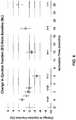
- a plurality of patients' heartswere paced at the ventricle and atrium using a pacing pattern that included 9 to 10 pulses having a first stimulation setting (according to which the ventricle was stimulated 40-90 milliseconds after atrial activation) and 1 to 2 pulses having a second stimulation setting (according to which the ventricle was stimulated 100-180 milliseconds after atrial activation). Pulses having the first stimulation setting reduced atrial kick, while pulses having the second stimulation setting did not do so.
- the pacing patternwas applied continuously for a period of up to about six months. Changes in end systolic volume (ESV), end diastolic volume (EDV), and ejection fraction (EF) were derived from echocardiogram data.
- ESVend systolic volume
- EDVend diastolic volume
- EFejection fraction
- end systolic volume (ESV)decreased by approximately 7 ml (about 15%)
- end diastolic volume (EDV)decreased by approximately 20 ml (about 17%)
- ejection fraction (EF)decreased slightly (about a 2% decrease).
- ESVend systolic volume
- EDVend diastolic volume
- EFejection fraction
- Cardiac volume (V)in turn is a function of R 3 , so T is a direct function of V 1/3 . Therefore, when V decreases by approximately 20%, R decreases by approximately 7%, and so does T. In other words, a 20% decrease in V is (0.8 ⁇ V), and (0.8) 1/3 equals 0.93, yielding (0.93 ⁇ R), or a 0.07 (or 7%) decrease in R. Extending this analysis to the at least 5% reduction suggested by the experimental observations, when V decreases by approximately 5%, R decreases by approximately 2%, and so does T.
- Figures 4-6illustrate a second set of experimental results according to a second embodiment, over a longer period of activation time (about twenty-four months), and including a greater number of patients. Similar to the first embodiment, in the experiment of the second embodiment, a plurality of patients' hearts were paced at the ventricle and atrium using a pacing pattern that included 9 to 10 pulses having a first stimulation setting (according to which the ventricle was stimulated 40-90 milliseconds after atrial activation) and 1 to 2 pulses having a second stimulation setting (according to which the ventricle was stimulated 100-180 milliseconds after atrial activation). Pulses having the first stimulation setting reduced atrial kick, while pulses having the second stimulation setting did not do so.
- the pacing patternwas applied continuously for a period of up to about twenty-four months.
- Changes in end systolic volume (ESV), end diastolic volume (EDV), and ejection fraction (EF)were derived from echocardiogram data.
- ESVend systolic volume
- EDVend diastolic volume
- EFejection fraction
- end systolic volume (ESV) and end diastolic volume (EDV)remained at reduced levels, with values at the twenty-four month decreased by about 9% and 8%, respectively, while ejection fraction (EF) initially decreased slightly and insignificantly, and later increased steadily (to about a 2% increase at twenty-four months).
- EFejection fraction
- a stimulation patternmay be configured to reduce the at least one of end systolic volume (ESV) and end diastolic volume (EDV) by at least 5% and maintain the at least one of end systolic volume (ESV) and end diastolic volume (EDV) on average at such reduced volume for a time period of at least one hour, including as long as three months, twenty-four months, or even longer.
- a method for treating cardiac malfunctionmay include delivering a stimulation pattern of stimulation pulses to at least one cardiac chamber of a heart. At least one of the stimulation pulses may have a first stimulation setting configured to reduce at least one of end systolic volume (ESV) and end diastolic volume (EDV) in the heart and at least one of the stimulation pulses may have a second stimulation setting different from the first stimulation setting.
- ESVend systolic volume
- EDVend diastolic volume
- the stimulation patternmay be configured to reduce the at least one of end systolic volume (ESV) and end diastolic volume (EDV) by at least 5% and maintain the at least one of end systolic volume (ESV) and end diastolic volume (EDV) on average at such reduced volume for a time period of at least one hour.
- Figure 7illustrates a method 700 for treating cardiac malfunction that includes delivering a stimulation pattern of stimulation pulses to at least one cardiac chamber of a heart.
- the methodmay begin by delivering at least one stimulation pulse having a first stimulation setting configured to reduce at least one of end systolic volume (ESV) and end diastolic volume (EDV) in the heart.
- the methodmay continue by delivering at least one stimulation pulse having a second stimulation setting different from the first stimulation setting.
- step 706reduces end systolic volume (ESV) and/or end diastolic volume (EDV) by at least 5% and in step 708 maintains the reduced end systolic volume (ESV) and/or end diastolic volume (EDV) on average at such reduced volume for an extended time period (e.g., at least one hour).
- ESVend systolic volume
- EDVend diastolic volume
- step 708maintains the reduced end systolic volume (ESV) and/or end diastolic volume (EDV) on average at such reduced volume for an extended time period (e.g., at least one hour).
- the time periodmay be at least three months.
- the methodmay include a stimulation pattern configured to reduce cardiac strain by at least 2% and maintain the cardiac strain on average at such reduced strain for the time period.
- the methodmay include a stimulation pattern configured to maintain blood pressure within an average pressure of ⁇ 10% as compared to a pretreatment blood pressure value for the time period.
- the methodmay include a stimulation pattern configured to prevent cardiac remodeling in the patient.
- the methodmay provide that the first stimulation setting is configured to reduce or prevent atrial kick in at least one ventricle and at least one of the stimulation pulses has a second stimulation setting different from the first stimulation setting.
- the methodmay provide that the first stimulation setting is configured to reduce the at least one of end systolic volume (ESV) and end diastolic volume (EDV) by at least 5% and the second stimulation setting is configured to reduce a baroreflex response or adaptation to the reduction in the at least one of end systolic volume (ESV) and end diastolic volume (EDV).
- the stimulation patternis configured not to activate a baroreceptor of the patient.
- the methodmay provide that the stimulation pattern is configured to reduce a baroreflex response or adaptation to the reduction in the at least one of end systolic volume (ESV) and end diastolic volume (EDV).
- ESVend systolic volume
- EDVend diastolic volume
- the methodmay provide that the first stimulation setting is configured to reduce the at least one of end systolic volume (ESV) and end diastolic volume (EDV) by at least 5% and the second stimulation setting is configured to reduce a neuronal response or adaptation to the reduction in the at least one of end systolic volume (ESV) and end diastolic volume (EDV).
- ESVend systolic volume
- EDVend diastolic volume
- EDVend diastolic volume
- the methodmay provide that the stimulation pattern is configured to reduce a neuronal response or adaptation to the reduction in the at least one of end systolic volume (ESV) and end diastolic volume (EDV).
- ESVend systolic volume
- EDVend diastolic volume
- the methodmay provide that the first stimulation setting is configured to reduce the at least one of end systolic volume (ESV) and end diastolic volume (EDV) by at least 5% and the second stimulation setting is configured to increase hormonal secretion.
- ESVend systolic volume
- EDVend diastolic volume
- the methodmay provide that the stimulation pattern is configured to increase hormonal secretion.
- the system of the present inventionprovides that the first stimulation setting comprises stimulating a ventricle of the heart 40-90 milliseconds after atrial activation.
- the methodmay provide that the second stimulation setting comprises stimulating an atrium of the heart to thereby produce atrial stimulation.
- the system of the present inventionprovides that the second stimulation setting comprises stimulating a ventricle of the heart 100-180 milliseconds after atrial activation.
- the methodmay provide that the second stimulation setting comprises allowing a natural AV delay to occur.
- the systemmay provide that the stimulation pattern includes at least 4 consecutive heartbeats having the first stimulation setting for every 1 consecutive heartbeat having the second stimulation setting.
- the systemmay provide that the stimulation pattern includes at least 8 consecutive heartbeats having the first stimulation setting for every 1 consecutive heartbeat having the second stimulation setting.
- the methodmay provide that the stimulation pattern comprises at least one stimulation pulse having a third stimulation setting different from the first and second stimulation settings.
- the methodmay provide that the cardiac malfunction is associated with an increase in at least one of end systolic volume (ESV) and end diastolic volume (EDV).
- ESVend systolic volume
- EDVend diastolic volume
- the methodmay provide that the cardiac malfunction is congestive heart failure.
- the methodmay further include applying the stimulation pattern at a stimulation pattern configuration for a first period, sensing at least one parameter indicative of at least one of end systolic volume (ESV), end diastolic volume (EDV), cardiac strain, and blood pressure for the first period, and adjusting the stimulation pattern configuration according to the sensing.
- ESVend systolic volume
- EDVend diastolic volume
- cardiac straincardiac strain
- blood pressurefor the first period
- adjusting the stimulation patternincludes adjusting at least one of the first stimulation setting and the second stimulation setting.
- adjusting the stimulation pattern configurationincludes adjusting at least one of the number and proportion of at least one of stimulation pulses having the first stimulation setting and stimulation pulses having the second stimulation setting within the stimulation pattern.
- the inventionprovides a system for treating cardiac malfunction.
- the systemincludes a stimulation circuit configured to deliver a stimulation pulse to at least one cardiac chamber of a heart of a patient, and at least one controller configured to execute delivery of a stimulation pattern of stimulation pulses to the at least one cardiac chamber.
- At least one of the stimulation pulseshave a first stimulation setting configured to reduce at least one of end systolic volume (ESV) and end diastolic volume (EDV) in the heart and at least one of the stimulation pulses have a second stimulation setting different from the first stimulation setting.
- ESVend systolic volume
- EDVend diastolic volume
- the stimulation patternmay be configured to reduce the at least one of end systolic volume (ESV) and end diastolic volume (EDV) by at least 5% and maintain the at least one of end systolic volume (ESV) and end diastolic volume (EDV) on average at such reduced volume for a time period of at least one hour.
- the at least one controllermay be configured to receive input data relating to at least one sensed parameter indicative of at least one of end systolic volume (ESV), end diastolic volume (EDV), cardiac strain, and blood pressure for the time period and to adjust the stimulation pattern configuration according to the at least one sensed parameter.
- ESVend systolic volume
- EDVend diastolic volume
- cardiac straincardiac strain
- blood pressureblood pressure
- systemmay further comprise at least one sensor configured to sense the at least one sensed parameter and to communicate the input data to the at least one controller.
- the at least one controller, the stimulation circuit, and the at least one sensormay be combined in a single device.
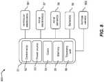
- FIG 8illustrates an embodiment of a system 800 for treating cardiac malfunction, which includes a stimulation circuit and at least one controller as described above.
- System 800may be constructed and have components similar to a cardiac pacemaker essentially as known in the art with some modifications as discussed herein.
- the system, or deviceis implantable.
- the systemcomprises components that may provide additional and/or alternative electrical treatments of the heart (e.g., defibrillation).
- System 800may be configured for implantation in the body of a patient essentially as is known in the art for implantable pacemakers, optionally with some modifications as discussed herein.
- System 800may include a biocompatible body 51, one or more controllers 52, a power source 53, and a telemetry unit 56.
- Body 51may comprise a housing for encasing a plurality of components of the system.
- Controller(s) 52may be configured to control the operation of the system, and may implement any of the embodiments and methods disclosed herein.
- controller(s) 52may control the delivery of stimulation pulses.
- power source 53may include a battery.
- power source 53may include a rechargeable battery.
- power source 53may include a battery that is rechargeable by induction.
- telemetry unit 56may be configured to communicate with one or more other units and/or components.
- telemetry unit 56may be configured to communicate with an external programmer and/or a receiving unit for receiving data recorded on system 800 during operation.
- system 800may include one or more electrodes and/or sensors.
- the electrodes and/or sensorsmay be integrated in system 800, attached thereto, and/or connectable therewith.
- the electrodesmay include ventricular electrode(s) 561 configured to pace at least one ventricle. Additionally or alternatively, the system may be connected, optionally via wires or wirelessly, to at least one implanted artificial valve 562.
- system 800may comprise one or more atrial electrode(s) 57 for pacing one or more atria, and/or one or more atrial sensors 58 for sensing the onset of atrial excitation, and/or one or more sensors 59 for providing other feedback parameters (e.g., a blood-pressure-related parameter and a cardiac strain/volume related parameter).
- atrial electrode(s) 57for pacing one or more atria
- atrial sensors 58for sensing the onset of atrial excitation
- sensors 59for providing other feedback parameters (e.g., a blood-pressure-related parameter and a cardiac strain/volume related parameter).
- sensor(s) 59may comprise one or more pressure sensors, electrical sensors (e.g., ECG monitoring), flow sensors, heart rate sensors, activity sensors, and/or volume sensors.
- Sensor(s) 59may include mechanical sensors and/or electronic sensors (e.g., ultrasound sensors, electrodes, and/or RF transceivers).
- sensor(s) 59may communicate with system 800 via telemetry.
- ventricular electrode(s) 561 and/or atrial electrode(s) 57may be standard pacing electrodes.
- Ventricular electrode(s) 561may be positioned relative to the heart at positions as known in the art for ventricular pacing.
- ventricular electrode(s)may be placed in and/or near one or more of the ventricles.
- atrial electrode(s) 57may be placed in and/or near one or more of the atria.
- atrial electrode(s) 57may be attached to the one or more atria at one or more positions selected to provide early detection of atrial excitation or depolarization.
- atrial electrode(s) 57may be attached to the right atrium near the site of the sinoatrial (SA) node.
- SAsinoatrial
- One position of ventricular electrode(s) 561may be such that pacing may reduce or minimize the prolongation of QRS when the heart is paced, to reduce or even minimize dyssynchrony. In some embodiments, this position is on the ventricular septum near the Bundle of His. Ventricular electrode(s) 561 may additionally or alternatively be placed on the epicardium of the heart or in coronary veins. More than one electrode can be placed on the ventricles to provide biventricular pacing, optionally to reduce dyssynchrony.
- System 800may include a pulse generator, or stimulation circuit, configured to deliver a stimulation pulse to at least one cardiac chamber.
- the pulse generator, or stimulation circuitmay include some or all standard capabilities of a conventional pacemaker.
- Controller 52may be configured to control the pulse generator, or stimulation circuit.
- Atrial sensor(s) 58(and optionally other electrode sensors configured to sense other heart chambers) may be connected to system 800 via specific circuits that amplify the electrical activity of the heart and allow sampling and detection of the activation of the specific chamber.
- Other circuitsmay be configured to deliver stimulation to a specific electrode to pace the heart, creating propagating electrical activation.
- one or more additional sensors 59may be placed in and/or on one or more of the atria and/or in and/or on one or more of the ventricles and/or in and/or on one or more other locations that might optionally be adjacent the heart.
- one or more sensorsmay be placed on and/or in vena cava and/or on one or more arteries and/or within one or more cardiac chambers. These sensors may measure pressure, or other indicators, such as, for example, impedance and/or flow.
- controller 52may comprise or be a microprocessor powered by power source 53.
- system 800may comprise a clock 54, for example, generated by a crystal.
- System 800may comprise an internal memory 55 and/or be connected to external memory.
- devicemay connect to an external memory via telemetry unit 56.
- telemetry unit 56may be configured to allow communication with external devices such as a programmer and/or one or more of sensors 59. Any and all feedback information and/or a log of device operation may be stored in internal memory 55 and/or relayed by telemetry unit 56 to an external memory unit.
- controller 52may operate in accordance with at least one embodiment of a method described herein.
- system 800may comprise one or more sensors for sensing one or more feedback parameters to control the application of the AV delay and/or its magnitude.
Landscapes
- Health & Medical Sciences (AREA)
- Cardiology (AREA)
- Life Sciences & Earth Sciences (AREA)
- Heart & Thoracic Surgery (AREA)
- Engineering & Computer Science (AREA)
- Biomedical Technology (AREA)
- Nuclear Medicine, Radiotherapy & Molecular Imaging (AREA)
- Radiology & Medical Imaging (AREA)
- Animal Behavior & Ethology (AREA)
- General Health & Medical Sciences (AREA)
- Public Health (AREA)
- Veterinary Medicine (AREA)
- Hematology (AREA)
- Physiology (AREA)
- Biophysics (AREA)
- Hospice & Palliative Care (AREA)
- Physics & Mathematics (AREA)
- Fluid Mechanics (AREA)
- Electrotherapy Devices (AREA)
Description
- The present invention relates to the field of treating cardiac malfunction, and more particularly, to systems for stimulating the heart to treat cardiac malfunction, such as congestive heart failure.
- A system for stimulating a heart to treat cardiac malfunction according to the preamble of
claim 1 is known fromUS 7 363 077 B1 . - Methods and systems for treating cardiac malfunction are disclosed.
- A method for treating cardiac malfunction may include delivering a stimulation pattern of stimulation pulses to at least one cardiac chamber of a heart. At least one of the stimulation pulses may have a first stimulation setting configured to reduce at least one of end systolic volume (ESV) and end diastolic volume (EDV) in the heart, and at least one of the stimulation pulses may have a second stimulation setting different from the first stimulation setting. The stimulation pattern may be configured to reduce the at least one of end systolic volume (ESV) and end diastolic volume (EDV) by at least 5% and to maintain the at least one of end systolic volume (ESV) and end diastolic volume (EDV) on average at such reduced volume for a time period of at least one hour.
- The invention provides a system for treating cardiac malfunction as defined in
claim 1. This system for treating cardiac malfunction includes a stimulation circuit and at least one controller. The stimulation circuit is configured to deliver a stimulation pulse to at least one cardiac chamber of a heart of a patient. The at least one controller is configured to execute delivery of a stimulation pattern of stimulation pulses to the at least one cardiac chamber. At least one of the stimulation pulses has a first stimulation setting configured to reduce at least one of end systolic volume (ESV) and end diastolic volume (EDV) in the heart, and at least one of the stimulation pulses has a second stimulation setting different from the first stimulation setting. The stimulation pattern as defined inclaim 1 is configured to reduce the at least one of end systolic volume (ESV) and end diastolic volume (EDV) by at least 5% and maintain the at least one of the end systolic volume (ESV) and end diastolic volume (EDV) on average at such reduced volume for a time period of at least one hour. - Advantages of the present invention will be, or will become, apparent to one of ordinary skill in the art upon examination of the following figures and detailed description.
- The embodiments can be better understood with reference to the following drawings and description. The components in the figures are not necessarily to scale, emphasis instead being placed upon illustrating the principles of the embodiments. Moreover, in the figures, like reference numerals designate corresponding parts throughout the different views.
FIG. 1 is a plot illustrating change in end systolic volume (ESV) from baseline (BL) as a function of activation time, according to an embodiment;FIG. 2 is a plot illustrating change in end diastolic volume (EDV) from baseline (BL) as a function of activation time, according to an embodiment;FIG. 3 is a plot illustrating change in ejection fraction (EF) from baseline (BL) as a function of activation time, according to an embodiment;FIG. 4 is a plot illustrating change in end systolic volume (ESV) from baseline (BL) as a function of activation time over a longer period (about twenty-four months), according to a second embodiment;FIG. 5 a plot illustrating change in end diastolic volume (EDV) from baseline (BL) as a function of activation time over the longer period (about twenty-four months), according to the second embodiment;FIG. 6 is a plot illustrating change in ejection fraction (EF) from baseline (BL) as a function of activation time over the longer period (about twenty-four months), according to the second embodiment;FIG. 7 is a flow chart illustrating an exemplary method for treating cardiac malfunction; andFIG. 8 is a schematic drawing illustrating an exemplary system for treating cardiac malfunction, which may perform one or more of the methods described herein, such as the method ofFIG. 7 .- A healthy cardiac contraction cycle includes two phases: a systole and a diastole. The systole is a period of cardiac contraction, commencing with ventricular contraction. The systole causes blood to be ejected from the heart and into the vascular system. At the end of this contraction, cardiac muscles relax. This period of relaxation is the diastole. During diastole, the heart passively fills with blood from the vascular system and at the end of diastole the atria contract and provide additional filling to the ventricle. Accordingly, at the end of the diastole and just before the heart begins to contract, cardiac blood volume peaks. This peak volume is the end diastolic volume (EDV). At the end of the systole, when contraction ends and cardiac filling is about to commence, cardiac blood volume reaches a minimal value. This minimal volume is the end systolic volume (ESV). The amount of blood ejected in a heartbeat is known as the stroke volume (SV) and equals the difference between end diastolic volume (EDV) and end systolic volume (ESV). The ejection fraction (EF) is the fraction of blood that is ejected by the heart (i.e., SV divided by EDV).
- Cardiac remodeling is a phenomenon that results from cardiac load or injury, and is an accepted determinant of the clinical course of heart failure (HF). It manifests clinically as changes in size, shape, and function of the heart.
- As a result of damage to the heart, abnormal strain patterns are manifested in the heart, and the cells that experience a high strain undergo hypertrophy and some loss of function. This affects cardiac function and blood pressure parameters, and as a consequence, neuronal and/or hormonal pathways are activated in an attempt to compensate for cardiac damage. For example, poor contraction results in high end systolic volume (ESV) and thus a reduction in stroke volume (SV). This may lead to increase in end diastolic volume (EDV) as well. As cardiac volumes increase, so does the wall tension in the heart, as known from Laplace's Law. According to the law, a dilated ventricle requires more tension in the wall to generate the same pressure as would a smaller ventricle. This increase in tension increases sympathetic stimulation and vasopressin (antidiuretic hormone or ADH) secretion. Vasopressin is known to constrict blood vessels and increase heart rate, which causes an increase in blood pressure and an increase in oxygen consumption by the heart muscle cells. However, this also causes an additional increase in cardiac strain, since the heart beats faster and in every systole needs to eject blood against a system showing an increased resistance. This increased strain may cause additional hypertrophy and further loss of function. Thus, a vicious cycle comes into play where the cardiovascular system's attempts at reducing the effect of the damaged tissue cause additional reduction in cardiac performance.
- Many attempts have been made to develop devices and methods to treat heart failure, including, for example, devices intended to mechanically control cardiac volumes:
U.S. Patent No. 7,651,461 to Alferness et al. , entitled "Cardiac Support with Metallic Structure," describes a jacket "configured to surround the myocardium" which "provides reduced expansion of the heart wall during diastole by applying constraining surfaces at least at diametrically opposing aspects of the heart;" andU.S. Patent No. 7,618,364 to Walsh et al. , entitled "Cardiac Wall Tension Relief Device and Method," states that "[i]t is believed that such resistance decreases wall tension on the heart and permits a diseased heart to beneficially remodel."- Interfering with cardiac filling may reduce blood pressure in hypertensive patients. One option for interfering with cardiac filling to reduce blood pressure may be to reduce or even eliminate atrial kick. Atrial kick may provide a relatively small boost to ventricle filling (10-30%) that is caused by atrial contraction before an atrioventricular (AV) valve between the atrium and the ventricle is closed. Once the ventricle begins to contract, pressure builds up in the ventricle and causes the AV valve to passively close. The inventors also found that when effecting treatment that reduces or even eliminates atrial kick, the cardiovascular system acts to adapt to the change and return performance values to those that occurred before treatment commenced.
International Publications Nos. WO2015/094401 andWO2014/100429, both to Mika et al . describe, inter alia, methods and systems providing pacing patterns of stimulation pulses that comprise pulses configured to reduce or prevent atrial kick and also reduce or even eliminate the cardiovascular system's adaptation to the reduction in blood pressure. The reduction in adaptation may be achieved by reducing neurohormonal response to changes in generated pressure and stretch using specific patterns. - Heart failure patients, on the other hand, typically (although not exclusively) have blood pressure values that are not significantly elevated. Accordingly, in some cases, it may be preferred to reduce cardiac volumes (e.g., at least one of end diastolic volume and end systolic volume) and/or reduce cardiac strain without significantly affecting blood pressure.
- The inventors have developed methods and systems of applying cardiac stimulations that yield surprising results in meeting the needs of heart failure patients. Embodiments provide methods and system that may reduce the strain sensed by cardiac muscles by applying cardiac stimulations that reduce at least one of end systolic volume (ESV) and end diastolic volume (EDV) without significantly affecting the ejection fraction (EF) of the heart, and optionally also without significantly affecting blood pressure. A reduction of such strain may be useful in reversing cardiac remodeling or at least in slowing down or even stopping the remodeling process. In some embodiments, a stimulation pattern may be configured to reduce a neurohormonal response to changes in strain without significantly affecting blood pressure.
- Optionally, when a patient is provided with a cardiac stimulation device, a stimulation pattern is selected based on sensing of one or more of: at least one blood-pressure-related parameter (e.g., an indication of pretreatment and/or treatment blood pressure) and at least one cardiac-strain and/or volume-related parameter (e.g., a pressure measurement within a chamber and/or echocardiogram-derived information). These parameters can be used to adjust one or more of the properties of one or more (or two or more) pulses in a pulse pattern and/or the proportion between pulses having different settings and/or the order of the pulses in the pattern. Optionally, reducing at least one of end diastolic volume (EDV) and end systolic volume (ESV) suppresses sympathetic stimulation, for example, by at least one of reducing wall strain and hence reducing or eliminating the activation of sympathetic pathways involved with adaptation of the cardiovascular system to changes.
- Optionally, a device is configured to receive feedback information regarding one or more of the at least one blood-pressure-related parameter and at least one cardiac strain/volume related parameter. This may be performed periodically (e.g., in a periodic measurement such as a periodic checkup) and/or ongoing (e.g., with an associated or integral sensor). The information may be received, for example, via communication with a user through an input interface and/or by communication with implanted and/or external devices. Further details of suitable devices are described below in reference to
Figure 8 . - A stimulation setting means one or more parameters of one or more stimulation pulses delivered in a single cardiac cycle. For example, these parameters may include one or more of: a time interval between electrical pulses that are included in a single stimulation pulse (e.g., AV delay), a period of delivery with respect to the natural rhythm of the heart, the length of a stimulation pulse or a portion thereof, and the site of delivery between two or more chambers. In some embodiments, a pulse setting includes applying an excitatory pulse to a ventricle, timed in synchronization to the natural activity of an atrium of the heart. In some embodiments, a pulse setting includes applying an excitatory pulse to an atrium timed in synchronization to the natural activity of a ventricle of the heart. In some embodiments, a pulse setting includes applying an excitatory pulse to each of a ventricle and an atrium.
- A stimulation pattern may include a series of pulses having identical stimulation settings or a stimulation pattern may include multiple pulses each having different stimulation settings. For example, a stimulation pattern may have one or more pulses having a first setting and one or more pulses having a second setting that is different from the first setting. When stating that a stimulation pattern has a setting, it is understood that this means a stimulation pattern may include at least one stimulation pulse having that setting. It is also understood that, in some embodiments a stimulation pattern may include one or more cardiac cycles where no stimulation pulse is delivered, in which case the pulse(s) may be viewed as being delivered at zero power. A stimulation pattern may include a plurality of identical pulses or a sequence of pulses including two or more different settings. Two stimulation sequences in a pattern may differ in the order of pulses provided within a setting. Two or more stimulation sequences may optionally differ in their lengths (in time and/or number of heartbeats). In some embodiments, a stimulation pattern may include pulses having blood pressure reduction (BPR) settings. In some embodiments, a stimulation pattern may include pulses that do not have BPR settings.
- In an experiment according to a first embodiment, a plurality of patients' hearts were paced at the ventricle and atrium using a pacing pattern that included 9 to 10 pulses having a first stimulation setting (according to which the ventricle was stimulated 40-90 milliseconds after atrial activation) and 1 to 2 pulses having a second stimulation setting (according to which the ventricle was stimulated 100-180 milliseconds after atrial activation). Pulses having the first stimulation setting reduced atrial kick, while pulses having the second stimulation setting did not do so. The pacing pattern was applied continuously for a period of up to about six months. Changes in end systolic volume (ESV), end diastolic volume (EDV), and ejection fraction (EF) were derived from echocardiogram data.
- Before treatment, end systolic volume (ESV) and end diastolic volume (EDV) were measured and ejection fraction (EF) was calculated to provide baseline (BL) values for each patient. Thereafter, the same values were obtained approximately one, two, three, and six months after treatment commenced, and the change from baseline (BL) for each patient was calculated. Each of
Figures 1-3 depicts results obtained for a plurality of patients (N), as indicated on the plots. The numbers (N) of patients includes only data for which a core lab analysis determined that the echocardiogram was suitable for measurement. The baseline values were approximately 110 ml for diastolic volume, approximately 45 ml for systolic volume, and approximately 62% for ejection fraction. - As shown in
Figures 1-3 , for a period of up to about six months, end systolic volume (ESV) decreased by approximately 7 ml (about 15%), end diastolic volume (EDV) decreased by approximately 20 ml (about 17%), and ejection fraction (EF) decreased slightly (about a 2% decrease). This means that muscle strain was reduced (due to the lower volume of blood in the chambers), while the heart's efficiency (as shown by the ejection fraction (EF)) remained almost unchanged. A reduction of strain works against the vicious cycle of the heart in which cardiac strain plays a significant role, especially when ejection fraction (EF) is not significantly reduced, and thus remodeling is prevented or at least its progress rate is reduced. Experimental observations suggest that beneficial effects may be obtained over a shorter period, for example, reducing at least one of end systolic volume (ESV) and end diastolic volume (EDV) by at least 5% and maintaining the at least one of end systolic volume (ESV) and end diastolic volume (EDV) on average at such reduced volume for a time period of at least one hour. In particular, according to Laplace's Law, pressure (P) is directly proportional to wall tension (T) and inversely proportional to radius (R), such that P α T/R. This means that, as long as pressure (P) is maintained essentially constant, wall tension (T) is a direct function of radius (R). Cardiac volume (V) in turn is a function of R3, so T is a direct function of V1/3. Therefore, when V decreases by approximately 20%, R decreases by approximately 7%, and so does T. In other words, a 20% decrease in V is (0.8 × V), and (0.8)1/3 equals 0.93, yielding (0.93 × R), or a 0.07 (or 7%) decrease in R. Extending this analysis to the at least 5% reduction suggested by the experimental observations, when V decreases by approximately 5%, R decreases by approximately 2%, and so does T. Figures 4-6 illustrate a second set of experimental results according to a second embodiment, over a longer period of activation time (about twenty-four months), and including a greater number of patients. Similar to the first embodiment, in the experiment of the second embodiment, a plurality of patients' hearts were paced at the ventricle and atrium using a pacing pattern that included 9 to 10 pulses having a first stimulation setting (according to which the ventricle was stimulated 40-90 milliseconds after atrial activation) and 1 to 2 pulses having a second stimulation setting (according to which the ventricle was stimulated 100-180 milliseconds after atrial activation). Pulses having the first stimulation setting reduced atrial kick, while pulses having the second stimulation setting did not do so. The pacing pattern was applied continuously for a period of up to about twenty-four months. Changes in end systolic volume (ESV), end diastolic volume (EDV), and ejection fraction (EF) were derived from echocardiogram data.- Before treatment, end systolic volume (ESV) and end diastolic volume (EDV) were measured and ejection fraction (EF) was calculated to provide baseline (BL) values for each patient. Thereafter, the same values were obtained eight times after treatment commenced, and the change from baseline (BL) for each patient was calculated. The eight times were at approximately the following months after treatment commenced: one, two, three, six, twelve, eighteen, and twenty-four. Each of
Figures 4-6 depicts results obtained for a plurality of patients (N), as indicated on the plots. The numbers (N) of patients includes only data for which a core lab analysis determined that the echocardiogram was suitable for measurement. The baseline values were approximately 115 ml for diastolic volume and approximately 40 ml for systolic volume. - As shown in
Figures 4-6 , for a period of up to about twenty-four months, end systolic volume (ESV) and end diastolic volume (EDV) remained at reduced levels, with values at the twenty-four month decreased by about 9% and 8%, respectively, while ejection fraction (EF) initially decreased slightly and insignificantly, and later increased steadily (to about a 2% increase at twenty-four months). This means that muscle strain was reduced (due to the lower volume of blood in the chambers), while the heart's efficiency (as shown by the ejection fraction (EF)) improved slightly. A reduction of strain works against the vicious cycle of the heart in which cardiac strain plays a significant role, especially when ejection fraction (EF) improves, and thus remodeling is prevented or at least its progress rate is reduced. The longer term data ofFigures 4-6 therefore demonstrates a slight increase in ejection fraction (-60%) with normal global cardiac function over a period of twenty-four months, which suggests a trend toward continuing improvement of cardiac function beyond that longer term. Thus, in embodiments, a stimulation pattern may be configured to reduce the at least one of end systolic volume (ESV) and end diastolic volume (EDV) by at least 5% and maintain the at least one of end systolic volume (ESV) and end diastolic volume (EDV) on average at such reduced volume for a time period of at least one hour, including as long as three months, twenty-four months, or even longer. - In light of the experimental results, embodiments provide methods and systems for treating cardiac malfunction. A method for treating cardiac malfunction may include delivering a stimulation pattern of stimulation pulses to at least one cardiac chamber of a heart. At least one of the stimulation pulses may have a first stimulation setting configured to reduce at least one of end systolic volume (ESV) and end diastolic volume (EDV) in the heart and at least one of the stimulation pulses may have a second stimulation setting different from the first stimulation setting. The stimulation pattern may be configured to reduce the at least one of end systolic volume (ESV) and end diastolic volume (EDV) by at least 5% and maintain the at least one of end systolic volume (ESV) and end diastolic volume (EDV) on average at such reduced volume for a time period of at least one hour.
Figure 7 illustrates amethod 700 for treating cardiac malfunction that includes delivering a stimulation pattern of stimulation pulses to at least one cardiac chamber of a heart. As shown, instep 702, the method may begin by delivering at least one stimulation pulse having a first stimulation setting configured to reduce at least one of end systolic volume (ESV) and end diastolic volume (EDV) in the heart. Instep 704, the method may continue by delivering at least one stimulation pulse having a second stimulation setting different from the first stimulation setting. Through the delivery of the stimulation pattern of stimulation pulses ofsteps step 706 reduces end systolic volume (ESV) and/or end diastolic volume (EDV) by at least 5% and instep 708 maintains the reduced end systolic volume (ESV) and/or end diastolic volume (EDV) on average at such reduced volume for an extended time period (e.g., at least one hour).- The time period may be at least three months.
- The method may include a stimulation pattern configured to reduce cardiac strain by at least 2% and maintain the cardiac strain on average at such reduced strain for the time period.
- The method may include a stimulation pattern configured to maintain blood pressure within an average pressure of ±10% as compared to a pretreatment blood pressure value for the time period.
- The method may include a stimulation pattern configured to prevent cardiac remodeling in the patient.
- The method may provide that the first stimulation setting is configured to reduce or prevent atrial kick in at least one ventricle and at least one of the stimulation pulses has a second stimulation setting different from the first stimulation setting.
- The method may provide that the first stimulation setting is configured to reduce the at least one of end systolic volume (ESV) and end diastolic volume (EDV) by at least 5% and the second stimulation setting is configured to reduce a baroreflex response or adaptation to the reduction in the at least one of end systolic volume (ESV) and end diastolic volume (EDV). In some embodiments, the stimulation pattern is configured not to activate a baroreceptor of the patient.
- The method may provide that the stimulation pattern is configured to reduce a baroreflex response or adaptation to the reduction in the at least one of end systolic volume (ESV) and end diastolic volume (EDV).
- The method may provide that the first stimulation setting is configured to reduce the at least one of end systolic volume (ESV) and end diastolic volume (EDV) by at least 5% and the second stimulation setting is configured to reduce a neuronal response or adaptation to the reduction in the at least one of end systolic volume (ESV) and end diastolic volume (EDV).
- The method may provide that the stimulation pattern is configured to reduce a neuronal response or adaptation to the reduction in the at least one of end systolic volume (ESV) and end diastolic volume (EDV).
- The method may provide that the first stimulation setting is configured to reduce the at least one of end systolic volume (ESV) and end diastolic volume (EDV) by at least 5% and the second stimulation setting is configured to increase hormonal secretion.
- The method may provide that the stimulation pattern is configured to increase hormonal secretion.
- The system of the present invention provides that the first stimulation setting comprises stimulating a ventricle of the heart 40-90 milliseconds after atrial activation.
- The method may provide that the second stimulation setting comprises stimulating an atrium of the heart to thereby produce atrial stimulation.
- The system of the present invention provides that the second stimulation setting comprises stimulating a ventricle of the heart 100-180 milliseconds after atrial activation.
- The method may provide that the second stimulation setting comprises allowing a natural AV delay to occur.
- The system may provide that the stimulation pattern includes at least 4 consecutive heartbeats having the first stimulation setting for every 1 consecutive heartbeat having the second stimulation setting.
- The system may provide that the stimulation pattern includes at least 8 consecutive heartbeats having the first stimulation setting for every 1 consecutive heartbeat having the second stimulation setting.
- The method may provide that the stimulation pattern comprises at least one stimulation pulse having a third stimulation setting different from the first and second stimulation settings.
- The method may provide that the cardiac malfunction is associated with an increase in at least one of end systolic volume (ESV) and end diastolic volume (EDV).
- The method may provide that the cardiac malfunction is congestive heart failure.
- The method may further include applying the stimulation pattern at a stimulation pattern configuration for a first period, sensing at least one parameter indicative of at least one of end systolic volume (ESV), end diastolic volume (EDV), cardiac strain, and blood pressure for the first period, and adjusting the stimulation pattern configuration according to the sensing.
- The method referred to in the preceding paragraph may provide that adjusting the stimulation pattern includes adjusting at least one of the first stimulation setting and the second stimulation setting.
- The methods referred to in the two preceding paragraphs may provide that adjusting the stimulation pattern configuration includes adjusting at least one of the number and proportion of at least one of stimulation pulses having the first stimulation setting and stimulation pulses having the second stimulation setting within the stimulation pattern.
- The invention provides a system for treating cardiac malfunction. The system includes a stimulation circuit configured to deliver a stimulation pulse to at least one cardiac chamber of a heart of a patient, and at least one controller configured to execute delivery of a stimulation pattern of stimulation pulses to the at least one cardiac chamber. At least one of the stimulation pulses have a first stimulation setting configured to reduce at least one of end systolic volume (ESV) and end diastolic volume (EDV) in the heart and at least one of the stimulation pulses have a second stimulation setting different from the first stimulation setting. The stimulation pattern may be configured to reduce the at least one of end systolic volume (ESV) and end diastolic volume (EDV) by at least 5% and maintain the at least one of end systolic volume (ESV) and end diastolic volume (EDV) on average at such reduced volume for a time period of at least one hour.
- In a further aspect of the system, the at least one controller may be configured to receive input data relating to at least one sensed parameter indicative of at least one of end systolic volume (ESV), end diastolic volume (EDV), cardiac strain, and blood pressure for the time period and to adjust the stimulation pattern configuration according to the at least one sensed parameter.
- In a further aspect of the system, the system may further comprise at least one sensor configured to sense the at least one sensed parameter and to communicate the input data to the at least one controller.
- In a further aspect of the system, the at least one controller, the stimulation circuit, and the at least one sensor may be combined in a single device.
Figure 8 illustrates an embodiment of asystem 800 for treating cardiac malfunction, which includes a stimulation circuit and at least one controller as described above.System 800 may be constructed and have components similar to a cardiac pacemaker essentially as known in the art with some modifications as discussed herein. Optionally, the system, or device, is implantable. Optionally, the system comprises components that may provide additional and/or alternative electrical treatments of the heart (e.g., defibrillation).System 800 may be configured for implantation in the body of a patient essentially as is known in the art for implantable pacemakers, optionally with some modifications as discussed herein.System 800 may include abiocompatible body 51, one ormore controllers 52, apower source 53, and atelemetry unit 56.Body 51 may comprise a housing for encasing a plurality of components of the system. Controller(s) 52 may be configured to control the operation of the system, and may implement any of the embodiments and methods disclosed herein. For example, controller(s) 52 may control the delivery of stimulation pulses. In some embodiments,power source 53 may include a battery. For example,power source 53 may include a rechargeable battery. In some embodiments,power source 53 may include a battery that is rechargeable by induction. In some embodiments,telemetry unit 56 may be configured to communicate with one or more other units and/or components. For example,telemetry unit 56 may be configured to communicate with an external programmer and/or a receiving unit for receiving data recorded onsystem 800 during operation.- In some embodiments,
system 800 may include one or more electrodes and/or sensors. The electrodes and/or sensors may be integrated insystem 800, attached thereto, and/or connectable therewith. In some embodiments, the electrodes may include ventricular electrode(s) 561 configured to pace at least one ventricle. Additionally or alternatively, the system may be connected, optionally via wires or wirelessly, to at least one implantedartificial valve 562. Additionally,system 800 may comprise one or more atrial electrode(s) 57 for pacing one or more atria, and/or one or moreatrial sensors 58 for sensing the onset of atrial excitation, and/or one ormore sensors 59 for providing other feedback parameters (e.g., a blood-pressure-related parameter and a cardiac strain/volume related parameter). - In some embodiments, sensor(s) 59 may comprise one or more pressure sensors, electrical sensors (e.g., ECG monitoring), flow sensors, heart rate sensors, activity sensors, and/or volume sensors. Sensor(s) 59 may include mechanical sensors and/or electronic sensors (e.g., ultrasound sensors, electrodes, and/or RF transceivers). In some embodiments, sensor(s) 59 may communicate with
system 800 via telemetry. - In some embodiments, ventricular electrode(s) 561 and/or atrial electrode(s) 57 may be standard pacing electrodes. Ventricular electrode(s) 561 may be positioned relative to the heart at positions as known in the art for ventricular pacing. For example, ventricular electrode(s) may be placed in and/or near one or more of the ventricles. In some embodiments, atrial electrode(s) 57 may be placed in and/or near one or more of the atria. In some embodiments, atrial electrode(s) 57 may be attached to the one or more atria at one or more positions selected to provide early detection of atrial excitation or depolarization. For example, in some embodiments, atrial electrode(s) 57 may be attached to the right atrium near the site of the sinoatrial (SA) node.
- One position of ventricular electrode(s) 561 may be such that pacing may reduce or minimize the prolongation of QRS when the heart is paced, to reduce or even minimize dyssynchrony. In some embodiments, this position is on the ventricular septum near the Bundle of His. Ventricular electrode(s) 561 may additionally or alternatively be placed on the epicardium of the heart or in coronary veins. More than one electrode can be placed on the ventricles to provide biventricular pacing, optionally to reduce dyssynchrony.
System 800 may include a pulse generator, or stimulation circuit, configured to deliver a stimulation pulse to at least one cardiac chamber. The pulse generator, or stimulation circuit, may include some or all standard capabilities of a conventional pacemaker.Controller 52 may be configured to control the pulse generator, or stimulation circuit. Atrial sensor(s) 58 (and optionally other electrode sensors configured to sense other heart chambers) may be connected tosystem 800 via specific circuits that amplify the electrical activity of the heart and allow sampling and detection of the activation of the specific chamber. Other circuits may be configured to deliver stimulation to a specific electrode to pace the heart, creating propagating electrical activation.- In some embodiments, one or more
additional sensors 59 may be placed in and/or on one or more of the atria and/or in and/or on one or more of the ventricles and/or in and/or on one or more other locations that might optionally be adjacent the heart. For example, one or more sensors may be placed on and/or in vena cava and/or on one or more arteries and/or within one or more cardiac chambers. These sensors may measure pressure, or other indicators, such as, for example, impedance and/or flow. - In some embodiments,
controller 52 may comprise or be a microprocessor powered bypower source 53. In some embodiments,system 800 may comprise aclock 54, for example, generated by a crystal.System 800 may comprise aninternal memory 55 and/or be connected to external memory. For example, device may connect to an external memory viatelemetry unit 56. In some embodiments,telemetry unit 56 may be configured to allow communication with external devices such as a programmer and/or one or more ofsensors 59. Any and all feedback information and/or a log of device operation may be stored ininternal memory 55 and/or relayed bytelemetry unit 56 to an external memory unit. - In some embodiments,
controller 52 may operate in accordance with at least one embodiment of a method described herein. - In some embodiments,
system 800 may comprise one or more sensors for sensing one or more feedback parameters to control the application of the AV delay and/or its magnitude. - According to further embodiments, additional systems and devices suitable for implementing the methods presented herein are described in
U.S. Patent No. 9,370,662 to Mika et al., issued June 21, 2016
Claims (4)
- A system for treating cardiac malfunction, comprising:a stimulation circuit configured to deliver a stimulation pulse to at least one cardiac chamber of a heart of a patient; andat least one controller (52) configured to execute delivery of a stimulation pattern of stimulation pulses to the at least one cardiac chamber,wherein at least one of the stimulation pulses has a first stimulation setting configured to reduce at least one of end systolic volume (ESV) and end diastolic volume (EDV) in the heart and at least one of the stimulation pulses has a second stimulation setting different from the first stimulation setting,wherein the controller (52) is arranged to deliver the stimulation pattern having the stimulation pulses with the first stimulation setting stimulating a ventricle 40- 90 ms after atrial activation, and having the stimulation pulses with the second stimulation setting stimulating the ventricle 100-180 ms after atrial activation, such as to reduce the at least one of end systolic volume (ESV) and end diastolic volume (EDV) by at least 5% and maintain the at least one of end systolic volume (ESV) and end diastolic volume (EDV) on average at such reduced volume for a time period of at least one hour,characterized in that the controller is arranged to deliver the stimulation pattern having the stimulation pulses with the first stimulation setting and the stimulation pulses with the second stimulation setting such as to have 9 or 10 pulses having the first stimulation setting and 1 or 2 pulses having the second stimulation setting.
- The system of claim 1, wherein the at least one controller (52) is configured to receive input data relating to at least one sensed parameter indicative of at least one of end systolic volume (ESV), end diastolic volume (EDV), cardiac strain, and blood pressure for the time period and to adjust the stimulation pattern configuration according to the at least one sensed parameter.
- The system of claim 2, further comprising at least one sensor (58, 59) configured to sense the at least one sensed parameter and to communicate the input data to the at least one controller (52).
- The system of claim 3, wherein the at least one controller (52), the stimulation circuit, and the at least one sensor are combined in a single device.
Applications Claiming Priority (3)
| Application Number | Priority Date | Filing Date | Title |
|---|---|---|---|
| US201562217299P | 2015-09-11 | 2015-09-11 | |
| US15/259,282US10342982B2 (en) | 2015-09-11 | 2016-09-08 | Methods and systems for treating cardiac malfunction |
| PCT/US2016/051023WO2017044794A1 (en) | 2015-09-11 | 2016-09-09 | Methods and systems for treating cardiac malfunction |
Publications (3)
| Publication Number | Publication Date |
|---|---|
| EP3347090A1 EP3347090A1 (en) | 2018-07-18 |
| EP3347090A4 EP3347090A4 (en) | 2019-04-24 |
| EP3347090B1true EP3347090B1 (en) | 2021-11-03 |
Family
ID=58240156
Family Applications (1)
| Application Number | Title | Priority Date | Filing Date |
|---|---|---|---|
| EP16845150.8AActiveEP3347090B1 (en) | 2015-09-11 | 2016-09-09 | Systems for treating cardiac malfunction |
Country Status (9)
| Country | Link |
|---|---|
| US (2) | US10342982B2 (en) |
| EP (1) | EP3347090B1 (en) |
| JP (1) | JP6999545B2 (en) |
| KR (1) | KR102630590B1 (en) |
| CN (1) | CN108025173B (en) |
| AU (1) | AU2016319787B2 (en) |
| CA (1) | CA2996312C (en) |
| ES (1) | ES2898781T3 (en) |
| WO (1) | WO2017044794A1 (en) |
Cited By (8)
| Publication number | Priority date | Publication date | Assignee | Title |
|---|---|---|---|---|
| US11389658B2 (en) | 2015-09-11 | 2022-07-19 | Backbeat Medical, Llc | Methods and systems for treating cardiac malfunction |
| US11406829B2 (en) | 2004-02-12 | 2022-08-09 | Backbeat Medical, Llc | Cardiac stimulation apparatus and method for the control of hypertension |
| US11426589B2 (en) | 2016-04-22 | 2022-08-30 | Backbeat Medical, Llc | Methods and systems for controlling blood pressure |
| US11452875B2 (en) | 2012-12-21 | 2022-09-27 | Backbeat Medical, Llc | Methods and systems for lowering blood pressure through reduction of ventricle filling |
| US11529520B2 (en) | 2006-09-25 | 2022-12-20 | Backbeat Medical, Llc | Methods and apparatus to stimulate heart atria |
| US11577059B2 (en) | 2005-03-02 | 2023-02-14 | Backbeat Medical, Llc | Methods and apparatus to increase secretion of endogenous naturetic hormones |
| US11759639B2 (en) | 2008-09-08 | 2023-09-19 | Backbeat Medical, Llc | Methods and apparatus to stimulate the heart |
| US12440653B2 (en) | 2023-01-09 | 2025-10-14 | Backbeat Medical, Llc | Methods and apparatus to increase secretion of endogenous naturetic hormones |
Families Citing this family (1)
| Publication number | Priority date | Publication date | Assignee | Title |
|---|---|---|---|---|
| AU2018365166A1 (en)* | 2017-11-13 | 2020-06-04 | Promptu Systems Corporation | Systems and methods for adaptive proper name entity recognition and understanding |
Citations (1)
| Publication number | Priority date | Publication date | Assignee | Title |
|---|---|---|---|---|
| WO2007044279A1 (en)* | 2005-10-07 | 2007-04-19 | Medtronic, Inc. | Method and apparatus for monitoring and optimizing atrial function |
Family Cites Families (146)
| Publication number | Priority date | Publication date | Assignee | Title |
|---|---|---|---|---|
| NL254009A (en) | 1959-09-23 | 1900-01-01 | ||
| US3683934A (en) | 1968-08-31 | 1972-08-15 | Bohdan A Bukowiecki | Method and apparatus for providing synchronized stimulus and coupled stimulation from an implanted heart stimulator having a constant rhythm |
| US3814106A (en) | 1972-04-14 | 1974-06-04 | American Optical Corp | Atrial and ventricular pacer having independent rate controls and means to maintain a constant av delay |
| FR2248020B1 (en) | 1973-10-18 | 1977-05-27 | Pequignot Michel | |
| US4407287A (en) | 1981-02-17 | 1983-10-04 | Medtronic, Inc. | Atrial and ventricular-only pacemaker responsive to premature ventricular contractions |
| US4686988A (en) | 1984-10-19 | 1987-08-18 | Sholder Jason A | Pacemaker system and method for measuring and monitoring cardiac activity and for determining and maintaining capture |
| JPH0210421Y2 (en) | 1984-12-24 | 1990-03-15 | ||
| US4719921A (en) | 1985-08-28 | 1988-01-19 | Raul Chirife | Cardiac pacemaker adaptive to physiological requirements |
| DE3607821A1 (en) | 1986-03-08 | 1987-09-10 | Bayer Ag | AMINOESTER DIHYDROPYRIDINE, METHOD FOR THE PRODUCTION AND THEIR USE |
| US5163429A (en) | 1987-10-06 | 1992-11-17 | Leonard Bloom | Hemodynamically responsive system for treating a malfunctioning heart |
| US4899752A (en) | 1987-10-06 | 1990-02-13 | Leonard Bloom | System for and method of therapeutic stimulation of a patient's heart |
| US5318595A (en) | 1989-09-25 | 1994-06-07 | Ferek Petric Bozidar | Pacing method and system for blood flow velocity measurement and regulation of heart stimulating signals based on blood flow velocity |
| US5199428A (en) | 1991-03-22 | 1993-04-06 | Medtronic, Inc. | Implantable electrical nerve stimulator/pacemaker with ischemia for decreasing cardiac workload |
| US5154171A (en)* | 1991-06-15 | 1992-10-13 | Raul Chirife | Rate adaptive pacemaker controlled by ejection fraction |
| US5213098A (en) | 1991-07-26 | 1993-05-25 | Medtronic, Inc. | Post-extrasystolic potentiation stimulation with physiologic sensor feedback |
| US5334222A (en) | 1992-11-03 | 1994-08-02 | Cardiac Pacemakers, Inc. | Cardiac stimulating apparatus and method for heart failure therapy |
| US5800334A (en) | 1993-06-17 | 1998-09-01 | Wilk; Peter J. | Intrapericardial assist device and associated method |
| US5971911A (en) | 1993-06-17 | 1999-10-26 | Wilk; Peter J. | Intrapericardial assist device and associated method |
| US5601615A (en) | 1994-08-16 | 1997-02-11 | Medtronic, Inc. | Atrial and ventricular capture detection and threshold-seeking pacemaker |
| US5612380A (en) | 1994-09-27 | 1997-03-18 | The Scripps Research Institute | Method for sleep induction |
| US5601613A (en) | 1995-02-07 | 1997-02-11 | Pacesetter, Inc. | Method and apparatus for providing enhanced 2:1 block response with rate-responsive AV delay in a pacemaker |
| US6271015B1 (en) | 1995-06-12 | 2001-08-07 | The Scripps Research Institute | Fatty-acid amide hydrolase |
| EP0755696B1 (en) | 1995-07-25 | 2004-02-18 | ELA MEDICAL (Société anonyme) | Dual chamber pacemaker |
| US5891176A (en) | 1996-05-09 | 1999-04-06 | Pacesetter, Inc. | System and method for providing hemodynamically optimal pacing |
| US6123662A (en) | 1998-07-13 | 2000-09-26 | Acorn Cardiovascular, Inc. | Cardiac disease treatment and device |
| DE69739334D1 (en) | 1997-07-16 | 2009-05-07 | Metacure N V | Device for controlling a smooth muscle |
| WO1999004845A2 (en) | 1997-07-22 | 1999-02-04 | Chase Medical Inc. | Catheter having a lumen occluding balloon and method of use thereof |
| US5928271A (en) | 1998-02-25 | 1999-07-27 | Medtronic, Inc. | Atrial anti-arrhythmia pacemaker and method using high rate atrial and backup ventricular pacing |
| US6314322B1 (en) | 1998-03-02 | 2001-11-06 | Abiomed, Inc. | System and method for treating dilated cardiomyopathy using end diastolic volume (EDV) sensing |
| US6067470A (en) | 1998-03-05 | 2000-05-23 | Mower Family Chf Treatment Irrevocable Trust | System and method for multiple site biphasic stimulation to revert ventricular arrhythmias |
| US7110817B2 (en) | 1998-05-08 | 2006-09-19 | Cardiac Pacemakers, Inc. | Method and apparatus for optimizing ventricular synchrony during DDD resynchronization therapy using adjustable atrio-ventricular delays |
| US7158830B2 (en) | 1998-05-08 | 2007-01-02 | Cardiac Pacemakers, Inc. | Method and apparatus for optimizing stroke volume during DDD resynchronization therapy using adjustable atrio-ventricular delays |
| US6241678B1 (en) | 1998-08-21 | 2001-06-05 | Aga Medical Corporation | Sizing catheter for measuring septal defects |
| US7713282B2 (en) | 1998-11-06 | 2010-05-11 | Atritech, Inc. | Detachable atrial appendage occlusion balloon |
| US8257428B2 (en) | 1999-08-09 | 2012-09-04 | Cardiokinetix, Inc. | System for improving cardiac function |
| US7674222B2 (en)* | 1999-08-09 | 2010-03-09 | Cardiokinetix, Inc. | Cardiac device and methods of use thereof |
| US6450942B1 (en) | 1999-08-20 | 2002-09-17 | Cardiorest International Ltd. | Method for reducing heart loads in mammals |
| US6377852B1 (en) | 2000-01-20 | 2002-04-23 | Pacesetter, Inc. | Implanatable cardiac stimulation device and method for prolonging atrial refractoriness |
| US6507756B1 (en) | 2000-04-03 | 2003-01-14 | Medtronic, Inc. | Dual chamber pacing system having time-adaptive AV delay |
| US7001611B2 (en) | 2000-05-16 | 2006-02-21 | Suntory Limited | Compositions normalizing circadian rhythm |
| US7618364B2 (en) | 2000-06-12 | 2009-11-17 | Acorn Cardiovascular, Inc. | Cardiac wall tension relief device and method |
| US6985774B2 (en) | 2000-09-27 | 2006-01-10 | Cvrx, Inc. | Stimulus regimens for cardiovascular reflex control |
| US7623926B2 (en) | 2000-09-27 | 2009-11-24 | Cvrx, Inc. | Stimulus regimens for cardiovascular reflex control |
| US6580946B2 (en) | 2001-04-26 | 2003-06-17 | Medtronic, Inc. | Pressure-modulated rate-responsive cardiac pacing |
| US6628988B2 (en) | 2001-04-27 | 2003-09-30 | Cardiac Pacemakers, Inc. | Apparatus and method for reversal of myocardial remodeling with electrical stimulation |
| US7346394B2 (en) | 2001-04-27 | 2008-03-18 | Cardiac Pacemakers, Inc. | Cardiac stimulation at high ventricular wall stress areas |
| US6665564B2 (en) | 2001-05-21 | 2003-12-16 | Cardiac Pacemakers, Inc. | Cardiac rhythm management system selecting A-V delay based on interval between atrial depolarization and mitral valve closure |
| ITRM20010356A1 (en) | 2001-06-21 | 2002-12-23 | Sigma Tau Ind Farmaceuti | "5-HALOGEN TRIPTAMIN DERIVATIVES USEFUL AS LIGANDS OF THE 5-HT6 AND / OR 5-HT7 SEROTONIN RECEPTOR. |
| US7096064B2 (en) | 2001-08-28 | 2006-08-22 | Medtronic, Inc. | Implantable medical device for treating cardiac mechanical dysfunction by electrical stimulation |
| US8571653B2 (en) | 2001-08-31 | 2013-10-29 | Bio Control Medical (B.C.M.) Ltd. | Nerve stimulation techniques |
| US6871096B2 (en) | 2001-10-26 | 2005-03-22 | Medtronic, Inc. | System and method for bi-ventricular fusion pacing |
| US6795732B2 (en) | 2001-10-30 | 2004-09-21 | Medtronic, Inc. | Implantable medical device employing sonomicrometer output signals for detection and measurement of cardiac mechanical function |
| US6668195B2 (en) | 2001-10-30 | 2003-12-23 | Medtronic, Inc. | Methods and apparatus for reducing the likelihood of atrial fibrillation |
| US6832113B2 (en) | 2001-11-16 | 2004-12-14 | Cardiac Pacemakers, Inc. | Non-invasive method and apparatus for cardiac pacemaker pacing parameter optimization and monitoring of cardiac dysfunction |
| US6666826B2 (en) | 2002-01-04 | 2003-12-23 | Cardiac Pacemakers, Inc. | Method and apparatus for measuring left ventricular pressure |
| US7155284B1 (en) | 2002-01-24 | 2006-12-26 | Advanced Bionics Corporation | Treatment of hypertension |
| US6963777B2 (en) | 2002-03-13 | 2005-11-08 | Cardiac Pacemakers, Inc. | Cardiac rhythm management system and method using time between mitral valve closure and aortic ejection |
| US6934586B2 (en) | 2002-04-22 | 2005-08-23 | Medtronic, Inc. | Cardiac resynchronization with adaptive A1-A2 and/or V1-V2 intervals |
| US6882882B2 (en) | 2002-04-22 | 2005-04-19 | Medtronic, Inc. | Atrioventricular delay adjustment |
| US8036745B2 (en) | 2004-06-10 | 2011-10-11 | Bio Control Medical (B.C.M.) Ltd. | Parasympathetic pacing therapy during and following a medical procedure, clinical trauma or pathology |
| US7277761B2 (en)* | 2002-06-12 | 2007-10-02 | Pacesetter, Inc. | Vagal stimulation for improving cardiac function in heart failure or CHF patients |
| US7041061B2 (en) | 2002-07-19 | 2006-05-09 | Cardiac Pacemakers, Inc. | Method and apparatus for quantification of cardiac wall motion asynchrony |
| US7162300B2 (en) | 2003-01-13 | 2007-01-09 | Medtronic, Inc. | Synchronized atrial anti-tachy pacing system and method |
| US6887207B2 (en) | 2003-02-26 | 2005-05-03 | Medtronic, Inc. | Methods and apparatus for estimation of ventricular afterload based on ventricular pressure measurements |
| US7092755B2 (en) | 2003-03-18 | 2006-08-15 | Pacesetter, Inc. | System and method of cardiac pacing during sleep apnea |
| US6871088B2 (en) | 2003-03-20 | 2005-03-22 | Medtronic, Inc. | Method and apparatus for optimizing cardiac resynchronization therapy |
| CN1202871C (en) | 2003-04-18 | 2005-05-25 | 清华大学 | Optimal non-constant speed control method for miniature axial flow type blood pumps |
| US7647103B2 (en) | 2003-04-23 | 2010-01-12 | Medtronic, Inc. | Addressing pacemaker syndrome |
| US7069079B2 (en) | 2003-04-24 | 2006-06-27 | Medtronic, Inc. | Pressure regulated atrio-ventricular delay for multi-site pacing |
| US20040215255A1 (en) | 2003-04-25 | 2004-10-28 | Vries Janneke De | Accurate identification of intrinsic atrial activity during overdrive pacing |
| US7445593B2 (en) | 2003-06-18 | 2008-11-04 | The Texas A&M University System | Device for proactive modulation of cardiac strain patterns |
| US7092759B2 (en) | 2003-07-30 | 2006-08-15 | Medtronic, Inc. | Method of optimizing cardiac resynchronization therapy using sensor signals of septal wall motion |
| US20050038478A1 (en) | 2003-08-11 | 2005-02-17 | Klepfer Ruth N. | Activation recovery interval for classification of cardiac beats in an implanted device |
| EP1670360B1 (en) | 2003-09-12 | 2007-10-17 | NMT Medical, Inc. | Device for preventing formation of thrombi in the left atrial appendage |
| MXPA06003393A (en) | 2003-09-26 | 2006-06-08 | Pfizer Prod Inc | Treatment of neurological disorders related to rapid eye movement (rem) sleep disturbances with npy y5 receptor antagonists. |
| US7184832B2 (en) | 2003-10-07 | 2007-02-27 | Medtronic, Inc. | Refractory period tracking and arrhythmia detection |
| US7233824B2 (en) | 2003-10-07 | 2007-06-19 | Medtronic, Inc. | Secure and efficacious therapy delivery for an extra-systolic stimulation pacing engine |
| US7142916B2 (en) | 2003-10-07 | 2006-11-28 | Medtronic, Inc. | Cardiac pacing modality having improved blanking, timing, and therapy delivery methods for extra-systolic stimulation pacing therapy |
| US9002452B2 (en) | 2003-11-07 | 2015-04-07 | Cardiac Pacemakers, Inc. | Electrical therapy for diastolic dysfunction |
| US7643875B2 (en) | 2003-12-24 | 2010-01-05 | Cardiac Pacemakers, Inc. | Baroreflex stimulation system to reduce hypertension |
| US7706884B2 (en) | 2003-12-24 | 2010-04-27 | Cardiac Pacemakers, Inc. | Baroreflex stimulation synchronized to circadian rhythm |
| JP4755111B2 (en) | 2003-12-24 | 2011-08-24 | カーディアック ペースメイカーズ, インコーポレイテッド | Baroreflex stimulation system |
| US7486991B2 (en) | 2003-12-24 | 2009-02-03 | Cardiac Pacemakers, Inc. | Baroreflex modulation to gradually decrease blood pressure |
| JP2007527742A (en) | 2004-02-03 | 2007-10-04 | アトリア メディカル インク | Apparatus and method for controlling pressure in a living body |
| US20050222640A1 (en) | 2004-02-12 | 2005-10-06 | Schwartz Robert S | Heart muscle stimulator and pacing method for treating hypertension |
| US8086315B2 (en) | 2004-02-12 | 2011-12-27 | Asap Medical, Inc. | Cardiac stimulation apparatus and method for the control of hypertension |
| US20050234543A1 (en) | 2004-03-30 | 2005-10-20 | Nmt Medical, Inc. | Plug for use in left atrial appendage |
| GB0416320D0 (en) | 2004-07-21 | 2004-08-25 | Imp College Innovations Ltd | An apparatus and a method for programming a pacemaker |
| US7363077B1 (en)* | 2004-11-09 | 2008-04-22 | Pacesetters, Inc. | Adaptive timing interval control method for treating congestive heart failure |
| US20060173504A1 (en) | 2005-01-28 | 2006-08-03 | Qingsheng Zhu | Electrical pacing therapy for treating mitral regurgitation |
| US7289849B2 (en) | 2005-01-28 | 2007-10-30 | Cardiac Pacemakers, Inc. | Atrial pacing therapy for treating mitral regurgitation |
| US8224444B2 (en) | 2005-02-18 | 2012-07-17 | Bio Control Medical (B.C.M.) Ltd. | Intermittent electrical stimulation |
| US8165674B2 (en) | 2005-03-02 | 2012-04-24 | Backbeat Medical, Inc. | Methods and apparatus to increase secretion of endogenous naturetic hormones |
| US7286873B2 (en) | 2005-03-31 | 2007-10-23 | Medtronic, Inc. | Method of optimizing mechanical heart rate during delivery of coupled or paired pacing |
| US7493161B2 (en) | 2005-05-10 | 2009-02-17 | Cardiac Pacemakers, Inc. | System and method to deliver therapy in presence of another therapy |
| US7904158B2 (en) | 2005-04-28 | 2011-03-08 | Medtronic, Inc. | Measurement of coronary sinus parameters to optimize left ventricular performance |
| US7580747B1 (en) | 2005-07-13 | 2009-08-25 | Pacesetter, Inc. | Inducing premature atrial contractions for the purpose of monitoring autonomic tone, risk of sudden cardiac death and ischemic events |
| US9125732B2 (en) | 2005-07-25 | 2015-09-08 | Vascular Dynamics, Inc. | Devices and methods for control of blood pressure |
| WO2007021258A1 (en) | 2005-08-11 | 2007-02-22 | Schwartz Robert S | Heart muscle stimulator and pacing method for treating hypertension |
| US7725173B2 (en) | 2005-08-24 | 2010-05-25 | Ge Healthcare Finland Oy | Measurement of responsiveness of a subject with lowered level of consciousness |
| US7650181B2 (en) | 2005-09-14 | 2010-01-19 | Zoll Medical Corporation | Synchronization of repetitive therapeutic interventions |
| US20070073352A1 (en) | 2005-09-28 | 2007-03-29 | Euler David E | Method and apparatus for regulating a cardiac stimulation therapy |
| US20070239037A1 (en)* | 2005-10-05 | 2007-10-11 | Stefano Ghio | Interventricular delay as a prognostic marker for reverse remodeling outcome from cardiac resynchronization therapy |
| CN2897151Y (en) | 2006-03-10 | 2007-05-09 | 上海莱恩生物医学科技有限公司 | Heart connecting conduct operation station |
| US7877144B2 (en) | 2006-07-26 | 2011-01-25 | Medtronic, Inc. | Predicting chronic optimal A-V intervals for biventricular pacing via observed inter-atrial delay |
| US7869874B2 (en) | 2006-09-25 | 2011-01-11 | G&L Consulting, Llc | Methods and apparatus to stimulate heart atria |
| US8046070B2 (en) | 2006-11-07 | 2011-10-25 | Cardiac Pacemakers, Inc. | Pre-excitation pacing for treatment of hypertension |
| US20080114408A1 (en) | 2006-11-13 | 2008-05-15 | Shuros Allan C | Method and device for simulated exercise |
| US8019416B2 (en) | 2006-11-13 | 2011-09-13 | Cardiac Pacemakers, Inc. | Reduction of AV delay for treatment of cardiac disease |
| US7711420B2 (en) | 2007-03-19 | 2010-05-04 | Cardiac Pacemakers, Inc. | Closed-loop control of cardioprotective pre-excitation pacing |
| US7676264B1 (en) | 2007-04-13 | 2010-03-09 | Pacesetter, Inc. | Systems and methods for use by an implantable medical device for evaluating ventricular dyssynchrony based on T-wave morphology |
| WO2008137452A1 (en) | 2007-05-04 | 2008-11-13 | Kenergy Royalty Company, Llc | Implantable high efficiency digital stimulation device |
| US20080281368A1 (en) | 2007-05-09 | 2008-11-13 | Cherik Bulkes | Implantable digital device for tissue stimulation |
| US8027724B2 (en) | 2007-08-03 | 2011-09-27 | Cardiac Pacemakers, Inc. | Hypertension diagnosis and therapy using pressure sensor |
| WO2009035515A1 (en) | 2007-09-13 | 2009-03-19 | Cardiac Pacemakers, Inc. | Systems for avoiding neural stimulation habituation |
| US8972007B2 (en) | 2007-09-25 | 2015-03-03 | Cardiac Pacemakers, Inc. | Variable shortening of AV delay for treatment of cardiac disease |
| US8352032B2 (en) | 2007-11-01 | 2013-01-08 | Cardiac Pacemakers, Inc. | Monitoring right ventricular hemodynamic function during pacing optimization |
| US9005106B2 (en) | 2008-01-31 | 2015-04-14 | Enopace Biomedical Ltd | Intra-aortic electrical counterpulsation |
| US7956756B2 (en) | 2008-02-19 | 2011-06-07 | Alan Kubey | REM-sleep directed visual alarm system and method |
| EP2268354A1 (en)* | 2008-03-13 | 2011-01-05 | Cardiac Pacemakers, Inc. | Autonomic balance monitoring to control intermittent therapy |
| US8380307B2 (en) | 2008-03-18 | 2013-02-19 | Biotronik Crm Patent Ag | Switch polarity pacing to improve cardiac resynchronization therapy |
| US20090247893A1 (en) | 2008-03-27 | 2009-10-01 | The General Electric Company | Method and apparatus for measuring responsiveness of a subject |
| US8521278B2 (en) | 2008-05-08 | 2013-08-27 | Cardiac Pacemakers, Inc. | Smart delay for intermittent stress therapy |
| US8155739B2 (en) | 2008-06-20 | 2012-04-10 | Pacesetter, Inc. | Cardiac resynchronization therapy optimization using mechanical dyssynchrony and shortening parameters from realtime electrode motion tracking |
| US8768469B2 (en) | 2008-08-08 | 2014-07-01 | Enteromedics Inc. | Systems for regulation of blood pressure and heart rate |
| US8401642B2 (en) | 2008-08-12 | 2013-03-19 | Cardiac Pacemakers, Inc. | Pressure-driven intermittent pacing therapy |
| US8340763B2 (en) | 2008-09-08 | 2012-12-25 | Backbeat Medical, Inc. | Methods and apparatus to stimulate heart atria |
| EP2349467B1 (en) | 2008-10-06 | 2017-08-23 | Cardiac Pacemakers, Inc. | Dynamic cardiac resynchronization therapy by tracking intrinsic conduction |
| US8929983B2 (en) | 2008-11-10 | 2015-01-06 | Cardiac Pacemakers, Inc. | Reverse hysteresis and mode switching for intermittent pacing therapy |
| US8050760B2 (en) | 2008-11-13 | 2011-11-01 | Pacesetter, Inc. | System and method for evaluating mechanical cardiac dyssynchrony based on multiple impedance vectors using an implantable medical device |
| US8755881B2 (en) | 2009-01-30 | 2014-06-17 | Medtronic, Inc. | Pacing therapy adjustment based on ventriculo-atrial delay |
| DE102009002397A1 (en) | 2009-04-15 | 2010-10-21 | Biotronik Crm Patent Ag | heart monitor |
| US8571656B2 (en) | 2009-12-15 | 2013-10-29 | Cardiac Pacemakers, Inc. | Ventricular pacing to augment atrial natriuretic hormone production |
| US20110160787A1 (en) | 2009-12-30 | 2011-06-30 | Medtronic, Inc. | Optimization of av delay using ventricular pressure signal |
| EP2374503B1 (en) | 2010-04-08 | 2012-07-11 | Sorin CRM SAS | Active implantable medical device for vagal stimulation with optimised ventricular filling |
| US8509893B2 (en) | 2010-10-27 | 2013-08-13 | Medtronic, Inc. | Supraventricular stimulation to control ventricular rate |
| WO2012064743A2 (en) | 2010-11-08 | 2012-05-18 | The Johns Hopkins University | Methods for improving heart function |
| US10016233B2 (en) | 2010-12-06 | 2018-07-10 | Biosense Webster (Israel) Ltd. | Treatment of atrial fibrillation using high-frequency pacing and ablation of renal nerves |
| US8892204B2 (en) | 2010-12-17 | 2014-11-18 | Medtronic, Inc. | Aortic pacing to control cardiac afterload |
| US8478403B2 (en) | 2011-02-23 | 2013-07-02 | Pacesetter, Inc. | Implantable systems and methods for use therewith for monitoring and modifying arterial blood pressure without requiring an intravascular pressure transducer |
| US9008769B2 (en) | 2012-12-21 | 2015-04-14 | Backbeat Medical, Inc. | Methods and systems for lowering blood pressure through reduction of ventricle filling |
| JP6542678B2 (en)* | 2013-03-06 | 2019-07-10 | アコーダ セラピューティクス インコーポレイテッド | Methods of therapeutic administration of neuregulin or fragments thereof for the treatment or prevention of heart failure |
| KR102471841B1 (en) | 2013-12-19 | 2022-11-29 | 백비트 메디컬 엘엘씨 | Methods and systems for controlling blood pressure by controlling atrial pressure |
| US9370662B2 (en) | 2013-12-19 | 2016-06-21 | Backbeat Medical, Inc. | Methods and systems for controlling blood pressure by controlling atrial pressure |
| US10342982B2 (en) | 2015-09-11 | 2019-07-09 | Backbeat Medical, Inc. | Methods and systems for treating cardiac malfunction |
| US10485658B2 (en) | 2016-04-22 | 2019-11-26 | Backbeat Medical, Inc. | Methods and systems for controlling blood pressure |
- 2016
- 2016-09-08USUS15/259,282patent/US10342982B2/enactiveActive
- 2016-09-09CNCN201680052604.8Apatent/CN108025173B/enactiveActive
- 2016-09-09JPJP2018512118Apatent/JP6999545B2/enactiveActive
- 2016-09-09WOPCT/US2016/051023patent/WO2017044794A1/ennot_activeCeased
- 2016-09-09ESES16845150Tpatent/ES2898781T3/enactiveActive
- 2016-09-09EPEP16845150.8Apatent/EP3347090B1/enactiveActive
- 2016-09-09CACA2996312Apatent/CA2996312C/enactiveActive
- 2016-09-09AUAU2016319787Apatent/AU2016319787B2/enactiveActive
- 2016-09-09KRKR1020187010005Apatent/KR102630590B1/enactiveActive
- 2019
- 2019-06-05USUS16/431,776patent/US11389658B2/enactiveActive
Patent Citations (1)
| Publication number | Priority date | Publication date | Assignee | Title |
|---|---|---|---|---|
| WO2007044279A1 (en)* | 2005-10-07 | 2007-04-19 | Medtronic, Inc. | Method and apparatus for monitoring and optimizing atrial function |
Cited By (15)
| Publication number | Priority date | Publication date | Assignee | Title |
|---|---|---|---|---|
| US11406829B2 (en) | 2004-02-12 | 2022-08-09 | Backbeat Medical, Llc | Cardiac stimulation apparatus and method for the control of hypertension |
| US12029909B2 (en) | 2004-02-12 | 2024-07-09 | Backbeat Medical, Llc | Cardiac stimulation apparatus and method for the control of hypertension |
| US11577059B2 (en) | 2005-03-02 | 2023-02-14 | Backbeat Medical, Llc | Methods and apparatus to increase secretion of endogenous naturetic hormones |
| US11529520B2 (en) | 2006-09-25 | 2022-12-20 | Backbeat Medical, Llc | Methods and apparatus to stimulate heart atria |
| US11759639B2 (en) | 2008-09-08 | 2023-09-19 | Backbeat Medical, Llc | Methods and apparatus to stimulate the heart |
| US12239839B2 (en) | 2008-09-08 | 2025-03-04 | Backbeat Medical, Llc | Methods and apparatus to stimulate the heart |
| US11712567B2 (en) | 2012-12-21 | 2023-08-01 | Backbeat Medical, Llc | Methods and systems for controlling blood pressure by controlling atrial pressure |
| US11986661B2 (en) | 2012-12-21 | 2024-05-21 | Backbeat Medical, Llc | Methods and systems for lowering blood pressure through reduction of ventricle filling |
| US11452875B2 (en) | 2012-12-21 | 2022-09-27 | Backbeat Medical, Llc | Methods and systems for lowering blood pressure through reduction of ventricle filling |
| US12208271B2 (en) | 2012-12-21 | 2025-01-28 | Backbeat Medical, Llc | Methods and systems for controlling blood pressure by controlling atrial pressure |
| US12246180B2 (en) | 2012-12-21 | 2025-03-11 | Backbeat Medical, Llc | Methods and systems for controlling blood pressure by controlling atrial pressure |
| US11389658B2 (en) | 2015-09-11 | 2022-07-19 | Backbeat Medical, Llc | Methods and systems for treating cardiac malfunction |
| US11969598B2 (en) | 2016-04-22 | 2024-04-30 | Backbeat Medical, Llc | Methods and systems for controlling blood pressure |
| US11426589B2 (en) | 2016-04-22 | 2022-08-30 | Backbeat Medical, Llc | Methods and systems for controlling blood pressure |
| US12440653B2 (en) | 2023-01-09 | 2025-10-14 | Backbeat Medical, Llc | Methods and apparatus to increase secretion of endogenous naturetic hormones |
Also Published As
| Publication number | Publication date |
|---|---|
| US20190351237A1 (en) | 2019-11-21 |
| KR102630590B1 (en) | 2024-01-26 |
| WO2017044794A1 (en) | 2017-03-16 |
| EP3347090A1 (en) | 2018-07-18 |
| AU2016319787B2 (en) | 2021-05-27 |
| AU2016319787A1 (en) | 2018-03-29 |
| ES2898781T3 (en) | 2022-03-08 |
| US10342982B2 (en) | 2019-07-09 |
| JP6999545B2 (en) | 2022-01-18 |
| CN108025173B (en) | 2022-03-04 |
| CA2996312A1 (en) | 2017-03-16 |
| JP2018526135A (en) | 2018-09-13 |
| US20170072203A1 (en) | 2017-03-16 |
| CN108025173A (en) | 2018-05-11 |
| EP3347090A4 (en) | 2019-04-24 |
| US11389658B2 (en) | 2022-07-19 |
| KR20180051601A (en) | 2018-05-16 |
| CA2996312C (en) | 2024-02-20 |
Similar Documents
| Publication | Publication Date | Title |
|---|---|---|
| US11389658B2 (en) | Methods and systems for treating cardiac malfunction | |
| US12246180B2 (en) | Methods and systems for controlling blood pressure by controlling atrial pressure | |
| EP3445443B1 (en) | Systems for controlling blood pressure | |
| JP6457530B2 (en) | System that controls blood pressure by controlling the pressure in the atrium | |
| US9370662B2 (en) | Methods and systems for controlling blood pressure by controlling atrial pressure | |
| JP2005501617A (en) | System for treating mechanical mechanical dysfunction of the heart by electrical stimulation | |
| WO2023235722A1 (en) | Methods and systems for controlling blood pressure |
Legal Events
| Date | Code | Title | Description |
|---|---|---|---|
| STAA | Information on the status of an ep patent application or granted ep patent | Free format text:STATUS: THE INTERNATIONAL PUBLICATION HAS BEEN MADE | |
| PUAI | Public reference made under article 153(3) epc to a published international application that has entered the european phase | Free format text:ORIGINAL CODE: 0009012 | |
| STAA | Information on the status of an ep patent application or granted ep patent | Free format text:STATUS: REQUEST FOR EXAMINATION WAS MADE | |
| 17P | Request for examination filed | Effective date:20180409 | |
| AK | Designated contracting states | Kind code of ref document:A1 Designated state(s):AL AT BE BG CH CY CZ DE DK EE ES FI FR GB GR HR HU IE IS IT LI LT LU LV MC MK MT NL NO PL PT RO RS SE SI SK SM TR | |
| AX | Request for extension of the european patent | Extension state:BA ME | |
| DAV | Request for validation of the european patent (deleted) | ||
| DAX | Request for extension of the european patent (deleted) | ||
| A4 | Supplementary search report drawn up and despatched | Effective date:20190327 | |
| RIC1 | Information provided on ipc code assigned before grant | Ipc:A61N 1/362 20060101ALI20190321BHEP Ipc:A61N 1/36 20060101AFI20190321BHEP | |
| STAA | Information on the status of an ep patent application or granted ep patent | Free format text:STATUS: EXAMINATION IS IN PROGRESS | |
| 17Q | First examination report despatched | Effective date:20200604 | |
| RAP1 | Party data changed (applicant data changed or rights of an application transferred) | Owner name:BACKBEAT MEDICAL, LLC | |
| RIC1 | Information provided on ipc code assigned before grant | Ipc:A61N 1/36 20060101AFI20210329BHEP Ipc:A61N 1/365 20060101ALI20210329BHEP Ipc:A61N 1/368 20060101ALN20210329BHEP | |
| GRAP | Despatch of communication of intention to grant a patent | Free format text:ORIGINAL CODE: EPIDOSNIGR1 | |
| STAA | Information on the status of an ep patent application or granted ep patent | Free format text:STATUS: GRANT OF PATENT IS INTENDED | |
| INTG | Intention to grant announced | Effective date:20210521 | |
| GRAS | Grant fee paid | Free format text:ORIGINAL CODE: EPIDOSNIGR3 | |
| GRAA | (expected) grant | Free format text:ORIGINAL CODE: 0009210 | |
| STAA | Information on the status of an ep patent application or granted ep patent | Free format text:STATUS: THE PATENT HAS BEEN GRANTED | |
| RIN1 | Information on inventor provided before grant (corrected) | Inventor name:BURKHOFF, DANIEL Inventor name:SHERMAN, DARREN Inventor name:MIKA, YUVAL | |
| AK | Designated contracting states | Kind code of ref document:B1 Designated state(s):AL AT BE BG CH CY CZ DE DK EE ES FI FR GB GR HR HU IE IS IT LI LT LU LV MC MK MT NL NO PL PT RO RS SE SI SK SM TR | |
| REG | Reference to a national code | Ref country code:GB Ref legal event code:FG4D | |
| REG | Reference to a national code | Ref country code:AT Ref legal event code:REF Ref document number:1443425 Country of ref document:AT Kind code of ref document:T Effective date:20211115 Ref country code:CH Ref legal event code:EP | |
| REG | Reference to a national code | Ref country code:IE Ref legal event code:FG4D | |
| REG | Reference to a national code | Ref country code:DE Ref legal event code:R096 Ref document number:602016065834 Country of ref document:DE | |
| REG | Reference to a national code | Ref country code:SE Ref legal event code:TRGR | |
| REG | Reference to a national code | Ref country code:LT Ref legal event code:MG9D | |
| REG | Reference to a national code | Ref country code:ES Ref legal event code:FG2A Ref document number:2898781 Country of ref document:ES Kind code of ref document:T3 Effective date:20220308 | |
| REG | Reference to a national code | Ref country code:NL Ref legal event code:MP Effective date:20211103 | |
| REG | Reference to a national code | Ref country code:AT Ref legal event code:MK05 Ref document number:1443425 Country of ref document:AT Kind code of ref document:T Effective date:20211103 | |
| PG25 | Lapsed in a contracting state [announced via postgrant information from national office to epo] | Ref country code:RS Free format text:LAPSE BECAUSE OF FAILURE TO SUBMIT A TRANSLATION OF THE DESCRIPTION OR TO PAY THE FEE WITHIN THE PRESCRIBED TIME-LIMIT Effective date:20211103 Ref country code:LT Free format text:LAPSE BECAUSE OF FAILURE TO SUBMIT A TRANSLATION OF THE DESCRIPTION OR TO PAY THE FEE WITHIN THE PRESCRIBED TIME-LIMIT Effective date:20211103 Ref country code:FI Free format text:LAPSE BECAUSE OF FAILURE TO SUBMIT A TRANSLATION OF THE DESCRIPTION OR TO PAY THE FEE WITHIN THE PRESCRIBED TIME-LIMIT Effective date:20211103 Ref country code:BG Free format text:LAPSE BECAUSE OF FAILURE TO SUBMIT A TRANSLATION OF THE DESCRIPTION OR TO PAY THE FEE WITHIN THE PRESCRIBED TIME-LIMIT Effective date:20220203 Ref country code:AT Free format text:LAPSE BECAUSE OF FAILURE TO SUBMIT A TRANSLATION OF THE DESCRIPTION OR TO PAY THE FEE WITHIN THE PRESCRIBED TIME-LIMIT Effective date:20211103 | |
| PG25 | Lapsed in a contracting state [announced via postgrant information from national office to epo] | Ref country code:IS Free format text:LAPSE BECAUSE OF FAILURE TO SUBMIT A TRANSLATION OF THE DESCRIPTION OR TO PAY THE FEE WITHIN THE PRESCRIBED TIME-LIMIT Effective date:20220303 Ref country code:PT Free format text:LAPSE BECAUSE OF FAILURE TO SUBMIT A TRANSLATION OF THE DESCRIPTION OR TO PAY THE FEE WITHIN THE PRESCRIBED TIME-LIMIT Effective date:20220303 Ref country code:PL Free format text:LAPSE BECAUSE OF FAILURE TO SUBMIT A TRANSLATION OF THE DESCRIPTION OR TO PAY THE FEE WITHIN THE PRESCRIBED TIME-LIMIT Effective date:20211103 Ref country code:NO Free format text:LAPSE BECAUSE OF FAILURE TO SUBMIT A TRANSLATION OF THE DESCRIPTION OR TO PAY THE FEE WITHIN THE PRESCRIBED TIME-LIMIT Effective date:20220203 Ref country code:NL Free format text:LAPSE BECAUSE OF FAILURE TO SUBMIT A TRANSLATION OF THE DESCRIPTION OR TO PAY THE FEE WITHIN THE PRESCRIBED TIME-LIMIT Effective date:20211103 Ref country code:LV Free format text:LAPSE BECAUSE OF FAILURE TO SUBMIT A TRANSLATION OF THE DESCRIPTION OR TO PAY THE FEE WITHIN THE PRESCRIBED TIME-LIMIT Effective date:20211103 Ref country code:HR Free format text:LAPSE BECAUSE OF FAILURE TO SUBMIT A TRANSLATION OF THE DESCRIPTION OR TO PAY THE FEE WITHIN THE PRESCRIBED TIME-LIMIT Effective date:20211103 Ref country code:GR Free format text:LAPSE BECAUSE OF FAILURE TO SUBMIT A TRANSLATION OF THE DESCRIPTION OR TO PAY THE FEE WITHIN THE PRESCRIBED TIME-LIMIT Effective date:20220204 | |
| PG25 | Lapsed in a contracting state [announced via postgrant information from national office to epo] | Ref country code:SM Free format text:LAPSE BECAUSE OF FAILURE TO SUBMIT A TRANSLATION OF THE DESCRIPTION OR TO PAY THE FEE WITHIN THE PRESCRIBED TIME-LIMIT Effective date:20211103 Ref country code:SK Free format text:LAPSE BECAUSE OF FAILURE TO SUBMIT A TRANSLATION OF THE DESCRIPTION OR TO PAY THE FEE WITHIN THE PRESCRIBED TIME-LIMIT Effective date:20211103 Ref country code:RO Free format text:LAPSE BECAUSE OF FAILURE TO SUBMIT A TRANSLATION OF THE DESCRIPTION OR TO PAY THE FEE WITHIN THE PRESCRIBED TIME-LIMIT Effective date:20211103 Ref country code:EE Free format text:LAPSE BECAUSE OF FAILURE TO SUBMIT A TRANSLATION OF THE DESCRIPTION OR TO PAY THE FEE WITHIN THE PRESCRIBED TIME-LIMIT Effective date:20211103 Ref country code:DK Free format text:LAPSE BECAUSE OF FAILURE TO SUBMIT A TRANSLATION OF THE DESCRIPTION OR TO PAY THE FEE WITHIN THE PRESCRIBED TIME-LIMIT Effective date:20211103 Ref country code:CZ Free format text:LAPSE BECAUSE OF FAILURE TO SUBMIT A TRANSLATION OF THE DESCRIPTION OR TO PAY THE FEE WITHIN THE PRESCRIBED TIME-LIMIT Effective date:20211103 | |
| REG | Reference to a national code | Ref country code:DE Ref legal event code:R097 Ref document number:602016065834 Country of ref document:DE | |
| PLBE | No opposition filed within time limit | Free format text:ORIGINAL CODE: 0009261 | |
| STAA | Information on the status of an ep patent application or granted ep patent | Free format text:STATUS: NO OPPOSITION FILED WITHIN TIME LIMIT | |
| 26N | No opposition filed | Effective date:20220804 | |
| PG25 | Lapsed in a contracting state [announced via postgrant information from national office to epo] | Ref country code:AL Free format text:LAPSE BECAUSE OF FAILURE TO SUBMIT A TRANSLATION OF THE DESCRIPTION OR TO PAY THE FEE WITHIN THE PRESCRIBED TIME-LIMIT Effective date:20211103 | |
| PG25 | Lapsed in a contracting state [announced via postgrant information from national office to epo] | Ref country code:SI Free format text:LAPSE BECAUSE OF FAILURE TO SUBMIT A TRANSLATION OF THE DESCRIPTION OR TO PAY THE FEE WITHIN THE PRESCRIBED TIME-LIMIT Effective date:20211103 | |
| PG25 | Lapsed in a contracting state [announced via postgrant information from national office to epo] | Ref country code:MC Free format text:LAPSE BECAUSE OF FAILURE TO SUBMIT A TRANSLATION OF THE DESCRIPTION OR TO PAY THE FEE WITHIN THE PRESCRIBED TIME-LIMIT Effective date:20211103 | |
| REG | Reference to a national code | Ref country code:BE Ref legal event code:MM Effective date:20220930 | |
| P01 | Opt-out of the competence of the unified patent court (upc) registered | Effective date:20230508 | |
| PG25 | Lapsed in a contracting state [announced via postgrant information from national office to epo] | Ref country code:LU Free format text:LAPSE BECAUSE OF NON-PAYMENT OF DUE FEES Effective date:20220909 | |
| PG25 | Lapsed in a contracting state [announced via postgrant information from national office to epo] | Ref country code:IE Free format text:LAPSE BECAUSE OF NON-PAYMENT OF DUE FEES Effective date:20220909 | |
| PG25 | Lapsed in a contracting state [announced via postgrant information from national office to epo] | Ref country code:BE Free format text:LAPSE BECAUSE OF NON-PAYMENT OF DUE FEES Effective date:20220930 | |
| PG25 | Lapsed in a contracting state [announced via postgrant information from national office to epo] | Ref country code:HU Free format text:LAPSE BECAUSE OF FAILURE TO SUBMIT A TRANSLATION OF THE DESCRIPTION OR TO PAY THE FEE WITHIN THE PRESCRIBED TIME-LIMIT; INVALID AB INITIO Effective date:20160909 | |
| PG25 | Lapsed in a contracting state [announced via postgrant information from national office to epo] | Ref country code:CY Free format text:LAPSE BECAUSE OF FAILURE TO SUBMIT A TRANSLATION OF THE DESCRIPTION OR TO PAY THE FEE WITHIN THE PRESCRIBED TIME-LIMIT Effective date:20211103 | |
| PG25 | Lapsed in a contracting state [announced via postgrant information from national office to epo] | Ref country code:MK Free format text:LAPSE BECAUSE OF FAILURE TO SUBMIT A TRANSLATION OF THE DESCRIPTION OR TO PAY THE FEE WITHIN THE PRESCRIBED TIME-LIMIT Effective date:20211103 | |
| PG25 | Lapsed in a contracting state [announced via postgrant information from national office to epo] | Ref country code:TR Free format text:LAPSE BECAUSE OF FAILURE TO SUBMIT A TRANSLATION OF THE DESCRIPTION OR TO PAY THE FEE WITHIN THE PRESCRIBED TIME-LIMIT Effective date:20211103 | |
| PG25 | Lapsed in a contracting state [announced via postgrant information from national office to epo] | Ref country code:MT Free format text:LAPSE BECAUSE OF FAILURE TO SUBMIT A TRANSLATION OF THE DESCRIPTION OR TO PAY THE FEE WITHIN THE PRESCRIBED TIME-LIMIT Effective date:20211103 | |
| PGFP | Annual fee paid to national office [announced via postgrant information from national office to epo] | Ref country code:DE Payment date:20240927 Year of fee payment:9 | |
| PGFP | Annual fee paid to national office [announced via postgrant information from national office to epo] | Ref country code:GB Payment date:20240927 Year of fee payment:9 | |
| PGFP | Annual fee paid to national office [announced via postgrant information from national office to epo] | Ref country code:FR Payment date:20240925 Year of fee payment:9 | |
| PGFP | Annual fee paid to national office [announced via postgrant information from national office to epo] | Ref country code:SE Payment date:20240927 Year of fee payment:9 Ref country code:IT Payment date:20240919 Year of fee payment:9 | |
| PGFP | Annual fee paid to national office [announced via postgrant information from national office to epo] | Ref country code:ES Payment date:20241001 Year of fee payment:9 | |
| PGFP | Annual fee paid to national office [announced via postgrant information from national office to epo] | Ref country code:CH Payment date:20241002 Year of fee payment:9 |