EP3308370B1 - Hysterectomy model - Google Patents
Hysterectomy modelDownload PDFInfo
- Publication number
- EP3308370B1 EP3308370B1EP16731736.1AEP16731736AEP3308370B1EP 3308370 B1EP3308370 B1EP 3308370B1EP 16731736 AEP16731736 AEP 16731736AEP 3308370 B1EP3308370 B1EP 3308370B1
- Authority
- EP
- European Patent Office
- Prior art keywords
- simulated
- sheet
- pelvis frame
- surgical
- frame
- Prior art date
- Legal status (The legal status is an assumption and is not a legal conclusion. Google has not performed a legal analysis and makes no representation as to the accuracy of the status listed.)
- Active
Links
Images


Classifications
- G—PHYSICS
- G09—EDUCATION; CRYPTOGRAPHY; DISPLAY; ADVERTISING; SEALS
- G09B—EDUCATIONAL OR DEMONSTRATION APPLIANCES; APPLIANCES FOR TEACHING, OR COMMUNICATING WITH, THE BLIND, DEAF OR MUTE; MODELS; PLANETARIA; GLOBES; MAPS; DIAGRAMS
- G09B23/00—Models for scientific, medical, or mathematical purposes, e.g. full-sized devices for demonstration purposes
- G09B23/28—Models for scientific, medical, or mathematical purposes, e.g. full-sized devices for demonstration purposes for medicine
- G09B23/281—Models for scientific, medical, or mathematical purposes, e.g. full-sized devices for demonstration purposes for medicine for pregnancy, birth or obstetrics
- G—PHYSICS
- G09—EDUCATION; CRYPTOGRAPHY; DISPLAY; ADVERTISING; SEALS
- G09B—EDUCATIONAL OR DEMONSTRATION APPLIANCES; APPLIANCES FOR TEACHING, OR COMMUNICATING WITH, THE BLIND, DEAF OR MUTE; MODELS; PLANETARIA; GLOBES; MAPS; DIAGRAMS
- G09B23/00—Models for scientific, medical, or mathematical purposes, e.g. full-sized devices for demonstration purposes
- G09B23/28—Models for scientific, medical, or mathematical purposes, e.g. full-sized devices for demonstration purposes for medicine
- G09B23/285—Models for scientific, medical, or mathematical purposes, e.g. full-sized devices for demonstration purposes for medicine for injections, endoscopy, bronchoscopy, sigmoidscopy, insertion of contraceptive devices or enemas
- G—PHYSICS
- G09—EDUCATION; CRYPTOGRAPHY; DISPLAY; ADVERTISING; SEALS
- G09B—EDUCATIONAL OR DEMONSTRATION APPLIANCES; APPLIANCES FOR TEACHING, OR COMMUNICATING WITH, THE BLIND, DEAF OR MUTE; MODELS; PLANETARIA; GLOBES; MAPS; DIAGRAMS
- G09B23/00—Models for scientific, medical, or mathematical purposes, e.g. full-sized devices for demonstration purposes
- G09B23/28—Models for scientific, medical, or mathematical purposes, e.g. full-sized devices for demonstration purposes for medicine
- G09B23/30—Anatomical models
- G09B23/34—Anatomical models with removable parts
Definitions
- This applicationis generally related to surgical training tools, and in particular, to simulated tissue structures and models for teaching and practicing various surgical techniques and procedures related but not limited to laparoscopic, endoscopic and minimally invasive surgery.
- a trocar or cannulais inserted to access a body cavity and to create a channel for the insertion of a camera such as a laparoscope.
- the cameraprovides a live video feed capturing images that are then displayed to the surgeon on one or more monitors.
- At least one additional small incisionis made through which another trocar/cannula is inserted to create a pathway through which surgical instruments can be passed for performing procedures observed on the monitor.
- the targeted tissue locationsuch as the abdomen is typically enlarged by delivering carbon dioxide gas to insufflate the body cavity and create a working space large enough to accommodate the scope and instruments used by the surgeon.
- the insufflation pressure in the tissue cavityis maintained by using specialized trocars.
- Laparoscopic surgeryoffers a number of advantages when compared with an open procedure. These advantages include reduced pain, reduced blood and shorter recovery times due to smaller incisions.
- Laparoscopic or endoscopic minimally invasive surgeryrequires an increased level of skill compared to open surgery because the target tissue is not directly observed by the clinician.
- the target tissueis observed on monitors displaying a portion of the surgical site that is accessed through a small opening. Therefore, clinicians need to practice visually determining tissue planes, three-dimensional depth perception on a two-dimensional viewing screen, hand-to-hand transfer of instruments, suturing, precision cutting and tissue and instrument manipulation.
- models simulating a particular anatomy or procedureare placed in a simulated pelvic trainer where the anatomical model is obscured from direct visualization by the practitioner. Ports in the trainer are employed for passing instruments to practice techniques on the anatomical model hidden from direct visualization.
- Simulated pelvic trainersprovide a functional, inexpensive and practical means to train surgeons and residents the basic skills and typical techniques used in laparoscopic surgery such as grasping, manipulating, cutting, tying knots, suturing, stapling, cauterizing as well as how to perform specific surgical procedures that utilized these basic skills. Simulated pelvic trainers are also effective sales tools for demonstrating medical devices required to perform these laparoscopic procedures.
- One procedureis a hysterectomy in which the uterus is removed.
- the hysterectomymay be performed vaginally extracting the uterus through the vaginal canal or abdominally through a small incision in the abdomen.
- the vaginal hysterectomyis historically hard to train on as the field of view is limited. Unlike laparoscopic procedures, there is no camera that is projecting the surgery onto a screen and unlike open procedures there is not a wide incision that can be viewed by multiple people. As such, the best way to teach a vaginal hysterectomy is through a simulated model. Therefore, there is a need for a model for training hysterectomy procedures.
- a surgical training device 10that is configured to mimic the torso of a patient such as the abdominal region is shown in FIG. 1 .
- the surgical training device 10provides a body cavity 12 substantially obscured from the user for receiving simulated or live tissue or model organs or training models of the like described in this invention.
- the body cavity 12is accessed via a tissue simulation region 14 that is penetrated by the user employing devices to practice surgical techniques on the tissue or practice model found located in the body cavity 12.
- a hand-assisted access device or single-site port devicemay be alternatively employed to access the body cavity 12.
- the surgical training device 10is particularly well suited for practicing laparoscopic or other minimally invasive surgical procedures.
- the surgical training device 10includes a top cover 16 connected to and spaced apart from a base 18 by at least one leg 20.
- FIG. 1shows a plurality of legs 20.
- the surgical training device 10is configured to mimic the torso of a patient such as the abdominal region.
- the top cover 16is representative of the anterior surface of the patient and the space 12 between the top cover 16 and the base 18 is representative of an interior of the patient or body cavity where organs reside.
- the surgical trainer 10is a useful tool for teaching, practicing and demonstrating various surgical procedures and their related instruments in simulation of a patient undergoing a surgical procedure. Surgical instruments are inserted into the cavity 12 through the tissue simulation region 14 as well as through pre-established apertures 22 in the top cover 16.
- the base 18includes a model-receiving area 24 or tray for staging or holding a simulated tissue model or live tissue.
- the model-receiving area 24 of the base 18includes frame-like elements for holding the model (not shown) in place.
- a clip attached to a retractable wireis provided at locations 26. The retractable wire is extended and then clipped to hold the tissue model in position substantially beneath the tissue simulation region 14.
- tissue modelOther means for retaining the tissue model include a patch of hook-and-loop type fastening material (VELCRO ® ) affixed to the base 18 in the model receiving area 24 such that it is removably connectable to a complementary piece of hook-and-loop type fastening material (VELCRO ® ) affixed to the model.
- VELCRO ®hook-and-loop type fastening material
- a video display monitor 28 that is hinged to the top cover 16is shown in a closed orientation in FIG. 1 .
- the video monitor 28is connectable to a variety of visual systems for delivering an image to the monitor.
- a laparoscope inserted through one of the pre-established apertures 22 or a webcam located in the cavity and used to observe the simulated procedurecan be connected to the video monitor 28 and/or a mobile computing device to provide an image to the user.
- audio recording or delivery meansmay also be provided and integrated with the trainer 10 to provide audio and visual capabilities.
- Means for connecting a portable memory storage device such as a flash drive, smart phone, digital audio or video player, or other digital mobile deviceis also provided, to record training procedures and/or play back pre-recorded videos on the monitor for demonstration purposes.
- connection means for providing an audio visual output to a screen larger than the monitoris provided.
- the top cover 10does not include a video display but includes means for connecting with a laptop computer, a mobile digital device or tablet and connecting it by wire or wirelessly to the trainer.
- the top cover 16When assembled, the top cover 16 is positioned directly above the base 18 with the legs 20 located substantially around the periphery and interconnected between the top cover 16 and base 18.
- the top cover 16 and base 18are substantially the same shape and size and have substantially the same peripheral outline.
- the internal cavityis partially or entirely obscured from view.
- the legsinclude openings to allow ambient light to illuminate the internal cavity as much as possible and also to advantageously provide as much weight reduction as possible for convenient portability.
- the top cover 16is removable from the legs 20 which in turn are removable or collapsible via hinges or the like with respect to the base 18. Therefore, the unassembled trainer 10 has a reduced height that makes for easier portability.
- the surgical trainer 10provides a simulated body cavity 12 that is obscured from the user.
- the body cavity 12is configured to receive at least one surgical model accessible via at least one tissue simulation region 14 and/or apertures 22 in the top cover 16 through which the user may access the models to practice laparoscopic or endoscopic minimally invasive surgical techniques.
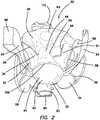
- FIG. 2A model 30 for practicing hysterectomies and, in particular, for practicing vaginal hysterectomies according to the present invention is shown in FIG. 2 .
- the model 30is configured to be placed inside the surgical training device 10 described above or other similar surgical trainer.
- the model 30includes a simulated uterus 32 connected to a frame 34 with a first sheet 36 and a second sheet 38.
- the simulated uterus 32includes a bulbous portion 40 defining a hollow simulated uterine cavity 42.
- the bulbous portion 40is connected to a tubular portion 44 defining a vaginal canal 46 having an opening 48.
- the simulated uterus 32further includes a simulated cervix 50 (shown in FIG.
- the simulated cervix 50includes a slit 52.
- the simulated cervix 50is made of a solid, high durometer silicone.
- the simulated uterus 32further includes simulated fallopian tubes 54 connected to ovaries 56.
- the simulated uterus 32, fallopian tubes 54 and ovaries 56are made of silicone or other elastomeric material and may include other material such as foam material combined with the silicone.
- the simulated uterus 32is made of silicone or lighter foam such as urethane or silicone foam or a combination of the two.
- the silicone constructionimparts the simulated uterus 32 with a more realistic weight when the attached simulated cervix 50 is being pulled and manipulated.
- the simulated uterus 32 made of foammakes the simulated uterus 32 easier to suspend inside the simulated pelvic cavity.
- the lightweight foamflexes more easily than a simulated uterus 32 made of higher durometer silicone allowing a larger simulated uterus 32 to be placed into the model 30 and still be removed.
- the foam uterus 32would compress and flex as it is being removed through the vaginal opening 48 similar to an actual surgery.
- the simulated uterus 32is approximately 300-500 grams and the simulated uterus 32 is composed of a selected durometer foam to accurately represent the size and weight of a real uterus that could normally be removed vaginally without significant morcellation.
- the simulated uterus 32is a combination of silicone and foam to give a more realistic look to the simulated uterus 32 while still having the flexibility of the foam.
- the foamcan be cast and then the silicone can be applied over the foam such as, for example, on a rotational mold.
- the simulated uterus 32is generally pink in color and the fallopian tubes 54 and ovaries are clear or white in color. Furthermore, the simulated uterus 32 may include embedded tumors, cysts and/or ectopic pregnancies in the fallopian tubes 54.
- the model 30may further include simulated vasculature 58 such as blood vessels.
- the simulated vasculature 58is made of solid or hollow tubular silicone or other suitable elastomer. Liquid may be included inside the hollow tubing of the simulated vasculature 58.
- the simulated vasculature 58 that simulates blood vesselsmay be red in color.
- the model 30may also include simulated ligaments 59 such as the uteralsacral ligament 59 and made of silicone material as seen in FIGs. 2 and 4E .
- the model 30may further include the round and tubo ovarian ligaments 61 attached to the frame 34 shown in FIG. 2 .
- the frame 34comprises a cylindrical-like shape defining an interior/lumen 60.
- the frame 34includes a first surface 62 interconnected to a second surface 64 defining a thickness therebetween.
- the first surface 62defines the inner surface of the cylindrical-like shape of the frame 34 and the second surface 64 defines an outer surface of the cylindrical-like shape of the frame 34.
- the frame 34is made of flexible foam material that is also slightly compressible.
- the frame 34includes one or more cutouts 66 extending between the first surface 62 and the second surface 64 to define an outer perimeter and apertures.
- the frame 34is made of a sheet of foam material that is cut according to a pattern shown in FIG. 3D.
- 3Dillustrates the outer perimeter having a top 68 and a bottom 70 interconnected by a first side and a second side 72, 74.
- the top 68includes two curved portions 76a, 76b interconnected at a first protrusion 78 along a vertical axis.
- the two curved portions 76a, 76brepresent the left and right illium/iliac crest.
- the bottom 70includes a second protrusion 80 along the vertical axis.
- the first protrusion 78represents the sacrum of a human pelvis and the second protrusion 80 represents the coccyx.
- the first side 72includes a first lower lobe 82 having a first aperture 86 and the second side 74 includes a second lower lobe 84 having a second aperture 88.
- the first and second lower lobes 82, 84represent the left and right ischium and the first aperture 86 and the second aperture 88 represent the obturator foramen of the human pelvis.
- a piece of foam having a thicknessis cut to have the flat pattern shape shown in FIG. 3D . Then the piece of foam is curved such that the first lower lobe 82 and second lower lobe 84 can then be joined together, in a cylinder-like configuration via the first sheet 36.
- the frame 34is bendable and may be made of a material that retains its shape after bending such as aluminum.
- the clips 26 and wire that are connected to the trainer 10may be used to hold the two lobes 82, 84 in an upward orientation and in a cylindrical-like configuration while inside the trainer 10.
- the anatomy of the pelvisis shown in FIG. 7.
- the frame 34is made of soft, compressible, semi-rigid foam that can be die cut and then formed into the correct shape with adhesive. If the frame 34 is made of harder plastic, it could be a thin thermoform that is initially formed into the correct shape or a thicker plastic that is cut into the pelvis shape and then formed into a cylindrical shape with heat. The frame 34 may also be made of deformable metal that holds its shape.
- the frame 34is not a perfect replica of the anatomy and need only include certain features selected to practice certain procedures that require those specific features as anatomical reference points or visual landmarks for the practitioner. For example, for practicing a vaginal hysterectomy, the important features of the pelvis are the restriction of the pelvic inlet and the attachments to the pelvic sidewall.
- the L-shape of the sacrumis an important landmark.
- the pubic tubercleis an important landmark.
- the frame 34can be made to have all anatomically correct features or only the ones needed for the specific procedure. As such, the frame 34 and model 30 can be used for the simulation of a vaginal hysterectomy, abdominal hysterectomy, colectomy, hernia, taTME, and other pelvic procedures. In another variation, the frame 34 forms a conical shape or frusto-conical shape having an open proximal and open distal ends.
- the model 30may further include a simulated bladder 90.
- the simulated bladder 90is a hollow, air-filled component typically made of silicone or other elastomeric material. In another variation, the simulated bladder contains liquid.
- the simulated bladder 90is connected to the frame 34 with adhesive or other means. It is connected to the first surface 62 or inner surface of the frame 34.
- the simulated bladder 90is attached in alignment with the vertical axis in the location of where the two lobes 82, 84 are in juxtaposition in a location representative of the pubis. When connected the simulated bladder 90 extends into the lumen 60 of the frame 34.
- the simulated bladder 90may further include a simulated ureter 94. In one variation, the simulated ureter 94 is connected to the simulated bladder 90.
- the simulated ureteris made of solid or hollow tubular silicone.
- the model 30may further include a simulated colon 92 or bowel portion.
- the simulated colon 92is a tubular structure that includes a lumen.
- the simulated colon 92is laid on the first surface 62 inside the interior 60 of the frame 34 and substantially along the vertical axis and against the second protrusion 80 of the frame 34.
- Adhesivemay be used to attach the simulated colon 92 to the frame 34.
- the simulated colon 92is made of silicone or other suitable elastomeric material and colored pink or other suitable color and may or may not include simulated tumors.
- the first sheet 36is a thin layer of clear silicone material having a top surface 96 and a bottom surface 98 and a first end 100 and a second end 102.
- the first sheet 36is transparent and at least one of the top surface 96 and the bottom surface 98 is textured in one variation.
- the first sheet 36is attached to the simulated uterus 32.
- the bottom surface 98 of the first sheet 36 near the first end 100is attached along at least a portion of the length of simulated uterus 32 to one or more of the bulbous portion 40 and tubular portion 44 as shown in FIG. 2 .
- the first sheet 36is then folded back toward the top of the model 30 and toward the first end 100 of the first sheet 36 creating a fold near the tubular portion 44 of the simulated uterus 32.
- At least a portion of the first sheet 36 near the second end 102 of the first sheet 36is attached to the frame 34 such that the bottom surface 98 of the first sheet 36 is adhered to the frame 34 in the general location of where the two lobes 82, 84 are in juxtaposition to create a cylinder-like configuration for the frame 34.
- the attachment of the first sheet 36also serve to hold the frame 34 in the cylindrical-like configuration.
- Adhesiveis used to attach the bottom surface 98 of the first sheet 36 to the frame 34.
- the bottom surface 98 of the first sheet 36is attached to the first surface 62 or inner surface of the frame 34 and then folded around a portion of the first side 72 and second side 74 of the frame 34.
- the second end 102 of the first sheet 36is also attached with adhesive to the outer surface of the simulated bladder 90 capturing the simulated bladder 90 between the frame 34 and the first sheet 36.
- a portion of the second end 102 of the first sheet 36is folded around the edge of the frame 34 and attached to the second surface 64 of the frame 34 such that at least part of the second end 102 of the first sheet 36 is resident above the second or outer surface 64 of the frame 34 as visible in FIG. 4D .
- the first sheet 36is sized and configured to suspend the simulated uterus 32 inside the interior 60 of the frame 34.
- Simulated vasculature 58may be attached to the top surface 96 or bottom surface 98 of the first sheet 36.
- the configuration of the first sheet 36forms a pocket-like structure wherein the top surface 96 of the first sheet 36 is folded and at least in part facing itself.
- the first sheet 36creates a webbing of suspension that simulates the peritoneum layer.
- the second sheet 38is a thin layer of clear silicone material having a top surface 104 and a bottom surface 106 and a first end 108 and a second end 110.
- the second sheet 38is transparent and at least one of the top surface 104 and the bottom surface 106 is textured in one variation.
- the second sheet 38is attached to the simulated uterus 32.
- the bottom surface 106 of the second sheet 38 near the first end 108is attached along at least a portion of the length of simulated uterus 32 to one or more of the bulbous portion 40 and tubular portion 44 on a side opposite from where the first sheet 36 is attached.
- the first sheet 36is attached to the anterior side of the model 30 which is also the anterior side of the simulated uterus 32.
- the second sheet 38is attached to the posterior side of the model 30 which is also the posterior side of the simulated uterus 32. After being attached to the posterior side of the simulated uterus 32, the second sheet 38 is then folded back toward the top of the model 30 and toward the first end 108 of the second sheet 38 creating a fold near the tubular portion 44 of the simulated uterus 32. At least a portion of the second sheet 38 near the second end 110 of the second sheet 38 is attached to the frame 34 such that the bottom surface 106 of the second sheet 38 is adhered to the frame 34 in the general location of the second protrusion 80. Adhesive is used to attach the bottom surface 106 of the second sheet 38 to the frame 34.
- the bottom surface 106 of the second sheet 38is attached to the first surface 62 or inner surface of the frame 34 and may be folded around the edge of the frame 34 such that at least part of the second end 110 of the second sheet 38 is connected to second or outer surface 64 of the frame 34. If a simulated colon 92 is employed in the model 30, then the second end 110 of the second sheet 38 is also attached with adhesive to the outer surface of the simulated colon 92 or at least overlaying and not attached with adhesive such that at least a portion of the simulated colon 92 is captured or located between the frame 34 and the second sheet 38.
- the second sheet 38is sized and configured to suspend the simulated uterus 32 inside the interior 60 of the frame 34 if the model 30 is turned over.
- Simulated vasculature 58may be attached to the top surface 104 or bottom surface 106 of the second sheet 38.
- the configuration of the second sheet 38forms a pocket-like structure wherein the top surface 104 of the second sheet 38 is folded and at least in part facing itself.
- the second sheet 38creates a suspended webbing that simulates the peritoneum layer.
- the model 30is shown placed inside a surgical training device 10 of the like described with respect to FIG. 1 .
- the model 30is shown inside the body cavity 12 and oriented such that the top 68 of the frame 34 is in the cephalad direction of the simulated training device 10 and the vaginal opening 48 of the simulated uterus 32 faces the caudal direction of the simulated training device 10.
- the model 30can be connected to the surgical training device 10 with the clips 26 attached to the trainer 10.
- the retractable clips 26can be pulled out and the clips 26 attached to any portion of the model 30 such as to the frame 34 of the model 30.
- the second or outer surface 64 of the model 30may include a hook-and-loop type fastener configured to attach to a complementary portion of hook-and-loop type fastener connected to the base 18 of the trainer 10. Together with one or more fasteners such as the clips 26 and/or hook-and-loop type fasteners, the model 30 is securely attached to the trainer 10 such that it can be manipulated in simulated surgery without dislodging the model 30 from the body cavity 12 of the trainer 10.
- the model 30is further connected to the trainer 10 via a transvaginal adapter 112 that is sized and configured to connect between the top cover 16 and the base 18 as an additional leg 20 positioned at the caudal direction of the surgical training device 10.
- FIGs. 5A-5B and 6A-6Bthere is shown a transvaginal adapter 112.
- a top coversupported above the base by five legs 20.
- a sixth leg 20is provided as shown in FIGs. 4A-4D in the form of the transvaginal adapter 112.
- the trainer 10may be assembled with an optional sixth support structure or leg which is configured for simulating transvaginal surgery including transvaginal hysterectomies.
- the transvaginal adapter 112includes a flat plate 114 having an inner surface 116 for facing toward the interior of the trainer and an outer surface 118 for facing outwardly towards the user.
- the plate 114has a rectangular shape and includes an aperture 120 passing through the plate 108 from the inner surface 116 to the outer surface 118.
- the aperture 120is circular in shape.
- the aperture 120is elongate elliptical oval-like in shape and oriented vertically along the longitudinal axis of the adapter 112.
- the aperture 120is elongate elliptical oval-like in shape and oriented perpendicularly to the longitudinal axis of the adapter. As shown in FIGs.
- the plate 114also includes means such as tabs 122 or a U-shaped channel for inserting to connect the transvaginal adapter 112 to the top cover 16 and to the base 18 to help support and space apart the top cover 16.
- the transvaginal adapter 112is located between the top cover 16 and the base 18 and provides a side access aperture 16 lateral to the trainer 10 or substantially perpendicular to the top cover 16 and the base 18.
- the plate 114further includes a plurality of molding apertures 124 surrounding or encompassing the main aperture 120 configured for overmolding a soft simulated vaginal tissue interface made of silicone or the like. In another variation the interface is insertable into the aperture 120 of the transvaginal adapter 112.
- the tissue interface(not shown) includes an aperture that is substantially coaxial with the plate aperture 120.
- a tubular extension 126is integrally provided and extends into the simulated body cavity 12 of the trainer 10.
- the tubular extension 126is longer in FIGs. 6A-6B in comparison to the tubular extension 126 of FIGs. 5A-5B .
- the tubular extension 126is sized and configured such that the tubular portion 44 of the simulated uterus 32 can be stretched around the extension 126 and secured to the transvaginal adapter 112 such that the vaginal canal 46 is supported in an open configuration, coincident with and accessible through the aperture 120 of the adapter 112 as shown in FIGs. 4A-4D .
- the tubular extension 126serves as a connector connecting the model 30 with the trainer 10 in a manner that permits the interior of the uterus to be accessed as in real surgery.
- the tubular extension 126is a cylindrically-shaped extension having a radially-extending distal flange 128 that extends around at least a portion of the extension 128 to help secure and retain the model 30 attached to the trainer 10.
- the tubular portion 44 of the model 20is attached to the tubular extension 126 by pulling the tubular portion 44 over the distal flange 128, if one is provided, and over and around the tubular extension 126 the outer diameter of which is the same or slightly larger than the relaxed inner diameter of the tubular portion 126 to keep the tubular portion 44 secured to the transvaginal adapter 112.
- the transvaginal adapter 112can be made of flexible or rigid material. If the adapter 112 is made of rigid material it will tend to simulate an already retracted vaginal canal 46. If the adapter 112 is made of flexible material or soft material, the adapter 112 is suited for practicing retraction. In another variation, the transvaginal adapter 112 has a tubular extension 126 that is made of soft flexible material and plate 114 made of rigid material or surrounded by rigid material to keep the top cover 16 of the trainer 10 supported which would still allow the practitioner to practice retraction at the opening of the vaginal canal 46 at the adapter 112.
- the model 30is placed inside the surgical training device 10 and held in place with a hook-and-loop type fastener and/or retracting clips 26.
- the tubular portion 44is attached to the transvaginal adapter 112 by stretching the vaginal opening 48 over the tubular extension 126 of the adapter 112.
- a curtainmay be employed that is placed around the sides of the trainer 30 to further conceal the model 30 such that the only visualization is through the simulated vaginal canal 46.
- the vaginal canal 46is then retracted using a surgical retractor.
- the vaginal canal 46is made of a flexible thermoplastic elastomer (TPE).
- TPEprovides resistance as it is retracted and wants to spring back to its original shape which permits the user to practice realistic retraction.
- the transvaginal adapter 112 of FIGs. 6A-6B having a longer tubular extension 126is used to simulate an already retracted vaginal canal. Hence, the transvaginal adapter 112 permits the practitioner to practice the hysterectomy procedure without needing extra-hands and assistance to perform the retraction. If the transvaginal adapter 112 of FIGs. 5A-5B having the shorter tubular extension 126 is used, the practitioner will practice retracting the vaginal canal 46 with retractors and the help of extra hands during the procedure.
- the transvaginal adapter 112can be made of rigid or flexible material or rigid and flexible material as described above and selected for the purpose of practicing retraction of the vaginal canal 46 or not.
- the simulated cervix 50is grasped and pulled towards the opening 48 of the vaginal canal 46.
- the simulated cervix 50is made of high durometer silicone relative to the surrounding tubular portion 44.
- the simulated cervix 50is also made as a solid component which allows it to be grasped with real surgical tools and pulled on without fear of the silicone ripping or tearing.
- the simulated cervix 50is incised circumferentially and the practitioner is able to practice carefully dissecting the vaginal mucosa off of the simulated cervix 50.
- a sheet of cotton or other webbing-like substancecan be included in the model 30 between the vaginal canal 46 and the simulated bladder 90.
- the simulated bladder 90is a hollow, air-filled component. If the practitioner cuts to high while dissecting the simulated vaginal mucosa and the simulated bladder 90 is accidentally incised, the simulated bladder 90 could pop and give immediate feedback to the practitioner especially if the simulated bladder 90 contains fluid.
- the model 30advantageously includes a second sheet 38 forming a fold between the simulated uterus 32 and the frame 34.
- the suspension of the simulated uterus 32 within the frame 34advantageously creates a realistic response when the simulated uterus 32 is being incised and manipulated.
- the simulated uteruswill remain suspended, hang and swing in response to being manipulated with surgical instruments. At least portions of the simulated uterus and simulated vagina are held in suspension inside the enclosure defined by the pelvic frame and connected thereto or directly connected to the enclosure defined by the trainer.
- the suspensionadvantageously permits the fold of the second sheet to be accessed to practice posterior colpotomy into the posterior cul-de-sac incision by incising the peritoneum forming the recto-uterine fold.
- the suspended simulated uterus 32allows for the existence of the recto-uterine peritoneum fold.
- the simulated uterus 32is pendent inside the frame 34 made of foam material that mimics a human pelvis.
- the simulated uterus 32is suspended by a folded first sheet of silicone material on the anterior side of the simulated uterus 32 and a folded second sheet of silicone material on the posterior side of the simulated uterus 32.
- the frame 34can be made of any material such as plastic or harder foam material.
- the frame 34serves as an attachment area for the various simulated portions of the anatomy including the broad ligament, ovaries 56 and fallopian tubes 54.
- the elasticity of the silicone of these anatomical componentsallows the simulated uterus 32 to be pulled and manipulated and still remain attached to the frame 34.
- a frame 34 made of semi-rigid foammay also move as the simulated uterus is being manipulated. A more rigid frame 34 would move less.
- the practitionerthen divides the uteralsacral ligaments 59.
- the practitionerthen performs an anterior colpotomy into the anterior cul-de-sac by incising the first sheet 38 simulating the peritoneum forming the vesico-uterine fold.
- the practitionerdivides the tubo ovarian and round ligaments 61 on each side of the simulated uterus 32. Due to the foam frame 34, the round and tubo ovarian ligaments 59 remain realistically attached to the frame 34 after they have been divided from the simulated uterus 32.
- the simulated uterus 32is then freed and removed.
- the practitionerthen practices to suture the vaginal cuff closed by passing a needle and suture through the tubular portion 44 of the model 32 to close the vaginal canal 46 opening. Suturing the vaginal cuff in real surgery is another difficult part of the vaginal hysterectomy due to the space limitations.
- the tubular portion 44 that is made of TPEholds the suture without tearing and limits the space allowed for instruments during the suturing process.
- the model 30allows the practitioner to practice numerous difficult procedures on one model.
- Any portion of the model 30can be made of one or more organic base polymer including but not limited to hydrogel, single-polymer hydrogel, multi-polymer hydrogel, rubber, latex, nitrile, protein, gelatin, collagen, soy, non-organic base polymer such as thermo plastic elastomer, Kraton, silicone, foam, silicone-based foam, urethane-based foam and ethylene vinyl acetate foam and the like.
- organic base polymerincluding but not limited to hydrogel, single-polymer hydrogel, multi-polymer hydrogel, rubber, latex, nitrile, protein, gelatin, collagen, soy, non-organic base polymer such as thermo plastic elastomer, Kraton, silicone, foam, silicone-based foam, urethane-based foam and ethylene vinyl acetate foam and the like.
- any base polymerone or more filler may be employed such as a fabric, woven or non-woven fiber, polyester, nylon, cotton and silk, conductive filler material such as graphite, platinum, silver, gold, copper, miscellaneous additives, gels, oil, cornstarch, glass, dolomite, carbonate mineral, alcohol, deadener, silicone oil, pigment, foam, poloxamer, collagen, gelatin and the like.
- the adhesives employedmay include but are not limited to cyanoacrylate, silicone, epoxy, spray adhesive, rubber adhesive and the like.
Landscapes
- Engineering & Computer Science (AREA)
- General Physics & Mathematics (AREA)
- Physics & Mathematics (AREA)
- Health & Medical Sciences (AREA)
- Mathematical Analysis (AREA)
- Mathematical Optimization (AREA)
- Medical Informatics (AREA)
- Medicinal Chemistry (AREA)
- Chemical & Material Sciences (AREA)
- Algebra (AREA)
- Computational Mathematics (AREA)
- Theoretical Computer Science (AREA)
- Educational Technology (AREA)
- General Health & Medical Sciences (AREA)
- Mathematical Physics (AREA)
- Pure & Applied Mathematics (AREA)
- Business, Economics & Management (AREA)
- Educational Administration (AREA)
- Pulmonology (AREA)
- Radiology & Medical Imaging (AREA)
- Gynecology & Obstetrics (AREA)
- Pregnancy & Childbirth (AREA)
- Reproductive Health (AREA)
- Instructional Devices (AREA)
Description
- This application is generally related to surgical training tools, and in particular, to simulated tissue structures and models for teaching and practicing various surgical techniques and procedures related but not limited to laparoscopic, endoscopic and minimally invasive surgery.
- Medical students as well as experienced doctors learning new surgical techniques must undergo extensive training before they are qualified to perform surgery on human patients. The training must teach proper techniques employing various medical devices for cutting, penetrating, clamping, grasping, stapling, cauterizing and suturing a variety of tissue types. The range of possibilities that a trainee may encounter is great. For example, different organs and patient anatomies and diseases are presented. The thickness and consistency of the various tissue layers will also vary from one part of the body to the next and from one patient to another. Different procedures demand different skills. Furthermore, the trainee must practice techniques in various anatomical environs that are influenced by factors such as the size and condition of the patient, the adjacent anatomical landscape and the types of targeted tissues and whether they are readily accessible or relatively inaccessible.
- Numerous teaching aids, trainers, simulators and model organs are available for one or more aspects of surgical training. However, there is a need for models or simulated tissue elements that are likely to be encountered in and that can be used for practicing endoscopic and laparoscopic, minimally invasive, transluminal surgical procedures. In laparoscopic surgery, a trocar or cannula is inserted to access a body cavity and to create a channel for the insertion of a camera such as a laparoscope. The camera provides a live video feed capturing images that are then displayed to the surgeon on one or more monitors. At least one additional small incision is made through which another trocar/cannula is inserted to create a pathway through which surgical instruments can be passed for performing procedures observed on the monitor. The targeted tissue location such as the abdomen is typically enlarged by delivering carbon dioxide gas to insufflate the body cavity and create a working space large enough to accommodate the scope and instruments used by the surgeon. The insufflation pressure in the tissue cavity is maintained by using specialized trocars. Laparoscopic surgery offers a number of advantages when compared with an open procedure. These advantages include reduced pain, reduced blood and shorter recovery times due to smaller incisions.
- Laparoscopic or endoscopic minimally invasive surgery requires an increased level of skill compared to open surgery because the target tissue is not directly observed by the clinician. The target tissue is observed on monitors displaying a portion of the surgical site that is accessed through a small opening. Therefore, clinicians need to practice visually determining tissue planes, three-dimensional depth perception on a two-dimensional viewing screen, hand-to-hand transfer of instruments, suturing, precision cutting and tissue and instrument manipulation. Typically, models simulating a particular anatomy or procedure are placed in a simulated pelvic trainer where the anatomical model is obscured from direct visualization by the practitioner. Ports in the trainer are employed for passing instruments to practice techniques on the anatomical model hidden from direct visualization. Simulated pelvic trainers provide a functional, inexpensive and practical means to train surgeons and residents the basic skills and typical techniques used in laparoscopic surgery such as grasping, manipulating, cutting, tying knots, suturing, stapling, cauterizing as well as how to perform specific surgical procedures that utilized these basic skills. Simulated pelvic trainers are also effective sales tools for demonstrating medical devices required to perform these laparoscopic procedures.
- One procedure is a hysterectomy in which the uterus is removed. The hysterectomy may be performed vaginally extracting the uterus through the vaginal canal or abdominally through a small incision in the abdomen. The vaginal hysterectomy is historically hard to train on as the field of view is limited. Unlike laparoscopic procedures, there is no camera that is projecting the surgery onto a screen and unlike open procedures there is not a wide incision that can be viewed by multiple people. As such, the best way to teach a vaginal hysterectomy is through a simulated model. Therefore, there is a need for a model for training hysterectomy procedures.
- Examples of surgical training models for practicing such procedures are to be found in patent applications having publication numbers
WO 2011/046606 A1 andUS 2012/082970 A1 . - According to the present invention there is provided a surgical simulator as recited in claim 1.
FIG. 1 is a top perspective view of a surgical training device which when incorporating the model ofFIG. 2 will be in accordance with the present invention.FIG. 2 is an antero-cephalad, top perspective view of a model according to the present invention.FIG. 3A is a top perspective view of a pelvic frame for use in the present invention.FIG. 3B is a top perspective view of a pelvic frame for use in the present inventionFIG. 3C is a top perspective view of a pelvic frame for use in the present invention.FIG. 3D is a top view of a pelvic frame in a flat orientation for use in the present invention.FIG. 4A is a caudal end view of a model inside a surgical training device according to the present invention.FIG. 4B is a lateral side view of a model inside a surgical training device according to the present invention.FIG. 4C is a lateral side view of a model inside a surgical training device according to the present invention.FIG. 4D is an antero-caudal, top perspective view of a model inside a surgical training device according to the present invention.FIG. 4E is a cephalad end view of a model inside a surgical training device according to the present invention.FIG. 5A is a side view of a transvaginal adapter for use in the present invention.FIG. 5B is a top perspective view of a transvaginal adapter for use in the present invention.FIG. 6A is a side view of a transvaginal adapter for use in the present invention.FIG. 6B is a top perspective view of a transvaginal adapter for use in the present invention.- A
surgical training device 10 that is configured to mimic the torso of a patient such as the abdominal region is shown inFIG. 1 . Thesurgical training device 10 provides abody cavity 12 substantially obscured from the user for receiving simulated or live tissue or model organs or training models of the like described in this invention. Thebody cavity 12 is accessed via atissue simulation region 14 that is penetrated by the user employing devices to practice surgical techniques on the tissue or practice model found located in thebody cavity 12. Although thebody cavity 12 is shown to be accessible through a tissue simulation region, a hand-assisted access device or single-site port device may be alternatively employed to access thebody cavity 12. Thesurgical training device 10 is particularly well suited for practicing laparoscopic or other minimally invasive surgical procedures. - Still referencing
FIG. 1 , thesurgical training device 10 includes atop cover 16 connected to and spaced apart from a base 18 by at least oneleg 20.FIG. 1 shows a plurality oflegs 20. Thesurgical training device 10 is configured to mimic the torso of a patient such as the abdominal region. Thetop cover 16 is representative of the anterior surface of the patient and thespace 12 between thetop cover 16 and thebase 18 is representative of an interior of the patient or body cavity where organs reside. Thesurgical trainer 10 is a useful tool for teaching, practicing and demonstrating various surgical procedures and their related instruments in simulation of a patient undergoing a surgical procedure. Surgical instruments are inserted into thecavity 12 through thetissue simulation region 14 as well as throughpre-established apertures 22 in thetop cover 16. Various tools and techniques may be used to penetrate thetop cover 16 to perform mock procedures on simulated organs or practice models placed between thetop cover 16 and thebase 18. Thebase 18 includes a model-receivingarea 24 or tray for staging or holding a simulated tissue model or live tissue. The model-receivingarea 24 of thebase 18 includes frame-like elements for holding the model (not shown) in place. To help retain a simulated tissue model or live organs on thebase 18, a clip attached to a retractable wire is provided atlocations 26. The retractable wire is extended and then clipped to hold the tissue model in position substantially beneath thetissue simulation region 14. Other means for retaining the tissue model include a patch of hook-and-loop type fastening material (VELCRO®) affixed to the base 18 in themodel receiving area 24 such that it is removably connectable to a complementary piece of hook-and-loop type fastening material (VELCRO®) affixed to the model. - A video display monitor 28 that is hinged to the
top cover 16 is shown in a closed orientation inFIG. 1 . The video monitor 28 is connectable to a variety of visual systems for delivering an image to the monitor. For example, a laparoscope inserted through one of thepre-established apertures 22 or a webcam located in the cavity and used to observe the simulated procedure can be connected to thevideo monitor 28 and/or a mobile computing device to provide an image to the user. Also, audio recording or delivery means may also be provided and integrated with thetrainer 10 to provide audio and visual capabilities. Means for connecting a portable memory storage device such as a flash drive, smart phone, digital audio or video player, or other digital mobile device is also provided, to record training procedures and/or play back pre-recorded videos on the monitor for demonstration purposes. Of course, connection means for providing an audio visual output to a screen larger than the monitor is provided. In another variation, thetop cover 10 does not include a video display but includes means for connecting with a laptop computer, a mobile digital device or tablet and connecting it by wire or wirelessly to the trainer. - When assembled, the
top cover 16 is positioned directly above the base 18 with thelegs 20 located substantially around the periphery and interconnected between thetop cover 16 andbase 18. Thetop cover 16 andbase 18 are substantially the same shape and size and have substantially the same peripheral outline. The internal cavity is partially or entirely obscured from view. In the variation shown inFIG. 1 , the legs include openings to allow ambient light to illuminate the internal cavity as much as possible and also to advantageously provide as much weight reduction as possible for convenient portability. Thetop cover 16 is removable from thelegs 20 which in turn are removable or collapsible via hinges or the like with respect to thebase 18. Therefore, theunassembled trainer 10 has a reduced height that makes for easier portability. In essence, thesurgical trainer 10 provides asimulated body cavity 12 that is obscured from the user. Thebody cavity 12 is configured to receive at least one surgical model accessible via at least onetissue simulation region 14 and/orapertures 22 in thetop cover 16 through which the user may access the models to practice laparoscopic or endoscopic minimally invasive surgical techniques. - A
model 30 for practicing hysterectomies and, in particular, for practicing vaginal hysterectomies according to the present invention is shown inFIG. 2 . Themodel 30 is configured to be placed inside thesurgical training device 10 described above or other similar surgical trainer. Themodel 30 includes asimulated uterus 32 connected to aframe 34 with afirst sheet 36 and asecond sheet 38. Thesimulated uterus 32 includes abulbous portion 40 defining a hollow simulateduterine cavity 42. Thebulbous portion 40 is connected to atubular portion 44 defining avaginal canal 46 having anopening 48. Thesimulated uterus 32 further includes a simulated cervix 50 (shown inFIG. 4A ) located inside thesimulated uterus 32 in a location substantially between theuterine cavity 42 and thevaginal canal 46. Thesimulated cervix 50 includes a slit 52. Thesimulated cervix 50 is made of a solid, high durometer silicone. - The
simulated uterus 32 further includes simulatedfallopian tubes 54 connected toovaries 56. Thesimulated uterus 32,fallopian tubes 54 andovaries 56 are made of silicone or other elastomeric material and may include other material such as foam material combined with the silicone. Thesimulated uterus 32 is made of silicone or lighter foam such as urethane or silicone foam or a combination of the two. The silicone construction imparts thesimulated uterus 32 with a more realistic weight when the attachedsimulated cervix 50 is being pulled and manipulated. Thesimulated uterus 32 made of foam makes thesimulated uterus 32 easier to suspend inside the simulated pelvic cavity. Also, when removing thesimulated uterus 32 the lightweight foam flexes more easily than asimulated uterus 32 made of higher durometer silicone allowing a largersimulated uterus 32 to be placed into themodel 30 and still be removed. Thefoam uterus 32 would compress and flex as it is being removed through thevaginal opening 48 similar to an actual surgery. Thesimulated uterus 32 is approximately 300-500 grams and thesimulated uterus 32 is composed of a selected durometer foam to accurately represent the size and weight of a real uterus that could normally be removed vaginally without significant morcellation. In another variation, thesimulated uterus 32 is a combination of silicone and foam to give a more realistic look to thesimulated uterus 32 while still having the flexibility of the foam. The foam can be cast and then the silicone can be applied over the foam such as, for example, on a rotational mold. Thesimulated uterus 32 is generally pink in color and thefallopian tubes 54 and ovaries are clear or white in color. Furthermore, thesimulated uterus 32 may include embedded tumors, cysts and/or ectopic pregnancies in thefallopian tubes 54. Themodel 30 may further includesimulated vasculature 58 such as blood vessels. Thesimulated vasculature 58 is made of solid or hollow tubular silicone or other suitable elastomer. Liquid may be included inside the hollow tubing of thesimulated vasculature 58. Thesimulated vasculature 58 that simulates blood vessels may be red in color. Themodel 30 may also includesimulated ligaments 59 such as theuteralsacral ligament 59 and made of silicone material as seen inFIGs. 2 and4E . Themodel 30 may further include the round and tuboovarian ligaments 61 attached to theframe 34 shown inFIG. 2 . - With additional reference to
FIGs. 3A-3D , theframe 34 comprises a cylindrical-like shape defining an interior/lumen 60. Theframe 34 includes afirst surface 62 interconnected to asecond surface 64 defining a thickness therebetween. Thefirst surface 62 defines the inner surface of the cylindrical-like shape of theframe 34 and thesecond surface 64 defines an outer surface of the cylindrical-like shape of theframe 34. Theframe 34 is made of flexible foam material that is also slightly compressible. Theframe 34 includes one ormore cutouts 66 extending between thefirst surface 62 and thesecond surface 64 to define an outer perimeter and apertures. In one variation, theframe 34 is made of a sheet of foam material that is cut according to a pattern shown inFIG. 3D. FIG. 3D illustrates the outer perimeter having a top 68 and a bottom 70 interconnected by a first side and asecond side curved portions first protrusion 78 along a vertical axis. The twocurved portions second protrusion 80 along the vertical axis. Thefirst protrusion 78 represents the sacrum of a human pelvis and thesecond protrusion 80 represents the coccyx. Thefirst side 72 includes a firstlower lobe 82 having afirst aperture 86 and thesecond side 74 includes a secondlower lobe 84 having asecond aperture 88. The first and secondlower lobes first aperture 86 and thesecond aperture 88 represent the obturator foramen of the human pelvis. A piece of foam having a thickness is cut to have the flat pattern shape shown inFIG. 3D . Then the piece of foam is curved such that the firstlower lobe 82 and secondlower lobe 84 can then be joined together, in a cylinder-like configuration via thefirst sheet 36. Theframe 34 is bendable and may be made of a material that retains its shape after bending such as aluminum. Also, theclips 26 and wire that are connected to thetrainer 10 may be used to hold the twolobes trainer 10. The anatomy of the pelvis is shown in FIG. 7. - The
frame 34 is made of soft, compressible, semi-rigid foam that can be die cut and then formed into the correct shape with adhesive. If theframe 34 is made of harder plastic, it could be a thin thermoform that is initially formed into the correct shape or a thicker plastic that is cut into the pelvis shape and then formed into a cylindrical shape with heat. Theframe 34 may also be made of deformable metal that holds its shape. Theframe 34 is not a perfect replica of the anatomy and need only include certain features selected to practice certain procedures that require those specific features as anatomical reference points or visual landmarks for the practitioner. For example, for practicing a vaginal hysterectomy, the important features of the pelvis are the restriction of the pelvic inlet and the attachments to the pelvic sidewall. For practicing a transanal total mesorectal excision (taTME), the L-shape of the sacrum is an important landmark. For hernia procedures, the pubic tubercle is an important landmark. Theframe 34 can be made to have all anatomically correct features or only the ones needed for the specific procedure. As such, theframe 34 andmodel 30 can be used for the simulation of a vaginal hysterectomy, abdominal hysterectomy, colectomy, hernia, taTME, and other pelvic procedures. In another variation, theframe 34 forms a conical shape or frusto-conical shape having an open proximal and open distal ends. - With reference back to
FIG. 2 , themodel 30 may further include asimulated bladder 90. Thesimulated bladder 90 is a hollow, air-filled component typically made of silicone or other elastomeric material. In another variation, the simulated bladder contains liquid. Thesimulated bladder 90 is connected to theframe 34 with adhesive or other means. It is connected to thefirst surface 62 or inner surface of theframe 34. Thesimulated bladder 90 is attached in alignment with the vertical axis in the location of where the twolobes simulated bladder 90 extends into thelumen 60 of theframe 34. Thesimulated bladder 90 may further include a simulated ureter 94. In one variation, the simulated ureter 94 is connected to thesimulated bladder 90. The simulated ureter is made of solid or hollow tubular silicone. - Still referencing
FIG. 2 , themodel 30 may further include asimulated colon 92 or bowel portion. Thesimulated colon 92 is a tubular structure that includes a lumen. Thesimulated colon 92 is laid on thefirst surface 62 inside the interior 60 of theframe 34 and substantially along the vertical axis and against thesecond protrusion 80 of theframe 34. Adhesive may be used to attach thesimulated colon 92 to theframe 34. Thesimulated colon 92 is made of silicone or other suitable elastomeric material and colored pink or other suitable color and may or may not include simulated tumors. - The
first sheet 36 is a thin layer of clear silicone material having atop surface 96 and a bottom surface 98 and afirst end 100 and asecond end 102. Thefirst sheet 36 is transparent and at least one of thetop surface 96 and the bottom surface 98 is textured in one variation. Thefirst sheet 36 is attached to thesimulated uterus 32. In particular, the bottom surface 98 of thefirst sheet 36 near thefirst end 100 is attached along at least a portion of the length ofsimulated uterus 32 to one or more of thebulbous portion 40 andtubular portion 44 as shown inFIG. 2 . Thefirst sheet 36 is then folded back toward the top of themodel 30 and toward thefirst end 100 of thefirst sheet 36 creating a fold near thetubular portion 44 of thesimulated uterus 32. At least a portion of thefirst sheet 36 near thesecond end 102 of thefirst sheet 36 is attached to theframe 34 such that the bottom surface 98 of thefirst sheet 36 is adhered to theframe 34 in the general location of where the twolobes frame 34. The attachment of thefirst sheet 36 also serve to hold theframe 34 in the cylindrical-like configuration. Adhesive is used to attach the bottom surface 98 of thefirst sheet 36 to theframe 34. The bottom surface 98 of thefirst sheet 36 is attached to thefirst surface 62 or inner surface of theframe 34 and then folded around a portion of thefirst side 72 andsecond side 74 of theframe 34. If asimulated bladder 90 is employed in themodel 30, then thesecond end 102 of thefirst sheet 36 is also attached with adhesive to the outer surface of thesimulated bladder 90 capturing thesimulated bladder 90 between theframe 34 and thefirst sheet 36. A portion of thesecond end 102 of thefirst sheet 36 is folded around the edge of theframe 34 and attached to thesecond surface 64 of theframe 34 such that at least part of thesecond end 102 of thefirst sheet 36 is resident above the second orouter surface 64 of theframe 34 as visible inFIG. 4D . Thefirst sheet 36 is sized and configured to suspend thesimulated uterus 32 inside the interior 60 of theframe 34.Simulated vasculature 58 may be attached to thetop surface 96 or bottom surface 98 of thefirst sheet 36. The configuration of thefirst sheet 36 forms a pocket-like structure wherein thetop surface 96 of thefirst sheet 36 is folded and at least in part facing itself. Thefirst sheet 36 creates a webbing of suspension that simulates the peritoneum layer. - The
second sheet 38 is a thin layer of clear silicone material having atop surface 104 and abottom surface 106 and a first end 108 and a second end 110. Thesecond sheet 38 is transparent and at least one of thetop surface 104 and thebottom surface 106 is textured in one variation. Thesecond sheet 38 is attached to thesimulated uterus 32. In particular, thebottom surface 106 of thesecond sheet 38 near the first end 108 is attached along at least a portion of the length ofsimulated uterus 32 to one or more of thebulbous portion 40 andtubular portion 44 on a side opposite from where thefirst sheet 36 is attached. Thefirst sheet 36 is attached to the anterior side of themodel 30 which is also the anterior side of thesimulated uterus 32. Thesecond sheet 38 is attached to the posterior side of themodel 30 which is also the posterior side of thesimulated uterus 32. After being attached to the posterior side of thesimulated uterus 32, thesecond sheet 38 is then folded back toward the top of themodel 30 and toward the first end 108 of thesecond sheet 38 creating a fold near thetubular portion 44 of thesimulated uterus 32. At least a portion of thesecond sheet 38 near the second end 110 of thesecond sheet 38 is attached to theframe 34 such that thebottom surface 106 of thesecond sheet 38 is adhered to theframe 34 in the general location of thesecond protrusion 80. Adhesive is used to attach thebottom surface 106 of thesecond sheet 38 to theframe 34. Thebottom surface 106 of thesecond sheet 38 is attached to thefirst surface 62 or inner surface of theframe 34 and may be folded around the edge of theframe 34 such that at least part of the second end 110 of thesecond sheet 38 is connected to second orouter surface 64 of theframe 34. If asimulated colon 92 is employed in themodel 30, then the second end 110 of thesecond sheet 38 is also attached with adhesive to the outer surface of thesimulated colon 92 or at least overlaying and not attached with adhesive such that at least a portion of thesimulated colon 92 is captured or located between theframe 34 and thesecond sheet 38. Thesecond sheet 38 is sized and configured to suspend thesimulated uterus 32 inside the interior 60 of theframe 34 if themodel 30 is turned over.Simulated vasculature 58 may be attached to thetop surface 104 orbottom surface 106 of thesecond sheet 38. The configuration of thesecond sheet 38 forms a pocket-like structure wherein thetop surface 104 of thesecond sheet 38 is folded and at least in part facing itself. Thesecond sheet 38 creates a suspended webbing that simulates the peritoneum layer. - With reference now to
FIGs. 4A-4E , themodel 30 is shown placed inside asurgical training device 10 of the like described with respect toFIG. 1 . Themodel 30 is shown inside thebody cavity 12 and oriented such that the top 68 of theframe 34 is in the cephalad direction of thesimulated training device 10 and thevaginal opening 48 of thesimulated uterus 32 faces the caudal direction of thesimulated training device 10. Themodel 30 can be connected to thesurgical training device 10 with theclips 26 attached to thetrainer 10. Theretractable clips 26 can be pulled out and theclips 26 attached to any portion of themodel 30 such as to theframe 34 of themodel 30. Also, the second orouter surface 64 of themodel 30 may include a hook-and-loop type fastener configured to attach to a complementary portion of hook-and-loop type fastener connected to thebase 18 of thetrainer 10. Together with one or more fasteners such as theclips 26 and/or hook-and-loop type fasteners, themodel 30 is securely attached to thetrainer 10 such that it can be manipulated in simulated surgery without dislodging themodel 30 from thebody cavity 12 of thetrainer 10. Themodel 30 is further connected to thetrainer 10 via atransvaginal adapter 112 that is sized and configured to connect between thetop cover 16 and the base 18 as anadditional leg 20 positioned at the caudal direction of thesurgical training device 10. - Turning now to
FIGs. 5A-5B and6A-6B , there is shown atransvaginal adapter 112. With reference also back toFIG. 1 , there is shown a top cover supported above the base by fivelegs 20. In one variation, asixth leg 20 is provided as shown inFIGs. 4A-4D in the form of thetransvaginal adapter 112. Thetrainer 10 may be assembled with an optional sixth support structure or leg which is configured for simulating transvaginal surgery including transvaginal hysterectomies. - The
transvaginal adapter 112 includes aflat plate 114 having aninner surface 116 for facing toward the interior of the trainer and anouter surface 118 for facing outwardly towards the user. Theplate 114 has a rectangular shape and includes anaperture 120 passing through the plate 108 from theinner surface 116 to theouter surface 118. In one variation, theaperture 120 is circular in shape. In another variation, theaperture 120 is elongate elliptical oval-like in shape and oriented vertically along the longitudinal axis of theadapter 112. In another variation, theaperture 120 is elongate elliptical oval-like in shape and oriented perpendicularly to the longitudinal axis of the adapter. As shown inFIGs. 5A-6B , theplate 114 also includes means such astabs 122 or a U-shaped channel for inserting to connect thetransvaginal adapter 112 to thetop cover 16 and to the base 18 to help support and space apart thetop cover 16. Thetransvaginal adapter 112 is located between thetop cover 16 and thebase 18 and provides aside access aperture 16 lateral to thetrainer 10 or substantially perpendicular to thetop cover 16 and thebase 18. Theplate 114 further includes a plurality ofmolding apertures 124 surrounding or encompassing themain aperture 120 configured for overmolding a soft simulated vaginal tissue interface made of silicone or the like. In another variation the interface is insertable into theaperture 120 of thetransvaginal adapter 112. The tissue interface (not shown) includes an aperture that is substantially coaxial with theplate aperture 120. At the inner surface of thetransvaginal adapter 112, atubular extension 126 is integrally provided and extends into thesimulated body cavity 12 of thetrainer 10. Thetubular extension 126 is longer inFIGs. 6A-6B in comparison to thetubular extension 126 ofFIGs. 5A-5B . Thetubular extension 126 is sized and configured such that thetubular portion 44 of thesimulated uterus 32 can be stretched around theextension 126 and secured to thetransvaginal adapter 112 such that thevaginal canal 46 is supported in an open configuration, coincident with and accessible through theaperture 120 of theadapter 112 as shown inFIGs. 4A-4D . Thetubular extension 126 serves as a connector connecting themodel 30 with thetrainer 10 in a manner that permits the interior of the uterus to be accessed as in real surgery. In one variation, thetubular extension 126 is a cylindrically-shaped extension having a radially-extendingdistal flange 128 that extends around at least a portion of theextension 128 to help secure and retain themodel 30 attached to thetrainer 10. Thetubular portion 44 of themodel 20 is attached to thetubular extension 126 by pulling thetubular portion 44 over thedistal flange 128, if one is provided, and over and around thetubular extension 126 the outer diameter of which is the same or slightly larger than the relaxed inner diameter of thetubular portion 126 to keep thetubular portion 44 secured to thetransvaginal adapter 112. Thetransvaginal adapter 112 can be made of flexible or rigid material. If theadapter 112 is made of rigid material it will tend to simulate an already retractedvaginal canal 46. If theadapter 112 is made of flexible material or soft material, theadapter 112 is suited for practicing retraction. In another variation, thetransvaginal adapter 112 has atubular extension 126 that is made of soft flexible material andplate 114 made of rigid material or surrounded by rigid material to keep thetop cover 16 of thetrainer 10 supported which would still allow the practitioner to practice retraction at the opening of thevaginal canal 46 at theadapter 112. - In use, the
model 30 is placed inside thesurgical training device 10 and held in place with a hook-and-loop type fastener and/or retracting clips 26. Thetubular portion 44 is attached to thetransvaginal adapter 112 by stretching thevaginal opening 48 over thetubular extension 126 of theadapter 112. A curtain may be employed that is placed around the sides of thetrainer 30 to further conceal themodel 30 such that the only visualization is through the simulatedvaginal canal 46. Thevaginal canal 46 is then retracted using a surgical retractor. Thevaginal canal 46 is made of a flexible thermoplastic elastomer (TPE). The TPE provides resistance as it is retracted and wants to spring back to its original shape which permits the user to practice realistic retraction. Thetransvaginal adapter 112 ofFIGs. 6A-6B having a longertubular extension 126 is used to simulate an already retracted vaginal canal. Hence, thetransvaginal adapter 112 permits the practitioner to practice the hysterectomy procedure without needing extra-hands and assistance to perform the retraction. If thetransvaginal adapter 112 ofFIGs. 5A-5B having the shortertubular extension 126 is used, the practitioner will practice retracting thevaginal canal 46 with retractors and the help of extra hands during the procedure. Thetransvaginal adapter 112 can be made of rigid or flexible material or rigid and flexible material as described above and selected for the purpose of practicing retraction of thevaginal canal 46 or not. Next, thesimulated cervix 50 is grasped and pulled towards the opening 48 of thevaginal canal 46. Thesimulated cervix 50 is made of high durometer silicone relative to the surroundingtubular portion 44. Thesimulated cervix 50 is also made as a solid component which allows it to be grasped with real surgical tools and pulled on without fear of the silicone ripping or tearing. Thesimulated cervix 50 is incised circumferentially and the practitioner is able to practice carefully dissecting the vaginal mucosa off of thesimulated cervix 50. A sheet of cotton or other webbing-like substance can be included in themodel 30 between thevaginal canal 46 and thesimulated bladder 90. As described above, thesimulated bladder 90 is a hollow, air-filled component. If the practitioner cuts to high while dissecting the simulated vaginal mucosa and thesimulated bladder 90 is accidentally incised, thesimulated bladder 90 could pop and give immediate feedback to the practitioner especially if thesimulated bladder 90 contains fluid. - The
model 30 advantageously includes asecond sheet 38 forming a fold between thesimulated uterus 32 and theframe 34. Also, the suspension of thesimulated uterus 32 within theframe 34 advantageously creates a realistic response when thesimulated uterus 32 is being incised and manipulated. Also, in the variation in which the simulated uterus is made of lighter foam material, the simulated uterus will remain suspended, hang and swing in response to being manipulated with surgical instruments. At least portions of the simulated uterus and simulated vagina are held in suspension inside the enclosure defined by the pelvic frame and connected thereto or directly connected to the enclosure defined by the trainer. The suspension advantageously permits the fold of the second sheet to be accessed to practice posterior colpotomy into the posterior cul-de-sac incision by incising the peritoneum forming the recto-uterine fold. The suspendedsimulated uterus 32 allows for the existence of the recto-uterine peritoneum fold. As previously described, thesimulated uterus 32 is pendent inside theframe 34 made of foam material that mimics a human pelvis. Thesimulated uterus 32 is suspended by a folded first sheet of silicone material on the anterior side of thesimulated uterus 32 and a folded second sheet of silicone material on the posterior side of thesimulated uterus 32. Theframe 34 can be made of any material such as plastic or harder foam material. Theframe 34 serves as an attachment area for the various simulated portions of the anatomy including the broad ligament,ovaries 56 andfallopian tubes 54. The elasticity of the silicone of these anatomical components allows thesimulated uterus 32 to be pulled and manipulated and still remain attached to theframe 34. Aframe 34 made of semi-rigid foam may also move as the simulated uterus is being manipulated. A morerigid frame 34 would move less. The practitioner then divides theuteralsacral ligaments 59. The practitioner then performs an anterior colpotomy into the anterior cul-de-sac by incising thefirst sheet 38 simulating the peritoneum forming the vesico-uterine fold. The practitioner divides the tubo ovarian andround ligaments 61 on each side of thesimulated uterus 32. Due to thefoam frame 34, the round and tuboovarian ligaments 59 remain realistically attached to theframe 34 after they have been divided from thesimulated uterus 32. Thesimulated uterus 32 is then freed and removed. The practitioner then practices to suture the vaginal cuff closed by passing a needle and suture through thetubular portion 44 of themodel 32 to close thevaginal canal 46 opening. Suturing the vaginal cuff in real surgery is another difficult part of the vaginal hysterectomy due to the space limitations. Thetubular portion 44 that is made of TPE holds the suture without tearing and limits the space allowed for instruments during the suturing process. Themodel 30 allows the practitioner to practice numerous difficult procedures on one model. - Any portion of the
model 30 can be made of one or more organic base polymer including but not limited to hydrogel, single-polymer hydrogel, multi-polymer hydrogel, rubber, latex, nitrile, protein, gelatin, collagen, soy, non-organic base polymer such as thermo plastic elastomer, Kraton, silicone, foam, silicone-based foam, urethane-based foam and ethylene vinyl acetate foam and the like. Into any base polymer one or more filler may be employed such as a fabric, woven or non-woven fiber, polyester, nylon, cotton and silk, conductive filler material such as graphite, platinum, silver, gold, copper, miscellaneous additives, gels, oil, cornstarch, glass, dolomite, carbonate mineral, alcohol, deadener, silicone oil, pigment, foam, poloxamer, collagen, gelatin and the like. The adhesives employed may include but are not limited to cyanoacrylate, silicone, epoxy, spray adhesive, rubber adhesive and the like. - It is understood that various modifications may be made to the embodiments and variations disclosed herein. Therefore, the above description should not be construed as limiting, but merely as exemplifications of preferred embodiments. Those skilled in the art will envision other modifications within the scope of the following claims.
Claims (14)
- A surgical simulator for surgical training comprising:a surgical trainer (10) including:a base (18);a top cover (16) connected to and spaced apart from the base to define an internal cavity (12) between the base (18) and the top cover (16) ; anda simulated tissue model including a simulated uterus (32) having a bulbous portion (40) at a distal end connected to a simulated vagina having a tubular portion (44) at a proximal end,characterized in that the surgical simulator further comprises:a transvaginal adapter (112) located between the top cover and the base and defining an aperture (120) sized and configured to interconnect with and provide access to the interior of the lumen of the tubular portion of the simulated vagina, the tubular portion being removably connected to the transvaginal adapter; anda cylindrical simulated pelvis frame (34) with curved portions (76a, 76b) defining a cylindrical enclosure having an inner surface, an outer surface, a proximal end and a distal end; with at least one opening at the proximal end, wherein:the simulated tissue model is connected to the simulated pelvis frame such that the simulated tissue model is suspended within the enclosure of the simulated pelvis frame with the bulbous portion of the simulated uterus located near the distal end and the tubular portion of the simulated vagina located near the proximal end of the simulated pelvis frame; the tubular portion having a lumen accessible through the at least one opening in the simulated pelvis frame;the simulated pelvis frame (34) comprises a first sheet (36) of silicone connected to the curved portions to hold the pelvis frame (34) in its cylindrical-like configuration and a second sheet (38) of silicone, the first sheet (36) being connected to the simulated uterus (32) and the simulated pelvis frame (34) to suspend an anterior side of the simulated uterus (32) within the enclosure of the simulated pelvis frame (34) and the second sheet (38) is connected to the simulated uterus (32) and the simulated pelvis frame (34) to suspend a posterior side of the simulated uterus (32) within the enclosure of the simulated pelvis frame (34); andthe internal cavity of the surgical trainer (10) is configured to house the simulated pelvis frame (34) and simulated tissue model, with the simulated pelvis frame (34) and simulated tissue model being connected to the surgical trainer (10) by a plurality of fasteners, so that the simulated pelvis frame (34) and simulated tissue model are securely attached within the surgical trainer (10) such that the simulated pelvis frame (34) and simulated tissue model can be manipulated in simulated surgery without dislodging the simulated pelvis frame (34) and simulated tissue model from the internal cavity (12) of the surgical trainer (10).
- The surgical simulator of claim 1 wherein the first end of the first sheet is connected to simulated uterus at a top of bulbous portion and extends proximally along the simulated vagina being connected to the top of the tubular portion; the first planar sheet folding upwardly near the proximal end before extending distally along a top of the simulated pelvis frame; the second end of the first sheet being connected to the simulated pelvis frame at the top of the simulated pelvis frame; and
wherein the first end of the second sheet is connected to the simulated uterus at a bottom of the bulbous portion and extends proximally along the simulated vagina being connected to the bottom of the tubular portion; the second sheet folding downwardly near the proximal end before extending distally. - The surgical simulator of claim 1 further including a simulated bladder (90) located between the first sheet and the top of the simulated pelvis frame; the simulated bladder being connected to the first sheet.
- The surgical simulator of claim 1 further including a simulated colon (92); the simulated colon being located between the second sheet and the bottom of the simulated pelvis frame; the simulated colon being connected to the second sheet and the bottom of the simulated pelvis frame.
- The surgical simulator of claim 1, wherein the first sheet and the second sheet are planar.
- The surgical simulator of claim 1 wherein the simulated tissue model is made of silicone and foam.
- The surgical simulator of claim 1 wherein the simulated pelvis frame includes at least one aperture (86, 88) extending between the inner surface and the outer surface.
- The surgical simulator of claim 1 wherein the simulated pelvis frame includes at least one cutout (66) along the first side and the second side.
- The surgical simulator of claim 1 wherein the simulated tissue model further includes at least one simulated fallopian tube (54) and ovaries (56).
- The surgical simulator of claim 1 wherein the simulated tissue model is removably connected to the simulated pelvis frame.
- The surgical simulator of claim 1 wherein the simulated uterus weighs 300 to 500 grams.
- The surgical simulator of claim 11 wherein the simulated uterus is composed of a selected durometer foam.
- The surgical simulator of claim 1 wherein the transvaginal adapter (112) further comprises a plurality of molding apertures (124) surrounding or encompassing the aperture (120) configured for overmolding a soft simulated vaginal tissue interface made of silicone.
- The surgical simulator of claim 1 wherein the transvaginal adapter (112) further comprises a rigid tubular extension (126) extending into the internal cavity of the surgical trainer and wherein the tubular portion of the simulated vagina is stretched around the tubular extension (126) and secured to the transvaginal adapter (112) to simulate an already retracted vaginal canal.
Priority Applications (1)
| Application Number | Priority Date | Filing Date | Title |
|---|---|---|---|
| EP22172093.1AEP4057260A1 (en) | 2015-06-09 | 2016-06-09 | Hysterectomy model |
Applications Claiming Priority (2)
| Application Number | Priority Date | Filing Date | Title |
|---|---|---|---|
| US201562173180P | 2015-06-09 | 2015-06-09 | |
| PCT/US2016/036664WO2016201085A1 (en) | 2015-06-09 | 2016-06-09 | Hysterectomy model |
Related Child Applications (2)
| Application Number | Title | Priority Date | Filing Date |
|---|---|---|---|
| EP22172093.1ADivisionEP4057260A1 (en) | 2015-06-09 | 2016-06-09 | Hysterectomy model |
| EP22172093.1ADivision-IntoEP4057260A1 (en) | 2015-06-09 | 2016-06-09 | Hysterectomy model |
Publications (2)
| Publication Number | Publication Date |
|---|---|
| EP3308370A1 EP3308370A1 (en) | 2018-04-18 |
| EP3308370B1true EP3308370B1 (en) | 2022-08-03 |
Family
ID=56194598
Family Applications (2)
| Application Number | Title | Priority Date | Filing Date |
|---|---|---|---|
| EP16731736.1AActiveEP3308370B1 (en) | 2015-06-09 | 2016-06-09 | Hysterectomy model |
| EP22172093.1APendingEP4057260A1 (en) | 2015-06-09 | 2016-06-09 | Hysterectomy model |
Family Applications After (1)
| Application Number | Title | Priority Date | Filing Date |
|---|---|---|---|
| EP22172093.1APendingEP4057260A1 (en) | 2015-06-09 | 2016-06-09 | Hysterectomy model |
Country Status (8)
| Country | Link |
|---|---|
| US (4) | US10223936B2 (en) |
| EP (2) | EP3308370B1 (en) |
| JP (5) | JP6820281B2 (en) |
| KR (1) | KR20180016553A (en) |
| AU (3) | AU2016276771B2 (en) |
| CA (1) | CA2988767A1 (en) |
| ES (1) | ES2925028T3 (en) |
| WO (1) | WO2016201085A1 (en) |
Families Citing this family (35)
| Publication number | Priority date | Publication date | Assignee | Title |
|---|---|---|---|---|
| EP2622594B1 (en) | 2010-10-01 | 2018-08-22 | Applied Medical Resources Corporation | Portable laparoscopic trainer |
| US9218753B2 (en) | 2011-10-21 | 2015-12-22 | Applied Medical Resources Corporation | Simulated tissue structure for surgical training |
| KR101953187B1 (en) | 2011-12-20 | 2019-02-28 | 어플라이드 메디컬 리소시스 코포레이션 | Advanced surgical simulation |
| JP2015532450A (en) | 2012-09-26 | 2015-11-09 | アプライド メディカル リソーシーズ コーポレイション | Surgical training model for laparoscopic procedures |
| US10679520B2 (en) | 2012-09-27 | 2020-06-09 | Applied Medical Resources Corporation | Surgical training model for laparoscopic procedures |
| WO2014052612A1 (en) | 2012-09-27 | 2014-04-03 | Applied Medical Resources Corporation | Surgical training model for laparoscopic procedures |
| CA2885314C (en) | 2012-09-28 | 2021-01-19 | Applied Medical Resources Corporation | Surgical training model for transluminal laparoscopic procedures |
| AU2014224004B2 (en) | 2013-03-01 | 2018-04-05 | Applied Medical Resources Corporation | Advanced surgical simulation constructions and methods |
| CA3139494A1 (en) | 2013-05-15 | 2014-11-20 | Applied Medical Resources Corporation | Hernia model |
| KR102607634B1 (en) | 2013-06-18 | 2023-11-29 | 어플라이드 메디컬 리소시스 코포레이션 | Gallbladder model for teaching and practicing surgical procedures |
| US10198966B2 (en) | 2013-07-24 | 2019-02-05 | Applied Medical Resources Corporation | Advanced first entry model for surgical simulation |
| AU2014293036B2 (en) | 2013-07-24 | 2017-12-21 | Applied Medical Resources Corporation | First entry model |
| ES2891756T3 (en) | 2014-03-26 | 2022-01-31 | Applied Med Resources | Simulated dissectable tissue |
| AU2015347077B2 (en) | 2014-11-13 | 2021-08-12 | Applied Medical Resources Corporation | Simulated tissue models and methods |
| KR102586607B1 (en) | 2015-02-19 | 2023-10-10 | 어플라이드 메디컬 리소시스 코포레이션 | Simulated tissue structures and methods |
| ES2716924T3 (en) | 2015-05-14 | 2019-06-18 | Applied Med Resources | Synthetic tissue structures for training and electrosurgical stimulation |
| AU2016276771B2 (en) | 2015-06-09 | 2022-02-03 | Applied Medical Resources Corporation | Hysterectomy model |
| KR102697097B1 (en) | 2015-07-16 | 2024-08-21 | 어플라이드 메디컬 리소시스 코포레이션 | Simulated exciseable tissue |
| WO2017015438A1 (en) | 2015-07-22 | 2017-01-26 | Applied Medical Resources Corporation | Appendectomy model |
| KR20250099424A (en) | 2015-10-02 | 2025-07-01 | 어플라이드 메디컬 리소시스 코포레이션 | Hysterectomy Model |
| JP6886975B2 (en) | 2015-11-20 | 2021-06-16 | アプライド メディカル リソーシーズ コーポレイション | Simulated incisable tissue |
| ES2946810T3 (en) | 2016-06-27 | 2023-07-26 | Applied Med Resources | simulated abdominal wall |
| ES3004046T3 (en) | 2017-02-14 | 2025-03-11 | Applied Med Resources | Laparoscopic training system |
| US10847057B2 (en) | 2017-02-23 | 2020-11-24 | Applied Medical Resources Corporation | Synthetic tissue structures for electrosurgical training and simulation |
| FR3073657B1 (en)* | 2017-11-10 | 2023-05-05 | Virtualisurg | SURGICAL ACT SIMULATION SYSTEM |
| AU2018368439B2 (en) | 2017-11-14 | 2024-08-29 | Applied Medical Resources Corporation | Hysterectomy model |
| EP3727172A1 (en) | 2017-12-19 | 2020-10-28 | Applied Medical Resources Corporation | Total mesorectal excision surgical simulator |
| US11373554B2 (en)* | 2018-02-14 | 2022-06-28 | The Charlotte Mecklenburg Hospital Authority | Pelvic model for robotic, laparoscopic, and abdominal/open approach surgical training |
| WO2020031311A1 (en)* | 2018-08-08 | 2020-02-13 | 株式会社ファソテック | Organ fixing tool for abdominal cavity simulator |
| DE102018120903A1 (en)* | 2018-08-27 | 2020-02-27 | MDO Medizinische Dienstleistungsorganisation GmbH | Device for learning and strengthening the skills of minimally invasive interventions |
| DE102018120904A1 (en)* | 2018-08-27 | 2020-02-27 | MDO Medizinische Dienstleistungsorganisation GmbH | Organ model for learning and strengthening the skills of minimally invasive interventions |
| WO2020163806A1 (en)* | 2019-02-08 | 2020-08-13 | Caldera Medical, Inc. | Uterine model with pathology |
| US20210280085A1 (en)* | 2020-03-05 | 2021-09-09 | Victoria Eugenia Utria Terán | Uterus simulation model |
| KR20230113600A (en)* | 2020-12-03 | 2023-07-31 | 인튜어티브 서지컬 오퍼레이션즈 인코포레이티드 | Systems and methods for assessing surgical performance |
| WO2025137719A1 (en)* | 2023-12-22 | 2025-06-26 | Novocuff, Inc. | Systems and devices for practicing performance of cervical procedures and methods of use thereof |
Citations (1)
| Publication number | Priority date | Publication date | Assignee | Title |
|---|---|---|---|---|
| US20140248596A1 (en)* | 2013-03-01 | 2014-09-04 | Applied Medical Resources Corporation | Advanced surgical simulation constructions and methods |
Family Cites Families (445)
| Publication number | Priority date | Publication date | Assignee | Title |
|---|---|---|---|---|
| US184573A (en) | 1876-11-21 | Improvement in gas-cocks | ||
| US2127774A (en) | 1936-04-27 | 1938-08-23 | Jacobs Julian Bay | Apparatus for teaching obstetrics |
| US2324702A (en) | 1938-11-30 | 1943-07-20 | Karl F Hoffmann | Surgical simulacra and process of preparing same |
| US2284888A (en) | 1941-04-14 | 1942-06-02 | Arc Diaphragm & Drug Co | Demonstrating device for vaginal diaphragms |
| US2345489A (en) | 1943-04-10 | 1944-03-28 | Frederic P Lord | Anatomical model |
| US2495568A (en) | 1948-12-30 | 1950-01-24 | Holland Rantos Company Inc | Clinical model |
| US3766666A (en) | 1971-10-13 | 1973-10-23 | Robins Co Inc A H | Uterine simulator trainer |
| US3789518A (en) | 1972-04-12 | 1974-02-05 | Weatherby Nasco Inc | Simulated human limb |
| US3775865A (en) | 1972-07-24 | 1973-12-04 | R Rowan | Simulator for teaching suturing techniques |
| US3991490A (en) | 1973-04-09 | 1976-11-16 | Markman H David | Teaching aid for sigmoidoscope and the like |
| US3921311A (en) | 1975-01-06 | 1975-11-25 | Pathfinder Fund | Clinical demonstration model |
| US4001951A (en) | 1975-03-25 | 1977-01-11 | Fasse Wolfgang G | Breast cancer detection training device |
| US4001952A (en) | 1975-10-24 | 1977-01-11 | Kleppinger Trygve M | Hysteroscopy teaching aid |
| US4321047A (en) | 1980-06-05 | 1982-03-23 | Bradley Landis | Simulator and process for teaching surgical knot tying techniques |
| US4323350A (en) | 1980-09-22 | 1982-04-06 | Bowden Jr Robert L | Anatomical model |
| US4371345A (en) | 1980-10-17 | 1983-02-01 | National Research Development Corporation | Multi-dimensional display equipment |
| US4332569A (en) | 1981-03-16 | 1982-06-01 | University Of Kentucky Research Foundation | Instructional device for use of a bronchoscope |
| ES260340Y (en) | 1981-08-31 | 1982-10-16 | LEARNING DEVICE FOR ENDOSCOPES | |
| US4386917A (en) | 1981-09-16 | 1983-06-07 | Forrest Leonard E | Suturing training device and method |
| US4481001A (en) | 1983-05-26 | 1984-11-06 | Collagen Corporation | Human skin model for intradermal injection demonstration or training |
| US4596528A (en) | 1984-07-02 | 1986-06-24 | Lewis Leonard A | Simulated skin and method |
| US4726772A (en) | 1986-12-01 | 1988-02-23 | Kurt Amplatz | Medical simulator |
| US4737109A (en) | 1987-03-03 | 1988-04-12 | Abramson Daniel J | Breast cancer detection training device |
| US4832978A (en) | 1987-04-24 | 1989-05-23 | Lesser Jary M | Simulated connective tissue for construction of models and prostheses |
| US4789340A (en) | 1987-08-18 | 1988-12-06 | Zikria Bashir A | Surgical student teaching aid |
| JP2663165B2 (en) | 1987-08-24 | 1997-10-15 | プラカス,ヴィクター・マニュエル | Human figure |
| IL84752A (en) | 1987-12-08 | 1991-11-21 | Elscint Ltd | Anatomical models and methods for manufacturing such models |
| US4907973A (en) | 1988-11-14 | 1990-03-13 | Hon David C | Expert system simulator for modeling realistic internal environments and performance |
| US4867686A (en) | 1989-02-09 | 1989-09-19 | Goldstein Mark K | Breast cancer detection model and method for using same |
| US4938696A (en) | 1989-07-25 | 1990-07-03 | Foster-Pickard International, Inc. | Model demonstrating human organ systems |
| US5061187A (en) | 1990-04-12 | 1991-10-29 | Ravinder Jerath | Ultrasound training apparatus |
| US5104328A (en) | 1990-04-18 | 1992-04-14 | Lounsbury Katherine L | Anatomical model |
| US5149270A (en) | 1990-10-29 | 1992-09-22 | Mckeown M J | Apparatus for practicing surgical procedures |
| US5279547A (en) | 1991-01-03 | 1994-01-18 | Alcon Surgical Inc. | Computer controlled smart phacoemulsification method and apparatus |
| DE4105892A1 (en) | 1991-02-14 | 1992-08-27 | Arnold Dipl Ing Dr Med Pier | Simulation training appts. for surgery - comprises sealed housing and cover releasably connected to housing, excess pressure being formable in housing, and having flat insert in cover top side opening |
| DE9102218U1 (en) | 1991-02-14 | 1991-05-16 | Pier, Arnold, Dipl.-Ing. Dr.med., 5138 Heinsberg | Training device for laparoscopic surgical technique |
| WO1992017121A1 (en) | 1991-04-05 | 1992-10-15 | Metcal, Inc. | Instrument for cutting, coagulating and ablating tissue |
| US5403191A (en) | 1991-10-21 | 1995-04-04 | Tuason; Leo B. | Laparoscopic surgery simulator and method of use |
| US5180308A (en) | 1992-01-06 | 1993-01-19 | Garito Jon C | Medical demonstration model |
| US5318448A (en) | 1992-01-06 | 1994-06-07 | Garito Jon C | Demonstration model for gynecological procedure |
| US5775916A (en) | 1992-01-15 | 1998-07-07 | Limbs & Things Limited | Method of making a surgical and/or clinical apparatus |
| FR2691826A1 (en) | 1992-06-01 | 1993-12-03 | Allal Hossein | Coelio-surgery simulator for teaching and training of surgeons - uses semi-rigid envelope in form of human body, with coeloscope connected to video camera and to video screen |
| US5273435B1 (en) | 1992-07-16 | 1995-12-05 | Wisconsin Med College Inc | Tumor localization phantom |
| US5230630A (en) | 1992-07-20 | 1993-07-27 | Richard Burgett | Suture training device |
| US5368487A (en) | 1992-07-31 | 1994-11-29 | Medina; Marelyn | Laparoscopic training device and method of use |
| US5762458A (en) | 1996-02-20 | 1998-06-09 | Computer Motion, Inc. | Method and apparatus for performing minimally invasive cardiac procedures |
| DE9320422U1 (en) | 1992-09-07 | 1994-06-30 | Bastert, G., Prof. Dr., 69115 Heidelberg | Medical training device |
| EP0611469B1 (en) | 1992-09-07 | 1999-12-15 | Karl Storz GmbH & Co. | Medical training apparatus |
| US5310348A (en) | 1992-10-09 | 1994-05-10 | United States Surgical Corporation | Suture demonstration portfolio |
| US5295694A (en) | 1992-10-27 | 1994-03-22 | Levin John M | Laparoscopic surgery simulating game |
| US5769640A (en) | 1992-12-02 | 1998-06-23 | Cybernet Systems Corporation | Method and system for simulating medical procedures including virtual reality and control method and system for use therein |
| US5358408A (en) | 1993-03-09 | 1994-10-25 | Marelyn Medina | Tissue specimen suspension device |
| US5320537A (en) | 1993-03-16 | 1994-06-14 | Triangle Research And Development Corporation | Microsurgical training apparatus |
| US5472345A (en) | 1993-04-14 | 1995-12-05 | Gaumard Scientific Company, Inc. | Gynecological simulator |
| US5425644A (en) | 1993-05-13 | 1995-06-20 | Gerhard Szinicz | Surgical training apparatus and method |
| US5518407A (en) | 1993-11-02 | 1996-05-21 | Greenfield; Cathy L. | Anatomically correct artificial organ replicas for use as teaching aids |
| US5518406A (en) | 1993-11-24 | 1996-05-21 | Waters; Tammie C. | Percutaneous endoscopic gastrostomy teaching device |
| US5380207A (en) | 1993-12-27 | 1995-01-10 | Siepser; Steven B. | Slip suture practice kit |
| DE4414832A1 (en) | 1994-04-28 | 1995-11-02 | Laerdal Asmund S As | Teaching model for practising blood taking or injection of blood vessels |
| US5541304A (en) | 1994-05-02 | 1996-07-30 | Hercules Incorporated | Crosslinked hydrogel compositions with improved mechanical performance |
| US5623582A (en) | 1994-07-14 | 1997-04-22 | Immersion Human Interface Corporation | Computer interface or control input device for laparoscopic surgical instrument and other elongated mechanical objects |
| WO1996010725A1 (en) | 1994-09-30 | 1996-04-11 | Tovarischestvo S Ogranichennoi Otvetstvennostiu 'anter Ltd.' | Target |
| US5720742A (en) | 1994-10-11 | 1998-02-24 | Zacharias; Jaime | Controller and actuating system for surgical instrument |
| US6106524A (en) | 1995-03-03 | 2000-08-22 | Neothermia Corporation | Methods and apparatus for therapeutic cauterization of predetermined volumes of biological tissue |
| US5882206A (en) | 1995-03-29 | 1999-03-16 | Gillio; Robert G. | Virtual surgery system |
| US5649956A (en) | 1995-06-07 | 1997-07-22 | Sri International | System and method for releasably holding a surgical instrument |
| US5814038A (en) | 1995-06-07 | 1998-09-29 | Sri International | Surgical manipulator for a telerobotic system |
| US5620326A (en) | 1995-06-09 | 1997-04-15 | Simulab Corporation | Anatomical simulator for videoendoscopic surgical training |
| US5803746A (en) | 1996-01-23 | 1998-09-08 | Medisim Corporation | Body part model and method of making same |
| US5855583A (en) | 1996-02-20 | 1999-01-05 | Computer Motion, Inc. | Method and apparatus for performing minimally invasive cardiac procedures |
| US5743730A (en) | 1996-05-07 | 1998-04-28 | Clester; Kenneth E. | Dental porcelain shading guide and method of use therefore |
| US5722836A (en) | 1996-05-21 | 1998-03-03 | Simulab Corporation | Reflected-image videoendoscopic surgical trainer and method of training |
| US5785531A (en) | 1996-06-06 | 1998-07-28 | Wilson-Cook Medical Incorporated | Cuttable papilla and sphincterotomy training apparatus |
| US6929481B1 (en) | 1996-09-04 | 2005-08-16 | Immersion Medical, Inc. | Interface device and method for interfacing instruments to medical procedure simulation systems |
| US5727948A (en) | 1996-09-05 | 1998-03-17 | Jordan; Lynette S. | Syringe injection practice device |
| DE59608936D1 (en) | 1996-12-04 | 2002-04-25 | Erbe Elektromedizin | ARTIFICIAL FABRIC |
| JP3679535B2 (en) | 1997-01-29 | 2005-08-03 | オリンパス株式会社 | Colonoscopy insertion practice device |
| DE19716341C2 (en) | 1997-03-19 | 2000-09-21 | Erbe Elektromedizin | Training model, especially torso |
| US6271278B1 (en) | 1997-05-13 | 2001-08-07 | Purdue Research Foundation | Hydrogel composites and superporous hydrogel composites having fast swelling, high mechanical strength, and superabsorbent properties |
| GB9712987D0 (en) | 1997-06-19 | 1997-08-27 | Limbs & Things Ltd | Surgical training apparatus |
| US5873863A (en) | 1997-08-29 | 1999-02-23 | United States Surgical Corporation | Vascular surgery demonstration/training kit |
| JP2893178B2 (en) | 1997-09-01 | 1999-05-17 | 工業技術院長 | Biological optical phantom and method of manufacturing the same |
| US5873732A (en) | 1997-09-26 | 1999-02-23 | Hasson; Harrith M. | Apparatus for training for the performance of a medical procedure |
| US5947743A (en) | 1997-09-26 | 1999-09-07 | Hasson; Harrith M. | Apparatus for training for the performance of a medical procedure |
| IL123073A0 (en) | 1998-01-26 | 1998-09-24 | Simbionix Ltd | Endoscopic tutorial system |
| GB2349730B (en) | 1998-01-28 | 2003-04-09 | Ht Medical Systems Inc | Interface device and method for interfacing instruments to medical procedure simulation system |
| US8016823B2 (en) | 2003-01-18 | 2011-09-13 | Tsunami Medtech, Llc | Medical instrument and method of use |
| US6511325B1 (en) | 1998-05-04 | 2003-01-28 | Advanced Research & Technology Institute | Aortic stent-graft calibration and training model |
| US5908302A (en) | 1998-06-12 | 1999-06-01 | Goldfarb; Michael A. | Inguinal hernia model |
| US6113395A (en) | 1998-08-18 | 2000-09-05 | Hon; David C. | Selectable instruments with homing devices for haptic virtual reality medical simulation |
| GB9827496D0 (en) | 1998-12-14 | 1999-02-10 | Pharmabotics Limited | Simulated body tissue |
| US6169155B1 (en) | 1999-01-14 | 2001-01-02 | Dow Corning Corporation | Silicone gel composition and silicone gel produced therefrom |
| EP1169693A2 (en) | 1999-03-02 | 2002-01-09 | Peter Yong | Thoracic training model for endoscopic cardiac surgery |
| JP2001005378A (en) | 1999-06-24 | 2001-01-12 | Yasuhisa Koki:Kk | Simulator for training of operation technique |
| US20030031993A1 (en) | 1999-08-30 | 2003-02-13 | Carla Pugh | Medical examination teaching and measurement system |
| US6398557B1 (en) | 1999-09-17 | 2002-06-04 | The University Of Iowa Research Foundation | Devices, methods and kits for training in surgical techniques |
| US6488507B1 (en) | 1999-11-29 | 2002-12-03 | Ethicon, Inc. | Portable surgical trainer |
| US6497902B1 (en) | 1999-12-01 | 2002-12-24 | The Regents Of The University Of Michigan | Ionically crosslinked hydrogels with adjustable gelation time |
| BR9906054A (en) | 1999-12-29 | 2001-09-18 | Thadeu Rezende Provenza | Female human mammary gland simulator device |
| US7819799B2 (en) | 2000-03-16 | 2010-10-26 | Immersion Medical, Inc. | System and method for controlling force applied to and manipulation of medical instruments |
| US6817973B2 (en) | 2000-03-16 | 2004-11-16 | Immersion Medical, Inc. | Apparatus for controlling force for manipulation of medical instruments |
| US6939138B2 (en) | 2000-04-12 | 2005-09-06 | Simbionix Ltd. | Endoscopic tutorial system for urology |
| CN2421706Y (en) | 2000-04-26 | 2001-02-28 | 佟玉章 | Multifunctional exerciser for operating tying and suturing |
| US6969480B2 (en) | 2000-05-12 | 2005-11-29 | Matregen Corp. | Method of producing structures using centrifugal forces |
| CA2420240A1 (en) | 2000-08-23 | 2002-02-28 | The Royal Alexandra Hospital For Children | A laparoscopic trainer |
| US6589057B1 (en) | 2000-09-27 | 2003-07-08 | Becton, Dickinson & Company | Incision trainer for ophthalmological surgery |
| US8556635B2 (en) | 2000-10-23 | 2013-10-15 | Christopher C. Toly | Physiological simulator for use as a brachial plexus nerve block trainer |
| US7857626B2 (en) | 2000-10-23 | 2010-12-28 | Toly Christopher C | Medical physiological simulator including a conductive elastomer layer |
| US7850454B2 (en) | 2000-10-23 | 2010-12-14 | Toly Christopher C | Simulated anatomical structures incorporating an embedded image layer |
| US6780016B1 (en) | 2000-10-23 | 2004-08-24 | Christopher C. Toly | Human surgical trainer and methods for training |
| AU2002230578A1 (en) | 2000-10-30 | 2002-05-15 | Naval Postgraduate School | Method and apparatus for motion tracking of an articulated rigid body |
| US6517354B1 (en) | 2000-11-17 | 2003-02-11 | David Levy | Medical simulation apparatus and related method |
| US6659776B1 (en) | 2000-12-28 | 2003-12-09 | 3-D Technical Services, Inc. | Portable laparoscopic trainer |
| US7526112B2 (en) | 2001-04-30 | 2009-04-28 | Chase Medical, L.P. | System and method for facilitating cardiac intervention |
| US6485308B1 (en) | 2001-07-09 | 2002-11-26 | Mark K. Goldstein | Training aid for needle biopsy |
| US7056123B2 (en) | 2001-07-16 | 2006-06-06 | Immersion Corporation | Interface apparatus with cable-driven force feedback and grounded actuators |
| NL1018874C2 (en) | 2001-09-03 | 2003-03-05 | Michel Petronella Hub Vleugels | Surgical instrument. |
| DE10148341A1 (en) | 2001-09-29 | 2003-04-24 | Friedhelm Brassel | Process for the production of a model system for vascular malformations |
| GB2380594B (en) | 2001-10-02 | 2004-02-04 | Keymed | Improvements in endoscopy training apparatus |
| US6773263B2 (en) | 2001-10-09 | 2004-08-10 | Robert J. Nicholls | Medical simulator |
| US7464847B2 (en) | 2005-06-03 | 2008-12-16 | Tyco Healthcare Group Lp | Surgical stapler with timer and feedback display |
| EP1453432B1 (en) | 2001-12-04 | 2012-08-01 | Tyco Healthcare Group LP | System and method for calibrating a surgical instrument |
| FR2838185B1 (en) | 2002-04-05 | 2004-08-06 | Commissariat Energie Atomique | DEVICE FOR CAPTURING ROTATIONAL MOVEMENTS OF A SOLID |
| WO2003089506A1 (en) | 2002-04-22 | 2003-10-30 | Purdue Research Foundation | Hydrogels having enhanced elasticity and mechanical strength properties |
| WO2003090630A2 (en) | 2002-04-25 | 2003-11-06 | Tyco Healthcare Group, Lp | Surgical instruments including micro-electromechanical systems (mems) |
| US7080984B1 (en) | 2002-04-29 | 2006-07-25 | Bonnie Cohen | Simulated disposable foreskin for training surgical procedure of infant circumcision |
| MXPA02004422A (en) | 2002-05-03 | 2003-11-07 | Univ Mexico Nacional Autonoma | Training device for surgery of minimal invasion. |
| IES20030352A2 (en) | 2002-05-10 | 2003-10-15 | Haptica Ltd | A surgical training simulator |
| US6950025B1 (en) | 2002-05-17 | 2005-09-27 | Li Nguyen | Medical surgery safety device |
| US6997719B2 (en) | 2002-06-26 | 2006-02-14 | Ethicon, Inc. | Training model for endoscopic vessel harvesting |
| US7018327B1 (en) | 2002-08-27 | 2006-03-28 | Conti James C | Test apparatus providing pulsatile flow service for test samples |
| WO2004032095A1 (en) | 2002-10-07 | 2004-04-15 | Xitact S.A. | Interactive medical training system and method |
| US6854976B1 (en) | 2002-11-02 | 2005-02-15 | John S. Suhr | Breast model teaching aid and method |
| US20070166682A1 (en) | 2003-01-22 | 2007-07-19 | Realsim Systems, Llc. | Medical training apparatus |
| US7997903B2 (en) | 2003-01-22 | 2011-08-16 | Realsim Systems, Llc | Medical training apparatus |
| US6866514B2 (en) | 2003-01-31 | 2005-03-15 | Von Enterprises, Inc. | Gel electrophoresis training aid and training kit |
| US20050142525A1 (en) | 2003-03-10 | 2005-06-30 | Stephane Cotin | Surgical training system for laparoscopic procedures |
| WO2004082486A1 (en) | 2003-03-18 | 2004-09-30 | Anke Gasche | Apparatus and method for colonoscopic appendectomy |
| US20050008997A1 (en) | 2003-07-08 | 2005-01-13 | Mayo Foundation For Medical Education And Research | Portable endoscopic training and research device and methods of use |
| US7850456B2 (en) | 2003-07-15 | 2010-12-14 | Simbionix Ltd. | Surgical simulation device, system and method |
| FR2858453B1 (en)* | 2003-08-01 | 2006-04-28 | Centre Nat Rech Scient | FUNCTIONAL SIMULATOR AND ANATOMIC DELIVERY |
| US7594815B2 (en) | 2003-09-24 | 2009-09-29 | Toly Christopher C | Laparoscopic and endoscopic trainer including a digital camera |
| US8007281B2 (en) | 2003-09-24 | 2011-08-30 | Toly Christopher C | Laparoscopic and endoscopic trainer including a digital camera with multiple camera angles |
| KR100713726B1 (en) | 2003-10-16 | 2007-05-02 | 나고야 인더스트리얼 사이언스 리서치 인스티튜트 | Three-dimensional model |
| US10041822B2 (en) | 2007-10-05 | 2018-08-07 | Covidien Lp | Methods to shorten calibration times for powered devices |
| US7147650B2 (en) | 2003-10-30 | 2006-12-12 | Woojin Lee | Surgical instrument |
| US7901348B2 (en) | 2003-12-12 | 2011-03-08 | University Of Washington | Catheterscope 3D guidance and interface system |
| WO2005071639A1 (en) | 2004-01-09 | 2005-08-04 | Board Of Regents, The University Of Texas System | Models imitating internal organs and the real anatomy |
| US7802990B2 (en) | 2004-01-23 | 2010-09-28 | Korndorffer James R Jr | Laparoscopic camera navigation trainer |
| WO2005083653A1 (en) | 2004-02-24 | 2005-09-09 | Cedars-Sinai Medical Center | Laparoscopic surgery training device with adjustable instrument placement |
| CN1950862B (en) | 2004-03-08 | 2011-12-28 | 约翰·霍普金斯大学 | device and method for medical training and evaluation |
| US20050196740A1 (en) | 2004-03-08 | 2005-09-08 | Olympus Corporation | Simulator system and training method for endoscopic manipulation using simulator |
| US20050196739A1 (en) | 2004-03-08 | 2005-09-08 | Olympus Corporation | Endoscopic simulator system and training method for endoscopic manipulation using endoscopic simulator |
| US7255565B2 (en) | 2004-03-15 | 2007-08-14 | Brian Keegan | Anthropomorphic phantoms and method |
| US8403674B2 (en) | 2004-03-23 | 2013-03-26 | Laerdal Medical As | Vascular-access simulation system with ergonomic features |
| US20070275359A1 (en) | 2004-06-22 | 2007-11-29 | Rotnes Jan S | Kit, operating element and haptic device for use in surgical simulation systems |
| US7968085B2 (en) | 2004-07-05 | 2011-06-28 | Ascendis Pharma A/S | Hydrogel formulations |
| US8021162B2 (en) | 2004-08-06 | 2011-09-20 | The Chinese University Of Hong Kong | Navigation surgical training model, apparatus having the same and method thereof |
| US7465168B2 (en) | 2004-09-03 | 2008-12-16 | Birth Injury Prevention, Llc | Birthing simulator |
| US8128658B2 (en) | 2004-11-05 | 2012-03-06 | Biomet Sports Medicine, Llc | Method and apparatus for coupling soft tissue to bone |
| CN2751372Y (en) | 2004-12-15 | 2006-01-11 | 武彪 | Laparoscope simulated training table |
| US7467075B2 (en) | 2004-12-23 | 2008-12-16 | Covidien Ag | Three-dimensional finite-element code for electrosurgery and thermal ablation simulations |
| JP2006187566A (en) | 2005-01-05 | 2006-07-20 | Tamotsu Sato | Table for injection and blood collection |
| US7272766B2 (en) | 2005-04-04 | 2007-09-18 | Christopher Sakezles | Method of making tissue simulating analog materials and models made from same |
| US8137110B2 (en) | 2005-02-03 | 2012-03-20 | Christopher Sakezles | Dielectric properties models and methods of using same |
| US7427199B2 (en) | 2005-02-03 | 2008-09-23 | Christopher Sakezles | Models and methods of using same for testing medical devices |
| US7699615B2 (en) | 2005-02-03 | 2010-04-20 | Christopher Sakezles | Joint replica models and methods of using same for testing medical devices |
| EP1848332A4 (en) | 2005-02-03 | 2011-11-02 | Christopher Sakezles | Models and methods of using same for testing medical devices |
| WO2006085564A1 (en) | 2005-02-09 | 2006-08-17 | Koken Co., Ltd. | Medical training model device |
| US7845949B2 (en)* | 2005-02-10 | 2010-12-07 | Wilkins Jason D | Ultrasound training mannequin |
| US9427496B2 (en) | 2005-02-18 | 2016-08-30 | Drexel University | Method for creating an internal transport system within tissue scaffolds using computer-aided tissue engineering |
| US8465771B2 (en) | 2005-03-30 | 2013-06-18 | The University Of Western Ontario | Anisotropic hydrogels |
| US8945095B2 (en) | 2005-03-30 | 2015-02-03 | Intuitive Surgical Operations, Inc. | Force and torque sensing for surgical instruments |
| US20110020779A1 (en) | 2005-04-25 | 2011-01-27 | University Of Washington | Skill evaluation using spherical motion mechanism |
| US20060252019A1 (en) | 2005-05-06 | 2006-11-09 | David Burkitt | Knot tying training apparatus |
| US20060275741A1 (en) | 2005-06-02 | 2006-12-07 | Depuy Spine, Inc. | Spine simulator system |
| US7717312B2 (en) | 2005-06-03 | 2010-05-18 | Tyco Healthcare Group Lp | Surgical instruments employing sensors |
| US20070077544A1 (en) | 2005-06-16 | 2007-04-05 | Gottfried Lemperle | Life-like anatomic feature for testing injection of soft tissue fillers |
| US8573462B2 (en) | 2006-05-19 | 2013-11-05 | Ethicon Endo-Surgery, Inc. | Electrical surgical instrument with optimized power supply and drive |
| US7544062B1 (en) | 2005-08-02 | 2009-06-09 | Ams Research Corporation | Abdominopelvic region male anatomic model |
| US7775916B1 (en) | 2005-08-05 | 2010-08-17 | Thomas Henry Mahoney | Soccer goal structure |
| CN101305052B (en) | 2005-09-09 | 2012-10-10 | 渥太华健康研究所 | Interpenetrating networks, and related methods and compositions |
| US7648367B1 (en) | 2005-09-23 | 2010-01-19 | Acclarent, Inc. | Anatomical models and methods for training and demonstration of medical procedures |
| CA2520942C (en) | 2005-09-23 | 2013-03-19 | Queen's University At Kingston | Tactile amplification instrument and method of use |
| US20070078484A1 (en) | 2005-10-03 | 2007-04-05 | Joseph Talarico | Gentle touch surgical instrument and method of using same |
| US20070074584A1 (en) | 2005-10-03 | 2007-04-05 | Joseph Talarico | Gentle touch surgical instrument and method of using same |
| JP2009519476A (en) | 2005-12-13 | 2009-05-14 | エルベ エレクトロメディジン ゲーエムベーハー | Training model for endoscopic examination and treatment of luminal organs |
| US7549866B2 (en) | 2005-12-15 | 2009-06-23 | Kimberly-Clark Worldwide, Inc. | Mannequin with more skin-like properties |
| DE602006017160D1 (en) | 2005-12-22 | 2010-11-11 | Zimmer Inc | Perfluorocyclobutane crosslinked hydrogels |
| DE102006001884A1 (en) | 2006-01-13 | 2007-07-19 | Siemens Ag | Medical instrument`s e.g. intracranial catheter, guidance visual aiding method, involves marking input point and/or target point in spatial representation of hollow organ section of examination object by medical personnel |
| US7866983B2 (en) | 2006-01-13 | 2011-01-11 | East Tennessee State University Research Foundation | Surgical simulator system |
| US8708213B2 (en) | 2006-01-31 | 2014-04-29 | Ethicon Endo-Surgery, Inc. | Surgical instrument having a feedback system |
| US7845537B2 (en) | 2006-01-31 | 2010-12-07 | Ethicon Endo-Surgery, Inc. | Surgical instrument having recording capabilities |
| US20120292367A1 (en) | 2006-01-31 | 2012-11-22 | Ethicon Endo-Surgery, Inc. | Robotically-controlled end effector |
| EP1815949A1 (en) | 2006-02-03 | 2007-08-08 | The European Atomic Energy Community (EURATOM), represented by the European Commission | Medical robotic system with manipulator arm of the cylindrical coordinate type |
| US20070197895A1 (en) | 2006-02-17 | 2007-08-23 | Sdgi Holdings, Inc. | Surgical instrument to assess tissue characteristics |
| CN2909427Y (en) | 2006-03-21 | 2007-06-06 | 南方医科大学珠江医院 | Surgical basic skill training box |
| US20070225734A1 (en) | 2006-03-22 | 2007-09-27 | Minos Medical | Systems and methods for less invasive resolution of maladies of tissue including the appendix, gall bladder, and hemorrhoids |
| US7837473B2 (en) | 2006-04-11 | 2010-11-23 | Koh Charles H | Surgical training device and method |
| EP2012803A4 (en) | 2006-04-20 | 2012-08-01 | Univ Utah Res Found | POLYMER COMPOSITIONS AND METHODS OF PRODUCTION AND USE THEREOF |
| US7621749B2 (en) | 2006-05-05 | 2009-11-24 | Wallcur, Inc. | Kit, injectable object, aids and a method of using them for practicing hypodermic needle insertion techniques |
| US7553159B1 (en)* | 2006-05-12 | 2009-06-30 | Ams Research Corporation | Abdominopelvic region surgical training model |
| US20080076101A1 (en) | 2006-05-12 | 2008-03-27 | Abbott Laboratories | Forming vascular diseases within anatomical models |
| EP2023811B1 (en) | 2006-05-17 | 2018-08-08 | NuVasive, Inc. | Surgical trajectory monitoring system |
| US7854612B2 (en) | 2006-05-19 | 2010-12-21 | Spirus Medical, Inc. | Anatomical model |
| US8403676B2 (en) | 2006-05-19 | 2013-03-26 | Olympus Endo Technology America Inc. | Anatomical model |
| US20080032273A1 (en) | 2006-06-21 | 2008-02-07 | Boston Scientific Scimed, Inc. | Anatomical model |
| US20080097501A1 (en) | 2006-06-22 | 2008-04-24 | Tyco Healthcare Group Lp | Ultrasonic probe deflection sensor |
| MX2009000046A (en) | 2006-07-06 | 2009-06-05 | Abbott Respiratory Llc | Superporous hydrogels for heavy-duty applications. |
| US7575434B2 (en) | 2006-08-01 | 2009-08-18 | Palakodeti Ratna K | Surgery practice kit |
| US7431189B2 (en) | 2006-08-02 | 2008-10-07 | Ethicon Endo-Surgery, Inc. | Pneumatically powered surgical cutting and fastening instrument with mechanical linkage coupling end effector and trigger motion |
| US7419376B2 (en) | 2006-08-14 | 2008-09-02 | Artahn Laboratories, Inc. | Human tissue phantoms and methods for manufacturing thereof |
| US20080064017A1 (en) | 2006-08-29 | 2008-03-13 | Grundmeyer Ramond Iii | Suture training device |
| ITMI20061726A1 (en) | 2006-09-11 | 2008-03-12 | Fidia Farmaceutici | CROSSLINKATI DERIVATIVES BASED ON HYALURONIC ACID RETICULATED VIA CLICK CHEMISTRY |
| US7660749B2 (en) | 2006-09-29 | 2010-02-09 | Apple Inc. | Method, system, and medium for representing visitor activity in an online store |
| GB2437763B (en) | 2006-10-05 | 2008-05-07 | Hi Tec Medical Services Ltd | Endotrainer |
| US8807414B2 (en) | 2006-10-06 | 2014-08-19 | Covidien Lp | System and method for non-contact electronic articulation sensing |
| US8460002B2 (en) | 2006-10-18 | 2013-06-11 | Shyh-Jen Wang | Laparoscopic trainer and method of training |
| US8116847B2 (en) | 2006-10-19 | 2012-02-14 | Stryker Corporation | System and method for determining an optimal surgical trajectory |
| WO2008048688A2 (en) | 2006-10-20 | 2008-04-24 | Femsuite, Llc | Optical surgical device and methods of use |
| EP1915963A1 (en) | 2006-10-25 | 2008-04-30 | The European Atomic Energy Community (EURATOM), represented by the European Commission | Force estimation for a minimally invasive robotic surgery system |
| US7780451B2 (en) | 2006-11-07 | 2010-08-24 | Arthrex, Inc. | Shoulder model for shoulder arthroscopy |
| WO2008055284A1 (en) | 2006-11-10 | 2008-05-15 | Bayer Healthcare Llc | Training aid |
| WO2008075303A1 (en) | 2006-12-21 | 2008-06-26 | Koninklijke Philips Electronics N.V. | Anatomically and functionally accurate soft tissue phantoms and method for generating same |
| US8439687B1 (en) | 2006-12-29 | 2013-05-14 | Acclarent, Inc. | Apparatus and method for simulated insertion and positioning of guidewares and other interventional devices |
| US8684253B2 (en) | 2007-01-10 | 2014-04-01 | Ethicon Endo-Surgery, Inc. | Surgical instrument with wireless communication between a control unit of a robotic system and remote sensor |
| US8459520B2 (en) | 2007-01-10 | 2013-06-11 | Ethicon Endo-Surgery, Inc. | Surgical instrument with wireless communication between control unit and remote sensor |
| US20110174861A1 (en) | 2007-01-10 | 2011-07-21 | Shelton Iv Frederick E | Surgical Instrument With Wireless Communication Between Control Unit and Remote Sensor |
| US20080188948A1 (en) | 2007-02-05 | 2008-08-07 | Flatt Terry J | Liner system and liner for prosthetics and method for using and making |
| WO2008103383A1 (en) | 2007-02-20 | 2008-08-28 | Gildenberg Philip L | Videotactic and audiotactic assisted surgical methods and procedures |
| US8893946B2 (en) | 2007-03-28 | 2014-11-25 | Ethicon Endo-Surgery, Inc. | Laparoscopic tissue thickness and clamp load measuring devices |
| US8647125B2 (en) | 2007-03-30 | 2014-02-11 | Emory University | Apparatuses and methods for simulating microlaryngeal surgery |
| US7931471B2 (en) | 2007-05-24 | 2011-04-26 | Anthony Senagore | Surgical training aid apparatus |
| CN101313842A (en) | 2007-05-29 | 2008-12-03 | 高永东 | Peritoneoscope vermiform appendix ablation lancing retractor |
| US8157145B2 (en) | 2007-05-31 | 2012-04-17 | Ethicon Endo-Surgery, Inc. | Pneumatically powered surgical cutting and fastening instrument with electrical feedback |
| WO2009000939A1 (en) | 2007-06-22 | 2008-12-31 | Gmv, S.A. | Laparoscopic surgical simulator |
| FR2917876B1 (en) | 2007-06-22 | 2010-01-15 | Michel Bams | ANATOMICAL PEDAGOGICAL DEVICE OF THE VERTEBRAL COLUMN TYPE WHICH ENABLES TO DEMONSTRATE AND TEACH THE IMPORTANCE OF THE ROLE OF FLEXIBILITY |
| US20110046659A1 (en) | 2007-07-09 | 2011-02-24 | Immersion Corporation | Minimally Invasive Surgical Tools With Haptic Feedback |
| WO2009010898A2 (en) | 2007-07-13 | 2009-01-22 | Koninklijke Philips Electronics N.V. | Phantom for ultrasound guided needle insertion and method for making the phantom |
| EP2170229A2 (en) | 2007-08-02 | 2010-04-07 | Össur HF | Liner for prosthetic and orthopedic systems |
| JP4866816B2 (en) | 2007-09-06 | 2012-02-01 | 株式会社八光 | Endoscopic surgical training instrument |
| US8469715B2 (en) | 2007-09-26 | 2013-06-25 | Rose Marie Ambrozio | Dynamic human model |
| US8197464B2 (en) | 2007-10-19 | 2012-06-12 | Cordis Corporation | Deflecting guide catheter for use in a minimally invasive medical procedure for the treatment of mitral valve regurgitation |
| DE112008002851B4 (en) | 2007-10-24 | 2018-06-21 | Nuvasive, Inc. | Surgical pathway monitoring system and related procedures |
| JP4580973B2 (en) | 2007-11-29 | 2010-11-17 | オリンパスメディカルシステムズ株式会社 | Treatment instrument system |
| US8454368B2 (en) | 2007-11-29 | 2013-06-04 | Cedars-Sinai Medical Center | Medical training methods and devices |
| EP2068294A1 (en) | 2007-12-03 | 2009-06-10 | Endosim Limited | Laparoscopic training apparatus |
| US20110046637A1 (en) | 2008-01-14 | 2011-02-24 | The University Of Western Ontario | Sensorized medical instrument |
| CA2711116C (en) | 2008-01-22 | 2017-08-29 | Applied Medical Resources Corporation | Surgical instrument access device |
| US20090246747A1 (en) | 2008-03-25 | 2009-10-01 | Operative Experience, Inc. | Simulator for major surgical operations |
| JP2009236963A (en) | 2008-03-25 | 2009-10-15 | Panasonic Electric Works Co Ltd | Training device for endoscopic surgery, and skill evaluation method for endoscopic surgery |
| EP2127604A1 (en) | 2008-05-30 | 2009-12-02 | Nederlandse Organisatie voor toegepast- natuurwetenschappelijk onderzoek TNO | An instrument for minimally invasive surgery |
| US8491309B2 (en) | 2008-06-03 | 2013-07-23 | Techline Technologies, Inc. | Wearable wound simulant |
| US8221129B2 (en) | 2008-06-03 | 2012-07-17 | Techline Technologies, Inc. | Wearable wound simulant |
| US9280917B2 (en) | 2008-06-03 | 2016-03-08 | Techline Technologies, Inc. | Simulant with vascular element mechanically responsive to a tourniquet |
| US20090314550A1 (en) | 2008-06-18 | 2009-12-24 | Layton Michael D | Touchpad designed in a planar configuration that can be molded to conform to a non-planar object |
| JP4974253B2 (en) | 2008-07-16 | 2012-07-11 | 学校法人早稲田大学 | Mold for manufacturing simulated blood vessel, method for manufacturing simulated blood vessel, and simulated blood vessel |
| CA2675217C (en) | 2008-08-13 | 2016-10-04 | National Research Council Of Canada | Tissue-mimicking phantom for prostate cancer brachytherapy |
| US9017080B1 (en) | 2008-08-29 | 2015-04-28 | Otto J. Placik | System and method for teaching injection techniques of the human head and face |
| US8342851B1 (en) | 2008-09-19 | 2013-01-01 | Devicor Medical Products, Inc. | Tissue model for testing biopsy needles |
| US20100248200A1 (en) | 2008-09-26 | 2010-09-30 | Ladak Hanif M | System, Method and Computer Program for Virtual Reality Simulation for Medical Procedure Skills Training |
| CA2739648A1 (en) | 2008-10-14 | 2010-04-22 | Lawrence Stock | Training device for medical procedures |
| WO2010046772A1 (en) | 2008-10-21 | 2010-04-29 | Eugenio Agro Felice | Mannequin for medical training |
| US8083691B2 (en) | 2008-11-12 | 2011-12-27 | Hansen Medical, Inc. | Apparatus and method for sensing force |
| CN201364679Y (en) | 2008-12-05 | 2009-12-16 | 天津市天堰医教科技开发有限公司 | Genital cutting demonstration model |
| US8535062B2 (en) | 2008-12-23 | 2013-09-17 | Simskin, Llc | Cutaneous surgical training model of the head, neck and shoulders |
| US20100167249A1 (en) | 2008-12-31 | 2010-07-01 | Haptica Ltd. | Surgical training simulator having augmented reality |
| US20100167248A1 (en) | 2008-12-31 | 2010-07-01 | Haptica Ltd. | Tracking and training system for medical procedures |
| US20100167253A1 (en) | 2008-12-31 | 2010-07-01 | Haptica Ltd. | Surgical training simulator |
| US20100167250A1 (en) | 2008-12-31 | 2010-07-01 | Haptica Ltd. | Surgical training simulator having multiple tracking systems |
| EP2218570A1 (en) | 2009-01-26 | 2010-08-18 | VKR Holding A/S | Roofing components having vacuum-formed thermoset materials and related manufacturing methods |
| FR2941556B1 (en) | 2009-01-27 | 2023-09-15 | Thierry Mousques | EDUCATIONAL DEVICE FOR INCISIONS AND SUTURES |
| US8459094B2 (en) | 2009-01-30 | 2013-06-11 | Research In Motion Limited | Method for calibrating an accelerometer of an electronic device, an accelerometer, and an electronic device having an accelerometer with improved calibration features |
| CN102317992B (en) | 2009-02-17 | 2015-01-07 | 泰尔茂株式会社 | Biological model for training and method for producing biological model for training |
| US8297982B2 (en) | 2009-02-18 | 2012-10-30 | University Of Maryland, Baltimore | Simulated abdominal wall |
| FR2942392B1 (en) | 2009-02-20 | 2011-04-22 | Commissariat Energie Atomique | DEVICE FOR CONNECTING SUTURE TO TWO HOLLOW BIOLOGICAL TISSUES. |
| US20130066304A1 (en) | 2009-02-27 | 2013-03-14 | Modular Surgical, Inc. | Apparatus and methods for hybrid endoscopic and laparoscopic surgery |
| JP5726850B2 (en) | 2009-03-20 | 2015-06-03 | ザ ジョンズ ホプキンス ユニバーシティ | Method and system for quantifying technical skills |
| US20100273136A1 (en) | 2009-04-24 | 2010-10-28 | Sangampalyam Vedanayagam Kandasami | Svk's real time turp simulator |
| EP2426657B1 (en) | 2009-04-28 | 2016-08-03 | Yuugengaisha Seiwadental | Organ model |
| US8360786B2 (en) | 2009-04-29 | 2013-01-29 | Scott Duryea | Polysomnography training apparatus |
| EP2429600B1 (en) | 2009-05-15 | 2017-09-06 | Nanyang Technological University | Composition for manufacturing a scaffold for tissue engineering, and a method of making it |
| US8888498B2 (en) | 2009-06-02 | 2014-11-18 | National Research Council Of Canada | Multilayered tissue phantoms, fabrication methods, and use |
| US8205779B2 (en) | 2009-07-23 | 2012-06-26 | Tyco Healthcare Group Lp | Surgical stapler with tactile feedback system |
| US8641423B2 (en) | 2009-08-14 | 2014-02-04 | Covidien Lp | Circumcision testing and training model |
| KR101103423B1 (en) | 2009-09-04 | 2012-01-06 | 아주대학교산학협력단 | Bio-injectable tissue adhesive hydrogels and their biomedical uses |
| KR101231565B1 (en) | 2009-09-04 | 2013-02-08 | 한양대학교 산학협력단 | Preparation method of dna-carbon nanotube hydrogel fiber and dna-carbon nanotube hydrogel fiber thereof |
| JP5065525B2 (en) | 2009-09-07 | 2012-11-07 | 学校法人昭和大学 | Small intestine endoscope practice model |
| DE102009042438A1 (en) | 2009-09-22 | 2011-03-31 | Erbe Elektromedizin Gmbh | surgical device |
| CA2774995C (en) | 2009-09-22 | 2017-07-18 | The University Of Western Ontario | Surgical training aids and methods of fabrication thereof |
| EP2489028A1 (en)* | 2009-10-15 | 2012-08-22 | Douglas W. Miyazaki | Pelvic surgery training model |
| JP3162161U (en) | 2009-11-26 | 2010-08-26 | 株式会社ワインレッド | Endoscopic surgery / inspection training organ placement device and pulsation device |
| JP5505927B2 (en) | 2009-11-30 | 2014-05-28 | 株式会社 鹿児島Tlo | Endoscopic surgery simulation device |
| US20110200976A1 (en) | 2010-02-12 | 2011-08-18 | Mari Hou | Method and apparatus for in vitro testing for medical devices |
| WO2011103489A2 (en) | 2010-02-19 | 2011-08-25 | Gaumard Scientific Company, Inc. | Ultrasound phantom models, materials, and methods |
| US20110218550A1 (en) | 2010-03-08 | 2011-09-08 | Tyco Healthcare Group Lp | System and method for determining and adjusting positioning and orientation of a surgical device |
| US20110244436A1 (en) | 2010-04-01 | 2011-10-06 | Campo Theresa M | Incision and drainage simulator |
| WO2011127379A2 (en) | 2010-04-09 | 2011-10-13 | University Of Florida Research Foundation Inc. | Interactive mixed reality system and uses thereof |
| US8469716B2 (en) | 2010-04-19 | 2013-06-25 | Covidien Lp | Laparoscopic surgery simulator |
| FR2959409B1 (en) | 2010-05-03 | 2012-06-29 | Gen Electric | METHOD FOR DETERMINING A TOOL INSERTION PATH IN A DEFORMABLE TISSUE MATRIX AND ROBOTIC SYSTEM USING THE METHOD |
| EP2386493B1 (en) | 2010-05-12 | 2017-09-06 | Mondi Halle GmbH | Standing pouch made of a heat sealable plastic film |
| CN102906802B (en) | 2010-05-31 | 2016-06-15 | 挪度医疗器械有限公司 | Iv training system |
| US9226799B2 (en) | 2010-06-23 | 2016-01-05 | Mako Surgical Corp. | Inertially tracked objects |
| US8708707B2 (en)* | 2010-07-15 | 2014-04-29 | Colorado State University Research Foundation | Surgical simulator, simulated organs and method of making same |
| US9959785B2 (en) | 2010-08-24 | 2018-05-01 | Vti Medical, Inc. | Apparatus and method for laparoscopic skills training |
| EP2622594B1 (en) | 2010-10-01 | 2018-08-22 | Applied Medical Resources Corporation | Portable laparoscopic trainer |
| US9345534B2 (en) | 2010-10-04 | 2016-05-24 | Covidien Lp | Vessel sealing instrument |
| US9299476B2 (en) | 2010-10-22 | 2016-03-29 | Newsouth Innovations Pty Limited | Polymeric material |
| US9072523B2 (en) | 2010-11-05 | 2015-07-07 | Ethicon Endo-Surgery, Inc. | Medical device with feature for sterile acceptance of non-sterile reusable component |
| US9782214B2 (en) | 2010-11-05 | 2017-10-10 | Ethicon Llc | Surgical instrument with sensor and powered control |
| US20120115117A1 (en) | 2010-11-08 | 2012-05-10 | Marshall M Blair | Suture training device |
| US20120115118A1 (en) | 2010-11-08 | 2012-05-10 | Marshall M Blair | Suture training device |
| US8679279B2 (en) | 2010-11-16 | 2014-03-25 | Allergan, Inc. | Methods for creating foam-like texture |
| CN201955979U (en) | 2010-11-17 | 2011-08-31 | 天津市医学堂科技有限公司 | Abdominal operation opening-suturing model |
| US8480703B2 (en) | 2010-11-19 | 2013-07-09 | Covidien Lp | Surgical device |
| JP5550050B2 (en)* | 2010-12-14 | 2014-07-16 | 株式会社ティー・エム・シー | Partial model of human body |
| CA2819622C (en) | 2010-12-14 | 2021-02-09 | Khan, Aslam | Stylus and treatment head for use with a medical device |
| WO2012082987A1 (en) | 2010-12-15 | 2012-06-21 | Allergan, Inc. | Anatomical model |
| ES2384853B1 (en) | 2010-12-17 | 2013-05-20 | Jesus HERNANDEZ JUANPERA | AUTONOMOUS PORTABLE DEVICE, SPECIFICALLY APPLICABLE IN SURGERY, MICRO-COMPONENT AND SIMILAR HANDLING |
| GB2488994A (en) | 2011-03-14 | 2012-09-19 | Marek Stefan Cynk | Surgical Training Model |
| US9026247B2 (en) | 2011-03-30 | 2015-05-05 | University of Washington through its Center for Communication | Motion and video capture for tracking and evaluating robotic surgery and associated systems and methods |
| WO2012132463A1 (en) | 2011-03-31 | 2012-10-04 | 国立大学法人神戸大学 | Method for manufacturing three-dimensional molded model and support tool for medical treatment, medical training, research, and education |
| US8932063B2 (en) | 2011-04-15 | 2015-01-13 | Ams Research Corporation | BPH laser ablation simulation |
| US10354555B2 (en) | 2011-05-02 | 2019-07-16 | Simbionix Ltd. | System and method for performing a hybrid simulation of a medical procedure |
| WO2012149606A1 (en) | 2011-05-05 | 2012-11-08 | University Of New England | Artificial bowel model |
| KR20140048128A (en) | 2011-05-05 | 2014-04-23 | 더 존스 홉킨스 유니버시티 | Method and system for analyzing a task trajectory |
| US20140357977A1 (en) | 2011-05-12 | 2014-12-04 | William Beaumont Hospital | Catheter Placement Detection System and Method for Surgical Procedures |
| WO2012168287A1 (en) | 2011-06-06 | 2012-12-13 | Lapskill Medical As | Artificial organs for surgical simulation training and method of producing artificial organs |
| JP6188685B2 (en) | 2011-06-10 | 2017-08-30 | コーニンクレッカ フィリップス エヌ ヴェKoninklijke Philips N.V. | Fiber optic sensing determines real-time changes in applicator placement for interventional therapy |
| GB2492115B (en) | 2011-06-22 | 2014-03-05 | Royal Brompton & Harefield Nhs Foundation Trust | Simulation apparatus |
| US9498231B2 (en) | 2011-06-27 | 2016-11-22 | Board Of Regents Of The University Of Nebraska | On-board tool tracking system and methods of computer assisted surgery |
| EP2760491A1 (en) | 2011-09-26 | 2014-08-06 | Allergan, Inc. | Silicone implant with imprinted texture |
| US20150037773A1 (en) | 2011-10-06 | 2015-02-05 | Cesar Quirarte Catano | Tissue-Simulation Device for Learning and Training in Basic Techniques of Laparoscopic, Endoscopic or Minimally-Invasive Surgery |
| CN103050040A (en) | 2011-10-11 | 2013-04-17 | 天津艾劢奇科技有限公司 | Surgical planar model for use in simulation teaching of laparoscope gynecological tumor surgery |
| US9218753B2 (en) | 2011-10-21 | 2015-12-22 | Applied Medical Resources Corporation | Simulated tissue structure for surgical training |
| EP2770937B1 (en) | 2011-10-26 | 2016-10-05 | Intuitive Surgical Operations, Inc. | Cartridge status and presence detection |
| TWI452999B (en) | 2011-10-31 | 2014-09-21 | Iner Aec Executive Yuan | Medical prostheses for medical imaging systems |
| US8801438B2 (en) | 2011-11-23 | 2014-08-12 | Christopher Sakezles | Artificial anatomic model |
| US8911238B2 (en) | 2011-11-28 | 2014-12-16 | BrachyTech LLC | Prostate brachytherapy simulator |
| WO2013085832A1 (en) | 2011-12-06 | 2013-06-13 | Ohio University | Active colonoscopy training model and method of using the same |
| JP5865694B2 (en) | 2011-12-16 | 2016-02-17 | 株式会社 タナック | Simulated organ placement table and surgical training device |
| KR101953187B1 (en) | 2011-12-20 | 2019-02-28 | 어플라이드 메디컬 리소시스 코포레이션 | Advanced surgical simulation |
| US20130171288A1 (en) | 2011-12-29 | 2013-07-04 | Allergan, Inc. | Device for facilitating molding of breast implant shells |
| US9387276B2 (en) | 2012-01-05 | 2016-07-12 | President And Fellows Of Harvard College | Interpenetrating networks with covalent and Ionic Crosslinks |
| CN202443680U (en) | 2012-01-19 | 2012-09-19 | 德州学院 | Teaching model for abdominal operation |
| US9472123B2 (en) | 2012-01-27 | 2016-10-18 | Gaumard Scientific Company, Inc. | Human tissue models, materials, and methods |
| US9123261B2 (en)* | 2012-01-28 | 2015-09-01 | Gaumard Scientific Company, Inc. | Surgical simulation models, materials, and methods |
| US20130218166A1 (en) | 2012-02-21 | 2013-08-22 | Ranell Elmore | Surgical Angulation Measurement Instrument for Orthopedic Instumentation System |
| US9489869B2 (en) | 2012-02-24 | 2016-11-08 | Arizona Board Of Regents, On Behalf Of The University Of Arizona | Portable low cost computer assisted surgical trainer and assessment system |
| CN202694651U (en) | 2012-03-15 | 2013-01-23 | 中国人民解放军第二军医大学 | Laparoscopic surgery puncture operation training device |
| US8740919B2 (en) | 2012-03-16 | 2014-06-03 | Ethicon, Inc. | Devices for dispensing surgical fasteners into tissue while simultaneously generating external marks that mirror the number and location of the dispensed surgical fasteners |
| US20130253480A1 (en) | 2012-03-22 | 2013-09-26 | Cory G. Kimball | Surgical instrument usage data management |
| PT106230A (en) | 2012-03-27 | 2013-09-27 | David Serrano Faustino Angelo | SURGICAL TRAINING PLATFORM |
| USD699297S1 (en) | 2012-03-30 | 2014-02-11 | Ali Nehme Bahsoun | Laparoscopic trainer |
| US20130302771A1 (en) | 2012-04-17 | 2013-11-14 | Suzanne Renee Alderete | Three-dimensional muscle and fascial pieces |
| US9788851B2 (en) | 2012-04-18 | 2017-10-17 | Ethicon Llc | Surgical instrument with tissue density sensing |
| WO2013165256A1 (en) | 2012-04-30 | 2013-11-07 | Laerdal Global Health As | Postpartum uterus model |
| US9008989B2 (en) | 2012-05-02 | 2015-04-14 | Microsoft Technology Licensing, Llc | Wireless controller |
| WO2013165529A2 (en) | 2012-05-03 | 2013-11-07 | Poniatowski Lauren H | Systems and methods for analyzing surgical techniques |
| CN202601055U (en) | 2012-05-17 | 2012-12-12 | 谢梅芳 | Perineum cutting and suturing simulation teaching model |
| CN202563792U (en) | 2012-05-17 | 2012-11-28 | 北京日正华瑞科技发展有限公司 | Base laparoscope simulator |
| US9681884B2 (en) | 2012-05-31 | 2017-06-20 | Ethicon Endo-Surgery, Llc | Surgical instrument with stress sensor |
| US9572592B2 (en) | 2012-05-31 | 2017-02-21 | Ethicon Endo-Surgery, Llc | Surgical instrument with orientation sensing |
| CN104394764B (en) | 2012-06-28 | 2018-01-05 | 皇家飞利浦有限公司 | The private subscribers interface intervened between tissue for MR guiding |
| US20140030682A1 (en) | 2012-07-26 | 2014-01-30 | William Jackson THILENIUS | Training device and method for spaying and/or suturing animals |
| CA2880277A1 (en) | 2012-08-03 | 2014-02-06 | Applied Medical Resources Corporation | Simulated stapling and energy based ligation for surgical training |
| WO2014028209A1 (en) | 2012-08-14 | 2014-02-20 | The Trustees Of The University Of Pennsylvania | Stabilizing shear-thinning hydrogels |
| US20140051049A1 (en) | 2012-08-17 | 2014-02-20 | Intuitive Surgical Operations, Inc. | Anatomical model and method for surgical training |
| US20140081659A1 (en) | 2012-09-17 | 2014-03-20 | Depuy Orthopaedics, Inc. | Systems and methods for surgical and interventional planning, support, post-operative follow-up, and functional recovery tracking |
| JP2015532450A (en) | 2012-09-26 | 2015-11-09 | アプライド メディカル リソーシーズ コーポレイション | Surgical training model for laparoscopic procedures |
| WO2014052612A1 (en) | 2012-09-27 | 2014-04-03 | Applied Medical Resources Corporation | Surgical training model for laparoscopic procedures |
| US9959786B2 (en) | 2012-09-27 | 2018-05-01 | Applied Medical Resources Corporation | Surgical training model for laparoscopic procedures |
| CA2885314C (en) | 2012-09-28 | 2021-01-19 | Applied Medical Resources Corporation | Surgical training model for transluminal laparoscopic procedures |
| EP2901439A1 (en) | 2012-09-28 | 2015-08-05 | Applied Medical Resources Corporation | Surgical training model for laparoscopic procedures |
| US9713564B2 (en) | 2012-10-05 | 2017-07-25 | Wsm Investment Llc | Model dolls and methods for making the same |
| US20140106328A1 (en) | 2012-10-17 | 2014-04-17 | The Cleveland Clinic Foundation | Surgical training apparatus |
| EP2915157B1 (en) | 2012-10-30 | 2019-05-08 | Truinject Corp. | System for injection training |
| US9070306B2 (en) | 2012-11-02 | 2015-06-30 | Digital Surgicals Pte. Ltd. | Apparatus, method and system for microsurgical suture training |
| JP5904110B2 (en) | 2012-12-06 | 2016-04-13 | ソニー株式会社 | Manufacturing method of shaped objects |
| CN203038549U (en) | 2012-12-12 | 2013-07-03 | 内蒙古自治区人民医院医学工程处 | Endoscopic surgery operation training device |
| CA2895083A1 (en) | 2012-12-13 | 2014-06-19 | Allergan, Inc. | Device and method for making a variable surface breast implant |
| US20140170623A1 (en) | 2012-12-19 | 2014-06-19 | John S. Jarstad | Cataract surgical trainer |
| US9682177B2 (en) | 2012-12-28 | 2017-06-20 | Boston Scientific Scimed, Inc. | Methods, compositions and kits for surgical repair |
| US10265090B2 (en) | 2013-01-16 | 2019-04-23 | Covidien Lp | Hand held electromechanical surgical system including battery compartment diagnostic display |
| CN203013103U (en) | 2013-01-16 | 2013-06-19 | 黄磊 | A uterus operation teaching and training model |
| US20140212861A1 (en) | 2013-01-29 | 2014-07-31 | Peter Joseph Romano | Educational suturing apparatus |
| US9011158B2 (en) | 2013-02-07 | 2015-04-21 | The Johns Hopkins University | Human surrogate neck model |
| US20140220527A1 (en) | 2013-02-07 | 2014-08-07 | AZ Board of Regents, a body corporate of the State of AZ, acting for & on behalf of AZ State | Video-Based System for Improving Surgical Training by Providing Corrective Feedback on a Trainee's Movement |
| US9468438B2 (en) | 2013-03-01 | 2016-10-18 | Eticon Endo-Surgery, LLC | Sensor straightened end effector during removal through trocar |
| US9675272B2 (en) | 2013-03-13 | 2017-06-13 | DePuy Synthes Products, Inc. | Methods, systems, and devices for guiding surgical instruments using radio frequency technology |
| US20140272870A1 (en) | 2013-03-14 | 2014-09-18 | 7-Sigma, Inc. | Responsive model with sensors |
| US9808244B2 (en) | 2013-03-14 | 2017-11-07 | Ethicon Llc | Sensor arrangements for absolute positioning system for surgical instruments |
| US9117377B2 (en) | 2013-03-15 | 2015-08-25 | SmarTummy, LLC | Dynamically-changeable abdominal simulator system |
| US10105149B2 (en) | 2013-03-15 | 2018-10-23 | Board Of Regents Of The University Of Nebraska | On-board tool tracking system and methods of computer assisted surgery |
| US9600138B2 (en) | 2013-03-15 | 2017-03-21 | Synaptive Medical (Barbados) Inc. | Planning, navigation and simulation systems and methods for minimally invasive therapy |
| US9087458B2 (en) | 2013-03-15 | 2015-07-21 | Smartummy Llc | Dynamically-changeable abdominal simulator system |
| GB201304947D0 (en) | 2013-03-18 | 2013-05-01 | Cass Anthony E G | Biomimetic collagen |
| EP2981218A1 (en) | 2013-03-29 | 2016-02-10 | Koninklijke Philips N.V. | Force feedback gripping device with a magnetorheological based actuator |
| US20140303660A1 (en) | 2013-04-04 | 2014-10-09 | Elwha Llc | Active tremor control in surgical instruments |
| KR20140121581A (en) | 2013-04-08 | 2014-10-16 | 삼성전자주식회사 | Surgical robot system |
| CA3139494A1 (en)* | 2013-05-15 | 2014-11-20 | Applied Medical Resources Corporation | Hernia model |
| US11361678B2 (en) | 2013-06-06 | 2022-06-14 | Board Of Regents Of The University Of Nebraska | Portable camera aided simulator (PortCAS) for minimally invasive surgical training |
| EP2811479B1 (en) | 2013-06-07 | 2017-08-02 | Surgical Science Sweden AB | A user interface for a surgical simulation system |
| KR102607634B1 (en)* | 2013-06-18 | 2023-11-29 | 어플라이드 메디컬 리소시스 코포레이션 | Gallbladder model for teaching and practicing surgical procedures |
| CN203338651U (en) | 2013-07-09 | 2013-12-11 | 金黑鹰 | Laparoscope exercising machine |
| CN103396562B (en) | 2013-07-09 | 2015-07-08 | 西安交通大学 | A kind of preparation method based on sodium alginate-polyacrylamide hydrogel |
| US9666102B2 (en) | 2013-07-18 | 2017-05-30 | Biotras Holdings, Llc | Spinal injection trainer and methods therefor |
| US10198966B2 (en) | 2013-07-24 | 2019-02-05 | Applied Medical Resources Corporation | Advanced first entry model for surgical simulation |
| AU2014293036B2 (en) | 2013-07-24 | 2017-12-21 | Applied Medical Resources Corporation | First entry model |
| CN203397593U (en) | 2013-08-22 | 2014-01-15 | 马常兰 | Obstetric perineum cutting and stitching skill training model |
| CN110074755A (en) | 2013-09-25 | 2019-08-02 | 柯惠Lp公司 | Surgical instrument with magnetic sensor |
| US9817019B2 (en) | 2013-11-13 | 2017-11-14 | Intuitive Surgical Operations, Inc. | Integrated fiber bragg grating accelerometer in a surgical instrument |
| CN203562128U (en) | 2013-11-29 | 2014-04-23 | 刘兰峰 | Porous laparoscope simulation teaching aid |
| CN103845757B (en) | 2013-12-13 | 2015-12-09 | 天津大学 | A kind of artificial articular cartilage material and preparation method thereof |
| US9801679B2 (en) | 2014-01-28 | 2017-10-31 | Ethicon Llc | Methods and devices for controlling motorized surgical devices |
| US9802033B2 (en) | 2014-01-28 | 2017-10-31 | Ethicon Llc | Surgical devices having controlled tissue cutting and sealing |
| US10342623B2 (en) | 2014-03-12 | 2019-07-09 | Proximed, Llc | Surgical guidance systems, devices, and methods |
| CN103886797B (en) | 2014-03-13 | 2017-06-20 | 西安交通大学 | A kind of height emulation laparoscopic surgery simulation trainer |
| US20150262511A1 (en) | 2014-03-17 | 2015-09-17 | Henry Lin | Systems and methods for medical device simulator scoring |
| US10013049B2 (en) | 2014-03-26 | 2018-07-03 | Ethicon Llc | Power management through sleep options of segmented circuit and wake up control |
| ES2891756T3 (en) | 2014-03-26 | 2022-01-31 | Applied Med Resources | Simulated dissectable tissue |
| US20150272580A1 (en) | 2014-03-26 | 2015-10-01 | Ethicon Endo-Surgery, Inc. | Verification of number of battery exchanges/procedure count |
| US10420577B2 (en) | 2014-03-31 | 2019-09-24 | Covidien Lp | Apparatus and method for tissue thickness sensing |
| US10653339B2 (en) | 2014-04-29 | 2020-05-19 | Nxp B.V. | Time and frequency domain based activity tracking system |
| US9613545B2 (en) | 2014-05-16 | 2017-04-04 | Kimberly Jean Alexander | Kit for simulated animal spaying |
| US9987095B2 (en) | 2014-06-26 | 2018-06-05 | Covidien Lp | Adapter assemblies for interconnecting electromechanical handle assemblies and surgical loading units |
| US10369045B2 (en) | 2014-07-29 | 2019-08-06 | The Johns Hopkins University | Micromanipulation systems and methods |
| WO2016033337A1 (en) | 2014-08-27 | 2016-03-03 | The Cleveland Clinic Foundation | Biocompatible tissue graft |
| US10135242B2 (en) | 2014-09-05 | 2018-11-20 | Ethicon Llc | Smart cartridge wake up operation and data retention |
| US10820939B2 (en) | 2014-09-15 | 2020-11-03 | Covidien Lp | Vessel-sealing device including force-balance interface and electrosurgical system including same |
| US9830834B2 (en) | 2014-10-09 | 2017-11-28 | Douglas Miyazaki | Pelvic model |
| US20160125762A1 (en) | 2014-11-05 | 2016-05-05 | Illinois Tool Works Inc. | System and method for welding system clamp assembly |
| AU2015347077B2 (en) | 2014-11-13 | 2021-08-12 | Applied Medical Resources Corporation | Simulated tissue models and methods |
| US9734732B2 (en) | 2014-11-18 | 2017-08-15 | Ibrahim Ihsan Jabbour | Collapsible surgical training apparatus and method for laparoscopic procedures |
| KR102586607B1 (en) | 2015-02-19 | 2023-10-10 | 어플라이드 메디컬 리소시스 코포레이션 | Simulated tissue structures and methods |
| CN107530295A (en) | 2015-02-27 | 2018-01-02 | 韦恩州立大学 | The method and composition related to biocompatible implant |
| US10548504B2 (en) | 2015-03-06 | 2020-02-04 | Ethicon Llc | Overlaid multi sensor radio frequency (RF) electrode system to measure tissue compression |
| ES2716924T3 (en) | 2015-05-14 | 2019-06-18 | Applied Med Resources | Synthetic tissue structures for training and electrosurgical stimulation |
| AU2016276771B2 (en) | 2015-06-09 | 2022-02-03 | Applied Medical Resources Corporation | Hysterectomy model |
| EP3103485A1 (en) | 2015-06-11 | 2016-12-14 | Commissariat A L'energie Atomique Et Aux Energies Alternatives | Material comprising a polymer capable of forming a hydrogel and nanoparticles |
| CN114010640A (en) | 2015-08-19 | 2022-02-08 | 爱荷华大学研究基金会 | Prophylactic treatment of post-traumatic osteoarthritis |
| PT3347061T (en) | 2015-09-09 | 2021-08-25 | Eth Zuerich | Injectable macroporous hydrogels |
| CN105194740B (en) | 2015-09-20 | 2018-06-26 | 哈尔滨工业大学 | A kind of post-operation adhesion preventing hydrogel and preparation method thereof |
| KR20250099424A (en)* | 2015-10-02 | 2025-07-01 | 어플라이드 메디컬 리소시스 코포레이션 | Hysterectomy Model |
| CN105504166B (en) | 2016-01-20 | 2018-06-08 | 武汉理工大学 | A kind of sodium alginate-acrylamide composite hydrogel and its preparation method and application |
- 2016
- 2016-06-09AUAU2016276771Apatent/AU2016276771B2/enactiveActive
- 2016-06-09KRKR1020187000734Apatent/KR20180016553A/enactivePending
- 2016-06-09EPEP16731736.1Apatent/EP3308370B1/enactiveActive
- 2016-06-09ESES16731736Tpatent/ES2925028T3/enactiveActive
- 2016-06-09EPEP22172093.1Apatent/EP4057260A1/enactivePending
- 2016-06-09WOPCT/US2016/036664patent/WO2016201085A1/ennot_activeCeased
- 2016-06-09JPJP2017563952Apatent/JP6820281B2/enactiveActive
- 2016-06-09CACA2988767Apatent/CA2988767A1/enactivePending
- 2016-07-05USUS15/202,327patent/US10223936B2/enactiveActive
- 2019
- 2019-01-16USUS16/249,276patent/US10733908B2/enactiveActive
- 2020
- 2020-08-03USUS16/983,255patent/US11721240B2/enactiveActive
- 2021
- 2021-01-04JPJP2021000122Apatent/JP7128304B2/enactiveActive
- 2022
- 2022-06-03AUAU2022203874Apatent/AU2022203874B2/enactiveActive
- 2022-08-18JPJP2022130555Apatent/JP7379616B2/enactiveActive
- 2023
- 2023-08-02USUS18/363,893patent/US12175883B2/enactiveActive
- 2023-11-01JPJP2023187428Apatent/JP7716770B2/enactiveActive
- 2024
- 2024-06-20AUAU2024204239Apatent/AU2024204239A1/enactivePending
- 2025
- 2025-06-03JPJP2025092553Apatent/JP2025134734A/enactivePending
Patent Citations (1)
| Publication number | Priority date | Publication date | Assignee | Title |
|---|---|---|---|---|
| US20140248596A1 (en)* | 2013-03-01 | 2014-09-04 | Applied Medical Resources Corporation | Advanced surgical simulation constructions and methods |
Also Published As
| Publication number | Publication date |
|---|---|
| WO2016201085A1 (en) | 2016-12-15 |
| KR20180016553A (en) | 2018-02-14 |
| CA2988767A1 (en) | 2016-12-15 |
| AU2022203874B2 (en) | 2024-03-21 |
| AU2016276771A1 (en) | 2017-11-16 |
| US20230377482A1 (en) | 2023-11-23 |
| JP7379616B2 (en) | 2023-11-14 |
| JP7128304B2 (en) | 2022-08-30 |
| US11721240B2 (en) | 2023-08-08 |
| AU2024204239A1 (en) | 2024-07-11 |
| US20190147766A1 (en) | 2019-05-16 |
| US12175883B2 (en) | 2024-12-24 |
| JP2025134734A (en) | 2025-09-17 |
| ES2925028T3 (en) | 2022-10-13 |
| US20210020066A1 (en) | 2021-01-21 |
| EP4057260A1 (en) | 2022-09-14 |
| JP6820281B2 (en) | 2021-01-27 |
| AU2022203874A1 (en) | 2022-06-23 |
| JP2021073493A (en) | 2021-05-13 |
| JP2022172167A (en) | 2022-11-15 |
| JP2024016133A (en) | 2024-02-06 |
| US10223936B2 (en) | 2019-03-05 |
| JP7716770B2 (en) | 2025-08-01 |
| JP2018522270A (en) | 2018-08-09 |
| US20160365007A1 (en) | 2016-12-15 |
| AU2016276771B2 (en) | 2022-02-03 |
| EP3308370A1 (en) | 2018-04-18 |
| US10733908B2 (en) | 2020-08-04 |
Similar Documents
| Publication | Publication Date | Title |
|---|---|---|
| US12175883B2 (en) | Hysterectomy model | |
| US12243441B2 (en) | Hysterectomy model | |
| US12300120B2 (en) | Hysterectomy model | |
| EP3326168B1 (en) | Appendectomy model |
Legal Events
| Date | Code | Title | Description |
|---|---|---|---|
| STAA | Information on the status of an ep patent application or granted ep patent | Free format text:STATUS: THE INTERNATIONAL PUBLICATION HAS BEEN MADE | |
| PUAI | Public reference made under article 153(3) epc to a published international application that has entered the european phase | Free format text:ORIGINAL CODE: 0009012 | |
| STAA | Information on the status of an ep patent application or granted ep patent | Free format text:STATUS: REQUEST FOR EXAMINATION WAS MADE | |
| 17P | Request for examination filed | Effective date:20171020 | |
| AK | Designated contracting states | Kind code of ref document:A1 Designated state(s):AL AT BE BG CH CY CZ DE DK EE ES FI FR GB GR HR HU IE IS IT LI LT LU LV MC MK MT NL NO PL PT RO RS SE SI SK SM TR | |
| AX | Request for extension of the european patent | Extension state:BA ME | |
| DAV | Request for validation of the european patent (deleted) | ||
| DAX | Request for extension of the european patent (deleted) | ||
| STAA | Information on the status of an ep patent application or granted ep patent | Free format text:STATUS: EXAMINATION IS IN PROGRESS | |
| 17Q | First examination report despatched | Effective date:20190108 | |
| GRAP | Despatch of communication of intention to grant a patent | Free format text:ORIGINAL CODE: EPIDOSNIGR1 | |
| STAA | Information on the status of an ep patent application or granted ep patent | Free format text:STATUS: GRANT OF PATENT IS INTENDED | |
| INTG | Intention to grant announced | Effective date:20220127 | |
| GRAS | Grant fee paid | Free format text:ORIGINAL CODE: EPIDOSNIGR3 | |
| GRAA | (expected) grant | Free format text:ORIGINAL CODE: 0009210 | |
| STAA | Information on the status of an ep patent application or granted ep patent | Free format text:STATUS: THE PATENT HAS BEEN GRANTED | |
| AK | Designated contracting states | Kind code of ref document:B1 Designated state(s):AL AT BE BG CH CY CZ DE DK EE ES FI FR GB GR HR HU IE IS IT LI LT LU LV MC MK MT NL NO PL PT RO RS SE SI SK SM TR | |
| REG | Reference to a national code | Ref country code:AT Ref legal event code:REF Ref document number:1509386 Country of ref document:AT Kind code of ref document:T Effective date:20220815 Ref country code:CH Ref legal event code:EP | |
| REG | Reference to a national code | Ref country code:NL Ref legal event code:FP | |
| REG | Reference to a national code | Ref country code:DE Ref legal event code:R096 Ref document number:602016073944 Country of ref document:DE | |
| REG | Reference to a national code | Ref country code:IE Ref legal event code:FG4D | |
| REG | Reference to a national code | Ref country code:ES Ref legal event code:FG2A Ref document number:2925028 Country of ref document:ES Kind code of ref document:T3 Effective date:20221013 | |
| REG | Reference to a national code | Ref country code:LT Ref legal event code:MG9D | |
| PG25 | Lapsed in a contracting state [announced via postgrant information from national office to epo] | Ref country code:SE Free format text:LAPSE BECAUSE OF FAILURE TO SUBMIT A TRANSLATION OF THE DESCRIPTION OR TO PAY THE FEE WITHIN THE PRESCRIBED TIME-LIMIT Effective date:20220803 Ref country code:RS Free format text:LAPSE BECAUSE OF FAILURE TO SUBMIT A TRANSLATION OF THE DESCRIPTION OR TO PAY THE FEE WITHIN THE PRESCRIBED TIME-LIMIT Effective date:20220803 Ref country code:PT Free format text:LAPSE BECAUSE OF FAILURE TO SUBMIT A TRANSLATION OF THE DESCRIPTION OR TO PAY THE FEE WITHIN THE PRESCRIBED TIME-LIMIT Effective date:20221205 Ref country code:NO Free format text:LAPSE BECAUSE OF FAILURE TO SUBMIT A TRANSLATION OF THE DESCRIPTION OR TO PAY THE FEE WITHIN THE PRESCRIBED TIME-LIMIT Effective date:20221103 Ref country code:LV Free format text:LAPSE BECAUSE OF FAILURE TO SUBMIT A TRANSLATION OF THE DESCRIPTION OR TO PAY THE FEE WITHIN THE PRESCRIBED TIME-LIMIT Effective date:20220803 Ref country code:LT Free format text:LAPSE BECAUSE OF FAILURE TO SUBMIT A TRANSLATION OF THE DESCRIPTION OR TO PAY THE FEE WITHIN THE PRESCRIBED TIME-LIMIT Effective date:20220803 Ref country code:FI Free format text:LAPSE BECAUSE OF FAILURE TO SUBMIT A TRANSLATION OF THE DESCRIPTION OR TO PAY THE FEE WITHIN THE PRESCRIBED TIME-LIMIT Effective date:20220803 | |
| REG | Reference to a national code | Ref country code:AT Ref legal event code:MK05 Ref document number:1509386 Country of ref document:AT Kind code of ref document:T Effective date:20220803 | |
| PG25 | Lapsed in a contracting state [announced via postgrant information from national office to epo] | Ref country code:PL Free format text:LAPSE BECAUSE OF FAILURE TO SUBMIT A TRANSLATION OF THE DESCRIPTION OR TO PAY THE FEE WITHIN THE PRESCRIBED TIME-LIMIT Effective date:20220803 Ref country code:IS Free format text:LAPSE BECAUSE OF FAILURE TO SUBMIT A TRANSLATION OF THE DESCRIPTION OR TO PAY THE FEE WITHIN THE PRESCRIBED TIME-LIMIT Effective date:20221203 Ref country code:HR Free format text:LAPSE BECAUSE OF FAILURE TO SUBMIT A TRANSLATION OF THE DESCRIPTION OR TO PAY THE FEE WITHIN THE PRESCRIBED TIME-LIMIT Effective date:20220803 Ref country code:GR Free format text:LAPSE BECAUSE OF FAILURE TO SUBMIT A TRANSLATION OF THE DESCRIPTION OR TO PAY THE FEE WITHIN THE PRESCRIBED TIME-LIMIT Effective date:20221104 | |
| PG25 | Lapsed in a contracting state [announced via postgrant information from national office to epo] | Ref country code:SM Free format text:LAPSE BECAUSE OF FAILURE TO SUBMIT A TRANSLATION OF THE DESCRIPTION OR TO PAY THE FEE WITHIN THE PRESCRIBED TIME-LIMIT Effective date:20220803 Ref country code:RO Free format text:LAPSE BECAUSE OF FAILURE TO SUBMIT A TRANSLATION OF THE DESCRIPTION OR TO PAY THE FEE WITHIN THE PRESCRIBED TIME-LIMIT Effective date:20220803 Ref country code:DK Free format text:LAPSE BECAUSE OF FAILURE TO SUBMIT A TRANSLATION OF THE DESCRIPTION OR TO PAY THE FEE WITHIN THE PRESCRIBED TIME-LIMIT Effective date:20220803 Ref country code:CZ Free format text:LAPSE BECAUSE OF FAILURE TO SUBMIT A TRANSLATION OF THE DESCRIPTION OR TO PAY THE FEE WITHIN THE PRESCRIBED TIME-LIMIT Effective date:20220803 Ref country code:AT Free format text:LAPSE BECAUSE OF FAILURE TO SUBMIT A TRANSLATION OF THE DESCRIPTION OR TO PAY THE FEE WITHIN THE PRESCRIBED TIME-LIMIT Effective date:20220803 | |
| REG | Reference to a national code | Ref country code:DE Ref legal event code:R097 Ref document number:602016073944 Country of ref document:DE | |
| PG25 | Lapsed in a contracting state [announced via postgrant information from national office to epo] | Ref country code:SK Free format text:LAPSE BECAUSE OF FAILURE TO SUBMIT A TRANSLATION OF THE DESCRIPTION OR TO PAY THE FEE WITHIN THE PRESCRIBED TIME-LIMIT Effective date:20220803 Ref country code:EE Free format text:LAPSE BECAUSE OF FAILURE TO SUBMIT A TRANSLATION OF THE DESCRIPTION OR TO PAY THE FEE WITHIN THE PRESCRIBED TIME-LIMIT Effective date:20220803 | |
| PLBE | No opposition filed within time limit | Free format text:ORIGINAL CODE: 0009261 | |
| STAA | Information on the status of an ep patent application or granted ep patent | Free format text:STATUS: NO OPPOSITION FILED WITHIN TIME LIMIT | |
| P01 | Opt-out of the competence of the unified patent court (upc) registered | Effective date:20230523 | |
| PG25 | Lapsed in a contracting state [announced via postgrant information from national office to epo] | Ref country code:AL Free format text:LAPSE BECAUSE OF FAILURE TO SUBMIT A TRANSLATION OF THE DESCRIPTION OR TO PAY THE FEE WITHIN THE PRESCRIBED TIME-LIMIT Effective date:20220803 | |
| 26N | No opposition filed | Effective date:20230504 | |
| PG25 | Lapsed in a contracting state [announced via postgrant information from national office to epo] | Ref country code:SI Free format text:LAPSE BECAUSE OF FAILURE TO SUBMIT A TRANSLATION OF THE DESCRIPTION OR TO PAY THE FEE WITHIN THE PRESCRIBED TIME-LIMIT Effective date:20220803 | |
| PG25 | Lapsed in a contracting state [announced via postgrant information from national office to epo] | Ref country code:MC Free format text:LAPSE BECAUSE OF FAILURE TO SUBMIT A TRANSLATION OF THE DESCRIPTION OR TO PAY THE FEE WITHIN THE PRESCRIBED TIME-LIMIT Effective date:20220803 | |
| PG25 | Lapsed in a contracting state [announced via postgrant information from national office to epo] | Ref country code:MC Free format text:LAPSE BECAUSE OF FAILURE TO SUBMIT A TRANSLATION OF THE DESCRIPTION OR TO PAY THE FEE WITHIN THE PRESCRIBED TIME-LIMIT Effective date:20220803 | |
| REG | Reference to a national code | Ref country code:CH Ref legal event code:PL | |
| REG | Reference to a national code | Ref country code:BE Ref legal event code:MM Effective date:20230630 | |
| PG25 | Lapsed in a contracting state [announced via postgrant information from national office to epo] | Ref country code:LU Free format text:LAPSE BECAUSE OF NON-PAYMENT OF DUE FEES Effective date:20230609 | |
| PG25 | Lapsed in a contracting state [announced via postgrant information from national office to epo] | Ref country code:LU Free format text:LAPSE BECAUSE OF NON-PAYMENT OF DUE FEES Effective date:20230609 | |
| PG25 | Lapsed in a contracting state [announced via postgrant information from national office to epo] | Ref country code:CH Free format text:LAPSE BECAUSE OF NON-PAYMENT OF DUE FEES Effective date:20230630 | |
| PG25 | Lapsed in a contracting state [announced via postgrant information from national office to epo] | Ref country code:BE Free format text:LAPSE BECAUSE OF NON-PAYMENT OF DUE FEES Effective date:20230630 | |
| PGFP | Annual fee paid to national office [announced via postgrant information from national office to epo] | Ref country code:IT Payment date:20240619 Year of fee payment:9 | |
| PG25 | Lapsed in a contracting state [announced via postgrant information from national office to epo] | Ref country code:BG Free format text:LAPSE BECAUSE OF FAILURE TO SUBMIT A TRANSLATION OF THE DESCRIPTION OR TO PAY THE FEE WITHIN THE PRESCRIBED TIME-LIMIT Effective date:20220803 | |
| PG25 | Lapsed in a contracting state [announced via postgrant information from national office to epo] | Ref country code:BG Free format text:LAPSE BECAUSE OF FAILURE TO SUBMIT A TRANSLATION OF THE DESCRIPTION OR TO PAY THE FEE WITHIN THE PRESCRIBED TIME-LIMIT Effective date:20220803 | |
| PGFP | Annual fee paid to national office [announced via postgrant information from national office to epo] | Ref country code:DE Payment date:20250627 Year of fee payment:10 | |
| PGFP | Annual fee paid to national office [announced via postgrant information from national office to epo] | Ref country code:GB Payment date:20250627 Year of fee payment:10 | |
| PGFP | Annual fee paid to national office [announced via postgrant information from national office to epo] | Ref country code:NL Payment date:20250626 Year of fee payment:10 | |
| PGFP | Annual fee paid to national office [announced via postgrant information from national office to epo] | Ref country code:FR Payment date:20250625 Year of fee payment:10 | |
| PG25 | Lapsed in a contracting state [announced via postgrant information from national office to epo] | Ref country code:CY Free format text:LAPSE BECAUSE OF FAILURE TO SUBMIT A TRANSLATION OF THE DESCRIPTION OR TO PAY THE FEE WITHIN THE PRESCRIBED TIME-LIMIT; INVALID AB INITIO Effective date:20160609 | |
| PGFP | Annual fee paid to national office [announced via postgrant information from national office to epo] | Ref country code:IE Payment date:20250627 Year of fee payment:10 | |
| PG25 | Lapsed in a contracting state [announced via postgrant information from national office to epo] | Ref country code:HU Free format text:LAPSE BECAUSE OF FAILURE TO SUBMIT A TRANSLATION OF THE DESCRIPTION OR TO PAY THE FEE WITHIN THE PRESCRIBED TIME-LIMIT; INVALID AB INITIO Effective date:20160609 | |
| PGFP | Annual fee paid to national office [announced via postgrant information from national office to epo] | Ref country code:ES Payment date:20250701 Year of fee payment:10 |