EP3295864A1 - Apparatus for assisting in establishing a correction for correcting heterotropia or heterophoria and method of operating a computer for assisting in establishing a correction for correcting heterotropia or heterophoria - Google Patents
Apparatus for assisting in establishing a correction for correcting heterotropia or heterophoria and method of operating a computer for assisting in establishing a correction for correcting heterotropia or heterophoriaDownload PDFInfo
- Publication number
- EP3295864A1 EP3295864A1EP16188905.0AEP16188905AEP3295864A1EP 3295864 A1EP3295864 A1EP 3295864A1EP 16188905 AEP16188905 AEP 16188905AEP 3295864 A1EP3295864 A1EP 3295864A1
- Authority
- EP
- European Patent Office
- Prior art keywords
- data
- eye
- patient
- heterotropia
- heterophoria
- Prior art date
- Legal status (The legal status is an assumption and is not a legal conclusion. Google has not performed a legal analysis and makes no representation as to the accuracy of the status listed.)
- Granted
Links
Images

Classifications
- A—HUMAN NECESSITIES
- A61—MEDICAL OR VETERINARY SCIENCE; HYGIENE
- A61B—DIAGNOSIS; SURGERY; IDENTIFICATION
- A61B3/00—Apparatus for testing the eyes; Instruments for examining the eyes
- A61B3/0016—Operational features thereof
- A61B3/0025—Operational features thereof characterised by electronic signal processing, e.g. eye models
- A—HUMAN NECESSITIES
- A61—MEDICAL OR VETERINARY SCIENCE; HYGIENE
- A61B—DIAGNOSIS; SURGERY; IDENTIFICATION
- A61B3/00—Apparatus for testing the eyes; Instruments for examining the eyes
- A61B3/10—Objective types, i.e. instruments for examining the eyes independent of the patients' perceptions or reactions
- A61B3/103—Objective types, i.e. instruments for examining the eyes independent of the patients' perceptions or reactions for determining refraction, e.g. refractometers, skiascopes
- A—HUMAN NECESSITIES
- A61—MEDICAL OR VETERINARY SCIENCE; HYGIENE
- A61B—DIAGNOSIS; SURGERY; IDENTIFICATION
- A61B3/00—Apparatus for testing the eyes; Instruments for examining the eyes
- A61B3/0016—Operational features thereof
- A61B3/0041—Operational features thereof characterised by display arrangements
- A61B3/0058—Operational features thereof characterised by display arrangements for multiple images
- A—HUMAN NECESSITIES
- A61—MEDICAL OR VETERINARY SCIENCE; HYGIENE
- A61B—DIAGNOSIS; SURGERY; IDENTIFICATION
- A61B3/00—Apparatus for testing the eyes; Instruments for examining the eyes
- A61B3/02—Subjective types, i.e. testing apparatus requiring the active assistance of the patient
- A61B3/08—Subjective types, i.e. testing apparatus requiring the active assistance of the patient for testing binocular or stereoscopic vision, e.g. strabismus
- A61B3/085—Subjective types, i.e. testing apparatus requiring the active assistance of the patient for testing binocular or stereoscopic vision, e.g. strabismus for testing strabismus
- A—HUMAN NECESSITIES
- A61—MEDICAL OR VETERINARY SCIENCE; HYGIENE
- A61B—DIAGNOSIS; SURGERY; IDENTIFICATION
- A61B3/00—Apparatus for testing the eyes; Instruments for examining the eyes
- A61B3/18—Arrangement of plural eye-testing or -examining apparatus
- A—HUMAN NECESSITIES
- A61—MEDICAL OR VETERINARY SCIENCE; HYGIENE
- A61B—DIAGNOSIS; SURGERY; IDENTIFICATION
- A61B5/00—Measuring for diagnostic purposes; Identification of persons
- A61B5/16—Devices for psychotechnics; Testing reaction times ; Devices for evaluating the psychological state
- A61B5/163—Devices for psychotechnics; Testing reaction times ; Devices for evaluating the psychological state by tracking eye movement, gaze, or pupil change
- G—PHYSICS
- G02—OPTICS
- G02B—OPTICAL ELEMENTS, SYSTEMS OR APPARATUS
- G02B27/00—Optical systems or apparatus not provided for by any of the groups G02B1/00 - G02B26/00, G02B30/00
- G02B27/01—Head-up displays
- G02B27/017—Head mounted
- G02B27/0172—Head mounted characterised by optical features
- G—PHYSICS
- G06—COMPUTING OR CALCULATING; COUNTING
- G06F—ELECTRIC DIGITAL DATA PROCESSING
- G06F3/00—Input arrangements for transferring data to be processed into a form capable of being handled by the computer; Output arrangements for transferring data from processing unit to output unit, e.g. interface arrangements
- G06F3/01—Input arrangements or combined input and output arrangements for interaction between user and computer
- G06F3/011—Arrangements for interaction with the human body, e.g. for user immersion in virtual reality
- G06F3/012—Head tracking input arrangements
- G—PHYSICS
- G06—COMPUTING OR CALCULATING; COUNTING
- G06F—ELECTRIC DIGITAL DATA PROCESSING
- G06F3/00—Input arrangements for transferring data to be processed into a form capable of being handled by the computer; Output arrangements for transferring data from processing unit to output unit, e.g. interface arrangements
- G06F3/01—Input arrangements or combined input and output arrangements for interaction between user and computer
- G06F3/011—Arrangements for interaction with the human body, e.g. for user immersion in virtual reality
- G06F3/013—Eye tracking input arrangements
- A—HUMAN NECESSITIES
- A61—MEDICAL OR VETERINARY SCIENCE; HYGIENE
- A61B—DIAGNOSIS; SURGERY; IDENTIFICATION
- A61B3/00—Apparatus for testing the eyes; Instruments for examining the eyes
- A61B3/0091—Fixation targets for viewing direction
- A—HUMAN NECESSITIES
- A61—MEDICAL OR VETERINARY SCIENCE; HYGIENE
- A61B—DIAGNOSIS; SURGERY; IDENTIFICATION
- A61B3/00—Apparatus for testing the eyes; Instruments for examining the eyes
- A61B3/02—Subjective types, i.e. testing apparatus requiring the active assistance of the patient
- A61B3/028—Subjective types, i.e. testing apparatus requiring the active assistance of the patient for testing visual acuity; for determination of refraction, e.g. phoropters
- A61B3/04—Trial frames; Sets of lenses for use therewith
- A—HUMAN NECESSITIES
- A61—MEDICAL OR VETERINARY SCIENCE; HYGIENE
- A61B—DIAGNOSIS; SURGERY; IDENTIFICATION
- A61B3/00—Apparatus for testing the eyes; Instruments for examining the eyes
- A61B3/10—Objective types, i.e. instruments for examining the eyes independent of the patients' perceptions or reactions
- A61B3/113—Objective types, i.e. instruments for examining the eyes independent of the patients' perceptions or reactions for determining or recording eye movement
- G—PHYSICS
- G02—OPTICS
- G02B—OPTICAL ELEMENTS, SYSTEMS OR APPARATUS
- G02B27/00—Optical systems or apparatus not provided for by any of the groups G02B1/00 - G02B26/00, G02B30/00
- G02B27/01—Head-up displays
- G02B27/0101—Head-up displays characterised by optical features
- G02B2027/0132—Head-up displays characterised by optical features comprising binocular systems
- G02B2027/0134—Head-up displays characterised by optical features comprising binocular systems of stereoscopic type
- G—PHYSICS
- G02—OPTICS
- G02B—OPTICAL ELEMENTS, SYSTEMS OR APPARATUS
- G02B27/00—Optical systems or apparatus not provided for by any of the groups G02B1/00 - G02B26/00, G02B30/00
- G02B27/0093—Optical systems or apparatus not provided for by any of the groups G02B1/00 - G02B26/00, G02B30/00 with means for monitoring data relating to the user, e.g. head-tracking, eye-tracking
- G—PHYSICS
- G02—OPTICS
- G02C—SPECTACLES; SUNGLASSES OR GOGGLES INSOFAR AS THEY HAVE THE SAME FEATURES AS SPECTACLES; CONTACT LENSES
- G02C7/00—Optical parts
- G02C7/02—Lenses; Lens systems ; Methods of designing lenses
- G02C7/024—Methods of designing ophthalmic lenses
- G02C7/028—Special mathematical design techniques
Definitions
- the present inventionrelates to an apparatus for assisting in establishing a correction for correcting heterotropia or heterophoria.
- the inventionrelates to a method of operating a computer for assisting in establishing a correction for correcting heterotropia or heterophoria and a computer program product.
- Binocular visionor the use of two eyes working in conjunction, can be achieved only with a well-developed coordinated oculomotor and neural system and with the optical functioning of each eye in reasonable adjustment. When some of the mentioned mechanisms fail, binocular vision can be impaired, as it is described by Bennett & Rabbetts 1998, Clinical Visual Optics, 3rd editi on.
- Heterophoria and heterotropiaare anomalies of a binocular vision and are conditions that prevent a person from directing both eyes simultaneously towards to a fixation target. These anomalies can be diagnosed by dissociating the eyes.
- heterophoriafor example, if a patient fixates a stationary fixation target and one of the eyes is covered (or dissociated, as it is called) the covered eye will turn by an angle ⁇ so that the visual axis no longer passes through the fixation target, as it is shown in panel A of figure 1 . When the cover is removed, bifoveal fixation is rapidly regained, as shown in panel B of figure 1 . This behaviour is described in Bennett & Rabbetts 1998, Clinical Visual Optics, 3rd editi on.
- cover testsare performed to determine the presence, classification and magnitude of an ocular deviation.
- the testclassifies the heterophoria based on the movement of the eye under cover.
- the classificationcan be esophoria or exophoria for horizontal movements and hyperphoria or hypophoria for vertical movements.
- the classification of heterotropiais based on the relative position of the deviating eye and can be esotropia or exotropia for horizontal eye movements and hypertropia or hypotropia for vertical movements.
- MKHKorrektur nach Hase
- heterophoria or heterotropiais diagnosed, it is treated by refractive, prismatic or orthoptic means. Also surgery is an option, typically the last one.
- a first objective of the present inventionto provide an advantageous apparatus for assisting in establishing a correction for correcting heterotropia or heterophoria.
- An inventive apparatus for assisting in establishing a correction for correcting heterotropia or heterophoriacomprises a simulation device with:
- Using the simulation deviceoffers the possibility to test the prescription in a simulated real life environment before the prescription is actually realized. This allows for correcting the prescription if the patient feels uncomfortable with the prescription before the prisms are actually manufactured.
- the right simulation image and the left simulation image used in the simulation devicemay be generated from an image or a video of an object or a scene stored in a memory.
- the right simulation image and the left simulation imagemay be generated from a real time video of the environment, e.g. of the examination room.
- a test of the prescription under realistic conditionscan be achieved. This is particularly true when a real time image of the examination room is used as basis for the simulation images.
- the generator unit of the simulation devicemay use a stored object or scene instead of the real time video of the examination room.
- the simulation devicefurther comprises a means that allows for correcting the refraction of the patient's eye during the simulation.
- a meansmay, for example, include a trial frame or at least one wave front manipulator such as a liquid lens, an Alvarez-element, or the like.
- the inventive apparatusfurther comprises a measuring device and an analysing unit.
- the measuring deviceis equipped with
- the blocking sequenceresembles at least one event of blocking and unblocking at least one of the eyes according to a cover test.
- the switchable blocking meanscomprises at least one cover that can be moved to selectively cover and uncover the right eye or the left eye.
- the meanscomprises at least one switchable transmission display that is located in front of the patient's eyes.
- the displaycan be switched back and forth between a transmissive state and an opaque state. There may be individual displays for each eye, or there may be a single display with left and right sections that can be switched independently.
- the fixation targetemits polarized light and the switchable blocking means comprises at least one switchable polarizer located in front of the patient's eyes.
- the polarizercan be switched back and forth between a first polarizing state and a second polarizing state where the first polarizing state is transmissive for the polarized light of the fixation target while the second polarizing state is blocking the polarized light of the fixation target.
- the inventive apparatusmay include a fixation unit for fixing the patient's head so that eye movement is prohibited or at least restricted during the measurement.
- the measuring devicecomprises a head tracker for tracking the movement and/or orientation of the patient's head in addition to the eye tracker.
- the head trackerestablishes the movement and/or orientation while the eye tracker measures the gazing direction so that the gazing direction established by means of the eye tracker can be corrected for changes in the head position and/or the head orientation.
- the measuring deviceis integrated into a head mounted device (HMD).
- HMDhead mounted device
- the eye trackeris always fixed relative to the head so that a head movement is not reflected in the gazing direction determined by use of the eye tracker.
- the simulation deviceis also integrated into the head mounted device.
- the head mounted devicemay, for example, be based on binocular disparity, holographic projection, projection on the retina, virtual retinal display, etc.
- the analysing unitis equipped with
- the output interface of the analysing unitis connected or connectable to the input interface of the simulation device.
- a meansfor objectively measuring eye movement in a test for diagnosing heterophoria or heterotropia since the gazing directing of the eye is not estimated by the examiner but directly measured by use of the eye tracker.
- the accuracy of the measurementdoes not depend on the experience of the examiner but only on the measurement accuracy of the eye tracker.
- the switchable blocking meanscan be achieved.
- the measurement devicealso allows for determining oblique gazing directions which in turn allows for determining oblique heterotropia or heterophoria.
- diagnosing and treating heterotropia and heterophoriais based on objective measurements and does not depend on the examiner's experience. Furthermore, due to the fact that the measuring device is able to also detect oblique ocular deviations diagnosis and prescription is not restricted to diagnosing and treating horizontal and/or vertical heterotropia or heterophoria.
- the inventionalso provides a method of operating a computer for assisting in establishing a correction for correcting heterotropia or heterophoria.
- the methodincludes using diagnostic data indicating whether heterotropia or heterophoria is present, classification data indicating which kind of heterotropia or heterophoria is present, magnitude data (m) indicating the magnitude of the heterotropia or heterophoria and prescription data (p) indicating a prism or prisms suitable for correcting the heterotropia or heterophoria for generating a right simulation image for the right eye and a left simulation image for the left eye where the right simulation image and the left simulation image each represent the same object or scene looked at by the right eye under a right viewing angle and the left eye under left viewing angle, respectively.
- the right viewing angle and the left viewing angleare calculated based on the diagnostic data (d), the classification data (c), the magnitude data (m) and the prescription data (p) such that the right viewing angle and/or the left viewing angle correspond to viewing angles which would be achieved by applying the prism or prisms indicated by the prescription data (p).
- the right simulation image and the left simulation imagemay be generated from a stored image or video of an object or may be generated from a real time video of the environment.
- the methodalso includes outputting the right simulation image and the left simulation image, e.g. to a viewing device.
- the inventive methodallows using a computer together with a viewing device with separate displays or display sections for the right eye and the left eye of the patient as an apparatus for assisting in establishing a correction for correcting heterotropia or heterophoria.
- the methodmay further include determining, based on data representing a blocking sequence describing a sequence of blocking and unblocking the right eye and/or the left eye and date representing the line of sight of the patient's eyes during the blocking sequence, deviation data which represent deviations of the line of sight of the patient's eyes from a line of sight necessary for gazing at the fixation target during the blocking sequence, and determining the diagnostic data, the classification data, the magnitude data, and the prescription data, from said deviation data.
- the computercan also be used for determining the diagnostic data, the classification data, the magnitude data, and the prescription data, from deviation data.
- the inventionprovides a computer program product including computer readable instructions for performing the method of operating a computer for assisting in establishing a correction for correcting heterotropia or heterophoria.
- a computer program productmay be a computer readable storage medium such as, for example, a USB mass storage device, a flash card, a DVD.
- the computer program productmay be implemented in form of one or more data packages which is/are made available for downloading form the internet or any other computer network.
- FIG. 3shows an apparatus for assisting in establishing a correction for correcting heterotropia or heterophoria.
- the apparatuscomprises a measuring device 1, an analysing unit 21 and a simulation device 41.
- the measuring device 1 and the analysing unit 21are connected to each other via an output interface 3 of the measuring device and an input interface 23 of the analysing unit 21.
- the analysing unit 21includes an output interface 25 by which it is connected to an input interface 43 of the simulation device 41.
- the interfacescan be standard interfaces or dedicated interfaces especially designed for connecting the measuring device 1 with the analysing unit 21 and the analysing unit 21 with the simulation device 41, respectively.
- the interfacesmay be connected to each other by cable or by wireless means.
- the measuring device 1includes a fixation target 5 to which a patient to be examined directs its gaze.
- the measuring devicemay provide a fixation unit 14 with a support 15 for the patient's chin and a rest 16 for the forehead.
- the measuring device 1further comprises a blocking means 7 which is, in the present embodiment, a device with moveable covers 9A, 9B, where one of the covers can be moved in front of the patient's right eye while the other one of the covers can be moved in front of the patient's left eye. Movement of the covers 9A, 9B is controlled by control unit 11 which controls moving the covers 9A, 9B in front of the eyes according to a blocking sequence describing a sequence of blocking an unblocking the right eye and/or the left eye of the patient.
- This blocking sequenceincludes the covering und uncovering actions of an eye that are necessary for determining heterophoria or heterotropia.
- an eye tracker 13which is also part of the measuring device 1 tracks the gazing directions of at least one of the patient's right eye and the patient's left eye.
- the control unit 11outputs data b presenting the blocking sequence through the output interface 3 and causes the eye tracker 13 to output data I representing the line of sight of the at least one of the patient's eyes during the blocking sequence via the output interface 3,
- the switchable blocking means 7which allows selectively blocking the sight of the right eye and the left eye when the patient gazes at the fixation target 5 comprises two covers 9A, 9B in the present embodiment
- the blocking deviceinstead of moveable covers 9A, 9B liquid crystal displays 19 could be present before the patient's eyes which can be switched by the control unit 11 into a transmissive state and an opaque state ( Figure 4 ). When an eye shall be covered (dissociate) the respective display 19 becomes opaque.
- the switchable blocking meanscan be realized by the use of polarizing filters 20 in front of the patient's eyes if the light of the fixation target 5 is polarized, e.g.
- a further polarizing filter 20 in front of the light source of the fixation target 5Fig. 5
- the polarizing filter 20 in front of the light source of the fixation target 5polarizes the light linearly in a first direction
- the polarizing filter 20 in front of the patient's eyecan be switched between polarizing states perpendicular and parallel to the polarizing direction of the polarizer in front of the fixation target 5 so that the polarized light from the fixation target 5 can pass the polarizing filter 20 in front of the patient's eye (polarising filters are in front of the light source and in front of the eye are oriented parallel to each other) or is blocked the polarizing filter 20 in front of the patient's eye (polarising filters are in front of the light source and in front of the eye oriented perpendicular to each other).
- the measuring device 1is equipped with a head tracker 17 which tracks the position and/or orientation of the head a fixation unit 14 for fixing the patient's head is not necessary because the gazing direction measured by the eye tracker 13 can be corrected for taking into account the head position and/or head orientation given by the result of the measurement performed by the head tracker 17.
- a further possibilityis to integrate the measuring device into 1 a head mounted 18 device like for example a helmet or goggles like structure which includes the switchable blocking means 7 and the eye tracker 13. Since the orientation of the head mounted device 18 is fixed relative to the head a fixation unit 14 or a head tracker 17 are not necessary during the measurement of the gazing direction of the patient's eyes.
- the analysing unit 21receives from measuring device 1 through the input interface 23 the data b representing the blocking sequence and the data I representing the line of sight of at least one of the patient's eyes during the blocking sequence.
- an evaluation unit 27evaluates the data b representing the blocking sequence and the data I representing the line of sight to determine whether the line of sight of the patient's eyes measured during the blocking sequence deviates from the line of sight necessary for gazing at the fixation target. The determined deviation is then output from the evaluation unit 27 in form of a deviation data ⁇ .
- a diagnostic unit 29 of the analysing unit 21receives the deviation data ⁇ and determines from the deviation data ⁇ diagnostic data d, classification data c, magnitude data m, and prescription data p.
- the diagnostic data drepresents whether or not heterotropia or heterophoria is present
- the classification data cindicates which kind of heterotropia or heterophoria is present
- the magnitude data mindicates the magnitude of the heterotropia or heterophoria
- the prescription data pindicates a prism or prisms suitable for correcting the heterotropia or heterophoria which has been found to be present with a certain direction and magnitude.
- the diagnostic data d, the classification data c, the magnitude data m and the prescription data pis then output from the analysing unit 21 through the output interface 25.
- the data d, c, m, and p output through the output interface 25 of the analysing unit 21is received by the input interface 43 of the simulation device 41.
- the simulation device 41includes a generator unit 45 which generates a stereoscopic simulation image, i.e. a right simulation image for the right eye and a left simulation image for the left eye, where both simulation images represent the same object or scene under a right eye viewing angle and a left eye viewing angle, respectively.
- the right eye viewing angle and the left eye viewing angleare calculated based on the diagnostic data d, the classification data c, the magnitude data m and the prescription data p such that the right viewing angle and/or the left viewing angle correspond to the viewing angles achieved with the prism or prisms indicated by the prescription data.
- the viewing angle of the simulation image presented to the affected eye or eyesis/are calculated such as to simulate the line of sight which would be achieved with the prism or prisms indicated in the prescription data p installed in front of the affected eye.
- the diagnostic data dwould indicate heterotropia
- the classification data cwould indicated a deviation of the strabismic eye which directs away from the other eye
- the magnitude data mwould give the angle ⁇
- the prescription data pwould describe a prism suitable for correcting the line of sight of the affected eye so that, with the prism or prisms installed, both eyes would gaze at the fixation target.
- the effect of the prism or prismscan be simulated before the prism or prisms is/are manufactured and, in case the patient feels uncomfortable with the simulation, the diagnostics can be reviewed and the prism or prisms can be amended if the review reveals that a different kind of prism or prisms would lead to a result more comfortable for the patient.
- the simulation device 41comprises a viewing device 47, which, in the present embodiment, includes two displays 49A, 49B, where one of the displays is provided for the right eye and the other one is provided for the left eye.
- the viewing device 47can be located in a stationary optical instrument or in a head mounted device. In particular, it may be integrated in the same optical instrument or the same head mounted device as the measuring device 1.
- the right and left simulation imagesmay be generated based on images or videos of objects or scenes stored in a memory 53 of the simulation device 41.
- images or videos stored in a memory 53a large number of objects or scenes can be presented to the patient.
- the right and left simulation imagescan closely meet the needs and preferences of a patient.
- a live video of the examination roomcould be used for generating the stereoscopic simulation image. This way of generating the simulation image allows presenting a very realistic image impression to the patient.
- the viewing devicepreferably also includes a refraction correcting device, which may in the simplest case be a trial frame that allows putting lenses 51 A, 51 B in front the patient's eyes.
- a refraction correcting devicewhich allows introducing a defocus could be used instead of a trial frame.
- a wave front manipulatorwould be present in front of each eye.
- wave front manipulators liquid lensesAlvarez-elements or the like could be used. By varying the liquid lens or the Alvarez-element the necessary refraction correction could be provided.
- the simulation device 41has been described to receive the diagnostic data d, the classification data c, the magnitude data m and the prescription data p from the analyser unit 21 it is, in the present embodiment also equipped with a user interface 44 allowing for manually or verbally inputting data relating to a diagnosis indicating whether heterotropia or heterophoria is present, a classification indicating which kind of heterotropia or heterophoria is present, a magnitude indicating the magnitude of the heterotropia or heterophoria and a prescription indicating a prism or prisms suitable for correcting the heterotropia or heterophoria.
- the user interface 44may include a keyboard, a touchpad or touchscreen, a speech recognition unit, or any other unit that allows a user to input the mentioned data.
- the simulation device 41also comprises a reader 48 for reading a storage medium containing stored data relating to a diagnosis indicating whether heterotropia or heterophoria is present, a classification indicating which kind of heterotropia or heterophoria is present, a magnitude indicating the magnitude of the heterotropia or heterophoria and a prescription indicating a prism or prisms suitable for correcting the heterotropia or heterophoria.
- the storage mediummay be any medium suitable for storing the mentioned data, for example a memory stick, a memory card, a DVD, etc.
- the tasks of the generator unit 45, the input interface 43 and the user interface 48are realized by a computer running a software implementing a method of using the computer for assisting in establishing a correction for correcting heterotropia or heterophoria.
- the computeruses the diagnostic data (d), the classification data (c), the magnitude data (m) and the prescription data (p) for generating a right simulation image for the right eye and a left simulation image for the left eye.
- the right simulation image and the left simulation imageeach represent the same object or scene looked at by the right eye under a right viewing angle and the left eye under left viewing angle, respectively, where the right viewing angle and the left viewing angle are calculated based on the diagnostic data (d), the classification data (c), the magnitude data (m) and the prescription data (p) such that the right viewing angle and/or the left viewing angle correspond to viewing angles which would be achieved by applying the prism or prisms indicated by the prescription data (p).
- the computerthen outputs the right simulation image and the left simulation image to a viewing device 47 which may be a viewing device as used in the first embodiment.
- the computeralso realizes the tasks of the analysing unit 21.
- the computer running the softwarealso determines, based on the data representing the line of sight of the patient's eyes during a blocking sequence and data representing the blocking sequence, deviation data ( ⁇ ) which represent deviations of the line of sight of the patient's eyes from a line of sight necessary for gazing at the fixation target (5) during the blocking sequence. It then determines the diagnostic data (d), the classification data (c), the magnitude data (m), and the prescription data (p), from deviation data ( ⁇ ).
Landscapes
- Health & Medical Sciences (AREA)
- Life Sciences & Earth Sciences (AREA)
- Engineering & Computer Science (AREA)
- Physics & Mathematics (AREA)
- Public Health (AREA)
- Surgery (AREA)
- Biophysics (AREA)
- Biomedical Technology (AREA)
- Heart & Thoracic Surgery (AREA)
- Medical Informatics (AREA)
- Molecular Biology (AREA)
- Veterinary Medicine (AREA)
- Animal Behavior & Ethology (AREA)
- General Health & Medical Sciences (AREA)
- Ophthalmology & Optometry (AREA)
- Theoretical Computer Science (AREA)
- General Engineering & Computer Science (AREA)
- General Physics & Mathematics (AREA)
- Human Computer Interaction (AREA)
- Signal Processing (AREA)
- Child & Adolescent Psychology (AREA)
- Developmental Disabilities (AREA)
- Educational Technology (AREA)
- Hospice & Palliative Care (AREA)
- Psychiatry (AREA)
- Psychology (AREA)
- Social Psychology (AREA)
- Pathology (AREA)
- Optics & Photonics (AREA)
- Eye Examination Apparatus (AREA)
Abstract
Description
- The present invention relates to an apparatus for assisting in establishing a correction for correcting heterotropia or heterophoria. In addition, the invention relates to a method of operating a computer for assisting in establishing a correction for correcting heterotropia or heterophoria and a computer program product.
- Binocular vision, or the use of two eyes working in conjunction, can be achieved only with a well-developed coordinated oculomotor and neural system and with the optical functioning of each eye in reasonable adjustment. When some of the mentioned mechanisms fail, binocular vision can be impaired, as it is described byBennett & Rabbetts 1998, Clinical Visual Optics, 3rd edition.
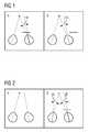
- Heterophoria and heterotropia are anomalies of a binocular vision and are conditions that prevent a person from directing both eyes simultaneously towards to a fixation target. These anomalies can be diagnosed by dissociating the eyes. In the case of heterophoria, for example, if a patient fixates a stationary fixation target and one of the eyes is covered (or dissociated, as it is called) the covered eye will turn by an angle α so that the visual axis no longer passes through the fixation target, as it is shown in panel A of
figure 1 . When the cover is removed, bifoveal fixation is rapidly regained, as shown in panel B offigure 1 . This behaviour is described inBennett & Rabbetts 1998, Clinical Visual Optics, 3rd edition. - In the case of heterotropia, patients do not achieve bifoveal fixation of any object and one of the eyes shows a manifested deviation, even without dissociation, as shown in panel A of
figure 2 . This is also called strabismus. In this case, if the strabismic eye is covered while the patient looks at the fixation point, neither the covered nor the uncovered eye will move, however if the originally fixating eye is covered, the strabismic eye will turn through the angle of misalignment and foveate the fixation point, as it is shown in panel B offigure 2 . This behaviour is also described inBennett & Rabbetts 1998, Clinical Visual Optics, 3rd edition. - A prevalence of strabismus ranging from 2 % to 5 % among preschool and school-aged European children has been found in some population-based studies (Frandsen AD. "Occurrence of Squint", Acta Ophthalmol Suppl. 1960, 62, pages 1 - 158 and "Prevalence and risk factors for common vision problems in children: Data from the ALSPAC study", Williams C, Northstone K, Howard M, Harvey I, Harrad RA, Sparrow JM, Br J Ophthalmol., July 2008, 92(7) pages 959-964) and similar values have been found in African American populations (Baltimore Vision Screening Project, Preslan MW, Novak A, "Ophthalmology", January 1996, 103(1), pages 105 - 109 andGiordano L, Friedman DS, Repka MX, et al. "Prevalence of strabismus and amblyopia in preschool-aged children: The Baltimore Pediatric Eye Disease Study", Invest Ophthalmol Vis Sci., 2008, 49, E-abstract 1552). In a study of Japanese school children a prevalence of 1 % was found ("The prevalence of strabismus and amblyopia in Japanese elementary school children", Matsuo T, Matsuo C, Ophthalmic Epidemiol., February 2005 12(1), pages 31 - 36 and "Comparison of prevalence rates of strabismus and amblyopia in Japanese elementary school children between the years 2003 and 2005", Matsuo T, Matsuo C, Acta Med Okayama, December 2007, 61(6), pages 329 - 334) and same prevalence of strabismus was found in a population-based sample of native American children in the Kindergarten and first-grade (Garvey 2010 "Prevalence of strabismus among preschool, kindergarten and first-grade Tohono O'Odham children").
- Usually, cover tests are performed to determine the presence, classification and magnitude of an ocular deviation. The test classifies the heterophoria based on the movement of the eye under cover. The classification can be esophoria or exophoria for horizontal movements and hyperphoria or hypophoria for vertical movements. The classification of heterotropia is based on the relative position of the deviating eye and can be esotropia or exotropia for horizontal eye movements and hypertropia or hypotropia for vertical movements. Another method is the so-called MKH ("Mess- und Korrektur nach Hase") measure and correction method which is critically discussed in the community of opticians and ophthalmologists.
- Once a heterophoria or heterotropia is diagnosed, it is treated by refractive, prismatic or orthoptic means. Also surgery is an option, typically the last one.
- The current methods of manually diagnosing and treating heterophoria or heterotropia often have the following issues:
- Diagnosis, classification and the determination of magnitude of heterophoria or heterotropia lead to a prescription which is verified with the standard optometric methods. This prescription, e.g. prismatic glasses, is never tested in a realistic life environment before it is actually applied.
- Diagnosis is subjective and based on the experience of the optometrist or examiner.
- Determination of the magnitude of correction is estimated by subjective observation of the optometrist and, therefore, the accuracy of the correction also depends on the experience of the examiner.
- Determination of oblique deviations is hard to be achieved and, thus, often uncorrected.
- Strabismus testing apparatus as they are described in
US 5,094,521 and inCN101147670 (A ) can overcome some of these issues. In particular,US 5,094,521 describes a measuring device which allows diagnosing strabismus and suggesting a treatment. However, even when using such testing apparatus there is no possibility for testing suggested prisms in a realistic life environment. - It is, therefore, a first objective of the present invention to provide an advantageous apparatus for assisting in establishing a correction for correcting heterotropia or heterophoria.
- It is a second objective of the present invention to provide an advantageous method of operating a computer for assisting in establishing a correction for correcting heterotropia or heterophoria.
- The mentioned objectives are achieved by an apparatus as claimed in
claim 1 and a method of operating a computer as claimed inclaim 11. The depending claims contain further developments of the invention. - An inventive apparatus for assisting in establishing a correction for correcting heterotropia or heterophoria comprises a simulation device with:
- An input interface for receiving diagnostic data indicating whether heterotropia or heterophoria is present, classification data indicating which kind of heterotropia or heterophoria is present, magnitude data indicating the magnitude of the heterotropia or heterophoria and prescription data indicating a prism or prisms suitable for correcting the heterotropia or heterophoria. The simulation device may also comprise a user interface allowing a user to input manually or verbally, or by other suitable means, the diagnosis data, the classification data, the magnitude data and the prescription data. Moreover, as an addition or as an alternative, the simulation device may comprise a reader for reading a storage medium containing stored diagnosis data, stored classification data, stored magnitude data and stored prescription data.
- A generator unit for generating a right simulation image for the right eye and a left simulation image for the left eye where the right simulation image and the left simulation image each represent the same object or scene looked at by the right eye under a right viewing angle and the left eye under left viewing angle, respectively. The right viewing angle and the left viewing angle are calculated based on the diagnostic data, the classification data, the magnitude data and the prescription data such that the right viewing angle and/or the left viewing angle correspond to viewing angles which would be achieved by applying the prism or prisms indicated by the prescription data.
- A viewing device with separate displays or display sections for the right eye and the left eye of the patient for displaying the simulation image for the right eye and the simulation image for the left eye, respectively.
- Using the simulation device offers the possibility to test the prescription in a simulated real life environment before the prescription is actually realized. This allows for correcting the prescription if the patient feels uncomfortable with the prescription before the prisms are actually manufactured.
- The right simulation image and the left simulation image used in the simulation device may be generated from an image or a video of an object or a scene stored in a memory. As an alternative, the right simulation image and the left simulation image may be generated from a real time video of the environment, e.g. of the examination room. In both cases a test of the prescription under realistic conditions can be achieved. This is particularly true when a real time image of the examination room is used as basis for the simulation images. However, for example if the examination room is not big enough for simulating an object at a certain distance the generator unit of the simulation device may use a stored object or scene instead of the real time video of the examination room.
- If heterotropia or heterophoria is accompanied by myopia or hyperopia it is advantageous that the simulation device further comprises a means that allows for correcting the refraction of the patient's eye during the simulation. Such a means may, for example, include a trial frame or at least one wave front manipulator such as a liquid lens, an Alvarez-element, or the like.
- In a further development of the inventive apparatus it further comprises a measuring device and an analysing unit.
- The measuring device is equipped with
- a means for displaying a fixation target for providing a patient with a target to gaze at,
- a switchable blocking means which allows selectively blocking the sight of the right eye and the left eye when the patient gazes at the fixation target,
- a control unit for switching the switching means according to a blocking sequence describing a sequence of blocking and unblocking the right eye and/or the left eye,
- an eye tracker for tracking the line of sight of at least one of the patient's eyes during the blocking sequence, and
- an output interface for outputting data representing the blocking sequence and data representing the line of the sight of the patient's eyes during the blocking sequence.
- The blocking sequence resembles at least one event of blocking and unblocking at least one of the eyes according to a cover test.
- As switchable blocking means of the measuring device various implementations are conceivable. In a first implementation, the switchable blocking means comprises at least one cover that can be moved to selectively cover and uncover the right eye or the left eye. In particular, there may be two covers, one for the right eye and one for the left eye which can be controlled to cover and uncover the right eye and the left eye, independently of each other. However, it would also be possible to provide only a single cover which can be switched to cover either the left eye or the right eye and which may also be brought into a neutral position, in which none of the eyes is covered.
- In a second implementation of the switchable blocking means the means comprises at least one switchable transmission display that is located in front of the patient's eyes. The display can be switched back and forth between a transmissive state and an opaque state. There may be individual displays for each eye, or there may be a single display with left and right sections that can be switched independently.
- In a third implementation of the switchable blocking means the fixation target emits polarized light and the switchable blocking means comprises at least one switchable polarizer located in front of the patient's eyes. The polarizer can be switched back and forth between a first polarizing state and a second polarizing state where the first polarizing state is transmissive for the polarized light of the fixation target while the second polarizing state is blocking the polarized light of the fixation target. Like in the first two implementations there may be individual polarizers for each eye, or there may be a single polarizer with switchable sections that can be switched independently where the switchable sections are associated to the left eye and the right eye, respectively.
- In order to allow the eye tracker to accurately track the gazing direction of the patient's eyes the inventive apparatus may include a fixation unit for fixing the patient's head so that eye movement is prohibited or at least restricted during the measurement. The need for prohibiting or restricting the movement of the patient's eye may be overcome if the measuring device comprises a head tracker for tracking the movement and/or orientation of the patient's head in addition to the eye tracker. In this case, the head tracker establishes the movement and/or orientation while the eye tracker measures the gazing direction so that the gazing direction established by means of the eye tracker can be corrected for changes in the head position and/or the head orientation. However, neither a fixation for the head nor a head tracker is necessary if the measuring device is integrated into a head mounted device (HMD). By integrating it into a head mounted device, the eye tracker is always fixed relative to the head so that a head movement is not reflected in the gazing direction determined by use of the eye tracker. If the measuring device is integrated into a head mounted device it is advantageous if the simulation device is also integrated into the head mounted device. However, it is generally also possible that only the simulation device is integrated into a head mounted device while the measuring device is a stationary device which relies on a fixation of the head or a head tracker. The head mounted device may, for example, be based on binocular disparity, holographic projection, projection on the retina, virtual retinal display, etc.
- The analysing unit is equipped with
- an input interface that is connected or connectable to the output interface of the measuring device for receiving the data representing the blocking sequence and the data representing the line of the sight of the patient's eyes during the blocking sequence,
- an evaluation unit which determines based on the line of sight of the patient's eyes during the blocking sequence deviation data which represents deviations of the line of sight of the patient's eyes from a line of sight necessary for gazing the fixation target,
- a diagnostic unit which is coupled to the evaluation unit for receiving the deviation data and which determines from the deviation data the diagnostic data, the classification data, the magnitude data and the prescription data, and
- an output interface for outputting the diagnostic data, the classification data, the magnitude data and the prescription data.
- The output interface of the analysing unit is connected or connectable to the input interface of the simulation device.
- With the described further development of the inventive apparatus a means is provided for objectively measuring eye movement in a test for diagnosing heterophoria or heterotropia since the gazing directing of the eye is not estimated by the examiner but directly measured by use of the eye tracker. In addition, the accuracy of the measurement does not depend on the experience of the examiner but only on the measurement accuracy of the eye tracker. Thus, a highly reliable and accurate determination of the line of sight of the patient's eye or eyes during the cover test by means the switchable blocking means can be achieved. In particular, by the use of the eye tracker the measurement device also allows for determining oblique gazing directions which in turn allows for determining oblique heterotropia or heterophoria.
- Furthermore, by use of the analysing unit diagnostics of heterophoria and heterotropia as well as the prescription for a treatment can be fully automated. Hence, diagnosing and treating heterotropia and heterophoria is based on objective measurements and does not depend on the examiner's experience. Furthermore, due to the fact that the measuring device is able to also detect oblique ocular deviations diagnosis and prescription is not restricted to diagnosing and treating horizontal and/or vertical heterotropia or heterophoria.
- The invention also provides a method of operating a computer for assisting in establishing a correction for correcting heterotropia or heterophoria. The method includes using diagnostic data indicating whether heterotropia or heterophoria is present, classification data indicating which kind of heterotropia or heterophoria is present, magnitude data (m) indicating the magnitude of the heterotropia or heterophoria and prescription data (p) indicating a prism or prisms suitable for correcting the heterotropia or heterophoria for generating a right simulation image for the right eye and a left simulation image for the left eye where the right simulation image and the left simulation image each represent the same object or scene looked at by the right eye under a right viewing angle and the left eye under left viewing angle, respectively. In the method, the right viewing angle and the left viewing angle are calculated based on the diagnostic data (d), the classification data (c), the magnitude data (m) and the prescription data (p) such that the right viewing angle and/or the left viewing angle correspond to viewing angles which would be achieved by applying the prism or prisms indicated by the prescription data (p). The right simulation image and the left simulation image may be generated from a stored image or video of an object or may be generated from a real time video of the environment. The method also includes outputting the right simulation image and the left simulation image, e.g. to a viewing device.
- The inventive method allows using a computer together with a viewing device with separate displays or display sections for the right eye and the left eye of the patient as an apparatus for assisting in establishing a correction for correcting heterotropia or heterophoria.
- The method may further include determining, based on data representing a blocking sequence describing a sequence of blocking and unblocking the right eye and/or the left eye and date representing the line of sight of the patient's eyes during the blocking sequence, deviation data which represent deviations of the line of sight of the patient's eyes from a line of sight necessary for gazing at the fixation target during the blocking sequence, and determining the diagnostic data, the classification data, the magnitude data, and the prescription data, from said deviation data. By this development, the computer can also be used for determining the diagnostic data, the classification data, the magnitude data, and the prescription data, from deviation data.
- In addition, the invention provides a computer program product including computer readable instructions for performing the method of operating a computer for assisting in establishing a correction for correcting heterotropia or heterophoria. A computer program product may be a computer readable storage medium such as, for example, a USB mass storage device, a flash card, a DVD. As an alternative, the computer program product may be implemented in form of one or more data packages which is/are made available for downloading form the internet or any other computer network.
- Further features, advantages and properties of the present invention will become clear from the following description of embodiments in conjunction with the accompanying drawings.
- Figure 1
- shows an example of dissociation and regain of bifoveal fixation for individuals with heterophoria.
- Figure 2
- shows an example of a strabismic eye and of the effect on axis location during dissociation.
- Figure 3
- shows an apparatus for assisting in establishing a correction for correcting heterotropia or heterophoria.
- Figure 4
- shows a detail of the apparatus shown in
Figure 3 . - Figure 5
- shows an alternative implementation of the detail shown in
Figure 4 . - An embodiment of the present invention will be described with respect to
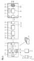
Figure 3 which shows an apparatus for assisting in establishing a correction for correcting heterotropia or heterophoria. The apparatus comprises ameasuring device 1, an analysingunit 21 and asimulation device 41. The measuringdevice 1 and the analysingunit 21 are connected to each other via anoutput interface 3 of the measuring device and aninput interface 23 of the analysingunit 21. In addition, the analysingunit 21 includes anoutput interface 25 by which it is connected to aninput interface 43 of thesimulation device 41. The interfaces can be standard interfaces or dedicated interfaces especially designed for connecting the measuringdevice 1 with the analysingunit 21 and the analysingunit 21 with thesimulation device 41, respectively. Moreover, the interfaces may be connected to each other by cable or by wireless means. - The measuring
device 1 includes afixation target 5 to which a patient to be examined directs its gaze. For stopping the head from moving when the patient gazes at thefixation target 5, the measuring device may provide afixation unit 14 with asupport 15 for the patient's chin and arest 16 for the forehead. - The measuring
device 1 further comprises a blocking means 7 which is, in the present embodiment, a device withmoveable covers covers control unit 11 which controls moving thecovers eye tracker 13 which is also part of the measuringdevice 1 tracks the gazing directions of at least one of the patient's right eye and the patient's left eye. Thecontrol unit 11 outputs data b presenting the blocking sequence through theoutput interface 3 and causes theeye tracker 13 to output data I representing the line of sight of the at least one of the patient's eyes during the blocking sequence via theoutput interface 3, - Although the switchable blocking means 7 which allows selectively blocking the sight of the right eye and the left eye when the patient gazes at the
fixation target 5 comprises twocovers moveable covers liquid crystal displays 19 could be present before the patient's eyes which can be switched by thecontrol unit 11 into a transmissive state and an opaque state (Figure 4 ). When an eye shall be covered (dissociate) therespective display 19 becomes opaque. In a still further implementation the switchable blocking means can be realized by the use ofpolarizing filters 20 in front of the patient's eyes if the light of thefixation target 5 is polarized, e.g. by a furtherpolarizing filter 20 in front of the light source of the fixation target 5 (Fig. 5 ). If, for example, thepolarizing filter 20 in front of the light source of thefixation target 5 polarizes the light linearly in a first direction thepolarizing filter 20 in front of the patient's eye can be switched between polarizing states perpendicular and parallel to the polarizing direction of the polarizer in front of thefixation target 5 so that the polarized light from thefixation target 5 can pass thepolarizing filter 20 in front of the patient's eye (polarising filters are in front of the light source and in front of the eye are oriented parallel to each other) or is blocked thepolarizing filter 20 in front of the patient's eye (polarising filters are in front of the light source and in front of the eye oriented perpendicular to each other). - As has already been mentioned, during the measurement the head of the patient is kept fixed by the
chin support 15 and therest 16 for the forehead. However, if the measuringdevice 1 is equipped with ahead tracker 17 which tracks the position and/or orientation of the head afixation unit 14 for fixing the patient's head is not necessary because the gazing direction measured by theeye tracker 13 can be corrected for taking into account the head position and/or head orientation given by the result of the measurement performed by thehead tracker 17. - A further possibility is to integrate the measuring device into 1 a head mounted 18 device like for example a helmet or goggles like structure which includes the switchable blocking means 7 and the
eye tracker 13. Since the orientation of the head mounteddevice 18 is fixed relative to the head afixation unit 14 or ahead tracker 17 are not necessary during the measurement of the gazing direction of the patient's eyes. - The analysing
unit 21 receives from measuringdevice 1 through theinput interface 23 the data b representing the blocking sequence and the data I representing the line of sight of at least one of the patient's eyes during the blocking sequence. In the analysingunit 21, anevaluation unit 27 evaluates the data b representing the blocking sequence and the data I representing the line of sight to determine whether the line of sight of the patient's eyes measured during the blocking sequence deviates from the line of sight necessary for gazing at the fixation target. The determined deviation is then output from theevaluation unit 27 in form of a deviation data Δ. - A
diagnostic unit 29 of the analysingunit 21 receives the deviation data Δ and determines from the deviation data Δ diagnostic data d, classification data c, magnitude data m, and prescription data p. The diagnostic data d represents whether or not heterotropia or heterophoria is present, the classification data c indicates which kind of heterotropia or heterophoria is present, the magnitude data m indicates the magnitude of the heterotropia or heterophoria, and the prescription data p indicates a prism or prisms suitable for correcting the heterotropia or heterophoria which has been found to be present with a certain direction and magnitude. The diagnostic data d, the classification data c, the magnitude data m and the prescription data p is then output from the analysingunit 21 through theoutput interface 25. - The data d, c, m, and p output through the
output interface 25 of the analysingunit 21 is received by theinput interface 43 of thesimulation device 41. Thesimulation device 41 includes agenerator unit 45 which generates a stereoscopic simulation image, i.e. a right simulation image for the right eye and a left simulation image for the left eye, where both simulation images represent the same object or scene under a right eye viewing angle and a left eye viewing angle, respectively. The right eye viewing angle and the left eye viewing angle are calculated based on the diagnostic data d, the classification data c, the magnitude data m and the prescription data p such that the right viewing angle and/or the left viewing angle correspond to the viewing angles achieved with the prism or prisms indicated by the prescription data. In other words, to simulate the effect of the prism or prisms without using an actual prism the viewing angle of the simulation image presented to the affected eye or eyes is/are calculated such as to simulate the line of sight which would be achieved with the prism or prisms indicated in the prescription data p installed in front of the affected eye. For example, in the case shown inFigure 2 the diagnostic data d would indicate heterotropia, the classification data c would indicated a deviation of the strabismic eye which directs away from the other eye, the magnitude data m would give the angle β and the prescription data p would describe a prism suitable for correcting the line of sight of the affected eye so that, with the prism or prisms installed, both eyes would gaze at the fixation target. - By use of the
simulation device 41, the effect of the prism or prisms can be simulated before the prism or prisms is/are manufactured and, in case the patient feels uncomfortable with the simulation, the diagnostics can be reviewed and the prism or prisms can be amended if the review reveals that a different kind of prism or prisms would lead to a result more comfortable for the patient. - For presenting the stereoscopic simulation image to the patient the
simulation device 41 comprises aviewing device 47, which, in the present embodiment, includes twodisplays viewing device 47 can be located in a stationary optical instrument or in a head mounted device. In particular, it may be integrated in the same optical instrument or the same head mounted device as the measuringdevice 1. - The right and left simulation images may be generated based on images or videos of objects or scenes stored in a
memory 53 of thesimulation device 41. When using images or videos stored in a memory 53 a large number of objects or scenes can be presented to the patient. Hence, the right and left simulation images can closely meet the needs and preferences of a patient. - On the other hand, instead of using videos or images stored in the memory 53 a live video of the examination room could be used for generating the stereoscopic simulation image. This way of generating the simulation image allows presenting a very realistic image impression to the patient.
- If the patient does not only show heterophoria or heterotropia but is also nearsighted (myopic) or farsighted (hyperopic) the lack of refraction of the patient's eyes needs also to be corrected. To this end, the viewing device preferably also includes a refraction correcting device, which may in the simplest case be a trial frame that allows putting
lenses - Although the
simulation device 41 has been described to receive the diagnostic data d, the classification data c, the magnitude data m and the prescription data p from theanalyser unit 21 it is, in the present embodiment also equipped with auser interface 44 allowing for manually or verbally inputting data relating to a diagnosis indicating whether heterotropia or heterophoria is present, a classification indicating which kind of heterotropia or heterophoria is present, a magnitude indicating the magnitude of the heterotropia or heterophoria and a prescription indicating a prism or prisms suitable for correcting the heterotropia or heterophoria. Theuser interface 44 may include a keyboard, a touchpad or touchscreen, a speech recognition unit, or any other unit that allows a user to input the mentioned data. In addition, in the present embodiment thesimulation device 41 also comprises areader 48 for reading a storage medium containing stored data relating to a diagnosis indicating whether heterotropia or heterophoria is present, a classification indicating which kind of heterotropia or heterophoria is present, a magnitude indicating the magnitude of the heterotropia or heterophoria and a prescription indicating a prism or prisms suitable for correcting the heterotropia or heterophoria. The storage medium may be any medium suitable for storing the mentioned data, for example a memory stick, a memory card, a DVD, etc. - In another embodiment of the invention, the tasks of the
generator unit 45, theinput interface 43 and theuser interface 48 are realized by a computer running a software implementing a method of using the computer for assisting in establishing a correction for correcting heterotropia or heterophoria. By running of the software, the computer uses the diagnostic data (d), the classification data (c), the magnitude data (m) and the prescription data (p) for generating a right simulation image for the right eye and a left simulation image for the left eye. The right simulation image and the left simulation image each represent the same object or scene looked at by the right eye under a right viewing angle and the left eye under left viewing angle, respectively, where the right viewing angle and the left viewing angle are calculated based on the diagnostic data (d), the classification data (c), the magnitude data (m) and the prescription data (p) such that the right viewing angle and/or the left viewing angle correspond to viewing angles which would be achieved by applying the prism or prisms indicated by the prescription data (p). The computer then outputs the right simulation image and the left simulation image to aviewing device 47 which may be a viewing device as used in the first embodiment. - In a further embodiment of the invention, the computer also realizes the tasks of the analysing
unit 21. In this embodiment, the computer running the software also determines, based on the data representing the line of sight of the patient's eyes during a blocking sequence and data representing the blocking sequence, deviation data (Δ) which represent deviations of the line of sight of the patient's eyes from a line of sight necessary for gazing at the fixation target (5) during the blocking sequence. It then determines the diagnostic data (d), the classification data (c), the magnitude data (m), and the prescription data (p), from deviation data (Δ). - The present invention has been illustrated by describing embodiments of the invention. However, a person skilled in the art realizes that deviations from the described embodiments are possible since such deviations have already been described throughout the embodiment. For example neither of the user interface, the reader, the analyser device and the measuring device is mandatory as long as there is a device which allows inputting the diagnostic data, the classification data, the magnitude data and the prescription data. The present invention shall, therefore, not be restricted by the described embodiment but only by the accompanying drawings.
- 1
- measuring device
- 3
- output interface
- 5
- fixation target
- 7
- blocking means
- 9A, B
- cover
- 11
- control unit
- 13
- eye tracker
- 21
- analysing unit
- 23
- input interface
- 25
- output interface
- 27
- evaluation unit
- 29
- diagnostic unit
- 41
- simulation device
- 43
- input interface
- 44
- user interface
- 45
- generator unit
- 47
- viewing device
- 48
- reader
- 49A, B
- display
- 51A, B
- refraction correcting device
- 53
- memory
- b
- data representing blocking sequence
- l
- data representing line of sight
- Δ
- deviation data
- d
- diagnostic data
- c
- classification data
- m
- magnitude data
- p
- prescription data
Claims (15)
- An apparatus for assisting in establishing a correction for correcting heterotropia or heterophoria, comprising a simulation device (41) with- an input interface (43) for receiving a diagnostic data (d) indicating whether heterotropia or heterophoria is present, classification data (c) indicating which kind of heterotropia or heterophoria is present, magnitude data (m) indicating the magnitude of the heterotropia or heterophoria and prescription data (p) indicating a prism or prisms suitable for correcting the heterotropia or heterophoria,- a generator unit (45) for generating a right simulation image for the right eye and a left simulation image for the left eye where the right simulation image and the left simulation image each represent the same object or scene looked at by the right eye under a right viewing angle and the left eye under left viewing angle, respectively, and where the right viewing angle and the left viewing angle are calculated based on the diagnostic data (d), the classification data (c), the magnitude data (m) and the prescription data (p) such that the right viewing angle and/or the left viewing angle correspond to viewing angles which would be achieved by applying the prism or prisms indicated by the prescription data (p), and- a viewing device (47) with separate displays (49A, 49B) or display sections for the right eye and the left eye of the patient for displaying the simulation image for the right eye and the simulation image for the left eye, respectively.
- The apparatus as claimed in claim 1, in which a means is present for generating the right simulation image and the left simulation image from an image or video of an object or a scene stored in a memory (53), and/or a means is present for generating the right simulation image and the left simulation image from a real time video of the environment.
- The apparatus as claimed in claim 1 or claim 2, in which the simulation device (41) further comprises a means (51 A, 51 B) which allows for correcting refraction of the patient's eye.
- The apparatus as claimed in claim 3, in which the means (51 A, 51 B) that allow for correcting refraction of the patient's eye is a trial frame and/or comprises at least one wave front manipulator.
- The apparatus as claimed in at least one of the claims 1 to 4, in which the simulation unit (41) further comprises a user interface (44) allowing a user to input the diagnostic data (d), the classification data (c), the magnitude data (m) and the prescription data (p) and/or a reader (48) for reading a storage medium containing stored diagnostic data (d), stored classification data, stored magnitude data (m) and stored prescription data (p).
- The apparatus as claimed in at least one of the claims 1 to 5, further comprising
a measuring device (1) with- a means for displaying a fixation target (5) for providing a patient with a target to gaze at,- a switchable blocking means (7) which allows selectively blocking the sight of the right eye and the left eye when the patient gazes at the fixation target (5),- a control unit (11) for switching the switching means (7) according to a blocking sequence describing a sequence of blocking and unblocking the right eye and/or the left eye,- an eye tracker (13) for tracking the line of sight of at least one of the patient's eyes during the blocking sequence, and- an output interface (3) for outputting data (b) representing the blocking sequence and data (I) representing the line of sight of at least one of the patient's eyes during the blocking sequence, andan analysing unit (21) with- an input interface (23) that is connected or connectable to the output interface (3) of the measuring device (1) for receiving the data (b) representing the blocking sequence and the data (I) representing the line of sight of the patient's eyes during the blocking sequence,- an evaluation unit (27) which determines based on the line of sight of the patient's eyes during the blocking sequence deviation data (Δ) which represent deviations of the line of sight of the patient's eyes from a line of sight necessary for gazing at the fixation target (5),- a diagnostic unit (29) which is coupled to the evaluation unit (27) for receiving the deviation data (Δ) and which determines from deviation data (Δ) the diagnostic data (d), the classification data (c), the magnitude data (m), and the prescription data (p), and- an output interface (25) for outputting the diagnostic data (d), the classification data (c), the magnitude data (m) and the prescription data (p),where the output interface (25) of the analysing unit (21) is connected or connectable to the input interface (43) of the simulation device (41). - The apparatus as claimed in claim 6, in which the switchable blocking means (7) comprises at least one cover (9A, 9B) that can be moved to selectively cover the right eye or the left right eye and/or at least one switchable transmission display (19) locatable in front of the patient's eyes where the display can be switched between a transmissive state and an opaque state.
- The apparatus as claimed in claim 6, in which the fixation target (5) emits polarised light and the switchable blocking means (7) comprises at least one switchable polariser (20) locatable in front of the patient's eyes where the polariser (20) can be switched between a first polarising state being transmissive for the polarised light of the fixation target (5) and a second polarising state blocking the polarised light of the fixation target (5).
- The apparatus as claimed in at least one of the claims 6 to 8, in which the measuring device (1) further comprises a fixation unit (14) for fixing the patient's head and/or a head tracker (17) for tracking the movement of the patient's head.
- The apparatus of at least one of the claims 1 to 5 and/or at least one of the claims 6 to 9, in which the measuring device (1) and/or the viewing device (41) of the simulation device are integrated in a head mounted device (18).
- A method of operating a computer for assisting in establishing a correction for correcting heterotropia or heterophoria which includes using diagnostic data (d) indicating whether heterotropia or heterophoria is present, classification data (c) indicating which kind of heterotropia or heterophoria is present, magnitude data (m) indicating the magnitude of the heterotropia or heterophoria and prescription data (p) indicating a prism or prisms suitable for correcting the heterotropia or heterophoria for generating a right simulation image for the right eye and a left simulation image for the left eye where the right simulation image and the left simulation image each represent the same object or scene looked at by the right eye under a right viewing angle and the left eye under left viewing angle, respectively, where the right viewing angle and the left viewing angle are calculated based on the diagnostic data (d), the classification data (c), the magnitude data (m) and the prescription data (p) such that the right viewing angle and/or the left viewing angle correspond to viewing angles which would be achieved by applying the prism or prisms indicated by the prescription data (p), and outputting the right simulation image and the left simulation image.
- The method claimed in claim 11, which further includes determining, based on data representing a blocking sequence describing a sequence of blocking and unblocking the right eye and/or the left eye and date representing the line of sight of the patient's eyes during the blocking sequence, deviation data (Δ) which represent deviations of the line of sight of the patient's eyes from a line of sight necessary for gazing at the fixation target (5) during the blocking sequence, and determining the diagnostic data (d), the classification data (c), the magnitude data (m), and the prescription data (p), from deviation data (Δ).
- The method as claimed in claim 11 or claim 12, in which the right simulation image and the left simulation image are generated from a stored image or video of an object.
- The method as claimed in claim 11 or claim 12, in which the right simulation image and the left simulation image are generated from a real time video of the environment.
- A computer program product including computer readable instructions for performing the method claimed in any of the claims 11 to 14.
Priority Applications (5)
| Application Number | Priority Date | Filing Date | Title |
|---|---|---|---|
| ES16188905TES2734285T3 (en) | 2016-09-15 | 2016-09-15 | Apparatus to help establish a correction to correct heterotropy or heteroforia and procedure to operate a computer to help establish a correction to correct heterotropy or heteroforia |
| EP16188905.0AEP3295864B1 (en) | 2016-09-15 | 2016-09-15 | Apparatus for assisting in establishing a correction for correcting heterotropia or heterophoria and method of operating a computer for assisting in establishing a correction for correcting heterotropia or heterophoria |
| CN201780056526.3ACN109688898B (en) | 2016-09-15 | 2017-09-08 | Device for assisting in the establishment of a correction for correcting strabismus or heterophoria and related method |
| PCT/EP2017/072633WO2018050561A1 (en) | 2016-09-15 | 2017-09-08 | Apparatus for assisting in establishing a correction for correcting heterotropia or heterophoria and method of operating a computer for assisting in establishing a correction for correcting heterotropia or heterophoria |
| US16/353,027US10405742B2 (en) | 2016-09-15 | 2019-03-14 | Apparatus for assisting in establishing a correction for correcting heterotropia or heterophoria and method of operating a computer for assisting in establishing a correction for correcting heterotropia or heterophoria |
Applications Claiming Priority (1)
| Application Number | Priority Date | Filing Date | Title |
|---|---|---|---|
| EP16188905.0AEP3295864B1 (en) | 2016-09-15 | 2016-09-15 | Apparatus for assisting in establishing a correction for correcting heterotropia or heterophoria and method of operating a computer for assisting in establishing a correction for correcting heterotropia or heterophoria |
Publications (2)
| Publication Number | Publication Date |
|---|---|
| EP3295864A1true EP3295864A1 (en) | 2018-03-21 |
| EP3295864B1 EP3295864B1 (en) | 2019-05-15 |
Family
ID=56958762
Family Applications (1)
| Application Number | Title | Priority Date | Filing Date |
|---|---|---|---|
| EP16188905.0AActiveEP3295864B1 (en) | 2016-09-15 | 2016-09-15 | Apparatus for assisting in establishing a correction for correcting heterotropia or heterophoria and method of operating a computer for assisting in establishing a correction for correcting heterotropia or heterophoria |
Country Status (5)
| Country | Link |
|---|---|
| US (1) | US10405742B2 (en) |
| EP (1) | EP3295864B1 (en) |
| CN (1) | CN109688898B (en) |
| ES (1) | ES2734285T3 (en) |
| WO (1) | WO2018050561A1 (en) |
Cited By (2)
| Publication number | Priority date | Publication date | Assignee | Title |
|---|---|---|---|---|
| EP3750471A1 (en)* | 2019-06-12 | 2020-12-16 | Oculus Optikgeräte GmbH | Method for the testing of eyes and eye testing device |
| CN114077061A (en)* | 2020-08-19 | 2022-02-22 | 宏达国际电子股份有限公司 | Head-mounted display device and eye-tracking device |
Families Citing this family (15)
| Publication number | Priority date | Publication date | Assignee | Title |
|---|---|---|---|---|
| US10962780B2 (en)* | 2015-10-26 | 2021-03-30 | Microsoft Technology Licensing, Llc | Remote rendering for virtual images |
| JP6897467B2 (en)* | 2017-10-02 | 2021-06-30 | 富士通株式会社 | Line-of-sight detection device, line-of-sight detection program, and line-of-sight detection method |
| US11801821B2 (en)* | 2020-11-24 | 2023-10-31 | Bendix Commercial Vehicle Systems Llc | Brake wear sensing apparatus and system |
| CN113208884B (en)* | 2021-01-08 | 2022-08-09 | 上海青研科技有限公司 | Visual detection and visual training equipment |
| CN112807200B (en)* | 2021-01-08 | 2022-07-19 | 上海青研科技有限公司 | Strabismus training equipment |
| CN114642502B (en)* | 2022-02-21 | 2023-07-14 | 北京工业大学 | Auxiliary design method and device for strabismus operation plan |
| CN117111332B (en)* | 2022-05-17 | 2025-08-26 | 陈光伟 | Preparation method of children's heterophoria correction glasses |
| JP2025525666A (en)* | 2022-07-29 | 2025-08-05 | エデンルックス コーポレーション | Vision training device and vision training method |
| CN115643395A (en)* | 2022-12-23 | 2023-01-24 | 广州视景医疗软件有限公司 | Visual training method and device based on virtual reality |
| CN116418962A (en)* | 2023-02-06 | 2023-07-11 | 深圳创通联达智能技术有限公司 | Visual information processing method, device, equipment and computer storage medium |
| CN116095296A (en)* | 2023-02-15 | 2023-05-09 | 香港智能建造研发中心有限公司 | Building site monitoring method and device |
| TWI878808B (en)* | 2023-02-25 | 2025-04-01 | 逢甲大學 | Virtual reality head mounted display with build-in strabismus treatment |
| US20240335109A1 (en)* | 2023-04-04 | 2024-10-10 | Neurolens, Inc. | Prismatic lens for reducing a dynamic alignment differential |
| US20250004304A1 (en)* | 2023-06-28 | 2025-01-02 | Pixieray Oy | Optical adjustment in adaptive lenses during eye blinks |
| US20250004269A1 (en)* | 2023-06-28 | 2025-01-02 | Pixieray Oy | Optical adjustment in adaptive lenses during saccades |
Citations (5)
| Publication number | Priority date | Publication date | Assignee | Title |
|---|---|---|---|---|
| US5094521A (en) | 1990-11-07 | 1992-03-10 | Vision Research Laboratories | Apparatus for evaluating eye alignment |
| CN101147670A (en) | 2007-08-29 | 2008-03-26 | 中国人民解放军第三军医大学第一附属医院 | Strabismus Tester |
| EP2856931A1 (en)* | 2012-05-25 | 2015-04-08 | HOYA Corporation | Simulation device |
| WO2015125848A1 (en)* | 2014-02-19 | 2015-08-27 | ホヤ レンズ タイランド リミテッド | Spectacle lens supply system, spectacle lens supply method, spectacle lens supply program, spectacle lens recommended type presentation device, and spectacle lens production method |
| WO2016139662A1 (en)* | 2015-03-01 | 2016-09-09 | Improved Vision Systems (I.V.S.) Ltd. | A system and method for measuring ocular motility |
Family Cites Families (9)
| Publication number | Priority date | Publication date | Assignee | Title |
|---|---|---|---|---|
| US5293187A (en)* | 1992-02-05 | 1994-03-08 | Biocontrol Systems, Inc. | Method and apparatus for eye tracking for convergence and strabismus measurement |
| CN1291281A (en)* | 1997-11-21 | 2001-04-11 | 自控技术公司 | Objective measurement and correction of optical systems using wavefront analysis |
| JP4185337B2 (en)* | 2002-09-13 | 2008-11-26 | 株式会社トプコン | Correction element determination apparatus and method |
| US20090153796A1 (en)* | 2005-09-02 | 2009-06-18 | Arthur Rabner | Multi-functional optometric-ophthalmic system for testing diagnosing, or treating, vision or eyes of a subject, and methodologies thereof |
| JP4954586B2 (en)* | 2006-03-31 | 2012-06-20 | 株式会社トプコン | Optometry program, recording medium having the optometry program, and subjective optometry apparatus |
| DE102008015189A1 (en) | 2008-03-20 | 2009-10-01 | Rodenstock Gmbh | Rescaling the target astigmatism for other additions |
| US9895058B2 (en)* | 2011-03-25 | 2018-02-20 | Carl Zeiss Meditec Ag | Heads-up vision analyzer |
| US9492075B2 (en)* | 2013-01-18 | 2016-11-15 | Hoya Corporation | Prism prescription value acquisition system, acquisition method, acquisition apparatus and program for correcting fixation disparity |
| JP6311004B2 (en)* | 2014-02-19 | 2018-04-11 | ホヤ レンズ タイランド リミテッドHOYA Lens Thailand Ltd | Spectacle lens supply system, program thereof, prism amount determination device, prism amount determination method, and spectacle lens manufacturing method |
- 2016
- 2016-09-15ESES16188905Tpatent/ES2734285T3/enactiveActive
- 2016-09-15EPEP16188905.0Apatent/EP3295864B1/enactiveActive
- 2017
- 2017-09-08WOPCT/EP2017/072633patent/WO2018050561A1/ennot_activeCeased
- 2017-09-08CNCN201780056526.3Apatent/CN109688898B/enactiveActive
- 2019
- 2019-03-14USUS16/353,027patent/US10405742B2/enactiveActive
Patent Citations (6)
| Publication number | Priority date | Publication date | Assignee | Title |
|---|---|---|---|---|
| US5094521A (en) | 1990-11-07 | 1992-03-10 | Vision Research Laboratories | Apparatus for evaluating eye alignment |
| CN101147670A (en) | 2007-08-29 | 2008-03-26 | 中国人民解放军第三军医大学第一附属医院 | Strabismus Tester |
| EP2856931A1 (en)* | 2012-05-25 | 2015-04-08 | HOYA Corporation | Simulation device |
| WO2015125848A1 (en)* | 2014-02-19 | 2015-08-27 | ホヤ レンズ タイランド リミテッド | Spectacle lens supply system, spectacle lens supply method, spectacle lens supply program, spectacle lens recommended type presentation device, and spectacle lens production method |
| US20170052389A1 (en)* | 2014-02-19 | 2017-02-23 | Hoya Lens Thailand Ltd. | Spectacle lens supply system, spectacle lens supply method, spectacle lens supply program, spectacle lens recommended type presentation device, and spectacle lens production method |
| WO2016139662A1 (en)* | 2015-03-01 | 2016-09-09 | Improved Vision Systems (I.V.S.) Ltd. | A system and method for measuring ocular motility |
Non-Patent Citations (8)
| Title |
|---|
| BENNETT; RABBETTS: "Clinical Visual Optics", 1998 |
| FRANDSEN AD.: "Occurrence of Squint", ACTA OPHTHALMOL SUPPL., vol. 62, 1960, pages 1 - 158 |
| GARVEY, PREVALENCE OF STRABISMUS AMONG PRESCHOOL, KINDERGARTEN AND FIRST-GRADE TOHONO O'ODHAM CHILDREN, 2010 |
| GIORDANO L; FRIEDMAN DS; REPKA MX ET AL.: "Prevalence of strabismus and amblyopia in preschool-aged children: The Baltimore Pediatric Eye Disease Study", INVEST OPHTHALMOL VIS SCI., 2008, pages 49 |
| MATSUO T; MATSUO C: "Comparison of prevalence rates of strabismus and amblyopia in Japanese elementary school children between the years 2003 and 2005", ACTA MED OKAYAMA, vol. 61, no. 6, December 2007 (2007-12-01), pages 329 - 334 |
| MATSUO T; MATSUO C: "The prevalence of strabismus and amblyopia in Japanese elementary school children", OPHTHALMIC EPIDEMIOL., vol. 12, no. 1, February 2005 (2005-02-01), pages 31 - 36 |
| PRESLAN MW; NOVAK A, OPHTHALMOLOGY, vol. 103, no. 1, January 1996 (1996-01-01), pages 105 - 109 |
| WILLIAMS C; NORTHSTONE K; HOWARD M; HARVEY I; HARRAD RA; SPARROW JM: "Prevalence and risk factors for common vision problems in children: Data from the ALSPAC study", BR J OPHTHALMOL., vol. 92, no. 7, July 2008 (2008-07-01), pages 959 - 964 |
Cited By (2)
| Publication number | Priority date | Publication date | Assignee | Title |
|---|---|---|---|---|
| EP3750471A1 (en)* | 2019-06-12 | 2020-12-16 | Oculus Optikgeräte GmbH | Method for the testing of eyes and eye testing device |
| CN114077061A (en)* | 2020-08-19 | 2022-02-22 | 宏达国际电子股份有限公司 | Head-mounted display device and eye-tracking device |
Also Published As
| Publication number | Publication date |
|---|---|
| WO2018050561A1 (en) | 2018-03-22 |
| CN109688898A (en) | 2019-04-26 |
| US20190209005A1 (en) | 2019-07-11 |
| ES2734285T3 (en) | 2019-12-05 |
| CN109688898B (en) | 2022-03-18 |
| EP3295864B1 (en) | 2019-05-15 |
| US10405742B2 (en) | 2019-09-10 |
Similar Documents
| Publication | Publication Date | Title |
|---|---|---|
| US10405742B2 (en) | Apparatus for assisting in establishing a correction for correcting heterotropia or heterophoria and method of operating a computer for assisting in establishing a correction for correcting heterotropia or heterophoria | |
| EP1691669B1 (en) | Ophthalmic binocular wavefront measurement system | |
| Sweeney et al. | Quantifying interactions between accommodation and vergence in a binocularly normal population | |
| US12357168B2 (en) | Holographic real space refractive system | |
| US20250295307A1 (en) | A device and a method for automated measurements of eyeball deviation and/or vertical and horizontal viewing angles | |
| JP2019063265A (en) | Subjective optometer | |
| US20150320306A1 (en) | Device and method for checking human vision | |
| EP1082939A2 (en) | Digital synoptophore | |
| Antona et al. | Validity and repeatability of a new test for aniseikonia | |
| JP7528640B2 (en) | Opthalmography device and control program for optometric device | |
| JP4494075B2 (en) | Optometry equipment | |
| JP7295960B2 (en) | Virtual reality vision inspection system | |
| KR102209573B1 (en) | Strabismus score assessmenting apparatus and mathod | |
| JP7548616B1 (en) | VR head-mounted display with strabismus correction function and its operation method | |
| US20240049963A1 (en) | Cover-Uncover Test in a VR/AR Headset | |
| Ramsdale et al. | Accommodation and convergence: effects of lenses and prisms in ‘closed‐loop’conditions | |
| JP7293726B2 (en) | Subjective optometric device and subjective optometric program | |
| RU2846234C1 (en) | Digital glasses for recovery of binocular vision | |
| Predebon | Convergence responses to monocularly viewed objects: implications for distance perception | |
| Ron et al. | Clinical Evaluation of Strabismus | |
| EP4183320A1 (en) | Method for comparing two ophthalmic lenses having different optical designs | |
| Hartikainen | Advancing Technology of Automated Refraction–Developing an Algorithm for Automated Subjective Refractometer: An Innovation Project | |
| Bandela | Role of optical and non-optical cues to decipher the sign of defocus and magnitude of ocular accommodation | |
| Kelly et al. | Eye-hand coordination during visually-guided reaching in children with monocular deprivation amblyopia | |
| KR20220168883A (en) | Method for Screening Strabismus and Device therefor |
Legal Events
| Date | Code | Title | Description |
|---|---|---|---|
| PUAI | Public reference made under article 153(3) epc to a published international application that has entered the european phase | Free format text:ORIGINAL CODE: 0009012 | |
| STAA | Information on the status of an ep patent application or granted ep patent | Free format text:STATUS: THE APPLICATION HAS BEEN PUBLISHED | |
| AK | Designated contracting states | Kind code of ref document:A1 Designated state(s):AL AT BE BG CH CY CZ DE DK EE ES FI FR GB GR HR HU IE IS IT LI LT LU LV MC MK MT NL NO PL PT RO RS SE SI SK SM TR | |
| AX | Request for extension of the european patent | Extension state:BA ME | |
| STAA | Information on the status of an ep patent application or granted ep patent | Free format text:STATUS: REQUEST FOR EXAMINATION WAS MADE | |
| 17P | Request for examination filed | Effective date:20180829 | |
| RBV | Designated contracting states (corrected) | Designated state(s):AL AT BE BG CH CY CZ DE DK EE ES FI FR GB GR HR HU IE IS IT LI LT LU LV MC MK MT NL NO PL PT RO RS SE SI SK SM TR | |
| GRAJ | Information related to disapproval of communication of intention to grant by the applicant or resumption of examination proceedings by the epo deleted | Free format text:ORIGINAL CODE: EPIDOSDIGR1 | |
| GRAP | Despatch of communication of intention to grant a patent | Free format text:ORIGINAL CODE: EPIDOSNIGR1 | |
| RIC1 | Information provided on ipc code assigned before grant | Ipc:A61B 3/08 20060101AFI20181219BHEP Ipc:A61B 3/113 20060101ALN20181219BHEP Ipc:A61B 3/00 20060101ALI20181219BHEP Ipc:G02C 7/02 20060101ALN20181219BHEP | |
| GRAP | Despatch of communication of intention to grant a patent | Free format text:ORIGINAL CODE: EPIDOSNIGR1 | |
| STAA | Information on the status of an ep patent application or granted ep patent | Free format text:STATUS: GRANT OF PATENT IS INTENDED | |
| RIC1 | Information provided on ipc code assigned before grant | Ipc:A61B 3/00 20060101ALI20190114BHEP Ipc:G02C 7/02 20060101ALN20190114BHEP Ipc:A61B 3/08 20060101AFI20190114BHEP Ipc:A61B 3/113 20060101ALN20190114BHEP | |
| INTG | Intention to grant announced | Effective date:20190201 | |
| RIC1 | Information provided on ipc code assigned before grant | Ipc:A61B 3/08 20060101AFI20190123BHEP Ipc:A61B 3/00 20060101ALI20190123BHEP Ipc:A61B 3/113 20060101ALN20190123BHEP Ipc:G02C 7/02 20060101ALN20190123BHEP | |
| GRAS | Grant fee paid | Free format text:ORIGINAL CODE: EPIDOSNIGR3 | |
| GRAA | (expected) grant | Free format text:ORIGINAL CODE: 0009210 | |
| STAA | Information on the status of an ep patent application or granted ep patent | Free format text:STATUS: THE PATENT HAS BEEN GRANTED | |
| AK | Designated contracting states | Kind code of ref document:B1 Designated state(s):AL AT BE BG CH CY CZ DE DK EE ES FI FR GB GR HR HU IE IS IT LI LT LU LV MC MK MT NL NO PL PT RO RS SE SI SK SM TR | |
| REG | Reference to a national code | Ref country code:CH Ref legal event code:EP | |
| RIN1 | Information on inventor provided before grant (corrected) | Inventor name:BARRAZA-BERNAL, MARIA JOSE Inventor name:WAHL, SIEGFRIED | |
| REG | Reference to a national code | Ref country code:DE Ref legal event code:R096 Ref document number:602016013931 Country of ref document:DE | |
| REG | Reference to a national code | Ref country code:IE Ref legal event code:FG4D | |
| REG | Reference to a national code | Ref country code:NL Ref legal event code:MP Effective date:20190515 | |
| REG | Reference to a national code | Ref country code:LT Ref legal event code:MG4D | |
| PG25 | Lapsed in a contracting state [announced via postgrant information from national office to epo] | Ref country code:NL Free format text:LAPSE BECAUSE OF FAILURE TO SUBMIT A TRANSLATION OF THE DESCRIPTION OR TO PAY THE FEE WITHIN THE PRESCRIBED TIME-LIMIT Effective date:20190515 Ref country code:SE Free format text:LAPSE BECAUSE OF FAILURE TO SUBMIT A TRANSLATION OF THE DESCRIPTION OR TO PAY THE FEE WITHIN THE PRESCRIBED TIME-LIMIT Effective date:20190515 Ref country code:LT Free format text:LAPSE BECAUSE OF FAILURE TO SUBMIT A TRANSLATION OF THE DESCRIPTION OR TO PAY THE FEE WITHIN THE PRESCRIBED TIME-LIMIT Effective date:20190515 Ref country code:NO Free format text:LAPSE BECAUSE OF FAILURE TO SUBMIT A TRANSLATION OF THE DESCRIPTION OR TO PAY THE FEE WITHIN THE PRESCRIBED TIME-LIMIT Effective date:20190815 Ref country code:PT Free format text:LAPSE BECAUSE OF FAILURE TO SUBMIT A TRANSLATION OF THE DESCRIPTION OR TO PAY THE FEE WITHIN THE PRESCRIBED TIME-LIMIT Effective date:20190915 Ref country code:AL Free format text:LAPSE BECAUSE OF FAILURE TO SUBMIT A TRANSLATION OF THE DESCRIPTION OR TO PAY THE FEE WITHIN THE PRESCRIBED TIME-LIMIT Effective date:20190515 Ref country code:HR Free format text:LAPSE BECAUSE OF FAILURE TO SUBMIT A TRANSLATION OF THE DESCRIPTION OR TO PAY THE FEE WITHIN THE PRESCRIBED TIME-LIMIT Effective date:20190515 Ref country code:FI Free format text:LAPSE BECAUSE OF FAILURE TO SUBMIT A TRANSLATION OF THE DESCRIPTION OR TO PAY THE FEE WITHIN THE PRESCRIBED TIME-LIMIT Effective date:20190515 | |
| PG25 | Lapsed in a contracting state [announced via postgrant information from national office to epo] | Ref country code:GR Free format text:LAPSE BECAUSE OF FAILURE TO SUBMIT A TRANSLATION OF THE DESCRIPTION OR TO PAY THE FEE WITHIN THE PRESCRIBED TIME-LIMIT Effective date:20190816 Ref country code:BG Free format text:LAPSE BECAUSE OF FAILURE TO SUBMIT A TRANSLATION OF THE DESCRIPTION OR TO PAY THE FEE WITHIN THE PRESCRIBED TIME-LIMIT Effective date:20190815 Ref country code:RS Free format text:LAPSE BECAUSE OF FAILURE TO SUBMIT A TRANSLATION OF THE DESCRIPTION OR TO PAY THE FEE WITHIN THE PRESCRIBED TIME-LIMIT Effective date:20190515 Ref country code:LV Free format text:LAPSE BECAUSE OF FAILURE TO SUBMIT A TRANSLATION OF THE DESCRIPTION OR TO PAY THE FEE WITHIN THE PRESCRIBED TIME-LIMIT Effective date:20190515 | |
| REG | Reference to a national code | Ref country code:ES Ref legal event code:FG2A Ref document number:2734285 Country of ref document:ES Kind code of ref document:T3 Effective date:20191205 | |
| REG | Reference to a national code | Ref country code:AT Ref legal event code:MK05 Ref document number:1132443 Country of ref document:AT Kind code of ref document:T Effective date:20190515 | |
| PG25 | Lapsed in a contracting state [announced via postgrant information from national office to epo] | Ref country code:DK Free format text:LAPSE BECAUSE OF FAILURE TO SUBMIT A TRANSLATION OF THE DESCRIPTION OR TO PAY THE FEE WITHIN THE PRESCRIBED TIME-LIMIT Effective date:20190515 Ref country code:AT Free format text:LAPSE BECAUSE OF FAILURE TO SUBMIT A TRANSLATION OF THE DESCRIPTION OR TO PAY THE FEE WITHIN THE PRESCRIBED TIME-LIMIT Effective date:20190515 Ref country code:EE Free format text:LAPSE BECAUSE OF FAILURE TO SUBMIT A TRANSLATION OF THE DESCRIPTION OR TO PAY THE FEE WITHIN THE PRESCRIBED TIME-LIMIT Effective date:20190515 Ref country code:SK Free format text:LAPSE BECAUSE OF FAILURE TO SUBMIT A TRANSLATION OF THE DESCRIPTION OR TO PAY THE FEE WITHIN THE PRESCRIBED TIME-LIMIT Effective date:20190515 Ref country code:CZ Free format text:LAPSE BECAUSE OF FAILURE TO SUBMIT A TRANSLATION OF THE DESCRIPTION OR TO PAY THE FEE WITHIN THE PRESCRIBED TIME-LIMIT Effective date:20190515 | |
| REG | Reference to a national code | Ref country code:DE Ref legal event code:R097 Ref document number:602016013931 Country of ref document:DE | |
| PG25 | Lapsed in a contracting state [announced via postgrant information from national office to epo] | Ref country code:SM Free format text:LAPSE BECAUSE OF FAILURE TO SUBMIT A TRANSLATION OF THE DESCRIPTION OR TO PAY THE FEE WITHIN THE PRESCRIBED TIME-LIMIT Effective date:20190515 | |
| PLBE | No opposition filed within time limit | Free format text:ORIGINAL CODE: 0009261 | |
| STAA | Information on the status of an ep patent application or granted ep patent | Free format text:STATUS: NO OPPOSITION FILED WITHIN TIME LIMIT | |
| PG25 | Lapsed in a contracting state [announced via postgrant information from national office to epo] | Ref country code:TR Free format text:LAPSE BECAUSE OF FAILURE TO SUBMIT A TRANSLATION OF THE DESCRIPTION OR TO PAY THE FEE WITHIN THE PRESCRIBED TIME-LIMIT Effective date:20190515 | |
| 26N | No opposition filed | Effective date:20200218 | |
| PG25 | Lapsed in a contracting state [announced via postgrant information from national office to epo] | Ref country code:PL Free format text:LAPSE BECAUSE OF FAILURE TO SUBMIT A TRANSLATION OF THE DESCRIPTION OR TO PAY THE FEE WITHIN THE PRESCRIBED TIME-LIMIT Effective date:20190515 | |
| PG25 | Lapsed in a contracting state [announced via postgrant information from national office to epo] | Ref country code:SI Free format text:LAPSE BECAUSE OF FAILURE TO SUBMIT A TRANSLATION OF THE DESCRIPTION OR TO PAY THE FEE WITHIN THE PRESCRIBED TIME-LIMIT Effective date:20190515 Ref country code:MC Free format text:LAPSE BECAUSE OF FAILURE TO SUBMIT A TRANSLATION OF THE DESCRIPTION OR TO PAY THE FEE WITHIN THE PRESCRIBED TIME-LIMIT Effective date:20190515 | |
| REG | Reference to a national code | Ref country code:CH Ref legal event code:PL | |
| PG25 | Lapsed in a contracting state [announced via postgrant information from national office to epo] | Ref country code:CH Free format text:LAPSE BECAUSE OF NON-PAYMENT OF DUE FEES Effective date:20190930 Ref country code:LI Free format text:LAPSE BECAUSE OF NON-PAYMENT OF DUE FEES Effective date:20190930 Ref country code:IE Free format text:LAPSE BECAUSE OF NON-PAYMENT OF DUE FEES Effective date:20190915 Ref country code:LU Free format text:LAPSE BECAUSE OF NON-PAYMENT OF DUE FEES Effective date:20190915 | |
| REG | Reference to a national code | Ref country code:BE Ref legal event code:MM Effective date:20190930 | |
| PG25 | Lapsed in a contracting state [announced via postgrant information from national office to epo] | Ref country code:BE Free format text:LAPSE BECAUSE OF NON-PAYMENT OF DUE FEES Effective date:20190930 | |
| PG25 | Lapsed in a contracting state [announced via postgrant information from national office to epo] | Ref country code:RO Free format text:LAPSE BECAUSE OF FAILURE TO SUBMIT A TRANSLATION OF THE DESCRIPTION OR TO PAY THE FEE WITHIN THE PRESCRIBED TIME-LIMIT Effective date:20190515 | |
| PG25 | Lapsed in a contracting state [announced via postgrant information from national office to epo] | Ref country code:CY Free format text:LAPSE BECAUSE OF FAILURE TO SUBMIT A TRANSLATION OF THE DESCRIPTION OR TO PAY THE FEE WITHIN THE PRESCRIBED TIME-LIMIT Effective date:20190515 | |
| PG25 | Lapsed in a contracting state [announced via postgrant information from national office to epo] | Ref country code:IS Free format text:LAPSE BECAUSE OF FAILURE TO SUBMIT A TRANSLATION OF THE DESCRIPTION OR TO PAY THE FEE WITHIN THE PRESCRIBED TIME-LIMIT Effective date:20190915 | |
| PG25 | Lapsed in a contracting state [announced via postgrant information from national office to epo] | Ref country code:MT Free format text:LAPSE BECAUSE OF FAILURE TO SUBMIT A TRANSLATION OF THE DESCRIPTION OR TO PAY THE FEE WITHIN THE PRESCRIBED TIME-LIMIT Effective date:20190515 Ref country code:HU Free format text:LAPSE BECAUSE OF FAILURE TO SUBMIT A TRANSLATION OF THE DESCRIPTION OR TO PAY THE FEE WITHIN THE PRESCRIBED TIME-LIMIT; INVALID AB INITIO Effective date:20160915 | |
| PG25 | Lapsed in a contracting state [announced via postgrant information from national office to epo] | Ref country code:MK Free format text:LAPSE BECAUSE OF FAILURE TO SUBMIT A TRANSLATION OF THE DESCRIPTION OR TO PAY THE FEE WITHIN THE PRESCRIBED TIME-LIMIT Effective date:20190515 | |
| PGFP | Annual fee paid to national office [announced via postgrant information from national office to epo] | Ref country code:DE Payment date:20240918 Year of fee payment:9 | |
| PGFP | Annual fee paid to national office [announced via postgrant information from national office to epo] | Ref country code:GB Payment date:20240920 Year of fee payment:9 | |
| PGFP | Annual fee paid to national office [announced via postgrant information from national office to epo] | Ref country code:FR Payment date:20240925 Year of fee payment:9 | |
| PGFP | Annual fee paid to national office [announced via postgrant information from national office to epo] | Ref country code:IT Payment date:20240924 Year of fee payment:9 | |
| PGFP | Annual fee paid to national office [announced via postgrant information from national office to epo] | Ref country code:ES Payment date:20241028 Year of fee payment:9 |