EP3145398B1 - Device and method for noninvasively determining the hematocrit value of a subject - Google Patents
Device and method for noninvasively determining the hematocrit value of a subjectDownload PDFInfo
- Publication number
- EP3145398B1 EP3145398B1EP15719731.0AEP15719731AEP3145398B1EP 3145398 B1EP3145398 B1EP 3145398B1EP 15719731 AEP15719731 AEP 15719731AEP 3145398 B1EP3145398 B1EP 3145398B1
- Authority
- EP
- European Patent Office
- Prior art keywords
- light
- wavelength
- subject
- ratio
- ppg
- Prior art date
- Legal status (The legal status is an assumption and is not a legal conclusion. Google has not performed a legal analysis and makes no representation as to the accuracy of the status listed.)
- Active
Links
Images
Classifications
- A—HUMAN NECESSITIES
- A61—MEDICAL OR VETERINARY SCIENCE; HYGIENE
- A61B—DIAGNOSIS; SURGERY; IDENTIFICATION
- A61B5/00—Measuring for diagnostic purposes; Identification of persons
- A61B5/145—Measuring characteristics of blood in vivo, e.g. gas concentration or pH-value ; Measuring characteristics of body fluids or tissues, e.g. interstitial fluid or cerebral tissue
- A61B5/14535—Measuring characteristics of blood in vivo, e.g. gas concentration or pH-value ; Measuring characteristics of body fluids or tissues, e.g. interstitial fluid or cerebral tissue for measuring haematocrit
- A—HUMAN NECESSITIES
- A61—MEDICAL OR VETERINARY SCIENCE; HYGIENE
- A61B—DIAGNOSIS; SURGERY; IDENTIFICATION
- A61B5/00—Measuring for diagnostic purposes; Identification of persons
- A61B5/02—Detecting, measuring or recording for evaluating the cardiovascular system, e.g. pulse, heart rate, blood pressure or blood flow
- A61B5/026—Measuring blood flow
- A61B5/0295—Measuring blood flow using plethysmography, i.e. measuring the variations in the volume of a body part as modified by the circulation of blood therethrough, e.g. impedance plethysmography
- A—HUMAN NECESSITIES
- A61—MEDICAL OR VETERINARY SCIENCE; HYGIENE
- A61B—DIAGNOSIS; SURGERY; IDENTIFICATION
- A61B5/00—Measuring for diagnostic purposes; Identification of persons
- A61B5/145—Measuring characteristics of blood in vivo, e.g. gas concentration or pH-value ; Measuring characteristics of body fluids or tissues, e.g. interstitial fluid or cerebral tissue
- A61B5/1455—Measuring characteristics of blood in vivo, e.g. gas concentration or pH-value ; Measuring characteristics of body fluids or tissues, e.g. interstitial fluid or cerebral tissue using optical sensors, e.g. spectral photometrical oximeters
- A—HUMAN NECESSITIES
- A61—MEDICAL OR VETERINARY SCIENCE; HYGIENE
- A61B—DIAGNOSIS; SURGERY; IDENTIFICATION
- A61B5/00—Measuring for diagnostic purposes; Identification of persons
- A61B5/72—Signal processing specially adapted for physiological signals or for diagnostic purposes
- A61B5/7271—Specific aspects of physiological measurement analysis
- A—HUMAN NECESSITIES
- A61—MEDICAL OR VETERINARY SCIENCE; HYGIENE
- A61B—DIAGNOSIS; SURGERY; IDENTIFICATION
- A61B2562/00—Details of sensors; Constructional details of sensor housings or probes; Accessories for sensors
- A61B2562/02—Details of sensors specially adapted for in-vivo measurements
- A61B2562/0233—Special features of optical sensors or probes classified in A61B5/00
Definitions
- the present inventionrelates to a device and method for noninvasively determining the hematocrit value of a subject, such as a person or animal. Further, the present invention relates to a processor, processing method and computer program.
- US 6606509 B2discloses a device and method for measurement of hematocrit (Hct) by noninvasive means.
- the changes in the intensities of light of multiple wavelengths transmitted through or reflected light from the tissue locationare recorded immediately before and after occluding the flow of venous blood from the tissue location with an occlusion device positioned near the tissue location.
- the tissue water fractionis determined before the occlusion cycle begins by measuring the diffuse transmittance or reflectance spectra of the tissue at selected wavelengths.
- US 2010/0331636 A1discloses a method for the non-invasive determination of the concentration of blood constituents, in which a radiation source emits several radiation beams, each with a different wavelength.
- a first photo detectorreceives the measurement radiation of each wavelength that is reflected by a body part to be examined.
- a second photo detectorreceives the measurement radiation of each wavelength that is transmitted by the body part to be examined.
- the measurement radiation of each wavelength that is absorbed by the body part to be examinedis then determined on the basis of the measurement of the reflected radiation by the first radiation receiver and the measurement of the transmitted radiation by the second radiation receiver.
- the concentration of the different constituentsis calculated from the absorption of the measurement radiation that has been determined for each wavelength.
- US 6,611 320 B1discloses a method for detecting blood characteristics including hemoglobin in a fluid medium using both transmission and reflection of a light beam which forms a quotient.
- WO 93/13706 A2discloses an apparatus and method of measuring blood hematocrit which involves directing first and second wavelengths of light through a blood sample, determining the ratio between pulsatile and non-pulsatile diffuse transmittances measured at each of the first and second wavelengths of light from the blood sample, and determining blood hematocrit of the blood sample from the ratio between pulsatile and non-pulsatile diffuse transmittances measured at each of the first and second wavelengths of light from the blood sample.
- It an object of the present inventionto provide an improved device and method for noninvasively determining the hematocrit value of a subject with higher accuracy and reliability. It is a further object of the present invention to provide an improved processor, processing method and computer program.
- a processor for noninvasively determining the hematocrit value of a subjectcomprising:
- a devicefor noninvasively determining the hematocrit value of a subject comprising:
- a computer programcomprising program code means for causing a computer to carry out the steps of the processing method proposed according to the present invention when said computer program is carried out on the computer.
- Heartbeat-induced pulsatility in tissue blood volumeis associated with pulsatility in the tissue's optical absorption (both red blood cells and plasma) and scattering properties (red blood cells only). More specifically, a heartbeat-induced increase in tissue blood volume leads to an increase in optical absorption and an increase in optical scattering.
- Light with a wavelength below 1000 nmis mainly absorbed by hemoglobin in the red blood cells and light with a wavelength above 1000 nm, in particular above 1200 nm, is mainly absorbed by the water in the plasma.
- Heartbeat-induced pressure wavesresult in a temporary increase in tissue blood volume which is associated with a temporary increase in tissue optical absorption and scattering, both in the wavelength range of hemoglobin absorption as in the wavelength range of water absorption.
- tissue optical absorption and scatteringboth in the wavelength range of hemoglobin absorption as in the wavelength range of water absorption.
- an increase in hematocritresults in an increase in absorption (decreasing the intensity of the detected light) and an increase in backscattering (also decreasing the intensity of the detected light). Both effects decrease the intensity of the detected light.
- the interpretation of the measured signalis less straightforward because the same increase in hematocrit results in a decrease in absorption by plasma (increasing the intensity of the detected light), but an increase in backscattering by red blood cells (decreasing the intensity of the detected light).
- the effects of changing hematocrit on the detected light intensity at 1450 nmare difficult to predict.
- the transmitted lightis thus not limited light that is transmitted (i.e. forward scattered) straight through the skin and tissue, but may also be scattered at a different angle, e.g. at an angle of 90° so that the transmission detector may be arranged at the side of the tissue and not, as in one embodiment, opposite to the light source.
- the first wavelength lies within a first wavelength rangeis between 500 and 1000 nm, preferably 700 and 900 nm
- the second wavelengthlies within a second wavelength range between 1000 and 2000 nm, preferably 1200 and 1500 nm.
- a preferred first wavelengthmay e.g. be 660, 800 or 880nm
- a preferred second wavelengthmay e.g. be 1310 or 1450 nm.
- the "first wavelength” and the "second wavelength”may not only be understood as single wavelengths, but may also be understood as small wavelength ranges covering the respective wavelengths.
- said reflection detector and said transmission detectorare configured to simultaneously detect light and said processing unit is configured to derive PPG signals from detector signals generated from simultaneously detected light.
- the light sourcecan be a single light source emitting light at different wavelengths, simultaneously or alternatingly.
- said light sourcecomprises a first light unit for emitting first light onto the skin area (i.e. a selected portion of the skin including the underlying tissue) of the subject at said first wavelength and a second light unit for emitting second light onto the skin area of the subject second light at said second wavelength.
- Said light sourcese.g. LEDs
- the two light unitspreferably illuminate the skin area alternatingly, e.g. at a frequency of 125 Hz.
- the detectorsin contrast, are preferably continuously on and detect the reflected and transmitted light simultaneously for each wavelength.
- Alternating the light sourcesis easier to implement than making the detectors alternatingly sensitive to one wavelength or the other as required if a single light source is used that emits light at the first and second wavelength simultaneously. This can e.g. be done with a filter wheel or special crystals. A more realistic solution would be to double the amount of detectors, i.e. to use two reflection detectors and two transmission detectors, one being sensitive to the first wavelength and the other one being sensitive to the second wavelength. Sensitivity of the sensor can be determined by using optical bandpass filters.
- said processing unitis configured to derive a first PPG signal from the reflected light at the first wavelength, a second PPG signal from the reflected light at the second wavelength, a third PPG signal from the transmitted light at the first wavelength, and a fourth PPG signal from the transmitted light at the second wavelength
- said analysis unitis configured to determine the hematocrit value of the subject from said first to fourth PPG signals.
- Said analysis unitis then configured to determine relative pulsatile absorptions for said first to fourth PPG signals by forming the ratio of the AC (pulsatile) component to the DC (non-pulsatile) component of the respective PPG signals and to determine the hematocrit value of the subject from said ratios. Further, said analysis unit is configured to obtain a first ratio by adding the ratios obtained from the PPG signals obtained from the light at the first wavelength, to obtain a second ratio by adding the ratios obtained from the PPG signals obtained from the light at the second wavelength, to obtain a final ratio by dividing the first ratio by the second ratio and to determine the hematocrit value of the subject from said final ratio.
- said analysis unitis configured to determine relative pulsatile absorptions from said first to fourth PPG signals by forming a third ratio of the sum of the AC components of the first and third PPG signals and the sum of the DC components of the first and third PPG signals and by forming a fourth ratio of the sum of the AC components of the second and fourth PPG signals and the sum of the DC components of the second and fourth PPG signals and said analysis unit is further configured to determine the hematocrit value of the subject from said third and fourth ratios. Further, said analysis unit is then configured to obtain a final ratio by dividing the third ratio by the fourth ratio and to determine the hematocrit value of the subject from said final ratio. There are, however, other ways to calculate said final ratio.
- said analysis unitis configured to use said final ratio to determine the corresponding hematocrit value from a look-up table or a calibration curve.
- Said look-up table or calibration curvemay e.g. be acquired in advance by a calibration measurement and/or by earlier uses of the device and method.
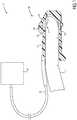
- Fig. 1shows a schematic diagram of an embodiment of a device 10 for noninvasively determining the hematocrit value of a subject, e.g. a patient, according to the present invention.
- the device 10comprises a sensor device 20 (also called PPG sensor) for obtaining signals and a processor 30 for processing said signals to determine the hematocrit value of the subject.
- the sensor device 20is configured for being mounted to the finger 1 of a person, but in other embodiments it may be configured for being mounted to a different body part such as an ala of the nose, an ear lobe or a toe.
- the processor 30is arranged separately from the sensor device 20, for instance on a separate computer, but in other embodiments the processor 30 may be configured at or within the sensor device 20, for instance as a digital signal processor.
- the sensor device 20comprises a housing 21 which fits snugly around the anatomy in question, so as to keep the sensor device 20 from falling off of the anatomy, but not so snug as to prevent circulation of blood therein.
- the sensor device 20comprises a light source for emitting light onto a skin area 2 of the subject.
- the light sourcecomprises a first light unit 22 for emitting first light onto the skin area 2 of the subject at a first wavelength and a second light unit 23 for emitting second light onto the skin area of the subject second light at a second wavelength.
- a single light sourcemay be employed for emitting light at both wavelengths.
- the light sourcemay e.g. comprise one or more LEDs.
- the emitted lightcomprises first light at said first wavelength in a first wavelength range between 500 and 1000 nm, e.g. at a first wavelength of 660, 800, or 880 nm, and second light at said second wavelength in a second wavelength range between 1000 and 2000 nm, e.g. at a second wavelength of 1310 or 1450 nm.
- the first wavelengthis used to take an increase in hematocrit into account that results in an increase in absorption (decreasing the intensity of the detected light) and an increase in backscattering (also decreasing the intensity of the detected light). Both effects decrease the intensity of the detected light.
- the second wavelengthis used to take an increase in hematocrit into account that results in a decrease in absorption by plasma (increasing the intensity of the detected light) and an increase in backscattering by red blood cells (decreasing the intensity of the detected light). The same holds vice versa for a decrease in hematocrit.
- the sensor device 20further comprises a reflection detector 24 for detecting light reflected (i.e. back scattered) from said skin area 2 of the subject in response to light illumination by said light units 22, 23 and a transmission detector 25 for detecting light transmitted (i.e. forward scattered) through said skin area 2 (and underlying tissue) of the subject in response to light illumination by said light units 22, 23.
- the reflection detector 24is thus arranged on the same side of the finger 1 as the light units 22, 23, while the transmission detector 25 is arranged on the opposite side. Further, the reflection detector 24 and the transmission detector 25 are configured to simultaneously detect light.
- the device 10further comprises a processor 30 for processing the signals acquired by the reflection detector 24 and the transmission detector 25.
- the processor 30may be integrated within the housing of the sensor device 20 (e.g. as CMOS chip), but is preferably configured as external unit connected to the sensor device 20 by a data transmission cable 26.
- the processor 30comprises a processing unit 31 for deriving plethysmography (PPG) signals from the light detected by said reflection detector 24 and said transmission detector 25.
- PPGplethysmography
- the generation of the PPG signalsis generally known in the art, e.g. from above cited document or from US 8315682 B2 describing an integrated pulse oximetry sensor, and shall not be described here in more detail.
- the processor 30comprises an analysis unit 32 for determining the hematocrit value of the subject from said PPG signals. This can be done in various ways as will be explained below by reference to different embodiments.
- both reflectance and transmittance photoplethysmographyare combined into a single modality, thereby taking into account the hematocrit-dependent scattering properties of blood which differs for wavelengths below 1000 nm (absorbed by hemoglobin in red blood cells) compared to for wavelengths above 1000 nm (absorbed by water in plasma). This gives additional parameters for the hematocrit estimation, which makes it more accurate and reliable.
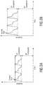
- Fig. 2shows typical PPG signals for reflection ( Fig. 2A ) and transmission ( Fig. 2B ) at a wavelength below 1000 nm. In said figures the AC and DC components are indicated.
- Hcthematocrit value
- a and bare constants found e.g. in calibration experiments. These constants depend on the device and system used.
- a first ratio R1is obtained by adding the ratios Rh ⁇ 1000nm,reflect and Rh ⁇ 1000nm,transmit obtained from the PPG signals obtained from the light at the first wavelength ( ⁇ 1000 nm)
- a second ratio R2is obtained by adding the ratios Rw> 1000nm,reflect and Rw> 1000nm,transmit obtained from the PPG signals obtained from the light at the second wavelength (> 1000 nm)
- a final ratio Ris obtained by dividing the first ratio R1 by the second ratio R2. The hematocrit value of the subject is then determined from said final ratio R.
- a third ratio R3is formed of the sum of the AC components (AC) ⁇ 1000nm,reflect+transmit of the first and third PPG signals and the sum of the DC components (DC) ⁇ 1000nm,reflect+transmit of the first and third PPG signals
- a fourth ratio R4is formed of the sum of the AC components (AC) >1000nm,reflect+transmit of the second and fourth PPG signals and the sum of the DC components (DC) >1000,reflect+transmit of the second and fourth PPG signals
- the hematocrit value of the subjectis determined from said third and fourth ratios R3, R4, in particular from a final ratio R obtained by dividing the third ratio R3 by the fourth ratio R4.
- the present inventioncan be applied in different scenarios. Dehydration produces a high hematocrit that disappears when proper fluid balance is restored. Overhydration, or fluid overload, as a result of excessive fluid therapy in intensive care or surgical patients leads to hemodilution which is reflected by a low hematocrit. Hence, hematocrit is an important indicator of a patient's fluid balance.
- hematocritOther groups of individuals at risk for developing anemia (i.e. low hematocrit) include infants without adequate iron intake, children going through a rapid growth spurt, during which the iron available cannot keep up with the demands for a growing red cell mass, women in childbearing years with a greater need for iron because of blood loss during menstruation, pregnant women in whom the growing fetus creates a high demand for iron, and patients with chronic kidney disease whose kidneys no longer secrete sufficient levels of the hormone erythropoietin that promotes RBC proliferation. High hematocrit can be seen in chronic smokers and in athletes that abuse the drug erythropoietin (Epogen) for blood doping purposes.
- Epogenerythropoietin
- a computer programmay be stored/distributed on a suitable non-transitory medium, such as an optical storage medium or a solid-state medium supplied together with or as part of other hardware, but may also be distributed in other forms, such as via the Internet or other wired or wireless telecommunication systems.
- a suitable non-transitory mediumsuch as an optical storage medium or a solid-state medium supplied together with or as part of other hardware, but may also be distributed in other forms, such as via the Internet or other wired or wireless telecommunication systems.
Landscapes
- Health & Medical Sciences (AREA)
- Life Sciences & Earth Sciences (AREA)
- Physics & Mathematics (AREA)
- Engineering & Computer Science (AREA)
- Animal Behavior & Ethology (AREA)
- Veterinary Medicine (AREA)
- Biophysics (AREA)
- Biomedical Technology (AREA)
- Heart & Thoracic Surgery (AREA)
- Medical Informatics (AREA)
- Molecular Biology (AREA)
- Surgery (AREA)
- Pathology (AREA)
- General Health & Medical Sciences (AREA)
- Public Health (AREA)
- Optics & Photonics (AREA)
- Physiology (AREA)
- Hematology (AREA)
- Cardiology (AREA)
- Artificial Intelligence (AREA)
- Computer Vision & Pattern Recognition (AREA)
- Psychiatry (AREA)
- Signal Processing (AREA)
- Spectroscopy & Molecular Physics (AREA)
- Measurement Of The Respiration, Hearing Ability, Form, And Blood Characteristics Of Living Organisms (AREA)
Description
- The present invention relates to a device and method for noninvasively determining the hematocrit value of a subject, such as a person or animal. Further, the present invention relates to a processor, processing method and computer program.
- Clinicians and researchers have recognized the value of a completely noninvasive method for measurement of hematocrit or total hemoglobin concentration.
US 6606509 B2 discloses a device and method for measurement of hematocrit (Hct) by noninvasive means. The changes in the intensities of light of multiple wavelengths transmitted through or reflected light from the tissue location are recorded immediately before and after occluding the flow of venous blood from the tissue location with an occlusion device positioned near the tissue location. As the venous return stops and the incoming arterial blood expands the blood vessels, the light intensities measured within a particular band of near-infrared wavelengths decrease in proportion to the volume of hemoglobin in the tissue location; those intensities measured within a separate band of wavelengths in which water absorbs respond to the difference between the water fractions within the blood and the displaced tissue volume. A mathematical algorithm applied to the time-varying intensities yields a quantitative estimate of the absolute concentration of hemoglobin in the blood. To compensate for the effect of the unknown fraction of water in the extravascular tissue on the Hct measurement, the tissue water fraction is determined before the occlusion cycle begins by measuring the diffuse transmittance or reflectance spectra of the tissue at selected wavelengths. US 2010/0331636 A1 discloses a method for the non-invasive determination of the concentration of blood constituents, in which a radiation source emits several radiation beams, each with a different wavelength. A first photo detector receives the measurement radiation of each wavelength that is reflected by a body part to be examined. A second photo detector receives the measurement radiation of each wavelength that is transmitted by the body part to be examined. The measurement radiation of each wavelength that is absorbed by the body part to be examined is then determined on the basis of the measurement of the reflected radiation by the first radiation receiver and the measurement of the transmitted radiation by the second radiation receiver. The concentration of the different constituents is calculated from the absorption of the measurement radiation that has been determined for each wavelength.US 6,611 320 B1 discloses a method for detecting blood characteristics including hemoglobin in a fluid medium using both transmission and reflection of a light beam which forms a quotient.WO 93/13706 A2 - It an object of the present invention to provide an improved device and method for noninvasively determining the hematocrit value of a subject with higher accuracy and reliability. It is a further object of the present invention to provide an improved processor, processing method and computer program.
- In a first aspect of the present invention a processor for noninvasively determining the hematocrit value of a subject is presented, the processor comprising:
- a processing unit for deriving plethysmography, PPG, signals for a first wavelength and a second wavelength from detector signals acquired by detection of light reflected from a skin area of the subject in response to light illumination and light transmitted through said skin area of the subject in response to said light illumination, said light illumination comprising first light at a first wavelength in a first wavelength range between 500 and 1000 nm and second light at a second wavelength in a second wavelength range between 1000 and 2000 nm, wherein said processing unit is configured to derive a first PPG signal from a first detector signal acquired by detection of the reflected light at the first wavelength, a second PPG signal from a second detector signal acquired by detection of the reflected light at the second wavelength, a third PPG signal from a third detector signal acquired by detection of the transmitted light at the first wavelength, and a fourth PPG signal from a fourth detector signal acquired by detection of the transmitted light at the second wavelength, and
- an analysis unit for determining the hematocrit value of the subject from said PPG signals, wherein said analysis unit is configured
- i) to determine relative pulsatile absorptions for said first to fourth PPG signals by forming a first to fourth pulsatile absorption ratio of the AC component to the DC component of the respective PPG signals, to obtain a first ratio by adding the first and third pulsatile absorption ratios obtained from the PPG signals obtained from the light at the first wavelength, to obtain a second ratio by adding the second and fourth pulsatile absorption ratios obtained from the PPG signals obtained from the light at the second wavelength, to obtain a final ratio by dividing the first ratio by the second ratio and to determine the hematocrit value of the subject from said final ratio, or
- ii) to determine relative pulsatile absorptions from said first to fourth PPG signals by forming a third ratio of the sum of the AC components of the first and third PPG signals and the sum of the DC components of the first and third PPG signals and by forming a fourth ratio of the sum of the AC components of the second and fourth PPG signals and the sum of the DC components of the second and fourth PPG signals, to obtain a final ratio by dividing the third ratio by the fourth ratio and to determine the hematocrit value of the subject from said final ratio.
- In a further of the present invention a device is presented for noninvasively determining the hematocrit value of a subject comprising:
- a light source for emitting light onto a skin area of the subject, said light comprising first light at a first wavelength in a first wavelength range between 500 and 1000 nm and second light at a second wavelength in a second wavelength range between 1000 and 2000 nm,
- a reflection detector for acquiring detector signals by detecting light reflected from said skin area of the subject in response to light illumination by said light source,
- a transmission detector for acquiring detector signals by detecting light transmitted through said skin area of the subject in response to light illumination by said light source, and
- a processor as disclosed herein for processing the detector signals acquired by said reflection detector and said transmission detector for noninvasively determining the hematocrit value of the subject.
- In further aspects of the present invention a corresponding method and a corresponding processing method are presented.
- In yet a further aspect of the present invention a computer program is presented comprising program code means for causing a computer to carry out the steps of the processing method proposed according to the present invention when said computer program is carried out on the computer.
- Preferred embodiments of the invention are defined in the dependent claims. It shall be understood that the claimed methods, device and computer program have similar and/or identical preferred embodiments as the claimed processor and as defined in the dependent claims.
- The present invention is based on the following findings. Heartbeat-induced pulsatility in tissue blood volume is associated with pulsatility in the tissue's optical absorption (both red blood cells and plasma) and scattering properties (red blood cells only). More specifically, a heartbeat-induced increase in tissue blood volume leads to an increase in optical absorption and an increase in optical scattering. Light with a wavelength below 1000 nm is mainly absorbed by hemoglobin in the red blood cells and light with a wavelength above 1000 nm, in particular above 1200 nm, is mainly absorbed by the water in the plasma. Heartbeat-induced pressure waves result in a temporary increase in tissue blood volume which is associated with a temporary increase in tissue optical absorption and scattering, both in the wavelength range of hemoglobin absorption as in the wavelength range of water absorption. For hematocrit determination it is important to know how much light is absorbed by red blood cells relative to how much light is absorbed by plasma. Since light is either absorbed, forward scattered (transmitted), or backward scattered (reflected), it is insufficient to only measure one of the three (i.e. reflection or transmission) to estimate the absorption.
- For pulse oximetry, a method to determine the ratio between oxyhemoglobin and deoxyhemoglobin applying one wavelength at approximately 660 nm and one wavelength at approximately 880 nm, this is not a problem because both the absorption and scattering at these wavelengths are due to the red blood cells. Hence, changes in the ratio between oxyhemoglobin and deoxyhemoglobin do not change the scattering properties at one wavelength versus the other. However, for hematocrit determination, changes in the ratio between red blood cells and plasma do change the scattering properties differently at one wavelength compared to at the other wavelength. More specifically, at wavelengths below 1000 nm, an increase in hematocrit results in an increase in absorption (decreasing the intensity of the detected light) and an increase in backscattering (also decreasing the intensity of the detected light). Both effects decrease the intensity of the detected light. At wavelengths above 1200 nm, however, the interpretation of the measured signal is less straightforward because the same increase in hematocrit results in a decrease in absorption by plasma (increasing the intensity of the detected light), but an increase in backscattering by red blood cells (decreasing the intensity of the detected light). Hence, the effects of changing hematocrit on the detected light intensity at 1450 nm are difficult to predict.
- Therefore, to estimate the relative absorption by red blood cells versus that of plasma it is important to take both the forward scatter (i.e. transmission) and backward scatter (i.e. backscatter or reflection) into account as proposed according to the present invention. The transmitted light is thus not limited light that is transmitted (i.e. forward scattered) straight through the skin and tissue, but may also be scattered at a different angle, e.g. at an angle of 90° so that the transmission detector may be arranged at the side of the tissue and not, as in one embodiment, opposite to the light source.
- The first wavelength lies within a first wavelength range is between 500 and 1000 nm, preferably 700 and 900 nm, and the second wavelength lies within a second wavelength range between 1000 and 2000 nm, preferably 1200 and 1500 nm. A preferred first wavelength may e.g. be 660, 800 or 880nm, and a preferred second wavelength may e.g. be 1310 or 1450 nm. However, it shall be noted that the "first wavelength" and the "second wavelength" may not only be understood as single wavelengths, but may also be understood as small wavelength ranges covering the respective wavelengths.
- In an embodiment of the device said reflection detector and said transmission detector are configured to simultaneously detect light and said processing unit is configured to derive PPG signals from detector signals generated from simultaneously detected light.
- The light source can be a single light source emitting light at different wavelengths, simultaneously or alternatingly. In an embodiment, however, said light source comprises a first light unit for emitting first light onto the skin area (i.e. a selected portion of the skin including the underlying tissue) of the subject at said first wavelength and a second light unit for emitting second light onto the skin area of the subject second light at said second wavelength. Said light sources, e.g. LEDs, are preferably arranged in the same housing, e.g. in a housing arranged for being mounted to a body part of the subject such as a finger clip, an ear clip or a nose clip. The two light units preferably illuminate the skin area alternatingly, e.g. at a frequency of 125 Hz. The detectors, in contrast, are preferably continuously on and detect the reflected and transmitted light simultaneously for each wavelength.
- Alternating the light sources is easier to implement than making the detectors alternatingly sensitive to one wavelength or the other as required if a single light source is used that emits light at the first and second wavelength simultaneously. This can e.g. be done with a filter wheel or special crystals. A more realistic solution would be to double the amount of detectors, i.e. to use two reflection detectors and two transmission detectors, one being sensitive to the first wavelength and the other one being sensitive to the second wavelength. Sensitivity of the sensor can be determined by using optical bandpass filters.
- According to one alternative of the proposed processor, said processing unit is configured to derive a first PPG signal from the reflected light at the first wavelength, a second PPG signal from the reflected light at the second wavelength, a third PPG signal from the transmitted light at the first wavelength, and a fourth PPG signal from the transmitted light at the second wavelength, and said analysis unit is configured to determine the hematocrit value of the subject from said first to fourth PPG signals.
- Said analysis unit is then configured to determine relative pulsatile absorptions for said first to fourth PPG signals by forming the ratio of the AC (pulsatile) component to the DC (non-pulsatile) component of the respective PPG signals and to determine the hematocrit value of the subject from said ratios. Further, said analysis unit is configured to obtain a first ratio by adding the ratios obtained from the PPG signals obtained from the light at the first wavelength, to obtain a second ratio by adding the ratios obtained from the PPG signals obtained from the light at the second wavelength, to obtain a final ratio by dividing the first ratio by the second ratio and to determine the hematocrit value of the subject from said final ratio.
- According to another alternative of the processor said analysis unit is configured to determine relative pulsatile absorptions from said first to fourth PPG signals by forming a third ratio of the sum of the AC components of the first and third PPG signals and the sum of the DC components of the first and third PPG signals and by forming a fourth ratio of the sum of the AC components of the second and fourth PPG signals and the sum of the DC components of the second and fourth PPG signals and said analysis unit is further configured to determine the hematocrit value of the subject from said third and fourth ratios. Further, said analysis unit is then configured to obtain a final ratio by dividing the third ratio by the fourth ratio and to determine the hematocrit value of the subject from said final ratio. There are, however, other ways to calculate said final ratio.
- In a preferred embodiment said analysis unit is configured to use said final ratio to determine the corresponding hematocrit value from a look-up table or a calibration curve. Said look-up table or calibration curve may e.g. be acquired in advance by a calibration measurement and/or by earlier uses of the device and method.
- These and other aspects of the invention will be apparent from and elucidated with reference to the embodiments described hereinafter. In the following drawings
Fig. 1 shows a schematic diagram of a device including a processor and a sensor device according to the present invention, andFig. 2 shows typical PPG signals for reflection and transmission (Fig. 2B ) at a wavelength below 1000 nm.Fig. 1 shows a schematic diagram of an embodiment of adevice 10 for noninvasively determining the hematocrit value of a subject, e.g. a patient, according to the present invention. Thedevice 10 comprises a sensor device 20 (also called PPG sensor) for obtaining signals and aprocessor 30 for processing said signals to determine the hematocrit value of the subject. In this embodiment thesensor device 20 is configured for being mounted to the finger 1 of a person, but in other embodiments it may be configured for being mounted to a different body part such as an ala of the nose, an ear lobe or a toe. Further, in this embodiment theprocessor 30 is arranged separately from thesensor device 20, for instance on a separate computer, but in other embodiments theprocessor 30 may be configured at or within thesensor device 20, for instance as a digital signal processor.- The
sensor device 20 comprises ahousing 21 which fits snugly around the anatomy in question, so as to keep thesensor device 20 from falling off of the anatomy, but not so snug as to prevent circulation of blood therein. - Further, the
sensor device 20 comprises a light source for emitting light onto askin area 2 of the subject. In this embodiment the light source comprises afirst light unit 22 for emitting first light onto theskin area 2 of the subject at a first wavelength and a secondlight unit 23 for emitting second light onto the skin area of the subject second light at a second wavelength. In other embodiment a single light source may be employed for emitting light at both wavelengths. The light source may e.g. comprise one or more LEDs. The emitted light comprises first light at said first wavelength in a first wavelength range between 500 and 1000 nm, e.g. at a first wavelength of 660, 800, or 880 nm, and second light at said second wavelength in a second wavelength range between 1000 and 2000 nm, e.g. at a second wavelength of 1310 or 1450 nm. - The first wavelength is used to take an increase in hematocrit into account that results in an increase in absorption (decreasing the intensity of the detected light) and an increase in backscattering (also decreasing the intensity of the detected light). Both effects decrease the intensity of the detected light. The second wavelength is used to take an increase in hematocrit into account that results in a decrease in absorption by plasma (increasing the intensity of the detected light) and an increase in backscattering by red blood cells (decreasing the intensity of the detected light). The same holds vice versa for a decrease in hematocrit.
- The
sensor device 20 further comprises areflection detector 24 for detecting light reflected (i.e. back scattered) from saidskin area 2 of the subject in response to light illumination by saidlight units transmission detector 25 for detecting light transmitted (i.e. forward scattered) through said skin area 2 (and underlying tissue) of the subject in response to light illumination by saidlight units reflection detector 24 is thus arranged on the same side of the finger 1 as thelight units transmission detector 25 is arranged on the opposite side. Further, thereflection detector 24 and thetransmission detector 25 are configured to simultaneously detect light. - The
device 10 further comprises aprocessor 30 for processing the signals acquired by thereflection detector 24 and thetransmission detector 25. Theprocessor 30 may be integrated within the housing of the sensor device 20 (e.g. as CMOS chip), but is preferably configured as external unit connected to thesensor device 20 by adata transmission cable 26. - The
processor 30 comprises a processing unit 31 for deriving plethysmography (PPG) signals from the light detected by saidreflection detector 24 and saidtransmission detector 25. The generation of the PPG signals is generally known in the art, e.g. from above cited document or fromUS 8315682 B2 describing an integrated pulse oximetry sensor, and shall not be described here in more detail. Further, theprocessor 30 comprises an analysis unit 32 for determining the hematocrit value of the subject from said PPG signals. This can be done in various ways as will be explained below by reference to different embodiments. - To estimate the relative absorption by red blood cells versus that of plasma the device and method according to the present invention take both the forward scatter (transmission) and backward scatter (reflection) into account. In other words, both reflectance and transmittance photoplethysmography are combined into a single modality, thereby taking into account the hematocrit-dependent scattering properties of blood which differs for wavelengths below 1000 nm (absorbed by hemoglobin in red blood cells) compared to for wavelengths above 1000 nm (absorbed by water in plasma). This gives additional parameters for the hematocrit estimation, which makes it more accurate and reliable.
- For the analysis and the determination of the hematocrit value at least two wavelengths are used to calculate the relative pulsatile absorption of hemoglobin and water. The relative pulsatile absorption Rh of hemoglobin is determined by the ratio of the AC (pulsatile) component to the DC (non-pulsatile) component of the PPG signals at a wavelength below 1000 nm, i.e. Rh<1000nm = (AC/DC)<1000nm. The relative pulsatile absorption Rw of water is determined by the ratio of the AC (pulsatile) component to the DC (non-pulsatile) component of the PPG signals at a wavelength above 1000 nm, i.e. Rw>1000nm = (AC/DC)>1000nm.
Fig. 2 shows typical PPG signals for reflection (Fig. 2A ) and transmission (Fig. 2B ) at a wavelength below 1000 nm. In said figures the AC and DC components are indicated. - The final ratio R = Rh<1000nm / Rw>1000nm is preferably related to the hematocrit value (Hct) by means of a lookup table or an empirically determined relation, typically in the form of Hct = a - bR. Here, a and b are constants found e.g. in calibration experiments. These constants depend on the device and system used.
- In one embodiment transmission and reflection the photoplethysmography signals are combined as follows:

- In other words, a first ratio R1 is obtained by adding the ratios Rh<1000nm,reflect and Rh<1000nm,transmit obtained from the PPG signals obtained from the light at the first wavelength (< 1000 nm), a second ratio R2 is obtained by adding the ratios Rw>1000nm,reflect and Rw>1000nm,transmit obtained from the PPG signals obtained from the light at the second wavelength (> 1000 nm), and a final ratio R is obtained by dividing the first ratio R1 by the second ratio R2. The hematocrit value of the subject is then determined from said final ratio R.
- In another embodiment the transmission and reflection photoplethysmography signals are combined as follows:

- In other words, a third ratio R3 is formed of the sum of the AC components (AC)<1000nm,reflect+transmit of the first and third PPG signals and the sum of the DC components (DC)<1000nm,reflect+transmit of the first and third PPG signals, a fourth ratio R4 is formed of the sum of the AC components (AC)>1000nm,reflect+transmit of the second and fourth PPG signals and the sum of the DC components (DC)>1000,reflect+transmit of the second and fourth PPG signals, and the hematocrit value of the subject is determined from said third and fourth ratios R3, R4, in particular from a final ratio R obtained by dividing the third ratio R3 by the fourth ratio R4.
- There are still further embodiments to determine the final ratio.
- For instance, according to another embodiment the following calculations are made:


- The present invention can be applied in different scenarios. Dehydration produces a high hematocrit that disappears when proper fluid balance is restored. Overhydration, or fluid overload, as a result of excessive fluid therapy in intensive care or surgical patients leads to hemodilution which is reflected by a low hematocrit. Hence, hematocrit is an important indicator of a patient's fluid balance.
- Other groups of individuals at risk for developing anemia (i.e. low hematocrit) include infants without adequate iron intake, children going through a rapid growth spurt, during which the iron available cannot keep up with the demands for a growing red cell mass, women in childbearing years with a greater need for iron because of blood loss during menstruation, pregnant women in whom the growing fetus creates a high demand for iron, and patients with chronic kidney disease whose kidneys no longer secrete sufficient levels of the hormone erythropoietin that promotes RBC proliferation. High hematocrit can be seen in chronic smokers and in athletes that abuse the drug erythropoietin (Epogen) for blood doping purposes.
- While the invention has been illustrated and described in detail in the drawings and foregoing description, such illustration and description are to be considered illustrative or exemplary and not restrictive; the invention is not limited to the disclosed embodiments. Other variations to the disclosed embodiments can be understood and effected by those skilled in the art in practicing the claimed invention, which is defined by the appended claims.
- In the claims, the word "comprising" does not exclude other elements or steps, and the indefinite article "a" or "an" does not exclude a plurality. A single element or other unit may fulfill the functions of several items recited in the claims. The mere fact that certain measures are recited in mutually different dependent claims does not indicate that a combination of these measures cannot be used to advantage.
- A computer program may be stored/distributed on a suitable non-transitory medium, such as an optical storage medium or a solid-state medium supplied together with or as part of other hardware, but may also be distributed in other forms, such as via the Internet or other wired or wireless telecommunication systems.
- Any reference signs in the claims should not be construed as limiting the scope.


Claims (9)
- Processor (30) for noninvasively determining the hematocrit value of a subject comprising:- a processing unit (31) for deriving plethysmography, PPG, signals for a first wavelength and a second wavelength from detector signals acquired by detection of light reflected from a skin area of the subject in response to light illumination and light transmitted through said skin area of the subject in response to said light illumination, said light illumination comprising first light at a first wavelength in a first wavelength range between 500 and 1000 nm and second light at a second wavelength in a second wavelength range between 1000 and 2000 nm, wherein said processing unit (31) is configured to derive a first PPG signal from a first detector signal acquired by detection of the reflected light at the first wavelength, a second PPG signal from a second detector signal acquired by detection of the reflected light at the second wavelength, a third PPG signal from a third detector signal acquired by detection of the transmitted light at the first wavelength, and a fourth PPG signal from a fourth detector signal acquired by detection of the transmitted light at the second wavelength, and- an analysis unit (32) for determining the hematocrit value of the subject from said PPG signals, wherein said analysis unit (32) is configuredi) to determine relative pulsatile absorptions for said first to fourth PPG signals by forming a first to fourth pulsatile absorption ratio of the AC component to the DC component of the respective PPG signals, to obtain a first ratio by adding the first and third pulsatile absorption ratios obtained from the PPG signals obtained from the light at the first wavelength, to obtain a second ratio by adding the second and fourth pulsatile absorption ratios obtained from the PPG signals obtained from the light at the second wavelength, to obtain a final ratio by dividing the first ratio by the second ratio and to determine the hematocrit value of the subject from said final ratio, orii) to determine relative pulsatile absorptions from said first to fourth PPG signals by forming a third ratio of the sum of the AC components of the first and third PPG signals and the sum of the DC components of the first and third PPG signals and by forming a fourth ratio of the sum of the AC components of the second and fourth PPG signals and the sum of the DC components of the second and fourth PPG signals, to obtain a final ratio by dividing the third ratio by the fourth ratio and to determine the hematocrit value of the subject from said final ratio.
- Processor as claimed in claim 1,
wherein said analysis unit (32) is configured to use said final ratio determine the corresponding hematocrit value from a look-up table or a calibration curve. - Device (10) for noninvasively determining the hematocrit value of a subject comprising:- a light source (22, 23) for emitting light onto a skin area of the subject, said light comprising first light at a first wavelength in a first wavelength range between 500 and 1000 nm and second light at a second wavelength in a second wavelength range between 1000 and 2000 nm,- a reflection detector (24) for acquiring detector signals by detecting light reflected from said skin area of the subject in response to light illumination by said light source,- a transmission detector (25) for acquiring detector signals by detecting light transmitted through said skin area of the subject in response to light illumination by said light source, and- a processor as claimed in claim 1 for processing the detector signals acquired by said reflection detector and said transmission detector for noninvasively determining the hematocrit value of the subject.
- Device as claimed in claim 3,
wherein said reflection detector (24) and said transmission detector are configured to simultaneously detect light and wherein said processing unit (31) is configured to derive PPG signals from detector signals generated from simultaneously detected light. - Device as claimed in claim 3,
wherein said light source comprises a first light unit (22) for emitting first light onto the skin area of the subject at said first wavelength and a second light unit (23) for emitting second light onto the skin area of the subject second light at said second wavelength. - Device as claimed in claim 5,
wherein the two light units are configured to illuminate the skin area alternatingly. - Processing method noninvasively determining the hematocrit value of a subject comprising, by a processor (30):- deriving plethysmography, PPG, signals for a first wavelength and a second wavelength from detector signals acquired by detection of light reflected from a skin area of the subject in response to light illumination and light transmitted through said skin area of the subject in response to said light illumination, said light illumination comprising first light at a first wavelength in a first wavelength range between 500 and 1000 nm and second light at a second wavelength in a second wavelength range between 1000 and 2000 nm, wherein a first PPG signal is derived from a first detector signal acquired by detection of the reflected light at the first wavelength, a second PPG signal is derived from a first detector signal acquired by detection of the reflected light at the second wavelength, a third PPG signal is derived from a first detector signal acquired by detection of the transmitted light at the first wavelength, and a fourth PPG signal is derived from a first detector signal acquired by detection of the transmitted light at the second wavelength, and- determining the hematocrit value of the subject from said PPG signals, wherein said step of determining is configuredi) to determine relative pulsatile absorptions for said first to fourth PPG signals by forming a first to fourth pulsatile absorption ratio of the AC component to the DC component of the respective PPG signals, to obtain a first ratio by adding the first and third pulsatile absorption ratios obtained from the PPG signals obtained from the light at the first wavelength, to obtain a second ratio by adding the second and fourth pulsatile absorption ratios obtained from the PPG signals obtained from the light at the second wavelength, to obtain a final ratio by dividing the first ratio by the second ratio and to determine the hematocrit value of the subject from said final ratio, orii) to determine relative pulsatile absorptions from said first to fourth PPG signals by forming a third ratio of the sum of the AC components of the first and third PPG signals and the sum of the DC components of the first and third PPG signals and by forming a fourth ratio of the sum of the AC components of the second and fourth PPG signals and the sum of the DC components of the second and fourth PPG signals, to obtain a final ratio by dividing the third ratio by the fourth ratio and to determine the hematocrit value of the subject from said final ratio.
- Method for noninvasively determining the hematocrit value of a subject comprising:- emitting light onto a skin area of the subject, said light comprising first light at a first wavelength in a first wavelength range between 500 and 1000 nm and second light at a second wavelength in a second wavelength range between 1000 and 2000 nm,- acquiring detector signals by detecting light reflected from said skin area of the subject in response to light illumination by said light source,- acquiring detector signals by detecting light transmitted through said skin area of the subject in response to light illumination by said light source, and- a processing method as claimed in claim 7 for processing the detector signals acquired by detecting the reflected light and the transmitted light for noninvasively determining the hematocrit value of the subject.
- Computer program comprising program code means for causing a computer to carry out the steps of the method as claimed in claim 7 when said computer program is carried out on the computer.
Applications Claiming Priority (2)
| Application Number | Priority Date | Filing Date | Title |
|---|---|---|---|
| EP14169271 | 2014-05-21 | ||
| PCT/EP2015/059916WO2015176955A1 (en) | 2014-05-21 | 2015-05-06 | Device and method for noninvasively determining the hematocrit value of a subject |
Publications (2)
| Publication Number | Publication Date |
|---|---|
| EP3145398A1 EP3145398A1 (en) | 2017-03-29 |
| EP3145398B1true EP3145398B1 (en) | 2020-09-02 |
Family
ID=50842044
Family Applications (1)
| Application Number | Title | Priority Date | Filing Date |
|---|---|---|---|
| EP15719731.0AActiveEP3145398B1 (en) | 2014-05-21 | 2015-05-06 | Device and method for noninvasively determining the hematocrit value of a subject |
Country Status (5)
| Country | Link |
|---|---|
| US (1) | US10582885B2 (en) |
| EP (1) | EP3145398B1 (en) |
| JP (1) | JP6667456B2 (en) |
| CN (1) | CN106456029B (en) |
| WO (1) | WO2015176955A1 (en) |
Families Citing this family (12)
| Publication number | Priority date | Publication date | Assignee | Title |
|---|---|---|---|---|
| EP3204064B1 (en) | 2014-10-10 | 2020-12-02 | NxStage Medical, Inc. | Flow balancing devices, methods, and systems |
| US11129556B2 (en) | 2015-12-31 | 2021-09-28 | Wear2B Ltd. | Device, system and method for non-invasive monitoring of physiological measurements |
| GB2570050B (en) | 2016-07-18 | 2020-01-01 | Nxstage Medical Inc | Apparatus for controlling fluid flow in a circuit |
| WO2018045102A1 (en) | 2016-08-30 | 2018-03-08 | Nxstage Medical, Inc. | Parameter monitoring in medical treatment systems |
| WO2019181267A1 (en)* | 2018-03-20 | 2019-09-26 | Dynamic Brain Lab合同会社 | Biological information measurement device |
| US12214114B2 (en) | 2019-05-23 | 2025-02-04 | Nxstage Medical, Inc. | Flow synchronization devices, methods, and systems |
| KR20220045341A (en) | 2020-10-05 | 2022-04-12 | 삼성전자주식회사 | Apparatus and method for estimating bio-information |
| CN112924660B (en)* | 2021-01-26 | 2023-09-26 | 上海浩创亘永科技有限公司 | Scanning system and scanning method thereof |
| WO2024100185A1 (en) | 2022-11-10 | 2024-05-16 | Trinamix Gmbh | A spectrometer system and a method for calibrating a photosensitive detector of a portable device |
| EP4619734A1 (en) | 2022-11-15 | 2025-09-24 | trinamiX GmbH | A spectrometer device and a method for a classification of a sample |
| WO2025003364A1 (en) | 2023-06-27 | 2025-01-02 | Trinamix Gmbh | Rolling shutter rgb-ir sensor |
| WO2025141699A1 (en)* | 2023-12-26 | 2025-07-03 | シグマ光機株式会社 | Intravascular plasma index measurement system |
Family Cites Families (22)
| Publication number | Priority date | Publication date | Assignee | Title |
|---|---|---|---|---|
| US6266546B1 (en)* | 1990-10-06 | 2001-07-24 | In-Line Diagnostics Corporation | System for noninvasive hematocrit monitoring |
| US5277181A (en)* | 1991-12-12 | 1994-01-11 | Vivascan Corporation | Noninvasive measurement of hematocrit and hemoglobin content by differential optical analysis |
| WO1993013706A2 (en)* | 1992-01-17 | 1993-07-22 | The Government Of The United States Of America, As Represented By The Secretary Of The Department Of Health And Human Services | Optical method for monitoring arterial blood hematocrit |
| US5553615A (en) | 1994-01-31 | 1996-09-10 | Minnesota Mining And Manufacturing Company | Method and apparatus for noninvasive prediction of hematocrit |
| KR100350022B1 (en)* | 1995-12-27 | 2002-12-26 | 시스멕스 가부시키가이샤 | Non-Invasive Blood Test Device |
| US6006119A (en) | 1998-02-04 | 1999-12-21 | Polestar Technologies, Inc. | Non-invasive optical measurement of blood hematocrit |
| US6662031B1 (en) | 1998-05-18 | 2003-12-09 | Abbott Laboratoies | Method and device for the noninvasive determination of hemoglobin and hematocrit |
| US6611320B1 (en)* | 1999-09-08 | 2003-08-26 | Optoq Ab | Method and apparatus |
| US6606509B2 (en) | 2001-03-16 | 2003-08-12 | Nellcor Puritan Bennett Incorporated | Method and apparatus for improving the accuracy of noninvasive hematocrit measurements |
| US6591122B2 (en) | 2001-03-16 | 2003-07-08 | Nellcor Puritan Bennett Incorporated | Device and method for monitoring body fluid and electrolyte disorders |
| US7657292B2 (en)* | 2001-03-16 | 2010-02-02 | Nellcor Puritan Bennett Llc | Method for evaluating extracellular water concentration in tissue |
| KR100612827B1 (en)* | 2001-04-19 | 2006-08-14 | 삼성전자주식회사 | Noninvasive hemoglobin concentration and oxygen saturation monitoring method and apparatus |
| US20030212316A1 (en) | 2002-05-10 | 2003-11-13 | Leiden Jeffrey M. | Method and apparatus for determining blood parameters and vital signs of a patient |
| JP2003339678A (en)* | 2002-05-30 | 2003-12-02 | Minolta Co Ltd | Instrument for measuring blood state |
| WO2005074550A2 (en) | 2004-01-30 | 2005-08-18 | 3Wave Optics, Llc | Non-invasive blood component measurement system |
| US7277741B2 (en)* | 2004-03-09 | 2007-10-02 | Nellcor Puritan Bennett Incorporated | Pulse oximetry motion artifact rejection using near infrared absorption by water |
| WO2006064399A2 (en) | 2004-12-14 | 2006-06-22 | Koninklijke Philips Electronics, N.V. | Integrated pulse oximetry sensor |
| US20100331636A1 (en)* | 2007-03-23 | 2010-12-30 | Enverdis Gmbh | Method for the continuous non-invasive determination of the concentration of blood constituents |
| JP5195589B2 (en)* | 2009-03-31 | 2013-05-08 | コニカミノルタオプティクス株式会社 | Pulse oximeter |
| US8311601B2 (en)* | 2009-06-30 | 2012-11-13 | Nellcor Puritan Bennett Llc | Reflectance and/or transmissive pulse oximeter |
| JP5382666B2 (en)* | 2011-04-21 | 2014-01-08 | 学校法人 聖マリアンナ医科大学 | Concentration measuring device and concentration measuring method |
| CN103610468A (en) | 2013-12-05 | 2014-03-05 | 深圳市奥博莱特科技有限公司 | Blood oxygen blood volume absolute amount detection device and method thereof |
- 2015
- 2015-05-06USUS15/312,841patent/US10582885B2/enactiveActive
- 2015-05-06CNCN201580025818.1Apatent/CN106456029B/enactiveActive
- 2015-05-06EPEP15719731.0Apatent/EP3145398B1/enactiveActive
- 2015-05-06WOPCT/EP2015/059916patent/WO2015176955A1/enactiveApplication Filing
- 2015-05-06JPJP2016566698Apatent/JP6667456B2/enactiveActive
Non-Patent Citations (1)
| Title |
|---|
| None* |
Also Published As
| Publication number | Publication date |
|---|---|
| WO2015176955A1 (en) | 2015-11-26 |
| CN106456029B (en) | 2019-11-26 |
| JP2017518792A (en) | 2017-07-13 |
| US20170202493A1 (en) | 2017-07-20 |
| CN106456029A (en) | 2017-02-22 |
| JP6667456B2 (en) | 2020-03-18 |
| US10582885B2 (en) | 2020-03-10 |
| EP3145398A1 (en) | 2017-03-29 |
Similar Documents
| Publication | Publication Date | Title |
|---|---|---|
| EP3145398B1 (en) | Device and method for noninvasively determining the hematocrit value of a subject | |
| US20220369940A1 (en) | Patient monitor for monitoring microcirculation | |
| EP3496603B1 (en) | Device for use in blood oxygen saturation measurement | |
| RU2653799C2 (en) | Device and method for extracting physiological information | |
| CN105009173B (en) | Device and method for the arterial oxygen saturation for determining object | |
| EP0850013B1 (en) | Procedure for the determination of fractional oxygen saturation | |
| CN107106051B (en) | Non-invasive dehydration monitoring | |
| US8649838B2 (en) | Wavelength switching for pulse oximetry | |
| EP2129288B1 (en) | Device and method for monitoring blood parameters | |
| US20080188728A1 (en) | Method and Device for Determining the Perfusion of Blood in a Body Member | |
| JP6878312B2 (en) | Photoelectric volumetric pulse wave recording device | |
| TW201726060A (en) | System, device and method for performing transabdominal fetal oxygen saturation and/or transabdominal fetal pulse oximetry monitoring | |
| US20150313541A1 (en) | Method and apparatus for determining tissue hydration | |
| JP2010521266A (en) | Noninvasive continuous measurement of blood component concentration | |
| WO2018029123A1 (en) | Device for use in blood oxygen saturation measurement | |
| US11259721B2 (en) | Method and device for detecting concentration of total hemoglobin in blood | |
| EP4384801A1 (en) | Optical determination of a cardiovascular variability parameter independent of skin contributions | |
| US20180133411A1 (en) | Systems and Methods for Detecting and Visualizing Blood Vessels | |
| Patil et al. | Methods and devices to determine hemoglobin non invasively: A review | |
| CA2428866A1 (en) | Measuring haematocrit in blood vessels | |
| KR20140115539A (en) | Method and apparatus for measuring ischemia measuring | |
| CN118076877A (en) | Optical determination of cardiovascular variability parameters independent of skin contribution | |
| CN114788683A (en) | information acquisition device | |
| Fricke et al. | Blood circulatory system for noninvasive diagnostics | |
| Kraitl et al. | A non-invasive optical monitoring system for the multi-spectral determination of absorption changes in blood |
Legal Events
| Date | Code | Title | Description |
|---|---|---|---|
| STAA | Information on the status of an ep patent application or granted ep patent | Free format text:STATUS: THE INTERNATIONAL PUBLICATION HAS BEEN MADE | |
| PUAI | Public reference made under article 153(3) epc to a published international application that has entered the european phase | Free format text:ORIGINAL CODE: 0009012 | |
| STAA | Information on the status of an ep patent application or granted ep patent | Free format text:STATUS: REQUEST FOR EXAMINATION WAS MADE | |
| 17P | Request for examination filed | Effective date:20161221 | |
| AK | Designated contracting states | Kind code of ref document:A1 Designated state(s):AL AT BE BG CH CY CZ DE DK EE ES FI FR GB GR HR HU IE IS IT LI LT LU LV MC MK MT NL NO PL PT RO RS SE SI SK SM TR | |
| AX | Request for extension of the european patent | Extension state:BA ME | |
| DAV | Request for validation of the european patent (deleted) | ||
| DAX | Request for extension of the european patent (deleted) | ||
| GRAP | Despatch of communication of intention to grant a patent | Free format text:ORIGINAL CODE: EPIDOSNIGR1 | |
| STAA | Information on the status of an ep patent application or granted ep patent | Free format text:STATUS: GRANT OF PATENT IS INTENDED | |
| RAP1 | Party data changed (applicant data changed or rights of an application transferred) | Owner name:KONINKLIJKE PHILIPS N.V. | |
| INTG | Intention to grant announced | Effective date:20200316 | |
| GRAS | Grant fee paid | Free format text:ORIGINAL CODE: EPIDOSNIGR3 | |
| GRAA | (expected) grant | Free format text:ORIGINAL CODE: 0009210 | |
| STAA | Information on the status of an ep patent application or granted ep patent | Free format text:STATUS: THE PATENT HAS BEEN GRANTED | |
| AK | Designated contracting states | Kind code of ref document:B1 Designated state(s):AL AT BE BG CH CY CZ DE DK EE ES FI FR GB GR HR HU IE IS IT LI LT LU LV MC MK MT NL NO PL PT RO RS SE SI SK SM TR | |
| REG | Reference to a national code | Ref country code:GB Ref legal event code:FG4D | |
| REG | Reference to a national code | Ref country code:AT Ref legal event code:REF Ref document number:1307847 Country of ref document:AT Kind code of ref document:T Effective date:20200915 Ref country code:CH Ref legal event code:EP | |
| REG | Reference to a national code | Ref country code:DE Ref legal event code:R096 Ref document number:602015058332 Country of ref document:DE | |
| REG | Reference to a national code | Ref country code:IE Ref legal event code:FG4D | |
| REG | Reference to a national code | Ref country code:LT Ref legal event code:MG4D | |
| PG25 | Lapsed in a contracting state [announced via postgrant information from national office to epo] | Ref country code:GR Free format text:LAPSE BECAUSE OF FAILURE TO SUBMIT A TRANSLATION OF THE DESCRIPTION OR TO PAY THE FEE WITHIN THE PRESCRIBED TIME-LIMIT Effective date:20201203 Ref country code:SE Free format text:LAPSE BECAUSE OF FAILURE TO SUBMIT A TRANSLATION OF THE DESCRIPTION OR TO PAY THE FEE WITHIN THE PRESCRIBED TIME-LIMIT Effective date:20200902 Ref country code:HR Free format text:LAPSE BECAUSE OF FAILURE TO SUBMIT A TRANSLATION OF THE DESCRIPTION OR TO PAY THE FEE WITHIN THE PRESCRIBED TIME-LIMIT Effective date:20200902 Ref country code:LT Free format text:LAPSE BECAUSE OF FAILURE TO SUBMIT A TRANSLATION OF THE DESCRIPTION OR TO PAY THE FEE WITHIN THE PRESCRIBED TIME-LIMIT Effective date:20200902 Ref country code:FI Free format text:LAPSE BECAUSE OF FAILURE TO SUBMIT A TRANSLATION OF THE DESCRIPTION OR TO PAY THE FEE WITHIN THE PRESCRIBED TIME-LIMIT Effective date:20200902 Ref country code:BG Free format text:LAPSE BECAUSE OF FAILURE TO SUBMIT A TRANSLATION OF THE DESCRIPTION OR TO PAY THE FEE WITHIN THE PRESCRIBED TIME-LIMIT Effective date:20201202 Ref country code:NO Free format text:LAPSE BECAUSE OF FAILURE TO SUBMIT A TRANSLATION OF THE DESCRIPTION OR TO PAY THE FEE WITHIN THE PRESCRIBED TIME-LIMIT Effective date:20201202 | |
| REG | Reference to a national code | Ref country code:NL Ref legal event code:MP Effective date:20200902 | |
| REG | Reference to a national code | Ref country code:AT Ref legal event code:MK05 Ref document number:1307847 Country of ref document:AT Kind code of ref document:T Effective date:20200902 | |
| PG25 | Lapsed in a contracting state [announced via postgrant information from national office to epo] | Ref country code:PL Free format text:LAPSE BECAUSE OF FAILURE TO SUBMIT A TRANSLATION OF THE DESCRIPTION OR TO PAY THE FEE WITHIN THE PRESCRIBED TIME-LIMIT Effective date:20200902 Ref country code:RS Free format text:LAPSE BECAUSE OF FAILURE TO SUBMIT A TRANSLATION OF THE DESCRIPTION OR TO PAY THE FEE WITHIN THE PRESCRIBED TIME-LIMIT Effective date:20200902 Ref country code:LV Free format text:LAPSE BECAUSE OF FAILURE TO SUBMIT A TRANSLATION OF THE DESCRIPTION OR TO PAY THE FEE WITHIN THE PRESCRIBED TIME-LIMIT Effective date:20200902 | |
| PG25 | Lapsed in a contracting state [announced via postgrant information from national office to epo] | Ref country code:CZ Free format text:LAPSE BECAUSE OF FAILURE TO SUBMIT A TRANSLATION OF THE DESCRIPTION OR TO PAY THE FEE WITHIN THE PRESCRIBED TIME-LIMIT Effective date:20200902 Ref country code:RO Free format text:LAPSE BECAUSE OF FAILURE TO SUBMIT A TRANSLATION OF THE DESCRIPTION OR TO PAY THE FEE WITHIN THE PRESCRIBED TIME-LIMIT Effective date:20200902 Ref country code:SM Free format text:LAPSE BECAUSE OF FAILURE TO SUBMIT A TRANSLATION OF THE DESCRIPTION OR TO PAY THE FEE WITHIN THE PRESCRIBED TIME-LIMIT Effective date:20200902 Ref country code:EE Free format text:LAPSE BECAUSE OF FAILURE TO SUBMIT A TRANSLATION OF THE DESCRIPTION OR TO PAY THE FEE WITHIN THE PRESCRIBED TIME-LIMIT Effective date:20200902 Ref country code:NL Free format text:LAPSE BECAUSE OF FAILURE TO SUBMIT A TRANSLATION OF THE DESCRIPTION OR TO PAY THE FEE WITHIN THE PRESCRIBED TIME-LIMIT Effective date:20200902 Ref country code:PT Free format text:LAPSE BECAUSE OF FAILURE TO SUBMIT A TRANSLATION OF THE DESCRIPTION OR TO PAY THE FEE WITHIN THE PRESCRIBED TIME-LIMIT Effective date:20210104 | |
| PG25 | Lapsed in a contracting state [announced via postgrant information from national office to epo] | Ref country code:ES Free format text:LAPSE BECAUSE OF FAILURE TO SUBMIT A TRANSLATION OF THE DESCRIPTION OR TO PAY THE FEE WITHIN THE PRESCRIBED TIME-LIMIT Effective date:20200902 Ref country code:AL Free format text:LAPSE BECAUSE OF FAILURE TO SUBMIT A TRANSLATION OF THE DESCRIPTION OR TO PAY THE FEE WITHIN THE PRESCRIBED TIME-LIMIT Effective date:20200902 Ref country code:AT Free format text:LAPSE BECAUSE OF FAILURE TO SUBMIT A TRANSLATION OF THE DESCRIPTION OR TO PAY THE FEE WITHIN THE PRESCRIBED TIME-LIMIT Effective date:20200902 Ref country code:IS Free format text:LAPSE BECAUSE OF FAILURE TO SUBMIT A TRANSLATION OF THE DESCRIPTION OR TO PAY THE FEE WITHIN THE PRESCRIBED TIME-LIMIT Effective date:20210102 | |
| REG | Reference to a national code | Ref country code:DE Ref legal event code:R097 Ref document number:602015058332 Country of ref document:DE | |
| PG25 | Lapsed in a contracting state [announced via postgrant information from national office to epo] | Ref country code:SK Free format text:LAPSE BECAUSE OF FAILURE TO SUBMIT A TRANSLATION OF THE DESCRIPTION OR TO PAY THE FEE WITHIN THE PRESCRIBED TIME-LIMIT Effective date:20200902 | |
| PLBE | No opposition filed within time limit | Free format text:ORIGINAL CODE: 0009261 | |
| STAA | Information on the status of an ep patent application or granted ep patent | Free format text:STATUS: NO OPPOSITION FILED WITHIN TIME LIMIT | |
| 26N | No opposition filed | Effective date:20210603 | |
| PG25 | Lapsed in a contracting state [announced via postgrant information from national office to epo] | Ref country code:DK Free format text:LAPSE BECAUSE OF FAILURE TO SUBMIT A TRANSLATION OF THE DESCRIPTION OR TO PAY THE FEE WITHIN THE PRESCRIBED TIME-LIMIT Effective date:20200902 Ref country code:SI Free format text:LAPSE BECAUSE OF FAILURE TO SUBMIT A TRANSLATION OF THE DESCRIPTION OR TO PAY THE FEE WITHIN THE PRESCRIBED TIME-LIMIT Effective date:20200902 | |
| PG25 | Lapsed in a contracting state [announced via postgrant information from national office to epo] | Ref country code:IT Free format text:LAPSE BECAUSE OF FAILURE TO SUBMIT A TRANSLATION OF THE DESCRIPTION OR TO PAY THE FEE WITHIN THE PRESCRIBED TIME-LIMIT Effective date:20200902 | |
| REG | Reference to a national code | Ref country code:CH Ref legal event code:PL | |
| PG25 | Lapsed in a contracting state [announced via postgrant information from national office to epo] | Ref country code:MC Free format text:LAPSE BECAUSE OF FAILURE TO SUBMIT A TRANSLATION OF THE DESCRIPTION OR TO PAY THE FEE WITHIN THE PRESCRIBED TIME-LIMIT Effective date:20200902 Ref country code:LI Free format text:LAPSE BECAUSE OF NON-PAYMENT OF DUE FEES Effective date:20210531 Ref country code:LU Free format text:LAPSE BECAUSE OF NON-PAYMENT OF DUE FEES Effective date:20210506 Ref country code:CH Free format text:LAPSE BECAUSE OF NON-PAYMENT OF DUE FEES Effective date:20210531 | |
| REG | Reference to a national code | Ref country code:BE Ref legal event code:MM Effective date:20210531 | |
| PG25 | Lapsed in a contracting state [announced via postgrant information from national office to epo] | Ref country code:IE Free format text:LAPSE BECAUSE OF NON-PAYMENT OF DUE FEES Effective date:20210506 | |
| PG25 | Lapsed in a contracting state [announced via postgrant information from national office to epo] | Ref country code:BE Free format text:LAPSE BECAUSE OF NON-PAYMENT OF DUE FEES Effective date:20210531 | |
| PG25 | Lapsed in a contracting state [announced via postgrant information from national office to epo] | Ref country code:HU Free format text:LAPSE BECAUSE OF FAILURE TO SUBMIT A TRANSLATION OF THE DESCRIPTION OR TO PAY THE FEE WITHIN THE PRESCRIBED TIME-LIMIT; INVALID AB INITIO Effective date:20150506 | |
| PG25 | Lapsed in a contracting state [announced via postgrant information from national office to epo] | Ref country code:CY Free format text:LAPSE BECAUSE OF FAILURE TO SUBMIT A TRANSLATION OF THE DESCRIPTION OR TO PAY THE FEE WITHIN THE PRESCRIBED TIME-LIMIT Effective date:20200902 | |
| PGFP | Annual fee paid to national office [announced via postgrant information from national office to epo] | Ref country code:FR Payment date:20230523 Year of fee payment:9 | |
| PGFP | Annual fee paid to national office [announced via postgrant information from national office to epo] | Ref country code:GB Payment date:20230523 Year of fee payment:9 | |
| PG25 | Lapsed in a contracting state [announced via postgrant information from national office to epo] | Ref country code:MK Free format text:LAPSE BECAUSE OF FAILURE TO SUBMIT A TRANSLATION OF THE DESCRIPTION OR TO PAY THE FEE WITHIN THE PRESCRIBED TIME-LIMIT Effective date:20200902 | |
| PG25 | Lapsed in a contracting state [announced via postgrant information from national office to epo] | Ref country code:MT Free format text:LAPSE BECAUSE OF FAILURE TO SUBMIT A TRANSLATION OF THE DESCRIPTION OR TO PAY THE FEE WITHIN THE PRESCRIBED TIME-LIMIT Effective date:20200902 | |
| REG | Reference to a national code | Ref country code:DE Ref legal event code:R084 Ref document number:602015058332 Country of ref document:DE | |
| GBPC | Gb: european patent ceased through non-payment of renewal fee | Effective date:20240506 | |
| PG25 | Lapsed in a contracting state [announced via postgrant information from national office to epo] | Ref country code:FR Free format text:LAPSE BECAUSE OF NON-PAYMENT OF DUE FEES Effective date:20240531 | |
| PG25 | Lapsed in a contracting state [announced via postgrant information from national office to epo] | Ref country code:GB Free format text:LAPSE BECAUSE OF NON-PAYMENT OF DUE FEES Effective date:20240506 | |
| PGFP | Annual fee paid to national office [announced via postgrant information from national office to epo] | Ref country code:DE Payment date:20250528 Year of fee payment:11 |