EP3034625A1 - An ultra sensitive method for in situ detection of nucleic acids - Google Patents
An ultra sensitive method for in situ detection of nucleic acidsDownload PDFInfo
- Publication number
- EP3034625A1 EP3034625A1EP15194058.2AEP15194058AEP3034625A1EP 3034625 A1EP3034625 A1EP 3034625A1EP 15194058 AEP15194058 AEP 15194058AEP 3034625 A1EP3034625 A1EP 3034625A1
- Authority
- EP
- European Patent Office
- Prior art keywords
- probe
- label
- nucleic acid
- cell
- signal amplification
- Prior art date
- Legal status (The legal status is an assumption and is not a legal conclusion. Google has not performed a legal analysis and makes no representation as to the accuracy of the status listed.)
- Granted
Links
- 150000007523nucleic acidsChemical class0.000titleclaimsabstractdescription158
- 102000039446nucleic acidsHuman genes0.000titleclaimsabstractdescription157
- 108020004707nucleic acidsProteins0.000titleclaimsabstractdescription157
- 238000000034methodMethods0.000titleclaimsabstractdescription126
- 238000001514detection methodMethods0.000titleclaimsdescription49
- 238000011065in-situ storageMethods0.000titledescription11
- 238000007901in situ hybridizationMethods0.000claimsabstractdescription88
- 239000000523sampleSubstances0.000claimsdescription400
- 230000003321amplificationEffects0.000claimsdescription172
- 238000003199nucleic acid amplification methodMethods0.000claimsdescription172
- 108010001336Horseradish PeroxidaseProteins0.000claimsdescription64
- 229960002685biotinDrugs0.000claimsdescription60
- 239000011616biotinSubstances0.000claimsdescription60
- YBJHBAHKTGYVGT-ZKWXMUAHSA-N(+)-BiotinChemical compoundN1C(=O)N[C@@H]2[C@H](CCCCC(=O)O)SC[C@@H]21YBJHBAHKTGYVGT-ZKWXMUAHSA-N0.000claimsdescription52
- 230000027455bindingEffects0.000claimsdescription52
- 102000002260Alkaline PhosphataseHuman genes0.000claimsdescription44
- 108020004774Alkaline PhosphataseProteins0.000claimsdescription44
- 108091032973(ribonucleotides)n+mProteins0.000claimsdescription28
- 235000020958biotinNutrition0.000claimsdescription26
- 108090001008AvidinProteins0.000claimsdescription25
- 239000003593chromogenic compoundSubstances0.000claimsdescription21
- SXEHKFHPFVVDIR-UHFFFAOYSA-N[4-(4-hydrazinylphenyl)phenyl]hydrazineChemical compoundC1=CC(NN)=CC=C1C1=CC=C(NN)C=C1SXEHKFHPFVVDIR-UHFFFAOYSA-N0.000claimsdescription20
- 230000001268conjugating effectEffects0.000claimsdescription18
- 108020004999messenger RNAProteins0.000claimsdescription18
- 108050001427Avidin/streptavidinProteins0.000claimsdescription17
- 238000002509fluorescent in situ hybridizationMethods0.000claimsdescription17
- WLDHEUZGFKACJH-UHFFFAOYSA-KamaranthChemical compound[Na+].[Na+].[Na+].C12=CC=C(S([O-])(=O)=O)C=C2C=C(S([O-])(=O)=O)C(O)=C1N=NC1=CC=C(S([O-])(=O)=O)C2=CC=CC=C12WLDHEUZGFKACJH-UHFFFAOYSA-K0.000claimsdescription16
- -1DinitrophenylChemical group0.000claimsdescription14
- 239000000758substrateSubstances0.000claimsdescription12
- 239000002299complementary DNASubstances0.000claimsdescription8
- 239000000725suspensionSubstances0.000claimsdescription8
- 239000007787solidSubstances0.000claimsdescription7
- 108091070501miRNAProteins0.000claimsdescription6
- 239000002679microRNASubstances0.000claimsdescription6
- 1250000018942,4,6-trinitrophenyl groupChemical group[H]C1=C(C(*)=C(C([H])=C1[N+]([O-])=O)[N+]([O-])=O)[N+]([O-])=O0.000claimsdescription4
- 108020004459Small interfering RNAProteins0.000claimsdescription2
- 210000004027cellAnatomy0.000description48
- 238000003556assayMethods0.000description34
- 238000009396hybridizationMethods0.000description27
- 239000002157polynucleotideSubstances0.000description25
- 108091033319polynucleotideProteins0.000description25
- 102000040430polynucleotideHuman genes0.000description25
- 108090000623proteins and genesProteins0.000description21
- 210000001519tissueAnatomy0.000description21
- 125000003729nucleotide groupChemical group0.000description17
- 108020004414DNAProteins0.000description16
- 239000002773nucleotideSubstances0.000description15
- 230000000295complement effectEffects0.000description13
- 238000013461designMethods0.000description12
- 230000035945sensitivityEffects0.000description10
- 238000013459approachMethods0.000description6
- 101100314454Caenorhabditis elegans tra-1 geneProteins0.000description5
- WSFSSNUMVMOOMR-UHFFFAOYSA-NFormaldehydeChemical compoundO=CWSFSSNUMVMOOMR-UHFFFAOYSA-N0.000description5
- 230000000903blocking effectEffects0.000description5
- 102000053602DNAHuman genes0.000description4
- 102000004190EnzymesHuman genes0.000description4
- 108090000790EnzymesProteins0.000description4
- 108091028043Nucleic acid sequenceProteins0.000description4
- JLCPHMBAVCMARE-UHFFFAOYSA-N[3-[[3-[[3-[[3-[[3-[[3-[[3-[[3-[[3-[[3-[[3-[[5-(2-amino-6-oxo-1H-purin-9-yl)-3-[[3-[[3-[[3-[[3-[[3-[[5-(2-amino-6-oxo-1H-purin-9-yl)-3-[[5-(2-amino-6-oxo-1H-purin-9-yl)-3-hydroxyoxolan-2-yl]methoxy-hydroxyphosphoryl]oxyoxolan-2-yl]methoxy-hydroxyphosphoryl]oxy-5-(5-methyl-2,4-dioxopyrimidin-1-yl)oxolan-2-yl]methoxy-hydroxyphosphoryl]oxy-5-(6-aminopurin-9-yl)oxolan-2-yl]methoxy-hydroxyphosphoryl]oxy-5-(6-aminopurin-9-yl)oxolan-2-yl]methoxy-hydroxyphosphoryl]oxy-5-(6-aminopurin-9-yl)oxolan-2-yl]methoxy-hydroxyphosphoryl]oxy-5-(6-aminopurin-9-yl)oxolan-2-yl]methoxy-hydroxyphosphoryl]oxyoxolan-2-yl]methoxy-hydroxyphosphoryl]oxy-5-(5-methyl-2,4-dioxopyrimidin-1-yl)oxolan-2-yl]methoxy-hydroxyphosphoryl]oxy-5-(4-amino-2-oxopyrimidin-1-yl)oxolan-2-yl]methoxy-hydroxyphosphoryl]oxy-5-(5-methyl-2,4-dioxopyrimidin-1-yl)oxolan-2-yl]methoxy-hydroxyphosphoryl]oxy-5-(5-methyl-2,4-dioxopyrimidin-1-yl)oxolan-2-yl]methoxy-hydroxyphosphoryl]oxy-5-(6-aminopurin-9-yl)oxolan-2-yl]methoxy-hydroxyphosphoryl]oxy-5-(6-aminopurin-9-yl)oxolan-2-yl]methoxy-hydroxyphosphoryl]oxy-5-(4-amino-2-oxopyrimidin-1-yl)oxolan-2-yl]methoxy-hydroxyphosphoryl]oxy-5-(4-amino-2-oxopyrimidin-1-yl)oxolan-2-yl]methoxy-hydroxyphosphoryl]oxy-5-(4-amino-2-oxopyrimidin-1-yl)oxolan-2-yl]methoxy-hydroxyphosphoryl]oxy-5-(6-aminopurin-9-yl)oxolan-2-yl]methoxy-hydroxyphosphoryl]oxy-5-(4-amino-2-oxopyrimidin-1-yl)oxolan-2-yl]methyl [5-(6-aminopurin-9-yl)-2-(hydroxymethyl)oxolan-3-yl] hydrogen phosphateChemical classCc1cn(C2CC(OP(O)(=O)OCC3OC(CC3OP(O)(=O)OCC3OC(CC3O)n3cnc4c3nc(N)[nH]c4=O)n3cnc4c3nc(N)[nH]c4=O)C(COP(O)(=O)OC3CC(OC3COP(O)(=O)OC3CC(OC3COP(O)(=O)OC3CC(OC3COP(O)(=O)OC3CC(OC3COP(O)(=O)OC3CC(OC3COP(O)(=O)OC3CC(OC3COP(O)(=O)OC3CC(OC3COP(O)(=O)OC3CC(OC3COP(O)(=O)OC3CC(OC3COP(O)(=O)OC3CC(OC3COP(O)(=O)OC3CC(OC3COP(O)(=O)OC3CC(OC3COP(O)(=O)OC3CC(OC3COP(O)(=O)OC3CC(OC3COP(O)(=O)OC3CC(OC3COP(O)(=O)OC3CC(OC3COP(O)(=O)OC3CC(OC3CO)n3cnc4c(N)ncnc34)n3ccc(N)nc3=O)n3cnc4c(N)ncnc34)n3ccc(N)nc3=O)n3ccc(N)nc3=O)n3ccc(N)nc3=O)n3cnc4c(N)ncnc34)n3cnc4c(N)ncnc34)n3cc(C)c(=O)[nH]c3=O)n3cc(C)c(=O)[nH]c3=O)n3ccc(N)nc3=O)n3cc(C)c(=O)[nH]c3=O)n3cnc4c3nc(N)[nH]c4=O)n3cnc4c(N)ncnc34)n3cnc4c(N)ncnc34)n3cnc4c(N)ncnc34)n3cnc4c(N)ncnc34)O2)c(=O)[nH]c1=OJLCPHMBAVCMARE-UHFFFAOYSA-N0.000description4
- 239000012472biological sampleSubstances0.000description4
- 239000003153chemical reaction reagentSubstances0.000description4
- 239000000975dyeSubstances0.000description4
- 239000007850fluorescent dyeSubstances0.000description4
- 230000014509gene expressionEffects0.000description4
- 238000011534incubationMethods0.000description4
- 230000009871nonspecific bindingEffects0.000description4
- 102000004169proteins and genesHuman genes0.000description4
- 238000012800visualizationMethods0.000description4
- 101000896557Homo sapiens Eukaryotic translation initiation factor 3 subunit BProteins0.000description3
- 101000988834Homo sapiens Hypoxanthine-guanine phosphoribosyltransferaseProteins0.000description3
- 108091034117OligonucleotideProteins0.000description3
- 230000001413cellular effectEffects0.000description3
- 239000000463materialSubstances0.000description3
- 239000011159matrix materialSubstances0.000description3
- 239000002245particleSubstances0.000description3
- 238000010827pathological analysisMethods0.000description3
- 229920000642polymerPolymers0.000description3
- 206010006187Breast cancerDiseases0.000description2
- 208000026310Breast neoplasmDiseases0.000description2
- 241000283707CapraSpecies0.000description2
- SHIBSTMRCDJXLN-UHFFFAOYSA-NDigoxigeninNatural productsC1CC(C2C(C3(C)CCC(O)CC3CC2)CC2O)(O)C2(C)C1C1=CC(=O)OC1SHIBSTMRCDJXLN-UHFFFAOYSA-N0.000description2
- 102100021699Eukaryotic translation initiation factor 3 subunit BHuman genes0.000description2
- MHAJPDPJQMAIIY-UHFFFAOYSA-NHydrogen peroxideChemical compoundOOMHAJPDPJQMAIIY-UHFFFAOYSA-N0.000description2
- 241001465754MetazoaSpecies0.000description2
- 108020005187Oligonucleotide ProbesProteins0.000description2
- 108091093037Peptide nucleic acidProteins0.000description2
- 102000003992PeroxidasesHuman genes0.000description2
- 108091028664RibonucleotideProteins0.000description2
- 108020004682Single-Stranded DNAProteins0.000description2
- 239000002253acidSubstances0.000description2
- 150000007513acidsChemical class0.000description2
- 230000008901benefitEffects0.000description2
- 239000003795chemical substances by applicationSubstances0.000description2
- 230000000875corresponding effectEffects0.000description2
- 239000005547deoxyribonucleotideSubstances0.000description2
- 125000002637deoxyribonucleotide groupChemical group0.000description2
- QONQRTHLHBTMGP-UHFFFAOYSA-NdigitoxigeninNatural productsCC12CCC(C3(CCC(O)CC3CC3)C)C3C11OC1CC2C1=CC(=O)OC1QONQRTHLHBTMGP-UHFFFAOYSA-N0.000description2
- SHIBSTMRCDJXLN-KCZCNTNESA-NdigoxigeninChemical compoundC1([C@@H]2[C@@]3([C@@](CC2)(O)[C@H]2[C@@H]([C@@]4(C)CC[C@H](O)C[C@H]4CC2)C[C@H]3O)C)=CC(=O)OC1SHIBSTMRCDJXLN-KCZCNTNESA-N0.000description2
- 230000006870functionEffects0.000description2
- 238000002372labellingMethods0.000description2
- 150000002605large moleculesChemical class0.000description2
- 230000004807localizationEffects0.000description2
- 229920002521macromoleculePolymers0.000description2
- 238000002493microarrayMethods0.000description2
- 238000007826nucleic acid assayMethods0.000description2
- 239000002751oligonucleotide probeSubstances0.000description2
- 210000000056organAnatomy0.000description2
- 239000012188paraffin waxSubstances0.000description2
- 210000003819peripheral blood mononuclear cellAnatomy0.000description2
- 108040007629peroxidase activity proteinsProteins0.000description2
- 239000000047productSubstances0.000description2
- 230000009467reductionEffects0.000description2
- 239000002336ribonucleotideSubstances0.000description2
- 125000002652ribonucleotide groupChemical group0.000description2
- 241000894007speciesSpecies0.000description2
- 230000008685targetingEffects0.000description2
- 238000005406washingMethods0.000description2
- 102000040650(ribonucleotides)n+mHuman genes0.000description1
- 108020004635Complementary DNAProteins0.000description1
- 108020004394Complementary RNAProteins0.000description1
- 101150003028Hprt1 geneProteins0.000description1
- 108010021625Immunoglobulin FragmentsProteins0.000description1
- 102000008394Immunoglobulin FragmentsHuman genes0.000description1
- OUYCCCASQSFEME-QMMMGPOBSA-NL-tyrosineChemical compoundOC(=O)[C@@H](N)CC1=CC=C(O)C=C1OUYCCCASQSFEME-QMMMGPOBSA-N0.000description1
- 241000124008MammaliaSpecies0.000description1
- 206010028980NeoplasmDiseases0.000description1
- 108010090804StreptavidinProteins0.000description1
- DZGWFCGJZKJUFP-UHFFFAOYSA-NTyramineNatural productsNCCC1=CC=C(O)C=C1DZGWFCGJZKJUFP-UHFFFAOYSA-N0.000description1
- 238000009825accumulationMethods0.000description1
- 239000000427antigenSubstances0.000description1
- 102000036639antigensHuman genes0.000description1
- 108091007433antigensProteins0.000description1
- 239000002787antisense oligonuctleotideSubstances0.000description1
- 238000000149argon plasma sinteringMethods0.000description1
- 230000001580bacterial effectEffects0.000description1
- 238000010256biochemical assayMethods0.000description1
- 239000013060biological fluidSubstances0.000description1
- 230000008827biological functionEffects0.000description1
- 238000001574biopsyMethods0.000description1
- 239000002981blocking agentSubstances0.000description1
- 238000010804cDNA synthesisMethods0.000description1
- 201000011510cancerDiseases0.000description1
- 238000004113cell cultureMethods0.000description1
- 239000013592cell lysateSubstances0.000description1
- 210000000349chromosomeAnatomy0.000description1
- 239000003184complementary RNASubstances0.000description1
- 230000021615conjugationEffects0.000description1
- 230000002596correlated effectEffects0.000description1
- 238000004132cross linkingMethods0.000description1
- 210000004748cultured cellAnatomy0.000description1
- 230000008021depositionEffects0.000description1
- 238000002405diagnostic procedureMethods0.000description1
- 230000000694effectsEffects0.000description1
- 239000012530fluidSubstances0.000description1
- 239000012634fragmentSubstances0.000description1
- 238000003364immunohistochemistryMethods0.000description1
- 238000012296in situ hybridization assayMethods0.000description1
- 238000001727in vivoMethods0.000description1
- 239000003112inhibitorSubstances0.000description1
- 239000000543intermediateSubstances0.000description1
- 239000006249magnetic particleSubstances0.000description1
- 230000007246mechanismEffects0.000description1
- 230000008018meltingEffects0.000description1
- 238000002844meltingMethods0.000description1
- YACKEPLHDIMKIO-UHFFFAOYSA-Nmethylphosphonic acidChemical compoundCP(O)(O)=OYACKEPLHDIMKIO-UHFFFAOYSA-N0.000description1
- 239000000178monomerSubstances0.000description1
- 210000003463organelleAnatomy0.000description1
- 150000002989phenolsChemical class0.000description1
- 230000010399physical interactionEffects0.000description1
- 238000002360preparation methodMethods0.000description1
- 239000012048reactive intermediateSubstances0.000description1
- 230000001105regulatory effectEffects0.000description1
- 238000012552reviewMethods0.000description1
- 229920002477rna polymerPolymers0.000description1
- 238000011896sensitive detectionMethods0.000description1
- 238000012163sequencing techniqueMethods0.000description1
- 125000006850spacer groupChemical group0.000description1
- 230000009870specific bindingEffects0.000description1
- 239000000126substanceSubstances0.000description1
- 239000013589supplementSubstances0.000description1
- 101150065190term geneProteins0.000description1
- 238000012360testing methodMethods0.000description1
- RYYWUUFWQRZTIU-UHFFFAOYSA-KthiophosphateChemical compound[O-]P([O-])([O-])=SRYYWUUFWQRZTIU-UHFFFAOYSA-K0.000description1
- 229960003732tyramineDrugs0.000description1
- DZGWFCGJZKJUFP-UHFFFAOYSA-OtyraminiumChemical compound[NH3+]CCC1=CC=C(O)C=C1DZGWFCGJZKJUFP-UHFFFAOYSA-O0.000description1
- OUYCCCASQSFEME-UHFFFAOYSA-NtyrosineNatural productsOC(=O)C(N)CC1=CC=C(O)C=C1OUYCCCASQSFEME-UHFFFAOYSA-N0.000description1
- 230000003612virological effectEffects0.000description1
Images


Classifications
- C—CHEMISTRY; METALLURGY
- C12—BIOCHEMISTRY; BEER; SPIRITS; WINE; VINEGAR; MICROBIOLOGY; ENZYMOLOGY; MUTATION OR GENETIC ENGINEERING
- C12Q—MEASURING OR TESTING PROCESSES INVOLVING ENZYMES, NUCLEIC ACIDS OR MICROORGANISMS; COMPOSITIONS OR TEST PAPERS THEREFOR; PROCESSES OF PREPARING SUCH COMPOSITIONS; CONDITION-RESPONSIVE CONTROL IN MICROBIOLOGICAL OR ENZYMOLOGICAL PROCESSES
- C12Q1/00—Measuring or testing processes involving enzymes, nucleic acids or microorganisms; Compositions therefor; Processes of preparing such compositions
- C12Q1/68—Measuring or testing processes involving enzymes, nucleic acids or microorganisms; Compositions therefor; Processes of preparing such compositions involving nucleic acids
- C12Q1/6813—Hybridisation assays
- C12Q1/6816—Hybridisation assays characterised by the detection means
- C12Q1/682—Signal amplification
- G—PHYSICS
- G01—MEASURING; TESTING
- G01N—INVESTIGATING OR ANALYSING MATERIALS BY DETERMINING THEIR CHEMICAL OR PHYSICAL PROPERTIES
- G01N33/00—Investigating or analysing materials by specific methods not covered by groups G01N1/00 - G01N31/00
- G01N33/48—Biological material, e.g. blood, urine; Haemocytometers
- G01N33/50—Chemical analysis of biological material, e.g. blood, urine; Testing involving biospecific ligand binding methods; Immunological testing
- G01N33/68—Chemical analysis of biological material, e.g. blood, urine; Testing involving biospecific ligand binding methods; Immunological testing involving proteins, peptides or amino acids
- G01N33/6803—General methods of protein analysis not limited to specific proteins or families of proteins
- C—CHEMISTRY; METALLURGY
- C12—BIOCHEMISTRY; BEER; SPIRITS; WINE; VINEGAR; MICROBIOLOGY; ENZYMOLOGY; MUTATION OR GENETIC ENGINEERING
- C12Q—MEASURING OR TESTING PROCESSES INVOLVING ENZYMES, NUCLEIC ACIDS OR MICROORGANISMS; COMPOSITIONS OR TEST PAPERS THEREFOR; PROCESSES OF PREPARING SUCH COMPOSITIONS; CONDITION-RESPONSIVE CONTROL IN MICROBIOLOGICAL OR ENZYMOLOGICAL PROCESSES
- C12Q1/00—Measuring or testing processes involving enzymes, nucleic acids or microorganisms; Compositions therefor; Processes of preparing such compositions
- C12Q1/68—Measuring or testing processes involving enzymes, nucleic acids or microorganisms; Compositions therefor; Processes of preparing such compositions involving nucleic acids
- C12Q1/6813—Hybridisation assays
- C12Q1/6816—Hybridisation assays characterised by the detection means
- C12Q1/6818—Hybridisation assays characterised by the detection means involving interaction of two or more labels, e.g. resonant energy transfer
- C—CHEMISTRY; METALLURGY
- C12—BIOCHEMISTRY; BEER; SPIRITS; WINE; VINEGAR; MICROBIOLOGY; ENZYMOLOGY; MUTATION OR GENETIC ENGINEERING
- C12Q—MEASURING OR TESTING PROCESSES INVOLVING ENZYMES, NUCLEIC ACIDS OR MICROORGANISMS; COMPOSITIONS OR TEST PAPERS THEREFOR; PROCESSES OF PREPARING SUCH COMPOSITIONS; CONDITION-RESPONSIVE CONTROL IN MICROBIOLOGICAL OR ENZYMOLOGICAL PROCESSES
- C12Q1/00—Measuring or testing processes involving enzymes, nucleic acids or microorganisms; Compositions therefor; Processes of preparing such compositions
- C12Q1/68—Measuring or testing processes involving enzymes, nucleic acids or microorganisms; Compositions therefor; Processes of preparing such compositions involving nucleic acids
- C12Q1/6813—Hybridisation assays
- C12Q1/6841—In situ hybridisation
Definitions
- the inventionrelates generally to nucleic acid chemistry and biochemical assays. More particularly, the invention relates to methods for in situ detection of nucleic acid analytes in a sample.
- ISHIn situ hybridization
- ISHtissue-specific and cell-type specific express patterns together with expression level of the gene can be obtained with ISH, which will provide valuable information for analyzing the function of the gene.
- RNAscope®a new ISH signal amplification method called Advanced Cell Diagnostics, Inc. ( US Patent No. 7,709,198 ). This assay includes uniquely designed oligo capture probes and a signal amplification system composed of preamplifiers, amplifiers, and label probes, allowing substantial signal amplification without amplifying background signal, enabling single RNA molecule detection for virtually any genes.
- An exemplary embodiment of the RNAscope® technologyis schematically illustrated in Fig.1 and will be described in detail in Section 2 of this application.
- RNAscope®In a typical RNAscope® assay for detecting target nucleic acid, a target mRNA whose expression is to be detected is released from cells and captured on a solid surface (e.g. , a well of a microtiter plate). A set of two or more capture probes and a signal generating multimer are also provided. The capture probes hybridize to both target nucleic acid and signal generating multimer and thus capture signal generating multimer to target nucleic acid.
- the signal generating multimercomprises label probes (LPs). But more typically, the signal generating multimer comprises preamplifiers and/or amplifiers in addition to label probes.
- the label probeis capable of binding to a label particle or molecule that provide detectable signal.
- the label probehas a larger molecular structure enabling that attachment of a plurality of label particle or molecules that provide stronger signal than a single label particle or molecule. Thus, RNAscope® improves the sensitivity and specific of nucleic acid detection.
- RNAscope®alone still cannot reliably detect some low copy genes in retrospective Formalin-Fixed, Paraffin-Embedded (FFPE) tissue sections where RNA is significantly degraded. Also, single RNA molecule cannot to be visualized at 40X magnification using current RNAscope® technology. It is desirable to further enhance the detection signal to enable more robust detection of any RNA molecules, including the significantly degraded RNA molecules, and to allow easy visualization of detected RNA signal at 10X magnification.
- FFPEParaffin-Embedded
- RNAscope®in combination with biotin-(strept)avidin
- RNAscope®in combination with an antibody
- TSATyramide Signal Amplification
- Biotin-avidin(or biotin-streptavidin) is a well known signal amplification system base on the facts that the two molecules have extraordinarily high affinity to each other and that one avidin/streptavidin molecule can bind four biotin molecules.
- Antibodiesare widely used for signal amplification in immunohistochemistry and ISH.
- TSAis based on the deposition of a large number of haptenized tyramide molecules by peroxidase activity. Tyramine is a phenolic compound.
- immobilized Horse Radish Peroxidase (HRP)converts the labeled substrate into a short-lived, extremely reactive intermediate.
- the activated substrate moleculesthen very rapidly react with and covalently bind to electron-rich moieties of proteins, such as tyrosine, at or near the site of the peroxidase binding site. In this way, a lot of extra hapten molecules conjugated to tyramide can be introduced at the hybridization site in situ. Subsequently, the deposited tyramide-hapten molecules can be visualized directly or indirectly.
- proteinssuch as tyrosine
- the present inventioncombines the signal amplification method of RNAscope® with the general ISH signal amplification methods into a ultra sensitive method to detect, quantify and identify one or more target nucleic acid in a sample.
- the RNAscope® assayoffers exceptional specificity owning to its unique paired capture probe design.
- general ISH signal amplification methodssuch as the TSA-based signal amplification
- the combined methodachieves great signal intensity and low background.
- the signal amplification methods and system disclosed in this inventionare relatively simple to use and produce consistent result. The claimed method can be easily adapted for use in routine clinic diagnostic procedures.
- the specificity and sensitivity of the disclosed signal amplification methodare balanced. The balance is achieved by a moderate reduction of the fold of signal amplification in RNAscope®.
- one preampliferis designed to bind between 1 to 16 amplifiers. In a preferred embodiment, one preamplifer is designed to bind between 2 to 10 amplifiers. In another more preferred embodiment, one preamplifer is designed to bind between 2 to 5 amplifiers.
- the sample comprising or suspected of comprising the target nucleic acidis first provided, together with at least one set of two or more capture probes capable of hybridizing to the target nucleic acid, a signal generating multimer capable of hybridizing to the set of two or more capture probes, wherein said signal generating multimer comprises a label probe, and a signal amplification probe capable of binding to the label probe wherein the signal amplification probe comprises a label.
- the target nucleic acidis first hybridized to the set of two or more capture probes, then the signal generating multimer is captured to the set of two or more capture probes and thereby being captured the signal generating multimer to the target nucleic acid.
- the signal amplification probeis then captured to the label probe and thereby being captured to the signal generating multimer.
- the presence, absence, or amount of the label in the signal amplification probeare detected.
- the signal amplification probecomprises: a biotin molecule which is capable of conjugating to the label probe, an avidin/streptavidin molecule which is capable of binding to the biotin molecule, and additional biotin molecules being conjugated to Horse Radish Peroxidase (HRP), Alkaline Phosphatase (AP), or fluorephore and are capable of binding to the avidin/streptavidin molecule.
- HRPHorse Radish Peroxidase
- APAlkaline Phosphatase
- fluorephorefluorephore
- the signal amplification probecomprises: a HRP, AP, Dinitrophenyl (DNP), or fluorophore molecule which is capable of conjugating to the label probe, one or more first antibody which is capable of binding to said HRP, AP, DNP, or fluorophore molecule, and one or more second antibody being conjugated to HRP, Polymer-HRP, AP, Polymer-AP or fluorophore and is capable of binding to said one or more first antibody.
- a HRP, AP, Dinitrophenyl (DNP), or fluorophore moleculewhich is capable of conjugating to the label probe
- one or more first antibodywhich is capable of binding to said HRP, AP, DNP, or fluorophore molecule
- second antibodybeing conjugated to HRP, Polymer-HRP, AP, Polymer-AP or fluorophore and is capable of binding to said one or more first antibody.
- the signal amplification probecomprises: a HRP molecule which is capable of conjugating to said label probe, a plurality of tyramide-biotin or tyramide-fluorophore molecules which are capable of reacting with said HRP molecule, and detection labels which are capable detecting said tyramide-biotin or tyramide-fluorophore molecules visually.
- the detection labelsare a combination of avidin/streptavidin-HRP and a chromogenic substrate or a combination of avidin/streptavidin-AP and a chromogenic substrate.
- the chromogenic substrateis selected from the group consisting of: diaminobenzine(DAB) and Fast Red.
- the signal generating multimercomprises a label probe capable of hybridizing to the set of two or more capture probes. In another embodiment, the signal generating multimer comprises a label probe and an amplifier hybridized to the label probe. The amplifier is capable of hybridizing to said set of two or more capture probes. In a preferred embodiment, the signal generating multimer comprises a label probe, an amplifier hybridized to the label, and a preamplifier hybridized to one or more of the amplifier. The preamplifier is capable of hybridizing to said set of two or more capture probes.
- the target nucleic acidcan be any type, such as, e..g, DNA, cDNA, RNA, mRNA, rRNA, miRNA, siRNA and/or the like.
- the amplificationcan take place on a solid support before hybridization is performed.
- Methods of the inventionfurther provide a variety of ways to multiplex two or more target nucleic acids.
- the samplemay comprise a cell comprising or suspected of comprising two or more different target nucleic acids.
- the samplemay also comprise two or more different cells, each comprising or suspected of comprising a different target nucleic acid.
- the two different target nucleic acidscan be detected using a dual-color chromogenic in situ hybridization (CISH) or a dual-color fluorescent in situ hybridization (FISH).
- CISHdual-color chromogenic in situ hybridization
- FISHdual-color fluorescent in situ hybridization
- the dual-color CISHis performed using two different signal amplification probes, wherein the first signal amplification probe comprises: tyramide-biotin, streptavidin-HRP and DAB, and the second signal amplification probe comprises: anti-DNP-AP and Fast Red.
- kits to practice the methods of the inventioncan include reagents to carry out the nucleic acid detection and provide conditions necessary to practice the methods of the invention.
- the kitsinclude, e.g. , A kit comprising: a target nucleic acid, one set of two or more capture probes capable of hybridizing to said target nucleic acid, a signal generating multimer capable of hybridizing to the set of two or more capture probes wherein the signal generating multimer comprises a label probe, and a signal amplification probe capable of binding to the label probe, wherein the signal amplification probe comprises a label.
- the signal amplification probecomprises: a biotin molecule which is capable of conjugating to said label probe, an avidin/streptavidin molecule which is capable of binding to said biotin molecule, and additional biotin molecules being conjugated to HRP, AP or fluorophores and are capable of binding to the avidin/streptavidin molecule.
- the signal amplification probecomprises: a HRP, AP, DNP, or fluorophore molecule which is capable of conjugating to the label probe, one or more first antibody which is capable of binding to the HRP, AP, DNP, or fluorophore molecule, and one or more second antibody being conjugated to HRP, Polymer-HRP, AP, Polymer-AP or fluorophore and is capable of binding to the one or more first antibody.
- the signal amplification probecomprises: a HRP molecule which is capable of conjugating to the label probe, a plurality of tyramide-biotin or tyramide-fluorophoremolecules which are capable of reacting with said HRP molecule, and detection labels which are capable detecting the tyramide-biotin molecules visually.
- the detection labelsmay be a combination of avidin/streptavidin-HRP and a chromogenic substrate or a combination of avidin/streptavidin-AP and a chromogenic substrate.
- the chromogenic substratemay be selected from the group consisting of: DAB and Fast Red.
- the signal generating multimercomprises a label probe capable of hybridizing to the set of two or more capture probes.
- the signal generating multimercomprises a label probe and an amplifier hybridized to the label probe.
- the amplifieris capable of hybridizing to said set of two or more capture probes.
- the signal generating multimercomprises a label probe, an amplifier hybridized to the label, and a preamplifier hybridized to one or more of the amplifier.
- the preamplifier:amplifier ratio in the kitmay be between 1-16, or preferably between 2-10 or between 2-5.
- the kit of the inventioncan function in the multiplexing of multiple nucleic acid amplifications.
- the kitmay be used for detecting nucleic acid targets in two or more different cells, each comprising or suspected of comprising a different target nucleic acid, or to detect two or more nucleic acid targets in one cell.
- the signal amplification probe in the kitmay be either a dual-color chromogenic in situ hybridization (CISH) probe or a dual-color fluorescent in situ hybridization (FISH) probe.
- CISHdual-color chromogenic in situ hybridization
- FISHfluorescent in situ hybridization
- the first signal amplification probecomprises: tyramide-biotin, streptavidin-HRP and DAB
- the second signal amplification probecomprises: anti-DNP-AP and Fast Red.
- nucleic acidencompasses any physical string of monomer units that can be corresponded to a string of nucleotides, including a polymer of nucleotides (e.g. , a typical DNA or RNA polymer), peptide nucleic acids (PNAs), modified oligonucleotides (e.g. , oligonucleotides comprising nucleotides that are not typical to biological RNA or DNA, such as 2'-O-methylated oligonucleotides), and the like.
- PNAspeptide nucleic acids
- modified oligonucleotidese.g. , oligonucleotides comprising nucleotides that are not typical to biological RNA or DNA, such as 2'-O-methylated oligonucleotides
- the nucleotides of the polynucleotidecan be deoxyribonucleotides, ribonucleotides or nucleotide analogs, can be natural or non-natural, and can be unsubstituted, unmodified, substituted or modified.
- the nucleotidescan be linked by phosphodiester bonds, or by phosphorothioate linkages, methylphosphonate linkages, boranophosphate linkages, or the like.
- the polynucleotidecan additionally comprise non-nucleotide elements such as labels, quenchers, blocking groups, or the like.
- the polynucleotidecan be, e.g ., single-stranded or double-stranded.
- nucleic acid targetor “target nucleic acid” refers to a nucleic acid, or optionally a region thereof, that is to be detected.
- a "polynucleotide sequence” or “nucleotide sequence”is a polymer of nucleotides (an oligonucleotide, a DNA, a nucleic acid, etc.) or a character string representing a nucleotide polymer, depending on context. From any specified polynucleotide sequence, either the given nucleic acid or the complementary polynucleotide sequence (e.g. , the complementary nucleic acid) can be determined.
- geneis used broadly to refer to any nucleic acid associated with a biological function. Genes typically include coding sequences and/or the regulatory sequences required for expression of such coding sequences. The term gene can apply to a specific genomic sequence, as well as to a cDNA or an mRNA encoded by that genomic sequence.
- antibodyis used herein in the broadest sense and covers fully assembled antibodies, antibody fragments which retain the ability to specifically bind to the antigen (e.g. , Fab, F(ab')2, Fv, and other fragments), single chain antibodies, diabodies, antibody chimeras, hybrid antibodies, bispecific antibodies, humanized antibodies, and the like.
- antibodycovers both polyclonal and monoclonal antibodies.
- biological samplerefers to a sample obtained from a biological subject, including sample of biological tissue or fluid origin, obtained, reached, or collected in vivo or in situ .
- a biological samplealso includes samples from a region of a biological subject containing precancerous or cancer cells or tissues. Such samples can be, but are not limited to, organs, tissues, fractions and cells isolated from a mammal. Exemplary biological samples include but are not limited to cell lysate, a cell culture, a cell line, a tissue, an organ, an organelle, a biological fluid, and the like. Preferred biological samples include but are not limited to a skin sample, tissue biopsies, and the like.
- label proberefers to an entity that binds to a target molecule, directly or indirectly, and enables the target to be detected, e.g ., by a readout instrument.
- a label probe(or "LP") is typically a single-stranded polynucleotide that comprises one or more label which directly or indirectly provides a detectable signal.
- the labelcan be covalently attached to the polynucleotide, or the polynucleotide can be configured to bind to the label (e.g. , a biotinylated polynucleotide can bind a streptavidin-associated label).
- the label probecan, for example, hybridize directly to a target nucleic acid, or it can hybridize to a nucleic acid that is in turn hybridized to the target nucleic acid or to one or more other nucleic acids that are hybridized to the nucleic acid.
- the label probecan comprise a polynucleotide sequence that is complementary to a polynucleotide sequence of the target nucleic acid, or it can comprise at least one polynucleotide sequence that is complementary to a polynucleotide sequence in a capture probe, amplifier, or the like.
- labelis a moiety that facilitates detection of a molecule.
- Common labels in the context of the present inventioninclude fluorescent, luminescent, light-scattering, and/or colorimetric labels.
- Suitable labelsinclude enzymes and fluorescent moieties, as well as radionuclides, substrates, cofactors, inhibitors, chemiluminescent moieties, magnetic particles, and the like.
- Exemplary labelsinclude: Horse Radish Peroxidase (HRP), Alkaline Phosphatase (AP), fluorophore, Dinitrophenyl (DNP), etc. Patents teaching the use of such labels include U.S. Pat. Nos.
- a “capture probe”is a polynucleotide that is capable of hybridizing to a target nucleic acid and capturing a label probe to that target nucleic acid.
- the target probecan hybridize directly to the label probe, or it can hybridize to one or more nucleic acids that in turn hybridize to the label probe; for example, the target probe can hybridize to an amplifier or a preamplifier.
- the target probethus includes a first polynucleotide sequence that is complementary to a polynucleotide sequence of the target nucleic acid and a second polynucleotide sequence that is complementary to a polynucleotide sequence of the label probe, amplifier, preamplifier, or the like.
- the target probeis preferably single-stranded.
- An "amplifier”is a molecule, typically a polynucleotide, that is capable of hybridizing to multiple label probes. Typically, the amplifier hybridizes to multiple identical label probes. The amplifier also hybridizes to at least one target probe or nucleic acid bound to a target probe. For example, the amplifier can hybridize to at least one target probe and to a plurality of label probes, or to a preamplifier and a plurality of label probes. The amplifier can be, e.g ., a linear, forked, comb-like, or branched nucleic acid.
- the amplifiercan include modified nucleotides and/or nonstandard internucleotide linkages as well as standard deoxyribonucleotides, ribonucleotides, and/or phosphodiester bonds. Suitable amplifiers are described, for example, in USPN 5,635,352, USPN 5,124,246, USPN 5,710,264, and USPN 5,849,481.
- a "preamplifier”is a molecule, typically a polynucleotide, that serves as an intermediate between one or more target probes and amplifiers. Typically, the preamplifier hybridizes simultaneously to one or more target probes and to a plurality of amplifiers. Exemplary preamplifiers are described, for example, in USPN 5,635,352 and USPN 5,681,697.
- ISHin situ hybridization
- probea labeled complementary DNA or RNA strand
- the probe typesare double stranded DNA (dsDNA), single stranded DNA (ssDNA), single stranded complimentary RNA (sscRNA), messenger RNA (mRNA), micro RNA (miRNA), and synthetic oligonucleotides.
- FISHfluorescent in situ hybridization
- CISHor "chromogenic in situ hybridization” refers to a type of ISH with a chromogenic label.
- a "general ISH signal amplification" assay or systemrefers to any ISH based in situ hybridization assay or system that can amplify the specific DNA or RNA sequence targeted by the method. It includes, but not limited to the three exemplary general ISH signal amplification systems disclosed in the present invention (i.e., biotin-based ISH, antibody-based ISH, and TSA-based ISH).
- the present inventionprovides methods and systems for detecting the presence of a target nucleic acid with improved sensitivity and specificity in a sample.
- the inventioncombines RNAscope® method with general ISH signal amplification methods such as the TSA-based signal amplification method, the antibody-based signal amplification method, or the Biotin-Avidin-based signal amplification method.
- the RNAscope® methodis known for its high specificity in detecting a target nucleic acid. In other words, a target nucleic acid can be detected with high signal-to-noise ratio in an RNAscope® assay.
- Such high specificityas will be explained in Section 2 below, are derived from the "double Z" probe design and the preamplifier-amplifier-label probe signal cascade design.
- General ISH signal amplification methodsare known for their ability to boosting signal amplification of nucleic acid molecules. However, ISH signal amplification methods are also known for their high signal to noise ratio and their lack of consistency in the amplification
- the present inventioncombines the RNAscope® method with general ISH signal amplification methods and further adjust the amplification ratio between the amplifier and preamplifier in RNAscope®.
- methods and systems of the present inventionovercome the above noted disadvantages of the general ISH signal amplification methods while retaining the unique mechanisms for background noise reduction in RNAscope®.
- the present inventionis capable of reliably detecting nucleic acid targets with great sensitivity and specificity. Such enhancement increases the consistency among the results of nucleic acid detection to the level that can be implemented in diagnostic assays.
- This inventiondescribes a novel, simple and ultra sensitive in suit hybridization method for detecting target nucleic acid in a sample.
- This methodcan be used in both fluorescent ISH (FISH) and chromogenic ISH (CISH) assays.
- FISHfluorescent ISH
- CISHchromogenic ISH
- the methodis capable of detecting nuclear acids (DNA and RNA) immobilized in intact cells, tissue sections, tissue microarrays and cDNA microarrays.
- ISH signal amplification methodsThere are many ISH signal amplification methods which have been developed to detect DNA or RNA target. Three exemplary ISH signal amplification methods will be described below: (1) Biotin-(Strept)avidin-based ISH signal amplification; (2) Antibody-based ISH signal amplification, and (3) Tyramide Signal Amplification (TSA)-based ISH signal amplification. It is clear to a skilled artisan that any ISH signal amplification method can be used in the present invention in combination with the RNAscope®. Thus, these examples should be not considered as a limitation to the present invention.
- the biotin-(strept)avidin-based ISH signal amplification methodwas used in combination with RNAscope®.
- the RNAscopecomprises a set of two or more capture probes, a plurality of label probes, a plurality of amplifiers, and a plurality of preamplifiers.
- the target nucleic acidis hybridized to the set of capture probes.
- the set of capture probesis then hybridized to a preamplifier.
- the preamplifieris further hybridized to a plurality of amplifiers. Each amplifier is then hybridized to a plurality of label probes.
- the label probe (LP) of the RNAscope complex structureis then conjugated to a biotin molecule of the ISH signal amplification structure and forms a LP-biotin complex.
- the LP-biotin complexis further bound by a avidin/streptavidin molecule.
- the avidin/streptavidin moleculehas the capacity to bind a maximum of 4 biotin molecules where each biotin molecule is further conjugated to HRP or AP.
- HRP or APcan be detected visually by chromogenic dyes such as diaminobenzidine, DAB or Fast Red.
- the antibody-based ISH signal amplification methodwas used in combination with RNAscope®.
- the RNAscopecomprises a set of two or more capture probes, a plurality of label probes, a plurality of amplifiers, and a plurality of preamplifiers.
- the target nucleic acidis hybridized to the set of capture probes.
- the two or more capture probesare hybridized to the nucleic acid target. Every preamplifier are hybridized to each of the two or more capture probes, a plurality of amplifiers is hybridized to each of the preamplifiers, and the plurality of label probes is hybridized to the amplifiers.
- Each label probe of the RNAscope complex structureis conjugated to an HRP molecule, an AP molecule, a DNP molecule, or a fluorophore molecule and thus forming an HRP-LP complex, an AP-LP complex, a DNP-LP complex, or a fluorophore-LP complex, respectively.
- the HRP-LP, AP-LP, DNP-LP, or fluorophore-LP complexis recognized by an anti-HRP antibody, anti-AP-LP antibody, anti-DNP-LP antibody, or anti-fluorophore-LP antibody, respectively.
- the above antibodiesare produced by animal species A, such as goat. This first antibody is then recognized by second antibody against species A.
- the second antibodyis conjugated with HRP, Polymer-HRP, AP, Polymer-AP, or fluorophore and is capable of binding to one or more first antibodies.
- HRP or APcan be detected visually by chromogenic dyes such as Diaminobenzidine (DAB) or Fast Red.
- DABDiaminobenzidine
- the result hereis further signal amplification in addition to the RNAscope signal amplification.
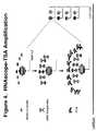
- the TSA-based ISH signal amplification methodwas used in combination with RNAscope®.
- the RNAscopecomprises a set of two or more capture probes, a plurality of label probes, a plurality of amplifiers, and a plurality of preamplifiers.
- the target nucleic acidis hybridized to the set of capture probes.
- the set of capture probesis then hybridized to a preamplifier.
- the preamplifieris further hybridized to a plurality of amplifiers.
- Each amplifieris then hybridized to a plurality of label probes.
- Each label probe of the RNAscope® complex structureis conjugated to an HRP molecule forming an HRP-LP complex.
- the HRP-LP complexis then bind to a plurality of Tyramide-Biotin or Fluorophore-Biotin molecules.
- the HRP-LP complexcatalyze multiple copies of Tyramide-Biotin or Fluorophore-Biotin to covalently bind to electron-rich moieties of proteins in the vicinity of LP-HRP binding sites.
- the Tyramide-Biotin or Fluorophore-Biotinis then subsequently detected by detection labels.
- the detection labelsare a combination of avidin/streptavidin-HRP and chromogenic substrate or a combination of avidin/streptavidin-AP and chromogenic substrate.
- the HRP or AP on the avidin/streptavidin-HRP or avidin/streptavidin-AP complexesare detected visually by chromogenic substrates such as DAB or Fast Red. Since many copies of Tyramide-Biotin or Fluorophore-Biotin can be deposited around each LP-HRP binding site, the intensity of end signal, which is the sum of RNAscope amplification and TSA amplification, is significantly increased.
- tyramidein other embodiments of the TSA-based ISH signal amplification method, can also be conjugated to other haptens such as digoxigenin (DIG), trinitrophenyl (TNP), Dinotrophenyl (DNP) and fluorescent dyes.
- DIGdigoxigenin
- TNPtrinitrophenyl
- DNPDinotrophenyl
- fluorescent dyeswhen fluorescent dye molecules are used in tyramide conjugation, the tyramide-dye conjugates can be directly used in RNAscope-TSA system for simple and effective fluorescent (FISH) detection.
- RNAscope®with general ISH signal amplification methods disclosed in this invention can be further fine-tuned to increase the sensitivity and specificity of signal amplification.
- RNAscope® + general ISH signal amplification approachRNAscope®'s major contribution is assay specificity.
- the RNAscope®also provides modest signal amplification of the nucleic acid target with high specificity.
- One of the general ISH signal amplification methods' contributionsis their signal boosting power.
- the general ISH signal amplification methodsfurther amplify the target nucleic acid signal to a much higher level.
- RNAscope®with general ISH signal amplification methods faces the problem of elevated background noise in signal detection.
- the present inventiondiscloses an intelligent way to reduce the level of false positive signals.
- RNAscope®One balancing factor for the combination of RNAscope® with general ISH signal amplification methods disclosed in this invention is the size of the preamplifier used in RNAscope®.
- the preamplifier of the RNAscope®could be a very large molecule if it is designed to bind to a large number of amplifiers.
- the preamplifiermay have the capacity to hybridize to more than 20 amplifiers.
- Such a large moleculeis prone to bind nonspecifically in cellular matrix during in situ hybridization.
- one of the preamplifiersbinds to sample nonspecifically, it will attract the same large number of amplifiers and label probes onto itself as it does when binding to a target nucleic acid.
- RNAscope®In the case when RNAscope® is used alone, the number of label probes which are bound to the few nonspecific binding preamplifiers may not generate any observable signals. However, if the signal of these label probes is further boosted by the additional signal amplification by a subsequent general ISH signal amplification, the false positive signal will be high enough to be detected.
- one preamplifieris designed to bind to between 1 to 16 amplifiers. In a more preferred embodiment, one preamplifier is designed to bind to between 2 to 10 amplifiers. In another preferred embodiment, one preamplifier is designed to bind to between 2 to 5 amplifiers.
- RNAscope®By balancing the amplification powers and specificity of RNAscope® and TSA in the assay, an optimum signal-to-noise ratio has been achieved for in situ RNA detection in routine clinical tissue specimen (see example 1 and Fig. 5 ).
- RNAscope®A powerful in situ hybridization method called RNAscope® was recently developed by Advanced Cell Diagnostics, Inc. (United States Patent No. 7,709,198 , incorporated herein by reference in its entirety). Since RNAscope® is part of the method disclosed in the present invention, the working principle of RNAscope® will be described below for better understanding of the invention.
- RNAscope®allows for direct visualization of RNA in situ. This method utilizes the oligonucleotide probe sets and novel signal amplification systems described below.
- the assaycan be used on a variety of sample types including cultured cells, peripheral blood mononuclear cells (PBMCs), frozen tissue, and formalin-fixed paraffin embedded (FFPE) tissue.
- PBMCsperipheral blood mononuclear cells
- FFPEformalin-fixed paraffin embedded
- the assaycan utilize both chromogenic and fluorescent detection reagents.
- the RNAscope® assay technologyprovides multiplex nucleic acid assays in single cells (see Fig. 1 ).
- At the core of this technologyis the "double Z" probe design, which allows robust amplification of specific hybridization signals without also amplifying nonspecific events.
- Each capture probe (“Z”)has a target-specific sequence which binds to the target mRNA, a spacer, and a "tail” sequence.
- Two capture probes (double Z)hybridize contiguously onto a target mRNA, and the two "tail” sequences form a 28-base hybridization site for the preamplifier.
- the double Z probe designensures high fidelity of signal amplification because: 1) it is highly unlikely that a pair of target probes will hybridize nonspecifically juxtaposed to each other to form a binding site for the preamplifier; and 2) neither tail alone can bind efficiently to the preamplifier under the assay conditions.
- the preamplifier, amplifier and label probeare hybridized sequentially to each capture probe pair, resulting in the accumulation of as many as 8,000 label molecules per 1 kb of target RNA.
- the label probecan be conjugated to either a fluorophore or a chromogenic enzyme (e.g. , HRP or AP), enabling viewing of hybridization signals under a standard bright-field or epifluorescent microscope, respectively. With a fluorescent label probe, the signals can contain at least 100-fold more fluorescent molecules than traditional RNA fluorescent ISH methods and are readily visible under a standard fluorescent microscope.
- RNAscope®was used to detect a target nucleic acid.
- a samplecomprising one or more cell is provided.
- the cell testedcomprises, or is suspected of comprising, the target nucleic acid.
- Provided in the assayare: a set of capture probes comprising two or more capture probes, a label probe comprising a label, and optionally preamplifiers and amplifiers.
- the set of capture probesis hybridized, in the cell, to the target nucleic acid.
- the label probeis captured to the set of capture probes, thereby capturing the label probe to the target nucleic acid.
- the signal from the labelis then detected. Since the label is associated with the target nucleic acid through the capture probes, presence of the label(s) in the cell indicates the presence of the corresponding nucleic acid target(s) in the cell.
- the methodsare optionally quantitative.
- an intensity of the signalcan be measured, and the intensity of the signal can be correlated with a quantity of the target nucleic acid in the cell.
- a signal spotcan be counted for each copy of the target nucleic acid in order to quantify them.
- the label probesbind directly to the capture probes.
- the label probesare captured to the capture probes indirectly, for example, through binding of preamplifiers and/or amplifiers.
- Use of amplifiers and preamplifierscan be advantageous in increasing signal strength, since they can facilitate binding of large numbers of label probes to each nucleic acid target.
- the indirect capture approachis preferred because it enables the label probe to be target-independent and further disclosure will show that it can offer better specificity and sensitivity.
- the capture probeis specially designed in the RNAscope® assay.
- each capture probethere is at least one section, T, complementary to a section on the target molecule, and another section, L, complementary to a section on the label probe.
- the T and L sectionsare connected by a section C.
- two adjacent capture probesare incorporated in a probe set targeting a gene of interest.
- T1 and T2are designed to be complementary to two unique and adjacent sections on the target nucleic acid.
- L1 and L2which can be different or the same, are complementary to two adjacent sections on the label probe.
- Their binding sections, T, L or bothare designed so that the linkage between the label probe and the target is unstable and tends to fall off at hybridization temperature when only one of the capture probes is in place.
- the melting temperature, T mof the T sections of the two capture probes are designed to be significantly above the hybridization temperature while the T m of the L sections is below the hybridization temperature.
- the T sectionscan be 20-30 nucleotides in length while the L sections are 13-15 nucleotides in length; C can be 0 to 10 nucleotides in length, e.g ., 5 nucleotides.
- T m of the T sectionsis below hybridization temperature while T m of the L sections is substantially above.
- the linkage between the label probe and the targetcan only survive the hybridization when both capture probes are hybridized to the target in a cooperative fashion. See U.S. Patent No. 7,709,198 for additional details on design of capture probes.
- the capture probespreferably hybridize to nonoverlapping polynucleotide sequences in their respective nucleic acid target.
- the capture probescan, but need not, cover a contiguous region of the nucleic acid target.
- Blocking probes, polynucleotides which hybridize to regions of the nucleic acid target not occupied by target probes,are optionally provided and hybridized to the target.
- the corresponding capture probes and blocking probesare preferably complementary to physically distinct, nonoverlapping sequences in the nucleic acid target, which nonoverlapping sequences are preferably, but not necessarily, contiguous. Having the capture probes and optional blocking probes be contiguous with each other can in some embodiments enhance hybridization strength, remove secondary structure, and ensure more consistent and reproducible signal.
- the cellIn detection of nucleic acid targets in a cell, the cell is typically fixed and permeabilized before hybridization of the capture probes, to retain the nucleic acid targets in the cell and to permit the capture probes, label probes, etc. to enter the cell.
- the cellis optionally washed to remove materials not captured to one of the nucleic acid targets.
- the cellcan be washed after any of various steps, for example, after hybridization of the capture probes to the nucleic acid targets to remove unbound capture probes, after hybridization of the preamplifiers, amplifiers, and/or label probes to the capture probes, and/or the like.
- the cellis in suspension for all or most of the steps of the method, for ease of handling.
- the methodsare also applicable to cells in solid tissue samples (e.g. , tissue sections) and/or cells immobilized on a substrate ( e.g. , a slide or other surface).
- the cellis in suspension in the sample comprising the cell, and/or the cell is in suspension during the hybridizing, capturing, and/or detecting steps.
- the cellcan be in suspension in the sample and during the hybridization, capture, optional washing, and detection steps.
- the cellis in suspension in the sample comprising the cell, and the cell is fixed on a substrate during the hybridizing, capturing, and/or detecting steps.
- the cellcan be in suspension during the hybridization, capture, and optional washing steps and immobilized on a substrate during the detection step.
- the samplecomprises a tissue section.
- one aspect of the inventionprovides multiplex nucleic acid assays.
- one general class of embodimentsincludes methods of detecting two or more target nucleic acids.
- method for detecting a single target nucleic acid using RNAscope®has been described.
- method for detecting a single target nucleic acid using a combination of RNAscope® and general ISH signal amplification methodwill be described first.
- Method for detecting two or more target nucleic acids using a combination of RNAscope® and general ISH signal amplification methodwill be described next.
- the detection of two or more target nucleic acidscan be conducted in a same way as that of detecting a single target nucleic acids except for using different, target-specific, labels and probes.
- a method of detecting a single target nucleic acidis provided.
- a samplecomprising or suspected of comprising the target nucleic acid is provided.
- a set of two or more capture probeswherein the capture probes are capable of binding to the target nucleic acid, signal generating multimers, and signal amplification probes.
- the signal generating multimercomprises at least a label probe.
- the signal generating multimercomprises a label probe and an amplifier.
- the amplifieris capable of hybridizing to the label probe, and is also capable of hybridizing to the set of two or more capture probes.
- the signal generating multimercomprises a label probe, an amplifier capable of hybridizing to the label, and a preamplifier capable of hybridizing to the amplifier and also capable of hybridizing to the set of two or more capture probes.
- the signal amplification probesare made of components used in general ISH signal amplification assays.
- the signal amplification probecomprises: a biotin molecule which is capable of conjugating to the label probe of RNAscope®, an avidin/streptavidin molecule which is capable of binding to the biotin molecule, and additional biotin molecules being conjugated to HRP, AP, or fluorophore and are capable of binding to the avidin/streptavidin molecule.
- the signal amplification probecomprises: a HRP, DNP, fluorophore, or AP molecule which is capable of conjugating to said label probe, one or more first antibody which is capable of binding to the HRP, DNP, fluorophore, or AP molecule, and one or more second antibody which is conjugated with HRP, Polymer-HRP, AP, or Polymer-AP and is capable of binding to one or more of the first antibody.
- the signal amplification probecomprises: a HRP molecule which is capable of conjugating to said label probe, a plurality of tyramide-biotin or tyramide-fluorophore molecules which are capable of reacting with the HRP molecule, and detection labels which are capable binding to the tyramide-biotin or tyramide-fluorophore molecules and thus display the target nucleic acid visually.
- the detection labelsare a combination of acivdin/strepavidin-HRP and a chromogenic substrate or a combination of acivdin/strepavidin-HRP and a chromogenic substrate.
- a sample comprising or suspected of comprising the target nucleic acidis first provided.
- the target nucleic acidis then hybridized to the set of two or more capture probes.
- the signal generating multimeris then added and being hybridized to the set of two or more capture probes and thereby being captured to the target nucleic acid.
- Components of the signal amplification probeare then added to the assay solution sequentially and bind to the signal generating multimer and thereby being captured to the target nucleic acid.
- the labels in the signal amplification probeare then detected by flow cytometer or other visualization devices. The presence, absence, or amount of the labels allows a sensitive detection and localization of the target nucleic acid in the sample.
- a samplecomprising or suspected of comprising the two target nucleic acids is provided. Also provided are two or more sets of capture probes, each set comprising two or more capture probes. The first set of capture probe is capable of hybridizing to the first target nucleic acid but not the second target nucleic acid, and the second set of capture probe is capable of hybridizing to the second target nucleic acid but not the first target nucleic acid. Further provided are a first signal generating multimer and a first signal amplification probe, together with a second signal generating multimer and a second signal amplification probe.
- the first signal generating multimercan hybridize to the first set of capture probe but not the second set of capture probe.
- the second signal generating multimercan hybridize to the second set of capture probe but not the first set of capture probe.
- the first signal generating multimerhas a first label probe that hybridize, either directly or indirectly through preamplifiers and amplifiers, to the first set of capture probe.
- the second signal generating multimerhas a second label probe that hybridize, either directly or indirectly through preamplifiers and amplifiers, to the second set of capture probe.
- the first label probeis different from the second label probe.
- the first signal generating multimer and the second signal generating multimerare labeled with different signal amplification probes sequentially.
- the two signal amplification probescan be a dual-color CISH or a dual-color FISH.
- the first signal generating multimeris labeled with the first signal amplification probe.
- the first signal amplification probeis a TSA-based ISH signal amplification which linked with the label probe of RNAscope® amplification via a conjugated LP-HRP complex.
- the label probe of RNAscope®is first conjugated with HRP.
- the conjugated LP-HRP complexis then detected with sequential incubation of tyramide-biotin, streptavidin-HRP and color substrate DAB, resulting in brown signal for the first target nucleic acid.
- the residual labeling agents for the first signal generating multimerare blocked in order to prevent the labels from reacting with the second signal generating multimer which is going to be added in the third step.
- the second signal generating multimeris labeled with the second signal amplification probe.
- the label probe of RNAscope®is first conjugated with Dinotrophenyl (DNP).
- DNPDinotrophenyl
- the conjugated LP-DNP complexis then detected with sequential incubation of anti-DNP-AP and color substrate Fast Red, resulting in red signal for the first target nucleic acid.
- the above methodsare also useful for multiplex detection of nucleic acids, including sequential detection of more than three nucleic acid targets.
- the celloptionally comprises or is suspected of comprising a third, fourth, fifth, sixth, seventh or even more nucleic acid target can be analyzed in the similar way described above.
- kits for detecting one or more target nucleic acidincludes a target nucleic acid, an RNAscope® component and a general ISH signal amplification component.
- the kitincludes a target nucleic acid, sets of capture probes capable of hybridizing to the target nucleic acid, a signal generating multimer capable of hybridizing to set of capture probes, and a signal amplification probe capable of binding to the label probe.
- the signal generating multimercomprises a label probe and the signal amplification probe comprises a label.
- the signal generating multimer in the kitcan include appropriate amplifiers, preamplifiers.
- the kitmay also include a solid support.
- the ISH signal amplification component of the kitcan be any ISH signal amplification system.
- itcan be the three ISH signal amplification systems described in Section 1.1 of the present invention.
- the kitcan be used for detection of any nucleic acids described earlier in this application, including, not not limited to DNA, cDNA, RNA, and mRNA.
- kitsmany components can be represented with two or more different subsets.
- the label probescan include two subsets, hybridizing to the first and the second target nucleic acids, respectively.
- the signal generating multimercan include two different label probe, each hybridizes, either directly or indirectly, to the first and the second capture probes.
- the kitcan also include two different signal amplification probes, each conjugates to the first and second label probes. The different signal amplification probes are distinguishable from each other by binding to different color substrates.
- the kitincludes a blocking agent which is capable of blocking the residual labeling agents for the first signal generating multimer and thus ensuring the specific binding of the second signal generating multimer to the second target nucleic acid.
- RNAscope®shows how a combination of RNAscope® and general ISH signal amplification methods further enhanced by an optimized preamplifier/amplifier ratio achieves a significantly improved specificity and sensitivity for in situ RNA detection.
- the basic assay procedurecan be done within a day and generally includes the following steps.
- a sample containing a plurality of cells suspected of comprising a target nucleic acid HPRT1is provided.
- cells either fixed on a solid support or in suspensionare hybridized to the following series of oligonucleotide probes.
- a set of capture probesis hybridized to the target RNA inside the cells.
- preamplifier moleculesare hybridized to the capture probes, providing a bridge for the hybridization of amplifier molecules.
- Multiple amplifier moleculesare further hybridized to a preamplifier, e.g ., up to 20 amplifiers to each preamplifier.
- Multiple label probesare then hybridized to an amplifier, e.g . up to 20 label probes to each amplifier.
- the signal intensityis enhanced further by TSA-based ISH signal amplification.
- An HRPis conjugated to each label probe.
- Reagent containing tyramide-biotin moleculesis then added to the assay solution.
- the HRP-conjugated-label-probecatalyzes multiple copies of tyramide-biotin to covalently bind to the electron-rich moieties of proteins in the vicinity of the HRP-label probe binding site.
- the tyramide-biotin moleculeis then detected by avidin/streptavidin-HRP, where avidin/streptavidin-HRP can be visualized by label substrate DAB which shows brown color.
- RNAscope® + TSA-based ISH signal amplification methodsignals obtained from RNAscope® + TSA-based ISH signal amplification method was significantly stronger than that obtained from RNAscope® alone.
- Nonspecific bindingwas prevented or minimized by designing probe sets targeting a specific mRNA sequence using a double "Z" probe design.
- the double "Z" capture probeswere designed based on the sequence of the HPRT1 and were prescreened against the GenBank database to ensure minimal cross-hybridization with unintended nucleic acid sequences.
- two neighboring probeseach contain a target-hybridizing sequence, e.g ., 20 to 30 base in length with a T m significantly above the assay temperature, and a preamplifier-hybridizing sequence, e.g. , only 14 bases in length with a T m well below the assay temperature.
- a single capture probeis able to bind to target RNA strongly and stably during hybridization, but will bind to the preamplifier weakly and unstably due to the 14 base pair region of homology having a T m well below the assay temperature.
- the combined hybridization strengthe.g ., of 28 complementary base pairs, holds the preamplifier strongly and stably at the assay temperature, enabling signal amplification to occur.
- Such a double "Z" designensures high detection specificity and simplifies probe design for simultaneous detection of multiple targets.
- the signal boosting power of the TSA-based ISH signal amplification methodDue to the signal boosting power of the TSA-based ISH signal amplification method, the nonspecific binding of preamplifier in cellular matrix during in situ hybridization could be amplified and consequently produces a false positive signal. This problem becomes serious when the preamplifier is designed to bind large number of amplifiers and thus is large in size. Power of amplification is gained at the cost of lost binding specificity. We solved this problem by moderately reducing the number of amplifiers a preamplifier is designed to bind to. The end result is increased the signal-to-noise ratio. The lost amplification power is compensated by signal boosting power of the ISH signal amplification methods, particularly the TSA-based ISH signal amplification method.
- Preamplifier PREAMP1is designed to bind to 20 amplifiers.
- Preamplifier PREAMP2is designed to bind to 16 amplifiers.
- Preamplifier PREAMP3is designed to bind to 10 amplifiers.
- Preamplifier PREAMP4is designed to bind to 5 amplifiers.
- PREAMP2has improved signal-to-noise ratio over PREAMP 1.
- PREAMP3 and PREAMP4have the best signal-to-noise ratio among all the PREAMPs tested (data not shown).
- 18S geneis captured on capture probes designed for 18S.
- the capture probe for 18Sis designed as described in example 1.
- the capture-probe-hybridized-18S geneis then sequentially hybridized to preamplifiers, amplifiers and label probes.
- the label probeis then conjugated to HRP.
- the conjugated LP-HRP complexesare then detected with sequential incubations of tyramide-biotin, streptavidin-HRP and DAB. The product of the hybridization produces brown color at locations where target gene 18Ss are detected.
- Her-2 geneis captured on capture probes designed for Her-2.
- the capture probe for Her-2is designed as described in example 1.
- the capture-probe-hybridized-Her-2 geneis then sequentially hybridized to preamplifiers, amplifiers and label probes.
- the label probeis then conjugated to Diotrophenyl (DNP).
- DNPDiotrophenyl
- the conjugated LP-DNPsare then detected with incubation of anti-DNP-AP.
- the alkaline phosphatase (AP) molecule on the anti-DNP-AP complexis then detected by dye reagent Fast Red.
- the product of the hybridizationproduces red signal for target gene Her-2.
- the method disclosed in this inventioncan be used to detect two RNA transcripts.
- the relative intensity of the signalscan be used to compare gene expression levels of the two genes.
- the inventionrefers inter alia to the following items:
Landscapes
- Chemical & Material Sciences (AREA)
- Life Sciences & Earth Sciences (AREA)
- Organic Chemistry (AREA)
- Health & Medical Sciences (AREA)
- Engineering & Computer Science (AREA)
- Proteomics, Peptides & Aminoacids (AREA)
- Zoology (AREA)
- Wood Science & Technology (AREA)
- Molecular Biology (AREA)
- Immunology (AREA)
- Physics & Mathematics (AREA)
- General Health & Medical Sciences (AREA)
- Biotechnology (AREA)
- Microbiology (AREA)
- Analytical Chemistry (AREA)
- Biochemistry (AREA)
- Bioinformatics & Cheminformatics (AREA)
- Biophysics (AREA)
- General Engineering & Computer Science (AREA)
- Genetics & Genomics (AREA)
- Biomedical Technology (AREA)
- Hematology (AREA)
- Urology & Nephrology (AREA)
- Bioinformatics & Computational Biology (AREA)
- Cell Biology (AREA)
- Food Science & Technology (AREA)
- Medicinal Chemistry (AREA)
- General Physics & Mathematics (AREA)
- Pathology (AREA)
- Measuring Or Testing Involving Enzymes Or Micro-Organisms (AREA)
Abstract
Description
- This application claims priority to and benefit of
U.S. provisional patent application no. 61/405,503, filed on October 21, 2010 Wu et al., and U.S. provisional patent application no. 61/412,276, filed on November 10, 2010 - The invention relates generally to nucleic acid chemistry and biochemical assays. More particularly, the invention relates to methods forin situ detection of nucleic acid analytes in a sample.
- In situ hybridization (ISH) is a technique that allows detection and localization of specific nucleic acid molecules in morphologically preserved individual cells, histological tissue sections, or chromosome preparations. It was first described in 1969 and is based on the complementary hybridization of a nucleotide probe to a specific target sequence of DNA or RNA in the cell. This may be endogenous DNA, messenger RNA (mRNA), micro RNA (miRNA), viral sequences, or bacterial sequences. The added probe is labeled with a reporter molecule and sites of binding are visualized either fluorescently (Fluorescentin situ hybridization, FISH) or chromogenically (Chromogenicin situ hybridization, CISH).
- After completion of sequencing the whole human genome, many thousands of novel human gene sequences have been annotated in public and private databases in recent years. This opened a new door for ISH, since it is relatively easy, fast and inexpensive to design and synthesizes antisense probes for detecting a specific sequence of any novel gene in the cell. The tissue-specific and cell-type specific express patterns together with expression level of the gene can be obtained with ISH, which will provide valuable information for analyzing the function of the gene.
- However, the application of ISH techniques can be limited by their inability to detect DNA or RNA targets with low copy numbers in cells due to lack of sensitivity and specificity. It is especially challenging to apply ISH into the clinical setting where formalin fixation and paraffin embedding (FFPE) is the most commonly used method worldwide to store clinical tissue samples. The FFPE method preserves tissue morphology well but the cross-linking fixation by formaldehyde can cause poor accessibility of the target sequence to the detection probe, chemical or physical interaction of the probe molecules with other molecules or structures, and damage to nuclear acids, especially mRNAs.
- In order to circumvent the limitation of ISH and extend its utility in diagnostic pathology, several strategies have been developed to improve the sensitivity of ISH. Recently, a new ISH signal amplification method called RNAscope® was developed by Advanced Cell Diagnostics, Inc. (
US Patent No. 7,709,198 ). This assay includes uniquely designed oligo capture probes and a signal amplification system composed of preamplifiers, amplifiers, and label probes, allowing substantial signal amplification without amplifying background signal, enabling single RNA molecule detection for virtually any genes. An exemplary embodiment of the RNAscope® technology is schematically illustrated inFig.1 and will be described in detail in Section 2 of this application. - In a typical RNAscope® assay for detecting target nucleic acid, a target mRNA whose expression is to be detected is released from cells and captured on a solid surface (e.g., a well of a microtiter plate). A set of two or more capture probes and a signal generating multimer are also provided. The capture probes hybridize to both target nucleic acid and signal generating multimer and thus capture signal generating multimer to target nucleic acid. The signal generating multimer comprises label probes (LPs). But more typically, the signal generating multimer comprises preamplifiers and/or amplifiers in addition to label probes. The label probe is capable of binding to a label particle or molecule that provide detectable signal. The label probe has a larger molecular structure enabling that attachment of a plurality of label particle or molecules that provide stronger signal than a single label particle or molecule. Thus, RNAscope® improves the sensitivity and specific of nucleic acid detection.
- However, RNAscope® alone still cannot reliably detect some low copy genes in retrospective Formalin-Fixed, Paraffin-Embedded (FFPE) tissue sections where RNA is significantly degraded. Also, single RNA molecule cannot to be visualized at 40X magnification using current RNAscope® technology. It is desirable to further enhance the detection signal to enable more robust detection of any RNA molecules, including the significantly degraded RNA molecules, and to allow easy visualization of detected RNA signal at 10X magnification.
- Here we describe a novel ISH signal amplification assay to extend the utility of ISH in diagnostic pathology by combining two signal amplification methods into one system. This system has three formats: (1) RNAscope® in combination with biotin-(strept)avidin, (2) RNAscope® in combination with an antibody, (3) RNAscope® in combination with Tyramide Signal Amplification (TSA). All these methods can leverage the exceptional specificity of RNAscope® technology with the amplification power of the three amplification methods. We surprisingly found that the above three combined signal amplification methods can highly amplify signals associated with the presence of a target nucleic acid in sample while having excellent signal to noise ratio.
- Biotin-avidin (or biotin-streptavidin) is a well known signal amplification system base on the facts that the two molecules have extraordinarily high affinity to each other and that one avidin/streptavidin molecule can bind four biotin molecules. Antibodies are widely used for signal amplification in immunohistochemistry and ISH. TSA is based on the deposition of a large number of haptenized tyramide molecules by peroxidase activity. Tyramine is a phenolic compound. In the presence of small amounts of hydrogen peroxide, immobilized Horse Radish Peroxidase (HRP) converts the labeled substrate into a short-lived, extremely reactive intermediate. The activated substrate molecules then very rapidly react with and covalently bind to electron-rich moieties of proteins, such as tyrosine, at or near the site of the peroxidase binding site. In this way, a lot of extra hapten molecules conjugated to tyramide can be introduced at the hybridization sitein situ. Subsequently, the deposited tyramide-hapten molecules can be visualized directly or indirectly.
- All of these signal amplification methods have been used in ISH with different degrees of success but usually the signal intensity and/or signal to noise ratio are not good enough for routine diagnostic pathology. In the case of TSA, in particular, when used alone in ISH, produces a high degree of background noise. The day-to-day and laboratory-to-laboratory consistency is also less than desirable.
- In view of the above, a need exists for methods of amplifying nucleic acids with both high intensity and high specificity. A need also exists for such a method with strong consistency. The present invention provides these and other features that will be apparent upon review of the following.
- The present invention combines the signal amplification method of RNAscope® with the general ISH signal amplification methods into a ultra sensitive method to detect, quantify and identify one or more target nucleic acid in a sample. The RNAscope® assay offers exceptional specificity owning to its unique paired capture probe design. By adding general ISH signal amplification methods such as the TSA-based signal amplification to the RNAscope® assay, the combined method achieves great signal intensity and low background. The signal amplification methods and system disclosed in this invention are relatively simple to use and produce consistent result. The claimed method can be easily adapted for use in routine clinic diagnostic procedures.
- In a preferred embodiment of the present invention, the specificity and sensitivity of the disclosed signal amplification method are balanced. The balance is achieved by a moderate reduction of the fold of signal amplification in RNAscope®. In one embodiment of the present invention, one preamplifer is designed to bind between 1 to 16 amplifiers. In a preferred embodiment, one preamplifer is designed to bind between 2 to 10 amplifiers. In another more preferred embodiment, one preamplifer is designed to bind between 2 to 5 amplifiers.
- In a preferred embodiment of detecting a target nucleic acid in a sample, the sample comprising or suspected of comprising the target nucleic acid is first provided, together with at least one set of two or more capture probes capable of hybridizing to the target nucleic acid, a signal generating multimer capable of hybridizing to the set of two or more capture probes, wherein said signal generating multimer comprises a label probe, and a signal amplification probe capable of binding to the label probe wherein the signal amplification probe comprises a label. In the method, the target nucleic acid is first hybridized to the set of two or more capture probes, then the signal generating multimer is captured to the set of two or more capture probes and thereby being captured the signal generating multimer to the target nucleic acid. The signal amplification probe is then captured to the label probe and thereby being captured to the signal generating multimer. In the last step, the presence, absence, or amount of the label in the signal amplification probe are detected.
- In one embodiment, the signal amplification probe comprises: a biotin molecule which is capable of conjugating to the label probe, an avidin/streptavidin molecule which is capable of binding to the biotin molecule, and additional biotin molecules being conjugated to Horse Radish Peroxidase (HRP), Alkaline Phosphatase (AP), or fluorephore and are capable of binding to the avidin/streptavidin molecule.
- In another embodiment, the signal amplification probe comprises: a HRP, AP, Dinitrophenyl (DNP), or fluorophore molecule which is capable of conjugating to the label probe, one or more first antibody which is capable of binding to said HRP, AP, DNP, or fluorophore molecule, and one or more second antibody being conjugated to HRP, Polymer-HRP, AP, Polymer-AP or fluorophore and is capable of binding to said one or more first antibody.
- In yet another embodiment, the signal amplification probe comprises: a HRP molecule which is capable of conjugating to said label probe, a plurality of tyramide-biotin or tyramide-fluorophore molecules which are capable of reacting with said HRP molecule, and detection labels which are capable detecting said tyramide-biotin or tyramide-fluorophore molecules visually. The detection labels are a combination of avidin/streptavidin-HRP and a chromogenic substrate or a combination of avidin/streptavidin-AP and a chromogenic substrate. The chromogenic substrate is selected from the group consisting of: diaminobenzine(DAB) and Fast Red.
- In one embodiment, the signal generating multimer comprises a label probe capable of hybridizing to the set of two or more capture probes. In another embodiment, the signal generating multimer comprises a label probe and an amplifier hybridized to the label probe. The amplifier is capable of hybridizing to said set of two or more capture probes. In a preferred embodiment, the signal generating multimer comprises a label probe, an amplifier hybridized to the label, and a preamplifier hybridized to one or more of the amplifier. The preamplifier is capable of hybridizing to said set of two or more capture probes.
- The target nucleic acid can be any type, such as, e..g, DNA, cDNA, RNA, mRNA, rRNA, miRNA, siRNA and/or the like.
- In one embodiment, the amplification can take place on a solid support before hybridization is performed.
- Methods of the invention further provide a variety of ways to multiplex two or more target nucleic acids. For example, the sample may comprise a cell comprising or suspected of comprising two or more different target nucleic acids. The sample may also comprise two or more different cells, each comprising or suspected of comprising a different target nucleic acid.
- The two different target nucleic acids can be detected using a dual-color chromogenicin situ hybridization (CISH) or a dual-color fluorescentin situ hybridization (FISH). In one embodiment, the dual-color CISH is performed using two different signal amplification probes, wherein the first signal amplification probe comprises: tyramide-biotin, streptavidin-HRP and DAB, and the second signal amplification probe comprises: anti-DNP-AP and Fast Red.
- The present invention includes kits to practice the methods of the invention. The kits can include reagents to carry out the nucleic acid detection and provide conditions necessary to practice the methods of the invention. The kits include,e.g., A kit comprising: a target nucleic acid, one set of two or more capture probes capable of hybridizing to said target nucleic acid, a signal generating multimer capable of hybridizing to the set of two or more capture probes wherein the signal generating multimer comprises a label probe, and a signal amplification probe capable of binding to the label probe, wherein the signal amplification probe comprises a label.
- In the one type of the kit, the signal amplification probe comprises: a biotin molecule which is capable of conjugating to said label probe, an avidin/streptavidin molecule which is capable of binding to said biotin molecule, and additional biotin molecules being conjugated to HRP, AP or fluorophores and are capable of binding to the avidin/streptavidin molecule.
- In another type of kit, the signal amplification probe comprises: a HRP, AP, DNP, or fluorophore molecule which is capable of conjugating to the label probe, one or more first antibody which is capable of binding to the HRP, AP, DNP, or fluorophore molecule, and one or more second antibody being conjugated to HRP, Polymer-HRP, AP, Polymer-AP or fluorophore and is capable of binding to the one or more first antibody.
- In yet another type of kit, the signal amplification probe comprises: a HRP molecule which is capable of conjugating to the label probe, a plurality of tyramide-biotin or tyramide-fluorophoremolecules which are capable of reacting with said HRP molecule, and detection labels which are capable detecting the tyramide-biotin molecules visually. The detection labels may be a combination of avidin/streptavidin-HRP and a chromogenic substrate or a combination of avidin/streptavidin-AP and a chromogenic substrate. The chromogenic substrate may be selected from the group consisting of: DAB and Fast Red.
- In one kit, the signal generating multimer comprises a label probe capable of hybridizing to the set of two or more capture probes. In another kit, the signal generating multimer comprises a label probe and an amplifier hybridized to the label probe. The amplifier is capable of hybridizing to said set of two or more capture probes. In a preferred kit, the signal generating multimer comprises a label probe, an amplifier hybridized to the label, and a preamplifier hybridized to one or more of the amplifier.
- The preamplifier:amplifier ratio in the kit may be between 1-16, or preferably between 2-10 or between 2-5.
- The kit of the invention can function in the multiplexing of multiple nucleic acid amplifications. The kit may be used for detecting nucleic acid targets in two or more different cells, each comprising or suspected of comprising a different target nucleic acid, or to detect two or more nucleic acid targets in one cell. The signal amplification probe in the kit may be either a dual-color chromogenicin situ hybridization (CISH) probe or a dual-color fluorescentin situ hybridization (FISH) probe. In case the dual-color CISH probes are used, the first signal amplification probe comprises: tyramide-biotin, streptavidin-HRP and DAB, and the second signal amplification probe comprises: anti-DNP-AP and Fast Red.
- Unless defined otherwise, all technical and scientific terms used herein have the same meaning as commonly understood by one of ordinary skill in the art to which the invention pertains. The following definitions supplement those in the art and are directed to the current application and are not to be imputed to any related or unrelated case,e.g., to any commonly owned patent or application. Although any methods and materials similar or equivalent to those described herein can be used in the practice for testing of the present invention, the preferred materials and methods are described herein. Accordingly, the terminology used herein is for the purpose of describing particular embodiments only, and is not intended to be limiting.
- The term "nucleic acid" (and the equivalent term "polynucleotide") encompasses any physical string of monomer units that can be corresponded to a string of nucleotides, including a polymer of nucleotides (e.g., a typical DNA or RNA polymer), peptide nucleic acids (PNAs), modified oligonucleotides (e.g., oligonucleotides comprising nucleotides that are not typical to biological RNA or DNA, such as 2'-O-methylated oligonucleotides), and the like. The nucleotides of the polynucleotide can be deoxyribonucleotides, ribonucleotides or nucleotide analogs, can be natural or non-natural, and can be unsubstituted, unmodified, substituted or modified. The nucleotides can be linked by phosphodiester bonds, or by phosphorothioate linkages, methylphosphonate linkages, boranophosphate linkages, or the like. The polynucleotide can additionally comprise non-nucleotide elements such as labels, quenchers, blocking groups, or the like. The polynucleotide can be,e.g., single-stranded or double-stranded.
- A "nucleic acid target" or "target nucleic acid" refers to a nucleic acid, or optionally a region thereof, that is to be detected.
- A "polynucleotide sequence" or "nucleotide sequence" is a polymer of nucleotides (an oligonucleotide, a DNA, a nucleic acid, etc.) or a character string representing a nucleotide polymer, depending on context. From any specified polynucleotide sequence, either the given nucleic acid or the complementary polynucleotide sequence (e.g., the complementary nucleic acid) can be determined.
- The term "gene" is used broadly to refer to any nucleic acid associated with a biological function. Genes typically include coding sequences and/or the regulatory sequences required for expression of such coding sequences. The term gene can apply to a specific genomic sequence, as well as to a cDNA or an mRNA encoded by that genomic sequence.
- The term "antibody" is used herein in the broadest sense and covers fully assembled antibodies, antibody fragments which retain the ability to specifically bind to the antigen (e.g., Fab, F(ab')2, Fv, and other fragments), single chain antibodies, diabodies, antibody chimeras, hybrid antibodies, bispecific antibodies, humanized antibodies, and the like. The term "antibody" covers both polyclonal and monoclonal antibodies.
- The term "biological sample" or "tissue sample" as used herein refers to a sample obtained from a biological subject, including sample of biological tissue or fluid origin, obtained, reached, or collectedin vivo orin situ. A biological sample also includes samples from a region of a biological subject containing precancerous or cancer cells or tissues. Such samples can be, but are not limited to, organs, tissues, fractions and cells isolated from a mammal. Exemplary biological samples include but are not limited to cell lysate, a cell culture, a cell line, a tissue, an organ, an organelle, a biological fluid, and the like. Preferred biological samples include but are not limited to a skin sample, tissue biopsies, and the like.
- The term "label probe" refers to an entity that binds to a target molecule, directly or indirectly, and enables the target to be detected,e.g., by a readout instrument. A label probe (or "LP") is typically a single-stranded polynucleotide that comprises one or more label which directly or indirectly provides a detectable signal. The label can be covalently attached to the polynucleotide, or the polynucleotide can be configured to bind to the label (e.g., a biotinylated polynucleotide can bind a streptavidin-associated label). The label probe can, for example, hybridize directly to a target nucleic acid, or it can hybridize to a nucleic acid that is in turn hybridized to the target nucleic acid or to one or more other nucleic acids that are hybridized to the nucleic acid. Thus, the label probe can comprise a polynucleotide sequence that is complementary to a polynucleotide sequence of the target nucleic acid, or it can comprise at least one polynucleotide sequence that is complementary to a polynucleotide sequence in a capture probe, amplifier, or the like.
- A "label" is a moiety that facilitates detection of a molecule. Common labels in the context of the present invention include fluorescent, luminescent, light-scattering, and/or colorimetric labels. Suitable labels include enzymes and fluorescent moieties, as well as radionuclides, substrates, cofactors, inhibitors, chemiluminescent moieties, magnetic particles, and the like. Exemplary labels include: Horse Radish Peroxidase (HRP), Alkaline Phosphatase (AP), fluorophore, Dinitrophenyl (DNP), etc. Patents teaching the use of such labels include
U.S. Pat. Nos. 3,817,837 ;3,850,752 ;3,939,350 ;3,996,345 ;4,277,437 ;4,275,149 ; and4,366,241 . Many labels are commercially available and can be used in the context of the invention. - A "capture probe" is a polynucleotide that is capable of hybridizing to a target nucleic acid and capturing a label probe to that target nucleic acid. The target probe can hybridize directly to the label probe, or it can hybridize to one or more nucleic acids that in turn hybridize to the label probe; for example, the target probe can hybridize to an amplifier or a preamplifier. The target probe thus includes a first polynucleotide sequence that is complementary to a polynucleotide sequence of the target nucleic acid and a second polynucleotide sequence that is complementary to a polynucleotide sequence of the label probe, amplifier, preamplifier, or the like. The target probe is preferably single-stranded.
- An "amplifier" is a molecule, typically a polynucleotide, that is capable of hybridizing to multiple label probes. Typically, the amplifier hybridizes to multiple identical label probes. The amplifier also hybridizes to at least one target probe or nucleic acid bound to a target probe. For example, the amplifier can hybridize to at least one target probe and to a plurality of label probes, or to a preamplifier and a plurality of label probes. The amplifier can be,e.g., a linear, forked, comb-like, or branched nucleic acid. As noted for all polynucleotides, the amplifier can include modified nucleotides and/or nonstandard internucleotide linkages as well as standard deoxyribonucleotides, ribonucleotides, and/or phosphodiester bonds. Suitable amplifiers are described, for example, in USPN 5,635,352, USPN 5,124,246, USPN 5,710,264, and USPN 5,849,481.
- A "preamplifier" is a molecule, typically a polynucleotide, that serves as an intermediate between one or more target probes and amplifiers. Typically, the preamplifier hybridizes simultaneously to one or more target probes and to a plurality of amplifiers. Exemplary preamplifiers are described, for example, in USPN 5,635,352 and USPN 5,681,697.
- A "ISH" or "in situ hybridization" refers to a type of hybridization that uses a labeled complementary DNA or RNA strand (i.e., probe) to localize a specific DNA or RNA sequence in a portion or section of tissue (in situ). The probe types are double stranded DNA (dsDNA), single stranded DNA (ssDNA), single stranded complimentary RNA (sscRNA), messenger RNA (mRNA), micro RNA (miRNA), and synthetic oligonucleotides.
- A "FISH" or "fluorescentin situ hybridization" refers to a type of ISH with a fluorescent label.
- A "CISH" or "chromogenicin situ hybridization" refers to a type of ISH with a chromogenic label.
- A "general ISH signal amplification" assay or system refers to any ISH basedin situ hybridization assay or system that can amplify the specific DNA or RNA sequence targeted by the method. It includes, but not limited to the three exemplary general ISH signal amplification systems disclosed in the present invention (i.e., biotin-based ISH, antibody-based ISH, and TSA-based ISH).
Fig.1 illustrates the principle of the RNAscope® assay. A target nucleic acid such as mRNA are hybridized with sets of capture probes. Each set of capture probe comprises two or more capture probes. The set of capture probes is hybridized with a preamplifier. Each preamplifier is further hybridized with a plurality of amplifier. The signal is further amplified by hybridizing the amplifiers with multiple label probes. Each label probe is conjugated to one or more labels such as fluorophore or enzyme. Signals are detected using a standard bright-field or epifluorescent microscope.Fig.2 illustrates the principle of RNAscope® + Biotin/(Strept)avidin amplification. Label probes in RNAscope® are conjugated to biotin molecules and further bound by avidin/streptavidin. Each avidin/streptavidin has the capacity of binding to a maximum of 4 biotin molecules which are conjugated to enzyme or fluorescent labels, such as Horse Radish Peroxidase (HRP), Alkaline Phosphatase (AP) or fluorophore, leading to a 4-fold increase in signal per label probe.Fig.3 illustrates the principle of RNAscope® + Antibody amplification. Label probes in RNAscope® are conjugated to HRP, AP, DNO, or fluerophore molecules and recognized by an antibody (produced in animal species A, such as goat) against HRP, AP, DNP, or fluerophore molecules. The first antibody then can be recognized by a HRP, polymer-HRP, AP, or polymer-AP conjugated secondary antibody against the first antibody of species A, leading to a further signal amplification of the RNAscope signal.Fig.4 illustrates the principle of RNAscope® + TSA amplification. Label probes in RNAscope® are conjugated to HRP. The HRP-conjugated label probes catalyze multiple copies of Tyramide-Biotin or Tyramide-Fluorophore to covalently bind to electron-rich moieties of proteins in the vicinity of LP-HRP binding sites. The Tyramide-Biotin or Tyramide-Fluorophore is then subsequently detected by avidin/streptavidin-HRP or avidin/streptavidin-AP together with chromogenic substrates for HRP or AP (such as DAB and Fast Red). Since many copies of tyramidebiotin can be deposited around each LP-HRP binding site, the intensity of end signal, which is the sum of RNAscope amplification and TSA amplification, is significantly increased.Fig.5 illustrates detection of the mRNAs of low copy gene HPRT1 in human breast cancer FFPE sections using RNAscope and RNAscope + TSA procedures. Signal obtained with RNAscope + TSA (right) was significantly stronger than that obtained with RNAscope alone (left).- The present invention provides methods and systems for detecting the presence of a target nucleic acid with improved sensitivity and specificity in a sample. The invention combines RNAscope® method with general ISH signal amplification methods such as the TSA-based signal amplification method, the antibody-based signal amplification method, or the Biotin-Avidin-based signal amplification method. The RNAscope® method is known for its high specificity in detecting a target nucleic acid. In other words, a target nucleic acid can be detected with high signal-to-noise ratio in an RNAscope® assay. Such high specificity, as will be explained in Section 2 below, are derived from the "double Z" probe design and the preamplifier-amplifier-label probe signal cascade design. General ISH signal amplification methods are known for their ability to boosting signal amplification of nucleic acid molecules. However, ISH signal amplification methods are also known for their high signal to noise ratio and their lack of consistency in the amplification assays.
- The present invention combines the RNAscope® method with general ISH signal amplification methods and further adjust the amplification ratio between the amplifier and preamplifier in RNAscope®. Among other benefits, methods and systems of the present invention overcome the above noted disadvantages of the general ISH signal amplification methods while retaining the unique mechanisms for background noise reduction in RNAscope®. As a result the present invention is capable of reliably detecting nucleic acid targets with great sensitivity and specificity. Such enhancement increases the consistency among the results of nucleic acid detection to the level that can be implemented in diagnostic assays.
- This invention describes a novel, simple and ultra sensitive in suit hybridization method for detecting target nucleic acid in a sample. This method can be used in both fluorescent ISH (FISH) and chromogenic ISH (CISH) assays. The method is capable of detecting nuclear acids (DNA and RNA) immobilized in intact cells, tissue sections, tissue microarrays and cDNA microarrays.
- There are many ISH signal amplification methods which have been developed to detect DNA or RNA target. Three exemplary ISH signal amplification methods will be described below: (1) Biotin-(Strept)avidin-based ISH signal amplification; (2) Antibody-based ISH signal amplification, and (3) Tyramide Signal Amplification (TSA)-based ISH signal amplification. It is clear to a skilled artisan that any ISH signal amplification method can be used in the present invention in combination with the RNAscope®. Thus, these examples should be not considered as a limitation to the present invention.
- In one embodiment, the biotin-(strept)avidin-based ISH signal amplification method was used in combination with RNAscope®. In one aspect, the RNAscope comprises a set of two or more capture probes, a plurality of label probes, a plurality of amplifiers, and a plurality of preamplifiers. The target nucleic acid is hybridized to the set of capture probes. The set of capture probes is then hybridized to a preamplifier. The preamplifier is further hybridized to a plurality of amplifiers. Each amplifier is then hybridized to a plurality of label probes. The label probe (LP) of the RNAscope complex structure is then conjugated to a biotin molecule of the ISH signal amplification structure and forms a LP-biotin complex. The LP-biotin complex is further bound by a avidin/streptavidin molecule. The avidin/streptavidin molecule has the capacity to bind a maximum of 4 biotin molecules where each biotin molecule is further conjugated to HRP or AP. HRP or AP can be detected visually by chromogenic dyes such as diaminobenzidine, DAB or Fast Red. Thus, combining biotin-(strept)avidin-based ISH signal amplification method with RNAscope® leads to a 4-fold increase in signal for every label probe.
- In another embodiment, the antibody-based ISH signal amplification method was used in combination with RNAscope®. In one aspect, the RNAscope comprises a set of two or more capture probes, a plurality of label probes, a plurality of amplifiers, and a plurality of preamplifiers. The target nucleic acid is hybridized to the set of capture probes. The two or more capture probes are hybridized to the nucleic acid target. Every preamplifier are hybridized to each of the two or more capture probes, a plurality of amplifiers is hybridized to each of the preamplifiers, and the plurality of label probes is hybridized to the amplifiers. Each label probe of the RNAscope complex structure is conjugated to an HRP molecule, an AP molecule, a DNP molecule, or a fluorophore molecule and thus forming an HRP-LP complex, an AP-LP complex, a DNP-LP complex, or a fluorophore-LP complex, respectively. The HRP-LP, AP-LP, DNP-LP, or fluorophore-LP complex is recognized by an anti-HRP antibody, anti-AP-LP antibody, anti-DNP-LP antibody, or anti-fluorophore-LP antibody, respectively. The above antibodies are produced by animal species A, such as goat. This first antibody is then recognized by second antibody against species A. The second antibody is conjugated with HRP, Polymer-HRP, AP, Polymer-AP, or fluorophore and is capable of binding to one or more first antibodies. HRP or AP can be detected visually by chromogenic dyes such as Diaminobenzidine (DAB) or Fast Red. The result here is further signal amplification in addition to the RNAscope signal amplification.
- In yet another embodiment, the TSA-based ISH signal amplification method was used in combination with RNAscope®. In one aspect, the RNAscope comprises a set of two or more capture probes, a plurality of label probes, a plurality of amplifiers, and a plurality of preamplifiers. The target nucleic acid is hybridized to the set of capture probes. The set of capture probes is then hybridized to a preamplifier. The preamplifier is further hybridized to a plurality of amplifiers. Each amplifier is then hybridized to a plurality of label probes. Each label probe of the RNAscope® complex structure is conjugated to an HRP molecule forming an HRP-LP complex. The HRP-LP complex is then bind to a plurality of Tyramide-Biotin or Fluorophore-Biotin molecules. In this step, the HRP-LP complex catalyze multiple copies of Tyramide-Biotin or Fluorophore-Biotin to covalently bind to electron-rich moieties of proteins in the vicinity of LP-HRP binding sites. The Tyramide-Biotin or Fluorophore-Biotin is then subsequently detected by detection labels. The detection labels are a combination of avidin/streptavidin-HRP and chromogenic substrate or a combination of avidin/streptavidin-AP and chromogenic substrate. The HRP or AP on the avidin/streptavidin-HRP or avidin/streptavidin-AP complexes are detected visually by chromogenic substrates such as DAB or Fast Red. Since many copies of Tyramide-Biotin or Fluorophore-Biotin can be deposited around each LP-HRP binding site, the intensity of end signal, which is the sum of RNAscope amplification and TSA amplification, is significantly increased.
- In other embodiments of the TSA-based ISH signal amplification method, tyramide can also be conjugated to other haptens such as digoxigenin (DIG), trinitrophenyl (TNP), Dinotrophenyl (DNP) and fluorescent dyes. When fluorescent dye molecules are used in tyramide conjugation, the tyramide-dye conjugates can be directly used in RNAscope-TSA system for simple and effective fluorescent (FISH) detection.
- The combination of RNAscope® with general ISH signal amplification methods disclosed in this invention can be further fine-tuned to increase the sensitivity and specificity of signal amplification. In this RNAscope® + general ISH signal amplification approach, RNAscope®'s major contribution is assay specificity. The RNAscope® also provides modest signal amplification of the nucleic acid target with high specificity. One of the general ISH signal amplification methods' contributions is their signal boosting power. Thus, in addition to the modest signal amplification power provided by RNAscope®, the general ISH signal amplification methods further amplify the target nucleic acid signal to a much higher level.
- One potential problem is that the general ISH signal amplification methods not only amplify true positive signals, but also false positive signals. Thus a simple combination of RNAscope® with general ISH signal amplification methods faces the problem of elevated background noise in signal detection. The present invention discloses an intelligent way to reduce the level of false positive signals.
- One balancing factor for the combination of RNAscope® with general ISH signal amplification methods disclosed in this invention is the size of the preamplifier used in RNAscope®. The preamplifier of the RNAscope® could be a very large molecule if it is designed to bind to a large number of amplifiers. For example, the preamplifier may have the capacity to hybridize to more than 20 amplifiers. Such a large molecule is prone to bind nonspecifically in cellular matrix duringin situ hybridization. When one of the preamplifiers binds to sample nonspecifically, it will attract the same large number of amplifiers and label probes onto itself as it does when binding to a target nucleic acid. In the case when RNAscope® is used alone, the number of label probes which are bound to the few nonspecific binding preamplifiers may not generate any observable signals. However, if the signal of these label probes is further boosted by the additional signal amplification by a subsequent general ISH signal amplification, the false positive signal will be high enough to be detected.
- One approach to reduce such false positive signal is to reduce the size of preamplifier so that, firstly, it is less likely to be trapped in cellular matrix; secondly, even when it is bound nonspecifically in the sample, only a smaller number of amplifier molecules will be hybridized to it. As a result, the signal produced by the nonspecific binding will likely be too low to be detected even when the signal is boosted by the general ISH signal amplification. In one embodiment of this approach, one preamplifier is designed to bind to between 1 to 16 amplifiers. In a more preferred embodiment, one preamplifier is designed to bind to between 2 to 10 amplifiers. In another preferred embodiment, one preamplifier is designed to bind to between 2 to 5 amplifiers.
- By balancing the amplification powers and specificity of RNAscope® and TSA in the assay, an optimum signal-to-noise ratio has been achieved forin situ RNA detection in routine clinical tissue specimen (see example 1 and
Fig. 5 ). - A powerfulin situ hybridization method called RNAscope® was recently developed by Advanced Cell Diagnostics, Inc. (United States Patent No.
7,709,198 , incorporated herein by reference in its entirety). Since RNAscope® is part of the method disclosed in the present invention, the working principle of RNAscope® will be described below for better understanding of the invention. - RNAscope® allows for direct visualization of RNAin situ. This method utilizes the oligonucleotide probe sets and novel signal amplification systems described below. The assay can be used on a variety of sample types including cultured cells, peripheral blood mononuclear cells (PBMCs), frozen tissue, and formalin-fixed paraffin embedded (FFPE) tissue. In addition, the assay can utilize both chromogenic and fluorescent detection reagents.
- The RNAscope® assay technology provides multiplex nucleic acid assays in single cells (see
Fig. 1 ). At the core of this technology is the "double Z" probe design, which allows robust amplification of specific hybridization signals without also amplifying nonspecific events. Each capture probe ("Z") has a target-specific sequence which binds to the target mRNA, a spacer, and a "tail" sequence. Two capture probes (double Z) hybridize contiguously onto a target mRNA, and the two "tail" sequences form a 28-base hybridization site for the preamplifier. The double Z probe design ensures high fidelity of signal amplification because: 1) it is highly unlikely that a pair of target probes will hybridize nonspecifically juxtaposed to each other to form a binding site for the preamplifier; and 2) neither tail alone can bind efficiently to the preamplifier under the assay conditions. The preamplifier, amplifier and label probe are hybridized sequentially to each capture probe pair, resulting in the accumulation of as many as 8,000 label molecules per 1 kb of target RNA. The label probe can be conjugated to either a fluorophore or a chromogenic enzyme (e.g., HRP or AP), enabling viewing of hybridization signals under a standard bright-field or epifluorescent microscope, respectively. With a fluorescent label probe, the signals can contain at least 100-fold more fluorescent molecules than traditional RNA fluorescent ISH methods and are readily visible under a standard fluorescent microscope. - In addition, multiple signal amplifiers have been built that each recognizes a unique tail sequence on the target probes, allowing for the simultaneous visualization of multiple target RNAs. Importantly, this assay is compatible with partially degraded RNA present in archival FFPE tissues, since the double Z probe pairs target short regions of ∼50 nucleotides in length.
- In one example, RNAscope® was used to detect a target nucleic acid. In the method, a sample comprising one or more cell is provided. The cell tested comprises, or is suspected of comprising, the target nucleic acid. Provided in the assay are: a set of capture probes comprising two or more capture probes, a label probe comprising a label, and optionally preamplifiers and amplifiers.
- In the method, the set of capture probes is hybridized, in the cell, to the target nucleic acid. The label probe is captured to the set of capture probes, thereby capturing the label probe to the target nucleic acid. The signal from the label is then detected. Since the label is associated with the target nucleic acid through the capture probes, presence of the label(s) in the cell indicates the presence of the corresponding nucleic acid target(s) in the cell. The methods are optionally quantitative. Thus, an intensity of the signal can be measured, and the intensity of the signal can be correlated with a quantity of the target nucleic acid in the cell. As another example, a signal spot can be counted for each copy of the target nucleic acid in order to quantify them.
- In one aspect, the label probes bind directly to the capture probes. In another aspect, the label probes are captured to the capture probes indirectly, for example, through binding of preamplifiers and/or amplifiers. Use of amplifiers and preamplifiers can be advantageous in increasing signal strength, since they can facilitate binding of large numbers of label probes to each nucleic acid target.
- Although both direct capture approach and indirect capture approach can be used in the instant technology, the indirect capture approach is preferred because it enables the label probe to be target-independent and further disclosure will show that it can offer better specificity and sensitivity.
- The capture probe is specially designed in the RNAscope® assay. In each capture probe, there is at least one section, T, complementary to a section on the target molecule, and another section, L, complementary to a section on the label probe. The T and L sections are connected by a section C. In a indirect capture embodiment, two adjacent capture probes are incorporated in a probe set targeting a gene of interest. T1 and T2 are designed to be complementary to two unique and adjacent sections on the target nucleic acid. L1 and L2, which can be different or the same, are complementary to two adjacent sections on the label probe. Their binding sections, T, L or both, are designed so that the linkage between the label probe and the target is unstable and tends to fall off at hybridization temperature when only one of the capture probes is in place. Such a design should enable exceptional specificity because the signal-generating label probe can only be attached to the target gene of interest when two independent capture probes both recognize the target and bind to the adjacent sequences or in very close proximity of the target gene. In one embodiment, the melting temperature, Tm, of the T sections of the two capture probes are designed to be significantly above the hybridization temperature while the Tm of the L sections is below the hybridization temperature. As a result, T sections bind to the target molecule strongly and stably during hybridization, while L sections bind to the label probe weakly and unstably if only one of the capture probes is present. However, if both capture probes are present, the combination of L1 and L2 holds the label probe strongly and stably during hybridization. For example, the T sections can be 20-30 nucleotides in length while the L sections are 13-15 nucleotides in length; C can be 0 to 10 nucleotides in length,e.g., 5 nucleotides. In another embodiment, Tm of the T sections is below hybridization temperature while Tm of the L sections is substantially above. In the same way, the linkage between the label probe and the target can only survive the hybridization when both capture probes are hybridized to the target in a cooperative fashion. See
U.S. Patent No. 7,709,198 for additional details on design of capture probes. - In the above classes of embodiments, the capture probes preferably hybridize to nonoverlapping polynucleotide sequences in their respective nucleic acid target. The capture probes can, but need not, cover a contiguous region of the nucleic acid target. Blocking probes, polynucleotides which hybridize to regions of the nucleic acid target not occupied by target probes, are optionally provided and hybridized to the target. For a given nucleic acid target, the corresponding capture probes and blocking probes are preferably complementary to physically distinct, nonoverlapping sequences in the nucleic acid target, which nonoverlapping sequences are preferably, but not necessarily, contiguous. Having the capture probes and optional blocking probes be contiguous with each other can in some embodiments enhance hybridization strength, remove secondary structure, and ensure more consistent and reproducible signal.
- In detection of nucleic acid targets in a cell, the cell is typically fixed and permeabilized before hybridization of the capture probes, to retain the nucleic acid targets in the cell and to permit the capture probes, label probes, etc. to enter the cell. The cell is optionally washed to remove materials not captured to one of the nucleic acid targets. The cell can be washed after any of various steps, for example, after hybridization of the capture probes to the nucleic acid targets to remove unbound capture probes, after hybridization of the preamplifiers, amplifiers, and/or label probes to the capture probes, and/or the like.
- In some embodiments, the cell is in suspension for all or most of the steps of the method, for ease of handling. However, the methods are also applicable to cells in solid tissue samples (e.g., tissue sections) and/or cells immobilized on a substrate (e.g., a slide or other surface). Thus, in one class of embodiments, the cell is in suspension in the sample comprising the cell, and/or the cell is in suspension during the hybridizing, capturing, and/or detecting steps. For example, the cell can be in suspension in the sample and during the hybridization, capture, optional washing, and detection steps. In other embodiments, the cell is in suspension in the sample comprising the cell, and the cell is fixed on a substrate during the hybridizing, capturing, and/or detecting steps. For example, the cell can be in suspension during the hybridization, capture, and optional washing steps and immobilized on a substrate during the detection step. In other embodiments, the sample comprises a tissue section.
- One aspect of the invention provides multiplex nucleic acid assays. Thus, one general class of embodiments includes methods of detecting two or more target nucleic acids. In the section above, method for detecting a single target nucleic acid using RNAscope® has been described. In this section, method for detecting a single target nucleic acid using a combination of RNAscope® and general ISH signal amplification method will be described first. Method for detecting two or more target nucleic acids using a combination of RNAscope® and general ISH signal amplification method will be described next. The detection of two or more target nucleic acids can be conducted in a same way as that of detecting a single target nucleic acids except for using different, target-specific, labels and probes.
- In one embodiment of the invention, a method of detecting a single target nucleic acid is provided. In this method, a sample comprising or suspected of comprising the target nucleic acid is provided. Also provided are: a set of two or more capture probes wherein the capture probes are capable of binding to the target nucleic acid, signal generating multimers, and signal amplification probes.
- The signal generating multimer comprises at least a label probe. In a preferred embodiment, the signal generating multimer comprises a label probe and an amplifier. The amplifier is capable of hybridizing to the label probe, and is also capable of hybridizing to the set of two or more capture probes. In another preferred embodiment, the signal generating multimer comprises a label probe, an amplifier capable of hybridizing to the label, and a preamplifier capable of hybridizing to the amplifier and also capable of hybridizing to the set of two or more capture probes.
- The signal amplification probes are made of components used in general ISH signal amplification assays. For example, in an embodiment where RNAscope® is combined with a biotin-strept(avidin) based ISH assay, the signal amplification probe comprises: a biotin molecule which is capable of conjugating to the label probe of RNAscope®, an avidin/streptavidin molecule which is capable of binding to the biotin molecule, and additional biotin molecules being conjugated to HRP, AP, or fluorophore and are capable of binding to the avidin/streptavidin molecule.
- In an embodiment where RNAscope® is combined with a antibody-based ISH assay, the signal amplification probe comprises: a HRP, DNP, fluorophore, or AP molecule which is capable of conjugating to said label probe, one or more first antibody which is capable of binding to the HRP, DNP, fluorophore, or AP molecule, and one or more second antibody which is conjugated with HRP, Polymer-HRP, AP, or Polymer-AP and is capable of binding to one or more of the first antibody.
- In an embodiment where RNAscope® is combined with a TSA-based ISH assay, the signal amplification probe comprises: a HRP molecule which is capable of conjugating to said label probe, a plurality of tyramide-biotin or tyramide-fluorophore molecules which are capable of reacting with the HRP molecule, and detection labels which are capable binding to the tyramide-biotin or tyramide-fluorophore molecules and thus display the target nucleic acid visually. The detection labels are a combination of acivdin/strepavidin-HRP and a chromogenic substrate or a combination of acivdin/strepavidin-HRP and a chromogenic substrate.
- In the method of detecting a target nucleic acid, a sample comprising or suspected of comprising the target nucleic acid is first provided. The target nucleic acid is then hybridized to the set of two or more capture probes. The signal generating multimer is then added and being hybridized to the set of two or more capture probes and thereby being captured to the target nucleic acid. Components of the signal amplification probe are then added to the assay solution sequentially and bind to the signal generating multimer and thereby being captured to the target nucleic acid. The labels in the signal amplification probe are then detected by flow cytometer or other visualization devices. The presence, absence, or amount of the labels allows a sensitive detection and localization of the target nucleic acid in the sample.
- In the method of detecting two target nucleic acids, a sample comprising or suspected of comprising the two target nucleic acids is provided. Also provided are two or more sets of capture probes, each set comprising two or more capture probes. The first set of capture probe is capable of hybridizing to the first target nucleic acid but not the second target nucleic acid, and the second set of capture probe is capable of hybridizing to the second target nucleic acid but not the first target nucleic acid. Further provided are a first signal generating multimer and a first signal amplification probe, together with a second signal generating multimer and a second signal amplification probe.
- The first signal generating multimer can hybridize to the first set of capture probe but not the second set of capture probe. The second signal generating multimer can hybridize to the second set of capture probe but not the first set of capture probe. The first signal generating multimer has a first label probe that hybridize, either directly or indirectly through preamplifiers and amplifiers, to the first set of capture probe. The second signal generating multimer has a second label probe that hybridize, either directly or indirectly through preamplifiers and amplifiers, to the second set of capture probe. The first label probe is different from the second label probe.
- The first signal generating multimer and the second signal generating multimer are labeled with different signal amplification probes sequentially. The two signal amplification probes can be a dual-color CISH or a dual-color FISH.
- At least three steps will be performed in a duplex nucleic acid detection. In the first step, the first signal generating multimer is labeled with the first signal amplification probe. In an exemplary embodiment, the first signal amplification probe is a TSA-based ISH signal amplification which linked with the label probe of RNAscope® amplification via a conjugated LP-HRP complex. After the RNAscope® method is performed, the label probe of RNAscope® is first conjugated with HRP. The conjugated LP-HRP complex is then detected with sequential incubation of tyramide-biotin, streptavidin-HRP and color substrate DAB, resulting in brown signal for the first target nucleic acid. In the second step, the residual labeling agents for the first signal generating multimer are blocked in order to prevent the labels from reacting with the second signal generating multimer which is going to be added in the third step. In the third step, the second signal generating multimer is labeled with the second signal amplification probe. In an exemplary embodiment, After the RNAscope® method is performed, the label probe of RNAscope® is first conjugated with Dinotrophenyl (DNP). The conjugated LP-DNP complex is then detected with sequential incubation of anti-DNP-AP and color substrate Fast Red, resulting in red signal for the first target nucleic acid.
- The above methods are also useful for multiplex detection of nucleic acids, including sequential detection of more than three nucleic acid targets. Thus, the cell optionally comprises or is suspected of comprising a third, fourth, fifth, sixth, seventh or even more nucleic acid target can be analyzed in the similar way described above.
- Yet another general class of embodiment provides a kit for detecting one or more target nucleic acid. In one aspect, the kit includes a target nucleic acid, an RNAscope® component and a general ISH signal amplification component. In one embodiment, the kit includes a target nucleic acid, sets of capture probes capable of hybridizing to the target nucleic acid, a signal generating multimer capable of hybridizing to set of capture probes, and a signal amplification probe capable of binding to the label probe. The signal generating multimer comprises a label probe and the signal amplification probe comprises a label. In certain embodiment, the signal generating multimer in the kit can include appropriate amplifiers, preamplifiers. The kit may also include a solid support. The ISH signal amplification component of the kit can be any ISH signal amplification system. For example, it can be the three ISH signal amplification systems described in Section 1.1 of the present invention. The kit can be used for detection of any nucleic acids described earlier in this application, including, not not limited to DNA, cDNA, RNA, and mRNA.
- In multiplexed embodiments of kits, many components can be represented with two or more different subsets. For example, the label probes can include two subsets, hybridizing to the first and the second target nucleic acids, respectively. The signal generating multimer can include two different label probe, each hybridizes, either directly or indirectly, to the first and the second capture probes. The kit can also include two different signal amplification probes, each conjugates to the first and second label probes. The different signal amplification probes are distinguishable from each other by binding to different color substrates. In another aspect, the kit includes a blocking agent which is capable of blocking the residual labeling agents for the first signal generating multimer and thus ensuring the specific binding of the second signal generating multimer to the second target nucleic acid.
- The following examples are offered to illustrate, but not to limit the claimed invention.
- The following example shows how a combination of RNAscope® and general ISH signal amplification methods further enhanced by an optimized preamplifier/amplifier ratio achieves a significantly improved specificity and sensitivity forin situ RNA detection.
- The basic assay procedure can be done within a day and generally includes the following steps. A sample containing a plurality of cells suspected of comprising a target nucleic acid HPRT1 is provided. After being fixed and permeablized, cells either fixed on a solid support or in suspension are hybridized to the following series of oligonucleotide probes. First, a set of capture probes is hybridized to the target RNA inside the cells. Next, preamplifier molecules are hybridized to the capture probes, providing a bridge for the hybridization of amplifier molecules. Multiple amplifier molecules are further hybridized to a preamplifier,e.g., up to 20 amplifiers to each preamplifier. Multiple label probes are then hybridized to an amplifier,e.g. up to 20 label probes to each amplifier.
- The signal intensity is enhanced further by TSA-based ISH signal amplification. An HRP is conjugated to each label probe. Reagent containing tyramide-biotin molecules is then added to the assay solution. The HRP-conjugated-label-probe catalyzes multiple copies of tyramide-biotin to covalently bind to the electron-rich moieties of proteins in the vicinity of the HRP-label probe binding site. The tyramide-biotin molecule is then detected by avidin/streptavidin-HRP, where avidin/streptavidin-HRP can be visualized by label substrate DAB which shows brown color. Signals are detected,e.g., with either a regular fluorescent microscope with appropriate filters or with a multicolor flow cytometer. A comparison of detection of mRNA of low copy gene HPRT1 gene in human breast cancer FFPE sections, either with RNAscope® alone or RNAscope® + TSA-based ISH signal amplification, were made. As shown in
Fig. 5 , signals obtained from RNAscope® + TSA-based ISH signal amplification method was significantly stronger than that obtained from RNAscope® alone. - Nonspecific binding was prevented or minimized by designing probe sets targeting a specific mRNA sequence using a double "Z" probe design. The double "Z" capture probes were designed based on the sequence of the HPRT1 and were prescreened against the GenBank database to ensure minimal cross-hybridization with unintended nucleic acid sequences. In the double "Z" design, two neighboring probes each contain a target-hybridizing sequence,e.g., 20 to 30 base in length with a Tm significantly above the assay temperature, and a preamplifier-hybridizing sequence,e.g., only 14 bases in length with a Tm well below the assay temperature. As a result, a single capture probe is able to bind to target RNA strongly and stably during hybridization, but will bind to the preamplifier weakly and unstably due to the 14 base pair region of homology having a Tm well below the assay temperature. However, when two capture probes are present in neighboring positions, the combined hybridization strength,e.g., of 28 complementary base pairs, holds the preamplifier strongly and stably at the assay temperature, enabling signal amplification to occur. Such a double "Z" design ensures high detection specificity and simplifies probe design for simultaneous detection of multiple targets.
- Due to the signal boosting power of the TSA-based ISH signal amplification method, the nonspecific binding of preamplifier in cellular matrix duringin situ hybridization could be amplified and consequently produces a false positive signal. This problem becomes serious when the preamplifier is designed to bind large number of amplifiers and thus is large in size. Power of amplification is gained at the cost of lost binding specificity. We solved this problem by moderately reducing the number of amplifiers a preamplifier is designed to bind to. The end result is increased the signal-to-noise ratio. The lost amplification power is compensated by signal boosting power of the ISH signal amplification methods, particularly the TSA-based ISH signal amplification method.
- Several amplifiers have been designed and tested in this assay. Preamplifier PREAMP1 is designed to bind to 20 amplifiers. Preamplifier PREAMP2 is designed to bind to 16 amplifiers. Preamplifier PREAMP3 is designed to bind to 10 amplifiers. Preamplifier PREAMP4 is designed to bind to 5 amplifiers.
- Experimental results show that PREAMP2 has improved signal-to-noise ratio over PREAMP 1. PREAMP3 and PREAMP4 have the best signal-to-noise ratio among all the PREAMPs tested (data not shown).
- The following prophetic example of a multiplexed amplification showing how the method disclosed in the present invention can detect two target nucleic acids with high sensitivity. To explore the potential of the disclosed invention forin situ detection of low copy RNA transcripts and its capability for multiplex detection, 18S and Her-2 are used as the model genes.
- In the first amplification, 18S gene is captured on capture probes designed for 18S. The capture probe for 18S is designed as described in example 1. The capture-probe-hybridized-18S gene is then sequentially hybridized to preamplifiers, amplifiers and label probes. The label probe is then conjugated to HRP. The conjugated LP-HRP complexes are then detected with sequential incubations of tyramide-biotin, streptavidin-HRP and DAB. The product of the hybridization produces brown color at locations where target gene 18Ss are detected.
- Before starting the second amplification, the residue HRPs from the first amplification are blocked. In the second amplification, Her-2 gene is captured on capture probes designed for Her-2. The capture probe for Her-2 is designed as described in example 1. The capture-probe-hybridized-Her-2 gene is then sequentially hybridized to preamplifiers, amplifiers and label probes. The label probe is then conjugated to Diotrophenyl (DNP). The conjugated LP-DNPs are then detected with incubation of anti-DNP-AP. The alkaline phosphatase (AP) molecule on the anti-DNP-AP complex is then detected by dye reagent Fast Red. The product of the hybridization produces red signal for target gene Her-2.
- In this example we have demonstrated that the method disclosed in this invention can be used to detect two RNA transcripts. The relative intensity of the signals can be used to compare gene expression levels of the two genes.
- The invention refersinter alia to the following items:
- 1. A method of detecting at least one target nucleic acid, the method comprising:
- (a) providing a sample comprising or suspected of comprising said target nucleic acid;
- (b) providing at least one set of two or more capture probes capable of hybridizing to said target nucleic acid;
- (c) providing a signal generating multimer capable of hybridizing to said set of two or more capture probes, wherein said signal generating multimer comprises a label probe;
- (d) providing a signal amplification probe capable of binding to said label probe, wherein said signal amplification probe comprises a label;
- (e) hybridizing said target nucleic acid to said set of two or more capture probes;
- (f) capturing the signal generating multimer to said set of two or more capture probes and thereby capturing the signal generating multimer to said target nucleic acid;
- (g) capturing said signal amplification probe to said label probe and thereby capturing the signal amplification probe to said signal generating multimer; and
- (h) detecting the presence, absence, or amount of the label.
- 2. The method of item 1, wherein said signal amplification probe comprises:
- a biotin molecule which is capable of conjugating to said label probe, an avidin/streptavidin molecule which is capable of binding to said biotin molecule, and additional biotin molecules being conjugated to Horse Radish Peroxidase (HRP), Alkaline Phosphatase (AP), or fluorophores and are capable of binding to said avidin/streptavidin molecule.
- 3. The method of item 1, wherein said signal amplification probe comprises:
- a HRP, AP, Dinitrophenyl (DNP), or fluorophore molecule which is capable of conjugating to said label probe, one or more first antibody which is capable of binding to said HRP, AP, DNP, or fluorophore molecule, and one or more second antibody being conjugated to HRP, Polymer- HRP, AP, Polymer-AP or fluorophore and is capable of binding to said one or more first antibody.
- 4. The method of item 1, wherein said signal amplification probe comprises:
- a HRP molecule which is capable of conjugating to said label probe, a plurality of tyramide-biotin or tyramide-fluorophore molecules which are capable of reacting with said HRP molecule, and detection labels which are capable detecting said tyramide-biotin or tyramide-fluorophore molecules visually.
- 5. The method of item 4, wherein said detection labels are a combination of avidin/streptavidin-HRP and a chromogenic substrate or a combination of avidin/streptavidin-AP and a chromogenic substrate.
- 6. The method of item 5, wherein said chromogenic substrate is selected from the group consisting of: diaminobenzine(DAB) and Fast Red.
- 7. The method of item 4, wherein said signal generating multimer comprises either (i) said label probe capable of hybridizing to said set of two or more capture probes, (ii) said label probe and an amplifier hybridized to the label probe and capable of hybridizing to said set of two or more capture probes, and (iii) said label probe, an amplifier hybridized to the label, and a preamplifier hybridized to the amplifier and capable of hybridizing to said set of two or more capture probes.
- 8. The method of item 7, wherein the preamplifier:amplifier ratio is between 1-16, between 2-10, or between 2-5.
- 9. The method of item 8, wherein said target nucleic acid is selected from the group consisting of: a DNA, a cDNA, an RNA, and an mRNA.
- 10. The method of item 8, wherein step (a) comprises capturing said target nucleic acid on a solid support.
- 11. The method of item 8, wherein in step (a), said sample comprises a cell comprising or suspected of comprising the target nucleic acid.
- 12. The method of item 8, wherein in step (a), said sample comprises a cell comprising or suspected of comprising two or more different target nucleic acids.
- 13. The method of item 8, wherein in step (a), said sample comprises two or more different cells, each comprising or suspected of comprising a different target nucleic acid.
- 14. The method of item 12 or 13, wherein the two different target nucleic acids are detected using a dual-color chromogenic in situ hybridization (CISH) or a dual-color fluorescent in situ hybridization (FISH).
- 15. The method of item 14, wherein said dual-color CISH is performed using two different signal amplification probes, wherein the first signal amplification probe comprises: tyramide-biotin, streptavidin-HRP and DAB, and the second signal amplification probe comprises: anti-DNP-AP and Fast Red.
- 16. A kit comprising:
- (a) a target nucleic acid;
- (b) one set of two or more capture probes capable of hybridizing to said target nucleic acid;
- (c) a signal generating multimer capable of hybridizing to said set of two or more capture probes, wherein said signal generating multimer comprises a label probe; and
- (d) a signal amplification probe capable of binding to said label probe, wherein said signal amplification probe comprises a label.
- 17. The kit of item 16, wherein said signal amplification probe comprises: a biotin molecule which is capable of conjugating to said label probe, an avidin/streptavidin molecule which is capable of binding to said biotin molecule, and additional biotin molecules being conjugated to HRP, AP or fluorophores and are capable of binding to said avidin/streptavidin molecule.
- 18. The kit of item 16, wherein said signal amplification probe comprises: a HRP, AP, DNP, or fluorophore molecule which is capable of conjugating to said label probe, one or more first antibody which is capable of binding to said HRP, AP, DNP, or fluorophore molecule, and one or more second antibody being conjugated to HRP, Polymer-HRP, AP, Polymer-AP or fluorophore and is capable of binding to said one or more first antibody.
- 19. The kit of item 16, wherein said signal amplification probe comprises: a HRP molecule which is capable of conjugating to said label probe, a plurality of tyramide-biotin or tyramide-fluorophoremolecules which are capable of reacting with said HRP molecule, and detection labels which are capable detecting said tyramide-biotin molecules visually.
- 20. The kit of item 19, wherein said detection labels are a combination of avidin/streptavidin-HRP and a chromogenic substrate or a combination of avidin/streptavidin-AP and a chromogenic substrate.
- 21. The method of item 20, wherein said chromogenic substrate is selected from the group consisting of: DAB and Fast Red.
- 22. The kit of item 19, wherein said signal generating multimer comprises either (i) said label probe capable of hybridizing to said set of two or more capture probes, (ii) said label probe and an amplifier hybridized to the label probe and capable of hybridizing to said set of two or more capture probes, and (iii) said label probe, an amplifier hybridized to the label, and a preamplifier hybridized to the amplifier and capable of hybridizing to said set of two or more capture probes.
- 23. The method of item 22, wherein the preamplifier:amplifier ratio is between 1-16, between 2-10, or between 2-5.
- 24. The kit of item 23, wherein said target nucleic acid is selected from the group consisting of: a DNA, a cDNA, an RNA, and an mRNA.
- 25. The kit of item 23, further comprising a cell comprising or suspected of comprising the target nucleic acid.
- 26. The kit of item 23, further comprising two or more different cells, each comprising or suspected of comprising a different target nucleic acid.
- 27. The kit of item 25 or 26, wherein said signal amplification probe is either a dual-color chromogenic in situ hybridization (CISH) probe or a dual-color fluorescent in situ hybridization (FISH) probe.
- 28. The kit of item 27, wherein in said dual-color CISH probe, the first signal amplification probe comprises: tyramide-biotin, streptavidin-HRP and DAB, and the second signal amplification probe comprises: anti-DNP-AP and Fast Red.
Claims (15)
- A method of detecting at least one target nucleic acid in a cell, the method comprising:(a) providing a cell, wherein the cell comprises or is suspected of comprising said target nucleic acid;(b) providing at least one set of two or more capture probes capable of hybridizing to said target nucleic acid;(c) providing a signal generating multimer capable of hybridizing to said set of two or more capture probes, wherein said signal generating multimer comprises a label probe;(d) providing a signal amplification probe capable of binding to said label probe, wherein said signal amplification probe comprises a label;(e) fixing and permeabilizing the cell;(f) hybridizing said target nucleic acid in the cell to said set of two or more capture probes;(g) capturing the signal generating multimer to said set of two or more capture probes, thereby capturing the signal generating multimer to said target nucleic acid;(h) capturing said signal amplification probe to said label probe, thereby capturing the signal amplification probe to said signal generating multimer; and(i) detecting the presence, absence, or amount of the label.
- A fixed and permeabilized cell, comprising:(a) at least one target nucleic acid;(b) at least one set of two or more capture probes capable of hybridizing to said target nucleic acid;(c) a signal generating multimer capable of hybridizing to said set of two or more capture probes, wherein said signal generating multimer comprises a label probe; and(d) a signal amplification probe capable of binding to said label probe, wherein said signal amplification probe comprises a label;wherein said set of two or more capture probes is hybridized to said target nucleic acid in the cell; said signal generating multimer is captured to said set of two or more capture probes; and said signal amplification probe is captured to said label probe of said signal generating multimer.
- A tissue section comprising a cell, wherein the cell comprises:(a) at least one target nucleic acid;(b) at least one set of two or more capture probes capable of hybridizing to said target nucleic acid;(c) a signal generating multimer capable of hybridizing to said set of two or more capture probes, wherein said signal generating multimer comprises a label probe; and(d) a signal amplification probe capable of binding to said label probe, wherein said signal amplification probe comprises a label;wherein said set of two or more capture probes is hybridized to said target nucleic acid in the cell; said signal generating multimer is captured to said set of two or more capture probes; and said signal amplification probe is captured to said label probe of said signal generating multimer.
- A kit comprising:(a) one set of two or more capture probes capable of hybridizing to a target nucleic acid in a cell;(b) a signal generating multimer capable of hybridizing to said set of two or more capture probes, wherein said signal generating multimer comprises a label probe; and(c) a signal amplification probe capable of binding to said label probe, wherein said signal amplification probe comprises a label.
- The method of claim 1, the cell of claim 2, or the kit of claim 4, wherein the cell is:(i) in suspension;(ii) in a solid tissue sample, preferably in a tissue section; or(iii) immobilized on a substrate, preferably wherein the substrate is a slide.
- The method of claim 1 or 5, the cell of claim 2 or 5, the tissue section of claim 3, or the kit of claim 4 or 5, wherein said signal amplification probe comprises:(i) a biotin molecule which is capable of conjugating to said label probe,
an avidin/streptavidin molecule which is capable of binding to said biotin molecule, and
additional biotin molecules which are conjugated to Horse Radish Peroxidase (HRP), Alkaline Phosphatase (AP), or fluorophores and capable of binding to said avidin/streptavidin molecule;(ii) a HRP, AP, Dinitrophenyl (DNP), or fluorophore molecule which is capable of conjugating to said label probe,
one or more first antibody which is capable of binding to said HRP, AP, DNP, or fluorophore molecule, and
one or more second antibody which is conjugated to HRP, Polymer-HRP, AP, Polymer-AP, or fluorophore and capable of binding to said one or more first antibody; or(iii) a HRP molecule which is capable of conjugating to said label probe,
a plurality of tyramide-fluorophore molecules or a plurality of tyramide-biotin or tyramide-hapten molecules which are capable of reacting with said HRP molecule, and
detection labels which are capable of detecting said tyramide-biotin or tyramide-hapten molecules visually. - The method, the cell, the tissue section, or the kit of claim 6, part (iii), wherein said hapten is selected from Dinitrophenyl (DNP), digoxygenin(DIG), trinitrophenyl (TNP), or a fluorophore.
- The method, the cell, the tissue section, or the kit of claim 6, part (iii), wherein said detection labels are a combination of avidin/streptavidin-HRP and a chromogenic substrate, or a combination of avidin/streptavidin-AP and a chromogenic substrate.
- The method, the cell, the tissue section, or the kit of claim 8, wherein said chromogenic substrate is selected from the group consisting of: diaminobenzidine (DAB) and Fast Red.
- The method, the cell, the tissue section, or the kit of any one of the preceding claims, wherein said signal generating multimer comprises either(i) said label probe capable of hybridizing to said set of two or more capture probes;(ii) said label probe and an amplifier hybridized to the label probe and capable of hybridizing to said set of two or more capture probes; or(iii) said label probe, an amplifier hybridized to the label probe, and a preamplifier hybridized to the amplifier and capable of hybridizing to said set of two or more capture probes.
- The method, the cell, the tissue section, or the kit of claim 10, wherein the preamplifier:amplifier ratio is between 1:1-16, between 1:2-10, or between 1:2-5.
- The method, the cell, the tissue section, or the kit of any one of the preceding claims, wherein said target nucleic acid is selected from the group consisting of: a DNA, a cDNA, an RNA, an mRNA, an rRNA, an miRNA and an siRNA.
- The method, the cell, the tissue section, or the kit of any one of the preceding claims, wherein said at least one target nucleic acid comprises two or more different target nucleic acids.
- The method, the cell, the tissue section, or the kit of claim 13, wherein said signal amplification probe is either a dual-color chromogenicin situ hybridization (CISH) probe or a dual-color fluorescentin situ hybridization (FISH) probe.
- The method, the cell, the tissue section, or the kit of claim 14, wherein in said dual-color CISH probe, the first signal amplification probe comprises tyramide-biotin, streptavidin-HRP and DAB, and the second signal amplification probe comprises anti-DNP-AP and Fast Red.
Applications Claiming Priority (3)
| Application Number | Priority Date | Filing Date | Title |
|---|---|---|---|
| US40550310P | 2010-10-21 | 2010-10-21 | |
| US41227610P | 2010-11-10 | 2010-11-10 | |
| EP11785174.1AEP2630260B1 (en) | 2010-10-21 | 2011-10-21 | An ultra sensitive method for in situ detection of nucleic acids |
Related Parent Applications (1)
| Application Number | Title | Priority Date | Filing Date |
|---|---|---|---|
| EP11785174.1ADivisionEP2630260B1 (en) | 2010-10-21 | 2011-10-21 | An ultra sensitive method for in situ detection of nucleic acids |
Publications (2)
| Publication Number | Publication Date |
|---|---|
| EP3034625A1true EP3034625A1 (en) | 2016-06-22 |
| EP3034625B1 EP3034625B1 (en) | 2017-10-04 |
Family
ID=44999880
Family Applications (2)
| Application Number | Title | Priority Date | Filing Date |
|---|---|---|---|
| EP11785174.1AActiveEP2630260B1 (en) | 2010-10-21 | 2011-10-21 | An ultra sensitive method for in situ detection of nucleic acids |
| EP15194058.2AActiveEP3034625B1 (en) | 2010-10-21 | 2011-10-21 | An ultra sensitive method for in situ detection of nucleic acids |
Family Applications Before (1)
| Application Number | Title | Priority Date | Filing Date |
|---|---|---|---|
| EP11785174.1AActiveEP2630260B1 (en) | 2010-10-21 | 2011-10-21 | An ultra sensitive method for in situ detection of nucleic acids |
Country Status (5)
| Country | Link |
|---|---|
| US (3) | US8658361B2 (en) |
| EP (2) | EP2630260B1 (en) |
| CN (3) | CN103429755A (en) |
| ES (2) | ES2562821T3 (en) |
| WO (1) | WO2012054795A1 (en) |
Families Citing this family (96)
| Publication number | Priority date | Publication date | Assignee | Title |
|---|---|---|---|---|
| US20090081688A1 (en)* | 2005-06-20 | 2009-03-26 | Advanced Cell Diagnostics | Methods of detecting nucleic acids in individual cells and of identifying rare cells from large heterogeneous cell populations |
| EP2500439B2 (en) | 2005-06-20 | 2017-08-16 | Advanced Cell Diagnostics, Inc. | kits and products for detecting nucleic acids in individual cells and of identifying rare cells from large heterogeneous cell populations. |
| US20130023433A1 (en)* | 2009-09-28 | 2013-01-24 | Yuling Luo | Methods of detecting nucleic acid sequences with high specificity |
| JP5726917B2 (en)* | 2010-02-26 | 2015-06-03 | ヴェンタナ メディカル システムズ, インク. | Poly tag probe |
| US20190300945A1 (en) | 2010-04-05 | 2019-10-03 | Prognosys Biosciences, Inc. | Spatially Encoded Biological Assays |
| US10787701B2 (en) | 2010-04-05 | 2020-09-29 | Prognosys Biosciences, Inc. | Spatially encoded biological assays |
| CN103517990A (en) | 2010-10-07 | 2014-01-15 | 通用医疗公司 | Biomarkers of cancer |
| US8658361B2 (en) | 2010-10-21 | 2014-02-25 | Advanced Cell Diagnostics, Inc. | Ultra sensitive method for in situ detection of nucleic acids |
| GB201106254D0 (en) | 2011-04-13 | 2011-05-25 | Frisen Jonas | Method and product |
| JP2014531908A (en) | 2011-10-14 | 2014-12-04 | プレジデント アンド フェローズ オブ ハーバード カレッジ | Sequencing by structural assembly |
| EP4249605B1 (en) | 2011-12-22 | 2024-08-28 | President And Fellows Of Harvard College | Methods for analyte detection |
| ES2991004T3 (en) | 2011-12-22 | 2024-12-02 | Harvard College | Methods for the detection of analytes |
| US10041950B2 (en) | 2012-03-27 | 2018-08-07 | Ventana Medical Systems, Inc. | Signaling conjugates and methods of use |
| US20140178869A1 (en) | 2012-04-05 | 2014-06-26 | Advanced Cell Diagnostics, Inc. | Detection of immunoglobulin light chain restriction by rna in situ hybridization |
| US9914967B2 (en) | 2012-06-05 | 2018-03-13 | President And Fellows Of Harvard College | Spatial sequencing of nucleic acids using DNA origami probes |
| WO2014060483A1 (en) | 2012-10-17 | 2014-04-24 | Spatial Transcriptomics Ab | Methods and product for optimising localised or spatial detection of gene expression in a tissue sample |
| EP3578666A1 (en) | 2013-03-12 | 2019-12-11 | President and Fellows of Harvard College | Method of generating a three-dimensional nucleic acid containing matrix |
| WO2014201232A2 (en)* | 2013-06-14 | 2014-12-18 | Nanostring Technologies, Inc. | Multiplexable tag-based reporter system |
| CN105849275B (en) | 2013-06-25 | 2020-03-17 | 普罗格诺西斯生物科学公司 | Method and system for detecting spatial distribution of biological targets in a sample |
| US20150247204A1 (en) | 2014-02-07 | 2015-09-03 | The General Hopital Corporation | Differential diagnosis of hepatic neoplasms |
| WO2015123565A1 (en) | 2014-02-14 | 2015-08-20 | The General Hospital Corporation | Methods for diagnosing igg4-related disease |
| EP3110966A1 (en)* | 2014-02-24 | 2017-01-04 | Ventana Medical Systems, Inc. | Automated rna detection using labeled 2'-o-methyl rna oligonucleotide probes and signal amplification systems |
| US20150247205A1 (en) | 2014-02-28 | 2015-09-03 | The General Hospital Corporation | Diagnosis of multiple myeloma and lymphoma |
| WO2015195937A1 (en)* | 2014-06-18 | 2015-12-23 | North Central Cooperative | Crop production system |
| WO2015200697A1 (en) | 2014-06-25 | 2015-12-30 | The General Hospital Corporation | Targeting human satellite ii (hsatii) |
| US10179932B2 (en) | 2014-07-11 | 2019-01-15 | President And Fellows Of Harvard College | Methods for high-throughput labelling and detection of biological features in situ using microscopy |
| CN112029826B (en) | 2014-07-30 | 2025-03-14 | 哈佛学院院长及董事 | Systems and methods for measuring nucleic acids |
| US20160046984A1 (en)* | 2014-08-15 | 2016-02-18 | Affymetrix, Inc. | Robust Detection of Nucleic Acids in Situ |
| WO2016061460A1 (en)* | 2014-10-17 | 2016-04-21 | Carnegie Mellon University | Enhanced biomolecule detection assays based on tyramide signal amplification and gammapna probes |
| US9851330B2 (en)* | 2015-03-20 | 2017-12-26 | Konica Minolta Laboratory U.S.A., Inc. | Rapid, highly-sensitive, and highly-specific nucleic acid detection |
| CA2982146A1 (en) | 2015-04-10 | 2016-10-13 | Spatial Transcriptomics Ab | Spatially distinguished, multiplex nucleic acid analysis of biological specimens |
| WO2016207436A1 (en)* | 2015-06-26 | 2016-12-29 | Albert-Ludwigs-Universität Freiburg | Branched proximity hybridization assay |
| CN108368543B (en) | 2015-10-12 | 2023-06-13 | 领先细胞医疗诊断有限公司 | In situ detection of nucleotide variants in noisy samples and related compositions and methods |
| HK1252878A1 (en) | 2015-11-03 | 2019-06-06 | President And Fellows Of Harvard College | Method and apparatus for volumetric imaging of a three-dimensional nucleic acid containing matrix |
| CN105861669B (en)* | 2016-04-22 | 2019-10-18 | 郑州科蒂亚生物技术有限公司 | A kind of detection method of fast Acquisition hybridizing targets object branched chain DNA signal amplification |
| CA3022290A1 (en) | 2016-04-25 | 2017-11-02 | President And Fellows Of Harvard College | Hybridization chain reaction methods for in situ molecular detection |
| EP3472359B1 (en) | 2016-06-21 | 2022-03-16 | 10X Genomics, Inc. | Nucleic acid sequencing |
| EP3481843B1 (en) | 2016-07-05 | 2022-01-26 | California Institute of Technology | Fractional initiator hybridization chain reaction |
| CA2974854A1 (en) | 2016-07-29 | 2018-01-29 | University Of Utah Research Foundation | Methods for detecting rapidly processed introns to evaulate allelic expression |
| CN109923216B (en) | 2016-08-31 | 2024-08-02 | 哈佛学院董事及会员团体 | Methods for combining detection of biomolecules into a single assay using fluorescent in situ sequencing |
| EP4556575A3 (en)* | 2016-11-21 | 2025-10-08 | Bruker Spatial Biology, Inc. | A method for sequencing nucleic acids |
| DK3929304T3 (en) | 2016-12-16 | 2024-04-08 | Aratome Llc | MOLECULAR DETECTION BY LIGATION AMPLIFICATION |
| US10280445B2 (en) | 2017-03-09 | 2019-05-07 | Diagnostic Biosystems | Chromogen layering for color generation |
| WO2018218150A1 (en) | 2017-05-26 | 2018-11-29 | President And Fellows Of Harvard College | Systems and methods for high-throughput image-based screening |
| US10379015B2 (en)* | 2017-06-04 | 2019-08-13 | Diagnostic Biosystems | Method for labeling concentration density differentials of an analyte in a biological sample |
| CN107227350A (en)* | 2017-06-09 | 2017-10-03 | 苏州达麦迪生物医学科技有限公司 | A kind of probe groups, kit and the method for the fracture of quick detection ALK gene |
| CA3078158A1 (en) | 2017-10-06 | 2019-04-11 | Cartana Ab | Rna templated ligation |
| CN107764804B (en)* | 2017-10-09 | 2020-02-07 | 郑州大学 | Self-assembled poly HRP probe, preparation method, kit and application thereof |
| EP3749330A4 (en) | 2018-02-06 | 2021-11-17 | The General Hospital Corporation | REPEAT RNA AS A BIOMARKER OF TUMOR IMMUNE REACTION |
| CN108504777A (en)* | 2018-04-03 | 2018-09-07 | 新乡医学院第三附属医院 | A method for detecting HCV viral load based on signal amplification |
| EP4063519B1 (en) | 2018-04-09 | 2023-09-13 | Advanced Cell Diagnostics, Inc. | Methods to further enhance signal amplification for the in situ detection of nucleic acids |
| SG11202011274YA (en) | 2018-05-14 | 2020-12-30 | Nanostring Technologies Inc | Chemical compositions and methods of using same |
| CN108949913B (en)* | 2018-07-31 | 2021-05-25 | 四川大学华西医院 | Gene detection kit containing universal fluorescent reduction probe nucleic acid molecules and application thereof |
| WO2020076976A1 (en) | 2018-10-10 | 2020-04-16 | Readcoor, Inc. | Three-dimensional spatial molecular indexing |
| JP7660897B2 (en)* | 2018-12-13 | 2025-04-14 | プレジデント アンド フェローズ オブ ハーバード カレッジ | Amplification methods and systems for MERFISH and other applications |
| US20220154262A1 (en)* | 2019-02-15 | 2022-05-19 | Advanced Cell Diagnostics, Inc. | Methods for multiplex detection of nucleic acids by in situ hybridization |
| EP3976817A1 (en) | 2019-05-31 | 2022-04-06 | 10X Genomics, Inc. | Method of detecting target nucleic acid molecules |
| EP4077718A1 (en)* | 2019-12-19 | 2022-10-26 | Akoya Biosciences, Inc. | Rna detection |
| GB201919032D0 (en) | 2019-12-20 | 2020-02-05 | Cartana Ab | Method of detecting an analyte |
| US12110548B2 (en) | 2020-02-03 | 2024-10-08 | 10X Genomics, Inc. | Bi-directional in situ analysis |
| EP4107284A1 (en) | 2020-02-17 | 2022-12-28 | 10X Genomics, Inc. | In situ analysis of chromatin interaction |
| CA3168202A1 (en) | 2020-02-21 | 2021-08-26 | Felice Alessio BAVA | Methods and compositions for integrated in situ spatial assay |
| US12188085B2 (en) | 2020-03-05 | 2025-01-07 | 10X Genomics, Inc. | Three-dimensional spatial transcriptomics with sequencing readout |
| CN112113820A (en)* | 2020-05-28 | 2020-12-22 | 王剑 | Staining and flaking method of cytopathology sample |
| US12209273B2 (en) | 2020-06-12 | 2025-01-28 | 10X Genomics, Inc. | Nucleic acid assays using click chemistry bioconjugation |
| US11459567B2 (en) | 2020-06-24 | 2022-10-04 | Patricia Virginia Elizalde | Specific siRNA molecules, composition and use thereof for the treatment of triple negative breast cancer |
| US12297499B2 (en) | 2020-08-17 | 2025-05-13 | 10X Genomics, Inc. | Multicomponent nucleic acid probes for sample analysis |
| US20220049303A1 (en) | 2020-08-17 | 2022-02-17 | Readcoor, Llc | Methods and systems for spatial mapping of genetic variants |
| US20230295698A1 (en) | 2020-08-21 | 2023-09-21 | Advanced Cell Diagnostics, Inc. | Compositions and methods for determining t cell clonality |
| US12071667B2 (en) | 2020-11-04 | 2024-08-27 | 10X Genomics, Inc. | Sequence analysis using meta-stable nucleic acid molecules |
| CN114544967B (en)* | 2020-11-11 | 2022-11-29 | 艾克发(北京)生物技术有限公司 | Multiple signal amplification system and application thereof in immunoadsorption direct method detection |
| US12060603B2 (en) | 2021-01-19 | 2024-08-13 | 10X Genomics, Inc. | Methods for internally controlled in situ assays using padlock probes |
| EP4284925A4 (en) | 2021-01-26 | 2025-08-13 | California Inst Of Techn | ALLOSTERIC CONDITIONAL GUIDE RNAS FOR CELL-SELECTIVE REGULATION OF CRISPR/CAS |
| US12275984B2 (en) | 2021-03-02 | 2025-04-15 | 10X Genomics, Inc. | Sequential hybridization and quenching |
| JP7062803B1 (en) | 2021-03-31 | 2022-05-06 | 積水メディカル株式会社 | Anti-drug antibody measurement method |
| US20240212150A1 (en) | 2021-04-16 | 2024-06-27 | Advanced Cel Diagnostics, Inc. | Materials and methods related to image processing |
| EP4347874A1 (en)* | 2021-05-24 | 2024-04-10 | California Institute of Technology | Linked amplification tethered with exponential radiance |
| US20250044284A1 (en) | 2021-06-23 | 2025-02-06 | Janssen Biotech, Inc | Materials and methods for enhanced detection and screening of molecules of therapeutic potential |
| WO2023009842A1 (en) | 2021-07-30 | 2023-02-02 | 10X Genomics, Inc. | Methods and compositions for synchronizing reactions in situ |
| US12139751B2 (en) | 2021-07-30 | 2024-11-12 | 10X Genomics, Inc. | Circularizable probes for in situ analysis |
| US12391984B2 (en) | 2021-08-03 | 2025-08-19 | 10X Genomics, Inc. | Compositions and methods for rolling circle amplification |
| CN117858958A (en) | 2021-08-16 | 2024-04-09 | 10X基因组学有限公司 | Probes comprising segmented barcode regions and methods of use |
| WO2023141588A1 (en) | 2022-01-21 | 2023-07-27 | 10X Genomics, Inc. | Multiple readout signals for analyzing a sample |
| CN118974836A (en) | 2022-03-08 | 2024-11-15 | 10X基因组学有限公司 | In-situ code design method to minimize optical crowding |
| EP4505177A1 (en) | 2022-04-01 | 2025-02-12 | 10x Genomics, Inc. | Compositions and methods for targeted masking of autofluorescence |
| EP4504969A1 (en) | 2022-04-06 | 2025-02-12 | 10X Genomics, Inc. | Methods for multiplex cell analysis |
| EP4522769A1 (en) | 2022-05-11 | 2025-03-19 | 10X Genomics, Inc. | Compositions and methods for in situ sequencing |
| EP4540607A1 (en) | 2022-06-17 | 2025-04-23 | 10X Genomics, Inc. | Catalytic de-crosslinking of samples for in situ analysis |
| CN119677873A (en) | 2022-08-16 | 2025-03-21 | 10X基因组学有限公司 | AP50 polymerase and its use |
| CN120129825A (en) | 2022-10-14 | 2025-06-10 | 10X基因组学有限公司 | Methods, compositions and systems for assessing the quality of biological samples |
| US12372771B2 (en) | 2022-11-18 | 2025-07-29 | 10X Genomics, Inc. | Systems and methods for actively mitigating vibrations |
| WO2024124041A1 (en) | 2022-12-07 | 2024-06-13 | Advanced Cell Diagnostics, Inc. | Multiplexed detection of nucleic acid targets |
| WO2024191598A1 (en)* | 2023-03-10 | 2024-09-19 | Emory University | Labeled amplifier oligonucleotides for detecting low or ultra low expression of cancer markers and uses in managing cancer treatments |
| WO2025029831A1 (en) | 2023-07-31 | 2025-02-06 | 10X Genomics, Inc. | Methods and systems for targeted rna cleavage and target rna-primed rolling circle amplification |
| CN117448422B (en)* | 2023-10-23 | 2024-09-03 | 复旦大学附属肿瘤医院 | Method for enriching cfDNA in urine based on biotin double probes |
| WO2025194033A1 (en) | 2024-03-15 | 2025-09-18 | 10X Genomics, Inc. | Systems and methods for covering and sealing an open well |
Citations (9)
| Publication number | Priority date | Publication date | Assignee | Title |
|---|---|---|---|---|
| US3817837A (en) | 1971-05-14 | 1974-06-18 | Syva Corp | Enzyme amplification assay |
| US3850752A (en) | 1970-11-10 | 1974-11-26 | Akzona Inc | Process for the demonstration and determination of low molecular compounds and of proteins capable of binding these compounds specifically |
| US3939350A (en) | 1974-04-29 | 1976-02-17 | Board Of Trustees Of The Leland Stanford Junior University | Fluorescent immunoassay employing total reflection for activation |
| US3996345A (en) | 1974-08-12 | 1976-12-07 | Syva Company | Fluorescence quenching with immunological pairs in immunoassays |
| US4275149A (en) | 1978-11-24 | 1981-06-23 | Syva Company | Macromolecular environment control in specific receptor assays |
| US4277437A (en) | 1978-04-05 | 1981-07-07 | Syva Company | Kit for carrying out chemically induced fluorescence immunoassay |
| US4366241A (en) | 1980-08-07 | 1982-12-28 | Syva Company | Concentrating zone method in heterogeneous immunoassays |
| EP0795610A1 (en)* | 1996-03-13 | 1997-09-17 | Becton, Dickinson and Company | In situ hybridization signal amplification based on biotinylated tyramine deposition |
| WO2007001986A2 (en)* | 2005-06-20 | 2007-01-04 | Yuling Luo | Methods of detecting nucleic acids in individual cells and of identifying rare cells from large heterogeneous cell populations |
Family Cites Families (99)
| Publication number | Priority date | Publication date | Assignee | Title |
|---|---|---|---|---|
| US4994373A (en) | 1983-01-27 | 1991-02-19 | Enzo Biochem, Inc. | Method and structures employing chemically-labelled polynucleotide probes |
| US5985549A (en) | 1985-10-22 | 1999-11-16 | University Of Massachusetts | Non-isotopic in-situ hybridization method for detection of nucleic acids |
| US4888278A (en) | 1985-10-22 | 1989-12-19 | University Of Massachusetts Medical Center | In-situ hybridization to detect nucleic acid sequences in morphologically intact cells |
| US5093232A (en) | 1985-12-11 | 1992-03-03 | Chiron Corporation | Nucleic acid probes |
| US4868105A (en) | 1985-12-11 | 1989-09-19 | Chiron Corporation | Solution phase nucleic acid sandwich assay |
| US5122599A (en) | 1986-08-13 | 1992-06-16 | Molecular Diagnostics, Inc. | CDNAS coding for members of the carcinoembryonic antigen family |
| US5359100A (en) | 1987-10-15 | 1994-10-25 | Chiron Corporation | Bifunctional blocked phosphoramidites useful in making nucleic acid mutimers |
| US5124246A (en) | 1987-10-15 | 1992-06-23 | Chiron Corporation | Nucleic acid multimers and amplified nucleic acid hybridization assays using same |
| SE8801537D0 (en) | 1988-04-26 | 1988-04-26 | Ellco Food Ab | CELL CULTURE MEDIUM AND PROCEDURES FOR ITS PREPARATION |
| US5374524A (en) | 1988-05-10 | 1994-12-20 | E. I. Du Pont De Nemours And Company | Solution sandwich hybridization, capture and detection of amplified nucleic acids |
| US6242184B1 (en) | 1988-10-18 | 2001-06-05 | University Of Massachusetts | In-situ hybridization of single-copy and multiple-copy nucleic acid sequences |
| CA1339729C (en) | 1988-10-26 | 1998-03-17 | Wayne D. Lancaster | Human papillomavirus type 52 dna sequences and methods for employing thesame |
| US5334499A (en) | 1989-04-17 | 1994-08-02 | Eastman Kodak Company | Methods of extracting, amplifying and detecting a nucleic acid from whole blood or PBMC fraction |
| US5231015A (en) | 1989-10-18 | 1993-07-27 | Eastman Kodak Company | Methods of extracting nucleic acids and pcr amplification without using a proteolytic enzyme |
| US5185244A (en) | 1989-12-08 | 1993-02-09 | Emory University | Genetic test for hereditary neuromuscular disease |
| US5130423A (en) | 1990-07-13 | 1992-07-14 | Microprobe Corporation | Non-corrosive compositions and methods useful for the extraction of nucleic acids |
| US5849481A (en) | 1990-07-27 | 1998-12-15 | Chiron Corporation | Nucleic acid hybridization assays employing large comb-type branched polynucleotides |
| US5747244A (en) | 1991-12-23 | 1998-05-05 | Chiron Corporation | Nucleic acid probes immobilized on polystyrene surfaces |
| WO1994000598A1 (en) | 1992-06-19 | 1994-01-06 | Northwestern University | Method of detecting amplified nucleic acid sequences in cells by flow cytometry |
| SE9201929D0 (en) | 1992-06-23 | 1992-06-23 | Pharmacia Lkb Biotech | METHOD AND SYSTEM FOR MOLECULAR-BIOLOGICAL DIAGNOSTICS |
| US5633134A (en) | 1992-10-06 | 1997-05-27 | Ig Laboratories, Inc. | Method for simultaneously detecting multiple mutations in a DNA sample |
| US5386024A (en) | 1993-02-10 | 1995-01-31 | Gen-Probe Incorporated | Method to prepare nucleic acids from a biological sample using low pH and acid protease |
| AU7401394A (en) | 1993-07-20 | 1995-02-20 | University Of Massachusetts Medical Center | In vivo nucleic acid hybridization method |
| US5681697A (en) | 1993-12-08 | 1997-10-28 | Chiron Corporation | Solution phase nucleic acid sandwich assays having reduced background noise and kits therefor |
| US5523204A (en) | 1993-12-10 | 1996-06-04 | Becton Dickinson And Company | Detection of nucleic acids in cells by strand displacement amplification |
| US5681702A (en) | 1994-08-30 | 1997-10-28 | Chiron Corporation | Reduction of nonspecific hybridization by using novel base-pairing schemes |
| US5780227A (en) | 1995-06-07 | 1998-07-14 | Sheridan; Patrick J. | Oligonucleotide probe conjugated to a purified hydrophilic alkaline phosphatase and uses thereof |
| US5945515A (en) | 1995-07-31 | 1999-08-31 | Chomczynski; Piotr | Product and process for isolating DNA, RNA and proteins |
| US5804684A (en) | 1995-08-24 | 1998-09-08 | The Theobald Smith Research Institute, Inc. | Method for isolating nucleic acids |
| DE19537952A1 (en)* | 1995-10-12 | 1997-04-17 | Boehringer Mannheim Gmbh | Method for the detection of an analyte |
| US5866331A (en) | 1995-10-20 | 1999-02-02 | University Of Massachusetts | Single molecule detection by in situ hybridization |
| US6418382B2 (en) | 1995-10-24 | 2002-07-09 | Curagen Corporation | Method and apparatus for identifying, classifying, or quantifying DNA sequences in a sample without sequencing |
| US6007994A (en) | 1995-12-22 | 1999-12-28 | Yale University | Multiparametric fluorescence in situ hybridization |
| US20020172953A1 (en) | 1996-07-29 | 2002-11-21 | Mirkin Chad A. | Movement of biomolecule-coated nanoparticles in an electric field |
| US6750016B2 (en) | 1996-07-29 | 2004-06-15 | Nanosphere, Inc. | Nanoparticles having oligonucleotides attached thereto and uses therefor |
| US6352827B1 (en) | 1996-08-28 | 2002-03-05 | President And Fellows Of Harvard College | Detection of multiple nucleic acid sequences in a fluid sample |
| JP2001501825A (en) | 1996-10-04 | 2001-02-13 | ダコ アクティーゼルスカブ | Novel probe for detection of Mycobacteria |
| US6027945A (en) | 1997-01-21 | 2000-02-22 | Promega Corporation | Methods of isolating biological target materials using silica magnetic particles |
| US5888778A (en) | 1997-06-16 | 1999-03-30 | Exact Laboratories, Inc. | High-throughput screening method for identification of genetic mutations or disease-causing microorganisms using segmented primers |
| AU8572898A (en) | 1997-07-17 | 1999-02-10 | Tm Technologies, Inc. | Methods and compositions for modulating melting temperatures of nucleic acids |
| US20050037397A1 (en) | 2001-03-28 | 2005-02-17 | Nanosphere, Inc. | Bio-barcode based detection of target analytes |
| US6673914B1 (en) | 1998-01-22 | 2004-01-06 | John Wayne Cancer Institute | Human tumor-associated gene |
| US20020187470A1 (en) | 1998-06-24 | 2002-12-12 | Casey Warren Michael | Detection of single nucleotide polymorphisms |
| US6306643B1 (en) | 1998-08-24 | 2001-10-23 | Affymetrix, Inc. | Methods of using an array of pooled probes in genetic analysis |
| US6268147B1 (en) | 1998-11-02 | 2001-07-31 | Kenneth Loren Beattie | Nucleic acid analysis using sequence-targeted tandem hybridization |
| US6261779B1 (en)* | 1998-11-10 | 2001-07-17 | Bio-Pixels Ltd. | Nanocrystals having polynucleotide strands and their use to form dendrimers in a signal amplification system |
| US6670464B1 (en) | 1998-11-17 | 2003-12-30 | Curagen Corporation | Nucleic acids containing single nucleotide polymorphisms and methods of use thereof |
| ATE432993T1 (en) | 1999-10-04 | 2009-06-15 | Univ New Jersey Med | TAR RNA BINDING PEPTIDES |
| US6428957B1 (en) | 1999-11-08 | 2002-08-06 | Agilent Technologies, Inc. | Systems tools and methods of assaying biological materials using spatially-addressable arrays |
| US6727356B1 (en) | 1999-12-08 | 2004-04-27 | Epoch Pharmaceuticals, Inc. | Fluorescent quenching detection reagents and methods |
| US7955794B2 (en) | 2000-09-21 | 2011-06-07 | Illumina, Inc. | Multiplex nucleic acid reactions |
| CA2405952A1 (en) | 2000-05-04 | 2001-11-08 | Syngenta Participations Ag | Novel assay for nucleic acid analysis |
| WO2001094632A2 (en) | 2000-06-02 | 2001-12-13 | Bayer Corporation | Highly sensitive gene detection and localization using in situ branched-dna hybridization |
| US7439016B1 (en) | 2000-06-15 | 2008-10-21 | Digene Corporation | Detection of nucleic acids by type-specific hybrid capture method |
| NZ526835A (en) | 2000-12-12 | 2005-05-27 | Invitrogen Corp | Compositions and methods for the release of nucleic acid molecules from solid matrices with alkanol amines |
| US20020106644A1 (en) | 2001-02-05 | 2002-08-08 | Carsten Rosenow | Methods for transcription detection and analysis |
| WO2002072789A2 (en) | 2001-03-12 | 2002-09-19 | Irm, Llc. | Genomics-driven high speed cellular assays, development thereof, and collections of cellular reporters |
| US20040161741A1 (en) | 2001-06-30 | 2004-08-19 | Elazar Rabani | Novel compositions and processes for analyte detection, quantification and amplification |
| DK1432786T3 (en) | 2001-09-06 | 2009-10-26 | Rapid Micro Biosystems Inc | Rapid detection of replicated cells |
| EP2196544A1 (en) | 2001-11-21 | 2010-06-16 | Applied Biosystems, LLC | Kit for ligation detection assays using codeable labels |
| US20040023248A1 (en) | 2001-12-07 | 2004-02-05 | Whitehead Institiute For Biomedical Research | Methods and reagents for improving nucleic acid detection |
| EP2272988A3 (en) | 2002-01-07 | 2011-06-15 | Norchip A/S | Method for detecting human papillomavirus mRNA |
| US20040115686A1 (en) | 2002-05-17 | 2004-06-17 | Douglas Dolginow | Materials and methods to detect alternative splicing of mrna |
| WO2004020654A2 (en) | 2002-08-30 | 2004-03-11 | Bayer Healthcare Llc | Solid phase based nucleic acid assays combining high affinity and high specificity |
| US8394944B2 (en) | 2002-09-20 | 2013-03-12 | Siemens Healthcare Diagnostics Inc. | Dual-purpose primers and probes for providing enhanced hybridization assays by disruption of secondary structure formation |
| US7122384B2 (en) | 2002-11-06 | 2006-10-17 | E. I. Du Pont De Nemours And Company | Resonant light scattering microparticle methods |
| WO2005054516A2 (en) | 2003-11-26 | 2005-06-16 | Advandx, Inc. | Peptide nucleic acid probes for analysis of certain staphylococcus species |
| US7462475B2 (en) | 2004-05-20 | 2008-12-09 | Dna Poleymerase Technology, Inc. | Use of whole blood in PCR reactions |
| CN101258249A (en) | 2004-06-24 | 2008-09-03 | 维里德克斯有限责任公司 | Methods and reagents for detecting melanoma |
| US8063196B2 (en) | 2005-02-01 | 2011-11-22 | Siemens Healthcare Diagnostics Inc. | Highly orthogonal universal sequences for use in nucleic acid assays |
| US7524631B2 (en) | 2005-02-02 | 2009-04-28 | Patterson Bruce K | HPV E6, E7 mRNA assay and methods of use thereof |
| US8632970B2 (en) | 2005-05-09 | 2014-01-21 | Affymetrix, Inc. | Multiplex capture of nucleic acids |
| EP1880206B1 (en) | 2005-05-09 | 2012-11-14 | Affymetrix, Inc. | Multiplex capture of nucleic acids |
| EP1880025B1 (en) | 2005-05-12 | 2011-03-16 | Affymetrix, Inc. | Multiplex branched-chain dna assays |
| US20090081688A1 (en) | 2005-06-20 | 2009-03-26 | Advanced Cell Diagnostics | Methods of detecting nucleic acids in individual cells and of identifying rare cells from large heterogeneous cell populations |
| WO2007044427A2 (en) | 2005-10-05 | 2007-04-19 | Panomics, Inc. | Detection of nucleic acids from whole blood |
| WO2007058898A2 (en) | 2005-11-10 | 2007-05-24 | Panomics, Inc. | Detection of nucleic acids through amplification of surrogate nucleic acids |
| JP2008043332A (en) | 2006-08-17 | 2008-02-28 | Panomics Inc | Quantitative determination of nucleic acid from tissue slide |
| EP2099928B1 (en) | 2006-12-01 | 2016-01-27 | Affymetrix, Inc. | Two-stage nucleic acid amplification using an amplification oligomer |
| US20090298709A1 (en) | 2008-05-28 | 2009-12-03 | Affymetrix, Inc. | Assays for determining telomere length and repeated sequence copy number |
| US20100081131A1 (en) | 2008-10-01 | 2010-04-01 | Ach Robert A | Identification of microbes using oligonucleotide based in situ hybridization |
| EP2364371A4 (en) | 2008-11-24 | 2012-05-02 | Univ Loma Linda | BIOMARKERS FOR DETECTION OF HEAD AND NECK TUMORS |
| EP2367959B1 (en) | 2008-12-18 | 2016-12-07 | Siemens Healthcare Diagnostics Inc. | Methods for shortening incubation times in hybridization assays |
| WO2010129941A1 (en) | 2009-05-08 | 2010-11-11 | Becton, Dickinson And Company | Correlation of hpv e6 and e7 expression with progression of cervical disease |
| US20130023433A1 (en) | 2009-09-28 | 2013-01-24 | Yuling Luo | Methods of detecting nucleic acid sequences with high specificity |
| EP2529030B1 (en) | 2010-01-29 | 2019-03-13 | Advanced Cell Diagnostics, Inc. | Methods of in situ detection of nucleic acids |
| JP5726917B2 (en)* | 2010-02-26 | 2015-06-03 | ヴェンタナ メディカル システムズ, インク. | Poly tag probe |
| US20120003648A1 (en) | 2010-07-01 | 2012-01-05 | Affymetrix, Inc. | Signal Multiplexing and Signal Amplification |
| US20120052498A1 (en) | 2010-07-01 | 2012-03-01 | Affymetrix, Inc. | Detection of Nucleic Acids |
| US20120004132A1 (en) | 2010-07-02 | 2012-01-05 | Affymetrix, Inc. | Detection of Nucleic Acids and Proteins |
| EP2619321B1 (en) | 2010-09-20 | 2018-08-01 | Advanced Cell Diagnostics, Inc. | Biomarkers for differentiating melanoma from benign nevus in the skin |
| US8658361B2 (en) | 2010-10-21 | 2014-02-25 | Advanced Cell Diagnostics, Inc. | Ultra sensitive method for in situ detection of nucleic acids |
| US20120178081A1 (en) | 2010-12-31 | 2012-07-12 | Affymetrix. Inc. | Methods of Labeling Cells, Labeled Cells, and uses Thereof |
| US20120172246A1 (en) | 2010-12-31 | 2012-07-05 | Affymetrix, Inc. | Detection of Nucleic Acids |
| US20120214152A1 (en) | 2011-01-28 | 2012-08-23 | Xiao-Jun Ma | Rnascope® hpv assay for determining hpv status in head and neck cancers and cervical lesions |
| EP2823066A4 (en) | 2012-03-06 | 2015-11-11 | Advanced Cell Diagnostics Inc | DOUBLE CHROMOGENOUS TEST FOR IN SITU DETECTION OF NUCLEIC ACIDS |
| US20140178869A1 (en) | 2012-04-05 | 2014-06-26 | Advanced Cell Diagnostics, Inc. | Detection of immunoglobulin light chain restriction by rna in situ hybridization |
| US20130294826A1 (en) | 2012-05-04 | 2013-11-07 | Advanced Cell Diagnostics, Inc. | Lock-in slide rack |
| US20140357509A1 (en) | 2013-03-28 | 2014-12-04 | Advanced Cell Diagnostics, Inc. | Differentiation between transient and persistent high-risk hpv infection by in situ hybridization |
- 2011
- 2011-10-21USUS13/278,353patent/US8658361B2/enactiveActive
- 2011-10-21ESES11785174.1Tpatent/ES2562821T3/enactiveActive
- 2011-10-21WOPCT/US2011/057202patent/WO2012054795A1/enactiveApplication Filing
- 2011-10-21CNCN2011800620371Apatent/CN103429755A/enactivePending
- 2011-10-21ESES15194058.2Tpatent/ES2648564T3/enactiveActive
- 2011-10-21CNCN201710667237.9Apatent/CN107365847A/enactivePending
- 2011-10-21EPEP11785174.1Apatent/EP2630260B1/enactiveActive
- 2011-10-21CNCN201510204485.0Apatent/CN104849472A/enactivePending
- 2011-10-21EPEP15194058.2Apatent/EP3034625B1/enactiveActive
- 2014
- 2014-01-09USUS14/151,716patent/US9315854B2/enactiveActive
- 2016
- 2016-03-17USUS15/073,516patent/US20160201117A1/ennot_activeAbandoned
Patent Citations (12)
| Publication number | Priority date | Publication date | Assignee | Title |
|---|---|---|---|---|
| US3850752A (en) | 1970-11-10 | 1974-11-26 | Akzona Inc | Process for the demonstration and determination of low molecular compounds and of proteins capable of binding these compounds specifically |
| US3817837A (en) | 1971-05-14 | 1974-06-18 | Syva Corp | Enzyme amplification assay |
| US3939350A (en) | 1974-04-29 | 1976-02-17 | Board Of Trustees Of The Leland Stanford Junior University | Fluorescent immunoassay employing total reflection for activation |
| US3996345A (en) | 1974-08-12 | 1976-12-07 | Syva Company | Fluorescence quenching with immunological pairs in immunoassays |
| US4277437A (en) | 1978-04-05 | 1981-07-07 | Syva Company | Kit for carrying out chemically induced fluorescence immunoassay |
| US4275149A (en) | 1978-11-24 | 1981-06-23 | Syva Company | Macromolecular environment control in specific receptor assays |
| US4366241A (en) | 1980-08-07 | 1982-12-28 | Syva Company | Concentrating zone method in heterogeneous immunoassays |
| US4366241B1 (en) | 1980-08-07 | 1988-10-18 | ||
| EP0795610A1 (en)* | 1996-03-13 | 1997-09-17 | Becton, Dickinson and Company | In situ hybridization signal amplification based on biotinylated tyramine deposition |
| WO2007001986A2 (en)* | 2005-06-20 | 2007-01-04 | Yuling Luo | Methods of detecting nucleic acids in individual cells and of identifying rare cells from large heterogeneous cell populations |
| WO2007002006A2 (en)* | 2005-06-20 | 2007-01-04 | Panomics, Inc. | Multiplex detection of nucleic acids |
| US7709198B2 (en) | 2005-06-20 | 2010-05-04 | Advanced Cell Diagnostics, Inc. | Multiplex detection of nucleic acids |
Non-Patent Citations (3)
| Title |
|---|
| BOBROW M N ET AL: "UNIT 8.9 Tyramide Signal Amplification (TSA) Systems for the Enhancement of ISH Signals in Cytogenetics", CURRENT PROTOCOLS IN CYTOMETRY, JOHN WILEY & SONS, INC, US, no. supplement 11, 1 January 2000 (2000-01-01), pages 8.9.1 - 8.9.16, XP002477763, ISSN: 1934-9297* |
| DEICHMANN M ET AL: "Ultra-sensitive FISH is a useful tool for studying chronic HIV-1 infection", JOURNAL OF VIROLOGICAL METHODS, ELSEVIER BV, NL, vol. 65, no. 1, 1 April 1997 (1997-04-01), pages 19 - 25, XP026831788, ISSN: 0166-0934, [retrieved on 19970401]* |
| LIZARD G ET AL: "IN SITU HYBRIDIZATION DETECTION OF SINGLE-COPY HUMAN PAPILLOMAVIRUS ON ISOLATED CELLS, USING A CATALYZED SIGNAL AMPLIFICATION SYSTEM: GENPOINT", DIAGNOSTIC CYTOPATHOLOGY, NEW YORK, NY, US, vol. 24, no. 2, 1 February 2001 (2001-02-01), pages 112 - 116, XP009053918, ISSN: 8755-1039, DOI: 10.1002/1097-0339(200102)24:2<,112::AID-DC1020>,3.0.CO,2-6* |
Also Published As
| Publication number | Publication date |
|---|---|
| US8658361B2 (en) | 2014-02-25 |
| US20160201117A1 (en) | 2016-07-14 |
| WO2012054795A1 (en) | 2012-04-26 |
| US20140249040A1 (en) | 2014-09-04 |
| ES2648564T3 (en) | 2018-01-04 |
| US9315854B2 (en) | 2016-04-19 |
| CN103429755A (en) | 2013-12-04 |
| CN104849472A (en) | 2015-08-19 |
| EP3034625B1 (en) | 2017-10-04 |
| EP2630260A1 (en) | 2013-08-28 |
| EP2630260B1 (en) | 2015-11-18 |
| CN107365847A (en) | 2017-11-21 |
| US20120100540A1 (en) | 2012-04-26 |
| ES2562821T3 (en) | 2016-03-08 |
Similar Documents
| Publication | Publication Date | Title |
|---|---|---|
| EP3034625B1 (en) | An ultra sensitive method for in situ detection of nucleic acids | |
| US20240425908A1 (en) | Methods of detecting nucleic acid sequences with high specificity | |
| US20230128368A1 (en) | Set of Oligonucleotide Probes as Well as Methods and Uses Thereto | |
| US20230151413A1 (en) | Methods of detecting nucleic acids in individual cells and of identifying rare cells from large heterogeneous cell populations | |
| US20210032689A1 (en) | Methods of detecting nucleic acids in individual cells and of identifying rare cells from large heterogeneous cell populations | |
| EP2529030B1 (en) | Methods of in situ detection of nucleic acids | |
| EP3561071B1 (en) | Gene expression assays conducted by elemental analysis | |
| JP5272063B2 (en) | Oligonucleotide probes for in situ analysis and oligonucleotides for labeling proteins | |
| US20160046984A1 (en) | Robust Detection of Nucleic Acids in Situ | |
| US20150045251A1 (en) | Duplex chromogenic assay for in situ detection of nucleic acids | |
| KR20220117235A (en) | Sequential detection method for nucleic acids | |
| JP2005503753A5 (en) | ||
| HK40016364A (en) | Gene expression assays conducted by elemental analysis |
Legal Events
| Date | Code | Title | Description |
|---|---|---|---|
| PUAI | Public reference made under article 153(3) epc to a published international application that has entered the european phase | Free format text:ORIGINAL CODE: 0009012 | |
| AC | Divisional application: reference to earlier application | Ref document number:2630260 Country of ref document:EP Kind code of ref document:P | |
| AK | Designated contracting states | Kind code of ref document:A1 Designated state(s):AL AT BE BG CH CY CZ DE DK EE ES FI FR GB GR HR HU IE IS IT LI LT LU LV MC MK MT NL NO PL PT RO RS SE SI SK SM TR | |
| STAA | Information on the status of an ep patent application or granted ep patent | Free format text:STATUS: REQUEST FOR EXAMINATION WAS MADE | |
| RAP1 | Party data changed (applicant data changed or rights of an application transferred) | Owner name:ADVANCED CELL DIAGNOSTICS, INC. | |
| 17P | Request for examination filed | Effective date:20161209 | |
| RBV | Designated contracting states (corrected) | Designated state(s):AL AT BE BG CH CY CZ DE DK EE ES FI FR GB GR HR HU IE IS IT LI LT LU LV MC MK MT NL NO PL PT RO RS SE SI SK SM TR | |
| GRAP | Despatch of communication of intention to grant a patent | Free format text:ORIGINAL CODE: EPIDOSNIGR1 | |
| STAA | Information on the status of an ep patent application or granted ep patent | Free format text:STATUS: GRANT OF PATENT IS INTENDED | |
| INTG | Intention to grant announced | Effective date:20170509 | |
| GRAA | (expected) grant | Free format text:ORIGINAL CODE: 0009210 | |
| GRAS | Grant fee paid | Free format text:ORIGINAL CODE: EPIDOSNIGR3 | |
| STAA | Information on the status of an ep patent application or granted ep patent | Free format text:STATUS: THE PATENT HAS BEEN GRANTED | |
| AC | Divisional application: reference to earlier application | Ref document number:2630260 Country of ref document:EP Kind code of ref document:P | |
| AK | Designated contracting states | Kind code of ref document:B1 Designated state(s):AL AT BE BG CH CY CZ DE DK EE ES FI FR GB GR HR HU IE IS IT LI LT LU LV MC MK MT NL NO PL PT RO RS SE SI SK SM TR | |
| REG | Reference to a national code | Ref country code:GB Ref legal event code:FG4D | |
| REG | Reference to a national code | Ref country code:CH Ref legal event code:EP | |
| REG | Reference to a national code | Ref country code:AT Ref legal event code:REF Ref document number:934092 Country of ref document:AT Kind code of ref document:T Effective date:20171015 | |
| REG | Reference to a national code | Ref country code:FR Ref legal event code:PLFP Year of fee payment:7 | |
| REG | Reference to a national code | Ref country code:IE Ref legal event code:FG4D | |
| REG | Reference to a national code | Ref country code:DE Ref legal event code:R096 Ref document number:602011042230 Country of ref document:DE | |
| REG | Reference to a national code | Ref country code:NL Ref legal event code:FP | |
| REG | Reference to a national code | Ref country code:ES Ref legal event code:FG2A Ref document number:2648564 Country of ref document:ES Kind code of ref document:T3 Effective date:20180104 | |
| REG | Reference to a national code | Ref country code:LT Ref legal event code:MG4D | |
| REG | Reference to a national code | Ref country code:AT Ref legal event code:MK05 Ref document number:934092 Country of ref document:AT Kind code of ref document:T Effective date:20171004 | |
| PG25 | Lapsed in a contracting state [announced via postgrant information from national office to epo] | Ref country code:SE Free format text:LAPSE BECAUSE OF FAILURE TO SUBMIT A TRANSLATION OF THE DESCRIPTION OR TO PAY THE FEE WITHIN THE PRESCRIBED TIME-LIMIT Effective date:20171004 Ref country code:NO Free format text:LAPSE BECAUSE OF FAILURE TO SUBMIT A TRANSLATION OF THE DESCRIPTION OR TO PAY THE FEE WITHIN THE PRESCRIBED TIME-LIMIT Effective date:20180104 Ref country code:LT Free format text:LAPSE BECAUSE OF FAILURE TO SUBMIT A TRANSLATION OF THE DESCRIPTION OR TO PAY THE FEE WITHIN THE PRESCRIBED TIME-LIMIT Effective date:20171004 Ref country code:FI Free format text:LAPSE BECAUSE OF FAILURE TO SUBMIT A TRANSLATION OF THE DESCRIPTION OR TO PAY THE FEE WITHIN THE PRESCRIBED TIME-LIMIT Effective date:20171004 | |
| PG25 | Lapsed in a contracting state [announced via postgrant information from national office to epo] | Ref country code:HR Free format text:LAPSE BECAUSE OF FAILURE TO SUBMIT A TRANSLATION OF THE DESCRIPTION OR TO PAY THE FEE WITHIN THE PRESCRIBED TIME-LIMIT Effective date:20171004 Ref country code:LV Free format text:LAPSE BECAUSE OF FAILURE TO SUBMIT A TRANSLATION OF THE DESCRIPTION OR TO PAY THE FEE WITHIN THE PRESCRIBED TIME-LIMIT Effective date:20171004 Ref country code:AT Free format text:LAPSE BECAUSE OF FAILURE TO SUBMIT A TRANSLATION OF THE DESCRIPTION OR TO PAY THE FEE WITHIN THE PRESCRIBED TIME-LIMIT Effective date:20171004 Ref country code:GR Free format text:LAPSE BECAUSE OF FAILURE TO SUBMIT A TRANSLATION OF THE DESCRIPTION OR TO PAY THE FEE WITHIN THE PRESCRIBED TIME-LIMIT Effective date:20180105 Ref country code:IS Free format text:LAPSE BECAUSE OF FAILURE TO SUBMIT A TRANSLATION OF THE DESCRIPTION OR TO PAY THE FEE WITHIN THE PRESCRIBED TIME-LIMIT Effective date:20180204 Ref country code:BG Free format text:LAPSE BECAUSE OF FAILURE TO SUBMIT A TRANSLATION OF THE DESCRIPTION OR TO PAY THE FEE WITHIN THE PRESCRIBED TIME-LIMIT Effective date:20180104 Ref country code:RS Free format text:LAPSE BECAUSE OF FAILURE TO SUBMIT A TRANSLATION OF THE DESCRIPTION OR TO PAY THE FEE WITHIN THE PRESCRIBED TIME-LIMIT Effective date:20171004 | |
| REG | Reference to a national code | Ref country code:CH Ref legal event code:PL | |
| REG | Reference to a national code | Ref country code:DE Ref legal event code:R097 Ref document number:602011042230 Country of ref document:DE | |
| REG | Reference to a national code | Ref country code:IE Ref legal event code:MM4A | |
| PG25 | Lapsed in a contracting state [announced via postgrant information from national office to epo] | Ref country code:DK Free format text:LAPSE BECAUSE OF FAILURE TO SUBMIT A TRANSLATION OF THE DESCRIPTION OR TO PAY THE FEE WITHIN THE PRESCRIBED TIME-LIMIT Effective date:20171004 Ref country code:LU Free format text:LAPSE BECAUSE OF NON-PAYMENT OF DUE FEES Effective date:20171021 Ref country code:MC Free format text:LAPSE BECAUSE OF FAILURE TO SUBMIT A TRANSLATION OF THE DESCRIPTION OR TO PAY THE FEE WITHIN THE PRESCRIBED TIME-LIMIT Effective date:20171004 Ref country code:CZ Free format text:LAPSE BECAUSE OF FAILURE TO SUBMIT A TRANSLATION OF THE DESCRIPTION OR TO PAY THE FEE WITHIN THE PRESCRIBED TIME-LIMIT Effective date:20171004 Ref country code:SK Free format text:LAPSE BECAUSE OF FAILURE TO SUBMIT A TRANSLATION OF THE DESCRIPTION OR TO PAY THE FEE WITHIN THE PRESCRIBED TIME-LIMIT Effective date:20171004 Ref country code:LI Free format text:LAPSE BECAUSE OF NON-PAYMENT OF DUE FEES Effective date:20171031 Ref country code:EE Free format text:LAPSE BECAUSE OF FAILURE TO SUBMIT A TRANSLATION OF THE DESCRIPTION OR TO PAY THE FEE WITHIN THE PRESCRIBED TIME-LIMIT Effective date:20171004 Ref country code:CH Free format text:LAPSE BECAUSE OF NON-PAYMENT OF DUE FEES Effective date:20171031 | |
| PLBE | No opposition filed within time limit | Free format text:ORIGINAL CODE: 0009261 | |
| STAA | Information on the status of an ep patent application or granted ep patent | Free format text:STATUS: NO OPPOSITION FILED WITHIN TIME LIMIT | |
| REG | Reference to a national code | Ref country code:BE Ref legal event code:MM Effective date:20171031 | |
| PG25 | Lapsed in a contracting state [announced via postgrant information from national office to epo] | Ref country code:PL Free format text:LAPSE BECAUSE OF FAILURE TO SUBMIT A TRANSLATION OF THE DESCRIPTION OR TO PAY THE FEE WITHIN THE PRESCRIBED TIME-LIMIT Effective date:20171004 Ref country code:RO Free format text:LAPSE BECAUSE OF FAILURE TO SUBMIT A TRANSLATION OF THE DESCRIPTION OR TO PAY THE FEE WITHIN THE PRESCRIBED TIME-LIMIT Effective date:20171004 Ref country code:SM Free format text:LAPSE BECAUSE OF FAILURE TO SUBMIT A TRANSLATION OF THE DESCRIPTION OR TO PAY THE FEE WITHIN THE PRESCRIBED TIME-LIMIT Effective date:20171004 Ref country code:BE Free format text:LAPSE BECAUSE OF NON-PAYMENT OF DUE FEES Effective date:20171031 | |
| 26N | No opposition filed | Effective date:20180705 | |
| PG25 | Lapsed in a contracting state [announced via postgrant information from national office to epo] | Ref country code:MT Free format text:LAPSE BECAUSE OF NON-PAYMENT OF DUE FEES Effective date:20171021 | |
| REG | Reference to a national code | Ref country code:FR Ref legal event code:PLFP Year of fee payment:8 | |
| PG25 | Lapsed in a contracting state [announced via postgrant information from national office to epo] | Ref country code:IE Free format text:LAPSE BECAUSE OF NON-PAYMENT OF DUE FEES Effective date:20171021 | |
| PG25 | Lapsed in a contracting state [announced via postgrant information from national office to epo] | Ref country code:SI Free format text:LAPSE BECAUSE OF FAILURE TO SUBMIT A TRANSLATION OF THE DESCRIPTION OR TO PAY THE FEE WITHIN THE PRESCRIBED TIME-LIMIT Effective date:20171004 | |
| PG25 | Lapsed in a contracting state [announced via postgrant information from national office to epo] | Ref country code:HU Free format text:LAPSE BECAUSE OF FAILURE TO SUBMIT A TRANSLATION OF THE DESCRIPTION OR TO PAY THE FEE WITHIN THE PRESCRIBED TIME-LIMIT; INVALID AB INITIO Effective date:20111021 | |
| PG25 | Lapsed in a contracting state [announced via postgrant information from national office to epo] | Ref country code:CY Free format text:LAPSE BECAUSE OF FAILURE TO SUBMIT A TRANSLATION OF THE DESCRIPTION OR TO PAY THE FEE WITHIN THE PRESCRIBED TIME-LIMIT Effective date:20171004 | |
| PG25 | Lapsed in a contracting state [announced via postgrant information from national office to epo] | Ref country code:MK Free format text:LAPSE BECAUSE OF FAILURE TO SUBMIT A TRANSLATION OF THE DESCRIPTION OR TO PAY THE FEE WITHIN THE PRESCRIBED TIME-LIMIT Effective date:20171004 | |
| PG25 | Lapsed in a contracting state [announced via postgrant information from national office to epo] | Ref country code:TR Free format text:LAPSE BECAUSE OF FAILURE TO SUBMIT A TRANSLATION OF THE DESCRIPTION OR TO PAY THE FEE WITHIN THE PRESCRIBED TIME-LIMIT Effective date:20171004 | |
| PG25 | Lapsed in a contracting state [announced via postgrant information from national office to epo] | Ref country code:PT Free format text:LAPSE BECAUSE OF FAILURE TO SUBMIT A TRANSLATION OF THE DESCRIPTION OR TO PAY THE FEE WITHIN THE PRESCRIBED TIME-LIMIT Effective date:20171004 | |
| REG | Reference to a national code | Ref country code:DE Ref legal event code:R082 Ref document number:602011042230 Country of ref document:DE Representative=s name:GLAWE DELFS MOLL PARTNERSCHAFT MBB VON PATENT-, DE | |
| PG25 | Lapsed in a contracting state [announced via postgrant information from national office to epo] | Ref country code:AL Free format text:LAPSE BECAUSE OF FAILURE TO SUBMIT A TRANSLATION OF THE DESCRIPTION OR TO PAY THE FEE WITHIN THE PRESCRIBED TIME-LIMIT Effective date:20171004 | |
| P01 | Opt-out of the competence of the unified patent court (upc) registered | Effective date:20230516 | |
| PGFP | Annual fee paid to national office [announced via postgrant information from national office to epo] | Ref country code:NL Payment date:20241026 Year of fee payment:14 | |
| PGFP | Annual fee paid to national office [announced via postgrant information from national office to epo] | Ref country code:DE Payment date:20241029 Year of fee payment:14 | |
| PGFP | Annual fee paid to national office [announced via postgrant information from national office to epo] | Ref country code:GB Payment date:20241028 Year of fee payment:14 | |
| PGFP | Annual fee paid to national office [announced via postgrant information from national office to epo] | Ref country code:FR Payment date:20241025 Year of fee payment:14 | |
| PGFP | Annual fee paid to national office [announced via postgrant information from national office to epo] | Ref country code:IT Payment date:20241021 Year of fee payment:14 Ref country code:ES Payment date:20241104 Year of fee payment:14 |