EP3029066A2 - Antibodies with modified isoelectric points - Google Patents
Antibodies with modified isoelectric pointsDownload PDFInfo
- Publication number
- EP3029066A2 EP3029066A2EP15182653.4AEP15182653AEP3029066A2EP 3029066 A2EP3029066 A2EP 3029066A2EP 15182653 AEP15182653 AEP 15182653AEP 3029066 A2EP3029066 A2EP 3029066A2
- Authority
- EP
- European Patent Office
- Prior art keywords
- antibody
- native
- antibodies
- igg
- amino acid
- Prior art date
- Legal status (The legal status is an assumption and is not a legal conclusion. Google has not performed a legal analysis and makes no representation as to the accuracy of the status listed.)
- Granted
Links
- 238000000034methodMethods0.000claimsabstractdescription55
- 238000006467substitution reactionMethods0.000claimsdescription118
- 125000003275alpha amino acid groupChemical group0.000claimsdescription51
- 102000039446nucleic acidsHuman genes0.000claimsdescription14
- 108020004707nucleic acidsProteins0.000claimsdescription14
- 150000007523nucleic acidsChemical class0.000claimsdescription14
- NFGXHKASABOEEW-UHFFFAOYSA-N1-methylethyl 11-methoxy-3,7,11-trimethyl-2,4-dodecadienoateChemical compoundCOC(C)(C)CCCC(C)CC=CC(C)=CC(=O)OC(C)CNFGXHKASABOEEW-UHFFFAOYSA-N0.000claimsdescription10
- 238000004113cell cultureMethods0.000claimsdescription3
- 238000012258culturingMethods0.000claimsdescription3
- 210000002966serumAnatomy0.000abstractdescription30
- 239000000203mixtureSubstances0.000abstractdescription27
- 230000001965increasing effectEffects0.000abstractdescription24
- 238000001727in vivoMethods0.000abstractdescription14
- WHUUTDBJXJRKMK-UHFFFAOYSA-NGlutamic acidNatural productsOC(=O)C(N)CCC(O)=OWHUUTDBJXJRKMK-UHFFFAOYSA-N0.000description136
- 235000013922glutamic acidNutrition0.000description133
- 229960002989glutamic acidDrugs0.000description132
- 239000004220glutamic acidSubstances0.000description132
- 125000000291glutamic acid groupChemical groupN[C@@H](CCC(O)=O)C(=O)*0.000description108
- 235000001014amino acidNutrition0.000description106
- 229940024606amino acidDrugs0.000description92
- 150000001413amino acidsChemical class0.000description89
- 239000003814drugSubstances0.000description88
- 229940079593drugDrugs0.000description71
- 238000012217deletionMethods0.000description58
- 230000037430deletionEffects0.000description58
- 230000027455bindingEffects0.000description57
- 230000035772mutationEffects0.000description53
- -1AddressinsProteins0.000description50
- 108090000623proteins and genesProteins0.000description50
- 229940049595antibody-drug conjugateDrugs0.000description49
- ZDXPYRJPNDTMRX-UHFFFAOYSA-NglutamineChemical groupOC(=O)C(N)CCC(N)=OZDXPYRJPNDTMRX-UHFFFAOYSA-N0.000description49
- 235000004554glutamineNutrition0.000description49
- 239000000427antigenSubstances0.000description48
- 102000036639antigensHuman genes0.000description48
- 108091007433antigensProteins0.000description48
- 210000004027cellAnatomy0.000description46
- 102000004169proteins and genesHuman genes0.000description46
- 235000018102proteinsNutrition0.000description45
- 206010028980NeoplasmDiseases0.000description42
- 239000004473ThreonineSubstances0.000description34
- AYFVYJQAPQTCCC-UHFFFAOYSA-NThreonineNatural productsCC(O)C(N)C(O)=OAYFVYJQAPQTCCC-UHFFFAOYSA-N0.000description33
- 239000000611antibody drug conjugateSubstances0.000description33
- 229960000397bevacizumabDrugs0.000description33
- 102100026120IgG receptor FcRn large subunit p51Human genes0.000description32
- 101710177940IgG receptor FcRn large subunit p51Proteins0.000description32
- 125000000341threoninyl groupChemical group[H]OC([H])(C([H])([H])[H])C([H])(N([H])[H])C(*)=O0.000description32
- WHUUTDBJXJRKMK-VKHMYHEASA-NL-glutamic acidChemical groupOC(=O)[C@@H](N)CCC(O)=OWHUUTDBJXJRKMK-VKHMYHEASA-N0.000description31
- 108090000765processed proteins & peptidesProteins0.000description31
- CKLJMWTZIZZHCS-REOHCLBHSA-Naspartic acid groupChemical groupN[C@@H](CC(=O)O)C(=O)OCKLJMWTZIZZHCS-REOHCLBHSA-N0.000description30
- 235000003704aspartic acidNutrition0.000description29
- ZDXPYRJPNDTMRX-VKHMYHEASA-NL-glutamineChemical groupOC(=O)[C@@H](N)CCC(N)=OZDXPYRJPNDTMRX-VKHMYHEASA-N0.000description28
- KDXKERNSBIXSRK-UHFFFAOYSA-NLysineChemical groupNCCCCC(N)C(O)=OKDXKERNSBIXSRK-UHFFFAOYSA-N0.000description27
- OQFSQFPPLPISGP-UHFFFAOYSA-Nbeta-carboxyaspartic acidNatural productsOC(=O)C(N)C(C(O)=O)C(O)=OOQFSQFPPLPISGP-UHFFFAOYSA-N0.000description27
- 150000001875compoundsChemical class0.000description25
- 241000699670Mus sp.Species0.000description24
- 230000004048modificationEffects0.000description24
- 238000012986modificationMethods0.000description24
- 102000004196processed proteins & peptidesHuman genes0.000description23
- 239000004472LysineChemical group0.000description22
- 235000018977lysineNutrition0.000description22
- 108060003951ImmunoglobulinProteins0.000description21
- 102000018358immunoglobulinHuman genes0.000description21
- MTCFGRXMJLQNBG-UHFFFAOYSA-NSerineNatural productsOCC(N)C(O)=OMTCFGRXMJLQNBG-UHFFFAOYSA-N0.000description20
- 235000009697arginineNutrition0.000description20
- DCXYFEDJOCDNAF-UHFFFAOYSA-NAsparagineNatural productsOC(=O)C(N)CC(N)=ODCXYFEDJOCDNAF-UHFFFAOYSA-N0.000description19
- KDXKERNSBIXSRK-YFKPBYRVSA-NL-lysineChemical groupNCCCC[C@H](N)C(O)=OKDXKERNSBIXSRK-YFKPBYRVSA-N0.000description19
- 230000006870functionEffects0.000description19
- 125000000404glutamine groupChemical groupN[C@@H](CCC(N)=O)C(=O)*0.000description19
- COLNVLDHVKWLRT-UHFFFAOYSA-NphenylalanineNatural productsOC(=O)C(N)CC1=CC=CC=C1COLNVLDHVKWLRT-UHFFFAOYSA-N0.000description19
- COLNVLDHVKWLRT-QMMMGPOBSA-Nphenylalanine groupChemical groupN[C@@H](CC1=CC=CC=C1)C(=O)OCOLNVLDHVKWLRT-QMMMGPOBSA-N0.000description19
- 229920001184polypeptidePolymers0.000description19
- 239000004475ArginineSubstances0.000description18
- ODKSFYDXXFIFQN-UHFFFAOYSA-NarginineNatural productsOC(=O)C(N)CCCNC(N)=NODKSFYDXXFIFQN-UHFFFAOYSA-N0.000description18
- 201000011510cancerDiseases0.000description18
- 125000002496methyl groupChemical group[H]C([H])([H])*0.000description17
- 235000018417cysteineNutrition0.000description16
- 229910052757nitrogenInorganic materials0.000description16
- 229910052717sulfurInorganic materials0.000description16
- 230000001225therapeutic effectEffects0.000description16
- XUJNEKJLAYXESH-UHFFFAOYSA-NcysteineNatural productsSCC(N)C(O)=OXUJNEKJLAYXESH-UHFFFAOYSA-N0.000description15
- 125000000151cysteine groupChemical groupN[C@@H](CS)C(=O)*0.000description15
- 230000007423decreaseEffects0.000description15
- 201000010099diseaseDiseases0.000description15
- 208000037265diseases, disorders, signs and symptomsDiseases0.000description15
- KZSNJWFQEVHDMF-UHFFFAOYSA-NValineNatural productsCC(C)C(N)C(O)=OKZSNJWFQEVHDMF-UHFFFAOYSA-N0.000description14
- 150000001720carbohydratesChemical class0.000description14
- 239000012636effectorSubstances0.000description14
- 102000005962receptorsHuman genes0.000description14
- 108020003175receptorsProteins0.000description14
- 239000004474valineSubstances0.000description14
- 235000009582asparagineNutrition0.000description13
- 229960001230asparagineDrugs0.000description13
- DHMQDGOQFOQNFH-UHFFFAOYSA-NGlycineNatural productsNCC(O)=ODHMQDGOQFOQNFH-UHFFFAOYSA-N0.000description12
- 238000006243chemical reactionMethods0.000description12
- 230000004927fusionEffects0.000description12
- 230000013595glycosylationEffects0.000description12
- 238000006206glycosylation reactionMethods0.000description12
- 230000005847immunogenicityEffects0.000description12
- 238000011068loading methodMethods0.000description12
- HXCHCVDVKSCDHU-PJKCJEBCSA-Ns-[(2r,3s,4s,6s)-6-[[(2r,3s,4s,5r,6r)-5-[(2s,4s,5s)-5-(ethylamino)-4-methoxyoxan-2-yl]oxy-4-hydroxy-6-[[(2s,5z,9r,13e)-9-hydroxy-12-(methoxycarbonylamino)-13-[2-(methyltrisulfanyl)ethylidene]-11-oxo-2-bicyclo[7.3.1]trideca-1(12),5-dien-3,7-diynyl]oxy]-2-mChemical compoundC1[C@H](OC)[C@@H](NCC)CO[C@H]1O[C@H]1[C@H](O[C@@H]2C\3=C(NC(=O)OC)C(=O)C[C@@](C/3=C/CSSSC)(O)C#C\C=C/C#C2)O[C@H](C)[C@@H](NO[C@@H]2O[C@H](C)[C@@H](SC(=O)C=3C(=C(OC)C(O[C@H]4[C@@H]([C@H](OC)[C@@H](O)[C@H](C)O4)O)=C(I)C=3C)OC)[C@@H](O)C2)[C@@H]1OHXCHCVDVKSCDHU-PJKCJEBCSA-N0.000description12
- 125000003607serino groupChemical group[H]N([H])[C@]([H])(C(=O)[*])C(O[H])([H])[H]0.000description12
- 108020004414DNAProteins0.000description11
- 239000003795chemical substances by applicationSubstances0.000description11
- 238000011282treatmentMethods0.000description11
- 238000004458analytical methodMethods0.000description10
- 231100000433cytotoxicToxicity0.000description10
- 230000001472cytotoxic effectEffects0.000description10
- 230000000694effectsEffects0.000description10
- 238000005516engineering processMethods0.000description10
- 239000012634fragmentSubstances0.000description10
- 239000003446ligandSubstances0.000description10
- 125000003588lysine groupChemical group[H]N([H])C([H])([H])C([H])([H])C([H])([H])C([H])([H])C([H])(N([H])[H])C(*)=O0.000description10
- 230000002829reductive effectEffects0.000description10
- 239000004471GlycineSubstances0.000description9
- 102100024598Tumor necrosis factor ligand superfamily member 10Human genes0.000description9
- 125000000539amino acid groupChemical group0.000description9
- 229930195731calicheamicinNatural products0.000description9
- 239000000562conjugateSubstances0.000description9
- 229940127089cytotoxic agentDrugs0.000description9
- 238000013461designMethods0.000description9
- 230000006872improvementEffects0.000description9
- 230000004044responseEffects0.000description9
- 239000000126substanceSubstances0.000description9
- 239000003053toxinSubstances0.000description9
- 231100000765toxinToxicity0.000description9
- 108700012359toxinsProteins0.000description9
- 108010059616ActivinsProteins0.000description8
- 102100026818Inhibin beta E chainHuman genes0.000description8
- ODKSFYDXXFIFQN-BYPYZUCNSA-PL-argininium(2+)Chemical groupNC(=[NH2+])NCCC[C@H]([NH3+])C(O)=OODKSFYDXXFIFQN-BYPYZUCNSA-P0.000description8
- KZSNJWFQEVHDMF-BYPYZUCNSA-NL-valineChemical groupCC(C)[C@H](N)C(O)=OKZSNJWFQEVHDMF-BYPYZUCNSA-N0.000description8
- 102100040247Tumor necrosis factorHuman genes0.000description8
- 102100036922Tumor necrosis factor ligand superfamily member 13BHuman genes0.000description8
- 102100022203Tumor necrosis factor receptor superfamily member 25Human genes0.000description8
- 239000000488activinSubstances0.000description8
- 239000002246antineoplastic agentSubstances0.000description8
- 230000006907apoptotic processEffects0.000description8
- 125000000637arginyl groupChemical groupN[C@@H](CCCNC(N)=N)C(=O)*0.000description8
- 238000003556assayMethods0.000description8
- 230000008901benefitEffects0.000description8
- 229910052799carbonInorganic materials0.000description8
- 230000007935neutral effectEffects0.000description8
- FWMNVWWHGCHHJJ-SKKKGAJSSA-N4-amino-1-[(2r)-6-amino-2-[[(2r)-2-[[(2r)-2-[[(2r)-2-amino-3-phenylpropanoyl]amino]-3-phenylpropanoyl]amino]-4-methylpentanoyl]amino]hexanoyl]piperidine-4-carboxylic acidChemical compoundC([C@H](C(=O)N[C@H](CC(C)C)C(=O)N[C@H](CCCCN)C(=O)N1CCC(N)(CC1)C(O)=O)NC(=O)[C@H](N)CC=1C=CC=CC=1)C1=CC=CC=C1FWMNVWWHGCHHJJ-SKKKGAJSSA-N0.000description7
- 102000035195PeptidasesHuman genes0.000description7
- 108091005804PeptidasesProteins0.000description7
- 108700012411TNFSF10Proteins0.000description7
- 102000005789Vascular Endothelial Growth FactorsHuman genes0.000description7
- 108010019530Vascular Endothelial Growth FactorsProteins0.000description7
- 238000013459approachMethods0.000description7
- 125000000613asparagine groupChemical groupN[C@@H](CC(N)=O)C(=O)*0.000description7
- 239000011230binding agentSubstances0.000description7
- 235000014633carbohydratesNutrition0.000description7
- 230000021615conjugationEffects0.000description7
- 229930188854dolastatinNatural products0.000description7
- 239000000975dyeSubstances0.000description7
- 238000001802infusionMethods0.000description7
- 230000007246mechanismEffects0.000description7
- 229930182817methionineNatural products0.000description7
- 238000011160researchMethods0.000description7
- QWPXBEHQFHACTK-KZVYIGENSA-N(10e,12e)-86-chloro-12,14,4-trihydroxy-85,14-dimethoxy-33,2,7,10-tetramethyl-15,16-dihydro-14h-7-aza-1(6,4)-oxazina-3(2,3)-oxirana-8(1,3)-benzenacyclotetradecaphane-10,12-dien-6-oneChemical compoundCN1C(=O)CC(O)C2(C)OC2C(C)C(OC(=O)N2)CC2(O)C(OC)\C=C\C=C(C)\CC2=CC(OC)=C(Cl)C1=C2QWPXBEHQFHACTK-KZVYIGENSA-N0.000description6
- DJQYYYCQOZMCRC-UHFFFAOYSA-N2-aminopropane-1,3-dithiolChemical groupSCC(N)CSDJQYYYCQOZMCRC-UHFFFAOYSA-N0.000description6
- 102100032937CD40 ligandHuman genes0.000description6
- AOJJSUZBOXZQNB-TZSSRYMLSA-NDoxorubicinChemical compoundO([C@H]1C[C@@](O)(CC=2C(O)=C3C(=O)C=4C=CC=C(C=4C(=O)C3=C(O)C=21)OC)C(=O)CO)[C@H]1C[C@H](N)[C@H](O)[C@H](C)O1AOJJSUZBOXZQNB-TZSSRYMLSA-N0.000description6
- 101000679903Homo sapiens Tumor necrosis factor receptor superfamily member 25Proteins0.000description6
- 108010029485Protein IsoformsProteins0.000description6
- 102000001708Protein IsoformsHuman genes0.000description6
- 102000004887Transforming Growth Factor betaHuman genes0.000description6
- 108090001012Transforming Growth Factor betaProteins0.000description6
- 230000009824affinity maturationEffects0.000description6
- 230000004075alterationEffects0.000description6
- 108010044540auristatinProteins0.000description6
- 230000015572biosynthetic processEffects0.000description6
- 230000001413cellular effectEffects0.000description6
- 230000008859changeEffects0.000description6
- 231100000135cytotoxicityToxicity0.000description6
- 125000003630glycyl groupChemical group[H]N([H])C([H])([H])C(*)=O0.000description6
- 230000003834intracellular effectEffects0.000description6
- 125000001360methionine groupChemical groupN[C@@H](CCSC)C(=O)*0.000description6
- 235000000346sugarNutrition0.000description6
- 208000024891symptomDiseases0.000description6
- ZRKFYGHZFMAOKI-QMGMOQQFSA-NtgfbetaChemical compoundC([C@H](NC(=O)[C@H](C(C)C)NC(=O)CNC(=O)[C@H](CCC(O)=O)NC(=O)[C@H](CCCNC(N)=N)NC(=O)[C@H](CC(N)=O)NC(=O)[C@H](CC(C)C)NC(=O)[C@H]([C@@H](C)O)NC(=O)[C@H](CCC(O)=O)NC(=O)[C@H]([C@@H](C)O)NC(=O)[C@H](CC(C)C)NC(=O)CNC(=O)[C@H](C)NC(=O)[C@H](CO)NC(=O)[C@H](CCC(N)=O)NC(=O)[C@@H](NC(=O)[C@H](C)NC(=O)[C@H](C)NC(=O)[C@@H](NC(=O)[C@H](CC(C)C)NC(=O)[C@@H](N)CCSC)C(C)C)[C@@H](C)CC)C(=O)N[C@@H]([C@@H](C)O)C(=O)N[C@@H](C(C)C)C(=O)N[C@@H](CC=1C=CC=CC=1)C(=O)N[C@@H](C)C(=O)N1[C@@H](CCC1)C(=O)N[C@@H]([C@@H](C)O)C(=O)N[C@@H](CC(N)=O)C(=O)N[C@@H](CCC(O)=O)C(=O)N[C@@H](C)C(=O)N[C@@H](CC=1C=CC=CC=1)C(=O)N[C@@H](CCCNC(N)=N)C(=O)N[C@@H](C)C(=O)N[C@@H](CC(C)C)C(=O)N1[C@@H](CCC1)C(=O)N1[C@@H](CCC1)C(=O)N[C@@H](CCCNC(N)=N)C(=O)N[C@@H](CCC(O)=O)C(=O)N[C@@H](CCCNC(N)=N)C(=O)N[C@@H](CO)C(=O)N[C@@H](CCCNC(N)=N)C(=O)N[C@@H](CC(C)C)C(=O)N[C@@H](CC(C)C)C(O)=O)C1=CC=C(O)C=C1ZRKFYGHZFMAOKI-QMGMOQQFSA-N0.000description6
- 238000002560therapeutic procedureMethods0.000description6
- 230000004797therapeutic responseEffects0.000description6
- 125000003396thiol groupChemical group[H]S*0.000description6
- 125000002987valine groupChemical group[H]N([H])C([H])(C(*)=O)C([H])(C([H])([H])[H])C([H])([H])[H]0.000description6
- MTCFGRXMJLQNBG-REOHCLBHSA-N(2S)-2-Amino-3-hydroxypropansäureChemical compoundOC[C@H](N)C(O)=OMTCFGRXMJLQNBG-REOHCLBHSA-N0.000description5
- 102100025475Carcinoembryonic antigen-related cell adhesion molecule 5Human genes0.000description5
- 241000282412HomoSpecies0.000description5
- 101000868215Homo sapiens CD40 ligandProteins0.000description5
- 101000611183Homo sapiens Tumor necrosis factorProteins0.000description5
- 101000851370Homo sapiens Tumor necrosis factor receptor superfamily member 9Proteins0.000description5
- 241000701024Human betaherpesvirus 5Species0.000description5
- 101710099301Low affinity immunoglobulin gamma Fc region receptor III-AProteins0.000description5
- QWPXBEHQFHACTK-UHFFFAOYSA-NMaytansinolNatural productsCN1C(=O)CC(O)C2(C)OC2C(C)C(OC(=O)N2)CC2(O)C(OC)C=CC=C(C)CC2=CC(OC)=C(Cl)C1=C2QWPXBEHQFHACTK-UHFFFAOYSA-N0.000description5
- 239000002202Polyethylene glycolSubstances0.000description5
- 239000004365ProteaseSubstances0.000description5
- 102100026890Tumor necrosis factor ligand superfamily member 4Human genes0.000description5
- 102100028785Tumor necrosis factor receptor superfamily member 14Human genes0.000description5
- 102100040403Tumor necrosis factor receptor superfamily member 6Human genes0.000description5
- 102100036856Tumor necrosis factor receptor superfamily member 9Human genes0.000description5
- 239000002253acidSubstances0.000description5
- 125000003277amino groupChemical group0.000description5
- 239000003153chemical reaction reagentSubstances0.000description5
- 239000002254cytotoxic agentSubstances0.000description5
- 230000003013cytotoxicityEffects0.000description5
- 230000003247decreasing effectEffects0.000description5
- 238000009826distributionMethods0.000description5
- 235000014304histidineNutrition0.000description5
- 230000005764inhibitory processEffects0.000description5
- 102000006495integrinsHuman genes0.000description5
- 108010044426integrinsProteins0.000description5
- 230000000670limiting effectEffects0.000description5
- 230000001404mediated effectEffects0.000description5
- 108010068617neonatal Fc receptorProteins0.000description5
- 229920001223polyethylene glycolPolymers0.000description5
- 239000000047productSubstances0.000description5
- 238000002864sequence alignmentMethods0.000description5
- 230000009870specific bindingEffects0.000description5
- 229940124597therapeutic agentDrugs0.000description5
- MFRNYXJJRJQHNW-DEMKXPNLSA-N(2s)-2-[[(2r,3r)-3-methoxy-3-[(2s)-1-[(3r,4s,5s)-3-methoxy-5-methyl-4-[methyl-[(2s)-3-methyl-2-[[(2s)-3-methyl-2-(methylamino)butanoyl]amino]butanoyl]amino]heptanoyl]pyrrolidin-2-yl]-2-methylpropanoyl]amino]-3-phenylpropanoic acidChemical compoundCN[C@@H](C(C)C)C(=O)N[C@@H](C(C)C)C(=O)N(C)[C@@H]([C@@H](C)CC)[C@H](OC)CC(=O)N1CCC[C@H]1[C@H](OC)[C@@H](C)C(=O)N[C@H](C(O)=O)CC1=CC=CC=C1MFRNYXJJRJQHNW-DEMKXPNLSA-N0.000description4
- 102100023126Cell surface glycoprotein MUC18Human genes0.000description4
- 206010009944Colon cancerDiseases0.000description4
- 101000623903Homo sapiens Cell surface glycoprotein MUC18Proteins0.000description4
- 101000798130Homo sapiens Tumor necrosis factor receptor superfamily member 11BProteins0.000description4
- 101000801234Homo sapiens Tumor necrosis factor receptor superfamily member 18Proteins0.000description4
- 101000597785Homo sapiens Tumor necrosis factor receptor superfamily member 6BProteins0.000description4
- 108010021625Immunoglobulin FragmentsProteins0.000description4
- 102000008394Immunoglobulin FragmentsHuman genes0.000description4
- 102100037877Intercellular adhesion molecule 1Human genes0.000description4
- 102000001399KallikreinHuman genes0.000description4
- 108060005987KallikreinProteins0.000description4
- 102100029204Low affinity immunoglobulin gamma Fc region receptor II-aHuman genes0.000description4
- 102100029193Low affinity immunoglobulin gamma Fc region receptor III-AHuman genes0.000description4
- 102100029185Low affinity immunoglobulin gamma Fc region receptor III-BHuman genes0.000description4
- 102100026894Lymphotoxin-betaHuman genes0.000description4
- 241000699666Mus <mouse, genus>Species0.000description4
- NWIBSHFKIJFRCO-WUDYKRTCSA-NMytomycinChemical compoundC1N2C(C(C(C)=C(N)C3=O)=O)=C3[C@@H](COC(N)=O)[C@@]2(OC)[C@@H]2[C@H]1N2NWIBSHFKIJFRCO-WUDYKRTCSA-N0.000description4
- 102000014128RANK LigandHuman genes0.000description4
- 108010025832RANK LigandProteins0.000description4
- 241000700159RattusSpecies0.000description4
- 108091008874T cell receptorsProteins0.000description4
- 102000016266T-Cell Antigen ReceptorsHuman genes0.000description4
- IQFYYKKMVGJFEH-XLPZGREQSA-NThymidineChemical compoundO=C1NC(=O)C(C)=CN1[C@@H]1O[C@H](CO)[C@@H](O)C1IQFYYKKMVGJFEH-XLPZGREQSA-N0.000description4
- 102100024584Tumor necrosis factor ligand superfamily member 12Human genes0.000description4
- 102100024587Tumor necrosis factor ligand superfamily member 15Human genes0.000description4
- 102100032236Tumor necrosis factor receptor superfamily member 11BHuman genes0.000description4
- 102100033728Tumor necrosis factor receptor superfamily member 18Human genes0.000description4
- 102100022156Tumor necrosis factor receptor superfamily member 3Human genes0.000description4
- 102100035284Tumor necrosis factor receptor superfamily member 6BHuman genes0.000description4
- IEDXPSOJFSVCKU-HOKPPMCLSA-N[4-[[(2S)-5-(carbamoylamino)-2-[[(2S)-2-[6-(2,5-dioxopyrrolidin-1-yl)hexanoylamino]-3-methylbutanoyl]amino]pentanoyl]amino]phenyl]methyl N-[(2S)-1-[[(2S)-1-[[(3R,4S,5S)-1-[(2S)-2-[(1R,2R)-3-[[(1S,2R)-1-hydroxy-1-phenylpropan-2-yl]amino]-1-methoxy-2-methyl-3-oxopropyl]pyrrolidin-1-yl]-3-methoxy-5-methyl-1-oxoheptan-4-yl]-methylamino]-3-methyl-1-oxobutan-2-yl]amino]-3-methyl-1-oxobutan-2-yl]-N-methylcarbamateChemical compoundCC[C@H](C)[C@@H]([C@@H](CC(=O)N1CCC[C@H]1[C@H](OC)[C@@H](C)C(=O)N[C@H](C)[C@@H](O)c1ccccc1)OC)N(C)C(=O)[C@@H](NC(=O)[C@H](C(C)C)N(C)C(=O)OCc1ccc(NC(=O)[C@H](CCCNC(N)=O)NC(=O)[C@@H](NC(=O)CCCCCN2C(=O)CCC2=O)C(C)C)cc1)C(C)CIEDXPSOJFSVCKU-HOKPPMCLSA-N0.000description4
- 230000002776aggregationEffects0.000description4
- 238000004220aggregationMethods0.000description4
- 210000004899c-terminal regionAnatomy0.000description4
- 125000003178carboxy groupChemical group[H]OC(*)=O0.000description4
- 210000000170cell membraneAnatomy0.000description4
- 238000003776cleavage reactionMethods0.000description4
- 230000000875corresponding effectEffects0.000description4
- 230000001085cytostatic effectEffects0.000description4
- 231100000599cytotoxic agentToxicity0.000description4
- 238000011161developmentMethods0.000description4
- 238000001962electrophoresisMethods0.000description4
- 238000009472formulationMethods0.000description4
- 230000001900immune effectEffects0.000description4
- 210000000987immune systemAnatomy0.000description4
- 238000010348incorporationMethods0.000description4
- 239000003112inhibitorSubstances0.000description4
- 238000001155isoelectric focusingMethods0.000description4
- 210000003712lysosomeAnatomy0.000description4
- 230000001868lysosomic effectEffects0.000description4
- 238000004519manufacturing processMethods0.000description4
- 239000012528membraneSubstances0.000description4
- 239000003094microcapsuleSubstances0.000description4
- 108010093470monomethyl auristatin EProteins0.000description4
- 108010059074monomethylauristatin FProteins0.000description4
- 230000036961partial effectEffects0.000description4
- 229920000642polymerPolymers0.000description4
- 238000002360preparation methodMethods0.000description4
- 230000002285radioactive effectEffects0.000description4
- 230000009467reductionEffects0.000description4
- 230000007017scissionEffects0.000description4
- 238000001542size-exclusion chromatographyMethods0.000description4
- 210000001519tissueAnatomy0.000description4
- 102100022718Atypical chemokine receptor 2Human genes0.000description3
- 108010028006B-Cell Activating FactorProteins0.000description3
- WVDDGKGOMKODPV-UHFFFAOYSA-NBenzyl alcoholChemical compoundOCC1=CC=CC=C1WVDDGKGOMKODPV-UHFFFAOYSA-N0.000description3
- 102100032367C-C motif chemokine 5Human genes0.000description3
- 102100025248C-X-C motif chemokine 10Human genes0.000description3
- 102100025277C-X-C motif chemokine 13Human genes0.000description3
- 102100025221CD70 antigenHuman genes0.000description3
- 102000000905CadherinHuman genes0.000description3
- 108050007957CadherinProteins0.000description3
- KLWPJMFMVPTNCC-UHFFFAOYSA-NCamptothecinNatural productsCCC1(O)C(=O)OCC2=C1C=C3C4Nc5ccccc5C=C4CN3C2=OKLWPJMFMVPTNCC-UHFFFAOYSA-N0.000description3
- 108010022366Carcinoembryonic AntigenProteins0.000description3
- 239000012625DNA intercalatorSubstances0.000description3
- 102100031111Disintegrin and metalloproteinase domain-containing protein 17Human genes0.000description3
- 238000012286ELISA AssayMethods0.000description3
- 101150029707ERBB2 geneProteins0.000description3
- 102100037354Ectodysplasin-AHuman genes0.000description3
- 102000004190EnzymesHuman genes0.000description3
- 108090000790EnzymesProteins0.000description3
- GHASVSINZRGABV-UHFFFAOYSA-NFluorouracilChemical compoundFC1=CNC(=O)NC1=OGHASVSINZRGABV-UHFFFAOYSA-N0.000description3
- 102100020997FractalkineHuman genes0.000description3
- 102000003886GlycoproteinsHuman genes0.000description3
- 108090000288GlycoproteinsProteins0.000description3
- 102100035379Growth/differentiation factor 5Human genes0.000description3
- 102100026122High affinity immunoglobulin gamma Fc receptor IHuman genes0.000description3
- 101000678892Homo sapiens Atypical chemokine receptor 2Proteins0.000description3
- 101000858064Homo sapiens C-X-C motif chemokine 13Proteins0.000description3
- 101000880080Homo sapiens Ectodysplasin-AProteins0.000description3
- 101000913074Homo sapiens High affinity immunoglobulin gamma Fc receptor IProteins0.000description3
- 101000917826Homo sapiens Low affinity immunoglobulin gamma Fc region receptor II-aProteins0.000description3
- 101000917824Homo sapiens Low affinity immunoglobulin gamma Fc region receptor II-bProteins0.000description3
- 101000917839Homo sapiens Low affinity immunoglobulin gamma Fc region receptor III-BProteins0.000description3
- 101000764294Homo sapiens Lymphotoxin-betaProteins0.000description3
- 101001133056Homo sapiens Mucin-1Proteins0.000description3
- 101000830596Homo sapiens Tumor necrosis factor ligand superfamily member 15Proteins0.000description3
- 101000764263Homo sapiens Tumor necrosis factor ligand superfamily member 4Proteins0.000description3
- 101000610604Homo sapiens Tumor necrosis factor receptor superfamily member 10BProteins0.000description3
- 101000610602Homo sapiens Tumor necrosis factor receptor superfamily member 10CProteins0.000description3
- 101000610609Homo sapiens Tumor necrosis factor receptor superfamily member 10DProteins0.000description3
- 101000795167Homo sapiens Tumor necrosis factor receptor superfamily member 13BProteins0.000description3
- 101000679857Homo sapiens Tumor necrosis factor receptor superfamily member 3Proteins0.000description3
- 101000611023Homo sapiens Tumor necrosis factor receptor superfamily member 6Proteins0.000description3
- 101000851376Homo sapiens Tumor necrosis factor receptor superfamily member 8Proteins0.000description3
- 102100025323Integrin alpha-1Human genes0.000description3
- 102100032818Integrin alpha-4Human genes0.000description3
- 108010041012Integrin alpha4Proteins0.000description3
- 102100025390Integrin beta-2Human genes0.000description3
- XUJNEKJLAYXESH-REOHCLBHSA-NL-CysteineChemical compoundSC[C@H](N)C(O)=OXUJNEKJLAYXESH-REOHCLBHSA-N0.000description3
- HNDVDQJCIGZPNO-YFKPBYRVSA-NL-histidineChemical compoundOC(=O)[C@@H](N)CC1=CN=CN1HNDVDQJCIGZPNO-YFKPBYRVSA-N0.000description3
- 108010061593Member 14 Tumor Necrosis Factor ReceptorsProteins0.000description3
- 102100023174Methionine aminopeptidase 2Human genes0.000description3
- 241001529936MurinaeSpecies0.000description3
- 108090000028NeprilysinProteins0.000description3
- 102000003729NeprilysinHuman genes0.000description3
- 102100027347Neural cell adhesion molecule 1Human genes0.000description3
- 230000004989O-glycosylationEffects0.000description3
- 229930012538PaclitaxelNatural products0.000description3
- 241000725643Respiratory syncytial virusSpecies0.000description3
- 241000700584SimplexvirusSpecies0.000description3
- 241000187747StreptomycesSpecies0.000description3
- 102100021669Stromal cell-derived factor 1Human genes0.000description3
- 108060008682Tumor Necrosis FactorProteins0.000description3
- 102100024568Tumor necrosis factor ligand superfamily member 11Human genes0.000description3
- 101710097155Tumor necrosis factor ligand superfamily member 12Proteins0.000description3
- 102100032100Tumor necrosis factor ligand superfamily member 8Human genes0.000description3
- 102100040113Tumor necrosis factor receptor superfamily member 10AHuman genes0.000description3
- 102100040112Tumor necrosis factor receptor superfamily member 10BHuman genes0.000description3
- 102100040115Tumor necrosis factor receptor superfamily member 10CHuman genes0.000description3
- 102100040110Tumor necrosis factor receptor superfamily member 10DHuman genes0.000description3
- 102100029675Tumor necrosis factor receptor superfamily member 13BHuman genes0.000description3
- 102100033725Tumor necrosis factor receptor superfamily member 16Human genes0.000description3
- 102100033726Tumor necrosis factor receptor superfamily member 17Human genes0.000description3
- 102100033760Tumor necrosis factor receptor superfamily member 19Human genes0.000description3
- 102100033732Tumor necrosis factor receptor superfamily member 1AHuman genes0.000description3
- 102100033733Tumor necrosis factor receptor superfamily member 1BHuman genes0.000description3
- 102100040245Tumor necrosis factor receptor superfamily member 5Human genes0.000description3
- 102100036857Tumor necrosis factor receptor superfamily member 8Human genes0.000description3
- 108010053100Vascular Endothelial Growth Factor Receptor-3Proteins0.000description3
- 230000002159abnormal effectEffects0.000description3
- 230000002378acidificating effectEffects0.000description3
- 150000007513acidsChemical class0.000description3
- 230000004913activationEffects0.000description3
- 239000003242anti bacterial agentSubstances0.000description3
- 230000000259anti-tumor effectEffects0.000description3
- 229940088710antibiotic agentDrugs0.000description3
- 230000010056antibody-dependent cellular cytotoxicityEffects0.000description3
- 230000009286beneficial effectEffects0.000description3
- 210000004369bloodAnatomy0.000description3
- 239000008280bloodSubstances0.000description3
- GXJABQQUPOEUTA-RDJZCZTQSA-NbortezomibChemical compoundC([C@@H](C(=O)N[C@@H](CC(C)C)B(O)O)NC(=O)C=1N=CC=NC=1)C1=CC=CC=C1GXJABQQUPOEUTA-RDJZCZTQSA-N0.000description3
- 229940127093camptothecinDrugs0.000description3
- VSJKWCGYPAHWDS-FQEVSTJZSA-NcamptothecinChemical compoundC1=CC=C2C=C(CN3C4=CC5=C(C3=O)COC(=O)[C@]5(O)CC)C4=NC2=C1VSJKWCGYPAHWDS-FQEVSTJZSA-N0.000description3
- 230000000295complement effectEffects0.000description3
- 229920001577copolymerPolymers0.000description3
- 238000000113differential scanning calorimetryMethods0.000description3
- VSJKWCGYPAHWDS-UHFFFAOYSA-Ndl-camptothecinNatural productsC1=CC=C2C=C(CN3C4=CC5=C(C3=O)COC(=O)C5(O)CC)C4=NC2=C1VSJKWCGYPAHWDS-UHFFFAOYSA-N0.000description3
- 229960004679doxorubicinDrugs0.000description3
- 239000003937drug carrierSubstances0.000description3
- 229960005501duocarmycinDrugs0.000description3
- 229930184221duocarmycinNatural products0.000description3
- 230000002255enzymatic effectEffects0.000description3
- 229940088598enzymeDrugs0.000description3
- 229960002949fluorouracilDrugs0.000description3
- 238000013467fragmentationMethods0.000description3
- 238000006062fragmentation reactionMethods0.000description3
- 229960003297gemtuzumab ozogamicinDrugs0.000description3
- 230000012010growthEffects0.000description3
- 239000003102growth factorSubstances0.000description3
- HNDVDQJCIGZPNO-UHFFFAOYSA-NhistidineNatural productsOC(=O)C(N)CC1=CN=CN1HNDVDQJCIGZPNO-UHFFFAOYSA-N0.000description3
- 229910052739hydrogenInorganic materials0.000description3
- 125000002887hydroxy groupChemical group[H]O*0.000description3
- 229940127121immunoconjugateDrugs0.000description3
- 229940072221immunoglobulinsDrugs0.000description3
- 238000003780insertionMethods0.000description3
- 230000037431insertionEffects0.000description3
- 238000001990intravenous administrationMethods0.000description3
- 238000004949mass spectrometryMethods0.000description3
- GLVAUDGFNGKCSF-UHFFFAOYSA-NmercaptopurineChemical compoundS=C1NC=NC2=C1NC=N2GLVAUDGFNGKCSF-UHFFFAOYSA-N0.000description3
- 238000001823molecular biology techniqueMethods0.000description3
- 230000017074necrotic cell deathEffects0.000description3
- 210000005170neoplastic cellAnatomy0.000description3
- 229920001542oligosaccharidePolymers0.000description3
- 150000002482oligosaccharidesChemical class0.000description3
- 239000008194pharmaceutical compositionSubstances0.000description3
- 125000002924primary amino groupChemical group[H]N([H])*0.000description3
- 238000000746purificationMethods0.000description3
- 230000010076replicationEffects0.000description3
- 229960004641rituximabDrugs0.000description3
- 150000003384small moleculesChemical class0.000description3
- 239000002904solventSubstances0.000description3
- 150000008163sugarsChemical class0.000description3
- 230000004083survival effectEffects0.000description3
- 238000003786synthesis reactionMethods0.000description3
- 230000001988toxicityEffects0.000description3
- 231100000419toxicityToxicity0.000description3
- 229960004528vincristineDrugs0.000description3
- OGWKCGZFUXNPDA-XQKSVPLYSA-NvincristineChemical compoundC([N@]1C[C@@H](C[C@]2(C(=O)OC)C=3C(=CC4=C([C@]56[C@H]([C@@]([C@H](OC(C)=O)[C@]7(CC)C=CCN([C@H]67)CC5)(O)C(=O)OC)N4C=O)C=3)OC)C[C@@](C1)(O)CC)CC1=C2NC2=CC=CC=C12OGWKCGZFUXNPDA-XQKSVPLYSA-N0.000description3
- OGWKCGZFUXNPDA-UHFFFAOYSA-NvincristineNatural productsC1C(CC)(O)CC(CC2(C(=O)OC)C=3C(=CC4=C(C56C(C(C(OC(C)=O)C7(CC)C=CCN(C67)CC5)(O)C(=O)OC)N4C=O)C=3)OC)CN1CCC1=C2NC2=CC=CC=C12OGWKCGZFUXNPDA-UHFFFAOYSA-N0.000description3
- 230000003442weekly effectEffects0.000description3
- JWDFQMWEFLOOED-UHFFFAOYSA-N(2,5-dioxopyrrolidin-1-yl) 3-(pyridin-2-yldisulfanyl)propanoateChemical compoundO=C1CCC(=O)N1OC(=O)CCSSC1=CC=CC=N1JWDFQMWEFLOOED-UHFFFAOYSA-N0.000description2
- JSHOVKSMJRQOGY-UHFFFAOYSA-N(2,5-dioxopyrrolidin-1-yl) 4-(pyridin-2-yldisulfanyl)butanoateChemical compoundO=C1CCC(=O)N1OC(=O)CCCSSC1=CC=CC=N1JSHOVKSMJRQOGY-UHFFFAOYSA-N0.000description2
- GKSPIZSKQWTXQG-UHFFFAOYSA-N(2,5-dioxopyrrolidin-1-yl) 4-[1-(pyridin-2-yldisulfanyl)ethyl]benzoateChemical compoundC=1C=C(C(=O)ON2C(CCC2=O)=O)C=CC=1C(C)SSC1=CC=CC=N1GKSPIZSKQWTXQG-UHFFFAOYSA-N0.000description2
- MZOFCQQQCNRIBI-VMXHOPILSA-N(3s)-4-[[(2s)-1-[[(2s)-1-[[(1s)-1-carboxy-2-hydroxyethyl]amino]-4-methyl-1-oxopentan-2-yl]amino]-5-(diaminomethylideneamino)-1-oxopentan-2-yl]amino]-3-[[2-[[(2s)-2,6-diaminohexanoyl]amino]acetyl]amino]-4-oxobutanoic acidChemical compoundOC[C@@H](C(O)=O)NC(=O)[C@H](CC(C)C)NC(=O)[C@H](CCCN=C(N)N)NC(=O)[C@H](CC(O)=O)NC(=O)CNC(=O)[C@@H](N)CCCCNMZOFCQQQCNRIBI-VMXHOPILSA-N0.000description2
- VYEWZWBILJHHCU-OMQUDAQFSA-N(e)-n-[(2s,3r,4r,5r,6r)-2-[(2r,3r,4s,5s,6s)-3-acetamido-5-amino-4-hydroxy-6-(hydroxymethyl)oxan-2-yl]oxy-6-[2-[(2r,3s,4r,5r)-5-(2,4-dioxopyrimidin-1-yl)-3,4-dihydroxyoxolan-2-yl]-2-hydroxyethyl]-4,5-dihydroxyoxan-3-yl]-5-methylhex-2-enamideChemical compoundN1([C@@H]2O[C@@H]([C@H]([C@H]2O)O)C(O)C[C@@H]2[C@H](O)[C@H](O)[C@H]([C@@H](O2)O[C@@H]2[C@@H]([C@@H](O)[C@H](N)[C@@H](CO)O2)NC(C)=O)NC(=O)/C=C/CC(C)C)C=CC(=O)NC1=OVYEWZWBILJHHCU-OMQUDAQFSA-N0.000description2
- PJOHVEQSYPOERL-SHEAVXILSA-N(e)-n-[(4r,4as,7ar,12br)-3-(cyclopropylmethyl)-9-hydroxy-7-oxo-2,4,5,6,7a,13-hexahydro-1h-4,12-methanobenzofuro[3,2-e]isoquinoline-4a-yl]-3-(4-methylphenyl)prop-2-enamideChemical compoundC1=CC(C)=CC=C1\C=C\C(=O)N[C@]1(CCC(=O)[C@@H]2O3)[C@H]4CC5=CC=C(O)C3=C5[C@]12CCN4CC1CC1PJOHVEQSYPOERL-SHEAVXILSA-N0.000description2
- IOOMXAQUNPWDLL-UHFFFAOYSA-N2-[6-(diethylamino)-3-(diethyliminiumyl)-3h-xanthen-9-yl]-5-sulfobenzene-1-sulfonateChemical compoundC=12C=CC(=[N+](CC)CC)C=C2OC2=CC(N(CC)CC)=CC=C2C=1C1=CC=C(S(O)(=O)=O)C=C1S([O-])(=O)=OIOOMXAQUNPWDLL-UHFFFAOYSA-N0.000description2
- RTQWWZBSTRGEAV-PKHIMPSTSA-N2-[[(2s)-2-[bis(carboxymethyl)amino]-3-[4-(methylcarbamoylamino)phenyl]propyl]-[2-[bis(carboxymethyl)amino]propyl]amino]acetic acidChemical compoundCNC(=O)NC1=CC=C(C[C@@H](CN(CC(C)N(CC(O)=O)CC(O)=O)CC(O)=O)N(CC(O)=O)CC(O)=O)C=C1RTQWWZBSTRGEAV-PKHIMPSTSA-N0.000description2
- BGFTWECWAICPDG-UHFFFAOYSA-N2-[bis(4-chlorophenyl)methyl]-4-n-[3-[bis(4-chlorophenyl)methyl]-4-(dimethylamino)phenyl]-1-n,1-n-dimethylbenzene-1,4-diamineChemical compoundC1=C(C(C=2C=CC(Cl)=CC=2)C=2C=CC(Cl)=CC=2)C(N(C)C)=CC=C1NC(C=1)=CC=C(N(C)C)C=1C(C=1C=CC(Cl)=CC=1)C1=CC=C(Cl)C=C1BGFTWECWAICPDG-UHFFFAOYSA-N0.000description2
- VPFUWHKTPYPNGT-UHFFFAOYSA-N3-(3,4-dihydroxyphenyl)-1-(5-hydroxy-2,2-dimethylchromen-6-yl)propan-1-oneChemical compoundOC1=C2C=CC(C)(C)OC2=CC=C1C(=O)CCC1=CC=C(O)C(O)=C1VPFUWHKTPYPNGT-UHFFFAOYSA-N0.000description2
- STQGQHZAVUOBTE-UHFFFAOYSA-N7-Cyan-hept-2t-en-4,6-diinsaeureNatural productsC1=2C(O)=C3C(=O)C=4C(OC)=CC=CC=4C(=O)C3=C(O)C=2CC(O)(C(C)=O)CC1OC1CC(N)C(O)C(C)O1STQGQHZAVUOBTE-UHFFFAOYSA-N0.000description2
- 102100031585ADP-ribosyl cyclase/cyclic ADP-ribose hydrolase 1Human genes0.000description2
- 102100024321Alkaline phosphatase, placental typeHuman genes0.000description2
- 102100022749Aminopeptidase NHuman genes0.000description2
- 108050009514Antigen peptide transporter 1Proteins0.000description2
- 102100026376ArteminHuman genes0.000description2
- CJLHTKGWEUGORV-UHFFFAOYSA-NArteminChemical compoundC1CC2(C)C(O)CCC(=C)C2(O)C2C1C(C)C(=O)O2CJLHTKGWEUGORV-UHFFFAOYSA-N0.000description2
- CIWBSHSKHKDKBQ-JLAZNSOCSA-NAscorbic acidChemical compoundOC[C@H](O)[C@H]1OC(=O)C(O)=C1OCIWBSHSKHKDKBQ-JLAZNSOCSA-N0.000description2
- 108010008014B-Cell Maturation AntigenProteins0.000description2
- 102100038080B-cell receptor CD22Human genes0.000description2
- 102100022005B-lymphocyte antigen CD20Human genes0.000description2
- 108010006654BleomycinProteins0.000description2
- 108010049870Bone Morphogenetic Protein 7Proteins0.000description2
- 102100022544Bone morphogenetic protein 7Human genes0.000description2
- 102100022545Bone morphogenetic protein 8BHuman genes0.000description2
- 206010006187Breast cancerDiseases0.000description2
- 208000026310Breast neoplasmDiseases0.000description2
- FERIUCNNQQJTOY-UHFFFAOYSA-NButyric acidChemical compoundCCCC(O)=OFERIUCNNQQJTOY-UHFFFAOYSA-N0.000description2
- 102100023701C-C motif chemokine 18Human genes0.000description2
- 102100021933C-C motif chemokine 25Human genes0.000description2
- 102100021936C-C motif chemokine 27Human genes0.000description2
- 102100032366C-C motif chemokine 7Human genes0.000description2
- 102100025279C-X-C motif chemokine 11Human genes0.000description2
- 102100036153C-X-C motif chemokine 6Human genes0.000description2
- 102100036170C-X-C motif chemokine 9Human genes0.000description2
- 229960005532CC-1065Drugs0.000description2
- DLGOEMSEDOSKAD-UHFFFAOYSA-NCarmustineChemical compoundClCCNC(=O)N(N=O)CCClDLGOEMSEDOSKAD-UHFFFAOYSA-N0.000description2
- 108090000712Cathepsin BProteins0.000description2
- 102000004225Cathepsin BHuman genes0.000description2
- 102100024940Cathepsin KHuman genes0.000description2
- 241000282693CercopithecidaeSpecies0.000description2
- 108010055166Chemokine CCL5Proteins0.000description2
- 102100035294Chemokine XC receptor 1Human genes0.000description2
- XZMCDFZZKTWFGF-UHFFFAOYSA-NCyanamideChemical compoundNC#NXZMCDFZZKTWFGF-UHFFFAOYSA-N0.000description2
- UHDGCWIWMRVCDJ-CCXZUQQUSA-NCytarabineChemical compoundO=C1N=C(N)C=CN1[C@H]1[C@@H](O)[C@H](O)[C@@H](CO)O1UHDGCWIWMRVCDJ-CCXZUQQUSA-N0.000description2
- 102000004127CytokinesHuman genes0.000description2
- 108090000695CytokinesProteins0.000description2
- 102100039498Cytotoxic T-lymphocyte protein 4Human genes0.000description2
- SHZGCJCMOBCMKK-UHFFFAOYSA-ND-mannomethyloseNatural productsCC1OC(O)C(O)C(O)C1OSHZGCJCMOBCMKK-UHFFFAOYSA-N0.000description2
- SRBFZHDQGSBBOR-IOVATXLUSA-ND-xylopyranoseChemical compoundO[C@@H]1COC(O)[C@H](O)[C@H]1OSRBFZHDQGSBBOR-IOVATXLUSA-N0.000description2
- 102000016911DeoxyribonucleasesHuman genes0.000description2
- 108010053770DeoxyribonucleasesProteins0.000description2
- 102100025012Dipeptidyl peptidase 4Human genes0.000description2
- 102000016607Diphtheria ToxinHuman genes0.000description2
- 108010053187Diphtheria ToxinProteins0.000description2
- AZVARJHZBXHUSO-UHFFFAOYSA-NDuocarmycin ANatural productsCOC1=C(OC)C(OC)=C2NC(C(=O)N3CC4CC44C5=C(C(C=C43)=O)NC(C5=O)(C)C(=O)OC)=CC2=C1AZVARJHZBXHUSO-UHFFFAOYSA-N0.000description2
- VQNATVDKACXKTF-UHFFFAOYSA-NDuocarmycin SANatural productsCOC1=C(OC)C(OC)=C2NC(C(=O)N3C4=CC(=O)C5=C(C64CC6C3)C=C(N5)C(=O)OC)=CC2=C1VQNATVDKACXKTF-UHFFFAOYSA-N0.000description2
- 238000002965ELISAMethods0.000description2
- 101150039808Egfr geneProteins0.000description2
- 102100038132Endogenous retrovirus group K member 6 Pro proteinHuman genes0.000description2
- 101710121417Envelope glycoproteinProteins0.000description2
- 102100023688EotaxinHuman genes0.000description2
- 229930189413EsperamicinNatural products0.000description2
- 108010008165EtanerceptProteins0.000description2
- LYCAIKOWRPUZTN-UHFFFAOYSA-NEthylene glycolChemical compoundOCCOLYCAIKOWRPUZTN-UHFFFAOYSA-N0.000description2
- 108010021472Fc gamma receptor IIBProteins0.000description2
- 108090000379Fibroblast growth factor 2Proteins0.000description2
- 108090000385Fibroblast growth factor 7Proteins0.000description2
- 102000003972Fibroblast growth factor 7Human genes0.000description2
- 108090000368Fibroblast growth factor 8Proteins0.000description2
- 101710182396Fibroblast growth factor receptor 3Proteins0.000description2
- 102100027842Fibroblast growth factor receptor 3Human genes0.000description2
- 102100028461Frizzled-9Human genes0.000description2
- PNNNRSAQSRJVSB-SLPGGIOYSA-NFucoseNatural productsC[C@H](O)[C@@H](O)[C@H](O)[C@H](O)C=OPNNNRSAQSRJVSB-SLPGGIOYSA-N0.000description2
- 102000034615Glial cell line-derived neurotrophic factorHuman genes0.000description2
- 108091010837Glial cell line-derived neurotrophic factorProteins0.000description2
- 102100041003Glutamate carboxypeptidase 2Human genes0.000description2
- 108010031186Glycoside HydrolasesProteins0.000description2
- 102000005744Glycoside HydrolasesHuman genes0.000description2
- 108010090254Growth Differentiation Factor 5Proteins0.000description2
- 102100034221Growth-regulated alpha proteinHuman genes0.000description2
- 102100040896Growth/differentiation factor 15Human genes0.000description2
- 102100035368Growth/differentiation factor 6Human genes0.000description2
- 102100039939Growth/differentiation factor 8Human genes0.000description2
- 102000006354HLA-DR AntigensHuman genes0.000description2
- 108010058597HLA-DR AntigensProteins0.000description2
- 101000777636Homo sapiens ADP-ribosyl cyclase/cyclic ADP-ribose hydrolase 1Proteins0.000description2
- 101000897405Homo sapiens B-lymphocyte antigen CD20Proteins0.000description2
- 101000777558Homo sapiens C-C chemokine receptor type 10Proteins0.000description2
- 101000978371Homo sapiens C-C motif chemokine 18Proteins0.000description2
- 101000897486Homo sapiens C-C motif chemokine 25Proteins0.000description2
- 101000797758Homo sapiens C-C motif chemokine 7Proteins0.000description2
- 101000858088Homo sapiens C-X-C motif chemokine 10Proteins0.000description2
- 101000934356Homo sapiens CD70 antigenProteins0.000description2
- 101000914324Homo sapiens Carcinoembryonic antigen-related cell adhesion molecule 5Proteins0.000description2
- 101000804783Homo sapiens Chemokine XC receptor 1Proteins0.000description2
- 101000777461Homo sapiens Disintegrin and metalloproteinase domain-containing protein 17Proteins0.000description2
- 101000854520Homo sapiens FractalkineProteins0.000description2
- 101001061405Homo sapiens Frizzled-9Proteins0.000description2
- 101000876511Homo sapiens General transcription and DNA repair factor IIH helicase subunit XPDProteins0.000description2
- 101000892862Homo sapiens Glutamate carboxypeptidase 2Proteins0.000description2
- 101000599951Homo sapiens Insulin-like growth factor IProteins0.000description2
- 101000599852Homo sapiens Intercellular adhesion molecule 1Proteins0.000description2
- 101000917858Homo sapiens Low affinity immunoglobulin gamma Fc region receptor III-AProteins0.000description2
- 101000582950Homo sapiens Platelet factor 4Proteins0.000description2
- 101000650117Homo sapiens Protein Wnt-9aProteins0.000description2
- 101001012157Homo sapiens Receptor tyrosine-protein kinase erbB-2Proteins0.000description2
- 101000716102Homo sapiens T-cell surface glycoprotein CD4Proteins0.000description2
- 101100369992Homo sapiens TNFSF10 geneProteins0.000description2
- 101000830565Homo sapiens Tumor necrosis factor ligand superfamily member 10Proteins0.000description2
- 101000851434Homo sapiens Tumor necrosis factor ligand superfamily member 13BProteins0.000description2
- 101000597779Homo sapiens Tumor necrosis factor ligand superfamily member 18Proteins0.000description2
- 101000638255Homo sapiens Tumor necrosis factor ligand superfamily member 8Proteins0.000description2
- 101000610605Homo sapiens Tumor necrosis factor receptor superfamily member 10AProteins0.000description2
- 101000648507Homo sapiens Tumor necrosis factor receptor superfamily member 14Proteins0.000description2
- 101000801254Homo sapiens Tumor necrosis factor receptor superfamily member 16Proteins0.000description2
- 101000801227Homo sapiens Tumor necrosis factor receptor superfamily member 19Proteins0.000description2
- 101000801228Homo sapiens Tumor necrosis factor receptor superfamily member 1AProteins0.000description2
- 101000801232Homo sapiens Tumor necrosis factor receptor superfamily member 1BProteins0.000description2
- 101000679921Homo sapiens Tumor necrosis factor receptor superfamily member 21Proteins0.000description2
- 101000679851Homo sapiens Tumor necrosis factor receptor superfamily member 4Proteins0.000description2
- 102000002265Human Growth HormoneHuman genes0.000description2
- 108010000521Human Growth HormoneProteins0.000description2
- 239000000854Human Growth HormoneSubstances0.000description2
- PMMYEEVYMWASQN-DMTCNVIQSA-NHydroxyprolineChemical compoundO[C@H]1CN[C@H](C(O)=O)C1PMMYEEVYMWASQN-DMTCNVIQSA-N0.000description2
- 108010073807IgG ReceptorsProteins0.000description2
- 108010054477Immunoglobulin Fab FragmentsProteins0.000description2
- 102000001706Immunoglobulin Fab FragmentsHuman genes0.000description2
- 102000004877InsulinHuman genes0.000description2
- 108090001061InsulinProteins0.000description2
- 108090000723Insulin-Like Growth Factor IProteins0.000description2
- 102000004218Insulin-Like Growth Factor IHuman genes0.000description2
- 102100037852Insulin-like growth factor IHuman genes0.000description2
- 102100032817Integrin alpha-5Human genes0.000description2
- 108010041341Integrin alpha1Proteins0.000description2
- 108010055795Integrin alpha1beta1Proteins0.000description2
- 108010008212Integrin alpha4beta1Proteins0.000description2
- 108010041014Integrin alpha5Proteins0.000description2
- 108010064593Intercellular Adhesion Molecule-1Proteins0.000description2
- 108010050904InterferonsProteins0.000description2
- 102000014150InterferonsHuman genes0.000description2
- 108010002616Interleukin-5Proteins0.000description2
- SHZGCJCMOBCMKK-DHVFOXMCSA-NL-fucopyranoseChemical compoundC[C@@H]1OC(O)[C@@H](O)[C@H](O)[C@@H]1OSHZGCJCMOBCMKK-DHVFOXMCSA-N0.000description2
- FBOZXECLQNJBKD-ZDUSSCGKSA-NL-methotrexateChemical compoundC=1N=C2N=C(N)N=C(N)C2=NC=1CN(C)C1=CC=C(C(=O)N[C@@H](CCC(O)=O)C(O)=O)C=C1FBOZXECLQNJBKD-ZDUSSCGKSA-N0.000description2
- AYFVYJQAPQTCCC-GBXIJSLDSA-NL-threonineChemical compoundC[C@@H](O)[C@H](N)C(O)=OAYFVYJQAPQTCCC-GBXIJSLDSA-N0.000description2
- 101000844802Lacticaseibacillus rhamnosus Teichoic acid D-alanyltransferaseProteins0.000description2
- 108010000817LeuprolideProteins0.000description2
- 102100029205Low affinity immunoglobulin gamma Fc region receptor II-bHuman genes0.000description2
- 102100039373Membrane cofactor proteinHuman genes0.000description2
- 102100039364Metalloproteinase inhibitor 1Human genes0.000description2
- 241001465754MetazoaSpecies0.000description2
- 108090000192Methionyl aminopeptidasesProteins0.000description2
- 108010056852MyostatinProteins0.000description2
- OVRNDRQMDRJTHS-CBQIKETKSA-NN-Acetyl-D-GalactosamineChemical compoundCC(=O)N[C@H]1[C@@H](O)O[C@H](CO)[C@H](O)[C@@H]1OOVRNDRQMDRJTHS-CBQIKETKSA-N0.000description2
- MBLBDJOUHNCFQT-UHFFFAOYSA-NN-acetyl-D-galactosamineNatural productsCC(=O)NC(C=O)C(O)C(O)C(O)COMBLBDJOUHNCFQT-UHFFFAOYSA-N0.000description2
- ZDZOTLJHXYCWBA-VCVYQWHSSA-NN-debenzoyl-N-(tert-butoxycarbonyl)-10-deacetyltaxolChemical compoundO([C@H]1[C@H]2[C@@](C([C@H](O)C3=C(C)[C@@H](OC(=O)[C@H](O)[C@@H](NC(=O)OC(C)(C)C)C=4C=CC=CC=4)C[C@]1(O)C3(C)C)=O)(C)[C@@H](O)C[C@H]1OC[C@]12OC(=O)C)C(=O)C1=CC=CC=C1ZDZOTLJHXYCWBA-VCVYQWHSSA-N0.000description2
- 230000004988N-glycosylationEffects0.000description2
- 108010069196Neural Cell Adhesion MoleculesProteins0.000description2
- 241000283973Oryctolagus cuniculusSpecies0.000description2
- 108010011536PTEN PhosphohydrolaseProteins0.000description2
- ISWSIDIOOBJBQZ-UHFFFAOYSA-NPhenolNatural productsOC1=CC=CC=C1ISWSIDIOOBJBQZ-UHFFFAOYSA-N0.000description2
- 102100032543Phosphatidylinositol 3,4,5-trisphosphate 3-phosphatase and dual-specificity protein phosphatase PTENHuman genes0.000description2
- 206010035226Plasma cell myelomaDiseases0.000description2
- 102100036154Platelet basic proteinHuman genes0.000description2
- 102100030304Platelet factor 4Human genes0.000description2
- 102100022661Pro-neuregulin-1, membrane-bound isoformHuman genes0.000description2
- ONIBWKKTOPOVIA-UHFFFAOYSA-NProlineNatural productsOC(=O)C1CCCN1ONIBWKKTOPOVIA-UHFFFAOYSA-N0.000description2
- 102100027503Protein Wnt-9aHuman genes0.000description2
- 108010038036Receptor Activator of Nuclear Factor-kappa BProteins0.000description2
- 102000010498Receptor Activator of Nuclear Factor-kappa BHuman genes0.000description2
- 102100030086Receptor tyrosine-protein kinase erbB-2Human genes0.000description2
- 108010039491RicinProteins0.000description2
- 239000002262Schiff baseSubstances0.000description2
- 150000004753Schiff basesChemical class0.000description2
- 108010071390Serum AlbuminProteins0.000description2
- 102000007562Serum AlbuminHuman genes0.000description2
- 101710088580Stromal cell-derived factor 1Proteins0.000description2
- 102100036011T-cell surface glycoprotein CD4Human genes0.000description2
- 101710090322Truncated surface proteinProteins0.000description2
- 101710097161Tumor necrosis factor ligand superfamily member 11Proteins0.000description2
- 102100024585Tumor necrosis factor ligand superfamily member 13Human genes0.000description2
- 101710181056Tumor necrosis factor ligand superfamily member 13BProteins0.000description2
- 102100024586Tumor necrosis factor ligand superfamily member 14Human genes0.000description2
- 102100035283Tumor necrosis factor ligand superfamily member 18Human genes0.000description2
- 102100031988Tumor necrosis factor ligand superfamily member 6Human genes0.000description2
- 102100032101Tumor necrosis factor ligand superfamily member 9Human genes0.000description2
- 102100029690Tumor necrosis factor receptor superfamily member 13CHuman genes0.000description2
- 102100022205Tumor necrosis factor receptor superfamily member 21Human genes0.000description2
- 102100022153Tumor necrosis factor receptor superfamily member 4Human genes0.000description2
- YJQCOFNZVFGCAF-UHFFFAOYSA-NTunicamycin IINatural productsO1C(CC(O)C2C(C(O)C(O2)N2C(NC(=O)C=C2)=O)O)C(O)C(O)C(NC(=O)C=CCCCCCCCCC(C)C)C1OC1OC(CO)C(O)C(O)C1NC(C)=OYJQCOFNZVFGCAF-UHFFFAOYSA-N0.000description2
- 102000016663Vascular Endothelial Growth Factor Receptor-3Human genes0.000description2
- 102100033177Vascular endothelial growth factor receptor 2Human genes0.000description2
- DPKHZNPWBDQZCN-UHFFFAOYSA-Nacridine orange free baseChemical compoundC1=CC(N(C)C)=CC2=NC3=CC(N(C)C)=CC=C3C=C21DPKHZNPWBDQZCN-UHFFFAOYSA-N0.000description2
- 238000010171animal modelMethods0.000description2
- 239000005557antagonistSubstances0.000description2
- 230000001093anti-cancerEffects0.000description2
- 230000009830antibody antigen interactionEffects0.000description2
- 230000000890antigenic effectEffects0.000description2
- 229940041181antineoplastic drugDrugs0.000description2
- 230000001640apoptogenic effectEffects0.000description2
- 125000003118aryl groupChemical group0.000description2
- 150000001510aspartic acidsChemical class0.000description2
- 210000003719b-lymphocyteAnatomy0.000description2
- DZBUGLKDJFMEHC-UHFFFAOYSA-NbenzoquinolinylideneNatural productsC1=CC=CC2=CC3=CC=CC=C3N=C21DZBUGLKDJFMEHC-UHFFFAOYSA-N0.000description2
- 102000015736beta 2-MicroglobulinHuman genes0.000description2
- 108010081355beta 2-MicroglobulinProteins0.000description2
- AFYNADDZULBEJA-UHFFFAOYSA-Nbicinchoninic acidChemical compoundC1=CC=CC2=NC(C=3C=C(C4=CC=CC=C4N=3)C(=O)O)=CC(C(O)=O)=C21AFYNADDZULBEJA-UHFFFAOYSA-N0.000description2
- 230000001588bifunctional effectEffects0.000description2
- OYVAGSVQBOHSSS-UAPAGMARSA-Obleomycin A2Chemical classN([C@H](C(=O)N[C@H](C)[C@@H](O)[C@H](C)C(=O)N[C@@H]([C@H](O)C)C(=O)NCCC=1SC=C(N=1)C=1SC=C(N=1)C(=O)NCCC[S+](C)C)[C@@H](O[C@H]1[C@H]([C@@H](O)[C@H](O)[C@H](CO)O1)O[C@@H]1[C@H]([C@@H](OC(N)=O)[C@H](O)[C@@H](CO)O1)O)C=1N=CNC=1)C(=O)C1=NC([C@H](CC(N)=O)NC[C@H](N)C(N)=O)=NC(N)=C1COYVAGSVQBOHSSS-UAPAGMARSA-O0.000description2
- 238000009583bone marrow aspirationMethods0.000description2
- 229960001467bortezomibDrugs0.000description2
- DQXBYHZEEUGOBF-UHFFFAOYSA-Nbut-3-enoic acid;etheneChemical compoundC=C.OC(=O)CC=CDQXBYHZEEUGOBF-UHFFFAOYSA-N0.000description2
- 239000000969carrierSubstances0.000description2
- 230000015556catabolic processEffects0.000description2
- YCIMNLLNPGFGHC-UHFFFAOYSA-NcatecholChemical compoundOC1=CC=CC=C1OYCIMNLLNPGFGHC-UHFFFAOYSA-N0.000description2
- 230000003833cell viabilityEffects0.000description2
- 230000004700cellular uptakeEffects0.000description2
- 210000001175cerebrospinal fluidAnatomy0.000description2
- 229960005395cetuximabDrugs0.000description2
- 238000012512characterization methodMethods0.000description2
- 210000004978chinese hamster ovary cellAnatomy0.000description2
- BFPSDSIWYFKGBC-UHFFFAOYSA-NchlorotrianiseneChemical compoundC1=CC(OC)=CC=C1C(Cl)=C(C=1C=CC(OC)=CC=1)C1=CC=C(OC)C=C1BFPSDSIWYFKGBC-UHFFFAOYSA-N0.000description2
- 208000029742colonic neoplasmDiseases0.000description2
- 238000009833condensationMethods0.000description2
- 230000005494condensationEffects0.000description2
- 230000001276controlling effectEffects0.000description2
- 230000008878couplingEffects0.000description2
- 238000010168coupling processMethods0.000description2
- 238000005859coupling reactionMethods0.000description2
- 239000000824cytostatic agentSubstances0.000description2
- STQGQHZAVUOBTE-VGBVRHCVSA-NdaunorubicinChemical compoundO([C@H]1C[C@@](O)(CC=2C(O)=C3C(=O)C=4C=CC=C(C=4C(=O)C3=C(O)C=21)OC)C(C)=O)[C@H]1C[C@H](N)[C@H](O)[C@H](C)O1STQGQHZAVUOBTE-VGBVRHCVSA-N0.000description2
- 229960000975daunorubicinDrugs0.000description2
- 230000022811deglycosylationEffects0.000description2
- 238000006731degradation reactionMethods0.000description2
- 239000003405delayed action preparationSubstances0.000description2
- 230000017858demethylationEffects0.000description2
- 238000010520demethylation reactionMethods0.000description2
- 230000001419dependent effectEffects0.000description2
- VHJLVAABSRFDPM-QWWZWVQMSA-NdithiothreitolChemical compoundSC[C@@H](O)[C@H](O)CSVHJLVAABSRFDPM-QWWZWVQMSA-N0.000description2
- AMRJKAQTDDKMCE-UHFFFAOYSA-NdolastatinChemical compoundCC(C)C(N(C)C)C(=O)NC(C(C)C)C(=O)N(C)C(C(C)C)C(OC)CC(=O)N1CCCC1C(OC)C(C)C(=O)NC(C=1SC=CN=1)CC1=CC=CC=C1AMRJKAQTDDKMCE-UHFFFAOYSA-N0.000description2
- VQNATVDKACXKTF-XELLLNAOSA-NduocarmycinChemical compoundCOC1=C(OC)C(OC)=C2NC(C(=O)N3C4=CC(=O)C5=C([C@@]64C[C@@H]6C3)C=C(N5)C(=O)OC)=CC2=C1VQNATVDKACXKTF-XELLLNAOSA-N0.000description2
- 229960005519duocarmycin ADrugs0.000description2
- 229960005510duocarmycin SADrugs0.000description2
- 210000001163endosomeAnatomy0.000description2
- 230000007515enzymatic degradationEffects0.000description2
- 102000052116epidermal growth factor receptor activity proteinsHuman genes0.000description2
- 108700015053epidermal growth factor receptor activity proteinsProteins0.000description2
- 229960005542ethidium bromideDrugs0.000description2
- ZMMJGEGLRURXTF-UHFFFAOYSA-Nethidium bromideChemical compound[Br-].C12=CC(N)=CC=C2C2=CC=C(N)C=C2[N+](CC)=C1C1=CC=CC=C1ZMMJGEGLRURXTF-UHFFFAOYSA-N0.000description2
- 239000005038ethylene vinyl acetateSubstances0.000description2
- 229960005420etoposideDrugs0.000description2
- VJJPUSNTGOMMGY-MRVIYFEKSA-NetoposideChemical compoundCOC1=C(O)C(OC)=CC([C@@H]2C3=CC=4OCOC=4C=C3[C@@H](O[C@H]3[C@@H]([C@@H](O)[C@@H]4O[C@H](C)OC[C@H]4O3)O)[C@@H]3[C@@H]2C(OC3)=O)=C1VJJPUSNTGOMMGY-MRVIYFEKSA-N0.000description2
- 230000002349favourable effectEffects0.000description2
- 238000001914filtrationMethods0.000description2
- 239000000499gelSubstances0.000description2
- 230000036541healthEffects0.000description2
- 238000004128high performance liquid chromatographyMethods0.000description2
- 150000002411histidinesChemical class0.000description2
- 210000004408hybridomaAnatomy0.000description2
- 239000000017hydrogelSubstances0.000description2
- 230000007062hydrolysisEffects0.000description2
- 238000006460hydrolysis reactionMethods0.000description2
- 229960001001ibritumomab tiuxetanDrugs0.000description2
- 238000003384imaging methodMethods0.000description2
- 230000008073immune recognitionEffects0.000description2
- 238000000338in vitroMethods0.000description2
- 238000000099in vitro assayMethods0.000description2
- 230000002401inhibitory effectEffects0.000description2
- 108091022911insulin-like growth factor bindingProteins0.000description2
- 102000028416insulin-like growth factor bindingHuman genes0.000description2
- 230000003993interactionEffects0.000description2
- 229940079322interferonDrugs0.000description2
- GOTYRUGSSMKFNF-UHFFFAOYSA-NlenalidomideChemical compoundC1C=2C(N)=CC=CC=2C(=O)N1C1CCC(=O)NC1=OGOTYRUGSSMKFNF-UHFFFAOYSA-N0.000description2
- RGLRXNKKBLIBQS-XNHQSDQCSA-Nleuprolide acetateChemical compoundCC(O)=O.CCNC(=O)[C@@H]1CCCN1C(=O)[C@H](CCCNC(N)=N)NC(=O)[C@H](CC(C)C)NC(=O)[C@@H](CC(C)C)NC(=O)[C@@H](NC(=O)[C@H](CO)NC(=O)[C@H](CC=1C2=CC=CC=C2NC=1)NC(=O)[C@H](CC=1N=CNC=1)NC(=O)[C@H]1NC(=O)CC1)CC1=CC=C(O)C=C1RGLRXNKKBLIBQS-XNHQSDQCSA-N0.000description2
- RLSSMJSEOOYNOY-UHFFFAOYSA-Nm-cresolChemical compoundCC1=CC=CC(O)=C1RLSSMJSEOOYNOY-UHFFFAOYSA-N0.000description2
- 230000003211malignant effectEffects0.000description2
- 210000004962mammalian cellAnatomy0.000description2
- 239000011159matrix materialSubstances0.000description2
- SGDBTWWWUNNDEQ-LBPRGKRZSA-NmelphalanChemical compoundOC(=O)[C@@H](N)CC1=CC=C(N(CCCl)CCCl)C=C1SGDBTWWWUNNDEQ-LBPRGKRZSA-N0.000description2
- 229960001924melphalanDrugs0.000description2
- 229960001428mercaptopurineDrugs0.000description2
- 229960000485methotrexateDrugs0.000description2
- AZVARJHZBXHUSO-DZQVEHCYSA-Nmethyl (1R,4R,12S)-4-methyl-3,7-dioxo-10-(5,6,7-trimethoxy-1H-indole-2-carbonyl)-5,10-diazatetracyclo[7.4.0.01,12.02,6]trideca-2(6),8-diene-4-carboxylateChemical compoundCOC1=C(OC)C(OC)=C2NC(C(=O)N3C[C@H]4C[C@]44C5=C(C(C=C43)=O)N[C@@](C5=O)(C)C(=O)OC)=CC2=C1AZVARJHZBXHUSO-DZQVEHCYSA-N0.000description2
- 239000004005microsphereSubstances0.000description2
- 229960004857mitomycinDrugs0.000description2
- 230000004660morphological changeEffects0.000description2
- 230000000869mutational effectEffects0.000description2
- YOHYSYJDKVYCJI-UHFFFAOYSA-Nn-[3-[[6-[3-(trifluoromethyl)anilino]pyrimidin-4-yl]amino]phenyl]cyclopropanecarboxamideChemical compoundFC(F)(F)C1=CC=CC(NC=2N=CN=C(NC=3C=C(NC(=O)C4CC4)C=CC=3)C=2)=C1YOHYSYJDKVYCJI-UHFFFAOYSA-N0.000description2
- 230000000269nucleophilic effectEffects0.000description2
- 239000002777nucleosideSubstances0.000description2
- 229960001592paclitaxelDrugs0.000description2
- AQIXEPGDORPWBJ-UHFFFAOYSA-Npentan-3-olChemical compoundCCC(O)CCAQIXEPGDORPWBJ-UHFFFAOYSA-N0.000description2
- 230000035699permeabilityEffects0.000description2
- 229960002087pertuzumabDrugs0.000description2
- 239000000546pharmaceutical excipientSubstances0.000description2
- 229910052698phosphorusInorganic materials0.000description2
- 108010031345placental alkaline phosphataseProteins0.000description2
- 239000013612plasmidSubstances0.000description2
- YJGVMLPVUAXIQN-XVVDYKMHSA-NpodophyllotoxinChemical compoundCOC1=C(OC)C(OC)=CC([C@@H]2C3=CC=4OCOC=4C=C3[C@H](O)[C@@H]3[C@@H]2C(OC3)=O)=C1YJGVMLPVUAXIQN-XVVDYKMHSA-N0.000description2
- 229920001200poly(ethylene-vinyl acetate)Polymers0.000description2
- 229920002451polyvinyl alcoholPolymers0.000description2
- 239000002243precursorSubstances0.000description2
- 230000035755proliferationEffects0.000description2
- QELSKZZBTMNZEB-UHFFFAOYSA-NpropylparabenChemical compoundCCCOC(=O)C1=CC=C(O)C=C1QELSKZZBTMNZEB-UHFFFAOYSA-N0.000description2
- 235000019419proteasesNutrition0.000description2
- 230000005180public healthEffects0.000description2
- GHMLBKRAJCXXBS-UHFFFAOYSA-NresorcinolChemical compoundOC1=CC=CC(O)=C1GHMLBKRAJCXXBS-UHFFFAOYSA-N0.000description2
- 238000000926separation methodMethods0.000description2
- 238000002741site-directed mutagenesisMethods0.000description2
- 238000002415sodium dodecyl sulfate polyacrylamide gel electrophoresisMethods0.000description2
- 241000894007speciesSpecies0.000description2
- 230000006641stabilisationEffects0.000description2
- 238000011105stabilizationMethods0.000description2
- 239000003381stabilizerSubstances0.000description2
- 238000007920subcutaneous administrationMethods0.000description2
- 239000000758substrateSubstances0.000description2
- RCINICONZNJXQF-MZXODVADSA-NtaxolChemical compoundO([C@@H]1[C@@]2(C[C@@H](C(C)=C(C2(C)C)[C@H](C([C@]2(C)[C@@H](O)C[C@H]3OC[C@]3([C@H]21)OC(C)=O)=O)OC(=O)C)OC(=O)[C@H](O)[C@@H](NC(=O)C=1C=CC=CC=1)C=1C=CC=CC=1)O)C(=O)C1=CC=CC=C1RCINICONZNJXQF-MZXODVADSA-N0.000description2
- 238000012360testing methodMethods0.000description2
- 229960000575trastuzumabDrugs0.000description2
- 230000004614tumor growthEffects0.000description2
- MEYZYGMYMLNUHJ-UHFFFAOYSA-NtunicamycinNatural productsCC(C)CCCCCCCCCC=CC(=O)NC1C(O)C(O)C(CC(O)C2OC(C(O)C2O)N3C=CC(=O)NC3=O)OC1OC4OC(CO)C(O)C(O)C4NC(=O)CMEYZYGMYMLNUHJ-UHFFFAOYSA-N0.000description2
- 239000013598vectorSubstances0.000description2
- 229960003048vinblastineDrugs0.000description2
- HDTRYLNUVZCQOY-UHFFFAOYSA-Nα-D-glucopyranosyl-α-D-glucopyranosideNatural productsOC1C(O)C(O)C(CO)OC1OC1C(O)C(O)C(O)C(CO)O1HDTRYLNUVZCQOY-UHFFFAOYSA-N0.000description1
- UFIVODCEJLHUTQ-UHFFFAOYSA-N(2,5-dioxopyrrolidin-1-yl) 2-(1-phenylethyldisulfanyl)-2h-pyridine-1-carboxylateChemical compoundC=1C=CC=CC=1C(C)SSC1C=CC=CN1C(=O)ON1C(=O)CCC1=OUFIVODCEJLHUTQ-UHFFFAOYSA-N0.000description1
- VQZYZXLBKBUOHE-UHFFFAOYSA-N(2,5-dioxopyrrolidin-1-yl) 3-(pyridin-2-yldisulfanyl)butanoateChemical compoundC=1C=CC=NC=1SSC(C)CC(=O)ON1C(=O)CCC1=OVQZYZXLBKBUOHE-UHFFFAOYSA-N0.000description1
- ARLKVQYMFRECLV-JSGCOSHPSA-N(2s)-2-[[(2s)-2-amino-3-(1h-indol-3-yl)propanoyl]amino]-4-methylsulfanylbutanamideChemical compoundC1=CC=C2C(C[C@H](N)C(=O)N[C@@H](CCSC)C(N)=O)=CNC2=C1ARLKVQYMFRECLV-JSGCOSHPSA-N0.000description1
- MCEHFIXEKNKSRW-LBPRGKRZSA-N(2s)-2-[[3,5-dichloro-4-[(2,4-diaminopteridin-6-yl)methyl-methylamino]benzoyl]amino]pentanedioic acidChemical compoundC=1N=C2N=C(N)N=C(N)C2=NC=1CN(C)C1=C(Cl)C=C(C(=O)N[C@@H](CCC(O)=O)C(O)=O)C=C1ClMCEHFIXEKNKSRW-LBPRGKRZSA-N0.000description1
- XMQUEQJCYRFIQS-YFKPBYRVSA-N(2s)-2-amino-5-ethoxy-5-oxopentanoic acidChemical compoundCCOC(=O)CC[C@H](N)C(O)=OXMQUEQJCYRFIQS-YFKPBYRVSA-N0.000description1
- KYBXNPIASYUWLN-WUCPZUCCSA-N(2s)-5-hydroxypyrrolidine-2-carboxylic acidChemical compoundOC1CC[C@@H](C(O)=O)N1KYBXNPIASYUWLN-WUCPZUCCSA-N0.000description1
- FPVKHBSQESCIEP-UHFFFAOYSA-N(8S)-3-(2-deoxy-beta-D-erythro-pentofuranosyl)-3,6,7,8-tetrahydroimidazo[4,5-d][1,3]diazepin-8-olNatural productsC1C(O)C(CO)OC1N1C(NC=NCC2O)=C2N=C1FPVKHBSQESCIEP-UHFFFAOYSA-N0.000description1
- FDKXTQMXEQVLRF-ZHACJKMWSA-N(E)-dacarbazineChemical compoundCN(C)\N=N\c1[nH]cnc1C(N)=OFDKXTQMXEQVLRF-ZHACJKMWSA-N0.000description1
- PXGPLTODNUVGFL-BRIYLRKRSA-N(E,Z)-(1R,2R,3R,5S)-7-(3,5-Dihydroxy-2-((3S)-(3-hydroxy-1-octenyl))cyclopentyl)-5-heptenoic acidChemical compoundCCCCC[C@H](O)C=C[C@H]1[C@H](O)C[C@H](O)[C@@H]1CC=CCCCC(O)=OPXGPLTODNUVGFL-BRIYLRKRSA-N0.000description1
- FJQZXCPWAGYPSD-UHFFFAOYSA-N1,3,4,6-tetrachloro-3a,6a-diphenylimidazo[4,5-d]imidazole-2,5-dioneChemical compoundClN1C(=O)N(Cl)C2(C=3C=CC=CC=3)N(Cl)C(=O)N(Cl)C12C1=CC=CC=C1FJQZXCPWAGYPSD-UHFFFAOYSA-N0.000description1
- ZJNLYGOUHDJHMG-UHFFFAOYSA-N1-n,4-n-bis(5-methylhexan-2-yl)benzene-1,4-diamineChemical compoundCC(C)CCC(C)NC1=CC=C(NC(C)CCC(C)C)C=C1ZJNLYGOUHDJHMG-UHFFFAOYSA-N0.000description1
- UEJJHQNACJXSKW-UHFFFAOYSA-N2-(2,6-dioxopiperidin-3-yl)-1H-isoindole-1,3(2H)-dioneChemical compoundO=C1C2=CC=CC=C2C(=O)N1C1CCC(=O)NC1=OUEJJHQNACJXSKW-UHFFFAOYSA-N0.000description1
- VKUYLANQOAKALN-UHFFFAOYSA-N2-[benzyl-(4-methoxyphenyl)sulfonylamino]-n-hydroxy-4-methylpentanamideChemical compoundC1=CC(OC)=CC=C1S(=O)(=O)N(C(CC(C)C)C(=O)NO)CC1=CC=CC=C1VKUYLANQOAKALN-UHFFFAOYSA-N0.000description1
- FBUTXZSKZCQABC-UHFFFAOYSA-N2-amino-1-methyl-7h-purine-6-thioneChemical compoundS=C1N(C)C(N)=NC2=C1NC=N2FBUTXZSKZCQABC-UHFFFAOYSA-N0.000description1
- 1250000009792-amino-2-oxoethyl groupChemical group[H]C([*])([H])C(=O)N([H])[H]0.000description1
- GOJUJUVQIVIZAV-UHFFFAOYSA-N2-amino-4,6-dichloropyrimidine-5-carbaldehydeChemical groupNC1=NC(Cl)=C(C=O)C(Cl)=N1GOJUJUVQIVIZAV-UHFFFAOYSA-N0.000description1
- OZDAOHVKBFBBMZ-UHFFFAOYSA-N2-aminopentanedioic acid;hydrateChemical compoundO.OC(=O)C(N)CCC(O)=OOZDAOHVKBFBBMZ-UHFFFAOYSA-N0.000description1
- KZMAWJRXKGLWGS-UHFFFAOYSA-N2-chloro-n-[4-(4-methoxyphenyl)-1,3-thiazol-2-yl]-n-(3-methoxypropyl)acetamideChemical compoundS1C(N(C(=O)CCl)CCCOC)=NC(C=2C=CC(OC)=CC=2)=C1KZMAWJRXKGLWGS-UHFFFAOYSA-N0.000description1
- XBBVURRQGJPTHH-UHFFFAOYSA-N2-hydroxyacetic acid;2-hydroxypropanoic acidChemical compoundOCC(O)=O.CC(O)C(O)=OXBBVURRQGJPTHH-UHFFFAOYSA-N0.000description1
- ZOOGRGPOEVQQDX-UUOKFMHZSA-N3',5'-cyclic GMPChemical compoundC([C@H]1O2)OP(O)(=O)O[C@H]1[C@@H](O)[C@@H]2N1C(N=C(NC2=O)N)=C2N=C1ZOOGRGPOEVQQDX-UUOKFMHZSA-N0.000description1
- NDMPLJNOPCLANR-UHFFFAOYSA-N3,4-dihydroxy-15-(4-hydroxy-18-methoxycarbonyl-5,18-seco-ibogamin-18-yl)-16-methoxy-1-methyl-6,7-didehydro-aspidospermidine-3-carboxylic acid methyl esterNatural productsC1C(CC)(O)CC(CC2(C(=O)OC)C=3C(=CC4=C(C56C(C(C(O)C7(CC)C=CCN(C67)CC5)(O)C(=O)OC)N4C)C=3)OC)CN1CCC1=C2NC2=CC=CC=C12NDMPLJNOPCLANR-UHFFFAOYSA-N0.000description1
- 1021000218343-hydroxyacyl-CoA dehydrogenaseHuman genes0.000description1
- QXZBMSIDSOZZHK-DOPDSADYSA-N31362-50-2Chemical compoundC([C@@H](C(=O)N[C@@H](CC(C)C)C(=O)N[C@@H](CCSC)C(N)=O)NC(=O)CNC(=O)[C@@H](NC(=O)[C@H](C)NC(=O)[C@H](CC=1C2=CC=CC=C2NC=1)NC(=O)[C@H](CCC(N)=O)NC(=O)[C@H](CC(N)=O)NC(=O)CNC(=O)[C@H](CC(C)C)NC(=O)[C@H](CCCNC(N)=N)NC(=O)[C@H](CCC(N)=O)NC(=O)[C@H]1NC(=O)CC1)C(C)C)C1=CNC=N1QXZBMSIDSOZZHK-DOPDSADYSA-N0.000description1
- 1080100828084-1BB LigandProteins0.000description1
- TVZGACDUOSZQKY-LBPRGKRZSA-N4-aminofolic acidChemical compoundC1=NC2=NC(N)=NC(N)=C2N=C1CNC1=CC=C(C(=O)N[C@@H](CCC(O)=O)C(O)=O)C=C1TVZGACDUOSZQKY-LBPRGKRZSA-N0.000description1
- AVPYQKSLYISFPO-UHFFFAOYSA-N4-chlorobenzaldehydeChemical compoundClC1=CC=C(C=O)C=C1AVPYQKSLYISFPO-UHFFFAOYSA-N0.000description1
- 2299401179765-hydroxylysineDrugs0.000description1
- MJZJYWCQPMNPRM-UHFFFAOYSA-N6,6-dimethyl-1-[3-(2,4,5-trichlorophenoxy)propoxy]-1,6-dihydro-1,3,5-triazine-2,4-diamineChemical compoundCC1(C)N=C(N)N=C(N)N1OCCCOC1=CC(Cl)=C(Cl)C=C1ClMJZJYWCQPMNPRM-UHFFFAOYSA-N0.000description1
- KFGOFTHODYBSGM-IJCBKZNRSA-N6-Keto-prostaglandin F1aChemical compoundCCCCC[C@H](O)C=C[C@H]1[C@H](O)C[C@H](O)[C@@H]1CC(=O)CCCCC(O)=OKFGOFTHODYBSGM-IJCBKZNRSA-N0.000description1
- WYWHKKSPHMUBEB-UHFFFAOYSA-N6-MercaptoguanineNatural productsN1C(N)=NC(=S)C2=C1N=CN2WYWHKKSPHMUBEB-UHFFFAOYSA-N0.000description1
- 10210002680272 kDa type IV collagenaseHuman genes0.000description1
- 10171015180672 kDa type IV collagenaseProteins0.000description1
- HCAJQHYUCKICQH-VPENINKCSA-N8-Oxo-7,8-dihydro-2'-deoxyguanosineChemical compoundC1=2NC(N)=NC(=O)C=2NC(=O)N1[C@H]1C[C@H](O)[C@@H](CO)O1HCAJQHYUCKICQH-VPENINKCSA-N0.000description1
- ZKRFOXLVOKTUTA-KQYNXXCUSA-N9-(5-phosphoribofuranosyl)-6-mercaptopurineChemical compoundO[C@@H]1[C@H](O)[C@@H](COP(O)(O)=O)O[C@H]1N1C(NC=NC2=S)=C2N=C1ZKRFOXLVOKTUTA-KQYNXXCUSA-N0.000description1
- 102100027400A disintegrin and metalloproteinase with thrombospondin motifs 4Human genes0.000description1
- 108091022885ADAMProteins0.000description1
- 102000029791ADAMHuman genes0.000description1
- 108091007504ADAM10Proteins0.000description1
- 108091007507ADAM12Proteins0.000description1
- 108091007505ADAM17Proteins0.000description1
- 108091022879ADAMTSProteins0.000description1
- 102000029750ADAMTSHuman genes0.000description1
- 108091005664ADAMTS4Proteins0.000description1
- 102000051389ADAMTS5Human genes0.000description1
- 108091005663ADAMTS5Proteins0.000description1
- 101150007969ADORA1 geneProteins0.000description1
- 102100033793ALK tyrosine kinase receptorHuman genes0.000description1
- 108010066676AbrinProteins0.000description1
- 101100192359Acinetobacter johnsonii ptk geneProteins0.000description1
- 241000186046ActinomycesSpecies0.000description1
- 108010075348Activated-Leukocyte Cell Adhesion MoleculeProteins0.000description1
- 102100034111Activin receptor type-1Human genes0.000description1
- 102100034134Activin receptor type-1BHuman genes0.000description1
- 101710173011Activin receptor type-1BProteins0.000description1
- 102100034135Activin receptor type-1CHuman genes0.000description1
- 101710173005Activin receptor type-1CProteins0.000description1
- ORILYTVJVMAKLC-UHFFFAOYSA-NAdamantaneNatural productsC1C(C2)CC3CC1CC2C3ORILYTVJVMAKLC-UHFFFAOYSA-N0.000description1
- 229930024421AdenineNatural products0.000description1
- GFFGJBXGBJISGV-UHFFFAOYSA-NAdenineChemical compoundNC1=NC=NC2=C1N=CN2GFFGJBXGBJISGV-UHFFFAOYSA-N0.000description1
- 108010054404Adenylyl-sulfate kinaseProteins0.000description1
- 102100024439Adhesion G protein-coupled receptor A2Human genes0.000description1
- 108010088751AlbuminsProteins0.000description1
- 102000009027AlbuminsHuman genes0.000description1
- 102100025677Alkaline phosphatase, germ cell typeHuman genes0.000description1
- 102100035248Alpha-(1,3)-fucosyltransferase 4Human genes0.000description1
- 102100021266Alpha-(1,6)-fucosyltransferaseHuman genes0.000description1
- 102100022712Alpha-1-antitrypsinHuman genes0.000description1
- 102100031323Anthrax toxin receptor 1Human genes0.000description1
- 108010032595Antibody Binding SitesProteins0.000description1
- 102100021253AntileukoproteinaseHuman genes0.000description1
- 108010062544Apoptotic Protease-Activating Factor 1Proteins0.000description1
- 102100034524Apoptotic protease-activating factor 1Human genes0.000description1
- 101710205806ArteminProteins0.000description1
- 101000605172Aspergillus niger (strain CBS 513.88 / FGSC A1513) Probable endopolygalacturonase EProteins0.000description1
- 101000605171Aspergillus niger Endopolygalacturonase EProteins0.000description1
- 101000669426Aspergillus restrictus Ribonuclease mitogillinProteins0.000description1
- 101800001288Atrial natriuretic factorProteins0.000description1
- 102400001282Atrial natriuretic peptideHuman genes0.000description1
- 101800001890Atrial natriuretic peptideProteins0.000description1
- 208000010839B-cell chronic lymphocytic leukemiaDiseases0.000description1
- 101710187595B-cell receptor CD22Proteins0.000description1
- 102100024222B-lymphocyte antigen CD19Human genes0.000description1
- 101150033765BAG1 geneProteins0.000description1
- PCLCDPVEEFVAAQ-UHFFFAOYSA-NBCA 1Chemical compoundCC(CO)CCCC(C)C1=CCC(C)(O)C1CC2=C(O)C(O)CCC2=OPCLCDPVEEFVAAQ-UHFFFAOYSA-N0.000description1
- 108091007065BIRCsProteins0.000description1
- 231100000699Bacterial toxinToxicity0.000description1
- 102100021676Baculoviral IAP repeat-containing protein 1Human genes0.000description1
- 102100028239Basal cell adhesion moleculeHuman genes0.000description1
- 108010064528BasiginProteins0.000description1
- 102000015279BasiginHuman genes0.000description1
- 102100032412BasiginHuman genes0.000description1
- 102100032305Bcl-2 homologous antagonist/killerHuman genes0.000description1
- 102100024775Beta-1,4-mannosyl-glycoprotein 4-beta-N-acetylglucosaminyltransferaseHuman genes0.000description1
- DWRXFEITVBNRMK-UHFFFAOYSA-NBeta-D-1-ArabinofuranosylthymineNatural productsO=C1NC(=O)C(C)=CN1C1C(O)C(O)C(CO)O1DWRXFEITVBNRMK-UHFFFAOYSA-N0.000description1
- 102100031109Beta-catenin-like protein 1Human genes0.000description1
- 101710164563Beta-catenin-like protein 1Proteins0.000description1
- 102100021257Beta-secretase 1Human genes0.000description1
- 102000013585BombesinHuman genes0.000description1
- 108010051479BombesinProteins0.000description1
- 108010049931Bone Morphogenetic Protein 2Proteins0.000description1
- 108010049951Bone Morphogenetic Protein 3Proteins0.000description1
- 108010049955Bone Morphogenetic Protein 4Proteins0.000description1
- 108010049976Bone Morphogenetic Protein 5Proteins0.000description1
- 108010049974Bone Morphogenetic Protein 6Proteins0.000description1
- 102100037086Bone marrow stromal antigen 2Human genes0.000description1
- 102100024506Bone morphogenetic protein 2Human genes0.000description1
- 102100024504Bone morphogenetic protein 3Human genes0.000description1
- 102100024505Bone morphogenetic protein 4Human genes0.000description1
- 102100022526Bone morphogenetic protein 5Human genes0.000description1
- 102100022525Bone morphogenetic protein 6Human genes0.000description1
- 102100025423Bone morphogenetic protein receptor type-1AHuman genes0.000description1
- 101710120270Bone morphogenetic protein receptor type-1AProteins0.000description1
- 101710120271Bone morphogenetic protein receptor type-1BProteins0.000description1
- 102100025422Bone morphogenetic protein receptor type-2Human genes0.000description1
- 108050008407Bone morphogenetic protein receptor type-2Proteins0.000description1
- 108030001720BontoxilysinProteins0.000description1
- 101000941281Bos taurus Gastric triacylglycerol lipaseProteins0.000description1
- 108090000715Brain-derived neurotrophic factorProteins0.000description1
- 102000004219Brain-derived neurotrophic factorHuman genes0.000description1
- COVZYZSDYWQREU-UHFFFAOYSA-NBusulfanChemical compoundCS(=O)(=O)OCCCCOS(C)(=O)=OCOVZYZSDYWQREU-UHFFFAOYSA-N0.000description1
- 102100031172C-C chemokine receptor type 1Human genes0.000description1
- 101710149814C-C chemokine receptor type 1Proteins0.000description1
- 102100031151C-C chemokine receptor type 2Human genes0.000description1
- 101710149815C-C chemokine receptor type 2Proteins0.000description1
- 102100024167C-C chemokine receptor type 3Human genes0.000description1
- 101710149862C-C chemokine receptor type 3Proteins0.000description1
- 101710149863C-C chemokine receptor type 4Proteins0.000description1
- 102100035875C-C chemokine receptor type 5Human genes0.000description1
- 101710149870C-C chemokine receptor type 5Proteins0.000description1
- 102100036301C-C chemokine receptor type 7Human genes0.000description1
- 102100036305C-C chemokine receptor type 8Human genes0.000description1
- 102100025074C-C chemokine receptor-like 2Human genes0.000description1
- 102100036841C-C motif chemokine 1Human genes0.000description1
- 102100023702C-C motif chemokine 13Human genes0.000description1
- 102100023705C-C motif chemokine 14Human genes0.000description1
- 102100023703C-C motif chemokine 15Human genes0.000description1
- 102100023700C-C motif chemokine 16Human genes0.000description1
- 102100023698C-C motif chemokine 17Human genes0.000description1
- 102100036842C-C motif chemokine 19Human genes0.000description1
- 102100021943C-C motif chemokine 2Human genes0.000description1
- 102100036848C-C motif chemokine 20Human genes0.000description1
- 102100036846C-C motif chemokine 21Human genes0.000description1
- 102100036850C-C motif chemokine 23Human genes0.000description1
- 102100036849C-C motif chemokine 24Human genes0.000description1
- 102100021935C-C motif chemokine 26Human genes0.000description1
- 101710112538C-C motif chemokine 27Proteins0.000description1
- 102100021942C-C motif chemokine 28Human genes0.000description1
- 102100031092C-C motif chemokine 3Human genes0.000description1
- 101710155856C-C motif chemokine 3Proteins0.000description1
- 102100034871C-C motif chemokine 8Human genes0.000description1
- 102100036166C-X-C chemokine receptor type 1Human genes0.000description1
- 102100028989C-X-C chemokine receptor type 2Human genes0.000description1
- 102100028990C-X-C chemokine receptor type 3Human genes0.000description1
- 102100031650C-X-C chemokine receptor type 4Human genes0.000description1
- 102100031658C-X-C chemokine receptor type 5Human genes0.000description1
- 102100025618C-X-C chemokine receptor type 6Human genes0.000description1
- 101710098275C-X-C motif chemokine 10Proteins0.000description1
- 101710098272C-X-C motif chemokine 11Proteins0.000description1
- 102100025250C-X-C motif chemokine 14Human genes0.000description1
- 102100039396C-X-C motif chemokine 16Human genes0.000description1
- 102100039398C-X-C motif chemokine 2Human genes0.000description1
- 102100036189C-X-C motif chemokine 3Human genes0.000description1
- 102100036150C-X-C motif chemokine 5Human genes0.000description1
- 101710085504C-X-C motif chemokine 6Proteins0.000description1
- 101710085500C-X-C motif chemokine 9Proteins0.000description1
- 108010008629CA-125 AntigenProteins0.000description1
- 102000007269CA-125 AntigenHuman genes0.000description1
- 101150008415CALCA geneProteins0.000description1
- 102100024217CAMPATH-1 antigenHuman genes0.000description1
- 108091005932CCKBRProteins0.000description1
- 101150049756CCL6 geneProteins0.000description1
- 101150011672CCL9 geneProteins0.000description1
- 102100031168CCN family member 2Human genes0.000description1
- 102100032976CCR4-NOT transcription complex subunit 6Human genes0.000description1
- 102100024210CD166 antigenHuman genes0.000description1
- 108010059108CD18 AntigensProteins0.000description1
- 102100038077CD226 antigenHuman genes0.000description1
- 108010046080CD27 LigandProteins0.000description1
- 102100027207CD27 antigenHuman genes0.000description1
- 102000017420CD3 protein, epsilon/gamma/delta subunitHuman genes0.000description1
- 108050005493CD3 protein, epsilon/gamma/delta subunitProteins0.000description1
- 108010029697CD40 LigandProteins0.000description1
- 101150013553CD40 geneProteins0.000description1
- 102100032912CD44 antigenHuman genes0.000description1
- 108010065524CD52 AntigenProteins0.000description1
- 108010009575CD55 AntigensProteins0.000description1
- 108010084313CD58 AntigensProteins0.000description1
- 108010021064CTLA-4 AntigenProteins0.000description1
- 229940045513CTLA4 antagonistDrugs0.000description1
- 108090000835CX3C Chemokine Receptor 1Proteins0.000description1
- 102100039196CX3C chemokine receptor 1Human genes0.000description1
- FVLVBPDQNARYJU-XAHDHGMMSA-NC[C@H]1CCC(CC1)NC(=O)N(CCCl)N=OChemical compoundC[C@H]1CCC(CC1)NC(=O)N(CCCl)N=OFVLVBPDQNARYJU-XAHDHGMMSA-N0.000description1
- 102100036364Cadherin-2Human genes0.000description1
- 102100036360Cadherin-3Human genes0.000description1
- 101100381481Caenorhabditis elegans baz-2 geneProteins0.000description1
- 101100289995Caenorhabditis elegans mac-1 geneProteins0.000description1
- 102400000113CalcitoninHuman genes0.000description1
- 108060001064CalcitoninProteins0.000description1
- 101710158575Cap-specific mRNA (nucleoside-2'-O-)-methyltransferaseProteins0.000description1
- GAGWJHPBXLXJQN-UORFTKCHSA-NCapecitabineChemical compoundC1=C(F)C(NC(=O)OCCCCC)=NC(=O)N1[C@H]1[C@H](O)[C@H](O)[C@@H](C)O1GAGWJHPBXLXJQN-UORFTKCHSA-N0.000description1
- 102100024423Carbonic anhydrase 9Human genes0.000description1
- 102100025473Carcinoembryonic antigen-related cell adhesion molecule 6Human genes0.000description1
- 201000009030CarcinomaDiseases0.000description1
- 102000013602Cardiac MyosinsHuman genes0.000description1
- 108010051609Cardiac MyosinsProteins0.000description1
- 102100028892Cardiotrophin-1Human genes0.000description1
- 108010076667CaspasesProteins0.000description1
- 102000011727CaspasesHuman genes0.000description1
- 108010059081Cathepsin AProteins0.000description1
- 102000005572Cathepsin AHuman genes0.000description1
- 102000003902Cathepsin CHuman genes0.000description1
- 108090000267Cathepsin CProteins0.000description1
- 102000003908Cathepsin DHuman genes0.000description1
- 108090000258Cathepsin DProteins0.000description1
- 102000004178Cathepsin EHuman genes0.000description1
- 108090000611Cathepsin EProteins0.000description1
- 108090000619Cathepsin HProteins0.000description1
- 108090000624Cathepsin LProteins0.000description1
- 102400001321Cathepsin LHuman genes0.000description1
- 102100026540Cathepsin L2Human genes0.000description1
- 101710177066Cathepsin OProteins0.000description1
- 108090000613Cathepsin SProteins0.000description1
- 102100035654Cathepsin SHuman genes0.000description1
- 102100026657Cathepsin ZHuman genes0.000description1
- 108010061117Cathepsin ZProteins0.000description1
- 102000005600CathepsinsHuman genes0.000description1
- 108010084457CathepsinsProteins0.000description1
- 101150075117Ccl12 geneProteins0.000description1
- 108010082548Chemokine CCL11Proteins0.000description1
- 108010078239Chemokine CX3CL1Proteins0.000description1
- 108010005939Ciliary Neurotrophic FactorProteins0.000description1
- 102100031614Ciliary neurotrophic factorHuman genes0.000description1
- KRKNYBCHXYNGOX-UHFFFAOYSA-KCitrateChemical compound[O-]C(=O)CC(O)(CC([O-])=O)C([O-])=OKRKNYBCHXYNGOX-UHFFFAOYSA-K0.000description1
- 101000862089Clarkia lewisii Glucose-6-phosphate isomerase, cytosolic 1AProteins0.000description1
- 241000193155Clostridium botulinumSpecies0.000description1
- 241000193468Clostridium perfringensSpecies0.000description1
- 102100022641Coagulation factor IXHuman genes0.000description1
- 102100023804Coagulation factor VIIHuman genes0.000description1
- 108020004705CodonProteins0.000description1
- 108010048623Collagen ReceptorsProteins0.000description1
- 102100027995Collagenase 3Human genes0.000description1
- 108050005238Collagenase 3Proteins0.000description1
- 208000001333Colorectal NeoplasmsDiseases0.000description1
- 102100025680Complement decay-accelerating factorHuman genes0.000description1
- 102100032768Complement receptor type 2Human genes0.000description1
- 108010047041Complementarity Determining RegionsProteins0.000description1
- 108700032819Croton tiglium crotin IIProteins0.000description1
- 108050006400CyclinProteins0.000description1
- CMSMOCZEIVJLDB-UHFFFAOYSA-NCyclophosphamideChemical compoundClCCN(CCCl)P1(=O)NCCCO1CMSMOCZEIVJLDB-UHFFFAOYSA-N0.000description1
- 108010079245Cystic Fibrosis Transmembrane Conductance RegulatorProteins0.000description1
- 102100035298Cytokine SCM-1 betaHuman genes0.000description1
- FBPFZTCFMRRESA-FSIIMWSLSA-ND-GlucitolNatural productsOC[C@H](O)[C@H](O)[C@@H](O)[C@H](O)COFBPFZTCFMRRESA-FSIIMWSLSA-N0.000description1
- FBPFZTCFMRRESA-KVTDHHQDSA-ND-MannitolChemical compoundOC[C@@H](O)[C@@H](O)[C@H](O)[C@H](O)COFBPFZTCFMRRESA-KVTDHHQDSA-N0.000description1
- FBPFZTCFMRRESA-JGWLITMVSA-ND-glucitolChemical compoundOC[C@H](O)[C@@H](O)[C@H](O)[C@H](O)COFBPFZTCFMRRESA-JGWLITMVSA-N0.000description1
- WQZGKKKJIJFFOK-QTVWNMPRSA-ND-mannopyranoseChemical compoundOC[C@H]1OC(O)[C@@H](O)[C@@H](O)[C@@H]1OWQZGKKKJIJFFOK-QTVWNMPRSA-N0.000description1
- 108010037897DC-specific ICAM-3 grabbing nonintegrinProteins0.000description1
- 101150082208DIABLO geneProteins0.000description1
- 239000012623DNA damaging agentSubstances0.000description1
- 102100033215DNA nucleotidylexotransferaseHuman genes0.000description1
- 229940123780DNA topoisomerase I inhibitorDrugs0.000description1
- 229940124087DNA topoisomerase II inhibitorDrugs0.000description1
- 102100035619DNA-(apurinic or apyrimidinic site) lyaseHuman genes0.000description1
- 230000004568DNA-bindingEffects0.000description1
- 102100027700DNA-directed RNA polymerase I subunit RPA2Human genes0.000description1
- 108010092160DactinomycinProteins0.000description1
- 101100203200Danio rerio shha geneProteins0.000description1
- 101100317380Danio rerio wnt4a geneProteins0.000description1
- 102000007260Deoxyribonuclease IHuman genes0.000description1
- 108010008532Deoxyribonuclease IProteins0.000description1
- 239000004375DextrinSubstances0.000description1
- 229920001353DextrinPolymers0.000description1
- 102100033189Diablo IAP-binding mitochondrial proteinHuman genes0.000description1
- LTMHDMANZUZIPE-AMTYYWEZSA-NDigoxinNatural productsO([C@H]1[C@H](C)O[C@H](O[C@@H]2C[C@@H]3[C@@](C)([C@@H]4[C@H]([C@]5(O)[C@](C)([C@H](O)C4)[C@H](C4=CC(=O)OC4)CC5)CC3)CC2)C[C@@H]1O)[C@H]1O[C@H](C)[C@@H](O[C@H]2O[C@@H](C)[C@H](O)[C@@H](O)C2)[C@@H](O)C1LTMHDMANZUZIPE-AMTYYWEZSA-N0.000description1
- 108010016626DipeptidesProteins0.000description1
- 102100029921Dipeptidyl peptidase 1Human genes0.000description1
- 101710087078Dipeptidyl peptidase 1Proteins0.000description1
- 102100039673Disintegrin and metalloproteinase domain-containing protein 10Human genes0.000description1
- 102100031107Disintegrin and metalloproteinase domain-containing protein 11Human genes0.000description1
- 101710121366Disintegrin and metalloproteinase domain-containing protein 11Proteins0.000description1
- 102100031112Disintegrin and metalloproteinase domain-containing protein 12Human genes0.000description1
- 102100031113Disintegrin and metalloproteinase domain-containing protein 15Human genes0.000description1
- 102100024364Disintegrin and metalloproteinase domain-containing protein 8Human genes0.000description1
- 102100024361Disintegrin and metalloproteinase domain-containing protein 9Human genes0.000description1
- 101100347633Drosophila melanogaster Mhc geneProteins0.000description1
- 101000708615Drosophila melanogaster Protein smoothenedProteins0.000description1
- 101100261976Drosophila melanogaster trk geneProteins0.000description1
- 108010024212E-SelectinProteins0.000description1
- 102100023471E-selectinHuman genes0.000description1
- 102100037024E3 ubiquitin-protein ligase XIAPHuman genes0.000description1
- KCXVZYZYPLLWCC-UHFFFAOYSA-NEDTAChemical compoundOC(=O)CN(CC(O)=O)CCN(CC(O)=O)CC(O)=OKCXVZYZYPLLWCC-UHFFFAOYSA-N0.000description1
- 102000001301EGF receptorHuman genes0.000description1
- 108060006698EGF receptorProteins0.000description1
- 108010059397ENA-VASP proteinsProteins0.000description1
- 102100040897Embryonic growth/differentiation factor 1Human genes0.000description1
- 241000196324EmbryophytaSpecies0.000description1
- 108010042407EndonucleasesProteins0.000description1
- 102000004533EndonucleasesHuman genes0.000description1
- 102100038083EndosialinHuman genes0.000description1
- 102000010180Endothelin receptorHuman genes0.000description1
- 108050001739Endothelin receptorProteins0.000description1
- 101800004490Endothelin-1Proteins0.000description1
- 102100031983Ephrin type-B receptor 4Human genes0.000description1
- 102100023721Ephrin-B2Human genes0.000description1
- 108010044090Ephrin-B2Proteins0.000description1
- 102000018651Epithelial Cell Adhesion MoleculeHuman genes0.000description1
- 108010066687Epithelial Cell Adhesion MoleculeProteins0.000description1
- 241000402754Erythranthe moschataSpecies0.000description1
- 101100172469Escherichia coli (strain K12) envZ geneProteins0.000description1
- 101000759376Escherichia phage Mu Tail sheath proteinProteins0.000description1
- 229910052693EuropiumInorganic materials0.000description1
- 101710082714Exotoxin AProteins0.000description1
- 101150021185FGF geneProteins0.000description1
- 108091008794FGF receptorsProteins0.000description1
- 108010029144Factor IIaProteins0.000description1
- 108010076282Factor IXProteins0.000description1
- 108010023321Factor VIIProteins0.000description1
- 108010021468Fc gamma receptor IIAProteins0.000description1
- 108010021470Fc gamma receptor IICProteins0.000description1
- 102000008857FerritinHuman genes0.000description1
- 108050000784FerritinProteins0.000description1
- 238000008416FerritinMethods0.000description1
- 108010073385FibrinProteins0.000description1
- 102000009123FibrinHuman genes0.000description1
- BWGVNKXGVNDBDI-UHFFFAOYSA-NFibrin monomerChemical compoundCNC(=O)CNC(=O)CNBWGVNKXGVNDBDI-UHFFFAOYSA-N0.000description1
- 108090000386Fibroblast Growth Factor 1Proteins0.000description1
- 102100031706Fibroblast growth factor 1Human genes0.000description1
- 102100031734Fibroblast growth factor 19Human genes0.000description1
- 102000003974Fibroblast growth factor 2Human genes0.000description1
- 102100024785Fibroblast growth factor 2Human genes0.000description1
- 102100028043Fibroblast growth factor 3Human genes0.000description1
- 102100037680Fibroblast growth factor 8Human genes0.000description1
- 108090000652Flap endonucleasesProteins0.000description1
- 102000004150Flap endonucleasesHuman genes0.000description1
- PXGOKWXKJXAPGV-UHFFFAOYSA-NFluorineChemical compoundFFPXGOKWXKJXAPGV-UHFFFAOYSA-N0.000description1
- 102000012673Follicle Stimulating HormoneHuman genes0.000description1
- 108010079345Follicle Stimulating HormoneProteins0.000description1
- 102100021259Frizzled-1Human genes0.000description1
- 102100021261Frizzled-10Human genes0.000description1
- 102100021265Frizzled-2Human genes0.000description1
- 102100039820Frizzled-4Human genes0.000description1
- 102100039818Frizzled-5Human genes0.000description1
- 102100039799Frizzled-6Human genes0.000description1
- 102100039676Frizzled-7Human genes0.000description1
- 102100028466Frizzled-8Human genes0.000description1
- 235000014820Galium aparineNutrition0.000description1
- 240000005702Galium aparineSpecies0.000description1
- 101000934641Gallus gallus Bone morphogenetic protein receptor type-1BProteins0.000description1
- 101100181195Gallus gallus RPS6KA geneProteins0.000description1
- 101710115997Gamma-tubulin complex component 2Proteins0.000description1
- 108010010803GelatinProteins0.000description1
- JRZJKWGQFNTSRN-UHFFFAOYSA-NGeldanamycinNatural productsC1C(C)CC(OC)C(O)C(C)C=C(C)C(OC(N)=O)C(OC)CCC=C(C)C(=O)NC2=CC(=O)C(OC)=C1C2=OJRZJKWGQFNTSRN-UHFFFAOYSA-N0.000description1
- 108700004714Gelonium multiflorum GELProteins0.000description1
- 102100035184General transcription and DNA repair factor IIH helicase subunit XPDHuman genes0.000description1
- 102100039289Glial fibrillary acidic proteinHuman genes0.000description1
- 101710193519Glial fibrillary acidic proteinProteins0.000description1
- 102400000321GlucagonHuman genes0.000description1
- 108060003199GlucagonProteins0.000description1
- WQZGKKKJIJFFOK-GASJEMHNSA-NGlucoseNatural productsOC[C@H]1OC(O)[C@H](O)[C@@H](O)[C@@H]1OWQZGKKKJIJFFOK-GASJEMHNSA-N0.000description1
- 108010009504Gly-Phe-Leu-GlyChemical group0.000description1
- 102100039619Granulocyte colony-stimulating factorHuman genes0.000description1
- 108010017213Granulocyte-Macrophage Colony-Stimulating FactorProteins0.000description1
- 102100039620Granulocyte-macrophage colony-stimulating factorHuman genes0.000description1
- 108010090296Growth Differentiation Factor 1Proteins0.000description1
- 108010041834Growth Differentiation Factor 15Proteins0.000description1
- 108010090293Growth Differentiation Factor 3Proteins0.000description1
- 108010090250Growth Differentiation Factor 6Proteins0.000description1
- 102000009465Growth Factor ReceptorsHuman genes0.000description1
- 108010009202Growth Factor ReceptorsProteins0.000description1
- 239000000095Growth Hormone-Releasing HormoneSubstances0.000description1
- 102000004858Growth differentiation factor-9Human genes0.000description1
- 108090001086Growth differentiation factor-9Proteins0.000description1
- 101710194460Growth/differentiation factor 15Proteins0.000description1
- 102100035364Growth/differentiation factor 3Human genes0.000description1
- 101710204282Growth/differentiation factor 5Proteins0.000description1
- 101710204281Growth/differentiation factor 6Proteins0.000description1
- 102100035363Growth/differentiation factor 7Human genes0.000description1
- 101710204283Growth/differentiation factor 7Proteins0.000description1
- 102100030595HLA class II histocompatibility antigen gamma chainHuman genes0.000description1
- 101000691214Haloarcula marismortui (strain ATCC 43049 / DSM 3752 / JCM 8966 / VKM B-1809) 50S ribosomal protein L44eProteins0.000description1
- 102100031573Hematopoietic progenitor cell antigen CD34Human genes0.000description1
- 102100024025HeparanaseHuman genes0.000description1
- 102400001369Heparin-binding EGF-like growth factorHuman genes0.000description1
- 101800001649Heparin-binding EGF-like growth factorProteins0.000description1
- 102100031465Hepatocyte growth factor activatorHuman genes0.000description1
- 101710085796Hepatocyte growth factor activatorProteins0.000description1
- 101710121996Hexon protein p72Proteins0.000description1
- 101000779641Homo sapiens ALK tyrosine kinase receptorProteins0.000description1
- 101000799140Homo sapiens Activin receptor type-1Proteins0.000description1
- 101000833358Homo sapiens Adhesion G protein-coupled receptor A2Proteins0.000description1
- 101000574440Homo sapiens Alkaline phosphatase, germ cell typeProteins0.000description1
- 101001022185Homo sapiens Alpha-(1,3)-fucosyltransferase 4Proteins0.000description1
- 101000819490Homo sapiens Alpha-(1,6)-fucosyltransferaseProteins0.000description1
- 101000757160Homo sapiens Aminopeptidase NProteins0.000description1
- 101000796095Homo sapiens Anthrax toxin receptor 1Proteins0.000description1
- 101000615334Homo sapiens AntileukoproteinaseProteins0.000description1
- 101000884305Homo sapiens B-cell receptor CD22Proteins0.000description1
- 101000980825Homo sapiens B-lymphocyte antigen CD19Proteins0.000description1
- 101100325746Homo sapiens BAK1 geneProteins0.000description1
- 101000935638Homo sapiens Basal cell adhesion moleculeProteins0.000description1
- 101000798441Homo sapiens BasiginProteins0.000description1
- 101000894895Homo sapiens Beta-secretase 1Proteins0.000description1
- 101000740785Homo sapiens Bone marrow stromal antigen 2Proteins0.000description1
- 101000899368Homo sapiens Bone morphogenetic protein 8BProteins0.000description1
- 101000766294Homo sapiens Branched-chain-amino-acid aminotransferase, mitochondrialProteins0.000description1
- 101000716068Homo sapiens C-C chemokine receptor type 6Proteins0.000description1
- 101000716065Homo sapiens C-C chemokine receptor type 7Proteins0.000description1
- 101000716063Homo sapiens C-C chemokine receptor type 8Proteins0.000description1
- 101000716070Homo sapiens C-C chemokine receptor type 9Proteins0.000description1
- 101000713104Homo sapiens C-C motif chemokine 1Proteins0.000description1
- 101000978379Homo sapiens C-C motif chemokine 13Proteins0.000description1
- 101000978381Homo sapiens C-C motif chemokine 14Proteins0.000description1
- 101000978376Homo sapiens C-C motif chemokine 15Proteins0.000description1
- 101000978375Homo sapiens C-C motif chemokine 16Proteins0.000description1
- 101000978362Homo sapiens C-C motif chemokine 17Proteins0.000description1
- 101000713106Homo sapiens C-C motif chemokine 19Proteins0.000description1
- 101000897480Homo sapiens C-C motif chemokine 2Proteins0.000description1
- 101000713099Homo sapiens C-C motif chemokine 20Proteins0.000description1
- 101000713085Homo sapiens C-C motif chemokine 21Proteins0.000description1
- 101000713081Homo sapiens C-C motif chemokine 23Proteins0.000description1
- 101000713078Homo sapiens C-C motif chemokine 24Proteins0.000description1
- 101000897493Homo sapiens C-C motif chemokine 26Proteins0.000description1
- 101000897494Homo sapiens C-C motif chemokine 27Proteins0.000description1
- 101000897477Homo sapiens C-C motif chemokine 28Proteins0.000description1
- 101000797762Homo sapiens C-C motif chemokine 5Proteins0.000description1
- 101000946794Homo sapiens C-C motif chemokine 8Proteins0.000description1
- 101000947174Homo sapiens C-X-C chemokine receptor type 1Proteins0.000description1
- 101000916050Homo sapiens C-X-C chemokine receptor type 3Proteins0.000description1
- 101000922348Homo sapiens C-X-C chemokine receptor type 4Proteins0.000description1
- 101000922405Homo sapiens C-X-C chemokine receptor type 5Proteins0.000description1
- 101000856683Homo sapiens C-X-C chemokine receptor type 6Proteins0.000description1
- 101000858060Homo sapiens C-X-C motif chemokine 11Proteins0.000description1
- 101000858068Homo sapiens C-X-C motif chemokine 14Proteins0.000description1
- 101000889133Homo sapiens C-X-C motif chemokine 16Proteins0.000description1
- 101000889128Homo sapiens C-X-C motif chemokine 2Proteins0.000description1
- 101000947193Homo sapiens C-X-C motif chemokine 3Proteins0.000description1
- 101000947186Homo sapiens C-X-C motif chemokine 5Proteins0.000description1
- 101000947177Homo sapiens C-X-C motif chemokine 6Proteins0.000description1
- 101000947172Homo sapiens C-X-C motif chemokine 9Proteins0.000description1
- 101100165850Homo sapiens CA9 geneProteins0.000description1
- 101000914211Homo sapiens CASP8 and FADD-like apoptosis regulatorProteins0.000description1
- 101000777550Homo sapiens CCN family member 2Proteins0.000description1
- 101000884298Homo sapiens CD226 antigenProteins0.000description1
- 101000914511Homo sapiens CD27 antigenProteins0.000description1
- 101000868273Homo sapiens CD44 antigenProteins0.000description1
- 101000714537Homo sapiens Cadherin-2Proteins0.000description1
- 101000714553Homo sapiens Cadherin-3Proteins0.000description1
- 101000914326Homo sapiens Carcinoembryonic antigen-related cell adhesion molecule 6Proteins0.000description1
- 101000761509Homo sapiens Cathepsin KProteins0.000description1
- 101000983577Homo sapiens Cathepsin L2Proteins0.000description1
- 101000910979Homo sapiens Cathepsin ZProteins0.000description1
- 101000856022Homo sapiens Complement decay-accelerating factorProteins0.000description1
- 101000941929Homo sapiens Complement receptor type 2Proteins0.000description1
- 101000856395Homo sapiens Cullin-9Proteins0.000description1
- 101000804771Homo sapiens Cytokine SCM-1 betaProteins0.000description1
- 101000889276Homo sapiens Cytotoxic T-lymphocyte protein 4Proteins0.000description1
- 101001137256Homo sapiens DNA-(apurinic or apyrimidinic site) lyaseProteins0.000description1
- 101000650600Homo sapiens DNA-directed RNA polymerase I subunit RPA2Proteins0.000description1
- 101000908391Homo sapiens Dipeptidyl peptidase 4Proteins0.000description1
- 101000777455Homo sapiens Disintegrin and metalloproteinase domain-containing protein 15Proteins0.000description1
- 101000832767Homo sapiens Disintegrin and metalloproteinase domain-containing protein 8Proteins0.000description1
- 101000832769Homo sapiens Disintegrin and metalloproteinase domain-containing protein 9Proteins0.000description1
- 101001010541Homo sapiens Electron transfer flavoprotein subunit alpha, mitochondrialProteins0.000description1
- 101000884275Homo sapiens EndosialinProteins0.000description1
- 101000978392Homo sapiens EotaxinProteins0.000description1
- 101000846394Homo sapiens Fibroblast growth factor 19Proteins0.000description1
- 101001060280Homo sapiens Fibroblast growth factor 3Proteins0.000description1
- 101000819438Homo sapiens Frizzled-1Proteins0.000description1
- 101000819451Homo sapiens Frizzled-10Proteins0.000description1
- 101000819477Homo sapiens Frizzled-2Proteins0.000description1
- 101000819458Homo sapiens Frizzled-3Proteins0.000description1
- 101000885581Homo sapiens Frizzled-4Proteins0.000description1
- 101000885585Homo sapiens Frizzled-5Proteins0.000description1
- 101000885673Homo sapiens Frizzled-6Proteins0.000description1
- 101000885797Homo sapiens Frizzled-7Proteins0.000description1
- 101001061408Homo sapiens Frizzled-8Proteins0.000description1
- 101000746367Homo sapiens Granulocyte colony-stimulating factorProteins0.000description1
- 101001069921Homo sapiens Growth-regulated alpha proteinProteins0.000description1
- 101001082627Homo sapiens HLA class II histocompatibility antigen gamma chainProteins0.000description1
- 101000777663Homo sapiens Hematopoietic progenitor cell antigen CD34Proteins0.000description1
- 101000840258Homo sapiens Immunoglobulin J chainProteins0.000description1
- 101000878602Homo sapiens Immunoglobulin alpha Fc receptorProteins0.000description1
- 101001078158Homo sapiens Integrin alpha-1Proteins0.000description1
- 101001046686Homo sapiens Integrin alpha-MProteins0.000description1
- 101000935043Homo sapiens Integrin beta-1Proteins0.000description1
- 101000935040Homo sapiens Integrin beta-2Proteins0.000description1
- 101001015004Homo sapiens Integrin beta-3Proteins0.000description1
- 101001057504Homo sapiens Interferon-stimulated gene 20 kDa proteinProteins0.000description1
- 101001055144Homo sapiens Interleukin-2 receptor subunit alphaProteins0.000description1
- 101000998120Homo sapiens Interleukin-3 receptor subunit alphaProteins0.000description1
- 101001055222Homo sapiens Interleukin-8Proteins0.000description1
- 101001034314Homo sapiens LactadherinProteins0.000description1
- 101000945751Homo sapiens Leukocyte cell-derived chemotaxin-2Proteins0.000description1
- 101000878605Homo sapiens Low affinity immunoglobulin epsilon Fc receptorProteins0.000description1
- 101000804764Homo sapiens LymphotactinProteins0.000description1
- 101000764535Homo sapiens Lymphotoxin-alphaProteins0.000description1
- 101000961414Homo sapiens Membrane cofactor proteinProteins0.000description1
- 101000669513Homo sapiens Metalloproteinase inhibitor 1Proteins0.000description1
- 101000628547Homo sapiens Metalloreductase STEAP1Proteins0.000description1
- 101000979001Homo sapiens Methionine aminopeptidase 2Proteins0.000description1
- 101000969087Homo sapiens Microtubule-associated protein 2Proteins0.000description1
- 101000946889Homo sapiens Monocyte differentiation antigen CD14Proteins0.000description1
- 101000616778Homo sapiens Myelin-associated glycoproteinProteins0.000description1
- 101000934338Homo sapiens Myeloid cell surface antigen CD33Proteins0.000description1
- 101100405240Homo sapiens NRG1 geneProteins0.000description1
- 101000581981Homo sapiens Neural cell adhesion molecule 1Proteins0.000description1
- 101000613820Homo sapiens OsteopontinProteins0.000description1
- 101000595923Homo sapiens Placenta growth factorProteins0.000description1
- 101000947178Homo sapiens Platelet basic proteinProteins0.000description1
- 101001126417Homo sapiens Platelet-derived growth factor receptor alphaProteins0.000description1
- 101001096065Homo sapiens Plexin domain-containing protein 1Proteins0.000description1
- 101000684208Homo sapiens Prolyl endopeptidase FAPProteins0.000description1
- 101001136592Homo sapiens Prostate stem cell antigenProteins0.000description1
- 101000814371Homo sapiens Protein Wnt-10aProteins0.000description1
- 101000770799Homo sapiens Protein Wnt-10bProteins0.000description1
- 101000781981Homo sapiens Protein Wnt-11Proteins0.000description1
- 101000781950Homo sapiens Protein Wnt-16Proteins0.000description1
- 101000804728Homo sapiens Protein Wnt-2bProteins0.000description1
- 101000804792Homo sapiens Protein Wnt-5aProteins0.000description1
- 101000804804Homo sapiens Protein Wnt-5bProteins0.000description1
- 101000855002Homo sapiens Protein Wnt-6Proteins0.000description1
- 101000855004Homo sapiens Protein Wnt-7aProteins0.000description1
- 101000814380Homo sapiens Protein Wnt-7bProteins0.000description1
- 101000814350Homo sapiens Protein Wnt-8aProteins0.000description1
- 101000650149Homo sapiens Protein Wnt-8bProteins0.000description1
- 101000650119Homo sapiens Protein Wnt-9bProteins0.000description1
- 101000781955Homo sapiens Proto-oncogene Wnt-1Proteins0.000description1
- 101000954762Homo sapiens Proto-oncogene Wnt-3Proteins0.000description1
- 101001099199Homo sapiens RalA-binding protein 1Proteins0.000description1
- 101000738771Homo sapiens Receptor-type tyrosine-protein phosphatase CProteins0.000description1
- 101000606506Homo sapiens Receptor-type tyrosine-protein phosphatase etaProteins0.000description1
- 101001092206Homo sapiens Replication protein A 32 kDa subunitProteins0.000description1
- 101001133085Homo sapiens Sialomucin core protein 24Proteins0.000description1
- 101000652846Homo sapiens Single Ig IL-1-related receptorProteins0.000description1
- 101000708614Homo sapiens Smoothened homologProteins0.000description1
- 101000617130Homo sapiens Stromal cell-derived factor 1Proteins0.000description1
- 101000874179Homo sapiens Syndecan-1Proteins0.000description1
- 101000914496Homo sapiens T-cell antigen CD7Proteins0.000description1
- 101000934376Homo sapiens T-cell differentiation antigen CD6Proteins0.000description1
- 101000934346Homo sapiens T-cell surface antigen CD2Proteins0.000description1
- 101000980827Homo sapiens T-cell surface glycoprotein CD1aProteins0.000description1
- 101000716149Homo sapiens T-cell surface glycoprotein CD1bProteins0.000description1
- 101000716124Homo sapiens T-cell surface glycoprotein CD1cProteins0.000description1
- 101000946860Homo sapiens T-cell surface glycoprotein CD3 epsilon chainProteins0.000description1
- 101000934341Homo sapiens T-cell surface glycoprotein CD5Proteins0.000description1
- 101000946843Homo sapiens T-cell surface glycoprotein CD8 alpha chainProteins0.000description1
- 101000914514Homo sapiens T-cell-specific surface glycoprotein CD28Proteins0.000description1
- 101000914484Homo sapiens T-lymphocyte activation antigen CD80Proteins0.000description1
- 101000712674Homo sapiens TGF-beta receptor type-1Proteins0.000description1
- 101100369999Homo sapiens TNFSF13 geneProteins0.000description1
- 101100207070Homo sapiens TNFSF8 geneProteins0.000description1
- 101000845170Homo sapiens Thymic stromal lymphopoietinProteins0.000description1
- 101000834948Homo sapiens Tomoregulin-2Proteins0.000description1
- 101000638154Homo sapiens Transmembrane protease serine 2Proteins0.000description1
- 101000830603Homo sapiens Tumor necrosis factor ligand superfamily member 11Proteins0.000description1
- 101000830598Homo sapiens Tumor necrosis factor ligand superfamily member 12Proteins0.000description1
- 101000830600Homo sapiens Tumor necrosis factor ligand superfamily member 13Proteins0.000description1
- 101000638161Homo sapiens Tumor necrosis factor ligand superfamily member 6Proteins0.000description1
- 101000638251Homo sapiens Tumor necrosis factor ligand superfamily member 9Proteins0.000description1
- 101000648505Homo sapiens Tumor necrosis factor receptor superfamily member 12AProteins0.000description1
- 101000795169Homo sapiens Tumor necrosis factor receptor superfamily member 13CProteins0.000description1
- 101000801255Homo sapiens Tumor necrosis factor receptor superfamily member 17Proteins0.000description1
- 101000762805Homo sapiens Tumor necrosis factor receptor superfamily member 19LProteins0.000description1
- 101000679907Homo sapiens Tumor necrosis factor receptor superfamily member 27Proteins0.000description1
- 101000611185Homo sapiens Tumor necrosis factor receptor superfamily member 5Proteins0.000description1
- 101000920026Homo sapiens Tumor necrosis factor receptor superfamily member EDARProteins0.000description1
- 101000984551Homo sapiens Tyrosine-protein kinase BlkProteins0.000description1
- 101000808011Homo sapiens Vascular endothelial growth factor AProteins0.000description1
- 108090000144Human ProteinsProteins0.000description1
- 102000003839Human ProteinsHuman genes0.000description1
- UFHFLCQGNIYNRP-UHFFFAOYSA-NHydrogenChemical compound[H][H]UFHFLCQGNIYNRP-UHFFFAOYSA-N0.000description1
- VSNHCAURESNICA-UHFFFAOYSA-NHydroxyureaChemical compoundNC(=O)NOVSNHCAURESNICA-UHFFFAOYSA-N0.000description1
- 102000001284I-kappa-B kinaseHuman genes0.000description1
- 108060006678I-kappa-B kinaseProteins0.000description1
- 101150106931IFNG geneProteins0.000description1
- 108010042653IgA receptorProteins0.000description1
- 102000009438IgE ReceptorsHuman genes0.000description1
- 102000009490IgG ReceptorsHuman genes0.000description1
- DGAQECJNVWCQMB-PUAWFVPOSA-MIlexoside XXIXChemical classC[C@@H]1CC[C@@]2(CC[C@@]3(C(=CC[C@H]4[C@]3(CC[C@@H]5[C@@]4(CC[C@@H](C5(C)C)OS(=O)(=O)[O-])C)C)[C@@H]2[C@]1(C)O)C)C(=O)O[C@H]6[C@@H]([C@H]([C@@H]([C@H](O6)CO)O)O)O.[Na+]DGAQECJNVWCQMB-PUAWFVPOSA-M0.000description1
- 108010009817Immunoglobulin Constant RegionsProteins0.000description1
- 102000009786Immunoglobulin Constant RegionsHuman genes0.000description1
- 102100029571Immunoglobulin J chainHuman genes0.000description1
- 102100038005Immunoglobulin alpha Fc receptorHuman genes0.000description1
- 102000002746InhibinsHuman genes0.000description1
- 108010004250InhibinsProteins0.000description1
- 108090000191Inhibitor of growth protein 1Proteins0.000description1
- 102000003781Inhibitor of growth protein 1Human genes0.000description1
- 102000048143Insulin-Like Growth Factor IIHuman genes0.000description1
- 108090001117Insulin-Like Growth Factor IIProteins0.000description1
- 102100022338Integrin alpha-MHuman genes0.000description1
- 102100022297Integrin alpha-XHuman genes0.000description1
- 102000000507Integrin alpha2Human genes0.000description1
- 102000000510Integrin alpha3Human genes0.000description1
- 108010041357Integrin alpha3Proteins0.000description1
- 108010042918Integrin alpha5beta1Proteins0.000description1
- 102000000426Integrin alpha6Human genes0.000description1
- 108010041100Integrin alpha6Proteins0.000description1
- 102100025304Integrin beta-1Human genes0.000description1
- 102100032999Integrin beta-3Human genes0.000description1
- 102100033016Integrin beta-7Human genes0.000description1
- 102000012355Integrin beta1Human genes0.000description1
- 108010022222Integrin beta1Proteins0.000description1
- 102000008607Integrin beta3Human genes0.000description1
- 108010020950Integrin beta3Proteins0.000description1
- 108010064600Intercellular Adhesion Molecule-3Proteins0.000description1
- 102100037871Intercellular adhesion molecule 3Human genes0.000description1
- 238000012695Interfacial polymerizationMethods0.000description1
- 102000008070Interferon-gammaHuman genes0.000description1
- 108010074328Interferon-gammaProteins0.000description1
- 102100027268Interferon-stimulated gene 20 kDa proteinHuman genes0.000description1
- 108010002352Interleukin-1Proteins0.000description1
- 102000000589Interleukin-1Human genes0.000description1
- 102000003814Interleukin-10Human genes0.000description1
- 108090000174Interleukin-10Proteins0.000description1
- 102000013462Interleukin-12Human genes0.000description1
- 108010065805Interleukin-12Proteins0.000description1
- 102000003816Interleukin-13Human genes0.000description1
- 108090000176Interleukin-13Proteins0.000description1
- 102000003812Interleukin-15Human genes0.000description1
- 108090000172Interleukin-15Proteins0.000description1
- 102000003810Interleukin-18Human genes0.000description1
- 108090000171Interleukin-18Proteins0.000description1
- 108010002350Interleukin-2Proteins0.000description1
- 102000000588Interleukin-2Human genes0.000description1
- 102000013264Interleukin-23Human genes0.000description1
- 108010065637Interleukin-23Proteins0.000description1
- 102100033493Interleukin-3 receptor subunit alphaHuman genes0.000description1
- 108090000978Interleukin-4Proteins0.000description1
- 108090001005Interleukin-6Proteins0.000description1
- 102000004889Interleukin-6Human genes0.000description1
- 102100037792Interleukin-6 receptor subunit alphaHuman genes0.000description1
- 108090001007Interleukin-8Proteins0.000description1
- 102000004890Interleukin-8Human genes0.000description1
- 102100026236Interleukin-8Human genes0.000description1
- 108010018951Interleukin-8B ReceptorsProteins0.000description1
- 108010002335Interleukin-9Proteins0.000description1
- 102000000585Interleukin-9Human genes0.000description1
- ZCYVEMRRCGMTRW-AHCXROLUSA-NIodine-123Chemical compound[123I]ZCYVEMRRCGMTRW-AHCXROLUSA-N0.000description1
- 102100027612Kallikrein-11Human genes0.000description1
- 101710115807Kallikrein-11Proteins0.000description1
- 102100038318Kallikrein-12Human genes0.000description1
- 101710115809Kallikrein-12Proteins0.000description1
- 102100038298Kallikrein-14Human genes0.000description1
- 101710115806Kallikrein-14Proteins0.000description1
- 102100038301Kallikrein-15Human genes0.000description1
- 101710115873Kallikrein-15Proteins0.000description1
- 102100038356Kallikrein-2Human genes0.000description1
- 101710176220Kallikrein-2Proteins0.000description1
- 102100034868Kallikrein-5Human genes0.000description1
- 101710176223Kallikrein-5Proteins0.000description1
- 102100034866Kallikrein-6Human genes0.000description1
- 101710176224Kallikrein-6Proteins0.000description1
- 108010076876KeratinsProteins0.000description1
- 102000011782KeratinsHuman genes0.000description1
- FADYJNXDPBKVCA-UHFFFAOYSA-NL-Phenylalanyl-L-lysinNatural productsNCCCCC(C(O)=O)NC(=O)C(N)CC1=CC=CC=C1FADYJNXDPBKVCA-UHFFFAOYSA-N0.000description1
- 108010092694L-SelectinProteins0.000description1
- DCXYFEDJOCDNAF-REOHCLBHSA-NL-asparagineChemical compoundOC(=O)[C@@H](N)CC(N)=ODCXYFEDJOCDNAF-REOHCLBHSA-N0.000description1
- ROHFNLRQFUQHCH-YFKPBYRVSA-NL-leucineChemical compoundCC(C)C[C@H](N)C(O)=OROHFNLRQFUQHCH-YFKPBYRVSA-N0.000description1
- FFEARJCKVFRZRR-BYPYZUCNSA-NL-methionineChemical compoundCSCC[C@H](N)C(O)=OFFEARJCKVFRZRR-BYPYZUCNSA-N0.000description1
- 102100033467L-selectinHuman genes0.000description1
- QIVBCDIJIAJPQS-VIFPVBQESA-NL-tryptophaneChemical compoundC1=CC=C2C(C[C@H](N)C(O)=O)=CNC2=C1QIVBCDIJIAJPQS-VIFPVBQESA-N0.000description1
- OUYCCCASQSFEME-QMMMGPOBSA-NL-tyrosineChemical compoundOC(=O)[C@@H](N)CC1=CC=C(O)C=C1OUYCCCASQSFEME-QMMMGPOBSA-N0.000description1
- 239000005411L01XE02 - GefitinibSubstances0.000description1
- 102100039648LactadherinHuman genes0.000description1
- 102100027000Latent-transforming growth factor beta-binding protein 1Human genes0.000description1
- 101710178954Latent-transforming growth factor beta-binding protein 1Proteins0.000description1
- 102100023487Lens fiber major intrinsic proteinHuman genes0.000description1
- ROHFNLRQFUQHCH-UHFFFAOYSA-NLeucineNatural productsCC(C)CC(N)C(O)=OROHFNLRQFUQHCH-UHFFFAOYSA-N0.000description1
- 102100034762Leukocyte cell-derived chemotaxin-2Human genes0.000description1
- LPGWZGMPDKDHEP-HLTPFJCJSA-NLeurosineChemical compoundC([C@]1([C@@H]2O1)CC)N(CCC=1C3=CC=CC=C3NC=11)C[C@H]2C[C@]1(C(=O)OC)C1=CC([C@]23[C@H]([C@@]([C@H](OC(C)=O)[C@]4(CC)C=CCN([C@H]34)CC2)(O)C(=O)OC)N2C)=C2C=C1OCLPGWZGMPDKDHEP-HLTPFJCJSA-N0.000description1
- LPGWZGMPDKDHEP-GKWAKPNHSA-NLeurosineNatural productsO=C(O[C@H]1[C@](O)(C(=O)OC)[C@@H]2N(C)c3c(cc(c(OC)c3)[C@]3(C(=O)OC)c4[nH]c5c(c4CCN4C[C@]6(CC)O[C@@H]6[C@H](C3)C4)cccc5)[C@@]32[C@H]2[C@@]1(CC)C=CCN2CC3)CLPGWZGMPDKDHEP-GKWAKPNHSA-N0.000description1
- 101710089435Lipopolysaccharide-binding proteinProteins0.000description1
- 102000004895LipoproteinsHuman genes0.000description1
- 108090001030LipoproteinsProteins0.000description1
- GQYIWUVLTXOXAJ-UHFFFAOYSA-NLomustineChemical compoundClCCN(N=O)C(=O)NC1CCCCC1GQYIWUVLTXOXAJ-UHFFFAOYSA-N0.000description1
- 102100038007Low affinity immunoglobulin epsilon Fc receptorHuman genes0.000description1
- 102100029206Low affinity immunoglobulin gamma Fc region receptor II-cHuman genes0.000description1
- 102000009151Luteinizing HormoneHuman genes0.000description1
- 108010073521Luteinizing HormoneProteins0.000description1
- 108010064548Lymphocyte Function-Associated Antigen-1Proteins0.000description1
- 208000031422Lymphocytic Chronic B-Cell LeukemiaDiseases0.000description1
- 206010025323LymphomasDiseases0.000description1
- 102100035304LymphotactinHuman genes0.000description1
- 108010091221Lymphotoxin beta ReceptorProteins0.000description1
- 102100026238Lymphotoxin-alphaHuman genes0.000description1
- 239000004907Macro-emulsionSubstances0.000description1
- 108010046938Macrophage Colony-Stimulating FactorProteins0.000description1
- 102100028123Macrophage colony-stimulating factor 1Human genes0.000description1
- 102100027998Macrophage metalloelastaseHuman genes0.000description1
- 101710187853Macrophage metalloelastaseProteins0.000description1
- 102100021760Magnesium transporter protein 1Human genes0.000description1
- 101710125418Major capsid proteinProteins0.000description1
- 241000124008MammaliaSpecies0.000description1
- 229930195725MannitolNatural products0.000description1
- 102100030417MatrilysinHuman genes0.000description1
- 108090000855MatrilysinProteins0.000description1
- 102000000380Matrix Metalloproteinase 1Human genes0.000description1
- 108010016113Matrix Metalloproteinase 1Proteins0.000description1
- 108010076557Matrix Metalloproteinase 14Proteins0.000description1
- 108010000684Matrix MetalloproteinasesProteins0.000description1
- 102000002274Matrix MetalloproteinasesHuman genes0.000description1
- 102100030216Matrix metalloproteinase-14Human genes0.000description1
- 102100030201Matrix metalloproteinase-15Human genes0.000description1
- 108090000560Matrix metalloproteinase-15Proteins0.000description1
- 102100024129Matrix metalloproteinase-24Human genes0.000description1
- 108050005214Matrix metalloproteinase-24Proteins0.000description1
- 102100030412Matrix metalloproteinase-9Human genes0.000description1
- 108010015302Matrix metalloproteinase-9Proteins0.000description1
- 108050008953Melanoma-associated antigenProteins0.000description1
- 108010060408Member 25 Tumor Necrosis Factor ReceptorsProteins0.000description1
- 101100366137Mesembryanthemum crystallinum SODCC.1 geneProteins0.000description1
- 102000005741MetalloproteasesHuman genes0.000description1
- 108010006035MetalloproteasesProteins0.000description1
- 102100026712Metalloreductase STEAP1Human genes0.000description1
- 206010027476MetastasesDiseases0.000description1
- 102100025825Methylated-DNA-protein-cysteine methyltransferaseHuman genes0.000description1
- 108010047660Mitochondrial intermediate peptidaseProteins0.000description1
- 229930192392MitomycinNatural products0.000description1
- HYFMSAFINFJTFH-UHFFFAOYSA-NMitomycin-ANatural productsO=C1C(OC)=C(C)C(=O)C2=C1C(COC(N)=O)C1(OC)N2CC2NC21HYFMSAFINFJTFH-UHFFFAOYSA-N0.000description1
- 244000302512Momordica charantiaSpecies0.000description1
- 235000009811Momordica charantiaNutrition0.000description1
- 102100035877Monocyte differentiation antigen CD14Human genes0.000description1
- 108010063954MucinsProteins0.000description1
- 102000015728MucinsHuman genes0.000description1
- 208000034578Multiple myelomasDiseases0.000description1
- 101100437777Mus musculus Bmpr1a geneProteins0.000description1
- 101000713102Mus musculus C-C motif chemokine 1Proteins0.000description1
- 101100222387Mus musculus Cxcl15 geneProteins0.000description1
- 101100335081Mus musculus Flt3 geneProteins0.000description1
- 101100175313Mus musculus Gdf3 geneProteins0.000description1
- 101100288960Mus musculus Lefty1 geneProteins0.000description1
- 101100153533Mus musculus Ltbr geneProteins0.000description1
- 101100346932Mus musculus Muc1 geneProteins0.000description1
- 101100239613Mus musculus Myadm geneProteins0.000description1
- 101100153523Mus musculus Tnfrsf22 geneProteins0.000description1
- 101100153524Mus musculus Tnfrsf23 geneProteins0.000description1
- 101100153526Mus musculus Tnfrsf26 geneProteins0.000description1
- 101100207071Mus musculus Tnfsf8 geneProteins0.000description1
- 101000866339Mus musculus Transcription factor E2F6Proteins0.000description1
- 101000597780Mus musculus Tumor necrosis factor ligand superfamily member 18Proteins0.000description1
- 101100264116Mus musculus Xcl1 geneProteins0.000description1
- 101710190051Muscle, skeletal receptor tyrosine protein kinaseProteins0.000description1
- 102100038168Muscle, skeletal receptor tyrosine-protein kinaseHuman genes0.000description1
- 102100021831Myelin-associated glycoproteinHuman genes0.000description1
- 102100025243Myeloid cell surface antigen CD33Human genes0.000description1
- 101001055320Myxine glutinosa Insulin-like growth factorProteins0.000description1
- FONIWJIDLJEJTL-UHFFFAOYSA-NN(8)-acetylspermidineChemical compoundCC(=O)NCCCCNCCCNFONIWJIDLJEJTL-UHFFFAOYSA-N0.000description1
- OVRNDRQMDRJTHS-UHFFFAOYSA-NN-acelyl-D-glucosamineNatural productsCC(=O)NC1C(O)OC(CO)C(O)C1OOVRNDRQMDRJTHS-UHFFFAOYSA-N0.000description1
- OVRNDRQMDRJTHS-FMDGEEDCSA-NN-acetyl-beta-D-glucosamineChemical compoundCC(=O)N[C@H]1[C@H](O)O[C@H](CO)[C@@H](O)[C@@H]1OOVRNDRQMDRJTHS-FMDGEEDCSA-N0.000description1
- MBLBDJOUHNCFQT-LXGUWJNJSA-NN-acetylglucosamineNatural productsCC(=O)N[C@@H](C=O)[C@@H](O)[C@H](O)[C@H](O)COMBLBDJOUHNCFQT-LXGUWJNJSA-N0.000description1
- 108050000637N-cadherinProteins0.000description1
- 108091007491NSP3 Papain-like protease domainsProteins0.000description1
- 108010025020Nerve Growth FactorProteins0.000description1
- 108010032605Nerve Growth Factor ReceptorsProteins0.000description1
- 102400000054Neuregulin-3Human genes0.000description1
- 101800000673Neuregulin-3Proteins0.000description1
- 108010006696Neuronal Apoptosis-Inhibitory ProteinProteins0.000description1
- 102000004230Neurotrophin 3Human genes0.000description1
- 108090000742Neurotrophin 3Proteins0.000description1
- 102000003683Neurotrophin-4Human genes0.000description1
- 108090000099Neurotrophin-4Proteins0.000description1
- 108090000095Neurotrophin-6Proteins0.000description1
- 102100021584NeurturinHuman genes0.000description1
- 108010015406NeurturinProteins0.000description1
- 102100030411Neutrophil collagenaseHuman genes0.000description1
- 101710118230Neutrophil collagenaseProteins0.000description1
- 102100022397Nitric oxide synthase, brainHuman genes0.000description1
- 101710111444Nitric oxide synthase, brainProteins0.000description1
- 102100028452Nitric oxide synthase, endothelialHuman genes0.000description1
- 101710090055Nitric oxide synthase, endothelialProteins0.000description1
- 102100029438Nitric oxide synthase, inducibleHuman genes0.000description1
- 101710089543Nitric oxide synthase, inducibleProteins0.000description1
- 241000187654NocardiaSpecies0.000description1
- 208000015914Non-Hodgkin lymphomasDiseases0.000description1
- 108010042215OX40 LigandProteins0.000description1
- 108700020796OncogeneProteins0.000description1
- 108090000630Oncostatin MProteins0.000description1
- 102100031942Oncostatin-MHuman genes0.000description1
- 101100081884Oryza sativa subsp. japonica OSA15 geneProteins0.000description1
- 102100040557OsteopontinHuman genes0.000description1
- 101150044441PECAM1 geneProteins0.000description1
- 229910019142PO4Inorganic materials0.000description1
- 101150082245PSAG geneProteins0.000description1
- 101150071808PTHLH geneProteins0.000description1
- 101100096142Panax ginseng SODCC geneProteins0.000description1
- 108090000445Parathyroid hormoneProteins0.000description1
- 102000003982Parathyroid hormoneHuman genes0.000description1
- 102000007079Peptide FragmentsHuman genes0.000description1
- 108010033276Peptide FragmentsProteins0.000description1
- RFCVXVPWSPOMFJ-STQMWFEESA-NPhe-LeuChemical groupCC(C)C[C@@H](C(O)=O)NC(=O)[C@@H](N)CC1=CC=CC=C1RFCVXVPWSPOMFJ-STQMWFEESA-N0.000description1
- 102100026918Phospholipase A2Human genes0.000description1
- 101710096328Phospholipase A2Proteins0.000description1
- 102100021768Phosphoserine aminotransferaseHuman genes0.000description1
- 101100413173Phytolacca americana PAP2 geneProteins0.000description1
- 231100000742Plant toxinToxicity0.000description1
- 108010035030Platelet Membrane Glycoprotein IIbProteins0.000description1
- 102100030485Platelet-derived growth factor receptor alphaHuman genes0.000description1
- 102100037891Plexin domain-containing protein 1Human genes0.000description1
- 101710179684Poly [ADP-ribose] polymeraseProteins0.000description1
- 102100023712Poly [ADP-ribose] polymerase 1Human genes0.000description1
- 229920000776Poly(Adenosine diphosphate-ribose) polymerasePolymers0.000description1
- 102100025067Potassium voltage-gated channel subfamily H member 4Human genes0.000description1
- 101710163352Potassium voltage-gated channel subfamily H member 4Proteins0.000description1
- 208000006994Precancerous ConditionsDiseases0.000description1
- 241000288906PrimatesSpecies0.000description1
- 102100025974Pro-cathepsin HHuman genes0.000description1
- 102100033237Pro-epidermal growth factorHuman genes0.000description1
- 101710098940Pro-epidermal growth factorProteins0.000description1
- 108010076181ProinsulinProteins0.000description1
- 102100036691Proliferating cell nuclear antigenHuman genes0.000description1
- 102100036735Prostate stem cell antigenHuman genes0.000description1
- 108010072866Prostate-Specific AntigenProteins0.000description1
- 229940079156Proteasome inhibitorDrugs0.000description1
- 101800004937Protein CProteins0.000description1
- 102100039461Protein Wnt-10aHuman genes0.000description1
- 102100029062Protein Wnt-10bHuman genes0.000description1
- 102100036567Protein Wnt-11Human genes0.000description1
- 102100036587Protein Wnt-16Human genes0.000description1
- 102100035289Protein Wnt-2bHuman genes0.000description1
- 102100035331Protein Wnt-5bHuman genes0.000description1
- 102100020732Protein Wnt-6Human genes0.000description1
- 102100020729Protein Wnt-7aHuman genes0.000description1
- 102100039470Protein Wnt-7bHuman genes0.000description1
- 102100039453Protein Wnt-8aHuman genes0.000description1
- 102100027542Protein Wnt-8bHuman genes0.000description1
- 102100027502Protein Wnt-9bHuman genes0.000description1
- 102100036385Protocadherin-12Human genes0.000description1
- 101710158929Protocadherin-12Proteins0.000description1
- 241000589517Pseudomonas aeruginosaSpecies0.000description1
- 102100031269Putative peripheral benzodiazepine receptor-related proteinHuman genes0.000description1
- 206010037660PyrexiaDiseases0.000description1
- 108010052562RELTProteins0.000description1
- 102000018795RELTHuman genes0.000description1
- 102100038914RalA-binding protein 1Human genes0.000description1
- 101100372762Rattus norvegicus Flt1 geneProteins0.000description1
- 101100517381Rattus norvegicus Ntrk1 geneProteins0.000description1
- 102100037422Receptor-type tyrosine-protein phosphatase CHuman genes0.000description1
- 102100039808Receptor-type tyrosine-protein phosphatase etaHuman genes0.000description1
- 102400000834Relaxin A chainHuman genes0.000description1
- 101800000074Relaxin A chainProteins0.000description1
- 102400000610Relaxin B chainHuman genes0.000description1
- 101710109558Relaxin B chainProteins0.000description1
- 102100028255ReninHuman genes0.000description1
- 108090000783ReninProteins0.000description1
- 108010083644RibonucleasesProteins0.000description1
- 102000006382RibonucleasesHuman genes0.000description1
- 241000283984RodentiaSpecies0.000description1
- 238000011579SCID mouse modelMethods0.000description1
- 108091007602SLC58A1Proteins0.000description1
- 101001117144Saccharomyces cerevisiae (strain ATCC 204508 / S288c) [Pyruvate dehydrogenase (acetyl-transferring)] kinase 1, mitochondrialProteins0.000description1
- 102100036546Salivary acidic proline-rich phosphoprotein 1/2Human genes0.000description1
- 101800001700Saposin-DProteins0.000description1
- 101100537955Schizosaccharomyces pombe (strain 972 / ATCC 24843) trk1 geneProteins0.000description1
- 102100030053Secreted frizzled-related protein 3Human genes0.000description1
- 108050007990Secreted frizzled-related protein 3Proteins0.000description1
- 102100034136Serine/threonine-protein kinase receptor R3Human genes0.000description1
- 101710082813Serine/threonine-protein kinase receptor R3Proteins0.000description1
- 102100034258Sialomucin core protein 24Human genes0.000description1
- 102000008115Signaling Lymphocytic Activation Molecule Family Member 1Human genes0.000description1
- 108010074687Signaling Lymphocytic Activation Molecule Family Member 1Proteins0.000description1
- 102100030929Single Ig IL-1-related receptorHuman genes0.000description1
- 102100032799Smoothened homologHuman genes0.000description1
- 102100022831SomatoliberinHuman genes0.000description1
- 101710142969SomatoliberinProteins0.000description1
- 102100039024Sphingosine kinase 1Human genes0.000description1
- 108010090804StreptavidinProteins0.000description1
- 101000879712Streptomyces lividans Protease inhibitorProteins0.000description1
- 102100030416Stromelysin-1Human genes0.000description1
- 101710108790Stromelysin-1Proteins0.000description1
- 102100028848Stromelysin-2Human genes0.000description1
- 101710108792Stromelysin-2Proteins0.000description1
- 102100028847Stromelysin-3Human genes0.000description1
- 108050005271Stromelysin-3Proteins0.000description1
- 229930006000SucroseNatural products0.000description1
- CZMRCDWAGMRECN-UGDNZRGBSA-NSucroseChemical compoundO[C@H]1[C@H](O)[C@@H](CO)O[C@@]1(CO)O[C@@H]1[C@H](O)[C@@H](O)[C@H](O)[C@@H](CO)O1CZMRCDWAGMRECN-UGDNZRGBSA-N0.000description1
- 102100035721Syndecan-1Human genes0.000description1
- 101100342402Synechocystis sp. (strain PCC 6803 / Kazusa) prk geneProteins0.000description1
- 102100027208T-cell antigen CD7Human genes0.000description1
- 102100025131T-cell differentiation antigen CD6Human genes0.000description1
- 102100025237T-cell surface antigen CD2Human genes0.000description1
- 102100024219T-cell surface glycoprotein CD1aHuman genes0.000description1
- 102100035794T-cell surface glycoprotein CD3 epsilon chainHuman genes0.000description1
- 102100025244T-cell surface glycoprotein CD5Human genes0.000description1
- 102100034922T-cell surface glycoprotein CD8 alpha chainHuman genes0.000description1
- 102100027213T-cell-specific surface glycoprotein CD28Human genes0.000description1
- 102100027222T-lymphocyte activation antigen CD80Human genes0.000description1
- 102100033456TGF-beta receptor type-1Human genes0.000description1
- 101150081494TMPO geneProteins0.000description1
- 108091007178TNFRSF10AProteins0.000description1
- 101150077103TPO geneProteins0.000description1
- 229940123237TaxaneDrugs0.000description1
- FOCVUCIESVLUNU-UHFFFAOYSA-NThiotepaChemical compoundC1CN1P(N1CC1)(=S)N1CC1FOCVUCIESVLUNU-UHFFFAOYSA-N0.000description1
- 108090000190ThrombinProteins0.000description1
- 108010000499ThromboplastinProteins0.000description1
- 108010041111ThrombopoietinProteins0.000description1
- 102100034195ThrombopoietinHuman genes0.000description1
- 102100031294Thymic stromal lymphopoietinHuman genes0.000description1
- 102000011923ThyrotropinHuman genes0.000description1
- 108010061174ThyrotropinProteins0.000description1
- 102100030859Tissue factorHuman genes0.000description1
- 108050006955Tissue-type plasminogen activatorProteins0.000description1
- 102100026160Tomoregulin-2Human genes0.000description1
- 101710183280TopoisomeraseProteins0.000description1
- 239000000365Topoisomerase I InhibitorSubstances0.000description1
- 239000000317Topoisomerase II InhibitorSubstances0.000description1
- 102100026144Transferrin receptor protein 1Human genes0.000description1
- 108050003222Transferrin receptor protein 1Proteins0.000description1
- 102000046299Transforming Growth Factor beta1Human genes0.000description1
- 102000011117Transforming Growth Factor beta2Human genes0.000description1
- 108010009583Transforming Growth FactorsProteins0.000description1
- 102000009618Transforming Growth FactorsHuman genes0.000description1
- 102400001320Transforming growth factor alphaHuman genes0.000description1
- 101800004564Transforming growth factor alphaProteins0.000description1
- 101800002279Transforming growth factor beta-1Proteins0.000description1
- 101800000304Transforming growth factor beta-2Proteins0.000description1
- 108090000097Transforming growth factor beta-3Proteins0.000description1
- 102000056172Transforming growth factor beta-3Human genes0.000description1
- 108050005134Translocation protein Sec62Proteins0.000description1
- 101710166801Translocator proteinProteins0.000description1
- 102100031989Transmembrane protease serine 2Human genes0.000description1
- HDTRYLNUVZCQOY-WSWWMNSNSA-NTrehaloseNatural productsO[C@@H]1[C@@H](O)[C@@H](O)[C@@H](CO)O[C@@H]1O[C@@H]1[C@H](O)[C@@H](O)[C@@H](O)[C@@H](CO)O1HDTRYLNUVZCQOY-WSWWMNSNSA-N0.000description1
- GLNADSQYFUSGOU-GPTZEZBUSA-JTrypan blueChemical compound[Na+].[Na+].[Na+].[Na+].C1=C(S([O-])(=O)=O)C=C2C=C(S([O-])(=O)=O)C(/N=N/C3=CC=C(C=C3C)C=3C=C(C(=CC=3)\N=N\C=3C(=CC4=CC(=CC(N)=C4C=3O)S([O-])(=O)=O)S([O-])(=O)=O)C)=C(O)C2=C1NGLNADSQYFUSGOU-GPTZEZBUSA-J0.000description1
- QIVBCDIJIAJPQS-UHFFFAOYSA-NTryptophanNatural productsC1=CC=C2C(CC(N)C(O)=O)=CNC2=C1QIVBCDIJIAJPQS-UHFFFAOYSA-N0.000description1
- 102000004243TubulinHuman genes0.000description1
- 108090000704TubulinProteins0.000description1
- 108010065158Tumor Necrosis Factor Ligand Superfamily Member 14Proteins0.000description1
- 108090000138Tumor necrosis factor ligand superfamily member 15Proteins0.000description1
- 108050002568Tumor necrosis factor ligand superfamily member 6Proteins0.000description1
- 102100028786Tumor necrosis factor receptor superfamily member 12AHuman genes0.000description1
- 101710178300Tumor necrosis factor receptor superfamily member 13CProteins0.000description1
- 101710187887Tumor necrosis factor receptor superfamily member 19Proteins0.000description1
- 102100026716Tumor necrosis factor receptor superfamily member 19LHuman genes0.000description1
- 101710187743Tumor necrosis factor receptor superfamily member 1AProteins0.000description1
- 101710187830Tumor necrosis factor receptor superfamily member 1BProteins0.000description1
- 102100022202Tumor necrosis factor receptor superfamily member 27Human genes0.000description1
- 102100030810Tumor necrosis factor receptor superfamily member EDARHuman genes0.000description1
- 208000035896Twin-reversed arterial perfusion sequenceDiseases0.000description1
- 208000006391Type 1 Hyper-IgM Immunodeficiency SyndromeDiseases0.000description1
- 206010053613Type IV hypersensitivity reactionDiseases0.000description1
- 102100027053Tyrosine-protein kinase BlkHuman genes0.000description1
- 102000003990Urokinase-type plasminogen activatorHuman genes0.000description1
- 108090000435Urokinase-type plasminogen activatorProteins0.000description1
- 208000024780UrticariaDiseases0.000description1
- 102000008790VE-cadherinHuman genes0.000description1
- 108091008605VEGF receptorsProteins0.000description1
- 108010000134Vascular Cell Adhesion Molecule-1Proteins0.000description1
- 102100023543Vascular cell adhesion protein 1Human genes0.000description1
- 102100033179Vascular endothelial growth factor receptor 3Human genes0.000description1
- 240000001866Vernicia fordiiSpecies0.000description1
- JXLYSJRDGCGARV-WWYNWVTFSA-NVinblastineNatural productsO=C(O[C@H]1[C@](O)(C(=O)OC)[C@@H]2N(C)c3c(cc(c(OC)c3)[C@]3(C(=O)OC)c4[nH]c5c(c4CCN4C[C@](O)(CC)C[C@H](C3)C4)cccc5)[C@@]32[C@H]2[C@@]1(CC)C=CCN2CC3)CJXLYSJRDGCGARV-WWYNWVTFSA-N0.000description1
- 241000863480VincaSpecies0.000description1
- 101150010310WNT-4 geneProteins0.000description1
- 101150019524WNT2 geneProteins0.000description1
- 101710194167Wnt inhibitory factor 1Proteins0.000description1
- 102100038258Wnt inhibitory factor 1Human genes0.000description1
- 102000052547Wnt-1Human genes0.000description1
- 102000052556Wnt-2Human genes0.000description1
- 108700020986Wnt-2Proteins0.000description1
- 102000052549Wnt-3Human genes0.000description1
- 102000052548Wnt-4Human genes0.000description1
- 108700020984Wnt-4Proteins0.000description1
- 102000043366Wnt-5aHuman genes0.000description1
- 102000044880Wnt3AHuman genes0.000description1
- 108700013515Wnt3AProteins0.000description1
- 108700031544X-Linked Inhibitor of ApoptosisProteins0.000description1
- 201000001696X-linked hyper IgM syndromeDiseases0.000description1
- 101100485099Xenopus laevis wnt2b-b geneProteins0.000description1
- VWQVUPCCIRVNHF-OUBTZVSYSA-NYttrium-90Chemical compound[90Y]VWQVUPCCIRVNHF-OUBTZVSYSA-N0.000description1
- HCHKCACWOHOZIP-UHFFFAOYSA-NZincChemical compound[Zn]HCHKCACWOHOZIP-UHFFFAOYSA-N0.000description1
- 102100026497Zinc finger protein 654Human genes0.000description1
- 229960000446abciximabDrugs0.000description1
- 230000004308accommodationEffects0.000description1
- DHKHKXVYLBGOIT-UHFFFAOYSA-Nacetaldehyde Diethyl AcetalNatural productsCCOC(C)OCCDHKHKXVYLBGOIT-UHFFFAOYSA-N0.000description1
- 150000001241acetalsChemical class0.000description1
- 239000003929acidic solutionSubstances0.000description1
- 229930183665actinomycinNatural products0.000description1
- 230000009471actionEffects0.000description1
- 239000004480active ingredientSubstances0.000description1
- 108010023082activin AProteins0.000description1
- 108010023079activin BProteins0.000description1
- 150000001263acyl chloridesChemical class0.000description1
- 230000010933acylationEffects0.000description1
- 238000005917acylation reactionMethods0.000description1
- 125000004423acyloxy groupChemical group0.000description1
- 125000005042acyloxymethyl groupChemical group0.000description1
- 229960002964adalimumabDrugs0.000description1
- 239000000654additiveSubstances0.000description1
- 229960000643adenineDrugs0.000description1
- 230000002411adverseEffects0.000description1
- 235000004279alanineNutrition0.000description1
- 125000003295alanine groupChemical groupN[C@@H](C)C(=O)*0.000description1
- 150000001299aldehydesChemical class0.000description1
- 229960002459alefaceptDrugs0.000description1
- 229960000548alemtuzumabDrugs0.000description1
- 125000000217alkyl groupChemical group0.000description1
- 229940100198alkylating agentDrugs0.000description1
- 239000002168alkylating agentSubstances0.000description1
- 230000002152alkylating effectEffects0.000description1
- SHGAZHPCJJPHSC-YCNIQYBTSA-Nall-trans-retinoic acidChemical compoundOC(=O)\C=C(/C)\C=C\C=C(/C)\C=C\C1=C(C)CCCC1(C)CSHGAZHPCJJPHSC-YCNIQYBTSA-N0.000description1
- 108010050122alpha 1-AntitrypsinProteins0.000description1
- 229940024142alpha 1-antitrypsinDrugs0.000description1
- HDTRYLNUVZCQOY-LIZSDCNHSA-Nalpha,alpha-trehaloseChemical compoundO[C@@H]1[C@@H](O)[C@H](O)[C@@H](CO)O[C@@H]1O[C@@H]1[C@H](O)[C@@H](O)[C@H](O)[C@@H](CO)O1HDTRYLNUVZCQOY-LIZSDCNHSA-N0.000description1
- WQZGKKKJIJFFOK-PHYPRBDBSA-Nalpha-D-galactoseChemical compoundOC[C@H]1O[C@H](O)[C@H](O)[C@@H](O)[C@H]1OWQZGKKKJIJFFOK-PHYPRBDBSA-N0.000description1
- SRHNADOZAAWYLV-XLMUYGLTSA-Nalpha-L-Fucp-(1->2)-beta-D-Galp-(1->4)-[alpha-L-Fucp-(1->3)]-beta-D-GlcpNAcChemical compoundO[C@H]1[C@H](O)[C@H](O)[C@H](C)O[C@H]1O[C@H]1[C@H](O[C@H]2[C@@H]([C@@H](NC(C)=O)[C@H](O)O[C@@H]2CO)O[C@H]2[C@H]([C@H](O)[C@H](O)[C@H](C)O2)O)O[C@H](CO)[C@H](O)[C@@H]1OSRHNADOZAAWYLV-XLMUYGLTSA-N0.000description1
- MXKCYTKUIDTFLY-ZNNSSXPHSA-Nalpha-L-Fucp-(1->2)-beta-D-Galp-(1->4)-[alpha-L-Fucp-(1->3)]-beta-D-GlcpNAc-(1->3)-D-GalpChemical compoundO[C@H]1[C@H](O)[C@H](O)[C@H](C)O[C@H]1O[C@H]1[C@H](O[C@H]2[C@@H]([C@@H](NC(C)=O)[C@H](O[C@H]3[C@H]([C@@H](CO)OC(O)[C@@H]3O)O)O[C@@H]2CO)O[C@H]2[C@H]([C@H](O)[C@H](O)[C@H](C)O2)O)O[C@H](CO)[C@H](O)[C@@H]1OMXKCYTKUIDTFLY-ZNNSSXPHSA-N0.000description1
- 108010001818alpha-sarcinProteins0.000description1
- 125000003368amide groupChemical group0.000description1
- 150000001408amidesChemical class0.000description1
- 150000001412aminesChemical class0.000description1
- 229960003896aminopterinDrugs0.000description1
- 229940045799anthracyclines and related substanceDrugs0.000description1
- 230000003092anti-cytokineEffects0.000description1
- 230000003432anti-folate effectEffects0.000description1
- 230000000843anti-fungal effectEffects0.000description1
- 230000000340anti-metaboliteEffects0.000description1
- 229940127074antifolateDrugs0.000description1
- 229940100197antimetaboliteDrugs0.000description1
- 239000002256antimetaboliteSubstances0.000description1
- 229940045696antineoplastic drug podophyllotoxin derivativeDrugs0.000description1
- 239000003963antioxidant agentSubstances0.000description1
- 235000006708antioxidantsNutrition0.000description1
- 238000003782apoptosis assayMethods0.000description1
- 239000007864aqueous solutionSubstances0.000description1
- PYMYPHUHKUWMLA-UHFFFAOYSA-NarabinoseNatural productsOCC(O)C(O)C(O)C=OPYMYPHUHKUWMLA-UHFFFAOYSA-N0.000description1
- 229960005070ascorbic acidDrugs0.000description1
- 235000010323ascorbic acidNutrition0.000description1
- 239000011668ascorbic acidSubstances0.000description1
- 229940120638avastinDrugs0.000description1
- VSRXQHXAPYXROS-UHFFFAOYSA-Nazanide;cyclobutane-1,1-dicarboxylic acid;platinum(2+)Chemical compound[NH2-].[NH2-].[Pt+2].OC(=O)C1(C(O)=O)CCC1VSRXQHXAPYXROS-UHFFFAOYSA-N0.000description1
- 230000001580bacterial effectEffects0.000description1
- 239000000688bacterial toxinSubstances0.000description1
- 230000003385bacteriostatic effectEffects0.000description1
- 239000002585baseSubstances0.000description1
- 229960004669basiliximabDrugs0.000description1
- 102000055102bcl-2-Associated XHuman genes0.000description1
- 108700000707bcl-2-Associated XProteins0.000description1
- 229960000686benzalkonium chlorideDrugs0.000description1
- 229960001950benzethonium chlorideDrugs0.000description1
- UREZNYTWGJKWBI-UHFFFAOYSA-Mbenzethonium chlorideChemical compound[Cl-].C1=CC(C(C)(C)CC(C)(C)C)=CC=C1OCCOCC[N+](C)(C)CC1=CC=CC=C1UREZNYTWGJKWBI-UHFFFAOYSA-M0.000description1
- DQEPMTIXHXSFOR-UHFFFAOYSA-Nbenzo[a]pyrene diol epoxide IChemical compoundC1=C2C(C3OC3C(C3O)O)=C3C=C(C=C3)C2=C2C3=CC=CC2=C1DQEPMTIXHXSFOR-UHFFFAOYSA-N0.000description1
- 235000019445benzyl alcoholNutrition0.000description1
- CADWTSSKOVRVJC-UHFFFAOYSA-Nbenzyl(dimethyl)azanium;chlorideChemical compound[Cl-].C[NH+](C)CC1=CC=CC=C1CADWTSSKOVRVJC-UHFFFAOYSA-N0.000description1
- 102000012740beta Adrenergic ReceptorsHuman genes0.000description1
- 108010079452beta Adrenergic ReceptorsProteins0.000description1
- 108010087667beta-1,4-mannosyl-glycoprotein beta-1,4-N-acetylglucosaminyltransferaseProteins0.000description1
- SRBFZHDQGSBBOR-UHFFFAOYSA-Nbeta-D-Pyranose-LyxoseNatural productsOC1COC(O)C(O)C1OSRBFZHDQGSBBOR-UHFFFAOYSA-N0.000description1
- WQZGKKKJIJFFOK-VFUOTHLCSA-Nbeta-D-glucoseChemical compoundOC[C@H]1O[C@@H](O)[C@H](O)[C@@H](O)[C@@H]1OWQZGKKKJIJFFOK-VFUOTHLCSA-N0.000description1
- IQFYYKKMVGJFEH-UHFFFAOYSA-Nbeta-L-thymidineNatural productsO=C1NC(=O)C(C)=CN1C1OC(CO)C(O)C1IQFYYKKMVGJFEH-UHFFFAOYSA-N0.000description1
- 230000004071biological effectEffects0.000description1
- 239000012472biological sampleSubstances0.000description1
- 238000001574biopsyMethods0.000description1
- HUTDDBSSHVOYJR-UHFFFAOYSA-Hbis[(2-oxo-1,3,2$l^{5},4$l^{2}-dioxaphosphaplumbetan-2-yl)oxy]leadChemical compound[Pb+2].[Pb+2].[Pb+2].[O-]P([O-])([O-])=O.[O-]P([O-])([O-])=OHUTDDBSSHVOYJR-UHFFFAOYSA-H0.000description1
- 229960001561bleomycinDrugs0.000description1
- 230000037396body weightEffects0.000description1
- 238000006664bond formation reactionMethods0.000description1
- 210000000988bone and boneAnatomy0.000description1
- 210000001185bone marrowAnatomy0.000description1
- 238000007469bone scintigraphyMethods0.000description1
- 229940053031botulinum toxinDrugs0.000description1
- 210000004556brainAnatomy0.000description1
- 239000000872bufferSubstances0.000description1
- 229960002092busulfanDrugs0.000description1
- LRHPLDYGYMQRHN-UHFFFAOYSA-Nbutyl alcoholSubstancesCCCCOLRHPLDYGYMQRHN-UHFFFAOYSA-N0.000description1
- 125000000484butyl groupChemical group[H]C([*])([H])C([H])([H])C([H])([H])C([H])([H])[H]0.000description1
- 108010018828cadherin 5Proteins0.000description1
- BBBFJLBPOGFECG-VJVYQDLKSA-NcalcitoninChemical compoundN([C@H](C(=O)N[C@@H](CC(C)C)C(=O)NCC(=O)N[C@@H](CCCCN)C(=O)N[C@@H](CC(C)C)C(=O)N[C@@H](CO)C(=O)N[C@@H](CCC(N)=O)C(=O)N[C@@H](CCC(O)=O)C(=O)N[C@@H](CC(C)C)C(=O)N[C@@H](CC=1NC=NC=1)C(=O)N[C@@H](CCCCN)C(=O)N[C@@H](CC(C)C)C(=O)N[C@@H](CCC(N)=O)C(=O)N[C@@H]([C@@H](C)O)C(=O)N[C@@H](CC=1C=CC(O)=CC=1)C(=O)N1[C@@H](CCC1)C(=O)N[C@@H](CCCNC(N)=N)C(=O)N[C@@H]([C@@H](C)O)C(=O)N[C@@H](CC(N)=O)C(=O)N[C@@H]([C@@H](C)O)C(=O)NCC(=O)N[C@@H](CO)C(=O)NCC(=O)N[C@@H]([C@@H](C)O)C(=O)N1[C@@H](CCC1)C(N)=O)C(C)C)C(=O)[C@@H]1CSSC[C@H](N)C(=O)N[C@@H](CO)C(=O)N[C@@H](CC(N)=O)C(=O)N[C@@H](CC(C)C)C(=O)N[C@@H](CO)C(=O)N[C@@H]([C@@H](C)O)C(=O)N1BBBFJLBPOGFECG-VJVYQDLKSA-N0.000description1
- 229960004015calcitoninDrugs0.000description1
- 238000004364calculation methodMethods0.000description1
- 229940112129campathDrugs0.000description1
- 229960004117capecitabineDrugs0.000description1
- 150000001718carbodiimidesChemical class0.000description1
- 229960004562carboplatinDrugs0.000description1
- 125000002843carboxylic acid groupChemical group0.000description1
- 108010041776cardiotrophin 1Proteins0.000description1
- 229960005243carmustineDrugs0.000description1
- NSQLIUXCMFBZME-MPVJKSABSA-NcarperitideChemical compoundC([C@H]1C(=O)NCC(=O)NCC(=O)N[C@@H](CCCNC(N)=N)C(=O)N[C@@H](CCSC)C(=O)N[C@@H](CC(O)=O)C(=O)N[C@@H](CCCNC(N)=N)C(=O)N[C@H](C(NCC(=O)N[C@@H](C)C(=O)N[C@@H](CCC(N)=O)C(=O)N[C@@H](CO)C(=O)NCC(=O)N[C@@H](CC(C)C)C(=O)NCC(=O)N[C@@H](CSSC[C@@H](C(=O)N1)NC(=O)[C@H](CO)NC(=O)[C@H](CO)NC(=O)[C@H](CCCNC(N)=N)NC(=O)[C@H](CCCNC(N)=N)NC(=O)[C@H](CC(C)C)NC(=O)[C@@H](N)CO)C(=O)N[C@@H](CC(N)=O)C(=O)N[C@@H](CO)C(=O)N[C@@H](CC=1C=CC=CC=1)C(=O)N[C@@H](CCCNC(N)=N)C(=O)N[C@@H](CC=1C=CC(O)=CC=1)C(O)=O)=O)[C@@H](C)CC)C1=CC=CC=C1NSQLIUXCMFBZME-MPVJKSABSA-N0.000description1
- 108010027904cartilage-derived-morphogenetic protein-2Proteins0.000description1
- 239000006143cell culture mediumSubstances0.000description1
- 230000030833cell deathEffects0.000description1
- 230000010261cell growthEffects0.000description1
- 238000005119centrifugationMethods0.000description1
- 239000002738chelating agentSubstances0.000description1
- 238000007385chemical modificationMethods0.000description1
- 239000003638chemical reducing agentSubstances0.000description1
- JCKYGMPEJWAADB-UHFFFAOYSA-NchlorambucilChemical compoundOC(=O)CCCC1=CC=C(N(CCCl)CCCl)C=C1JCKYGMPEJWAADB-UHFFFAOYSA-N0.000description1
- 229960004630chlorambucilDrugs0.000description1
- 230000010428chromatin condensationEffects0.000description1
- 238000004587chromatography analysisMethods0.000description1
- 208000032852chronic lymphocytic leukemiaDiseases0.000description1
- 229960004316cisplatinDrugs0.000description1
- DQLATGHUWYMOKM-UHFFFAOYSA-LcisplatinChemical compoundN[Pt](N)(Cl)ClDQLATGHUWYMOKM-UHFFFAOYSA-L0.000description1
- 229950002334clenoliximabDrugs0.000description1
- 238000005354coacervationMethods0.000description1
- 238000007398colorimetric assayMethods0.000description1
- 238000013329compoundingMethods0.000description1
- 230000001010compromised effectEffects0.000description1
- 230000002596correlated effectEffects0.000description1
- 239000007822coupling agentSubstances0.000description1
- 108040004564crotonyl-CoA reductase activity proteinsProteins0.000description1
- 239000013078crystalSubstances0.000description1
- 239000012228culture supernatantSubstances0.000description1
- HPXRVTGHNJAIIH-UHFFFAOYSA-NcyclohexanolChemical compoundOC1CCCCC1HPXRVTGHNJAIIH-UHFFFAOYSA-N0.000description1
- 229960004397cyclophosphamideDrugs0.000description1
- 229960000684cytarabineDrugs0.000description1
- 230000009089cytolysisEffects0.000description1
- 210000000805cytoplasmAnatomy0.000description1
- 230000001086cytosolic effectEffects0.000description1
- 230000006378damageEffects0.000description1
- 238000006298dechlorination reactionMethods0.000description1
- 238000002716delivery methodMethods0.000description1
- YSMODUONRAFBET-UHFFFAOYSA-Ndelta-DL-hydroxylysineNatural productsNCC(O)CCC(N)C(O)=OYSMODUONRAFBET-UHFFFAOYSA-N0.000description1
- 108700001680des-(1-3)- insulin-like growth factor 1Proteins0.000description1
- 238000001514detection methodMethods0.000description1
- 230000001627detrimental effectEffects0.000description1
- 235000019425dextrinNutrition0.000description1
- 229930191339dianthinNatural products0.000description1
- PQYUGUXEJHLOIL-UHFFFAOYSA-Ndiethoxysilyl triethyl silicateChemical compoundC(C)O[SiH](O[Si](OCC)(OCC)OCC)OCCPQYUGUXEJHLOIL-UHFFFAOYSA-N0.000description1
- 230000004069differentiationEffects0.000description1
- LTMHDMANZUZIPE-PUGKRICDSA-NdigoxinChemical compoundC1[C@H](O)[C@H](O)[C@@H](C)O[C@H]1O[C@@H]1[C@@H](C)O[C@@H](O[C@@H]2[C@H](O[C@@H](O[C@@H]3C[C@@H]4[C@]([C@@H]5[C@H]([C@]6(CC[C@@H]([C@@]6(C)[C@H](O)C5)C=5COC(=O)C=5)O)CC4)(C)CC3)C[C@@H]2O)C)C[C@@H]1OLTMHDMANZUZIPE-PUGKRICDSA-N0.000description1
- 229960005156digoxinDrugs0.000description1
- LTMHDMANZUZIPE-UHFFFAOYSA-NdigoxineNatural productsC1C(O)C(O)C(C)OC1OC1C(C)OC(OC2C(OC(OC3CC4C(C5C(C6(CCC(C6(C)C(O)C5)C=5COC(=O)C=5)O)CC4)(C)CC3)CC2O)C)CC1OLTMHDMANZUZIPE-UHFFFAOYSA-N0.000description1
- 239000003166dihydrofolate reductase inhibitorSubstances0.000description1
- 206010013023diphtheriaDiseases0.000description1
- LOKCTEFSRHRXRJ-UHFFFAOYSA-Idipotassium trisodium dihydrogen phosphate hydrogen phosphate dichlorideChemical compoundP(=O)(O)(O)[O-].[K+].P(=O)(O)([O-])[O-].[Na+].[Na+].[Cl-].[K+].[Cl-].[Na+]LOKCTEFSRHRXRJ-UHFFFAOYSA-I0.000description1
- 150000002016disaccharidesChemical class0.000description1
- 238000010494dissociation reactionMethods0.000description1
- 230000005593dissociationsEffects0.000description1
- PMMYEEVYMWASQN-UHFFFAOYSA-Ndl-hydroxyprolineNatural productsOC1C[NH2+]C(C([O-])=O)C1PMMYEEVYMWASQN-UHFFFAOYSA-N0.000description1
- 239000003534dna topoisomerase inhibitorSubstances0.000description1
- 101150007302dntt geneProteins0.000description1
- 229960003668docetaxelDrugs0.000description1
- 238000012377drug deliveryMethods0.000description1
- 239000003118drug derivativeSubstances0.000description1
- 238000009510drug designMethods0.000description1
- 230000009977dual effectEffects0.000description1
- 229960000284efalizumabDrugs0.000description1
- 230000008030eliminationEffects0.000description1
- 238000003379elimination reactionMethods0.000description1
- 229940073621enbrelDrugs0.000description1
- 238000001839endoscopyMethods0.000description1
- 230000003511endothelial effectEffects0.000description1
- 230000002708enhancing effectEffects0.000description1
- 108010028531enomycinProteins0.000description1
- 239000002532enzyme inhibitorSubstances0.000description1
- YJGVMLPVUAXIQN-UHFFFAOYSA-NepipodophyllotoxinNatural productsCOC1=C(OC)C(OC)=CC(C2C3=CC=4OCOC=4C=C3C(O)C3C2C(OC3)=O)=C1YJGVMLPVUAXIQN-UHFFFAOYSA-N0.000description1
- 229950009760epratuzumabDrugs0.000description1
- 229940082789erbituxDrugs0.000description1
- YSMODUONRAFBET-UHNVWZDZSA-Nerythro-5-hydroxy-L-lysineChemical compoundNC[C@H](O)CC[C@H](N)C(O)=OYSMODUONRAFBET-UHNVWZDZSA-N0.000description1
- LJQQFQHBKUKHIS-WJHRIEJJSA-NesperamicinChemical compoundO1CC(NC(C)C)C(OC)CC1OC1C(O)C(NOC2OC(C)C(SC)C(O)C2)C(C)OC1OC1C(\C2=C/CSSSC)=C(NC(=O)OC)C(=O)C(OC3OC(C)C(O)C(OC(=O)C=4C(=CC(OC)=C(OC)C=4)NC(=O)C(=C)OC)C3)C2(O)C#C\C=C/C#C1LJQQFQHBKUKHIS-WJHRIEJJSA-N0.000description1
- 229960000403etanerceptDrugs0.000description1
- 229960000752etoposide phosphateDrugs0.000description1
- LIQODXNTTZAGID-OCBXBXKTSA-Netoposide phosphateChemical compoundCOC1=C(OP(O)(O)=O)C(OC)=CC([C@@H]2C3=CC=4OCOC=4C=C3[C@@H](O[C@H]3[C@@H]([C@@H](O)[C@@H]4O[C@H](C)OC[C@H]4O3)O)[C@@H]3[C@@H]2C(OC3)=O)=C1LIQODXNTTZAGID-OCBXBXKTSA-N0.000description1
- OGPBJKLSAFTDLK-UHFFFAOYSA-Neuropium atomChemical compound[Eu]OGPBJKLSAFTDLK-UHFFFAOYSA-N0.000description1
- 238000011156evaluationMethods0.000description1
- 238000002474experimental methodMethods0.000description1
- 239000013604expression vectorSubstances0.000description1
- 229960004222factor ixDrugs0.000description1
- 229940012413factor viiDrugs0.000description1
- 229950003499fibrinDrugs0.000description1
- 210000002950fibroblastAnatomy0.000description1
- 229940126864fibroblast growth factorDrugs0.000description1
- 102000052178fibroblast growth factor receptor activity proteinsHuman genes0.000description1
- 238000007667floatingMethods0.000description1
- 229960000390fludarabineDrugs0.000description1
- GIUYCYHIANZCFB-FJFJXFQQSA-Nfludarabine phosphateChemical compoundC1=NC=2C(N)=NC(F)=NC=2N1[C@@H]1O[C@H](COP(O)(O)=O)[C@@H](O)[C@@H]1OGIUYCYHIANZCFB-FJFJXFQQSA-N0.000description1
- 239000007850fluorescent dyeSubstances0.000description1
- 239000004052folic acid antagonistSubstances0.000description1
- 229940028334follicle stimulating hormoneDrugs0.000description1
- 230000033581fucosylationEffects0.000description1
- 125000000524functional groupChemical group0.000description1
- 230000002538fungal effectEffects0.000description1
- 108020001507fusion proteinsProteins0.000description1
- 102000037865fusion proteinsHuman genes0.000description1
- 229930182830galactoseNatural products0.000description1
- 229940044627gamma-interferonDrugs0.000description1
- 239000007789gasSubstances0.000description1
- XGALLCVXEZPNRQ-UHFFFAOYSA-NgefitinibChemical compoundC=12C=C(OCCCN3CCOCC3)C(OC)=CC2=NC=NC=1NC1=CC=C(F)C(Cl)=C1XGALLCVXEZPNRQ-UHFFFAOYSA-N0.000description1
- 229960002584gefitinibDrugs0.000description1
- 238000001502gel electrophoresisMethods0.000description1
- 239000008273gelatinSubstances0.000description1
- 229920000159gelatinPolymers0.000description1
- 235000019322gelatineNutrition0.000description1
- 235000011852gelatine dessertsNutrition0.000description1
- QTQAWLPCGQOSGP-GBTDJJJQSA-NgeldanamycinChemical compoundN1C(=O)\C(C)=C/C=C\[C@@H](OC)[C@H](OC(N)=O)\C(C)=C/[C@@H](C)[C@@H](O)[C@H](OC)C[C@@H](C)CC2=C(OC)C(=O)C=C1C2=OQTQAWLPCGQOSGP-GBTDJJJQSA-N0.000description1
- SDUQYLNIPVEERB-QPPQHZFASA-NgemcitabineChemical compoundO=C1N=C(N)C=CN1[C@H]1C(F)(F)[C@H](O)[C@@H](CO)O1SDUQYLNIPVEERB-QPPQHZFASA-N0.000description1
- 229960005277gemcitabineDrugs0.000description1
- 238000010353genetic engineeringMethods0.000description1
- 210000005046glial fibrillary acidic proteinAnatomy0.000description1
- MASNOZXLGMXCHN-ZLPAWPGGSA-NglucagonChemical compoundC([C@@H](C(=O)N[C@H](C(=O)N[C@@H](CCC(N)=O)C(=O)N[C@@H](CC=1C2=CC=CC=C2NC=1)C(=O)N[C@@H](CC(C)C)C(=O)N[C@@H](CCSC)C(=O)N[C@@H](CC(N)=O)C(=O)N[C@@H]([C@@H](C)O)C(O)=O)C(C)C)NC(=O)[C@H](CC(O)=O)NC(=O)[C@H](CCC(N)=O)NC(=O)[C@H](C)NC(=O)[C@H](CCCNC(N)=N)NC(=O)[C@H](CCCNC(N)=N)NC(=O)[C@H](CO)NC(=O)[C@H](CC(O)=O)NC(=O)[C@H](CC(C)C)NC(=O)[C@H](CC=1C=CC(O)=CC=1)NC(=O)[C@H](CCCCN)NC(=O)[C@H](CO)NC(=O)[C@H](CC=1C=CC(O)=CC=1)NC(=O)[C@H](CC(O)=O)NC(=O)[C@H](CO)NC(=O)[C@@H](NC(=O)[C@H](CC=1C=CC=CC=1)NC(=O)[C@@H](NC(=O)CNC(=O)[C@H](CCC(N)=O)NC(=O)[C@H](CO)NC(=O)[C@@H](N)CC=1NC=NC=1)[C@@H](C)O)[C@@H](C)O)C1=CC=CC=C1MASNOZXLGMXCHN-ZLPAWPGGSA-N0.000description1
- 229960004666glucagonDrugs0.000description1
- 239000008103glucoseSubstances0.000description1
- 150000002307glutamic acidsChemical class0.000description1
- 150000002337glycosaminesChemical group0.000description1
- 229930182470glycosideNatural products0.000description1
- 150000002338glycosidesChemical class0.000description1
- 229960001743golimumabDrugs0.000description1
- 229940093915gynecological organic acidDrugs0.000description1
- 230000002607hemopoietic effectEffects0.000description1
- 108010037536heparanaseProteins0.000description1
- 229940022353herceptinDrugs0.000description1
- 229940088597hormoneDrugs0.000description1
- 239000005556hormoneSubstances0.000description1
- 102000058223human VEGFAHuman genes0.000description1
- 229940048921humiraDrugs0.000description1
- 150000007857hydrazonesChemical class0.000description1
- 239000001257hydrogenSubstances0.000description1
- 229920001477hydrophilic polymerPolymers0.000description1
- 238000004191hydrophobic interaction chromatographyMethods0.000description1
- 229920001600hydrophobic polymerPolymers0.000description1
- WGCNASOHLSPBMP-UHFFFAOYSA-NhydroxyacetaldehydeNatural productsOCC=OWGCNASOHLSPBMP-UHFFFAOYSA-N0.000description1
- 125000002349hydroxyamino groupChemical group[H]ON([H])[*]0.000description1
- 229960001330hydroxycarbamideDrugs0.000description1
- CBOIHMRHGLHBPB-UHFFFAOYSA-NhydroxymethylChemical compoundO[CH2]CBOIHMRHGLHBPB-UHFFFAOYSA-N0.000description1
- 229940031574hydroxymethyl celluloseDrugs0.000description1
- 229920003063hydroxymethyl cellulosePolymers0.000description1
- 229960002591hydroxyprolineDrugs0.000description1
- 208000026095hyper-IgM syndrome type 1Diseases0.000description1
- HOMGKSMUEGBAAB-UHFFFAOYSA-NifosfamideChemical compoundClCCNP1(=O)OCCCN1CCClHOMGKSMUEGBAAB-UHFFFAOYSA-N0.000description1
- 229960001101ifosfamideDrugs0.000description1
- 229960003685imatinib mesylateDrugs0.000description1
- YLMAHDNUQAMNNX-UHFFFAOYSA-Nimatinib methanesulfonateChemical compoundCS(O)(=O)=O.C1CN(C)CCN1CC1=CC=C(C(=O)NC=2C=C(NC=3N=C(C=CN=3)C=3C=NC=CC=3)C(C)=CC=2)C=C1YLMAHDNUQAMNNX-UHFFFAOYSA-N0.000description1
- 150000002463imidatesChemical class0.000description1
- 230000002519immonomodulatory effectEffects0.000description1
- 238000003018immunoassayMethods0.000description1
- 230000002163immunogenEffects0.000description1
- 239000002596immunotoxinSubstances0.000description1
- 239000000411inducerSubstances0.000description1
- 230000006882induction of apoptosisEffects0.000description1
- 229960000598infliximabDrugs0.000description1
- 239000000893inhibinSubstances0.000description1
- 108091008042inhibitory receptorsProteins0.000description1
- ZPNFWUPYTFPOJU-LPYSRVMUSA-NiniprolChemical compoundC([C@H]1C(=O)NCC(=O)NCC(=O)N[C@H]2CSSC[C@H]3C(=O)N[C@@H](CCCCN)C(=O)N[C@@H](C)C(=O)N[C@@H](CCCNC(N)=N)C(=O)N[C@H](C(N[C@H](C(=O)N[C@@H](CCCNC(N)=N)C(=O)N[C@@H](CC=4C=CC(O)=CC=4)C(=O)N[C@@H](CC=4C=CC=CC=4)C(=O)N[C@@H](CC=4C=CC(O)=CC=4)C(=O)N[C@@H](CC(N)=O)C(=O)N[C@@H](C)C(=O)N[C@@H](CCCCN)C(=O)N[C@@H](C)C(=O)NCC(=O)N[C@@H](CC(C)C)C(=O)N[C@@H](CSSC[C@H](NC(=O)[C@H](CC(O)=O)NC(=O)[C@H](CCC(O)=O)NC(=O)[C@H](C)NC(=O)[C@H](CO)NC(=O)[C@H](CCCCN)NC(=O)[C@H](CC=4C=CC=CC=4)NC(=O)[C@H](CC(N)=O)NC(=O)[C@H](CC(N)=O)NC(=O)[C@H](CCCNC(N)=N)NC(=O)[C@H](CCCCN)NC(=O)[C@H](C)NC(=O)[C@H](CCCNC(N)=N)NC2=O)C(=O)N[C@@H](CCSC)C(=O)N[C@@H](CCCNC(N)=N)C(=O)N[C@@H]([C@@H](C)O)C(=O)N[C@@H](CSSC[C@H](NC(=O)[C@H](CC=2C=CC=CC=2)NC(=O)[C@H](CC(O)=O)NC(=O)[C@H]2N(CCC2)C(=O)[C@@H](N)CCCNC(N)=N)C(=O)N[C@@H](CC(C)C)C(=O)N[C@@H](CCC(O)=O)C(=O)N2[C@@H](CCC2)C(=O)N2[C@@H](CCC2)C(=O)N[C@@H](CC=2C=CC(O)=CC=2)C(=O)N[C@@H]([C@@H](C)O)C(=O)NCC(=O)N2[C@@H](CCC2)C(=O)N3)C(=O)NCC(=O)NCC(=O)N[C@@H](C)C(O)=O)C(=O)N[C@@H](CCC(N)=O)C(=O)N[C@H](C(=O)N[C@@H](CC=2C=CC=CC=2)C(=O)N[C@H](C(=O)N1)C(C)C)[C@@H](C)O)[C@@H](C)CC)=O)[C@@H](C)CC)C1=CC=C(O)C=C1ZPNFWUPYTFPOJU-LPYSRVMUSA-N0.000description1
- 230000000977initiatory effectEffects0.000description1
- 238000002347injectionMethods0.000description1
- 239000007924injectionSubstances0.000description1
- 108010021315integrin beta7Proteins0.000description1
- 229960003130interferon gammaDrugs0.000description1
- 108040006732interleukin-1 receptor activity proteinsProteins0.000description1
- 102000014909interleukin-1 receptor activity proteinsHuman genes0.000description1
- 108040002014interleukin-18 receptor activity proteinsProteins0.000description1
- 102000008625interleukin-18 receptor activity proteinsHuman genes0.000description1
- 108040006849interleukin-2 receptor activity proteinsProteins0.000description1
- 108040006852interleukin-4 receptor activity proteinsProteins0.000description1
- 108040006859interleukin-5 receptor activity proteinsProteins0.000description1
- 108040006858interleukin-6 receptor activity proteinsProteins0.000description1
- 102000008371intracellularly ATP-gated chloride channel activity proteinsHuman genes0.000description1
- 238000007918intramuscular administrationMethods0.000description1
- 238000007912intraperitoneal administrationMethods0.000description1
- 238000007919intrasynovial administrationMethods0.000description1
- 238000007913intrathecal administrationMethods0.000description1
- 229960005386ipilimumabDrugs0.000description1
- 229960004768irinotecanDrugs0.000description1
- UWKQSNNFCGGAFS-XIFFEERXSA-NirinotecanChemical compoundC1=C2C(CC)=C3CN(C(C4=C([C@@](C(=O)OC4)(O)CC)C=4)=O)C=4C3=NC2=CC=C1OC(=O)N(CC1)CCC1N1CCCCC1UWKQSNNFCGGAFS-XIFFEERXSA-N0.000description1
- 150000002540isothiocyanatesChemical class0.000description1
- 108010028309kalininProteins0.000description1
- 210000003734kidneyAnatomy0.000description1
- 229950000518labetuzumabDrugs0.000description1
- 108010012808leiomyoma-derived growth factorProteins0.000description1
- 229960004942lenalidomideDrugs0.000description1
- 230000003902lesionEffects0.000description1
- VNYSSYRCGWBHLG-AMOLWHMGSA-Nleukotriene B4Chemical compoundCCCCC\C=C/C[C@@H](O)\C=C\C=C\C=C/[C@@H](O)CCCC(O)=OVNYSSYRCGWBHLG-AMOLWHMGSA-N0.000description1
- 229960004338leuprorelinDrugs0.000description1
- 239000002502liposomeSubstances0.000description1
- 239000007791liquid phaseSubstances0.000description1
- 239000012280lithium aluminium hydrideSubstances0.000description1
- 229960002247lomustineDrugs0.000description1
- 229940066294lung surfactantDrugs0.000description1
- 239000003580lung surfactantSubstances0.000description1
- 229940087857lupronDrugs0.000description1
- 229940040129luteinizing hormoneDrugs0.000description1
- 230000002132lysosomal effectEffects0.000description1
- 239000012516mab select resinSubstances0.000description1
- 238000002595magnetic resonance imagingMethods0.000description1
- 238000009115maintenance therapyMethods0.000description1
- 239000000594mannitolSubstances0.000description1
- 235000010355mannitolNutrition0.000description1
- 239000000463materialSubstances0.000description1
- 229950008001matuzumabDrugs0.000description1
- WKPWGQKGSOKKOO-RSFHAFMBSA-NmaytansineChemical classCO[C@@H]([C@@]1(O)C[C@](OC(=O)N1)([C@H]([C@@H]1O[C@@]1(C)[C@@H](OC(=O)[C@H](C)N(C)C(C)=O)CC(=O)N1C)C)[H])\C=C\C=C(C)\CC2=CC(OC)=C(Cl)C1=C2WKPWGQKGSOKKOO-RSFHAFMBSA-N0.000description1
- 239000002207metaboliteSubstances0.000description1
- 229910052751metalInorganic materials0.000description1
- 239000002184metalChemical class0.000description1
- 230000009401metastasisEffects0.000description1
- MYWUZJCMWCOHBA-VIFPVBQESA-NmethamphetamineChemical compoundCN[C@@H](C)CC1=CC=CC=C1MYWUZJCMWCOHBA-VIFPVBQESA-N0.000description1
- FBOZXECLQNJBKD-UHFFFAOYSA-NmethotrexateChemical compoundC=1N=C2N=C(N)N=C(N)C2=NC=1CN(C)C1=CC=C(C(=O)NC(CCC(O)=O)C(O)=O)C=C1FBOZXECLQNJBKD-UHFFFAOYSA-N0.000description1
- 235000010270methyl p-hydroxybenzoateNutrition0.000description1
- 239000004292methyl p-hydroxybenzoateSubstances0.000description1
- 108040008770methylated-DNA-[protein]-cysteine S-methyltransferase activity proteinsProteins0.000description1
- 229960002216methylparabenDrugs0.000description1
- 239000004530micro-emulsionSubstances0.000description1
- 230000008880microtubule cytoskeleton organizationEffects0.000description1
- 230000005012migrationEffects0.000description1
- 238000013508migrationMethods0.000description1
- HYFMSAFINFJTFH-NGSRAFSJSA-Nmitomycin AChemical compoundO=C1C(OC)=C(C)C(=O)C2=C1[C@@H](COC(N)=O)[C@]1(OC)N2C[C@@H]2N[C@@H]21HYFMSAFINFJTFH-NGSRAFSJSA-N0.000description1
- 229950003063mitumomabDrugs0.000description1
- 238000002156mixingMethods0.000description1
- 108010010621modeccinProteins0.000description1
- 201000005328monoclonal gammopathy of uncertain significanceDiseases0.000description1
- 150000002772monosaccharidesChemical class0.000description1
- 101150069922mug geneProteins0.000description1
- 229960003816muromonab-cd3Drugs0.000description1
- 201000000050myeloid neoplasmDiseases0.000description1
- 229950006780n-acetylglucosamineDrugs0.000description1
- 239000002088nanocapsuleSubstances0.000description1
- 239000002105nanoparticleSubstances0.000description1
- 229960005027natalizumabDrugs0.000description1
- 229930014626natural productNatural products0.000description1
- 230000017128negative regulation of NF-kappaB transcription factor activityEffects0.000description1
- 230000007514neuronal growthEffects0.000description1
- 239000003900neurotrophic factorSubstances0.000description1
- 229940032018neurotrophin 3Drugs0.000description1
- 229940097998neurotrophin 4Drugs0.000description1
- PGSADBUBUOPOJS-UHFFFAOYSA-Nneutral redChemical compoundCl.C1=C(C)C(N)=CC2=NC3=CC(N(C)C)=CC=C3N=C21PGSADBUBUOPOJS-UHFFFAOYSA-N0.000description1
- 206010029410night sweatsDiseases0.000description1
- 230000036565night sweatsEffects0.000description1
- 229950010203nimotuzumabDrugs0.000description1
- 239000002736nonionic surfactantSubstances0.000description1
- 231100000252nontoxicToxicity0.000description1
- 230000003000nontoxic effectEffects0.000description1
- 238000010606normalizationMethods0.000description1
- 210000000633nuclear envelopeAnatomy0.000description1
- 230000001293nucleolytic effectEffects0.000description1
- 150000003833nucleoside derivativesChemical class0.000description1
- 125000003835nucleoside groupChemical group0.000description1
- 239000002773nucleotideSubstances0.000description1
- 125000003729nucleotide groupChemical group0.000description1
- 238000011580nude mouse modelMethods0.000description1
- 229960002450ofatumumabDrugs0.000description1
- 229960000470omalizumabDrugs0.000description1
- 238000011275oncology therapyMethods0.000description1
- 150000007524organic acidsChemical class0.000description1
- 235000005985organic acidsNutrition0.000description1
- 229940029358orthoclone okt3Drugs0.000description1
- 150000002905orthoestersChemical class0.000description1
- 229960001756oxaliplatinDrugs0.000description1
- DWAFYCQODLXJNR-BNTLRKBRSA-LoxaliplatinChemical compoundO1C(=O)C(=O)O[Pt]11N[C@@H]2CCCC[C@H]2N1DWAFYCQODLXJNR-BNTLRKBRSA-L0.000description1
- 230000003647oxidationEffects0.000description1
- 238000007254oxidation reactionMethods0.000description1
- LXCFILQKKLGQFO-UHFFFAOYSA-Np-hydroxybenzoic acid methyl esterNatural productsCOC(=O)C1=CC=C(O)C=C1LXCFILQKKLGQFO-UHFFFAOYSA-N0.000description1
- 229960000402palivizumabDrugs0.000description1
- 229960001972panitumumabDrugs0.000description1
- 239000000199parathyroid hormoneSubstances0.000description1
- 229960001319parathyroid hormoneDrugs0.000description1
- 230000037361pathwayEffects0.000description1
- 229960005570pemtumomabDrugs0.000description1
- SVHOVVJFOWGYJO-UHFFFAOYSA-NpentabromophenolChemical compoundOC1=C(Br)C(Br)=C(Br)C(Br)=C1BrSVHOVVJFOWGYJO-UHFFFAOYSA-N0.000description1
- 229960002340pentostatinDrugs0.000description1
- FPVKHBSQESCIEP-JQCXWYLXSA-NpentostatinChemical compoundC1[C@H](O)[C@@H](CO)O[C@H]1N1C(N=CNC[C@H]2O)=C2N=C1FPVKHBSQESCIEP-JQCXWYLXSA-N0.000description1
- 229940124633peptidic drugDrugs0.000description1
- LUYQYZLEHLTPBH-UHFFFAOYSA-Nperfluorobutanesulfonyl fluorideChemical compoundFC(F)(F)C(F)(F)C(F)(F)C(F)(F)S(F)(=O)=OLUYQYZLEHLTPBH-UHFFFAOYSA-N0.000description1
- KHIWWQKSHDUIBK-UHFFFAOYSA-Nperiodic acidChemical compoundOI(=O)(=O)=OKHIWWQKSHDUIBK-UHFFFAOYSA-N0.000description1
- 230000000144pharmacologic effectEffects0.000description1
- 239000012071phaseSubstances0.000description1
- 108010076042phenomycinProteins0.000description1
- 108010073101phenylalanylleucineProteins0.000description1
- NBIIXXVUZAFLBC-UHFFFAOYSA-KphosphateChemical compound[O-]P([O-])([O-])=ONBIIXXVUZAFLBC-UHFFFAOYSA-K0.000description1
- 239000010452phosphateSubstances0.000description1
- 239000002953phosphate buffered salineSubstances0.000description1
- 239000003123plant toxinSubstances0.000description1
- 229940012957plasminDrugs0.000description1
- 229960001237podophyllotoxinDrugs0.000description1
- YVCVYCSAAZQOJI-UHFFFAOYSA-NpodophyllotoxinNatural productsCOC1=C(O)C(OC)=CC(C2C3=CC=4OCOC=4C=C3C(O)C3C2C(OC3)=O)=C1YVCVYCSAAZQOJI-UHFFFAOYSA-N0.000description1
- 239000003600podophyllotoxin derivativeSubstances0.000description1
- 229920000747poly(lactic acid)Polymers0.000description1
- 229920000728polyesterPolymers0.000description1
- 229920002338polyhydroxyethylmethacrylatePolymers0.000description1
- 102000040430polynucleotideHuman genes0.000description1
- 108091033319polynucleotideProteins0.000description1
- 239000002157polynucleotideSubstances0.000description1
- 229920005862polyolPolymers0.000description1
- 150000003077polyolsChemical class0.000description1
- 229920001451polypropylene glycolPolymers0.000description1
- 229920000036polyvinylpyrrolidonePolymers0.000description1
- 239000001267polyvinylpyrrolidoneSubstances0.000description1
- 235000013855polyvinylpyrrolidoneNutrition0.000description1
- XOFYZVNMUHMLCC-ZPOLXVRWSA-NprednisoneChemical compoundO=C1C=C[C@]2(C)[C@H]3C(=O)C[C@](C)([C@@](CC4)(O)C(=O)CO)[C@@H]4[C@@H]3CCC2=C1XOFYZVNMUHMLCC-ZPOLXVRWSA-N0.000description1
- 239000003755preservative agentSubstances0.000description1
- 230000005522programmed cell deathEffects0.000description1
- 125000001500prolyl groupChemical group[H]N1C([H])(C(=O)[*])C([H])([H])C([H])([H])C1([H])[H]0.000description1
- XJMOSONTPMZWPB-UHFFFAOYSA-Mpropidium iodideChemical compound[I-].[I-].C12=CC(N)=CC=C2C2=CC=C(N)C=C2[N+](CCC[N+](C)(CC)CC)=C1C1=CC=CC=C1XJMOSONTPMZWPB-UHFFFAOYSA-M0.000description1
- 235000010232propyl p-hydroxybenzoateNutrition0.000description1
- 239000004405propyl p-hydroxybenzoateSubstances0.000description1
- 229960003415propylparabenDrugs0.000description1
- 108010087851prorelaxinProteins0.000description1
- KAQKFAOMNZTLHT-OZUDYXHBSA-Nprostaglandin I2Chemical compoundO1\C(=C/CCCC(O)=O)C[C@@H]2[C@@H](/C=C/[C@@H](O)CCCCC)[C@H](O)C[C@@H]21KAQKFAOMNZTLHT-OZUDYXHBSA-N0.000description1
- UQOQENZZLBSFKO-POPPZSFYSA-Nprostaglandin J2Chemical compoundCCCCC[C@H](O)\C=C\[C@@H]1[C@@H](C\C=C/CCCC(O)=O)C=CC1=OUQOQENZZLBSFKO-POPPZSFYSA-N0.000description1
- 235000019833proteaseNutrition0.000description1
- 239000003207proteasome inhibitorSubstances0.000description1
- 229960000856protein cDrugs0.000description1
- GPTFURBXHJWNHR-UHFFFAOYSA-NprotopineChemical compoundC1=C2C(=O)CC3=CC=C4OCOC4=C3CN(C)CCC2=CC2=C1OCO2GPTFURBXHJWNHR-UHFFFAOYSA-N0.000description1
- CPNGPNLZQNNVQM-UHFFFAOYSA-NpteridineChemical compoundN1=CN=CC2=NC=CN=C21CPNGPNLZQNNVQM-UHFFFAOYSA-N0.000description1
- NGVDGCNFYWLIFO-UHFFFAOYSA-Npyridoxal 5'-phosphateChemical compoundCC1=NC=C(COP(O)(O)=O)C(C=O)=C1ONGVDGCNFYWLIFO-UHFFFAOYSA-N0.000description1
- 150000003254radicalsChemical class0.000description1
- 238000002708random mutagenesisMethods0.000description1
- 230000035484reaction timeEffects0.000description1
- 238000010188recombinant methodMethods0.000description1
- 230000001105regulatory effectEffects0.000description1
- 229940116176remicadeDrugs0.000description1
- 229940107685reoproDrugs0.000description1
- 229930002330retinoic acidNatural products0.000description1
- 238000004007reversed phase HPLCMethods0.000description1
- 239000000523sampleSubstances0.000description1
- 238000005070samplingMethods0.000description1
- 238000012216screeningMethods0.000description1
- 150000007659semicarbazonesChemical class0.000description1
- 229960003440semustineDrugs0.000description1
- 230000035945sensitivityEffects0.000description1
- 101150088976shh geneProteins0.000description1
- 229940115586simulectDrugs0.000description1
- 230000003007single stranded DNA breakEffects0.000description1
- 101150017120sod geneProteins0.000description1
- 229910052708sodiumInorganic materials0.000description1
- 239000011734sodiumSubstances0.000description1
- 239000007787solidSubstances0.000description1
- 239000000243solutionSubstances0.000description1
- 239000000600sorbitolSubstances0.000description1
- 125000006850spacer groupChemical group0.000description1
- SFVFIFLLYFPGHH-UHFFFAOYSA-Mstearalkonium chlorideChemical compound[Cl-].CCCCCCCCCCCCCCCCCC[N+](C)(C)CC1=CC=CC=C1SFVFIFLLYFPGHH-UHFFFAOYSA-M0.000description1
- 238000011146sterile filtrationMethods0.000description1
- 238000003860storageMethods0.000description1
- 229960001052streptozocinDrugs0.000description1
- ZSJLQEPLLKMAKR-GKHCUFPYSA-NstreptozocinChemical compoundO=NN(C)C(=O)N[C@H]1[C@@H](O)O[C@H](CO)[C@@H](O)[C@@H]1OZSJLQEPLLKMAKR-GKHCUFPYSA-N0.000description1
- 239000005720sucroseSubstances0.000description1
- 239000006228supernatantSubstances0.000description1
- 238000013268sustained releaseMethods0.000description1
- 239000012730sustained-release formSubstances0.000description1
- 230000008961swellingEffects0.000description1
- 229940036185synagisDrugs0.000description1
- 238000001308synthesis methodMethods0.000description1
- 230000009885systemic effectEffects0.000description1
- 108700003774talisomycinProteins0.000description1
- 230000008685targetingEffects0.000description1
- 150000004579taxol derivativesChemical class0.000description1
- 229940063683taxotereDrugs0.000description1
- NRUKOCRGYNPUPR-QBPJDGROSA-NteniposideChemical compoundCOC1=C(O)C(OC)=CC([C@@H]2C3=CC=4OCOC=4C=C3[C@@H](O[C@H]3[C@@H]([C@@H](O)[C@@H]4O[C@@H](OC[C@H]4O3)C=3SC=CC=3)O)[C@@H]3[C@@H]2C(OC3)=O)=C1NRUKOCRGYNPUPR-QBPJDGROSA-N0.000description1
- 229960001278teniposideDrugs0.000description1
- 230000002381testicularEffects0.000description1
- 125000003831tetrazolyl groupChemical group0.000description1
- 229960003433thalidomideDrugs0.000description1
- SRVJKTDHMYAMHA-WUXMJOGZSA-NthioacetazoneChemical compoundCC(=O)NC1=CC=C(\C=N\NC(N)=S)C=C1SRVJKTDHMYAMHA-WUXMJOGZSA-N0.000description1
- 150000003568thioethersChemical class0.000description1
- 150000003573thiolsChemical class0.000description1
- 229960001196thiotepaDrugs0.000description1
- 229960004072thrombinDrugs0.000description1
- XNRNNGPBEPRNAR-JQBLCGNGSA-Nthromboxane B2Chemical compoundCCCCC[C@H](O)\C=C\[C@H]1OC(O)C[C@H](O)[C@@H]1C\C=C/CCCC(O)=OXNRNNGPBEPRNAR-JQBLCGNGSA-N0.000description1
- 229940104230thymidineDrugs0.000description1
- 239000003734thymidylate synthase inhibitorSubstances0.000description1
- 210000001541thymus glandAnatomy0.000description1
- MNRILEROXIRVNJ-UHFFFAOYSA-NtioguanineChemical compoundN1C(N)=NC(=S)C2=NC=N[C]21MNRILEROXIRVNJ-UHFFFAOYSA-N0.000description1
- 229960003087tioguanineDrugs0.000description1
- YONPGGFAJWQGJC-UHFFFAOYSA-Ktitanium(iii) chlorideChemical compoundCl[Ti](Cl)ClYONPGGFAJWQGJC-UHFFFAOYSA-K0.000description1
- 230000000699topical effectEffects0.000description1
- 229940044693topoisomerase inhibitorDrugs0.000description1
- 229960000303topotecanDrugs0.000description1
- UCFGDBYHRUNTLO-QHCPKHFHSA-NtopotecanChemical compoundC1=C(O)C(CN(C)C)=C2C=C(CN3C4=CC5=C(C3=O)COC(=O)[C@]5(O)CC)C4=NC2=C1UCFGDBYHRUNTLO-QHCPKHFHSA-N0.000description1
- 231100000331toxicToxicity0.000description1
- 230000002588toxic effectEffects0.000description1
- 230000007888toxin activityEffects0.000description1
- FGMPLJWBKKVCDB-UHFFFAOYSA-Ntrans-L-hydroxy-prolineNatural productsON1CCCC1C(O)=OFGMPLJWBKKVCDB-UHFFFAOYSA-N0.000description1
- 238000012546transferMethods0.000description1
- 108010042974transforming growth factor beta4Proteins0.000description1
- 102000027257transmembrane receptorsHuman genes0.000description1
- 108091008578transmembrane receptorsProteins0.000description1
- 238000011277treatment modalityMethods0.000description1
- 229960001727tretinoinDrugs0.000description1
- LZAJKCZTKKKZNT-PMNGPLLRSA-NtrichotheceneChemical compoundC12([C@@]3(CC[C@H]2OC2C=C(CCC23C)C)C)CO1LZAJKCZTKKKZNT-PMNGPLLRSA-N0.000description1
- 229930013292trichotheceneNatural products0.000description1
- ITMCEJHCFYSIIV-UHFFFAOYSA-Ntriflic acidChemical compoundOS(=O)(=O)C(F)(F)FITMCEJHCFYSIIV-UHFFFAOYSA-N0.000description1
- 210000004881tumor cellAnatomy0.000description1
- 230000005740tumor formationEffects0.000description1
- OUYCCCASQSFEME-UHFFFAOYSA-NtyrosineNatural productsOC(=O)C(N)CC1=CC=C(O)C=C1OUYCCCASQSFEME-UHFFFAOYSA-N0.000description1
- 239000005483tyrosine kinase inhibitorSubstances0.000description1
- 229960005356urokinaseDrugs0.000description1
- 102000009816urokinase plasminogen activator receptor activity proteinsHuman genes0.000description1
- 108040001269urokinase plasminogen activator receptor activity proteinsProteins0.000description1
- 229960003824ustekinumabDrugs0.000description1
- 230000002792vascularEffects0.000description1
- 210000003462veinAnatomy0.000description1
- 229940099039velcadeDrugs0.000description1
- 230000035899viabilityEffects0.000description1
- JXLYSJRDGCGARV-CFWMRBGOSA-NvinblastineChemical compoundC([C@H](C[C@]1(C(=O)OC)C=2C(=CC3=C([C@]45[C@H]([C@@]([C@H](OC(C)=O)[C@]6(CC)C=CCN([C@H]56)CC4)(O)C(=O)OC)N3C)C=2)OC)C[C@@](C2)(O)CC)N2CCC2=C1NC1=CC=CC=C21JXLYSJRDGCGARV-CFWMRBGOSA-N0.000description1
- JXLYSJRDGCGARV-XQKSVPLYSA-NvincaleukoblastineChemical compoundC([C@@H](C[C@]1(C(=O)OC)C=2C(=CC3=C([C@]45[C@H]([C@@]([C@H](OC(C)=O)[C@]6(CC)C=CCN([C@H]56)CC4)(O)C(=O)OC)N3C)C=2)OC)C[C@@](C2)(O)CC)N2CCC2=C1NC1=CC=CC=C21JXLYSJRDGCGARV-XQKSVPLYSA-N0.000description1
- UGGWPQSBPIFKDZ-KOTLKJBCSA-NvindesineChemical compoundC([C@@H](C[C@]1(C(=O)OC)C=2C(=CC3=C([C@]45[C@H]([C@@]([C@H](O)[C@]6(CC)C=CCN([C@H]56)CC4)(O)C(N)=O)N3C)C=2)OC)C[C@@](C2)(O)CC)N2CCC2=C1N=C1[C]2C=CC=C1UGGWPQSBPIFKDZ-KOTLKJBCSA-N0.000description1
- 229960004355vindesineDrugs0.000description1
- 230000003612virological effectEffects0.000description1
- 229950004393visilizumabDrugs0.000description1
- 238000005406washingMethods0.000description1
- XLYOFNOQVPJJNP-UHFFFAOYSA-NwaterSubstancesOXLYOFNOQVPJJNP-UHFFFAOYSA-N0.000description1
- 238000005303weighingMethods0.000description1
- 230000004580weight lossEffects0.000description1
- 208000016261weight lossDiseases0.000description1
- 101150068520wnt3a geneProteins0.000description1
- 229950004899yttrium (90y) tacatuzumab tetraxetanDrugs0.000description1
- GRTBAGCGDOYUBE-OUBTZVSYSA-Nyttrium-90(3+)Chemical compound[90Y+3]GRTBAGCGDOYUBE-OUBTZVSYSA-N0.000description1
Images


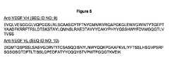


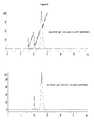


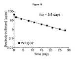
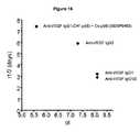


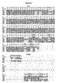

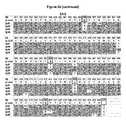
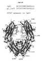

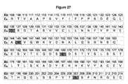

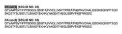


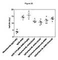


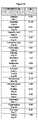


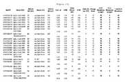
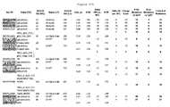


Classifications
- C—CHEMISTRY; METALLURGY
- C07—ORGANIC CHEMISTRY
- C07K—PEPTIDES
- C07K16/00—Immunoglobulins [IGs], e.g. monoclonal or polyclonal antibodies
- C07K16/18—Immunoglobulins [IGs], e.g. monoclonal or polyclonal antibodies against material from animals or humans
- C07K16/22—Immunoglobulins [IGs], e.g. monoclonal or polyclonal antibodies against material from animals or humans against growth factors ; against growth regulators
- A—HUMAN NECESSITIES
- A61—MEDICAL OR VETERINARY SCIENCE; HYGIENE
- A61P—SPECIFIC THERAPEUTIC ACTIVITY OF CHEMICAL COMPOUNDS OR MEDICINAL PREPARATIONS
- A61P35/00—Antineoplastic agents
- C—CHEMISTRY; METALLURGY
- C07—ORGANIC CHEMISTRY
- C07H—SUGARS; DERIVATIVES THEREOF; NUCLEOSIDES; NUCLEOTIDES; NUCLEIC ACIDS
- C07H21/00—Compounds containing two or more mononucleotide units having separate phosphate or polyphosphate groups linked by saccharide radicals of nucleoside groups, e.g. nucleic acids
- C—CHEMISTRY; METALLURGY
- C07—ORGANIC CHEMISTRY
- C07K—PEPTIDES
- C07K16/00—Immunoglobulins [IGs], e.g. monoclonal or polyclonal antibodies
- A—HUMAN NECESSITIES
- A61—MEDICAL OR VETERINARY SCIENCE; HYGIENE
- A61K—PREPARATIONS FOR MEDICAL, DENTAL OR TOILETRY PURPOSES
- A61K39/00—Medicinal preparations containing antigens or antibodies
- A61K2039/505—Medicinal preparations containing antigens or antibodies comprising antibodies
- C—CHEMISTRY; METALLURGY
- C07—ORGANIC CHEMISTRY
- C07K—PEPTIDES
- C07K2317/00—Immunoglobulins specific features
- C07K2317/50—Immunoglobulins specific features characterized by immunoglobulin fragments
- C07K2317/51—Complete heavy chain or Fd fragment, i.e. VH + CH1
- C—CHEMISTRY; METALLURGY
- C07—ORGANIC CHEMISTRY
- C07K—PEPTIDES
- C07K2317/00—Immunoglobulins specific features
- C07K2317/50—Immunoglobulins specific features characterized by immunoglobulin fragments
- C07K2317/515—Complete light chain, i.e. VL + CL
- C—CHEMISTRY; METALLURGY
- C07—ORGANIC CHEMISTRY
- C07K—PEPTIDES
- C07K2317/00—Immunoglobulins specific features
- C07K2317/50—Immunoglobulins specific features characterized by immunoglobulin fragments
- C07K2317/52—Constant or Fc region; Isotype
- C—CHEMISTRY; METALLURGY
- C07—ORGANIC CHEMISTRY
- C07K—PEPTIDES
- C07K2317/00—Immunoglobulins specific features
- C07K2317/50—Immunoglobulins specific features characterized by immunoglobulin fragments
- C07K2317/52—Constant or Fc region; Isotype
- C07K2317/522—CH1 domain
- C—CHEMISTRY; METALLURGY
- C07—ORGANIC CHEMISTRY
- C07K—PEPTIDES
- C07K2317/00—Immunoglobulins specific features
- C07K2317/50—Immunoglobulins specific features characterized by immunoglobulin fragments
- C07K2317/52—Constant or Fc region; Isotype
- C07K2317/524—CH2 domain
- C—CHEMISTRY; METALLURGY
- C07—ORGANIC CHEMISTRY
- C07K—PEPTIDES
- C07K2317/00—Immunoglobulins specific features
- C07K2317/50—Immunoglobulins specific features characterized by immunoglobulin fragments
- C07K2317/52—Constant or Fc region; Isotype
- C07K2317/526—CH3 domain
- C—CHEMISTRY; METALLURGY
- C07—ORGANIC CHEMISTRY
- C07K—PEPTIDES
- C07K2317/00—Immunoglobulins specific features
- C07K2317/50—Immunoglobulins specific features characterized by immunoglobulin fragments
- C07K2317/52—Constant or Fc region; Isotype
- C07K2317/53—Hinge
- C—CHEMISTRY; METALLURGY
- C07—ORGANIC CHEMISTRY
- C07K—PEPTIDES
- C07K2317/00—Immunoglobulins specific features
- C07K2317/90—Immunoglobulins specific features characterized by (pharmaco)kinetic aspects or by stability of the immunoglobulin
- C07K2317/94—Stability, e.g. half-life, pH, temperature or enzyme-resistance
Definitions
- the inventionrelates generally to compositions and methods for altering the isoelectric point of an antibody, and in some cases, resulting in improved plasma pharmacokinetics, e.g. increased serum half-life in vivo.
- Antibodiesare immunological proteins that bind a specific antigen. In most mammals, including humans and mice, antibodies are constructed from paired heavy and light polypeptide chains. Each chain is made up of individual immunoglobulin (Ig) domains, and thus the generic term immunoglobulin is used for such proteins. Each chain is made up of two distinct regions, referred to as the variable and constant regions. The light and heavy chain variable regions show significant sequence diversity between antibodies, and are responsible for binding the target antigen. The constant regions show less sequence diversity, and are responsible for binding a number of natural proteins to elicit important biochemical events.
- IgAwhich includes subclasses IgA1 and IgA2
- IgDwhich includes subclasses IgA1 and IgA2
- IgEwhich includes subclasses IgG1, IgG2, IgG3, and IgG4
- IgMimmunoglobulin M
- IgG antibodiesare tetrameric proteins composed of two heavy chains and two light chains.
- the IgG heavy chainis composed of four immunoglobulin domains linked from N- to C-terminus in the order VH-CH1-CH2-CH3, referring to the heavy chain variable domain, heavy chain constant domain 1, heavy chain constant domain 2, and heavy chain constant domain 3 respectively (also referred to as VH-C ⁇ l-C ⁇ 2-C ⁇ 3, referring to the heavy chain variable domain, constant gamma 1 domain, constant gamma 2 domain, and constant gamma 3 domain respectively).
- the IgG light chainis composed of two immunoglobulin domains linked from N- to C-terminus in the order VL-CL, referring to the light chain variable domain and the light chain constant domain respectively.
- Antibodieshave serum half-lives in vivo ranging from one to three weeks. This favorable property is due to the preclusion of kidney filtration due to the large size of the full-length molecule, and interaction of the antibody Fc region with the neonatal Fc receptor FcRn. Binding to FcRn recycles endocytosed antibody from the endosome back to the bloodstream ( Raghavan et al., 1996, Annu Rev Cell Dev Biol 12:181-220 ; Ghetie et al., 2000, Annu Rev Immunol 18:739-766 , both entirely incorporated by reference).
- the antibodymay determine its clearance rate (e.g. stability and half-life) in vivo.
- clearance ratee.g. stability and half-life
- factors that contribute to clearance and half-lifeare serum aggregation, enzymatic degradation in the serum, inherent immunogenicity of the antibody leading to clearing by the immune system, antigen-mediated uptake, FcR (non-FcRn) mediated uptake and non-serum distribution (e.g. in different tissue compartments).
- variable regionsmay also have longer serum half-lives ( Igawa et al., 2010 PEDS. 23(5): 385-392 ; US Publication 2011/0076275 both of which are entirely incorporated by reference).
- the mechanism of thisis still poorly understood, and in fact the authors suggest that engineering the variable region is an alternative to engineering the Fc region.
- variable regionsdiffer from antibody to antibody. As such, each variable region must be altered without significantly affecting the binding affinity.
- the present applicationdefines the impact of charge state on antibody pharmacokinetics, and provides novel engineered variants in the constant regions to improve serum half-life.
- one problem to be solvedis to increase serum half life of antibodies by altering the constant domains, thus allowing the same constant regions to be used with different antigen binding sequences, e.g. the variable regions including the CDRs, and minimizing the possibility of immunogenic alterations.
- providing antibodies with constant region variants with reduced pI and extended half-lifeprovides a more modular approach to improving the pharmacokinetic properties of antibodies, as described herein.
- the possibility of immunogenicity resulting from the pI variantsis significantly reduced by importing pI variants from different IgG isotypes such that pI is reduced without introducing significant immunogenicity.
- an additional problem to be solvedis the elucidation of low pI constant domains with high human sequence content, e.g. the minimization or avoidance of non-human residues at any particular position.
- the inventionprovides methods for modifying the isoelectric point of an antibody by introducing at least 6 amino acid mutations, including substitutions with non-native amino acids in a constant domain selected from the heavy chain constant domain and light chain constant domain, wherein the substituted amino acids have a pI lower than the native amino acid, such that said isoelectric point of the variant antibody is lowered by at least 0.5 logs.
- only the heavy chain constant domainis altered; in some cases, only the light chain constant domain, and in some cases both the heavy and light constant domains comprise mutated amino acids.
- the methodsprovide for the generation of these variants by amino acid mutations selected from the group consisting of a non-native glutamic acid at position 119; a non-native cysteine at position 131; a non-native arginine, lysine or glutamine at position 133; a non-native glutamic acid at position 137; a non-native serine at position 138; a non-native glutamic acid at position 164; a non native asparagine at position 192; a non native phenylalanine at position 193, a non-native lysine at position 196, a non-native threonine at position 199, a non-native aspartic acid at position 203, a non-native glutamic acid or glutamine at position 205, a non native aspartic acid at position 208, a non-native glutamic acid or glutamine at position 210, a non native threonine at position 214
- the inventionprovides methods for modifying the isoelectric point of an antibody by introducing at least 2 amino acid mutations in the light constant domain, such that said isoelectric point of the variant antibody is lowered by at least 0.5 logs, and wherein said variant antibody comprises substitutions selected from the group consisting of a non-native glutamine or glutamic acid at position 126, a non-native glutamine, glutamic acid or threonine at position 145; a non-native aspartic acid at position 152, a non-native glutamic acid at position 156, a non-native glutamine or glutamic acid at position 169, a non-native glutamic acid at position 199, a non-native glutamic acid at position 202 and a a non-native glutamic acid at position 207 (using EU numbering).
- the inventionprovides methods for modifying the isoelectric point of an antibody by introducing: a) at least 6 amino acid mutations in the heavy constant domain, wherein said variant antibody comprises mutations selected from the group consisting of a non-native glutamic acid at position 119; a non-native cysteine at position 131; a non-native arginine, lysine or glutamine at position 133; a non-native glutamic acid at position 137; a non-native serine at position 138; a non-native glutamic acid at position 164; a non native asparagine at position 192; a non native phenylalanine at position 193, a non-native lysine at position 196, a non-native threonine at position 199, a non-native aspartic acid at position 203, a non-native glutamic acid or glutamine at position 205, a non native aspartic acid at position 208, a
- the pI antibodies of the inventiongenerated using the above methods, has an increased serum half life as compared to an antibody without the mutations.
- the inventionprovides antibodies comprising a variant heavy constant domain polypeptide comprising a variant of SEQ ID NO: 2, comprising at least 6 mutations selected from the group consisting of a non-native glutamic acid at position 119; a non-native cysteine at position 131; a non-native arginine, lysine or glutamine at position 133; a non-native glutamic acid at position 137; a non-native serine at position 138; a non-native glutamic acid at position 164; a non native asparagine at position 192; a non native phenylalanine at position 193, a non-native lysine at position 196, a non-native threonine at position 199, a non-native aspartic acid at position 203, a non-native glutamic acid or glutamine at position 205, a non native aspartic acid at position 208, a non-native glutamic acid
- the inventionprovides antibodies comprising a variant light constant domain polypeptide comprising of variant of SEQ ID NO: 112, wherein said variant antibody comprises substitutions selected from the group consisting of a non-native glutamine or glutamic acid at position 126, a non-native glutamine, glutamic acid or threonine at position 145; a non-native aspartic acid at position 152, a non-native glutamic acid at position 156, a non-native glutamine or glutamic acid at position 169, a non-native glutamic acid at position 199, a non-native glutamic acid at position 202 and a a non-native glutamic acid at position 207 (using EU numbering).
- the inventionprovides nucleic acids encoding the antibodies, including a nucleic acid encoding a variant heavy chain constant domain and/or a nucleic acid encoding a variant light chain constant domain. Host cells containing the nucleic acids and methods of producing the antibodies are also included.
- the inventionprovides antibodies comprising a variant heavy chain constant domain having the formula: A-X 119 -T-K-G-P-S-V-F-P-L-A-P-X 131 -S-X 133 -S-T-S-X 137 -X 138 -T-A-A-L-G-C-L-V-K-D-Y-F-P-E-P-V-T-V-S-W-N-S-G-A-L-X 164 -S-G-V-H-T-F-P-A-V-L-Q-S-S-G-L-Y-S-L-S-S-V-V-T-V-P-S-S-X 192 -X 193 -G-T-X 196 -T-Y-X 199 -C-N-V-X 203 -H-X 205 -P-S-X 208 -T-X 210 -V-D-K
- variant heavy chain constant domaincomprises at least 10 or 15 substitutions as compared to SEQ ID NO: 2.
- the inventionprovides antibodies with a variant light chain constant domain having the formula: X 108 -T-V-A-A-P-S-V-F-I-F-P-P-S-D-E-X 124 -L-X 126 -S-G-T-A-S-V-V-C-L-L-N-X 138 -F-Y-P-R-E-A-X 145 -V-Q-W-K-V-D-X 152 -A-L-Q-X 156 -G-N-S-Q-E-S-V-T-E-Q-D-S-X 169 -D-S-T-Y-S-L-S-S-T-L-T-L-S-K-A-D-Y-E-K-H-K-V-Y-A-C-E-V-T-H-X 199 -G-L-X 202 -S-P-V-T-X 207 -
- Figure 1Amino acid sequences of wild-type constant regions used in the invention.
- Figure 2Engineering of heavy chain CH1 domains. List of CH1 residues for the four IgG isotypes, fraction exposed, and examples of substitutions that can be made to lower pI. Numbering is according to the EU index.
- Figure 3Engineering of light chain CK domains. List of CK residues, fraction exposed, and substitutions that can be made to lower pI. Numbering is according to the EU index.
- Figure 4Amino acid sequences of pI engineered constant regions IgG1-CH1-pI(6) and CK-pI(6).
- Figure 5Amino acid sequences of wild-type anti-VEGF VH and VL variable regions used in the invention.
- Figure 6Amino acid sequences of the heavy and light chains of pI engineered anti-VEGF antibody XENP9493 IgG1-CH1-pI(6)-CK-pI(6) used in the invention.
- Figure 7Structure of an antibody Fab domain showing the locations of pI lowering mutations in XENP9493 IgG1-CH1-pI(6)-CK-pI(6).
- Figure 8Analysis of pI engineered anti-VEGF variants on an Agilent Bioanalyzer showing high purity.
- Figure 9Analysis of pI engineered anti-VEGF variants on SEC showing high purity.
- Figure 10Analysis of pI engineered anti-VEGF variants on an IEF gel showing variants have altered pI.
- Figure 11Analysis of
- Binding analysis(Biacore) of bevacizumab and pI engineered anti-VEGF binding to VEGF.
- Figure 12DSC analysis of CH1 and CKpI engineered anti-VEGF showing high thermostability.
- Figure 13PK of bevacizumab variants in huFcRn mice. The 9493 variant with pI-engineered CH1 and CK domains extends half-life in vivo.
- Figure 14PK of a native IgG1 version of bevacizumab in four separate in vivo studies in huFcRn mice. The average IgG1 half-life was 3.2 days.
- Figure 15.
- PK of a native IgG2 version of bevacizumab in huFcRn micePK of a native IgG2 version of bevacizumab in huFcRn mice.
- Figure 16Correlation between half-life and isoelectric point (pI) of antibody variants with different constant chains.
- Figure 17Amino acid sequence alignment of the IgG subclasses. Residues with a bounded box illustrate isotypic differences between the IgG's. Residues which contribute to a higher pI (K, R, and H) or lower pI (D and E) are highlighted in bold. Designed substitutions that either lower the pI, or extend an epitope are shown in gray.
- Figure 18Amino acid sequence of the CK and C ⁇ light constant chains.
- Residues which contribute to a higher pI (K, R, and H) or lower pI (D and E)are highlighted in bold. Preferred positions that can be modified to lower the pI are shown in gray.
- Figure 19Amino acid sequences of pI-engineered variant heavy chains.
- Figure 20Amino acid sequences of pI-engineered variant light chains.
- Figure 21PK results of pI-engineered variant bevacizumab antibodies in huFcRn mice.
- Figure 22PK results of variants that combine pI-engineered modifications with Fc modifications that enhance binding to FcRn.
- Figure 23.
- FIG. 30Plot showing the effect of charge swap mutations on antibody pI. As the pI gets lower the change in pI per charge swap decreases.
- Figure 31PK results of pI-engineered isotypic variant bevacizumab antibodies (IgG-pI-Iso3) and combinations with substitution N434S in huFcRn mice.
- Figure 32PK results of pI-engineered isotypic variant bevacizumab antibodies and combinations with substitution N434S in huFcRn mice.
- Figure 33.
- WTchange in the number of Asp or Glu acid residues relative to IgG1 wild-type sequence of bevacizumab Charge state derived from the total number of Lys and Arg minus the total number of Asp and Glu residues, assuming a pH of 7 # HC Mutations vs IgG1 number of mutations in the heavy chain constant domain as compared to IgG1 # LC Mutations vs IgG1 number of mutations in the light chain constant domain as compared to IgG1 Total # of Mutations total number of mutations in the heavy chain and light chain constant domains as compared to IgG1
- the present inventionis generally directed to compositions and methods relating to decreasing the isoelectric point (pI) of antibodies (to form "pI antibodies") by incorporating amino acid substitutions ("pI variants" or "pI substitutions") into one or more constant region domains of the antibody.
- the pI substitutionsare chosen such that the pI amino acids have a pI lower than the native amino acid at a particular position in the constant domain.
- the constant domain variantsreduce the pI of the antibody, and, as shown herein for the first time, improve serum half-life in vivo. While, as noted above, there is limited data that might suggest that lowering the pI of an antibody by generating variants in the CDR regions of an antibody can lead to increased serum half life. However, the present invention provides a significant benefit to CDR pI engineering, as the constant domains of the present invention can be added in a modular fashion to the variable regions, thus significantly simplifying design of antibodies that have increased serum half lives.
- IgG1is a common isotype for therapeutic antibodies for a variety of reasons, including high effector function.
- the heavy constant region of IgG1has a higher pI than that of IgG2 (8.10 versus 7.31).
- IgG1has a glycine (pI 5.97) at position 137
- IgG2has a glutamic acid (pI 3.22); importing the glutamic acid will affect the pI of the resulting protein.
- a number of amino acid substitutionsare generally required to significant affect the pI of the variant antibody.
- even changes in IgG2 moleculesallow for increased serum half-life.
- non-isotypic amino acid changesare made, either to reduce the overall charge state of the resulting protein (e.g. by changing a higher pI amino acid to a lower pI amino acid), or to allow accommodations in structure for stability, etc. as is more further described below.
- the present inventiondemonstrates that while the absolute half life of two different antibodies may differ due to these antigen specificity effects, the pI variants (which optionally include FcRn variants as outlined herein), can transfer to different ligands to give the same trends of increasing half-life. That is, in general, the relative "order" of the pI decreases/half life increases will track to antibodies with the same pI variants of antibodies with different Fvs as is discussed herein.
- the present inventionrelates to the generation of pI variants of antibodies, generally therapeutic antibodies.
- antibodyis used generally. Antibodies that find use in the present invention can take on a number of formats as described herein, including traditional antibodies as well as antibody derivatives, fragments and mimetics, described below.
- antibodyincludes any polypeptide that includes at least one constant domain, including, but not limited to, CH1, CH2, CH3 and CL.
- Traditional antibody structural unitstypically comprise a tetramer. Each tetramer is typically composed of two identical pairs of polypeptide chains, each pair having one "light” (typically having a molecular weight of about 25 kDa) and one "heavy” chain (typically having a molecular weight of about 50-70 kDa). Human light chains are classified as kappa and lambda light chains.
- the present inventionis directed to the IgG class, which has several subclasses, including, but not limited to IgG1, IgG2, IgG3, and IgG4.
- “isotype” as used hereinis meant any of the subclasses of immunoglobulins defined by the chemical and antigenic characteristics of their constant regions.
- therapeutic antibodiescan also comprise hybrids of isotypes and/or subclasses. For example, as shown herein, the present invention covers pI engineering of IgG1/G2 hybrids.
- each chainincludes a variable region of about 100 to 110 or more amino acids primarily responsible for antigen recognition, generally referred to in the art and herein as the "Fv domain” or “Fv region".
- Fv domainor “Fv region”.
- CDRcomplementarity-determining region
- Variablerefers to the fact that certain segments of the variable region differ extensively in sequence among antibodies. Variability within the variable region is not evenly distributed. Instead, the V regions consist of relatively invariant stretches called framework regions (FRs) of 15-30 amino acids separated by shorter regions of extreme variability called “hypervariable regions” that are each 9-15 amino acids long or longer.
- Each VH and VLis composed of three hypervariable regions ("complementary determining regions," "CDRs") and four FRs, arranged from amino-terminus to carboxy-terminus in the following order: FR1-CDR1-FR2-CDR2-FR3-CDR3-FR4.
- the hypervariable regiongenerally encompasses amino acid residues from about amino acid residues 24-34 (LCDR1; “L” denotes light chain), 50-56 (LCDR2) and 89-97 (LCDR3) in the light chain variable region and around about 31-35B (HCDR1; “H” denotes heavy chain), 50-65 (HCDR2), and 95-102 (HCDR3) in the heavy chain variable region; Kabat et al., SEQUENCES OF PROTEINS OF IMMUNOLOGICAL INTEREST, 5th Ed. Public Health Service, National Institutes of Health, Bethesda, Md. (1991 ) and/or those residues forming a hypervariable loop (e.g.
- the Kabat numbering systemis generally used when referring to a residue in the variable domain (approximately, residues 1-107 of the light chain variable region and residues 1-113 of the heavy chain variable region) (e.g, Kabat et al ., supra (1991)).
- epitope binding sitecontributes to the formation of the antigen-binding, or more specifically, epitope binding site of antibodies.
- Epitoperefers to a determinant that interacts with a specific antigen binding site in the variable region of an antibody molecule known as a paratope. Epitopes are groupings of molecules such as amino acids or sugar side chains and usually have specific structural characteristics, as well as specific charge characteristics. A single antigen may have more than one epitope.
- the epitopemay comprise amino acid residues directly involved in the binding (also called immunodominant component of the epitope) and other amino acid residues, which are not directly involved in the binding, such as amino acid residues which are effectively blocked by the specifically antigen binding peptide; in other words, the amino acid residue is within the footprint of the specifically antigen binding peptide.
- Epitopesmay be either conformational or linear.
- a conformational epitopeis produced by spatially juxtaposed amino acids from different segments of the linear polypeptide chain.
- a linear epitopeis one produced by adjacent amino acid residues in a polypeptide chain. Conformational and nonconformational epitopes may be distinguished in that the binding to the former but not the latter is lost in the presence of denaturing solvents.
- An epitopetypically includes at least 3, and more usually, at least 5 or 8-10 amino acids in a unique spatial conformation. Antibodies that recognize the same epitope can be verified in a simple immunoassay showing the ability of one antibody to block the binding of another antibody to a target antigen, for example "binning.”
- any antigenmay be targeted by the IgG variants, including but not limited to proteins, subunits, domains, motifs, and/or epitopes belonging to the following list of target antigens, which includes both soluble factors such as cytokines and membrane-bound factors, including transmembrane receptors: 17-IA, 4-1BB, 4Dc, 6-keto-PGF1a, 8-iso-PGF2a, 8-oxo-dG, A1 Adenosine Receptor, A33, ACE, ACE-2, Activin, Activin A, Activin AB, Activin B, Activin C, Activin RIA, Activin RIA ALK-2, Activin RIB ALK-4, Activin RIIA, Activin RIIB, ADAM, ADAM10, ADAM12,
- the pI engineering described hereinis done to therapeutic antibodies.
- a number of antibodies that are approved for use, in clinical trials, or in developmentmay benefit from the pI variants of the present invention. These antibodies are herein referred to as "clinical products and candidates".
- the pI engineered constant region(s) of the present inventionmay find use in a range of clinical products and candidates.
- a number of antibodies that target CD20may benefit from the pI engineering of the present invention.
- the pI variants of the present inventionmay find use in an antibody that is substantially similar to rituximab (Rituxan®, IDEC/Genentech/Roche) (see for example US 5,736,137 ), a chimeric anti-CD20 antibody approved to treat Non-Hodgkin's lymphoma; HuMax-CD20, an anti-CD20 currently being developed by Genmab, an anti-CD20 antibody described in US 5,500,362 , AME-133 (Applied Molecular Evolution), hA20 (Immunomedics, Inc.), HumaLYM (Intracel), and PRO70769 ( PCT/US2003/040426 , entitled "Immunoglobulin Variants and Uses Thereof").
- rituximabRituxan®, IDEC/Genentech/Roche
- HuMax-CD20an anti-CD20 currently being developed by Genmab
- an anti-CD20 antibody described in US 5,500,362AME-133 (Applied Molecular Evolution
- a number of antibodies that target members of the family of epidermal growth factor receptorsmay benefit from pI engineered constant region(s) of the invention.
- the pI engineered constant region(s) of the inventionmay find use in an antibody that is substantially similar to trastuzumab (Herceptin®, Genentech) (see for example US 5,677,171 ), a humanized anti-Her2/neu antibody approved to treat breast cancer; pertuzumab (rhuMab-2C4, OmnitargTM), currently being developed by Genentech; an anti-Her2 antibody described in US 4,753,894 ; cetuximab (Erbitux®, Imclone) ( US 4,943,533 ; PCT WO 96/40210 ), a chimeric anti-EGFR antibody in clinical trials for a variety of cancers; ABX-EGF ( US 6,235,883 ), currently being developed by Abgenix-Immunex-Amgen; HuMax-EGFr ( USSN 10/172,317 ), currently being developed by Genmab; 425, EMD55900, EMD62000, and EMD72000 (Merck KGa
- the pI engineered constant region(s) of the present inventionmay find use in alemtuzumab (Campath®, Millenium), a humanized monoclonal antibody currently approved for treatment of B-cell chronic lymphocytic leukemia.
- the pI engineered constant region(s) of the present inventionmay find use in a variety of antibodies that are substantially similar to other clinical products and candidates, including but not limited to muromonab-CD3 (Orthoclone OKT3®), an anti-CD3 antibody developed by Ortho Biotech/Johnson & Johnson, ibritumomab tiuxetan (Zevalin®), an anti-CD20 antibody developed by IDEC/Schering AG, gemtuzumab ozogamicin (Mylotarg®), an anti-CD33 (p67 protein) antibody developed by Celltech/Wyeth, alefacept (Amevive®), an anti-LFA-3 Fc fusion developed by Biogen), abciximab (ReoPro®), developed by Centocor/Lilly, basiliximab (Simulect®), developed by Novartis, palivizumab (Synagis®), developed by MedImmune, infliximab (Remicade®), an anti
- each chaindefines a constant region primarily responsible for effector function.
- Kabat et alcollected numerous primary sequences of the variable regions of heavy chains and light chains. Based on the degree of conservation of the sequences, they classified individual primary sequences into the CDR and the framework and made a list thereof (see SEQUENCES OF IMMUNOLOGICAL INTEREST, 5th edition, NIH publication, No. 91-3242, E.A. Kabat et al., entirely incorporated by reference).
- immunoglobulin domainsin the heavy chain.
- immunoglobulin (Ig) domainherein is meant a region of an immunoglobulin having a distinct tertiary structure.
- the heavy chain domainsincluding, the constant heavy (CH) domains and the hinge domains.
- the IgG isotypeseach have three CH regions. Accordingly, "CH” domains in the context of IgG are as follows: “CH1” refers to positions 118-220 according to the EU index as in Kabat.
- CH2refers to positions 237-340 according to the EU index as in Kabat
- CH3refers to positions 341-447 according to the EU index as in Kabat.
- the pI variantscan be in one or more of the CH regions, as well as the hinge region, discussed below.
- sequences depicted hereinstart at the CH1 region, position 118; the variable regions are not included except as noted.
- the first amino acid of SEQ ID NO: 2while designated as position "1" in the sequence listing, corresponds to position 118 of the CH1 region, according to EU numbering.
- Ig domain of the heavy chainis the hinge region.
- hinge regionor “hinge region” or “antibody hinge region” or “immunoglobulin hinge region” herein is meant the flexible polypeptide comprising the amino acids between the first and second constant domains of an antibody.
- the IgG CH1 domainends at EU position 220, and the IgG CH2 domain begins at residue EU position 237.
- the antibody hingeis herein defined to include positions 221 (D221 in IgG1) to 236 (G236 in IgG1), wherein the numbering is according to the EU index as in Kabat.
- the lower hingeis included, with the “lower hinge” generally referring to positions 226 or 230.
- pI variantscan be made in the hinge region as well.
- the light chaingenerally comprises two domains, the variable light domain (containing the light chain CDRs and together with the variable heavy domains forming the Fv region), and a constant light chain region (often referred to as CL or C ⁇ ).
- Fc regionAnother region of interest for additional substitutions, outlined below, is the Fc region.
- Fcor “Fc region” or “Fc domain” as used herein is meant the polypeptide comprising the constant region of an antibody excluding the first constant region immunoglobulin domain and in some cases, part of the hinge.
- Fcrefers to the last two constant region immunoglobulin domains of IgA, IgD, and IgG, the last three constant region immunoglobulin domains of IgE and IgM, and the flexible hinge N-terminal to these domains.
- Fcmay include the J chain.
- the Fc domaincomprises immunoglobulin domains C ⁇ 2 and C ⁇ 3 (C ⁇ 2 and C ⁇ 3) and the lower hinge region between C ⁇ 1 (C ⁇ 1) and C ⁇ 2 (C ⁇ 2).
- the human IgG heavy chain Fc regionis usually defined to include residues C226 or P230 to its carboxyl-terminus, wherein the numbering is according to the EU index as in Kabat.
- amino acid modificationsare made to the Fc region, for example to alter binding to one or more Fc ⁇ R receptors or to the FcRn receptor.
- the antibodiesare full length.
- full length antibodyherein is meant the structure that constitutes the natural biological form of an antibody, including variable and constant regions, including one or more modifications as outlined herein.
- the antibodiescan be a variety of structures, including, but not limited to, antibody fragments, monoclonal antibodies, bispecific antibodies, minibodies, domain antibodies, synthetic antibodies (sometimes referred to herein as “antibody mimetics”), chimeric antibodies, humanized antibodies, antibody fusions (sometimes referred to as “antibody conjugates”), and fragments of each, respectively.
- the antibodyis an antibody fragment, as long as it contains at least one constant domain which can be pI engineered.
- Specific antibody fragmentsinclude, but are not limited to, (i) the Fab fragment consisting of VL, VH, CL and CH1 domains, (ii) the Fd fragment consisting of the VH and CH1 domains, (iii) F(ab')2 fragments, a bivalent fragment comprising two linked Fab fragments (vii) single chain Fv molecules (scFv), wherein a VH domain and a VL domain are linked by a peptide linker which allows the two domains to associate to form an antigen binding site ( Bird et al., 1988, Science 242:423-426 , Huston et al., 1988, Proc.
- scFvsingle chain Fv molecules
- antibody fragmentsthat can be used include fragments that contain one or more of the CH1, CH2, CH3, hinge and CL domains of the invention that have been pI engineered.
- Fc fusionsare fusions of the Fc region (CH2 and CH3, optionally with the hinge region) fused to another protein.
- a number of Fc fusionsare known the art and can be improved by the addition of the pI variants of the invention.
- antibody fusionscan be made comprising CH1; CH1, CH2 and CH3; CH2; CH3; CH2 and CH3; CH1 and CH3, any or all of which can be made optionally with the hinge region, utilizing any combination of pI variants described herein..
- the antibodycan be a mixture from different species, e.g. a chimeric antibody and/or a humanized antibody.
- chimeric antibodiesand “humanized antibodies” refer to antibodies that combine regions from more than one species.
- chimeric antibodiestraditionally comprise variable region(s) from a mouse (or rat, in some cases) and the constant region(s) from a human.
- Humanized antibodiesgenerally refer to non-human antibodies that have had the variable-domain framework regions swapped for sequences found in human antibodies.
- the entire antibody, except the CDRsis encoded by a polynucleotide of human origin or is identical to such an antibody except within its CDRs.
- the CDRsare grafted into the beta-sheet framework of a human antibody variable region to create an antibody, the specificity of which is determined by the engrafted CDRs.
- the creation of such antibodiesis described in, e.g., WO 92/11018 , Jones, 1986, Nature 321:522-525 , Verhoeyen et al., 1988, Science 239:1534-1536 , all entirely incorporated by reference.
- the humanized antibodyoptimally also will comprise at least a portion of an immunoglobulin constant region, typically that of a human immunoglobulin, and thus will typically comprise a human Fc region. Humanized antibodies can also be generated using mice with a genetically engineered immune system. Roque et al., 2004, Biotechnol. Prog.
- Humanization methodsinclude but are not limited to methods described in Jones et al., 1986, Nature 321:522-525 ; Riechmann et al.,1988; Nature 332:323-329 ; Verhoeyen et al., 1988, Science, 239:1534-1536 ; Queen et al., 1989, Proc Natl Acad Sci, USA 86:10029-33 ; He et al., 1998, J. Immunol. 160: 1029-1035 ; Carter et al., 1992, Proc Natl Acad Sci USA 89:4285-9 , Presta et al., 1997, Cancer Res. 57(20):4593-9 ; Gorman et al., 1991, Proc.
- Humanization or other methods of reducing the immunogenicity of nonhuman antibody variable regionsmay include resurfacing methods, as described for example in Roguska et al., 1994, Proc. Natl. Acad. Sci. USA 91:969-973 , entirely incorporated by reference.
- the parent antibodyhas been affinity matured, as is known in the art. Structure-based methods may be employed for humanization and affinity maturation, for example as described in USSN 11/004,590 .
- Selection based methodsmay be employed to humanize and/or affinity mature antibody variable regions, including but not limited to methods described in Wu et al., 1999, J. Mol. Biol. 294:151-162 ; Baca et al., 1997, J. Biol. Chem. 272(16):10678-10684 ; Rosok et al., 1996, J. Biol. Chem. 271(37): 22611-22618 ; Rader et al., 1998, Proc. Natl. Acad. Sci. USA 95: 8910-8915 ; Krauss et al., 2003, Protein Engineering 16(10):753-759 , all entirely incorporated by reference.
- the antibodies of the inventioncan be multispecific antibodies, and notably bispecific antibodies, also sometimes referred to as "diabodies". These are antibodies that bind to two (or more) different antigens, or different epitopes on the same antigen. Diabodies can be manufactured in a variety of ways known in the art ( Holliger and Winter, 1993, Current Opinion Biotechnol. 4:446-449 , entirely incorporated by reference), e.g., prepared chemically or from hybrid hybridomas. In some cases, multispecific (for example bispecific) antibodies are not preferred.
- the antibodyis a minibody.
- Minibodiesare minimized antibody-like proteins comprising a scFv joined to a CH3 domain.
- the CH3 domaincan be pI engineered.
- the scFvcan be joined to the Fc region, and may include some or the entire hinge region.
- the antibodies of the present inventionare generally isolated or recombinant.
- isolatedwhen used to describe the various polypeptides disclosed herein, means a polypeptide that has been identified and separated and/or recovered from a cell or cell culture from which it was expressed. Ordinarily, an isolated polypeptide will be prepared by at least one purification step.
- Specific bindingor “specifically binds to” or is “specific for” a particular antigen or an epitope means binding that is measurably different from a non-specific interaction. Specific binding can be measured, for example, by determining binding of a molecule compared to binding of a control molecule, which generally is a molecule of similar structure that does not have binding activity. For example, specific binding can be determined by competition with a control molecule that is similar to the target.
- Specific binding for a particular antigen or an epitopecan be exhibited, for example, by an antibody having a KD for an antigen or epitope of at least about 10 -4 M, at least about 10 -5 M, at least about 10 -6 M, at least about 10 -7 M, at least about 10 -8 M, at least about 10 -9 M, alternatively at least about 10 -10 M, at least about 10 -11 M, at least about 10 -12 M, or greater, where KD refers to a dissociation rate of a particular antibody-antigen interaction.
- an antibody that specifically binds an antigenwill have a KD that is 20-, 50-, 100-, 500-, 1000-, 5,000-, 10,000- or more times greater for a control molecule relative to the antigen or epitope.
- specific binding for a particular antigen or an epitopecan be exhibited, for example, by an antibody having a KA or Ka for an antigen or epitope of at least 20-, 50-, 100-, 500-, 1000-, 5,000-, 10,000- or more times greater for the epitope relative to a control, where KA or Ka refers to an association rate of a particular antibody-antigen interaction.
- the present inventionrelates to the generation of pI variants of antibodies.
- pIrefers to the isoelectric point of a molecule (including both the individual amino acids and antibodies) and is the pH at which a particular molecule or surface carries no net electrical charge.
- the invention hereinsometimes refers to changes in the "charge state" of the proteins at pH 7. That is, wild-type heavy constant region of IgG1 has a charge state of +6, while the heavy constant region of IgG2 has a charge state of 0.
- Variant 9493(with a SEQ ID NO: 193 heavy chain constant domain and a SEQ ID NO:117 light chain constant domain) has 12 substitutions in both the heavy and light constant regions resulting in a charge state of -30.
- the present inventionrelates to the generation of pI variants of antibodies to form "pI antibodies".
- pI variantsare made by introducing amino acid mutations into the parent molecule. "Mutations” in this context are usually amino acid substitutions, although as shown herein, deletions and insertions of amino acids can also be done and thus are defined as mutations.
- pI variantsor “isoelectric point variants” or “pI substitutions” or grammatical equivalents thereof herein is meant mutating an amino acid to result in a lower pI at that position. In many embodiments, this means making an amino acid substitution with a lower pI than the original (e.g.wild type) amino acid at the particular position. In some embodiments, this can also mean deleting an amino acid with a high pI (if the structure will tolerate it) or inserting amino acids with lower pIs, for example the low pI "tails” discussed below.
- the different amino acidshave different pIs, although this figure shows the pI of amino acids as individual compositions rather than in the context of a protein, although the trend is identical.
- pI variants in the context of the inventionare made to contribute to the decrease of the pI of the protein, in this case at least the heavy constant domain or the light constant domain of an IgG antibody, or both.
- An antibody engineered to include one or more of the amino acid mutations outlined hereinis sometimes also referred to herein as a "pI antibody".
- pI variantsrefer to the mutation of a higher pI amino acid either via substituting with an amino acid with a lower pI, deleting an amino acid, or inserting low pI amino acids, thus lowering the overall pI of the antibody.
- additional non-pI variantsare often added to structurally compensate for the pI variants, leading to increased stability, etc.
- the solvent accessibility of the amino acidis taken into account, although in general it is not the only factor. That is, based on the known structure of IgG molecules, and as shown in Figure 2 , each position will either be fully exposed, fully shielded (e.g.
- candidate positions for substitution with lower pI amino acidsare at least 50% exposed, with exposures of over 60, 70, 80+% finding use in the present invention, as well as those residues that are effectively 100% exposed.
- the lowering of the pIcan be done in one of several ways, either replacing a higher pI amino acid (e.g. positive charge state, for example) with a neutral pI, replacing a higher pI amino acid with a lower or low pI amino acid, or replacing a neutral pI amino acid with a low pI amino acid.
- deletions or insertions of one or more amino acidscan also be done, e.g. deleting a high pI amino acid or inserting one or more low pI amino acids.
- an arginine(pI 11.15) can be replaced by lysine (pI 9.59, still high but lower), a more neutral amino acid like glycine or serine, or by low pI variants such as aspartic acid or glutamic acid.
- pI variantsare defined as variants by comparison to the starting or parent sequence, which frequently is the wild-type IgG constant domain (either heavy or light or both, as outlined herein). That is, the amino acid at a particular position in the wild-type is referred to as the "native" amino acid, and an amino acid substitution (or deletion or insertion) at this position is referred to as a "non-native" amino acid.
- the amino acid at a particular position in the wild-typeis referred to as the "native" amino acid, and an amino acid substitution (or deletion or insertion) at this position is referred to as a "non-native" amino acid.
- a "non-native" amino acidis as compared to the IgG1 sequence.
- IgG1has a serine, and thus the non-native amino acid that can be substituted is glutamic acid.
- SEQ ID NO: 193has a non-native glutamic acid at position 119.
- the native and non-native amino acidsare compared to the wild-type IgG2 sequence.
- SEQ ID NO: 28is a hybrid IgG1/G2 molecule
- SEQ ID NO: 164is a hybrid IgG2/G1 molecule.
- “non-native" or “non-wild type” substitutionsmeans that the amino acid at the position in question is different from the parent wild-type sequence from whence that position came; that is, if the cross-over point is between amino acids 100 and 101, such that the N-terminus is from IgG1 and the C-terminus is from IgG2, a "non-native" amino acid at position 90 will be compared to the IgG1 sequence.
- non-wild type IgG domainse.g. IgG domains that already have variants
- a substitutionwill be "non-native" as long as it does not revert back to a wild type sequence.
- the pI variants of the inventionare chosen to decrease the positive charge of the pI antibody.
- the pI variantsare made at least in the CH1 region of the heavy chain domain of an IgG antibody.
- the mutationscan be independently and optionally selected from position 119, 131, 133, 137, 138, 164, 192, 193, 196, 199, 203, 205, 208, 210,214, 217 and 219. All possible combinations of these 17 positions can be made; e.g. a pI antibody may have 1, 2, 3, 4, 5, 6, 7, 8, 9, 10, 11, 12, 13, 14, 15, 16, or 17 CH1 pI substitutions.
- any single or combination CH1 variant(s)can be combined, optionally and individually, with any CH2, CH3, hinge and LC variant(s) as well, as is further described below.
- substitutions that find use in lowering the pI of CH1 domainsinclude, but are not limited to, a non-native glutamic acid at position 119; a non-native cysteine at position 131; a non-native arginine, lysine or glutamine at position 133; a non-native glutamic acid at position 137; a non-native serine at position 138; a non-native glutamic acid at position 164; a non native asparagine at position 192; a non native phenylalanine at position 193, a non-native lysine at position 196, a non-native threonine at position 199, a non-native aspartic acid at position 203, a non-native glutamic acid or glutamine at position 205, a non native aspartic acid at position 208, a non-native glutamic acid or glutamine at position 210, a non native threonine at position
- substitutionscan be made individually and in any combination, with preferred combinations shown in the SEQ ID listings and described below. In some cases, only pI substitutions are done in the CH1 domain, and in others, these substitution(s) are added to other pI variants in other domains in any combination.
- mutationsare made in the hinge domain, including positions 221, 222, 223, 224, 225, 233, 234, 235 and 236. It should be noted that changes in 233-236 can be made to increase effector function (along with 327A) in the IgG2 backbone. Thus, pI mutations and particularly substitutions can be made in one or more of positions 221-225, with 1, 2, 3, 4 or 5 mutations finding use in the present invention. Again, all possible combinations are contemplated, alone or with other pI variants in other domains.
- substitutions that find use in lowering the pI of hinge domainsinclude, but are not limited to, a deletion at position 221, a non-native valine or threonine at position 222, a deletion at position 223, a non-native glutamic acid at position 224, a deletion at position 225, a deletion at position 235 and a deletion or a non-native alanine at position 236.
- these mutationscan be made individually and in any combination, with preferred combinations shown in the SEQ ID listings and described below. In some cases, only pI substitutions are done in the hinge domain, and in others, these substitution(s) are added to other pI variants in other domains in any combination.
- mutationscan be made in the CH2 region, including positions 274, 296, 300, 309, 320, 322, 326, 327, 334 and 339. Again, all possible combinations of these 10 positions can be made; e.g. a pI antibody may have 1, 2, 3, 4, 5, 6, 7, 8, 9 or 10 CH2 pI substitutions.
- substitutions that find use in lowering the pI of CH2 domainsinclude, but are not limited to, a non-native glutamine or glutamic acid at position 274, a non-native phenylalanine at position 296, a non native phenylalanine at position 300, a non-native valine at position 309, a non-native glutamic acid at position 320, a non-native glutamic acid at position 322, a non-native glutamic acid at position 326, a non-native glycine at position 327, a non-native glutamic acid at position 334, a non native threonine at position 339, and all possible combinations within CH2 and with other domains.
- the mutationscan be independently and optionally selected from position 355, 384, 392, 397, 419 and 447. All possible combinations of these 6 positions can be made; e.g. a pI antibody may have 1, 2, 3, 4, 5 or 6 CH1 pI mutations.
- a pI antibodymay have 1, 2, 3, 4, 5 or 6 CH1 pI mutations.
- any single or combination CH3 variant(s)can be combined, optionally and individually, with any CH2, CH1, hinge and LC variant(s) as well, as is further described below.
- substitutions that find use in lowering the pI of CH3 domainsinclude, but are not limited to, a non native glutamine or glutamic acid at position 355, a non-native serine at position 384, a non-native asparagine or glutamic acid at position 392, a non-native methionine at position 397, a non native glutamic acid at position 419, and a deletion or non-native aspartic acid at position 447.
- any possible combination of the following heavy chain constant domain mutationscan be made, with each mutation being optionally included or excluded: a non-native glutamic acid at position 119; a non-native cysteine at position 131; a non-native arginine, lysine or glutamine at position 133; a non-native glutamic acid at position 137; a non-native serine at position 138; a non-native glutamic acid at position 164; a non native asparagine at position 192; a non native phenylalanine at position 193, a non-native lysine at position 196, a non-native threonine at position 199, a non-native aspartic acid at position 203, a non-native glutamic acid or glutamine at position 205, a non native aspartic acid at position 208, a non-native glutamic acid or glutamine at position 210, a non native thre
- some embodimentsutilize variant heavy chain domains with 0 (when the pI engineering is done in the light constant domain only), 1, 2, 3, 4, 5, 6, 7, 8, 9, 10, 11, 12, 13, 14, 15, 16, 17, 18, 19, 20, 22, 23, 26, 27, 28 and 29 mutations (as compared to IgG1) can be made, as depicted in Figure 37 .
- the pI variantsare made at least in the light chain domain of an IgG antibody.
- the mutationscan be independently and optionally selected from positions 126, 145, 152, 156, 169, 199, 202 and 207. All possible combinations of these 8 positions can be made; e.g. a pI antibody may have 1, 2, 3, 4, 5, 6, 7 or light constant domain pI mutations.
- any single or combination CL domain mutationscan be combined with any heavy chain constant domain pI variants.
- Specific mutations that find use in lowering the pI of light chain constant domainsinclude, but are not limited to, a non-native glutamine or glutamic acid at position 126, a non-native glutamine, glutamic acid or threonine at position 145; a non-native aspartic acid at position 152, a non-native glutamic acid at position 156, a non-native glutamine or glutamic acid at position 169, a non-native glutamic acid at position 199, a non-native glutamic acid at position 202 and a a non-native glutamic acid at position 207.
- some embodimentsutilize variant light chain domains with 0 (when the pI engineering is done in the heavy constant domain only), 1, 2, 3, 4, 5, 6, or 10 mutations (as compared to C ⁇ ) can be made, as depicted in Figure 37 .
- any pI engineered heavy chain depicted in Figure 37 and in the sequence listingcan be combined with either a wild-type constant light domain or a pI engineered light constant domain.
- an pI engineered light chain constant domaincan be combined with either a wild-type constant heavy domain or a pI engineered heavy constant domain, even if not specifically present in Figure 37 . That is, the column of "HC names” and "LC names” are meant to form a matrix, with all possible combinations possible.
- any possible combination of the following heavy chain constant domain mutations and light chain constant domainscan be made, with each mutation being optionally included or excluded: a) heavy chain: a non-native glutamic acid at position 119; a non-native cysteine at position 131; a non-native arginine, lysine or glutamine at position 133; a non-native glutamic acid at position 137; a non-native serine at position 138; a non-native glutamic acid at position 164; a non native asparagine at position 192; a non native phenylalanine at position 193, a non-native lysine at position 196, a non-native threonine at position 199, a non-native aspartic acid at position 203, a non-native glutamic acid or glutamine at position 205, a non native aspartic acid at position 208, a non-native glutamic acid or glutamine
- the pI antibodies of the inventioncan contain additional amino acid substitutions in addition to the pI variants.
- amino acid substitutionsare imported from one isotype into the pI antibody despite either a neutrality of charge state or even an increase of charge state, so as to accommodate the pI variants.
- non-pI isotypic variantsFor example, the replacement of the native lysine at position 133 of IgG1 with an arginine from IgG2 is such a change, as is the replacement of the native glutamine in IgG1 at position 196 with the IgG2 lysine, the replacement of native IgG1 proline at position 217 with the IgG2 arginine, etc. It should be noted in this instance that as described above, pI variants can be made at position 133 as well, substituting non-native glutamic acid or glutamine at position 133.
- a mutation at position 384can be made, for example substituting a non-native serine.
- Additional mutationsthat can be made include adding either N-terminal or C-terminal (depending on the structure of the antibody or fusion protein) "tails" or sequences of one or more low pI amino acids; for example, glutamic acids and aspartic acids can be added to the CH3 C-terminus; generally, from 1 to 5 amino acids are added.
- the pI antibodies of the present inventiondisplay decreased pIs. In general, decreases of at least 0.5 log (e.g. corresponding to half a pH point) are seen, with decreases of at least about 1, 1.5, 2, 2.5 and 3 finding particular use in the invention.
- the pIcan be either calculated or determined experimentally, as is well known in the art.
- pI antibodies with pIs ranging from 5. to 5.5 to 6exhibit good extended serum half lives. As will be appreciated by those in the art and depicted in Figure 30 , pIs lower than this are difficult to achieve, as more and more mutations are required and the physical limits are reached.
- the pI antibodies of the present inventiondisplay increased serum half life. As shown in the Figures, surprisingly, every tested pI antibody has exhibited an increase in half life as compared to the starting molecule. While half-life is affected by a number of factors, including the Fv portion, increases of 25, 50, 75, 100, 150, 200 and 250% or more can be obtained using the pI antibodies of the present invention. As shown in Figure 34 , pI variants can increase half-life from around 4 days to over 15.
- some variants hereinare generated to increase stability.
- a number of properties of antibodiesaffect the clearance rate (e.g. stability for half-life) in vivo.
- other factors that contribute to clearance and half-lifeare serum aggregation, enzymatic degradation in the serum, inherent immunogenicity of the antibody leading to clearing by the immune system, antigen-mediated uptake, FcR (non-FcRn) mediated uptake and non-serum distribution (e.g. in different tissue compartments).
- the pI variants of the present inventioncan be combined with amino acid substitutions in the FcRn binding domain.
- the present inventionshows that pI variants can be independently and optionally combined with Fc variants that result in both higher binding to the FcRn receptor as well as increased half-lives.
- FcRnor "neonatal Fc Receptor” as used herein is meant a protein that binds the IgG antibody Fc region and is encoded at least in part by an FcRn gene.
- the FcRnmay be from any organism, including but not limited to humans, mice, rats, rabbits, and monkeys.
- the functional FcRn proteincomprises two polypeptides, often referred to as the heavy chain and light chain.
- the light chainis beta-2-microglobulin and the heavy chain is encoded by the FcRn gene.
- FcRn or an FcRn proteinrefers to the complex of FcRn heavy chain with beta-2-microglobulin.
- the FcRn variantsbind to the human FcRn receptor, or it may be desirable to design variants that bind to rodent or primate receptors in addition, to facilitate clinical trials.
- a pI antibodycan be engineered to include any of the following substitutions, alone or in any combination: 436I, 436V, 311I, 311V, 428L, 434S, 428L/434S, 259I, 308F, 259I/308F, 259I/308F/428L, 307Q/434S, 434A, 434H, 250Q/428L, M252Y/S254T/T256E, 307Q/434A, 307Q//380A/434A, and 308P/434A.
- variable regionseither framework or CDRs, as is generally described in US Publication 2011/0076275 , expressly incorporated herein by reference.
- no pI variantsare made in the variable region(s) of the antibodies, e.g. no amino acid substitutions are made that purposefully decrease the pI of the amino acid at a position, nor of the total protein.
- affinity maturation substitutions in the variable region(s)that are made to increase binding affinity of the antibody to its antigen but may result in a lower pI amino acid being added. That is, a pI variant in the variable region(s) is generally significantly "silent" with respect to binding affinity.
- substitutions made to increase binding affinity to FcRn and/or increase serum half lifeother substitutions can be made in the Fc region, in general for altering binding to Fc ⁇ R receptors.
- Fc gamma receptorany member of the family of proteins that bind the IgG antibody Fc region and is encoded by an Fc ⁇ R gene. In humans this family includes but is not limited to Fc ⁇ RI (CD64), including isoforms Fc ⁇ RIa, Fc ⁇ RIb, and Fc ⁇ RIc; Fc ⁇ RII (CD32), including isoforms Fc ⁇ RIIa (including allotypes H131 and R131), Fc ⁇ RIIb (including Fc ⁇ RIIb-1 and Fc ⁇ RIIb-2), and Fc ⁇ RIIc; and Fc ⁇ RIII (CD16), including isoforms Fc ⁇ RIIIa (including allotypes V158 and F158) and Fc ⁇ RIIIb (including allotypes Fc ⁇ RIIIb-NAl and Fc ⁇ RIIIb-NA2) ( Jefferis et al., 2002, Immunol Lett 82:57-65
- An Fc ⁇ Rmay be from any organism, including but not limited to humans, mice, rats, rabbits, and monkeys.
- Mouse Fc ⁇ Rsinclude but are not limited to Fc ⁇ RI (CD64), Fc ⁇ RII (CD32), Fc ⁇ RIII-1 (CD 16), and Fc ⁇ RIII-2 (CD16-2), as well as any undiscovered mouse Fc ⁇ Rs or Fc ⁇ R isoforms or allotypes.
- Fc substitutionsthat can be made to alter binding to one or more of the Fc ⁇ R receptors.
- Substitutions that result in increased binding as well as decreased bindingcan be useful.
- ADCCantibody dependent cell-mediated cytotoxicity; the cell-mediated reaction wherein nonspecific cytotoxic cells that express Fc ⁇ Rs recognize bound antibody on a target cell and subsequently cause lysis of the target cell.
- Fc ⁇ RIIban inhibitory receptor
- Amino acid substitutions that find use in the present inventioninclude those listed in USSNs 11/124,620 (particularly Figure 41), 11/174,287, 11/396,495, 11/538,406, all of which are expressly incorporated herein by reference in their entirety and specifically for the variants disclosed therein.
- Particular variants that find useinclude, but are not limited to, 236A, 239D, 239E, 332E, 332D, 239D/332E, 267D, 267E, 328F, 267E/328F, 236A/332E, 239D/332E/330Y, 239D, 332E/330L and 299T.
- one or more amino acid modificationsare made in one or more of the CDRs of the antibody.
- 1 or 2 or 3amino acidsare substituted in any single CDR, and generally no more than from 4, 5, 6, 7, 8 9 or 10 changes are made within a set of CDRs.
- any combination of no substitutions, 1, 2 or 3 substitutions in any CDRcan be independently and optionally combined with any other substitution.
- amino acid modifications in the CDRsare referred to as "affinity maturation".
- An “affinity matured” antibodyis one having one or more alteration(s) in one or more CDRs which results in an improvement in the affinity of the antibody for antigen, compared to a parent antibody which does not possess those alteration(s).
- Affinity maturationcan be done to increase the binding affinity of the antibody for the antigen by at least about 10% to 50-100-150% or more, or from 1 to 5 fold as compared to the "parent" antibody.
- Preferred affinity matured antibodieswill have nanomolar or even picomolar affinities for the target antigen.
- Affinity matured antibodiesare produced by known procedures. See, for example, Marks et al., 1992, Biotechnology 10:779-783 that describes affinity maturation by variable heavy chain (VH) and variable light chain (VL) domain shuffling. Random mutagenesis of CDR and/or framework residues is described in: Barbas, et al. 1994, Proc. Nat. Acad.
- amino acid modificationscan be made in one or more of the CDRs of the antibodies of the invention that are "silent", e.g. that do not significantly alter the affinity of the antibody for the antigen. These can be made for a number of reasons, including optimizing expression (as can be done for the nucleic acids encoding the antibodies of the invention).
- variant CDRs and antibodies of the inventioncan include amino acid modifications in one or more of the CDRs of Ab79 and Ab19.
- amino acid modificationscan also independently and optionally be made in any region outside the CDRs, including framework and constant regions.
- the pI antibodies of the inventionare conjugated with drugs to form antibody-drug conjugates (ADCs).
- ADCsare used in oncology applications, where the use of antibody-drug conjugates for the local delivery of cytotoxic or cytostatic agents allows for the targeted delivery of the drug moiety to tumors, which can allow higher efficacy, lower toxicity, etc.
- An overview of this technologyis provided in Ducry et al., Bioconjugate Chem., 21:5-13 (2010 ), Carter et al., Cancer J. 14(3):154 (2008 ) and Senter, Current Opin. Chem. Biol. 13:235-244 (2009 ), all of which are hereby incorporated by reference in their entirety
- the inventionprovides pI antibodies conjugated to drugs.
- conjugationis done by covalent attachment to the antibody, as further described below, and generally relies on a linker, often a peptide linkage (which, as described below, may be designed to be sensitive to cleavage by proteases at the target site or not).
- linkage of the linker-drug unit (LU-D)can be done by attachment to cysteines within the antibody.
- the number of drug moieties per antibodycan change, depending on the conditions of the reaction, and can vary from 1:1 to 10:1 drug:antibody. As will be appreciated by those in the art, the actual number is an average.
- the inventionprovides pI antibodies conjugated to drugs.
- the drug of the ADCcan be any number of agents, including but not limited to cytotoxic agents such as chemotherapeutic agents, growth inhibitory agents, toxins (for example, an enzymatically active toxin of bacterial, fungal, plant, or animal origin, or fragments thereof), or a radioactive isotope (that is, a radioconjugate) are provided.
- the inventionfurther provides methods of using the ADCs.
- Drugs for use in the present inventioninclude cytotoxic drugs, particularly those which are used for cancer therapy.
- Such drugsinclude, in general, DNA damaging agents, anti-metabolites, natural products and their analogs.
- cytotoxic agentsinclude the enzyme inhibitors such as dihydrofolate reductase inhibitors, and thymidylate synthase inhibitors, DNA intercalators, DNA cleavers, topoisomerase inhibitors, the anthracycline family of drugs, the vinca drugs, the mitomycins, the bleomycins, the cytotoxic nucleosides, the pteridine family of drugs, diynenes, the podophyllotoxins, dolastatins, maytansinoids, differentiation inducers, and taxols.
- enzyme inhibitorssuch as dihydrofolate reductase inhibitors, and thymidylate synthase inhibitors, DNA intercalators, DNA cleavers, topoisomerase inhibitors, the anthracycline family of
- Toxinsmay be used as antibody-toxin conjugates and include bacterial toxins such as diphtheria toxin, plant toxins such as ricin, small molecule toxins such as geldanamycin ( Mandler et al (2000) J. Nat. Cancer Inst. 92(19):1573-1581 ; Mandler et al (2000) Bioorganic & Med. Chem. Letters 10:1025-1028 ; Mandler et al (2002) Bioconjugate Chem. 13:786-791 ), maytansinoids ( EP 1391213 ; Liu et al., (1996) Proc. Natl. Acad. Sci.
- Toxinsmay exert their cytotoxic and cytostatic effects by mechanisms including tubulin binding, DNA binding, or topoisomerase inhibition.
- Conjugates of an pI antibody and one or more small molecule toxinssuch as a maytansinoids, dolastatins, auristatins, a trichothecene, calicheamicin, and CC1065, and the derivatives of these toxins that have toxin activity, are contemplated.
- Maytansine compounds suitable for use as maytansinoid drug moietiesare well known in the art, and can be isolated from natural sources according to known methods, produced using genetic engineering techniques (see Yu et al (2002) PNAS 99:7968-7973 ), or maytansinol and maytansinol analogues prepared synthetically according to known methods.
- drugsmay be modified by the incorporation of a functionally active group such as a thiol or amine group for conjugation to the antibody.
- Exemplary maytansinoid drug moietiesinclude those having a modified aromatic ring, such as: C-19-dechloro ( U.S. Pat. No. 4,256,746 ) (prepared by lithium aluminum hydride reduction of ansamytocin P2); C-20-hydroxy (or C-20-demethyl) +/-C-19-dechloro ( U.S. Pat. Nos. 4,361,650 and 4,307,016 ) (prepared by demethylation using Streptomyces or Actinomyces or dechlorination using LAH); and C-20-demethoxy, C-20-acyloxy (--OCOR), +/-dechloro ( U.S. Pat. No. 4,294,757 ) (prepared by acylation using acyl chlorides) and those having modifications at other positions
- Exemplary maytansinoid drug moietiesalso include those having modifications such as: C-9-SH ( U.S. Pat. No. 4,424,219 ) (prepared by the reaction of maytansinol with H2S or P2S5); C-14-alkoxymethyl(demethoxy/CH2OR) ( U.S. Pat. No. 4,331,598 ); C-14-hydroxymethyl or acyloxymethyl (CH2OH or CH2OAc) ( U.S. Pat. No. 4,450,254 ) (prepared from Nocardia); C-15-hydroxy/acyloxy ( U.S. Pat. No. 4,364,866 ) (prepared by the conversion of maytansinol by Streptomyces); C-15-methoxy ( U.S.
- DM1Disclosed in US Patent No. 5,208,020 , incorporated by reference
- DM4Dislosed in US Patent No. 7,276,497 , incorporated by reference. See also a number of additional maytansinoid derivatives and methods in 5,416,064 , WO/01/24763 , 7,303,749 , 7,601,354 , USSN 12/631,508 , WO02/098883 , 6,441,163 , 7,368,565 , WO02/16368 and WO04/1033272 , all of which are expressly incorporated by reference in their entirety.
- ADCs containing maytansinoids, methods of making same, and their therapeutic useare disclosed, for example, in U.S. Pat. Nos. 5,208,020 ; 5,416,064 ; 6,441,163 and European Patent EP 0 425 235 B1 , the disclosures of which are hereby expressly incorporated by reference.
- the conjugatewas found to be highly cytotoxic towards cultured colon cancer cells, and showed antitumor activity in an in vivo tumor growth assay.
- ADCsin which a maytansinoid was conjugated via a disulfide linker to the murine antibody A7 binding to an antigen on human colon cancer cell lines, or to another murine monoclonal antibody TA.1 that binds the HER-2/neu oncogene.
- the cytotoxicity of the TA.1-maytansonoid conjugatewas tested in vitro on the human breast cancer cell line SK-BR-3, which expresses 3x10⁵ HER-2 surface antigens per cell.
- the drug conjugateachieved a degree of cytotoxicity similar to the free maytansinoid drug, which could be increased by increasing the number of maytansinoid molecules per antibody molecule.
- the A7-maytansinoid conjugateshowed low systemic cytotoxicity in mice.
- the ADCcomprises an pI antibody conjugated to dolastatins or dolostatin peptidic analogs and derivatives, the auristatins ( U.S. Pat. Nos. 5,635,483 ; 5,780,588 ).
- Dolastatins and auristatinshave been shown to interfere with microtubule dynamics, GTP hydrolysis, and nuclear and cellular division ( Woyke et al (2001) Antimicrob. Agents and Chemother. 45(12):3580-3584 ) and have anticancer ( U.S. Pat. No. 5,663,149 ) and antifungal activity ( Pettit et al (1998) Antimicrob. Agents Chemother. 42:2961-2965 ).
- the dolastatin or auristatin drug moietymay be attached to the antibody through the N (amino) terminus or the C (carboxyl) terminus of the peptidic drug moiety ( WO 02/088172 ).
- Exemplary auristatin embodimentsinclude the N-terminus linked monomethylauristatin drug moieties DE and DF, disclosed in " Senter et al, Proceedings of the American Association for Cancer Research, Volume 45, Abstract Number 623, presented Mar. 28, 2004 and described in United States Patent Publication No. 2005/0238648 , the disclosure of which is expressly incorporated by reference in its entirety.
- An exemplary auristatin embodimentis MMAE (shown in Figure 10 wherein the wavy line indicates the covalent attachment to a linker (L) of an antibody drug conjugate; see US Patent No. 6,884,869 expressly incorporated by reference in its entirety).
- MMAFAnother exemplary auristatin embodiment is MMAF, shown in Figure 10 wherein the wavy line indicates the covalent attachment to a linker (L) of an antibody drug conjugate ( US 2005/0238649 , 5,767,237 and 6,124,431 , expressly incorporated by reference in their entirety):
- peptide-based drug moietiescan be prepared by forming a peptide bond between two or more amino acids and/or peptide fragments.
- Such peptide bondscan be prepared, for example, according to the liquid phase synthesis method (see E. Schroder and K. Lubke, "The Peptides", volume 1, pp 76-136, 1965, Academic Press ) that is well known in the field of peptide chemistry.
- the auristatin/dolastatin drug moietiesmay be prepared according to the methods of: U.S. Pat. No. 5,635,483 ; U.S. Pat. No. 5,780,588 ; Pettit et al (1989) J. Am. Chem. Soc.
- the ADCcomprises an antibody of the invention conjugated to one or more calicheamicin molecules.
- Mylotargis the first commercial ADC drug and utilizes calicheamicin ⁇ 1 as the payload (see US Patent No. 4,970,198 , incorporated by reference in its entirety). Additional calicheamicin derivatives are described in US Patent Nos. 5,264,586 , 5,384,412 , 5,550,246 , 5,739,116 , 5,773,001 , 5,767,285 and 5,877,296 , all expressly incorporated by reference. The calicheamicin family of antibiotics are capable of producing double-stranded DNA breaks at sub-picomolar concentrations.
- Structural analogues of calicheamicinwhich may be used include, but are not limited to, ⁇ 1I, ⁇ 2I, ⁇ 2I, N-acetyl- ⁇ 1I, PSAG and ⁇ I1 ( Hinman et al., Cancer Research 53:3336-3342 (1993 ), Lode et al., Cancer Research 58:2925-2928 (1998 ) and the aforementioned U.S. patents to American Cyanamid).
- Another anti-tumor drug that the antibody can be conjugatedis QFA which is an antifolate.
- QFAis an antifolate.
- Both calicheamicin and QFAhave intracellular sites of action and do not readily cross the plasma membrane. Therefore, cellular uptake of these agents through antibody mediated internalization greatly enhances their cytotoxic effects.
- CC-1065(see 4,169,888, incorporated by reference) and duocarmycins are members of a family of antitumor antibiotics utilized in ADCs. These antibiotics appear to work through sequence-selectively alkylating DNA at the N3 of adenine in the minor groove, which initiates a cascade of events that result in apoptosis.
- duocarmycin AUS Patent No. 4,923,990 , incorporated by reference
- duocarmycin SAU.S. Pat. No. 5,101,038 , incorporated by reference
- analoguesas described in US Patent Nos.
- Enzymatically active toxins and fragments thereofwhich can be used include diphtheria A chain, nonbinding active fragments of diphtheria toxin, exotoxin A chain (from Pseudomonas aeruginosa), ricin A chain, abrin A chain, modeccin A chain, alpha-sarcin, Aleurites fordii proteins, dianthin proteins, Phytolaca americana proteins (PAPI, PAPII, and PAP-S), momordica charantia inhibitor, curcin, crotin, sapaonaria officinalis inhibitor, gelonin, mitogellin, restrictocin, phenomycin, enomycin and the tricothecenes. See, for example, WO 93/21232 published Oct. 28, 1993 .
- the present inventionfurther contemplates an ADC formed between an antibody and a compound with nucleolytic activity (e.g., a ribonuclease or a DNA endonuclease such as a deoxyribonuclease; DNase).
- a compound with nucleolytic activitye.g., a ribonuclease or a DNA endonuclease such as a deoxyribonuclease; DNase.
- the antibodymay comprise a highly radioactive atom.
- radioactive isotopesare available for the production of radioconjugated antibodies. Examples include At211, I131, I125, Y90, Re186, Re188, Sm153, Bi212, P32, Pb212 and radioactive isotopes of Lu.
- the radio- or other labelsmay be incorporated in the conjugate in known ways.
- the peptidemay be biosynthesized or may be synthesized by chemical amino acid synthesis using suitable amino acid precursors involving, for example, fluorine-19 in place of hydrogen.
- Labelssuch as Tc99m or I123, Re186, Re188 and In111 can be attached via a cysteine residue in the peptide.
- Yttrium-90can be attached via a lysine residue.
- the IODOGEN methodFraker et al (1978) Biochem. Biophys. Res. Commun. 80: 49-57 can be used to incorporate Iodine-123.
- Monoclonal Antibodies in Immunoscintigraphy(Chatal, CRC Press 1989 ) describes other methods in detail.
- the drug loadingis represented by p, the average number of drug molecules per Antibody.
- Drug loadingmay range from 1 to 20 drugs (D) per Antibody.
- the average number of drugs per antibody in preparation of conjugation reactionsmay be characterized by conventional means such as mass spectroscopy, ELISA assay, and HPLC.
- the quantitative distribution of Antibody-Drug-Conjugates in terms of pmay also be determined.
- pis a certain value from Antibody-Drug-Conjugates with other drug loadings
- separation, purification, and characterization of homogeneous Antibody-Drug-conjugates where p is a certain value from Antibody-Drug-Conjugates with other drug loadingsmay be achieved by means such as reverse phase HPLC or electrophoresis.
- pis 2, 3, 4, 5, 6, 7, or 8 or a fraction thereof.
- the generation of Antibody-drug conjugate compoundscan be accomplished by any technique known to the skilled artisan. Briefly, the Antibody-drug conjugate compounds can include an pI antibody as the Antibody unit, a drug, and optionally a linker that joins the drug and the binding agent.
- a number of different reactionsare available for covalent attachment of drugs and/or linkers to binding agents. This is can be accomplished by reaction of the amino acid residues of the binding agent, for example, antibody molecule, including the amine groups of lysine, the free carboxylic acid groups of glutamic and aspartic acid, the sulfhydryl groups of cysteine and the various moieties of the aromatic amino acids.
- a commonly used nonspecific methods of covalent attachmentis the carbodiimide reaction to link a carboxy (or amino) group of a compound to amino (or carboxy) groups of the antibody.
- bifunctional agentssuch as dialdehydes or imidoesters have been used to link the amino group of a compound to amino groups of an antibody molecule.
- Schiff base reactionalso available for attachment of drugs to binding agents.
- This methodinvolves the periodate oxidation of a drug that contains glycol or hydroxy groups, thus forming an aldehyde which is then reacted with the binding agent. Attachment occurs via formation of a Schiff base with amino groups of the binding agent.
- Isothiocyanatescan also be used as coupling agents for covalently attaching drugs to binding agents. Other techniques are known to the skilled artisan and within the scope of the present invention.
- an intermediatewhich is the precursor of the linker, is reacted with the drug under appropriate conditions.
- reactive groupsare used on the drug and/or the intermediate.
- the product of the reaction between the drug and the intermediate, or the derivatized drug,is subsequently reacted with an pI antibody of the invention under appropriate conditions.
- the antibody-drug conjugate compoundscomprise a Linker unit between the drug unit and the antibody unit.
- the linkeris cleavable under intracellular or extracellular conditions, such that cleavage of the linker releases the drug unit from the antibody in the appropriate environment.
- solid tumors that secrete certain proteasesmay serve as the target of the cleavable linker; in other embodiments, it is the intracellular proteases that are utilized.
- the linker unitis not cleavable and the drug is released, for example, by antibody degradation in lysosomes.
- the linkeris cleavable by a cleaving agent that is present in the intracellular environment (for example, within a lysosome or endosome or caveolea).
- the linkercan be, for example, a peptidyl linker that is cleaved by an intracellular peptidase or protease enzyme, including, but not limited to, a lysosomal or endosomal protease.
- the peptidyl linkeris at least two amino acids long or at least three amino acids long or more.
- Cleaving agentscan include,without limitation, cathepsins B and D and plasmin, all of which are known to hydrolyze dipeptide drug derivatives resulting in the release of active drug inside target cells (see, e.g., Dubowchik and Walker, 1999, Pharm. Therapeutics 83:67-123 ). Peptidyl linkers that are cleavable by enzymes that are present in CD38-expressing cells.
- a peptidyl linker that is cleavable by the thiol-dependent protease cathepsin-B, which is highly expressed in cancerous tissuecan be used (e.g., a Phe-Leu or a Gly-Phe-Leu-Gly linker (SEQ ID NO: X)).
- linkersare described, e.g., in U.S. Pat. No. 6,214,345 , incorporated herein by reference in its entirety and for all purposes.
- the peptidyl linker cleavable by an intracellular proteaseis a Val-Cit linker or a Phe-Lys linker (see, e.g., U.S. Pat. No. 6,214,345 , which describes the synthesis of doxorubicin with the val-cit linker).
- the cleavable linkeris pH-sensitive, that is, sensitive to hydrolysis at certain pH values.
- the pH-sensitive linkerhydrolyzable under acidic conditions.
- an acid-labile linker that is hydrolyzable in the lysosomefor example, a hydrazone, semicarbazone, thiosemicarbazone, cis-aconitic amide, orthoester, acetal, ketal, or the like
- an acid-labile linker that is hydrolyzable in the lysosomefor example, a hydrazone, semicarbazone, thiosemicarbazone, cis-aconitic amide, orthoester, acetal, ketal, or the like
- the hydrolyzable linkeris a thioether linker (such as, e.g., a thioether attached to the therapeutic agent via an acylhydrazone bond (see, e.g., U.S. Pat. No. 5,622,929 ).
- the linkeris cleavable under reducing conditions (for example, a disulfide linker).
- a disulfide linkerfor example, a disulfide linker.
- disulfide linkersare known in the art, including, for example, those that can be formed using SATA (N-succinimidyl-5-acetylthioacetate), SPDP (N-succinimidyl-3-(2-pyridyldithio)propionate), SPDB (N-succinimidyl-3-(2-pyridyldithio)butyrate) and SMPT (N-succinimidyl-oxycarbonyl-alpha-methyl-alpha-(2-pyridyl-dithio)toluene)-, SPDB and SMPT.
- SATAN-succinimidyl-5-acetylthioacetate
- SPDPN-succinimidyl-3-(2-pyri
- the linkeris a malonate linker ( Johnson et al., 1995, Anticancer Res. 15:1387-93 ), a maleimidobenzoyl linker ( Lau et al., 1995, Bioorg-Med-Chem. 3(10):1299-1304 ), or a 3'-N-amide analog ( Lau et al., 1995, Bioorg-Med-Chem. 3(10):1305-12 ).
- the linker unitis not cleavable and the drug is released by antibody degradation.
- the linkeris self-immolative.
- self-immolative Spacerrefers to a bifunctional chemical moiety that is capable of covalently linking together two spaced chemical moieties into a stable tripartite molecule. It will spontaneously separate from the second chemical moiety if its bond to the first moiety is cleaved.
- WO 2007059404A2WO06110476A2 , WO05112919A2 , WO2010/062171 , WO09/017394 , WO07/089149 , WO 07/018431 , WO04/043493 and WO02/083180 , which are directed to drug-cleavable substrate conjugates where the drug and cleavable substrate are optionally linked through a self-immolative linker and which are all expressly incorporated by reference.
- linkeris not substantially sensitive to the extracellular environment.
- "not substantially sensitive to the extracellular environment” in the context of a linkermeans that no more than about 20%, 15%, 10%, 5%, 3%, or no more than about 1% of the linkers, in a sample of antibody-drug conjugate compound, are cleaved when the antibody-drug conjugate compound presents in an extracellular environment (for example, in plasma).
- Whether a linker is not substantially sensitive to the extracellular environmentcan be determined, for example, by incubating with plasma the antibody-drug conjugate compound for a predetermined time period (for example, 2, 4, 8, 16, or 24 hours) and then quantitating the amount of free drug present in the plasma.
- a predetermined time periodfor example, 2, 4, 8, 16, or 24 hours
- the linkerpromotes cellular internalization. In certain embodiments, the linker promotes cellular internalization when conjugated to the therapeutic agent (that is, in the milieu of the linker-therapeutic agent moiety of the antibody-drug conjugate compound as described herein). In yet other embodiments, the linker promotes cellular internalization when conjugated to both the auristatin compound and the pI antibodies of the invention.
- Drug loadingis represented by p and is the average number of Drug moieties per antibody in a molecule.
- Drug loading(“p") may be 1, 2, 3, 4, 5, 6, 7, 8, 9, 10, 11, 12, 13, 14, 15, 16, 17, 18, 19, 20 or more moieties (D) per antibody, although frequently the average number is a fraction or a decimal.
- Dmoieties
- ADCs of the inventioninclude collections of antibodies conjugated with a range of drug moieties, from 1 to 20.
- the average number of drug moieties per antibody in preparations of ADC from conjugation reactionsmay be characterized by conventional means such as mass spectroscopy and, ELISA assay.
- ADCquantitative distribution of ADC in terms of p may also be determined.
- separation, purification, and characterization of homogeneous ADC where p is a certain value from ADC with other drug loadingsmay be achieved by means such as electrophoresis.
- pmay be limited by the number of attachment sites on the antibody.
- the attachmentis a cysteine thiol
- an antibodymay have only one or several cysteine thiol groups, or may have only one or several sufficiently reactive thiol groups through which a linker may be attached.
- higher drug loadinge.g. p>5
- the drug loading for an ADC of the inventionranges from 1 to about 8; from about 2 to about 6; from about 3 to about 5; from about 3 to about 4; from about 3.1 to about 3.9; from about 3.2 to about 3.8; from about 3.2 to about 3.7; from about 3.2 to about 3.6; from about 3.3 to about 3.8; or from about 3.3 to about 3.7.
- the optimal ratio of drug moieties per antibodymay be less than 8, and may be about 2 to about 5. See US 2005-0238649 A1 (herein incorporated by reference in its entirety).
- an antibodymay contain, for example, lysine residues that do not react with the drug-linker intermediate or linker reagent, as discussed below. Generally, antibodies do not contain many free and reactive cysteine thiol groups which may be linked to a drug moiety; indeed most cysteine thiol residues in antibodies exist as disulfide bridges.
- an antibodymay be reduced with a reducing agent such as dithiothreitol (DTT) or tricarbonylethylphosphine (TCEP), under partial or total reducing conditions, to generate reactive cysteine thiol groups.
- DTTdithiothreitol
- TCEPtricarbonylethylphosphine
- an antibodyis subjected to denaturing conditions to reveal reactive nucleophilic groups such as lysine or cysteine.
- the loading (drug/antibody ratio) of an ADCmay be controlled in different ways, e.g., by: (i) limiting the molar excess of drug-linker intermediate or linker reagent relative to antibody, (ii) limiting the conjugation reaction time or temperature, (iii) partial or limiting reductive conditions for cysteine thiol modification, (iv) engineering by recombinant techniques the amino acid sequence of the antibody such that the number and position of cysteine residues is modified for control of the number and/or position of linker-drug attachements (such as thioMab or thioFab prepared as disclosed herein and in WO2006/034488 (herein incorporated by reference in its entirety)).
- linker-drug attachementssuch as thioMab or thioFab prepared as disclosed herein and in WO2006/034488 (herein incorporated by reference in its entirety)
- the resulting productis a mixture of ADC compounds with a distribution of one or more drug moieties attached to an antibody.
- the average number of drugs per antibodymay be calculated from the mixture by a dual ELISA antibody assay, which is specific for antibody and specific for the drug.
- Individual ADC moleculesmay be identified in the mixture by mass spectroscopy and separated by HPLC, e.g. hydrophobic interaction chromatography.
- a homogeneous ADC with a single loading valuemay be isolated from the conjugation mixture by electrophoresis or chromatography.
- cytotoxic or cytostatic activity of an Antibody Drug conjugatecan be measured by: exposing mammalian cells expressing a target protein of the Antibody Drug conjugate in a cell culture medium; culturing the cells for a period from about 6 hours to about 5 days; and measuring cell viability.
- Cell-based in vitro assayscan be used to measure viability (proliferation), cytotoxicity, and induction of apoptosis (caspase activation) of the Antibody Drug conjugate.
- a thymidine incorporation assaymay be used. For example, cancer cells expressing a target antigen at a density of 5,000 cells/well of a 96-well plated can be cultured for a 72-hour period and exposed to 0.5 ⁇ Ci of 3 H-thymidine during the final 8 hours of the 72-hour period. The incorporation of 3 H-thymidine into cells of the culture is measured in the presence and absence of the Antibody Drug conjugate.
- necrosis or apoptosiscan be measured.
- necrosisis typically accompanied by increased permeability of the plasma membrane; swelling of the cell, and rupture of the plasma membrane.
- Apoptosisis typically characterized by membrane blebbing, condensation of cytoplasm, and the activation of endogenous endonucleases. Determination of any of these effects on cancer cells indicates that an Antibody Drug conjugate is useful in the treatment of cancers.
- Cell viabilitycan be measured by determining in a cell the uptake of a dye such as neutral red, trypan blue, or ALAMARTM blue (see, e.g., Page et al., 1993, Intl. J. Oncology 3:473-476 ).
- a dyesuch as neutral red, trypan blue, or ALAMARTM blue
- the cellsare incubated in media containing the dye, the cells are washed, and the remaining dye, reflecting cellular uptake of the dye, is measured spectrophotometrically.
- the protein-binding dye sulforhodamine B (SRB)can also be used to measure cytoxicity ( Skehan et al., 1990, J. Natl. Cancer Inst. 82:1107-12 ).
- a tetrazolium saltsuch as MTT
- MTTa tetrazolium salt
- Apoptosiscan be quantitated by measuring, for example, DNA fragmentation.
- Commercial photometric methods for the quantitative in vitro determination of DNA fragmentationare available. Examples of such assays, including TUNEL (which detects incorporation of labeled nucleotides in fragmented DNA) and ELISA-based assays, are described in Biochemica, 1999, no. 2, pp. 34-37 (Roche Molecular Biochemicals).
- Apoptosiscan also be determined by measuring morphological changes in a cell. For example, as with necrosis, loss of plasma membrane integrity can be determined by measuring uptake of certain dyes (e.g., a fluorescent dye such as, for example, acridine orange or ethidium bromide).
- a fluorescent dyesuch as, for example, acridine orange or ethidium bromide.
- a method for measuring apoptotic cell numberhas been described by Duke and Cohen, Current Protocols in Immunology (Coligan et al. eds., 1992, pp. 3.17.1-3.17.16 ).
- Cellsalso can be labeled with a DNA dye (e.g., acridine orange, ethidium bromide, or propidium iodide) and the cells observed for chromatin condensation and margination along the inner nuclear membrane.
- Other morphological changes that can be measured to determine apoptosisinclude, e.g., cytoplasmic
- the presence of apoptotic cellscan be measured in both the attached and "floating" compartments of the cultures.
- both compartmentscan be collected by removing the supernatant, trypsinizing the attached cells, combining the preparations following a centrifugation wash step (e.g., 10 minutes at 2000 rpm), and detecting apoptosis (e.g., by measuring DNA fragmentation).
- a centrifugation wash stepe.g. 10 minutes at 2000 rpm
- detecting apoptosise.g., by measuring DNA fragmentation.
- a therapeutic composition of the pI antibody of the inventioncan be evaluated in a suitable animal model.
- xenogenic cancer modelscan be used, wherein cancer explants or passaged xenograft tissues are introduced into immune compromised animals, such as nude or SCID mice ( Klein et al., 1997, Nature Medicine 3: 402-408 ). Efficacy can be measured using assays that measure inhibition of tumor formation, tumor regression or metastasis, and the like.
- Suitable carriersinclude any material that when combined with the therapeutic composition retains the anti-tumor function of the therapeutic composition and is generally non-reactive with the patient's immune system. Examples include, but are not limited to, any of a number of standard pharmaceutical carriers such as sterile phosphate buffered saline solutions, bacteriostatic water, and the like (see, generally, Remington's Pharmaceutical Sciences 16th Edition, A. Osal., Ed., 1980 ).
- the antibodies disclosed hereincan be modified to include one or more engineered glycoforms.
- engineered glycoformas used herein is meant a carbohydrate composition that is covalently attached to the antibody, wherein said carbohydrate composition differs chemically from that of a parent antibody.
- Engineered glycoformsmay be useful for a variety of purposes, including but not limited to enhancing or reducing effector function.
- a preferred form of engineered glycoformis afucosylation, which has been shown to be correlated to an increase in ADCC function, presumably through tighter binding to the Fc ⁇ RIIIa receptor.
- afucosylationmeans that the majority of the antibody produced in the host cells is substantially devoid of fucose, e.g. 90-95-98% of the generated antibodies do not have appreciable fucose as a component of the carbohydrate moiety of the antibody (generally attached at N297 in the Fc region).
- afucosylated antibodiesgenerally exhibit at least a 50% or higher affinity to the Fc ⁇ RIIIa receptor.
- Engineered glycoformsmay be generated by a variety of methods known in the art ( Uma ⁇ a et al., 1999, Nat Biotechnol 17:176-180 ; Davies et al., 2001, Biotechnol Bioeng 74:288-294 ; Shields et al., 2002, J Biol Chem 277:26733-26740 ; Shinkawa et al., 2003, J Biol Chem 278:3466-3473 ; US 6,602,684 ; USSN 10/277,370 ; USSN 10/113,929 ; PCT WO 00/61739A1 ; PCT WO 01/29246A1 ; PCT WO 02/31140A1 ; PCT WO 02/30954A1 , all entirely incorporated by reference; (Potelligent® technology [Biowa, Inc., Princeton, NJ]; GlycoMAb® glycosylation engineering technology [Glycart Biotechnology AG, Zurich, Switzerland]).
- the "sugar engineered antibody” or “SEA technology” of Seattle Geneticsfunctions by adding modified saccharides that inhibit fucosylation during production; see for example 20090317869 , hereby incorporated by reference in its entirety.
- Engineered glycoformtypically refers to the different carbohydrate or oligosaccharide; thus an antibody can include an engineered glycoform.
- engineered glycoformmay refer to the IgG variant that comprises the different carbohydrate or oligosaccharide.
- glycosylation patternscan depend on both the sequence of the protein (e.g., the presence or absence of particular glycosylation amino acid residues, discussed below), or the host cell or organism in which the protein is produced. Particular expression systems are discussed below.
- N-linkedrefers to the attachment of the carbohydrate moiety to the side chain of an asparagine residue.
- the tri-peptide sequences asparagine-X-serine and asparagine-X-threonine, where X is any amino acid except proline,are the recognition sequences for enzymatic attachment of the carbohydrate moiety to the asparagine side chain.
- O-linked glycosylationrefers to the attachment of one of the sugars N-acetylgalactosamine, galactose, or xylose, to a hydroxyamino acid, most commonly serine or threonine, although 5-hydroxyproline or 5-hydroxylysine may also be used.
- Addition of glycosylation sites to the antibodyis conveniently accomplished by altering the amino acid sequence such that it contains one or more of the above-described tri-peptide sequences (for N-linked glycosylation sites).
- the alterationmay also be made by the addition of, or substitution by, one or more serine or threonine residues to the starting sequence (for O-linked glycosylation sites).
- the antibody amino acid sequenceis preferably altered through changes at the DNA level, particularly by mutating the DNA encoding the target polypeptide at preselected bases such that codons are generated that will translate into the desired amino acids.
- Another means of increasing the number of carbohydrate moieties on the antibodyis by chemical or enzymatic coupling of glycosides to the protein. These procedures are advantageous in that they do not require production of the protein in a host cell that has glycosylation capabilities for N- and O-linked glycosylation.
- the sugar(s)may be attached to (a) arginine and histidine, (b) free carboxyl groups, (c) free sulfhydryl groups such as those of cysteine, (d) free hydroxyl groups such as those of serine, threonine, or hydroxyproline, (e) aromatic residues such as those of phenylalanine, tyrosine, or tryptophan, or (f) the amide group of glutamine.
- Removal of carbohydrate moieties present on the starting antibodymay be accomplished chemically or enzymatically.
- Chemical deglycosylationrequires exposure of the protein to the compound trifluoromethanesulfonic acid, or an equivalent compound. This treatment results in the cleavage of most or all sugars except the linking sugar (N-acetylglucosamine or N-acetylgalactosamine), while leaving the polypeptide intact.
- Chemical deglycosylationis described by Hakimuddin et al., 1987, Arch. Biochem. Biophys. 259:52 and by Edge et al., 1981, Anal. Biochem. 118:131 , both entirely incorporated by reference.
- Enzymatic cleavage of carbohydrate moieties on polypeptidescan be achieved by the use of a variety of endo- and exo-glycosidases as described by Thotakura et al., 1987, Meth. Enzymol. 138:350 , entirely incorporated by reference. Glycosylation at potential glycosylation sites may be prevented by the use of the compound tunicamycin as described by Duskin et al., 1982, J. Biol. Chem. 257:3105 , entirely incorporated by reference. Tunicamycin blocks the formation of protein-N-glycoside linkages.
- Another type of covalent modification of the antibodycomprises linking the antibody to various nonproteinaceous polymers, including, but not limited to, various polyols such as polyethylene glycol, polypropylene glycol or polyoxyalkylenes, in the manner set forth in, for example, 2005-2006 PEG Catalog from Nektar Therapeutics (available at the Nektar website) US Patents 4,640,835 ; 4,496,689 ; 4,301,144 ; 4,670,417 ; 4,791,192 or 4,179,337 , all entirely incorporated by reference.
- amino acid substitutionsmay be made in various positions within the antibody to facilitate the addition of polymers such as PEG. See for example, U.S. Publication No. 2005/0114037A1 , entirely incorporated by reference.
- nucleic acids encoding the pI antibodies of the inventioninclude the nucleic acids encoding the pI antibodies of the invention.
- theseare made using nucleic acids encoding each, that are combined into standard host cells (e.g. CHO cells, etc.) to produce the tetrameric structure of the antibody. If only one pI engineered constant domain is being made, only a single nucleic acid will be used.
- the use of the pI antibodies of the invention in therapywill depend on the antigen binding component; e.g. in the case of full length standard therapeutic antibodies, on the antigen to which the antibody's Fv binds. That is, as will be appreciated by those in the art, the treatment of specific diseases can be done with the additional benefit of increased half life of the molecule. This can result in a variety of benefits, including, but not limited to, less frequent dosing (which can lead to better patient compliance), lower dosing, and lower production costs.
- Formulations of the antibodies used in accordance with the present inventionare prepared for storage by mixing an antibody having the desired degree of purity with optional pharmaceutically acceptable carriers, excipients or stabilizers ( Remington's Pharmaceutical Sciences 16th edition, Osol, A. Ed. [1980 ]), in the form of lyophilized formulations or aqueous solutions.
- Acceptable carriers, excipients, or stabilizersare nontoxic to recipients at the dosages and concentrations employed, and include buffers such as phosphate, citrate, and other organic acids; antioxidants including ascorbic acid and methionine; preservatives (such as octadecyldimethylbenzyl ammonium chloride; hexamethonium chloride; benzalkonium chloride, benzethonium chloride; phenol, butyl or benzyl alcohol; alkyl parabens such as methyl or propyl paraben; catechol; resorcinol; cyclohexanol; 3-pentanol; and m-cresol); low molecular weight (less than about 10 residues) polypeptides; proteins, such as serum albumin, gelatin, or immunoglobulins; hydrophilic polymers such as polyvinylpyrrolidone; amino acids such as glycine, glutamine, asparagine, histidine,
- the formulation hereinmay also contain more than one active compound as necessary for the particular indication being treated, preferably those with complementary activities that do not adversely affect each other. For example, it may be desirable to provide antibodies with other specifcities.
- the compositionmay comprise a cytotoxic agent, cytokine, growth inhibitory agent and/or small molecule antagonist. Such molecules are suitably present in combination in amounts that are effective for the purpose intended.
- the active ingredientsmay also be entrapped in microcapsules prepared, for example, by coacervation techniques or by interfacial polymerization, for example, hydroxymethylcellulose or gelatin-microcapsules and poly-(methylmethacylate) microcapsules, respectively, in colloidal drug delivery systems (for example, liposomes, albumin microspheres, microemulsions, nano-particles and nanocapsules) or in macroemulsions.
- colloidal drug delivery systemsfor example, liposomes, albumin microspheres, microemulsions, nano-particles and nanocapsules
- the formulations to be used for in vivo administrationshould be sterile, or nearly so. This is readily accomplished by filtration through sterile filtration membranes.
- Sustained-release preparationsmay be prepared. Suitable examples of sustained-release preparations include semipermeable matrices of solid hydrophobic polymers containing the antibody, which matrices are in the form of shaped articles, e.g. films, or microcapsules. Examples of sustained-release matrices include polyesters, hydrogels (for example, poly(2-hydroxyethyl-methacrylate), or poly(vinylalcohol)), polylactides ( U.S. Pat. No. 3,773,919 ), copolymers of L-glutamic acid and .gamma.
- sustained-release preparationsinclude polyesters, hydrogels (for example, poly(2-hydroxyethyl-methacrylate), or poly(vinylalcohol)), polylactides ( U.S. Pat. No. 3,773,919 ), copolymers of L-glutamic acid and .gamma.
- ethyl-L-glutamatenondegradable ethylene-vinyl acetate
- degradable lactic acid-glycolic acid copolymerssuch as the LUPRON DEPOTTM (injectable microspheres composed of lactic acid-glycolic acid copolymer and leuprolide acetate)
- poly-D-(-)-3-hydroxybutyric acidWhile polymers such as ethylene-vinyl acetate and lactic acid-glycolic acid enable release of molecules for over 100 days, certain hydrogels release proteins for shorter time periods.
- encapsulated antibodiesWhen encapsulated antibodies remain in the body for a long time, they may denature or aggregate as a result of exposure to moisture at 37°C, resulting in a loss of biological activity and possible changes in immunogenicity. Rational strategies can be devised for stabilization depending on the mechanism involved. For example, if the aggregation mechanism is discovered to be intermolecular S--S bond formation through thio-disulfide interchange, stabilization may be achieved by modifying sulfhydryl residues, lyophilizing from acidic solutions, controlling moisture content, using appropriate additives, and developing specific polymer matrix compositions.
- the antibodies and chemotherapeutic agents of the inventionare administered to a subject, in accord with known methods, such as intravenous administration as a bolus or by continuous infusion over a period of time, by intramuscular, intraperitoneal, intracerobrospinal, subcutaneous, intra-articular, intrasynovial, intrathecal, oral, topical, or inhalation routes. Intravenous or subcutaneous administration of the antibody is preferred.
- therapyis used to provide a positive therapeutic response with respect to a disease or condition.
- positive therapeutic responseis intended an improvement in the disease or condition, and/or an improvement in the symptoms associated with the disease or condition.
- a positive therapeutic responsewould refer to one or more of the following improvements in the disease: (1) a reduction in the number of neoplastic cells; (2) an increase in neoplastic cell death; (3) inhibition of neoplastic cell survival; (5) inhibition (i.e., slowing to some extent, preferably halting) of tumor growth; (6) an increased patient survival rate; and (7) some relief from one or more symptoms associated with the disease or condition.
- Positive therapeutic responses in any given disease or conditioncan be determined by standardized response criteria specific to that disease or condition.
- Tumor responsecan be assessed for changes in tumor morphology (i.e., overall tumor burden, tumor size, and the like) using screening techniques such as magnetic resonance imaging (MRI) scan, x-radiographic imaging, computed tomographic (CT) scan, bone scan imaging, endoscopy, and tumor biopsy sampling including bone marrow aspiration (BMA) and counting of tumor cells in the circulation.
- MRImagnetic resonance imaging
- CTcomputed tomographic
- BMAbone marrow aspiration
- the subject undergoing therapymay experience the beneficial effect of an improvement in the symptoms associated with the disease.
- B cell tumorsthe subject may experience a decrease in the so-called B symptoms, i.e., night sweats, fever, weight loss, and/or urticaria.
- therapy with an pI therapeutic agentmay block and/or prolong the time before development of a related malignant condition, for example, development of multiple myeloma in subjects suffering from monoclonal gammopathy of undetermined significance (MGUS).
- MGUSmonoclonal gammopathy of undetermined significance
- An improvement in the diseasemay be characterized as a complete response.
- complete responseis intended an absence of clinically detectable disease with normalization of any previously abnormal radiographic studies, bone marrow, and cerebrospinal fluid (CSF) or abnormal monoclonal protein in the case of myeloma.
- CSFcerebrospinal fluid
- Such a responsemay persist for at least 4 to 8 weeks, or sometimes 6 to 8 weeks, following treatment according to the methods of the invention.
- an improvement in the diseasemay be categorized as being a partial response.
- partial responseis intended at least about a 50% decrease in all measurable tumor burden (i.e., the number of malignant cells present in the subject, or the measured bulk of tumor masses or the quantity of abnormal monoclonal protein) in the absence of new lesions, which may persist for 4 to 8 weeks, or 6 to 8 weeks.
- Treatment according to the present inventionincludes a “therapeutically effective amount” of the medicaments used.
- a “therapeutically effective amount”refers to an amount effective, at dosages and for periods of time necessary, to achieve a desired therapeutic result.
- a therapeutically effective amountmay vary according to factors such as the disease state, age, sex, and weight of the individual, and the ability of the medicaments to elicit a desired response in the individual.
- a therapeutically effective amountis also one in which any toxic or detrimental effects of the antibody or antibody portion are outweighed by the therapeutically beneficial effects.
- a “therapeutically effective amount” for tumor therapymay also be measured by its ability to stabilize the progression of disease.
- the ability of a compound to inhibit cancermay be evaluated in an animal model system predictive of efficacy in human tumors.
- this property of a compositionmay be evaluated by examining the ability of the compound to inhibit cell growth or to induce apoptosis by in vitro assays known to the skilled practitioner.
- a therapeutically effective amount of a therapeutic compoundmay decrease tumor size, or otherwise ameliorate symptoms in a subject.
- One of ordinary skill in the artwould be able to determine such amounts based on such factors as the subject's size, the severity of the subject's symptoms, and the particular composition or route of administration selected.
- Dosage regimensare adjusted to provide the optimum desired response (e.g., a therapeutic response). For example, a single bolus may be administered, several divided doses may be administered over time or the dose may be proportionally reduced or increased as indicated by the exigencies of the therapeutic situation.
- Parenteral compositionsmay be formulated in dosage unit form for ease of administration and uniformity of dosage.
- Dosage unit form as used hereinrefers to physically discrete units suited as unitary dosages for the subjects to be treated; each unit contains a predetermined quantity of active compound calculated to produce the desired therapeutic effect in association with the required pharmaceutical carrier.
- the efficient dosages and the dosage regimens for the pI antibodies used in the present inventiondepend on the disease or condition to be treated and may be determined by the persons skilled in the art.
- An exemplary, non-limiting range for a therapeutically effective amount of an pI antibody used in the present inventionis about 0.1-100 mg/kg, such as about 0.1-50 mg/kg, for example about 0.1-20 mg/kg, such as about 0.1-10 mg/kg, for instance about 0.5, about such as 0.3, about 1, or about 3 mg/kg.
- he antibodyis administered in a dose of 1 mg/kg or more, such as a dose of from 1 to 20 mg/kg, e.g. a dose of from 5 to 20 mg/kg, e.g. a dose of 8 mg/kg.
- a medical professional having ordinary skill in the artmay readily determine and prescribe the effective amount of the pharmaceutical composition required.
- a physician or a veterinariancould start doses of the medicament employed in the pharmaceutical composition at levels lower than that required in order to achieve the desired therapeutic effect and gradually increase the dosage until the desired effect is achieved.
- the pI antibodyis administered by infusion in a weekly dosage of from 10 to 500 mg/kg such as of from 200 to 400 mg/kg Such administration may be repeated, e.g., 1 to 8 times, such as 3 to 5 times.
- the administrationmay be performed by continuous infusion over a period of from 2 to 24 hours, such as of from 2 to 12 hours.
- the pI antibodyis administered by slow continuous infusion over a long period, such as more than 24 hours, if required to reduce side effects including toxicity.
- the pI antibodyis administered in a weekly dosage of from 250 mg to 2000 mg, such as for example 300 mg, 500 mg, 700 mg, 1000 mg, 1500 mg or 2000 mg, for up to 8 times, such as from 4 to 6 times.
- the administrationmay be performed by continuous infusion over a period of from 2 to 24 hours, such as of from 2 to 12 hours. Such regimen may be repeated one or more times as necessary, for example, after 6 months or 12 months.
- the dosagemay be determined or adjusted by measuring the amount of compound of the present invention in the blood upon administration by for instance taking out a biological sample and using anti-idiotypic antibodies which target the antigen binding region of the pI antibody.
- the pI antibodyis administered once weekly for 2 to 12 weeks, such as for 3 to 10 weeks, such as for 4 to 8 weeks.
- the pI antibodyis administered by maintenance therapy, such as, e.g., once a week for a period of 6 months or more.
- the pI antibodyis administered by a regimen including one infusion of an pI antibody followed by an infusion of an pI antibody conjugated to a radioisotope.
- the regimenmay be repeated, e.g., 7 to 9 days later.
- treatment according to the present inventionmay be provided as a daily dosage of an antibody in an amount of about 0.1-100 mg/kg, such as 0.5, 0.9, 1.0, 1.1, 1.5, 2, 3, 4, 5, 6, 7, 8, 9, 10, 11, 12, 13, 14, 15, 16, 17, 18, 19, 20, 21, 22, 23, 24, 25, 26, 27, 28, 29, 30, 40, 45, 50, 60, 70, 80, 90 or 100 mg/kg, per day, on at least one of day 1, 2, 3, 4, 5, 6, 7, 8, 9, 10, 11, 12, 13, 14, 15, 16, 17, 18, 19, 20, 21, 22, 23, 24, 25, 26, 27, 28, 29, 30, 31, 32, 33, 34, 35, 36, 37, 38, 39, or 40, or alternatively, at least one of week 1, 2, 3, 4, 5, 6, 7, 8, 9, 10, 11, 12, 13, 14, 15, 16, 17, 18, 19 or 20 after initiation of treatment, or any combination thereof, using single or divided doses of every 24, 12, 8, 6, 4, or 2 hours, or any combination thereof.
- an antibodyin an amount of about 0.1-100 mg/kg, such as 0.5, 0.9, 1.0, 1.1, 1.5, 2, 3, 4, 5, 6,
- the pI antibody molecule thereofis used in combination with one or more additional therapeutic agents, e.g. a chemotherapeutic agent.
- DNA damaging chemotherapeutic agentsinclude topoisomerase I inhibitors (e.g., irinotecan, topotecan, camptothecin and analogs or metabolites thereof, and doxorubicin); topoisomerase II inhibitors (e.g., etoposide, teniposide, and daunorubicin); alkylating agents (e.g., melphalan, chlorambucil, busulfan, thiotepa, ifosfamide, carmustine, lomustine, semustine, streptozocin, decarbazine, methotrexate, mitomycin C, and cyclophosphamide); DNA intercalators (e.g., cisplatin, oxaliplatin, and carboplatin); DNA intercalators and free
- Chemotherapeutic agents that disrupt cell replicationinclude: paclitaxel, docetaxel, and related analogs; vincristine, vinblastin, and related analogs; thalidomide, lenalidomide, and related analogs (e.g., CC-5013 and CC-4047); protein tyrosine kinase inhibitors (e.g., imatinib mesylate and gefitinib); proteasome inhibitors (e.g., bortezomib); NF- ⁇ B inhibitors, including inhibitors of I ⁇ B kinase; antibodies which bind to proteins overexpressed in cancers and thereby downregulate cell replication (e.g., trastuzumab, rituximab, cetuximab, and bevacizumab); and other inhibitors of proteins or enzymes known to be upregulated, over-expressed or activated in cancers, the inhibition of which downregulates cell replication.
- the antibodies of the inventioncan be used prior to, concurrent with, or after treatment with Velcade® (bortezomib).
- Antibody constant chainswere modified with lower pI by engineering substitutions in the constant domains.
- Reduced pIcan be engineered by making substitutions of basic amino acids (K or R) to acidic amino acids (D or E), which result in the largest decrease in pI. Mutations of basic amino acids to neutral amino acids and neutral amino acids to acidic amino acids will also result in a decrease in pI.
- a list of amino acid pK valuescan be found in Table 1 of Bjellqvist et al., 1994, Electrophoresis 15:529-539 .
- Amino acid modificationswere engineered in the CH1 and CK domains of an IgG1 antibody to lower the pI of the antibody. Based on the above analysis, chosen substitutions for the heavy chain CH1 were 119E, 133E, 164E, 205E, 208D, and 210E, and substitutions for the light chain C ⁇ substitutions were 126E, 145E, 152D, 156E, 169E, and 202E. These variant constant chains are referred to as IgG1-CH1-pI(6) and CK-pI(6) respectively, and their amino acid sequences are provided in Figure 4 .
- CH1 and CK variantswere engineered in the context of an antibody targeting vascular endothelial factor (VEGF).
- VEGFvascular endothelial factor
- the heavy and light chain variable regionsare those of a humanized version of the antibody A4.6.1, also referred to as bevacizumab (Avastin®), which is approved for the treatment of a variety of cancers. These variable region sequences are provided in Figure 5 .
- the anti-VEGF antibody variant containing the low pI substitutionsis referred to as XENP9493 Bevacizumab-IgG1-CH1-pI(6)-CK-pI(6), and the amino acid sequences of the heavy and light chains of this antibody are provided in Figure 6 .
- FIG. 7A structural model of the Fab domain showing the 6 substitutions of CH1-pI(6) and the 6 substitutions of CK-pI(6) is shown in Figure 7 .
- the calculated pI of WT anti-VEGF(bevacizumab) is 8.14.
- the calculated pI of the engineered anti-VEGF CH1 variantis 6.33 and that of the anti-VEGF CK variant is 6.22.
- the full-length anti-VEGF mAbhas a calculated pI of 5.51.
- Genes encoding the heavy and light chains of the anti-VEGF antibodieswere constructed in the mammalian expression vector pTT5.
- the human IgG1 constant chain genewas obtained from IMAGE clones and subcloned into the pTT5 vector.
- VH and VL genes encoding the anti-VEGF antibodieswere synthesized commercially (Blue Heron Biotechnologies, Bothell WA), and subcloned into the vectors encoding the appropriate CL and IgG1 constant chains. Amino acid modifications were constructed using site-directed mutagenesis using the QuikChange® site-directed mutagenesis methods (Stratagene, La Jolla CA). All DNA was sequenced to confirm the fidelity of the sequences.
- Plasmids containing heavy chain gene(VH-C ⁇ 1-C ⁇ 2-C ⁇ 3) were co-transfected with plasmid containing light chain gene (VL-C ⁇ ) into 293E cells using llipofectamine (Invitrogen, Carlsbad CA) and grown in FreeStyle 293 media (Invitrogen, Carlsbad CA). After 5 days of growth, the antibodies were purified from the culture supernatant by protein A affinity using the MabSelect resin (GE Healthcare). Antibody concentrations were determined by bicinchoninic acid (BCA) assay (Pierce).
- BCAbicinchoninic acid
- the pI engineered anti-VEGF mAbswere characterized by SDS PAGE on an Agilent Bioanalyzer ( Figure 8 ), by size exclusion chromatography (SEC) ( Figure 9 ), isoelectric focusing (IEF) gel electrophoresis ( Figure 10 ), binding to antigen VEGF by Biacore ( Figure 11 ), and differential scanning calorimetry (DSC) ( Figure 12 ). All mAbs showed high purity on SDS-PAGE and SEC. IEF gels indicated that each variant had the designed isoelectric point.
- VEGF binding analysis on Biacoreshowed that pI engineered anti-VEGF bound to VEGF with similar affinity as bevacizumab, indicating that the designed substitutions did not perturb the function of the mAb.
- DSCshowed that the anti-VEGF variant with both CH1 and CL engineered substitutions had high thermostability with a Tm of 71.9°C.
- a single, intravenous tail vein injection of anti-VEGF antibody (2 mg/kg)was given to groups of 4-7 female mice randomized by body weight (20-30g range). Blood ( ⁇ 50ul) was drawn from the orbital plexus at each time point, processed to serum, and stored at -80°C until analysis. Antibody concentrations were determined using an ELISA assay. Serum concentration of antibody was measured using a recombinant VEGF (VEGF-165 , PeproTech, Rocky Hill, NJ) as capture reagent, and detection was carried out with biotinylated anti-human kappa antibody and europium-labeled streptavidin. The time resolved fluorescence signal was collected. PK parameters were determined for individual mice with a non-compartmental model using WinNonLin (Pharsight Inc, Mountain View CA). Nominal times and dose were used with uniform weighing of points.
- resultsare shown in Figure 13 .
- Fitted half-life (t1/2) valueswhich represents the beta phase that characterizes elimination of antibody from serum, are shown in Table 1.
- the pI-engineered variantcontaining substitutions in CH1 and CL that reduce the pI, extended half-life to 7.4 days, an improvement of approximately 2.6-fold relative to IgG1/2.
- the pI-engineered varianthad a comparable half-life to the Fc variant version N434S.
- Combinations of antibody variantsare contemplated that reduce pI and improve affinity for FcRn for extending the half-lives of antibodies and Fc fusions.
- Table 1.PK results of pI-engineered variant Group Variant n Individual mice t1/2 (days) Average t1/2 (days) St.
- IgG1 and IgG2 isotype versions of bevacizumabwere carried out in the huFcRn mice as described above.
- the IgG1 results from four separate PK studiesare shown in Figure 14 .
- the half-lives from the four studieswere 3.0, 3.9, 2.8, and 2.9 days, resulting in an average half-life of 3.2 days.
- the PK results from the IgG2 studyare shown in Figure 15 .
- the half-life of IgG2was 5.9 days.
- PK results from the IgG1 and IgG2were analyzed with the results from the IgG1/2 and pI-engineered versions of bevacizumab.
- Table 2shows the half-lives of the antibodies along with their calculated pI. These data are plotted in Figure 16 .
- Table 2.PK results of antibodies with identical Fv (bevacizumab) but constant regions with different pI's XENP IgG PI Average t1/2 (days) 4547 IgG1 8.1 3.2 7349 IgG1/2 8.1 2.9 6384 IgG2 7.3 5.9 9493 1gG1_CH-CL_pI_eng [aka IgG1-pI(12)] 5.6 7.4
- Reduction in the pI of a protein or antibodycan be carried out using a variety of approaches. At the most basic level, residues with high pKa's (lysine, arginine, and to some extent histidine) are replaced with neutral or negative residues, and/or neutral residues are replaced with low pKa residues (aspartic acid and glutamic acid). The particular replacements may depend on a variety of factors, including location in the structure, role in function, and immunogenicity.
- pI-altering substitutionsmay be accompanied by isotypic substitutions proximal in sequence. In this way, epitopes can be extended to match a natural isotype. Such substitutions would thus make up epitopes that are present in other human IgG isotypes, and thus would be expected to be tolerized.
- Figure 17shows an amino acid sequence alignment of the IgG subclasses. Residues with a bounded box illustrate isotypic differences between the IgG's. Residues which contribute to a higher pI (K, R, and H) or lower pI (D and E) are highlighted in bold. Designed substitutions that either lower the pI, or extend an epitope to match a natural isotype are shown in gray.
- Figure 18shows the amino acid sequence of the CK and C ⁇ light constant chains. Homology between C ⁇ and C ⁇ is not as high as between the IgG subclasses. Nonetheless the alignment may be used to guide substitutions. Residues which contribute to a higher pI (K, R, and H) or lower pI (D and E) are highlighted in bold. Gray indicates lysine, arginines, and histidines that may be substituted, preferably with aspartic or glutatmic acids, to lower the isoelectric point.
- N- or C-terminiAnother approach to engineering lower pI into proteins and antibodies is to fuse negatively charged residues to the N- or C-termini.
- peptidesconsisting principally of aspartic acids and glutamic acid may be fused to the N-terminus or C-terminus to the antibody heavy chain, light chain or both. Because the N-termini are structurally close to the antigen binding site, the C-termini are preferred.
- the heavy chain variantscomprise various combinations of isotypic substitutions, as well as C-terminal negatively charged peptides.
- the variantscomprise one or more isotypic substitutions from the group consisting of G137E, G138S, S192N, L193F, I199T, N203D, K214T, K222T,substitution of 221-225 DKTHT to VE, H268Q, K274Q, R355Q, N384S, K392N, V397M, Q419E, and a deletion of K447 (referred to as K447#), wherein numbering is according to the EU index.
- the light chain variantscomprise various combinations of non-isotypic substitutions and C-terminal negatively charged peptides.
- C ⁇ variantscomprise one or more substitutions from the group consisting of K126E, K145E, N152D, S156E, K169E, and S202E, wherein numbering is according to the EU index.
- the 9493 IgG1-pI(12) variantwas combined with substitutions that improve binding to FcRn at pH 6.0 in order to test whether the two mechanisms of half-life improvement, charge state and FcRn, are compatible.
- These variants9992 IgG1-pI(12)-N434S and 9993 IgG1-pI(12)-M428L/N434S, are listed in Table 4.
- Antibody variantswere constructed with the variable region of bevacizumab using molecular biology techniques as described above. Antibodies were expressed, purified, and characterized as described above. PK studies of the variant and control antibodies were carried out in the huFcRn mice as described above. The group mean averages of the serum concentrations are plotted in Figures 21 and 22 , along with the half-lives obtained from the fits of the data.
- resultsindicate that both reducing positive charge and increasing negative charge contribute to improved half-life.
- resultsindicate that engineered lower pI and increased binding to FcRn can be used in combination to obtain even greater enhancements in half-life.
- a plot of the half-life vs. pI relationshipis provided in Figure 23 for variant and native IgG's of identical Fv (bevacizumab) that have been tested in the huFcRn mice.
- the graphillustrates again the inverse relationship between half-life and pI, as well as the comb inability of variants engineered for lower pI and Fc variants that improve binding to FcRn.
- IgG-pI-Iso2The designed low-pI isotypes, referred to as IgG-pI-Iso2, IgG-pI-Iso2-SL, IgG-pI-Iso2-charges-only, IgG-pI-Iso3, IgG-pI-Iso3-SL, and IgG-pI-Iso3-charges-only are described in Table 5, along with their pI and effector function properties.
- Figure 24provides a sequence alignment of IgG-pI-Iso3 with the native IgG isotypes, and depicts residue identities and residues that reduce pI relative to one or more of the native IgG isotypes.
- Figures 25 and 26illustrate the structural differences between IgG1 and IgG-pI-Iso3.
- IgG-pI-Iso2, IgG-pI-Iso2-SL, and IgG-pI-Iso2-charges-onlywere designed to have low (weak) effector function, as determined by IgG2-like residues in the hinge (233P, 234V, 235A) and CH2 domain (327G).
- IgG-pI-Iso3, IgG-pI-Iso3-SL, and IgG-pI-Iso3-charges-onlywere designed to have high (strong) effector function, as determined by IgG1-like residues in the hinge (233E, 234L, 235L, 236G) and CH2 domain (327A).
- Isotypic low pI variants with the "SL" designationindicate that these variants differ from IgG-pI-Iso2 and IgG-pI-Iso3 by having 192S and 193L. Serine and leucine at these positions were found to be more compatible than 192N/193F due to differences in neighboring residues that are present in IgG1 and IgG2.
- Low pI isotype variants designated as "charges only”contain charge affecting isotypic substitutions, but do not contain the neighboring non-charge altering substitutions.
- the novel isotypescan be combined with a native light chain constant region (Ckappa or Clambda), or a variant version engineered with substitutions to further reduce the pI.
- An example of a pI-engineered light constant chainis a new variant referred to as CK-pI(4), described schematically in Figure 27 .
- the novel isotypescan be engineered with Fc variants that improve affinity to FcRn, thereby further enabling extended half-life.
- Such Fc variantsmay include, for example 434S or 428L/434S as described in Table 5, or other Fc variants as described herein.
- Amino acid sequences of IgG-pI-Iso2, IgG-pI-Iso2-SL, IgG-pI-Iso2-charges-only, IgG-pI-Iso3, IgG-pI-Iso3-SL, IgG-pI-Iso3-charges-only and CK-pI(4)are provided in Figure 28 . Table 5.
- IgG-pI-Iso2-SL and/or IgG-pI-Iso3-SLmay incorporate variants 239D, 332E, 267E, and/or 328F that modulate binding to Fc ⁇ Rs to provide enhanced effector function or immunomodulatory properties.
- novel isotypesmay be combined with other Fc variants that improve binding to FcRn, including for example 428L, 428L/434S, T250Q/M428L, M252Y/S254T/T256E, and N434A/T307Q, thereby potentially further extending in vivo half-life.
- Exemplary heavy chainsare described in Table 6.
- Such variantsmay be expressed with a light chain that has a native constant light chain (CK or C ⁇ ), or one that also incorporates constant light chain modifications that reduce pI, including for example any of the engineered constant light chains described herein, including for example CK-pI(4). Table 6. Engineered combinations of pI isotype variants with other variants.
- pI(7)incorporated amino acid modifications K133E, K205E, K210E, K274E, R355E, K392E, and a deletion of the Lys at 447
- pI(11)incorporated amino acid modifications K133E, K205E, K210E, K274E, K320E, K322E, K326E, K334E, R355E, K392E, and a deletion of the Lys at 447
- Antibody variantswere constructed with the variable region of bevacizumab using molecular biology techniques as described above. Antibodies were expressed, purified, and characterized as described above. PK studies of the variant and control antibodies were carried out in the huFcRn mice as described above. The group mean averages of the serum concentrations are plotted in Figure 31 and Figure 32 , along with the half-lives obtained from the fits of the data. Half-lives for individual mice are plotted in Figure 33 . The data clearly demonstrate the additivity of low pI from isotypic pI variants as well as enhanced FcRn binding from the N434S substitution as shown by a plot of half-life vs. pI as shown in Figure 34 .
- CK and C ⁇Homology between CK and C ⁇ is not as high as between the IgG subclasses (as shown in Figure 18 ), however the sequence and structural homology that exists may still be used to guide substitutions to create an isotypic low-pI light chain constant region.
- positions with residues contributing to a higher pI (K, R, and H) or lower pI (D and E)are highlighted in bold. Gray indicates lysine, arginines, and histidines that may be substituted, preferably with aspartic or glutatmic acids, to lower the isoelectric point.
- a structural alignment of CK and C ⁇was constructed ( Figure 35 ) and used along with the sequence alignment as a guide to make several CK/C ⁇ isotypic variants.
- Antibody variantswere constructed with the variable region of bevacizumab using molecular biology techniques as described above. Antibodies were expressed, purified, and characterized as described above. PK studies of the variant and control antibodies were carried out in the huFcRn mice as described above. The group mean averages of the serum concentrations as well as the half-lives obtained from fits of the data for one of these variants (XENP10519 - IgG-pI-Iso3-SL-434S-CK-Iso(5)) are plotted in Figure 32 and the half-lives for individual mice in Figure 33 . This variant is also included in the correlation plot shown in Figure 34 . The benefit of lower pI due to the CK-Iso(5) light chain is clearly shown.
Landscapes
- Chemical & Material Sciences (AREA)
- Health & Medical Sciences (AREA)
- Organic Chemistry (AREA)
- Life Sciences & Earth Sciences (AREA)
- Molecular Biology (AREA)
- Biochemistry (AREA)
- General Health & Medical Sciences (AREA)
- Genetics & Genomics (AREA)
- Medicinal Chemistry (AREA)
- Biophysics (AREA)
- Proteomics, Peptides & Aminoacids (AREA)
- Immunology (AREA)
- Engineering & Computer Science (AREA)
- Biotechnology (AREA)
- Chemical Kinetics & Catalysis (AREA)
- Pharmacology & Pharmacy (AREA)
- Animal Behavior & Ethology (AREA)
- Public Health (AREA)
- Veterinary Medicine (AREA)
- Nuclear Medicine, Radiotherapy & Molecular Imaging (AREA)
- General Chemical & Material Sciences (AREA)
- Peptides Or Proteins (AREA)
- Preparation Of Compounds By Using Micro-Organisms (AREA)
- Micro-Organisms Or Cultivation Processes Thereof (AREA)
- Medicines Containing Antibodies Or Antigens For Use As Internal Diagnostic Agents (AREA)
Abstract
Description
- This application claims the benefit under 35 U.S.C. 119 to
U.S. Provisional Application Serial Nos. 61/368,962, filed July 29, 2010 61/368,969, filed July 29, 2010 61/391,509, filed October 8, 2010 61/391,515, filed October 8, 2010 61/439,263, filed February 3, 2011 - The invention relates generally to compositions and methods for altering the isoelectric point of an antibody, and in some cases, resulting in improved plasma pharmacokinetics, e.g. increased serum half-life in vivo.
- Antibodies are immunological proteins that bind a specific antigen. In most mammals, including humans and mice, antibodies are constructed from paired heavy and light polypeptide chains. Each chain is made up of individual immunoglobulin (Ig) domains, and thus the generic term immunoglobulin is used for such proteins. Each chain is made up of two distinct regions, referred to as the variable and constant regions. The light and heavy chain variable regions show significant sequence diversity between antibodies, and are responsible for binding the target antigen. The constant regions show less sequence diversity, and are responsible for binding a number of natural proteins to elicit important biochemical events. In humans there are five different classes of antibodies including IgA (which includes subclasses IgA1 and IgA2), IgD, IgE, IgG (which includes subclasses IgG1, IgG2, IgG3, and IgG4), and IgM. The distinguishing feature between these antibody classes is their constant regions, although subtler differences may exist in the V region. IgG antibodies are tetrameric proteins composed of two heavy chains and two light chains. The IgG heavy chain is composed of four immunoglobulin domains linked from N- to C-terminus in the order VH-CH1-CH2-CH3, referring to the heavy chain variable domain, heavy chain
constant domain 1, heavy chainconstant domain 2, and heavy chainconstant domain 3 respectively (also referred to as VH-Cγl-Cγ2-Cγ3, referring to the heavy chain variable domain,constant gamma 1 domain,constant gamma 2 domain, andconstant gamma 3 domain respectively). The IgG light chain is composed of two immunoglobulin domains linked from N- to C-terminus in the order VL-CL, referring to the light chain variable domain and the light chain constant domain respectively. - Antibodies have serum half-lives in vivo ranging from one to three weeks. This favorable property is due to the preclusion of kidney filtration due to the large size of the full-length molecule, and interaction of the antibody Fc region with the neonatal Fc receptor FcRn. Binding to FcRn recycles endocytosed antibody from the endosome back to the bloodstream (Raghavan et al., 1996, Annu Rev Cell Dev Biol 12:181-220;Ghetie et al., 2000, Annu Rev Immunol 18:739-766, both entirely incorporated by reference).
- Other properties of the antibody may determine its clearance rate (e.g. stability and half-life) in vivo. In addition to antibody binding to the FcRn receptor, other factors that contribute to clearance and half-life are serum aggregation, enzymatic degradation in the serum, inherent immunogenicity of the antibody leading to clearing by the immune system, antigen-mediated uptake, FcR (non-FcRn) mediated uptake and non-serum distribution (e.g. in different tissue compartments).
- Recently it has been suggested that antibodies with variable regions that have lower isoelectric points may also have longer serum half-lives (Igawa et al., 2010 PEDS. 23(5): 385-392;
US Publication 2011/0076275 both of which are entirely incorporated by reference). However, the mechanism of this is still poorly understood, and in fact the authors suggest that engineering the variable region is an alternative to engineering the Fc region. Moreover, variable regions differ from antibody to antibody. As such, each variable region must be altered without significantly affecting the binding affinity. - Accordingly, the present application defines the impact of charge state on antibody pharmacokinetics, and provides novel engineered variants in the constant regions to improve serum half-life.
- Accordingly, one problem to be solved is to increase serum half life of antibodies by altering the constant domains, thus allowing the same constant regions to be used with different antigen binding sequences, e.g. the variable regions including the CDRs, and minimizing the possibility of immunogenic alterations. Thus providing antibodies with constant region variants with reduced pI and extended half-life provides a more modular approach to improving the pharmacokinetic properties of antibodies, as described herein. In addition, due to the methodologies outlined herein, the possibility of immunogenicity resulting from the pI variants is significantly reduced by importing pI variants from different IgG isotypes such that pI is reduced without introducing significant immunogenicity. Thus, an additional problem to be solved is the elucidation of low pI constant domains with high human sequence content, e.g. the minimization or avoidance of non-human residues at any particular position.
- Accordingly, one aspect the invention provides methods for modifying the isoelectric point of an antibody by introducing at least 6 amino acid mutations, including substitutions with non-native amino acids in a constant domain selected from the heavy chain constant domain and light chain constant domain, wherein the substituted amino acids have a pI lower than the native amino acid, such that said isoelectric point of the variant antibody is lowered by at least 0.5 logs. In some cases, only the heavy chain constant domain is altered; in some cases, only the light chain constant domain, and in some cases both the heavy and light constant domains comprise mutated amino acids.
- In another aspect the methods provide for the generation of these variants by amino acid mutations selected from the group consisting of a non-native glutamic acid at
position 119; a non-native cysteine atposition 131; a non-native arginine, lysine or glutamine atposition 133; a non-native glutamic acid atposition 137; a non-native serine atposition 138; a non-native glutamic acid atposition 164; a non native asparagine atposition 192; a non native phenylalanine atposition 193, a non-native lysine atposition 196, a non-native threonine atposition 199, a non-native aspartic acid atposition 203, a non-native glutamic acid or glutamine atposition 205, a non native aspartic acid atposition 208, a non-native glutamic acid or glutamine atposition 210, a non native threonine atposition 214, a non native arginine atposition 217 and a non-native cysteine atposition 219, a deletion atposition 221, a non-native valine or threonine atposition 222, a deletion atposition 223, a non-native glutamic acid atposition 224, a deletion atposition 225, a deletion atposition 235, a non-native glutamine or glutamic acid atposition 274, a non-native phenylalanine atposition 296, a non native phenylalanine atposition 300, a non-native valine atposition 309, a non-native glutamic acid atposition 320, a non-native glutamic acid atposition 322, a non-native glutamic acid atposition 326, a non-native glycine atposition 327, a non-native glutamic acid atposition 334, a non native threonine atposition 339, a non native glutamine or glutamic acid atposition 355, a non-native serine atposition 384, a non-native asparagine or glutamic acid atposition 392, a non-native methionine atposition 397, a non native glutamic acid atposition 419, and a deletion or non-native aspartic acid atposition 447, using EU numbering. - In a further aspect, the invention provides methods for modifying the isoelectric point of an antibody by introducing at least 2 amino acid mutations in the light constant domain, such that said isoelectric point of the variant antibody is lowered by at least 0.5 logs, and wherein said variant antibody comprises substitutions selected from the group consisting of a non-native glutamine or glutamic acid at
position 126, a non-native glutamine, glutamic acid or threonine atposition 145; a non-native aspartic acid atposition 152, a non-native glutamic acid atposition 156, a non-native glutamine or glutamic acid atposition 169, a non-native glutamic acid atposition 199, a non-native glutamic acid atposition 202 and a a non-native glutamic acid at position 207 (using EU numbering). - In additional aspects, the invention provides methods for modifying the isoelectric point of an antibody by introducing: a) at least 6 amino acid mutations in the heavy constant domain, wherein said variant antibody comprises mutations selected from the group consisting of a non-native glutamic acid at
position 119; a non-native cysteine atposition 131; a non-native arginine, lysine or glutamine atposition 133; a non-native glutamic acid atposition 137; a non-native serine atposition 138; a non-native glutamic acid atposition 164; a non native asparagine atposition 192; a non native phenylalanine atposition 193, a non-native lysine atposition 196, a non-native threonine atposition 199, a non-native aspartic acid atposition 203, a non-native glutamic acid or glutamine atposition 205, a non native aspartic acid atposition 208, a non-native glutamic acid or glutamine atposition 210, a non native threonine atposition 214, a non native arginine atposition 217 and a non-native cysteine atposition 219, a deletion atposition 221, a non-native valine or threonine atposition 222, a deletion atposition 223, a non-native glutamic acid atposition 224, a deletion atposition 225, a deletion atposition 235, a non-native glutamine or glutamic acid atposition 274, a non-native phenylalanine atposition 296, a non native phenylalanine atposition 300, a non-native valine atposition 309, a non-native glutamic acid atposition 320, a non-native glutamic acid atposition 322, a non-native glutamic acid atposition 326, a non-native glycine atposition 327, a non-native glutamic acid atposition 334, a non native threonine atposition 339, a non native glutamine or glutamic acid atposition 355, a non-native serine atposition 384, a non-native asparagine or glutamic acid atposition 392, a non-native methionine atposition 397, a non native glutamic acid atposition 419, and a deletion or non-native aspartic acid atposition 447; and b) substituting at least 2 non-native amino acids in the light constant domain, wherein said variant antibody comprises substitutions selected from the group consisting of a non-native glutamine or glutamic acid atposition 126, a non-native glutamine, glutamic acid or threonine atposition 145; a non-native aspartic acid atposition 152, a non-native glutamic acid atposition 156, a non-native glutamine or glutamic acid atposition 169, a non-native glutamic acid atposition 199, a non-native glutamic acid atposition 202 and a a non-native glutamic acid at position 207 (using EU numbering), such that said isoelectric point of the variant antibody is lowered by at least 0.5 logs. - In a further aspect, the pI antibodies of the invention, generated using the above methods, has an increased serum half life as compared to an antibody without the mutations.
- In an additional aspect, the invention provides antibodies comprising a variant heavy constant domain polypeptide comprising a variant of SEQ ID NO: 2, comprising at least 6 mutations selected from the group consisting of a non-native glutamic acid at
position 119; a non-native cysteine atposition 131; a non-native arginine, lysine or glutamine atposition 133; a non-native glutamic acid atposition 137; a non-native serine atposition 138; a non-native glutamic acid atposition 164; a non native asparagine atposition 192; a non native phenylalanine atposition 193, a non-native lysine atposition 196, a non-native threonine atposition 199, a non-native aspartic acid atposition 203, a non-native glutamic acid or glutamine atposition 205, a non native aspartic acid atposition 208, a non-native glutamic acid or glutamine atposition 210, a non native threonine atposition 214, a non native arginine atposition 217 and a non-native cysteine atposition 219, a deletion atposition 221, a non-native valine or threonine atposition 222, a deletion atposition 223, a non-native glutamic acid atposition 224, a deletion atposition 225, a deletion atposition 235, a non-native glutamine or glutamic acid atposition 274, a non-native phenylalanine atposition 296, a non native phenylalanine atposition 300, a non-native valine atposition 309, a non-native glutamic acid atposition 320, a non-native glutamic acid atposition 322, a non-native glutamic acid atposition 326, a non-native glycine atposition 327, a non-native glutamic acid atposition 334, a non native threonine atposition 339, a non native glutamine or glutamic acid atposition 355, a non-native serine atposition 384, a non-native asparagine or glutamic acid atposition 392, a non-native methionine atposition 397, a non native glutamic acid atposition 419, and a deletion or non-native aspartic acid atposition 447. - In an additional aspect, the invention provides antibodies comprising a variant light constant domain polypeptide comprising of variant of SEQ ID NO: 112, wherein said variant antibody comprises substitutions selected from the group consisting of a non-native glutamine or glutamic acid at
position 126, a non-native glutamine, glutamic acid or threonine atposition 145; a non-native aspartic acid atposition 152, a non-native glutamic acid atposition 156, a non-native glutamine or glutamic acid atposition 169, a non-native glutamic acid atposition 199, a non-native glutamic acid atposition 202 and a a non-native glutamic acid at position 207 (using EU numbering). - In a further aspect, the invention provides nucleic acids encoding the antibodies, including a nucleic acid encoding a variant heavy chain constant domain and/or a nucleic acid encoding a variant light chain constant domain. Host cells containing the nucleic acids and methods of producing the antibodies are also included.
- In an additional aspect, the invention provides antibodies comprising a variant heavy chain constant domain having the formula:
A-X119-T-K-G-P-S-V-F-P-L-A-P-X131-S-X133-S-T-S-X137-X138-T-A-A-L-G-C-L-V-K-D-Y-F-P-E-P-V-T-V-S-W-N-S-G-A-L-X164-S-G-V-H-T-F-P-A-V-L-Q-S-S-G-L-Y-S-L-S-S-V-V-T-V-P-S-S-X192-X193-G-T-X196-T-Y-X199-C-N-V-X203-H-X205-P-S-X208-T-X210-V-D-K-X214-V-E-X217-K-X219-C-X221-X222-X223-X224-X225-C-P-P-C-P-A-P-X233-X234-X235-X236-G-P-S-V-F-L-F-P-P-K-P-K-D-T-L-M-I-S-R-T-P-E-V-T-C-V-V-V-D-V-S-H-E-D-P-E-V-X274-F-N-W-Y-V-D-G-V-E-V-H-N-A-K-T-K-P-R-E-E-Q-X296-N-S-T-X300-R-V-V-S-V-L-T-V-X309-H-Q-D-W-L-N-G-K-E-Y-X320-C-X322-V-S-N-X326-X327-L-P-A-P-I-E-X334-T-I-S-K-X339-K-G-Q-P-R-E-P-Q-V-Y-T-L-P-P-S-X355-E-E-M-T-K-N-Q-V-S-L-T-C-L-V-K-G-F-Y-P-S-D-I-A-V-E-W-E-S-X384-G-Q-P-E-N-N-Y-X392-T-T-P-P-X397-L-D-S-D-G-S-F-F-L-Y-S-K-L-T-V-D-K-S-R-W-Q-X419-G-N-V-F-S-C-S-V-X428-H-E-A-L-H-X434-H-Y-T-Q-K-S-L-S-L-S-P-G-X447, - wherein X119 is selected from the group consisting of S and E;
- wherein X131 is selected from the group consisting of S and C;
- wherein X133 is selected from the group consisting of K, R, E, and Q;
- wherein X137 is selected from the group consisting of G and E;
- wherein X138 is selected from the group consisting of G and S;
- wherein X164 is selected from the group consisting of T and E;
- wherein X192 is selected from the group consisting of S and N;
- wherein X193 is selected from the group consisting of L and F;
- wherein X196 is selected from the group consisting of Q and K;
- wherein X199 is selected from the group consisting of I and T;
- wherein X203 is selected from the group consisting of N and D;
- wherein X205 is selected from the group consisting of K, E, and Q;
- wherein X208 is selected from the group consisting of N and D;
- wherein X210 is selected from the group consisting of K, E, and Q;
- wherein X214 is selected from the group consisting of K and T;
- wherein X217 is selected from the group consisting of P and R;
- wherein X219 is selected from the group consisting of S and C;
- wherein X220 is selected from the group consisting of C, PLG, and G;
- wherein X221 is selected from the group consisting of D and a deletion;
- wherein X222 is selected from the group consisting of K, V, and T;
- wherein X223 is selected from the group consisting of T and a deletion;
- wherein X224 is selected from the group consisting of H and E;
- wherein X225 is selected from the group consisting of T and a deletion;
- wherein X233 is selected from the group consisting of E and P;
- wherein X234 is selected from the group consisting of L and V;
- wherein X235 is selected from the group consisting of L, A, and a deletion;
- wherein X236 is selected from the group consisting of G, A, and a deletion;
- wherein X274 is selected from the group consisting of K, Q, and E;
- wherein X296 is selected from the group consisting of Y and F;
- wherein X300 is selected from the group consisting of Y and F;
- wherein X309 is selected from the group consisting of L and V;
- wherein X320 is selected from the group consisting of K and E;
- wherein X322 is selected from the group consisting of K and E;
- wherein X326 is selected from the group consisting of K and E;
- wherein X327 is selected from the group consisting of A and G;
- wherein X334 is selected from the group consisting of K and E;
- wherein X339 is selected from the group consisting of A and T;
- wherein X355 is selected from the group consisting of R, Q, and E;
- wherein X384 is selected from the group consisting of N and S;
- wherein X392 is selected from the group consisting of K, N, and E;
- wherein X397 is selected from the group consisting of V and M;
- wherein X419 is selected from the group consisting of Q and E;
- wherein X428 is selected from the group consisting of M and L;
- wherein X434 is selected from the group consisting of N and S; and
- wherein X447 is selected from the group consisting of K, DEDE, and a deletion;
- wherein said variant heavy chain constant domain comprises at least 6 substitutions as compared to SEQ ID NO: 2 and said variant is not SEQ ID NO: 3.
- In a further aspect the invention provides variant heavy chain constant domain comprises at least 10 or 15 substitutions as compared to SEQ ID NO: 2.
- In an additional aspect, the invention provides antibodies with a variant light chain constant domain having the formula:
X108-T-V-A-A-P-S-V-F-I-F-P-P-S-D-E-X124-L-X126-S-G-T-A-S-V-V-C-L-L-N-X138-F-Y-P-R-E-A-X145-V-Q-W-K-V-D-X152-A-L-Q-X156-G-N-S-Q-E-S-V-T-E-Q-D-S-X169-D-S-T-Y-S-L-S-S-T-L-T-L-S-K-A-D-Y-E-K-H-K-V-Y-A-C-E-V-T-H-X199-G-L-X202-S-P-V-T-X207-S-F-N-R-G-E-X214, - wherein X108 is selected from the group consisting of R and Q;
- wherein X124 is selected from the group consisting of Q and E;
- wherein X126 is selected from the group consisting of K, E, and Q;
- wherein X138 is selected from the group consisting of N and D;
- wherein X145 is selected from the group consisting of K, E, Q, and T;
- wherein X152 is selected from the group consisting of N and D;
- wherein X156 is selected from the group consisting of S and E;
- wherein X169 is selected from the group consisting of K, E, and Q;
- wherein X199 is selected from the group consisting of Q and E;
- wherein X202 is selected from the group consisting of S and E; and
- wherein X207 is selected from the group consisting of K and E; and
- wherein X214 is selected from the group consisting of C and CDEDE.
- wherein said variant light chain constant domain comprises at least 2 substitutions as compared to SEQ ID NO: 112.
Figure 1 . Amino acid sequences of wild-type constant regions used in the invention.Figure 2 . Engineering of heavy chain CH1 domains. List of CH1 residues for the four IgG isotypes, fraction exposed, and examples of substitutions that can be made to lower pI. Numbering is according to the EU index.Figure 3 . Engineering of light chain CK domains. List of CK residues, fraction exposed, and substitutions that can be made to lower pI. Numbering is according to the EU index.Figure 4 . Amino acid sequences of pI engineered constant regions IgG1-CH1-pI(6) and CK-pI(6).Figure 5 . Amino acid sequences of wild-type anti-VEGF VH and VL variable regions used in the invention.Figure 6 . Amino acid sequences of the heavy and light chains of pI engineered anti-VEGF antibody XENP9493 IgG1-CH1-pI(6)-CK-pI(6) used in the invention.Figure 7 . Structure of an antibody Fab domain showing the locations of pI lowering mutations in XENP9493 IgG1-CH1-pI(6)-CK-pI(6).Figure 8 . Analysis of pI engineered anti-VEGF variants on an Agilent Bioanalyzer showing high purity.Figure 9 . Analysis of pI engineered anti-VEGF variants on SEC showing high purity.Figure 10 . Analysis of pI engineered anti-VEGF variants on an IEF gel showing variants have altered pI.Figure 11 . Binding analysis (Biacore) of bevacizumab and pI engineered anti-VEGF binding to VEGF.Figure 12 . DSC analysis of CH1 and CKpI engineered anti-VEGF showing high thermostability.Figure 13 . PK of bevacizumab variants in huFcRn mice. The 9493 variant with pI-engineered CH1 and CK domains extends half-life in vivo.Figure 14 . PK of a native IgG1 version of bevacizumab in four separate in vivo studies in huFcRn mice. The average IgG1 half-life was 3.2 days.Figure 15 . PK of a native IgG2 version of bevacizumab in huFcRn mice.Figure 16 . Correlation between half-life and isoelectric point (pI) of antibody variants with different constant chains.Figure 17 . Amino acid sequence alignment of the IgG subclasses. Residues with a bounded box illustrate isotypic differences between the IgG's. Residues which contribute to a higher pI (K, R, and H) or lower pI (D and E) are highlighted in bold. Designed substitutions that either lower the pI, or extend an epitope are shown in gray.Figure 18 . Amino acid sequence of the CK and Cλ light constant chains. Residues which contribute to a higher pI (K, R, and H) or lower pI (D and E) are highlighted in bold. Preferred positions that can be modified to lower the pI are shown in gray.Figure 19 . Amino acid sequences of pI-engineered variant heavy chains.Figure 20 . Amino acid sequences of pI-engineered variant light chains.Figure 21 . PK results of pI-engineered variant bevacizumab antibodies in huFcRn mice.Figure 22 . PK results of variants that combine pI-engineered modifications with Fc modifications that enhance binding to FcRn.Figure 23 . Correlation between half-life and isoelectric point (pI) of native bevacizumab antibodies, pI-engineered variant versions with reduced pI, and native and pI-engineered versions that incorporate Fc modifications that improve binding to human FcRn.Figure 24 . Amino acid sequence alignment of novel isotype IgG-pI-Iso3 with the IgG subclasses. Blue indicates a match between pI-iso3 and residues in the four native IgG's IgG1, IgG2, IgG3, and IgG4. Residues with a bounded box illustrate IgG isotypic differences that have been incorporated into IgG-pI-Iso3 that reduce pI.Figure 25 . Differences between IgG1 and IgG-pI-Iso3 in the hinge and Fc region.Figure 26 . Differences between IgG1 and IgG-pI-Iso3 in the CH1 region.Figure 27 . Amino acid illustration of the CK-pI(4) variant. Red indicates lysine to glutamic acid charge substitutions relative to the native CK light constant chain.Figure 28 . Amino acid sequences of pI-engineered heavy and light constant chains.Figure 29 . Analysis of basic residues in the antibody Fc region showing fraction exposed and the calculated energy for substitution to Glu normalized against the energy of the WT residue. Basic residues with a high fraction exposed and a favorable delta E for substitution to Glu are targets for charge swap mutations to lower pI.Figure 30 . Plot showing the effect of charge swap mutations on antibody pI. As the pI gets lower the change in pI per charge swap decreases.Figure 31 . PK results of pI-engineered isotypic variant bevacizumab antibodies (IgG-pI-Iso3) and combinations with substitution N434S in huFcRn mice.Figure 32 . PK results of pI-engineered isotypic variant bevacizumab antibodies and combinations with substitution N434S in huFcRn mice.Figure 33 . Scatter plot of PK results of pI-engineered isotypic variant bevacizumab antibodies and combinations with substitution N434S in huFcRn mice. Each point represents a single mouse from the study. It should be noted that the 428L substitution can also be added to each of these pI antibodies.Figure 34 . Plot showing correlation between pI engineered variant pI and half-life (t1/2).Figure 35 . Structural alignment of CK and C-lambda domains.Figure 36 . Literature pIs of the 20 amino acids. It should be noted that the listed pIs are calculated as free amino acids; the actual pI of any side chain in the context of a protein is different, and thus this list is used to show pI trends and not absolute numbers for the purposes of the invention.Figure 37 . Data table of exemplary pI-engineered variants listing:XenP# the internal reference number Name (HC) heavy chain sequence designation SEQ ID NO (HC) corresponding SEQ ID NO of the heavy chain sequence Name (LC) light chain sequence designation SEQ ID NO (LC) corresponding SEQ ID NO of the light chain sequence Calc. pI calculated pI value for the entire antibody sequence, including heavy and light chain Fv + constant domains, with the Fv of bevacizumab and the constant domains as defined in the table #KR number of Lys or Arg residues in IgG1 with the Fv of bevacizumab and the constant domains as defined in the table Delta KR (vs. WT) change in the number of Lys or Arg residues relative to IgG1 wild-type sequence of bevacizumab #DE number of Asp or Glu residues in IgG1 with the Fv of bevacizumab and the constant domains as defined in the table Delta DE (vs. WT) change in the number of Asp or Glu acid residues relative to IgG1 wild-type sequence of bevacizumab Charge state derived from the total number of Lys and Arg minus the total number of Asp and Glu residues, assuming a pH of 7 # HC Mutations vs IgG1 number of mutations in the heavy chain constant domain as compared to IgG1 # LC Mutations vs IgG1 number of mutations in the light chain constant domain as compared to IgG1 Total # of Mutations total number of mutations in the heavy chain and light chain constant domains as compared to IgG1 - The present invention is generally directed to compositions and methods relating to decreasing the isoelectric point (pI) of antibodies (to form "pI antibodies") by incorporating amino acid substitutions ("pI variants" or "pI substitutions") into one or more constant region domains of the antibody. The pI substitutions are chosen such that the pI amino acids have a pI lower than the native amino acid at a particular position in the constant domain. In various embodiments, the constant domain variants reduce the pI of the antibody, and, as shown herein for the first time, improve serum half-life in vivo. While, as noted above, there is limited data that might suggest that lowering the pI of an antibody by generating variants in the CDR regions of an antibody can lead to increased serum half life. However, the present invention provides a significant benefit to CDR pI engineering, as the constant domains of the present invention can be added in a modular fashion to the variable regions, thus significantly simplifying design of antibodies that have increased serum half lives.
- That is, until the present invention, the fact that decreasing pI of an antibody would lead to increased serum half life was both unpredictable and unexpected.
- In addition, many embodiments of the invention rely on the "importation" of lower pI amino acids at particular positions from one IgG isotype into another, thus reducing or eliminating the possibility of unwanted immunogenicity being introduced into the variants. That is, IgG1 is a common isotype for therapeutic antibodies for a variety of reasons, including high effector function. However, the heavy constant region of IgG1 has a higher pI than that of IgG2 (8.10 versus 7.31). By introducing IgG2 residues at particular positions into the IgG1 backbone, the pI of the resulting protein is lowered, and additionally exhibits longer serum half-life. For example, IgG1 has a glycine (pI 5.97) at
position 137, and IgG2 has a glutamic acid (pI 3.22); importing the glutamic acid will affect the pI of the resulting protein. As is described below, a number of amino acid substitutions are generally required to significant affect the pI of the variant antibody. However, it should be noted as discussed below that even changes in IgG2 molecules allow for increased serum half-life. - In other embodiments, non-isotypic amino acid changes are made, either to reduce the overall charge state of the resulting protein (e.g. by changing a higher pI amino acid to a lower pI amino acid), or to allow accommodations in structure for stability, etc. as is more further described below.
- In addition, by pI engineering both the heavy and light constant domains, significant decreases in pI of the resulting antibody can be seen. As discussed below, lowering the pI by at least 0.5 can increase the half life significantly.
- As will be appreciated by those in the art and described below, a number of factors contribute to the in vivo clearance, and thus the half-life, of antibodies in serum. One factor involves the antigen to which the antibody binds; that is, antibodies with identical constant regions but different variable regions (e.g. Fv domains), may have different half-lives due to differential ligand binding effects. However, the present invention demonstrates that while the absolute half life of two different antibodies may differ due to these antigen specificity effects, the pI variants (which optionally include FcRn variants as outlined herein), can transfer to different ligands to give the same trends of increasing half-life. That is, in general, the relative "order" of the pI decreases/half life increases will track to antibodies with the same pI variants of antibodies with different Fvs as is discussed herein.
- The present invention relates to the generation of pI variants of antibodies, generally therapeutic antibodies. As is discussed below, the term "antibody" is used generally. Antibodies that find use in the present invention can take on a number of formats as described herein, including traditional antibodies as well as antibody derivatives, fragments and mimetics, described below. In general, the term "antibody" includes any polypeptide that includes at least one constant domain, including, but not limited to, CH1, CH2, CH3 and CL.
- Traditional antibody structural units typically comprise a tetramer. Each tetramer is typically composed of two identical pairs of polypeptide chains, each pair having one "light" (typically having a molecular weight of about 25 kDa) and one "heavy" chain (typically having a molecular weight of about 50-70 kDa). Human light chains are classified as kappa and lambda light chains. The present invention is directed to the IgG class, which has several subclasses, including, but not limited to IgG1, IgG2, IgG3, and IgG4. Thus, "isotype" as used herein is meant any of the subclasses of immunoglobulins defined by the chemical and antigenic characteristics of their constant regions. It should be understood that therapeutic antibodies can also comprise hybrids of isotypes and/or subclasses. For example, as shown herein, the present invention covers pI engineering of IgG1/G2 hybrids.
- The amino-terminal portion of each chain includes a variable region of about 100 to 110 or more amino acids primarily responsible for antigen recognition, generally referred to in the art and herein as the "Fv domain" or "Fv region". In the variable region, three loops are gathered for each of the V domains of the heavy chain and light chain to form an antigen-binding site. Each of the loops is referred to as a complementarity-determining region (hereinafter referred to as a "CDR"), in which the variation in the amino acid sequence is most significant. "Variable" refers to the fact that certain segments of the variable region differ extensively in sequence among antibodies. Variability within the variable region is not evenly distributed. Instead, the V regions consist of relatively invariant stretches called framework regions (FRs) of 15-30 amino acids separated by shorter regions of extreme variability called "hypervariable regions" that are each 9-15 amino acids long or longer.
- Each VH and VL is composed of three hypervariable regions ("complementary determining regions," "CDRs") and four FRs, arranged from amino-terminus to carboxy-terminus in the following order: FR1-CDR1-FR2-CDR2-FR3-CDR3-FR4.
- The hypervariable region generally encompasses amino acid residues from about amino acid residues 24-34 (LCDR1; "L" denotes light chain), 50-56 (LCDR2) and 89-97 (LCDR3) in the light chain variable region and around about 31-35B (HCDR1; "H" denotes heavy chain), 50-65 (HCDR2), and 95-102 (HCDR3) in the heavy chain variable region;Kabat et al., SEQUENCES OF PROTEINS OF IMMUNOLOGICAL INTEREST, 5th Ed. Public Health Service, National Institutes of Health, Bethesda, Md. (1991) and/or those residues forming a hypervariable loop (e.g. residues 26-32 (LCDR1), 50-52 (LCDR2) and 91-96 (LCDR3) in the light chain variable region and 26-32 (HCDR1), 53-55 (HCDR2) and 96-101 (HCDR3) in the heavy chain variable region;Chothia and Lesk (1987) J. Mol. Biol. 196:901-917. Specific CDRs of the invention are described below.
- Throughout the present specification, the Kabat numbering system is generally used when referring to a residue in the variable domain (approximately, residues 1-107 of the light chain variable region and residues 1-113 of the heavy chain variable region) (e.g, Kabatet al., supra (1991)).
- The CDRs contribute to the formation of the antigen-binding, or more specifically, epitope binding site of antibodies. "Epitope" refers to a determinant that interacts with a specific antigen binding site in the variable region of an antibody molecule known as a paratope. Epitopes are groupings of molecules such as amino acids or sugar side chains and usually have specific structural characteristics, as well as specific charge characteristics. A single antigen may have more than one epitope.
- The epitope may comprise amino acid residues directly involved in the binding (also called immunodominant component of the epitope) and other amino acid residues, which are not directly involved in the binding, such as amino acid residues which are effectively blocked by the specifically antigen binding peptide; in other words, the amino acid residue is within the footprint of the specifically antigen binding peptide.
- Epitopes may be either conformational or linear. A conformational epitope is produced by spatially juxtaposed amino acids from different segments of the linear polypeptide chain. A linear epitope is one produced by adjacent amino acid residues in a polypeptide chain. Conformational and nonconformational epitopes may be distinguished in that the binding to the former but not the latter is lost in the presence of denaturing solvents.
- An epitope typically includes at least 3, and more usually, at least 5 or 8-10 amino acids in a unique spatial conformation. Antibodies that recognize the same epitope can be verified in a simple immunoassay showing the ability of one antibody to block the binding of another antibody to a target antigen, for example "binning."
- As will be appreciated by those in the art, a wide variant of antigen binding domains, e.g. Fv regions, may find use in the present invention. Virtually any antigen may be targeted by the IgG variants, including but not limited to proteins, subunits, domains, motifs, and/or epitopes belonging to the following list of target antigens, which includes both soluble factors such as cytokines and membrane-bound factors, including transmembrane receptors: 17-IA, 4-1BB, 4Dc, 6-keto-PGF1a, 8-iso-PGF2a, 8-oxo-dG, A1 Adenosine Receptor, A33, ACE, ACE-2, Activin, Activin A, Activin AB, Activin B, Activin C, Activin RIA, Activin RIA ALK-2, Activin RIB ALK-4, Activin RIIA, Activin RIIB, ADAM, ADAM10, ADAM12, ADAM15, ADAM17/TACE, ADAM8, ADAM9, ADAMTS, ADAMTS4, ADAMTS5, Addressins, aFGF, ALCAM, ALK, ALK-1, ALK-7, alpha-1-antitrypsin, alpha-V/beta-1 antagonist, ANG, Ang, APAF-1, APE, APJ, APP, APRIL, AR, ARC, ART, Artemin, anti-Id, ASPARTIC, Atrial natriuretic factor, av/b3 integrin, Axl, b2M, B7-1, B7-2, B7-H, B-lymphocyte Stimulator (BlyS), BACE, BACE-1, Bad, BAFF, BAFF-R, Bag-1, BAK, Bax, BCA-1, BCAM, Bcl, BCMA, BDNF, b-ECGF, bFGF, BID, Bik, BIM, BLC, BL-CAM, BLK, BMP, BMP-2 BMP-2a, BMP-3 Osteogenin, BMP-4 BMP-2b, BMP-5, BMP-6 Vgr-1, BMP-7 (OP-1), BMP-8 (BMP-8a, OP-2), BMPR, BMPR-IA (ALK-3), BMPR-IB (ALK-6), BRK-2, RPK-1, BMPR-II (BRK-3), BMPs, b-NGF, BOK, Bombesin, Bone-derived neurotrophic factor, BPDE, BPDE-DNA, BTC, complement factor 3 (C3), C3a, C4, C5, C5a, C10, CA125, CAD-8, Calcitonin, cAMP, carcinoembryonic antigen (CEA), carcinoma-associated antigen, Cathepsin A, Cathepsin B, Cathepsin C/DPPI, Cathepsin D, Cathepsin E, Cathepsin H, Cathepsin L, Cathepsin O, Cathepsin S, Cathepsin V, Cathepsin X/Z/P, CBL, CCI, CCK2, CCL, CCL1, CCL11, CCL12, CCL13, CCL14, CCL15, CCL16, CCL17, CCL18, CCL19, CCL2, CCL20, CCL21, CCL22, CCL23, CCL24, CCL25, CCL26, CCL27, CCL28, CCL3, CCL4, CCL5, CCL6, CCL7, CCL8, CCL9/10, CCR, CCR1, CCR10, CCR10, CCR2, CCR3, CCR4, CCR5, CCR6, CCR7, CCR8, CCR9, CD1, CD2, CD3, CD3E, CD4, CD5, CD6, CD7, CD8, CD10, CD11a, CD11b, CD11c, CD13, CD14, CD15, CD16, CD18, CD19, CD20, CD21, CD22, CD23, CD25, CD27L, CD28, CD29, CD30, CD30L, CD32, CD33 (p67 proteins), CD34, CD38, CD40, CD40L, CD44, CD45, CD46, CD49a, CD52, CD54, CD55, CD56, CD61, CD64, CD66e, CD74, CD80 (B7-1), CD89, CD95, CD123, CD137, CD138, CD140a, CD146, CD147, CD148, CD152, CD164, CEACAM5, CFTR, cGMP, CINC,Clostridium botulinum toxin,Clostridium perfringens toxin, CKb8-1, CLC, CMV, CMV UL, CNTF, CNTN-1, COX, C-Ret, CRG-2, CT-1, CTACK, CTGF, CTLA-4, CX3CL1, CX3CR1, CXCL, CXCL1, CXCL2, CXCL3, CXCL4, CXCL5, CXCL6, CXCL7, CXCL8, CXCL9, CXCL10, CXCL11, CXCL12, CXCL13, CXCL14, CXCL15, CXCL16, CXCR, CXCR1, CXCR2, CXCR3, CXCR4, CXCR5, CXCR6, cytokeratin tumor-associated antigen, DAN, DCC, DcR3, DC-SIGN, Decay accelerating factor, des(1-3)-IGF-I (brain IGF-1), Dhh, digoxin, DNAM-1, Dnase, Dpp, DPPIV/CD26, Dtk, ECAD, EDA, EDA-A1, EDA-A2, EDAR, EGF, EGFR (ErbB-1), EMA, EMMPRIN, ENA, endothelin receptor, Enkephalinase, eNOS, Eot, eotaxin1, EpCAM, Ephrin B2/ EphB4, EPO, ERCC, E-selectin, ET-1, Factor IIa, Factor VII, Factor VIIIc, Factor IX, fibroblast activation protein (FAP), Fas, FcR1, FEN-1, Ferritin, FGF, FGF-19, FGF-2, FGF3, FGF-8, FGFR, FGFR-3, Fibrin, FL, FLIP, Flt-3, Flt-4, Follicle stimulating hormone, Fractalkine, FZD1, FZD2, FZD3, FZD4, FZD5, FZD6, FZD7, FZD8, FZD9, FZD10, G250, Gas 6, GCP-2, GCSF, GD2, GD3, GDF, GDF-1, GDF-3 (Vgr-2), GDF-5 (BMP-14, CDMP-1), GDF-6 (BMP-13, CDMP-2), GDF-7 (BMP-12, CDMP-3), GDF-8 (Myostatin), GDF-9, GDF-15 (MIC-1), GDNF, GDNF, GFAP, GFRa-1, GFR-alpha1, GFR-alpha2, GFR-alpha3, GITR, Glucagon, Glut 4, glycoprotein IIb/IIIa (GP IIb/IIIa), GM-CSF, gp130, gp72, GRO, Growth hormone releasing factor, Hapten (NP-cap or NIP-cap), HB-EGF, HCC, HCMV gB envelope glycoprotein, HCMV) gH envelope glycoprotein, HCMV UL, Hemopoietic growth factor (HGF), Hep B gp120, heparanase, Her2, Her2/neu (ErbB-2), Her3 (ErbB-3), Her4 (ErbB-4), herpes simplex virus (HSV) gB glycoprotein, HSV gD glycoprotein, HGFA, High molecular weight melanoma-associated antigen (HMW-MAA), HIV gp120, HIV IIIB gp 120 V3 loop, HLA, HLA-DR, HM1.24, HMFG PEM, HRG, Hrk, human cardiac myosin, human cytomegalovirus (HCMV), human growth hormone (HGH), HVEM, I-309, IAP, ICAM, ICAM-1, ICAM-3, ICE, ICOS, IFNg, Ig, IgA receptor, IgE, IGF, IGF binding proteins, IGF-1R, IGFBP, IGF-I, IGF-II, IL, IL-1, IL-1R, IL-2, IL-2R, IL-4, IL-4R, IL-5, IL-5R, IL-6, IL-6R, IL-8, IL-9, IL-10, IL-12, IL-13, IL-15, IL-18, IL-18R, IL-23, interferon (INF)-alpha, INF-beta, INF-gamma, Inhibin, iNOS, Insulin A-chain, Insulin B-chain, Insulin-like growth factor 1, integrin alpha2, integrin alpha3, integrin alpha4, integrin alpha4/beta1, integrin alpha4/beta7, integrin alpha5 (alphaV), integrin alpha5/beta1, integrin alpha5/beta3, integrin alpha6, integrin beta1, integrin beta2, interferon gamma, IP-10, I-TAC, JE, Kallikrein 2, Kallikrein 5, Kallikrein 6,, Kallikrein 11, Kallikrein 12, Kallikrein 14, Kallikrein 15, Kallikrein L1, Kallikrein L2, Kallikrein L3, Kallikrein L4, KC, KDR, Keratinocyte Growth Factor (KGF), laminin 5, LAMP, LAP, LAP (TGF- 1), Latent TGF-1, Latent TGF-1 bpl, LBP, LDGF, LECT2, Lefty, Lewis-Y antigen, Lewis-Y related antigen, LFA-1, LFA-3, Lfo, LIF, LIGHT, lipoproteins, LIX, LKN, Lptn, L-Selectin, LT-a, LT-b, LTB4, LTBP-1, Lung surfactant, Luteinizing hormone, Lymphotoxin Beta Receptor, Mac-1, MAdCAM, MAG, MAP2, MARC, MCAM, MCAM, MCK-2, MCP, M-CSF, MDC, Mer, METALLOPROTEASES, MGDF receptor, MGMT, MHC (HLA-DR), MIF, MIG, MIP, MIP-1-alpha, MK, MMAC1, MMP, MMP-1, MMP-10, MMP-11, MMP-12, MMP-13, MMP-14, MMP-15, MMP-2, MMP-24, MMP-3, MMP-7, MMP-8, MMP-9, MPIF, Mpo, MSK, MSP, mucin (Muc1), MUC18, Muellerian-inhibitin substance, Mug, MuSK, NAIP, NAP, NCAD, N-Cadherin, NCA 90, NCAM, NCAM, Neprilysin, Neurotrophin-3,-4, or -6, Neurturin, Neuronal growth factor (NGF), NGFR, NGF-beta, nNOS, NO, NOS, Npn, NRG-3, NT, NTN, OB, OGG1, OPG, OPN, OSM, OX40L, OX40R, p150, p95, PADPr, Parathyroid hormone, PARC, PARP, PBR, PBSF, PCAD, P-Cadherin, PCNA, PDGF, PDGF, PDK-1, PECAM, PEM, PF4, PGE, PGF, PGI2, PGJ2, PIN, PLA2, placental alkaline phosphatase (PLAP), P1GF, PLP, PP14, Proinsulin, Prorelaxin, Protein C, PS, PSA, PSCA, prostate specific membrane antigen (PSMA), PTEN, PTHrp, Ptk, PTN, R51, RANK, RANKL, RANTES, RANTES, Relaxin A-chain, Relaxin B-chain, renin, respiratory syncytial virus (RSV) F, RSV Fgp, Ret, Rheumatoid factors, RLIP76, RPA2, RSK, S100, SCF/KL, SDF-1, SERINE, Serum albumin, sFRP-3, Shh, SIGIRR, SK-1, SLAM, SLPI, SMAC, SMDF, SMOH, SOD, SPARC, Stat, STEAP, STEAP-II, TACE, TACI, TAG-72 (tumor-associated glycoprotein-72), TARC, TCA-3, T-cell receptors (e.g., T-cell receptor alpha/beta), TdT, TECK, TEM1, TEM5, TEM7, TEM8, TERT, testicular PLAP-like alkaline phosphatase, TfR, TGF, TGF-alpha, TGF-beta, TGF-beta Pan Specific, TGF-beta RI (ALK-5), TGF-beta RII, TGF-beta RIIb, TGF-beta RIII, TGF-beta1, TGF-beta2, TGF-beta3, TGF-beta4, TGF-beta5, Thrombin, Thymus Ck-1, Thyroid stimulating hormone, Tie, TIMP, TIQ, Tissue Factor, TMEFF2, Tmpo, TMPRSS2, TNF, TNF-alpha, TNF-alpha beta, TNF-beta2, TNFc, TNF-RI, TNF-RII, TNFRSF10A (TRAIL R1 Apo-2, DR4), TNFRSF10B (TRAIL R2 DR5, KILLER, TRICK-2A, TRICK-B), TNFRSF10C (TRAIL R3 DcR1, LIT, TRID), TNFRSF10D (TRAIL R4 DcR2, TRUNDD), TNFRSF11A (RANK ODF R, TRANCE R), TNFRSF11B (OPG OCIF, TR1), TNFRSF12 (TWEAK R FN14), TNFRSF13B (TACI), TNFRSF13C (BAFF R), TNFRSF14 (HVEM ATAR, HveA, LIGHT R, TR2), TNFRSF16 (NGFR p75NTR), TNFRSF17 (BCMA), TNFRSF18 (GITR AITR), TNFRSF19 (TROY TAJ, TRADE), TNFRSF19L (RELT), TNFRSF1A (TNF RI CD120a, p55-60), TNFRSF1B (TNF RII CD120b, p75-80), TNFRSF26 (TNFRH3), TNFRSF3 (LTbR TNF RIII, TNFC R), TNFRSF4 (OX40 ACT35, TXGP1 R), TNFRSF5 (CD40 p50), TNFRSF6 (Fas Apo-1, APT1, CD95), TNFRSF6B (DcR3 M68, TR6), TNFRSF7 (CD27), TNFRSF8 (CD30), TNFRSF9 (4-1BB CD137, ILA), TNFRSF21 (DR6), TNFRSF22 (DcTRAIL R2 TNFRH2), TNFRST23 (DcTRAIL R1 TNFRH1), TNFRSF25 (DR3 Apo-3, LARD, TR-3, TRAMP, WSL-1), TNFSF10 (TRAIL Apo-2 Ligand, TL2), TNFSF11 (TRANCE/RANK Ligand ODF, OPG Ligand), TNFSF12 (TWEAK Apo-3 Ligand, DR3 Ligand), TNFSF13 (APRIL TALL2), TNFSF13B (BAFF BLYS, TALL1, THANK, TNFSF20), TNFSF14 (LIGHT HVEM Ligand, LTg), TNFSF15 (TL1A/VEGI), TNFSF18 (GITR Ligand AITR Ligand, TL6), TNFSF1A (TNF-a Conectin, DIF, TNFSF2), TNFSF1B (TNF-b LTa, TNFSF1), TNFSF3 (LTb TNFC, p33), TNFSF4 (OX40 Ligand gp34, TXGP1), TNFSF5 (CD40 Ligand CD154, gp39, HIGM1, IMD3, TRAP), TNFSF6 (Fas Ligand Apo-1 Ligand, APT1 Ligand), TNFSF7 (CD27 Ligand CD70), TNFSF8 (CD30 Ligand CD153), TNFSF9 (4-1BB Ligand CD137 Ligand), TP-1, t-PA, Tpo, TRAIL, TRAIL R, TRAIL-R1, TRAIL-R2, TRANCE, transferring receptor, TRF, Trk, TROP-2, TSG, TSLP, tumor-associated antigen CA 125, tumor-associated antigen expressing Lewis Y related carbohydrate, TWEAK, TXB2, Ung, uPAR, uPAR-1, Urokinase, VCAM, VCAM-1, VECAD, VE-Cadherin, VE-cadherin-2, VEFGR-1 (flt-1), VEGF, VEGFR, VEGFR-3 (flt-4), VEGI, VIM, Viral antigens, VLA, VLA-1, VLA-4, VNR integrin, von Willebrands factor, WIF-1, WNT1, WNT2, WNT2B/13, WNT3, WNT3A, WNT4, WNT5A, WNT5B, WNT6, WNT7A, WNT7B, WNT8A, WNT8B, WNT9A, WNT9A, WNT9B, WNT10A, WNT10B, WNT11, WNT16, XCL1, XCL2, XCR1, XCR1, XEDAR, XIAP, XPD, and receptors for hormones and growth factors.
- In some embodiments, the pI engineering described herein is done to therapeutic antibodies. A number of antibodies that are approved for use, in clinical trials, or in development may benefit from the pI variants of the present invention. These antibodies are herein referred to as "clinical products and candidates". Thus in a preferred embodiment, the pI engineered constant region(s) of the present invention may find use in a range of clinical products and candidates. For example, a number of antibodies that target CD20 may benefit from the pI engineering of the present invention. For example the pI variants of the present invention may find use in an antibody that is substantially similar to rituximab (Rituxan®, IDEC/Genentech/Roche) (see for example
US 5,736,137 ), a chimeric anti-CD20 antibody approved to treat Non-Hodgkin's lymphoma; HuMax-CD20, an anti-CD20 currently being developed by Genmab, an anti-CD20 antibody described inUS 5,500,362 , AME-133 (Applied Molecular Evolution), hA20 (Immunomedics, Inc.), HumaLYM (Intracel), and PRO70769 (PCT/US2003/040426 , entitled "Immunoglobulin Variants and Uses Thereof"). A number of antibodies that target members of the family of epidermal growth factor receptors, including EGFR (ErbB-1), Her2/neu (ErbB-2), Her3 (ErbB-3), Her4 (ErbB-4), may benefit from pI engineered constant region(s) of the invention. For example the pI engineered constant region(s) of the invention may find use in an antibody that is substantially similar to trastuzumab (Herceptin®, Genentech) (see for exampleUS 5,677,171 ), a humanized anti-Her2/neu antibody approved to treat breast cancer; pertuzumab (rhuMab-2C4, Omnitarg™), currently being developed by Genentech; an anti-Her2 antibody described inUS 4,753,894 ; cetuximab (Erbitux®, Imclone) (US 4,943,533 ;PCT WO 96/40210 US 6,235,883 ), currently being developed by Abgenix-Immunex-Amgen; HuMax-EGFr (USSN 10/172,317 US 5,558,864 ;Murthy et al. 1987, Arch Biochem Biophys. 252(2):549-60;Rodeck et al., 1987, J Cell Biochem. 35(4):315-20;Kettleborough et al., 1991, Protein Eng. 4(7):773-83); ICR62 (Institute of Cancer Research) (PCT WO 95/20045 US 5,891,996 ;US 6, 506,883PCT WO 0162931A2 PCT WO 01/88138 - The carboxy-terminal portion of each chain defines a constant region primarily responsible for effector function. Kabatet al. collected numerous primary sequences of the variable regions of heavy chains and light chains. Based on the degree of conservation of the sequences, they classified individual primary sequences into the CDR and the framework and made a list thereof (seeSEQUENCES OF IMMUNOLOGICAL INTEREST, 5th edition, NIH publication, No. 91-3242, E.A. Kabatet al., entirely incorporated by reference).
- In the IgG subclass of immunoglobulins, there are several immunoglobulin domains in the heavy chain. By "immunoglobulin (Ig) domain" herein is meant a region of an immunoglobulin having a distinct tertiary structure. Of interest in the present invention are the heavy chain domains, including, the constant heavy (CH) domains and the hinge domains. In the context of IgG antibodies, the IgG isotypes each have three CH regions. Accordingly, "CH" domains in the context of IgG are as follows: "CH1" refers to positions 118-220 according to the EU index as in Kabat. "CH2" refers to positions 237-340 according to the EU index as in Kabat, and "CH3" refers to positions 341-447 according to the EU index as in Kabat. As shown herein and described below, the pI variants can be in one or more of the CH regions, as well as the hinge region, discussed below.
- It should be noted that the sequences depicted herein start at the CH1 region,
position 118; the variable regions are not included except as noted. For example, the first amino acid of SEQ ID NO: 2, while designated as position "1" in the sequence listing, corresponds to position 118 of the CH1 region, according to EU numbering. - Another type of Ig domain of the heavy chain is the hinge region. By "hinge" or "hinge region" or "antibody hinge region" or "immunoglobulin hinge region" herein is meant the flexible polypeptide comprising the amino acids between the first and second constant domains of an antibody. Structurally, the IgG CH1 domain ends at
EU position 220, and the IgG CH2 domain begins atresidue EU position 237. Thus for IgG the antibody hinge is herein defined to include positions 221 (D221 in IgG1) to 236 (G236 in IgG1), wherein the numbering is according to the EU index as in Kabat. In some embodiments, for example in the context of an Fc region, the lower hinge is included, with the "lower hinge" generally referring topositions - The light chain generally comprises two domains, the variable light domain (containing the light chain CDRs and together with the variable heavy domains forming the Fv region), and a constant light chain region (often referred to as CL or Cκ).
- Another region of interest for additional substitutions, outlined below, is the Fc region. By "Fc" or "Fc region" or "Fc domain" as used herein is meant the polypeptide comprising the constant region of an antibody excluding the first constant region immunoglobulin domain and in some cases, part of the hinge. Thus Fc refers to the last two constant region immunoglobulin domains of IgA, IgD, and IgG, the last three constant region immunoglobulin domains of IgE and IgM, and the flexible hinge N-terminal to these domains. For IgA and IgM, Fc may include the J chain. For IgG, the Fc domain comprises immunoglobulin domains Cγ2 and Cγ3 (Cγ2 and Cγ3) and the lower hinge region between Cγ1 (Cγ1) and Cγ2 (Cγ2). Although the boundaries of the Fc region may vary, the human IgG heavy chain Fc region is usually defined to include residues C226 or P230 to its carboxyl-terminus, wherein the numbering is according to the EU index as in Kabat. In some embodiments, as is more fully described below, amino acid modifications are made to the Fc region, for example to alter binding to one or more FcγR receptors or to the FcRn receptor.
- In some embodiments, the antibodies are full length. By "full length antibody" herein is meant the structure that constitutes the natural biological form of an antibody, including variable and constant regions, including one or more modifications as outlined herein.
- Alternatively, the antibodies can be a variety of structures, including, but not limited to, antibody fragments, monoclonal antibodies, bispecific antibodies, minibodies, domain antibodies, synthetic antibodies (sometimes referred to herein as "antibody mimetics"), chimeric antibodies, humanized antibodies, antibody fusions (sometimes referred to as "antibody conjugates"), and fragments of each, respectively.
- In one embodiment, the antibody is an antibody fragment, as long as it contains at least one constant domain which can be pI engineered. Specific antibody fragments include, but are not limited to, (i) the Fab fragment consisting of VL, VH, CL and CH1 domains, (ii) the Fd fragment consisting of the VH and CH1 domains, (iii) F(ab')2 fragments, a bivalent fragment comprising two linked Fab fragments (vii) single chain Fv molecules (scFv), wherein a VH domain and a VL domain are linked by a peptide linker which allows the two domains to associate to form an antigen binding site (Bird et al., 1988, Science 242:423-426,Huston et al., 1988, Proc. Natl. Acad. Sci. U.S.A. 85:5879-5883, entirely incorporated by reference), (iv) "diabodies" or "triabodies", multivalent or multispecific fragments constructed by gene fusion (Tomlinson et. al., 2000, Methods Enzymol. 326:461-479;
WO94/13804 - Other antibody fragments that can be used include fragments that contain one or more of the CH1, CH2, CH3, hinge and CL domains of the invention that have been pI engineered. For example, Fc fusions are fusions of the Fc region (CH2 and CH3, optionally with the hinge region) fused to another protein. A number of Fc fusions are known the art and can be improved by the addition of the pI variants of the invention. In the present case, antibody fusions can be made comprising CH1; CH1, CH2 and CH3; CH2; CH3; CH2 and CH3; CH1 and CH3, any or all of which can be made optionally with the hinge region, utilizing any combination of pI variants described herein..
- In some embodiments, the antibody can be a mixture from different species, e.g. a chimeric antibody and/or a humanized antibody. In general, both "chimeric antibodies" and "humanized antibodies" refer to antibodies that combine regions from more than one species. For example, "chimeric antibodies" traditionally comprise variable region(s) from a mouse (or rat, in some cases) and the constant region(s) from a human. "Humanized antibodies" generally refer to non-human antibodies that have had the variable-domain framework regions swapped for sequences found in human antibodies. Generally, in a humanized antibody, the entire antibody, except the CDRs, is encoded by a polynucleotide of human origin or is identical to such an antibody except within its CDRs. The CDRs, some or all of which are encoded by nucleic acids originating in a non-human organism, are grafted into the beta-sheet framework of a human antibody variable region to create an antibody, the specificity of which is determined by the engrafted CDRs. The creation of such antibodies is described in, e.g.,
WO 92/11018 US 5530101 ;US 5585089 ;US 5693761 ;US 5693762 ;US 6180370 ;US 5859205 ;US 5821337 ;US 6054297 ;US 6407213 , all entirely incorporated by reference). The humanized antibody optimally also will comprise at least a portion of an immunoglobulin constant region, typically that of a human immunoglobulin, and thus will typically comprise a human Fc region. Humanized antibodies can also be generated using mice with a genetically engineered immune system.Roque et al., 2004, Biotechnol. Prog. 20:639-654, entirely incorporated by reference. A variety of techniques and methods for humanizing and reshaping non-human antibodies are well known in the art (SeeTsurushita & Vasquez, 2004, Humanization of Monoclonal Antibodies, Molecular Biology of B Cells, 533-545, Elsevier Science (USA), and references cited therein, all entirely incorporated by reference). Humanization methods include but are not limited to methods described inJones et al., 1986, Nature 321:522-525; Riechmann et al.,1988; Nature 332:323-329;Verhoeyen et al., 1988, Science, 239:1534-1536; Queen et al., 1989, Proc Natl Acad Sci, USA 86:10029-33; He et al., 1998, J. Immunol. 160: 1029-1035;Carter et al., 1992, Proc Natl Acad Sci USA 89:4285-9, Presta et al., 1997, Cancer Res. 57(20):4593-9; Gorman et al., 1991, Proc. Natl. Acad. Sci. USA 88:4181-4185;O'Connor et al., 1998, Protein Eng 11:321-8, all entirely incorporated by reference. Humanization or other methods of reducing the immunogenicity of nonhuman antibody variable regions may include resurfacing methods, as described for example in Roguska et al., 1994, Proc. Natl. Acad. Sci. USA 91:969-973, entirely incorporated by reference. In one embodiment, the parent antibody has been affinity matured, as is known in the art. Structure-based methods may be employed for humanization and affinity maturation, for example as described inUSSN 11/004,590USSN 09/810,510 - In one embodiment, the antibodies of the invention can be multispecific antibodies, and notably bispecific antibodies, also sometimes referred to as "diabodies". These are antibodies that bind to two (or more) different antigens, or different epitopes on the same antigen. Diabodies can be manufactured in a variety of ways known in the art (Holliger and Winter, 1993, Current Opinion Biotechnol. 4:446-449, entirely incorporated by reference), e.g., prepared chemically or from hybrid hybridomas. In some cases, multispecific (for example bispecific) antibodies are not preferred.
- In one embodiment, the antibody is a minibody. Minibodies are minimized antibody-like proteins comprising a scFv joined to a CH3 domain.Hu et al., 1996, Cancer Res. 56:3055-3061, entirely incorporated by reference. In the present instance, the CH3 domain can be pI engineered. In some cases, the scFv can be joined to the Fc region, and may include some or the entire hinge region.
- The antibodies of the present invention are generally isolated or recombinant. "Isolated," when used to describe the various polypeptides disclosed herein, means a polypeptide that has been identified and separated and/or recovered from a cell or cell culture from which it was expressed. Ordinarily, an isolated polypeptide will be prepared by at least one purification step. An "isolated antibody," refers to an antibody which is substantially free of other antibodies having different antigenic specificities.
- "Specific binding" or "specifically binds to" or is "specific for" a particular antigen or an epitope means binding that is measurably different from a non-specific interaction. Specific binding can be measured, for example, by determining binding of a molecule compared to binding of a control molecule, which generally is a molecule of similar structure that does not have binding activity. For example, specific binding can be determined by competition with a control molecule that is similar to the target.
- Specific binding for a particular antigen or an epitope can be exhibited, for example, by an antibody having a KD for an antigen or epitope of at least about 10-4 M, at least about 10-5 M, at least about 10-6 M, at least about 10-7 M, at least about 10-8 M, at least about 10-9 M, alternatively at least about 10-10 M, at least about 10-11 M, at least about 10-12 M, or greater, where KD refers to a dissociation rate of a particular antibody-antigen interaction. Typically, an antibody that specifically binds an antigen will have a KD that is 20-, 50-, 100-, 500-, 1000-, 5,000-, 10,000- or more times greater for a control molecule relative to the antigen or epitope.
- Also, specific binding for a particular antigen or an epitope can be exhibited, for example, by an antibody having a KA or Ka for an antigen or epitope of at least 20-, 50-, 100-, 500-, 1000-, 5,000-, 10,000- or more times greater for the epitope relative to a control, where KA or Ka refers to an association rate of a particular antibody-antigen interaction.
- The present invention relates to the generation of pI variants of antibodies. "pI" refers to the isoelectric point of a molecule (including both the individual amino acids and antibodies) and is the pH at which a particular molecule or surface carries no net electrical charge. In addition, the invention herein sometimes refers to changes in the "charge state" of the proteins at
pH 7. That is, wild-type heavy constant region of IgG1 has a charge state of +6, while the heavy constant region of IgG2 has a charge state of 0. Variant 9493 (with a SEQ ID NO: 193 heavy chain constant domain and a SEQ ID NO:117 light chain constant domain) has 12 substitutions in both the heavy and light constant regions resulting in a charge state of -30. - The present invention relates to the generation of pI variants of antibodies to form "pI antibodies". pI variants are made by introducing amino acid mutations into the parent molecule. "Mutations" in this context are usually amino acid substitutions, although as shown herein, deletions and insertions of amino acids can also be done and thus are defined as mutations.
- By "pI variants" or "isoelectric point variants" or "pI substitutions" or grammatical equivalents thereof herein is meant mutating an amino acid to result in a lower pI at that position. In many embodiments, this means making an amino acid substitution with a lower pI than the original (e.g.wild type) amino acid at the particular position. In some embodiments, this can also mean deleting an amino acid with a high pI (if the structure will tolerate it) or inserting amino acids with lower pIs, for example the low pI "tails" discussed below.
- As shown in
Figure 36 , the different amino acids have different pIs, although this figure shows the pI of amino acids as individual compositions rather than in the context of a protein, although the trend is identical. pI variants in the context of the invention are made to contribute to the decrease of the pI of the protein, in this case at least the heavy constant domain or the light constant domain of an IgG antibody, or both. An antibody engineered to include one or more of the amino acid mutations outlined herein is sometimes also referred to herein as a "pI antibody". - In general, "pI variants" refer to the mutation of a higher pI amino acid either via substituting with an amino acid with a lower pI, deleting an amino acid, or inserting low pI amino acids, thus lowering the overall pI of the antibody. (As is noted below, additional non-pI variants are often added to structurally compensate for the pI variants, leading to increased stability, etc.). In the selection of constant domain positions for alteration with a lower pI amino acid, the solvent accessibility of the amino acid is taken into account, although in general it is not the only factor. That is, based on the known structure of IgG molecules, and as shown in
Figure 2 , each position will either be fully exposed, fully shielded (e.g. in the interior of the molecule), or partially exposed. This evaluation is shown inFigure 2 as a "fraction exposed" of each residue in the CH1 domain and in Cκ light. In some embodiments, candidate positions for substitution with lower pI amino acids are at least 50% exposed, with exposures of over 60, 70, 80+% finding use in the present invention, as well as those residues that are effectively 100% exposed. - While not shown, the same calculations can be done for the hinge region, CH2 and CH3 of the heavy chain and the CL domain of the light chain, using standard and commercially available programs to calculate the percentage exposure.
- The lowering of the pI can be done in one of several ways, either replacing a higher pI amino acid (e.g. positive charge state, for example) with a neutral pI, replacing a higher pI amino acid with a lower or low pI amino acid, or replacing a neutral pI amino acid with a low pI amino acid. In some cases, when the structure allows it, deletions or insertions of one or more amino acids can also be done, e.g. deleting a high pI amino acid or inserting one or more low pI amino acids. Thus, for example, an arginine (pI 11.15) can be replaced by lysine (pI 9.59, still high but lower), a more neutral amino acid like glycine or serine, or by low pI variants such as aspartic acid or glutamic acid.
- pI variants are defined as variants by comparison to the starting or parent sequence, which frequently is the wild-type IgG constant domain (either heavy or light or both, as outlined herein). That is, the amino acid at a particular position in the wild-type is referred to as the "native" amino acid, and an amino acid substitution (or deletion or insertion) at this position is referred to as a "non-native" amino acid. For example, many embodiments herein use the IgG1 heavy chain constant region as a parent sequence in which pI mutations are made. Thus, in some embodiments, a "non-native" amino acid is as compared to the IgG1 sequence. For example, at
position 119, IgG1 has a serine, and thus the non-native amino acid that can be substituted is glutamic acid. Thus, SEQ ID NO: 193 has a non-native glutamic acid atposition 119. Similarly, when starting with IgG2 constant domain(s), the native and non-native amino acids are compared to the wild-type IgG2 sequence. - As will be appreciated by those in the art, it is possible to make fusions or hybrids from the various IgG molecules. Thus, for example, SEQ ID NO: 28 is a hybrid IgG1/G2 molecule, and SEQ ID NO: 164 is a hybrid IgG2/G1 molecule. In this context, "non-native" or "non-wild type" substitutions means that the amino acid at the position in question is different from the parent wild-type sequence from whence that position came; that is, if the cross-over point is between
amino acids 100 and 101, such that the N-terminus is from IgG1 and the C-terminus is from IgG2, a "non-native" amino acid atposition 90 will be compared to the IgG1 sequence. - Thus, it is possible to use non-wild type IgG domains, e.g. IgG domains that already have variants, as the starting or parent molecule. In these cases, as above, a substitution will be "non-native" as long as it does not revert back to a wild type sequence.
- In general, the pI variants of the invention are chosen to decrease the positive charge of the pI antibody.
- In some embodiments, the pI variants are made at least in the CH1 region of the heavy chain domain of an IgG antibody. In this embodiment, the mutations can be independently and optionally selected from
position - In addition, the substitution of aspartic acid or glutamic acid at
positions Figure 2 . - Specific substitutions that find use in lowering the pI of CH1 domains include, but are not limited to, a non-native glutamic acid at
position 119; a non-native cysteine atposition 131; a non-native arginine, lysine or glutamine atposition 133; a non-native glutamic acid atposition 137; a non-native serine atposition 138; a non-native glutamic acid atposition 164; a non native asparagine atposition 192; a non native phenylalanine atposition 193, a non-native lysine atposition 196, a non-native threonine atposition 199, a non-native aspartic acid atposition 203, a non-native glutamic acid or glutamine atposition 205, a non native aspartic acid atposition 208, a non-native glutamic acid or glutamine atposition 210, a non native threonine atposition 214, a non native arginine atposition 217 and a non-native cysteine atposition 219. As is discussed herein, these substitutions can be made individually and in any combination, with preferred combinations shown in the SEQ ID listings and described below. In some cases, only pI substitutions are done in the CH1 domain, and in others, these substitution(s) are added to other pI variants in other domains in any combination. - In some embodiments, mutations are made in the hinge domain, including
positions - Specific substitutions that find use in lowering the pI of hinge domains include, but are not limited to, a deletion at
position 221, a non-native valine or threonine atposition 222, a deletion atposition 223, a non-native glutamic acid atposition 224, a deletion atposition 225, a deletion atposition 235 and a deletion or a non-native alanine atposition 236. Again, as above, these mutations can be made individually and in any combination, with preferred combinations shown in the SEQ ID listings and described below. In some cases, only pI substitutions are done in the hinge domain, and in others, these substitution(s) are added to other pI variants in other domains in any combination. - In some embodiments, mutations can be made in the CH2 region, including
positions - Specific substitutions that find use in lowering the pI of CH2 domains include, but are not limited to, a non-native glutamine or glutamic acid at
position 274, a non-native phenylalanine atposition 296, a non native phenylalanine atposition 300, a non-native valine atposition 309, a non-native glutamic acid atposition 320, a non-native glutamic acid atposition 322, a non-native glutamic acid atposition 326, a non-native glycine atposition 327, a non-native glutamic acid atposition 334, a non native threonine atposition 339, and all possible combinations within CH2 and with other domains. - In this embodiment, the mutations can be independently and optionally selected from
position - Specific substitutions that find use in lowering the pI of CH3 domains include, but are not limited to, a non native glutamine or glutamic acid at
position 355, a non-native serine atposition 384, a non-native asparagine or glutamic acid atposition 392, a non-native methionine atposition 397, a non native glutamic acid atposition 419, and a deletion or non-native aspartic acid atposition 447. - Thus, taken together, any possible combination of the following heavy chain constant domain mutations can be made, with each mutation being optionally included or excluded: a non-native glutamic acid at position 119; a non-native cysteine at position 131; a non-native arginine, lysine or glutamine at position 133; a non-native glutamic acid at position 137; a non-native serine at position 138; a non-native glutamic acid at position 164; a non native asparagine at position 192; a non native phenylalanine at position 193, a non-native lysine at position 196, a non-native threonine at position 199, a non-native aspartic acid at position 203, a non-native glutamic acid or glutamine at position 205, a non native aspartic acid at position 208, a non-native glutamic acid or glutamine at position 210, a non native threonine at position 214, a non native arginine at position 217 and a non-native cysteine at position 219, a deletion at position 221, a non-native valine or threonine at position 222, a deletion at position 223, a non-native glutamic acid at position 224, a deletion at position 225, a deletion at position 235, a deletion at position 221, a non-native valine or threonine at position 222, a deletion at position 223, a non-native glutamic acid at position 224, a deletion at position 225, and a deletion at position 235, a non-native glutamine or glutamic acid at position 274, a non-native phenylalanine at position 296, a non native phenylalanine at position 300, a non-native valine at position 309, a non-native glutamic acid at position 320, a non-native glutamic acid at position 322, a non-native glutamic acid at position 326, a non-native glycine at position 327, a non-native glutamic acid at position 334, a non native threonine at position 339, a non native glutamine or glutamic acid at position 355, a non-native serine at position 384, a non-native asparagine or glutamic acid at position 392, a non-native methionine at position 397, a non native glutamic acid at position 419, and a deletion or non-native aspartic acid at position 447.
- Taken together, some embodiments utilize variant heavy chain domains with 0 (when the pI engineering is done in the light constant domain only), 1, 2, 3, 4, 5, 6, 7, 8, 9, 10, 11, 12, 13, 14, 15, 16, 17, 18, 19, 20, 22, 23, 26, 27, 28 and 29 mutations (as compared to IgG1) can be made, as depicted in
Figure 37 . - In some embodiments, the pI variants are made at least in the light chain domain of an IgG antibody. In this embodiment, the mutations can be independently and optionally selected from
positions - Specific mutations that find use in lowering the pI of light chain constant domains include, but are not limited to, a non-native glutamine or glutamic acid at
position 126, a non-native glutamine, glutamic acid or threonine atposition 145; a non-native aspartic acid atposition 152, a non-native glutamic acid atposition 156, a non-native glutamine or glutamic acid atposition 169, a non-native glutamic acid atposition 199, a non-native glutamic acid atposition 202 and a a non-native glutamic acid atposition 207. - Taken together, some embodiments utilize variant light chain domains with 0 (when the pI engineering is done in the heavy constant domain only), 1, 2, 3, 4, 5, 6, or 10 mutations (as compared to Cκ) can be made, as depicted in
Figure 37 . - As is shown in
Figure 37 , a number of pI antibodies have been generated with heavy and light chain pI variants. As outlined herein and specifically meant to be included in the present invention, any pI engineered heavy chain depicted inFigure 37 and in the sequence listing can be combined with either a wild-type constant light domain or a pI engineered light constant domain. Similarly, an pI engineered light chain constant domain can be combined with either a wild-type constant heavy domain or a pI engineered heavy constant domain, even if not specifically present inFigure 37 . That is, the column of "HC names" and "LC names" are meant to form a matrix, with all possible combinations possible. - Thus, taken together, any possible combination of the following heavy chain constant domain mutations and light chain constant domains can be made, with each mutation being optionally included or excluded: a) heavy chain: a non-native glutamic acid at position 119; a non-native cysteine at position 131; a non-native arginine, lysine or glutamine at position 133; a non-native glutamic acid at position 137; a non-native serine at position 138; a non-native glutamic acid at position 164; a non native asparagine at position 192; a non native phenylalanine at position 193, a non-native lysine at position 196, a non-native threonine at position 199, a non-native aspartic acid at position 203, a non-native glutamic acid or glutamine at position 205, a non native aspartic acid at position 208, a non-native glutamic acid or glutamine at position 210, a non native threonine at position 214, a non native arginine at position 217 and a non-native cysteine at position 219, a deletion at position 221, a non-native valine or threonine at position 222, a deletion at position 223, a non-native glutamic acid at position 224, a deletion at position 225, a deletion at position 235, a deletion at position 221, a non-native valine or threonine at position 222, a deletion at position 223, a non-native glutamic acid at position 224, a deletion at position 225, and a deletion at position 235, a non-native glutamine or glutamic acid at position 274, a non-native phenylalanine at position 296, a non native phenylalanine at position 300, a non-native valine at position 309, a non-native glutamic acid at position 320, a non-native glutamic acid at position 322, a non-native glutamic acid at position 326, a non-native glycine at position 327, a non-native glutamic acid at position 334, a non native threonine at position 339, a non native glutamine or glutamic acid at position 355, a non-native serine at position 384, a non-native asparagine or glutamic acid at position 392, a non-native methionine at position 397, a non native glutamic acid at position 419, and a deletion or non-native aspartic acid at position 447; and b) light chain: a non-native glutamine or glutamic acid at position 126, a non-native glutamine, glutamic acid or threonine at position 145; a non-native aspartic acid at position 152, a non-native glutamic acid at position 156, a non-native glutamine or glutamic acid at position 169, a non-native glutamic acid at position 199, a non-native glutamic acid at position 202 and a a non-native glutamic acid at position 207.
- Similarly, the number of mutations that can be generated in suitable pairs of heavy and light constant domains are shown in
Figure 37 ("total # of mutations" column), ranging from 1 to 37. - As will be appreciated by those in the art, the pI antibodies of the invention can contain additional amino acid substitutions in addition to the pI variants.
- In some embodiments, amino acid substitutions are imported from one isotype into the pI antibody despite either a neutrality of charge state or even an increase of charge state, so as to accommodate the pI variants. These are sometimes referred to as "non-pI isotypic variants". For example, the replacement of the native lysine at
position 133 of IgG1 with an arginine from IgG2 is such a change, as is the replacement of the native glutamine in IgG1 atposition 196 with the IgG2 lysine, the replacement of native IgG1 proline atposition 217 with the IgG2 arginine, etc. It should be noted in this instance that as described above, pI variants can be made atposition 133 as well, substituting non-native glutamic acid or glutamine atposition 133. - In the hinge region (positions 233-236), changes can be made to increase effector function. That is, IgG2 has lowered effector function, and as a result, amino acid substitutions at these positions from PVA(deletion) can be changed to ELLG, and an additional G327A variant generated as well.
- In the CH3 region, a mutation at
position 384 can be made, for example substituting a non-native serine. - Additional mutations that can be made include adding either N-terminal or C-terminal (depending on the structure of the antibody or fusion protein) "tails" or sequences of one or more low pI amino acids; for example, glutamic acids and aspartic acids can be added to the CH3 C-terminus; generally, from 1 to 5 amino acids are added.
- The pI antibodies of the present invention display decreased pIs. In general, decreases of at least 0.5 log (e.g. corresponding to half a pH point) are seen, with decreases of at least about 1, 1.5, 2, 2.5 and 3 finding particular use in the invention. The pI can be either calculated or determined experimentally, as is well known in the art. In addition, it appears that pI antibodies with pIs ranging from 5. to 5.5 to 6 exhibit good extended serum half lives. As will be appreciated by those in the art and depicted in
Figure 30 , pIs lower than this are difficult to achieve, as more and more mutations are required and the physical limits are reached. - The pI antibodies of the present invention display increased serum half life. As shown in the Figures, surprisingly, every tested pI antibody has exhibited an increase in half life as compared to the starting molecule. While half-life is affected by a number of factors, including the Fv portion, increases of 25, 50, 75, 100, 150, 200 and 250% or more can be obtained using the pI antibodies of the present invention. As shown in
Figure 34 , pI variants can increase half-life from around 4 days to over 15. - In addition, some variants herein are generated to increase stability. As noted herein, a number of properties of antibodies affect the clearance rate (e.g. stability for half-life) in vivo. In addition to antibody binding to the FcRn receptor, other factors that contribute to clearance and half-life are serum aggregation, enzymatic degradation in the serum, inherent immunogenicity of the antibody leading to clearing by the immune system, antigen-mediated uptake, FcR (non-FcRn) mediated uptake and non-serum distribution (e.g. in different tissue compartments).
- Accordingly, some additional amino acid substitutions can be made that effect one or more of these properties. As shown in
Figure 37 , this include, but are not limited to, 222K, 274K, 296Y, 300Y, 339A, 355R, 384N, 392K, 397V, 419Q, 296Y/300Y, 384N/392K/397V, 137G, 138G, 192S, 193L, 199I, 203N, 214K, 137G/138G, 192S/193G, 199I/203N, 214K/222K, 138G/192S/193L and 137G/138G/192S/193L. - In some embodiments, the pI variants of the present invention can be combined with amino acid substitutions in the FcRn binding domain. Surprisingly, the present invention shows that pI variants can be independently and optionally combined with Fc variants that result in both higher binding to the FcRn receptor as well as increased half-lives.
- By "FcRn" or "neonatal Fc Receptor" as used herein is meant a protein that binds the IgG antibody Fc region and is encoded at least in part by an FcRn gene. The FcRn may be from any organism, including but not limited to humans, mice, rats, rabbits, and monkeys. As is known in the art, the functional FcRn protein comprises two polypeptides, often referred to as the heavy chain and light chain. The light chain is beta-2-microglobulin and the heavy chain is encoded by the FcRn gene. Unless other wise noted herein, FcRn or an FcRn protein refers to the complex of FcRn heavy chain with beta-2-microglobulin. In some cases, the FcRn variants bind to the human FcRn receptor, or it may be desirable to design variants that bind to rodent or primate receptors in addition, to facilitate clinical trials.
- A variety of such substitutions are known and described in
USSN 12/341,769 - In some embodiments, it is also possible to do pI engineering on variable regions, either framework or CDRs, as is generally described in
US Publication 2011/0076275 , expressly incorporated herein by reference. - In other embodiments, no pI variants are made in the variable region(s) of the antibodies, e.g. no amino acid substitutions are made that purposefully decrease the pI of the amino acid at a position, nor of the total protein. This is to be distinguished from affinity maturation substitutions in the variable region(s) that are made to increase binding affinity of the antibody to its antigen but may result in a lower pI amino acid being added. That is, a pI variant in the variable region(s) is generally significantly "silent" with respect to binding affinity.
- In addition to substitutions made to increase binding affinity to FcRn and/or increase serum half life, other substitutions can be made in the Fc region, in general for altering binding to FcγR receptors.
- By "Fc gamma receptor", "FcγR" or "FcgammaR" as used herein is meant any member of the family of proteins that bind the IgG antibody Fc region and is encoded by an FcγR gene. In humans this family includes but is not limited to FcγRI (CD64), including isoforms FcγRIa, FcγRIb, and FcγRIc; FcγRII (CD32), including isoforms FcγRIIa (including allotypes H131 and R131), FcγRIIb (including FcγRIIb-1 and FcγRIIb-2), and FcγRIIc; and FcγRIII (CD16), including isoforms FcγRIIIa (including allotypes V158 and F158) and FcγRIIIb (including allotypes FcγRIIIb-NAl and FcγRIIIb-NA2) (Jefferis et al., 2002, Immunol Lett 82:57-65, entirely incorporated by reference), as well as any undiscovered human FcγRs or FcγR isoforms or allotypes. An FcγR may be from any organism, including but not limited to humans, mice, rats, rabbits, and monkeys. Mouse FcγRs include but are not limited to FcγRI (CD64), FcγRII (CD32), FcγRIII-1 (CD 16), and FcγRIII-2 (CD16-2), as well as any undiscovered mouse FcγRs or FcγR isoforms or allotypes.
- There are a number of useful Fc substitutions that can be made to alter binding to one or more of the FcγR receptors. Substitutions that result in increased binding as well as decreased binding can be useful. For example, it is known that increased binding to FcγRIIIa generally results in increased ADCC (antibody dependent cell-mediated cytotoxicity; the cell-mediated reaction wherein nonspecific cytotoxic cells that express FcγRs recognize bound antibody on a target cell and subsequently cause lysis of the target cell. Similarly, decreased binding to FcγRIIb (an inhibitory receptor) can be beneficial as well in some circumstances. Amino acid substitutions that find use in the present invention include those listed in
USSNs 11/124,620 (particularly Figure 41), 11/174,287, 11/396,495, 11/538,406, all of which are expressly incorporated herein by reference in their entirety and specifically for the variants disclosed therein. Particular variants that find use include, but are not limited to, 236A, 239D, 239E, 332E, 332D, 239D/332E, 267D, 267E, 328F, 267E/328F, 236A/332E, 239D/332E/330Y, 239D, 332E/330L and 299T. - In some embodiments, one or more amino acid modifications are made in one or more of the CDRs of the antibody. In general, only 1 or 2 or 3amino acids are substituted in any single CDR, and generally no more than from 4, 5, 6, 7, 8 9 or 10 changes are made within a set of CDRs. However, it should be appreciated that any combination of no substitutions, 1, 2 or 3 substitutions in any CDR can be independently and optionally combined with any other substitution.
- In some cases, amino acid modifications in the CDRs are referred to as "affinity maturation". An "affinity matured" antibody is one having one or more alteration(s) in one or more CDRs which results in an improvement in the affinity of the antibody for antigen, compared to a parent antibody which does not possess those alteration(s). In some cases, although rare, it may be desirable to decrease the affinity of an antibody to its antigen, but this is generally not preferred.
- Affinity maturation can be done to increase the binding affinity of the antibody for the antigen by at least about 10% to 50-100-150% or more, or from 1 to 5 fold as compared to the "parent" antibody. Preferred affinity matured antibodies will have nanomolar or even picomolar affinities for the target antigen. Affinity matured antibodies are produced by known procedures. See, for example,Marks et al., 1992, Biotechnology 10:779-783 that describes affinity maturation by variable heavy chain (VH) and variable light chain (VL) domain shuffling. Random mutagenesis of CDR and/or framework residues is described in:Barbas, et al. 1994, Proc. Nat. Acad. Sci, USA 91:3809-3813;Shier et al., 1995, Gene 169:147-155; Yelton et al., 1995, J. Immunol. 155:1994-2004; Jackson et al., 1995, J. Immunol. 154(7):3310-9; andHawkins et al, 1992, J. Mol. Biol. 226:889-896, for example.
- Alternatively, amino acid modifications can be made in one or more of the CDRs of the antibodies of the invention that are "silent", e.g. that do not significantly alter the affinity of the antibody for the antigen. These can be made for a number of reasons, including optimizing expression (as can be done for the nucleic acids encoding the antibodies of the invention).
- Thus, included within the definition of the CDRs and antibodies of the invention are variant CDRs and antibodies; that is, the antibodies of the invention can include amino acid modifications in one or more of the CDRs of Ab79 and Ab19. In addition, as outlined below, amino acid modifications can also independently and optionally be made in any region outside the CDRs, including framework and constant regions.
- In some embodiments, the pI antibodies of the invention are conjugated with drugs to form antibody-drug conjugates (ADCs). In general, ADCs are used in oncology applications, where the use of antibody-drug conjugates for the local delivery of cytotoxic or cytostatic agents allows for the targeted delivery of the drug moiety to tumors, which can allow higher efficacy, lower toxicity, etc. An overview of this technology is provided inDucry et al., Bioconjugate Chem., 21:5-13 (2010),Carter et al., Cancer J. 14(3):154 (2008) andSenter, Current Opin. Chem. Biol. 13:235-244 (2009), all of which are hereby incorporated by reference in their entirety
- Thus the invention provides pI antibodies conjugated to drugs. Generally, conjugation is done by covalent attachment to the antibody, as further described below, and generally relies on a linker, often a peptide linkage (which, as described below, may be designed to be sensitive to cleavage by proteases at the target site or not). In addition, as described above, linkage of the linker-drug unit (LU-D) can be done by attachment to cysteines within the antibody. As will be appreciated by those in the art, the number of drug moieties per antibody can change, depending on the conditions of the reaction, and can vary from 1:1 to 10:1 drug:antibody. As will be appreciated by those in the art, the actual number is an average.
- Thus the invention provides pI antibodies conjugated to drugs. As described below, the drug of the ADC can be any number of agents, including but not limited to cytotoxic agents such as chemotherapeutic agents, growth inhibitory agents, toxins (for example, an enzymatically active toxin of bacterial, fungal, plant, or animal origin, or fragments thereof), or a radioactive isotope (that is, a radioconjugate) are provided. In other embodiments, the invention further provides methods of using the ADCs.
- Drugs for use in the present invention include cytotoxic drugs, particularly those which are used for cancer therapy. Such drugs include, in general, DNA damaging agents, anti-metabolites, natural products and their analogs. Exemplary classes of cytotoxic agents include the enzyme inhibitors such as dihydrofolate reductase inhibitors, and thymidylate synthase inhibitors, DNA intercalators, DNA cleavers, topoisomerase inhibitors, the anthracycline family of drugs, the vinca drugs, the mitomycins, the bleomycins, the cytotoxic nucleosides, the pteridine family of drugs, diynenes, the podophyllotoxins, dolastatins, maytansinoids, differentiation inducers, and taxols.
- Members of these classes include, for example, methotrexate, methopterin, dichloromethotrexate, 5-fluorouracil, 6-mercaptopurine, cytosine arabinoside, melphalan, leurosine, leurosideine, actinomycin, daunorubicin, doxorubicin, mitomycin C, mitomycin A, caminomycin, aminopterin, tallysomycin, podophyllotoxin and podophyllotoxin derivatives such as etoposide or etoposide phosphate, vinblastine, vincristine, vindesine, taxanes including taxol, taxotere retinoic acid, butyric acid, N8-acetyl spermidine, camptothecin, calicheamicin, esperamicin, ene-diynes, duocarmycin A, duocarmycin SA, calicheamicin, camptothecin, maytansinoids (including DM1), monomethylauristatin E (MMAE), monomethylauristatin F (MMAF), and maytansinoids (DM4) and their analogues.
- Toxins may be used as antibody-toxin conjugates and include bacterial toxins such as diphtheria toxin, plant toxins such as ricin, small molecule toxins such as geldanamycin (Mandler et al (2000) J. Nat. Cancer Inst. 92(19):1573-1581; Mandler et al (2000) Bioorganic & Med. Chem. Letters 10:1025-1028;Mandler et al (2002) Bioconjugate Chem. 13:786-791), maytansinoids (
EP 1391213 ;Liu et al., (1996) Proc. Natl. Acad. Sci. USA 93:8618-8623), and calicheamicin (Lode et al (1998) Cancer Res. 58:2928;Hinman et al (1993) Cancer Res. 53:3336-3342). Toxins may exert their cytotoxic and cytostatic effects by mechanisms including tubulin binding, DNA binding, or topoisomerase inhibition. - Conjugates of an pI antibody and one or more small molecule toxins, such as a maytansinoids, dolastatins, auristatins, a trichothecene, calicheamicin, and CC1065, and the derivatives of these toxins that have toxin activity, are contemplated.
- Maytansine compounds suitable for use as maytansinoid drug moieties are well known in the art, and can be isolated from natural sources according to known methods, produced using genetic engineering techniques (seeYu et al (2002) PNAS 99:7968-7973), or maytansinol and maytansinol analogues prepared synthetically according to known methods. As described below, drugs may be modified by the incorporation of a functionally active group such as a thiol or amine group for conjugation to the antibody.
- Exemplary maytansinoid drug moieties include those having a modified aromatic ring, such as: C-19-dechloro (
U.S. Pat. No. 4,256,746 ) (prepared by lithium aluminum hydride reduction of ansamytocin P2); C-20-hydroxy (or C-20-demethyl) +/-C-19-dechloro (U.S. Pat. Nos. 4,361,650 and4,307,016 ) (prepared by demethylation using Streptomyces or Actinomyces or dechlorination using LAH); and C-20-demethoxy, C-20-acyloxy (--OCOR), +/-dechloro (U.S. Pat. No. 4,294,757 ) (prepared by acylation using acyl chlorides) and those having modifications at other positions - Exemplary maytansinoid drug moieties also include those having modifications such as: C-9-SH (
U.S. Pat. No. 4,424,219 ) (prepared by the reaction of maytansinol with H2S or P2S5); C-14-alkoxymethyl(demethoxy/CH2OR) (U.S. Pat. No. 4,331,598 ); C-14-hydroxymethyl or acyloxymethyl (CH2OH or CH2OAc) (U.S. Pat. No. 4,450,254 ) (prepared from Nocardia); C-15-hydroxy/acyloxy (U.S. Pat. No. 4,364,866 ) (prepared by the conversion of maytansinol by Streptomyces); C-15-methoxy (U.S. Pat. Nos. 4,313,946 and4,315,929 ) (isolated from Trewia nudlflora); C-18-N-demethyl (U.S. Pat. Nos. 4,362,663 and4,322,348 ) (prepared by the demethylation of maytansinol by Streptomyces); and 4,5-deoxy (U.S. Pat. No. 4,371,533 ) (prepared by the titanium trichloride/LAH reduction of maytansinol). - Of particular use are DM1 (disclosed in
US Patent No. 5,208,020 , incorporated by reference) and DM4 (disclosed inUS Patent No. 7,276,497 , incorporated by reference). See also a number of additional maytansinoid derivatives and methods in5,416,064 ,WO/01/24763 7,303,749 7,601,354 USSN 12/631,508WO02/098883 6,441,163 7,368,565 WO02/16368 WO04/1033272 - ADCs containing maytansinoids, methods of making same, and their therapeutic use are disclosed, for example, in
U.S. Pat. Nos. 5,208,020 ;5,416,064 ;6,441,163 andEuropean Patent EP 0 425 235 B1 , the disclosures of which are hereby expressly incorporated by reference.Liu et al., Proc. Natl. Acad. Sci. USA 93:8618-8623 (1996) described ADCs comprising a maytansinoid designated DM1 linked to the monoclonal antibody C242 directed against human colorectal cancer. The conjugate was found to be highly cytotoxic towards cultured colon cancer cells, and showed antitumor activity in an in vivo tumor growth assay. - Chari et al., Cancer Research 52:127-131 (1992) describe ADCs in which a maytansinoid was conjugated via a disulfide linker to the murine antibody A7 binding to an antigen on human colon cancer cell lines, or to another murine monoclonal antibody TA.1 that binds the HER-2/neu oncogene. The cytotoxicity of the TA.1-maytansonoid conjugate was tested in vitro on the human breast cancer cell line SK-BR-3, which expresses 3x10⁵ HER-2 surface antigens per cell. The drug conjugate achieved a degree of cytotoxicity similar to the free maytansinoid drug, which could be increased by increasing the number of maytansinoid molecules per antibody molecule. The A7-maytansinoid conjugate showed low systemic cytotoxicity in mice.
- In some embodiments, the ADC comprises an pI antibody conjugated to dolastatins or dolostatin peptidic analogs and derivatives, the auristatins (
U.S. Pat. Nos. 5,635,483 ;5,780,588 ). Dolastatins and auristatins have been shown to interfere with microtubule dynamics, GTP hydrolysis, and nuclear and cellular division (Woyke et al (2001) Antimicrob. Agents and Chemother. 45(12):3580-3584) and have anticancer (U.S. Pat. No. 5,663,149 ) and antifungal activity (Pettit et al (1998) Antimicrob. Agents Chemother. 42:2961-2965). The dolastatin or auristatin drug moiety may be attached to the antibody through the N (amino) terminus or the C (carboxyl) terminus of the peptidic drug moiety (WO 02/088172 - Exemplary auristatin embodiments include the N-terminus linked monomethylauristatin drug moieties DE and DF, disclosed in "Senter et al, Proceedings of the American Association for Cancer Research, Volume 45, Abstract Number 623, presented Mar. 28, 2004 and described in
United States Patent Publication No. 2005/0238648 , the disclosure of which is expressly incorporated by reference in its entirety. - An exemplary auristatin embodiment is MMAE (shown in
Figure 10 wherein the wavy line indicates the covalent attachment to a linker (L) of an antibody drug conjugate; seeUS Patent No. 6,884,869 expressly incorporated by reference in its entirety). - Another exemplary auristatin embodiment is MMAF, shown in
Figure 10 wherein the wavy line indicates the covalent attachment to a linker (L) of an antibody drug conjugate (US 2005/0238649 ,5,767,237 and6,124,431 , expressly incorporated by reference in their entirety): - Additional exemplary embodiments comprising MMAE or MMAF and various linker components (described further herein) have the following structures and abbreviations (wherein Ab means antibody and p is 1 to about 8):
- Typically, peptide-based drug moieties can be prepared by forming a peptide bond between two or more amino acids and/or peptide fragments. Such peptide bonds can be prepared, for example, according to the liquid phase synthesis method (seeE. Schroder and K. Lubke, "The Peptides",) that is well known in the field of peptide chemistry. The auristatin/dolastatin drug moieties may be prepared according to the methods of:
U.S. Pat. No. 5,635,483 ;U.S. Pat. No. 5,780,588 ;Pettit et al (1989) J. Am. Chem. Soc. 111:5463-5465; Pettit et al (1998) Anti-Cancer Drug Design 13:243-277;Pettit, G. R., et al. Synthesis, 1996, 719-725;Pettit et al (1996) J. Chem. Soc. Perkin Trans. 1 5:859-863; andDoronina (2003) Nat Biotechnol 21(7):778-784. - In other embodiments, the ADC comprises an antibody of the invention conjugated to one or more calicheamicin molecules. For example, Mylotarg is the first commercial ADC drug and utilizes calicheamicin γ1 as the payload (see
US Patent No. 4,970,198 , incorporated by reference in its entirety). Additional calicheamicin derivatives are described inUS Patent Nos. 5,264,586 ,5,384,412 ,5,550,246 ,5,739,116 ,5,773,001 ,5,767,285 and5,877,296 , all expressly incorporated by reference. The calicheamicin family of antibiotics are capable of producing double-stranded DNA breaks at sub-picomolar concentrations. For the preparation of conjugates of the calicheamicin family, seeU.S. Pat. Nos. 5,712,374 ,5,714,586 ,5,739,116 ,5,767,285 ,5,770,701 ,5,770,710 ,5,773,001 ,5,877,296 (all to American Cyanamid Company). Structural analogues of calicheamicin which may be used include, but are not limited to, γ1I, α2I, α2I, N-acetyl- γ1I, PSAG and θI1 (Hinman et al., Cancer Research 53:3336-3342 (1993),Lode et al., Cancer Research 58:2925-2928 (1998) and the aforementioned U.S. patents to American Cyanamid). Another anti-tumor drug that the antibody can be conjugated is QFA which is an antifolate. Both calicheamicin and QFA have intracellular sites of action and do not readily cross the plasma membrane. Therefore, cellular uptake of these agents through antibody mediated internalization greatly enhances their cytotoxic effects. - CC-1065 (see 4,169,888, incorporated by reference) and duocarmycins are members of a family of antitumor antibiotics utilized in ADCs. These antibiotics appear to work through sequence-selectively alkylating DNA at the N3 of adenine in the minor groove, which initiates a cascade of events that result in apoptosis.
- Important members of the duocarmycins include duocarmycin A (
US Patent No. 4,923,990 , incorporated by reference) and duocarmycin SA (U.S. Pat. No. 5,101,038 , incorporated by reference), and a large number of analogues as described inUS Patent Nos. 7,517,903 ,7,691,962 ,5,101,038 ;5,641,780 ;5,187,186 ;5,070,092 ;5,070,092 ;5,641,780 ;5,101,038 ;5,084,468 ,5,475,092 ,5,585,499 ,5,846,545 ,WO2007/089149 ,WO2009/017394A1 ,5,703,080 6,989,452 ,7,087,600 7,129,261 7,498,302 7,507,420 - Other antitumor agents that can be conjugated to the antibodies of the invention include BCNU, streptozoicin, vincristine and 5-fluorouracil, the family of agents known collectively LL-E33288 complex described in
U.S. Pat. Nos. 5,053,394 ,5,770,710 , as well as esperamicins (U.S. Pat. No. 5,877,296 ). - Enzymatically active toxins and fragments thereof which can be used include diphtheria A chain, nonbinding active fragments of diphtheria toxin, exotoxin A chain (from Pseudomonas aeruginosa), ricin A chain, abrin A chain, modeccin A chain, alpha-sarcin, Aleurites fordii proteins, dianthin proteins, Phytolaca americana proteins (PAPI, PAPII, and PAP-S), momordica charantia inhibitor, curcin, crotin, sapaonaria officinalis inhibitor, gelonin, mitogellin, restrictocin, phenomycin, enomycin and the tricothecenes. See, for example,
WO 93/21232 published Oct. 28, 1993 - The present invention further contemplates an ADC formed between an antibody and a compound with nucleolytic activity (e.g., a ribonuclease or a DNA endonuclease such as a deoxyribonuclease; DNase).
- For selective destruction of the tumor, the antibody may comprise a highly radioactive atom. A variety of radioactive isotopes are available for the production of radioconjugated antibodies. Examples include At211, I131, I125, Y90, Re186, Re188, Sm153, Bi212, P32, Pb212 and radioactive isotopes of Lu.
- The radio- or other labels may be incorporated in the conjugate in known ways. For example, the peptide may be biosynthesized or may be synthesized by chemical amino acid synthesis using suitable amino acid precursors involving, for example, fluorine-19 in place of hydrogen. Labels such as Tc99m or I123, Re186, Re188 and In111 can be attached via a cysteine residue in the peptide. Yttrium-90 can be attached via a lysine residue. The IODOGEN method (Fraker et al (1978) Biochem. Biophys. Res. Commun. 80: 49-57 can be used to incorporate Iodine-123. "Monoclonal Antibodies in Immunoscintigraphy" (Chatal, CRC Press 1989) describes other methods in detail.
- For compositions comprising a plurality of antibodies, the drug loading is represented by p, the average number of drug molecules per Antibody. Drug loading may range from 1 to 20 drugs (D) per Antibody. The average number of drugs per antibody in preparation of conjugation reactions may be characterized by conventional means such as mass spectroscopy, ELISA assay, and HPLC. The quantitative distribution of Antibody-Drug-Conjugates in terms of p may also be determined.
- In some instances, separation, purification, and characterization of homogeneous Antibody-Drug-conjugates where p is a certain value from Antibody-Drug-Conjugates with other drug loadings may be achieved by means such as reverse phase HPLC or electrophoresis. In exemplary embodiments, p is 2, 3, 4, 5, 6, 7, or 8 or a fraction thereof.
- The generation of Antibody-drug conjugate compounds can be accomplished by any technique known to the skilled artisan. Briefly, the Antibody-drug conjugate compounds can include an pI antibody as the Antibody unit, a drug, and optionally a linker that joins the drug and the binding agent.
- A number of different reactions are available for covalent attachment of drugs and/or linkers to binding agents. This is can be accomplished by reaction of the amino acid residues of the binding agent, for example, antibody molecule, including the amine groups of lysine, the free carboxylic acid groups of glutamic and aspartic acid, the sulfhydryl groups of cysteine and the various moieties of the aromatic amino acids. A commonly used nonspecific methods of covalent attachment is the carbodiimide reaction to link a carboxy (or amino) group of a compound to amino (or carboxy) groups of the antibody. Additionally, bifunctional agents such as dialdehydes or imidoesters have been used to link the amino group of a compound to amino groups of an antibody molecule.
- Also available for attachment of drugs to binding agents is the Schiff base reaction. This method involves the periodate oxidation of a drug that contains glycol or hydroxy groups, thus forming an aldehyde which is then reacted with the binding agent. Attachment occurs via formation of a Schiff base with amino groups of the binding agent. Isothiocyanates can also be used as coupling agents for covalently attaching drugs to binding agents. Other techniques are known to the skilled artisan and within the scope of the present invention.
- In some embodiments, an intermediate, which is the precursor of the linker, is reacted with the drug under appropriate conditions. In other embodiments, reactive groups are used on the drug and/or the intermediate. The product of the reaction between the drug and the intermediate, or the derivatized drug, is subsequently reacted with an pI antibody of the invention under appropriate conditions.
- It will be understood that chemical modifications may also be made to the desired compound in order to make reactions of that compound more convenient for purposes of preparing conjugates of the invention. For example a functional group e.g. amine, hydroxyl, or sulfhydryl, may be appended to the drug at a position which has minimal or an acceptable effect on the activity or other properties of the drug
- Typically, the antibody-drug conjugate compounds comprise a Linker unit between the drug unit and the antibody unit. In some embodiments, the linker is cleavable under intracellular or extracellular conditions, such that cleavage of the linker releases the drug unit from the antibody in the appropriate environment. For example, solid tumors that secrete certain proteases may serve as the target of the cleavable linker; in other embodiments, it is the intracellular proteases that are utilized. In yet other embodiments, the linker unit is not cleavable and the drug is released, for example, by antibody degradation in lysosomes.
- In some embodiments, the linker is cleavable by a cleaving agent that is present in the intracellular environment (for example, within a lysosome or endosome or caveolea). The linker can be, for example, a peptidyl linker that is cleaved by an intracellular peptidase or protease enzyme, including, but not limited to, a lysosomal or endosomal protease. In some embodiments, the peptidyl linker is at least two amino acids long or at least three amino acids long or more.
- Cleaving agents can include,without limitation, cathepsins B and D and plasmin, all of which are known to hydrolyze dipeptide drug derivatives resulting in the release of active drug inside target cells (see, e.g.,Dubowchik and Walker, 1999, Pharm. Therapeutics 83:67-123). Peptidyl linkers that are cleavable by enzymes that are present in CD38-expressing cells. For example, a peptidyl linker that is cleavable by the thiol-dependent protease cathepsin-B, which is highly expressed in cancerous tissue, can be used (e.g., a Phe-Leu or a Gly-Phe-Leu-Gly linker (SEQ ID NO: X)). Other examples of such linkers are described, e.g., in
U.S. Pat. No. 6,214,345 , incorporated herein by reference in its entirety and for all purposes. - In some embodiments, the peptidyl linker cleavable by an intracellular protease is a Val-Cit linker or a Phe-Lys linker (see, e.g.,
U.S. Pat. No. 6,214,345 , which describes the synthesis of doxorubicin with the val-cit linker). - In other embodiments, the cleavable linker is pH-sensitive, that is, sensitive to hydrolysis at certain pH values. Typically, the pH-sensitive linker hydrolyzable under acidic conditions. For example, an acid-labile linker that is hydrolyzable in the lysosome (for example, a hydrazone, semicarbazone, thiosemicarbazone, cis-aconitic amide, orthoester, acetal, ketal, or the like) may be used. (See, e.g.,
U.S. Pat. Nos. 5,122,368 ;5,824,805 ;5,622,929 ;Dubowchik and Walker, 1999, Pharm. Therapeutics 83:67-123;Neville et al., 1989, Biol. Chem. 264:14653-14661.) Such linkers are relatively stable under neutral pH conditions, such as those in the blood, but are unstable at below pH 5.5 or 5.0, the approximate pH of the lysosome. In certain embodiments, the hydrolyzable linker is a thioether linker (such as, e.g., a thioether attached to the therapeutic agent via an acylhydrazone bond (see, e.g.,U.S. Pat. No. 5,622,929 ). - In yet other embodiments, the linker is cleavable under reducing conditions (for example, a disulfide linker). A variety of disulfide linkers are known in the art, including, for example, those that can be formed using SATA (N-succinimidyl-5-acetylthioacetate), SPDP (N-succinimidyl-3-(2-pyridyldithio)propionate), SPDB (N-succinimidyl-3-(2-pyridyldithio)butyrate) and SMPT (N-succinimidyl-oxycarbonyl-alpha-methyl-alpha-(2-pyridyl-dithio)toluene)-, SPDB and SMPT. (See, e.g.,Thorpe et al., 1987, Cancer Res. 47:5924-5931;Wawrzynczak et al., In Immunoconjugates: Antibody Conjugates in Radioimagery and Therapy of Cancer (C. W. Vogel ed., Oxford U. Press, 1987. See also
U.S. Pat. No. 4,880,935 .) - In other embodiments, the linker is a malonate linker (Johnson et al., 1995, Anticancer Res. 15:1387-93), a maleimidobenzoyl linker (Lau et al., 1995, Bioorg-Med-Chem. 3(10):1299-1304), or a 3'-N-amide analog (Lau et al., 1995, Bioorg-Med-Chem. 3(10):1305-12).
- In yet other embodiments, the linker unit is not cleavable and the drug is released by antibody degradation. (See
U.S. Publication No. 2005/0238649 incorporated by reference herein in its entirety and for all purposes). - In many embodiments, the linker is self-immolative. As used herein, the term "self-immolative Spacer" refers to a bifunctional chemical moiety that is capable of covalently linking together two spaced chemical moieties into a stable tripartite molecule. It will spontaneously separate from the second chemical moiety if its bond to the first moiety is cleaved. See for example,
WO 2007059404A2 ,WO06110476A2 WO05112919A2 WO2010/062171 ,WO09/017394 WO07/089149 WO 07/018431 WO04/043493 WO02/083180 - Often the linker is not substantially sensitive to the extracellular environment. As used herein, "not substantially sensitive to the extracellular environment," in the context of a linker, means that no more than about 20%, 15%, 10%, 5%, 3%, or no more than about 1% of the linkers, in a sample of antibody-drug conjugate compound, are cleaved when the antibody-drug conjugate compound presents in an extracellular environment (for example, in plasma).
- Whether a linker is not substantially sensitive to the extracellular environment can be determined, for example, by incubating with plasma the antibody-drug conjugate compound for a predetermined time period (for example, 2, 4, 8, 16, or 24 hours) and then quantitating the amount of free drug present in the plasma.
- In other, non-mutually exclusive embodiments, the linker promotes cellular internalization. In certain embodiments, the linker promotes cellular internalization when conjugated to the therapeutic agent (that is, in the milieu of the linker-therapeutic agent moiety of the antibody-drug conjugate compound as described herein). In yet other embodiments, the linker promotes cellular internalization when conjugated to both the auristatin compound and the pI antibodies of the invention.
- A variety of exemplary linkers that can be used with the present compositions and methods are described in
WO 2004-010957 ,U.S. Publication No. 2006/0074008 ,U.S. Publication No. 20050238649 , andU.S. Publication No. 2006/0024317 (each of which is incorporated by reference herein in its entirety and for all purposes). - Drug loading is represented by p and is the average number of Drug moieties per antibody in a molecule. Drug loading ("p") may be 1, 2, 3, 4, 5, 6, 7, 8, 9, 10, 11, 12, 13, 14, 15, 16, 17, 18, 19, 20 or more moieties (D) per antibody, although frequently the average number is a fraction or a decimal. Generally, drug loading of from 1 to 4 is frequently useful, and from 1 to 2 is also useful. ADCs of the invention include collections of antibodies conjugated with a range of drug moieties, from 1 to 20. The average number of drug moieties per antibody in preparations of ADC from conjugation reactions may be characterized by conventional means such as mass spectroscopy and, ELISA assay.
- The quantitative distribution of ADC in terms of p may also be determined. In some instances, separation, purification, and characterization of homogeneous ADC where p is a certain value from ADC with other drug loadings may be achieved by means such as electrophoresis.
- For some antibody-drug conjugates, p may be limited by the number of attachment sites on the antibody. For example, where the attachment is a cysteine thiol, as in the exemplary embodiments above, an antibody may have only one or several cysteine thiol groups, or may have only one or several sufficiently reactive thiol groups through which a linker may be attached. In certain embodiments, higher drug loading, e.g. p>5, may cause aggregation, insolubility, toxicity, or loss of cellular permeability of certain antibody-drug conjugates. In certain embodiments, the drug loading for an ADC of the invention ranges from 1 to about 8; from about 2 to about 6; from about 3 to about 5; from about 3 to about 4; from about 3.1 to about 3.9; from about 3.2 to about 3.8; from about 3.2 to about 3.7; from about 3.2 to about 3.6; from about 3.3 to about 3.8; or from about 3.3 to about 3.7. Indeed, it has been shown that for certain ADCs, the optimal ratio of drug moieties per antibody may be less than 8, and may be about 2 to about 5. See
US 2005-0238649 A1 (herein incorporated by reference in its entirety). - In certain embodiments, fewer than the theoretical maximum of drug moieties are conjugated to an antibody during a conjugation reaction. An antibody may contain, for example, lysine residues that do not react with the drug-linker intermediate or linker reagent, as discussed below. Generally, antibodies do not contain many free and reactive cysteine thiol groups which may be linked to a drug moiety; indeed most cysteine thiol residues in antibodies exist as disulfide bridges. In certain embodiments, an antibody may be reduced with a reducing agent such as dithiothreitol (DTT) or tricarbonylethylphosphine (TCEP), under partial or total reducing conditions, to generate reactive cysteine thiol groups. In certain embodiments, an antibody is subjected to denaturing conditions to reveal reactive nucleophilic groups such as lysine or cysteine.
- The loading (drug/antibody ratio) of an ADC may be controlled in different ways, e.g., by: (i) limiting the molar excess of drug-linker intermediate or linker reagent relative to antibody, (ii) limiting the conjugation reaction time or temperature, (iii) partial or limiting reductive conditions for cysteine thiol modification, (iv) engineering by recombinant techniques the amino acid sequence of the antibody such that the number and position of cysteine residues is modified for control of the number and/or position of linker-drug attachements (such as thioMab or thioFab prepared as disclosed herein and in
WO2006/034488 (herein incorporated by reference in its entirety)). - It is to be understood that where more than one nucleophilic group reacts with a drug-linker intermediate or linker reagent followed by drug moiety reagent, then the resulting product is a mixture of ADC compounds with a distribution of one or more drug moieties attached to an antibody. The average number of drugs per antibody may be calculated from the mixture by a dual ELISA antibody assay, which is specific for antibody and specific for the drug. Individual ADC molecules may be identified in the mixture by mass spectroscopy and separated by HPLC, e.g. hydrophobic interaction chromatography.
- In some embodiments, a homogeneous ADC with a single loading value may be isolated from the conjugation mixture by electrophoresis or chromatography.
- Methods of determining whether a Drug or Antibody-Drug conjugate exerts a cytostatic and/or cytotoxic effect on a cell are known. Generally, the cytotoxic or cytostatic activity of an Antibody Drug conjugate can be measured by: exposing mammalian cells expressing a target protein of the Antibody Drug conjugate in a cell culture medium; culturing the cells for a period from about 6 hours to about 5 days; and measuring cell viability. Cell-based in vitro assays can be used to measure viability (proliferation), cytotoxicity, and induction of apoptosis (caspase activation) of the Antibody Drug conjugate.
- For determining whether an Antibody Drug conjugate exerts a cytostatic effect, a thymidine incorporation assay may be used. For example, cancer cells expressing a target antigen at a density of 5,000 cells/well of a 96-well plated can be cultured for a 72-hour period and exposed to 0.5 µCi of3H-thymidine during the final 8 hours of the 72-hour period. The incorporation of3H-thymidine into cells of the culture is measured in the presence and absence of the Antibody Drug conjugate.
- For determining cytotoxicity, necrosis or apoptosis (programmed cell death) can be measured. Necrosis is typically accompanied by increased permeability of the plasma membrane; swelling of the cell, and rupture of the plasma membrane. Apoptosis is typically characterized by membrane blebbing, condensation of cytoplasm, and the activation of endogenous endonucleases. Determination of any of these effects on cancer cells indicates that an Antibody Drug conjugate is useful in the treatment of cancers.
- Cell viability can be measured by determining in a cell the uptake of a dye such as neutral red, trypan blue, or ALAMAR™ blue (see, e.g.,Page et al., 1993, Intl. J. Oncology 3:473-476). In such an assay, the cells are incubated in media containing the dye, the cells are washed, and the remaining dye, reflecting cellular uptake of the dye, is measured spectrophotometrically. The protein-binding dye sulforhodamine B (SRB) can also be used to measure cytoxicity (Skehan et al., 1990, J. Natl. Cancer Inst. 82:1107-12).
- Alternatively, a tetrazolium salt, such as MTT, is used in a quantitative colorimetric assay for mammalian cell survival and proliferation by detecting living, but not dead, cells (see, e.g.,Mosmann, 1983, J. Immunol. Methods 65:55-63).
- Apoptosis can be quantitated by measuring, for example, DNA fragmentation. Commercial photometric methods for the quantitative in vitro determination of DNA fragmentation are available. Examples of such assays, including TUNEL (which detects incorporation of labeled nucleotides in fragmented DNA) and ELISA-based assays, are described inBiochemica, 1999, no. 2, pp. 34-37 (Roche Molecular Biochemicals).
- Apoptosis can also be determined by measuring morphological changes in a cell. For example, as with necrosis, loss of plasma membrane integrity can be determined by measuring uptake of certain dyes (e.g., a fluorescent dye such as, for example, acridine orange or ethidium bromide). A method for measuring apoptotic cell number has been described byDuke and Cohen, Current Protocols in Immunology (Coligan et al. eds., 1992, pp. 3.17.1-3.17.16). Cells also can be labeled with a DNA dye (e.g., acridine orange, ethidium bromide, or propidium iodide) and the cells observed for chromatin condensation and margination along the inner nuclear membrane. Other morphological changes that can be measured to determine apoptosis include, e.g., cytoplasmic condensation, increased membrane blebbing, and cellular shrinkage.
- The presence of apoptotic cells can be measured in both the attached and "floating" compartments of the cultures. For example, both compartments can be collected by removing the supernatant, trypsinizing the attached cells, combining the preparations following a centrifugation wash step (e.g., 10 minutes at 2000 rpm), and detecting apoptosis (e.g., by measuring DNA fragmentation). (See, e.g.,Piazza et al., 1995, Cancer Research 55:3110-16).
- In vivo, the effect of a therapeutic composition of the pI antibody of the invention can be evaluated in a suitable animal model. For example, xenogenic cancer models can be used, wherein cancer explants or passaged xenograft tissues are introduced into immune compromised animals, such as nude or SCID mice (Klein et al., 1997, Nature Medicine 3: 402-408). Efficacy can be measured using assays that measure inhibition of tumor formation, tumor regression or metastasis, and the like.
- The therapeutic compositions used in the practice of the foregoing methods can be formulated into pharmaceutical compositions comprising a carrier suitable for the desired delivery method. Suitable carriers include any material that when combined with the therapeutic composition retains the anti-tumor function of the therapeutic composition and is generally non-reactive with the patient's immune system. Examples include, but are not limited to, any of a number of standard pharmaceutical carriers such as sterile phosphate buffered saline solutions, bacteriostatic water, and the like (see, generally,Remington's Pharmaceutical Sciences 16th Edition, A. Osal., Ed., 1980).
- Another type of covalent modification is alterations in glycosylation. In another embodiment, the antibodies disclosed herein can be modified to include one or more engineered glycoforms. By "engineered glycoform" as used herein is meant a carbohydrate composition that is covalently attached to the antibody, wherein said carbohydrate composition differs chemically from that of a parent antibody. Engineered glycoforms may be useful for a variety of purposes, including but not limited to enhancing or reducing effector function. A preferred form of engineered glycoform is afucosylation, which has been shown to be correlated to an increase in ADCC function, presumably through tighter binding to the FcγRIIIa receptor. In this context, "afucosylation" means that the majority of the antibody produced in the host cells is substantially devoid of fucose, e.g. 90-95-98% of the generated antibodies do not have appreciable fucose as a component of the carbohydrate moiety of the antibody (generally attached at N297 in the Fc region). Defined functionally, afucosylated antibodies generally exhibit at least a 50% or higher affinity to the FcγRIIIa receptor.
- Engineered glycoforms may be generated by a variety of methods known in the art (Umaña et al., 1999, Nat Biotechnol 17:176-180;Davies et al., 2001, Biotechnol Bioeng 74:288-294; Shields et al., 2002, J Biol Chem 277:26733-26740;Shinkawa et al., 2003, J Biol Chem 278:3466-3473;
US 6,602,684 ;USSN 10/277,370USSN 10/113,929PCT WO 00/61739A1 PCT WO 01/29246A1 PCT WO 02/31140A1 PCT WO 02/30954A1 20090317869 - Alternatively, engineered glycoform may refer to the IgG variant that comprises the different carbohydrate or oligosaccharide. As is known in the art, glycosylation patterns can depend on both the sequence of the protein (e.g., the presence or absence of particular glycosylation amino acid residues, discussed below), or the host cell or organism in which the protein is produced. Particular expression systems are discussed below.
- Glycosylation of polypeptides is typically either N-linked or O-linked. N-linked refers to the attachment of the carbohydrate moiety to the side chain of an asparagine residue. The tri-peptide sequences asparagine-X-serine and asparagine-X-threonine, where X is any amino acid except proline, are the recognition sequences for enzymatic attachment of the carbohydrate moiety to the asparagine side chain. Thus, the presence of either of these tri-peptide sequences in a polypeptide creates a potential glycosylation site. O-linked glycosylation refers to the attachment of one of the sugars N-acetylgalactosamine, galactose, or xylose, to a hydroxyamino acid, most commonly serine or threonine, although 5-hydroxyproline or 5-hydroxylysine may also be used.
- Addition of glycosylation sites to the antibody is conveniently accomplished by altering the amino acid sequence such that it contains one or more of the above-described tri-peptide sequences (for N-linked glycosylation sites). The alteration may also be made by the addition of, or substitution by, one or more serine or threonine residues to the starting sequence (for O-linked glycosylation sites). For ease, the antibody amino acid sequence is preferably altered through changes at the DNA level, particularly by mutating the DNA encoding the target polypeptide at preselected bases such that codons are generated that will translate into the desired amino acids.
- Another means of increasing the number of carbohydrate moieties on the antibody is by chemical or enzymatic coupling of glycosides to the protein. These procedures are advantageous in that they do not require production of the protein in a host cell that has glycosylation capabilities for N- and O-linked glycosylation. Depending on the coupling mode used, the sugar(s) may be attached to (a) arginine and histidine, (b) free carboxyl groups, (c) free sulfhydryl groups such as those of cysteine, (d) free hydroxyl groups such as those of serine, threonine, or hydroxyproline, (e) aromatic residues such as those of phenylalanine, tyrosine, or tryptophan, or (f) the amide group of glutamine. These methods are described in
WO 87/05330 - Removal of carbohydrate moieties present on the starting antibody (e.g. post-translationally) may be accomplished chemically or enzymatically. Chemical deglycosylation requires exposure of the protein to the compound trifluoromethanesulfonic acid, or an equivalent compound. This treatment results in the cleavage of most or all sugars except the linking sugar (N-acetylglucosamine or N-acetylgalactosamine), while leaving the polypeptide intact. Chemical deglycosylation is described byHakimuddin et al., 1987, Arch. Biochem. Biophys. 259:52and by Edge et al., 1981, Anal. Biochem. 118:131, both entirely incorporated by reference. Enzymatic cleavage of carbohydrate moieties on polypeptides can be achieved by the use of a variety of endo- and exo-glycosidases as described by Thotakura et al., 1987, Meth. Enzymol. 138:350, entirely incorporated by reference. Glycosylation at potential glycosylation sites may be prevented by the use of the compound tunicamycin as described byDuskin et al., 1982, J. Biol. Chem. 257:3105, entirely incorporated by reference. Tunicamycin blocks the formation of protein-N-glycoside linkages.
- Another type of covalent modification of the antibody comprises linking the antibody to various nonproteinaceous polymers, including, but not limited to, various polyols such as polyethylene glycol, polypropylene glycol or polyoxyalkylenes, in the manner set forth in, for example, 2005-2006 PEG Catalog from Nektar Therapeutics (available at the Nektar website)
US Patents 4,640,835 ;4,496,689 ;4,301,144 ;4,670,417 ;4,791,192 or4,179,337 , all entirely incorporated by reference. In addition, as is known in the art, amino acid substitutions may be made in various positions within the antibody to facilitate the addition of polymers such as PEG. See for example,U.S. Publication No. 2005/0114037A1 , entirely incorporated by reference. - Included within the invention are the nucleic acids encoding the pI antibodies of the invention. In the case where both a heavy and light chain constant domains are included in the pI antibody, generally these are made using nucleic acids encoding each, that are combined into standard host cells (e.g. CHO cells, etc.) to produce the tetrameric structure of the antibody. If only one pI engineered constant domain is being made, only a single nucleic acid will be used.
- The use of the pI antibodies of the invention in therapy will depend on the antigen binding component; e.g. in the case of full length standard therapeutic antibodies, on the antigen to which the antibody's Fv binds. That is, as will be appreciated by those in the art, the treatment of specific diseases can be done with the additional benefit of increased half life of the molecule. This can result in a variety of benefits, including, but not limited to, less frequent dosing (which can lead to better patient compliance), lower dosing, and lower production costs.
- Formulations of the antibodies used in accordance with the present invention are prepared for storage by mixing an antibody having the desired degree of purity with optional pharmaceutically acceptable carriers, excipients or stabilizers (Remington's Pharmaceutical Sciences 16th edition, Osol, A. Ed. [1980]), in the form of lyophilized formulations or aqueous solutions. Acceptable carriers, excipients, or stabilizers are nontoxic to recipients at the dosages and concentrations employed, and include buffers such as phosphate, citrate, and other organic acids; antioxidants including ascorbic acid and methionine; preservatives (such as octadecyldimethylbenzyl ammonium chloride; hexamethonium chloride; benzalkonium chloride, benzethonium chloride; phenol, butyl or benzyl alcohol; alkyl parabens such as methyl or propyl paraben; catechol; resorcinol; cyclohexanol; 3-pentanol; and m-cresol); low molecular weight (less than about 10 residues) polypeptides; proteins, such as serum albumin, gelatin, or immunoglobulins; hydrophilic polymers such as polyvinylpyrrolidone; amino acids such as glycine, glutamine, asparagine, histidine, arginine, or lysine; monosaccharides, disaccharides, and other carbohydrates including glucose, mannose, or dextrins; chelating agents such as EDTA; sugars such as sucrose, mannitol, trehalose or sorbitol; salt-forming counter-ions such as sodium; metal complexes (e.g. Zn-protein complexes); and/or non-ionic surfactants such as TWEEN™, PLURONICS™ or polyethylene glycol (PEG).
- The formulation herein may also contain more than one active compound as necessary for the particular indication being treated, preferably those with complementary activities that do not adversely affect each other. For example, it may be desirable to provide antibodies with other specifcities. Alternatively, or in addition, the composition may comprise a cytotoxic agent, cytokine, growth inhibitory agent and/or small molecule antagonist. Such molecules are suitably present in combination in amounts that are effective for the purpose intended.
- The active ingredients may also be entrapped in microcapsules prepared, for example, by coacervation techniques or by interfacial polymerization, for example, hydroxymethylcellulose or gelatin-microcapsules and poly-(methylmethacylate) microcapsules, respectively, in colloidal drug delivery systems (for example, liposomes, albumin microspheres, microemulsions, nano-particles and nanocapsules) or in macroemulsions. Such techniques are disclosed inRemington's Pharmaceutical Sciences 16th edition, Osol, A. Ed. (1980).
- The formulations to be used for in vivo administration should be sterile, or nearly so. This is readily accomplished by filtration through sterile filtration membranes.
- Sustained-release preparations may be prepared. Suitable examples of sustained-release preparations include semipermeable matrices of solid hydrophobic polymers containing the antibody, which matrices are in the form of shaped articles, e.g. films, or microcapsules. Examples of sustained-release matrices include polyesters, hydrogels (for example, poly(2-hydroxyethyl-methacrylate), or poly(vinylalcohol)), polylactides (
U.S. Pat. No. 3,773,919 ), copolymers of L-glutamic acid and .gamma. ethyl-L-glutamate, nondegradable ethylene-vinyl acetate, degradable lactic acid-glycolic acid copolymers such as the LUPRON DEPOT™ (injectable microspheres composed of lactic acid-glycolic acid copolymer and leuprolide acetate), and poly-D-(-)-3-hydroxybutyric acid. While polymers such as ethylene-vinyl acetate and lactic acid-glycolic acid enable release of molecules for over 100 days, certain hydrogels release proteins for shorter time periods. - When encapsulated antibodies remain in the body for a long time, they may denature or aggregate as a result of exposure to moisture at 37°C, resulting in a loss of biological activity and possible changes in immunogenicity. Rational strategies can be devised for stabilization depending on the mechanism involved. For example, if the aggregation mechanism is discovered to be intermolecular S--S bond formation through thio-disulfide interchange, stabilization may be achieved by modifying sulfhydryl residues, lyophilizing from acidic solutions, controlling moisture content, using appropriate additives, and developing specific polymer matrix compositions.
- The antibodies and chemotherapeutic agents of the invention are administered to a subject, in accord with known methods, such as intravenous administration as a bolus or by continuous infusion over a period of time, by intramuscular, intraperitoneal, intracerobrospinal, subcutaneous, intra-articular, intrasynovial, intrathecal, oral, topical, or inhalation routes. Intravenous or subcutaneous administration of the antibody is preferred.
- In the methods of the invention, therapy is used to provide a positive therapeutic response with respect to a disease or condition. By "positive therapeutic response" is intended an improvement in the disease or condition, and/or an improvement in the symptoms associated with the disease or condition. For example, a positive therapeutic response would refer to one or more of the following improvements in the disease: (1) a reduction in the number of neoplastic cells; (2) an increase in neoplastic cell death; (3) inhibition of neoplastic cell survival; (5) inhibition (i.e., slowing to some extent, preferably halting) of tumor growth; (6) an increased patient survival rate; and (7) some relief from one or more symptoms associated with the disease or condition.
- Positive therapeutic responses in any given disease or condition can be determined by standardized response criteria specific to that disease or condition. Tumor response can be assessed for changes in tumor morphology (i.e., overall tumor burden, tumor size, and the like) using screening techniques such as magnetic resonance imaging (MRI) scan, x-radiographic imaging, computed tomographic (CT) scan, bone scan imaging, endoscopy, and tumor biopsy sampling including bone marrow aspiration (BMA) and counting of tumor cells in the circulation.
- In addition to these positive therapeutic responses, the subject undergoing therapy may experience the beneficial effect of an improvement in the symptoms associated with the disease.
- Thus for B cell tumors, the subject may experience a decrease in the so-called B symptoms, i.e., night sweats, fever, weight loss, and/or urticaria. For pre-malignant conditions, therapy with an pI therapeutic agent may block and/or prolong the time before development of a related malignant condition, for example, development of multiple myeloma in subjects suffering from monoclonal gammopathy of undetermined significance (MGUS).
- An improvement in the disease may be characterized as a complete response. By "complete response" is intended an absence of clinically detectable disease with normalization of any previously abnormal radiographic studies, bone marrow, and cerebrospinal fluid (CSF) or abnormal monoclonal protein in the case of myeloma.
- Such a response may persist for at least 4 to 8 weeks, or sometimes 6 to 8 weeks, following treatment according to the methods of the invention. Alternatively, an improvement in the disease may be categorized as being a partial response. By "partial response" is intended at least about a 50% decrease in all measurable tumor burden (i.e., the number of malignant cells present in the subject, or the measured bulk of tumor masses or the quantity of abnormal monoclonal protein) in the absence of new lesions, which may persist for 4 to 8 weeks, or 6 to 8 weeks.
- Treatment according to the present invention includes a "therapeutically effective amount" of the medicaments used. A "therapeutically effective amount" refers to an amount effective, at dosages and for periods of time necessary, to achieve a desired therapeutic result.
- A therapeutically effective amount may vary according to factors such as the disease state, age, sex, and weight of the individual, and the ability of the medicaments to elicit a desired response in the individual. A therapeutically effective amount is also one in which any toxic or detrimental effects of the antibody or antibody portion are outweighed by the therapeutically beneficial effects.
- A "therapeutically effective amount" for tumor therapy may also be measured by its ability to stabilize the progression of disease. The ability of a compound to inhibit cancer may be evaluated in an animal model system predictive of efficacy in human tumors.
- Alternatively, this property of a composition may be evaluated by examining the ability of the compound to inhibit cell growth or to induce apoptosis by in vitro assays known to the skilled practitioner. A therapeutically effective amount of a therapeutic compound may decrease tumor size, or otherwise ameliorate symptoms in a subject. One of ordinary skill in the art would be able to determine such amounts based on such factors as the subject's size, the severity of the subject's symptoms, and the particular composition or route of administration selected.
- Dosage regimens are adjusted to provide the optimum desired response (e.g., a therapeutic response). For example, a single bolus may be administered, several divided doses may be administered over time or the dose may be proportionally reduced or increased as indicated by the exigencies of the therapeutic situation. Parenteral compositions may be formulated in dosage unit form for ease of administration and uniformity of dosage. Dosage unit form as used herein refers to physically discrete units suited as unitary dosages for the subjects to be treated; each unit contains a predetermined quantity of active compound calculated to produce the desired therapeutic effect in association with the required pharmaceutical carrier.
- The specification for the dosage unit forms of the present invention are dictated by and directly dependent on (a) the unique characteristics of the active compound and the particular therapeutic effect to be achieved, and (b) the limitations inherent in the art of compounding such an active compound for the treatment of sensitivity in individuals.
- The efficient dosages and the dosage regimens for the pI antibodies used in the present invention depend on the disease or condition to be treated and may be determined by the persons skilled in the art.
- An exemplary, non-limiting range for a therapeutically effective amount of an pI antibody used in the present invention is about 0.1-100 mg/kg, such as about 0.1-50 mg/kg, for example about 0.1-20 mg/kg, such as about 0.1-10 mg/kg, for instance about 0.5, about such as 0.3, about 1, or about 3 mg/kg. In another embodiment, he antibody is administered in a dose of 1 mg/kg or more, such as a dose of from 1 to 20 mg/kg, e.g. a dose of from 5 to 20 mg/kg, e.g. a dose of 8 mg/kg.
- A medical professional having ordinary skill in the art may readily determine and prescribe the effective amount of the pharmaceutical composition required. For example, a physician or a veterinarian could start doses of the medicament employed in the pharmaceutical composition at levels lower than that required in order to achieve the desired therapeutic effect and gradually increase the dosage until the desired effect is achieved.
- In one embodiment, the pI antibody is administered by infusion in a weekly dosage of from 10 to 500 mg/kg such as of from 200 to 400 mg/kg Such administration may be repeated, e.g., 1 to 8 times, such as 3 to 5 times. The administration may be performed by continuous infusion over a period of from 2 to 24 hours, such as of from 2 to 12 hours.
- In one embodiment, the pI antibody is administered by slow continuous infusion over a long period, such as more than 24 hours, if required to reduce side effects including toxicity.
- In one embodiment the pI antibody is administered in a weekly dosage of from 250 mg to 2000 mg, such as for example 300 mg, 500 mg, 700 mg, 1000 mg, 1500 mg or 2000 mg, for up to 8 times, such as from 4 to 6 times. The administration may be performed by continuous infusion over a period of from 2 to 24 hours, such as of from 2 to 12 hours. Such regimen may be repeated one or more times as necessary, for example, after 6 months or 12 months. The dosage may be determined or adjusted by measuring the amount of compound of the present invention in the blood upon administration by for instance taking out a biological sample and using anti-idiotypic antibodies which target the antigen binding region of the pI antibody.
- In a further embodiment, the pI antibody is administered once weekly for 2 to 12 weeks, such as for 3 to 10 weeks, such as for 4 to 8 weeks.
- In one embodiment, the pI antibody is administered by maintenance therapy, such as, e.g., once a week for a period of 6 months or more.
- In one embodiment, the pI antibody is administered by a regimen including one infusion of an pI antibody followed by an infusion of an pI antibody conjugated to a radioisotope. The regimen may be repeated, e.g., 7 to 9 days later.
- As non-limiting examples, treatment according to the present invention may be provided as a daily dosage of an antibody in an amount of about 0.1-100 mg/kg, such as 0.5, 0.9, 1.0, 1.1, 1.5, 2, 3, 4, 5, 6, 7, 8, 9, 10, 11, 12, 13, 14, 15, 16, 17, 18, 19, 20, 21, 22, 23, 24, 25, 26, 27, 28, 29, 30, 40, 45, 50, 60, 70, 80, 90 or 100 mg/kg, per day, on at least one of day 1, 2, 3, 4, 5, 6, 7, 8, 9, 10, 11, 12, 13, 14, 15, 16, 17, 18, 19, 20, 21, 22, 23, 24, 25, 26, 27, 28, 29, 30, 31, 32, 33, 34, 35, 36, 37, 38, 39, or 40, or alternatively, at least one of week 1, 2, 3, 4, 5, 6, 7, 8, 9, 10, 11, 12, 13, 14, 15, 16, 17, 18, 19 or 20 after initiation of treatment, or any combination thereof, using single or divided doses of every 24, 12, 8, 6, 4, or 2 hours, or any combination thereof.
- In some embodiments the pI antibody molecule thereof is used in combination with one or more additional therapeutic agents, e.g. a chemotherapeutic agent. Non-limiting examples of DNA damaging chemotherapeutic agents include topoisomerase I inhibitors (e.g., irinotecan, topotecan, camptothecin and analogs or metabolites thereof, and doxorubicin); topoisomerase II inhibitors (e.g., etoposide, teniposide, and daunorubicin); alkylating agents (e.g., melphalan, chlorambucil, busulfan, thiotepa, ifosfamide, carmustine, lomustine, semustine, streptozocin, decarbazine, methotrexate, mitomycin C, and cyclophosphamide); DNA intercalators (e.g., cisplatin, oxaliplatin, and carboplatin); DNA intercalators and free radical generators such as bleomycin; and nucleoside mimetics(e.g., 5-fluorouracil, capecitibine, gemcitabine, fludarabine, cytarabine, mercaptopurine, thioguanine, pentostatin, and hydroxyurea).
- Chemotherapeutic agents that disrupt cell replication include: paclitaxel, docetaxel, and related analogs; vincristine, vinblastin, and related analogs; thalidomide, lenalidomide, and related analogs(e.g., CC-5013 and CC-4047); protein tyrosine kinase inhibitors(e.g., imatinib mesylate and gefitinib); proteasome inhibitors(e.g., bortezomib); NF-κB inhibitors, including inhibitors of IκB kinase; antibodies which bind to proteins overexpressed in cancers and thereby downregulate cell replication (e.g., trastuzumab, rituximab, cetuximab, and bevacizumab); and other inhibitors of proteins or enzymes known to be upregulated, over-expressed or activated in cancers, the inhibition of which downregulates cell replication.
- In some embodiments, the antibodies of the invention can be used prior to, concurrent with, or after treatment with Velcade® (bortezomib).
- Examples are provided below to illustrate the present invention. These examples are not meant to constrain the present invention to any particular application or theory of operation. For all constant region positions discussed in the present invention, numbering is according to the EU index as in Kabat (Kabat et al., 1991, Sequences of Protein of Immunological Interest, 5th Ed., United States Public Health Service, National Institutes of Health, Bethesda, entirely incorporated by reference). Those skilled in the art of antibodies will appreciate that this convention consists of nonsequential numbering in specific regions of an immunoglobulin sequence, enabling a normalized reference to conserved positions in immunoglobulin families. Accordingly, the positions of any given immunoglobulin as defined by the EU index will not necessarily correspond to its sequential sequence.
- Antibody constant chains were modified with lower pI by engineering substitutions in the constant domains. Reduced pI can be engineered by making substitutions of basic amino acids (K or R) to acidic amino acids (D or E), which result in the largest decrease in pI. Mutations of basic amino acids to neutral amino acids and neutral amino acids to acidic amino acids will also result in a decrease in pI. A list of amino acid pK values can be found in Table 1 of Bjellqvist et al., 1994, Electrophoresis 15:529-539.
- We chose to explore substitutions in the antibody CH1 (Cγ1) and CL (Ckappa or CK) regions (sequences are shown in
Figure 1 ) because, unlike the Fc region, they do not interact with native ligands that impact the antibody's pharmacological properties. In deciding which positions to mutate, the surrounding environment and number of contacts the WT amino acid makes with its neighbors was taken into account such as to minimize the impact of a substitution or set of substitutions on structure and/or function. The solvent accessibility or fraction exposed of each CH1 and CK position was calculated using relevant crystal structures of antibody Fab domains. The results are shown inFigures 2 and3 for the Cγ1 and CK respectively. Design was guided further by examining the CH1 and CL domains for positions that are isotypic between the immunoglobulin isotypes (IgG1, IgG2, IgG3, and IgG4). Because such variations occur naturally, such position are expected to be amenable to substitution. Based on this analysis, a number of substitutions were identified that reduce pI but are predicted to have minimal impact on the biophysical properties of the domains. - Amino acid modifications were engineered in the CH1 and CK domains of an IgG1 antibody to lower the pI of the antibody. Based on the above analysis, chosen substitutions for the heavy chain CH1 were 119E, 133E, 164E, 205E, 208D, and 210E, and substitutions for the light chain Cκ substitutions were 126E, 145E, 152D, 156E, 169E, and 202E. These variant constant chains are referred to as IgG1-CH1-pI(6) and CK-pI(6) respectively, and their amino acid sequences are provided in
Figure 4 . - CH1 and CK variants were engineered in the context of an antibody targeting vascular endothelial factor (VEGF). The heavy and light chain variable regions (VH and VL) are those of a humanized version of the antibody A4.6.1, also referred to as bevacizumab (Avastin®), which is approved for the treatment of a variety of cancers. These variable region sequences are provided in
Figure 5 . The anti-VEGF antibody variant containing the low pI substitutions is referred to as XENP9493 Bevacizumab-IgG1-CH1-pI(6)-CK-pI(6), and the amino acid sequences of the heavy and light chains of this antibody are provided inFigure 6 . A structural model of the Fab domain showing the 6 substitutions of CH1-pI(6) and the 6 substitutions of CK-pI(6) is shown inFigure 7 . The calculated pI of WT anti-VEGF (bevacizumab) is 8.14. The calculated pI of the engineered anti-VEGF CH1 variant is 6.33 and that of the anti-VEGF CK variant is 6.22. When the heavy chain and light chain pI engineered anti-VEGF variants are co-transfected, the full-length anti-VEGF mAb has a calculated pI of 5.51. - Genes encoding the heavy and light chains of the anti-VEGF antibodies were constructed in the mammalian expression vector pTT5. The human IgG1 constant chain gene was obtained from IMAGE clones and subcloned into the pTT5 vector. VH and VL genes encoding the anti-VEGF antibodies were synthesized commercially (Blue Heron Biotechnologies, Bothell WA), and subcloned into the vectors encoding the appropriate CL and IgG1 constant chains. Amino acid modifications were constructed using site-directed mutagenesis using the QuikChange® site-directed mutagenesis methods (Stratagene, La Jolla CA). All DNA was sequenced to confirm the fidelity of the sequences.
- Plasmids containing heavy chain gene (VH-Cγ1-Cγ2-Cγ3) were co-transfected with plasmid containing light chain gene (VL-Cκ) into 293E cells using llipofectamine (Invitrogen, Carlsbad CA) and grown in
FreeStyle 293 media (Invitrogen, Carlsbad CA). After 5 days of growth, the antibodies were purified from the culture supernatant by protein A affinity using the MabSelect resin (GE Healthcare). Antibody concentrations were determined by bicinchoninic acid (BCA) assay (Pierce). - The pI engineered anti-VEGF mAbs were characterized by SDS PAGE on an Agilent Bioanalyzer (
Figure 8 ), by size exclusion chromatography (SEC) (Figure 9 ), isoelectric focusing (IEF) gel electrophoresis (Figure 10 ), binding to antigen VEGF by Biacore (Figure 11 ), and differential scanning calorimetry (DSC) (Figure 12 ). All mAbs showed high purity on SDS-PAGE and SEC. IEF gels indicated that each variant had the designed isoelectric point. VEGF binding analysis on Biacore showed that pI engineered anti-VEGF bound to VEGF with similar affinity as bevacizumab, indicating that the designed substitutions did not perturb the function of the mAb. DSC showed that the anti-VEGF variant with both CH1 and CL engineered substitutions had high thermostability with a Tm of 71.9°C. - Pharmacokinetic experiments were performed in B6 mice that are homozygous knock-outs for murine FcRn and heterozygous knock-ins of human FcRn (mFcRn-/-, hFcRn+) (Petkova et al., 2006, Int Immunol 18(12):1759-69, entirely incorporated by reference), herein referred to as hFcRn or hFcRn+ mice. Samples tested included the parent IgG1/2 constant region, the pI-engineered variant with a pI of 5.51, referred to as IgG1_CH-CL_pI_eng, and an Fc variant version of IgG1/2 containing the substitution N434S, which improves affinity to human FcRn.
- A single, intravenous tail vein injection of anti-VEGF antibody (2 mg/kg) was given to groups of 4-7 female mice randomized by body weight (20-30g range). Blood (∼50ul) was drawn from the orbital plexus at each time point, processed to serum, and stored at -80°C until analysis. Antibody concentrations were determined using an ELISA assay. Serum concentration of antibody was measured using a recombinant VEGF (VEGF-165 , PeproTech, Rocky Hill, NJ) as capture reagent, and detection was carried out with biotinylated anti-human kappa antibody and europium-labeled streptavidin. The time resolved fluorescence signal was collected. PK parameters were determined for individual mice with a non-compartmental model using WinNonLin (Pharsight Inc, Mountain View CA). Nominal times and dose were used with uniform weighing of points.
- Results are shown in
Figure 13 . Fitted half-life (t1/2) values, which represents the beta phase that characterizes elimination of antibody from serum, are shown in Table 1. The pI-engineered variant, containing substitutions in CH1 and CL that reduce the pI, extended half-life to 7.4 days, an improvement of approximately 2.6-fold relative to IgG1/2. The pI-engineered variant had a comparable half-life to the Fc variant version N434S. Combinations of antibody variants are contemplated that reduce pI and improve affinity for FcRn for extending the half-lives of antibodies and Fc fusions.Table 1. PK results of pI-engineered variant Group Variant n Individual mice t1/2 (days) Average t1/2 (days) St. Dev. (days) n1 n2 n3 n4 7349 IgG1/ 2_WT 4 2.9 2.5 3.2 2.8 2.9 0.3 7350 IgG1/ 2_N434S 4 6.3 7.7 7.3 6.5 7.0 0.7 9493 1gG1_CH- CL_pI_eng 3 7.4 8.4 6.4 7.4 1.0 - PK studies of IgG1 and IgG2 isotype versions of bevacizumab were carried out in the huFcRn mice as described above. The IgG1 results from four separate PK studies are shown in
Figure 14 . The half-lives from the four studies were 3.0, 3.9, 2.8, and 2.9 days, resulting in an average half-life of 3.2 days. The PK results from the IgG2 study are shown inFigure 15 . The half-life of IgG2 was 5.9 days. - The PK results from the the IgG1 and IgG2 were analyzed with the results from the IgG1/2 and pI-engineered versions of bevacizumab. Table 2 shows the half-lives of the antibodies along with their calculated pI. These data are plotted in
Figure 16 .Table 2. PK results of antibodies with identical Fv (bevacizumab) but constant regions with different pI's XENP IgG PI Average t1/2 (days) 4547 IgG1 8.1 3.2 7349 IgG1/2 8.1 2.9 6384 IgG2 7.3 5.9 9493 1gG1_CH-CL_pI_eng [aka IgG1-pI(12)] 5.6 7.4 - A correlation was observed between half-life and the pI of the antibodies. These data further suggest that engineering of antibody constant chains, including heavy and light chain constant regions, for reduced isoelectric point is potentially a novel generalizable approach to extending the serum half-lives of antibodies and Fc fusions.
- Reduction in the pI of a protein or antibody can be carried out using a variety of approaches. At the most basic level, residues with high pKa's (lysine, arginine, and to some extent histidine) are replaced with neutral or negative residues, and/or neutral residues are replaced with low pKa residues (aspartic acid and glutamic acid). The particular replacements may depend on a variety of factors, including location in the structure, role in function, and immunogenicity.
- Because immunogenicity is a concern, efforts can be made to minimize the risk that a substitution that lowers the pI will elicit immunogenicity. One way to minimize risk is to minimize the mutational load of the variants, i.e. to reduce the pI with the fewest number of mutations. Charge swapping mutations, where a K, R, or H is replaced with a D or E, have the greatest impact on reducing pI, and so these substitutions are preferred. Another approach to minimizing the risk of immunogenicity while reducing pI is to utilize substitutions from homologous human proteins. Thus for antibody constant chains, the isotypic differences between the IgG subclasses (IgG1, IgG2, IgG3, and IgG4) provide low-risk substitutions. Because immune recognition occurs at a local sequence level, i.e. MHC II and T-cell receptors recognize epitopes typically 9 residues in length, pI-altering substitutions may be accompanied by isotypic substitutions proximal in sequence. In this way, epitopes can be extended to match a natural isotype. Such substitutions would thus make up epitopes that are present in other human IgG isotypes, and thus would be expected to be tolerized.
Figure 17 shows an amino acid sequence alignment of the IgG subclasses. Residues with a bounded box illustrate isotypic differences between the IgG's. Residues which contribute to a higher pI (K, R, and H) or lower pI (D and E) are highlighted in bold. Designed substitutions that either lower the pI, or extend an epitope to match a natural isotype are shown in gray.Figure 18 shows the amino acid sequence of the CK and Cλ light constant chains. Homology between Cκ and Cλ is not as high as between the IgG subclasses. Nonetheless the alignment may be used to guide substitutions. Residues which contribute to a higher pI (K, R, and H) or lower pI (D and E) are highlighted in bold. Gray indicates lysine, arginines, and histidines that may be substituted, preferably with aspartic or glutatmic acids, to lower the isoelectric point.- Another approach to engineering lower pI into proteins and antibodies is to fuse negatively charged residues to the N- or C-termini. Thus for example, peptides consisting principally of aspartic acids and glutamic acid may be fused to the N-terminus or C-terminus to the antibody heavy chain, light chain or both. Because the N-termini are structurally close to the antigen binding site, the C-termini are preferred.
- Based on the described engineering approaches, a number of variants were designed to reduce the isoelectric point of both the antibody heavy chain and light chain. The heavy chain variants comprise various combinations of isotypic substitutions, as well as C-terminal negatively charged peptides. Relative to a native IgG1, the variants comprise one or more isotypic substitutions from the group consisting of G137E, G138S, S192N, L193F, I199T, N203D, K214T, K222T,substitution of 221-225 DKTHT to VE, H268Q, K274Q, R355Q, N384S, K392N, V397M, Q419E, and a deletion of K447 (referred to as K447#), wherein numbering is according to the EU index. The light chain variants comprise various combinations of non-isotypic substitutions and C-terminal negatively charged peptides. Cκ variants comprise one or more substitutions from the group consisting of K126E, K145E, N152D, S156E, K169E, and S202E, wherein numbering is according to the EU index.
- Sequences of the variant heavy chains are provided in
Figure 19 , and sequences of the variant light chains are provided inFigure 20 . Table 3 lists the variants constructed, along with the calculated pI's of the heavy constant chain, light constant chain, as well as the pI of the full length monoclonal antibody (mAb) containing the variable region (Fv) of the anti-VEGF antibody Bevacizumab.Table 3. pI-engineered antibody constant chain variants Heavy Chain Light Chain Fv mAbb Identity pI Identity pI Identitya VH pI VL pI pI IgG1-WT 8.46 Ck-WT 6.1 Bev 6.99 6.75 8.10 IgG1-WT 8.46 Ck-pI(3) 4.6 Bev 6.99 6.75 6.58 IgG1-WT 8.46 Ck-pI(6) 4.4 Bev 6.99 6.75 6.21 IgG1-WT 8.46 Ck-pI(6-DEDE) 4.3 Bev 6.99 6.75 5.85 IgG2-WT 7.66 Ck-WT 6.1 Bev 6.99 6.75 7.31 IgG2-WT 7.66 Ck-pI(3) 4.6 Bev 6.99 6.75 6.16 IgG2-WT 7.66 Ck-pI(6) 4.4 Bev 6.99 6.75 5.88 IgG2-WT 7.66 Ck-pI(6-DEDE) 4.3 Bev 6.99 6.75 5.58 pI-iso1 5.93 Ck-WT 6.1 Bev 6.99 6.75 6.16 pI-iso1(NF) 5.93 Ck-WT 6.1 Bev 6.99 6.75 6.16 pI-iso1(NF-VE) 5.85 Ck-WT 6.1 Bev 6.99 6.75 6.11 pI-iso1(NF-VE) 5.85 Ck-pI(3) 4.6 Bev 6.99 6.75 5.58 pI-iso1(NF-VE) 5.85 Ck-pI(6) 4.4 Bev 6.99 6.75 5.38 pI-iso1(NF-VE) 5.85 Ck-pI(6-DEDE) 4.3 Bev 6.99 6.75 5.18 pI-iso1(NF-VE-DEDE) 5.36 Ck-WT 6.1 Bev 6.99 6.75 5.74 pI-iso1(NF-VE-DEDE) 5.36 Ck-pI(3) 4.6 Bev 6.99 6.75 5.32 pI-iso1(NF-VE-DEDE) 5.36 Ck-pI(6) 4.4 Bev 6.99 6.75 5.18 pI-iso1(NF-VE-DEDE) 5.36 Ck-pI(6-DEDE) 4.3 Bev 6.99 6.75 5.03 a Bev = the variable region of the anti-VEGF antibody Bevacizumab
b mAb pI = the pI of the full length monoclonal antibody containing the Fv of Bevacizumab - A series of new pI-engineered variants were generated to test two aspects of the relationship between low pI and extended half-life. First, the parameter of charge was investigated by making a controlled set of variants based on the 9493 IgG1-pI(12) variant. These variants, 10017, 10018, and 10019, are described in Table 4, along with their pI and the differences in positively and negatively charged residues relative to bevacizumab IgG1 WT.
Table 4. Engineered constructs exploring charge and Fc variants XENP HC Identity HC Substitutions LC Substitutions pI Charge State # KR # DE 4547 IgG1-WT 8.1 (+6) 0 0 9493 IgG1-pI(12) CH1-pI(6) Ck-pI(6) 5.6 (-30) (-12) (+24) 9992 IgG1-pI(12) CH1-pI(6)+ N434S Ck-pI(6) 5.6 (-30) (-12) (+24) 9993 IgG1-pI(12) CH1-pI(6)+ M428L/N434S Ck-pI(6) 5.6 (-30) (-12) (+24) 10017 IgG1-pI(6)-Neutral-to-DE S119E T164E N208D N152D S156E S202E 6.6 (-6) 0 (+12) 10018 IgG1-pI(6)-KR-to-Neutral K133Q K205Q K210Q K126Q K145Q K169Q 6.6 (-6) (-12) 0 10019 IgG1-pI(6)-KR-to-DE K133E K205E K210E K126E K145E K169E 5.9 (-18) (-12) (+12) CH1-pI(6) = S119E K133E T164E K205E N208D K210E
Ck-pI(6) = K126E K145E N152D S156E K169E S202E
pI calculated with Fv = Bevacizumab - The experimental rationale here is as follows. If all the mechanism for improved half-life is based on removal of positive charge, 10018 and 10019 should be as good as 9493 while 10017 would not be extended. If the mechanism is based on an increase in negative charge, 10018 will not be extended, while 10017 and 10019 will have equivalent half-life that is extended relative to IgG1 but shorter than 9493. If overall pI (or charge state) is the basis, the result will be 9493 > 10019 > 10017 = 10018.
- In addition to the charge-controlled variant set, the 9493 IgG1-pI(12) variant was combined with substitutions that improve binding to FcRn at pH 6.0 in order to test whether the two mechanisms of half-life improvement, charge state and FcRn, are compatible. These variants, 9992 IgG1-pI(12)-N434S and 9993 IgG1-pI(12)-M428L/N434S, are listed in Table 4.
- Antibody variants were constructed with the variable region of bevacizumab using molecular biology techniques as described above. Antibodies were expressed, purified, and characterized as described above. PK studies of the variant and control antibodies were carried out in the huFcRn mice as described above. The group mean averages of the serum concentrations are plotted in
Figures 21 and22 , along with the half-lives obtained from the fits of the data. - The results indicate that both reducing positive charge and increasing negative charge contribute to improved half-life. In addition, the results indicate that engineered lower pI and increased binding to FcRn can be used in combination to obtain even greater enhancements in half-life. A plot of the half-life vs. pI relationship is provided in
Figure 23 for variant and native IgG's of identical Fv (bevacizumab) that have been tested in the huFcRn mice. The graph illustrates again the inverse relationship between half-life and pI, as well as the comb inability of variants engineered for lower pI and Fc variants that improve binding to FcRn. - As described above, efforts can be made to minimize the risk that substitutions that lower pI will elicit immunogenicity by utilizing the isotypic differences between the IgG subclasses (IgG1, IgG2, IgG3, and IgG4). A new set of novel isotypes was designed based on this principal. Again, because immune recognition occurs at a local sequence level, i.e. MHC II and T-cell receptors recognize epitopes typically 9 residues in length, pI-altering substitutions were accompanied by isotypic substitutions proximal in sequence. In this way, epitopes were extended to match a natural isotype. Such substitutions would thus make up epitopes that are present in other human IgG isotypes, and thus would be expected to be tolerized.
- The designed low-pI isotypes, referred to as IgG-pI-Iso2, IgG-pI-Iso2-SL, IgG-pI-Iso2-charges-only, IgG-pI-Iso3, IgG-pI-Iso3-SL, and IgG-pI-Iso3-charges-only are described in Table 5, along with their pI and effector function properties.
Figure 24 provides a sequence alignment of IgG-pI-Iso3 with the native IgG isotypes, and depicts residue identities and residues that reduce pI relative to one or more of the native IgG isotypes.Figures 25 and26 illustrate the structural differences between IgG1 and IgG-pI-Iso3. IgG-pI-Iso2, IgG-pI-Iso2-SL, and IgG-pI-Iso2-charges-only were designed to have low (weak) effector function, as determined by IgG2-like residues in the hinge (233P, 234V, 235A) and CH2 domain (327G). IgG-pI-Iso3, IgG-pI-Iso3-SL, and IgG-pI-Iso3-charges-only were designed to have high (strong) effector function, as determined by IgG1-like residues in the hinge (233E, 234L, 235L, 236G) and CH2 domain (327A). Isotypic low pI variants with the "SL" designation indicate that these variants differ from IgG-pI-Iso2 and IgG-pI-Iso3 by having 192S and 193L. Serine and leucine at these positions were found to be more compatible than 192N/193F due to differences in neighboring residues that are present in IgG1 and IgG2. Low pI isotype variants designated as "charges only" contain charge affecting isotypic substitutions, but do not contain the neighboring non-charge altering substitutions. The novel isotypes can be combined with a native light chain constant region (Ckappa or Clambda), or a variant version engineered with substitutions to further reduce the pI. An example of a pI-engineered light constant chain is a new variant referred to as CK-pI(4), described schematically inFigure 27 . In addition, the novel isotypes can be engineered with Fc variants that improve affinity to FcRn, thereby further enabling extended half-life. Such Fc variants may include, for example 434S or 428L/434S as described in Table 5, or other Fc variants as described herein. Amino acid sequences of IgG-pI-Iso2, IgG-pI-Iso2-SL, IgG-pI-Iso2-charges-only, IgG-pI-Iso3, IgG-pI-Iso3-SL, IgG-pI-Iso3-charges-only and CK-pI(4) are provided inFigure 28 .Table 5. Novel IgG isotypes with low pI XENP Heavy Light Fc variant pI Effector Function 10178 IgG-pI-Iso2 WT 6.3 Low 10470 IgG-pI-Iso2-SL WT 6.3 Low 10180 IgG-pI- Iso2 WT 434S 6.3 Low 10471 IgG-pI-Iso2- SL WT 434S 6.3 Low 10182 IgG-pI-Iso2 CK-pI(4) 5.6 Low 10184 IgG-pI-Iso2 CK-pI(4) 434S 5.6 Low 10427 IgG-pI-Iso2-charges-only WT 6.3 Low 10473 IgG-pI-Iso2-charges-only WT 434S 6.3 Low 10179 IgG-pI-Iso3 WT 6.2 High 10286 IgG-pI-Iso3-SL WT 6.2 High 10181 IgG-pI- Iso3 WT 434S 6.2 High 10466 IgG-pI-Iso3- SL WT 434S 6.2 High 10467 IgG-pI-Iso3- SL WT 428L/434S 6.2 High 10183 IgG-pI-Iso3 CK-pI(4) 5.5 High 10185 IgG-pI-Iso3 CK-pI(4) 434S 5.5 High 10525 IgG-pI-Iso3-SL CK-pI(4) 434S 5.5 High 10426 IgG-pI-Iso3-charges-only WT 6.2 High 10472 IgG-pI-Iso3-charges-only WT 434S 6.2 High SL = 1925/193L
CK-pI(4) = K126E/K145E/K169E/K207E
pI calculated with Fv = Bevacizumab - The novel engineered isotypes can be combined with other Fc variants to generate antibodies or Fc fusions with extended half-life and other improved properties. For example, IgG-pI-Iso2-SL and/or IgG-pI-Iso3-SL may incorporate variants 239D, 332E, 267E, and/or 328F that modulate binding to FcγRs to provide enhanced effector function or immunomodulatory properties. The novel isotypes may be combined with other Fc variants that improve binding to FcRn, including for example 428L, 428L/434S, T250Q/M428L, M252Y/S254T/T256E, and N434A/T307Q, thereby potentially further extending in vivo half-life. Exemplary heavy chains are described in Table 6. Such variants may be expressed with a light chain that has a native constant light chain (CK or Cλ), or one that also incorporates constant light chain modifications that reduce pI, including for example any of the engineered constant light chains described herein, including for example CK-pI(4).
Table 6. Engineered combinations of pI isotype variants with other variants. Heavy Fc IgG-pI-Iso3-SL 332E IgG-pI-Iso3-SL 239D/332E IgG-pI-Iso3-SL 332E/434S IgG-pI-Iso3-SL 239D/332E/434S IgG-pI-Iso2-SL 267E/328F IgG-pI-Iso2- SL 434S/267E/328F IgG-pI-Iso3-SL 267E/328F IgG-pI-Iso3- SL 434S/267E/328F IgG-pI-Iso2- SL 428L/434S IgG-pI-Iso3- SL 428L/434S IgG-pI-Iso2- SL 428L IgG-pI-Iso3- SL 428L IgG-pI-Iso2-SL 250Q/428L IgG-pI-Iso3-SL 250Q/428L IgG-pI-Iso2-SL 252Y/254T/256E IgG-pI-Iso3-SL 252Y/254T/256E IgG-pI-Iso2-SL 434A/307Q IgG-pI-Iso3-SL 434A/307Q - In order to reduce pI even further, additional variant heavy constant chains with reduced pI were designed to minimize mutational load by introducing charge swapping mutations, i.e. where K and R were replaced with D or E, as described above. To aid in the design of these variants, fraction exposed as well as the energy change upon substitution to Glu were calculated for each K and R residue in the Fc region (
Figure 29 ). These new variants are referred to as pI(7) and pI(11). pI(7) incorporated amino acid modifications K133E, K205E, K210E, K274E, R355E, K392E, and a deletion of the Lys at 447, and pI(11) incorporated amino acid modifications K133E, K205E, K210E, K274E, K320E, K322E, K326E, K334E, R355E, K392E, and a deletion of the Lys at 447 These modifications were introduced into heavy constant chains to result in antibodies with strong effector function, IgG1-pI(7) and IgG1-pI(11), and weak effector function IgG1/2-pI(7) and IgG1/2-pI(11). As can be seen inFigure 30 , as mAb pI gets lower, it requires a greater number of charge swap substitutions to decrease pI further. These pI-engineered variants are described in Table 7, and amino acid sequences are provided inFigure 28 .Table 7. Engineered charge swaps XENP Heavy Fc variant Light pI 10107 IgG1-pI(7) CK-pI(4) 5.3 10108 IgG1-pI(11) CK-pI(4) 5.0 10109 IgG1/2-pI(7) CK-pI(4) 5.4 10110 IgG1/2-pI(11) CK-pI(4) 5.0 10476 IgG1/2-pI(7) 434S CK-pI(4) 5.4 IgG1-pI(7) = K133E/K205E/K210E/K274E/R355E/K392E/K447#
IgG1-pI(11) = K133E/K205E/K210E/K274E/K320E/K322E/K326E/K334E/R355E/K392E/K447#
IgG1/2-pI(7) = K133E/K205E/K210E/Q274E/R355E/K392E/K447#
IgG1/2-pI(11) = K133E/K205E/K210E/Q274E/K320E/K322E/K326E/K334E/R355E/K392E/K447#
CK-pI(4) = K126E/K145E/K169E/K207E
pI calculated with Fv = Bevacizumab - Antibody variants were constructed with the variable region of bevacizumab using molecular biology techniques as described above. Antibodies were expressed, purified, and characterized as described above. PK studies of the variant and control antibodies were carried out in the huFcRn mice as described above. The group mean averages of the serum concentrations are plotted in
Figure 31 andFigure 32 , along with the half-lives obtained from the fits of the data. Half-lives for individual mice are plotted inFigure 33 . The data clearly demonstrate the additivity of low pI from isotypic pI variants as well as enhanced FcRn binding from the N434S substitution as shown by a plot of half-life vs. pI as shown inFigure 34 . - Homology between CK and Cλ is not as high as between the IgG subclasses (as shown in
Figure 18 ), however the sequence and structural homology that exists may still be used to guide substitutions to create an isotypic low-pI light chain constant region. InFigure 18 , positions with residues contributing to a higher pI (K, R, and H) or lower pI (D and E) are highlighted in bold. Gray indicates lysine, arginines, and histidines that may be substituted, preferably with aspartic or glutatmic acids, to lower the isoelectric point. A structural alignment of CK and Cλ was constructed (Figure 35 ) and used along with the sequence alignment as a guide to make several CK/Cλ isotypic variants. These pI-engineered variants are described in Table 8, and amino acid sequences are provided inFigure 28 .TABLE 8. Engineered low-pI variants containing isotypic light chain constant regions XENP Heavy Light Fc variant pI Effector Function 10324 IgG-pI-Iso3 CK-Iso(3) 5.9 High 10325 IgG-pI-Iso3 CK-Iso(4) 5.8 High 10326 IgG-pI-Iso3 CK-Iso(5) 5.8 High 10327 IgG-pI-Iso3 CK-Iso(6) 5.7 High 10511 IgG-pI-Iso3-SL CK-Iso(3) 5.9 High 10512 IgG-pI-Iso3-SL CK-Iso(4) 5.8 High 10513 IgG-pI-Iso3-SL CK-Iso(5) 5.8 High 10517 IgG-pI-Iso3-SL CK-Iso(3) 434S 5.9 High 10518 IgG-pI-Iso3-SL CK-Iso(4) 434S 5.8 High 10519 IgG-pI-Iso3-SL CK-Iso(5) 434S 5.8 High 10520 IgG-pI-Iso3-SL CK-Iso(3) 428L/434S 5.9 High 10521 IgG-pI-Iso3-SL CK-Iso(4) 428L/434S 5.8 High 10522 IgG-pI-Iso3-SL CK-Iso(5) 428L/434S 5.8 High 10526 IgG-pI-Iso3 CK-Iso(5) 434S 5.8 High 10527 IgG-pI-Iso2-SL CK-Iso(5) 434S 5.8 Low - Antibody variants were constructed with the variable region of bevacizumab using molecular biology techniques as described above. Antibodies were expressed, purified, and characterized as described above. PK studies of the variant and control antibodies were carried out in the huFcRn mice as described above. The group mean averages of the serum concentrations as well as the half-lives obtained from fits of the data for one of these variants (XENP10519 - IgG-pI-Iso3-SL-434S-CK-Iso(5)) are plotted in
Figure 32 and the half-lives for individual mice inFigure 33 . This variant is also included in the correlation plot shown inFigure 34 . The benefit of lower pI due to the CK-Iso(5) light chain is clearly shown. - 1. An antibody comprising a variant heavy chain constant domain having the formula:
A-X119-T-K-G-P-S-V-F-P-L-A-P-X131-S-X133-S-T-S-X137-X138-T-A-A-L-G-C-L-V-K-D-Y-F-P-E-P-V-T-V-S-W-N-S-G-A-L-X164-S-G-V-H-T-F-P-A-V-L-Q-S-S-G-L-Y-S-L-S-S-V-V-T-V-P-S-S-X192-X193-G-T-X196-T-Y-X199-C-N-V-X203-H-X205-P-S-X208-T-X210-V-D-K-X214-V-E-X217-K-X219-C-X221-X222-X223-X224-X225-C-P-P-C-P-A-P-X233-X234-X235-X236-G-P-S-V-F-L-F-P-P-K-P-K-D-T-L-M-I-S-R-T-P-E-V-T-C-V-V-V-D-V-S-H-E-D-P-E-V-X274-F-N-W-Y-V-D-G-V-E-V-H-N-A-K-T-K-P-R-E-E-Q-X296-N-S-T-X300-R-V-V-S-V-L-T-V-X309-H-Q-D-W-L-N-G-K-E-Y-X320-C-X322-V-S-N-X326-X327-L-P-A-P-I-E-X334-T-I-S-K-X339-K-G-Q-P-R-E-P-Q-V-Y-T-L-P-P-S-X355-E-E-M-T-K-N-Q-V-S-L-T-C-L-V-K-G-F-Y-P-S-D-I-A-V-E-W-E-S-X384-G-Q-P-E-N-N-Y-X392-T-T-P-P-X397-L-D-S-D-G-S-F-F-L-Y-S-K-L-T-V-D-K-S-R-W-Q-X419-G-N-V-F-S-C-S-V-X428-H-E-A-L-H-X434-H-Y-T-Q-K-S-L-S-L-S-P-G-X447,- wherein X119 is selected from the group consisting of S and E;
- wherein X131 is selected from the group consisting of S and C;
- wherein X133 is selected from the group consisting of K, R, E, and Q;
- wherein X137 is selected from the group consisting of G and E;
- wherein X138 is selected from the group consisting of G and S;
- wherein X164 is selected from the group consisting of T and E;
- wherein X192 is selected from the group consisting of S and N;
- wherein X193 is selected from the group consisting of L and F;
- wherein X196 is selected from the group consisting of Q and K;
- wherein X199 is selected from the group consisting of I and T;
- wherein X203 is selected from the group consisting of N and D;
- wherein X205 is selected from the group consisting of K, E, and Q;
- wherein X208 is selected from the group consisting of N and D;
- wherein X210 is selected from the group consisting of K, E, and Q;
- wherein X214 is selected from the group consisting of K and T;
- wherein X217 is selected from the group consisting of P and R;
- wherein X219 is selected from the group consisting of S and C;
- wherein X220 is selected from the group consisting of C, PLG, and G;
- wherein X221 is selected from the group consisting of D and a deletion;
- wherein X222 is selected from the group consisting of K, V, and T;
- wherein X223 is selected from the group consisting of T and a deletion;
- wherein X224 is selected from the group consisting of H and E;
- wherein X225 is selected from the group consisting of T and a deletion;
- wherein X233 is selected from the group consisting of E and P;
- wherein X234 is selected from the group consisting of L and V;
- wherein X235 is selected from the group consisting of L, A, and a deletion;
- wherein X236 is selected from the group consisting of G, A, and a deletion;
- wherein X274 is selected from the group consisting of K, Q, and E;
- wherein X296 is selected from the group consisting of Y and F;
- wherein X300 is selected from the group consisting of Y and F;
- wherein X309 is selected from the group consisting of L and V;
- wherein X320 is selected from the group consisting of K and E;
- wherein X322 is selected from the group consisting of K and E;
- wherein X326 is selected from the group consisting of K and E;
- wherein X327 is selected from the group consisting of A and G;
- wherein X334 is selected from the group consisting of K and E;
- wherein X339 is selected from the group consisting of A and T;
- wherein X355 is selected from the group consisting of R, Q, and E;
- wherein X384 is selected from the group consisting of N and S;
- wherein X392 is selected from the group consisting of K, N, and E;
- wherein X397 is selected from the group consisting of V and M;
- wherein X419 is selected from the group consisting of Q and E;
- wherein X428 is selected from the group consisting of M and L;
- wherein X434 is selected from the group consisting of N and S; and
- wherein X447 is selected from the group consisting of K, DEDE, and a deletion;
- 2. An antibody according to
paragraph 1 wherein said variant heavy chain constant domain comprises at least 10 mutations as compared to SEQ ID NO: 2. - 3. An antibody according to
paragraph 1 wherein said variant heavy chain constant domain comprises at least 15 mutations as compared to SEQ ID NO: 2. - 4. An antibody according to
paragraph - 5. An antibody according to
paragraph 4 wherein said light chain constant domain is SEQ ID NO: 112. - 6. An antibody according to
paragraph 4 wherein said light chain constant domain is a variant light chain constant domain having the formula:
X108-T-V-A-A-P-S-V-F-I-F-P-P-S-D-E-X124-L-X126-S-G-T-A-S-V-V-C-L-L-N-X138-F-Y-P-R-E-A-X145-V-Q-W-K-V-D-X152-A-L-Q-X156-G-N-S-Q-E-S-V-T-E-Q-D-S-X169-D-S-T-Y-S-L-S-S-T-L-T-L-S-K-A-D-Y-E-K-H-K-V-Y-A-C-E-V-T-H-X199-G-L-X202-S-P-V-T-X207-S-F-N-R-G-E-X214,- wherein X108 is selected from the group consisting of R and Q;
- wherein X124 is selected from the group consisting of Q and E;
- wherein X126 is selected from the group consisting of K, E, and Q;
- wherein X138 is selected from the group consisting of N and D;
- wherein X145 is selected from the group consisting of K, E, Q, and T;
- wherein X152 is selected from the group consisting of N and D;
- wherein X156 is selected from the group consisting of S and E;
- wherein X169 is selected from the group consisting of K, E, and Q;
- wherein X199 is selected from the group consisting of Q and E;
- wherein X202 is selected from the group consisting of S and E; and
- wherein X207 is selected from the group consisting of K and E; and
- wherein X214 is selected from the group consisting of C and CDEDE.
- 7. An antibody according to
paragraph 7 wherein said antibody is selected from the group consisting of:- a) an antibody comprising SEQ ID NO:52 in the heavy chain constant domain and SEQ ID NO:117 in the light chain constant domain;
- b) an antibody comprising SEQ ID NO:98 in the heavy chain constant domain and SEQ ID NO:152 in the light chain constant domain;
- c) an antibody comprising SEQ ID NO:66 in the heavy chain constant domain and SEQ ID NO:152 in the light chain constant domain; and
- d) an antibody comprising SEQ ID NO:191; and
- e) an antibody comprising SEQ ID NO:98 in the heavy chain constant domain and SEQ ID NO:150 in the light chain constant domain.
- 8. A nucleic acid encoding the antibody of
paragraphs 1 to 7. - 9. A host cell containing the nucleic acid of
paragraph 8. - 10. A method of producing the antibody of any of
paragraphs 1 to 7 comprising culturing a host cell ofparagraph 9 and recovering said antibody from the cell culture.


Claims (17)
- An antibody comprising:a) a heavy chain comprising a variant heavy chain constant domain of a parent heavy chain constant domain, wherein said variant heavy chain constant domain comprises the amino acid substitutions 119E, 133E, 164E, 205E, 208D, and 210E; andb) a light chain comprising a variant light chain constant domain of a parent light chain constant domain, wherein the variant light chain constant domain comprises the amino acid substitutions 126E, 145E, 152D, 156E, 169E and 202E , wherein numbering is according to the EU index as in Kabat.
- The antibody of claim 1, wherein said variant heavy chain constant domain comprises the amino acid sequence of SEQ ID NO: 7.
- The antibody of claim 1, wherein the variant heavy chain further comprises the amino acid substitution 434S.
- The antibody of claim 1 or 3, wherein the variant heavy chain constant domain comprises the amino acid sequence of SEQ ID NO: 52.
- The antibody of claim 1, wherein the variant heavy chain constant domain further comprises the amino acid substitutions 428L and 434S.
- The antibody of claim 1 or 5, wherein the variant heavy chain constant domain comprises the amino acid sequence of SEQ ID NO: 53.
- The antibody of any one of claims 1 to 6, wherein the variant light chain constant domain comprises the amino acid sequence of SEQ ID NO: 8.
- An antibody comprising:a) a heavy chain comprising a variant heavy chain constant domain of a parent heavy chain constant domain, wherein said variant heavy chain constant domain comprises the amino acid substitutions 137E, 138S, 199T, 203D, 214T, 222T, 274Q, 296Y, 300Y 309V and 399T; andb) a light chain comprising a light chain constant domain, wherein numbering is according to the EU index as in Kabat.
- The antibody of claim 8, wherein the variant heavy chain constant domain comprises the amino acid sequence of SEQ ID NO: 22.
- The antibody of claim 8, wherein the variant heavy chain constant domain further comprises the amino acid substitution 434S.
- The antibody of claim 8 or 10, wherein the variant heavy chain constant domain comprises the amino acid sequence of SEQ ID NO: 62.
- The antibody of any one of claims 8 to 11, wherein the light chain constant domain comprises the amino acid sequence of SEQ ID NO: 1.
- The antibody of claim 10 or 11, wherein the light chain constant domain is a variant light chain constant domain comprising the amino acid substitutions 126E, 145E, 169E, and 207E.
- The antibody of claim 13, wherein the light chain constant domain comprises the amino acid sequence of SEQ ID NO: 29.
- A nucleic acid encoding the antibody of any of claims 1-14.
- A host cell containing the nucleic acid of claim 15.
- A method of producing the antibody of any of claims 1-14 comprising culturing a host cell of claim 16 and recovering said antibody from the cell culture.
Applications Claiming Priority (7)
| Application Number | Priority Date | Filing Date | Title |
|---|---|---|---|
| US36896210P | 2010-07-29 | 2010-07-29 | |
| US36896910P | 2010-07-29 | 2010-07-29 | |
| US39150910P | 2010-10-08 | 2010-10-08 | |
| US39151510P | 2010-10-08 | 2010-10-08 | |
| US201161439263P | 2011-02-03 | 2011-02-03 | |
| EP11743407.6AEP2598530A2 (en) | 2010-07-29 | 2011-07-29 | Antibodies with modified isoelectric points |
| PCT/US2011/046041WO2012016227A2 (en) | 2010-07-29 | 2011-07-29 | Antibodies with modified isoelectric points |
Related Parent Applications (1)
| Application Number | Title | Priority Date | Filing Date |
|---|---|---|---|
| EP11743407.6ADivisionEP2598530A2 (en) | 2010-07-29 | 2011-07-29 | Antibodies with modified isoelectric points |
Publications (3)
| Publication Number | Publication Date |
|---|---|
| EP3029066A2true EP3029066A2 (en) | 2016-06-08 |
| EP3029066A3 EP3029066A3 (en) | 2016-09-14 |
| EP3029066B1 EP3029066B1 (en) | 2019-02-20 |
Family
ID=45527122
Family Applications (2)
| Application Number | Title | Priority Date | Filing Date |
|---|---|---|---|
| EP15182653.4ANot-in-forceEP3029066B1 (en) | 2010-07-29 | 2011-07-29 | Antibodies with modified isoelectric points |
| EP11743407.6AWithdrawnEP2598530A2 (en) | 2010-07-29 | 2011-07-29 | Antibodies with modified isoelectric points |
Family Applications After (1)
| Application Number | Title | Priority Date | Filing Date |
|---|---|---|---|
| EP11743407.6AWithdrawnEP2598530A2 (en) | 2010-07-29 | 2011-07-29 | Antibodies with modified isoelectric points |
Country Status (9)
| Country | Link |
|---|---|
| US (3) | US8637641B2 (en) |
| EP (2) | EP3029066B1 (en) |
| JP (1) | JP5953303B2 (en) |
| CN (2) | CN105585630B (en) |
| AU (2) | AU2011283694B2 (en) |
| CA (1) | CA2806252C (en) |
| DK (1) | DK3029066T3 (en) |
| IL (1) | IL224441B (en) |
| WO (1) | WO2012016227A2 (en) |
Families Citing this family (132)
| Publication number | Priority date | Publication date | Assignee | Title |
|---|---|---|---|---|
| US20040132101A1 (en) | 2002-09-27 | 2004-07-08 | Xencor | Optimized Fc variants and methods for their generation |
| US20090010920A1 (en) | 2003-03-03 | 2009-01-08 | Xencor, Inc. | Fc Variants Having Decreased Affinity for FcyRIIb |
| US20150010550A1 (en) | 2004-07-15 | 2015-01-08 | Xencor, Inc. | OPTIMIZED Fc VARIANTS |
| US8367805B2 (en)* | 2004-11-12 | 2013-02-05 | Xencor, Inc. | Fc variants with altered binding to FcRn |
| US8802820B2 (en)* | 2004-11-12 | 2014-08-12 | Xencor, Inc. | Fc variants with altered binding to FcRn |
| EP1817340B1 (en) | 2004-11-12 | 2012-05-16 | Xencor, Inc. | Fc variants with altered binding to fcrn |
| WO2006106905A1 (en) | 2005-03-31 | 2006-10-12 | Chugai Seiyaku Kabushiki Kaisha | Process for production of polypeptide by regulation of assembly |
| EP2006381B1 (en) | 2006-03-31 | 2016-02-03 | Chugai Seiyaku Kabushiki Kaisha | Method for controlling blood pharmacokinetics of antibodies |
| EP2009101B1 (en) | 2006-03-31 | 2017-10-25 | Chugai Seiyaku Kabushiki Kaisha | Antibody modification method for purifying bispecific antibody |
| JP5334319B2 (en) | 2007-09-26 | 2013-11-06 | 中外製薬株式会社 | Method for modifying isoelectric point of antibody by amino acid substitution of CDR |
| ES2566957T3 (en) | 2007-09-26 | 2016-04-18 | Chugai Seiyaku Kabushiki Kaisha | Constant region of modified antibody |
| CN107469077A (en) | 2008-04-11 | 2017-12-15 | 中外制药株式会社 | The antigen binding molecules combined repeatedly with the antigen of multiple molecules |
| TWI682995B (en) | 2009-03-19 | 2020-01-21 | 日商中外製藥股份有限公司 | Antibody constant region alteration |
| EP2409991B1 (en) | 2009-03-19 | 2017-05-03 | Chugai Seiyaku Kabushiki Kaisha | Antibody constant region variant |
| US9493578B2 (en) | 2009-09-02 | 2016-11-15 | Xencor, Inc. | Compositions and methods for simultaneous bivalent and monovalent co-engagement of antigens |
| JP5837821B2 (en)* | 2009-09-24 | 2015-12-24 | 中外製薬株式会社 | Antibody constant region variants |
| US10435458B2 (en) | 2010-03-04 | 2019-10-08 | Chugai Seiyaku Kabushiki Kaisha | Antibody constant region variants with reduced Fcgammar binding |
| DK3029066T3 (en) | 2010-07-29 | 2019-05-20 | Xencor Inc | ANTIBODIES WITH MODIFIED ISOELECTRIC ITEMS |
| GB201014033D0 (en) | 2010-08-20 | 2010-10-06 | Ucb Pharma Sa | Biological products |
| BR112013013354A2 (en) | 2010-11-30 | 2020-10-06 | Chugai Seiyaku Kabushiki Kaisha | antigen-binding molecule capable of binding to a plurality of antigen molecules repeatedly |
| FR2976811A1 (en) | 2011-06-22 | 2012-12-28 | Lfb Biotechnologies | USE OF A HIGH ADCC ANTI-CD20 ANTIBODY FOR THE TREATMENT OF WALDENSTROM'S DISEASE |
| WO2013022855A1 (en)* | 2011-08-05 | 2013-02-14 | Xencor, Inc. | Antibodies with modified isoelectric points and immunofiltering |
| US10851178B2 (en) | 2011-10-10 | 2020-12-01 | Xencor, Inc. | Heterodimeric human IgG1 polypeptides with isoelectric point modifications |
| JP6310394B2 (en)* | 2011-10-10 | 2018-04-11 | ゼンコア インコーポレイテッド | Methods for purifying antibodies |
| GB201203071D0 (en)* | 2012-02-22 | 2012-04-04 | Ucb Pharma Sa | Biological products |
| GB201203051D0 (en) | 2012-02-22 | 2012-04-04 | Ucb Pharma Sa | Biological products |
| US9708388B2 (en) | 2012-04-11 | 2017-07-18 | Hoffmann-La Roche Inc. | Antibody light chains |
| WO2014030750A1 (en) | 2012-08-24 | 2014-02-27 | 中外製薬株式会社 | MOUSE FcγRII-SPECIFIC Fc ANTIBODY |
| WO2014030728A1 (en) | 2012-08-24 | 2014-02-27 | 中外製薬株式会社 | Fcγriib-specific fc region variant |
| US9701759B2 (en) | 2013-01-14 | 2017-07-11 | Xencor, Inc. | Heterodimeric proteins |
| US10487155B2 (en) | 2013-01-14 | 2019-11-26 | Xencor, Inc. | Heterodimeric proteins |
| DK2943511T3 (en) | 2013-01-14 | 2019-10-21 | Xencor Inc | NEW HETERODIMERIC PROTEINS |
| US11053316B2 (en) | 2013-01-14 | 2021-07-06 | Xencor, Inc. | Optimized antibody variable regions |
| US10131710B2 (en) | 2013-01-14 | 2018-11-20 | Xencor, Inc. | Optimized antibody variable regions |
| US9605084B2 (en) | 2013-03-15 | 2017-03-28 | Xencor, Inc. | Heterodimeric proteins |
| US10968276B2 (en) | 2013-03-12 | 2021-04-06 | Xencor, Inc. | Optimized anti-CD3 variable regions |
| WO2014113510A1 (en) | 2013-01-15 | 2014-07-24 | Xencor, Inc. | Rapid clearance of antigen complexes using novel antibodies |
| EA201591700A1 (en)* | 2013-03-14 | 2015-12-30 | Ридженерон Фармасьютикалз, Инк. | HYBRID PROTEINS APELINA AND THEIR APPLICATION |
| US10106624B2 (en) | 2013-03-15 | 2018-10-23 | Xencor, Inc. | Heterodimeric proteins |
| US10858417B2 (en) | 2013-03-15 | 2020-12-08 | Xencor, Inc. | Heterodimeric proteins |
| KR102211176B1 (en)* | 2013-03-15 | 2021-02-01 | 젠코어 인코포레이티드 | Heterodimeric proteins |
| US10544187B2 (en) | 2013-03-15 | 2020-01-28 | Xencor, Inc. | Targeting regulatory T cells with heterodimeric proteins |
| US20140294812A1 (en)* | 2013-03-15 | 2014-10-02 | Xencor, Inc. | Fc variants that improve fcrn binding and/or increase antibody half-life |
| US10519242B2 (en) | 2013-03-15 | 2019-12-31 | Xencor, Inc. | Targeting regulatory T cells with heterodimeric proteins |
| CA2908350C (en) | 2013-04-02 | 2023-08-08 | Futa Mimoto | Fc region variant |
| KR102441231B1 (en) | 2013-09-27 | 2022-09-06 | 추가이 세이야쿠 가부시키가이샤 | Method for preparing polypeptide heteromultimers |
| AU2014352962B2 (en) | 2013-11-20 | 2019-11-07 | Regeneron Pharmaceuticals, Inc. | APLNR modulators and uses thereof |
| BR112016022385A2 (en) | 2014-03-28 | 2018-06-19 | Xencor, Inc | specific antibodies that bind to cd38 and cd3 |
| CN106413750B (en) | 2014-05-16 | 2022-04-29 | 免疫医疗有限责任公司 | Molecules with altered neonatal Fc receptor binding with enhanced therapeutic and diagnostic properties |
| EP3151921B1 (en)* | 2014-06-06 | 2019-08-28 | Bristol-Myers Squibb Company | Antibodies against glucocorticoid-induced tumor necrosis factor receptor (gitr) and uses thereof |
| MX2016014355A (en)* | 2014-06-12 | 2017-01-27 | Hoffmann La Roche | Method for selecting antibodies with modified fcrn interaction. |
| PE20170768A1 (en)* | 2014-07-10 | 2017-07-04 | Eisai Randd Man Co Ltd | IMPROVED AB PROTOFIBRILL BINDING ANTIBODIES |
| CN107250157B (en)* | 2014-11-21 | 2021-06-29 | 百时美施贵宝公司 | Antibodies comprising modified heavy chain constant regions |
| CA2968357A1 (en) | 2014-11-21 | 2016-05-26 | Bristol-Myers Squibb Company | Antibodies against cd73 and uses thereof |
| US10259887B2 (en) | 2014-11-26 | 2019-04-16 | Xencor, Inc. | Heterodimeric antibodies that bind CD3 and tumor antigens |
| EA037065B1 (en) | 2014-11-26 | 2021-02-01 | Ксенкор, Инк. | Heterodimeric antibodies that bind cd3 and cd38 |
| ES2886523T3 (en) | 2014-11-26 | 2021-12-20 | Xencor Inc | Heterodimeric antibodies that bind to CD3 and CD20 |
| PE20221834A1 (en) | 2014-12-19 | 2022-11-29 | Chugai Pharmaceutical Co Ltd | ANTIMYOSTATIN ANTIBODIES |
| WO2016105450A2 (en) | 2014-12-22 | 2016-06-30 | Xencor, Inc. | Trispecific antibodies |
| TWI844507B (en) | 2015-02-05 | 2024-06-11 | 日商中外製藥股份有限公司 | Antibodies comprising an ion concentration dependent antigen-binding domain, fc region variants, and uses therof |
| WO2016141387A1 (en) | 2015-03-05 | 2016-09-09 | Xencor, Inc. | Modulation of t cells with bispecific antibodies and fc fusions |
| EP3279216A4 (en) | 2015-04-01 | 2019-06-19 | Chugai Seiyaku Kabushiki Kaisha | PROCESS FOR THE PRODUCTION OF A POLYPEPTIDE HETERO-OLIGOMER |
| JP6812364B2 (en) | 2015-06-03 | 2021-01-13 | ブリストル−マイヤーズ スクイブ カンパニーBristol−Myers Squibb Company | Anti-GITR antibody for cancer diagnosis |
| BR112018002432A2 (en) | 2015-09-18 | 2018-09-18 | Chugai Pharmaceutical Co Ltd | il-8 binding antibodies and uses thereof |
| CN108137681B (en) | 2015-09-23 | 2024-06-18 | 豪夫迈·罗氏有限公司 | Optimized variants of anti-VEGF antibodies |
| AU2016356780A1 (en)* | 2015-11-19 | 2018-06-28 | Bristol-Myers Squibb Company | Antibodies against glucocorticoid-induced tumor necrosis factor receptor (GITR) and uses thereof |
| AU2016365742A1 (en) | 2015-12-07 | 2018-06-21 | Xencor, Inc. | Heterodimeric antibodies that bind CD3 and PSMA |
| EA201891420A1 (en) | 2015-12-18 | 2019-02-28 | Чугаи Сейяку Кабусики Кайся | ANTIBODIES TO MYOSTATIN, POLYPEPTIDES, CONTAINING OPTIONS OF FC-REGIONS, AND METHODS OF THEIR APPLICATION |
| EP3394098A4 (en) | 2015-12-25 | 2019-11-13 | Chugai Seiyaku Kabushiki Kaisha | Anti-myostatin antibodies and methods of use |
| BR112018009312A8 (en) | 2015-12-28 | 2019-02-26 | Chugai Pharmaceutical Co Ltd | method for promoting purification efficiency of fc region-containing polypeptide |
| HUE068801T2 (en) | 2016-06-14 | 2025-01-28 | Xencor Inc | Bispecific checkpoint inhibitor antibodies |
| KR102306744B1 (en) | 2016-06-17 | 2021-09-28 | 추가이 세이야쿠 가부시키가이샤 | Anti-myostatin antibodies and methods of use |
| CN109715663B (en) | 2016-06-28 | 2022-11-25 | Xencor股份有限公司 | Heterodimeric antibodies binding to somatostatin receptor 2 |
| US11053308B2 (en) | 2016-08-05 | 2021-07-06 | Chugai Seiyaku Kabushiki Kaisha | Method for treating IL-8-related diseases |
| US10793632B2 (en) | 2016-08-30 | 2020-10-06 | Xencor, Inc. | Bispecific immunomodulatory antibodies that bind costimulatory and checkpoint receptors |
| SG10201607778XA (en) | 2016-09-16 | 2018-04-27 | Chugai Pharmaceutical Co Ltd | Anti-Dengue Virus Antibodies, Polypeptides Containing Variant Fc Regions, And Methods Of Use |
| MY203000A (en) | 2016-10-14 | 2024-06-01 | Xencor Inc | Il15/il15r� heterodimeric fc-fusion proteins |
| CN110869392A (en) | 2017-05-16 | 2020-03-06 | 百时美施贵宝公司 | Treatment of cancer with anti-GITR agonistic antibodies |
| WO2019006472A1 (en) | 2017-06-30 | 2019-01-03 | Xencor, Inc. | Targeted heterodimeric fc fusion proteins containing il-15/il-15ra and antigen binding domains |
| US10981992B2 (en) | 2017-11-08 | 2021-04-20 | Xencor, Inc. | Bispecific immunomodulatory antibodies that bind costimulatory and checkpoint receptors |
| EP3706793A1 (en) | 2017-11-08 | 2020-09-16 | Xencor, Inc. | Bispecific and monospecific antibodies using novel anti-pd-1 sequences |
| JP7731196B2 (en) | 2017-11-14 | 2025-08-29 | 中外製薬株式会社 | Anti-C1s Antibodies and Methods of Use |
| SG11202005732XA (en) | 2017-12-19 | 2020-07-29 | Xencor Inc | Engineered il-2 fc fusion proteins |
| CN116327926A (en) | 2018-03-15 | 2023-06-27 | 中外制药株式会社 | Anti-dengue virus antibodies with cross-reactivity to Zika virus and methods of use |
| EP3773911A2 (en) | 2018-04-04 | 2021-02-17 | Xencor, Inc. | Heterodimeric antibodies that bind fibroblast activation protein |
| KR20210003814A (en) | 2018-04-18 | 2021-01-12 | 젠코어 인코포레이티드 | TIM-3 targeting heterodimer fusion protein containing IL-15/IL-15Rα Fc-fusion protein and TIM-3 antigen binding domain |
| JP2021521784A (en) | 2018-04-18 | 2021-08-30 | ゼンコア インコーポレイテッド | PD-1 targeted heterodimer fusion proteins containing IL-15 / IL-15RaFc fusion proteins and PD-1 antigen binding domains and their use |
| US20190352362A1 (en) | 2018-04-18 | 2019-11-21 | Xencor, Inc. | LAG-3 TARGETED HETERODIMERIC FUSION PROTEINS CONTAINING IL-15/IL-15RA Fc-FUSION PROTEINS AND LAG-3 ANTIGEN BINDING DOMAINS |
| TWI848953B (en) | 2018-06-09 | 2024-07-21 | 德商百靈佳殷格翰國際股份有限公司 | Multi-specific binding proteins for cancer treatment |
| KR20210028204A (en) | 2018-07-02 | 2021-03-11 | 암젠 인크 | Anti-STEAP1 antigen binding protein |
| CN112566929A (en)* | 2018-08-21 | 2021-03-26 | 生物蛋白有限公司 | Conditionally active protein with pH selectivity |
| MA53822A (en) | 2018-10-03 | 2021-08-11 | Xencor Inc | IL-12 HETERODIMER FC FUSION PROTEINS |
| EP3864047A2 (en) | 2018-10-12 | 2021-08-18 | Xencor, Inc. | Pd-1 targeted il-15/il-15ralpha fc fusion proteins and uses in combination therapies thereof |
| CN114173875B (en) | 2019-03-01 | 2025-04-15 | Xencor股份有限公司 | Heterodimeric antibodies that bind ENPP3 and CD3 |
| KR20210149779A (en) | 2019-04-10 | 2021-12-09 | 추가이 세이야쿠 가부시키가이샤 | Purification method of Fc region modified antibody |
| WO2020236655A1 (en) | 2019-05-17 | 2020-11-26 | Xencor, Inc. | Il-7-fc-fusi0n proteins |
| EP4010376A2 (en) | 2019-08-06 | 2022-06-15 | Xencor, Inc. | Heterodimeric igg-like bispecific antibodies |
| BR112022012010A2 (en) | 2019-12-18 | 2022-08-30 | Hoffmann La Roche | ANTIBODIES, ISOLATED NUCLEIC ACID, HOST CELL, PHARMACEUTICAL FORMULATION, USE OF THE ANTIBODY, METHOD OF PRODUCTION OF AN ANTIBODY, METHOD OF TREATMENT OF AN INDIVIDUAL THAT HAS CANCER, AND METHOD OF TREATMENT OF AN INDIVIDUAL THAT HAS AN INFLAMMATORY OR AUTOIMMUNE DISEASE |
| CA3162444C (en) | 2019-12-27 | 2024-04-30 | Hitoshi KATADA | Anti-ctla-4 antibody and use thereof |
| EP4149632A1 (en) | 2020-05-14 | 2023-03-22 | Xencor, Inc. | Heterodimeric antibodies that bind msln and cd3 |
| WO2021231976A1 (en) | 2020-05-14 | 2021-11-18 | Xencor, Inc. | Heterodimeric antibodies that bind prostate specific membrane antigen (psma) and cd3 |
| JP2023538891A (en) | 2020-08-19 | 2023-09-12 | ゼンコア インコーポレイテッド | Anti-CD28 composition |
| US20230416371A1 (en) | 2020-08-28 | 2023-12-28 | Chugai Seiyaku Kabushiki Kaisha | Heterodimer fc polypeptide |
| US20220135684A1 (en) | 2020-10-14 | 2022-05-05 | Xencor, Inc. | Bispecific antibodies that bind pd-l1 and cd28 |
| WO2022192403A1 (en) | 2021-03-09 | 2022-09-15 | Xencor, Inc. | Heterodimeric antibodies that bind cd3 and cldn6 |
| US11859012B2 (en) | 2021-03-10 | 2024-01-02 | Xencor, Inc. | Heterodimeric antibodies that bind CD3 and GPC3 |
| AR125344A1 (en)* | 2021-04-15 | 2023-07-05 | Chugai Pharmaceutical Co Ltd | ANTI-C1S ANTIBODY |
| CA3221735A1 (en) | 2021-06-18 | 2022-12-22 | F. Hoffmann-La Roche Ag | Bispecific anti-ccl2 antibodies |
| PE20242297A1 (en) | 2021-06-25 | 2024-12-11 | Chugai Pharmaceutical Co Ltd | ANTI-CTLA-4 ANTIBODY |
| CA3220353A1 (en) | 2021-06-25 | 2022-12-29 | Chugai Seiyaku Kabushiki Kaisha | Use of anti-ctla-4 antibody |
| WO2023010021A1 (en) | 2021-07-27 | 2023-02-02 | Xencor, Inc. | Il-18-fc fusion proteins |
| WO2023028612A2 (en) | 2021-08-27 | 2023-03-02 | Board Of Regents, The University Of Texas System | Anti-tslpr (crlf2) antibodies |
| JP2024534620A (en) | 2021-09-27 | 2024-09-20 | ゼンコア インコーポレイテッド | B-CELL MATURATION ANTIGEN (BCMA) BINDING DOMAIN COMPOSITIONS |
| WO2023086772A1 (en) | 2021-11-12 | 2023-05-19 | Xencor, Inc. | Bispecific antibodies that bind to b7h3 and nkg2d |
| EP4482585A1 (en) | 2022-02-23 | 2025-01-01 | Xencor, Inc. | Anti-cd28 x anti-psma antibodies |
| US20230340128A1 (en) | 2022-02-24 | 2023-10-26 | Xencor, Inc. | Anti-cd28 x anti-msln antibodies |
| EP4482865A1 (en) | 2022-02-24 | 2025-01-01 | Xencor, Inc. | Anti-cd28 x anti-trop2 antibodies |
| WO2023196905A1 (en) | 2022-04-07 | 2023-10-12 | Xencor, Inc. | Lag-3 targeted heterodimeric fusion proteins containing il-15/il-15ra fc-fusion proteins and lag-3 antigen binding domains |
| US20230383012A1 (en) | 2022-04-13 | 2023-11-30 | Xencor, Inc. | Antibodies that bind pd-l1, pd-l2, and/or cd28 |
| KR20250113537A (en) | 2022-10-25 | 2025-07-25 | 아블렉시스, 엘엘씨 | anti-CD3 antibody |
| WO2024102636A1 (en) | 2022-11-07 | 2024-05-16 | Xencor, Inc. | Bispecific antibodies that bind to b7h3 and mica/b |
| US20240166705A1 (en) | 2022-11-07 | 2024-05-23 | Xencor, Inc. | Il18-fc fusion proteins |
| WO2024107731A2 (en) | 2022-11-14 | 2024-05-23 | Ablexis, Llc | Anti-pd-l1 antibodies |
| US20250043001A1 (en) | 2023-03-14 | 2025-02-06 | Xencor, Inc. | Anti-cd28 compositions |
| US20250084186A1 (en) | 2023-04-14 | 2025-03-13 | Xencor, Inc. | Anti-cd28 x anti-enpp3 antibodies |
| WO2024216221A1 (en) | 2023-04-14 | 2024-10-17 | Xencor, Inc. | Anti-cd28 x anti-trop2 antibodies |
| US20240360220A1 (en) | 2023-04-28 | 2024-10-31 | Xencor, Inc. | Orthogonal multimeric proteins |
| WO2025049613A1 (en) | 2023-08-28 | 2025-03-06 | Xencor, Inc. | Anti-cd20 x anti-cd28 combination therapies |
| US20250243279A1 (en) | 2023-10-17 | 2025-07-31 | Xencor, Inc. | Bispecific antibodies that bind to nkp46 and mica/b |
| WO2025085447A1 (en) | 2023-10-17 | 2025-04-24 | Xencor, Inc. | Pd-1 targeted il18-fc fusion proteins |
| WO2025090973A2 (en) | 2023-10-27 | 2025-05-01 | Xencor, Inc. | Anti-trop2 x anti-pdl1 x anti-cd28 antibodies |
| WO2025111585A1 (en)* | 2023-11-22 | 2025-05-30 | Cantai Therapeutics Inc. | Engineered bispecific molecules and methods of use |
Citations (100)
| Publication number | Priority date | Publication date | Assignee | Title |
|---|---|---|---|---|
| US3773919A (en) | 1969-10-23 | 1973-11-20 | Du Pont | Polylactide-drug mixtures |
| US4179337A (en) | 1973-07-20 | 1979-12-18 | Davis Frank F | Non-immunogenic polypeptides |
| US4256746A (en) | 1978-11-14 | 1981-03-17 | Takeda Chemical Industries | Dechloromaytansinoids, their pharmaceutical compositions and method of use |
| US4294757A (en) | 1979-01-31 | 1981-10-13 | Takeda Chemical Industries, Ltd | 20-O-Acylmaytansinoids |
| US4301144A (en) | 1979-07-11 | 1981-11-17 | Ajinomoto Company, Incorporated | Blood substitute containing modified hemoglobin |
| US4307016A (en) | 1978-03-24 | 1981-12-22 | Takeda Chemical Industries, Ltd. | Demethyl maytansinoids |
| US4313946A (en) | 1981-01-27 | 1982-02-02 | The United States Of America As Represented By The Secretary Of Agriculture | Chemotherapeutically active maytansinoids from Trewia nudiflora |
| US4315929A (en) | 1981-01-27 | 1982-02-16 | The United States Of America As Represented By The Secretary Of Agriculture | Method of controlling the European corn borer with trewiasine |
| US4322348A (en) | 1979-06-05 | 1982-03-30 | Takeda Chemical Industries, Ltd. | Maytansinoids |
| US4331598A (en) | 1979-09-19 | 1982-05-25 | Takeda Chemical Industries, Ltd. | Maytansinoids |
| US4362663A (en) | 1979-09-21 | 1982-12-07 | Takeda Chemical Industries, Ltd. | Maytansinoid compound |
| US4364866A (en) | 1979-09-21 | 1982-12-21 | Takeda Chemical Industries, Ltd. | Maytansinoids |
| US4371533A (en) | 1980-10-08 | 1983-02-01 | Takeda Chemical Industries, Ltd. | 4,5-Deoxymaytansinoids, their use and pharmaceutical compositions thereof |
| US4424219A (en) | 1981-05-20 | 1984-01-03 | Takeda Chemical Industries, Ltd. | 9-Thiomaytansinoids and their pharmaceutical compositions and use |
| US4450254A (en) | 1980-11-03 | 1984-05-22 | Standard Oil Company | Impact improvement of high nitrile resins |
| US4496689A (en) | 1983-12-27 | 1985-01-29 | Miles Laboratories, Inc. | Covalently attached complex of alpha-1-proteinase inhibitor with a water soluble polymer |
| US4640835A (en) | 1981-10-30 | 1987-02-03 | Nippon Chemiphar Company, Ltd. | Plasminogen activator derivatives |
| US4670417A (en) | 1985-06-19 | 1987-06-02 | Ajinomoto Co., Inc. | Hemoglobin combined with a poly(alkylene oxide) |
| WO1987005330A1 (en) | 1986-03-07 | 1987-09-11 | Michel Louis Eugene Bergh | Method for enhancing glycoprotein stability |
| US4753894A (en) | 1984-02-08 | 1988-06-28 | Cetus Corporation | Monoclonal anti-human breast cancer antibodies |
| US4791192A (en) | 1986-06-26 | 1988-12-13 | Takeda Chemical Industries, Ltd. | Chemically modified protein with polyethyleneglycol |
| US4880935A (en) | 1986-07-11 | 1989-11-14 | Icrf (Patents) Limited | Heterobifunctional linking agents derived from N-succinimido-dithio-alpha methyl-methylene-benzoates |
| US4923990A (en) | 1986-04-17 | 1990-05-08 | Kyowa Hakko Kogyo Co., Ltd. | Pyrindamycins A and B and duocarmycin A antibiotics derived from certain streptomyces culture |
| US4943533A (en) | 1984-03-01 | 1990-07-24 | The Regents Of The University Of California | Hybrid cell lines that produce monoclonal antibodies to epidermal growth factor receptor |
| US4970198A (en) | 1985-10-17 | 1990-11-13 | American Cyanamid Company | Antitumor antibiotics (LL-E33288 complex) |
| US5053394A (en) | 1988-09-21 | 1991-10-01 | American Cyanamid Company | Targeted forms of methyltrithio antitumor agents |
| US5070092A (en) | 1989-07-03 | 1991-12-03 | Kyowa Hakko Kogyo Co., Ltd. | Pyrroloindole derivatives related to dc-88a compound |
| US5084468A (en) | 1988-08-11 | 1992-01-28 | Kyowa Hakko Kogyo Co., Ltd. | Dc-88a derivatives |
| US5101038A (en) | 1988-12-28 | 1992-03-31 | Kyowa Hakko Kogyo Co., Ltd. | Novel substance dc 113 and production thereof |
| US5122368A (en) | 1988-02-11 | 1992-06-16 | Bristol-Myers Squibb Company | Anthracycline conjugates having a novel linker and methods for their production |
| WO1992011018A1 (en) | 1990-12-19 | 1992-07-09 | Protein Design Labs, Inc. | Improved humanized immunoglobulins |
| US5187186A (en) | 1989-07-03 | 1993-02-16 | Kyowa Hakko Kogyo Co., Ltd. | Pyrroloindole derivatives |
| US5208020A (en) | 1989-10-25 | 1993-05-04 | Immunogen Inc. | Cytotoxic agents comprising maytansinoids and their therapeutic use |
| WO1993021232A1 (en) | 1992-04-10 | 1993-10-28 | Research Development Foundation | IMMUNOTOXINS DIRECTED AGAINST c-erbB-2 (HER-2/neu) RELATED SURFACE ANTIGENS |
| US5264586A (en) | 1991-07-17 | 1993-11-23 | The Scripps Research Institute | Analogs of calicheamicin gamma1I, method of making and using the same |
| WO1994013804A1 (en) | 1992-12-04 | 1994-06-23 | Medical Research Council | Multivalent and multispecific binding proteins, their manufacture and use |
| US5384412A (en) | 1990-05-07 | 1995-01-24 | The Scripps Research Institute | Saccharide intermediates in the formation of the calicheamicin and esperamicin oligosaccharides |
| WO1995020045A1 (en) | 1994-01-21 | 1995-07-27 | The Institute Of Cancer Research: Royal Cancer Hospital | Antibodies to egf receptor and their antitumour effect |
| US5475092A (en) | 1992-03-25 | 1995-12-12 | Immunogen Inc. | Cell binding agent conjugates of analogues and derivatives of CC-1065 |
| US5500362A (en) | 1987-01-08 | 1996-03-19 | Xoma Corporation | Chimeric antibody with specificity to human B cell surface antigen |
| US5550246A (en) | 1994-09-07 | 1996-08-27 | The Scripps Research Institute | Calicheamicin mimics |
| US5558864A (en) | 1991-03-06 | 1996-09-24 | Merck Patent Gesellschaft Mit Beschrankter Haftung | Humanized and chimeric anti-epidermal growth factor receptor monoclonal antibodies |
| EP0425235B1 (en) | 1989-10-25 | 1996-09-25 | Immunogen Inc | Cytotoxic agents comprising maytansinoids and their therapeutic use |
| WO1996040210A1 (en) | 1995-06-07 | 1996-12-19 | Imclone Systems Incorporated | Antibody and antibody fragments for inhibiting the growth of tumors |
| US5622929A (en) | 1992-01-23 | 1997-04-22 | Bristol-Myers Squibb Company | Thioether conjugates |
| US5635483A (en) | 1992-12-03 | 1997-06-03 | Arizona Board Of Regents Acting On Behalf Of Arizona State University | Tumor inhibiting tetrapeptide bearing modified phenethyl amides |
| US5641780A (en) | 1994-04-22 | 1997-06-24 | Kyowa Hakko Kogyo Co., Ltd. | Pyrrolo-indole derivatives |
| US5663149A (en) | 1994-12-13 | 1997-09-02 | Arizona Board Of Regents Acting On Behalf Of Arizona State University | Human cancer inhibitory pentapeptide heterocyclic and halophenyl amides |
| US5677171A (en) | 1988-01-12 | 1997-10-14 | Genentech, Inc. | Monoclonal antibodies directed to the HER2 receptor |
| US5703080A (en) | 1994-05-20 | 1997-12-30 | Kyowa Hakko Kogyo Co., Ltd. | Method for stabilizing duocarmycin derivatives |
| US5712374A (en) | 1995-06-07 | 1998-01-27 | American Cyanamid Company | Method for the preparation of substantiallly monomeric calicheamicin derivative/carrier conjugates |
| US5714586A (en) | 1995-06-07 | 1998-02-03 | American Cyanamid Company | Methods for the preparation of monomeric calicheamicin derivative/carrier conjugates |
| US5736137A (en) | 1992-11-13 | 1998-04-07 | Idec Pharmaceuticals Corporation | Therapeutic application of chimeric and radiolabeled antibodies to human B lymphocyte restricted differentiation antigen for treatment of B cell lymphoma |
| US5739116A (en) | 1994-06-03 | 1998-04-14 | American Cyanamid Company | Enediyne derivatives useful for the synthesis of conjugates of methyltrithio antitumor agents |
| US5767237A (en) | 1993-10-01 | 1998-06-16 | Teikoku Hormone Mfg. Co., Ltd. | Peptide derivatives |
| US5770710A (en) | 1987-10-30 | 1998-06-23 | American Cyanamid Company | Antitumor and antibacterial substituted disulfide derivatives prepared from compounds possessing a methlytrithio group |
| US5770701A (en) | 1987-10-30 | 1998-06-23 | American Cyanamid Company | Process for preparing targeted forms of methyltrithio antitumor agents |
| US5780588A (en) | 1993-01-26 | 1998-07-14 | Arizona Board Of Regents | Elucidation and synthesis of selected pentapeptides |
| US5821337A (en) | 1991-06-14 | 1998-10-13 | Genentech, Inc. | Immunoglobulin variants |
| US5824805A (en) | 1995-12-22 | 1998-10-20 | King; Dalton | Branched hydrazone linkers |
| US5859205A (en) | 1989-12-21 | 1999-01-12 | Celltech Limited | Humanised antibodies |
| US5891996A (en) | 1972-09-17 | 1999-04-06 | Centro De Inmunologia Molecular | Humanized and chimeric monoclonal antibodies that recognize epidermal growth factor receptor (EGF-R); diagnostic and therapeutic use |
| US6054297A (en) | 1991-06-14 | 2000-04-25 | Genentech, Inc. | Humanized antibodies and methods for making them |
| WO2000061739A1 (en) | 1999-04-09 | 2000-10-19 | Kyowa Hakko Kogyo Co., Ltd. | Method for controlling the activity of immunologically functional molecule |
| US6214345B1 (en) | 1993-05-14 | 2001-04-10 | Bristol-Myers Squibb Co. | Lysosomal enzyme-cleavable antitumor drug conjugates |
| WO2001024763A2 (en) | 1999-10-01 | 2001-04-12 | Immunogen, Inc. | Compositions and methods for treating cancer using immunoconjugates and chemotherapeutic agents |
| WO2001029246A1 (en) | 1999-10-19 | 2001-04-26 | Kyowa Hakko Kogyo Co., Ltd. | Process for producing polypeptide |
| US6235883B1 (en) | 1997-05-05 | 2001-05-22 | Abgenix, Inc. | Human monoclonal antibodies to epidermal growth factor receptor |
| WO2001062931A2 (en) | 2000-02-25 | 2001-08-30 | The Government Of The United States, As Represented By The Secretary Of The Department Of Health And Human Services | ANTI-EGFRvIII SCFVS WITH IMPROVED CYTOTOXICITY AND YIELD, IMMUNOTOXINS BASED THEREON, AND METHODS OF USE THEREOF |
| WO2001088138A1 (en) | 2000-05-19 | 2001-11-22 | Scancell Limited | Humanised antibodies to the epidermal growth factor receptor |
| WO2002016368A1 (en) | 2000-08-18 | 2002-02-28 | Immunogen, Inc. | Process for the preparation and purification of thiol-containing maytansinoids |
| WO2002031140A1 (en) | 2000-10-06 | 2002-04-18 | Kyowa Hakko Kogyo Co., Ltd. | Cells producing antibody compositions |
| WO2002030954A1 (en) | 2000-10-06 | 2002-04-18 | Kyowa Hakko Kogyo Co., Ltd. | Method of purifying antibody |
| US6441163B1 (en) | 2001-05-31 | 2002-08-27 | Immunogen, Inc. | Methods for preparation of cytotoxic conjugates of maytansinoids and cell binding agents |
| WO2002083180A1 (en) | 2001-03-23 | 2002-10-24 | Syntarga B.V. | Elongated and multiple spacers in activatible prodrugs |
| WO2002088172A2 (en) | 2001-04-30 | 2002-11-07 | Seattle Genetics, Inc. | Pentapeptide compounds and uses related thereto |
| US20030040426A1 (en) | 2000-09-26 | 2003-02-27 | Barrera Jesus Manuel Bautista | Preparation procedure for a zeolite type monometallic catalyst to obtain high octane gasolines through naphtha reforming process |
| US6602684B1 (en) | 1998-04-20 | 2003-08-05 | Glycart Biotechnology Ag | Glycosylation engineering of antibodies for improving antibody-dependent cellular cytotoxicity |
| WO2004010957A2 (en) | 2002-07-31 | 2004-02-05 | Seattle Genetics, Inc. | Drug conjugates and their use for treating cancer, an autoimmune disease or an infectious disease |
| EP1391213A1 (en) | 2002-08-21 | 2004-02-25 | Boehringer Ingelheim International GmbH | Compositions and methods for treating cancer using maytansinoid CD44 antibody immunoconjugates and chemotherapeutic agents |
| WO2004043493A1 (en) | 2002-11-14 | 2004-05-27 | Syntarga B.V. | Prodrugs built as multiple self-elimination-release spacers |
| WO2004103272A2 (en) | 2003-05-20 | 2004-12-02 | Immunogen, Inc. | Improved cytotoxic agents comprising new maytansinoids |
| US20050114037A1 (en) | 2003-03-31 | 2005-05-26 | Xencor, Inc. | Methods for rational pegylation of proteins |
| US20050238648A1 (en) | 1999-04-28 | 2005-10-27 | Genetics Institute, Llc | Composition and method for trating inflammatory disorders |
| US20050238649A1 (en) | 2003-11-06 | 2005-10-27 | Seattle Genetics, Inc. | Monomethylvaline compounds capable of conjugation to ligands |
| WO2005112919A2 (en) | 2004-05-19 | 2005-12-01 | Medarex, Inc. | Self-immolative linkers and drug conjugates |
| US6989452B2 (en) | 2001-05-31 | 2006-01-24 | Medarex, Inc. | Disulfide prodrugs and linkers and stabilizers useful therefor |
| US20060024317A1 (en) | 2004-05-19 | 2006-02-02 | Medarex, Inc | Chemical linkers and conjugates thereof |
| WO2006034488A2 (en) | 2004-09-23 | 2006-03-30 | Genentech, Inc. | Cysteine engineered antibodies and conjugates |
| WO2006110476A2 (en) | 2005-04-08 | 2006-10-19 | Medarex, Inc. | Cytotoxic compounds and conjugates comprising duocarmycins with cleavable substrates |
| WO2007018431A2 (en) | 2005-08-05 | 2007-02-15 | Syntarga B.V. | Triazole-containing releasable linkers and conjugates comprising the same |
| WO2007059404A2 (en) | 2005-11-10 | 2007-05-24 | Medarex, Inc. | Duocarmycin derivatives as novel cytotoxic compounds and conjugates |
| WO2007089149A2 (en) | 2006-02-02 | 2007-08-09 | Syntarga B.V. | Water-soluble cc-1065 analogs and their conjugates |
| US7276497B2 (en) | 2003-05-20 | 2007-10-02 | Immunogen Inc. | Cytotoxic agents comprising new maytansinoids |
| US7303749B1 (en) | 1999-10-01 | 2007-12-04 | Immunogen Inc. | Compositions and methods for treating cancer using immunoconjugates and chemotherapeutic agents |
| WO2009017394A1 (en) | 2007-08-01 | 2009-02-05 | Syntarga B.V. | Substituted cc-1065 analogs and their conjugates |
| US20090317869A1 (en) | 2008-05-02 | 2009-12-24 | Seattle Genetics, Inc. | Methods and compositions for making antibodies and antibody derivatives with reduced core fucosylation |
| WO2010062171A2 (en) | 2008-11-03 | 2010-06-03 | Syntarga B.V. | Novel cc-1065 analogs and their conjugates |
| US20110076275A1 (en) | 2007-09-26 | 2011-03-31 | Chugai Seiyaku Kabushiki Kaisha | Method of Modifying Isoelectric Point of Antibody Via Amino Acid Substitution in CDR |
| US11392902B2 (en) | 2017-06-06 | 2022-07-19 | United Parcel Service Of America, Inc. | Systems, methods, apparatuses and computer program products for providing notification of items for pickup and delivery |
Family Cites Families (194)
| Publication number | Priority date | Publication date | Assignee | Title |
|---|---|---|---|---|
| US4364935A (en) | 1979-12-04 | 1982-12-21 | Ortho Pharmaceutical Corporation | Monoclonal antibody to a human prothymocyte antigen and methods of preparing same |
| US5968509A (en) | 1990-10-05 | 1999-10-19 | Btp International Limited | Antibodies with binding affinity for the CD3 antigen |
| GB9206422D0 (en) | 1992-03-24 | 1992-05-06 | Bolt Sarah L | Antibody preparation |
| US6329507B1 (en) | 1992-08-21 | 2001-12-11 | The Dow Chemical Company | Dimer and multimer forms of single chain polypeptides |
| JP3312357B2 (en) | 1992-12-11 | 2002-08-05 | ザ ダウ ケミカル カンパニー | Multivalent single chain antibody |
| US5945311A (en) | 1994-06-03 | 1999-08-31 | GSF--Forschungszentrumfur Umweltund Gesundheit | Method for producing heterologous bi-specific antibodies |
| US5541087A (en) | 1994-09-14 | 1996-07-30 | Fuji Immunopharmaceuticals Corporation | Expression and export technology of proteins as immunofusins |
| US5731168A (en) | 1995-03-01 | 1998-03-24 | Genentech, Inc. | Method for making heteromultimeric polypeptides |
| US5739277A (en)* | 1995-04-14 | 1998-04-14 | Genentech Inc. | Altered polypeptides with increased half-life |
| US7696338B2 (en) | 1995-10-30 | 2010-04-13 | The United States Of America As Represented By The Department Of Health And Human Services | Immunotoxin fusion proteins and means for expression thereof |
| DK0979281T3 (en) | 1997-05-02 | 2005-11-21 | Genentech Inc | Process for the preparation of multispecific antibodies with heteromultimers and common components |
| ATE283364T1 (en) | 1998-01-23 | 2004-12-15 | Vlaams Interuniv Inst Biotech | MULTIPURPOSE ANTIBODIES DERIVATIVES |
| ID27512A (en) | 1998-04-21 | 2001-04-12 | Microment Ges Fur Biomedizinis | PRIVATE POLIPEPTIDES CD19XCD3 AND ITS USES |
| US6455677B1 (en) | 1998-04-30 | 2002-09-24 | Boehringer Ingelheim International Gmbh | FAPα-specific antibody with improved producibility |
| ATE460946T1 (en) | 1998-06-22 | 2010-04-15 | Immunomedics Inc | USE OF BISPECIFIC ANTIBODIES IN DIAGNOSIS AND THERAPY |
| GB9815909D0 (en) | 1998-07-21 | 1998-09-16 | Btg Int Ltd | Antibody preparation |
| US6723538B2 (en) | 1999-03-11 | 2004-04-20 | Micromet Ag | Bispecific antibody and chemokine receptor constructs |
| US6716410B1 (en) | 1999-10-26 | 2004-04-06 | The Regents Of The University Of California | Reagents and methods for diagnosing, imaging and treating atherosclerotic disease |
| US20010035606A1 (en) | 2000-03-28 | 2001-11-01 | Schoen Alan H. | Set of blocks for packing a cube |
| EP1278778A2 (en) | 2000-05-03 | 2003-01-29 | Amgen Inc., | Modified peptides, comprising an fc domain, as therapeutic agents |
| WO2001090192A2 (en) | 2000-05-24 | 2001-11-29 | Imclone Systems Incorporated | Bispecific immunoglobulin-like antigen binding proteins and method of production |
| CA2417185A1 (en) | 2000-07-25 | 2002-01-31 | Shui-On Leung | Multivalent target binding protein |
| DE10043437A1 (en) | 2000-09-04 | 2002-03-28 | Horst Lindhofer | Use of trifunctional bispecific and trispecific antibodies for the treatment of malignant ascites |
| US20030133939A1 (en) | 2001-01-17 | 2003-07-17 | Genecraft, Inc. | Binding domain-immunoglobulin fusion proteins |
| US20020147312A1 (en) | 2001-02-02 | 2002-10-10 | O'keefe Theresa | Hybrid antibodies and uses thereof |
| AU2002307037B2 (en) | 2001-04-02 | 2008-08-07 | Biogen Idec Inc. | Recombinant antibodies coexpressed with GnTIII |
| CN1966525A (en) | 2001-06-13 | 2007-05-23 | 根马布股份公司 | Human monoclonal antibodies to epidermal growth factor receptor (egfr) |
| CA2452058A1 (en) | 2001-06-26 | 2003-01-09 | Imclone Systems Incorporated | Bispecific antibodies that bind to vegf receptors |
| PL213948B1 (en) | 2001-10-25 | 2013-05-31 | Genentech Inc | Glycoprotein compositions |
| US8188231B2 (en) | 2002-09-27 | 2012-05-29 | Xencor, Inc. | Optimized FC variants |
| US20080219974A1 (en) | 2002-03-01 | 2008-09-11 | Bernett Matthew J | Optimized antibodies that target hm1.24 |
| US7332580B2 (en) | 2002-04-05 | 2008-02-19 | The Regents Of The University Of California | Bispecific single chain Fv antibody molecules and methods of use thereof |
| US8946387B2 (en) | 2002-08-14 | 2015-02-03 | Macrogenics, Inc. | FcγRIIB specific antibodies and methods of use thereof |
| US20060235208A1 (en) | 2002-09-27 | 2006-10-19 | Xencor, Inc. | Fc variants with optimized properties |
| US7820166B2 (en) | 2002-10-11 | 2010-10-26 | Micromet Ag | Potent T cell modulating molecules |
| BRPI0316282B8 (en) | 2002-11-15 | 2021-05-25 | Genmab As | monoclonal isolated human antibody, composition, immunoconjugate, bispecific or multispecific molecule, method and diagnostic kit to detect the presence of cd25 antigen or a cell expressing cd25, and, expression vector |
| EP2301966A1 (en) | 2002-12-16 | 2011-03-30 | Genentech, Inc. | Immunoglobulin variants and uses thereof |
| US8084582B2 (en) | 2003-03-03 | 2011-12-27 | Xencor, Inc. | Optimized anti-CD20 monoclonal antibodies having Fc variants |
| CN100509850C (en) | 2003-05-31 | 2009-07-08 | 麦克罗梅特股份公司 | Pharmaceutical compositions comprising bispecific anti-cd3, anti-cd19 antibody constructs for the treatment of b-cell related disorders |
| KR20060015602A (en) | 2003-05-31 | 2006-02-17 | 마이크로메트 에이지 | Pharmaceutical composition comprising a bispecific antibody against EVCA |
| US7888134B2 (en) | 2003-06-05 | 2011-02-15 | Oakland University | Immunosensors: scFv-linker design for surface immobilization |
| US20150071948A1 (en) | 2003-09-26 | 2015-03-12 | Gregory Alan Lazar | Novel immunoglobulin variants |
| US20060134105A1 (en) | 2004-10-21 | 2006-06-22 | Xencor, Inc. | IgG immunoglobulin variants with optimized effector function |
| US20050176028A1 (en) | 2003-10-16 | 2005-08-11 | Robert Hofmeister | Deimmunized binding molecules to CD3 |
| US20050142133A1 (en) | 2003-12-03 | 2005-06-30 | Xencor, Inc. | Optimized proteins that target the epidermal growth factor receptor |
| WO2005056759A2 (en) | 2003-12-04 | 2005-06-23 | Xencor, Inc. | Methods of generating variant proteins with increased host string content and compositions thereof |
| BRPI0417107A (en) | 2003-12-19 | 2007-02-06 | Genentech Inc | antibody fragment, antibody fragment preparation methods, isolated nucleic acid, compositions, host cell, and antibody fragment manufacture and generation methods |
| US7235641B2 (en) | 2003-12-22 | 2007-06-26 | Micromet Ag | Bispecific antibodies |
| CA2561264A1 (en)* | 2004-03-24 | 2005-10-06 | Xencor, Inc. | Immunoglobulin variants outside the fc region |
| EP1753783B1 (en) | 2004-06-03 | 2014-08-06 | Novimmune SA | Anti-cd3 antibodies and methods of use thereof |
| WO2006004910A2 (en) | 2004-06-28 | 2006-01-12 | Transtarget Inc. | Improved bispecific antibodies |
| JP2008512352A (en) | 2004-07-17 | 2008-04-24 | イムクローン システムズ インコーポレイティド | Novel tetravalent bispecific antibody |
| CN101052653A (en) | 2004-09-02 | 2007-10-10 | 健泰科生物技术公司 | Anti-Fc-gamma RIIB receptor antibody and uses therefor |
| CA2580796C (en) | 2004-09-24 | 2013-03-26 | Amgen Inc. | Modified fc molecules having peptides inserted in internal loop regions |
| US8367805B2 (en) | 2004-11-12 | 2013-02-05 | Xencor, Inc. | Fc variants with altered binding to FcRn |
| US8066989B2 (en) | 2004-11-30 | 2011-11-29 | Trion Pharma Gmbh | Method of treating tumor growth and metastasis by using trifunctional antibodies to reduce the risk for GvHD in allogeneic antitumor cell therapy |
| KR20130105885A (en) | 2005-01-05 | 2013-09-26 | 에프-스타 비오테크놀로기쉐 포르슝스 운드 엔트비클룽스게스.엠.베.하. | Synthetic immunoglobulin domains with binding properties engineered in regions of the molecule different from the complementarity determining regions |
| US8716451B2 (en) | 2005-01-12 | 2014-05-06 | Kyowa Hakko Kirin Co., Ltd | Stabilized human IgG2 and IgG3 antibodies |
| WO2006106905A1 (en) | 2005-03-31 | 2006-10-12 | Chugai Seiyaku Kabushiki Kaisha | Process for production of polypeptide by regulation of assembly |
| US9284375B2 (en) | 2005-04-15 | 2016-03-15 | Macrogenics, Inc. | Covalent diabodies and uses thereof |
| AU2006265936A1 (en) | 2005-07-01 | 2007-01-11 | Medimmune, Llc | An integrated approach for generating multidomain protein therapeutics |
| EP1919930B1 (en) | 2005-07-01 | 2016-09-14 | Dako Denmark A/S | New nucleic acid base pairs |
| EP1919505A2 (en) | 2005-07-25 | 2008-05-14 | Trubion Pharmaceuticals, Inc. | Single dose use of cd20-specific binding molecules |
| EP2586798A3 (en) | 2005-07-25 | 2013-08-07 | Emergent Product Development Seattle, LLC | B-cell reduction using CD37-specific and CD20-specific binding molecules |
| US7612181B2 (en) | 2005-08-19 | 2009-11-03 | Abbott Laboratories | Dual variable domain immunoglobulin and uses thereof |
| DK1931709T3 (en) | 2005-10-03 | 2017-03-13 | Xencor Inc | FC VARIETIES WITH OPTIMIZED FC RECEPTOR BINDING PROPERTIES |
| CA2625440C (en) | 2005-10-11 | 2023-06-13 | Micromet Ag | Compositions comprising cross-species-specific antibodies and uses thereof |
| KR20080073293A (en) | 2005-10-14 | 2008-08-08 | 메디뮨 엘엘씨 | Cell display of antibody library |
| WO2007047829A2 (en) | 2005-10-19 | 2007-04-26 | Laboratoires Serono S.A. | Novel heterodimeric proteins and uses thereof |
| TW200732350A (en) | 2005-10-21 | 2007-09-01 | Amgen Inc | Methods for generating monovalent IgG |
| CA2627446A1 (en) | 2005-11-21 | 2007-05-31 | Laboratoires Serono S.A. | Compositions and methods of producing hybrid antigen binding molecules and uses thereof |
| CN101426521A (en) | 2005-12-21 | 2009-05-06 | 医疗免疫有限责任公司 | EPHA2 BiTE molecules and uses thereof |
| EP1820513A1 (en) | 2006-02-15 | 2007-08-22 | Trion Pharma Gmbh | Destruction of tumor cells expressing low to medium levels of tumor associated target antigens by trifunctional bispecific antibodies |
| EP1829895A1 (en) | 2006-03-03 | 2007-09-05 | f-star Biotechnologische Forschungs- und Entwicklungsges.m.b.H. | Bispecific molecule binding TLR9 and CD32 and comprising a T cell epitope for treatment of allergies |
| AU2007227292B2 (en) | 2006-03-17 | 2012-04-12 | Biogen Ma Inc. | Stabilized polypeptide compositions |
| EP1999154B1 (en) | 2006-03-24 | 2012-10-24 | Merck Patent GmbH | Engineered heterodimeric protein domains |
| EP2009101B1 (en) | 2006-03-31 | 2017-10-25 | Chugai Seiyaku Kabushiki Kaisha | Antibody modification method for purifying bispecific antibody |
| EP2006381B1 (en) | 2006-03-31 | 2016-02-03 | Chugai Seiyaku Kabushiki Kaisha | Method for controlling blood pharmacokinetics of antibodies |
| CA2647282A1 (en) | 2006-04-05 | 2007-10-11 | Pfizer Products Inc. | Ctla4 antibody combination therapy |
| EP2035456A1 (en) | 2006-06-22 | 2009-03-18 | Novo Nordisk A/S | Production of bispecific antibodies |
| AT503902B1 (en) | 2006-07-05 | 2008-06-15 | F Star Biotech Forsch & Entw | METHOD FOR MANIPULATING IMMUNE LOBULINS |
| AT503861B1 (en) | 2006-07-05 | 2008-06-15 | F Star Biotech Forsch & Entw | METHOD FOR MANIPULATING T-CELL RECEPTORS |
| AT503889B1 (en) | 2006-07-05 | 2011-12-15 | Star Biotechnologische Forschungs Und Entwicklungsges M B H F | MULTIVALENT IMMUNE LOBULINE |
| AU2007303572A1 (en) | 2006-10-02 | 2008-04-10 | Sea Lane Biotechnologies, Llc | Design and construction of diverse synthetic peptide and polypeptide libraries |
| JP5779350B2 (en) | 2007-03-27 | 2015-09-16 | シー レーン バイオテクノロジーズ, エルエルシー | Constructs and libraries containing antibody surrogate light chain sequences |
| EP1975178A1 (en) | 2007-03-30 | 2008-10-01 | f-star Biotechnologische Forschungs- und Entwicklungsges.m.b.H. | Transcytotic modular antibody |
| MX2009010611A (en) | 2007-04-03 | 2010-03-26 | Micromet Ag | Cross-species-specific bispecific binders. |
| KR101589759B1 (en) | 2007-04-03 | 2016-01-29 | 암젠 리서치 (뮌헨) 게엠베하 | Species specific CD3-epsilon binding domain |
| WO2008124858A2 (en) | 2007-04-11 | 2008-10-23 | F-Star Biotechnologische Forschungs- Und Entwicklungsges. M.B.H. | Targeted receptor |
| KR20100021601A (en) | 2007-05-14 | 2010-02-25 | 바이오겐 아이덱 엠에이 인코포레이티드 | Single-chain fc (scfc) regions, binding polypeptides comprising same, and methods related thereto |
| EP3392273A1 (en) | 2007-05-30 | 2018-10-24 | Xencor, Inc. | Methods and compositions for inhibiting cd32b expressing cells |
| EP4119579A1 (en) | 2007-05-31 | 2023-01-18 | Genmab A/S | Stable igg4 antibodies |
| JP2010531137A (en) | 2007-06-12 | 2010-09-24 | ワイス・エルエルシー | Anti-CD20 therapeutic compositions and methods |
| CN101802006B (en) | 2007-06-26 | 2013-08-14 | F-星生物技术研究与开发有限公司 | display binder |
| WO2009017823A2 (en) | 2007-08-01 | 2009-02-05 | The Government Of The United States Of America, As Represented By The Secretary, Department Of Health Of Human Services, National Institutes Of Health | A fold-back diabody diphtheria toxin immunotoxin and methods of use |
| CN101842116A (en) | 2007-08-28 | 2010-09-22 | 比奥根艾迪克Ma公司 | Compositions binding multiple epitopes of IGF-1R |
| EP2033657A1 (en) | 2007-09-04 | 2009-03-11 | Trion Pharma Gmbh | Intraoperative trifunctional antibody application for prophylatic intraperitonal tumour cell dissemination |
| MX2010002683A (en) | 2007-09-14 | 2010-03-26 | Amgen Inc | POPULATIONS OF HOMOGENEO ANTIBODIES. |
| EP2535349A1 (en) | 2007-09-26 | 2012-12-19 | UCB Pharma S.A. | Dual specificity antibody fusions |
| ES2566957T3 (en)* | 2007-09-26 | 2016-04-18 | Chugai Seiyaku Kabushiki Kaisha | Constant region of modified antibody |
| KR20100085067A (en)* | 2007-09-28 | 2010-07-28 | 추가이 세이야쿠 가부시키가이샤 | Anti-glypican-3 antibody having improved kinetics in plasma |
| AU2008345242B2 (en)* | 2007-10-31 | 2014-02-27 | Xencor, Inc. | Fc variants with altered binding to FcRn |
| PL2235064T3 (en) | 2008-01-07 | 2016-06-30 | Amgen Inc | Method for making antibody fc-heterodimeric molecules using electrostatic steering effects |
| WO2009106096A1 (en) | 2008-02-27 | 2009-09-03 | Fresenius Biotech Gmbh | Treatment of resistant tumors with trifunctional antibodies |
| RU2531754C2 (en) | 2008-04-11 | 2014-10-27 | ЭМЕРДЖЕНТ ПРОДАКТ ДИВЕЛОПМЕНТ СИЭТЛ,ЭлЭлСи,US | Immunotherapeutic agent combined with cd37, and its combination with bifunctional chemotherapeutic agent |
| RU2010153580A (en) | 2008-06-03 | 2012-07-20 | Эбботт Лэборетриз (Us) | IMMUNOGLOBULINS WITH TWO VARIABLE DOMAINS AND THEIR APPLICATION |
| WO2010028796A1 (en) | 2008-09-10 | 2010-03-18 | F. Hoffmann-La Roche Ag | Trispecific hexavalent antibodies |
| US20170247470A9 (en) | 2008-09-17 | 2017-08-31 | Xencor, Inc. | Rapid clearance of antigen complexes using novel antibodies |
| PL2853545T3 (en) | 2008-09-17 | 2016-12-30 | Antibody specific for IgE | |
| EP2342231A1 (en) | 2008-09-26 | 2011-07-13 | Roche Glycart AG | Bispecific anti-egfr/anti-igf-1r antibodies |
| KR101942694B1 (en) | 2008-10-01 | 2019-01-25 | 암젠 리서치 (뮌헨) 게엠베하 | Cross-species-specific pscaxcd3, cd19xcd3, c-metxcd3, endosialinxcd3, epcamxcd3, igf-1rxcd3 or fapalphaxcd3 bispecific single chain antibody |
| JP6126782B2 (en) | 2008-10-01 | 2017-05-10 | アムゲン リサーチ (ミュンヘン) ゲーエムベーハー | Cross-species specific PSMA × CD3 bispecific single chain antibody |
| CA2738566C (en) | 2008-10-01 | 2024-04-30 | Micromet Ag | Bispecific single chain antibodies with specificity for high molecular weight target antigens |
| WO2010037838A2 (en) | 2008-10-01 | 2010-04-08 | Micromet Ag | Cross-species-specific single domain bispecific single chain antibody |
| KR101901458B1 (en) | 2008-10-10 | 2018-09-21 | 압테보 리서치 앤드 디벨롭먼트 엘엘씨 | TCR Complex immunotherapeutics |
| MX2011007833A (en) | 2009-01-23 | 2011-10-06 | Biogen Idec Inc | Stabilized fc polypeptides with reduced effector function and methods of use. |
| JP2012519706A (en) | 2009-03-06 | 2012-08-30 | ジェネンテック, インコーポレイテッド | Antibody preparation |
| EP2233500A1 (en) | 2009-03-20 | 2010-09-29 | LFB Biotechnologies | Optimized Fc variants |
| EP2414391B1 (en) | 2009-04-02 | 2018-11-28 | Roche Glycart AG | Multispecific antibodies comprising full length antibodies and single chain fab fragments |
| PT2417156E (en) | 2009-04-07 | 2015-04-29 | Roche Glycart Ag | Trivalent, bispecific antibodies |
| AU2010233993A1 (en) | 2009-04-07 | 2011-09-08 | Roche Glycart Ag | Bispecific anti-ErbB-1/anti-c-Met antibodies |
| EP2417159A1 (en) | 2009-04-07 | 2012-02-15 | Roche Glycart AG | Bispecific anti-erbb-3/anti-c-met antibodies |
| EP2241576A1 (en) | 2009-04-17 | 2010-10-20 | Trion Pharma Gmbh | Use of trifunctional bispecific antibodies for the treatment of tumors associated with CD133+/EpCAM+ cancer stem cells |
| CN101531718B (en)* | 2009-04-22 | 2011-11-02 | 中国人民解放军南京军区军事医学研究所 | Anti-VEGFR-2 antibody chimeric Fab antibody fragment, preparation method and application thereof |
| KR20180017231A (en) | 2009-06-26 | 2018-02-20 | 아이2 파마슈티컬스, 인크. | Expression of surrogate light chains |
| PL2975051T3 (en) | 2009-06-26 | 2021-09-20 | Regeneron Pharmaceuticals, Inc. | Readily isolated bispecific antibodies with native immunoglobulin format |
| AU2010270844A1 (en) | 2009-07-08 | 2011-12-22 | Amgen Inc. | Design of stable and aggregation free antibody Fc molecules through CH3 domain interface engineering |
| US9493578B2 (en) | 2009-09-02 | 2016-11-15 | Xencor, Inc. | Compositions and methods for simultaneous bivalent and monovalent co-engagement of antigens |
| DE102009045006A1 (en) | 2009-09-25 | 2011-04-14 | Technische Universität Dresden | Anti-CD33 antibodies and their use for immuno-targeting in the treatment of CD33-associated diseases |
| AU2010321720B2 (en) | 2009-11-23 | 2017-03-02 | Amgen Inc. | Monomeric antibody Fc |
| JP2013512251A (en) | 2009-11-24 | 2013-04-11 | アンプリミューン、インコーポレーテッド | Simultaneous inhibition of PD-L1 / PD-L2 |
| EP2506871B1 (en) | 2009-11-30 | 2016-10-19 | Janssen Biotech, Inc. | ANTIBODY Fc MUTANTS WITH ABLATED EFFECTOR FUNCTIONS |
| PL2522724T3 (en) | 2009-12-25 | 2020-07-13 | Chugai Seiyaku Kabushiki Kaisha | Polypeptide modification method for purifying polypeptide multimers |
| EA022984B1 (en) | 2009-12-29 | 2016-04-29 | Эмерджент Продакт Дивелопмент Сиэтл, Ллс | Ron binding constructs and methods of use thereof |
| US20130129723A1 (en) | 2009-12-29 | 2013-05-23 | Emergent Product Development Seattle, Llc | Heterodimer Binding Proteins and Uses Thereof |
| CA2794708C (en) | 2010-03-29 | 2021-11-16 | Zymeworks Inc. | Antibodies with enhanced or suppressed effector function |
| KR101930964B1 (en) | 2010-04-20 | 2018-12-19 | 젠맵 에이/에스 | Heterodimeric antibody fc-containing proteins and methods for production thereof |
| MX346731B (en) | 2010-04-23 | 2017-03-30 | Genentech Inc * | Production of heteromultimeric proteins. |
| CA2797981C (en) | 2010-05-14 | 2019-04-23 | Rinat Neuroscience Corporation | Heterodimeric proteins and methods for producing and purifying them |
| DK2580243T3 (en) | 2010-06-09 | 2020-01-13 | Genmab As | ANTIBODIES AGAINST HUMAN CD38 |
| DK3029066T3 (en) | 2010-07-29 | 2019-05-20 | Xencor Inc | ANTIBODIES WITH MODIFIED ISOELECTRIC ITEMS |
| ES2667100T3 (en) | 2010-08-02 | 2018-05-09 | Macrogenics, Inc. | Covalent Diabodies and Their Uses |
| WO2012032080A1 (en) | 2010-09-07 | 2012-03-15 | F-Star Biotechnologische Forschungs- Und Entwicklungsges.M.B.H | Stabilised human fc |
| KR101973930B1 (en) | 2010-11-05 | 2019-04-29 | 자임워크스 인코포레이티드 | Stable heterodimeric antibody design with mutations in the fc domain |
| JP5878182B2 (en) | 2011-02-10 | 2016-03-08 | ロシュ グリクアート アーゲー | Mutant interleukin-2 polypeptide |
| US9499605B2 (en) | 2011-03-03 | 2016-11-22 | Zymeworks Inc. | Multivalent heteromultimer scaffold design and constructs |
| EP2686682A4 (en) | 2011-03-11 | 2015-03-11 | Amgen Inc | Method of correlated mutational analysis to improve therapeutic antibodies |
| EP2686345B1 (en) | 2011-03-16 | 2018-04-25 | Amgen Inc. | Fc variants |
| EA201791693A1 (en) | 2011-03-25 | 2018-05-31 | Гленмарк Фармасьютикалс С.А. | HETERODIMERNY IMMUNOHLOBULINS |
| TWI803876B (en) | 2011-03-28 | 2023-06-01 | 法商賽諾菲公司 | Dual variable region antibody-like binding proteins having cross-over binding region orientation |
| SG193554A1 (en) | 2011-03-29 | 2013-11-29 | Roche Glycart Ag | Antibody fc variants |
| EA201892619A1 (en) | 2011-04-29 | 2019-04-30 | Роше Гликарт Аг | IMMUNOCONJUGATES CONTAINING INTERLEUKIN-2 MUTANT POLYPETIPS |
| NZ703939A (en) | 2011-05-21 | 2016-01-29 | Macrogenics Inc | Cd3-binding molecules capable of binding to human and non-human cd3 |
| US9328170B2 (en) | 2011-05-25 | 2016-05-03 | Merck Sharp & Dohme Corp. | Method for preparing Fc containing polypeptides having improved properties |
| WO2013006544A1 (en) | 2011-07-06 | 2013-01-10 | Medimmune, Llc | Methods for making multimeric polypeptides |
| CA2842860A1 (en) | 2011-07-28 | 2013-01-31 | Sea Lane Biotechnologies, Llc | Sur-binding proteins |
| WO2013022855A1 (en) | 2011-08-05 | 2013-02-14 | Xencor, Inc. | Antibodies with modified isoelectric points and immunofiltering |
| SI2748201T1 (en) | 2011-08-23 | 2018-03-30 | Roche Glycart Ag | Bispecific t cell activating antigen binding molecules |
| KR20140054268A (en) | 2011-08-26 | 2014-05-08 | 메리맥 파마슈티컬즈, 인크. | Tandem fc bispecific antibodies |
| WO2013047748A1 (en) | 2011-09-30 | 2013-04-04 | 中外製薬株式会社 | Antigen-binding molecule promoting disappearance of antigens having plurality of biological activities |
| JP6310394B2 (en) | 2011-10-10 | 2018-04-11 | ゼンコア インコーポレイテッド | Methods for purifying antibodies |
| US10851178B2 (en) | 2011-10-10 | 2020-12-01 | Xencor, Inc. | Heterodimeric human IgG1 polypeptides with isoelectric point modifications |
| AU2012325232B2 (en) | 2011-10-20 | 2017-08-31 | Esbatech - A Novartis Company Llc | Stable multiple antigen-binding antibody |
| RU2681885C2 (en) | 2011-10-31 | 2019-03-13 | Чугаи Сейяку Кабусики Кайся | Antigen-binding molecule having regulated conjugation between heavy-chain and light-chain |
| KR102052774B1 (en) | 2011-11-04 | 2019-12-04 | 자임워크스 인코포레이티드 | Stable heterodimeric antibody design with mutations in the fc domain |
| WO2013096828A1 (en) | 2011-12-22 | 2013-06-27 | Sea Lane Biotechnologies, Llc | Surrogate binding proteins |
| CN104244980B (en) | 2012-02-24 | 2017-11-10 | 中外制药株式会社 | Antigen binding molecules that promote antigen elimination via Fc γ IIB |
| CN114163530B (en) | 2012-04-20 | 2025-04-29 | 美勒斯公司 | Methods and means for producing immunoglobulin-like molecules |
| JPWO2013180201A1 (en) | 2012-05-30 | 2016-01-21 | 中外製薬株式会社 | Antigen-binding molecules that eliminate associated antigens |
| WO2014004586A1 (en) | 2012-06-25 | 2014-01-03 | Zymeworks Inc. | Process and methods for efficient manufacturing of highly pure asymmetric antibodies in mammalian cells |
| AU2013289883B2 (en) | 2012-07-13 | 2018-11-01 | Zymeworks Bc Inc. | Bispecific asymmetric heterodimers comprising anti-CD3 constructs |
| CN104640561A (en) | 2012-07-23 | 2015-05-20 | 酵活有限公司 | Immunoglobulin constructs comprising selective pairing of light and heavy chains |
| CN104271602B (en) | 2012-11-21 | 2020-08-21 | 武汉友芝友生物制药有限公司 | Bispecific antibodies |
| EP2934577A1 (en) | 2012-12-19 | 2015-10-28 | Adimab, LLC | Multivalent antibody analogs, and methods of their preparation and use |
| US9605084B2 (en) | 2013-03-15 | 2017-03-28 | Xencor, Inc. | Heterodimeric proteins |
| US9701759B2 (en) | 2013-01-14 | 2017-07-11 | Xencor, Inc. | Heterodimeric proteins |
| US10131710B2 (en) | 2013-01-14 | 2018-11-20 | Xencor, Inc. | Optimized antibody variable regions |
| US10487155B2 (en) | 2013-01-14 | 2019-11-26 | Xencor, Inc. | Heterodimeric proteins |
| DK2943511T3 (en) | 2013-01-14 | 2019-10-21 | Xencor Inc | NEW HETERODIMERIC PROTEINS |
| US10968276B2 (en) | 2013-03-12 | 2021-04-06 | Xencor, Inc. | Optimized anti-CD3 variable regions |
| WO2014113510A1 (en) | 2013-01-15 | 2014-07-24 | Xencor, Inc. | Rapid clearance of antigen complexes using novel antibodies |
| AU2014249243C1 (en) | 2013-03-13 | 2019-08-08 | Imaginab, Inc. | Antigen binding constructs to CD8 |
| KR102211176B1 (en) | 2013-03-15 | 2021-02-01 | 젠코어 인코포레이티드 | Heterodimeric proteins |
| US10519242B2 (en) | 2013-03-15 | 2019-12-31 | Xencor, Inc. | Targeting regulatory T cells with heterodimeric proteins |
| US10858417B2 (en) | 2013-03-15 | 2020-12-08 | Xencor, Inc. | Heterodimeric proteins |
| US10106624B2 (en) | 2013-03-15 | 2018-10-23 | Xencor, Inc. | Heterodimeric proteins |
| US10544187B2 (en) | 2013-03-15 | 2020-01-28 | Xencor, Inc. | Targeting regulatory T cells with heterodimeric proteins |
| EA036577B1 (en) | 2013-11-04 | 2020-11-25 | Икнос Сайенсиз Са | Production of t cell retargeting hetero-dimeric immunoglobulins |
| BR112016022385A2 (en) | 2014-03-28 | 2018-06-19 | Xencor, Inc | specific antibodies that bind to cd38 and cd3 |
| WO2016014984A1 (en) | 2014-07-24 | 2016-01-28 | Xencor, Inc. | Rapid clearance of antigen complexes using novel antibodies |
| EA037065B1 (en) | 2014-11-26 | 2021-02-01 | Ксенкор, Инк. | Heterodimeric antibodies that bind cd3 and cd38 |
| ES2886523T3 (en) | 2014-11-26 | 2021-12-20 | Xencor Inc | Heterodimeric antibodies that bind to CD3 and CD20 |
| US20160176969A1 (en) | 2014-11-26 | 2016-06-23 | Xencor, Inc. | Heterodimeric antibodies including binding to cd8 |
| WO2016105450A2 (en) | 2014-12-22 | 2016-06-30 | Xencor, Inc. | Trispecific antibodies |
- 2011
- 2011-07-29DKDK15182653.4Tpatent/DK3029066T3/enactive
- 2011-07-29JPJP2013522017Apatent/JP5953303B2/ennot_activeExpired - Fee Related
- 2011-07-29EPEP15182653.4Apatent/EP3029066B1/ennot_activeNot-in-force
- 2011-07-29EPEP11743407.6Apatent/EP2598530A2/ennot_activeWithdrawn
- 2011-07-29USUS13/194,904patent/US8637641B2/enactiveActive
- 2011-07-29AUAU2011283694Apatent/AU2011283694B2/ennot_activeCeased
- 2011-07-29CNCN201510784558.8Apatent/CN105585630B/ennot_activeExpired - Fee Related
- 2011-07-29CNCN201180036902.5Apatent/CN103052649B/ennot_activeExpired - Fee Related
- 2011-07-29WOPCT/US2011/046041patent/WO2012016227A2/enactiveApplication Filing
- 2011-07-29CACA2806252Apatent/CA2806252C/ennot_activeExpired - Fee Related
- 2013
- 2013-01-28ILIL224441Apatent/IL224441B/enunknown
- 2014
- 2014-01-27USUS14/165,487patent/US9605061B2/enactiveActive
- 2017
- 2017-02-27USUS15/444,087patent/US20170174757A1/ennot_activeAbandoned
- 2017-07-12AUAU2017204797Apatent/AU2017204797B2/ennot_activeCeased
Patent Citations (126)
| Publication number | Priority date | Publication date | Assignee | Title |
|---|---|---|---|---|
| US3773919A (en) | 1969-10-23 | 1973-11-20 | Du Pont | Polylactide-drug mixtures |
| US5891996A (en) | 1972-09-17 | 1999-04-06 | Centro De Inmunologia Molecular | Humanized and chimeric monoclonal antibodies that recognize epidermal growth factor receptor (EGF-R); diagnostic and therapeutic use |
| US4179337A (en) | 1973-07-20 | 1979-12-18 | Davis Frank F | Non-immunogenic polypeptides |
| US4307016A (en) | 1978-03-24 | 1981-12-22 | Takeda Chemical Industries, Ltd. | Demethyl maytansinoids |
| US4361650A (en) | 1978-03-24 | 1982-11-30 | Takeda Chemical Industries, Ltd. | Fermentation process of preparing demethyl maytansinoids |
| US4256746A (en) | 1978-11-14 | 1981-03-17 | Takeda Chemical Industries | Dechloromaytansinoids, their pharmaceutical compositions and method of use |
| US4294757A (en) | 1979-01-31 | 1981-10-13 | Takeda Chemical Industries, Ltd | 20-O-Acylmaytansinoids |
| US4322348A (en) | 1979-06-05 | 1982-03-30 | Takeda Chemical Industries, Ltd. | Maytansinoids |
| US4301144A (en) | 1979-07-11 | 1981-11-17 | Ajinomoto Company, Incorporated | Blood substitute containing modified hemoglobin |
| US4331598A (en) | 1979-09-19 | 1982-05-25 | Takeda Chemical Industries, Ltd. | Maytansinoids |
| US4362663A (en) | 1979-09-21 | 1982-12-07 | Takeda Chemical Industries, Ltd. | Maytansinoid compound |
| US4364866A (en) | 1979-09-21 | 1982-12-21 | Takeda Chemical Industries, Ltd. | Maytansinoids |
| US4371533A (en) | 1980-10-08 | 1983-02-01 | Takeda Chemical Industries, Ltd. | 4,5-Deoxymaytansinoids, their use and pharmaceutical compositions thereof |
| US4450254A (en) | 1980-11-03 | 1984-05-22 | Standard Oil Company | Impact improvement of high nitrile resins |
| US4315929A (en) | 1981-01-27 | 1982-02-16 | The United States Of America As Represented By The Secretary Of Agriculture | Method of controlling the European corn borer with trewiasine |
| US4313946A (en) | 1981-01-27 | 1982-02-02 | The United States Of America As Represented By The Secretary Of Agriculture | Chemotherapeutically active maytansinoids from Trewia nudiflora |
| US4424219A (en) | 1981-05-20 | 1984-01-03 | Takeda Chemical Industries, Ltd. | 9-Thiomaytansinoids and their pharmaceutical compositions and use |
| US4640835A (en) | 1981-10-30 | 1987-02-03 | Nippon Chemiphar Company, Ltd. | Plasminogen activator derivatives |
| US4496689A (en) | 1983-12-27 | 1985-01-29 | Miles Laboratories, Inc. | Covalently attached complex of alpha-1-proteinase inhibitor with a water soluble polymer |
| US4753894A (en) | 1984-02-08 | 1988-06-28 | Cetus Corporation | Monoclonal anti-human breast cancer antibodies |
| US4943533A (en) | 1984-03-01 | 1990-07-24 | The Regents Of The University Of California | Hybrid cell lines that produce monoclonal antibodies to epidermal growth factor receptor |
| US4670417A (en) | 1985-06-19 | 1987-06-02 | Ajinomoto Co., Inc. | Hemoglobin combined with a poly(alkylene oxide) |
| US4970198A (en) | 1985-10-17 | 1990-11-13 | American Cyanamid Company | Antitumor antibiotics (LL-E33288 complex) |
| WO1987005330A1 (en) | 1986-03-07 | 1987-09-11 | Michel Louis Eugene Bergh | Method for enhancing glycoprotein stability |
| US4923990A (en) | 1986-04-17 | 1990-05-08 | Kyowa Hakko Kogyo Co., Ltd. | Pyrindamycins A and B and duocarmycin A antibiotics derived from certain streptomyces culture |
| US4791192A (en) | 1986-06-26 | 1988-12-13 | Takeda Chemical Industries, Ltd. | Chemically modified protein with polyethyleneglycol |
| US4880935A (en) | 1986-07-11 | 1989-11-14 | Icrf (Patents) Limited | Heterobifunctional linking agents derived from N-succinimido-dithio-alpha methyl-methylene-benzoates |
| US5500362A (en) | 1987-01-08 | 1996-03-19 | Xoma Corporation | Chimeric antibody with specificity to human B cell surface antigen |
| US5770701A (en) | 1987-10-30 | 1998-06-23 | American Cyanamid Company | Process for preparing targeted forms of methyltrithio antitumor agents |
| US5770710A (en) | 1987-10-30 | 1998-06-23 | American Cyanamid Company | Antitumor and antibacterial substituted disulfide derivatives prepared from compounds possessing a methlytrithio group |
| US5677171A (en) | 1988-01-12 | 1997-10-14 | Genentech, Inc. | Monoclonal antibodies directed to the HER2 receptor |
| US5122368A (en) | 1988-02-11 | 1992-06-16 | Bristol-Myers Squibb Company | Anthracycline conjugates having a novel linker and methods for their production |
| US5084468A (en) | 1988-08-11 | 1992-01-28 | Kyowa Hakko Kogyo Co., Ltd. | Dc-88a derivatives |
| US5053394A (en) | 1988-09-21 | 1991-10-01 | American Cyanamid Company | Targeted forms of methyltrithio antitumor agents |
| US5101038A (en) | 1988-12-28 | 1992-03-31 | Kyowa Hakko Kogyo Co., Ltd. | Novel substance dc 113 and production thereof |
| US5693761A (en) | 1988-12-28 | 1997-12-02 | Protein Design Labs, Inc. | Polynucleotides encoding improved humanized immunoglobulins |
| US5693762A (en) | 1988-12-28 | 1997-12-02 | Protein Design Labs, Inc. | Humanized immunoglobulins |
| US5530101A (en) | 1988-12-28 | 1996-06-25 | Protein Design Labs, Inc. | Humanized immunoglobulins |
| US6180370B1 (en) | 1988-12-28 | 2001-01-30 | Protein Design Labs, Inc. | Humanized immunoglobulins and methods of making the same |
| US5585089A (en) | 1988-12-28 | 1996-12-17 | Protein Design Labs, Inc. | Humanized immunoglobulins |
| US5070092A (en) | 1989-07-03 | 1991-12-03 | Kyowa Hakko Kogyo Co., Ltd. | Pyrroloindole derivatives related to dc-88a compound |
| US5187186A (en) | 1989-07-03 | 1993-02-16 | Kyowa Hakko Kogyo Co., Ltd. | Pyrroloindole derivatives |
| EP0425235B1 (en) | 1989-10-25 | 1996-09-25 | Immunogen Inc | Cytotoxic agents comprising maytansinoids and their therapeutic use |
| US5416064A (en) | 1989-10-25 | 1995-05-16 | Immunogen, Inc. | Cytotoxic agents comprising maytansinoids and their therapeutic use |
| US5208020A (en) | 1989-10-25 | 1993-05-04 | Immunogen Inc. | Cytotoxic agents comprising maytansinoids and their therapeutic use |
| US5859205A (en) | 1989-12-21 | 1999-01-12 | Celltech Limited | Humanised antibodies |
| US5384412A (en) | 1990-05-07 | 1995-01-24 | The Scripps Research Institute | Saccharide intermediates in the formation of the calicheamicin and esperamicin oligosaccharides |
| WO1992011018A1 (en) | 1990-12-19 | 1992-07-09 | Protein Design Labs, Inc. | Improved humanized immunoglobulins |
| US5558864A (en) | 1991-03-06 | 1996-09-24 | Merck Patent Gesellschaft Mit Beschrankter Haftung | Humanized and chimeric anti-epidermal growth factor receptor monoclonal antibodies |
| US5821337A (en) | 1991-06-14 | 1998-10-13 | Genentech, Inc. | Immunoglobulin variants |
| US6407213B1 (en) | 1991-06-14 | 2002-06-18 | Genentech, Inc. | Method for making humanized antibodies |
| US6054297A (en) | 1991-06-14 | 2000-04-25 | Genentech, Inc. | Humanized antibodies and methods for making them |
| US5264586A (en) | 1991-07-17 | 1993-11-23 | The Scripps Research Institute | Analogs of calicheamicin gamma1I, method of making and using the same |
| US5622929A (en) | 1992-01-23 | 1997-04-22 | Bristol-Myers Squibb Company | Thioether conjugates |
| US5475092A (en) | 1992-03-25 | 1995-12-12 | Immunogen Inc. | Cell binding agent conjugates of analogues and derivatives of CC-1065 |
| US5846545A (en) | 1992-03-25 | 1998-12-08 | Immunogen, Inc. | Targeted delivery of cyclopropylbenzindole-containing cytotoxic drugs |
| US5585499A (en) | 1992-03-25 | 1996-12-17 | Immunogen Inc. | Cyclopropylbenzindole-containing cytotoxic drugs |
| WO1993021232A1 (en) | 1992-04-10 | 1993-10-28 | Research Development Foundation | IMMUNOTOXINS DIRECTED AGAINST c-erbB-2 (HER-2/neu) RELATED SURFACE ANTIGENS |
| US5736137A (en) | 1992-11-13 | 1998-04-07 | Idec Pharmaceuticals Corporation | Therapeutic application of chimeric and radiolabeled antibodies to human B lymphocyte restricted differentiation antigen for treatment of B cell lymphoma |
| US5635483A (en) | 1992-12-03 | 1997-06-03 | Arizona Board Of Regents Acting On Behalf Of Arizona State University | Tumor inhibiting tetrapeptide bearing modified phenethyl amides |
| WO1994013804A1 (en) | 1992-12-04 | 1994-06-23 | Medical Research Council | Multivalent and multispecific binding proteins, their manufacture and use |
| US5780588A (en) | 1993-01-26 | 1998-07-14 | Arizona Board Of Regents | Elucidation and synthesis of selected pentapeptides |
| US6214345B1 (en) | 1993-05-14 | 2001-04-10 | Bristol-Myers Squibb Co. | Lysosomal enzyme-cleavable antitumor drug conjugates |
| US5767237A (en) | 1993-10-01 | 1998-06-16 | Teikoku Hormone Mfg. Co., Ltd. | Peptide derivatives |
| US6124431A (en) | 1993-10-01 | 2000-09-26 | Teikoku Hormone Mfg. Co., Ltd. | Peptide derivatives |
| WO1995020045A1 (en) | 1994-01-21 | 1995-07-27 | The Institute Of Cancer Research: Royal Cancer Hospital | Antibodies to egf receptor and their antitumour effect |
| US5641780A (en) | 1994-04-22 | 1997-06-24 | Kyowa Hakko Kogyo Co., Ltd. | Pyrrolo-indole derivatives |
| US5703080A (en) | 1994-05-20 | 1997-12-30 | Kyowa Hakko Kogyo Co., Ltd. | Method for stabilizing duocarmycin derivatives |
| US5767285A (en) | 1994-06-03 | 1998-06-16 | American Cyanamid Company | Linkers useful for the synthesis of conjugates of methyltrithio antitumor agents |
| US5773001A (en) | 1994-06-03 | 1998-06-30 | American Cyanamid Company | Conjugates of methyltrithio antitumor agents and intermediates for their synthesis |
| US5877296A (en) | 1994-06-03 | 1999-03-02 | American Cyanamid Company | Process for preparing conjugates of methyltrithio antitumor agents |
| US5739116A (en) | 1994-06-03 | 1998-04-14 | American Cyanamid Company | Enediyne derivatives useful for the synthesis of conjugates of methyltrithio antitumor agents |
| US5550246A (en) | 1994-09-07 | 1996-08-27 | The Scripps Research Institute | Calicheamicin mimics |
| US6506883B2 (en) | 1994-11-18 | 2003-01-14 | Centro De Inmunologia Molecular | Humanized and chimeric monoclonal antibodies that recognize epidermal growth factor receptor (EGF-R); diagnostic and therapeutic use |
| US5663149A (en) | 1994-12-13 | 1997-09-02 | Arizona Board Of Regents Acting On Behalf Of Arizona State University | Human cancer inhibitory pentapeptide heterocyclic and halophenyl amides |
| US5712374A (en) | 1995-06-07 | 1998-01-27 | American Cyanamid Company | Method for the preparation of substantiallly monomeric calicheamicin derivative/carrier conjugates |
| US5714586A (en) | 1995-06-07 | 1998-02-03 | American Cyanamid Company | Methods for the preparation of monomeric calicheamicin derivative/carrier conjugates |
| WO1996040210A1 (en) | 1995-06-07 | 1996-12-19 | Imclone Systems Incorporated | Antibody and antibody fragments for inhibiting the growth of tumors |
| US5824805A (en) | 1995-12-22 | 1998-10-20 | King; Dalton | Branched hydrazone linkers |
| US6235883B1 (en) | 1997-05-05 | 2001-05-22 | Abgenix, Inc. | Human monoclonal antibodies to epidermal growth factor receptor |
| US6602684B1 (en) | 1998-04-20 | 2003-08-05 | Glycart Biotechnology Ag | Glycosylation engineering of antibodies for improving antibody-dependent cellular cytotoxicity |
| WO2000061739A1 (en) | 1999-04-09 | 2000-10-19 | Kyowa Hakko Kogyo Co., Ltd. | Method for controlling the activity of immunologically functional molecule |
| US20050238648A1 (en) | 1999-04-28 | 2005-10-27 | Genetics Institute, Llc | Composition and method for trating inflammatory disorders |
| US7303749B1 (en) | 1999-10-01 | 2007-12-04 | Immunogen Inc. | Compositions and methods for treating cancer using immunoconjugates and chemotherapeutic agents |
| WO2001024763A2 (en) | 1999-10-01 | 2001-04-12 | Immunogen, Inc. | Compositions and methods for treating cancer using immunoconjugates and chemotherapeutic agents |
| US7601354B2 (en) | 1999-10-01 | 2009-10-13 | Immunogen Inc. | Compositions and methods for treating cancer using immunoconjugates and chemotherapeutic agents |
| WO2001029246A1 (en) | 1999-10-19 | 2001-04-26 | Kyowa Hakko Kogyo Co., Ltd. | Process for producing polypeptide |
| WO2001062931A2 (en) | 2000-02-25 | 2001-08-30 | The Government Of The United States, As Represented By The Secretary Of The Department Of Health And Human Services | ANTI-EGFRvIII SCFVS WITH IMPROVED CYTOTOXICITY AND YIELD, IMMUNOTOXINS BASED THEREON, AND METHODS OF USE THEREOF |
| WO2001088138A1 (en) | 2000-05-19 | 2001-11-22 | Scancell Limited | Humanised antibodies to the epidermal growth factor receptor |
| WO2002016368A1 (en) | 2000-08-18 | 2002-02-28 | Immunogen, Inc. | Process for the preparation and purification of thiol-containing maytansinoids |
| US20030040426A1 (en) | 2000-09-26 | 2003-02-27 | Barrera Jesus Manuel Bautista | Preparation procedure for a zeolite type monometallic catalyst to obtain high octane gasolines through naphtha reforming process |
| WO2002030954A1 (en) | 2000-10-06 | 2002-04-18 | Kyowa Hakko Kogyo Co., Ltd. | Method of purifying antibody |
| WO2002031140A1 (en) | 2000-10-06 | 2002-04-18 | Kyowa Hakko Kogyo Co., Ltd. | Cells producing antibody compositions |
| WO2002083180A1 (en) | 2001-03-23 | 2002-10-24 | Syntarga B.V. | Elongated and multiple spacers in activatible prodrugs |
| WO2002088172A2 (en) | 2001-04-30 | 2002-11-07 | Seattle Genetics, Inc. | Pentapeptide compounds and uses related thereto |
| US6884869B2 (en) | 2001-04-30 | 2005-04-26 | Seattle Genetics, Inc. | Pentapeptide compounds and uses related thereto |
| WO2002098883A1 (en) | 2001-05-31 | 2002-12-12 | Immunogen, Inc. | Methods for preparation of cytotoxic conjugates of maytansinoids and cell binding agents |
| US7498302B2 (en) | 2001-05-31 | 2009-03-03 | Medarex, Inc. | Disulfide prodrugs and linkers and stabilizers useful therefor |
| US7087600B2 (en) | 2001-05-31 | 2006-08-08 | Medarex, Inc. | Peptidyl prodrugs and linkers and stabilizers useful therefor |
| US7368565B2 (en) | 2001-05-31 | 2008-05-06 | Immunogen Inc. | Methods for preparation of cytotoxic conjugates of maytansinoids and cell binding agents |
| US6441163B1 (en) | 2001-05-31 | 2002-08-27 | Immunogen, Inc. | Methods for preparation of cytotoxic conjugates of maytansinoids and cell binding agents |
| US7507420B2 (en) | 2001-05-31 | 2009-03-24 | Medarex, Inc. | Peptidyl prodrugs and linkers and stabilizers useful therefor |
| US7129261B2 (en) | 2001-05-31 | 2006-10-31 | Medarex, Inc. | Cytotoxic agents |
| US6989452B2 (en) | 2001-05-31 | 2006-01-24 | Medarex, Inc. | Disulfide prodrugs and linkers and stabilizers useful therefor |
| WO2004010957A2 (en) | 2002-07-31 | 2004-02-05 | Seattle Genetics, Inc. | Drug conjugates and their use for treating cancer, an autoimmune disease or an infectious disease |
| US20060074008A1 (en) | 2002-07-31 | 2006-04-06 | Senter Peter D | Drug conjugates and their use for treating cancer, an autoimmune disease or an infectious disease |
| EP1391213A1 (en) | 2002-08-21 | 2004-02-25 | Boehringer Ingelheim International GmbH | Compositions and methods for treating cancer using maytansinoid CD44 antibody immunoconjugates and chemotherapeutic agents |
| WO2004043493A1 (en) | 2002-11-14 | 2004-05-27 | Syntarga B.V. | Prodrugs built as multiple self-elimination-release spacers |
| US20050114037A1 (en) | 2003-03-31 | 2005-05-26 | Xencor, Inc. | Methods for rational pegylation of proteins |
| WO2004103272A2 (en) | 2003-05-20 | 2004-12-02 | Immunogen, Inc. | Improved cytotoxic agents comprising new maytansinoids |
| US7276497B2 (en) | 2003-05-20 | 2007-10-02 | Immunogen Inc. | Cytotoxic agents comprising new maytansinoids |
| US20050238649A1 (en) | 2003-11-06 | 2005-10-27 | Seattle Genetics, Inc. | Monomethylvaline compounds capable of conjugation to ligands |
| US7691962B2 (en) | 2004-05-19 | 2010-04-06 | Medarex, Inc. | Chemical linkers and conjugates thereof |
| US7517903B2 (en) | 2004-05-19 | 2009-04-14 | Medarex, Inc. | Cytotoxic compounds and conjugates |
| US20060024317A1 (en) | 2004-05-19 | 2006-02-02 | Medarex, Inc | Chemical linkers and conjugates thereof |
| WO2005112919A2 (en) | 2004-05-19 | 2005-12-01 | Medarex, Inc. | Self-immolative linkers and drug conjugates |
| WO2006034488A2 (en) | 2004-09-23 | 2006-03-30 | Genentech, Inc. | Cysteine engineered antibodies and conjugates |
| WO2006110476A2 (en) | 2005-04-08 | 2006-10-19 | Medarex, Inc. | Cytotoxic compounds and conjugates comprising duocarmycins with cleavable substrates |
| WO2007018431A2 (en) | 2005-08-05 | 2007-02-15 | Syntarga B.V. | Triazole-containing releasable linkers and conjugates comprising the same |
| WO2007059404A2 (en) | 2005-11-10 | 2007-05-24 | Medarex, Inc. | Duocarmycin derivatives as novel cytotoxic compounds and conjugates |
| WO2007089149A2 (en) | 2006-02-02 | 2007-08-09 | Syntarga B.V. | Water-soluble cc-1065 analogs and their conjugates |
| WO2009017394A1 (en) | 2007-08-01 | 2009-02-05 | Syntarga B.V. | Substituted cc-1065 analogs and their conjugates |
| US20110076275A1 (en) | 2007-09-26 | 2011-03-31 | Chugai Seiyaku Kabushiki Kaisha | Method of Modifying Isoelectric Point of Antibody Via Amino Acid Substitution in CDR |
| US20090317869A1 (en) | 2008-05-02 | 2009-12-24 | Seattle Genetics, Inc. | Methods and compositions for making antibodies and antibody derivatives with reduced core fucosylation |
| WO2010062171A2 (en) | 2008-11-03 | 2010-06-03 | Syntarga B.V. | Novel cc-1065 analogs and their conjugates |
| US11392902B2 (en) | 2017-06-06 | 2022-07-19 | United Parcel Service Of America, Inc. | Systems, methods, apparatuses and computer program products for providing notification of items for pickup and delivery |
Non-Patent Citations (99)
| Title |
|---|
| A. OSOL.,: "Remington's Pharmaceutical Sciences 16th Edition,", 1980 |
| APLIN; WRISTON, CRC CRIT. REV. BIOCHEM., 1981, pages 259 - 306 |
| BACA ET AL., J. BIOL. CHEM., vol. 272, no. 16, 1997, pages 10678 - 10684 |
| BARBAS ET AL., PROC. NAT. ACAD. SCI, USA, vol. 91, 1994, pages 3809 - 3813 |
| BIOCHEMICA, 1999, pages 34 - 37 |
| BIRD ET AL., SCIENCE, vol. 242, 1988, pages 423 - 426 |
| BJELLQVIST ET AL., ELECTROPHORESIS, vol. 15, 1994, pages 529 - 539 |
| CARTER ET AL., CANCER J., vol. 14, no. 3, 2008, pages 154 |
| CARTER ET AL., PROC NATL ACAD SCI USA, vol. 89, 1992, pages 4285 - 9 |
| CHARI ET AL., CANCER RESEARCH, vol. 52, 1992, pages 127 - 131 |
| CHATAL: "Monoclonal Antibodies in Immunoscintigraphy", 1989, CRC PRESS |
| CHOTHIA; LESK, J. MOL. BIOL., vol. 196, 1987, pages 901 - 917 |
| DAVIES ET AL., BIOTECHNOL BIOENG, vol. 74, 2001, pages 288 - 294 |
| DE PASCALIS ET AL., J. IMMUNOL., vol. 169, 2002, pages 3076 - 3084 |
| DORONINA, NAT BIOTECHNOL, vol. 21, no. 7, 2003, pages 778 - 784 |
| DUBOWCHIK; WALKER, PHARM. THERAPEUTICS, vol. 83, 1999, pages 67 - 123 |
| DUCRY ET AL., BIOCONJUGATE CHEM., vol. 21, 2010, pages 5 - 13 |
| DUKE; COHEN ET AL.: "Current Protocols in Immunology", 1992, pages: 3.17.1 - 3.17.16 |
| DUSKIN ET AL., J. BIOL. CHEM., vol. 257, 1982, pages 3105 |
| E. SCHRODER; K. LUBKE: "The Peptides", vol. 1, 1965, ACADEMIC PRESS, pages: 76 - 136 |
| E.A. KABAT: "SEQUENCES OF IMMUNOLOGICAL INTEREST, 5th edition,", NIH PUBLICATION, NO. 91-3242 |
| EDGE ET AL., ANAL. BIOCHEM., vol. 118, 1981, pages 131 |
| FRAKER ET AL., BIOCHEM. BIOPHYS. RES. COMMUN., vol. 80, 1978, pages 49 - 57 |
| GHETIE ET AL., ANNU REV IMMUNOL, vol. 18, 2000, pages 739 - 766 |
| GORMAN ET AL., PROC. NATL. ACAD. SCI. USA, vol. 88, 1991, pages 4181 - 4185 |
| HAKIMUDDIN ET AL., ARCH. BIOCHEM. BIOPHYS., vol. 259, 1987, pages 52 |
| HAWKINS ET AL., J. MOL. BIOL., vol. 226, 1992, pages 889 - 896 |
| HE ET AL., J. IMMUNOL., vol. 160, 1998, pages 1029 - 1035 |
| HINMAN ET AL., CANCER RES., vol. 53, 1993, pages 3336 - 3342 |
| HINMAN ET AL., CANCER RESEARCH, vol. 53, 1993, pages 3336 - 3342 |
| HOLLIGER ET AL., PROC. NATL. ACAD. SCI. U.S.A., vol. 90, 1993, pages 6444 - 6448 |
| HOLLIGER; WINTER, CURRENT OPINION BIOTECHNOL., vol. 4, 1993, pages 446 - 449 |
| HU ET AL., CANCER RES., vol. 56, 1996, pages 3055 - 3061 |
| HUSTON ET AL., PROC. NATL. ACAD. SCI. U.S.A., vol. 85, 1988, pages 5879 - 5883 |
| IGAWA ET AL., PEDS, vol. 23, no. 5, 2010, pages 385 - 392 |
| JACKSON ET AL., J. IMMUNOL., vol. 154, no. 7, 1995, pages 3310 - 9 |
| JEFFERIS ET AL., IMMUNOL LETT, vol. 82, 2002, pages 57 - 65 |
| JOHNSON ET AL., ANTICANCER RES., vol. 15, 1995, pages 1387 - 93 |
| JONES ET AL., NATURE, vol. 321, 1986, pages 522 - 525 |
| JONES, NATURE, vol. 321, 1986, pages 522 - 525 |
| JUNGBLUTH ET AL., PROC NATL ACAD SCI USA., vol. 100, no. 2, 2003, pages 639 - 44 |
| KABAT ET AL.: "Sequences of Proteins of Immunological Interest 5th Ed.,", 1991, NATIONAL INSTITUTES OF HEALTH |
| KABAT ET AL.: "SEQUENCES OF PROTEINS OF IMMUNOLOGICAL INTEREST, 5th Ed.", 1991, PUBLIC HEALTH SERVICE, NATIONAL INSTITUTES OF HEALTH |
| KETTLEBOROUGH ET AL., PROTEIN ENG., vol. 4, no. 7, 1991, pages 773 - 83 |
| KLEIN ET AL., NATURE MEDICINE, vol. 3, 1997, pages 402 - 408 |
| KRAUSS ET AL., PROTEIN ENGINEERING, vol. 16, no. 10, 2003, pages 753 - 759 |
| LAU ET AL., BIOORG-MED-CHEM., vol. 3, no. 10, 1995, pages 1299 - 1304 |
| LAU ET AL., BIOORG-MED-CHEM., vol. 3, no. 10, 1995, pages 1305 - 12 |
| LIU ET AL., PROC. NATL. ACAD. SCI. USA, vol. 93, 1996, pages 8618 - 8623 |
| LODE ET AL., CANCER RES., vol. 58, 1998, pages 2928 |
| LODE ET AL., CANCER RESEARCH, vol. 58, 1998, pages 2925 - 2928 |
| MANDLER ET AL., BIOCONJUGATE CHEM., vol. 13, 2002, pages 786 - 791 |
| MANDLER ET AL., BIOORGANIC & MED. CHEM. LETTERS, vol. 10, 2000, pages 1025 - 1028 |
| MANDLER ET AL., J. NAT. CANCER INST., vol. 92, no. 19, 2000, pages 1573 - 1581 |
| MARKS ET AL., BIOTECHNOLOGY, vol. 10, 1992, pages 779 - 783 |
| MATEO ET AL., IMMUNOTECHNOLOGY, vol. 3, no. 1, 1997, pages 71 - 81 |
| MODJTAHEDI ET AL., BR J CANCER, vol. 73, no. 2, 1996, pages 228 - 35 |
| MODJTAHEDI ET AL., BR J CANCER. 1993, vol. 67, no. 2, 1993, pages 247 - 53 |
| MODJTAHEDI ET AL., INT J CANCER, vol. 105, no. 2, 2003, pages 273 - 80 |
| MODJTAHEDI ET AL., J. CELL BIOPHYS, vol. 22, no. 1-3, 1993, pages 129 - 46 |
| MOSMANN, J. IMMUNOL. METHODS, vol. 65, 1983, pages 55 - 63 |
| MURTHY ET AL., ARCH BIOCHEM BIOPHYS, vol. 252, no. 2, 1987, pages 549 - 60 |
| NEVILLE ET AL., BIOL. CHEM., vol. 264, 1989, pages 14653 - 14661 |
| O'CONNOR ET AL., PROTEIN ENG, vol. 11, 1998, pages 321 - 8 |
| PAGE ET AL., INTL. J. ONCOLOGY, vol. 3, 1993, pages 473 - 476 |
| PETKOVA ET AL., INT IMMUNOL, vol. 18, no. 12, 2006, pages 1759 - 69 |
| PETTIT ET AL., ANTI-CANCER DRUG DESIGN, vol. 13, 1998, pages 243 - 277 |
| PETTIT ET AL., ANTIMICROB. AGENTS CHEMOTHER., vol. 42, 1998, pages 2961 - 2965 |
| PETTIT ET AL., J. AM. CHEM. SOC., vol. 111, 1989, pages 5463 - 5465 |
| PETTIT ET AL., J. CHEM. SOC. PERKIN TRANS., vol. 1, no. 5, 1996, pages 859 - 863 |
| PETTIT, G. R. ET AL., SYNTHESIS, 1996, pages 719 - 725 |
| PIAZZA ET AL., CANCER RESEARCH, vol. 55, 1995, pages 3110 - 16 |
| PRESTA ET AL., CANCER RES., vol. 57, no. 20, 1997, pages 4593 - 9 |
| QUEEN ET AL., PROC NATL ACAD SCI, USA, vol. 86, 1989, pages 10029 - 33 |
| RADER ET AL., PROC. NATL. ACAD. SCI. USA, vol. 95, 1998, pages 8910 - 8915 |
| RAGHAVAN ET AL., ANNU REV CELL DEV BIOL, vol. 12, 1996, pages 181 - 220 |
| RIECHMANN ET AL., NATURE, vol. 332, 1988, pages 323 - 329 |
| RODECK ET AL., J CELL BIOCHEM., vol. 35, no. 4, 1987, pages 315 - 20 |
| ROGUSKA ET AL., PROC. NATL. ACAD. SCI. USA, vol. 91, 1994, pages 969 - 973 |
| ROQUE ET AL., BIOTECHNOL. PROG., vol. 20, 2004, pages 639 - 654 |
| ROSOK ET AL., J. BIOL. CHEM., vol. 271, no. 37, 1996, pages 22611 - 22618 |
| SENTER ET AL., PROCEEDINGS OF THE AMERICAN ASSOCIATION FOR CANCER RESEARCH, vol. 45, no. 623, 28 March 2004 (2004-03-28) |
| SENTER, CURRENT OPIN. CHEM. BIOL., vol. 13, 2009, pages 235 - 244 |
| SHIELDS ET AL., J BIOL CHEM, vol. 277, 2002, pages 26733 - 26740 |
| SHIER ET AL., GENE, vol. 169, 1995, pages 147 - 155 |
| SHINKAWA ET AL., J BIOL CHEM, vol. 278, 2003, pages 3466 - 3473 |
| SKEHAN ET AL., J. NATL. CANCER INST., vol. 82, 1990, pages 1107 - 12 |
| TAN ET AL., J. IMMUNOL., vol. 169, 2002, pages 1119 - 1125 |
| THORPE ET AL., CANCER RES., vol. 47, 1987, pages 5924 - 5931 |
| THOTAKURA ET AL., METH. ENZYMOL., vol. 138, 1987, pages 350 |
| TOMLINSON, METHODS ENZYMOL., vol. 326, 2000, pages 461 - 479 |
| TSURUSHITA; VASQUEZ: "Humanization of Monoclonal Antibodies, Molecular Biology of B Cells", 2004, ELSEVIER SCIENCE, pages: 533 - 545 |
| UMANA ET AL., NAT BIOTECHNOL, vol. 17, 1999, pages 176 - 180 |
| VERHOEYEN ET AL., SCIENCE, vol. 239, 1988, pages 1534 - 1536 |
| WAWRZYNCZAK ET AL.: "Immunoconjugates: Antibody Conjugates in Radioimagery and Therapy of Cancer", 1987, OXFORD U. PRESS |
| WOYKE ET AL., ANTIMICROB. AGENTS AND CHEMOTHER., vol. 45, no. 12, 2001, pages 3580 - 3584 |
| WU ET AL., J. MOL. BIOL., vol. 294, 1999, pages 151 - 162 |
| YELTON ET AL., J. IMMUNOL., vol. 155, 1995, pages 1994 - 2004 |
| YU ET AL., PNAS, vol. 99, 2002, pages 7968 - 7973 |
Also Published As
| Publication number | Publication date |
|---|---|
| US20170174757A1 (en) | 2017-06-22 |
| US8637641B2 (en) | 2014-01-28 |
| CA2806252C (en) | 2019-05-14 |
| EP2598530A2 (en) | 2013-06-05 |
| US9605061B2 (en) | 2017-03-28 |
| WO2012016227A2 (en) | 2012-02-02 |
| AU2017204797B2 (en) | 2019-11-21 |
| CN105585630A (en) | 2016-05-18 |
| CA2806252A1 (en) | 2012-02-02 |
| CN103052649B (en) | 2015-12-16 |
| WO2012016227A3 (en) | 2012-05-03 |
| CN103052649A (en) | 2013-04-17 |
| US20140249297A1 (en) | 2014-09-04 |
| AU2011283694A1 (en) | 2013-02-28 |
| JP2013539361A (en) | 2013-10-24 |
| DK3029066T3 (en) | 2019-05-20 |
| US20120028304A1 (en) | 2012-02-02 |
| JP5953303B2 (en) | 2016-07-20 |
| CN105585630B (en) | 2020-09-15 |
| IL224441B (en) | 2021-12-01 |
| EP3029066A3 (en) | 2016-09-14 |
| EP3029066B1 (en) | 2019-02-20 |
| AU2011283694B2 (en) | 2017-04-13 |
| AU2017204797A1 (en) | 2017-07-27 |
Similar Documents
| Publication | Publication Date | Title |
|---|---|---|
| US20230056900A1 (en) | Fc VARIANTS THAT IMPROVE FcRn BINDING AND/OR INCREASE ANTIBODY HALF-LIFE | |
| AU2018202978B2 (en) | A method for purifying antibodies | |
| US20210284754A1 (en) | HETERODIMERIC HUMAN IgG1 POLYPEPTIDES WITH ISOELECTRIC POINT MODIFICATIONS | |
| US9605061B2 (en) | Antibodies with modified isoelectric points | |
| US8546543B2 (en) | Fc variants that extend antibody half-life | |
| US20160068588A1 (en) | Antibodies with modified isoelectric points and immunofiltering | |
| US20250230262A1 (en) | HETERODIMERIC HUMAN IgG1 POLYPEPTIDES WITH ISOELECTRIC POINT MODIFICATIONS |
Legal Events
| Date | Code | Title | Description |
|---|---|---|---|
| PUAI | Public reference made under article 153(3) epc to a published international application that has entered the european phase | Free format text:ORIGINAL CODE: 0009012 | |
| AC | Divisional application: reference to earlier application | Ref document number:2598530 Country of ref document:EP Kind code of ref document:P | |
| AK | Designated contracting states | Kind code of ref document:A2 Designated state(s):AL AT BE BG CH CY CZ DE DK EE ES FI FR GB GR HR HU IE IS IT LI LT LU LV MC MK MT NL NO PL PT RO RS SE SI SK SM TR | |
| PUAL | Search report despatched | Free format text:ORIGINAL CODE: 0009013 | |
| AK | Designated contracting states | Kind code of ref document:A3 Designated state(s):AL AT BE BG CH CY CZ DE DK EE ES FI FR GB GR HR HU IE IS IT LI LT LU LV MC MK MT NL NO PL PT RO RS SE SI SK SM TR | |
| RIC1 | Information provided on ipc code assigned before grant | Ipc:C07K 16/22 20060101AFI20160808BHEP Ipc:C07K 16/00 20060101ALI20160808BHEP | |
| STAA | Information on the status of an ep patent application or granted ep patent | Free format text:STATUS: REQUEST FOR EXAMINATION WAS MADE | |
| 17P | Request for examination filed | Effective date:20170314 | |
| RBV | Designated contracting states (corrected) | Designated state(s):AL AT BE BG CH CY CZ DE DK EE ES FI FR GB GR HR HU IE IS IT LI LT LU LV MC MK MT NL NO PL PT RO RS SE SI SK SM TR | |
| STAA | Information on the status of an ep patent application or granted ep patent | Free format text:STATUS: EXAMINATION IS IN PROGRESS | |
| 17Q | First examination report despatched | Effective date:20180102 | |
| GRAP | Despatch of communication of intention to grant a patent | Free format text:ORIGINAL CODE: EPIDOSNIGR1 | |
| STAA | Information on the status of an ep patent application or granted ep patent | Free format text:STATUS: GRANT OF PATENT IS INTENDED | |
| INTG | Intention to grant announced | Effective date:20180822 | |
| GRAS | Grant fee paid | Free format text:ORIGINAL CODE: EPIDOSNIGR3 | |
| GRAA | (expected) grant | Free format text:ORIGINAL CODE: 0009210 | |
| STAA | Information on the status of an ep patent application or granted ep patent | Free format text:STATUS: THE PATENT HAS BEEN GRANTED | |
| AC | Divisional application: reference to earlier application | Ref document number:2598530 Country of ref document:EP Kind code of ref document:P | |
| AK | Designated contracting states | Kind code of ref document:B1 Designated state(s):AL AT BE BG CH CY CZ DE DK EE ES FI FR GB GR HR HU IE IS IT LI LT LU LV MC MK MT NL NO PL PT RO RS SE SI SK SM TR | |
| REG | Reference to a national code | Ref country code:GB Ref legal event code:FG4D | |
| REG | Reference to a national code | Ref country code:CH Ref legal event code:EP | |
| REG | Reference to a national code | Ref country code:DE Ref legal event code:R096 Ref document number:602011056508 Country of ref document:DE | |
| REG | Reference to a national code | Ref country code:AT Ref legal event code:REF Ref document number:1098045 Country of ref document:AT Kind code of ref document:T Effective date:20190315 | |
| REG | Reference to a national code | Ref country code:IE Ref legal event code:FG4D | |
| REG | Reference to a national code | Ref country code:DK Ref legal event code:T3 Effective date:20190516 | |
| REG | Reference to a national code | Ref country code:CH Ref legal event code:NV Representative=s name:RENTSCH PARTNER AG, CH | |
| REG | Reference to a national code | Ref country code:NL Ref legal event code:FP | |
| REG | Reference to a national code | Ref country code:LT Ref legal event code:MG4D | |
| PG25 | Lapsed in a contracting state [announced via postgrant information from national office to epo] | Ref country code:PT Free format text:LAPSE BECAUSE OF FAILURE TO SUBMIT A TRANSLATION OF THE DESCRIPTION OR TO PAY THE FEE WITHIN THE PRESCRIBED TIME-LIMIT Effective date:20190620 Ref country code:SE Free format text:LAPSE BECAUSE OF FAILURE TO SUBMIT A TRANSLATION OF THE DESCRIPTION OR TO PAY THE FEE WITHIN THE PRESCRIBED TIME-LIMIT Effective date:20190220 Ref country code:FI Free format text:LAPSE BECAUSE OF FAILURE TO SUBMIT A TRANSLATION OF THE DESCRIPTION OR TO PAY THE FEE WITHIN THE PRESCRIBED TIME-LIMIT Effective date:20190220 Ref country code:NO Free format text:LAPSE BECAUSE OF FAILURE TO SUBMIT A TRANSLATION OF THE DESCRIPTION OR TO PAY THE FEE WITHIN THE PRESCRIBED TIME-LIMIT Effective date:20190520 Ref country code:LT Free format text:LAPSE BECAUSE OF FAILURE TO SUBMIT A TRANSLATION OF THE DESCRIPTION OR TO PAY THE FEE WITHIN THE PRESCRIBED TIME-LIMIT Effective date:20190220 | |
| PG25 | Lapsed in a contracting state [announced via postgrant information from national office to epo] | Ref country code:IS Free format text:LAPSE BECAUSE OF FAILURE TO SUBMIT A TRANSLATION OF THE DESCRIPTION OR TO PAY THE FEE WITHIN THE PRESCRIBED TIME-LIMIT Effective date:20190620 Ref country code:GR Free format text:LAPSE BECAUSE OF FAILURE TO SUBMIT A TRANSLATION OF THE DESCRIPTION OR TO PAY THE FEE WITHIN THE PRESCRIBED TIME-LIMIT Effective date:20190521 Ref country code:HR Free format text:LAPSE BECAUSE OF FAILURE TO SUBMIT A TRANSLATION OF THE DESCRIPTION OR TO PAY THE FEE WITHIN THE PRESCRIBED TIME-LIMIT Effective date:20190220 Ref country code:LV Free format text:LAPSE BECAUSE OF FAILURE TO SUBMIT A TRANSLATION OF THE DESCRIPTION OR TO PAY THE FEE WITHIN THE PRESCRIBED TIME-LIMIT Effective date:20190220 Ref country code:BG Free format text:LAPSE BECAUSE OF FAILURE TO SUBMIT A TRANSLATION OF THE DESCRIPTION OR TO PAY THE FEE WITHIN THE PRESCRIBED TIME-LIMIT Effective date:20190520 Ref country code:RS Free format text:LAPSE BECAUSE OF FAILURE TO SUBMIT A TRANSLATION OF THE DESCRIPTION OR TO PAY THE FEE WITHIN THE PRESCRIBED TIME-LIMIT Effective date:20190220 | |
| REG | Reference to a national code | Ref country code:AT Ref legal event code:MK05 Ref document number:1098045 Country of ref document:AT Kind code of ref document:T Effective date:20190220 | |
| PG25 | Lapsed in a contracting state [announced via postgrant information from national office to epo] | Ref country code:CZ Free format text:LAPSE BECAUSE OF FAILURE TO SUBMIT A TRANSLATION OF THE DESCRIPTION OR TO PAY THE FEE WITHIN THE PRESCRIBED TIME-LIMIT Effective date:20190220 Ref country code:IT Free format text:LAPSE BECAUSE OF FAILURE TO SUBMIT A TRANSLATION OF THE DESCRIPTION OR TO PAY THE FEE WITHIN THE PRESCRIBED TIME-LIMIT Effective date:20190220 Ref country code:RO Free format text:LAPSE BECAUSE OF FAILURE TO SUBMIT A TRANSLATION OF THE DESCRIPTION OR TO PAY THE FEE WITHIN THE PRESCRIBED TIME-LIMIT Effective date:20190220 Ref country code:EE Free format text:LAPSE BECAUSE OF FAILURE TO SUBMIT A TRANSLATION OF THE DESCRIPTION OR TO PAY THE FEE WITHIN THE PRESCRIBED TIME-LIMIT Effective date:20190220 Ref country code:ES Free format text:LAPSE BECAUSE OF FAILURE TO SUBMIT A TRANSLATION OF THE DESCRIPTION OR TO PAY THE FEE WITHIN THE PRESCRIBED TIME-LIMIT Effective date:20190220 Ref country code:AL Free format text:LAPSE BECAUSE OF FAILURE TO SUBMIT A TRANSLATION OF THE DESCRIPTION OR TO PAY THE FEE WITHIN THE PRESCRIBED TIME-LIMIT Effective date:20190220 Ref country code:SK Free format text:LAPSE BECAUSE OF FAILURE TO SUBMIT A TRANSLATION OF THE DESCRIPTION OR TO PAY THE FEE WITHIN THE PRESCRIBED TIME-LIMIT Effective date:20190220 | |
| REG | Reference to a national code | Ref country code:DE Ref legal event code:R097 Ref document number:602011056508 Country of ref document:DE | |
| PG25 | Lapsed in a contracting state [announced via postgrant information from national office to epo] | Ref country code:PL Free format text:LAPSE BECAUSE OF FAILURE TO SUBMIT A TRANSLATION OF THE DESCRIPTION OR TO PAY THE FEE WITHIN THE PRESCRIBED TIME-LIMIT Effective date:20190220 Ref country code:SM Free format text:LAPSE BECAUSE OF FAILURE TO SUBMIT A TRANSLATION OF THE DESCRIPTION OR TO PAY THE FEE WITHIN THE PRESCRIBED TIME-LIMIT Effective date:20190220 | |
| PLBE | No opposition filed within time limit | Free format text:ORIGINAL CODE: 0009261 | |
| STAA | Information on the status of an ep patent application or granted ep patent | Free format text:STATUS: NO OPPOSITION FILED WITHIN TIME LIMIT | |
| PG25 | Lapsed in a contracting state [announced via postgrant information from national office to epo] | Ref country code:AT Free format text:LAPSE BECAUSE OF FAILURE TO SUBMIT A TRANSLATION OF THE DESCRIPTION OR TO PAY THE FEE WITHIN THE PRESCRIBED TIME-LIMIT Effective date:20190220 | |
| 26N | No opposition filed | Effective date:20191121 | |
| PG25 | Lapsed in a contracting state [announced via postgrant information from national office to epo] | Ref country code:SI Free format text:LAPSE BECAUSE OF FAILURE TO SUBMIT A TRANSLATION OF THE DESCRIPTION OR TO PAY THE FEE WITHIN THE PRESCRIBED TIME-LIMIT Effective date:20190220 Ref country code:MC Free format text:LAPSE BECAUSE OF FAILURE TO SUBMIT A TRANSLATION OF THE DESCRIPTION OR TO PAY THE FEE WITHIN THE PRESCRIBED TIME-LIMIT Effective date:20190220 | |
| PG25 | Lapsed in a contracting state [announced via postgrant information from national office to epo] | Ref country code:TR Free format text:LAPSE BECAUSE OF FAILURE TO SUBMIT A TRANSLATION OF THE DESCRIPTION OR TO PAY THE FEE WITHIN THE PRESCRIBED TIME-LIMIT Effective date:20190220 | |
| REG | Reference to a national code | Ref country code:BE Ref legal event code:MM Effective date:20190731 | |
| PG25 | Lapsed in a contracting state [announced via postgrant information from national office to epo] | Ref country code:LU Free format text:LAPSE BECAUSE OF NON-PAYMENT OF DUE FEES Effective date:20190729 Ref country code:BE Free format text:LAPSE BECAUSE OF NON-PAYMENT OF DUE FEES Effective date:20190731 | |
| PG25 | Lapsed in a contracting state [announced via postgrant information from national office to epo] | Ref country code:IE Free format text:LAPSE BECAUSE OF NON-PAYMENT OF DUE FEES Effective date:20190729 | |
| PG25 | Lapsed in a contracting state [announced via postgrant information from national office to epo] | Ref country code:CY Free format text:LAPSE BECAUSE OF FAILURE TO SUBMIT A TRANSLATION OF THE DESCRIPTION OR TO PAY THE FEE WITHIN THE PRESCRIBED TIME-LIMIT Effective date:20190220 | |
| PG25 | Lapsed in a contracting state [announced via postgrant information from national office to epo] | Ref country code:MT Free format text:LAPSE BECAUSE OF FAILURE TO SUBMIT A TRANSLATION OF THE DESCRIPTION OR TO PAY THE FEE WITHIN THE PRESCRIBED TIME-LIMIT Effective date:20190220 Ref country code:HU Free format text:LAPSE BECAUSE OF FAILURE TO SUBMIT A TRANSLATION OF THE DESCRIPTION OR TO PAY THE FEE WITHIN THE PRESCRIBED TIME-LIMIT; INVALID AB INITIO Effective date:20110729 | |
| PGFP | Annual fee paid to national office [announced via postgrant information from national office to epo] | Ref country code:NL Payment date:20210726 Year of fee payment:11 | |
| PGFP | Annual fee paid to national office [announced via postgrant information from national office to epo] | Ref country code:FR Payment date:20210726 Year of fee payment:11 | |
| PGFP | Annual fee paid to national office [announced via postgrant information from national office to epo] | Ref country code:DK Payment date:20210728 Year of fee payment:11 Ref country code:DE Payment date:20210728 Year of fee payment:11 Ref country code:CH Payment date:20210804 Year of fee payment:11 Ref country code:GB Payment date:20210727 Year of fee payment:11 | |
| PG25 | Lapsed in a contracting state [announced via postgrant information from national office to epo] | Ref country code:MK Free format text:LAPSE BECAUSE OF FAILURE TO SUBMIT A TRANSLATION OF THE DESCRIPTION OR TO PAY THE FEE WITHIN THE PRESCRIBED TIME-LIMIT Effective date:20190220 | |
| REG | Reference to a national code | Ref country code:DE Ref legal event code:R119 Ref document number:602011056508 Country of ref document:DE | |
| REG | Reference to a national code | Ref country code:DK Ref legal event code:EBP Effective date:20220731 | |
| REG | Reference to a national code | Ref country code:CH Ref legal event code:PL | |
| REG | Reference to a national code | Ref country code:NL Ref legal event code:MM Effective date:20220801 | |
| GBPC | Gb: european patent ceased through non-payment of renewal fee | Effective date:20220729 | |
| PG25 | Lapsed in a contracting state [announced via postgrant information from national office to epo] | Ref country code:LI Free format text:LAPSE BECAUSE OF NON-PAYMENT OF DUE FEES Effective date:20220731 Ref country code:FR Free format text:LAPSE BECAUSE OF NON-PAYMENT OF DUE FEES Effective date:20220731 Ref country code:CH Free format text:LAPSE BECAUSE OF NON-PAYMENT OF DUE FEES Effective date:20220731 | |
| PG25 | Lapsed in a contracting state [announced via postgrant information from national office to epo] | Ref country code:GB Free format text:LAPSE BECAUSE OF NON-PAYMENT OF DUE FEES Effective date:20220729 Ref country code:DE Free format text:LAPSE BECAUSE OF NON-PAYMENT OF DUE FEES Effective date:20230201 | |
| PG25 | Lapsed in a contracting state [announced via postgrant information from national office to epo] | Ref country code:NL Free format text:LAPSE BECAUSE OF NON-PAYMENT OF DUE FEES Effective date:20220801 | |
| PG25 | Lapsed in a contracting state [announced via postgrant information from national office to epo] | Ref country code:DK Free format text:LAPSE BECAUSE OF NON-PAYMENT OF DUE FEES Effective date:20220731 |