EP2956208B1 - Cuff electrode with integrated tendril - Google Patents
Cuff electrode with integrated tendrilDownload PDFInfo
- Publication number
- EP2956208B1 EP2956208B1EP14706753.2AEP14706753AEP2956208B1EP 2956208 B1EP2956208 B1EP 2956208B1EP 14706753 AEP14706753 AEP 14706753AEP 2956208 B1EP2956208 B1EP 2956208B1
- Authority
- EP
- European Patent Office
- Prior art keywords
- tendril
- cuff
- lead
- electrode
- extending
- Prior art date
- Legal status (The legal status is an assumption and is not a legal conclusion. Google has not performed a legal analysis and makes no representation as to the accuracy of the status listed.)
- Active
Links
- 210000005036nerveAnatomy0.000claimsdescription47
- 239000004020conductorSubstances0.000claimsdescription23
- 210000001186vagus nerveAnatomy0.000description7
- 230000000638stimulationEffects0.000description6
- 230000001154acute effectEffects0.000description5
- 238000000034methodMethods0.000description5
- 230000002889sympathetic effectEffects0.000description5
- 239000000463materialSubstances0.000description4
- 238000012986modificationMethods0.000description4
- 230000004048modificationEffects0.000description4
- 210000002820sympathetic nervous systemAnatomy0.000description4
- 206010019280Heart failuresDiseases0.000description3
- 230000006870functionEffects0.000description3
- 238000002513implantationMethods0.000description3
- 230000001734parasympathetic effectEffects0.000description3
- 210000001002parasympathetic nervous systemAnatomy0.000description3
- 230000028327secretionEffects0.000description3
- 230000004936stimulating effectEffects0.000description3
- 210000002784stomachAnatomy0.000description3
- 206010007559Cardiac failure congestiveDiseases0.000description2
- 206010003119arrhythmiaDiseases0.000description2
- 230000006793arrhythmiaEffects0.000description2
- 210000003169central nervous systemAnatomy0.000description2
- 230000007423decreaseEffects0.000description2
- 239000011888foilSubstances0.000description2
- 230000004899motilityEffects0.000description2
- 230000003843mucus productionEffects0.000description2
- 210000003205muscleAnatomy0.000description2
- 229920001296polysiloxanePolymers0.000description2
- 210000001747pupilAnatomy0.000description2
- 210000003296salivaAnatomy0.000description2
- 210000005070sphincterAnatomy0.000description2
- 210000001519tissueAnatomy0.000description2
- 210000002700urineAnatomy0.000description2
- 208000012661DyskinesiaDiseases0.000description1
- WQZGKKKJIJFFOK-GASJEMHNSA-NGlucoseNatural productsOC[C@H]1OC(O)[C@H](O)[C@@H](O)[C@@H]1OWQZGKKKJIJFFOK-GASJEMHNSA-N0.000description1
- 229920002527GlycogenPolymers0.000description1
- 208000015592Involuntary movementsDiseases0.000description1
- 208000021642Muscular diseaseDiseases0.000description1
- 201000009623MyopathyDiseases0.000description1
- 208000012902Nervous system diseaseDiseases0.000description1
- 206010067598Neurogenic hypertensionDiseases0.000description1
- 208000025966Neurological diseaseDiseases0.000description1
- 210000001015abdomenAnatomy0.000description1
- 238000007792additionMethods0.000description1
- 210000003484anatomyAnatomy0.000description1
- 230000002567autonomic effectEffects0.000description1
- 108091008698baroreceptorsProteins0.000description1
- 230000017531blood circulationEffects0.000description1
- 230000036772blood pressureEffects0.000description1
- 230000000747cardiac effectEffects0.000description1
- 210000001715carotid arteryAnatomy0.000description1
- 238000006243chemical reactionMethods0.000description1
- 238000010276constructionMethods0.000description1
- 230000008602contractionEffects0.000description1
- 238000011161developmentMethods0.000description1
- 238000010586diagramMethods0.000description1
- 230000029087digestionEffects0.000description1
- IZEKFCXSFNUWAM-UHFFFAOYSA-NdipyridamoleChemical compoundC=12N=C(N(CCO)CCO)N=C(N3CCCCC3)C2=NC(N(CCO)CCO)=NC=1N1CCCCC1IZEKFCXSFNUWAM-UHFFFAOYSA-N0.000description1
- 230000000694effectsEffects0.000description1
- 206010015037epilepsyDiseases0.000description1
- 239000008103glucoseSubstances0.000description1
- 229940096919glycogenDrugs0.000description1
- 230000001969hypertrophic effectEffects0.000description1
- 230000002401inhibitory effectEffects0.000description1
- 210000003734kidneyAnatomy0.000description1
- 210000002429large intestineAnatomy0.000description1
- WABPQHHGFIMREM-RKEGKUSMSA-Nlead-214Chemical compound[214Pb]WABPQHHGFIMREM-RKEGKUSMSA-N0.000description1
- 210000004185liverAnatomy0.000description1
- 230000017311musculoskeletal movement, spinal reflex actionEffects0.000description1
- 208000010125myocardial infarctionDiseases0.000description1
- 230000007383nerve stimulationEffects0.000description1
- 230000001537neural effectEffects0.000description1
- 230000008855peristalsisEffects0.000description1
- 210000003105phrenic nerveAnatomy0.000description1
- 230000006461physiological responseEffects0.000description1
- 210000001774pressoreceptorAnatomy0.000description1
- 208000020016psychiatric diseaseDiseases0.000description1
- 238000007634remodelingMethods0.000description1
- 238000011160researchMethods0.000description1
- 239000012781shape memory materialSubstances0.000description1
- 229920000431shape-memory polymerPolymers0.000description1
- 238000004513sizingMethods0.000description1
- 210000000813small intestineAnatomy0.000description1
- 208000024891symptomDiseases0.000description1
- 230000001515vagal effectEffects0.000description1
- 230000021542voluntary musculoskeletal movementEffects0.000description1
- 238000004804windingMethods0.000description1
Images


Classifications
- A—HUMAN NECESSITIES
- A61—MEDICAL OR VETERINARY SCIENCE; HYGIENE
- A61N—ELECTROTHERAPY; MAGNETOTHERAPY; RADIATION THERAPY; ULTRASOUND THERAPY
- A61N1/00—Electrotherapy; Circuits therefor
- A61N1/02—Details
- A61N1/04—Electrodes
- A61N1/05—Electrodes for implantation or insertion into the body, e.g. heart electrode
- A61N1/0551—Spinal or peripheral nerve electrodes
- A61N1/0556—Cuff electrodes
- A—HUMAN NECESSITIES
- A61—MEDICAL OR VETERINARY SCIENCE; HYGIENE
- A61N—ELECTROTHERAPY; MAGNETOTHERAPY; RADIATION THERAPY; ULTRASOUND THERAPY
- A61N1/00—Electrotherapy; Circuits therefor
- A61N1/02—Details
- A61N1/04—Electrodes
- A61N1/05—Electrodes for implantation or insertion into the body, e.g. heart electrode
- A61N1/0551—Spinal or peripheral nerve electrodes
- A61N1/0558—Anchoring or fixation means therefor
Definitions
- the present inventionrelates generally to implantable medical devices. More specifically, the present invention relates to medical device leads including helical neurostimulation electrodes
- a significant amount of researchhas been directed both to the direct and indirect stimulation and sensing of the left and right vagus nerves, the phrenic nerve, the sacral nerve, the cavernous nerve, and portions of the anatomy with baroreceptors (e.g., the carotid artery) to treat a wide variety of medical, psychiatric, and neurological disorders or conditions.
- stimulation of the vagus nervehas been proposed as a method for treating various heart conditions, including heart failure.
- the nerves stimulated and/or sensedmay be sympathetic or parasympathetic in character.
- one or more electrodesare formed on a lead that are electrically connected to an implanted electronic package, such as a pulse generator. Electrical energy is delivered to the electrodes by conductors that extend from the pulse generator at a proximal end of the lead to the electrodes at a distal end of the lead.
- the electrodesmay be configured to be secured directly to, wrapped around, or laid next to the nerve.
- US 2007/173914 A1discloses a neurostimulation lead comprising a lead body, a first conductor and a second conductor each extending through the lead body, and a first cuff and a second cuff each secured relative to the distal portion of the lead body.
- the first cuffincludes a first cuff body, and a first tendril and a second tendril each extending from the first cuff body and biased to a curved configuration wherein the first tendril curves in a first direction and the second tendril curves in a second direction opposite to the first direction.
- the second cuffincludes a second cuff body, and a third tendril and a fourth tendril each extending from the second cuff body and biased to a curved configuration wherein the third tendril curves in a third direction and the fourth tendril curves in a fourth direction opposite to the third direction. Electrodes are disposed on the tendrils and are electrically connected to the first conductor and the second conductor, respectively.
- US 2007/100406 A1discloses an implantable lead assembly which comprises conductive electrodes, a lead body comprising a coil and an outer body tubing, and a connector assembly.
- the electrodesmay be formed into a helical shape and can be deformed under force. Gripping surfaces are formed on opposing ends of the electrodes. During implantation the electrodes are stretched and wrapped around a nerve. Then, the gripping surfaces are released, and the elastic nature of the electrodes causes them to revert back to their original shape so that they remain secured to the nerve.
- US 2008/132987 A1describes medical lead systems utilizing self-sizing helical nerve cuffs. These incorporate electrode foils.
- FIG. 1shows an embodiment of a neurostimulation system 10 implanted in a patient P.
- the neurostimulation system 10includes an implantable medical device (IMD) 12 with a lead 14 having a proximal end 16 and a distal end 18.
- the IMD 12includes a pulse generator.
- the IMD 12can be implanted subcutaneously within the body, typically at a location such as in a patient's chest or abdomen, although other implantation locations are possible.
- the proximal end 16 of the lead 14can be coupled to the IMD 12 via one or more connectors 19.
- the lead 14may be formed integrally with the IMD 12.
- the distal end 18 of the lead 14,in turn, can be implanted at a desired location in the patient's body to stimulate excitable tissue.
- the distal end 18 of the lead 14includes one or more electrode cuffs 20. While a single electrode cuff 20 is shown in FIG. 1 , it will be appreciated that in some embodiments the lead 14 may include one, two, three or more electrode cuffs 20. Further details regarding the construction and implantation of the electrode cuffs 20 will be described with respect to subsequent FIGS. In some embodiments, a single electrode cuff 20 may include two electrodes (not shown in FIG. 1 ). In some embodiments, the lead 14 may include several electrode cuffs 20, each with a single electrode. One electrode may, for example, function as a cathode electrode while another electrode may function as an anode electrode. The electrode(s) is(are) electrically connected to the IMD 12 via one or more conductors 11, 13, 15 (shown in FIG. 1A ) extending through the lead 14.
- the lead 14delivers electrical signals between the IMD 12 and the electrode cuffs 20.
- the electrode cuffs 20may be separately controlled by IMD 12, such that energy having different magnitude, phase, and/or timing characteristics may be delivered to or from each of the electrode cuffs 20.
- one or more of the electrode cuffs 20can alternatively be configured as a strain relief cuff that does not carry electrical signals, but secures the distal end 18 relative to the nerve N to minimize movement of the electrode cuffs 20 relative to the excitable tissue due to voluntary or involuntary movements of the patient.
- the IMD 12 shownis merely by way of illustration, and the IMD 12 may have any configuration suitable for use in conjunction with the lead 14 and may be implanted in any suitable location in the patient's body.
- the electrode cuffs 20include electrodes that are configured for stimulation or sensing of a nerve or nerve bundle. In the embodiment shown, the distal end 18 is secured to the vagus nerve N.
- the electrode cuffs 20may be arranged around the nerve, with the IMD 12 configured to deliver energy to the electrode cuffs 20 to stimulate the nerve. Stimulating the sympathetic and parasympathetic nervous systems can have effects on physiological parameters associated with the heart H, such as heart rate and blood pressure.
- stimulating the sympathetic nervous systemdilates the pupil, reduces saliva and mucus production, relaxes the bronchial muscle, reduces the successive waves of involuntary contraction (peristalsis) of the stomach and the motility of the stomach, increases the conversion of glycogen to glucose by the liver, decreases urine secretion by the kidneys, and relaxes the wall and closes the sphincter of the bladder.
- Stimulating the parasympathetic nervous systemconstricts the pupil, increases saliva and mucus production, contracts the bronchial muscle, increases secretions and motility in the stomach and large intestine, and increases digestion in the small intestine, increases urine secretion, and contracts the wall and relaxes the sphincter of the bladder.
- the functions associated with the sympathetic and parasympathetic nervous systemsare many and can be complexly integrated with each other.
- the vagus nerve Nhas afferent properties, such that the neural stimulation is transmitted to the central nervous system (CNS).
- Vagal stimulationsimultaneously increases parasympathetic and decreases sympathetic activity, and is believed to prevent further remodeling or predisposition to fatal arrhythmias in post-myocardial infarction (MI) patients, to help restore autonomic balance and increase heart rate variability (HRV), to increase parasympathetic and reduce sympathetic tone in hypertrophic cardiac myopathy (HCM), neurogenic hypertension, and arrhythmia protection, to reduce anginal symptoms, to increase coronary blood flow (CBF), and to prevent development or worsening of congestive heart failure (CHF) following MI.
- MIpost-myocardial infarction
- HRVheart rate variability
- HCMhypertrophic cardiac myopathy
- CBFcoronary blood flow
- CHFcongestive heart failure
- the electrode cuffs 20may be configured and arranged to stimulate the vagus nerve N to provide any of the physiological responses described. While the electrode cuffs 20 are shown arranged around the right vagus nerve N in FIG. 1 , the electrode cuffs 20 can be configured and arranged to stimulate the left vagus nerve to treat other physiological and psychological conditions, such as epilepsy and depression.
- FIG. 2provides a side view of a portion of the lead 14 that includes a lead body 30 and three (as illustrated) electrode cuffs 20.
- the lead 14also includes one or more of a first lead extension 32 and a second lead extension 34.
- the first and second lead extensions 32, 34can, if included, each include electrical conductors that provide electrical connections between the IMD 12 and one or more of the electrode cuffs 20.
- the electrode cuffs 20include a strain relief 36, a first electrode cuff 38 and a second electrode cuff 40.
- the strain relief 36is secured to the lead body 30 in order to help mitigate movement of the first and second electrode cuffs 38, 40.
- the first electrode cuff 38can be secured to the first lead extension 32 while the second electrode cuff 40 can be secured to the second lead extension 34.
- the strain relief 36can be secured to the lead body 30 by a connector 48.
- the connector 48can also secure at least one of the first lead extension 32 and/or the second lead extension 34, if present, to the lead body 30.
- FIG. 3is similar to FIG. 2 but shows a portion of a lead 14 having a strain relief 42 that is configured as a helical winding that is biased to a coiled configuration as shown.
- the strain relief 42includes a first end 44 and a second end 46, and the strain relief 42 can be wrapped around the nerve N by pulling each of the first end 44 and the second end 46 and wrapping each of the first end 44 and the second end 46 around the nerve N.
- the strain relief 42can be secured to the lead body 30 by a connector 48.
- the connector 48can also secure at least one of the first lead extension 32 and/or the second lead extension 34, if present, to the lead body 30.
- FIGS. 4 and 5provide an illustration of an electrode cuff 50 and an exemplary method of securing the electrode cuff 50 to the nerve N.
- the electrode cuff 50is an example of an electrode cuff 20 that can be used in combination with the lead 14.
- the electrode cuff 50has a first region 52 and a second region 54.
- a first tendril 56extends from the first region 52 of the electrode cuff 50 and a second tendril 58 extends from the second region 54 of the electrode cuff 50.
- the first tendril 56 and the second tendril 58curve in opposite directions (with respect to an observer viewing the Figure).
- the electrode cuff 50is disposed proximate the nerve N.
- the electrode cuff 50can be rotated in a direction indicated by an arrow 60, thereby bringing the first tendril 56 and the second tendril 58 into contact with the nerve N.
- the first tendril 56 and the second tendril 58are sufficiently stiff to permit adequate securement of the electrode cuff 50 to the nerve N without requiring that either of the first tendril 56 or the second tendril 58 extend much beyond a half circle.
- the first tendril 56 and/or the second tendril 58may be sufficiently flexible to permit uncoiling and recoiling the first tendril 56 and/or the second tendril 58 around the nerve N.
- FIG. 6is a schematic illustration of an electrode cuff 70 disposed about the nerve N.
- the electrode cuff 70is an example of an electrode cuff 20 that can be used in combination with the lead 14.
- the electrode cuff 70includes a cuff body 72 and an electrode 74 that is disposed on or otherwise secured to the cuff body 72.
- the electrode 74may be a foil electrode. While a single electrode 74 is shown, in some embodiments the cuff body 72 may include two or more electrodes 74.
- the cuff body 72includes a first region 76 and a second region 78.
- a first tendril 80extends from the first region 76 of the cuff body 72 and a second tendril 82 extends from the second region 78 of the cuff body 72.
- the first tendril 80is biased to a curved configuration as shown.
- the second tendril 82is biased to a curved configuration as shown.
- the first tendril 80extends at an acute angle with respect to the cuff body 72 and the second tendril 82 extends at an acute angle with respect to the cuff body 72.
- this acute angleis indicated as angle alpha ( ⁇ ), which can be in a range greater than zero degrees and less than 90 degrees.
- the first tendril 80, the second tendril 82 and the cuff body 72can be formed of any suitable material.
- the first tendril 80, the second tendril 82 and the cuff body 72are each formed of a polymeric material such as silicone.
- Figures 6A and 6Bare cross-sectional views of the first tendril 80 and the second tendril 82, respectively.
- the first tendril 80can include a first suture 86 that is molded within the first tendril 80 and that extends through the first tendril 80.
- the second tendril 82can include a second suture 88 that is molded within the second tendril 82 and that extends through the second tendril 82.
- the first suture 86 and the second suture 88can, if present, aid in deployment of the electrode cuff 70 by providing the surgeon with something that can be grasped and pulled on in order to appropriately wrap the first tendril 80 and/or the second tendril 82 around the nerve N.
- the first suture 86can extend out of the end of the first tendril 80 so that the surgeon can grasp it.
- the second suture 86can extend out of the end of the second tendril 82 so that the surgeon can grasp it.
- FIG. 7is an illustration of a unitary tendril 90 that can form part of the electrode cuffs 20 described herein.
- the unitary tendril 90includes a first portion 92 and a second portion 94.
- the unitary tendril 90also includes a midpoint 96 where the unitary tendril 90 switches wrapping direction.
- the first portion 92 of the unitary tendril 90can be considered as having a first coil direction indicated by arrow 98 while the second portion 94 of the unitary tendril 90 can be considered as having a second coil direction indicated by arrow 100.
- FIG. 8illustrates an electrode cuff 110.
- the electrode cuff 110is an example of an electrode cuff 20 that can be used in combination with the lead 14.
- the electrode cuff 110includes a cuff body 112 and one or more electrodes (not shown).
- the cuff body 112includes a first region 114 and a second region 116.
- a first tendril 118extends from the first region 114 of the cuff body 112 and a second tendril 120 extends from the second region 116 of the cuff body 112.
- the first tendril 118is biased to a curved configuration as shown.
- the second tendril 120is biased to a curved configuration as shown.
- the electrode cuff 110is similar to the electrode cuff 70 ( FIG. 6 ), in this instance the first tendril 118 and the second tendril 120 are long enough to wrap several times around the nerve N. In some embodiments, the first tendril 118 and the second tendril 120 are biased to a coiled configuration in which the first and second tendrils 118, 120 have an overall diameter that is greater than a diameter of the cuff body 112.
- FIGS. 9 and 10provide illustrations of electrode cuffs that are configured to minimize the overall length of the electrode cuff and thus minimize the overall cut-down length required for deployment of the electrode cuff.
- FIG. 9illustrates an electrode cuff 130 deployed on the nerve N.
- the electrode cuff 130is an example of an electrode cuff 20 that can be used in combination with the lead 14.
- the electrode cuff 130includes a cuff body 132 and an electrode 134.
- the cuff body 132includes a first region 136 and a second region 138.
- a first tendril 140extends from the first region 136 of the cuff body 132 and a second tendril 142 extends from the second region 138 of the cuff body 132.
- the first tendril 140extends perpendicularly or at least substantially perpendicular to the cuff body 132.
- the second tendril 142extends perpendicularly or at least substantially perpendicularly to the cuff body 132.
- the first tendril 140 and/or the second tendril 142extend more than 360 degrees around the nerve N and thus overlap on the cuff body 132.
- the first tendril 140 and/or the second tendril 142may extend less than 360 degrees around the nerve N.
- the first tendril 140 and the second tendril 142extend from a common side 144 of the cuff body 132.
- FIG. 10illustrates an electrode cuff 150 deployed on the nerve N.
- the electrode cuff 150is an example of an electrode cuff 20 that can be used in combination with the lead 14.
- the electrode cuff 150includes a cuff body 152 and an electrode 154.
- the cuff body 152includes a first region 156 and a second region 158.
- a first tendril 160extends from the first region 156 of the cuff body 152 and a second tendril 162 extends from the second region 158 of the cuff body 152.
- the first tendril 160extends perpendicularly or at least substantially perpendicular to the cuff body 152.
- the second tendril 162extends perpendicularly or at least substantially perpendicularly to the cuff body 152.
- the first tendril 160 and/or the second tendril 162extend more than 360 degrees around the nerve N and thus overlap on the cuff body 152.
- the first tendril 160 and/or the second tendril 162may extend less than 360 degrees around the nerve N.
- the first tendril 160extends from a first side 164 of the cuff body 152 and the second tendril 162 extends from a second side 166 of the cuff body 152.
- each of the electrode cuffs described hereinsuch as the electrode cuff 50, the electrode cuff 70, the electrode cuff 110, the electrode cuff 130 or the electrode cuff 150 can be used in combination with the lead 14 as one or more of the electrode cuffs 20.
- Each of the electrode cuffs described hereincan be formed of any suitable material including a polymeric material such as silicone.
- the tendrilscan be biased to a particular curved or coiled configuration.
- the tendrilscan be formed of a shape memory material such as a shape memory polymer.
- FIG. 11illustrates a method that can be carried out using the lead 14 and the electrode cuffs described herein.
- An electrode cuff having a first tendril and a second tendrilcan be disposed proximate the nerve N as generally indicated at block 170.
- the electrode cuffcan be positioned on the nerve N.
- the first tendrilcan be secured in position on the nerve N as generally indicated at block 174.
- the first tendrilcan be secured by wrapping the first tendril around the nerve in a rotational direction.
- the second tendrilcan be secured in position on the nerve N as generally indicated at block 176.
- the second tendrilcan be secured by wrapping the second tendril around the nerve N in the same rotational direction.
- FIG. 12illustrates a lead 214 that includes a first electrode cuff 220 secured to a lead body extension 221 and a second electrode cuff 222 secured to a lead body extension 223.
- the first electrode cuff 220includes a first cuff body 224 and a first tendril 226 extending from the first cuff body 224.
- the first tendril 226is biased to a curved configuration in which the first tendril 226 curves in a first direction 228.
- a second tendril 230extends from the first cuff body 224 and is biased to a curved configuration in which the second tendril 230 curves in a second direction 232.
- the second electrode cuff 222includes a second cuff body 234 and a third tendril 236 extending from the second cuff body 234.
- the third tendril 236is biased to a curved configuration in which the third tendril 226 curves in a third direction 238.
- a fourth tendril 240extends from the second cuff body 234 and is biased to a curved configuration in which the fourth tendril 240 curves in a fourth direction 242.
- the first direction 228 and the third direction 238can be the same.
- the second direction 230 and the fourth direction 240can be the same.
- each electrode cuff 220 and 222By having opposing ends of each electrode cuff 220 and 222 biased to opposite coil directions, it will be appreciated that the surgeon installing the electrode cuffs can wrap or unwrap either end in the same direction, i.e., both ends of each electrode cuff 230, 232 can be wrapped or unwrapped in a clockwise direction or in a counter-clockwise direction. In some situations, this can simplify and speed up the deployment.
Landscapes
- Health & Medical Sciences (AREA)
- Neurology (AREA)
- Neurosurgery (AREA)
- Orthopedic Medicine & Surgery (AREA)
- Cardiology (AREA)
- Heart & Thoracic Surgery (AREA)
- Engineering & Computer Science (AREA)
- Biomedical Technology (AREA)
- Nuclear Medicine, Radiotherapy & Molecular Imaging (AREA)
- Radiology & Medical Imaging (AREA)
- Life Sciences & Earth Sciences (AREA)
- Animal Behavior & Ethology (AREA)
- General Health & Medical Sciences (AREA)
- Public Health (AREA)
- Veterinary Medicine (AREA)
- Electrotherapy Devices (AREA)
Description
- The present invention relates generally to implantable medical devices. More specifically, the present invention relates to medical device leads including helical neurostimulation electrodes
- A significant amount of research has been directed both to the direct and indirect stimulation and sensing of the left and right vagus nerves, the phrenic nerve, the sacral nerve, the cavernous nerve, and portions of the anatomy with baroreceptors (e.g., the carotid artery) to treat a wide variety of medical, psychiatric, and neurological disorders or conditions. For example, stimulation of the vagus nerve has been proposed as a method for treating various heart conditions, including heart failure. The nerves stimulated and/or sensed may be sympathetic or parasympathetic in character.
- In a nerve stimulation and sensing system, one or more electrodes are formed on a lead that are electrically connected to an implanted electronic package, such as a pulse generator. Electrical energy is delivered to the electrodes by conductors that extend from the pulse generator at a proximal end of the lead to the electrodes at a distal end of the lead. For direct stimulation of a nerve, the electrodes may be configured to be secured directly to, wrapped around, or laid next to the nerve.
US 2007/173914 A1 discloses a neurostimulation lead comprising a lead body, a first conductor and a second conductor each extending through the lead body, and a first cuff and a second cuff each secured relative to the distal portion of the lead body. The first cuff includes a first cuff body, and a first tendril and a second tendril each extending from the first cuff body and biased to a curved configuration wherein the first tendril curves in a first direction and the second tendril curves in a second direction opposite to the first direction. The second cuff includes a second cuff body, and a third tendril and a fourth tendril each extending from the second cuff body and biased to a curved configuration wherein the third tendril curves in a third direction and the fourth tendril curves in a fourth direction opposite to the third direction. Electrodes are disposed on the tendrils and are electrically connected to the first conductor and the second conductor, respectively.US 2007/100406 A1 discloses an implantable lead assembly which comprises conductive electrodes, a lead body comprising a coil and an outer body tubing, and a connector assembly. The electrodes may be formed into a helical shape and can be deformed under force. Gripping surfaces are formed on opposing ends of the electrodes. During implantation the electrodes are stretched and wrapped around a nerve. Then, the gripping surfaces are released, and the elastic nature of the electrodes causes them to revert back to their original shape so that they remain secured to the nerve.US 2008/132987 A1 describes medical lead systems utilizing self-sizing helical nerve cuffs. These incorporate electrode foils.- While the invention is defined in the appended claims, the following description provides additional background of the invention in Examples 1-20.
- Example 1 is a neurostimulation lead including a lead body and a first conductor extending through the lead body. An electrode cuff is secured relative to a distal portion of the lead body. The electrode cuff includes a cuff body, a first tendril extending from a first region of the cuff body, a second tendril extending from a second region of the cuff body and a first electrode disposed on the cuff body and electrically connected to the first conductor.
- Example 2 includes Example 1 and specifies that the first tendril extends from the first region of the cuff body at an acute angle and is biased to a curved configuration in which the first tendril curves in a first direction.
- Example 3 includes either of Examples 1 and 2 and specifies that the second tendril extends from the second region of the cuff body at an acute angle and is biased to a curved configuration in which the second tendril curves in a second direction opposite the first direction.
- Example 4 includes Example 1 and specifies that the first tendril extends substantially perpendicularly from the first region of the cuff body.
- Example 5 includes Examples 1 and 4 and specifies that the second tendril extends substantially perpendicularly from the second region of the cuff body.
- Example 6 includes Example 5 and specifies that the first tendril and the second tendril both extend in a same direction from the cuff body.
- Example 7 includes Example 5 and specifies that the first tendril and the second tendril extend in opposite directions from the cuff body.
- Example 8 includes any of Examples 1 through Example 7 and specifies inclusion of a second conductor extending through the lead body and a second electrode disposed on the cuff body and electrically connected to the second conductor.
- Example 9 includes any of Examples 1 through Example 8 and specifies inclusion of a first suture molded into and extending through the first tendril and a second suture molded into and extending through the second tendril.
- Example 10 includes any of Examples 1 through Example 9 and specifies that the cuff body is configured to extend less than about 360 degrees about the nerve.
- Example 11 includes any of Examples 1 through Example 10 and specifies that the first and second tendrils are each configured to extend more than about 360 degrees about the nerve.
- Example 12 includes any of Examples 1 through 9 and specifies that the cuff body is configured to be wrapped around the nerve, thereby extending more than about 360 degrees about the nerve.
- Example 13 includes any of Examples 1 through 12 and specifies that the first tendril and the second tendril are portions of a unitary tendril, the unitary tendril biased to a wrapping direction that reverses direction near a midpoint of the unitary tendril.
- Example 14 is a neurostimulation lead including a lead body, a first conductor extending through the lead body and a second conductor extending through the lead body. A first cuff is secured relative to a distal portion of the lead body. A second cuff is secured relative to a distal portion of the lead body. A first electrode is disposed on the first cuff body and is electrically connected to the first conductor. A second electrode is disposed on the second cuff body and is electrically connected to the second conductor. The first cuff includes a first cuff body, a first tendril extending from the first cuff body and biased to a curved configuration in which the first tendril curves in a first direction. A second tendril extends from the first cuff body and is biased to a curved configuration in which the second tendril curves in a second direction opposite the first direction. The second cuff includes a second cuff body, a third tendril extending from the second cuff body and biased to a curved configuration in which the third tendril curves in a third direction. A fourth tendril extends from the second cuff body and is biased to a curved configuration in which the second tendril curves in a fourth direction opposite the third direction.
- Example 15 includes Example 14 and specifies that the first cuff is secured relative to the distal portion of the lead body via a first lead extension. The first lead extension is secured to the lead body and the first cuff is attached to the first lead extension.
- Example 16 includes Examples 14 and 15 and specifies that the second cuff is secured relative to the distal portion of the lead body via a second lead extension. The second lead is extension secured to the lead body and the second cuff is attached to the second lead extension.
- Example 17 includes any of Examples 14 to 16 and specifies inclusion of a strain relief secured to the distal portion of the lead body.
- Example 18 includes Example 14 and specifies that the first direction is the same as the third direction, and the second direction is the same as the fourth direction.
- Example 19 is a method of securing a neurostimulation cuff to a surgically exposed nerve. The neurostimulation cuff is disposed proximate the nerve, the neurostimulation cuff including a cuff body and first and second tendrils extending from the cuff body. The neurostimulation cuff is positioned in position on the nerve. The first tendril is secured in position by wrapping the first tendril around the nerve in a rotational direction. The second tendril is secured in position by wrapping the second tendril around the nerve in the same rotational direction.
- Example 20 includes Example 19 and specifies that the first tendril is biased to extend from the cuff body in a first rotational direction and the second tendril is biased to extend from the cuff in a second rotational direction opposite the first rotational direction in order to permit securement of the first and second tendrils by wrapping the first and second tendrils in the same rotational direction.
- While multiple embodiments are disclosed, still other embodiments of the present invention will become apparent to those skilled in the art from the following detailed description, which shows and describes illustrative embodiments of the invention. Accordingly, the drawings and detailed description are to be regarded as illustrative in nature and not restrictive.
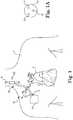
FIG. 1 shows an embodiment of a neurostimulation system and portions of an environment in which the neurostimulation system is used.FIG. 1A is a cross-sectional view of the neurostimulation lead shown inFIG. 1 .FIG. 2 is a schematic illustration of a portion of a neurostimulation lead.FIG. 3 is a schematic illustration of a portion of a neurostimulation lead.FIG. 4 is a schematic illustration of a cuff suitable for use with the neurostimulation leads ofFIGS. 1 to 3 .FIG. 5 is a schematic illustration of a cuff suitable for use with the neurostimulation leads ofFIGS. 1 to 3 .FIG. 6 is a schematic illustration of a cuff suitable for use with the neurostimulation leads ofFIGS. 1 to 3 .FIGS. 6A and 6B are cross-sectional views of a portion of the cuff shown inFIG.6 .FIG. 7 is a schematic illustration of a cuff suitable for use with the neurostimulation leads ofFIGS.1 to 3 .FIG. 8 is a schematic illustration of a unitary tendril suitable for use with the neurostimulation leads ofFIGS.1 to 3 .FIG. 9 is a schematic illustration of a cuff suitable for use with the neurostimulation leads ofFIGS.1 to 3 .FIG. 10 is a schematic illustration of a cuff suitable for use with the neurostimulation leads ofFIGS.1 to 3 .FIG. 11 is a flow diagram illustrating a method that may be carried out using the neurostimulation leads ofFIGS.1 to 3 .FIG. 12 is a schematic illustration of a portion of a neurostimulation lead.- While the invention is amenable to various modifications and alternative forms, specific embodiments have been shown by way of example in the drawings and are described in detail below. The intention, however, is not to limit the invention to the particular embodiments described. On the contrary, the invention is intended to cover all modifications, equivalents, and alternatives falling within the scope of the invention as defined by the appended claims.
FIG. 1 shows an embodiment of aneurostimulation system 10 implanted in a patient P. Theneurostimulation system 10 includes an implantable medical device (IMD) 12 with a lead 14 having a proximal end 16 and adistal end 18. In one embodiment, theIMD 12 includes a pulse generator. TheIMD 12 can be implanted subcutaneously within the body, typically at a location such as in a patient's chest or abdomen, although other implantation locations are possible. The proximal end 16 of thelead 14 can be coupled to theIMD 12 via one ormore connectors 19. Alternatively, thelead 14 may be formed integrally with theIMD 12. Thedistal end 18 of thelead 14, in turn, can be implanted at a desired location in the patient's body to stimulate excitable tissue.- The
distal end 18 of thelead 14 includes one or more electrode cuffs 20. While asingle electrode cuff 20 is shown inFIG. 1 , it will be appreciated that in some embodiments thelead 14 may include one, two, three or more electrode cuffs 20. Further details regarding the construction and implantation of the electrode cuffs 20 will be described with respect to subsequent FIGS. In some embodiments, asingle electrode cuff 20 may include two electrodes (not shown inFIG. 1 ). In some embodiments, thelead 14 may includeseveral electrode cuffs 20, each with a single electrode. One electrode may, for example, function as a cathode electrode while another electrode may function as an anode electrode. The electrode(s) is(are) electrically connected to theIMD 12 via one ormore conductors 11, 13, 15 (shown inFIG. 1A ) extending through thelead 14. - During operation, the
lead 14 delivers electrical signals between theIMD 12 and the electrode cuffs 20. The electrode cuffs 20 may be separately controlled byIMD 12, such that energy having different magnitude, phase, and/or timing characteristics may be delivered to or from each of the electrode cuffs 20. In some embodiments, one or more of the electrode cuffs 20 can alternatively be configured as a strain relief cuff that does not carry electrical signals, but secures thedistal end 18 relative to the nerve N to minimize movement of the electrode cuffs 20 relative to the excitable tissue due to voluntary or involuntary movements of the patient. Furthermore, theIMD 12 shown is merely by way of illustration, and theIMD 12 may have any configuration suitable for use in conjunction with thelead 14 and may be implanted in any suitable location in the patient's body. - The electrode cuffs 20 include electrodes that are configured for stimulation or sensing of a nerve or nerve bundle. In the embodiment shown, the
distal end 18 is secured to the vagus nerve N. The electrode cuffs 20 may be arranged around the nerve, with theIMD 12 configured to deliver energy to the electrode cuffs 20 to stimulate the nerve. Stimulating the sympathetic and parasympathetic nervous systems can have effects on physiological parameters associated with the heart H, such as heart rate and blood pressure. In addition, stimulating the sympathetic nervous system dilates the pupil, reduces saliva and mucus production, relaxes the bronchial muscle, reduces the successive waves of involuntary contraction (peristalsis) of the stomach and the motility of the stomach, increases the conversion of glycogen to glucose by the liver, decreases urine secretion by the kidneys, and relaxes the wall and closes the sphincter of the bladder. Stimulating the parasympathetic nervous system (inhibiting the sympathetic nervous system) constricts the pupil, increases saliva and mucus production, contracts the bronchial muscle, increases secretions and motility in the stomach and large intestine, and increases digestion in the small intestine, increases urine secretion, and contracts the wall and relaxes the sphincter of the bladder. The functions associated with the sympathetic and parasympathetic nervous systems are many and can be complexly integrated with each other. - The vagus nerve N has afferent properties, such that the neural stimulation is transmitted to the central nervous system (CNS). Vagal stimulation simultaneously increases parasympathetic and decreases sympathetic activity, and is believed to prevent further remodeling or predisposition to fatal arrhythmias in post-myocardial infarction (MI) patients, to help restore autonomic balance and increase heart rate variability (HRV), to increase parasympathetic and reduce sympathetic tone in hypertrophic cardiac myopathy (HCM), neurogenic hypertension, and arrhythmia protection, to reduce anginal symptoms, to increase coronary blood flow (CBF), and to prevent development or worsening of congestive heart failure (CHF) following MI. The electrode cuffs 20 may be configured and arranged to stimulate the vagus nerve N to provide any of the physiological responses described. While the electrode cuffs 20 are shown arranged around the right vagus nerve N in
FIG. 1 , the electrode cuffs 20 can be configured and arranged to stimulate the left vagus nerve to treat other physiological and psychological conditions, such as epilepsy and depression. FIG. 2 provides a side view of a portion of thelead 14 that includes alead body 30 and three (as illustrated) electrode cuffs 20. In some embodiments, thelead 14 also includes one or more of afirst lead extension 32 and asecond lead extension 34. The first andsecond lead extensions IMD 12 and one or more of the electrode cuffs 20. In some embodiments, the electrode cuffs 20 include astrain relief 36, afirst electrode cuff 38 and asecond electrode cuff 40. In some embodiments, thestrain relief 36 is secured to thelead body 30 in order to help mitigate movement of the first and second electrode cuffs 38, 40. Thefirst electrode cuff 38 can be secured to thefirst lead extension 32 while thesecond electrode cuff 40 can be secured to thesecond lead extension 34. In some embodiments, as illustrated, thestrain relief 36 can be secured to thelead body 30 by aconnector 48. In some embodiments, theconnector 48 can also secure at least one of thefirst lead extension 32 and/or thesecond lead extension 34, if present, to thelead body 30.FIG. 3 is similar toFIG. 2 but shows a portion of a lead 14 having astrain relief 42 that is configured as a helical winding that is biased to a coiled configuration as shown. In some embodiments, thestrain relief 42 includes afirst end 44 and asecond end 46, and thestrain relief 42 can be wrapped around the nerve N by pulling each of thefirst end 44 and thesecond end 46 and wrapping each of thefirst end 44 and thesecond end 46 around the nerve N. In some embodiments, as illustrated, thestrain relief 42 can be secured to thelead body 30 by aconnector 48. In some embodiments, theconnector 48 can also secure at least one of thefirst lead extension 32 and/or thesecond lead extension 34, if present, to thelead body 30.FIGS. 4 and 5 provide an illustration of anelectrode cuff 50 and an exemplary method of securing theelectrode cuff 50 to the nerve N. Theelectrode cuff 50 is an example of anelectrode cuff 20 that can be used in combination with thelead 14. In the illustrated embodiment, theelectrode cuff 50 has afirst region 52 and asecond region 54. Afirst tendril 56 extends from thefirst region 52 of theelectrode cuff 50 and a second tendril 58 extends from thesecond region 54 of theelectrode cuff 50. In some embodiments, thefirst tendril 56 and the second tendril 58 curve in opposite directions (with respect to an observer viewing the Figure).- To secure the
electrode cuff 50 to the nerve N, theelectrode cuff 50 is disposed proximate the nerve N. Theelectrode cuff 50 can be rotated in a direction indicated by an arrow 60, thereby bringing thefirst tendril 56 and the second tendril 58 into contact with the nerve N. In some embodiments, thefirst tendril 56 and the second tendril 58 are sufficiently stiff to permit adequate securement of theelectrode cuff 50 to the nerve N without requiring that either of thefirst tendril 56 or the second tendril 58 extend much beyond a half circle. In some embodiments, thefirst tendril 56 and/or the second tendril 58 may be sufficiently flexible to permit uncoiling and recoiling thefirst tendril 56 and/or the second tendril 58 around the nerve N. FIG. 6 is a schematic illustration of anelectrode cuff 70 disposed about the nerve N. Theelectrode cuff 70 is an example of anelectrode cuff 20 that can be used in combination with thelead 14. Theelectrode cuff 70 includes acuff body 72 and anelectrode 74 that is disposed on or otherwise secured to thecuff body 72. In some embodiments, theelectrode 74 may be a foil electrode. While asingle electrode 74 is shown, in some embodiments thecuff body 72 may include two ormore electrodes 74.- The
cuff body 72 includes afirst region 76 and asecond region 78. Afirst tendril 80 extends from thefirst region 76 of thecuff body 72 and asecond tendril 82 extends from thesecond region 78 of thecuff body 72. In some embodiments, thefirst tendril 80 is biased to a curved configuration as shown. In some embodiments, thesecond tendril 82 is biased to a curved configuration as shown. In some embodiments, as illustrated, thefirst tendril 80 extends at an acute angle with respect to thecuff body 72 and thesecond tendril 82 extends at an acute angle with respect to thecuff body 72. For illustrative purposes, this acute angle is indicated as angle alpha (α), which can be in a range greater than zero degrees and less than 90 degrees. - The
first tendril 80, thesecond tendril 82 and thecuff body 72 can be formed of any suitable material. In some embodiments, thefirst tendril 80, thesecond tendril 82 and thecuff body 72 are each formed of a polymeric material such as silicone.Figures 6A and 6B are cross-sectional views of thefirst tendril 80 and thesecond tendril 82, respectively. - In some embodiments, as illustrated, the
first tendril 80 can include afirst suture 86 that is molded within thefirst tendril 80 and that extends through thefirst tendril 80. In some embodiments, as illustrated, thesecond tendril 82 can include asecond suture 88 that is molded within thesecond tendril 82 and that extends through thesecond tendril 82. Thefirst suture 86 and thesecond suture 88 can, if present, aid in deployment of theelectrode cuff 70 by providing the surgeon with something that can be grasped and pulled on in order to appropriately wrap thefirst tendril 80 and/or thesecond tendril 82 around the nerve N. In some embodiments, as illustrated, thefirst suture 86 can extend out of the end of thefirst tendril 80 so that the surgeon can grasp it. In some embodiments, as illustrated, thesecond suture 86 can extend out of the end of thesecond tendril 82 so that the surgeon can grasp it. FIG. 7 is an illustration of aunitary tendril 90 that can form part of the electrode cuffs 20 described herein. Theunitary tendril 90 includes afirst portion 92 and asecond portion 94. Theunitary tendril 90 also includes amidpoint 96 where theunitary tendril 90 switches wrapping direction. In some embodiments, as illustrated, thefirst portion 92 of theunitary tendril 90 can be considered as having a first coil direction indicated byarrow 98 while thesecond portion 94 of theunitary tendril 90 can be considered as having a second coil direction indicated byarrow 100.- By having either end biased to opposite coil directions, it will be appreciated that the surgeon installing an electrode cuff with such a
unitary tendril 90 can wrap or unwrap either end in the same direction, i.e., both ends can be wrapped or unwrapped in a clockwise direction or in a counter-clockwise direction by virtue of the two ends of theunitary tendril 90 extending from opposite ends of theelectrode cuff 20. In some situations, this can simplify and speed up the deployment of theelectrode cuff 20. FIG. 8 illustrates anelectrode cuff 110. Theelectrode cuff 110 is an example of anelectrode cuff 20 that can be used in combination with thelead 14. Theelectrode cuff 110 includes acuff body 112 and one or more electrodes (not shown). Thecuff body 112 includes afirst region 114 and asecond region 116. Afirst tendril 118 extends from thefirst region 114 of thecuff body 112 and asecond tendril 120 extends from thesecond region 116 of thecuff body 112. In some embodiments, thefirst tendril 118 is biased to a curved configuration as shown. In some embodiments, thesecond tendril 120 is biased to a curved configuration as shown. While in some respects theelectrode cuff 110 is similar to the electrode cuff 70 (FIG. 6 ), in this instance thefirst tendril 118 and thesecond tendril 120 are long enough to wrap several times around the nerve N. In some embodiments, thefirst tendril 118 and thesecond tendril 120 are biased to a coiled configuration in which the first andsecond tendrils cuff body 112.FIGS. 9 and10 provide illustrations of electrode cuffs that are configured to minimize the overall length of the electrode cuff and thus minimize the overall cut-down length required for deployment of the electrode cuff.FIG. 9 illustrates anelectrode cuff 130 deployed on the nerve N. Theelectrode cuff 130 is an example of anelectrode cuff 20 that can be used in combination with thelead 14. Theelectrode cuff 130 includes acuff body 132 and anelectrode 134. Thecuff body 132 includes afirst region 136 and asecond region 138. Afirst tendril 140 extends from thefirst region 136 of thecuff body 132 and asecond tendril 142 extends from thesecond region 138 of thecuff body 132.- In the illustrated embodiment, the
first tendril 140 extends perpendicularly or at least substantially perpendicular to thecuff body 132. Thesecond tendril 142 extends perpendicularly or at least substantially perpendicularly to thecuff body 132. In some embodiments, thefirst tendril 140 and/or thesecond tendril 142 extend more than 360 degrees around the nerve N and thus overlap on thecuff body 132. In some embodiments, thefirst tendril 140 and/or thesecond tendril 142 may extend less than 360 degrees around the nerve N. Thefirst tendril 140 and thesecond tendril 142 extend from acommon side 144 of thecuff body 132. FIG. 10 illustrates anelectrode cuff 150 deployed on the nerve N. Theelectrode cuff 150 is an example of anelectrode cuff 20 that can be used in combination with thelead 14. Theelectrode cuff 150 includes acuff body 152 and anelectrode 154. Thecuff body 152 includes afirst region 156 and asecond region 158. Afirst tendril 160 extends from thefirst region 156 of thecuff body 152 and asecond tendril 162 extends from thesecond region 158 of thecuff body 152.- In the illustrated embodiment, the
first tendril 160 extends perpendicularly or at least substantially perpendicular to thecuff body 152. Thesecond tendril 162 extends perpendicularly or at least substantially perpendicularly to thecuff body 152. In some embodiments, thefirst tendril 160 and/or thesecond tendril 162 extend more than 360 degrees around the nerve N and thus overlap on thecuff body 152. In some embodiments, thefirst tendril 160 and/or thesecond tendril 162 may extend less than 360 degrees around the nerve N. In the illustrated embodiment, thefirst tendril 160 extends from afirst side 164 of thecuff body 152 and thesecond tendril 162 extends from asecond side 166 of thecuff body 152. - As noted, each of the electrode cuffs described herein, such as the
electrode cuff 50, theelectrode cuff 70, theelectrode cuff 110, theelectrode cuff 130 or theelectrode cuff 150 can be used in combination with thelead 14 as one or more of the electrode cuffs 20. Each of the electrode cuffs described herein can be formed of any suitable material including a polymeric material such as silicone. In some embodiments, the tendrils can be biased to a particular curved or coiled configuration. In some embodiments, the tendrils can be formed of a shape memory material such as a shape memory polymer. FIG. 11 illustrates a method that can be carried out using thelead 14 and the electrode cuffs described herein. An electrode cuff having a first tendril and a second tendril can be disposed proximate the nerve N as generally indicated atblock 170. Atblock 172, the electrode cuff can be positioned on the nerve N. The first tendril can be secured in position on the nerve N as generally indicated atblock 174. In some embodiments, the first tendril can be secured by wrapping the first tendril around the nerve in a rotational direction. The second tendril can be secured in position on the nerve N as generally indicated atblock 176. In some embodiments, the second tendril can be secured by wrapping the second tendril around the nerve N in the same rotational direction.FIG. 12 illustrates a lead 214 that includes afirst electrode cuff 220 secured to alead body extension 221 and asecond electrode cuff 222 secured to alead body extension 223. Thefirst electrode cuff 220 includes afirst cuff body 224 and afirst tendril 226 extending from thefirst cuff body 224. Thefirst tendril 226 is biased to a curved configuration in which thefirst tendril 226 curves in afirst direction 228. Asecond tendril 230 extends from thefirst cuff body 224 and is biased to a curved configuration in which thesecond tendril 230 curves in asecond direction 232.- The
second electrode cuff 222 includes asecond cuff body 234 and athird tendril 236 extending from thesecond cuff body 234. Thethird tendril 236 is biased to a curved configuration in which thethird tendril 226 curves in athird direction 238. A fourth tendril 240 extends from thesecond cuff body 234 and is biased to a curved configuration in which the fourth tendril 240 curves in afourth direction 242. In some embodiments, thefirst direction 228 and thethird direction 238 can be the same. In some embodiments, thesecond direction 230 and the fourth direction 240 can be the same. - By having opposing ends of each
electrode cuff electrode cuff - Various modifications and additions can be made to the exemplary embodiments discussed. For example, while the embodiments described above refer to particular features, the scope of this invention also includes embodiments having different combinations of features and embodiments that do not include all of the described features. Accordingly, the scope of the present invention is intended to embrace all such alternatives, modifications, and variations as fall within the scope of the claims, together with all equivalents thereof.
Claims (15)
- A neurostimulation lead (14) comprising:a lead body (30) having a proximal portion and a distal portion;a first conductor (11) extending through the lead body (30); andan electrode cuff (130, 150) secured relative to the distal portion of the lead body (30), the electrode cuff (130, 150) including:a cuff body (132, 152) having a first region (136, 156) and a second region (138, 158);a first tendril (140, 160) extending from the first region (136, 156) of the cuff body (132, 152);a second tendril (142, 162) extending from the second region (138, 158) of the cuff body (132, 152); anda first electrode (134, 154) disposed on the cuff body (132, 152) and electrically connected to the first conductor,wherein at least one of the first tendril (140, 160) and the second tendril (142, 162) is configured to overlap the cuff body (132, 152).
- The neurostimulation lead of claim 1, wherein the first tendril (140, 160) extends substantially perpendicularly from the first region (136, 156) of the cuff body (132, 152).
- The neurostimulation lead of claim 2, wherein the second tendril (142, 162) extends substantially perpendicularly from the second region (138, 158) of the cuff body (132, 152).
- The neurostimulation lead of claim 3, wherein the first tendril (140, 160) and the second tendril (142, 162) both extend in a same direction from the cuff body (132, 152).
- The neurostimulation lead of claim 3, wherein the first tendril (140, 160) and the second tendril (142, 162) extend in opposite directions from the cuff body (132, 152).
- The neurostimulation lead of claim 1, further comprising:a second conductor (13) extending through the lead body (14); anda second electrode (74) disposed on the cuff body and electrically connected to the second conductor.
- The neurostimulation lead of claim 1, further comprising:a first suture (86) molded into and extending through the first tendril (80); anda second suture (86) molded into and extending through the second tendril (82).
- The neurostimulation lead of claim 1, wherein the cuff body is configured to extend less than about 360 degrees about the nerve.
- The neurostimulation lead of claim 1, wherein the first and second tendrils are each configured to extend more than about 360 degrees about the nerve.
- The neurostimulation lead of claim 1, wherein the first tendril and the second tendril are portions of a unitary tendril (90), the unitary tendril (90) biased to a wrapping direction that reverses direction near a midpoint of the unitary tendril (90).
- A neurostimulation lead (214) comprising:a lead body having a proximal portion and a distal portion;a first conductor extending through the lead body;a second conductor extending through the lead body; anda first cuff (220) secured relative to the distal portion of the lead body, the first cuff (220) including:a first cuff body (224);a first tendril (226) extending from the first cuff body (224), the first tendril (226) biased to a curved configuration in which the first tendril (226) curves in a first direction (228), the first tendril (226) configured to overlap the first cuff body (224);a second tendril (230) extending from the first cuff body (224), the second tendril (230) biased to a curved configuration in which the second tendril (230) curves in a second direction (232) opposite the first direction (228), the second tendril (230) configured to overlap the first cuff body (224); anda first electrode disposed on the first cuff body (224) and electrically connected to the first conductor; anda second cuff (222) secured relative to the distal portion of the lead body, the second cuff (222) including:a second cuff body (234);a third tendril (236) extending from the second cuff body (234), the third tendril (236) biased to a curved configuration in which the third tendril (236) curves in a third direction (238), the third tendril (236) configured to overlap the second cuff body (234);a fourth tendril (240) extending from the second cuff body (234), the fourth tendril (240) biased to a curved configuration in which the fourth tendril (240) curves in a fourth direction (242) opposite the third direction (238), the fourth tendril (240) configured to overlap the second cuff body (234); anda second electrode disposed on the second cuff body (234) and electrically connected to the second conductor.
- The neurostimulation lead of claim 11, wherein the first cuff (220) is secured relative to the distal portion of the lead body via a first lead extension (221), the first lead extension (221) secured to the lead body and the first cuff (220) being attached to the first lead extension (221).
- The neurostimulation lead of claim 11, wherein the second cuff (222) is secured relative to the distal portion of the lead body via a second lead extension (223), the second lead extension (223) secured to the lead body and the second cuff (222) being attached to the second lead extension (223).
- The neurostimulation lead of claim 11, further comprising a strain relief secured to the distal portion of the lead body.
- The neurostimulation lead of claim 11, wherein the first direction (228) is the same as the third direction (238), and the second direction (232) is the same as the fourth direction (242).
Applications Claiming Priority (2)
| Application Number | Priority Date | Filing Date | Title |
|---|---|---|---|
| US201361764306P | 2013-02-13 | 2013-02-13 | |
| PCT/US2014/015590WO2014126857A1 (en) | 2013-02-13 | 2014-02-10 | Cuff electrode with integrated tendril |
Publications (2)
| Publication Number | Publication Date |
|---|---|
| EP2956208A1 EP2956208A1 (en) | 2015-12-23 |
| EP2956208B1true EP2956208B1 (en) | 2021-01-13 |
Family
ID=50179953
Family Applications (1)
| Application Number | Title | Priority Date | Filing Date |
|---|---|---|---|
| EP14706753.2AActiveEP2956208B1 (en) | 2013-02-13 | 2014-02-10 | Cuff electrode with integrated tendril |
Country Status (4)
| Country | Link |
|---|---|
| US (2) | US9320889B2 (en) |
| EP (1) | EP2956208B1 (en) |
| JP (1) | JP6069523B2 (en) |
| WO (1) | WO2014126857A1 (en) |
Families Citing this family (7)
| Publication number | Priority date | Publication date | Assignee | Title |
|---|---|---|---|---|
| EP2956208B1 (en) | 2013-02-13 | 2021-01-13 | Cardiac Pacemakers, Inc. | Cuff electrode with integrated tendril |
| WO2014210282A1 (en)* | 2013-06-26 | 2014-12-31 | Kunis Christopher G | Implant device with spine and c-ring |
| JP6854513B2 (en)* | 2017-04-05 | 2021-04-07 | 国立大学法人 鹿児島大学 | Secretory promoter |
| DE102018204036B4 (en)* | 2018-03-16 | 2025-05-08 | Neuroloop GmbH | Implant in the form of a wound cuff electrode arrangement |
| CN110841186B (en)* | 2019-11-19 | 2021-11-19 | 华中科技大学 | Implanted peripheral nerve electrode |
| CN114761068A (en)* | 2019-11-27 | 2022-07-15 | 加尔瓦尼生物电子有限公司 | Neural interface system |
| EP4565123A1 (en) | 2023-04-18 | 2025-06-11 | Retropsoas Technologies, Llc | Bipolar nerve stimulation/monitoring cuff |
Family Cites Families (144)
| Publication number | Priority date | Publication date | Assignee | Title |
|---|---|---|---|---|
| AR216293A1 (en) | 1976-12-02 | 1979-12-14 | Leon De Pedro F | SELF-FIXING TOOTH ELECTRODE AND A GRIPPER FOR ITS MANEUVERING |
| US4590946A (en) | 1984-06-14 | 1986-05-27 | Biomed Concepts, Inc. | Surgically implantable electrode for nerve bundles |
| US4573481A (en) | 1984-06-25 | 1986-03-04 | Huntington Institute Of Applied Research | Implantable electrode array |
| US4602624A (en) | 1984-10-11 | 1986-07-29 | Case Western Reserve University | Implantable cuff, method of manufacture, and method of installation |
| US4628942A (en) | 1984-10-11 | 1986-12-16 | Case Western Reserve University | Asymmetric shielded two electrode cuff |
| US4590949A (en) | 1984-11-01 | 1986-05-27 | Cordis Corporation | Neural stimulating lead with stabilizing mechanism and method for using same |
| US4740170A (en) | 1986-04-25 | 1988-04-26 | Teletronics N.V. | Removable sleeve adaptor for electrode leads |
| US4920979A (en) | 1988-10-12 | 1990-05-01 | Huntington Medical Research Institute | Bidirectional helical electrode for nerve stimulation |
| US5218089A (en) | 1988-12-23 | 1993-06-08 | Sclavo S.P.A. | Retro-inverso analogues of thymopentin and the method for their synthesis |
| US4940065A (en) | 1989-01-23 | 1990-07-10 | Regents Of The University Of California | Surgically implantable peripheral nerve electrode |
| US4979511A (en) | 1989-11-03 | 1990-12-25 | Cyberonics, Inc. | Strain relief tether for implantable electrode |
| US5031621A (en) | 1989-12-06 | 1991-07-16 | Grandjean Pierre A | Nerve electrode with biological substrate |
| US5095905A (en) | 1990-06-07 | 1992-03-17 | Medtronic, Inc. | Implantable neural electrode |
| FR2669622B1 (en) | 1990-11-28 | 1993-11-19 | Aerospatiale Ste Nat Indle | COMPOSITE MATERIAL WITH REFRACTORY FIBROUS REINFORCEMENT AND PROCESS FOR PRODUCING THE SAME. |
| FR2671010B1 (en) | 1990-12-27 | 1993-07-09 | Ela Medical Sa | ENDOCARDIAC PROBE PROVIDED WITH AN ACTIVE FIXING MEMBER |
| US5251634A (en) | 1991-05-03 | 1993-10-12 | Cyberonics, Inc. | Helical nerve electrode |
| US5215089A (en) | 1991-10-21 | 1993-06-01 | Cyberonics, Inc. | Electrode assembly for nerve stimulation |
| US5324322A (en) | 1992-04-20 | 1994-06-28 | Case Western Reserve University | Thin film implantable electrode and method of manufacture |
| DE69326404T2 (en) | 1992-08-14 | 2000-03-09 | Pacesetter Ab | Multipole electrode lead |
| US5358516A (en) | 1992-12-11 | 1994-10-25 | W. L. Gore & Associates, Inc. | Implantable electrophysiology lead and method of making |
| US5375594A (en) | 1993-03-29 | 1994-12-27 | Cueva; Roberto A. | Removable medical electrode system |
| US5344438A (en) | 1993-04-16 | 1994-09-06 | Medtronic, Inc. | Cuff electrode |
| US5505201A (en) | 1994-04-20 | 1996-04-09 | Case Western Reserve University | Implantable helical spiral cuff electrode |
| US5531778A (en)* | 1994-09-20 | 1996-07-02 | Cyberonics, Inc. | Circumneural electrode assembly |
| US5674272A (en) | 1995-06-05 | 1997-10-07 | Ventritex, Inc. | Crush resistant implantable lead |
| US5913876A (en) | 1996-02-20 | 1999-06-22 | Cardiothoracic Systems, Inc. | Method and apparatus for using vagus nerve stimulation in surgery |
| US6051017A (en) | 1996-02-20 | 2000-04-18 | Advanced Bionics Corporation | Implantable microstimulator and systems employing the same |
| US7269457B2 (en) | 1996-04-30 | 2007-09-11 | Medtronic, Inc. | Method and system for vagal nerve stimulation with multi-site cardiac pacing |
| US6174329B1 (en) | 1996-08-22 | 2001-01-16 | Advanced Cardiovascular Systems, Inc. | Protective coating for a stent with intermediate radiopaque coating |
| TW438860B (en) | 1996-11-20 | 2001-06-07 | Japan Synthetic Rubber Co Ltd | Curable resin composition and cured products |
| US5755766A (en) | 1997-01-24 | 1998-05-26 | Cardiac Pacemakers, Inc. | Open-ended intravenous cardiac lead |
| AU6329498A (en) | 1997-02-13 | 1998-09-08 | Boston Scientific Ireland Limited, Barbados Head Office | Percutaneous and hiatal devices and methods for use in minimally invasive pelvicsurgery |
| US5782892A (en) | 1997-04-25 | 1998-07-21 | Medtronic, Inc. | Medical lead adaptor for external medical device |
| US5931861A (en) | 1997-04-25 | 1999-08-03 | Medtronic, Inc. | Medical lead adaptor having rotatable locking clip mechanism |
| US6038472A (en) | 1997-04-29 | 2000-03-14 | Medtronic, Inc. | Implantable defibrillator and lead system |
| US6249708B1 (en) | 1997-08-26 | 2001-06-19 | Angeion Corporation | Fluted channel construction for a multi-conductor catheter lead |
| US6050996A (en) | 1997-11-12 | 2000-04-18 | Sherwood Services Ag | Bipolar electrosurgical instrument with replaceable electrodes |
| US6093197A (en) | 1997-12-08 | 2000-07-25 | Axon Engineering, Inc. | Spiral nerve cuff electrode implantation tool |
| DE19847446B4 (en) | 1998-10-08 | 2010-04-22 | Biotronik Gmbh & Co. Kg | Nerve electrode assembly |
| US6178349B1 (en) | 1999-04-15 | 2001-01-23 | Medtronic, Inc. | Drug delivery neural stimulation device for treatment of cardiovascular disorders |
| US6308105B1 (en) | 1999-07-15 | 2001-10-23 | Medtronic Inc. | Medical electrical stimulation system using an electrode assembly having opposing semi-circular arms |
| US7615076B2 (en) | 1999-10-20 | 2009-11-10 | Anulex Technologies, Inc. | Method and apparatus for the treatment of the intervertebral disc annulus |
| US8128698B2 (en) | 1999-10-20 | 2012-03-06 | Anulex Technologies, Inc. | Method and apparatus for the treatment of the intervertebral disc annulus |
| US6296659B1 (en) | 2000-02-29 | 2001-10-02 | Opus Medical, Inc. | Single-tailed suturing method and apparatus |
| US6725096B2 (en) | 2000-05-05 | 2004-04-20 | Advanced Bionics Corporation | Multiple in-line contact connector |
| US20080177366A1 (en) | 2000-09-27 | 2008-07-24 | Cvrx, Inc. | Cuff electrode arrangement for nerve stimulation and methods of treating disorders |
| US7158832B2 (en) | 2000-09-27 | 2007-01-02 | Cvrx, Inc. | Electrode designs and methods of use for cardiovascular reflex control devices |
| US7499742B2 (en) | 2001-09-26 | 2009-03-03 | Cvrx, Inc. | Electrode structures and methods for their use in cardiovascular reflex control |
| US20030236562A1 (en) | 2000-10-10 | 2003-12-25 | Kuzma Janusz A. | Band type multicontact electrode and method of making the same |
| US6501992B1 (en) | 2000-10-17 | 2002-12-31 | Medtronic, Inc. | Radiopaque marking of lead electrode zone in a continuous conductor construction |
| EP1341579B1 (en) | 2000-12-07 | 2006-11-29 | Medtronic, Inc. | Directional brain stimulation and recording leads |
| US6597953B2 (en) | 2001-02-20 | 2003-07-22 | Neuropace, Inc. | Furcated sensing and stimulation lead |
| US20020128700A1 (en) | 2001-03-08 | 2002-09-12 | Cross Thomas E. | Lead with adjustable angular and spatial relationships between electrodes |
| US7899551B2 (en) | 2001-04-13 | 2011-03-01 | Greatbatch Ltd. | Medical lead system utilizing electromagnetic bandstop filters |
| US7054692B1 (en) | 2001-06-22 | 2006-05-30 | Advanced Bionics Corporation | Fixation device for implantable microdevices |
| US6600956B2 (en)* | 2001-08-21 | 2003-07-29 | Cyberonics, Inc. | Circumneural electrode assembly |
| US7881805B2 (en) | 2002-02-04 | 2011-02-01 | Boston Scientific Neuromodulation Corporation | Method for optimizing search for spinal cord stimulation parameter settings |
| US7292890B2 (en) | 2002-06-20 | 2007-11-06 | Advanced Bionics Corporation | Vagus nerve stimulation via unidirectional propagation of action potentials |
| US20040111139A1 (en) | 2002-12-10 | 2004-06-10 | Mccreery Douglas B. | Apparatus and methods for differential stimulation of nerve fibers |
| JP4387724B2 (en) | 2003-08-12 | 2009-12-24 | テルモ株式会社 | Living body electrode lead |
| US7502650B2 (en) | 2003-09-22 | 2009-03-10 | Cvrx, Inc. | Baroreceptor activation for epilepsy control |
| US7460906B2 (en) | 2003-12-24 | 2008-12-02 | Cardiac Pacemakers, Inc. | Baroreflex stimulation to treat acute myocardial infarction |
| US9155877B2 (en) | 2004-03-30 | 2015-10-13 | Medtronic, Inc. | Lead electrode for use in an MRI-safe implantable medical device |
| US7833238B2 (en) | 2004-04-19 | 2010-11-16 | Granit Medical Innovations, Llc | Endoscopic anchoring device and associated method |
| WO2006017634A2 (en) | 2004-08-04 | 2006-02-16 | Ndi Medical, Llc | Devices, systems, and methods employing a molded nerve cuff electrode |
| US7238883B2 (en) | 2004-08-11 | 2007-07-03 | Cardiac Pacemakers, Inc. | Lead assembly with flexible portions and method therefor |
| US7831311B2 (en) | 2004-10-21 | 2010-11-09 | Medtronic, Inc. | Reduced axial stiffness implantable medical lead |
| US7905904B2 (en) | 2006-02-03 | 2011-03-15 | Biomet Sports Medicine, Llc | Soft tissue repair device and associated methods |
| US9017381B2 (en) | 2007-04-10 | 2015-04-28 | Biomet Sports Medicine, Llc | Adjustable knotless loops |
| US20060190042A1 (en) | 2004-11-05 | 2006-08-24 | Arthrotek, Inc. | Tissue repair assembly |
| US8361113B2 (en) | 2006-02-03 | 2013-01-29 | Biomet Sports Medicine, Llc | Method and apparatus for coupling soft tissue to a bone |
| US9089691B2 (en) | 2004-12-07 | 2015-07-28 | Cardiac Pacemakers, Inc. | Stimulator for auricular branch of vagus nerve |
| US7891085B1 (en) | 2005-01-11 | 2011-02-22 | Boston Scientific Neuromodulation Corporation | Electrode array assembly and method of making same |
| US7536227B1 (en) | 2005-01-26 | 2009-05-19 | Pacesetter, Inc. | Shielded electrode for nerve sensing |
| US7561923B2 (en) | 2005-05-09 | 2009-07-14 | Cardiac Pacemakers, Inc. | Method and apparatus for controlling autonomic balance using neural stimulation |
| US20060259078A1 (en) | 2005-05-16 | 2006-11-16 | Imad Libbus | Method and apparatus for electronically switching electrode configuration |
| US20070083236A1 (en) | 2005-06-24 | 2007-04-12 | Smith & Nephew, Inc. | Methods and devices for tissue repair |
| US8660647B2 (en) | 2005-07-28 | 2014-02-25 | Cyberonics, Inc. | Stimulating cranial nerve to treat pulmonary disorder |
| EP1933934B1 (en) | 2005-08-24 | 2011-10-12 | St. Jude Medical AB | Suture sleeve with lead locking device |
| US7306417B2 (en) | 2005-09-26 | 2007-12-11 | Edward Dorstewitz | Rope tie-down |
| US8509914B2 (en) | 2005-10-28 | 2013-08-13 | Cyberonics, Inc. | Insert for implantable electrode |
| US7630760B2 (en) | 2005-11-21 | 2009-12-08 | Cardiac Pacemakers, Inc. | Neural stimulation therapy system for atherosclerotic plaques |
| US7570999B2 (en) | 2005-12-20 | 2009-08-04 | Cardiac Pacemakers, Inc. | Implantable device for treating epilepsy and cardiac rhythm disorders |
| US20070173914A1 (en)* | 2006-01-24 | 2007-07-26 | Cyberonics, Inc. | Self-locking electrode assembly usable with an implantable medical device |
| US8160722B2 (en) | 2006-02-28 | 2012-04-17 | Medtronic, Inc. | Subcutaneous lead fixation mechanisms |
| US7974706B2 (en) | 2006-03-30 | 2011-07-05 | Boston Scientific Neuromodulation Corporation | Electrode contact configurations for cuff leads |
| US7933662B2 (en) | 2006-04-26 | 2011-04-26 | Marshall Mark T | Medical electrical lead including an inductance augmenter |
| US20080046058A1 (en) | 2006-04-28 | 2008-02-21 | Cross Thomas E | Methods for customizing implantable medical lead assemblies with improved flexibility and extensibility |
| US20070255320A1 (en) | 2006-04-28 | 2007-11-01 | Cyberonics, Inc. | Method and apparatus for forming insulated implantable electrodes |
| US8103341B2 (en) | 2006-08-25 | 2012-01-24 | Cardiac Pacemakers, Inc. | System for abating neural stimulation side effects |
| US8457734B2 (en) | 2006-08-29 | 2013-06-04 | Cardiac Pacemakers, Inc. | System and method for neural stimulation |
| US7801603B2 (en) | 2006-09-01 | 2010-09-21 | Cardiac Pacemakers, Inc. | Method and apparatus for optimizing vagal nerve stimulation using laryngeal activity |
| US7925342B2 (en) | 2006-10-06 | 2011-04-12 | Cardiac Pacemakers, Inc. | Implantable device for responsive neural stimulation therapy |
| US20080091255A1 (en) | 2006-10-11 | 2008-04-17 | Cardiac Pacemakers | Implantable neurostimulator for modulating cardiovascular function |
| CA2666529A1 (en) | 2006-10-13 | 2008-04-24 | Apnex Medical, Inc. | Obstructive sleep apnea treatment devices, systems and methods |
| US8024034B2 (en) | 2006-11-01 | 2011-09-20 | Cardiac Pacemakers, Inc. | Programmable neural therapies |
| US7996092B2 (en) | 2007-01-16 | 2011-08-09 | Ndi Medical, Inc. | Devices, systems, and methods employing a molded nerve cuff electrode |
| US8301239B2 (en) | 2007-01-18 | 2012-10-30 | Cardiac Pacemakers, Inc. | Systems, devices and methods for acute autonomic stimulation |
| US7974707B2 (en) | 2007-01-26 | 2011-07-05 | Cyberonics, Inc. | Electrode assembly with fibers for a medical device |
| CA2676119C (en) | 2007-01-29 | 2021-01-19 | Simon Fraser University | Transvascular nerve stimulation apparatus and methods |
| US8244378B2 (en) | 2007-01-30 | 2012-08-14 | Cardiac Pacemakers, Inc. | Spiral configurations for intravascular lead stability |
| US20080195188A1 (en) | 2007-02-09 | 2008-08-14 | Cardiac Pacemakers, Inc. | Implantable medical device with fixation mechanism |
| US8406877B2 (en) | 2007-03-19 | 2013-03-26 | Cardiac Pacemakers, Inc. | Selective nerve stimulation with optionally closed-loop capabilities |
| US8155757B1 (en) | 2007-07-26 | 2012-04-10 | Advanced Neuromodulation Systems, Inc. | Cuff electrode having tubular body with controlled closing force |
| US9037239B2 (en) | 2007-08-07 | 2015-05-19 | Cardiac Pacemakers, Inc. | Method and apparatus to perform electrode combination selection |
| EP2190528B1 (en) | 2007-08-20 | 2014-10-08 | Medtronic, Inc. | Evaluating therapeutic stimulation electrode configurations based on physiological responses |
| US7957817B1 (en) | 2007-09-04 | 2011-06-07 | Pacesetter, Inc. | Medical electrode and tool for delivering the electrode |
| US8790367B2 (en) | 2008-02-06 | 2014-07-29 | Guided Delivery Systems Inc. | Multi-window guide tunnel |
| WO2009100531A1 (en) | 2008-02-15 | 2009-08-20 | Angeltear Solutions Inc. | Adjustable tissue or nerve cuff and method of use |
| US8632502B2 (en) | 2008-03-27 | 2014-01-21 | Medtronic, Inc. | Anchor deployment apparatus |
| US7925352B2 (en) | 2008-03-27 | 2011-04-12 | Synecor Llc | System and method for transvascularly stimulating contents of the carotid sheath |
| US8244372B1 (en) | 2008-04-01 | 2012-08-14 | Advanced Neuromodulation Systems, Inc. | Electrical stimulation lead with stiffeners having varying stiffness zones |
| US20100211131A1 (en) | 2008-04-07 | 2010-08-19 | Williams Michael S | Intravascular system and method for blood pressure control |
| US20090275997A1 (en) | 2008-05-01 | 2009-11-05 | Michael Allen Faltys | Vagus nerve stimulation electrodes and methods of use |
| US8340785B2 (en) | 2008-05-02 | 2012-12-25 | Medtronic, Inc. | Self expanding electrode cuff |
| US8548593B2 (en) | 2008-11-10 | 2013-10-01 | Cardiac Pacemakers, Inc. | Distal end converter for a medical device lead |
| US8515520B2 (en) | 2008-12-08 | 2013-08-20 | Medtronic Xomed, Inc. | Nerve electrode |
| US20100168831A1 (en) | 2008-12-30 | 2010-07-01 | Board Of Supervisors Of Louisiana State University And Agricultural And Mechanical College | Implantable clip-on micro-cuff electrode for functional stimulation and bio-potential recording |
| US20100286553A1 (en) | 2009-05-08 | 2010-11-11 | Feler Claudio A | Devices and Methods for Screening of Vagal Nerve Stimulation |
| US20100298916A1 (en) | 2009-05-22 | 2010-11-25 | Pierre Rabischong | Pelvic implanted neural electrode and method for implanting same |
| AU2010254490B2 (en) | 2009-05-26 | 2014-06-12 | Cardiac Pacemakers, Inc. | Helically formed coil for a neural cuff electrode |
| WO2010144578A2 (en) | 2009-06-09 | 2010-12-16 | Setpoint Medical Corporation | Nerve cuff with pocket for leadless stimulator |
| US8340783B2 (en) | 2009-06-30 | 2012-12-25 | Medtronic, Inc. | Implantable medical device lead with selectively exposed electrodes and reinforcement member |
| US20110004281A1 (en) | 2009-07-03 | 2011-01-06 | Jones Robert E | Implantable anchor with locking cam |
| US8295948B2 (en) | 2009-07-21 | 2012-10-23 | Boston Scientific Neuromodulation Corporation | Tubular lead anchor and methods and devices using the anchor |
| US8489208B2 (en) | 2009-09-09 | 2013-07-16 | Advanced Neuromodulation Systems, Inc. | Key locking anchoring device for implanted lead |
| US9211403B2 (en) | 2009-10-30 | 2015-12-15 | Advanced Bionics, Llc | Steerable stylet |
| US8652153B2 (en) | 2010-01-11 | 2014-02-18 | Anulex Technologies, Inc. | Intervertebral disc annulus repair system and bone anchor delivery tool |
| JP5596989B2 (en) | 2010-01-26 | 2014-10-01 | オリンパス株式会社 | Electrode system |
| US8100141B2 (en) | 2010-05-27 | 2012-01-24 | Slupecki Raymond H | Water supply control assembly with automatic shut-off and duty cycle reset |
| US8620450B2 (en) | 2010-07-19 | 2013-12-31 | Cardiac Pacemakers, Inc. | Minimally invasive lead system for vagus nerve stimulation |
| AU2011285624A1 (en) | 2010-08-05 | 2013-01-24 | Cardiac Pacemakers, Inc. | System and method for securing a lead in a vessel |
| JP5577469B2 (en) | 2010-09-15 | 2014-08-20 | カーディアック ペースメイカーズ, インコーポレイテッド | Automatic selection of lead configuration for neural stimulation leads |
| JP2012075670A (en) | 2010-10-01 | 2012-04-19 | Shimane Univ | Cuff electrode |
| US8892204B2 (en) | 2010-12-17 | 2014-11-18 | Medtronic, Inc. | Aortic pacing to control cardiac afterload |
| JP5602612B2 (en) | 2010-12-22 | 2014-10-08 | オリンパス株式会社 | Electrode unit and tissue stimulation system |
| US8483845B2 (en) | 2011-03-11 | 2013-07-09 | Greatbatch Ltd. | Anchor for implantable medical device |
| WO2013003278A1 (en) | 2011-06-28 | 2013-01-03 | Cardiac Pacemakers, Inc. | Strain relief feature for an implantable medical device lead |
| US8639355B2 (en) | 2011-07-07 | 2014-01-28 | Cardiac Pacemakers, Inc. | Insulation and stability features for an implantable medical device lead |
| US20130172973A1 (en) | 2012-01-04 | 2013-07-04 | Bruce A. Tockman | Neural stimulation device with insulating sheath |
| EP2827944B1 (en) | 2012-03-21 | 2016-05-18 | Cardiac Pacemakers, Inc. | Systems and methods for stimulation of vagus nerve |
| US9114250B2 (en) | 2012-10-02 | 2015-08-25 | Cardiac Pacemakers, Inc. | Pinch to open cuff electrode |
| WO2014055393A1 (en) | 2012-10-02 | 2014-04-10 | Cardiac Pacemakers, Inc. | Pinch to open cuff electrode |
| EP2956208B1 (en) | 2013-02-13 | 2021-01-13 | Cardiac Pacemakers, Inc. | Cuff electrode with integrated tendril |
- 2014
- 2014-02-10EPEP14706753.2Apatent/EP2956208B1/enactiveActive
- 2014-02-10USUS14/176,658patent/US9320889B2/enactiveActive
- 2014-02-10WOPCT/US2014/015590patent/WO2014126857A1/enactiveApplication Filing
- 2014-02-10JPJP2015555450Apatent/JP6069523B2/ennot_activeExpired - Fee Related
- 2016
- 2016-03-29USUS15/084,339patent/US9452283B2/enactiveActive
Non-Patent Citations (1)
| Title |
|---|
| None* |
Also Published As
| Publication number | Publication date |
|---|---|
| US20160206874A1 (en) | 2016-07-21 |
| US9320889B2 (en) | 2016-04-26 |
| US20140228923A1 (en) | 2014-08-14 |
| US9452283B2 (en) | 2016-09-27 |
| JP6069523B2 (en) | 2017-02-01 |
| EP2956208A1 (en) | 2015-12-23 |
| JP2016504170A (en) | 2016-02-12 |
| WO2014126857A1 (en) | 2014-08-21 |
Similar Documents
| Publication | Publication Date | Title |
|---|---|---|
| US9452283B2 (en) | Cuff electrode with integrated tendril | |
| US8548593B2 (en) | Distal end converter for a medical device lead | |
| EP2435129B1 (en) | Helically formed coil for a neural cuff electrode | |
| US9114250B2 (en) | Pinch to open cuff electrode | |
| EP2726142B1 (en) | Strain relief feature for an implantable medical device lead | |
| US9283379B2 (en) | Pinch to open cuff electrode | |
| US7617003B2 (en) | System for selective activation of a nerve trunk using a transvascular reshaping lead | |
| AU2012279403B2 (en) | Insulation and stability features for an implantable medical device lead |
Legal Events
| Date | Code | Title | Description |
|---|---|---|---|
| PUAI | Public reference made under article 153(3) epc to a published international application that has entered the european phase | Free format text:ORIGINAL CODE: 0009012 | |
| 17P | Request for examination filed | Effective date:20150708 | |
| AK | Designated contracting states | Kind code of ref document:A1 Designated state(s):AL AT BE BG CH CY CZ DE DK EE ES FI FR GB GR HR HU IE IS IT LI LT LU LV MC MK MT NL NO PL PT RO RS SE SI SK SM TR | |
| AX | Request for extension of the european patent | Extension state:BA ME | |
| DAX | Request for extension of the european patent (deleted) | ||
| GRAP | Despatch of communication of intention to grant a patent | Free format text:ORIGINAL CODE: EPIDOSNIGR1 | |
| STAA | Information on the status of an ep patent application or granted ep patent | Free format text:STATUS: GRANT OF PATENT IS INTENDED | |
| INTG | Intention to grant announced | Effective date:20200806 | |
| GRAS | Grant fee paid | Free format text:ORIGINAL CODE: EPIDOSNIGR3 | |
| GRAA | (expected) grant | Free format text:ORIGINAL CODE: 0009210 | |
| STAA | Information on the status of an ep patent application or granted ep patent | Free format text:STATUS: THE PATENT HAS BEEN GRANTED | |
| AK | Designated contracting states | Kind code of ref document:B1 Designated state(s):AL AT BE BG CH CY CZ DE DK EE ES FI FR GB GR HR HU IE IS IT LI LT LU LV MC MK MT NL NO PL PT RO RS SE SI SK SM TR | |
| REG | Reference to a national code | Ref country code:GB Ref legal event code:FG4D | |
| REG | Reference to a national code | Ref country code:CH Ref legal event code:EP | |
| REG | Reference to a national code | Ref country code:IE Ref legal event code:FG4D | |
| REG | Reference to a national code | Ref country code:DE Ref legal event code:R096 Ref document number:602014074190 Country of ref document:DE | |
| REG | Reference to a national code | Ref country code:AT Ref legal event code:REF Ref document number:1354099 Country of ref document:AT Kind code of ref document:T Effective date:20210215 | |
| REG | Reference to a national code | Ref country code:AT Ref legal event code:MK05 Ref document number:1354099 Country of ref document:AT Kind code of ref document:T Effective date:20210113 | |
| REG | Reference to a national code | Ref country code:NL Ref legal event code:MP Effective date:20210113 | |
| REG | Reference to a national code | Ref country code:LT Ref legal event code:MG9D | |
| PG25 | Lapsed in a contracting state [announced via postgrant information from national office to epo] | Ref country code:BG Free format text:LAPSE BECAUSE OF FAILURE TO SUBMIT A TRANSLATION OF THE DESCRIPTION OR TO PAY THE FEE WITHIN THE PRESCRIBED TIME-LIMIT Effective date:20210413 Ref country code:NL Free format text:LAPSE BECAUSE OF FAILURE TO SUBMIT A TRANSLATION OF THE DESCRIPTION OR TO PAY THE FEE WITHIN THE PRESCRIBED TIME-LIMIT Effective date:20210113 Ref country code:NO Free format text:LAPSE BECAUSE OF FAILURE TO SUBMIT A TRANSLATION OF THE DESCRIPTION OR TO PAY THE FEE WITHIN THE PRESCRIBED TIME-LIMIT Effective date:20210413 Ref country code:PT Free format text:LAPSE BECAUSE OF FAILURE TO SUBMIT A TRANSLATION OF THE DESCRIPTION OR TO PAY THE FEE WITHIN THE PRESCRIBED TIME-LIMIT Effective date:20210513 Ref country code:LT Free format text:LAPSE BECAUSE OF FAILURE TO SUBMIT A TRANSLATION OF THE DESCRIPTION OR TO PAY THE FEE WITHIN THE PRESCRIBED TIME-LIMIT Effective date:20210113 Ref country code:GR Free format text:LAPSE BECAUSE OF FAILURE TO SUBMIT A TRANSLATION OF THE DESCRIPTION OR TO PAY THE FEE WITHIN THE PRESCRIBED TIME-LIMIT Effective date:20210414 Ref country code:HR Free format text:LAPSE BECAUSE OF FAILURE TO SUBMIT A TRANSLATION OF THE DESCRIPTION OR TO PAY THE FEE WITHIN THE PRESCRIBED TIME-LIMIT Effective date:20210113 Ref country code:FI Free format text:LAPSE BECAUSE OF FAILURE TO SUBMIT A TRANSLATION OF THE DESCRIPTION OR TO PAY THE FEE WITHIN THE PRESCRIBED TIME-LIMIT Effective date:20210113 | |
| PG25 | Lapsed in a contracting state [announced via postgrant information from national office to epo] | Ref country code:SE Free format text:LAPSE BECAUSE OF FAILURE TO SUBMIT A TRANSLATION OF THE DESCRIPTION OR TO PAY THE FEE WITHIN THE PRESCRIBED TIME-LIMIT Effective date:20210113 Ref country code:AT Free format text:LAPSE BECAUSE OF FAILURE TO SUBMIT A TRANSLATION OF THE DESCRIPTION OR TO PAY THE FEE WITHIN THE PRESCRIBED TIME-LIMIT Effective date:20210113 Ref country code:RS Free format text:LAPSE BECAUSE OF FAILURE TO SUBMIT A TRANSLATION OF THE DESCRIPTION OR TO PAY THE FEE WITHIN THE PRESCRIBED TIME-LIMIT Effective date:20210113 Ref country code:LV Free format text:LAPSE BECAUSE OF FAILURE TO SUBMIT A TRANSLATION OF THE DESCRIPTION OR TO PAY THE FEE WITHIN THE PRESCRIBED TIME-LIMIT Effective date:20210113 Ref country code:PL Free format text:LAPSE BECAUSE OF FAILURE TO SUBMIT A TRANSLATION OF THE DESCRIPTION OR TO PAY THE FEE WITHIN THE PRESCRIBED TIME-LIMIT Effective date:20210113 | |
| REG | Reference to a national code | Ref country code:DE Ref legal event code:R119 Ref document number:602014074190 Country of ref document:DE | |
| PG25 | Lapsed in a contracting state [announced via postgrant information from national office to epo] | Ref country code:IS Free format text:LAPSE BECAUSE OF FAILURE TO SUBMIT A TRANSLATION OF THE DESCRIPTION OR TO PAY THE FEE WITHIN THE PRESCRIBED TIME-LIMIT Effective date:20210513 | |
| REG | Reference to a national code | Ref country code:BE Ref legal event code:MM Effective date:20210228 | |
| PG25 | Lapsed in a contracting state [announced via postgrant information from national office to epo] | Ref country code:SM Free format text:LAPSE BECAUSE OF FAILURE TO SUBMIT A TRANSLATION OF THE DESCRIPTION OR TO PAY THE FEE WITHIN THE PRESCRIBED TIME-LIMIT Effective date:20210113 Ref country code:CH Free format text:LAPSE BECAUSE OF NON-PAYMENT OF DUE FEES Effective date:20210228 Ref country code:CZ Free format text:LAPSE BECAUSE OF FAILURE TO SUBMIT A TRANSLATION OF THE DESCRIPTION OR TO PAY THE FEE WITHIN THE PRESCRIBED TIME-LIMIT Effective date:20210113 Ref country code:EE Free format text:LAPSE BECAUSE OF FAILURE TO SUBMIT A TRANSLATION OF THE DESCRIPTION OR TO PAY THE FEE WITHIN THE PRESCRIBED TIME-LIMIT Effective date:20210113 Ref country code:LI Free format text:LAPSE BECAUSE OF NON-PAYMENT OF DUE FEES Effective date:20210228 Ref country code:LU Free format text:LAPSE BECAUSE OF NON-PAYMENT OF DUE FEES Effective date:20210210 Ref country code:MC Free format text:LAPSE BECAUSE OF FAILURE TO SUBMIT A TRANSLATION OF THE DESCRIPTION OR TO PAY THE FEE WITHIN THE PRESCRIBED TIME-LIMIT Effective date:20210113 | |
| PLBE | No opposition filed within time limit | Free format text:ORIGINAL CODE: 0009261 | |
| STAA | Information on the status of an ep patent application or granted ep patent | Free format text:STATUS: NO OPPOSITION FILED WITHIN TIME LIMIT | |
| PG25 | Lapsed in a contracting state [announced via postgrant information from national office to epo] | Ref country code:DK Free format text:LAPSE BECAUSE OF FAILURE TO SUBMIT A TRANSLATION OF THE DESCRIPTION OR TO PAY THE FEE WITHIN THE PRESCRIBED TIME-LIMIT Effective date:20210113 Ref country code:ES Free format text:LAPSE BECAUSE OF FAILURE TO SUBMIT A TRANSLATION OF THE DESCRIPTION OR TO PAY THE FEE WITHIN THE PRESCRIBED TIME-LIMIT Effective date:20210113 Ref country code:SK Free format text:LAPSE BECAUSE OF FAILURE TO SUBMIT A TRANSLATION OF THE DESCRIPTION OR TO PAY THE FEE WITHIN THE PRESCRIBED TIME-LIMIT Effective date:20210113 Ref country code:RO Free format text:LAPSE BECAUSE OF FAILURE TO SUBMIT A TRANSLATION OF THE DESCRIPTION OR TO PAY THE FEE WITHIN THE PRESCRIBED TIME-LIMIT Effective date:20210113 | |
| 26N | No opposition filed | Effective date:20211014 | |
| GBPC | Gb: european patent ceased through non-payment of renewal fee | Effective date:20210413 | |
| PG25 | Lapsed in a contracting state [announced via postgrant information from national office to epo] | Ref country code:AL Free format text:LAPSE BECAUSE OF FAILURE TO SUBMIT A TRANSLATION OF THE DESCRIPTION OR TO PAY THE FEE WITHIN THE PRESCRIBED TIME-LIMIT Effective date:20210113 Ref country code:DE Free format text:LAPSE BECAUSE OF NON-PAYMENT OF DUE FEES Effective date:20210901 Ref country code:IE Free format text:LAPSE BECAUSE OF NON-PAYMENT OF DUE FEES Effective date:20210210 Ref country code:FR Free format text:LAPSE BECAUSE OF NON-PAYMENT OF DUE FEES Effective date:20210313 Ref country code:GB Free format text:LAPSE BECAUSE OF NON-PAYMENT OF DUE FEES Effective date:20210413 | |
| PG25 | Lapsed in a contracting state [announced via postgrant information from national office to epo] | Ref country code:SI Free format text:LAPSE BECAUSE OF FAILURE TO SUBMIT A TRANSLATION OF THE DESCRIPTION OR TO PAY THE FEE WITHIN THE PRESCRIBED TIME-LIMIT Effective date:20210113 | |
| PG25 | Lapsed in a contracting state [announced via postgrant information from national office to epo] | Ref country code:IT Free format text:LAPSE BECAUSE OF FAILURE TO SUBMIT A TRANSLATION OF THE DESCRIPTION OR TO PAY THE FEE WITHIN THE PRESCRIBED TIME-LIMIT Effective date:20210113 | |
| PG25 | Lapsed in a contracting state [announced via postgrant information from national office to epo] | Ref country code:IS Free format text:LAPSE BECAUSE OF FAILURE TO SUBMIT A TRANSLATION OF THE DESCRIPTION OR TO PAY THE FEE WITHIN THE PRESCRIBED TIME-LIMIT Effective date:20210513 | |
| PG25 | Lapsed in a contracting state [announced via postgrant information from national office to epo] | Ref country code:BE Free format text:LAPSE BECAUSE OF NON-PAYMENT OF DUE FEES Effective date:20210228 | |
| PG25 | Lapsed in a contracting state [announced via postgrant information from national office to epo] | Ref country code:HU Free format text:LAPSE BECAUSE OF FAILURE TO SUBMIT A TRANSLATION OF THE DESCRIPTION OR TO PAY THE FEE WITHIN THE PRESCRIBED TIME-LIMIT; INVALID AB INITIO Effective date:20140210 | |
| PG25 | Lapsed in a contracting state [announced via postgrant information from national office to epo] | Ref country code:CY Free format text:LAPSE BECAUSE OF FAILURE TO SUBMIT A TRANSLATION OF THE DESCRIPTION OR TO PAY THE FEE WITHIN THE PRESCRIBED TIME-LIMIT Effective date:20210113 | |
| PG25 | Lapsed in a contracting state [announced via postgrant information from national office to epo] | Ref country code:MK Free format text:LAPSE BECAUSE OF FAILURE TO SUBMIT A TRANSLATION OF THE DESCRIPTION OR TO PAY THE FEE WITHIN THE PRESCRIBED TIME-LIMIT Effective date:20210113 | |
| PG25 | Lapsed in a contracting state [announced via postgrant information from national office to epo] | Ref country code:MT Free format text:LAPSE BECAUSE OF FAILURE TO SUBMIT A TRANSLATION OF THE DESCRIPTION OR TO PAY THE FEE WITHIN THE PRESCRIBED TIME-LIMIT Effective date:20210113 |