EP2675819B1 - Compositions and methods for molecular labeling - Google Patents
Compositions and methods for molecular labelingDownload PDFInfo
- Publication number
- EP2675819B1 EP2675819B1EP12746483.2AEP12746483AEP2675819B1EP 2675819 B1EP2675819 B1EP 2675819B1EP 12746483 AEP12746483 AEP 12746483AEP 2675819 B1EP2675819 B1EP 2675819B1
- Authority
- EP
- European Patent Office
- Prior art keywords
- droplet
- droplets
- barcode
- target
- library
- Prior art date
- Legal status (The legal status is an assumption and is not a legal conclusion. Google has not performed a legal analysis and makes no representation as to the accuracy of the status listed.)
- Active
Links
- 238000000034methodMethods0.000titleclaimsdescription257
- 238000002372labellingMethods0.000titledescription54
- 239000000203mixtureSubstances0.000titledescription36
- 150000007523nucleic acidsChemical class0.000claimsdescription138
- 102000039446nucleic acidsHuman genes0.000claimsdescription131
- 108020004707nucleic acidsProteins0.000claimsdescription131
- 238000012163sequencing techniqueMethods0.000claimsdescription127
- 238000007847digital PCRMethods0.000claimsdescription56
- 125000003729nucleotide groupChemical group0.000claimsdescription50
- 230000002441reversible effectEffects0.000claimsdescription45
- 239000002773nucleotideSubstances0.000claimsdescription43
- 239000007787solidSubstances0.000claimsdescription13
- 238000004519manufacturing processMethods0.000claimsdescription7
- 239000000523sampleSubstances0.000description332
- 239000013615primerSubstances0.000description323
- 238000003752polymerase chain reactionMethods0.000description180
- 210000004027cellAnatomy0.000description169
- 239000012530fluidSubstances0.000description160
- 102000053602DNAHuman genes0.000description152
- 108020004414DNAProteins0.000description152
- 238000003556assayMethods0.000description143
- 230000003287optical effectEffects0.000description103
- 238000003199nucleic acid amplification methodMethods0.000description96
- 230000003321amplificationEffects0.000description95
- 108090000623proteins and genesProteins0.000description93
- 238000006243chemical reactionMethods0.000description89
- 101000617738Homo sapiens Survival motor neuron proteinProteins0.000description86
- 102100021947Survival motor neuron proteinHuman genes0.000description84
- 239000011230binding agentSubstances0.000description79
- 239000003153chemical reaction reagentSubstances0.000description66
- 108091034117OligonucleotideProteins0.000description63
- 102000004169proteins and genesHuman genes0.000description63
- 239000002585baseSubstances0.000description62
- 238000009739bindingMethods0.000description62
- 230000027455bindingEffects0.000description60
- 239000013077target materialSubstances0.000description59
- 238000001514detection methodMethods0.000description57
- 239000011324beadSubstances0.000description54
- 108091093088AmpliconProteins0.000description51
- 238000004458analytical methodMethods0.000description42
- 239000000463materialSubstances0.000description41
- 239000000090biomarkerSubstances0.000description36
- 239000003921oilSubstances0.000description35
- 239000000047productSubstances0.000description34
- 108700028369AllelesProteins0.000description32
- 239000012634fragmentSubstances0.000description32
- 229920002477rna polymerPolymers0.000description30
- 238000009396hybridizationMethods0.000description29
- 102000054766genetic haplotypesHuman genes0.000description28
- 230000037452primingEffects0.000description27
- 208000002320spinal muscular atrophyDiseases0.000description27
- 230000001965increasing effectEffects0.000description24
- 210000000349chromosomeAnatomy0.000description23
- 238000006073displacement reactionMethods0.000description23
- 239000002987primer (paints)Substances0.000description23
- 238000010790dilutionMethods0.000description22
- 239000012895dilutionSubstances0.000description22
- 239000000839emulsionSubstances0.000description22
- 230000008569processEffects0.000description22
- 108090000621Ribonuclease PProteins0.000description20
- 102000004167Ribonuclease PHuman genes0.000description20
- 230000015572biosynthetic processEffects0.000description20
- 230000000875corresponding effectEffects0.000description20
- 230000005684electric fieldEffects0.000description20
- 230000002068genetic effectEffects0.000description20
- 239000004094surface-active agentSubstances0.000description20
- 108010090804StreptavidinProteins0.000description19
- 238000000137annealingMethods0.000description19
- 238000003776cleavage reactionMethods0.000description19
- 239000003086colorantSubstances0.000description19
- 238000005538encapsulationMethods0.000description19
- JLCPHMBAVCMARE-UHFFFAOYSA-N[3-[[3-[[3-[[3-[[3-[[3-[[3-[[3-[[3-[[3-[[3-[[5-(2-amino-6-oxo-1H-purin-9-yl)-3-[[3-[[3-[[3-[[3-[[3-[[5-(2-amino-6-oxo-1H-purin-9-yl)-3-[[5-(2-amino-6-oxo-1H-purin-9-yl)-3-hydroxyoxolan-2-yl]methoxy-hydroxyphosphoryl]oxyoxolan-2-yl]methoxy-hydroxyphosphoryl]oxy-5-(5-methyl-2,4-dioxopyrimidin-1-yl)oxolan-2-yl]methoxy-hydroxyphosphoryl]oxy-5-(6-aminopurin-9-yl)oxolan-2-yl]methoxy-hydroxyphosphoryl]oxy-5-(6-aminopurin-9-yl)oxolan-2-yl]methoxy-hydroxyphosphoryl]oxy-5-(6-aminopurin-9-yl)oxolan-2-yl]methoxy-hydroxyphosphoryl]oxy-5-(6-aminopurin-9-yl)oxolan-2-yl]methoxy-hydroxyphosphoryl]oxyoxolan-2-yl]methoxy-hydroxyphosphoryl]oxy-5-(5-methyl-2,4-dioxopyrimidin-1-yl)oxolan-2-yl]methoxy-hydroxyphosphoryl]oxy-5-(4-amino-2-oxopyrimidin-1-yl)oxolan-2-yl]methoxy-hydroxyphosphoryl]oxy-5-(5-methyl-2,4-dioxopyrimidin-1-yl)oxolan-2-yl]methoxy-hydroxyphosphoryl]oxy-5-(5-methyl-2,4-dioxopyrimidin-1-yl)oxolan-2-yl]methoxy-hydroxyphosphoryl]oxy-5-(6-aminopurin-9-yl)oxolan-2-yl]methoxy-hydroxyphosphoryl]oxy-5-(6-aminopurin-9-yl)oxolan-2-yl]methoxy-hydroxyphosphoryl]oxy-5-(4-amino-2-oxopyrimidin-1-yl)oxolan-2-yl]methoxy-hydroxyphosphoryl]oxy-5-(4-amino-2-oxopyrimidin-1-yl)oxolan-2-yl]methoxy-hydroxyphosphoryl]oxy-5-(4-amino-2-oxopyrimidin-1-yl)oxolan-2-yl]methoxy-hydroxyphosphoryl]oxy-5-(6-aminopurin-9-yl)oxolan-2-yl]methoxy-hydroxyphosphoryl]oxy-5-(4-amino-2-oxopyrimidin-1-yl)oxolan-2-yl]methyl [5-(6-aminopurin-9-yl)-2-(hydroxymethyl)oxolan-3-yl] hydrogen phosphatePolymersCc1cn(C2CC(OP(O)(=O)OCC3OC(CC3OP(O)(=O)OCC3OC(CC3O)n3cnc4c3nc(N)[nH]c4=O)n3cnc4c3nc(N)[nH]c4=O)C(COP(O)(=O)OC3CC(OC3COP(O)(=O)OC3CC(OC3COP(O)(=O)OC3CC(OC3COP(O)(=O)OC3CC(OC3COP(O)(=O)OC3CC(OC3COP(O)(=O)OC3CC(OC3COP(O)(=O)OC3CC(OC3COP(O)(=O)OC3CC(OC3COP(O)(=O)OC3CC(OC3COP(O)(=O)OC3CC(OC3COP(O)(=O)OC3CC(OC3COP(O)(=O)OC3CC(OC3COP(O)(=O)OC3CC(OC3COP(O)(=O)OC3CC(OC3COP(O)(=O)OC3CC(OC3COP(O)(=O)OC3CC(OC3COP(O)(=O)OC3CC(OC3CO)n3cnc4c(N)ncnc34)n3ccc(N)nc3=O)n3cnc4c(N)ncnc34)n3ccc(N)nc3=O)n3ccc(N)nc3=O)n3ccc(N)nc3=O)n3cnc4c(N)ncnc34)n3cnc4c(N)ncnc34)n3cc(C)c(=O)[nH]c3=O)n3cc(C)c(=O)[nH]c3=O)n3ccc(N)nc3=O)n3cc(C)c(=O)[nH]c3=O)n3cnc4c3nc(N)[nH]c4=O)n3cnc4c(N)ncnc34)n3cnc4c(N)ncnc34)n3cnc4c(N)ncnc34)n3cnc4c(N)ncnc34)O2)c(=O)[nH]c1=OJLCPHMBAVCMARE-UHFFFAOYSA-N0.000description18
- 230000007017scissionEffects0.000description18
- 102000004190EnzymesHuman genes0.000description17
- 108090000790EnzymesProteins0.000description17
- 230000000295complement effectEffects0.000description17
- 239000000975dyeSubstances0.000description16
- 238000005516engineering processMethods0.000description16
- 238000005382thermal cyclingMethods0.000description16
- 108020004999messenger RNAProteins0.000description15
- 230000008685targetingEffects0.000description15
- YBJHBAHKTGYVGT-ZKWXMUAHSA-N(+)-BiotinChemical compoundN1C(=O)N[C@@H]2[C@H](CCCCC(=O)O)SC[C@@H]21YBJHBAHKTGYVGT-ZKWXMUAHSA-N0.000description14
- 230000008901benefitEffects0.000description14
- 230000035772mutationEffects0.000description13
- 238000005192partitionMethods0.000description13
- -1polytetrafluoroethylenePolymers0.000description13
- 108091008146restriction endonucleasesProteins0.000description13
- 108091005804PeptidasesProteins0.000description12
- 239000004365ProteaseSubstances0.000description12
- 238000010276constructionMethods0.000description12
- 239000003599detergentSubstances0.000description12
- 230000000670limiting effectEffects0.000description12
- 230000010076replicationEffects0.000description12
- 239000000243solutionSubstances0.000description12
- 230000008859changeEffects0.000description11
- 239000002245particleSubstances0.000description11
- 241000894007speciesSpecies0.000description11
- 206010028980NeoplasmDiseases0.000description10
- 238000013459approachMethods0.000description10
- 239000008346aqueous phaseSubstances0.000description10
- 239000012071phaseSubstances0.000description10
- 238000002360preparation methodMethods0.000description10
- 239000000758substrateSubstances0.000description10
- DVGKRPYUFRZAQW-UHFFFAOYSA-N3 primeNatural productsCC(=O)NC1OC(CC(O)C1C(O)C(O)CO)(OC2C(O)C(CO)OC(OC3C(O)C(O)C(O)OC3CO)C2O)C(=O)ODVGKRPYUFRZAQW-UHFFFAOYSA-N0.000description9
- 241000894006BacteriaSpecies0.000description9
- 102000003960LigasesHuman genes0.000description9
- 108090000364LigasesProteins0.000description9
- 238000004581coalescenceMethods0.000description9
- 238000004925denaturationMethods0.000description9
- 230000036425denaturationEffects0.000description9
- 239000004205dimethyl polysiloxaneSubstances0.000description9
- 235000013870dimethyl polysiloxaneNutrition0.000description9
- 239000007850fluorescent dyeSubstances0.000description9
- 238000011068loading methodMethods0.000description9
- 238000005259measurementMethods0.000description9
- 229920000642polymerPolymers0.000description9
- 238000012545processingMethods0.000description9
- 239000000126substanceSubstances0.000description9
- 108091028043Nucleic acid sequenceProteins0.000description8
- 238000011529RT qPCRMethods0.000description8
- 102000008579TransposasesHuman genes0.000description8
- 108010020764TransposasesProteins0.000description8
- 239000000872bufferSubstances0.000description8
- 201000010099diseaseDiseases0.000description8
- 208000037265diseases, disorders, signs and symptomsDiseases0.000description8
- 238000002474experimental methodMethods0.000description8
- NBVXSUQYWXRMNV-UHFFFAOYSA-NfluoromethaneChemical compoundFCNBVXSUQYWXRMNV-UHFFFAOYSA-N0.000description8
- 238000010348incorporationMethods0.000description8
- 238000002844meltingMethods0.000description8
- 230000008018meltingEffects0.000description8
- 238000011002quantificationMethods0.000description8
- 238000003860storageMethods0.000description8
- 108091023037AptamerProteins0.000description7
- 238000009015Human TaqMan MicroRNA Assay kitMethods0.000description7
- 239000012807PCR reagentSubstances0.000description7
- 102000035195PeptidasesHuman genes0.000description7
- 229960002685biotinDrugs0.000description7
- 235000020958biotinNutrition0.000description7
- 239000011616biotinSubstances0.000description7
- 201000011510cancerDiseases0.000description7
- 238000004891communicationMethods0.000description7
- 239000002131composite materialSubstances0.000description7
- 230000000694effectsEffects0.000description7
- 238000003205genotyping methodMethods0.000description7
- 239000007788liquidSubstances0.000description7
- 238000013207serial dilutionMethods0.000description7
- 238000003786synthesis reactionMethods0.000description7
- 238000012408PCR amplificationMethods0.000description6
- 239000012491analyteSubstances0.000description6
- 239000000427antigenSubstances0.000description6
- 108091007433antigensProteins0.000description6
- 102000036639antigensHuman genes0.000description6
- 239000012472biological sampleSubstances0.000description6
- 239000013592cell lysateSubstances0.000description6
- 230000006037cell lysisEffects0.000description6
- 238000011305dPCR assayMethods0.000description6
- 238000009826distributionMethods0.000description6
- 238000002156mixingMethods0.000description6
- 102000040430polynucleotideHuman genes0.000description6
- 108091033319polynucleotideProteins0.000description6
- 239000002157polynucleotideSubstances0.000description6
- 150000003384small moleculesChemical class0.000description6
- 239000007790solid phaseSubstances0.000description6
- 102100037486Reverse transcriptase/ribonuclease HHuman genes0.000description5
- 238000013461designMethods0.000description5
- 239000008241heterogeneous mixtureSubstances0.000description5
- 238000010191image analysisMethods0.000description5
- 238000002955isolationMethods0.000description5
- 125000005647linker groupChemical group0.000description5
- 238000002493microarrayMethods0.000description5
- 238000012986modificationMethods0.000description5
- 230000004048modificationEffects0.000description5
- 230000017854proteolysisEffects0.000description5
- 229920005573silicon-containing polymerPolymers0.000description5
- 238000010561standard procedureMethods0.000description5
- 210000001519tissueAnatomy0.000description5
- 238000002965ELISAMethods0.000description4
- 108700024394ExonProteins0.000description4
- 108060002716ExonucleaseProteins0.000description4
- TWRXJAOTZQYOKJ-UHFFFAOYSA-LMagnesium chlorideChemical compound[Mg+2].[Cl-].[Cl-]TWRXJAOTZQYOKJ-UHFFFAOYSA-L0.000description4
- 101150081851SMN1 geneProteins0.000description4
- 241000700605VirusesSpecies0.000description4
- DZBUGLKDJFMEHC-UHFFFAOYSA-NacridineChemical compoundC1=CC=CC2=CC3=CC=CC=C3N=C21DZBUGLKDJFMEHC-UHFFFAOYSA-N0.000description4
- 238000003491arrayMethods0.000description4
- 210000004369bloodAnatomy0.000description4
- 239000008280bloodSubstances0.000description4
- 238000010804cDNA synthesisMethods0.000description4
- 230000001413cellular effectEffects0.000description4
- 239000003795chemical substances by applicationSubstances0.000description4
- 230000002596correlated effectEffects0.000description4
- 230000001351cycling effectEffects0.000description4
- 230000009089cytolysisEffects0.000description4
- 238000012217deletionMethods0.000description4
- 230000037430deletionEffects0.000description4
- 102000013165exonucleaseHuman genes0.000description4
- 230000010354integrationEffects0.000description4
- 230000003993interactionEffects0.000description4
- 150000002500ionsChemical class0.000description4
- 230000033001locomotionEffects0.000description4
- 238000007481next generation sequencingMethods0.000description4
- 238000005457optimizationMethods0.000description4
- 230000003204osmotic effectEffects0.000description4
- 239000013612plasmidSubstances0.000description4
- 108090000765processed proteins & peptidesProteins0.000description4
- 238000004393prognosisMethods0.000description4
- BBEAQIROQSPTKN-UHFFFAOYSA-NpyreneChemical compoundC1=CC=C2C=CC3=CC=CC4=CC=C1C2=C43BBEAQIROQSPTKN-UHFFFAOYSA-N0.000description4
- 238000003908quality control methodMethods0.000description4
- PYWVYCXTNDRMGF-UHFFFAOYSA-Nrhodamine BChemical compound[Cl-].C=12C=CC(=[N+](CC)CC)C=C2OC2=CC(N(CC)CC)=CC=C2C=1C1=CC=CC=C1C(O)=OPYWVYCXTNDRMGF-UHFFFAOYSA-N0.000description4
- 230000003595spectral effectEffects0.000description4
- 238000011282treatmentMethods0.000description4
- 230000003612virological effectEffects0.000description4
- 238000005406washingMethods0.000description4
- 108091032973(ribonucleotides)n+mProteins0.000description3
- 102000040650(ribonucleotides)n+mHuman genes0.000description3
- 102100031780EndonucleaseHuman genes0.000description3
- 102100030708GTPase KRasHuman genes0.000description3
- 108700039691Genetic Promoter RegionsProteins0.000description3
- 101000584612Homo sapiens GTPase KRasProteins0.000description3
- 108010026552ProteomeProteins0.000description3
- XUIMIQQOPSSXEZ-UHFFFAOYSA-NSiliconChemical compound[Si]XUIMIQQOPSSXEZ-UHFFFAOYSA-N0.000description3
- 108020004682Single-Stranded DNAProteins0.000description3
- HEMHJVSKTPXQMS-UHFFFAOYSA-MSodium hydroxideChemical compound[OH-].[Na+]HEMHJVSKTPXQMS-UHFFFAOYSA-M0.000description3
- 108010006785Taq PolymeraseProteins0.000description3
- 230000009471actionEffects0.000description3
- 238000007844allele-specific PCRMethods0.000description3
- 239000012062aqueous bufferSubstances0.000description3
- 210000001124body fluidAnatomy0.000description3
- 239000010839body fluidSubstances0.000description3
- 239000011248coating agentSubstances0.000description3
- 238000000576coating methodMethods0.000description3
- 230000001276controlling effectEffects0.000description3
- GLNDAGDHSLMOKX-UHFFFAOYSA-Ncoumarin 120Chemical compoundC1=C(N)C=CC2=C1OC(=O)C=C2CGLNDAGDHSLMOKX-UHFFFAOYSA-N0.000description3
- 238000011304droplet digital PCRMethods0.000description3
- IINNWAYUJNWZRM-UHFFFAOYSA-Lerythrosin BChemical compound[Na+].[Na+].[O-]C(=O)C1=CC=CC=C1C1=C2C=C(I)C(=O)C(I)=C2OC2=C(I)C([O-])=C(I)C=C21IINNWAYUJNWZRM-UHFFFAOYSA-L0.000description3
- 230000007717exclusionEffects0.000description3
- 238000013467fragmentationMethods0.000description3
- 238000006062fragmentation reactionMethods0.000description3
- 238000013412genome amplificationMethods0.000description3
- 238000010438heat treatmentMethods0.000description3
- 230000001976improved effectEffects0.000description3
- 238000002347injectionMethods0.000description3
- 239000007924injectionSubstances0.000description3
- 230000014759maintenance of locationEffects0.000description3
- 238000000465mouldingMethods0.000description3
- 238000007837multiplex assayMethods0.000description3
- 239000013610patient sampleSubstances0.000description3
- 229920001223polyethylene glycolPolymers0.000description3
- 229920001296polysiloxanePolymers0.000description3
- 238000011176poolingMethods0.000description3
- 102000004196processed proteins & peptidesHuman genes0.000description3
- 238000000159protein binding assayMethods0.000description3
- 238000000746purificationMethods0.000description3
- 230000001105regulatory effectEffects0.000description3
- 102220003413rs104893922Human genes0.000description3
- 150000003839saltsChemical class0.000description3
- 238000007789sealingMethods0.000description3
- 238000000926separation methodMethods0.000description3
- 210000002966serumAnatomy0.000description3
- 239000010703siliconSubstances0.000description3
- 229910052710siliconInorganic materials0.000description3
- 238000012360testing methodMethods0.000description3
- TXEYQDLBPFQVAA-UHFFFAOYSA-NtetrafluoromethaneChemical compoundFC(F)(F)FTXEYQDLBPFQVAA-UHFFFAOYSA-N0.000description3
- 238000011144upstream manufacturingMethods0.000description3
- 239000002699waste materialSubstances0.000description3
- HBEDSQVIWPRPAY-UHFFFAOYSA-N2,3-dihydrobenzofuranChemical compoundC1=CC=C2OCCC2=C1HBEDSQVIWPRPAY-UHFFFAOYSA-N0.000description2
- PXBFMLJZNCDSMP-UHFFFAOYSA-N2-AminobenzamideChemical compoundNC(=O)C1=CC=CC=C1NPXBFMLJZNCDSMP-UHFFFAOYSA-N0.000description2
- OBYNJKLOYWCXEP-UHFFFAOYSA-N2-[3-(dimethylamino)-6-dimethylazaniumylidenexanthen-9-yl]-4-isothiocyanatobenzoateChemical compoundC=12C=CC(=[N+](C)C)C=C2OC2=CC(N(C)C)=CC=C2C=1C1=CC(N=C=S)=CC=C1C([O-])=OOBYNJKLOYWCXEP-UHFFFAOYSA-N0.000description2
- QGZKDVFQNNGYKY-UHFFFAOYSA-NAmmoniaChemical compoundNQGZKDVFQNNGYKY-UHFFFAOYSA-N0.000description2
- 108091060290ChromatidProteins0.000description2
- 108020004705CodonProteins0.000description2
- 108091035707Consensus sequenceProteins0.000description2
- RYGMFSIKBFXOCR-UHFFFAOYSA-NCopperChemical compound[Cu]RYGMFSIKBFXOCR-UHFFFAOYSA-N0.000description2
- 102000004127CytokinesHuman genes0.000description2
- 108090000695CytokinesProteins0.000description2
- 230000004544DNA amplificationEffects0.000description2
- 239000003298DNA probeSubstances0.000description2
- 230000006820DNA synthesisEffects0.000description2
- 108010014303DNA-directed DNA polymeraseProteins0.000description2
- 102000016928DNA-directed DNA polymeraseHuman genes0.000description2
- XPDXVDYUQZHFPV-UHFFFAOYSA-NDansyl ChlorideChemical compoundC1=CC=C2C(N(C)C)=CC=CC2=C1S(Cl)(=O)=OXPDXVDYUQZHFPV-UHFFFAOYSA-N0.000description2
- 208000032538DepersonalisationDiseases0.000description2
- 206010059866Drug resistanceDiseases0.000description2
- KCXVZYZYPLLWCC-UHFFFAOYSA-NEDTAChemical compoundOC(=O)CN(CC(O)=O)CCN(CC(O)=O)CC(O)=OKCXVZYZYPLLWCC-UHFFFAOYSA-N0.000description2
- ZHNUHDYFZUAESO-UHFFFAOYSA-NFormamideChemical compoundNC=OZHNUHDYFZUAESO-UHFFFAOYSA-N0.000description2
- PXHVJJICTQNCMI-UHFFFAOYSA-NNickelChemical compound[Ni]PXHVJJICTQNCMI-UHFFFAOYSA-N0.000description2
- 101710163270NucleaseProteins0.000description2
- 108091005461Nucleic proteinsProteins0.000description2
- 239000004696Poly ether ether ketoneSubstances0.000description2
- 239000002202Polyethylene glycolSubstances0.000description2
- 239000004721Polyphenylene oxideSubstances0.000description2
- 206010036790Productive coughDiseases0.000description2
- 208000032225Proximal spinal muscular atrophy type 1Diseases0.000description2
- 108010092799RNA-directed DNA polymeraseProteins0.000description2
- 208000007660Residual NeoplasmDiseases0.000description2
- AUNGANRZJHBGPY-SCRDCRAPSA-NRiboflavinChemical compoundOC[C@@H](O)[C@@H](O)[C@@H](O)CN1C=2C=C(C)C(C)=CC=2N=C2C1=NC(=O)NC2=OAUNGANRZJHBGPY-SCRDCRAPSA-N0.000description2
- VYPSYNLAJGMNEJ-UHFFFAOYSA-NSilicium dioxideChemical compoundO=[Si]=OVYPSYNLAJGMNEJ-UHFFFAOYSA-N0.000description2
- 108700026226TATA BoxProteins0.000description2
- 229920006362Teflon®Polymers0.000description2
- 239000000654additiveSubstances0.000description2
- 150000001413amino acidsChemical class0.000description2
- 230000003466anti-cipated effectEffects0.000description2
- QVGXLLKOCUKJST-UHFFFAOYSA-Natomic oxygenChemical compound[O]QVGXLLKOCUKJST-UHFFFAOYSA-N0.000description2
- 230000001580bacterial effectEffects0.000description2
- JUPQTSLXMOCDHR-UHFFFAOYSA-Nbenzene-1,4-diol;bis(4-fluorophenyl)methanoneChemical compoundOC1=CC=C(O)C=C1.C1=CC(F)=CC=C1C(=O)C1=CC=C(F)C=C1JUPQTSLXMOCDHR-UHFFFAOYSA-N0.000description2
- 230000000903blocking effectEffects0.000description2
- 238000004364calculation methodMethods0.000description2
- 239000002775capsuleSubstances0.000description2
- 235000014633carbohydratesNutrition0.000description2
- 150000001720carbohydratesChemical class0.000description2
- 239000006285cell suspensionSubstances0.000description2
- 210000001175cerebrospinal fluidAnatomy0.000description2
- 239000007795chemical reaction productSubstances0.000description2
- 210000004756chromatidAnatomy0.000description2
- 238000010367cloningMethods0.000description2
- 230000008045co-localizationEffects0.000description2
- 238000012790confirmationMethods0.000description2
- 238000001816coolingMethods0.000description2
- 229910052802copperInorganic materials0.000description2
- 239000010949copperSubstances0.000description2
- ZYGHJZDHTFUPRJ-UHFFFAOYSA-NcoumarinChemical compoundC1=CC=C2OC(=O)C=CC2=C1ZYGHJZDHTFUPRJ-UHFFFAOYSA-N0.000description2
- 210000004748cultured cellAnatomy0.000description2
- DIOQZVSQGTUSAI-UHFFFAOYSA-NdecaneChemical compoundCCCCCCCCCCDIOQZVSQGTUSAI-UHFFFAOYSA-N0.000description2
- 238000011161developmentMethods0.000description2
- 238000011143downstream manufacturingMethods0.000description2
- 238000002651drug therapyMethods0.000description2
- 239000007772electrode materialSubstances0.000description2
- 230000002255enzymatic effectEffects0.000description2
- 238000001976enzyme digestionMethods0.000description2
- YQGOJNYOYNNSMM-UHFFFAOYSA-NeosinChemical compound[Na+].OC(=O)C1=CC=CC=C1C1=C2C=C(Br)C(=O)C(Br)=C2OC2=C(Br)C(O)=C(Br)C=C21YQGOJNYOYNNSMM-UHFFFAOYSA-N0.000description2
- 238000005530etchingMethods0.000description2
- VYXSBFYARXAAKO-UHFFFAOYSA-Nethyl 2-[3-(ethylamino)-6-ethylimino-2,7-dimethylxanthen-9-yl]benzoate;hydron;chlorideChemical compound[Cl-].C1=2C=C(C)C(NCC)=CC=2OC2=CC(=[NH+]CC)C(C)=CC2=C1C1=CC=CC=C1C(=O)OCCVYXSBFYARXAAKO-UHFFFAOYSA-N0.000description2
- 230000005284excitationEffects0.000description2
- GVEPBJHOBDJJJI-UHFFFAOYSA-NfluoranthreneNatural productsC1=CC(C2=CC=CC=C22)=C3C2=CC=CC3=C1GVEPBJHOBDJJJI-UHFFFAOYSA-N0.000description2
- GNBHRKFJIUUOQI-UHFFFAOYSA-NfluoresceinChemical compoundO1C(=O)C2=CC=CC=C2C21C1=CC=C(O)C=C1OC1=CC(O)=CC=C21GNBHRKFJIUUOQI-UHFFFAOYSA-N0.000description2
- XPBBUZJBQWWFFJ-UHFFFAOYSA-NfluorosilaneChemical compound[SiH3]FXPBBUZJBQWWFFJ-UHFFFAOYSA-N0.000description2
- 238000012252genetic analysisMethods0.000description2
- DCAYPVUWAIABOU-UHFFFAOYSA-NhexadecaneChemical compoundCCCCCCCCCCCCCCCCDCAYPVUWAIABOU-UHFFFAOYSA-N0.000description2
- 229920001519homopolymerPolymers0.000description2
- 230000007062hydrolysisEffects0.000description2
- 238000006460hydrolysis reactionMethods0.000description2
- 125000002887hydroxy groupChemical group[H]O*0.000description2
- 238000005286illuminationMethods0.000description2
- 238000011534incubationMethods0.000description2
- 230000001788irregularEffects0.000description2
- 150000002540isothiocyanatesChemical class0.000description2
- 238000001499laser induced fluorescence spectroscopyMethods0.000description2
- 230000007774longtermEffects0.000description2
- 239000006166lysateSubstances0.000description2
- 239000012139lysis bufferSubstances0.000description2
- 238000007403mPCRMethods0.000description2
- 229910001629magnesium chlorideInorganic materials0.000description2
- 230000007246mechanismEffects0.000description2
- 230000005499meniscusEffects0.000description2
- 239000003094microcapsuleSubstances0.000description2
- 238000001000micrographMethods0.000description2
- 238000005459micromachiningMethods0.000description2
- 238000010369molecular cloningMethods0.000description2
- YOHYSYJDKVYCJI-UHFFFAOYSA-Nn-[3-[[6-[3-(trifluoromethyl)anilino]pyrimidin-4-yl]amino]phenyl]cyclopropanecarboxamideChemical compoundFC(F)(F)C1=CC=CC(NC=2N=CN=C(NC=3C=C(NC(=O)C4CC4)C=CC=3)C=2)=C1YOHYSYJDKVYCJI-UHFFFAOYSA-N0.000description2
- 239000013642negative controlSubstances0.000description2
- 229910052760oxygenInorganic materials0.000description2
- 239000001301oxygenSubstances0.000description2
- BASFCYQUMIYNBI-UHFFFAOYSA-NplatinumChemical compound[Pt]BASFCYQUMIYNBI-UHFFFAOYSA-N0.000description2
- 229920000570polyetherPolymers0.000description2
- 229920002530polyetherether ketonePolymers0.000description2
- 229920001343polytetrafluoroethylenePolymers0.000description2
- 239000004810polytetrafluoroethyleneSubstances0.000description2
- 238000010791quenchingMethods0.000description2
- 230000000171quenching effectEffects0.000description2
- 230000005855radiationEffects0.000description2
- 239000011541reaction mixtureSubstances0.000description2
- 238000005096rolling processMethods0.000description2
- 210000003296salivaAnatomy0.000description2
- 210000000582semenAnatomy0.000description2
- 239000004065semiconductorSubstances0.000description2
- 230000009919sequestrationEffects0.000description2
- 238000010008shearingMethods0.000description2
- 239000002904solventSubstances0.000description2
- 210000003802sputumAnatomy0.000description2
- 208000024794sputumDiseases0.000description2
- 108091035539telomereProteins0.000description2
- 210000003411telomereAnatomy0.000description2
- 102000055501telomereHuman genes0.000description2
- BGHCVCJVXZWKCC-UHFFFAOYSA-NtetradecaneChemical compoundCCCCCCCCCCCCCCBGHCVCJVXZWKCC-UHFFFAOYSA-N0.000description2
- ABZLKHKQJHEPAX-UHFFFAOYSA-NtetramethylrhodamineChemical compoundC=12C=CC(N(C)C)=CC2=[O+]C2=CC(N(C)C)=CC=C2C=1C1=CC=CC=C1C([O-])=OABZLKHKQJHEPAX-UHFFFAOYSA-N0.000description2
- 230000001225therapeutic effectEffects0.000description2
- 229920000428triblock copolymerPolymers0.000description2
- 208000032471type 1 spinal muscular atrophyDiseases0.000description2
- 210000002700urineAnatomy0.000description2
- XLYOFNOQVPJJNP-UHFFFAOYSA-NwaterSubstancesOXLYOFNOQVPJJNP-UHFFFAOYSA-N0.000description2
- 238000009736wettingMethods0.000description2
- GIANIJCPTPUNBA-QMMMGPOBSA-N(2s)-3-(4-hydroxyphenyl)-2-nitramidopropanoic acidChemical compound[O-][N+](=O)N[C@H](C(=O)O)CC1=CC=C(O)C=C1GIANIJCPTPUNBA-QMMMGPOBSA-N0.000description1
- AUTOLBMXDDTRRT-JGVFFNPUSA-N(4R,5S)-dethiobiotinChemical compoundC[C@@H]1NC(=O)N[C@@H]1CCCCCC(O)=OAUTOLBMXDDTRRT-JGVFFNPUSA-N0.000description1
- QGKMIGUHVLGJBR-UHFFFAOYSA-M(4z)-1-(3-methylbutyl)-4-[[1-(3-methylbutyl)quinolin-1-ium-4-yl]methylidene]quinoline;iodideChemical compound[I-].C12=CC=CC=C2N(CCC(C)C)C=CC1=CC1=CC=[N+](CCC(C)C)C2=CC=CC=C12QGKMIGUHVLGJBR-UHFFFAOYSA-M0.000description1
- DUFUXAHBRPMOFG-UHFFFAOYSA-N1-(4-anilinonaphthalen-1-yl)pyrrole-2,5-dioneChemical compoundO=C1C=CC(=O)N1C(C1=CC=CC=C11)=CC=C1NC1=CC=CC=C1DUFUXAHBRPMOFG-UHFFFAOYSA-N0.000description1
- ZTTARJIAPRWUHH-UHFFFAOYSA-N1-isothiocyanatoacridineChemical compoundC1=CC=C2C=C3C(N=C=S)=CC=CC3=NC2=C1ZTTARJIAPRWUHH-UHFFFAOYSA-N0.000description1
- RUDINRUXCKIXAJ-UHFFFAOYSA-N2,2,3,3,4,4,5,5,6,6,7,7,8,8,9,9,10,10,11,11,12,12,13,13,14,14,14-heptacosafluorotetradecanoic acidChemical compoundOC(=O)C(F)(F)C(F)(F)C(F)(F)C(F)(F)C(F)(F)C(F)(F)C(F)(F)C(F)(F)C(F)(F)C(F)(F)C(F)(F)C(F)(F)C(F)(F)FRUDINRUXCKIXAJ-UHFFFAOYSA-N0.000description1
- IOOMXAQUNPWDLL-UHFFFAOYSA-N2-[6-(diethylamino)-3-(diethyliminiumyl)-3h-xanthen-9-yl]-5-sulfobenzene-1-sulfonateChemical compoundC=12C=CC(=[N+](CC)CC)C=C2OC2=CC(N(CC)CC)=CC=C2C=1C1=CC=C(S(O)(=O)=O)C=C1S([O-])(=O)=OIOOMXAQUNPWDLL-UHFFFAOYSA-N0.000description1
- QKNYBSVHEMOAJP-UHFFFAOYSA-N2-amino-2-(hydroxymethyl)propane-1,3-diol;hydron;chlorideChemical compoundCl.OCC(N)(CO)COQKNYBSVHEMOAJP-UHFFFAOYSA-N0.000description1
- GRJRKPMIRMSBNK-UHFFFAOYSA-N3,3,4,4,5,5,6,6,7,7,8,8,8-tridecafluorooctan-1-olChemical compoundOCCC(F)(F)C(F)(F)C(F)(F)C(F)(F)C(F)(F)C(F)(F)FGRJRKPMIRMSBNK-UHFFFAOYSA-N0.000description1
- CPBJMKMKNCRKQB-UHFFFAOYSA-N3,3-bis(4-hydroxy-3-methylphenyl)-2-benzofuran-1-oneChemical compoundC1=C(O)C(C)=CC(C2(C3=CC=CC=C3C(=O)O2)C=2C=C(C)C(O)=CC=2)=C1CPBJMKMKNCRKQB-UHFFFAOYSA-N0.000description1
- GOLORTLGFDVFDW-UHFFFAOYSA-N3-(1h-benzimidazol-2-yl)-7-(diethylamino)chromen-2-oneChemical compoundC1=CC=C2NC(C3=CC4=CC=C(C=C4OC3=O)N(CC)CC)=NC2=C1GOLORTLGFDVFDW-UHFFFAOYSA-N0.000description1
- 1080100467163-Methyl-2-Oxobutanoate Dehydrogenase (Lipoamide)Proteins0.000description1
- FWBHETKCLVMNFS-UHFFFAOYSA-N4',6-Diamino-2-phenylindolChemical compoundC1=CC(C(=N)N)=CC=C1C1=CC2=CC=C(C(N)=N)C=C2N1FWBHETKCLVMNFS-UHFFFAOYSA-N0.000description1
- YSCNMFDFYJUPEF-OWOJBTEDSA-N4,4'-diisothiocyano-trans-stilbene-2,2'-disulfonic acidChemical compoundOS(=O)(=O)C1=CC(N=C=S)=CC=C1\C=C\C1=CC=C(N=C=S)C=C1S(O)(=O)=OYSCNMFDFYJUPEF-OWOJBTEDSA-N0.000description1
- YJCCSLGGODRWKK-NSCUHMNNSA-N4-Acetamido-4'-isothiocyanostilbene-2,2'-disulphonic acidChemical compoundOS(=O)(=O)C1=CC(NC(=O)C)=CC=C1\C=C\C1=CC=C(N=C=S)C=C1S(O)(=O)=OYJCCSLGGODRWKK-NSCUHMNNSA-N0.000description1
- OSWZKAVBSQAVFI-UHFFFAOYSA-N4-[(4-isothiocyanatophenyl)diazenyl]-n,n-dimethylanilineChemical compoundC1=CC(N(C)C)=CC=C1N=NC1=CC=C(N=C=S)C=C1OSWZKAVBSQAVFI-UHFFFAOYSA-N0.000description1
- SJQRQOKXQKVJGJ-UHFFFAOYSA-N5-(2-aminoethylamino)naphthalene-1-sulfonic acidChemical compoundC1=CC=C2C(NCCN)=CC=CC2=C1S(O)(=O)=OSJQRQOKXQKVJGJ-UHFFFAOYSA-N0.000description1
- ZWONWYNZSWOYQC-UHFFFAOYSA-N5-benzamido-3-[[5-[[4-chloro-6-(4-sulfoanilino)-1,3,5-triazin-2-yl]amino]-2-sulfophenyl]diazenyl]-4-hydroxynaphthalene-2,7-disulfonic acidChemical compoundOC1=C(N=NC2=CC(NC3=NC(NC4=CC=C(C=C4)S(O)(=O)=O)=NC(Cl)=N3)=CC=C2S(O)(=O)=O)C(=CC2=C1C(NC(=O)C1=CC=CC=C1)=CC(=C2)S(O)(=O)=O)S(O)(=O)=OZWONWYNZSWOYQC-UHFFFAOYSA-N0.000description1
- NJYVEMPWNAYQQN-UHFFFAOYSA-N5-carboxyfluoresceinChemical compoundC12=CC=C(O)C=C2OC2=CC(O)=CC=C2C21OC(=O)C1=CC(C(=O)O)=CC=C21NJYVEMPWNAYQQN-UHFFFAOYSA-N0.000description1
- YERWMQJEYUIJBO-UHFFFAOYSA-N5-chlorosulfonyl-2-[3-(diethylamino)-6-diethylazaniumylidenexanthen-9-yl]benzenesulfonateChemical compoundC=12C=CC(=[N+](CC)CC)C=C2OC2=CC(N(CC)CC)=CC=C2C=1C1=CC=C(S(Cl)(=O)=O)C=C1S([O-])(=O)=OYERWMQJEYUIJBO-UHFFFAOYSA-N0.000description1
- AXGKYURDYTXCAG-UHFFFAOYSA-N5-isothiocyanato-2-[2-(4-isothiocyanato-2-sulfophenyl)ethyl]benzenesulfonic acidChemical compoundOS(=O)(=O)C1=CC(N=C=S)=CC=C1CCC1=CC=C(N=C=S)C=C1S(O)(=O)=OAXGKYURDYTXCAG-UHFFFAOYSA-N0.000description1
- HWQQCFPHXPNXHC-UHFFFAOYSA-N6-[(4,6-dichloro-1,3,5-triazin-2-yl)amino]-3',6'-dihydroxyspiro[2-benzofuran-3,9'-xanthene]-1-oneChemical compoundC=1C(O)=CC=C2C=1OC1=CC(O)=CC=C1C2(C1=CC=2)OC(=O)C1=CC=2NC1=NC(Cl)=NC(Cl)=N1HWQQCFPHXPNXHC-UHFFFAOYSA-N0.000description1
- WQZIDRAQTRIQDX-UHFFFAOYSA-N6-carboxy-x-rhodamineChemical compoundOC(=O)C1=CC=C(C([O-])=O)C=C1C(C1=CC=2CCCN3CCCC(C=23)=C1O1)=C2C1=C(CCC1)C3=[N+]1CCCC3=C2WQZIDRAQTRIQDX-UHFFFAOYSA-N0.000description1
- YALJZNKPECPZAS-UHFFFAOYSA-N7-(diethylamino)-3-(4-isothiocyanatophenyl)-4-methylchromen-2-oneChemical compoundO=C1OC2=CC(N(CC)CC)=CC=C2C(C)=C1C1=CC=C(N=C=S)C=C1YALJZNKPECPZAS-UHFFFAOYSA-N0.000description1
- SGAOZXGJGQEBHA-UHFFFAOYSA-N82344-98-7Chemical compoundC1CCN2CCCC(C=C3C4(OC(C5=CC(=CC=C54)N=C=S)=O)C4=C5)=C2C1=C3OC4=C1CCCN2CCCC5=C12SGAOZXGJGQEBHA-UHFFFAOYSA-N0.000description1
- 208000035657AbasiaDiseases0.000description1
- VHUUQVKOLVNVRT-UHFFFAOYSA-NAmmonium hydroxideChemical compound[NH4+].[OH-]VHUUQVKOLVNVRT-UHFFFAOYSA-N0.000description1
- FYEHYMARPSSOBO-UHFFFAOYSA-NAurinChemical compoundC1=CC(O)=CC=C1C(C=1C=CC(O)=CC=1)=C1C=CC(=O)C=C1FYEHYMARPSSOBO-UHFFFAOYSA-N0.000description1
- OKTJSMMVPCPJKN-UHFFFAOYSA-NCarbonChemical compound[C]OKTJSMMVPCPJKN-UHFFFAOYSA-N0.000description1
- 102000053642Catalytic RNAHuman genes0.000description1
- 108090000994Catalytic RNAProteins0.000description1
- 108020004638Circular DNAProteins0.000description1
- 239000004821Contact adhesiveSubstances0.000description1
- AUNGANRZJHBGPY-UHFFFAOYSA-ND-LyxoflavinNatural productsOCC(O)C(O)C(O)CN1C=2C=C(C)C(C)=CC=2N=C2C1=NC(=O)NC2=OAUNGANRZJHBGPY-UHFFFAOYSA-N0.000description1
- 108010017826DNA Polymerase IProteins0.000description1
- 102000004594DNA Polymerase IHuman genes0.000description1
- 239000003155DNA primerSubstances0.000description1
- 230000004568DNA-bindingEffects0.000description1
- 102000016911DeoxyribonucleasesHuman genes0.000description1
- 108010053770DeoxyribonucleasesProteins0.000description1
- LTMHDMANZUZIPE-AMTYYWEZSA-NDigoxinNatural productsO([C@H]1[C@H](C)O[C@H](O[C@@H]2C[C@@H]3[C@@](C)([C@@H]4[C@H]([C@]5(O)[C@](C)([C@H](O)C4)[C@H](C4=CC(=O)OC4)CC5)CC3)CC2)C[C@@H]1O)[C@H]1O[C@H](C)[C@@H](O[C@H]2O[C@@H](C)[C@H](O)[C@@H](O)C2)[C@@H](O)C1LTMHDMANZUZIPE-AMTYYWEZSA-N0.000description1
- 241000196324EmbryophytaSpecies0.000description1
- 108010042407EndonucleasesProteins0.000description1
- 239000004593EpoxySubstances0.000description1
- QTANTQQOYSUMLC-UHFFFAOYSA-OEthidium cationChemical compoundC12=CC(N)=CC=C2C2=CC=C(N)C=C2[N+](CC)=C1C1=CC=CC=C1QTANTQQOYSUMLC-UHFFFAOYSA-O0.000description1
- 241000233866FungiSpecies0.000description1
- 206010064571Gene mutationDiseases0.000description1
- 102000006947HistonesHuman genes0.000description1
- 108010033040HistonesProteins0.000description1
- 206010069755K-ras gene mutationDiseases0.000description1
- 208000035752Live birthDiseases0.000description1
- FYYHWMGAXLPEAU-UHFFFAOYSA-NMagnesiumChemical compound[Mg]FYYHWMGAXLPEAU-UHFFFAOYSA-N0.000description1
- 241001465754MetazoaSpecies0.000description1
- 101100412856Mus musculus Rhod geneProteins0.000description1
- KWYHDKDOAIKMQN-UHFFFAOYSA-NN,N,N',N'-tetramethylethylenediamineChemical compoundCN(C)CCN(C)CKWYHDKDOAIKMQN-UHFFFAOYSA-N0.000description1
- QPCDCPDFJACHGM-UHFFFAOYSA-NN,N-bis{2-[bis(carboxymethyl)amino]ethyl}glycineChemical compoundOC(=O)CN(CC(O)=O)CCN(CC(=O)O)CCN(CC(O)=O)CC(O)=OQPCDCPDFJACHGM-UHFFFAOYSA-N0.000description1
- 101710147059Nicking endonucleaseProteins0.000description1
- 108020005187Oligonucleotide ProbesProteins0.000description1
- 108091093037Peptide nucleic acidProteins0.000description1
- BELBBZDIHDAJOR-UHFFFAOYSA-NPhenolsulfonephthaleinChemical compoundC1=CC(O)=CC=C1C1(C=2C=CC(O)=CC=2)C2=CC=CC=C2S(=O)(=O)O1BELBBZDIHDAJOR-UHFFFAOYSA-N0.000description1
- 206010035148PlagueDiseases0.000description1
- 239000004698PolyethyleneSubstances0.000description1
- 239000004793PolystyreneSubstances0.000description1
- 102000014128RANK LigandHuman genes0.000description1
- 108010025832RANK LigandProteins0.000description1
- 108010065868RNA polymerase SP6Proteins0.000description1
- 102000006382RibonucleasesHuman genes0.000description1
- 108010083644RibonucleasesProteins0.000description1
- 108091028664RibonucleotideProteins0.000description1
- 240000004808Saccharomyces cerevisiaeSpecies0.000description1
- 101000757182Saccharomyces cerevisiae Glucoamylase S2Proteins0.000description1
- 241000239226ScorpionesSpecies0.000description1
- 229910052581Si3N4Inorganic materials0.000description1
- BQCADISMDOOEFD-UHFFFAOYSA-NSilverChemical compound[Ag]BQCADISMDOOEFD-UHFFFAOYSA-N0.000description1
- 108020004459Small interfering RNAProteins0.000description1
- FAPWRFPIFSIZLT-UHFFFAOYSA-MSodium chlorideChemical compound[Na+].[Cl-]FAPWRFPIFSIZLT-UHFFFAOYSA-M0.000description1
- 108700024715Survival of Motor Neuron 1Proteins0.000description1
- 101710137500T7 RNA polymeraseProteins0.000description1
- 229910052771TerbiumInorganic materials0.000description1
- 101100242191Tetraodon nigroviridis rho geneProteins0.000description1
- ATJFFYVFTNAWJD-UHFFFAOYSA-NTinChemical compound[Sn]ATJFFYVFTNAWJD-UHFFFAOYSA-N0.000description1
- 108091023040Transcription factorProteins0.000description1
- 102000040945Transcription factorHuman genes0.000description1
- 241000607479Yersinia pestisSpecies0.000description1
- 238000009825accumulationMethods0.000description1
- 239000008351acetate bufferSubstances0.000description1
- 239000002253acidSubstances0.000description1
- 230000004913activationEffects0.000description1
- 230000000996additive effectEffects0.000description1
- 239000000853adhesiveSubstances0.000description1
- 230000001070adhesive effectEffects0.000description1
- 230000002776aggregationEffects0.000description1
- 238000004220aggregationMethods0.000description1
- 150000001298alcoholsChemical class0.000description1
- 239000003513alkaliSubstances0.000description1
- WYTGDNHDOZPMIW-RCBQFDQVSA-NalstonineNatural productsC1=CC2=C3C=CC=CC3=NC2=C2N1C[C@H]1[C@H](C)OC=C(C(=O)OC)[C@H]1C2WYTGDNHDOZPMIW-RCBQFDQVSA-N0.000description1
- 150000001412aminesChemical class0.000description1
- 229910021529ammoniaInorganic materials0.000description1
- 239000000908ammonium hydroxideSubstances0.000description1
- 238000004873anchoringMethods0.000description1
- 239000007864aqueous solutionSubstances0.000description1
- 238000011948assay developmentMethods0.000description1
- 210000003719b-lymphocyteAnatomy0.000description1
- 239000002199base oilSubstances0.000description1
- 230000006399behaviorEffects0.000description1
- 238000001574biopsyMethods0.000description1
- 229920001400block copolymerPolymers0.000description1
- 239000002981blocking agentSubstances0.000description1
- 150000004648butanoic acid derivativesChemical class0.000description1
- 229910052793cadmiumInorganic materials0.000description1
- BDOSMKKIYDKNTQ-UHFFFAOYSA-Ncadmium atomChemical compound[Cd]BDOSMKKIYDKNTQ-UHFFFAOYSA-N0.000description1
- 229910052799carbonInorganic materials0.000description1
- 239000000969carrierSubstances0.000description1
- 238000004113cell cultureMethods0.000description1
- 230000022131cell cycleEffects0.000description1
- 230000032823cell divisionEffects0.000description1
- 239000002771cell markerSubstances0.000description1
- 210000000170cell membraneAnatomy0.000description1
- 241000902900cellular organismsSpecies0.000description1
- 238000005119centrifugationMethods0.000description1
- 239000013522chelantSubstances0.000description1
- 238000010382chemical cross-linkingMethods0.000description1
- 125000003636chemical groupChemical group0.000description1
- 238000005229chemical vapour depositionMethods0.000description1
- 238000004440column chromatographyMethods0.000description1
- 238000012777commercial manufacturingMethods0.000description1
- 230000002860competitive effectEffects0.000description1
- 238000004590computer programMethods0.000description1
- 239000012468concentrated sampleSubstances0.000description1
- 238000007334copolymerization reactionMethods0.000description1
- 229960000956coumarinDrugs0.000description1
- 235000001671coumarinNutrition0.000description1
- 238000009223counselingMethods0.000description1
- 230000008878couplingEffects0.000description1
- 238000010168coupling processMethods0.000description1
- 238000005859coupling reactionMethods0.000description1
- 239000006071creamSubstances0.000description1
- 238000004132cross linkingMethods0.000description1
- 239000003431cross linking reagentSubstances0.000description1
- 230000009260cross reactivityEffects0.000description1
- 230000001186cumulative effectEffects0.000description1
- 238000007405data analysisMethods0.000description1
- 230000003247decreasing effectEffects0.000description1
- 239000005547deoxyribonucleotideSubstances0.000description1
- 125000002637deoxyribonucleotide groupChemical group0.000description1
- 230000001419dependent effectEffects0.000description1
- 238000005137deposition processMethods0.000description1
- 230000000368destabilizing effectEffects0.000description1
- 238000010586diagramMethods0.000description1
- 230000004069differentiationEffects0.000description1
- 238000009792diffusion processMethods0.000description1
- 230000029087digestionEffects0.000description1
- LTMHDMANZUZIPE-PUGKRICDSA-NdigoxinChemical compoundC1[C@H](O)[C@H](O)[C@@H](C)O[C@H]1O[C@@H]1[C@@H](C)O[C@@H](O[C@@H]2[C@H](O[C@@H](O[C@@H]3C[C@@H]4[C@]([C@@H]5[C@H]([C@]6(CC[C@@H]([C@@]6(C)[C@H](O)C5)C=5COC(=O)C=5)O)CC4)(C)CC3)C[C@@H]2O)C)C[C@@H]1OLTMHDMANZUZIPE-PUGKRICDSA-N0.000description1
- 229960005156digoxinDrugs0.000description1
- LTMHDMANZUZIPE-UHFFFAOYSA-NdigoxineNatural productsC1C(O)C(O)C(C)OC1OC1C(C)OC(OC2C(OC(OC3CC4C(C5C(C6(CCC(C6(C)C(O)C5)C=5COC(=O)C=5)O)CC4)(C)CC3)CC2O)C)CC1OLTMHDMANZUZIPE-UHFFFAOYSA-N0.000description1
- 238000007865dilutingMethods0.000description1
- 230000003467diminishing effectEffects0.000description1
- 150000002009diolsChemical class0.000description1
- 230000008034disappearanceEffects0.000description1
- OOYIOIOOWUGAHD-UHFFFAOYSA-Ldisodium;2',4',5',7'-tetrabromo-4,5,6,7-tetrachloro-3-oxospiro[2-benzofuran-1,9'-xanthene]-3',6'-diolateChemical compound[Na+].[Na+].O1C(=O)C(C(=C(Cl)C(Cl)=C2Cl)Cl)=C2C21C1=CC(Br)=C([O-])C(Br)=C1OC1=C(Br)C([O-])=C(Br)C=C21OOYIOIOOWUGAHD-UHFFFAOYSA-L0.000description1
- 229940079593drugDrugs0.000description1
- 239000003814drugSubstances0.000description1
- 230000008030eliminationEffects0.000description1
- 238000003379elimination reactionMethods0.000description1
- 230000002708enhancing effectEffects0.000description1
- 238000006911enzymatic reactionMethods0.000description1
- XHXYXYGSUXANME-UHFFFAOYSA-Neosin 5-isothiocyanateChemical compoundO1C(=O)C2=CC(N=C=S)=CC=C2C21C1=CC(Br)=C(O)C(Br)=C1OC1=C(Br)C(O)=C(Br)C=C21XHXYXYGSUXANME-UHFFFAOYSA-N0.000description1
- 102000052116epidermal growth factor receptor activity proteinsHuman genes0.000description1
- 108700015053epidermal growth factor receptor activity proteinsProteins0.000description1
- 210000002950fibroblastAnatomy0.000description1
- 238000011049fillingMethods0.000description1
- ZFKJVJIDPQDDFY-UHFFFAOYSA-NfluorescamineChemical compoundC12=CC=CC=C2C(=O)OC1(C1=O)OC=C1C1=CC=CC=C1ZFKJVJIDPQDDFY-UHFFFAOYSA-N0.000description1
- MHMNJMPURVTYEJ-UHFFFAOYSA-Nfluorescein-5-isothiocyanateChemical compoundO1C(=O)C2=CC(N=C=S)=CC=C2C21C1=CC=C(O)C=C1OC1=CC(O)=CC=C21MHMNJMPURVTYEJ-UHFFFAOYSA-N0.000description1
- 238000002875fluorescence polarizationMethods0.000description1
- 238000003682fluorination reactionMethods0.000description1
- 125000001153fluoro groupChemical groupF*0.000description1
- 230000004927fusionEffects0.000description1
- 238000007429general methodMethods0.000description1
- 238000011331genomic analysisMethods0.000description1
- 239000011521glassSubstances0.000description1
- 229910021397glassy carbonInorganic materials0.000description1
- PCHJSUWPFVWCPO-UHFFFAOYSA-NgoldChemical compound[Au]PCHJSUWPFVWCPO-UHFFFAOYSA-N0.000description1
- 229910052737goldInorganic materials0.000description1
- 239000010931goldSubstances0.000description1
- 238000012165high-throughput sequencingMethods0.000description1
- 210000004408hybridomaAnatomy0.000description1
- 230000005660hydrophilic surfaceEffects0.000description1
- 230000002209hydrophobic effectEffects0.000description1
- 238000003384imaging methodMethods0.000description1
- 230000003116impacting effectEffects0.000description1
- 230000006872improvementEffects0.000description1
- AMGQUBHHOARCQH-UHFFFAOYSA-Nindium;oxotinChemical compound[In].[Sn]=OAMGQUBHHOARCQH-UHFFFAOYSA-N0.000description1
- 230000001939inductive effectEffects0.000description1
- 208000015181infectious diseaseDiseases0.000description1
- 230000008595infiltrationEffects0.000description1
- 238000001764infiltrationMethods0.000description1
- 238000001802infusionMethods0.000description1
- 239000003112inhibitorSubstances0.000description1
- 230000000977initiatory effectEffects0.000description1
- 230000003834intracellular effectEffects0.000description1
- 238000005304joiningMethods0.000description1
- 231100000518lethalToxicity0.000description1
- 230000001665lethal effectEffects0.000description1
- 210000000265leukocyteAnatomy0.000description1
- 238000007834ligase chain reactionMethods0.000description1
- 150000002632lipidsChemical class0.000description1
- 230000004807localizationEffects0.000description1
- 229920002521macromoleculePolymers0.000description1
- 239000011777magnesiumSubstances0.000description1
- 229910052749magnesiumInorganic materials0.000description1
- 229940107698malachite greenDrugs0.000description1
- 239000003550markerSubstances0.000description1
- 230000013011matingEffects0.000description1
- 230000001404mediated effectEffects0.000description1
- 230000031864metaphaseEffects0.000description1
- 239000002480mineral oilSubstances0.000description1
- 235000010446mineral oilNutrition0.000description1
- 238000001080multi-layer soft lithographyMethods0.000description1
- 238000011306multiplexed dPCR assayMethods0.000description1
- LKKPNUDVOYAOBB-UHFFFAOYSA-NnaphthalocyanineChemical compoundN1C(N=C2C3=CC4=CC=CC=C4C=C3C(N=C3C4=CC5=CC=CC=C5C=C4C(=N4)N3)=N2)=C(C=C2C(C=CC=C2)=C2)C2=C1N=C1C2=CC3=CC=CC=C3C=C2C4=N1LKKPNUDVOYAOBB-UHFFFAOYSA-N0.000description1
- 230000004770neurodegenerationEffects0.000description1
- 208000015122neurodegenerative diseaseDiseases0.000description1
- 229910052759nickelInorganic materials0.000description1
- 229910052757nitrogenInorganic materials0.000description1
- 239000012454non-polar solventSubstances0.000description1
- 238000010606normalizationMethods0.000description1
- 230000005257nucleotidylationEffects0.000description1
- 239000002751oligonucleotide probeSubstances0.000description1
- 238000007500overflow downdraw methodMethods0.000description1
- 230000003647oxidationEffects0.000description1
- 238000007254oxidation reactionMethods0.000description1
- AFAIELJLZYUNPW-UHFFFAOYSA-Npararosaniline free baseChemical compoundC1=CC(N)=CC=C1C(C=1C=CC(N)=CC=1)=C1C=CC(=N)C=C1AFAIELJLZYUNPW-UHFFFAOYSA-N0.000description1
- 244000052769pathogenSpecies0.000description1
- 230000001717pathogenic effectEffects0.000description1
- RVZRBWKZFJCCIB-UHFFFAOYSA-NperfluorotributylamineChemical compoundFC(F)(F)C(F)(F)C(F)(F)C(F)(F)N(C(F)(F)C(F)(F)C(F)(F)C(F)(F)F)C(F)(F)C(F)(F)C(F)(F)C(F)(F)FRVZRBWKZFJCCIB-UHFFFAOYSA-N0.000description1
- KHIWWQKSHDUIBK-UHFFFAOYSA-Nperiodic acidChemical compoundOI(=O)(=O)=OKHIWWQKSHDUIBK-UHFFFAOYSA-N0.000description1
- 229960003531phenolsulfonphthaleinDrugs0.000description1
- 125000001997phenyl groupChemical group[H]C1=C([H])C([H])=C(*)C([H])=C1[H]0.000description1
- 239000008363phosphate bufferSubstances0.000description1
- 229910052698phosphorusInorganic materials0.000description1
- 230000026731phosphorylationEffects0.000description1
- 238000006366phosphorylation reactionMethods0.000description1
- 238000000206photolithographyMethods0.000description1
- ZWLUXSQADUDCSB-UHFFFAOYSA-NphthalaldehydeChemical compoundO=CC1=CC=CC=C1C=OZWLUXSQADUDCSB-UHFFFAOYSA-N0.000description1
- IEQIEDJGQAUEQZ-UHFFFAOYSA-NphthalocyanineChemical compoundN1C(N=C2C3=CC=CC=C3C(N=C3C4=CC=CC=C4C(=N4)N3)=N2)=C(C=CC=C2)C2=C1N=C1C2=CC=CC=C2C4=N1IEQIEDJGQAUEQZ-UHFFFAOYSA-N0.000description1
- 239000004033plasticSubstances0.000description1
- 229920003023plasticPolymers0.000description1
- 229910052697platinumInorganic materials0.000description1
- 229920000435poly(dimethylsiloxane)Polymers0.000description1
- 239000004417polycarbonateSubstances0.000description1
- 229920000515polycarbonatePolymers0.000description1
- 229920000573polyethylenePolymers0.000description1
- 238000006116polymerization reactionMethods0.000description1
- 229920001184polypeptidePolymers0.000description1
- 229920000136polysorbatePolymers0.000description1
- 229920002223polystyrenePolymers0.000description1
- 230000001323posttranslational effectEffects0.000description1
- 238000000575proteomic methodMethods0.000description1
- 238000005086pumpingMethods0.000description1
- 210000004915pusAnatomy0.000description1
- AJMSJNPWXJCWOK-UHFFFAOYSA-Npyren-1-yl butanoateChemical compoundC1=C2C(OC(=O)CCC)=CC=C(C=C3)C2=C2C3=CC=CC2=C1AJMSJNPWXJCWOK-UHFFFAOYSA-N0.000description1
- 238000012175pyrosequencingMethods0.000description1
- 238000013442quality metricsMethods0.000description1
- 239000002096quantum dotSubstances0.000description1
- 239000010453quartzSubstances0.000description1
- 230000002285radioactive effectEffects0.000description1
- 238000003753real-time PCRMethods0.000description1
- 230000002829reductive effectEffects0.000description1
- 238000011160researchMethods0.000description1
- 230000004044responseEffects0.000description1
- 230000000717retained effectEffects0.000description1
- 238000010839reverse transcriptionMethods0.000description1
- 238000012552reviewMethods0.000description1
- MYFATKRONKHHQL-UHFFFAOYSA-Nrhodamine 123Chemical compound[Cl-].COC(=O)C1=CC=CC=C1C1=C2C=CC(=[NH2+])C=C2OC2=CC(N)=CC=C21MYFATKRONKHHQL-UHFFFAOYSA-N0.000description1
- 229940043267rhodamine bDrugs0.000description1
- 239000002151riboflavinSubstances0.000description1
- 229960002477riboflavinDrugs0.000description1
- 235000019192riboflavinNutrition0.000description1
- 239000002336ribonucleotideSubstances0.000description1
- 125000002652ribonucleotide groupChemical group0.000description1
- 108091092562ribozymeProteins0.000description1
- 238000005070samplingMethods0.000description1
- 238000007423screening assayMethods0.000description1
- 230000035945sensitivityEffects0.000description1
- 238000007841sequencing by ligationMethods0.000description1
- 230000019491signal transductionEffects0.000description1
- 229910000077silaneInorganic materials0.000description1
- HQVNEWCFYHHQES-UHFFFAOYSA-Nsilicon nitrideChemical compoundN12[Si]34N5[Si]62N3[Si]51N64HQVNEWCFYHHQES-UHFFFAOYSA-N0.000description1
- 229910052814silicon oxideInorganic materials0.000description1
- 229920002545silicone oilPolymers0.000description1
- 229910052709silverInorganic materials0.000description1
- 239000004332silverSubstances0.000description1
- 239000002356single layerSubstances0.000description1
- 238000004557single molecule detectionMethods0.000description1
- 239000011780sodium chlorideSubstances0.000description1
- 238000002174soft lithographyMethods0.000description1
- 239000011343solid materialSubstances0.000description1
- 238000000638solvent extractionMethods0.000description1
- 210000001082somatic cellAnatomy0.000description1
- 238000000527sonicationMethods0.000description1
- 230000009870specific bindingEffects0.000description1
- 238000004528spin coatingMethods0.000description1
- 230000002269spontaneous effectEffects0.000description1
- 230000000087stabilizing effectEffects0.000description1
- 210000000130stem cellAnatomy0.000description1
- 230000004936stimulating effectEffects0.000description1
- COIVODZMVVUETJ-UHFFFAOYSA-Nsulforhodamine 101Chemical compoundOS(=O)(=O)C1=CC(S([O-])(=O)=O)=CC=C1C1=C(C=C2C3=C4CCCN3CCC2)C4=[O+]C2=C1C=C1CCCN3CCCC2=C13COIVODZMVVUETJ-UHFFFAOYSA-N0.000description1
- YBBRCQOCSYXUOC-UHFFFAOYSA-Nsulfuryl dichlorideChemical classClS(Cl)(=O)=OYBBRCQOCSYXUOC-UHFFFAOYSA-N0.000description1
- 239000000725suspensionSubstances0.000description1
- 230000002194synthesizing effectEffects0.000description1
- 230000009897systematic effectEffects0.000description1
- GZCRRIHWUXGPOV-UHFFFAOYSA-Nterbium atomChemical compound[Tb]GZCRRIHWUXGPOV-UHFFFAOYSA-N0.000description1
- MPLHNVLQVRSVEE-UHFFFAOYSA-Ntexas redChemical compound[O-]S(=O)(=O)C1=CC(S(Cl)(=O)=O)=CC=C1C(C1=CC=2CCCN3CCCC(C=23)=C1O1)=C2C1=C(CCC1)C3=[N+]1CCCC3=C2MPLHNVLQVRSVEE-UHFFFAOYSA-N0.000description1
- 229910052718tinInorganic materials0.000description1
- 239000011135tinSubstances0.000description1
- 238000013518transcriptionMethods0.000description1
- 230000035897transcriptionEffects0.000description1
- 238000012546transferMethods0.000description1
- PISDRBMXQBSCIP-UHFFFAOYSA-Ntrichloro(3,3,4,4,5,5,6,6,7,7,8,8,8-tridecafluorooctyl)silaneChemical compoundFC(F)(F)C(F)(F)C(F)(F)C(F)(F)C(F)(F)C(F)(F)CC[Si](Cl)(Cl)ClPISDRBMXQBSCIP-UHFFFAOYSA-N0.000description1
- 239000001226triphosphateSubstances0.000description1
- 235000011178triphosphateNutrition0.000description1
- 125000002264triphosphate groupChemical class[H]OP(=O)(O[H])OP(=O)(O[H])OP(=O)(O[H])O*0.000description1
- 238000012176true single molecule sequencingMethods0.000description1
- WFKWXMTUELFFGS-UHFFFAOYSA-NtungstenChemical compound[W]WFKWXMTUELFFGS-UHFFFAOYSA-N0.000description1
- 229910052721tungstenInorganic materials0.000description1
- 239000010937tungstenSubstances0.000description1
- 229910021642ultra pure waterInorganic materials0.000description1
- 239000012498ultrapure waterSubstances0.000description1
- 238000010200validation analysisMethods0.000description1
- 108700026220vif GenesProteins0.000description1
- 239000007762w/o emulsionSubstances0.000description1
- 239000000080wetting agentSubstances0.000description1
Images


Classifications
- G—PHYSICS
- G01—MEASURING; TESTING
- G01N—INVESTIGATING OR ANALYSING MATERIALS BY DETERMINING THEIR CHEMICAL OR PHYSICAL PROPERTIES
- G01N33/00—Investigating or analysing materials by specific methods not covered by groups G01N1/00 - G01N31/00
- G01N33/48—Biological material, e.g. blood, urine; Haemocytometers
- G01N33/50—Chemical analysis of biological material, e.g. blood, urine; Testing involving biospecific ligand binding methods; Immunological testing
- G01N33/53—Immunoassay; Biospecific binding assay; Materials therefor
- C—CHEMISTRY; METALLURGY
- C12—BIOCHEMISTRY; BEER; SPIRITS; WINE; VINEGAR; MICROBIOLOGY; ENZYMOLOGY; MUTATION OR GENETIC ENGINEERING
- C12N—MICROORGANISMS OR ENZYMES; COMPOSITIONS THEREOF; PROPAGATING, PRESERVING, OR MAINTAINING MICROORGANISMS; MUTATION OR GENETIC ENGINEERING; CULTURE MEDIA
- C12N15/00—Mutation or genetic engineering; DNA or RNA concerning genetic engineering, vectors, e.g. plasmids, or their isolation, preparation or purification; Use of hosts therefor
- C12N15/09—Recombinant DNA-technology
- C12N15/10—Processes for the isolation, preparation or purification of DNA or RNA
- C12N15/1034—Isolating an individual clone by screening libraries
- C12N15/1075—Isolating an individual clone by screening libraries by coupling phenotype to genotype, not provided for in other groups of this subclass
- C—CHEMISTRY; METALLURGY
- C12—BIOCHEMISTRY; BEER; SPIRITS; WINE; VINEGAR; MICROBIOLOGY; ENZYMOLOGY; MUTATION OR GENETIC ENGINEERING
- C12Q—MEASURING OR TESTING PROCESSES INVOLVING ENZYMES, NUCLEIC ACIDS OR MICROORGANISMS; COMPOSITIONS OR TEST PAPERS THEREFOR; PROCESSES OF PREPARING SUCH COMPOSITIONS; CONDITION-RESPONSIVE CONTROL IN MICROBIOLOGICAL OR ENZYMOLOGICAL PROCESSES
- C12Q1/00—Measuring or testing processes involving enzymes, nucleic acids or microorganisms; Compositions therefor; Processes of preparing such compositions
- C12Q1/68—Measuring or testing processes involving enzymes, nucleic acids or microorganisms; Compositions therefor; Processes of preparing such compositions involving nucleic acids
- C12Q1/6804—Nucleic acid analysis using immunogens
- C—CHEMISTRY; METALLURGY
- C12—BIOCHEMISTRY; BEER; SPIRITS; WINE; VINEGAR; MICROBIOLOGY; ENZYMOLOGY; MUTATION OR GENETIC ENGINEERING
- C12Q—MEASURING OR TESTING PROCESSES INVOLVING ENZYMES, NUCLEIC ACIDS OR MICROORGANISMS; COMPOSITIONS OR TEST PAPERS THEREFOR; PROCESSES OF PREPARING SUCH COMPOSITIONS; CONDITION-RESPONSIVE CONTROL IN MICROBIOLOGICAL OR ENZYMOLOGICAL PROCESSES
- C12Q1/00—Measuring or testing processes involving enzymes, nucleic acids or microorganisms; Compositions therefor; Processes of preparing such compositions
- C12Q1/68—Measuring or testing processes involving enzymes, nucleic acids or microorganisms; Compositions therefor; Processes of preparing such compositions involving nucleic acids
- C12Q1/6806—Preparing nucleic acids for analysis, e.g. for polymerase chain reaction [PCR] assay
- C—CHEMISTRY; METALLURGY
- C12—BIOCHEMISTRY; BEER; SPIRITS; WINE; VINEGAR; MICROBIOLOGY; ENZYMOLOGY; MUTATION OR GENETIC ENGINEERING
- C12Q—MEASURING OR TESTING PROCESSES INVOLVING ENZYMES, NUCLEIC ACIDS OR MICROORGANISMS; COMPOSITIONS OR TEST PAPERS THEREFOR; PROCESSES OF PREPARING SUCH COMPOSITIONS; CONDITION-RESPONSIVE CONTROL IN MICROBIOLOGICAL OR ENZYMOLOGICAL PROCESSES
- C12Q1/00—Measuring or testing processes involving enzymes, nucleic acids or microorganisms; Compositions therefor; Processes of preparing such compositions
- C12Q1/68—Measuring or testing processes involving enzymes, nucleic acids or microorganisms; Compositions therefor; Processes of preparing such compositions involving nucleic acids
- C12Q1/6869—Methods for sequencing
- C12Q1/6874—Methods for sequencing involving nucleic acid arrays, e.g. sequencing by hybridisation
- C—CHEMISTRY; METALLURGY
- C40—COMBINATORIAL TECHNOLOGY
- C40B—COMBINATORIAL CHEMISTRY; LIBRARIES, e.g. CHEMICAL LIBRARIES
- C40B50/00—Methods of creating libraries, e.g. combinatorial synthesis
- C40B50/08—Liquid phase synthesis, i.e. wherein all library building blocks are in liquid phase or in solution during library creation; Particular methods of cleavage from the liquid support
- G—PHYSICS
- G01—MEASURING; TESTING
- G01N—INVESTIGATING OR ANALYSING MATERIALS BY DETERMINING THEIR CHEMICAL OR PHYSICAL PROPERTIES
- G01N33/00—Investigating or analysing materials by specific methods not covered by groups G01N1/00 - G01N31/00
- G01N33/48—Biological material, e.g. blood, urine; Haemocytometers
- G01N33/50—Chemical analysis of biological material, e.g. blood, urine; Testing involving biospecific ligand binding methods; Immunological testing
- G01N33/53—Immunoassay; Biospecific binding assay; Materials therefor
- G01N33/531—Production of immunochemical test materials
- G01N33/532—Production of labelled immunochemicals
- G—PHYSICS
- G01—MEASURING; TESTING
- G01N—INVESTIGATING OR ANALYSING MATERIALS BY DETERMINING THEIR CHEMICAL OR PHYSICAL PROPERTIES
- G01N33/00—Investigating or analysing materials by specific methods not covered by groups G01N1/00 - G01N31/00
- G01N33/48—Biological material, e.g. blood, urine; Haemocytometers
- G01N33/50—Chemical analysis of biological material, e.g. blood, urine; Testing involving biospecific ligand binding methods; Immunological testing
- G01N33/53—Immunoassay; Biospecific binding assay; Materials therefor
- G01N33/543—Immunoassay; Biospecific binding assay; Materials therefor with an insoluble carrier for immobilising immunochemicals
- G01N33/5436—Immunoassay; Biospecific binding assay; Materials therefor with an insoluble carrier for immobilising immunochemicals with ligand physically entrapped within the solid phase
- G—PHYSICS
- G01—MEASURING; TESTING
- G01N—INVESTIGATING OR ANALYSING MATERIALS BY DETERMINING THEIR CHEMICAL OR PHYSICAL PROPERTIES
- G01N33/00—Investigating or analysing materials by specific methods not covered by groups G01N1/00 - G01N31/00
- G01N33/48—Biological material, e.g. blood, urine; Haemocytometers
- G01N33/50—Chemical analysis of biological material, e.g. blood, urine; Testing involving biospecific ligand binding methods; Immunological testing
- G01N33/58—Chemical analysis of biological material, e.g. blood, urine; Testing involving biospecific ligand binding methods; Immunological testing involving labelled substances
- C—CHEMISTRY; METALLURGY
- C12—BIOCHEMISTRY; BEER; SPIRITS; WINE; VINEGAR; MICROBIOLOGY; ENZYMOLOGY; MUTATION OR GENETIC ENGINEERING
- C12Q—MEASURING OR TESTING PROCESSES INVOLVING ENZYMES, NUCLEIC ACIDS OR MICROORGANISMS; COMPOSITIONS OR TEST PAPERS THEREFOR; PROCESSES OF PREPARING SUCH COMPOSITIONS; CONDITION-RESPONSIVE CONTROL IN MICROBIOLOGICAL OR ENZYMOLOGICAL PROCESSES
- C12Q2521/00—Reaction characterised by the enzymatic activity
- C12Q2521/10—Nucleotidyl transfering
- C—CHEMISTRY; METALLURGY
- C12—BIOCHEMISTRY; BEER; SPIRITS; WINE; VINEGAR; MICROBIOLOGY; ENZYMOLOGY; MUTATION OR GENETIC ENGINEERING
- C12Q—MEASURING OR TESTING PROCESSES INVOLVING ENZYMES, NUCLEIC ACIDS OR MICROORGANISMS; COMPOSITIONS OR TEST PAPERS THEREFOR; PROCESSES OF PREPARING SUCH COMPOSITIONS; CONDITION-RESPONSIVE CONTROL IN MICROBIOLOGICAL OR ENZYMOLOGICAL PROCESSES
- C12Q2563/00—Nucleic acid detection characterized by the use of physical, structural and functional properties
- C12Q2563/179—Nucleic acid detection characterized by the use of physical, structural and functional properties the label being a nucleic acid
- C—CHEMISTRY; METALLURGY
- C12—BIOCHEMISTRY; BEER; SPIRITS; WINE; VINEGAR; MICROBIOLOGY; ENZYMOLOGY; MUTATION OR GENETIC ENGINEERING
- C12Q—MEASURING OR TESTING PROCESSES INVOLVING ENZYMES, NUCLEIC ACIDS OR MICROORGANISMS; COMPOSITIONS OR TEST PAPERS THEREFOR; PROCESSES OF PREPARING SUCH COMPOSITIONS; CONDITION-RESPONSIVE CONTROL IN MICROBIOLOGICAL OR ENZYMOLOGICAL PROCESSES
- C12Q2565/00—Nucleic acid analysis characterised by mode or means of detection
- C12Q2565/30—Detection characterised by liberation or release of label
- G—PHYSICS
- G01—MEASURING; TESTING
- G01N—INVESTIGATING OR ANALYSING MATERIALS BY DETERMINING THEIR CHEMICAL OR PHYSICAL PROPERTIES
- G01N2458/00—Labels used in chemical analysis of biological material
- G01N2458/10—Oligonucleotides as tagging agents for labelling antibodies
Definitions
- the present inventiongenerally relates to methods for building barcode libraries.
- nucleic acids and proteinsare an essential element of molecular biology.
- the ability to detect, discriminate, and utilize genetic and proteomic informationallows sensitive and specific diagnostics, as well as the development of treatments.
- Most genetic and proteomic analysisrequires labeling for detection of the analytes of interest.
- nucleotides added to a template strand during sequencing-by-synthesistypically are labeled, or are intended to generate a label, upon incorporation into the growing strand.
- the presence of the labelallows detection of the incorporated nucleotide.
- Effective labeling techniquesare desirable in order to improve diagnostic and therapeutic results.
- WO 2010/009365 A1(RAINDANCE TECHNOLOGIES INC et al) relates to methods of forming droplet libraries, and methods of using those droplet libraries in assays.
- GALAN MAXIME et al"A 454 multiplex sequencing method for rapid and reliable genotyping of highly polymorphic genes in large-scale studies.”, BMC GENOMICS, vol. 11, 2010, page 296 relates to an application for the 454 GS FLX platform, which allows individual genotyping of thousands of samples in one run.
- the present disclosuregenerally provides products and methods for labeling target material in a fluid compartment.
- the disclosureprovides fluid compartments such as droplets for the sequestration, isolation, labeling, detection, identification, and analysis of target material.
- the disclosurefurther provides labels, which include barcode-type labels and probe-type labels.
- Principles disclosed hereincan be applied to analyze all or a portion of an entire genome, transcriptome, or proteome.
- Techniques disclosed hereinprovide labeled materials isolated in fluid compartments for use with analytical techniques such as sequencing, haplotyping, and multiplex digital-PCR.
- target materialcan be sequestered in a fluid compartment or partition such as a single droplet.
- Other reagents including labelse.g., barcoded or optically-labeled N-mers
- the other reagentscan be introduced into the fluid partitions containing the target material, for example, by merging droplets, resulting in the labeling of the target molecules (e.g., by hybridization of N-mers to target nucleic acids).
- Target materialcan undergo optional processing such as selective enrichment, amplification, or capture on a substrate (e.g., beads).
- the disclosureprovides analytical methods including selective capture or enrichment, sequencing, haplotype phasing, genotyping, and improved sequence read assembly, as well as methods of producing barcode droplet libraries.
- the labelsare of the probe-type, the disclosure provides novel digital PCR assays including multiplex assays.
- Target materialcan be obtained from a sample, and can include nucleic acid, proteins, carbohydrates, or other materials.
- the samplemay be a human tissue or body fluid.
- Exemplary body fluidsinclude pus, sputum, semen, urine, blood, saliva, and cerebrospinal fluid.
- a compartmentis droplet. While reference is made to "droplets" throughout the specification, that term is used interchangeably with fluid compartment and fluid partition unless otherwise indicated.
- a fluid compartmentcan be a slug, an area on an array surface, a globule, or a reaction chamber in a micro fluidic device, such as for example, a micro fluidic device fabricated using multilayer soft lithography (e.g., integrated fluidic circuits). Except where indicated otherwise, "droplet” is used for convenience and any fluid partition or compartment may be used.
- a dropletgenerally includes an amount of a first sample fluid in a second carrier fluid. Any technique known in the art for forming droplets may be used with methods of disclosure.
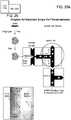
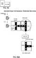
- An exemplary methodinvolves flowing a stream of the sample fluid containing the target material (e.g., nucleic acid template) such that it intersects two opposing streams of flowing carrier fluid.
- the carrier fluidis immiscible with the sample fluid. Intersection of the sample fluid with the two opposing streams of flowing carrier fluid results in partitioning of the sample fluid into individual sample droplets containing the target material.
- the carrier fluidmay be any fluid that is immiscible with the sample fluid.
- An exemplary carrier fluidis oil.
- the carrier fluidincludes a surfactant, such as a fluoro surfactant.
- PCRpolymerase chain reaction
- non-PCR based amplification reactionsuch as multi-strand displacement amplification, or other methods known to one of ordinary skill in the art.
- Suitable reagents for conducting PCR-based amplification reactionsinclude, but are not limited to, DNA polymerases such as Taq polymerase, forward and reverse primers, deoxynucleotide triphosphates (dNTPs), and one or more buffers.
- Suitable reagents for conducting non-PCR amplification reactionsinclude, for example, a high fidelity enzyme such as ⁇ 29. Alternatively, a transposase can be used.
- Either the droplets containing the first fluid, the droplets containing the second fluid, or both,may be formed and then stored in a library for later merging, aspects of certain implementations of which are described in U.S. Pub. 2010/0022414 .
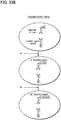
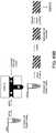
- droplets containing the target materialcan be merged with droplets containing other reagents.
- Mergingcan produce a set of droplets, each containing target and other reagents such as, in each droplet, a single nucleic acid template and heterogeneous mixture of primer pairs and probes. Merging can be accomplished, for example, in the presence of an electric field. Moreover, it is not required that both fluids be in the form of droplets when merging takes places.
- One exemplary method for merging of fluid portions with dropletsis taught, for example, in copending U.S. Patent Application No s. 61/441,985 and 13/371,222 .
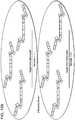
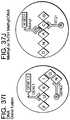
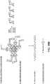
- fluidic compartmentsare formed by providing one or more of a first fluid partition (e.g., a droplet) comprising a target material and a second fluid (e.g., as a fluid stream or within droplets) comprising a plurality of nucleic acid constructs, each containing a functional N-mer capable of hybridizing to a unique region of the target material, and a unique N-mer to label the target.
- the first and second fluidsare merged to form a droplet. Merging can be accomplished by application of an electric field to the two fluids.
- the second fluidadditionally contains reagents for conducting an amplification reaction, such as a polymerase chain reaction or a multiple displacement amplification reaction.
- the genetic materialcan be fragmented or sheared using methods well known to those of skill in the art, for example, prior to sequestering into droplets or hybridizing to N-mers.
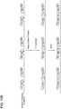
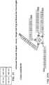
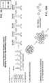
- the disclosureprovides a method of making a barcode library including obtaining a plurality of nucleic acid constructs in which each construct includes a unique N-mer and a functional N-mer.
- the functional N-mercan be a random N-mer, a PCR primer, a universal primer, an antibody, a sticky end, or any other sequence.
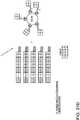
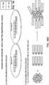
- the methodcan include making M sets of a number N of fluid compartments each containing one or more copies of a unique construct.
- the methodcan create barcode libraries of higher complexity by adding an additional construct to each compartment in a set, and repeating that for each set to produce NXM compartments each containing a unique pair of constructs. The pairs can be hybridized or ligated to produce new constructs.
- each unique N-mercan be adapted for identification by sequencing, probe hybridization, other methods, or a combination of methods.
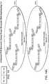
- the disclosureprovides a method for labeling target material comprising segregating each of a plurality of targets into a fluid compartment and providing one or more copies of a construct that is unique for each fluid compartment, in which each construct includes a unique N-mer and a functional N-mer.
- the methodcan include associating each target with a copies of a construct, for example, by hybridization.
- Optional steps of methods of the disclosurecan include performing an amplification reaction to produce amplicons that each contain a copy of the construct; releasing the contents of fluid compartments into a bulk phase; performing a second amplification reaction on amplicons; sequencing products of the disclosure; and detecting products of the disclosure by digital PCR.
- Higher levels of complexitycan obtained by introducing into each fluid partition one or more copies of an additional construct (for example, that are unique to a specific portion of a target) and linking each additional construct to a copy of the construct unique to each fluid partition.
- Target materialcan be unlabeled when segregated into the fluid compartments.
- the disclosureprovides a compartment containing all or a portion of a target material, and a plurality of constructs including unique N-mers and functional N-mers (e.g., capable of hybridizing to a unique region of the target material).
- target materialinclude but are not limited DNA, genomic DNA, chromosome(s), RNA, expressed RNA and/or protein molecules.
- the target materialincludes a single cell segregated into a fluid compartment. The cell can be lysed within the compartment, and the lysate can be targeted for labeling.
- Lysatecan include the genetic or proteomic material derived from the single cell (prokaryotic or eukaryotic) or a subset thereof (e.g., an entire genome, transcriptome, proteome, or a portion thereof). Droplets containing cells may be sorted according to a sorting operation prior to merging with the other reagents (e.g., as a second set of droplets).
- the other reagentsmay contain reagents or enzymes such as a detergent or a protease (e.g., a heat activatable protease) that facilitates the breaking open of the cell and release of the nucleic acids therein.
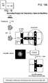
- primerscan be hybridized to the target and then target (e.g., nucleic acid) can be amplified, for example, by PCR.
- the disclosureprovides a plurality of nucleic constructs including a functional N-mer that comprises a random sequence, for example, a 6-mer for use in a multiple displacement reaction (MDA).
- the N-merscan comprise a target specific sequence, such as a sequence specific for a gene, a gene mutation, a gene motif, a splice site, a regulatory region of a gene, or a single nucleotide polymorphism.
- the N-merscan correspond to one or more consensus sequences, such as, for example, CPG motifs, or other sequence motifs that are related to known or suspected sequences indicative of splice sites, promoter regions, regulatory regions, or other functional genomic units, etc.
- the N-merscan each further comprise a common sequence, such as a universal primer sequence.
- the N-merscomprise oligo-dT labeled primers.
- the disclosuregenerally provides methods and materials for labeling a target material (e.g., protein or nucleic acid). Labeling can involve barcode-type labeling using nucleic acid constructs or a probe-type label (e.g., for digital PCR). Nucleic acid constructs can involve informational (i.e, unique or of known sequence) or functional N-mers. In certain aspects, one or more constructs contain different unique N-mers (i.e., unique labels). The label is preferably associated with a 5' end of the N-mers. However, the label can be associated with a 3' end of the N-mers.
- a target materiale.g., protein or nucleic acid.
- Labelingcan involve barcode-type labeling using nucleic acid constructs or a probe-type label (e.g., for digital PCR).
- Nucleic acid constructscan involve informational (i.e, unique or of known sequence) or functional N-mers. In certain aspects, one or more constructs contain different unique N-mers (i.e
- the label associated with each of the N-merscan be a nucleic acid tag, or "barcode" sequence.
- a barcodeis included
- the N-mergenerally hybridizes to the target material and is copied throughout subsequent steps such that the barcode is included in amplicons or sequence reads that may result.
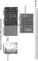
- a probe-type labelis included, the N-mer generally hybridizes to a specific material, for example, PCR product containing the target region, and can be detected in assays such as digital PCR.
- a probe-type labelcan include an optical label such as a fluorescent label. In some aspects, an optical label is attached to an antibody specific for a target region of interest in a target material. Applications involving probe-type or barcode-type labels will be discussed in greater detail below.
- a target materialcan be labeled by merging droplets containing the target material with a fluid stream or droplet stream containing the desired construct or merging a fluid stream of the target material with the construct into droplets.
- the methodscan further include the step of amplifying or copying the target material so as to preserve, for each amplified product, an association between the amplified product and the label.
- the amplified productis indicative of a haplotype.
- the nucleic acid template in each of the merged/formed dropletsis amplified, e.g., by thermocycling the droplets under temperatures/conditions sufficient to conduct a PCR reaction.
- the resulting amplicons in the dropletscan then be analyzed. For example, using probe-type labels, the presence or absence of the plurality of targets in the one or more droplets is detected optically, e.g., by the detectable label on the plurality of probes. Alternatively, amplicons can be sequenced and reads assembled based on the presence of barcode-type labels.
- capture sequencesare introduced into droplets containing target material, for example, by merging the droplets with a second set of droplets containing the capture sequences.
- Capture sequencescan include a barcode label and a portion that is capable of being captured on a solid surface (e.g., biotin/streptavidin on a surface; antibody/ antigen; aptamers; anchored oligonucleotides; etc.).
- a droplet containing a nucleic acidcan be merged with a second droplet containing the capture sequence, preferably with a tag (i.e., a barcode-type label). The capture sequence is allowed to hybridize to the target nucleic acid.
- the emulsionis then broken to release the hybridized capture sequence and target nucleic acid.
- the released nucleic acidis then captured on a solid support allowing the removal of elements such as cell debris, proteases and detergents that may inhibit subsequent steps.
- the tagis then incorporated by replication of the captured nucleic acid using the capture sequence with the tag as a primer. Replication can generate DNA from either DNA or RNA (cDNA synthesis). This material can either be processed directly or amplified further using methods known in the field such as PCR or multi-strand displacement amplification.
- the capture sequencescan be synthesized directly onto the beads or be attached by such means as biotinylated sequences and streptavidin beads.
- streptavidin beads and biotinylated sequenceshas the advantage of allowing a generic bead to be used with new libraries of biotinylated capture sequences that can be assembled on demand.
- any method known in the art for attaching nucleic acid sequences to beadscan be utilized.
- droplets containing target materialmay be merged with droplets containing beads that are designed to capture the target.
- the emulsioni.e., set of droplets
- the beadsare used to purify the target away from other components of the reaction that may inhibit subsequent steps such as cell debris, proteases and detergents.
- Targete.g., nucleic acid
- N-mersthat are designed to capture only portions of the target are attached to the beads.
- the N-mersinclude a barcode-type tag
- the tagcan be incorporated by replication of the captured nucleic acid using the capture sequence with the tag as a primer. The replication can generate DNA from DNA or RNA (cDNA synthesis). This material can either be processed directly or amplified further using methods known in the art such as PCR or multi-strand displacement amplification.
- methods of the disclosureinclude enriching all or selected portions of a target material.
- N-merscan be provided that further contain a common nucleotide sequence, such as a universal PCR sequence.
- the enrichment stepis accomplished by incorporating an adapter onto the 5' end of the amplified genetic material, such as a universal PCR primer sequence, and further amplifying the genetic material. Only those strands having a label will be amplified, thereby enriching for the labeled genetic material.
- enrichment of sequence specific labeled strandscan be achieved through amplification using a primer specific for the universal priming sequence incorporated into the labeled strand, and a primer specific for a desired target sequence.
- An enrichment stepcan be specific for target regions of interest in the genetic material, such as consensus sequences like CPG motifs, or other sequence motifs that are related to known or suspected sequences indicative of splice sites, promoter regions, regulatory regions, poly-A tail etc.
- a first portion of amplified product associated with the labelis enriched relative to a second portion of amplified product not associated with the label (e.g., through the inclusion of universal priming sites with the label).
- the disclosureprovides long sequences from short-read sequencing technologies.
- a set of primersis used that is tiled across the sequence of interest.
- Target nucleic acidis isolated in fluid partitions (e.g., droplets).
- a plurality of targetsare isolated in droplets and analyzed in parallel.
- a set of primersis provided in which each primer includes a label sequence that is unique for the droplet, the target nucleic acid is amplified in each droplet, with the result that every amplicon strand includes the label sequence at each end.
- the dropletsare ruptured and the amplicons are sequenced in such a way that each sequence read contains the target label sequence.
- each readis associated with a unique starting molecule through the presence of the label sequence, each "long read” that is produced from short read assembly will correspond to a single molecule of template.
- sequence readscan be mapped back to the targeted genome, transcriptome, proteome, or a portion thereof.
- Suitable sequencing methodsinclude, but are not limited to, sequencing by hybridization, sequencing by synthesis technology (e.g., HiSeqTM and SolexaTM, Illumina), SMRTTM (Single Molecule Real Time) technology ( Pacific Biosciences), true single molecule sequencing (e.g., HeliScopeTM, Helicos Biosciences), massively parallel next generation sequencing (e.g., SOLiDTM, Applied Biosciences; Solexa and HiSeqTM, Illumina), massively parallel semiconductor sequencing (e.g., Ion Torrent), and pyrosequencing technology (e.g., GS FLX and GS Junior Systems, Roche/454).
- sequencing by synthesis technologye.g., HiSeqTM and SolexaTM, Illumina
- SMRTTMSingle Molecule Real Time
- true single molecule sequencinge.g., HeliScopeTM, Helicos Biosciences
- massively parallel next generation sequencinge.g., SOLiDTM,
- the disclosureprovides a barcode library, which can be, for example, a stable barcode library which can be stored (e.g., for a year or longer).
- a barcode librarycan comprise a plurality of fluid compartments, each containing one or more copies of a unique construct, in which each construct includes a unique N-mer and a functional N-mer.
- each functional N-mermay be a sticky end, capable of being associated with another sticky end.
- Other functional N-merscan include sequence-specific primers; random N-mers; antibodies; probe targets; and universal primer sites.
- the fluid compartmentscan be water-in-oil droplets.
- the unique N-meroffers a barcode of information and can generally be between about 2 and 21 nucleotides in length, and optional longer, e.g., up to 50, 100, or any length.
- the disclosurerelates to methods for detecting or identifying one or a plurality of targets in a biological sample using digital PCR in fluid partitions.
- Methods of the disclosureinclude labeling target material with a probe-type label.
- a probe type labelcan include an optical label, and labeled target material can be identified or analyzed using digital PCR.
- Probesmay generally include sequences designed to hybridize to a target of interest. Detection of hybridization can indicate that the target of interest is present. Hybridization can be detected, for example, by including a fluorescent label on a probe structured so that the label is quenched unless hybridized to the intended target of the probe. Quenched and unquenched probes can be detected optically.
- One or a plurality of such probescan be provided in a fluid partition.
- Members of the plurality of probescan each include the same detectable label, or a different detectable label.
- the plurality of probescan include one or more groups of probes at varying concentrations.
- the one or more groups of probescan include the same detectable label which varies in intensity upon detection, due to the varying probe concentrations.
- the droplets of the disclosurecan further contain one or more reagents for conducting a polymerase chain reaction (e.g., polymerase, dNTPs, primers, etc.), for example, to enable probes to hybridize to amplified product (i.e., amplicons).
- the disclosureprovides microfluidic droplets for multiplex analysis.
- Each dropletcan contain a plurality of probes that hybridize to amplicons produced in the droplets.
- the dropletcontains two or more probes, e.g., 2, 3, 4, 5, 6, 7, 8, 9, 10, 12, 14, 16, 18, 20, 25, 30, 35, 40, 45, 50, 55, 60, 65, 60, 75, 80, 85, 90, 95, 100, 110, 120, 130, 140, 150, 160, 170, 180, 190, 200, 500, or more probes.
- the ability to amplify and detect single nucleic acids in dropletsenables digital PCR, detection, counting, and differentiation among nucleic acids, especially those present in heterogeneous samples.
- the disclosureapplies to digital amplification techniques and, in specific aspects enables multiplex PCR in droplets.
- multiplexing primers in dropletsenables the simultaneous increase in the number of PCR droplets while keeping the amount of input DNA the same or lower and generate the same or greater amplicon yield. This results in an overall increase in the amount of PCR positive droplets and amplicon yield without the consumption of more DNA.
- PCR primer pairs per dropleteven though the number of PCR primer pairs per droplet is greater than one, there is only one template molecule per droplet, and thus, in some implementations, there is only one primer pair per droplet that is being utilized at one time. As such, the advantages of droplet PCR for eliminating bias from either allele specific PCR or competition between different amplicons is maintained. However, as described below in relation to detection of haplotypes, other implementations advantageously allow detection of multiple loci on a single template using multiple primer pairs, preferably designed to minimize bias.
- the disclosureprovides methods of forming fluid partitions including target and reagents for digital PCR in which the methods enable multiplex digital PCR at high "plexity" in fluid partitions.
- one or more dropletsare formed, each containing a single nucleic acid template and a heterogeneous mixture of primer pairs and probes, each specific for multiple target sites on the template.
- a first fluid(either continuous, or discontinuous as in droplets) containing a single nucleic acid template (DNA or RNA) is merged with a second fluid (also either continuous, or discontinuous as in droplets) containing a plurality of primer pairs and a plurality of probes, each specific for multiple target sites on the nucleic acid template, to form a droplet containing the single nucleic acid template and a heterogeneous mixture of primer pairs and probes.
- the second fluidcan also contain reagents for conducting a PCR reaction, such as a polymerase and dNTPs.
- the droplet contentscan be amplified (e.g., by thermocycling).
- the probesare hybridized to the amplicons and hybridization is optically detected.
- the disclosuregenerally provides materials and methods for labeling target nucleic acid, protein, or other material using microfluidic droplet-based technology, and droplets produced using the same.
- the disclosurealso provides the ability to associate sequencing reads with single cells in a heterogeneous mixture of cells. For example, one or more mutations are identified in subpopulations of cancer cells in a sample using the labeling methods of the disclosure.
- the ability to identify multiple mutations existing in one cellbetter informs research and physicians on the possibility of drug resistance or reoccurrence of disease, and also inform treatment.
- the disclosurefurther provides for the ability to identify metagenomic loss of identity in individual bacteria (e.g., bacteria having multiple mutations in the same organism versus multiple bacteria, each having different mutations, in the same population).
- the disclosureprovides for the ability to pool multiple patient samples in a multiplex sequencing reaction and to accurately identify the source of the multiple samples after sequencing.
- methods of the disclosureprovide accurate labeling and detection.
- the disclosureprovides (I.) droplets for the analysis and labeling of target material.
- the disclosurefurther provides (II.) barcode-type labels and (III.) probe-type labels.
- FIG. 1depicts a flow chart of the general methods of the disclosure.
- the target materialis encapsulated in a droplet, for example, using a microfluidic system.
- Figure 2A-2Cshow droplet manipulation.
- Figure 2Dshows products of a labeling step.
- Figure 2Eshows a 10-mer and a schematic for a 6-mer.
- Figure 2Cshows one exemplary method of sequestering material in droplets.
- the genetic materialis diluted such that each droplet contains a single element (e.g., nucleic acid molecule, chromosome, genome, cell, protein, biological macromolecule, etc.).
- the elementscan be from a single cell (prokaryotic or eukaryotic), or a portion or subset thereof (e.g., a single nucleic acid template).
- the dropletscan optionally be sorted (e.g., to identify subspecies that will be subsequently labeled).
- the genetic materialis a single cell, the cells are lysed to release the genetic element in the single cell.
- Lysiscan be performed prior to encapsulation or after encapsulation (e.g., using proteases, alkaline reagents, and/or detergents).
- Labelse.g., barcodes, fluorescent labels
- an enrichment stepcan be performed to enrich for the labeled genetic element, or sequence specific enrichment.
- microfluidicDroplets can be generated using microfluidic systems or devices.
- the "micro-" prefixfor example, as “microchannel” or “”microfluidic”
- the element or articleincludes a channel through which a fluid can flow.
- microfluidicrefers to a device, apparatus or system that includes at least one microscale channel.
- Microfluidic systems and deviceshave been described in a variety of contexts, typically in the context of miniaturized laboratory (e.g., clinical) analysis. Other uses have been described as well.
- the devices and methods described hereinare based on the creation and manipulation of aqueous phase droplets (e.g., droplet libraries) surrounded by an immiscible carrier fluid. This combination enables precise droplet generation, highly efficient, electrically addressable droplet coalescence, and controllable, electrically addressable single droplet sorting.
- aqueous phase dropletse.g., droplet libraries
- microfluidic devicesinclude one or more channels in one or more analysis units.
- An "analysis unit”is a microsubstrate, e.g., a microchip.
- the terms microsubstrate, substrate, microchip, and chipare used interchangeably herein.
- An analysis unittypically includes at least an inlet channel and a main channel.
- the analysis unitcan further include coalescence, detection, or sorting modules.
- the sorting modulecan be in fluid communication with branch channels which are in fluid communication with one or more outlet modules (e.g., collection module or waste module).
- at least one detection modulecooperates with at least one sorting module to divert flow via a detector-originated signal.
- modules and channelsare in fluid communication with each other and therefore may overlap; i.e., there may be no clear boundary where a module or channel begins or ends.
- a plurality of analysis units of the disclosuremay be combined in one device.
- the dimensions of the substrateare those of typical microchips, ranging between about 0.5 cm to about 15 cm per side and about 1 micron to about 1 cm in thickness.
- componentscan be formed from solid materials, in which the channels can be formed via molding, micromachining, film deposition processes such as spin coating and chemical vapor deposition, laser fabrication, photolithographic techniques, etching methods including wet chemical or plasma processes, and the like. See, for example, Angell, et al., Scientific American, 248:44-55, 1983 . At least a portion of the fluidic system can be formed of silicone by molding a silicon chip.
- Devices of the disclosurecan also be formed of a polymer, for example, an elastomeric polymer such as poly-dimethylsiloxane (PDMS), polytetrafluoroethylene (PTFE), Teflon®, or the like.
- PDMSpoly-dimethylsiloxane
- PTFEpolytetrafluoroethylene
- Teflon®Teflon®
- PDMS polymersinclude those sold under the trademark Sylgard by Dow Chemical Co., Midland, Mich., and particularly Sylgard 182, Sylgard 184, and Sylgard 186.
- Silicone polymers such as PDMSare generally inexpensive, readily available, and can be solidified from a prepolymeric liquid via curing with heat.
- PDMSis typically curable by exposure of the prepolymeric liquid to temperatures of about, for example, about 65 °C to about 75 °C for exposure times of, for example, about an hour.
- silicone polymerscan be elastomeric and thus may be useful for forming very small features with relatively high aspect ratios, necessary in certain aspects of the disclosure.
- devices of the disclosuremay contain, at their surface, chemical groups capable of cross-linking to other oxidized silicone polymer surfaces or to the oxidized surfaces of a variety of other polymeric and non-polymeric materials.
- componentscan be formed and then oxidized and essentially irreversibly sealed to other silicone polymer surfaces or to the surfaces without the need for separate adhesives or other sealing means.
- sealingcan be completed simply by contacting an oxidized silicone surface to another surface without the need to apply auxiliary pressure to form the seal. That is, the pre-oxidized silicone surface acts as a contact adhesive against suitable mating surfaces.
- PDMScan also be sealed irreversibly to a range of oxidized materials other than itself including, for example, glass, silicon, silicon oxide, quartz, silicon nitride, polyethylene, polystyrene, glassy carbon, and epoxy polymers, which have been oxidized in a similar fashion to the PDMS surface (for example, via exposure to an oxygen-containing plasma). Molding, oxidation and sealing methods are described in the art, for example, in Duffy et al., "Rapid Prototyping of Microfluidic Systems and Polydimethylsiloxane," Anal. Chem., 70:474-480, 1998 .
- oxidized silicone polymersare much more hydrophilic than the surfaces of typical elastomeric polymers (where a hydrophilic interior surface is desired).
- a channelcan have a hydrophilic surface, which can be more easily wetted compared to other surfaces, which makes the channel easier to fill with aqueous solutions
- channelmeans a feature on or in a substrate that at least partially directs the flow of a fluid.
- the channelmay be formed, at least in part, by a single component, e.g., an etched substrate or molded unit.
- the channelcan have any cross-sectional shape, for example, circular, oval, triangular, irregular, square or rectangular (having any aspect ratio), or the like, and can be covered or uncovered (i.e., open to the external environment surrounding the channel).
- At least one portion of the channelcan have a cross-section that is completely enclosed, and/or the entire channel may be completely enclosed along its entire length with the exception of its inlet and outlet.
- a channelcan be formed, for example by etching a silicon chip using conventional photolithography techniques, or using a micromachining technology called "soft lithography” as described by Whitesides and Xia, Angewandte Chemie International Edition 37, 550 (1998 ).
- a fluid within a channelmay partially or completely fill the channel.
- the fluidmay be held or confined within the channel or a portion of the channel in some fashion, for example, using surface tension (e.g., such that the fluid is held within the channel within a meniscus, such as a concave or convex meniscus).
- some (or all) of the channelsmay be of a particular size or less, for example, having a largest dimension perpendicular to fluid flow of less than about 5 mm, less than about 2 mm, less than about 1 mm, less than about 500 microns, less than about 200 microns, less than about 100 microns, less than about 60 microns, less than about 50 microns, less than about 40 microns, less than about 30 microns, less than about 25 microns, less than about 10 microns, less than about 3 microns, less than about 1 micron, less than about 300 nm, less than about 100nm, less than about 30 nm, or less than about 10 nm or less in some cases.
- Channelscan be configured to coalesce droplets or to flow material by a detection module or a sorting module.
- a main channelis typically in fluid communication with any coalescence, detection and/or sorting modules, as well as inlet, branch, or outlet channels and any collection or waste modules. These channels permit the flow of molecules, cells, small molecules or particles out of the main channel.
- An "inlet channel”permits the flow of molecules, cells, small molecules or particles into the main channel.
- One or more inlet channelscommunicate with one or more means for introducing a sample into the device of the present disclosure.
- a microfluidic devicecan also include fluid channels to inject or remove fluid in between droplets in a droplet stream for the purpose of changing the spacing between droplets.
- a microfluidic substratecan also include a specific geometry designed to prevent the aggregation of material prior to encapsulation in droplets.
- the geometry of channel dimensioncan be changed to disturb the aggregates and break them apart by various methods, that can include, but is not limited to, geometric pinching (to force cells or particles through a narrow region, whose dimension is smaller or comparable to the dimension of a single cell) or a barricade (place a series of barricades on the way of the moving cells to disturb the movement and breakup the aggregates of cells).
- the channelsmay have a coating to minimize adhesion.
- the surface of the channelscan be coated with any anti- wetting or blocking agent for the dispersed phase.
- the channelcan be coated with any protein to prevent adhesion of the biological/chemical sample.
- Channelscan be coated by any means known in the art.
- the channelscan be coated with Teflon®, BSA, PEG-silane and/or fluorosilane in an amount sufficient to prevent attachment and prevent clogging.
- the channelscan be coated with a cyclized transparent optical polymer obtained by copolymerization of perfluoro (alkenyl vinyl ethers), such as the type sold by Asahi Glass Co. under the trademark Cytop.
- the coatingis applied from a 0.1-0.5 wt% solution of Cytop CTL-809M in CT-Solv 180. This solution can be injected into the channels of a microfluidic device via a plastic syringe. The device can then be heated to about 90 °C for 2 hours, followed by heating at 200 °C for an additional 2 hours.
- the channelscan be coated with a hydrophobic coating of perfluoro-alkylalkylsilane, described in U.S.
- the surface of the channels in the microfluidic devicecan be also fluorinated by any means known in the art to prevent undesired wetting behaviors.
- a microfluidic devicecan be placed in a polycarbonate dessicator with an open bottle of (tridecafluoro-1,1,2,2-tetrahydrooctyl)trichlorosilane. The dessicator is evacuated for 5 minutes, and then sealed for 20-40 minutes. The dessicator is then backfilled with air and removed.
- This approachuses a simple diffusion mechanism to enable facile infiltration of channels of the microfluidic device with the fluorosilane and can be readily scaled up for simultaneous device fluorination.
- the continuous phasepreferentially wets the channels and allows for the stable generation and movement of droplets through the device.
- the low surface tension of the channel wallsthereby minimizes the accumulation of channel clogging particulates, enhancing the processing of target material.
- Target materials for labeling, analysis, or detection according to the methods of the disclosureinclude, but are not limited to, cells, nucleic acids, proteins, multi-component complexes such as nucleic acid with associated proteins (e.g., histones), chromosomes, carbohydrates, or similar materials. Methods of disclosure are applicable to whole cells or to portions of genetic or proteomic material obtained from cells. Target material generally includes anything that can be sequestered into a fluid partition (e.g., droplet) and labeled.
- a fluid partitione.g., droplet
- Nucleic acid moleculesinclude deoxyribonucleic acid (DNA) and/or ribonucleic acid (RNA). Nucleic acid molecules can be synthetic or derived from naturally occurring sources. In one aspect, nucleic acid molecules are isolated from a biological sample containing a variety of other components, such as proteins, lipids and non-template nucleic acids. Nucleic acid template molecules can be obtained from any cellular material, obtained from an animal, plant, bacterium, fungus, or any other cellular organism. In certain aspects, the nucleic acid molecules are obtained from a single cell. Biological samples for use in the present disclosure include viral particles or preparations.
- Nucleic acid moleculescan be obtained directly from an organism or from a biological sample obtained from an organism, e.g., from blood, urine, cerebrospinal fluid, seminal fluid, saliva, sputum, stool and tissue. Any tissue or body fluid specimen may be used as a source for nucleic acid for use in the disclosure. Nucleic acid molecules can also be isolated from cultured cells, such as a primary cell culture or a cell line. The cells or tissues from which template nucleic acids are obtained can be infected with a virus or other intracellular pathogen.
- a samplecan also be total RNA extracted from a biological specimen, a cDNA library, viral, or genomic DNA.
- the nucleic acid moleculesare bound as to other target molecules such as proteins, enzymes, substrates, antibodies, binding agents, beads, small molecules, peptides, or any other molecule and serve as a surrogate for quantifying and / or detecting the target molecule.
- nucleic acidcan be extracted from a biological sample by a variety of techniques such as those described by Sambrook and Russell, Molecular Cloning: A Laboratory Manual, Third Edition, Cold Spring Harbor, N.Y. (2001 ).
- Nucleic acid moleculesmay be single-stranded, double-stranded, or double-stranded with single-stranded regions (for example, stem- and loop-structures).
- Proteins or portions of proteins (amino acid polymers) that can bind to high affinity binding moieties, such as antibodies or aptamers,are target molecules for oligonucleotide labeling, for example, in droplets, in some aspects of this disclosure.
- Methods of the disclosureinvolve forming droplets, which may contain no target material, target material from a single cell (e.g., a nucleic acid such as genomic DNA or expressed RNA), all or a portion of a target from a single cell, or all or a portion of target from multiple cells (corresponding to limiting or terminal dilution, respectively, as defined above).
- target material from a single celle.g., a nucleic acid such as genomic DNA or expressed RNA
- all or a portion of a target from a single celle.g., a nucleic acid such as genomic DNA or expressed RNA
- all or a portion of target from multiple cellscorresponding to limiting or terminal dilution, respectively, as defined above.
- the distribution of material within dropletsobeys the Poisson distribution.
- methods for non-Poisson loading of dropletsinclude but are not limited to active sorting of droplets, such as by laser-induced fluorescence, or by passive one-to-one loading.
- the dropletsare aqueous droplets that are surrounded by an immiscible carrier fluid. Methods of forming such droplets are discussed in U.S. Pub. 2008/0014589 ; U.S. Pub. 2008/0003142 ; U.S. Pub. 2010/0137163 ; U.S. Pat. 7,708,949 ; U.S. Pub. 2010/0172803 ; and U.S. Pat. 7,041,481 .
- Figure 4Ashows an exemplary aspect of a device 100 for droplet formation.
- Device 100includes an inlet channel 101 , and outlet channel 102 , and two carrier fluid channels 103 and 104. Channels 101 , 102 , 103 , and 104 meet at a junction 105.
- Inlet channel 101flows sample fluid to the junction 105.
- Carrier fluid channels 103 and 104flow a carrier fluid that is immiscible with the sample fluid to the junction 105.
- Inlet channel 101narrows at its distal portion wherein it connects to junction 105 (See Figure 4B ).
- Inlet channel 101is oriented to be perpendicular to carrier fluid channels 103 and 104. Droplets are formed as sample fluid flows from inlet channel 101 to junction 105 , where the sample fluid interacts with flowing carrier fluid provided to the junction 105 by carrier fluid channels 103 and 104.
- Outlet channel 102receives the droplets of sample fluid surrounded by carrier fluid.
- the sample fluidis typically an aqueous buffer solution, such as ultrapure water (e.g., 18 mega-ohm resistivity, obtained, for example by column chromatography), 10 mM Tris HCl and 1 mM EDTA (TE) buffer, phosphate buffer saline (PBS) or acetate buffer. Any liquid or buffer that is physiologically compatible with target material can be used.
- the carrier fluidis immiscible with the sample fluid.
- the carrier fluidcan be a non-polar solvent, decane (e g., tetradecane or hexadecane), fluorocarbon oil, silicone oil or another oil (e.g., mineral oil).
- the carrier fluidcontains one or more additives, such as agents which increase, reduce, or otherwise create non-Newtonian surface tensions (surfactants) and/or stabilize droplets against spontaneous coalescence on contact.
- Surfactantscan include Tween, Span, fluorosurfactants, and other agents that are soluble in oil relative to water. Suitable surfactants are known in the art.
- performanceis improved by adding a second surfactant, or other agent, such as a polymer or other additive, to the sample fluid.
- Surfactantscan aid in controlling or optimizing droplet size, flow and uniformity, for example by reducing the shear force needed to extrude or inject droplets into an intersecting channel. This can affect droplet volume and periodicity, or the rate or frequency at which droplets break off into an intersecting channel.
- the surfactantcan serve to stabilize aqueous emulsions in fluorinated oils from coalescing.
- the dropletsmay be coated with a surfactant or a mixture of surfactants.
- the carrier fluidmay be caused to flow through the outlet channel so that the surfactant in the carrier fluid coats the channel walls.
- the fluorosurfactantcan be prepared by reacting the perflourinated polyether DuPont Krytox 157 FSL, FSM, or FSH with ammonium hydroxide in a fluorinated solvent. The solvent, water, and ammonia can be removed with a rotary evaporator. The surfactant can then be dissolved (e.g., 2.5 wt %) in a fluorinated oil (e.g., Flourinert (3M)), which then serves as the carrier fluid.
- a fluorinated oile.g., Flourinert (3M)
- Another approach to merging sample fluidsinvolves forming a droplet, and contacting the droplet with a fluid stream, in which a portion of the fluid stream integrates with the droplet to form a mixed droplet.
- a dropletis formed as described above. After formation of the sample droplet from the first sample fluid, the droplet is contacted with a flow of a second sample fluid stream. Contact between the droplet and the fluid stream results in a portion of the fluid stream integrating with the droplet to form a mixed droplet, which, as discussed below, form a basis for droplet libraries according to certain aspects of the disclosure.
- the monodisperse droplets of the first sample fluidflow through a first channel separated from each other by immiscible carrier fluid and suspended in the immiscible carrier fluid.
- the dropletsare delivered to the merge area, i.e., junction of the first channel with the second channel, by a pressure-driven flow generated by a positive displacement pump. While droplet arrives at the merge area, a bolus of a second sample fluid is protruding from an opening of the second channel into the first channel.
- the channelsare oriented perpendicular to each other. However, any angle that results in an intersection of the channels may be used.
- the bolus of the second sample fluid streamcontinues to increase in size due to pumping action of a positive displacement pump connected to channel, which outputs a steady stream of the second sample fluid into the merge area.
- the flowing droplet containing the first sample fluideventually contacts the bolus of the second sample fluid that is protruding into the first channel. Contact between the two sample fluids results in a portion of the second sample fluid being segmented from the second sample fluid stream and joining with the first sample fluid droplet to form a mixed droplet.
- each incoming droplet of first sample fluidis merged with the same amount of second sample fluid.
- an electric chargeis applied to the first or second sample fluids. Applying electric charge is described in U.S. Pub. 2007/0003442 . Electric charge may be created in a sample fluid within the carrier fluid using any suitable technique, for example, by placing the first and second sample fluids within an electric field (which may be AC, DC, etc.), and/or causing a reaction to occur that causes the first and second sample fluids to have an electric charge, for example, a chemical reaction, an ionic reaction, a photocatalyzed reaction, etc.
- an electric fieldwhich may be AC, DC, etc.
- the electric fieldis generated from an electric field generator, i.e., a device or system able to create an electric field that can be applied to the fluid.
- the electric field generatormay produce an AC field (i.e., one that varies periodically with respect to time, for example, sinusoidally, sawtooth, square, etc.), a DC field (i.e., one that is constant with respect to time), a pulsed field, etc.
- the electric field generatormay be constructed and arranged to create an electric field within a fluid contained within a channel or a microfluidic channel.
- the electric field generatormay be integral to or separate from the fluidic system containing the channel or microfluidic channel, according to some aspects.
- an electric fieldis produced by applying voltage across a pair of electrodes, which may be positioned on or embedded within the fluidic system (for example, within a substrate defining the channel or microfluidic channel), and/or positioned proximate the fluid such that at least a portion of the electric field interacts with the fluid.
- the electrodescan be fashioned from any suitable electrode material or materials known to those of ordinary skill in the art, including, but not limited to, silver, gold, copper, carbon, platinum, copper, tungsten, tin, cadmium, nickel, indium tin oxide (“ITO”), etc., as well as combinations thereof. In some cases, transparent or substantially transparent electrodes can be used.
- the electric fieldfacilitates rupture of the interface separating the second sample fluid and the droplet. Rupturing the interface facilitates merging of a bolus of the second sample fluid and the first sample fluid droplet.
- the forming mixed dropletcontinues to increase in size until breaks free from the second sample fluid stream, for instance prior to the arrival of the next droplet containing the first sample fluid.
- the segmenting of the portion of the second sample fluid from the second sample fluid streamoccurs as soon as the shear force exerted on the forming mixed droplet by the immiscible carrier fluid overcomes the surface tension whose action is to keep the segmenting portion of the second sample fluid connected with the second sample fluid stream.
- the now fully formed mixed dropletcontinues to flow through the first channel (e.g., for possible use in a droplet library).
- a second fluid containing reagents for a PCR reactione.g., Taq polymerase, dNTPs, magnesium chloride, and forward and reverse primers, all suspended within an aqueous buffer.
- the second fluidmay also include detectably labeled probes and/or universal barcodes for detection of the amplified target material, the details of which are discussed below.
- a droplet containing the target or portion thereofis then caused to merge with the PCR reagents in the second fluid as described above, producing a droplet that includes target and PCR reagents as well as, optionally, detectably labeled probes.
- Droplet librariesare useful to perform large numbers of assays while consuming only limited amounts of reagents.
- a "droplet,” as used herein,is an isolated portion of a first fluid that is surrounded by a second fluid.
- the dropletsmay be spherical or substantially spherical; however, in other cases, the droplets may be non-spherical, for example, the droplets may have the appearance of "blobs" or other irregular shapes, for instance, depending on the external environment.
- a dropletis a first fluid completely surrounded by a second fluid.
- a first entityis "surrounded" by a second entity if a closed loop can be drawn or idealized around the first entity through only the second entity (with the sometimes exception for portions of the first fluid that may be in contact with a wall or other boundary, where applicable).
- a droplet libraryis made up of a number of library elements that are pooled together in a single collection. Libraries may vary in complexity from a single library element to 10 15 library elements or more. Each library element is one or more given components at a fixed concentration.
- the elementmay be, but is not limited to, cells, virus, bacteria, yeast, beads, amino acids, proteins, polypeptides, nucleic acids, polynucleotides or small molecule chemical compounds.
- the elementmay contain an identifier such as a label.
- the terms "droplet library” or “droplet libraries”are also referred to herein as an "emulsion library” or “emulsion libraries.” These terms are used interchangeably throughout the specification.
- a cell library elementcan include, but is not limited to, hybridomas, B-cells, primary cells, cultured cell lines, cancer cells, stem cells, or any other cell type.
- Cellular library elementsare prepared by encapsulating a number of cells from one to tens of thousands in individual droplets. The number of cells encapsulated is usually given by Poisson statistics from the number density of cells and volume of the droplet. However, in some cases the number deviates from Poisson statistics as described in Edd et al., "Controlled encapsulation of single-cells into monodisperse picolitre drops.” Lab Chip, 8(8): 1262- 1264, 2008 .
- a bead based library elementcontains one or more beads, and may also contain other reagents, such as antibodies, enzymes or other proteins.
- the library elementscan all be prepared from a single starting fluid or have a variety of starting fluids.
- the library elementswill be prepared from a variety of starting fluids.
- droplet librariesare collections of droplets that have different contents, ranging from beads, cells, small molecules, DNA, primers, antibodies.
- the dropletsrange in size from roughly 0.5 micron to 500 micron in diameter, which corresponds to about 1 pico liter to 1 nano liter. However, droplets can be as small as 5 microns and as large as 500 microns.
- the dropletsare at less than 100 microns, about 1 micron to about 100 microns in diameter. The most preferred size is about 20 to 40 microns in diameter (10 to 100 picoliters).
- the preferred properties examined of droplet librariesinclude osmotic pressure balance, uniform size, and size ranges.
- the droplets comprised within the droplet libraryare preferably uniform in size. That is, the diameter of any droplet within the library will vary less than 5%, 4%, 3%, 2%, 1% or 0.5% when compared to the diameter of other droplets within the same library.
- the uniform size of the droplets in the libraryis critical to maintain the stability and integrity of the droplets and is also essential for the subsequent use of the droplets within the library for the various biological and chemical assays described herein.
- the droplet librariesare using an immiscible fluorocarbon oil.
- the oilcan comprise at least one fluorosurfactant.
- the fluorosurfactant comprised within immiscible fluorocarbon oilis a block copolymer consisting of one or more perfluorinated polyether (PFPE) blocks and one or more polyethylene glycol (PEG) blocks.
- PFPEperfluorinated polyether
- PEGpolyethylene glycol
- the fluorosurfactantis a triblock copolymer consisting of a PEG center block covalently bound to two PFPE blocks by amide linking groups.
- fluorosurfactantsimilar to uniform size of the droplets in the library
- fluorosurfactantis critical to maintain the stability and integrity of the droplets and is also essential for the subsequent use of the droplets within the library for the various biological and chemical assays described herein.
- Fluidse.g., aqueous fluids, immiscible oils, etc.
- surfactantsthat can be utilized in the droplet libraries of the present disclosure are described in greater detail herein.
- the droplet libraries of the present disclosureare very stable and are capable of long-term storage.
- the droplet librariesare determined to be stable if the droplets comprised within the libraries maintain their structural integrity, that is the droplets do not rupture and elements do not diffuse from the droplets.
- the droplets librariesare also determined to be stable if the droplets comprised within the libraries do not coalesce spontaneously (without additional energy input, such as electrical fields described in detail herein). Stability can be measured at any temperature.
- the dropletsare very stable and are capable of long-term storage at any temperature; for example, e.g., -70°C, 0°C, 4°C, 37°C, room temperature, 75°C and 95°C.
- the droplet libraries of the present disclosureare stable for at least 30 days. More preferably, the droplets are stable for at least 60 days. Most preferably, the droplets are stable for at least 90 days.
- the disclosureprovides a droplet library comprising a plurality of aqueous droplets within an immiscible fluid (optionally comprising a fluorosurfactant), wherein each droplet is preferably substantially uniform in size and comprises a different library element.
- the disclosureprovides a method for forming the droplet library comprising providing a single aqueous fluid comprising different library elements, encapsulating each library element into an aqueous droplet within an immiscible fluid (optionally comprising a fluorosurfactant).
- different types of elementsare pooled in a single source contained in the same medium.
- the elementsare then encapsulated in droplets to generate a library of droplets wherein each droplet with a different type of bead or cell is a different library element.
- the dilution of the initial solutionenables the encapsulation process.
- the droplets formedwill either contain a single element or will not contain anything, i.e., be empty. In other aspects, the droplets formed will contain multiple copies of a library element.
- the elements being encapsulatedare generally variants of a type.
- elementsare cancer cells of a tissue biopsy, and each cell type is encapsulated to be screened for genomic data or against different drug therapies.
- Another exampleis that 10 11 or 10 15 different type of bacteria; each having a different plasmid spliced therein, are encapsulated.
- One exampleis a bacterial library where each library element grows into a clonal population that secretes a variant on an enzyme.
- a droplet librarycomprises a plurality of aqueous droplets within an immiscible fluid, such that there is a single molecule contained within a droplet for every 20-60 droplets produced (e.g., 20, 25, 30, 35, 40, 45, 50, 55, 60 droplets, or any integer in between).
- Single moleculesare encapsulated by diluting the solution containing the molecules to such a low concentration that the encapsulation of single molecules is enabled.
- a LacZ plasmid DNAwas encapsulated at a concentration of 20 f M after two hours of incubation such that there was about one gene in 40 droplets, where 10 ⁇ m droplets were made at 10 kHz per second. Formation of these libraries relies on limiting dilutions.
- the present disclosurealso provides a droplet library comprising at least a first aqueous droplet and at least a second aqueous droplet within a fluorocarbon oil comprising at least one fluorosurfactant, wherein the at least first and the at least second droplets are uniform in size and comprise a different aqueous fluid and a different library element.
- the present disclosurealso provides a method for forming the emulsion library comprising providing at least a first aqueous fluid comprising at least a first library of elements, providing at least a second aqueous fluid comprising at least a second library of elements, encapsulating each element of said at least first library into at least a first aqueous droplet within an immiscible fluorocarbon oil comprising at least one fluorosurfactant, encapsulating each element of said at least second library into at least a second aqueous droplet within an immiscible fluorocarbon oil comprising at least one fluorosurfactant, wherein the at least first and the at least second droplets are uniform in size and comprise a different aqueous fluid and a different library element, and pooling the at least first aqueous droplet and the at least second aqueous droplet within an immiscible fluorocarbon oil comprising at least one fluorosurfactant thereby forming an emulsion library.
- emulsion librarythere are library elements that have different particles, i.e., cells or beads in a different medium and are encapsulated prior to pooling.
- a specified number of library elementsi.e., n number of different cells or beads, are contained within different mediums.
- Each of the library elementsare separately emulsified and pooled, at which point each of the n number of pooled different library elements are combined and pooled into a single pool.
- the resultant poolcontains a plurality of water-in-oil emulsion droplets each containing a different type of particle.
- the droplets formedwill either contain a single library element or will not contain anything, i.e., be empty. In other aspects, the droplets formed will contain multiple copies of a library element.
- the contents of the beadsfollow a Poisson distribution, where there is a discrete probability distribution that expresses the probability of a number of events occurring in a fixed period of time if these events occur with a known average rate and independently of the time since the last event.
- the oils and surfactants used to create the librariesprevent the exchange of the contents of the library between droplets.
- Figure 71shows droplet generation, merging, and combining.
- panel Amonodisperse aqueous droplets are formed in a fluorocarbon oil using pressure-driven flow into a microfluidic nozzle.
- Panel Bpresents a schematic showing surfactant "wall" which provides stability for droplet manipulations.
- Panel Cincludes an image from a sequence enrichment application, combining DNA samples with PCR reagents (merging with Primer Droplet Library). The mixed droplets can be taken off-chip for PCR.
- the top imageshows ⁇ 2x10⁸ droplets before PCR, and bottom shows a microscope image of intact droplets after thermocycling.
- Figure 72shows droplet library generation and use in binding assays (discussed in greater detail herein).
- Panel Astable droplet library reagents can be formulated (5-member library shown) and aliquoted for later use.
- Panel Bshows reinjection of a droplet library and re-spacing into single file droplets.
- Panel Cpresents two binding assays: microscope image on the top shows virus particles binding to embryonic fibroblasts; the image on the bottom shows individual beads binding to ELISA sandwich reagents in droplets.
- Methods of the disclosuremay further include sorting the droplets based upon whether the droplets contain a homogeneous population of molecules or a heterogeneous population of molecules.
- a sorting modulemay be a junction of a channel where the flow of droplets can change direction to enter one or more other channels, e.g., a branch channel, depending on a signal received in connection with a droplet interrogation in the detection module.
- a sorting moduleis monitored and/or under the control of the detection module, and therefore a sorting module may correspond to the detection module.
- the sorting regionis in communication with and is influenced by one or more sorting apparatuses.
- a sorting apparatusincludes techniques or control systems, e.g., dielectric, electric, electro-osmotic, (micro-) valve, etc.
- a control systemcan employ a variety of sorting techniques to change or direct the flow of molecules, cells, small molecules or particles into a predetermined branch channel.
- a branch channelis a channel that is in communication with a sorting region and a main channel.
- the main channelcan communicate with two or more branch channels at the sorting module or branch point, forming, for example, a T-shape or a Y-shape. Other shapes and channel geometries may be used as desired.
- a branch channelreceives droplets of interest as detected by the detection module and sorted at the sorting module.
- a branch channelcan have an outlet module and/or terminate with a well or reservoir to allow collection or disposal (collection module or waste module, respectively) of the molecules, cells, small molecules or particles.
- a branch channelmay be in communication with other channels to permit additional sorting.
- a characteristic of a fluidic dropletmay be sensed and/or determined in some fashion, for example, as described herein (e.g., fluorescence of the fluidic droplet may be determined), and, in response, an electric field may be applied or removed from the fluidic droplet to direct the fluidic droplet to a particular region (e.g. a channel).
- a fluidic dropletis sorted or steered by inducing a dipole in the uncharged fluidic droplet (which may be initially charged or uncharged), and sorting or steering the droplet using an applied electric field.
- the electric fieldmay be an AC field, a DC field, etc.
- a channel containing fluidic droplets and carrier fluiddivides into first and second channels at a branch point.
- the fluidic dropletis uncharged. After the branch point, a first electrode is positioned near the first channel, and a second electrode is positioned near the second channel. A third electrode is positioned near the branch point of the first and second channels. A dipole is then induced in the fluidic droplet using a combination of the electrodes. The combination of electrodes used determines which channel will receive the flowing droplet. Thus, by applying the proper electric field, the droplets can be directed to either the first or second channel as desired. Further description of droplet sorting is shown in U.S. Pub. 2008/0014589 ; U.S. Pub. 2008/0003142 , and U.S. Pub. 2010/0137163 .
- droplets containing a heterogeneous population of moleculesare sorted away from droplets that contain a homogeneous population of molecules. Droplets may be further sorted to separate droplets that contain a homogeneous population of amplicons of the target from droplets that contain a homogeneous population of amplicons of the variant of the target.
- Methods of the disclosuremay further involve amplifying the target genetic material in each droplet.
- Amplificationrefers to production of additional copies of a nucleic acid sequence and is generally carried out using polymerase chain reaction or other technologies well known in the art (e.g., Dieffenbach and Dveksler, PCR Primer, a Laboratory Manual, Cold Spring Harbor Press, Plainview, N.Y. [1995 ]).
- the amplification reactionmay be any amplification reaction known in the art that amplifies nucleic acid molecules, such as polymerase chain reaction, nested polymerase chain reaction, ligase chain reaction ( Barany F. (1991) PNAS 88:189-193 ; Barany F.
- the amplification reactionis the polymerase chain reaction.
- Polymerase chain reactionrefers to methods by K. B. Mullis (U.S. Pats. 4,683,195 and 4,683,202 ) for increasing concentration of a segment of a target sequence in a mixture of genomic DNA without cloning or purification.
- the process for amplifying the target sequenceincludes introducing an excess of oligonucleotide primers to a DNA mixture containing a desired target sequence, followed by a precise sequence of thermal cycling in the presence of a DNA polymerase.
- the primersare complementary to their respective strands of the double stranded target sequence.
- primersare annealed to their complementary sequence within the target molecule. Following annealing, the primers are extended with a polymerase so as to form a new pair of complementary strands.
- the steps of denaturation, primer annealing and polymerase extensioncan be repeated many times (i.e., denaturation, annealing and extension constitute one cycle; there can be numerous cycles) to obtain a high concentration of an amplified segment of a desired target sequence.
- the length of the amplified segment of the desired target sequenceis determined by relative positions of the primers with respect to each other and by cycling parameters, and therefore, this length is a controllable parameter.
- the sample dropletmay be pre-mixed with a primer or primers, or the primer or primers may be added to the droplet.
- droplets created by segmenting the starting sampleare merged with a second set of droplets including one or more primers for the target nucleic acid in order to produce final droplets.
- a first droplet formation moduleproduces the sample droplets consistent with limiting or terminal dilution of target nucleic acid.
- a second droplet formation or reinjection moduleinserts droplets that contain reagents for a PCR reaction.
- Such dropletsgenerally include the "PCR master mix” (known to those in the art as a mixture containing at least Taq polymerase, deoxynucleotides of type A, C, G and T, and magnesium chloride) and forward and reverse primers (known to those in the art collectively as "primers”), all suspended within an aqueous buffer.
- the second dropletalso includes detectably labeled probes for detection of the amplified target nucleic acid, the details of which are discussed below.
- detectably labeled probesfor detection of the amplified target nucleic acid, the details of which are discussed below.
- Different arrangements of reagents between the two droplet typesis envisioned.
- the template dropletsalso contain the PCR master mix, but the primers and probes remain in the second droplets. Any arrangement of reagents and template DNA can be used according to the disclosure.
- Primerscan be prepared by a variety of methods including but not limited to cloning of appropriate sequences and direct chemical synthesis using methods well known in the art ( Narang et al., Methods Enzymol., 68:90 (1979 ); Brown et al., Methods Enzymol., 68:109 (1979 )). Primers can also be obtained from commercial sources such as Operon Technologies, Amersham Pharmacia Biotech, Sigma, and Life Technologies. The primers can have an identical melting temperature. The lengths of the primers can be extended or shortened at the 5' end or the 3' end to produce primers with desired melting temperatures. Also, the annealing position of each primer pair can be designed such that the sequence and, length of the primer pairs yield the desired melting temperature.
- Another method for determining the melting temperature of primersis the nearest neighbor method ( SantaLucia, "A unified view of polymer, dumbbell, and oligonucleotide DNA nearest-neighbor thermodynamics", 1998, P.N.A.S., 95 (4): 1460-5 ).
- Computer programscan also be used to design primers, including but not limited to Array Designer Software (Arrayit Inc.), Oligonucleotide Probe Sequence Design Software for Genetic Analysis (Olympus Optical Co.), NetPrimer, and DNAsis from Hitachi Software Engineering.
- the TM (melting or annealing temperature) of each primeris calculated using software programs such as Oligo Design, available from Invitrogen Corp.
- the droplet formation modulesare arranged and controlled to produce an interdigitation of sample droplets and PCR reagent droplets flowing through a channel.
- Such an arrangementis described U.S. Pub. 2008/0014589 ; U.S. Pub. 2008/0003142 , and U.S. Pub. 2010/0137163 .
- a sample dropletis then caused to merge with a PCR reagent droplet, producing a droplet that includes the PCR master mix, primers, detectably labeled probes, and the target nucleic acid.
- Dropletsmay be merged for example by: producing dielectrophoretic forces on the droplets using electric field gradients and then controlling the forces to cause the droplets to merge; producing droplets of different sizes that thus travel at different velocities, which causes the droplets to merge; and producing droplets having different viscosities that thus travel at different velocities, which causes the droplets to merge with each other. Further discussion can be found in U.S. Pub. 2007/0003442 .
- a single droplet formation module, or a plurality of droplet formation modulesare arranged to produce droplets from a mixture already containing the template DNA, the PCR master mix, primers, and detectably labeled probes.
- co-flowupstream from a single droplet formation module two channels intersect allowing two flow streams to converge.
- One flow streamcontains one set of reagents and the template DNA, and the other contains the remaining reagents.
- the template DNA and the PCR master mixare in one flow stream, and the primers and probes are in the other.
- the flow streamsmay or may not mix before the droplet generation nozzle. In either aspect, some amount of fluid from the first stream, and some amount of fluid from the second stream are encapsulated within a single droplet. Following encapsulation, complete mixing occurs.
- the dropletsare thermal cycled, resulting in amplification of the target nucleic acid in each droplet.
- the dropletsare collected off-chip as an emulsion in a PCR thermal cycling tube and then thermally cycled in a conventional thermal cycler. Temperature profiles for thermal cycling can be adjusted and optimized as with any conventional DNA amplification by PCR.
- the dropletsare flowed through a channel in a serpentine path between heating and cooling lines to amplify the nucleic acid in the droplet.
- the width and depth of the channelmay be adjusted to set the residence time at each temperature, which can be controlled to anywhere between less than a second and minutes.
- the three temperature zonesare used for the amplification reaction.
- the three temperature zonesare controlled to result in denaturation of double stranded nucleic acid (high temperature zone), annealing of primers (low temperature zones), and amplification of single stranded nucleic acid to produce double stranded nucleic acids (intermediate temperature zones).
- the temperatures within these zonesfall within ranges well known in the art for conducting PCR reactions. See for example, Sambrook et al. (Molecular Cloning, A Laboratory Manual, 3rd edition, Cold Spring Harbor Laboratory Press, Cold Spring Harbor, New York, 2001 ).
- the three temperature zonesare controlled to have temperatures as follows: 95°C (T H ), 55°C (T L ), 72°C (T M ).
- the prepared sample dropletsflow through the channel at a controlled rate.
- the sample dropletsfirst pass the initial denaturation zone (T H ) before thermal cycling.
- the initial preheatis an extended zone to ensure that nucleic acids within the sample droplet have denatured successfully before thermal cycling.
- the requirement for a preheat zone and the length of denaturation time requiredis dependent on the chemistry being used in the reaction.
- the samplespass into the high temperature zone, of approximately 95°C, where the sample is first separated into single stranded DNA in a process called denaturation.
- the samplethen flows to the low temperature, of approximately 55°C, where the hybridization process takes place, during which the primers anneal to the complementary sequences of the sample.
- the third medium temperatureof approximately 72°C, the polymerase process occurs when the primers are extended along the single strand of DNA with a thermostable enzyme.
- Methods for controlling the temperature in each zonemay include but are not limited to electrical resistance, peltier junction, microwave radiation, and illumination with infrared radiation.
- the nucleic acidsundergo the same thermal cycling and chemical reaction as the droplets passes through each thermal cycle as they flow through the channel.
- the total number of cycles in the deviceis easily altered by an extension of thermal zones or by the creation of a continuous loop structure.
- the sampleundergoes the same thermal cycling and chemical reaction as it passes through N amplification cycles of the complete thermal device.
- the temperature zonesare controlled to achieve two individual temperature zones for a PCR reaction.
- the two temperature zonesare controlled to have temperatures as follows: 95°C (T H ) and 60°C (T L ).
- the sample dropletoptionally flows through an initial preheat zone before entering thermal cycling.
- the preheat zonemay be important for some chemistry for activation and also to ensure that double stranded nucleic acid in the droplets are fully denatured before the thermal cycling reaction begins.
- the preheat dwell lengthresults in approximately 10 minutes preheat of the droplets at the higher temperature.
- the sample dropletcontinues into the high temperature zone, of approximately 95°C, where the sample is first separated into single stranded DNA in a process called denaturation.
- the samplethen flows through the device to the low temperature zone, of approximately 60°C, where the hybridization process takes place, during which the primers anneal to the complementary sequences of the sample.
- the polymerase processoccurs when the primers are extended along the single strand of DNA with a thermostable enzyme.
- the sampleundergoes the same thermal cycling and chemical reaction as it passes through each thermal cycle of the complete device. The total number of cycles in the device is easily altered by an extension of block length and tubing.
- the dropletsare created and/or merged on chip followed by their storage either on the same chip or another chip or off chip in some type of storage vessel such as a PCR tube.
- the chip or storage vessel containing the dropletsis then cycled in its entirety to achieve the desired PCR heating and cooling cycles.
- the dropletsare collected in a chamber where the density difference between the droplets and the surrounding oil allows for the oil to be rapidly exchanged without removing the droplets.
- the temperature of the dropletscan then be rapidly changed by exchange of the oil in the vessel for oil of a different temperature. This technique is broadly useful with two and three step temperature cycling or any other sequence of temperatures.
- Methods of the disclosuremay further involve releasing amplified target molecules from the droplets for further analysis. Methods of releasing amplified target molecules from the droplets are shown in publications and patents referenced above.
- sample dropletsare allowed to cream to the top of the carrier fluid.
- the carrier fluidcan include a perfluorocarbon oil that can have one or more stabilizing surfactants.
- the dropletrises to the top or separates from the carrier fluid by virtue of the density of the carrier fluid being greater than that of the aqueous phase that makes up the droplet.
- the perfluorocarbon oil used in one aspect of the methods of the disclosureis 1.8, compared to the density of the aqueous phase of the droplet, which is 1.0.
- the creamed liquidsare then placed onto a second carrier fluid which contains a de-stabilizing surfactant, such as a perfluorinated alcohol (e.g. 1H,1H,2H,2H-Perfluoro-1-octanol).
- a de-stabilizing surfactantsuch as a perfluorinated alcohol (e.g. 1H,1H,2H,2H-Perfluoro-1-octanol).
- the second carrier fluidcan also be a perfluorocarbon oil.
- the released amplified materialcan also be subjected to further amplification by the use tailed primers and secondary PCR primers.
- the primers in the dropletcontain an additional sequence or tail added onto the 5' end of the sequence specific portion of the primer.
- the sequences for the tailed regionsare the same for each primer pair and are incorporated onto the 5' portion of the amplicons during PCR cycling.
- another set of PCR primers that can hybridize to the tail regions of the ampliconscan be used to amplify the products through additional rounds of PCR.
- the secondary primerscan exactly match the tailed region in length and sequence or can themselves contain additional sequence at the 5' ends of the tail portion of the primer.
- additional regionsalso become incorporated into the amplicons.
- additional sequencescan include, but are not limited to: adaptor regions utilized by sequencing platforms for library preparation; barcode sequences for the identification of samples multiplexed into the same reaction; molecules for the separation of amplicons from the rest of the reaction materials (e.g., biotin, digoxin, peptides, or antibodies); or molecules such as fluorescent markers that can be used to identify the fragments.
- the amplified target moleculesare sequenced.
- the sequencingis single-molecule sequencing-by-synthesis. Single-molecule sequencing is shown in U.S. Pat. 7,169,560 ; U.S. Pat. 6,818,395 ; U.S. Pat. 7,282,337 ; U.S. Pub. 2002/0164629 ; and Braslavsky, et al., PNAS (USA), 100: 3960-3964 (2003 ).
- a single-stranded nucleic acid(e.g., DNA or cDNA) is hybridized to oligonucleotides attached to a surface of a flow cell.
- the single-stranded nucleic acidsmay be captured by methods known in the art, such as those shown in U.S. Pat. 7,666,593 .
- the oligonucleotidesmay be covalently attached to the surface or various attachments other than covalent linking as known to those of ordinary skill in the art may be employed.
- the attachmentmay be indirect, e.g., via the polymerases of the disclosure directly or indirectly attached to the surface.
- the surfacemay be planar or otherwise, and/or may be porous or nonporous, or any other type of surface known to those of ordinary skill to be suitable for attachment.
- the nucleic acidis then sequenced by imaging the polymerase-mediated addition of fluorescently-labeled nucleotides incorporated into the growing strand surface oligonucleotide, at single molecule resolution.
- a detectable barcode-type labelcan be any barcode-type label known in the art including, for example, radio-frequency tags, semiconductor chips, barcoded magnetic beads (e.g., from Applied Biocode, Inc., Santa Fe Springs, CA), and nucleic acid sequences.
- a barcode-type labelis a nucleic acid construct such as a nucleic acid construct including a barcode-type sequence (e.g., a unique N-mer).
- a construct of the disclosuregenerally includes a functional portion.
- a barcode sequencegenerally refers to a nucleic acid construct that includes at least a unique N-mer portion and a functional N-mer portion.
- the unique N-mer portioncan be used to tag-by means of its unique sequence information-any target material labeled with that construct.
- the functional N-mer portionmay be used to attach the construct to a target (e.g., cells, proteins, nucleic acids, other molecules, solid substrates, and other barcode constructs).
- the target materialincludes nucleic acid
- the functional N-mercan include a complementary nucleic acid (primer, hexamer, randomer, universal primer, etc.) to hybridize to the target.
- the unique N-mer(“barcode sequence") is attached to the functional N-mer (e.g., "primer") such that the barcode sequence is incorporated into a 5' end of the primer.
- the barcode sequencemay be incorporated into the 3' end of the primer.
- nucleic acid constructs of the disclosureare combined e.g., a set of constructs is cross combined one of each at a time with a set of other labels) with a labels of another type, such as magnetic barcoded beads from Applied BioCode.
- the functional N-mercan operate as a primer sequence or sticky end to hybridize to nucleic acid.
- Functional N-merscan be designed to favor hybridization under certain conditions. For example, length or GC content can be varied to favor high-temperature (or stringent) conditions. Where a nucleic acid construct (barcode) will be made with sticky ends to append to other constructs, the melting temperature of the sticky ends can be tuned, for example, by tuning the length of those ends. No particular length is required in general, so any given length can be chosen based on intended melting temperature or other design considerations.
- Attaching barcode sequences to nucleic acidsis shown in U.S. Pub. 2008/0081330 and PCT/US09/64001 .
- Methods for designing sets of barcode sequences and other methods for attaching barcode sequencesare shown in U.S. patent numbers 6,138,077 ; 6,352,828 ; 5,636,400 ; 6,172,214 ; 6235,475 ; 7,393,665 ; 7,544,473 ; 5,846,719 ; 5,695,934 ; 5,604,097 ; 6,150,516 ; RE39,793 ; 7,537,897 ; 6172,218 ; and 5,863,722 .
- Barcode sequencestypically include a set of oligonucleotides ranging from about 4 to about 20 oligonucleotide bases (e.g., 8-10 oligonucleotide bases), which uniquely encode a discrete library member preferably without containing significant homology to any sequence in the targeted genome.
- the barcode sequencegenerally includes features useful in sequencing reactions. For example the barcode sequences are designed to have minimal or no homopolymer regions, i.e., 2 or more of the same base in a row such as AA or CCC, within the barcode sequence.
- the barcode sequencesare also designed so that they are at least one edit distance away from the base addition order when performing base-by-base sequencing, ensuring that the first and last base do not match the expected bases of the sequence.
- oligonucleotides for use as constructscan be by any means known in the art. Oligonucleotides can be synthesized on arrays, or in bulk, for example.
- the barcode sequencesare designed to be correlated to a particular patient, allowing patient samples to be distinguished.
- the barcode sequences incorporated into a plurality of primers (and subsequently into DNA or RNA targets) within a single dropletmay be the same, and vary from droplet to droplet.
- the barcode sequences incorporated into the plurality of primers (and subsequently into DNA or RNA target) within a single dropletmay be different. Designing barcodes is shown U.S. Pat. 6,235,475 .
- the barcode sequencesrange from about 2 nucleotides to about 25 nucleotides, e.g., about 5 nucleotides to about 10 nucleotides.
- the oligonucleotide lengthshould be of minimal length so as to permit the longest read from the template nucleic acid attached.
- the barcode sequencesare spaced from the template nucleic acid molecule by at least one base (minimizes homopolymeric combinations).
- Methods of the disclosureinclude attaching the barcode sequences to a functional N-mer such as a primer, then incorporating the barcode into a target, or portion thereof using, for example, multiple displacement amplification.
- the labeled strands produced by MDAare able to be fragmented or sheared to desired length, e.g. generally from 100 to 500 bases or longer, using a variety of mechanical, chemical and/or enzymatic methods.
- DNAmay be randomly sheared via sonication, e.g. Covaris method, brief exposure to a DNase, or using a mixture of one or more restriction enzymes, or a transposase or nicking enzyme.
- RNAmay be fragmented by brief exposure to an RNase, heat plus magnesium, or by shearing. The RNA may be converted to cDNA before or after fragmentation.
- a barcode droplet librarygenerally is a set of droplets containing barcodes (e.g., unique N-mers) for incorporation into a target molecule.
- Barcodescan be provided in an oligonucleotide containing sequence to function as an amplification primer with the result that a nucleic acid subsequently introduced into the droplet will be amplified, and the copies that result will include the barcode of that droplet.
- barcodescan also be provided that are used to label proteins or other molecules of interest.
- the functional N-mercan be chosen based on a type of target material.
- a type of target materialFor example, for barcoding antibodies, one set of antibodies could all have a sticky-end that binds one class of barcodes, and another antibody set would have a different sticky end, for example, to bind a capture tag.
- a set of barcoded PCR primerscould include one forward/reverse pair that could bind to one class of barcodes and a different 'universal' forward/reverse pair that binds to a different class of barcodes (with the compliment to the second for/rev pair).
- Barcodescan be provided as oligonucleotides as discussed above.
- a barcodeis provided as part of a tripartite construct (e.g., as shown in Figure 7 ) including a universal priming site, a barcode, and a sequence specific region.
- the sequence specific regioncan provide a PCR primer of known sequence, a random hexamer for MDA, or any other suitable nucleotide sequence that will bind to target.
- the disclosureprovides universal barcode libraries (e.g., droplets that each contain a plurality of universal primers or priming sites all having a single unique barcode, but without a sequence-specific region).
- a universal barcodegenerally includes a unique N-mer and a sticky end.
- each droplet in the forward librarywill contain a plurality of copies of uniquely-barcoded tripartite "forward" primers. That is, each tripartite construct in the forward library will comprise 5'-universal forward tail-barcode-forward primer-3'.
- the forward librarycontains sets that include a number of droplets equal to or less than the number of possible unique barcode given the number of nucleotides in each barcode. Thus, if a six nucleotide barcode is to be used, sets of approximately 4,000 droplets (or any arbitrarily-lower number) can be made.
- a corresponding number of reverse tripartite constructscan be made (e.g., universal reverse tail-barcode-reverse primer).
- microfluidic methods and devices as discussed hereincan be used to add reverse constructs to each droplet containing forward constructs.
- Forward and reverse constructscan be put into droplets together in a variety of ways.
- the forward and reverse constructscan be put into droplets a single well at a time.
- flowing microfluidic systemsare used.
- a stream containing reverse constructscan be merged with a stream containing the forward droplets. As each droplet passes the merge point, the reverse construct is added.
- Forward and reverse constructscan be put together randomly, or they can be put together in a serial fashion.
- the first reverse constructcan be added to all droplets (e.g., about 4,000) of a set of forward droplets by flowing those droplets through the merge point.
- the second reverse constructcan be used, and the steps repeated.
- a second complete set of forward dropletscan be streamed into the second reverse construct, thereby creating 4,000 droplets, each of which contains a unique forward primer and the second reverse primer construct.
- 4,000 x 4,000 dropletswill have been made, each containing uniquely-barcoded primer pairs (e.g., as tripartite constructs).
- Production of a large barcode library by these meansneed not include tripartite constructs and can use any constructs that include barcodes (e.g., primer pairs + barcodes; random hexamers + barcodes; universal primers + barcodes; etc.).
- barcodese.g., primer pairs + barcodes; random hexamers + barcodes; universal primers + barcodes; etc.
- primer pairsany number of primers or primer pairs can be used. Where a large number of cells will be assayed for information about a single locus of interest, a single PCR primer pair may be used in a large barcode droplet library. Where a barcode droplet library will be used to assay a number X of loci on a plurality of genomes, X primer pairs will be used. Where MDA will be used to amplify one or more target regions, a number of random hexamers will be used according to calculations discussed elsewhere.
- methods of the disclosureinclude preparation of barcode droplet libraries in which each droplet contains a single barcoded construct without a corresponding partner-pair barcode.
- primers for an initial round of amplificationare universal primers, for example, where the target to be amplified includes universal priming sites.
- droplets of the disclosureare stable when stored.
- a barcode droplet librarycan be prepared having any arbitrarily large size and stored to be later used in any of the suited assays described herein or known in the art.
- the disclosureprovides methods involving a two-step "drop" PCR wherein multiple sets of primers are provided in a droplet. Either, both, or neither set of primers can include barcodes.
- Target materialis added to the droplet. A first round of amplification is performed, and then a condition is changed, and amplification is performed again. For example, low-stringency conditions are created for the first amplification, through manipulation of temperature or chemical environment. Thus, even though other primers are present, an intended first set of primers outcompetes or predominates in amplification. By these means, target nucleic acid can be amplified and barcoded in multiple steps.
- a barcode librarygenerally includes constructs having a functional N-mer and a unique N-mer.
- a functional N-meris a sticky end.
- the disclosureprovides methods and materials to generate large, complex, or extensible barcode libraries, and applications for barcode libraries.
- a "Universal Barcoding Droplet Library”for combining with samples.
- This reagentcan be used to barcode DNA, RNA, proteins, chemicals, beads or other species present in the sample if they contain complimentary binding moieties.
- antibodies or oligonucleotidesare used as functional N-mers for binding to sample molecules with (optionally releasable) unique N-mers as barcodes. Both the types and numbers of each type of barcodes are determined by a digitally quantified readout, and thus correlated with the presence and concentration of various biomarker species in a sample.
- a universal binding barcode droplet libraryis described for use in a 'bind and ligate' approach (see Figure 6 ).
- This library typeconsists of droplets containing oligonucleotide strands that encode barcodes and contain ligation competent ends, enabling the modular linking of barcodes by specific hybridization (also referred to as 'annealing' or 'binding') in droplets followed by ligation into a covalently bonded strand (or duplex) of bases.
- the Universal Binding Barcode Droplet Librarycan be used directly with samples that contain pre-bound barcoded binding moieties, as a 'primary' library that is combined with binding moieties targeting specific sample molecules, or can be used in the construction of 'secondary' or higher order binding barcode libraries through the successive combination of droplet libraries.
- the end use of such librariescan include assembly of the barcoded specific binding agents into a release-able and readable single molecule for use in digital quantification of bound targets for a variety of applications.
- FIG. 7shows one example of a universal barcode droplet library with targeting primers (e.g., to "bind and prime”).
- This library typeconsists of droplets containing barcoded primers for PCR (or other polymerase) priming, such that after combination with a sample droplet containing at least one target sequence from the same single DNA or RNA molecule, or multiple molecules co-localized in a single droplet, a digitally readable oligonucleotide barcode is attached to the target molecule's sequence. Since all polymerase generated molecules in the same droplet will have the same barcode, the co-localization information is retained after release from the droplet, and any sequencer can be used to both determine the sequence and count the number of templates traceable to each original droplet.
- Both library typesenable molecular barcoding in droplets, providing a large excess of unique identifying barcodes compared to the number of sample droplets, or compared to the number of sample objects or molecules contained in the droplets, thus allowing digital quantification of many targets of interest on various reading platforms.
- the two typesare not exclusive of each other.
- Figure 9shows ligating sticky-ended universal barcodes to barcoded PCR primers.
- Figure 5shows the overall scheme for construction of a universal binding barcode droplet library.
- Pairs of overhanging complimentary oligonucleotide barcodesare chemically synthesized (using standard commercial manufacturing methods) such that the complementary barcoding sequences are flanked by 'sticky-ends' for subsequent annealing and ligation to the target species or other barcodes, or for polymerase or other enzymatic priming.
- the oligonucleotidesmay include 5-prime or 3-prime phosphorylation, or combinations of these or other modifications.
- oligonucleotides resistant to nuclease activitymay be used, including the use of 2'O-Methyl RNA bases and/or phosphorothioated bonds to form the entire backbone of the oligo or to cap the ends of the sequence.
- PNA, LNA, or other modified nucleotide structurescan also be used.
- a sticky-endmay be any length and sequence, with preferred aspects containing base pairs including restriction endonuclease cleavage sites, or priming sites for sequencing or digital PCR, or array hybridization, and any number of sticky-ends with different sequences can be utilized.
- Sticky-end sequencesmay be used as barcode identifiers as part of composite barcodes.
- FIG. 5Two example barcoded oligonucleotide pairs are shown in Figure 5 (1a and 2a , flanked by sticky-end Type1 and sticky-end Type2).
- each discrete complementary oligonucleotide paircan be placed together into a standard microtiter-plate well and formed into droplets, which can be subsequently mixed with other oligonucleotide pair-containing droplets to make a 'primary barcode droplet library'.
- Forming droplets for a libraryis discussed in U.S. Pub. 2010/0022414 .
- the number of pair types (N members)is not limited.
- N x MNM-plex.
- a 4000 N-member library combined with a 4000 M-member librarywill generate a 16 million-plex barcode library.
- Combination of the N-member primary barcode library with the M secondary barcodescan be done in series (with each member of the M-barcode combined as an aqueous liquid one at a time with the N-member primary barcode library, using various methods including lambda or pico-injection modes and co-flow) or by combining the N-member and M-member library droplets in parallel (primary library combined with secondary library).
- Heterogeneous mixtures of barcodese.g. barcodes synthesized using degenerate bases
- Manipulation of dropletsis described in U.S. Pat. 7,718,578 and U.S. Pub. 2011/0000560 .
- the four oligonucleotide types present in the final combined dropletwill specifically hybridize to create a sticky-ended tandem barcode (e.g., droplet 1 or 2 in Figure 5 ). This can then be ligated together. A similar specific hybridization will occur for additional numbers of barcodes containing complimentary sticky-ends.
- the panel on the right of Figure 6shows the initial binding barcode droplet library (only one droplet and one molecule of each type shown, with a barcode identifier 1a) on the top, a tandem barcoded droplet library formed by combination of a primary barcode and a secondary barcode in the middle (e.g. barcode identifier 1a:1b), and a triple barcoded library at the bottom (formed by combining a secondary barcoded library with a third barcode, resulting in barcode identifier 1a:1b:1c).
- a tandem barcoded droplet libraryformed by combination of a primary barcode and a secondary barcode in the middle (e.g. barcode identifier 1a:1b)
- a triple barcoded library at the bottomformed by combining a secondary barcoded library with a third barcode, resulting in barcode identifier 1a:1b:1c).
- This modular constructionis not limited to the combinations shown, with any composite sticky-ended barcode library able to be combined with additional barcodes in subsequent rounds of droplet combination. Even a low number of combinations can result in a very high level of barcode-plex.
- the oligonucleotidescan be designed such that the resulting annealed oligo set can have a single or double sticky-ends (with different or similar ends).
- a barcode librarycan also be made to include a sticky-end adapter specific for a sequencing platform.
- a constructis made that includes a sequencing platform N-mer and a sticky-end N-mer.
- a library of these constructscan be made.
- a universal barcode library as discussed abovecan be made.
- the universal barcode librarycan be combined with the sequencing platform adapter library by means of the sticky ends in view of a particular application.
- products of any analysis discussed hereincan be adapted to go directly into the workflow of any given sequencing platform (e.g. sticky-ended Illumina adaptors to anneal/ligate onto either the primer library or the output from a targeted sequencing run, so that it could be hybridized directly onto their flow cell.
- a different sticky-end adaptor setcould be used for 454, etc.). This approach can minimize PCR bias.
- a universal PCR primer barcode librarycan also be prepared with an unlimited amount of plex by creating sticky-ended forward and reverse primers that can be further combined with additional numbers of sticky-ended barcodes to generate combinatorial barcodes, as shown at the top of Figure 9 .
- the forward and reverse universal primersare constructed in an identical fashion as described above and in Figure 7 (primary barcoded primers) and then annealed to a sticky-ended barcode oligonucleotide pair (either single or double sticky-ended as shown in Figure 5 ) and subsequently ligated, to make a contiguous forward (and/or reverse) primer annealed to the complimentary oligo that was used to anneal to the primary barcoded primer.
- the top right side of Figure 9shows the ligated product after addition of both forward and reverse single sticky-ends to create a 'secondary' barcoded priming set.
- the bottom of Figure 9shows a single droplet after combination of one prepared template-containing sample droplet with one universal PCR primer barcode library droplet.
- the annealed PCR primerscan be amplified by using polymerase and dNTPs, and all of the amplicons from this droplet will be barcoded 1b:1a:1a:1b (with 1b:1a 5-prime to the target loci sequence, followed by 1a:1b, as read 5-prime to 3-prime).
- the disclosureprovides systems and methods for haplotype phasing and genotyping.
- a nucleic acidcan be isolated from a sample and haplotyped through the examination of a number of loci. For example, the allelic form present for a number of suspect SNPs of interest can be determined along a single chromosome.
- barcodes in multiplexed tiled PCR reactions within dropletsthis aspect of the disclosure enables 'haplotype phase' assignments to be made using existing sequencing platforms.
- haplotype phasinginvolves preparing a barcode droplet library in which each droplet contains the set of primers to amplify the loci of interest.
- each primeris part of a tripartite construct that also includes a universal priming site (for subsequent amplification, capture, or sequencing) and a barcode.
- every tripartite constructwill preferably contain the same barcode.
- the target locuscan be pre-amplified using a single pair of PCR primers that flanks the entire locus, before appropriate loading of the sample into droplets for amplification and barcoding (not depicted in the workflow in Figure 10 ).
- each tripartite constructincludes a universal tail portion immediately 5' to a barcode sequence followed by the sequence-specific targeting primer bases.
- a primer droplet library 'member'includes a droplet that contains all of the targeted primers sufficient for covering the target bases, each with the same barcode that will enable post-sequencing correlation to the target strand.
- the number of library membersis determined by the ratio of barcode number to the number of target alleles to be analyzed.
- Figure 10shows 100 cells as input, with 4000 barcodes giving a 1/10 chance of duplicate barcodes for any allele.
- the DNA from 100 cellsprovides 400 target alleles, which are loaded (together with polymerase, buffer, and nucleotides) into one million droplets and combined with the barcoding primer library to generate a PCR-competent droplet emulsion.
- the barcoding primer libraryto generate a PCR-competent droplet emulsion.
- 13 tiled primer pairscan be used to cover the target bases. Fewer primer pairs can be used if only subsets of the target bases are to be phased.
- Droplets from the droplet libraryare merged with target nucleic acid.
- genomic DNAhas been sheared to a length of about 20 kilobases. Fragments of the target are introduced into droplets such that on average each droplet will generally contain no more than one molecule of target nucleic acid.
- each dropletcontains at least: a pair of tripartite constructs for each locus of interest; a single nucleic acid molecule; and PCR reagents (e.g., Taq , dNTPs).
- PCR reagentse.g., Taq , dNTPs.
- Droplet librariesare generally discussed herein as having pairs of barcodes and in some aspects those barcodes may be necessarily the same or different, while in other aspects, it may not matter whether the forward and reverse barcodes are the same or different. For example, if barcodes have six nucleotides, and millions of targets are to be analyzed, then using different forward and reverse barcodes will create ample unique barcode pairs.
- haplotype phasingis presented in which the forward and reverse barcodes need not be the same. In some aspects, those bar codes may be the same.
- the amplified productsare released into a single bulk aqueous phase (e.g., using a droplet de-stabilizing reagent), and a subsequent amplification, sequencing, or capture is performed using the universal primer tail as well as any sequencing platform-specific adaptor (and additional barcodes) needed before sequencing.
- Examples of the PCR inputs and outputsare shown in Figure 11 .
- the total number of PCR cyclesdroplet PCR plus bulk PCR
- the total number of PCR cyclescan be limited to 10 cycles (sufficient to generate 150 copies).
- the amplified productsare then sequenced according to any method known in the art including those discussed herein. Even though, in certain aspects, the amplified products are provided for sequencing in a bulk aqueous phase, sequencing results can be haplotyped based on the presence of the barcode in the sequence reads. Particularly in aspects in which the barcode is immediately 3' to a sequencing primer binding site, every sequence read will have barcode information.
- genetic materialis haplotyped without amplification as shown in Figure 15- Figure 17 .
- Figure 8shows the overall scheme for construction of a universal priming barcode droplet library for use in a 'Bind and Primer Extend' application (addition of sticky end component on right of Figure 8 is optional). Primers as shown in Figure 8 can be used as shown in Figures 15-17 to provide haplotype information without amplification.
- a the initial 'primary' droplet librarycontains single universal priming barcodes for primer extension, constructed using similar methods as that described for construction of a universal binding barcode droplet library.
- Contiguous oligonucleotidesare chemically synthesized with each of the sequence components including a universal forward tail (consisting of bases to be annealed to an analogous sticky-end), a sequence of bases used as a barcoding component, and a priming sequence that will anneal to the universal 5-prime end of the target sequence to be counted (the complimentary target is designated as 'for' in, for example, Figure 15- Figure 17 ).
- Figure 15shows up-front processing for haplotyping a single molecule of DNA using a universal priming barcode library.
- a forward primeris used with a corresponding oligonucleotide (here labeled 3' Tailed primer). Note that the corresponding 3' oligonucleotide need not function as a primer. For convenience, they will be referred to as primers.
- a set of specific targeting primersare prepared and annealed to the target DNA, with a 5' oligonucleotide comprising oligonucleotides complimentary to the 5' end of the target locus (3 loci are shown in Figure 15 ) and a universal 'for' sequence that will be used to anneal to the barcode library, and a 3-prime oligo comprising oligos complimentary to the 3-prime end of the target locus followed by a universal 3-tailed end.
- the target molecule to be haplotypedcan be targeted by any number of primer pairs and targeted loci can be any distance from other loci (the shear length of the genome will limit the maximum distance that can be haplotyped).
- Each of the uniquely barcoded primerscan be reformatted from a well plate into a droplet format, either for direct use with samples or to be used in optional subsequent rounds of droplet combination to add complimentary sticky-ended barcoding sequences (right side of Figure 8 , also shown in right side of Figure 9 ).
- Combination of the primary 'forward barcode library' with sticky-ended barcodesprovides another barcode encoding level.
- the primary droplet librarycan be generated from pairs of primers initially placed together in wells and reformatted into droplets, with a sticky-ended barcode oligo together with various forward barcoded primers. Additional rounds of droplet combination with either single or multiple sets of sticky-ended barcode members can be used to get to any required level of barcode-plex needed for the assay.
- a universal priming barcode library having sufficient plex to uniquely barcode the sample targetsis used by combination and annealing to target molecules that have been either prepared in bulk or in droplets (bulk prep is shown in Figure 15 ).
- Hybridization of all of the targeting primer pairs with the target sampleis followed by removal of unbound primers (e.g. purification by size or via use of biotinylated primers or targets), elongation of the forward hybridized primer for each locus using a polymerase (e.g. Klenow) that lacks exonuclease activity such that the elongation stops when the polymerase encounters the 3-prime primer, and subsequent ligation using ligase and ATP or photo-ligation or other ligation means.
- the resulting bulk sample preparationcontains the target single stranded molecules with each targeted loci annealed to a contiguous oligonucleotide strand that contains the newly synthesized compliment to the target loci bracketed by the added primer pairs.
- This output of the process shown in Figure 15is the target which the nucleic acid constructs (primers) shown in Figure 16 bind to.
- the bulk annealed sample prepis loaded into droplets, along with polymerase and dNTPs, such that a single contiguous molecule with its set of targets to be haplotyped is contained in a droplet.
- the Universal Barcode Libraryis combined with the prepared sample, either in a droplet to droplet combination mode, or in a droplet to aqueous stream combination mode (shown in Figure 16).
- Figure 16shows introducing primer pairs, each bearing a barcode.
- a pair of primers for each locus of interestis incubated with a strand of target DNA and allowed to hybridize. Any unbound (un-hybridized) material is removed.
- the two forward 'for' primerscan be elongated either inside droplets that include the polymerase and dNTPs, or following release of the primers annealed to the template (the workflow in Figure 17 shows release before elongation outside of droplets).
- Polymerasesynthesizes a complementary strand from the 5' forward primer to the 3' "reverse primer”. Ligase then ligates the complementary strand to the 3' reverse primer. As a result, a complementary strand has been synthesized representing each locus of interest.
- the dropletscan be lysed (or "burst", discussed elsewhere herein), and the contiguous strands (e.g., barcoded amplicons) released into bulk phase as shown in Figure 17 .
- the resulting contiguous strands containing barcodesare used for sequencing (or sequence determination using microarrays), and the sequences that contain identical barcodes have sequences that derive from the same sample strand (i.e. the same haplotype).
- This exampleshows a method for haplotype determination without any amplification (i.e. only elongation and ligation), and has the advantage of elimination of potential amplification bias.
- a locushas copy number variation within the same sample target molecule, this can be seen as variation in the number of sequenced reads that have the same barcode and sequence.
- ampliconscan provide target material according to a "bind and primer PCR” approach.
- Figure 7shows the overall scheme for construction of a Universal Priming Barcode Droplet Library for use in a 'PCR' version of the haplotyping application, which provides haplotype information with amplification.
- the initial droplet librarycontains single 'forward' universal priming barcodes that will be subsequently paired with a 'reverse' barcoded primer to create a Universal PCR Primer Barcoded Droplet Library.
- Contiguous oligonucleotidesare chemically synthesized with each of the sequence components including a universal forward tail (consisting of bases to be used as priming sites or for ligation to adaptors used for sequencing), a sequence of bases used as a barcoding component, and a forward priming sequence that will anneal to the universal 5-prime end of the target sequence to be counted (the complimentary target is designated as 'for' in Figure 12- Figure 14 ).
- Each of the uniquely barcoded primersis reformatted from a well plate into a droplet format. Combination of the forward primer barcoded droplet library with similarly constructed reverse primer barcodes (either serial or library combination, with the serial mode shown in Figure 7 ) generates the universal PCR primer barcoded droplet library.
- a PCR primer droplet librarycan be generated from pairs of forward and reverse primers initially placed together in wells and reformatted into droplets, however this type of library will have the same number of unique forward/reverse barcode combinations as the number of paired primers wells.
- a universal priming barcode library having sufficient plex to uniquely barcode the sample targetsis used by combining with samples and annealing to complimentary target molecules present in the sample.
- An example sample preparation schemeis shown in Figure 12 .
- To prepare for target molecule haplotypinga set of specific targeting oligos are prepared and annealed in bulk phase to the target DNA, with a 5-prime oligonucleotide comprising oligonucleotides complimentary to the 5-prime end of the target locus (3 Loci present on the same contiguous target molecule are shown being targeted for haplotyping) and a universal 'for' sequence that will be used to anneal to the Universal Priming Barcode Library, and a 3-prime oligo comprising oligos complimentary to the 3-prime end of the target locus followed by a universal 3-tailed end.
- the target molecule to be haplotypedcan be targeted by any number of primer pairs, and the targeted loci can be any distance from each other (the shear length of the genome will limit the maximum distance that can be haplotyped).
- Hybridization of all of the targeting primer pairs with the target sampleis followed by removal of unbound primers (e.g. purification by size or via use of biotinylated primers or targets), elongation of the forward hybridized primer for each locus using a polymerase (e.g. Klenow) that lacks exonuclease activity such that the elongation stops when the polymerase encounters the 3-prime primer, and subsequent ligation using ligase and ATP or photo-ligation or other ligation means (bottom of Figure 12 ).
- the resulting bulk sample preparationcontains the target single stranded molecules with each targeted loci annealed to a contiguous oligonucleotide strand that contains the newly synthesized compliment to the target loci bracketed by the
- FIG. 13Use of the universal PCR barcode primer library with prepared sample is shown in Figure 13 .
- the bulk annealed sample prepis loaded into droplets, along with polymerase and dNTPs, such that a single contiguous molecule with its set of targets to be haplotyped is contained in a droplet.
- the universal barcode libraryis combined with the prepared sample, either using a droplet to droplet combination mode, or in a droplet to aqueous stream combination mode (shown in Figure 13 ).
- the two forward 'for' complimentary base pairsare allowed to hybridize (two droplets from a combined library and sample are shown in Figure 13 , and elongated either inside droplets that include polymerase and dNTPs, or following release of the primers annealed to the template (the workflow in Figure 14 shows release before elongation outside of droplets), followed by ligation of the 3-prime primer to the elongated strand.
- the resulting contiguous strands shown on the right side of Figure 14 containing barcodesare used for sequencing (or sequence determination using microarrays), and the sequences that contain identical barcodes have target loci sequences that derive from the same sample strand (i.e. the same haplotype phase).
- Genotyping of single cellscan be performed in a fashion similar to haplotyping discussed above.
- Figure 18depicts the steps associated with isolation, encapsulation, molecular labeling, sorting and analysis of single cell genomes using fluidic droplets (including optional upfront sorting, and cell lysis within droplets using a detergent and heat).
- each dropletcontaining genetic or proteomic material (e.g., from a single cell, or a portion thereof).
- genetic or proteomic materiale.g., from a single cell, or a portion thereof.
- single cell dropletsare created and then the cells are lysed within the droplets.
- the dropletscan be merged with droplets from a barcode droplet library (e.g., containing nucleic acid constructs in which the functional N-mers are primer oligonucleotides to hybridize to the genome of the single cell).
- a barcode droplet librarye.g., containing nucleic acid constructs in which the functional N-mers are primer oligonucleotides to hybridize to the genome of the single cell.
- each dropletwill contain at least: the entire genome from a single cell; nucleic acid constructs for hybridizing to the genome of interest with a barcode; and analysis reagents (e.g., polymerase and nucleotides).
- a second library of dropletsis formed, each containing a plurality of N-mers, each N-mer containing an associated label.
- each droplet in the second library of dropletscontains the same label within the given droplet, and the labels preferably vary from droplet to droplet.
- Each droplet in the second libraryfurther contains reagents for the replication of the genetic material in the first droplet and subsequent incorporation of the tag.
- the replicationcan be DNA from DNA or DNA from RNA (cDNA).
- cDNADNA from RNA
- the reagents for conducting the replicationcan include such things as polymerase, reverse transcriptase, nucleotides, buffers, etc.).
- the second dropletcould contain beads which are designed to capture the target nucleic acid.
- Capture sequences with a tagcan be attached to bead and used to capture the genetic material from the first droplet introduced after merging with the second droplet.
- the capture sequence with the tagcan be synthesized directly onto the beads or be attached by such means as biotinylated sequences and streptavidin beads.
- streptavidin beads and biotinylated sequenceshas the advantage of allowing a generic bead to be used with new libraries of biotinylated capture sequences that can be assembled on demand.
- any method known in the art for attaching nucleic acid sequences to beadscan be utilized.
- the second dropletcould contain capture sequences that have an attached molecule that is capable of being captured on a solid surface.
- Biotinwould be such a molecule that could be captured by streptavidin attached to a solid surface.
- Other methods known in the artsuch as antibody/ antigen or aptamers could also be utilized.
- the target regions of the genomecan be amplified according to the workflow shown in Figure 18 .
- Amplification of the target region of the genomecan be accomplished by any method known in the art.
- amplification and barcodingcan involve barcoded primers, as shown in Figure 19 .
- Figure 20shows single cell genomics using primers for a first round of amplification followed by subsequent merging with a universal barcode library.
- Primers as shown in Figure 19 or in Figure 20generally can amplify target material by a PCR reaction.
- the new library of dropletsthen undergoes amplification (see “1st Round PCR” in Figure 19 ) to incorporate the labels into the genetic target (i.e., DNA or RNA).
- the genetic targeti.e., DNA or RNA
- Target materialcan also be amplified using multiple displacement amplification (MDA) or any other method known in the art.
- MDAmultiple displacement amplification
- a random hexamer(or similar oligo) is used in conjunction with a barcode.
- Figure 21shows generating a barcoded random hexamer library and using the random hexamer library with phi29.
- Figure 22shows a barcode/primer library used with transposase.
- the labelcan be incorporated into the DNA using, for example, multiple displacement amplification with random hexamer primers having a label incorporated at the 5'end.
- MDAMultiple displacement amplification
- This methodcan rapidly amplify minute amount of DNA samples to reasonable quantity for genomic analysis.
- the reactionstarts by annealing random hexamer primers to the template and DNA synthesis is carried out by high fidelity enzyme, preferentially ⁇ 29 at a constant temperature.
- MDAgenerates larger sized products with lower error frequency.
- MDA reactionstarts with the annealing of random hexamer primers to the DNA template and then continues with the chain elongation ⁇ 29. Increasing number of primer annealing events happens along the amplification reaction.
- the amplification reactioninitiates when multiple primer hexamers anneal to the template.
- the polymerasedisplaces the newly produced DNA strand and continues its strand elongation.
- the strand displacementgenerates newly synthesized single stranded DNA template for more primers to anneal. Further primer annealing and strand displacement on the newly synthesized template results in a hyper-branched DNA network.
- the sequence debranching during amplificationresults in high yield of the products.
- SI nucleasesis used to cleave the fragments at displacement sites. The nicks on the resulting DNA fragments are repaired by DNA polymerase I.
- the generated DNA fragmentscan be directly used for analysis or be ligated to generate genomic libraries for further sequencing analysis. Using MDA, the DNA would simultaneously be amplified and have a barcode incorporated into the sequence.
- the merged dropletsare incubated in a manner that releases the genetic material from a cell if present and allows the hybridization of the genetic material to the capture sequences attached to the bead.
- Reagentssuch as proteases, alkaline reagents, detergents or other methods known in the art can be used to release the genetic material from the cells if present. After capture the emulsion is broken and the beads used to purify the genetic material away from other components of the reaction that may inhibit subsequent steps such as cell debris, proteases and detergents.
- the beadswould capture the genetic material using attached sequences that were random N-mers of length N designed to capture all sequences or variations of N-mers designed to capture only portions of the genetic material present.
- the tagis then incorporated by replication of the captured nucleic acid using the capture sequence with the tag as a primer.
- the replicationcan either generate DNA from a DNA strand or DNA from an RNA strand (cDNA synthesis).
- This materialcan either be processed directly or amplified further using methods known in the art such as PCR or multi-strand displacement amplification.
- the capture sequencescan be synthesized directly onto the beads or be attached by such means as utilizing biotinylated sequences and streptavidin beads.
- streptavidin beads and biotinylated sequenceshas the advantage of allowing a generic bead to be used with new libraries of biotinylated capture sequences that can be assembled on demand.
- any method known in the art for attaching nucleic acid sequences to beadscan be utilized.
- the merged dropletsare incubated in a manner that releases the genetic material from a cell if present and allows the hybridization of the genetic material to the capture sequences with the binding molecule.
- Reagentssuch as proteases, alkaline reagents, detergents or other methods known in the art can be used to release the genetic material from the cells if present.
- the emulsionis then broken to release the hybridized capture sequence and genetic material.
- the released genetic material hybridized to the capture sequenceis then captured on a solid support allowing the removal of elements such as cell debris, proteases and detergents that may inhibit subsequent steps.
- the tagis then incorporated by replication of the captured nucleic acid using the capture sequence with the tag as a primer.
- the replicationcan generate DNA from a DNA strand or DNA from an RNA strand (cDNA synthesis).
- This materialcan either be processed directly or amplified further using methods known in the art such as PCR or multi-strand displacement amplification.
- the labelcan be incorporated in the RNA using MDA with random hexamers having barcodes attached at the 5' ends.
- poly dT primers having barcodes attached at the 5' endscan be used.
- a promoter region, such as T7 or SP6 RNA polymerase promotercould also be incorporated at the same time, which could be used to amplify the RNA and incorporate a barcode.
- the amplified genetic materialcan be analyzed.
- the amplified materialcan be released from encapsulation (e.g., "Release from Droplet” in Figure 19 ) or from the bead or solid support and prepared for direct sequencing using, for example, sequencing library preparation protocols well known to those skilled in the art.
- the amplified genetic materialcan be sheared/fragmented using methods well known to those of ordinary skill in the art, and adaptors can be ligated onto the ends of the fragments to be utilized, for example in direct sequencing, or in an enrichment process.
- each of the MDA amplified labeled strandsare sheared/fragmented such that one or more fragments from the same labeled strand become disassociated from the label incorporated into that strand (see Figure 3A ).
- This issuecan be resolved in a number of ways. For example, once encapsulated the target can be fragmented prior to the amplification step to incorporate the labels, thus ensuring that each fragment is labeled when it is input into the sequencing reaction.
- the MDA amplified labeled strandscan be enriched in a subsequent PCR reaction prior to sequencing.
- enrichment of the labeled strandscan be accomplished by incorporating a universal sequence into a 5' end of each of the plurality of labeled primers such that each of the primers has a sequence as follows: 3'-(N-mer)-label-(universal priming sequence)-5' (see Figure 3B ).
- the amplified materialcan be released from encapsulation using one or more methods described in further detail below.
- a universal primercan then be ligated onto the 3' ends of the amplified mix and input into a standard PCR reaction ( Figure 3B ). Only those strands having the incorporated label with the universal PCR sequence will be amplified, and thereby enriched.
- sequence-specific enrichmentcan be achieved using a similar method to enrich for fragments that have barcodes and for targeted regions of interest.
- a plurality of labeled primers each having a universal PCR sequence incorporated into a 5' endi.e., 3'-(N-mer)-label-(universal PCR sequence)-5'
- the labelsare incorporated into the encapsulated genetic material using, e.g., amplification.
- the droplet containing the amplified mixtureis then re-merged with another droplet containing a universal primer and a primer specific for a targeted sequence of interest, as well as reagents sufficient for conducting a PCR reaction. This merged droplet is then exposed to conditions sufficient for conducting a PCR reaction, and only those fragments having barcodes and the targeted regions of interest will be amplified.
- each dropletenables a plurality of sequences from multiple different genomes to be simultaneously amplified, pooled for sequencing, then mapped back to their original genome/transcriptome.
- the methods of the disclosureprovide a researcher/clinician/physician with the ability to link two strands that are physically separated within a single genome/transcriptome to the same genome/transcriptome by tiling the same label along an entire genome/transcriptome.
- the skilled artisanwill readily recognize that reads from high repeat regions and/or regions of high homology can be easily mapped, haplotype information can be obtained, and rearragements or deletions can be identified using the methods described herein. Additionally, mutations in subpopulations of cells in a sample, and metagenomic loss of identity can be traced using the methods of disclosure.
- Figure 23is a schematic depicting various exemplary barcode schemes for the generation of a barcoded mRNA primer droplet library.
- a constructcan include a biotinylated universal primer, a barcode, and a poly-T region. These constructs can be used to generate mRNA barcoding primer droplet libraries wherein each droplet contains one or more copies of a unique construct.
- primer typescan be used to copy mRNA. Primer type variations include poly-T oligonucleotides, sequence specific primers, and random hexamers.
- Figure 23shows a flow chart for barcoding mRNA.
- the mRNAis hybridized to a barcoded primer, released from fluid compartments, purified on streptavidin beads, and copied with reverse transcriptase.
- the resulting cDNAcan be analyzed by sequencing.
- Figure 24is a flowchart depicting the steps associated with isolation, encapsulation, molecular labeling, sorting and analysis of single cell transcriptomes using fluidic droplets (including optional upfront sorting, and cell lysis within droplets using a temperature-inducible protease). As shown in Figure 24 , a sorting mechanism can be used to sort the mRNA-containing droplets.
- Figure 25shows a sorted cell workflow for barcoding transcriptomes from single cells using a barcode library in a detergent lysis buffer.
- Figure 25shows a workflow for barcoding transcriptomes from single cells.
- Figure 26is an alternative flowchart depicting the steps associated with isolation, encapsulation, molecular labeling, sorting and analysis of single cell transcriptomes using fluidic droplets (capture beads are included in the droplet library).
- a universal barcode droplet library(e.g., of a bind-and-ligate type) is used to count biomarkers associated with a single cell.
- a barcode librarycan be used in combination with binding molecules that are also barcoded to provide target identification, linking information about the type and number of target molecules present in a sample with their co-incident presence in the same droplet.
- Figure 27first illustrates the construction of sticky-ended barcoded binders, and then shows how they can be used for generating cell population averages of the identified biomarkers, before Figures 28-31 illustrates how the same sticky-ended barcoded binder constructions work using dropletized versions of both the samples and the barcoded reagents.
- the barcoded binding reagentsare constructed by linking in a reversible manner a sticky-end oligonucleotide onto the binding reagent (e.g. antibody binding reagents are shown in Figures 27-31 ).
- Two examples of linking motifs tying the barcode to the binderare shown in Figure 32 , one using a photo-activatable linking base 5-prime to a photo-cleavable base and the other using a restriction enzyme cleavage site 5-prime to the barcode that identifies the binding species.
- a 'sticky-ended barcoded binder'is created (e.g. barcoded binder#1 in Figure 27 ).
- a variety of commercial kits and reagentsare available for linking the oligonucleotide to the binder (e.g. chemical cross-linking agents for linking to a protein binder, hybridization and ligation or synthesis of the entire sequence for linking with an oligonucleotide binder).
- a second similar set of motifs linking a second binding species to a second identifying barcode with the same universal sticky-endis constructed as 'barcoded binder#2'. Additional barcoded binders are constructed such that a set of n barcoded binders is available for use either to determine bulk average quantification of biomarkers ( Figure 27 ), or for droplet-based quantification of single species (e.g. single cells, single capture reagents, etc.).
- Figure 27shows the workflow for determination of biomarker averages for a cell population.
- the set of barcoded bindersis incubated with the cell population, resulting in localization of the barcoded binders on the cell biomarkers (e.g. biomarker#1 on the cell surface is bound by antibody#1, which has a linked oligonucleotide sequence that encodes the identity of antibody#1).
- the linkage to the binderis cleaved (e.g. with a restriction enzyme), and the released barcode is quantified (e.g. by sequencing).
- the total number of cells used divided by the total number of antibody#1 barcodeswill give the average number of biomarker#1 molecules on a cell in the population.
- the total number of cells used divided by the total number of antibody#2 barcodeswill give the average number of biomarker#2 molecules on a cell in the population, and so on.
- this methodhas unlimited multiplexing capability.
- Figures 28-31show a scheme for single-cell biomarker counting.
- Single-cell biomarker countingcan include the same barcode binder construction, binding, and washing steps as shown in Figure 27 .
- the labeled cellsare individually loaded into droplets, and each droplet is combined with a droplet from a universal barcode droplet library ( Figure 28 and Figure 29 show cell marker barcodes and universal barcodes of a sticky-end type, but any suitable type can be used).
- the cellscan be loaded at a dilution such that most droplets are empty and the cell-containing droplets have a single cell.
- Each droplet that contains a single cellcombines with a droplet that contains a unique barcode, such that all of the biomarkers on one cell will have an identical droplet-identifying barcode appended, thus enabling later determination of the set of biomarkers that were present on the same single cell.
- Ligation of the target-identifying and droplet-identifying barcodes into a composite barcodeis followed by processing to read both sequence identity and counts of each composite barcode type (e.g. by sequencing, digital PCR, or microarray detection).
- FIG. 28panel A shows the preparation of sticky-ended barcoded binders.
- Panel Bshows binding barcoded binders to cell population. Cells can be washed to remove unbound barcoded binders (Panel C) and then loaded into a microfluidic device to generate single cell droplets (Panel D) (e.g., loaded at such a dilution that cell-containing droplets primarily contain single cells according to Poisson statistics).
- Panel Eshows sample droplets being combined with the universal barcoding droplet library (combination is achieved by droplet interdigitation followed by dielectrophoretic pair-wise coalescence).
- Panel F in Figure 30releasing the barcoded cells into a single aqueous phase (e.g., lysis by a chemical droplet destabilizer). Cells are washed to remove unbound material (Panel G). Ligase and ATP are added to ligate the annealed composite barcodes (alternatively, the ligase and ATP can be included in the droplet for in-droplet ligation),a s shown in Panel H.
- Panel Ishows releasing the barcodes by cleavage (e.g. restriction enzyme).
- information from the ligated composite barcodecan be read (e.g. NextGen sequencer).
- barcode members shown in Figure Jcan be optionally ligated back together into constructs of any length before sequencing according to any known method. Sequencing reveals the type and number of each barcode present. The type and number of each barcode corresponds to the type and number of biomarkers present on each individual cell.
- Cell#1-associated biomarkersare identified by the 5-prime binder barcode (e.g. biomarker#1 is bound by binder#1 that contains barcode#1 ligated to cell identifier barcode#1a:1b) and the number of each type of biomarker on that cell matches the number of barcodes with all three sequences (i.e. 1:1a:1b).
- Cell#1has 1 molecule of biomarker#1, 3 molecules of biomarker#2, and 1 molecule ofbiomarker#n;
- Cell#2has 2 molecules of biomarker#1, 2 molecules of biomarker#2, and 1 molecule ofbiomarker#n.
- a cleavable linkeris included in barcoded binders (Panel A in Figure 27 ); barcode products are ligated together (Panel H, Figure 30 ); and the cleavable linkers are subsequently released (Panel I, Figure 31 ).
- the disclosureprovides methods for linking and releasing oligos. Methods of cleaving and releasing are not limited to the examples shown in Figures 27-30 , and may be used generally in methods of the disclosure.
- Figure 32illustrates methods for linking and for releasing barcodes.
- Figure 32 , panel Ashows coupling barcodes by photoligation and releasing barcodes by photocleavage.
- Figure 32 , panel Billustrates including restriction sites or binder-specific loops for cleavage enzymes for subsequent cleavage of barcodes (or for blocking of enzymes).
- the disclosuregenerally provides methods for labeling target material including providing copies of a construct that includes a unique N-mer and a functional N-mer.
- constructs of the disclosureare further analyzed in combination with digital PCR.
- sticky-ended Barcodes containing dPCR optimized counter sequencescan be provided for use in counting barcoded digital sandwich assays.
- the optimized multiplexing dPCR setcan be used in conjunction with dELISA panels of any "plexity" including, for example, moderate-plex (e.g. 15-100 plex for just FAM/VIC probes) or higher-plex for any dELISA panel known in the art (e.g. cytokine panel; viral antigen panel; bacterial antigen panel).
- barcodescan be used with digital PCR for a copy-number variant (CNV) analysis.
- Methodsinclude generating a sticky-ended barcode set that will hybridize to a set of targets that have disease-associated copy number variation.
- Using the non-amplified version of the priming universal barcode library on purified DNAwill result in digitally countable (e.g. by optimized multiplex dPCR) CNV analysis.
- a diverse collection of these sticky-ended barcoded priming probe pairsare provided in a droplet (i.e. each library droplet has the same barcode, but a mixture of targeting probes) this collection can be combined with genetic material (e.g., a genome, transcriptome, a chromosome, a target nucleic acid).
- genetic materiale.g., a genome, transcriptome, a chromosome, a target nucleic acid.
- the constructsare allowed to hybridize to the target, and then detected or counted according to the methods described herein. From these results
- Figure 33depicts barcoding binders generally (e.g., for digital proteomics). Binders are shown in a form generally representing an antibody for purposes of illustration, but methods of barcoding binders are not labeled to antibodies and include any known binder.
- a barcodeis attached to a binder, optionally via a cleavable linker and adaptor end.
- the barcoded bindercan be further functionalized by including an optional universal barcode library binding end.
- the binder shown in a droplet on the right side of Panel Acan also be provided with a binder that is capture-tagged (for example, with biotin) for subsequent selective capture of target material.
- Panel Billustrates an optional additional step for adding additional barcode information to the barcoded binder.
- a sticky-end barcodeis merged into the droplet with ligase, the sticky ends hybridize, and the construct is ligated together. This produces a binder linked to a barcode including barcode information from two supplied barcodes.
- this stepcan optionally be repeated-any number of times-to produce barcodes of increasing complexity, or greater numbers of unique barcodes.
- Barcodescan be hybridized on without ligation. Additional barcodes can optionally be added with strand-displacement polymerase or transposase. Suitable binders include, without limit, antibodies, aptamers, nucleic acids, proteins, cofactors, and other binding molecules discussed elsewhere herein.
- Figure 34shows an approach to high-plex barcoding of binders. Steps shown in Figure 33 are generally followed, although multiple binders are provided. In a first step (e.g., Panel A of Figure 34 ), different binders are first individually barcoded. The second barcoding step (e.g., Panel B) then adds a droplet-specific barcode to the multiple binders.
- a first stepe.g., Panel A of Figure 34
- the second barcoding stepe.g., Panel B
- an ELISA sandwichcan be formed, for example, by combining a serum droplet with a droplet containing ELISA reagents (e.g., with one antibody immobilized on a bead and the other in solution).
- Figure 35shows a general schematic for a sandwich assay using barcoded binders.
- This aspect of the disclosureinvolves several components, such as creating tagged binding reagent droplet libraries consisting of individual members targeting single protein markers, with expandability to high levels of multiplexing (e.g. 1000 member library targets 1000 proteins/motifs); binding samples to the tagged detecting molecules in picoliter volume droplets in a highly parallel "single-plex" manner, performing pair-wise combination of droplet reagent libraries and sample droplets; digital counting of productive binding events after washing and release of a readable DNA sequence tag.
- multiplexinge.g. 1000 member library targets 1000 proteins/motifs
- the binding speciesinclude antibody reagents.
- the sandwich principlewill be identical (described for antibodies), using appropriate concentrations of two antibodies binding to different epitopes of the same target antigen for co-encapsulation in the reagent droplet.
- One of the antibodiescan be biotinylated (available from many commercial sources, or can be made using a number of kits) and the other can be covalently tagged with a synthetic oligonucleotide.
- the oligonucleotide tagswill be synthesized as amine-linked oligos by a commercial manufacturer (e.g. Sigma Genosys, IDT) and can include restriction enzyme motifs if desired. Well established methods for performing the oligo-tagging of antibodies are used.
- Sandwich antibody pairs for many biologically and therapeutically relevant (e.g., cancer) targetsare readily available.
- well characterized pairsare selected that bind proteins and protein motifs that are important in common cancer signaling pathways (using cell lysate samples), or which have been identified as relevant clinical biomarkers (using serum samples).
- Initial candidates for target proteins in cell lysate analysisinclude: Akt and phosphorylated AKT, EGFR and phosphorylated EGFR, Src and phosphorylated Src and TNFRI/II.
- Initial targets in seruminclude, but are not limited to: PSA, soluble TNFRI/II, soluble RANKL, CEA, AFP, CA125, beta2 microglobin.
- Automated equipment for generation of primer droplet librariesis described above. This equipment can be used to generate a number of types of droplet libraries, including viral particles, antibodies, and beads.
- a standard microtiter plateis prepared containing each mixed pair of sandwich reagents at the appropriate concentration as input to the library generation process.
- reagent droplets of two typesare prepared, with or without a streptavidin (SA) capture bead.
- SAstreptavidin
- the binding reagent droplet libraryis mixed and aliquoted for use in assay development.
- a droplet libraryis stable and can be stored for longer than a year (e.g., 4°C).
- tagsAny known method known in the art may be utilized to read the tags. Some examples include, quantitative PCR with standard equipment, digital PCR techniques or a tag readout scheme based on NextGen sequencing, including the use of barcoding strategies, and microarrays
- Figure 35shows the use of sticky-end barcodes with sticky-end barcoded binders for labeling a sample in a sandwich assay.
- a sticky-ended barcodeis added to bulk Sample A, then made into droplets (Step A).
- sample dropletsare combined with binders having sticky-ended barcodes and incubated to form a sandwich centered around an analyte.
- the binder barcodethen anneals to the sample barcode. Ligation can be performed before or after release of sandwich from droplet.
- the droplet contentsare released and the full sandwich on the capture surface is washed.
- the combined barcodeis released (e.g. restriction digest) as shown in panel D.
- reads for each barcodeare counted (e.g. using sequencing).
- Figure 36shows the use of a universal barcode droplet library in a single-cell lysate sandwich assays.
- a bind-and-ligate version of a libraryis generally illustrated.
- the universal barcoding droplet librarycan be combined with barcoded binders that are used in droplet-based sandwich assays, enabling very high-plex digital sandwich assays, including single cell lysate sandwich assays.
- Cleavable sticky-ended barcoded bindersare constructed (e.g., see Figure 34 ) and combined with matching binders that contain a 'capture-tag' (see Figure 34 , right hand side) for use as paired binding and capture agents.
- Each binding pairtargets one biomarker or complex, with each binder directed to a different epitope of the same biomarker or complex component.
- the Universal Barcode Droplet Librarycan be used to generate a very high number of uniquely barcoded binders, by successive addition of sticky-ended barcodes to the previously barcoded binders (two rounds of additional sticky-ended barcode addition are shown in Figure 34 ).
- a sticky-ended barcode that is provided along with the sample to be targeted by the binding paircan be annealed to the barcoded binder and subsequently ligated (see Figure 36 ). Association of the sample identifier with the barcoded binder pair identifier results in a combined barcode, that if captured and read, will identify both the binding agent pair and the sample source for the specific analyte targeted by the binding pair.
- the combination of the sample identifier and the unique binding pair barcodewill allow determination of co-localization of the target molecules coming from the same cell lysate after the droplet contents are released, captured on a solid support and washed, and released and read (see Figure 36 ).
- Figure 36shows an example workflow for quantifying analytes in a sample using barcoded binders in a sandwich assay.
- two binding reagents typesare constructed: barcoded binders and capture-tag binders. If the barcoded binder has a sticky-end, successive combination with a universal barcode droplet library enables building very high levels of barcode complexity, such that the number of barcodes exceeds the number of analytes or analyte droplets. Pairs of target-specific binders are made into a droplet library (Panel B), with each set of target binders in separate droplets.
- Panel Bdroplet library
- a sticky-ended sample barcode identifieris added to the sample and sample droplets are generated, and (Panel D) combined with the library droplets to initiate highly parallel 'single-plex' binding reactions. After binding is complete, productive sandwiches are captured via the capture-tag (streptavidin (SA) biotin (B) interaction shown in Panel F), and washed to remove unbound material. The captured barcodes are released, recovered, and processed for reading as shown in Panel G. Reads for each barcode are counted (e.g. using sequencing).
- Figure 37shows a number of examples of single or multiple target barcode sandwich assays.
- Panel Ashows a binder pair targeting two different regions of the same analyte enable counting single target analytes.
- binder pairs targeting different analytes in a complexenable identification and digital quantification of analyte complexes.
- Panel Dshows a binder pair targeting two different regions of the same analyte, with one target being a specific modification (e.g. protein post-translational).
- Panel Eshows cross-linked or stable complexes that can be analyzed (e.g. protein-nucleic acid).
- Panels F-Jshow the identification and counting of various nucleic acid molecules and motifs.
- the binder barcode informationincludes details on which binders are in the library droplet (e.g. "3:1:2" in example C means Binder3 in the same droplet as Binder1 and Capture-Tag Binder2).
- the labeled (and possibly amplified) samplemay be sequenced. Sequencing can be carried out using any suitable sequencing technique.
- a particularly useful method for nucleic acid sequencingis one wherein nucleotides are added successively to a free 3' hydroxyl group, resulting in synthesis of a polynucleotide chain in the 5' to 3' direction. The nature of the nucleotide added may be determined after each nucleotide addition or at the end of the sequencing process.
- Sequencing techniques using sequencing by ligationwherein not every contiguous base is sequenced, and techniques such as massively parallel signature sequencing (MPSS) where bases are removed from, rather than added to the strands on the surface are also within the scope of the disclosure.
- MPSSmassively parallel signature sequencing
- the disclosurealso encompasses methods of sequencing amplified nucleic acids generated by solid-phase amplification.
- the disclosureprovides a method of nucleic acid sequencing comprising amplifying a pool of nucleic acid templates using solid-phase amplification and carrying out a nucleic acid sequencing reaction to determine the sequence of the whole or a part of at least one amplified nucleic acid strand produced in the solid-phase amplification reaction.
- the initiation point for the sequencing reactionmay be provided by annealing of a sequencing primer to a product of a solid-phase amplification reaction.
- one or both of the adaptors added during formation of the template librarymay include a nucleotide sequence which permits annealing of a sequencing primer to amplified products derived by whole genome or solid-phase amplification of the template library.
- bridged structuresformed by annealing of pairs of immobilized polynucleotide strands and immobilized complementary strands, both strands being attached to the solid support at the 5' end.
- Arrays comprised of such bridged structuresprovide inefficient templates for typical nucleic acid sequencing techniques, since hybridization of a conventional sequencing primer to one of the immobilized strands is not favored compared to annealing of this strand to its immobilized complementary strand under standard conditions for hybridization.
- Bridged template structuresmay be linearized by cleavage of one or both strands with a restriction endonuclease or by cleavage of one strand with a nicking endonuclease.
- Other methods of cleavagecan be used as an alternative to restriction enzymes or nicking enzymes, including inter alia chemical cleavage (e.g.
- cleavage of a diol linkage with periodatecleavage of abasic sites by cleavage with endonuclease (for example 'USER', as supplied by NEB, part number M5505S), or by exposure to heat or alkali, cleavage of ribonucleotides incorporated into amplification products otherwise comprised of deoxyribonucleotides, photochemical cleavage or cleavage of a peptide linker.
- the product of the cleavage reactionmay be subjected to denaturing conditions in order to remove the portion(s) of the cleaved strand(s) that are not attached to the solid support.
- denaturing conditionsfor example sodium hydroxide solution, formamide solution or heat, will be apparent to the skilled reader with reference to standard molecular biology protocols (Sambrook et al., supra; Ausubel et al. supra). Denaturation results in the production of a sequencing template which is partially or substantially single-stranded. A sequencing reaction may then be initiated by hybridization of a sequencing primer to the single-stranded portion of the template.
- the disclosureencompasses methods wherein the nucleic acid sequencing reaction comprises hybridizing a sequencing primer to a single-stranded region of a linearized amplification product, sequentially incorporating one or more nucleotides into a polynucleotide strand complementary to the region of amplified template strand to be sequenced, identifying the base present in one or more of the incorporated nucleotide(s) and thereby determining the sequence of a region of the template strand.
- One sequencing method which can be used in accordance with the disclosurerelies on the use of modified nucleotides having removable 3' blocks, for example as described in WO04018497 , US 2007/0166705A1 and U.S. Pat. No. 7,057,026 .
- the modified nucleotidehas been incorporated into the growing polynucleotide chain complementary to the region of the template being sequenced there is no free 3'-OH group available to direct further sequence extension and therefore the polymerase cannot add further nucleotides.
- the 3' blockmay be removed to allow addition of the next successive nucleotide.
- the modified nucleotidesmay be labeled (e.g., fluorescent label) for detection.
- Each nucleotide typemay thus carry a different fluorescent label, for example, as described in U.S. Pub. 2010/0009353 .
- the detectable labelneed not, however, be a fluorescent label. Any label can be used which allows the detection of an incorporated nucleotide.
- One method for detecting fluorescently labeled nucleotidescomprises using laser light of a wavelength specific for the labeled nucleotides, or the use of other suitable sources of illumination.
- the fluorescence from the label on the nucleotidemay be detected by a CCD camera or other suitable detection means.
- Suitable instrumentation for recording images of clustered arraysis described in WO07123744 and U.S. Pub. 2010/0111768 .
- sequencing adaptorscan be attached to the nucleic acid product in a bi-directional way such that in the same sequencing run there will be sequencing reads from both the 5' and 3' end of the target sequence. In some cases it is advantage to use the location of the barcode on the 5' or 3' end of the target sequence to indicate the direction of the read. It is well known to one skilled in the art how to attach the sequencing adaptors using techniques such as PCR or ligation.
- the disclosureis not intended to be limited to use of the sequencing method outlined above, as essentially any sequencing methodology which relies on successive incorporation of nucleotides into a polynucleotide chain can be used.
- Suitable alternative techniquesinclude, for example, the genome sequencers from Roche/454 Life Sciences ( Margulies et al. (2005) Nature, 437:376-380 ; U.S. Pat. Nos. 6,274,320 ; 6,258,568 ; 6,210,891 ), the SOLiD system from Life Technologies Applied Biosystems (Grand Island, NY), the HELISCOPE system from Helicos Biosciences (Cambridge, MA) (see, e.g., U.S. Pub. 2007/0070349 ), and the Ion sequencers from Life Technologies Ion Torrent, Ion Torrent Systems, Inc. (Guilford, CT).
- Methods of the disclosurecan be used for highly accurate nucleic acid sequencing, particularly by enabling the discrimination between true SNPs and sequencing errors. This is particularly valuable where an allele frequency may be much lower than 50% (e.g., 1%, 0.01%, etc.), for example, when testing for loss of heterzygosity in a tumor.
- the presence of low percentage mutants in cancer samples due to the heterogeneity of the tumor or the presence of normal cellscan result in a mutant allele frequencies below 5%.
- Other applicationssuch as the detection of cell free circulating tumor DNA from blood and the detection of minimal residual disease (MRD) in cancer are other applications requiring the correct identification of low percentage alleles in a mixture of other alleles. Detection of mutations in bacteria or virus that are at a low percentage, but which convey resistance to drug therapies, can alter treatment regiments if the potential for resistance is known ahead of time.
- Methods of the disclosureinclude labeling each target molecule going into the process with a unique barcode that ends up in the sequencing read. This allows each sequencing read to be traced back to the original molecule in the sample. This enables the ability to distinguish between base errors and true base changes.
- a samplehas two fragments for the same target region of interest (Target Fragment A and Target Fragment B).
- Target Fragment A and Target Fragment BIn this target region there are two bases of interest (X and Y).
- the base at site Xis the same in both fragments (C:G).
- the base at site Yis different, an A:T in Fragment A and a G:C in Fragment B.
- the first stepis to label each fragment with a unique barcode.
- Fragment Ais labeled with barcode 1 (BC1)
- Fragment Bis labeled with barcode 2 (BC2).
- sequence corresponding to PCR primersis also added to the fragment so that each of the fragments can be amplified by PCR (PCR F, PCR R).
- PCR FPCR F
- PCR RPCR R
- each of the fragmentsgo through one cycle of PCR.
- Fragment Bis replicated correctly, however, there is base change introduced into Fragment A at site X changing the C to a T in one of the strands (drawn-in oval in Figure 43 ).
- the sampleis subjected to another round of PCR generating four PCR products for each fragment. If this sample was sent out for sequencing at this point in which both the barcode and the target sequence were read and associated, BC1 in the results would reveal a sequencing error. This is shown in Figure 44 .
- the barcodingallows a user to determine whether a detected base change is real or an artifact of some kind.
- sequencing without a barcodewould identify the base change at site X of a C to a T to be a true change present at 12.5% (1 out of 8 reads with a T).
- examining the associated barcodewould identify that the there are two different bases (C and T) associated with the same barcode. This could only happen if the change happened after barcoding and thus is an artifact of the process.
- the base change at site Yis a true change because all the bases at site Y are the same for each barcode (BC1 all A, BC2 all G).
- the attachment of a barcode to DNA or RNA prior to processingallows a user to distinguish a real base change from a sequencing artifact even down to very low percentages.
- a padlock barcode librarycan have sticky-ends, for example, to combine with universal barcode library building block.
- Padlock probescan be hybridized to target (in either a sequence-specific manner, or with universal or random sequences), and polymerase plus ligase can be used to fill in the gap between probe ends. This can produce a circular DNA template including a (optionally barcoded) copy of a target template for downstream processing.
- the disclosureincludes restriction barcoding.
- Figure 69shows a workflow for restriction barcoding.
- Cells or nucleic acidare sequestered in droplets. If cells, these can be lysed. Any restriction enzyme or combination of restriction enzymes can be introduced into the cells.
- nucleic acidcan be provided (e.g., a barcode construct of the disclosure) having a restriction site.
- Nucleic acidscan be provided sticky-ended, or can be exposed to restriction enzymes to expose sticky ends. Barcode sticky ends can be hybridized to target sticky ends, and optionally ligated.
- a barcode in provided with a restriction site or associated sticky end, and a capture tag or moiety such as a beadAfter target is connected to the barcode via the sticky end, functional steps can exploit the bead or capture moiety (e.g., to isolate target). Then in subsequent downstream processing, for example, target can be released by subsequent restriction digestion.
- the disclosureprovides (amplified and non-amplified versions of) probes having sticky ends of a certain motif.
- General or specific motifscan be used and include transcription factor binding sites, TATA-box, telomeric sequences, known promoters, or any other known motif.
- a probe having a TATA-boxcan be provided (e.g. as a singlet, without a corresponding "downstream/3'" partner, or as part of a pair.
- a plurality of such probescan be hybridized to a target sample, and polymerase can synthesize a copy of the target from the probe-binding positions (e.g., the probe can provide a 3' free hydroxyl group to seed polymerization). This can proceed by primer extension (e.g., synthesizing a single copy of each target area) or via an amplification reaction.
- a barcoded sticky-ended motif probeis bound to telomeres, and the count is correlated to telomere length.
- labeled target materialcan be analyzed using digital PCR methods or by counting of fluorescent probe labels.
- Digital PCRis discussed below.
- Methodsfurther include incorporating labels having a fluorescent or other colorimetric probe using the methods described herein.
- labelsare incorporated and amplified material is released from encapsulation and can be input into a digital PCR reaction to simultaneously screen for multiple genotypes and/or mutations for a plurality of target genes in the sample.
- digital PCRis performed in aqueous droplets separated by oil using a microfluidics system.
- the oilis a fluorinated oil such as the Fluorinert oils (3M).
- the fluorinated oilcontains a surfactant, such as PFPE-PEG-PFPE triblock copolymer, to stabilize the droplets against coalescence during the amplification step or at any point where they contact each other.
- Microfluidic approachesallow the rapid generation of large numbers (e.g. 10 6 or greater) of very uniformly sized droplets that function as picoliter volume reaction vessels (see reviews of droplet-based microfluidics). But as will be described, the disclosure is not limited to dPCR performed in water-in-oil emulsions, but rather is general to all methods of reaction compartmentalization for dPCR. In the description that follows, the disclosure is described in terms of the use of droplets for compartmentalization, but it is understood that this choice of description is not limiting for the disclosure, and that all of the methods of the disclosure are compatible with all other methods of reaction compartmentalization for dPCR. In yet another aspect, the labeled, amplified genetic mixture is analyzed using an array (e.g., microarray) readout.
- an arraye.g., microarray
- Methods of the disclosureinvolve novel strategies for performing multiple different amplification reactions on the same sample simultaneously to quantify the abundance of multiple different DNA targets, commonly known to those familiar with the art as "multiplexing”.
- Methods of the disclosure for multiplexing dPCR assayspromise greater plexity-the number of simultaneous reactions-than possible with existing qPCR or dPCR techniques. It is based on the singular nature of amplifications at terminal or limiting dilution that arises because most often only a single target allele is ever present in any one droplet even when multiple primers/probes targeting different alleles are present. This alleviates the complications that otherwise plague simultaneous competing reactions, such as varying arrival time into the exponential stage and unintended interactions between primers.
- the disclosureprovides materials and methods for improving amplicon yield while maintaining the quality of droplet-based digital PCR. More specifically, the disclosure provides droplets containing a single nucleic acid template and multiplexed PCR primers and methods for detecting a plurality of targets in a biological sample by forming such droplets and amplifying the nucleic acid templates using droplet-based digital PCR.
- adding multiple colorsincreases the number of possible reactions geometrically, rather than linearly as with qPCR, because individual reactions can be labeled with multiple fluorophores.
- fluorophoresVIC and FAM
- VOC and FAMtwo fluorophores
- dropletsare flowed to a detection module for detection of amplification products.
- the dropletsrequire re-injection into either a second fluidic circuit for read-out-that may or may not reside on the same chip as the fluidic circuit or circuits for droplet generation-or in certain aspects the droplets may be re-injected for read-out back into the original fluidic circuit used for droplet generation.
- the dropletsmay be individually analyzed and detected using any methods known in the art, such as detecting the presence or amount of a reporter.
- An apparatuscan include optical or electrical detectors or combinations thereof.
- suitable detection apparatusesinclude optical waveguides, microscopes, diodes, light stimulating devices, (e.g., lasers), photo multiplier tubes, and processors (e.g., computers and software), and combinations thereof, which cooperate to detect a signal representative of a characteristic, marker, or reporter, and to determine and direct the measurement or the sorting action at a sorting module.
- Figure 47shows a detection apparatus according to certain aspects. Detecting labeled material in droplets is discussed in U.S. Pub. 2008/0014589 ; U.S. Pub. 2008/0003142 , and U.S. Pub. 2010/0137163 .
- the droplets of the disclosurecontain a plurality of detectable probes that hybridize to amplicons produced in the droplets.
- Members of the plurality of probescan each include the same detectable label, or a different detectable label.
- the plurality of probescan also include one or more groups of probes at varying concentration.
- the groups of probes at varying concentrationscan include the same detectable label which vary in intensity, due to varying probe concentrations.
- the detectioncan occur by the scanning of droplets confined to a monolayer in a storage device that is transparent to the wavelengths or method or detection. Droplets stored in this fashion can be scanned either by the movement of the storage device by the scanner or the movement of the scanner over the storage device.
- the disclosureis not limited to the TaqMan assay, as described above, but rather the disclosure encompasses the use of all fluorogenic DNA hybridization probes, such as molecular beacons, Solaris probes, scorpion probes, and any other probes that function by sequence specific recognition of target DNA by hybridization and result in increased fluorescence on amplification of the target sequence.
- fluorogenic DNA hybridization probessuch as molecular beacons, Solaris probes, scorpion probes, and any other probes that function by sequence specific recognition of target DNA by hybridization and result in increased fluorescence on amplification of the target sequence.
- the labels incorporated into the DNA or RNA of a single cell, or portion thereofare optically labeled probes, such as fluorescently labeled probes that are attached to a primer (or N-mer) that hybridizes to a unique region of the target.
- fluorescent labelsinclude, but are not limited to, Atto dyes, 4-acetamido-4'-isothiocyanatostilbene-2,2'disulfonic acid; acridine and derivatives: acridine, acridine isothiocyanate; 5-(2'-aminoethyl)aminonaphthalene-1-sulfbnic acid (EDANS); 4-amino-N-[3-vinylsulfonyl)phenyl]naphthalimide-3,5 disulfonate; N-(4-anilino-1-naphthyl)maleimide; anthranilamide; BODIPY; Brilliant Yellow; coumarin and derivatives; coumarin, 7-amino-4-methylcoumarin (AMC, Coumarin 120), 7-amino-4-trifluoromethylcouluarin (Coumaran 151); cyanine dyes; cyanosine; 4',6-diamini
- the optical labelcan be conjugated to an antibody, an siRNA, an aptamer, or a ribozyme specific for target gene or region of interest on the target.
- Labelscan be used for identification of the library elements of the various types of droplet libraries. Libraries can be labeled for unique identification of each library element by any means known in the art.
- the labelcan be an optical label, an enzymatic label or a radioactive label.
- the labelcan be any detectable label, e.g., a protein, a DNA tag, a dye, a quantum dot or a radio frequency identification tag, or combinations thereof.
- the labelis an optical label.
- the labelcan be detected by any means known in the art.
- the labelis detected by fluorescence polarization, fluorescence intensity, fluorescence lifetime, fluorescence energy transfer, pH, ionic content, temperature or combinations thereof.
- fluorescence polarizationfluorescence intensity, fluorescence lifetime, fluorescence energy transfer, pH, ionic content, temperature or combinations thereof.
- each of the various library elementscontains a unique label so that the library elements may be identified.
- the elementsare then encapsulated and each of the droplets contains a unique label so that the library elements may be identified.
- Optical labelsare also utilized in quality control in order to ensure that the droplet libraries are well controlled, and that equal number of each library elements are contained within uniform volumes in each droplet library. After 120 minutes of mixing, using 8-labels in a 96-member library, the average number of droplets is 13,883 for each of the library elements.
- 384-member librarieswere prepared with eight optical labels; typically 5 to 20 micro-liters of each library element are emulsified into approximately 10 picoliter volume droplets so there are about 1 million droplets of each library element and 384 million droplets in the library.
- the eight optical labelsare a dye at concentrations that increase by a factor of c (where c ranges from about 1.2 to 1.4) from one optical label to the next so that the nth optical label has (c)(n-1) the dye concentration of the lowest concentration.
- Optical labelsare used with concentrations between 10 nM and 1 uM.
- the range of optical label concentrations for one series of labelsis 1 order of magnitude (e.g., 10 nM to 100 nM with a multiplier of 1.43 for each increasing label concentration).
- a larger range of droplet label concentrationscan also be used.
- multiplexed two-color labelscan be used as well.
- Platesare prepared with 384 separate library elements in separate wells of the 384-well plates; 8 of which have optical labels.
- the library elementsare made into droplets, collected in a vial, (also known as a creaming tower) and mixed for several hours.
- the mixerworks by flipping the vial over about once every 30 seconds and then allowing the droplets to rise. Multiple plates can be emulsified and pooled or collected sequentially into the same vial.
- a small fraction of the dropletsare taken out of the vial to verify 1) that the droplets are present in the correct predetermined ratio and 2) that the droplets are of uniform size.
- 1,000 to 10,000 droplets of each library elementare removed from the vial through a PEEK line in the center opening in the vial cap by positive displacement with a drive oil infused through the side opening in vial cap.
- the PEEK linetakes the droplets into a port on a microfluidic chip at a rate of several thousand droplets/second; for 10 picoliter droplets at a rate of 3000 droplets/s corresponds to a typical infusion rate of roughly 110 micro-liters/hr.
- the dropletsare spaced out by adding oil before they are imaged and pass one droplet at a time through a laser excitation spot.
- Maximum fluorescence intensity data from individual dropletsis collected for all of the QC-droplets and histograms are built to show the number of droplets within a given fluorescence intensity range.
- the relative number of droplets in each peakis used as a quality metric to validate that the libraries were prepared with the expected relative representation. In this example, the percent variation is determined to be only 2.7% demonstrating that all library elements have uniform representation.
- Osmotic pressuree.g., two member library prepared with a small difference in buffer concentration
- Osmotic pressurecan effect droplets. Specifically, droplets with a lower salt concentration shrink over time and droplets with higher salt concentration grow over time, until uniform salt concentrations are achieved.
- Image analysiscan also be utilized for quality control of the library reformatting process. After the various library elements are generated, pooled and mixed, optical labels can be used to verify uniform representation of all library elements. Additionally, image analysis is used to verify uniform volume for all droplets.
- image analysiscan be used for shelf life testing by quantifying the materials performance. Droplets are stored in vials under a variety of conditions to test droplets stability against droplet-droplet coalescence events. Conditions tested include temperature, vibration, presence of air in vials, surfactant type, and surfactant concentration. A Quality Score of percent coalescence is calculated by image analysis. Shelf-life for the droplet libraries of the present disclosure exceed 90 days.
- the disclosureprovides methods and systems for droplet digital PCR including high plexity multiplexing.
- FIG. 48An exemplary microfluidic system for droplet generation and readout is depicted in Figure 48 .
- the microfluidic system for droplet generation and readoutAs shown in Figure 48a (droplet generation chip), a continuous aqueous phase containing the PCR master mix, primers, and probes, and template DNA is flowed into the fluidic intersection from the left, and the carrier oil enters from the top and bottom. An emerging bolus of aqueous liquid is imaged inside the intersection just prior to snapping off into a discrete 4 pL droplet as the fluidic strain began to exceed the surface tension of the aqueous liquid. The steady train of droplets leaving the intersection toward the right is collected off chip as a stable emulsion for thermal cycling.
- Figure 48bdepicts the droplet spacing for readout. Flows are arranged as in Figure 48a , except instead of a continuous phase, the emulsion from (a) is injected from the left into the intersection after thermal cycling. The oil drains from the emulsion during off-chip handling, hence the emulsion appears tightly packed in the image before the intersection. The oil introduced in the intersection separates the droplets and the fluorescence of each droplet is measured at the location marked by the arrow.
- Figure 48cdepicts a cartoon of droplet readout by fluorescence. The relatively infrequent PCR(+) droplets (light gray) flow along with the majority of PCR(-) droplets (dark gray) toward the detector. The droplets are interrogated sequentially by laser induced fluorescence while passing through the detection region.
- occupancyln P + N N , where P and N are the numbers of PCR(+) and PCR(-) droplets respectively.
- Digital PCR performance in the emulsion formatis validated by measuring a serial dilution of a reference gene, branched chain keto acid dehydrogenase E1 (BCKDHA).
- BCKDHAbranched chain keto acid dehydrogenase E1
- Mixtures of the PCR master mix, 1 ⁇ primers and probe for BCKDHA, and varying concentrations of a mixture of human genomic DNA (1:1 NA14091 and NA13705)are compartmentalized into over one million 5.3 pL droplets in a water-in-fluorinated oil emulsion using the droplet generation microfluidic chip.
- the emulsionis thermally cycled off-chip and afterwards the fluorescence of each droplet is analyzed by fluorescence in the readout chip (see Figure 48 ).
- Dropletsare analyzed by fluorescence while flowing through the readout chip to count the numbers of PCR(+) and PCR(-) droplets (see Figure 48c ). As each droplet passes the detection zone (marked with an arrow in Figure 48b ), a burst of fluorescence is observed. To account for small run-to-run differences in the fluorescence intensity that can occur due to different chip positioning, etc., each set of data is scaled such that the average fluorescence intensity of the empty droplets is 0.1 V.
- Figure 49ashows a very short duration of a typical trace of fluorescence bursts from individual droplets for the sample with the highest DNA concentration in the series.
- PCR(+) and PCR(-) dropletsare easily discriminated by fluorescence intensity.
- the two large bursts of fluorescence peaking at ⁇ 0.8 V arose from the PCR(+) droplets, whereas the smaller bursts due to incomplete fluorescence quenching in the PCR(-) dropletspeaked at ⁇ 0.1 V.
- a histogram of peak intensities from the complete data setreveals two clear populations centered at 0.10 and 0.78 V ( Figure 49b ), demonstrating that the trend evident in the short trace in Figure 49a is stable over much longer periods of time.
- Droplet-based digital PCR technologyuses a single primer pair per droplet. This library droplet is merged with a template droplet which contains all the PCR reagents including genomic DNA except for the primers. After merging of the template and the primer library droplets the new droplet now contains all the reagents necessary to perform PCR. The droplet is then thermocycled to produce amplicons. In one aspect, the template DNA is diluted in the template mix such that on average there is less than one haploid genome per droplet. Droplet-based digital PCR is described in U.S. Pat. 7,041,481 ; U.S. Pub. 2008/0014589 ; U.S. Pub. 2008/0003142 ; and U.S. Pub. 2010/0137163 .
- Having only one haploid genome (i.e., one allele) per dropletgives droplet PCR advantages over standard singleplex or multiplex PCR in tubes or microwells. For example, in traditional PCR, both alleles are present in the reaction mix so if there is a difference in the PCR efficiency between alleles, the allele with the highest efficiency will be over represented. Additionally, there can be variances in the sequence to which the PCR primers hybridize, despite careful primer design. A variance in the primer hybridization sequence can cause that primer to have a lower efficiency for hybridization for the allele that has the variance compared to the allele that has the wild type sequence. This can also cause one allele to be amplified preferentially over the other allele if both alleles are present in the same reaction mix.
- droplet-based digital PCRutilizes only one template molecule per droplet, even if there are multiple PCR primer pairs present in the droplet, only one primer pair will be active. Since only one amplicon is being generated per droplet, there is no competition between amplicons, resulting in uniform amplicon yield between different amplicons.
- a certain amount of DNAis required to generate either a specific quantity of DNA and/or a specific number of PCR positive droplets to achieve sufficient sequencing coverage per base. Because only a percentage of the droplets are PCR positive, approximately 1 in 3 in the standard procedure, it takes more DNA to achieve the equivalent PCR yield per template DNA molecule.
- the number of PCR positive droplets and thus the amplicon yieldcan be increased by adding more genomic DNA. For instance, increasing the amount of genomic DNA twofold while maintaining the number of droplets constant will double the amplicon yield.
- One way to allow the input of more genomic DNAis by generating more droplets to keep the haploid molecules per droplet ratio constant. For instance doubling the amount of DNA and doubling the amount of droplets increases the amplicon yield by 2x while maintaining the same haploid genome per droplet ratio. However, while doubling the number of droplets isn't problematic, increasing the amount of DNA can be challenging to users that have a limited amount of DNA.
- the multiplexing of PCR primers in dropletsenables the simultaneous increase in the number of PCR droplets while keeping the amount of input DNA the same or lower to generate an equal or greater amplicon yield. This results in an overall increase in the amount of PCR positive droplets and amplicon yield without the consumption of more DNA.
- the number of droplets containing the 2x multiplexed primersis doubled and the DNA kept constant, then the number of PCR positive droplets drops back to 1 in 4, but the total number of PCR droplets remains the same because the number of droplets have been doubled. If the multiplexing level in each droplet is increased to 4x and the input DNA is the same, the chance of the correct template molecule being present in each droplet doubles. This results in the number of PCR positive droplets being increased to 1 in 2 which doubles the amount of amplicon yield without increasing the amount of input DNA. Thus, by increasing the multiplexing of PCR primers in each droplet and by increasing the number of droplets overall, the amplicon yield can be increased by 4-fold without increasing the amount of input DNA.
- the amplicon yieldis already sufficient, by increasing the multiplexing level for the PCR primers in each droplet, the amount of input genomic DNA can be dropped without sacrificing amplicon yield. For example if the multiplexing level of the PCR primers goes from 1x to 2x, the amount of input genomic DNA can be decreased by 2x while still maintaining the same overall amplicon yield.
- FIG. 50An example demonstration of droplet-based amplification and detection of multiple target sequences in a single droplet is shown in Figure 50 .
- Multiple copies of 5 sets of primers(primers for TERT, RNaseP, E1a, SMN1 and SMN2) were encapsulated in a single droplet at various concentrations along with the template DNA and the PCR master mix.
- Figure 50Ais a schematic representation of a droplet having 5 sets of primers for PCR amplification of a template sequence and 5 probes, each labeled with a fluorescent dye, that binds specifically to the amplified sequences;
- Figure 50Bis a time trace of fluorescence intensity detected from droplets after PCR amplification;
- Figure 50Cis a scatter plot showing clusters representing droplets that contain specific amplified sequences (TERT, RNaseP, E1a, SMN1 and SMN2).
- Probes that specifically bind to TERT, RNaseP, E1a, SMN1 or SMN2were also encapsulated in the droplets containing the primers. Probes for TERT, RNaseP and E1a were labeled with the VIC dye and probes for SMN1 and SMN2 were labeled with the FAM dye.
- the sequences for TERT RNaseP, E1a, SMN1 and SMN2were amplified by PCR. The PCR was conducted with a standard thermal cycling setting. For example:
- the fluorescence emission from each dropletwas determined and plotted on a scattered plot based on its wavelength and intensity.
- Six clusters, each representing droplets having the corresponding fluorescence wavelength and intensitywere shown.
- the TERT, RNaseP and E1a clustersshowed the fluorescence of the VIC dye at three distinct intensities and SMN1 and SMN1 clusters showed the fluorescence of the FAM dye at two distinct intensities ( Figure 50C ).
- the number of droplets, each having sequences selected from TERT, RNaseP, E1a, SMN1 and SMN2can be determined from the scattered plot.
- Figure 51 and Figure 52show another demonstration of droplet-based amplification and detection of multiple target sequences in a single droplet.
- five sets of primersfor TERT, RNaseP, E1a, 815A and 815G
- the five different probes TERT, RNaseP, E1a, 815A and 815Gwere also encapsulated in the droplets containing the primers.
- Probes for TERT and 815Awere labeled with the VIC dye and probes for 815G were labeled with the FAM dye.
- two probes, one labeled with the VIC dye and the other labeled with the FAM dyewere encapsulated.
- the primer-plus-probe dropletswere fused with template droplets. PCR reactions were conducted with the fused droplets to amplify the sequences for TERT, RNaseP, E1a, 815A and 815G. The PCR was conducted with a standard thermal cycling setting.
- the fluorescence emission from each fused dropletwas determined and plotted on a scattered plot based on its wavelength and intensity.
- Figure 52shows six clusters, each representing droplets having the corresponding fluorescence wavelength and intensity.
- the TERT and 815A clustersshowed the fluorescence of the VIC dye at two distinct intensities; the 815G clusters showed the fluorescence of the FAM dye; and the RNaseP and E1a clusters showed the fluorescence of both the FAM and the VIC dye at distinct intensities ( Figure 52 ).
- the number of droplets, each having one or more sequences selected from TERT, RNaseP, E1a, 815A and 815G,can be determined from the scattered plot.
- the copy number of RNaseP, E1a, 815A and 815G in the templatewere determined by the ratio between the number of droplets having the RNaseP, E1a, 815A and/or 815G sequences and the number of droplets having the TERT sequence ( Figures 51-52).
- Figure 52Bis a table showing the copy number of specific sequences shown in Figure 49B .
- droplet library Awas generated where each droplet contained only one primer pair; and droplet library B was generated where the primer pairs were multiplexed at 5x level in each droplet.
- HapMap sample NA18858was processed in duplicate with droplet libraries A or B using standard procedures. Two ⁇ g sample DNA was used for droplet library A and one ⁇ g sample DNA was used for the 5x multiplex droplet library B. After PCR amplification, both droplet libraries were broken and purified over a Qiagen MinElute column and then run on an Agilent Bioanalyzer.
- the results obtained from the 5x multiplexed droplet library Bwere equivalent or better than what was obtained from droplet library A.
- the multiplexing of primersdelivers the same sequencing results for base coverage, specificity and uniformity that the singleplexing does with the added advantage of reduced input DNA as shown in Figure 45 .
- FIG 53is a schematic depicting one-color detection of a target sequence using droplet-based digital PCR.
- a template DNAis amplified with a forward primer (F1) and a reverse primer (R1).
- Probe (P1) labeled with a fluorophore of color 1binds to the target genetic sequence (target 1).
- Dropletsare made of diluted solution of template DNA under conditions of limiting or terminal dilution. Droplets containing the target sequence emit fluorescence and are detected by laser (Panels B and C). The number of microcapsules either containing or not containing the target sequence is shown in a histogram (D) and quantified (E).
- Figure 54is a schematic depicting two-color detection of two genetic sequences with a microfluidic device.
- a template DNAis amplified with two sets of primers: forward primer (F1) and a reverse primer (R1), and forward primer (F2) and a reverse primer (R2).
- Probe (P1) labeled with a fluorophore of color 1binds to the target 1
- probe (P2) labeled with a fluorophore of color 2binds to the target 2 (Panels B and C).
- Dropletsare made of diluted solution of template DNA under conditions of limiting or terminal dilution. Droplets containing the target sequence 1 or 2 emit fluorescence of color 1 or 2 respectively and are optically detected by laser (Panels B and C). The number of microcapsules containing target 1 or 2 is shown by histogram in Panel D.
- FIG. 55is a schematic depicting two-color detection of three genetic sequences with a microfluidic device.
- a template DNAis amplified with three sets of primers: forward primers (F1, F2 and F3) and reverse primers (R1, R2 and R3).
- Probes (P1, P2 and P3)are labeled with fluorophores (color 1, color 2 and color 1) and bind to the target genetic sequences (target 1, target 2 and target 3) (Panels B and C).
- Dropletsare made of diluted solution of template DNA under conditions of limiting or terminal dilution. Droplets containing target sequence 1 or 3 emit fluorescence of color 1 at two different intensities; and droplets containing target sequence 2 emit fluorescence of color 2. The number of droplets containing target 1, 2 or 3 is shown by histogram in Panel D.
- dPCRdroplet digital PCR
- FIG. 56shows two dot plots depicting clusters of genetic sequences detected through fluorescence intensity.
- the left panelis a dot plot showing four clusters, where SMN1 blocker was present, each corresponding to droplet containing: top left--reference sequence (SMARCC1); bottom left-no sequence; bottom middle--SMN1; and bottom right--SMN2.
- Right panelis a dot plot showing four clusters. No block for SMN1 sequence was present.
- Top leftdroplets containing the reference sequence (SMARCC1); bottom left: droplets not containing any sequence; bottom middle: droplets containing sequence for SMN1; and bottom right: droplets containing sequence for SMN2.
- the shift of the bottom middle cluster in right panel as compared to left panelconfirms that fluorescence intensity provides a very sensitive measurement for the presence of a sequence.
- the clusterarises from weak association of the SMN2 probe to the SMN1 gene despite the presence of a blocker to that gene (a non-fluorescent complementary probe to the SMN1 gene).
- the probe hybridizationdoes not reach equilibrium before exonuclease activity. In this case, the association rates would play a more dominant role. Similar logic applies.
- the binding rate to the matching siteis likely to be faster than to the mismatch site, and the blocker would act to decelerate probe binding to the mismatch site.
- the binding of SMN2 probe to SMN1 DNAmight be detectable by conventional bulk qPCR, especially in absence of SMN2, but highly quantitative results like those shown here are very unlikely.
- dPCRfor multiplexed reactions
- the different reactionsdo not compete with each other for reagents as they would in qPCR.
- a multiplexed assaycan require a more dilute sample. For instance, at 10% occupancy a duplex reaction would have double occupancy 1% of the time. Hence 1 in 10 PCR+ droplets would be doubles, resulting in a final intensity at least as high and possibly higher than the brighter of the two probes. For a simple duplex system the contribution from each probe could be recovered. In this example the total number of PCR+ droplets for probe 1 would be (Probe 1) + (Probe1+Probe2).
- a single fluorophorewas used in a gene copy number assay for both the reference and the target DNA.
- a model systemwas used with varying concentrations of plasmid DNA to represent a change in the target gene copy number, relative to a reference gene, equivalent to 0-16 copies of the target gene per cell.
- BCKDHA and SMN2 plasmid DNAserved as the reference and target with 1 ⁇ and 0.5 ⁇ primers and probes respectively.
- the samplewas diluted serially by 2 ⁇ into a solution of BCKDHA at the same concentration to vary just the amount of SMN2.
- the resultant sampleswere emulsified, thermally cycled, and over 10 5 droplets were analyzed for each sample as described in the previous section. The process was repeated in triplicate.
- Methods of the disclosurealso include analytical techniques for identification of fluorescence signatures unique to each probe.
- Figures 57a-57bdepict histograms of a duplex gene copy number assay using only one type of fluorophore by digital PCR.
- histograms of the droplet fluorescence intensitiesare shown in Figure 57a for three different template DNA samples: a no template control (dotted line), BCKDHA only (solid line), and 1:1 BCKDHA to SMN2 (dashed line).
- a no template controldotted line
- BCKDHA onlysolid line
- 1:1 BCKDHA to SMN2dashed line
- the histogramsare shown both overlapped to highlight the similarity for certain peaks, and offset from each other to reveal all of the features.
- 1:1 BCKDHA to SMN2three populations were readily apparent: a dominant feature appeared at 0.08 V, and two smaller peaks were evident at 0.27 and 0.71 V.
- the dominant feature at 0.08 Vwas assigned to PCR(-) droplets since both small peaks disappeared, but the large one remained, in the no template control.
- a heterogeneous samplecontained components with different copy level numbers. If the copy number variants to be assayed were spaced close enough along the chromosome, the DNA from a sample could be fragmented and encapsulated in droplets at a level of one haploid genomic equivalent or less per droplet.
- the dropletwould also contain a TaqMan assay specific for the copy number variant. The intensity of the signal in each droplet would depend on the number of copy number variants are present for the sample. Counting of the number of droplets of different intensities would indicate things like how many cells in a particular sample had what level of copy number variants.
- target materialincludes alternatively spliced transcripts
- the disclosureprovides labels for detecting or counting the splice variant.
- TaqMan assayscan be designed that are specific for each of the exons in an RNA transcript. After the RNA is turned into cDNA it can be encapsulated into a droplet at 1 copy or less per droplet. The droplet would also contain the multiplexed TaqMan assay for each of the exons. Each of the TaqMan assays would contain a different probe but all the probes would have the same fluorescent dye attached. The droplets would be thermocycled to generate signal for each of the TaqMan assays.
- each splice variant in the sampleit each will contain a different number of exons depending on the splicing events.
- the fluorescent intensity of each dropletwould be different depending on the number of exons present. By counting the number of droplets with different intensities it would be possible to identify the presence and abundance of different splice variants in a sample.
- Identifying probes by fluorescence intensityoften requires adjusting the brightness of the probes, particularly for higher-plex assays with dense probe patterns.
- the probes for the gene copy number assayyielded very well resolved peaks ( Figure 57a ).
- a method for adjusting the fluorescence intensity of the new probesis required to avoid interference with the existing assay.
- One method of the disclosureinvolves varying the probe and primer concentrations together as a very simple technique to optimize relative intensities in higher-plex reactions.
- Figure 58is a schematic for tuning the intensity of a detectable label to a particular target with a microfluidic device.
- a template DNAis amplified with two sets of primers: forward primers (F1 and F2) and reverse primers (R1 and R2).
- Probes (P1 and P2)are labeled with fluorophore of color 1 and bind to target 1 and target 2 respectively. Fluorescence from target 2 is lower in intensity than that from target 1 due to single base mismatch between P2 and target 2.
- template DNAis amplified with two sets of primers: forward primers (F1 and F2) and reverse primers (R1 and R2) (Panel B).
- Fluorescence from target 2is lower in intensity than that from target 1 due to the presence of a competing probe 2 that is not labeled with the fluorophore.
- template DNAis amplified with two sets of primers: forward primers (F1 and F2) and reverse primers (R1 and R2).
- Probes (P1 and P2)are labeled with fluorophore of color 1 and bind to target 1 and target 2 respectively. Fluorescence from target 2 is lower in intensity than that from target 1 due to the presence of a competing probe 2 that is labeled with a different fluorophore.
- Figure 59shows probe fluorescence intensities throughout a serial dilution of the probes and primers for a different reference gene, ribonuclease P (RNaseP), against a constant amount of genomic DNA from the Coriell cell line NA3814 at an occupancy of 0.02 target DNA molecules per droplet.
- probe intensitiescan be varied by dilution over a small but adequate range for the purpose of tuning multiplexed assays without affecting the amplification itself.
- probe fluorescence intensitiesinvolves varying probe and primer concentrations together by the same factor
- the disclosureis not limited to this method alone for varying probe intensity.
- Other methodsinclude varying just the probe concentration; varying just the primer concentrations; varying just the forward primer concentration; varying just the reverse primer concentration; varying the probe, forward, and reverse primers concentrations in any way; varying the thermal cycling program; varying the PCR master mix; incorporating into the assay some fraction of probes that lack fluorophores; or incorporating into the assay any hybridization-based competitive inhibitors to probe binding, such as blocking oligomer nucleotides, peptide nucleic acids, and locked nucleic acids.
- the disclosureincorporates such methods by themselves or in any combination.
- probe fluorescent intensitiescan be adjusted by a variety of means such that each intensity level uniquely identifies a DNA target.
- targets T1, T2, T3, and T4might be uniquely identified by intensity levels I1, I2, I3, and I4.
- the maximum number of intensity levels possible for unique identification of targetsis related to the resolution of the different intensity levels-that is the spread of intensities for each particular probe compared to the separation between the average intensities of the probes-and it is also related to the intensity of the empty droplets that tends to grow with increasing numbers of probes.
- the number of intensity levelscan be 0, 1, 2, 3, 4, 10, 20, 50, or any number (e.g., up to 100, or higher). In the examples show below, as many as three intensity levels are demonstrated.
- Another method of the disclosureinvolves performing higher-plex assays using multiple different probe colors (i.e. fluorophores).
- multiple targetscan be identified based on intensity.
- multiple colors that are spectrally separablecan be used simultaneously.
- a single dropletmight contain four different probes for measuring four different targets. Two probes might be of color A with different intensities (say, A1 and A2), and the other two probes of color B with different intensities (say B1 and B2).
- the corresponding targetsare T1, T2, T3, and T4 for A1, ⁇ 2, B1, and B2 respectively. If a droplet shows an increase in fluoresce in color A, the droplet therefore contained either targets T1 or T2.
- the targetcould be identified as T1 or the target could be identified as T2. If, however, a droplet shows an increase in fluorescence in color B, the droplet therefore contained either targets T3 or T4. Then, based on the fluorescence intensity of color B, the target could be identified as T3 or the target could be identified as T4.
- the maximum number of different colors possibleis limited by spectral overlap between fluorescence emission of the different fluorophores.
- the maximum number of colorscan be 1, or 2, or 3, or 4, or up to 10, or up to 20.
- the maximum number of colorscan be higher than 20. In the demonstrations that follow, the largest number of colors is two.
- a single targetmay be identified by multiple probes that constitute a unique signature of both colors and intensities.
- a single dropletmight contain four different probes for measuring three different targets (say, T1, T2, and T3). Two probes might be of color A (say, A1, and A2), and two probes might be of color B (say, B1 and B2). T1 is measured by probe A1, T2 is measured by probe B1, but T3 is measured by both probes A2 and B2.
- T1is measured by probe A1
- T2is measured by probe B1
- T3is measured by both probes A2 and B2.
- the above three methods for higher-plex dPCRare simplest to implement under conditions of terminal dilution, that is when the probability of multiple different target molecules co-occupying the same droplet is very low compared to the probability of any single target occupying a droplet.
- multiple occupancyarises the complexity of simultaneous assays competing within the same reaction droplet, and also complexity of assigning the resulting fluorescence intensity that involves a combination of fluorescence from two different reaction products that may or may not be equal to the sum of the two fluorescence intensities of the individual reaction products.
- methods of the disclosurecan accommodate these complications arising from multiple occupancy.
- Methods of the disclosure for higher-plex reactionsalso include methods for primer and probe pairing.
- targetsare unlikely to reside on the same DNA fragments, such as when targets are from different cells; or when targets are from different chromosomes within a single cell type; or when targets are distant from each other within a single chromosome such that they become physically separated during DNA fragmentation; or when targets are very close to each other within a chromosome, but nevertheless become separated by targeted cleavage of the DNA, such as by restriction enzyme digestion; or for any other reason.
- each probecan be paired with a single set of primers (forward and reverse).
- the target regionsmight frequently reside on the same DNA fragments, for example when targets reside within the same codon, or for any other reason.
- a single set of primersmight serve for multiple probes (for an example, see Pekin et al. ) .
- Higher multiplex reactionscan be performed to distinguish the haplotypes of two SNPs. For example, assume that at position one there can be genotypes A or A' and at position two there can be genotypes of B or B'. In a diploid genome four unique haplotypes are possible (A,B; A,B';A',B; and A',B'). If for example A' and B' represent drug resistant mutations for infection, it is often the case that A'B and AB' are less severe and treated differently than A'B' which represents a significant drug resistance that must be treated with extreme care. Digital PCR with intensity discrimination is ideally suited for identifying low prevalence of A'B' in a background of mixtures of the other three haplotypes.
- Haplotyping informationis also important for construction of haplotypes (e.g., human leukocyte antigen (HLA) haplotypes).
- haplotypese.g., human leukocyte antigen (HLA) haplotypes.
- HLAhuman leukocyte antigen
- One way that the present example can be constructedis by assay design such that color one is used for A and is of high or low intensity indicative of allele A or A' respectively and color two is used for B and is of high or low intensity respectively indicative of B or B'.
- Populations of [color1,color2] corresponding to [Low, Low]would be a measure of an allele of AB and [high, low] allele A'B and an allele of [A'B'] will be readily distinguishable as [high, high] even in a background that is predominately a mixture of A'B and AB'. See Figure 68 .
- the multiplex intensity detection of multiple simultaneous targetscan be used as surrogate markers for multiple types of binding interactions or labeling of target materials.
- This techniqueis also not limited to single molecule detection and can be used for haplotype detection in single cells (e.g., bacteria, somatic cells, etc.). In single cell analysis, a sorting step may be applied prior to haplotyping.
- the disclosurein general provides multiplex assays for genetic markers.
- SMAspinal muscular atrophy
- the disclosureincludes other "plex" levels and other genetic markers.
- SMAwas selected for one example due to both its important clinical significance as well as its complicated genetics. It is the second-most prevalent fatal neurodegenerative disease and affects ⁇ 1 in 10,000 live births. SMA is most often caused by homozygous absence of exon 7 within the survival of motor neuron 1 gene (SMN1, reviewed by Wirth et al. ), however the severity of the condition is modulated by the number of gene copies of SMN2 with prognosis ranging from lethal to asymptomatic over 1-5 copy numbers (reviewed by Elsheikh et al. ) .
- SMN2 copy numberis important for clinical prognosis and genetic counseling.
- a number of single point mutations or short deletions / duplications within the same genealso account for ⁇ 4% of cases of SMA.
- the multiplexed dPCR assay demonstrated herecontains both copy number assays (for SMN1 & 2) and an assay for one of the prevalent SNPs (c.815A>G).
- One aspect of the disclosureis a multi-plex assay for diagnostics.
- a 5-plex assayquantifies common genetic variants impacting SMA including two copy number assays for the SMN1 and SMN2 genes with BCKDHA as a reference, and a SNP assay for the c.815A>G mutation.
- Two differently colored fluorophores, FAM and VICwere used to uniquely identify each of the assays.
- the probes for SMN1 and SMN2contained only FAM, and for c.815A only VIC. However, mixtures of VIC and FAM-labeled probes were used for BCKDHA and c.815G.
- VIC and FAM fluorophoresin this example does not limit the disclosure, rather the 5-plex assay can be used with any suitable hybridization-based probe chemistries.
- a model chromosomewas synthesized containing a single target region for each of the different primer/probe pairs. EcoRV restriction sites flanked each target, allowing separation of the fragments.
- histogram-based data presentation and analysisis incorporated into the disclosure for identifying and characterizing statistically similar populations of droplets that arise from one probe signature (color and intensity), and for discriminating one population of droplets from the others.
- Figure 60ashows a 2-dimensional histogram of droplet fluorescence intensities as a contoured heat map, with hotter colors representing higher occurrences. Standard techniques were used to compensate for spectral overlap of the FAM and VIC signals. Samples were run at 0.006 occupancy per target. Six populations were clearly evident, five for the assay and one for PCR(-) droplets.
- the populationswere assigned by selective exclusion of assay components. For example, excluding the SMN2 primers and probe eliminated the population at the bottom right in the histogram, but otherwise the distribution remained unchanged.
- Figure 60ais a 2-D histogram of droplet fluorescence intensities, shown as a heat map, for the 5-plex assay against the synthetic model chromosome for validation.
- the six well resolved droplet populationscorresponded to the five individual assays plus the empty droplets;
- Figure 60bshows the results of the SMA pilot study.
- Assignmentsare labeled in Figure 60a .
- the assayworked immediately with well resolved or at least distinguishable populations for each target.
- the relative positions of the different populations in the histogramwere then adjusted into a regularly spaced rectangular array by tuning the probe concentration as described in the previous section. Usually no more than two iterations are required for optimization.
- the different populationswere sufficiently well resolved to allow droplets within each population to be counted by integration across rectangular boundaries.
- the boundarieswere positioned at mid-sections between neighboring peaks.
- the methods of the disclosureare not constrained to rectangular boundaries, or to specific boundary locations between peaks. Rather, any closed or unclosed boundary condition can suffice. Boundary conditions do not need to be "binary" either, in the sense that weighted integrations can also be performed across the boundaries to arrive at droplet counts.
- the peak position of each clustervaried by no more than 2% from run to run after normalization to the intensity of the empty droplets to account for variations in detection efficiency (data not shown). Hence, once identified, the same boundaries for integration could be reused between samples.
- the methods of the disclosureare not limited to fixed boundary positions. Dynamic population identification and boundary selection in between samples or studies is anticipated. Twenty different patient samples from the Coriell cell repositories were analyzed with this assay: 4 afflicted with SMA, 1 SMA carrier, and 15 negative controls. Assay results are shown in Figure 60b . Gene copy number was calculated as before, as the ratio of occupancies derived from the number of target droplets vs. reference droplets. Like the copy number measurement in Figure 57 , each assay yielded ratios very close to the expected integer values, but when all of the patient data was plotted as actual ratio vs. expected integer ratio a small systematic deviation from the ideal slope of 1 was observed. Measured slopes were 0.92, 0.92, and 0.99 for SMN1, SMN2, and c.815A respectively. For clarity, the data in Figure 60b was scaled to the ideal slope of 1.
- the measured genotypes of the different patientswere consistent with their disease conditions (unafflicted, carrier, or afflicted).
- the patients afflicted with SMAeach had zero copies of SMN1 (numbers SMA 1-4 in Figure 60b ), the carrier had just one copy, and the negative controls all had two or three copies (numbers 1-15).
- Three unrelated individuals(numbers 6, 8, and 9) had three copies of SMN1, occurring at a rate of 20% which is similar to a previous report for healthy individuals.
- Variability in SMN1 copy numberis not surprising since it lies within an unstable region of chromosome 5q13. A larger variety of SMN2 copy numbers was observed.
- SMA carrier and afflicted patientshad elevated copy numbers of SMN2 on average: 5 for the carrier, two afflicted with 3 copies, and the others with 2 copies.
- the afflicted patientswere all diagnosed as SMA Type I, the most severe form, based on clinical observations according to the Coriell repository.
- the strong genotype/phenotype correlation between SMN2 copy number and disease severitysuggests that the two individuals with three copies of SMN2 might have an improved Type II prognosis, especially for the patient SMA 1 who had survived to three years at the time of sampling, much beyond the typical maximum life expectancy for SMA Type I of 2 years.
- the SNP assayrevealed that all patients carried the normal c.815A genotype and no instances of c.815G were observed. The mutation is relatively rare and hence was not expected to appear in a small patient panel. Of interest, however, was the presence of an apparent extra gene fragment in two unrelated individuals that was uncovered with the SNP assay.
- the c.815A>G assaydoes not discriminate between SMN1 and SMN2 due to their high sequence similarity, and hence the total copies of c.815A and G should equal the sum of the copies of SMN1 and SMN2. This was true for all patients except for healthy patients number 1 and 2, both of whom had one extra copy of c.815A.
- c.815lies on exon 6, and the SNP that discriminates between the SMN1 and SMN2 genes lies on exon 7, hence the extra genes may be fragments of SMN1 lacking exon 7. This seems reasonable because the deletion of exon 7 is the common mutation causing 95% of cases of SMA (reviewed by Wirth et al. ) and it is carried by 1/40 to 1/60 adults. Thus these patients might have been typical carriers of SMA but for the acquisition of at least one compensating healthy copy of SMN1 on the same chromosome.
- FIG. 61ashows the various droplet populations in 2-D histograms before optimization of probe concentrations. The identity of the different targets is shown on the figure itself. As one method of the disclosure, the identification of the different populations was made as before, by selective exclusion and/or addition of one or more assays. Most of the populations were already well resolved, with the exception of the probe for the c.815A genotype that was in close proximity with the cluster corresponding to empty droplets.
- Methods of the disclosureinclude any method that can cause an identifiable displacement, appearance, or disappearance of one or more populations within the histograms including changing the probe and primer concentrations together, either by the same factor or by different factors; changing the probe concentration alone; changing the primer concentrations alone; changing the thermal cycling conditions; and changing the master mix composition. Another method of the discosure takes advantage of prior knowledge of the position of an assay within a histogram to assist assignment.
- the level of multiplexing demonstrated in the preceding SMA examplewas 9 ⁇ , significantly exceeding the maximum practicable number with qPCR.
- the two main limitationsare the resolution between assays and the increasing fluorescence intensity of empty droplets with higher loading of probes.
- a method of the disclosureinvolves optimizing the pattern of colors and intensities of the different probes for maximum multiplexing while still achieving adequate specificity for each individual reaction.
- rectangular arrays of droplet populationswere demonstrated for the 5- and 9-plex reactions, another desirable pattern is the tight-packed hexagonal array.
- the disclosureis not constrained to any particular array strategy.
- primers and probescan be loaded individually into droplets along with an optical label to uniquely identify the assay.
- the optical labelis a fluorophore, or a combination of different fluorophores, that are spectrally distinct from the probe fluorophore.
- Various different types of droplets, each containing different assays that are uniquely identified by different optical labels,can be mixed into a "library" of droplets. Then, according to methods of the disclosure above, library droplets are merged one-to-one with droplets containing template DNA. After thermal cycling, some droplets that contain template DNA will exhibit brighter fluorescence at the emission wavelengths of the probes.
- the specific target DNA molecules giving rise to these PCR(+) signalsare subsequently identified by the optical probes.
- the six common mutations in KRAS codon 12were screened in parallel in a single experiment by one-to-one fusion of droplets containing genomic DNA with any one of seven different types of droplets (a seven-member library), each containing a TaqMan® probe specific for a different KRAS mutation, or wild-type KRAS, and an optical code.
- optical labelingcan be combined with the various methods for multiplexing dPCR already incorporated into this disclosure.
- a single optical labelmight code for the entire 5-plex SMA assay, above, instead of just a single assay as in the KRAS example above.
- other optical labelsmight code for different screening assays for newborn infants.
- a single DNA sample from an infantcould then be analyzed with all of the assays simultaneously by merging droplets containing the DNA one-to-one with library droplets containing the optically encoded assays.
- each droplet formedcontained the contents of both flow streams.
- Methods to implement co-flow microfluidicsare well known to those in the art.
- the dropletswere collected, and then the procedure was repeated with the next triplex assay and optical label. The procedure was repeated a total of nine times, once for each pair of assays and optical labels. All of the droplets were collected into a single PCR tube and thermally cycled off chip. The mixture of thermally cycled droplets was reinjected into the same read-out chip as used for the SMA assay, above, and the fluorescence intensities of the assays from all four fluorophores was recorded.
- Figure 63shows the cumulative results from all droplets in the 3x3x3 assay using co-flow microfluidics.
- the figureshows two 2-D histograms of droplet fluorescence intensities, the histogram on the left from all of the optical labels, and the histogram on the right from the assays. The contributions from all droplets are shown, that is, from three different triplex assays.
- Bottom panels2-D histograms shown as heat maps with hotter colors representing higher droplet counts.
- (Left panel) histogram of optical labelsi.e. fluorescence intensities of droplets measured at wavelengths for the two fluorophores comprising the optical labels.
- (Right panel) assay histogrami.e. fluorescence intensities of droplets measured at wavelengths suitable for FAM detection (x-axis), and VIC detection (y-axis). Both histograms were compensated for spectral overlap by standard techniques.
- Methods of the disclosureinvolve selecting individual populations of droplets all containing the same optical labels, or groups of optical labels.
- boundaries of fluorescence intensitywere used to specify populations.
- a rectangular boundarywas used specifying the minimum and maximum fluorescence intensities for each fluorophore.
- the methods of the disclosureare not restricted to rectangular boundaries. Any boundary, closed or unclosed, can be employed.
- selections of droplet populationscan be made by any method, and is not restricted to threshold-based methods such as boundary selection.
- Figure 64Ashows the droplet fluorescence intensities for the assay (right histogram) when only one optical label was selected (left histogram). Selections were taken from all of the droplets from Figure 63 . Each of the three different selections in panels A-C were for optical labels encoding the same assay (TERT, SMN1, and SMN2). Histograms are as described in Figure 63 . (Left histograms, optical labels) Superimposed lines demark the bounding box for selecting a single optical label. (Right histograms, assay) Only droplets containing the selected optical label are displayed.
- the lines overlaid on the histogram of the optical labelsidentify the rectangular boundary used to select just the optical label with the lowest fluorescence for both fluorophores. Both histograms showed only the droplets that were selected. After selection, four distinct clusters of droplets appeared in the assay histogram, three for the different assays (in this case, assays for SMN1, SMN2, and TERT, where TERT is another common reference gene) and one for the empty droplets.
- the copy numbers for SMN1 and SMN2were measured by the same methods of the disclosure as described above for the 5-plex SMA assay, with values of 1.8 and 0.94 close to the expected values of 2 and 1, respectively.
- Figures 65A , B, and Cshow optical label selections for a different assay (TERT, c.5C in the SMN1 gene, and BCKDHA (labeled E1a in the figure)). Selections were taken from all of the droplets from Figure19 . Each of the three different selections in panels A-C were for optical labels encoding the same assay (TERT, c.5C from SMN1, and BCKDHA). Histograms are as described in Figure 63 . (Left histograms, optical labels) Superimposed lines demark the bounding box for selecting a single optical label. (Right histograms, assay) Only droplets containing the selected optical label are displayed.
- Figures 66 , B, and Cshow optical label selections for a third assay (TERT, c.88G in the SMN1 gene, and RNaseP, where RNaseP is a common reference gene). Selections were taken from all of the droplets from Figure 63 . Each of the three different selections in panels A-C were for optical labels encoding the same assay (TERT, c.88G from SMN1, and BCKDHA). Histograms are as described in Figure 63 . (Left histograms, optical labels) Superimposed lines demark the bounding box for selecting a single optical label. (Right histograms, assay) Only droplets containing the selected optical label are displayed. Accurate gene copy numbers of 2.1 ⁇ 0.1 were measured for both c.88G and RNaseP, referenced to TERT, compared to the expected value of 2.
- the demonstration hereshows use of nine different optical labels to enable independent measurement of three triplex assays in a single experiment.
- some of the optical labels encoded for redundant assays in this example(there were only three different assays despite having nine optical labels), the disclosure is not constrained to any particular formatting of assays and optical labels.
- aspects of the disclosureinclude formats where all of the assays are the same across all of the optical labels; where none of the assays are the same across all of the optical labels; where some of the assays are the same across all of the optical labels; where some of the assays have greater plexity than others across all of the optical labels; where all of the assays have the same plexity across all of the optical labels; and any other arrangements of assays across all of the optical labels are considered.
- optical labelscomprised of 1 fluorophore, or 2 fluorophores, or 3 fluorophores, or 4 fluorophores, or up to 10 fluorophores, or up to 20 fluorophores.
- Optical labelscan also comprise more than 20 fluorophores.
- triplex assaysAlthough solely triplex assays were used in the example demonstration here, the disclosure is not constrained to use of triplex assays with optical labels. Aspects of the disclosure include plexities of the following amounts when used with optical labels: single plex, duplex, triplex, 4-plex, up to 10-plex, up to 20-plex, up to 50-plex, and up to 100-plex. Aspects of the disclosure also include plexities exceeding 100 when used with optical labels.
- Another method of the disclosureinvolves the use of droplet merging, instead of co-flow, for combining multiplexing with optical labels.
- a demonstration using droplet mergingwas performed with the same 3x3x3 assay as in the preceding example with co-flow.
- the assaysprobes and primers combined with their unique optical labels were first encapsulated into droplets along with the PCR master mix.
- a library containing a mixture of droplets from all nine optically labeled assayswas merged one-to-one with droplets containing template DNA from the same patient as in the preceding example.
- the droplet mergewas performed using a lambda-injector style merge module, as described in U.S.
- Figure 67shows 2-D histograms of droplet fluorescence intensity for the optical labels and the assays that are similar to those in Figures 63-66 .
- Figure 67depicts a dPCR assay combining multiplexing with optical labels using droplet merging.
- the measured gene copy numbermatched or very nearly matched the expected values within experimental uncertainty (See Figure 46 ).
- methods of the disclosureinclude using either microfluidics with co-flow or droplet merging, the disclosure is not limited in this regard. Any fluidic method capable of generating optically labeled droplets that also contain fluorogenic DNA hybridization probes are considered.
- other aspects well known in the artare mixing optical labels and assays in the macro fluidic environment before injection into a droplet generating chip; and mixing optical labels and assays thoroughly upstream from the droplet forming module in dedicated mixing modules, such as with a serpentine mixer.
- One method of the disclosureinvolves histogram-based data presentation and analysis for identifying and characterizing populations of statistically similar droplets that arise from unique probe signatures (color and intensity), and for discriminating one population of droplets from the others.
- Another method of the disclosureinvolves histogram-based data presentation and analysis for identifying and selecting populations of droplets based on unique signatures from optical labels. Examples of one and two-dimensional histograms have been provided for these methods, but the disclosure is not limited in this regard. As described above, it is anticipated that greater numbers of colors will be used for both multiplexing and for optical labels.
- aspects of the disclosureinclude histograms of dimensionality greater than two, such as 3, or 4, or up to 10, or up to 20. Histograms of dimensionality greater than 20 are also incorporated into the disclosure.
- Another method of the disclosureinvolves the selection of droplets within histograms, either for counting, or for assay selection as in the use of optical labels, or for any other purpose.
- Methods of the disclosureinclude selections by boundaries, either closed or unclosed, of any possible shape and dimension.
- Methods of the disclosurealso include selections of droplets that exhibit fluorescence from single types of fluorophores, or from multiple types of fluorophores, such as arising from multiple probes against a common DNA target.
- Single cell dropletsare generated using a limited dilution regime loading of a dispersed cell suspension into a microfluidic droplet generation device (as previously described). Additional droplet manipulations can also be performed (e.g. droplet sorting) in advance of the step where the droplets are combined with the labeling droplet library.
- MDAmultiple displacement amplification
- WGAwhole genome amplification
- each cell's genomebecomes labeled with a barcode that will identify the amplified genomic loci as coming from a unique genome during subsequent sequencing and analysis.
- the 'tailed barcoded primer' labeling reagentis tri-partite, with a universal tail portion (e.g.,oligonucleotide sequences for use in sequencing library construction) immediately 5' to a barcode sequence, followed by one of a set of random hexamer bases that enable priming from multiple places in the genome.
- the 'tailed barcoded primer' labeling reagentis bi-partite, with a universal tail portion immediately 5' to a barcode sequence.
- the labeling enzyme and any required buffer or co-reaction componentcan either be included in the primer droplet library or added subsequently via droplet fusion methods.
- Data regarding the cell lysis and temperature-responsive proteolysis stepsare shown in Figure 41 .
- Data regarding whole genome amplification using phi29 in dropletsis shown in Figure 42 .
- Barcoding of single chromosomescan be performed following encapsulation of individual chromosomes in droplets (see Figure 43 for overall workflow).
- First cellsare treated (e.g. nocadazole) to cause metaphase arrest (synchronizing the cell cycle and condensing the chromosomes) and the cell membranes are lysed (e.g. osmotic pressure) to release the condensed chromosomes.
- Limited proteolysisseparates the chromosome pairs into individual chromatids.
- Single-chromosome dropletsare generated using a limited dilution regime loading of a dispersed chromatid suspension into a microfluidic droplet generation device (previously described). Additional droplet manipulations can also be performed (e.g.
- each chromosomebecomes labeled with a barcode that will identify the amplified genomic locus as coming from a unique chromosome during subsequent sequencing and post-process analysis.
- RNAsBarcoding of single cell RNAs can be performed following encapsulation and lysis of single cells in droplets (see Figure 23 for overall workflow).
- Single cell dropletsare first generated using limited dilution regime loading of a dispersed cell suspension into a microfluidic droplet generation device (previously described). Additional droplet manipulations can also be performed (e.g. droplet sorting) in advance of the step where the droplets are combined with the labeling droplet library.
- RNA collectionspolyA-tailed mRNA (using poly dT affinity reagents), sequence-selected subsets of RNA species using sequence-specific primers, or other subsets (e.g. using random hexamer primers).
- the example scheme described hereis for recovering and labeling the entire polyA+ mRNA compliment of a cell, i.e. the transcriptome.
- the 'biotinylated tailed barcoded primer' labeling reagentis tri-partite, with a biotinylated universal tail portion (including a 5' biotinylated oligonucleotide sequence that will be used for both capture onto streptavidin beads and for use in subsequent amplification) immediately 5' to a barcode sequence, followed by a string of T's (poly dT) ending in an 'anchoring' sequence (NV; with V degenerate for the 3 bases other than T, and N degenerate for all bases).
- Example workflowsare shown in Figure 23 (Flow Chart), Figure 24 (including optional upfront sorting, and cell lysis within droplets using a temperature-inducible protease), Figure 40 (including optional upfront sorting, and cell lysis within droplets using a detergent and heat), and Figure 26 (capture beads are included in the droplet library).
- Figure 23Flow Chart
- Figure 24including optional upfront sorting, and cell lysis within droplets using a temperature-inducible protease
- Figure 40including optional upfront sorting, and cell lysis within droplets using a detergent and heat
- Figure 26capture beads are included in the droplet library.
- the aqueous phase containing the mRNA hybridized to the biotinylated capture primersis incubated with immobilized streptavidin (or a workflow is used that includes capture beads in the primer droplet library, see Figure 40 ), and the bound complexes are washed in preparation for reverse transcription using the universal tail primer.
- the resulting material from these proceduresis barcoded first strand cDNA, with all of the mRNA from each individual cell encoded with the same barcode. Standard steps for processing cDNA for sequencing are performed, and sequencing of this collection will provide a digital count of each captured mRNA assigned to a barcode that is unique for each cell.
- the above processcan be conducted on selected RNAs from the transcriptome using the procedure outline above or using sequence-specific capture primers.
- Haplotype-like information about variation in DNA sequence along a contiguous stretch of DNAis challenging to acquire using current sample prep and sequencing technologies.
- Individual single nucleotide polymorphism (SNP) and collections of SNPscan be determined, but the assignment of a series of SNPs to either of the 2 alleles present in a diploid genome cannot be performed beyond the 'read-length' of the sequencing platform (unless individual chromosomes are isolated for sequencing).
- this aspect of the disclosureenables 'haplotype phase' assignments to be made using current short-read sequencing platforms, and allows this haplotype information to be correlated with patient's disease propensity, and ultimately to be used as a genomic biomarker for disease propensity and therapeutic treatment.
- the Illumina sequencing platformgenerates sequence data with a read length of 125bp per amplicon. If paired-end reads are used, one can potentially generate high quality reads 125bp from both ends of a 250bp amplicon. As each 250bp amplicon is generated from a single molecule of target DNA, any number of SNPs identified along this amplicon are unambiguously 'in phase' with each other, allowing a 'haplotype' to be defined for this 250bp region.
- the disclosureis combined to enable assignment of a series of SNPs to target DNA stretches longer than the read-length of the sequencing platform, such as a PCR primer library that contains primers with a large number of barcodes, multiplexed PCR primers that will not cross-hybridize with each other and which will uniquely amplify the target DNA locus, and droplet based amplification.
- the overall workflow exampleis shown in Figure 35 .
- the target locuscan be pre-amplified using a single pair of PCR primers that flanks the entire locus, before appropriate loading of the sample into droplets for amplification and barcoding (not depicted in the workflow in Figure 35 ).
- the 'tailed barcoded primer' labeling reagentis tri-partite, with a universal tail portion (for use in subsequent amplification) immediately 5' to a barcode sequence followed by the sequence-specific targeting primer bases.
- a primer droplet library 'member'includes a droplet that contains all of the targeted primers sufficient for covering the target bases, each with the same barcode that will enable post-sequencing correlation to the target strand.
- the number of library membersis determined by the ratio of barcode number to the number of target alleles to be analyzed.
- Figure 35shows 100 cells as input, with 4000 barcodes giving a 1/10 chance of duplicate barcodes for any allele.
- the DNA from 100 cellsprovides 400 target alleles, which is loaded (together with polymerase, buffer, and nucleotides) into one million droplets and combined with the barcoding primer library to generate a PCR-competent droplet emulsion.
- 13 tiled primer pairscan be used to cover all of the target bases. Fewer primer pairs can be used if only subsets of the target bases need to be phased.
- the amplified productsare released into a single bulk aqueous phase (e.g., using a droplet de-stabilizing reagent), and a subsequent PCR reaction is performed using the universal primer tail and any sequencing platform-specific adaptor (and additional barcodes) needed before sequencing.
- PCR inputs and outputsare shown in Figure 11 .
- the total number of PCR cyclesdroplet PCR plus bulk PCR
- the total number of PCR cyclescan be limited to 10 cycles (sufficient to generate 150 copies).
- Barcoding of digital sandwich assaysis performed using barcoded binders and capture tagged-binders (e.g. antibody ELISA pair) constructed in a similar manner as shown in Figure 33 or Figure 37 , constituting a sandwich assay barcoded binder library.
- the overall workflow for the droplet barcoded sandwich assayis shown in Figure 70 : A) Two binding reagents types are constructed: Barcoded Binders and Capture-Tag Binders; B) Pairs of target-specific binders are made into a droplet library, with each set of target binders in separate droplets; C) The sample is made into sample droplets, and D) combined with the library droplets to initiate highly parallel 'single-plex' binding reactions.
- productive sandwichesare E) captured via the capture-tag (e.g. streptavidin (SA) biotin (B) interaction shown), and washed to remove unbound material; F) The captured barcodes are released, recovered, and processed for reading; G) Reads for each barcode are counted.
- capture-tage.g. streptavidin (SA) biotin (B) interaction shown
- the barcodes added to the barcoded bindersare constituted such that they are targets for dPCR analysis, with optimized barcode sequences that enable optically resolved multiplex dPCR analysis. For example, if an optimized set of 15 barcode targets is constructed for identification of 15 different target proteins, then counting the number of each barcode type using the optimized dPCR readout will enable a 15-plex sandwich assay (e.g. 15 different cytokines can be quantified from a blood sample after combination with the Sandwich Assay Barcoded Binder Library and readout using a 16-plex optimized dPCR scheme with 4 concentrations of FAM and 4 concentrations of VIC TaqMan probes).
- a 15-plex sandwich assaye.g. 15 different cytokines can be quantified from a blood sample after combination with the Sandwich Assay Barcoded Binder Library and readout using a 16-plex optimized dPCR scheme with 4 concentrations of FAM and 4 concentrations of VIC TaqMan probes.
- the dPCR optimized barcodescan be sticky-ended, enabling additional barcoding information to be added (e.g. a sticky-ended barcode present in the sample droplet can be ligated to the barcoded binder, such that the final released barcode has an additional motif that can include a fluorescent moiety that is optically resolved from FAM and VIC, enabling higherplex analysis).
Landscapes
- Health & Medical Sciences (AREA)
- Life Sciences & Earth Sciences (AREA)
- Chemical & Material Sciences (AREA)
- Engineering & Computer Science (AREA)
- Immunology (AREA)
- Molecular Biology (AREA)
- Organic Chemistry (AREA)
- Biomedical Technology (AREA)
- Biotechnology (AREA)
- Biochemistry (AREA)
- Urology & Nephrology (AREA)
- Hematology (AREA)
- Analytical Chemistry (AREA)
- Genetics & Genomics (AREA)
- Physics & Mathematics (AREA)
- Microbiology (AREA)
- General Health & Medical Sciences (AREA)
- Zoology (AREA)
- Wood Science & Technology (AREA)
- Proteomics, Peptides & Aminoacids (AREA)
- Medicinal Chemistry (AREA)
- Bioinformatics & Cheminformatics (AREA)
- General Engineering & Computer Science (AREA)
- Pathology (AREA)
- Cell Biology (AREA)
- Food Science & Technology (AREA)
- General Physics & Mathematics (AREA)
- Biophysics (AREA)
- Chemical Kinetics & Catalysis (AREA)
- Bioinformatics & Computational Biology (AREA)
- Plant Pathology (AREA)
- Crystallography & Structural Chemistry (AREA)
- Structural Engineering (AREA)
- General Chemical & Material Sciences (AREA)
- Measuring Or Testing Involving Enzymes Or Micro-Organisms (AREA)
Description
- The present invention generally relates to methods for building barcode libraries.
- The analysis of nucleic acids and proteins is an essential element of molecular biology. The ability to detect, discriminate, and utilize genetic and proteomic information allows sensitive and specific diagnostics, as well as the development of treatments. Most genetic and proteomic analysis requires labeling for detection of the analytes of interest. For example, in sequencing applications, nucleotides added to a template strand during sequencing-by-synthesis typically are labeled, or are intended to generate a label, upon incorporation into the growing strand. The presence of the label allows detection of the incorporated nucleotide. Effective labeling techniques are desirable in order to improve diagnostic and therapeutic results.
- BINLADEN JONAS et al: "The use of coded PCR primers enables high throughput sequencing of multiple homolog amplification products by 454 parallel sequencing.", PLOS ONE, vol. 2, no. 2, 2007 page e197relates to the use of 5' primer tagging in PCR -based assays of multiple homologous PCR products.
WO 2010/009365 A1 (RAINDANCE TECHNOLOGIES INC et al) relates to methods of forming droplet libraries, and methods of using those droplet libraries in assays.- RYAN TEWHEY et al: "Microdroplet-based PCR enrichment for largescale targeted sequencing", NATURE BIOTECHNOLOGY, vol. 27, no. 11, 1 November 2009, pages 1025-1031 relates to use of microdroplet technology in processing DNA for massively parallel enrichment of specific subsets of the human genome for targeted sequencing.
- NEIMAN MARTEN et al: "Decoding a substantial set of samples in parallel by massive sequencing.", PLOS ONE, vol. 6, no. 3, March 2011, page e17785relates to a method where the combination of two tags is used to link sequencing reads back to their origins from a pool of samples.
- GALAN MAXIME et al: "A 454 multiplex sequencing method for rapid and reliable genotyping of highly polymorphic genes in large-scale studies.", BMC GENOMICS, vol. 11, 2010, page 296relates to an application for the 454 GS FLX platform, which allows individual genotyping of thousands of samples in one run.
- BAKER M: "Clever PCR: More genotyping, smaller volumes", NATURE METHODS, vol. 7, no. 5, May 2010, pages 351-356 relates to use of microfluidics and multiplexing in PCR.
- The present invention is defined by the appended claims.
- The present disclosure generally provides products and methods for labeling target material in a fluid compartment. In particular, the disclosure provides fluid compartments such as droplets for the sequestration, isolation, labeling, detection, identification, and analysis of target material. The disclosure further provides labels, which include barcode-type labels and probe-type labels.
- Principles disclosed herein can be applied to analyze all or a portion of an entire genome, transcriptome, or proteome. Techniques disclosed herein provide labeled materials isolated in fluid compartments for use with analytical techniques such as sequencing, haplotyping, and multiplex digital-PCR.
- As disclosed herein, target material can be sequestered in a fluid compartment or partition such as a single droplet. Other reagents including labels (e.g., barcoded or optically-labeled N-mers) can be provided, optionally also sequestered in droplets. The other reagents can be introduced into the fluid partitions containing the target material, for example, by merging droplets, resulting in the labeling of the target molecules (e.g., by hybridization of N-mers to target nucleic acids). Target material can undergo optional processing such as selective enrichment, amplification, or capture on a substrate (e.g., beads). Where the labels are of the barcode type, the disclosure provides analytical methods including selective capture or enrichment, sequencing, haplotype phasing, genotyping, and improved sequence read assembly, as well as methods of producing barcode droplet libraries. Where the labels are of the probe-type, the disclosure provides novel digital PCR assays including multiplex assays.
- Target material can be obtained from a sample, and can include nucleic acid, proteins, carbohydrates, or other materials. The sample may be a human tissue or body fluid. Exemplary body fluids include pus, sputum, semen, urine, blood, saliva, and cerebrospinal fluid.
- In certain aspects, the disclosure provides fluidic compartments to contain all or a portion of a target material. In some aspects, a compartment is droplet. While reference is made to "droplets" throughout the specification, that term is used interchangeably with fluid compartment and fluid partition unless otherwise indicated. A fluid compartment can be a slug, an area on an array surface, a globule, or a reaction chamber in a micro fluidic device, such as for example, a micro fluidic device fabricated using multilayer soft lithography (e.g., integrated fluidic circuits). Except where indicated otherwise, "droplet" is used for convenience and any fluid partition or compartment may be used.
- A droplet generally includes an amount of a first sample fluid in a second carrier fluid. Any technique known in the art for forming droplets may be used with methods of disclosure. An exemplary method involves flowing a stream of the sample fluid containing the target material (e.g., nucleic acid template) such that it intersects two opposing streams of flowing carrier fluid. The carrier fluid is immiscible with the sample fluid. Intersection of the sample fluid with the two opposing streams of flowing carrier fluid results in partitioning of the sample fluid into individual sample droplets containing the target material.
- The carrier fluid may be any fluid that is immiscible with the sample fluid. An exemplary carrier fluid is oil. In certain aspects, the carrier fluid includes a surfactant, such as a fluoro surfactant. The same method may be applied to create individual droplets that contain other reagents such as labels or reagents for an amplification reaction such as a polymerase chain reaction (PCR), or a non-PCR based amplification reaction such as multi-strand displacement amplification, or other methods known to one of ordinary skill in the art. Suitable reagents for conducting PCR-based amplification reactions are known to those of ordinary skill in the art and include, but are not limited to, DNA polymerases such as Taq polymerase, forward and reverse primers, deoxynucleotide triphosphates (dNTPs), and one or more buffers. Suitable reagents for conducting non-PCR amplification reactions include, for example, a high fidelity enzyme such as Φ29. Alternatively, a transposase can be used.
- Either the droplets containing the first fluid, the droplets containing the second fluid, or both, may be formed and then stored in a library for later merging, aspects of certain implementations of which are described in
U.S. Pub. 2010/0022414 . - Once formed, droplets containing the target material can be merged with droplets containing other reagents. Merging can produce a set of droplets, each containing target and other reagents such as, in each droplet, a single nucleic acid template and heterogeneous mixture of primer pairs and probes. Merging can be accomplished, for example, in the presence of an electric field. Moreover, it is not required that both fluids be in the form of droplets when merging takes places. One exemplary method for merging of fluid portions with droplets is taught, for example, in copending
U.S. Patent Application No s. 61/441,985 13/371,222 - In certain aspects, fluidic compartments are formed by providing one or more of a first fluid partition (e.g., a droplet) comprising a target material and a second fluid (e.g., as a fluid stream or within droplets) comprising a plurality of nucleic acid constructs, each containing a functional N-mer capable of hybridizing to a unique region of the target material, and a unique N-mer to label the target. The first and second fluids are merged to form a droplet. Merging can be accomplished by application of an electric field to the two fluids. In certain aspects, the second fluid additionally contains reagents for conducting an amplification reaction, such as a polymerase chain reaction or a multiple displacement amplification reaction. Optionally, the genetic material can be fragmented or sheared using methods well known to those of skill in the art, for example, prior to sequestering into droplets or hybridizing to N-mers.
- In certain aspects, the disclosure provides a method of making a barcode library including obtaining a plurality of nucleic acid constructs in which each construct includes a unique N-mer and a functional N-mer. The functional N-mer can be a random N-mer, a PCR primer, a universal primer, an antibody, a sticky end, or any other sequence. The method can include making M sets of a number N of fluid compartments each containing one or more copies of a unique construct. The method can create barcode libraries of higher complexity by adding an additional construct to each compartment in a set, and repeating that for each set to produce NXM compartments each containing a unique pair of constructs. The pairs can be hybridized or ligated to produce new constructs. In each construct in a barcode library, each unique N-mer can be adapted for identification by sequencing, probe hybridization, other methods, or a combination of methods.
- In certain aspects, the disclosure provides a method for labeling target material comprising segregating each of a plurality of targets into a fluid compartment and providing one or more copies of a construct that is unique for each fluid compartment, in which each construct includes a unique N-mer and a functional N-mer. The method can include associating each target with a copies of a construct, for example, by hybridization. Optional steps of methods of the disclosure can include performing an amplification reaction to produce amplicons that each contain a copy of the construct; releasing the contents of fluid compartments into a bulk phase; performing a second amplification reaction on amplicons; sequencing products of the disclosure; and detecting products of the disclosure by digital PCR. Higher levels of complexity (e.g., for arbitrary high levels of multiplex parallel analysis) can obtained by introducing into each fluid partition one or more copies of an additional construct (for example, that are unique to a specific portion of a target) and linking each additional construct to a copy of the construct unique to each fluid partition. Target material can be unlabeled when segregated into the fluid compartments.
- In certain aspects, the disclosure provides a compartment containing all or a portion of a target material, and a plurality of constructs including unique N-mers and functional N-mers (e.g., capable of hybridizing to a unique region of the target material). Examples of target material include but are not limited DNA, genomic DNA, chromosome(s), RNA, expressed RNA and/or protein molecules. In some aspects, the target material includes a single cell segregated into a fluid compartment. The cell can be lysed within the compartment, and the lysate can be targeted for labeling. Lysate can include the genetic or proteomic material derived from the single cell (prokaryotic or eukaryotic) or a subset thereof (e.g., an entire genome, transcriptome, proteome, or a portion thereof). Droplets containing cells may be sorted according to a sorting operation prior to merging with the other reagents (e.g., as a second set of droplets). The other reagents may contain reagents or enzymes such as a detergent or a protease (e.g., a heat activatable protease) that facilitates the breaking open of the cell and release of the nucleic acids therein. Once the reagents are added to the droplets containing the cells (for example, through droplet merging) and the cells are lysed, primers can be hybridized to the target and then target (e.g., nucleic acid) can be amplified, for example, by PCR.
- In certain aspects, the disclosure provides a plurality of nucleic constructs including a functional N-mer that comprises a random sequence, for example, a 6-mer for use in a multiple displacement reaction (MDA). Alternatively, the N-mers can comprise a target specific sequence, such as a sequence specific for a gene, a gene mutation, a gene motif, a splice site, a regulatory region of a gene, or a single nucleotide polymorphism. In some aspects, the N-mers can correspond to one or more consensus sequences, such as, for example, CPG motifs, or other sequence motifs that are related to known or suspected sequences indicative of splice sites, promoter regions, regulatory regions, or other functional genomic units, etc. The N-mers can each further comprise a common sequence, such as a universal primer sequence. In certain aspects, the N-mers comprise oligo-dT labeled primers.
- The disclosure generally provides methods and materials for labeling a target material (e.g., protein or nucleic acid). Labeling can involve barcode-type labeling using nucleic acid constructs or a probe-type label (e.g., for digital PCR). Nucleic acid constructs can involve informational (i.e, unique or of known sequence) or functional N-mers. In certain aspects, one or more constructs contain different unique N-mers (i.e., unique labels). The label is preferably associated with a 5' end of the N-mers. However, the label can be associated with a 3' end of the N-mers.
- The label associated with each of the N-mers can be a nucleic acid tag, or "barcode" sequence. Where a barcode is included, the N-mer generally hybridizes to the target material and is copied throughout subsequent steps such that the barcode is included in amplicons or sequence reads that may result. Where a probe-type label is included, the N-mer generally hybridizes to a specific material, for example, PCR product containing the target region, and can be detected in assays such as digital PCR. A probe-type label can include an optical label such as a fluorescent label. In some aspects, an optical label is attached to an antibody specific for a target region of interest in a target material. Applications involving probe-type or barcode-type labels will be discussed in greater detail below.
- Whatever construct is used, a target material can be labeled by merging droplets containing the target material with a fluid stream or droplet stream containing the desired construct or merging a fluid stream of the target material with the construct into droplets.
- The methods can further include the step of amplifying or copying the target material so as to preserve, for each amplified product, an association between the amplified product and the label. In certain aspects of the disclosure, the amplified product is indicative of a haplotype. The nucleic acid template in each of the merged/formed droplets is amplified, e.g., by thermocycling the droplets under temperatures/conditions sufficient to conduct a PCR reaction. The resulting amplicons in the droplets can then be analyzed. For example, using probe-type labels, the presence or absence of the plurality of targets in the one or more droplets is detected optically, e.g., by the detectable label on the plurality of probes. Alternatively, amplicons can be sequenced and reads assembled based on the presence of barcode-type labels.
- In some aspects, capture sequences are introduced into droplets containing target material, for example, by merging the droplets with a second set of droplets containing the capture sequences. Capture sequences can include a barcode label and a portion that is capable of being captured on a solid surface (e.g., biotin/streptavidin on a surface; antibody/ antigen; aptamers; anchored oligonucleotides; etc.). A droplet containing a nucleic acid can be merged with a second droplet containing the capture sequence, preferably with a tag (i.e., a barcode-type label). The capture sequence is allowed to hybridize to the target nucleic acid. The emulsion is then broken to release the hybridized capture sequence and target nucleic acid. The released nucleic acid is then captured on a solid support allowing the removal of elements such as cell debris, proteases and detergents that may inhibit subsequent steps. The tag is then incorporated by replication of the captured nucleic acid using the capture sequence with the tag as a primer. Replication can generate DNA from either DNA or RNA (cDNA synthesis). This material can either be processed directly or amplified further using methods known in the field such as PCR or multi-strand displacement amplification.
- The capture sequences can be synthesized directly onto the beads or be attached by such means as biotinylated sequences and streptavidin beads. The use of streptavidin beads and biotinylated sequences has the advantage of allowing a generic bead to be used with new libraries of biotinylated capture sequences that can be assembled on demand. However, any method known in the art for attaching nucleic acid sequences to beads can be utilized.
- In certain aspects, droplets containing target material may be merged with droplets containing beads that are designed to capture the target. After capture, the emulsion (i.e., set of droplets) is broken and the beads are used to purify the target away from other components of the reaction that may inhibit subsequent steps such as cell debris, proteases and detergents. Target (e.g., nucleic acid) can be captured on beads by using random N-mers designed to capture all sequences. In some aspects, N-mers that are designed to capture only portions of the target are attached to the beads. Where the N-mers include a barcode-type tag, the tag can be incorporated by replication of the captured nucleic acid using the capture sequence with the tag as a primer. The replication can generate DNA from DNA or RNA (cDNA synthesis). This material can either be processed directly or amplified further using methods known in the art such as PCR or multi-strand displacement amplification.
- In certain aspects, methods of the disclosure include enriching all or selected portions of a target material. N-mers can be provided that further contain a common nucleotide sequence, such as a universal PCR sequence. In an exemplary aspect, the enrichment step is accomplished by incorporating an adapter onto the 5' end of the amplified genetic material, such as a universal PCR primer sequence, and further amplifying the genetic material. Only those strands having a label will be amplified, thereby enriching for the labeled genetic material. Alternatively, enrichment of sequence specific labeled strands can be achieved through amplification using a primer specific for the universal priming sequence incorporated into the labeled strand, and a primer specific for a desired target sequence. An enrichment step can be specific for target regions of interest in the genetic material, such as consensus sequences like CPG motifs, or other sequence motifs that are related to known or suspected sequences indicative of splice sites, promoter regions, regulatory regions, poly-A tail etc. In some aspects, a first portion of amplified product associated with the label is enriched relative to a second portion of amplified product not associated with the label (e.g., through the inclusion of universal priming sites with the label).
- In certain aspects, the disclosure provides long sequences from short-read sequencing technologies. A set of primers is used that is tiled across the sequence of interest. Target nucleic acid is isolated in fluid partitions (e.g., droplets). Optionally, a plurality of targets are isolated in droplets and analyzed in parallel. For each droplet, a set of primers is provided in which each primer includes a label sequence that is unique for the droplet, the target nucleic acid is amplified in each droplet, with the result that every amplicon strand includes the label sequence at each end. In some aspects, the droplets are ruptured and the amplicons are sequenced in such a way that each sequence read contains the target label sequence. Since the primer pairs were tiled to cover a long sequence, the reads can be assembled into "long reads" covering the sequence. Because each read is associated with a unique starting molecule through the presence of the label sequence, each "long read" that is produced from short read assembly will correspond to a single molecule of template. Thus, the sequence reads can be mapped back to the targeted genome, transcriptome, proteome, or a portion thereof.
- Suitable sequencing methods include, but are not limited to, sequencing by hybridization, sequencing by synthesis technology (e.g., HiSeq™ and Solexa™, Illumina), SMRT™ (Single Molecule Real Time) technology (Pacific Biosciences), true single molecule sequencing (e.g., HeliScope™, Helicos Biosciences), massively parallel next generation sequencing (e.g., SOLiD™, Applied Biosciences; Solexa and HiSeq™, Illumina), massively parallel semiconductor sequencing (e.g., Ion Torrent), and pyrosequencing technology (e.g., GS FLX and GS Junior Systems, Roche/454).
- In certain aspects, the disclosure provides a barcode library, which can be, for example, a stable barcode library which can be stored (e.g., for a year or longer). A barcode library can comprise a plurality of fluid compartments, each containing one or more copies of a unique construct, in which each construct includes a unique N-mer and a functional N-mer. For a universal barcode library of general applicability, each functional N-mer may be a sticky end, capable of being associated with another sticky end. Other functional N-mers can include sequence-specific primers; random N-mers; antibodies; probe targets; and universal primer sites. The fluid compartments can be water-in-oil droplets. The unique N-mer offers a barcode of information and can generally be between about 2 and 21 nucleotides in length, and optional longer, e.g., up to 50, 100, or any length.
- In certain aspects, the disclosure relates to methods for detecting or identifying one or a plurality of targets in a biological sample using digital PCR in fluid partitions. Methods of the disclosure include labeling target material with a probe-type label. A probe type label can include an optical label, and labeled target material can be identified or analyzed using digital PCR.
- Target material can be labeled with any suitable probe-type label known in the art. Probes may generally include sequences designed to hybridize to a target of interest. Detection of hybridization can indicate that the target of interest is present. Hybridization can be detected, for example, by including a fluorescent label on a probe structured so that the label is quenched unless hybridized to the intended target of the probe. Quenched and unquenched probes can be detected optically.
- One or a plurality of such probes can be provided in a fluid partition. Members of the plurality of probes can each include the same detectable label, or a different detectable label. The plurality of probes can include one or more groups of probes at varying concentrations. The one or more groups of probes can include the same detectable label which varies in intensity upon detection, due to the varying probe concentrations. The droplets of the disclosure can further contain one or more reagents for conducting a polymerase chain reaction (e.g., polymerase, dNTPs, primers, etc.), for example, to enable probes to hybridize to amplified product (i.e., amplicons).
- In some aspects, the disclosure provides microfluidic droplets for multiplex analysis. Each droplet can contain a plurality of probes that hybridize to amplicons produced in the droplets. Preferably, the droplet contains two or more probes, e.g., 2, 3, 4, 5, 6, 7, 8, 9, 10, 12, 14, 16, 18, 20, 25, 30, 35, 40, 45, 50, 55, 60, 65, 60, 75, 80, 85, 90, 95, 100, 110, 120, 130, 140, 150, 160, 170, 180, 190, 200, 500, or more probes.
- The ability to amplify and detect single nucleic acids in droplets enables digital PCR, detection, counting, and differentiation among nucleic acids, especially those present in heterogeneous samples. Thus, the disclosure applies to digital amplification techniques and, in specific aspects enables multiplex PCR in droplets. For example, multiplexing primers in droplets enables the simultaneous increase in the number of PCR droplets while keeping the amount of input DNA the same or lower and generate the same or greater amplicon yield. This results in an overall increase in the amount of PCR positive droplets and amplicon yield without the consumption of more DNA. In some aspects, even though the number of PCR primer pairs per droplet is greater than one, there is only one template molecule per droplet, and thus, in some implementations, there is only one primer pair per droplet that is being utilized at one time. As such, the advantages of droplet PCR for eliminating bias from either allele specific PCR or competition between different amplicons is maintained. However, as described below in relation to detection of haplotypes, other implementations advantageously allow detection of multiple loci on a single template using multiple primer pairs, preferably designed to minimize bias.
- In certain aspects, the disclosure provides methods of forming fluid partitions including target and reagents for digital PCR in which the methods enable multiplex digital PCR at high "plexity" in fluid partitions. In some aspects, one or more droplets are formed, each containing a single nucleic acid template and a heterogeneous mixture of primer pairs and probes, each specific for multiple target sites on the template. For example, a first fluid (either continuous, or discontinuous as in droplets) containing a single nucleic acid template (DNA or RNA) is merged with a second fluid (also either continuous, or discontinuous as in droplets) containing a plurality of primer pairs and a plurality of probes, each specific for multiple target sites on the nucleic acid template, to form a droplet containing the single nucleic acid template and a heterogeneous mixture of primer pairs and probes. The second fluid can also contain reagents for conducting a PCR reaction, such as a polymerase and dNTPs. The droplet contents can be amplified (e.g., by thermocycling). The probes are hybridized to the amplicons and hybridization is optically detected.
- In the drawings, like reference characters generally refer to the same or similar parts throughout the different views. Also, the drawings are not necessarily to scale, emphasis instead generally being placed upon illustrating the principles of the disclosure.
Figure 1 is a flow chart of the depicting an example of a labeling method.Figure 2A shows a method of merging droplets.Figure 2B is a block diagram of droplets for merging.Figure 2C shows a method of sequestering material in dropletsFigure 2D shows products of a labeling stepFigure 2E shows a 10-mer and a schematic for a 6-mer.Figure 3A is a schematic depicting an example of barcode labeled strands in a droplet before sequencing (in droplet) and after sequencing (in bulk).Figure 3B is a schematic depicting an example of a labeled primer having a universal priming site before incorporation into/onto a target nucleic acid and after incorporation into a target nucleic acid.Figure 4A depicts a droplet formation device.Figure 4B depicts a portion of the droplet formation device ofFigure 4A .Figure 5 shows a method of making a universal barcode library.Figure 6 shows six types of barcodes with sticky end components.Figure 7 shows a universal barcode droplet library with targeting primers.Figure 8 shows a universal barcode droplet library.Figure 9 shows ligating sticky-ended universal barcodes to barcoded PCR primers.Figure 10 shows an overall workflow for single molecule barcoded haplotype phasing.Figure 11 is a schematic depicting the PCR details of the schematic depicted inFigure 35 .Figure 12 shows up front processing for amplification-based single molecule haplotyping with universal PCR barcodes.Figure 13 shows barcode addition for amplification-based single molecule haplotyping with universal PCR barcodes.Figure 14 shows labeling and release for amplification-based single molecule haplotyping with universal PCR barcodes.Figure 15 shows processing for amplification-free haplotyping.Figure 16 shows barcoding in amplification-free haplotyping.Figure 17 shows amplification free haplotyping.Figure 18 shows a general workflow for single cell genomics.Figure 19 shows single cell genomics using barcoded primers.Figure 20 shows single cell genomics using a universal barcode library.Figure 21 shows using a random hexamer library with phi29.Figure 22 shows a barcoded random hexamer library.Figure 23 is a schematic depicting various exemplary barcode schemes for the generation of an barcoded mRNA primer droplet library.Figure 24 shows a sorted cell workflow for barcoding transcriptomes from single cells.Figure 25 shows a sorted cell workflow for barcoding transcriptomes from single cells using a barcode library in a detergent lysis buffer.Figure 26 shows a bead-in-droplet workflow for barcoding transcriptomes.Figure 27 shows barcoding biomarkers.Figure 28 shows barcoding biomarkers on a per-cell basis.Figure 29 shows a step for single cell digital biomarker counting.Figure 30 shows a step for single cell digital biomarker counting.Figure 31 shows a step for single cell digital biomarker counting.Figure 32 shows motifs for linking and releasing barcodes.Figure 33 shows a workflow for digital droplet proteomics using barcoded antibodies.Figure 34 shows barcoding a binder.Figure 35 is a schematic depicting an exemplary workflow of a sandwich assay.Figure 36 shows use of a universal barcode library in a single cell lysate sandwich assay.Figure 37 shows types of sandwich assays.Figure 38 shows use of universal barcodes in a binding partner identification assay.Figure 39 shows barcodes for high-plex bead-based barcode labeling.Figure 40 is a flowchart depicting the steps associated with isolation, encapsulation, molecular labeling, sorting and analysis of single cell genomes using fluidic droplets.Figure 41 depicts lysis/proteolysis of cells (before and after) inside fluidic droplets.Figure 42 are images depicting the merger of droplets containing lysed cells and droplets containing reagents for WGA, and subsequent whole genome amplification (WGA) in the merged fluidic droplets.Figure 43 shows high-accuracy next-generation sequencing (NGS).Figure 44 shows sequencing results.Figure 45 shows results from the 5x multiplexed droplet libraryFigure 46 shows results from a multiplexed copy number analysis.Figure 47 shows a detection apparatus according to certain aspects.Figure 48a shows a droplet generation chip.Figure 48b depicts the droplet spacing for readout.Figure 48c depicts a cartoon of droplet readout by fluorescence.Figures 49a-49c depict the serial dilution of template DNA quantified by dPCR.Figure 50A is a schematic representation of a droplet having 5 sets of primers for PCR amplification of a template sequence and 5 probes, each labeled with a fluorescent dye, that binds specifically to the amplified sequences.Figure 50B is a time trace of fluorescence intensity detected from droplets.Figure 50C is a scatter plot showing clusters corresponding to amplified sequences.Figure 51 is a schematic representation of a droplet having 5 sets of primers.Figure 52A is a scatter plot showing clusters representing amplified sequences.Figure 52B is a table showing the copy number of specific sequences.Figure 53 is a schematic depicting one-color detection of a genetic sequence with a microfluidic device.Figure 54 shows detection of two genetic sequences with a microfluidic device.Figure 55 shows detection of three genetic sequences with a microfluidic device.Figure 56 shows dot plots depicting genetic sequences detected by fluorescence intensity.Figure 57a depicts a histogram of droplet peak fluorescence intensities.Figure 57b shows a comparison of gene copy numbers by monochromatic dPCR.Figure 58 is a schematic for tuning the intensity of a detectable label to a particular target with a microfluidic device.Figure 59 is a line graph depicting the linear dependence of droplet fluorescence intensity on probe concentration (Line, best linear fit (y = -0.092x + 0.082,R2 = 0.995).Figure 60A is a 2D histogram of droplet fluorescence intensities.Figure 60B shows the results of the SMA pilot study.Figure 61 depicts a 9-plex dPCR assay for spinal muscular atrophy with only two fluorophores, showing the process of optimizing droplet intensities.Figure 62 depicts an optical schematic for combining optical labels with multiplexing.Figure 63 depicts a dPCR assay combining multiplexing with optical labels using co-flow micro fluidics.Figure 64 shows single assay selections using optical labels.Figure 65 shows single assay selections using optical labels.Figure 66 shows single assay selections using optical labels.Figure 67 depicts a dPCR assay combining multiplexing with optical labels.Figure 68 is a schematic showing haplotype detection in droplets.Figure 69 shows a workflow for restriction barcoding.Figure 70 shows a workflow for barcoding sandwich assays for dPCR readout.Figure 71 shows droplet generation, merging, and combiningFigure 72 shows droplet library generation and use in binding assays.- The disclosure generally provides materials and methods for labeling target nucleic acid, protein, or other material using microfluidic droplet-based technology, and droplets produced using the same. The disclosure also provides the ability to associate sequencing reads with single cells in a heterogeneous mixture of cells. For example, one or more mutations are identified in subpopulations of cancer cells in a sample using the labeling methods of the disclosure. The ability to identify multiple mutations existing in one cell better informs research and physicians on the possibility of drug resistance or reoccurrence of disease, and also inform treatment. The disclosure further provides for the ability to identify metagenomic loss of identity in individual bacteria (e.g., bacteria having multiple mutations in the same organism versus multiple bacteria, each having different mutations, in the same population). In another aspect, the disclosure provides for the ability to pool multiple patient samples in a multiplex sequencing reaction and to accurately identify the source of the multiple samples after sequencing. Similarly, in proteomic assays (e.g., assays in which a labeled antibody or nucleic acid identifier (such as an aptamer) are used), methods of the disclosure provide accurate labeling and detection.
- As discussed herein, the disclosure provides (I.) droplets for the analysis and labeling of target material. The disclosure further provides (II.) barcode-type labels and (III.) probe-type labels.
- The disclosure provides microfluidic devices and systems for the formation of droplets and their manipulation (e.g., merging, sorting, rupturing, storing) for the analysis (e.g., amplification, labeling, detecting) of a variety of target materials
Figure 1 depicts a flow chart of the general methods of the disclosure. As shown inFigure 1 , the target material is encapsulated in a droplet, for example, using a microfluidic system.Figure 2A-2C show droplet manipulation.Figure 2D shows products of a labeling step.Figure 2E shows a 10-mer and a schematic for a 6-mer.Figure 2C shows one exemplary method of sequestering material in droplets. Preferably, the genetic material is diluted such that each droplet contains a single element (e.g., nucleic acid molecule, chromosome, genome, cell, protein, biological macromolecule, etc.). The elements can be from a single cell (prokaryotic or eukaryotic), or a portion or subset thereof (e.g., a single nucleic acid template). The droplets can optionally be sorted (e.g., to identify subspecies that will be subsequently labeled). Where the genetic material is a single cell, the cells are lysed to release the genetic element in the single cell. Lysis can be performed prior to encapsulation or after encapsulation (e.g., using proteases, alkaline reagents, and/or detergents). Labels (e.g., barcodes, fluorescent labels) can be introduced into the droplet and incorporated into or on the target. Optionally, an enrichment step can be performed to enrich for the labeled genetic element, or sequence specific enrichment. - Droplets can be generated using microfluidic systems or devices. As used herein, the "micro-" prefix (for example, as "microchannel" or ""microfluidic"), generally refers to elements or articles having widths or diameters of less than about 1 mm, and less than about 100 microns (micrometers) in some cases. In some cases, the element or article includes a channel through which a fluid can flow. Additionally, "microfluidic", as used herein, refers to a device, apparatus or system that includes at least one microscale channel.
- Microfluidic systems and devices have been described in a variety of contexts, typically in the context of miniaturized laboratory (e.g., clinical) analysis. Other uses have been described as well. For example,
International Patent Application Publication Nos. WO 01/89788 WO 2006/040551 ;WO 2006/040554 ;WO 2004/002627 ;WO 2008/063227 ;WO 2004/091763 ;WO 2005/021151 ;WO 2006/096571 ;WO 2007/089541 ;WO 2007/081385 andWO 2008/063227 . - Specifically, the devices and methods described herein are based on the creation and manipulation of aqueous phase droplets (e.g., droplet libraries) surrounded by an immiscible carrier fluid. This combination enables precise droplet generation, highly efficient, electrically addressable droplet coalescence, and controllable, electrically addressable single droplet sorting.
- Generally, microfluidic devices include one or more channels in one or more analysis units. An "analysis unit" is a microsubstrate, e.g., a microchip. The terms microsubstrate, substrate, microchip, and chip are used interchangeably herein. An analysis unit typically includes at least an inlet channel and a main channel. The analysis unit can further include coalescence, detection, or sorting modules. The sorting module can be in fluid communication with branch channels which are in fluid communication with one or more outlet modules (e.g., collection module or waste module). For sorting applications, at least one detection module cooperates with at least one sorting module to divert flow via a detector-originated signal. It shall be appreciated that the "modules" and "channels" are in fluid communication with each other and therefore may overlap; i.e., there may be no clear boundary where a module or channel begins or ends. A plurality of analysis units of the disclosure may be combined in one device. The dimensions of the substrate are those of typical microchips, ranging between about 0.5 cm to about 15 cm per side and about 1 micron to about 1 cm in thickness. The analysis unit and specific modules are described in further detail in
WO 2006/040551 ;WO 2006/040554 ;WO 2004/002627 ;WO 2004/091763 ;WO 2005/021151 ;WO 2006/096571 ;WO 2007/089541 ;WO 2007/081385 andWO 2008/063227 . - A variety of materials and methods can be used to form devices of the disclosure. For example, components can be formed from solid materials, in which the channels can be formed via molding, micromachining, film deposition processes such as spin coating and chemical vapor deposition, laser fabrication, photolithographic techniques, etching methods including wet chemical or plasma processes, and the like. See, for example, Angell, et al., Scientific American, 248:44-55, 1983. At least a portion of the fluidic system can be formed of silicone by molding a silicon chip. Devices of the disclosure can also be formed of a polymer, for example, an elastomeric polymer such as poly-dimethylsiloxane (PDMS), polytetrafluoroethylene (PTFE), Teflon®, or the like. PDMS polymers include those sold under the trademark Sylgard by Dow Chemical Co., Midland, Mich., and particularly Sylgard 182, Sylgard 184, and Sylgard 186. Silicone polymers such as PDMS are generally inexpensive, readily available, and can be solidified from a prepolymeric liquid via curing with heat. For example, PDMS is typically curable by exposure of the prepolymeric liquid to temperatures of about, for example, about 65 °C to about 75 °C for exposure times of, for example, about an hour. Also, silicone polymers can be elastomeric and thus may be useful for forming very small features with relatively high aspect ratios, necessary in certain aspects of the disclosure.
- Because PDMS can be oxidized, for example by exposure to an oxygen-containing plasma such as an air plasma, devices of the disclosure may contain, at their surface, chemical groups capable of cross-linking to other oxidized silicone polymer surfaces or to the oxidized surfaces of a variety of other polymeric and non-polymeric materials. Thus, components can be formed and then oxidized and essentially irreversibly sealed to other silicone polymer surfaces or to the surfaces without the need for separate adhesives or other sealing means. In most cases, sealing can be completed simply by contacting an oxidized silicone surface to another surface without the need to apply auxiliary pressure to form the seal. That is, the pre-oxidized silicone surface acts as a contact adhesive against suitable mating surfaces. Further, PDMS can also be sealed irreversibly to a range of oxidized materials other than itself including, for example, glass, silicon, silicon oxide, quartz, silicon nitride, polyethylene, polystyrene, glassy carbon, and epoxy polymers, which have been oxidized in a similar fashion to the PDMS surface (for example, via exposure to an oxygen-containing plasma). Molding, oxidation and sealing methods are described in the art, for example, inDuffy et al., "Rapid Prototyping of Microfluidic Systems and Polydimethylsiloxane," Anal. Chem., 70:474-480, 1998.
- Another advantage of oxidized silicone polymers is that these surfaces can be much more hydrophilic than the surfaces of typical elastomeric polymers (where a hydrophilic interior surface is desired).
- Thus, a channel can have a hydrophilic surface, which can be more easily wetted compared to other surfaces, which makes the channel easier to fill with aqueous solutions Generally, "channel," as used herein, means a feature on or in a substrate that at least partially directs the flow of a fluid. In some cases, the channel may be formed, at least in part, by a single component, e.g., an etched substrate or molded unit. The channel can have any cross-sectional shape, for example, circular, oval, triangular, irregular, square or rectangular (having any aspect ratio), or the like, and can be covered or uncovered (i.e., open to the external environment surrounding the channel). In aspects where the channel is completely covered, at least one portion of the channel can have a cross-section that is completely enclosed, and/or the entire channel may be completely enclosed along its entire length with the exception of its inlet and outlet. A channel can be formed, for example by etching a silicon chip using conventional photolithography techniques, or using a micromachining technology called "soft lithography" as described byWhitesides and Xia, Angewandte Chemie International Edition 37, 550 (1998).
- A fluid within a channel may partially or completely fill the channel. In some cases the fluid may be held or confined within the channel or a portion of the channel in some fashion, for example, using surface tension (e.g., such that the fluid is held within the channel within a meniscus, such as a concave or convex meniscus). In an article or substrate, some (or all) of the channels may be of a particular size or less, for example, having a largest dimension perpendicular to fluid flow of less than about 5 mm, less than about 2 mm, less than about 1 mm, less than about 500 microns, less than about 200 microns, less than about 100 microns, less than about 60 microns, less than about 50 microns, less than about 40 microns, less than about 30 microns, less than about 25 microns, less than about 10 microns, less than about 3 microns, less than about 1 micron, less than about 300 nm, less than about 100nm, less than about 30 nm, or less than about 10 nm or less in some cases.
- Channels can be configured to coalesce droplets or to flow material by a detection module or a sorting module. A main channel is typically in fluid communication with any coalescence, detection and/or sorting modules, as well as inlet, branch, or outlet channels and any collection or waste modules. These channels permit the flow of molecules, cells, small molecules or particles out of the main channel. An "inlet channel" permits the flow of molecules, cells, small molecules or particles into the main channel. One or more inlet channels communicate with one or more means for introducing a sample into the device of the present disclosure. A microfluidic device can also include fluid channels to inject or remove fluid in between droplets in a droplet stream for the purpose of changing the spacing between droplets.
- A microfluidic substrate can also include a specific geometry designed to prevent the aggregation of material prior to encapsulation in droplets. The geometry of channel dimension can be changed to disturb the aggregates and break them apart by various methods, that can include, but is not limited to, geometric pinching (to force cells or particles through a narrow region, whose dimension is smaller or comparable to the dimension of a single cell) or a barricade (place a series of barricades on the way of the moving cells to disturb the movement and breakup the aggregates of cells).
- To prevent target material (e.g., cells, molecules, or other material as discussed below) from adhering to the sides of the channels, the channels (and coverslip, if used) may have a coating to minimize adhesion. The surface of the channels can be coated with any anti- wetting or blocking agent for the dispersed phase. The channel can be coated with any protein to prevent adhesion of the biological/chemical sample. Channels can be coated by any means known in the art. For example, the channels can be coated with Teflon®, BSA, PEG-silane and/or fluorosilane in an amount sufficient to prevent attachment and prevent clogging. In another example, the channels can be coated with a cyclized transparent optical polymer obtained by copolymerization of perfluoro (alkenyl vinyl ethers), such as the type sold by Asahi Glass Co. under the trademark Cytop. In such an example, the coating is applied from a 0.1-0.5 wt% solution of Cytop CTL-809M in CT-
Solv 180. This solution can be injected into the channels of a microfluidic device via a plastic syringe. The device can then be heated to about 90 °C for 2 hours, followed by heating at 200 °C for an additional 2 hours. In another aspect, the channels can be coated with a hydrophobic coating of perfluoro-alkylalkylsilane, described inU.S. Pat. 5,523,162 . The surface of the channels in the microfluidic device can be also fluorinated by any means known in the art to prevent undesired wetting behaviors. For example, a microfluidic device can be placed in a polycarbonate dessicator with an open bottle of (tridecafluoro-1,1,2,2-tetrahydrooctyl)trichlorosilane. The dessicator is evacuated for 5 minutes, and then sealed for 20-40 minutes. The dessicator is then backfilled with air and removed. This approach uses a simple diffusion mechanism to enable facile infiltration of channels of the microfluidic device with the fluorosilane and can be readily scaled up for simultaneous device fluorination. By fluorinating the surfaces of the channels, the continuous phase preferentially wets the channels and allows for the stable generation and movement of droplets through the device. The low surface tension of the channel walls thereby minimizes the accumulation of channel clogging particulates, enhancing the processing of target material. - Target materials for labeling, analysis, or detection according to the methods of the disclosure include, but are not limited to, cells, nucleic acids, proteins, multi-component complexes such as nucleic acid with associated proteins (e.g., histones), chromosomes, carbohydrates, or similar materials. Methods of disclosure are applicable to whole cells or to portions of genetic or proteomic material obtained from cells. Target material generally includes anything that can be sequestered into a fluid partition (e.g., droplet) and labeled.
- Nucleic acid molecules include deoxyribonucleic acid (DNA) and/or ribonucleic acid (RNA). Nucleic acid molecules can be synthetic or derived from naturally occurring sources. In one aspect, nucleic acid molecules are isolated from a biological sample containing a variety of other components, such as proteins, lipids and non-template nucleic acids. Nucleic acid template molecules can be obtained from any cellular material, obtained from an animal, plant, bacterium, fungus, or any other cellular organism. In certain aspects, the nucleic acid molecules are obtained from a single cell. Biological samples for use in the present disclosure include viral particles or preparations. Nucleic acid molecules can be obtained directly from an organism or from a biological sample obtained from an organism, e.g., from blood, urine, cerebrospinal fluid, seminal fluid, saliva, sputum, stool and tissue. Any tissue or body fluid specimen may be used as a source for nucleic acid for use in the disclosure. Nucleic acid molecules can also be isolated from cultured cells, such as a primary cell culture or a cell line. The cells or tissues from which template nucleic acids are obtained can be infected with a virus or other intracellular pathogen.
- A sample can also be total RNA extracted from a biological specimen, a cDNA library, viral, or genomic DNA. In certain aspects, the nucleic acid molecules are bound as to other target molecules such as proteins, enzymes, substrates, antibodies, binding agents, beads, small molecules, peptides, or any other molecule and serve as a surrogate for quantifying and / or detecting the target molecule. Generally, nucleic acid can be extracted from a biological sample by a variety of techniques such as those described by Sambrook and Russell, Molecular Cloning: A Laboratory Manual, Third Edition, Cold Spring Harbor, N.Y. (2001). Nucleic acid molecules may be single-stranded, double-stranded, or double-stranded with single-stranded regions (for example, stem- and loop-structures).
- Proteins or portions of proteins (amino acid polymers) that can bind to high affinity binding moieties, such as antibodies or aptamers, are target molecules for oligonucleotide labeling, for example, in droplets, in some aspects of this disclosure.
- Methods of the disclosure involve forming droplets, which may contain no target material, target material from a single cell (e.g., a nucleic acid such as genomic DNA or expressed RNA), all or a portion of a target from a single cell, or all or a portion of target from multiple cells (corresponding to limiting or terminal dilution, respectively, as defined above).
- In certain aspects, the distribution of material within droplets obeys the Poisson distribution. However, methods for non-Poisson loading of droplets are known to those familiar with the art, and include but are not limited to active sorting of droplets, such as by laser-induced fluorescence, or by passive one-to-one loading.
- The droplets are aqueous droplets that are surrounded by an immiscible carrier fluid. Methods of forming such droplets are discussed in
U.S. Pub. 2008/0014589 ;U.S. Pub. 2008/0003142 ;U.S. Pub. 2010/0137163 ;U.S. Pat. 7,708,949 ;U.S. Pub. 2010/0172803 ; andU.S. Pat. 7,041,481 . Figure 4A shows an exemplary aspect of adevice 100 for droplet formation.Device 100 includes aninlet channel 101, andoutlet channel 102, and twocarrier fluid channels Channels junction 105.Inlet channel 101 flows sample fluid to thejunction 105.Carrier fluid channels junction 105.Inlet channel 101 narrows at its distal portion wherein it connects to junction105 (SeeFigure 4B ).Inlet channel 101 is oriented to be perpendicular tocarrier fluid channels inlet channel 101 tojunction 105, where the sample fluid interacts with flowing carrier fluid provided to thejunction 105 bycarrier fluid channels Outlet channel 102 receives the droplets of sample fluid surrounded by carrier fluid.- The sample fluid is typically an aqueous buffer solution, such as ultrapure water (e.g., 18 mega-ohm resistivity, obtained, for example by column chromatography), 10 mM Tris HCl and 1 mM EDTA (TE) buffer, phosphate buffer saline (PBS) or acetate buffer. Any liquid or buffer that is physiologically compatible with target material can be used. The carrier fluid is immiscible with the sample fluid. The carrier fluid can be a non-polar solvent, decane (e g., tetradecane or hexadecane), fluorocarbon oil, silicone oil or another oil (e.g., mineral oil).
- In certain aspects, the carrier fluid contains one or more additives, such as agents which increase, reduce, or otherwise create non-Newtonian surface tensions (surfactants) and/or stabilize droplets against spontaneous coalescence on contact. Surfactants can include Tween, Span, fluorosurfactants, and other agents that are soluble in oil relative to water. Suitable surfactants are known in the art. In some applications, performance is improved by adding a second surfactant, or other agent, such as a polymer or other additive, to the sample fluid. Surfactants can aid in controlling or optimizing droplet size, flow and uniformity, for example by reducing the shear force needed to extrude or inject droplets into an intersecting channel. This can affect droplet volume and periodicity, or the rate or frequency at which droplets break off into an intersecting channel. Furthermore, the surfactant can serve to stabilize aqueous emulsions in fluorinated oils from coalescing.
- In certain aspects, the droplets may be coated with a surfactant or a mixture of surfactants. In certain aspects, the carrier fluid may be caused to flow through the outlet channel so that the surfactant in the carrier fluid coats the channel walls. In one aspect, the fluorosurfactant can be prepared by reacting the perflourinated polyether DuPont Krytox 157 FSL, FSM, or FSH with ammonium hydroxide in a fluorinated solvent. The solvent, water, and ammonia can be removed with a rotary evaporator. The surfactant can then be dissolved (e.g., 2.5 wt %) in a fluorinated oil (e.g., Flourinert (3M)), which then serves as the carrier fluid.
- Another approach to merging sample fluids involves forming a droplet, and contacting the droplet with a fluid stream, in which a portion of the fluid stream integrates with the droplet to form a mixed droplet.
- A droplet is formed as described above. After formation of the sample droplet from the first sample fluid, the droplet is contacted with a flow of a second sample fluid stream. Contact between the droplet and the fluid stream results in a portion of the fluid stream integrating with the droplet to form a mixed droplet, which, as discussed below, form a basis for droplet libraries according to certain aspects of the disclosure.
- The monodisperse droplets of the first sample fluid flow through a first channel separated from each other by immiscible carrier fluid and suspended in the immiscible carrier fluid. The droplets are delivered to the merge area, i.e., junction of the first channel with the second channel, by a pressure-driven flow generated by a positive displacement pump. While droplet arrives at the merge area, a bolus of a second sample fluid is protruding from an opening of the second channel into the first channel. Preferably, the channels are oriented perpendicular to each other. However, any angle that results in an intersection of the channels may be used.
- The bolus of the second sample fluid stream continues to increase in size due to pumping action of a positive displacement pump connected to channel, which outputs a steady stream of the second sample fluid into the merge area. The flowing droplet containing the first sample fluid eventually contacts the bolus of the second sample fluid that is protruding into the first channel. Contact between the two sample fluids results in a portion of the second sample fluid being segmented from the second sample fluid stream and joining with the first sample fluid droplet to form a mixed droplet. In certain aspects, each incoming droplet of first sample fluid is merged with the same amount of second sample fluid.
- In certain aspects, an electric charge is applied to the first or second sample fluids. Applying electric charge is described in
U.S. Pub. 2007/0003442 . Electric charge may be created in a sample fluid within the carrier fluid using any suitable technique, for example, by placing the first and second sample fluids within an electric field (which may be AC, DC, etc.), and/or causing a reaction to occur that causes the first and second sample fluids to have an electric charge, for example, a chemical reaction, an ionic reaction, a photocatalyzed reaction, etc. - The electric field, in some aspects, is generated from an electric field generator, i.e., a device or system able to create an electric field that can be applied to the fluid. The electric field generator may produce an AC field (i.e., one that varies periodically with respect to time, for example, sinusoidally, sawtooth, square, etc.), a DC field (i.e., one that is constant with respect to time), a pulsed field, etc. The electric field generator may be constructed and arranged to create an electric field within a fluid contained within a channel or a microfluidic channel. The electric field generator may be integral to or separate from the fluidic system containing the channel or microfluidic channel, according to some aspects.
- Techniques for producing a suitable electric field (which may be AC, DC, etc.) are known to those of ordinary skill in the art. For example, in one aspect, an electric field is produced by applying voltage across a pair of electrodes, which may be positioned on or embedded within the fluidic system (for example, within a substrate defining the channel or microfluidic channel), and/or positioned proximate the fluid such that at least a portion of the electric field interacts with the fluid. The electrodes can be fashioned from any suitable electrode material or materials known to those of ordinary skill in the art, including, but not limited to, silver, gold, copper, carbon, platinum, copper, tungsten, tin, cadmium, nickel, indium tin oxide ("ITO"), etc., as well as combinations thereof. In some cases, transparent or substantially transparent electrodes can be used.
- The electric field facilitates rupture of the interface separating the second sample fluid and the droplet. Rupturing the interface facilitates merging of a bolus of the second sample fluid and the first sample fluid droplet. The forming mixed droplet continues to increase in size until breaks free from the second sample fluid stream, for instance prior to the arrival of the next droplet containing the first sample fluid. The segmenting of the portion of the second sample fluid from the second sample fluid stream occurs as soon as the shear force exerted on the forming mixed droplet by the immiscible carrier fluid overcomes the surface tension whose action is to keep the segmenting portion of the second sample fluid connected with the second sample fluid stream. The now fully formed mixed droplet continues to flow through the first channel (e.g., for possible use in a droplet library).
- Where material in droplets will be subject to PCR, those droplets can be merged with a second fluid containing reagents for a PCR reaction (e.g., Taq polymerase, dNTPs, magnesium chloride, and forward and reverse primers, all suspended within an aqueous buffer). The second fluid may also include detectably labeled probes and/or universal barcodes for detection of the amplified target material, the details of which are discussed below. A droplet containing the target or portion thereof is then caused to merge with the PCR reagents in the second fluid as described above, producing a droplet that includes target and PCR reagents as well as, optionally, detectably labeled probes.
- Droplet libraries are useful to perform large numbers of assays while consuming only limited amounts of reagents. A "droplet," as used herein, is an isolated portion of a first fluid that is surrounded by a second fluid. In some cases, the droplets may be spherical or substantially spherical; however, in other cases, the droplets may be non-spherical, for example, the droplets may have the appearance of "blobs" or other irregular shapes, for instance, depending on the external environment. In some aspects, a droplet is a first fluid completely surrounded by a second fluid. As used herein, a first entity is "surrounded" by a second entity if a closed loop can be drawn or idealized around the first entity through only the second entity (with the sometimes exception for portions of the first fluid that may be in contact with a wall or other boundary, where applicable).
- In general, a droplet library is made up of a number of library elements that are pooled together in a single collection. Libraries may vary in complexity from a single library element to 1015 library elements or more. Each library element is one or more given components at a fixed concentration. The element may be, but is not limited to, cells, virus, bacteria, yeast, beads, amino acids, proteins, polypeptides, nucleic acids, polynucleotides or small molecule chemical compounds. The element may contain an identifier such as a label. The terms "droplet library" or "droplet libraries" are also referred to herein as an "emulsion library" or "emulsion libraries." These terms are used interchangeably throughout the specification.
- A cell library element can include, but is not limited to, hybridomas, B-cells, primary cells, cultured cell lines, cancer cells, stem cells, or any other cell type. Cellular library elements are prepared by encapsulating a number of cells from one to tens of thousands in individual droplets. The number of cells encapsulated is usually given by Poisson statistics from the number density of cells and volume of the droplet. However, in some cases the number deviates from Poisson statistics as described inEdd et al., "Controlled encapsulation of single-cells into monodisperse picolitre drops." Lab Chip, 8(8): 1262- 1264, 2008. The discreet nature of cells allows for libraries to be prepared in mass with a plurality of cellular variants all present in a single starting media and then that media is broken up into individual droplet capsules that contain at most one cell. These individual droplets capsules are then combined or pooled to form a library consisting of unique library elements. Cell division subsequent to, or in some aspects following, encapsulation produces a clonal library element.
- A bead based library element contains one or more beads, and may also contain other reagents, such as antibodies, enzymes or other proteins. In the case where all library elements contain different types of beads, but the same surrounding media, the library elements can all be prepared from a single starting fluid or have a variety of starting fluids. In the case of cellular libraries prepared in mass from a collection of variants, the library elements will be prepared from a variety of starting fluids.
- Often it is desirable to have exactly one cell per droplet with only a few droplets containing more than one cell when starting with a plurality of cells. In some cases, variations from Poisson statistics can be achieved to provide an enhanced loading of droplets such that there are more droplets with exactly one cell per droplet and few exceptions of empty droplets or droplets containing more than one cell.
- Examples of droplet libraries are collections of droplets that have different contents, ranging from beads, cells, small molecules, DNA, primers, antibodies. The droplets range in size from roughly 0.5 micron to 500 micron in diameter, which corresponds to about 1 pico liter to 1 nano liter. However, droplets can be as small as 5 microns and as large as 500 microns. Preferably, the droplets are at less than 100 microns, about 1 micron to about 100 microns in diameter. The most preferred size is about 20 to 40 microns in diameter (10 to 100 picoliters). The preferred properties examined of droplet libraries include osmotic pressure balance, uniform size, and size ranges.
- The droplets comprised within the droplet library are preferably uniform in size. That is, the diameter of any droplet within the library will vary less than 5%, 4%, 3%, 2%, 1% or 0.5% when compared to the diameter of other droplets within the same library. The uniform size of the droplets in the library is critical to maintain the stability and integrity of the droplets and is also essential for the subsequent use of the droplets within the library for the various biological and chemical assays described herein.
- In certain aspects, the droplet libraries are using an immiscible fluorocarbon oil. The oil can comprise at least one fluorosurfactant. In some aspects, the fluorosurfactant comprised within immiscible fluorocarbon oil is a block copolymer consisting of one or more perfluorinated polyether (PFPE) blocks and one or more polyethylene glycol (PEG) blocks. In other aspects, the fluorosurfactant is a triblock copolymer consisting of a PEG center block covalently bound to two PFPE blocks by amide linking groups. The presence of the fluorosurfactant (similar to uniform size of the droplets in the library) is critical to maintain the stability and integrity of the droplets and is also essential for the subsequent use of the droplets within the library for the various biological and chemical assays described herein.Fluids (e.g., aqueous fluids, immiscible oils, etc.) and other surfactants that can be utilized in the droplet libraries of the present disclosure are described in greater detail herein.
- The droplet libraries of the present disclosure are very stable and are capable of long-term storage. The droplet libraries are determined to be stable if the droplets comprised within the libraries maintain their structural integrity, that is the droplets do not rupture and elements do not diffuse from the droplets. The droplets libraries are also determined to be stable if the droplets comprised within the libraries do not coalesce spontaneously (without additional energy input, such as electrical fields described in detail herein). Stability can be measured at any temperature. For example, the droplets are very stable and are capable of long-term storage at any temperature; for example, e.g., -70°C, 0°C, 4°C, 37°C, room temperature, 75°C and 95°C. Specifically, the droplet libraries of the present disclosure are stable for at least 30 days. More preferably, the droplets are stable for at least 60 days. Most preferably, the droplets are stable for at least 90 days.
- The disclosure provides a droplet library comprising a plurality of aqueous droplets within an immiscible fluid (optionally comprising a fluorosurfactant), wherein each droplet is preferably substantially uniform in size and comprises a different library element. The disclosure provides a method for forming the droplet library comprising providing a single aqueous fluid comprising different library elements, encapsulating each library element into an aqueous droplet within an immiscible fluid (optionally comprising a fluorosurfactant).
- In certain aspects, different types of elements (e.g., cells or beads), are pooled in a single source contained in the same medium. After the initial pooling, the elements are then encapsulated in droplets to generate a library of droplets wherein each droplet with a different type of bead or cell is a different library element. The dilution of the initial solution enables the encapsulation process. In some aspects, the droplets formed will either contain a single element or will not contain anything, i.e., be empty. In other aspects, the droplets formed will contain multiple copies of a library element. The elements being encapsulated are generally variants of a type. In one example, elements are cancer cells of a tissue biopsy, and each cell type is encapsulated to be screened for genomic data or against different drug therapies. Another example is that 1011 or 1015 different type of bacteria; each having a different plasmid spliced therein, are encapsulated. One example is a bacterial library where each library element grows into a clonal population that secretes a variant on an enzyme.
- In certain aspects, a droplet library comprises a plurality of aqueous droplets within an immiscible fluid, such that there is a single molecule contained within a droplet for every 20-60 droplets produced (e.g., 20, 25, 30, 35, 40, 45, 50, 55, 60 droplets, or any integer in between). Single molecules are encapsulated by diluting the solution containing the molecules to such a low concentration that the encapsulation of single molecules is enabled. In one specific example, a LacZ plasmid DNA was encapsulated at a concentration of 20fM after two hours of incubation such that there was about one gene in 40 droplets, where 10 µm droplets were made at 10 kHz per second. Formation of these libraries relies on limiting dilutions.
- The present disclosure also provides a droplet library comprising at least a first aqueous droplet and at least a second aqueous droplet within a fluorocarbon oil comprising at least one fluorosurfactant, wherein the at least first and the at least second droplets are uniform in size and comprise a different aqueous fluid and a different library element. The present disclosure also provides a method for forming the emulsion library comprising providing at least a first aqueous fluid comprising at least a first library of elements, providing at least a second aqueous fluid comprising at least a second library of elements, encapsulating each element of said at least first library into at least a first aqueous droplet within an immiscible fluorocarbon oil comprising at least one fluorosurfactant, encapsulating each element of said at least second library into at least a second aqueous droplet within an immiscible fluorocarbon oil comprising at least one fluorosurfactant, wherein the at least first and the at least second droplets are uniform in size and comprise a different aqueous fluid and a different library element, and pooling the at least first aqueous droplet and the at least second aqueous droplet within an immiscible fluorocarbon oil comprising at least one fluorosurfactant thereby forming an emulsion library.
- For example, in one type of emulsion library, there are library elements that have different particles, i.e., cells or beads in a different medium and are encapsulated prior to pooling. In one example, a specified number of library elements, i.e., n number of different cells or beads, are contained within different mediums. Each of the library elements are separately emulsified and pooled, at which point each of the n number of pooled different library elements are combined and pooled into a single pool. The resultant pool contains a plurality of water-in-oil emulsion droplets each containing a different type of particle.
- In some aspects, the droplets formed will either contain a single library element or will not contain anything, i.e., be empty. In other aspects, the droplets formed will contain multiple copies of a library element. The contents of the beads follow a Poisson distribution, where there is a discrete probability distribution that expresses the probability of a number of events occurring in a fixed period of time if these events occur with a known average rate and independently of the time since the last event. The oils and surfactants used to create the libraries prevent the exchange of the contents of the library between droplets.
Figure 71 shows droplet generation, merging, and combining. As shown in panel A, monodisperse aqueous droplets are formed in a fluorocarbon oil using pressure-driven flow into a microfluidic nozzle. Panel B presents a schematic showing surfactant "wall" which provides stability for droplet manipulations. Panel C includes an image from a sequence enrichment application, combining DNA samples with PCR reagents (merging with Primer Droplet Library). The mixed droplets can be taken off-chip for PCR. In panel D, the top image shows ∼2x10⁸ droplets before PCR, and bottom shows a microscope image of intact droplets after thermocycling.Figure 72 shows droplet library generation and use in binding assays (discussed in greater detail herein). As shown in Panel A, stable droplet library reagents can be formulated (5-member library shown) and aliquoted for later use. Panel B shows reinjection of a droplet library and re-spacing into single file droplets. Panel C presents two binding assays: microscope image on the top shows virus particles binding to embryonic fibroblasts; the image on the bottom shows individual beads binding to ELISA sandwich reagents in droplets.- Methods of the disclosure may further include sorting the droplets based upon whether the droplets contain a homogeneous population of molecules or a heterogeneous population of molecules. A sorting module may be a junction of a channel where the flow of droplets can change direction to enter one or more other channels, e.g., a branch channel, depending on a signal received in connection with a droplet interrogation in the detection module. Typically, a sorting module is monitored and/or under the control of the detection module, and therefore a sorting module may correspond to the detection module. The sorting region is in communication with and is influenced by one or more sorting apparatuses.
- A sorting apparatus includes techniques or control systems, e.g., dielectric, electric, electro-osmotic, (micro-) valve, etc. A control system can employ a variety of sorting techniques to change or direct the flow of molecules, cells, small molecules or particles into a predetermined branch channel. A branch channel is a channel that is in communication with a sorting region and a main channel. The main channel can communicate with two or more branch channels at the sorting module or branch point, forming, for example, a T-shape or a Y-shape. Other shapes and channel geometries may be used as desired. Typically, a branch channel receives droplets of interest as detected by the detection module and sorted at the sorting module. A branch channel can have an outlet module and/or terminate with a well or reservoir to allow collection or disposal (collection module or waste module, respectively) of the molecules, cells, small molecules or particles. Alternatively, a branch channel may be in communication with other channels to permit additional sorting.
- A characteristic of a fluidic droplet may be sensed and/or determined in some fashion, for example, as described herein (e.g., fluorescence of the fluidic droplet may be determined), and, in response, an electric field may be applied or removed from the fluidic droplet to direct the fluidic droplet to a particular region (e.g. a channel). In certain aspects, a fluidic droplet is sorted or steered by inducing a dipole in the uncharged fluidic droplet (which may be initially charged or uncharged), and sorting or steering the droplet using an applied electric field. The electric field may be an AC field, a DC field, etc. For example, a channel containing fluidic droplets and carrier fluid, divides into first and second channels at a branch point. Generally, the fluidic droplet is uncharged. After the branch point, a first electrode is positioned near the first channel, and a second electrode is positioned near the second channel. A third electrode is positioned near the branch point of the first and second channels. A dipole is then induced in the fluidic droplet using a combination of the electrodes. The combination of electrodes used determines which channel will receive the flowing droplet. Thus, by applying the proper electric field, the droplets can be directed to either the first or second channel as desired. Further description of droplet sorting is shown in
U.S. Pub. 2008/0014589 ;U.S. Pub. 2008/0003142 , andU.S. Pub. 2010/0137163 . - Based upon the detected signal at the detection module, droplets containing a heterogeneous population of molecules are sorted away from droplets that contain a homogeneous population of molecules. Droplets may be further sorted to separate droplets that contain a homogeneous population of amplicons of the target from droplets that contain a homogeneous population of amplicons of the variant of the target.
- Methods of the disclosure may further involve amplifying the target genetic material in each droplet. Amplification refers to production of additional copies of a nucleic acid sequence and is generally carried out using polymerase chain reaction or other technologies well known in the art (e.g.,Dieffenbach and Dveksler, PCR Primer, a Laboratory Manual, Cold Spring Harbor Press, Plainview, N.Y. [1995]). The amplification reaction may be any amplification reaction known in the art that amplifies nucleic acid molecules, such as polymerase chain reaction, nested polymerase chain reaction, ligase chain reaction (Barany F. (1991) PNAS 88:189-193;Barany F. (1991) PCR Methods and Applications 1:5-16), ligase detection reaction (Barany F. (1991) PNAS 88:189-193), strand displacement amplification, transcription based amplification system, nucleic acid sequence-based amplification, rolling circle amplification, and hyper-branched rolling circle amplification.
- In certain aspects, the amplification reaction is the polymerase chain reaction. Polymerase chain reaction (PCR) refers to methods by
K. B. Mullis (U.S. Pats. 4,683,195 and4,683,202 ) for increasing concentration of a segment of a target sequence in a mixture of genomic DNA without cloning or purification. The process for amplifying the target sequence includes introducing an excess of oligonucleotide primers to a DNA mixture containing a desired target sequence, followed by a precise sequence of thermal cycling in the presence of a DNA polymerase. The primers are complementary to their respective strands of the double stranded target sequence. - To effect amplification, primers are annealed to their complementary sequence within the target molecule. Following annealing, the primers are extended with a polymerase so as to form a new pair of complementary strands. The steps of denaturation, primer annealing and polymerase extension can be repeated many times (i.e., denaturation, annealing and extension constitute one cycle; there can be numerous cycles) to obtain a high concentration of an amplified segment of a desired target sequence. The length of the amplified segment of the desired target sequence is determined by relative positions of the primers with respect to each other and by cycling parameters, and therefore, this length is a controllable parameter.
- The sample droplet may be pre-mixed with a primer or primers, or the primer or primers may be added to the droplet. In some aspects, droplets created by segmenting the starting sample are merged with a second set of droplets including one or more primers for the target nucleic acid in order to produce final droplets.
- In aspects involving merging of droplets, two droplet formation modules are used. In one aspect, a first droplet formation module produces the sample droplets consistent with limiting or terminal dilution of target nucleic acid. A second droplet formation or reinjection module inserts droplets that contain reagents for a PCR reaction. Such droplets generally include the "PCR master mix" (known to those in the art as a mixture containing at least Taq polymerase, deoxynucleotides of type A, C, G and T, and magnesium chloride) and forward and reverse primers (known to those in the art collectively as "primers"), all suspended within an aqueous buffer. The second droplet also includes detectably labeled probes for detection of the amplified target nucleic acid, the details of which are discussed below. Different arrangements of reagents between the two droplet types is envisioned. For example, in another aspect, the template droplets also contain the PCR master mix, but the primers and probes remain in the second droplets. Any arrangement of reagents and template DNA can be used according to the disclosure.
- Primers can be prepared by a variety of methods including but not limited to cloning of appropriate sequences and direct chemical synthesis using methods well known in the art (Narang et al., Methods Enzymol., 68:90 (1979);Brown et al., Methods Enzymol., 68:109 (1979)). Primers can also be obtained from commercial sources such as Operon Technologies, Amersham Pharmacia Biotech, Sigma, and Life Technologies. The primers can have an identical melting temperature. The lengths of the primers can be extended or shortened at the 5' end or the 3' end to produce primers with desired melting temperatures. Also, the annealing position of each primer pair can be designed such that the sequence and, length of the primer pairs yield the desired melting temperature. The simplest equation for determining the melting temperature of primers smaller than 25 base pairs is the Wallace Rule (Td=2(A+T)+4(G+C)). Another method for determining the melting temperature of primers is the nearest neighbor method (SantaLucia, "A unified view of polymer, dumbbell, and oligonucleotide DNA nearest-neighbor thermodynamics", 1998, P.N.A.S., 95 (4): 1460-5). Computer programs can also be used to design primers, including but not limited to Array Designer Software (Arrayit Inc.), Oligonucleotide Probe Sequence Design Software for Genetic Analysis (Olympus Optical Co.), NetPrimer, and DNAsis from Hitachi Software Engineering. The TM (melting or annealing temperature) of each primer is calculated using software programs such as Oligo Design, available from Invitrogen Corp.
- In one aspect, the droplet formation modules are arranged and controlled to produce an interdigitation of sample droplets and PCR reagent droplets flowing through a channel. Such an arrangement is described
U.S. Pub. 2008/0014589 ;U.S. Pub. 2008/0003142 , andU.S. Pub. 2010/0137163 . - A sample droplet is then caused to merge with a PCR reagent droplet, producing a droplet that includes the PCR master mix, primers, detectably labeled probes, and the target nucleic acid. Droplets may be merged for example by: producing dielectrophoretic forces on the droplets using electric field gradients and then controlling the forces to cause the droplets to merge; producing droplets of different sizes that thus travel at different velocities, which causes the droplets to merge; and producing droplets having different viscosities that thus travel at different velocities, which causes the droplets to merge with each other. Further discussion can be found in
U.S. Pub. 2007/0003442 . - In another aspect, called simple droplet generation, a single droplet formation module, or a plurality of droplet formation modules are arranged to produce droplets from a mixture already containing the template DNA, the PCR master mix, primers, and detectably labeled probes. In yet another aspect, called co-flow, upstream from a single droplet formation module two channels intersect allowing two flow streams to converge. One flow stream contains one set of reagents and the template DNA, and the other contains the remaining reagents. In the preferred aspect for co-flow, the template DNA and the PCR master mix are in one flow stream, and the primers and probes are in the other. On convergence of the flow streams in a fluidic intersection, the flow streams may or may not mix before the droplet generation nozzle. In either aspect, some amount of fluid from the first stream, and some amount of fluid from the second stream are encapsulated within a single droplet. Following encapsulation, complete mixing occurs.
- Once final droplets have been produced by any of the droplet forming aspects above, or by any other aspects, the droplets are thermal cycled, resulting in amplification of the target nucleic acid in each droplet. In certain aspects, the droplets are collected off-chip as an emulsion in a PCR thermal cycling tube and then thermally cycled in a conventional thermal cycler. Temperature profiles for thermal cycling can be adjusted and optimized as with any conventional DNA amplification by PCR.
- In certain aspects, the droplets are flowed through a channel in a serpentine path between heating and cooling lines to amplify the nucleic acid in the droplet. The width and depth of the channel may be adjusted to set the residence time at each temperature, which can be controlled to anywhere between less than a second and minutes.
- In certain aspects, the three temperature zones are used for the amplification reaction. The three temperature zones are controlled to result in denaturation of double stranded nucleic acid (high temperature zone), annealing of primers (low temperature zones), and amplification of single stranded nucleic acid to produce double stranded nucleic acids (intermediate temperature zones). The temperatures within these zones fall within ranges well known in the art for conducting PCR reactions. See for example,Sambrook et al. (Molecular Cloning, A Laboratory Manual, 3rd edition, Cold Spring Harbor Laboratory Press, Cold Spring Harbor, New York, 2001).
- In certain aspects, the three temperature zones are controlled to have temperatures as follows: 95°C (TH), 55°C (TL), 72°C (TM). The prepared sample droplets flow through the channel at a controlled rate. The sample droplets first pass the initial denaturation zone (TH) before thermal cycling. The initial preheat is an extended zone to ensure that nucleic acids within the sample droplet have denatured successfully before thermal cycling. The requirement for a preheat zone and the length of denaturation time required is dependent on the chemistry being used in the reaction. The samples pass into the high temperature zone, of approximately 95°C, where the sample is first separated into single stranded DNA in a process called denaturation. The sample then flows to the low temperature, of approximately 55°C, where the hybridization process takes place, during which the primers anneal to the complementary sequences of the sample. Finally, as the sample flows through the third medium temperature, of approximately 72°C, the polymerase process occurs when the primers are extended along the single strand of DNA with a thermostable enzyme. Methods for controlling the temperature in each zone may include but are not limited to electrical resistance, peltier junction, microwave radiation, and illumination with infrared radiation.
- The nucleic acids undergo the same thermal cycling and chemical reaction as the droplets passes through each thermal cycle as they flow through the channel. The total number of cycles in the device is easily altered by an extension of thermal zones or by the creation of a continuous loop structure. The sample undergoes the same thermal cycling and chemical reaction as it passes through N amplification cycles of the complete thermal device.
- In other aspects, the temperature zones are controlled to achieve two individual temperature zones for a PCR reaction. In certain aspects, the two temperature zones are controlled to have temperatures as follows: 95°C (TH) and 60°C (TL). The sample droplet optionally flows through an initial preheat zone before entering thermal cycling. The preheat zone may be important for some chemistry for activation and also to ensure that double stranded nucleic acid in the droplets are fully denatured before the thermal cycling reaction begins. In an exemplary aspect, the preheat dwell length results in approximately 10 minutes preheat of the droplets at the higher temperature.
- The sample droplet continues into the high temperature zone, of approximately 95°C, where the sample is first separated into single stranded DNA in a process called denaturation. The sample then flows through the device to the low temperature zone, of approximately 60°C, where the hybridization process takes place, during which the primers anneal to the complementary sequences of the sample. Finally the polymerase process occurs when the primers are extended along the single strand of DNA with a thermostable enzyme. The sample undergoes the same thermal cycling and chemical reaction as it passes through each thermal cycle of the complete device. The total number of cycles in the device is easily altered by an extension of block length and tubing.
- In another aspect the droplets are created and/or merged on chip followed by their storage either on the same chip or another chip or off chip in some type of storage vessel such as a PCR tube. The chip or storage vessel containing the droplets is then cycled in its entirety to achieve the desired PCR heating and cooling cycles.
- In another aspect the droplets are collected in a chamber where the density difference between the droplets and the surrounding oil allows for the oil to be rapidly exchanged without removing the droplets. The temperature of the droplets can then be rapidly changed by exchange of the oil in the vessel for oil of a different temperature. This technique is broadly useful with two and three step temperature cycling or any other sequence of temperatures.
- Methods of the disclosure may further involve releasing amplified target molecules from the droplets for further analysis. Methods of releasing amplified target molecules from the droplets are shown in publications and patents referenced above.
- In certain aspects, sample droplets are allowed to cream to the top of the carrier fluid. By way of non-limiting example, the carrier fluid can include a perfluorocarbon oil that can have one or more stabilizing surfactants. The droplet rises to the top or separates from the carrier fluid by virtue of the density of the carrier fluid being greater than that of the aqueous phase that makes up the droplet. For example, the perfluorocarbon oil used in one aspect of the methods of the disclosure is 1.8, compared to the density of the aqueous phase of the droplet, which is 1.0.
- The creamed liquids are then placed onto a second carrier fluid which contains a de-stabilizing surfactant, such as a perfluorinated alcohol (e.g. 1H,1H,2H,2H-Perfluoro-1-octanol). The second carrier fluid can also be a perfluorocarbon oil. Upon mixing, the aqueous droplets begins to coalesce, and coalescence is completed by brief centrifugation at low speed (e.g., 1 minute at 2000 rpm in a microcentrifuge). The coalesced aqueous phase can now be removed and the further analyzed.
- The released amplified material can also be subjected to further amplification by the use tailed primers and secondary PCR primers. In this aspect the primers in the droplet contain an additional sequence or tail added onto the 5' end of the sequence specific portion of the primer. The sequences for the tailed regions are the same for each primer pair and are incorporated onto the 5' portion of the amplicons during PCR cycling. Once the amplicons are removed from the droplets, another set of PCR primers that can hybridize to the tail regions of the amplicons can be used to amplify the products through additional rounds of PCR. The secondary primers can exactly match the tailed region in length and sequence or can themselves contain additional sequence at the 5' ends of the tail portion of the primer.
- During the secondary PCR cycling these additional regions also become incorporated into the amplicons. These additional sequences can include, but are not limited to: adaptor regions utilized by sequencing platforms for library preparation; barcode sequences for the identification of samples multiplexed into the same reaction; molecules for the separation of amplicons from the rest of the reaction materials (e.g., biotin, digoxin, peptides, or antibodies); or molecules such as fluorescent markers that can be used to identify the fragments.
- In certain aspects, the amplified target molecules are sequenced. In a particular aspect, the sequencing is single-molecule sequencing-by-synthesis. Single-molecule sequencing is shown in
U.S. Pat. 7,169,560 ;U.S. Pat. 6,818,395 ;U.S. Pat. 7,282,337 ;U.S. Pub. 2002/0164629 ; andBraslavsky, et al., PNAS (USA), 100: 3960-3964 (2003). - Briefly, a single-stranded nucleic acid (e.g., DNA or cDNA) is hybridized to oligonucleotides attached to a surface of a flow cell. The single-stranded nucleic acids may be captured by methods known in the art, such as those shown in
U.S. Pat. 7,666,593 . The oligonucleotides may be covalently attached to the surface or various attachments other than covalent linking as known to those of ordinary skill in the art may be employed. Moreover, the attachment may be indirect, e.g., via the polymerases of the disclosure directly or indirectly attached to the surface. The surface may be planar or otherwise, and/or may be porous or nonporous, or any other type of surface known to those of ordinary skill to be suitable for attachment. The nucleic acid is then sequenced by imaging the polymerase-mediated addition of fluorescently-labeled nucleotides incorporated into the growing strand surface oligonucleotide, at single molecule resolution. - The disclosure provides labels for target materials comprising a detectable barcode-type label. A detectable barcode-type label can be any barcode-type label known in the art including, for example, radio-frequency tags, semiconductor chips, barcoded magnetic beads (e.g., from Applied Biocode, Inc., Santa Fe Springs, CA), and nucleic acid sequences. In certain aspects, a barcode-type label is a nucleic acid construct such as a nucleic acid construct including a barcode-type sequence (e.g., a unique N-mer). A construct of the disclosure generally includes a functional portion. Thus, a barcode sequence generally refers to a nucleic acid construct that includes at least a unique N-mer portion and a functional N-mer portion. For example, the unique N-mer portion can be used to tag-by means of its unique sequence information-any target material labeled with that construct. The functional N-mer portion may be used to attach the construct to a target (e.g., cells, proteins, nucleic acids, other molecules, solid substrates, and other barcode constructs). Where, for example, the target material includes nucleic acid, the functional N-mer can include a complementary nucleic acid (primer, hexamer, randomer, universal primer, etc.) to hybridize to the target. In some apects, the unique N-mer ("barcode sequence") is attached to the functional N-mer (e.g., "primer") such that the barcode sequence is incorporated into a 5' end of the primer. Alternatively, the barcode sequence may be incorporated into the 3' end of the primer.
- In some apects, more than one type of barcode-type label are included, for example, to be cross-combined. In one illustrative aspect, nucleic acid constructs of the disclosure are combined e.g., a set of constructs is cross combined one of each at a time with a set of other labels) with a labels of another type, such as magnetic barcoded beads from Applied BioCode.
- The functional N-mer can operate as a primer sequence or sticky end to hybridize to nucleic acid. Functional N-mers can be designed to favor hybridization under certain conditions. For example, length or GC content can be varied to favor high-temperature (or stringent) conditions. Where a nucleic acid construct (barcode) will be made with sticky ends to append to other constructs, the melting temperature of the sticky ends can be tuned, for example, by tuning the length of those ends. No particular length is required in general, so any given length can be chosen based on intended melting temperature or other design considerations.
- Attaching barcode sequences to nucleic acids is shown in
U.S. Pub. 2008/0081330 andPCT/US09/64001 . Methods for designing sets of barcode sequences and other methods for attaching barcode sequences are shown inU.S. patent numbers 6,138,077 ;6,352,828 ;5,636,400 ;6,172,214 ;6235,475 ;7,393,665 ;7,544,473 ;5,846,719 ;5,695,934 ;5,604,097 ;6,150,516 ;RE39,793 ;7,537,897 ;6172,218 ; and5,863,722 . - Barcode sequences typically include a set of oligonucleotides ranging from about 4 to about 20 oligonucleotide bases (e.g., 8-10 oligonucleotide bases), which uniquely encode a discrete library member preferably without containing significant homology to any sequence in the targeted genome. The barcode sequence generally includes features useful in sequencing reactions. For example the barcode sequences are designed to have minimal or no homopolymer regions, i.e., 2 or more of the same base in a row such as AA or CCC, within the barcode sequence. The barcode sequences are also designed so that they are at least one edit distance away from the base addition order when performing base-by-base sequencing, ensuring that the first and last base do not match the expected bases of the sequence.
- Synthesis of oligonucleotides for use as constructs (e.g., barcodes or functional portions) can be by any means known in the art. Oligonucleotides can be synthesized on arrays, or in bulk, for example.
- In certain aspects, the barcode sequences are designed to be correlated to a particular patient, allowing patient samples to be distinguished. The barcode sequences incorporated into a plurality of primers (and subsequently into DNA or RNA targets) within a single droplet may be the same, and vary from droplet to droplet. Alternatively, the barcode sequences incorporated into the plurality of primers (and subsequently into DNA or RNA target) within a single droplet may be different. Designing barcodes is shown
U.S. Pat. 6,235,475 . In certain aspects, the barcode sequences range from about 2 nucleotides to about 25 nucleotides, e.g., about 5 nucleotides to about 10 nucleotides. Since the barcode sequence is sequenced along with the template nucleic acid to which it is attached, the oligonucleotide length should be of minimal length so as to permit the longest read from the template nucleic acid attached. Generally, the barcode sequences are spaced from the template nucleic acid molecule by at least one base (minimizes homopolymeric combinations). - Methods of the disclosure include attaching the barcode sequences to a functional N-mer such as a primer, then incorporating the barcode into a target, or portion thereof using, for example, multiple displacement amplification. The labeled strands produced by MDA are able to be fragmented or sheared to desired length, e.g. generally from 100 to 500 bases or longer, using a variety of mechanical, chemical and/or enzymatic methods. DNA may be randomly sheared via sonication, e.g. Covaris method, brief exposure to a DNase, or using a mixture of one or more restriction enzymes, or a transposase or nicking enzyme. RNA may be fragmented by brief exposure to an RNase, heat plus magnesium, or by shearing. The RNA may be converted to cDNA before or after fragmentation.
- In certain aspects, the disclosure provides libraries of barcodes in droplets, as well as methods of making and using them. Making a barcode library is shown in
Figures 5-Figure 9 . A barcode droplet library generally is a set of droplets containing barcodes (e.g., unique N-mers) for incorporation into a target molecule. Barcodes can be provided in an oligonucleotide containing sequence to function as an amplification primer with the result that a nucleic acid subsequently introduced into the droplet will be amplified, and the copies that result will include the barcode of that droplet. However, barcodes can also be provided that are used to label proteins or other molecules of interest. - In various aspects, there is a distinction between a droplet library that is used directly with samples (function N-mer is PCR primer, random hexamer, etc), and a library that can be used either for continued building of higher complexity composite barcodes, or directly with samples that have been prepared to contain appropriate sticky-ends (functional N-mer is a sticky end; the haplotyping with annealed samples is one example of this case).
- Regardless of the library type, the functional N-mer can be chosen based on a type of target material. For example, for barcoding antibodies, one set of antibodies could all have a sticky-end that binds one class of barcodes, and another antibody set would have a different sticky end, for example, to bind a capture tag. In another example, a set of barcoded PCR primers could include one forward/reverse pair that could bind to one class of barcodes and a different 'universal' forward/reverse pair that binds to a different class of barcodes (with the compliment to the second for/rev pair).
- Barcodes can be provided as oligonucleotides as discussed above. In certain aspects, a barcode is provided as part of a tripartite construct (e.g., as shown in
Figure 7 ) including a universal priming site, a barcode, and a sequence specific region. The sequence specific region can provide a PCR primer of known sequence, a random hexamer for MDA, or any other suitable nucleotide sequence that will bind to target. In other aspects, the disclosure provides universal barcode libraries (e.g., droplets that each contain a plurality of universal primers or priming sites all having a single unique barcode, but without a sequence-specific region). A universal barcode generally includes a unique N-mer and a sticky end. - For creation of a library, a number of different barcodes will be obtained. For any given length, L, in nucleotides, the number N of unique barcodes that can be made using standard nucleotides (A, T, C, G) is given by N=4L. It can be seen by simple calculation, for example, that if barcodes are to be five nucleotides long, then 1,024 unique barcodes are possible. Six, seven, and eight nucleotides in a barcode allow for 4096, 16384, and 65536 unique barcodes, respectively. If each barcode includes 10 nucleotides, then more than one million unique libraries can be made. At 15 nucleotides, then N is greater than one billion. Combining such barcodes using sticky ends (shown in
Figure 5 ) gives N' = NxN. In creating a barcode droplet library, a number of droplets are formed, each preferably containing copies of a uniquely-barcoded construct. - For aspects in which primer pairs are used, for example, where target nucleic acid is to be amplified using PCR, one step of creating a barcode droplet library involves creating a forward library. In a tripartite construct-based aspect, each droplet in the forward library will contain a plurality of copies of uniquely-barcoded tripartite "forward" primers. That is, each tripartite construct in the forward library will comprise 5'-universal forward tail-barcode-forward primer-3'. While any number of droplets can be made in the forward library, in a preferred aspect, the forward library contains sets that include a number of droplets equal to or less than the number of possible unique barcode given the number of nucleotides in each barcode. Thus, if a six nucleotide barcode is to be used, sets of approximately 4,000 droplets (or any arbitrarily-lower number) can be made.
- A corresponding number of reverse tripartite constructs can be made (e.g., universal reverse tail-barcode-reverse primer). Then, microfluidic methods and devices as discussed herein can be used to add reverse constructs to each droplet containing forward constructs. Forward and reverse constructs can be put into droplets together in a variety of ways. For example, the forward and reverse constructs can be put into droplets a single well at a time. In some aspects, flowing microfluidic systems are used. For example, a stream containing reverse constructs can be merged with a stream containing the forward droplets. As each droplet passes the merge point, the reverse construct is added.
- Forward and reverse constructs can be put together randomly, or they can be put together in a serial fashion. In a serial approach, the first reverse construct can be added to all droplets (e.g., about 4,000) of a set of forward droplets by flowing those droplets through the merge point. Then, the second reverse construct can be used, and the steps repeated. A second complete set of forward droplets can be streamed into the second reverse construct, thereby creating 4,000 droplets, each of which contains a unique forward primer and the second reverse primer construct. After this process is repeated 4,000 times, 4,000 x 4,000 droplets will have been made, each containing uniquely-barcoded primer pairs (e.g., as tripartite constructs). Production of a large barcode library by these means need not include tripartite constructs and can use any constructs that include barcodes (e.g., primer pairs + barcodes; random hexamers + barcodes; universal primers + barcodes; etc.).
- Where primer pairs are used, any number of primers or primer pairs can be used. Where a large number of cells will be assayed for information about a single locus of interest, a single PCR primer pair may be used in a large barcode droplet library. Where a barcode droplet library will be used to assay a number X of loci on a plurality of genomes, X primer pairs will be used. Where MDA will be used to amplify one or more target regions, a number of random hexamers will be used according to calculations discussed elsewhere.
- In certain aspects, only one type of construct is provided per droplet (i.e., forward only or reverse only, without a corresponding reverse). Thus, methods of the disclosure include preparation of barcode droplet libraries in which each droplet contains a single barcoded construct without a corresponding partner-pair barcode.
- In certain aspects, primers for an initial round of amplification are universal primers, for example, where the target to be amplified includes universal priming sites.
- As discussed elsewhere herein, droplets of the disclosure are stable when stored. Thus a barcode droplet library can be prepared having any arbitrarily large size and stored to be later used in any of the suited assays described herein or known in the art.
- In some aspects, the disclosure provides methods involving a two-step "drop" PCR wherein multiple sets of primers are provided in a droplet. Either, both, or neither set of primers can include barcodes. Target material is added to the droplet. A first round of amplification is performed, and then a condition is changed, and amplification is performed again. For example, low-stringency conditions are created for the first amplification, through manipulation of temperature or chemical environment. Thus, even though other primers are present, an intended first set of primers outcompetes or predominates in amplification. By these means, target nucleic acid can be amplified and barcoded in multiple steps.
- As discussed above, a barcode library generally includes constructs having a functional N-mer and a unique N-mer. In some aspects, a functional N-mer is a sticky end.
- The disclosure provides methods and materials to generate large, complex, or extensible barcode libraries, and applications for barcode libraries.
- In order to facilitate generation of a sufficiently high number of barcoding oligonucleotide species for labeling a wide range of molecules, particles, or cells, one can generate a "Universal Barcoding Droplet Library" for combining with samples. This reagent can be used to barcode DNA, RNA, proteins, chemicals, beads or other species present in the sample if they contain complimentary binding moieties.
- The concepts for generation and use of a droplet library for massively parallel molecular barcoding apply to all forms of binding agents that can have a readable identifying barcode appended. Expanded 'plex' for barcode identifiers is provided via the use of barcodes in droplets, such that one barcode can be linked to other barcodes via one or more library combinations, resulting in multiplicatively larger sets of unique barcodes.
- In certain aspects, antibodies or oligonucleotides are used as functional N-mers for binding to sample molecules with (optionally releasable) unique N-mers as barcodes. Both the types and numbers of each type of barcodes are determined by a digitally quantified readout, and thus correlated with the presence and concentration of various biomarker species in a sample.
- Two basic types of universal barcoding droplet libraries are described as examples of the general concept for providing a means to append unique barcodes to target material for identification or quantification, but the concept is not limited to these examples and at least one example will be given where the two described library types are used together.
- In the first set of examples, a universal binding barcode droplet library is described for use in a 'bind and ligate' approach (see
Figure 6 ). This library type consists of droplets containing oligonucleotide strands that encode barcodes and contain ligation competent ends, enabling the modular linking of barcodes by specific hybridization (also referred to as 'annealing' or 'binding') in droplets followed by ligation into a covalently bonded strand (or duplex) of bases. The Universal Binding Barcode Droplet Library can be used directly with samples that contain pre-bound barcoded binding moieties, as a 'primary' library that is combined with binding moieties targeting specific sample molecules, or can be used in the construction of 'secondary' or higher order binding barcode libraries through the successive combination of droplet libraries. The end use of such libraries can include assembly of the barcoded specific binding agents into a release-able and readable single molecule for use in digital quantification of bound targets for a variety of applications. - In the second set of examples, a universal priming barcode droplet library is described for use in a 'bind and prime' approach.
Figure 7 shows one example of a universal barcode droplet library with targeting primers (e.g., to "bind and prime"). This library type consists of droplets containing barcoded primers for PCR (or other polymerase) priming, such that after combination with a sample droplet containing at least one target sequence from the same single DNA or RNA molecule, or multiple molecules co-localized in a single droplet, a digitally readable oligonucleotide barcode is attached to the target molecule's sequence. Since all polymerase generated molecules in the same droplet will have the same barcode, the co-localization information is retained after release from the droplet, and any sequencer can be used to both determine the sequence and count the number of templates traceable to each original droplet. - Both library types enable molecular barcoding in droplets, providing a large excess of unique identifying barcodes compared to the number of sample droplets, or compared to the number of sample objects or molecules contained in the droplets, thus allowing digital quantification of many targets of interest on various reading platforms. Significantly, the two types are not exclusive of each other. For example,
Figure 9 shows ligating sticky-ended universal barcodes to barcoded PCR primers. Figure 5 shows the overall scheme for construction of a universal binding barcode droplet library. Pairs of overhanging complimentary oligonucleotide barcodes are chemically synthesized (using standard commercial manufacturing methods) such that the complementary barcoding sequences are flanked by 'sticky-ends' for subsequent annealing and ligation to the target species or other barcodes, or for polymerase or other enzymatic priming. The oligonucleotides may include 5-prime or 3-prime phosphorylation, or combinations of these or other modifications. Methods to make oligonucleotides resistant to nuclease activity may be used, including the use of 2'O-Methyl RNA bases and/or phosphorothioated bonds to form the entire backbone of the oligo or to cap the ends of the sequence. PNA, LNA, or other modified nucleotide structures can also be used. A sticky-end may be any length and sequence, with preferred aspects containing base pairs including restriction endonuclease cleavage sites, or priming sites for sequencing or digital PCR, or array hybridization, and any number of sticky-ends with different sequences can be utilized. Sticky-end sequences may be used as barcode identifiers as part of composite barcodes.- Two example barcoded oligonucleotide pairs are shown in
Figure 5 (1a and2a , flanked by sticky-end Type1 and sticky-end Type2). To construct a droplet library each discrete complementary oligonucleotide pair can be placed together into a standard microtiter-plate well and formed into droplets, which can be subsequently mixed with other oligonucleotide pair-containing droplets to make a 'primary barcode droplet library'. Forming droplets for a library is discussed inU.S. Pub. 2010/0022414 . The number of pair types (N members) is not limited. - These storable stable droplets can either be used directly as an N-member barcoding library, or combined with another barcoding oligonucleotide set (M-members) to form a 'tandem' barcoded library with N x M = NM-plex. A 4000 N-member library combined with a 4000 M-member library will generate a 16 million-plex barcode library.
- Combination of the N-member primary barcode library with the M secondary barcodes can be done in series (with each member of the M-barcode combined as an aqueous liquid one at a time with the N-member primary barcode library, using various methods including lambda or pico-injection modes and co-flow) or by combining the N-member and M-member library droplets in parallel (primary library combined with secondary library).
- Heterogeneous mixtures of barcodes (e.g. barcodes synthesized using degenerate bases) can be converted into a unique set of droplet barcodes by addition of a unique sticky-end. Manipulation of droplets is described in
U.S. Pat. 7,718,578 andU.S. Pub. 2011/0000560 . - By combining complimentary sticky-ends from two barcode sets, the four oligonucleotide types present in the final combined droplet will specifically hybridize to create a sticky-ended tandem barcode (e.g.,
droplet Figure 5 ). This can then be ligated together. A similar specific hybridization will occur for additional numbers of barcodes containing complimentary sticky-ends. This is illustrated inFigure 6 , with 'single sticky-ended' barcoded oligonucleotide pairs shown on the left, where one end is capped such that there is no overhang, and 'double sticky-ended' barcode oligonucleotides shown in the middle panel (either different or similar sticky-ends can be used, with different ends precluding promiscuous concatamer formation). Additional modifications of the sticky-ends can also be included (e.g. biotin or desthiobiotin, shown on the bottom left of the figure). - After annealing the sticky ends together, adjacent strands can be ligated together.
- The panel on the right of
Figure 6 shows the initial binding barcode droplet library (only one droplet and one molecule of each type shown, with abarcode identifier 1a) on the top, a tandem barcoded droplet library formed by combination of a primary barcode and a secondary barcode in the middle (e.g. barcode identifier 1a:1b), and a triple barcoded library at the bottom (formed by combining a secondary barcoded library with a third barcode, resulting inbarcode identifier 1a:1b:1c). - This modular construction is not limited to the combinations shown, with any composite sticky-ended barcode library able to be combined with additional barcodes in subsequent rounds of droplet combination. Even a low number of combinations can result in a very high level of barcode-plex.
- For example, a 16 million-plex tandem barcode library (made from 4000 N x 4000 M barcoded oligos) can be combined with another sticky-ended set of 4000 Z barcoded oligos to form a 64 billion-plex barcode library (16 million NM members x 4000 Z-members = 64 billion). As shown in
Figure 6 , the oligonucleotides can be designed such that the resulting annealed oligo set can have a single or double sticky-ends (with different or similar ends). - A barcode library can also be made to include a sticky-end adapter specific for a sequencing platform. In certain aspects, a construct is made that includes a sequencing platform N-mer and a sticky-end N-mer. A library of these constructs can be made. Separately, a universal barcode library as discussed above can be made. The, the universal barcode library can be combined with the sequencing platform adapter library by means of the sticky ends in view of a particular application. Thus products of any analysis discussed herein can be adapted to go directly into the workflow of any given sequencing platform (e.g. sticky-ended Illumina adaptors to anneal/ligate onto either the primer library or the output from a targeted sequencing run, so that it could be hybridized directly onto their flow cell. A different sticky-end adaptor set could be used for 454, etc.). This approach can minimize PCR bias.
- A universal PCR primer barcode library can also be prepared with an unlimited amount of plex by creating sticky-ended forward and reverse primers that can be further combined with additional numbers of sticky-ended barcodes to generate combinatorial barcodes, as shown at the top of
Figure 9 . The forward and reverse universal primers are constructed in an identical fashion as described above and inFigure 7 (primary barcoded primers) and then annealed to a sticky-ended barcode oligonucleotide pair (either single or double sticky-ended as shown inFigure 5 ) and subsequently ligated, to make a contiguous forward (and/or reverse) primer annealed to the complimentary oligo that was used to anneal to the primary barcoded primer. The top right side ofFigure 9 shows the ligated product after addition of both forward and reverse single sticky-ends to create a 'secondary' barcoded priming set. The bottom ofFigure 9 shows a single droplet after combination of one prepared template-containing sample droplet with one universal PCR primer barcode library droplet. The annealed PCR primers can be amplified by using polymerase and dNTPs, and all of the amplicons from this droplet will be barcoded 1b:1a:1a:1b (with 1b:1a 5-prime to the target loci sequence, followed by 1a:1b, as read 5-prime to 3-prime). - The disclosure provides systems and methods for haplotype phasing and genotyping. A nucleic acid can be isolated from a sample and haplotyped through the examination of a number of loci. For example, the allelic form present for a number of suspect SNPs of interest can be determined along a single chromosome. By including barcodes in multiplexed tiled PCR reactions within droplets, this aspect of the disclosure enables 'haplotype phase' assignments to be made using existing sequencing platforms.
- Several aspects of the disclosure are combined to enable assignment of sequencing information (e.g., a series of SNPs) to target DNA stretches. In certain aspects, haplotype phasing involves preparing a barcode droplet library in which each droplet contains the set of primers to amplify the loci of interest. Preferably, each primer is part of a tripartite construct that also includes a universal priming site (for subsequent amplification, capture, or sequencing) and a barcode. In each droplet, every tripartite construct will preferably contain the same barcode. Multiplexed PCR primers that will not cross-hybridize with each other and which will uniquely amplify the target DNA locus can be used.
- The overall workflow example is shown in
Figure 10 . Optionally, the target locus can be pre-amplified using a single pair of PCR primers that flanks the entire locus, before appropriate loading of the sample into droplets for amplification and barcoding (not depicted in the workflow inFigure 10 ). - Preferably, each tripartite construct includes a universal tail portion immediately 5' to a barcode sequence followed by the sequence-specific targeting primer bases. A primer droplet library 'member' includes a droplet that contains all of the targeted primers sufficient for covering the target bases, each with the same barcode that will enable post-sequencing correlation to the target strand. The number of library members is determined by the ratio of barcode number to the number of target alleles to be analyzed. By way of example, without limitation,
Figure 10 shows 100 cells as input, with 4000 barcodes giving a 1/10 chance of duplicate barcodes for any allele. In this example, the DNA from 100 cells provides 400 target alleles, which are loaded (together with polymerase, buffer, and nucleotides) into one million droplets and combined with the barcoding primer library to generate a PCR-competent droplet emulsion. As an example for a 3 kb target region, 13 tiled primer pairs can be used to cover the target bases. Fewer primer pairs can be used if only subsets of the target bases are to be phased. - Droplets from the droplet library are merged with target nucleic acid. In the example pictured in
Figure 10 , genomic DNA has been sheared to a length of about 20 kilobases. Fragments of the target are introduced into droplets such that on average each droplet will generally contain no more than one molecule of target nucleic acid. - After this merging step, each droplet contains at least: a pair of tripartite constructs for each locus of interest; a single nucleic acid molecule; and PCR reagents (e.g.,Taq, dNTPs). Each droplet is then thermocycled, producing numerous copies of each locus of interest in which each of those copies has a unique pair of barcodes at each end.
- Droplet libraries are generally discussed herein as having pairs of barcodes and in some aspects those barcodes may be necessarily the same or different, while in other aspects, it may not matter whether the forward and reverse barcodes are the same or different. For example, if barcodes have six nucleotides, and millions of targets are to be analyzed, then using different forward and reverse barcodes will create ample unique barcode pairs. Here, an aspect of haplotype phasing is presented in which the forward and reverse barcodes need not be the same. In some aspects, those bar codes may be the same.
- After thermocycling, the amplified products are released into a single bulk aqueous phase (e.g., using a droplet de-stabilizing reagent), and a subsequent amplification, sequencing, or capture is performed using the universal primer tail as well as any sequencing platform-specific adaptor (and additional barcodes) needed before sequencing. Examples of the PCR inputs and outputs are shown in
Figure 11 . Using a yield threshold of ∼150 sequencing reads as being more than sufficient for high confidence SNP calling, the total number of PCR cycles (droplet PCR plus bulk PCR) can be limited to 10 cycles (sufficient to generate 150 copies). - The amplified products are then sequenced according to any method known in the art including those discussed herein. Even though, in certain aspects, the amplified products are provided for sequencing in a bulk aqueous phase, sequencing results can be haplotyped based on the presence of the barcode in the sequence reads. Particularly in aspects in which the barcode is immediately 3' to a sequencing primer binding site, every sequence read will have barcode information.
- In certain aspects, genetic material is haplotyped without amplification as shown in
Figure 15-Figure 17 .Figure 8 shows the overall scheme for construction of a universal priming barcode droplet library for use in a 'Bind and Primer Extend' application (addition of sticky end component on right ofFigure 8 is optional). Primers as shown inFigure 8 can be used as shown inFigures 15-17 to provide haplotype information without amplification. In this example, a the initial 'primary' droplet library contains single universal priming barcodes for primer extension, constructed using similar methods as that described for construction of a universal binding barcode droplet library. Contiguous oligonucleotides are chemically synthesized with each of the sequence components including a universal forward tail (consisting of bases to be annealed to an analogous sticky-end), a sequence of bases used as a barcoding component, and a priming sequence that will anneal to the universal 5-prime end of the target sequence to be counted (the complimentary target is designated as 'for' in, for example,Figure 15-Figure 17 ). Figure 15 shows up-front processing for haplotyping a single molecule of DNA using a universal priming barcode library. A forward primer is used with a corresponding oligonucleotide (here labeled 3' Tailed primer). Note that the corresponding 3' oligonucleotide need not function as a primer. For convenience, they will be referred to as primers. To prepare for target molecule haplotyping, a set of specific targeting primers are prepared and annealed to the target DNA, with a 5' oligonucleotide comprising oligonucleotides complimentary to the 5' end of the target locus (3 loci are shown inFigure 15 ) and a universal 'for' sequence that will be used to anneal to the barcode library, and a 3-prime oligo comprising oligos complimentary to the 3-prime end of the target locus followed by a universal 3-tailed end. The target molecule to be haplotyped can be targeted by any number of primer pairs and targeted loci can be any distance from other loci (the shear length of the genome will limit the maximum distance that can be haplotyped).- Each of the uniquely barcoded primers can be reformatted from a well plate into a droplet format, either for direct use with samples or to be used in optional subsequent rounds of droplet combination to add complimentary sticky-ended barcoding sequences (right side of
Figure 8 , also shown in right side ofFigure 9 ). Combination of the primary 'forward barcode library' with sticky-ended barcodes (either serial or library combination) provides another barcode encoding level. Alternatively, the primary droplet library can be generated from pairs of primers initially placed together in wells and reformatted into droplets, with a sticky-ended barcode oligo together with various forward barcoded primers. Additional rounds of droplet combination with either single or multiple sets of sticky-ended barcode members can be used to get to any required level of barcode-plex needed for the assay. - A universal priming barcode library having sufficient plex to uniquely barcode the sample targets is used by combination and annealing to target molecules that have been either prepared in bulk or in droplets (bulk prep is shown in
Figure 15 ). - Hybridization of all of the targeting primer pairs with the target sample is followed by removal of unbound primers (e.g. purification by size or via use of biotinylated primers or targets), elongation of the forward hybridized primer for each locus using a polymerase (e.g. Klenow) that lacks exonuclease activity such that the elongation stops when the polymerase encounters the 3-prime primer, and subsequent ligation using ligase and ATP or photo-ligation or other ligation means. The resulting bulk sample preparation contains the target single stranded molecules with each targeted loci annealed to a contiguous oligonucleotide strand that contains the newly synthesized compliment to the target loci bracketed by the added primer pairs.
- This output of the process shown in
Figure 15 is the target which the nucleic acid constructs (primers) shown inFigure 16 bind to. - The bulk annealed sample prep is loaded into droplets, along with polymerase and dNTPs, such that a single contiguous molecule with its set of targets to be haplotyped is contained in a droplet. The Universal Barcode Library is combined with the prepared sample, either in a droplet to droplet combination mode, or in a droplet to aqueous stream combination mode (shown in
Figure 16). Figure 16 shows introducing primer pairs, each bearing a barcode. - As shown in
Figure 16 , a pair of primers for each locus of interest is incubated with a strand of target DNA and allowed to hybridize. Any unbound (un-hybridized) material is removed. The two forward 'for' primers can be elongated either inside droplets that include the polymerase and dNTPs, or following release of the primers annealed to the template (the workflow inFigure 17 shows release before elongation outside of droplets). Polymerase synthesizes a complementary strand from the 5' forward primer to the 3' "reverse primer". Ligase then ligates the complementary strand to the 3' reverse primer. As a result, a complementary strand has been synthesized representing each locus of interest. - The droplets can be lysed (or "burst", discussed elsewhere herein), and the contiguous strands (e.g., barcoded amplicons) released into bulk phase as shown in
Figure 17 . - The resulting contiguous strands containing barcodes are used for sequencing (or sequence determination using microarrays), and the sequences that contain identical barcodes have sequences that derive from the same sample strand (i.e. the same haplotype). This example shows a method for haplotype determination without any amplification (i.e. only elongation and ligation), and has the advantage of elimination of potential amplification bias. In addition, if a locus has copy number variation within the same sample target molecule, this can be seen as variation in the number of sequenced reads that have the same barcode and sequence.
- In contrast to the "no-amplification" haplotype methods, the disclosure also provides methods for single-molecule haplotyping that include amplification. In certain aspects, amplicons can provide target material according to a "bind and primer PCR" approach.
Figure 7 shows the overall scheme for construction of a Universal Priming Barcode Droplet Library for use in a 'PCR' version of the haplotyping application, which provides haplotype information with amplification. In this example, the initial droplet library contains single 'forward' universal priming barcodes that will be subsequently paired with a 'reverse' barcoded primer to create a Universal PCR Primer Barcoded Droplet Library. Contiguous oligonucleotides are chemically synthesized with each of the sequence components including a universal forward tail (consisting of bases to be used as priming sites or for ligation to adaptors used for sequencing), a sequence of bases used as a barcoding component, and a forward priming sequence that will anneal to the universal 5-prime end of the target sequence to be counted (the complimentary target is designated as 'for' inFigure 12-Figure 14 ). Each of the uniquely barcoded primers is reformatted from a well plate into a droplet format. Combination of the forward primer barcoded droplet library with similarly constructed reverse primer barcodes (either serial or library combination, with the serial mode shown inFigure 7 ) generates the universal PCR primer barcoded droplet library. Alternatively, the a PCR primer droplet library can be generated from pairs of forward and reverse primers initially placed together in wells and reformatted into droplets, however this type of library will have the same number of unique forward/reverse barcode combinations as the number of paired primers wells.- A universal priming barcode library having sufficient plex to uniquely barcode the sample targets is used by combining with samples and annealing to complimentary target molecules present in the sample. An example sample preparation scheme is shown in
Figure 12 . To prepare for target molecule haplotyping, a set of specific targeting oligos are prepared and annealed in bulk phase to the target DNA, with a 5-prime oligonucleotide comprising oligonucleotides complimentary to the 5-prime end of the target locus (3 Loci present on the same contiguous target molecule are shown being targeted for haplotyping) and a universal 'for' sequence that will be used to anneal to the Universal Priming Barcode Library, and a 3-prime oligo comprising oligos complimentary to the 3-prime end of the target locus followed by a universal 3-tailed end. The target molecule to be haplotyped can be targeted by any number of primer pairs, and the targeted loci can be any distance from each other (the shear length of the genome will limit the maximum distance that can be haplotyped). Hybridization of all of the targeting primer pairs with the target sample is followed by removal of unbound primers (e.g. purification by size or via use of biotinylated primers or targets), elongation of the forward hybridized primer for each locus using a polymerase (e.g. Klenow) that lacks exonuclease activity such that the elongation stops when the polymerase encounters the 3-prime primer, and subsequent ligation using ligase and ATP or photo-ligation or other ligation means (bottom ofFigure 12 ). The resulting bulk sample preparation contains the target single stranded molecules with each targeted loci annealed to a contiguous oligonucleotide strand that contains the newly synthesized compliment to the target loci bracketed by the added primer pairs. - Use of the universal PCR barcode primer library with prepared sample is shown in
Figure 13 . The bulk annealed sample prep is loaded into droplets, along with polymerase and dNTPs, such that a single contiguous molecule with its set of targets to be haplotyped is contained in a droplet. The universal barcode library is combined with the prepared sample, either using a droplet to droplet combination mode, or in a droplet to aqueous stream combination mode (shown inFigure 13 ). - The two forward 'for' complimentary base pairs are allowed to hybridize (two droplets from a combined library and sample are shown in
Figure 13 , and elongated either inside droplets that include polymerase and dNTPs, or following release of the primers annealed to the template (the workflow inFigure 14 shows release before elongation outside of droplets), followed by ligation of the 3-prime primer to the elongated strand. The resulting contiguous strands shown on the right side ofFigure 14 containing barcodes are used for sequencing (or sequence determination using microarrays), and the sequences that contain identical barcodes have target loci sequences that derive from the same sample strand (i.e. the same haplotype phase). - Genotyping of single cells can be performed in a fashion similar to haplotyping discussed above.
Figure 18 depicts the steps associated with isolation, encapsulation, molecular labeling, sorting and analysis of single cell genomes using fluidic droplets (including optional upfront sorting, and cell lysis within droplets using a detergent and heat). - Preferably a first library of droplets is formed, each droplet containing genetic or proteomic material (e.g., from a single cell, or a portion thereof). In certain aspects, single cell droplets are created and then the cells are lysed within the droplets.
- The droplets can be merged with droplets from a barcode droplet library (e.g., containing nucleic acid constructs in which the functional N-mers are primer oligonucleotides to hybridize to the genome of the single cell). As a result, each droplet will contain at least: the entire genome from a single cell; nucleic acid constructs for hybridizing to the genome of interest with a barcode; and analysis reagents (e.g., polymerase and nucleotides).
- A second library of droplets is formed, each containing a plurality of N-mers, each N-mer containing an associated label. In a preferred aspect, each droplet in the second library of droplets contains the same label within the given droplet, and the labels preferably vary from droplet to droplet. Each droplet in the second library further contains reagents for the replication of the genetic material in the first droplet and subsequent incorporation of the tag. The replication can be DNA from DNA or DNA from RNA (cDNA). There can be a single replication of the genetic material, there can be a linear amplification of the genetic material or an exponential amplification of the genetic material such as PCR or multi-strand displacement amplification. The reagents for conducting the replication can include such things as polymerase, reverse transcriptase, nucleotides, buffers, etc.).
- Alternatively, the second droplet could contain beads which are designed to capture the target nucleic acid. Capture sequences with a tag can be attached to bead and used to capture the genetic material from the first droplet introduced after merging with the second droplet. The capture sequence with the tag can be synthesized directly onto the beads or be attached by such means as biotinylated sequences and streptavidin beads. The use of streptavidin beads and biotinylated sequences has the advantage of allowing a generic bead to be used with new libraries of biotinylated capture sequences that can be assembled on demand. However, any method known in the art for attaching nucleic acid sequences to beads can be utilized.
- Alternatively, the second droplet could contain capture sequences that have an attached molecule that is capable of being captured on a solid surface. Biotin would be such a molecule that could be captured by streptavidin attached to a solid surface. Other methods known in the art such as antibody/ antigen or aptamers could also be utilized.
- The target regions of the genome can be amplified according to the workflow shown in
Figure 18 . Amplification of the target region of the genome can be accomplished by any method known in the art. For example, amplification and barcoding can involve barcoded primers, as shown inFigure 19 .Figure 20 shows single cell genomics using primers for a first round of amplification followed by subsequent merging with a universal barcode library. Primers as shown inFigure 19 or inFigure 20 generally can amplify target material by a PCR reaction. - The new library of droplets then undergoes amplification (see "1st Round PCR" in
Figure 19 ) to incorporate the labels into the genetic target (i.e., DNA or RNA). - Target material can also be amplified using multiple displacement amplification (MDA) or any other method known in the art. In certain aspects (e.g., certain MDA aspects), a random hexamer (or similar oligo) is used in conjunction with a barcode. For example,
Figure 21 shows generating a barcoded random hexamer library and using the random hexamer library with phi29.Figure 22 shows a barcode/primer library used with transposase. - In the description that follows, amplification is described in terms of the use of MDA for incorporating the labeled primers into or onto the target material, but it is understood that this choice of description is not limiting for the disclosure, and that similar methods (e.g., using a transposase) are compatible with all other methods of the reaction. Where the target is genomic DNA, the label can be incorporated into the DNA using, for example, multiple displacement amplification with random hexamer primers having a label incorporated at the 5'end.
- Multiple displacement amplification (MDA) is a non-PCR based DNA amplification technique. This method can rapidly amplify minute amount of DNA samples to reasonable quantity for genomic analysis. The reaction starts by annealing random hexamer primers to the template and DNA synthesis is carried out by high fidelity enzyme, preferentiallyΦ29 at a constant temperature. Compared with the conventional PCR amplification techniques, MDA generates larger sized products with lower error frequency. MDA reaction starts with the annealing of random hexamer primers to the DNA template and then continues with the chain elongationΦ29. Increasing number of primer annealing events happens along the amplification reaction. The amplification reaction initiates when multiple primer hexamers anneal to the template. When DNA synthesis proceeds to the next starting site, the polymerase displaces the newly produced DNA strand and continues its strand elongation. The strand displacement generates newly synthesized single stranded DNA template for more primers to anneal. Further primer annealing and strand displacement on the newly synthesized template results in a hyper-branched DNA network. The sequence debranching during amplification results in high yield of the products. To separate the DNA branching network, SI nucleases is used to cleave the fragments at displacement sites. The nicks on the resulting DNA fragments are repaired by DNA polymerase I. The generated DNA fragments can be directly used for analysis or be ligated to generate genomic libraries for further sequencing analysis. Using MDA, the DNA would simultaneously be amplified and have a barcode incorporated into the sequence.
- Alternatively, if the second droplet contains beads to which are attached tagged sequences designed to capture the genetic material, the merged droplets are incubated in a manner that releases the genetic material from a cell if present and allows the hybridization of the genetic material to the capture sequences attached to the bead. Reagents such as proteases, alkaline reagents, detergents or other methods known in the art can be used to release the genetic material from the cells if present. After capture the emulsion is broken and the beads used to purify the genetic material away from other components of the reaction that may inhibit subsequent steps such as cell debris, proteases and detergents. The beads would capture the genetic material using attached sequences that were random N-mers of length N designed to capture all sequences or variations of N-mers designed to capture only portions of the genetic material present. The tag is then incorporated by replication of the captured nucleic acid using the capture sequence with the tag as a primer. The replication can either generate DNA from a DNA strand or DNA from an RNA strand (cDNA synthesis). This material can either be processed directly or amplified further using methods known in the art such as PCR or multi-strand displacement amplification.
- The capture sequences can be synthesized directly onto the beads or be attached by such means as utilizing biotinylated sequences and streptavidin beads. The use of streptavidin beads and biotinylated sequences has the advantage of allowing a generic bead to be used with new libraries of biotinylated capture sequences that can be assembled on demand. However, any method known in the art for attaching nucleic acid sequences to beads can be utilized.
- Alternatively, if the second droplet contains capture sequences with the tag and an attached binding molecule the merged droplets are incubated in a manner that releases the genetic material from a cell if present and allows the hybridization of the genetic material to the capture sequences with the binding molecule. Reagents such as proteases, alkaline reagents, detergents or other methods known in the art can be used to release the genetic material from the cells if present. The emulsion is then broken to release the hybridized capture sequence and genetic material. The released genetic material hybridized to the capture sequence is then captured on a solid support allowing the removal of elements such as cell debris, proteases and detergents that may inhibit subsequent steps. The tag is then incorporated by replication of the captured nucleic acid using the capture sequence with the tag as a primer. The replication can generate DNA from a DNA strand or DNA from an RNA strand (cDNA synthesis). This material can either be processed directly or amplified further using methods known in the art such as PCR or multi-strand displacement amplification.
- Where the target is RNA, the label can be incorporated in the RNA using MDA with random hexamers having barcodes attached at the 5' ends. Alternatively, poly dT primers having barcodes attached at the 5' ends can be used. A promoter region, such as T7 or SP6 RNA polymerase promoter could also be incorporated at the same time, which could be used to amplify the RNA and incorporate a barcode.
- After the target is labeled, the amplified genetic material can be analyzed. For example, the amplified material can be released from encapsulation (e.g., "Release from Droplet" in
Figure 19 ) or from the bead or solid support and prepared for direct sequencing using, for example, sequencing library preparation protocols well known to those skilled in the art. For example, the amplified genetic material can be sheared/fragmented using methods well known to those of ordinary skill in the art, and adaptors can be ligated onto the ends of the fragments to be utilized, for example in direct sequencing, or in an enrichment process. - While the entire encapsulated target can be tiled with a labeled primer using the methods described herein, it should be noted that a majority of the fragments input into the sequencing reaction may not contain a label due to the shearing/fragmentation preparation step required for short read sequencing technology. In other words, each of the MDA amplified labeled strands are sheared/fragmented such that one or more fragments from the same labeled strand become disassociated from the label incorporated into that strand (see
Figure 3A ). This issue can be resolved in a number of ways. For example, once encapsulated the target can be fragmented prior to the amplification step to incorporate the labels, thus ensuring that each fragment is labeled when it is input into the sequencing reaction. Alternatively, the MDA amplified labeled strands can be enriched in a subsequent PCR reaction prior to sequencing. - In an exemplary aspect, enrichment of the labeled strands can be accomplished by incorporating a universal sequence into a 5' end of each of the plurality of labeled primers such that each of the primers has a sequence as follows: 3'-(N-mer)-label-(universal priming sequence)-5' (see
Figure 3B ). Once the labels are incorporated into the encapsulated genetic material (using, e.g., multiple displacement amplification, as described above), the amplified material can be released from encapsulation using one or more methods described in further detail below. A universal primer can then be ligated onto the 3' ends of the amplified mix and input into a standard PCR reaction (Figure 3B ). Only those strands having the incorporated label with the universal PCR sequence will be amplified, and thereby enriched. - Alternatively, sequence-specific enrichment can be achieved using a similar method to enrich for fragments that have barcodes and for targeted regions of interest. For example, a plurality of labeled primers each having a universal PCR sequence incorporated into a 5' end (i.e., 3'-(N-mer)-label-(universal PCR sequence)-5') is introduced into a droplet containing the target. The labels are incorporated into the encapsulated genetic material using, e.g., amplification. The droplet containing the amplified mixture is then re-merged with another droplet containing a universal primer and a primer specific for a targeted sequence of interest, as well as reagents sufficient for conducting a PCR reaction. This merged droplet is then exposed to conditions sufficient for conducting a PCR reaction, and only those fragments having barcodes and the targeted regions of interest will be amplified.
- The labels incorporated within each droplet enables a plurality of sequences from multiple different genomes to be simultaneously amplified, pooled for sequencing, then mapped back to their original genome/transcriptome. Like paired read/paired end sequencing, the methods of the disclosure provide a researcher/clinician/physician with the ability to link two strands that are physically separated within a single genome/transcriptome to the same genome/transcriptome by tiling the same label along an entire genome/transcriptome. The skilled artisan will readily recognize that reads from high repeat regions and/or regions of high homology can be easily mapped, haplotype information can be obtained, and rearragements or deletions can be identified using the methods described herein. Additionally, mutations in subpopulations of cells in a sample, and metagenomic loss of identity can be traced using the methods of disclosure.
- The disclosure provides methods for analyzing transcriptomes that include labeling transcriptomes with nucleic acid constructs comprising unique N-mers and functional N-mers.
Figure 23 is a schematic depicting various exemplary barcode schemes for the generation of a barcoded mRNA primer droplet library. As shown inFigure 23 , a construct can include a biotinylated universal primer, a barcode, and a poly-T region. These constructs can be used to generate mRNA barcoding primer droplet libraries wherein each droplet contains one or more copies of a unique construct. As shown inFigure 23 , a variety of primer types can be used to copy mRNA. Primer type variations include poly-T oligonucleotides, sequence specific primers, and random hexamers. Figure 23 shows a flow chart for barcoding mRNA. The mRNA is hybridized to a barcoded primer, released from fluid compartments, purified on streptavidin beads, and copied with reverse transcriptase. The resulting cDNA can be analyzed by sequencing.Figure 24 is a flowchart depicting the steps associated with isolation, encapsulation, molecular labeling, sorting and analysis of single cell transcriptomes using fluidic droplets (including optional upfront sorting, and cell lysis within droplets using a temperature-inducible protease). As shown inFigure 24 , a sorting mechanism can be used to sort the mRNA-containing droplets.Figure 25 shows a sorted cell workflow for barcoding transcriptomes from single cells using a barcode library in a detergent lysis buffer.Figure 25 shows a workflow for barcoding transcriptomes from single cells.Figure 26 is an alternative flowchart depicting the steps associated with isolation, encapsulation, molecular labeling, sorting and analysis of single cell transcriptomes using fluidic droplets (capture beads are included in the droplet library).- With these described modifications for target material being mRNA, the above described approaches to haplotyping and genotyping can be applied to transcriptomes.
- In certain aspects, a universal barcode droplet library (e.g., of a bind-and-ligate type) is used to count biomarkers associated with a single cell.
- A barcode library can be used in combination with binding molecules that are also barcoded to provide target identification, linking information about the type and number of target molecules present in a sample with their co-incident presence in the same droplet.
Figure 27 first illustrates the construction of sticky-ended barcoded binders, and then shows how they can be used for generating cell population averages of the identified biomarkers, beforeFigures 28-31 illustrates how the same sticky-ended barcoded binder constructions work using dropletized versions of both the samples and the barcoded reagents. - The barcoded binding reagents are constructed by linking in a reversible manner a sticky-end oligonucleotide onto the binding reagent (e.g. antibody binding reagents are shown in
Figures 27-31 ). Two examples of linking motifs tying the barcode to the binder are shown inFigure 32 , one using a photo-activatable linking base 5-prime to a photo-cleavable base and the other using a restriction enzyme cleavage site 5-prime to the barcode that identifies the binding species. By linking an oligonucleotide that contains a cleavable linker, an optional adaptor sequence (to enable interfacing with the downstream sequencing method), a barcode identifying the binding species, and a sticky-end for combining with the complimentary sticky-ended barcode library to a particular binding species, a 'sticky-ended barcoded binder' is created (e.g.barcoded binder# 1 inFigure 27 ). A variety of commercial kits and reagents are available for linking the oligonucleotide to the binder (e.g. chemical cross-linking agents for linking to a protein binder, hybridization and ligation or synthesis of the entire sequence for linking with an oligonucleotide binder). A second similar set of motifs linking a second binding species to a second identifying barcode with the same universal sticky-end is constructed as 'barcoded binder#2'. Additional barcoded binders are constructed such that a set of n barcoded binders is available for use either to determine bulk average quantification of biomarkers (Figure 27 ), or for droplet-based quantification of single species (e.g. single cells, single capture reagents, etc.). Figure 27 shows the workflow for determination of biomarker averages for a cell population. The set of barcoded binders is incubated with the cell population, resulting in localization of the barcoded binders on the cell biomarkers (e.g. biomarker# 1 on the cell surface is bound byantibody# 1, which has a linked oligonucleotide sequence that encodes the identity of antibody#1). After washing unbound binders, the linkage to the binder is cleaved (e.g. with a restriction enzyme), and the released barcode is quantified (e.g. by sequencing). The total number of cells used divided by the total number ofantibody# 1 barcodes will give the average number ofbiomarker# 1 molecules on a cell in the population. The total number of cells used divided by the total number ofantibody# 2 barcodes will give the average number ofbiomarker# 2 molecules on a cell in the population, and so on. As there is no limit to the number of different types of barcoded binders that can be used to bind to the cell population (other than the number of separate constructions needed of barcoded binders), this method has unlimited multiplexing capability.- Compared to
Figure 27 , which shows steps for average (i.e., "bulk") biomarker analysis,Figures 28-31 show a scheme for single-cell biomarker counting. Single-cell biomarker counting can include the same barcode binder construction, binding, and washing steps as shown inFigure 27 . For single-cell biomarker counting, however, the labeled cells are individually loaded into droplets, and each droplet is combined with a droplet from a universal barcode droplet library (Figure 28 andFigure 29 show cell marker barcodes and universal barcodes of a sticky-end type, but any suitable type can be used). The cells can be loaded at a dilution such that most droplets are empty and the cell-containing droplets have a single cell. Each droplet that contains a single cell combines with a droplet that contains a unique barcode, such that all of the biomarkers on one cell will have an identical droplet-identifying barcode appended, thus enabling later determination of the set of biomarkers that were present on the same single cell. - Use of a sticky-ended barcoding droplet library in combination with sample droplets enables collections of individual cells to have their biomarkers digitally counted, with different target components in a droplet being labeled with a unique, droplet-identifying barcode, allowing identification and digital quantification of targets present in the same droplet shown in
Figure 28 . The same unlimited multiplexing level for cell biomarker analysis is provided for individual cells as with the bulk averaged cell experiment, and the universal barcoding library can be scaled to have enough binding barcodes to uniquely label a very large number of single cells for digital biomarker analysis. Ligation of the target-identifying and droplet-identifying barcodes into a composite barcode, as shown inFigure 29 , is followed by processing to read both sequence identity and counts of each composite barcode type (e.g. by sequencing, digital PCR, or microarray detection). - The individual steps for this workflow are shown in
Figures 28-31 .Figure 28 , panel A shows the preparation of sticky-ended barcoded binders. Panel B shows binding barcoded binders to cell population. Cells can be washed to remove unbound barcoded binders (Panel C) and then loaded into a microfluidic device to generate single cell droplets (Panel D) (e.g., loaded at such a dilution that cell-containing droplets primarily contain single cells according to Poisson statistics). InFigure 29 , Panel E shows sample droplets being combined with the universal barcoding droplet library (combination is achieved by droplet interdigitation followed by dielectrophoretic pair-wise coalescence). Panel F inFigure 30 releasing the barcoded cells into a single aqueous phase (e.g., lysis by a chemical droplet destabilizer). Cells are washed to remove unbound material (Panel G). Ligase and ATP are added to ligate the annealed composite barcodes (alternatively, the ligase and ATP can be included in the droplet for in-droplet ligation),a s shown in Panel H. InFigure 31 , Panel I shows releasing the barcodes by cleavage (e.g. restriction enzyme). According to Panel J, information from the ligated composite barcode can be read (e.g. NextGen sequencer). Note that the barcode members shown in Figure J can be optionally ligated back together into constructs of any length before sequencing according to any known method. Sequencing reveals the type and number of each barcode present. The type and number of each barcode corresponds to the type and number of biomarkers present on each individual cell. Cell#1-associated biomarkers are identified by the 5-prime binder barcode (e.g. biomarker# 1 is bound bybinder# 1 that containsbarcode# 1 ligated to cellidentifier barcode# 1a:1b) and the number of each type of biomarker on that cell matches the number of barcodes with all three sequences (i.e. 1:1a:1b). In this example,Cell# 1 has 1 molecule ofbiomarker# biomarker# Cell# 2 has 2 molecules ofbiomarker# biomarker# - Note that in the foregoing, a cleavable linker is included in barcoded binders (Panel A in
Figure 27 ); barcode products are ligated together (Panel H,Figure 30 ); and the cleavable linkers are subsequently released (Panel I,Figure 31 ). The disclosure provides methods for linking and releasing oligos. Methods of cleaving and releasing are not limited to the examples shown inFigures 27-30 , and may be used generally in methods of the disclosure. Figure 32 illustrates methods for linking and for releasing barcodes.Figure 32 , panel A, shows coupling barcodes by photoligation and releasing barcodes by photocleavage.Figure 32 , panel B, illustrates including restriction sites or binder-specific loops for cleavage enzymes for subsequent cleavage of barcodes (or for blocking of enzymes).- The disclosure generally provides methods for labeling target material including providing copies of a construct that includes a unique N-mer and a functional N-mer. In some aspects, constructs of the disclosure are further analyzed in combination with digital PCR.
- For example, sticky-ended Barcodes containing dPCR optimized counter sequences can be provided for use in counting barcoded digital sandwich assays. The optimized multiplexing dPCR set can be used in conjunction with dELISA panels of any "plexity" including, for example, moderate-plex (e.g. 15-100 plex for just FAM/VIC probes) or higher-plex for any dELISA panel known in the art (e.g. cytokine panel; viral antigen panel; bacterial antigen panel).
- In some aspects, barcodes can be used with digital PCR for a copy-number variant (CNV) analysis. Methods include generating a sticky-ended barcode set that will hybridize to a set of targets that have disease-associated copy number variation. Using the non-amplified version of the priming universal barcode library on purified DNA will result in digitally countable (e.g. by optimized multiplex dPCR) CNV analysis. If a diverse collection of these sticky-ended barcoded priming probe pairs are provided in a droplet (i.e. each library droplet has the same barcode, but a mixture of targeting probes) this collection can be combined with genetic material (e.g., a genome, transcriptome, a chromosome, a target nucleic acid). The constructs are allowed to hybridize to the target, and then detected or counted according to the methods described herein. From these results, variations in copy number can be ascertained.
Figure 33 depicts barcoding binders generally (e.g., for digital proteomics). Binders are shown in a form generally representing an antibody for purposes of illustration, but methods of barcoding binders are not labeled to antibodies and include any known binder. As shown in Panel A, a barcode is attached to a binder, optionally via a cleavable linker and adaptor end. The barcoded binder can be further functionalized by including an optional universal barcode library binding end. The binder shown in a droplet on the right side of Panel A can also be provided with a binder that is capture-tagged (for example, with biotin) for subsequent selective capture of target material.- Panel B illustrates an optional additional step for adding additional barcode information to the barcoded binder. A sticky-end barcode is merged into the droplet with ligase, the sticky ends hybridize, and the construct is ligated together. This produces a binder linked to a barcode including barcode information from two supplied barcodes.
- As shown in Panel C, this step can optionally be repeated-any number of times-to produce barcodes of increasing complexity, or greater numbers of unique barcodes.
- While additional barcodes are shown here being added with ligase, this is optional. Barcodes can be hybridized on without ligation. Additional barcodes can optionally be added with strand-displacement polymerase or transposase. Suitable binders include, without limit, antibodies, aptamers, nucleic acids, proteins, cofactors, and other binding molecules discussed elsewhere herein.
Figure 34 shows an approach to high-plex barcoding of binders. Steps shown inFigure 33 are generally followed, although multiple binders are provided. In a first step (e.g., Panel A ofFigure 34 ), different binders are first individually barcoded. The second barcoding step (e.g., Panel B) then adds a droplet-specific barcode to the multiple binders.- In certain aspects, an ELISA sandwich can be formed, for example, by combining a serum droplet with a droplet containing ELISA reagents (e.g., with one antibody immobilized on a bead and the other in solution).
Figure 35 shows a general schematic for a sandwich assay using barcoded binders. - This aspect of the disclosure involves several components, such as creating tagged binding reagent droplet libraries consisting of individual members targeting single protein markers, with expandability to high levels of multiplexing (e.g. 1000
member library targets 1000 proteins/motifs); binding samples to the tagged detecting molecules in picoliter volume droplets in a highly parallel "single-plex" manner, performing pair-wise combination of droplet reagent libraries and sample droplets; digital counting of productive binding events after washing and release of a readable DNA sequence tag. - Although the instant disclosure can utilize any species suitable for binding to a protein or protein fragment (e.g. aptamers, affibodies), in one example provided herein, the binding species include antibody reagents. The sandwich principle will be identical (described for antibodies), using appropriate concentrations of two antibodies binding to different epitopes of the same target antigen for co-encapsulation in the reagent droplet. One of the antibodies can be biotinylated (available from many commercial sources, or can be made using a number of kits) and the other can be covalently tagged with a synthetic oligonucleotide. The oligonucleotide tags will be synthesized as amine-linked oligos by a commercial manufacturer (e.g. Sigma Genosys, IDT) and can include restriction enzyme motifs if desired. Well established methods for performing the oligo-tagging of antibodies are used.
- Sandwich antibody pairs for many biologically and therapeutically relevant (e.g., cancer) targets are readily available. In one example, well characterized pairs are selected that bind proteins and protein motifs that are important in common cancer signaling pathways (using cell lysate samples), or which have been identified as relevant clinical biomarkers (using serum samples). Initial candidates for target proteins in cell lysate analysis include: Akt and phosphorylated AKT, EGFR and phosphorylated EGFR, Src and phosphorylated Src and TNFRI/II. Initial targets in serum include, but are not limited to: PSA, soluble TNFRI/II, soluble RANKL, CEA, AFP, CA125, beta2 microglobin.
- Automated equipment for generation of primer droplet libraries is described above. This equipment can be used to generate a number of types of droplet libraries, including viral particles, antibodies, and beads. For the binding reagent library, a standard microtiter plate is prepared containing each mixed pair of sandwich reagents at the appropriate concentration as input to the library generation process.
- Library reagent droplets of two types are prepared, with or without a streptavidin (SA) capture bead. The binding reagent droplet library is mixed and aliquoted for use in assay development. In preferred aspects, a droplet library is stable and can be stored for longer than a year (e.g., 4°C).
- After pair-wise combination of the sample droplets and the tagged-antibody library droplets, and incubation (e.g.,
Figure 37A ), productive sandwiches containing all target molecules (and uncomplexed biotinylated antibody) bound to an immobilized streptavidin-containing surface (either SA beads co-encapsulated with the library reagents, or post release from the droplets). Washing the binding surface removes unbound material, including all background from non-targeted proteins. Finally, the remaining tags can be released by any means known in the art (e.g., by denaturation or restriction enzyme digestion) for digital quantification. - Any known method known in the art may be utilized to read the tags. Some examples include, quantitative PCR with standard equipment, digital PCR techniques or a tag readout scheme based on NextGen sequencing, including the use of barcoding strategies, and microarrays
Figure 35 shows the use of sticky-end barcodes with sticky-end barcoded binders for labeling a sample in a sandwich assay. To couple sample barcode information to a barcode sandwich assay as shown inFigure 35 , a sticky-ended barcode is added to bulk Sample A, then made into droplets (Step A). In step B, sample droplets are combined with binders having sticky-ended barcodes and incubated to form a sandwich centered around an analyte. The binder barcode then anneals to the sample barcode. Ligation can be performed before or after release of sandwich from droplet. The droplet contents are released and the full sandwich on the capture surface is washed. The combined barcode is released (e.g. restriction digest) as shown in panel D. Then, reads for each barcode are counted (e.g. using sequencing).Figure 36 shows the use of a universal barcode droplet library in a single-cell lysate sandwich assays. In the aspect shown inFigure 36 , a bind-and-ligate version of a library is generally illustrated. As shown inFigure 36 , the universal barcoding droplet library can be combined with barcoded binders that are used in droplet-based sandwich assays, enabling very high-plex digital sandwich assays, including single cell lysate sandwich assays.- Cleavable sticky-ended barcoded binders are constructed (e.g., see
Figure 34 ) and combined with matching binders that contain a 'capture-tag' (seeFigure 34 , right hand side) for use as paired binding and capture agents. Each binding pair targets one biomarker or complex, with each binder directed to a different epitope of the same biomarker or complex component. The Universal Barcode Droplet Library can be used to generate a very high number of uniquely barcoded binders, by successive addition of sticky-ended barcodes to the previously barcoded binders (two rounds of additional sticky-ended barcode addition are shown inFigure 34 ). By leaving a sticky-end available with the last barcode addition before use with a sample, a sticky-ended barcode that is provided along with the sample to be targeted by the binding pair can be annealed to the barcoded binder and subsequently ligated (seeFigure 36 ). Association of the sample identifier with the barcoded binder pair identifier results in a combined barcode, that if captured and read, will identify both the binding agent pair and the sample source for the specific analyte targeted by the binding pair. As an example, in the case where the sample droplet contains a lysed single cell, the combination of the sample identifier and the unique binding pair barcode will allow determination of co-localization of the target molecules coming from the same cell lysate after the droplet contents are released, captured on a solid support and washed, and released and read (seeFigure 36 ). Figure 36 shows an example workflow for quantifying analytes in a sample using barcoded binders in a sandwich assay. As shown in Panel A, two binding reagents types are constructed: barcoded binders and capture-tag binders. If the barcoded binder has a sticky-end, successive combination with a universal barcode droplet library enables building very high levels of barcode complexity, such that the number of barcodes exceeds the number of analytes or analyte droplets. Pairs of target-specific binders are made into a droplet library (Panel B), with each set of target binders in separate droplets. As shown in Panel C, a sticky-ended sample barcode identifier is added to the sample and sample droplets are generated, and (Panel D) combined with the library droplets to initiate highly parallel 'single-plex' binding reactions. After binding is complete, productive sandwiches are captured via the capture-tag (streptavidin (SA) biotin (B) interaction shown in Panel F), and washed to remove unbound material. The captured barcodes are released, recovered, and processed for reading as shown in Panel G. Reads for each barcode are counted (e.g. using sequencing).Figure 37 shows a number of examples of single or multiple target barcode sandwich assays. Panel A shows a binder pair targeting two different regions of the same analyte enable counting single target analytes. As shown in Panels B and C, binder pairs targeting different analytes in a complex enable identification and digital quantification of analyte complexes. Panel D shows a binder pair targeting two different regions of the same analyte, with one target being a specific modification (e.g. protein post-translational). Panel E shows cross-linked or stable complexes that can be analyzed (e.g. protein-nucleic acid). Panels F-J show the identification and counting of various nucleic acid molecules and motifs. Note that the binder barcode information includes details on which binders are in the library droplet (e.g. "3:1:2" in example C means Binder3 in the same droplet as Binder1 and Capture-Tag Binder2).- Having labeled the DNA, RNA, or protein of cell-free material, a collection of cells, single cell, or portion thereof, using the methods described herein, the labeled (and possibly amplified) sample may be sequenced. Sequencing can be carried out using any suitable sequencing technique. A particularly useful method for nucleic acid sequencing is one wherein nucleotides are added successively to a free 3' hydroxyl group, resulting in synthesis of a polynucleotide chain in the 5' to 3' direction. The nature of the nucleotide added may be determined after each nucleotide addition or at the end of the sequencing process. Sequencing techniques using sequencing by ligation, wherein not every contiguous base is sequenced, and techniques such as massively parallel signature sequencing (MPSS) where bases are removed from, rather than added to the strands on the surface are also within the scope of the disclosure.
- The disclosure also encompasses methods of sequencing amplified nucleic acids generated by solid-phase amplification. Thus, the disclosure provides a method of nucleic acid sequencing comprising amplifying a pool of nucleic acid templates using solid-phase amplification and carrying out a nucleic acid sequencing reaction to determine the sequence of the whole or a part of at least one amplified nucleic acid strand produced in the solid-phase amplification reaction. The initiation point for the sequencing reaction may be provided by annealing of a sequencing primer to a product of a solid-phase amplification reaction. In this connection, one or both of the adaptors added during formation of the template library may include a nucleotide sequence which permits annealing of a sequencing primer to amplified products derived by whole genome or solid-phase amplification of the template library.
- The products of solid-phase amplification reactions wherein both forward and reverse amplification primers are covalently immobilized on the solid surface are so-called bridged structures formed by annealing of pairs of immobilized polynucleotide strands and immobilized complementary strands, both strands being attached to the solid support at the 5' end. Arrays comprised of such bridged structures provide inefficient templates for typical nucleic acid sequencing techniques, since hybridization of a conventional sequencing primer to one of the immobilized strands is not favored compared to annealing of this strand to its immobilized complementary strand under standard conditions for hybridization.
- In order to provide more suitable templates for nucleic acid sequencing, it may be advantageous to remove or displace substantially all or at least a portion of one of the immobilized strands in the bridged structure in order to generate a template which is at least partially single-stranded. The portion of the template which is single-stranded will thus be available for hybridization to a sequencing primer. The process of removing all or a portion of one immobilized strand in a 'bridged' double-stranded nucleic acid structure may be referred to herein as linearization, and is described in further detail in
U.S. Pub. 2009/0118128 . - Bridged template structures may be linearized by cleavage of one or both strands with a restriction endonuclease or by cleavage of one strand with a nicking endonuclease. Other methods of cleavage can be used as an alternative to restriction enzymes or nicking enzymes, including inter alia chemical cleavage (e.g. cleavage of a diol linkage with periodate), cleavage of abasic sites by cleavage with endonuclease (for example 'USER', as supplied by NEB, part number M5505S), or by exposure to heat or alkali, cleavage of ribonucleotides incorporated into amplification products otherwise comprised of deoxyribonucleotides, photochemical cleavage or cleavage of a peptide linker.
- Following the cleavage step, regardless of the method used for cleavage, the product of the cleavage reaction may be subjected to denaturing conditions in order to remove the portion(s) of the cleaved strand(s) that are not attached to the solid support. Suitable denaturing conditions, for example sodium hydroxide solution, formamide solution or heat, will be apparent to the skilled reader with reference to standard molecular biology protocols (Sambrook et al., supra; Ausubel et al. supra). Denaturation results in the production of a sequencing template which is partially or substantially single-stranded. A sequencing reaction may then be initiated by hybridization of a sequencing primer to the single-stranded portion of the template.
- Thus, the disclosure encompasses methods wherein the nucleic acid sequencing reaction comprises hybridizing a sequencing primer to a single-stranded region of a linearized amplification product, sequentially incorporating one or more nucleotides into a polynucleotide strand complementary to the region of amplified template strand to be sequenced, identifying the base present in one or more of the incorporated nucleotide(s) and thereby determining the sequence of a region of the template strand.
- One sequencing method which can be used in accordance with the disclosure relies on the use of modified nucleotides having removable 3' blocks, for example as described in
WO04018497 US 2007/0166705A1 andU.S. Pat. No. 7,057,026 . Once the modified nucleotide has been incorporated into the growing polynucleotide chain complementary to the region of the template being sequenced there is no free 3'-OH group available to direct further sequence extension and therefore the polymerase cannot add further nucleotides. Once the nature of the base incorporated into the growing chain has been determined, the 3' block may be removed to allow addition of the next successive nucleotide. By ordering the products derived using these modified nucleotides, it is possible to deduce the DNA sequence of the DNA template. Such reactions can be done in a single experiment if each of the modified nucleotides has a different label attached thereto, known to correspond to the particular base, to facilitate discrimination between the bases added during each incorporation step. Alternatively, a separate reaction may be carried out containing each of the modified nucleotides separately. - The modified nucleotides may be labeled (e.g., fluorescent label) for detection. Each nucleotide type may thus carry a different fluorescent label, for example, as described in
U.S. Pub. 2010/0009353 . The detectable label need not, however, be a fluorescent label. Any label can be used which allows the detection of an incorporated nucleotide. - One method for detecting fluorescently labeled nucleotides comprises using laser light of a wavelength specific for the labeled nucleotides, or the use of other suitable sources of illumination. The fluorescence from the label on the nucleotide may be detected by a CCD camera or other suitable detection means. Suitable instrumentation for recording images of clustered arrays is described in
WO07123744 U.S. Pub. 2010/0111768 . - In all cases, regardless of the incorporation of molecular barcodes or the location of the barcodes in the event that they are incorporated, sequencing adaptors can be attached to the nucleic acid product in a bi-directional way such that in the same sequencing run there will be sequencing reads from both the 5' and 3' end of the target sequence. In some cases it is advantage to use the location of the barcode on the 5' or 3' end of the target sequence to indicate the direction of the read. It is well known to one skilled in the art how to attach the sequencing adaptors using techniques such as PCR or ligation.
- The disclosure is not intended to be limited to use of the sequencing method outlined above, as essentially any sequencing methodology which relies on successive incorporation of nucleotides into a polynucleotide chain can be used. Suitable alternative techniques include, for example, the genome sequencers from Roche/454 Life Sciences (Margulies et al. (2005) Nature, 437:376-380;
U.S. Pat. Nos. 6,274,320 ;6,258,568 ;6,210,891 ), the SOLiD system from Life Technologies Applied Biosystems (Grand Island, NY), the HELISCOPE system from Helicos Biosciences (Cambridge, MA) (see, e.g.,U.S. Pub. 2007/0070349 ), and the Ion sequencers from Life Technologies Ion Torrent, Ion Torrent Systems, Inc. (Guilford, CT). - Methods of the disclosure can be used for highly accurate nucleic acid sequencing, particularly by enabling the discrimination between true SNPs and sequencing errors. This is particularly valuable where an allele frequency may be much lower than 50% (e.g., 1%, 0.01%, etc.), for example, when testing for loss of heterzygosity in a tumor. The presence of low percentage mutants in cancer samples due to the heterogeneity of the tumor or the presence of normal cells can result in a mutant allele frequencies below 5%. Other applications such as the detection of cell free circulating tumor DNA from blood and the detection of minimal residual disease (MRD) in cancer are other applications requiring the correct identification of low percentage alleles in a mixture of other alleles. Detection of mutations in bacteria or virus that are at a low percentage, but which convey resistance to drug therapies, can alter treatment regiments if the potential for resistance is known ahead of time.
- For the applications mentioned above as well as others, there is a need to accurately detect base changes at
frequencies 5% or lower. However, this level can be below what is expected for the error rate encountered throughout the entire sequencing process. A method is provided that can distinguish between true base changes from those that were introduced through error. Methods of the disclosure include labeling each target molecule going into the process with a unique barcode that ends up in the sequencing read. This allows each sequencing read to be traced back to the original molecule in the sample. This enables the ability to distinguish between base errors and true base changes. - In one illustrative example shown in
Figure 43 , a sample has two fragments for the same target region of interest (Target Fragment A and Target Fragment B). In this target region there are two bases of interest (X and Y). The base at site X is the same in both fragments (C:G). The base at site Y is different, an A:T in Fragment A and a G:C in Fragment B. The first step is to label each fragment with a unique barcode. Fragment A is labeled with barcode 1 (BC1) and Fragment B is labeled with barcode 2 (BC2). In addition in this example sequence corresponding to PCR primers is also added to the fragment so that each of the fragments can be amplified by PCR (PCR F, PCR R). After barcode labeling, each of the fragments go through one cycle of PCR. Fragment B is replicated correctly, however, there is base change introduced into Fragment A at site X changing the C to a T in one of the strands (drawn-in oval inFigure 43 ). The sample is subjected to another round of PCR generating four PCR products for each fragment. If this sample was sent out for sequencing at this point in which both the barcode and the target sequence were read and associated, BC1 in the results would reveal a sequencing error. This is shown inFigure 44 . - The barcoding allows a user to determine whether a detected base change is real or an artifact of some kind. In this case sequencing without a barcode would identify the base change at site X of a C to a T to be a true change present at 12.5% (1 out of 8 reads with a T). However, examining the associated barcode would identify that the there are two different bases (C and T) associated with the same barcode. This could only happen if the change happened after barcoding and thus is an artifact of the process. However, the base change at site Y is a true change because all the bases at site Y are the same for each barcode (BC1 all A, BC2 all G). The attachment of a barcode to DNA or RNA prior to processing allows a user to distinguish a real base change from a sequencing artifact even down to very low percentages.
- In certain aspects, the disclosure provides a gap-filling padlock probe library. A padlock barcode library can have sticky-ends, for example, to combine with universal barcode library building block.
- Padlock probes can be hybridized to target (in either a sequence-specific manner, or with universal or random sequences), and polymerase plus ligase can be used to fill in the gap between probe ends. This can produce a circular DNA template including a (optionally barcoded) copy of a target template for downstream processing.
- In certain aspects, the disclosure includes restriction barcoding.
Figure 69 shows a workflow for restriction barcoding. Cells or nucleic acid are sequestered in droplets. If cells, these can be lysed. Any restriction enzyme or combination of restriction enzymes can be introduced into the cells. - Any nucleic acid can be provided (e.g., a barcode construct of the disclosure) having a restriction site. Nucleic acids can be provided sticky-ended, or can be exposed to restriction enzymes to expose sticky ends. Barcode sticky ends can be hybridized to target sticky ends, and optionally ligated.
- In some aspects, a barcode in provided with a restriction site or associated sticky end, and a capture tag or moiety such as a bead. After target is connected to the barcode via the sticky end, functional steps can exploit the bead or capture moiety (e.g., to isolate target). Then in subsequent downstream processing, for example, target can be released by subsequent restriction digestion.
- In certain aspects, the disclosure provides (amplified and non-amplified versions of) probes having sticky ends of a certain motif. General or specific motifs can be used and include transcription factor binding sites, TATA-box, telomeric sequences, known promoters, or any other known motif.
- For example, a probe having a TATA-box can be provided (e.g. as a singlet, without a corresponding "downstream/3'" partner, or as part of a pair. A plurality of such probes can be hybridized to a target sample, and polymerase can synthesize a copy of the target from the probe-binding positions (e.g., the probe can provide a 3' free hydroxyl group to seed polymerization). This can proceed by primer extension (e.g., synthesizing a single copy of each target area) or via an amplification reaction.
- In certain aspects, a barcoded sticky-ended motif probe is bound to telomeres, and the count is correlated to telomere length.
- In addition to barcode-based methods discussed above, labeled target material can be analyzed using digital PCR methods or by counting of fluorescent probe labels. Digital PCR is discussed below. Methods further include incorporating labels having a fluorescent or other colorimetric probe using the methods described herein. In some aspects, labels are incorporated and amplified material is released from encapsulation and can be input into a digital PCR reaction to simultaneously screen for multiple genotypes and/or mutations for a plurality of target genes in the sample.
- Ideally, the sensitivity of digital PCR is limited only by the number of independent amplifications that can be analyzed, which has motivated the development of several ultra-high throughput miniaturized methods allowing millions of single molecule PCR reactions to be performed in parallel (discussed in detail elsewhere). In a preferred aspect of the disclosure, digital PCR is performed in aqueous droplets separated by oil using a microfluidics system. In another preferred aspect, the oil is a fluorinated oil such as the Fluorinert oils (3M). In a still more preferred aspect the fluorinated oil contains a surfactant, such as PFPE-PEG-PFPE triblock copolymer, to stabilize the droplets against coalescence during the amplification step or at any point where they contact each other. Microfluidic approaches allow the rapid generation of large numbers (e.g. 106 or greater) of very uniformly sized droplets that function as picoliter volume reaction vessels (see reviews of droplet-based microfluidics). But as will be described, the disclosure is not limited to dPCR performed in water-in-oil emulsions, but rather is general to all methods of reaction compartmentalization for dPCR. In the description that follows, the disclosure is described in terms of the use of droplets for compartmentalization, but it is understood that this choice of description is not limiting for the disclosure, and that all of the methods of the disclosure are compatible with all other methods of reaction compartmentalization for dPCR. In yet another aspect, the labeled, amplified genetic mixture is analyzed using an array (e.g., microarray) readout.
- Methods of the disclosure involve novel strategies for performing multiple different amplification reactions on the same sample simultaneously to quantify the abundance of multiple different DNA targets, commonly known to those familiar with the art as "multiplexing". Methods of the disclosure for multiplexing dPCR assays promise greater plexity-the number of simultaneous reactions-than possible with existing qPCR or dPCR techniques. It is based on the singular nature of amplifications at terminal or limiting dilution that arises because most often only a single target allele is ever present in any one droplet even when multiple primers/probes targeting different alleles are present. This alleviates the complications that otherwise plague simultaneous competing reactions, such as varying arrival time into the exponential stage and unintended interactions between primers.
- In one aspect, the disclosure provides materials and methods for improving amplicon yield while maintaining the quality of droplet-based digital PCR. More specifically, the disclosure provides droplets containing a single nucleic acid template and multiplexed PCR primers and methods for detecting a plurality of targets in a biological sample by forming such droplets and amplifying the nucleic acid templates using droplet-based digital PCR.
- Reactions within microfluidic droplets yield very uniform fluorescence intensity at the end point, and ultimately the intensity depends on the efficiency of probe hydrolysis. Thus, in another aspect of the methods of the disclosure, different reactions with different efficiencies can be discriminated on the basis of end point fluorescence intensity alone even if they have the same color. Furthermore, in another method of the disclosuren, the efficiencies can be tuned simply by adjusting the probe concentration, resulting in an easy-to-use and general purpose method for multiplexing. In one demonstration of the disclosure, a 5-plex TaqMan® dPCR assay worked "right out of the box", in contrast to lengthy optimizations that typify qPCR multiplexing to this degree. In another aspect of the disclosure, adding multiple colors increases the number of possible reactions geometrically, rather than linearly as with qPCR, because individual reactions can be labeled with multiple fluorophores. As an example, two fluorophores (VIC and FAM) were used to distinguish five different reactions in one implementation of the disclosure.
- In certain aspects, after amplification, droplets are flowed to a detection module for detection of amplification products. For aspects in which the droplets are thermally cycled off-chip, the droplets require re-injection into either a second fluidic circuit for read-out-that may or may not reside on the same chip as the fluidic circuit or circuits for droplet generation-or in certain aspects the droplets may be re-injected for read-out back into the original fluidic circuit used for droplet generation. The droplets may be individually analyzed and detected using any methods known in the art, such as detecting the presence or amount of a reporter.
- An apparatus can include optical or electrical detectors or combinations thereof. Examples of suitable detection apparatuses include optical waveguides, microscopes, diodes, light stimulating devices, (e.g., lasers), photo multiplier tubes, and processors (e.g., computers and software), and combinations thereof, which cooperate to detect a signal representative of a characteristic, marker, or reporter, and to determine and direct the measurement or the sorting action at a sorting module.
Figure 47 shows a detection apparatus according to certain aspects. Detecting labeled material in droplets is discussed inU.S. Pub. 2008/0014589 ;U.S. Pub. 2008/0003142 , andU.S. Pub. 2010/0137163 . - In certain aspects, the droplets of the disclosure contain a plurality of detectable probes that hybridize to amplicons produced in the droplets. Members of the plurality of probes can each include the same detectable label, or a different detectable label. The plurality of probes can also include one or more groups of probes at varying concentration. The groups of probes at varying concentrations can include the same detectable label which vary in intensity, due to varying probe concentrations.
- In a separate aspect the detection can occur by the scanning of droplets confined to a monolayer in a storage device that is transparent to the wavelengths or method or detection. Droplets stored in this fashion can be scanned either by the movement of the storage device by the scanner or the movement of the scanner over the storage device.
- The disclosure is not limited to the TaqMan assay, as described above, but rather the disclosure encompasses the use of all fluorogenic DNA hybridization probes, such as molecular beacons, Solaris probes, scorpion probes, and any other probes that function by sequence specific recognition of target DNA by hybridization and result in increased fluorescence on amplification of the target sequence.
- In particular aspects, the labels incorporated into the DNA or RNA of a single cell, or portion thereof, are optically labeled probes, such as fluorescently labeled probes that are attached to a primer (or N-mer) that hybridizes to a unique region of the target. Examples of fluorescent labels include, but are not limited to, Atto dyes, 4-acetamido-4'-isothiocyanatostilbene-2,2'disulfonic acid; acridine and derivatives: acridine, acridine isothiocyanate; 5-(2'-aminoethyl)aminonaphthalene-1-sulfbnic acid (EDANS); 4-amino-N-[3-vinylsulfonyl)phenyl]naphthalimide-3,5 disulfonate; N-(4-anilino-1-naphthyl)maleimide; anthranilamide; BODIPY; Brilliant Yellow; coumarin and derivatives; coumarin, 7-amino-4-methylcoumarin (AMC, Coumarin 120), 7-amino-4-trifluoromethylcouluarin (Coumaran 151); cyanine dyes; cyanosine; 4',6-diaminidino-2-phenylindole (DAPI); 5'5"-dibromopyrogallol-sulfonaphthalein (Bromopyrogallol Red); 7-diethylamino-3-(4'-isothiocyanatophenyl)-4-methylcoumarin; diethylenetriamine pentaacetate; 4,4'-diisothiocyanatodihydro-stilbene-2,2'-disulfonic acid; 4,4'-diisothiocyanatostilbene-2,2'-disulfonic acid; 5-[dimethylamino]naphthalene-1-sulfonyl chloride (DNS, dansylchloride); 4-dimethylaminophenylazophenyl-4'-isothiocyanate (DABITC); eosin and derivatives; eosin, eosin isothiocyanate, erythrosin and derivatives; erythrosin B, erythrosin, isothiocyanate; ethidium; fluorescein and derivatives; 5-carboxyfluorescein (FAM), 5-(4,6-dichlorotriazin-2-yl)aminofluorescein (DTAF), 2',7'-dimethoxy-4'5'-dichloro-6-carboxyfluorescein, fluorescein, fluorescein isothiocyanate, QFITC, (XRITC); fluorescamine; IR144; IR1446; Malachite Green isothiocyanate; 4-methylumbelliferoneortho cresolphthalein; nitrotyrosine; pararosaniline; Phenol Red; B-phycoerythrin; o-phthaldialdehyde; pyrene and derivatives: pyrene, pyrene butyrate, succinimidyl 1-pyrene; butyrate quantum dots; Reactive Red 4 (Cibacron.TM. Brilliant Red 3B-A) rhodamine and derivatives: 6-carboxy-X-rhodamine (ROX), 6-carboxyrhodamine (R6G), lissamine rhodamine B sulfonyl chloride rhodamine (Rhod), rhodamine B,
rhodamine 123, rhodamine X isothiocyanate, sulforhodamine B,sulforhodamine 101, sulfonyl chloride derivative of sulforhodamine 101 (Texas Red); N,N,N',N'tetramethyl-6-carboxyrhodamine (TAMRA); tetramethyl rhodamine; tetramethyl rhodamine isothiocyanate (TRITC); riboflavin; rosolic acid; terbium chelate derivatives; Cy3; Cy5; Cy5.5; Cy7; IRD 700; IRD 800; La Jolta Blue; phthalo cyanine; and naphthalo cyanine. Preferred fluorescent labels are FAM and VIC™ (from Applied Biosystems). Labels other than fluorescent labels are contemplated by the disclosure, including other optically-detectable labels. - In a particular aspect, the optical label can be conjugated to an antibody, an siRNA, an aptamer, or a ribozyme specific for target gene or region of interest on the target.
- Labels can be used for identification of the library elements of the various types of droplet libraries. Libraries can be labeled for unique identification of each library element by any means known in the art. The label can be an optical label, an enzymatic label or a radioactive label. The label can be any detectable label, e.g., a protein, a DNA tag, a dye, a quantum dot or a radio frequency identification tag, or combinations thereof. Preferably the label is an optical label.
- The label can be detected by any means known in the art. Preferably, the label is detected by fluorescence polarization, fluorescence intensity, fluorescence lifetime, fluorescence energy transfer, pH, ionic content, temperature or combinations thereof. Various labels and means for detection are described in greater detail herein.
- Specifically, after a label is added to each of the various library elements, the elements are then encapsulated and each of the droplets contains a unique label so that the library elements may be identified. In one example, by using various combinations of labels and detection methods, it is possible to use two different colors with different intensities or to use a single color at a different intensity and different florescence anisotropy.
- Optical labels are also utilized in quality control in order to ensure that the droplet libraries are well controlled, and that equal number of each library elements are contained within uniform volumes in each droplet library. After 120 minutes of mixing, using 8-labels in a 96-member library, the average number of droplets is 13,883 for each of the library elements.
- In some quality control examples, 384-member libraries were prepared with eight optical labels; typically 5 to 20 micro-liters of each library element are emulsified into approximately 10 picoliter volume droplets so there are about 1 million droplets of each library element and 384 million droplets in the library.
- The eight optical labels are a dye at concentrations that increase by a factor of c (where c ranges from about 1.2 to 1.4) from one optical label to the next so that the nth optical label has (c)(n-1) the dye concentration of the lowest concentration. Optical labels are used with concentrations between 10 nM and 1 uM. Typically, the range of optical label concentrations for one series of labels is 1 order of magnitude (e.g., 10 nM to 100 nM with a multiplier of 1.43 for each increasing label concentration). A larger range of droplet label concentrations can also be used. Further, multiplexed two-color labels can be used as well.
- Plates are prepared with 384 separate library elements in separate wells of the 384-well plates; 8 of which have optical labels. The library elements are made into droplets, collected in a vial, (also known as a creaming tower) and mixed for several hours. The mixer works by flipping the vial over about once every 30 seconds and then allowing the droplets to rise. Multiple plates can be emulsified and pooled or collected sequentially into the same vial.
- A small fraction of the droplets are taken out of the vial to verify 1) that the droplets are present in the correct predetermined ratio and 2) that the droplets are of uniform size. Typically, 1,000 to 10,000 droplets of each library element (0.384 to 3.84 million QC-droplets) are removed from the vial through a PEEK line in the center opening in the vial cap by positive displacement with a drive oil infused through the side opening in vial cap. The PEEK line takes the droplets into a port on a microfluidic chip at a rate of several thousand droplets/second; for 10 picoliter droplets at a rate of 3000 droplets/s corresponds to a typical infusion rate of roughly 110 micro-liters/hr. Once on chip the droplets are spaced out by adding oil before they are imaged and pass one droplet at a time through a laser excitation spot. Maximum fluorescence intensity data from individual droplets is collected for all of the QC-droplets and histograms are built to show the number of droplets within a given fluorescence intensity range. As expected, if eight of the library elements have optical labels, then there are eight peaks in the histograms. The increasing concentration factor c=1.38 results in uniformly separated peaks across one decade when plotted on a log scale. The relative number of droplets in each peak is used as a quality metric to validate that the libraries were prepared with the expected relative representation. In this example, the percent variation is determined to be only 2.7% demonstrating that all library elements have uniform representation.
- Image analysis can be utilized to determine and monitor osmotic pressure within the droplets. Osmotic pressure (e.g., two member library prepared with a small difference in buffer concentration) can effect droplets. Specifically, droplets with a lower salt concentration shrink over time and droplets with higher salt concentration grow over time, until uniform salt concentrations are achieved.
- Image analysis can also be utilized for quality control of the library reformatting process. After the various library elements are generated, pooled and mixed, optical labels can be used to verify uniform representation of all library elements. Additionally, image analysis is used to verify uniform volume for all droplets.
- Further, image analysis can be used for shelf life testing by quantifying the materials performance. Droplets are stored in vials under a variety of conditions to test droplets stability against droplet-droplet coalescence events. Conditions tested include temperature, vibration, presence of air in vials, surfactant type, and surfactant concentration. A Quality Score of percent coalescence is calculated by image analysis. Shelf-life for the droplet libraries of the present disclosure exceed 90 days.
- In certain aspects, the disclosure provides methods and systems for droplet digital PCR including high plexity multiplexing.
- An exemplary microfluidic system for droplet generation and readout is depicted in
Figure 48 . The microfluidic system for droplet generation and readout. As shown inFigure 48a (droplet generation chip), a continuous aqueous phase containing the PCR master mix, primers, and probes, and template DNA is flowed into the fluidic intersection from the left, and the carrier oil enters from the top and bottom. An emerging bolus of aqueous liquid is imaged inside the intersection just prior to snapping off into a discrete 4 pL droplet as the fluidic strain began to exceed the surface tension of the aqueous liquid. The steady train of droplets leaving the intersection toward the right is collected off chip as a stable emulsion for thermal cycling.Figure 48b depicts the droplet spacing for readout. Flows are arranged as inFigure 48a , except instead of a continuous phase, the emulsion from (a) is injected from the left into the intersection after thermal cycling. The oil drains from the emulsion during off-chip handling, hence the emulsion appears tightly packed in the image before the intersection. The oil introduced in the intersection separates the droplets and the fluorescence of each droplet is measured at the location marked by the arrow.Figure 48c depicts a cartoon of droplet readout by fluorescence. The relatively infrequent PCR(+) droplets (light gray) flow along with the majority of PCR(-) droplets (dark gray) toward the detector. The droplets are interrogated sequentially by laser induced fluorescence while passing through the detection region. - In a serial dilution the average number of target DNA molecules per droplet-called the "occupancy" from this point forward-decreases in direct proportion to the DNA concentration. The occupancy is calculated from Poisson statistics using the following equation well known to those experienced in the art:

- Digital PCR performance in the emulsion format is validated by measuring a serial dilution of a reference gene, branched chain keto acid dehydrogenase E1 (BCKDHA). Mixtures of the PCR master mix, 1 × primers and probe for BCKDHA, and varying concentrations of a mixture of human genomic DNA (1:1 NA14091 and NA13705) are compartmentalized into over one million 5.3 pL droplets in a water-in-fluorinated oil emulsion using the droplet generation microfluidic chip. The emulsion is thermally cycled off-chip and afterwards the fluorescence of each droplet is analyzed by fluorescence in the readout chip (see
Figure 48 ). - Droplets are analyzed by fluorescence while flowing through the readout chip to count the numbers of PCR(+) and PCR(-) droplets (see
Figure 48c ). As each droplet passes the detection zone (marked with an arrow inFigure 48b ), a burst of fluorescence is observed. To account for small run-to-run differences in the fluorescence intensity that can occur due to different chip positioning, etc., each set of data is scaled such that the average fluorescence intensity of the empty droplets is 0.1 V. Figure 49a shows droplet fluorescence during readout for the most concentrated sample. Each discrete burst of fluorescence corresponds to an individual droplet. Two different groups of droplets are evident: PCR(+) droplets peaking at ∼0.8 V and PCR(-) droplets at ∼0.1 V;Figure 49b shows a histogram of the peak fluorescence intensities of droplets from the complete data trace in 6a. PCR(+) and PCR(-) droplets appear as two very distinct populations centered at 0.78 and 0.10 V, respectively;Figure 49c shows the serial dilution of template DNA. Open circles: measured occupancies; solid line: the best fit to Eqn 2 (A = 0.15,f = 4.8,R2 - 0.9999).Figure 49a shows a very short duration of a typical trace of fluorescence bursts from individual droplets for the sample with the highest DNA concentration in the series. PCR(+) and PCR(-) droplets are easily discriminated by fluorescence intensity. The two large bursts of fluorescence peaking at ∼0.8 V arose from the PCR(+) droplets, whereas the smaller bursts due to incomplete fluorescence quenching in the PCR(-) droplets peaked at ∼0.1 V. A histogram of peak intensities from the complete data set reveals two clear populations centered at 0.10 and 0.78 V (Figure 49b ), demonstrating that the trend evident in the short trace inFigure 49a is stable over much longer periods of time. Integration over the two populations inFigure 49b yields a total of 197,507 PCR(+) and 1,240,126 PCR(-) droplets. Hence the occupancy is 0.15 for this sample by Eqn. 1, corresponding to the expected occupancy of 0.18 based on the measured DNA concentration of 110 ng/µL. The occupancy is measured for each sample in the serial dilution and fit to the dilution equation:
- Droplet-based digital PCR technology uses a single primer pair per droplet. This library droplet is merged with a template droplet which contains all the PCR reagents including genomic DNA except for the primers. After merging of the template and the primer library droplets the new droplet now contains all the reagents necessary to perform PCR. The droplet is then thermocycled to produce amplicons. In one aspect, the template DNA is diluted in the template mix such that on average there is less than one haploid genome per droplet. Droplet-based digital PCR is described in
U.S. Pat. 7,041,481 ;U.S. Pub. 2008/0014589 ;U.S. Pub. 2008/0003142 ; andU.S. Pub. 2010/0137163 . - Having only one haploid genome (i.e., one allele) per droplet gives droplet PCR advantages over standard singleplex or multiplex PCR in tubes or microwells. For example, in traditional PCR, both alleles are present in the reaction mix so if there is a difference in the PCR efficiency between alleles, the allele with the highest efficiency will be over represented. Additionally, there can be variances in the sequence to which the PCR primers hybridize, despite careful primer design. A variance in the primer hybridization sequence can cause that primer to have a lower efficiency for hybridization for the allele that has the variance compared to the allele that has the wild type sequence. This can also cause one allele to be amplified preferentially over the other allele if both alleles are present in the same reaction mix.
- These issues are avoided in droplet-based PCR because there is only one template molecule per droplet, and thus one allele per droplet. Thus, even if primer variance exists that reduces the PCR efficiency for one allele, there is no competition between alleles because the alleles are separated and thus uniformly amplified.
- Because droplet-based digital PCR utilizes only one template molecule per droplet, even if there are multiple PCR primer pairs present in the droplet, only one primer pair will be active. Since only one amplicon is being generated per droplet, there is no competition between amplicons, resulting in uniform amplicon yield between different amplicons.
- A certain amount of DNA is required to generate either a specific quantity of DNA and/or a specific number of PCR positive droplets to achieve sufficient sequencing coverage per base. Because only a percentage of the droplets are PCR positive, approximately 1 in 3 in the standard procedure, it takes more DNA to achieve the equivalent PCR yield per template DNA molecule. The number of PCR positive droplets and thus the amplicon yield can be increased by adding more genomic DNA. For instance, increasing the amount of genomic DNA twofold while maintaining the number of droplets constant will double the amplicon yield. However there is a limit to the amount of genomic DNA that can be added before there is a significant chance of having both alleles for a gene in the same droplet, thereby eliminating the advantage of droplet PCR for overcoming allele specific PCR and resulting in allelic dropout.
- One way to allow the input of more genomic DNA is by generating more droplets to keep the haploid molecules per droplet ratio constant. For instance doubling the amount of DNA and doubling the amount of droplets increases the amplicon yield by 2x while maintaining the same haploid genome per droplet ratio. However, while doubling the number of droplets isn't problematic, increasing the amount of DNA can be challenging to users that have a limited amount of DNA.
- The multiplexing of PCR primers in droplets enables the simultaneous increase in the number of PCR droplets while keeping the amount of input DNA the same or lower to generate an equal or greater amplicon yield. This results in an overall increase in the amount of PCR positive droplets and amplicon yield without the consumption of more DNA.
- By way of example, if there is an average of 1 haploid genome per every 4 droplets or ¼ of the haploid genome per droplet and one PCR primer pair per droplet, the chances of the correct template being present for the PCR primer in the droplet is 1 out of 4. However, if there are 2 PCR primer pairs per droplet, then there is double the chance that there will be the correct template present in the droplet. This results in 1 out of 2 droplets being PCR positive which doubles the amplicon yield without doubling the input DNA. If the number of droplets containing the 2x multiplexed primers is doubled and the DNA kept constant, then the number of PCR positive droplets drops back to 1 in 4, but the total number of PCR droplets remains the same because the number of droplets have been doubled. If the multiplexing level in each droplet is increased to 4x and the input DNA is the same, the chance of the correct template molecule being present in each droplet doubles. This results in the number of PCR positive droplets being increased to 1 in 2 which doubles the amount of amplicon yield without increasing the amount of input DNA. Thus, by increasing the multiplexing of PCR primers in each droplet and by increasing the number of droplets overall, the amplicon yield can be increased by 4-fold without increasing the amount of input DNA.
- Alternatively, if the amplicon yield is already sufficient, by increasing the multiplexing level for the PCR primers in each droplet, the amount of input genomic DNA can be dropped without sacrificing amplicon yield. For example if the multiplexing level of the PCR primers goes from 1x to 2x, the amount of input genomic DNA can be decreased by 2x while still maintaining the same overall amplicon yield.
- Even though the number of PCR primer pairs per droplet is greater than one, there is still only one template molecule per droplet and thus there is only one primer pair per droplet that is being utilized at one time. This means that the advantages of droplet PCR for eliminating bias from either allele specific PCR or competition between different amplicons is maintained.
- An example demonstration of droplet-based amplification and detection of multiple target sequences in a single droplet is shown in
Figure 50 . Multiple copies of 5 sets of primers (primers for TERT, RNaseP, E1a, SMN1 and SMN2) were encapsulated in a single droplet at various concentrations along with the template DNA and the PCR master mix. Figure 50A is a schematic representation of a droplet having 5 sets of primers for PCR amplification of a template sequence and 5 probes, each labeled with a fluorescent dye, that binds specifically to the amplified sequences;Figure 50B is a time trace of fluorescence intensity detected from droplets after PCR amplification;Figure 50C is a scatter plot showing clusters representing droplets that contain specific amplified sequences (TERT, RNaseP, E1a, SMN1 and SMN2).- Probes that specifically bind to TERT, RNaseP, E1a, SMN1 or SMN2 were also encapsulated in the droplets containing the primers. Probes for TERT, RNaseP and E1a were labeled with the VIC dye and probes for SMN1 and SMN2 were labeled with the FAM dye. The sequences for TERT RNaseP, E1a, SMN1 and SMN2 were amplified by PCR. The PCR was conducted with a standard thermal cycling setting. For example:
- 95 °C for 10 min
- 31 cycles
- 92 °C for 15 s
- 60 °C for 60 s
- At the end of the PCR, the fluorescence emission from each droplet was determined and plotted on a scattered plot based on its wavelength and intensity. Six clusters, each representing droplets having the corresponding fluorescence wavelength and intensity were shown. The TERT, RNaseP and E1a clusters showed the fluorescence of the VIC dye at three distinct intensities and SMN1 and SMN1 clusters showed the fluorescence of the FAM dye at two distinct intensities (
Figure 50C ). The number of droplets, each having sequences selected from TERT, RNaseP, E1a, SMN1 and SMN2, can be determined from the scattered plot. Figure 51 and Figure 52 show another demonstration of droplet-based amplification and detection of multiple target sequences in a single droplet. Here, five sets of primers (for TERT, RNaseP, E1a, 815A and 815G) were encapsulated in a single droplet at various concentrations along with the template DNA, the PCR master mix, and the probes. The five different probes TERT, RNaseP, E1a, 815A and 815G were also encapsulated in the droplets containing the primers. Probes for TERT and 815A were labeled with the VIC dye and probes for 815G were labeled with the FAM dye. For each of RNaseP and E1a, two probes, one labeled with the VIC dye and the other labeled with the FAM dye, were encapsulated.- The primer-plus-probe droplets were fused with template droplets. PCR reactions were conducted with the fused droplets to amplify the sequences for TERT, RNaseP, E1a, 815A and 815G. The PCR was conducted with a standard thermal cycling setting.
- At the end of the PCR, the fluorescence emission from each fused droplet was determined and plotted on a scattered plot based on its wavelength and intensity.
Figure 52 shows six clusters, each representing droplets having the corresponding fluorescence wavelength and intensity. The TERT and 815A clusters showed the fluorescence of the VIC dye at two distinct intensities; the 815G clusters showed the fluorescence of the FAM dye; and the RNaseP and E1a clusters showed the fluorescence of both the FAM and the VIC dye at distinct intensities (Figure 52 ). The number of droplets, each having one or more sequences selected from TERT, RNaseP, E1a, 815A and 815G, can be determined from the scattered plot. The copy number of RNaseP, E1a, 815A and 815G in the template were determined by the ratio between the number of droplets having the RNaseP, E1a, 815A and/or 815G sequences and the number of droplets having the TERT sequence (Figures 51-52). Figure 52B is a table showing the copy number of specific sequences shown inFigure 49B . - In yet another exemplary demonstration of multiplexed primer pairs in a droplet-based digital PCR reaction, two droplet libraries were generated: droplet library A was generated where each droplet contained only one primer pair; and droplet library B was generated where the primer pairs were multiplexed at 5x level in each droplet. HapMap sample NA18858 was processed in duplicate with droplet libraries A or B using standard procedures. Two µg sample DNA was used for droplet library A and one µg sample DNA was used for the 5x multiplex droplet library B. After PCR amplification, both droplet libraries were broken and purified over a Qiagen MinElute column and then run on an Agilent Bioanalyzer. Samples were sequenced by Illumina on the Illumina GAII with 50 nucleotide reads and the sequencing results were analyzed using the standard sequencing metrics. The results from the 5x multiplexed droplet library B were compared to the singleplex droplet library A as shown in
Figure 45 . - The results obtained from the 5x multiplexed droplet library B were equivalent or better than what was obtained from droplet library A. The multiplexing of primers delivers the same sequencing results for base coverage, specificity and uniformity that the singleplexing does with the added advantage of reduced input DNA as shown in
Figure 45 . - In
Figure 45 , the following entries appear: - Total reads: total number of sequencing read found within the provided sample data.
- Mapped reads (%): percentage of total reads that mapped to the human genome.
- Specificity: percentage of mapped reads that include the target. The target includes all amplicon sequences with primer sequences excluded.
- Mean base coverage: average base coverage within the target. The target includes all amplicon sequences with primer sequences excluded.
- C1: % of target that has at least 1x base coverage. Note: non-unique sequencing reads are mapped randomly.
- C20: % of target that has at least 20x base coverage.
- C100: % of target that has at least 100x base coverage.
- Base coverage (0.2x of mean): % of target that has at least 20% of mean base coverage.
- Traditional digital PCR methods involve the use of a single labeled probe specific for an individual target.
Figure 53 is a schematic depicting one-color detection of a target sequence using droplet-based digital PCR. As shown in Panel A ofFigure 53 , a template DNA is amplified with a forward primer (F1) and a reverse primer (R1). Probe (P1) labeled with a fluorophore ofcolor 1 binds to the target genetic sequence (target 1). Droplets are made of diluted solution of template DNA under conditions of limiting or terminal dilution. Droplets containing the target sequence emit fluorescence and are detected by laser (Panels B and C). The number of microcapsules either containing or not containing the target sequence is shown in a histogram (D) and quantified (E). Figure 54 is a schematic depicting two-color detection of two genetic sequences with a microfluidic device. As shown in Panel A ofFigure 54 , a template DNA is amplified with two sets of primers: forward primer (F1) and a reverse primer (R1), and forward primer (F2) and a reverse primer (R2). Probe (P1) labeled with a fluorophore ofcolor 1 binds to thetarget 1 and probe (P2) labeled with a fluorophore ofcolor 2 binds to the target 2 (Panels B and C). Droplets are made of diluted solution of template DNA under conditions of limiting or terminal dilution. Droplets containing thetarget sequence color microcapsules containing target - Methods of the disclosure involve performing accurate quantitation of multiple different DNA targets by dPCR using probes with the same fluorophore.
Figure 55 is a schematic depicting two-color detection of three genetic sequences with a microfluidic device. As shown in Panel A ofFigure 55 , a template DNA is amplified with three sets of primers: forward primers (F1, F2 and F3) and reverse primers (R1, R2 and R3). Probes (P1, P2 and P3) are labeled with fluorophores (color 1,color 2 and color 1) and bind to the target genetic sequences (target 1,target 2 and target 3) (Panels B and C). Droplets are made of diluted solution of template DNA under conditions of limiting or terminal dilution. Droplets containingtarget sequence color 1 at two different intensities; and droplets containingtarget sequence 2 emit fluorescence ofcolor 2. The number ofdroplets containing target - Recent results from the droplet digital PCR (dPCR) shows that multiple independent PCR reactions can be run and separately quantified using the same fluorophore. Specifically, an SMN2 assay yields an unexpected population of droplets with slightly elevated signal in the FAM detection channel.
- The results are depicted in
Figure 56 , which shows two dot plots depicting clusters of genetic sequences detected through fluorescence intensity. The left panel is a dot plot showing four clusters, where SMN1 blocker was present, each corresponding to droplet containing: top left--reference sequence (SMARCC1); bottom left-no sequence; bottom middle--SMN1; and bottom right--SMN2. Right panel is a dot plot showing four clusters. No block for SMN1 sequence was present. Top left: droplets containing the reference sequence (SMARCC1); bottom left: droplets not containing any sequence; bottom middle: droplets containing sequence for SMN1; and bottom right: droplets containing sequence for SMN2. The shift of the bottom middle cluster in right panel as compared to left panel confirms that fluorescence intensity provides a very sensitive measurement for the presence of a sequence. - Without intending to be bound by any theory, the simplest explanation is that the cluster arises from weak association of the SMN2 probe to the SMN1 gene despite the presence of a blocker to that gene (a non-fluorescent complementary probe to the SMN1 gene).
- One confirmation of SMN1 as the source of the unexpected cluster was an observed dependence of the intensity of this feature on the presence of the SMN1 blocker. A shift toward higher FAM fluorescent intensities was observed in the absence of the blocker (
Figure 56 ). In another definitive confirmation the ratio of the SMN1 (putative) population size to the reference size of 0.96 in perfect agreement with expectation (two copies of each) (S_131 sample). Another sample, S_122, with the same number of SMN1 copies yielded a ratio of 0.88 in one run and 0.93 in another, also consistent with the proposed explanation of the unexpected cluster. - Without intending to be bound by any theory, these observations indicate that SMN2 probe binding to SMN1 DNA yields an elevated fluorescent signal. A simple kinetic model explaining this phenomenon assumes that the hybridization of the SMN2 probe to the SMN1 DNA achieves equilibrium at a faster rate than the polymerase fills in the complementary strand. The amount of probe fluorophore that is released in each thermal cycle is therefore proportional to (or even equal to) the number of bound probes. Thus the lower the binding affinity the fewer the number of probe fluorophores that are released. Due to SMN1 sequence mismatch(es) with the SMN2 probe, the affinity of the probe is certainly expected to be lower to SMN1 than SMN2. This model also explains the signal dependence on the sMN1 blocker: the blocker competitively inhibits the SMN2 probe hydrolysis by the polymerase exonuclease activity.
- It may also be, however, that the probe hybridization does not reach equilibrium before exonuclease activity. In this case, the association rates would play a more dominant role. Similar logic applies. The binding rate to the matching site is likely to be faster than to the mismatch site, and the blocker would act to decelerate probe binding to the mismatch site. The binding of SMN2 probe to SMN1 DNA might be detectable by conventional bulk qPCR, especially in absence of SMN2, but highly quantitative results like those shown here are very unlikely. Definitely, there is no report of qPCR or any other technique quantifying two different DNA sequence motifs with the same color fluorophore. Sequestration of the individual reactions by single molecule amplification within droplets eliminates any confusion regarding mixed contributions to the signal.
- The advantage of quantifying DNA with multiple probes of the same color fluorophore goes beyond two highly homologous sequences, as shown here. Rather, any plurality of sequences of any degree of similarity or dissimilarity can be quantified so long as the different probes have significantly different binding occupancies to their respective DNA binding sites.
- Another advantage of dPCR for multiplexed reactions is that the different reactions do not compete with each other for reagents as they would in qPCR. However, the possibility for unintended cross-reactivity remains. A multiplexed assay can require a more dilute sample. For instance, at 10% occupancy a duplex reaction would have
double occupancy 1% of the time. Hence 1 in 10 PCR+ droplets would be doubles, resulting in a final intensity at least as high and possibly higher than the brighter of the two probes. For a simple duplex system the contribution from each probe could be recovered. In this example the total number of PCR+ droplets forprobe 1 would be (Probe 1) + (Probe1+Probe2). - Higher degrees of multiplexing would require greater dilution. For example, for a 4-plex at 1% occupancy the probability of one probe overlapping any of the other 3 is ∼3%, and that error may be too high for some applications. The need for large dilutions strongly favors the large number of dPCR reactions.
- In another example of the disclosure, a single fluorophore (FAM) was used in a gene copy number assay for both the reference and the target DNA. A model system was used with varying concentrations of plasmid DNA to represent a change in the target gene copy number, relative to a reference gene, equivalent to 0-16 copies of the target gene per cell. BCKDHA and SMN2 plasmid DNA served as the reference and target with 1× and 0.5× primers and probes respectively. With a starting ratio of 8:1 SMN2 to BCKDHA, the sample was diluted serially by 2× into a solution of BCKDHA at the same concentration to vary just the amount of SMN2. The resultant samples were emulsified, thermally cycled, and over 105 droplets were analyzed for each sample as described in the previous section. The process was repeated in triplicate.
- Methods of the disclosure also include analytical techniques for identification of fluorescence signatures unique to each probe.
Figures 57a-57b depict histograms of a duplex gene copy number assay using only one type of fluorophore by digital PCR. - In this example of the disclosure, histograms of the droplet fluorescence intensities are shown in
Figure 57a for three different template DNA samples: a no template control (dotted line), BCKDHA only (solid line), and 1:1 BCKDHA to SMN2 (dashed line). For clarity, the histograms are shown both overlapped to highlight the similarity for certain peaks, and offset from each other to reveal all of the features. In the case of 1:1 BCKDHA to SMN2, three populations were readily apparent: a dominant feature appeared at 0.08 V, and two smaller peaks were evident at 0.27 and 0.71 V. The dominant feature at 0.08 V was assigned to PCR(-) droplets since both small peaks disappeared, but the large one remained, in the no template control. The peak at 0.71 V was assigned to BCKDHA since it was the sole feature arising with the addition of just BCKDHA, and the peak at 0.27 V appeared on subsequent addition of SMN2, completing the assignments. A very small peak appeared at ∼0.9 V, not visible on the scale ofFigure 57a , that corresponded to droplets occupied by both genes. As another method of the disclosure, once the different peaks are identified, droplets within each peak were counted corresponding to each possible state (PCR(+) for either BCKDHA or SMN2, or both, or PCR(-)), and the gene copy number was then determined from the ratio of occupancies. Gene copy numbers for each sample in the serial dilution are plotted inFigure 57b against expected values (observed ratios of SMN2 to BCKDHA to expected ratios of SMN2 to BSKDHA), with an excellent linear fit (y = 1.01x) across the full range (R2 = 0.9997, slope = 1.01), demonstrating accurate and precise measurement of the equivalent of 0 to 16 copies of SMN2 per cell. - It is possible to determine if a heterogeneous sample contained components with different copy level numbers. If the copy number variants to be assayed were spaced close enough along the chromosome, the DNA from a sample could be fragmented and encapsulated in droplets at a level of one haploid genomic equivalent or less per droplet. The droplet would also contain a TaqMan assay specific for the copy number variant. The intensity of the signal in each droplet would depend on the number of copy number variants are present for the sample. Counting of the number of droplets of different intensities would indicate things like how many cells in a particular sample had what level of copy number variants.
- In certain aspects, target material includes alternatively spliced transcripts, and the disclosure provides labels for detecting or counting the splice variant. TaqMan assays can be designed that are specific for each of the exons in an RNA transcript. After the RNA is turned into cDNA it can be encapsulated into a droplet at 1 copy or less per droplet. The droplet would also contain the multiplexed TaqMan assay for each of the exons. Each of the TaqMan assays would contain a different probe but all the probes would have the same fluorescent dye attached. The droplets would be thermocycled to generate signal for each of the TaqMan assays. If there are multiple splice variants in the sample they each will contain a different number of exons depending on the splicing events. The fluorescent intensity of each droplet would be different depending on the number of exons present. By counting the number of droplets with different intensities it would be possible to identify the presence and abundance of different splice variants in a sample.
- Identifying probes by fluorescence intensity often requires adjusting the brightness of the probes, particularly for higher-plex assays with dense probe patterns. In the previous section the probes for the gene copy number assay yielded very well resolved peaks (
Figure 57a ). Clearly room exists to accommodate one or multiple extra probes in the copy number assay within the resolution of the measurement, but a method for adjusting the fluorescence intensity of the new probes is required to avoid interference with the existing assay. One method of the disclosure involves varying the probe and primer concentrations together as a very simple technique to optimize relative intensities in higher-plex reactions. Figure 58 is a schematic for tuning the intensity of a detectable label to a particular target with a microfluidic device. As shown in Panel A ofFigure 58 , a template DNA is amplified with two sets of primers: forward primers (F1 and F2) and reverse primers (R1 and R2). Probes (P1 and P2) are labeled with fluorophore ofcolor 1 and bind to target 1 andtarget 2 respectively. Fluorescence fromtarget 2 is lower in intensity than that fromtarget 1 due to single base mismatch between P2 andtarget 2. As shown in Panel B, template DNA is amplified with two sets of primers: forward primers (F1 and F2) and reverse primers (R1 and R2) (Panel B). Fluorescence fromtarget 2 is lower in intensity than that fromtarget 1 due to the presence of a competingprobe 2 that is not labeled with the fluorophore. As shown in Panel C, template DNA is amplified with two sets of primers: forward primers (F1 and F2) and reverse primers (R1 and R2). Probes (P1 and P2) are labeled with fluorophore ofcolor 1 and bind to target 1 andtarget 2 respectively. Fluorescence fromtarget 2 is lower in intensity than that fromtarget 1 due to the presence of a competingprobe 2 that is labeled with a different fluorophore.Figure 59 shows probe fluorescence intensities throughout a serial dilution of the probes and primers for a different reference gene, ribonuclease P (RNaseP), against a constant amount of genomic DNA from the Coriell cell line NA3814 at an occupancy of 0.02 target DNA molecules per droplet. The probe fluorescent intensities varied in direct proportion to probe concentration over a narrow concentration range spanning ∼0.15 to 0.4 µM (R2 = 0.995)-roughly centered about the typical probe concentration of 0.2 µM-after compensation for dilution errors and other run-to-run differences such as optical realignments using the intensity of the PCR(-) droplets as a reference. In summary, probe intensities can be varied by dilution over a small but adequate range for the purpose of tuning multiplexed assays without affecting the amplification itself.- Although the example above for adjusting probe fluorescence intensities involves varying probe and primer concentrations together by the same factor, the disclosure is not limited to this method alone for varying probe intensity. Other methods include varying just the probe concentration; varying just the primer concentrations; varying just the forward primer concentration; varying just the reverse primer concentration; varying the probe, forward, and reverse primers concentrations in any way; varying the thermal cycling program; varying the PCR master mix; incorporating into the assay some fraction of probes that lack fluorophores; or incorporating into the assay any hybridization-based competitive inhibitors to probe binding, such as blocking oligomer nucleotides, peptide nucleic acids, and locked nucleic acids. The disclosure incorporates such methods by themselves or in any combination.
- One method of the disclosure involves performing higher-plex assays with a single probe color (e.g., fluorophore). As described above, probe fluorescent intensities can be adjusted by a variety of means such that each intensity level uniquely identifies a DNA target. For example, targets T1, T2, T3, and T4 might be uniquely identified by intensity levels I1, I2, I3, and I4. Not intending to be bound by theory, the maximum number of intensity levels possible for unique identification of targets is related to the resolution of the different intensity levels-that is the spread of intensities for each particular probe compared to the separation between the average intensities of the probes-and it is also related to the intensity of the empty droplets that tends to grow with increasing numbers of probes. The number of intensity levels can be 0, 1, 2, 3, 4, 10, 20, 50, or any number (e.g., up to 100, or higher). In the examples show below, as many as three intensity levels are demonstrated.
- Another method of the disclosure involves performing higher-plex assays using multiple different probe colors (i.e. fluorophores). As above for the monochromatic multiplexing assay, for each color probe, multiple targets can be identified based on intensity. Additionally, multiple colors that are spectrally separable can be used simultaneously. For example, a single droplet might contain four different probes for measuring four different targets. Two probes might be of color A with different intensities (say, A1 and A2), and the other two probes of color B with different intensities (say B1 and B2). The corresponding targets are T1, T2, T3, and T4 for A1, □2, B1, and B2 respectively. If a droplet shows an increase in fluoresce in color A, the droplet therefore contained either targets T1 or T2. Then, based on the fluorescence intensity of color A, the target could be identified as T1 or the target could be identified as T2. If, however, a droplet shows an increase in fluorescence in color B, the droplet therefore contained either targets T3 or T4. Then, based on the fluorescence intensity of color B, the target could be identified as T3 or the target could be identified as T4. Not intending to be bound by theory, the maximum number of different colors possible is limited by spectral overlap between fluorescence emission of the different fluorophores. The maximum number of colors can be 1, or 2, or 3, or 4, or up to 10, or up to 20. The maximum number of colors can be higher than 20. In the demonstrations that follow, the largest number of colors is two.
- Another method of the disclosure involves performing higher-plex assays using multiple different probe colors (i.e. fluorophores), however unlike the strategy above where each target is identified by single type of probe with a unique color and intensity, instead in this method a single target may be identified by multiple probes that constitute a unique signature of both colors and intensities. For example, a single droplet might contain four different probes for measuring three different targets (say, T1, T2, and T3). Two probes might be of color A (say, A1, and A2), and two probes might be of color B (say, B1 and B2). T1 is measured by probe A1, T2 is measured by probe B1, but T3 is measured by both probes A2 and B2. Thus, when a droplet contains T1 only increased fluorescence appears in color A. When a droplet contains T2 only increased fluorescence appears in color B. However when a droplet contains T3, increased fluorescence appears in both colors A and B.
- Generally, without wishing to be constrained by theory, the above three methods for higher-plex dPCR are simplest to implement under conditions of terminal dilution, that is when the probability of multiple different target molecules co-occupying the same droplet is very low compared to the probability of any single target occupying a droplet. With multiple occupancy arises the complexity of simultaneous assays competing within the same reaction droplet, and also complexity of assigning the resulting fluorescence intensity that involves a combination of fluorescence from two different reaction products that may or may not be equal to the sum of the two fluorescence intensities of the individual reaction products. However, methods of the disclosure can accommodate these complications arising from multiple occupancy.
- Methods of the disclosure for higher-plex reactions also include methods for primer and probe pairing. In the simplest case targets are unlikely to reside on the same DNA fragments, such as when targets are from different cells; or when targets are from different chromosomes within a single cell type; or when targets are distant from each other within a single chromosome such that they become physically separated during DNA fragmentation; or when targets are very close to each other within a chromosome, but nevertheless become separated by targeted cleavage of the DNA, such as by restriction enzyme digestion; or for any other reason. In such cases each probe can be paired with a single set of primers (forward and reverse). However, in other cases the target regions might frequently reside on the same DNA fragments, for example when targets reside within the same codon, or for any other reason. In such cases, a single set of primers might serve for multiple probes (for an example, see Pekinet al.).
- Higher multiplex reactions can be performed to distinguish the haplotypes of two SNPs. For example, assume that at position one there can be genotypes A or A' and at position two there can be genotypes of B or B'. In a diploid genome four unique haplotypes are possible (A,B; A,B';A',B; and A',B'). If for example A' and B' represent drug resistant mutations for infection, it is often the case that A'B and AB' are less severe and treated differently than A'B' which represents a significant drug resistance that must be treated with extreme care. Digital PCR with intensity discrimination is ideally suited for identifying low prevalence of A'B' in a background of mixtures of the other three haplotypes. Haplotyping information is also important for construction of haplotypes (e.g., human leukocyte antigen (HLA) haplotypes). One way that the present example can be constructed is by assay design such that color one is used for A and is of high or low intensity indicative of allele A or A' respectively and color two is used for B and is of high or low intensity respectively indicative of B or B'. Populations of [color1,color2] corresponding to [Low, Low] would be a measure of an allele of AB and [high, low] allele A'B and an allele of [A'B'] will be readily distinguishable as [high, high] even in a background that is predominately a mixture of A'B and AB'. See
Figure 68 . In some cases it will be advantageous to start by encapsulating into the droplets long single molecules of nucleic acid that contain both A and B SNP location and in other cases it will be desirable to start by encapsulating single cells, bacteria or other organism within the droplets prior to releasing the nucleic acid from the organism. In still other aspects the multiplex intensity detection of multiple simultaneous targets can be used as surrogate markers for multiple types of binding interactions or labeling of target materials. This technique is also not limited to single molecule detection and can be used for haplotype detection in single cells (e.g., bacteria, somatic cells, etc.). In single cell analysis, a sorting step may be applied prior to haplotyping. - The disclosure in general provides multiplex assays for genetic markers. Here is discussed a 5-plex assay for spinal muscular atrophy (SMA). The disclosure includes other "plex" levels and other genetic markers. SMA was selected for one example due to both its important clinical significance as well as its complicated genetics. It is the second-most prevalent fatal neurodegenerative disease and affects ∼1 in 10,000 live births. SMA is most often caused by homozygous absence of
exon 7 within the survival ofmotor neuron 1 gene (SMN1, reviewed by Wirthet al.), however the severity of the condition is modulated by the number of gene copies of SMN2 with prognosis ranging from lethal to asymptomatic over 1-5 copy numbers (reviewed by Elsheikhet al.). Hence accurate quantitation of SMN2 copy number is important for clinical prognosis and genetic counseling. Aside from large deletions of SMN1, a number of single point mutations or short deletions / duplications within the same gene also account for ∼4% of cases of SMA. In a significant step toward a comprehensive SMA assay, the multiplexed dPCR assay demonstrated here contains both copy number assays (for SMN1 & 2) and an assay for one of the prevalent SNPs (c.815A>G). - One aspect of the disclosure is a multi-plex assay for diagnostics. Here, a 5-plex assay quantifies common genetic variants impacting SMA including two copy number assays for the SMN1 and SMN2 genes with BCKDHA as a reference, and a SNP assay for the c.815A>G mutation. Two differently colored fluorophores, FAM and VIC, were used to uniquely identify each of the assays. The probes for SMN1 and SMN2 contained only FAM, and for c.815A only VIC. However, mixtures of VIC and FAM-labeled probes were used for BCKDHA and c.815G. The use of VIC and FAM fluorophores in this example does not limit the disclosure, rather the 5-plex assay can be used with any suitable hybridization-based probe chemistries. For validating the assay, a model chromosome was synthesized containing a single target region for each of the different primer/probe pairs. EcoRV restriction sites flanked each target, allowing separation of the fragments.
- As another method of the disclosure, histogram-based data presentation and analysis is incorporated into the disclosure for identifying and characterizing statistically similar populations of droplets that arise from one probe signature (color and intensity), and for discriminating one population of droplets from the others.
Figure 60a shows a 2-dimensional histogram of droplet fluorescence intensities as a contoured heat map, with hotter colors representing higher occurrences. Standard techniques were used to compensate for spectral overlap of the FAM and VIC signals. Samples were run at 0.006 occupancy per target. Six populations were clearly evident, five for the assay and one for PCR(-) droplets. As one method of the disclosure, the populations were assigned by selective exclusion of assay components. For example, excluding the SMN2 primers and probe eliminated the population at the bottom right in the histogram, but otherwise the distribution remained unchanged. Figure 60a is a 2-D histogram of droplet fluorescence intensities, shown as a heat map, for the 5-plex assay against the synthetic model chromosome for validation. The six well resolved droplet populations corresponded to the five individual assays plus the empty droplets;Figure 60b shows the results of the SMA pilot study.- Assignments are labeled in
Figure 60a . As we have found to be generally true for this method of multiplexing, the assay worked immediately with well resolved or at least distinguishable populations for each target. As another method of the disclosure, the relative positions of the different populations in the histogram were then adjusted into a regularly spaced rectangular array by tuning the probe concentration as described in the previous section. Usually no more than two iterations are required for optimization. - In another method of the disclosure, the different populations were sufficiently well resolved to allow droplets within each population to be counted by integration across rectangular boundaries. The boundaries were positioned at mid-sections between neighboring peaks. The methods of the disclosure are not constrained to rectangular boundaries, or to specific boundary locations between peaks. Rather, any closed or unclosed boundary condition can suffice. Boundary conditions do not need to be "binary" either, in the sense that weighted integrations can also be performed across the boundaries to arrive at droplet counts. The peak position of each cluster varied by no more than 2% from run to run after normalization to the intensity of the empty droplets to account for variations in detection efficiency (data not shown). Hence, once identified, the same boundaries for integration could be reused between samples. The methods of the disclosure are not limited to fixed boundary positions. Dynamic population identification and boundary selection in between samples or studies is anticipated. Twenty different patient samples from the Coriell cell repositories were analyzed with this assay: 4 afflicted with SMA, 1 SMA carrier, and 15 negative controls. Assay results are shown in
Figure 60b . Gene copy number was calculated as before, as the ratio of occupancies derived from the number of target droplets vs. reference droplets. Like the copy number measurement inFigure 57 , each assay yielded ratios very close to the expected integer values, but when all of the patient data was plotted as actual ratio vs. expected integer ratio a small systematic deviation from the ideal slope of 1 was observed. Measured slopes were 0.92, 0.92, and 0.99 for SMN1, SMN2, and c.815A respectively. For clarity, the data inFigure 60b was scaled to the ideal slope of 1. - The measured genotypes of the different patients were consistent with their disease conditions (unafflicted, carrier, or afflicted). The patients afflicted with SMA each had zero copies of SMN1 (numbers SMA 1-4 in
Figure 60b ), the carrier had just one copy, and the negative controls all had two or three copies (numbers 1-15). Three unrelated individuals (numbers patient SMA 1 who had survived to three years at the time of sampling, much beyond the typical maximum life expectancy for SMA Type I of 2 years. However there remains reluctance to predict disease outcome based on SMN2 copies alone since other less well characterized or unknown modifying genes may impact prognosis and because not all SMN2 copies may be complete genes. Furthermore some Type I patients have begun surviving longer in newer clinical settings. Hence, with little clinical information regarding the patients available to us, we can conclude that our SMN2 assay results were consistent with broad expectations for disease severity. - The SNP assay revealed that all patients carried the normal c.815A genotype and no instances of c.815G were observed. The mutation is relatively rare and hence was not expected to appear in a small patient panel. Of interest, however, was the presence of an apparent extra gene fragment in two unrelated individuals that was uncovered with the SNP assay. The c.815A>G assay does not discriminate between SMN1 and SMN2 due to their high sequence similarity, and hence the total copies of c.815A and G should equal the sum of the copies of SMN1 and SMN2. This was true for all patients except for
healthy patients number exon 6, and the SNP that discriminates between the SMN1 and SMN2 genes lies onexon 7, hence the extra genes may be fragments ofSMN1 lacking exon 7. This seems reasonable because the deletion ofexon 7 is the common mutation causing 95% of cases of SMA (reviewed by Wirthet al.) and it is carried by 1/40 to 1/60 adults. Thus these patients might have been typical carriers of SMA but for the acquisition of at least one compensating healthy copy of SMN1 on the same chromosome. - A 9-plex assay for certain SMA related targets was also demonstrated with just two colors (probes containing FAM and VIC fluorophores). Aside from the optimized primer and probe concentrations, assay conditions and experimental procedures were identical to the 5-plex assay above.
Figure 61a shows the various droplet populations in 2-D histograms before optimization of probe concentrations. The identity of the different targets is shown on the figure itself. As one method of the disclosure, the identification of the different populations was made as before, by selective exclusion and/or addition of one or more assays. Most of the populations were already well resolved, with the exception of the probe for the c.815A genotype that was in close proximity with the cluster corresponding to empty droplets. After three iterations of optimization of probe concentrations, all of the target populations were well resolved from each other, and well resolved from the empty droplets (Figure 61b ). Three methods of the disclosure were highlighted in this demonstration: (1) nine DNA targets were uniquely identified in a two-dimensional histogram, far beyond the capabilities of conventional qPCR; (2) target DNA molecules were distinguished on the basis of some combination of both color and intensity arising from one or multiple probes against the same target; and (3) the relative positions of the target molecules within the histogram were adjusted by varying the probe concentrations to optimize the pattern of colors and intensities for increased resolution amongst the various droplet populations. - As one method of the disclosure, different droplet populations were identified by selective addition or exclusion of assays in the examples above. However the disclosure is not limited to this method alone. Rather, any method for population assignments known to those in the art are considered. Methods of the disclosure include any method that can cause an identifiable displacement, appearance, or disappearance of one or more populations within the histograms including changing the probe and primer concentrations together, either by the same factor or by different factors; changing the probe concentration alone; changing the primer concentrations alone; changing the thermal cycling conditions; and changing the master mix composition. Another method of the discosure takes advantage of prior knowledge of the position of an assay within a histogram to assist assignment.
- The level of multiplexing demonstrated in the preceding SMA example was 9×, significantly exceeding the maximum practicable number with qPCR. Without wishing to be constrained by theory, the two main limitations are the resolution between assays and the increasing fluorescence intensity of empty droplets with higher loading of probes. A method of the disclosure involves optimizing the pattern of colors and intensities of the different probes for maximum multiplexing while still achieving adequate specificity for each individual reaction. Although rectangular arrays of droplet populations were demonstrated for the 5- and 9-plex reactions, another desirable pattern is the tight-packed hexagonal array. However the disclosure is not constrained to any particular array strategy.
- Adding extra colors would increase the capability even further, however with some diminishing returns because the fluorescence of the empty droplets would continue to rise. The capacity could be yet further increased with better probes yielding larger differential signals, such as
hybrid 5'-nuclease / molecular beacon probes that reduce background by contact quenching yet exhibit the bright signals typical of free unquenched fluorophores. With such improvements multiplexing capacity exceeding 50× can be envisioned. - Using droplet-based microfluidics, multiple targets can also be measured simultaneously by a different method. According to the alternative method, primers and probes can be loaded individually into droplets along with an optical label to uniquely identify the assay. Typically the optical label is a fluorophore, or a combination of different fluorophores, that are spectrally distinct from the probe fluorophore. Various different types of droplets, each containing different assays that are uniquely identified by different optical labels, can be mixed into a "library" of droplets. Then, according to methods of the disclosure above, library droplets are merged one-to-one with droplets containing template DNA. After thermal cycling, some droplets that contain template DNA will exhibit brighter fluorescence at the emission wavelengths of the probes. The specific target DNA molecules giving rise to these PCR(+) signals are subsequently identified by the optical probes. In one study, the six common mutations in
KRAS codon 12 were screened in parallel in a single experiment by one-to-one fusion of droplets containing genomic DNA with any one of seven different types of droplets (a seven-member library), each containing a TaqMan® probe specific for a differentKRAS mutation, or wild-typeKRAS, and an optical code. - In one method of the disclosure, optical labeling can be combined with the various methods for multiplexing dPCR already incorporated into this disclosure. For example, a single optical label might code for the entire 5-plex SMA assay, above, instead of just a single assay as in theKRAS example above. In this manner, other optical labels might code for different screening assays for newborn infants. According to other methods of the disclosure, above, a single DNA sample from an infant could then be analyzed with all of the assays simultaneously by merging droplets containing the DNA one-to-one with library droplets containing the optically encoded assays.
- As an example of combining multiplexing with optical labels, a so called 3x3x3 combination multiplex reaction with optical labeling was demonstrated (3x3 optical labeling with two fluorophores, each encoding a triplex assay, for a total of 27-plex). Two fluorophores were employed for optical labeling, Alexa633 and CF680 (excited by a 640 nm laser), with three intensity levels each producing nine total optical labels. As before with the 5- and 9-plex assays for SMA, TaqMan assays were used with FAM and VIC fluorophores (excited by a 488 nm laser). The fluorescence from the FAM and VIC fluorophores were recorded simultaneously with the fluorescence from the optical labels, requiring modifications to the optical layout of the instrumentation described for the SMA assay (the optical schematic for two-laser excitation and 4-color detection is shown in entirety in
Figure 62 ). Also, co-flow microfluidics were used in this example (the use of co-flow based microfluidics for this application is one of the methods of the disclosure described above). In this case, the template DNA was introduced into the chip in one flow, and the PCR master mix, the primers and probes for one triplex assay, and the unique composition of fluorophores for the optical label were introduced into the chip in another flow simultaneously. The two flow streams converged in a fluidic intersection upstream from the droplet forming module, and thus each droplet formed contained the contents of both flow streams. Methods to implement co-flow microfluidics are well known to those in the art. The droplets were collected, and then the procedure was repeated with the next triplex assay and optical label. The procedure was repeated a total of nine times, once for each pair of assays and optical labels. All of the droplets were collected into a single PCR tube and thermally cycled off chip. The mixture of thermally cycled droplets was reinjected into the same read-out chip as used for the SMA assay, above, and the fluorescence intensities of the assays from all four fluorophores was recorded. Figure 63 shows the cumulative results from all droplets in the 3x3x3 assay using co-flow microfluidics. The figure shows two 2-D histograms of droplet fluorescence intensities, the histogram on the left from all of the optical labels, and the histogram on the right from the assays. The contributions from all droplets are shown, that is, from three different triplex assays. (Both panels) 2-D histograms shown as heat maps with hotter colors representing higher droplet counts. (Left panel) histogram of optical labels, i.e. fluorescence intensities of droplets measured at wavelengths for the two fluorophores comprising the optical labels. (Right panel) assay histogram, i.e. fluorescence intensities of droplets measured at wavelengths suitable for FAM detection (x-axis), and VIC detection (y-axis). Both histograms were compensated for spectral overlap by standard techniques.- Standard methods were used to compensate for spectral overlap. The histograms if
Figure 63 are shown as a heat maps, with hotter colors designating larger numbers of droplets. Nine different clusters of droplets were clearly evident in the histogram of the optical labels, corresponding to each of the nine different optical labels: there is a small group of four clusters at the bottom left corner of the histogram, corresponding to optical labels with the lowest fluorescent intensities; and there are five clusters appearing as linear streaks at the higher intensities. The droplet clusters were less distinct in the histogram, but this was as expected because the droplets shown contained all of the triplex assays. The individual assays became clearly distinct once a single type of assay was selected by using the optical labels, as follows. - Methods of the disclosure involve selecting individual populations of droplets all containing the same optical labels, or groups of optical labels. In some methods of the disclosure, boundaries of fluorescence intensity were used to specify populations. In the example shown here, a rectangular boundary was used specifying the minimum and maximum fluorescence intensities for each fluorophore. However the methods of the disclosure are not restricted to rectangular boundaries. Any boundary, closed or unclosed, can be employed. Furthermore, according to methods of the disclosure, selections of droplet populations can be made by any method, and is not restricted to threshold-based methods such as boundary selection.
Figure 64A shows the droplet fluorescence intensities for the assay (right histogram) when only one optical label was selected (left histogram). Selections were taken from all of the droplets fromFigure 63 . Each of the three different selections in panels A-C were for optical labels encoding the same assay (TERT, SMN1, and SMN2). Histograms are as described inFigure 63 . (Left histograms, optical labels) Superimposed lines demark the bounding box for selecting a single optical label. (Right histograms, assay) Only droplets containing the selected optical label are displayed.- The lines overlaid on the histogram of the optical labels identify the rectangular boundary used to select just the optical label with the lowest fluorescence for both fluorophores. Both histograms showed only the droplets that were selected. After selection, four distinct clusters of droplets appeared in the assay histogram, three for the different assays (in this case, assays for SMN1, SMN2, and TERT, where TERT is another common reference gene) and one for the empty droplets. The copy numbers for SMN1 and SMN2 were measured by the same methods of the disclosure as described above for the 5-plex SMA assay, with values of 1.8 and 0.94 close to the expected values of 2 and 1, respectively. The same assay was encoded with two other optical labels, and their selections are shown in
Figures 20B andC . Similar results were achieved, with an overall measurement of 1.9±0.1 and 0.9±0.1 copies of SMN1 and SMN2 respectively, showing the measurement to be accurate within experimental uncertainty. Figures 65A , B, and C show optical label selections for a different assay (TERT, c.5C in the SMN1 gene, and BCKDHA (labeled E1a in the figure)). Selections were taken from all of the droplets fromFigure19 . Each of the three different selections in panels A-C were for optical labels encoding the same assay (TERT, c.5C from SMN1, and BCKDHA). Histograms are as described inFigure 63 . (Left histograms, optical labels) Superimposed lines demark the bounding box for selecting a single optical label. (Right histograms, assay) Only droplets containing the selected optical label are displayed.- In each case shown in
Figure 65 four distinct clusters also appeared, and accurate measurements of gene copy number were made for c.5C and BCKDHA, referenced to TERT, of 2.9±0.1 and 2.0±0.2 compared to 3 and 2, respectively. Figures 66 , B, and C show optical label selections for a third assay (TERT, c.88G in the SMN1 gene, and RNaseP, where RNaseP is a common reference gene). Selections were taken from all of the droplets fromFigure 63 . Each of the three different selections in panels A-C were for optical labels encoding the same assay (TERT, c.88G from SMN1, and BCKDHA). Histograms are as described inFigure 63 . (Left histograms, optical labels) Superimposed lines demark the bounding box for selecting a single optical label. (Right histograms, assay) Only droplets containing the selected optical label are displayed. Accurate gene copy numbers of 2.1±0.1 were measured for both c.88G and RNaseP, referenced to TERT, compared to the expected value of 2.- In summary, the demonstration here shows use of nine different optical labels to enable independent measurement of three triplex assays in a single experiment. Although some of the optical labels encoded for redundant assays in this example (there were only three different assays despite having nine optical labels), the disclosure is not constrained to any particular formatting of assays and optical labels. Aspects of the disclosure include formats where all of the assays are the same across all of the optical labels; where none of the assays are the same across all of the optical labels; where some of the assays are the same across all of the optical labels; where some of the assays have greater plexity than others across all of the optical labels; where all of the assays have the same plexity across all of the optical labels; and any other arrangements of assays across all of the optical labels are considered.
- Although two different fluorophores were used to create the optical labels in this example, the disclosure is not constrained to any particular number of fluorophores comprising the optical labels. Aspects of the disclosure include optical labels comprised of 1 fluorophore, or 2 fluorophores, or 3 fluorophores, or 4 fluorophores, or up to 10 fluorophores, or up to 20 fluorophores. Optical labels can also comprise more than 20 fluorophores.
- Although solely triplex assays were used in the example demonstration here, the disclosure is not constrained to use of triplex assays with optical labels. Aspects of the disclosure include plexities of the following amounts when used with optical labels: single plex, duplex, triplex, 4-plex, up to 10-plex, up to 20-plex, up to 50-plex, and up to 100-plex. Aspects of the disclosure also include plexities exceeding 100 when used with optical labels.
- Another method of the disclosure involves the use of droplet merging, instead of co-flow, for combining multiplexing with optical labels. A demonstration using droplet merging was performed with the same 3x3x3 assay as in the preceding example with co-flow. The assays (probes and primers) combined with their unique optical labels were first encapsulated into droplets along with the PCR master mix. Subsequently, according to methods of the disclosure described above, a library containing a mixture of droplets from all nine optically labeled assays was merged one-to-one with droplets containing template DNA from the same patient as in the preceding example. As another method of the disclosure, the droplet merge was performed using a lambda-injector style merge module, as described in
U.S. Provisional Application, Serial NO. 61/441,985 Figure 67 shows 2-D histograms of droplet fluorescence intensity for the optical labels and the assays that are similar to those inFigures 63-66 .Figure 67 depicts a dPCR assay combining multiplexing with optical labels using droplet merging. As in the case for co-flow, upon selection of droplets containing individual optical labels, the expected distinct clusters of droplets corresponding to each assay were clearly evident. Furthermore for each assay the measured gene copy number matched or very nearly matched the expected values within experimental uncertainty (SeeFigure 46 ). - Although methods of the disclosure include using either microfluidics with co-flow or droplet merging, the disclosure is not limited in this regard. Any fluidic method capable of generating optically labeled droplets that also contain fluorogenic DNA hybridization probes are considered. For example, other aspects well known in the art are mixing optical labels and assays in the macro fluidic environment before injection into a droplet generating chip; and mixing optical labels and assays thoroughly upstream from the droplet forming module in dedicated mixing modules, such as with a serpentine mixer.
- One method of the disclosure involves histogram-based data presentation and analysis for identifying and characterizing populations of statistically similar droplets that arise from unique probe signatures (color and intensity), and for discriminating one population of droplets from the others. Another method of the disclosure involves histogram-based data presentation and analysis for identifying and selecting populations of droplets based on unique signatures from optical labels. Examples of one and two-dimensional histograms have been provided for these methods, but the disclosure is not limited in this regard. As described above, it is anticipated that greater numbers of colors will be used for both multiplexing and for optical labels. Hence, aspects of the disclosure include histograms of dimensionality greater than two, such as 3, or 4, or up to 10, or up to 20. Histograms of dimensionality greater than 20 are also incorporated into the disclosure.
- Another method of the disclosure involves the selection of droplets within histograms, either for counting, or for assay selection as in the use of optical labels, or for any other purpose. Methods of the disclosure include selections by boundaries, either closed or unclosed, of any possible shape and dimension. Methods of the disclosure also include selections of droplets that exhibit fluorescence from single types of fluorophores, or from multiple types of fluorophores, such as arising from multiple probes against a common DNA target.
- The following examples, including the experiments conducted and results achieved are provided for illustrative purposes only and are not to be construed as limiting upon the present disclosure.
- Barcoding of single cell genomes is performed following encapsulation, cell lysis, and temperature-sensitive proteolysis of single cells in droplets (see
Figure 40 for overall workflow). Single cell droplets are generated using a limited dilution regime loading of a dispersed cell suspension into a microfluidic droplet generation device (as previously described). Additional droplet manipulations can also be performed (e.g. droplet sorting) in advance of the step where the droplets are combined with the labeling droplet library. After combining the single genome-containing droplets with barcoded droplet libraries, and either multiple displacement amplification (MDA) enzymes for whole genome amplification (WGA) (e.g. using the enzyme phi29 (seeFigure 21 )), transposase enzymes (seeFigure 22 )), or other amplification enzymes and reagents, each cell's genome becomes labeled with a barcode that will identify the amplified genomic loci as coming from a unique genome during subsequent sequencing and analysis. In the example scheme shown inFigure 21 for MDA WGA, the 'tailed barcoded primer' labeling reagent is tri-partite, with a universal tail portion (e.g.,oligonucleotide sequences for use in sequencing library construction) immediately 5' to a barcode sequence, followed by one of a set of random hexamer bases that enable priming from multiple places in the genome. In the example scheme for using a transposase (Figure 22 ), the 'tailed barcoded primer' labeling reagent is bi-partite, with a universal tail portion immediately 5' to a barcode sequence. The labeling enzyme and any required buffer or co-reaction component can either be included in the primer droplet library or added subsequently via droplet fusion methods. Data regarding the cell lysis and temperature-responsive proteolysis steps are shown inFigure 41 . Data regarding whole genome amplification using phi29 in droplets is shown inFigure 42 . - Barcoding of single chromosomes can be performed following encapsulation of individual chromosomes in droplets (see
Figure 43 for overall workflow). First cells are treated (e.g. nocadazole) to cause metaphase arrest (synchronizing the cell cycle and condensing the chromosomes) and the cell membranes are lysed (e.g. osmotic pressure) to release the condensed chromosomes. Limited proteolysis separates the chromosome pairs into individual chromatids. Single-chromosome droplets are generated using a limited dilution regime loading of a dispersed chromatid suspension into a microfluidic droplet generation device (previously described). Additional droplet manipulations can also be performed (e.g. droplet sorting, additional proteolysis using a thermo-responsive protease) in advance of the step where the droplets are combined with the labeling droplet library. After combining the single chromosome-containing droplets with barcoded droplet libraries, and either MDA enzymes for WGA (e.g. phi29) (seeFigure 21 ) or transposase enzymes (seeFigure 22 ), each chromosome becomes labeled with a barcode that will identify the amplified genomic locus as coming from a unique chromosome during subsequent sequencing and post-process analysis. - Barcoding of single cell RNAs can be performed following encapsulation and lysis of single cells in droplets (see
Figure 23 for overall workflow). Single cell droplets are first generated using limited dilution regime loading of a dispersed cell suspension into a microfluidic droplet generation device (previously described). Additional droplet manipulations can also be performed (e.g. droplet sorting) in advance of the step where the droplets are combined with the labeling droplet library. One can choose to label and recover several types of RNA collections: polyA-tailed mRNA (using poly dT affinity reagents), sequence-selected subsets of RNA species using sequence-specific primers, or other subsets (e.g. using random hexamer primers). The example scheme described here (and shown inFigure 23-Figure 26 ) is for recovering and labeling the entire polyA+ mRNA compliment of a cell, i.e. the transcriptome. For this example (Figure 23 ), the 'biotinylated tailed barcoded primer' labeling reagent is tri-partite, with a biotinylated universal tail portion (including a 5' biotinylated oligonucleotide sequence that will be used for both capture onto streptavidin beads and for use in subsequent amplification) immediately 5' to a barcode sequence, followed by a string of T's (poly dT) ending in an 'anchoring' sequence (NV; with V degenerate for the 3 bases other than T, and N degenerate for all bases). Example workflows are shown inFigure 23 (Flow Chart),Figure 24 (including optional upfront sorting, and cell lysis within droplets using a temperature-inducible protease),Figure 40 (including optional upfront sorting, and cell lysis within droplets using a detergent and heat), andFigure 26 (capture beads are included in the droplet library). Following combination of the lysed cell droplet with the barcoded mRNA capture primer library, the droplets are incubated for a time sufficient for binding the mRNA to the primer library, and the resulting hybrids are subsequently released from the droplet emulsion by addition of a droplet destabilizing reagent. The aqueous phase containing the mRNA hybridized to the biotinylated capture primers is incubated with immobilized streptavidin (or a workflow is used that includes capture beads in the primer droplet library, seeFigure 40 ), and the bound complexes are washed in preparation for reverse transcription using the universal tail primer. The resulting material from these procedures is barcoded first strand cDNA, with all of the mRNA from each individual cell encoded with the same barcode. Standard steps for processing cDNA for sequencing are performed, and sequencing of this collection will provide a digital count of each captured mRNA assigned to a barcode that is unique for each cell. The above process can be conducted on selected RNAs from the transcriptome using the procedure outline above or using sequence-specific capture primers. - Haplotype-like information about variation in DNA sequence along a contiguous stretch of DNA is challenging to acquire using current sample prep and sequencing technologies. In particular, there is a need for determining 'haplotype phasing' of long stretches of genomic sequence data derived from 'short read' sequencing platforms. Individual single nucleotide polymorphism (SNP) and collections of SNPs can be determined, but the assignment of a series of SNPs to either of the 2 alleles present in a diploid genome cannot be performed beyond the 'read-length' of the sequencing platform (unless individual chromosomes are isolated for sequencing). By including barcodes in multiplexed tiled PCR reactions within droplets, this aspect of the disclosure enables 'haplotype phase' assignments to be made using current short-read sequencing platforms, and allows this haplotype information to be correlated with patient's disease propensity, and ultimately to be used as a genomic biomarker for disease propensity and therapeutic treatment.
- As an example, the Illumina sequencing platform generates sequence data with a read length of 125bp per amplicon. If paired-end reads are used, one can potentially generate high quality reads 125bp from both ends of a 250bp amplicon. As each 250bp amplicon is generated from a single molecule of target DNA, any number of SNPs identified along this amplicon are unambiguously 'in phase' with each other, allowing a 'haplotype' to be defined for this 250bp region. However, it is not currently possible to get phased haplotype information across a longer target DNA stretch using 'tiled' amplicons, as sequence from adjacent 250bp amplicons could come from either allele in the sample, and one would need to know that the tiled amplicons were all generated from the same DNA template strand.
- Several aspects of the disclosure are combined to enable assignment of a series of SNPs to target DNA stretches longer than the read-length of the sequencing platform, such as a PCR primer library that contains primers with a large number of barcodes, multiplexed PCR primers that will not cross-hybridize with each other and which will uniquely amplify the target DNA locus, and droplet based amplification. The overall workflow example is shown in
Figure 35 . Optionally, the target locus can be pre-amplified using a single pair of PCR primers that flanks the entire locus, before appropriate loading of the sample into droplets for amplification and barcoding (not depicted in the workflow inFigure 35 ). - In an exemplary aspect, the 'tailed barcoded primer' labeling reagent is tri-partite, with a universal tail portion (for use in subsequent amplification) immediately 5' to a barcode sequence followed by the sequence-specific targeting primer bases. A primer droplet library 'member' includes a droplet that contains all of the targeted primers sufficient for covering the target bases, each with the same barcode that will enable post-sequencing correlation to the target strand. The number of library members is determined by the ratio of barcode number to the number of target alleles to be analyzed. By way of example, without limitation,
Figure 35 shows 100 cells as input, with 4000 barcodes giving a 1/10 chance of duplicate barcodes for any allele. In this example, the DNA from 100 cells provides 400 target alleles, which is loaded (together with polymerase, buffer, and nucleotides) into one million droplets and combined with the barcoding primer library to generate a PCR-competent droplet emulsion. As an example for a 3 kb target region, 13 tiled primer pairs can be used to cover all of the target bases. Fewer primer pairs can be used if only subsets of the target bases need to be phased. After thermocycling, the amplified products are released into a single bulk aqueous phase (e.g., using a droplet de-stabilizing reagent), and a subsequent PCR reaction is performed using the universal primer tail and any sequencing platform-specific adaptor (and additional barcodes) needed before sequencing. Examples of the PCR inputs and outputs are shown inFigure 11 . Using a yield threshold of ∼150 sequencing reads as being more than sufficient for high confidence SNP calling, the total number of PCR cycles (droplet PCR plus bulk PCR) can be limited to 10 cycles (sufficient to generate 150 copies). - Barcoding of digital sandwich assays is performed using barcoded binders and capture tagged-binders (e.g. antibody ELISA pair) constructed in a similar manner as shown in
Figure 33 orFigure 37 , constituting a sandwich assay barcoded binder library. The overall workflow for the droplet barcoded sandwich assay is shown inFigure 70 : A) Two binding reagents types are constructed: Barcoded Binders and Capture-Tag Binders; B) Pairs of target-specific binders are made into a droplet library, with each set of target binders in separate droplets; C) The sample is made into sample droplets, and D) combined with the library droplets to initiate highly parallel 'single-plex' binding reactions. After binding is complete, productive sandwiches are E) captured via the capture-tag (e.g. streptavidin (SA) biotin (B) interaction shown), and washed to remove unbound material; F) The captured barcodes are released, recovered, and processed for reading; G) Reads for each barcode are counted. - In this example the barcodes added to the barcoded binders are constituted such that they are targets for dPCR analysis, with optimized barcode sequences that enable optically resolved multiplex dPCR analysis. For example, if an optimized set of 15 barcode targets is constructed for identification of 15 different target proteins, then counting the number of each barcode type using the optimized dPCR readout will enable a 15-plex sandwich assay (e.g. 15 different cytokines can be quantified from a blood sample after combination with the Sandwich Assay Barcoded Binder Library and readout using a 16-plex optimized dPCR scheme with 4 concentrations of FAM and 4 concentrations of VIC TaqMan probes).
In addition, the dPCR optimized barcodes can be sticky-ended, enabling additional barcoding information to be added (e.g. a sticky-ended barcode present in the sample droplet can be ligated to the barcoded binder, such that the final released barcode has an additional motif that can include a fluorescent moiety that is optically resolved from FAM and VIC, enabling higherplex analysis). - The invention may be embodied in other specific forms without departing from the invention. The foregoing aspects are therefore to be considered in all respects illustrative rather than limiting on the invention described herein.


Claims (7)
- A method of making a barcode library comprising:(a) obtaining a plurality of first nucleic acid constructs, wherein each first nucleic acid construct comprises a forward sequence specific primer, a first barcode sequence comprising at least six nucleotides, and a first universal primer recognition site;(b) segregating the plurality of first nucleic acid constructs into N first droplets such that each first droplet contains one or more copies of a unique first nucleic acid construct, the first droplets being water-in-oil droplets;(c) adding a second nucleic acid construct to each of said N first droplets, wherein each second nucleic acid construct comprises a reverse sequence specific primer, a second barcode sequence comprising at least six nucleotides, and a second universal primer recognition site; and(d) repeating steps (b) and (c) M times, thereby producing MxN number of second droplets each containing a unique pair of nucleic acid constructs, each pair consisting of a first nucleic acid construct and a second nucleic acid construct.
- The method of claim 1, wherein the adding step comprises merging a first droplet containing a first nucleic acid construct with a droplet containing a second nucleic acid construct in a micro fluidic device.
- The method of claim 1, wherein the first barcode sequence is adapted for identification by sequencing or digital PCR.
- The method of claim 1, wherein each of the first barcode sequences is between 6 and 30 nucleotides in length.
- The method of claim 1, wherein the plurality of nucleic acid constructs are attached to one or more solid support.
- The method of claim 1, wherein each of the first nucleic acid constructs comprises a poly(T) sequence.
- The method of claim 1, wherein the first nucleic acid constructs in different first droplets comprise different first barcode sequences.
Priority Applications (1)
| Application Number | Priority Date | Filing Date | Title |
|---|---|---|---|
| EP20153133.2AEP3736281A1 (en) | 2011-02-18 | 2012-02-16 | Compositions and methods for molecular labeling |
Applications Claiming Priority (3)
| Application Number | Priority Date | Filing Date | Title |
|---|---|---|---|
| US201161444612P | 2011-02-18 | 2011-02-18 | |
| US201161476714P | 2011-04-18 | 2011-04-18 | |
| PCT/US2012/025499WO2012112804A1 (en) | 2011-02-18 | 2012-02-16 | Compositions and methods for molecular labeling |
Related Child Applications (2)
| Application Number | Title | Priority Date | Filing Date |
|---|---|---|---|
| EP20153133.2ADivisionEP3736281A1 (en) | 2011-02-18 | 2012-02-16 | Compositions and methods for molecular labeling |
| EP20153133.2ADivision-IntoEP3736281A1 (en) | 2011-02-18 | 2012-02-16 | Compositions and methods for molecular labeling |
Publications (3)
| Publication Number | Publication Date |
|---|---|
| EP2675819A1 EP2675819A1 (en) | 2013-12-25 |
| EP2675819A4 EP2675819A4 (en) | 2014-12-24 |
| EP2675819B1true EP2675819B1 (en) | 2020-04-08 |
Family
ID=46672948
Family Applications (2)
| Application Number | Title | Priority Date | Filing Date |
|---|---|---|---|
| EP12746483.2AActiveEP2675819B1 (en) | 2011-02-18 | 2012-02-16 | Compositions and methods for molecular labeling |
| EP20153133.2APendingEP3736281A1 (en) | 2011-02-18 | 2012-02-16 | Compositions and methods for molecular labeling |
Family Applications After (1)
| Application Number | Title | Priority Date | Filing Date |
|---|---|---|---|
| EP20153133.2APendingEP3736281A1 (en) | 2011-02-18 | 2012-02-16 | Compositions and methods for molecular labeling |
Country Status (3)
| Country | Link |
|---|---|
| US (12) | US9150852B2 (en) |
| EP (2) | EP2675819B1 (en) |
| WO (1) | WO2012112804A1 (en) |
Families Citing this family (366)
| Publication number | Priority date | Publication date | Assignee | Title |
|---|---|---|---|---|
| US20100022414A1 (en) | 2008-07-18 | 2010-01-28 | Raindance Technologies, Inc. | Droplet Libraries |
| US7968287B2 (en) | 2004-10-08 | 2011-06-28 | Medical Research Council Harvard University | In vitro evolution in microfluidic systems |
| US9039273B2 (en) | 2005-03-04 | 2015-05-26 | President And Fellows Of Harvard College | Method and apparatus for forming multiple emulsions |
| US20100137163A1 (en) | 2006-01-11 | 2010-06-03 | Link Darren R | Microfluidic Devices and Methods of Use in The Formation and Control of Nanoreactors |
| ATE540750T1 (en) | 2006-05-11 | 2012-01-15 | Raindance Technologies Inc | MICROFLUIDIC DEVICE AND METHOD |
| US9562837B2 (en) | 2006-05-11 | 2017-02-07 | Raindance Technologies, Inc. | Systems for handling microfludic droplets |
| WO2008097559A2 (en) | 2007-02-06 | 2008-08-14 | Brandeis University | Manipulation of fluids and reactions in microfluidic systems |
| WO2008109176A2 (en) | 2007-03-07 | 2008-09-12 | President And Fellows Of Harvard College | Assays and other reactions involving droplets |
| WO2008130623A1 (en) | 2007-04-19 | 2008-10-30 | Brandeis University | Manipulation of fluids, fluid components and reactions in microfluidic systems |
| EP2235210B1 (en) | 2007-12-21 | 2015-03-25 | President and Fellows of Harvard College | Methods for nucleic acid sequencing |
| US12038438B2 (en) | 2008-07-18 | 2024-07-16 | Bio-Rad Laboratories, Inc. | Enzyme quantification |
| US20110218123A1 (en) | 2008-09-19 | 2011-09-08 | President And Fellows Of Harvard College | Creation of libraries of droplets and related species |
| US9764322B2 (en) | 2008-09-23 | 2017-09-19 | Bio-Rad Laboratories, Inc. | System for generating droplets with pressure monitoring |
| US8633015B2 (en) | 2008-09-23 | 2014-01-21 | Bio-Rad Laboratories, Inc. | Flow-based thermocycling system with thermoelectric cooler |
| US8663920B2 (en) | 2011-07-29 | 2014-03-04 | Bio-Rad Laboratories, Inc. | Library characterization by digital assay |
| US9417190B2 (en) | 2008-09-23 | 2016-08-16 | Bio-Rad Laboratories, Inc. | Calibrations and controls for droplet-based assays |
| US12162008B2 (en) | 2008-09-23 | 2024-12-10 | Bio-Rad Laboratories, Inc. | Partition-based method of analysis |
| US12090480B2 (en) | 2008-09-23 | 2024-09-17 | Bio-Rad Laboratories, Inc. | Partition-based method of analysis |
| US9921154B2 (en) | 2011-03-18 | 2018-03-20 | Bio-Rad Laboratories, Inc. | Multiplexed digital assays |
| US9132394B2 (en) | 2008-09-23 | 2015-09-15 | Bio-Rad Laboratories, Inc. | System for detection of spaced droplets |
| US11130128B2 (en) | 2008-09-23 | 2021-09-28 | Bio-Rad Laboratories, Inc. | Detection method for a target nucleic acid |
| US10512910B2 (en) | 2008-09-23 | 2019-12-24 | Bio-Rad Laboratories, Inc. | Droplet-based analysis method |
| US8951939B2 (en) | 2011-07-12 | 2015-02-10 | Bio-Rad Laboratories, Inc. | Digital assays with multiplexed detection of two or more targets in the same optical channel |
| US8709762B2 (en) | 2010-03-02 | 2014-04-29 | Bio-Rad Laboratories, Inc. | System for hot-start amplification via a multiple emulsion |
| US9492797B2 (en) | 2008-09-23 | 2016-11-15 | Bio-Rad Laboratories, Inc. | System for detection of spaced droplets |
| GB2477053B (en) | 2008-09-23 | 2013-11-13 | Quantalife Inc | Droplet-based assay system |
| US9156010B2 (en) | 2008-09-23 | 2015-10-13 | Bio-Rad Laboratories, Inc. | Droplet-based assay system |
| US8748094B2 (en) | 2008-12-19 | 2014-06-10 | President And Fellows Of Harvard College | Particle-assisted nucleic acid sequencing |
| BRPI1008965B1 (en) | 2009-03-13 | 2018-12-18 | Harvard College | method for scaling up microfluidic devices and system for droplet formation in parallel microfluidic channels |
| EP3415235A1 (en) | 2009-03-23 | 2018-12-19 | Raindance Technologies Inc. | Manipulation of microfluidic droplets |
| CN102439177B (en) | 2009-04-02 | 2014-10-01 | 弗卢伊蒂格姆公司 | Multi-primer amplification method for barcoding target nucleic acids |
| JP5869482B2 (en) | 2009-09-02 | 2016-02-24 | プレジデント アンド フェローズ オブ ハーバード カレッジ | Multiple emulsions produced using jetting and other techniques |
| JP6155418B2 (en) | 2009-09-02 | 2017-07-05 | バイオ−ラッド・ラボラトリーズ・インコーポレーテッド | System for mixing fluids by combining multiple emulsions |
| EP3842150A1 (en) | 2009-10-27 | 2021-06-30 | President and Fellows of Harvard College | Droplet creation techniques |
| US8835358B2 (en) | 2009-12-15 | 2014-09-16 | Cellular Research, Inc. | Digital counting of individual molecules by stochastic attachment of diverse labels |
| US9399797B2 (en) | 2010-02-12 | 2016-07-26 | Raindance Technologies, Inc. | Digital analyte analysis |
| CA2789425C (en) | 2010-02-12 | 2020-04-28 | Raindance Technologies, Inc. | Digital analyte analysis with polymerase error correction |
| US10351905B2 (en) | 2010-02-12 | 2019-07-16 | Bio-Rad Laboratories, Inc. | Digital analyte analysis |
| US8399198B2 (en) | 2010-03-02 | 2013-03-19 | Bio-Rad Laboratories, Inc. | Assays with droplets transformed into capsules |
| CA2767182C (en) | 2010-03-25 | 2020-03-24 | Bio-Rad Laboratories, Inc. | Droplet generation for droplet-based assays |
| EP2556170A4 (en) | 2010-03-25 | 2014-01-01 | Quantalife Inc | Droplet transport system for detection |
| EP2550351A4 (en) | 2010-03-25 | 2014-07-09 | Quantalife Inc | Detection system for droplet-based assays |
| US10787701B2 (en) | 2010-04-05 | 2020-09-29 | Prognosys Biosciences, Inc. | Spatially encoded biological assays |
| EP2622103B2 (en) | 2010-09-30 | 2022-11-16 | Bio-Rad Laboratories, Inc. | Sandwich assays in droplets |
| CA3215088A1 (en) | 2010-11-01 | 2012-05-10 | Bio-Rad Laboratories, Inc. | System for forming emulsions |
| AU2011338502B2 (en) | 2010-12-07 | 2016-08-11 | Bio-Rad Laboratories, Inc. | Nucleic acid target detection using a detector, a probe and an inhibitor |
| WO2012109500A2 (en) | 2011-02-09 | 2012-08-16 | Bio-Rad Laboratories, Inc. | Analysis of nucleic acids |
| WO2012109600A2 (en) | 2011-02-11 | 2012-08-16 | Raindance Technologies, Inc. | Methods for forming mixed droplets |
| EP2675819B1 (en) | 2011-02-18 | 2020-04-08 | Bio-Rad Laboratories, Inc. | Compositions and methods for molecular labeling |
| US12097495B2 (en) | 2011-02-18 | 2024-09-24 | Bio-Rad Laboratories, Inc. | Methods and compositions for detecting genetic material |
| EP2686449B1 (en) | 2011-03-18 | 2020-11-18 | Bio-Rad Laboratories, Inc. | Multiplexed digital assays with combinatorial use of signals |
| WO2012129363A2 (en) | 2011-03-24 | 2012-09-27 | President And Fellows Of Harvard College | Single cell nucleic acid detection and analysis |
| EP2691676B1 (en) | 2011-03-30 | 2019-03-27 | Bio-Rad Laboratories, Inc. | Injection of multiple volumes into or out of droplets |
| JP5986623B2 (en) | 2011-03-31 | 2016-09-06 | ヌビオ,インコーポレイテッド | Scalable spectral detection and measurement |
| EP3056573B1 (en) | 2011-03-31 | 2018-09-26 | Bio-Rad Laboratories, Inc. | Managing variation in spectroscopic intensity measurements through the use of a reference component |
| EP3395957B1 (en) | 2011-04-25 | 2020-08-12 | Bio-Rad Laboratories, Inc. | Methods and compositions for nucleic acid analysis |
| CN103890245B (en)* | 2011-05-20 | 2020-11-17 | 富鲁达公司 | Nucleic acid encoding reactions |
| WO2012162296A2 (en) | 2011-05-23 | 2012-11-29 | President And Fellows Of Harvard College | Control of emulsions, including multiple emulsions |
| EP2714970B1 (en) | 2011-06-02 | 2017-04-19 | Raindance Technologies, Inc. | Enzyme quantification |
| CN106268389A (en) | 2011-07-06 | 2017-01-04 | 哈佛学院院长等 | Multiple Emulsion and for preparing the technology of multiple Emulsion |
| US8658430B2 (en) | 2011-07-20 | 2014-02-25 | Raindance Technologies, Inc. | Manipulating droplet size |
| US20150087537A1 (en) | 2011-08-31 | 2015-03-26 | Life Technologies Corporation | Methods, Systems, Computer Readable Media, and Kits for Sample Identification |
| US10704164B2 (en) | 2011-08-31 | 2020-07-07 | Life Technologies Corporation | Methods, systems, computer readable media, and kits for sample identification |
| CN104364392B (en)* | 2012-02-27 | 2018-05-25 | 赛卢拉研究公司 | For the composition and kit of numerator counts |
| EP3305918B1 (en) | 2012-03-05 | 2020-06-03 | President and Fellows of Harvard College | Methods for epigenetic sequencing |
| EP3744857A1 (en) | 2012-03-20 | 2020-12-02 | University Of Washington Through Its Center For Commercialization | Methods of lowering the error rate of massively parallel dna sequencing using duplex consensus sequencing |
| WO2013155531A2 (en) | 2012-04-13 | 2013-10-17 | Bio-Rad Laboratories, Inc. | Sample holder with a well having a wicking promoter |
| KR101621643B1 (en) | 2012-05-08 | 2016-05-16 | 어댑티브 바이오테크놀로지스 코포레이션 | Compositions and method for measuring and calibrating amplification bias in multiplexed pcr reactions |
| US20150011396A1 (en) | 2012-07-09 | 2015-01-08 | Benjamin G. Schroeder | Methods for creating directional bisulfite-converted nucleic acid libraries for next generation sequencing |
| CA2881783A1 (en) | 2012-08-13 | 2014-02-20 | The Regents Of The University Of California | Methods and systems for detecting biological components |
| CN113528634A (en) | 2012-08-14 | 2021-10-22 | 10X基因组学有限公司 | Microcapsule compositions and methods |
| US10221442B2 (en) | 2012-08-14 | 2019-03-05 | 10X Genomics, Inc. | Compositions and methods for sample processing |
| US10273541B2 (en) | 2012-08-14 | 2019-04-30 | 10X Genomics, Inc. | Methods and systems for processing polynucleotides |
| US10752949B2 (en) | 2012-08-14 | 2020-08-25 | 10X Genomics, Inc. | Methods and systems for processing polynucleotides |
| US11591637B2 (en)* | 2012-08-14 | 2023-02-28 | 10X Genomics, Inc. | Compositions and methods for sample processing |
| US10400280B2 (en) | 2012-08-14 | 2019-09-03 | 10X Genomics, Inc. | Methods and systems for processing polynucleotides |
| US10323279B2 (en) | 2012-08-14 | 2019-06-18 | 10X Genomics, Inc. | Methods and systems for processing polynucleotides |
| US9951386B2 (en) | 2014-06-26 | 2018-04-24 | 10X Genomics, Inc. | Methods and systems for processing polynucleotides |
| US9701998B2 (en) | 2012-12-14 | 2017-07-11 | 10X Genomics, Inc. | Methods and systems for processing polynucleotides |
| US9970052B2 (en) | 2012-08-23 | 2018-05-15 | Bio-Rad Laboratories, Inc. | Digital assays with a generic reporter |
| WO2014043388A1 (en) | 2012-09-12 | 2014-03-20 | Gnubio, Inc. | Integrated microfluidic system, method and kit for performing assays |
| AU2013325107C1 (en)* | 2012-09-25 | 2019-02-28 | Agriculture Victoria Services Pty Ltd | Method of producing a normalised nucleic acid library using solid state capture material |
| WO2014060483A1 (en) | 2012-10-17 | 2014-04-24 | Spatial Transcriptomics Ab | Methods and product for optimising localised or spatial detection of gene expression in a tissue sample |
| CA2889862C (en) | 2012-11-05 | 2021-02-16 | Rubicon Genomics, Inc. | Barcoding nucleic acids |
| US10533221B2 (en) | 2012-12-14 | 2020-01-14 | 10X Genomics, Inc. | Methods and systems for processing polynucleotides |
| EP2931919B1 (en) | 2012-12-14 | 2019-02-20 | 10X Genomics, Inc. | Methods and systems for processing polynucleotides |
| US9592503B2 (en) | 2013-01-25 | 2017-03-14 | Gnubio, Inc. | System and method for performing droplet inflation |
| AU2014214682B2 (en) | 2013-02-08 | 2018-07-26 | 10X Genomics, Inc. | Polynucleotide barcode generation |
| EP2774998A1 (en)* | 2013-03-08 | 2014-09-10 | Roche Diagniostics GmbH | Improved sequence tags |
| US20140274738A1 (en) | 2013-03-15 | 2014-09-18 | Nugen Technologies, Inc. | Sequential sequencing |
| WO2014144495A1 (en) | 2013-03-15 | 2014-09-18 | Abvitro, Inc. | Single cell bar-coding for antibody discovery |
| EP3327123B1 (en) | 2013-03-15 | 2019-08-28 | Lineage Biosciences, Inc. | Methods of sequencing the immune repertoire |
| EP4566715A3 (en) | 2013-04-02 | 2025-08-27 | Bio-Rad Laboratories, Inc. | Systems and methods for handling microfluidic droplets |
| SG10201708498VA (en)* | 2013-04-17 | 2017-11-29 | Agency Science Tech & Res | Method for generating extended sequence reads |
| US9104298B1 (en) | 2013-05-10 | 2015-08-11 | Trade Only Limited | Systems, methods, and devices for integrated product and electronic image fulfillment |
| US9766261B2 (en) | 2013-05-29 | 2017-09-19 | Bio-Rad Laboratories, Inc. | Low cost optical high speed discrete measurement system |
| WO2014194131A2 (en) | 2013-05-29 | 2014-12-04 | Gnubio, Inc. | Systems and methods for sequencing in emulsion based microfluidics |
| AU2014302277A1 (en)* | 2013-06-27 | 2015-12-24 | 10X Genomics, Inc. | Compositions and methods for sample processing |
| US9708657B2 (en) | 2013-07-01 | 2017-07-18 | Adaptive Biotechnologies Corp. | Method for generating clonotype profiles using sequence tags |
| CN106062207B (en)* | 2013-07-19 | 2020-07-03 | 路德维格癌症研究有限公司 | Genome-wide and targeted haplotype reconstruction |
| EP3024948B1 (en) | 2013-07-25 | 2020-01-15 | Bio-rad Laboratories, Inc. | Genetic assays for detecting viral recombination rate |
| EP3039119A4 (en) | 2013-08-27 | 2017-04-05 | GnuBIO, Inc. | Microfluidic devices and methods of their use |
| GB2525104B (en) | 2013-08-28 | 2016-09-28 | Cellular Res Inc | Massively Parallel Single Cell Nucleic Acid Analysis |
| US10395758B2 (en) | 2013-08-30 | 2019-08-27 | 10X Genomics, Inc. | Sequencing methods |
| DK3052658T3 (en) | 2013-09-30 | 2021-06-07 | Vesicode Ab | METHODS FOR PROFILING MOLECULE COMPLEXES USING PROCESS-DEPENDENT BAR CODING |
| EP3052236B1 (en) | 2013-09-30 | 2021-07-14 | Bio-Rad Laboratories, Inc. | Microfluidic cartridge device and methods of use and assembly |
| US11901041B2 (en) | 2013-10-04 | 2024-02-13 | Bio-Rad Laboratories, Inc. | Digital analysis of nucleic acid modification |
| EP3055676A1 (en) | 2013-10-07 | 2016-08-17 | Cellular Research, Inc. | Methods and systems for digitally counting features on arrays |
| WO2015069634A1 (en) | 2013-11-08 | 2015-05-14 | President And Fellows Of Harvard College | Microparticles, methods for their preparation and use |
| WO2015070187A2 (en)* | 2013-11-11 | 2015-05-14 | The Translational Genomics Research Institute | Systems and methods for universal tail-based indexing strategies for amplicon sequencing |
| US9546399B2 (en) | 2013-11-13 | 2017-01-17 | Nugen Technologies, Inc. | Compositions and methods for identification of a duplicate sequencing read |
| US10130950B2 (en) | 2013-11-27 | 2018-11-20 | Bio-Rad Laboratories, Inc. | Microfluidic droplet packing |
| US9944977B2 (en)* | 2013-12-12 | 2018-04-17 | Raindance Technologies, Inc. | Distinguishing rare variations in a nucleic acid sequence from a sample |
| US9824068B2 (en) | 2013-12-16 | 2017-11-21 | 10X Genomics, Inc. | Methods and apparatus for sorting data |
| US9580736B2 (en)* | 2013-12-30 | 2017-02-28 | Atreca, Inc. | Analysis of nucleic acids associated with single cells using nucleic acid barcodes |
| EP3099820A4 (en) | 2014-01-27 | 2018-01-03 | The General Hospital Corporation | Methods of preparing nucleic acids for sequencing |
| US11390921B2 (en) | 2014-04-01 | 2022-07-19 | Adaptive Biotechnologies Corporation | Determining WT-1 specific T cells and WT-1 specific T cell receptors (TCRs) |
| US10066265B2 (en) | 2014-04-01 | 2018-09-04 | Adaptive Biotechnologies Corp. | Determining antigen-specific t-cells |
| CN114534806B (en) | 2014-04-10 | 2024-03-29 | 10X基因组学有限公司 | Fluidic devices, systems and methods for packaging and partitioning reagents and uses thereof |
| CA2945798A1 (en)* | 2014-04-17 | 2015-10-22 | President And Fellows Of Harvard College | Methods and systems for droplet tagging and amplification |
| CA2945794A1 (en)* | 2014-04-17 | 2015-10-22 | President And Fellows Of Harvard College | Systems and methods for droplet tagging |
| US20150298091A1 (en) | 2014-04-21 | 2015-10-22 | President And Fellows Of Harvard College | Systems and methods for barcoding nucleic acids |
| JP6853667B2 (en)* | 2014-04-21 | 2021-03-31 | プレジデント アンド フェローズ オブ ハーバード カレッジ | Systems and methods for barcoding nucleic acids |
| US20170233727A1 (en)* | 2014-05-23 | 2017-08-17 | Centrillion Technology Holdings Corporation | Methods for generating and decoding barcodes |
| WO2015200541A1 (en)* | 2014-06-24 | 2015-12-30 | Bio-Rad Laboratories, Inc. | Digital pcr barcoding |
| CN106574298A (en)* | 2014-06-26 | 2017-04-19 | 10X基因组学有限公司 | Methods and compositions for sample analysis |
| US12312640B2 (en) | 2014-06-26 | 2025-05-27 | 10X Genomics, Inc. | Analysis of nucleic acid sequences |
| EP4235677A3 (en) | 2014-06-26 | 2023-11-22 | 10X Genomics, Inc. | Processes and systems for nucleic acid sequence assembly |
| AU2015279617A1 (en) | 2014-06-26 | 2017-01-12 | 10X Genomics, Inc. | Analysis of nucleic acid sequences |
| KR102755843B1 (en)* | 2014-06-26 | 2025-01-15 | 10엑스 제노믹스, 인크. | Methods of analyzing nucleic acids from individual cells or cell populations |
| EP3160654A4 (en) | 2014-06-27 | 2017-11-15 | The Regents of The University of California | Pcr-activated sorting (pas) |
| US9683792B2 (en) | 2014-06-30 | 2017-06-20 | Bio-Rad Laboratories, Inc. | Floating thermal contact enabled PCR |
| WO2016022833A1 (en) | 2014-08-06 | 2016-02-11 | Nugen Technologies, Inc. | Digital measurements from targeted sequencing |
| GB201414177D0 (en)* | 2014-08-11 | 2014-09-24 | Sphere Fluidics Ltd | Droplet Sorting |
| CN107873054B (en) | 2014-09-09 | 2022-07-12 | 博德研究所 | Droplet-based method and device for complex single-cell nucleic acid analysis |
| SG11201702060VA (en)* | 2014-09-15 | 2017-04-27 | Abvitro Inc | High-throughput nucleotide library sequencing |
| WO2016065056A1 (en) | 2014-10-22 | 2016-04-28 | The Regents Of The University Of California | High definition microdroplet printer |
| CA2964472A1 (en)* | 2014-10-29 | 2016-05-06 | 10X Genomics, Inc. | Methods and compositions for targeted nucleic acid sequencing |
| CA2966201A1 (en) | 2014-10-29 | 2016-05-06 | Adaptive Biotechnologies Corp. | Highly-multiplexed simultaneous detection of nucleic acids encoding paired adaptive immune receptor heterodimers from many samples |
| US9975122B2 (en) | 2014-11-05 | 2018-05-22 | 10X Genomics, Inc. | Instrument systems for integrated sample processing |
| US10900065B2 (en) | 2014-11-14 | 2021-01-26 | University Of Washington | Methods and kits for labeling cellular molecules |
| US10233490B2 (en) | 2014-11-21 | 2019-03-19 | Metabiotech Corporation | Methods for assembling and reading nucleic acid sequences from mixed populations |
| US10221436B2 (en) | 2015-01-12 | 2019-03-05 | 10X Genomics, Inc. | Processes and systems for preparation of nucleic acid sequencing libraries and libraries prepared using same |
| US10650912B2 (en) | 2015-01-13 | 2020-05-12 | 10X Genomics, Inc. | Systems and methods for visualizing structural variation and phasing information |
| WO2016115550A1 (en)* | 2015-01-16 | 2016-07-21 | Seqwell, Inc. | Normalized iterative barcoding and sequencing of dna collections |
| CN107208160B (en)* | 2015-01-30 | 2023-02-17 | 哈佛学院院长及董事 | No microscope imaging |
| CN107530654A (en)* | 2015-02-04 | 2018-01-02 | 加利福尼亚大学董事会 | Sequence nucleic acids by barcoding in discrete entities |
| US10854315B2 (en) | 2015-02-09 | 2020-12-01 | 10X Genomics, Inc. | Systems and methods for determining structural variation and phasing using variant call data |
| WO2016130704A2 (en)* | 2015-02-10 | 2016-08-18 | Illumina, Inc. | Methods and compositions for analyzing cellular components |
| RU2017131622A (en)* | 2015-02-13 | 2019-03-13 | Абхиджит Аджит ПАТЕЛ | METHODS FOR HIGH-PARALLEL AND PRECISE MEASUREMENT OF NUCLEIC ACIDS |
| EP3766988B1 (en) | 2015-02-19 | 2024-02-14 | Becton, Dickinson and Company | High-throughput single-cell analysis combining proteomic and genomic information |
| EP3936619A1 (en) | 2015-02-24 | 2022-01-12 | 10X Genomics, Inc. | Methods for targeted nucleic acid sequence coverage |
| US10697000B2 (en) | 2015-02-24 | 2020-06-30 | 10X Genomics, Inc. | Partition processing methods and systems |
| EP3062103B1 (en)* | 2015-02-27 | 2024-07-17 | Alpha M.O.S. | Portable fluid sensory device with learning capabilities |
| EP3262192B1 (en) | 2015-02-27 | 2020-09-16 | Becton, Dickinson and Company | Spatially addressable molecular barcoding |
| EP3268462B1 (en) | 2015-03-11 | 2021-08-11 | The Broad Institute, Inc. | Genotype and phenotype coupling |
| JP7508191B2 (en) | 2015-03-30 | 2024-07-01 | ベクトン・ディキンソン・アンド・カンパニー | Methods and compositions for combinatorial barcoding |
| AU2016248995B2 (en)* | 2015-04-17 | 2022-04-28 | President And Fellows Of Harvard College | Barcoding systems and methods for gene sequencing and other applications |
| US10385387B2 (en) | 2015-04-20 | 2019-08-20 | Pacific Biosciences Of California, Inc. | Methods for selectively amplifying and tagging nucleic acids |
| CN107580632B (en) | 2015-04-23 | 2021-12-28 | 贝克顿迪金森公司 | Methods and compositions for whole transcriptome amplification |
| US20160326582A1 (en)* | 2015-05-04 | 2016-11-10 | Fry Laboratories, L.L.C. | Method and kit for target molecule characterization |
| CN114540475B (en) | 2015-05-14 | 2025-07-29 | 生命科技公司 | Barcode sequences and related systems and methods |
| US11124823B2 (en) | 2015-06-01 | 2021-09-21 | Becton, Dickinson And Company | Methods for RNA quantification |
| MX393983B (en)* | 2015-09-01 | 2025-03-24 | Univ California | Modular Polypeptide Libraries and Methods for Making and Using Them |
| US10647981B1 (en) | 2015-09-08 | 2020-05-12 | Bio-Rad Laboratories, Inc. | Nucleic acid library generation methods and compositions |
| KR102395450B1 (en) | 2015-09-11 | 2022-05-09 | 셀룰러 리서치, 인크. | Methods and Compositions for Normalizing Nucleic Acid Libraries |
| US10870879B2 (en) | 2015-10-05 | 2020-12-22 | Helmholtz Zentrum Münchendeutsches Forschungszentrum Für Gesundheit Und Umwelt | Method for the preparation of bar-coded primer sets |
| WO2017066231A1 (en) | 2015-10-13 | 2017-04-20 | President And Fellows Of Harvard College | Systems and methods for making and using gel microspheres |
| US11092607B2 (en) | 2015-10-28 | 2021-08-17 | The Board Institute, Inc. | Multiplex analysis of single cell constituents |
| WO2017075297A1 (en) | 2015-10-28 | 2017-05-04 | The Broad Institute Inc. | High-throughput dynamic reagent delivery system |
| US11371094B2 (en) | 2015-11-19 | 2022-06-28 | 10X Genomics, Inc. | Systems and methods for nucleic acid processing using degenerate nucleotides |
| PT3882357T (en) | 2015-12-04 | 2022-09-05 | 10X Genomics Inc | Methods and compositions for nucleic acid analysis |
| CN109072294A (en) | 2015-12-08 | 2018-12-21 | 特温斯特兰德生物科学有限公司 | For the improvement adapter of dual sequencing, method and composition |
| CN108603224B (en) | 2015-12-16 | 2022-06-28 | 富鲁达公司 | High level multiplexed amplification |
| EP3397764A4 (en)* | 2015-12-30 | 2019-05-22 | Bio-Rad Laboratories, Inc. | DIGITAL QUANTIFICATION OF PROTEINS |
| WO2017117440A1 (en)* | 2015-12-30 | 2017-07-06 | Bio-Rad Laboratories, Inc. | Droplet partitioned pcr-based library preparation |
| US10730030B2 (en)* | 2016-01-08 | 2020-08-04 | Bio-Rad Laboratories, Inc. | Multiple beads per droplet resolution |
| US12071663B2 (en) | 2016-01-15 | 2024-08-27 | Massachusetts Institute Of Technology | Semi-permeable arrays for analyzing biological systems and methods of using same |
| CA3010418A1 (en)* | 2016-01-22 | 2017-07-27 | Grail, Inc. | Variant based disease diagnostics and tracking |
| EP3414341A4 (en) | 2016-02-11 | 2019-10-09 | 10X Genomics, Inc. | SYSTEMS, METHODS, AND MEDIA FOR ASSEMBLING NOVO OF GENOME SEQUENCE DATA OVERALL |
| WO2017156290A1 (en)* | 2016-03-09 | 2017-09-14 | Baylor College Of Medicine | A novel algorithm for smn1 and smn2 copy number analysis using coverage depth data from next generation sequencing |
| ES2956757T3 (en) | 2016-05-02 | 2023-12-27 | Becton Dickinson Co | Accurate molecular barcode coding |
| KR102379048B1 (en) | 2016-05-02 | 2022-03-28 | 엔코디아, 인코포레이티드 | Macromolecular Analysis Using Encoding Nucleic Acids |
| US10619205B2 (en)* | 2016-05-06 | 2020-04-14 | Life Technologies Corporation | Combinatorial barcode sequences, and related systems and methods |
| WO2017197343A2 (en) | 2016-05-12 | 2017-11-16 | 10X Genomics, Inc. | Microfluidic on-chip filters |
| WO2017197338A1 (en) | 2016-05-13 | 2017-11-16 | 10X Genomics, Inc. | Microfluidic systems and methods of use |
| EP3464634B1 (en)* | 2016-05-24 | 2021-02-17 | The Translational Genomics Research Institute | Molecular tagging methods and sequencing libraries |
| US10301677B2 (en) | 2016-05-25 | 2019-05-28 | Cellular Research, Inc. | Normalization of nucleic acid libraries |
| JP7046007B2 (en) | 2016-05-26 | 2022-04-01 | ベクトン・ディキンソン・アンド・カンパニー | How to adjust the molecular label count |
| US10202641B2 (en) | 2016-05-31 | 2019-02-12 | Cellular Research, Inc. | Error correction in amplification of samples |
| US10640763B2 (en) | 2016-05-31 | 2020-05-05 | Cellular Research, Inc. | Molecular indexing of internal sequences |
| US10157408B2 (en) | 2016-07-29 | 2018-12-18 | Customer Focus Software Limited | Method, systems, and devices for integrated product and electronic image fulfillment from database |
| US11142791B2 (en) | 2016-08-10 | 2021-10-12 | The Regents Of The University Of California | Combined multiple-displacement amplification and PCR in an emulsion microdroplet |
| EP3516079B1 (en) | 2016-09-20 | 2022-06-22 | President and Fellows of Harvard College | Molecular product authentication systems |
| AU2017331459B2 (en) | 2016-09-26 | 2023-04-13 | Becton, Dickinson And Company | Measurement of protein expression using reagents with barcoded oligonucleotide sequences |
| US10190155B2 (en) | 2016-10-14 | 2019-01-29 | Nugen Technologies, Inc. | Molecular tag attachment and transfer |
| WO2018081113A1 (en) | 2016-10-24 | 2018-05-03 | Sawaya Sterling | Concealing information present within nucleic acids |
| CN117594126A (en) | 2016-11-08 | 2024-02-23 | 贝克顿迪金森公司 | Method for classifying expression profiles |
| KR102790039B1 (en) | 2016-11-08 | 2025-04-04 | 벡톤 디킨슨 앤드 컴퍼니 | Cell marker classification method |
| US11371090B2 (en) | 2016-12-12 | 2022-06-28 | Dana-Farber Cancer Institute, Inc. | Compositions and methods for molecular barcoding of DNA molecules prior to mutation enrichment and/or mutation detection |
| CA3047328A1 (en) | 2016-12-21 | 2018-06-28 | The Regents Of The University Of California | Single cell genomic sequencing using hydrogel based droplets |
| US20190177800A1 (en)* | 2017-12-08 | 2019-06-13 | 10X Genomics, Inc. | Methods and compositions for labeling cells |
| US10815525B2 (en) | 2016-12-22 | 2020-10-27 | 10X Genomics, Inc. | Methods and systems for processing polynucleotides |
| US10550429B2 (en) | 2016-12-22 | 2020-02-04 | 10X Genomics, Inc. | Methods and systems for processing polynucleotides |
| US10011872B1 (en) | 2016-12-22 | 2018-07-03 | 10X Genomics, Inc. | Methods and systems for processing polynucleotides |
| GB201622219D0 (en)* | 2016-12-23 | 2017-02-08 | Cs Genetics Ltd | Methods and reagents for molecular barcoding |
| GB201622222D0 (en) | 2016-12-23 | 2017-02-08 | Cs Genetics Ltd | Reagents and methods for molecular barcoding of nucleic acids of single cells |
| WO2018132610A1 (en) | 2017-01-13 | 2018-07-19 | Cellular Research, Inc. | Hydrophilic coating of fluidic channels |
| EP3351926A1 (en)* | 2017-01-18 | 2018-07-25 | Hifibio | Method for analyzing and selecting a specific droplet among a plurality of droplets and associated apparatus |
| US12264411B2 (en) | 2017-01-30 | 2025-04-01 | 10X Genomics, Inc. | Methods and systems for analysis |
| CN117512066A (en) | 2017-01-30 | 2024-02-06 | 10X基因组学有限公司 | Methods and systems for droplet-based single cell barcoding |
| CN110382708A (en) | 2017-02-01 | 2019-10-25 | 赛卢拉研究公司 | Selective amplification using blocking oligonucleotides |
| US10995333B2 (en) | 2017-02-06 | 2021-05-04 | 10X Genomics, Inc. | Systems and methods for nucleic acid preparation |
| US12180546B2 (en) | 2017-03-17 | 2024-12-31 | Massachusetts Institute Of Technology | Methods for identifying and modulating co-occurant cellular phenotypes |
| EP3620535A4 (en)* | 2017-05-02 | 2021-01-13 | The University of Tokyo | METHOD OF INTEGRAL DETECTION OF NON-DESTRUCTIVE MEASUREMENT INFORMATION AND INFORMATION RELATING TO THE GENOME OF A CELL |
| US11072816B2 (en)* | 2017-05-03 | 2021-07-27 | The Broad Institute, Inc. | Single-cell proteomic assay using aptamers |
| US10544413B2 (en)* | 2017-05-18 | 2020-01-28 | 10X Genomics, Inc. | Methods and systems for sorting droplets and beads |
| EP4435113A1 (en) | 2017-05-18 | 2024-09-25 | 10x Genomics, Inc. | Methods and systems for sorting droplets and beads |
| WO2018213774A1 (en) | 2017-05-19 | 2018-11-22 | 10X Genomics, Inc. | Systems and methods for analyzing datasets |
| US10844372B2 (en) | 2017-05-26 | 2020-11-24 | 10X Genomics, Inc. | Single cell analysis of transposase accessible chromatin |
| CN116064732A (en) | 2017-05-26 | 2023-05-05 | 10X基因组学有限公司 | Single-cell analysis of transposase-accessible chromatin |
| US10676779B2 (en) | 2017-06-05 | 2020-06-09 | Becton, Dickinson And Company | Sample indexing for single cells |
| WO2018226546A1 (en) | 2017-06-05 | 2018-12-13 | 10X Genomics, Inc. | Gaskets for the distribution of pressures in a microfluidic system |
| WO2019023243A1 (en) | 2017-07-24 | 2019-01-31 | Dana-Farber Cancer Institute, Inc. | Methods and compositions for selecting and amplifying dna targets in a single reaction mixture |
| FI3662083T3 (en)* | 2017-08-01 | 2024-11-20 | Illumina Inc | Spatial indexing of genetic material and library preparation using hydrogel beads and flow cells |
| US10610865B2 (en) | 2017-08-22 | 2020-04-07 | 10X Genomics, Inc. | Droplet forming devices and system with differential surface properties |
| US10248971B2 (en) | 2017-09-07 | 2019-04-02 | Customer Focus Software Limited | Methods, systems, and devices for dynamically generating a personalized advertisement on a website for manufacturing customizable products |
| JP2020534015A (en) | 2017-09-22 | 2020-11-26 | ユニヴァーシティ オブ ワシントン | In situ combinatorial labeling of cell molecules |
| US10837047B2 (en) | 2017-10-04 | 2020-11-17 | 10X Genomics, Inc. | Compositions, methods, and systems for bead formation using improved polymers |
| US10501739B2 (en) | 2017-10-18 | 2019-12-10 | Mission Bio, Inc. | Method, systems and apparatus for single cell analysis |
| WO2019079640A1 (en)* | 2017-10-18 | 2019-04-25 | Mission Bio, Inc. | Method, systems and apparatus for high-throughput single-cell dna sequencing with droplet microfluidics |
| CN111433372B (en) | 2017-10-19 | 2024-01-26 | 生物辐射实验室股份有限公司 | Digital amplification assay with unusual and/or inverse variation of photoluminescence |
| US11099202B2 (en) | 2017-10-20 | 2021-08-24 | Tecan Genomics, Inc. | Reagent delivery system |
| WO2019084046A1 (en)* | 2017-10-23 | 2019-05-02 | The Broad Institute, Inc. | Single cell cellular component enrichment from barcoded sequencing libraries |
| WO2019083852A1 (en) | 2017-10-26 | 2019-05-02 | 10X Genomics, Inc. | Microfluidic channel networks for partitioning |
| WO2019084043A1 (en) | 2017-10-26 | 2019-05-02 | 10X Genomics, Inc. | Methods and systems for nuclecic acid preparation and chromatin analysis |
| CN114525273B (en) | 2017-10-27 | 2025-01-28 | 10X基因组学有限公司 | Methods and systems for sample preparation and analysis |
| MX2020004559A (en) | 2017-10-31 | 2020-10-05 | Encodia Inc | Kits for analysis using nucleic acid encoding and/or label. |
| US11739367B2 (en) | 2017-11-08 | 2023-08-29 | Twinstrand Biosciences, Inc. | Reagents and adapters for nucleic acid sequencing and methods for making such reagents and adapters |
| US11192104B2 (en) | 2017-11-10 | 2021-12-07 | Visca, Llc | Rapid assessment device for radiation exposure |
| EP3954782A1 (en) | 2017-11-15 | 2022-02-16 | 10X Genomics, Inc. | Functionalized gel beads |
| US10829815B2 (en) | 2017-11-17 | 2020-11-10 | 10X Genomics, Inc. | Methods and systems for associating physical and genetic properties of biological particles |
| WO2019108851A1 (en) | 2017-11-30 | 2019-06-06 | 10X Genomics, Inc. | Systems and methods for nucleic acid preparation and analysis |
| CN118818037A (en) | 2017-12-12 | 2024-10-22 | 10X基因组学有限公司 | Systems and methods for single cell processing |
| US11946095B2 (en) | 2017-12-19 | 2024-04-02 | Becton, Dickinson And Company | Particles associated with oligonucleotides |
| WO2019126789A1 (en) | 2017-12-22 | 2019-06-27 | 10X Genomics, Inc. | Systems and methods for processing nucleic acid molecules from one or more cells |
| WO2019157529A1 (en) | 2018-02-12 | 2019-08-15 | 10X Genomics, Inc. | Methods characterizing multiple analytes from individual cells or cell populations |
| US11639928B2 (en) | 2018-02-22 | 2023-05-02 | 10X Genomics, Inc. | Methods and systems for characterizing analytes from individual cells or cell populations |
| EP3533519B1 (en)* | 2018-02-28 | 2020-09-30 | Paris Sciences et Lettres - Quartier Latin | Biphasic plasma microreactor and method of using the same |
| WO2019169028A1 (en) | 2018-02-28 | 2019-09-06 | 10X Genomics, Inc. | Transcriptome sequencing through random ligation |
| US12142104B2 (en) | 2018-03-22 | 2024-11-12 | President And Fellows Of Harvard College | Methods and compositions for molecular authentication |
| SG11202009889VA (en) | 2018-04-06 | 2020-11-27 | 10X Genomics Inc | Systems and methods for quality control in single cell processing |
| WO2019204560A1 (en)* | 2018-04-18 | 2019-10-24 | The Regents Of The University Of California | Method to connect chromatin accessibility and transcriptome |
| US11561221B2 (en)* | 2018-05-02 | 2023-01-24 | Trustees Of Boston University | Dynamic tracking of captured targets for enhanced digital biosensing |
| EP4234717A3 (en) | 2018-05-03 | 2023-11-01 | Becton, Dickinson and Company | High throughput multiomics sample analysis |
| ES3014208T3 (en) | 2018-05-03 | 2025-04-21 | Becton Dickinson Co | Molecular barcoding on opposite transcript ends |
| WO2019217758A1 (en) | 2018-05-10 | 2019-11-14 | 10X Genomics, Inc. | Methods and systems for molecular library generation |
| US11932899B2 (en) | 2018-06-07 | 2024-03-19 | 10X Genomics, Inc. | Methods and systems for characterizing nucleic acid molecules |
| US11040347B2 (en) | 2018-06-14 | 2021-06-22 | Owl biomedical, Inc. | Microfabricated droplet dispensor with immiscible fluid |
| US11703427B2 (en) | 2018-06-25 | 2023-07-18 | 10X Genomics, Inc. | Methods and systems for cell and bead processing |
| BR112021000409A2 (en) | 2018-07-12 | 2021-04-06 | Twinstrand Biosciences, Inc. | METHODS AND REAGENTS TO CHARACTERIZE GENOMIC EDITION, CLONAL EXPANSION AND ASSOCIATED APPLICATIONS |
| US20200020419A1 (en) | 2018-07-16 | 2020-01-16 | Flagship Pioneering Innovations Vi, Llc. | Methods of analyzing cells |
| US12188014B1 (en) | 2018-07-25 | 2025-01-07 | 10X Genomics, Inc. | Compositions and methods for nucleic acid processing using blocking agents |
| US20200032335A1 (en) | 2018-07-27 | 2020-01-30 | 10X Genomics, Inc. | Systems and methods for metabolome analysis |
| SG11202101164TA (en) | 2018-08-03 | 2021-03-30 | 10X Genomics Inc | Methods and systems to minimize barcode exchange |
| US12065688B2 (en) | 2018-08-20 | 2024-08-20 | 10X Genomics, Inc. | Compositions and methods for cellular processing |
| WO2020041148A1 (en) | 2018-08-20 | 2020-02-27 | 10X Genomics, Inc. | Methods and systems for detection of protein-dna interactions using proximity ligation |
| US11519033B2 (en) | 2018-08-28 | 2022-12-06 | 10X Genomics, Inc. | Method for transposase-mediated spatial tagging and analyzing genomic DNA in a biological sample |
| US20220098640A1 (en)* | 2018-09-11 | 2022-03-31 | The Board Of Trustees Of The Leland Stanford Junior University | Methods and Systems for Multiplex Gene Amplification from Ultra-Low DNA Input Amounts and Uses Thereof |
| WO2020069185A1 (en)* | 2018-09-26 | 2020-04-02 | University Of Virginia Patent Foundation | Multiplexed on-chip impedance cytometry system and method |
| EP3856924A4 (en)* | 2018-09-26 | 2022-06-22 | Lamprogen, Inc. | DIGITAL AMPLIFICATION FOR PROTEIN DETECTION |
| ES2992135T3 (en) | 2018-10-01 | 2024-12-09 | Becton Dickinson Co | Determine 5 transcription sequences |
| WO2020076914A1 (en)* | 2018-10-09 | 2020-04-16 | Academia Sinica | Digital polymerase chain reaction method for detecting nucleic acids in samples |
| WO2020096015A1 (en)* | 2018-11-07 | 2020-05-14 | 国立大学法人 東京大学 | Method for detecting genome-related information of cell coexisting with one or more substances to be detected |
| JP7618548B2 (en) | 2018-11-08 | 2025-01-21 | ベクトン・ディキンソン・アンド・カンパニー | Whole-transcriptome analysis of single cells using random priming |
| KR20210104698A (en)* | 2018-11-14 | 2021-08-25 | 더 브로드 인스티튜트, 인코퍼레이티드 | Droplet diagnosis system and method based on CRISPR system |
| WO2020123319A2 (en) | 2018-12-10 | 2020-06-18 | 10X Genomics, Inc. | Methods of using master / copy arrays for spatial detection |
| US11459607B1 (en) | 2018-12-10 | 2022-10-04 | 10X Genomics, Inc. | Systems and methods for processing-nucleic acid molecules from a single cell using sequential co-partitioning and composite barcodes |
| EP3894552A1 (en) | 2018-12-13 | 2021-10-20 | Becton, Dickinson and Company | Selective extension in single cell whole transcriptome analysis |
| CN111378557B (en) | 2018-12-26 | 2023-06-06 | 财团法人工业技术研究院 | Tubular structure for producing liquid beads and method for producing liquid beads |
| US11926867B2 (en) | 2019-01-06 | 2024-03-12 | 10X Genomics, Inc. | Generating capture probes for spatial analysis |
| US12169198B2 (en) | 2019-01-08 | 2024-12-17 | 10X Genomics, Inc. | Systems and methods for sample analysis |
| US11845983B1 (en) | 2019-01-09 | 2023-12-19 | 10X Genomics, Inc. | Methods and systems for multiplexing of droplet based assays |
| US11371076B2 (en) | 2019-01-16 | 2022-06-28 | Becton, Dickinson And Company | Polymerase chain reaction normalization through primer titration |
| WO2020154247A1 (en) | 2019-01-23 | 2020-07-30 | Cellular Research, Inc. | Oligonucleotides associated with antibodies |
| US12305239B2 (en) | 2019-02-12 | 2025-05-20 | 10X Genomics, Inc. | Analysis of nucleic acid sequences |
| US11467153B2 (en) | 2019-02-12 | 2022-10-11 | 10X Genomics, Inc. | Methods for processing nucleic acid molecules |
| US11851683B1 (en) | 2019-02-12 | 2023-12-26 | 10X Genomics, Inc. | Methods and systems for selective analysis of cellular samples |
| EP3924505A1 (en) | 2019-02-12 | 2021-12-22 | 10X Genomics, Inc. | Methods for processing nucleic acid molecules |
| WO2020167866A1 (en) | 2019-02-12 | 2020-08-20 | 10X Genomics, Inc. | Systems and methods for transposon loading |
| WO2020167862A1 (en) | 2019-02-12 | 2020-08-20 | 10X Genomics, Inc. | Systems and methods for transfer of reagents between droplets |
| US12275993B2 (en) | 2019-02-12 | 2025-04-15 | 10X Genomics, Inc. | Analysis of nucleic acid sequences |
| CN113454234B (en) | 2019-02-14 | 2025-03-18 | 贝克顿迪金森公司 | Heterozygote targeted and whole transcriptome amplification |
| US11655499B1 (en) | 2019-02-25 | 2023-05-23 | 10X Genomics, Inc. | Detection of sequence elements in nucleic acid molecules |
| EP3938537A1 (en) | 2019-03-11 | 2022-01-19 | 10X Genomics, Inc. | Systems and methods for processing optically tagged beads |
| US11965208B2 (en) | 2019-04-19 | 2024-04-23 | Becton, Dickinson And Company | Methods of associating phenotypical data and single cell sequencing data |
| CA3138367A1 (en) | 2019-04-30 | 2020-11-05 | Encodia, Inc. | Methods for preparing analytes and related kits |
| US11365441B2 (en) | 2019-05-22 | 2022-06-21 | Mission Bio, Inc. | Method and apparatus for simultaneous targeted sequencing of DNA, RNA and protein |
| EP3976820A1 (en) | 2019-05-30 | 2022-04-06 | 10X Genomics, Inc. | Methods of detecting spatial heterogeneity of a biological sample |
| WO2021003255A1 (en) | 2019-07-01 | 2021-01-07 | Mission Bio | Method and apparatus to normalize quantitative readouts in single-cell experiments |
| AU2020315955A1 (en)* | 2019-07-22 | 2022-03-03 | Igenomx International Genomics Corporation | Methods and compositions for high throughput sample preparation using double unique dual indexing |
| WO2021016239A1 (en) | 2019-07-22 | 2021-01-28 | Becton, Dickinson And Company | Single cell chromatin immunoprecipitation sequencing assay |
| US12235262B1 (en) | 2019-09-09 | 2025-02-25 | 10X Genomics, Inc. | Methods and systems for single cell protein analysis |
| WO2021067966A1 (en)* | 2019-10-05 | 2021-04-08 | Mission Bio, Inc. | Methods, systems and apparatus for copy number variations and single nucleotide variations simultaneously detected in single-cells |
| EP4041310A4 (en) | 2019-10-10 | 2024-05-15 | 1859, Inc. | Methods and systems for microfluidic screening |
| EP4051810B1 (en) | 2019-10-28 | 2024-08-28 | Pixelgen Technologies Ab | Method for mapping rolling circle amplification products |
| CN114729350A (en) | 2019-11-08 | 2022-07-08 | 贝克顿迪金森公司 | Obtaining full-length V (D) J information for immunohistorian sequencing using random priming |
| US12163189B2 (en) | 2019-11-27 | 2024-12-10 | University Of Washington | Method for preparation and high-throughput microbial single-cell RNA sequencing of bacteria |
| WO2021119201A1 (en) | 2019-12-10 | 2021-06-17 | Enumerix, Inc. | Methods and systems for three-dimensional lightsheet imaging |
| AU2020412766C1 (en) | 2019-12-23 | 2025-01-02 | 10X Genomics, Inc. | Methods for spatial analysis using RNA-templated ligation |
| WO2021133842A1 (en) | 2019-12-23 | 2021-07-01 | 10X Genomics, Inc. | Compositions and methods for using fixed biological samples in partition-based assays |
| US11649497B2 (en) | 2020-01-13 | 2023-05-16 | Becton, Dickinson And Company | Methods and compositions for quantitation of proteins and RNA |
| US12365942B2 (en) | 2020-01-13 | 2025-07-22 | 10X Genomics, Inc. | Methods of decreasing background on a spatial array |
| US12405264B2 (en) | 2020-01-17 | 2025-09-02 | 10X Genomics, Inc. | Electrophoretic system and method for analyte capture |
| US20210230681A1 (en) | 2020-01-24 | 2021-07-29 | 10X Genomics, Inc. | Methods for spatial analysis using proximity ligation |
| EP4097228B1 (en) | 2020-01-29 | 2024-08-14 | Becton, Dickinson and Company | Barcoded wells for spatial mapping of single cells through sequencing |
| US12076701B2 (en)* | 2020-01-31 | 2024-09-03 | 10X Genomics, Inc. | Capturing oligonucleotides in spatial transcriptomics |
| US12110541B2 (en) | 2020-02-03 | 2024-10-08 | 10X Genomics, Inc. | Methods for preparing high-resolution spatial arrays |
| US12059674B2 (en) | 2020-02-03 | 2024-08-13 | Tecan Genomics, Inc. | Reagent storage system |
| US11898205B2 (en) | 2020-02-03 | 2024-02-13 | 10X Genomics, Inc. | Increasing capture efficiency of spatial assays |
| US11732300B2 (en) | 2020-02-05 | 2023-08-22 | 10X Genomics, Inc. | Increasing efficiency of spatial analysis in a biological sample |
| WO2021158925A1 (en) | 2020-02-07 | 2021-08-12 | 10X Genomics, Inc. | Quantitative and automated permeabilization performance evaluation for spatial transcriptomics |
| US12421558B2 (en) | 2020-02-13 | 2025-09-23 | 10X Genomics, Inc. | Systems and methods for joint interactive visualization of gene expression and DNA chromatin accessibility |
| US12281357B1 (en) | 2020-02-14 | 2025-04-22 | 10X Genomics, Inc. | In situ spatial barcoding |
| US11891654B2 (en) | 2020-02-24 | 2024-02-06 | 10X Genomics, Inc. | Methods of making gene expression libraries |
| US12153043B2 (en) | 2020-02-25 | 2024-11-26 | Becton, Dickinson And Company | Bi-specific probes to enable the use of single-cell samples as single color compensation control |
| US11926863B1 (en) | 2020-02-27 | 2024-03-12 | 10X Genomics, Inc. | Solid state single cell method for analyzing fixed biological cells |
| WO2021185599A1 (en) | 2020-03-16 | 2021-09-23 | Miltenyi Biotec B.V. & Co. KG | Microfabricated sorter with magnetic sorting stage and droplet dispenser |
| AU2021240092A1 (en)* | 2020-03-20 | 2022-10-27 | Mission Bio, Inc. | Single cell workflow for whole genome amplification |
| US11851700B1 (en) | 2020-05-13 | 2023-12-26 | 10X Genomics, Inc. | Methods, kits, and compositions for processing extracellular molecules |
| WO2021231779A1 (en) | 2020-05-14 | 2021-11-18 | Becton, Dickinson And Company | Primers for immune repertoire profiling |
| AU2021275906A1 (en) | 2020-05-22 | 2022-12-22 | 10X Genomics, Inc. | Spatial analysis to detect sequence variants |
| ES2987035T3 (en) | 2020-06-02 | 2024-11-13 | Becton Dickinson Co | Oligonucleotides and beads for gene expression assay 5 |
| US12265079B1 (en) | 2020-06-02 | 2025-04-01 | 10X Genomics, Inc. | Systems and methods for detecting analytes from captured single biological particles |
| US12031177B1 (en) | 2020-06-04 | 2024-07-09 | 10X Genomics, Inc. | Methods of enhancing spatial resolution of transcripts |
| WO2021250060A1 (en) | 2020-06-12 | 2021-12-16 | Miltenyi Biotec B.V. & Co. KG | Plural microfabricated valve sorter with immiscible fluid |
| AU2021294334A1 (en) | 2020-06-25 | 2023-02-02 | 10X Genomics, Inc. | Spatial analysis of DNA methylation |
| US11761038B1 (en) | 2020-07-06 | 2023-09-19 | 10X Genomics, Inc. | Methods for identifying a location of an RNA in a biological sample |
| US11981960B1 (en) | 2020-07-06 | 2024-05-14 | 10X Genomics, Inc. | Spatial analysis utilizing degradable hydrogels |
| US12209280B1 (en) | 2020-07-06 | 2025-01-28 | 10X Genomics, Inc. | Methods of identifying abundance and location of an analyte in a biological sample using second strand synthesis |
| US11932901B2 (en) | 2020-07-13 | 2024-03-19 | Becton, Dickinson And Company | Target enrichment using nucleic acid probes for scRNAseq |
| US12391940B2 (en) | 2020-07-31 | 2025-08-19 | Becton, Dickinson And Company | Single cell assay for transposase-accessible chromatin |
| US11981958B1 (en) | 2020-08-20 | 2024-05-14 | 10X Genomics, Inc. | Methods for spatial analysis using DNA capture |
| US11926822B1 (en) | 2020-09-23 | 2024-03-12 | 10X Genomics, Inc. | Three-dimensional spatial analysis |
| US20240247303A1 (en)* | 2021-02-23 | 2024-07-25 | 10X Genomics, Inc. | Methods and systems for nucleic acid analysis |
| US12084715B1 (en) | 2020-11-05 | 2024-09-10 | 10X Genomics, Inc. | Methods and systems for reducing artifactual antisense products |
| WO2022109343A1 (en) | 2020-11-20 | 2022-05-27 | Becton, Dickinson And Company | Profiling of highly expressed and lowly expressed proteins |
| US12392771B2 (en) | 2020-12-15 | 2025-08-19 | Becton, Dickinson And Company | Single cell secretome analysis |
| AU2021409136A1 (en) | 2020-12-21 | 2023-06-29 | 10X Genomics, Inc. | Methods, compositions, and systems for capturing probes and/or barcodes |
| US12398262B1 (en) | 2021-01-22 | 2025-08-26 | 10X Genomics, Inc. | Triblock copolymer-based cell stabilization and fixation system and methods of use thereof |
| CN117280028A (en)* | 2021-02-12 | 2023-12-22 | 亨特生物发现公司 | System and method for evaluating pharmaceutical compositions |
| EP4298244A1 (en)* | 2021-02-23 | 2024-01-03 | 10X Genomics, Inc. | Probe-based analysis of nucleic acids and proteins |
| EP4301499A4 (en) | 2021-03-05 | 2025-01-22 | Enumerix, Inc. | Systems and methods for generating droplets and performing digital analyses |
| CA3213800A1 (en) | 2021-03-30 | 2022-10-06 | Simon Fredriksson | Spatial mapping by serial primer extension |
| EP4305196B1 (en) | 2021-04-14 | 2025-04-02 | 10X Genomics, Inc. | Methods of measuring mislocalization of an analyte |
| EP4320271B1 (en) | 2021-05-06 | 2025-03-19 | 10X Genomics, Inc. | Methods for increasing resolution of spatial analysis |
| EP4347879B1 (en) | 2021-06-03 | 2025-02-19 | 10X Genomics, Inc. | Methods, compositions, kits, and systems for enhancing analyte capture for spatial analysis |
| EP4196605B1 (en) | 2021-09-01 | 2024-12-04 | 10X Genomics, Inc. | Methods for blocking a capture probe on a spatial array |
| US12252745B2 (en) | 2021-09-02 | 2025-03-18 | Enumerix, Inc. | Detection and digital quantitation of multiple targets |
| EP4419707A1 (en) | 2021-11-10 | 2024-08-28 | 10X Genomics, Inc. | Methods, compositions, and kits for determining the location of an analyte in a biological sample |
| EP4305195A2 (en) | 2021-12-01 | 2024-01-17 | 10X Genomics, Inc. | Methods, compositions, and systems for improved in situ detection of analytes and spatial analysis |
| CN114192125B (en)* | 2021-12-03 | 2024-01-30 | 晋江精纯科技有限公司 | Preparation method of hybrid silica gel chromatographic packing |
| EP4453246A1 (en) | 2021-12-20 | 2024-10-30 | Enumerix, Inc. | Detection and digital quantitation of multiple targets |
| US20240052419A1 (en)* | 2022-08-10 | 2024-02-15 | Guardant Health, Inc. | Methods and systems for detecting genetic variants |
| US20240102086A1 (en)* | 2022-09-23 | 2024-03-28 | Bio-Rad Laboratories, Inc. | UNIVERSAL FLUORESCENT PROBES ACTIVATED WITH RNaseH2 |
| WO2025054494A1 (en)* | 2023-09-08 | 2025-03-13 | Rasyn, Llc | Multi-omic analysis using capture particle aggregation |
Family Cites Families (1056)
| Publication number | Priority date | Publication date | Assignee | Title |
|---|---|---|---|---|
| US2097692A (en) | 1936-03-23 | 1937-11-02 | Bohn Aluminium & Brass Corp | Method and machine for forming bearing shells |
| US2164172A (en) | 1938-04-30 | 1939-06-27 | Gen Electric | Liquid-dispensing apparatus |
| US2636855A (en) | 1948-03-25 | 1953-04-28 | Hilger & Watts Ltd | Method of producing photoconductive coatings |
| US2656508A (en) | 1949-08-27 | 1953-10-20 | Wallace H Coulter | Means for counting particles suspended in a fluid |
| US2692800A (en) | 1951-10-08 | 1954-10-26 | Gen Electric | Nozzle flow control |
| US2797149A (en) | 1953-01-08 | 1957-06-25 | Technicon International Ltd | Methods of and apparatus for analyzing liquids containing crystalloid and non-crystalloid constituents |
| US2879141A (en) | 1955-11-16 | 1959-03-24 | Technicon Instr | Automatic analyzing apparatus |
| US2971700A (en) | 1957-07-22 | 1961-02-14 | Vilbiss Co | Apparatus for coating articles with chemically reactive liquids |
| GB1143839A (en) | 1965-10-15 | |||
| CH455414A (en) | 1966-01-10 | 1968-07-15 | Bachofen Willy A | Installation element for optical flow control on pipelines |
| US3479141A (en) | 1967-05-17 | 1969-11-18 | Technicon Corp | Method and apparatus for analysis |
| US3980541A (en) | 1967-06-05 | 1976-09-14 | Aine Harry E | Electrode structures for electric treatment of fluids and filters using same |
| US3621059A (en) | 1969-07-30 | 1971-11-16 | Du Pont | Amides of hexafluoropropylene oxide polymer acids and polyalklene oxide |
| US3784471A (en) | 1970-05-11 | 1974-01-08 | Avco Corp | Solid additives dispersed in perfluorinated liquids with perfluoroalkyl ether dispersants |
| US3828085A (en) | 1970-07-09 | 1974-08-06 | Allied Chem | Novel amidoamine oxides |
| US3658430A (en) | 1970-11-27 | 1972-04-25 | Gaf Corp | Apparatus for detecting, quantizing and displaying the position of registration mark on a sheet |
| DE2100685C2 (en) | 1971-01-08 | 1983-09-22 | Basf Ag, 6700 Ludwigshafen | Process for the preparation of pure 4-amino-5-halogen-pyridazonen- (6) |
| US3698635A (en) | 1971-02-22 | 1972-10-17 | Ransburg Electro Coating Corp | Spray charging device |
| US3816331A (en) | 1972-07-05 | 1974-06-11 | Ncr | Continuous encapsulation and device therefor |
| US3832646A (en) | 1972-10-06 | 1974-08-27 | Westinghouse Electric Corp | Common mode noise suppressing circuit adjustment sequence |
| CH563807A5 (en) | 1973-02-14 | 1975-07-15 | Battelle Memorial Institute | Fine granules and microcapsules mfrd. from liquid droplets - partic. of high viscosity requiring forced sepn. of droplets |
| CH564966A5 (en) | 1974-02-25 | 1975-08-15 | Sauter Fr Ag Fabrik Elektrisch | |
| US3930061A (en) | 1974-04-08 | 1975-12-30 | Ransburg Corp | Electrostatic method for forming structures and articles |
| US4059552A (en) | 1974-06-21 | 1977-11-22 | The Dow Chemical Company | Cross-linked water-swellable polymer particles |
| US3960187A (en) | 1974-07-23 | 1976-06-01 | Usm Corporation | Method and device for metering and dispersing fluid materials |
| US3982541A (en) | 1974-07-29 | 1976-09-28 | Esperance Jr Francis A L | Eye surgical instrument |
| DK150802C (en) | 1974-09-16 | 1988-02-01 | Bifok Ab | METHOD AND APPARATUS FOR CONTINUOUS HIGH-SPEED ANALYSIS OF A LIQUID TEST IN A BEARING FLOW |
| US4098897A (en) | 1975-04-14 | 1978-07-04 | Beecham Group Limited | Anti bacterial agents |
| US4034966A (en) | 1975-11-05 | 1977-07-12 | Massachusetts Institute Of Technology | Method and apparatus for mixing particles |
| US4014469A (en) | 1975-11-17 | 1977-03-29 | Kozo Sato | Nozzle of gas cutting torch |
| JPS5372016A (en) | 1976-12-08 | 1978-06-27 | Toyo Tire & Rubber Co Ltd | Apparatus for preparation and supply of heavy oil w/o emulsion fuel |
| US4117550A (en) | 1977-02-14 | 1978-09-26 | Folland Enertec Ltd. | Emulsifying system |
| US4091042A (en) | 1977-08-19 | 1978-05-23 | American Cyanamid Company | Continuous adiabatic process for the mononitration of benzene |
| US4130394A (en) | 1977-10-03 | 1978-12-19 | Technicon Instruments Corporation | Short sample detection |
| ZA791659B (en) | 1978-04-17 | 1980-04-30 | Ici Ltd | Process and apparatus for spraying liquid |
| SU1226392A1 (en) | 1978-08-11 | 1986-04-23 | Научно-исследовательский институт часовой промышленности | Reduction gear box for electronic-mechanical clock with step motor |
| US4210809A (en) | 1979-03-16 | 1980-07-01 | Technicon Instruments Corporation | Method and apparatus for the non-invasive determination of the characteristics of a segmented fluid stream |
| US4279345A (en) | 1979-08-03 | 1981-07-21 | Allred John C | High speed particle sorter using a field emission electrode |
| US4315754A (en) | 1979-08-28 | 1982-02-16 | Bifok Ab | Flow injection analysis with intermittent flow |
| US4266721A (en) | 1979-09-17 | 1981-05-12 | Ppg Industries, Inc. | Spray application of coating compositions utilizing induction and corona charging means |
| JPS5665627A (en) | 1979-11-05 | 1981-06-03 | Agency Of Ind Science & Technol | Method of combining particles of liquid, etc. |
| US4253846A (en) | 1979-11-21 | 1981-03-03 | Technicon Instruments Corporation | Method and apparatus for automated analysis of fluid samples |
| EP0047130B1 (en) | 1980-08-28 | 1985-02-13 | E.I. Du Pont De Nemours And Company | Flow analysis |
| GB2097692B (en) | 1981-01-10 | 1985-05-22 | Shaw Stewart P D | Combining chemical reagents |
| GB2092497B (en) | 1981-02-06 | 1985-01-16 | Honda Motor Co Ltd | Welding torch assembly |
| JPS6057907B2 (en) | 1981-06-18 | 1985-12-17 | 工業技術院長 | Liquid mixing and atomization method |
| US4439980A (en) | 1981-11-16 | 1984-04-03 | The United States Of America As Represented By The Secretary Of The Navy | Electrohydrodynamic (EHD) control of fuel injection in gas turbines |
| DE3230289A1 (en) | 1982-08-14 | 1984-02-16 | Bayer Ag, 5090 Leverkusen | PRODUCTION OF PHARMACEUTICAL OR COSMETIC DISPERSIONS |
| EP0107324B1 (en) | 1982-10-13 | 1989-03-22 | Imperial Chemical Industries Plc | Electrostatic sprayhead assembly |
| CA1238900A (en) | 1982-11-15 | 1988-07-05 | Stephen Saros | Single channel continuous slug flow mixing of discrete fluid components |
| US4853336A (en) | 1982-11-15 | 1989-08-01 | Technicon Instruments Corporation | Single channel continuous flow system |
| US4533634A (en) | 1983-01-26 | 1985-08-06 | Amf Inc. | Tissue culture medium |
| US4585209A (en) | 1983-10-27 | 1986-04-29 | Harry E. Aine | Miniature valve and method of making same |
| US4618476A (en) | 1984-02-10 | 1986-10-21 | Eastman Kodak Company | Capillary transport device having speed and meniscus control means |
| US4566908A (en) | 1984-02-24 | 1986-01-28 | Mita Industrial Company, Limited | Azoic pigments having a silica core |
| US4865444A (en) | 1984-04-05 | 1989-09-12 | Mobil Oil Corporation | Apparatus and method for determining luminosity of hydrocarbon fuels |
| US4675285A (en) | 1984-09-19 | 1987-06-23 | Genetics Institute, Inc. | Method for identification and isolation of DNA encoding a desired protein |
| US4883750A (en) | 1984-12-13 | 1989-11-28 | Applied Biosystems, Inc. | Detection of specific sequences in nucleic acids |
| GB8504254D0 (en) | 1985-02-19 | 1985-03-20 | Ici Plc | Spraying apparatus |
| GB8504916D0 (en) | 1985-02-26 | 1985-03-27 | Isc Chemicals Ltd | Emulsions of perfluorocarbons in aqueous media |
| US4676274A (en) | 1985-02-28 | 1987-06-30 | Brown James F | Capillary flow control |
| US4683195A (en) | 1986-01-30 | 1987-07-28 | Cetus Corporation | Process for amplifying, detecting, and/or-cloning nucleic acid sequences |
| US5333675C1 (en) | 1986-02-25 | 2001-05-01 | Perkin Elmer Corp | Apparatus and method for performing automated amplification of nucleic acid sequences and assays using heating and cooling steps |
| US4683202A (en) | 1985-03-28 | 1987-07-28 | Cetus Corporation | Process for amplifying nucleic acid sequences |
| US5656493A (en) | 1985-03-28 | 1997-08-12 | The Perkin-Elmer Corporation | System for automated performance of the polymerase chain reaction |
| US4739044A (en) | 1985-06-13 | 1988-04-19 | Amgen | Method for derivitization of polynucleotides |
| US4801529A (en) | 1985-06-18 | 1989-01-31 | Brandeis University | Methods for isolating mutant microoganisms using microcapsules coated with indicator material |
| US4963498A (en) | 1985-08-05 | 1990-10-16 | Biotrack | Capillary flow device |
| US4757141A (en) | 1985-08-26 | 1988-07-12 | Applied Biosystems, Incorporated | Amino-derivatized phosphite and phosphate linking agents, phosphoramidite precursors, and useful conjugates thereof |
| GB8604328D0 (en) | 1986-02-21 | 1986-03-26 | Ici Plc | Producing spray of droplets of liquid |
| CA1284931C (en) | 1986-03-13 | 1991-06-18 | Henry A. Erlich | Process for detecting specific nucleotide variations and genetic polymorphisms present in nucleic acids |
| US4916070A (en) | 1986-04-14 | 1990-04-10 | The General Hospital Corporation | Fibrin-specific antibodies and method of screening for the antibodies |
| US5204112A (en) | 1986-06-16 | 1993-04-20 | The Liposome Company, Inc. | Induction of asymmetry in vesicles |
| US4767929A (en) | 1986-10-06 | 1988-08-30 | The United States Of America As Represented By The United State Department Of Energy | Extended range radiation dose-rate monitor |
| US4767515A (en) | 1987-07-30 | 1988-08-30 | The United States Of America As Represented By The United States Department Of Energy | Surface area generation and droplet size control in solvent extraction systems utilizing high intensity electric fields |
| US5149625A (en) | 1987-08-11 | 1992-09-22 | President And Fellows Of Harvard College | Multiplex analysis of DNA |
| CA1303740C (en) | 1987-08-21 | 1992-06-16 | Kazuo Van | Optical disk for use in optical memory devices |
| JPS6489884A (en) | 1987-09-30 | 1989-04-05 | Sony Corp | White balance correction circuit |
| US4931225A (en) | 1987-12-30 | 1990-06-05 | Union Carbide Industrial Gases Technology Corporation | Method and apparatus for dispersing a gas into a liquid |
| US5180662A (en) | 1988-01-05 | 1993-01-19 | The United States Of America As Represented By The Department Of Health And Human Services | Cytotoxic T lymphocyte activation assay |
| US4856363A (en) | 1988-02-10 | 1989-08-15 | Wickes Manufacturing Company | Parking brake assembly |
| US5185099A (en) | 1988-04-20 | 1993-02-09 | Institut National De Recherche Chimique Appliquee | Visco-elastic, isotropic materials based on water, fluorinate sufactants and fluorinated oils, process for their preparation, and their use in various fields, such as optics, pharmacology and electrodynamics |
| US5055390A (en) | 1988-04-22 | 1991-10-08 | Massachusetts Institute Of Technology | Process for chemical manipulation of non-aqueous surrounded microdroplets |
| US4908112A (en) | 1988-06-16 | 1990-03-13 | E. I. Du Pont De Nemours & Co. | Silicon semiconductor wafer for analyzing micronic biological samples |
| US5498523A (en) | 1988-07-12 | 1996-03-12 | President And Fellows Of Harvard College | DNA sequencing with pyrophosphatase |
| US5096615A (en) | 1988-07-19 | 1992-03-17 | The United States Of America As Represented By The United States Department Of Energy | Solid aerosol generator |
| US5104813A (en) | 1989-04-13 | 1992-04-14 | Biotrack, Inc. | Dilution and mixing cartridge |
| US4981580A (en) | 1989-05-01 | 1991-01-01 | Coulter Corporation | Coincidence arbitration in a flow cytomery sorting system |
| NZ229355A (en) | 1989-05-31 | 1991-12-23 | Nz Ministry Forestry | Spray nozzle assembly; flexible fluid outlet within nozzle to atomise fluid |
| US5143854A (en) | 1989-06-07 | 1992-09-01 | Affymax Technologies N.V. | Large scale photolithographic solid phase synthesis of polypeptides and receptor binding screening thereof |
| CA2016981C (en) | 1989-06-12 | 1994-09-27 | Mark Joseph Devaney, Jr. | Temperature control device and reaction vessel |
| DE69002767T2 (en) | 1989-06-22 | 1994-03-17 | Atta | FLUORINE AND PHOSPHORUS AMPHIPHILIC MOLECULES WITH SURFACE-ACTIVE PROPERTIES. |
| CA2020958C (en) | 1989-07-11 | 2005-01-11 | Daniel L. Kacian | Nucleic acid sequence amplification methods |
| GB8917963D0 (en) | 1989-08-05 | 1989-09-20 | Scras | Apparatus for repeated automatic execution of a thermal cycle for treatment of biological samples |
| US5192659A (en) | 1989-08-25 | 1993-03-09 | Genetype Ag | Intron sequence analysis method for detection of adjacent and remote locus alleles as haplotypes |
| DE3930376A1 (en) | 1989-09-12 | 1991-03-21 | Biotest Ag | ENZYME IMMUNOMETRIC DETERMINATION METHOD USING PEROXIDASE AS A MARKING ENZYME |
| DE69032483T2 (en) | 1989-10-05 | 1998-11-26 | Optein, Inc., Seattle, Wash. | CELL-FREE SYNTHESIS AND ISOLATION OF GENES AND POLYPEPTIDES |
| US5310653A (en) | 1989-10-24 | 1994-05-10 | Board Of Regents, The University Of Texas System | Tumor marker protein and antibodies thereto for cancer risk assessment or diagnosis |
| US5093602A (en) | 1989-11-17 | 1992-03-03 | Charged Injection Corporation | Methods and apparatus for dispersing a fluent material utilizing an electron beam |
| US4941959A (en) | 1989-11-27 | 1990-07-17 | Martin Marietta Energy Systems, Inc. | Electric field-driven, magnetically-stabilized ferro-emulsion phase contactor |
| US5207973A (en) | 1989-11-27 | 1993-05-04 | Martin Marietta Energy Systems, Inc. | Method and apparatus for the production of metal oxide powder |
| US5122360A (en) | 1989-11-27 | 1992-06-16 | Martin Marietta Energy Systems, Inc. | Method and apparatus for the production of metal oxide powder |
| US5313009A (en) | 1990-01-04 | 1994-05-17 | Nrm International Technologies C.V. | Nitration process |
| US5091652A (en) | 1990-01-12 | 1992-02-25 | The Regents Of The University Of California | Laser excited confocal microscope fluorescence scanner and method |
| JP3176607B2 (en) | 1990-02-07 | 2001-06-18 | 群馬大学長 | Method for forming uniform droplets |
| DK0442019T3 (en) | 1990-02-16 | 1995-03-13 | Wagner Gmbh J | Method of operating an electrostatic pneumatic spray gun |
| US5523162A (en) | 1990-04-03 | 1996-06-04 | Ppg Industries, Inc. | Water repellent surface treatment for plastic and coated plastic substrates |
| SE470347B (en) | 1990-05-10 | 1994-01-31 | Pharmacia Lkb Biotech | Microstructure for fluid flow systems and process for manufacturing such a system |
| US5270163A (en) | 1990-06-11 | 1993-12-14 | University Research Corporation | Methods for identifying nucleic acid ligands |
| EP0533838B1 (en) | 1990-06-11 | 1997-12-03 | NeXstar Pharmaceuticals, Inc. | Nucleic acid ligands |
| US5650489A (en) | 1990-07-02 | 1997-07-22 | The Arizona Board Of Regents | Random bio-oligomer library, a method of synthesis thereof, and a method of use thereof |
| WO1992003734A1 (en) | 1990-08-20 | 1992-03-05 | Alain De Weck | A method for measuring t-lymphocyte responses by chemiluminescent assays |
| ES2030639T1 (en) | 1990-09-21 | 1992-11-16 | Bioplex Medical B.V. | DEVICE FOR PLACING STYPTIC MATERIAL, IN CONCRETE IN A FIBROUS OR SPONJIFORM FORM ON PERFORATED BLOOD VESSELS, IN CONCRETE ARTERIES. |
| US6149789A (en) | 1990-10-31 | 2000-11-21 | Fraunhofer Gesellschaft Zur Forderung Der Angewandten Forschung E.V. | Process for manipulating microscopic, dielectric particles and a device therefor |
| FR2669028B1 (en) | 1990-11-13 | 1992-12-31 | Rhone Poulenc Chimie | PROCESS FOR THE MANUFACTURE OF DOUBLE RARE EARTH AND AMMONIUM OXALATES AND THEIR USES FOR THE MANUFACTURE OF RARE EARTH OXIDES. |
| KR100236506B1 (en) | 1990-11-29 | 2000-01-15 | 퍼킨-엘머시터스인스트루먼츠 | Apparatus for polymerase chain reaction |
| US5490505A (en) | 1991-03-07 | 1996-02-13 | Masimo Corporation | Signal processing apparatus |
| US6110700A (en) | 1991-03-11 | 2000-08-29 | The General Hospital Corporation | PRAD1 cyclin and its cDNA |
| US5262027A (en) | 1991-03-22 | 1993-11-16 | Martin Marietta Energy Systems, Inc. | Method of using an electric field controlled emulsion phase contactor |
| GB9107628D0 (en) | 1991-04-10 | 1991-05-29 | Moonbrook Limited | Preparation of diagnostic agents |
| NZ264353A (en) | 1991-05-30 | 1996-05-28 | For Blood Research Inc Centre | Method of collecting or purifying leukocytes from a fluid sample, apparatus, immune response inhibitor test |
| NZ242896A (en) | 1991-05-30 | 1996-05-28 | Blood Res Center | Apparatus and methods for analysing blood components especially leukocyte content |
| US5460945A (en) | 1991-05-30 | 1995-10-24 | Center For Blood Research, Inc. | Device and method for analysis of blood components and identifying inhibitors and promoters of the inflammatory response |
| DE4119955C2 (en) | 1991-06-18 | 2000-05-31 | Danfoss As | Miniature actuator |
| JP3135283B2 (en) | 1991-06-27 | 2001-02-13 | キヤノン株式会社 | Signal processing circuit device for absolute value detection |
| EP0546174B1 (en) | 1991-06-29 | 1997-10-29 | Miyazaki-Ken | Monodisperse single and double emulsions and production thereof |
| GB9117191D0 (en) | 1991-08-08 | 1991-09-25 | Tioxide Chemicals Limited | Preparation of titanium derivatives |
| ATE175997T1 (en) | 1991-08-10 | 1999-02-15 | Medical Res Council | TREATMENT OF CELL POPULATIONS |
| DE4127405C2 (en) | 1991-08-19 | 1996-02-29 | Fraunhofer Ges Forschung | Process for the separation of mixtures of microscopic dielectric particles suspended in a liquid or a gel and device for carrying out the process |
| JP3097866B2 (en) | 1991-10-15 | 2000-10-10 | マルティライト リミティド | Binding assays using labeling reagents |
| US5270170A (en) | 1991-10-16 | 1993-12-14 | Affymax Technologies N.V. | Peptide library and screening method |
| JP3164919B2 (en) | 1991-10-29 | 2001-05-14 | ゼロックス コーポレーション | Method of forming dichroic balls |
| US6048690A (en) | 1991-11-07 | 2000-04-11 | Nanogen, Inc. | Methods for electronic fluorescent perturbation for analysis and electronic perturbation catalysis for synthesis |
| US5344489A (en) | 1991-11-15 | 1994-09-06 | Manfred R. Kuehnle | Synthetic, monodispersed color pigments for the coloration of media such as printing inks, and method and apparatus for making same |
| US5612188A (en) | 1991-11-25 | 1997-03-18 | Cornell Research Foundation, Inc. | Automated, multicompartmental cell culture system |
| EP0620858B1 (en) | 1991-12-24 | 2003-05-02 | The President And Fellows Of Harvard College | Site-directed mutagenesis of dna |
| US5413924A (en) | 1992-02-13 | 1995-05-09 | Kosak; Kenneth M. | Preparation of wax beads containing a reagent for release by heating |
| JP2675945B2 (en) | 1992-02-21 | 1997-11-12 | 富士写真フイルム株式会社 | Silver halide emulsion |
| US5241159A (en) | 1992-03-11 | 1993-08-31 | Eastman Kodak Company | Multi-zone heating for a fuser roller |
| US6107059A (en) | 1992-04-29 | 2000-08-22 | Affymax Technologies N.V. | Peptide library and screening method |
| US5296375A (en) | 1992-05-01 | 1994-03-22 | Trustees Of The University Of Pennsylvania | Mesoscale sperm handling devices |
| US5486335A (en) | 1992-05-01 | 1996-01-23 | Trustees Of The University Of Pennsylvania | Analysis based on flow restriction |
| US5744366A (en) | 1992-05-01 | 1998-04-28 | Trustees Of The University Of Pennsylvania | Mesoscale devices and methods for analysis of motile cells |
| WO1993022053A1 (en) | 1992-05-01 | 1993-11-11 | Trustees Of The University Of Pennsylvania | Microfabricated detection structures |
| US5726026A (en) | 1992-05-01 | 1998-03-10 | Trustees Of The University Of Pennsylvania | Mesoscale sample preparation device and systems for determination and processing of analytes |
| US5587128A (en) | 1992-05-01 | 1996-12-24 | The Trustees Of The University Of Pennsylvania | Mesoscale polynucleotide amplification devices |
| US5304487A (en) | 1992-05-01 | 1994-04-19 | Trustees Of The University Of Pennsylvania | Fluid handling in mesoscale analytical devices |
| US5498392A (en) | 1992-05-01 | 1996-03-12 | Trustees Of The University Of Pennsylvania | Mesoscale polynucleotide amplification device and method |
| US5397605A (en) | 1992-05-29 | 1995-03-14 | Barbieri; Girolamo | Method and apparatus for electrostatically coating a workpiece with paint |
| SE500071C2 (en) | 1992-06-25 | 1994-04-11 | Vattenfall Utveckling Ab | Device for mixing two fluids, in particular liquids of different temperature |
| DE4223169C1 (en) | 1992-07-10 | 1993-11-25 | Ferring Arzneimittel Gmbh | Process for the microencapsulation of water-soluble active substances |
| JPH0665609A (en) | 1992-08-25 | 1994-03-08 | Mitsubishi Materials Corp | Production of ferrous sintered and forged parts |
| RU2048522C1 (en) | 1992-10-14 | 1995-11-20 | Институт белка РАН | Method of nucleic acid copying, method of their expression and a medium for their realization |
| GB9225098D0 (en) | 1992-12-01 | 1993-01-20 | Coffee Ronald A | Charged droplet spray mixer |
| US6105571A (en) | 1992-12-22 | 2000-08-22 | Electrosols, Ltd. | Dispensing device |
| IL104384A (en) | 1993-01-13 | 1996-11-14 | Yeda Res & Dev | Method for screening catalytic non-enzyme polypeptides and proteins |
| US5436149A (en) | 1993-02-19 | 1995-07-25 | Barnes; Wayne M. | Thermostable DNA polymerase with enhanced thermostability and enhanced length and efficiency of primer extension |
| JPH06265447A (en) | 1993-03-16 | 1994-09-22 | Hitachi Ltd | Microreactor and trace component measuring device using the same |
| DE4308839C2 (en) | 1993-03-19 | 1997-04-30 | Jordanow & Co Gmbh | Device for mixing flow media |
| FR2703263B1 (en) | 1993-03-31 | 1995-05-19 | Rhone Poulenc Nutrition Animal | Process for the preparation of spherules of active principles. |
| DE69333601T2 (en) | 1993-04-15 | 2005-09-15 | Zeptosens Ag | Method of controlling sample introduction in microseparation techniques and sampling devices |
| WO1994023738A1 (en) | 1993-04-19 | 1994-10-27 | Medisorb Technologies International L.P. | Encapsulation of nucleic acids with conjugates that facilitate and target cellular uptake and gene expression |
| WO1994024314A1 (en) | 1993-04-19 | 1994-10-27 | Kauffman Stuart A | Random chemistry for the generation of new compounds |
| ES2106692T3 (en) | 1993-04-22 | 1999-08-01 | Federalloy Inc | PLUMBING ACCESSORIES AND FITTINGS. |
| JP3954092B2 (en) | 1993-06-25 | 2007-08-08 | アフィメトリックス インコーポレイテッド | Nucleic acid sequence hybridization and sequencing |
| US7229770B1 (en) | 1998-10-01 | 2007-06-12 | The Regents Of The University Of California | YKL-40 as a marker and prognostic indicator for cancers |
| US20040091923A1 (en) | 1993-07-23 | 2004-05-13 | Bio-Rad Laboratories, Inc. | Linked linear amplification of nucleic acids |
| US5417235A (en) | 1993-07-28 | 1995-05-23 | Regents Of The University Of Michigan | Integrated microvalve structures with monolithic microflow controller |
| US5403617A (en) | 1993-09-15 | 1995-04-04 | Mobium Enterprises Corporation | Hybrid pulsed valve for thin film coating and method |
| US6776094B1 (en) | 1993-10-04 | 2004-08-17 | President & Fellows Of Harvard College | Kit For Microcontact Printing |
| US5512131A (en) | 1993-10-04 | 1996-04-30 | President And Fellows Of Harvard College | Formation of microstamped patterns on surfaces and derivative articles |
| WO1995011922A1 (en) | 1993-10-29 | 1995-05-04 | Affymax Technologies N.V. | In vitro peptide and antibody display libraries |
| US6165778A (en) | 1993-11-02 | 2000-12-26 | Affymax Technologies N.V. | Reaction vessel agitation apparatus |
| US6316208B1 (en) | 1994-01-07 | 2001-11-13 | Memorial Sloan-Kettering Cancer Center | Methods for determining isolated p27 protein levels and uses thereof |
| DE4402038A1 (en) | 1994-01-25 | 1995-07-27 | Borries Horst Von | Blister pack |
| PH31414A (en) | 1994-02-24 | 1998-10-29 | Boehringer Ingelheim Int | Method of diagnosing cancer precancerous state, orsusceptibility to other forms of diseases by anal ysis of irf-1 specific rna in biopsy samples. |
| WO1995024929A2 (en) | 1994-03-15 | 1995-09-21 | Brown University Research Foundation | Polymeric gene delivery system |
| US5989815A (en) | 1994-03-18 | 1999-11-23 | University Of Utah Research Foundation | Methods for detecting predisposition to cancer at the MTS gene |
| GB9406171D0 (en) | 1994-03-29 | 1994-05-18 | Electrosols Ltd | Dispensing device |
| JPH07270319A (en) | 1994-03-30 | 1995-10-20 | Mochida Pharmaceut Co Ltd | Method for measuring substance containing adenyl group using heteropoly acid |
| US5587081A (en) | 1994-04-26 | 1996-12-24 | Jet-Tech, Inc. | Thermophilic aerobic waste treatment process |
| FR2720943B1 (en) | 1994-06-09 | 1996-08-23 | Applic Transferts Technolo | Stable inverse emulsions with a high concentration of fluorinated compound (s) and their use for the pulmonary administration of medicaments and for the manufacture of multiple emulsions. |
| GB9411671D0 (en) | 1994-06-10 | 1994-08-03 | Univ Singapore | Tumor diagnosis and prognosis |
| CA2151541C (en) | 1994-06-13 | 1999-06-08 | William Thoru Kobayashi | Narrow spray angle liquid fuel atomizers for combustion |
| US5750988A (en) | 1994-07-11 | 1998-05-12 | Hewlett-Packard Company | Orthogonal ion sampling for APCI mass spectrometry |
| US6653626B2 (en) | 1994-07-11 | 2003-11-25 | Agilent Technologies, Inc. | Ion sampling for APPI mass spectrometry |
| US5641658A (en) | 1994-08-03 | 1997-06-24 | Mosaic Technologies, Inc. | Method for performing amplification of nucleic acid with two primers bound to a single solid support |
| US6124439A (en) | 1994-08-17 | 2000-09-26 | The Rockefeller University | OB polypeptide antibodies and method of making |
| US5536516A (en) | 1994-08-24 | 1996-07-16 | Renaissance Herbs, Inc. | Hydroxycitric acid concentrate and food products prepared therefrom |
| US5935331A (en) | 1994-09-09 | 1999-08-10 | Matsushita Electric Industrial Co., Ltd. | Apparatus and method for forming films |
| US5762775A (en) | 1994-09-21 | 1998-06-09 | Lockheed Martin Energy Systems, Inc. | Method for electrically producing dispersions of a nonconductive fluid in a conductive medium |
| US5680283A (en) | 1994-09-30 | 1997-10-21 | Kabushiki Kaisha Toshiba | Magnetic head and magnetic disk drive |
| US5604097A (en) | 1994-10-13 | 1997-02-18 | Spectragen, Inc. | Methods for sorting polynucleotides using oligonucleotide tags |
| US5695934A (en) | 1994-10-13 | 1997-12-09 | Lynx Therapeutics, Inc. | Massively parallel sequencing of sorted polynucleotides |
| US5846719A (en) | 1994-10-13 | 1998-12-08 | Lynx Therapeutics, Inc. | Oligonucleotide tags for sorting and identification |
| JPH08153669A (en) | 1994-11-30 | 1996-06-11 | Hitachi Ltd | Thin film forming method and forming apparatus |
| US5813988A (en) | 1995-02-03 | 1998-09-29 | Research Foundation | Time-resolved diffusion tomographic imaging in highly scattering turbid media |
| US5661222A (en) | 1995-04-13 | 1997-08-26 | Dentsply Research & Development Corp. | Polyvinylsiloxane impression material |
| EP0822990A4 (en) | 1995-04-24 | 2002-07-03 | Chromaxome Corp | Methods for generating and screening novel metabolic pathways |
| US5840254A (en) | 1995-06-02 | 1998-11-24 | Cdc Technologies, Inc. | Apparatus for mixing fluids for analysis |
| WO1996039212A2 (en) | 1995-06-06 | 1996-12-12 | Quantic Biomedical Partners | Wound sealant preparation and application device and method |
| AU704918B2 (en) | 1995-06-07 | 1999-05-06 | Alliance Pharmaceutical Corporation | Reverse fluorocarbon emulsion compositions for drug delivery |
| US5882856A (en) | 1995-06-07 | 1999-03-16 | Genzyme Corporation | Universal primer sequence for multiplex DNA amplification |
| US5756122A (en) | 1995-06-07 | 1998-05-26 | Georgetown University | Liposomally encapsulated nucleic acids having high entrapment efficiencies, method of manufacturer and use thereof for transfection of targeted cells |
| US5910408A (en) | 1995-06-07 | 1999-06-08 | The General Hospital Corporation | Catalytic DNA having ligase activity |
| US5989892A (en) | 1995-06-14 | 1999-11-23 | Tonen Corporation | Microorganisms, demulsifiers and processes for breaking an emulsion |
| AU6541596A (en) | 1995-06-16 | 1997-01-15 | University Of Washington | Microfabricated differential extraction device and method |
| TW293783B (en) | 1995-06-16 | 1996-12-21 | Ciba Geigy Ag | |
| US5589136A (en) | 1995-06-20 | 1996-12-31 | Regents Of The University Of California | Silicon-based sleeve devices for chemical reactions |
| US20020022261A1 (en) | 1995-06-29 | 2002-02-21 | Anderson Rolfe C. | Miniaturized genetic analysis systems and methods |
| US5789206A (en) | 1995-07-07 | 1998-08-04 | Myriad Genetics, Inc. | Method for ligating adaptors to nucleic acids which methods are useful for obtaining the ends of genes |
| DE69626579T2 (en) | 1995-07-19 | 2003-11-20 | Nippon Telegraph And Telephone Corp., Tokio/Tokyo | Water repellent composition, fluorocarbon polymer coating composition and coating film |
| US5872010A (en) | 1995-07-21 | 1999-02-16 | Northeastern University | Microscale fluid handling system |
| WO1997004748A2 (en) | 1995-08-01 | 1997-02-13 | Advanced Therapies, Inc. | Enhanced artificial viral envelopes for cellular delivery of therapeutic substances |
| US5636400A (en) | 1995-08-07 | 1997-06-10 | Young; Keenan L. | Automatic infant bottle cleaner |
| US6130098A (en) | 1995-09-15 | 2000-10-10 | The Regents Of The University Of Michigan | Moving microdroplets |
| CA2232709C (en) | 1995-09-22 | 2002-11-19 | Terragen Diversity Inc. | Method for isolating xylanase gene sequences from soil dna, compositions useful in such method and compositions obtained thereby |
| US5851769A (en) | 1995-09-27 | 1998-12-22 | The Regents Of The University Of California | Quantitative DNA fiber mapping |
| US6243373B1 (en) | 1995-11-01 | 2001-06-05 | Telecom Internet Ltd. | Method and apparatus for implementing a computer network/internet telephone system |
| US6562605B1 (en) | 1995-11-13 | 2003-05-13 | Genencor International, Inc. | Extraction of water soluble biomaterials from fluids using a carbon dioxide/surfactant mixture |
| US20030215798A1 (en) | 1997-06-16 | 2003-11-20 | Diversa Corporation | High throughput fluorescence-based screening for novel enzymes |
| JP3759986B2 (en) | 1995-12-07 | 2006-03-29 | フロイント産業株式会社 | Seamless capsule and manufacturing method thereof |
| US5808691A (en) | 1995-12-12 | 1998-09-15 | Cirrus Logic, Inc. | Digital carrier synthesis synchronized to a reference signal that is asynchronous with respect to a digital sampling clock |
| US5733526A (en) | 1995-12-14 | 1998-03-31 | Alliance Pharmaceutical Corp. | Hydrocarbon oil/fluorochemical preparations and methods of use |
| US5681600A (en) | 1995-12-18 | 1997-10-28 | Abbott Laboratories | Stabilization of liquid nutritional products and method of making |
| US5670325A (en) | 1996-08-14 | 1997-09-23 | Exact Laboratories, Inc. | Method for the detection of clonal populations of transformed cells in a genomically heterogeneous cellular sample |
| US6261797B1 (en) | 1996-01-29 | 2001-07-17 | Stratagene | Primer-mediated polynucleotide synthesis and manipulation techniques |
| US5868322A (en) | 1996-01-31 | 1999-02-09 | Hewlett-Packard Company | Apparatus for forming liquid droplets having a mechanically fixed inner microtube |
| JP2975943B2 (en) | 1996-02-20 | 1999-11-10 | 農林水産省食品総合研究所長 | Emulsion manufacturing method and emulsion manufacturing apparatus |
| US6355198B1 (en) | 1996-03-15 | 2002-03-12 | President And Fellows Of Harvard College | Method of forming articles including waveguides via capillary micromolding and microtransfer molding |
| AU2290897A (en) | 1996-04-04 | 1997-10-29 | Novartis Ag | Device for counting small particles and a sorting apparatus comprising such a device |
| WO1997039359A1 (en) | 1996-04-15 | 1997-10-23 | Dade International Inc. | Apparatus and method for analysis |
| US5942443A (en) | 1996-06-28 | 1999-08-24 | Caliper Technologies Corporation | High throughput screening assay systems in microscale fluidic devices |
| US6207397B1 (en) | 1996-04-18 | 2001-03-27 | Ariad Pharmaceuticals, Inc. | In vitro fluorescence polarization assay |
| GB9608129D0 (en) | 1996-04-19 | 1996-06-26 | Central Research Lab Ltd | Method and apparatus for diffusive transfer between immiscible fluids |
| US5783431A (en) | 1996-04-24 | 1998-07-21 | Chromaxome Corporation | Methods for generating and screening novel metabolic pathways |
| GB9608540D0 (en) | 1996-04-25 | 1996-07-03 | Medical Res Council | Isolation of enzymes |
| US6189803B1 (en) | 1996-05-13 | 2001-02-20 | University Of Seville | Fuel injection nozzle and method of use |
| US6299145B1 (en) | 1996-05-13 | 2001-10-09 | Universidad De Sevilla | Device and method for fluid aeration via gas forced through a liquid within an orifice of a pressure chamber |
| US6405936B1 (en) | 1996-05-13 | 2002-06-18 | Universidad De Sevilla | Stabilized capillary microjet and devices and methods for producing same |
| US6386463B1 (en) | 1996-05-13 | 2002-05-14 | Universidad De Sevilla | Fuel injection nozzle and method of use |
| ES2140998B1 (en) | 1996-05-13 | 2000-10-16 | Univ Sevilla | LIQUID ATOMIZATION PROCEDURE. |
| US6116516A (en) | 1996-05-13 | 2000-09-12 | Universidad De Sevilla | Stabilized capillary microjet and devices and methods for producing same |
| US6196525B1 (en) | 1996-05-13 | 2001-03-06 | Universidad De Sevilla | Device and method for fluid aeration via gas forced through a liquid within an orifice of a pressure chamber |
| US6187214B1 (en) | 1996-05-13 | 2001-02-13 | Universidad De Seville | Method and device for production of components for microfabrication |
| US6248378B1 (en) | 1998-12-16 | 2001-06-19 | Universidad De Sevilla | Enhanced food products |
| EP1736554B1 (en) | 1996-05-29 | 2013-10-09 | Cornell Research Foundation, Inc. | Detection of nucleic acid sequence differences using coupled ligase detection and polymerase chain reactions |
| US5726404A (en) | 1996-05-31 | 1998-03-10 | University Of Washington | Valveless liquid microswitch |
| US5840506A (en) | 1996-06-05 | 1998-11-24 | Thomas Jefferson University | Methods for the diagnosis and prognosis of cancer |
| US6083693A (en) | 1996-06-14 | 2000-07-04 | Curagen Corporation | Identification and comparison of protein-protein interactions that occur in populations |
| US5876771A (en) | 1996-06-20 | 1999-03-02 | Tetra Laval Holdings & Finance, Sa | Process and article for determining the residence time of a food particle |
| NZ333346A (en) | 1996-06-28 | 2000-03-27 | Caliper Techn Corp | High-throughput screening assay systems in microscale fluidic devices |
| CA2258481C (en) | 1996-06-28 | 2006-05-23 | Caliper Technologies Corporation | Electropipettor and compensation means for electrophoretic bias |
| AU726987B2 (en) | 1996-06-28 | 2000-11-30 | Caliper Life Sciences, Inc. | Electropipettor and compensation means for electrophoretic bias |
| AU729537B2 (en) | 1996-06-28 | 2001-02-01 | Caliper Technologies Corporation | High throughput screening assay systems in microscale fluidic devices |
| US5779868A (en) | 1996-06-28 | 1998-07-14 | Caliper Technologies Corporation | Electropipettor and compensation means for electrophoretic bias |
| ATE206633T1 (en) | 1996-07-15 | 2001-10-15 | Calcitech Ltd | PRODUCTION OF POWDER |
| US6252129B1 (en) | 1996-07-23 | 2001-06-26 | Electrosols, Ltd. | Dispensing device and method for forming material |
| US5928870A (en) | 1997-06-16 | 1999-07-27 | Exact Laboratories, Inc. | Methods for the detection of loss of heterozygosity |
| US6146828A (en) | 1996-08-14 | 2000-11-14 | Exact Laboratories, Inc. | Methods for detecting differences in RNA expression levels and uses therefor |
| US6100029A (en) | 1996-08-14 | 2000-08-08 | Exact Laboratories, Inc. | Methods for the detection of chromosomal aberrations |
| US6203993B1 (en) | 1996-08-14 | 2001-03-20 | Exact Science Corp. | Methods for the detection of nucleic acids |
| JP2002503334A (en) | 1996-09-04 | 2002-01-29 | テクニカル ユニバーシティ オブ デンマーク | Microflow system for particle separation and analysis |
| US5884846A (en) | 1996-09-19 | 1999-03-23 | Tan; Hsiaoming Sherman | Pneumatic concentric nebulizer with adjustable and capillaries |
| US6221654B1 (en) | 1996-09-25 | 2001-04-24 | California Institute Of Technology | Method and apparatus for analysis and sorting of polynucleotides based on size |
| US5858187A (en) | 1996-09-26 | 1999-01-12 | Lockheed Martin Energy Systems, Inc. | Apparatus and method for performing electrodynamic focusing on a microchip |
| US6120666A (en) | 1996-09-26 | 2000-09-19 | Ut-Battelle, Llc | Microfabricated device and method for multiplexed electrokinetic focusing of fluid streams and a transport cytometry method using same |
| AU4596597A (en) | 1996-09-27 | 1998-04-17 | Icos Corporation | Method to identify compounds for disrupting protein/protein interactions |
| GB9620209D0 (en) | 1996-09-27 | 1996-11-13 | Cemu Bioteknik Ab | Method of sequencing DNA |
| US6140053A (en) | 1996-11-06 | 2000-10-31 | Sequenom, Inc. | DNA sequencing by mass spectrometry via exonuclease degradation |
| US7054674B2 (en) | 1996-11-19 | 2006-05-30 | Astron Clinica Limited | Method of and apparatus for investigating tissue histology |
| GB9624003D0 (en) | 1996-11-19 | 1997-01-08 | Univ Birmingham | Method and apparatus for measurement of skin histology |
| WO1998022625A1 (en) | 1996-11-20 | 1998-05-28 | The Regents Of The University Of Michigan | Microfabricated isothermal nucleic acid amplification devices and methods |
| WO1998023733A2 (en) | 1996-11-27 | 1998-06-04 | University Of Washington | Thermostable polymerases having altered fidelity |
| US5958703A (en) | 1996-12-03 | 1999-09-28 | Glaxo Group Limited | Use of modified tethers in screening compound libraries |
| US6310354B1 (en) | 1996-12-03 | 2001-10-30 | Erkki Soini | Method and a device for monitoring nucleic acid amplification reactions |
| US20030104372A1 (en) | 1996-12-23 | 2003-06-05 | Pyrosequencing Ab. | Allele specific primer extension |
| GB9626815D0 (en) | 1996-12-23 | 1997-02-12 | Cemu Bioteknik Ab | Method of sequencing DNA |
| US20020034737A1 (en) | 1997-03-04 | 2002-03-21 | Hyseq, Inc. | Methods and compositions for detection or quantification of nucleic acid species |
| ES2268763T3 (en) | 1997-01-21 | 2007-03-16 | The General Hospital Corporation | SELECTION OF PROTEINS USING ARN-PROTEIN FUSIONS. |
| JPH10259038A (en) | 1997-01-24 | 1998-09-29 | Samsung Corning Co Ltd | Durable water-repelling glass and its production |
| US5890745A (en) | 1997-01-29 | 1999-04-06 | The Board Of Trustees Of The Leland Stanford Junior University | Micromachined fluidic coupler |
| CA2196496A1 (en) | 1997-01-31 | 1998-07-31 | Stephen William Watson Michnick | Protein fragment complementation assay for the detection of protein-protein interactions |
| US5921678A (en) | 1997-02-05 | 1999-07-13 | California Institute Of Technology | Microfluidic sub-millisecond mixers |
| JPH10217477A (en) | 1997-02-07 | 1998-08-18 | Fuji Xerox Co Ltd | Ink jet recording device |
| EP1009802B1 (en) | 1997-02-12 | 2004-08-11 | Eugene Y. Chan | Methods for analyzimg polymers |
| GB9703369D0 (en) | 1997-02-18 | 1997-04-09 | Lindqvist Bjorn H | Process |
| WO1998037811A1 (en) | 1997-02-28 | 1998-09-03 | Electro-Optical Sciences, Inc. | Systems and methods for the multispectral imaging and characterization of skin tissue |
| US6081612A (en) | 1997-02-28 | 2000-06-27 | Electro Optical Sciences Inc. | Systems and methods for the multispectral imaging and characterization of skin tissue |
| US6307957B1 (en) | 1997-02-28 | 2001-10-23 | Electro-Optical Sciences Inc | Multispectral imaging and characterization of biological tissue |
| US6045755A (en) | 1997-03-10 | 2000-04-04 | Trega Biosciences,, Inc. | Apparatus and method for combinatorial chemistry synthesis |
| US5994068A (en) | 1997-03-11 | 1999-11-30 | Wisconsin Alumni Research Foundation | Nucleic acid indexing |
| US6023540A (en) | 1997-03-14 | 2000-02-08 | Trustees Of Tufts College | Fiber optic sensor with encoded microspheres |
| WO1998041869A1 (en) | 1997-03-18 | 1998-09-24 | Chromaxome Corporation | Methods for screening compounds using encapsulated cells |
| US6294344B1 (en) | 1997-03-19 | 2001-09-25 | The Board Of Trustees Of The University Of Arkansas | Methods for the early diagnosis of ovarian cancer |
| US6316213B1 (en) | 1997-03-19 | 2001-11-13 | The Board Of Trustees Of The University Of Arkansas | Methods for the early diagnosis of ovarian, breast and lung cancer |
| US6268165B1 (en) | 1997-03-19 | 2001-07-31 | The Board Of Trustees Of The University Of Arkansas | Methods for the early diagnosis of ovarian cancer |
| US6090800A (en) | 1997-05-06 | 2000-07-18 | Imarx Pharmaceutical Corp. | Lipid soluble steroid prodrugs |
| US6048551A (en) | 1997-03-27 | 2000-04-11 | Hilfinger; John M. | Microsphere encapsulation of gene transfer vectors |
| JPH10288131A (en) | 1997-04-11 | 1998-10-27 | Yanmar Diesel Engine Co Ltd | Injection nozzle of diesel engine |
| US6143496A (en) | 1997-04-17 | 2000-11-07 | Cytonix Corporation | Method of sampling, amplifying and quantifying segment of nucleic acid, polymerase chain reaction assembly having nanoliter-sized sample chambers, and method of filling assembly |
| DE19717085C2 (en) | 1997-04-23 | 1999-06-17 | Bruker Daltonik Gmbh | Processes and devices for extremely fast DNA multiplication using polymerase chain reactions (PCR) |
| US5879892A (en) | 1997-04-25 | 1999-03-09 | Ludwig Institute For Cancer Research | Leukemia associated genes |
| JP4102459B2 (en) | 1997-05-14 | 2008-06-18 | 森下仁丹株式会社 | Seamless capsule for synthesizing biopolymer and method for producing the same |
| JP2002503336A (en) | 1997-05-16 | 2002-01-29 | アルバータ リサーチ カウンシル | Micro-distribution system and method of using the same |
| US6004025A (en) | 1997-05-16 | 1999-12-21 | Life Technologies, Inc. | Automated liquid manufacturing system |
| US6632619B1 (en) | 1997-05-16 | 2003-10-14 | The Governors Of The University Of Alberta | Microfluidic system and methods of use |
| US5869004A (en) | 1997-06-09 | 1999-02-09 | Caliper Technologies Corp. | Methods and apparatus for in situ concentration and/or dilution of materials in microfluidic systems |
| US5888778A (en) | 1997-06-16 | 1999-03-30 | Exact Laboratories, Inc. | High-throughput screening method for identification of genetic mutations or disease-causing microorganisms using segmented primers |
| US20020015997A1 (en) | 1997-06-16 | 2002-02-07 | Lafferty William Michael | Capillary array-based sample screening |
| US6074879A (en) | 1997-06-23 | 2000-06-13 | Bayer Corporation | Synthetic polymer particles for use as standards and calibrators in flow cytometry |
| JP2843319B1 (en) | 1997-06-27 | 1999-01-06 | 科学技術振興事業団 | Microstrip gas chamber high-speed data acquisition system and sample measurement method using the same |
| ATE374815T1 (en) | 1997-07-07 | 2007-10-15 | Medical Res Council | METHOD OF INCREASE THE CONCENTRATION OF NUCLIC ACID MOLECULES |
| JP3557859B2 (en) | 1997-07-15 | 2004-08-25 | コニカミノルタホールディングス株式会社 | Silver halide photographic emulsion, production method thereof and silver halide photographic light-sensitive material |
| US6403373B1 (en) | 1997-10-10 | 2002-06-11 | Ludwig Institute For Cancer Research | Isolated nucleic acid molecules associated with colon, renal, and stomach cancer and methods of using these |
| US20050037397A1 (en) | 2001-03-28 | 2005-02-17 | Nanosphere, Inc. | Bio-barcode based detection of target analytes |
| US6974669B2 (en) | 2000-03-28 | 2005-12-13 | Nanosphere, Inc. | Bio-barcodes based on oligonucleotide-modified nanoparticles |
| US6165578A (en) | 1997-07-23 | 2000-12-26 | Matsushita Electric Industrial Co., Ltd. | Optical information recording medium and method for producing the same |
| US5980936A (en) | 1997-08-07 | 1999-11-09 | Alliance Pharmaceutical Corp. | Multiple emulsions comprising a hydrophobic continuous phase |
| FR2767064B1 (en) | 1997-08-07 | 1999-11-12 | Centre Nat Rech Scient | METHOD FOR RELEASING AN ACTIVE INGREDIENT CONTAINED IN A MULTIPLE EMULSION |
| NZ328751A (en) | 1997-09-16 | 1999-01-28 | Bernard Charles Sherman | Solid medicament containing an anionic surfactant and cyclosporin |
| WO2000042209A1 (en) | 1999-01-15 | 2000-07-20 | Ljl Biosystems, Inc. | Methods and apparatus for detecting polynucleotide hybridization |
| US7214298B2 (en) | 1997-09-23 | 2007-05-08 | California Institute Of Technology | Microfabricated cell sorter |
| US6833242B2 (en) | 1997-09-23 | 2004-12-21 | California Institute Of Technology | Methods for detecting and sorting polynucleotides based on size |
| US6540895B1 (en) | 1997-09-23 | 2003-04-01 | California Institute Of Technology | Microfabricated cell sorter for chemical and biological materials |
| US6103537A (en) | 1997-10-02 | 2000-08-15 | Aclara Biosciences, Inc. | Capillary assays involving separation of free and bound species |
| US6511803B1 (en) | 1997-10-10 | 2003-01-28 | President And Fellows Of Harvard College | Replica amplification of nucleic acid arrays |
| US6008003A (en) | 1997-10-28 | 1999-12-28 | Promega Corporation | Non-invasive diagnostic method for interstitial cystitis and bladder cancer |
| GB9723262D0 (en) | 1997-11-05 | 1998-01-07 | British Nuclear Fuels Plc | Reactions of aromatic compounds |
| US6162421A (en) | 1997-11-17 | 2000-12-19 | Revlon Consumer Products Corporation | Pigmented water-in-oil emulsion cosmetic sticks |
| US5927852A (en) | 1997-12-01 | 1999-07-27 | Minnesota Mining And Manfacturing Company | Process for production of heat sensitive dispersions or emulsions |
| US6972170B1 (en) | 1997-12-01 | 2005-12-06 | Sloan-Kettering Institute For Cancer Research | Markers for prostate cancer |
| JP2002508250A (en) | 1997-12-17 | 2002-03-19 | ユニバーシィダッド デ セビリヤ | Device and method for producing spherical particles of uniform size |
| US5972615A (en) | 1998-01-21 | 1999-10-26 | Urocor, Inc. | Biomarkers and targets for diagnosis, prognosis and management of prostate disease |
| WO1999037814A1 (en) | 1998-01-22 | 1999-07-29 | Luminex Corporation | Microparticles with multiple fluorescent signals |
| GB2334271B (en) | 1998-02-17 | 2000-09-20 | Sofitech Nv | Water based drilling fluid with shale swelling inhibiting agent and phosphonate |
| TW575562B (en) | 1998-02-19 | 2004-02-11 | Agrevo Uk Ltd | Fungicides |
| US7022821B1 (en) | 1998-02-20 | 2006-04-04 | O'brien Timothy J | Antibody kit for the detection of TADG-15 protein |
| US6064149A (en) | 1998-02-23 | 2000-05-16 | Micron Technology Inc. | Field emission device with silicon-containing adhesion layer |
| US6897018B1 (en) | 1998-02-25 | 2005-05-24 | The United States Of America As Represented By The Department Of Health And Human Services | DLC-1 gene deleted in cancers |
| US6292756B1 (en) | 1998-02-26 | 2001-09-18 | Premier Instruments, Inc. | Narrow band infrared water fraction apparatus for gas well and liquid hydrocarbon flow stream use |
| FR2776538B1 (en) | 1998-03-27 | 2000-07-21 | Centre Nat Rech Scient | ELECTROHYDRODYNAMIC SPRAYING MEANS |
| JP3081880B2 (en) | 1998-03-30 | 2000-08-28 | 農林水産省食品総合研究所長 | Microsphere continuous manufacturing equipment |
| JP3109471B2 (en) | 1998-03-31 | 2000-11-13 | 日本電気株式会社 | Cleaning / drying equipment and semiconductor device manufacturing line |
| FI980874A7 (en) | 1998-04-20 | 1999-10-21 | Wallac Oy | Method and apparatus for performing chemical analysis of small quantities of liquids |
| US6395253B2 (en) | 1998-04-23 | 2002-05-28 | The Regents Of The University Of Michigan | Microspheres containing condensed polyanionic bioactive agents and methods for their production |
| US20060269558A1 (en) | 1998-04-27 | 2006-11-30 | Murphy Gerald P | Nr-CAM gene, nucleic acids and nucleic acid products for therapeutic and diagnostic uses for tumors |
| US5997636A (en) | 1998-05-01 | 1999-12-07 | Instrumentation Technology Associates, Inc. | Method and apparatus for growing crystals |
| DE19822674A1 (en) | 1998-05-20 | 1999-12-09 | Gsf Forschungszentrum Umwelt | Gas inlet for an ion source |
| JP2002528699A (en) | 1998-05-22 | 2002-09-03 | カリフォルニア インスティチュート オブ テクノロジー | Microfabricated cell sorter |
| KR20010052402A (en) | 1998-05-25 | 2001-06-25 | 이토 미츠루 | Liquid spray device and cutting method |
| CA2332919A1 (en) | 1998-06-08 | 1999-12-16 | Caliper Technologies Corporation | Microfluidic devices, systems and methods for performing integrated reactions and separations |
| GB9812768D0 (en) | 1998-06-13 | 1998-08-12 | Zeneca Ltd | Methods |
| US6576420B1 (en) | 1998-06-23 | 2003-06-10 | Regents Of The University Of California | Method for early diagnosis of, and determination of prognosis in, cancer |
| US7700568B2 (en) | 1998-06-30 | 2010-04-20 | Sloan-Kettering Institute For Cancer Research | Uses of DNA-PK |
| JP2981547B1 (en) | 1998-07-02 | 1999-11-22 | 農林水産省食品総合研究所長 | Cross-flow type microchannel device and method for producing or separating emulsion using the device |
| WO2000004139A1 (en) | 1998-07-17 | 2000-01-27 | Mirus Corporation | Micellar systems |
| US6003794A (en) | 1998-08-04 | 1999-12-21 | Progressive Grower Technologies, Inc. | Electrostatic spray module |
| US6586176B1 (en) | 1998-08-07 | 2003-07-01 | Cellay, Llc | Gel microdrops in genetic analysis |
| US6210896B1 (en) | 1998-08-13 | 2001-04-03 | Us Genomics | Molecular motors |
| EP1113850A4 (en) | 1998-09-17 | 2006-02-01 | Advion Biosciences Inc | Integrated monolithic microfabricated electrospray and liquid chromatography system and method |
| ATE234468T1 (en) | 1998-09-18 | 2003-03-15 | Massachusetts Inst Technology | BIOLOGICAL USES OF SEMICONDUCTING NANOCRYSTALS |
| DE19845078A1 (en) | 1998-09-30 | 2000-04-06 | Basf Ag | Polymer particles containing dye |
| BR9914554A (en) | 1998-10-13 | 2001-06-26 | Biomicro Systems Inc | Fluid circuit components based on passive fluid dynamics |
| US6601613B2 (en) | 1998-10-13 | 2003-08-05 | Biomicro Systems, Inc. | Fluid circuit components based upon passive fluid dynamics |
| US6591852B1 (en) | 1998-10-13 | 2003-07-15 | Biomicro Systems, Inc. | Fluid circuit components based upon passive fluid dynamics |
| US6637463B1 (en) | 1998-10-13 | 2003-10-28 | Biomicro Systems, Inc. | Multi-channel microfluidic system design with balanced fluid flow distribution |
| US6902892B1 (en) | 1998-10-19 | 2005-06-07 | Diadexus, Inc. | Method of diagnosing, monitoring, staging, imaging and treating prostate cancer |
| US6960433B1 (en) | 1998-10-19 | 2005-11-01 | Diadexus, Inc. | Method of diagnosing, monitoring, staging, imaging and treating prostate cancer |
| US7022472B2 (en) | 1998-10-22 | 2006-04-04 | Diadexus, Inc. | Mutations in human MLH1 and human MSH2 genes useful in diagnosing colorectal cancer |
| US6086740A (en) | 1998-10-29 | 2000-07-11 | Caliper Technologies Corp. | Multiplexed microfluidic devices and systems |
| US20030045491A1 (en) | 2001-02-23 | 2003-03-06 | Christoph Reinhard | TTK in diagnosis and as a therapeutic target in cancer |
| US6614598B1 (en) | 1998-11-12 | 2003-09-02 | Institute Of Technology, California | Microlensing particles and applications |
| US6569631B1 (en) | 1998-11-12 | 2003-05-27 | 3-Dimensional Pharmaceuticals, Inc. | Microplate thermal shift assay for ligand development using 5-(4″dimethylaminophenyl)-2-(4′-phenyl)oxazole derivative fluorescent dyes |
| US6450189B1 (en) | 1998-11-13 | 2002-09-17 | Universidad De Sevilla | Method and device for production of components for microfabrication |
| US6139303A (en) | 1998-11-20 | 2000-10-31 | United Technologies Corporation | Fixture for disposing a laser blocking material in an airfoil |
| US6353226B1 (en) | 1998-11-23 | 2002-03-05 | Abbott Laboratories | Non-invasive sensor capable of determining optical parameters in a sample having multiple layers |
| AU2206800A (en) | 1998-12-11 | 2000-06-26 | Regents Of The University Of California, The | Targeted molecular bar codes and methods for using the same |
| DE19857302C2 (en) | 1998-12-14 | 2000-10-26 | Forschungszentrum Juelich Gmbh | Process for the enantioselective reduction of 3,5-dioxocarboxylic acids, their salts and esters |
| US20030069601A1 (en) | 1998-12-15 | 2003-04-10 | Closys Corporation | Clotting cascade initiating apparatus and methods of use |
| US6205353B1 (en) | 1998-12-22 | 2001-03-20 | Research Foundation Of Cuny | Time-resolved optical backscattering tomographic image reconstruction in scattering turbid media |
| GB9900298D0 (en) | 1999-01-07 | 1999-02-24 | Medical Res Council | Optical sorting method |
| US6565727B1 (en) | 1999-01-25 | 2003-05-20 | Nanolytics, Inc. | Actuators for microfluidics without moving parts |
| US6600077B1 (en) | 1999-01-29 | 2003-07-29 | Board Of Trustees Operating Michigan State University | Biocatalytic synthesis of quinic acid and conversion to hydroquinone |
| US6294063B1 (en) | 1999-02-12 | 2001-09-25 | Board Of Regents, The University Of Texas System | Method and apparatus for programmable fluidic processing |
| GB9903841D0 (en) | 1999-02-20 | 1999-04-14 | Imp College Innovations Ltd | Diagnosis and treatment of cancer |
| AU3316600A (en) | 1999-02-22 | 2000-09-21 | Torben F. Orntoft | Gene expression in bladder tumors |
| US7615373B2 (en) | 1999-02-25 | 2009-11-10 | Virginia Commonwealth University Intellectual Property Foundation | Electroprocessed collagen and tissue engineering |
| US6633031B1 (en) | 1999-03-02 | 2003-10-14 | Advion Biosciences, Inc. | Integrated monolithic microfabricated dispensing nozzle and liquid chromatography-electrospray system and method |
| US6942978B1 (en) | 1999-03-03 | 2005-09-13 | The Board Of Trustees Of The University Of Arkansas | Transmembrane serine protease overexpressed in ovarian carcinoma and uses thereof |
| US6171850B1 (en) | 1999-03-08 | 2001-01-09 | Caliper Technologies Corp. | Integrated devices and systems for performing temperature controlled reactions and analyses |
| US6428053B1 (en) | 1999-03-12 | 2002-08-06 | California Institute Of Technology | Micromachined fluidic coupler and method of making the same |
| CN1181337C (en) | 2000-08-08 | 2004-12-22 | 清华大学 | Method for manipulating solid molecules in microfluidic system and related kits |
| DE19911777A1 (en) | 1999-03-17 | 2000-09-21 | Merck Patent Gmbh | Process for the preparation of cosmetic formulations |
| JP2000271475A (en) | 1999-03-23 | 2000-10-03 | Shinji Katsura | Finely controlling method of chemical reaction by fine operation of water-in-oil emulsion |
| US6174160B1 (en) | 1999-03-25 | 2001-01-16 | University Of Washington | Staged prevaporizer-premixer |
| US7153700B1 (en) | 1999-03-26 | 2006-12-26 | Dana-Farber Cancer Institute, Inc. | Methods and compositions for diagnosing and predicting the behavior of cancer |
| AU4910300A (en) | 1999-04-08 | 2000-11-14 | Bernd Penth | Method and device for carrying out chemical and physical processes |
| US6267353B1 (en) | 1999-04-19 | 2001-07-31 | Pbm, Inc. | Self draining valve |
| US20030207295A1 (en) | 1999-04-20 | 2003-11-06 | Kevin Gunderson | Detection of nucleic acid reactions on bead arrays |
| AU777169B2 (en) | 1999-04-23 | 2004-10-07 | Battelle Memorial Institute | High mass transfer electrosprayer |
| US6682940B2 (en) | 1999-05-04 | 2004-01-27 | Dan A. Pankowsky | Products and methods for single parameter and multiparameter phenotyping of cells |
| US6399952B1 (en) | 1999-05-12 | 2002-06-04 | Aclara Biosciences, Inc. | Multiplexed fluorescent detection in microfluidic devices |
| US6592821B1 (en) | 1999-05-17 | 2003-07-15 | Caliper Technologies Corp. | Focusing of microparticles in microfluidic systems |
| WO2000070080A1 (en) | 1999-05-17 | 2000-11-23 | Caliper Technologies Corp. | Focusing of microparticles in microfluidic systems |
| US6738502B1 (en) | 1999-06-04 | 2004-05-18 | Kairos Scientific, Inc. | Multispectral taxonomic identification |
| WO2000076673A1 (en) | 1999-06-11 | 2000-12-21 | Aradigm Corporation | Method for producing an aerosol |
| US20060169800A1 (en) | 1999-06-11 | 2006-08-03 | Aradigm Corporation | Aerosol created by directed flow of fluids and devices and methods for producing same |
| US6296673B1 (en) | 1999-06-18 | 2001-10-02 | The Regents Of The University Of California | Methods and apparatus for performing array microcrystallizations |
| US6630006B2 (en) | 1999-06-18 | 2003-10-07 | The Regents Of The University Of California | Method for screening microcrystallizations for crystal formation |
| WO2000078455A1 (en) | 1999-06-22 | 2000-12-28 | Tecan Trading Ag | Devices and methods for the performance of miniaturized in vitro amplification assays |
| US6210396B1 (en) | 1999-06-24 | 2001-04-03 | Medtronic, Inc. | Guiding catheter with tungsten loaded band |
| US7195670B2 (en) | 2000-06-27 | 2007-03-27 | California Institute Of Technology | High throughput screening of crystallization of materials |
| DK1065378T3 (en) | 1999-06-28 | 2002-07-29 | California Inst Of Techn | Elastomeric micropump and micro valve systems |
| US6818395B1 (en) | 1999-06-28 | 2004-11-16 | California Institute Of Technology | Methods and apparatus for analyzing polynucleotide sequences |
| US6964847B1 (en) | 1999-07-14 | 2005-11-15 | Packard Biosciences Company | Derivative nucleic acids and uses thereof |
| US6977145B2 (en) | 1999-07-28 | 2005-12-20 | Serono Genetics Institute S.A. | Method for carrying out a biochemical protocol in continuous flow in a microreactor |
| US6440706B1 (en) | 1999-08-02 | 2002-08-27 | Johns Hopkins University | Digital amplification |
| US6524456B1 (en) | 1999-08-12 | 2003-02-25 | Ut-Battelle, Llc | Microfluidic devices for the controlled manipulation of small volumes |
| US6632800B1 (en) | 1999-08-17 | 2003-10-14 | Mayo Foundation For Medical Education And Research | System for monitoring the expression of transgenes |
| AU6788100A (en) | 1999-08-20 | 2001-03-19 | Luminex Corporation | Liquid array technology |
| US7163801B2 (en) | 1999-09-01 | 2007-01-16 | The Burnham Institute | Methods for determining the prognosis for cancer patients using tucan |
| US6439103B1 (en) | 1999-09-07 | 2002-08-27 | Vector Engineering Co. | Hydraulic and pneumatic cylinder construction |
| GB9921155D0 (en) | 1999-09-08 | 1999-11-10 | Medical Res Council | Selection system |
| CN1232826C (en) | 1999-09-10 | 2005-12-21 | 村松乔 | early cancer tumor markers |
| US6274320B1 (en) | 1999-09-16 | 2001-08-14 | Curagen Corporation | Method of sequencing a nucleic acid |
| TW507305B (en) | 1999-09-18 | 2002-10-21 | Samsung Electronics Co Ltd | Method of measuring etched state of semiconductor wafer |
| US20010050881A1 (en) | 1999-09-20 | 2001-12-13 | Depaoli David W. | Continuous flow, electrohydrodynamic micromixing apparatus and methods |
| US6998232B1 (en) | 1999-09-27 | 2006-02-14 | Quark Biotech, Inc. | Methods of diagnosing bladder cancer |
| US6890487B1 (en) | 1999-09-30 | 2005-05-10 | Science & Technology Corporation ©UNM | Flow cytometry for high throughput screening |
| DE19947496C2 (en) | 1999-10-01 | 2003-05-22 | Agilent Technologies Inc | Microfluidic microchip |
| US6506551B1 (en) | 1999-10-08 | 2003-01-14 | North Shore - Long Island Jewish Research Institute | CD38 as a prognostic indicator in B cell chronic lymphocytic leukemia |
| US7393634B1 (en) | 1999-10-12 | 2008-07-01 | United States Of America As Represented By The Secretary Of The Air Force | Screening for disease susceptibility by genotyping the CCR5 and CCR2 genes |
| US20030190644A1 (en)* | 1999-10-13 | 2003-10-09 | Andreas Braun | Methods for generating databases and databases for identifying polymorphic genetic markers |
| DE60044879D1 (en) | 1999-10-28 | 2010-10-07 | Agensys Inc | 36P6D5: ERRORED TUMOR PROTEIN |
| JP2001130154A (en) | 1999-11-08 | 2001-05-15 | Fuji Photo Film Co Ltd | Member for lithographic printing and lithographic printing method |
| US20020048777A1 (en) | 1999-12-06 | 2002-04-25 | Shujath Ali | Method of diagnosing monitoring, staging, imaging and treating prostate cancer |
| DE19961257C2 (en) | 1999-12-18 | 2002-12-19 | Inst Mikrotechnik Mainz Gmbh | micromixer |
| US7510707B2 (en) | 1999-12-20 | 2009-03-31 | New York University Mt. Sinai School Of Medicine | PAR, a novel marker gene for breast and prostate cancers |
| EP1110599B1 (en) | 1999-12-23 | 2003-04-09 | Ernst Mühlbauer GmbH & Co.KG | Dynamic mixer for dental impression pastes |
| WO2001049874A1 (en) | 2000-01-06 | 2001-07-12 | Caliper Technologies Corp. | Methods and systems for monitoring intracellular binding reactions |
| WO2001051918A1 (en) | 2000-01-12 | 2001-07-19 | Ut-Battelle, Llc | A microfluidic device and method for focusing, segmenting, and dispensing of a fluid stream |
| WO2001053349A2 (en) | 2000-01-21 | 2001-07-26 | Ludwig Institute For Cancer Research | Small cell lung cancer associated antigens and uses therefor |
| US20010032053A1 (en) | 2000-01-24 | 2001-10-18 | Hielscher Andreas H. | Imaging of a scattering medium using the equation of radiative transfer |
| AU2001234779A1 (en) | 2000-02-03 | 2001-08-14 | Nanoscale Combinatorial Synthesis, Inc. | Structure identification methods using mass measurements |
| US7582420B2 (en) | 2001-07-12 | 2009-09-01 | Illumina, Inc. | Multiplex nucleic acid reactions |
| US6530944B2 (en) | 2000-02-08 | 2003-03-11 | Rice University | Optically-active nanoparticles for use in therapeutic and diagnostic methods |
| US6355193B1 (en) | 2000-03-01 | 2002-03-12 | Gale Stott | Method for making a faux stone concrete panel |
| GB2359765B (en) | 2000-03-02 | 2003-03-05 | Univ Newcastle | Capillary reactor distribution device and method |
| KR20020087413A (en) | 2000-03-10 | 2002-11-22 | 플로 포커싱 인코포레이티드 | Methods for producing optical fiber by focusing high viscosity liquid |
| US7485454B1 (en) | 2000-03-10 | 2009-02-03 | Bioprocessors Corp. | Microreactor |
| ITPR20000017A1 (en) | 2000-03-15 | 2001-09-15 | Lino Lanfranchi | APPARATUS FOR THE CONTROL OF CONTAINERS, IN PARTICULAR PREFORMS |
| JP3442338B2 (en) | 2000-03-17 | 2003-09-02 | 株式会社日立製作所 | DNA analyzer, DNA base sequencer, DNA base sequence determination method, and reaction module |
| US20020012971A1 (en) | 2000-03-20 | 2002-01-31 | Mehta Tammy Burd | PCR compatible nucleic acid sieving medium |
| US6565010B2 (en) | 2000-03-24 | 2003-05-20 | Praxair Technology, Inc. | Hot gas atomization |
| ATE329060T1 (en) | 2000-03-24 | 2006-06-15 | Micromet Ag | MRNA AMPLIFICATION |
| ATE469242T1 (en) | 2000-03-27 | 2010-06-15 | Univ Jefferson | COMPOSITIONS AND METHODS FOR IDENTIFYING AND TARGETING CANCER CELLS FROM THE DIGESTIVE CANAL |
| DE10015109A1 (en) | 2000-03-28 | 2001-10-04 | Peter Walzel | Processes and devices for producing drops of equal size |
| AU5121801A (en) | 2000-03-31 | 2001-10-15 | Micronics Inc | Protein crystallization in microfluidic structures |
| US7867763B2 (en) | 2004-01-25 | 2011-01-11 | Fluidigm Corporation | Integrated chip carriers with thermocycler interfaces and methods of using the same |
| EP2206791B1 (en) | 2000-04-10 | 2016-07-13 | Taxon Biosciences, Inc. | Methods for the survey and genetic analysis of populations |
| US6481453B1 (en) | 2000-04-14 | 2002-11-19 | Nanostream, Inc. | Microfluidic branch metering systems and methods |
| WO2001080283A1 (en) | 2000-04-18 | 2001-10-25 | Waters Investments Limited | Improved electrospray and other lc/ms interfaces |
| JP2001301154A (en) | 2000-04-20 | 2001-10-30 | Dainippon Printing Co Ltd | Method of attaching liquid with surface tension reduced by voltage application by electric field jet |
| US6469094B1 (en) | 2000-04-28 | 2002-10-22 | Xerox Corporation | Polymerization processes |
| CN1189159C (en) | 2000-05-05 | 2005-02-16 | 欧莱雅 | Micro-capsule contg. water soluble beauty-care activity component water nuclear, and composition contg. same |
| AU2001274869A1 (en) | 2000-05-20 | 2001-12-03 | The Regents Of The University Of Michigan | Method of producing a dna library using positional amplification |
| DE10025290B4 (en) | 2000-05-22 | 2005-03-24 | Fico I.T.M. S.A. | Sun visor outer surfaces |
| WO2001089696A2 (en) | 2000-05-24 | 2001-11-29 | Micronics, Inc. | Microfluidic concentration gradient loop |
| US6645432B1 (en) | 2000-05-25 | 2003-11-11 | President & Fellows Of Harvard College | Microfluidic systems including three-dimensionally arrayed channel networks |
| US6686184B1 (en) | 2000-05-25 | 2004-02-03 | President And Fellows Of Harvard College | Patterning of surfaces utilizing microfluidic stamps including three-dimensionally arrayed channel networks |
| US6777450B1 (en) | 2000-05-26 | 2004-08-17 | Color Access, Inc. | Water-thin emulsions with low emulsifier levels |
| JP3939077B2 (en) | 2000-05-30 | 2007-06-27 | 大日本スクリーン製造株式会社 | Substrate cleaning device |
| US20060263888A1 (en) | 2000-06-02 | 2006-11-23 | Honeywell International Inc. | Differential white blood count on a disposable card |
| US6680178B2 (en) | 2000-06-02 | 2004-01-20 | The Regents Of The University Of California | Profiling of protease specificity using combinatorial fluorogenic substrate libraries |
| US7049072B2 (en) | 2000-06-05 | 2006-05-23 | University Of South Florida | Gene expression analysis of pluri-differentiated mesenchymal progenitor cells and methods for diagnosing a leukemic disease state |
| US7351376B1 (en) | 2000-06-05 | 2008-04-01 | California Institute Of Technology | Integrated active flux microfluidic devices and methods |
| US6974667B2 (en) | 2000-06-14 | 2005-12-13 | Gene Logic, Inc. | Gene expression profiles in liver cancer |
| US6592321B2 (en) | 2000-08-03 | 2003-07-15 | Demag Cranes & Components Gmbh | Control and guiding device for manually operating a handling unit, and modular construction kit for making such devices of different configuration |
| FR2812942B1 (en) | 2000-08-08 | 2002-10-31 | Commissariat Energie Atomique | POLARIZED LIGHT FLUORESCENCE IMAGING DEVICE |
| US20040005582A1 (en) | 2000-08-10 | 2004-01-08 | Nanobiodynamics, Incorporated | Biospecific desorption microflow systems and methods for studying biospecific interactions and their modulators |
| US6301055B1 (en) | 2000-08-16 | 2001-10-09 | California Institute Of Technology | Solid immersion lens structures and methods for producing solid immersion lens structures |
| US6682890B2 (en) | 2000-08-17 | 2004-01-27 | Protein Design Labs, Inc. | Methods of diagnosing and determining prognosis of colorectal cancer |
| DE10041823C2 (en) | 2000-08-25 | 2002-12-19 | Inst Mikrotechnik Mainz Gmbh | Method and static micromixer for mixing at least two fluids |
| US20030148273A1 (en) | 2000-08-26 | 2003-08-07 | Shoulian Dong | Target enrichment and amplification |
| US6610499B1 (en) | 2000-08-31 | 2003-08-26 | The Regents Of The University Of California | Capillary array and related methods |
| JP2002071687A (en) | 2000-08-31 | 2002-03-12 | Canon Inc | Screening method for mutant gene |
| GB0022458D0 (en) | 2000-09-13 | 2000-11-01 | Medical Res Council | Directed evolution method |
| US6739036B2 (en) | 2000-09-13 | 2004-05-25 | Fuji Machine Mfg., Co., Ltd. | Electric-component mounting system |
| JP3993372B2 (en) | 2000-09-13 | 2007-10-17 | 独立行政法人理化学研究所 | Reactor manufacturing method |
| DE10045586C2 (en) | 2000-09-15 | 2002-07-18 | Alstom Power Boiler Gmbh | Process and device for cleaning smoke gases containing sulfur dioxide |
| WO2002023163A1 (en) | 2000-09-15 | 2002-03-21 | California Institute Of Technology | Microfabricated crossflow devices and methods |
| JP2004526411A (en) | 2000-09-18 | 2004-09-02 | トマス・ジエフアーソン・ユニバーシテイ | Compositions and methods for identifying and targeting gastric and esophageal cancer cells |
| US6775405B1 (en) | 2000-09-29 | 2004-08-10 | Koninklijke Philips Electronics, N.V. | Image registration system and method using cross-entropy optimization |
| US6508988B1 (en) | 2000-10-03 | 2003-01-21 | California Institute Of Technology | Combinatorial synthesis system |
| JP2005501217A (en) | 2000-10-10 | 2005-01-13 | ディベルサ コーポレーション | High-throughput or capillary-based screening for bioactivity or biomolecules |
| JP2004537712A (en) | 2000-10-18 | 2004-12-16 | バーチャル・アレイズ・インコーポレーテッド | Multiple cell analysis system |
| US20020067800A1 (en) | 2000-10-19 | 2002-06-06 | Janet Newman | Apparatus and method for identification of crystals by in-situ X-ray diffraction |
| JP3946430B2 (en) | 2000-10-20 | 2007-07-18 | 株式会社日立製作所 | Valve timing control device for internal combustion engine |
| GB0026424D0 (en) | 2000-10-28 | 2000-12-13 | Ncimb Ltd | Genetic analysis of microorganisms |
| AU2002230524A1 (en) | 2000-11-16 | 2002-05-27 | California Institute Of Technology | Apparatus and methods for conducting assays and high throughput screening |
| KR100426453B1 (en) | 2000-11-28 | 2004-04-13 | 김진우 | Human cervical cancer 2 protooncogene and protein encoded by same, expression vector containing same, and cell transformed by said vector |
| US6778724B2 (en) | 2000-11-28 | 2004-08-17 | The Regents Of The University Of California | Optical switching and sorting of biological samples and microparticles transported in a micro-fluidic device, including integrated bio-chip devices |
| WO2002044331A2 (en) | 2000-11-29 | 2002-06-06 | Cangen International | Dap-kinase and hoxa9, two human genes associated with genesis, progression, and aggressiveness of non-small cell lung cancer |
| US6849423B2 (en) | 2000-11-29 | 2005-02-01 | Picoliter Inc | Focused acoustics for detection and sorting of fluid volumes |
| AU2002243277A1 (en) | 2000-12-07 | 2002-06-24 | President And Fellows Of Harvard College | Methods and compositions for encapsulating active agents |
| US20040096515A1 (en) | 2001-12-07 | 2004-05-20 | Bausch Andreas R. | Methods and compositions for encapsulating active agents |
| JP4248238B2 (en) | 2001-01-08 | 2009-04-02 | プレジデント・アンド・フェローズ・オブ・ハーバード・カレッジ | Valves and pumps for microfluidic systems and methods for making microfluidic systems |
| KR100475649B1 (en) | 2001-01-29 | 2005-03-10 | 배석철 | RUNX3 gene showing anti-tumor activity and use thereof |
| EP1355537A4 (en) | 2001-01-31 | 2010-04-07 | Kraft Foods Global Brands Llc | Production of capsules and particles for improvement of food products |
| ES2180405B1 (en) | 2001-01-31 | 2004-01-16 | Univ Sevilla | DEVICE AND PROCEDURE FOR PRODUCING MULTICOMPONENT COMPOSITE LIQUID JEANS AND MULTICOMPONENT AND / OR MULTI-PAPER MICRO AND NANOMETRIC SIZE CAPSULES. |
| EP1741482B1 (en) | 2001-02-23 | 2008-10-15 | Japan Science and Technology Agency | Process and apparatus for producing microcapsules |
| JP3805746B2 (en) | 2001-02-23 | 2006-08-09 | 独立行政法人科学技術振興機構 | Method and apparatus for handling liquid fine particles |
| DE60221036T2 (en) | 2001-02-23 | 2007-10-11 | Japan Science And Technology Agency, Kawaguchi | Device for producing emulsions and microcapsules |
| US6936264B2 (en) | 2001-03-05 | 2005-08-30 | The Procter & Gamble Company | Delivery of reactive agents via multiple emulsions for use in shelf stable products |
| NO325061B1 (en) | 2001-03-06 | 2008-01-28 | Photosense As | Method and arrangement for determining the optical property of a multilayer tissue |
| WO2002072264A1 (en) | 2001-03-09 | 2002-09-19 | Biomicro Systems, Inc. | Method and system for microfluidic interfacing to arrays |
| CA2440754A1 (en) | 2001-03-12 | 2002-09-19 | Stephen Quake | Methods and apparatus for analyzing polynucleotide sequences by asynchronous base extension |
| US6717136B2 (en) | 2001-03-19 | 2004-04-06 | Gyros Ab | Microfludic system (EDI) |
| US7010391B2 (en) | 2001-03-28 | 2006-03-07 | Handylab, Inc. | Methods and systems for control of microfluidic devices |
| US20030064414A1 (en) | 2001-03-30 | 2003-04-03 | Benecky Michael J. | Rapid assessment of coagulation activity in whole blood |
| US6752922B2 (en) | 2001-04-06 | 2004-06-22 | Fluidigm Corporation | Microfluidic chromatography |
| AU2002307152A1 (en) | 2001-04-06 | 2002-10-21 | California Institute Of Technology | Nucleic acid amplification utilizing microfluidic devices |
| US7318642B2 (en) | 2001-04-10 | 2008-01-15 | Essilor International (Compagnie Générale d'Optique) | Progressive addition lenses with reduced unwanted astigmatism |
| US7756558B2 (en) | 2004-05-24 | 2010-07-13 | Trutouch Technologies, Inc. | Apparatus and methods for mitigating the effects of foreign interferents on analyte measurements in spectroscopy |
| WO2003027223A2 (en) | 2001-04-25 | 2003-04-03 | Cornell Research Foundation, Inc. | Devices and methods for pharmacokinetic-based cell culture system |
| US20020164271A1 (en) | 2001-05-02 | 2002-11-07 | Ho Winston Z. | Wavelength-coded bead for bioassay and signature recogniton |
| CN100485032C (en) | 2001-05-11 | 2009-05-06 | 松下电器产业株式会社 | Biomolecule substrate, and inspection and diagnosis method and apparatus using the same |
| US7320027B1 (en) | 2001-05-14 | 2008-01-15 | At&T Corp. | System having generalized client-server computing |
| CA2448387C (en) | 2001-05-24 | 2008-02-05 | New Objective, Inc. | Method and apparatus for feedback controlled electrospray |
| JP3570714B2 (en) | 2001-05-24 | 2004-09-29 | 株式会社リコー | Developer container and image forming apparatus |
| DE60229246D1 (en) | 2001-05-26 | 2008-11-20 | One Cell Systems Inc | |
| EP1262545A1 (en) | 2001-05-31 | 2002-12-04 | Direvo Biotech AG | Microstructures and the use thereof in the targeted evolution of biomolecules |
| US6719840B2 (en) | 2001-06-08 | 2004-04-13 | Syrrx, Inc. | In situ crystal growth and crystallization |
| US6797056B2 (en) | 2001-06-08 | 2004-09-28 | Syrrx, Inc. | Microfluidic method employing delivery of plural different fluids to same lumen |
| US7171311B2 (en) | 2001-06-18 | 2007-01-30 | Rosetta Inpharmatics Llc | Methods of assigning treatment to breast cancer patients |
| GB0114854D0 (en) | 2001-06-18 | 2001-08-08 | Medical Res Council | Selective gene amplification |
| GB0114856D0 (en) | 2001-06-18 | 2001-08-08 | Medical Res Council | Selection by avidity capture |
| DE60239535D1 (en) | 2001-06-18 | 2011-05-05 | Rosetta Inpharmatics Llc | DIAGNOSIS AND FORECASTING BREAST CANCER PATIENTS |
| US20030015425A1 (en) | 2001-06-20 | 2003-01-23 | Coventor Inc. | Microfluidic system including a virtual wall fluid interface port for interfacing fluids with the microfluidic system |
| CA2450725A1 (en) | 2001-06-28 | 2003-01-09 | Advanced Research And Technology Institute, Inc. | Methods of preparing multicolor quantum dot tagged beads and conjugates thereof |
| US6553944B1 (en) | 2001-07-03 | 2003-04-29 | Virginia A. Allen | Wrist worn leash retaining device |
| US6656267B2 (en) | 2001-07-10 | 2003-12-02 | Structural Genomix, Inc. | Tray for macromolecule crystallization and method of using the same |
| AU2002327220A1 (en) | 2001-07-10 | 2003-01-29 | Wisconsin Alumni Research Foundation | Surface plasmon resonance imaging of micro-arrays |
| CA2353030A1 (en) | 2001-07-13 | 2003-01-13 | Willem Jager | Caster mounted reel mower |
| US7314599B2 (en) | 2001-07-17 | 2008-01-01 | Agilent Technologies, Inc. | Paek embossing and adhesion for microfluidic devices |
| CN100335121C (en) | 2001-07-20 | 2007-09-05 | 得克萨斯大学体系董事会 | Methods and compositions related to HPV-associated precancerous and cancerous growths, including CIN |
| US6766817B2 (en) | 2001-07-25 | 2004-07-27 | Tubarc Technologies, Llc | Fluid conduction utilizing a reversible unsaturated siphon with tubarc porosity action |
| WO2003011443A2 (en) | 2001-07-27 | 2003-02-13 | President And Fellows Of Harvard College | Laminar mixing apparatus and methods |
| US7700293B2 (en) | 2001-08-02 | 2010-04-20 | The Regents Of The University Of Michigan | Expression profile of prostate cancer |
| EP1453977B1 (en) | 2001-08-16 | 2009-11-18 | THE UNITED STATES OF AMERICA, as represented by the Secretary of the Department of Health and Human Services | Molecular characteristics of non-small cell lung cancer |
| WO2003015890A1 (en) | 2001-08-20 | 2003-02-27 | President And Fellows Of Harvard College | Fluidic arrays and method of using |
| US6520425B1 (en) | 2001-08-21 | 2003-02-18 | The University Of Akron | Process and apparatus for the production of nanofibers |
| JP2003053996A (en) | 2001-08-22 | 2003-02-26 | Canon Inc | Ink supply mechanism and ink jet recording apparatus provided with the ink supply mechanism |
| WO2003021227A2 (en) | 2001-09-05 | 2003-03-13 | The Children's Hospital Of Philadelphia | Methods and compositions useful for diagnosis, staging, and treatment of cancers and tumors |
| US7390463B2 (en) | 2001-09-07 | 2008-06-24 | Corning Incorporated | Microcolumn-based, high-throughput microfluidic device |
| DE10145568A1 (en) | 2001-09-14 | 2003-04-03 | Knoell Hans Forschung Ev | Process for the cultivation and analysis of microbial single cell cultures |
| FR2829948B1 (en) | 2001-09-21 | 2004-07-09 | Commissariat Energie Atomique | METHOD FOR MOVING A FLUID OF INTEREST INTO A CAPILLARY AND FLUIDIC MICROSYSTEM |
| DE10149725B4 (en) | 2001-10-09 | 2004-04-15 | Promos Technologies, Inc. | Anisotropic manufacturing process of oxide layers in a substrate trench |
| AU2002360272A1 (en) | 2001-10-10 | 2003-04-22 | Superarray Bioscience Corporation | Detecting targets by unique identifier nucleotide tags |
| US6670142B2 (en) | 2001-10-26 | 2003-12-30 | The Regents Of The University Of California | Method for screening combinatorial bead library, capturing cells from body fluids, and ligands for cancer cells |
| WO2003037302A1 (en) | 2001-10-30 | 2003-05-08 | Windsor J Brian | Method and system for the co-isolation of cognate dna, rna and protein sequences and method for screening co-isolates for defined activities |
| US6464336B1 (en) | 2001-10-31 | 2002-10-15 | Eastman Kodak Company | Ink jet printing with color-balanced ink drops mixed using bleached ink |
| US7308364B2 (en) | 2001-11-07 | 2007-12-11 | The University Of Arkansas For Medical Sciences | Diagnosis of multiple myeloma on gene expression profiling |
| US7371736B2 (en) | 2001-11-07 | 2008-05-13 | The Board Of Trustees Of The University Of Arkansas | Gene expression profiling based identification of DKK1 as a potential therapeutic targets for controlling bone loss |
| AU2002359436A1 (en) | 2001-11-13 | 2003-06-23 | Rubicon Genomics Inc. | Dna amplification and sequencing using dna molecules generated by random fragmentation |
| JP2003149136A (en) | 2001-11-13 | 2003-05-21 | Shimadzu Corp | Optical image measurement method |
| ES2304462T3 (en) | 2001-11-16 | 2008-10-16 | The Johns Hopkins University School Of Medicine | PROSTATE CANCER DETECTION METHOD. |
| GB0127564D0 (en) | 2001-11-16 | 2002-01-09 | Medical Res Council | Emulsion compositions |
| US7163790B2 (en) | 2001-11-28 | 2007-01-16 | Bio-Rad Laboratories, Inc. | Parallel polymorphism scoring by amplification and error correction |
| JP4355210B2 (en) | 2001-11-30 | 2009-10-28 | フルイディグム コーポレイション | Microfluidic device and method of using microfluidic device |
| US7057026B2 (en) | 2001-12-04 | 2006-06-06 | Solexa Limited | Labelled nucleotides |
| GB0129374D0 (en) | 2001-12-07 | 2002-01-30 | Univ Brunel | Test apparatus |
| US6800849B2 (en) | 2001-12-19 | 2004-10-05 | Sau Lan Tang Staats | Microfluidic array devices and methods of manufacture and uses thereof |
| US20030198972A1 (en) | 2001-12-21 | 2003-10-23 | Erlander Mark G. | Grading of breast cancer |
| US6949342B2 (en) | 2001-12-21 | 2005-09-27 | Whitehead Institute For Biomedical Research | Prostate cancer diagnosis and outcome prediction by expression analysis |
| US20030144260A1 (en) | 2002-01-03 | 2003-07-31 | Yissum Research Development Company Of The Hebrew University Of Jerusalem | Heterocyclic compounds, method of developing new drug leads and combinatorial libraries used in such method |
| JPWO2003057875A1 (en) | 2002-01-08 | 2005-05-19 | 独立行政法人科学技術振興機構 | PCR method by electrostatic transport, hybridization method using electrostatic transport, and devices thereof |
| EP1469067A4 (en) | 2002-01-25 | 2007-05-23 | Olympus Corp | Method and apparatus for detecting nucleic acid data |
| JP2003222633A (en) | 2002-01-30 | 2003-08-08 | Nippon Sheet Glass Co Ltd | Microchip |
| WO2003066231A1 (en) | 2002-02-04 | 2003-08-14 | Universidad De Sevilla | Device for the production of capillary jets and micro- and nanometric particles |
| WO2003067217A2 (en) | 2002-02-08 | 2003-08-14 | Integriderm, Inc. | Skin cell biomarkers and methods for identifying biomarkers using nucleic acid microarrays |
| US7105579B2 (en) | 2002-02-11 | 2006-09-12 | Rhodia Chimie | Method for controlling the stability of emulsions |
| US7101467B2 (en) | 2002-03-05 | 2006-09-05 | Caliper Life Sciences, Inc. | Mixed mode microfluidic systems |
| EP1485191B1 (en) | 2002-03-05 | 2012-08-01 | Caliper Life Sciences, Inc. | Mixed mode microfluidic system and method |
| CA2992643C (en) | 2002-03-13 | 2019-06-18 | Genomic Health, Inc. | Gene expression profiling in biopsied tumor tissues |
| CN1656234B (en) | 2002-03-20 | 2012-02-01 | 创新生物公司 | Microcapsules encapsulating a nucleic acid amplification reaction mixture and their use as reaction compartments for parallel reactions |
| US7348142B2 (en) | 2002-03-29 | 2008-03-25 | Veridex, Lcc | Cancer diagnostic panel |
| JP2005521409A (en) | 2002-03-29 | 2005-07-21 | ニューゲン テクノロジーズ, インコーポレイテッド | Single primer isothermal nucleic acid amplification-enhanced analyte detection and quantification |
| US7147763B2 (en) | 2002-04-01 | 2006-12-12 | Palo Alto Research Center Incorporated | Apparatus and method for using electrostatic force to cause fluid movement |
| WO2003085379A2 (en) | 2002-04-01 | 2003-10-16 | Fluidigm Corporation | Microfluidic particle-analysis systems |
| GB0207533D0 (en) | 2002-04-02 | 2002-05-08 | Oxford Glycosciences Uk Ltd | Protein |
| AU2003236028A1 (en) | 2002-04-09 | 2003-10-20 | Kyowa Hakko Kogyo Co., Ltd. | Method of judging leukemia, pre-leukemia or aleukemic malignant blood disease and diagnostic therefor |
| US6976590B2 (en) | 2002-06-24 | 2005-12-20 | Cytonome, Inc. | Method and apparatus for sorting particles |
| CN1325915C (en) | 2002-05-08 | 2007-07-11 | 松下电器产业株式会社 | Biomolecular substrates, detection and diagnostic methods and devices using same |
| US7901939B2 (en) | 2002-05-09 | 2011-03-08 | University Of Chicago | Method for performing crystallization and reactions in pressure-driven fluid plugs |
| EP2282214B1 (en) | 2002-05-09 | 2022-10-05 | The University of Chicago | Device and method for pressure-driven plug transport and reaction |
| EP1551762B1 (en) | 2002-05-20 | 2010-10-27 | Danisco US Inc. | Peptide derivatives, and their use for the synthesis of silicon-based composite materials |
| US20040018525A1 (en) | 2002-05-21 | 2004-01-29 | Bayer Aktiengesellschaft | Methods and compositions for the prediction, diagnosis, prognosis, prevention and treatment of malignant neoplasma |
| US20030219754A1 (en) | 2002-05-23 | 2003-11-27 | Oleksy Jerome E. | Fluorescence polarization detection of nucleic acids |
| ATE517682T1 (en) | 2002-05-24 | 2011-08-15 | Fraunhofer Ges Forschung | METHOD FOR MIX-FREE TRANSFER OF HETEROGENEOUS LIQUIDS IN MICROCHANNELS |
| US7198899B2 (en) | 2002-06-03 | 2007-04-03 | Chiron Corporation | Use of NRG4, or inhibitors thereof, in the treatment of colon and pancreatic cancers |
| US7218959B2 (en) | 2002-06-05 | 2007-05-15 | Research Foundation Of City University | Hybrid-dual-fourier tomographic algorithm for a fast three-dimensionial optical image reconstruction in turbid media |
| JP3883060B2 (en) | 2002-06-17 | 2007-02-21 | 株式会社リガク | Crystal evaluation equipment |
| US7776348B2 (en) | 2002-06-26 | 2010-08-17 | L'oreal S.A. | Water-in-oil emulsion foundation |
| US20050019776A1 (en)* | 2002-06-28 | 2005-01-27 | Callow Matthew James | Universal selective genome amplification and universal genotyping system |
| JP2006507921A (en) | 2002-06-28 | 2006-03-09 | プレジデント・アンド・フェロウズ・オブ・ハーバード・カレッジ | Method and apparatus for fluid dispersion |
| US7244961B2 (en) | 2002-08-02 | 2007-07-17 | Silicon Valley Scientific | Integrated system with modular microfluidic components |
| US7150412B2 (en) | 2002-08-06 | 2006-12-19 | Clean Earth Technologies Llc | Method and apparatus for electrostatic spray |
| ES2550513T3 (en) | 2002-08-23 | 2015-11-10 | Illumina Cambridge Limited | Modified nucleotides for polynucleotide sequencing |
| GB0220063D0 (en) | 2002-08-29 | 2002-10-09 | Isis Innovation | Magnetic particle and process for preparation |
| WO2004026453A2 (en) | 2002-09-06 | 2004-04-01 | Genteric, Inc. | Microcapsules and methods of use |
| GB0221053D0 (en) | 2002-09-11 | 2002-10-23 | Medical Res Council | Single-molecule in vitro evolution |
| CA2498713A1 (en) | 2002-09-17 | 2004-04-01 | Richard Abraham Joseph | Real-time detection of nucleic acid reactions |
| US7078681B2 (en) | 2002-09-18 | 2006-07-18 | Agilent Technologies, Inc. | Multimode ionization source |
| US7357937B2 (en) | 2002-09-24 | 2008-04-15 | Therox, Inc. | Perfluorocarbon emulsions with non-fluorinated surfactants |
| US7329545B2 (en) | 2002-09-24 | 2008-02-12 | Duke University | Methods for sampling a liquid flow |
| ATE414177T1 (en) | 2002-09-30 | 2008-11-15 | Hoffmann La Roche | OLIGONUCLEOTIDES FOR GENOTYPING THE THYMIDYLATE SYNTHASE GENE |
| US6966990B2 (en) | 2002-10-11 | 2005-11-22 | Ferro Corporation | Composite particles and method for preparing |
| AU2003299553A1 (en) | 2002-10-23 | 2004-05-13 | The Trustees Of Princeton University | Method for continuous particle separation using obstacle arrays asymmetrically aligned to fields |
| US20040136497A1 (en) | 2002-10-30 | 2004-07-15 | Meldrum Deirdre R | Preparation of samples and sample evaluation |
| WO2004039489A2 (en) | 2002-11-01 | 2004-05-13 | Cellectricon Ab | Computer programs,workstations, systems and methods for microfluidic substrates in cell |
| US20040086892A1 (en) | 2002-11-06 | 2004-05-06 | Crothers Donald M. | Universal tag assay |
| GB2395196B (en) | 2002-11-14 | 2006-12-27 | Univ Cardiff | Microfluidic device and methods for construction and application |
| DE10254601A1 (en) | 2002-11-22 | 2004-06-03 | Ganymed Pharmaceuticals Ag | Gene products differentially expressed in tumors and their use |
| US20040101822A1 (en) | 2002-11-26 | 2004-05-27 | Ulrich Wiesner | Fluorescent silica-based nanoparticles |
| JP2004354364A (en) | 2002-12-02 | 2004-12-16 | Nec Corp | Fine particle manipulating unit, chip mounted with the same and detector, and method for separating, capturing and detecting protein |
| US7166575B2 (en) | 2002-12-17 | 2007-01-23 | Nastech Pharmaceutical Company Inc. | Compositions and methods for enhanced mucosal delivery of peptide YY and methods for treating and preventing obesity |
| WO2004061410A2 (en) | 2002-12-18 | 2004-07-22 | Ciphergen Biosystems, Inc. | Serum biomarkers in lung cancer |
| CA2511214C (en) | 2002-12-20 | 2012-12-18 | Tularik, Inc. | Asthma and allergic inflammation modulators |
| US20050042639A1 (en) | 2002-12-20 | 2005-02-24 | Caliper Life Sciences, Inc. | Single molecule amplification and detection of DNA length |
| US8275554B2 (en) | 2002-12-20 | 2012-09-25 | Caliper Life Sciences, Inc. | System for differentiating the lengths of nucleic acids of interest in a sample |
| WO2004083443A1 (en) | 2002-12-20 | 2004-09-30 | Caliper Life Sciences, Inc. | Single molecule amplification and detection of dna |
| AU2003303594A1 (en) | 2002-12-30 | 2004-07-29 | The Regents Of The University Of California | Methods and apparatus for pathogen detection and analysis |
| WO2004065547A2 (en) | 2003-01-17 | 2004-08-05 | The Research Foundation Of The State University Of New York | Pancreatic cancer associated antigen, antibody thereto, and diagnostic and treatment methods |
| US20040142329A1 (en) | 2003-01-17 | 2004-07-22 | Ingeneus Corporation | Probe conjugation to increase multiplex binding motif preference |
| WO2004065628A1 (en) | 2003-01-21 | 2004-08-05 | Guoliang Fu | Quantitative multiplex detection of nucleic acids |
| CA2514187A1 (en) | 2003-01-24 | 2004-08-12 | Bayer Pharmaceuticals Corporation | Expression profiles for colon cancer and methods of use |
| US6832787B1 (en) | 2003-01-24 | 2004-12-21 | Sandia National Laboratories | Edge compression manifold apparatus |
| DE602004026033D1 (en) | 2003-01-29 | 2010-04-29 | 454 Corp | SECONDARY SEQUENCING |
| US7575865B2 (en) | 2003-01-29 | 2009-08-18 | 454 Life Sciences Corporation | Methods of amplifying and sequencing nucleic acids |
| WO2004071638A2 (en) | 2003-02-11 | 2004-08-26 | Regents Of The University Of California, The | Microfluidic devices and method for controlled viscous shearing and formation of amphiphilic vesicles |
| US7361474B2 (en) | 2003-02-24 | 2008-04-22 | United States Of America As Represented By The Department Of Veterans Affairs | Serum macrophage migration inhibitory factor (MIF) as marker for prostate cancer |
| WO2004075734A2 (en) | 2003-02-25 | 2004-09-10 | Inlight Solutions, Inc. | DETERMINATION OF pH INCLUDING HEMOGLOBIN CORRECTION |
| WO2004078923A2 (en) | 2003-02-28 | 2004-09-16 | Plexxikon, Inc. | Pyk2 crystal structure and uses |
| EP1606417A2 (en) | 2003-03-07 | 2005-12-21 | Rubicon Genomics Inc. | In vitro dna immortalization and whole genome amplification using libraries generated from randomly fragmented dna |
| WO2004092708A2 (en) | 2003-03-07 | 2004-10-28 | University Of North Carolina At Chapel Hill | Methods for the electrochemical detection of target compounds |
| US7041481B2 (en) | 2003-03-14 | 2006-05-09 | The Regents Of The University Of California | Chemical amplification based on fluid partitioning |
| US20100022414A1 (en) | 2008-07-18 | 2010-01-28 | Raindance Technologies, Inc. | Droplet Libraries |
| US7045040B2 (en) | 2003-03-20 | 2006-05-16 | Asm Nutool, Inc. | Process and system for eliminating gas bubbles during electrochemical processing |
| KR100620303B1 (en) | 2003-03-25 | 2006-09-13 | 도요다 지도샤 가부시끼가이샤 | Gas storage tank and its manufacturing method |
| US20060078893A1 (en) | 2004-10-12 | 2006-04-13 | Medical Research Council | Compartmentalised combinatorial chemistry by microfluidic control |
| GB0307403D0 (en) | 2003-03-31 | 2003-05-07 | Medical Res Council | Selection by compartmentalised screening |
| GB0307428D0 (en) | 2003-03-31 | 2003-05-07 | Medical Res Council | Compartmentalised combinatorial chemistry |
| US6926313B1 (en) | 2003-04-02 | 2005-08-09 | Sandia National Laboratories | High pressure capillary connector |
| WO2004091763A2 (en) | 2003-04-10 | 2004-10-28 | President And Fellows Of Harvard College | Formation and control of fluidic species |
| US7378233B2 (en) | 2003-04-12 | 2008-05-27 | The Johns Hopkins University | BRAF mutation T1796A in thyroid cancers |
| JP2006525026A (en) | 2003-05-02 | 2006-11-09 | ヘルス リサーチ インコーポレイテッド | Use of JAG2 expression in the diagnosis of plasma cell diseases |
| WO2004099432A2 (en) | 2003-05-02 | 2004-11-18 | The Johns Hopkins University | Identification of biomarkers for detecting pancreatic cancer |
| US7262059B2 (en) | 2003-05-06 | 2007-08-28 | Thrombodyne, Inc. | Systems and methods for measuring fluid properties |
| EP1629286A1 (en) | 2003-05-16 | 2006-03-01 | Global Technologies (NZ) Ltd. | Method and apparatus for mixing sample and reagent in a suspension fluid |
| DE10322893A1 (en) | 2003-05-19 | 2004-12-16 | Hans-Knöll-Institut für Naturstoff-Forschung e.V. | Equipment for microtechnological structuring of fluids used in analytical or combinatorial biology or chemistry, has dosing, splitting and fusion devices in fluid pathway |
| WO2004103565A2 (en) | 2003-05-19 | 2004-12-02 | Hans-Knöll-Institut für Naturstoff-Forschung e.V. | Device and method for structuring liquids and for dosing reaction liquids into liquid compartments immersed in a separation medium |
| JP4466991B2 (en) | 2003-05-22 | 2010-05-26 | 英明 森山 | Crystal growth apparatus and method |
| KR101203402B1 (en) | 2003-06-06 | 2012-11-23 | 마이크로닉스 인코포레이티드. | System and method for heating, cooling and heat cycling on microfluidic device |
| WO2004111641A2 (en) | 2003-06-12 | 2004-12-23 | University Of Manitoba | Methods for detecting cancer and monitoring cancer progression |
| EP1641810B2 (en) | 2003-06-24 | 2017-04-19 | Genomic Health, Inc. | Prediction of likelihood of cancer recurrence |
| JP2005037346A (en) | 2003-06-25 | 2005-02-10 | Aisin Seiki Co Ltd | Micro fluid control system |
| US7115230B2 (en) | 2003-06-26 | 2006-10-03 | Intel Corporation | Hydrodynamic focusing devices |
| JP2007527699A (en) | 2003-06-26 | 2007-10-04 | エグゾニ・テラピューティック・ソシエテ・アノニム | Prostate specific genes and their use as targets for prostate cancer treatment and diagnosis |
| JP2005016482A (en) | 2003-06-27 | 2005-01-20 | Aisin Seiki Co Ltd | Valve timing control device |
| AU2003903296A0 (en) | 2003-06-30 | 2003-07-10 | Raustech Pty Ltd | Chemical compositions of matter |
| GB0315438D0 (en) | 2003-07-02 | 2003-08-06 | Univ Manchester | Analysis of mixed cell populations |
| CA2531105C (en) | 2003-07-05 | 2015-03-17 | The Johns Hopkins University | Method and compositions for detection and enumeration of genetic variations |
| BRPI0412725B8 (en) | 2003-07-17 | 2021-07-27 | Pacific Edge Biotechnology Ltd | markers for gastric cancer detection |
| WO2005008248A2 (en) | 2003-07-18 | 2005-01-27 | Georgetown University | Diagnosis and treatment of cervical cancer |
| US20050014165A1 (en) | 2003-07-18 | 2005-01-20 | California Pacific Medical Center | Biomarker panel for colorectal cancer |
| EP2407243B1 (en) | 2003-07-31 | 2020-04-22 | Handylab, Inc. | Multilayered microfluidic device |
| US8114978B2 (en)* | 2003-08-05 | 2012-02-14 | Affymetrix, Inc. | Methods for genotyping selected polymorphism |
| US20050032238A1 (en) | 2003-08-07 | 2005-02-10 | Nanostream, Inc. | Vented microfluidic separation devices and methods |
| US7473531B1 (en) | 2003-08-08 | 2009-01-06 | Colora Corporation | Pancreatic cancer targets and uses thereof |
| BRPI0414004A (en) | 2003-08-27 | 2006-10-24 | Harvard College | electronic control of fluidic species |
| CA2536360C (en) | 2003-08-28 | 2013-08-06 | Celula, Inc. | Methods and apparatus for sorting cells using an optical switch in a microfluidic channel network |
| WO2005023427A1 (en) | 2003-09-05 | 2005-03-17 | Stokes Bio Limited | A microfluidic analysis system |
| EP1668149A4 (en) | 2003-09-05 | 2007-01-03 | Royal Women S Hospital | MARKER FOR THE DIAGNOSIS OF OVARIAN CANCER |
| EP1668159A4 (en) | 2003-09-08 | 2007-04-25 | Health Research Inc | DETECTION OF CHROMOSIC MODIFICATIONS 13Q14 |
| US7354706B2 (en) | 2003-09-09 | 2008-04-08 | The Regents Of The University Of Colorado, A Body Corporate | Use of photopolymerization for amplification and detection of a molecular recognition event |
| CA2536565A1 (en)* | 2003-09-10 | 2005-05-12 | Althea Technologies, Inc. | Expression profiling using microarrays |
| US7504214B2 (en) | 2003-09-19 | 2009-03-17 | Biotheranostics, Inc. | Predicting outcome with tamoxifen in breast cancer |
| WO2005028629A2 (en) | 2003-09-19 | 2005-03-31 | Applera Corporation | Whole genome expression analysis system |
| CN1882700A (en) | 2003-09-22 | 2006-12-20 | 特莱索根生物科技有限合伙公司 | Methods and kits useful for detecting an alteration in a locus copy number |
| US20060269971A1 (en) | 2003-09-26 | 2006-11-30 | Mount Sinai Hospital | Methods for detecting prostate cancer |
| US7332280B2 (en) | 2003-10-14 | 2008-02-19 | Ronald Levy | Classification of patients having diffuse large B-cell lymphoma based upon gene expression |
| WO2005039389A2 (en) | 2003-10-22 | 2005-05-06 | 454 Corporation | Sequence-based karyotyping |
| US7204431B2 (en) | 2003-10-31 | 2007-04-17 | Agilent Technologies, Inc. | Electrospray ion source for mass spectroscopy |
| WO2005041884A2 (en) | 2003-10-31 | 2005-05-12 | Engineered Release Systems, Inc | Polymer-based microstructures |
| AU2004286343A1 (en) | 2003-11-03 | 2005-05-12 | Genenews Inc. | Liver cancer biomarkers |
| GB0325653D0 (en) | 2003-11-03 | 2003-12-10 | Medical Res Council | CST emulsions |
| EP1694869A2 (en) | 2003-11-10 | 2006-08-30 | Investigen, Inc. | Methods of preparing nucleic acid for detection |
| US7169560B2 (en) | 2003-11-12 | 2007-01-30 | Helicos Biosciences Corporation | Short cycle methods for sequencing polynucleotides |
| EP1533605A3 (en) | 2003-11-19 | 2006-05-31 | Aisin Seiki Kabushiki Kaisha | Micro control system for transfer of liquids |
| EP1691792A4 (en) | 2003-11-24 | 2008-05-28 | Yeda Res & Dev | COMPOSITIONS AND METHODS FOR IN VITRO / I SORTING OF MOLECULAR AND CELLULAR BANKS |
| CA2544577C (en) | 2003-12-01 | 2013-01-08 | Dako Denmark A/S | Methods and compositions for immuno-histochemical detection |
| EP1704405A1 (en) | 2003-12-31 | 2006-09-27 | The President and Fellows of Harvard College | Assay device and method |
| JP4437202B2 (en) | 2004-01-09 | 2010-03-24 | 学校法人慶應義塾 | Telemedicine system for pigmentation site |
| US7569662B2 (en) | 2004-01-27 | 2009-08-04 | Compugen Ltd | Nucleotide and amino acid sequences, and assays and methods of use thereof for diagnosis of lung cancer |
| WO2005084116A2 (en) | 2004-01-27 | 2005-09-15 | Compugen Usa, Inc. | Calcium channel variants |
| ES2432040T3 (en) | 2004-01-28 | 2013-11-29 | 454 Life Sciences Corporation | Nucleic acid amplification with continuous flow emulsion |
| US20050186215A1 (en) | 2004-02-04 | 2005-08-25 | Kwok Tim T. | CUDR as biomarker for cancer progression and therapeutic response |
| US20060195266A1 (en) | 2005-02-25 | 2006-08-31 | Yeatman Timothy J | Methods for predicting cancer outcome and gene signatures for use therein |
| WO2005084109A2 (en) | 2004-03-08 | 2005-09-15 | Medigen Biotechnology Corporation | Cancer specific gene mh15 |
| KR100552706B1 (en) | 2004-03-12 | 2006-02-20 | 삼성전자주식회사 | Nucleic Acid Amplification Method and Apparatus |
| CN1977052B (en) | 2004-03-23 | 2012-09-05 | 肿瘤疗法科学股份有限公司 | Double-chain molecule related to KIF11 gene and carrier |
| MXPA06011020A (en) | 2004-03-24 | 2007-08-14 | Tripath Imaging Inc | Methods and compositions for the detection of cervical disease. |
| US20050221339A1 (en) | 2004-03-31 | 2005-10-06 | Medical Research Council Harvard University | Compartmentalised screening by microfluidic control |
| WO2005098036A1 (en) | 2004-04-12 | 2005-10-20 | Postech Foundation | Oligonucleotide for detecting target dna or rna |
| US7665393B2 (en) | 2004-04-22 | 2010-02-23 | Black & Decker Inc. | Table saw guard |
| EP1742979A4 (en) | 2004-04-23 | 2008-05-21 | Eugenia Kumacheva | Method of producing polymeric particles with selected size, shape, morphology and composition |
| AU2005241093B2 (en) | 2004-05-04 | 2008-06-05 | Institute Of Virology Of Slovak Academy Of Sciences | MN/CA IX/ CA9 and renal cancer prognosis |
| US7622281B2 (en) | 2004-05-20 | 2009-11-24 | The Board Of Trustees Of The Leland Stanford Junior University | Methods and compositions for clonal amplification of nucleic acid |
| US7828175B2 (en) | 2004-05-21 | 2010-11-09 | Pepsico, Inc. | Beverage dispensing system with a head capable of dispensing plural different beverages |
| JP2008512084A (en) | 2004-05-25 | 2008-04-24 | ヘリコス バイオサイエンシーズ コーポレイション | Methods and devices for nucleic acid sequencing |
| US7799553B2 (en) | 2004-06-01 | 2010-09-21 | The Regents Of The University Of California | Microfabricated integrated DNA analysis system |
| WO2005118138A1 (en) | 2004-06-04 | 2005-12-15 | Crystal Vision Microsystems Llc | Device and process for continuous on-chip flow injection analysis |
| WO2006009910A2 (en) | 2004-06-17 | 2006-01-26 | The Regents Of The University Of California | Time-resolved optometric fluorescence detection for skin diagnostics |
| US7991557B2 (en) | 2004-06-19 | 2011-08-02 | Genenews Corporation | Computer system and methods for constructing biological classifiers and uses thereof |
| US20070154889A1 (en) | 2004-06-25 | 2007-07-05 | Veridex, Llc | Methods and reagents for the detection of melanoma |
| US9477233B2 (en) | 2004-07-02 | 2016-10-25 | The University Of Chicago | Microfluidic system with a plurality of sequential T-junctions for performing reactions in microdroplets |
| WO2006002641A1 (en) | 2004-07-02 | 2006-01-12 | Versamatrix A/S | Spherical radiofrequency-encoded beads |
| US7655470B2 (en) | 2004-10-29 | 2010-02-02 | University Of Chicago | Method for manipulating a plurality of plugs and performing reactions therein in microfluidic systems |
| EP1766408A2 (en) | 2004-07-09 | 2007-03-28 | Tripath Imaging, Inc. | Methods and compositions for the detection of ovarian cancer |
| US7670792B2 (en) | 2004-07-14 | 2010-03-02 | The Regents Of The University Of California | Biomarkers for early detection of ovarian cancer |
| US20060100788A1 (en) | 2004-07-14 | 2006-05-11 | Invitrogen Corporation | Collections of matched biological reagents and methods for identifying matched reagents |
| ATE408032T1 (en) | 2004-07-16 | 2008-09-15 | Oncomethylome Sciences Sa | ESR1 AND CERVICAL CANCER |
| WO2006015308A2 (en) | 2004-07-29 | 2006-02-09 | California Institute Of Technology | Modular microfluidic packaging system |
| US7405002B2 (en) | 2004-08-04 | 2008-07-29 | Agency For Science, Technology And Research | Coated water-soluble nanoparticles comprising semiconductor core and silica coating |
| JP2006058652A (en) | 2004-08-20 | 2006-03-02 | Toshiba Corp | toner |
| US7759111B2 (en) | 2004-08-27 | 2010-07-20 | The Regents Of The University Of California | Cell encapsulation microfluidic device |
| EP1802395B1 (en) | 2004-09-09 | 2020-01-22 | Institut Curie | Microfluidic device using a collinear electric field |
| US20060068398A1 (en) | 2004-09-24 | 2006-03-30 | Cepheid | Universal and target specific reagent beads for nucleic acid amplification |
| US7698287B2 (en) | 2004-09-30 | 2010-04-13 | Microsoft Corporation | Design of spreadsheet functions for working with tables of data |
| WO2006035773A1 (en) | 2004-09-30 | 2006-04-06 | Ngk Insulators, Ltd. | Liquid drop discharge piezoelectric device |
| US7968287B2 (en) | 2004-10-08 | 2011-06-28 | Medical Research Council Harvard University | In vitro evolution in microfluidic systems |
| US20060078894A1 (en) | 2004-10-12 | 2006-04-13 | Winkler Matthew M | Methods and compositions for analyzing nucleic acids |
| US7482123B2 (en) | 2004-11-05 | 2009-01-27 | The Regents Of The University Of California | Biomarkers for prostate cancer metastasis |
| US20130071836A9 (en) | 2004-11-08 | 2013-03-21 | Sungwhan An | Colon cancer biomarker discovery |
| US7416851B2 (en) | 2004-11-08 | 2008-08-26 | Institut Pasteur | Method of diagnosis/prognosis of human chronic lymphocytic leukemia comprising the profiling of LPL/ADAM genes |
| US20080004436A1 (en) | 2004-11-15 | 2008-01-03 | Yeda Research And Development Co. Ltd. At The Weizmann Institute Of Science | Directed Evolution and Selection Using in Vitro Compartmentalization |
| US20070070349A1 (en) | 2005-09-23 | 2007-03-29 | Helicos Biosciences Corporation | Optical train and method for TIRF single molecule detection and analysis |
| WO2006060653A2 (en) | 2004-11-30 | 2006-06-08 | Veridex Llc | Lung cancer prognostics |
| EP1827238A4 (en) | 2004-12-06 | 2009-04-22 | Cambridge Res & Instrmnt Inc | Systems and methods for in-vivo optical imaging and measurement |
| US20060160762A1 (en) | 2004-12-13 | 2006-07-20 | Children's Medical Center Corporation | Methods for the treatment, diagnosis, and prognosis of cancer |
| CA2614946A1 (en) | 2005-01-07 | 2006-07-13 | The Johns Hopkins University | Biomarkers for melanoma |
| JP2008528064A (en) | 2005-01-21 | 2008-07-31 | パーセプトロニクス メディカル インク | Method and apparatus for measuring cancer changes from reflectance spectrum measurements obtained during endoscopic imaging |
| WO2006078841A1 (en) | 2005-01-21 | 2006-07-27 | President And Fellows Of Harvard College | Systems and methods for forming fluidic droplets encapsulated in particles such as colloidal particles |
| US7442507B2 (en) | 2005-01-24 | 2008-10-28 | New York University School Of Medicine | Methods for detecting circulating mutant BRAF DNA |
| CA2595377A1 (en) | 2005-01-28 | 2006-08-03 | Children's Medical Center Corporation | Methods for diagnosis and prognosis of epithelial cancers |
| EP2233581A1 (en) | 2005-02-01 | 2010-09-29 | AB Advanced Genetic Analysis Corporation | Nucleic acid sequencing by performing successive cycles of duplex extension |
| US7393665B2 (en) | 2005-02-10 | 2008-07-01 | Population Genetics Technologies Ltd | Methods and compositions for tagging and identifying polynucleotides |
| US7407757B2 (en) | 2005-02-10 | 2008-08-05 | Population Genetics Technologies | Genetic analysis by sequence-specific sorting |
| WO2006089125A2 (en) | 2005-02-16 | 2006-08-24 | Dana-Farber Cancer Institute | Methods of detecting ovarian cancer |
| WO2006091412A2 (en) | 2005-02-17 | 2006-08-31 | Children's Medical Center Corporation | Adamts-7 as a biomarker for cancers of epithelial origin |
| CA2598217A1 (en) | 2005-02-18 | 2006-08-24 | Marsha A. Moses | Cyr61 as a biomarker for diagnosis and prognosis of cancers of epithelial origin |
| CN101163800B (en) | 2005-02-18 | 2013-04-17 | 佳能美国生命科学公司 | Devices and methods for monitoring genomic DNA of organisms |
| WO2006091776A2 (en) | 2005-02-25 | 2006-08-31 | The Brigham And Women's Hospital, Inc. | Biomarkers for predicting prostate cancer progression |
| US9039273B2 (en) | 2005-03-04 | 2015-05-26 | President And Fellows Of Harvard College | Method and apparatus for forming multiple emulsions |
| WO2006093141A1 (en) | 2005-03-04 | 2006-09-08 | Japan Science And Technology Agency | Wide-band optical amplifier |
| US20070054119A1 (en) | 2005-03-04 | 2007-03-08 | Piotr Garstecki | Systems and methods of forming particles |
| CN101189516A (en) | 2005-03-11 | 2008-05-28 | 赛弗吉生物系统公司 | Ovarian and endometrial cancer biomarkers: antimicrobial protein (HEPCIDIN) |
| FR2882939B1 (en) | 2005-03-11 | 2007-06-08 | Centre Nat Rech Scient | FLUIDIC SEPARATION DEVICE |
| US20060234264A1 (en) | 2005-03-14 | 2006-10-19 | Affymetrix, Inc. | Multiplex polynucleotide synthesis |
| WO2006099579A2 (en) | 2005-03-16 | 2006-09-21 | Applera Corporation | Compositions and methods for clonal amplification and analysis of polynucleotides |
| WO2007084163A2 (en) | 2005-04-06 | 2007-07-26 | President And Fellows Of Harvard College | Molecular characterization with carbon nanotube control |
| US8084001B2 (en) | 2005-05-02 | 2011-12-27 | Cornell Research Foundation, Inc. | Photoluminescent silica-based sensors and methods of use |
| US7570988B2 (en) | 2005-05-02 | 2009-08-04 | Wisconsin Alumni Research Foundation | Method for extraction of optical properties from diffuse reflectance spectra |
| US7918244B2 (en) | 2005-05-02 | 2011-04-05 | Massachusetts Institute Of Technology | Microfluidic bubble logic devices |
| US7473530B2 (en) | 2005-05-04 | 2009-01-06 | Wayne State University | Method to detect lung cancer |
| WO2006122312A2 (en) | 2005-05-11 | 2006-11-16 | The Trustees Of The University Of Pennsylvania | Methods of testing using a microfluidic cassette |
| AU2006247752B2 (en) | 2005-05-11 | 2012-04-12 | Advanced Liquid Logic, Inc. | Method and device for conducting biochemical or chemical reactions at multiple temperatures |
| EP1891201A4 (en) | 2005-05-18 | 2011-11-09 | Cornell Res Foundation Inc | PHARMACOKINETIC CULTURE SYSTEM HAVING BIOLOGICAL BARRIERS |
| EP2703499A1 (en) | 2005-06-02 | 2014-03-05 | Fluidigm Corporation | Analysis using microfluidic partitioning devices to generate single cell samples |
| US8407013B2 (en) | 2005-06-07 | 2013-03-26 | Peter K. Rogan | AB initio generation of single copy genomic probes |
| US7368242B2 (en) | 2005-06-14 | 2008-05-06 | Affymetrix, Inc. | Method and kits for multiplex hybridization assays |
| US7494776B2 (en) | 2005-07-07 | 2009-02-24 | Beckman Coulter, Inc. | Labeled complementary oligonucleotides to detect oligonucleotide-linked ligands |
| CN101351542A (en) | 2005-07-15 | 2009-01-21 | 阿普尔拉公司 | Fluid processing device and method |
| GB0514936D0 (en) | 2005-07-20 | 2005-08-24 | Solexa Ltd | Preparation of templates for nucleic acid sequencing |
| FR2888912B1 (en) | 2005-07-25 | 2007-08-24 | Commissariat Energie Atomique | METHOD FOR CONTROLLING COMMUNICATION BETWEEN TWO ZONES BY ELECTROWRINKING, DEVICE COMPRISING ISOLABLE ZONES AND OTHERS AND METHOD FOR PRODUCING SUCH DEVICE |
| US7632562B2 (en) | 2005-08-04 | 2009-12-15 | Eastman Kodak Company | Universal print media |
| JP4756948B2 (en) | 2005-08-08 | 2011-08-24 | ベイバイオサイエンス株式会社 | Flow cytometer and flow cytometry method |
| FR2893626B1 (en) | 2005-11-18 | 2008-01-04 | Inst Francais Du Petrole | WELL FLUID COMPRISING A FLUORINATED LIQUID PHASE |
| US20070141593A1 (en) | 2005-08-22 | 2007-06-21 | Lee Linda G | Apparatus, system, and method using immiscible-fluid-discrete-volumes |
| US7666593B2 (en) | 2005-08-26 | 2010-02-23 | Helicos Biosciences Corporation | Single molecule sequencing of captured nucleic acids |
| WO2007028084A2 (en) | 2005-09-01 | 2007-03-08 | Canon U.S. Life Sciences, Inc. | Method and molecular diagnostic device for detection, analysis and identification of genomic dna |
| WO2007026884A1 (en) | 2005-09-02 | 2007-03-08 | Pola Chemical Industries Inc. | Method of evaluating skin conditions and method of estimating skin thickness |
| US20090098057A1 (en) | 2007-10-16 | 2009-04-16 | Shiying Zheng | Silica-cored carrier particle |
| US7556776B2 (en) | 2005-09-08 | 2009-07-07 | President And Fellows Of Harvard College | Microfluidic manipulation of fluids and reactions |
| WO2007033385A2 (en) | 2005-09-13 | 2007-03-22 | Fluidigm Corporation | Microfluidic assay devices and methods |
| US8734003B2 (en) | 2005-09-15 | 2014-05-27 | Alcatel Lucent | Micro-chemical mixing |
| GB2429385C (en) | 2005-09-23 | 2008-04-24 | Astron Clinica Ltd | Image processing method and apparatus. |
| EP2428579B1 (en) | 2005-10-24 | 2013-05-29 | The Johns Hopkins University | Improved methods for BEAMing |
| JP2009515167A (en) | 2005-11-02 | 2009-04-09 | バイエル ヘルスケア エルエルシー | Cancer prediction and prognosis testing methods, and cancer treatment monitoring |
| WO2007061284A1 (en) | 2005-11-22 | 2007-05-31 | Plant Research International B.V. | Multiplex nucleic acid detection |
| US7358231B1 (en) | 2005-12-01 | 2008-04-15 | Applera Corporation | Pancreatic cancer secreted targets and uses thereof |
| US7846664B2 (en) | 2005-12-07 | 2010-12-07 | The Regents Of The University Of California | Diagnosis and treatment of chronic lymphocytic leukemia (CLL) |
| EP2311982A1 (en) | 2005-12-12 | 2011-04-20 | The Government of the United States of America as represented by the Secretary of the Department of Health and Human Services | Method of sequencing nucleic acids |
| ES2277785B1 (en) | 2005-12-21 | 2008-06-16 | Oryzon Genomics, S.A. | METHOD OF DIFFERENTIAL EXPRESSION ANALYSIS IN COLORECTAL CANCER. |
| EP1966608A1 (en) | 2005-12-21 | 2008-09-10 | Roche Diagnostics GmbH | Method of assessing colorectal cancer by measuring hemoglobin and m2-pk in a stool sample |
| US20100137163A1 (en) | 2006-01-11 | 2010-06-03 | Link Darren R | Microfluidic Devices and Methods of Use in The Formation and Control of Nanoreactors |
| US7544473B2 (en) | 2006-01-23 | 2009-06-09 | Population Genetics Technologies Ltd. | Nucleic acid analysis using sequence tokens |
| WO2007087312A2 (en) | 2006-01-23 | 2007-08-02 | Population Genetics Technologies Ltd. | Molecular counting |
| MX2008009592A (en) | 2006-01-27 | 2008-09-08 | Tripath Imaging Inc | Methods for identifying patients with an increased likelihood of having ovarian cancer and compositions therefor. |
| WO2007089541A2 (en) | 2006-01-27 | 2007-08-09 | President And Fellows Of Harvard College | Fluidic droplet coalescence |
| LT3002338T (en) | 2006-02-02 | 2019-10-25 | Univ Leland Stanford Junior | Non-invasive fetal genetic screening by digital analysis |
| US8735169B2 (en) | 2006-02-07 | 2014-05-27 | Stokes Bio Limited | Methods for analyzing agricultural and environmental samples |
| EP2298438A1 (en) | 2006-02-07 | 2011-03-23 | Stokes Bio Limited | A microfluidic droplet queuing network |
| RU2436098C2 (en) | 2006-02-09 | 2011-12-10 | Юниверсити Оф Саут Флорида | Cancer detection by high bcl-2 levels |
| DK1991698T3 (en)* | 2006-03-01 | 2014-03-10 | Keygene Nv | "High-throughput" -sekvensbaseret detection of SNPs using ligeringsassays |
| WO2007103770A2 (en) | 2006-03-02 | 2007-09-13 | Ppd Biomarker Discovery Sciences, Llc | Compositions and methods for analyzing renal cancer |
| JP2009528825A (en) | 2006-03-03 | 2009-08-13 | ベリデックス・エルエルシー | Molecular analysis to predict recurrence of Dukes B colorectal cancer |
| EA200802017A1 (en) | 2006-03-21 | 2009-04-28 | ДСМ АйПи АССЕТС Б.В. | MICROPARTICLES CONTAINING CROSS-LINKED POLYMER |
| EP1922326A4 (en) | 2006-03-24 | 2011-06-01 | Phenomenome Discoveries Inc | Biomarkers useful for diagnosing prostate cancer, and methods thereof |
| EP3722409A1 (en) | 2006-03-31 | 2020-10-14 | Illumina, Inc. | Systems and devices for sequence by synthesis analysis |
| US20090181864A1 (en) | 2006-03-31 | 2009-07-16 | Nam Trung Nguyen | Active control for droplet-based microfluidics |
| US8613889B2 (en) | 2006-04-13 | 2013-12-24 | Advanced Liquid Logic, Inc. | Droplet-based washing |
| US8492168B2 (en) | 2006-04-18 | 2013-07-23 | Advanced Liquid Logic Inc. | Droplet-based affinity assays |
| US7282337B1 (en) | 2006-04-14 | 2007-10-16 | Helicos Biosciences Corporation | Methods for increasing accuracy of nucleic acid sequencing |
| US7901947B2 (en) | 2006-04-18 | 2011-03-08 | Advanced Liquid Logic, Inc. | Droplet-based particle sorting |
| US7815871B2 (en) | 2006-04-18 | 2010-10-19 | Advanced Liquid Logic, Inc. | Droplet microactuator system |
| US8980198B2 (en) | 2006-04-18 | 2015-03-17 | Advanced Liquid Logic, Inc. | Filler fluids for droplet operations |
| US7439014B2 (en) | 2006-04-18 | 2008-10-21 | Advanced Liquid Logic, Inc. | Droplet-based surface modification and washing |
| US20070259368A1 (en) | 2006-05-03 | 2007-11-08 | Genomictree, Inc. | Gastric cancer biomarker discovery |
| US7702468B2 (en) | 2006-05-03 | 2010-04-20 | Population Diagnostics, Inc. | Evaluating genetic disorders |
| ATE540750T1 (en) | 2006-05-11 | 2012-01-15 | Raindance Technologies Inc | MICROFLUIDIC DEVICE AND METHOD |
| US9562837B2 (en) | 2006-05-11 | 2017-02-07 | Raindance Technologies, Inc. | Systems for handling microfludic droplets |
| US8178360B2 (en) | 2006-05-18 | 2012-05-15 | Illumina Cambridge Limited | Dye compounds and the use of their labelled conjugates |
| EP2021515A4 (en) | 2006-05-26 | 2010-06-02 | Meltzer Stephen J | Methylated promoters as biomarkers of colon cancer |
| US20080124726A1 (en) | 2006-05-26 | 2008-05-29 | Althea Technologies, Inc. | Biochemical analysis of partitioned cells |
| FR2901717A1 (en) | 2006-05-30 | 2007-12-07 | Centre Nat Rech Scient | METHOD FOR TREATING DROPS IN A MICROFLUIDIC CIRCUIT |
| EP2589668A1 (en) | 2006-06-14 | 2013-05-08 | Verinata Health, Inc | Rare cell analysis using sample splitting and DNA tags |
| JP5411694B2 (en) | 2006-06-19 | 2014-02-12 | ザ ジョンズ ホプキンス ユニバーシティー | Single molecule PCR on microparticles in water-in-oil emulsion |
| KR100813169B1 (en) | 2006-07-21 | 2008-03-17 | 삼성전자주식회사 | An optical sensor module having a tilt and a body fat measuring device including the optical sensor module |
| US20080020940A1 (en) | 2006-07-24 | 2008-01-24 | Miraculins Inc. | Biomarkers for use in the diagnosis and treatment of colorectal cancer |
| EP3536396B1 (en) | 2006-08-07 | 2022-03-30 | The President and Fellows of Harvard College | Fluorocarbon emulsion stabilizing surfactants |
| US20080050723A1 (en) | 2006-08-23 | 2008-02-28 | Nabil Belacel | Molecular method for diagnosis of colon cancer |
| EP2061909A2 (en) | 2006-08-24 | 2009-05-27 | Illumina Cambridge Limited | Method for retaining even coverage of short insert libraries |
| US7939299B2 (en) | 2006-08-31 | 2011-05-10 | Toyo Seikan Kaisha, Ltd. | Nucleic acid amplification method |
| US7811778B2 (en) | 2006-09-06 | 2010-10-12 | Vanderbilt University | Methods of screening for gastrointestinal cancer |
| DE102006042040B4 (en) | 2006-09-07 | 2013-04-18 | Siemens Audiologische Technik Gmbh | A method of adapting a hearing aid using a genetic feature and arrangement for performing the method |
| US20080081330A1 (en) | 2006-09-28 | 2008-04-03 | Helicos Biosciences Corporation | Method and devices for analyzing small RNA molecules |
| WO2008042870A2 (en) | 2006-09-29 | 2008-04-10 | The Administrators Of The Tulane Educational Fund | Methods and devices for simultaneously monitoring microscopic particles in suspension |
| US8541169B2 (en) | 2006-10-10 | 2013-09-24 | The Henry M. Jackson Foundation For The Advancement Of Military Medicine, Inc. | Prostate cancer-specific alternations in ERG gene expression and detection and treatment methods based on those alterations |
| TWM319361U (en) | 2006-10-20 | 2007-09-21 | Tai Sol Electronics Co Ltd | Flexible heat pipe |
| WO2008052138A2 (en) | 2006-10-25 | 2008-05-02 | The Regents Of The University Of California | Inline-injection microdevice and microfabricated integrated dna analysis system using same |
| US20110045462A1 (en) | 2006-11-14 | 2011-02-24 | The Regents Of The University Of California | Digital analysis of gene expression |
| WO2008058384A1 (en) | 2006-11-15 | 2008-05-22 | University Health Network | Materials and methods for prognosing lung cancer survival |
| US20080234138A1 (en) | 2006-12-08 | 2008-09-25 | Shaughnessy John D | TP53 gene expression and uses thereof |
| TW200825414A (en) | 2006-12-08 | 2008-06-16 | Univ Nat Taiwan | Biomarker molecule of gastrointestinal disease and measurement method thereof |
| WO2008073856A2 (en) | 2006-12-08 | 2008-06-19 | Massachusetts Institute Of Technology | Delivery of nanoparticles and/or agents to cells |
| CA2672315A1 (en) | 2006-12-14 | 2008-06-26 | Ion Torrent Systems Incorporated | Methods and apparatus for measuring analytes using large scale fet arrays |
| US8262900B2 (en) | 2006-12-14 | 2012-09-11 | Life Technologies Corporation | Methods and apparatus for measuring analytes using large scale FET arrays |
| WO2008076367A2 (en) | 2006-12-14 | 2008-06-26 | University Of Massachusetts | Polymer-protein substrates for immunosorbent fluorescence assays |
| US8349167B2 (en) | 2006-12-14 | 2013-01-08 | Life Technologies Corporation | Methods and apparatus for detecting molecular interactions using FET arrays |
| US8198027B2 (en) | 2006-12-21 | 2012-06-12 | Gen-Probe Incorporated | Methods and compositions for nucleic acid amplification |
| US8338166B2 (en) | 2007-01-04 | 2012-12-25 | Lawrence Livermore National Security, Llc | Sorting, amplification, detection, and identification of nucleic acid subsequences in a complex mixture |
| US20080171078A1 (en) | 2007-01-12 | 2008-07-17 | Mark Gray | Uniformly sized liposomes |
| US7807393B2 (en) | 2007-01-29 | 2010-10-05 | Northwestern University | Biomarkers for prostate cancer |
| WO2008093098A2 (en) | 2007-02-02 | 2008-08-07 | Illumina Cambridge Limited | Methods for indexing samples and sequencing multiple nucleotide templates |
| US20090170081A1 (en) | 2007-02-02 | 2009-07-02 | Orion Genomics Llc | Gene methylation in bladder cancer diagnosis |
| EP2109666A4 (en) | 2007-02-05 | 2011-09-14 | Integenx Inc | Microfluidic and nanofluidic devices, systems, and applications |
| WO2008097559A2 (en) | 2007-02-06 | 2008-08-14 | Brandeis University | Manipulation of fluids and reactions in microfluidic systems |
| WO2008109176A2 (en) | 2007-03-07 | 2008-09-12 | President And Fellows Of Harvard College | Assays and other reactions involving droplets |
| US20090081237A1 (en) | 2007-03-12 | 2009-03-26 | Dana-Farber Cancer Institute | Prognostic, diagnostic, and cancer therapeutic uses of FANCI and FANCI modulating agents |
| WO2008121342A2 (en) | 2007-03-28 | 2008-10-09 | President And Fellows Of Harvard College | Emulsions and techniques for formation |
| EP2479552B1 (en) | 2007-04-02 | 2015-09-02 | Acoustic Cytometry Systems, Inc. | Methods for enhanced analysis of acoustic field focused cells and particles |
| US20090062144A1 (en) | 2007-04-03 | 2009-03-05 | Nancy Lan Guo | Gene signature for prognosis and diagnosis of lung cancer |
| WO2008130623A1 (en) | 2007-04-19 | 2008-10-30 | Brandeis University | Manipulation of fluids, fluid components and reactions in microfluidic systems |
| US8691164B2 (en) | 2007-04-20 | 2014-04-08 | Celula, Inc. | Cell sorting system and methods |
| WO2008134153A1 (en) | 2007-04-23 | 2008-11-06 | Advanced Liquid Logic, Inc. | Bead-based multiplexed analytical methods and instrumentation |
| US20080274908A1 (en) | 2007-05-04 | 2008-11-06 | Dermtech International | Diagnosis of melanoma by nucleic acid analysis |
| US7901888B2 (en) | 2007-05-09 | 2011-03-08 | The Regents Of The University Of California | Multigene diagnostic assay for malignant thyroid neoplasm |
| US20090029372A1 (en) | 2007-05-14 | 2009-01-29 | Kobenhavns Universitet | Adam12 as a biomarker for bladder cancer |
| EP2164985A4 (en) | 2007-06-01 | 2014-05-14 | 454 Life Sciences Corp | SYSTEM AND METHOD FOR IDENTIFYING INDIVIDUAL SAMPLES FROM A MULTIPLEX MIXTURE |
| US20100183504A1 (en) | 2007-06-14 | 2010-07-22 | Fanqing Frank Chen | Multimodal imaging probes for in vivo targeted and non-targeted imaging and therapeutics |
| US7820386B2 (en) | 2007-06-15 | 2010-10-26 | National Defense Medical Center | Cancer screening method |
| EP2465609B1 (en) | 2007-06-21 | 2016-12-28 | Gen-Probe Incorporated | Method for mixing the contents of a detection chamber |
| CA2691980C (en) | 2007-06-29 | 2022-05-10 | Correlogic Systems, Inc. | Predictive markers for ovarian cancer |
| US20090017463A1 (en) | 2007-07-10 | 2009-01-15 | Vanderbilt University | Methods for predicting prostate cancer recurrence |
| US20090068170A1 (en) | 2007-07-13 | 2009-03-12 | President And Fellows Of Harvard College | Droplet-based selection |
| EP2167633A4 (en) | 2007-07-16 | 2014-12-24 | California Inst Of Techn | MICROFLUIDIC DEVICES, METHODS AND SYSTEMS FOR DETECTING TARGET MOLECULES |
| DE102007034020A1 (en) | 2007-07-20 | 2009-01-22 | Biotronik Crm Patent Ag | Active element and battery and method of making same |
| SI2514842T1 (en) | 2007-07-23 | 2016-06-30 | The Chinese University Of Hong Kong Office of Research and Knowledge Transfer Services | Diagnosing fetal chromosomal aneuploidy using genomic sequencing |
| WO2009015296A1 (en) | 2007-07-24 | 2009-01-29 | The Regents Of The University Of California | Microfabricated dropley generator |
| EP2178641B1 (en) | 2007-08-09 | 2018-04-11 | Progenity, Inc. | Methods and devices for correlated, multi-parameter single cell measurements and recovery of remnant biological material |
| WO2009029229A2 (en) | 2007-08-24 | 2009-03-05 | President And Fellows Of Harvard College | Ferrofluid emulsions, particles, and systems and methods for making and using the same |
| US20090087849A1 (en) | 2007-09-06 | 2009-04-02 | Tripath Imaging, Inc. | Nucleic acid-based methods and compositions for the detection of ovarian cancer |
| MX2010002556A (en) | 2007-09-07 | 2010-08-02 | Fluidigm Corp | Copy number variation determination, methods and systems. |
| EP2188386A2 (en) | 2007-09-17 | 2010-05-26 | Universite De Strasbourg | Method for detecting or quantifying a truncating mutation |
| WO2009036525A2 (en) | 2007-09-21 | 2009-03-26 | Katholieke Universiteit Leuven | Tools and methods for genetic tests using next generation sequencing |
| WO2009042814A1 (en) | 2007-09-25 | 2009-04-02 | Minerva Biotechnologies Corp. | Early diagnosis and treatment of drug resistance in muc1-positive cancer |
| US8268564B2 (en) | 2007-09-26 | 2012-09-18 | President And Fellows Of Harvard College | Methods and applications for stitched DNA barcodes |
| WO2009049214A2 (en) | 2007-10-12 | 2009-04-16 | Northwestern University | Inhibition and treatment of prostate cancer metastasis |
| CN102124125A (en) | 2007-10-16 | 2011-07-13 | 霍夫曼-拉罗奇有限公司 | High resolution, high throughput hla genotyping by clonal sequencing |
| US20100086914A1 (en) | 2008-10-03 | 2010-04-08 | Roche Molecular Systems, Inc. | High resolution, high throughput hla genotyping by clonal sequencing |
| JP5404638B2 (en) | 2007-11-07 | 2014-02-05 | ザ ユニヴァーシティ オブ ブリティッシュ コロンビア | Microfluidic device and method of use thereof |
| US7923517B2 (en) | 2007-11-09 | 2011-04-12 | Ricoh Company, Ltd. | Polymer microparticles and production method for the same |
| US8462269B2 (en) | 2007-11-16 | 2013-06-11 | Mediatek Inc. | Devices and methods for extracting a synchronization signal from a video signal |
| US8592150B2 (en)* | 2007-12-05 | 2013-11-26 | Complete Genomics, Inc. | Methods and compositions for long fragment read sequencing |
| US20110028661A1 (en) | 2007-12-20 | 2011-02-03 | Dsm Ip Assets B.V. | Hybrid polyurethane block copolymers with thermoplastic processability and thermoset properties |
| EP2235210B1 (en) | 2007-12-21 | 2015-03-25 | President and Fellows of Harvard College | Methods for nucleic acid sequencing |
| KR20090127893A (en) | 2007-12-26 | 2009-12-14 | 아크레이 가부시키가이샤 | Amplification Method of Target Nucleic Acid Sequence and Probe Used therein |
| US9170060B2 (en) | 2008-01-22 | 2015-10-27 | Lawrence Livermore National Security, Llc | Rapid microfluidic thermal cycler for nucleic acid amplification |
| US20090226971A1 (en) | 2008-01-22 | 2009-09-10 | Neil Reginald Beer | Portable Rapid Microfluidic Thermal Cycler for Extremely Fast Nucleic Acid Amplification |
| US20110046894A1 (en) | 2008-01-24 | 2011-02-24 | Jakob Stamnes | Method for discriminating between malignant and benign tissue lesions |
| DK2255173T3 (en) | 2008-03-18 | 2018-03-26 | Balter Inc | OPTICAL PROCEDURE FOR DETERMINING MORPHOLOGICAL PARAMETERS AND PHYSIOLOGICAL PROPERTIES OF SKIN TISSUE |
| US20090246788A1 (en) | 2008-04-01 | 2009-10-01 | Roche Nimblegen, Inc. | Methods and Assays for Capture of Nucleic Acids |
| JP2009265751A (en) | 2008-04-22 | 2009-11-12 | Oki Electric Ind Co Ltd | Character recognition device, optical character recognition system and character recognition program |
| WO2009134395A2 (en) | 2008-04-28 | 2009-11-05 | President And Fellows Of Harvard College | Microfluidic device for storage and well-defined arrangement of droplets |
| US20110104725A1 (en) | 2008-05-02 | 2011-05-05 | Advanced Liquid Logic, Inc. | Method of Effecting Coagulation in a Droplet |
| WO2009137415A2 (en) | 2008-05-03 | 2009-11-12 | Advanced Liquid Logic, Inc. | Reagent and sample preparation, loading, and storage |
| US20100075436A1 (en) | 2008-05-06 | 2010-03-25 | Urdea Michael S | Methods for use with nanoreactors |
| US9068181B2 (en) | 2008-05-23 | 2015-06-30 | The General Hospital Corporation | Microfluidic droplet encapsulation |
| CN102439165A (en) | 2008-06-27 | 2012-05-02 | 麻省理工学院 | Microfluidic droplets for metabolic engineering and other applications |
| EP2304020B1 (en) | 2008-06-30 | 2018-11-07 | Microbix Biosystems Inc. | Method and apparatus for sorting cells |
| US7888034B2 (en) | 2008-07-01 | 2011-02-15 | 454 Life Sciences Corporation | System and method for detection of HIV tropism variants |
| US20110274706A1 (en) | 2010-05-04 | 2011-11-10 | General Electric Company | Nucleic acid delivery vehicle and uses thereof |
| US20110275063A1 (en) | 2008-07-11 | 2011-11-10 | President And Fellows Of Harvard College | Systems and methods of droplet-based selection |
| FR2934050B1 (en) | 2008-07-15 | 2016-01-29 | Univ Paris Curie | METHOD AND DEVICE FOR READING EMULSION |
| US20100035252A1 (en) | 2008-08-08 | 2010-02-11 | Ion Torrent Systems Incorporated | Methods for sequencing individual nucleic acids under tension |
| EP3358019B1 (en) | 2008-08-12 | 2019-09-25 | Stokes Bio Limited | Methods for digital pcr |
| US9180453B2 (en) | 2008-08-15 | 2015-11-10 | University Of Washington | Method and apparatus for the discretization and manipulation of sample volumes |
| WO2010027442A1 (en) | 2008-08-26 | 2010-03-11 | Becton, Dickinson And Company | Assay for chlamydia trachomatis by amplification and detection of chlamydia trachomatis cytotoxin gene |
| US8586310B2 (en) | 2008-09-05 | 2013-11-19 | Washington University | Method for multiplexed nucleic acid patch polymerase chain reaction |
| US20110218123A1 (en) | 2008-09-19 | 2011-09-08 | President And Fellows Of Harvard College | Creation of libraries of droplets and related species |
| US9156010B2 (en) | 2008-09-23 | 2015-10-13 | Bio-Rad Laboratories, Inc. | Droplet-based assay system |
| US9132394B2 (en) | 2008-09-23 | 2015-09-15 | Bio-Rad Laboratories, Inc. | System for detection of spaced droplets |
| US20100162643A1 (en) | 2008-10-02 | 2010-07-01 | Blomberg Jerome O | Curbless multiple skylight and smoke vent system |
| US20100137143A1 (en)* | 2008-10-22 | 2010-06-03 | Ion Torrent Systems Incorporated | Methods and apparatus for measuring analytes |
| US8546128B2 (en) | 2008-10-22 | 2013-10-01 | Life Technologies Corporation | Fluidics system for sequential delivery of reagents |
| US20100301398A1 (en) | 2009-05-29 | 2010-12-02 | Ion Torrent Systems Incorporated | Methods and apparatus for measuring analytes |
| US9080211B2 (en) | 2008-10-24 | 2015-07-14 | Epicentre Technologies Corporation | Transposon end compositions and methods for modifying nucleic acids |
| WO2010056728A1 (en) | 2008-11-11 | 2010-05-20 | Helicos Biosciences Corporation | Nucleic acid encoding for multiplex analysis |
| JP2012509974A (en) | 2008-11-26 | 2012-04-26 | ユニバーシティ・カレッジ・コークーナショナル・ユニバーシティ・オブ・アイルランド,コーク | Method for producing silica fine particles |
| US9404924B2 (en) | 2008-12-04 | 2016-08-02 | Massachusetts Institute Of Technology | Method of performing one-step, single cell RT-PCR |
| US8748094B2 (en) | 2008-12-19 | 2014-06-10 | President And Fellows Of Harvard College | Particle-assisted nucleic acid sequencing |
| US11634747B2 (en) | 2009-01-21 | 2023-04-25 | Streck Llc | Preservation of fetal nucleic acids in maternal plasma |
| JP5457222B2 (en) | 2009-02-25 | 2014-04-02 | エフ.ホフマン−ラ ロシュ アーゲー | Miniaturized high-throughput nucleic acid analysis |
| JP2010198393A (en) | 2009-02-26 | 2010-09-09 | Alpine Electronics Inc | Map display device |
| US8481698B2 (en)* | 2009-03-19 | 2013-07-09 | The President And Fellows Of Harvard College | Parallel proximity ligation event analysis |
| EP2230312A1 (en) | 2009-03-19 | 2010-09-22 | Helmholtz-Zentrum für Infektionsforschung GmbH | Probe compound for detecting and isolating enzymes and means and methods using the same |
| EP3415235A1 (en) | 2009-03-23 | 2018-12-19 | Raindance Technologies Inc. | Manipulation of microfluidic droplets |
| EP3998346A1 (en)* | 2009-03-30 | 2022-05-18 | Illumina, Inc. | Gene expression analysis in single cells |
| CN102439177B (en) | 2009-04-02 | 2014-10-01 | 弗卢伊蒂格姆公司 | Multi-primer amplification method for barcoding target nucleic acids |
| CN101892291A (en) | 2009-05-08 | 2010-11-24 | 中国科学院苏州纳米技术与纳米仿生研究所 | A general label, probe and detection method for multiple target detection of biomolecules |
| FR2945545B1 (en) | 2009-05-14 | 2011-08-05 | Univ Aix Marseille Ii | METHOD FOR DETECTION OF PROCARYOTE DNA EXTRACTED FROM A SAMPLE SAMPLE |
| US8574835B2 (en) | 2009-05-29 | 2013-11-05 | Life Technologies Corporation | Scaffolded nucleic acid polymer particles and methods of making and using |
| US8673627B2 (en) | 2009-05-29 | 2014-03-18 | Life Technologies Corporation | Apparatus and methods for performing electrochemical reactions |
| US9757698B2 (en) | 2009-06-26 | 2017-09-12 | President And Fellows Of Harvard College | Fluid injection |
| WO2011001496A1 (en) | 2009-06-29 | 2011-01-06 | 株式会社 東芝 | Sample analysis method and assay kit for use in the method |
| CN102549427A (en) | 2009-07-07 | 2012-07-04 | 新加坡科技研究局 | Methods of identifying a pair of binding partners |
| US20130288254A1 (en) | 2009-08-13 | 2013-10-31 | Advanced Liquid Logic, Inc. | Droplet Actuator and Droplet-Based Techniques |
| BR112012004382A2 (en) | 2009-09-01 | 2016-03-22 | Koninkl Philips Electronics Nv | device for specific target molecule selection, method for specifically targeting molecules and use of a device |
| JP6155418B2 (en) | 2009-09-02 | 2017-07-05 | バイオ−ラッド・ラボラトリーズ・インコーポレーテッド | System for mixing fluids by combining multiple emulsions |
| US9625454B2 (en) | 2009-09-04 | 2017-04-18 | The Research Foundation For The State University Of New York | Rapid and continuous analyte processing in droplet microfluidic devices |
| WO2011042564A1 (en) | 2009-10-09 | 2011-04-14 | Universite De Strasbourg | Labelled silica-based nanomaterial with enhanced properties and uses thereof |
| WO2011047087A2 (en) | 2009-10-13 | 2011-04-21 | Nanostring Technologies, Inc. | Protein detection via nanoreporters |
| EP2504448B1 (en) | 2009-11-25 | 2016-10-19 | Bio-Rad Laboratories, Inc. | Methods and compositions for detecting genetic material |
| US9315857B2 (en) | 2009-12-15 | 2016-04-19 | Cellular Research, Inc. | Digital counting of individual molecules by stochastic attachment of diverse label-tags |
| EP2336354A1 (en) | 2009-12-18 | 2011-06-22 | Roche Diagnostics GmbH | A method for the detection of a RNA molecule, a kit and a use related thereof |
| EP2517025B1 (en) | 2009-12-23 | 2019-11-27 | Bio-Rad Laboratories, Inc. | Methods for reducing the exchange of molecules between droplets |
| US20120010085A1 (en) | 2010-01-19 | 2012-01-12 | Rava Richard P | Methods for determining fraction of fetal nucleic acids in maternal samples |
| CA2789425C (en) | 2010-02-12 | 2020-04-28 | Raindance Technologies, Inc. | Digital analyte analysis with polymerase error correction |
| US9366632B2 (en) | 2010-02-12 | 2016-06-14 | Raindance Technologies, Inc. | Digital analyte analysis |
| US9494520B2 (en) | 2010-02-12 | 2016-11-15 | Raindance Technologies, Inc. | Digital analyte analysis |
| US20110257031A1 (en) | 2010-02-12 | 2011-10-20 | Life Technologies Corporation | Nucleic acid, biomolecule and polymer identifier codes |
| US10351905B2 (en) | 2010-02-12 | 2019-07-16 | Bio-Rad Laboratories, Inc. | Digital analyte analysis |
| US9399797B2 (en) | 2010-02-12 | 2016-07-26 | Raindance Technologies, Inc. | Digital analyte analysis |
| JP5901046B2 (en) | 2010-02-19 | 2016-04-06 | 国立大学法人 千葉大学 | Novel alternative splicing variant of OATP1B3 mRNA |
| US20130203606A1 (en)* | 2010-02-25 | 2013-08-08 | Advanced Liquid Logic Inc | Method of Preparing a Nucleic Acid Library |
| US20110223314A1 (en) | 2010-03-10 | 2011-09-15 | Xiaoxiao Zhang | Efficient microencapsulation |
| CA2767182C (en) | 2010-03-25 | 2020-03-24 | Bio-Rad Laboratories, Inc. | Droplet generation for droplet-based assays |
| WO2011141703A1 (en) | 2010-05-12 | 2011-11-17 | Protiva Biotherapeutics Inc. | Compositions and methods for silencing apolipoprotein b |
| JP2013531983A (en) | 2010-06-11 | 2013-08-15 | パソジェニカ,インコーポレイテッド | Nucleic acids for multiplex biological detection and methods of use and production thereof |
| CN103069006A (en) | 2010-07-23 | 2013-04-24 | 艾索特里克斯遗传实验室有限责任公司 | Identification of differentially represented fetal or maternal genomic regions and uses thereof |
| GB2482911A (en) | 2010-08-20 | 2012-02-22 | Sphere Fluidics Ltd | Microdroplet emulsion system |
| WO2012036679A1 (en) | 2010-09-15 | 2012-03-22 | Life Technologies Corporation | Methods and apparatus for measuring analytes |
| EP2622103B2 (en) | 2010-09-30 | 2022-11-16 | Bio-Rad Laboratories, Inc. | Sandwich assays in droplets |
| GB2498163B (en)* | 2010-10-08 | 2015-07-01 | Harvard College | High-throughput immune sequencing |
| WO2012048341A1 (en)* | 2010-10-08 | 2012-04-12 | President And Fellows Of Harvard College | High-throughput single cell barcoding |
| US20120088691A1 (en) | 2010-10-08 | 2012-04-12 | Gao Chen | Highly multiplexed real-time pcr using encoded microbeads |
| US20130225623A1 (en) | 2010-10-27 | 2013-08-29 | Mount Sinai School Of Medicine | Methods of Treating Psychiatric or Neurological Disorders with MGLUR Antagonists |
| US9074251B2 (en) | 2011-02-10 | 2015-07-07 | Illumina, Inc. | Linking sequence reads using paired code tags |
| US8829171B2 (en) | 2011-02-10 | 2014-09-09 | Illumina, Inc. | Linking sequence reads using paired code tags |
| US8278711B2 (en) | 2010-11-23 | 2012-10-02 | General Electric Company | Semiconductor device and method of making the same |
| EP2652155B1 (en) | 2010-12-16 | 2016-11-16 | Gigagen, Inc. | Methods for massively parallel analysis of nucleic acids in single cells |
| US20120167142A1 (en) | 2010-12-23 | 2012-06-28 | Eldon Technology Limited | Methods and apparatuses to facilitate preselection of programming preferences |
| EP3839064A1 (en) | 2010-12-27 | 2021-06-23 | Abbott Molecular Inc. | Systems for quantitating high titer samples by digital pcr |
| DE112012000581B4 (en) | 2011-01-26 | 2023-01-19 | Firestone Industrial Products Company, Llc | Gas spring piston and including gas spring assembly, system, kit and method |
| US20120244043A1 (en) | 2011-01-28 | 2012-09-27 | Sean Leblanc | Elastomeric gasket for fluid interface to a microfluidic chip |
| US10144950B2 (en) | 2011-01-31 | 2018-12-04 | Roche Sequencing Solutions, Inc. | Methods of identifying multiple epitopes in cells |
| WO2012106668A2 (en) | 2011-02-03 | 2012-08-09 | Fluidigm Corporation | Multifunctional probe-primers |
| WO2012109500A2 (en) | 2011-02-09 | 2012-08-16 | Bio-Rad Laboratories, Inc. | Analysis of nucleic acids |
| WO2012109600A2 (en) | 2011-02-11 | 2012-08-16 | Raindance Technologies, Inc. | Methods for forming mixed droplets |
| WO2012109604A1 (en) | 2011-02-11 | 2012-08-16 | Raindance Technologies, Inc. | Thermocycling device for nucleic acid amplification and methods of use |
| EP2675819B1 (en) | 2011-02-18 | 2020-04-08 | Bio-Rad Laboratories, Inc. | Compositions and methods for molecular labeling |
| WO2012129363A2 (en) | 2011-03-24 | 2012-09-27 | President And Fellows Of Harvard College | Single cell nucleic acid detection and analysis |
| JP5711583B2 (en) | 2011-03-28 | 2015-05-07 | 株式会社タムラ製作所 | Reflow device |
| WO2012139125A2 (en) | 2011-04-07 | 2012-10-11 | Life Technologies Corporation | System and methods for making and processing emulsions |
| CN110016499B (en) | 2011-04-15 | 2023-11-14 | 约翰·霍普金斯大学 | Safe sequencing system |
| US9110026B2 (en) | 2011-05-05 | 2015-08-18 | Biopico Systems Inc | Microfluidic devices and methods based on massively parallel picoreactors for cell and molecular diagnostics |
| EP2714970B1 (en) | 2011-06-02 | 2017-04-19 | Raindance Technologies, Inc. | Enzyme quantification |
| US8841071B2 (en) | 2011-06-02 | 2014-09-23 | Raindance Technologies, Inc. | Sample multiplexing |
| US20130178378A1 (en) | 2011-06-09 | 2013-07-11 | Andrew C. Hatch | Multiplex digital pcr |
| US9150916B2 (en) | 2011-06-24 | 2015-10-06 | Beat Christen | Compositions and methods for identifying the essential genome of an organism |
| US8658430B2 (en) | 2011-07-20 | 2014-02-25 | Raindance Technologies, Inc. | Manipulating droplet size |
| FR2978498B1 (en) | 2011-07-28 | 2018-03-02 | Valeo Equipements Electriques Moteur | MOTOR VEHICLE STARTER CIRCUIT COMPRISING A VOLTAGE-INCREASING DEVICE AND EQUIPPED STARTER |
| EP2753715A4 (en) | 2011-09-09 | 2015-05-20 | Univ Leland Stanford Junior | METHODS FOR OBTAINING A SEQUENCE |
| US9347900B2 (en) | 2011-10-14 | 2016-05-24 | Pacific Biosciences Of California, Inc. | Real-time redox sequencing |
| EP2798089B1 (en) | 2011-12-30 | 2018-05-23 | Bio-rad Laboratories, Inc. | Methods and compositions for performing nucleic acid amplification reactions |
| EP2809783A2 (en) | 2012-02-02 | 2014-12-10 | Invenra, Inc. | High throughput screen for biologically active polypeptides |
| US20130210638A1 (en) | 2012-02-10 | 2013-08-15 | Jeffrey Charles Olson | Methods for sequencing nucleic acid |
| EP2823303A4 (en) | 2012-02-10 | 2015-09-30 | Raindance Technologies Inc | MOLECULAR DIAGNOSTIC SCREEN TYPE ASSAY |
| EP3309262B1 (en) | 2012-02-24 | 2019-09-25 | Bio-Rad Laboratories, Inc. | Labeling and sample preparation for sequencing |
| EP2844768B1 (en) | 2012-04-30 | 2019-03-13 | Raindance Technologies, Inc. | Digital analyte analysis |
| JP2015524282A (en) | 2012-08-10 | 2015-08-24 | シーケンタ インコーポレイテッド | Sensitive mutation detection using sequence tags |
| US9790546B2 (en) | 2012-08-31 | 2017-10-17 | Roche Molecular Systems, Inc. | Microfluidic chip, device and system for the generation of aqueous droplets in emulsion oil for nucleic acid amplification |
| US10752945B2 (en) | 2012-09-12 | 2020-08-25 | The Regents Of The University Of California | Accurate genome sequencing of single cells by single-stranded amplification and sequencing |
| US9783841B2 (en) | 2012-10-04 | 2017-10-10 | The Board Of Trustees Of The Leland Stanford Junior University | Detection of target nucleic acids in a cellular sample |
| GB201218909D0 (en) | 2012-10-22 | 2012-12-05 | Univ Singapore | Assay for the parallel detection of biological material based on PCR |
| US9840734B2 (en) | 2012-10-22 | 2017-12-12 | Raindance Technologies, Inc. | Methods for analyzing DNA |
| EP2925447B1 (en) | 2012-11-30 | 2020-04-08 | The Broad Institute, Inc. | High-throughput dynamic reagent delivery system |
| US9745571B2 (en) | 2013-03-07 | 2017-08-29 | Bio-Rad Laboratories, Inc. | Repetitive reverse transcription partition assay |
| US9273349B2 (en) | 2013-03-14 | 2016-03-01 | Affymetrix, Inc. | Detection of nucleic acids |
| CN116218963A (en) | 2013-03-15 | 2023-06-06 | 伯乐生命医学产品有限公司 | Digital measurement of associated targets |
| EP4566715A3 (en) | 2013-04-02 | 2025-08-27 | Bio-Rad Laboratories, Inc. | Systems and methods for handling microfluidic droplets |
| WO2014194131A2 (en) | 2013-05-29 | 2014-12-04 | Gnubio, Inc. | Systems and methods for sequencing in emulsion based microfluidics |
| US20150011397A1 (en) | 2013-06-17 | 2015-01-08 | Kim Lewis | Methods for quantitative determination of multiple proteins in complex mixtures |
| EP3024948B1 (en) | 2013-07-25 | 2020-01-15 | Bio-rad Laboratories, Inc. | Genetic assays for detecting viral recombination rate |
| US11901041B2 (en) | 2013-10-04 | 2024-02-13 | Bio-Rad Laboratories, Inc. | Digital analysis of nucleic acid modification |
| KR102640585B1 (en) | 2013-12-11 | 2024-02-23 | 아큐라젠 홀딩스 리미티드 | Compositions and methods for detecting rare sequence variants |
| US9944977B2 (en) | 2013-12-12 | 2018-04-17 | Raindance Technologies, Inc. | Distinguishing rare variations in a nucleic acid sequence from a sample |
| US9580736B2 (en) | 2013-12-30 | 2017-02-28 | Atreca, Inc. | Analysis of nucleic acids associated with single cells using nucleic acid barcodes |
| EP3090063B1 (en) | 2013-12-31 | 2019-11-06 | Bio-Rad Laboratories, Inc. | Method for detection of latent retrovirus |
| WO2015106209A1 (en) | 2014-01-10 | 2015-07-16 | Bio-Rad Laboratories, Inc. | Intercalating dyes for differential detection |
| JP6853667B2 (en) | 2014-04-21 | 2021-03-31 | プレジデント アンド フェローズ オブ ハーバード カレッジ | Systems and methods for barcoding nucleic acids |
| US20150298091A1 (en) | 2014-04-21 | 2015-10-22 | President And Fellows Of Harvard College | Systems and methods for barcoding nucleic acids |
| WO2015200541A1 (en) | 2014-06-24 | 2015-12-30 | Bio-Rad Laboratories, Inc. | Digital pcr barcoding |
| KR102755843B1 (en) | 2014-06-26 | 2025-01-15 | 10엑스 제노믹스, 인크. | Methods of analyzing nucleic acids from individual cells or cell populations |
| US20160200847A1 (en) | 2014-12-12 | 2016-07-14 | Marcella Chiari | Clickable polymers and gels for microarray and other applications |
| JP6518515B2 (en) | 2015-05-28 | 2019-05-22 | 山洋電気株式会社 | Motor sensor |
| US10647981B1 (en) | 2015-09-08 | 2020-05-12 | Bio-Rad Laboratories, Inc. | Nucleic acid library generation methods and compositions |
| US10144850B2 (en) | 2015-09-25 | 2018-12-04 | Versum Materials Us, Llc | Stop-on silicon containing layer additive |
| CN108779460B (en) | 2015-12-07 | 2023-08-01 | 睿恩达思技术公司 | Multiplexing using particles in distribution |
| EP3397764A4 (en) | 2015-12-30 | 2019-05-22 | Bio-Rad Laboratories, Inc. | DIGITAL QUANTIFICATION OF PROTEINS |
| US10036024B2 (en) | 2016-06-03 | 2018-07-31 | Purdue Research Foundation | siRNA compositions that specifically downregulate expression of a variant of the PNPLA3 gene and methods of use thereof for treating a chronic liver disease or alcoholic liver disease (ALD) |
| WO2018044831A1 (en) | 2016-08-30 | 2018-03-08 | Integrated Dna Technologies, Inc. | Cleavable hairpin primers |
| US20210230681A1 (en) | 2020-01-24 | 2021-07-29 | 10X Genomics, Inc. | Methods for spatial analysis using proximity ligation |
- 2012
- 2012-02-16EPEP12746483.2Apatent/EP2675819B1/enactiveActive
- 2012-02-16WOPCT/US2012/025499patent/WO2012112804A1/enactiveApplication Filing
- 2012-02-16USUS13/398,677patent/US9150852B2/enactiveActive
- 2012-02-16EPEP20153133.2Apatent/EP3736281A1/enactivePending
- 2015
- 2015-10-05USUS14/874,553patent/US11965877B2/enactiveActive
- 2016
- 2016-01-14USUS14/995,744patent/US10584332B2/enactiveActive
- 2019
- 2019-05-01USUS16/400,858patent/US11168353B2/enactiveActive
- 2020
- 2020-02-19USUS16/795,467patent/US11768198B2/enactiveActive
- 2020-07-14USUS16/928,924patent/US11747327B2/enactiveActive
- 2021
- 2021-11-17USUS17/529,082patent/US12140590B2/enactiveActive
- 2022
- 2022-02-24USUS17/679,310patent/US20220251628A1/enactivePending
- 2022-03-21USUS17/700,373patent/US12140591B2/enactiveActive
- 2023
- 2023-03-03USUS18/178,121patent/US20230220447A1/enactivePending
- 2023-07-26USUS18/359,296patent/US20240011974A1/enactivePending
- 2025
- 2025-03-20USUS19/085,379patent/US20250216383A1/enactivePending
Non-Patent Citations (1)
| Title |
|---|
| None* |
Also Published As
| Publication number | Publication date |
|---|---|
| US9150852B2 (en) | 2015-10-06 |
| US20160201125A1 (en) | 2016-07-14 |
| US20200399635A1 (en) | 2020-12-24 |
| US20240011974A1 (en) | 2024-01-11 |
| US20250216383A1 (en) | 2025-07-03 |
| EP3736281A1 (en) | 2020-11-11 |
| US20190316119A1 (en) | 2019-10-17 |
| WO2012112804A1 (en) | 2012-08-23 |
| US20160289670A1 (en) | 2016-10-06 |
| US20220220532A1 (en) | 2022-07-14 |
| US20220251628A1 (en) | 2022-08-11 |
| US20200270600A1 (en) | 2020-08-27 |
| US12140590B2 (en) | 2024-11-12 |
| US11747327B2 (en) | 2023-09-05 |
| US12140591B2 (en) | 2024-11-12 |
| US20120220494A1 (en) | 2012-08-30 |
| EP2675819A4 (en) | 2014-12-24 |
| US20230220447A1 (en) | 2023-07-13 |
| US10584332B2 (en) | 2020-03-10 |
| US11965877B2 (en) | 2024-04-23 |
| US20220064711A1 (en) | 2022-03-03 |
| US11768198B2 (en) | 2023-09-26 |
| US11168353B2 (en) | 2021-11-09 |
| EP2675819A1 (en) | 2013-12-25 |
Similar Documents
| Publication | Publication Date | Title |
|---|---|---|
| US12140590B2 (en) | Compositions and methods for molecular labeling | |
| EP2817418B1 (en) | Labeling and sample preparation for sequencing | |
| US11390917B2 (en) | Digital analyte analysis | |
| US10808279B2 (en) | Digital analyte analysis | |
| EP2844768B1 (en) | Digital analyte analysis | |
| EP2534267B1 (en) | Digital analyte analysis | |
| CN112592960B (en) | Nucleic acid encoding reactions | |
| EP4324962A2 (en) | Methods and compositions for deconvoluting partition barcodes | |
| EP2986762B1 (en) | Digital analyte analysis | |
| HK40049881A (en) | Nucleic acid encoding reactions |
Legal Events
| Date | Code | Title | Description |
|---|---|---|---|
| PUAI | Public reference made under article 153(3) epc to a published international application that has entered the european phase | Free format text:ORIGINAL CODE: 0009012 | |
| 17P | Request for examination filed | Effective date:20130911 | |
| AK | Designated contracting states | Kind code of ref document:A1 Designated state(s):AL AT BE BG CH CY CZ DE DK EE ES FI FR GB GR HR HU IE IS IT LI LT LU LV MC MK MT NL NO PL PT RO RS SE SI SK SM TR | |
| DAX | Request for extension of the european patent (deleted) | ||
| A4 | Supplementary search report drawn up and despatched | Effective date:20141126 | |
| RIC1 | Information provided on ipc code assigned before grant | Ipc:C07H 21/00 20060101AFI20141119BHEP Ipc:C12N 15/10 20060101ALI20141119BHEP Ipc:C12Q 1/68 20060101ALI20141119BHEP Ipc:G01N 33/543 20060101ALI20141119BHEP Ipc:C40B 40/06 20060101ALI20141119BHEP | |
| 17Q | First examination report despatched | Effective date:20160504 | |
| STAA | Information on the status of an ep patent application or granted ep patent | Free format text:STATUS: EXAMINATION IS IN PROGRESS | |
| RAP1 | Party data changed (applicant data changed or rights of an application transferred) | Owner name:BIO-RAD LABORATORIES, INC. | |
| GRAP | Despatch of communication of intention to grant a patent | Free format text:ORIGINAL CODE: EPIDOSNIGR1 | |
| STAA | Information on the status of an ep patent application or granted ep patent | Free format text:STATUS: GRANT OF PATENT IS INTENDED | |
| INTG | Intention to grant announced | Effective date:20190916 | |
| GRAS | Grant fee paid | Free format text:ORIGINAL CODE: EPIDOSNIGR3 | |
| GRAA | (expected) grant | Free format text:ORIGINAL CODE: 0009210 | |
| STAA | Information on the status of an ep patent application or granted ep patent | Free format text:STATUS: THE PATENT HAS BEEN GRANTED | |
| AK | Designated contracting states | Kind code of ref document:B1 Designated state(s):AL AT BE BG CH CY CZ DE DK EE ES FI FR GB GR HR HU IE IS IT LI LT LU LV MC MK MT NL NO PL PT RO RS SE SI SK SM TR | |
| REG | Reference to a national code | Ref country code:GB Ref legal event code:FG4D | |
| REG | Reference to a national code | Ref country code:AT Ref legal event code:REF Ref document number:1254251 Country of ref document:AT Kind code of ref document:T Effective date:20200415 Ref country code:CH Ref legal event code:EP | |
| REG | Reference to a national code | Ref country code:IE Ref legal event code:FG4D | |
| REG | Reference to a national code | Ref country code:DE Ref legal event code:R096 Ref document number:602012069124 Country of ref document:DE | |
| REG | Reference to a national code | Ref country code:NL Ref legal event code:MP Effective date:20200408 | |
| REG | Reference to a national code | Ref country code:LT Ref legal event code:MG4D | |
| PG25 | Lapsed in a contracting state [announced via postgrant information from national office to epo] | Ref country code:LT Free format text:LAPSE BECAUSE OF FAILURE TO SUBMIT A TRANSLATION OF THE DESCRIPTION OR TO PAY THE FEE WITHIN THE PRESCRIBED TIME-LIMIT Effective date:20200408 Ref country code:PT Free format text:LAPSE BECAUSE OF FAILURE TO SUBMIT A TRANSLATION OF THE DESCRIPTION OR TO PAY THE FEE WITHIN THE PRESCRIBED TIME-LIMIT Effective date:20200817 Ref country code:GR Free format text:LAPSE BECAUSE OF FAILURE TO SUBMIT A TRANSLATION OF THE DESCRIPTION OR TO PAY THE FEE WITHIN THE PRESCRIBED TIME-LIMIT Effective date:20200709 Ref country code:NO Free format text:LAPSE BECAUSE OF FAILURE TO SUBMIT A TRANSLATION OF THE DESCRIPTION OR TO PAY THE FEE WITHIN THE PRESCRIBED TIME-LIMIT Effective date:20200708 Ref country code:SE Free format text:LAPSE BECAUSE OF FAILURE TO SUBMIT A TRANSLATION OF THE DESCRIPTION OR TO PAY THE FEE WITHIN THE PRESCRIBED TIME-LIMIT Effective date:20200408 Ref country code:NL Free format text:LAPSE BECAUSE OF FAILURE TO SUBMIT A TRANSLATION OF THE DESCRIPTION OR TO PAY THE FEE WITHIN THE PRESCRIBED TIME-LIMIT Effective date:20200408 Ref country code:FI Free format text:LAPSE BECAUSE OF FAILURE TO SUBMIT A TRANSLATION OF THE DESCRIPTION OR TO PAY THE FEE WITHIN THE PRESCRIBED TIME-LIMIT Effective date:20200408 Ref country code:IS Free format text:LAPSE BECAUSE OF FAILURE TO SUBMIT A TRANSLATION OF THE DESCRIPTION OR TO PAY THE FEE WITHIN THE PRESCRIBED TIME-LIMIT Effective date:20200808 | |
| REG | Reference to a national code | Ref country code:AT Ref legal event code:MK05 Ref document number:1254251 Country of ref document:AT Kind code of ref document:T Effective date:20200408 | |
| PG25 | Lapsed in a contracting state [announced via postgrant information from national office to epo] | Ref country code:HR Free format text:LAPSE BECAUSE OF FAILURE TO SUBMIT A TRANSLATION OF THE DESCRIPTION OR TO PAY THE FEE WITHIN THE PRESCRIBED TIME-LIMIT Effective date:20200408 Ref country code:LV Free format text:LAPSE BECAUSE OF FAILURE TO SUBMIT A TRANSLATION OF THE DESCRIPTION OR TO PAY THE FEE WITHIN THE PRESCRIBED TIME-LIMIT Effective date:20200408 Ref country code:BG Free format text:LAPSE BECAUSE OF FAILURE TO SUBMIT A TRANSLATION OF THE DESCRIPTION OR TO PAY THE FEE WITHIN THE PRESCRIBED TIME-LIMIT Effective date:20200708 Ref country code:RS Free format text:LAPSE BECAUSE OF FAILURE TO SUBMIT A TRANSLATION OF THE DESCRIPTION OR TO PAY THE FEE WITHIN THE PRESCRIBED TIME-LIMIT Effective date:20200408 | |
| PG25 | Lapsed in a contracting state [announced via postgrant information from national office to epo] | Ref country code:AL Free format text:LAPSE BECAUSE OF FAILURE TO SUBMIT A TRANSLATION OF THE DESCRIPTION OR TO PAY THE FEE WITHIN THE PRESCRIBED TIME-LIMIT Effective date:20200408 | |
| REG | Reference to a national code | Ref country code:DE Ref legal event code:R097 Ref document number:602012069124 Country of ref document:DE | |
| PG25 | Lapsed in a contracting state [announced via postgrant information from national office to epo] | Ref country code:ES Free format text:LAPSE BECAUSE OF FAILURE TO SUBMIT A TRANSLATION OF THE DESCRIPTION OR TO PAY THE FEE WITHIN THE PRESCRIBED TIME-LIMIT Effective date:20200408 Ref country code:CZ Free format text:LAPSE BECAUSE OF FAILURE TO SUBMIT A TRANSLATION OF THE DESCRIPTION OR TO PAY THE FEE WITHIN THE PRESCRIBED TIME-LIMIT Effective date:20200408 Ref country code:RO Free format text:LAPSE BECAUSE OF FAILURE TO SUBMIT A TRANSLATION OF THE DESCRIPTION OR TO PAY THE FEE WITHIN THE PRESCRIBED TIME-LIMIT Effective date:20200408 Ref country code:SM Free format text:LAPSE BECAUSE OF FAILURE TO SUBMIT A TRANSLATION OF THE DESCRIPTION OR TO PAY THE FEE WITHIN THE PRESCRIBED TIME-LIMIT Effective date:20200408 Ref country code:IT Free format text:LAPSE BECAUSE OF FAILURE TO SUBMIT A TRANSLATION OF THE DESCRIPTION OR TO PAY THE FEE WITHIN THE PRESCRIBED TIME-LIMIT Effective date:20200408 Ref country code:EE Free format text:LAPSE BECAUSE OF FAILURE TO SUBMIT A TRANSLATION OF THE DESCRIPTION OR TO PAY THE FEE WITHIN THE PRESCRIBED TIME-LIMIT Effective date:20200408 Ref country code:AT Free format text:LAPSE BECAUSE OF FAILURE TO SUBMIT A TRANSLATION OF THE DESCRIPTION OR TO PAY THE FEE WITHIN THE PRESCRIBED TIME-LIMIT Effective date:20200408 Ref country code:DK Free format text:LAPSE BECAUSE OF FAILURE TO SUBMIT A TRANSLATION OF THE DESCRIPTION OR TO PAY THE FEE WITHIN THE PRESCRIBED TIME-LIMIT Effective date:20200408 | |
| PLBE | No opposition filed within time limit | Free format text:ORIGINAL CODE: 0009261 | |
| STAA | Information on the status of an ep patent application or granted ep patent | Free format text:STATUS: NO OPPOSITION FILED WITHIN TIME LIMIT | |
| PG25 | Lapsed in a contracting state [announced via postgrant information from national office to epo] | Ref country code:SK Free format text:LAPSE BECAUSE OF FAILURE TO SUBMIT A TRANSLATION OF THE DESCRIPTION OR TO PAY THE FEE WITHIN THE PRESCRIBED TIME-LIMIT Effective date:20200408 Ref country code:PL Free format text:LAPSE BECAUSE OF FAILURE TO SUBMIT A TRANSLATION OF THE DESCRIPTION OR TO PAY THE FEE WITHIN THE PRESCRIBED TIME-LIMIT Effective date:20200408 | |
| 26N | No opposition filed | Effective date:20210112 | |
| PG25 | Lapsed in a contracting state [announced via postgrant information from national office to epo] | Ref country code:SI Free format text:LAPSE BECAUSE OF FAILURE TO SUBMIT A TRANSLATION OF THE DESCRIPTION OR TO PAY THE FEE WITHIN THE PRESCRIBED TIME-LIMIT Effective date:20200408 | |
| PG25 | Lapsed in a contracting state [announced via postgrant information from national office to epo] | Ref country code:MC Free format text:LAPSE BECAUSE OF FAILURE TO SUBMIT A TRANSLATION OF THE DESCRIPTION OR TO PAY THE FEE WITHIN THE PRESCRIBED TIME-LIMIT Effective date:20200408 | |
| REG | Reference to a national code | Ref country code:BE Ref legal event code:MM Effective date:20210228 | |
| PG25 | Lapsed in a contracting state [announced via postgrant information from national office to epo] | Ref country code:LI Free format text:LAPSE BECAUSE OF NON-PAYMENT OF DUE FEES Effective date:20210228 Ref country code:LU Free format text:LAPSE BECAUSE OF NON-PAYMENT OF DUE FEES Effective date:20210216 Ref country code:CH Free format text:LAPSE BECAUSE OF NON-PAYMENT OF DUE FEES Effective date:20210228 | |
| PG25 | Lapsed in a contracting state [announced via postgrant information from national office to epo] | Ref country code:IE Free format text:LAPSE BECAUSE OF NON-PAYMENT OF DUE FEES Effective date:20210216 | |
| PG25 | Lapsed in a contracting state [announced via postgrant information from national office to epo] | Ref country code:BE Free format text:LAPSE BECAUSE OF NON-PAYMENT OF DUE FEES Effective date:20210228 | |
| PG25 | Lapsed in a contracting state [announced via postgrant information from national office to epo] | Ref country code:HU Free format text:LAPSE BECAUSE OF FAILURE TO SUBMIT A TRANSLATION OF THE DESCRIPTION OR TO PAY THE FEE WITHIN THE PRESCRIBED TIME-LIMIT; INVALID AB INITIO Effective date:20120216 Ref country code:CY Free format text:LAPSE BECAUSE OF FAILURE TO SUBMIT A TRANSLATION OF THE DESCRIPTION OR TO PAY THE FEE WITHIN THE PRESCRIBED TIME-LIMIT Effective date:20200408 | |
| PG25 | Lapsed in a contracting state [announced via postgrant information from national office to epo] | Ref country code:MK Free format text:LAPSE BECAUSE OF FAILURE TO SUBMIT A TRANSLATION OF THE DESCRIPTION OR TO PAY THE FEE WITHIN THE PRESCRIBED TIME-LIMIT Effective date:20200408 | |
| PG25 | Lapsed in a contracting state [announced via postgrant information from national office to epo] | Ref country code:TR Free format text:LAPSE BECAUSE OF FAILURE TO SUBMIT A TRANSLATION OF THE DESCRIPTION OR TO PAY THE FEE WITHIN THE PRESCRIBED TIME-LIMIT Effective date:20200408 | |
| PG25 | Lapsed in a contracting state [announced via postgrant information from national office to epo] | Ref country code:MT Free format text:LAPSE BECAUSE OF FAILURE TO SUBMIT A TRANSLATION OF THE DESCRIPTION OR TO PAY THE FEE WITHIN THE PRESCRIBED TIME-LIMIT Effective date:20200408 | |
| PGFP | Annual fee paid to national office [announced via postgrant information from national office to epo] | Ref country code:DE Payment date:20250226 Year of fee payment:14 | |
| PGFP | Annual fee paid to national office [announced via postgrant information from national office to epo] | Ref country code:FR Payment date:20250224 Year of fee payment:14 | |
| PGFP | Annual fee paid to national office [announced via postgrant information from national office to epo] | Ref country code:GB Payment date:20250218 Year of fee payment:14 |