EP2542184B1 - Percutaneously deliverable heart valve and methods associated therewith - Google Patents
Percutaneously deliverable heart valve and methods associated therewithDownload PDFInfo
- Publication number
- EP2542184B1 EP2542184B1EP11751242.6AEP11751242AEP2542184B1EP 2542184 B1EP2542184 B1EP 2542184B1EP 11751242 AEP11751242 AEP 11751242AEP 2542184 B1EP2542184 B1EP 2542184B1
- Authority
- EP
- European Patent Office
- Prior art keywords
- tissue
- heart valve
- prosthetic heart
- frame
- valve
- Prior art date
- Legal status (The legal status is an assumption and is not a legal conclusion. Google has not performed a legal analysis and makes no representation as to the accuracy of the status listed.)
- Not-in-force
Links
Images


Classifications
- A—HUMAN NECESSITIES
- A61—MEDICAL OR VETERINARY SCIENCE; HYGIENE
- A61F—FILTERS IMPLANTABLE INTO BLOOD VESSELS; PROSTHESES; DEVICES PROVIDING PATENCY TO, OR PREVENTING COLLAPSING OF, TUBULAR STRUCTURES OF THE BODY, e.g. STENTS; ORTHOPAEDIC, NURSING OR CONTRACEPTIVE DEVICES; FOMENTATION; TREATMENT OR PROTECTION OF EYES OR EARS; BANDAGES, DRESSINGS OR ABSORBENT PADS; FIRST-AID KITS
- A61F2/00—Filters implantable into blood vessels; Prostheses, i.e. artificial substitutes or replacements for parts of the body; Appliances for connecting them with the body; Devices providing patency to, or preventing collapsing of, tubular structures of the body, e.g. stents
- A61F2/0095—Packages or dispensers for prostheses or other implants
- A—HUMAN NECESSITIES
- A61—MEDICAL OR VETERINARY SCIENCE; HYGIENE
- A61F—FILTERS IMPLANTABLE INTO BLOOD VESSELS; PROSTHESES; DEVICES PROVIDING PATENCY TO, OR PREVENTING COLLAPSING OF, TUBULAR STRUCTURES OF THE BODY, e.g. STENTS; ORTHOPAEDIC, NURSING OR CONTRACEPTIVE DEVICES; FOMENTATION; TREATMENT OR PROTECTION OF EYES OR EARS; BANDAGES, DRESSINGS OR ABSORBENT PADS; FIRST-AID KITS
- A61F2/00—Filters implantable into blood vessels; Prostheses, i.e. artificial substitutes or replacements for parts of the body; Appliances for connecting them with the body; Devices providing patency to, or preventing collapsing of, tubular structures of the body, e.g. stents
- A61F2/02—Prostheses implantable into the body
- A61F2/24—Heart valves ; Vascular valves, e.g. venous valves; Heart implants, e.g. passive devices for improving the function of the native valve or the heart muscle; Transmyocardial revascularisation [TMR] devices; Valves implantable in the body
- A61F2/2412—Heart valves ; Vascular valves, e.g. venous valves; Heart implants, e.g. passive devices for improving the function of the native valve or the heart muscle; Transmyocardial revascularisation [TMR] devices; Valves implantable in the body with soft flexible valve members, e.g. tissue valves shaped like natural valves
- A61F2/2415—Manufacturing methods
- A—HUMAN NECESSITIES
- A61—MEDICAL OR VETERINARY SCIENCE; HYGIENE
- A61F—FILTERS IMPLANTABLE INTO BLOOD VESSELS; PROSTHESES; DEVICES PROVIDING PATENCY TO, OR PREVENTING COLLAPSING OF, TUBULAR STRUCTURES OF THE BODY, e.g. STENTS; ORTHOPAEDIC, NURSING OR CONTRACEPTIVE DEVICES; FOMENTATION; TREATMENT OR PROTECTION OF EYES OR EARS; BANDAGES, DRESSINGS OR ABSORBENT PADS; FIRST-AID KITS
- A61F2/00—Filters implantable into blood vessels; Prostheses, i.e. artificial substitutes or replacements for parts of the body; Appliances for connecting them with the body; Devices providing patency to, or preventing collapsing of, tubular structures of the body, e.g. stents
- A61F2/02—Prostheses implantable into the body
- A61F2/24—Heart valves ; Vascular valves, e.g. venous valves; Heart implants, e.g. passive devices for improving the function of the native valve or the heart muscle; Transmyocardial revascularisation [TMR] devices; Valves implantable in the body
- A61F2/2412—Heart valves ; Vascular valves, e.g. venous valves; Heart implants, e.g. passive devices for improving the function of the native valve or the heart muscle; Transmyocardial revascularisation [TMR] devices; Valves implantable in the body with soft flexible valve members, e.g. tissue valves shaped like natural valves
- A61F2/2418—Scaffolds therefor, e.g. support stents
- A—HUMAN NECESSITIES
- A61—MEDICAL OR VETERINARY SCIENCE; HYGIENE
- A61F—FILTERS IMPLANTABLE INTO BLOOD VESSELS; PROSTHESES; DEVICES PROVIDING PATENCY TO, OR PREVENTING COLLAPSING OF, TUBULAR STRUCTURES OF THE BODY, e.g. STENTS; ORTHOPAEDIC, NURSING OR CONTRACEPTIVE DEVICES; FOMENTATION; TREATMENT OR PROTECTION OF EYES OR EARS; BANDAGES, DRESSINGS OR ABSORBENT PADS; FIRST-AID KITS
- A61F2/00—Filters implantable into blood vessels; Prostheses, i.e. artificial substitutes or replacements for parts of the body; Appliances for connecting them with the body; Devices providing patency to, or preventing collapsing of, tubular structures of the body, e.g. stents
- A61F2/02—Prostheses implantable into the body
- A61F2/24—Heart valves ; Vascular valves, e.g. venous valves; Heart implants, e.g. passive devices for improving the function of the native valve or the heart muscle; Transmyocardial revascularisation [TMR] devices; Valves implantable in the body
- A61F2/2427—Devices for manipulating or deploying heart valves during implantation
- A61F2/243—Deployment by mechanical expansion
- A61F2/2433—Deployment by mechanical expansion using balloon catheter
- A—HUMAN NECESSITIES
- A61—MEDICAL OR VETERINARY SCIENCE; HYGIENE
- A61F—FILTERS IMPLANTABLE INTO BLOOD VESSELS; PROSTHESES; DEVICES PROVIDING PATENCY TO, OR PREVENTING COLLAPSING OF, TUBULAR STRUCTURES OF THE BODY, e.g. STENTS; ORTHOPAEDIC, NURSING OR CONTRACEPTIVE DEVICES; FOMENTATION; TREATMENT OR PROTECTION OF EYES OR EARS; BANDAGES, DRESSINGS OR ABSORBENT PADS; FIRST-AID KITS
- A61F2/00—Filters implantable into blood vessels; Prostheses, i.e. artificial substitutes or replacements for parts of the body; Appliances for connecting them with the body; Devices providing patency to, or preventing collapsing of, tubular structures of the body, e.g. stents
- A61F2/02—Prostheses implantable into the body
- A61F2/24—Heart valves ; Vascular valves, e.g. venous valves; Heart implants, e.g. passive devices for improving the function of the native valve or the heart muscle; Transmyocardial revascularisation [TMR] devices; Valves implantable in the body
- A61F2/2442—Annuloplasty rings or inserts for correcting the valve shape; Implants for improving the function of a native heart valve
- A61F2/2466—Delivery devices therefor
- A—HUMAN NECESSITIES
- A61—MEDICAL OR VETERINARY SCIENCE; HYGIENE
- A61F—FILTERS IMPLANTABLE INTO BLOOD VESSELS; PROSTHESES; DEVICES PROVIDING PATENCY TO, OR PREVENTING COLLAPSING OF, TUBULAR STRUCTURES OF THE BODY, e.g. STENTS; ORTHOPAEDIC, NURSING OR CONTRACEPTIVE DEVICES; FOMENTATION; TREATMENT OR PROTECTION OF EYES OR EARS; BANDAGES, DRESSINGS OR ABSORBENT PADS; FIRST-AID KITS
- A61F2220/00—Fixations or connections for prostheses classified in groups A61F2/00 - A61F2/26 or A61F2/82 or A61F9/00 or A61F11/00 or subgroups thereof
- A61F2220/0025—Connections or couplings between prosthetic parts, e.g. between modular parts; Connecting elements
- A61F2220/0075—Connections or couplings between prosthetic parts, e.g. between modular parts; Connecting elements sutured, ligatured or stitched, retained or tied with a rope, string, thread, wire or cable
- A—HUMAN NECESSITIES
- A61—MEDICAL OR VETERINARY SCIENCE; HYGIENE
- A61F—FILTERS IMPLANTABLE INTO BLOOD VESSELS; PROSTHESES; DEVICES PROVIDING PATENCY TO, OR PREVENTING COLLAPSING OF, TUBULAR STRUCTURES OF THE BODY, e.g. STENTS; ORTHOPAEDIC, NURSING OR CONTRACEPTIVE DEVICES; FOMENTATION; TREATMENT OR PROTECTION OF EYES OR EARS; BANDAGES, DRESSINGS OR ABSORBENT PADS; FIRST-AID KITS
- A61F2240/00—Manufacturing or designing of prostheses classified in groups A61F2/00 - A61F2/26 or A61F2/82 or A61F9/00 or A61F11/00 or subgroups thereof
- A61F2240/001—Designing or manufacturing processes
- Y—GENERAL TAGGING OF NEW TECHNOLOGICAL DEVELOPMENTS; GENERAL TAGGING OF CROSS-SECTIONAL TECHNOLOGIES SPANNING OVER SEVERAL SECTIONS OF THE IPC; TECHNICAL SUBJECTS COVERED BY FORMER USPC CROSS-REFERENCE ART COLLECTIONS [XRACs] AND DIGESTS
- Y10—TECHNICAL SUBJECTS COVERED BY FORMER USPC
- Y10T—TECHNICAL SUBJECTS COVERED BY FORMER US CLASSIFICATION
- Y10T156/00—Adhesive bonding and miscellaneous chemical manufacture
- Y10T156/10—Methods of surface bonding and/or assembly therefor
- Y—GENERAL TAGGING OF NEW TECHNOLOGICAL DEVELOPMENTS; GENERAL TAGGING OF CROSS-SECTIONAL TECHNOLOGIES SPANNING OVER SEVERAL SECTIONS OF THE IPC; TECHNICAL SUBJECTS COVERED BY FORMER USPC CROSS-REFERENCE ART COLLECTIONS [XRACs] AND DIGESTS
- Y10—TECHNICAL SUBJECTS COVERED BY FORMER USPC
- Y10T—TECHNICAL SUBJECTS COVERED BY FORMER US CLASSIFICATION
- Y10T29/00—Metal working
- Y10T29/49—Method of mechanical manufacture
- Y10T29/49826—Assembling or joining
- Y10T29/49908—Joining by deforming
Definitions
- the present inventionrelates to the field of medical devices, and more particularly, to a percutaneously deliverable heart valve and a method of making a percutaneously deliverable heart valve.
- Heart valve diseaseis a common degenerative condition that compromises physiologic function and causes limiting symptoms and threat to life in millions of patients all over the world.
- Heart valvesThere are various underlying causes, but malfunction of heart valves is ultimately expressed as insufficient conduction of blood through the plane of the valve due to narrowing of the anatomic pathway (stenosis), or as incompetent closure that allows blood to return back through the valve again, thereby reducing the effective forward conduction of blood through the valve (insufficiency or regurgitation).
- These hemodynamic stateslead to 1) deficiency of cardiac output and 2) adverse loads on the pumping chambers of the heart, both of which in turn lead to functional compromise of the patient and often premature death unless effectively corrected.
- Heart valve surgeryis performed in hundreds of thousands of cases yearly world-wide, but carries a high burden of cost, morbidity, and mortality, especially in susceptible patients who may be elderly or otherwise physiologically compromised by collateral disease. Further, the costs and resource requirements of the surgical enterprise restrict the availability of heart valve replacement to many more patients all over the world.
- the current PHV designscomprise a biological membrane forming the operating leaflets of the valve, attached within a metal frame, that is then collapsed onto a delivery catheter or balloon, and then constrained within an outer sheath. After an initial dilation of the diseased valve with a large balloon, this assembly is then advanced to the plane of the valve and deployed by self-expansion or by balloon expansion.
- the effective caliber of the valve delivery systemis determined by the total bulk of each coaxially mounted component.
- the bulk of the PHV itselfis determined by the diameter of the frame and by the thickness, stiffness, and particular arrangement of the inner membrane forming the operating leaflets of the valve.
- the characteristic thickness of current PHV membranesis thus a limiting factor in the ultimate delivery profile of the PHV.
- Such characteristic membrane thicknessis, in turn, a result of the methods by which it is processed and ultimately delivered for use.
- glutaraldehyde fixationfor protein cross-linking
- Requirements for strength and durabilityhave determined the most useful ranges for tissue thickness and cross-linking while typically imposing countervailing stiffness and brittleness. Subsequent hydration in suitable solutions improves these characteristics, but the hydrated membrane by this means also gains thickness.
- US 2008/177381discloses a method that includes preparing a substantially dehydrated bioprosthetic valve and then providing an expandable support member having oppositely disposed first and second ends and a main body portion extending between the ends. Next, the substantially dehydrated bioprosthetic valve is attached to the expandable support member so that the substantially dehydrated bioprosthetic valve is operably secured within the main body portion of the expandable support member. The expandable support member is then crimped into a compressed configuration.
- PHVsare delivered for use "wet" in a preservative solution, they have to be treated prior to insertion with a series of cleansing and hydrating solutions. Once this is completed, the PHVs have to be mounted on their delivery catheters. Special crimping and mounting tools are needed in the case of the balloon-expandable Edwards Sapien valve, for example. Accordingly, there is a need to address the shortcomings discussed above.
- the subclaimcomprises a preferred embodiment of the invention.
- a substantially "dry" membrane PHV systemwherein a tissue material is prepared and folded in a dry state to form a tissue leaflet assembly. Thereafter, the tissue leaflet assembly is attached to a frame to form an implantable prosthetic heart valve that is subsequently pre-mounted in an integrated catheter delivery system. The catheter delivery system that includes the prosthetic heart valve is then packaged and transported while the tissue leaflet assembly remains substantially dry.
- the prosthetic heart valveis available for use directly out of its package envelope. Accordingly, it can be inserted into the body without need of hydration, crimping or mounting tools, or other preparatory acts.
- the tissue forming the tissue leaflet assembly of the prosthetic heart valvecan be treated and dried, then while remaining dry, folded into a tissue leaflet assembly. Thereafter, the tissue leaflet assembly is at least partially rehydrated and then attached within a frame, such as a stent, to form an implantable prosthetic heart valve. The tissue leaflet assembly of the prosthetic heart valve is then allowed to dry. The prosthetic heart valve can thereafter be subsequently packaged, delivered, and shipped while the tissue leaflet assembly of the prosthetic heart valve remains in a dry condition. The prosthetic heart valve can then be implanted into the receiving patient. Accordingly, the PHV system simplifies arterial insertion, and, as the dry condition also confers lower bulk and profile, procedural manipulation and associated complications may be reduced if not eliminated.
- one or more embodiments of the present inventionwiden the candidacy of patients with smaller arteries for the PHV procedure.
- at least one embodiment of the present inventionallows the implantation to take place under shorten elapsed times at the most critical phase of the procedure.
- a membrane PHV systemwherein a tissue material is prepared and folded in a dry state to form a tissue leaflet assembly, and further wherein the tissue leaflet assembly is thereafter at least partially hydrated and attached to a frame that is subsequently pre-mounted in an integrated catheter delivery system.
- a membrane PHV systemwherein a tissue material is prepared and folded in a dry state to form a tissue leaflet assembly, and further wherein the tissue leaflet assembly is at least partially hydrated and attached to a frame to form the prosthetic heart valve. Thereafter, the prosthetic heart valve is allowed to dry and subsequently pre-mounted in an integrated catheter delivery system after which the tissue leaflet assembly of the prosthetic heart valve remains dry, and wherein the system is then associated with a package for shipment while the tissue leaflet assembly remains dry.
- a membrane PHV systemwherein a tissue material is prepared and then folded in a dry state to form a tissue leaflet assembly, and further wherein the tissue leaflet assembly is at least partially hydrated and attached to a frame to form the prosthetic heart valve. Thereafter, the prosthetic heart valve is allowed to dry and subsequently pre-mounted in an integrated catheter delivery system after which the tissue leaflet assembly of the prosthetic heart valve is then at least partially hydrated and associated with a package for shipment.
- an article adapted for trans-catheter delivery into a patientcomprising: a prosthetic heart valve further comprising a treated tissue attached to a frame, wherein the treated tissue comprises a thickness of about 50 to 500 micrometers and an ultimate tensile strength of greater than about 15 MegaPascals when at a water content of less than about 50% by weight of the section of treated tissue.
- the tensile strength of the treated tissue described hereinis higher than the tensile strength of other known prepared tissues, whether hydrated or dry.
- the water content of the treated tissueis less than about 40% by weight of the treated tissue.
- the ultimate tensile strengthis greater than about 20 MegaPascals.
- the treated tissuedoes not include a matrix that has been exposed to a polymer infiltrate.
- the treated tissuecomprises a treated pericardium tissue.
- the methodfurther comprises exposing the section of tissue to light energy for an exposure duration, the exposure duration extending until there is no further visible separation of lipid droplets from an exposed surface of the section of tissue.
- the light energyis at least equivalent to exposing the section of tissue to a 25-100 watt light source, and more preferably, a 50 watt incandescent light source with a flat radiant face situated at a distance of about 10 centimeters from the exposed surface for about 15 minutes.
- the methodfurther comprises: (d) rinsing the section of tissue with distilled water and isopropyl alcohol for a post-fixation period of time of not less than about 7 days; wherein step (d) occurs after step (c).
- an article adapted for implantation in a patientcomprising: a prosthetic heart valve further comprising a treated tissue attached to a frame, wherein the treated tissue comprises a water content of less than about 60% by weight of the treated tissue.
- the treated tissuecomprises a section of pericardium tissue having an ultimate tensile strength of greater than about 12 MegaPascals.
- the section of treated tissuecomprises a thickness of between about 50 to 300 micrometers.
- the water content of the treated tissueis less than about 40% by weight of the treated tissue.
- the term “dry”when referring to the state of the tissue that forms the heart valve of the percutaneous heart valve means a moisture content less than the water moisture content of the tissue when the tissue is allowed to fully rehydrate in the body of a patient.
- pericardium tissue treated in accordance with one or more embodiments described hereinis about 70% by weight water when fully hydrated. Drying to a constitution of less than 40% by weight of water usefully alters the handling properties for purposes of folding and sewing the tissue.
- the moisture content of the tissuemay vary when dry. For example, the moisture content of the tissue when being folded and dry may be different than the moisture content of the tissue when dry and being shipped in a premounted state within a catheter delivery system.
- At least one example of the one or more present inventionsis directed to a prosthetic heart valve that is mounted onto a valve delivery system and stored in a sterile package. Accordingly, in at least one example, an assembly is provided, comprising:
- the percutaneously insertable valve delivery mechanismcomprises a balloon catheter.
- the balloon catheteris a 12 to 14 French balloon catheter. In at least one example, the balloon catheter is less than about 12 French. In at least one example, the balloon catheter is between about 5 to 12 French.
- the percutaneously insertable valve delivery mechanismcomprises a mandrel.
- tissue forming the tissue leaflet assembly within the sterile packagingis at least one of hydrated and not substantially dry. In at least one embodiment, tissue forming the tissue leaflet assembly within the sterile packaging is substantially dry.
- the framecomprises a stent. In at least one embodiment, tissue forming the tissue leaflet assembly comprises treated pericardium tissue.
- At least one example of the one or more present inventionsincludes a prosthetic heart valve for implantation in a patient. Accordingly, a pre-packaged percutaneous, trans-catheter deliverable prosthetic heart valve ready for implantation in a patient is provided, comprising:
- the substantially dry tissuecomprises treated pericardium tissue.
- the frame and tissue leaflet assembly attached theretoare operably associated with a 12 to 14 French balloon catheter.
- the frame and tissue leaflet assembly attached theretoare operably associated with a balloon catheter having a size of less than about 12 French.
- the frame and tissue leaflet assembly attached theretoare operably associated with a balloon catheter having a size of between about 5 to 12 French.
- the substantially dry tissuecomprises a water moisture content of less than about 40% by weight of the substantially dry tissue.
- an assembly for use with a patientcomprising:
- the tissue leaflet assemblycomprises pericardium tissue.
- a methodcomprising:
- the methodfurther comprises transporting the sterilized and packaged prosthetic heart valve and delivery catheter.
- the tissuecomprises treated pericardium tissue.
- the tissueprior to partially compressing and mounting the prosthetic heart valve upon the delivery catheter, the tissue is at least one of (a) not substantially dry, and (b) at least partially hydrated.
- the prosthetic heart valveincluding the tissue leaflet assembly, comprises membrane tissue other than pericardium tissue.
- a methodcomprising:
- the tissueprior to partially compressing and mounting the frame, is at least one of (a) not substantially dry, and (b) at least partially hydrated.
- the methodfurther comprises transporting the sterilized and packaged frame, with the tissue attached thereto, mounted upon the delivery catheter, to a surgical or medical procedure facility.
- the tissueprior to attaching the tissue to the frame the tissue is folded to form a tissue leaflet assembly.
- the tissue leaflet assemblycomprises at least one cuff and at least one pleat.
- a method of preparing a percutaneous, trans-catheter prosthetic heart valvecomprising:
- the methodfurther comprises compressing and mounting the frame, with the substantially dry tissue attached thereto, upon a delivery catheter.
- the methodfurther comprises sterilizing and packaging the frame, with the substantially dry tissue attached thereto, mounted upon the delivery catheter.
- the treatingcomprises sterilizing the frame with the substantially dry tissue attached thereto with exposure to at least one of ethylene oxide, a proton beam, and gamma radiation.
- the methodfurther comprises shipping the sterilized and packaged frame with the substantially dry tissue attached thereto, mounted upon the delivery catheter, to a surgery or medical procedure facility.
- the dry tissueprior to the attaching step the dry tissue is not folded to provide a cuff and/or a pleat.
- the dry tissueprior to the attaching step the dry tissue is folded to form a tissue leaflet assembly.
- the tissue leaflet assemblycomprises at least one cuff and at least one pleat.
- the method of preparing a percutaneous, trans-catheter prosthetic heart valvefurther comprises implanting the frame with the substantially dry tissue attached thereto into a patient.
- the framecomprises a stent.
- the methodfurther comprises mounting the frame and the tissue leaflet assembly attached thereto upon a 12 to 14 French balloon catheter.
- the methodfurther comprises mounting the frame and the tissue leaflet assembly attached thereto upon a balloon catheter having a size of less than about 12 French.
- the methodfurther comprises mounting the frame and the tissue leaflet assembly attached thereto upon a balloon catheter having a size of between about 5 to 12 French.
- the methodfurther comprises mounting the frame and the tissue leaflet assembly attached thereto on a mandrel.
- the method of preparing a percutaneous, trans-catheter prosthetic heart valvefurther comprises immersion of the membrane tissue in buffered or unbuffered 1-37.5% formalin for between about 3 days to 3 weeks.
- the method of preparing a percutaneous, trans-catheter prosthetic heart valvefurther comprises immersion of the membrane tissue in buffered or unbuffered 1-37.5% formalin for between about 3 days to 5 weeks.
- the treatingcomprises immersion of the membrane tissue in 100% glycerol for greater than 3 weeks.
- the treatingcomprises immersion of the membrane tissue in 0.1 - 25% glutaraldehyde for between about 3 days to 3 weeks. In at least one example the treating comprises immersion of the membrane tissue in 0.1 - 25% glutaraldehyde for between about 3 days to 5 weeks. In at least one example the treating comprises immersion of the membrane tissue in oligomeric filtered 0.1 - 25% glutaraldehyde for between about 3 days to 3 weeks. In at least one example the treating comprises immersion of the membrane tissue in oligomeric filtered 0.1 - 25% glutaraldehyde for between about 3 days to 5 weeks.
- the treatingcomprises immersion of the membrane tissue in the aforementioned formalin, glutaraldehyde, or oligomeric filtered glutaraldehyde solutions with the added free amino acids lysine and/or histidine.
- the treatingdoes not include contact and/or exposure to a polymer to infiltrate and/or encapsulate tissue fibers of the tissue.
- a method of preparing a percutaneous, trans-catheter prosthetic heart valvecomprising:
- an implantable prosthetic heart valve using a relatively thin tissue component described hereinoffers a relatively low packing volume as compared to commercially available prosthetic heart valves.
- the implantable prosthetic heart valveprovides a relatively low catheter delivery profile, thereby enabling implantation in patients possessing relatively small diameter vascular systems.
- a dry tissue membranehas substantially less mass than a wet membrane.
- a substantially dry pericardium tissue prepared by one or more of the present embodimentshas approximately 30% of the mass of a wet pericardium tissue, and marked reduction in profile and packing volume, thereby achieving a relatively low profile and making it suitable for implantation in greater number of patients, especially those having small diameter vascular systems.
- a dry prosthetic heart valvedoes not require storage and transport in preservative.
- a dry prosthetic heart valvecan be mounted on a delivery catheter at its location of manufacture, which allows for pre-packaging of an integrated delivery system.
- embodiments of the prosthetic heart valvesthereby offer reliability and convenience because the implantable prosthetic heart valve is pre-mounted upon a delivery catheter and forms part of a pre-packaged delivery system.
- a dry prosthetic heart valvedoes not require rinsing, rehydration, or mounting upon a delivery catheter in a catheterization lab. Therefore, a dry prosthetic heart valve can be inserted directly from package into the body at a critical time during the procedure. Advantageously, this avoids procedure time, manipulation, and errors of mounting, crimping, and orienting catheters and sheaths.
- the dry prosthetic heart valveis inserted and delivered by balloon catheter expansion in the plane of the diseased valve in the standard way and the dry prosthetic heart valve begins to function immediately, even in its dry state or not fully rehydrated state (because some rehydration will occur upon flushing of the catheter with the prosthetic heart valve residing therein), with rehydration of the tissue membrane subsequently completing naturally in the body.
- operably associatedrefers to components that are linked together in operable fashion, and encompasses embodiments in which components are linked directly, as well as embodiments in which additional components are placed between the two linked components.
- each of the expressions “at least one of A, B and C,” “at least one of A, B, or C,” “one or more of A, B, and C,” “one or more of A, B, or C” and “A, B, and/or C”means A alone, B alone, C alone, A and B together, A and C together, B and C together, or A, B and C together.
- “sometime”means at some indefinite or indeterminate point of time. So for example, as used herein, “sometime after” means following, whether immediately following or at some indefinite or indeterminate point of time following the prior act.
- Embodiments of the one or more inventions described hereininclude one or more devices, assemblies and/or methods related to a prosthetic heart valve.
- a prosthetic heart valve in accordance with at least one embodiment described hereincan be surgically implanted, such as by percutaneous, trans-catheter delivery, to the implantation site within the patient.
- One or more embodiments of the prosthetic heart valves described hereinhave application for at least aortic and pulmonary valve positions, including for structural defects and diseased valves.
- biocompatible materialis attached within a frame to form an implantable prosthetic heart valve, and then at a later time, the implantable prosthetic heart valve is implanted within a patient, such as by way of a percutaneous, trans-catheter delivery mechanism. Once implanted, the prosthetic heart valve serves to regulate the flow of blood associated with the patient's heart by allowing forward blood flow and substantially preventing backflow or valvular regurgitation.
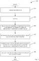
- the prosthetic heart valve preparation and delivery method 100generally includes a plurality of procedures to include tissue preparation at 200, drying at 300, tissue leaflet assembly construction and attachment to frame at 400 to form an implantable prosthetic heart valve, mounting of the prosthetic heart valve (that is, the frame with the tissue leaflet assembly) into a delivery system at 500, ensheathing, sterilizing and packaging the delivery system including the prosthetic heart valve at 600, and finally, delivering the prosthetic heart valve into the patient at 700. Further detail of the prosthetic heart valve preparation and delivery method 100 is provided below.
- At least one or more examples described hereininclude a relatively thin tissue component.
- the tissuehas a thickness of approximately 50 - 150 ⁇ m, and further possesses characteristics of pliability and resistance to calcification after implantation.
- the relatively thin nature of the tissue used in the implantable prosthetic heart valveassists with biocompatibility.
- the relatively thin tissue componentthereby provides for a relatively low mass. As a result, an implantable prosthetic heart valve using the tissue can accelerate to a relatively high heart rate in beats per minute with competent function.
- Tissue suitable for use in the one or more prosthetic heart valves and/or one or more assemblies described hereinis relatively thin and can generally be considered to be a membrane.
- a membranecan generally be considered to be a membrane.
- both natural and synthetic types of materialsmay be used to form a leaflet assembly of a prosthetic heart valves. Accordingly, it is to be understood that although treated pericardium tissue is described as a suitable material for use in the leaflet assembly of a prosthetic heart valve of one or more embodiments described herein, material other than xenograft tissue membrane can be used, and indeed, xenograft tissue membrane other than pericardium tissue can be used. More specifically, synthetic materials may include, but are not limited to, PTFE, PET, Dacron, and nylon.
- xenograft tissue membranemay include, but is not limited to, membrane material from the intestine, lung and brain. Suitable material may also comprise allograft material, that is, material from human sources. The listing of possible materials is for exemplary purposes and shall not be considered limiting.
- pericardium tissuesuch as porcine or bovine pericardium tissue
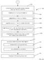
- pericardium tissueis harvested at 204 and then processed to serve as the biocompatible tissue for association with a frame, such as by attaching within a frame.
- the pericardium tissueis cleaned and decellularized at 208. More particularly, in at least one example the tissue is initially cleaned with distilled water using gentle rubbing and hydrodynamic pressure at 208 in order to remove adherent non-pericardial and non-collagenous tissue.
- the hydrodynamic pressure at 208is provided by spraying the tissue with a relatively weak stream of liquid to remove at least some of the non-collagenous material associated with the tissue.
- the rinsing at 208is to achieve effective decellularization of the pericardium tissue through osmotic shock.
- the thickness of the tissue in the cleaned conditionvaries from about 50 to 500 micrometers, depending on the source of raw tissue. Cleaning preferably continues until there is no visible adherent non-pericardial or non-collagenous tissue.
- the tissueafter the tissue has been cleaned and decellularized at 208, the tissue then undergoes optional additional removal of lipids at 220 to further treat the tissue for preventing immunologic response and calcification. More particularly, the tissue first optionally undergoes a 100% glycerol pretreatment at 224 while being positioned on a flat surface (e.g., an acrylic plate), after which the tissue becomes nearly transparent.
- a flat surfacee.g., an acrylic plate
- the tissueoptionally undergoes a "thermophotonic" process.
- the tissueis optionally exposed to light energy for additional removal of lipids and for initial cross-linking of the collagen.
- a 25-100 watt incandescent light sourceand more preferably, a 50 watt incandescent light source with a flat radiant face is employed at a distance of about 10 centimeters from the tissue surface, typically requiring 15 minutes of exposure before further visible separation of lipid droplets from the tissue stops.
- the tissueis then cleaned again in secondary cleaning at 232. More particularly, at 236 the tissue is again rinsed with distilled water. Thereafter, at 240 the tissue is rinsed with 25% isopropyl alcohol for periods of several hours to several days and weeks, depending on the desired tissue properties of pliability and tensile strength.
- tissuehas been successfully prepared by rinsing with 25% isopropyl alcohol for a period of 7 days, and after further treatment steps described herein, provided an ultimate tensile strength of greater than 25 MegaPascals.
- tissue pliability and tensile strengthis sought for purposes of producing a material having property characteristics suitable for being physically manipulated to form a tissue leaflet assembly or other configuration appropriate for attaching with a frame, while providing a tissue material that will operate properly once implanted.
- These techniquesare intended to conserve and preserve collagen fibers, minimizing damage to the tissue and improving tissue characteristics.
- the preparation and fixation techniquesproduce tissue membrane material that may be rendered and used at lesser thickness than typically rendered in the prior art. Thinner membranes are more pliable, but with conventional preparation techniques the tensile strength of the tissue is sacrificed.
- the preparation techniques described hereinhave produced membranes that have as much as three times the tensile strength of a commercial product of the prior art.
- tissue leaflet assemblyhaving a low profile with appropriate durability, even in a substantially dry state.
- the tissuepossesses a relatively high tensile strength.
- embodiments of tissue prepared as described hereinprovide a tissue with a tensile strength of approximately three times the tensile strength of current pericardial valve tissue, such as on the order of approximately 25 MegaPascals, thereby providing about 2000 times the physiologic load strength for valve tissue.
- testing of an embodiment of an implantable prosthetic heart valve made with tissue prepared as described herein and under a static load of greater than approximately 250 mmHgshowed less than approximately 14% leakage, wherein such results are generally considered superior to surgical tissue valve prostheses.
- ethanolmay be used in its place as an alternative, although resulting tissue properties may vary.
- Fig. 9stress-strain curve results for five different tissue samples prepared in accordance with an embodiment are shown.
- the yield stress or ultimate tensile strengthwas obtained by mounting strips of tissue fixed at the ends in a linear force tester and increasing the length by 0.3 mm/sec while recording resultant force (tension) until the material ruptured or separated entirely; these measurements were then used to calculate the stress-strain curves depicted in Fig. 9 .
- the yield stress or ultimate tensile strength of the various tissue samplesvaried from about 30 to about 50 MegaPascals. More particularly, for each curve shown in Fig. 9 , the testing procedures were the same.
- each of the curves shownpertain to separate pieces of tissue that were subjected to the same test.
- the resultsshow a minimum ultimate tensile strength of 30 MegaPascals, with a range up to 50 MegaPascals. Accordingly, the illustrated test results demonstrate consistency of the ultimate tensile strength results for the tissue treatment process.
- the tissueis rinsed with distilled water at 244 as a final cleaning step and for rehydration.
- fixation for collagen cross-linking at 248is achieved by performing at least one of the following:
- step a followed by step bsteps a followed by step c; and step a followed by step d.
- heat-shrink testingmay be conducted on tissue samples to correlate the effectiveness of protein cross-linking.
- results of heat-shrink testing performed on one or more samples of tissue prepared in accordance with at least one embodiment using formalinshowed that the tissue had a shrink temperature of 90°C. This compares favorably with samples prepared using glutaraldehyde, wherein the shrink temperature was 80°C.
- formalinis a suitable variant of fixation. It is noted that formalin was generally abandoned by the field, largely because of material properties that were unfavorable and because of inadequate or unstable protein cross-linking. Such problems have been overcome through the pretreatments described herein, allowing production of tissue with strength, pliability, and durability in a relatively thin membrane.
- tissue characteristics imparted by the tissue preparation processfacilitate formation of a construct having a relatively low-profile, which also thereby facilitates dry packaging of the prosthetic heart valve.
- the same advantagesare also achieved using the pretreatments when using a glutaraldehyde process.
- an alcohol post-fixation treatment at 252is preferably performed by rinsing the tissue in distilled water at 256, and then at 260 rinsing the tissue in 25% isopropyl alcohol for between about 30 minutes to 14 days or more at between about 0 to 37°C, and more preferably, for at least about 7 days at 20°C.
- the tissueundergoes a rinsing with distilled water.
- treatment of the tissuedoes not include contact and/or exposure to a polymer to infiltrate and/or encapsulate tissue fibers of the tissue.
- the drying process at 300is performed after the tissue preparation at 200.
- the tissueis dried under a load. More particularly, for the tissue drying at 304, the tissue is placed minimally stretched flat (that is, stretched just enough to eliminate visible wrinkles and bubbles) on a flat surface (e.g., a polymer or acrylic sheet) at 308, and held fixed at its edges at 312.
- a flat surfacee.g., a polymer or acrylic sheet
- the joined tissue and underlying sheetare then set in a slight curve.
- the tensionmaintains the substantially flat structure of the tissue as it dries, thereby mitigating or preventing excessive shrinkage, wrinkling, and/or curling at the edges, and also making the rate of drying more uniform across the surface of the tissue because of the surface tension between the plate and the tissue.
- the tissueis dried while compressed between acrylic plates.
- the temperatureis held at between about 4 to 37°C, and more preferably, between about 20 to 37°C (i.e., approximately room temperature to normal human body temperature), and more preferably, at about 20°C.
- the drying processis performed in substantially dark conditions (i.e., substantially no visible light) for between about 6 hours to 5 days, and more preferably, for about 72 hours.
- the tissueis dried in dark conditions at a temperature of about 20°C for between about 6 hours to 5 days, and more preferably, for about 72 hours.
- a temperature of about 20°Cfor between about 6 hours to 5 days, and more preferably, for about 72 hours.
- drying the tissue while the tissue is compressed between platesrequires a longer period of time.
- the tissue lotsare inspected at 316, such as by stereomicroscopy, to identify and discard those with defects or discontinuities of the fiber matrix.
- the preferential fiber direction for each pieceis identified to determine the necessary orientation of the free edge of the pieces that will form the valve leaflets.
- the tissuemay be trimmed or otherwise sized in optional sizing at 320, such as by cutting the tissue into an appropriately sized and shaped sheet for valve formation.
- cutting of the tissue membraneis oriented so that the resulting free edge of the leaflet is parallel to the preferential fiber direction of the tissue membrane.
- the free edge of the leafletsmay also be cut with a parabolic or other curved profile to compensate for the downward angle from the commissural leaflet attachment point to the central coaptation point and to increase the total contact surface between the coapting leaflets.
- This approachminimizes focal weaknesses in the operating margins of the leaflet assembly and advantageously distributes the principal loading forces of the operating valve along the long axis of the collagen fibers. As a result, the tissue is resistant to surface fracture and fraying.
- optional sizing at 320is performed after the drying at 304 and inspection at 316.
- tissue generated from one or more of the tissue preparation procedures described hereinmay be used for a variety of devices or uses, and that use in a prosthetic heart valve is but one possible application for utilizing the tissue.
- the tissuemay be used in a shunt, or as graft material for repair or modification of one or more human organs, including the heart and its blood vessels.
- the tissuemay be used as a pericardial membrane patch for repair of congenital heart defects.
- the tissuealso has application as a prosthetic tissue in tendon and ligament replacement, and as a tissue product for wound management.
- the tissuemay be configured in a variety of ways and attached to a frame in a variety of ways.
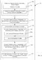
- the prepared tissueis formed into a tissue leaflet assembly at 404 by folding the tissue at 408, preferably while the tissue is in a dry state, to form at least a portion of the tissue leaflet assembly.
- a completed tissue leaflet assemblymay be formed of a single monolithic piece of tissue 800, such as that shown in Fig. 8A , or alternatively, as shown in Figs.
- the seams 804are preferably situated at overlapping portions of pleats 832 of the plurality of tissue pieces 802.
- a single monolithic piece of tissue 800 or a plurality of tissue pieces 802may be used to form a prosthetic heart valve, wherein the tissue leaflet assembly is not a folded construct.
- a plurality of separate tissue piecesmay each be attached to a frame (such as by suturing) to form a prosthetic heart valve. Thereafter, whether the prosthetic heart valve is made of a folded tissue leaflet assembly or a plurality of separate tissue pieces attached to a frame, the resulting prosthetic heart valve may then be further manipulated for delivery as a dry prosthetic heart valve.
- tissue generated from one or more of the tissue preparation procedures described hereinmay be used to form a prosthetic heart valve that includes a frame, and that may be implanted by a "trans-apical" approach in which the prosthetic heart valve is surgically inserted through the chest wall and the apex of the heart.
- tissue generated from one or more of the tissue preparation procedures described hereinmay be used to form a prosthetic heart valve that does not include a frame, and is not delivered via a catheter, but rather, is implanted via a surgical opening through the patient's chest.
- the prosthetic heart valvemay be packaged for delivery as a dry prosthetic heart valve.
- tissue generated from one or more of the tissue preparation procedures described hereinmay be used to form a prosthetic heart valve that includes a frame, but that is not delivered via a catheter, but rather, is implanted via a surgical opening through the patient's chest.
- the prosthetic heart valvemay be packaged for delivery as a dry prosthetic heart valve.
- tissuemay be implanted in a "wet" or hydrated state.
- a prosthetic heart valve utilizing a prepared tissue described hereinmay be packaged for delivery as a hydrated prosthetic heart valve.
- the tissue preparation processmay include drying the tissue so that it may be manipulated more easily, the tissue may then be hydrated at a later point in time prior to implantation, and it may be maintained in a hydrated condition up to and including packaging, delivery and implantation into a patient.
- Advantages associated with using a folded tissue leaflet assemblyinclude that a folded structure allows a relatively thin membrane to be used by avoiding suture lines in loaded, dynamically active surfaces. Accordingly, a sutureless leaflet assembly preserves long-term integrity.
- a prosthetic heart valve that does not include a folded tissue leaflet assemblyis encompassed by one or more embodiments described herein.
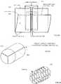
- a tissue blank 808is shown in Fig. 8D , wherein the tissue blank 808 is a single monolithic piece of tissue 800.
- a line of primary fold or fold line 812is visualized for the tissue blank 808.
- Fig. 8Da tissue blank 808 is a single monolithic piece of tissue 800.
- the primary fold 814is achieved along the fold line 812 by folding the bottom edge 816 of the tissue blank 808 toward the top edge 820, but leaving a cuff portion 824 along the upper portion 828 of the tissue blank 808.
- the direction of top and bottomare relative to each other and are used as a convenience for describing the folding sequence, wherein such directions correspond to the orientation of the page illustrating the drawings.
- the folding geometry of Figs. 8D-8Lforms cuffs 824 that are continuous with the leaflets, thereby reducing the risk of aortic insufficiency or leakage.
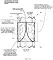
- FIG. 8FAfter folding the tissue blank 808 along fold line 812 to form primary fold 814, pleats are formed by folding the tissue along its length. For the embodiment shown in Fig. 8F , three pleats 832a, 832b, and 832c are shown.
- Fig 8Gillustrates a detail drawing of a single pleat 832 representative of one of pleats 832a-c. In Fig. 8G , the inner leaflet layer free edge 836 is shown, as is the valve sinus 840 and the commissure folds 844.
- Fig. 8Hshows a schematic perspective drawing of tissue leaflet assembly 848, wherein the pleated tissue construct shown in the bottom half of Fig. 8F is seamed, such as along seam 850, to form a substantially tubular construct.
- the folded tissue leaflet assembly 848is maintained dry or is partially hydrated prior to mounting the tissue leaflet assembly in a frame.
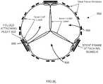
- the tissue leaflet assembly 848is then attached within a frame, such as frame 852 shown in Fig. 8I .
- the tissue leaflet assembly 848 attached within a frame 852forms an implantable prosthetic heart valve 860, such as that shown in the schematic perspective drawing of Fig. 8J , side elevation view Fig. 8K , as well as that shown in the photo of Fig. 11A , and drawing of Fig. 11B .
- Fig. 8Killustrates possible suture points 864 where the tissue leaflet assembly 848 can be sutured to the frame 852. That is, the tissue leaflet assembly 848 may be attached within the frame 852, such as by suturing the outer layer of the tissue leaflet assembly 848 to the frame.
- the term "attached”means that the tissue leaflet assembly 848 is secured to the frame 852, although the inner leaflet layer free edges 836 are able to readily move during operation of the prosthetic heart valve 860.
- a cutaway side elevation view of a prosthetic heart valve 860that includes a frame 852 with a tissue leaflet assembly 848 attached therein is shown.
- the tissue membrane leaflet assembly 848is disposed coaxially within the frame 852.
- the valve 860is illustrated in the closed position with the leaflet free edges 836 in at least partial contact with each other.
- An arc 1112 of the leaflet free edges 836(out of plane of the cutaway view) is continuous with pleats 832 at the radial edge of the tissue leaflet assembly 848, and may be seen in the alternate view shown in Fig. 8L .
- the tissue membrane leaflet assembly 848is attached to the frame 852 along the axially oriented membrane pleats 832, as illustrated again in Fig. 8L .
- the extended cuff layeris attached circumferentially at the distal edge 1104 of the frame 852.
- continuous suture attachment 1108may be used to attach the extended cuff layer to the distal edge 1104.
- a distal portion 1116 of the frame 852does not include any portion of the tissue leaflet assembly 848, such as the cuff layer.
- the leaflet free edges 836still at least partially contact each other.
- FIG. 8Lan end view of the prosthetic heart valve is shown.
- the pleats 832are used as the portion of the tissue leaflet assembly 848 to attach to the frame 852.
- the outer cuff layeris attached to the frame members of frame 852.
- the cusps 868 formed by the inner leaflet layerare generally situated as depicted in Fig. 8L .
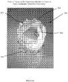
- Fig. 12is a photo of the tissue leaflets of a prosthetic heart valve after 30,000,000 cycles of testing to model performance if associated with a human heart. In testing, the prosthetic heart valve 860 has demonstrated a natural opening gradient of approximately 5 mmHg.
- tissue leaflet assembly 848 described and shown hereinis but one possible construct for forming a flow control mechanism that can be attached to a frame to regulate the flow of blood in a patient's vascular system upon deployment. That is, the illustrated tissue leaflet assembly 848 is provided by way of example and not limitation, and in no way should be interpreted to limit the geometries of membrane leaflet assemblies that can be used to regulate fluid flow. Accordingly, other leaflet configurations and constructs are considered encompassed by claims directed to or otherwise including premounted percutaneously deliverable valves.
- the frame 852may be a stent or a structure having similarities to a stent.
- the frame 852essentially serves as a holding mechanism for the tissue leaflet assembly 848 that can then be inserted percutaneously into a patient, wherein the frame 852 serves as a way to anchor the folded tissue leaflet assembly 848 to a vascular portion (e.g., in situ arterial tissue) of the patient.
- a vascular portione.g., in situ arterial tissue
- the tissue leaflet assembly 848is inserted into a frame 852.
- the frame 852may comprise a balloon-expandable frame, or alternatively, at 424b a self-expanding frame may be used.
- the folded tissue leaflet assembly 848is attached to the frame 852, such as by suturing the tissue leaflet assembly 848 to the frame 852 to form an implantable prosthetic heart valve 860, such as that shown in Fig. 8L .
- the prosthetic heart valve 860is fully hydrated for inspection and testing. Thereafter, the fully constructed implantable prosthetic heart valve 860 may be dried and maintained in a substantially dry condition.
- tissue 800suitable for implanting in a human, wherein the implantable tissue may be allowed to dry prior to implanting, or it may be hydrated prior to implanting.
- tissue 800is suitable for use in forming a tissue leaflet assembly 848 for use in a prosthetic heart valve, including an implantable prosthetic heart valve 860 that can be implanted with its tissue leaflet assembly in a dry state, or with its tissue leaflet assembly in a partially or fully hydrated state.
- One or more of the examples of the tissue leaflet assemblies described hereinmay be implanted into the patient using a balloon-expandable frame or a self-expanding frame.
- Expandable framesare generally conveyed to the site of the target valve on balloon catheters.
- the expandable frameis positioned in a compressed configuration along the delivery device, for example crimped onto the balloon of a balloon catheter that is part of the delivery device intended for coaxial mounting on a guidewire.
- the expandable frameis expanded by the delivery device.
- a self-expanding framecommonly a sheath is retracted, allowing expansion of the self-expanding frame.
- the framecomprises a metal alloy frame possessing a high strain design tolerance that is compressible to a relatively small diameter.
- the implantable prosthetic heart valve 860allows standard retrograde arterial aortic delivery via femoral artery insertion, without surgical cutdown or general anesthesia. This is achieved by providing the prosthetic heart valve on a premounted delivery system with the tissue leaflet assembly or tissue membrane construct in a substantially dry condition.
- a dry tissue membranehas substantially less mass than a wet membrane.
- a substantially dry pericardium tissue prepared by one or more of the present embodimentshas approximately 30% of the mass of a wet pericardium tissue, and marked reduction in profile and packing volume, thereby achieving a relatively low profile and making it suitable for implantation in greater number of patients, especially those having small diameter vascular systems.
- a dry prosthetic heart valvedoes not require storage and transport in preservative.
- a dry prosthetic heart valvecan be mounted on a delivery catheter at its location of manufacture, which allows for pre-packaging of an integrated delivery system.
- the term "mounted"means that the prosthetic heart valve 860 is temporarily associated with the delivery catheter. Together with a relatively low profile, embodiments of the prosthetic heart valve thereby offer reliability and convenience because the implantable prosthetic heart valve 860 is pre-mounted upon its delivery catheter and forms part of a pre-packaged delivery system.
- a dry prosthetic heart valvedoes not require rinsing, rehydration, or mounting in a catheterization lab. Therefore, a dry prosthetic heart valve can be inserted directly from package into the patient's body at a critical time during the procedure. Advantageously, this avoids procedure time, manipulation, and errors of mounting, crimping, and orienting catheters and sheaths.
- the dry prosthetic heart valveis inserted and delivered by balloon catheter expansion in the plane of the target valve in the standard way and the dry prosthetic heart valve begins to function immediately, even without specific steps to rehydrate the tissue membrane portion of the heart valve from its dry state, with hydration of the tissue membrane subsequently occurring rapidly and naturally in the body. More particularly, hydration of the tissue membrane portion occurs rapidly and begins with simple preparatory flushing of catheter lumens with saline. Thereafter, hydration continues with device insertion and dwelling into the central blood vessels, and completes naturally after deployment in the patient's body.
- the low profile of the implantable prosthetic valveis particularly advantageous for patient's having relatively small diameter vascular systems.
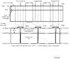
- Table 1provides aortic and pulmonary valve prosthesis sizing.
- Table 1Aortic and Pulmonary Valve Prosthesis Sizing Aorta/Pulmonary Valve Diameter Collapsed Implantable Prosthetic Heart Valve Size (French) Collapsed Implantable Prosthetic Heart Valve Diameter 19 - 21 mm 12 French 4.0 mm 22 - 26 mm 14 French 4.7 mm 27 - 30 mm 16 French 5.3 mm
- the femoral arteryhas a diameter of between about 5-8 mm.
- embodiments of the collapsed implantable prosthetic heart valves 860 described hereinoffer a low profile that enables a larger group of patients to qualify for receiving an implantable prosthetic heart valve 860.
- implantable prosthetic heart valves 860 described hereinvirtually no candidate patients would be excluded from treatment with an implantable prosthetic heart valve 860 without open heart surgery and without general anesthesia on the basis of inadequate femoral blood vessel access caliber.
- implantable prosthetic heart valve 860 described hereinfeature a scalable construct, wherein the implantable prosthetic heart valves 860 can be produced to accommodate target valve diameters ranging between 6 - 35 mm, and wherein the implantable prosthetic heart valves 860 offer consistent function using fundamentally a single design.
- an implantable prosthetic heart valve 860(also referred to herein as a percutaneously deliverable heart valve) is collapsed.
- the initial phase of collapsing the percutaneously deliverable heart valveis executed with the tissue membrane in a hydrated condition. That is, since the percutaneously deliverable heart valve 860 includes the frame 852 with the tissue leaflet assembly 848 attached within the frame 852, the percutaneously deliverable heart valve 860 is collapsed down as an integral unit.
- an axial pullermay be utilized to collapse down the frame 852 of the percutaneously deliverable heart valve 860 without the application of force directly to the sides of the frame 852.
- This procedureoffers the advantage of preserving the cell structure of the frame 852 while also maintaining the orientation of the leaflets of the tissue leaflet assembly 848 as the percutaneously deliverable heart valve 860 is compressed. The proper orientation and disposition of the leaflets is facilitated by the hydrated state of the leaflets. This assists in preventing tissue prolapse or bulging of the tissue 800 or 802 through the frame 852.
- this techniquereduces recompression strain on the metal frame 852 (e.g., a stent) that can tend to compromise fatigue life of the frame 852.
- This techniquealso tends to promote the circumferentially uniform collapsing of cells in the frame 852, thereby mitigating bunching of the tissue that forms the tissue leaflet assembly 848 of the percutaneously deliverable heart valve 860.
- the sidesare forced to collapse by providing a radial compression force to the frame and may be assisted by axial traction force.
- the percutaneously deliverable heart valve 860(i.e., the frame 852 with the tissue leaflet assembly 848 attached thereto) is collapsed in an initially hydrated state.
- the delivery mandrel or balloonis inserted into a delivery sheath, and the mounting segment is then extended out the end of the sheath.
- the sheath and frameare coaxially mounted and then compressed with initial crimping onto the mounting segment with the tissue leaflet assembly 848 still in a hydrated state.
- the tissue leaflet assembly 848 of the percutaneously deliverable heart valve 860is then allowed to dry, which further reduces the volume and profile of the tissue membrane leaflets, permitting further compression by radial force.
- the percutaneously deliverable heart valve 860is then further crimped with a circumferential crimping tool at 520 to finally mount the compressed valve/frame onto the delivery mandrel or balloon catheter.
- Fig. 6the ensheathing, sterilization and packaging at 600 is described. More particularly, once the percutaneously deliverable heart valve 860 is coaxially mounted and crimped on a delivery mandrel or balloon catheter as described above and shown in Fig. 5 , the assembly is then inserted at 604 into a distal end of a delivery sheath, such as by "backloading" the assembly into position with a distal end of the percutaneously deliverable heart valve 860 contained within the delivery sheath proximate the end of the sheath.
- Fig. 10schematically illustrates catheter 1000 with an implantable prosthetic heart valve 860 mounted thereto.
- the percutaneously deliverable heart valve 860 and delivery cathetersare sterilized, such as by using by one or more of ethylene oxide, proton beam, or gamma radiation.
- the assemblyis then optionally packaged in a sterile package. Additional elements are optionally shipped with the assembly, wherein, by way of example, such elements may include any necessary delivery tools and documentation.
- the packagemay optionally contain a device to control the water vapor content within the sealed volume of the package.
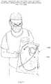
- Fig. 13depicts a surgeon holding a sterile package 1300 containing a premounted percutaneously implantable prosthetic heart valve.
- a flow chart illustrating the general procedure associated with implantation of the percutaneously deliverable heart valve 860is provided. More particularly, at 704, catheter access is gained to the patient's femoral artery and a guidewire is placed through the plane of the diseased valve that is targeted to receive the implant.
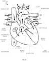
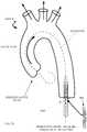
- Fig. 14is a schematic of a simplified cutaway view of a human heart, including heart valves that may be targeted for receiving an embodiment of an implantable prosthetic heart valve.
- Fig. 15illustrates the aorta with the guidewire placed through the diseased aortic valve.
- the percutaneously deliverable heart valve 860 in the form of a prepackaged assembled dry prosthetic heart valveis removed from the sterile packaging.
- the dry prosthetic heart valve assemblyincluding its lumens, are preferably flushed and prepared in the usual fashion for standard balloons and catheters that do not contain a biocompatible tissue.
- implantation of the dry prosthetic heart valve assemblycan be conducted without specific maneuvers for rehydration of the tissue leaflet assembly 848 of the percutaneously deliverable heart valve 860. Some rehydration of the tissue leaflets may occur as a consequence of the routine flushing of the catheter lumens in preparation for use as with any other catheters.
- implantation of the dry prosthetic heart valve assemblycan proceed without additional cleaning steps, such as by having to use alcohol or water rinsing solutions.
- the percutaneously deliverable heart valve 860can essentially be implanted percutaneously in its dry state.
- the carrier catheter or balloon catheteris then coaxially mounted and advanced over the guidewire, such as under fluoroscopic vision initially to the level of the great vessel where it can be inspected under fluoroscopy.
- the delivery systemis advanced through the plane of the diseased valve under fluoroscopy, and the covering sheath is withdrawn, either at this point or during the advance prior to it, thus exposing the mounted implantable prosthetic heart valve 860 in place.
- the balloonis then inflated, deploying the percutaneously deliverable heart valve 860 in the plane of the valve.
- the leaflets of the percutaneously deliverable heart valve 860operate immediately.
- the deployed prosthetic heart valve 860is shown in Fig. 16 , wherein the tissue leaflet assembly 848 serves to properly control the flow blood.
- the one or more present inventionsinclude components, methods, processes, systems and/or apparatus substantially as depicted and described herein, including various embodiments, subcombinations, and subsets thereof. Those of skill in the art will understand how to make and use the present invention after understanding the present disclosure.
- the present inventionin various embodiments, includes providing devices and processes in the absence of items not depicted and/or described herein or in various embodiments hereof, including in the absence of such items as may have been used in previous devices or processes (e.g., for improving performance, achieving ease and/or reducing cost of implementation).
Landscapes
- Health & Medical Sciences (AREA)
- Cardiology (AREA)
- Engineering & Computer Science (AREA)
- Biomedical Technology (AREA)
- Animal Behavior & Ethology (AREA)
- General Health & Medical Sciences (AREA)
- Transplantation (AREA)
- Heart & Thoracic Surgery (AREA)
- Vascular Medicine (AREA)
- Life Sciences & Earth Sciences (AREA)
- Veterinary Medicine (AREA)
- Oral & Maxillofacial Surgery (AREA)
- Public Health (AREA)
- Manufacturing & Machinery (AREA)
- Mechanical Engineering (AREA)
- Prostheses (AREA)
- Materials For Medical Uses (AREA)
- Media Introduction/Drainage Providing Device (AREA)
- Packages (AREA)
- Apparatus For Disinfection Or Sterilisation (AREA)
Description
- The present invention relates to the field of medical devices, and more particularly, to a percutaneously deliverable heart valve and a method of making a percutaneously deliverable heart valve.
- Heart valve disease is a common degenerative condition that compromises physiologic function and causes limiting symptoms and threat to life in millions of patients all over the world. There are various underlying causes, but malfunction of heart valves is ultimately expressed as insufficient conduction of blood through the plane of the valve due to narrowing of the anatomic pathway (stenosis), or as incompetent closure that allows blood to return back through the valve again, thereby reducing the effective forward conduction of blood through the valve (insufficiency or regurgitation). These hemodynamic states lead to 1) deficiency of cardiac output and 2) adverse loads on the pumping chambers of the heart, both of which in turn lead to functional compromise of the patient and often premature death unless effectively corrected.
- Definitive corrective treatment of heart valve disease is conventionally performed by open-chest surgical techniques, wherein the valve is manipulated, repaired, or replaced with a prosthetic valve under direct vision. Heart valve surgery is performed in hundreds of thousands of cases yearly world-wide, but carries a high burden of cost, morbidity, and mortality, especially in susceptible patients who may be elderly or otherwise physiologically compromised by collateral disease. Further, the costs and resource requirements of the surgical enterprise restrict the availability of heart valve replacement to many more patients all over the world.
- In pursuit of alternatives to heart valve surgery, over the last ten years a number of development programs have brought percutaneous, trans-catheter implantation of prosthetic heart valves into commercial use in the European Union (EU) and into pivotal clinical trials in the United States of America. Initial clinical experience in the EU was directed toward patients who had critical aortic valve stenosis, but were deemed to be at unacceptably high risk for open-heart surgical valve replacement. In several thousand such cases, utilizing both balloon-expandable and self-expanding designs in two separate programs, percutaneous heart valve replacement (PHVR) was shown to be feasible and possibly competitive with surgery in selected patients with 12-18 month mortality rates of about 25%.Grube E., et al., Progress and Current Status of Percutaneous Aortic Valve Replacement: Results of Three Device Generations of the CoreValve Revolving System, Circ. Cardiovasc Intervent. 2008;1:167-175.
- The application of PHVR thus far has been challenged by the technical difficulties of the implantation sequence-especially in the aortic valve position. The technique for available devices is limited by the large caliber of the devices and their delivery catheters; often, if it can be done at all in some smaller arteries, open surgical exposure and management of the femoral artery is required to insert the 18 - 24 French (6 - 8 mm diameter) systems, and their bulkiness inside the central arteries can threaten the safety of the delivery sequence. Further, access site bleeding complications form a significant part of the adverse events of the procedures.
- Typically, the current PHV designs comprise a biological membrane forming the operating leaflets of the valve, attached within a metal frame, that is then collapsed onto a delivery catheter or balloon, and then constrained within an outer sheath. After an initial dilation of the diseased valve with a large balloon, this assembly is then advanced to the plane of the valve and deployed by self-expansion or by balloon expansion.
- The effective caliber of the valve delivery system is determined by the total bulk of each coaxially mounted component. The bulk of the PHV itself is determined by the diameter of the frame and by the thickness, stiffness, and particular arrangement of the inner membrane forming the operating leaflets of the valve. The characteristic thickness of current PHV membranes is thus a limiting factor in the ultimate delivery profile of the PHV. Such characteristic membrane thickness is, in turn, a result of the methods by which it is processed and ultimately delivered for use. Typically, glutaraldehyde fixation (for protein cross-linking) of animal tissue is employed to produce suitable biological membranes for incorporation. Requirements for strength and durability have determined the most useful ranges for tissue thickness and cross-linking while typically imposing countervailing stiffness and brittleness. Subsequent hydration in suitable solutions improves these characteristics, but the hydrated membrane by this means also gains thickness.
- One of the evident requirements for a PHV design is that the valve functions with a high degree of competence immediately on deployment, since the patient's hemodynamic survival depends on it. To this end, in part, like surgical valve prostheses, current PHV designs are completed, transported, and delivered for use in a hydrated state in a jar of solution. In use, commercially available surgical and percutaneously implanted bioprosthetic heart valves are rinsed and prepared before use in a "wet" state. More particularly, commercially available prosthetic heart valves are rinsed, crimped, and mounted in the catheterization lab. Accordingly, problems with current commercially available prosthetic heart valves include the time, cost and variability associated with the necessity to rinse, crimp, and mount the valve in the catheterization lab. That is, current mounting of prosthetic heart valves in the catheterization lab imposes one or more of delay, cost, technical burdens and possible errors. Avoiding one or more of these problems would be advantageous. In addition, current "wet" valve designs impose additional profile on the collapsed valve. The hydrated membrane, while having desirable and necessary flexibility for reliable operation immediately on deployment, also imposes a large part of the thickness of the assembled and mounted valve that compromises its deliverability.
US 2008/177381 discloses a method that includes preparing a substantially dehydrated bioprosthetic valve and then providing an expandable support member having oppositely disposed first and second ends and a main body portion extending between the ends. Next, the substantially dehydrated bioprosthetic valve is attached to the expandable support member so that the substantially dehydrated bioprosthetic valve is operably secured within the main body portion of the expandable support member. The expandable support member is then crimped into a compressed configuration.- Expanding on some of the problems described above, the use of current PHVs in the catheter lab requires a number of preparatory acts that are potentially troublesome and can prolong the delivery sequence during a critical phase of the procedure. Since PHVs are delivered for use "wet" in a preservative solution, they have to be treated prior to insertion with a series of cleansing and hydrating solutions. Once this is completed, the PHVs have to be mounted on their delivery catheters. Special crimping and mounting tools are needed in the case of the balloon-expandable Edwards Sapien valve, for example. Accordingly, there is a need to address the shortcomings discussed above.
- It is an object of the present invention to provide an improved method for providing a prosthetic heart valve.
- This object is solved by a method according to claim 1. The subclaim comprises a preferred embodiment of the invention.
- It is to be understood that the present invention includes a variety of different versions or embodiments, and this Summary is not meant to be limiting or all-inclusive. This Summary provides some general descriptions of some of the embodiments, but may also include some more specific descriptions of other embodiments.
- In at least one example, a substantially "dry" membrane PHV system is provided wherein a tissue material is prepared and folded in a dry state to form a tissue leaflet assembly. Thereafter, the tissue leaflet assembly is attached to a frame to form an implantable prosthetic heart valve that is subsequently pre-mounted in an integrated catheter delivery system. The catheter delivery system that includes the prosthetic heart valve is then packaged and transported while the tissue leaflet assembly remains substantially dry. The prosthetic heart valve is available for use directly out of its package envelope. Accordingly, it can be inserted into the body without need of hydration, crimping or mounting tools, or other preparatory acts. That is, the tissue forming the tissue leaflet assembly of the prosthetic heart valve can be treated and dried, then while remaining dry, folded into a tissue leaflet assembly. Thereafter, the tissue leaflet assembly is at least partially rehydrated and then attached within a frame, such as a stent, to form an implantable prosthetic heart valve. The tissue leaflet assembly of the prosthetic heart valve is then allowed to dry. The prosthetic heart valve can thereafter be subsequently packaged, delivered, and shipped while the tissue leaflet assembly of the prosthetic heart valve remains in a dry condition. The prosthetic heart valve can then be implanted into the receiving patient. Accordingly, the PHV system simplifies arterial insertion, and, as the dry condition also confers lower bulk and profile, procedural manipulation and associated complications may be reduced if not eliminated. In addition, one or more embodiments of the present invention widen the candidacy of patients with smaller arteries for the PHV procedure. As an added advantage, at least one embodiment of the present invention allows the implantation to take place under shorten elapsed times at the most critical phase of the procedure.
- In at least one example, a membrane PHV system is provided wherein a tissue material is prepared and folded in a dry state to form a tissue leaflet assembly, and further wherein the tissue leaflet assembly is thereafter at least partially hydrated and attached to a frame that is subsequently pre-mounted in an integrated catheter delivery system.
- In at least one example, a membrane PHV system is provided wherein a tissue material is prepared and folded in a dry state to form a tissue leaflet assembly, and further wherein the tissue leaflet assembly is at least partially hydrated and attached to a frame to form the prosthetic heart valve. Thereafter, the prosthetic heart valve is allowed to dry and subsequently pre-mounted in an integrated catheter delivery system after which the tissue leaflet assembly of the prosthetic heart valve remains dry, and wherein the system is then associated with a package for shipment while the tissue leaflet assembly remains dry.
- In at least one example, a membrane PHV system is provided wherein a tissue material is prepared and then folded in a dry state to form a tissue leaflet assembly, and further wherein the tissue leaflet assembly is at least partially hydrated and attached to a frame to form the prosthetic heart valve. Thereafter, the prosthetic heart valve is allowed to dry and subsequently pre-mounted in an integrated catheter delivery system after which the tissue leaflet assembly of the prosthetic heart valve is then at least partially hydrated and associated with a package for shipment.
- In at least one example, an article adapted for trans-catheter delivery into a patient is provided, comprising: a prosthetic heart valve further comprising a treated tissue attached to a frame, wherein the treated tissue comprises a thickness of about 50 to 500 micrometers and an ultimate tensile strength of greater than about 15 MegaPascals when at a water content of less than about 50% by weight of the section of treated tissue. Here it is noted that the tensile strength of the treated tissue described herein is higher than the tensile strength of other known prepared tissues, whether hydrated or dry. In at least one embodiment, the water content of the treated tissue is less than about 40% by weight of the treated tissue. In at least one example, the ultimate tensile strength is greater than about 20 MegaPascals. In at least one example, the treated tissue does not include a matrix that has been exposed to a polymer infiltrate. In at least one embodiment the treated tissue comprises a treated pericardium tissue.
- In at least one example, the method further comprises exposing the section of tissue to light energy for an exposure duration, the exposure duration extending until there is no further visible separation of lipid droplets from an exposed surface of the section of tissue. In at least one example, the light energy is at least equivalent to exposing the section of tissue to a 25-100 watt light source, and more preferably, a 50 watt incandescent light source with a flat radiant face situated at a distance of about 10 centimeters from the exposed surface for about 15 minutes. In at least one embodiment, the method further comprises: (d) rinsing the section of tissue with distilled water and isopropyl alcohol for a post-fixation period of time of not less than about 7 days; wherein step (d) occurs after step (c).
- In at least one example, an article adapted for implantation in a patient is provided, comprising: a prosthetic heart valve further comprising a treated tissue attached to a frame, wherein the treated tissue comprises a water content of less than about 60% by weight of the treated tissue. In at least one embodiment, the treated tissue comprises a section of pericardium tissue having an ultimate tensile strength of greater than about 12 MegaPascals. In at least one example, the section of treated tissue comprises a thickness of between about 50 to 300 micrometers. In at least one embodiment, the water content of the treated tissue is less than about 40% by weight of the treated tissue.
- As used herein, the term "dry" (or "substantially dry") when referring to the state of the tissue that forms the heart valve of the percutaneous heart valve means a moisture content less than the water moisture content of the tissue when the tissue is allowed to fully rehydrate in the body of a patient. Typically, pericardium tissue treated in accordance with one or more embodiments described herein is about 70% by weight water when fully hydrated. Drying to a constitution of less than 40% by weight of water usefully alters the handling properties for purposes of folding and sewing the tissue. As those skilled in the art will appreciate, the moisture content of the tissue may vary when dry. For example, the moisture content of the tissue when being folded and dry may be different than the moisture content of the tissue when dry and being shipped in a premounted state within a catheter delivery system.
- Advantageously, at least one example of the one or more present inventions is directed to a prosthetic heart valve that is mounted onto a valve delivery system and stored in a sterile package. Accordingly, in at least one example, an assembly is provided, comprising:
- a prosthetic heart valve including:
- a frame; and
- a tissue leaflet assembly attached to the frame;
- a percutaneously insertable valve delivery mechanism, wherein the prosthetic heart valve is releasably mounted onto the percutaneously insertable valve delivery mechanism; and
- sterile packaging containing the prosthetic heart valve releasably mounted onto the percutaneously insertable valve delivery mechanism.
- In at least one example, the percutaneously insertable valve delivery mechanism comprises a balloon catheter. In at least one example, the balloon catheter is a 12 to 14 French balloon catheter. In at least one example, the balloon catheter is less than about 12 French. In at least one example, the balloon catheter is between about 5 to 12 French. In at least one example, the percutaneously insertable valve delivery mechanism comprises a mandrel. In at least one example, tissue forming the tissue leaflet assembly within the sterile packaging is at least one of hydrated and not substantially dry. In at least one embodiment, tissue forming the tissue leaflet assembly within the sterile packaging is substantially dry. In at least one example, the frame comprises a stent. In at least one embodiment, tissue forming the tissue leaflet assembly comprises treated pericardium tissue.
- At least one example of the one or more present inventions includes a prosthetic heart valve for implantation in a patient. Accordingly, a pre-packaged percutaneous, trans-catheter deliverable prosthetic heart valve ready for implantation in a patient is provided, comprising:
- a frame; and,
- a tissue leaflet assembly attached to the frame, the tissue leaflet assembly comprising a substantially dry tissue.
- In at least one embodiment, the substantially dry tissue comprises treated pericardium tissue. In at least one example, the frame and tissue leaflet assembly attached thereto are operably associated with a 12 to 14 French balloon catheter. In at least one example, the frame and tissue leaflet assembly attached thereto are operably associated with a balloon catheter having a size of less than about 12 French. In at least one example, the frame and tissue leaflet assembly attached thereto are operably associated with a balloon catheter having a size of between about 5 to 12 French. In at least one embodiment, the substantially dry tissue comprises a water moisture content of less than about 40% by weight of the substantially dry tissue.
- In at least another example, an assembly for use with a patient is provided, comprising:
- a sealed sterile package containing a delivery system for percutaneously deploying a heart valve in the patient, the heart valve including:
- a frame releasably mounted on the delivery system within the sealed sterile package; and
- a tissue leaflet assembly attached to the frame.
- In at least one embodiment, the tissue leaflet assembly comprises pericardium tissue.
- In at least one embodiment, a method is provided, comprising:
- partially compressing and mounting a prosthetic heart valve upon a delivery catheter, the prosthetic heart valve comprising a tissue;
- allowing the tissue to at least partially dry to less than 40% by weight of water;
- further compressing and mounting the prosthetic heart valve upon the delivery catheter; and
- sterilizing and packaging the prosthetic heart valve and delivery catheter.
- In at least one example, the method further comprises transporting the sterilized and packaged prosthetic heart valve and delivery catheter. In at least one embodiment, the tissue comprises treated pericardium tissue. In at least one embodiment, prior to partially compressing and mounting the prosthetic heart valve upon the delivery catheter, the tissue is at least one of (a) not substantially dry, and (b) at least partially hydrated.
- For the various examples described herein, the prosthetic heart valve, including the tissue leaflet assembly, comprises membrane tissue other than pericardium tissue.
- In at least one example, a method is provided, comprising:
- attaching pericardium tissue to a frame;
- partially compressing and mounting the frame, with the tissue attached thereto, upon a delivery catheter;
- allowing the tissue to at least partially dry;
- further compressing and mounting the frame, with the tissue attached thereto, upon the delivery catheter; and
- sterilizing and packaging the frame and delivery catheter, with the tissue attached thereto.
- In at least one example, prior to partially compressing and mounting the frame, the tissue is at least one of (a) not substantially dry, and (b) at least partially hydrated. In at least one embodiment, the method further comprises transporting the sterilized and packaged frame, with the tissue attached thereto, mounted upon the delivery catheter, to a surgical or medical procedure facility. In at least one example, prior to attaching the tissue to the frame the tissue is folded to form a tissue leaflet assembly. In at least one embodiment, the tissue leaflet assembly comprises at least one cuff and at least one pleat.
- In at least one example, a method of preparing a percutaneous, trans-catheter prosthetic heart valve is provided, the method comprising:
- providing a membrane tissue from an organism;
- treating the membrane tissue with at least one chemical to produce a treated membrane tissue;
- drying the treated membrane tissue until it is a substantially dry tissue;
- attaching the substantially dry tissue in a frame;
- rehydrating the substantially dry tissue that is attached within the frame to form a rehydrated tissue;
- collapsing the frame with the rehydrated tissue attached thereto; and
- drying the rehydrated tissue within the collapsed frame until it is a substantially dry tissue.
- In at least one example the method further comprises compressing and mounting the frame, with the substantially dry tissue attached thereto, upon a delivery catheter. In at least one embodiment the method further comprises sterilizing and packaging the frame, with the substantially dry tissue attached thereto, mounted upon the delivery catheter. In at least one embodiment, the treating comprises sterilizing the frame with the substantially dry tissue attached thereto with exposure to at least one of ethylene oxide, a proton beam, and gamma radiation. In at least one example, the method further comprises shipping the sterilized and packaged frame with the substantially dry tissue attached thereto, mounted upon the delivery catheter, to a surgery or medical procedure facility. In at least one example, prior to the attaching step the dry tissue is not folded to provide a cuff and/or a pleat. In at least one example, prior to the attaching step the dry tissue is folded to form a tissue leaflet assembly. In at least one example, the tissue leaflet assembly comprises at least one cuff and at least one pleat.
- In at least one example, the method of preparing a percutaneous, trans-catheter prosthetic heart valve further comprises implanting the frame with the substantially dry tissue attached thereto into a patient. In at least one example, the frame comprises a stent. In at least one example, the method further comprises mounting the frame and the tissue leaflet assembly attached thereto upon a 12 to 14 French balloon catheter. In at least one example, the method further comprises mounting the frame and the tissue leaflet assembly attached thereto upon a balloon catheter having a size of less than about 12 French. In at least one example, the method further comprises mounting the frame and the tissue leaflet assembly attached thereto upon a balloon catheter having a size of between about 5 to 12 French. In at least one example, the method further comprises mounting the frame and the tissue leaflet assembly attached thereto on a mandrel. In at least one example, the method of preparing a percutaneous, trans-catheter prosthetic heart valve further comprises immersion of the membrane tissue in buffered or unbuffered 1-37.5% formalin for between about 3 days to 3 weeks. In at least one example, the method of preparing a percutaneous, trans-catheter prosthetic heart valve further comprises immersion of the membrane tissue in buffered or unbuffered 1-37.5% formalin for between about 3 days to 5 weeks. In at least one example the treating comprises immersion of the membrane tissue in 100% glycerol for greater than 3 weeks. In at least one example the treating comprises immersion of the membrane tissue in 0.1 - 25% glutaraldehyde for between about 3 days to 3 weeks. In at least one example the treating comprises immersion of the membrane tissue in 0.1 - 25% glutaraldehyde for between about 3 days to 5 weeks. In at least one example the treating comprises immersion of the membrane tissue in oligomeric filtered 0.1 - 25% glutaraldehyde for between about 3 days to 3 weeks. In at least one example the treating comprises immersion of the membrane tissue in oligomeric filtered 0.1 - 25% glutaraldehyde for between about 3 days to 5 weeks. In at least one example the treating comprises immersion of the membrane tissue in the aforementioned formalin, glutaraldehyde, or oligomeric filtered glutaraldehyde solutions with the added free amino acids lysine and/or histidine. In at least one example the treating does not include contact and/or exposure to a polymer to infiltrate and/or encapsulate tissue fibers of the tissue.
- In at least one example, a method of preparing a percutaneous, trans-catheter prosthetic heart valve is provided, the method comprising:
- providing a section of tissue harvested from a mammalian organism; and
- causing osmotic shocking of the section of tissue by performing multiple rinses of the section of tissue with distilled water. In at least one example, the method further comprises hydrating the section of tissue during a plurality of time intervals using distilled water. In at least one example the section tissue comprises pericardium tissue. In at least one example, the method further comprises not using saline for causing at least one of the osmotic shocking and the hydrating of the tissue. In at least one example, the method further comprises pretreating the section of tissue with glycerol before contacting the section of tissue with one or more of isopropyl alcohol, glutaraldehyde and formalin. In at least one example, the method further comprises contacting the section of tissue with a solution containing formalin after pretreating the section of tissue with glycerol. In at least one example, the method further comprises contacting the section of tissue with a solution containing glutaraldehyde after pretreating the section of tissue with glycerol. In at least one example, the method further comprises pretreating the section of tissue with isopropyl alcohol before contacting the section of tissue with either glutaraldehyde and formalin. In at least one example, the method further comprises contacting the section of tissue with a solution containing formalin after pretreating the section of tissue with isopropyl alcohol. In at least one example, the method further comprises contacting the section of tissue with a solution containing glutaraldehyde after pretreating the section of tissue with isopropyl alcohol. In at least one example, the method further comprises exposing the section of tissue to light energy for a period time, the period of time extending until there is no further visible separation of lipid droplets from an exposed surface of the section of tissue. In at least one example, the light energy is at least equivalent to exposing the section of tissue to a 50 watt incandescent light source with a flat radiant face situated at a distance of about 10 centimeters from the exposed surface for about 15 minutes.
- With regard to delivery characteristics, another significant advantage of an implantable prosthetic heart valve using a relatively thin tissue component described herein is that the implantable prosthetic heart valve offers a relatively low packing volume as compared to commercially available prosthetic heart valves. As a result, the implantable prosthetic heart valve provides a relatively low catheter delivery profile, thereby enabling implantation in patients possessing relatively small diameter vascular systems.
- In accordance with one or more embodiments, a dry tissue membrane has substantially less mass than a wet membrane. By way of example, a substantially dry pericardium tissue prepared by one or more of the present embodiments has approximately 30% of the mass of a wet pericardium tissue, and marked reduction in profile and packing volume, thereby achieving a relatively low profile and making it suitable for implantation in greater number of patients, especially those having small diameter vascular systems. In addition, a dry prosthetic heart valve does not require storage and transport in preservative. A dry prosthetic heart valve can be mounted on a delivery catheter at its location of manufacture, which allows for pre-packaging of an integrated delivery system. Together with a relatively low profile, embodiments of the prosthetic heart valves thereby offer reliability and convenience because the implantable prosthetic heart valve is pre-mounted upon a delivery catheter and forms part of a pre-packaged delivery system. In addition, a dry prosthetic heart valve does not require rinsing, rehydration, or mounting upon a delivery catheter in a catheterization lab. Therefore, a dry prosthetic heart valve can be inserted directly from package into the body at a critical time during the procedure. Advantageously, this avoids procedure time, manipulation, and errors of mounting, crimping, and orienting catheters and sheaths. Once at the surgical facility/location, the dry prosthetic heart valve is inserted and delivered by balloon catheter expansion in the plane of the diseased valve in the standard way and the dry prosthetic heart valve begins to function immediately, even in its dry state or not fully rehydrated state (because some rehydration will occur upon flushing of the catheter with the prosthetic heart valve residing therein), with rehydration of the tissue membrane subsequently completing naturally in the body.
- Various components are referred to herein as "operably associated." As used herein, "operably associated" refers to components that are linked together in operable fashion, and encompasses embodiments in which components are linked directly, as well as embodiments in which additional components are placed between the two linked components.
- As used herein, "at least one," "one or more," and "and/or" are open-ended expressions that are both conjunctive and disjunctive in operation. For example, each of the expressions "at least one of A, B and C," "at least one of A, B, or C," "one or more of A, B, and C," "one or more of A, B, or C" and "A, B, and/or C" means A alone, B alone, C alone, A and B together, A and C together, B and C together, or A, B and C together.
- As used herein, "sometime" means at some indefinite or indeterminate point of time. So for example, as used herein, "sometime after" means following, whether immediately following or at some indefinite or indeterminate point of time following the prior act.
- Various embodiments of the present inventions are set forth in the attached figures and in the Detailed Description as provided herein and as embodied by the claims. It should be understood, however, that this Summary does not contain all of the aspects and embodiments of the one or more present inventions, is not meant to be limiting or restrictive in any manner, and that the invention(s) as disclosed herein is/are understood by those of ordinary skill in the art to encompass obvious improvements and modifications thereto.
- Additional advantages of the present invention will become readily apparent from the following discussion, particularly when taken together with the accompanying drawings.
- To further clarify the above and other advantages and features of the one or more present inventions, a more particular description of the one or more present inventions is rendered by reference to specific embodiments thereof which are illustrated in the appended drawings. It is appreciated that these drawings depict only typical embodiments of the one or more present inventions and are therefore not to be considered limiting of its scope. The one or more present inventions is described and explained with additional specificity and detail through the use of the accompanying drawings in which:
Fig. 1 is a flow chart of a method associated with at least of one embodiment of the present invention;Figs. 2A-2B are a flow chart illustrating elements of the tissue preparation;Fig. 3 is a flow chart illustrating elements of the drying and sizing;Fig. 4 is a flow chart illustrating elements of the valve construction with attachment of tissue membrane leaflets to a frame;Fig. 5 is a flow chart illustrating elements of the mounting of the valve into a delivery system;Fig. 6 is a flow chart illustrating elements of the ensheathing, sterilization, and packaging;Fig. 7 is a flow chart illustrating elements of the delivery of the valve into a patient;Fig. 8A is a view of a one-piece section of tissue prior to being folded;Fig. 8B is a view of two (of three) separate pieces of tissue after folding (detailed below);Fig. 8C is a view of the two pieces of tissue shown inFig. 8B after being sutured together at the pleat formed after folding (detailed below);Fig. 8D is a view of a tissue blank with the line of primary fold shown using a dashed line;Fig. 8E is a perspective view of the tissue blank being folded along the primary fold line;Fig. 8F is a 2-part figure showing the pleats fold lines and pleats after folding;Fig. 8G is a detail perspective view of a single pleat shown inFig. 8F ;Fig. 8H is a perspective schematic view of a folded and seamed tissue leaflet assembly;Fig. 8I is a perspective schematic view of a frame;Fig. 8J is a perspective schematic view of the frame ofFig. 8I with the tissue leaflet assembly ofFig. 8H attached thereto;Fig. 8K is side elevation schematic view of the device shown inFig. 8J ;Fig. 8L is an end schematic view of the frame and tissue leaflet assembly attached thereto;Fig. 9 is a graph that shows actual stress-strain test results for five tissue samples prepared in accordance with at least one embodiment;Fig. 10 is a schematic of a portion of a catheter with a percutaneously deliverable heart valve mounted thereto;Fig. 11A is a photo of an implantable prosthetic heart valve, including a tissue leaflet assembly attached within a frame, wherein the tissue is situated in a partially open orientation;Fig. 11B is a drawing of an implantable prosthetic heart valve, including a tissue leaflet assembly attached within a frame, wherein the tissue is situated in a closed orientation;Fig. 11C is a side cutaway view of an implantable prosthetic heart valve, including a tissue leaflet assembly attached within a frame, wherein the tissue is situated in a closed orientation;Fig. 11D is another side cutaway view of an implantable prosthetic heart valve, including a tissue leaflet assembly attached within a frame, wherein the tissue is situated in a closed orientation;Fig. 12 is a photo of valve tissue after testing through 30,000,000 cycles of pumping used to model human heart conditions, wherein the photo shows a smooth uniform surface;Fig. 13 is a drawing of a surgeon holding a premounted percutaneously deliverable heart valve associated with a catheter and residing within sterile packaging;Fig. 14 is a schematic of a simplified cutaway view of a human heart, including heart valves that may be targeted for receiving an embodiment of an implantable prosthetic heart valve;Fig. 15 is a schematic of a human aorta receiving a catheter with an implantable prosthetic heart valve mounted thereto; andFig. 16 is a schematic of a human aorta with the implanted prosthetic heart valve implanted at the site of the original diseased aortic valve.- The drawings are not necessarily to scale.
- Embodiments of the one or more inventions described herein include one or more devices, assemblies and/or methods related to a prosthetic heart valve. A prosthetic heart valve in accordance with at least one embodiment described herein can be surgically implanted, such as by percutaneous, trans-catheter delivery, to the implantation site within the patient. One or more embodiments of the prosthetic heart valves described herein have application for at least aortic and pulmonary valve positions, including for structural defects and diseased valves.
- In at least one example, biocompatible material is attached within a frame to form an implantable prosthetic heart valve, and then at a later time, the implantable prosthetic heart valve is implanted within a patient, such as by way of a percutaneous, trans-catheter delivery mechanism. Once implanted, the prosthetic heart valve serves to regulate the flow of blood associated with the patient's heart by allowing forward blood flow and substantially preventing backflow or valvular regurgitation.
- Referring now to
Fig. 1 , a flow chart illustrates at least one embodiment of a prosthetic heart valve preparation anddelivery method 100. The prosthetic heart valve preparation anddelivery method 100 generally includes a plurality of procedures to include tissue preparation at 200, drying at 300, tissue leaflet assembly construction and attachment to frame at 400 to form an implantable prosthetic heart valve, mounting of the prosthetic heart valve (that is, the frame with the tissue leaflet assembly) into a delivery system at 500, ensheathing, sterilizing and packaging the delivery system including the prosthetic heart valve at 600, and finally, delivering the prosthetic heart valve into the patient at 700. Further detail of the prosthetic heart valve preparation anddelivery method 100 is provided below. - At least one or more examples described herein include a relatively thin tissue component. By way of example and not limitation, in at least one embodiment the tissue has a thickness of approximately 50 - 150 µm, and further possesses characteristics of pliability and resistance to calcification after implantation. The relatively thin nature of the tissue used in the implantable prosthetic heart valve assists with biocompatibility. In addition, the relatively thin tissue component thereby provides for a relatively low mass. As a result, an implantable prosthetic heart valve using the tissue can accelerate to a relatively high heart rate in beats per minute with competent function.
- Tissue suitable for use in the one or more prosthetic heart valves and/or one or more assemblies described herein is relatively thin and can generally be considered to be a membrane. Those skilled in the art will appreciate that both natural and synthetic types of materials may be used to form a leaflet assembly of a prosthetic heart valves. Accordingly, it is to be understood that although treated pericardium tissue is described as a suitable material for use in the leaflet assembly of a prosthetic heart valve of one or more embodiments described herein, material other than xenograft tissue membrane can be used, and indeed, xenograft tissue membrane other than pericardium tissue can be used. More specifically, synthetic materials may include, but are not limited to, PTFE, PET, Dacron, and nylon. In addition, other than pericardium tissue, xenograft tissue membrane may include, but is not limited to, membrane material from the intestine, lung and brain. Suitable material may also comprise allograft material, that is, material from human sources. The listing of possible materials is for exemplary purposes and shall not be considered limiting.
- With reference now to
Fig. 2A , the process associated with preparation of a biocompatible tissue consistent with the above-noted characteristics is described. In at least one embodiment, pericardium tissue, such as porcine or bovine pericardium tissue, is harvested at 204 and then processed to serve as the biocompatible tissue for association with a frame, such as by attaching within a frame. Accordingly, subsequent to the harvesting at 204, the pericardium tissue is cleaned and decellularized at 208. More particularly, in at least one example the tissue is initially cleaned with distilled water using gentle rubbing and hydrodynamic pressure at 208 in order to remove adherent non-pericardial and non-collagenous tissue. In at least one example, the hydrodynamic pressure at 208 is provided by spraying the tissue with a relatively weak stream of liquid to remove at least some of the non-collagenous material associated with the tissue. The rinsing at 208 is to achieve effective decellularization of the pericardium tissue through osmotic shock. Typically, the thickness of the tissue in the cleaned condition varies from about 50 to 500 micrometers, depending on the source of raw tissue. Cleaning preferably continues until there is no visible adherent non-pericardial or non-collagenous tissue. - With continued reference to
Fig. 2A , after the tissue has been cleaned and decellularized at 208, the tissue then undergoes optional additional removal of lipids at 220 to further treat the tissue for preventing immunologic response and calcification. More particularly, the tissue first optionally undergoes a 100% glycerol pretreatment at 224 while being positioned on a flat surface (e.g., an acrylic plate), after which the tissue becomes nearly transparent. - At 228, the tissue optionally undergoes a "thermophotonic" process. In at least one embodiment, the tissue is optionally exposed to light energy for additional removal of lipids and for initial cross-linking of the collagen. By way of example and not limitation, in at least one embodiment a 25-100 watt incandescent light source, and more preferably, a 50 watt incandescent light source with a flat radiant face is employed at a distance of about 10 centimeters from the tissue surface, typically requiring 15 minutes of exposure before further visible separation of lipid droplets from the tissue stops.
- Still referring to
Fig. 2A , the tissue is then cleaned again in secondary cleaning at 232. More particularly, at 236 the tissue is again rinsed with distilled water. Thereafter, at 240 the tissue is rinsed with 25% isopropyl alcohol for periods of several hours to several days and weeks, depending on the desired tissue properties of pliability and tensile strength. By way of example and not limitation, tissue has been successfully prepared by rinsing with 25% isopropyl alcohol for a period of 7 days, and after further treatment steps described herein, provided an ultimate tensile strength of greater than 25 MegaPascals. Here, the combination of tissue pliability and tensile strength is sought for purposes of producing a material having property characteristics suitable for being physically manipulated to form a tissue leaflet assembly or other configuration appropriate for attaching with a frame, while providing a tissue material that will operate properly once implanted. These techniques are intended to conserve and preserve collagen fibers, minimizing damage to the tissue and improving tissue characteristics. The preparation and fixation techniques produce tissue membrane material that may be rendered and used at lesser thickness than typically rendered in the prior art. Thinner membranes are more pliable, but with conventional preparation techniques the tensile strength of the tissue is sacrificed. Advantageously, the preparation techniques described herein have produced membranes that have as much as three times the tensile strength of a commercial product of the prior art. This achieved strength is thus enabling for providing a tissue leaflet assembly having a low profile with appropriate durability, even in a substantially dry state. More particularly, the tissue possesses a relatively high tensile strength. By way of example and not limitation, testing has shown that embodiments of tissue prepared as described herein provide a tissue with a tensile strength of approximately three times the tensile strength of current pericardial valve tissue, such as on the order of approximately 25 MegaPascals, thereby providing about 2000 times the physiologic load strength for valve tissue. Moreover, testing of an embodiment of an implantable prosthetic heart valve made with tissue prepared as described herein and under a static load of greater than approximately 250 mmHg showed less than approximately 14% leakage, wherein such results are generally considered superior to surgical tissue valve prostheses. - In at least one example where isopropyl alcohol is described as a rinsing agent, ethanol may be used in its place as an alternative, although resulting tissue properties may vary.
- With reference to
Fig. 9 , stress-strain curve results for five different tissue samples prepared in accordance with an embodiment are shown. For the testing results shown, the yield stress or ultimate tensile strength was obtained by mounting strips of tissue fixed at the ends in a linear force tester and increasing the length by 0.3 mm/sec while recording resultant force (tension) until the material ruptured or separated entirely; these measurements were then used to calculate the stress-strain curves depicted inFig. 9 . As illustrated in the graph, the yield stress or ultimate tensile strength of the various tissue samples varied from about 30 to about 50 MegaPascals. More particularly, for each curve shown inFig. 9 , the testing procedures were the same. That is, each of the curves shown pertain to separate pieces of tissue that were subjected to the same test. The results show a minimum ultimate tensile strength of 30 MegaPascals, with a range up to 50 MegaPascals. Accordingly, the illustrated test results demonstrate consistency of the ultimate tensile strength results for the tissue treatment process. - With reference back to
Fig. 2A , the tissue is rinsed with distilled water at 244 as a final cleaning step and for rehydration. - Referring now to
Fig. 2B , following the rinse with distilled water at 244, treatment of the tissue continues. More particularly, fixation for collagen cross-linking at 248 is achieved by performing at least one of the following: - a. At 248a, immersion of the tissue in 1-37.5% formalin, ideally a buffered solution, for between about 3 days to 5 weeks, and more preferably, for between about 3 days to 4 weeks, and more preferably yet, for between about 3 weeks to 4 weeks, at a temperature of between about 4 to 37°C, and more preferably, 10% formalin for 6 days at 20°C; or
- b. At 248b, immersion of the tissue in 100% glycerol for up to 6 weeks at between 4 to 37°C, and more preferably, immersion of the tissue in 100% glycerol for about 3 weeks at 20°C; or
- c. At 248c, immersion of the tissue in 0.1 - 25% glutaraldehyde for between about 3 days to 5 weeks, and more preferably, for between about 3 days to 4 weeks, and more preferably yet, for between about 3 weeks to 4 weeks, at 0 to 37°C, and more preferably, immersion of the tissue in 0.25% glutaraldehyde for 7 days at 4°C; or
- d. At 248d, immersion of the tissue in 0.1 - 25% glutaraldehyde (filtered to limit oligomeric content) for between about 3 days to 5 weeks, and more preferably, for between about 3 days to 4 weeks, and more preferably yet, for between about 3 weeks to 4 weeks, at 0 to 37°C, and more preferably, 0.25% glutaraldehyde for 7 days at 4°C; or
- e. At 248e, immersion in the tissue in one of the above formalin, glutaraldehyde, or oligomeric filtered glutaraldehyde solutions together with added amino acids, lysine and/or histidine, wherein the concentration of the amino acids, L-lysine or histidine, used as an additive to the fixative is in the range of about 100 - 1000 milliMolar, with a preferred value of about 684 mM.
- In addition to the foregoing, combinations of the processes listed above may be performed, including: step a followed by step b; step a followed by step c; and step a followed by step d.
- As those skilled in the art will appreciate, heat-shrink testing may be conducted on tissue samples to correlate the effectiveness of protein cross-linking. Here, results of heat-shrink testing performed on one or more samples of tissue prepared in accordance with at least one embodiment using formalin showed that the tissue had a shrink temperature of 90°C. This compares favorably with samples prepared using glutaraldehyde, wherein the shrink temperature was 80°C. Accordingly, formalin is a suitable variant of fixation. It is noted that formalin was generally abandoned by the field, largely because of material properties that were unfavorable and because of inadequate or unstable protein cross-linking. Such problems have been overcome through the pretreatments described herein, allowing production of tissue with strength, pliability, and durability in a relatively thin membrane. When used in a percutaneous deliverable heart valve (also referred to herein as "prosthetic heart valve"), the tissue characteristics imparted by the tissue preparation process facilitate formation of a construct having a relatively low-profile, which also thereby facilitates dry packaging of the prosthetic heart valve. The same advantages are also achieved using the pretreatments when using a glutaraldehyde process.
- Referring still to
Fig. 2B , after fixation for collagen cross-linking at 248, an alcohol post-fixation treatment at 252 is preferably performed by rinsing the tissue in distilled water at 256, and then at 260 rinsing the tissue in 25% isopropyl alcohol for between about 30 minutes to 14 days or more at between about 0 to 37°C, and more preferably, for at least about 7 days at 20°C. At 264, the tissue undergoes a rinsing with distilled water. - In accordance with at least one example, treatment of the tissue, including from the time of harvest to the time of implantation or grafting, does not include contact and/or exposure to a polymer to infiltrate and/or encapsulate tissue fibers of the tissue.
- Referring now to
Figs. 1 and3 , the drying process at 300 is performed after the tissue preparation at 200. Thus, in accordance with at least one embodiment, the tissue is dried under a load. More particularly, for the tissue drying at 304, the tissue is placed minimally stretched flat (that is, stretched just enough to eliminate visible wrinkles and bubbles) on a flat surface (e.g., a polymer or acrylic sheet) at 308, and held fixed at its edges at 312. Optionally, the joined tissue and underlying sheet are then set in a slight curve. The tension maintains the substantially flat structure of the tissue as it dries, thereby mitigating or preventing excessive shrinkage, wrinkling, and/or curling at the edges, and also making the rate of drying more uniform across the surface of the tissue because of the surface tension between the plate and the tissue. Alternatively, the tissue is dried while compressed between acrylic plates. When drying the tissue, the temperature is held at between about 4 to 37°C, and more preferably, between about 20 to 37°C (i.e., approximately room temperature to normal human body temperature), and more preferably, at about 20°C. At 314, the drying process is performed in substantially dark conditions (i.e., substantially no visible light) for between about 6 hours to 5 days, and more preferably, for about 72 hours. By way of example, the tissue is dried in dark conditions at a temperature of about 20°C for between about 6 hours to 5 days, and more preferably, for about 72 hours. As those skilled in the art will appreciate, drying the tissue while the tissue is compressed between plates requires a longer period of time. - In at least one example, after drying, the tissue lots are inspected at 316, such as by stereomicroscopy, to identify and discard those with defects or discontinuities of the fiber matrix. In addition, the preferential fiber direction for each piece is identified to determine the necessary orientation of the free edge of the pieces that will form the valve leaflets. Depending upon the size (i.e., the area) of the tissue being prepared and the size of tissue needed for a given valve, the tissue may be trimmed or otherwise sized in optional sizing at 320, such as by cutting the tissue into an appropriately sized and shaped sheet for valve formation. Preferably, cutting of the tissue membrane is oriented so that the resulting free edge of the leaflet is parallel to the preferential fiber direction of the tissue membrane. Optionally, the free edge of the leaflets may also be cut with a parabolic or other curved profile to compensate for the downward angle from the commissural leaflet attachment point to the central coaptation point and to increase the total contact surface between the coapting leaflets. This approach minimizes focal weaknesses in the operating margins of the leaflet assembly and advantageously distributes the principal loading forces of the operating valve along the long axis of the collagen fibers. As a result, the tissue is resistant to surface fracture and fraying. As shown in
Fig. 3 , optional sizing at 320 is performed after the drying at 304 and inspection at 316. - With reference now to
Fig. 4 , an example associated with forming a tissue leaflet assembly and attachment to a frame to form a prosthetic heart valve at 400 is further described. It is to be understood that the tissue generated from one or more of the tissue preparation procedures described herein may be used for a variety of devices or uses, and that use in a prosthetic heart valve is but one possible application for utilizing the tissue. For example, the tissue may be used in a shunt, or as graft material for repair or modification of one or more human organs, including the heart and its blood vessels. By way of further example, the tissue may be used as a pericardial membrane patch for repair of congenital heart defects. The tissue also has application as a prosthetic tissue in tendon and ligament replacement, and as a tissue product for wound management. Moreover, for use in a prosthetic heart valve, the tissue may be configured in a variety of ways and attached to a frame in a variety of ways. By way of example and not limitation, in at least one embodiment, the prepared tissue is formed into a tissue leaflet assembly at 404 by folding the tissue at 408, preferably while the tissue is in a dry state, to form at least a portion of the tissue leaflet assembly. Here, those skilled in the art will appreciate that a completed tissue leaflet assembly may be formed of a single monolithic piece oftissue 800, such as that shown inFig. 8A , or alternatively, as shown inFigs. 8B and 8C , it may be formed of a plurality oftissue pieces 802 that are operatively connected, such as by gluing or sewing the tissue pieces together alongseams 804. As seen inFig. 8C , theseams 804 are preferably situated at overlapping portions ofpleats 832 of the plurality oftissue pieces 802. - As those skilled in the art will further appreciate, a single monolithic piece of
tissue 800 or a plurality oftissue pieces 802 may be used to form a prosthetic heart valve, wherein the tissue leaflet assembly is not a folded construct. By way of example and not limitation, a plurality of separate tissue pieces may each be attached to a frame (such as by suturing) to form a prosthetic heart valve. Thereafter, whether the prosthetic heart valve is made of a folded tissue leaflet assembly or a plurality of separate tissue pieces attached to a frame, the resulting prosthetic heart valve may then be further manipulated for delivery as a dry prosthetic heart valve. - In an alternative example, tissue generated from one or more of the tissue preparation procedures described herein may be used to form a prosthetic heart valve that includes a frame, and that may be implanted by a "trans-apical" approach in which the prosthetic heart valve is surgically inserted through the chest wall and the apex of the heart.
- In yet another alternative example, tissue generated from one or more of the tissue preparation procedures described herein may be used to form a prosthetic heart valve that does not include a frame, and is not delivered via a catheter, but rather, is implanted via a surgical opening through the patient's chest. In such a case, the prosthetic heart valve may be packaged for delivery as a dry prosthetic heart valve.
- In still yet another alternative example, tissue generated from one or more of the tissue preparation procedures described herein may be used to form a prosthetic heart valve that includes a frame, but that is not delivered via a catheter, but rather, is implanted via a surgical opening through the patient's chest. In such a case, the prosthetic heart valve may be packaged for delivery as a dry prosthetic heart valve.
- As a further alternative to the embodiments described herein, tissue may be implanted in a "wet" or hydrated state. For example, a prosthetic heart valve utilizing a prepared tissue described herein may be packaged for delivery as a hydrated prosthetic heart valve. Accordingly, while a portion of the tissue preparation process may include drying the tissue so that it may be manipulated more easily, the tissue may then be hydrated at a later point in time prior to implantation, and it may be maintained in a hydrated condition up to and including packaging, delivery and implantation into a patient. Advantages associated with using a folded tissue leaflet assembly include that a folded structure allows a relatively thin membrane to be used by avoiding suture lines in loaded, dynamically active surfaces. Accordingly, a sutureless leaflet assembly preserves long-term integrity. However, it is to be understood that a prosthetic heart valve that does not include a folded tissue leaflet assembly is encompassed by one or more embodiments described herein.
- With reference now to
Figs. 8D-8L , and in accordance with at least one embodiment, for a prosthetic heart valve that includes a tissue leaflet assembly formed of a folded tissue membrane, the folding sequence for the tissue is shown for configuring the tissue into a completed tissue leaflet assembly. More particularly, a tissue blank 808 is shown inFig. 8D , wherein the tissue blank 808 is a single monolithic piece oftissue 800. Depending upon the size requirements for a given tissue leaflet assembly, a line of primary fold or fold line 812 (shown as a dashed line) is visualized for thetissue blank 808. As shown inFig. 8D , theprimary fold 814 is achieved along thefold line 812 by folding thebottom edge 816 of the tissue blank 808 toward thetop edge 820, but leaving acuff portion 824 along theupper portion 828 of thetissue blank 808. Here, it is noted that the direction of top and bottom are relative to each other and are used as a convenience for describing the folding sequence, wherein such directions correspond to the orientation of the page illustrating the drawings. Advantageously, the folding geometry ofFigs. 8D-8L forms cuffs 824 that are continuous with the leaflets, thereby reducing the risk of aortic insufficiency or leakage. - With reference now to
Fig. 8F , after folding the tissue blank 808 alongfold line 812 to formprimary fold 814, pleats are formed by folding the tissue along its length. For the embodiment shown inFig. 8F , threepleats Fig 8G illustrates a detail drawing of asingle pleat 832 representative of one ofpleats 832a-c. InFig. 8G , the inner leaflet layerfree edge 836 is shown, as is thevalve sinus 840 and the commissure folds 844. - Referring again to
Fig. 4 as well asFig. 8H , at 412 the folded tissue is seamed to form a folded tissue leaflet assembly. More particularly,Fig. 8H shows a schematic perspective drawing oftissue leaflet assembly 848, wherein the pleated tissue construct shown in the bottom half ofFig. 8F is seamed, such as alongseam 850, to form a substantially tubular construct. At 416, the foldedtissue leaflet assembly 848 is maintained dry or is partially hydrated prior to mounting the tissue leaflet assembly in a frame. At 420, thetissue leaflet assembly 848 is then attached within a frame, such asframe 852 shown inFig. 8I . Thetissue leaflet assembly 848 attached within aframe 852 forms an implantableprosthetic heart valve 860, such as that shown in the schematic perspective drawing ofFig. 8J , side elevation viewFig. 8K , as well as that shown in the photo ofFig. 11A , and drawing ofFig. 11B .Fig. 8K illustrates possible suture points 864 where thetissue leaflet assembly 848 can be sutured to theframe 852. That is, thetissue leaflet assembly 848 may be attached within theframe 852, such as by suturing the outer layer of thetissue leaflet assembly 848 to the frame. In the foregoing sentence, and as used herein, it is noted that the term "attached" means that thetissue leaflet assembly 848 is secured to theframe 852, although the inner leaflet layerfree edges 836 are able to readily move during operation of theprosthetic heart valve 860. - Referring now to
Fig. 11C , a cutaway side elevation view of aprosthetic heart valve 860 that includes aframe 852 with atissue leaflet assembly 848 attached therein is shown. The tissuemembrane leaflet assembly 848 is disposed coaxially within theframe 852. As shown inFig. 11C , thevalve 860 is illustrated in the closed position with the leafletfree edges 836 in at least partial contact with each other. Anarc 1112 of the leaflet free edges 836 (out of plane of the cutaway view) is continuous withpleats 832 at the radial edge of thetissue leaflet assembly 848, and may be seen in the alternate view shown inFig. 8L . The tissuemembrane leaflet assembly 848 is attached to theframe 852 along the axially oriented membrane pleats 832, as illustrated again inFig. 8L . The extended cuff layer is attached circumferentially at thedistal edge 1104 of theframe 852. By way of example and not limitation,continuous suture attachment 1108 may be used to attach the extended cuff layer to thedistal edge 1104. - Referring now to
Fig. 11D , an example is shown wherein the cuff layer is not extended distally to thedistal edge 1104 of theframe 852. As shown inFig. 11D , the distal edge of the cuff layer is attached circumferentially to an inner aspect of theframe 852, such as along those possible suture points 864 illustrated inFig. 8K . As a result, adistal portion 1116 of theframe 852 does not include any portion of thetissue leaflet assembly 848, such as the cuff layer. However, with thevalve 860 in the closed position the leafletfree edges 836 still at least partially contact each other. - With reference now to
Fig. 8L , an end view of the prosthetic heart valve is shown. As depicted inFig. 8L , thepleats 832 are used as the portion of thetissue leaflet assembly 848 to attach to theframe 852. As can be seen inFig. 8L , the outer cuff layer is attached to the frame members offrame 852. When theprosthetic heart valve 860 is closed, thecusps 868 formed by the inner leaflet layer are generally situated as depicted inFig. 8L .Fig. 12 is a photo of the tissue leaflets of a prosthetic heart valve after 30,000,000 cycles of testing to model performance if associated with a human heart. In testing, theprosthetic heart valve 860 has demonstrated a natural opening gradient of approximately 5 mmHg. - It will be appreciated by one of ordinary skill in the art that the
tissue leaflet assembly 848 described and shown herein is but one possible construct for forming a flow control mechanism that can be attached to a frame to regulate the flow of blood in a patient's vascular system upon deployment. That is, the illustratedtissue leaflet assembly 848 is provided by way of example and not limitation, and in no way should be interpreted to limit the geometries of membrane leaflet assemblies that can be used to regulate fluid flow. Accordingly, other leaflet configurations and constructs are considered encompassed by claims directed to or otherwise including premounted percutaneously deliverable valves. - As those skilled in the art will appreciate, the
frame 852 may be a stent or a structure having similarities to a stent. Theframe 852 essentially serves as a holding mechanism for thetissue leaflet assembly 848 that can then be inserted percutaneously into a patient, wherein theframe 852 serves as a way to anchor the foldedtissue leaflet assembly 848 to a vascular portion (e.g.,in situ arterial tissue) of the patient. Thus, at 424 thetissue leaflet assembly 848 is inserted into aframe 852. More particularly, at 424a theframe 852 may comprise a balloon-expandable frame, or alternatively, at 424b a self-expanding frame may be used. After the tissue leaflet assembly is inserted into the frame, at 428 the foldedtissue leaflet assembly 848 is attached to theframe 852, such as by suturing thetissue leaflet assembly 848 to theframe 852 to form an implantableprosthetic heart valve 860, such as that shown inFig. 8L . In at least one embodiment, after attaching thetissue leaflet assembly 848 within theframe 852 and connecting thetissue leaflet assembly 848 to theframe 852 to form an implantableprosthetic heart valve 860, at 432 theprosthetic heart valve 860 is fully hydrated for inspection and testing. Thereafter, the fully constructed implantableprosthetic heart valve 860 may be dried and maintained in a substantially dry condition. Accordingly, as those skilled in the art will appreciate, one or more embodiments described herein provide atissue 800 suitable for implanting in a human, wherein the implantable tissue may be allowed to dry prior to implanting, or it may be hydrated prior to implanting. In addition, thetissue 800 is suitable for use in forming atissue leaflet assembly 848 for use in a prosthetic heart valve, including an implantableprosthetic heart valve 860 that can be implanted with its tissue leaflet assembly in a dry state, or with its tissue leaflet assembly in a partially or fully hydrated state. - One or more of the examples of the tissue leaflet assemblies described herein may be implanted into the patient using a balloon-expandable frame or a self-expanding frame. Expandable frames are generally conveyed to the site of the target valve on balloon catheters. For insertion, the expandable frame is positioned in a compressed configuration along the delivery device, for example crimped onto the balloon of a balloon catheter that is part of the delivery device intended for coaxial mounting on a guidewire. After the expandable frame is positioned across the plane of the valve, the expandable frame is expanded by the delivery device. For a self-expanding frame, commonly a sheath is retracted, allowing expansion of the self-expanding frame.
- In at least one example, the frame comprises a metal alloy frame possessing a high strain design tolerance that is compressible to a relatively small diameter. By providing a device with a low profile, the implantable
prosthetic heart valve 860 allows standard retrograde arterial aortic delivery via femoral artery insertion, without surgical cutdown or general anesthesia. This is achieved by providing the prosthetic heart valve on a premounted delivery system with the tissue leaflet assembly or tissue membrane construct in a substantially dry condition. - In accordance with one or more examples, a dry tissue membrane has substantially less mass than a wet membrane. By way of example, a substantially dry pericardium tissue prepared by one or more of the present embodiments has approximately 30% of the mass of a wet pericardium tissue, and marked reduction in profile and packing volume, thereby achieving a relatively low profile and making it suitable for implantation in greater number of patients, especially those having small diameter vascular systems. In addition, a dry prosthetic heart valve does not require storage and transport in preservative. A dry prosthetic heart valve can be mounted on a delivery catheter at its location of manufacture, which allows for pre-packaging of an integrated delivery system. In the foregoing sentence, it is noted that the term "mounted" means that the
prosthetic heart valve 860 is temporarily associated with the delivery catheter. Together with a relatively low profile, embodiments of the prosthetic heart valve thereby offer reliability and convenience because the implantableprosthetic heart valve 860 is pre-mounted upon its delivery catheter and forms part of a pre-packaged delivery system. In addition, a dry prosthetic heart valve does not require rinsing, rehydration, or mounting in a catheterization lab. Therefore, a dry prosthetic heart valve can be inserted directly from package into the patient's body at a critical time during the procedure. Advantageously, this avoids procedure time, manipulation, and errors of mounting, crimping, and orienting catheters and sheaths. Once at the surgical facility/location, the dry prosthetic heart valve is inserted and delivered by balloon catheter expansion in the plane of the target valve in the standard way and the dry prosthetic heart valve begins to function immediately, even without specific steps to rehydrate the tissue membrane portion of the heart valve from its dry state, with hydration of the tissue membrane subsequently occurring rapidly and naturally in the body. More particularly, hydration of the tissue membrane portion occurs rapidly and begins with simple preparatory flushing of catheter
lumens with saline. Thereafter, hydration continues with device insertion and dwelling into the central blood vessels, and completes naturally after deployment in the patient's body. - The low profile of the implantable prosthetic valve is particularly advantageous for patient's having relatively small diameter vascular systems. Table 1 provides aortic and pulmonary valve prosthesis sizing.
Table 1: Aortic and Pulmonary Valve Prosthesis Sizing Aorta/Pulmonary Valve Diameter Collapsed Implantable Prosthetic Heart Valve Size (French) Collapsed Implantable Prosthetic Heart Valve Diameter 19 - 21 mm 12 French 4.0 mm 22 - 26 mm 14 French 4.7 mm 27 - 30 mm 16 French 5.3 mm prosthetic heart valves 860 described herein offer a low profile that enables a larger group of patients to qualify for receiving an implantableprosthetic heart valve 860. As a result of the sizing advantages offered by one or more embodiments of implantableprosthetic heart valves 860 described herein, virtually no candidate patients would be excluded from treatment with an implantableprosthetic heart valve 860 without open heart surgery and without general anesthesia on the basis of inadequate femoral blood vessel access caliber. In addition, one or more embodiments of the implantableprosthetic heart valve 860 described herein feature a scalable construct, wherein the implantableprosthetic heart valves 860 can be produced to accommodate target valve diameters ranging between 6 - 35 mm, and wherein the implantableprosthetic heart valves 860 offer consistent function using fundamentally a single design. - Referring now to
Fig. 5 , the mounting of the implantableprosthetic heart valve 860 into a delivery system at 500 is further described. More particularly, at 504 an implantable prosthetic heart valve 860 (also referred to herein as a percutaneously deliverable heart valve) is collapsed. The initial phase of collapsing the percutaneously deliverable heart valve is executed with the tissue membrane in a hydrated condition. That is, since the percutaneouslydeliverable heart valve 860 includes theframe 852 with thetissue leaflet assembly 848 attached within theframe 852, the percutaneouslydeliverable heart valve 860 is collapsed down as an integral unit. If a balloon-expandable frame is used, then an axial puller may be utilized to collapse down theframe 852 of the percutaneouslydeliverable heart valve 860 without the application of force directly to the sides of theframe 852. This procedure offers the advantage of preserving the cell structure of theframe 852 while also maintaining the orientation of the leaflets of thetissue leaflet assembly 848 as the percutaneouslydeliverable heart valve 860 is compressed. The proper orientation and disposition of the leaflets is facilitated by the hydrated state of the leaflets. This assists in preventing tissue prolapse or bulging of thetissue frame 852. In addition, this technique reduces recompression strain on the metal frame 852 (e.g., a stent) that can tend to compromise fatigue life of theframe 852. This technique also tends to promote the circumferentially uniform collapsing of cells in theframe 852, thereby mitigating bunching of the tissue that forms thetissue leaflet assembly 848 of the percutaneouslydeliverable heart valve 860. For a self-expanding frame, the sides are forced to collapse by providing a radial compression force to the frame and may be assisted by axial traction force. - With further reference to
Fig. 5 , the percutaneously deliverable heart valve 860 (i.e., theframe 852 with thetissue leaflet assembly 848 attached thereto) is collapsed in an initially hydrated state. At 508 the delivery mandrel or balloon is inserted into a delivery sheath, and the mounting segment is then extended out the end of the sheath. Thereafter, at 512 the sheath and frame are coaxially mounted and then compressed with initial crimping onto the mounting segment with thetissue leaflet assembly 848 still in a hydrated state. At 516, thetissue leaflet assembly 848 of the percutaneouslydeliverable heart valve 860 is then allowed to dry, which further reduces the volume and profile of the tissue membrane leaflets, permitting further compression by radial force. Accordingly, in the final compression step, the percutaneouslydeliverable heart valve 860 is then further crimped with a circumferential crimping tool at 520 to finally mount the compressed valve/frame onto the delivery mandrel or balloon catheter. - Referring now to
Fig. 6 , the ensheathing, sterilization and packaging at 600 is described. More particularly, once the percutaneouslydeliverable heart valve 860 is coaxially mounted and crimped on a delivery mandrel or balloon catheter as described above and shown inFig. 5 , the assembly is then inserted at 604 into a distal end of a delivery sheath, such as by "backloading" the assembly into position with a distal end of the percutaneouslydeliverable heart valve 860 contained within the delivery sheath proximate the end of the sheath. Reference here is made toFig. 10 that schematically illustratescatheter 1000 with an implantableprosthetic heart valve 860 mounted thereto. - With further reference to
Fig. 6 , at 608 the percutaneouslydeliverable heart valve 860 and delivery catheters are sterilized, such as by using by one or more of ethylene oxide, proton beam, or gamma radiation. At 612, the assembly is then optionally packaged in a sterile package. Additional elements are optionally shipped with the assembly, wherein, by way of example, such elements may include any necessary delivery tools and documentation. In at least one embodiment, the package may optionally contain a device to control the water vapor content within the sealed volume of the package.Fig. 13 depicts a surgeon holding asterile package 1300 containing a premounted percutaneously implantable prosthetic heart valve. - Referring now to
Fig. 7 , a flow chart illustrating the general procedure associated with implantation of the percutaneouslydeliverable heart valve 860 is provided. More particularly, at 704, catheter access is gained to the patient's femoral artery and a guidewire is placed through the plane of the diseased valve that is targeted to receive the implant.Fig. 14 is a schematic of a simplified cutaway view of a human heart, including heart valves that may be targeted for receiving an embodiment of an implantable prosthetic heart valve.Fig. 15 illustrates the aorta with the guidewire placed through the diseased aortic valve. At 708, the percutaneouslydeliverable heart valve 860 in the form of a prepackaged assembled dry prosthetic heart valve is removed from the sterile packaging. The dry prosthetic heart valve assembly, including its lumens, are preferably flushed and prepared in the usual fashion for standard balloons and catheters that do not contain a biocompatible tissue. Advantageously, implantation of the dry prosthetic heart valve assembly can be conducted without specific maneuvers for rehydration of thetissue leaflet assembly 848 of the percutaneouslydeliverable heart valve 860. Some rehydration of the tissue leaflets may occur as a consequence of the routine flushing of the catheter lumens in preparation for use as with any other catheters. Additionally, implantation of the dry prosthetic heart valve assembly can proceed without additional cleaning steps, such as by having to use alcohol or water rinsing solutions. In addition, further mounting of the drytissue leaflet assembly 848 that resides in theframe 852 of the percutaneouslydeliverable heart valve 860 is not needed, thereby obviating the need for another mounting step. Accordingly, the percutaneouslydeliverable heart valve 860 can essentially be implanted percutaneously in its dry state. At 712, the carrier catheter or balloon catheter is then coaxially mounted and advanced over the guidewire, such as under fluoroscopic vision initially to the level of the great vessel where it can be inspected under fluoroscopy. At 716, and after the nominal position and configuration is confirmed, the delivery system is advanced through the plane of the diseased valve under fluoroscopy, and the covering sheath is withdrawn, either at this point or during the advance prior to it, thus exposing the mounted implantableprosthetic heart valve 860 in place. At 720, in the case of a balloon expandable frame, and assuming the delivery approach involving the pre-mounting of the percutaneouslydeliverable heart valve 860 on the expansion balloon, the balloon is then inflated, deploying the percutaneouslydeliverable heart valve 860 in the plane of the valve. At 724, the leaflets of the percutaneouslydeliverable heart valve 860 operate immediately. The deployedprosthetic heart valve 860 is shown inFig. 16 , wherein thetissue leaflet assembly 848 serves to properly control the flow blood. - The present invention may be embodied in other specific forms without departing from its spirit or essential characteristics. The described embodiments are to be considered in all respects only as illustrative and not restrictive. The scope of the invention is, therefore, indicated by the appended claims rather than by the foregoing description. All changes which come within the meaning and range of equivalency of the claims are to be embraced within their scope.
- The one or more present inventions, in various embodiments, include components, methods, processes, systems and/or apparatus substantially as depicted and described herein, including various embodiments, subcombinations, and subsets thereof. Those of skill in the art will understand how to make and use the present invention after understanding the present disclosure.
- The present invention, in various embodiments, includes providing devices and processes in the absence of items not depicted and/or described herein or in various embodiments hereof, including in the absence of such items as may have been used in previous devices or processes (e.g., for improving performance, achieving ease and/or reducing cost of implementation).
- The foregoing discussion of the invention has been presented for purposes of illustration and description. The foregoing is not intended to limit the invention to the form or forms disclosed herein. In the foregoing Detailed Description for example, various features of the invention are grouped together in one or more embodiments for the purpose of streamlining the disclosure. This method of disclosure is not to be interpreted as reflecting an intention that the claimed invention requires more features than are expressly recited in each claim. Rather, as the following claims reflect, inventive aspects lie in less than all features of a single foregoing disclosed embodiment. Thus, the following claims are hereby incorporated into this Detailed Description, with each claim standing on its own as a separate preferred embodiment of the invention.
- Moreover, though the description of the invention has included description of one or more embodiments and certain variations and modifications, other variations and modifications are within the scope of the invention (e.g., as may be within the skill and knowledge of those in the art, after understanding the present disclosure). It is intended to obtain rights which include alternative embodiments to the extent permitted, including alternate, interchangeable and/or equivalent structures, functions, ranges or acts to those claimed, whether or not such alternate, interchangeable and/or equivalent structures, functions, ranges or acts are disclosed herein, and without intending to publicly dedicate any patentable subject matter.
Claims (2)
- A method, comprising:partially compressing and mounting a prosthetic heart valve (860) upon a delivery catheter (1000), the prosthetic heart valve comprising a tissue that is hydrated;allowing the tissue to dry to less than 40% by weight of water;further compressing and mounting the prosthetic heart valve (860) upon the delivery catheter (1000); andsterilizing and packaging the prosthetic heart valve and delivery catheter.
- The method of Claim 1, wherein the tissue comprises a treated pericardium tissue.
Priority Applications (1)
| Application Number | Priority Date | Filing Date | Title |
|---|---|---|---|
| EP16151844.4AEP3028672A1 (en) | 2010-03-01 | 2011-03-01 | Percutaneously deliverable heart valve and method associated therewith |
Applications Claiming Priority (3)
| Application Number | Priority Date | Filing Date | Title |
|---|---|---|---|
| US30910910P | 2010-03-01 | 2010-03-01 | |
| US13/038,361US8361144B2 (en) | 2010-03-01 | 2011-03-01 | Percutaneously deliverable heart valve and methods associated therewith |
| PCT/US2011/026763WO2011109450A2 (en) | 2010-03-01 | 2011-03-01 | Percutaneously deliverable heart valve and methods associated therewith |
Related Child Applications (2)
| Application Number | Title | Priority Date | Filing Date |
|---|---|---|---|
| EP16151844.4ADivision-IntoEP3028672A1 (en) | 2010-03-01 | 2011-03-01 | Percutaneously deliverable heart valve and method associated therewith |
| EP16151844.4ADivisionEP3028672A1 (en) | 2010-03-01 | 2011-03-01 | Percutaneously deliverable heart valve and method associated therewith |
Publications (3)
| Publication Number | Publication Date |
|---|---|
| EP2542184A2 EP2542184A2 (en) | 2013-01-09 |
| EP2542184A4 EP2542184A4 (en) | 2014-08-27 |
| EP2542184B1true EP2542184B1 (en) | 2016-05-25 |
Family
ID=44542809
Family Applications (2)
| Application Number | Title | Priority Date | Filing Date |
|---|---|---|---|
| EP11751242.6ANot-in-forceEP2542184B1 (en) | 2010-03-01 | 2011-03-01 | Percutaneously deliverable heart valve and methods associated therewith |
| EP16151844.4ACeasedEP3028672A1 (en) | 2010-03-01 | 2011-03-01 | Percutaneously deliverable heart valve and method associated therewith |
Family Applications After (1)
| Application Number | Title | Priority Date | Filing Date |
|---|---|---|---|
| EP16151844.4ACeasedEP3028672A1 (en) | 2010-03-01 | 2011-03-01 | Percutaneously deliverable heart valve and method associated therewith |
Country Status (8)
| Country | Link |
|---|---|
| US (4) | US8361144B2 (en) |
| EP (2) | EP2542184B1 (en) |
| JP (4) | JP5575931B2 (en) |
| CN (2) | CN106943208B (en) |
| AU (1) | AU2011223708A1 (en) |
| CA (1) | CA2800232C (en) |
| SG (2) | SG186837A1 (en) |
| WO (1) | WO2011109450A2 (en) |
Cited By (16)
| Publication number | Priority date | Publication date | Assignee | Title |
|---|---|---|---|---|
| US10856984B2 (en) | 2017-08-25 | 2020-12-08 | Neovasc Tiara Inc. | Sequentially deployed transcatheter mitral valve prosthesis |
| US10940001B2 (en) | 2012-05-30 | 2021-03-09 | Neovasc Tiara Inc. | Methods and apparatus for loading a prosthesis onto a delivery system |
| WO2021242514A3 (en)* | 2020-05-29 | 2022-02-10 | Medtronic, Inc. | Assemblies including a pre-loaded implant and methods of sterilizing assemblies |
| US11311376B2 (en) | 2019-06-20 | 2022-04-26 | Neovase Tiara Inc. | Low profile prosthetic mitral valve |
| US11357622B2 (en) | 2016-01-29 | 2022-06-14 | Neovase Tiara Inc. | Prosthetic valve for avoiding obstruction of outflow |
| US11389291B2 (en) | 2013-04-04 | 2022-07-19 | Neovase Tiara Inc. | Methods and apparatus for delivering a prosthetic valve to a beating heart |
| US11413139B2 (en) | 2011-11-23 | 2022-08-16 | Neovasc Tiara Inc. | Sequentially deployed transcatheter mitral valve prosthesis |
| US11419720B2 (en) | 2010-05-05 | 2022-08-23 | Neovasc Tiara Inc. | Transcatheter mitral valve prosthesis |
| US11464631B2 (en) | 2016-11-21 | 2022-10-11 | Neovasc Tiara Inc. | Methods and systems for rapid retraction of a transcatheter heart valve delivery system |
| US11491006B2 (en) | 2019-04-10 | 2022-11-08 | Neovasc Tiara Inc. | Prosthetic valve with natural blood flow |
| US11497602B2 (en) | 2012-02-14 | 2022-11-15 | Neovasc Tiara Inc. | Methods and apparatus for engaging a valve prosthesis with tissue |
| US11602429B2 (en) | 2019-04-01 | 2023-03-14 | Neovasc Tiara Inc. | Controllably deployable prosthetic valve |
| US11737872B2 (en) | 2018-11-08 | 2023-08-29 | Neovasc Tiara Inc. | Ventricular deployment of a transcatheter mitral valve prosthesis |
| US11779742B2 (en) | 2019-05-20 | 2023-10-10 | Neovasc Tiara Inc. | Introducer with hemostasis mechanism |
| US11998447B2 (en) | 2019-03-08 | 2024-06-04 | Neovasc Tiara Inc. | Retrievable prosthesis delivery system |
| US12109111B2 (en) | 2015-12-15 | 2024-10-08 | Neovasc Tiara Inc. | Transseptal delivery system |
Families Citing this family (121)
| Publication number | Priority date | Publication date | Assignee | Title |
|---|---|---|---|---|
| US8308797B2 (en) | 2002-01-04 | 2012-11-13 | Colibri Heart Valve, LLC | Percutaneously implantable replacement heart valve device and method of making same |
| WO2008089365A2 (en)* | 2007-01-19 | 2008-07-24 | The Cleveland Clinic Foundation | Method for implanting a cardiovascular valve |
| US9101691B2 (en) | 2007-06-11 | 2015-08-11 | Edwards Lifesciences Corporation | Methods for pre-stressing and capping bioprosthetic tissue |
| US8357387B2 (en) | 2007-12-21 | 2013-01-22 | Edwards Lifesciences Corporation | Capping bioprosthetic tissue to reduce calcification |
| US8870950B2 (en) | 2009-12-08 | 2014-10-28 | Mitral Tech Ltd. | Rotation-based anchoring of an implant |
| EP2542668A2 (en)* | 2010-03-01 | 2013-01-09 | Colibri Heart Valve LLC | Tissue for prosthetic implants and grafts, and methods associated therewith |
| EP2542184B1 (en) | 2010-03-01 | 2016-05-25 | Colibri Heart Valve LLC | Percutaneously deliverable heart valve and methods associated therewith |
| JP5936610B2 (en) | 2010-06-28 | 2016-06-22 | コリブリ ハート バルブ エルエルシーColibri Heart Valve Llc | Device for intracavity delivery of an intravascular injection device |
| US11653910B2 (en) | 2010-07-21 | 2023-05-23 | Cardiovalve Ltd. | Helical anchor implantation |
| AU2011343755A1 (en) | 2010-12-14 | 2013-06-06 | Colibri Heart Valve Llc | Percutaneously deliverable heart valve including folded membrane cusps with integral leaflets |
| US9498317B2 (en)* | 2010-12-16 | 2016-11-22 | Edwards Lifesciences Corporation | Prosthetic heart valve delivery systems and packaging |
| US20120172981A1 (en)* | 2011-01-05 | 2012-07-05 | Curia, Inc. | Prosthetic valves formed with supporting structure and isotropic filter screen leaflets |
| US9155619B2 (en)* | 2011-02-25 | 2015-10-13 | Edwards Lifesciences Corporation | Prosthetic heart valve delivery apparatus |
| EP2520251A1 (en) | 2011-05-05 | 2012-11-07 | Symetis SA | Method and Apparatus for Compressing Stent-Valves |
| US8795357B2 (en) | 2011-07-15 | 2014-08-05 | Edwards Lifesciences Corporation | Perivalvular sealing for transcatheter heart valve |
| CN104114127B (en) | 2011-12-09 | 2017-09-05 | 爱德华兹生命科学公司 | Prosthetic heart valve with improved commissural support |
| EP3281608B1 (en)* | 2012-02-10 | 2020-09-16 | CVDevices, LLC | Medical product comprising a frame and visceral pleura |
| US9999501B2 (en) | 2012-04-18 | 2018-06-19 | Medtronic CV Luxembourg S.a.r.l. | Valve prosthesis |
| US9301835B2 (en)* | 2012-06-04 | 2016-04-05 | Edwards Lifesciences Corporation | Pre-assembled bioprosthetic valve and sealed conduit |
| WO2014008207A1 (en) | 2012-07-02 | 2014-01-09 | Boston Scientific Scimed, Inc. | Prosthetic heart valve formation |
| US9283072B2 (en) | 2012-07-25 | 2016-03-15 | W. L. Gore & Associates, Inc. | Everting transcatheter valve and methods |
| US10376360B2 (en) | 2012-07-27 | 2019-08-13 | W. L. Gore & Associates, Inc. | Multi-frame prosthetic valve apparatus and methods |
| US9144492B2 (en) | 2012-12-19 | 2015-09-29 | W. L. Gore & Associates, Inc. | Truncated leaflet for prosthetic heart valves, preformed valve |
| US10966820B2 (en) | 2012-12-19 | 2021-04-06 | W. L. Gore & Associates, Inc. | Geometric control of bending character in prosthetic heart valve leaflets |
| US10039638B2 (en) | 2012-12-19 | 2018-08-07 | W. L. Gore & Associates, Inc. | Geometric prosthetic heart valves |
| US9968443B2 (en) | 2012-12-19 | 2018-05-15 | W. L. Gore & Associates, Inc. | Vertical coaptation zone in a planar portion of prosthetic heart valve leaflet |
| US9101469B2 (en) | 2012-12-19 | 2015-08-11 | W. L. Gore & Associates, Inc. | Prosthetic heart valve with leaflet shelving |
| US10321986B2 (en) | 2012-12-19 | 2019-06-18 | W. L. Gore & Associates, Inc. | Multi-frame prosthetic heart valve |
| US9737398B2 (en) | 2012-12-19 | 2017-08-22 | W. L. Gore & Associates, Inc. | Prosthetic valves, frames and leaflets and methods thereof |
| US20150351906A1 (en) | 2013-01-24 | 2015-12-10 | Mitraltech Ltd. | Ventricularly-anchored prosthetic valves |
| CA2900862C (en) | 2013-02-11 | 2017-10-03 | Cook Medical Technologies Llc | Expandable support frame and medical device |
| US9326856B2 (en) | 2013-03-14 | 2016-05-03 | St. Jude Medical, Cardiology Division, Inc. | Cuff configurations for prosthetic heart valve |
| US9615922B2 (en) | 2013-09-30 | 2017-04-11 | Edwards Lifesciences Corporation | Method and apparatus for preparing a contoured biological tissue |
| US10959839B2 (en) | 2013-10-08 | 2021-03-30 | Edwards Lifesciences Corporation | Method for directing cellular migration patterns on a biological tissue |
| CN104023526B (en)* | 2014-03-24 | 2016-01-13 | 金仕生物科技(常熟)有限公司 | A method for processing biological material and biological material processed by the method |
| EP3174502B1 (en) | 2014-07-30 | 2022-04-06 | Cardiovalve Ltd | Apparatus for implantation of an articulatable prosthetic valve |
| BR112017003339A2 (en) | 2014-08-18 | 2017-11-28 | Gore & Ass | integral seamed structure for protective valves |
| US9827094B2 (en) | 2014-09-15 | 2017-11-28 | W. L. Gore & Associates, Inc. | Prosthetic heart valve with retention elements |
| CN110141399B (en) | 2015-02-05 | 2021-07-27 | 卡迪尔维尔福股份有限公司 | Prosthetic valve with axial sliding frame |
| US10314696B2 (en) | 2015-04-09 | 2019-06-11 | Boston Scientific Scimed, Inc. | Prosthetic heart valves having fiber reinforced leaflets |
| US10299915B2 (en)* | 2015-04-09 | 2019-05-28 | Boston Scientific Scimed, Inc. | Synthetic heart valves composed of zwitterionic polymers |
| US10426609B2 (en) | 2015-04-09 | 2019-10-01 | Boston Scientific Scimed, Inc. | Fiber reinforced prosthetic heart valve having undulating fibers |
| US10716671B2 (en) | 2015-07-02 | 2020-07-21 | Boston Scientific Scimed, Inc. | Prosthetic heart valve composed of composite fibers |
| US10413403B2 (en) | 2015-07-14 | 2019-09-17 | Boston Scientific Scimed, Inc. | Prosthetic heart valve including self-reinforced composite leaflets |
| ITUB20152409A1 (en) | 2015-07-22 | 2017-01-22 | Sorin Group Italia Srl | VALVE SLEEVE FOR VALVULAR PROSTHESIS AND CORRESPONDING DEVICE |
| US11026788B2 (en) | 2015-08-20 | 2021-06-08 | Edwards Lifesciences Corporation | Loader and retriever for transcatheter heart valve, and methods of crimping transcatheter heart valve |
| US10350047B2 (en) | 2015-09-02 | 2019-07-16 | Edwards Lifesciences Corporation | Method and system for packaging and preparing a prosthetic heart valve and associated delivery system |
| US10357351B2 (en) | 2015-12-04 | 2019-07-23 | Edwards Lifesciences Corporation | Storage assembly for prosthetic valve |
| US10299916B2 (en)* | 2016-01-07 | 2019-05-28 | Medtronic Vascular, Inc. | Bioprosthetic tissue repair and reinforcement |
| US10531866B2 (en) | 2016-02-16 | 2020-01-14 | Cardiovalve Ltd. | Techniques for providing a replacement valve and transseptal communication |
| US10130465B2 (en) | 2016-02-23 | 2018-11-20 | Abbott Cardiovascular Systems Inc. | Bifurcated tubular graft for treating tricuspid regurgitation |
| EP3457989B1 (en) | 2016-05-19 | 2021-09-29 | Boston Scientific Scimed, Inc. | Prosthetic valves, valve leaflets and related methods |
| US20190231525A1 (en) | 2016-08-01 | 2019-08-01 | Mitraltech Ltd. | Minimally-invasive delivery systems |
| CA3031187A1 (en) | 2016-08-10 | 2018-02-15 | Cardiovalve Ltd. | Prosthetic valve with concentric frames |
| CN106691631A (en)* | 2017-02-28 | 2017-05-24 | 沛嘉医疗科技(苏州)有限公司 | Integrated transcatheter interventional heart valve prosthesis system |
| US10925998B2 (en) | 2017-04-25 | 2021-02-23 | Boston Scientific Scimed, Inc. | Method of manufacturing a biocompatible composite material |
| CA3062547A1 (en) | 2017-05-31 | 2018-12-06 | Edwards Lifesciences Corporation | Collagen fibers and articles formed therefrom |
| US12064347B2 (en) | 2017-08-03 | 2024-08-20 | Cardiovalve Ltd. | Prosthetic heart valve |
| US11246704B2 (en) | 2017-08-03 | 2022-02-15 | Cardiovalve Ltd. | Prosthetic heart valve |
| US11793633B2 (en) | 2017-08-03 | 2023-10-24 | Cardiovalve Ltd. | Prosthetic heart valve |
| US10888421B2 (en) | 2017-09-19 | 2021-01-12 | Cardiovalve Ltd. | Prosthetic heart valve with pouch |
| WO2019045766A1 (en)* | 2017-08-17 | 2019-03-07 | Incubar Llc | Prosthetic vascular valve and methods associated therewith |
| WO2019051476A1 (en) | 2017-09-11 | 2019-03-14 | Incubar, LLC | Conduit vascular implant sealing device for reducing endoleak |
| WO2019055577A1 (en) | 2017-09-12 | 2019-03-21 | W. L. Gore & Associates, Inc. | Leaflet frame attachment for prosthetic valves |
| CN111132636B (en) | 2017-09-27 | 2022-04-08 | W.L.戈尔及同仁股份有限公司 | Prosthetic valve with expandable frame and related systems and methods |
| CN111163728B (en) | 2017-09-27 | 2022-04-29 | W.L.戈尔及同仁股份有限公司 | Prosthetic valve with mechanically coupled leaflets |
| US11090153B2 (en) | 2017-10-13 | 2021-08-17 | W. L. Gore & Associates, Inc. | Telescoping prosthetic valve and delivery system |
| IL274054B2 (en) | 2017-10-19 | 2024-12-01 | Anteris Tech Corporation | Replacement heart valve with reduced suturing |
| US9895226B1 (en) | 2017-10-19 | 2018-02-20 | Mitral Tech Ltd. | Techniques for use with prosthetic valve leaflets |
| JP7072062B2 (en) | 2017-10-31 | 2022-05-19 | ダブリュ.エル.ゴア アンド アソシエイツ,インコーポレイティド | Transcatheter placement system and related methods |
| EP3703618A1 (en) | 2017-10-31 | 2020-09-09 | W. L. Gore & Associates, Inc. | Prosthetic heart valve |
| US11154397B2 (en) | 2017-10-31 | 2021-10-26 | W. L. Gore & Associates, Inc. | Jacket for surgical heart valve |
| CN111295158A (en) | 2017-10-31 | 2020-06-16 | W.L.戈尔及同仁股份有限公司 | Medical valve and valve leaflet for promoting tissue ingrowth |
| GB201800399D0 (en) | 2018-01-10 | 2018-02-21 | Mitraltech Ltd | Temperature-control during crimping of an implant |
| CN111491675A (en) | 2018-01-23 | 2020-08-04 | 爱德华兹生命科学公司 | Method for pre-stretching implantable biocompatible materials, materials and devices produced thereby |
| EP3762051A1 (en)* | 2018-03-05 | 2021-01-13 | Edwards Lifesciences Corporation | Optical tissue measurement |
| WO2019195860A2 (en) | 2018-04-04 | 2019-10-10 | Vdyne, Llc | Devices and methods for anchoring transcatheter heart valve |
| US11925549B2 (en) | 2018-05-18 | 2024-03-12 | Anteris Technologies Corporation | Heart valve with gathered sealing region |
| EP3793482A4 (en) | 2018-05-18 | 2022-03-16 | Anteris Technologies Corporation | HEART VALVE REPLACEMENT ARRANGEMENT WITH A VALVE DISTALLY STENT LOADED |
| CN112584797A (en) | 2018-05-18 | 2021-03-30 | 安特瑞斯技术公司 | Inverted heart valve for transcatheter valve replacement |
| CN109549761A (en)* | 2018-09-12 | 2019-04-02 | 沛嘉医疗科技(苏州)有限公司 | A kind of bioprosthesis valve and preparation method thereof |
| US10779946B2 (en) | 2018-09-17 | 2020-09-22 | Cardiovalve Ltd. | Leaflet-testing apparatus |
| US10595994B1 (en) | 2018-09-20 | 2020-03-24 | Vdyne, Llc | Side-delivered transcatheter heart valve replacement |
| US11278437B2 (en) | 2018-12-08 | 2022-03-22 | Vdyne, Inc. | Compression capable annular frames for side delivery of transcatheter heart valve replacement |
| US10321995B1 (en) | 2018-09-20 | 2019-06-18 | Vdyne, Llc | Orthogonally delivered transcatheter heart valve replacement |
| US11344413B2 (en) | 2018-09-20 | 2022-05-31 | Vdyne, Inc. | Transcatheter deliverable prosthetic heart valves and methods of delivery |
| US11071627B2 (en) | 2018-10-18 | 2021-07-27 | Vdyne, Inc. | Orthogonally delivered transcatheter heart valve frame for valve in valve prosthesis |
| US12186187B2 (en) | 2018-09-20 | 2025-01-07 | Vdyne, Inc. | Transcatheter deliverable prosthetic heart valves and methods of delivery |
| US11109969B2 (en) | 2018-10-22 | 2021-09-07 | Vdyne, Inc. | Guidewire delivery of transcatheter heart valve |
| CN113164258B (en) | 2018-11-01 | 2024-08-27 | 爱德华兹生命科学公司 | Regeneration of pulmonary valve via catheter |
| USD926322S1 (en) | 2018-11-07 | 2021-07-27 | W. L. Gore & Associates, Inc. | Heart valve cover |
| US11253359B2 (en) | 2018-12-20 | 2022-02-22 | Vdyne, Inc. | Proximal tab for side-delivered transcatheter heart valves and methods of delivery |
| WO2020146842A1 (en) | 2019-01-10 | 2020-07-16 | Vdyne, Llc | Anchor hook for side-delivery transcatheter heart valve prosthesis |
| US11185409B2 (en) | 2019-01-26 | 2021-11-30 | Vdyne, Inc. | Collapsible inner flow control component for side-delivered transcatheter heart valve prosthesis |
| US11273032B2 (en) | 2019-01-26 | 2022-03-15 | Vdyne, Inc. | Collapsible inner flow control component for side-deliverable transcatheter heart valve prosthesis |
| US11648109B2 (en) | 2019-02-04 | 2023-05-16 | Medtronic, Inc. | Balloon expandable frame for transcatheter implantation of a cardiac valve prosthesis |
| EP3693030A1 (en)* | 2019-02-07 | 2020-08-12 | P+F Products + Features GmbH | Method for preparing biological tissue for surgical implantation |
| GB201901887D0 (en) | 2019-02-11 | 2019-04-03 | Cardiovalve Ltd | Device for conditioning ex vivo pericardial tissue |
| US11497601B2 (en) | 2019-03-01 | 2022-11-15 | W. L. Gore & Associates, Inc. | Telescoping prosthetic valve with retention element |
| WO2020181154A2 (en) | 2019-03-05 | 2020-09-10 | Vdyne, Inc. | Tricuspid regurgitation control devices for orthogonal transcatheter heart valve prosthesis |
| US11076956B2 (en) | 2019-03-14 | 2021-08-03 | Vdyne, Inc. | Proximal, distal, and anterior anchoring tabs for side-delivered transcatheter mitral valve prosthesis |
| US11173027B2 (en) | 2019-03-14 | 2021-11-16 | Vdyne, Inc. | Side-deliverable transcatheter prosthetic valves and methods for delivering and anchoring the same |
| CA3138875A1 (en) | 2019-05-04 | 2020-11-12 | Vdyne, Inc. | Cinch device and method for deployment of a side-delivered prosthetic heart valve in a native annulus |
| WO2020236520A1 (en) | 2019-05-17 | 2020-11-26 | Medtronic, Inc. | Supra annular tapered balloon expandable stent for transcatheter implantation of a cardiac valve prosthesis |
| CN113891695A (en)* | 2019-06-07 | 2022-01-04 | 爱德华兹生命科学公司 | Systems, devices, and methods for treating heart valves |
| EP4480458A3 (en) | 2019-08-20 | 2025-04-09 | Vdyne, Inc. | Delivery devices for side-deliverable transcatheter prosthetic valves |
| CN120531525A (en) | 2019-08-26 | 2025-08-26 | 维迪内股份有限公司 | Laterally deliverable transcatheter prosthetic valve and method for its delivery and anchoring |
| US20210077253A1 (en)* | 2019-09-12 | 2021-03-18 | Boston Scientific Limited | Tissue based bioprosthetic heart valve |
| US12245935B2 (en) | 2019-11-26 | 2025-03-11 | Boston Scientific Limited | Composite web-polymer heart valve |
| US11234813B2 (en) | 2020-01-17 | 2022-02-01 | Vdyne, Inc. | Ventricular stability elements for side-deliverable prosthetic heart valves and methods of delivery |
| US12011349B2 (en) | 2020-03-04 | 2024-06-18 | Medtronic, Inc. | Balloon expandable stent with lengthened commissure posts for transcatheter implantation of a cardiac valve prosthesis |
| CN111686304A (en)* | 2020-06-11 | 2020-09-22 | 四川大学 | Small-size transcatheter artificial heart valve and preparation method thereof |
| US20230248513A1 (en) | 2020-07-07 | 2023-08-10 | Anteris Technologies Corporation | Expandable frame for improved hemodynamic performance of transcatheter replacement heart valve |
| CN114052004A (en)* | 2020-07-30 | 2022-02-18 | 赛诺心畅医疗科技有限公司 | Tissue fixation treatment device and operation method thereof |
| WO2022072824A1 (en)* | 2020-10-01 | 2022-04-07 | The Medical College Of Wisconsin, Inc. | Foldable prosthetic heart valve |
| WO2022100849A1 (en)* | 2020-11-13 | 2022-05-19 | P+F Products + Features Gmbh | Self-expandable stent and set of stents |
| US12357459B2 (en) | 2020-12-03 | 2025-07-15 | Cardiovalve Ltd. | Transluminal delivery system |
| EP4046601A1 (en)* | 2021-02-18 | 2022-08-24 | P+F Products + Features GmbH | Mitral stent |
| EP4046600A1 (en)* | 2021-02-18 | 2022-08-24 | P+F Products + Features GmbH | Aortic stent |
| EP4046602A1 (en)* | 2021-02-18 | 2022-08-24 | P+F Products + Features GmbH | Self-extendable stent for pulmonary artery |
| US11622853B1 (en) | 2022-09-30 | 2023-04-11 | Anteris Technologies Corporation | Prosthetic heart valves |
Family Cites Families (294)
| Publication number | Priority date | Publication date | Assignee | Title |
|---|---|---|---|---|
| US3014024A (en) | 1958-03-19 | 1961-12-19 | Johnson & Johnson | Collagen film |
| US3105492A (en) | 1958-10-01 | 1963-10-01 | Us Catheter & Instr Corp | Synthetic blood vessel grafts |
| US3029819A (en) | 1959-07-30 | 1962-04-17 | J L Mcatee | Artery graft and method of producing artery grafts |
| US3320972A (en) | 1964-04-16 | 1967-05-23 | Roy F High | Prosthetic tricuspid valve and method of and device for fabricating same |
| US3409914A (en) | 1966-07-01 | 1968-11-12 | Avco Corp | Connector for blood pumps and the like |
| AT261800B (en) | 1966-08-22 | 1968-05-10 | Braun Internat Gmbh B | Process for the manufacture of tubular, smooth or threaded tissue-blood vessel prostheses |
| US3548417A (en) | 1967-09-05 | 1970-12-22 | Ronnie G Kischer | Heart valve having a flexible wall which rotates between open and closed positions |
| US3588920A (en) | 1969-09-05 | 1971-06-29 | Sigmund A Wesolowski | Surgical vascular prostheses formed of polyester fiber paper |
| US3671979A (en) | 1969-09-23 | 1972-06-27 | Univ Utah | Catheter mounted artificial heart valve for implanting in close proximity to a defective natural heart valve |
| US3878565A (en) | 1971-07-14 | 1975-04-22 | Providence Hospital | Vascular prosthesis with external pile surface |
| US3945052A (en) | 1972-05-01 | 1976-03-23 | Meadox Medicals, Inc. | Synthetic vascular graft and method for manufacturing the same |
| US4291420A (en) | 1973-11-09 | 1981-09-29 | Medac Gesellschaft Fur Klinische Spezialpraparate Mbh | Artificial heart valve |
| US3986828A (en) | 1974-03-05 | 1976-10-19 | Meadox Medicals, Inc. | Polymer fabric compacting process |
| US3966401A (en) | 1974-07-01 | 1976-06-29 | Hancock Laboratories Incorporated | Preparing natural tissue for implantation so as to provide improved flexibility |
| US4340091A (en)* | 1975-05-07 | 1982-07-20 | Albany International Corp. | Elastomeric sheet materials for heart valve and other prosthetic implants |
| US4060081A (en) | 1975-07-15 | 1977-11-29 | Massachusetts Institute Of Technology | Multilayer membrane useful as synthetic skin |
| US4035849A (en) | 1975-11-17 | 1977-07-19 | William W. Angell | Heart valve stent and process for preparing a stented heart valve prosthesis |
| US4084268A (en) | 1976-04-22 | 1978-04-18 | Shiley Laboratories, Incorporated | Prosthetic tissue heart valve |
| US4082507A (en) | 1976-05-10 | 1978-04-04 | Sawyer Philip Nicholas | Prosthesis and method for making the same |
| US4056854A (en) | 1976-09-28 | 1977-11-08 | The United States Of America As Represented By The Department Of Health, Education And Welfare | Aortic heart valve catheter |
| DK229077A (en) | 1977-05-25 | 1978-11-26 | Biocoating Aps | HEARTBALL PROSTHET AND PROCEDURE FOR MANUFACTURING IT |
| US4164045A (en) | 1977-08-03 | 1979-08-14 | Carbomedics, Inc. | Artificial vascular and patch grafts |
| US4172295A (en) | 1978-01-27 | 1979-10-30 | Shiley Scientific, Inc. | Tri-cuspid three-tissue prosthetic heart valve |
| US4265694A (en) | 1978-12-14 | 1981-05-05 | The United States Of America As Represented By The Department Of Health, Education And Welfare | Method of making unitized three leaflet heart valve |
| US4222126A (en) | 1978-12-14 | 1980-09-16 | The United States Of America As Represented By The Secretary Of The Department Of Health, Education & Welfare | Unitized three leaflet heart valve |
| US4388735A (en) | 1980-11-03 | 1983-06-21 | Shiley Inc. | Low profile prosthetic xenograft heart valve |
| DE3042860A1 (en) | 1980-11-13 | 1982-06-09 | Heyl & Co Chemisch-Pharmazeutische Fabrik, 1000 Berlin | COLLAGEN PREPARATIONS, METHODS FOR THEIR PRODUCTION AND THEIR USE IN HUMAN AND VETERINE MEDICINE |
| US4350492A (en) | 1981-08-24 | 1982-09-21 | Vascor, Inc. | Method for preparing tissue heart valve |
| US4364127A (en) | 1981-10-02 | 1982-12-21 | Research Corporation | Trileaflet type prosthetic heart valve |
| GR77865B (en) | 1982-03-25 | 1984-09-25 | Coats Ltd J & P | |
| US4473423A (en) | 1982-05-03 | 1984-09-25 | University Of Utah | Artificial heart valve made by vacuum forming technique |
| US4456589A (en) | 1982-07-08 | 1984-06-26 | Genetic Laboratories, Inc. | Preparation of animal tissues for surgical implantation in human recipients |
| US4517687A (en) | 1982-09-15 | 1985-05-21 | Meadox Medicals, Inc. | Synthetic woven double-velour graft |
| US4477930A (en) | 1982-09-28 | 1984-10-23 | Mitral Medical International, Inc. | Natural tissue heat valve and method of making same |
| US4801299A (en) | 1983-06-10 | 1989-01-31 | University Patents, Inc. | Body implants of extracellular matrix and means and methods of making and using such implants |
| US4631052A (en) | 1984-01-03 | 1986-12-23 | Intravascular Surgical Instruments, Inc. | Method and apparatus for surgically removing remote deposits |
| US4627436A (en) | 1984-03-01 | 1986-12-09 | Innoventions Biomedical Inc. | Angioplasty catheter and method for use thereof |
| IT1208326B (en) | 1984-03-16 | 1989-06-12 | Sorin Biomedica Spa | CARDIAC VALVE PROSTHESIS PROVIDED WITH VALVES OF ORGANIC FABRIC |
| US4883458A (en) | 1987-02-24 | 1989-11-28 | Surgical Systems & Instruments, Inc. | Atherectomy system and method of using the same |
| US4979939A (en) | 1984-05-14 | 1990-12-25 | Surgical Systems & Instruments, Inc. | Atherectomy system with a guide wire |
| US5007896A (en) | 1988-12-19 | 1991-04-16 | Surgical Systems & Instruments, Inc. | Rotary-catheter for atherectomy |
| US4728328A (en) | 1984-10-19 | 1988-03-01 | Research Corporation | Cuffed tubular organic prostheses |
| US4759758A (en) | 1984-12-07 | 1988-07-26 | Shlomo Gabbay | Prosthetic heart valve |
| US4600533A (en) | 1984-12-24 | 1986-07-15 | Collagen Corporation | Collagen membranes for medical use |
| US4798611A (en) | 1986-10-14 | 1989-01-17 | Hancock Jaffe Laboratories | Enhancement of xenogeneic tissue |
| IT1202558B (en) | 1987-02-17 | 1989-02-09 | Alberto Arpesani | INTERNAL PROSTHESIS FOR THE REPLACEMENT OF A PART OF THE HUMAN BODY PARTICULARLY IN THE VASCULAR OPERATIONS |
| US4870966A (en) | 1988-02-01 | 1989-10-03 | American Cyanamid Company | Bioabsorbable surgical device for treating nerve defects |
| US4976733A (en) | 1988-02-03 | 1990-12-11 | Biomedical Design, Inc. | Prevention of prosthesis calcification |
| US4892539A (en) | 1988-02-08 | 1990-01-09 | D-R Medical Systems, Inc. | Vascular graft |
| US5746775A (en) | 1988-04-01 | 1998-05-05 | The Board Of Regent6S Of The University Of Michigan | Method of making calcification-resistant bioprosthetic tissue |
| US5032128A (en) | 1988-07-07 | 1991-07-16 | Medtronic, Inc. | Heart valve prosthesis |
| US5011488A (en) | 1988-12-07 | 1991-04-30 | Robert Ginsburg | Thrombus extraction system |
| US5171273A (en) | 1989-01-13 | 1992-12-15 | University Of Medicine And Dentistry Of New Jersey | Synthetic collagen orthopaedic structures such as grafts, tendons and other structures |
| US4966604A (en) | 1989-01-23 | 1990-10-30 | Interventional Technologies Inc. | Expandable atherectomy cutter with flexibly bowed blades |
| US5047041A (en) | 1989-08-22 | 1991-09-10 | Samuels Peter B | Surgical apparatus for the excision of vein valves in situ |
| US5413601A (en) | 1990-03-26 | 1995-05-09 | Keshelava; Viktor V. | Tubular organ prosthesis |
| US5080660A (en) | 1990-05-11 | 1992-01-14 | Applied Urology, Inc. | Electrosurgical electrode |
| DK124690D0 (en) | 1990-05-18 | 1990-05-18 | Henning Rud Andersen | FAT PROTECTION FOR IMPLEMENTATION IN THE BODY FOR REPLACEMENT OF NATURAL FLEET AND CATS FOR USE IN IMPLEMENTING A SUCH FAT PROTECTION |
| US5360443A (en) | 1990-06-11 | 1994-11-01 | Barone Hector D | Aortic graft for repairing an abdominal aortic aneurysm |
| US5578071A (en) | 1990-06-11 | 1996-11-26 | Parodi; Juan C. | Aortic graft |
| IT9084979A1 (en) | 1990-07-30 | 1992-01-30 | Imad Sheiban | PERCUTANEOUS TRANSLUMINAL CORONARY ANGIOPLASTIC CATHETER WITH TWO BALLOONS AT ITS DISTAL END ONE OF SMALL DIAMETER (1, 5MM. FOLLOWED BY ANOTHER BALLOON OF GREATER DIAMETER VARIABLE FROM 2, 5 TO 4 MM THE BALLOON THE SMALL BALLOON |
| US5549664A (en) | 1990-07-31 | 1996-08-27 | Ube Industries, Ltd. | Artificial blood vessel |
| US5139515A (en) | 1990-08-15 | 1992-08-18 | Francis Robicsek | Ascending aortic prosthesis |
| US5336616A (en) | 1990-09-12 | 1994-08-09 | Lifecell Corporation | Method for processing and preserving collagen-based tissues for transplantation |
| US5052771A (en) | 1990-12-21 | 1991-10-01 | Eastman Kodak Company | Integrated electro-optical scanner |
| US5163955A (en) | 1991-01-24 | 1992-11-17 | Autogenics | Rapid assembly, concentric mating stent, tissue heart valve with enhanced clamping and tissue alignment |
| JPH06505187A (en) | 1991-02-14 | 1994-06-16 | バクスター インターナショナル インコーポレーテッド | Flexible biograft material and method for manufacturing the same |
| US5282847A (en) | 1991-02-28 | 1994-02-01 | Medtronic, Inc. | Prosthetic vascular grafts with a pleated structure |
| DE69231964T2 (en) | 1991-05-16 | 2002-06-06 | Mures Cardiovascular Research, Inc. | HEART VALVE |
| IT1245750B (en) | 1991-05-24 | 1994-10-14 | Sorin Biomedica Emodialisi S R | CARDIAC VALVE PROSTHESIS, PARTICULARLY FOR REPLACING THE AORTIC VALVE |
| US5374539A (en) | 1991-06-17 | 1994-12-20 | Nimni; Marcel E. | Process for purifying collagen and generating bioprosthesis |
| US6029671A (en) | 1991-07-16 | 2000-02-29 | Heartport, Inc. | System and methods for performing endovascular procedures |
| US5203328A (en) | 1991-07-17 | 1993-04-20 | Georgia Tech Research Corporation | Apparatus and methods for quantitatively measuring molecular changes in the ocular lens |
| US5522879A (en) | 1991-11-12 | 1996-06-04 | Ethicon, Inc. | Piezoelectric biomedical device |
| US5316023A (en) | 1992-01-08 | 1994-05-31 | Expandable Grafts Partnership | Method for bilateral intra-aortic bypass |
| US5383927A (en) | 1992-05-07 | 1995-01-24 | Intervascular Inc. | Non-thromogenic vascular prosthesis |
| US5332402A (en) | 1992-05-12 | 1994-07-26 | Teitelbaum George P | Percutaneously-inserted cardiac valve |
| US5261878A (en) | 1992-05-19 | 1993-11-16 | The Regents Of The University Of California | Double balloon pediatric ductus arteriosus stent catheter and method of using the same |
| US6125852A (en) | 1993-02-22 | 2000-10-03 | Heartport, Inc. | Minimally-invasive devices and methods for treatment of congestive heart failure |
| US5972030A (en) | 1993-02-22 | 1999-10-26 | Heartport, Inc. | Less-invasive devices and methods for treatment of cardiac valves |
| US20020029783A1 (en) | 1993-02-22 | 2002-03-14 | Stevens John H. | Minimally-invasive devices and methods for treatment of congestive heart failure |
| US6010531A (en) | 1993-02-22 | 2000-01-04 | Heartport, Inc. | Less-invasive devices and methods for cardiac valve surgery |
| US7213601B2 (en) | 1993-02-22 | 2007-05-08 | Heartport, Inc | Minimally-invasive devices and methods for treatment of congestive heart failure |
| EP0958795A3 (en) | 1993-08-20 | 2001-04-04 | Kanji Inoue | Appliance to be implanted and device for introducing the collapsed appliance into a catheter |
| US5480424A (en) | 1993-11-01 | 1996-01-02 | Cox; James L. | Heart valve replacement using flexible tubes |
| US5713950A (en) | 1993-11-01 | 1998-02-03 | Cox; James L. | Method of replacing heart valves using flexible tubes |
| DE4340072C2 (en) | 1993-11-24 | 1996-05-15 | Siemens Ag | Device for examining tissue with light |
| US5509930A (en) | 1993-12-17 | 1996-04-23 | Autogenics | Stentless heart valve |
| US5476506A (en) | 1994-02-08 | 1995-12-19 | Ethicon, Inc. | Bi-directional crimped graft |
| US5522881A (en) | 1994-06-28 | 1996-06-04 | Meadox Medicals, Inc. | Implantable tubular prosthesis having integral cuffs |
| CA2147547C (en) | 1994-08-02 | 2006-12-19 | Peter J. Schmitt | Thinly woven flexible graft |
| DE4430485C1 (en) | 1994-08-27 | 1996-03-07 | Braun B Surgical Gmbh | Textile vascular prosthesis, process for its manufacture and tools for its manufacture |
| US5549666A (en) | 1994-09-02 | 1996-08-27 | Baxter International Inc. | Natural tissue valve prostheses having variably complaint leaflets |
| US5545215A (en) | 1994-09-14 | 1996-08-13 | Duran; Carlos G. | External sigmoid valve complex frame and valved conduit supported by the same |
| US5632760A (en) | 1994-10-20 | 1997-05-27 | Cordis Corporation | Balloon catheter for stent implantation |
| CA2163708C (en) | 1994-12-07 | 2007-08-07 | Robert E. Fischell | Integrated dual-function catheter system for balloon angioplasty and stent delivery |
| EP0813397A4 (en) | 1995-03-10 | 1999-10-06 | Cardiovascular Concepts Inc | Tubular endoluminar prosthesis having oblique ends |
| BE1009278A3 (en) | 1995-04-12 | 1997-01-07 | Corvita Europ | Guardian self-expandable medical device introduced in cavite body, and medical device with a stake as. |
| WO1996036297A1 (en) | 1995-05-19 | 1996-11-21 | Kanji Inoue | Transplantation instrument, method of bending same and method of transplanting same |
| US5728152A (en) | 1995-06-07 | 1998-03-17 | St. Jude Medical, Inc. | Bioresorbable heart valve support |
| US5861028A (en) | 1996-09-09 | 1999-01-19 | Shelhigh Inc | Natural tissue heart valve and stent prosthesis and method for making the same |
| GB2312485B (en) | 1996-04-24 | 1999-10-20 | Endre Bodnar | Bioprosthetic conduits |
| US5855601A (en) | 1996-06-21 | 1999-01-05 | The Trustees Of Columbia University In The City Of New York | Artificial heart valve and method and device for implanting the same |
| TW501934B (en) | 1996-11-20 | 2002-09-11 | Tapic Int Co Ltd | Collagen material and process for making the same |
| US5782914A (en) | 1996-11-29 | 1998-07-21 | Bio-Vascular, Inc. | Method for preparing heterogeneous tissue grafts |
| AU743779B2 (en) | 1996-12-10 | 2002-02-07 | Cook Biotech, Inc. | Tubular grafts from purified submucosa |
| CA2272097C (en) | 1996-12-10 | 2007-02-20 | Purdue Research Foundation | Artificial vascular valves |
| EP0850607A1 (en) | 1996-12-31 | 1998-07-01 | Cordis Corporation | Valve prosthesis for implantation in body channels |
| US5961539A (en) | 1997-01-17 | 1999-10-05 | Segmed, Inc. | Method and apparatus for sizing, stabilizing and/or reducing the circumference of an anatomical structure |
| US5928281A (en) | 1997-03-27 | 1999-07-27 | Baxter International Inc. | Tissue heart valves |
| US5961549A (en) | 1997-04-03 | 1999-10-05 | Baxter International Inc. | Multi-leaflet bioprosthetic heart valve |
| JP2001520542A (en) | 1997-04-11 | 2001-10-30 | クライオライフ・インコーポレーテッド | Tissue decellularization |
| US5957949A (en) | 1997-05-01 | 1999-09-28 | World Medical Manufacturing Corp. | Percutaneous placement valve stent |
| US6162245A (en) | 1997-05-07 | 2000-12-19 | Iowa-India Investments Company Limited | Stent valve and stent graft |
| DE19721902A1 (en) | 1997-05-26 | 1998-12-03 | Boehringer Mannheim Gmbh | Method and device for in-vivo detection of the direction of Langer lines in the skin |
| US6409755B1 (en) | 1997-05-29 | 2002-06-25 | Scimed Life Systems, Inc. | Balloon expandable stent with a self-expanding portion |
| US6004328A (en) | 1997-06-19 | 1999-12-21 | Solar; Ronald J. | Radially expandable intraluminal stent and delivery catheter therefore and method of using the same |
| EP2133030A1 (en) | 1997-06-27 | 2009-12-16 | The Trustees of Columbia University of the City of New York | Method and apparatus for circulatory valve repair |
| EP0891752B1 (en) | 1997-07-17 | 2005-01-12 | Schneider (Europe) GmbH | Stent and method for manufacturing such a stent |
| US7039446B2 (en) | 2001-01-26 | 2006-05-02 | Sensys Medical, Inc. | Indirect measurement of tissue analytes through tissue properties |
| US5925063A (en) | 1997-09-26 | 1999-07-20 | Khosravi; Farhad | Coiled sheet valve, filter or occlusive device and methods of use |
| US6091984A (en) | 1997-10-10 | 2000-07-18 | Massachusetts Institute Of Technology | Measuring tissue morphology |
| US5862806A (en) | 1997-10-30 | 1999-01-26 | Mitroflow International, Inc. | Borohydride reduction of biological tissues |
| US6174327B1 (en) | 1998-02-27 | 2001-01-16 | Scimed Life Systems, Inc. | Stent deployment apparatus and method |
| US6129756A (en) | 1998-03-16 | 2000-10-10 | Teramed, Inc. | Biluminal endovascular graft system |
| EP1064031B1 (en) | 1998-03-23 | 2002-02-06 | Bio-Vascular, Inc. | Implants and method of making |
| US6626938B1 (en) | 2000-11-16 | 2003-09-30 | Cordis Corporation | Stent graft having a pleated graft member |
| US6290731B1 (en) | 1998-03-30 | 2001-09-18 | Cordis Corporation | Aortic graft having a precursor gasket for repairing an abdominal aortic aneurysm |
| US6352554B2 (en) | 1998-05-08 | 2002-03-05 | Sulzer Vascutek Limited | Prosthetic tubular aortic conduit and method for manufacturing the same |
| WO1999058082A2 (en)* | 1998-05-14 | 1999-11-18 | The Cleveland Clinic Foundation | Processing of implantable animal tissues for dry storage |
| ATE423577T1 (en) | 1998-06-05 | 2009-03-15 | Organogenesis Inc | BIOLOGICALLY MODELED IMPLANTABLE PROSTHESES |
| CA2334435C (en) | 1998-06-05 | 2010-10-05 | Organogenesis Inc. | Bioengineered vascular graft prostheses |
| CA2334664A1 (en) | 1998-06-11 | 1999-12-16 | Yasuhiko Shimizu | Collagen material and its production process |
| US6630001B2 (en) | 1998-06-24 | 2003-10-07 | International Heart Institute Of Montana Foundation | Compliant dehyrated tissue for implantation and process of making the same |
| US6117169A (en) | 1998-06-24 | 2000-09-12 | Sulzer Carbomedics Inc. | Living hinge attachment of leaflet to a valve body |
| US6254636B1 (en) | 1998-06-26 | 2001-07-03 | St. Jude Medical, Inc. | Single suture biological tissue aortic stentless valve |
| US6293970B1 (en) | 1998-06-30 | 2001-09-25 | Lifenet | Plasticized bone and soft tissue grafts and methods of making and using same |
| US20030217415A1 (en) | 1998-06-30 | 2003-11-27 | Katrina Crouch | Plasticized bone grafts and methods of making and using same |
| US7662409B2 (en) | 1998-09-25 | 2010-02-16 | Gel-Del Technologies, Inc. | Protein matrix materials, devices and methods of making and using thereof |
| US6168619B1 (en) | 1998-10-16 | 2001-01-02 | Quanam Medical Corporation | Intravascular stent having a coaxial polymer member and end sleeves |
| US6214055B1 (en) | 1998-10-30 | 2001-04-10 | Mures Cardiovascular Research, Inc. | Method and kit for rapid preparation of autologous tissue medical devices |
| DE60018814T2 (en) | 1999-01-21 | 2006-04-06 | Nipro Corp. | Sewable membrane for adhesion prevention |
| US6404497B1 (en) | 1999-01-25 | 2002-06-11 | Massachusetts Institute Of Technology | Polarized light scattering spectroscopy of tissue |
| US6736845B2 (en) | 1999-01-26 | 2004-05-18 | Edwards Lifesciences Corporation | Holder for flexible heart valve |
| US7018401B1 (en) | 1999-02-01 | 2006-03-28 | Board Of Regents, The University Of Texas System | Woven intravascular devices and methods for making the same and apparatus for delivery of the same |
| US6425916B1 (en) | 1999-02-10 | 2002-07-30 | Michi E. Garrison | Methods and devices for implanting cardiac valves |
| US6666886B1 (en) | 1999-02-16 | 2003-12-23 | Regents Of The University Of Minnesota | Tissue equivalent approach to a tissue-engineered cardiovascular valve |
| US6342069B1 (en) | 1999-03-26 | 2002-01-29 | Mures Cardiovascular Research, Inc. | Surgical instruments utilized to assemble a stentless autologous tissue heart valve |
| US6391333B1 (en) | 1999-04-14 | 2002-05-21 | Collagen Matrix, Inc. | Oriented biopolymeric membrane |
| US6264691B1 (en) | 1999-04-23 | 2001-07-24 | Shlomo Gabbay | Apparatus and method for supporting a heart valve |
| US6287335B1 (en) | 1999-04-26 | 2001-09-11 | William J. Drasler | Intravascular folded tubular endoprosthesis |
| US6790229B1 (en) | 1999-05-25 | 2004-09-14 | Eric Berreklouw | Fixing device, in particular for fixing to vascular wall tissue |
| US7628803B2 (en) | 2001-02-05 | 2009-12-08 | Cook Incorporated | Implantable vascular device |
| US6241763B1 (en) | 1999-06-08 | 2001-06-05 | William J. Drasler | In situ venous valve device and method of formation |
| US6312474B1 (en) | 1999-09-15 | 2001-11-06 | Bio-Vascular, Inc. | Resorbable implant materials |
| US6312462B1 (en) | 1999-09-22 | 2001-11-06 | Impra, Inc. | Prosthesis for abdominal aortic aneurysm repair |
| US6358275B1 (en) | 1999-10-04 | 2002-03-19 | Sulzer Carbomedics Inc. | Tissue-derived vascular grafts and methods for making the same |
| US6383171B1 (en) | 1999-10-12 | 2002-05-07 | Allan Will | Methods and devices for protecting a passageway in a body when advancing devices through the passageway |
| US6352708B1 (en) | 1999-10-14 | 2002-03-05 | The International Heart Institute Of Montana Foundation | Solution and method for treating autologous tissue for implant operation |
| US6458153B1 (en) | 1999-12-31 | 2002-10-01 | Abps Venture One, Ltd. | Endoluminal cardiac and venous valve prostheses and methods of manufacture and delivery thereof |
| US6432712B1 (en) | 1999-11-22 | 2002-08-13 | Bioscience Consultants, Llc | Transplantable recellularized and reendothelialized vascular tissue graft |
| US6482240B1 (en) | 1999-12-07 | 2002-11-19 | Ed. Geistlich Soehne Ag Fur Chemische Industrie | Method of making a collagen membrane from porcine skin |
| US20020128708A1 (en) | 1999-12-09 | 2002-09-12 | Northrup William F. | Annuloplasty system |
| US6471723B1 (en) | 2000-01-10 | 2002-10-29 | St. Jude Medical, Inc. | Biocompatible prosthetic tissue |
| CN1806775A (en) | 2000-01-14 | 2006-07-26 | 维亚科公司 | Tissue annuloplasty band and apparatus and method for fashioning, sizing and implanting the same |
| BR0107897A (en) | 2000-01-27 | 2002-11-05 | 3F Therapeutics Inc | Prosthetic heart valve without stent, semi-lunar heart valve without stent, process for producing a prosthetic tubular heart valve without stent, process for making a prosthetic heart valve, and, process for producing a prosthetic valve |
| MXPA02007426A (en) | 2000-01-31 | 2003-10-14 | Cook Biotech Inc | Stent valves and uses of same. |
| US6821297B2 (en)* | 2000-02-02 | 2004-11-23 | Robert V. Snyders | Artificial heart valve, implantation instrument and method therefor |
| US6378221B1 (en) | 2000-02-29 | 2002-04-30 | Edwards Lifesciences Corporation | Systems and methods for mapping and marking the thickness of bioprosthetic sheet |
| US6454799B1 (en) | 2000-04-06 | 2002-09-24 | Edwards Lifesciences Corporation | Minimally-invasive heart valves and methods of use |
| US20020005073A1 (en) | 2000-04-20 | 2002-01-17 | David Tompkins | Method and apparatus for testing the strength of autologous tissue |
| US6610088B1 (en)* | 2000-05-03 | 2003-08-26 | Shlomo Gabbay | Biologically covered heart valve prosthesis |
| EP1162684B1 (en)* | 2000-05-23 | 2004-02-25 | Matsushita Electric Industrial Co., Ltd. | Dielectric resonator filter |
| US6565960B2 (en) | 2000-06-01 | 2003-05-20 | Shriners Hospital Of Children | Polymer composite compositions |
| US6840246B2 (en) | 2000-06-20 | 2005-01-11 | University Of Maryland, Baltimore | Apparatuses and methods for performing minimally invasive diagnostic and surgical procedures inside of a beating heart |
| EP1330189B1 (en) | 2000-06-23 | 2007-12-19 | Viacor Incorporated | Automated annular plication for mitral valve repair |
| US20020073359A1 (en) | 2000-09-08 | 2002-06-13 | Wade Jennifer A. | System and method for high priority machine check analysis |
| US7510572B2 (en) | 2000-09-12 | 2009-03-31 | Shlomo Gabbay | Implantation system for delivery of a heart valve prosthesis |
| US6913608B2 (en) | 2000-10-23 | 2005-07-05 | Viacor, Inc. | Automated annular plication for mitral valve repair |
| US6482228B1 (en) | 2000-11-14 | 2002-11-19 | Troy R. Norred | Percutaneous aortic valve replacement |
| US6494909B2 (en) | 2000-12-01 | 2002-12-17 | Prodesco, Inc. | Endovascular valve |
| US6696074B2 (en) | 2000-12-04 | 2004-02-24 | Tei Biosciences, Inc. | Processing fetal or neo-natal tissue to produce a scaffold for tissue engineering |
| AUPR217300A0 (en) | 2000-12-20 | 2001-01-25 | Ketharanathan, Vettivetpillai | Method of creating biological and biosynthetic material for implantation |
| US20020091441A1 (en) | 2001-01-05 | 2002-07-11 | Guzik Donald S. | Focused beam cutting of materials |
| US6916338B2 (en) | 2001-03-16 | 2005-07-12 | Mayo Foundation For Medical Education And Research | Synthetic leaflets for heart valve repair or replacement |
| US6773456B1 (en) | 2001-03-23 | 2004-08-10 | Endovascular Technologies, Inc. | Adjustable customized endovascular graft |
| ES2223759T3 (en) | 2001-03-27 | 2005-03-01 | William Cook Europe Aps | AORTIC GRAFT DEVICE. |
| GB0107910D0 (en) | 2001-03-29 | 2001-05-23 | Isis Innovation | Deployable stent |
| US7526112B2 (en) | 2001-04-30 | 2009-04-28 | Chase Medical, L.P. | System and method for facilitating cardiac intervention |
| US7510571B2 (en) | 2001-06-11 | 2009-03-31 | Boston Scientific, Scimed, Inc. | Pleated composite ePTFE/textile hybrid covering |
| AU2002320610A1 (en)* | 2001-07-16 | 2003-03-03 | Altertek/Bio Inc | Tissue engineered heart valve |
| US20060025800A1 (en) | 2001-09-05 | 2006-02-02 | Mitta Suresh | Method and device for surgical ventricular repair |
| US6961123B1 (en) | 2001-09-28 | 2005-11-01 | The Texas A&M University System | Method and apparatus for obtaining information from polarization-sensitive optical coherence tomography |
| US6893460B2 (en) | 2001-10-11 | 2005-05-17 | Percutaneous Valve Technologies Inc. | Implantable prosthetic valve |
| US20060020336A1 (en) | 2001-10-23 | 2006-01-26 | Liddicoat John R | Automated annular plication for mitral valve repair |
| US20030078659A1 (en) | 2001-10-23 | 2003-04-24 | Jun Yang | Graft element |
| US7201771B2 (en) | 2001-12-27 | 2007-04-10 | Arbor Surgical Technologies, Inc. | Bioprosthetic heart valve |
| US8308797B2 (en)* | 2002-01-04 | 2012-11-13 | Colibri Heart Valve, LLC | Percutaneously implantable replacement heart valve device and method of making same |
| US20030130729A1 (en) | 2002-01-04 | 2003-07-10 | David Paniagua | Percutaneously implantable replacement heart valve device and method of making same |
| US6764510B2 (en) | 2002-01-09 | 2004-07-20 | Myocor, Inc. | Devices and methods for heart valve treatment |
| US7018404B2 (en) | 2002-01-24 | 2006-03-28 | St. Jude Medical, Inc. | Conduit for aorta or pulmonary artery replacement |
| US7125418B2 (en)* | 2002-04-16 | 2006-10-24 | The International Heart Institute Of Montana Foundation | Sigmoid valve and method for its percutaneous implantation |
| WO2003092554A1 (en) | 2002-05-03 | 2003-11-13 | The General Hospital Corporation | Involuted endovascular valve and method of construction |
| US6736823B2 (en) | 2002-05-10 | 2004-05-18 | C.R. Bard, Inc. | Prosthetic repair fabric |
| US7138226B2 (en) | 2002-05-10 | 2006-11-21 | The University Of Miami | Preservation of RNA and morphology in cells and tissues |
| US7011688B2 (en) | 2002-05-10 | 2006-03-14 | C.R. Bard, Inc. | Prosthetic repair fabric |
| US20030229394A1 (en)* | 2002-06-06 | 2003-12-11 | Ogle Matthew F. | Processed tissue for medical device formation |
| US20040024452A1 (en) | 2002-08-02 | 2004-02-05 | Kruse Steven D. | Valved prostheses with preformed tissue leaflets |
| US7041132B2 (en) | 2002-08-16 | 2006-05-09 | 3F Therapeutics, Inc, | Percutaneously delivered heart valve and delivery means thereof |
| AU2003276926B2 (en) | 2002-09-18 | 2009-02-05 | Warsaw Orthopedic, Inc. | Natural tissue devices and methods of implantation |
| US7062708B2 (en)* | 2002-09-19 | 2006-06-13 | International Business Machines Corporation | Tree construction for XML to XML document transformation |
| US7008763B2 (en) | 2002-09-23 | 2006-03-07 | Cheung David T | Method to treat collagenous connective tissue for implant remodeled by host cells into living tissue |
| US7402319B2 (en) | 2002-09-27 | 2008-07-22 | Board Of Regents, The University Of Texas System | Cell-free tissue replacement for tissue engineering |
| US7189259B2 (en) | 2002-11-26 | 2007-03-13 | Clemson University | Tissue material and process for bioprosthesis |
| BRPI0412362A (en) | 2003-07-08 | 2006-09-05 | Ventor Technologies Ltd | prosthetic implant devices particularly for transarterial transport in the treatment of aortic stenoses and implantation methods for such devices |
| US6892643B2 (en)* | 2003-07-15 | 2005-05-17 | Special Devices, Inc. | Constant-current, rail-voltage regulated charging electronic detonator |
| WO2005011534A1 (en) | 2003-07-31 | 2005-02-10 | Cook Incorporated | Prosthetic valve devices and methods of making such devices |
| US20060259137A1 (en) | 2003-10-06 | 2006-11-16 | Jason Artof | Minimally invasive valve replacement system |
| US20050247320A1 (en) | 2003-10-10 | 2005-11-10 | Stack Richard S | Devices and methods for retaining a gastro-esophageal implant |
| US7053051B2 (en) | 2003-10-28 | 2006-05-30 | Medtronic, Inc. | Methods of preparing crosslinked materials and bioprosthetic devices |
| US7232461B2 (en) | 2003-10-29 | 2007-06-19 | Cordis Neurovascular, Inc. | Neck covering device for an aneurysm |
| US20050148512A1 (en) | 2003-11-10 | 2005-07-07 | Angiotech International Ag | Medical implants and fibrosis-inducing agents |
| US7261732B2 (en)* | 2003-12-22 | 2007-08-28 | Henri Justino | Stent mounted valve |
| US7381219B2 (en) | 2003-12-23 | 2008-06-03 | Sadra Medical, Inc. | Low profile heart valve and delivery system |
| EP2926767B2 (en)* | 2003-12-23 | 2023-03-08 | Boston Scientific Scimed, Inc. | Repositionable heart valve |
| US7329279B2 (en) | 2003-12-23 | 2008-02-12 | Sadra Medical, Inc. | Methods and apparatus for endovascularly replacing a patient's heart valve |
| US7247167B2 (en) | 2004-02-19 | 2007-07-24 | Shlomo Gabbay | Low profile heart valve prosthesis |
| US7289211B1 (en) | 2004-04-09 | 2007-10-30 | Walsh Jr Joseph T | System and method for imaging sub-surface polarization-sensitive material structures |
| US7309461B2 (en) | 2004-04-12 | 2007-12-18 | Boston Scientific Scimed, Inc. | Ultrasonic crimping of a varied diameter vascular graft |
| US7648676B2 (en) | 2004-04-20 | 2010-01-19 | Rti Biologics, Inc. | Process and apparatus for treating implants comprising soft tissue |
| US20050267529A1 (en) | 2004-05-13 | 2005-12-01 | Heber Crockett | Devices, systems and methods for tissue repair |
| US7462191B2 (en) | 2004-06-30 | 2008-12-09 | Edwards Lifesciences Pvt, Inc. | Device and method for assisting in the implantation of a prosthetic valve |
| US7566343B2 (en) | 2004-09-02 | 2009-07-28 | Boston Scientific Scimed, Inc. | Cardiac valve, system, and method |
| US6951571B1 (en) | 2004-09-30 | 2005-10-04 | Rohit Srivastava | Valve implanting device |
| WO2006050460A1 (en)* | 2004-10-29 | 2006-05-11 | Cook Incorporated | Vascular valves having implanted and target configurations and methods of preparing the same |
| JP2008520748A (en) | 2004-11-22 | 2008-06-19 | セレゲン,インコーポレーテッド | Use of cultured three-dimensional tissue for the treatment of congestive heart failure |
| US7771442B2 (en) | 2004-11-22 | 2010-08-10 | Edgar Louis Shriver | Graft core for seal and suture anastomoses with devices and methods for percutaneous intraluminal excisional surgery (PIES) |
| US20060129225A1 (en) | 2004-12-15 | 2006-06-15 | Kopia Gregory A | Device for the delivery of a cardioprotective agent to ischemic reperfused myocardium |
| US8874204B2 (en) | 2004-12-20 | 2014-10-28 | Cardiac Pacemakers, Inc. | Implantable medical devices comprising isolated extracellular matrix |
| PL1835948T3 (en) | 2004-12-24 | 2016-12-30 | An implantable biomaterial and a method of producing same | |
| US20060155366A1 (en)* | 2005-01-10 | 2006-07-13 | Laduca Robert | Apparatus and method for deploying an implantable device within the body |
| US7989157B2 (en) | 2005-01-11 | 2011-08-02 | Medtronic, Inc. | Solution for storing bioprosthetic tissue used in a biological prosthesis |
| AU2006210494B2 (en) | 2005-02-04 | 2011-01-06 | Ams Research Corporation | Needle design for male transobturator sling |
| US7914437B2 (en) | 2005-02-04 | 2011-03-29 | Ams Research Corporation | Transobturator methods for installing sling to treat incontinence, and related devices |
| ITTO20050074A1 (en) | 2005-02-10 | 2006-08-11 | Sorin Biomedica Cardio Srl | CARDIAC VALVE PROSTHESIS |
| US20060195183A1 (en) | 2005-02-18 | 2006-08-31 | The Cleveland Clinic Foundation | Apparatus and methods for replacing a cardiac valve |
| US9216236B2 (en) | 2005-03-07 | 2015-12-22 | Technion Research & Development Foundation Limited | Natural tissue-derived decellularized matrix and methods of generating and using same |
| US7462156B2 (en)* | 2005-04-11 | 2008-12-09 | Zan Mitrev | Replacement aortic valve leaflets and related technology |
| EP1893131A1 (en) | 2005-04-20 | 2008-03-05 | The Cleveland Clinic Foundation | Apparatus and method for replacing a cardiac valve |
| US7164145B2 (en) | 2005-05-12 | 2007-01-16 | Honeywell International Inc. | Measuring fiber orientation by detecting dispersion of polarized light |
| US7914569B2 (en) | 2005-05-13 | 2011-03-29 | Medtronics Corevalve Llc | Heart valve prosthesis and methods of manufacture and use |
| CA2612188A1 (en) | 2005-06-17 | 2007-01-04 | Theregen, Inc. | Methods for treating ischemic tissue |
| US8951285B2 (en) | 2005-07-05 | 2015-02-10 | Mitralign, Inc. | Tissue anchor, anchoring system and methods of using the same |
| US8470022B2 (en)* | 2005-08-31 | 2013-06-25 | Cook Biotech Incorporated | Implantable valve |
| US7712606B2 (en)* | 2005-09-13 | 2010-05-11 | Sadra Medical, Inc. | Two-part package for medical implant |
| KR20130066707A (en) | 2005-09-21 | 2013-06-20 | 다스크 테크날러지, 엘엘씨 | Methods and compositions for organ and tissue functionality |
| US20080190989A1 (en) | 2005-10-03 | 2008-08-14 | Crews Samuel T | Endoscopic plication device and method |
| US8167932B2 (en)* | 2005-10-18 | 2012-05-01 | Edwards Lifesciences Corporation | Heart valve delivery system with valve catheter |
| US20070213813A1 (en)* | 2005-12-22 | 2007-09-13 | Symetis Sa | Stent-valves for valve replacement and associated methods and systems for surgery |
| US9717468B2 (en) | 2006-01-10 | 2017-08-01 | Mediguide Ltd. | System and method for positioning an artificial heart valve at the position of a malfunctioning valve of a heart through a percutaneous route |
| US20080039871A1 (en) | 2006-04-04 | 2008-02-14 | Wallace Jeffrey M | Minimally invasive gastric restriction methods |
| US9005220B2 (en) | 2006-04-04 | 2015-04-14 | C.R. Bard, Inc. | Suturing devices and methods with energy emitting elements |
| US8425584B2 (en)* | 2006-04-21 | 2013-04-23 | W. L. Gore & Associates, Inc. | Expandable covered stent with wide range of wrinkle-free deployed diameters |
| US7460248B2 (en) | 2006-05-15 | 2008-12-02 | Carestream Health, Inc. | Tissue imaging system |
| CA2654314A1 (en) | 2006-06-01 | 2007-12-06 | Mor Research Applications Ltd. | Methods and devices for treatment of cardiac valves |
| US20080009667A1 (en) | 2006-06-08 | 2008-01-10 | Ams Research Corporation | Methods and apparatus for prolapse repair and hysterectomy |
| US20080039926A1 (en) | 2006-08-11 | 2008-02-14 | Majercak David C | Stent graft sealing zone connecting structure |
| US8876894B2 (en)* | 2006-09-19 | 2014-11-04 | Medtronic Ventor Technologies Ltd. | Leaflet-sensitive valve fixation member |
| US8834564B2 (en)* | 2006-09-19 | 2014-09-16 | Medtronic, Inc. | Sinus-engaging valve fixation member |
| US20080082113A1 (en) | 2006-10-03 | 2008-04-03 | Alure Medical, Inc. | Minimally invasive tissue support |
| CN103933612B (en)* | 2006-10-27 | 2016-06-22 | 爱德华兹生命科学公司 | Biological tissue for Srgery grafting |
| US8070799B2 (en) | 2006-12-19 | 2011-12-06 | Sorin Biomedica Cardio S.R.L. | Instrument and method for in situ deployment of cardiac valve prostheses |
| WO2008089365A2 (en) | 2007-01-19 | 2008-07-24 | The Cleveland Clinic Foundation | Method for implanting a cardiovascular valve |
| WO2008101083A2 (en)* | 2007-02-15 | 2008-08-21 | Cook Incorporated | Artificial valve prostheses with a free leaflet portion |
| US8747458B2 (en)* | 2007-08-20 | 2014-06-10 | Medtronic Ventor Technologies Ltd. | Stent loading tool and method for use thereof |
| DE202008018557U1 (en)* | 2007-08-21 | 2015-10-26 | Symetis Sa | A replacement flap |
| US20090062907A1 (en) | 2007-08-31 | 2009-03-05 | Quijano Rodolfo C | Self-expanding valve for the venous system |
| US20100249922A1 (en)* | 2007-09-19 | 2010-09-30 | St Jude Medical Inc. | Fiber-reinforced synthetic sheets for prosthetic heart valve leaflets |
| EP2211779B1 (en) | 2007-10-15 | 2014-08-20 | Edwards Lifesciences Corporation | Transcatheter heart valve with micro-anchors |
| US8357387B2 (en)* | 2007-12-21 | 2013-01-22 | Edwards Lifesciences Corporation | Capping bioprosthetic tissue to reduce calcification |
| WO2009094373A1 (en) | 2008-01-22 | 2009-07-30 | Cook Incorporated | Valve frame |
| US20090254175A1 (en) | 2008-04-03 | 2009-10-08 | Quijano Rodolfo C | Valved stent for chronic venous insufficiency |
| WO2009156471A1 (en) | 2008-06-26 | 2009-12-30 | Iberhospitex, S.A. | Prosthetic heart valve and method for making such a valve |
| AU2009276505B2 (en)* | 2008-08-01 | 2015-04-23 | Intersect Ent, Inc. | Methods and devices for crimping self-expanding devices |
| EP2340055B1 (en)* | 2008-08-19 | 2017-01-25 | DSM IP Assets B.V. | Implantable valve prosthesis and method for manufacturing such a valve |
| US8652202B2 (en)* | 2008-08-22 | 2014-02-18 | Edwards Lifesciences Corporation | Prosthetic heart valve and delivery apparatus |
| US8308798B2 (en)* | 2008-12-19 | 2012-11-13 | Edwards Lifesciences Corporation | Quick-connect prosthetic heart valve and methods |
| US8029534B2 (en) | 2009-03-16 | 2011-10-04 | Cook Medical Technologies Llc | Closure device with string retractable umbrella |
| US9066785B2 (en)* | 2009-04-06 | 2015-06-30 | Medtronic Vascular, Inc. | Packaging systems for percutaneously deliverable bioprosthetic valves |
| CN102908213B (en)* | 2009-04-28 | 2015-05-13 | Dc设备公司 | Devices, systems and methods to treat heart failure |
| EP2542668A2 (en) | 2010-03-01 | 2013-01-09 | Colibri Heart Valve LLC | Tissue for prosthetic implants and grafts, and methods associated therewith |
| EP2542184B1 (en) | 2010-03-01 | 2016-05-25 | Colibri Heart Valve LLC | Percutaneously deliverable heart valve and methods associated therewith |
| JP5936610B2 (en) | 2010-06-28 | 2016-06-22 | コリブリ ハート バルブ エルエルシーColibri Heart Valve Llc | Device for intracavity delivery of an intravascular injection device |
| US9498317B2 (en)* | 2010-12-16 | 2016-11-22 | Edwards Lifesciences Corporation | Prosthetic heart valve delivery systems and packaging |
- 2011
- 2011-03-01EPEP11751242.6Apatent/EP2542184B1/ennot_activeNot-in-force
- 2011-03-01JPJP2012556191Apatent/JP5575931B2/ennot_activeExpired - Fee Related
- 2011-03-01WOPCT/US2011/026763patent/WO2011109450A2/enactiveApplication Filing
- 2011-03-01AUAU2011223708Apatent/AU2011223708A1/ennot_activeAbandoned
- 2011-03-01EPEP16151844.4Apatent/EP3028672A1/ennot_activeCeased
- 2011-03-01SGSG2012095758Apatent/SG186837A1/enunknown
- 2011-03-01USUS13/038,361patent/US8361144B2/enactiveActive
- 2011-03-01CNCN201610825427.4Apatent/CN106943208B/ennot_activeExpired - Fee Related
- 2011-03-01CNCN201180022155.XApatent/CN102869320B/ennot_activeExpired - Fee Related
- 2011-03-01CACA2800232Apatent/CA2800232C/enactiveActive
- 2011-03-01SGSG10201501500TApatent/SG10201501500TA/enunknown
- 2012
- 2012-12-21USUS13/723,541patent/US20130123914A1/ennot_activeAbandoned
- 2014
- 2014-07-02JPJP2014136849Apatent/JP2015013119A/enactivePending
- 2016
- 2016-07-22JPJP2016144286Apatent/JP2016195827A/enactivePending
- 2018
- 2018-12-28JPJP2018247025Apatent/JP2019069233A/enactivePending
- 2019
- 2019-02-01USUS16/265,573patent/US20190159897A1/ennot_activeAbandoned
- 2023
- 2023-08-29USUS18/457,575patent/US20240225827A1/enactivePending
Cited By (25)
| Publication number | Priority date | Publication date | Assignee | Title |
|---|---|---|---|---|
| US11419720B2 (en) | 2010-05-05 | 2022-08-23 | Neovasc Tiara Inc. | Transcatheter mitral valve prosthesis |
| US12053369B2 (en) | 2011-11-23 | 2024-08-06 | Neovasc Tiara Inc. | Sequentially deployed transcatheter mitral valve prosthesis |
| US11413139B2 (en) | 2011-11-23 | 2022-08-16 | Neovasc Tiara Inc. | Sequentially deployed transcatheter mitral valve prosthesis |
| US12138159B2 (en) | 2012-02-14 | 2024-11-12 | Neovasc Tiara Inc. | Methods and apparatus for engaging a valve prosthesis with tissue |
| US11497602B2 (en) | 2012-02-14 | 2022-11-15 | Neovasc Tiara Inc. | Methods and apparatus for engaging a valve prosthesis with tissue |
| US10940001B2 (en) | 2012-05-30 | 2021-03-09 | Neovasc Tiara Inc. | Methods and apparatus for loading a prosthesis onto a delivery system |
| US11617650B2 (en) | 2012-05-30 | 2023-04-04 | Neovasc Tiara Inc. | Methods and apparatus for loading a prosthesis onto a delivery system |
| US11389294B2 (en) | 2012-05-30 | 2022-07-19 | Neovasc Tiara Inc. | Methods and apparatus for loading a prosthesis onto a delivery system |
| US11389291B2 (en) | 2013-04-04 | 2022-07-19 | Neovase Tiara Inc. | Methods and apparatus for delivering a prosthetic valve to a beating heart |
| US12109111B2 (en) | 2015-12-15 | 2024-10-08 | Neovasc Tiara Inc. | Transseptal delivery system |
| US11357622B2 (en) | 2016-01-29 | 2022-06-14 | Neovase Tiara Inc. | Prosthetic valve for avoiding obstruction of outflow |
| US12193932B2 (en) | 2016-01-29 | 2025-01-14 | Neovasc Tiara Inc. | Prosthetic valve for avoiding obstruction of outflow |
| US11464631B2 (en) | 2016-11-21 | 2022-10-11 | Neovasc Tiara Inc. | Methods and systems for rapid retraction of a transcatheter heart valve delivery system |
| US12201524B2 (en) | 2016-11-21 | 2025-01-21 | Neovasc Tiara Inc. | Methods and systems for rapid retraction of a transcatheter heart valve delivery system |
| US10856984B2 (en) | 2017-08-25 | 2020-12-08 | Neovasc Tiara Inc. | Sequentially deployed transcatheter mitral valve prosthesis |
| US11793640B2 (en) | 2017-08-25 | 2023-10-24 | Neovasc Tiara Inc. | Sequentially deployed transcatheter mitral valve prosthesis |
| US11737872B2 (en) | 2018-11-08 | 2023-08-29 | Neovasc Tiara Inc. | Ventricular deployment of a transcatheter mitral valve prosthesis |
| US11998447B2 (en) | 2019-03-08 | 2024-06-04 | Neovasc Tiara Inc. | Retrievable prosthesis delivery system |
| US11602429B2 (en) | 2019-04-01 | 2023-03-14 | Neovasc Tiara Inc. | Controllably deployable prosthetic valve |
| US12036117B2 (en) | 2019-04-10 | 2024-07-16 | Neovasc Tiara Inc. | Prosthetic valve with natural blood flow |
| US11491006B2 (en) | 2019-04-10 | 2022-11-08 | Neovasc Tiara Inc. | Prosthetic valve with natural blood flow |
| US11779742B2 (en) | 2019-05-20 | 2023-10-10 | Neovasc Tiara Inc. | Introducer with hemostasis mechanism |
| US11931254B2 (en) | 2019-06-20 | 2024-03-19 | Neovasc Tiara Inc. | Low profile prosthetic mitral valve |
| US11311376B2 (en) | 2019-06-20 | 2022-04-26 | Neovase Tiara Inc. | Low profile prosthetic mitral valve |
| WO2021242514A3 (en)* | 2020-05-29 | 2022-02-10 | Medtronic, Inc. | Assemblies including a pre-loaded implant and methods of sterilizing assemblies |
Also Published As
| Publication number | Publication date |
|---|---|
| US20190159897A1 (en) | 2019-05-30 |
| CN102869320A (en) | 2013-01-09 |
| WO2011109450A3 (en) | 2012-01-05 |
| US20130123914A1 (en) | 2013-05-16 |
| US8361144B2 (en) | 2013-01-29 |
| EP2542184A2 (en) | 2013-01-09 |
| JP2019069233A (en) | 2019-05-09 |
| JP5575931B2 (en) | 2014-08-20 |
| JP2015013119A (en) | 2015-01-22 |
| US20240225827A1 (en) | 2024-07-11 |
| CN106943208B (en) | 2020-01-24 |
| CA2800232A1 (en) | 2011-09-09 |
| US20110301700A1 (en) | 2011-12-08 |
| CA2800232C (en) | 2015-08-11 |
| SG186837A1 (en) | 2013-02-28 |
| SG10201501500TA (en) | 2015-04-29 |
| WO2011109450A2 (en) | 2011-09-09 |
| CN106943208A (en) | 2017-07-14 |
| EP3028672A1 (en) | 2016-06-08 |
| JP2016195827A (en) | 2016-11-24 |
| AU2011223708A1 (en) | 2012-09-27 |
| JP2013521060A (en) | 2013-06-10 |
| CN102869320B (en) | 2016-10-12 |
| EP2542184A4 (en) | 2014-08-27 |
Similar Documents
| Publication | Publication Date | Title |
|---|---|---|
| US20240225827A1 (en) | Percutaneously deliverable heart valve and methods associated therewith | |
| JP2013521060A5 (en) | ||
| US9011524B2 (en) | Prosthetic heart valves and methods of attaching same | |
| US6126686A (en) | Artificial vascular valves | |
| US8075615B2 (en) | Prosthetic cardiac valve formed from pericardium material and methods of making same | |
| EP0871415B1 (en) | Heart valve replacement using flexible tubes | |
| US20140234434A1 (en) | Tissue for Prosthetic Implants and Grafts, and Methods Associated Therewith | |
| WO2009156471A1 (en) | Prosthetic heart valve and method for making such a valve | |
| AU2018217314B2 (en) | Percutaneously deliverable heart valve and methods associated therewith | |
| HK1225595A1 (en) | Percutaneously deliverable heart valve and method associated therewith | |
| HK1180577B (en) | Percutaneously deliverable heart valve and methods associated therewith | |
| Vesely | Heart valve leaflet preparation | |
| Rabelo et al. | Aortic valve replacement with the Labcor stentless porcine bioprosthesis |
Legal Events
| Date | Code | Title | Description |
|---|---|---|---|
| PUAI | Public reference made under article 153(3) epc to a published international application that has entered the european phase | Free format text:ORIGINAL CODE: 0009012 | |
| 17P | Request for examination filed | Effective date:20121001 | |
| AK | Designated contracting states | Kind code of ref document:A2 Designated state(s):AL AT BE BG CH CY CZ DE DK EE ES FI FR GB GR HR HU IE IS IT LI LT LU LV MC MK MT NL NO PL PT RO RS SE SI SK SM TR | |
| DAX | Request for extension of the european patent (deleted) | ||
| TPAC | Observations filed by third parties | Free format text:ORIGINAL CODE: EPIDOSNTIPA | |
| REG | Reference to a national code | Ref country code:HK Ref legal event code:DE Ref document number:1180577 Country of ref document:HK | |
| A4 | Supplementary search report drawn up and despatched | Effective date:20140730 | |
| RIC1 | Information provided on ipc code assigned before grant | Ipc:A61F 2/82 20130101ALI20140724BHEP Ipc:A61M 25/01 20060101ALI20140724BHEP Ipc:C12N 5/071 20100101ALI20140724BHEP Ipc:A61M 29/02 20060101ALI20140724BHEP Ipc:A61M 5/00 20060101ALI20140724BHEP Ipc:A61L 2/08 20060101ALI20140724BHEP Ipc:A61B 19/02 20060101ALI20140724BHEP Ipc:A61L 2/16 20060101ALI20140724BHEP Ipc:A61F 2/24 20060101AFI20140724BHEP | |
| TPAA | Information related to observations by third parties modified | Free format text:ORIGINAL CODE: EPIDOSCTIPA | |
| 17Q | First examination report despatched | Effective date:20150804 | |
| GRAP | Despatch of communication of intention to grant a patent | Free format text:ORIGINAL CODE: EPIDOSNIGR1 | |
| INTG | Intention to grant announced | Effective date:20151029 | |
| GRAS | Grant fee paid | Free format text:ORIGINAL CODE: EPIDOSNIGR3 | |
| GRAA | (expected) grant | Free format text:ORIGINAL CODE: 0009210 | |
| AK | Designated contracting states | Kind code of ref document:B1 Designated state(s):AL AT BE BG CH CY CZ DE DK EE ES FI FR GB GR HR HU IE IS IT LI LT LU LV MC MK MT NL NO PL PT RO RS SE SI SK SM TR | |
| REG | Reference to a national code | Ref country code:GB Ref legal event code:FG4D | |
| RIC1 | Information provided on ipc code assigned before grant | Ipc:A61F 2/24 20060101AFI20160418BHEP Ipc:C12N 5/071 20100101ALI20160418BHEP Ipc:A61F 2/82 20060101ALI20160418BHEP Ipc:A61M 5/00 20060101ALI20160418BHEP Ipc:A61L 2/08 20060101ALI20160418BHEP Ipc:A61L 2/16 20060101ALI20160418BHEP Ipc:A61M 25/01 20060101ALI20160418BHEP Ipc:A61M 29/02 20060101ALI20160418BHEP | |
| REG | Reference to a national code | Ref country code:CH Ref legal event code:EP | |
| REG | Reference to a national code | Ref country code:IE Ref legal event code:FG4D Ref country code:AT Ref legal event code:REF Ref document number:801598 Country of ref document:AT Kind code of ref document:T Effective date:20160615 | |
| REG | Reference to a national code | Ref country code:DE Ref legal event code:R096 Ref document number:602011026920 Country of ref document:DE | |
| REG | Reference to a national code | Ref country code:CH Ref legal event code:NV Representative=s name:KELLER AND PARTNER PATENTANWAELTE AG, CH | |
| REG | Reference to a national code | Ref country code:NL Ref legal event code:FP | |
| REG | Reference to a national code | Ref country code:LT Ref legal event code:MG4D | |
| PG25 | Lapsed in a contracting state [announced via postgrant information from national office to epo] | Ref country code:FI Free format text:LAPSE BECAUSE OF FAILURE TO SUBMIT A TRANSLATION OF THE DESCRIPTION OR TO PAY THE FEE WITHIN THE PRESCRIBED TIME-LIMIT Effective date:20160525 Ref country code:LT Free format text:LAPSE BECAUSE OF FAILURE TO SUBMIT A TRANSLATION OF THE DESCRIPTION OR TO PAY THE FEE WITHIN THE PRESCRIBED TIME-LIMIT Effective date:20160525 Ref country code:NO Free format text:LAPSE BECAUSE OF FAILURE TO SUBMIT A TRANSLATION OF THE DESCRIPTION OR TO PAY THE FEE WITHIN THE PRESCRIBED TIME-LIMIT Effective date:20160825 | |
| REG | Reference to a national code | Ref country code:AT Ref legal event code:MK05 Ref document number:801598 Country of ref document:AT Kind code of ref document:T Effective date:20160525 | |
| PG25 | Lapsed in a contracting state [announced via postgrant information from national office to epo] | Ref country code:GR Free format text:LAPSE BECAUSE OF FAILURE TO SUBMIT A TRANSLATION OF THE DESCRIPTION OR TO PAY THE FEE WITHIN THE PRESCRIBED TIME-LIMIT Effective date:20160826 Ref country code:RS Free format text:LAPSE BECAUSE OF FAILURE TO SUBMIT A TRANSLATION OF THE DESCRIPTION OR TO PAY THE FEE WITHIN THE PRESCRIBED TIME-LIMIT Effective date:20160525 Ref country code:PT Free format text:LAPSE BECAUSE OF FAILURE TO SUBMIT A TRANSLATION OF THE DESCRIPTION OR TO PAY THE FEE WITHIN THE PRESCRIBED TIME-LIMIT Effective date:20160926 Ref country code:LV Free format text:LAPSE BECAUSE OF FAILURE TO SUBMIT A TRANSLATION OF THE DESCRIPTION OR TO PAY THE FEE WITHIN THE PRESCRIBED TIME-LIMIT Effective date:20160525 Ref country code:SE Free format text:LAPSE BECAUSE OF FAILURE TO SUBMIT A TRANSLATION OF THE DESCRIPTION OR TO PAY THE FEE WITHIN THE PRESCRIBED TIME-LIMIT Effective date:20160525 Ref country code:HR Free format text:LAPSE BECAUSE OF FAILURE TO SUBMIT A TRANSLATION OF THE DESCRIPTION OR TO PAY THE FEE WITHIN THE PRESCRIBED TIME-LIMIT Effective date:20160525 | |
| REG | Reference to a national code | Ref country code:ES Ref legal event code:FG2A Ref document number:2595358 Country of ref document:ES Kind code of ref document:T3 Effective date:20161229 | |
| PG25 | Lapsed in a contracting state [announced via postgrant information from national office to epo] | Ref country code:DK Free format text:LAPSE BECAUSE OF FAILURE TO SUBMIT A TRANSLATION OF THE DESCRIPTION OR TO PAY THE FEE WITHIN THE PRESCRIBED TIME-LIMIT Effective date:20160525 Ref country code:SK Free format text:LAPSE BECAUSE OF FAILURE TO SUBMIT A TRANSLATION OF THE DESCRIPTION OR TO PAY THE FEE WITHIN THE PRESCRIBED TIME-LIMIT Effective date:20160525 Ref country code:EE Free format text:LAPSE BECAUSE OF FAILURE TO SUBMIT A TRANSLATION OF THE DESCRIPTION OR TO PAY THE FEE WITHIN THE PRESCRIBED TIME-LIMIT Effective date:20160525 Ref country code:RO Free format text:LAPSE BECAUSE OF FAILURE TO SUBMIT A TRANSLATION OF THE DESCRIPTION OR TO PAY THE FEE WITHIN THE PRESCRIBED TIME-LIMIT Effective date:20160525 Ref country code:CZ Free format text:LAPSE BECAUSE OF FAILURE TO SUBMIT A TRANSLATION OF THE DESCRIPTION OR TO PAY THE FEE WITHIN THE PRESCRIBED TIME-LIMIT Effective date:20160525 | |
| PG25 | Lapsed in a contracting state [announced via postgrant information from national office to epo] | Ref country code:AT Free format text:LAPSE BECAUSE OF FAILURE TO SUBMIT A TRANSLATION OF THE DESCRIPTION OR TO PAY THE FEE WITHIN THE PRESCRIBED TIME-LIMIT Effective date:20160525 Ref country code:PL Free format text:LAPSE BECAUSE OF FAILURE TO SUBMIT A TRANSLATION OF THE DESCRIPTION OR TO PAY THE FEE WITHIN THE PRESCRIBED TIME-LIMIT Effective date:20160525 Ref country code:SM Free format text:LAPSE BECAUSE OF FAILURE TO SUBMIT A TRANSLATION OF THE DESCRIPTION OR TO PAY THE FEE WITHIN THE PRESCRIBED TIME-LIMIT Effective date:20160525 Ref country code:BE Free format text:LAPSE BECAUSE OF FAILURE TO SUBMIT A TRANSLATION OF THE DESCRIPTION OR TO PAY THE FEE WITHIN THE PRESCRIBED TIME-LIMIT Effective date:20160525 | |
| REG | Reference to a national code | Ref country code:DE Ref legal event code:R097 Ref document number:602011026920 Country of ref document:DE | |
| REG | Reference to a national code | Ref country code:FR Ref legal event code:PLFP Year of fee payment:7 | |
| PLBE | No opposition filed within time limit | Free format text:ORIGINAL CODE: 0009261 | |
| STAA | Information on the status of an ep patent application or granted ep patent | Free format text:STATUS: NO OPPOSITION FILED WITHIN TIME LIMIT | |
| 26N | No opposition filed | Effective date:20170228 | |
| PG25 | Lapsed in a contracting state [announced via postgrant information from national office to epo] | Ref country code:SI Free format text:LAPSE BECAUSE OF FAILURE TO SUBMIT A TRANSLATION OF THE DESCRIPTION OR TO PAY THE FEE WITHIN THE PRESCRIBED TIME-LIMIT Effective date:20160525 | |
| PG25 | Lapsed in a contracting state [announced via postgrant information from national office to epo] | Ref country code:MC Free format text:LAPSE BECAUSE OF FAILURE TO SUBMIT A TRANSLATION OF THE DESCRIPTION OR TO PAY THE FEE WITHIN THE PRESCRIBED TIME-LIMIT Effective date:20160525 | |
| PG25 | Lapsed in a contracting state [announced via postgrant information from national office to epo] | Ref country code:LU Free format text:LAPSE BECAUSE OF NON-PAYMENT OF DUE FEES Effective date:20170301 | |
| REG | Reference to a national code | Ref country code:FR Ref legal event code:PLFP Year of fee payment:8 | |
| REG | Reference to a national code | Ref country code:HK Ref legal event code:GR Ref document number:1180577 Country of ref document:HK | |
| PG25 | Lapsed in a contracting state [announced via postgrant information from national office to epo] | Ref country code:MT Free format text:LAPSE BECAUSE OF NON-PAYMENT OF DUE FEES Effective date:20170301 | |
| PG25 | Lapsed in a contracting state [announced via postgrant information from national office to epo] | Ref country code:AL Free format text:LAPSE BECAUSE OF FAILURE TO SUBMIT A TRANSLATION OF THE DESCRIPTION OR TO PAY THE FEE WITHIN THE PRESCRIBED TIME-LIMIT Effective date:20160525 | |
| PGFP | Annual fee paid to national office [announced via postgrant information from national office to epo] | Ref country code:TR Payment date:20190304 Year of fee payment:9 | |
| PG25 | Lapsed in a contracting state [announced via postgrant information from national office to epo] | Ref country code:HU Free format text:LAPSE BECAUSE OF FAILURE TO SUBMIT A TRANSLATION OF THE DESCRIPTION OR TO PAY THE FEE WITHIN THE PRESCRIBED TIME-LIMIT; INVALID AB INITIO Effective date:20110301 | |
| PG25 | Lapsed in a contracting state [announced via postgrant information from national office to epo] | Ref country code:BG Free format text:LAPSE BECAUSE OF FAILURE TO SUBMIT A TRANSLATION OF THE DESCRIPTION OR TO PAY THE FEE WITHIN THE PRESCRIBED TIME-LIMIT Effective date:20160525 | |
| PGFP | Annual fee paid to national office [announced via postgrant information from national office to epo] | Ref country code:CH Payment date:20190404 Year of fee payment:9 | |
| PG25 | Lapsed in a contracting state [announced via postgrant information from national office to epo] | Ref country code:CY Free format text:LAPSE BECAUSE OF NON-PAYMENT OF DUE FEES Effective date:20160525 | |
| PG25 | Lapsed in a contracting state [announced via postgrant information from national office to epo] | Ref country code:MK Free format text:LAPSE BECAUSE OF FAILURE TO SUBMIT A TRANSLATION OF THE DESCRIPTION OR TO PAY THE FEE WITHIN THE PRESCRIBED TIME-LIMIT Effective date:20160525 | |
| PG25 | Lapsed in a contracting state [announced via postgrant information from national office to epo] | Ref country code:IS Free format text:LAPSE BECAUSE OF FAILURE TO SUBMIT A TRANSLATION OF THE DESCRIPTION OR TO PAY THE FEE WITHIN THE PRESCRIBED TIME-LIMIT Effective date:20160925 | |
| REG | Reference to a national code | Ref country code:CH Ref legal event code:PFA Owner name:COLIBRI HEART VALVE LLC, US Free format text:FORMER OWNER: COLIBRI HEART VALVE LLC, US | |
| REG | Reference to a national code | Ref country code:CH Ref legal event code:PL | |
| PG25 | Lapsed in a contracting state [announced via postgrant information from national office to epo] | Ref country code:LI Free format text:LAPSE BECAUSE OF NON-PAYMENT OF DUE FEES Effective date:20200331 Ref country code:CH Free format text:LAPSE BECAUSE OF NON-PAYMENT OF DUE FEES Effective date:20200331 | |
| PG25 | Lapsed in a contracting state [announced via postgrant information from national office to epo] | Ref country code:TR Free format text:LAPSE BECAUSE OF NON-PAYMENT OF DUE FEES Effective date:20200301 | |
| PGFP | Annual fee paid to national office [announced via postgrant information from national office to epo] | Ref country code:NL Payment date:20230219 Year of fee payment:13 | |
| PGFP | Annual fee paid to national office [announced via postgrant information from national office to epo] | Ref country code:IE Payment date:20230219 Year of fee payment:13 Ref country code:FR Payment date:20230220 Year of fee payment:13 | |
| PGFP | Annual fee paid to national office [announced via postgrant information from national office to epo] | Ref country code:IT Payment date:20230222 Year of fee payment:13 Ref country code:GB Payment date:20230220 Year of fee payment:13 Ref country code:DE Payment date:20230221 Year of fee payment:13 | |
| PGFP | Annual fee paid to national office [announced via postgrant information from national office to epo] | Ref country code:ES Payment date:20230404 Year of fee payment:13 | |
| REG | Reference to a national code | Ref country code:DE Ref legal event code:R119 Ref document number:602011026920 Country of ref document:DE | |
| REG | Reference to a national code | Ref country code:NL Ref legal event code:MM Effective date:20240401 | |
| GBPC | Gb: european patent ceased through non-payment of renewal fee | Effective date:20240301 | |
| PG25 | Lapsed in a contracting state [announced via postgrant information from national office to epo] | Ref country code:NL Free format text:LAPSE BECAUSE OF NON-PAYMENT OF DUE FEES Effective date:20240401 | |
| PG25 | Lapsed in a contracting state [announced via postgrant information from national office to epo] | Ref country code:NL Free format text:LAPSE BECAUSE OF NON-PAYMENT OF DUE FEES Effective date:20240401 | |
| PG25 | Lapsed in a contracting state [announced via postgrant information from national office to epo] | Ref country code:DE Free format text:LAPSE BECAUSE OF NON-PAYMENT OF DUE FEES Effective date:20241001 | |
| PG25 | Lapsed in a contracting state [announced via postgrant information from national office to epo] | Ref country code:GB Free format text:LAPSE BECAUSE OF NON-PAYMENT OF DUE FEES Effective date:20240301 | |
| PG25 | Lapsed in a contracting state [announced via postgrant information from national office to epo] | Ref country code:FR Free format text:LAPSE BECAUSE OF NON-PAYMENT OF DUE FEES Effective date:20240331 | |
| PG25 | Lapsed in a contracting state [announced via postgrant information from national office to epo] | Ref country code:IE Free format text:LAPSE BECAUSE OF NON-PAYMENT OF DUE FEES Effective date:20240301 | |
| PG25 | Lapsed in a contracting state [announced via postgrant information from national office to epo] | Ref country code:IE Free format text:LAPSE BECAUSE OF NON-PAYMENT OF DUE FEES Effective date:20240301 Ref country code:GB Free format text:LAPSE BECAUSE OF NON-PAYMENT OF DUE FEES Effective date:20240301 Ref country code:FR Free format text:LAPSE BECAUSE OF NON-PAYMENT OF DUE FEES Effective date:20240331 Ref country code:DE Free format text:LAPSE BECAUSE OF NON-PAYMENT OF DUE FEES Effective date:20241001 | |
| REG | Reference to a national code | Ref country code:ES Ref legal event code:FD2A Effective date:20250425 | |
| PG25 | Lapsed in a contracting state [announced via postgrant information from national office to epo] | Ref country code:IT Free format text:LAPSE BECAUSE OF NON-PAYMENT OF DUE FEES Effective date:20240301 | |
| PG25 | Lapsed in a contracting state [announced via postgrant information from national office to epo] | Ref country code:ES Free format text:LAPSE BECAUSE OF NON-PAYMENT OF DUE FEES Effective date:20240302 |