EP2453408B1 - Method for processing radiographic images for stenosis detection - Google Patents
Method for processing radiographic images for stenosis detectionDownload PDFInfo
- Publication number
- EP2453408B1 EP2453408B1EP10306249.3AEP10306249AEP2453408B1EP 2453408 B1EP2453408 B1EP 2453408B1EP 10306249 AEP10306249 AEP 10306249AEP 2453408 B1EP2453408 B1EP 2453408B1
- Authority
- EP
- European Patent Office
- Prior art keywords
- artery
- interest
- image
- images
- process according
- Prior art date
- Legal status (The legal status is an assumption and is not a legal conclusion. Google has not performed a legal analysis and makes no representation as to the accuracy of the status listed.)
- Not-in-force
Links
Images


Classifications
- G—PHYSICS
- G06—COMPUTING OR CALCULATING; COUNTING
- G06T—IMAGE DATA PROCESSING OR GENERATION, IN GENERAL
- G06T7/00—Image analysis
- G06T7/0002—Inspection of images, e.g. flaw detection
- G06T7/0012—Biomedical image inspection
- G—PHYSICS
- G06—COMPUTING OR CALCULATING; COUNTING
- G06T—IMAGE DATA PROCESSING OR GENERATION, IN GENERAL
- G06T7/00—Image analysis
- G06T7/10—Segmentation; Edge detection
- G06T7/11—Region-based segmentation
- G—PHYSICS
- G06—COMPUTING OR CALCULATING; COUNTING
- G06T—IMAGE DATA PROCESSING OR GENERATION, IN GENERAL
- G06T2207/00—Indexing scheme for image analysis or image enhancement
- G06T2207/20—Special algorithmic details
- G06T2207/20068—Projection on vertical or horizontal image axis
- G—PHYSICS
- G06—COMPUTING OR CALCULATING; COUNTING
- G06T—IMAGE DATA PROCESSING OR GENERATION, IN GENERAL
- G06T2207/00—Indexing scheme for image analysis or image enhancement
- G06T2207/30—Subject of image; Context of image processing
- G06T2207/30004—Biomedical image processing
- G06T2207/30101—Blood vessel; Artery; Vein; Vascular
Definitions
- the inventionrelates to the field of medical imaging and more particularly that of radiology and finds application in the field of interventional radiological vascular imaging.
- Vascular interventional radiologyincludes procedures performed under the control of imaging and in particular to treat myocardial ischemia.
- Myocardial ischemiais a disease that affects more than a third of people in developed countries results in a stenosis that is to say a narrowing of an artery.
- a cardiologist practitioneruses the interventional imaging that allows the characterization of possible lesions and the quantification of coronary arteries and more particularly stenoses to properly choose the size of the stent to introduce.
- the cardiologist practitioneruses an image of the zone comprising an artery to be treated on which, manually, he positions several markers along the artery to be treated to perform a stenosis analysis, that is to say to determine the where the stenosis is located and determine the size of the stent needed to treat the stenosis.
- This detection and this quantificationrequire an interaction of the radiologist practitioner with a medical imaging device and a preliminary step of detecting the artery to be treated.
- the inventionovercomes these disadvantages.
- the inventionrelates to a method for processing radiological images of a region of interest of a patient, the radiological images being 2D projection images, in which an elongated tool has been previously inserted into a artery, the method comprising the following step: obtaining at least one set of images consisting of a first image and a second image of the region of interest, the first image being an image of the region of interest in which a contrast product has been previously injected, the second image being an image of the region of interest without product or with a minimal amount of contrast medium, each set corresponding to a given angulation; the method comprising, for each set, the following steps: segmenting the first image to detect a plurality of arteries of the region of interest; segmenting the second image to detect and isolate the tool; defining in the first segmented image a plurality of lines, each line defining an artery; determining, from the second segmented image and the defined lines, an artery of interest corresponding to the artery in which the tool has been inserted
- the inventionrelates to a medical imaging system comprising means for implementing the method according to the first aspect of the invention.
- the inventionrelates to a computer program comprising machine instructions for carrying out a method according to the first aspect of the invention.
- the figure 1schematically illustrates a medical imaging system 100 for the acquisition of radiological images.
- the medical imaging system 100comprises a support 1 intended to receive a patient 10 to examine a source 2 intended to emit an X-ray beam 3, a detector 4 placed in front of the source 2 and configured to detect the X-rays emitted by the source 2, a control unit 6, a storage unit 7 and a display unit 8.
- the X-ray source 2 and the detector 4are connected by a C-shaped arm 5.
- Such an arm 5is more commonly called a bow.
- the arm 5can be oriented in three degrees of freedom.
- the detector 4may be a solid state image sensor comprising, for example, cesium iodide phosphor (scintillator) on an amorphous silicon transistor / photodiode array.
- Other suitable detectorsare: a CCD sensor, direct digital detector that directly converts X-rays into digital signals.
- the detector 4 illustrated on the figure 1is flat and defines a flat image surface, other geometries can of course be suitable.
- the control unit 6is connected to the hoop 5 by wired or wireless connection.
- the control unit 6makes it possible to control the acquisition by fixing several parameters such as the dose of radiation to be emitted by the X-ray source and the angular positioning of the arm 5.
- the control unit 6makes it possible to control the position of the arm 5, that is to say the position of the source 2 with respect to the detector 4.
- the control unit 6may comprise a reading device (not shown) for example a floppy disk drive a CD-ROM drive, DVD-ROM, or connection ports for reading the instructions of the method of processing a medium instructions (not shown), such as a floppy disk, a CD-ROM, DVD-ROM, or USB key or more generally by any removable memory medium or via a network connection.
- a reading devicefor example a floppy disk drive a CD-ROM drive, DVD-ROM, or connection ports for reading the instructions of the method of processing a medium instructions (not shown), such as a floppy disk, a CD-ROM, DVD-ROM, or USB key or more generally by any removable memory medium or via a network connection.
- the storage unit 7is connected to the control unit 6 for the recording of the acquired parameters and images. It is possible to provide that the storage unit 7 is located inside the control unit 6 or outside.
- the storage unit 7may be formed by a hard disk or SSD, or any other removable and rewritable storage means (USB sticks, memory cards etc.).
- the storage unit 7may be a ROM / RAM memory of the control unit 6, a USB key, a memory card, a memory of a central server.
- the display unit 8is connected to the control unit 6 for displaying the acquired images and / or information on the control parameters of the acquisition.
- the display unit 8may be for example a computer screen, a monitor, a flat screen, a plasma screen or any other type of display device of known type.
- Such a display unit 8allows a practitioner to control the acquisition of radiological images.
- the medical imaging system 100is coupled to a processing system 200.
- the processing system 200comprises a calculation unit 9 and a storage unit 10.
- the processing system 200receives images acquired and stored in the storage unit 4 of the medical imaging system 100 from which it performs a number of treatments (see below).
- the data transmission from the storage unit 4 of the medical imaging system 100 to the computing unit 9 of the processing system 200can be done through an internal or external computer network or with the aid of any memory medium adequate physical data such as floppy disks, CD-ROM, DVD-ROM, external hard disk, USB stick, SD card, etc.
- the computing unit 9is for example a computer (s), a processor (s), a microcontroller (s), a microcomputer (s), an automaton (s) programmable (s), specific integrated circuit (s), other programmable circuits, or other devices that include a computer such as a workstation.
- the computer 9may comprise a reading device (not shown), for example a floppy disk drive, a CD-ROM or DVD-ROM reader, or connection ports for reading the instructions of the processing method of a computer.
- instruction mediumsuch as a floppy disk, a CD-ROM, a DVD-ROM or a USB key or more generally by any removable memory medium or via a network connection.
- the processing systemcomprises a storage unit 11 for storing the data generated by the calculation unit 9.
- the calculation unit 9can be connected to the display unit 8 (as on the figure 1 ) or to another display unit (not shown).
- FIG. 2schematically illustrates the steps of the method.
- the radiological image processing methoduses two radiological images I 1 , I 2 of a region of interest of a patient, into which a tool has been previously introduced. We consider in the following an elongated tool.
- Such a toolis for example a guide wire, a catheter or the combination of several of them.
- the guide wirewhich is intended to facilitate the introduction of a stent.
- the first image I 1corresponds to a radiological image of the region of interest in which a contrast product has been injected.
- a contrast productis for example iodine and the region of interest is typically the coronary region of a patient to be treated.
- the second image I 2corresponds to a radiological image of the region of interest, without contrast product or with a minimum amount of contrast product.
- minimal amount of contrast mediumis meant a quantity of contrast medium which makes it possible to visualize the tool without being masked by the arteries.
- the first and second images I 1 , I 2can be obtained by means of acquisition implemented during the radiological image processing method or else obtained from a storage unit of the medical imaging system.
- first and second images I 1 , I 2are derived from acquisitions implemented using the medical imaging system described above for a given angulation, that is to say the orientation of the source. X-ray compared to normal to the support on which the patient is disposed.
- the first and second images I 1 , I 2are 2D projection images.
- a set of imagesis defined as being the pair constituted by the first and second images acquired for a given angulation.
- This stepconsists in extracting and isolating, from the first image I 1 and thanks to the injected contrast product, the arteries of the patient.
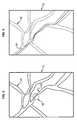
- the figure 3schematically illustrates an example of a first segmented image I 1 'having an array 101 of arteries visualized patient, an artery 103 having a constriction 102 corresponding to a lesion to be detected.
- This step S4consists of extracting and isolating, from the region of interest imaged without contrast product, the tool inserted into an artery of interest to the patient.
- the figure 4schematically illustrates an example of a second segmented image I 2 'having the same network 101 as that of the first segmented image I 1 ' illustrated in broken lines and a tool 201, here a guide wire inserted in an artery of the network 101 of arteries.
- a tool 201here a guide wire inserted in an artery of the network 101 of arteries.
- the network 101is shown here as an indication.
- segmenting an imageis a technique well known to those skilled in the art and will not be described in more detail.
- This step S5consists in defining for each artery a line Ci, for example a central line defining an axis of symmetry of an artery.
- central lines Ciare for example implemented by means of a technique described in the document Karl Krissian, Gregory Malandain, Nicholas Ayache, Régis Vaillant and Yves Trousset: "Model-Based Detection of Tubular Structures in 3D Images", Computer Vision and Image Understanding, Vol. 80, num. 2, p. 130-171, 2000 .
- This step S5consists in detecting an artery of interest, that is to say the artery into which the tool has been inserted.
- the detection of the artery of interestis carried out starting from the second image I 2 'and from the image I 3 '.
- Such detectionis implemented by means of a distance criterion between the tool 201 isolated and detected in the second segmented image I 2 'and the central lines Ci defined.
- the artery of interestcorresponds to the artery for which the distance between the tool and the central line C is minimal.
- a method of quantitative analysis of coronary lesions( Quantitatvie Coronary Analysis , QCA) is applied to the artery of interest.
- QCA analysisis based on a clinically validated algorithm for contour detection and allows for the determination of occlusion percentage, occlusion of diameter, and size of normal artery and stenotic artery.
- such an algorithmanalyzes the artery all along the central line C and in particular determines at each point of the central line the apparent diameter of the artery.
- the variations of this diameter and in particular the decreasesare indicators of the presence of pathology such as arterial stenosis.
- Quantificationconsists in determining such a decrease by a percentage relative to a zone of normal diameter.
- the method described abovecan be applied to several 2D projection images taken for different angulations.
- 3D reconstruction of known type and applicable to linescan be obtained 3D lines that describe the geometry of the artery. More particularly it is possible to obtain a 3D view of the artery of interest.
- the radiological image processing methodcan advantageously be implemented in the form of a computer program comprising machine instructions for carrying out the method.
Landscapes
- Engineering & Computer Science (AREA)
- Computer Vision & Pattern Recognition (AREA)
- Physics & Mathematics (AREA)
- General Physics & Mathematics (AREA)
- Theoretical Computer Science (AREA)
- Health & Medical Sciences (AREA)
- General Health & Medical Sciences (AREA)
- Medical Informatics (AREA)
- Nuclear Medicine, Radiotherapy & Molecular Imaging (AREA)
- Radiology & Medical Imaging (AREA)
- Quality & Reliability (AREA)
- Apparatus For Radiation Diagnosis (AREA)
Description
Translated fromFrenchL'invention concerne le domaine de l'imagerie médicale et plus particulièrement celui de la radiologie et trouve application dans le domaine de l'imagerie interventionnelle radiologique vasculaire.The invention relates to the field of medical imaging and more particularly that of radiology and finds application in the field of interventional radiological vascular imaging.
La radiologie interventionnelle vasculaire inclut les actes pratiqués sous contrôle de l'imagerie et permet notamment de traiter l'ischémie myocardique.Vascular interventional radiology includes procedures performed under the control of imaging and in particular to treat myocardial ischemia.
L'ischémie myocardique est une maladie qui touche plus d'un tiers des personnes dans les pays développés se traduit par une sténose c'est-à-dire un rétrécissement d'une artère.Myocardial ischemia is a disease that affects more than a third of people in developed countries results in a stenosis that is to say a narrowing of an artery.
Pour traiter cette maladie, plusieurs traitements existent dont la pose de stents intra-coronariens.To treat this disease, several treatments exist including the installation of intracoronary stents.
Pour cela, un praticien cardiologue utilise l'imagerie interventionnelle qui lui permet la caractérisation des éventuelles lésions et la quantification des artères coronaires et plus particulièrement les sténoses afin de bien choisir les dimensions du stent à introduire.For this, a cardiologist practitioner uses the interventional imaging that allows the characterization of possible lesions and the quantification of coronary arteries and more particularly stenoses to properly choose the size of the stent to introduce.
Un article par
Habituellement, le praticien cardiologue utilise une image de la zone comprenant une artère à traiter sur laquelle, manuellement, il positionne plusieurs marqueurs le long de l'artère à traiter pour effectuer une analyse de sténose, c'est-à-dire déterminer l'endroit où se trouve la sténose et déterminer les dimensions du stent nécessaire au traitement de la sténose.Usually, the cardiologist practitioner uses an image of the zone comprising an artery to be treated on which, manually, he positions several markers along the artery to be treated to perform a stenosis analysis, that is to say to determine the where the stenosis is located and determine the size of the stent needed to treat the stenosis.
Cette détection et cette quantification nécessitent une interaction du praticien radiologue avec un dispositif d'imagerie médicale et une étape préalable de détection de l'artère à traiter.This detection and this quantification require an interaction of the radiologist practitioner with a medical imaging device and a preliminary step of detecting the artery to be treated.
Ceci présente les inconvénients que la détection, la quantification prennent du temps et sont difficiles à mettre en oeuvre par le praticien.This has the disadvantages that detection and quantification take time and are difficult to implement by the practitioner.
L'invention permet de pallier ces inconvénients.The invention overcomes these disadvantages.
Selon un premier aspect, l'invention concerne un procédé de traitement d'images radiologiques d'une région d'intérêt d'un patient, les images radiologiques étant des images de projection 2D, dans laquelle un outil longiligne a été préalablement inséré dans une artère, le procédé comprenant l'étape suivante : obtention d'au moins un ensemble d'images constitué par une première image et par une seconde image de la région d'intérêt, la première image étant une image de la région d'intérêt dans laquelle un produit de contraste a été préalablement injecté, la seconde image étant une image de la région d'intérêt sans produit ou avec une quantité minimale de produit de contraste, chaque ensemble correspondant à une angulation donnée ; le procédé comprenant, pour chaque ensemble, les étapes suivantes : segmentation de la première image afin de détecter une pluralité d'artères de la région d'intérêt ; segmentation de la seconde image afin de détecter et isoler l'outil ; définition dans la première image segmentée d'une pluralité de lignes, chaque ligne définissant une artère ; détermination, à partir de la seconde image segmentée et des lignes définies, d'une artère d'intérêt correspondant à l'artère dans laquelle l'outil a été inséré, la ligne de l'artère d'intérêt étant la plus proche de l'outil ; application d'un algorithme d'analyse quantitative de lésions coronariennes à l'artère d'intérêt pour détecter une lésion de l'artère d'intérêt.According to a first aspect, the invention relates to a method for processing radiological images of a region of interest of a patient, the radiological images being 2D projection images, in which an elongated tool has been previously inserted into a artery, the method comprising the following step: obtaining at least one set of images consisting of a first image and a second image of the region of interest, the first image being an image of the region of interest in which a contrast product has been previously injected, the second image being an image of the region of interest without product or with a minimal amount of contrast medium, each set corresponding to a given angulation; the method comprising, for each set, the following steps: segmenting the first image to detect a plurality of arteries of the region of interest; segmenting the second image to detect and isolate the tool; defining in the first segmented image a plurality of lines, each line defining an artery; determining, from the second segmented image and the defined lines, an artery of interest corresponding to the artery in which the tool has been inserted, the line of the artery of interest being the closest to the tool; application of an algorithm for the quantitative analysis of coronary lesions in the artery of interest to detect a lesion of the artery of interest.
Grâce à la détection de l'artère d'intérêt, la quantification de la sténose est rendue possible sans avoir à définir manuellement des points le long d'une artère et donc sans interaction avec le système d'imagerie médicale.By detecting the artery of interest, quantification of the stenosis is made possible without having to manually define points along an artery and thus without interaction with the medical imaging system.
Ceci apporte rapidité, simplicité à la mise en oeuvre de la détection de lésions vasculaires.This brings speed, simplicity to the implementation of the detection of vascular lesions.
D'autres aspects du procédé sont les suivants :
- l'application de l'algorithme d'analyse quantitative est réalisée en utilisant des points placés au long de la ligne de l'artère d'intérêt ;
- l'étape de détermination de l'artère d'intérêt consiste à mettre en oeuvre un critère de distance combinant la distance euclidienne d'une ligne d'une artère et une évaluation de la différence d'orientation de l'outil avec cette ligne tout au long de la courbe définissant l'outil longiligne ;
- les lignes définissant les artères sont des lignes centrales définissant chacune un axe de symétrie d'une artère ;
- on obtient au moins deux ensembles d'images, le procédé comprenant, à l'issue de la détermination de l'artère d'intérêt pour chaque ensemble, une étape de reconstruction 3D pour obtenir une image 3D de l'artère d'intérêt ;
- l'algorithme d'analyse quantitative est mis en oeuvre sur l'artère d'intérêt 3D ;
- on obtient une séquence d'ensembles d'images et dans lequel l'identification de l'artère d'intérêt est mise en oeuvre sur chaque artère d'intérêt détectée pour chaque ensemble pour obtenir un champ de mouvement bidimensionnel de la ligne de l'artère d' intérêt ;
- on obtient une pluralité de séquences d'ensembles d'images et dans lequel l'algorithme d'analyse quantitative est mis en oeuvre sur chaque ensemble d'images pour obtenir le champ de mouvement projeté dans l'image de la ligne de l'artère d'intérêt ;
- les images radiologiques obtenues sont des images préalablement acquises et stockées dans une unité mémoire du système d'imagerie médicale.
- the application of the quantitative analysis algorithm is performed using points placed along the line of the artery of interest;
- the step of determining the artery of interest consists in implementing a distance criterion combining the Euclidean distance of a line of a artery and an evaluation of the difference in orientation of the tool with this line throughout the curve defining the elongated tool;
- the lines defining the arteries are central lines each defining an axis of symmetry of an artery;
- at least two sets of images are obtained, the method comprising, after the determination of the artery of interest for each set, a 3D reconstruction step to obtain a 3D image of the artery of interest;
- the quantitative analysis algorithm is implemented on the artery of interest 3D;
- a sequence of sets of images is obtained and in which the identification of the artery of interest is implemented on each artery of interest detected for each set to obtain a two-dimensional field of motion of the line of the artery of interest;
- a plurality of sequences of sets of images are obtained and in which the quantitative analysis algorithm is implemented on each set of images to obtain the projected motion field in the image of the line of the artery of interest;
- the radiological images obtained are images previously acquired and stored in a memory unit of the medical imaging system.
Selon un second aspect, l'invention concerne un système d'imagerie médicale comprenant des moyens pour la mise en oeuvre du procédé selon le premier aspect de l'invention.According to a second aspect, the invention relates to a medical imaging system comprising means for implementing the method according to the first aspect of the invention.
Selon un troisième aspect, l'invention concerne un programme d'ordinateur comprenant des instructions machine pour la mise en oeuvre d'un procédé selon le premier aspect de l'invention.According to a third aspect, the invention relates to a computer program comprising machine instructions for carrying out a method according to the first aspect of the invention.
D'autres caractéristiques et avantages de l'invention ressortiront encore de la description qui suit laquelle est purement illustrative et non limitative et doit être lue en regard des dessins annexés sur lesquels
- la
figure 1 illustre un système d'imagerie médicale conforme à l'invention ; - la
figure 2 illustre schématiquement des étapes du procédé de traitement d'images radiologiques conforme à l'invention ; - la
figure 3 illustre une première image obtenue par le procédé de traitement d'images radiologiques conforme à l'invention ; - la
figure 4 illustre une seconde image obtenue par le procédé de traitement d'images radiologiques conforme à l'invention ; - la
figure 5 illustre schématiquement une artère d'intérêt obtenue au cours du procédé de traitement d'images radiologiques conforme à l'invention.
- the
figure 1 illustrates a medical imaging system according to the invention; - the
figure 2 schematically illustrates steps of the radiological image processing method according to the invention; - the
figure 3 illustrates a first image obtained by the radiological image processing method according to the invention; - the
figure 4 illustrates a second image obtained by the radiological image processing method according to the invention; - the
figure 5 schematically illustrates an artery of interest obtained during the radiological image processing method according to the invention.
La
Le système 100 d'imagerie médicale comprend un support 1 destiné à recevoir un patient 10 à examiner une source 2 destinée à émettre un faisceau 3 de rayons X, un détecteur 4 disposé en face de la source 2 et configuré pour détecter les rayons X émis par la source 2, une unité de commande 6, une unité de stockage 7 et une unité d'affichage 8.The
La source 2 de rayons X et le détecteur 4 sont reliés par un bras 5 en forme de C. Un tel bras 5 est plus communément appelé arceau. Le bras 5 peut être orienté selon trois degrés de liberté.The X-ray source 2 and the
Le détecteur 4 peut être un capteur d'image à semi-conducteurs comprenant, par exemple, du phosphore d'iodure de césium (scintillateur) sur une matrice de transistor/photodiode en silicium amorphe. D'autres détecteurs adéquats sont : un capteur CCD, détecteur numérique direct qui convertit directement les rayons X en signaux numériques. Le détecteur 4 illustré sur la
L'unité de commande 6 est connectée à l'arceau 5 par connexion filaire ou sans fil. L'unité de commande 6 permet de commander l'acquisition en fixant plusieurs paramètres tels que la dose de radiation à émettre par la source à rayons X et le positionnement angulaire du bras 5. L'unité de commande 6 permet de commander la position du bras 5, c'est-à-dire la position de la source 2 par rapport au détecteur 4.The
L'unité de commande 6 peut comprendre un dispositif de lecture (non représenté) par exemple un lecteur de disquettes un lecteur de CD-ROM, DVD-ROM, ou des ports de connexion pour lire les instructions du procédé de traitement d'un support d'instructions (non montré), comme une disquette, un CD-ROM, DVD-ROM, ou clé USB ou de manière plus générale par tout support de mémoire amovible ou encore via une connexion réseau.The
L'unité de stockage 7 est connectée à l'unité de commande 6 pour l'enregistrement des paramètres et des images acquises. Il est possible de prévoir que l'unité de stockage 7 est située à l'intérieur de l'unité de commande 6 ou à l'extérieur.The
L'unité de stockage 7 peut être formée par un disque dur ou SSD, ou tout autre moyen de stockage amovible et réinscriptible (clés USB, cartes mémoires etc.). L'unité de stockage 7 peut être une mémoire ROM/RAM de l'unité de commande 6, une clé USB, une carte mémoire, une mémoire d'un serveur central.The
L'unité d'affichage 8 est connectée à l'unité de commande 6 pour l'affichage des images acquises et/ou d'informations sur les paramètres de commande de l'acquisition.The
L'unité d'affichage 8 peut être par exemple un écran d'ordinateur, un moniteur, un écran plat, un écran plasma ou tout autre type de dispositif d'affichage de type connu.The
Une telle unité d'affichage 8 permet à un praticien de contrôler l'acquisition des images radiologiques.Such a
Le système d'imagerie médicale 100 est couplé à un système de traitement 200. Le système de traitement 200 comprend une unité de calcul 9 et unité de stockage 10.The
Le système de traitement 200 reçoit des images acquises et stockées dans l'unité de stockage 4 du système d'imagerie médicale 100 à partir desquelles il effectue un certain nombre de traitement (voir ci-après).The
La transmission des données de l'unité de stockage 4 du système d'imagerie médicale 100 vers l'unité de calcul 9 du système de traitement 200 peut être faite à travers un réseau informatique interne ou externe ou à l'aide de tout support mémoire physique adéquat tel que disquettes, CD-ROM, DVD-ROM, disque dure externe, clé USB, carte SD, etc.The data transmission from the
L'unité de calcul 9 est par exemple un/des ordinateur(s), un/des processeur(s), un/des microcontrôleur(s), un/des micro-ordinateur(s), un/des automate(s) programmable(s), un/des circuit(s) intégré(s) spécifique(s) d'application, d'autres circuits programmables, ou d'autres dispositifs qui incluent un ordinateur tel qu'une station de travail.The
En variante, le calculateur 9 peut comprendre un dispositif de lecture (non représenté) par exemple un lecteur de disquettes, un lecteur de CD-ROM ou DVD-ROM, ou des ports de connexion pour lire les instructions du procédé de traitement d'un support d'instructions (non montré), comme une disquette, un CD-ROM, un DVD-ROM ou une clé USB ou de manière plus générale par tout support de mémoire amovible ou encore via une connexion réseau.As a variant, the
En outre, le système de traitement comprend une unité de stockage 11 pour le stockage des données générées par l'unité de calcul 9.In addition, the processing system comprises a
L'unité de calcul 9 peut être connectée à l'unité d'affichage 8 (comme sur la
On décrit ci-dessous un exemple de réalisation du procédé de traitement d'images radiologiques selon l'invention. La
Le procédé de traitement d'images radiologiques utilise deux images I1, I2 radiologiques d'une région d'intérêt d'un patient, dans laquelle un outil a été préalablement introduit. On considère dans ce qui suit un outil longiligne.The radiological image processing method uses two radiological images I1 , I2 of a region of interest of a patient, into which a tool has been previously introduced. We consider in the following an elongated tool.
Un tel outil est par exemple un fil guide, un cathéter ou la combinaison de plusieurs d'entre eux. De manière préférée il s'agit du fil guide qui est destiné à faciliter l'introduction d'un stent.Such a tool is for example a guide wire, a catheter or the combination of several of them. Preferably it is the guide wire which is intended to facilitate the introduction of a stent.
La première image I1 correspond à une image radiologique de la région d'intérêt dans laquelle un produit de contraste a été injecté. Un tel produit est par exemple de l'iode et la région d'intérêt est typiquement la région coronarienne d'un patient à traiter.The first image I1 corresponds to a radiological image of the region of interest in which a contrast product has been injected. Such a product is for example iodine and the region of interest is typically the coronary region of a patient to be treated.
La seconde image I2 correspond à une image radiologique de la région d'intérêt, sans produit de contraste ou avec une quantité minimale de produit de contraste.The second image I2 corresponds to a radiological image of the region of interest, without contrast product or with a minimum amount of contrast product.
On entend par quantité minimale de produit de contraste, une quantité de produit de contraste qui permet de visualiser l'outil sans qu'il ne soit masqué par les artères.By minimal amount of contrast medium is meant a quantity of contrast medium which makes it possible to visualize the tool without being masked by the arteries.
Les première et seconde images I1, I2 peuvent être obtenues au moyen d'acquisition mises en oeuvre au cours du procédé de traitement d'images radiologiques ou bien obtenues à partir d'une unité de stockage du système d'imagerie médicale.The first and second images I1 , I2 can be obtained by means of acquisition implemented during the radiological image processing method or else obtained from a storage unit of the medical imaging system.
On note que les première et seconde images I1, I2 sont issues d'acquisitions mises en oeuvre au moyen du système d'imagerie médicale ci-dessus décrit pour une angulation donnée c'est-à-dire l'orientation de la source à rayons X par rapport à la normale au support sur lequel le patient est disposé.It should be noted that the first and second images I1 , I2 are derived from acquisitions implemented using the medical imaging system described above for a given angulation, that is to say the orientation of the source. X-ray compared to normal to the support on which the patient is disposed.
Les première et seconde images I1, I2 sont des images de projection 2D.The first and second images I1 , I2 are 2D projection images.
Dans la suite de la description on définit un ensemble d'images comme étant le couple constitué par les première et seconde images acquises pour une angulation donnée.In the rest of the description, a set of images is defined as being the pair constituted by the first and second images acquired for a given angulation.
On considère également une séquence d'ensembles d'images c'est-à-dire pour une angulation donnée plusieurs ensembles d'images I1 I2 acquis à des instants différents.We also consider a sequence of sets of images that is to say for a given angulation several sets of images I1 I2 acquired at different times.
Cette étape consiste à extraire et isoler, de la première image I1 et grâce au produit de contraste injecté, les artères du patient.This step consists in extracting and isolating, from the first image I1 and thanks to the injected contrast product, the arteries of the patient.
De manière plus précise, il s'agit du réseau d'artères de la région d'intérêt qui est extrait et isolé.More precisely, it is the network of arteries of the region of interest that is extracted and isolated.
La
Cette étape S4 consiste à extraire et isoler, de la région d'intérêt imagée sans produit de contraste, l'outil inséré dans une artère d'intérêt du patient.This step S4 consists of extracting and isolating, from the region of interest imaged without contrast product, the tool inserted into an artery of interest to the patient.
La
On note que segmenter une image est une technique bien connue de l'homme du métier et ne sera pas décrite plus en détail.It is noted that segmenting an image is a technique well known to those skilled in the art and will not be described in more detail.
Cette étape S5 consiste à définir pour chaque artère une ligne Ci, par exemple une ligne centrale définissant un axe de symétrie d'une artère.This step S5 consists in defining for each artery a line Ci, for example a central line defining an axis of symmetry of an artery.
La définition des lignes centrales Ci est par exemple mise en oeuvre au moyen d'une technique décrite dans le document
La
Cette étape S5 consiste à détecter une artère d'intérêt, c'est-à-dire l'artère dans laquelle l'outil a été inséré.This step S5 consists in detecting an artery of interest, that is to say the artery into which the tool has been inserted.
La détection de l'artère d'intérêt est mise en oeuvre à partir de la seconde image I2' et à partie de l'image I3'.The detection of the artery of interest is carried out starting from the second image I2 'and from the image I3 '.
Une telle détection est mise en oeuvre au moyen d'un critère de distance entre l'outil 201 isolé et détecté dans la seconde image segmentée I2' et les lignes centrales Ci définies.Such detection is implemented by means of a distance criterion between the
L'artère d'intérêt correspond à l'artère pour laquelle la distance entre l'outil et la ligne centrale C est minimale.The artery of interest corresponds to the artery for which the distance between the tool and the central line C is minimal.
Il s'agit par exemple d'un critère de distance combinant la distance euclidienne d'une ligne centrale C d'une artère et une évaluation de la différence d'orientation de l'outil avec cette ligne centrale C.This is for example a distance criterion combining the Euclidean distance of a central line C of an artery and an evaluation of the difference in orientation of the tool with this central line C.
Une fois l'artère d'intérêt dans laquelle l'outil a été inséré est identifiée, on peut procéder à l'analyse quantitative.Once the artery of interest into which the tool has been inserted is identified, quantitative analysis can be performed.
Pour ce faire on applique à l'artère d'intérêt un procédé d'analyse quantitative de lésions coronariennes (en anglais,« Quantitatvie Coronary Analysis », (QCA)).For this purpose, a method of quantitative analysis of coronary lesions (Quantitatvie Coronary Analysis , QCA) is applied to the artery of interest.
Un tel procédé d'analyse est par exemple décrit dans le document
L'analyse QCA se fonde sur un algorithme validé cliniquement pour la détection des contours et permet de déterminer un pourcentage d'occlusion, l'occlusion du diamètre et la taille de l'artère normale et de l'artère présentant une sténose.QCA analysis is based on a clinically validated algorithm for contour detection and allows for the determination of occlusion percentage, occlusion of diameter, and size of normal artery and stenotic artery.
En particulier, un tel algorithme analyse l'artère tout au long de la ligne centrale C et en particulier détermine en chaque point de la ligne centrale le diamètre apparent de l'artère. Les variations de ce diamètre et en particulier les diminutions sont des indicateurs de la présence de pathologie comme par exemple une sténose artérielle. La quantification consiste à déterminer une telle diminution par un pourcentage relatif à une zone de diamètre normal.In particular, such an algorithm analyzes the artery all along the central line C and in particular determines at each point of the central line the apparent diameter of the artery. The variations of this diameter and in particular the decreases are indicators of the presence of pathology such as arterial stenosis. Quantification consists in determining such a decrease by a percentage relative to a zone of normal diameter.
Le procédé ci-dessus décrit peut être appliqué à plusieurs images de projection 2D prises pour différentes angulations.The method described above can be applied to several 2D projection images taken for different angulations.
En utilisant une reconstruction 3D de type connue et applicable à des lignes on peut obtenir des lignes 3D qui décrivent la géométrie de l'artère. Plus particulièrement il est possible d'obtenir une vue 3D de l'artère d'intérêt.Using a 3D reconstruction of known type and applicable to lines can be obtained 3D lines that describe the geometry of the artery. More particularly it is possible to obtain a 3D view of the artery of interest.
On peut appliquer cela à une séquence d'images pour obtenir une séquence d'images 3D et ainsi obtenir les éléments suivants :
- analyse de la projection du mouvement des artères décrit par l'évolution des lignes identifiées dans la séquence d'images ;
- analyse quantitative de la projection des artères dans la succession des images ;
- analyse du mouvement de l'artère dans l'espace tridimensionnel à partir du traitement de plusieurs séquences d'images acquises suivant différentes angulations ;
- analyse quantitative tridimensionnelle et temporelle de l'artère d'intérêt.
- analysis of the projection of the movement of the arteries described by the evolution of the lines identified in the sequence of images;
- quantitative analysis of the projection of arteries in the succession of images;
- analysis of the movement of the artery in three-dimensional space from the processing of several image sequences acquired according to different angulations;
- three-dimensional and temporal quantitative analysis of the artery of interest.
Le procédé de traitement d'images radiologiques peut être avantageusement implémenté sous la forme d'un programme d'ordinateur comprenant des instructions machine pour la mise en oeuvre du procédé.The radiological image processing method can advantageously be implemented in the form of a computer program comprising machine instructions for carrying out the method.
Claims (11)
- Process for processing radiological images of a region of interest of a patient, the radiological images being 2D projection images, wherein an elongated tool has been previously inserted into an artery, the process comprising the following step:- obtaining (S1, S2) at least one set of images consisting of a first image (I1) and a second image (I2) of the region of interest, the first image (I1) being an image of the region of interest wherein a contrast medium has been previously injected, the second image being an image of the region of interest without medium or with a minimal quantity of contrast medium, each set corresponding to a given angulation;the process comprising, for each set, the following steps:- segmenting (S3) the first image (I1) to detect a plurality of arteries of the region of interest;- segmenting (S4) the second image (I2) to detect and isolate the tool;- defining (S5) in the segmented first image a plurality of lines (Ci) each line (Ci) defining an artery;- determining (S6), on the basis of the segmented second image and the lines defined, an artery of interest corresponding to the artery wherein the tool has been inserted, the line (C7) of the artery of interest being the closest to the tool;- applying (S7) a quantitative coronary lesion analysis algorithm to the artery of interest to detect a lesion of the artery of interest.
- Process according to the above claim wherein the quantitative analysis algorithm is applied using points placed along the line of the artery of interest.
- Process according to any of the above claims wherein the step for determining the artery of interest consists of applying a distance criterion combining the Euclidean distance of a line of an artery and an evaluation of the difference in orientation of the tool with this line along the curve defining the elongated tool.
- Process according to any of the above claims wherein the lines defining the arteries are central lines each defining an axis of symmetry of an artery.
- Process according to any of the above claims, wherein at least two sets of images (I1, 12) are obtained, the process comprising, following the determination of the artery of interest for each set, a 3D reconstruction step to obtain a 3D image of the artery of interest.
- Process according to claim 5 whereby the quantitative analysis algorithm is applied to the 3D artery of interest.
- Process according to any of claims 1 to 3 wherein a sequence of sets of images is obtained and wherein the identification of the artery of interest is applied to each artery of interest detected for each set to obtain a two-dimensional field of movement of the line of the artery of interest.
- Process according to any of claims 1 to 3 wherein a plurality of sequences of sets of images are obtained and wherein the quantitative analysis algorithm is applied to each set of images to obtain the projected field of movement in the image of the line of the artery of interest.
- Process according to any of the above claims wherein the radiological images obtained are images previously acquired and stored in a memory unit of the medical imaging system.
- Medical imaging system comprising means for applying a process according to any of the above claims.
- Computer programcharacterised in that it comprises machine instructions for implementing a process according to any of claims 1 to 9, when it is executed by a computer.
Priority Applications (3)
| Application Number | Priority Date | Filing Date | Title |
|---|---|---|---|
| EP10306249.3AEP2453408B1 (en) | 2010-11-12 | 2010-11-12 | Method for processing radiographic images for stenosis detection |
| US13/290,450US8880148B2 (en) | 2010-11-12 | 2011-11-07 | Treatment process of radiological images for detection of stenosis |
| CN201110387801.4ACN102592274B (en) | 2010-11-12 | 2011-11-11 | Processing procedure for the radiation image of stenosis detection |
Applications Claiming Priority (1)
| Application Number | Priority Date | Filing Date | Title |
|---|---|---|---|
| EP10306249.3AEP2453408B1 (en) | 2010-11-12 | 2010-11-12 | Method for processing radiographic images for stenosis detection |
Publications (2)
| Publication Number | Publication Date |
|---|---|
| EP2453408A1 EP2453408A1 (en) | 2012-05-16 |
| EP2453408B1true EP2453408B1 (en) | 2013-06-05 |
Family
ID=43585724
Family Applications (1)
| Application Number | Title | Priority Date | Filing Date |
|---|---|---|---|
| EP10306249.3ANot-in-forceEP2453408B1 (en) | 2010-11-12 | 2010-11-12 | Method for processing radiographic images for stenosis detection |
Country Status (2)
| Country | Link |
|---|---|
| US (1) | US8880148B2 (en) |
| EP (1) | EP2453408B1 (en) |
Families Citing this family (18)
| Publication number | Priority date | Publication date | Assignee | Title |
|---|---|---|---|---|
| WO2008107905A2 (en) | 2007-03-08 | 2008-09-12 | Sync-Rx, Ltd. | Imaging and tools for use with moving organs |
| US9968256B2 (en) | 2007-03-08 | 2018-05-15 | Sync-Rx Ltd. | Automatic identification of a tool |
| US11197651B2 (en) | 2007-03-08 | 2021-12-14 | Sync-Rx, Ltd. | Identification and presentation of device-to-vessel relative motion |
| US9305334B2 (en) | 2007-03-08 | 2016-04-05 | Sync-Rx, Ltd. | Luminal background cleaning |
| US11064964B2 (en) | 2007-03-08 | 2021-07-20 | Sync-Rx, Ltd | Determining a characteristic of a lumen by measuring velocity of a contrast agent |
| US8781193B2 (en) | 2007-03-08 | 2014-07-15 | Sync-Rx, Ltd. | Automatic quantitative vessel analysis |
| US9629571B2 (en) | 2007-03-08 | 2017-04-25 | Sync-Rx, Ltd. | Co-use of endoluminal data and extraluminal imaging |
| US10716528B2 (en) | 2007-03-08 | 2020-07-21 | Sync-Rx, Ltd. | Automatic display of previously-acquired endoluminal images |
| US9375164B2 (en) | 2007-03-08 | 2016-06-28 | Sync-Rx, Ltd. | Co-use of endoluminal data and extraluminal imaging |
| EP2303385B1 (en) | 2008-06-19 | 2013-12-11 | Sync-RX, Ltd. | Stepwise advancement of a medical tool |
| US9144394B2 (en) | 2008-11-18 | 2015-09-29 | Sync-Rx, Ltd. | Apparatus and methods for determining a plurality of local calibration factors for an image |
| US8855744B2 (en) | 2008-11-18 | 2014-10-07 | Sync-Rx, Ltd. | Displaying a device within an endoluminal image stack |
| US10362962B2 (en) | 2008-11-18 | 2019-07-30 | Synx-Rx, Ltd. | Accounting for skipped imaging locations during movement of an endoluminal imaging probe |
| US9974509B2 (en) | 2008-11-18 | 2018-05-22 | Sync-Rx Ltd. | Image super enhancement |
| US9095313B2 (en) | 2008-11-18 | 2015-08-04 | Sync-Rx, Ltd. | Accounting for non-uniform longitudinal motion during movement of an endoluminal imaging probe |
| US9101286B2 (en) | 2008-11-18 | 2015-08-11 | Sync-Rx, Ltd. | Apparatus and methods for determining a dimension of a portion of a stack of endoluminal data points |
| US11064903B2 (en) | 2008-11-18 | 2021-07-20 | Sync-Rx, Ltd | Apparatus and methods for mapping a sequence of images to a roadmap image |
| EP2863802B1 (en) | 2012-06-26 | 2020-11-04 | Sync-RX, Ltd. | Flow-related image processing in luminal organs |
Family Cites Families (4)
| Publication number | Priority date | Publication date | Assignee | Title |
|---|---|---|---|---|
| AU2002212642A1 (en) | 2000-10-18 | 2002-04-29 | Paieon Inc. | Method and system for measuring dimensions of an organ |
| US6718193B2 (en) | 2000-11-28 | 2004-04-06 | Ge Medical Systems Global Technology Company, Llc | Method and apparatus for analyzing vessels displayed as unfolded structures |
| WO2005031635A1 (en) | 2003-09-25 | 2005-04-07 | Paieon, Inc. | System and method for three-dimensional reconstruction of a tubular organ |
| US20070003121A1 (en)* | 2004-01-15 | 2007-01-04 | Koninklijke Philips Electronic, N.V. | Automatic contrast medium control in images |
- 2010
- 2010-11-12EPEP10306249.3Apatent/EP2453408B1/ennot_activeNot-in-force
- 2011
- 2011-11-07USUS13/290,450patent/US8880148B2/enactiveActive
Also Published As
| Publication number | Publication date |
|---|---|
| US20120123238A1 (en) | 2012-05-17 |
| CN102592274A (en) | 2012-07-18 |
| EP2453408A1 (en) | 2012-05-16 |
| US8880148B2 (en) | 2014-11-04 |
Similar Documents
| Publication | Publication Date | Title |
|---|---|---|
| EP2453408B1 (en) | Method for processing radiographic images for stenosis detection | |
| EP3659112B1 (en) | A method for co-registering and displaying multiple imaging modalities | |
| US10453190B2 (en) | Detection of and validation of shadows in intravascular images | |
| Martuscelli et al. | Accuracy of thin-slice computed tomography in the detection of coronary stenoses | |
| Bourantas et al. | ANGIOCARE: An automated system for fast three‐dimensional coronary reconstruction by integrating angiographic and intracoronary ultrasound data | |
| EP3634294A1 (en) | Method and system for assistance in guiding an endovascular instrument | |
| FR2963976A1 (en) | IMAGE PROCESSING METHOD FOR DETERMINING SUSPECTED ZONES IN A TISSUE MATRIX, AND ITS USE FOR 3D NAVIGATION THROUGH THE TISSUE MATRIX | |
| EP3913578A1 (en) | Method and system for resetting of images containing anatomical structures | |
| FR2797978A1 (en) | AUTOMATIC IMAGE RECORDING PROCESS | |
| FR2998160A1 (en) | PROCESS FOR PROCESSING RADIOLOGICAL IMAGES IN DOUBLE ENERGY | |
| EP0840252A1 (en) | Digital image-processing method for the automatic extraction of ribbon-like objects | |
| FR2960332A1 (en) | METHOD OF PROCESSING RADIOLOGICAL IMAGES TO DETERMINE A 3D POSITION OF A NEEDLE. | |
| CN106163401A (en) | The equipment of medical imaging and method for coronary vasodilator | |
| WO2019023375A2 (en) | A method for co-registering and displaying multiple imaging modalities | |
| US8077954B2 (en) | System and method for image processing | |
| US20150202021A1 (en) | Method for processing images of interventional radiology | |
| FR2965651A1 (en) | TOMOGRAPHIC RECONSTRUCTION OF AN OBJECT IN MOTION | |
| FR2972551A1 (en) | TOMOGRAPHIC PROCESSING METHOD WITH LOW NUMBER OF PROJECTIONS OF A CONTRAST OBJECT | |
| JP6898047B2 (en) | Quantitative evaluation of time-varying data | |
| JP7497390B2 (en) | Systems and methods for intravascular device detection and crimp measurement - Patents.com | |
| FR2915867A1 (en) | METHOD AND SYSTEM FOR CT TOMOGRAPHY IMAGING | |
| WO2003088143A2 (en) | Method for assisting and guiding the navigation of a tool in anatomical structures | |
| Chabi et al. | Semi-automatic segmentation of elongated interventional instruments for online calibration of C-arm imaging system | |
| FR3099985A1 (en) | Mid-procedure change of view for ultrasound diagnosis | |
| CN102592274B (en) | Processing procedure for the radiation image of stenosis detection |
Legal Events
| Date | Code | Title | Description |
|---|---|---|---|
| PUAI | Public reference made under article 153(3) epc to a published international application that has entered the european phase | Free format text:ORIGINAL CODE: 0009012 | |
| AK | Designated contracting states | Kind code of ref document:A1 Designated state(s):AL AT BE BG CH CY CZ DE DK EE ES FI FR GB GR HR HU IE IS IT LI LT LU LV MC MK MT NL NO PL PT RO RS SE SI SK SM TR | |
| AX | Request for extension of the european patent | Extension state:BA ME | |
| 17P | Request for examination filed | Effective date:20121018 | |
| RIC1 | Information provided on ipc code assigned before grant | Ipc:G06T 7/00 20060101AFI20121121BHEP | |
| GRAP | Despatch of communication of intention to grant a patent | Free format text:ORIGINAL CODE: EPIDOSNIGR1 | |
| GRAS | Grant fee paid | Free format text:ORIGINAL CODE: EPIDOSNIGR3 | |
| GRAA | (expected) grant | Free format text:ORIGINAL CODE: 0009210 | |
| AK | Designated contracting states | Kind code of ref document:B1 Designated state(s):AL AT BE BG CH CY CZ DE DK EE ES FI FR GB GR HR HU IE IS IT LI LT LU LV MC MK MT NL NO PL PT RO RS SE SI SK SM TR | |
| REG | Reference to a national code | Ref country code:GB Ref legal event code:FG4D Free format text:NOT ENGLISH | |
| REG | Reference to a national code | Ref country code:CH Ref legal event code:EP | |
| REG | Reference to a national code | Ref country code:AT Ref legal event code:REF Ref document number:616037 Country of ref document:AT Kind code of ref document:T Effective date:20130615 | |
| REG | Reference to a national code | Ref country code:IE Ref legal event code:FG4D Free format text:LANGUAGE OF EP DOCUMENT: FRENCH | |
| REG | Reference to a national code | Ref country code:DE Ref legal event code:R096 Ref document number:602010007567 Country of ref document:DE Effective date:20130801 | |
| REG | Reference to a national code | Ref country code:AT Ref legal event code:MK05 Ref document number:616037 Country of ref document:AT Kind code of ref document:T Effective date:20130605 | |
| PG25 | Lapsed in a contracting state [announced via postgrant information from national office to epo] | Ref country code:SI Free format text:LAPSE BECAUSE OF FAILURE TO SUBMIT A TRANSLATION OF THE DESCRIPTION OR TO PAY THE FEE WITHIN THE PRESCRIBED TIME-LIMIT Effective date:20130605 Ref country code:GR Free format text:LAPSE BECAUSE OF FAILURE TO SUBMIT A TRANSLATION OF THE DESCRIPTION OR TO PAY THE FEE WITHIN THE PRESCRIBED TIME-LIMIT Effective date:20130906 Ref country code:ES Free format text:LAPSE BECAUSE OF FAILURE TO SUBMIT A TRANSLATION OF THE DESCRIPTION OR TO PAY THE FEE WITHIN THE PRESCRIBED TIME-LIMIT Effective date:20130916 Ref country code:AT Free format text:LAPSE BECAUSE OF FAILURE TO SUBMIT A TRANSLATION OF THE DESCRIPTION OR TO PAY THE FEE WITHIN THE PRESCRIBED TIME-LIMIT Effective date:20130605 Ref country code:NO Free format text:LAPSE BECAUSE OF FAILURE TO SUBMIT A TRANSLATION OF THE DESCRIPTION OR TO PAY THE FEE WITHIN THE PRESCRIBED TIME-LIMIT Effective date:20130905 Ref country code:SE Free format text:LAPSE BECAUSE OF FAILURE TO SUBMIT A TRANSLATION OF THE DESCRIPTION OR TO PAY THE FEE WITHIN THE PRESCRIBED TIME-LIMIT Effective date:20130605 Ref country code:FI Free format text:LAPSE BECAUSE OF FAILURE TO SUBMIT A TRANSLATION OF THE DESCRIPTION OR TO PAY THE FEE WITHIN THE PRESCRIBED TIME-LIMIT Effective date:20130605 Ref country code:LT Free format text:LAPSE BECAUSE OF FAILURE TO SUBMIT A TRANSLATION OF THE DESCRIPTION OR TO PAY THE FEE WITHIN THE PRESCRIBED TIME-LIMIT Effective date:20130605 | |
| REG | Reference to a national code | Ref country code:NL Ref legal event code:VDEP Effective date:20130605 | |
| REG | Reference to a national code | Ref country code:LT Ref legal event code:MG4D | |
| PG25 | Lapsed in a contracting state [announced via postgrant information from national office to epo] | Ref country code:HR Free format text:LAPSE BECAUSE OF FAILURE TO SUBMIT A TRANSLATION OF THE DESCRIPTION OR TO PAY THE FEE WITHIN THE PRESCRIBED TIME-LIMIT Effective date:20130605 Ref country code:RS Free format text:LAPSE BECAUSE OF FAILURE TO SUBMIT A TRANSLATION OF THE DESCRIPTION OR TO PAY THE FEE WITHIN THE PRESCRIBED TIME-LIMIT Effective date:20130605 Ref country code:BG Free format text:LAPSE BECAUSE OF FAILURE TO SUBMIT A TRANSLATION OF THE DESCRIPTION OR TO PAY THE FEE WITHIN THE PRESCRIBED TIME-LIMIT Effective date:20130905 | |
| PG25 | Lapsed in a contracting state [announced via postgrant information from national office to epo] | Ref country code:LV Free format text:LAPSE BECAUSE OF FAILURE TO SUBMIT A TRANSLATION OF THE DESCRIPTION OR TO PAY THE FEE WITHIN THE PRESCRIBED TIME-LIMIT Effective date:20130605 | |
| PG25 | Lapsed in a contracting state [announced via postgrant information from national office to epo] | Ref country code:EE Free format text:LAPSE BECAUSE OF FAILURE TO SUBMIT A TRANSLATION OF THE DESCRIPTION OR TO PAY THE FEE WITHIN THE PRESCRIBED TIME-LIMIT Effective date:20130605 Ref country code:IS Free format text:LAPSE BECAUSE OF FAILURE TO SUBMIT A TRANSLATION OF THE DESCRIPTION OR TO PAY THE FEE WITHIN THE PRESCRIBED TIME-LIMIT Effective date:20131005 Ref country code:PT Free format text:LAPSE BECAUSE OF FAILURE TO SUBMIT A TRANSLATION OF THE DESCRIPTION OR TO PAY THE FEE WITHIN THE PRESCRIBED TIME-LIMIT Effective date:20131007 Ref country code:SK Free format text:LAPSE BECAUSE OF FAILURE TO SUBMIT A TRANSLATION OF THE DESCRIPTION OR TO PAY THE FEE WITHIN THE PRESCRIBED TIME-LIMIT Effective date:20130605 Ref country code:CZ Free format text:LAPSE BECAUSE OF FAILURE TO SUBMIT A TRANSLATION OF THE DESCRIPTION OR TO PAY THE FEE WITHIN THE PRESCRIBED TIME-LIMIT Effective date:20130605 | |
| PG25 | Lapsed in a contracting state [announced via postgrant information from national office to epo] | Ref country code:RO Free format text:LAPSE BECAUSE OF FAILURE TO SUBMIT A TRANSLATION OF THE DESCRIPTION OR TO PAY THE FEE WITHIN THE PRESCRIBED TIME-LIMIT Effective date:20130605 Ref country code:PL Free format text:LAPSE BECAUSE OF FAILURE TO SUBMIT A TRANSLATION OF THE DESCRIPTION OR TO PAY THE FEE WITHIN THE PRESCRIBED TIME-LIMIT Effective date:20130605 Ref country code:NL Free format text:LAPSE BECAUSE OF FAILURE TO SUBMIT A TRANSLATION OF THE DESCRIPTION OR TO PAY THE FEE WITHIN THE PRESCRIBED TIME-LIMIT Effective date:20130605 | |
| PLBE | No opposition filed within time limit | Free format text:ORIGINAL CODE: 0009261 | |
| STAA | Information on the status of an ep patent application or granted ep patent | Free format text:STATUS: NO OPPOSITION FILED WITHIN TIME LIMIT | |
| PG25 | Lapsed in a contracting state [announced via postgrant information from national office to epo] | Ref country code:DK Free format text:LAPSE BECAUSE OF FAILURE TO SUBMIT A TRANSLATION OF THE DESCRIPTION OR TO PAY THE FEE WITHIN THE PRESCRIBED TIME-LIMIT Effective date:20130605 | |
| 26N | No opposition filed | Effective date:20140306 | |
| PG25 | Lapsed in a contracting state [announced via postgrant information from national office to epo] | Ref country code:IT Free format text:LAPSE BECAUSE OF FAILURE TO SUBMIT A TRANSLATION OF THE DESCRIPTION OR TO PAY THE FEE WITHIN THE PRESCRIBED TIME-LIMIT Effective date:20130605 | |
| BERE | Be: lapsed | Owner name:GENERAL ELECTRIC CY Effective date:20131130 | |
| REG | Reference to a national code | Ref country code:DE Ref legal event code:R097 Ref document number:602010007567 Country of ref document:DE Effective date:20140306 | |
| PG25 | Lapsed in a contracting state [announced via postgrant information from national office to epo] | Ref country code:MC Free format text:LAPSE BECAUSE OF FAILURE TO SUBMIT A TRANSLATION OF THE DESCRIPTION OR TO PAY THE FEE WITHIN THE PRESCRIBED TIME-LIMIT Effective date:20130605 | |
| REG | Reference to a national code | Ref country code:IE Ref legal event code:MM4A | |
| PG25 | Lapsed in a contracting state [announced via postgrant information from national office to epo] | Ref country code:BE Free format text:LAPSE BECAUSE OF NON-PAYMENT OF DUE FEES Effective date:20131130 | |
| PG25 | Lapsed in a contracting state [announced via postgrant information from national office to epo] | Ref country code:IE Free format text:LAPSE BECAUSE OF NON-PAYMENT OF DUE FEES Effective date:20131112 | |
| PG25 | Lapsed in a contracting state [announced via postgrant information from national office to epo] | Ref country code:SM Free format text:LAPSE BECAUSE OF FAILURE TO SUBMIT A TRANSLATION OF THE DESCRIPTION OR TO PAY THE FEE WITHIN THE PRESCRIBED TIME-LIMIT Effective date:20130605 | |
| PG25 | Lapsed in a contracting state [announced via postgrant information from national office to epo] | Ref country code:TR Free format text:LAPSE BECAUSE OF FAILURE TO SUBMIT A TRANSLATION OF THE DESCRIPTION OR TO PAY THE FEE WITHIN THE PRESCRIBED TIME-LIMIT Effective date:20130605 Ref country code:CY Free format text:LAPSE BECAUSE OF FAILURE TO SUBMIT A TRANSLATION OF THE DESCRIPTION OR TO PAY THE FEE WITHIN THE PRESCRIBED TIME-LIMIT Effective date:20130605 | |
| REG | Reference to a national code | Ref country code:CH Ref legal event code:PL | |
| PG25 | Lapsed in a contracting state [announced via postgrant information from national office to epo] | Ref country code:CH Free format text:LAPSE BECAUSE OF NON-PAYMENT OF DUE FEES Effective date:20141130 Ref country code:LI Free format text:LAPSE BECAUSE OF NON-PAYMENT OF DUE FEES Effective date:20141130 Ref country code:MK Free format text:LAPSE BECAUSE OF FAILURE TO SUBMIT A TRANSLATION OF THE DESCRIPTION OR TO PAY THE FEE WITHIN THE PRESCRIBED TIME-LIMIT Effective date:20130605 Ref country code:LU Free format text:LAPSE BECAUSE OF NON-PAYMENT OF DUE FEES Effective date:20131112 Ref country code:HU Free format text:LAPSE BECAUSE OF FAILURE TO SUBMIT A TRANSLATION OF THE DESCRIPTION OR TO PAY THE FEE WITHIN THE PRESCRIBED TIME-LIMIT; INVALID AB INITIO Effective date:20101112 | |
| PG25 | Lapsed in a contracting state [announced via postgrant information from national office to epo] | Ref country code:MT Free format text:LAPSE BECAUSE OF FAILURE TO SUBMIT A TRANSLATION OF THE DESCRIPTION OR TO PAY THE FEE WITHIN THE PRESCRIBED TIME-LIMIT Effective date:20130605 | |
| REG | Reference to a national code | Ref country code:FR Ref legal event code:PLFP Year of fee payment:6 | |
| REG | Reference to a national code | Ref country code:FR Ref legal event code:PLFP Year of fee payment:7 | |
| REG | Reference to a national code | Ref country code:FR Ref legal event code:PLFP Year of fee payment:8 | |
| REG | Reference to a national code | Ref country code:FR Ref legal event code:PLFP Year of fee payment:9 | |
| PG25 | Lapsed in a contracting state [announced via postgrant information from national office to epo] | Ref country code:AL Free format text:LAPSE BECAUSE OF FAILURE TO SUBMIT A TRANSLATION OF THE DESCRIPTION OR TO PAY THE FEE WITHIN THE PRESCRIBED TIME-LIMIT Effective date:20130605 | |
| PGFP | Annual fee paid to national office [announced via postgrant information from national office to epo] | Ref country code:GB Payment date:20201021 Year of fee payment:11 Ref country code:FR Payment date:20201021 Year of fee payment:11 Ref country code:DE Payment date:20201020 Year of fee payment:11 | |
| REG | Reference to a national code | Ref country code:DE Ref legal event code:R119 Ref document number:602010007567 Country of ref document:DE | |
| GBPC | Gb: european patent ceased through non-payment of renewal fee | Effective date:20211112 | |
| PG25 | Lapsed in a contracting state [announced via postgrant information from national office to epo] | Ref country code:GB Free format text:LAPSE BECAUSE OF NON-PAYMENT OF DUE FEES Effective date:20211112 Ref country code:DE Free format text:LAPSE BECAUSE OF NON-PAYMENT OF DUE FEES Effective date:20220601 | |
| PG25 | Lapsed in a contracting state [announced via postgrant information from national office to epo] | Ref country code:FR Free format text:LAPSE BECAUSE OF NON-PAYMENT OF DUE FEES Effective date:20211130 |