EP2338428A1 - Estimation and mapping of ablation volume - Google Patents
Estimation and mapping of ablation volumeDownload PDFInfo
- Publication number
- EP2338428A1 EP2338428A1EP10252190AEP10252190AEP2338428A1EP 2338428 A1EP2338428 A1EP 2338428A1EP 10252190 AEP10252190 AEP 10252190AEP 10252190 AEP10252190 AEP 10252190AEP 2338428 A1EP2338428 A1EP 2338428A1
- Authority
- EP
- European Patent Office
- Prior art keywords
- ablation
- tissue
- catheter
- controller
- operative
- Prior art date
- Legal status (The legal status is an assumption and is not a legal conclusion. Google has not performed a legal analysis and makes no representation as to the accuracy of the status listed.)
- Granted
Links
- 238000002679ablationMethods0.000titleclaimsabstractdescription99
- 238000013507mappingMethods0.000titledescription7
- 230000000007visual effectEffects0.000claimsabstractdescription7
- 230000004044responseEffects0.000claimsdescription7
- 238000003780insertionMethods0.000claimsdescription6
- 230000037431insertionEffects0.000claimsdescription6
- 210000002216heartAnatomy0.000abstractdescription26
- 230000000747cardiac effectEffects0.000abstractdescription3
- 210000001519tissueAnatomy0.000description59
- 238000000034methodMethods0.000description44
- 210000001174endocardiumAnatomy0.000description12
- 239000000523sampleSubstances0.000description11
- 230000003902lesionEffects0.000description8
- 230000033001locomotionEffects0.000description7
- 239000013598vectorSubstances0.000description6
- 238000006073displacement reactionMethods0.000description5
- 210000005003heart tissueAnatomy0.000description5
- 230000008569processEffects0.000description5
- 238000005259measurementMethods0.000description4
- 210000000056organAnatomy0.000description4
- 238000002604ultrasonographyMethods0.000description4
- 230000002159abnormal effectEffects0.000description3
- 206010003119arrhythmiaDiseases0.000description3
- 238000013153catheter ablationMethods0.000description3
- 230000005672electromagnetic fieldEffects0.000description3
- 239000000463materialSubstances0.000description3
- 230000002792vascularEffects0.000description3
- 230000004913activationEffects0.000description2
- 239000008280bloodSubstances0.000description2
- 210000004369bloodAnatomy0.000description2
- 239000002131composite materialSubstances0.000description2
- 229910003460diamondInorganic materials0.000description2
- 239000010432diamondSubstances0.000description2
- 239000011810insulating materialSubstances0.000description2
- 230000007246mechanismEffects0.000description2
- BASFCYQUMIYNBI-UHFFFAOYSA-NplatinumChemical compound[Pt]BASFCYQUMIYNBI-UHFFFAOYSA-N0.000description2
- 238000012545processingMethods0.000description2
- 230000001225therapeutic effectEffects0.000description2
- 238000011282treatmentMethods0.000description2
- 206010001497AgitationDiseases0.000description1
- 230000005355Hall effectEffects0.000description1
- 229910000575Ir alloyInorganic materials0.000description1
- 229910001260Pt alloyInorganic materials0.000description1
- 238000011298ablation treatmentMethods0.000description1
- 230000006793arrhythmiaEffects0.000description1
- 230000003126arrythmogenic effectEffects0.000description1
- 230000000712assemblyEffects0.000description1
- 238000000429assemblyMethods0.000description1
- 238000005452bendingMethods0.000description1
- 210000005242cardiac chamberAnatomy0.000description1
- 230000006835compressionEffects0.000description1
- 238000007906compressionMethods0.000description1
- 238000004590computer programMethods0.000description1
- 238000012937correctionMethods0.000description1
- 230000006378damageEffects0.000description1
- 230000000694effectsEffects0.000description1
- 238000011156evaluationMethods0.000description1
- 238000010438heat treatmentMethods0.000description1
- 239000007769metal materialSubstances0.000description1
- 238000012986modificationMethods0.000description1
- 230000004048modificationEffects0.000description1
- 238000012544monitoring processMethods0.000description1
- 210000004165myocardiumAnatomy0.000description1
- 230000003287optical effectEffects0.000description1
- 230000037361pathwayEffects0.000description1
- 229920003023plasticPolymers0.000description1
- 239000004033plasticSubstances0.000description1
- 229920000642polymerPolymers0.000description1
- 229920001296polysiloxanePolymers0.000description1
- 229920002635polyurethanePolymers0.000description1
- 239000004814polyurethaneSubstances0.000description1
- 230000002685pulmonary effectEffects0.000description1
- 238000007674radiofrequency ablationMethods0.000description1
- 230000033764rhythmic processEffects0.000description1
- 230000000451tissue damageEffects0.000description1
- 231100000827tissue damageToxicity0.000description1
- 238000012800visualizationMethods0.000description1
Images


Classifications
- A—HUMAN NECESSITIES
- A61—MEDICAL OR VETERINARY SCIENCE; HYGIENE
- A61B—DIAGNOSIS; SURGERY; IDENTIFICATION
- A61B18/00—Surgical instruments, devices or methods for transferring non-mechanical forms of energy to or from the body
- A61B18/04—Surgical instruments, devices or methods for transferring non-mechanical forms of energy to or from the body by heating
- A61B18/12—Surgical instruments, devices or methods for transferring non-mechanical forms of energy to or from the body by heating by passing a current through the tissue to be heated, e.g. high-frequency current
- A61B18/14—Probes or electrodes therefor
- A61B18/1492—Probes or electrodes therefor having a flexible, catheter-like structure, e.g. for heart ablation
- A—HUMAN NECESSITIES
- A61—MEDICAL OR VETERINARY SCIENCE; HYGIENE
- A61B—DIAGNOSIS; SURGERY; IDENTIFICATION
- A61B18/00—Surgical instruments, devices or methods for transferring non-mechanical forms of energy to or from the body
- A61B18/18—Surgical instruments, devices or methods for transferring non-mechanical forms of energy to or from the body by applying electromagnetic radiation, e.g. microwaves
- A61B18/1815—Surgical instruments, devices or methods for transferring non-mechanical forms of energy to or from the body by applying electromagnetic radiation, e.g. microwaves using microwaves
- A—HUMAN NECESSITIES
- A61—MEDICAL OR VETERINARY SCIENCE; HYGIENE
- A61B—DIAGNOSIS; SURGERY; IDENTIFICATION
- A61B18/00—Surgical instruments, devices or methods for transferring non-mechanical forms of energy to or from the body
- A61B2018/00315—Surgical instruments, devices or methods for transferring non-mechanical forms of energy to or from the body for treatment of particular body parts
- A61B2018/00345—Vascular system
- A61B2018/00351—Heart
- A—HUMAN NECESSITIES
- A61—MEDICAL OR VETERINARY SCIENCE; HYGIENE
- A61B—DIAGNOSIS; SURGERY; IDENTIFICATION
- A61B18/00—Surgical instruments, devices or methods for transferring non-mechanical forms of energy to or from the body
- A61B2018/00571—Surgical instruments, devices or methods for transferring non-mechanical forms of energy to or from the body for achieving a particular surgical effect
- A61B2018/00577—Ablation
- A—HUMAN NECESSITIES
- A61—MEDICAL OR VETERINARY SCIENCE; HYGIENE
- A61B—DIAGNOSIS; SURGERY; IDENTIFICATION
- A61B18/00—Surgical instruments, devices or methods for transferring non-mechanical forms of energy to or from the body
- A61B2018/00636—Sensing and controlling the application of energy
- A61B2018/00696—Controlled or regulated parameters
- A61B2018/00702—Power or energy
- A—HUMAN NECESSITIES
- A61—MEDICAL OR VETERINARY SCIENCE; HYGIENE
- A61B—DIAGNOSIS; SURGERY; IDENTIFICATION
- A61B18/00—Surgical instruments, devices or methods for transferring non-mechanical forms of energy to or from the body
- A61B2018/00636—Sensing and controlling the application of energy
- A61B2018/00696—Controlled or regulated parameters
- A61B2018/00714—Temperature
- A—HUMAN NECESSITIES
- A61—MEDICAL OR VETERINARY SCIENCE; HYGIENE
- A61B—DIAGNOSIS; SURGERY; IDENTIFICATION
- A61B18/00—Surgical instruments, devices or methods for transferring non-mechanical forms of energy to or from the body
- A61B2018/00636—Sensing and controlling the application of energy
- A61B2018/00696—Controlled or regulated parameters
- A61B2018/00761—Duration
- A—HUMAN NECESSITIES
- A61—MEDICAL OR VETERINARY SCIENCE; HYGIENE
- A61B—DIAGNOSIS; SURGERY; IDENTIFICATION
- A61B18/00—Surgical instruments, devices or methods for transferring non-mechanical forms of energy to or from the body
- A61B2018/00636—Sensing and controlling the application of energy
- A61B2018/00773—Sensed parameters
- A61B2018/00791—Temperature
- A—HUMAN NECESSITIES
- A61—MEDICAL OR VETERINARY SCIENCE; HYGIENE
- A61B—DIAGNOSIS; SURGERY; IDENTIFICATION
- A61B34/00—Computer-aided surgery; Manipulators or robots specially adapted for use in surgery
- A61B34/10—Computer-aided planning, simulation or modelling of surgical operations
- A61B2034/101—Computer-aided simulation of surgical operations
- A61B2034/102—Modelling of surgical devices, implants or prosthesis
- A61B2034/104—Modelling the effect of the tool, e.g. the effect of an implanted prosthesis or for predicting the effect of ablation or burring
- A—HUMAN NECESSITIES
- A61—MEDICAL OR VETERINARY SCIENCE; HYGIENE
- A61B—DIAGNOSIS; SURGERY; IDENTIFICATION
- A61B34/00—Computer-aided surgery; Manipulators or robots specially adapted for use in surgery
- A61B34/20—Surgical navigation systems; Devices for tracking or guiding surgical instruments, e.g. for frameless stereotaxis
- A61B2034/2046—Tracking techniques
- A61B2034/2051—Electromagnetic tracking systems
- A—HUMAN NECESSITIES
- A61—MEDICAL OR VETERINARY SCIENCE; HYGIENE
- A61B—DIAGNOSIS; SURGERY; IDENTIFICATION
- A61B90/00—Instruments, implements or accessories specially adapted for surgery or diagnosis and not covered by any of the groups A61B1/00 - A61B50/00, e.g. for luxation treatment or for protecting wound edges
- A61B90/06—Measuring instruments not otherwise provided for
- A61B2090/064—Measuring instruments not otherwise provided for for measuring force, pressure or mechanical tension
- A61B2090/065—Measuring instruments not otherwise provided for for measuring force, pressure or mechanical tension for measuring contact or contact pressure
- A—HUMAN NECESSITIES
- A61—MEDICAL OR VETERINARY SCIENCE; HYGIENE
- A61B—DIAGNOSIS; SURGERY; IDENTIFICATION
- A61B34/00—Computer-aided surgery; Manipulators or robots specially adapted for use in surgery
- A61B34/20—Surgical navigation systems; Devices for tracking or guiding surgical instruments, e.g. for frameless stereotaxis
- A—HUMAN NECESSITIES
- A61—MEDICAL OR VETERINARY SCIENCE; HYGIENE
- A61M—DEVICES FOR INTRODUCING MEDIA INTO, OR ONTO, THE BODY; DEVICES FOR TRANSDUCING BODY MEDIA OR FOR TAKING MEDIA FROM THE BODY; DEVICES FOR PRODUCING OR ENDING SLEEP OR STUPOR
- A61M25/00—Catheters; Hollow probes
- A61M2025/0001—Catheters; Hollow probes for pressure measurement
- A61M2025/0002—Catheters; Hollow probes for pressure measurement with a pressure sensor at the distal end
- A—HUMAN NECESSITIES
- A61—MEDICAL OR VETERINARY SCIENCE; HYGIENE
- A61M—DEVICES FOR INTRODUCING MEDIA INTO, OR ONTO, THE BODY; DEVICES FOR TRANSDUCING BODY MEDIA OR FOR TAKING MEDIA FROM THE BODY; DEVICES FOR PRODUCING OR ENDING SLEEP OR STUPOR
- A61M25/00—Catheters; Hollow probes
- A61M25/01—Introducing, guiding, advancing, emplacing or holding catheters
- A61M25/0105—Steering means as part of the catheter or advancing means; Markers for positioning
- A61M2025/0166—Sensors, electrodes or the like for guiding the catheter to a target zone, e.g. image guided or magnetically guided
- A—HUMAN NECESSITIES
- A61—MEDICAL OR VETERINARY SCIENCE; HYGIENE
- A61M—DEVICES FOR INTRODUCING MEDIA INTO, OR ONTO, THE BODY; DEVICES FOR TRANSDUCING BODY MEDIA OR FOR TAKING MEDIA FROM THE BODY; DEVICES FOR PRODUCING OR ENDING SLEEP OR STUPOR
- A61M25/00—Catheters; Hollow probes
- A61M25/01—Introducing, guiding, advancing, emplacing or holding catheters
- A61M25/0105—Steering means as part of the catheter or advancing means; Markers for positioning
- A61M25/0133—Tip steering devices
- A61M25/0158—Tip steering devices with magnetic or electrical means, e.g. by using piezo materials, electroactive polymers, magnetic materials or by heating of shape memory materials
Definitions
- This inventionrelates to relates generally to minimally invasive treatment of organs inside the body. More particularly, this invention relates to methods and devices for prediction and assessment of ablation treatments applied to cardiac tissue.
- Intracardiac radio-frequency (RF) ablationis a well known method for treating cardiac arrhythmias.
- a catheter having an electrode at its distal tipis inserted through the patient's vascular system into a chamber of the heart.
- the electrodeis brought into contact with a site (or sites) on the endocardium, and RF energy is applied through the catheter to the electrode in order to ablate the heart tissue at the site. It is important to ensure proper contact between the electrode and the endocardium during ablation in order to achieve the desired therapeutic effect without excessive damage to the tissue.
- U.S. Patent No. 6,695,808whose disclosure is incorporated herein by reference, describes apparatus for treating a selected patient tissue or organ region.
- a probehas a contact surface that may be urged against the region, thereby creating contact pressure.
- a pressure transducermeasures the contact pressure. This arrangement is said to meet the needs of procedures in which a medical instrument must be placed in firm but not excessive contact with an anatomical surface, by providing information to the user of the instrument that is indicative of the existence and magnitude of the contact force.
- an electrode assembly on a cathetercarries pressure transducers, which sense contact with tissue and convey signals to a pressure contact module.
- the moduleidentifies the electrode elements that are associated with the pressure transducer signals and directs an energy generator to convey RF energy to these elements, and not to other elements that are in contact only with blood.
- U.S. Patent Application Publication 2007/0100332whose disclosure is incorporated herein by reference, describes systems and methods for assessing electrode-tissue contact for tissue ablation.
- An electro-mechanical sensor within the catheter shaftgenerates electrical signals corresponding to the amount of movement of the electrode within a distal portion of the catheter shaft.
- An output devicereceives the electrical signals for assessing a level of contact between the electrode and a tissue.
- the volume of heart tissue ablated when RF energy is applied by a catheter electrode in contact with the tissue at a given pointis roughly proportional to the RF power (P) and roughly proportional to the mechanical force (F) between the catheter and the tissue.
- PRF power
- Fmechanical force
- An embodiment of the inventionprovides a method of ablation, which is carried out by inserting a probe into a body of a living subject, urging the probe into contact with a tissue in the body, determining a mechanical force that is exerted by the probe against the tissue, and applying a specified dosage of energy to the tissue for ablation thereof, wherein at least one of the application time of the dosage and the power level depend on the mechanical force.
- An aspect of the methodis performed prior to applying the specified dosage of energy by reporting an indication of an expected ablation volume at the power level, the application time and the mechanical force.
- a further aspect of the methodincludes displaying a visual indication of the ablation volume, and responsively to the visual indication controlling the ablation volume by varying at least one of the power level, the mechanical force and the application time.
- Another aspect of the methodincludes calculating a rate of ablation as a function of the power level and the mechanical force, and controlling the rate of ablation by varying at least one of the power level and the mechanical force.
- Still another aspect of the methodincludes monitoring tissue temperature of the tissue and controlling the rate of ablation is performed responsively to the temperature.
- Fig. 1is a pictorial illustration of a system for performing ablative procedures on a heart of a living subject, which is constructed and operative in accordance with an embodiment of the invention
- Fig. 2is a cutaway view of the distal end of a catheter used in the system shown in Fig. 1 , in accordance with an embodiment of the present invention
- Fig. 3is a pictorial view of the distal end of the catheter shown in Fig. 2 in contact with endocardial tissue, in accordance with an embodiment of the invention
- Fig. 4is a composite map of the heart, illustrating aspects of a cardiac ablation procedure in accordance with an embodiment of the invention
- Fig. 5is a flow chart of a method of estimation and mapping tissue ablation volume, in accordance with a disclosed embodiment of the invention.
- Fig. 6is a cutaway view of a catheter used in the system shown in Fig. 1 , which is constructed and operative in accordance with an alternate embodiment of the invention.
- Fig. 1is a pictorial illustration of a system 10 for performing ablative procedures on a heart 12 of a living subject, which is constructed and operative in accordance with a disclosed embodiment of the invention.
- the systemcomprises a catheter 14, which is percutaneously inserted by an operator 16 through the patient's vascular system into a chamber or vascular structure of the heart.
- the operator 16who is typically a physician, brings the catheter's distal tip 18 into contact with the heart wall at an ablation target site.
- Electrical activation mapsmay then be prepared, according to the methods disclosed in U.S. Patent Nos. 6,226,542 , and 6,301,496 , and in commonly assigned U.S. Patent No.
- Areas determined to be abnormal by evaluation of the electrical activation mapscan be ablated by application of thermal energy, e.g., by passage of radiofrequency electrical current through wires in the catheter to one or more electrodes at the distal tip 18, which apply the radiofrequency energy to the myocardium.
- the energyis absorbed in the tissue, heating it to a point (typically about 50°C) at which it permanently loses its electrical excitability.
- this procedurecreates non-conducting lesions in the cardiac tissue, which disrupt the abnormal electrical pathway causing the arrhythmia.
- other known methods of applying ablative energycan be used, e.g., ultrasound energy, as disclosed in U.S. Patent Application Publication No. 2004/0102769 , whose disclosure is herein incorporated by reference.
- the principles of the inventioncan be applied to different heart chambers, and to mapping in sinus rhythm, and when many different cardiac arrhythmias are present.
- the catheter 14typically comprises a handle 20, having suitable controls on the handle to enable the operator 16 to steer, position and orient the distal end of the catheter as desired for the ablation.
- the distal portion of the catheter 14contains position sensors (not shown) that provide signals to a positioning processor 22, located in a console 24.
- the console 24typically contains an ablation power generator 25.
- the catheter 14may be adapted to conduct ablative energy to the heart using any known ablation technique, e.g., radiofrequency energy, ultrasound energy, and laser energy. Such methods are disclosed in commonly assigned U.S. Patent Nos. 6,814,733 , 6,997,924 , and 7,156,816 , which are herein incorporated by reference.
- the positioning processor 22is an element of a positioning sub-system of the system 10 that measures location and orientation coordinates of the catheter 14.
- the positioning sub-systemcomprises a magnetic position tracking arrangement that determines the position and orientation of the catheter 14 by generating magnetic fields in a predefined working volume its vicinity and sensing these fields at the catheter.
- the magnetic position tracking arrangementtypically comprises a set of external radiators, such as field generating coils 28, which are located in fixed, known positions external to the patient.
- the field generating coils 28are driven by field generators (not shown), which are typically located in the console 24, and generate fields, typically electromagnetic fields, in the vicinity of the heart 12.
- a radiator in the catheter 14, such as a coilgenerates electromagnetic fields, which are received by sensors (not shown) outside the patient's body.
- the catheter 14is coupled to the console 24, which enables the operator 16 to observe and regulate the functions of the catheter 14.
- Console 24includes a processor, preferably a computer with appropriate signal processing circuits.
- the processoris coupled to drive a monitor 30.
- the signal processing circuitstypically receive, amplify, filter and digitize signals from the catheter 14, including signals generated by the above-noted sensors and a plurality of sensing electrodes (not shown) located distally in the catheter 14. The digitized signals are received and used by the console 24 to compute the position and orientation of the catheter 14 and to analyze the electrical signals from the electrodes.
- the information derived from this analysismay be used to generate an electrophysiological map of at least a portion of the heart 12 or structures such as the pulmonary venous ostia, for diagnostic purposes such as locating an arrhythmogenic area in the heart or to facilitate therapeutic ablation.
- the system 10includes other elements, which are not shown in the figures for the sake of simplicity.
- the system 10may include an electrocardiogram (ECG) monitor, coupled to receive signals from one or more body surface electrodes, so as to provide an ECG synchronization signal to the console 24.
- ECGelectrocardiogram
- the system 10typically also includes a reference position sensor, either on an externally-applied reference patch attached to the exterior of the subject's body, or on an internally-placed catheter, which is inserted into the heart 12 maintained in a fixed position relative to the heart 12. By comparing the position of the catheter 14 to that of the reference catheter, the coordinates of catheter 14 are determined relative to the heart 12, irrespective of heart motion. Alternatively, any other suitable method may be used to compensate for heart motion. Nevertheless, the positioning sub-system cannot guarantee that an energy-conveying component of the catheter 14 is in actual contact with the tissue to be ablated.
- Fig. 2is a cutaway view of distal end 33 of catheter 14 ( Fig. 1 ), showing details of the structure of the catheter in accordance with an embodiment of the present invention.
- the catheter shown in Fig. 2includes a pressure transducer, which is more fully disclosed in commonly assigned U.S. Patent Application Publication No. 2009/0093806 , which is herein incorporated by reference.
- Other known types of pressure transducerscan be substituted for the pressure transducer described with reference to Fig. 2 .
- Catheter 14comprises a flexible insertion tube 54, with a distal section 72 connected to the remainder of the insertion tube 54 at a joint 56.
- the insertion tubeis covered by a flexible, insulating material 60, such as CelconTM or TeflonTM.
- the area of joint 56is covered, as well, by a flexible, insulating material, which may be the same as material 60 or may be specially adapted to permit unimpeded bending and compression of the joint, (This material is cut away in Fig. 2 in order to expose the internal structure of the catheter.)
- Distal tip 18may be covered, at least in part, by an electrode 50, which is typically made of a metallic material, such as a platinum/iridium alloy. Alternatively, other suitable materials may be used, as will be apparent to those skilled in the art.
- the distal section 72is typically relatively rigid, by comparison with a proximal section 74.
- the distal section 72is connected to the proximal section 74 by a resilient member 58.
- the resilient member 58has the form of a coil spring, but other types of resilient components may alternatively be used for this purpose.
- resilient member 58may comprise a polymer, such as silicone, polyurethane, or other plastics, with the desired flexibility and strength characteristics.
- Resilient member 58permits a limited range of relative movement between distal section 72 and the proximal section 74 in response to forces exerted on the distal section 72 or directly against the distal tip 18. Such forces are encountered when the distal tip is pressed against the endocardium during an ablation procedure.
- the desired pressure for good electrical contact between the distal tip and the endocardium during ablationis on the order of 20-30 grams.
- the spring serving as the resilient member 58 in this embodimentmay be configured, for example, to permit axial displacement (i.e., lateral movement along the axis of catheter 14) of the distal end 33 by about 1-2 mm and angular deflection of the distal section 72 with respect to the proximal section 74 by up to about 30 degrees in response to a desired pressure.
- distal section 72contains a magnetic position sensor 62.
- Position sensor 62may comprise one or more miniature coils, and typically comprises multiple coils oriented along different axes.
- position sensor 62may comprise another type of magnetic sensor, such as a Hall effect or magnetoresistive sensor, for example.
- the magnetic fields created by the field generating coils 28( Fig. 1 ) cause the position sensor 62 to generate electrical signals, with amplitudes that are indicative of the position and orientation of position sensor 62 relative to the fixed frame of reference of field generating coils 28.
- Positioning processor 22Fig.
- catheter 14contains a miniature magnetic field generator 64 near the distal tip 18, which is driven by a current conveyed through catheter 14 from console 24 ( Fig. 1 ).
- the currentis generated so as to create magnetic fields that are distinguishable in time and/or frequency from the fields of field generating coils 28 ( Fig. 1 ).
- the current supplied to field generator 64may be generated at a selected frequency in the range between about 16 kHz and 25 kHz, while field generating coils 28 are driven at different frequencies.
- the operation of field generating coils 28 and field generator 64may be time-multiplexed.
- the magnetic field created by field generator 64causes one or more coils in position sensor 62 to generate electrical signals at the drive frequency of field generator 64.
- the amplitudes of these signalsvary depending upon the location and orientation of distal tip 18 relative to proximal section 74.
- Positioning processor 22processes these signals in order to determine the axial displacement and the magnitude of the angular deflection of the distal tip 18 relative to the proximal section 74.
- Position sensor 62may determine six position and orientation coordinates (X, Y, Z directions and pitch, yaw and roll orientations) of the distal end and distal tip of catheter 14. For this purpose, at least two sensing coils are typically required in the position sensor.
- three sensing coils 76are used, in order to improve the accuracy and reliability of the position measurement.
- system 10may be able to determine only five position and orientation coordinates (X, Y, Z directions and pitch and yaw orientations).
- X, Y, Z directions and pitch and yaw orientationsAs the readings of displacement and deflection should be accurate to within a few tenths of a millimeter and about one degree, respectively, it is desirable to include three coils 76 in position sensor 62, preferably mutually orthogonal, as shown in Fig. 2 .
- the position of the position sensor 62 with reference to some fixed frame of referencecan be determined, it is possible to compute the relative movement of the distal tip 18 relative to the proximal section 74. This gives a measure of the deformation and angular deviation of resilient member 58. Generally speaking, the deformation is proportional to the mechanical force that is exerted on the resilient member 58, which is roughly equal to the force that is exerted on the distal tip 18 by the heart tissue with which the distal tip 18 is in contact.
- the combination of field generator 64 with position sensor 62serves as a pressure sensing system for determining the approximate pressure exerted by the endocardial tissue on the distal tip 18 of the catheter 14 (or equivalently, the pressure exerted by electrode 50 against the endocardial tissue).
- Fig. 3is a pictorial view of the distal end 33 of the catheter 14 in contact with endocardium 70 of the heart 12, in accordance with an embodiment of the invention.
- Pressure exerted by the distal tip 18 against the endocardium 70deforms the endocardial tissue slightly, so that the electrode 50 contacts the tissue over a relatively large area. Since the electrode 50 engages the endocardium 70 at an angle 82, rather than head-on, distal section 72 bends at joint 56 forming a bend angle 84 relative to the insertion tube of the catheter. The bend facilitates optimal contact between the electrode and the endocardial tissue.
- positioning processor 22receives and processes the signals generated by position sensor 62 in response to the magnetic field of generator 64, in order to derive an indication of the pressure exerted by distal tip 18 on endocardium 70 ( Fig. 3 ).
- pressureabout 20-30 grams is desirable.
- Lower pressuremeans that there may be inadequate contact between electrode 50 and the endocardial tissue. As a result, much or all of the thermal energy may be carried away by the blood inside the heart, and the tissue will be ablated inadequately or not at all.
- Higher pressuremeans that the electrode is pressing too hard against the endocardial tissue. Excessive pressure of this sort may cause cavitation in the tissue, leading to extensive tissue damage and possibly even perforation of the heart wall.
- this pressure sensing systemreads the pressure correctly regardless of whether the electrode engages the endocardium head-on or at an angle.
- the pressure readingis insensitive to temperature variations and free of drift, unlike piezoelectric sensors, for example.
- the magnitudes of the displacement and deflectionmay be combined by vector addition to give a total magnitude of the movement of distal tip 18 relative to the proximal section 74.
- the systemcan determine the position of the distal section 72 and the distal tip 18 with six degrees of freedom.
- Force vectors 78, 80can then be computed, the vector 80 representing the magnitude of the component that is normal to the wall of the heart 12.
- the relationships between force and deflectionmay be pre-calibrated for each catheter and a calibration table constructed and used subsequently in force measurements.
- console 24outputs an indication of the pressure measured to the operator 16, and may issue an alarm if the pressure is too low or too high.
- ablation power generator 25may be interlocked, so as to supply power to electrode 50 ( Fig. 2 ) only when the pressure against the endocardium 70 ( Fig. 3 ) is in the desired range.
- the pressure indicationmay be used in closed-loop control of an automated mechanism for maneuvering and operating catheter 14, as described more fully in the above noted U.S. Patent Application Publication No. 2004/0102769 , in order to ensure that the mechanism causes the distal section 72 to engage the endocardium 70 ( Fig. 3 ) in the proper location and with the appropriate contact pressure.
- RF poweris discussed with respect to the methods and systems herein, in embodiments of the system 10 ( Fig. 1 ), other forms of energy may be delivered to the tissue, i.e., laser and microwave techniques, and high intensity focused ultrasound energy, as described in commonly assigned U.S. Patent Application Publication No. 2006/0287648 , which is herein incorporated by reference.
- the product P * Fgives a good indication of the rate of ablation of the tissue, where P represents RF power and F represents the magnitude of the force vector exerted by the catheter against the endocardial surface of the heart.
- the operatormay increase or decrease either or both of the component parameters, P and F in order to control the ablation rate.
- the total volume V of tissue ablated, up to a maximum dictated by tissue characteristics and safety considerations,is roughly proportional to the product V ⁇ k P * F * T wherein T is the time duration of RF power application, and k is a proportionality constant.
- Fig. 4is a composite map 86 of the heart illustrating aspects of a cardiac ablation procedure in accordance with a disclosed embodiment of the invention.
- the proceduremay be actual or simulated, for purposes of predicting the necessary force to be applied by a cardiac catheter 88 in an operating position within a chamber of heart 90.
- Arrows 92, 94represent two different force vectors, the length of the arrows corresponding to the magnitudes of the forces being.
- a dosage of energy, e.g., RF ablation currentis to be applied at a predetermined power level for a time sufficient to produce an ablation lesion.
- Predicted small and large circular ablation zones 96, 98correspond to the short and long arrows 92, 94, respectively.
- the size of the ablation zonecan be predicted and dynamically displayed.
- the completeness of the ablationcan be calculated as time varies, and progress displayed during the procedure as by changing the visual characteristics of the ablation zones 96, 98.
- the ablation volumegrows over time in proportion to the product P*F.
- the required forcecan be computed at a given RF power and application time or for a given total energy dosage at different combinations of application time and RF power.
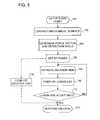
- Fig. 5is a flow chart of a method of estimation and mapping of tissue ablation volume, in accordance with a disclosed embodiment of the invention.
- the methodrequires a determination of mechanical force developed by contact between a probe and the tissue site to be ablated.
- the methodcan be performed by the system 10 ( Fig. 1 ) using catheter 14.
- other methods that are capable of measuring the pressurecan be applied, for example impedance-based measurements, such as disclosed in commonly assigned U.S. Patent Application Publication No. 2007/0060832 , whose disclosure is herein incorporated by reference.
- suitable optical or ultrasound techniquesmay be used to determine the mechanical force.
- the processbegins at initial step 100.
- the heartis catheterized conventionally and the catheter navigated to a desired location at which tissue ablation is required.
- the cardiac catheteris brought into contact with the endocardial surface, generally at an angle of incidence other than perpendicular as shown in Fig. 3 .
- the mechanical force or a desired force vector applied to the endocardium by the catheteris determined.
- the deflection angle, e.g., angle 82 ( Fig. 3 )may be determined automatically, using the information provided by location sensors in the catheter.
- ablation powere.g., RF power
- an estimated ablation timeis tentatively chosen, which establishes the energy dosage to be applied.
- steps 106, 108can be modified to set the ablation time, and estimate power levels, respectively.
- the operatormay be assisted at this step in that a controller may report an indication of an expected ablation volume at the energy dosage and the mechanical force.
- step 110the size of the lesion to be created by ablating is computed, according to the conditions established in step 104 and step 108.
- powertypically RF power
- controlreturns to step 108, where the ablation time is re-estimated.
- the size of the lesion to be created by ablationis known.
- the loop defined by step 108, step 110 and decision step 112can be iterated automatically until an acceptable size has been determined.
- the lesion sizemay be computed directly at optional step 116 using the relationship of Equation 1, and then perform ablation at final step 114.
- step 108, step 110 and decision step 112can be omitted.
- proposed power and proposed application time datacan be received as input and ablation volumes computed at different mechanical forces of contact with the tissue.
- Fig. 6is a cutaway view of distal end 33 of catheter 14 ( Fig. 1 ), which is constructed and operative in accordance with a disclosed embodiment of the invention.
- This embodimentis similar to Fig. 2 , except now the distal section 72 includes a conventional temperature sensor 118 that is capable of detecting abnormal rise in temperature of the tissues at the operating site.
- the rate of ablationmay be controlled by responsively to the temperature of the tissues in order to prevent charring or dangerous temperature elevation outside the computed ablation volume.
- Equation 1can be modified to account for the temperature such that only actual ablation time, rather than total elapsed time is taken into consideration.
- Ablation timecan be defined to run only when contact force exceeding a predetermined force threshold is ascertained and the temperature exceeds a predetermined temperature threshold.
- ablation timecan be defined to run only when contact force exceeding a predetermined force threshold is ascertained or the temperature exceeds a predetermined temperature threshold.
- Equation 1may be modified in several ways to account for ablation time.
- the following examplesare practical approximations, in which various first and second order corrections are not shown for clarity of presentation.
- the ablation poweris applied only during time intervals when the conditions shown are met.
- Aspect 1an ablation apparatus, comprising:
- Aspect 2the ablation apparatus according to aspect 1, wherein the controller is operative, prior to an application of the given dosage of energy to cause the monitor to display an indication of an expected ablation volume based on the mechanical force.
- Aspect 3the ablation apparatus according to aspect 1, wherein the distal tip is adapted to form an angular deflection with respect to the proximal segment.
- the ablation apparatusaccording to aspect 1, wherein the controller is operative to receive an operator input comprising a proposed power level and a proposed application time to define the given dosage of energy and to compute a proposed ablation volume using the operator input, and to cause the monitor to display the indication using the proposed ablation volume.
- the ablation apparatusaccording to aspect 1, wherein the given dosage of energy is delivered at a power level and an application time, wherein the controller is operative to calculate the ablation volume of the tissue as a function of the power level, the mechanical force and the application time.
- Aspect 6the ablation apparatus according to aspect 5, wherein the controller is operative to control a rate of ablation by varying at least one of the power level and the mechanical force.
- the ablation apparatusfurther comprising: a temperature sensor in the distal end of the catheter for sensing a temperature in the tissue, wherein the controller is operative to control the rate of ablation responsively to the temperature.
Landscapes
- Health & Medical Sciences (AREA)
- Life Sciences & Earth Sciences (AREA)
- Surgery (AREA)
- Engineering & Computer Science (AREA)
- Plasma & Fusion (AREA)
- Medical Informatics (AREA)
- Otolaryngology (AREA)
- Physics & Mathematics (AREA)
- Cardiology (AREA)
- Biomedical Technology (AREA)
- Heart & Thoracic Surgery (AREA)
- Nuclear Medicine, Radiotherapy & Molecular Imaging (AREA)
- Molecular Biology (AREA)
- Animal Behavior & Ethology (AREA)
- General Health & Medical Sciences (AREA)
- Public Health (AREA)
- Veterinary Medicine (AREA)
- Surgical Instruments (AREA)
Abstract
Description
- This invention relates to relates generally to minimally invasive treatment of organs inside the body. More particularly, this invention relates to methods and devices for prediction and assessment of ablation treatments applied to cardiac tissue.
- Intracardiac radio-frequency (RF) ablation is a well known method for treating cardiac arrhythmias. Typically, a catheter having an electrode at its distal tip is inserted through the patient's vascular system into a chamber of the heart. The electrode is brought into contact with a site (or sites) on the endocardium, and RF energy is applied through the catheter to the electrode in order to ablate the heart tissue at the site. It is important to ensure proper contact between the electrode and the endocardium during ablation in order to achieve the desired therapeutic effect without excessive damage to the tissue.
- Various techniques have been suggested for verifying electrode contact with the tissue. For example,
U.S. Patent No. 6,695,808 , whose disclosure is incorporated herein by reference, describes apparatus for treating a selected patient tissue or organ region. A probe has a contact surface that may be urged against the region, thereby creating contact pressure. A pressure transducer measures the contact pressure. This arrangement is said to meet the needs of procedures in which a medical instrument must be placed in firm but not excessive contact with an anatomical surface, by providing information to the user of the instrument that is indicative of the existence and magnitude of the contact force. - As another example, U.S.
U.S. Patent No. 6,241,724 , whose disclosure is incorporated herein by reference, describes methods for creating lesions in body tissue using segmented electrode assemblies. In one embodiment, an electrode assembly on a catheter carries pressure transducers, which sense contact with tissue and convey signals to a pressure contact module. The module identifies the electrode elements that are associated with the pressure transducer signals and directs an energy generator to convey RF energy to these elements, and not to other elements that are in contact only with blood. - A further example is presented in
U.S. Patent No. 6,915,149 , whose disclosure is incorporated herein by reference. This patent describes a method for mapping a heart using a catheter having a tip electrode for measuring local electrical activity. In order to avoid artifacts that may arise from poor tip contact with the tissue, the contact pressure between the tip and the tissue is measured using a pressure sensor to ensure stable contact. U.S. Patent Application Publication 2007/0100332 , whose disclosure is incorporated herein by reference, describes systems and methods for assessing electrode-tissue contact for tissue ablation. An electro-mechanical sensor within the catheter shaft generates electrical signals corresponding to the amount of movement of the electrode within a distal portion of the catheter shaft. An output device receives the electrical signals for assessing a level of contact between the electrode and a tissue.- Visualization of ablation lesions in real time is important in enabling the physician to ensure that each point along the treatment path has been sufficiently ablated to interrupt conduction, while avoiding the dangers of excessive ablation.
U.S. Patent No. 7,306,593, issued to Keidar et al ., whose disclosure is herein incorporated by reference, describes a method for ablating tissue in an organ by contacting a probe inside the body with the tissue to be ablated, and measuring one or more local parameters at the position using the probe prior to ablating the tissue. A map of the organ is displayed, showing, based on the one or more local parameters, a predicted extent of ablation of the tissue to be achieved for a given dosage of energy applied at the position using the probe. The given dosage of energy is applied to ablate the tissue using the probe, and an actual extent of the ablation at the position is measured using the probe subsequent to ablating the tissue. The measured actual extent of the ablation is displayed on the map for comparison with the predicted extent. - It has been found experimentally that the volume of heart tissue ablated when RF energy is applied by a catheter electrode in contact with the tissue at a given point is roughly proportional to the RF power (P) and roughly proportional to the mechanical force (F) between the catheter and the tissue. Thus, the P*F product gives a good indication of the rate of ablation of the tissue and may be used in real-time mapping of the volume of tissue ablated.
- An embodiment of the invention provides a method of ablation, which is carried out by inserting a probe into a body of a living subject, urging the probe into contact with a tissue in the body, determining a mechanical force that is exerted by the probe against the tissue, and applying a specified dosage of energy to the tissue for ablation thereof, wherein at least one of the application time of the dosage and the power level depend on the mechanical force.
- An aspect of the method is performed prior to applying the specified dosage of energy by reporting an indication of an expected ablation volume at the power level, the application time and the mechanical force.
- A further aspect of the method includes displaying a visual indication of the ablation volume, and responsively to the visual indication controlling the ablation volume by varying at least one of the power level, the mechanical force and the application time.
- Another aspect of the method includes calculating a rate of ablation as a function of the power level and the mechanical force, and controlling the rate of ablation by varying at least one of the power level and the mechanical force.
- Still another aspect of the method includes monitoring tissue temperature of the tissue and controlling the rate of ablation is performed responsively to the temperature.
- Other embodiments of the invention provide apparatus for carrying out the above-described method.
- For a better understanding of the present invention, reference is made to the detailed description of the invention, by way of example, which is to be read in conjunction with the following drawings, wherein like elements are given like reference numerals, and wherein:
Fig. 1 is a pictorial illustration of a system for performing ablative procedures on a heart of a living subject, which is constructed and operative in accordance with an embodiment of the invention;Fig. 2 is a cutaway view of the distal end of a catheter used in the system shown inFig. 1 , in accordance with an embodiment of the present invention;Fig. 3 is a pictorial view of the distal end of the catheter shown inFig. 2 in contact with endocardial tissue, in accordance with an embodiment of the invention;Fig. 4 is a composite map of the heart, illustrating aspects of a cardiac ablation procedure in accordance with an embodiment of the invention;Fig. 5 is a flow chart of a method of estimation and mapping tissue ablation volume, in accordance with a disclosed embodiment of the invention; andFig. 6 is a cutaway view of a catheter used in the system shown inFig. 1 , which is constructed and operative in accordance with an alternate embodiment of the invention.- In the following description, numerous specific details are set forth in order to provide a thorough understanding of the various principles of the present invention. It will be apparent to one skilled in the art, however, that not all these details are necessarily always needed for practicing the present invention. In this instance, well-known circuits, control logic, and the details of computer program instructions for conventional algorithms and processes have not been shown in detail in order not to obscure the general concepts unnecessarily.
- Turning now to the drawings, reference is initially made to
Fig. 1 , which is a pictorial illustration of asystem 10 for performing ablative procedures on aheart 12 of a living subject, which is constructed and operative in accordance with a disclosed embodiment of the invention. The system comprises acatheter 14, which is percutaneously inserted by anoperator 16 through the patient's vascular system into a chamber or vascular structure of the heart. Theoperator 16, who is typically a physician, brings the catheter'sdistal tip 18 into contact with the heart wall at an ablation target site. Electrical activation maps may then be prepared, according to the methods disclosed inU.S. Patent Nos. 6,226,542 , and6,301,496 , and in commonly assignedU.S. Patent No. 6,892,091 , whose disclosures are herein incorporated by reference. Although the embodiment described with respect toFig. 1 is concerned primarily with cardiac ablation, the principles of the invention may be applied, mutatismutandis, to other catheters and probes and to body tissues other than the heart. - Areas determined to be abnormal by evaluation of the electrical activation maps can be ablated by application of thermal energy, e.g., by passage of radiofrequency electrical current through wires in the catheter to one or more electrodes at the
distal tip 18, which apply the radiofrequency energy to the myocardium. The energy is absorbed in the tissue, heating it to a point (typically about 50°C) at which it permanently loses its electrical excitability. When successful, this procedure creates non-conducting lesions in the cardiac tissue, which disrupt the abnormal electrical pathway causing the arrhythmia. Alternatively, other known methods of applying ablative energy can be used, e.g., ultrasound energy, as disclosed inU.S. Patent Application Publication No. 2004/0102769 , whose disclosure is herein incorporated by reference. The principles of the invention can be applied to different heart chambers, and to mapping in sinus rhythm, and when many different cardiac arrhythmias are present. - The
catheter 14 typically comprises ahandle 20, having suitable controls on the handle to enable theoperator 16 to steer, position and orient the distal end of the catheter as desired for the ablation. To aid theoperator 16, the distal portion of thecatheter 14 contains position sensors (not shown) that provide signals to apositioning processor 22, located in aconsole 24. Theconsole 24 typically contains anablation power generator 25. Thecatheter 14 may be adapted to conduct ablative energy to the heart using any known ablation technique, e.g., radiofrequency energy, ultrasound energy, and laser energy. Such methods are disclosed in commonly assignedU.S. Patent Nos. 6,814,733 ,6,997,924 , and7,156,816 , which are herein incorporated by reference. - The
positioning processor 22 is an element of a positioning sub-system of thesystem 10 that measures location and orientation coordinates of thecatheter 14. - In one embodiment, the positioning sub-system comprises a magnetic position tracking arrangement that determines the position and orientation of the
catheter 14 by generating magnetic fields in a predefined working volume its vicinity and sensing these fields at the catheter. The magnetic position tracking arrangement typically comprises a set of external radiators, such as field generating coils 28, which are located in fixed, known positions external to the patient. The field generating coils 28 are driven by field generators (not shown), which are typically located in theconsole 24, and generate fields, typically electromagnetic fields, in the vicinity of theheart 12. - In an alternative embodiment, a radiator in the
catheter 14, such as a coil, generates electromagnetic fields, which are received by sensors (not shown) outside the patient's body. - Some position tracking techniques that may be used for this purpose are described, for example, in the above-noted
U.S. Patents 6,690,963 , and in commonly assignedU.S. Patent Nos. 6,618,612 and6,332,089, andU.S. Patent Application Publications 2004/0147920 , and2004/0068178 , whose disclosures are all incorporated herein by reference. Although the positioning sub-system shown inFig. 1 uses magnetic fields, the methods described below may be implemented using any other suitable positioning system, such as systems based on electromagnetic fields, acoustic or ultrasonic measurements. A suitable commercial positioning sub-system is the CARTO XP EP Navigation and Ablation System, available from Biosense Webster, Inc., 3333 Diamond Canyon Road, Diamond Bar, CA 91765. - As noted above, the
catheter 14 is coupled to theconsole 24, which enables theoperator 16 to observe and regulate the functions of thecatheter 14.Console 24 includes a processor, preferably a computer with appropriate signal processing circuits. The processor is coupled to drive amonitor 30. The signal processing circuits typically receive, amplify, filter and digitize signals from thecatheter 14, including signals generated by the above-noted sensors and a plurality of sensing electrodes (not shown) located distally in thecatheter 14. The digitized signals are received and used by theconsole 24 to compute the position and orientation of thecatheter 14 and to analyze the electrical signals from the electrodes. The information derived from this analysis may be used to generate an electrophysiological map of at least a portion of theheart 12 or structures such as the pulmonary venous ostia, for diagnostic purposes such as locating an arrhythmogenic area in the heart or to facilitate therapeutic ablation. - Typically, the
system 10 includes other elements, which are not shown in the figures for the sake of simplicity. For example, thesystem 10 may include an electrocardiogram (ECG) monitor, coupled to receive signals from one or more body surface electrodes, so as to provide an ECG synchronization signal to theconsole 24. As mentioned above, thesystem 10 typically also includes a reference position sensor, either on an externally-applied reference patch attached to the exterior of the subject's body, or on an internally-placed catheter, which is inserted into theheart 12 maintained in a fixed position relative to theheart 12. By comparing the position of thecatheter 14 to that of the reference catheter, the coordinates ofcatheter 14 are determined relative to theheart 12, irrespective of heart motion. Alternatively, any other suitable method may be used to compensate for heart motion. Nevertheless, the positioning sub-system cannot guarantee that an energy-conveying component of thecatheter 14 is in actual contact with the tissue to be ablated. - Reference is now made to
Fig. 2 , which is a cutaway view ofdistal end 33 of catheter 14 (Fig. 1 ), showing details of the structure of the catheter in accordance with an embodiment of the present invention. The catheter shown inFig. 2 includes a pressure transducer, which is more fully disclosed in commonly assignedU.S. Patent Application Publication No. 2009/0093806 , which is herein incorporated by reference. Other known types of pressure transducers can be substituted for the pressure transducer described with reference toFig. 2 . Catheter 14 comprises aflexible insertion tube 54, with adistal section 72 connected to the remainder of theinsertion tube 54 at a joint 56. The insertion tube is covered by a flexible, insulatingmaterial 60, such as Celcon™ or Teflon™. The area of joint 56 is covered, as well, by a flexible, insulating material, which may be the same asmaterial 60 or may be specially adapted to permit unimpeded bending and compression of the joint, (This material is cut away inFig. 2 in order to expose the internal structure of the catheter.)Distal tip 18 may be covered, at least in part, by anelectrode 50, which is typically made of a metallic material, such as a platinum/iridium alloy. Alternatively, other suitable materials may be used, as will be apparent to those skilled in the art. Thedistal section 72 is typically relatively rigid, by comparison with aproximal section 74.- The
distal section 72 is connected to theproximal section 74 by aresilient member 58. InFig. 2 , theresilient member 58 has the form of a coil spring, but other types of resilient components may alternatively be used for this purpose. For example,resilient member 58 may comprise a polymer, such as silicone, polyurethane, or other plastics, with the desired flexibility and strength characteristics.Resilient member 58 permits a limited range of relative movement betweendistal section 72 and theproximal section 74 in response to forces exerted on thedistal section 72 or directly against thedistal tip 18. Such forces are encountered when the distal tip is pressed against the endocardium during an ablation procedure. The desired pressure for good electrical contact between the distal tip and the endocardium during ablation is on the order of 20-30 grams. The spring serving as theresilient member 58 in this embodiment may be configured, for example, to permit axial displacement (i.e., lateral movement along the axis of catheter 14) of thedistal end 33 by about 1-2 mm and angular deflection of thedistal section 72 with respect to theproximal section 74 by up to about 30 degrees in response to a desired pressure. - As noted above,
distal section 72 contains amagnetic position sensor 62.Position sensor 62 may comprise one or more miniature coils, and typically comprises multiple coils oriented along different axes. Alternatively,position sensor 62 may comprise another type of magnetic sensor, such as a Hall effect or magnetoresistive sensor, for example. The magnetic fields created by the field generating coils 28 (Fig. 1 ) cause theposition sensor 62 to generate electrical signals, with amplitudes that are indicative of the position and orientation ofposition sensor 62 relative to the fixed frame of reference of field generating coils 28. Positioning processor 22 (Fig. 1 ) receives these signals via wires (not shown in the figures) running throughcatheter 14, and processes the signals in order to derive the location and orientation coordinates ofdistal tip 18 in this fixed frame of reference, as described in the patents and patent applications cited above. Some of the position sensing and mapping features of thecatheter 14 are implemented in the NOGA-STAR catheter and the and CARTO™ systems, marketed by Biosense Webster, Inc. - In addition,
catheter 14 contains a miniaturemagnetic field generator 64 near thedistal tip 18, which is driven by a current conveyed throughcatheter 14 from console 24 (Fig. 1 ). The current is generated so as to create magnetic fields that are distinguishable in time and/or frequency from the fields of field generating coils 28 (Fig. 1 ). For example, the current supplied to fieldgenerator 64 may be generated at a selected frequency in the range between about 16 kHz and 25 kHz, while field generating coils 28 are driven at different frequencies. Additionally or alternatively, the operation of field generating coils 28 andfield generator 64 may be time-multiplexed. - The magnetic field created by
field generator 64 causes one or more coils inposition sensor 62 to generate electrical signals at the drive frequency offield generator 64. The amplitudes of these signals vary depending upon the location and orientation ofdistal tip 18 relative toproximal section 74. Positioning processor 22 (Fig. 1 ) processes these signals in order to determine the axial displacement and the magnitude of the angular deflection of thedistal tip 18 relative to theproximal section 74.Position sensor 62 may determine six position and orientation coordinates (X, Y, Z directions and pitch, yaw and roll orientations) of the distal end and distal tip ofcatheter 14. For this purpose, at least two sensing coils are typically required in the position sensor. In the present embodiment, threesensing coils 76 are used, in order to improve the accuracy and reliability of the position measurement. Alternatively, if only a single sensing coil is used,system 10 may be able to determine only five position and orientation coordinates (X, Y, Z directions and pitch and yaw orientations). As the readings of displacement and deflection should be accurate to within a few tenths of a millimeter and about one degree, respectively, it is desirable to include threecoils 76 inposition sensor 62, preferably mutually orthogonal, as shown inFig. 2 . - As the position of the
position sensor 62 with reference to some fixed frame of reference (not shown) can be determined, it is possible to compute the relative movement of thedistal tip 18 relative to theproximal section 74. This gives a measure of the deformation and angular deviation ofresilient member 58. Generally speaking, the deformation is proportional to the mechanical force that is exerted on theresilient member 58, which is roughly equal to the force that is exerted on thedistal tip 18 by the heart tissue with which thedistal tip 18 is in contact. Thus, the combination offield generator 64 withposition sensor 62 serves as a pressure sensing system for determining the approximate pressure exerted by the endocardial tissue on thedistal tip 18 of the catheter 14 (or equivalently, the pressure exerted byelectrode 50 against the endocardial tissue). - Reference is now made to
Fig. 3 , which is a pictorial view of thedistal end 33 of thecatheter 14 in contact withendocardium 70 of theheart 12, in accordance with an embodiment of the invention. Pressure exerted by thedistal tip 18 against theendocardium 70 deforms the endocardial tissue slightly, so that theelectrode 50 contacts the tissue over a relatively large area. Since theelectrode 50 engages theendocardium 70 at anangle 82, rather than head-on,distal section 72 bends at joint 56 forming abend angle 84 relative to the insertion tube of the catheter. The bend facilitates optimal contact between the electrode and the endocardial tissue. - Reverting to
Fig. 2 , positioning processor 22 (Fig. 1 ) receives and processes the signals generated byposition sensor 62 in response to the magnetic field ofgenerator 64, in order to derive an indication of the pressure exerted bydistal tip 18 on endocardium 70 (Fig. 3 ). As noted earlier, for good ablation, pressure of about 20-30 grams is desirable. Lower pressure means that there may be inadequate contact betweenelectrode 50 and the endocardial tissue. As a result, much or all of the thermal energy may be carried away by the blood inside the heart, and the tissue will be ablated inadequately or not at all. Higher pressure means that the electrode is pressing too hard against the endocardial tissue. Excessive pressure of this sort may cause cavitation in the tissue, leading to extensive tissue damage and possibly even perforation of the heart wall. - It is possible to determine the coordinates of the
position sensor 62 with respect to some fixed frame of reference. In embodiments in which thefield generator 64 has at least two coils it is also possible to determine the directional orientations of the axes of theposition sensor 62 with respect to one another, and thereby compute the bend angle 84 (Fig. 3 ). - By virtue of the combined sensing of displacement and deflection, this pressure sensing system reads the pressure correctly regardless of whether the electrode engages the endocardium head-on or at an angle. The pressure reading is insensitive to temperature variations and free of drift, unlike piezoelectric sensors, for example.
- The magnitudes of the displacement and deflection may be combined by vector addition to give a total magnitude of the movement of
distal tip 18 relative to theproximal section 74. When there are three coils, the system can determine the position of thedistal section 72 and thedistal tip 18 with six degrees of freedom.Force vectors vector 80 representing the magnitude of the component that is normal to the wall of theheart 12. The relationships between force and deflection may be pre-calibrated for each catheter and a calibration table constructed and used subsequently in force measurements. - Referring again to
Fig. 1 ,console 24 outputs an indication of the pressure measured to theoperator 16, and may issue an alarm if the pressure is too low or too high. Optionally,ablation power generator 25 may be interlocked, so as to supply power to electrode 50 (Fig. 2 ) only when the pressure against the endocardium 70 (Fig. 3 ) is in the desired range. Alternatively or additionally, the pressure indication may be used in closed-loop control of an automated mechanism for maneuvering and operatingcatheter 14, as described more fully in the above notedU.S. Patent Application Publication No. 2004/0102769 , in order to ensure that the mechanism causes thedistal section 72 to engage the endocardium 70 (Fig. 3 ) in the proper location and with the appropriate contact pressure. - While RF power is discussed with respect to the methods and systems herein, in embodiments of the system 10 (
Fig. 1 ), other forms of energy may be delivered to the tissue, i.e., laser and microwave techniques, and high intensity focused ultrasound energy, as described in commonly assignedU.S. Patent Application Publication No. 2006/0287648 , which is herein incorporated by reference. - The product P * F gives a good indication of the rate of ablation of the tissue, where P represents RF power and F represents the magnitude of the force vector exerted by the catheter against the endocardial surface of the heart. The operator may increase or decrease either or both of the component parameters, P and F in order to control the ablation rate. The total volume V of tissue ablated, up to a maximum dictated by tissue characteristics and safety considerations, is roughly proportional to the product

- Reference is now made to
Fig. 4 , which is acomposite map 86 of the heart illustrating aspects of a cardiac ablation procedure in accordance with a disclosed embodiment of the invention. The procedure may be actual or simulated, for purposes of predicting the necessary force to be applied by acardiac catheter 88 in an operating position within a chamber ofheart 90.Arrows 92, 94 represent two different force vectors, the length of the arrows corresponding to the magnitudes of the forces being. A dosage of energy, e.g., RF ablation current is to be applied at a predetermined power level for a time sufficient to produce an ablation lesion. Predicted small and largecircular ablation zones long arrows 92, 94, respectively. - Additionally or alternatively, when the force being applied and the RF power are known, the size of the ablation zone can be predicted and dynamically displayed. The completeness of the ablation can be calculated as time varies, and progress displayed during the procedure as by changing the visual characteristics of the
ablation zones - Similarly, by fixing the desired size of the ablation zone, the required force can be computed at a given RF power and application time or for a given total energy dosage at different combinations of application time and RF power.
- Using the
map 86, a simple, clear measure of estimated ablation volume is provided to the operator, which can be measured easily and accurately in near real-time. - Reference is now made to
Fig. 5 , which is a flow chart of a method of estimation and mapping of tissue ablation volume, in accordance with a disclosed embodiment of the invention. The method requires a determination of mechanical force developed by contact between a probe and the tissue site to be ablated. The method can be performed by the system 10 (Fig. 1 ) usingcatheter 14. However, other methods that are capable of measuring the pressure can be applied, for example impedance-based measurements, such as disclosed in commonly assignedU.S. Patent Application Publication No. 2007/0060832 , whose disclosure is herein incorporated by reference. Alternatively, suitable optical or ultrasound techniques may be used to determine the mechanical force. - The process begins at
initial step 100. The heart is catheterized conventionally and the catheter navigated to a desired location at which tissue ablation is required. - Next, at
step 102, the cardiac catheter is brought into contact with the endocardial surface, generally at an angle of incidence other than perpendicular as shown inFig. 3 . - Next, at
step 104, The mechanical force or a desired force vector applied to the endocardium by the catheter is determined. The deflection angle, e.g., angle 82 (Fig. 3 ) may be determined automatically, using the information provided by location sensors in the catheter. - Next, at
step 106, ablation power, e.g., RF power, is determined for the current medical procedure. - Then, at
step 108, an estimated ablation time is tentatively chosen, which establishes the energy dosage to be applied. Alternatively, steps 106, 108 can be modified to set the ablation time, and estimate power levels, respectively. The operator may be assisted at this step in that a controller may report an indication of an expected ablation volume at the energy dosage and the mechanical force. - Next, at
step 110 the size of the lesion to be created by ablating is computed, according to the conditions established instep 104 andstep 108. - Control now proceeds to
decision step 112, where it is determined if the current lesion size is acceptable. If the determination atdecision step 112 is affirmative, then control proceeds tofinal step 114, where power, typically RF power, is applied, and ablation is performed. During the ablation the currently ablated tissue volume is dynamically displayed as shown inFig. 4 until the computed ablation volume has been achieved. The operator may vary the power to control the time of application. Additionally or alternatively the operator may adjust the position of the catheter to vary the mechanical force applied to the endocardial tissues. - If the determination at
decision step 112 is negative, then control returns to step 108, where the ablation time is re-estimated. - Typically, the size of the lesion to be created by ablation is known. In such cases, the loop defined by
step 108,step 110 anddecision step 112 can be iterated automatically until an acceptable size has been determined. - Alternatively the lesion size may be computed directly at
optional step 116 using the relationship of Equation 1, and then perform ablation atfinal step 114. In this case,step 108,step 110 anddecision step 112 can be omitted. - In alternate embodiments of the method, proposed power and proposed application time data can be received as input and ablation volumes computed at different mechanical forces of contact with the tissue.
- Reference is now made to
Fig. 6 , which is a cutaway view ofdistal end 33 of catheter 14 (Fig. 1 ), which is constructed and operative in accordance with a disclosed embodiment of the invention. This embodiment is similar toFig. 2 , except now thedistal section 72 includes aconventional temperature sensor 118 that is capable of detecting abnormal rise in temperature of the tissues at the operating site. By displaying the output of thetemperature sensor 118 on the monitor 30 (Fig. 1 ) or providing a suitable audible alert, the rate of ablation may be controlled by responsively to the temperature of the tissues in order to prevent charring or dangerous temperature elevation outside the computed ablation volume. - Equation 1 can be modified to account for the temperature such that only actual ablation time, rather than total elapsed time is taken into consideration. Ablation time can be defined to run only when contact force exceeding a predetermined force threshold is ascertained and the temperature exceeds a predetermined temperature threshold. Alternatively, ablation time can be defined to run only when contact force exceeding a predetermined force threshold is ascertained or the temperature exceeds a predetermined temperature threshold.
- Equation 1 may be modified in several ways to account for ablation time. The following examples are practical approximations, in which various first and second order corrections are not shown for clarity of presentation. The threshold values given below are suitable:


- It will be appreciated by persons skilled in the art that the present invention is not limited to what has been particularly shown and described hereinabove. Rather, the scope of the present invention includes both combinations and sub-combinations of the various features described hereinabove, as well as variations and modifications thereof that are not in the prior art, which would occur to persons skilled in the art upon reading the foregoing description.
- Aspect 1, an ablation apparatus, comprising:
- a flexible catheter, having a proximal segment, a distal end for insertion into a body cavity of a living subject and a distal tip, which is disposed at the distal end of the catheter and is configured to be brought into contact with a tissue in the body cavity;
- a resilient member, which couples the distal tip to the proximal segment of the catheter and is configured to deform in response to engagement of the distal tip with the tissue;
- a position sensor within the catheter for sensing a position of the distal tip relative to the proximal segment of the catheter, and generating signals responsively to changes in response to deformation and orientation of the resilient member;
- an ablator, which applies a given dosage of energy to the tissue so as to ablate the tissue;
- a controller, which is operative to determine deformation and orientation coordinates of the distal end of the catheter using the signals generated by the position sensor, the controller further operative to determine, responsively to the signals a mechanical force exerted between the distal end of the catheter and the tissue, and to compute an ablation volume using the given dosage of energy and the mechanical force; and
- a monitor linked to the controller, which is operative to display a map of the tissue and an indication of the computed ablation volume in the tissue.
- Aspect 2, the ablation apparatus according to aspect 1, wherein the controller is operative, prior to an application of the given dosage of energy to cause the monitor to display an indication of an expected ablation volume based on the mechanical force.
- Aspect 3, the ablation apparatus according to aspect 1, wherein the distal tip is adapted to form an angular deflection with respect to the proximal segment.
- Aspect 4, the ablation apparatus according to aspect 1, wherein the controller is operative to receive an operator input comprising a proposed power level and a proposed application time to define the given dosage of energy and to compute a proposed ablation volume using the operator input, and to cause the monitor to display the indication using the proposed ablation volume.
- Aspect 5, the ablation apparatus according to aspect 1, wherein the given dosage of energy is delivered at a power level and an application time, wherein the controller is operative to calculate the ablation volume of the tissue as a function of the power level, the mechanical force and the application time.
- Aspect 6, the ablation apparatus according to aspect 5, wherein the controller is operative to control a rate of ablation by varying at least one of the power level and the mechanical force.
- Aspect 7, the ablation apparatus according to aspect 6, further wherein the controller is operative to cause the monitor to display a visual indication of the rate of ablation.
- Aspect 8, the ablation apparatus according to aspect 6, further comprising: a temperature sensor in the distal end of the catheter for sensing a temperature in the tissue, wherein the controller is operative to control the rate of ablation responsively to the temperature.


Claims (9)
- An ablation apparatus, comprising:a flexible catheter, having a proximal segment, a distal end for insertion into a body cavity of a living subject and a distal tip, which is disposed at the distal end of the catheter and is configured to be brought into contact with a tissue in the body cavity;a pressure detector for sensing a mechanical force against the distal tip when the distal tip engages the tissue;an ablator, which applies a given dosage of energy to the tissue so as to ablate the tissue;a controller, which is responsive to a signal produced by the pressure detector to determine a magnitude of the mechanical force, and to compute an ablation volume using the given dosage of energy and the magnitude of the mechanical force; anda monitor linked to the controller, which is operative to display a map of the tissue and an indication of the computed ablation volume in the tissue.
- The ablation apparatus according to claim 1, wherein the controller is operative, prior to an application of the given dosage of energy to cause the monitor to display an indication of an expected ablation volume based on the mechanical force.
- The ablation apparatus according to claim 1, wherein the controller is operative to receive an operator input comprising a proposed power level and a proposed application time to define the given dosage of energy and to compute a proposed ablation volume using the operator input, and to cause the monitor to display the indication using the proposed ablation volume.
- The ablation apparatus according to claim 1, wherein the given dosage of energy is delivered at a power level and an application time, wherein the controller is operative to calculate the ablation volume of the tissue as a function of the power level, the mechanical force and the application time.
- The ablation apparatus according to claim 4, wherein the controller is operative to control a rate of ablation by varying at least one of the power level and the mechanical force.
- The ablation apparatus according to claim 5, further wherein the controller is operative to cause the monitor to display a visual indication of the rate of ablation.
- The ablation apparatus according to claim 5, further comprising: a temperature sensor in the distal end of the catheter for sensing a temperature in the tissue, wherein the controller is operative to control the rate of ablation responsively to the temperature.
- The ablation apparatus according to any one of claims 1 to 7, wherein the pressure detector comprises:a resilient member, which couples the distal tip to the proximal segment of the catheter and is configured to deform in response to engagement of the distal tip with the tissue; anda position sensor within the catheter for sensing a position of the distal tip relative to the proximal segment of the catheter, and generating signals responsively to changes in response to deformation and orientation of the resilient member; and wherein the controller is operative to determine deformation and orientation coordinates of the distal end of the catheter using the signals generated by the position sensor, the controller further operative to determine, responsively to the signals the magnitude of the mechanical force.
- The ablation apparatus according to claim 8, wherein the distal tip is adapted to form an angular deflection with respect to the proximal segment.
Applications Claiming Priority (1)
| Application Number | Priority Date | Filing Date | Title |
|---|---|---|---|
| US12/646,197US8926604B2 (en) | 2009-12-23 | 2009-12-23 | Estimation and mapping of ablation volume |
Publications (2)
| Publication Number | Publication Date |
|---|---|
| EP2338428A1true EP2338428A1 (en) | 2011-06-29 |
| EP2338428B1 EP2338428B1 (en) | 2015-08-26 |
Family
ID=43858324
Family Applications (1)
| Application Number | Title | Priority Date | Filing Date |
|---|---|---|---|
| EP10252190.3AActiveEP2338428B1 (en) | 2009-12-23 | 2010-12-22 | Estimation and mapping of ablation volume |
Country Status (7)
| Country | Link |
|---|---|
| US (1) | US8926604B2 (en) |
| EP (1) | EP2338428B1 (en) |
| JP (1) | JP5730563B2 (en) |
| CN (1) | CN102106753B (en) |
| AU (1) | AU2010257204B2 (en) |
| CA (1) | CA2725865C (en) |
| IL (1) | IL210018A0 (en) |
Cited By (11)
| Publication number | Priority date | Publication date | Assignee | Title |
|---|---|---|---|---|
| EP2749318A1 (en)* | 2012-12-28 | 2014-07-02 | Theraclion SA | Image-guided therapeutic apparatus and method of preparation of an image-guided therapeutic apparatus for treatment of tissue |
| WO2014118632A1 (en)* | 2013-01-29 | 2014-08-07 | Insightec, Ltd. | Simulation-based focused-ultrasound treatment planning |
| WO2015069887A1 (en)* | 2013-11-07 | 2015-05-14 | St. Jude Medical, Cardiology Division, Inc. | Medical device with contact force sensing tip |
| EP3167835A1 (en)* | 2015-11-11 | 2017-05-17 | Biosense Webster (Israel) Ltd. | Symmetric short contact force sensor with four coils |
| EP3248562A1 (en)* | 2012-05-22 | 2017-11-29 | Covidien LP | Systems and methods for planning and navigation |
| US10517670B2 (en) | 2015-07-16 | 2019-12-31 | Biosense Webster (Israel) Ltd. | Estimation of lesion size |
| EP3662855A1 (en)* | 2018-12-07 | 2020-06-10 | Biosense Webster (Israel) Ltd. | Semi-automated ablation system |
| US10893807B2 (en) | 2013-10-21 | 2021-01-19 | Biosense Webster (Israel) Ltd | Mapping force and temperature for a catheter |
| US11096741B2 (en) | 2012-10-10 | 2021-08-24 | Biosense Webster (Israel) Ltd. | Ablation power control based on contact force |
| US11376074B2 (en) | 2013-01-25 | 2022-07-05 | Yoav Levy | Simulation-based focused-ultrasound treatment planning |
| US12108983B2 (en) | 2019-05-03 | 2024-10-08 | Biosense Webster (Israel) Ltd. | Device, system and method to ablate cardiac tissue |
Families Citing this family (42)
| Publication number | Priority date | Publication date | Assignee | Title |
|---|---|---|---|---|
| US9393068B1 (en) | 2009-05-08 | 2016-07-19 | St. Jude Medical International Holding S.À R.L. | Method for predicting the probability of steam pop in RF ablation therapy |
| CA2703347C (en) | 2009-05-08 | 2016-10-04 | Endosense Sa | Method and apparatus for controlling lesion size in catheter-based ablation treatment |
| US9861438B2 (en)* | 2009-12-11 | 2018-01-09 | Biosense Webster (Israel), Ltd. | Pre-formed curved ablation catheter |
| US9962217B2 (en)* | 2009-12-23 | 2018-05-08 | Biosense Webster (Israel) Ltd. | Estimation and mapping of ablation volume |
| EP3482708B1 (en) | 2010-12-27 | 2021-03-10 | St. Jude Medical International Holding S.à r.l. | Prediction of atrial wall electrical reconnection based on contact force measured duing rf ablation |
| CN103747756B (en)* | 2011-04-13 | 2016-12-07 | 维特罗纳斯有限公司 | integrated ablation and mapping system |
| US9014789B2 (en) | 2011-09-22 | 2015-04-21 | The George Washington University | Systems and methods for visualizing ablated tissue |
| AU2012312066C1 (en) | 2011-09-22 | 2016-06-16 | 460Medical, Inc. | Systems and methods for visualizing ablated tissue |
| US8620417B2 (en)* | 2011-09-22 | 2013-12-31 | Biosense Webster (Israel), Ltd. | Graphic user interface for physical parameter mapping |
| US9101333B2 (en) | 2011-11-14 | 2015-08-11 | Biosense Webster (Israel) Ltd. | Integrative atrial fibrillation ablation |
| US10595937B2 (en) | 2011-12-29 | 2020-03-24 | St. Jude Medical, Atrial Fibrillation Division, Inc. | System for optimized coupling of ablation catheters to body tissues and evaluation of lesions formed by the catheters |
| US8900225B2 (en)* | 2012-05-07 | 2014-12-02 | Biosense Webster (Israel) Ltd. | Automatic ablation tracking |
| US9204820B2 (en)* | 2012-12-31 | 2015-12-08 | Biosense Webster (Israel) Ltd. | Catheter with combined position and pressure sensing structures |
| US9333033B2 (en)* | 2013-07-11 | 2016-05-10 | Biosense Webster (Israel) Ltd. | Detection of ablation electrode contact with tissue |
| JP6737705B2 (en) | 2013-11-14 | 2020-08-12 | ザ・ジョージ・ワシントン・ユニバーシティThe George Washingtonuniversity | Method of operating system for determining depth of injury site and system for generating images of heart tissue |
| JP2017500550A (en) | 2013-11-20 | 2017-01-05 | ザ・ジョージ・ワシントン・ユニバーシティThe George Washingtonuniversity | System and method for hyperspectral analysis of cardiac tissue |
| WO2016014949A1 (en) | 2014-07-24 | 2016-01-28 | Blake Robert C | System and method for cardiac ablation |
| KR102499045B1 (en) | 2014-11-03 | 2023-02-10 | 더 조지 워싱턴 유니버시티 | Systems and methods for lesion assessment |
| EP3215001A4 (en) | 2014-11-03 | 2018-04-04 | Luxcath, LLC | Systems and methods for assessment of contact quality |
| US10779904B2 (en) | 2015-07-19 | 2020-09-22 | 460Medical, Inc. | Systems and methods for lesion formation and assessment |
| US10682176B2 (en)* | 2015-08-25 | 2020-06-16 | Biosense Webster (Israel) Ltd. | System and method for controlling catheter power based on contact force |
| US10507056B2 (en)* | 2015-10-01 | 2019-12-17 | General Electric Company | System and method for representation and visualization of catheter applied force and power |
| US10792097B2 (en)* | 2015-12-03 | 2020-10-06 | Biosense Webster (Israel) Ltd. | Ablation line contiguity index |
| CN109069840B (en) | 2016-02-04 | 2022-03-15 | 心脏起搏器股份公司 | Delivery system with force sensor for leadless cardiac devices |
| US10251692B2 (en) | 2016-04-06 | 2019-04-09 | Medtronic Cryocath Lp | Method of using time to effect (TTE) to estimate the optimum cryodose to apply to a pulmonary vein |
| WO2017192510A2 (en) | 2016-05-02 | 2017-11-09 | Affera, Inc. | Pulsed radiofrequency ablation |
| US11000201B2 (en) | 2016-11-09 | 2021-05-11 | Biosense Webster (Israel) Ltd. | Coils formed in folded nitinol sheet |
| AU2018283147B2 (en) | 2017-06-15 | 2023-07-13 | Avent, Inc. | Device to reduce steam pops and increase tissue stability during radiofrequency ablation |
| CN112236109A (en)* | 2018-04-04 | 2021-01-15 | 先进冷却疗法有限责任公司 | Esophageal heat transfer device and method for cardiac tissue ablation |
| US12042163B2 (en) | 2018-10-05 | 2024-07-23 | Acclarent, Inc. | Hollow tube surgical instrument with single axis sensor |
| US20200113619A1 (en)* | 2018-10-11 | 2020-04-16 | Rebound Therapeutics Corporation | Cautery tool for intracranial surgery |
| US10687765B1 (en)* | 2019-09-24 | 2020-06-23 | Biosense Webster (Israel) Ltd. | Graphical user interface for parallel electroanatomical mappings |
| WO2021126980A1 (en) | 2019-12-16 | 2021-06-24 | Affera, Inc. | Pulmonary vein isolation catheters and associated devices, systems, and methods |
| CN111055722B (en)* | 2019-12-20 | 2023-11-17 | 华为技术有限公司 | Method, device and storage medium for estimating charging time |
| US11707320B2 (en)* | 2019-12-24 | 2023-07-25 | Biosense Webster (Israel) Ltd. | Irreversible electroporation (IRE) based on field, contact force and time |
| EP4087511A4 (en) | 2020-01-08 | 2024-02-14 | 460Medical, Inc. | Systems and methods for optical interrogation of ablation lesions |
| EP3889966A1 (en)* | 2020-04-02 | 2021-10-06 | Koninklijke Philips N.V. | Ablation zone assessment and configuration |
| CN114145838B (en)* | 2021-05-28 | 2024-03-15 | 南京邮电大学 | Cardiac radio frequency ablation catheter system |
| USD1014762S1 (en) | 2021-06-16 | 2024-02-13 | Affera, Inc. | Catheter tip with electrode panel(s) |
| CN116459013B (en)* | 2023-04-24 | 2024-03-22 | 北京微链道爱科技有限公司 | Collaborative robot based on 3D visual recognition |
| WO2025009616A1 (en)* | 2023-07-05 | 2025-01-09 | 国立大学法人 東京医科歯科大学 | Program for outputting data indicating shape of ablation lesion |
| CN117958956B (en)* | 2024-04-01 | 2024-06-18 | 四川大学华西医院 | Radiofrequency ablation catheter and hybrid control system for radiofrequency ablation catheter |
Citations (20)
| Publication number | Priority date | Publication date | Assignee | Title |
|---|---|---|---|---|
| US5396887A (en)* | 1993-09-23 | 1995-03-14 | Cardiac Pathways Corporation | Apparatus and method for detecting contact pressure |
| US6226542B1 (en) | 1998-07-24 | 2001-05-01 | Biosense, Inc. | Three-dimensional reconstruction of intrabody organs |
| US6241724B1 (en) | 1993-10-19 | 2001-06-05 | Ep Technologies, Inc. | Systems and methods for creating lesions in body tissue using segmented electrode assemblies |
| US6301496B1 (en) | 1998-07-24 | 2001-10-09 | Biosense, Inc. | Vector mapping of three-dimensionally reconstructed intrabody organs and method of display |
| US6332089B1 (en) | 1996-02-15 | 2001-12-18 | Biosense, Inc. | Medical procedures and apparatus using intrabody probes |
| US20020123749A1 (en)* | 2001-03-01 | 2002-09-05 | Jain Mudit K. | Ablation catheter with transducer for providing one or more of pressure, temperature and fluid flow data for use in controlling ablation therapy |
| US6618612B1 (en) | 1996-02-15 | 2003-09-09 | Biosense, Inc. | Independently positionable transducers for location system |
| US6690963B2 (en) | 1995-01-24 | 2004-02-10 | Biosense, Inc. | System for determining the location and orientation of an invasive medical instrument |
| US6695808B2 (en) | 2000-03-23 | 2004-02-24 | Scimed Life Systems, Inc. | Pressure sensor for therapeutic delivery device and method |
| US20040068178A1 (en) | 2002-09-17 | 2004-04-08 | Assaf Govari | High-gradient recursive locating system |
| US20040102769A1 (en) | 2002-11-26 | 2004-05-27 | Yitzhack Schwartz | Ultrasound pulmonary vein isolation |
| US20040147920A1 (en) | 2002-10-21 | 2004-07-29 | Yaron Keidar | Prediction and assessment of ablation of cardiac tissue |
| US6814733B2 (en) | 2002-01-31 | 2004-11-09 | Biosense, Inc. | Radio frequency pulmonary vein isolation |
| US6892091B1 (en) | 2000-02-18 | 2005-05-10 | Biosense, Inc. | Catheter, method and apparatus for generating an electrical map of a chamber of the heart |
| US6915149B2 (en) | 1996-01-08 | 2005-07-05 | Biosense, Inc. | Method of pacing a heart using implantable device |
| US6997924B2 (en) | 2002-09-17 | 2006-02-14 | Biosense Inc. | Laser pulmonary vein isolation |
| US20060287648A1 (en) | 2005-06-16 | 2006-12-21 | Yitzhack Schwartz | Less invasive methods for ablation of fat pads |
| US20070060832A1 (en) | 2005-08-26 | 2007-03-15 | Michael Levin | Detection of skin impedance |
| US20070100332A1 (en) | 2005-10-27 | 2007-05-03 | St. Jude Medical, Atrial Fibrillation Division, Inc. | Systems and methods for electrode contact assessment |
| US20090093806A1 (en) | 2007-10-08 | 2009-04-09 | Assaf Govari | Catheter with pressure sensing |
Family Cites Families (10)
| Publication number | Priority date | Publication date | Assignee | Title |
|---|---|---|---|---|
| SI0901341T1 (en)* | 1997-01-03 | 2005-04-30 | Biosense Webster, Inc. | Bend-responsive catheter |
| US6063078A (en)* | 1997-03-12 | 2000-05-16 | Medtronic, Inc. | Method and apparatus for tissue ablation |
| JP2003272518A (en)* | 2002-03-13 | 2003-09-26 | Pioneer Electronic Corp | Manufacturing method for plasma display panel |
| US7001383B2 (en)* | 2002-10-21 | 2006-02-21 | Biosense, Inc. | Real-time monitoring and mapping of ablation lesion formation in the heart |
| US20090076476A1 (en)* | 2007-08-15 | 2009-03-19 | Hansen Medical, Inc. | Systems and methods employing force sensing for mapping intra-body tissue |
| US8535308B2 (en)* | 2007-10-08 | 2013-09-17 | Biosense Webster (Israel), Ltd. | High-sensitivity pressure-sensing probe |
| US8437832B2 (en)* | 2008-06-06 | 2013-05-07 | Biosense Webster, Inc. | Catheter with bendable tip |
| US8097926B2 (en)* | 2008-10-07 | 2012-01-17 | Mc10, Inc. | Systems, methods, and devices having stretchable integrated circuitry for sensing and delivering therapy |
| US9326700B2 (en)* | 2008-12-23 | 2016-05-03 | Biosense Webster (Israel) Ltd. | Catheter display showing tip angle and pressure |
| US10688278B2 (en)* | 2009-11-30 | 2020-06-23 | Biosense Webster (Israel), Ltd. | Catheter with pressure measuring tip |
- 2009
- 2009-12-23USUS12/646,197patent/US8926604B2/enactiveActive
- 2010
- 2010-12-15AUAU2010257204Apatent/AU2010257204B2/ennot_activeCeased
- 2010-12-15ILIL210018Apatent/IL210018A0/enactiveIP Right Grant
- 2010-12-17CACA2725865Apatent/CA2725865C/enactiveActive
- 2010-12-22JPJP2010285656Apatent/JP5730563B2/enactiveActive
- 2010-12-22EPEP10252190.3Apatent/EP2338428B1/enactiveActive
- 2010-12-23CNCN201010620957.8Apatent/CN102106753B/enactiveActive
Patent Citations (22)
| Publication number | Priority date | Publication date | Assignee | Title |
|---|---|---|---|---|
| US5396887A (en)* | 1993-09-23 | 1995-03-14 | Cardiac Pathways Corporation | Apparatus and method for detecting contact pressure |
| US6241724B1 (en) | 1993-10-19 | 2001-06-05 | Ep Technologies, Inc. | Systems and methods for creating lesions in body tissue using segmented electrode assemblies |
| US6690963B2 (en) | 1995-01-24 | 2004-02-10 | Biosense, Inc. | System for determining the location and orientation of an invasive medical instrument |
| US6915149B2 (en) | 1996-01-08 | 2005-07-05 | Biosense, Inc. | Method of pacing a heart using implantable device |
| US6332089B1 (en) | 1996-02-15 | 2001-12-18 | Biosense, Inc. | Medical procedures and apparatus using intrabody probes |
| US6618612B1 (en) | 1996-02-15 | 2003-09-09 | Biosense, Inc. | Independently positionable transducers for location system |
| US6226542B1 (en) | 1998-07-24 | 2001-05-01 | Biosense, Inc. | Three-dimensional reconstruction of intrabody organs |
| US6301496B1 (en) | 1998-07-24 | 2001-10-09 | Biosense, Inc. | Vector mapping of three-dimensionally reconstructed intrabody organs and method of display |
| US6892091B1 (en) | 2000-02-18 | 2005-05-10 | Biosense, Inc. | Catheter, method and apparatus for generating an electrical map of a chamber of the heart |
| US6695808B2 (en) | 2000-03-23 | 2004-02-24 | Scimed Life Systems, Inc. | Pressure sensor for therapeutic delivery device and method |
| US20020123749A1 (en)* | 2001-03-01 | 2002-09-05 | Jain Mudit K. | Ablation catheter with transducer for providing one or more of pressure, temperature and fluid flow data for use in controlling ablation therapy |
| US6814733B2 (en) | 2002-01-31 | 2004-11-09 | Biosense, Inc. | Radio frequency pulmonary vein isolation |
| US6997924B2 (en) | 2002-09-17 | 2006-02-14 | Biosense Inc. | Laser pulmonary vein isolation |
| US20040068178A1 (en) | 2002-09-17 | 2004-04-08 | Assaf Govari | High-gradient recursive locating system |
| US20040147920A1 (en) | 2002-10-21 | 2004-07-29 | Yaron Keidar | Prediction and assessment of ablation of cardiac tissue |
| US7306593B2 (en) | 2002-10-21 | 2007-12-11 | Biosense, Inc. | Prediction and assessment of ablation of cardiac tissue |
| US20040102769A1 (en) | 2002-11-26 | 2004-05-27 | Yitzhack Schwartz | Ultrasound pulmonary vein isolation |
| US7156816B2 (en) | 2002-11-26 | 2007-01-02 | Biosense, Inc. | Ultrasound pulmonary vein isolation |
| US20060287648A1 (en) | 2005-06-16 | 2006-12-21 | Yitzhack Schwartz | Less invasive methods for ablation of fat pads |
| US20070060832A1 (en) | 2005-08-26 | 2007-03-15 | Michael Levin | Detection of skin impedance |
| US20070100332A1 (en) | 2005-10-27 | 2007-05-03 | St. Jude Medical, Atrial Fibrillation Division, Inc. | Systems and methods for electrode contact assessment |
| US20090093806A1 (en) | 2007-10-08 | 2009-04-09 | Assaf Govari | Catheter with pressure sensing |
Non-Patent Citations (2)
| Title |
|---|
| "Poster Session 1: PO0-1 to PO01-70", HEART RHYTHM, ELSEVIER, US, vol. 6, no. 5, 1 May 2009 (2009-05-01), pages S95 - S120, XP026019709, ISSN: 1547-5271, [retrieved on 20090418], DOI: 10.1016/J.HRTHM.2009.03.034* |
| KATSUAKI YOKOYAMA ET AL: "Novel Contact Force Sensor Incorporated in Irrigated Radiofrequency Ablation Catheter Predicts Lesion Size and Incidence of Steam Pop and Thrombus", CIRCULATION. ARRHYTHMIA AND ELECTROPHYSIOLOGY, LIPPINCOTT WILLIAMS & WILKINS, UNITED STATES, vol. 1, 1 January 2008 (2008-01-01), pages 354 - 362, XP007914347, ISSN: 1941-3149, [retrieved on 20081202], DOI: 10.1161/CIRCEP.108.803650* |
Cited By (19)
| Publication number | Priority date | Publication date | Assignee | Title |
|---|---|---|---|---|
| EP3248562A1 (en)* | 2012-05-22 | 2017-11-29 | Covidien LP | Systems and methods for planning and navigation |
| US11096741B2 (en) | 2012-10-10 | 2021-08-24 | Biosense Webster (Israel) Ltd. | Ablation power control based on contact force |
| WO2014102215A1 (en)* | 2012-12-28 | 2014-07-03 | Theraclion | Image-guided therapeutic apparatus and method of preparation of an image-guided therapeutic apparatus for treatment of tissue |
| EP2749318A1 (en)* | 2012-12-28 | 2014-07-02 | Theraclion SA | Image-guided therapeutic apparatus and method of preparation of an image-guided therapeutic apparatus for treatment of tissue |
| US11376074B2 (en) | 2013-01-25 | 2022-07-05 | Yoav Levy | Simulation-based focused-ultrasound treatment planning |
| US10751125B2 (en) | 2013-01-29 | 2020-08-25 | Insightec, Ltd. | Simulation-based focused-ultrasound treatment planning |
| WO2014118632A1 (en)* | 2013-01-29 | 2014-08-07 | Insightec, Ltd. | Simulation-based focused-ultrasound treatment planning |
| US10893807B2 (en) | 2013-10-21 | 2021-01-19 | Biosense Webster (Israel) Ltd | Mapping force and temperature for a catheter |
| WO2015069887A1 (en)* | 2013-11-07 | 2015-05-14 | St. Jude Medical, Cardiology Division, Inc. | Medical device with contact force sensing tip |
| US11051877B2 (en) | 2013-11-07 | 2021-07-06 | St. Jude Medical, Cardiology Division, Inc. | Medical device with contact force sensing tip |
| US10517670B2 (en) | 2015-07-16 | 2019-12-31 | Biosense Webster (Israel) Ltd. | Estimation of lesion size |
| US11304752B2 (en) | 2015-07-16 | 2022-04-19 | Biosense Webster (Israel) Ltd. | Estimation of lesion size |
| US10383543B2 (en) | 2015-11-11 | 2019-08-20 | Biosense Webster (Israel) Ltd. | Symmetric short contact force sensor with four coils |
| CN107028653A (en)* | 2015-11-11 | 2017-08-11 | 韦伯斯特生物官能(以色列)有限公司 | Symmetrical short circuit touch force sensor with four coils |
| EP3167835A1 (en)* | 2015-11-11 | 2017-05-17 | Biosense Webster (Israel) Ltd. | Symmetric short contact force sensor with four coils |
| US11382530B2 (en) | 2015-11-11 | 2022-07-12 | Biosense Webster (Israel) Ltd. | Symmetric short contact force sensor with four coils |
| EP3662855A1 (en)* | 2018-12-07 | 2020-06-10 | Biosense Webster (Israel) Ltd. | Semi-automated ablation system |
| US11413098B2 (en) | 2018-12-07 | 2022-08-16 | Biosense Webster (Israel) Ltd. | Semi-automated ablation system |
| US12108983B2 (en) | 2019-05-03 | 2024-10-08 | Biosense Webster (Israel) Ltd. | Device, system and method to ablate cardiac tissue |
Also Published As
| Publication number | Publication date |
|---|---|
| US8926604B2 (en) | 2015-01-06 |
| AU2010257204B2 (en) | 2015-06-04 |
| JP2011131061A (en) | 2011-07-07 |
| AU2010257204A1 (en) | 2011-07-07 |
| CA2725865A1 (en) | 2011-06-23 |
| EP2338428B1 (en) | 2015-08-26 |
| CN102106753A (en) | 2011-06-29 |
| IL210018A0 (en) | 2011-02-28 |
| CN102106753B (en) | 2015-12-09 |
| JP5730563B2 (en) | 2015-06-10 |
| US20110152856A1 (en) | 2011-06-23 |
| CA2725865C (en) | 2018-11-06 |
Similar Documents
| Publication | Publication Date | Title |
|---|---|---|
| EP2338428B1 (en) | Estimation and mapping of ablation volume | |
| US9962217B2 (en) | Estimation and mapping of ablation volume | |
| US8357152B2 (en) | Catheter with pressure sensing | |
| EP2612612B1 (en) | Contact assessment based on phase measurement | |
| EP2201890B1 (en) | Catheter display showing tip angle and pressure | |
| US20090306643A1 (en) | Method and apparatus for delivery and detection of transmural cardiac ablation lesions | |
| EP2401980A1 (en) | Pressure sensing for a multi-arm catheter | |
| CN106473804B (en) | System and method for controlling catheter power based on contact force | |
| JP6576707B2 (en) | Auxiliary manual zeroing visualization |
Legal Events
| Date | Code | Title | Description |
|---|---|---|---|
| PUAI | Public reference made under article 153(3) epc to a published international application that has entered the european phase | Free format text:ORIGINAL CODE: 0009012 | |
| AK | Designated contracting states | Kind code of ref document:A1 Designated state(s):AL AT BE BG CH CY CZ DE DK EE ES FI FR GB GR HR HU IE IS IT LI LT LU LV MC MK MT NL NO PL PT RO RS SE SI SK SM TR | |
| AX | Request for extension of the european patent | Extension state:BA ME | |
| 17P | Request for examination filed | Effective date:20111223 | |
| 17Q | First examination report despatched | Effective date:20140224 | |
| GRAP | Despatch of communication of intention to grant a patent | Free format text:ORIGINAL CODE: EPIDOSNIGR1 | |
| RIC1 | Information provided on ipc code assigned before grant | Ipc:A61B 18/14 20060101ALN20150331BHEP Ipc:A61B 18/12 20060101AFI20150331BHEP Ipc:A61B 19/00 20060101ALI20150331BHEP Ipc:A61B 17/00 20060101ALN20150331BHEP | |
| INTG | Intention to grant announced | Effective date:20150507 | |
| GRAS | Grant fee paid | Free format text:ORIGINAL CODE: EPIDOSNIGR3 | |
| GRAA | (expected) grant | Free format text:ORIGINAL CODE: 0009210 | |
| AK | Designated contracting states | Kind code of ref document:B1 Designated state(s):AL AT BE BG CH CY CZ DE DK EE ES FI FR GB GR HR HU IE IS IT LI LT LU LV MC MK MT NL NO PL PT RO RS SE SI SK SM TR | |
| REG | Reference to a national code | Ref country code:GB Ref legal event code:FG4D | |
| REG | Reference to a national code | Ref country code:CH Ref legal event code:EP | |
| REG | Reference to a national code | Ref country code:AT Ref legal event code:REF Ref document number:744685 Country of ref document:AT Kind code of ref document:T Effective date:20150915 | |
| REG | Reference to a national code | Ref country code:IE Ref legal event code:FG4D | |
| REG | Reference to a national code | Ref country code:DE Ref legal event code:R096 Ref document number:602010026950 Country of ref document:DE | |
| REG | Reference to a national code | Ref country code:FR Ref legal event code:PLFP Year of fee payment:6 | |
| REG | Reference to a national code | Ref country code:AT Ref legal event code:MK05 Ref document number:744685 Country of ref document:AT Kind code of ref document:T Effective date:20150826 | |
| REG | Reference to a national code | Ref country code:LT Ref legal event code:MG4D | |
| REG | Reference to a national code | Ref country code:NL Ref legal event code:FP | |
| PG25 | Lapsed in a contracting state [announced via postgrant information from national office to epo] | Ref country code:LV Free format text:LAPSE BECAUSE OF FAILURE TO SUBMIT A TRANSLATION OF THE DESCRIPTION OR TO PAY THE FEE WITHIN THE PRESCRIBED TIME-LIMIT Effective date:20150826 Ref country code:NO Free format text:LAPSE BECAUSE OF FAILURE TO SUBMIT A TRANSLATION OF THE DESCRIPTION OR TO PAY THE FEE WITHIN THE PRESCRIBED TIME-LIMIT Effective date:20151126 Ref country code:GR Free format text:LAPSE BECAUSE OF FAILURE TO SUBMIT A TRANSLATION OF THE DESCRIPTION OR TO PAY THE FEE WITHIN THE PRESCRIBED TIME-LIMIT Effective date:20151127 Ref country code:LT Free format text:LAPSE BECAUSE OF FAILURE TO SUBMIT A TRANSLATION OF THE DESCRIPTION OR TO PAY THE FEE WITHIN THE PRESCRIBED TIME-LIMIT Effective date:20150826 Ref country code:FI Free format text:LAPSE BECAUSE OF FAILURE TO SUBMIT A TRANSLATION OF THE DESCRIPTION OR TO PAY THE FEE WITHIN THE PRESCRIBED TIME-LIMIT Effective date:20150826 | |
| PG25 | Lapsed in a contracting state [announced via postgrant information from national office to epo] | Ref country code:IS Free format text:LAPSE BECAUSE OF FAILURE TO SUBMIT A TRANSLATION OF THE DESCRIPTION OR TO PAY THE FEE WITHIN THE PRESCRIBED TIME-LIMIT Effective date:20151226 Ref country code:PT Free format text:LAPSE BECAUSE OF FAILURE TO SUBMIT A TRANSLATION OF THE DESCRIPTION OR TO PAY THE FEE WITHIN THE PRESCRIBED TIME-LIMIT Effective date:20151228 Ref country code:AT Free format text:LAPSE BECAUSE OF FAILURE TO SUBMIT A TRANSLATION OF THE DESCRIPTION OR TO PAY THE FEE WITHIN THE PRESCRIBED TIME-LIMIT Effective date:20150826 Ref country code:SE Free format text:LAPSE BECAUSE OF FAILURE TO SUBMIT A TRANSLATION OF THE DESCRIPTION OR TO PAY THE FEE WITHIN THE PRESCRIBED TIME-LIMIT Effective date:20150826 Ref country code:RS Free format text:LAPSE BECAUSE OF FAILURE TO SUBMIT A TRANSLATION OF THE DESCRIPTION OR TO PAY THE FEE WITHIN THE PRESCRIBED TIME-LIMIT Effective date:20150826 Ref country code:HR Free format text:LAPSE BECAUSE OF FAILURE TO SUBMIT A TRANSLATION OF THE DESCRIPTION OR TO PAY THE FEE WITHIN THE PRESCRIBED TIME-LIMIT Effective date:20150826 Ref country code:ES Free format text:LAPSE BECAUSE OF FAILURE TO SUBMIT A TRANSLATION OF THE DESCRIPTION OR TO PAY THE FEE WITHIN THE PRESCRIBED TIME-LIMIT Effective date:20150826 Ref country code:PL Free format text:LAPSE BECAUSE OF FAILURE TO SUBMIT A TRANSLATION OF THE DESCRIPTION OR TO PAY THE FEE WITHIN THE PRESCRIBED TIME-LIMIT Effective date:20150826 | |
| PG25 | Lapsed in a contracting state [announced via postgrant information from national office to epo] | Ref country code:EE Free format text:LAPSE BECAUSE OF FAILURE TO SUBMIT A TRANSLATION OF THE DESCRIPTION OR TO PAY THE FEE WITHIN THE PRESCRIBED TIME-LIMIT Effective date:20150826 Ref country code:DK Free format text:LAPSE BECAUSE OF FAILURE TO SUBMIT A TRANSLATION OF THE DESCRIPTION OR TO PAY THE FEE WITHIN THE PRESCRIBED TIME-LIMIT Effective date:20150826 Ref country code:SK Free format text:LAPSE BECAUSE OF FAILURE TO SUBMIT A TRANSLATION OF THE DESCRIPTION OR TO PAY THE FEE WITHIN THE PRESCRIBED TIME-LIMIT Effective date:20150826 Ref country code:CZ Free format text:LAPSE BECAUSE OF FAILURE TO SUBMIT A TRANSLATION OF THE DESCRIPTION OR TO PAY THE FEE WITHIN THE PRESCRIBED TIME-LIMIT Effective date:20150826 | |
| REG | Reference to a national code | Ref country code:DE Ref legal event code:R097 Ref document number:602010026950 Country of ref document:DE | |
| PG25 | Lapsed in a contracting state [announced via postgrant information from national office to epo] | Ref country code:RO Free format text:LAPSE BECAUSE OF FAILURE TO SUBMIT A TRANSLATION OF THE DESCRIPTION OR TO PAY THE FEE WITHIN THE PRESCRIBED TIME-LIMIT Effective date:20150826 | |
| PLBE | No opposition filed within time limit | Free format text:ORIGINAL CODE: 0009261 | |
| STAA | Information on the status of an ep patent application or granted ep patent | Free format text:STATUS: NO OPPOSITION FILED WITHIN TIME LIMIT | |
| PG25 | Lapsed in a contracting state [announced via postgrant information from national office to epo] | Ref country code:MC Free format text:LAPSE BECAUSE OF FAILURE TO SUBMIT A TRANSLATION OF THE DESCRIPTION OR TO PAY THE FEE WITHIN THE PRESCRIBED TIME-LIMIT Effective date:20150826 Ref country code:LU Free format text:LAPSE BECAUSE OF FAILURE TO SUBMIT A TRANSLATION OF THE DESCRIPTION OR TO PAY THE FEE WITHIN THE PRESCRIBED TIME-LIMIT Effective date:20151222 | |
| REG | Reference to a national code | Ref country code:CH Ref legal event code:PL | |
| 26N | No opposition filed | Effective date:20160530 | |
| PG25 | Lapsed in a contracting state [announced via postgrant information from national office to epo] | Ref country code:SI Free format text:LAPSE BECAUSE OF FAILURE TO SUBMIT A TRANSLATION OF THE DESCRIPTION OR TO PAY THE FEE WITHIN THE PRESCRIBED TIME-LIMIT Effective date:20150826 | |
| REG | Reference to a national code | Ref country code:IE Ref legal event code:MM4A | |
| PG25 | Lapsed in a contracting state [announced via postgrant information from national office to epo] | Ref country code:LI Free format text:LAPSE BECAUSE OF NON-PAYMENT OF DUE FEES Effective date:20151231 Ref country code:CH Free format text:LAPSE BECAUSE OF NON-PAYMENT OF DUE FEES Effective date:20151231 Ref country code:IE Free format text:LAPSE BECAUSE OF NON-PAYMENT OF DUE FEES Effective date:20151222 | |
| REG | Reference to a national code | Ref country code:FR Ref legal event code:PLFP Year of fee payment:7 | |
| PG25 | Lapsed in a contracting state [announced via postgrant information from national office to epo] | Ref country code:SM Free format text:LAPSE BECAUSE OF FAILURE TO SUBMIT A TRANSLATION OF THE DESCRIPTION OR TO PAY THE FEE WITHIN THE PRESCRIBED TIME-LIMIT Effective date:20150826 Ref country code:HU Free format text:LAPSE BECAUSE OF FAILURE TO SUBMIT A TRANSLATION OF THE DESCRIPTION OR TO PAY THE FEE WITHIN THE PRESCRIBED TIME-LIMIT; INVALID AB INITIO Effective date:20101222 Ref country code:BG Free format text:LAPSE BECAUSE OF FAILURE TO SUBMIT A TRANSLATION OF THE DESCRIPTION OR TO PAY THE FEE WITHIN THE PRESCRIBED TIME-LIMIT Effective date:20150826 | |
| PG25 | Lapsed in a contracting state [announced via postgrant information from national office to epo] | Ref country code:CY Free format text:LAPSE BECAUSE OF FAILURE TO SUBMIT A TRANSLATION OF THE DESCRIPTION OR TO PAY THE FEE WITHIN THE PRESCRIBED TIME-LIMIT Effective date:20150826 | |
| PG25 | Lapsed in a contracting state [announced via postgrant information from national office to epo] | Ref country code:MT Free format text:LAPSE BECAUSE OF FAILURE TO SUBMIT A TRANSLATION OF THE DESCRIPTION OR TO PAY THE FEE WITHIN THE PRESCRIBED TIME-LIMIT Effective date:20150826 Ref country code:TR Free format text:LAPSE BECAUSE OF FAILURE TO SUBMIT A TRANSLATION OF THE DESCRIPTION OR TO PAY THE FEE WITHIN THE PRESCRIBED TIME-LIMIT Effective date:20150826 | |
| REG | Reference to a national code | Ref country code:FR Ref legal event code:PLFP Year of fee payment:8 | |
| PG25 | Lapsed in a contracting state [announced via postgrant information from national office to epo] | Ref country code:MK Free format text:LAPSE BECAUSE OF FAILURE TO SUBMIT A TRANSLATION OF THE DESCRIPTION OR TO PAY THE FEE WITHIN THE PRESCRIBED TIME-LIMIT Effective date:20150826 | |
| PG25 | Lapsed in a contracting state [announced via postgrant information from national office to epo] | Ref country code:AL Free format text:LAPSE BECAUSE OF FAILURE TO SUBMIT A TRANSLATION OF THE DESCRIPTION OR TO PAY THE FEE WITHIN THE PRESCRIBED TIME-LIMIT Effective date:20150826 | |
| PGFP | Annual fee paid to national office [announced via postgrant information from national office to epo] | Ref country code:NL Payment date:20221114 Year of fee payment:13 | |
| PGFP | Annual fee paid to national office [announced via postgrant information from national office to epo] | Ref country code:BE Payment date:20221118 Year of fee payment:13 | |
| REG | Reference to a national code | Ref country code:NL Ref legal event code:MM Effective date:20240101 | |
| REG | Reference to a national code | Ref country code:BE Ref legal event code:MM Effective date:20231231 | |
| PG25 | Lapsed in a contracting state [announced via postgrant information from national office to epo] | Ref country code:NL Free format text:LAPSE BECAUSE OF NON-PAYMENT OF DUE FEES Effective date:20240101 | |
| PG25 | Lapsed in a contracting state [announced via postgrant information from national office to epo] | Ref country code:NL Free format text:LAPSE BECAUSE OF NON-PAYMENT OF DUE FEES Effective date:20240101 | |
| PG25 | Lapsed in a contracting state [announced via postgrant information from national office to epo] | Ref country code:BE Free format text:LAPSE BECAUSE OF NON-PAYMENT OF DUE FEES Effective date:20231231 | |
| PG25 | Lapsed in a contracting state [announced via postgrant information from national office to epo] | Ref country code:BE Free format text:LAPSE BECAUSE OF NON-PAYMENT OF DUE FEES Effective date:20231231 | |
| PGFP | Annual fee paid to national office [announced via postgrant information from national office to epo] | Ref country code:DE Payment date:20241029 Year of fee payment:15 | |
| PGFP | Annual fee paid to national office [announced via postgrant information from national office to epo] | Ref country code:GB Payment date:20241031 Year of fee payment:15 | |
| PGFP | Annual fee paid to national office [announced via postgrant information from national office to epo] | Ref country code:FR Payment date:20241111 Year of fee payment:15 | |
| PGFP | Annual fee paid to national office [announced via postgrant information from national office to epo] | Ref country code:IT Payment date:20241112 Year of fee payment:15 |