EP2311938B1 - Method and apparatus for maintenance and expansion of hemopoietic stem cells and/or progenitor cells - Google Patents
Method and apparatus for maintenance and expansion of hemopoietic stem cells and/or progenitor cellsDownload PDFInfo
- Publication number
- EP2311938B1 EP2311938B1EP10184233.4AEP10184233AEP2311938B1EP 2311938 B1EP2311938 B1EP 2311938B1EP 10184233 AEP10184233 AEP 10184233AEP 2311938 B1EP2311938 B1EP 2311938B1
- Authority
- EP
- European Patent Office
- Prior art keywords
- cells
- stromal
- cell
- cell culture
- substrate
- Prior art date
- Legal status (The legal status is an assumption and is not a legal conclusion. Google has not performed a legal analysis and makes no representation as to the accuracy of the status listed.)
- Expired - Lifetime
Links
- 210000003995blood forming stem cellAnatomy0.000titleclaimsdescription93
- 238000000034methodMethods0.000titleabstractdescription52
- 210000001357hemopoietic progenitor cellAnatomy0.000titledescription63
- 238000012423maintenanceMethods0.000titledescription41
- 210000004027cellAnatomy0.000claimsabstractdescription159
- 210000002536stromal cellAnatomy0.000claimsabstractdescription153
- 238000004113cell cultureMethods0.000claimsabstractdescription51
- 239000000758substrateSubstances0.000claimsabstractdescription48
- 230000005526G1 to G0 transitionEffects0.000claimsabstractdescription17
- 239000011159matrix materialSubstances0.000claimsdescription44
- 239000000835fiberSubstances0.000claimsdescription35
- 239000011148porous materialSubstances0.000claimsdescription13
- 239000000203mixtureSubstances0.000claimsdescription7
- 229920000728polyesterPolymers0.000claimsdescription6
- 230000001464adherent effectEffects0.000claimsdescription5
- -1polyfluorochloroethylenePolymers0.000claimsdescription4
- KDXKERNSBIXSRK-RXMQYKEDSA-ND-lysineChemical compoundNCCCC[C@@H](N)C(O)=OKDXKERNSBIXSRK-RXMQYKEDSA-N0.000claimsdescription3
- 239000004793PolystyreneSubstances0.000claimsdescription3
- 229920002301cellulose acetatePolymers0.000claimsdescription3
- 239000003365glass fiberSubstances0.000claimsdescription3
- 229910052751metalInorganic materials0.000claimsdescription3
- 239000002184metalSubstances0.000claimsdescription3
- 229920002492poly(sulfone)Polymers0.000claimsdescription3
- 229920001281polyalkylenePolymers0.000claimsdescription3
- 229920002223polystyrenePolymers0.000claimsdescription3
- 229920000915polyvinyl chloridePolymers0.000claimsdescription3
- 239000004800polyvinyl chlorideSubstances0.000claimsdescription3
- 239000002609mediumSubstances0.000abstractdescription35
- 239000003636conditioned culture mediumSubstances0.000abstractdescription24
- 238000012258culturingMethods0.000abstractdescription3
- 102100031573Hematopoietic progenitor cell antigen CD34Human genes0.000description81
- 101000777663Homo sapiens Hematopoietic progenitor cell antigen CD34Proteins0.000description81
- 210000000130stem cellAnatomy0.000description32
- 239000000969carrierSubstances0.000description25
- 102000004127CytokinesHuman genes0.000description23
- 108090000695CytokinesProteins0.000description23
- 210000004700fetal bloodAnatomy0.000description22
- 238000010899nucleationMethods0.000description20
- 230000000694effectsEffects0.000description17
- 230000012010growthEffects0.000description13
- 102000036693ThrombopoietinHuman genes0.000description12
- 108010041111ThrombopoietinProteins0.000description12
- 238000003556assayMethods0.000description12
- 238000002474experimental methodMethods0.000description11
- 239000002356single layerSubstances0.000description11
- 230000008093supporting effectEffects0.000description11
- 238000004114suspension cultureMethods0.000description11
- 210000004271bone marrow stromal cellAnatomy0.000description10
- 230000007774longtermEffects0.000description10
- 108090000623proteins and genesProteins0.000description10
- 210000004524haematopoietic cellAnatomy0.000description9
- 210000001519tissueAnatomy0.000description9
- 210000001185bone marrowAnatomy0.000description8
- 108700014844flt3 ligandProteins0.000description8
- 230000002607hemopoietic effectEffects0.000description8
- 238000004519manufacturing processMethods0.000description8
- 230000024245cell differentiationEffects0.000description7
- 230000010261cell growthEffects0.000description7
- 230000004069differentiationEffects0.000description7
- 239000001963growth mediumSubstances0.000description7
- 238000002054transplantationMethods0.000description7
- 239000013553cell monolayerSubstances0.000description6
- 239000000047productSubstances0.000description6
- 230000002035prolonged effectEffects0.000description6
- 238000000926separation methodMethods0.000description6
- QVGXLLKOCUKJST-UHFFFAOYSA-Natomic oxygenChemical compound[O]QVGXLLKOCUKJST-UHFFFAOYSA-N0.000description5
- 230000008901benefitEffects0.000description5
- 238000001943fluorescence-activated cell sortingMethods0.000description5
- 230000000670limiting effectEffects0.000description5
- 235000015097nutrientsNutrition0.000description5
- 239000001301oxygenSubstances0.000description5
- 229910052760oxygenInorganic materials0.000description5
- 239000002953phosphate buffered salineSubstances0.000description5
- 102000004169proteins and genesHuman genes0.000description5
- 238000012546transferMethods0.000description5
- 230000003442weekly effectEffects0.000description5
- 102100031650C-X-C chemokine receptor type 4Human genes0.000description4
- 239000006144Dulbecco’s modified Eagle's mediumSubstances0.000description4
- 102000010834Extracellular Matrix ProteinsHuman genes0.000description4
- 108010037362Extracellular Matrix ProteinsProteins0.000description4
- 239000012981Hank's balanced salt solutionSubstances0.000description4
- 101000922348Homo sapiens C-X-C chemokine receptor type 4Proteins0.000description4
- 241001529936MurinaeSpecies0.000description4
- 238000004458analytical methodMethods0.000description4
- 238000000684flow cytometryMethods0.000description4
- 239000007789gasSubstances0.000description4
- 230000001939inductive effectEffects0.000description4
- 238000002955isolationMethods0.000description4
- 239000010410layerSubstances0.000description4
- 229920001296polysiloxanePolymers0.000description4
- 235000018102proteinsNutrition0.000description4
- 241000894007speciesSpecies0.000description4
- 230000004083survival effectEffects0.000description4
- 238000012360testing methodMethods0.000description4
- 238000012413Fluorescence activated cell sorting analysisMethods0.000description3
- 239000000427antigenSubstances0.000description3
- 102000036639antigensHuman genes0.000description3
- 108091007433antigensProteins0.000description3
- 230000009286beneficial effectEffects0.000description3
- 230000008614cellular interactionEffects0.000description3
- 230000007423decreaseEffects0.000description3
- 210000002889endothelial cellAnatomy0.000description3
- 239000011521glassSubstances0.000description3
- 238000001727in vivoMethods0.000description3
- 238000002347injectionMethods0.000description3
- 239000007924injectionSubstances0.000description3
- 239000003446ligandSubstances0.000description3
- 238000010369molecular cloningMethods0.000description3
- 238000004264monolayer cultureMethods0.000description3
- 239000004745nonwoven fabricSubstances0.000description3
- 230000008569processEffects0.000description3
- 230000002062proliferating effectEffects0.000description3
- 230000002829reductive effectEffects0.000description3
- 230000002629repopulating effectEffects0.000description3
- 238000005070samplingMethods0.000description3
- 238000013207serial dilutionMethods0.000description3
- 108091003079Bovine Serum AlbuminProteins0.000description2
- 102000008186CollagenHuman genes0.000description2
- 108010035532CollagenProteins0.000description2
- KCXVZYZYPLLWCC-UHFFFAOYSA-NEDTAChemical compoundOC(=O)CN(CC(O)=O)CCN(CC(O)=O)CC(O)=OKCXVZYZYPLLWCC-UHFFFAOYSA-N0.000description2
- 108020004511Recombinant DNAProteins0.000description2
- 210000001789adipocyteAnatomy0.000description2
- 239000011324beadSubstances0.000description2
- 230000001413cellular effectEffects0.000description2
- 229920001436collagenPolymers0.000description2
- 238000010790dilutionMethods0.000description2
- 239000012895dilutionSubstances0.000description2
- 210000002744extracellular matrixAnatomy0.000description2
- 239000012894fetal calf serumSubstances0.000description2
- 210000002950fibroblastAnatomy0.000description2
- 230000005714functional activityEffects0.000description2
- 238000001415gene therapyMethods0.000description2
- 239000006481glucose mediumSubstances0.000description2
- 239000003102growth factorSubstances0.000description2
- 239000012510hollow fiberSubstances0.000description2
- 238000000338in vitroMethods0.000description2
- 230000003993interactionEffects0.000description2
- 238000002372labellingMethods0.000description2
- 210000002540macrophageAnatomy0.000description2
- 239000000463materialSubstances0.000description2
- 210000004379membraneAnatomy0.000description2
- 239000012528membraneSubstances0.000description2
- 229920000609methyl cellulosePolymers0.000description2
- 239000001923methylcelluloseSubstances0.000description2
- 230000003278mimic effectEffects0.000description2
- 238000012986modificationMethods0.000description2
- 230000004048modificationEffects0.000description2
- 210000005087mononuclear cellAnatomy0.000description2
- 238000012856packingMethods0.000description2
- 239000002245particleSubstances0.000description2
- 210000005259peripheral bloodAnatomy0.000description2
- 239000011886peripheral bloodSubstances0.000description2
- 230000002572peristaltic effectEffects0.000description2
- 230000035755proliferationEffects0.000description2
- 238000001878scanning electron micrographMethods0.000description2
- 230000026683transductionEffects0.000description2
- 238000010361transductionMethods0.000description2
- 102000006573Chemokine CXCL12Human genes0.000description1
- 108010008951Chemokine CXCL12Proteins0.000description1
- 108090000790EnzymesProteins0.000description1
- 102000004190EnzymesHuman genes0.000description1
- ZDXPYRJPNDTMRX-VKHMYHEASA-NL-glutamineChemical compoundOC(=O)[C@@H](N)CCC(N)=OZDXPYRJPNDTMRX-VKHMYHEASA-N0.000description1
- 229930182816L-glutamineNatural products0.000description1
- 241000282553MacacaSpecies0.000description1
- 241000124008MammaliaSpecies0.000description1
- 108010004729PhycoerythrinProteins0.000description1
- 238000002123RNA extractionMethods0.000description1
- 238000011579SCID mouse modelMethods0.000description1
- 102100021669Stromal cell-derived factor 1Human genes0.000description1
- 101710088580Stromal cell-derived factor 1Proteins0.000description1
- 102000004142TrypsinHuman genes0.000description1
- 108090000631TrypsinProteins0.000description1
- 230000003213activating effectEffects0.000description1
- 239000000654additiveSubstances0.000description1
- 230000000996additive effectEffects0.000description1
- 230000000735allogeneic effectEffects0.000description1
- 210000004102animal cellAnatomy0.000description1
- 210000002469basement membraneAnatomy0.000description1
- 230000004071biological effectEffects0.000description1
- 230000008827biological functionEffects0.000description1
- 239000012620biological materialSubstances0.000description1
- 230000015572biosynthetic processEffects0.000description1
- 239000000872bufferSubstances0.000description1
- 230000011712cell developmentEffects0.000description1
- 230000036978cell physiologyEffects0.000description1
- 230000004663cell proliferationEffects0.000description1
- 239000006285cell suspensionSubstances0.000description1
- 230000008859changeEffects0.000description1
- 238000010367cloningMethods0.000description1
- 239000011248coating agentSubstances0.000description1
- 238000000576coating methodMethods0.000description1
- 230000005757colony formationEffects0.000description1
- 230000002301combined effectEffects0.000description1
- 230000001143conditioned effectEffects0.000description1
- 210000002808connective tissueAnatomy0.000description1
- 238000010276constructionMethods0.000description1
- 239000012228culture supernatantSubstances0.000description1
- 238000000432density-gradient centrifugationMethods0.000description1
- 230000001419dependent effectEffects0.000description1
- 238000013461designMethods0.000description1
- 238000011161developmentMethods0.000description1
- 230000018109developmental processEffects0.000description1
- 206010012601diabetes mellitusDiseases0.000description1
- 230000003292diminished effectEffects0.000description1
- LOKCTEFSRHRXRJ-UHFFFAOYSA-Idipotassium trisodium dihydrogen phosphate hydrogen phosphate dichlorideChemical compoundP(=O)(O)(O)[O-].[K+].P(=O)(O)([O-])[O-].[Na+].[Na+].[Cl-].[K+].[Cl-].[Na+]LOKCTEFSRHRXRJ-UHFFFAOYSA-I0.000description1
- 238000010494dissociation reactionMethods0.000description1
- 230000005593dissociationsEffects0.000description1
- 238000003366endpoint assayMethods0.000description1
- 230000000925erythroid effectEffects0.000description1
- MHMNJMPURVTYEJ-UHFFFAOYSA-Nfluorescein-5-isothiocyanateChemical compoundO1C(=O)C2=CC(N=C=S)=CC=C2C21C1=CC=C(O)C=C1OC1=CC(O)=CC=C21MHMNJMPURVTYEJ-UHFFFAOYSA-N0.000description1
- 239000006260foamSubstances0.000description1
- 230000006870functionEffects0.000description1
- 210000002360granulocyte-macrophage progenitor cellAnatomy0.000description1
- 238000003306harvestingMethods0.000description1
- 210000001822immobilized cellAnatomy0.000description1
- 238000003018immunoassayMethods0.000description1
- 238000003364immunohistochemistryMethods0.000description1
- 238000011534incubationMethods0.000description1
- 239000004615ingredientSubstances0.000description1
- 230000002401inhibitory effectEffects0.000description1
- 230000000977initiatory effectEffects0.000description1
- 230000002452interceptive effectEffects0.000description1
- 238000002826magnetic-activated cell sortingMethods0.000description1
- 239000003550markerSubstances0.000description1
- 230000002906microbiologic effectEffects0.000description1
- 210000001616monocyteAnatomy0.000description1
- 210000003643myeloid progenitor cellAnatomy0.000description1
- 238000007899nucleic acid hybridizationMethods0.000description1
- 229960000988nystatinDrugs0.000description1
- 238000002515oligonucleotide synthesisMethods0.000description1
- 238000005457optimizationMethods0.000description1
- 238000004806packaging method and processMethods0.000description1
- 210000003668pericyteAnatomy0.000description1
- 230000000737periodic effectEffects0.000description1
- 230000035479physiological effects, processes and functionsEffects0.000description1
- 239000004033plasticSubstances0.000description1
- 229920003023plasticPolymers0.000description1
- 210000001778pluripotent stem cellAnatomy0.000description1
- 229920000729poly(L-lysine) polymerPolymers0.000description1
- 229920000642polymerPolymers0.000description1
- 238000002360preparation methodMethods0.000description1
- 230000001737promoting effectEffects0.000description1
- 230000000644propagated effectEffects0.000description1
- 238000012514protein characterizationMethods0.000description1
- 238000001742protein purificationMethods0.000description1
- 238000010926purgeMethods0.000description1
- 102000005962receptorsHuman genes0.000description1
- 108020003175receptorsProteins0.000description1
- 238000011084recoveryMethods0.000description1
- 230000001105regulatory effectEffects0.000description1
- 102000037983regulatory factorsHuman genes0.000description1
- 108091008025regulatory factorsProteins0.000description1
- 230000001177retroviral effectEffects0.000description1
- 150000003839saltsChemical class0.000description1
- 239000000523sampleSubstances0.000description1
- 238000009738saturatingMethods0.000description1
- 238000004626scanning electron microscopyMethods0.000description1
- 210000002966serumAnatomy0.000description1
- 239000012679serum free mediumSubstances0.000description1
- 229940074404sodium succinateDrugs0.000description1
- ZDQYSKICYIVCPN-UHFFFAOYSA-Lsodium succinate (anhydrous)Chemical compound[Na+].[Na+].[O-]C(=O)CCC([O-])=OZDQYSKICYIVCPN-UHFFFAOYSA-L0.000description1
- 239000000243solutionSubstances0.000description1
- 125000006850spacer groupChemical group0.000description1
- 239000006228supernatantSubstances0.000description1
- 230000009469supplementationEffects0.000description1
- 230000003319supportive effectEffects0.000description1
- 238000001356surgical procedureMethods0.000description1
- 239000000725suspensionSubstances0.000description1
- 229910052715tantalumInorganic materials0.000description1
- GUVRBAGPIYLISA-UHFFFAOYSA-Ntantalum atomChemical compound[Ta]GUVRBAGPIYLISA-UHFFFAOYSA-N0.000description1
- AYEKOFBPNLCAJY-UHFFFAOYSA-Othiamine pyrophosphateChemical compoundCC1=C(CCOP(O)(=O)OP(O)(O)=O)SC=[N+]1CC1=CN=C(C)N=C1NAYEKOFBPNLCAJY-UHFFFAOYSA-O0.000description1
- 238000013518transcriptionMethods0.000description1
- 230000035897transcriptionEffects0.000description1
- 238000013519translationMethods0.000description1
- 239000012588trypsinSubstances0.000description1
- 239000002699waste materialSubstances0.000description1
- DGVVWUTYPXICAM-UHFFFAOYSA-Nβ‐MercaptoethanolChemical compoundOCCSDGVVWUTYPXICAM-UHFFFAOYSA-N0.000description1
Images


Classifications
- C—CHEMISTRY; METALLURGY
- C12—BIOCHEMISTRY; BEER; SPIRITS; WINE; VINEGAR; MICROBIOLOGY; ENZYMOLOGY; MUTATION OR GENETIC ENGINEERING
- C12N—MICROORGANISMS OR ENZYMES; COMPOSITIONS THEREOF; PROPAGATING, PRESERVING, OR MAINTAINING MICROORGANISMS; MUTATION OR GENETIC ENGINEERING; CULTURE MEDIA
- C12N5/00—Undifferentiated human, animal or plant cells, e.g. cell lines; Tissues; Cultivation or maintenance thereof; Culture media therefor
- C12N5/06—Animal cells or tissues; Human cells or tissues
- C12N5/0602—Vertebrate cells
- C12N5/0634—Cells from the blood or the immune system
- C12N5/0647—Haematopoietic stem cells; Uncommitted or multipotent progenitors
- A—HUMAN NECESSITIES
- A61—MEDICAL OR VETERINARY SCIENCE; HYGIENE
- A61F—FILTERS IMPLANTABLE INTO BLOOD VESSELS; PROSTHESES; DEVICES PROVIDING PATENCY TO, OR PREVENTING COLLAPSING OF, TUBULAR STRUCTURES OF THE BODY, e.g. STENTS; ORTHOPAEDIC, NURSING OR CONTRACEPTIVE DEVICES; FOMENTATION; TREATMENT OR PROTECTION OF EYES OR EARS; BANDAGES, DRESSINGS OR ABSORBENT PADS; FIRST-AID KITS
- A61F2/00—Filters implantable into blood vessels; Prostheses, i.e. artificial substitutes or replacements for parts of the body; Appliances for connecting them with the body; Devices providing patency to, or preventing collapsing of, tubular structures of the body, e.g. stents
- A—HUMAN NECESSITIES
- A61—MEDICAL OR VETERINARY SCIENCE; HYGIENE
- A61P—SPECIFIC THERAPEUTIC ACTIVITY OF CHEMICAL COMPOUNDS OR MEDICINAL PREPARATIONS
- A61P43/00—Drugs for specific purposes, not provided for in groups A61P1/00-A61P41/00
- A—HUMAN NECESSITIES
- A61—MEDICAL OR VETERINARY SCIENCE; HYGIENE
- A61P—SPECIFIC THERAPEUTIC ACTIVITY OF CHEMICAL COMPOUNDS OR MEDICINAL PREPARATIONS
- A61P7/00—Drugs for disorders of the blood or the extracellular fluid
- C—CHEMISTRY; METALLURGY
- C12—BIOCHEMISTRY; BEER; SPIRITS; WINE; VINEGAR; MICROBIOLOGY; ENZYMOLOGY; MUTATION OR GENETIC ENGINEERING
- C12M—APPARATUS FOR ENZYMOLOGY OR MICROBIOLOGY; APPARATUS FOR CULTURING MICROORGANISMS FOR PRODUCING BIOMASS, FOR GROWING CELLS OR FOR OBTAINING FERMENTATION OR METABOLIC PRODUCTS, i.e. BIOREACTORS OR FERMENTERS
- C12M25/00—Means for supporting, enclosing or fixing the microorganisms, e.g. immunocoatings
- C12M25/14—Scaffolds; Matrices
- C—CHEMISTRY; METALLURGY
- C12—BIOCHEMISTRY; BEER; SPIRITS; WINE; VINEGAR; MICROBIOLOGY; ENZYMOLOGY; MUTATION OR GENETIC ENGINEERING
- C12N—MICROORGANISMS OR ENZYMES; COMPOSITIONS THEREOF; PROPAGATING, PRESERVING, OR MAINTAINING MICROORGANISMS; MUTATION OR GENETIC ENGINEERING; CULTURE MEDIA
- C12N11/00—Carrier-bound or immobilised enzymes; Carrier-bound or immobilised microbial cells; Preparation thereof
- C12N11/02—Enzymes or microbial cells immobilised on or in an organic carrier
- C12N11/08—Enzymes or microbial cells immobilised on or in an organic carrier the carrier being a synthetic polymer
- C12N11/089—Enzymes or microbial cells immobilised on or in an organic carrier the carrier being a synthetic polymer obtained otherwise than by reactions only involving carbon-to-carbon unsaturated bonds
- C12N11/096—Polyesters; Polyamides
- C—CHEMISTRY; METALLURGY
- C12—BIOCHEMISTRY; BEER; SPIRITS; WINE; VINEGAR; MICROBIOLOGY; ENZYMOLOGY; MUTATION OR GENETIC ENGINEERING
- C12N—MICROORGANISMS OR ENZYMES; COMPOSITIONS THEREOF; PROPAGATING, PRESERVING, OR MAINTAINING MICROORGANISMS; MUTATION OR GENETIC ENGINEERING; CULTURE MEDIA
- C12N5/00—Undifferentiated human, animal or plant cells, e.g. cell lines; Tissues; Cultivation or maintenance thereof; Culture media therefor
- C12N5/06—Animal cells or tissues; Human cells or tissues
- A—HUMAN NECESSITIES
- A61—MEDICAL OR VETERINARY SCIENCE; HYGIENE
- A61K—PREPARATIONS FOR MEDICAL, DENTAL OR TOILETRY PURPOSES
- A61K35/00—Medicinal preparations containing materials or reaction products thereof with undetermined constitution
- A61K35/12—Materials from mammals; Compositions comprising non-specified tissues or cells; Compositions comprising non-embryonic stem cells; Genetically modified cells
- A61K2035/124—Materials from mammals; Compositions comprising non-specified tissues or cells; Compositions comprising non-embryonic stem cells; Genetically modified cells the cells being hematopoietic, bone marrow derived or blood cells
- C—CHEMISTRY; METALLURGY
- C12—BIOCHEMISTRY; BEER; SPIRITS; WINE; VINEGAR; MICROBIOLOGY; ENZYMOLOGY; MUTATION OR GENETIC ENGINEERING
- C12N—MICROORGANISMS OR ENZYMES; COMPOSITIONS THEREOF; PROPAGATING, PRESERVING, OR MAINTAINING MICROORGANISMS; MUTATION OR GENETIC ENGINEERING; CULTURE MEDIA
- C12N2501/00—Active agents used in cell culture processes, e.g. differentation
- C12N2501/10—Growth factors
- C12N2501/125—Stem cell factor [SCF], c-kit ligand [KL]
- C—CHEMISTRY; METALLURGY
- C12—BIOCHEMISTRY; BEER; SPIRITS; WINE; VINEGAR; MICROBIOLOGY; ENZYMOLOGY; MUTATION OR GENETIC ENGINEERING
- C12N—MICROORGANISMS OR ENZYMES; COMPOSITIONS THEREOF; PROPAGATING, PRESERVING, OR MAINTAINING MICROORGANISMS; MUTATION OR GENETIC ENGINEERING; CULTURE MEDIA
- C12N2501/00—Active agents used in cell culture processes, e.g. differentation
- C12N2501/10—Growth factors
- C12N2501/145—Thrombopoietin [TPO]
- C—CHEMISTRY; METALLURGY
- C12—BIOCHEMISTRY; BEER; SPIRITS; WINE; VINEGAR; MICROBIOLOGY; ENZYMOLOGY; MUTATION OR GENETIC ENGINEERING
- C12N—MICROORGANISMS OR ENZYMES; COMPOSITIONS THEREOF; PROPAGATING, PRESERVING, OR MAINTAINING MICROORGANISMS; MUTATION OR GENETIC ENGINEERING; CULTURE MEDIA
- C12N2501/00—Active agents used in cell culture processes, e.g. differentation
- C12N2501/20—Cytokines; Chemokines
- C12N2501/26—Flt-3 ligand (CD135L, flk-2 ligand)
- C—CHEMISTRY; METALLURGY
- C12—BIOCHEMISTRY; BEER; SPIRITS; WINE; VINEGAR; MICROBIOLOGY; ENZYMOLOGY; MUTATION OR GENETIC ENGINEERING
- C12N—MICROORGANISMS OR ENZYMES; COMPOSITIONS THEREOF; PROPAGATING, PRESERVING, OR MAINTAINING MICROORGANISMS; MUTATION OR GENETIC ENGINEERING; CULTURE MEDIA
- C12N2502/00—Coculture with; Conditioned medium produced by
- C12N2502/13—Coculture with; Conditioned medium produced by connective tissue cells; generic mesenchyme cells, e.g. so-called "embryonic fibroblasts"
- C12N2502/1394—Bone marrow stromal cells; whole marrow
- C—CHEMISTRY; METALLURGY
- C12—BIOCHEMISTRY; BEER; SPIRITS; WINE; VINEGAR; MICROBIOLOGY; ENZYMOLOGY; MUTATION OR GENETIC ENGINEERING
- C12N—MICROORGANISMS OR ENZYMES; COMPOSITIONS THEREOF; PROPAGATING, PRESERVING, OR MAINTAINING MICROORGANISMS; MUTATION OR GENETIC ENGINEERING; CULTURE MEDIA
- C12N2533/00—Supports or coatings for cell culture, characterised by material
- C12N2533/30—Synthetic polymers
Definitions
- the present inventionrelates to a cell culture according to claims 1-12 for maintenance and expansion of hemopoietic stem cells. More particularly, the present invention relates to a three dimensional stromal cell plug flow bioreactor for the maintenance and/or expansion of hemopoietic stem cells and/or for the production of a conditioned medium for the maintenance and/or expansion of hemopoietic stem cells.
- the hemopoietic system in mammalsis composed of a heterogenous population of cells that range in function from mature cells with limited proliferative potential to pluripotent stem cells with extensive proliferative, differentiative and self renewal capacities (1-3).
- Hemopoietic stem cells (HSC)are exclusively required for hemopoietic reconstitution following transplantation and serve as a primary target for gene therapy.
- stem cellsIn spite of the key role of stem cells in maintaining the hemopoietic system, their extremely low frequency in hemopoietic tissue, as well as the limited ability to maintain or expand undifferentiated stem cells under ex-vivo conditions for prolonged periods of time, not only remains a major drawback to essential clinical applications of these cells, but also reflects the current unavailability of, and the need for, novel stem cell regulators.
- stem cellsare intimately associated in vivo with discrete niches within the marrow (4-6), which provide molecular signals that collectively mediate their differentiation and self renewal, via cell-cell contacts or short-range interactions (7).
- nichesare part of the "hemopoietic inductive microenvironment” (HIM), composed of marrow stromal cells, e.g., macrophages, fibroblasts, adipocytes and endothelial cells (8).
- Marrow stromal cellsmaintain the functional integrity of the HIM by providing extracellular matrix (ECM) proteins and basement membrane components that facilitate cell-cell contact (9-11). They also provide various soluble or resident cytokines needed for controlled hemopoietic cell differentiation and proliferation (12-14).
- ECMextracellular matrix
- a recently developed human stem cell assaydetects a SCID repopulating cell (SRC), which homes to the bone marrow of non-obese diabetic (NOD)/SCID mice (27), where it gives rise to human myeloid, lymphoid, erythroid and CD34+ progenitor populations (28-30).
- SRCSCID repopulating cell
- NODnon-obese diabetic
- SCID micenon-obese diabetic mice
- the SRCis exclusively found in hemopoietic cell fractions expressing the CD34+38- surface antigen (31) and its frequency in CB (1/3x10 5 cells) is enriched as compared to BM (1/9x10 5 cells) or mobilized PB (1/ 6x10 6 cells) (32).
- Very recent studiesshowed that the SRC resides within a subpopulation of CD34+/38-/CXCR4+ cells (33).
- CXCR4a surface receptor for the chemokine stromal cell-derived factor 1 (SDF-1, 34), is apparently essential for homing and engraftment of human hemopoietic stem cells in the NOD/SCID marrow (33).
- stromal cell monolayerswhich do not reflect the in vivo growth conditions within the natural, three-dimensional structure of the bone marrow. Such conditions may diminish the capacity of stromal cells to provide the optimal, appropriate supportive microenvironment, as well as the capacity of stem cells to localize in specific niches and to physically interact with stromal cells and their products. Indeed, evidence for the importance of a three dimensional (3D) structure for the biological activity of hemopoietic progenitor cells, is provided by the superior growth of a human hemopoietic cell line on stromal cells seeded in a 3D collagen matrix, as compared to their proliferation on monolayers of such cells (35).
- US-A-5,541,107describes a three-dimensional cell culture system which can be used to culture a variety of different cells and tissues in vitro for prolongated periods of time.
- cells derived from a desired tissueare inoculated and grown on a pre-established stromal support matrix.
- the stromal support matrixcomprises stromal cells, such as fibroblasts actively growing on a three-dimensional matrix.
- Stromal cellsmay also include other cells found in loose connective tissue such as endothelial cells, macrophages/monocytes, adipocytes, pericytes, reticular cells found in bone marrow stroma, etc.
- the stromal matrixprovides the support, growth factors, and regulatory factors necessary to sustain long-term active proliferation of cells in culture. When grown in this three-dimensional system, the proliferating cells mature and segregate properly to form components of adult tissues analogous to counterparts found in vivo.
- US-A-5,266,476relates to a matrix and cultivation system for anchorage dependent cells.
- the matrixis characterized by a substantially increased available effective surface area for cell attachment which is attained by resorting to the use of a fiber network or open-pore foams with suitable pore size. Attachment of the cells can be enhanced by modifying the surface of the substrate.
- One embodimentcomprises a matrix in particle or flake form.
- a plug flow bioreactor systemwhich closely mimics the 3D bone marrow microenvironment and which is capable of supporting the growth and prolonged maintenance of stromal cells, has been developed.
- the latterwere seeded on porrosive inorganic carriers made of a non woven fabric matrix of polyester (54), enabling the propagation of large cell numbers in a relatively small volume.
- the structure and packing of the carrierhave a major impact on oxygen and nutrient transfer, as well as on local concentrations and released stromal cell products (e.g., ECM proteins, cytokines, 55).
- the capacity of stromal cells cultured in this system to promote the maintenance/expansion of transplantable human hemopoietic stem cells via direct cell-cell contacthas been determined to be far superior over prior art methods.
- the capacity of conditioned medium of stromal cells cultured in this system to promote the maintenance/expansion of transplantable human hemopoietic stem cells via novel stromal-cell associated stem cell factors included thereinhas been determined to be far superior over prior art methods.
- a method of expanding/maintaining undifferentiated hemopoietic stem cells or progenitor cellscomprising the steps of (a) obtaining undifferentiated hemopoietic stem cells or progenitor cells; and (b) seeding the undifferentiated hemopoietic stem cells or progenitor cells into a stationary phase plug-flow bioreactor in which a three dimensional stromal cell culture has been pre-established on a substrate in the form of a sheet, the substrate including a non-woven fibrous matrix forming a physiologically acceptable three-dimensional network of fibers, thereby expanding/maintaining undifferentiated hemopoietic stem cells or progenitor cells.
- the methodfurther comprising the step of isolating the undifferentiated hemopoietic stem cells or progenitor cells.
- a method of expanding/maintaining undifferentiated hemopoietic stem cells or progenitor cellscomprising the steps of (a) obtaining undifferentiated hemopoietic stem cells or progenitor cells; and (b) culturing the undifferentiated hemopoietic stem cells or progenitor cells in a medium containing a stromal cell conditioned medium, the stromal cell conditioned medium being derived from a stationary phase plug-flow bioreactor in which a three dimensional stromal cell culture has been established on a substrate in the form of a sheet, the substrate including a non-woven fibrous matrix forming a physiologically acceptable three-dimensional network of fibers, thereby expanding/maintaining undifferentiated hemopoietic stem cells or progenitor cells.
- a method of preparing a stromal cell conditioned medium useful in expanding/maintaining undifferentiated hemopoietic stem cells or progenitor cellscomprising the steps of (a) establishing a stromal cell culture in a stationary phase plug-flow bioreactor on a substrate in the form of a sheet, the substrate including a non-woven fibrous matrix forming a physiologically acceptable three-dimensional network of fibers, thereby expanding/maintaining undifferentiated hemopoietic stem cells or progenitor cells; and (b) when a desired stromal cell density has been achieved, collecting medium from the stationary phase plug-flow bioreactor, thereby obtaining the stromal cell conditioned medium useful in expanding/maintaining undifferentiated hemopoietic stem cells or progenitor cells.
- a method of transplanting undifferentiated hemopoietic stem cells or progenitor cells into a recipientcomprising the steps of (a) expanding/maintaining the undifferentiated hemopoietic stem cells or progenitor cells by (i) obtaining undifferentiated hemopoietic stem cells or progenitor cells; and (ii) seeding the undifferentiated hemopoietic stem cells or progenitor cells into a stationary phase plug-flow bioreactor in which a three dimensional stromal cell culture has been pre-established on a substrate in the form of a sheet, the substrate including a non-woven fibrous matrix forming a physiologically acceptable three-dimensional network of fibers, thereby expanding/maintaining undifferentiated hemopoietic stem cells or progenitor cells; and (b) transplanting the undifferentiated hemopoietic stem cells or progenitor cells resulting from step (a) in the recipient.
- the methodfurther comprising the step of isolating the undifferentiated hemopoietic stem cells or progenitor cells prior to step (b).
- a method of transplanting undifferentiated hemopoietic stem cells or progenitor cells into a recipientcomprising the steps of (a) expanding/maintaining the undifferentiated hemopoietic stem cells or progenitor cells by (i) obtaining undifferentiated hemopoietic stem cells or progenitor cells; and (ii) culturing the undifferentiated hemopoietic stem cells or progenitor cells in a medium containing a stromal cell conditioned medium, the stromal cell conditioned medium being derived from a stationary phase plug-flow bioreactor in which a three dimensional stromal cell culture has been established on a substrate in the form of a sheet, the substrate including a non-woven fibrous matrix forming a physiologically acceptable three-dimensional network of fibers, thereby expanding/maintaining undifferentiated hemopoietic stem cells or progenitor cells.
- a bioreactor plugcomprising a container having an outlet and an inlet and containing therein a substrate in the form of a sheet, the substrate including a non-woven fibrous matrix forming a physiologically acceptable three-dimensional network of fibers, the substrate supporting at least 5 x 10 6 stromal cells per cubic centimeter of the substrate.
- a plug-flow bioreactorcomprising the above bioreactor plug.
- the undifferentiated hemopoietic stem cells or progenitor cellsare cells isolated from a tissue selected from the group consisting of cord blood, mobilized peripheral blood and bone-marrow.
- the undifferentiated hemopoietic stem cells or progenitor cells and stromal cells of the stromal cell cultureshare common HLA antigens.
- the undifferentiated hemopoietic stem cells or progenitor cells and stromal cells of the stromal cell cultureare from a single individual.
- the undifferentiated hemopoietic stem cells or progenitor cells and stromal cells of the stromal cell cultureare from different individuals.
- the undifferentiated hemopoietic stem cells or progenitor cells and stromal cells of the stromal cell cultureare from the same species.
- the undifferentiated hemopoietic stem cells or progenitor cells and stromal cells of the stromal cell cultureare from different species.
- stromal cells of the stromal cell cultureare grown to a density of at least 5 x 10 6 cells per a cubic centimeter of the substrate.
- stromal cells of the stromal cell cultureare grown to a density of at least 10 7 cells per a cubic centimeter of the substrate.
- the step of seeding the undifferentiated hemopoietic stem cells or progenitor cells into the stationary phase plug-flow bioreactoris effected while flow in the bioreactor is shut off for at least 10 hours following the seeding.
- the fibersform a pore volume as a percentage of total volume of from 40 to 95 % and a pore size of from 10 microns to 100 microns.
- the matrixis made of fiber selected from the group consisting of flat, non-round, and hollow fibers and mixtures thereof, the fibers being of from 0.5 microns to 50 microns in diameter or width.
- the matrixis composed of ribbon formed fibers having a width of from 2 microns According to still further features in the described preferred embodiments the ratio of width to thickness of the fibers is at least 2:1.
- the matrixhaving a pore volume as a percentage of total volume of from 60 to 95%.
- the matrixhas a height of 50-1000 ⁇ m.
- the material of the matrixis selected from the group consisting of polyesters, polyalkylenes, polyfluorochloroethylenes, polyvinyl chloride, polystyrene, polysulfones, cellulose acetate, glass fibers, and inert metal fibers.
- the matrixis in a shape selected from the group consisting of squares, rings, discs, and cruciforms.
- the matrixis coated with poly-D-lysine.
- the present inventionsuccessfully addresses the shortcomings of the presently known configurations by providing more effective means for expanding/maintaining undifferentiated hemopoietic stem cells.
- Implementation of the method and bioreactor of the present inventionmay involve performing or completing selected tasks or steps manually, automatically, or a combination thereof.
- several selected stepscould be implemented by hardware or by software on any operating system of any firmware or a combination thereof.
- selected steps of the inventioncould be implemented as a chip or a circuit.
- selected steps of the inventioncould be implemented as a plurality of software instructions being executed by a computer using any suitable operating system.
- selected steps of the method and bioreactor of the inventioncould be described as being performed by a data processor, such as a computing platform for executing a plurality of instructions.
- the present inventionis of methods and bioreactor for hemopoietic stem cell expansion/maintenance which can be used for transplantation in a recipient or for other purposes as if further detailed hereinunder.
- the present inventionis of a three dimensional stromal cell plug flow bioreactor for the maintenance and/or expansion of hemopoietic stem cells and/or for the production of a conditioned medium for the maintenance and/or expansion of hemopoietic stem cells, which can be used in a variety of applications.
- a novel three dimensional (3D) plug flow bioreactorwhich closely mimics the bone marrow microenvironment and which is capable of supporting the growth and prolonged maintenance of marrow stromal cells is described herein.
- the latterare seeded on porrosive carriers made of a non woven fabric matrix of polyester, packed in a glass column, thereby enabling the propagation of large cell numbers in a relatively small volume.
- the bioreactorwas seeded with the murine 14F1.1 stromal cell line or alternatively with primary human marrow stromal cells.
- the carrierscontained a 100-fold increased cell density.
- the density at various levels of the columnwas the same, indicating a homogenous transfer of oxygen and nutrients to the cells.
- Media conditioned by stromal cells within the bioreactor (3D SCM)was superior to stromal cell monolayer (2D) SCM, in supporting the long-term maintenance of human cord blood (CB) CD34+38- cells.
- 3D SCMwas also capable of supporting the expansion of CD34+38-CXCR4+ cells, which represent SCID/NOD repopulating cells (SRC).
- 3D SCMIn the presence of cytokines (FLT3 ligand and TPO), 3D SCM enhanced stem cell self renewal and inhibited differentiation, while the opposite effect was induced by 2D SCM + cytokines.
- Three dimensional stromal-stem cell coculturesalso exhibited superior maintenance of CD34+38- cells than cocultures on monolayer stromal cells.
- the human HSCis an essential target for transplantation and gene therapy.
- the bioreactor described hereinis unique in that it combines both 3D stromal cell cultures with a continuous flow system. While 3D stromal-hemopoietic cell systems without continuous medium flow have recently been described ( U.S. -A- 5,541,107 ), the findings described herein (see, for example, Figure 7 ) clearly demonstrate the diminished advantage of 3D stromal cell cultures relative to monolayers, in the absence of continuous flow.
- the 3D plug-flow bioreactor described hereinis capable of supporting the long-term growth of stromal cell lines, as well as primary marrow stromal cells.
- the use of stromal cells in the bioreactoris not only essential for the establishment of superior stromal-stem cell contact (via unique "niches" and cell-cell, cell-ECM interactions), but also for stromal cell production of known and novel soluble and membrane-bound cytokines.
- Stromal cellscan facilitate the supplementation of such bioreactors with appropriate cytokines, by using genetically engineered cytokine-producing variants.
- Bioreactor stromal cellscan also be engineered to serve as retroviral packaging cell lines, enabling the efficient transduction of genetic material into stem cells, within the bioreactor itself.
- the use of various stromal cells in the bioreactorcan also allow the selection of the most suitable substrate for purging of Ph-positive stem cells, the latter known for their lesser capacity for stromal cell adherence (63).
- Primary stromal cellshave the advantage that they enable the establishment of "autologous" stromal-stem cell bioreactors, on which autologous or even cord blood stem cells can be expanded and which eliminate the need to remove stromal cells prior to transplantation.
- the bioreactor of the present inventionemploys a growth matrix that substantially increases the available attachment surface for the adherence of the stromal cells so as to mimic the mechanical infrastructure of bone marrow.
- a growth matrixfor a growth matrix of 0.5 mm in height, the increase is by a factor of at least from 5 to 30 times, calculated by projection onto a base of the growth matrix.
- Such an increase by a factor of about 5 to 30 times,is per unit layer, and if a plurality of such layers, either stacked or separated by spacers or the like, is used, the factor of 5 to 30 times applies per each such structure.
- the preferred thickness of the sheetis about 50 to 1000 ⁇ m or more, there being provided adequate porosity for cell entrance, entrance of nutrients and for removal of waste products from the sheet.
- the poreshaving an effective diameter of 10 ⁇ m to 100 ⁇ m.
- Such sheetscan be prepared from fibers of various thicknesses, the preferred fiber thickness or fiber diameter range being from about 0.5 ⁇ m to 20 ⁇ m, still more preferred fibers are in the range of 10 ⁇ m to 15 ⁇ . m in diameter.
- the structures of the inventionmay be supported by, or even better bonded to, a porous support sheet or screen providing for dimensional stability and physical strength.
- Such matrix sheetsmay also be cut, punched, or shredded to provide particles with projected area of the order of about 0.2 mm 2 to about 10 mm 2 , with the same order of thickness (about 50 to 1000 ⁇ m).
- the present inventionprovides expanded undifferentiated hemopoietic stem cell population which can be used in a variety of applications, such as, but not limited to: (i) expansion of human stem cells (of autologous or cord blood source) on recipient stroma, without the need for stromal-stem cell separation prior to transplantation; (ii) depletion of Ph+ CML stem cells in an autologous setting via stromal-stem cell interactions; (iii) gene transfer into self- renewing stem cells within the bioreactor or following harvesting from the bioreactor; (iv) production of 3D stromal cell conditioned medium (SCM) for ex-vivo maintenance/expansion of undifferentiated hemopoietic stem cells in suspension cultures or in a stem cell bioreactor; (v) isolation of novel proteins inducing stem cell self renewal in the absence of differentiation, as well as proteins having additional biological functions; (vi) isolation of 3D stromal cell RNA for cloning of novel stromal cell-
- SCM3D stromal cell
- a method of expanding/maintaining undifferentiated hemopoietic stem cells or progenitor cellsis effected by implementing the following method steps. First, undifferentiated hemopoietic stem cells or progenitor cells are obtained.
- the undifferentiated hemopoietic stem cells or progenitor cellsare seeded into a stationary phase plug-flow bioreactor, an example of which is depicted in Figure 1 along with reference numerals, in which a three dimensional stromal cell culture, of either stromal cell line or primary stromal cell culture, have been pre-established on a substrate in the form of a sheet, the substrate including a non-woven fibrous matrix forming a physiologically acceptable three-dimensional network of fibers, thereby, as is further described above and exemplified in the Examples section that follows, expanding/maintaining undifferentiated hemopoietic stem cells or progenitor cells.
- undifferentiated hemopoietic stem cellsrefers to uncommitted hemopoietic cells.
- progenitor cellsrefers to committed, yet immature hemopoietic cells.
- Both undifferentiated hemopoietic stem cells and progenitor cellsare CD34+ cells.
- the phrase "obtaining undifferentiated hemopoietic stem cells or progenitor cells” and its equivalent phrase “undifferentiated hemopoietic stem cells or progenitor cells are obtained”both refer to the obtainment of either isolated undifferentiated hemopoietic stem cells and/or progenitor cells, or a population of CD34+ cells which contain undifferentiated hemopoietic stem cells and progenitor cells.
- expandingand “expansion” refer to substantially differentiationless cell growth, i.e., increase of a cell population without differentiation accompanying such increase.
- maintainingand “maintenance” refer to substantially differentiationless cell renewal, i.e., substantially stationary cell population without differentiation accompanying such stationarity.
- differentiationrefers to change from relatively generalized to specialized kinds during development. Cell differentiation of various cell lineages is a well documented process and requires no further description herein.
- ex-vivorefers to cells removed from a living organism and are propagated outside the organism (e.g., in a test tube).
- the now expanded undifferentiated hemopoietic stem cells or progenitor cellscan be isolated by a variety of affinity separation/labeling techniques, such as, but not limited to, fluorescence activated cell sorting and affinity separation via an affinity substrate.
- affinity separation/labeling techniquessuch as, but not limited to, fluorescence activated cell sorting and affinity separation via an affinity substrate.
- Affinity molecules which can be used to implement such isolation methodsinclude anti-CD34 antibodies, for example, which bind CD34+ cells.
- the method according to this aspect of the present inventionis effected by implementing the following method steps. First, undifferentiated hemopoietic stem cells or progenitor cells are obtained.
- the undifferentiated hemopoietic stem cells or progenitor cellsare cultured in a medium containing, as a sole ingredient or as an additive, a stromal cell conditioned medium, the stromal cell conditioned medium being derived from a stationary phase plug-flow bioreactor in which a three dimensional stromal cell culture, of either stromal cell line or primary stromal cell culture, have been established on a substrate in the form of a sheet, the substrate including a non-woven fibrous matrix forming a physiologically acceptable three-dimensional network of fibers, thereby, as is further described above and exemplified in the Examples section that follows, expanding/maintaining undifferentiated hemopoietic stem cells or progenitor cells.
- a method of preparing a stromal cell conditioned medium useful in expanding/maintaining undifferentiated hemopoietic stem cells or progenitor cellsis effected by implementing the following method steps.
- a stromal cell cultureof either stromal cell line or primary stromal cell culture, is established in a stationary phase plug-flow bioreactor on a substrate in the form of a sheet, the substrate including a non-woven fibrous matrix forming a physiologically acceptable three-dimensional network of fibers, thereby expanding/maintaining undifferentiated hemopoietic stem cells or progenitor cells.
- a desired stromal cell densityhas been achieved, say, for example, above 5 x 10 6 or above 10 7 cells per cubic centimeter of the matrix, collecting medium from the stationary phase plug-flow bioreactor, as is further described above and exemplified in the Examples section that follows, obtaining the stromal cell conditioned medium useful in expanding/maintaining undifferentiated hemopoietic stem cells or progenitor cells.
- a method of transplanting undifferentiated hemopoietic stem cells or progenitor cells into a recipientis effected by implementing the following method steps.
- Second, the undifferentiated hemopoietic stem cells or progenitor cells resulting from the first stepare transplanted in the recipient.
- a bioreactor plugcomprising a container 5, typically in the form of a column, having an outlet and an inlet and containing therein a substrate in the form of a sheet, the substrate including a non-woven fibrous matrix forming a physiologically acceptable three-dimensional network of fibers, the substrate supporting at least 5 x 10 6 stromal cells, preferably, at least 10 7 , of either stromal cell line or primary stromal cell culture, per cubic centimeter of the substrate.
- a plug-flow bioreactorcomprising the above bioreactor plug.
- the substratemay theoretically support up to 5 x 10 7 cells per cubic centimeter thereof. Once sufficient cells have accumulated on the substrate, means such as irradiation can be employed to cease further cell growth, so as to control the exact number of cells supported by the substrate.
- the undifferentiated hemopoietic stem cells or progenitor cells which are used as a source for such cells while implementing the methods of the present inventioncan be purified or isolated from a tissue, such as, but not limited to, cord blood, cytokine-mobilized peripheral blood (collected by, for example, leukapheresis) and bone-marrow, all of which are known to include undifferentiated hemopoietic stem cells or progenitor cells.
- a tissuesuch as, but not limited to, cord blood, cytokine-mobilized peripheral blood (collected by, for example, leukapheresis) and bone-marrow, all of which are known to include undifferentiated hemopoietic stem cells or progenitor cells.
- Methods of such separationare well known in the art, the most frequently used being fluorescence activated cell sorting in which cells are first tagged by affinity labeling with a fluorophore and are than collected.
- the undifferentiated hemopoietic stem cells or progenitor cells and stromal cells of the stromal cell cultureshare common HLA antigens.
- the undifferentiated hemopoietic stem cells or progenitor cells and the stromal cells of the stromal cell cultureare from a single individual. Thus, separation of cells is not required in case of transplantation thereof to a recipient.
- the undifferentiated hemopoietic stem cells or progenitor cells and stromal cells of the stromal cell cultureare from different individuals.
- a future recipient of the undifferentiated hemopoietic stem cells or progenitor cells and stromal cellsbe used to provide the stromal cells, whereas the undifferentiated hemopoietic stem cells or progenitor cells and stromal cells are from a donor selected according to HLA compatibility to donate such cells to the recipient.
- HLA compatibilityto donate such cells to the recipient.
- the undifferentiated hemopoietic stem cells or progenitor cells and stromal cells of the stromal cell cultureare from the same species.
- the undifferentiated hemopoietic stem cells or progenitor cells and stromal cells of the stromal cell cultureare from different species.
- the step of seeding the undifferentiated hemopoietic stem cells or progenitor cells into the stationary phase plug-flow bioreactoris effected while flow in the bioreactor is shut off for at least 10 hours following such seeding, so as to enable the cells to anchor to the stromal cell covered matrix.
- the fibers of the substrateform a pore volume as a percentage of total volume of from 40 to 95 % and a pore size of from 10 microns to 100 microns.
- the matrix making the substrateis made of fiber selected from the group consisting of flat, non-round, and hollow fibers and mixtures thereof, the fibers being of from 0.5 microns to 50 microns in diameter or width.
- the matrixis composed of ribbon formed fibers having a width of from 2 microns.
- the ratio of width to thickness of the fibersis at least 2:1.
- the matrix making the substratehaving a pore volume as a percentage of total volume of from 60 to 95 %.
- the matrixhas a height of 50-1000 ⁇ m, whereas stacks thereof are employed.
- the material of the matrix making the substrateis selected from the group consisting of polyesters, polyalkylenes, polyfluorochloroethylenes, polyvinyl chloride, polystyrene, polysulfones, cellulose acetate, glass fibers, and inert metal fibers.
- the matrixis in a shape selected from the group consisting of squares, rings, discs, and cruciforms.
- the matrixis coated with poly-D-lysine.
- BioreactorThe bioreactor used in accordance with the teachings of the present invention was constructed in accordance with the design described in Figure 1 .
- the glasswarewas designed and manufactured at the Technion (Israel) and connected by silicone tubing (Degania, Israel).
- the carrierswere rotated overnight in phosphate buffered saline (PBS; Beit Ha'Emek Industries, Israel) without Ca +2 and Mg +2 , followed by removal of the PBS and released debris.
- PBSphosphate buffered saline
- Mg +2phosphate buffered saline
- Each columnwas loaded with 10 ml packed carrier.
- the bioreactorwas filled with PBS-Ca-Mg, all outlets were sealed and the system was autoclaved (120 °C, 30 minutes).
- the PBSwas removed via container [8] and the bioreactor was circulated in a 37°C incubator with 300 ml Dulbecco's high-glucose medium (DMEM; GIBCO BRL) containing 10 % heat-inactivated fetal calf serum (FCS; Beit Ha'Emek Industries, Israel) and a Pen-Strep-Nystatin mixture (100 U/ml:100 ⁇ g/ml:1.25 ⁇ n/ml; Beit Ha'Emek), for a period of 48 hours. Circulating medium was replaced with fresh DMEM containing the above + 2 mM L-glutamine (Beit Ha'Emek).
- DMEMDulbecco's high-glucose medium
- FCSheat-inactivated fetal calf serum
- Pen-Strep-Nystatin mixture100 U/ml:100 ⁇ g/ml:1.25 ⁇ n/ml; Beit Ha'Emek
- Stromal cellsStromal cell lines were maintained at 37°C in DMEM supplemented with 10 % FCS, in a fully humidified incubator of 5 % CO 2 in air. Cells were grown in tissue culture flasks (Coming) and were split by trypsinization upon reaching confluence. Primary human marrow stromal cultures were established from aspirated sternal marrow of hematologically healthy donors undergoing open-heart surgery. Briefly, marrow aspirates were diluted 3-fold in Hank's Balanced Salts Solution (HBSS;. GIBCO BRL) and were subject to Ficoll-Hypaque (Robbins Scientific Corp. Sunnyvale, CA) density gradient centrifugation.
- HBSSHank's Balanced Salts Solution
- Marrow mononuclear cells( ⁇ 1.077 gm/cm 3 ) were collected, washed 3 times in HBSS and resuspended in long-term culture (LTC) medium, consisting of DMEM supplemented with 12.5 % FCS, 12.5 % horse serum (Beit Ha'Emek), 10 -4 M ⁇ -mercaptoethanol (Merck) and 10-6 mol/L hydrocortwasone sodium succinate (Sigma).
- LTClong-term culture
- Cellswere incubated in 25 ml tissue culture flasks (Coming) for 3 days at 37 °C (5 % CO 2 ) and then at 33 °C (idem) with weekly culture refeeding.
- Stromal cells from individual donorswere employed for each bioreactor.
- stromal cellswere split by trypsinization (0.25 % Trypsin and EDTA in Puck's Saline A; Beit Ha'Emek) every 10 days, to allow sufficient stromal cell expansion.
- trypsinization0.25 % Trypsin and EDTA in Puck's Saline A; Beit Ha'Emek
- stromal cellswere irradiated (1500 cGy) using a 137 Cs source, cultures were maintained at 33 °C in LTC medium.
- stromal cellsConfluent cultures of stromal cell lines or 5-week primary marrow stromal cells were trypsinized and the cells washed 3 times in HBSS, resuspended in bioreactor medium (see above), counted and seeded at 10 6 cells/ml in 10 ml volumes via an injection point ([4], Figure 1 ) onto 10 ml carriers in the glass column of the bioreactor. Immediately following seeding, circulation was stopped for 16 hours to allow the cells to settle on the carriers. Stromal cell growth in the bioreactor was monitored by removal of carriers and cell enumeration by the MTT method (56). When stromal cells were confluent, medium was replaced with LTC medium, for continued studies (preparation of SCM, stem cell seeding).
- SCMstromal cell conditioned medium
- monolayer and bioreactor stromal cellswere recharged with fresh LTC culture medium.
- SCMwas collected following overnight incubation of the cells.
- medium flow in the 3D cultureswas stopped for 16 hours and removed directly from the column prior to re-initiation of circulation.
- circulationwas stopped at various intervals (2-7 days) after seeding of CD34+ into the 3D system and medium collected from the column as described above.
- SCMwas spun (1000 x g, 10 minutes), filtered and stored at -20 °C.
- Stromal cellswere also grown in the bioreactor in serum-free medium, for the collection of SCM, thereby excluding undefined variables.
- CD34 + cellsUmbilical cord blood samples taken under sterile conditions during delivery were fractionated on Ficoll-Hypaque and buoyant ( ⁇ 1.077 gr/cm 3 ) mononuclear cells collected. Cells from individual CB samples were pooled, incubated with anti-CD34 antibodies and isolated by midi MACS (Miltenyl Biotech).
- CB CD34+ cells(5x10 5 /well) were incubated in 24-well dishes (TPP, Switzerland), in 0.5 ml of 0-100 % SCM, minus or plus 300 ng/ml each of FLT3 ligand, SCF, or TPO, alone or combined.
- Controlscontained LTC medium plus or minus cytokines.
- Cellswere incubated at 37 °C at 5 % CO 2 in air. Culture medium was exchanged weekly. Prior to seeding and at various times (1-3 weeks), cells were harvested, enumerated and assayed for CD34+/38-/CXCR4+ by flow cytometry.
- Output assayscan also include SRC, CAFC and LTC-IC.
- Stromal-stem cell coculturesIsolated, pooled CB CD34+ cells were seeded at equivalent numbers (about 5 x 10 5 ) onto monolayer or bioreactor containing equivalent densities of confluent stromal cells. Upon addition to the bioreactor, medium flow was stopped for 16 hours to enable contact with stromal cells and was re-initiated at a rate of 0.1 - 1.0 ml per minute. CD34+ cell seeded-stromal cell carriers were removed for control studies in the absence of medium exchange. Cocultures were maintained in LTC medium, with or without cytokines.
- nonadherent cellswere collected from monolayer supernatants or from circulating culture medium via a container ([8], Figure 1 ).
- Adherent cellswere collected via sequential trypsinization and exposure to EDTA-based dissociation buffer (GIBCO BRL), followed by gentle pipetting of the cells. To avoid the presence of stromal cells in the resulting suspension, the cells were resuspended in HBSS + 10 % FCS and were subjected to a 60 minutes adhesion procedure in plastic tissue culture dishes (Coming), at 37 °C.
- Output assayscan also include SRC, CAFC and LTC-IC.
- LTC-IC and CAFC assaysFreshly isolated CD34+ cells, cells isolated from stromal-stem cell cocultures or from suspension cultures, were assayed for LTC-IC and CAFC, as previously described (16, 17). Confluent primary marrow stromal cells were trypsinized, irradiated (1500 cGy) and plated in 0.1 ml in 96-well dishes (Coming) at 1.5x10 4 /well. 24 replicate wells/group were established. Stromal cells were overlaid with 0.1 ml of LTC medium containing serial dilutions of CD34+ cells (500-5 cells/well), or with serial dilutions of cells harvested from various assays.
- the bioreactor system employed while reducing the present invention to practiceis depicted in Figure 1 . It contained four parallel plug flow bioreactor units [5]. Each bioreactor unit contained 1 gram of porrosive carriers (4 mm in diameter) made of a non woven fabric matrix of polyester (58). These carriers enable the propagation of large cell numbers in a relatively small volume. The structure and packing of the carrier have a major impact on oxygen and nutrient transfer, as well as on local concentrations and released stromal cell products (e.g., ECM proteins, cytokines, 59). The bioreactor was maintained in an incubator of 37 °C.
- each bioreactorcontains a sampling and injection point [4], allowing the sequential seeding of stromal and hemopoietic cells.
- Culture mediumwas supplied at pH 7.0 [13] from a reservoir [1].
- the reservoirwas supplied by a filtered [3] gas mixture containing air/CO 2 /O 2 [2] at differing proportions in order to maintain 5-40 % dissolved oxygen at exit from the column, depending on cell density in the bioreactor.
- the O 2 proportionwas suited to the level of dissolved O 2 at the bioreactor exit, as was determined by a monitor [12].
- the gas mixturewas supplied to the reservoir via silicone tubes.
- the culture mediumwas passed through a separating container [7] which enabled collection of circulating, nonadherent cells. Circulation of the medium was obtained by means of a peristaltic pump [9] operating at a rate of 0.1-3 ml/minute.
- the bioreactor unitswere equipped with an additional sampling point [10] and two containers [8, 11] for continuous medium exchange at a rate of 10-50 ml/day.
- the use of four parallel bioreactor unitsenables periodic dismantling for purposes such as cell removal, scanning electron microscopy, histology, immunohistochemistry, RNA extraction, etc.
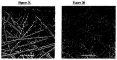
- the carriersWhen seeded into the bioreactor at 1.5x10 6 cells/10 ml culture volume, 14F1.1 cells grew and spread on the carriers ( Figure 5 ). By day 40 following seeding, the carriers contained a 100-fold increased cell density, i.e., approximately 1.5x10 6 cells/carrier, 1.5x10 7 cells/ml (Table 1).
- the cellular density on carriers at various levels of the columnwas the same, indicating a homogenous transfer of oxygen and nutrients to the cells.
- the culture conditionswere optimized for these cells: culture medium (Dulbecco's high-glucose medium + 10 % fetal calf serum), flow rate (1 ml/min), medium exchange frequency (once a week), initial seeding density (as above). No beneficial effect was found for collagen or poly L-lysine carrier coating, on the growth rate and final density of 14F1.1 cells.
- Preliminary findings with primary human marrow stromal cells(Table 1) indicated a similar density of 14F1.1 and primary stromal cells, on days 10 and 14 following seeding, respectively.
- stromal cells conditioned mediumSCM obtained from the bioreactor column (3D SCM)
- SCMstromal cells conditioned medium
- the activitywas compared to SCM obtained from monolayer cultures (2D SCM) containing the same concentration of stromal cells.
- SCM from 14F1.1 cellswas found to be equally or more capable of supporting the maintenance of human CB CD34+38- cells, than SCM from primary marrow stromal cells.
- a maximal effect of 14F1.1 SCMwas consistently observed at a lower concentration than that of primary marrow SCM.
- 3D SCMwas found to be superior to 2D SCM of both cell types, in supporting the expansion of human CB CD34+38- cells.
- the difference in activity between 2D and 3D SCMwas more pronounced with culture duration (14 versus 21 days).
- 14F1.1 3D SCM to suspension cultures of human CB CD34+ cellsalso resulted in the maintenance of CD34+38-CXCR4+, cells (Table 2), as compared to control cultures containing medium alone.
- Table 3demonstrates the effect of cytokines in suspension cultures of CD34+ containing 2D versus 3D SCM. The results clearly demonstrate that 3D SCM was superior to 2D SCM in supporting the maintenance of both CD34+38- and more importantly, the CD34+38-CXCR4+ (SRC) subset.
- TPO+FLT 3 ligandreduced the yield of CD34+38-/ CD34+38-CXCR4+ in the presence of 2D SCM but enhanced their yield in cultures supplemented with 3D SCM. Again, this can be attributed to the lesser extent of differentiation in the 3D system, as determined by the CD34+ surface marker.
- SCFinduced a marked increase in stem cell differentiation and a marked decline in the yield of CD34+38-/ CD34+38-CXCR4+ cells.
- Stromal-cell coated carrierswere superior to carriers alone or to monolayer 14F1.1 cells, in promoting the 7-day survival/maintenance of CD34+38- cells. Prolonged culture (day 14) resulted in increased CD34+38- numbers in both 14F1.1 monolayer and 14F1.1-coated carrier cultures.
Landscapes
- Health & Medical Sciences (AREA)
- Engineering & Computer Science (AREA)
- Life Sciences & Earth Sciences (AREA)
- Organic Chemistry (AREA)
- Bioinformatics & Cheminformatics (AREA)
- Chemical & Material Sciences (AREA)
- Biomedical Technology (AREA)
- Wood Science & Technology (AREA)
- Zoology (AREA)
- Genetics & Genomics (AREA)
- Biotechnology (AREA)
- General Health & Medical Sciences (AREA)
- General Engineering & Computer Science (AREA)
- Biochemistry (AREA)
- Microbiology (AREA)
- Hematology (AREA)
- Immunology (AREA)
- Cell Biology (AREA)
- Animal Behavior & Ethology (AREA)
- Public Health (AREA)
- Veterinary Medicine (AREA)
- Developmental Biology & Embryology (AREA)
- Oral & Maxillofacial Surgery (AREA)
- Transplantation (AREA)
- Nuclear Medicine, Radiotherapy & Molecular Imaging (AREA)
- Medicinal Chemistry (AREA)
- General Chemical & Material Sciences (AREA)
- Chemical Kinetics & Catalysis (AREA)
- Vascular Medicine (AREA)
- Cardiology (AREA)
- Pharmacology & Pharmacy (AREA)
- Heart & Thoracic Surgery (AREA)
- Sustainable Development (AREA)
- Diabetes (AREA)
- Micro-Organisms Or Cultivation Processes Thereof (AREA)
- Medicines Containing Material From Animals Or Micro-Organisms (AREA)
- Apparatus Associated With Microorganisms And Enzymes (AREA)
- Materials For Medical Uses (AREA)
- Immobilizing And Processing Of Enzymes And Microorganisms (AREA)
Abstract
Description
- The present invention relates to a cell culture according to claims 1-12 for maintenance and expansion of hemopoietic stem cells. More particularly, the present invention relates to a three dimensional stromal cell plug flow bioreactor for the maintenance and/or expansion of hemopoietic stem cells and/or for the production of a conditioned medium for the maintenance and/or expansion of hemopoietic stem cells.
- The hemopoietic system in mammals is composed of a heterogenous population of cells that range in function from mature cells with limited proliferative potential to pluripotent stem cells with extensive proliferative, differentiative and self renewal capacities (1-3). Hemopoietic stem cells (HSC) are exclusively required for hemopoietic reconstitution following transplantation and serve as a primary target for gene therapy. In spite of the key role of stem cells in maintaining the hemopoietic system, their extremely low frequency in hemopoietic tissue, as well as the limited ability to maintain or expand undifferentiated stem cells underex-vivo conditions for prolonged periods of time, not only remains a major drawback to essential clinical applications of these cells, but also reflects the current unavailability of, and the need for, novel stem cell regulators.
- It is widely accepted that stem cells are intimately associatedin vivo with discrete niches within the marrow (4-6), which provide molecular signals that collectively mediate their differentiation and self renewal, via cell-cell contacts or short-range interactions (7). These niches are part of the "hemopoietic inductive microenvironment" (HIM), composed of marrow stromal cells, e.g., macrophages, fibroblasts, adipocytes and endothelial cells (8). Marrow stromal cells maintain the functional integrity of the HIM by providing extracellular matrix (ECM) proteins and basement membrane components that facilitate cell-cell contact (9-11). They also provide various soluble or resident cytokines needed for controlled hemopoietic cell differentiation and proliferation (12-14).
- In view of the above, it is not surprising that efforts to develop culture systems for the prolonged maintenance of human HSC were mainly focused on the use of pre-established primary marrow stromal cell monolayers. These included long-term cultures of unirradiated (Dexter cultures, 15) or irradiated (16-19) primary human marrow stroma, as well as human or murine stromal cell lines (16, 19-24), with or without exogenously added cytokines. Output assays for HSC initially relied on the capacity of such cells to produce myeloid progeny (long-term culture initiating cells; LTC-IC) or to generate colonies with cobblestone morphology (cobblestone area forming cells; CAFC) after prolonged culture (5-7 weeks) on such stromal cells (16,17). In spite of the widespread use of LTC-IC and CAFC assays, it is becoming increasingly obvious, however, that they detect highly primitive progenitors, rather than true repopulating hemopoietic stem cells (25, 26).
- A recently developed human stem cell assay detects a SCID repopulating cell (SRC), which homes to the bone marrow of non-obese diabetic (NOD)/SCID mice (27), where it gives rise to human myeloid, lymphoid, erythroid and CD34+ progenitor populations (28-30). The SRC is exclusively found in hemopoietic cell fractions expressing the CD34+38- surface antigen (31) and its frequency in CB (1/3x105 cells) is enriched as compared to BM (1/9x105 cells) or mobilized PB (1/ 6x10⁶ cells) (32). Very recent studies showed that the SRC resides within a subpopulation of CD34+/38-/CXCR4+ cells (33). CXCR4, a surface receptor for the chemokine stromal cell-derived factor 1 (SDF-1, 34), is apparently essential for homing and engraftment of human hemopoietic stem cells in the NOD/SCID marrow (33).
- Studies aimed at inducing prolonged maintenance/expansion of human HSC on stromal cell cultures were mainly based on CAFC, LTC-IC or the CD34+38- phenotype as end-point assays (16, 19-24). Rare reports of SRC maintenance/expansion in stromal cell cultures fail to indicate significant long-term support. For example, allogeneic human marrow stroma was found to induce short-term (7-day) SRC maintenance, followed by a rapid, marked decline (6-fold) in activity (26). The inability to support the long-term maintenance/expansion of transplantable human stem cells on stromal cell layers, may be attributed to several factors related toin vitro cultures of these cells. Among these, one may include the use of stromal cell monolayers, which do not reflect thein vivo growth conditions within the natural, three-dimensional structure of the bone marrow. Such conditions may diminish the capacity of stromal cells to provide the optimal, appropriate supportive microenvironment, as well as the capacity of stem cells to localize in specific niches and to physically interact with stromal cells and their products. Indeed, evidence for the importance of a three dimensional (3D) structure for the biological activity of hemopoietic progenitor cells, is provided by the superior growth of a human hemopoietic cell line on stromal cells seeded in a 3D collagen matrix, as compared to their proliferation on monolayers of such cells (35). More importantly, a 3D tantalum-coated porous biomaterial, was recently shown to enhance the short-term maintenance of macaque LTCIC or CD34+38- cells, as compared to cells cultured alone or on marrow stromal cell monolayers (36). The effect of endothelial cell coated capillaries of an artificial capillary system has been investigated in
WO 95/19793 - Recent studies have shown the murine AFT024 cell line to be superior than human stroma, in supporting 2-3 week survival and maintenance (albeit not expansion) of human CB SRC (37). This line has been found to express several novel HIM genes encoding membrane-bound proteins (21, 38, 39), which may have an essential role in stem cell physiology. The possible expression of these and other genes by stromal cells under conditions which more closely mimic their 3D marrow microenvironment, and thus enable their optimal, physiological functional activity, has yet to be determined.
- Extensive studies have shown that stroma non-contact cultures (19, 21, 22, 40, 41) or stroma conditioned media (SCM) (21, 42-44), alone or with cytokines, can support theex-vivo maintenance or expansion of primitive hemopoietic progenitors. SCM has also been shown to improve the recovery and transduction efficiency of such cells (45, 46). While these findings again stress the importance of soluble stromal cell factors, the use of LTC-IC, CAFC or CD34+38- end-points in such assays cannot reflect the effect of SCM on maintenance/expansion of transplantable HSC. Furthermore, it is not known whether such SCM, obtained from monolayer cultures of stromal cells, indeed contains all stromal cell-associated gene products involved in human HSC physiology.
- Recent attention aimed atex-vivo expansion of transplantable hemopoietic stem cells has focused on the establishment of cytokine-supplemented suspension cultures (47-53). These studies have helped identify the major relevant cytokines for this process, e.g., early-acting ones such as stem cell factor (SCF), FLT3 ligand and thrombopoietin (TPO). Nevertheless, variable results have been obtained, indicating short-term loss (48, 49), maintenance (50-52) but also some rare examples of SRC expansion, following during 2-4 weeks of culture (47, 53). The interactive capacity of these cytokines and stromal cells, under 3D growth conditions, to support the maintenance/expansion of SRC, has not yet been defined.
US-A-5,541,107 describes a three-dimensional cell culture system which can be used to culture a variety of different cells and tissues in vitro for prolongated periods of time. In accordance with the invention, cells derived from a desired tissue are inoculated and grown on a pre-established stromal support matrix. The stromal support matrix comprises stromal cells, such as fibroblasts actively growing on a three-dimensional matrix. Stromal cells may also include other cells found in loose connective tissue such as endothelial cells, macrophages/monocytes, adipocytes, pericytes, reticular cells found in bone marrow stroma, etc. The stromal matrix provides the support, growth factors, and regulatory factors necessary to sustain long-term active proliferation of cells in culture. When grown in this three-dimensional system, the proliferating cells mature and segregate properly to form components of adult tissues analogous to counterparts found in vivo.US-A-5,266,476 relates to a matrix and cultivation system for anchorage dependent cells. The matrix is characterized by a substantially increased available effective surface area for cell attachment which is attained by resorting to the use of a fiber network or open-pore foams with suitable pore size. Attachment of the cells can be enhanced by modifying the surface of the substrate. One embodiment comprises a matrix in particle or flake form.- There is thus a widely recognized need for, and it would be highly advantageous to have, a method and apparatus forex-vivo expansion and/or maintenance of transplantable hemopoietic stem cells devoid of the above limitations, with superior results as is compared to the teachings of the prior art.
The cell culture of the present invention is defined in claims 1-12. - While reducing the present invention to practice, a plug flow bioreactor system which closely mimics the 3D bone marrow microenvironment and which is capable of supporting the growth and prolonged maintenance of stromal cells, has been developed. The latter were seeded on porrosive inorganic carriers made of a non woven fabric matrix of polyester (54), enabling the propagation of large cell numbers in a relatively small volume. The structure and packing of the carrier have a major impact on oxygen and nutrient transfer, as well as on local concentrations and released stromal cell products (e.g., ECM proteins, cytokines, 55). In addition, the capacity of stromal cells cultured in this system to promote the maintenance/expansion of transplantable human hemopoietic stem cells via direct cell-cell contact has been determined to be far superior over prior art methods. Furthermore, the capacity of conditioned medium of stromal cells cultured in this system to promote the maintenance/expansion of transplantable human hemopoietic stem cells via novel stromal-cell associated stem cell factors included therein, has been determined to be far superior over prior art methods.
- Thus, according to one aspect of the present invention there is provided a method of expanding/maintaining undifferentiated hemopoietic stem cells or progenitor cells, the method comprising the steps of (a) obtaining undifferentiated hemopoietic stem cells or progenitor cells; and (b) seeding the undifferentiated hemopoietic stem cells or progenitor cells into a stationary phase plug-flow bioreactor in which a three dimensional stromal cell culture has been pre-established on a substrate in the form of a sheet, the substrate including a non-woven fibrous matrix forming a physiologically acceptable three-dimensional network of fibers, thereby expanding/maintaining undifferentiated hemopoietic stem cells or progenitor cells.
- According to still further features in the described preferred embodiments the method further comprising the step of isolating the undifferentiated hemopoietic stem cells or progenitor cells.
- According to another aspect of the present invention there is provided a method of expanding/maintaining undifferentiated hemopoietic stem cells or progenitor cells, the method comprising the steps of (a) obtaining undifferentiated hemopoietic stem cells or progenitor cells; and (b) culturing the undifferentiated hemopoietic stem cells or progenitor cells in a medium containing a stromal cell conditioned medium, the stromal cell conditioned medium being derived from a stationary phase plug-flow bioreactor in which a three dimensional stromal cell culture has been established on a substrate in the form of a sheet, the substrate including a non-woven fibrous matrix forming a physiologically acceptable three-dimensional network of fibers, thereby expanding/maintaining undifferentiated hemopoietic stem cells or progenitor cells.
- According to yet another aspect of the present invention there is provided a method of preparing a stromal cell conditioned medium useful in expanding/maintaining undifferentiated hemopoietic stem cells or progenitor cells, the method comprising the steps of (a) establishing a stromal cell culture in a stationary phase plug-flow bioreactor on a substrate in the form of a sheet, the substrate including a non-woven fibrous matrix forming a physiologically acceptable three-dimensional network of fibers, thereby expanding/maintaining undifferentiated hemopoietic stem cells or progenitor cells; and (b) when a desired stromal cell density has been achieved, collecting medium from the stationary phase plug-flow bioreactor, thereby obtaining the stromal cell conditioned medium useful in expanding/maintaining undifferentiated hemopoietic stem cells or progenitor cells.
- According to still another aspect of the present invention there is provided a method of transplanting undifferentiated hemopoietic stem cells or progenitor cells into a recipient, the method comprising the steps of (a) expanding/maintaining the undifferentiated hemopoietic stem cells or progenitor cells by (i) obtaining undifferentiated hemopoietic stem cells or progenitor cells; and (ii) seeding the undifferentiated hemopoietic stem cells or progenitor cells into a stationary phase plug-flow bioreactor in which a three dimensional stromal cell culture has been pre-established on a substrate in the form of a sheet, the substrate including a non-woven fibrous matrix forming a physiologically acceptable three-dimensional network of fibers, thereby expanding/maintaining undifferentiated hemopoietic stem cells or progenitor cells; and (b) transplanting the undifferentiated hemopoietic stem cells or progenitor cells resulting from step (a) in the recipient.
- According to still further features in the described preferred embodiments the method further comprising the step of isolating the undifferentiated hemopoietic stem cells or progenitor cells prior to step (b).
- According to an additional aspect of the present invention there is provided a method of transplanting undifferentiated hemopoietic stem cells or progenitor cells into a recipient, the method comprising the steps of (a) expanding/maintaining the undifferentiated hemopoietic stem cells or progenitor cells by (i) obtaining undifferentiated hemopoietic stem cells or progenitor cells; and (ii) culturing the undifferentiated hemopoietic stem cells or progenitor cells in a medium containing a stromal cell conditioned medium, the stromal cell conditioned medium being derived from a stationary phase plug-flow bioreactor in which a three dimensional stromal cell culture has been established on a substrate in the form of a sheet, the substrate including a non-woven fibrous matrix forming a physiologically acceptable three-dimensional network of fibers, thereby expanding/maintaining undifferentiated hemopoietic stem cells or progenitor cells.
- According to yet an additional aspect of the present invention there is provided a bioreactor plug comprising a container having an outlet and an inlet and containing therein a substrate in the form of a sheet, the substrate including a non-woven fibrous matrix forming a physiologically acceptable three-dimensional network of fibers, the substrate supporting at least 5 x 10⁶ stromal cells per cubic centimeter of the substrate.
- According to still an additional aspect of the present invention there is provided a plug-flow bioreactor comprising the above bioreactor plug.
- According to further features in preferred embodiments of the invention described below, the undifferentiated hemopoietic stem cells or progenitor cells are cells isolated from a tissue selected from the group consisting of cord blood, mobilized peripheral blood and bone-marrow.
- According to still further features in the described preferred embodiments the undifferentiated hemopoietic stem cells or progenitor cells and stromal cells of the stromal cell culture share common HLA antigens.
- According to still further features in the described preferred embodiments the undifferentiated hemopoietic stem cells or progenitor cells and stromal cells of the stromal cell culture are from a single individual.
- According to still further features in the described preferred embodiments the undifferentiated hemopoietic stem cells or progenitor cells and stromal cells of the stromal cell culture are from different individuals.
- According to still further features in the described preferred embodiments the undifferentiated hemopoietic stem cells or progenitor cells and stromal cells of the stromal cell culture are from the same species.
- According to still further features in the described preferred embodiments the undifferentiated hemopoietic stem cells or progenitor cells and stromal cells of the stromal cell culture are from different species.
- According to still further features in the described preferred embodiments stromal cells of the stromal cell culture are grown to a density of at least 5 x 10⁶ cells per a cubic centimeter of the substrate.
- According to still further features in the described preferred embodiments stromal cells of the stromal cell culture are grown to a density of at least 107 cells per a cubic centimeter of the substrate.
- According to still further features in the described preferred embodiments the step of seeding the undifferentiated hemopoietic stem cells or progenitor cells into the stationary phase plug-flow bioreactor is effected while flow in the bioreactor is shut off for at least 10 hours following the seeding.
- According to still further features in the described preferred embodiments the fibers form a pore volume as a percentage of total volume of from 40 to 95 % and a pore size of from 10 microns to 100 microns.
- According to still further features in the described preferred embodiments the matrix is made of fiber selected from the group consisting of flat, non-round, and hollow fibers and mixtures thereof, the fibers being of from 0.5 microns to 50 microns in diameter or width.
- According to still further features in the described preferred embodiments the matrix is composed of ribbon formed fibers having a width of from 2 microns According to still further features in the described preferred embodiments the ratio of width to thickness of the fibers is at least 2:1.
- According to still further features in the described preferred embodiments the matrix having a pore volume as a percentage of total volume of from 60 to 95%.
- According to still further features in the described preferred embodiments the matrix has a height of 50-1000 µm.
- According to still further features in the described preferred embodiments the material of the matrix is selected from the group consisting of polyesters, polyalkylenes, polyfluorochloroethylenes, polyvinyl chloride, polystyrene, polysulfones, cellulose acetate, glass fibers, and inert metal fibers.
- According to still further features in the described preferred embodiments the matrix is in a shape selected from the group consisting of squares, rings, discs, and cruciforms.
- According to still further features in the described preferred embodiments the matrix is coated with poly-D-lysine.
- The present invention successfully addresses the shortcomings of the presently known configurations by providing more effective means for expanding/maintaining undifferentiated hemopoietic stem cells.
- Implementation of the method and bioreactor of the present invention may involve performing or completing selected tasks or steps manually, automatically, or a combination thereof. Moreover, according to actual instrumentation and equipment of preferred embodiments of the method and bioreactor of the present invention, several selected steps could be implemented by hardware or by software on any operating system of any firmware or a combination thereof. For example, as hardware, selected steps of the invention could be implemented as a chip or a circuit. As software, selected steps of the invention could be implemented as a plurality of software instructions being executed by a computer using any suitable operating system. In any case, selected steps of the method and bioreactor of the invention could be described as being performed by a data processor, such as a computing platform for executing a plurality of instructions.
- The invention is herein described, by way of example only, with reference to the accompanying drawings. With specific reference now to the drawings in detail, it is stressed that the particulars shown are by way of example and for purposes of illustrative discussion of the preferred embodiments of the present invention only, and are presented in the cause of providing what is believed to be the most useful and readily understood description of the principles and conceptual aspects of the invention. In this regard, no attempt is made to show structural details of the invention in more detail than is necessary for a fundamental understanding of the invention, the description taken with the drawings making apparent to those skilled in the art how the several forms of the invention may be embodied in practice.
- In the drawings:
FIG. 1 is a schematic depiction of an exemplary plug-flow bioreactor 20 which served while reducing the present invention to practice;1- medium reservoir;2 - gas mixture container;3 - gas filters;4 - injection points;5 - plug or container ofplug flow bioreactor 20;6 - flow monitors;6a - flow valves;7 - conditioned medium collecting/separating container;8 - container for medium exchange;9 - peristaltic pump;10 - sampling point;11- container for medium exchange;12 - 02 monitor;14 - steering device;PH - pH probe.FIG. 2 demonstrates CAFC maintenance by 14F1.1 cells. Cord blood CD34+ cells were seeded at limiting dilution onto irradiated 14F1.1 or primary human marrow stroma. Cobblestone formation was determined 5 weeks later. Results represent the mean ± SD of 2 independent experiments.FIG. 3 demonstrates LTC-IC maintenance by 14F1.1 cells. Cord blood CD34+ cells were seeded at limiting dilution onto irradiated 14F1.1 or primary human marrow stroma. Myeloid colony formation was determined 7 weeks later. FLT-3 ligand (300 ng/ml), TPO (300 ng/ml) and SCF (100 ng/ml) were added with weekly medium replacement. Results represent the mean ± SD of 2 experiments.Figure 4 demonstrates expansion of CD34+38- cells on 14F1.1 and primary human marrow stroma. CD34+ cells were seeded onto 14F1.1 or human marrow stroma at 70 CD34+38- cells/well. Cytokines were added weekly. Cultures were trypsinized 7-21 days later. CD34+38- were determined by FACS analysis. Results represent the mean ± SD of 2 independent experiments.FIGs. 5a-b show scanning electron micrographs (SEM) of carrier seeded with 14F1.1 stromal cell line following 10 days (Figure 5a ) or following 40 days (Figure b). Magnification: x 150.FIGs. 6a-b demonstrate the effect of 3Dversus 2D 14F1.1 conditioned medium on CD34+38- expansion. CD34+ cells were seeded in suspension cultures in the presence of various concentrations of conditioned medium from 14F1.1 and primary human marrow stroma. CD34+38- cell numbers were determined by FACS analysis. Results represent the mean ± SD of 2 independent experiments.FIG. 7 demonstrates maintenance of CD34+38- cells on stromal-cell coated carriers. Stromal cell-coated carriers were removed from the 3D system into silicone-coated 96-well dishes, followed by addition of 1.5 x 10⁴ CD34+ cells. Controls contained carriers alone and carrier-equivalent numbers of monolayer (2D) grown 14F1.1 cells. Cells were harvested at the designated times and analyzed by FACS. Results represent the mean ± SD of 2 independent experiments.- The present invention is of methods and bioreactor for hemopoietic stem cell expansion/maintenance which can be used for transplantation in a recipient or for other purposes as if further detailed hereinunder. Specifically, the present invention is of a three dimensional stromal cell plug flow bioreactor for the maintenance and/or expansion of hemopoietic stem cells and/or for the production of a conditioned medium for the maintenance and/or expansion of hemopoietic stem cells, which can be used in a variety of applications.
- The principles and operation of the present invention may be better understood with reference to the drawings and accompanying descriptions.
- Before explaining at least one embodiment of the invention in detail, it is to be understood that the invention is not limited in its application to the details of construction and the arrangement of the components set forth in the following description or illustrated in the drawings. The invention is capable of other embodiments or of being practiced or carried out in various ways. Also, it is to be understood that the phraseology and terminology employed herein is for the purpose of description and should not be regarded as limiting.
- Current strategies aimed at long-termex-vivo maintenance or expansion of transplantable human hemopoietic stem cells (HSC), have so far been met with limited success. A novel three dimensional (3D) plug flow bioreactor which closely mimics the bone marrow microenvironment and which is capable of supporting the growth and prolonged maintenance of marrow stromal cells is described herein. The latter are seeded on porrosive carriers made of a non woven fabric matrix of polyester, packed in a glass column, thereby enabling the propagation of large cell numbers in a relatively small volume. In the examples provided in the Example section that follows, the bioreactor was seeded with the murine 14F1.1 stromal cell line or alternatively with primary human marrow stromal cells. By
day 40 following seeding, the carriers contained a 100-fold increased cell density. The density at various levels of the column was the same, indicating a homogenous transfer of oxygen and nutrients to the cells. Media conditioned by stromal cells within the bioreactor (3D SCM) was superior to stromal cell monolayer (2D) SCM, in supporting the long-term maintenance of human cord blood (CB) CD34+38- cells. 3D SCM was also capable of supporting the expansion of CD34+38-CXCR4+ cells, which represent SCID/NOD repopulating cells (SRC). In the presence of cytokines (FLT3 ligand and TPO), 3D SCM enhanced stem cell self renewal and inhibited differentiation, while the opposite effect was induced by 2D SCM + cytokines. Three dimensional stromal-stem cell cocultures also exhibited superior maintenance of CD34+38- cells than cocultures on monolayer stromal cells. These findings demonstrate that the 3D plug flow bioreactor provides a suitable system forex-vivo maintenance/expansion of human HSC via superior stromal-stem cell contact and perhaps via stromal cell production of known and/or novel stem cell regulators. - The human HSC is an essential target for transplantation and gene therapy. The highly reduced frequency of HSCs, as well as the current unavailablity of growth factors capable of inducing stem cell self renewal in the absence of terminal differentiation, still provide a major hindrance to the implementation of such strategies, well as to the large-scale setup of HSC "banks".
- Current strategies aimed at long-term maintenance/expansion of undifferentiated human HSC, have so far been met with limited success. While recent studies with cytokine-supplemented suspension cultures have shown some SRC expansion, this process was also accompanied by a massive increment of early hemopoietic progenitors (53, 62), indicating that a substantial degree of stem cell differentiation was taking place. An ideal system would be one, for example, in which SRC were expanded, while LTC-IC remain reduced in numbers.
- Current systems for hemopoietic cell expansion employ perfused suspension cultures of hemopoietic cells, alone (see,
U.S. -A-5,646,043 ) or seeded on stromal-cell monolayers (see,U.S. -A- 5,605,822 ). While the former system demonstrates a tremendous production of committed progenitors, the latter suffers from the non-physiological nature of monolayer stromal-stem cell interactions. Additional systems for stem cell expansion describe the use of stromal cell conditioned media (U.S. -A- 4,536,151 and5,437,994 ). However, the latter were obtained from stromal cell monolayer cultures, which are clearly shown herein to be inferior and different in stem cell activating capacity, as compared to 3D SCM (see, Table 3 of the Examples section). Although a stationary phase bioreactor using stromal cell-coated glass beads has recently been described (U.S. -A-5,906,940 ), the beads do not provide a physiological, 3D structure and allow the propagation of a 10-fold lower number of stromal cells per ml, as compared to the carriers employed while reducing the present invention to practice. The advantage of a 3Dversus monolayer stromal cell culture is clearly demonstrated by the findings presented herein of the superior capacity of 3D derived SCM or 3D stromal cell cultures to support the maintenance of CD34+38- cells (see,Figures 6 and 7 ). The superior effect of 3D SCM may be attributed to enhanced levels of known cytokines or novel stem cell regulators. - Experiments aimed at evaluating the combined effects of 3D SCM and various cytokines (SCF, FLT3 ligand, TPO), on CD34+38-CXCR4+ (or SRC) maintenance/expansion (Table 3), clearly show a beneficial effect of 3D SCM in the presence of FLT3 ligand and TPO, but not SCF. These findings can be attributed to a relative inhibitory effect of 3D SCM on stem cell differentiation. These findings strongly indicate that under 3D conditions, novel stromal cell -associated factors which, perhaps less active themselves, may act synergistically with such cytokines, were produced. The use of LTC-IC and committed progenitor cell (GM-CFU) output readouts, in addition to CD34+ output, allow to test for stem cell differentiation.
- The bioreactor described herein is unique in that it combines both 3D stromal cell cultures with a continuous flow system. While 3D stromal-hemopoietic cell systems without continuous medium flow have recently been described (
U.S. -A- 5,541,107 ), the findings described herein (see, for example,Figure 7 ) clearly demonstrate the diminished advantage of 3D stromal cell cultures relative to monolayers, in the absence of continuous flow. - The 3D plug-flow bioreactor described herein is capable of supporting the long-term growth of stromal cell lines, as well as primary marrow stromal cells. The use of stromal cells in the bioreactor is not only essential for the establishment of superior stromal-stem cell contact (via unique "niches" and cell-cell, cell-ECM interactions), but also for stromal cell production of known and novel soluble and membrane-bound cytokines. Stromal cells can facilitate the supplementation of such bioreactors with appropriate cytokines, by using genetically engineered cytokine-producing variants.
- Bioreactor stromal cells can also be engineered to serve as retroviral packaging cell lines, enabling the efficient transduction of genetic material into stem cells, within the bioreactor itself. The use of various stromal cells in the bioreactor can also allow the selection of the most suitable substrate for purging of Ph-positive stem cells, the latter known for their lesser capacity for stromal cell adherence (63). Primary stromal cells have the advantage that they enable the establishment of "autologous" stromal-stem cell bioreactors, on which autologous or even cord blood stem cells can be expanded and which eliminate the need to remove stromal cells prior to transplantation.
- While the initial seeding experiments in the bioreactor indicated a rather small yield of CD34+38- cells in the carrier, the medium flow rate following seeding, as well as initial CD34+ cell numbers seeded into the bioreactor can be readily optimized. CD34+38-CXCR4+ analysis at early time points (1-4 days) following seeding is essential for such optimization.
- In sharp distinction to prior art methods, the bioreactor of the present invention employs a growth matrix that substantially increases the available attachment surface for the adherence of the stromal cells so as to mimic the mechanical infrastructure of bone marrow. For example, for a growth matrix of 0.5 mm in height, the increase is by a factor of at least from 5 to 30 times, calculated by projection onto a base of the growth matrix. Such an increase by a factor of about 5 to 30 times, is per unit layer, and if a plurality of such layers, either stacked or separated by spacers or the like, is used, the factor of 5 to 30 times applies per each such structure. When the matrix is used in sheet form, preferably non-woven fiber sheets, or sheets of open-pore foamed polymers, the preferred thickness of the sheet is about 50 to 1000 µm or more, there being provided adequate porosity for cell entrance, entrance of nutrients and for removal of waste products from the sheet. According to a preferred embodiment the pores having an effective diameter of 10 µm to 100 µm. Such sheets can be prepared from fibers of various thicknesses, the preferred fiber thickness or fiber diameter range being from about 0.5 µm to 20 µm, still more preferred fibers are in the range of 10 µm to 15 µ. m in diameter.
- The structures of the invention may be supported by, or even better bonded to, a porous support sheet or screen providing for dimensional stability and physical strength.
- Such matrix sheets may also be cut, punched, or shredded to provide particles with projected area of the order of about 0.2 mm2 to about 10 mm2, with the same order of thickness (about 50 to 1000 µm).
- Further details relating to the fabrication, use and/or advantages of the growth matrix which was used to reduce the present invention to practice are described in
U.S. -A- 5,168,085 , and in particular,5,266,476 . - As will readily be appreciated by the skilled artisan, the present invention provides expanded undifferentiated hemopoietic stem cell population which can be used in a variety of applications, such as, but not limited to: (i) expansion of human stem cells (of autologous or cord blood source) on recipient stroma, without the need for stromal-stem cell separation prior to transplantation; (ii) depletion of Ph+ CML stem cells in an autologous setting via stromal-stem cell interactions; (iii) gene transfer into self- renewing stem cells within the bioreactor or following harvesting from the bioreactor; (iv) production of 3D stromal cell conditioned medium (SCM) forex-vivo maintenance/expansion of undifferentiated hemopoietic stem cells in suspension cultures or in a stem cell bioreactor; (v) isolation of novel proteins inducing stem cell self renewal in the absence of differentiation, as well as proteins having additional biological functions; (vi) isolation of 3D stromal cell RNA for cloning of novel stromal cell-associated stem cell regulators and additional functional stromal cell gene products.
- According to one aspect of the present invention there is provided a method of expanding/maintaining undifferentiated hemopoietic stem cells or progenitor cells. The method according to this aspect of the present invention is effected by implementing the following method steps. First, undifferentiated hemopoietic stem cells or progenitor cells are obtained. Second, the undifferentiated hemopoietic stem cells or progenitor cells are seeded into a stationary phase plug-flow bioreactor, an example of which is depicted in
Figure 1 along with reference numerals, in which a three dimensional stromal cell culture, of either stromal cell line or primary stromal cell culture, have been pre-established on a substrate in the form of a sheet, the substrate including a non-woven fibrous matrix forming a physiologically acceptable three-dimensional network of fibers, thereby, as is further described above and exemplified in the Examples section that follows, expanding/maintaining undifferentiated hemopoietic stem cells or progenitor cells. - As used herein in the specification and in the claims section that follows, the phrase "undifferentiated hemopoietic stem cells" refers to uncommitted hemopoietic cells.
- As used herein in the specification and in the claims section that follows, the phrase "progenitor cells" refers to committed, yet immature hemopoietic cells.
- Both undifferentiated hemopoietic stem cells and progenitor cells are CD34+ cells. Thus, the phrase "obtaining undifferentiated hemopoietic stem cells or progenitor cells" and its equivalent phrase "undifferentiated hemopoietic stem cells or progenitor cells are obtained" both refer to the obtainment of either isolated undifferentiated hemopoietic stem cells and/or progenitor cells, or a population of CD34+ cells which contain undifferentiated hemopoietic stem cells and progenitor cells.
- As used herein in the specification and in the claims section that follows, the terms "expanding" and "expansion" refer to substantially differentiationless cell growth, i.e., increase of a cell population without differentiation accompanying such increase.
- As used herein in the specification and in the claims section that follows, the terms "maintaining" and "maintenance" refer to substantially differentiationless cell renewal, i.e., substantially stationary cell population without differentiation accompanying such stationarity.
- As used herein the term "differentiation" refers to change from relatively generalized to specialized kinds during development. Cell differentiation of various cell lineages is a well documented process and requires no further description herein.
- As used herein the term "ex-vivo" refers to cells removed from a living organism and are propagated outside the organism (e.g., in a test tube).
- Following expansion, for example, the now expanded undifferentiated hemopoietic stem cells or progenitor cells can be isolated by a variety of affinity separation/labeling techniques, such as, but not limited to, fluorescence activated cell sorting and affinity separation via an affinity substrate. Affinity molecules which can be used to implement such isolation methods include anti-CD34 antibodies, for example, which bind CD34+ cells.
- According to another aspect of the present invention there is provided another method of expanding/maintaining undifferentiated hemopoietic stem cells or progenitor cells. The method according to this aspect of the present invention is effected by implementing the following method steps. First, undifferentiated hemopoietic stem cells or progenitor cells are obtained. Second, the undifferentiated hemopoietic stem cells or progenitor cells are cultured in a medium containing, as a sole ingredient or as an additive, a stromal cell conditioned medium, the stromal cell conditioned medium being derived from a stationary phase plug-flow bioreactor in which a three dimensional stromal cell culture, of either stromal cell line or primary stromal cell culture, have been established on a substrate in the form of a sheet, the substrate including a non-woven fibrous matrix forming a physiologically acceptable three-dimensional network of fibers, thereby, as is further described above and exemplified in the Examples section that follows, expanding/maintaining undifferentiated hemopoietic stem cells or progenitor cells.
- According to yet another aspect of the present invention there is provided a method of preparing a stromal cell conditioned medium useful in expanding/maintaining undifferentiated hemopoietic stem cells or progenitor cells. The method according to this aspect of the present invention is effected by implementing the following method steps. First, a stromal cell culture, of either stromal cell line or primary stromal cell culture, is established in a stationary phase plug-flow bioreactor on a substrate in the form of a sheet, the substrate including a non-woven fibrous matrix forming a physiologically acceptable three-dimensional network of fibers, thereby expanding/maintaining undifferentiated hemopoietic stem cells or progenitor cells. second, when a desired stromal cell density has been achieved, say, for example, above 5 x 10⁶ or above 107 cells per cubic centimeter of the matrix, collecting medium from the stationary phase plug-flow bioreactor, as is further described above and exemplified in the Examples section that follows, obtaining the stromal cell conditioned medium useful in expanding/maintaining undifferentiated hemopoietic stem cells or progenitor cells.
- According to still another aspect of the present invention there is provided a method of transplanting undifferentiated hemopoietic stem cells or progenitor cells into a recipient. The method according to this aspect of the present invention is effected by implementing the following method steps. First, the undifferentiated hemopoietic stem cells or progenitor cells are expanded/maintained by any of the methods described above. Second, the undifferentiated hemopoietic stem cells or progenitor cells resulting from the first step are transplanted in the recipient.
- As is shown in
Figure 1 , according to yet an additional aspect of the present invention there is provided a bioreactor plug comprising a container 5, typically in the form of a column, having an outlet and an inlet and containing therein a substrate in the form of a sheet, the substrate including a non-woven fibrous matrix forming a physiologically acceptable three-dimensional network of fibers, the substrate supporting at least 5 x 10⁶ stromal cells, preferably, at least 107, of either stromal cell line or primary stromal cell culture, per cubic centimeter of the substrate. - According to still an additional aspect of the present invention there is provided a plug-flow bioreactor comprising the above bioreactor plug.
- It will be appreciated in this respect that the substrate may theoretically support up to 5 x 10⁷ cells per cubic centimeter thereof. Once sufficient cells have accumulated on the substrate, means such as irradiation can be employed to cease further cell growth, so as to control the exact number of cells supported by the substrate.
- The undifferentiated hemopoietic stem cells or progenitor cells which are used as a source for such cells while implementing the methods of the present invention can be purified or isolated from a tissue, such as, but not limited to, cord blood, cytokine-mobilized peripheral blood (collected by, for example, leukapheresis) and bone-marrow, all of which are known to include undifferentiated hemopoietic stem cells or progenitor cells. Methods of such separation are well known in the art, the most frequently used being fluorescence activated cell sorting in which cells are first tagged by affinity labeling with a fluorophore and are than collected.
- According to a preferred embodiment of the present invention the undifferentiated hemopoietic stem cells or progenitor cells and stromal cells of the stromal cell culture share common HLA antigens. According to another preferred embodiment of the present invention the undifferentiated hemopoietic stem cells or progenitor cells and the stromal cells of the stromal cell culture are from a single individual. Thus, separation of cells is not required in case of transplantation thereof to a recipient.
- According to still another preferred embodiment of the present invention the undifferentiated hemopoietic stem cells or progenitor cells and stromal cells of the stromal cell culture are from different individuals. For example, a future recipient of the undifferentiated hemopoietic stem cells or progenitor cells and stromal cells be used to provide the stromal cells, whereas the undifferentiated hemopoietic stem cells or progenitor cells and stromal cells are from a donor selected according to HLA compatibility to donate such cells to the recipient. Thus, again, separation of cells is not required prior to transplantation.
- According to another embodiment of the present invention the undifferentiated hemopoietic stem cells or progenitor cells and stromal cells of the stromal cell culture are from the same species. However, according to still another preferred embodiment of the present invention the undifferentiated hemopoietic stem cells or progenitor cells and stromal cells of the stromal cell culture are from different species.
- According to a presently preferred embodiment of the present invention the step of seeding the undifferentiated hemopoietic stem cells or progenitor cells into the stationary phase plug-flow bioreactor is effected while flow in the bioreactor is shut off for at least 10 hours following such seeding, so as to enable the cells to anchor to the stromal cell covered matrix.
- The following descriptions provide insight with respect to preferred substrates which are used while implementing the present invention.
- Thus, according to one embodiment the fibers of the substrate form a pore volume as a percentage of total volume of from 40 to 95 % and a pore size of from 10 microns to 100 microns. According to another embodiment, the matrix making the substrate is made of fiber selected from the group consisting of flat, non-round, and hollow fibers and mixtures thereof, the fibers being of from 0.5 microns to 50 microns in diameter or width. According to still another embodiment, the matrix is composed of ribbon formed fibers having a width of from 2 microns. According to a further embodiment, the ratio of width to thickness of the fibers is at least 2:1. According to still a further embodiment, the matrix making the substrate having a pore volume as a percentage of total volume of from 60 to 95 %. According to still another embodiment, the matrix has a height of 50-1000 µm, whereas stacks thereof are employed. According to yet another embodiment, the material of the matrix making the substrate is selected from the group consisting of polyesters, polyalkylenes, polyfluorochloroethylenes, polyvinyl chloride, polystyrene, polysulfones, cellulose acetate, glass fibers, and inert metal fibers. According to still another embodiment, the matrix is in a shape selected from the group consisting of squares, rings, discs, and cruciforms. According to still another embodiment, the matrix is coated with poly-D-lysine.
- Additional objects, advantages, and novel features of the present invention will become apparent to one ordinarily skilled in the art upon examination of the following examples, which are not intended to be limiting. Additionally, each of the various embodiments and aspects of the present invention as delineated hereinabove and as claimed in the claims section below finds experimental support in the following examples.
- Reference is now made to the following examples, which together with the above descriptions, illustrate the invention in a non limiting fashion.
- Generally, the nomenclature used herein and the laboratory procedures utilized in the present invention include molecular, biochemical, microbiological and recombinant DNA techniques. Such techniques are thoroughly explained in the literature. See, for example, "Molecular Cloning: A laboratory Manual" Sambrook et al., (1989); "Current Protocols in Molecular Biology" Volumes I-III Ausubel, R. M., ed. (1994);Ausubel et al., "Current Protocols in Molecular Biology", John Wiley and Sons, Baltimore, Maryland (1989);Perbal, "A Practical Guide to Molecular Cloning", John Wiley & Sons, New York (1988);Watson et al., "Recombinant DNA", Scientific American Books, New York;Birren et al. (eds) "Genome Analysis: A Laboratory Manual Series", Vols. 1-4, Cold Spring Harbor Laboratory Press, New York (1998); methodologies as set forth in
U.S. Pat. Nos. 4,666,828 ;4,683,202 ;4,801,531 ;5,192,659 and5,272,057 ; "Cell Biology: A Laboratory Handbook", Volumes I-III Cellis, J. E., ed. (1994); "Current Protocols in Immunology" Volumes I-III Coligan J. E., ed. (1994);Stites et al. (eds), "Basic and Clinical Immunology" (8th Edition), Appleton & Lange, Norwalk, CT (1994);Mishell and Shiigi (eds), "Selected Methods in Cellular Immunology", W. H. Freeman and Co., New York (1980); available immunoassays are extensively described in the patent and scientific literature, see, for example,U.S. Pat. Nos. 3,791,932 ;3,839,153 ;3,850,752 ;3,850,578 ;3,853,987 ;3,867,517 ;3,879,262 ;3,901,654 ;3,935,074 ;3,984,533 ;3,996,345 ;4,034,074 ;4,098,876 ;4,879,219 ;5,011,771 and5,281,521 ; "Oligonucleotide Synthesis" Gait, M. J., ed. (1984); "Nucleic Acid Hybridization" Hames, B. D., and Higgins S. J., eds. (1985); "Transcription and Translation" Hames, B. D., and Higgins S. J., eds. (1984); "Animal Cell Culture" Freshney, R. I., ed. (1986); "Immobilized Cells and Enzymes" IRL Press, (1986); "A Practical Guide to Molecular Cloning" Perbal, B., (1984) and "Methods in Enzymology" Vol. 1-317, Academic Press; "PCR Protocols: A Guide To Methods And Applications", Academic Press, San Diego, CA (1990);Marshak et al., "Strategies for Protein Purification and Characterization - A Laboratory Course Manual" CSHL Press (1996). Other general references are provided throughout this document. The procedures therein are believed to be well known in the art and are provided for the convenience of the reader. - Bioreactor: The bioreactor used in accordance with the teachings of the present invention was constructed in accordance with the design described in
Figure 1 . The glassware was designed and manufactured at the Technion (Israel) and connected by silicone tubing (Degania, Israel). The carriers were rotated overnight in phosphate buffered saline (PBS; Beit Ha'Emek Industries, Israel) without Ca+2 and Mg+2, followed by removal of the PBS and released debris. Each column was loaded with 10 ml packed carrier. The bioreactor was filled with PBS-Ca-Mg, all outlets were sealed and the system was autoclaved (120 °C, 30 minutes). The PBS was removed via container [8] and the bioreactor was circulated in a 37°C incubator with 300 ml Dulbecco's high-glucose medium (DMEM; GIBCO BRL) containing 10 % heat-inactivated fetal calf serum (FCS; Beit Ha'Emek Industries, Israel) and a Pen-Strep-Nystatin mixture (100 U/ml:100 µg/ml:1.25 µn/ml; Beit Ha'Emek), for a period of 48 hours. Circulating medium was replaced with fresh DMEM containing the above + 2 mM L-glutamine (Beit Ha'Emek). - Stromal cells: Stromal cell lines were maintained at 37°C in DMEM supplemented with 10 % FCS, in a fully humidified incubator of 5 % CO2 in air. Cells were grown in tissue culture flasks (Coming) and were split by trypsinization upon reaching confluence. Primary human marrow stromal cultures were established from aspirated sternal marrow of hematologically healthy donors undergoing open-heart surgery. Briefly, marrow aspirates were diluted 3-fold in Hank's Balanced Salts Solution (HBSS;. GIBCO BRL) and were subject to Ficoll-Hypaque (Robbins Scientific Corp. Sunnyvale, CA) density gradient centrifugation. Marrow mononuclear cells (<1.077 gm/cm3) were collected, washed 3 times in HBSS and resuspended in long-term culture (LTC) medium, consisting of DMEM supplemented with 12.5 % FCS, 12.5 % horse serum (Beit Ha'Emek), 10-4 M β-mercaptoethanol (Merck) and 10-6 mol/L hydrocortwasone sodium succinate (Sigma). Cells were incubated in 25 ml tissue culture flasks (Coming) for 3 days at 37 °C (5 % CO2) and then at 33 °C (idem) with weekly culture refeeding. Stromal cells from individual donors were employed for each bioreactor. For 3D and monolayer studies, primary stromal cell cultures were split by trypsinization (0.25 % Trypsin and EDTA in Puck's Saline A; Beit Ha'Emek) every 10 days, to allow sufficient stromal cell expansion. For LTC-IC and CAFC (see below), stromal cells were irradiated (1500 cGy) using a137Cs source, cultures were maintained at 33 °C in LTC medium.
- Seeding of stromal cells: Confluent cultures of stromal cell lines or 5-week primary marrow stromal cells were trypsinized and the cells washed 3 times in HBSS, resuspended in bioreactor medium (see above), counted and seeded at 106 cells/ml in 10 ml volumes via an injection point ([4],
Figure 1 ) onto 10 ml carriers in the glass column of the bioreactor. Immediately following seeding, circulation was stopped for 16 hours to allow the cells to settle on the carriers. Stromal cell growth in the bioreactor was monitored by removal of carriers and cell enumeration by the MTT method (56). When stromal cells were confluent, medium was replaced with LTC medium, for continued studies (preparation of SCM, stem cell seeding). - Reparation of stromal cell conditioned medium (SCM): At equivalent cell densities, monolayer and bioreactor stromal cells were recharged with fresh LTC culture medium. SCM was collected following overnight incubation of the cells. For this purpose, medium flow in the 3D cultures was stopped for 16 hours and removed directly from the column prior to re-initiation of circulation. For analysis of the effect of CD34+ cells on stromal cell production of SRC, circulation was stopped at various intervals (2-7 days) after seeding of CD34+ into the 3D system and medium collected from the column as described above. SCM was spun (1000 x g, 10 minutes), filtered and stored at -20 °C. Stromal cells were also grown in the bioreactor in serum-free medium, for the collection of SCM, thereby excluding undefined variables.
- Isolation of CD34+cells: Umbilical cord blood samples taken under sterile conditions during delivery were fractionated on Ficoll-Hypaque and buoyant (<1.077 gr/cm3) mononuclear cells collected. Cells from individual CB samples were pooled, incubated with anti-CD34 antibodies and isolated by midi MACS (Miltenyl Biotech).
- Suspension cultures of CD34+ cells: CB CD34+ cells (5x10⁵/well) were incubated in 24-well dishes (TPP, Switzerland), in 0.5 ml of 0-100 % SCM, minus or plus 300 ng/ml each of FLT3 ligand, SCF, or TPO, alone or combined. Controls contained LTC medium plus or minus cytokines. Cells were incubated at 37 °C at 5 % CO2 in air. Culture medium was exchanged weekly. Prior to seeding and at various times (1-3 weeks), cells were harvested, enumerated and assayed for CD34+/38-/CXCR4+ by flow cytometry. Output assays can also include SRC, CAFC and LTC-IC.
- Stromal-stem cell cocultures: Isolated, pooled CB CD34+ cells were seeded at equivalent numbers (about 5 x 10⁵) onto monolayer or bioreactor containing equivalent densities of confluent stromal cells. Upon addition to the bioreactor, medium flow was stopped for 16 hours to enable contact with stromal cells and was re-initiated at a rate of 0.1 - 1.0 ml per minute. CD34+ cell seeded-stromal cell carriers were removed for control studies in the absence of medium exchange. Cocultures were maintained in LTC medium, with or without cytokines. At various times (up to 4 weeks), nonadherent cells were collected from monolayer supernatants or from circulating culture medium via a container ([8],
Figure 1 ). Adherent cells were collected via sequential trypsinization and exposure to EDTA-based dissociation buffer (GIBCO BRL), followed by gentle pipetting of the cells. To avoid the presence of stromal cells in the resulting suspension, the cells were resuspended in HBSS + 10 % FCS and were subjected to a 60 minutes adhesion procedure in plastic tissue culture dishes (Coming), at 37 °C. Circulating and carrier-isolated hemopoietic cells were washed, counted and assayed separately for CD34+/38-/CXCR4+ by flow cytometry. Output assays can also include SRC, CAFC and LTC-IC. - Flow Cytometry: Cells were incubated at 4°C for 30 minutes with saturating concentrations of monoclonal anti-CD34+PerCP (Beckton-Dickinson), anti-CXCR4-fluorescein isothiocyanate (FITC, R&D systems) and - phycoerythrin (PE, Beckton-Dickinson) antibodies. The cells were washed twice in ice-cold PBS containing 5 % heat-inactivated FCS and resuspended for three-color flow cytometry on a FACSscan (Beckton-Dickinson).
- LTC-IC and CAFC assays: Freshly isolated CD34+ cells, cells isolated from stromal-stem cell cocultures or from suspension cultures, were assayed for LTC-IC and CAFC, as previously described (16, 17). Confluent primary marrow stromal cells were trypsinized, irradiated (1500 cGy) and plated in 0.1 ml in 96-well dishes (Coming) at 1.5x10⁴/well. 24 replicate wells/group were established. Stromal cells were overlaid with 0.1 ml of LTC medium containing serial dilutions of CD34+ cells (500-5 cells/well), or with serial dilutions of cells harvested from various assays. Cultures were directly incubated at 33 °C for 5 weeks, with weekly half-medium exchange. Plates were spun down at 1000 rpm for 10 minutes, culture supernatants removed and remaining cells overlayed with methylcellulose cultures and cytokines for myeloid progenitor cell assays, as previously described (57). Colonies were enumerated following 14 days and LTC-IC frequency determined according to the reciprocal of the concentration of test cells that gives 37 % negative cultures (16). The CAFC assay was basically performed as above except for the absence of overlay of methylcellulose and cytokines. The percentage of wells with at least one phase-dark hemopoietic clone of at least five cells (cobblestone area) beneath the stromal layer was determined at week 6 following seeding of the test cell suspensions, in serial dilutions.
- The bioreactor system employed while reducing the present invention to practice is depicted in
Figure 1 . It contained four parallel plug flow bioreactor units [5]. Each bioreactor unit contained 1 gram of porrosive carriers (4 mm in diameter) made of a non woven fabric matrix of polyester (58). These carriers enable the propagation of large cell numbers in a relatively small volume. The structure and packing of the carrier have a major impact on oxygen and nutrient transfer, as well as on local concentrations and released stromal cell products (e.g., ECM proteins, cytokines, 59). The bioreactor was maintained in an incubator of 37 °C. - The flow in each bioreactor was monitored [6] and regulated by a valve [6a]. Each bioreactor contains a sampling and injection point [4], allowing the sequential seeding of stromal and hemopoietic cells. Culture medium was supplied at pH 7.0 [13] from a reservoir [1]. The reservoir was supplied by a filtered [3] gas mixture containing air/CO2/O2 [2] at differing proportions in order to maintain 5-40 % dissolved oxygen at exit from the column, depending on cell density in the bioreactor. The O2 proportion was suited to the level of dissolved O2 at the bioreactor exit, as was determined by a monitor [12]. The gas mixture was supplied to the reservoir via silicone tubes. The culture medium was passed through a separating container [7] which enabled collection of circulating, nonadherent cells. Circulation of the medium was obtained by means of a peristaltic pump [9] operating at a rate of 0.1-3 ml/minute. The bioreactor units were equipped with an additional sampling point [10] and two containers [8, 11] for continuous medium exchange at a rate of 10-50 ml/day. The use of four parallel bioreactor units enables periodic dismantling for purposes such as cell removal, scanning electron microscopy, histology, immunohistochemistry, RNA extraction, etc.
- In one experiment a bioreactor system containing the murine 14F1.1 stromal cell line (24, 60, 61), which was previously shown to support the growth of committed human myeloid progenitors (24) has been established. This cell line can also equally support human CB CAFC (
Figure 2 ), LTC-IC (Figure 3 ) and CD34+38- cells (Figure 4 ), as well as primary human marrow stromal cells. The results presented in these Figures also show that the addition of FLT3 ligand + TPO to these cultures has no effect on LTC-IC, whereas these cytokines significantly enhanced CAFC and CD34+38- cell output. In contrast, SCF induced a decline in both LTC-IC and CAFC. When seeded into the bioreactor at 1.5x10⁶ cells/10 ml culture volume, 14F1.1 cells grew and spread on the carriers (Figure 5 ). Byday 40 following seeding, the carriers contained a 100-fold increased cell density, i.e., approximately 1.5x10⁶ cells/carrier, 1.5x10⁷ cells/ml (Table 1).TABLE 1 Kinetics of 14F1.1 and primary human marrow stroma growth on carriers Time of stromal on carrier (days) 10 14 20 30 40 14F1.1 human stroma 14F1.1 14F1.1 14F1.1 Top part 1.5 x 10³ 1.5 x 10³ 1 x 10⁵ 3.5 x 10⁵ 1.3 x 10⁶ Middle part 1 x 10³ 1.2 x 10³ 1.3 x 10⁵ 2.0 x 10⁵ 1.3 x 10⁶ Bottom part 1 x 10³ 1 x 10³ 7 x 10⁴ 2.0 x 10⁵ 1.5 x 10⁶ MTT analysis included 5 carriers/determination. Mean of 2 independent experiments. - The cellular density on carriers at various levels of the column was the same, indicating a homogenous transfer of oxygen and nutrients to the cells. The culture conditions were optimized for these cells: culture medium (Dulbecco's high-glucose medium + 10 % fetal calf serum), flow rate (1 ml/min), medium exchange frequency (once a week), initial seeding density (as above). No beneficial effect was found for collagen or poly L-lysine carrier coating, on the growth rate and final density of 14F1.1 cells. Preliminary findings with primary human marrow stromal cells (Table 1) indicated a similar density of 14F1.1 and primary stromal cells, on
days - In order to assay the functional activity of the stromal cells within the bioreactor, the effect of stromal cells conditioned medium (SCM) obtained from the bioreactor column (3D SCM), on the expansion of CD34+38- cells in suspension cultures seeded with human CB CD34+ cells was determined. The activity was compared to SCM obtained from monolayer cultures (2D SCM) containing the same concentration of stromal cells. As shown in
Figure 6 , SCM from 14F1.1 cells was found to be equally or more capable of supporting the maintenance of human CB CD34+38- cells, than SCM from primary marrow stromal cells. A maximal effect of 14F1.1 SCM was consistently observed at a lower concentration than that of primary marrow SCM. Furthermore, 3D SCM was found to be superior to 2D SCM of both cell types, in supporting the expansion of human CB CD34+38- cells. The difference in activity between 2D and 3D SCM was more pronounced with culture duration (14versus 21 days). The addition of 14F1.1 3D SCM to suspension cultures of human CB CD34+ cells also resulted in the maintenance of CD34+38-CXCR4+, cells (Table 2), as compared to control cultures containing medium alone.TABLE 2 Effect of 3D 4F1.1 SCM on yield of CD34+38-/CD34+38-CXCR4+ Cell surface phenotype LTC medium 14F1.1 SCM (50%) Cod34+38- 370 1296 CD34+38- CXCR4+ 0 38 Human CB CD34+ cells (8 x 10⁴ /point) were seeded in suspension cultures containing LTC medium or 50% 3D 14F1.1 SCM. Cultures were harvested 7 days later and cells analyzed by FACS. CD34+38- and CD34+38-CXCR4+ inputs were 2800 and 112, respectively. - Table 3 demonstrates the effect of cytokines in suspension cultures of CD34+ containing 2Dversus 3D SCM. The results clearly demonstrate that 3D SCM was superior to 2D SCM in supporting the maintenance of both CD34+38- and more importantly, the CD34+38-CXCR4+ (SRC) subset.
TABLE 3 Effect of cytokines on expansion of CD34+38-/CD34+38-CXCR4+ cells in 3D 14F1.1 SCM Cell surface phenotype 2D 14F1.1 SCM (50%) 3D 14F1.1 SCM (50%) alone FLT3 ligand + TPO SCF alone FLT3 ligand + TPO SCF CD34+38- 1820 140 0 2720 4080 130 CD34+38-CXCR4 460 70 0 620 930 0 CD34+ 37,000 178,000 361,000 17,000 25,000 210,000 Human CB CD34+ cells (2.6x10⁵ /point) 50% 2D vs 3D 14F1.1 SCM, in the absence or presence of FLT3 ligand (300 ng/ml) TPO (300 ng/ml) or SCF (50 ng/ml). Cultures were harvested 7 days later and cells analyzed by FACS. CD34+38- and CD34+38-CXCR4+ inputs were 7900 and 360, respectively. - This may be related to the stronger effect of 2D SCM on cell differentiation, as detected by the yield of CD34+ cells. TPO+FLT3 ligand reduced the yield of CD34+38-/ CD34+38-CXCR4+ in the presence of 2D SCM but enhanced their yield in cultures supplemented with 3D SCM. Again, this can be attributed to the lesser extent of differentiation in the 3D system, as determined by the CD34+ surface marker. In both 2D and 3D SCM cultures, SCF induced a marked increase in stem cell differentiation and a marked decline in the yield of CD34+38-/ CD34+38-CXCR4+ cells.
- In order to assay stromal-stem cell interactions in our bioreactor, the maintenance/expansion of CD34+38- cells on stromal cell (14F1.1)-coated carriers was first evaluated. The latter were removed from the bioreactor into silicone-coated 96-well dishes, followed by the addition of CD34+ cells. Controls contained carriers alone and carrier-equivalent numbers of monolayer 14F1.1 cells. As shown in
Figure 7 , the survival of CD34+38- cells was enhanced by the presence of the carrier alone, confirming the beneficial effect of a 3D structure on the survival/maintenance of primitive progenitors (36). Stromal-cell coated carriers were superior to carriers alone or to monolayer 14F1.1 cells, in promoting the 7-day survival/maintenance of CD34+38- cells. Prolonged culture (day 14) resulted in increased CD34+38- numbers in both 14F1.1 monolayer and 14F1.1-coated carrier cultures. - In a subsequent experiment 6x10⁶ pooled CB CD34+ (3x10⁵ CD34+38-) cells were seeded into a bioreactor containing 4 columns of non-irradiated, 14F1.1-coated carriers, in 350 ml circulating culture medium. Medium flow was stopped for 16 hours and continued thereafter at a normal rate (1 ml/min). Following 4 days of coculture, circulating medium contained 10 % of the initially seeded CD34+38-cells, determined by FACS analysis of harvested viable cells. Following 18 days of culture, circulating medium contained 0.4 % CD34+38- cells, while carrier -adherent cells contained 3 % of the initially seeded CD34+38- population.
- Although the invention has been described in conjunction with specific embodiments thereof, it is evident that many alternatives, modifications and variations will be apparent to those skilled in the art. Accordingly, it is intended to embrace all such alternatives, modifications and variations that fall within the broad scope of the appended claims. Citation or identification of any reference in this application shall not be construed as an admission that such reference is available as prior art to the present invention.
- 1.Turhan, A.G., Humphries, R.K., Phillips, G.L., Eaves, A.C. & Eaves, C.J. N.Engl. J. Med. 320:1655, 1989.
- 2.Morrison, S.J., Uchida, N. & Weissman I.L. Ann. Rev. Cell. Dev. Biol. 11:35,1995.
- 3.Ogawa, M. Blood 11:2844, 1993.
- 4.Lord BI, Testa NG, Hendry JH. Blood 45:65, 1995.
- 5.Trentin JJ. in Gordon A (ed): Regulation of Hematopoiesis Vol I. New York, N.Y., Appleton-Century-Crafts, p161, 1970.
- 6.Wolf NS. Clin. Hematol. 8:469, 1979.
- 7.Dorshkind K, Ann. Rev. Immunol. 8:11, 1990.
- 8.Allen TD, Dexter TM. Exp. Hematol. 12;517, 1984.
- 9.Gupta P, McCarthy JB, Verfaillie CM. Blood 87:3229, 1996.
- 10.Liesveld JL, Winslow JM, Kempshi MC, Ryan DH, Brennan JK, Abboud CN. Exp. Hematol. 19:63, 1991.
- 11.Long MW, Briddel R, Walter AW, Bruno E, Hoffman R. J. Clin. Invest. 90:251, 1992.
- 12.Eaves CJ, Cashman JD, Kay RJ, Daugherty GJ, Otsuka T, Gabury LA, Hogge DE, Landsdorp PM, Eaves AC, Humphries RK. Blood 78:110, 1991.
- 13.Moore KA, Pytowski B, Witte L, Hicklin D, Lemischka I. Proc. Nat. Acad. Sci. 94:4011, 1997.
- 14.Li L, Milner LA, Deng Y, Iwata M, Banta A, Graf L, Marcovina S, Friedman C, Trask BJ, Hood L, Torok-Storb B. Immunity 8:43, 1998.
- 15.Allen TD, Dexter TM. Exp. Hematol. 12;517, 1984.
- 16.Sutherland H, Landsdorp PM, Henkelman D, Eaves AC, Eaves CJ. Proc. Nat. Acad. Sci. 87:3584, 1990
- 17.Breems DA, Blokland EAW, Nben S, Ploemacher RE. Leukemia 8:1095, 1994.
- 18.Verfaillie C, Blakolmer K, McGlare P. J. Exp. Med. 172:509, 1990.
- 19.Burroughs J, Gupta P, Blazar B, Verfaillie C. Exp. Hematol. 22:1095, 1994.
- 20.Roecklein BA, Torok-Storb B. Blood 85: 997, 1995.
- 21.Thiemann FT, Moore KA, Smogorzewska EM, Lemischka IR, Crooks GM. Exp. Hematol. 26:612, 1998.
- 22.Breems DA, Blokland EAW, Siebel KE, Mayen AEM, Engels LJA, Ploemacher RE. Blood 91:111, 1998.
- 23.Aiuti A, Friedrich C, Sieff CA, Gutierrez-Ramos JC. Exp. Hematol. 26:143, 1998.
- 24.Otsuka T, Satoh H, Ogo T, Bairy O, Gluck U, Zipori D, Nakano T, Okamura Y, Niho Y. Int. J. Cell Cloning 10:153, 1992.
- 25.Larochelle A, Vormoor J, Hahenberg H, Wang JCY, Bhatia M, Lapidot T, Moritz T, Murdoch B, Xiao LX, Kato I, Willimas DA & Dick JE. Nat. Med. 2:1329, 1996.
- 26.Gan OI, MurdochB, Larochelle A, Dick JE. Blood 90:641, 1997.
- 27.Shultz LD, Schweitzer A, Christianson SW, Gott B, Shweitzer IB, Tennent B, McKenna S, Mobraaten L, Rajan TV, Greiner DL, Leiter EH. J. Immunol. 154:180, 1995.
- 28.Larochelle A, Vormoor J, Lapidot T, Sher G, Furukawa T, :I Q, Shultz L, Oliveri NF, Stamatoyannoppoulus G & Dick JE. Hum. Mol. Genet. 4:163, 1995.
- 29.Larochelle A, Vormoor J, Hahenberg H, Wang JCY, Bhatia M, Lapidot T, Moritz T, Murdoch B, Xiao LX, Kato I, Willimas DA & Dick JE. Nat. Med. 2:1329, 1996.
- 30.Dick JE. Sem. Immunol. 8:197,1996.
- 31.Bhatia M, Wang JCY, Kapp U, Bonnet D, Dick JE. Proc. Natl. Acad. Sci. 94:5320, 1997.
- 32.Wang JCY, Doedens M, Dick JE. Blood 89: 3919, 1997.
- 33.Peled A, Petit I, Kollet O, Magid M, Ponomaryov T, Nagler A, Ben-Hur H, Shultz L, Lider O, Alon R, Zipori D, Lapidot T. Science 283: 845-8. 1999
- 34.Bleul CC, Farzan M, Choe H, Parolin C, Clark-Lewis I, Legler DF, Loetscher M, Baggiolini M, Moser B. Nature 382:833, 1966.
- 35.Mizuno S, Wang J, Greenberger J, Glowacki J. Blood 88:189a (abs), 1996.
- 36.Rozenzweig M, Pykett M, Marks DF, Johnson RP. Gene Therapy 4:928, 1997.
- 37.Arakawa-Hoyt, Thiemann FT, Dao MA, Barsky L, Crooks GM, Nolta JA. Blood 92:581a, 1998(Abst).
- 38.Moore KA, Pytowski B, Witte L, Hicklin D, Lemischka I. Proc. Nat. Acad. Sci. 94:4011, 1997.
- 39.Varnum-Finney B, Purton LE, Yu M, Brashem-Stein C, Flowers D, Staats S, Moore KA, Le Roux I, Mann R, Gray G, Artavanis-Tsakonas S, Bernstein ID. Blood 91:4084, 1998.
- 40.Verfaillie CM. Blood 79:2821, 1992.
- 41.Verfaillie CM. Blood 82:2045, 1993.
- 42.Bhatia R, McGlave PB, Miller JS, Wissink S, Lin WN, Verfaillie CM. Exp. Hematol. 25:980, 1997.
- 43.Breems DA, Blokland EAW, Ploemacher RE. Leukemia 11:142, 1997.
- 44.Herman PH, Ferrant A, De Bruyere M, Straetmans N. Leukemia 12:735, 1998.
- 45.Breems DA, Van Driel EM, Hawley RG, Siebel KE, Ploemacher RE. Leukemia 12:951, 1998.
- 46.Aiuti A, Ficara F, Dando J, ZE, Bordignon C. Blood 92:145a, 1998 (Abs).
- 47.Kusadasi N, van Soest PL, Ploemacher RE. Exp. Hematol. 25:699, 1998(Abst).
- 48.Dorrell C, Gan OI, Pereira DS, Dick JE. Exp. Hematol. 25:688, 1998(Abst).
- 49.Bhatia M, Bonnet D, Kapp U, Wang JCY, Murdoch B, Dick JE. J. Exp. Med. 186:619, 1997.
- 50.Kollet O, Moore J, Fajerman I, Ben-Hur H, Hagay Z, Nagler A, Feldman M, Lapidot T. Blood 90:365a, 1997(Abs).
- 51.Bertolini F, Battaglia M, Lanza A, Palermo B, Soligo D, Robustelli della Cuna G. Blood 90:365a, 1997(Abs).
- 52.Luens KM, Travis MA, Chen BP, Hill BL, Scollay R, Murray LJ. Blood 91:1206, 1998.
- 53.Piacibello W, Sanavio F, Severino A, Dane A, Gammaitoni L, Fagioli F, Perissinotto E, Aglietta M. Blood. 93: 3736-49, 1999.
- 54.Kadouri A. Colloid and Surface B: Biointerface, 2:265, 1994.
- 55.Kadouri A, Kompier R, Honigwachs-Sha'anani J, Toledo J, Brosh N, Sternberg D, Levy A, Tzehoval E, Zipori D. Int. J. Cell Cloning 10:299, 1992.
- 56.Hansen MB, Nielsen SE, Berg K. J Immunol Methods 119:203, 1989.
- 57.Merchav S., Wagemaker G., Souza L. and Tatarsky I. Leukemia 5:344, 1991.
- 58.Kadouri A. Colloid and Surface B: Biointerface 2:265 , 1994.
- 59.Kadouri A, Kompier R, Honigwachs-Sha'anani J, Toledo J, Brosh N, Sternberg D, Levy A, Tzehoval E, Zipori D. Int. J. Cell Cloning 10:299, 1992.
- 60.Zipori, D., Toledo, J. & von der Mark, K. Blood 66:447, 1985.
- 61.Otsuka T, Ogo T, Nakano t, Niiro H, Kuga S, Satoh H, Furukawa Y, Zipori D, Niho Y. Stem Cells 12:409, 1994.
- 62.Piacibello W, Sanavio F, Garetto L, Severino A, Bergandi D, Ferrario J, Fagioli F, Berger M, Aglietta M. Blood 89: 2644, 1997.
- 63.Carlo-Stella C, Mangoni L, Piovani G, Garau D, Almici C, Rizzoli V. Blood 83:1373, 1994.
Claims (12)
- A cell culture in a stationary phase plug-flow bioreactor comprising (a) adherent stromal cells on a substrate in the form of a sheet, the substrate including a non-woven fibrous matrix forming a physiologically acceptable three-dimensional network of fibers, wherein the fibers form a pore volume as a percentage of total volume of from 40-95 % and a pore size of from 10-100 µm, and (b) undifferentiated hemopoietic stem cells.
- The cell culture of claim 1, wherein the adherent stromal cells are grown to a density of at least 5x10⁶ cells per a cubic centimeter of the substrate.
- The cell culture of claim 1, wherein the adherent stromal cells are grown to a density of at least 107 cells per a cubic centimeter of the substrate.
- The cell culture of claim 1, wherein the fibers are flat, non-round, hollow, or mixtures thereof.
- The cell culture of claim 4, wherein the fibers are 0.5 microns to 50 microns in diameter or width.
- The cell culture of claim 4, wherein the substrate comprises ribbon-formed fibers having a width of from 2 microns.
- The cell culture of claim 6, wherein the ratio of width to thickness of the fibers is at least 2:1.
- The cell culture of claim 1, wherein the substrate has a pore volume as a percentage of total volume of 60% to 95%.
- The cell culture of claim 1, wherein the substrate has a height of 50-1000 µm.
- The cell culture of claim 1, wherein the substrate is made from polyester, polyalkylene, polyfluorochloroethylene, polyvinyl chloride, polystyrene, polysulfones, cellulose acetate, glass fibers, or inert metal fibers.
- The cell culture of claim 1, wherein the adherent stromal cells are grown to confluence.
- The cell culture of claim 1, wherein the substrate is coated with poly-D-lysine.
Applications Claiming Priority (3)
| Application Number | Priority Date | Filing Date | Title |
|---|---|---|---|
| US11878999P | 1999-02-04 | 1999-02-04 | |
| EP09174118.1AEP2208782B1 (en) | 1999-02-04 | 2000-02-04 | Method and apparatus for maintenance and expansion of hemopoietic stem cells and/or progenitor cells |
| EP00913340AEP1147176B1 (en) | 1999-02-04 | 2000-02-04 | Method and apparatus for maintenance and expansion of hemopoietic stem cells and/or progenitor cells |
Related Parent Applications (5)
| Application Number | Title | Priority Date | Filing Date |
|---|---|---|---|
| EP09174118.1ADivision-IntoEP2208782B1 (en) | 1999-02-04 | 2000-02-04 | Method and apparatus for maintenance and expansion of hemopoietic stem cells and/or progenitor cells |
| EP09174118.1ADivisionEP2208782B1 (en) | 1999-02-04 | 2000-02-04 | Method and apparatus for maintenance and expansion of hemopoietic stem cells and/or progenitor cells |
| EP00913340.6Division | 2000-02-04 | ||
| EP00913340ADivisionEP1147176B1 (en) | 1999-02-04 | 2000-02-04 | Method and apparatus for maintenance and expansion of hemopoietic stem cells and/or progenitor cells |
| EP09174118.1Division | 2009-10-27 |
Publications (2)
| Publication Number | Publication Date |
|---|---|
| EP2311938A1 EP2311938A1 (en) | 2011-04-20 |
| EP2311938B1true EP2311938B1 (en) | 2016-04-06 |
Family
ID=22380750
Family Applications (3)
| Application Number | Title | Priority Date | Filing Date |
|---|---|---|---|
| EP09174118.1AExpired - LifetimeEP2208782B1 (en) | 1999-02-04 | 2000-02-04 | Method and apparatus for maintenance and expansion of hemopoietic stem cells and/or progenitor cells |
| EP10184233.4AExpired - LifetimeEP2311938B1 (en) | 1999-02-04 | 2000-02-04 | Method and apparatus for maintenance and expansion of hemopoietic stem cells and/or progenitor cells |
| EP00913340AExpired - LifetimeEP1147176B1 (en) | 1999-02-04 | 2000-02-04 | Method and apparatus for maintenance and expansion of hemopoietic stem cells and/or progenitor cells |
Family Applications Before (1)
| Application Number | Title | Priority Date | Filing Date |
|---|---|---|---|
| EP09174118.1AExpired - LifetimeEP2208782B1 (en) | 1999-02-04 | 2000-02-04 | Method and apparatus for maintenance and expansion of hemopoietic stem cells and/or progenitor cells |
Family Applications After (1)
| Application Number | Title | Priority Date | Filing Date |
|---|---|---|---|
| EP00913340AExpired - LifetimeEP1147176B1 (en) | 1999-02-04 | 2000-02-04 | Method and apparatus for maintenance and expansion of hemopoietic stem cells and/or progenitor cells |
Country Status (18)
| Country | Link |
|---|---|
| US (7) | US6911201B1 (en) |
| EP (3) | EP2208782B1 (en) |
| JP (1) | JP4523169B2 (en) |
| KR (1) | KR20020013496A (en) |
| CN (1) | CN100402642C (en) |
| AT (1) | ATE452181T1 (en) |
| AU (1) | AU759719B2 (en) |
| BR (1) | BR0009403A (en) |
| CA (1) | CA2360664C (en) |
| DE (1) | DE60043534D1 (en) |
| ES (1) | ES2338405T3 (en) |
| HK (1) | HK1046154B (en) |
| IL (1) | IL144629A (en) |
| MX (1) | MXPA01007820A (en) |
| NZ (1) | NZ513303A (en) |
| RU (1) | RU2249039C2 (en) |
| WO (1) | WO2000046349A1 (en) |
| ZA (1) | ZA200106483B (en) |
Families Citing this family (107)
| Publication number | Priority date | Publication date | Assignee | Title |
|---|---|---|---|---|
| AU759719B2 (en)* | 1999-02-04 | 2003-04-17 | Pluristem Ltd. | Method and apparatus for maintenance and expansion of hemopoietic stem cells and/or progenitor cells |
| US6372494B1 (en)* | 1999-05-14 | 2002-04-16 | Advanced Tissue Sciences, Inc. | Methods of making conditioned cell culture medium compositions |
| NL1017973C2 (en)* | 2000-05-10 | 2002-11-08 | Tristem Trading Cyprus Ltd | Design. |
| AU2001294577A1 (en)* | 2000-09-21 | 2002-04-02 | Schering Corporation | Methods for preparing interferon producing dentitric cells |
| JP2004534049A (en) | 2001-06-07 | 2004-11-11 | スキンメディカ,インコーポレーテッド | Conditioned cell culture media and their uses |
| EP2330181B1 (en) | 2002-04-08 | 2018-11-28 | Octane Biotech Inc. | Automated Tissue Engineering System |
| AU2003242360A1 (en)* | 2002-05-22 | 2003-12-22 | Mbs Corporation | Culture apparatus, artificial tissue and blood preparation |
| US20040042997A1 (en)* | 2002-09-03 | 2004-03-04 | Donnie Rudd | Method of regenerating human tissue |
| WO2004090121A2 (en)* | 2003-04-08 | 2004-10-21 | Yeda Research And Development Co. Ltd | Stem cells having increased sensitivity to a chemoattractant and methods of generating and using same |
| US8352031B2 (en) | 2004-03-10 | 2013-01-08 | Impulse Dynamics Nv | Protein activity modification |
| ATE506431T1 (en)* | 2004-04-23 | 2011-05-15 | Bioe Llc | MULTIPLE LINE PRECURSOR CELLS |
| US7622108B2 (en)* | 2004-04-23 | 2009-11-24 | Bioe, Inc. | Multi-lineage progenitor cells |
| US7521221B2 (en)* | 2005-11-21 | 2009-04-21 | Board Of Trustees Of The University Of Arknasas | Staphylococcus aureus strain CYL1892 |
| US8846393B2 (en) | 2005-11-29 | 2014-09-30 | Gamida-Cell Ltd. | Methods of improving stem cell homing and engraftment |
| WO2007079183A2 (en) | 2005-12-29 | 2007-07-12 | Anthrogenesis Corporation | Placental stem cell populations |
| EP1994143A4 (en)* | 2006-02-06 | 2009-08-26 | Pluristem Ltd | Method and apparatus for maintenance and expansion of hematopoietic stem cells from mononuclear cells |
| WO2007108003A2 (en) | 2006-03-23 | 2007-09-27 | Pluristem Ltd. | Methods for cell expansion and uses of cells and conditioned media produced thereby for therapy |
| CN100384987C (en)* | 2006-04-17 | 2008-04-30 | 大连理工大学 | A method for expanding hematopoietic stem cells under three-dimensional conditions |
| US7727763B2 (en)* | 2006-04-17 | 2010-06-01 | Bioe, Llc | Differentiation of multi-lineage progenitor cells to respiratory epithelial cells |
| EP2024493B1 (en)* | 2006-05-31 | 2014-07-02 | Cellect Biotechnology Ltd. | Methods of selecting stem cells and uses thereof |
| US20100041146A1 (en)* | 2007-01-12 | 2010-02-18 | Nippon Sheet Glass Company, Limited | Three-dimensional cell culture carrier and method for cell culture using the same |
| US8084023B2 (en) | 2007-01-22 | 2011-12-27 | The Board Of Trustees Of The University Of Arkansas | Maintenance and propagation of mesenchymal stem cells |
| US20100172830A1 (en)* | 2007-03-29 | 2010-07-08 | Cellx Inc. | Extraembryonic Tissue cells and method of use thereof |
| MX348010B (en)* | 2007-04-07 | 2017-05-23 | Whitehead Inst Biomedical Res | Reprogramming of somatic cells. |
| US8574567B2 (en)* | 2007-05-03 | 2013-11-05 | The Brigham And Women's Hospital, Inc. | Multipotent stem cells and uses thereof |
| WO2008137115A1 (en)* | 2007-05-03 | 2008-11-13 | The Brigham And Women's Hospital, Inc. | Multipotent stem cells and uses thereof |
| WO2008152640A2 (en)* | 2007-06-13 | 2008-12-18 | Pluristem Ltd. | Three dimensional biocompatible scaffolds for ex-vivo expansion and transplantation of stem cells |
| CA2693827A1 (en)* | 2007-07-25 | 2009-01-29 | Bioe, Inc. | Differentiation of multi-lineage progenitor cells to chondrocytes |
| EP3103463B1 (en) | 2007-09-19 | 2020-01-01 | Pluristem Ltd. | Adherent cells from adipose or placenta tissues and use thereof in therapy |
| WO2009042201A1 (en)* | 2007-09-26 | 2009-04-02 | Celgene Cellular Therapeutics | Angiogenic cells from human placental perfusate |
| CA2724839A1 (en)* | 2008-05-21 | 2009-11-26 | Bioe Llc | Differentiation of multi-lineage progenitor cells to pancreatic cells |
| RU2515156C2 (en) | 2008-05-27 | 2014-05-10 | Плуристем Лтд. | Methods of treating inflammatory diseases of colon |
| DK2330889T3 (en) | 2008-08-20 | 2017-01-30 | Anthrogenesis Corp | Improved cell composition and process for making the same |
| KR20110050688A (en)* | 2008-08-22 | 2011-05-16 | 안트로제네시스 코포레이션 | Bone Defect Treatment Method and Bone Defect Therapeutic Composition Using Placental Cell Population |
| US8524496B2 (en) | 2008-09-02 | 2013-09-03 | Pluristem Ltd. | Adherent cells from placenta tissue and use thereof in therapy |
| GB2463401B (en) | 2008-11-12 | 2014-01-29 | Caris Life Sciences Luxembourg Holdings S A R L | Characterizing prostate disorders by analysis of microvesicles |
| EP2367932B1 (en) | 2008-11-19 | 2019-06-12 | Celularity, Inc. | Amnion derived adherent cells |
| US9687348B2 (en) | 2009-03-03 | 2017-06-27 | The Trustees Of Columbia University In The City Of New York | Method of making a personalized bone graft |
| JP2012533519A (en)* | 2009-06-18 | 2012-12-27 | エムシーツー セル エイピーエス | Bone marrow extracellular matrix extract and its therapeutic use |
| WO2011011300A2 (en)* | 2009-07-20 | 2011-01-27 | Centocor Ortho Biotech Inc. | Differentiation of human embryonic stem cells |
| EP2977445B1 (en) | 2009-11-30 | 2018-07-25 | Pluristem Ltd. | Adherent cells from placenta and use of same in disease treatment |
| US8278101B2 (en)* | 2009-12-07 | 2012-10-02 | Synthecon, Inc. | Stem cell bioprocessing and cell expansion |
| US9128101B2 (en) | 2010-03-01 | 2015-09-08 | Caris Life Sciences Switzerland Holdings Gmbh | Biomarkers for theranostics |
| US9469876B2 (en) | 2010-04-06 | 2016-10-18 | Caris Life Sciences Switzerland Holdings Gmbh | Circulating biomarkers for metastatic prostate cancer |
| NZ602798A (en) | 2010-04-08 | 2014-10-31 | Anthrogenesis Corp | Treatment of sarcoidosis using placental stem cells |
| ES2572211T3 (en) | 2010-05-27 | 2016-05-30 | Pluristem Ltd | Skeletal muscle regeneration using mesenchymal stem cells |
| US9617511B2 (en) | 2010-09-07 | 2017-04-11 | The Board Of Regents Of The University Of Texas System | Tissue-specific differentiation matrices and uses thereof |
| EP2625263B1 (en) | 2010-10-08 | 2020-03-11 | Terumo BCT, Inc. | Configurable methods and systems of growing and harvesting cells in a hollow fiber bioreactor system |
| AR093183A1 (en) | 2010-12-31 | 2015-05-27 | Anthrogenesis Corp | INCREASE IN THE POWER OF PLACENTA MOTHER CELLS USING MODULATING RNA MOLECULES |
| KR101835917B1 (en) | 2011-03-22 | 2018-03-07 | 플루리스템 리미티드 | Methods for treating radiation or chemical injury |
| IL254464B2 (en) | 2011-04-15 | 2023-12-01 | Pluri Biotech Ltd | Methods and systems for harvesting cells |
| EA026193B1 (en)* | 2011-05-18 | 2017-03-31 | Те Риджентс Оф Те Юниверсити Оф Калифорния | Compositions and methods for treating retinal diseases |
| CN113559126A (en) | 2011-06-01 | 2021-10-29 | 人类起源公司 | Treating pain with placental stem cells |
| ES2763331T3 (en) | 2011-06-06 | 2020-05-28 | ReGenesys BVBA | Stem cell expansion in hollow fiber bioreactors |
| CN104105788B (en)* | 2011-10-07 | 2016-09-21 | 帕尔技术英国有限公司 | Fluids process control system and correlation technique |
| WO2013070734A1 (en) | 2011-11-07 | 2013-05-16 | Chaudhry Hina W | Methods of cardiac repair |
| IN2014MN01680A (en) | 2012-02-13 | 2015-07-03 | Gamida Cell Ltd | |
| CN106940268B (en) | 2012-02-23 | 2021-09-21 | 朱诺治疗有限公司 | Chromatographic separation of cells and other complex biological materials |
| US9567569B2 (en) | 2012-07-23 | 2017-02-14 | Gamida Cell Ltd. | Methods of culturing and expanding mesenchymal stem cells |
| US9175266B2 (en) | 2012-07-23 | 2015-11-03 | Gamida Cell Ltd. | Enhancement of natural killer (NK) cell proliferation and activity |
| WO2014037863A1 (en) | 2012-09-04 | 2014-03-13 | Pluristem Ltd. | Methods for prevention and treatment of preeclampsia |
| SI2739720T1 (en) | 2012-09-06 | 2015-04-30 | Pluristem Ltd. | Devices and methods for culture of cells |
| US10351910B2 (en) | 2013-02-20 | 2019-07-16 | Pluristem Ltd | Gene and protein expression properties of adherent stromal cells cultured in 3D |
| WO2014141111A1 (en) | 2013-03-14 | 2014-09-18 | Pluristem Ltd. | Methods for prevention and treatment of graft-versus-host disease |
| KR101442783B1 (en)* | 2013-05-24 | 2014-09-22 | 연세대학교 산학협력단 | Composition for Inducing Erythroid Differentiation |
| TWI555847B (en)* | 2013-08-02 | 2016-11-01 | jia-ju Cai | Culture and production of adipose - derived stem cells and their stem cell secretions |
| WO2015073913A1 (en) | 2013-11-16 | 2015-05-21 | Terumo Bct, Inc. | Expanding cells in a bioreactor |
| CN103690273A (en)* | 2013-11-21 | 2014-04-02 | 中国人民解放军第四军医大学 | In vivo bioreactor |
| EP3114206B1 (en) | 2014-03-04 | 2020-07-29 | Pluristem Ltd. | Systems and methods for growing and harvesting cells |
| CN106232800B (en) | 2014-03-25 | 2020-07-03 | 泰尔茂比司特公司 | Passive replacement of media |
| WO2015155777A1 (en) | 2014-04-10 | 2015-10-15 | Bonus Therapeutics Ltd. | Bone repair compositions |
| CN106414724A (en)* | 2014-04-16 | 2017-02-15 | 朱诺治疗有限公司 | Methods, kits and devices for expanding cell populations |
| JP6830059B2 (en) | 2014-09-26 | 2021-02-17 | テルモ ビーシーティー、インコーポレーテッド | Scheduled cell feeding |
| EP3406272B1 (en) | 2015-01-26 | 2020-12-30 | Ube Industries, Ltd. | Cell culture method using bone marrow-like structure, and porous polymide film for healing bone injury site |
| HK1252174A1 (en) | 2015-05-20 | 2019-05-17 | 细胞基因公司 | In vitro cell culture methods for beta-thalassemia using activin type ii receptor ligand traps |
| WO2017004592A1 (en) | 2015-07-02 | 2017-01-05 | Terumo Bct, Inc. | Cell growth with mechanical stimuli |
| CA2991817A1 (en)* | 2015-08-05 | 2017-02-09 | Konstantinos KOKKALIARIS | Use of dermatopontin for maintaining hematopoietic stem and/or progenitor cells in culture |
| MA45489A (en) | 2015-10-22 | 2018-08-29 | Juno Therapeutics Gmbh | CELL CULTURE PROCESSES, ASSOCIATED KITS AND APPARATUS |
| CN108474002B (en) | 2015-10-22 | 2023-05-23 | 朱诺治疗学有限公司 | Method, reagent cartridge, reagent and device for transduction |
| MA45488A (en) | 2015-10-22 | 2018-08-29 | Juno Therapeutics Gmbh | CELL CULTURE PROCESSES, KITS AND APPARATUS |
| WO2017205667A1 (en)* | 2016-05-25 | 2017-11-30 | Terumo Bct, Inc. | Cell expansion |
| US11685883B2 (en) | 2016-06-07 | 2023-06-27 | Terumo Bct, Inc. | Methods and systems for coating a cell growth surface |
| US11104874B2 (en) | 2016-06-07 | 2021-08-31 | Terumo Bct, Inc. | Coating a bioreactor |
| DE102016114043B3 (en)* | 2016-07-29 | 2017-08-10 | Technische Universität Dresden | Device for isolating stem cells from fetal tissues |
| US20180057784A1 (en) | 2016-08-27 | 2018-03-01 | 3D Biotek, Llc | Bioreactor |
| RU2688321C2 (en)* | 2016-12-12 | 2019-05-21 | Федеральное Государственное Бюджетное Учреждение Науки Институт Молекулярной Биологии Им. В.А. Энгельгардта Российской Академии Наук (Имб Ран) | Method for improving the collecting of hematopoietic cells during their cultivation on the stromal layers by pre-magnetization of the latter |
| EP3601521A2 (en) | 2017-03-31 | 2020-02-05 | Terumo BCT, Inc. | Cell expansion |
| US12234441B2 (en) | 2017-03-31 | 2025-02-25 | Terumo Bct, Inc. | Cell expansion |
| US11624046B2 (en) | 2017-03-31 | 2023-04-11 | Terumo Bct, Inc. | Cell expansion |
| US10961501B2 (en) | 2017-04-07 | 2021-03-30 | Epibone, Inc. | System and method for seeding and culturing |
| US11866465B2 (en) | 2017-04-27 | 2024-01-09 | Juno Therapeutics Gmbh | Oligomeric particle reagents and methods of use thereof |
| KR102546681B1 (en) | 2017-09-01 | 2023-06-22 | 론차 콜로그네 게엠베하 | End-to-End Cell Therapy Automation |
| WO2019108767A1 (en)* | 2017-11-29 | 2019-06-06 | Corning Incorporated | Filtered cell culture caps and cell culture methods |
| WO2020070688A1 (en)* | 2018-10-03 | 2020-04-09 | Pluristem Ltd. | Modular bioreactor |
| KR20210107749A (en) | 2018-12-21 | 2021-09-01 | 옥테인 바이오테크 인코포레이티드 | Carousel for modular biological production units |
| CA3123449A1 (en) | 2018-12-21 | 2020-06-25 | Lonza Walkersville, Inc. | Automated production of viral vectors |
| KR102810534B1 (en) | 2018-12-28 | 2025-05-22 | 옥테인 바이오테크 인코포레이티드 | Cell culture and tissue engineering systems including controlled environment zones |
| CN113498432A (en) | 2019-02-08 | 2021-10-12 | 隆萨沃克斯维尔股份有限公司 | Cell concentration method and device for automated bioreactor |
| RU190863U1 (en)* | 2019-02-15 | 2019-07-15 | Федеральное государственное бюджетное учреждение "Российский ордена Трудового Красного Знамени научно-исследовательский институт травматологии и ортопедии имени Р.Р. Вредена" Министерства здравоохранения Российской Федерации (ФГБУ "РНИИТО им. Р.Р. Вредена" Минздрава России) | DEVICE FOR COMPOUND CELLULAR CULTURE AND A Biodegradable Media |
| CN114729292A (en) | 2019-10-24 | 2022-07-08 | 奥克泰生物科技股份有限公司 | Cell culture chamber with improved cell contact surface |
| EP4058563A4 (en) | 2019-11-11 | 2023-12-06 | Lonza Walkersville, Inc. | Quality control methods for automated cell processing |
| WO2021110908A1 (en) | 2019-12-04 | 2021-06-10 | Centre Hospitalier Universitaire Vaudois (C.H.U.V.) | Device and process for tissue-engineering and regenerative medicine |
| BR112022024866A2 (en) | 2020-06-11 | 2023-02-14 | Mesoblast Int Sarl | 3D CULTURE OF MESENCHYMAL LINEAGE PRECURSOR CELLS OR STEM CELLS |
| EP4314244B1 (en) | 2021-03-23 | 2025-07-23 | Terumo BCT, Inc. | Cell capture and expansion |
| EP4095626A1 (en) | 2021-05-27 | 2022-11-30 | Bühler AG | Optimized industrial bioreactor and method thereof, with mutually dependent, coupled process control loops |
| BR112023022517A2 (en) | 2021-05-27 | 2024-01-02 | Buehler Ag | OPTIMIZED INDUSTRIAL BIOREACTOR AND METHOD THEREOF, WITH COUPLED, MUTUALLY DEPENDENT PROCESS CONTROL CIRCUITS |
| US12152699B2 (en) | 2022-02-28 | 2024-11-26 | Terumo Bct, Inc. | Multiple-tube pinch valve assembly |
Citations (1)
| Publication number | Priority date | Publication date | Assignee | Title |
|---|---|---|---|---|
| US5266476A (en)* | 1985-06-18 | 1993-11-30 | Yeda Research & Development Co., Ltd. | Fibrous matrix for in vitro cell cultivation |
Family Cites Families (250)
| Publication number | Priority date | Publication date | Assignee | Title |
|---|---|---|---|---|
| FR94491E (en)* | 1965-10-13 | 1969-08-22 | Philips Massiot Mat Medic | Pacemaker. |
| US6136019A (en) | 1996-08-19 | 2000-10-24 | Mower Family Chf Treatment Irrevocable Trust | Augmentation of electrical conduction and contractility by biphasic cardiac pacing administered via the cardiac blood pool |
| US6343232B1 (en)* | 1966-08-19 | 2002-01-29 | Mower Chf Treatment Irrevocable Trust | Augmentation of muscle contractility by biphasic stimulation |
| NL154600B (en) | 1971-02-10 | 1977-09-15 | Organon Nv | METHOD FOR THE DETERMINATION AND DETERMINATION OF SPECIFIC BINDING PROTEINS AND THEIR CORRESPONDING BINDABLE SUBSTANCES. |
| US3587567A (en)* | 1968-12-20 | 1971-06-28 | Peter Paul Schiff | Mechanical ventricular assistance assembly |
| DE1924227C3 (en)* | 1969-05-12 | 1974-12-05 | Draegerwerk Ag, 2400 Luebeck | Anesthetic vaporizer |
| NL154598B (en) | 1970-11-10 | 1977-09-15 | Organon Nv | PROCEDURE FOR DETERMINING AND DETERMINING LOW MOLECULAR COMPOUNDS AND PROTEINS THAT CAN SPECIFICALLY BIND THESE COMPOUNDS AND TEST PACKAGING. |
| NL154599B (en) | 1970-12-28 | 1977-09-15 | Organon Nv | PROCEDURE FOR DETERMINING AND DETERMINING SPECIFIC BINDING PROTEINS AND THEIR CORRESPONDING BINDABLE SUBSTANCES, AND TEST PACKAGING. |
| US3942536A (en)* | 1971-03-15 | 1976-03-09 | Mieczyslaw Mirowski | Cardioverting device having single intravascular catheter electrode system and method for its use |
| US3901654A (en) | 1971-06-21 | 1975-08-26 | Biological Developments | Receptor assays of biologically active compounds employing biologically specific receptors |
| US3796221A (en)* | 1971-07-07 | 1974-03-12 | N Hagfors | Apparatus for delivering electrical stimulation energy to body-implanted apparatus with signal-receiving means |
| US3853987A (en) | 1971-09-01 | 1974-12-10 | W Dreyer | Immunological reagent and radioimmuno assay |
| US3867517A (en) | 1971-12-21 | 1975-02-18 | Abbott Lab | Direct radioimmunoassay for antigens and their antibodies |
| NL171930C (en) | 1972-05-11 | 1983-06-01 | Akzo Nv | METHOD FOR DETERMINING AND DETERMINING BITES AND TEST PACKAGING. |
| US3796211A (en)* | 1972-08-07 | 1974-03-12 | Medics Res & Dev Inc | Biopsy sampling method and device for the female genital tract |
| US3850578A (en) | 1973-03-12 | 1974-11-26 | H Mcconnell | Process for assaying for biologically active molecules |
| NL7409823A (en)* | 1973-07-31 | 1975-02-04 | Fujitsu Ltd | OUTPUT DEVICE FOR COORDINATE POSITIONS INFORMATION. |
| US3935074A (en) | 1973-12-17 | 1976-01-27 | Syva Company | Antibody steric hindrance immunoassay with two antibodies |
| US4316472C1 (en)* | 1974-04-25 | 2001-08-14 | Mieczyslaw Mirowski | Cardioverting device with stored energy selecting means and discharge initiating means and related method |
| US3952750A (en) | 1974-04-25 | 1976-04-27 | Mieczyslaw Mirowski | Command atrial cardioverting device |
| US4572191B1 (en)* | 1974-04-25 | 2000-10-24 | Mirowski Miecyslaw | Command atrial cardioverter |
| US3996345A (en) | 1974-08-12 | 1976-12-07 | Syva Company | Fluorescence quenching with immunological pairs in immunoassays |
| US3942750A (en)* | 1974-08-13 | 1976-03-09 | Thomas & Betts Corporation | Adjustable clamp |
| US4034074A (en) | 1974-09-19 | 1977-07-05 | The Board Of Trustees Of Leland Stanford Junior University | Universal reagent 2-site immunoradiometric assay using labelled anti (IgG) |
| US4184493A (en)* | 1975-09-30 | 1980-01-22 | Mieczyslaw Mirowski | Circuit for monitoring a heart and for effecting cardioversion of a needy heart |
| US4030509A (en)* | 1975-09-30 | 1977-06-21 | Mieczyslaw Mirowski | Implantable electrodes for accomplishing ventricular defibrillation and pacing and method of electrode implantation and utilization |
| US4202340A (en)* | 1975-09-30 | 1980-05-13 | Mieczyslaw Mirowski | Method and apparatus for monitoring heart activity, detecting abnormalities, and cardioverting a malfunctioning heart |
| US3984533A (en) | 1975-11-13 | 1976-10-05 | General Electric Company | Electrophoretic method of detecting antigen-antibody reaction |
| US4098876A (en) | 1976-10-26 | 1978-07-04 | Corning Glass Works | Reverse sandwich immunoassay |
| US4106494A (en) | 1977-08-29 | 1978-08-15 | American Optical Corporation | Heart defibrillating and monitoring system |
| US4164216A (en) | 1978-01-26 | 1979-08-14 | Person Orville W | Throat obstruction expulsion device |
| US4223678A (en) | 1978-05-03 | 1980-09-23 | Mieczyslaw Mirowski | Arrhythmia recorder for use with an implantable defibrillator |
| US4273114A (en)* | 1978-10-19 | 1981-06-16 | Michigan Instruments, Inc. | Cardiopulmonary resuscitator, defibrillator and monitor |
| US4293734A (en)* | 1979-02-23 | 1981-10-06 | Peptek, Incorporated | Touch panel system and method |
| US4237895A (en) | 1979-04-20 | 1980-12-09 | Medcor, Inc. | Control signal transmitter and monitor for implanted pacer |
| US4403614A (en) | 1979-07-19 | 1983-09-13 | Medtronic, Inc. | Implantable cardioverter |
| US4686332A (en)* | 1986-06-26 | 1987-08-11 | International Business Machines Corporation | Combined finger touch and stylus detection system for use on the viewing surface of a visual display device |
| US4312354A (en)* | 1980-02-04 | 1982-01-26 | Arco Medical Products Company | Pacemaker with circuit for pulse width modulating stimulus pulses in accordance with programmed parameter control states |
| US4879219A (en) | 1980-09-19 | 1989-11-07 | General Hospital Corporation | Immunoassay utilizing monoclonal high affinity IgM antibodies |
| US4440172A (en)* | 1980-10-02 | 1984-04-03 | Mieczyslaw Mirowski | Apparatus for combining pacing and cardioverting functions in a single implanted device |
| US4387717A (en)* | 1980-10-03 | 1983-06-14 | Research Corporation | Pacer internal cardiac electrogram sensing system |
| US4639720A (en)* | 1981-01-12 | 1987-01-27 | Harris Corporation | Electronic sketch pad |
| US4407288B1 (en) | 1981-02-18 | 2000-09-19 | Mieczyslaw Mirowski | Implantable heart stimulator and stimulation method |
| US4384585A (en)* | 1981-03-06 | 1983-05-24 | Medtronic, Inc. | Synchronous intracardiac cardioverter |
| US4428366A (en)* | 1981-05-06 | 1984-01-31 | Alfred B. Kurtz | Electromagnetic apparatus and method for the reduction of serum glucose levels |
| US4765341A (en) | 1981-06-22 | 1988-08-23 | Mieczyslaw Mirowski | Cardiac electrode with attachment fin |
| US4554922A (en) | 1982-09-30 | 1985-11-26 | Prystowsky Eric N | Method of inhibiting cardiac arrhythmias |
| DE3244405A1 (en) | 1982-12-01 | 1984-06-07 | Fa. J. Eberspächer, 7300 Esslingen | DEVICE FOR MIXTURE TREATMENT IN MOTOR-INDEPENDENT HEATERS |
| CA1199371A (en)* | 1982-12-03 | 1986-01-14 | Orest Z. Roy | Ultrasonic enhancement of cardiac contractility synchronised with ecg event or defibrillation pulse |
| US4506680A (en)* | 1983-03-17 | 1985-03-26 | Medtronic, Inc. | Drug dispensing body implantable lead |
| US4550221A (en)* | 1983-10-07 | 1985-10-29 | Scott Mabusth | Touch sensitive control device |
| US5011771A (en) | 1984-04-12 | 1991-04-30 | The General Hospital Corporation | Multiepitopic immunometric assay |
| US4543956A (en) | 1984-05-24 | 1985-10-01 | Cordis Corporation | Biphasic cardiac pacer |
| US4628934A (en) | 1984-08-07 | 1986-12-16 | Cordis Corporation | Method and means of electrode selection for pacemaker with multielectrode leads |
| US4666828A (en) | 1984-08-15 | 1987-05-19 | The General Hospital Corporation | Test for Huntington's disease |
| US4566456A (en)* | 1984-10-18 | 1986-01-28 | Cordis Corporation | Apparatus and method for adjusting heart/pacer rate relative to right ventricular systolic pressure to obtain a required cardiac output |
| US4683202A (en) | 1985-03-28 | 1987-07-28 | Cetus Corporation | Process for amplifying nucleic acid sequences |
| US4801531A (en) | 1985-04-17 | 1989-01-31 | Biotechnology Research Partners, Ltd. | Apo AI/CIII genomic polymorphisms predictive of atherosclerosis |
| US4674508A (en)* | 1985-05-28 | 1987-06-23 | Cordis Corporation | Low-power consumption cardiac pacer based on automatic verification of evoked contractions |
| US4690155A (en) | 1985-07-03 | 1987-09-01 | Cordis Corporation | Monophasic action potential recording lead |
| US4726379A (en)* | 1985-11-14 | 1988-02-23 | Cardiac Control Systems, Inc. | Cardiac pacer with switching circuit for isolation |
| US4679572A (en) | 1986-03-11 | 1987-07-14 | Intermedics, Inc. | Low threshold cardiac pacing electrodes |
| US5032508A (en)* | 1988-09-08 | 1991-07-16 | Marrow-Tech, Inc. | Three-dimensional cell and tissue culture system |
| US5266480A (en)* | 1986-04-18 | 1993-11-30 | Advanced Tissue Sciences, Inc. | Three-dimensional skin culture system |
| WO1987006120A1 (en)* | 1986-04-18 | 1987-10-22 | Marrow-Tech Incorporated | Process for replicating bone marrow |
| US4830006B1 (en)* | 1986-06-17 | 1997-10-28 | Intermedics Inc | Implantable cardiac stimulator for detection and treatment of ventricular arrhythmias |
| JPS6370326A (en)* | 1986-09-12 | 1988-03-30 | Wacom Co Ltd | Position detector |
| US4726279A (en)* | 1986-11-12 | 1988-02-23 | United Technologies Corporation | Wake stabilized supersonic combustion ram cannon |
| DE3732640C1 (en)* | 1987-09-28 | 1989-05-18 | Alt Eckhard | Medical device for determining physiological functional parameters |
| US5018522A (en) | 1987-10-26 | 1991-05-28 | Medtronic, Inc. | Ramped waveform non-invasive pacemaker |
| KR0122737B1 (en)* | 1987-12-25 | 1997-11-20 | 후루다 모또오 | Position detection device |
| US5387419A (en)* | 1988-03-31 | 1995-02-07 | The University Of Michigan | System for controlled release of antiarrhythmic agents |
| US4914624A (en)* | 1988-05-06 | 1990-04-03 | Dunthorn David I | Virtual button for touch screen |
| DE3816042A1 (en) | 1988-05-10 | 1989-11-23 | Alt Eckhard | ENERGY SAVING HEART PACEMAKER |
| CA1327838C (en)* | 1988-06-13 | 1994-03-15 | Fred Zacouto | Implantable device to prevent blood clotting disorders |
| US5272057A (en) | 1988-10-14 | 1993-12-21 | Georgetown University | Method of detecting a predisposition to cancer by the use of restriction fragment length polymorphism of the gene for human poly (ADP-ribose) polymerase |
| JPH0538723Y2 (en)* | 1988-12-19 | 1993-09-30 | ||
| US4928688A (en)* | 1989-01-23 | 1990-05-29 | Mieczyslaw Mirowski | Method and apparatus for treating hemodynamic disfunction |
| USRE38119E1 (en)* | 1989-01-23 | 2003-05-20 | Mirowski Family Ventures, LLC | Method and apparatus for treating hemodynamic disfunction |
| US5031617A (en) | 1989-03-13 | 1991-07-16 | Klettner Harold L | Method of altering human blood glucose levels by the application of electric charge |
| US5437994A (en) | 1989-06-15 | 1995-08-01 | Regents Of The University Of Michigan | Method for the ex vivo replication of stem cells, for the optimization of hematopoietic progenitor cell cultures, and for increasing the metabolism, GM-CSF secretion and/or IL-6 secretion of human stromal cells |
| US5605822A (en) | 1989-06-15 | 1997-02-25 | The Regents Of The University Of Michigan | Methods, compositions and devices for growing human hematopoietic cells |
| US5101814A (en)* | 1989-08-11 | 1992-04-07 | Palti Yoram Prof | System for monitoring and controlling blood glucose |
| US5192659A (en) | 1989-08-25 | 1993-03-09 | Genetype Ag | Intron sequence analysis method for detection of adjacent and remote locus alleles as haplotypes |
| DE68928987T2 (en)* | 1989-10-02 | 1999-11-11 | Koninkl Philips Electronics Nv | Data processing system with a touch display and a digitizing tablet, both integrated in an input device |
| US5402151A (en)* | 1989-10-02 | 1995-03-28 | U.S. Philips Corporation | Data processing system with a touch screen and a digitizing tablet, both integrated in an input device |
| US5020544A (en)* | 1989-11-01 | 1991-06-04 | Cardiac Pacemakers, Inc. | Low energy defibrillation electrode |
| US5044375A (en) | 1989-12-08 | 1991-09-03 | Cardiac Pacemakers, Inc. | Unitary intravascular defibrillating catheter with separate bipolar sensing |
| US4998531A (en)* | 1990-03-28 | 1991-03-12 | Cardiac Pacemakers, Inc. | Implantable N-phasic defibrillator output bridge circuit |
| US5097843A (en)* | 1990-04-10 | 1992-03-24 | Siemens-Pacesetter, Inc. | Porous electrode for a pacemaker |
| US5236413B1 (en) | 1990-05-07 | 1996-06-18 | Andrew J Feiring | Method and apparatus for inducing the permeation of medication into internal tissue |
| US5083564A (en)* | 1990-06-01 | 1992-01-28 | Board Of Regents Of The University Of Oklahoma | Method for alleviating and diagnosing symptoms of heart block |
| US5320642A (en)* | 1990-06-01 | 1994-06-14 | Board Of Regents For The University Of Ok | Method for alleviating and diagnosing symptoms of heart block |
| US5205284A (en)* | 1990-06-12 | 1993-04-27 | Zoll Medical Corporation | Method and apparatus for transcutaneous electrical cardiac pacing with background stimulation |
| DE69131064T2 (en)* | 1990-06-12 | 1999-11-18 | Hitachi Maxell, Ltd. | ORGANIC, ELECTROLYTIC BATTERY |
| US5499971A (en)* | 1990-06-15 | 1996-03-19 | Cortrak Medical, Inc. | Method for iontophoretically delivering drug adjacent to a heart |
| AU8074591A (en)* | 1990-06-15 | 1992-01-07 | Cortrak Medical, Inc. | Drug delivery apparatus and method |
| US5087243A (en)* | 1990-06-18 | 1992-02-11 | Boaz Avitall | Myocardial iontophoresis |
| US5262320A (en)* | 1990-06-18 | 1993-11-16 | Massachusetts Institute Of Technology | Cell-culturing apparatus and method employing a macroporous support |
| US5370665A (en) | 1990-08-10 | 1994-12-06 | Medtronic, Inc. | Medical stimulator with multiple operational amplifier output stimulation circuits |
| US5156149A (en) | 1990-08-10 | 1992-10-20 | Medtronic, Inc. | Sensor for detecting cardiac depolarizations particularly adapted for use in a cardiac pacemaker |
| US5584804A (en) | 1990-10-10 | 1996-12-17 | Life Resuscitation Technologies, Inc. | Brain resuscitation and organ preservation device and method for performing the same |
| US5163428A (en) | 1990-10-11 | 1992-11-17 | Ventritex, Inc. | Implantable cardiac defibrillator with current leakage detecting means |
| US5111815A (en)* | 1990-10-15 | 1992-05-12 | Cardiac Pacemakers, Inc. | Method and apparatus for cardioverter/pacer utilizing neurosensing |
| US5137021A (en) | 1990-11-29 | 1992-08-11 | Medtronic, Inc. | Lead current measurement circuit |
| US5665822A (en)* | 1991-10-07 | 1997-09-09 | Landec Corporation | Thermoplastic Elastomers |
| US5129394A (en) | 1991-01-07 | 1992-07-14 | Medtronic, Inc. | Method and apparatus for controlling heart rate in proportion to left ventricular pressure |
| US5476497A (en) | 1991-01-09 | 1995-12-19 | Ann Mirowski | Oval electrode lead body |
| IT1246631B (en)* | 1991-01-25 | 1994-11-24 | Gd Spa | STABILIZATION CONVEYOR OF CIGARETTES PACKAGES OUT OF A PACKAGING MACHINE. |
| US5156147A (en) | 1991-02-05 | 1992-10-20 | Cardiac Pacemakers, Inc. | Variable rate pacemaker having upper rate limit governor based on hemodynamic performance |
| US5161527A (en) | 1991-02-13 | 1992-11-10 | Telectronics Pacing Systems, Inc. | Apparatus and method for detecting abnormal cardiac rhythms in dual chamber arrhythmia control system |
| US5199428A (en)* | 1991-03-22 | 1993-04-06 | Medtronic, Inc. | Implantable electrical nerve stimulator/pacemaker with ischemia for decreasing cardiac workload |
| US5464020A (en) | 1991-03-25 | 1995-11-07 | Lerner; Albert M. | Diagnosing and treating subacute cardiac dysfunction |
| EP0583237A1 (en)* | 1991-04-29 | 1994-02-23 | LERNER, Inna | Method for determining sensory functions |
| WO1994008657A1 (en) | 1992-10-20 | 1994-04-28 | Noel Desmond Gray | A heart pacemaker |
| US5168085A (en) | 1991-05-20 | 1992-12-01 | Corning Incorporated | Multi-stage twc system |
| WO1992021285A1 (en)* | 1991-05-24 | 1992-12-10 | Ep Technologies, Inc. | Combination monophasic action potential/ablation catheter and high-performance filter system |
| US5458568A (en) | 1991-05-24 | 1995-10-17 | Cortrak Medical, Inc. | Porous balloon for selective dilatation and drug delivery |
| US5213098A (en)* | 1991-07-26 | 1993-05-25 | Medtronic, Inc. | Post-extrasystolic potentiation stimulation with physiologic sensor feedback |
| US5381160A (en)* | 1991-09-27 | 1995-01-10 | Calcomp Inc. | See-through digitizer with clear conductive grid |
| DK0541338T3 (en)* | 1991-11-04 | 1996-12-02 | Cardiac Pacemakers Inc | Implantable device for monitoring and stimulating the heart for diagnosis and therapy |
| US5284491A (en)* | 1992-02-27 | 1994-02-08 | Medtronic, Inc. | Cardiac pacemaker with hysteresis behavior |
| US5531764A (en) | 1992-03-24 | 1996-07-02 | Angeion Corporation | Implantable defibrillator system and method having successive changeable defibrillation waveforms |
| US5342404A (en) | 1992-04-03 | 1994-08-30 | Intermedics, Inc. | Implantable medical interventional device |
| US5365461A (en)* | 1992-04-30 | 1994-11-15 | Microtouch Systems, Inc. | Position sensing computer input device |
| US5501662A (en)* | 1992-05-22 | 1996-03-26 | Genetronics, Inc. | Implantable electroporation method and apparatus for drug and gene delivery |
| US5543588A (en)* | 1992-06-08 | 1996-08-06 | Synaptics, Incorporated | Touch pad driven handheld computing device |
| US6239389B1 (en)* | 1992-06-08 | 2001-05-29 | Synaptics, Inc. | Object position detection system and method |
| US5861583A (en)* | 1992-06-08 | 1999-01-19 | Synaptics, Incorporated | Object position detector |
| DE69324067T2 (en)* | 1992-06-08 | 1999-07-15 | Synaptics Inc | Object position detector |
| US5366486A (en) | 1992-06-25 | 1994-11-22 | Indiana University Foundation | Automatic fibrillation detector and defibrillator apparatus and method |
| US5281521A (en) | 1992-07-20 | 1994-01-25 | The Trustees Of The University Of Pennsylvania | Modified avidin-biotin technique |
| US5342401A (en) | 1992-08-19 | 1994-08-30 | The Regents Of The University Of California | Real time cardiac arrhythmia stabilizing system |
| US5320643A (en) | 1992-10-06 | 1994-06-14 | Medtronic, Inc. | Automatic cardiac capture restoration and threshold-seeking method and apparatus |
| US5634899A (en)* | 1993-08-20 | 1997-06-03 | Cortrak Medical, Inc. | Simultaneous cardiac pacing and local drug delivery method |
| US5478739A (en)* | 1992-10-23 | 1995-12-26 | Advanced Tissue Sciences, Inc. | Three-dimensional stromal cell and tissue culture system |
| US5334222A (en) | 1992-11-03 | 1994-08-02 | Cardiac Pacemakers, Inc. | Cardiac stimulating apparatus and method for heart failure therapy |
| SE9203284D0 (en) | 1992-11-04 | 1992-11-04 | Siemens Elema Ab | HJAERTSTIMULATOR |
| US5807306A (en) | 1992-11-09 | 1998-09-15 | Cortrak Medical, Inc. | Polymer matrix drug delivery apparatus |
| US5346506A (en) | 1992-11-10 | 1994-09-13 | Mower Morton M | Method for establishing defibrillation threshold for a cardiac defibrillator |
| US5353800A (en) | 1992-12-11 | 1994-10-11 | Medtronic, Inc. | Implantable pressure sensor lead |
| US5327887A (en) | 1993-01-25 | 1994-07-12 | Ludwik Nowakowski | Cardiopulmonary resuscitation device |
| US5386837A (en)* | 1993-02-01 | 1995-02-07 | Mmtc, Inc. | Method for enhancing delivery of chemotherapy employing high-frequency force fields |
| US5320543A (en)* | 1993-02-04 | 1994-06-14 | Barton Craig S | Flexible plug protector |
| US6133906A (en)* | 1993-03-15 | 2000-10-17 | Microtouch Systems, Inc. | Display-integrated stylus detection system |
| JP2986047B2 (en)* | 1993-04-29 | 1999-12-06 | インターナショナル・ビジネス・マシーンズ・コーポレイション | Digital input display device and input processing device and method |
| US5528002A (en)* | 1993-07-15 | 1996-06-18 | Pentel Kabushiki Kaisha | Noiseproof digitizing apparatus with low power cordless pen |
| US5738096A (en)* | 1993-07-20 | 1998-04-14 | Biosense, Inc. | Cardiac electromechanics |
| US5391199A (en) | 1993-07-20 | 1995-02-21 | Biosense, Inc. | Apparatus and method for treating cardiac arrhythmias |
| US5468254A (en) | 1993-07-26 | 1995-11-21 | Cardiac Pacemakers, Inc. | Method and apparatus for defibrillation using a multiphasic truncated exponential waveform |
| US5368040A (en) | 1993-08-02 | 1994-11-29 | Medtronic, Inc. | Apparatus and method for determining a plurality of hemodynamic variables from a single, chroniclaly implanted absolute pressure sensor |
| US5571997A (en)* | 1993-08-02 | 1996-11-05 | Kurta Corporation | Pressure sensitive pointing device for transmitting signals to a tablet |
| BE1007462A3 (en)* | 1993-08-26 | 1995-07-04 | Philips Electronics Nv | Data processing device with touch sensor and power. |
| US5443485A (en) | 1993-09-08 | 1995-08-22 | Intermedics, Inc. | Apparatus and method for capture detection in a cardiac stimulator |
| AU7729094A (en)* | 1993-09-15 | 1995-04-03 | Pacesetter, Inc. | Synchronized cardioverter shock therapy for preemptive depolarization |
| US5415629A (en)* | 1993-09-15 | 1995-05-16 | Henley; Julian L. | Programmable apparatus for the transdermal delivery of drugs and method |
| US5476485A (en) | 1993-09-21 | 1995-12-19 | Pacesetter, Inc. | Automatic implantable pulse generator |
| US5411531A (en)* | 1993-09-23 | 1995-05-02 | Medtronic, Inc. | Method and apparatus for control of A-V interval |
| SE9303736D0 (en) | 1993-11-12 | 1993-11-12 | Siemens Elema Ab | Apparatus intended to sense the physical state of a living being |
| US5425363A (en)* | 1993-12-17 | 1995-06-20 | Wang; Yong G. | Plunge electrode for recording multiple intramyocardial monophasic action potential |
| US5419763B1 (en)* | 1994-01-04 | 1997-07-15 | Cor Trak Medical Inc | Prostatic drug-delivery catheter |
| JPH08205860A (en)* | 1994-01-21 | 1996-08-13 | Usa Government | Method of expanding and transplantation of hematopoietic cell |
| US5391192A (en)* | 1994-03-04 | 1995-02-21 | Telectronics Pacing Systems, Inc. | Automatic ventricular pacing pulse threshold determination utilizing an external programmer and a surface electrocardiogram |
| IT233201Y1 (en)* | 1994-03-24 | 2000-01-26 | Bracco Spa | TWO-COMPONENT DEVICE FOR THE ADMINISTRATION OF DRUGS |
| GB9406702D0 (en)* | 1994-04-05 | 1994-05-25 | Binstead Ronald P | Multiple input proximity detector and touchpad system |
| KR100300397B1 (en)* | 1994-04-21 | 2001-10-22 | 김순택 | System having touch panel and digitizer function and driving method |
| US5562708A (en) | 1994-04-21 | 1996-10-08 | Medtronic, Inc. | Method and apparatus for treatment of atrial fibrillation |
| US5540722A (en) | 1994-05-16 | 1996-07-30 | Physiometrix, Inc. | Switch apparatus and method for switching between multiple electrodes for diagnostic and therapeutic procedures |
| US5543589A (en)* | 1994-05-23 | 1996-08-06 | International Business Machines Corporation | Touchpad with dual sensor that simplifies scanning |
| US5735876A (en)* | 1994-05-31 | 1998-04-07 | Galvani Ltd. | Electrical cardiac output forcing method and apparatus for an atrial defibrillator |
| JP3186946B2 (en)* | 1994-05-31 | 2001-07-11 | シャープ株式会社 | Coordinate detection device |
| US5514162A (en)* | 1994-06-07 | 1996-05-07 | Pacesetter, Inc. | System and method for automatically determining the slope of a transfer function for a rate-responsive cardiac pacemaker |
| EP0688579B1 (en) | 1994-06-24 | 2001-08-22 | St. Jude Medical AB | Device for heart therapy |
| US5601611A (en)* | 1994-08-05 | 1997-02-11 | Ventritex, Inc. | Optical blood flow measurement apparatus and method and implantable defibrillator incorporating same |
| US5626622A (en)* | 1994-09-21 | 1997-05-06 | Telectronics Pacing Systems, Inc. | Dual sensor rate responsive pacemaker |
| US5687734A (en) | 1994-10-20 | 1997-11-18 | Hewlett-Packard Company | Flexible patient monitoring system featuring a multiport transmitter |
| DE4440386A1 (en) | 1994-11-11 | 1996-05-15 | Pacesetter Ab | Electrodes for medical applications |
| US5622687A (en)* | 1994-11-15 | 1997-04-22 | Molecular Biosystems, Inc. | Calixarene conjugates useful as MRI and CT diagnostic imaging agents |
| TW274598B (en)* | 1994-11-15 | 1996-04-21 | Alps Electric Co Ltd | Coordinate input device for pen of finger tip |
| ATE237676T1 (en) | 1995-02-16 | 2003-05-15 | Forschungszentrum Juelich Gmbh | METHOD FOR CULTIVATION OF ORGAN FUNCTIONAL CELLS |
| JPH08227336A (en)* | 1995-02-20 | 1996-09-03 | Wacom Co Ltd | Pressure sensing mechanism and stylus pen |
| SE9500620D0 (en) | 1995-02-20 | 1995-02-20 | Pacesetter Ab | Cardiac stimulation device |
| EP0756733B1 (en)* | 1995-02-22 | 2002-09-18 | Koninklijke Philips Electronics N.V. | Low-cost resistive tablet with touch and stylus functionality |
| US5556421A (en) | 1995-02-22 | 1996-09-17 | Intermedics, Inc. | Implantable medical device with enclosed physiological parameter sensors or telemetry link |
| US6041252A (en)* | 1995-06-07 | 2000-03-21 | Ichor Medical Systems Inc. | Drug delivery system and method |
| US5956020A (en)* | 1995-07-27 | 1999-09-21 | Microtouch Systems, Inc. | Touchscreen controller with pen and/or finger inputs |
| US5836311A (en) | 1995-09-20 | 1998-11-17 | Medtronic, Inc. | Method and apparatus for temporarily immobilizing a local area of tissue |
| US5782873A (en)* | 1995-10-11 | 1998-07-21 | Trustees Of Boston University | Method and apparatus for improving the function of sensory cells |
| US5738105A (en)* | 1995-10-24 | 1998-04-14 | Angeion Corporation | Method and apparatus for sensing R-waves using both near field and far field sensing simultaneously |
| US6473069B1 (en)* | 1995-11-13 | 2002-10-29 | Cirque Corporation | Apparatus and method for tactile feedback from input device |
| US5825352A (en)* | 1996-01-04 | 1998-10-20 | Logitech, Inc. | Multiple fingers contact sensing method for emulating mouse buttons and mouse operations on a touch sensor pad |
| EP0910429B1 (en) | 1996-01-08 | 2005-03-16 | Impulse Dynamics N.V. | Heart activity control apparatus using synchronized non-excitatory pre-stimulation |
| JPH09190268A (en)* | 1996-01-11 | 1997-07-22 | Canon Inc | Information processing apparatus and method |
| US5651378A (en) | 1996-02-20 | 1997-07-29 | Cardiothoracic Systems, Inc. | Method of using vagal nerve stimulation in surgery |
| US5727569A (en)* | 1996-02-20 | 1998-03-17 | Cardiothoracic Systems, Inc. | Surgical devices for imposing a negative pressure to fix the position of cardiac tissue during surgery |
| US5913876A (en)* | 1996-02-20 | 1999-06-22 | Cardiothoracic Systems, Inc. | Method and apparatus for using vagus nerve stimulation in surgery |
| US5683431A (en) | 1996-03-27 | 1997-11-04 | Medtronic, Inc. | Verification of capture by sensing evoked response across cardioversion electrodes |
| US5782876A (en) | 1996-04-15 | 1998-07-21 | Medtronic, Inc. | Method and apparatus using windows and an index value for identifying cardic arrhythmias |
| US6006134A (en) | 1998-04-30 | 1999-12-21 | Medtronic, Inc. | Method and device for electronically controlling the beating of a heart using venous electrical stimulation of nerve fibers |
| US5792198A (en) | 1996-04-30 | 1998-08-11 | Nappholz; Tibor A. | Auto adaptation of RR interval in implantable pacemaker |
| US5720768A (en)* | 1996-05-22 | 1998-02-24 | Sulzer Intermedics Inc. | Dual chamber pacing with interchamber delay |
| US6128007A (en)* | 1996-07-29 | 2000-10-03 | Motorola, Inc. | Method and apparatus for multi-mode handwritten input and hand directed control of a computing device |
| US6141586A (en) | 1996-08-19 | 2000-10-31 | Mower Family Chf Treatment Irrevocable Trust | Method and apparatus to allow cyclic pacing at an average rate just above the intrinsic heart rate so as to maximize inotropic pacing effects at minimal heart rates |
| US6178351B1 (en)* | 1996-08-19 | 2001-01-23 | The Mower Family Chf Treatment Irrevocable Trust | Atrial sensing and multiple site stimulation as intervention means for atrial fibrillation |
| US6411847B1 (en)* | 1996-08-19 | 2002-06-25 | Morton M. Mower | Apparatus for applying cyclic pacing at an average rate just above the intrinsic heart rate |
| US6337995B1 (en)* | 1996-08-19 | 2002-01-08 | Mower Chf Treatment Irrevocable Trust | Atrial sensing and multiple site stimulation as intervention for atrial fibrillation |
| US6295470B1 (en) | 1996-08-19 | 2001-09-25 | The Mower Family Chf Treatment Irrevocable Trust | Antitachycardial pacing |
| US6341235B1 (en)* | 1996-08-19 | 2002-01-22 | Mower Chf Treatment Irrevocable Trust | Augmentation of electrical conduction and contractility by biphasic cardiac pacing administered via the cardiac blood pool |
| US5871506A (en)* | 1996-08-19 | 1999-02-16 | Mower; Morton M. | Augmentation of electrical conduction and contractility by biphasic cardiac pacing |
| US5755740A (en)* | 1996-08-22 | 1998-05-26 | Nappholz; Tibor | Pacemaker with automatic calibration of the response of multiple sensors |
| US5713935A (en)* | 1996-08-23 | 1998-02-03 | Sulzer Intermedics Inc. | Method and apparatus for monitored biphasic cardiac impedance sensing |
| US5782881A (en) | 1996-09-20 | 1998-07-21 | Lu; Richard | Pacemaker with safety pacing |
| US5797967A (en) | 1996-09-27 | 1998-08-25 | Cardiac Pacemakers, Inc. | System and method to reduce defibrillation requirements |
| ZA9710342B (en)* | 1996-11-25 | 1998-06-10 | Alza Corp | Directional drug delivery stent and method of use. |
| US6151586A (en) | 1996-12-23 | 2000-11-21 | Health Hero Network, Inc. | Computerized reward system for encouraging participation in a health management program |
| US6086582A (en) | 1997-03-13 | 2000-07-11 | Altman; Peter A. | Cardiac drug delivery system |
| US5807234A (en) | 1997-06-27 | 1998-09-15 | Pacesetter, Inc. | Myostimulator control using metabolic demand and muscle performance |
| US6440734B1 (en)* | 1998-09-25 | 2002-08-27 | Cytomatrix, Llc | Methods and devices for the long-term culture of hematopoietic progenitor cells |
| US6037882A (en)* | 1997-09-30 | 2000-03-14 | Levy; David H. | Method and apparatus for inputting data to an electronic system |
| DE69810342T2 (en)* | 1997-10-23 | 2003-10-30 | H.B. Fuller Licensing & Financing, Inc. | MELT ADHESIVE WITH A MINOR POLLUTION |
| US6392636B1 (en)* | 1998-01-22 | 2002-05-21 | Stmicroelectronics, Inc. | Touchpad providing screen cursor/pointer movement control |
| US7663607B2 (en)* | 2004-05-06 | 2010-02-16 | Apple Inc. | Multipoint touchscreen |
| US6067470A (en)* | 1998-03-05 | 2000-05-23 | Mower Family Chf Treatment Irrevocable Trust | System and method for multiple site biphasic stimulation to revert ventricular arrhythmias |
| US6278443B1 (en)* | 1998-04-30 | 2001-08-21 | International Business Machines Corporation | Touch screen with random finger placement and rolling on screen to control the movement of information on-screen |
| JP4142175B2 (en)* | 1998-10-20 | 2008-08-27 | 松下電器産業株式会社 | Graphical user interface device |
| HK1040795B (en)* | 1998-11-27 | 2003-12-24 | Synaptics (Uk) Limited | Position sensor |
| US6452514B1 (en)* | 1999-01-26 | 2002-09-17 | Harald Philipp | Capacitive sensor and array |
| AU759719B2 (en)* | 1999-02-04 | 2003-04-17 | Pluristem Ltd. | Method and apparatus for maintenance and expansion of hemopoietic stem cells and/or progenitor cells |
| US6504530B1 (en)* | 1999-09-07 | 2003-01-07 | Elo Touchsystems, Inc. | Touch confirming touchscreen utilizing plural touch sensors |
| JP4365493B2 (en) | 1999-10-25 | 2009-11-18 | 東北リコー株式会社 | Printing device |
| US6587093B1 (en)* | 1999-11-04 | 2003-07-01 | Synaptics Incorporated | Capacitive mouse |
| JP2001142639A (en)* | 1999-11-15 | 2001-05-25 | Pioneer Electronic Corp | Touch panel device |
| WO2001036589A2 (en)* | 1999-11-17 | 2001-05-25 | University Of Rochester | Human ex vivo immune system |
| US6417846B1 (en)* | 2000-02-02 | 2002-07-09 | Lee Si-Ken | Multifunction input device |
| JP2001308247A (en)* | 2000-04-19 | 2001-11-02 | Nec Kansai Ltd | Lead frame and surface mounting type semiconductor device |
| US6555235B1 (en)* | 2000-07-06 | 2003-04-29 | 3M Innovative Properties Co. | Touch screen system |
| US6690156B1 (en)* | 2000-07-28 | 2004-02-10 | N-Trig Ltd. | Physical object location apparatus and method and a graphic display device using the same |
| US6505745B1 (en)* | 2000-08-01 | 2003-01-14 | Richard E Anderson | Holder for articles such as napkins |
| US7311905B2 (en)* | 2002-02-13 | 2007-12-25 | Anthrogenesis Corporation | Embryonic-like stem cells derived from post-partum mammalian placenta, and uses and methods of treatment using said cells |
| EP3299452A1 (en)* | 2000-12-06 | 2018-03-28 | Anthrogenesis Corporation | Method of collecting placental stem cells |
| US6570557B1 (en)* | 2001-02-10 | 2003-05-27 | Finger Works, Inc. | Multi-touch system and method for emulating modifier keys via fingertip chords |
| KR101012952B1 (en)* | 2001-02-14 | 2011-02-08 | 안트로제네시스 코포레이션 | Post-partum mammalian placenta, its use and placental stem cells therefrom |
| NZ541541A (en)* | 2001-02-14 | 2007-04-27 | Anthrogenesis Corp | A composition suitable for transplantation which comprises a population of hematopoietic stem cells comprising placental stem cells that are CD34+ and CD38- |
| US6583676B2 (en)* | 2001-06-20 | 2003-06-24 | Apple Computer, Inc. | Proximity/touch detector and calibration circuit |
| US6762752B2 (en)* | 2001-11-29 | 2004-07-13 | N-Trig Ltd. | Dual function input device and method |
| US20030188899A1 (en)* | 2002-04-09 | 2003-10-09 | Ching-Chuan Chao | Prepositive electro-magnetic tablet with transparent antenna |
| US7498171B2 (en)* | 2002-04-12 | 2009-03-03 | Anthrogenesis Corporation | Modulation of stem and progenitor cell differentiation, assays, and uses thereof |
| US6875605B1 (en) | 2002-08-21 | 2005-04-05 | Florida State University Research Foundation, Inc. | Modular cell culture bioreactor and associated methods |
| KR100459230B1 (en)* | 2002-11-14 | 2004-12-03 | 엘지.필립스 엘시디 주식회사 | touch panel for display device |
| DE602004027705D1 (en)* | 2003-02-10 | 2010-07-29 | N trig ltd | TOUCH DETECTION FOR A DIGITIZER |
- 2000
- 2000-02-04AUAU34807/00Apatent/AU759719B2/ennot_activeCeased
- 2000-02-04WOPCT/US2000/002688patent/WO2000046349A1/ennot_activeApplication Discontinuation
- 2000-02-04EPEP09174118.1Apatent/EP2208782B1/ennot_activeExpired - Lifetime
- 2000-02-04ATAT00913340Tpatent/ATE452181T1/ennot_activeIP Right Cessation
- 2000-02-04RURU2001124399/13Apatent/RU2249039C2/enactive
- 2000-02-04BRBR0009403-0Apatent/BR0009403A/ennot_activeApplication Discontinuation
- 2000-02-04DEDE60043534Tpatent/DE60043534D1/ennot_activeExpired - Lifetime
- 2000-02-04JPJP2000597409Apatent/JP4523169B2/ennot_activeExpired - Fee Related
- 2000-02-04EPEP10184233.4Apatent/EP2311938B1/ennot_activeExpired - Lifetime
- 2000-02-04MXMXPA01007820Apatent/MXPA01007820A/ennot_activeIP Right Cessation
- 2000-02-04NZNZ513303Apatent/NZ513303A/ennot_activeIP Right Cessation
- 2000-02-04USUS09/890,401patent/US6911201B1/ennot_activeExpired - Lifetime
- 2000-02-04KRKR1020017009869Apatent/KR20020013496A/ennot_activeWithdrawn
- 2000-02-04HKHK02107728.2Apatent/HK1046154B/ennot_activeIP Right Cessation
- 2000-02-04ESES00913340Tpatent/ES2338405T3/ennot_activeExpired - Lifetime
- 2000-02-04EPEP00913340Apatent/EP1147176B1/ennot_activeExpired - Lifetime
- 2000-02-04CACA2360664Apatent/CA2360664C/ennot_activeExpired - Fee Related
- 2000-02-04CNCNB00806007XApatent/CN100402642C/ennot_activeExpired - Fee Related
- 2001
- 2001-07-30ILIL144629Apatent/IL144629A/ennot_activeIP Right Cessation
- 2001-08-07ZAZA200106483Apatent/ZA200106483B/enunknown
- 2005
- 2005-04-11USUS11/102,635patent/US20050176137A1/ennot_activeAbandoned
- 2005-04-11USUS11/102,654patent/US20050180958A1/ennot_activeAbandoned
- 2005-04-11USUS11/102,625patent/US7534609B2/ennot_activeExpired - Lifetime
- 2005-04-11USUS11/102,623patent/US7678573B2/ennot_activeExpired - Fee Related
- 2008
- 2008-09-02USUS12/230,566patent/US20090004738A1/ennot_activeAbandoned
- 2012
- 2012-01-27USUS13/360,068patent/US20120122220A1/ennot_activeAbandoned
Patent Citations (1)
| Publication number | Priority date | Publication date | Assignee | Title |
|---|---|---|---|---|
| US5266476A (en)* | 1985-06-18 | 1993-11-30 | Yeda Research & Development Co., Ltd. | Fibrous matrix for in vitro cell cultivation |
Non-Patent Citations (3)
| Title |
|---|
| KOLLER M R ET AL: "BENEFICIAL EFFECTS OF REDUCED OXYGEN TENSION AND PERFUSION IN LONG-TERM HEMATOPOIETIC CULTURES", NEW YORK ACADEMY OF SCIENCES. ANNALS, WILEY-BLACKWELL PUBLISHING, INC, US, vol. 665, 1 January 1992 (1992-01-01), pages 105 - 116, XP000604786, ISSN: 0077-8923, DOI: 10.1111/J.1749-6632.1992.TB42578.X* |
| KOLLER M R ET AL: "Expansion of primitive human hematopoietic progenitors in a perfusion bioreactor system with IL-3, IL-6, and stem cell factor", BIO/TECHNOLOGY, NATURE PUBLISHING CO. NEW YORK, US, vol. 11, no. 3, 1 March 1993 (1993-03-01), pages 358 - 363, XP009087781, ISSN: 0733-222X, DOI: 10.1038/NBT0393-358* |
| PALSSON B O ET AL: "Expansion of human bone marrow progenitor cells in a high cell density continuous perfusion system", BIO/TECHNOLOGY, NATURE PUBLISHING CO. NEW YORK, US, vol. 11, no. 3, 1 March 1993 (1993-03-01), pages 368 - 372, XP002533473, ISSN: 0733-222X* |
Also Published As
Similar Documents
| Publication | Publication Date | Title |
|---|---|---|
| EP2311938B1 (en) | Method and apparatus for maintenance and expansion of hemopoietic stem cells and/or progenitor cells | |
| US20100233130A1 (en) | Method and Apparatus for Maintenance and Expansion of Hematopoietic Stem Cells From Mononuclear Cells | |
| Bagley et al. | Extended culture of multipotent hematopoietic progenitors without cytokine augmentation in a novel three-dimensional device | |
| AU778504B2 (en) | Human brain endothelial cells and growth medium and method for expansion of primitive CD34+CD38- bone marrow stem cells | |
| EP1627914A2 (en) | Methods and devices for the long-term culture of hematopoietic progenitor cells | |
| US6737051B1 (en) | Cell compositions containing macrophages, presenting anti-infectious and hematopoietic properties | |
| HK1155204A (en) | Method and apparatus for maintenance and expansion of hemopoietic stem cells and/or progenitor cells | |
| LaIuppa | Defined culture conditions for ex vivo expansion of megakaryocytes and myeloid progenitors | |
| HK1086600A (en) | Methods and devices for the long-term culture of hematopoietic progenitor cells |
Legal Events
| Date | Code | Title | Description |
|---|---|---|---|
| PUAI | Public reference made under article 153(3) epc to a published international application that has entered the european phase | Free format text:ORIGINAL CODE: 0009012 | |
| AC | Divisional application: reference to earlier application | Ref document number:1147176 Country of ref document:EP Kind code of ref document:P Ref document number:2208782 Country of ref document:EP Kind code of ref document:P | |
| AK | Designated contracting states | Kind code of ref document:A1 Designated state(s):AT BE CH CY DE DK ES FI FR GB GR IE IT LI LU MC NL PT SE | |
| RIN1 | Information on inventor provided before grant (corrected) | Inventor name:MERCHAV, SHOSHANA Inventor name:KADOURI, AVINOAM Inventor name:ZIPORI, DOV Inventor name:MERETSKI, SHAI | |
| 17P | Request for examination filed | Effective date:20111020 | |
| REG | Reference to a national code | Ref country code:HK Ref legal event code:DE Ref document number:1155204 Country of ref document:HK | |
| 17Q | First examination report despatched | Effective date:20120516 | |
| GRAP | Despatch of communication of intention to grant a patent | Free format text:ORIGINAL CODE: EPIDOSNIGR1 | |
| INTG | Intention to grant announced | Effective date:20160114 | |
| GRAS | Grant fee paid | Free format text:ORIGINAL CODE: EPIDOSNIGR3 | |
| GRAA | (expected) grant | Free format text:ORIGINAL CODE: 0009210 | |
| AC | Divisional application: reference to earlier application | Ref document number:1147176 Country of ref document:EP Kind code of ref document:P Ref document number:2208782 Country of ref document:EP Kind code of ref document:P | |
| AK | Designated contracting states | Kind code of ref document:B1 Designated state(s):AT BE CH CY DE DK ES FI FR GB GR IE IT LI LU MC NL PT SE | |
| REG | Reference to a national code | Ref country code:GB Ref legal event code:FG4D | |
| REG | Reference to a national code | Ref country code:AT Ref legal event code:REF Ref document number:787851 Country of ref document:AT Kind code of ref document:T Effective date:20160415 Ref country code:CH Ref legal event code:EP | |
| REG | Reference to a national code | Ref country code:IE Ref legal event code:FG4D | |
| REG | Reference to a national code | Ref country code:DE Ref legal event code:R096 Ref document number:60049284 Country of ref document:DE | |
| REG | Reference to a national code | Ref country code:NL Ref legal event code:MP Effective date:20160406 | |
| REG | Reference to a national code | Ref country code:AT Ref legal event code:MK05 Ref document number:787851 Country of ref document:AT Kind code of ref document:T Effective date:20160406 | |
| PG25 | Lapsed in a contracting state [announced via postgrant information from national office to epo] | Ref country code:NL Free format text:LAPSE BECAUSE OF FAILURE TO SUBMIT A TRANSLATION OF THE DESCRIPTION OR TO PAY THE FEE WITHIN THE PRESCRIBED TIME-LIMIT Effective date:20160406 | |
| PG25 | Lapsed in a contracting state [announced via postgrant information from national office to epo] | Ref country code:FI Free format text:LAPSE BECAUSE OF FAILURE TO SUBMIT A TRANSLATION OF THE DESCRIPTION OR TO PAY THE FEE WITHIN THE PRESCRIBED TIME-LIMIT Effective date:20160406 | |
| PG25 | Lapsed in a contracting state [announced via postgrant information from national office to epo] | Ref country code:GR Free format text:LAPSE BECAUSE OF FAILURE TO SUBMIT A TRANSLATION OF THE DESCRIPTION OR TO PAY THE FEE WITHIN THE PRESCRIBED TIME-LIMIT Effective date:20160707 Ref country code:AT Free format text:LAPSE BECAUSE OF FAILURE TO SUBMIT A TRANSLATION OF THE DESCRIPTION OR TO PAY THE FEE WITHIN THE PRESCRIBED TIME-LIMIT Effective date:20160406 Ref country code:ES Free format text:LAPSE BECAUSE OF FAILURE TO SUBMIT A TRANSLATION OF THE DESCRIPTION OR TO PAY THE FEE WITHIN THE PRESCRIBED TIME-LIMIT Effective date:20160406 Ref country code:PT Free format text:LAPSE BECAUSE OF FAILURE TO SUBMIT A TRANSLATION OF THE DESCRIPTION OR TO PAY THE FEE WITHIN THE PRESCRIBED TIME-LIMIT Effective date:20160808 Ref country code:SE Free format text:LAPSE BECAUSE OF FAILURE TO SUBMIT A TRANSLATION OF THE DESCRIPTION OR TO PAY THE FEE WITHIN THE PRESCRIBED TIME-LIMIT Effective date:20160406 | |
| PG25 | Lapsed in a contracting state [announced via postgrant information from national office to epo] | Ref country code:IT Free format text:LAPSE BECAUSE OF FAILURE TO SUBMIT A TRANSLATION OF THE DESCRIPTION OR TO PAY THE FEE WITHIN THE PRESCRIBED TIME-LIMIT Effective date:20160406 Ref country code:BE Free format text:LAPSE BECAUSE OF FAILURE TO SUBMIT A TRANSLATION OF THE DESCRIPTION OR TO PAY THE FEE WITHIN THE PRESCRIBED TIME-LIMIT Effective date:20160406 | |
| REG | Reference to a national code | Ref country code:DE Ref legal event code:R097 Ref document number:60049284 Country of ref document:DE | |
| PG25 | Lapsed in a contracting state [announced via postgrant information from national office to epo] | Ref country code:DK Free format text:LAPSE BECAUSE OF FAILURE TO SUBMIT A TRANSLATION OF THE DESCRIPTION OR TO PAY THE FEE WITHIN THE PRESCRIBED TIME-LIMIT Effective date:20160406 | |
| PLBE | No opposition filed within time limit | Free format text:ORIGINAL CODE: 0009261 | |
| STAA | Information on the status of an ep patent application or granted ep patent | Free format text:STATUS: NO OPPOSITION FILED WITHIN TIME LIMIT | |
| REG | Reference to a national code | Ref country code:FR Ref legal event code:PLFP Year of fee payment:18 | |
| 26N | No opposition filed | Effective date:20170110 | |
| PGFP | Annual fee paid to national office [announced via postgrant information from national office to epo] | Ref country code:FR Payment date:20170220 Year of fee payment:18 Ref country code:DE Payment date:20170210 Year of fee payment:18 Ref country code:CH Payment date:20170217 Year of fee payment:18 | |
| PG25 | Lapsed in a contracting state [announced via postgrant information from national office to epo] | Ref country code:MC Free format text:LAPSE BECAUSE OF FAILURE TO SUBMIT A TRANSLATION OF THE DESCRIPTION OR TO PAY THE FEE WITHIN THE PRESCRIBED TIME-LIMIT Effective date:20160406 | |
| REG | Reference to a national code | Ref country code:IE Ref legal event code:MM4A | |
| PG25 | Lapsed in a contracting state [announced via postgrant information from national office to epo] | Ref country code:LU Free format text:LAPSE BECAUSE OF NON-PAYMENT OF DUE FEES Effective date:20170204 | |
| PG25 | Lapsed in a contracting state [announced via postgrant information from national office to epo] | Ref country code:IE Free format text:LAPSE BECAUSE OF NON-PAYMENT OF DUE FEES Effective date:20170204 | |
| PGFP | Annual fee paid to national office [announced via postgrant information from national office to epo] | Ref country code:GB Payment date:20180215 Year of fee payment:19 | |
| REG | Reference to a national code | Ref country code:HK Ref legal event code:WD Ref document number:1155204 Country of ref document:HK | |
| REG | Reference to a national code | Ref country code:DE Ref legal event code:R119 Ref document number:60049284 Country of ref document:DE | |
| REG | Reference to a national code | Ref country code:CH Ref legal event code:PL | |
| PG25 | Lapsed in a contracting state [announced via postgrant information from national office to epo] | Ref country code:LI Free format text:LAPSE BECAUSE OF NON-PAYMENT OF DUE FEES Effective date:20180228 Ref country code:CH Free format text:LAPSE BECAUSE OF NON-PAYMENT OF DUE FEES Effective date:20180228 | |
| REG | Reference to a national code | Ref country code:FR Ref legal event code:ST Effective date:20181031 | |
| PG25 | Lapsed in a contracting state [announced via postgrant information from national office to epo] | Ref country code:DE Free format text:LAPSE BECAUSE OF NON-PAYMENT OF DUE FEES Effective date:20180901 | |
| PG25 | Lapsed in a contracting state [announced via postgrant information from national office to epo] | Ref country code:FR Free format text:LAPSE BECAUSE OF NON-PAYMENT OF DUE FEES Effective date:20180228 | |
| GBPC | Gb: european patent ceased through non-payment of renewal fee | Effective date:20190204 | |
| PG25 | Lapsed in a contracting state [announced via postgrant information from national office to epo] | Ref country code:CY Free format text:LAPSE BECAUSE OF NON-PAYMENT OF DUE FEES Effective date:20160406 | |
| PG25 | Lapsed in a contracting state [announced via postgrant information from national office to epo] | Ref country code:GB Free format text:LAPSE BECAUSE OF NON-PAYMENT OF DUE FEES Effective date:20190204 |