EP2279691B1 - Arrangements And Methods For Providing Multimodality Microscopic Imaging Of One Or More Biological Structures - Google Patents
Arrangements And Methods For Providing Multimodality Microscopic Imaging Of One Or More Biological StructuresDownload PDFInfo
- Publication number
- EP2279691B1 EP2279691B1EP10186060.9AEP10186060AEP2279691B1EP 2279691 B1EP2279691 B1EP 2279691B1EP 10186060 AEP10186060 AEP 10186060AEP 2279691 B1EP2279691 B1EP 2279691B1
- Authority
- EP
- European Patent Office
- Prior art keywords
- data
- arrangement
- light
- exemplary
- imaging
- Prior art date
- Legal status (The legal status is an assumption and is not a legal conclusion. Google has not performed a legal analysis and makes no representation as to the accuracy of the status listed.)
- Active
Links
Images


Classifications
- A—HUMAN NECESSITIES
- A61—MEDICAL OR VETERINARY SCIENCE; HYGIENE
- A61B—DIAGNOSIS; SURGERY; IDENTIFICATION
- A61B5/00—Measuring for diagnostic purposes; Identification of persons
- A61B5/0059—Measuring for diagnostic purposes; Identification of persons using light, e.g. diagnosis by transillumination, diascopy, fluorescence
- A61B5/0062—Arrangements for scanning
- G—PHYSICS
- G01—MEASURING; TESTING
- G01N—INVESTIGATING OR ANALYSING MATERIALS BY DETERMINING THEIR CHEMICAL OR PHYSICAL PROPERTIES
- G01N33/00—Investigating or analysing materials by specific methods not covered by groups G01N1/00 - G01N31/00
- G01N33/48—Biological material, e.g. blood, urine; Haemocytometers
- G01N33/483—Physical analysis of biological material
- G01N33/4833—Physical analysis of biological material of solid biological material, e.g. tissue samples, cell cultures
- G—PHYSICS
- G01—MEASURING; TESTING
- G01N—INVESTIGATING OR ANALYSING MATERIALS BY DETERMINING THEIR CHEMICAL OR PHYSICAL PROPERTIES
- G01N33/00—Investigating or analysing materials by specific methods not covered by groups G01N1/00 - G01N31/00
- G01N33/48—Biological material, e.g. blood, urine; Haemocytometers
- A—HUMAN NECESSITIES
- A61—MEDICAL OR VETERINARY SCIENCE; HYGIENE
- A61B—DIAGNOSIS; SURGERY; IDENTIFICATION
- A61B5/00—Measuring for diagnostic purposes; Identification of persons
- A61B5/0059—Measuring for diagnostic purposes; Identification of persons using light, e.g. diagnosis by transillumination, diascopy, fluorescence
- A61B5/0062—Arrangements for scanning
- A61B5/0066—Optical coherence imaging
- A—HUMAN NECESSITIES
- A61—MEDICAL OR VETERINARY SCIENCE; HYGIENE
- A61B—DIAGNOSIS; SURGERY; IDENTIFICATION
- A61B5/00—Measuring for diagnostic purposes; Identification of persons
- A61B5/0059—Measuring for diagnostic purposes; Identification of persons using light, e.g. diagnosis by transillumination, diascopy, fluorescence
- A61B5/0062—Arrangements for scanning
- A61B5/0068—Confocal scanning
- A—HUMAN NECESSITIES
- A61—MEDICAL OR VETERINARY SCIENCE; HYGIENE
- A61B—DIAGNOSIS; SURGERY; IDENTIFICATION
- A61B5/00—Measuring for diagnostic purposes; Identification of persons
- A61B5/0059—Measuring for diagnostic purposes; Identification of persons using light, e.g. diagnosis by transillumination, diascopy, fluorescence
- A61B5/0073—Measuring for diagnostic purposes; Identification of persons using light, e.g. diagnosis by transillumination, diascopy, fluorescence by tomography, i.e. reconstruction of 3D images from 2D projections
- A—HUMAN NECESSITIES
- A61—MEDICAL OR VETERINARY SCIENCE; HYGIENE
- A61B—DIAGNOSIS; SURGERY; IDENTIFICATION
- A61B5/00—Measuring for diagnostic purposes; Identification of persons
- A61B5/0059—Measuring for diagnostic purposes; Identification of persons using light, e.g. diagnosis by transillumination, diascopy, fluorescence
- A61B5/0082—Measuring for diagnostic purposes; Identification of persons using light, e.g. diagnosis by transillumination, diascopy, fluorescence adapted for particular medical purposes
- A61B5/0084—Measuring for diagnostic purposes; Identification of persons using light, e.g. diagnosis by transillumination, diascopy, fluorescence adapted for particular medical purposes for introduction into the body, e.g. by catheters
- A—HUMAN NECESSITIES
- A61—MEDICAL OR VETERINARY SCIENCE; HYGIENE
- A61B—DIAGNOSIS; SURGERY; IDENTIFICATION
- A61B5/00—Measuring for diagnostic purposes; Identification of persons
- A61B5/68—Arrangements of detecting, measuring or recording means, e.g. sensors, in relation to patient
- A61B5/6846—Arrangements of detecting, measuring or recording means, e.g. sensors, in relation to patient specially adapted to be brought in contact with an internal body part, i.e. invasive
- A61B5/6847—Arrangements of detecting, measuring or recording means, e.g. sensors, in relation to patient specially adapted to be brought in contact with an internal body part, i.e. invasive mounted on an invasive device
- A61B5/6852—Catheters
- G—PHYSICS
- G01—MEASURING; TESTING
- G01B—MEASURING LENGTH, THICKNESS OR SIMILAR LINEAR DIMENSIONS; MEASURING ANGLES; MEASURING AREAS; MEASURING IRREGULARITIES OF SURFACES OR CONTOURS
- G01B9/00—Measuring instruments characterised by the use of optical techniques
- G01B9/02—Interferometers
- G—PHYSICS
- G01—MEASURING; TESTING
- G01B—MEASURING LENGTH, THICKNESS OR SIMILAR LINEAR DIMENSIONS; MEASURING ANGLES; MEASURING AREAS; MEASURING IRREGULARITIES OF SURFACES OR CONTOURS
- G01B9/00—Measuring instruments characterised by the use of optical techniques
- G01B9/02—Interferometers
- G01B9/02015—Interferometers characterised by the beam path configuration
- G01B9/02027—Two or more interferometric channels or interferometers
- G—PHYSICS
- G01—MEASURING; TESTING
- G01B—MEASURING LENGTH, THICKNESS OR SIMILAR LINEAR DIMENSIONS; MEASURING ANGLES; MEASURING AREAS; MEASURING IRREGULARITIES OF SURFACES OR CONTOURS
- G01B9/00—Measuring instruments characterised by the use of optical techniques
- G01B9/02—Interferometers
- G01B9/02049—Interferometers characterised by particular mechanical design details
- G—PHYSICS
- G01—MEASURING; TESTING
- G01B—MEASURING LENGTH, THICKNESS OR SIMILAR LINEAR DIMENSIONS; MEASURING ANGLES; MEASURING AREAS; MEASURING IRREGULARITIES OF SURFACES OR CONTOURS
- G01B9/00—Measuring instruments characterised by the use of optical techniques
- G01B9/02—Interferometers
- G01B9/02055—Reduction or prevention of errors; Testing; Calibration
- G01B9/02062—Active error reduction, i.e. varying with time
- G01B9/02064—Active error reduction, i.e. varying with time by particular adjustment of coherence gate, i.e. adjusting position of zero path difference in low coherence interferometry
- G—PHYSICS
- G01—MEASURING; TESTING
- G01B—MEASURING LENGTH, THICKNESS OR SIMILAR LINEAR DIMENSIONS; MEASURING ANGLES; MEASURING AREAS; MEASURING IRREGULARITIES OF SURFACES OR CONTOURS
- G01B9/00—Measuring instruments characterised by the use of optical techniques
- G01B9/02—Interferometers
- G01B9/02083—Interferometers characterised by particular signal processing and presentation
- G01B9/02087—Combining two or more images of the same region
- G—PHYSICS
- G01—MEASURING; TESTING
- G01B—MEASURING LENGTH, THICKNESS OR SIMILAR LINEAR DIMENSIONS; MEASURING ANGLES; MEASURING AREAS; MEASURING IRREGULARITIES OF SURFACES OR CONTOURS
- G01B9/00—Measuring instruments characterised by the use of optical techniques
- G01B9/02—Interferometers
- G01B9/0209—Low-coherence interferometers
- G01B9/02091—Tomographic interferometers, e.g. based on optical coherence
- G—PHYSICS
- G01—MEASURING; TESTING
- G01B—MEASURING LENGTH, THICKNESS OR SIMILAR LINEAR DIMENSIONS; MEASURING ANGLES; MEASURING AREAS; MEASURING IRREGULARITIES OF SURFACES OR CONTOURS
- G01B9/00—Measuring instruments characterised by the use of optical techniques
- G01B9/04—Measuring microscopes
- G—PHYSICS
- G01—MEASURING; TESTING
- G01N—INVESTIGATING OR ANALYSING MATERIALS BY DETERMINING THEIR CHEMICAL OR PHYSICAL PROPERTIES
- G01N21/00—Investigating or analysing materials by the use of optical means, i.e. using sub-millimetre waves, infrared, visible or ultraviolet light
- G01N21/17—Systems in which incident light is modified in accordance with the properties of the material investigated
- G01N21/25—Colour; Spectral properties, i.e. comparison of effect of material on the light at two or more different wavelengths or wavelength bands
- G01N21/27—Colour; Spectral properties, i.e. comparison of effect of material on the light at two or more different wavelengths or wavelength bands using photo-electric detection ; circuits for computing concentration
- G—PHYSICS
- G01—MEASURING; TESTING
- G01N—INVESTIGATING OR ANALYSING MATERIALS BY DETERMINING THEIR CHEMICAL OR PHYSICAL PROPERTIES
- G01N21/00—Investigating or analysing materials by the use of optical means, i.e. using sub-millimetre waves, infrared, visible or ultraviolet light
- G01N21/17—Systems in which incident light is modified in accordance with the properties of the material investigated
- G01N21/47—Scattering, i.e. diffuse reflection
- G01N21/4795—Scattering, i.e. diffuse reflection spatially resolved investigating of object in scattering medium
- G—PHYSICS
- G01—MEASURING; TESTING
- G01N—INVESTIGATING OR ANALYSING MATERIALS BY DETERMINING THEIR CHEMICAL OR PHYSICAL PROPERTIES
- G01N21/00—Investigating or analysing materials by the use of optical means, i.e. using sub-millimetre waves, infrared, visible or ultraviolet light
- G01N21/62—Systems in which the material investigated is excited whereby it emits light or causes a change in wavelength of the incident light
- G01N21/63—Systems in which the material investigated is excited whereby it emits light or causes a change in wavelength of the incident light optically excited
- G01N21/64—Fluorescence; Phosphorescence
- G01N21/645—Specially adapted constructive features of fluorimeters
- G01N21/6456—Spatial resolved fluorescence measurements; Imaging
- G01N21/6458—Fluorescence microscopy
- G—PHYSICS
- G01—MEASURING; TESTING
- G01N—INVESTIGATING OR ANALYSING MATERIALS BY DETERMINING THEIR CHEMICAL OR PHYSICAL PROPERTIES
- G01N21/00—Investigating or analysing materials by the use of optical means, i.e. using sub-millimetre waves, infrared, visible or ultraviolet light
- G01N21/62—Systems in which the material investigated is excited whereby it emits light or causes a change in wavelength of the incident light
- G01N21/63—Systems in which the material investigated is excited whereby it emits light or causes a change in wavelength of the incident light optically excited
- G01N21/64—Fluorescence; Phosphorescence
- G01N21/6486—Measuring fluorescence of biological material, e.g. DNA, RNA, cells
- G—PHYSICS
- G01—MEASURING; TESTING
- G01N—INVESTIGATING OR ANALYSING MATERIALS BY DETERMINING THEIR CHEMICAL OR PHYSICAL PROPERTIES
- G01N23/00—Investigating or analysing materials by the use of wave or particle radiation, e.g. X-rays or neutrons, not covered by groups G01N3/00 – G01N17/00, G01N21/00 or G01N22/00
- G01N23/02—Investigating or analysing materials by the use of wave or particle radiation, e.g. X-rays or neutrons, not covered by groups G01N3/00 – G01N17/00, G01N21/00 or G01N22/00 by transmitting the radiation through the material
- G01N23/04—Investigating or analysing materials by the use of wave or particle radiation, e.g. X-rays or neutrons, not covered by groups G01N3/00 – G01N17/00, G01N21/00 or G01N22/00 by transmitting the radiation through the material and forming images of the material
- G01N23/046—Investigating or analysing materials by the use of wave or particle radiation, e.g. X-rays or neutrons, not covered by groups G01N3/00 – G01N17/00, G01N21/00 or G01N22/00 by transmitting the radiation through the material and forming images of the material using tomography, e.g. computed tomography [CT]
- G—PHYSICS
- G02—OPTICS
- G02B—OPTICAL ELEMENTS, SYSTEMS OR APPARATUS
- G02B23/00—Telescopes, e.g. binoculars; Periscopes; Instruments for viewing the inside of hollow bodies; Viewfinders; Optical aiming or sighting devices
- G02B23/24—Instruments or systems for viewing the inside of hollow bodies, e.g. fibrescopes
- G02B23/2407—Optical details
- G02B23/2423—Optical details of the distal end
- G02B23/243—Objectives for endoscopes
- G—PHYSICS
- G02—OPTICS
- G02B—OPTICAL ELEMENTS, SYSTEMS OR APPARATUS
- G02B23/00—Telescopes, e.g. binoculars; Periscopes; Instruments for viewing the inside of hollow bodies; Viewfinders; Optical aiming or sighting devices
- G02B23/24—Instruments or systems for viewing the inside of hollow bodies, e.g. fibrescopes
- G02B23/2407—Optical details
- G02B23/2461—Illumination
- G—PHYSICS
- G02—OPTICS
- G02B—OPTICAL ELEMENTS, SYSTEMS OR APPARATUS
- G02B23/00—Telescopes, e.g. binoculars; Periscopes; Instruments for viewing the inside of hollow bodies; Viewfinders; Optical aiming or sighting devices
- G02B23/24—Instruments or systems for viewing the inside of hollow bodies, e.g. fibrescopes
- G02B23/2476—Non-optical details, e.g. housings, mountings, supports
- A—HUMAN NECESSITIES
- A61—MEDICAL OR VETERINARY SCIENCE; HYGIENE
- A61B—DIAGNOSIS; SURGERY; IDENTIFICATION
- A61B5/00—Measuring for diagnostic purposes; Identification of persons
- A61B5/0059—Measuring for diagnostic purposes; Identification of persons using light, e.g. diagnosis by transillumination, diascopy, fluorescence
- A61B5/0075—Measuring for diagnostic purposes; Identification of persons using light, e.g. diagnosis by transillumination, diascopy, fluorescence by spectroscopy, i.e. measuring spectra, e.g. Raman spectroscopy, infrared absorption spectroscopy
- G—PHYSICS
- G01—MEASURING; TESTING
- G01N—INVESTIGATING OR ANALYSING MATERIALS BY DETERMINING THEIR CHEMICAL OR PHYSICAL PROPERTIES
- G01N21/00—Investigating or analysing materials by the use of optical means, i.e. using sub-millimetre waves, infrared, visible or ultraviolet light
- G01N21/17—Systems in which incident light is modified in accordance with the properties of the material investigated
- G01N2021/1765—Method using an image detector and processing of image signal
- G—PHYSICS
- G01—MEASURING; TESTING
- G01N—INVESTIGATING OR ANALYSING MATERIALS BY DETERMINING THEIR CHEMICAL OR PHYSICAL PROPERTIES
- G01N2223/00—Investigating materials by wave or particle radiation
- G01N2223/40—Imaging
- G01N2223/419—Imaging computed tomograph
- G—PHYSICS
- G02—OPTICS
- G02B—OPTICAL ELEMENTS, SYSTEMS OR APPARATUS
- G02B21/00—Microscopes
- G02B21/0004—Microscopes specially adapted for specific applications
- G02B21/002—Scanning microscopes
- G02B21/0024—Confocal scanning microscopes (CSOMs) or confocal "macroscopes"; Accessories which are not restricted to use with CSOMs, e.g. sample holders
- G02B21/0028—Confocal scanning microscopes (CSOMs) or confocal "macroscopes"; Accessories which are not restricted to use with CSOMs, e.g. sample holders specially adapted for specific applications, e.g. for endoscopes, ophthalmoscopes, attachments to conventional microscopes
Definitions
- the present inventiongenerally relates to arrangements and methods for providing multimodality microscopic imaging of one or more biological structures, and particularly to conducting reflectance microscopy of biological specimens using optical coherence tomography (“OCT").
- OCToptical coherence tomography
- a determination of the relationship between the molecular basis of genetic alterations and phenotypegenerally utilizes accurate two- and three-dimensional characterization of microstructure of biological specimens. However, motion and small dimensions make many living biological specimens more difficult to evaluate.
- Optical techniquesoffer the potential to image the biological specimens at a high resolution.
- optical imaging based on endogenous contrastcan be advantageous over techniques that require exogenous agents, since such beneficial procedures can allow the analysis of the specimen in its native state and at multiple time points, with a small amount of preparation.
- endogenous-contrast imaging modalitiesare described herein for visualizing embryonic heart microstructure: two exemplary forms of optical coherence tomography ("OCT”) as described in D. Huang et al, "Optical coherence tomography," Science 254, pp. 1178-1181 (1991 ), time-domain optical coherence tomography (“TD-OCT”) as described in S. A.
- Boppart et al."Investigation of developing embryonic morphology using optical coherence tomography," Dev Biol 111, pp. 54-63 (1996 ), and optical frequency domain imaging ("OFDI") as described in M. A. Choma et al., “Sensitivity advantage of swept source and Fourier domain optical coherence tomography,” Optics Express 11, pp. 2183-2189 (2003 ); and S. H. Yun et al., "High-speed optical frequency-domain imaging,” Optics Express 11, pp2953-2963 (2003 ).
- FOCMfull-field optical coherence microscopy
- the TDOCT techniquescan use low-coherence interferometry to obtain cross-sectional images with ⁇ 10 ⁇ m resolution and at depths of up to 2 mm.
- S. A. Boppart et al."Noninvasive assessment of the developing Xenopus cardiovascular system using optical coherence tomography," Proc Natl Acad Sci U S A 94, pp. 4256-4261 (1997 );
- S. Yazdanfar et al."High resolution imaging of in vivo cardiac dynamics using color Doppler optical coherence tomography," Optics Express 1, pp. 424-431 (1997 ); T. M.
- the exemplary OFDI techniquecan be considered as a derivative of the TDOCT techniques that may enable an acquisition of images at significantly higher frame rates as described in R. Huber et al., "Three-dimensional and C-mode OCT imaging with a compact, frequency swept laser source at 1300 nm," Optics Express 13, pp. 10523-10538 (2005 ).
- the high speed of the OFDI techniquescan enable an implementation of a true four-dimensional (4D) microscopy (e.g., three-dimensional microscopy as a function of time).
- Full-field optical coherence microscopy (“FFOCM”) techniquescan utilize low-coherence interferometry and higher numerical aperture objective lenses to attain resolution at the subcellular level in all three dimensions.
- the exemplary SECM techniquescan have a form of the reflectance confocal microscopy using which it may be possible to obtain two-dimensional images with micron-level resolution, at significantly higher speeds than possibly obtained using the FFOCM techniques.
- WO 03/046636 A1discloses a scanning confocal microscopy system, in which a probe has a grating and a lens which delivers a beam of multi-spectral light having spectral components which extend in one dimension across a region of an object and which is moved to scan in another dimension, the reflected confocal spectrum being measured to provide an image of the region.
- One of the objects of the present inventionis to overcome certain deficiencies and shortcomings of the prior art systems (including those described herein above), and provide exemplary embodiments of providing multimodality microscopic imaging of one or more biological structures.
- Such exemplary embodimentscan conduct reflectance microscopy of biological specimens using optical coherence tomography ("OCT”) procedures.
- OCToptical coherence tomography
- an analysis of biological specimensgenerally employs a visualization of its microstructure and functions, preferably with small alterations to the specimen.
- a combination of multiple different imaging modalitiescan be provided in a single microscope device.
- Each exemplary technique according to certain exemplary embodiments of the present inventioncan provide distinct and complementary imaging capabilities, including high-speed (e.g., 1000 frames per second) and high axial resolution (4-16 ⁇ m) cross-sectional imaging in vivo, true four-dimensional imaging in vivo, three-dimensional microscopy with isotropic cellular (e.g., 1-2 ⁇ m) resolution in vitro, and two-dimensional subcellular imaging in vivo.
- these exemplary imaging modalitiescan effectuate a more complete picture of the morphologic and dynamics of biological specimens.
- an apparatus and method as defined in the appended claimsinclude arrangements and methods for acquiring multimodality microscopic data.
- datacan be acquired simultaneously and/or serially, e.g., without moving the specimen.
- data obtained from different modalitiescan be co-registered so that it can be displayed side-by-side and/or overlaid on top of each other.
- Quantitative informationcan be obtained from all of the datasets in a complementary manner.
- Optical coherence tomography information associated with a signal provided by a source arrangementis obtained when wavelength varies over time. At least one image is generated based on the first and second data. In addition, a first image can be generated based on the first data and a second image can be generated based on the second data. The first and second images may be associated with one another as a function of the first and second data.
- Exemplary SECM techniquesare capable of providing subcellular level resolution images in tissue or biological specimens.
- SECM imagescan alternatively represent fluorescence from the sample or reflectance from the sample.
- Figure 1depicts a schematic diagram of an exemplary SECM arrangement which utilizes a broadband source, hi this exemplary configuration, a quasimonochromatic or broadband light 100 illuminates a circulator 110, which alternatively may be a beam splitter.
- this circulator or beam splitteris fiber-optic coupled.
- the core of the optical fibercan serve as the pinhole for the confocal microscope system.
- the fibermay alternatively have multiple claddings that transmit light such that for example the light exciting the sample is single mode and the collected light is multimode.
- Light from this elementmay be incident on a scanning mechanism 115 that scans the angle of the beam so as to produce one or more transverse scans on the sample.
- the scanning mechanismmay alternatively be one of a resonant scanner, galvanometer scanner, polygon scanning mirror, acousto-optic scanner or the like.
- a telescope apparatusmay be used to image the scan axis to the back focal plane of the objective lens 130.
- Light from the scanning mechanismcan then be directed towards a wavelength dispersing element 120 such as a transmission diffraction grating, prism, grating prism, dual prism grating prism (DP-GRISM) or the like.
- This exemplary elementmay disperse the different wavelengths in the broad bandwidth source so that it is incident on the objective lens 130 with varying angles that depend on wavelength,
- the lenscan have a numerical aperture that may produce a small focused spot or alternatively the lens has a high NA >0.2.
- the objective lens 130focuses each wavelength region onto the sample where each wavelength region on the sample 160 can be located at a different spatial location.
- these exemplary elementsmay form a wavelength encoded line 140 on the sample where each position on the line is encoded by a different wavelength region. Light from the sample 160 can be reflected back through the exemplary system of Figure 1 .
- Out-of-focus lightmay be rejected by the cladding of the optical fiber and in focus (e.g., confocal) light is transmitted back through the circulator/beam splitter 110 to a spectrometer that measures the spectral content of the returned light 145.
- Confocal remittance as a function of spatial locationis decoded by measuring this spectrum, forming one line on an image. Successive lines are formed for each angular position of the scanning mechanism 115, forming a spectrally-encoded confocal microscopy image.
- Figure 2depicts a schematic diagram of an exemplary spectral-domain OCT system.
- the exemplary SD-OCTcan provide cross-sectional images of a biological specimen by using coherence gating in the Fourier domain.
- SD-OCT imagescan typically have a lower resolution ( ⁇ 3-10 ⁇ m), and may have a larger field of view (several mm's).
- a broad bandwidth or quasimonochromatic source 200can be input into an interferometer, which may be fiber optic-based.
- the fiber-coupled lightcan be transmitted to a circulator 210 and a beam splitter 220.
- the lightcan preferably be subsequently split by a beam splitter 220 so that a portion thereof can be transmitted to a reference arm 225 and a portion is transmitted to a sample arm 235.
- Light from the reference arm 225can be reflected off a mirror 230 (e.g., a reference) to the beam splitter 220 or alternatively transmitted back to the beam splitter 220.
- the splitter 220can be configured so that the majority of light is transmitted to the sample arm 235.
- Light from the sample arm fibercan then be directed towards a lens and a scanning mechanism 240.
- the scanning mechanismcan scan the light of the sample arm 235 in arbitrary one- or two-dimensional patterns.
- Lightcan be transmitted from the scanning mechanism to a lens 250 which, in one exemplary embodiment, can have a NA so that the confocal parameter is sufficiently large to allow cross-sectional imaging in the biological specimen or sample 260,
- Light remitted from the samplemay be transmitted back through the apparatus to the circulator/beam splitter 210, and directed to a spectrometer 280.
- the reflectance as a function of depth (A-line) within the tissuemay be reconstructed by, e.g., a background subtraction, remapping ⁇ -space to k-space, and inverse Fourier transformation of the spectral interference signal is done in a central processing unit or computer 290. Successive A-lines are obtained for each scanning mechanism position, thereby reconstructing a cross-sectional image of the sample.
- STFTshort-time-Fourier transformation
- Doppler-sensitive SD-OCTDoppler-sensitive SD-OCT
- polarization-sensitive SD-OCTmay be also utilized to extract additional information from the biological specimen, such as absorption, flow, and birefringence.
- FIG. 3depicts a schematic diagram of an exemplary optical coherence microscopy ("OCM") system.
- OCMoptical coherence microscopy
- the exemplary OCM systemcan utilize a combination of confocal microscopy and OCT techniques that may be advantageous, as the axial point spread functions of both such exemplary techniques may be multiplied so as to provide a greater degree of optical sectioning.
- light from a broad bandwidth sourcecan be input into a modulating element 310 so that the modulation frequency approximates that of the spectral interference in the interferometer.
- This exemplary modulation elementmay be one of a Michelson interferometer, pulse shaping apparatus, spectral filter, etc.
- the modulationmay also shift the spectral phase by some amount over time so that successive spectra may be subtracted to extract only the spectral interference term.
- the lightcan be transmitted to a circulator/beam splitter 320 and then, if a circulator is used, to a beam splitter 330.
- Lightcan again be transmitted to a reference arm 335 and a sample arm 345.
- Light from the reference arm 335is reflected by a mirror 340.
- Light from the sample arm 345can be transmitted to an x-y scanner 350, which can alternatively be one of or a combination of a resonant scanner, galvanometer scanner, polygon scanning mirror, acousto-optic scanner or the like.
- Light from the scanner 350can be directed to an objective lens 355 so that a tightly focused spot can be scanned within the sample.
- the objective lens or sample 360may be alternatively scanned in any of three dimensions to facilitate data collection from different portions within the sample.
- Lightis transmitted back from the sample 360 to the circulator/beam splitter 320 and subsequently to a detection apparatus.
- the detectoris a spectrometer and OCM data is obtained by obtaining A-lines from the sample in a similar manner as performed by the exemplary SD-OCT.
- the detectorcan alternatively be a photodiode or other single detector that is synchronized to the source modulation element 310. Exemplary lock-in or subtraction techniques can be utilized to extract the OCM signal.
- FIG. 4Adepicts a schematic diagram of an exemplary FFOCM system, where broad bandwidth light 400 is transmitted to a beam splitter 410.
- Lightis split into the sample arm 423 and the reference arm 422.
- Light in the reference arm 422, according to one exemplary embodiment,may be directed 415 towards a reference objective lens 420 and to a mirror 425, which is capable of an axial motion.
- Light in the sample arm 423may be directed towards a sample objective lens 430 and to the sample 440.
- the reference and sample objectives 420, 430have the similar characteristics.
- the objective lenses 420, 430may be optimized for use with immersion fluid that has a refractive index that is similar to the sample.
- the samplecan be coupled to a stage 443 that provides motion in any of three-dimensions.
- Light from the reference arm 422 and the sample arm 423can be imaged using a lens 445 onto a CCD camera 450. Fringes are detected by the CCD camera 445 resulting from the interference of the sample arm 422 and the reference arm 423. Multiple images can be typically detected for different positions of the reference arm mirror 425.
- the exemplary imagesmay be arithmetically combined to extract the information from an optical section within the sample.
- a broad bandwidth light source 451can be coupled into a modulating element, such as a Michelson interferometer or other interferometer (e.g. Mach-Zehnder, Sagnac) or spectrum altering unit.
- a modulating elementsuch as a Michelson interferometer or other interferometer (e.g. Mach-Zehnder, Sagnac) or spectrum altering unit.
- a beam splitter 452Light may then be split into two arms for exemplary arm A 453 and arm B 455. Light from arm A 453 is transmitted to a mirror and backreflected back to a beam splitter. Light from arm ⁇ 455 is likewise transmitted to a mirror 456, and backreflected back to a beam splitter 452.
- ,can be set to be substantially equal to the path length difference between the reference and sample arms in the second interferometer.
- At least one of the arms A or Bcan be configured to change path lengths or produce a phase shift in the light therein.
- the path lengthmay be changed by a motion of one of the mirrors or a rapidly scanning optical delay line. The motion may be actuated by a piezoelectric transducer, galvanometer, linear motor or the like. Alternatively, path length changes may be generated by one of an acousto-optic modulator or electro optic modulator.
- Both reference and sample arm lightmay be combined at the beam splitter, and transmitted to another static interferometer with beam splitter 459, separating light into a reference arm 458 and a sample arm 457, respectively.
- Light from both arms 457, 458can illuminate objective lenses 460, 470, respectively, which are substantially similar.
- the reference objective lens 460can be brought to a focus on a reflector 465, which is typically not moving, whereas in the sample arm the sample objective lens 470 focuses the sample arm light on or within the sample 480.
- the sample 480 or the sample objective lens 470may be mounted to a stage 481, capable of moving the sample 480 in any of three-dimensions, under manual control or computer control.
- the path length difference between the path lengths of the reference arm 458 and the sample arm 457may be substantially equal to
- Light from reference and sample arms 458, 457, respectively,can be combined at a beam splitter 459, and imaged onto a CCD array 490 or array of detectors via a lens 485.
- a FFOCM image or datacan be generated by a linear combination of images acquired by CCD 490 and while moving or at different positions of mirror 456. Processing, display and storage of FFOCM images is provided by a CPU 495. Accumulations or averages are utilized to increase signal to noise ratio.
- Figure 5depicts an exemplary embodiment of a SECM system configured for a fluorescence detection and using a broad bandwidth source.
- light from the source 500can be transmitted to a beam splitter 510, which splits light into two paths 515 and 520. Both arms/paths terminate on mirrors 520 and 525, with at least one arm having a path length or phase that changes over time.
- Light returned from both arms 530can be coupled to the beam splitter 510 and directed 535 towards a SECM probe containing a grating or dispersive element 540, an objective lens 550.
- the arrangement of the grating and the objective lens 550focuses a spectrally encoded line 560 on or within the specimen 562 which may be mounted to a three-dimensional stage.
- Fluorescent light within the samplecan be excited by the illuminating light, transmitted back through the objective lens 550, and imaged by another lens 565 onto a detector 570.
- Detected lightcan be digitized and converted to a line in an image by a processing arrangement (e.g., CPU) 580. Additional lines in the image may be generated at different positions of the beam scanning mechanism 537.
- Nonlinearities in the moving mirrorcan be corrected by an exemplary interferometer 521 that has a narrow bandwidth source that illuminates the same moving mirror 520
- Figure 6depicts a schematic diagram of an exemplary embodiment of an SECM system that uses a wavelength tuning source 600.
- the source 600can be coupled into a circulator/beam splitter 610.
- light from the splitter 610is transmitted via an optical fiber to a scanner, which alternatively may also contain a telescope lens imaging system that projects the scan axis to the back focal plane of the objective lens 625.
- a dispersive element 620such as a diffraction grating, prism, GRISM, or DP-GRISM, etc.
- Light from 620is transmitted to an objective lens 625, with preferably a high NA, which can focus the beam within the sample 635.
- one wavelength from the wavelength swept source 600can illuminate a distinct portion of the sample.
- the beamcan be scanned along a line 630 within the sample 635.
- Remitted light from the sample 635can be transmitted back through the elements 625, 620, and 615, respectively, spatially filtered by the optical fiber or a pinhole and transmitted back to the circulator beam splitter 610.
- Light from the splitter 610can be directed to a detector 640, and digitized by a processing arrangement (e.g., CPU) 650, displayed and digitally stored.
- a single line in the imageis obtained following one full sweep of the wavelength-tuning source. Lines may be acquired at different positions of the scanning mechanism to form the image.
- Fluorescent light excited by the wavelength-tuning source 600 remitted from the samplecan be alternatively detected by a detector 660 to form a fluorescent image.
- FIG. 7depicts a schematic diagram of an exemplary OFDI system.
- a wavelength tuning sourcemay be coupled to an optical fiber-based circulator 705 and a beam splitter 705.
- Light from the circulator 705can be transmitted to the beam splitter 705, configured to send a majority of light, in the preferred embodiment, to the sample arm 725.
- Such split light forwarded to the reference arm 715can be terminated by a reflector 720, and sent back to the beam splitter 710 and the circulator 705.
- Light in the sample arm 725is transmitted to a scanning mechanism 730 and an imaging lens 735 that has a NA sufficiently low to allow cross-sectional imaging of the biological specimen 740.
- Lightis reflected from the reference mirror 720 and the sample 740, recombined at the circulator 705, and directed by an optical fiber 750 to a detector apparatus 755, which in an exemplary embodiment can contain dual-balanced detectors.
- Lightis digitized by the detector apparatus 755 and the digital signal is transmitted to a CPU 760.
- Spectral interferenceis processed in a manner similar to the processing using the exemplary SD-OCT system/procedure, e.g., the background is subtracted, ⁇ -space is converted to k-space, and an inverse Fourier transform is performed to produce an A-line.
- A-linescan be acquired as a function of scanning mechanism position, creating a cross-sectional OFDI image.
- Figure 8depicts a schematic diagram of an exemplary embodiment of an OCM system which utilizes a wavelength tuning/modulated source.
- a wavelength modulation arrangement 805may produce a spectral pattern on the source, for example, a sinusoidal modulation of the spectrum, which may be altered over time to correspond to spectral interference modulation produced by interference between the sample and reference arms.
- Light from the source 800 and/or the modulation arrangement 805can be coupled into a fiber-optic circulator/ beam splitter 810, and subsequently transmitted to a beam splitter 815 which preferably directs a majority of light to the sample arm 830.
- Light in the reference arm 820is directed towards a reference reflector 825 or a transmission element.
- Light in the sample arm 830can be transmitted to an x-y seamier, which may comprise one or more of galvanometers, resonant scanners, polygon scanners, acousto-optic scanners, electro optic scanners, etc.
- Light from the scannercan be alternatively transmitted to a telescope 837 and an objective lens 840 with preferably a high NA.
- the objective lens 840focuses the light within the sample 845, which is alternatively affixed to a three-dimensional stage 847.
- Lightis returned from the sample back through the elements 840, 837 and 835 and coupled back into preferable the core of an optical fiber or pinhole in the sample arm 83'1 to reject out-of-focus light.
- Lightis directed to the circulator 810 and transmitted to a detector 855, digitized and transmitted to a CPU 860.
- OCM datacan be obtained by obtaining A-lines from the sample in a similar manner to the way it is performed using the exemplary OFDI system and procedure.
- the detectorcan be synchronized to the source modulation element 805.
- Lock-in or subtraction techniquescan be utilized to extract the OCM signal in this case.
- An exemplary imagecan be generated by acquiring data for each position of the x-y scanning mechanism 835. Fluorescent light remitted from the sample can be further detected by use of a dichroic mirror or filter 853 and a second detector 865.
- Figure 9depicts an exemplary embodiment of an FFOCM system that utilizes a wavelength-tuning/modulated source 900.
- the light sourcemay be tuned over its bandwidth or alternatively be modulated to contain a spectral modulation frequency substantially similar to the frequency provided by spectral interference modulation of the interferometer.
- Light from the source 900may be coupled into a beam splitter 905, and directed to a sample arm 910 and a reference arm 915, respectively, which are terminated by respective objectives 920, 930.
- the reference arm objective lens 920focuses reference arm light onto a reflector, which is subsequently returned to the beam splitter 905. Sample arm light is focused by 930 onto or within the specimen 935.
- Light remitted from the sampleis combined with the reference arm light at 905, and imaged by a lens 940 onto a CCD array 950. Images can be obtained for each wavelength of the wavelength swept source or different modulation patterns of the source and arithmetically combined by a CPU 960 to reconstruct an exemplary FFOCM optical section.
- the exemplary systems described above and alternative exemplary embodiments thereofmay be combined to form a multimodality imaging system.
- This exemplary combination of systems and/or devicescan be provided by creating separate systems, and configuring their optics so that they can obtain images from the same portions of the biological specimen. Different wavelength, scanning, and detection mechanisms may be provided in such combined modality system.
- the different devicescan be implemented using many common components, which they share to provide a more efficient, cost-effective apparatus.
- Figure 10depicts a schematic diagram of a multimodality system according to an exemplary embodiment of the present invention that utilizes a broad bandwidth source 1000 and spectrometer 1080 to provide simultaneous and co-registered SD-OCT, OCM, SECM, and fluorescence SECM data and/or images.
- light from the broad bandwidth source 1000can be coupled alternatively to a spectral modulation unit 1005.
- Light from the spectral modulation unit 1005is coupled into a circulator 1010 and a beam splitter 1015. If a circulator is utilized, light from the circulator 1010 is transmitted to the beam splitter 1015 that preferably directs a majority of light to the sample.
- Reference arm 1020Light in the reference arm 1020 is transmitted to a reference reflector 1025 that may move or otherwise change the path length of 1020, and/or which can be non-movable. If the reference arm is allowed to move, conventional time-domain OCT (e.g., TD-OCT) arrangement and/or procedures may be implemented or complex spectral domain may be obtained using the exemplary SD-OCT arrangement and/or procedures using processes that are known in the art.
- TD-OCTtime-domain OCT
- Light in the sample arm 1030is transmitted to a filter/dichroic/WDM apparatus 1035 that transmits the sample arm light in the direction from the beam splitter to the sample.
- Light from 1035is directed to a beam scanning mechanism 1040 that is capable of scanning the beam in two directions at high or slow speeds.
- the beam scanning mechanism 1040may also contain a telescope for imaging the scanners onto the back focal plane of the lens 1055.
- Light from the scanning mechanism 1040can be transmitted, to a slider 1045 that contains multiple optical elements. For example, when the slider 1045 is positioned at a distinct position, either one or more or a combination of SD-OCT, OCM, SECM and/or fluorescence OCM arrangements/procedures can be implemented.
- Light from the slider 1045can be transmitted to an objective lens 1055 mounted to a lens turret in one embodiment that is capable of changing objective lenses.
- the slider 1045 and/or turret 1050may be under computer control for automatic selection of imaging modality.
- Lightis focused by objective lens 1055 onto or within the sample 1060, which may be mounted to a computer-controlled three-dimensional translation stage 1065.
- Reflected lightis transmitted back through the apparatus to 1010, which redirects the light to a spectrometer. Detected reflected light is processed to form exemplary SD-OCT, OCM, SECM images using the arrangements and/or procedures described herein,
- fluorescent lightmay be redirected to a second detector via the filter/dichroic mirror/WDM apparatus 1035 to a second detector 1075.
- Fluorescent light from 1075is utilized to reconstruct a fluorescent confocal image of the biological sample 1060.
- a visible aiming beammay be coupled into the exemplary system, coincident with the near-infrared light, to allow visualization of the locations of imaging.
- a white light image of the specimen under investigationmay be provided by use of an alternative imaging port on the microscope.
- spectral information from the sample by short-time-Fourier transformation (STFT) of the spectral interferencemay be also utilized to obtain additional information from the biological specimen, such as, e.g., absorption, flow, and birefringence.
- STFTshort-time-Fourier transformation
- Doppler-sensitive SD-OCT and polarization-sensitive SD-OCTmay be also utilized to obtain additional information from the biological specimen, such as, e.g., absorption, flow, and birefringence.
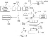
- FIG. 11An alternative exemplary multimodality embodiment configured to provide SD-OCT, OCM, SECM, and FFOCM images and data according to the present invention at a different wavelength from the other three modalities is depicted in Figure 11 .
- a broad bandwidth source 1100is coupled alternatively to a spectral modulation unit 1105.
- Light from the spectral modulation unit 1105is coupled into a circulator 1110 and a beam splitter 1115. If the circulator 1110 is utilized, light from the circulator 1110 can be transmitted to the beam splitter 1115 that preferably directs a majority of light to the sample.
- Light in the reference arm 1120is transmitted to a reference reflector 1125 that can be stationary and/or may or otherwise change the path length of the reference arm 1120.
- TD-OCTtime-domain OCT
- TD-OCTtime-domain OCT
- the beam scanning mechanism 1135may also include a telescope for imaging the scanners onto the back focal plane of the lens 1160.
- Light from the scanning mechanism 1135is transmitted to a dichroic splitter/WDM 1140 that transmits the excitation light for SD-OCT, OCM, and SECM modalities, and can reflect FFOCM light.
- an exemplary FFOCM system similar to that shown in Figure 3can be coupled into the beam path via 1140.
- Light from 1140is directed to a slider 1150 that contains multiple optical elements; when the slider may be positioned at a distinct position, either one or a combination of SD-OCT, OCM, SECM or FFOCM is provided.
- Light from the slider 1150is transmitted to an objective lens 1160 mounted to a lens turret 1155 in one embodiment that is capable of changing objective lenses.
- the slider 1150 and/or turret 1155may be under computer control for automatic selection of imaging modality.
- Lightis focused by the objective lens 1160 onto or within the sample 1165, which may be mounted to a computer-controlled three-dimensional translation stage 1170.
- Reflected lightis transmitted back through the apparatus to the circulator 1110, which redirects the light to a spectrometer.
- Detected reflected lightmay be processed to form exemplary SD-OCT, OCM, SECM images by methods described herein.
- FFOCM lightmay be redirected to the FFOCM system 1175 via the filter/dichroic mirror/WDM apparatus 1140.
- a visible aiming beammay be coupled into the exemplary system shown in Figure 11 , coincident with the near-infrared light, to allow visualization of the locations of imaging.
- a white light image of the specimen under investigationmay be provided by use of an alternative imaging port on the microscope.
- Alternative exemplary embodiments known in the artincluding the capability to obtain spectral information from the sample by short-time-Fourier transformation (STFT) of the spectral interference, Doppler-sensitive SD-OCT and polarization-sensitive SD-OCT, may be also utilized to extract additional information from the biological specimen, such as, e.g., absorption, flow, and birefringence.
- STFTshort-time-Fourier transformation
- Doppler-sensitive SD-OCT and polarization-sensitive SD-OCTmay be also utilized to extract additional information from the biological specimen, such as, e.g., absorption, flow, and birefringence.
- Figure 12depicts an exemplary embodiment of an arrangement of sliders that may be utilized for the multimodality imaging according to the present invention.
- optical elementscan be contained in a housing 1200 that may be translated manually, or under computer or automatic control.
- Each slider positioncan terminate in different slider positions 1205, 1210, 1230, 1260 that provide one or more imaging modalities.
- the slider position 1205, 1210, 1230, 1260may be coupled to the objective lens turret.
- the slider position 1205contains no optical elements (air) or optical element windows.
- the microscopeis configured to perform FFOCM.
- a lens apparatus 1212 and 1213can be configured to expand the beam and illuminate a DP-GRISM containing two prisms 1215 and 1225 that surround a transmission grating 1220.
- This exemplary configurationprovides an ability to perform the SECM imaging. Exemplary OCM procedures can also be conducted in this position using a scanning mechanism that scans the spectrally-encoded line across the sample.
- a lens apparatus 1240, 1250can be configured to image beam angle, with or without beam magnification. This slider position 1230 can provide imaging using exemplary SDOCT procedures.
- a lens apparatus 1270, 1280is configured to expand the scanned beam to allow imaging using the exemplary OCM procedures.
- exemplary embodiments of combined systemscan also include wavelength tuning/modulated sources and single or multiple detector configurations, and such exemplary embodiment is shown in Figure 1.3.
- a wavelength tuning/modulated source 1300is coupled into a circulator 1305 and a beam splitter 1310. If a circulator is utilized, light from the circulator 1305 is transmitted to a beam splitter 1310 that preferably directs a majority of light to the sample.
- Light in the reference arm 1315is transmitted to a reference reflector 1320 that may be stationary, and may or otherwise change the path length of 1315.
- TD-OCTtime-domain OCT
- a filter/dichoric/WDM apparatus 1330that transmits the sample arm light in the direction from the beam splitter to the sample.
- Light from 1330is directed to a x-y beam scanning mechanism 1335 that is capable of scanning the beam in two directions at high or slow speeds.
- the beam scanning mechanism 1335may also include a telescope for imaging the scanners onto the back focal plane of the lens 1353.
- Light from the scanning mechanism 1335is transmitted to a slider 1340 that contains multiple optical elements; when the slider is positioned at a distinct position, either one or a combination of OFDI, OCM, SECM or fluorescence OCM modalities can be provided.
- Light from the slider 1340is transmitted to an objective lens 1353 mounted to a lens turret 1350 in one embodiment that is capable of changing objective lenses.
- the slider 1340 and/or turret 1350may be manual, under computer control for automatic selection of imaging modality.
- Lightis focused by objective lens 1353 onto or within the sample 1355, which may be mounted to a computer-controlled three-dimensional translation stage 1360.
- Reflected lightis transmitted back through the apparatus to 1305, which redirects the light to a detector apparatus 1380 suitable for detecting OFDI, wavelength tuning OCM or SECM signals, images and/or data.
- Detected reflected lightis processed by a CPU 1355 to form exemplary OFDI, OCM, SECM images by methods described above.
- Fluorescent lightmay be redirected to a second detector via the filter/dichroic mirror/WDM apparatus 1330 to a second detector 1370. Fluorescent light from 1370 is utilized to reconstruct a fluorescent confocal image of the biological sample 1355.
- a visible aiming beammay be coupled into the system, coincident with the near-infrared light, to allow visualization of the locations of imaging.
- a white light image of the specimen under investigationmay be provided by use of an alternative imaging port on the microscope.
- FIG. 14Another exemplary multimodality embodiment of a system according to the present invention which is configured to provide OFDI, OCM, SECM, and FFOCM images, data and other information, where FFOCM signal is provided at a different wavelength from the other three modalities, is depicted in Figure 14 .
- a wavelength tuning source 1400is coupled alternatively to a spectral modulation unit 1405.
- Light from the modulation unit 1405is coupled into a circulator 1410 and a beam splitter 1415. If the circulator 1410 is utilized, light from the circulator 1410 is transmitted to the beam splitter 1415 that preferably directs a majority of light to the sample.
- TD-OCTtime-domain OCT
- a beam scanning mechanism 1435that is capable of scanning the beam in two directions at high or slow speeds.
- the beam scanning mechanism 1435may also contain a telescope for imaging the scanners onto the back focal plane of the lens 1465.
- Light from the scanning mechanism 1435is transmitted to a dichroic splitter/WDM 1445 that transmits the excitation light for OFDI, OCM, and SECM, but reflects FFOCM light.
- An exemplary FFOCM system similar to the system(s) of Figure 3 and/or Figure 4can be coupled into the beam path via the dichroic splitter/WDM 1445.
- Light from the dichroic splitter/WDM 1445is directed to a slider 1455 that contains multiple optical elements; when the slider 1455 is positioned at a distinct position, either one or a combination of OFDI, OCM, SECM or FFOCM data and/or images is provided.
- Light from the slider 1455is transmitted to an objective lens 1465 mounted to a lens turret 1460 in one exemplary embodiment that is capable of changing the objective lenses.
- the slider 1455 and/or a turret 1460may be under computer control for an automatic selection of imaging modality.
- Lightcan be focused by objective lens 1465 onto or within the sample 1470, which may be mounted to a computer-controlled three-dimensional translation stage 1475.
- Reflected lightis transmitted back through the apparatus to 1410, which redirects the light to a spectrometer.
- Detected reflected lightis processed to form OFDI, OCM, SECM images by methods described herein.
- FFOCM lightmay be redirected to the FFOCM system 1450 via the filter/dichroic mirror/WDM apparatus 1445.
- a visible aiming beammay be coupled into the exemplary system, coincident with the near-infrared light, to allow visualization of the locations of imaging.
- a white light image of the specimen under investigationmay be provided by use of an alternative imaging port on the microscope.
- Doppler-sensitive OFDI and polarization-sensitive OFDImay be also utilized to extract additional information from the biological specimen, such as absorption, flow, and birefringence.
- the microscopecan be configured to allow imaging from both sides of the sample.
- SDOCT, SECM and OCM procedurescan be performed from above the sample, and FFOCM procedures may be performed with the imaging lens illuminates the sample from below.
- the samplecan be mounted between a microscope slide and a thin cover glass, to allow imaging from both sides.
- the exemplary systems described hereincan provide a multimodality imaging of biological specimens in a variety of different formats, speeds, resolutions, fields of view, and contrast mechanisms.
- Each image data setmay be two- or three-dimensional, and may be co-registered to the data sets of the other respective imaging modalities.
- Computer processing methods known in the artmay be utilized to display the different data sets in a variety of different imaging formats including three-dimensional volume visualization, four-dimensional representations, and processed two-, three- and four-dimensional data sets, where the processing apparatus is configured to highlight important areas of interest. Any one or more datasets may be displayed with respect to the other and a comprehensive, all-inclusive dataset may be derived from a combination of the individual data sets.
- Quantitative informationmay be derived from the data sets in their two-, three-, and four-dimensional contexts.
- Image datamay also be combined with conventional fluorescent or brightfield images of the biological specimen.
- axial rangingis performed by use of low coherence reflectometry where the individual depth points are probed sequentially in time.
- the frame ratewas 20 per second (2 kHz A-line rate, 100 x 500 pixels).
- Exemplary OFDI procedures and systemscan use a frequency domain reflectometry in which all depth points are acquired simultaneously. This technique provides a several-hundred-fold improvement in signal-to-noise ratio (SNR) as described in M. A. Choma et al. "Sensitivity advantage of swept source and Fourier domain optical coherence tomography,” Optics Express 11, pp 2183-2189 (2003 ); and S. H. Yun et al., "High-speed optical frequency-domain imaging,” Optics Express 11, pp. 2953-2963 (2003 ).
- the exemplary OFDI systems and procedurescan use a rapidly swept, wavelength tunable laser as a light source.
- the laserfeatured a sweep repetition rate of up to 64 kHz, a wide tuning range of 111 nm centered at 1320 nm, and a high average output power of 30 mW (7 mW on the tissue).
- the axial resolutionwas 10 ⁇ m in tissue.
- the systemfurther comprised an acousto-optic frequency shifter (25 MHz) to remove the depth degeneracy inherent in the frequency-domain reflectometry, as described in S. H. Yun et al., "Removing the depth-degeneracy in optical frequency domain imaging with frequency shifting," Optics Express 12, pp. 4822-4828 (2004 ).
- Polarization-diversity detectionwas implemented to eliminate polarization artifacts in the fiber-based OFDI system.
- Dual-balanced photoreceiverswere used to improve imaging sensitivity through the reduction of laser intensity noise.
- the photoreceiver outputswere digitized with a 2-channel analog-to-digital converter at a sampling rate of 100 MHz with 14-bit resolution.
- Exemplary TDOCT and high-speed OFDI configurationwere incorporated into a dissecting light microscope.
- the scanning systemwas comprised of a collimating lens (5 mm beam diameter), two synchronized galvanometric scanners for transverse scanning, a focusing lens (50 mm focal length), and a small mirror that deflected the beam downward toward the sample.
- the transverse resolutionwas 16 ⁇ m with a confocal parameter of 330 ⁇ m.
- Displacements associated with local cardiac motionwere determined directly from the volumetric data by subtracting the heart surface locations at end diastole from those at end systole on a frame-by-frame basis. Displacement was displayed using a color look up table. Volumetric rendering and three-dimensional visualization was accomplished by using OsiriX ® software.
- High-resolution OFDI procedurewas performed using a laser source with 200 nm tuning range, centered at 1250 nm, in which two semiconductor optical amplifiers were utilized as the gain media, as described in W. Y. Oh et al., "Wide tuning range wavelength-swept laser with two semiconductor optical amplifiers," IEEE Photonics Technology Letters 17, pp. 678-680 (2005 ).
- An axial resolution of 4 ⁇ m in tissuewas achieved.
- the imaging ratewas 40 frames per second with an A-line rate of 20 kHz (500 A-lines per frame).
- Polarization-diversity and dual-balanced detectionwas performed and the photoreceiver outputs were digitized with a 2-channel analog-to-digital converter at a sampling rate of 10 MHz with 12-bit resolution.
- FFOCMis an interferometric technique that utilizes two-dimensional parallel detection to provide subcellular resolution images of reflected light within biological specimens, as described in A. Dubois et al., "Ultrahigh-resolution full-field optical coherence tomography,” Appl Opt 43, pp. 2874-2883 (2004 ), and A. Dubois et al., “Three-dimensional cellular-level imaging using full-field optical coherence tomography,” Phys Med Biol 49, pp. 1227-1234 (2004 ).
- Interference imageswere captured with a CMOS area scan camera with spectral response centered at 650 nm.
- the transverse resolutionswere 2 ⁇ m and axial resolution, 1.1 ⁇ m.
- Acquisition timewas 2 seconds per frame for a transverse field of view of approximately 700 ⁇ m x 700 ⁇ m.
- Three-dimensional datawas obtained by moving the sample through the focus at 1 ⁇ m increments. Volumetric rendering and visualization was accomplished by using OsiriX ® software.
- SECMis a reflectance confocal microscopy technique, which uses near-infrared light that allows deeper penetration into tissue, as described in R. R. Anderson et al., "The optics of human skin,” J Invest Dermatol 77, pp. 13-19 (1981 ), compared with confocal microscopes that utilize visible light.
- Exemplary SECM techniquediffers from conventional laser scanning confocal microscopy in that it projects different wavelengths onto distinct locations on the sample, as described in G. J. Tearney et al., "Spectrally encoded confocal microscopy," Optics Letters 23, pp. 1152-1154 (1998 ). Rapid acquisition of spectra returned from the sample enables high-speed reconstruction of the image.
- a multimode fiberwas used for signal collection, resulting in 0.9 ⁇ m transverse and 2.5 ⁇ m axial resolutions. Images comprised of 500 x 500 pixels were acquired at 10 frames per second. The maximum imaging depth was limited to the 280 ⁇ m working distance of the objective lens.
- Xenopus laevis frogswere purchased from Nasco ® (Fort Atkinson, Wisconsin). Animal procedures were performed according to the approved protocols of Massachusetts General Hospital Subcommittee on Research Animal Care. Embryos were obtained by in vitro fertilization, incubated in 0.1x Marc's modified Ringer's medium (MMR)(as described in J. Newport et al., "A major development transition in early Xenopus embryos; 1. Characterization and timing of cellular changes at the midblastula stage," Cell 30, pp. 675-686, 1982 ), and staged according to Nieuwkoop and Faber tables, (see P. D. Nieuwkoop and J. Faber, Normal table of Xenopus laevis, Daudin, North-Holland Publishing Company, Amsterdam, 1967 ).
- MMRmodified Ringer's medium
- Ethanol treatmentswere performed in 0.1X MMR (vol/vol), soon after Mid Blastula Transition (stage 8.5) (as described in R. Yelin et al., "Ethanol exposure affects gene expression in the embryonic organizer and reduces retinoic acid levels,” Dev Biol 279, pp. 193-204 (2005 ).) until imaging.
- embryosPrior to in vivo imaging, embryos were anesthetized using 0.02% 3-aminobenzoic acid ethyl ester (A-5040, Sigma).
- A-50403-aminobenzoic acid ethyl ester
- Plastic Histology sections(as described in A. M. Glauert, Fixation, Dehydration and Embedding of Biological Specimens., North-Holland Publishing Company Amsterdam, 1986 ) were obtained after additional fixation in Karnovsky's Fixative (KII) and embedding in tEpon-812 (Tousimis). Sections of 1 ⁇ m thick were cut on a Reichert Ultracut Microtome and stained with methylene blue/toluidine blue in borate buffer (Tousimis). Paraffin sections (5 ⁇ m thickness) were stained with Hematoxylin & Eosin.
- Rapid volumetric imaging of the beating heartenables the evaluation of three-dimensional morphology and function during the cardiac cycle.
- TDOCTwhich provides cross-sectional imaging in vivo (as shown in Figures 15a and 15b )
- the exemplary OFDI system and procedurecan image at much higher frame rates, making four-dimensional imaging of the beating heart without cardiac gating possible.
- Volumetric OFDI images of the Xenopus heart (stage 49)were acquired at a rate of 20 three-dimensional data sets per second (as shown in Figures 15c-15g ).
- OFDI cross-sections of a stage 49 Xenopus heartwere obtained in vivo (as shown in Figures 15i-15m ) using a broadband (e.g., 200 nm) wavelength-swept source, as described in W. Y. Oh et al., "Wide timing range wavelength-swept laser with two semiconductor optical amplifiers," IEEE Photonics Technology Letters 17, pp.
- a broadband wavelength-swept sourcee.g. 200 nm
- Exemplary FFOCM procedures and systemsoffer the capability to image microstructure of the embryonic heat with nearly isotropic cellular level resolution.
- Volumetric FFOCM imagesspanned a field of view of 700 x 700 x 1000 ⁇ m (axial). The transverse and axial resolutions were 2 ⁇ m and 1:1 ⁇ m, respectively. Acquisition time was 2 seconds for a single en face section, and 33 minutes for the entire volume.
- Exemplary FFOCM sections of the Xenopus heartallow visualization of ventricular trabeculae (as shown in Figures 16a and 16c ), the spiral valve (as shown in Figures 16b and 16d , see arrows), and the partial atrial septum (as shown in Figure 16d , see arrow head) with greater detail than generated using the exemplary TDOCT or OFDI procedures or systems.
- Partially transparent volumetric rendering of the heart(as shown in Figures 16e-16h ), reveals the looping-compression structure with the angled TA (as shown in Figure 16c ), the aortic arches (as shown in Figures 16f and 16g ), and the thin wall of the atrium (as shown in Figures 16g and 16h ), in their three-dimensional context.
- Cut-away views of(as shown in Figure 16e ) show line three-dimensional internal structures, including the trabeculae (as shown in Figures 16i and 16j ) and the atrioventricular valve (as shown in Figure 16k ).
- Exemplary SECM procedures and systemsprovide a transverse resolution comparable to those associated with FFOCM, but at higher frame rates, enabling microscopy of the heart in vivo.
- the Xenopus myocardium(stage 49) was imaged in vivo using the exemplary SECM procedures and systems at a frame rate of 10/s, a field of view of 220 x 220 ⁇ m, and transverse and axial resolutions of 1.2 and 6 ⁇ m, respectively.
- the maximum penetration depthwas 280 ⁇ m.
- tadpoleStage 49
- TD-OCTas shown in Figures 15a and 15b
- FFOCMas shown in Figures 16a-16m
- SECM imagescorrelated well with corresponding histology sections (as shown in Figures 17b and 17d ).
- a series of frames from a different tadpoledemonstrates the spiral valve as it closes (as shown in Figure 17e ) and opens (as shown in Figures 17f and 17g ), regulating blood flow, seen at the single-cell level, from the TA to the aortic bifurcation.
- Blood cellsare also apparent within the trabeculae (as shown in as shown in Figures 17h ). Intracellular features within individual myocytes that may represent nuclei and organelles can be observed.
- SECM sections obtained in vivo at two different depthsreveal its saccular shape, its location with respect to spiral valve, as well the flow of individual blood cells through the defect. This abnormality was also observed using the exemplary TDOCT procedures and systems in vivo (as shown in Figure 18a , see inset).
- the embryowas then fixed and imaged with the exemplary FFOCM procedures and systems.
- An FFOCM section (as shown in Figure 18c ) and a three-dimensional rendering of the FFOCM volumetric data setshow the dilatation in the context of the entire heart.
- this protrusionmay represent a saccular aneurismal dilatation of the TA, in a heart that otherwise appeared to have a normal phenotype.
- Cardiovascular malformationcan be caused by genetic (as described in K. L. Clark et al., "Transcription factors and congenital heart defects,” Annu Rev Physiol 68, pp. 97-121 (2006 )) and teratogenic factors (as described in S. M. Mone et al., "Effects of environmental exposures on the cardiovascular system; prenatal period through adolescence," Pediatrics 113, pp. 1058-1069 (2004 )).
- Ethanolis a well-known teratogen; exposure of human embryo during pregnancy to alcohol (ethanol) is associated with Fetal Alcohol Syndrome (FAS). (See K. L. Clark et al., "Transcription factors and congenital heart defects," Annu Rev Physiol 68, pp. 97-121 (2006 )
- teratogenic factorsas described in S. M. Mone et al., "Effects of environmental exposures on the cardiovascular system; prenatal period through adolescence,"
- a tadpole(stage 48) has been selected from each of the control, 0.5%, 1.5%, and 2.0% ethanol treated groups to demonstrate typical phenotypes (as shown in Figures 19a-19d ). It was determined that the four tadpoles' hearts were in advanced developmental stages by identifying the existence of a partial atrial septum (as shown in Figures 19a-19d , see right images, septa marked by arrows) and an atrioventricular valve. The TDOCT images provided the first indication of damaged looping in the 1.5% and 2.0% groups. Further observed were lower TDOCT signal from within the ventricle in the 1.5% and 2.0% groups, which may be attributed, to diminished blood flow in these embryos. Photographs of the tadpoles, taken in vivo from the ventral aspect, are shown in Figures 19e-19h .
- Pericardial edemawas present in the 1.5% and 2.0% groups (as shown in Figures 19o, 19p, 19s and 19t ), compared with control and 0.5% groups. Ethanol also affected the ventricle; the developed trabeculae in the control (as shown in Figure 19q ) and 0.5% (as shown in Figure 19r ) hearts contrast the less developed trabeculae in the 1.5% group (as shown in Figure 19s ) and the large ventricular cavity with sparse, stunted trabeculae in embryos exposed to 2.0% ethanol (as shown in Figure 19t ). Corresponding histological sections confirmed some of our findings, including the less developed trabeculae (as shown in Figures 19u-19x ) in embryos with the greater ethanol exposure.
- a common paradigm in developmental biology researchis to manipulate the genotype and monitor the phenotype. Morphology is an important aspect of the phenotype. In the heart, even slight morphological and dynamical abnormalities may be critical for proper myocardial function. An ability to identify subtle morphological and dynamical variations in two and three dimensions can significantly improve the sensitivity of this paradigm.
- heart structuressuch as the myocardium wall, septum and valves may only be a few cells thick. Evaluating the morphological phenotype not only requires resolving such fine structures, but also the capability to visualize these microscopic features within the beating heart, where typical displacement velocities are on the order of 1 mm/sec. If the imaging speed is sufficiently high, three-dimensional images of the embryo heart can be obtained at different times within the cardiac cycle. This exemplary four-dimensional imaging could allow reliable measurements of dynamic physiological parameters, such as stroke volume and ejection fraction, as well as valve opposition, stiffness and modularity, which have close analogs in human pathophysiology. High resolution and high speed are not the only requirements for effective imaging of the heart. In the Xenopus embryo, the heart extends from between 200 ⁇ m and 800 ⁇ m beneath the ventral surface. An effective imaging method should therefore also be capable of imaging at these depths without substantial loss of signal and resolution.
- the morphology of the developing Xenopus laevis hearthas been studied in vitro and described in detail, using three-dimensional rendering of histology sections. (See T. J. Mohun et al., "The morphology of heart development in Xenopus laevis," Dev Biol 218, 74-88 (2000 )). For histologic studies, however, sample preparation and sectioning make preserving structural fidelity difficult. As a result, imaging of intact embryos in their natural environment is preferred. Structural imaging of the heart in vivo has been demonstrated using a variety of non-invasive imaging modalities such as micro-MRI (see D. L.
- Optical techniquesenable imaging of the embryonic heart at higher resolution. Confocal microscopy has been used to image early Xenopus heart development, in vitro (as described in S. J. Kolker et al., "Confocal imaging of early heart development in Xenopus laevis,” Dev Biol 218, pp. 64-73 (2000 )), and to study the role of intracardiac fluid forces in zebrafish embryonic cardiogenesis, in vivo (as described in J. R. Hove et al., "Intracardiac fluid forces are an essential epigenetic factor for embryonic cardiogenesis," Nature 421, pp. 172-177 (2003 )).
- Gating or post-acquisition synchronization techniqueshave been employed to circumvent the limited speed of conventional imaging methods, enabling the reconstruction of three-dimensional images of embryo hearts at different stages in the cardiac cycle.
- M. W. Jenkins et al."4D embryonic cardiography using gated optical coherence tomography,” Optics Express 14, pp. 736-748 (2006 ).
- M. Liebling et al."Four-dimensional cardiac imaging in living embryos via postacquisition synchronization of nongated slice sequences," J Biomed Opt 10, 054001 (2005 ).
- contrastwas generated by endogenous scattering.
- molecular imagingmay be important for relating gene and protein expression to phenotype.
- the exemplary systems and methods described hereincan be used for imaging fluorescent labels and molecular species. It has been described that fluorescence imaging can be conducted via spectral encoding by modification of the source and detection electronics. (See J. T. Motz et al., "Spectral- and frequency-encoded fluorescence imaging," Opt Lett 30, pp. 2760-2762 (2005 )). The same principles used in fluorescence SECM procedures and systems can likewise be utilized for endoscopic two-photon and second harmonic imaging.
- the natural contrast optical imaging modalities presented in this workallow evaluation of the embryonic heart from different vantage points.
- Combining OFDI, SECM, and FFOCM modalitiescan leverage their strengths (see Table 1), and provide a ability for obtaining a more comprehensive morphological and functional myocardial phenotype.
- This multi-modality paradigmcan be extended to other systems and animal models as well. Since these non-invasive imaging techniques do not alter the specimen, they can be used sequentially or in parallel.
- there is no fundamental barrier preventing their combination into one imaging system that uses a single wavelength swept source.See S. H. Yun et al., "High-speed optical frequency-domain imaging," Optics Express 11, pp.
Landscapes
- Health & Medical Sciences (AREA)
- Physics & Mathematics (AREA)
- Life Sciences & Earth Sciences (AREA)
- General Physics & Mathematics (AREA)
- General Health & Medical Sciences (AREA)
- Engineering & Computer Science (AREA)
- Pathology (AREA)
- Biomedical Technology (AREA)
- Nuclear Medicine, Radiotherapy & Molecular Imaging (AREA)
- Molecular Biology (AREA)
- Chemical & Material Sciences (AREA)
- Biophysics (AREA)
- Radiology & Medical Imaging (AREA)
- Animal Behavior & Ethology (AREA)
- Veterinary Medicine (AREA)
- Public Health (AREA)
- Surgery (AREA)
- Medical Informatics (AREA)
- Heart & Thoracic Surgery (AREA)
- Analytical Chemistry (AREA)
- Biochemistry (AREA)
- Immunology (AREA)
- Optics & Photonics (AREA)
- Astronomy & Astrophysics (AREA)
- Theoretical Computer Science (AREA)
- Spectroscopy & Molecular Physics (AREA)
- Medicinal Chemistry (AREA)
- Urology & Nephrology (AREA)
- Hematology (AREA)
- Signal Processing (AREA)
- Pulmonology (AREA)
- Food Science & Technology (AREA)
- Mathematical Physics (AREA)
- Investigating Or Analysing Materials By Optical Means (AREA)
- Investigating, Analyzing Materials By Fluorescence Or Luminescence (AREA)
- Endoscopes (AREA)
- Microscoopes, Condenser (AREA)
- Instruments For Viewing The Inside Of Hollow Bodies (AREA)
- Automatic Focus Adjustment (AREA)
- Transforming Light Signals Into Electric Signals (AREA)
Description
- The present invention generally relates to arrangements and methods for providing multimodality microscopic imaging of one or more biological structures, and particularly to conducting reflectance microscopy of biological specimens using optical coherence tomography ("OCT").
- A determination of the relationship between the molecular basis of genetic alterations and phenotype generally utilizes accurate two- and three-dimensional characterization of microstructure of biological specimens. However, motion and small dimensions make many living biological specimens more difficult to evaluate.
- Optical techniques offer the potential to image the biological specimens at a high resolution. For certain applications, optical imaging based on endogenous contrast can be advantageous over techniques that require exogenous agents, since such beneficial procedures can allow the analysis of the specimen in its native state and at multiple time points, with a small amount of preparation. As an example, several endogenous-contrast imaging modalities are described herein for visualizing embryonic heart microstructure: two exemplary forms of optical coherence tomography ("OCT") as described inD. Huang et al, "Optical coherence tomography," Science 254, pp. 1178-1181 (1991), time-domain optical coherence tomography ("TD-OCT") as described inS. A. Boppart et al., "Investigation of developing embryonic morphology using optical coherence tomography," Dev Biol 111, pp. 54-63 (1996), and optical frequency domain imaging ("OFDI") as described inM. A. Choma et al., "Sensitivity advantage of swept source and Fourier domain optical coherence tomography," Optics Express 11, pp. 2183-2189 (2003); andS. H. Yun et al., "High-speed optical frequency-domain imaging," Optics Express 11, pp2953-2963 (2003).
- Additional examples can be provided and utilized including two reflectance microscopy techniques, e.g., full-field optical coherence microscopy ("FFOCM") as described inE. Beaurepaire et al., "Full-field optical coherence microscopy," Optics Letters 23, pp. 244-246 (1998);A. Dubois et al., "Ultrahigh-resolution full-field optical coherence tomography," Appl Opt 43, pp. 2874-2883 (2004); andG. Moneron et al., "Stroboscopic ultrahigh-resolution full-field optical coherence tomography," Opt Lett 30, pp. 1351-1353 (2005), and spectrally encoded confocal microscopy ("SECM") as described inG. J. Tearney et al., "Spectrally encoded confocal microscopy," Optics Letters 23, pp. 1152-1154 (1998); andC. Boudoux et al., "Rapid wavelength-swept spectrally encoded confocal microscopy," Optics Express 13, pp. 8214-8221 (2005).
- For example, the TDOCT techniques can use low-coherence interferometry to obtain cross-sectional images with ~10 µm resolution and at depths of up to 2 mm. (SeeS. A. Boppart et al., "Noninvasive assessment of the developing Xenopus cardiovascular system using optical coherence tomography," Proc Natl Acad Sci U S A 94, pp. 4256-4261 (1997);S. Yazdanfar et al., "High resolution imaging of in vivo cardiac dynamics using color Doppler optical coherence tomography," Optics Express 1, pp. 424-431 (1997);T. M. Yelbuz et al., "Optical coherence tomography: a new high-resolution imaging technology to study cardiac development in chick embryos," Circulation 106, pp. 2111-211 '4 (2002);V. X. D. Yang et al., "High speed, wide velocity dynamic range Doppler optical coherence tomography (Part II): Imaging in vivo cardiac dynamics of Xenopus laevis," Optics Express 11, pp. 1650-1658 (2003); andW. Luo et al., "Three-dimensional optical coherence tomography of the embryonic murine cardiovascular system" Journal of).
- The exemplary OFDI technique can be considered as a derivative of the TDOCT techniques that may enable an acquisition of images at significantly higher frame rates as described inR. Huber et al., "Three-dimensional and C-mode OCT imaging with a compact, frequency swept laser source at 1300 nm," Optics Express 13, pp. 10523-10538 (2005). The high speed of the OFDI techniques can enable an implementation of a true four-dimensional (4D) microscopy (e.g., three-dimensional microscopy as a function of time). Full-field optical coherence microscopy ("FFOCM") techniques can utilize low-coherence interferometry and higher numerical aperture objective lenses to attain resolution at the subcellular level in all three dimensions. Such FFOCM techniques are likely considerably slower than the OFDI techniques. The exemplary SECM techniques can have a form of the reflectance confocal microscopy using which it may be possible to obtain two-dimensional images with micron-level resolution, at significantly higher speeds than possibly obtained using the FFOCM techniques.
- While each of these natural-contrast procedures can individually be used for imaging a microstructure of the embryonic heart, when combined, these procedures can provide a powerful set of tools for two-, three-, and four-dimensional characterization of early myocardial morphology and dynamics. A combination of these different modalities into one single microscopy device may have additional advantages such as, e.g., (a) a comparison of images in different formats, different resolutions, and fields of view, (b) a simultaneous acquisition of both structural and function information, and/or (c) these tasks can be accomplished using one instrument without requiring moving or altering the specimen.
- E. Beaurepaire et al, in "Combined scanning optical coherence and two-photon excited fluorescence microscopy", Optics Letters, Vol. 24, 14, pp969-971 (1999), discloses simultaneous imaging by optical coherence microscopy (OCM) and two-photon-excited (TPE) fluorescence microscopy, in which a mode-locked Ti:sapphire laser is focused and scanned in three dimensions through a fixed sample, generating both backscattered light and fluorescence light, which are independently detected. Simultaneous imaging of cell nuclei with OCM and TPE is demonstrated in live drosophila embryos.
WO 03/046636 A1 - One of the objects of the present invention is to overcome certain deficiencies and shortcomings of the prior art systems (including those described herein above), and provide exemplary embodiments of providing multimodality microscopic imaging of one or more biological structures. Such exemplary embodiments can conduct reflectance microscopy of biological specimens using optical coherence tomography ("OCT") procedures.
- For example, an analysis of biological specimens generally employs a visualization of its microstructure and functions, preferably with small alterations to the specimen. According to one exemplary embodiment of the present invention, a combination of multiple different imaging modalities can be provided in a single microscope device. Each exemplary technique according to certain exemplary embodiments of the present invention can provide distinct and complementary imaging capabilities, including high-speed (e.g., 1000 frames per second) and high axial resolution (4-16 µm) cross-sectional imaging in vivo, true four-dimensional imaging in vivo, three-dimensional microscopy with isotropic cellular (e.g., 1-2 µm) resolution in vitro, and two-dimensional subcellular imaging in vivo. When combined, these exemplary imaging modalities can effectuate a more complete picture of the morphologic and dynamics of biological specimens.
- Thus, in accordance with the present invention there is provided an apparatus and method as defined in the appended claims. These include arrangements and methods for acquiring multimodality microscopic data. For example, according to one exemplary embodiment, it is possible to use a combination of unique broad bandwidth or rapid wavelength swept sources and optics interposed between a scanning mechanism and an imaging lens. Data can be acquired simultaneously and/or serially, e.g., without moving the specimen. For example, data obtained from different modalities can be co-registered so that it can be displayed side-by-side and/or overlaid on top of each other. Quantitative information can be obtained from all of the datasets in a complementary manner.
- Optical coherence tomography information associated with a signal provided by a source arrangement is obtained when wavelength varies over time. At least one image is generated based on the first and second data. In addition, a first image can be generated based on the first data and a second image can be generated based on the second data. The first and second images may be associated with one another as a function of the first and second data.
- Other features and advantages of the present invention will become apparent upon reading the following detailed description of embodiments of the invention, when taken in conjunction with the appended claims.
- Further objects, features and advantages of the present invention will become apparent from the following detailed description taken in conjunction with the accompanying figures showing illustrative embodiments of the present invention, in which:
Figure 1 is a schematic diagram of an exemplary SECM system that utilizes a broad bandwidth source;Figure 2 is a schematic diagram of an exemplary SD-OCT system;Figure 3 is a schematic diagram of an exemplary OCM system that utilizes a broad bandwidth source;Figure 4 is a schematic diagram of an exemplary FFOCM system that utilizes a broad bandwidth source;Figure 5 is a schematic diagram of an exemplary fluorescence SECM system that utilizes a broad bandwidth source;Figure 6 is a schematic diagram of an exemplary SECM system that utilizes a wavelength tuning source;Figure 7 is a schematic diagram of an exemplary OFDI system that utilizes a wavelength tuning/modulated source;Figure 8 is a schematic diagram of an exemplary OCM system that utilizes a wavelength modulated/tuning source;Figure 9 is a schematic diagram of an exemplary FFOCM system that utilizes a wavelength modulated/tuning source;Figure 10 is a schematic diagram of an exemplary combined SECM/SD-OCT/OCM system that utilizes a broad bandwidth source according to a first exemplary embodiment of the present invention;Figure 11 is a schematic diagram of an exemplary combined SECM/SD-OCT/FFOCM system that utilizes a broad bandwidth source according to a second exemplary embodiment of the present invention;Figure 12 is a schematic diagram of exemplary multimodality microscope sliders according to a particular exemplary embodiment of the present invention;Figure 13 is a schematic diagram of an exemplary combined SECM/OFDI/OCM system that utilizes a wavelength tuning source according to a third exemplary embodiment of the present invention;Figure 14 is a schematic diagram of an exemplary combined SECM/OFDI/FFOCM system that utilizes a wavelength tuning source according to a third exemplary embodiment of the present invention;Figures 15a-15m are various exemplary images of Xenopus laevis hearts (stage 49) in vivo using exemplary embodiments of the TDOCT and OFDI procedures.Figures 16a-16m are various exemplary three-dimensional images of Xenopus heart in vitro using exemplary embodiments of the FFOCM procedure.Figures 17a-17h are exemplary high-resolution confocal images obtained in vivo using the exemplary embodiments of the SECM procedure;Figures 18a-18e are exemplary images of an aneurismal dilatation in the Xenopus heart obtained using the exemplary embodiments of the method and arrangements according to the present invention; andFigures 19a-19x are exemplary images of abnormal heart formation due to ethanol exposure using the exemplary embodiments of the method and arrangements according to the present invention.- Throughout the figures, the same reference numerals and characters, unless otherwise stated, are used to denote like features, elements, components or portions of the illustrated embodiments. Moreover, while the subject invention will now be described in detail with reference to the figures, it is done so in connection with the illustrative embodiments. It is intended that changes and modifications can be made to the described embodiments without departing from the scope of the subject invention as defined by the appended claims.
- Exemplary SECM techniques are capable of providing subcellular level resolution images in tissue or biological specimens. SECM images can alternatively represent fluorescence from the sample or reflectance from the sample.
Figure 1 depicts a schematic diagram of an exemplary SECM arrangement which utilizes a broadband source, hi this exemplary configuration, a quasimonochromatic orbroadband light 100 illuminates acirculator 110, which alternatively may be a beam splitter. In one embodiment this circulator or beam splitter is fiber-optic coupled. The core of the optical fiber can serve as the pinhole for the confocal microscope system. The fiber may alternatively have multiple claddings that transmit light such that for example the light exciting the sample is single mode and the collected light is multimode. Light from this element may be incident on ascanning mechanism 115 that scans the angle of the beam so as to produce one or more transverse scans on the sample. The scanning mechanism may alternatively be one of a resonant scanner, galvanometer scanner, polygon scanning mirror, acousto-optic scanner or the like. A telescope apparatus may be used to image the scan axis to the back focal plane of theobjective lens 130. Light from the scanning mechanism can then be directed towards awavelength dispersing element 120 such as a transmission diffraction grating, prism, grating prism, dual prism grating prism (DP-GRISM) or the like. This exemplary element may disperse the different wavelengths in the broad bandwidth source so that it is incident on theobjective lens 130 with varying angles that depend on wavelength, - In one exemplary embodiment, the lens can have a numerical aperture that may produce a small focused spot or alternatively the lens has a high NA >0.2. The
objective lens 130 focuses each wavelength region onto the sample where each wavelength region on thesample 160 can be located at a different spatial location. For a diffraction grating and an objective lens, these exemplary elements may form a wavelength encodedline 140 on the sample where each position on the line is encoded by a different wavelength region. Light from thesample 160 can be reflected back through the exemplary system ofFigure 1 . Out-of-focus light may be rejected by the cladding of the optical fiber and in focus (e.g., confocal) light is transmitted back through the circulator/beam splitter 110 to a spectrometer that measures the spectral content of the returned light 145. Confocal remittance as a function of spatial location is decoded by measuring this spectrum, forming one line on an image. Successive lines are formed for each angular position of thescanning mechanism 115, forming a spectrally-encoded confocal microscopy image. Figure 2 depicts a schematic diagram of an exemplary spectral-domain OCT system. Contrary to the exemplary SECM system, the exemplary SD-OCT can provide cross-sectional images of a biological specimen by using coherence gating in the Fourier domain. SD-OCT images can typically have a lower resolution (∼3-10 µm), and may have a larger field of view (several mm's). In this exemplary SD-OCT system, a broad bandwidth orquasimonochromatic source 200 can be input into an interferometer, which may be fiber optic-based. The fiber-coupled light can be transmitted to acirculator 210 and abeam splitter 220. When coupled into thecirculator 210, the light can preferably be subsequently split by abeam splitter 220 so that a portion thereof can be transmitted to areference arm 225 and a portion is transmitted to asample arm 235. Light from thereference arm 225 can be reflected off a mirror 230 (e.g., a reference) to thebeam splitter 220 or alternatively transmitted back to thebeam splitter 220. In one exemplary embodiment, thesplitter 220 can be configured so that the majority of light is transmitted to thesample arm 235. Light from the sample arm fiber can then be directed towards a lens and ascanning mechanism 240. The scanning mechanism can scan the light of thesample arm 235 in arbitrary one- or two-dimensional patterns. Light can be transmitted from the scanning mechanism to alens 250 which, in one exemplary embodiment, can have a NA so that the confocal parameter is sufficiently large to allow cross-sectional imaging in the biological specimen orsample 260,- Light remitted from the sample may be transmitted back through the apparatus to the circulator/
beam splitter 210, and directed to aspectrometer 280. The reflectance as a function of depth (A-line) within the tissue may be reconstructed by, e.g., a background subtraction, remapping λ-space to k-space, and inverse Fourier transformation of the spectral interference signal is done in a central processing unit orcomputer 290. Successive A-lines are obtained for each scanning mechanism position, thereby reconstructing a cross-sectional image of the sample. Alternative exemplary embodiments known in the art, including the capability to obtain spectral information from the sample by short-time-Fourier transformation ("STFT") of the spectral interference, Doppler-sensitive SD-OCT, and polarization-sensitive SD-OCT, may be also utilized to extract additional information from the biological specimen, such as absorption, flow, and birefringence. Figure 3 depicts a schematic diagram of an exemplary optical coherence microscopy ("OCM") system. The exemplary OCM system can utilize a combination of confocal microscopy and OCT techniques that may be advantageous, as the axial point spread functions of both such exemplary techniques may be multiplied so as to provide a greater degree of optical sectioning. In one exemplary embodiment of the OCM system, light from a broad bandwidth source can be input into a modulatingelement 310 so that the modulation frequency approximates that of the spectral interference in the interferometer. This exemplary modulation element may be one of a Michelson interferometer, pulse shaping apparatus, spectral filter, etc. The modulation may also shift the spectral phase by some amount over time so that successive spectra may be subtracted to extract only the spectral interference term. Following the modulating element, the light can be transmitted to a circulator/beam splitter 320 and then, if a circulator is used, to abeam splitter 330. Light can again be transmitted to areference arm 335 and asample arm 345. Light from thereference arm 335 is reflected by amirror 340. Light from thesample arm 345 can be transmitted to anx-y scanner 350, which can alternatively be one of or a combination of a resonant scanner, galvanometer scanner, polygon scanning mirror, acousto-optic scanner or the like.- Light from the
scanner 350 can be directed to anobjective lens 355 so that a tightly focused spot can be scanned within the sample. The objective lens orsample 360 may be alternatively scanned in any of three dimensions to facilitate data collection from different portions within the sample. Light is transmitted back from thesample 360 to the circulator/beam splitter 320 and subsequently to a detection apparatus. In one embodiment, the detector is a spectrometer and OCM data is obtained by obtaining A-lines from the sample in a similar manner as performed by the exemplary SD-OCT. In the spectral modulation embodiment the detector can alternatively be a photodiode or other single detector that is synchronized to thesource modulation element 310. Exemplary lock-in or subtraction techniques can be utilized to extract the OCM signal. - Full-field optical coherence microscopy is typically a free-space interferometric technique that utilizes a broad bandwidth source to obtain transverse, high-resolution optical sections of biological specimens.
Figure 4A depicts a schematic diagram of an exemplary FFOCM system, wherebroad bandwidth light 400 is transmitted to abeam splitter 410. Light is split into thesample arm 423 and thereference arm 422. Light in thereference arm 422, according to one exemplary embodiment, may be directed 415 towards areference objective lens 420 and to amirror 425, which is capable of an axial motion. Light in thesample arm 423 may be directed towards a sampleobjective lens 430 and to thesample 440. In one exemplary embodiment, the reference andsample objectives - In another exemplary embodiment, the
objective lenses stage 443 that provides motion in any of three-dimensions. Light from thereference arm 422 and thesample arm 423 can be imaged using alens 445 onto aCCD camera 450. Fringes are detected by theCCD camera 445 resulting from the interference of thesample arm 422 and thereference arm 423. Multiple images can be typically detected for different positions of thereference arm mirror 425. The exemplary images may be arithmetically combined to extract the information from an optical section within the sample. - In another exemplary embodiment of the FFOCM system as depicted in
Figure 4B , a broad bandwidthlight source 451 can be coupled into a modulating element, such as a Michelson interferometer or other interferometer (e.g. Mach-Zehnder, Sagnac) or spectrum altering unit. For the Michelson interferometer case, light from thesource 451 can be transmitted to abeam splitter 452. Light may then be split into two arms forexemplary arm A 453 andarm B 455. Light fromarm A 453 is transmitted to a mirror and backreflected back to a beam splitter. Light fromarm β 455 is likewise transmitted to amirror 456, and backreflected back to abeam splitter 452. The difference between the path length La in arm A and the path length Lb in arm B, |La - Lb|, can be set to be substantially equal to the path length difference between the reference and sample arms in the second interferometer. At least one of the arms A or B can be configured to change path lengths or produce a phase shift in the light therein. In one exemplary embodiment, the path length may be changed by a motion of one of the mirrors or a rapidly scanning optical delay line. The motion may be actuated by a piezoelectric transducer, galvanometer, linear motor or the like. Alternatively, path length changes may be generated by one of an acousto-optic modulator or electro optic modulator. - Both reference and sample arm light may be combined at the beam splitter, and transmitted to another static interferometer with
beam splitter 459, separating light into areference arm 458 and asample arm 457, respectively. Light from botharms objective lenses reference arm 458, thereference objective lens 460 can be brought to a focus on areflector 465, which is typically not moving, whereas in the sample arm the sampleobjective lens 470 focuses the sample arm light on or within thesample 480. Thesample 480 or the sampleobjective lens 470 may be mounted to astage 481, capable of moving thesample 480 in any of three-dimensions, under manual control or computer control. - The path length difference between the path lengths of the
reference arm 458 and thesample arm 457 may be substantially equal to |La - Lb| of the first interferometer. Light from reference and samplearms beam splitter 459, and imaged onto aCCD array 490 or array of detectors via alens 485. A FFOCM image or data can be generated by a linear combination of images acquired byCCD 490 and while moving or at different positions ofmirror 456. Processing, display and storage of FFOCM images is provided by aCPU 495. Accumulations or averages are utilized to increase signal to noise ratio. Figure 5 depicts an exemplary embodiment of a SECM system configured for a fluorescence detection and using a broad bandwidth source. For example, light from thesource 500 can be transmitted to abeam splitter 510, which splits light into twopaths mirrors arms 530 can be coupled to thebeam splitter 510 and directed 535 towards a SECM probe containing a grating ordispersive element 540, anobjective lens 550. As discussed herein, the arrangement of the grating and theobjective lens 550 focuses a spectrally encodedline 560 on or within thespecimen 562 which may be mounted to a three-dimensional stage. Fluorescent light within the sample can be excited by the illuminating light, transmitted back through theobjective lens 550, and imaged by anotherlens 565 onto adetector 570. Detected light can be digitized and converted to a line in an image by a processing arrangement (e.g., CPU) 580. Additional lines in the image may be generated at different positions of thebeam scanning mechanism 537. Nonlinearities in the moving mirror can be corrected by anexemplary interferometer 521 that has a narrow bandwidth source that illuminates the same movingmirror 520Figure 6 depicts a schematic diagram of an exemplary embodiment of an SECM system that uses awavelength tuning source 600. For example, thesource 600 can be coupled into a circulator/beam splitter 610. According to one exemplary embodiment, light from thesplitter 610 is transmitted via an optical fiber to a scanner, which alternatively may also contain a telescope lens imaging system that projects the scan axis to the back focal plane of theobjective lens 625. Light from the scanning mechanism is transmitted to a dispersive element 620 (such as a diffraction grating, prism, GRISM, or DP-GRISM, etc.). Light from 620 is transmitted to anobjective lens 625, with preferably a high NA, which can focus the beam within thesample 635. At any point in time, one wavelength from the wavelength sweptsource 600 can illuminate a distinct portion of the sample. As the wavelength of the sweptsource 600 changes over time, the beam can be scanned along aline 630 within thesample 635. Remitted light from thesample 635 can be transmitted back through theelements circulator beam splitter 610. Light from thesplitter 610 can be directed to adetector 640, and digitized by a processing arrangement (e.g., CPU) 650, displayed and digitally stored. A single line in the image is obtained following one full sweep of the wavelength-tuning source. Lines may be acquired at different positions of the scanning mechanism to form the image. Fluorescent light excited by the wavelength-tuning source 600 remitted from the sample can be alternatively detected by adetector 660 to form a fluorescent image.Figure 7 depicts a schematic diagram of an exemplary OFDI system. In one exemplary embodiment of this exemplary OFDI system, a wavelength tuning source may be coupled to an optical fiber-basedcirculator 705 and abeam splitter 705. Light from thecirculator 705 can be transmitted to thebeam splitter 705, configured to send a majority of light, in the preferred embodiment, to thesample arm 725. Such split light forwarded to thereference arm 715 can be terminated by areflector 720, and sent back to thebeam splitter 710 and thecirculator 705. Light in thesample arm 725 is transmitted to ascanning mechanism 730 and animaging lens 735 that has a NA sufficiently low to allow cross-sectional imaging of thebiological specimen 740. Light is reflected from thereference mirror 720 and thesample 740, recombined at thecirculator 705, and directed by anoptical fiber 750 to adetector apparatus 755, which in an exemplary embodiment can contain dual-balanced detectors.- Light is digitized by the
detector apparatus 755 and the digital signal is transmitted to aCPU 760. Spectral interference is processed in a manner similar to the processing using the exemplary SD-OCT system/procedure, e.g., the background is subtracted, λ-space is converted to k-space, and an inverse Fourier transform is performed to produce an A-line. A-lines can be acquired as a function of scanning mechanism position, creating a cross-sectional OFDI image. Alternative exemplary embodiments known in the art, including the capability to obtain spectral information from the sample by short-time-Fourier transformation (STFT) of the spectral interference, complex spectral domain processing, Doppler-sensitive OFDI and polarization-sensitive OFDI, may be also utilized to extract additional information from the biological specimen, such as absorption, flow, and birefringence. Figure 8 depicts a schematic diagram of an exemplary embodiment of an OCM system which utilizes a wavelength tuning/modulated source. For example, awavelength modulation arrangement 805 may produce a spectral pattern on the source, for example, a sinusoidal modulation of the spectrum, which may be altered over time to correspond to spectral interference modulation produced by interference between the sample and reference arms. Light from thesource 800 and/or themodulation arrangement 805 can be coupled into a fiber-optic circulator/beam splitter 810, and subsequently transmitted to abeam splitter 815 which preferably directs a majority of light to thesample arm 830.- Light in the
reference arm 820 is directed towards areference reflector 825 or a transmission element. Light in thesample arm 830 can be transmitted to an x-y seamier, which may comprise one or more of galvanometers, resonant scanners, polygon scanners, acousto-optic scanners, electro optic scanners, etc. Light from the scanner can be alternatively transmitted to atelescope 837 and anobjective lens 840 with preferably a high NA. Theobjective lens 840 focuses the light within thesample 845, which is alternatively affixed to a three-dimensional stage 847. Light is returned from the sample back through theelements circulator 810 and transmitted to adetector 855, digitized and transmitted to aCPU 860. - In one exemplary embodiment, OCM data can be obtained by obtaining A-lines from the sample in a similar manner to the way it is performed using the exemplary OFDI system and procedure. For example, in the exemplary spectral modulation system and procedure, the detector can be synchronized to the
source modulation element 805. Lock-in or subtraction techniques can be utilized to extract the OCM signal in this case. An exemplary image can be generated by acquiring data for each position of thex-y scanning mechanism 835. Fluorescent light remitted from the sample can be further detected by use of a dichroic mirror or filter 853 and asecond detector 865. Figure 9 depicts an exemplary embodiment of an FFOCM system that utilizes a wavelength-tuning/modulatedsource 900. The light source may be tuned over its bandwidth or alternatively be modulated to contain a spectral modulation frequency substantially similar to the frequency provided by spectral interference modulation of the interferometer. Light from thesource 900 may be coupled into abeam splitter 905, and directed to asample arm 910 and areference arm 915, respectively, which are terminated byrespective objectives arm objective lens 920 focuses reference arm light onto a reflector, which is subsequently returned to thebeam splitter 905. Sample arm light is focused by 930 onto or within thespecimen 935. Light remitted from the sample is combined with the reference arm light at 905, and imaged by alens 940 onto aCCD array 950. Images can be obtained for each wavelength of the wavelength swept source or different modulation patterns of the source and arithmetically combined by aCPU 960 to reconstruct an exemplary FFOCM optical section.- According to one exemplary embodiment of the present invention , the exemplary systems described above and alternative exemplary embodiments thereof may be combined to form a multimodality imaging system. This exemplary combination of systems and/or devices can be provided by creating separate systems, and configuring their optics so that they can obtain images from the same portions of the biological specimen. Different wavelength, scanning, and detection mechanisms may be provided in such combined modality system. Alternatively, the different devices can be implemented using many common components, which they share to provide a more efficient, cost-effective apparatus.
Figure 10 depicts a schematic diagram of a multimodality system according to an exemplary embodiment of the present invention that utilizes abroad bandwidth source 1000 andspectrometer 1080 to provide simultaneous and co-registered SD-OCT, OCM, SECM, and fluorescence SECM data and/or images. For example, light from thebroad bandwidth source 1000 can be coupled alternatively to aspectral modulation unit 1005. Light from thespectral modulation unit 1005 is coupled into acirculator 1010 and abeam splitter 1015. If a circulator is utilized, light from thecirculator 1010 is transmitted to thebeam splitter 1015 that preferably directs a majority of light to the sample. Light in thereference arm 1020 is transmitted to areference reflector 1025 that may move or otherwise change the path length of 1020, and/or which can be non-movable. If the reference arm is allowed to move, conventional time-domain OCT (e.g., TD-OCT) arrangement and/or procedures may be implemented or complex spectral domain may be obtained using the exemplary SD-OCT arrangement and/or procedures using processes that are known in the art.- Light in the
sample arm 1030 is transmitted to a filter/dichroic/WDM apparatus 1035 that transmits the sample arm light in the direction from the beam splitter to the sample. Light from 1035 is directed to abeam scanning mechanism 1040 that is capable of scanning the beam in two directions at high or slow speeds. Thebeam scanning mechanism 1040 may also contain a telescope for imaging the scanners onto the back focal plane of thelens 1055. Light from thescanning mechanism 1040 can be transmitted, to aslider 1045 that contains multiple optical elements. For example, when theslider 1045 is positioned at a distinct position, either one or more or a combination of SD-OCT, OCM, SECM and/or fluorescence OCM arrangements/procedures can be implemented. Light from theslider 1045 can be transmitted to anobjective lens 1055 mounted to a lens turret in one embodiment that is capable of changing objective lenses. Theslider 1045 and/orturret 1050 may be under computer control for automatic selection of imaging modality. Light is focused byobjective lens 1055 onto or within thesample 1060, which may be mounted to a computer-controlled three-dimensional translation stage 1065. Reflected light is transmitted back through the apparatus to 1010, which redirects the light to a spectrometer. Detected reflected light is processed to form exemplary SD-OCT, OCM, SECM images using the arrangements and/or procedures described herein, - As shown in
Figure 10 , fluorescent light may be redirected to a second detector via the filter/dichroic mirror/WDM apparatus 1035 to asecond detector 1075. Fluorescent light from 1075 is utilized to reconstruct a fluorescent confocal image of thebiological sample 1060. In the case where invisible near-infrared light is utilized, a visible aiming beam may be coupled into the exemplary system, coincident with the near-infrared light, to allow visualization of the locations of imaging. Alternatively or in addition, a white light image of the specimen under investigation may be provided by use of an alternative imaging port on the microscope. Alternative exemplary embodiments known in the art, including the capability to obtain spectral information from the sample by short-time-Fourier transformation (STFT) of the spectral interference, Doppler-sensitive SD-OCT and polarization-sensitive SD-OCT, may be also utilized to obtain additional information from the biological specimen, such as, e.g., absorption, flow, and birefringence. - An alternative exemplary multimodality embodiment configured to provide SD-OCT, OCM, SECM, and FFOCM images and data according to the present invention at a different wavelength from the other three modalities is depicted in
Figure 11 . In this exemplary embodiment abroad bandwidth source 1100 is coupled alternatively to aspectral modulation unit 1105. Light from thespectral modulation unit 1105 is coupled into acirculator 1110 and abeam splitter 1115. If thecirculator 1110 is utilized, light from thecirculator 1110 can be transmitted to thebeam splitter 1115 that preferably directs a majority of light to the sample. Light in thereference arm 1120 is transmitted to areference reflector 1125 that can be stationary and/or may or otherwise change the path length of thereference arm 1120. In case thereference arm 1120 is allowed to move, exemplary conventional time-domain OCT (TD-OCT) procedures or complex spectral domain may be utilized for SD-OCT by methods known in the art. Light in the sample arm 1130 is transmitted to abeam scanning mechanism 1135 that is capable of scanning the beam in two directions at high or slow speeds. Thebeam scanning mechanism 1135 may also include a telescope for imaging the scanners onto the back focal plane of thelens 1160. Light from thescanning mechanism 1135 is transmitted to a dichroic splitter/WDM 1140 that transmits the excitation light for SD-OCT, OCM, and SECM modalities, and can reflect FFOCM light. - For example, an exemplary FFOCM system similar to that shown in
Figure 3 can be coupled into the beam path via 1140. Light from 1140 is directed to aslider 1150 that contains multiple optical elements; when the slider may be positioned at a distinct position, either one or a combination of SD-OCT, OCM, SECM or FFOCM is provided. Light from theslider 1150 is transmitted to anobjective lens 1160 mounted to alens turret 1155 in one embodiment that is capable of changing objective lenses. Theslider 1150 and/orturret 1155 may be under computer control for automatic selection of imaging modality. Light is focused by theobjective lens 1160 onto or within thesample 1165, which may be mounted to a computer-controlled three-dimensional translation stage 1170. Reflected light is transmitted back through the apparatus to thecirculator 1110, which redirects the light to a spectrometer. Detected reflected light may be processed to form exemplary SD-OCT, OCM, SECM images by methods described herein. FFOCM light may be redirected to theFFOCM system 1175 via the filter/dichroic mirror/WDM apparatus 1140. - In the case where invisible near-infrared light is utilized, a visible aiming beam may be coupled into the exemplary system shown in
Figure 11 , coincident with the near-infrared light, to allow visualization of the locations of imaging. Alternatively or in addition, a white light image of the specimen under investigation may be provided by use of an alternative imaging port on the microscope. Alternative exemplary embodiments known in the art, including the capability to obtain spectral information from the sample by short-time-Fourier transformation (STFT) of the spectral interference, Doppler-sensitive SD-OCT and polarization-sensitive SD-OCT, may be also utilized to extract additional information from the biological specimen, such as, e.g., absorption, flow, and birefringence. Figure 12 depicts an exemplary embodiment of an arrangement of sliders that may be utilized for the multimodality imaging according to the present invention. For example, optical elements can be contained in ahousing 1200 that may be translated manually, or under computer or automatic control. Each slider position can terminate indifferent slider positions slider position slider position 1205 contains no optical elements (air) or optical element windows. In this exemplary configuration, the microscope is configured to perform FFOCM. For theslider position 1210, alens apparatus prisms transmission grating 1220. This exemplary configuration provides an ability to perform the SECM imaging. Exemplary OCM procedures can also be conducted in this position using a scanning mechanism that scans the spectrally-encoded line across the sample. For theslider position 1230, alens apparatus slider position 1230 can provide imaging using exemplary SDOCT procedures. For theslider position 1260, alens apparatus - While certain embodiments of the multimodality imaging systems have utilized a broad bandwidth source, exemplary embodiments of combined systems can also include wavelength tuning/modulated sources and single or multiple detector configurations, and such exemplary embodiment is shown in Figure 1.3. For example, in
Figure 13 , a wavelength tuning/modulatedsource 1300 is coupled into acirculator 1305 and abeam splitter 1310. If a circulator is utilized, light from thecirculator 1305 is transmitted to abeam splitter 1310 that preferably directs a majority of light to the sample. Light in thereference arm 1315 is transmitted to areference reflector 1320 that may be stationary, and may or otherwise change the path length of 1315. In case the reference arm is allowed to move, conventional exemplary time-domain OCT (TD-OCT) procedures may be provided or complex spectral domain may be utilized for implementing OFDI modalities by methods known in the art. Light in thesample arm 1325 is transmitted to a filter/dichoric/WDM apparatus 1330 that transmits the sample arm light in the direction from the beam splitter to the sample. Light from 1330 is directed to a x-ybeam scanning mechanism 1335 that is capable of scanning the beam in two directions at high or slow speeds. - The
beam scanning mechanism 1335 may also include a telescope for imaging the scanners onto the back focal plane of thelens 1353. Light from thescanning mechanism 1335 is transmitted to aslider 1340 that contains multiple optical elements; when the slider is positioned at a distinct position, either one or a combination of OFDI, OCM, SECM or fluorescence OCM modalities can be provided. Light from theslider 1340 is transmitted to anobjective lens 1353 mounted to alens turret 1350 in one embodiment that is capable of changing objective lenses. Theslider 1340 and/orturret 1350 may be manual, under computer control for automatic selection of imaging modality. Light is focused byobjective lens 1353 onto or within thesample 1355, which may be mounted to a computer-controlled three-dimensional translation stage 1360. Reflected light is transmitted back through the apparatus to 1305, which redirects the light to adetector apparatus 1380 suitable for detecting OFDI, wavelength tuning OCM or SECM signals, images and/or data. Detected reflected light is processed by aCPU 1355 to form exemplary OFDI, OCM, SECM images by methods described above. - Fluorescent light may be redirected to a second detector via the filter/dichroic mirror/
WDM apparatus 1330 to asecond detector 1370. Fluorescent light from 1370 is utilized to reconstruct a fluorescent confocal image of thebiological sample 1355. In the case where invisible near-infrared light is utilized, a visible aiming beam may be coupled into the system, coincident with the near-infrared light, to allow visualization of the locations of imaging. Alternatively or in addition, a white light image of the specimen under investigation may be provided by use of an alternative imaging port on the microscope. Alternative embodiments known in the art, including the capability to obtain spectral information from the sample by short-time-Fourier transformation (STFT) of the spectral interference, Doppler-sensitive SD-OCT and polarization-sensitive SD-OCT, may be also utilized to extract additional information from the biological specimen, such as absorption, flow, and birefringence. - Another exemplary multimodality embodiment of a system according to the present invention which is configured to provide OFDI, OCM, SECM, and FFOCM images, data and other information, where FFOCM signal is provided at a different wavelength from the other three modalities, is depicted in
Figure 14 . In this exemplary embodiment, awavelength tuning source 1400 is coupled alternatively to aspectral modulation unit 1405. Light from themodulation unit 1405 is coupled into acirculator 1410 and abeam splitter 1415. If thecirculator 1410 is utilized, light from thecirculator 1410 is transmitted to thebeam splitter 1415 that preferably directs a majority of light to the sample. Light in thereference arm 1420 is transmitted to a reference reflector 1425 that may be stationary, or can move or otherwise change the path length of 1420. In case thereference arm 1420 is allowed to move, conventional time-domain OCT (TD-OCT) procedures and modalities may be provided or complex spectral domain may be obtained for the OFDI data by methods known in the art. Light in thesample arm 1430 is transmitted to abeam scanning mechanism 1435 that is capable of scanning the beam in two directions at high or slow speeds. Thebeam scanning mechanism 1435 may also contain a telescope for imaging the scanners onto the back focal plane of thelens 1465. Light from thescanning mechanism 1435 is transmitted to a dichroic splitter/WDM 1445 that transmits the excitation light for OFDI, OCM, and SECM, but reflects FFOCM light. - An exemplary FFOCM system similar to the system(s) of
Figure 3 and/orFigure 4 can be coupled into the beam path via the dichroic splitter/WDM 1445. Light from the dichroic splitter/WDM 1445 is directed to aslider 1455 that contains multiple optical elements; when theslider 1455 is positioned at a distinct position, either one or a combination of OFDI, OCM, SECM or FFOCM data and/or images is provided. Light from theslider 1455 is transmitted to anobjective lens 1465 mounted to alens turret 1460 in one exemplary embodiment that is capable of changing the objective lenses. Theslider 1455 and/or aturret 1460 may be under computer control for an automatic selection of imaging modality. Light can be focused byobjective lens 1465 onto or within thesample 1470, which may be mounted to a computer-controlled three-dimensional translation stage 1475. - Reflected light is transmitted back through the apparatus to 1410, which redirects the light to a spectrometer. Detected reflected light is processed to form OFDI, OCM, SECM images by methods described herein. FFOCM light may be redirected to the
FFOCM system 1450 via the filter/dichroic mirror/WDM apparatus 1445. In the case where invisible near-infrared light is utilized, a visible aiming beam may be coupled into the exemplary system, coincident with the near-infrared light, to allow visualization of the locations of imaging. Alternatively or in addition, a white light image of the specimen under investigation may be provided by use of an alternative imaging port on the microscope. Alternative exemplary embodiments known in the art, including the capability to obtain spectral information from the sample by short-time-Fourier transformation (STFT) of the spectral interference, Doppler-sensitive OFDI and polarization-sensitive OFDI may be also utilized to extract additional information from the biological specimen, such as absorption, flow, and birefringence. - In another exemplary embodiment of the present invention, the microscope can be configured to allow imaging from both sides of the sample. For example, SDOCT, SECM and OCM procedures can be performed from above the sample, and FFOCM procedures may be performed with the imaging lens illuminates the sample from below. In such exemplary configuration, the sample can be mounted between a microscope slide and a thin cover glass, to allow imaging from both sides.
- The exemplary systems described herein can provide a multimodality imaging of biological specimens in a variety of different formats, speeds, resolutions, fields of view, and contrast mechanisms. Each image data set may be two- or three-dimensional, and may be co-registered to the data sets of the other respective imaging modalities. Computer processing methods known in the art may be utilized to display the different data sets in a variety of different imaging formats including three-dimensional volume visualization, four-dimensional representations, and processed two-, three- and four-dimensional data sets, where the processing apparatus is configured to highlight important areas of interest. Any one or more datasets may be displayed with respect to the other and a comprehensive, all-inclusive dataset may be derived from a combination of the individual data sets. Quantitative information may be derived from the data sets in their two-, three-, and four-dimensional contexts. Image data may also be combined with conventional fluorescent or brightfield images of the biological specimen.
- Provided below are examples conducted to investigate using exemplary multiple imaging modalities according to the present invention to image the developing Xenopus laevis heart.
- In the exemplary TDOCT configuration, axial ranging is performed by use of low coherence reflectometry where the individual depth points are probed sequentially in time. A broad bandwidth (50 nm) source centered at 1.3 µm was used, providing an axial resolution of ~10 µm in tissue (n=1.4). The frame rate was 20 per second (2 kHz A-line rate, 100 x 500 pixels).
- Exemplary OFDI procedures and systems can use a frequency domain reflectometry in which all depth points are acquired simultaneously. This technique provides a several-hundred-fold improvement in signal-to-noise ratio (SNR) as described inM. A. Choma et al. "Sensitivity advantage of swept source and Fourier domain optical coherence tomography,"); andS. H. Yun et al., "High-speed optical frequency-domain imaging,"). The exemplary OFDI systems and procedures can use a rapidly swept, wavelength tunable laser as a light source. An extended-cavity semiconductor laser employing an intracavity spectral filter, as described inM. A. Choma et al. "Sensitivity advantage of swept source and Fourier domain optical coherence tomography,"), C. Boudoux et al., "Rapid wavelength-swept spectrally encoded confocal microscopy," Optics Express 13, pp. 8214-8221 (2005).
- The laser featured a sweep repetition rate of up to 64 kHz, a wide tuning range of 111 nm centered at 1320 nm, and a high average output power of 30 mW (7 mW on the tissue). The axial resolution was 10 µm in tissue. The system further comprised an acousto-optic frequency shifter (25 MHz) to remove the depth degeneracy inherent in the frequency-domain reflectometry, as described inS. H. Yun et al., "Removing the depth-degeneracy in optical frequency domain imaging with frequency shifting," Optics Express 12, pp. 4822-4828 (2004). Polarization-diversity detection was implemented to eliminate polarization artifacts in the fiber-based OFDI system. Dual-balanced photoreceivers were used to improve imaging sensitivity through the reduction of laser intensity noise. The photoreceiver outputs were digitized with a 2-channel analog-to-digital converter at a sampling rate of 100 MHz with 14-bit resolution.
- Exemplary TDOCT and high-speed OFDI configuration were incorporated into a dissecting light microscope. The scanning system was comprised of a collimating lens (5 mm beam diameter), two synchronized galvanometric scanners for transverse scanning, a focusing lens (50 mm focal length), and a small mirror that deflected the beam downward toward the sample. For exemplary TDOCT and OFDI configuration, the transverse resolution was 16 µm with a confocal parameter of 330 µm.
- Displacements associated with local cardiac motion were determined directly from the volumetric data by subtracting the heart surface locations at end diastole from those at end systole on a frame-by-frame basis. Displacement was displayed using a color look up table. Volumetric rendering and three-dimensional visualization was accomplished by using OsiriX® software.
- High-resolution OFDI procedure was performed using a laser source with 200 nm tuning range, centered at 1250 nm, in which two semiconductor optical amplifiers were utilized as the gain media, as described inW. Y. Oh et al., "Wide tuning range wavelength-swept laser with two semiconductor optical amplifiers," IEEE Photonics Technology Letters 17, pp. 678-680 (2005). An axial resolution of 4 µm in tissue was achieved. The transverse resolution was 2 µm with NA=0.2 objective lens. The imaging rate was 40 frames per second with an A-line rate of 20 kHz (500 A-lines per frame). Polarization-diversity and dual-balanced detection was performed and the photoreceiver outputs were digitized with a 2-channel analog-to-digital converter at a sampling rate of 10 MHz with 12-bit resolution.
- For example, FFOCM is an interferometric technique that utilizes two-dimensional parallel detection to provide subcellular resolution images of reflected light within biological specimens, as described inA. Dubois et al., "Ultrahigh-resolution full-field optical coherence tomography," Appl Opt 43, pp. 2874-2883 (2004), andA. Dubois et al., "Three-dimensional cellular-level imaging using full-field optical coherence tomography," Phys Med Biol 49, pp. 1227-1234 (2004). The exemplary FFOCM system used spatially incoherent broadband light from a xenon arc lamp to illuminate the sample and the reference mirror of a Linnik interference microscope using two identical NA=0.3 water-immersion microscope objective lenses. Interference images were captured with a CMOS area scan camera with spectral response centered at 650 nm. The transverse resolutions were 2 µm and axial resolution, 1.1 µm. Acquisition time was 2 seconds per frame for a transverse field of view of approximately 700 µm x 700 µm. Three-dimensional data was obtained by moving the sample through the focus at 1 µm increments. Volumetric rendering and visualization was accomplished by using OsiriX® software.
- For example, SECM is a reflectance confocal microscopy technique, which uses near-infrared light that allows deeper penetration into tissue, as described inR. R. Anderson et al., "The optics of human skin," J Invest Dermatol 77, pp. 13-19 (1981), compared with confocal microscopes that utilize visible light. Exemplary SECM technique differs from conventional laser scanning confocal microscopy in that it projects different wavelengths onto distinct locations on the sample, as described inG. J. Tearney et al., "Spectrally encoded confocal microscopy," Optics Letters 23, pp. 1152-1154 (1998). Rapid acquisition of spectra returned from the sample enables high-speed reconstruction of the image. In the SECM system, as described inC. Boudoux et al., "Rapid wavelength-swept spectrally encoded confocal microscopy," Optics Express 13, pp. 8214-8221 (2005), light from a rapid wavelength timing source in the near-infrared (center wavelength = 1.32 µm, instantaneous line width = 0.1 nm, total bandwidth = 70 nm, repetition rate up to 15.7 kHz), was collimated onto a diffraction grating (1100 lines per mm) and focused using a 1.2 NA, 60x objective Olympus® UPlanApo/IR 60X/1.20W). A multimode fiber was used for signal collection, resulting in 0.9 µm transverse and 2.5 µm axial resolutions. Images comprised of 500 x 500 pixels were acquired at 10 frames per second. The maximum imaging depth was limited to the 280 µm working distance of the objective lens.
- Xenopus laevis frogs were purchased from Nasco® (Fort Atkinson, Wisconsin). Animal procedures were performed according to the approved protocols of Massachusetts General Hospital Subcommittee on Research Animal Care. Embryos were obtained by in vitro fertilization, incubated in 0.1x Marc's modified Ringer's medium (MMR)(as described inJ. Newport et al., "A major development transition in early Xenopus embryos; 1. Characterization and timing of cellular changes at the midblastula stage," Cell 30, pp. 675-686, 1982), and staged according to Nieuwkoop and Faber tables, (seeP. D. Nieuwkoop and J. Faber, Normal table of Xenopus laevis, Daudin, North-Holland Publishing Company, Amsterdam, 1967).
- Ethanol treatments were performed in 0.1X MMR (vol/vol), soon after Mid Blastula Transition (stage 8.5) (as described inR. Yelin et al., "Ethanol exposure affects gene expression in the embryonic organizer and reduces retinoic acid levels," Dev Biol 279, pp. 193-204 (2005).) until imaging. Prior to in vivo imaging, embryos were anesthetized using 0.02% 3-aminobenzoic acid ethyl ester (A-5040, Sigma). For TDOCT and OFDI imaging techniques and systems, embryos were positioned on a 1.5% agarose gel plate with their ventral side facing up, covered by the anesthesia working solution. For imaging with the exemplary SECM system, embryos were placed on a cover slip, lying on their ventral side in an anesthesia buffer, and imaged from below. In vitro imaging by the exemplary FFOCM procedures and/or systems commenced following fixation in MEMFA (0.1M MOPS [pH7.4], 2mM EGTA, 1mM MgSO4 and 3.7% formaldehyde) for greater than one hour. Prior to imaging, the fixed embryos were transferred into a Petri dish with 1x PBS (8gr NaCl, 0.2gr KCl, 1.44gr Na2HPO4, 0.24gr KH2PO4), with its ventral side facing up, supported by clay.
- Plastic Histology sections (as described inA. M. Glauert, Fixation, Dehydration and Embedding of Biological Specimens., North-Holland Publishing Company Amsterdam, 1986) were obtained after additional fixation in Karnovsky's Fixative (KII) and embedding in tEpon-812 (Tousimis). Sections of 1µm thick were cut on a Reichert Ultracut Microtome and stained with methylene blue/toluidine blue in borate buffer (Tousimis). Paraffin sections (5 µm thickness) were stained with Hematoxylin & Eosin.
- Rapid volumetric imaging of the beating heart enables the evaluation of three-dimensional morphology and function during the cardiac cycle. Compared with TDOCT, which provides cross-sectional imaging in vivo (as shown in
Figures 15a and 15b ), the exemplary OFDI system and procedure can image at much higher frame rates, making four-dimensional imaging of the beating heart without cardiac gating possible. Volumetric OFDI images of the Xenopus heart (stage 49) were acquired at a rate of 20 three-dimensional data sets per second (as shown inFigures 15c-15g ). At end systole, the use of the OFDI procedure demonstrated that the ventricle was at its smallest volume; the volumes of the atrium and truncus arteriosus (TA) were conversely at their maxima (as shown inFigures 15c and 15d ). At end diastole, the ventricle was dilated to its greatest volume, whereas the volumes of the atrium and TA were at their minima (as shown inFigures 15c and 15f ). A three-dimensional rendering of the heart (as shown inFigure 15g ), taken from the four-dimensional data set, corresponds to a brightfield photograph of the same heart following its dissection (as shown inFigure 15h ). - While the exemplary OFDI system was capable of four-dimensional imaging, there are cases where higher resolution is required to identify subtle morphological and functional abnormalities. In order to increase resolution, OFDI cross-sections of a stage 49 Xenopus heart were obtained in vivo (as shown in
Figures 15i-15m ) using a broadband (e.g., 200 nm) wavelength-swept source, as described inW. Y. Oh et al., "Wide timing range wavelength-swept laser with two semiconductor optical amplifiers," IEEE Photonics Technology Letters 17, pp. 678-680 (2005) Compared to the 16 µm transverse and 10 µm axial resolutions of the previously described TDOCT and OFDI procedures and systems, the transverse and axial resolutions of high resolution OFDI results were 2 µm and 4 µm, respectively. Details within the three-chamber Xenopus heart can be clearly resolved with the high-resolution OFDI procedures and systems, including atrioventricular valve dynamics (as shown inFigures 15i-15k ), ventricular contractions, and trabecular dynamics (Fig, 15m ). Individual blood cells can also be seen, flowing from the atrium to the ventricle through the atrioventricular valve (as shown inFigure 15k ). - Exemplary FFOCM procedures and systems offer the capability to image microstructure of the embryonic heat with nearly isotropic cellular level resolution. Volumetric FFOCM images spanned a field of view of 700 x 700 x 1000 µm (axial). The transverse and axial resolutions were 2 µm and 1:1 µm, respectively. Acquisition time was 2 seconds for a single en face section, and 33 minutes for the entire volume. Exemplary FFOCM sections of the Xenopus heart (stage 49) allow visualization of ventricular trabeculae (as shown in
Figures 16a and 16c ), the spiral valve (as shown inFigures 16b and 16d , see arrows), and the partial atrial septum (as shown inFigure 16d , see arrow head) with greater detail than generated using the exemplary TDOCT or OFDI procedures or systems. Partially transparent volumetric rendering of the heart (as shown inFigures 16e-16h ), reveals the looping-compression structure with the angled TA (as shown inFigure 16c ), the aortic arches (as shown inFigures 16f and 16g ), and the thin wall of the atrium (as shown inFigures 16g and16h ), in their three-dimensional context. Cut-away views of (as shown inFigure 16e ) show line three-dimensional internal structures, including the trabeculae (as shown inFigures 16i and 16j ) and the atrioventricular valve (as shown inFigure 16k ). A magnified view of the atrioventricular valve shown (as shown inFigure 16l ) next to a corresponding histology section of the same embryo (as shown inFigure 16m ), demonstrates its bicuspid morphology. - Exemplary SECM procedures and systems provide a transverse resolution comparable to those associated with FFOCM, but at higher frame rates, enabling microscopy of the heart in vivo. The Xenopus myocardium (stage 49) was imaged in vivo using the exemplary SECM procedures and systems at a frame rate of 10/s, a field of view of 220 x 220 µm, and transverse and axial resolutions of 1.2 and 6 µm, respectively. The maximum penetration depth was 280 µm. Exemplary of the same tadpole (stage 49) visualized by TD-OCT (as shown in
Figures 15a and 15b ) and FFOCM (as shown inFigures 16a-16m ) procedures and systems, show the thin cusps of the atrioventricular valve (as shown inFigure 17a ), approximately 280 µm below the ventral surface, and parts of the ventricle and TA (as shown inFigures 17c ), containing individual blood cells within the intratrabecular spaces. SECM images correlated well with corresponding histology sections (as shown inFigures 17b and 17d ). A series of frames from a different tadpole (stage 47), demonstrates the spiral valve as it closes (as shown inFigure 17e ) and opens (as shown inFigures 17f and 17g ), regulating blood flow, seen at the single-cell level, from the TA to the aortic bifurcation. Blood cells are also apparent within the trabeculae (as shown in as shown inFigures 17h ). Intracellular features within individual myocytes that may represent nuclei and organelles can be observed. - In one of the embryos (stage 47), a protrusion emanating from the TA wall has been identified. SECM sections obtained in vivo at two different depths (as shown in
Figures 18a and 18b ), reveal its saccular shape, its location with respect to spiral valve, as well the flow of individual blood cells through the defect. This abnormality was also observed using the exemplary TDOCT procedures and systems in vivo (as shown inFigure 18a , see inset). The embryo was then fixed and imaged with the exemplary FFOCM procedures and systems. An FFOCM section (as shown inFigure 18c ) and a three-dimensional rendering of the FFOCM volumetric data set (as shown inFigure 18d ) show the dilatation in the context of the entire heart. Difficult to see under conventional brightfield microscopy (as shown inFigure 18e ), but clearly visualized using exemplary TDOCT, FFOCM and SECM procedures and systems, this protrusion may represent a saccular aneurismal dilatation of the TA, in a heart that otherwise appeared to have a normal phenotype. - Cardiovascular malformation can be caused by genetic (as described inK. L. Clark et al., "Transcription factors and congenital heart defects," Annu Rev Physiol 68, pp. 97-121 (2006)) and teratogenic factors (as described inS. M. Mone et al., "Effects of environmental exposures on the cardiovascular system; prenatal period through adolescence," Pediatrics 113, pp. 1058-1069 (2004)). Ethanol is a well-known teratogen; exposure of human embryo during pregnancy to alcohol (ethanol) is associated with Fetal Alcohol Syndrome (FAS). (SeeK. L. Jones et al., "Recognition of the fetal alcohol syndrome in early infancy,"), andJ. D. Chaudhuri, "Alcohol and the developing fetus--a review," Med Sci Monit 6, pp. 1031-1041 (2000)). One estimate indicates that 54% of the children with FAS have heart defects. (SeeE. L. Abel, Fetal Alcohol Syndrome, Medical Economics Books, Oradell, NJ, 1990).
- In order to study the teratogenic effect of ethanol on Xenopus heart development, embryos were exposed to different concentrations of ethanol (0.5%-2.5%) from the mid blastula transition (stage 8.5). (SeeR. Yelin et al., "Ethanol exposure affects gene expression in the embryonic organizer and reduces retinoic acid levels," Dev Biol 279, pp. 193-204 (2005)). Siblings developing under the same conditions, but not exposed to ethanol were used as controls. During the developmental process we screened the heart area of the embryos using the exemplary TDOCT procedures and systems to identify and qualitatively evaluate the extent of the teratogenic effect. We did not observe morphologic differences between the 0.5% ethanol treated group (n=16) and the control group (n=42). Moderate teratogenic effects, defined as complete maturation with a substantial change in morphology compared to the controls, was found in a minority (25%) of embryos that were exposed to 1% ethanol (n=28), and in a majority (74%) of embryos that were exposed to 1.5% ethanol (n=27). Severe effect, defined as grossly abnormal rotation of the heart tube and/or incomplete maturation, was found in all the embryos in the 2.0% and 2.5% groups (n=17, n=7, respectively). Cardiac motion was evident in all embryos, even those with the most severe malformations.
- Using the exemplary TDOCT procedures and systems, a tadpole (stage 48) has been selected from each of the control, 0.5%, 1.5%, and 2.0% ethanol treated groups to demonstrate typical phenotypes (as shown in
Figures 19a-19d ). It was determined that the four tadpoles' hearts were in advanced developmental stages by identifying the existence of a partial atrial septum (as shown inFigures 19a-19d , see right images, septa marked by arrows) and an atrioventricular valve. The TDOCT images provided the first indication of damaged looping in the 1.5% and 2.0% groups. Further observed were lower TDOCT signal from within the ventricle in the 1.5% and 2.0% groups, which may be attributed, to diminished blood flow in these embryos. Photographs of the tadpoles, taken in vivo from the ventral aspect, are shown inFigures 19e-19h . - Three-dimensional rendering of data acquired with the exemplary FFOCM systems and procedures in vitro allowed evaluation of myocardial structure at high-resolution, revealing the similarity between the control and the 0.5% tadpoles and clearly showing defective heart tube looping in the tadpoles from the 1.5% and 2.0% groups (as shown in
Figures 19i-19l ). Sections through the FFOCM volumetric data sets demonstrated smaller, distorted TA's and spiral valves (marked by arrows) in the 1.5% (as shown inFigure 19o ) and 2.0% embryos (as shown inFigure 19p ) compared with the control (as shown inFigure 19m ) and the 0.5% (as shown inFigure 19n ) embryos. Pericardial edema was present in the 1.5% and 2.0% groups (as shown inFigures 19o, 19p, 19s and 19t ), compared with control and 0.5% groups. Ethanol also affected the ventricle; the developed trabeculae in the control (as shown inFigure 19q ) and 0.5% (as shown inFigure 19r ) hearts contrast the less developed trabeculae in the 1.5% group (as shown inFigure 19s ) and the large ventricular cavity with sparse, stunted trabeculae in embryos exposed to 2.0% ethanol (as shown inFigure 19t ). Corresponding histological sections confirmed some of our findings, including the less developed trabeculae (as shown inFigures 19u-19x ) in embryos with the greater ethanol exposure. - A common paradigm in developmental biology research is to manipulate the genotype and monitor the phenotype. Morphology is an important aspect of the phenotype. In the heart, even slight morphological and dynamical abnormalities may be critical for proper myocardial function. An ability to identify subtle morphological and dynamical variations in two and three dimensions can significantly improve the sensitivity of this paradigm.
- In the Xenopus tadpole, heart structures such as the myocardium wall, septum and valves may only be a few cells thick. Evaluating the morphological phenotype not only requires resolving such fine structures, but also the capability to visualize these microscopic features within the beating heart, where typical displacement velocities are on the order of 1 mm/sec. If the imaging speed is sufficiently high, three-dimensional images of the embryo heart can be obtained at different times within the cardiac cycle. This exemplary four-dimensional imaging could allow reliable measurements of dynamic physiological parameters, such as stroke volume and ejection fraction, as well as valve opposition, stiffness and modularity, which have close analogs in human pathophysiology. High resolution and high speed are not the only requirements for effective imaging of the heart. In the Xenopus embryo, the heart extends from between 200 µm and 800 µm beneath the ventral surface. An effective imaging method should therefore also be capable of imaging at these depths without substantial loss of signal and resolution.
- The morphology of the developing Xenopus laevis heart has been studied in vitro and described in detail, using three-dimensional rendering of histology sections. (SeeT. J. Mohun et al., "The morphology of heart development in Xenopus laevis," Dev Biol 218, 74-88 (2000)). For histologic studies, however, sample preparation and sectioning make preserving structural fidelity difficult. As a result, imaging of intact embryos in their natural environment is preferred. Structural imaging of the heart in vivo has been demonstrated using a variety of non-invasive imaging modalities such as micro-MRI (seeD. L. Kraitchman et al., "In vivo magnetic resonance imaging of mesenchymal stem cells in myocardial infarction," Circulation 107, pp. 2290-2293 (2003), andF. Wiesmann et al., "Developmental changes of cardiac function and mass assessed with MRI in neonatal, juvenile, and adult mice," Am J Physiol Heart Circ Physiol 278, pp. H652-657 (2000)), micro-CT (seeM. Malyar et al., "Relationship between arterial diameter and perfused tissue volume in myocardial microcirculation: a micro-CT-based analysis," Am J.Physiol Heart Circ Physiol 256, pp. H2386-2392 (2004), andC. T. Badea et al., "4-D micro-CT of the mouse heart,")), ultrasound (seeS. Srinivasan et al., "Noninvasive, in utero imaging of mouse embryonic heart development with 40-MHz echocardiography," Circulation 98, pp. 912-918 (1998)), and PET (seeL. W. Dobrucki et al., "Molecular cardiovascular imaging," Curr Cardiol Rep 7, pp. 130-135 (2005), andL. Stegger et al., "Monitoring left ventricular dilation in mice with PET," J.Nucl Med 46, pp. 1516-1521 (2005)).
- Optical techniques enable imaging of the embryonic heart at higher resolution. Confocal microscopy has been used to image early Xenopus heart development, in vitro (as described inS. J. Kolker et al., "Confocal imaging of early heart development in Xenopus laevis," Dev Biol 218, pp. 64-73 (2000)), and to study the role of intracardiac fluid forces in zebrafish embryonic cardiogenesis, in vivo (as described inJ. R. Hove et al., "Intracardiac fluid forces are an essential epigenetic factor for embryonic cardiogenesis," Nature 421, pp. 172-177 (2003)). Doppler TDOCT procedures and systems were used to study blood flow in the Xenopus tadpole, allowing quantitative velocity measurements under the tissue surface. (SeeJ. R. IIove et al., "Intracardiac fluid forces are an essential epigenetic factor for embryonic cardiogenesis," Nature 421, pp. 172-177 (2003), andV. X. D. Yang, M. L. Gordon, E. Seng-Yue et al., "High speed, wide velocity dynamic range Doppler optical coherence tomography (Part II): Imaging in vivo cardiac dynamics of Xenopus laevis,")). Due to its limited imaging speed, three-dimensional heart imaging using TDOCT has primarily only been previously demonstrated in vitro. (SeeS. A. Boppart et al., "Noninvasive assessment of the developing Xenopus cardiovascular system using optical coherence tomography," Proc Natl Acad Sci U S A 94, pp. 4256-4261 (1997),T. M. Yelbuz et al., "Optical coherence tomography: a new high-resolution imaging technology to study cardiac development in chick embryos," Circulation 106, pp. 2771-2774 (2002), andW. Luo et al., "Three-dimensional optical coherence tomography of the embryonic murine cardiovascular system" Journal of).
- Gating or post-acquisition synchronization techniques have been employed to circumvent the limited speed of conventional imaging methods, enabling the reconstruction of three-dimensional images of embryo hearts at different stages in the cardiac cycle. (SeeM. W. Jenkins et al., "4D embryonic cardiography using gated optical coherence tomography," Optics Express 14, pp. 736-748 (2006).M. Liebling et al., "Four-dimensional cardiac imaging in living embryos via postacquisition synchronization of nongated slice sequences,"). For some of the experiments, we utilized TDOCT as it was more readily available in our laboratory, however the exemplary OFDI procedures and systems were capable of providing all of the functionality of the exemplary TDOCT procedures and systems at much higher speeds. The exemplary OFDI procedures and systems provided real-time, true four-dimensional imaging of a beating heart without requiring cardiac gating and was found to be useful for assessing myocardial wall displacement during the cardiac cycle (as shown in
Figures 15c-15f ). - By modifying the OFDI light source, we were also able to conduct real-time cross-sectional imaging with higher axial resolution (4 µm), enabling visualization of valve dynamics (as shown in
Figures 15i-15k ) and single-cell blood flow. For subcellular-level resolution imaging of the embryonic heart, we investigated the use of the exemplary FFOCM and SECM procedures and systems. The FFOCM modality was found to be capable of providing high quality three-dimensional imaging with isotropic cellular (1-2 µm) resolution. The SECM modality demonstrated comparable resolution to the FFOCM modality , but was capable of imaging at higher speeds, enabling visualization of myocyte, blood, and valve motion in vivo at the subcellular level. Table 1 summarizes the different capabilities of each procedure, highlighting their complementary nature.Table 1. Comparison of endogenous-contrast modalities for optical imaging of the embryonic heart. Cells shaded in gray denote the imaging technologies with the best transverse resolution, axial resolution, and frame rate characteristics. OCT* FFOCM SECM Transverse resolution 2-16 µm 2 µM 0.9 µm Axial resolution 4-10 µm 1.1 µm 2.5 µm Speed Iframes per second] 10-1000 0.5 10 Three dimension in vivo (4.D) Yes No No Applications Architectural dynamics Whole organ microscopic morphology Subcellular dynamics ∗ Includes TDOCT and OFDI modalities. - The large penetration depth of the exemplary TDOCT and FFOCM procedures and systems allowed imaging of the heart through pericardial edema that developed as part of the ethanol teratogenic phenotype. Our preliminary results suggest that ethanol interferes with the process of heart looping (
Figs. 19i-l ), in agreement with a study in quail. (SeeW. O. Twal et al., "Retinoic acid reverses ethanol-induced cardiovascular abnormalities in quail embryos," Alcohol Clin Exp Res 21, pp. 1137-1143 (1997)). The reduction in TA size that is reported in this work was predicted by Cavierres and Smith, (seeM. F. Cavieres et al., "Genetic and developmental modulation of cardiac deficits in prenatal alcohol exposure," Alcohol Clin Exp Res 24, pp. 102-109 (2000)), but not observed. It is believed that the less developed ventricular trabeculae described here (as shown inFigures 19q-t ) have not been previously developed. Since in Xenopus and zebrafish (Danio rerio), the ventricular trabeculae serve as a functional equivalent of the His-Purkinje system (seeD. Sedinera et al., "Functional and morphological evidence for a ventricular conduction system in zebrafish and Xenopus hearts," Am J Physiol Heart Circ Physiol 284, pp. H1152-1160 (2003)), .a determination of less developed trabeculae could be associated with the slower heart rate that has been reported in ethanol treated quail (seeW. O. Twal et al., "Retinoic acid reverses ethanol-induced cardiovascular abnormalities in quail embryos," Alcohol Clin Exp Res 21, pp. 1137-1143 (1997)), and zebrafish embryos (seeJ. Bilotta et al., "Ethanol exposure alters zebrafish development: a novel model of fetal alcohol syndrome," Neurotoxicol Teratol 26, pp. 737-743 (2004)). Interruption of active blood circulation due to ethanol treatment (W. O. Twal et al., "Retinoic acid reverses ethanol-induced cardiovascular abnormalities in quail embryos," Alcohol Clin Exp Res 21, pp. 1137-1143 (1997), andX. Wang et al., "Japanese medaka (Oryzias latipes): developmental model for the study of alcohol teratology," Birth Defects Res β Dev Reprod Toxicol 77, pp. 29-39 (2006)) may explain the loss of signal from within the heart cavities, which is also consistent with the determinations. - Despite their relatively high penetration depth, none of the conventional optical imaging procedures could image the heart at the onset of cardiac organogensis (heart tube formation, stage 29), due to high scattering at these earlier stages. The initiation of cardiac movements (stage 35), however, was observed and detailed structural images at the onset of chamber formation (around stage 40) were obtained as the embryo became optically transparent. Especially for the FFOCM and SECM modalities, it was difficult to match histology to the microscopy data sets. The embryos were quite fragile when processed and embedded, making preservation of morphology challenging. Furthermore, images should be registered to histology with a precision on the order of 10 µm, which is difficult to achieve with conventional sectioning techniques.
- For the imaging procedures according to exemplary embodiments of the present invention, contrast was generated by endogenous scattering. Still, molecular imaging may be important for relating gene and protein expression to phenotype. Thus, the exemplary systems and methods described herein can be used for imaging fluorescent labels and molecular species. It has been described that fluorescence imaging can be conducted via spectral encoding by modification of the source and detection electronics. (SeeJ. T. Motz et al., "Spectral- and frequency-encoded fluorescence imaging," Opt Lett 30, pp. 2760-2762 (2005)). The same principles used in fluorescence SECM procedures and systems can likewise be utilized for endoscopic two-photon and second harmonic imaging. With the coherent detection used in the exemplary TDOCT, OFDI, and FFOCM procedures and systems, it may be difficult to directly detect fluorescence. However, several molecular contrast methods have already been described for the OCT modality. (SeeC. Yang, "Molecular contrast optical coherence tomography: a review," Photochem Photobiol 81, pp. 215-237 (2005) andS. A. Boppart, et al., "Optical probes and techniques for molecular contrast enhancement in coherence imaging,")).
- The natural contrast optical imaging modalities presented in this work allow evaluation of the embryonic heart from different vantage points. Combining OFDI, SECM, and FFOCM modalities can leverage their strengths (see Table 1), and provide a ability for obtaining a more comprehensive morphological and functional myocardial phenotype. This multi-modality paradigm can be extended to other systems and animal models as well. Since these non-invasive imaging techniques do not alter the specimen, they can be used sequentially or in parallel. Furthermore, while we have used separate imaging systems in this work, there is no fundamental barrier preventing their combination into one imaging system that uses a single wavelength swept source. (SeeS. H. Yun et al., "High-speed optical frequency-domain imaging,");C. Boudoux et al., "Rapid wavelength-swept spectrally encoded confocal microscopy," Optics Express 13, pp. 8214-8221 (2005); andW. Y. Oh et al., "Wide tuning range wavelength-swept laser with two semiconductor optical amplifiers," IEEE Photonics Technology Letters 17, pp. 678-680 (2005)).
- The foregoing merely illustrates the principles of the invention. Various modifications and alterations to the described embodiments will be apparent to those skilled in the art in view of the teachings herein. Indeed, the arrangements, systems and methods according to the exemplary embodiments of the present invention can be used with any OCT system, OFDI system, SD-OCT system or other imaging systems, and for example with those described in International Patent Application
PCT/US2004/029148, filed September 8, 2004 ,U.S. Patent Application No. 11/266,779, filed November 2, 2005 U.S. Patent Application No. 10/501,276, filed July 9, 2004
Claims (12)
- An apparatus comprising:at least one first arrangement (150,280,370) configured to provide first data associated with a first signal received from at least one region of at least one sample based on a first modality, and second data associated with a second signal received from the at least one sample based on a second modality which is different from the first modality wherein the at least one first arrangement (150,280,370) is further configured to receive third data associated with a reference,characterized in that at least one of the first data or the second data is associated with a signal provided by a source arrangement, the wavelength of which varies over time; andat least one second arrangement (290,380) configured to generate further data based on the first, second and third data,wherein the first modality is optical coherence tomography,wherein the second modality is reflectance microscopy,wherein the at least one first arrangement (150,280,370) is configured to obtain optical coherence tomography information associated with the source arrangement, the wavelength of which varies over time, andin that the at least one second arrangement (290,380) is further configured to generate at least one of a two-dimensional image or a three-dimensional image as a function of the further data.
- The apparatus according to claim 1, further comprising a microscope arrangement which is associated with the first and second arrangements (150,280,370;290,380).
- The apparatus according to claim 1, further comprising a beam-scanning arrangement which is configured to forward electro-magnetic radiation to the at least one region.
- The apparatus according to claim 1, wherein the first and second data are associated with approximately the same location on the at least one sample.
- The apparatus according to claim 1, wherein the first and second arrangements are provided in at least one of a probe or a single enclosure.
- The apparatus according to claim 1, wherein the first and second arrangements (150,280,370;290,380) include common components.
- The apparatus according to claim 6, wherein the common components are provided in at least one of the source arrangement or a detector arrangement.
- The apparatus according to claim 1, further comprising a further arrangement which is configured to change from the first modality to the second modality.
- The apparatus according to claim 1, wherein the at least one first arrangement (150,280,370) is configured to obtain optical coherence tomography information associated with the signal provided by the source arrangement having a plurality of wavelengths, and further comprising a plurality of detectors configured to detect a spectral interference between the second and third signals as a function of the wavelengths.
- The apparatus according to claim 1, further comprising:at least one third arrangement configured to control the at least one first arrangement (150,280,370) based on at least one of the first data or the second data;at least one fourth arrangement configured to generate an image based on the first and second data, and/orat least one fifth arrangement configured to generate at least one first image based on the first data and at least one second image based on the second data, wherein the first and second images are associated with one another as a function of the first and second data.
- The apparatus according to claim 1, wherein the at least one first arrangement (150,280,370) is configured to obtain optical frequency domain interferometry information.
- A method comprising:providing first data associated with a first signal received from at least one region of at least one sample based on a first modality, second data associated with a second signal received from the at least one sample based on a second modality which is different from the first modality,characterized in that at least one of the first data or the second data is associated with a signal provided by a source arrangement, the wavelength of which varies over time;receiving third data associated with a reference; andgenerating further data based on the first, second and third data,wherein the first modality is optical coherence tomography,wherein the second modality is reflectance microscopy,wherein the at least one first arrangement (150,280,370) is configured to obtain optical coherence tomography information associated with the source arrangement, the wavelength of which varies over time, andin that the at least one second arrangement (290,380) is further configured to generate at least one of a two-dimensional image or a three-dimensional image as a function of the further data.
Applications Claiming Priority (3)
| Application Number | Priority Date | Filing Date | Title |
|---|---|---|---|
| US72180205P | 2005-09-29 | 2005-09-29 | |
| EP06815893.0AEP1928306B1 (en) | 2005-09-29 | 2006-09-29 | Optical coherence tomography systems and methods including fluorescence microscopic imaging of one or more biological structures |
| PCT/US2006/038223WO2007041382A1 (en) | 2005-09-29 | 2006-09-29 | Arrangements and methods for providing multimodality microscopic imaging of one or more biological structures |
Related Parent Applications (3)
| Application Number | Title | Priority Date | Filing Date |
|---|---|---|---|
| EP06815893.0Division | 2006-09-29 | ||
| EP06815893.0ADivisionEP1928306B1 (en) | 2005-09-29 | 2006-09-29 | Optical coherence tomography systems and methods including fluorescence microscopic imaging of one or more biological structures |
| EP06815893.0ADivision-IntoEP1928306B1 (en) | 2005-09-29 | 2006-09-29 | Optical coherence tomography systems and methods including fluorescence microscopic imaging of one or more biological structures |
Publications (2)
| Publication Number | Publication Date |
|---|---|
| EP2279691A1 EP2279691A1 (en) | 2011-02-02 |
| EP2279691B1true EP2279691B1 (en) | 2023-07-19 |
Family
ID=37533302
Family Applications (6)
| Application Number | Title | Priority Date | Filing Date |
|---|---|---|---|
| EP06815893.0AActiveEP1928306B1 (en) | 2005-09-29 | 2006-09-29 | Optical coherence tomography systems and methods including fluorescence microscopic imaging of one or more biological structures |
| EP10186060.9AActiveEP2279691B1 (en) | 2005-09-29 | 2006-09-29 | Arrangements And Methods For Providing Multimodality Microscopic Imaging Of One Or More Biological Structures |
| EP06816077.9AActiveEP1937137B1 (en) | 2005-09-29 | 2006-09-29 | Method and apparatus for optical imaging via spectral encoding |
| EP10186044AWithdrawnEP2275026A1 (en) | 2005-09-29 | 2006-09-29 | Arrangements and methods for providing multimodality microscopic imaging of one or more biological structures |
| EP06825290AWithdrawnEP1940286A1 (en) | 2005-09-29 | 2006-09-29 | Method and apparatus for method for viewing and analyzing of one or more biological samples with progressively increasing resolutions |
| EP06815885AWithdrawnEP1928305A1 (en) | 2005-09-29 | 2006-09-29 | Method and apparatus for optical imaging via spectral encoding |
Family Applications Before (1)
| Application Number | Title | Priority Date | Filing Date |
|---|---|---|---|
| EP06815893.0AActiveEP1928306B1 (en) | 2005-09-29 | 2006-09-29 | Optical coherence tomography systems and methods including fluorescence microscopic imaging of one or more biological structures |
Family Applications After (4)
| Application Number | Title | Priority Date | Filing Date |
|---|---|---|---|
| EP06816077.9AActiveEP1937137B1 (en) | 2005-09-29 | 2006-09-29 | Method and apparatus for optical imaging via spectral encoding |
| EP10186044AWithdrawnEP2275026A1 (en) | 2005-09-29 | 2006-09-29 | Arrangements and methods for providing multimodality microscopic imaging of one or more biological structures |
| EP06825290AWithdrawnEP1940286A1 (en) | 2005-09-29 | 2006-09-29 | Method and apparatus for method for viewing and analyzing of one or more biological samples with progressively increasing resolutions |
| EP06815885AWithdrawnEP1928305A1 (en) | 2005-09-29 | 2006-09-29 | Method and apparatus for optical imaging via spectral encoding |
Country Status (11)
| Country | Link |
|---|---|
| US (11) | US7843572B2 (en) |
| EP (6) | EP1928306B1 (en) |
| JP (8) | JP5275804B2 (en) |
| KR (1) | KR20080066705A (en) |
| CN (5) | CN103479331A (en) |
| AU (1) | AU2006299659A1 (en) |
| CA (1) | CA2624109A1 (en) |
| DK (1) | DK1937137T3 (en) |
| ES (1) | ES2925725T3 (en) |
| PL (1) | PL1937137T3 (en) |
| WO (4) | WO2007041376A1 (en) |
Families Citing this family (493)
| Publication number | Priority date | Publication date | Assignee | Title |
|---|---|---|---|---|
| US7231243B2 (en) | 2000-10-30 | 2007-06-12 | The General Hospital Corporation | Optical methods for tissue analysis |
| US9295391B1 (en) | 2000-11-10 | 2016-03-29 | The General Hospital Corporation | Spectrally encoded miniature endoscopic imaging probe |
| AT503309B1 (en) | 2001-05-01 | 2011-08-15 | Gen Hospital Corp | DEVICE FOR DETERMINING ATHEROSCLEROTIC BEARING BY MEASURING OPTICAL TISSUE PROPERTIES |
| US7355716B2 (en) | 2002-01-24 | 2008-04-08 | The General Hospital Corporation | Apparatus and method for ranging and noise reduction of low coherence interferometry LCI and optical coherence tomography OCT signals by parallel detection of spectral bands |
| US8054468B2 (en) | 2003-01-24 | 2011-11-08 | The General Hospital Corporation | Apparatus and method for ranging and noise reduction of low coherence interferometry LCI and optical coherence tomography OCT signals by parallel detection of spectral bands |
| EP2436307B1 (en) | 2003-03-31 | 2015-10-21 | The General Hospital Corporation | Speckle reduction in optical coherence tomography by path length encoded angular compounding |
| KR101386971B1 (en) | 2003-06-06 | 2014-04-18 | 더 제너럴 하스피탈 코포레이션 | Process and apparatus for a wavelength tunning source |
| EP2280256B1 (en) | 2003-10-27 | 2016-11-16 | The General Hospital Corporation | Method and apparatus for performing optical imaging using frequency-domain interferometry |
| JP5214883B2 (en) | 2003-11-28 | 2013-06-19 | ザ ジェネラル ホスピタル コーポレイション | Method and apparatus for three-dimensional spectrally encoded imaging |
| KR101239250B1 (en) | 2004-05-29 | 2013-03-05 | 더 제너럴 하스피탈 코포레이션 | Process, system and software arrangement for a chromatic dispersion compensation using reflective layers in optical coherence tomography (oct) imaging |
| AU2005270037B2 (en) | 2004-07-02 | 2012-02-09 | The General Hospital Corporation | Endoscopic imaging probe comprising dual clad fibre |
| EP1782020B1 (en) | 2004-08-06 | 2012-10-03 | The General Hospital Corporation | Process, system and software arrangement for determining at least one location in a sample using an optical coherence tomography |
| EP2272421A1 (en) | 2004-08-24 | 2011-01-12 | The General Hospital Corporation | Method and apparatus for imaging of vessel segments |
| WO2006024014A2 (en) | 2004-08-24 | 2006-03-02 | The General Hospital Corporation | Process, system and software arrangement for measuring a mechanical strain and elastic properties of a sample |
| KR101257100B1 (en) | 2004-09-29 | 2013-04-22 | 더 제너럴 하스피탈 코포레이션 | System and Method for Optical Coherence Imaging |
| WO2006058049A1 (en) | 2004-11-24 | 2006-06-01 | The General Hospital Corporation | Common-path interferometer for endoscopic oct |
| WO2006058346A1 (en) | 2004-11-29 | 2006-06-01 | The General Hospital Corporation | Arrangements, devices, endoscopes, catheters and methods for performing optical imaging by simultaneously illuminating and detecting multiple points on a sample |
| US7336366B2 (en)* | 2005-01-20 | 2008-02-26 | Duke University | Methods and systems for reducing complex conjugate ambiguity in interferometric data |
| ES2337497T3 (en) | 2005-04-28 | 2010-04-26 | The General Hospital Corporation | EVALUATION OF CHARACTERISTICS OF THE IMAGE OF AN ANATOMICAL STRUCTURE IN IMAGES OF TOMOGRAPHY OF OPTICAL COHERENCE. |
| US7859679B2 (en) | 2005-05-31 | 2010-12-28 | The General Hospital Corporation | System, method and arrangement which can use spectral encoding heterodyne interferometry techniques for imaging |
| US9060689B2 (en) | 2005-06-01 | 2015-06-23 | The General Hospital Corporation | Apparatus, method and system for performing phase-resolved optical frequency domain imaging |
| EP2267404B1 (en) | 2005-08-09 | 2016-10-05 | The General Hospital Corporation | Apparatus and method for performing polarization-based quadrature demodulation in optical coherence tomography |
| US7843572B2 (en) | 2005-09-29 | 2010-11-30 | The General Hospital Corporation | Method and apparatus for optical imaging via spectral encoding |
| US7889348B2 (en) | 2005-10-14 | 2011-02-15 | The General Hospital Corporation | Arrangements and methods for facilitating photoluminescence imaging |
| FI20051329A0 (en)* | 2005-12-27 | 2005-12-27 | Wallac Oy | Apparatus and method for optical measurement of samples |
| EP1971848B1 (en) | 2006-01-10 | 2019-12-04 | The General Hospital Corporation | Systems and methods for generating data based on one or more spectrally-encoded endoscopy techniques |
| DK1973466T3 (en)* | 2006-01-19 | 2021-02-01 | Massachusetts Gen Hospital | BALLOON IMAGING CATHETER |
| US8145018B2 (en) | 2006-01-19 | 2012-03-27 | The General Hospital Corporation | Apparatus for obtaining information for a structure using spectrally-encoded endoscopy techniques and methods for producing one or more optical arrangements |
| JP2009537024A (en) | 2006-02-01 | 2009-10-22 | ザ ジェネラル ホスピタル コーポレイション | Apparatus for controlling at least one of at least two sites of at least one fiber |
| JP5680829B2 (en) | 2006-02-01 | 2015-03-04 | ザ ジェネラル ホスピタル コーポレイション | A device that irradiates a sample with multiple electromagnetic radiations |
| WO2007149602A2 (en) | 2006-02-01 | 2007-12-27 | The General Hospital Corporation | Methods and systems for providing electromagnetic radiation to at least one portion of a sample using conformal laser therapy procedures |
| US9777053B2 (en) | 2006-02-08 | 2017-10-03 | The General Hospital Corporation | Methods, arrangements and systems for obtaining information associated with an anatomical sample using optical microscopy |
| EP2982929A1 (en) | 2006-02-24 | 2016-02-10 | The General Hospital Corporation | Methods and systems for performing angle-resolved fourier-domain optical coherence tomography |
| JP5135324B2 (en) | 2006-04-05 | 2013-02-06 | ザ ジェネラル ホスピタル コーポレイション | Method, arrangement and system for polarization sensitive optical frequency domain imaging of samples |
| US7439483B2 (en)* | 2006-04-17 | 2008-10-21 | Korea Advanced Institute Of Science And Technology | Real-time confocal microscope using the dispersion optics |
| WO2007133961A2 (en) | 2006-05-10 | 2007-11-22 | The General Hospital Corporation | Processes, arrangements and systems for providing frequency domain imaging of a sample |
| US7782464B2 (en) | 2006-05-12 | 2010-08-24 | The General Hospital Corporation | Processes, arrangements and systems for providing a fiber layer thickness map based on optical coherence tomography images |
| US9867530B2 (en) | 2006-08-14 | 2018-01-16 | Volcano Corporation | Telescopic side port catheter device with imaging system and method for accessing side branch occlusions |
| US7920271B2 (en) | 2006-08-25 | 2011-04-05 | The General Hospital Corporation | Apparatus and methods for enhancing optical coherence tomography imaging using volumetric filtering techniques |
| DE102006046925A1 (en)* | 2006-09-28 | 2008-04-03 | Jenlab Gmbh | Method for laser endoscopy e.g. for medical work and for semiconductor processing, requires laser pulse for producing multi-photon processes as target ionization |
| US8838213B2 (en)* | 2006-10-19 | 2014-09-16 | The General Hospital Corporation | Apparatus and method for obtaining and providing imaging information associated with at least one portion of a sample, and effecting such portion(s) |
| US7935060B2 (en) | 2006-11-08 | 2011-05-03 | Lightlab Imaging, Inc. | Opto-acoustic imaging devices and methods |
| EP2104968A1 (en) | 2007-01-19 | 2009-09-30 | The General Hospital Corporation | Rotating disk reflection for fast wavelength scanning of dispersed broadband light |
| US8190244B2 (en)* | 2007-01-23 | 2012-05-29 | Case Western Reserve University | Gated optical coherence tomography (OCT) environmental chamber |
| US9176319B2 (en) | 2007-03-23 | 2015-11-03 | The General Hospital Corporation | Methods, arrangements and apparatus for utilizing a wavelength-swept laser using angular scanning and dispersion procedures |
| US10534129B2 (en) | 2007-03-30 | 2020-01-14 | The General Hospital Corporation | System and method providing intracoronary laser speckle imaging for the detection of vulnerable plaque |
| US8045177B2 (en) | 2007-04-17 | 2011-10-25 | The General Hospital Corporation | Apparatus and methods for measuring vibrations using spectrally-encoded endoscopy |
| US7571707B2 (en)* | 2007-04-19 | 2009-08-11 | Ford Global Technologies, Llc | Engine mode transition utilizing dynamic torque control |
| JP5037215B2 (en)* | 2007-05-02 | 2012-09-26 | 富士フイルム株式会社 | Compensation table generation method, apparatus, program, and tomographic image processing apparatus using the same |
| WO2008137637A2 (en) | 2007-05-04 | 2008-11-13 | The General Hospital Corporation | Methods, arrangements and systems for obtaining information associated with a sample using brillouin microscopy |
| WO2008144766A1 (en)* | 2007-05-21 | 2008-11-27 | Board Of Regents, The University Of Texas System | Porcine biliary tract imaging |
| JP5291304B2 (en)* | 2007-06-29 | 2013-09-18 | テルモ株式会社 | Optical cable and optical interference diagnostic imaging apparatus using the same |
| WO2009009802A1 (en) | 2007-07-12 | 2009-01-15 | Volcano Corporation | Oct-ivus catheter for concurrent luminal imaging |
| EP2178442B1 (en) | 2007-07-12 | 2017-09-06 | Volcano Corporation | Catheter for in vivo imaging |
| US9596993B2 (en) | 2007-07-12 | 2017-03-21 | Volcano Corporation | Automatic calibration systems and methods of use |
| US9375158B2 (en) | 2007-07-31 | 2016-06-28 | The General Hospital Corporation | Systems and methods for providing beam scan patterns for high speed doppler optical frequency domain imaging |
| US7995828B2 (en)* | 2007-12-19 | 2011-08-09 | Carestream Health, Inc. | Speckle reporting in digital radiographic imaging |
| WO2009023635A1 (en)* | 2007-08-10 | 2009-02-19 | Board Of Regents, The University Of Texas System | Forward-imaging optical coherence tomography (oct) systems and probe |
| WO2009029843A1 (en) | 2007-08-31 | 2009-03-05 | The General Hospital Corporation | System and method for self-interference fluoresence microscopy, and computer-accessible medium associated therewith |
| US20090076396A1 (en)* | 2007-09-17 | 2009-03-19 | The General Hospital Corporation | Optical wavelength range for high contrast imaging of cancer |
| WO2009039303A1 (en)* | 2007-09-19 | 2009-03-26 | State University Of New York At Stony Brook | Optical coherence tomography systems and methods |
| US9347765B2 (en)* | 2007-10-05 | 2016-05-24 | Volcano Corporation | Real time SD-OCT with distributed acquisition and processing |
| US8184365B2 (en)* | 2007-10-19 | 2012-05-22 | University Of Central Florida Research Foundation, Inc. | Optical instruments having dynamic focus |
| US7933021B2 (en) | 2007-10-30 | 2011-04-26 | The General Hospital Corporation | System and method for cladding mode detection |
| WO2009079460A1 (en) | 2007-12-14 | 2009-06-25 | University Of Florida Research Foundation, Inc. | Electrothermal microactuator for large vertical displacement without tilt or lateral shift |
| EP2070468A1 (en)* | 2007-12-14 | 2009-06-17 | Koninklijke Philips Electronics N.V. | An optical image probe |
| EP2240068A1 (en)* | 2008-01-04 | 2010-10-20 | Koninklijke Philips Electronics N.V. | An optical probe |
| WO2009087522A1 (en)* | 2008-01-04 | 2009-07-16 | Koninklijke Philips Electronics N.V. | An optical probe |
| US9332942B2 (en) | 2008-01-28 | 2016-05-10 | The General Hospital Corporation | Systems, processes and computer-accessible medium for providing hybrid flourescence and optical coherence tomography imaging |
| US11123047B2 (en) | 2008-01-28 | 2021-09-21 | The General Hospital Corporation | Hybrid systems and methods for multi-modal acquisition of intravascular imaging data and counteracting the effects of signal absorption in blood |
| US20090227875A1 (en)* | 2008-03-04 | 2009-09-10 | Cao Group, Inc. | Three-dimensional Imaging System |
| US8718743B2 (en)* | 2008-04-24 | 2014-05-06 | Duke University | Methods for single-pass volumetric bidirectional blood flow imaging spectral domain optical coherence tomography using a modified hilbert transform |
| EP2274572A4 (en) | 2008-05-07 | 2013-08-28 | Gen Hospital Corp | SYSTEM, METHOD AND COMPUTER MEDIUM FOR MONITORING THE MOVEMENT OF VESSELS DURING A THREE-DIMENSIONAL MICROSCOPY EXAMINATION OF CORONARY ARTERIES |
| JP2009276327A (en)* | 2008-05-19 | 2009-11-26 | Topcon Corp | Optical image measuring device |
| CA3194784A1 (en) | 2008-05-20 | 2009-11-26 | University Health Network | Device and method for fluorescence-based imaging and monitoring |
| CN102046071B (en) | 2008-06-02 | 2013-11-06 | 光学实验室成像公司 | Quantitative methods for obtaining tissue characteristics from optical coherence tomography images |
| US8861910B2 (en) | 2008-06-20 | 2014-10-14 | The General Hospital Corporation | Fused fiber optic coupler arrangement and method for use thereof |
| WO2010009136A2 (en) | 2008-07-14 | 2010-01-21 | The General Hospital Corporation | Apparatus and methods for color endoscopy |
| US8133127B1 (en)* | 2008-07-21 | 2012-03-13 | Synder Terrance W | Sports training device and methods of use |
| ES2537333T3 (en)* | 2008-07-24 | 2015-06-05 | The Regents Of The University Of California | Apparatus and procedure for imaging using the dispersive Fourier transform |
| WO2010045400A2 (en)* | 2008-10-14 | 2010-04-22 | Tissuevision, Inc. | Devices and methods for direct sampling analog time-resolved detection |
| JPWO2010044322A1 (en)* | 2008-10-17 | 2012-03-15 | コニカミノルタオプト株式会社 | Optical tomograph |
| US8386015B2 (en)* | 2008-10-27 | 2013-02-26 | Siemens Aktiengesellschaft | Integration of micro and macro information for biomedical imaging |
| US8440952B2 (en) | 2008-11-18 | 2013-05-14 | The Regents Of The University Of California | Methods for optical amplified imaging using a two-dimensional spectral brush |
| FR2938917B1 (en)* | 2008-11-26 | 2018-05-18 | Formulaction | DEVICE FOR ANALYZING A POLYPHASIC MIXTURE VIA A LIGHT BEAM RETRODIFFUSED THEREBY |
| JP2010127902A (en)* | 2008-12-01 | 2010-06-10 | Fujifilm Corp | Optical three-dimensional structure image device and optical signal processing method therefor |
| US8275226B2 (en)* | 2008-12-09 | 2012-09-25 | Spectral Applied Research Ltd. | Multi-mode fiber optically coupling a radiation source module to a multi-focal confocal microscope |
| JP5731394B2 (en) | 2008-12-10 | 2015-06-10 | ザ ジェネラル ホスピタル コーポレイション | System, apparatus and method for extending imaging depth range of optical coherence tomography through optical subsampling |
| TWI414818B (en)* | 2008-12-22 | 2013-11-11 | Academia Sinica | Wide-field super-resolution optical sectioning microscopy using a spatial light modulator |
| US20100188739A1 (en)* | 2009-01-19 | 2010-07-29 | Visiongate, Inc. | Tomographic Light Field Microscope |
| JP2012515576A (en)* | 2009-01-20 | 2012-07-12 | ザ ジェネラル ホスピタル コーポレイション | Endoscopic biopsy device, system, and method |
| US8097864B2 (en) | 2009-01-26 | 2012-01-17 | The General Hospital Corporation | System, method and computer-accessible medium for providing wide-field superresolution microscopy |
| JP2010175448A (en)* | 2009-01-30 | 2010-08-12 | Kowa Co | Optical imaging device |
| JP5404078B2 (en)* | 2009-02-03 | 2014-01-29 | 株式会社トプコン | Optical image measuring device |
| CA2749670A1 (en) | 2009-02-04 | 2010-08-12 | The General Hospital Corporation | Apparatus and method for utilization of a high-speed optical wavelength tuning source |
| US10485422B2 (en)* | 2009-02-19 | 2019-11-26 | Manish Dinkarrao Kulkarni | System and method for imaging subsurface of specimen |
| JP5321145B2 (en)* | 2009-03-04 | 2013-10-23 | 日本電気株式会社 | Image diagnosis support apparatus, image diagnosis support method, image diagnosis support program, and storage medium thereof |
| WO2010105197A2 (en) | 2009-03-12 | 2010-09-16 | The General Hospital Corporation | Non-contact optical system, computer-accessible medium and method for measuring at least one mechanical property of tissue using coherent speckle techniques(s) |
| US8693745B2 (en)* | 2009-05-04 | 2014-04-08 | Duke University | Methods and computer program products for quantitative three-dimensional image correction and clinical parameter computation in optical coherence tomography |
| WO2010141500A1 (en) | 2009-06-01 | 2010-12-09 | Theranova, Llc | Methods and apparatus for treatment of a body cavity or lumen |
| JP5498728B2 (en)* | 2009-06-09 | 2014-05-21 | Hoya株式会社 | Medical observation system |
| US8812087B2 (en)* | 2009-06-16 | 2014-08-19 | Technion Research & Development Foundation Limited | Method and system of spectrally encoded imaging |
| DE102009029831A1 (en) | 2009-06-17 | 2011-01-13 | W.O.M. World Of Medicine Ag | Apparatus and method for multi-photon fluorescence microscopy for obtaining information from biological tissue |
| JP5819823B2 (en)* | 2009-07-14 | 2015-11-24 | ザ ジェネラル ホスピタル コーポレイション | Device for measuring the flow and pressure inside a blood vessel and method of operating the device |
| JP2011045525A (en)* | 2009-08-27 | 2011-03-10 | Fujifilm Corp | Endoscope |
| JP5298352B2 (en)* | 2009-09-16 | 2013-09-25 | 株式会社トプコン | Optical structure image observation device and endoscope device |
| US8348427B2 (en) | 2009-09-22 | 2013-01-08 | Bioptigen, Inc. | Systems for extended depth fourier domain optical coherence tomography (FDOCT) and related methods |
| US12426789B2 (en) | 2009-09-23 | 2025-09-30 | Lightlab Imaging, Inc. | Blood vessel lumen morphology and minimum lumen area measurements data collection by intravascular imaging systems for stenosis or stent planning |
| EP2742858B1 (en) | 2009-09-23 | 2024-06-05 | Light-Lab Imaging Inc. | Lumen morphology and vascular resistance measurements data collection systems, apparatus and methods |
| WO2011044301A2 (en)* | 2009-10-06 | 2011-04-14 | The General Hospital Corporation | Apparatus and methods for imaging particular cells including eosinophils |
| US8625104B2 (en)* | 2009-10-23 | 2014-01-07 | Bioptigen, Inc. | Systems for comprehensive fourier domain optical coherence tomography (FDOCT) and related methods |
| US9210288B2 (en) | 2009-11-20 | 2015-12-08 | Faro Technologies, Inc. | Three-dimensional scanner with dichroic beam splitters to capture a variety of signals |
| US9113023B2 (en) | 2009-11-20 | 2015-08-18 | Faro Technologies, Inc. | Three-dimensional scanner with spectroscopic energy detector |
| US9529083B2 (en) | 2009-11-20 | 2016-12-27 | Faro Technologies, Inc. | Three-dimensional scanner with enhanced spectroscopic energy detector |
| DE102009057101A1 (en) | 2009-11-20 | 2011-05-26 | Faro Technologies, Inc., Lake Mary | Device for optically scanning and measuring an environment |
| US9879976B2 (en) | 2010-01-20 | 2018-01-30 | Faro Technologies, Inc. | Articulated arm coordinate measurement machine that uses a 2D camera to determine 3D coordinates of smoothly continuous edge features |
| US9607239B2 (en) | 2010-01-20 | 2017-03-28 | Faro Technologies, Inc. | Articulated arm coordinate measurement machine having a 2D camera and method of obtaining 3D representations |
| US9628775B2 (en) | 2010-01-20 | 2017-04-18 | Faro Technologies, Inc. | Articulated arm coordinate measurement machine having a 2D camera and method of obtaining 3D representations |
| EP2538833A4 (en)* | 2010-02-08 | 2014-03-12 | Univ Oregon Health & Science | METHOD AND APPARATUS FOR ULTRASENSITIVE OPTICAL MICROANGIOGRAPHY |
| KR20130004326A (en) | 2010-03-05 | 2013-01-09 | 더 제너럴 하스피탈 코포레이션 | Systems, methods and computer-accessible medium which provide microscopic images of at least one anatomical structure at a particular resolution |
| US9044142B2 (en)* | 2010-03-12 | 2015-06-02 | Carl Zeiss Meditec Ag | Surgical optical systems for detecting brain tumors |
| CN101852982B (en)* | 2010-04-15 | 2012-05-23 | 万事利集团有限公司 | Manufacturing method of four-dimensional vivid photograph |
| US9069130B2 (en) | 2010-05-03 | 2015-06-30 | The General Hospital Corporation | Apparatus, method and system for generating optical radiation from biological gain media |
| DE102010020925B4 (en) | 2010-05-10 | 2014-02-27 | Faro Technologies, Inc. | Method for optically scanning and measuring an environment |
| US11105686B2 (en) | 2010-05-10 | 2021-08-31 | University of Pittshurgh-Of the Commonwealth System of Higher Education | Spatial-domain low-coherence quantitative phase microscopy |
| EP2575598A2 (en) | 2010-05-25 | 2013-04-10 | The General Hospital Corporation | Apparatus, systems, methods and computer-accessible medium for spectral analysis of optical coherence tomography images |
| EP2575597B1 (en) | 2010-05-25 | 2022-05-04 | The General Hospital Corporation | Apparatus for providing optical imaging of structures and compositions |
| WO2011152523A1 (en)* | 2010-06-03 | 2011-12-08 | 株式会社ニコン | Microscope device |
| JP6066901B2 (en) | 2010-06-03 | 2017-01-25 | ザ ジェネラル ホスピタル コーポレイション | Method for apparatus and device for imaging structures in or in one or more luminal organs |
| US20110301414A1 (en)* | 2010-06-04 | 2011-12-08 | Robert Hotto | Intelligent endoscopy systems and methods |
| JP5541972B2 (en)* | 2010-06-09 | 2014-07-09 | オリンパス株式会社 | Scanning confocal microscope |
| FR2962531B1 (en)* | 2010-07-08 | 2014-01-17 | Lltech Inc | METHOD AND DEVICE FOR THREE-DIMENSIONAL IMAGING BY FULL FIELD INTERFERENTIAL MICROSCOPY |
| DE102010032724A1 (en)* | 2010-07-26 | 2012-01-26 | Faro Technologies, Inc. | Device for optically scanning and measuring an environment |
| US9046419B2 (en)* | 2010-07-28 | 2015-06-02 | Technion Research & Development Foundation Limited | Systems and methods for spectrally encoded imaging |
| WO2012037058A1 (en)* | 2010-09-14 | 2012-03-22 | Albert Einstein College Of Medicine Of Yeshiva University | Methods and apparatus for imaging molecules in living systems |
| WO2012035148A1 (en)* | 2010-09-17 | 2012-03-22 | Lltech Management | Full-field optical coherence tomography system for imaging an object |
| CN101975769A (en)* | 2010-09-17 | 2011-02-16 | 福建师范大学 | Human tissue autofluorescence detection system based on excitation of light sources with different wavelength |
| EP2615967B1 (en)* | 2010-09-17 | 2017-08-16 | LLtech Management | Optical tissue sectioning using full field optical coherence tomography |
| US9482659B2 (en)* | 2010-09-27 | 2016-11-01 | Progyny, Inc. | Apparatus, method, and system for the automated imaging and evaluation of embryos, oocytes and stem cells |
| US9329308B2 (en)* | 2010-10-26 | 2016-05-03 | The Board Of Trustees Of The Leland Stanford Junior University | Longitudinally graded index lens |
| WO2012058381A2 (en) | 2010-10-27 | 2012-05-03 | The General Hospital Corporation | Apparatus, systems and methods for measuring blood pressure within at least one vessel |
| US8792102B2 (en)* | 2010-10-28 | 2014-07-29 | General Electric Company | Interferometric spectral imaging of a two-dimensional array of samples using surface plasmon resonance |
| WO2012055047A1 (en)* | 2010-10-29 | 2012-05-03 | Nir Science Corporation | Method and apparatus for analyte detection |
| EP2641079A2 (en) | 2010-11-15 | 2013-09-25 | Tissuevision, Inc. | Systems and methods for imaging and processing tissue |
| US9168654B2 (en) | 2010-11-16 | 2015-10-27 | Faro Technologies, Inc. | Coordinate measuring machines with dual layer arm |
| US11141063B2 (en) | 2010-12-23 | 2021-10-12 | Philips Image Guided Therapy Corporation | Integrated system architectures and methods of use |
| US11040140B2 (en) | 2010-12-31 | 2021-06-22 | Philips Image Guided Therapy Corporation | Deep vein thrombosis therapeutic methods |
| US9279659B2 (en) | 2011-01-21 | 2016-03-08 | Duke University | Systems and methods for complex conjugate artifact resolved optical coherence tomography |
| US8851675B2 (en)* | 2011-01-26 | 2014-10-07 | Josh N. Hogan | Hybrid OCT scanning device |
| EP2670328B1 (en) | 2011-02-01 | 2019-10-16 | Channel Medsystems, Inc. | Apparatus for cyrogenic treatment of a body cavity or lumen |
| US9446189B2 (en)* | 2011-02-11 | 2016-09-20 | Lifecell Corporation | Tissue transfer systems |
| US9864190B2 (en)* | 2011-02-24 | 2018-01-09 | The Board Of Trustees Of The Leland Stanford Junior University | Confocal microscope, system and method therefor |
| US9057706B2 (en) | 2011-02-25 | 2015-06-16 | University Of Western Australia | Optical cantilever based analyte detection |
| US9835591B2 (en) | 2011-02-25 | 2017-12-05 | Panorama Synergy Ltd | Optical cantilever based analysis |
| US20130205455A1 (en)* | 2012-02-07 | 2013-08-08 | University Of Western Australia | System and method of performing atomic force measurements |
| WO2014099527A1 (en) | 2012-12-17 | 2014-06-26 | Theranova, Llc | Wearable apparatus for the treatment or prevention of osteopenia and osteoporosis, stimulating bone growth, preserving or improving bone mineral density, and inhibiting adipogenesis |
| US9662058B2 (en) | 2011-03-07 | 2017-05-30 | Potrero Medical, Inc. | Sensing Foley catheter |
| WO2012128367A1 (en)* | 2011-03-24 | 2012-09-27 | 株式会社ニコン | Optical coherence tomography observation device, method for determining relative position between images, and program for determining relative position between images |
| ES2527004T3 (en)* | 2011-04-06 | 2015-01-19 | Agfa Healthcare N.V. | System and procedure for optical coherence tomography |
| CA2832162C (en)* | 2011-04-08 | 2019-05-14 | British Columbia Cancer Agency Branch | Apparatus and methods for multiphoton microscopy |
| JP6240064B2 (en) | 2011-04-29 | 2017-11-29 | ザ ジェネラル ホスピタル コーポレイション | Method for determining depth-resolved physical and / or optical properties of a scattering medium |
| US9330092B2 (en) | 2011-07-19 | 2016-05-03 | The General Hospital Corporation | Systems, methods, apparatus and computer-accessible-medium for providing polarization-mode dispersion compensation in optical coherence tomography |
| WO2013066447A1 (en)* | 2011-08-01 | 2013-05-10 | The Trustees Of Columbia University In The City Of New York | Lens-free planar imager and wireless transmitter |
| EP3835718B1 (en) | 2011-08-25 | 2023-07-26 | The General Hospital Corporation | Apparatus for providing micro-optical coherence tomography inside a respiratory system |
| US9360630B2 (en) | 2011-08-31 | 2016-06-07 | Volcano Corporation | Optical-electrical rotary joint and methods of use |
| KR101849974B1 (en)* | 2011-09-16 | 2018-04-19 | 삼성전자주식회사 | Numerical aperture controlling unit, variable optic probe and depth scanning method using the same |
| US9678007B2 (en)* | 2011-10-14 | 2017-06-13 | Northwestern University | Biological tissue analysis by inverse spectroscopic optical coherence tomography |
| JP2015502562A (en) | 2011-10-18 | 2015-01-22 | ザ ジェネラル ホスピタル コーポレイション | Apparatus and method for generating and / or providing recirculating optical delay |
| WO2013059665A1 (en) | 2011-10-19 | 2013-04-25 | The Trustees Of Columbia University In The City Of New York | Ultracompact fabry-perot array for ultracompact hyperspectral imaging |
| US8953911B1 (en) | 2011-10-28 | 2015-02-10 | Lightlab Imaging, Inc. | Spectroscopic imaging probes, devices, and methods |
| GB201118773D0 (en) | 2011-10-31 | 2011-12-14 | Univ Bristol | Probe |
| US8831321B1 (en) | 2011-11-07 | 2014-09-09 | Lightlab Imaging, Inc. | Side branch detection methods, systems and devices |
| EP3597100B1 (en) | 2011-12-05 | 2024-10-23 | Leica Microsystems NC, Inc. | Optical imaging systems having input beam shape control and path length control |
| CN104011508B (en)* | 2011-12-20 | 2016-11-02 | 洛桑联邦理工学院 | Optical Fiber Sensing System Based on Bragg Grating and Optical Time Domain Reflectometer |
| DE102012100609A1 (en) | 2012-01-25 | 2013-07-25 | Faro Technologies, Inc. | Device for optically scanning and measuring an environment |
| ES2878548T3 (en) | 2012-02-26 | 2021-11-19 | Caliber Imaging & Diagnostics Inc | Tissue specimen stage for an optical sectioning microscope |
| US20130250088A1 (en)* | 2012-03-22 | 2013-09-26 | Molecular Devices, Llc | Multi-color confocal microscope and imaging methods |
| WO2013148349A1 (en) | 2012-03-30 | 2013-10-03 | The Trustees Of Columbia University In The City Of New York | Graphene photonics for resonator-enhanced electro-optic devices and all-optical interactions |
| WO2013148306A1 (en) | 2012-03-30 | 2013-10-03 | The General Hospital Corporation | Imaging system, method and distal attachment for multidirectional field of view endoscopy |
| WO2013155537A1 (en)* | 2012-04-13 | 2013-10-17 | Tessonics Corp. | A method to obtain 3d images of a flowing region beneath an object using speckle reflections |
| US10835127B2 (en) | 2012-04-13 | 2020-11-17 | Baker Idi Heart & Diabetes Institute Holdings Limited | Atherosclerotic plaque detection |
| US8896840B2 (en)* | 2012-04-25 | 2014-11-25 | Canon Kabushiki Kaisha | Interferometric method and digital holographic microscope |
| JP2015517387A (en)* | 2012-05-21 | 2015-06-22 | ザ ジェネラル ホスピタル コーポレイション | Apparatus, device and method for capsule microscopy |
| DE102012104874B4 (en)* | 2012-06-05 | 2016-05-19 | Technische Universität München | Optical measuring system with polarization compensation and corresponding method |
| WO2013188520A2 (en)* | 2012-06-12 | 2013-12-19 | Yale University | Multimode optical fiber spectrometer |
| US9541375B2 (en) | 2012-07-20 | 2017-01-10 | Samsung Electronics Co., Ltd. | Method and apparatus for generating tomography images |
| EP2690396A1 (en)* | 2012-07-24 | 2014-01-29 | Hexagon Technology Center GmbH | Interferometric distance measuring assembly and method |
| CN102818768A (en)* | 2012-07-31 | 2012-12-12 | 苏州微清医疗器械有限公司 | Multifunctional biomedical microscope |
| JP6227652B2 (en) | 2012-08-22 | 2017-11-08 | ザ ジェネラル ホスピタル コーポレイション | System, method, and computer-accessible medium for fabricating a miniature endoscope using soft lithography |
| US9307926B2 (en) | 2012-10-05 | 2016-04-12 | Volcano Corporation | Automatic stent detection |
| US11272845B2 (en) | 2012-10-05 | 2022-03-15 | Philips Image Guided Therapy Corporation | System and method for instant and automatic border detection |
| US9513107B2 (en) | 2012-10-05 | 2016-12-06 | Faro Technologies, Inc. | Registration calculation between three-dimensional (3D) scans based on two-dimensional (2D) scan data from a 3D scanner |
| US10070827B2 (en) | 2012-10-05 | 2018-09-11 | Volcano Corporation | Automatic image playback |
| US9292918B2 (en) | 2012-10-05 | 2016-03-22 | Volcano Corporation | Methods and systems for transforming luminal images |
| DE102012109481A1 (en) | 2012-10-05 | 2014-04-10 | Faro Technologies, Inc. | Device for optically scanning and measuring an environment |
| US10067231B2 (en) | 2012-10-05 | 2018-09-04 | Faro Technologies, Inc. | Registration calculation of three-dimensional scanner data performed between scans based on measurements by two-dimensional scanner |
| CA2887421A1 (en) | 2012-10-05 | 2014-04-10 | David Welford | Systems and methods for amplifying light |
| US9286673B2 (en) | 2012-10-05 | 2016-03-15 | Volcano Corporation | Systems for correcting distortions in a medical image and methods of use thereof |
| US9367965B2 (en) | 2012-10-05 | 2016-06-14 | Volcano Corporation | Systems and methods for generating images of tissue |
| US9858668B2 (en) | 2012-10-05 | 2018-01-02 | Volcano Corporation | Guidewire artifact removal in images |
| US10568586B2 (en) | 2012-10-05 | 2020-02-25 | Volcano Corporation | Systems for indicating parameters in an imaging data set and methods of use |
| US20140100454A1 (en) | 2012-10-05 | 2014-04-10 | Volcano Corporation | Methods and systems for establishing parameters for three-dimensional imaging |
| US9324141B2 (en) | 2012-10-05 | 2016-04-26 | Volcano Corporation | Removal of A-scan streaking artifact |
| US9840734B2 (en) | 2012-10-22 | 2017-12-12 | Raindance Technologies, Inc. | Methods for analyzing DNA |
| US9417127B2 (en)* | 2012-10-23 | 2016-08-16 | Eric Udd | Fiber grating sensor system for measuring key parameters during high speed |
| US9212948B2 (en) | 2012-11-07 | 2015-12-15 | The Trustees Of Columbia University In The City Of New York | Lossless hyperspectral imaging |
| DE102012111008B4 (en) | 2012-11-15 | 2014-05-22 | Precitec Optronik Gmbh | Optical measuring method and optical measuring device for detecting a surface topography |
| US10829816B2 (en) | 2012-11-19 | 2020-11-10 | Apton Biosystems, Inc. | Methods of analyte detection |
| EP2919658B1 (en) | 2012-11-19 | 2024-03-20 | Lightlab Imaging, Inc. | Interface devices, systems and methods for multimodal probes |
| CN105264532B (en) | 2012-11-19 | 2019-02-26 | 雅普顿生物系统公司 | Methods and kits for detection of multiple single-molecule target analytes |
| EP2929327B1 (en) | 2012-12-05 | 2019-08-14 | Perimeter Medical Imaging, Inc. | System and method for wide field oct imaging |
| EP2931115B1 (en) | 2012-12-12 | 2017-07-26 | Lightlab Imaging, Inc. | Apparatus for automated determination of a lumen contour of a blood vessel |
| EP2931132B1 (en) | 2012-12-13 | 2023-07-05 | Philips Image Guided Therapy Corporation | System for targeted cannulation |
| US10942022B2 (en) | 2012-12-20 | 2021-03-09 | Philips Image Guided Therapy Corporation | Manual calibration of imaging system |
| EP2934310A4 (en) | 2012-12-20 | 2016-10-12 | Nathaniel J Kemp | Optical coherence tomography system that is reconfigurable between different imaging modes |
| US10939826B2 (en) | 2012-12-20 | 2021-03-09 | Philips Image Guided Therapy Corporation | Aspirating and removing biological material |
| EP2934311B1 (en) | 2012-12-20 | 2020-04-15 | Volcano Corporation | Smooth transition catheters |
| US11406498B2 (en) | 2012-12-20 | 2022-08-09 | Philips Image Guided Therapy Corporation | Implant delivery system and implants |
| WO2014113188A2 (en) | 2012-12-20 | 2014-07-24 | Jeremy Stigall | Locating intravascular images |
| US9486143B2 (en) | 2012-12-21 | 2016-11-08 | Volcano Corporation | Intravascular forward imaging device |
| EP2936241B1 (en) | 2012-12-21 | 2020-10-21 | Nathaniel J. Kemp | Power-efficient optical buffering using a polarisation-maintaining active optical switch |
| CA2895769A1 (en) | 2012-12-21 | 2014-06-26 | Douglas Meyer | Rotational ultrasound imaging catheter with extended catheter body telescope |
| US10413317B2 (en) | 2012-12-21 | 2019-09-17 | Volcano Corporation | System and method for catheter steering and operation |
| US10332228B2 (en) | 2012-12-21 | 2019-06-25 | Volcano Corporation | System and method for graphical processing of medical data |
| JP2016501625A (en) | 2012-12-21 | 2016-01-21 | ジェローム マイ, | Ultrasound imaging with variable line density |
| US10058284B2 (en) | 2012-12-21 | 2018-08-28 | Volcano Corporation | Simultaneous imaging, monitoring, and therapy |
| EP2934323A4 (en) | 2012-12-21 | 2016-08-17 | Andrew Hancock | SYSTEM AND METHOD FOR MULTIPLE PROCESSING OF IMAGE SIGNALS |
| JP2016507892A (en) | 2012-12-21 | 2016-03-10 | デイビッド ウェルフォード, | System and method for narrowing the wavelength emission of light |
| US9612105B2 (en) | 2012-12-21 | 2017-04-04 | Volcano Corporation | Polarization sensitive optical coherence tomography system |
| JP6338597B2 (en)* | 2013-01-09 | 2018-06-06 | ザ リージェンツ オブ ザ ユニバーシティ オブ カリフォルニアThe Regents Of The University Of California | Apparatus and method for fluorescence imaging using radio frequency multiplex excitation |
| US10580128B2 (en)* | 2013-01-10 | 2020-03-03 | Akoya Biosciences, Inc. | Whole slide multispectral imaging systems and methods |
| US9968261B2 (en) | 2013-01-28 | 2018-05-15 | The General Hospital Corporation | Apparatus and method for providing diffuse spectroscopy co-registered with optical frequency domain imaging |
| WO2014120791A1 (en)* | 2013-01-29 | 2014-08-07 | The General Hospital Corporation | Apparatus, systems and methods for providing information regarding the aortic valve |
| TW201447282A (en)* | 2013-01-31 | 2014-12-16 | Univ California | Inspecting method and inspecting apparatus |
| CN105073186B (en) | 2013-01-31 | 2018-07-10 | 迪格玛医疗有限公司 | Methods and systems for reducing neural activity in an organ of a subject |
| US9655521B2 (en)* | 2013-01-31 | 2017-05-23 | Physical Sciences, Inc. | Combined reflectance confocal microscopy-optical coherence tomography system for imaging of biological tissue |
| US11179028B2 (en) | 2013-02-01 | 2021-11-23 | The General Hospital Corporation | Objective lens arrangement for confocal endomicroscopy |
| CN103134784B (en)* | 2013-02-05 | 2015-04-29 | 华中科技大学 | Optical fiber living body fluorescence excitation spectral imaging device |
| WO2014138555A1 (en) | 2013-03-07 | 2014-09-12 | Bernhard Sturm | Multimodal segmentation in intravascular images |
| SG10201707238QA (en)* | 2013-03-07 | 2017-10-30 | Univ Nanyang Tech | Optical imaging device and method for imaging a sample |
| US10226597B2 (en) | 2013-03-07 | 2019-03-12 | Volcano Corporation | Guidewire with centering mechanism |
| US20140276923A1 (en) | 2013-03-12 | 2014-09-18 | Volcano Corporation | Vibrating catheter and methods of use |
| EP2967391A4 (en) | 2013-03-12 | 2016-11-02 | Donna Collins | SYSTEMS AND METHODS FOR DIAGNOSING CORONARY MICROVASCULAR DISEASE |
| US9351698B2 (en) | 2013-03-12 | 2016-05-31 | Lightlab Imaging, Inc. | Vascular data processing and image registration systems, methods, and apparatuses |
| US11026591B2 (en) | 2013-03-13 | 2021-06-08 | Philips Image Guided Therapy Corporation | Intravascular pressure sensor calibration |
| WO2014159819A1 (en) | 2013-03-13 | 2014-10-02 | Jinhyoung Park | System and methods for producing an image from a rotational intravascular ultrasound device |
| US9301687B2 (en) | 2013-03-13 | 2016-04-05 | Volcano Corporation | System and method for OCT depth calibration |
| US20160030151A1 (en) | 2013-03-14 | 2016-02-04 | Volcano Corporation | Filters with echogenic characteristics |
| US10292677B2 (en) | 2013-03-14 | 2019-05-21 | Volcano Corporation | Endoluminal filter having enhanced echogenic properties |
| US10219887B2 (en) | 2013-03-14 | 2019-03-05 | Volcano Corporation | Filters with echogenic characteristics |
| US12343198B2 (en) | 2013-03-14 | 2025-07-01 | Philips Image Guided Therapy Corporation | Delivery catheter having imaging capabilities |
| US10478072B2 (en) | 2013-03-15 | 2019-11-19 | The General Hospital Corporation | Methods and system for characterizing an object |
| US9684076B1 (en) | 2013-03-15 | 2017-06-20 | Daniel Feldkhun | Frequency multiplexed ranging |
| US9833221B2 (en) | 2013-03-15 | 2017-12-05 | Lightlab Imaging, Inc. | Apparatus and method of image registration |
| JP6120624B2 (en)* | 2013-03-18 | 2017-04-26 | オリンパス株式会社 | Optical fiber scanner, illumination device and observation device |
| CN105050475B (en)* | 2013-03-29 | 2017-10-13 | 索尼公司 | Device and Laser Scanning are observed in laser scanning |
| GB201307116D0 (en)* | 2013-04-18 | 2013-05-29 | Mbda Uk Ltd | Imaging apparatus and method |
| EP2997354A4 (en) | 2013-05-13 | 2017-01-18 | The General Hospital Corporation | Detecting self-interefering fluorescence phase and amplitude |
| US10092189B2 (en) | 2013-05-17 | 2018-10-09 | Ninepoint Medical, Inc. | Determining angular orientation for imaging |
| US9464883B2 (en) | 2013-06-23 | 2016-10-11 | Eric Swanson | Integrated optical coherence tomography systems and methods |
| US9683928B2 (en) | 2013-06-23 | 2017-06-20 | Eric Swanson | Integrated optical system and components utilizing tunable optical sources and coherent detection and phased array for imaging, ranging, sensing, communications and other applications |
| JP6217185B2 (en)* | 2013-07-02 | 2017-10-25 | 株式会社ニデック | Ophthalmic photographing apparatus and ophthalmic image processing program |
| EP3021735A4 (en) | 2013-07-19 | 2017-04-19 | The General Hospital Corporation | Determining eye motion by imaging retina. with feedback |
| WO2015009932A1 (en) | 2013-07-19 | 2015-01-22 | The General Hospital Corporation | Imaging apparatus and method which utilizes multidirectional field of view endoscopy |
| WO2015013651A2 (en) | 2013-07-26 | 2015-01-29 | The General Hospital Corporation | System, apparatus and method utilizing optical dispersion for fourier-domain optical coherence tomography |
| EP4306650A3 (en) | 2013-08-22 | 2024-03-27 | Pacific Biosciences of California, Inc. | Digital analysis of molecular analytes using electrical methods |
| WO2015089308A1 (en)* | 2013-12-11 | 2015-06-18 | The General Hospital Corporation | Apparatus and method for high-speed full field optical coherence microscopy |
| WO2015094411A1 (en) | 2013-12-18 | 2015-06-25 | Massachusetts Institute Of Technology | Methods and apparatus for spectrometry |
| WO2015105870A1 (en)* | 2014-01-08 | 2015-07-16 | The General Hospital Corporation | Method and apparatus for microscopic imaging |
| CN103776474A (en)* | 2014-01-10 | 2014-05-07 | 江苏昂德光电科技有限公司 | 3D matrix-type multi-channel optical fiber sensing demodulation system |
| WO2015116951A2 (en) | 2014-01-31 | 2015-08-06 | Canon U.S.A., Inc. | Optical probe, light intensity detection, imaging method and system |
| US10261223B2 (en) | 2014-01-31 | 2019-04-16 | Canon Usa, Inc. | System and method for fabrication of miniature endoscope using nanoimprint lithography |
| US10095020B2 (en) | 2014-01-31 | 2018-10-09 | Canon U.S.A., Inc. | Apparatus and methods for color endoscopy |
| US10736494B2 (en) | 2014-01-31 | 2020-08-11 | The General Hospital Corporation | System and method for facilitating manual and/or automatic volumetric imaging with real-time tension or force feedback using a tethered imaging device |
| US9482796B2 (en)* | 2014-02-04 | 2016-11-01 | California Institute Of Technology | Controllable planar optical focusing system |
| EP3120130B1 (en) | 2014-03-18 | 2023-07-26 | The Regents of the University of California | Parallel flow cytometer using radiofrequency mulitplexing, and method |
| WO2015153982A1 (en) | 2014-04-04 | 2015-10-08 | The General Hospital Corporation | Apparatus and method for controlling propagation and/or transmission of electromagnetic radiation in flexible waveguide(s) |
| EP3131490B1 (en)* | 2014-04-17 | 2019-08-07 | Digma Medical Ltd. | Systems for blocking neural activity in the duodenum |
| KR101609365B1 (en)* | 2014-05-27 | 2016-04-21 | 주식회사 고영테크놀러지 | Removable optical coherence tomography apparatus |
| JP6512756B2 (en)* | 2014-06-06 | 2019-05-15 | キヤノン株式会社 | Light source device and information acquisition device using the same |
| US9636011B2 (en) | 2014-07-03 | 2017-05-02 | Carl Zeiss Meditec, Inc. | Systems and methods for spectrally dispersed illumination optical coherence tomography |
| US9907507B2 (en)* | 2014-07-11 | 2018-03-06 | Lancaster University Business Enterprises Limited | Diagnostic methods and device |
| JP6769949B2 (en) | 2014-07-24 | 2020-10-14 | ユニバーシティー ヘルス ネットワーク | Data collection and analysis for diagnostic purposes |
| US10912462B2 (en) | 2014-07-25 | 2021-02-09 | The General Hospital Corporation | Apparatus, devices and methods for in vivo imaging and diagnosis |
| CN104224117B (en)* | 2014-09-10 | 2017-06-30 | 南京航空航天大学 | It is a kind of optical spectrum encoded confocal imaging method and system to be cooperateed with optical coherence tomography |
| US10499813B2 (en) | 2014-09-12 | 2019-12-10 | Lightlab Imaging, Inc. | Methods, systems and apparatus for temporal calibration of an intravascular imaging system |
| US10664705B2 (en) | 2014-09-26 | 2020-05-26 | Nec Corporation | Object tracking apparatus, object tracking system, object tracking method, display control device, object detection device, and computer-readable medium |
| JP6378602B2 (en)* | 2014-10-03 | 2018-08-22 | 日本電信電話株式会社 | Optical measuring device |
| US10184892B2 (en)* | 2014-10-29 | 2019-01-22 | Horiba Instruments Incorporated | Determination of water treatment parameters based on absorbance and fluorescence |
| TWI553294B (en)* | 2014-11-05 | 2016-10-11 | Univ Nat Taiwan | Optical interference imaging apparatus, system and method of the application of the same |
| US20160143589A1 (en)* | 2014-11-26 | 2016-05-26 | Panasonic Intellectual Property Management Co., Ltd. | Biological measurement device and biological measurement method |
| EP3229663B1 (en) | 2014-12-12 | 2021-02-24 | Lightlab Imaging, Inc. | Method to detect and display endovascular features |
| AU2015367283B2 (en)* | 2014-12-14 | 2021-05-20 | Alcon Inc. | Multichannel optical receivers |
| US10105107B2 (en) | 2015-01-08 | 2018-10-23 | St. Jude Medical International Holding S.À R.L. | Medical system having combined and synergized data output from multiple independent inputs |
| CN104568777A (en)* | 2015-01-12 | 2015-04-29 | 南京理工大学 | Spectrum-coding-based confocal microscopy imaging device and method |
| US10788403B2 (en) | 2015-03-11 | 2020-09-29 | Tissuevision, Inc. | Systems and methods for serial staining and imaging |
| US10070055B2 (en) | 2015-03-25 | 2018-09-04 | Massachusetts Institute Of Technology | Devices and methods for optically multiplexed imaging |
| FR3034858B1 (en)* | 2015-04-10 | 2017-05-26 | Lltech Man | METHOD AND SYSTEM FOR FULL FIELD INTERFERENTIAL MICROSCOPY IMAGING |
| EP4035586A1 (en) | 2015-04-16 | 2022-08-03 | Gentuity LLC | Micro-optic probes for neurology |
| WO2016167204A1 (en)* | 2015-04-16 | 2016-10-20 | 住友電気工業株式会社 | Optical probe |
| JP2016202866A (en)* | 2015-04-16 | 2016-12-08 | 住友電気工業株式会社 | Optical probe |
| DE102015207328A1 (en)* | 2015-04-22 | 2016-10-27 | Siemens Aktiengesellschaft | Method for depth determination |
| US20160357007A1 (en) | 2015-05-05 | 2016-12-08 | Eric Swanson | Fixed distal optics endoscope employing multicore fiber |
| US9996921B2 (en) | 2015-05-17 | 2018-06-12 | LIGHTLAB IMAGING, lNC. | Detection of metal stent struts |
| US10109058B2 (en) | 2015-05-17 | 2018-10-23 | Lightlab Imaging, Inc. | Intravascular imaging system interfaces and stent detection methods |
| US10222956B2 (en) | 2015-05-17 | 2019-03-05 | Lightlab Imaging, Inc. | Intravascular imaging user interface systems and methods |
| US10646198B2 (en) | 2015-05-17 | 2020-05-12 | Lightlab Imaging, Inc. | Intravascular imaging and guide catheter detection methods and systems |
| WO2016187456A1 (en) | 2015-05-20 | 2016-11-24 | Gravitas Medical, Inc. | Methods and apparatus for guiding medical care based on sensor data from the gastrointestinal tract |
| WO2016191367A1 (en)* | 2015-05-22 | 2016-12-01 | Massachusetts Institute Of Technology | Rapid and precise optically multiplexed imaging |
| IL239113A (en)* | 2015-06-01 | 2016-12-29 | Elbit Systems Land & C4I Ltd | System and method for determining audio characteristics from within a body |
| US20180217051A1 (en)* | 2015-06-02 | 2018-08-02 | Centre National De La Recherche Scientifique - Cnrs | Acoustic-optical imaging methods and systems |
| DE102015108759A1 (en)* | 2015-06-03 | 2016-12-08 | Valeo Schalter Und Sensoren Gmbh | Method for classifying a longitudinally extended stationary object in a lateral surrounding area of a motor vehicle, driver assistance system and motor vehicle |
| US10542961B2 (en) | 2015-06-15 | 2020-01-28 | The Research Foundation For The State University Of New York | System and method for infrasonic cardiac monitoring |
| DE102015111426B3 (en)* | 2015-07-14 | 2016-10-20 | Fraunhofer-Gesellschaft zur Förderung der angewandten Forschung e.V. | Method for phase contrast microscopy and phase contrast microscope |
| EP3324830B1 (en) | 2015-07-25 | 2023-01-04 | Lightlab Imaging, Inc. | Intravascular data visualization method and device |
| KR101898220B1 (en)* | 2015-08-04 | 2018-09-12 | 한국과학기술원 | Confocal microscopy and method of processing image using the same |
| WO2017024234A1 (en) | 2015-08-05 | 2017-02-09 | Canon U.S.A., Inc. | Endoscope probes and systems, and methods for use therewith |
| WO2017024145A1 (en) | 2015-08-05 | 2017-02-09 | Canon U.S.A., Inc. | Forward and angle view endoscope |
| US10631718B2 (en) | 2015-08-31 | 2020-04-28 | Gentuity, Llc | Imaging system includes imaging probe and delivery devices |
| KR102711298B1 (en) | 2015-10-13 | 2024-09-30 | 벡톤 디킨슨 앤드 컴퍼니 | Multi-modal fluorescence imaging flow cytometry system |
| WO2020206362A1 (en)* | 2019-04-04 | 2020-10-08 | Inscopix, Inc. | Multi-modal microscopic imaging |
| US9772282B2 (en)* | 2015-11-12 | 2017-09-26 | Massachusetts Institute Of Technology | System for wide field-of-view, highly oblique illumination microscopy for scatter-based discrimination of cells |
| JP6894896B2 (en) | 2015-11-18 | 2021-06-30 | ライトラボ・イメージング・インコーポレーテッド | X-ray image feature detection and alignment systems and methods |
| ES2851548T3 (en) | 2015-11-23 | 2021-09-07 | Lightlab Imaging Inc | Shadow detection and validation in intravascular images |
| US9869820B2 (en) | 2015-12-09 | 2018-01-16 | Canon U.S.A, Inc. | Optical probe, light intensity detection, imaging method and system |
| US9869854B2 (en) | 2015-12-16 | 2018-01-16 | Canon U.S.A, Inc. | Endoscopic system |
| WO2017108129A1 (en)* | 2015-12-23 | 2017-06-29 | Siemens Healthcare Gmbh | Flow cell for analyzing particles in a liquid to be examined |
| WO2017117203A1 (en) | 2015-12-28 | 2017-07-06 | Canon U.S.A., Inc. | Optical probe, light intensity detection, imaging method and system |
| CN105534606B (en)* | 2016-02-04 | 2018-11-09 | 清华大学 | Intelligent imaging system for surgical operation |
| US10551245B2 (en)* | 2016-02-12 | 2020-02-04 | Canon U.S.A., Inc. | Simple monolithic optical element for forward-viewing spectrally encoded endoscopy |
| CN105675615B (en)* | 2016-02-26 | 2018-04-06 | 苏州塞罗尔医学影像科技有限公司 | A kind of high speed large range high resolution rate imaging system |
| BR112018068866B1 (en) | 2016-03-17 | 2022-11-29 | Bd Biosciences | CELL SORTING USING A HIGH-THROUGH FLUORESCENCE FLOW CYTOMETER |
| CN105699401A (en)* | 2016-03-22 | 2016-06-22 | 杭州惠威无损探伤设备有限公司 | X-ray flaw detection machine with bidirectional circuit switch |
| US10996402B2 (en) | 2016-03-24 | 2021-05-04 | Canon U.S.A., Inc. | Multi-channel optical fiber rotary junction |
| EP3222964B1 (en)* | 2016-03-25 | 2020-01-15 | Fogale Nanotech | Chromatic confocal device and method for 2d/3d inspection of an object such as a wafer |
| JP7027331B2 (en) | 2016-04-14 | 2022-03-01 | ライトラボ・イメージング・インコーポレーテッド | Identification of blood vessel branches |
| EP3455608A1 (en) | 2016-05-12 | 2019-03-20 | BD Biosciences | Fluorescence imaging flow cytometry with enhanced image resolution |
| US10631754B2 (en) | 2016-05-16 | 2020-04-28 | Lightlab Imaging, Inc. | Intravascular absorbable stent detection and diagnostic methods and systems |
| WO2017197527A1 (en) | 2016-05-20 | 2017-11-23 | Perimeter Medical Imaging, Inc. | Method and system for combining microscopic imaging with x-ray |
| WO2017203369A2 (en)* | 2016-05-25 | 2017-11-30 | Carson Benjamin | Method, system, software, and device for remote, miniaturized, and three-dimensional imaging and analysis of human lesions. research and clinical applications thereof |
| EP3252446B1 (en)* | 2016-05-30 | 2019-07-24 | Hitachi High-Tech Analytical Science Limited | A detector assembly for analysis of elemental composition of a sample using optical emission spectroscopy |
| US10969571B2 (en)* | 2016-05-30 | 2021-04-06 | Eric Swanson | Few-mode fiber endoscope |
| US10578422B2 (en) | 2016-06-08 | 2020-03-03 | Canon U.S.A., Inc. | Devices, systems, methods and storage mediums using full range optical coherence tomography |
| US10321810B2 (en) | 2016-06-13 | 2019-06-18 | Canon U.S.A., Inc. | Spectrally encoded endoscopic probe having a fixed fiber |
| JP6895463B6 (en)* | 2016-06-20 | 2021-09-08 | プレアー ソシエテ・アノニム | Devices and methods for detecting and / or characterization suspended particles in fluids |
| US10952702B2 (en) | 2016-06-21 | 2021-03-23 | Canon U.S.A., Inc. | Non-uniform rotational distortion detection catheter system |
| US10234694B2 (en) | 2016-07-15 | 2019-03-19 | Canon U.S.A., Inc. | Spectrally encoded probes |
| US10401610B2 (en) | 2016-07-15 | 2019-09-03 | Canon Usa, Inc. | Spectrally encoded probe with multiple diffraction orders |
| WO2018031462A1 (en) | 2016-08-12 | 2018-02-15 | Canon U.S.A. Inc. | Coherence range imaging using common path interference |
| JP7048606B2 (en) | 2016-08-14 | 2022-04-05 | ジグマ メディカル リミテッド | Devices and methods for nerve ablation in the walls of the gastrointestinal tract |
| US10575904B1 (en) | 2016-08-14 | 2020-03-03 | Digma Medical Ltd. | Apparatus and method for selective submucosal ablation |
| KR101903074B1 (en) | 2016-08-24 | 2018-10-01 | 울산과학기술원 | Photoacoustic and ultrasonic endoscopy system including a coaxially-configured optical and electromagnetic rotary waveguide assembly and embodiment method thereof |
| US10602989B2 (en) | 2016-09-02 | 2020-03-31 | Canon U.S.A., Inc. | Capacitive sensing and encoding for imaging probes |
| CN109477785B (en) | 2016-09-13 | 2022-09-27 | 贝克顿·迪金森公司 | Flow cytometer with optical equalization |
| JP2019534069A (en) | 2016-09-23 | 2019-11-28 | キヤノン ユーエスエイ, インコーポレイテッドCanon U.S.A., Inc | Spectral-coded endoscopy apparatus and method |
| US10898068B2 (en) | 2016-11-01 | 2021-01-26 | Canon U.S.A., Inc. | Multi-bandwidth spectrally encoded endoscope |
| JP2018094395A (en) | 2016-11-03 | 2018-06-21 | キヤノン ユーエスエイ, インコーポレイテッドCanon U.S.A., Inc | Diagnostic spectrally encoded endoscopy apparatuses and systems, and methods for use with the same |
| EP3538941B1 (en) | 2016-11-10 | 2025-04-23 | The Trustees of Columbia University in the City of New York | Rapid high-resolution imaging methods for large samples |
| CN110192094A (en) | 2016-11-18 | 2019-08-30 | 迪术斐迅公司 | Automated tissue section capture, indexing and storage system and method |
| CN106419853B (en)* | 2016-11-30 | 2018-03-30 | 苏州阿格斯医疗技术有限公司 | Closed loop OCT conduit automatic returning methods and its device |
| US10234265B2 (en) | 2016-12-12 | 2019-03-19 | Precitec Optronik Gmbh | Distance measuring device and method for measuring distances |
| US10222607B2 (en) | 2016-12-14 | 2019-03-05 | Canon U.S.A., Inc. | Three-dimensional endoscope |
| JP7019700B2 (en) | 2016-12-21 | 2022-02-15 | アキュセラ インコーポレイテッド | Optical coherence tomography (OCT) system for measuring retina thickness |
| US10682044B2 (en) | 2017-01-12 | 2020-06-16 | Canon U.S.A., Inc. | Spectrally encoded forward view and spectrally encoded multi-view endoscope using back-reflected light between reflective surfaces |
| WO2018140788A1 (en) | 2017-01-27 | 2018-08-02 | Canon U.S.A. Inc. | Apparatus, system and method for dynamic in-line spectrum compensation of an image |
| DE102017104617A1 (en)* | 2017-03-06 | 2018-09-06 | Grintech Gmbh | Optical probe and method for operating the optical probe |
| CA3056765C (en) | 2017-03-17 | 2024-04-02 | Apton Biosystems, Inc. | Sequencing and high resolution imaging |
| US12064183B2 (en) | 2017-03-21 | 2024-08-20 | Canon U.S.A., Inc. | Methods, apparatuses and storage mediums for ablation planning and performance |
| WO2018204674A1 (en) | 2017-05-04 | 2018-11-08 | Massachusetts Institute Of Technology | Scanning optical imaging device |
| US10274312B2 (en)* | 2017-05-11 | 2019-04-30 | Nanovea, Inc. | 3d surface scanning white light axial chromatism device |
| US10895692B2 (en) | 2017-06-01 | 2021-01-19 | Canon U.S.A., Inc. | Fiber optic rotary joints and methods of using and manufacturing same |
| US10337987B2 (en)* | 2017-06-16 | 2019-07-02 | Canon U.S.A. , Inc. | Radial-line scanning spectrometer with two-dimensional sensor |
| US10323926B2 (en)* | 2017-06-21 | 2019-06-18 | Canon U.S.A., Inc. | Crosstalk elimination or mitigation in optical coherence tomography |
| JP2020525055A (en)* | 2017-06-29 | 2020-08-27 | ソニー株式会社 | Medical imaging system, method and computer program |
| DE102017115922C5 (en)* | 2017-07-14 | 2023-03-23 | Precitec Gmbh & Co. Kg | Method and device for measuring and setting a distance between a machining head and a workpiece and associated method for regulation |
| WO2019014767A1 (en) | 2017-07-18 | 2019-01-24 | Perimeter Medical Imaging, Inc. | Sample container for stabilizing and aligning excised biological tissue samples for ex vivo analysis |
| US10678044B2 (en) | 2017-08-23 | 2020-06-09 | Canon U.S.A., Inc. | Beam-steering devices employing electrowetting prisms |
| US10825152B2 (en) | 2017-09-14 | 2020-11-03 | Canon U.S.A., Inc. | Distortion measurement and correction for spectrally encoded endoscopy |
| JP2019054223A (en)* | 2017-09-14 | 2019-04-04 | 日本電信電話株式会社 | Wavelength swept light source |
| CN107561059A (en)* | 2017-09-28 | 2018-01-09 | 中国科学院苏州生物医学工程技术研究所 | Multi-wavelength line scans Raman spectrometer |
| CN107462336B (en)* | 2017-09-30 | 2019-01-22 | 飞秒激光研究中心(广州)有限公司 | A kind of multi-modal molecule image system of femtosecond laser |
| US11147453B2 (en) | 2017-10-03 | 2021-10-19 | Canon U.S.A., Inc. | Calibration for OCT-NIRAF multimodality probe |
| US10621748B2 (en) | 2017-10-03 | 2020-04-14 | Canon U.S.A., Inc. | Detecting and displaying stent expansion |
| US11571129B2 (en) | 2017-10-03 | 2023-02-07 | Canon U.S.A., Inc. | Detecting and displaying stent expansion |
| US10357160B2 (en) | 2017-10-05 | 2019-07-23 | Canon U.S.A., Inc. | Image acquiring apparatus, systems, and methods |
| WO2019089531A1 (en) | 2017-10-30 | 2019-05-09 | University Of Maryland, College Park | Brillouin imaging devices, and systems and methods employing such devices |
| US10502546B2 (en) | 2017-11-07 | 2019-12-10 | Santec Corporation | Systems and methods for variable-range fourier domain imaging |
| DE102017126310A1 (en) | 2017-11-09 | 2019-05-09 | Precitec Optronik Gmbh | Distance measuring device |
| US11224336B2 (en) | 2017-11-17 | 2022-01-18 | Canon U.S.A., Inc. | Rotational extender and/or repeater for rotating fiber based optical imaging systems, and methods and storage mediums for use therewith |
| US10809538B2 (en) | 2017-11-27 | 2020-10-20 | Canon U.S.A., Inc. | Image acquisition apparatus, spectral apparatus, methods, and storage medium for use with same |
| JP7160935B2 (en) | 2017-11-28 | 2022-10-25 | ジェンテュイティ・リミテッド・ライアビリティ・カンパニー | Imaging system |
| US10401883B2 (en) | 2018-01-11 | 2019-09-03 | Eric Swanson | Optical probe using multimode optical waveguide and proximal processing |
| US10561303B2 (en) | 2018-01-24 | 2020-02-18 | Canon U.S.A., Inc. | Optical probes with correction components for astigmatism correction |
| US10234676B1 (en) | 2018-01-24 | 2019-03-19 | Canon U.S.A., Inc. | Optical probes with reflecting components for astigmatism correction |
| US10606064B2 (en) | 2018-01-24 | 2020-03-31 | Canon U.S.A., Inc. | Optical probes with astigmatism correction |
| US10816789B2 (en) | 2018-01-24 | 2020-10-27 | Canon U.S.A., Inc. | Optical probes that include optical-correction components for astigmatism correction |
| US10806329B2 (en) | 2018-01-24 | 2020-10-20 | Canon U.S.A., Inc. | Optical probes with optical-correction components |
| JP7381087B2 (en)* | 2018-01-26 | 2023-11-15 | コーリー、ジェッド | Micro broadband spectrometer |
| US11650296B2 (en)* | 2018-02-16 | 2023-05-16 | Xiaotian Steve Yao | Optical sensing based on wavelength division multiplexed (WDM) light at different wavelengths in light detection and ranging LiDAR systems |
| JP6969459B2 (en)* | 2018-03-15 | 2021-11-24 | オムロン株式会社 | The sensor head |
| EP3542710A1 (en)* | 2018-03-23 | 2019-09-25 | JenLab GmbH | Multimodal imaging system and method for non-invasive examination of an object |
| CN108519329B (en)* | 2018-03-26 | 2021-01-15 | 华中科技大学 | Multi-channel scanning and detecting line confocal imaging device |
| US10952616B2 (en) | 2018-03-30 | 2021-03-23 | Canon U.S.A., Inc. | Fluorescence imaging apparatus |
| US10506922B2 (en) | 2018-04-06 | 2019-12-17 | Canon U.S.A., Inc. | Spectrometer for color spectrally-encoded endoscopy |
| CN108732133B (en)* | 2018-04-12 | 2020-10-16 | 杭州电子科技大学 | Plant disease in-vivo nondestructive testing system based on optical imaging technology |
| US11406327B2 (en) | 2018-04-17 | 2022-08-09 | Canon U.S.A., Inc. | Imaging catheter assembly |
| US10838047B2 (en)* | 2018-04-17 | 2020-11-17 | Santec Corporation | Systems and methods for LIDAR scanning of an environment over a sweep of wavelengths |
| US11067671B2 (en) | 2018-04-17 | 2021-07-20 | Santec Corporation | LIDAR sensing arrangements |
| US10314469B1 (en) | 2018-05-02 | 2019-06-11 | Canon U.S.A., Inc. | Spectrally encoded probes |
| JP7075371B2 (en) | 2018-05-03 | 2022-05-25 | キヤノン ユーエスエイ,インコーポレイテッド | Devices, systems, and methods for highlighting areas of interest across multiple imaging modality |
| TWI818020B (en) | 2018-05-15 | 2023-10-11 | 美商萊特美特股份有限公司 | Photonic processing systems and methods |
| TW202032187A (en)* | 2018-06-04 | 2020-09-01 | 美商萊特美特股份有限公司 | Real-number photonic encoding |
| US11382516B2 (en) | 2018-06-08 | 2022-07-12 | Canon U.S.A., Inc. | Apparatuses, methods, and storage mediums for lumen and artifacts detection in one or more images, such as in optical coherence tomography images |
| CN112638233B (en) | 2018-06-20 | 2024-06-14 | 奥克塞拉有限公司 | Miniature mobile low cost optical coherence tomography system for home-based ophthalmic applications |
| US12144491B2 (en)* | 2018-07-31 | 2024-11-19 | The Penn State Research Foundation | Spectrally encoded imaging using band-shifting imaging probes |
| JP7239285B2 (en)* | 2018-08-27 | 2023-03-14 | セメス株式会社 | X-ray inspection device |
| CN109171650A (en)* | 2018-08-31 | 2019-01-11 | 秦少平 | A kind of biological tissue's three-dimensional image forming apparatus |
| CN109061863B (en)* | 2018-09-11 | 2021-02-26 | 深圳立仪科技有限公司 | A side-illuminated spectral confocal lens |
| US10743749B2 (en) | 2018-09-14 | 2020-08-18 | Canon U.S.A., Inc. | System and method for detecting optical probe connection |
| WO2020061001A1 (en) | 2018-09-17 | 2020-03-26 | Gentuity, Llc | Imaging system with optical pathway |
| KR20210047364A (en) | 2018-09-19 | 2021-04-29 | 앱톤 바이오시스템즈, 인코포레이티드 | Densely packed analyte layer and detection method |
| US10791923B2 (en) | 2018-09-24 | 2020-10-06 | Canon U.S.A., Inc. | Ball lens for optical probe and methods therefor |
| KR102670423B1 (en)* | 2018-10-22 | 2024-05-28 | 캘리포니아 인스티튜트 오브 테크놀로지 | Color and multi-spectral image sensors based on 3D engineered materials |
| US10794732B2 (en) | 2018-11-08 | 2020-10-06 | Canon U.S.A., Inc. | Apparatus, system and method for correcting nonuniform rotational distortion in an image comprising at least two stationary light transmitted fibers with predetermined position relative to an axis of rotation of at least one rotating fiber |
| RU2722066C2 (en)* | 2018-11-19 | 2020-05-26 | Самсунг Электроникс Ко., Лтд. | Multichannel dust sensor |
| WO2020106036A1 (en)* | 2018-11-19 | 2020-05-28 | Samsung Electronics Co., Ltd. | Multimodal dust sensor |
| KR102135593B1 (en)* | 2018-11-29 | 2020-07-20 | 재단법인대구경북과학기술원 | Multimode microscope |
| DE102018130901A1 (en) | 2018-12-04 | 2020-06-04 | Precitec Optronik Gmbh | Optical measuring device |
| CN109758114A (en)* | 2019-01-21 | 2019-05-17 | 中国科学院宁波工业技术研究院慈溪生物医学工程研究所 | Light Sheet Microscopy for Corneal Neuroimaging |
| WO2020159984A1 (en) | 2019-01-30 | 2020-08-06 | Canon U.S.A., Inc. | Apparatuses, systems, methods and storage mediums for performance of co-registration |
| US20220125633A1 (en)* | 2019-02-06 | 2022-04-28 | The Johns Hopkins University | External and internal intraoperative optical coherence tomography imaging for subretinal material delivery |
| WO2020176393A1 (en) | 2019-02-25 | 2020-09-03 | Lightmatter, Inc. | Path-number-balanced universal photonic network |
| US10803259B2 (en) | 2019-02-26 | 2020-10-13 | Lightmatter, Inc. | Hybrid analog-digital matrix processors |
| US11284786B2 (en)* | 2019-03-07 | 2022-03-29 | ZSquare Ltd. | Spatial encoding system decoding system imaging system and methods thereof |
| US11175126B2 (en) | 2019-04-08 | 2021-11-16 | Canon U.S.A., Inc. | Automated polarization control |
| WO2020223157A1 (en)* | 2019-04-29 | 2020-11-05 | Animated Dynamics, Inc. | Interferometric method and apparatus for non-invasive assessment of oocyte maturity and competency |
| US12364385B2 (en) | 2019-04-30 | 2025-07-22 | Gentuity, Llc | Imaging probe with fluid pressurization element |
| US11661845B2 (en) | 2019-05-10 | 2023-05-30 | Baker Hughes Oilfield Operations Llc | Attenuated total internal reflection optical sensor for obtaining downhole fluid properties |
| US11408282B2 (en) | 2019-05-10 | 2022-08-09 | Baker Hughes Oilfield Operations Llc | Bi-conical optical sensor for obtaining downhole fluid properties |
| US12239412B2 (en) | 2019-05-21 | 2025-03-04 | Spryte Medical, Inc. | Systems and methods for OCT-guided treatment of a patient |
| US11707186B2 (en) | 2019-06-14 | 2023-07-25 | Canon U.S.A., Inc. | Fluorescence or auto-fluorescence trigger or triggers |
| US11704918B2 (en) | 2019-07-10 | 2023-07-18 | Becton, Dickinson And Company | Reconfigurable integrated circuits for adjusting cell sorting classification |
| US11950881B2 (en) | 2019-07-28 | 2024-04-09 | Holovsions LLC | Smart bra for optical scanning of breast tissue to detect abnormal tissue with selectively-expandable components to reduce air gaps |
| US11304456B2 (en) | 2019-07-28 | 2022-04-19 | Holovisions LLC | Smart bra with optical sensors to detect abnormal breast tissue |
| US12201402B2 (en) | 2019-07-28 | 2025-01-21 | Holovisions LLC | Wearable device (smart bra) with compressive chambers and optical sensors for analyzing breast tissue |
| CN114514490A (en) | 2019-07-29 | 2022-05-17 | 轻物质公司 | System and method for analog computation using linear optical processors |
| US12032181B2 (en) | 2019-08-27 | 2024-07-09 | The General Hospital Corporation | System and method for high-resolution, high-speed capsule endomicroscopy |
| CN110530609A (en)* | 2019-08-28 | 2019-12-03 | 中国科学院合肥物质科学研究院 | The device and method for surveying FP transmittance curve using Whispering-gallery-mode laser light source |
| US12109056B2 (en) | 2019-09-17 | 2024-10-08 | Canon U.S.A., Inc. | Constructing or reconstructing 3D structure(s) |
| JP7514300B2 (en) | 2019-09-20 | 2024-07-10 | キヤノン ユーエスエイ,インコーポレイテッド | Artificial Intelligence Coregistration and Marker Detection Including Machine Learning and Its Use |
| US12320988B2 (en) | 2019-10-18 | 2025-06-03 | California Institute Of Technology | Broadband polarization splitting based on volumetric meta-optics |
| US11239276B2 (en) | 2019-10-18 | 2022-02-01 | California Institute Of Technology | CMOS color image sensors with metamaterial color splitting |
| WO2021102349A1 (en) | 2019-11-22 | 2021-05-27 | Lightmatter Inc. | Linear photonic processors and related methods |
| US12121211B2 (en)* | 2019-11-22 | 2024-10-22 | Lake Region Manufacturing, Inc. | Guidewire and catheter system for in-vivo forward viewing of the vasculature |
| US11963740B2 (en) | 2019-12-05 | 2024-04-23 | Canon U.S.A., Inc. | Lumen, stent, and/or artifact detection in one or more images, such as in optical coherence tomography images |
| EP4081096A4 (en) | 2019-12-26 | 2024-01-10 | Acucela Inc. | Optical coherence tomography patient alignment system for home based ophthalmic applications |
| US11513228B2 (en) | 2020-03-05 | 2022-11-29 | Santec Corporation | Lidar sensing arrangements |
| US12334224B2 (en)* | 2020-04-14 | 2025-06-17 | Siemens Healthineers Ag | Cardiovascular assessment of patients suspected of having COVID-19 |
| US11681093B2 (en) | 2020-05-04 | 2023-06-20 | Eric Swanson | Multicore fiber with distal motor |
| US11802759B2 (en) | 2020-05-13 | 2023-10-31 | Eric Swanson | Integrated photonic chip with coherent receiver and variable optical delay for imaging, sensing, and ranging applications |
| JP2023527306A (en) | 2020-05-19 | 2023-06-28 | ベクトン・ディキンソン・アンド・カンパニー | Method and system for modulating the intensity profile of a laser beam |
| JP2023532895A (en) | 2020-06-26 | 2023-08-01 | ベクトン・ディキンソン・アンド・カンパニー | Dual excitation beam for irradiating a sample in a flowstream, and method for using same |
| US12038777B2 (en) | 2020-06-29 | 2024-07-16 | Lightmatter, Inc. | Fast prediction processor |
| US11922633B2 (en) | 2020-06-30 | 2024-03-05 | Canon U.S.A., Inc. | Real-time lumen distance calculation based on three-dimensional (3D) A-line signal data |
| JP2023536703A (en) | 2020-07-24 | 2023-08-29 | ライトマター インコーポレイテッド | Systems and methods for exploiting photon degrees of freedom in photonic processors |
| US10959613B1 (en) | 2020-08-04 | 2021-03-30 | Acucela Inc. | Scan pattern and signal processing for optical coherence tomography |
| US12112488B2 (en) | 2020-08-06 | 2024-10-08 | Canon U.S.A., Inc. | Methods and systems for image synchronization |
| US12366441B2 (en) | 2020-08-06 | 2025-07-22 | Canon U.S.A., Inc. | Detector or photomultiplier tube (PMT) gain control over time |
| US11972561B2 (en) | 2020-08-06 | 2024-04-30 | Canon U.S.A., Inc. | Auto-pullback triggering method for intracoronary imaging apparatuses or systems using blood clearing |
| US11920929B2 (en) | 2020-08-06 | 2024-03-05 | Canon U.S.A., Inc. | Detecting and guiding optical connection(s) for one or more imaging modalities, such as in optical coherence tomography |
| US11944778B2 (en) | 2020-08-06 | 2024-04-02 | Canon U.S.A., Inc. | Methods and systems for automatic pullback trigger |
| JP2023538542A (en) | 2020-08-14 | 2023-09-08 | アキュセラ インコーポレイテッド | Systems and methods for optical coherence tomography A-scan decarving |
| KR102488298B1 (en)* | 2020-08-21 | 2023-01-12 | 동아대학교 산학협력단 | Wavelength selection filter |
| US11393094B2 (en) | 2020-09-11 | 2022-07-19 | Acucela Inc. | Artificial intelligence for evaluation of optical coherence tomography images |
| JP2023544704A (en) | 2020-09-30 | 2023-10-25 | アキュセラ インコーポレイテッド | Myopia prediction, diagnosis, planning, and monitoring devices |
| WO2022079721A1 (en)* | 2020-10-18 | 2022-04-21 | Technion Research And Development Foundation Ltd. | System for in vivo measurements of tympanic membrane vibration |
| US12429746B2 (en) | 2020-10-20 | 2025-09-30 | Xiaotian Steve Yao | Optical beam scanning based on waveguide switching and position-to-angle conversion of a lens and applications |
| US12373687B2 (en) | 2020-11-30 | 2025-07-29 | Lightmatter, Inc. | Machine learning model training using an analog processor |
| WO2022132667A1 (en)* | 2020-12-14 | 2022-06-23 | Life Technologies Corporation | Systems and methods for improved imaging and fluorescence in flow cytometry and other applications |
| JP2024511975A (en)* | 2021-03-18 | 2024-03-18 | ワシントン・ユニバーシティ | hemodilution detector |
| US11497396B2 (en) | 2021-03-24 | 2022-11-15 | Acucela Inc. | Axial length measurement monitor |
| WO2023034300A1 (en) | 2021-08-31 | 2023-03-09 | Lightmatter, Inc. | Fiber-coupled laser light source |
| US12076118B2 (en) | 2021-10-01 | 2024-09-03 | Canon U.S.A., Inc. | Devices, systems, and methods for detecting external elastic lamina (EEL) from intravascular OCT images |
| USD1083949S1 (en) | 2021-10-15 | 2025-07-15 | Lightlab Imaging, Inc. | Display screen or portion thereof with graphical user interface |
| TWI800046B (en) | 2021-10-21 | 2023-04-21 | 財團法人工業技術研究院 | Optical probe |
| US12320694B2 (en)* | 2021-12-30 | 2025-06-03 | Technion Research Development Foundation Limited | Homodyne time-domain acousto-optic imaging for low-gain photodetectors |
| CN115077375B (en)* | 2022-05-23 | 2025-09-05 | 广州大学 | A self-witnessed coherent imaging system for artifact removal and noise filtering |
| US12277731B2 (en) | 2022-07-21 | 2025-04-15 | Canon U.S.A., Inc. | Methods and systems for system self-diagnosis |
| US12313883B2 (en) | 2023-03-03 | 2025-05-27 | santec Holdings Corporation | Photonic beam steering device with wavelength sweep |
| WO2025048720A1 (en)* | 2023-08-29 | 2025-03-06 | Agency For Science, Technology And Research | Parallel scanning confocal microscope and parallel scanning confocal microscopy method |
| US12085387B1 (en) | 2023-09-23 | 2024-09-10 | Hamamatsu Photonics K.K. | Optical coherence tomography system for subsurface inspection |
| WO2025147567A1 (en)* | 2024-01-03 | 2025-07-10 | The General Hospital Corporation | Bubble-encapsulated exosomes and their uses |
| US20250302312A1 (en) | 2024-03-27 | 2025-10-02 | Canon U.S.A., Inc. | Photobleached imaging apparatus or catheter, and methods for using same or performing photo-bleaching for same |
Citations (1)
| Publication number | Priority date | Publication date | Assignee | Title |
|---|---|---|---|---|
| WO2003046636A1 (en)* | 2001-11-29 | 2003-06-05 | The General Hospital Corporation | Confocal microscopy with multi-spectral encoding and system and apparatus for spectroscopically encoded confocal microscopy |
Family Cites Families (663)
| Publication number | Priority date | Publication date | Assignee | Title |
|---|---|---|---|---|
| US126048A (en)* | 1872-04-23 | Improvement in printing-telegraphs | ||
| US53673A (en)* | 1866-04-03 | Improved railway-frog | ||
| US53613A (en)* | 1866-04-03 | Improvement in steam-engines | ||
| US86347A (en)* | 1869-02-02 | Improvement in the manufacture of tin-lined lead pipes | ||
| US2002503A (en)* | 1930-11-19 | 1935-05-28 | John Speranza | Direction signal for automobiles |
| US2002309A (en)* | 1932-03-28 | 1935-05-21 | Mccabe Maier Corp | Refrigerating apparatus and method |
| US2339754A (en)* | 1941-03-04 | 1944-01-25 | Westinghouse Electric & Mfg Co | Supervisory apparatus |
| US3090753A (en)* | 1960-08-02 | 1963-05-21 | Exxon Research Engineering Co | Ester oil compositions containing acid anhydride |
| GB1257778A (en) | 1967-12-07 | 1971-12-22 | ||
| US3601480A (en) | 1968-07-10 | 1971-08-24 | Physics Int Co | Optical tunnel high-speed camera system |
| JPS4932484U (en) | 1972-06-19 | 1974-03-20 | ||
| US3872407A (en) | 1972-09-01 | 1975-03-18 | Us Navy | Rapidly tunable laser |
| JPS584481Y2 (en)* | 1973-06-23 | 1983-01-26 | オリンパス光学工業株式会社 | Naishikiyoushiyahenkankogakkei |
| FR2253410A5 (en) | 1973-12-03 | 1975-06-27 | Inst Nat Sante Rech Med | |
| US4077949A (en)* | 1973-12-28 | 1978-03-07 | Sloan-Kettering Institute For Cancer Research | Polypeptide hormones of the thymus |
| US3941121A (en) | 1974-12-20 | 1976-03-02 | The University Of Cincinnati | Focusing fiber-optic needle endoscope |
| US3983507A (en) | 1975-01-06 | 1976-09-28 | Research Corporation | Tunable laser systems and method |
| US3973219A (en) | 1975-04-24 | 1976-08-03 | Cornell Research Foundation, Inc. | Very rapidly tuned cw dye laser |
| DE2601226C3 (en)* | 1976-01-14 | 1982-01-14 | Zahnradfabrik Friedrichshafen Ag, 7990 Friedrichshafen | Control device for the automotive control of the hydraulic variable displacement pump of a hydrostat |
| US4030831A (en)* | 1976-03-22 | 1977-06-21 | The United States Of America As Represented By The Secretary Of The Navy | Phase detector for optical figure sensing |
| US4141362A (en) | 1977-05-23 | 1979-02-27 | Richard Wolf Gmbh | Laser endoscope |
| US4224929A (en) | 1977-11-08 | 1980-09-30 | Olympus Optical Co., Ltd. | Endoscope with expansible cuff member and operation section |
| GB2047894B (en) | 1978-03-09 | 1982-11-03 | Nat Res Dev | Speckle interferometric measurement of small oscillatory movements |
| GB2030313A (en) | 1978-06-29 | 1980-04-02 | Wolf Gmbh Richard | Endoscopes |
| FR2448728A1 (en) | 1979-02-07 | 1980-09-05 | Thomson Csf | ROTATING JOINT DEVICE FOR OPTICAL CONDUCTOR CONNECTION AND SYSTEM COMPRISING SUCH A DEVICE |
| US4295738A (en) | 1979-08-30 | 1981-10-20 | United Technologies Corporation | Fiber optic strain sensor |
| US4300816A (en) | 1979-08-30 | 1981-11-17 | United Technologies Corporation | Wide band multicore optical fiber |
| US4428643A (en) | 1981-04-08 | 1984-01-31 | Xerox Corporation | Optical scanning system with wavelength shift correction |
| US5065331A (en) | 1981-05-18 | 1991-11-12 | Vachon Reginald I | Apparatus and method for determining the stress and strain in pipes, pressure vessels, structural members and other deformable bodies |
| GB2106736B (en) | 1981-09-03 | 1985-06-12 | Standard Telephones Cables Ltd | Optical transmission system |
| US4479499A (en) | 1982-01-29 | 1984-10-30 | Alfano Robert R | Method and apparatus for detecting the presence of caries in teeth using visible light |
| US5302025A (en) | 1982-08-06 | 1994-04-12 | Kleinerman Marcos Y | Optical systems for sensing temperature and other physical parameters |
| US4601036A (en) | 1982-09-30 | 1986-07-15 | Honeywell Inc. | Rapidly tunable laser |
| HU187188B (en) | 1982-11-25 | 1985-11-28 | Koezponti Elelmiszeripari | Device for generating radiation of controllable spectral structure |
| US4451706A (en)* | 1982-12-20 | 1984-05-29 | Gte Automatic Electric Inc. | Telephone substation transmitter muting circuit |
| CH663466A5 (en)* | 1983-09-12 | 1987-12-15 | Battelle Memorial Institute | METHOD AND DEVICE FOR DETERMINING THE POSITION OF AN OBJECT IN RELATION TO A REFERENCE. |
| US4639999A (en)* | 1984-11-02 | 1987-02-03 | Xerox Corporation | High resolution, high efficiency I.R. LED printing array fabrication method |
| US4763977A (en) | 1985-01-09 | 1988-08-16 | Canadian Patents And Development Limited-Societe | Optical fiber coupler with tunable coupling ratio and method of making |
| EP0590268B1 (en) | 1985-03-22 | 1998-07-01 | Massachusetts Institute Of Technology | Fiber Optic Probe System for Spectrally Diagnosing Tissue |
| US5318024A (en) | 1985-03-22 | 1994-06-07 | Massachusetts Institute Of Technology | Laser endoscope for spectroscopic imaging |
| DE3610165A1 (en)* | 1985-03-27 | 1986-10-02 | Olympus Optical Co., Ltd., Tokio/Tokyo | OPTICAL SCAN MICROSCOPE |
| US4607622A (en) | 1985-04-11 | 1986-08-26 | Charles D. Fritch | Fiber optic ocular endoscope |
| US4631498A (en) | 1985-04-26 | 1986-12-23 | Hewlett-Packard Company | CW Laser wavemeter/frequency locking technique |
| US4650327A (en) | 1985-10-28 | 1987-03-17 | Oximetrix, Inc. | Optical catheter calibrating assembly |
| JPS62188001U (en) | 1986-05-20 | 1987-11-30 | ||
| US5040889A (en) | 1986-05-30 | 1991-08-20 | Pacific Scientific Company | Spectrometer with combined visible and ultraviolet sample illumination |
| CA1290019C (en) | 1986-06-20 | 1991-10-01 | Hideo Kuwahara | Dual balanced optical signal receiver |
| US4770492A (en) | 1986-10-28 | 1988-09-13 | Spectran Corporation | Pressure or strain sensitive optical fiber |
| JPH0824665B2 (en) | 1986-11-28 | 1996-03-13 | オリンパス光学工業株式会社 | Endoscope device |
| US4744656A (en) | 1986-12-08 | 1988-05-17 | Spectramed, Inc. | Disposable calibration boot for optical-type cardiovascular catheter |
| JPS63158363A (en) | 1986-12-22 | 1988-07-01 | Daikin Mfg Co Ltd | Seal device for air rotary joint |
| US4751706A (en) | 1986-12-31 | 1988-06-14 | The United States Of America As Represented By The Secretary Of The Army | Laser for providing rapid sequence of different wavelengths |
| US4834111A (en) | 1987-01-12 | 1989-05-30 | The Trustees Of Columbia University In The City Of New York | Heterodyne interferometer |
| CA1339426C (en) | 1987-09-01 | 1997-09-02 | Michael R. Layton | Hydrophone demodulator circuit and method |
| US5202931A (en)* | 1987-10-06 | 1993-04-13 | Cell Analysis Systems, Inc. | Methods and apparatus for the quantitation of nuclear protein |
| US4909631A (en)* | 1987-12-18 | 1990-03-20 | Tan Raul Y | Method for film thickness and refractive index determination |
| US4890901A (en) | 1987-12-22 | 1990-01-02 | Hughes Aircraft Company | Color corrector for embedded prisms |
| US4892406A (en) | 1988-01-11 | 1990-01-09 | United Technologies Corporation | Method of and arrangement for measuring vibrations |
| FR2626367B1 (en)* | 1988-01-25 | 1990-05-11 | Thomson Csf | MULTI-POINT FIBER OPTIC TEMPERATURE SENSOR |
| FR2626383B1 (en) | 1988-01-27 | 1991-10-25 | Commissariat Energie Atomique | EXTENDED FIELD SCAN AND DEPTH CONFOCAL OPTICAL MICROSCOPY AND DEVICES FOR CARRYING OUT THE METHOD |
| US5372138A (en)* | 1988-03-21 | 1994-12-13 | Boston Scientific Corporation | Acousting imaging catheters and the like |
| US4925302A (en) | 1988-04-13 | 1990-05-15 | Hewlett-Packard Company | Frequency locking device |
| US5730731A (en) | 1988-04-28 | 1998-03-24 | Thomas J. Fogarty | Pressure-based irrigation accumulator |
| US4998972A (en) | 1988-04-28 | 1991-03-12 | Thomas J. Fogarty | Real time angioscopy imaging system |
| US4905169A (en)* | 1988-06-02 | 1990-02-27 | The United States Of America As Represented By The United States Department Of Energy | Method and apparatus for simultaneously measuring a plurality of spectral wavelengths present in electromagnetic radiation |
| US5242437A (en) | 1988-06-10 | 1993-09-07 | Trimedyne Laser Systems, Inc. | Medical device applying localized high intensity light and heat, particularly for destruction of the endometrium |
| DE02012428T1 (en)* | 1988-07-13 | 2005-12-15 | Optiscan Pty. Ltd., Toorak | Confocal scanning microscope |
| US5214538A (en) | 1988-07-25 | 1993-05-25 | Keymed (Medical And Industrial Equipment) Limited | Optical apparatus |
| GB8817672D0 (en) | 1988-07-25 | 1988-09-01 | Sira Ltd | Optical apparatus |
| US4868834A (en) | 1988-09-14 | 1989-09-19 | The United States Of America As Represented By The Secretary Of The Army | System for rapidly tuning a low pressure pulsed laser |
| DE3833602A1 (en) | 1988-10-03 | 1990-02-15 | Krupp Gmbh | SPECTROMETER FOR SIMULTANEOUS INTENSITY MEASUREMENT IN DIFFERENT SPECTRAL AREAS |
| US4940328A (en)* | 1988-11-04 | 1990-07-10 | Georgia Tech Research Corporation | Optical sensing apparatus and method |
| US4966589A (en) | 1988-11-14 | 1990-10-30 | Hemedix International, Inc. | Intravenous catheter placement device |
| DE68925586T2 (en) | 1988-12-21 | 1996-10-24 | Massachusetts Inst Technology | METHOD FOR LASER-INDUCED FLUORESCENCE OF TISSUE |
| US5046501A (en) | 1989-01-18 | 1991-09-10 | Wayne State University | Atherosclerotic identification |
| US5085496A (en) | 1989-03-31 | 1992-02-04 | Sharp Kabushiki Kaisha | Optical element and optical pickup device comprising it |
| US5205186A (en)* | 1989-05-23 | 1993-04-27 | Eldra-Kunststofftechnik Gmbh | Steering wheel |
| US5317389A (en)* | 1989-06-12 | 1994-05-31 | California Institute Of Technology | Method and apparatus for white-light dispersed-fringe interferometric measurement of corneal topography |
| US4965599A (en) | 1989-11-13 | 1990-10-23 | Eastman Kodak Company | Scanning apparatus for halftone image screen writing |
| US5133035A (en) | 1989-11-14 | 1992-07-21 | Hicks John W | Multifiber endoscope with multiple scanning modes to produce an image free of fixed pattern noise |
| US4984888A (en)* | 1989-12-13 | 1991-01-15 | Imo Industries, Inc. | Two-dimensional spectrometer |
| KR930003307B1 (en) | 1989-12-14 | 1993-04-24 | 주식회사 금성사 | Stereoscopic projector |
| US5251009A (en) | 1990-01-22 | 1993-10-05 | Ciba-Geigy Corporation | Interferometric measuring arrangement for refractive index measurements in capillary tubes |
| DD293205B5 (en) | 1990-03-05 | 1995-06-29 | Zeiss Carl Jena Gmbh | Optical fiber guide for a medical observation device |
| US5039193A (en) | 1990-04-03 | 1991-08-13 | Focal Technologies Incorporated | Fibre optic single mode rotary joint |
| US5262644A (en) | 1990-06-29 | 1993-11-16 | Southwest Research Institute | Remote spectroscopy for raman and brillouin scattering |
| US5197470A (en)* | 1990-07-16 | 1993-03-30 | Eastman Kodak Company | Near infrared diagnostic method and instrument |
| GB9015793D0 (en) | 1990-07-18 | 1990-09-05 | Medical Res Council | Confocal scanning optical microscope |
| US5127730A (en) | 1990-08-10 | 1992-07-07 | Regents Of The University Of Minnesota | Multi-color laser scanning confocal imaging system |
| US5845639A (en) | 1990-08-10 | 1998-12-08 | Board Of Regents Of The University Of Washington | Optical imaging methods |
| JP2543516Y2 (en) | 1990-09-25 | 1997-08-06 | 因幡電機産業株式会社 | Connector |
| US5305759A (en)* | 1990-09-26 | 1994-04-26 | Olympus Optical Co., Ltd. | Examined body interior information observing apparatus by using photo-pulses controlling gains for depths |
| US5241364A (en) | 1990-10-19 | 1993-08-31 | Fuji Photo Film Co., Ltd. | Confocal scanning type of phase contrast microscope and scanning microscope |
| US5250186A (en) | 1990-10-23 | 1993-10-05 | Cetus Corporation | HPLC light scattering detector for biopolymers |
| US5202745A (en) | 1990-11-07 | 1993-04-13 | Hewlett-Packard Company | Polarization independent optical coherence-domain reflectometry |
| US5275594A (en)* | 1990-11-09 | 1994-01-04 | C. R. Bard, Inc. | Angioplasty system having means for identification of atherosclerotic plaque |
| JP3035336B2 (en)* | 1990-11-27 | 2000-04-24 | 興和株式会社 | Blood flow measurement device |
| US5228001A (en) | 1991-01-23 | 1993-07-13 | Syracuse University | Optical random access memory |
| US5784162A (en) | 1993-08-18 | 1998-07-21 | Applied Spectral Imaging Ltd. | Spectral bio-imaging methods for biological research, medical diagnostics and therapy |
| US6198532B1 (en) | 1991-02-22 | 2001-03-06 | Applied Spectral Imaging Ltd. | Spectral bio-imaging of the eye |
| US5293872A (en)* | 1991-04-03 | 1994-03-15 | Alfano Robert R | Method for distinguishing between calcified atherosclerotic tissue and fibrous atherosclerotic tissue or normal cardiovascular tissue using Raman spectroscopy |
| US6501551B1 (en) | 1991-04-29 | 2002-12-31 | Massachusetts Institute Of Technology | Fiber optic imaging endoscope interferometer with at least one faraday rotator |
| US6134003A (en) | 1991-04-29 | 2000-10-17 | Massachusetts Institute Of Technology | Method and apparatus for performing optical measurements using a fiber optic imaging guidewire, catheter or endoscope |
| US6485413B1 (en) | 1991-04-29 | 2002-11-26 | The General Hospital Corporation | Methods and apparatus for forward-directed optical scanning instruments |
| US6564087B1 (en) | 1991-04-29 | 2003-05-13 | Massachusetts Institute Of Technology | Fiber optic needle probes for optical coherence tomography imaging |
| US6111645A (en) | 1991-04-29 | 2000-08-29 | Massachusetts Institute Of Technology | Grating based phase control optical delay line |
| US5465147A (en) | 1991-04-29 | 1995-11-07 | Massachusetts Institute Of Technology | Method and apparatus for acquiring images using a ccd detector array and no transverse scanner |
| JP3479069B2 (en)* | 1991-04-29 | 2003-12-15 | マサチューセッツ・インステチュート・オブ・テクノロジー | Method and apparatus for optical imaging and measurement |
| US5748598A (en) | 1995-12-22 | 1998-05-05 | Massachusetts Institute Of Technology | Apparatus and methods for reading multilayer storage media using short coherence length sources |
| US5956355A (en) | 1991-04-29 | 1999-09-21 | Massachusetts Institute Of Technology | Method and apparatus for performing optical measurements using a rapidly frequency-tuned laser |
| US5441053A (en) | 1991-05-03 | 1995-08-15 | University Of Kentucky Research Foundation | Apparatus and method for multiple wavelength of tissue |
| US5281811A (en)* | 1991-06-17 | 1994-01-25 | Litton Systems, Inc. | Digital wavelength division multiplex optical transducer having an improved decoder |
| US5208651A (en)* | 1991-07-16 | 1993-05-04 | The Regents Of The University Of California | Apparatus and method for measuring fluorescence intensities at a plurality of wavelengths and lifetimes |
| WO1993003672A1 (en) | 1991-08-20 | 1993-03-04 | Redd Douglas C B | Optical histochemical analysis, in vivo detection and real-time guidance for ablation of abnormal tissues using a raman spectroscopic detection system |
| DE4128744C1 (en)* | 1991-08-29 | 1993-04-22 | Siemens Ag, 8000 Muenchen, De | |
| US5177488A (en)* | 1991-10-08 | 1993-01-05 | Hughes Aircraft Company | Programmable fiber optic delay line, and radar target simulation system incorporating the same |
| ATE150573T1 (en) | 1991-12-30 | 1997-04-15 | Philips Electronics Nv | OPTICAL DEVICE AND DEVICE PROVIDED WITH SUCH AN OPTICAL DEVICE FOR SCANNING AN INFORMATION PLANE |
| US5353790A (en) | 1992-01-17 | 1994-10-11 | Board Of Regents, The University Of Texas System | Method and apparatus for optical measurement of bilirubin in tissue |
| US5212667A (en) | 1992-02-03 | 1993-05-18 | General Electric Company | Light imaging in a scattering medium, using ultrasonic probing and speckle image differencing |
| US5217456A (en) | 1992-02-24 | 1993-06-08 | Pdt Cardiovascular, Inc. | Device and method for intra-vascular optical radial imaging |
| US5283795A (en)* | 1992-04-21 | 1994-02-01 | Hughes Aircraft Company | Diffraction grating driven linear frequency chirped laser |
| US5248876A (en) | 1992-04-21 | 1993-09-28 | International Business Machines Corporation | Tandem linear scanning confocal imaging system with focal volumes at different heights |
| US5486701A (en)* | 1992-06-16 | 1996-01-23 | Prometrix Corporation | Method and apparatus for measuring reflectance in two wavelength bands to enable determination of thin film thickness |
| US5716324A (en) | 1992-08-25 | 1998-02-10 | Fuji Photo Film Co., Ltd. | Endoscope with surface and deep portion imaging systems |
| US5348003A (en) | 1992-09-03 | 1994-09-20 | Sirraya, Inc. | Method and apparatus for chemical analysis |
| EP0587514A1 (en) | 1992-09-11 | 1994-03-16 | Welch Allyn, Inc. | Processor module for video inspection probe |
| US5698397A (en) | 1995-06-07 | 1997-12-16 | Sri International | Up-converting reporters for biological and other assays using laser excitation techniques |
| US5772597A (en) | 1992-09-14 | 1998-06-30 | Sextant Medical Corporation | Surgical tool end effector |
| US5383467A (en)* | 1992-11-18 | 1995-01-24 | Spectrascience, Inc. | Guidewire catheter and apparatus for diagnostic imaging |
| ATE151615T1 (en) | 1992-11-18 | 1997-05-15 | Spectrascience Inc | DIAGNOSTIC IMAGE DEVICE |
| US5785663A (en) | 1992-12-21 | 1998-07-28 | Artann Corporation | Method and device for mechanical imaging of prostate |
| JPH06209953A (en)* | 1993-01-21 | 1994-08-02 | Olympus Optical Co Ltd | Microscope device for surgery |
| US5400771A (en) | 1993-01-21 | 1995-03-28 | Pirak; Leon | Endotracheal intubation assembly and related method |
| JPH06222242A (en)* | 1993-01-27 | 1994-08-12 | Shin Etsu Chem Co Ltd | Optical fiber coupler and manufacturing method thereof |
| US5987346A (en) | 1993-02-26 | 1999-11-16 | Benaron; David A. | Device and method for classification of tissue |
| US5414509A (en) | 1993-03-08 | 1995-05-09 | Associated Universities, Inc. | Optical pressure/density measuring means |
| JP3112595B2 (en)* | 1993-03-17 | 2000-11-27 | 安藤電気株式会社 | Optical fiber strain position measuring device using optical frequency shifter |
| FI93781C (en) | 1993-03-18 | 1995-05-26 | Wallac Oy | Biospecific multiparametric method of determination |
| DE4309056B4 (en) | 1993-03-20 | 2006-05-24 | Häusler, Gerd, Prof. Dr. | Method and device for determining the distance and scattering intensity of scattering points |
| US5485079A (en)* | 1993-03-29 | 1996-01-16 | Matsushita Electric Industrial Co., Ltd. | Magneto-optical element and optical magnetic field sensor |
| DE4310209C2 (en)* | 1993-03-29 | 1996-05-30 | Bruker Medizintech | Optical stationary imaging in strongly scattering media |
| SE501932C2 (en)* | 1993-04-30 | 1995-06-26 | Ericsson Telefon Ab L M | Apparatus and method for dispersion compensation in a fiber optic transmission system |
| US5424827A (en) | 1993-04-30 | 1995-06-13 | Litton Systems, Inc. | Optical system and method for eliminating overlap of diffraction spectra |
| DE4314189C1 (en) | 1993-04-30 | 1994-11-03 | Bodenseewerk Geraetetech | Device for the examination of optical fibres made of glass by means of heterodyne Brillouin spectroscopy |
| US5454807A (en) | 1993-05-14 | 1995-10-03 | Boston Scientific Corporation | Medical treatment of deeply seated tissue using optical radiation |
| EP0627643B1 (en) | 1993-06-03 | 1999-05-06 | Hamamatsu Photonics K.K. | Laser scanning optical system using axicon |
| JP3234353B2 (en) | 1993-06-15 | 2001-12-04 | 富士写真フイルム株式会社 | Tomographic information reader |
| US5840031A (en) | 1993-07-01 | 1998-11-24 | Boston Scientific Corporation | Catheters for imaging, sensing electrical potentials and ablating tissue |
| US5995645A (en) | 1993-08-18 | 1999-11-30 | Applied Spectral Imaging Ltd. | Method of cancer cell detection |
| US5803082A (en) | 1993-11-09 | 1998-09-08 | Staplevision Inc. | Omnispectramammography |
| US5983125A (en) | 1993-12-13 | 1999-11-09 | The Research Foundation Of City College Of New York | Method and apparatus for in vivo examination of subcutaneous tissues inside an organ of a body using optical spectroscopy |
| US5450203A (en) | 1993-12-22 | 1995-09-12 | Electroglas, Inc. | Method and apparatus for determining an objects position, topography and for imaging |
| US5411016A (en)* | 1994-02-22 | 1995-05-02 | Scimed Life Systems, Inc. | Intravascular balloon catheter for use in combination with an angioscope |
| US5590660A (en)* | 1994-03-28 | 1997-01-07 | Xillix Technologies Corp. | Apparatus and method for imaging diseased tissue using integrated autofluorescence |
| DE4411017C2 (en) | 1994-03-30 | 1995-06-08 | Alexander Dr Knuettel | Optical stationary spectroscopic imaging in strongly scattering objects through special light focusing and signal detection of light of different wavelengths |
| TW275570B (en) | 1994-05-05 | 1996-05-11 | Boehringer Mannheim Gmbh | |
| WO1996002184A1 (en) | 1994-07-14 | 1996-02-01 | Washington Research Foundation | Method and apparatus for detecting barrett's metaplasia of the esophagus |
| US5459325A (en) | 1994-07-19 | 1995-10-17 | Molecular Dynamics, Inc. | High-speed fluorescence scanner |
| US6159445A (en) | 1994-07-20 | 2000-12-12 | Nycomed Imaging As | Light imaging contrast agents |
| US5623940A (en) | 1994-08-02 | 1997-04-29 | S.L.T. Japan Co., Ltd. | Catheter apparatus with a sensor |
| WO1996004839A1 (en) | 1994-08-08 | 1996-02-22 | Computed Anatomy, Incorporated | Processing of keratoscopic images using local spatial phase |
| EP1231496B1 (en) | 1994-08-18 | 2004-12-29 | Carl Zeiss AG | Optical coherence tomography assisted surgical apparatus |
| US5491524A (en) | 1994-10-05 | 1996-02-13 | Carl Zeiss, Inc. | Optical coherence tomography corneal mapping apparatus |
| US5740808A (en) | 1996-10-28 | 1998-04-21 | Ep Technologies, Inc | Systems and methods for guilding diagnostic or therapeutic devices in interior tissue regions |
| US5817144A (en) | 1994-10-25 | 1998-10-06 | Latis, Inc. | Method for contemporaneous application OF laser energy and localized pharmacologic therapy |
| US6033721A (en)* | 1994-10-26 | 2000-03-07 | Revise, Inc. | Image-based three-axis positioner for laser direct write microchemical reaction |
| JPH08136345A (en) | 1994-11-10 | 1996-05-31 | Anritsu Corp | Double monochromator |
| JPH08160129A (en) | 1994-12-05 | 1996-06-21 | Uniden Corp | Speed detector |
| US5566267A (en) | 1994-12-15 | 1996-10-15 | Ceram Optec Industries Inc. | Flat surfaced optical fibers and diode laser medical delivery devices |
| US5600486A (en) | 1995-01-30 | 1997-02-04 | Lockheed Missiles And Space Company, Inc. | Color separation microlens |
| US5648848A (en) | 1995-02-01 | 1997-07-15 | Nikon Precision, Inc. | Beam delivery apparatus and method for interferometry using rotatable polarization chucks |
| DE19506484C2 (en) | 1995-02-24 | 1999-09-16 | Stiftung Fuer Lasertechnologie | Method and device for selective non-invasive laser myography (LMG) |
| RU2100787C1 (en) | 1995-03-01 | 1997-12-27 | Геликонов Валентин Михайлович | Fibre-optical interferometer and fiber-optical piezoelectric transducer |
| WO1996028212A1 (en) | 1995-03-09 | 1996-09-19 | Innotech Usa, Inc. | Laser surgical device and method of its use |
| US5868731A (en) | 1996-03-04 | 1999-02-09 | Innotech Usa, Inc. | Laser surgical device and method of its use |
| US5526338A (en) | 1995-03-10 | 1996-06-11 | Yeda Research & Development Co. Ltd. | Method and apparatus for storage and retrieval with multilayer optical disks |
| US5697373A (en) | 1995-03-14 | 1997-12-16 | Board Of Regents, The University Of Texas System | Optical method and apparatus for the diagnosis of cervical precancers using raman and fluorescence spectroscopies |
| US5735276A (en)* | 1995-03-21 | 1998-04-07 | Lemelson; Jerome | Method and apparatus for scanning and evaluating matter |
| DE19681304T1 (en) | 1995-03-24 | 1998-04-16 | Optiscan Pty Ltd | Confocal imaging system with optical fiber and variable close confocal control |
| US5565983A (en) | 1995-05-26 | 1996-10-15 | The Perkin-Elmer Corporation | Optical spectrometer for detecting spectra in separate ranges |
| JP2738343B2 (en) | 1995-05-29 | 1998-04-08 | 株式会社日立製作所 | Method and apparatus for controlling shape of rolling mill |
| US5745496A (en)* | 1995-06-02 | 1998-04-28 | Dsc Communications Corporation | Apparatus and method of establishing a downlink communication path in a wireless telecommunications system |
| US5785651A (en) | 1995-06-07 | 1998-07-28 | Keravision, Inc. | Distance measuring confocal microscope |
| US5621830A (en) | 1995-06-07 | 1997-04-15 | Smith & Nephew Dyonics Inc. | Rotatable fiber optic joint |
| WO1997001167A1 (en) | 1995-06-21 | 1997-01-09 | Massachusetts Institute Of Technology | Apparatus and method for accessing data on multilayered optical media |
| ATA107495A (en) | 1995-06-23 | 1996-06-15 | Fercher Adolf Friedrich Dr | COHERENCE BIOMETRY AND TOMOGRAPHY WITH DYNAMIC COHERENT FOCUS |
| US6104945A (en) | 1995-08-01 | 2000-08-15 | Medispectra, Inc. | Spectral volume microprobe arrays |
| AU1130797A (en) | 1995-08-24 | 1997-03-19 | Purdue Research Foundation | Fluorescence lifetime-based imaging and spectroscopy in tissues and other random media |
| US6016197A (en)* | 1995-08-25 | 2000-01-18 | Ceramoptec Industries Inc. | Compact, all-optical spectrum analyzer for chemical and biological fiber optic sensors |
| FR2738343B1 (en) | 1995-08-30 | 1997-10-24 | Cohen Sabban Joseph | OPTICAL MICROSTRATIGRAPHY DEVICE |
| US5638178A (en)* | 1995-09-01 | 1997-06-10 | Phase Metrics | Imaging polarimeter detector for measurement of small spacings |
| US6763261B2 (en)* | 1995-09-20 | 2004-07-13 | Board Of Regents, The University Of Texas System | Method and apparatus for detecting vulnerable atherosclerotic plaque |
| WO1997010748A1 (en) | 1995-09-20 | 1997-03-27 | Texas Heart Institute | Detecting thermal discrepancies in vessel walls |
| US6615071B1 (en) | 1995-09-20 | 2003-09-02 | Board Of Regents, The University Of Texas System | Method and apparatus for detecting vulnerable atherosclerotic plaque |
| US5742419A (en) | 1995-11-07 | 1998-04-21 | The Board Of Trustees Of The Leland Stanford Junior Universtiy | Miniature scanning confocal microscope |
| DE19542955C2 (en) | 1995-11-17 | 1999-02-18 | Schwind Gmbh & Co Kg Herbert | endoscope |
| US5719399A (en) | 1995-12-18 | 1998-02-17 | The Research Foundation Of City College Of New York | Imaging and characterization of tissue based upon the preservation of polarized light transmitted therethrough |
| JP3699761B2 (en)* | 1995-12-26 | 2005-09-28 | オリンパス株式会社 | Epifluorescence microscope |
| US5748318A (en) | 1996-01-23 | 1998-05-05 | Brown University Research Foundation | Optical stress generator and detector |
| US5840023A (en) | 1996-01-31 | 1998-11-24 | Oraevsky; Alexander A. | Optoacoustic imaging for medical diagnosis |
| US5642194A (en) | 1996-02-05 | 1997-06-24 | The Regents Of The University Of California | White light velocity interferometer |
| US5862273A (en) | 1996-02-23 | 1999-01-19 | Kaiser Optical Systems, Inc. | Fiber optic probe with integral optical filtering |
| US5843000A (en) | 1996-05-07 | 1998-12-01 | The General Hospital Corporation | Optical biopsy forceps and method of diagnosing tissue |
| ATA84696A (en) | 1996-05-14 | 1998-03-15 | Adolf Friedrich Dr Fercher | METHOD AND ARRANGEMENTS FOR INCREASING CONTRAST IN OPTICAL COHERENCE TOMOGRAPHY |
| US6020963A (en)* | 1996-06-04 | 2000-02-01 | Northeastern University | Optical quadrature Interferometer |
| US5795295A (en) | 1996-06-25 | 1998-08-18 | Carl Zeiss, Inc. | OCT-assisted surgical microscope with multi-coordinate manipulator |
| US5842995A (en) | 1996-06-28 | 1998-12-01 | Board Of Regents, The Univerisity Of Texas System | Spectroscopic probe for in vivo measurement of raman signals |
| US6296608B1 (en) | 1996-07-08 | 2001-10-02 | Boston Scientific Corporation | Diagnosing and performing interventional procedures on tissue in vivo |
| JP3517535B2 (en)* | 1996-07-10 | 2004-04-12 | 日本碍子株式会社 | Display device |
| US6245026B1 (en) | 1996-07-29 | 2001-06-12 | Farallon Medsystems, Inc. | Thermography catheter |
| US6396941B1 (en)* | 1996-08-23 | 2002-05-28 | Bacus Research Laboratories, Inc. | Method and apparatus for internet, intranet, and local viewing of virtual microscope slides |
| US5840075A (en) | 1996-08-23 | 1998-11-24 | Eclipse Surgical Technologies, Inc. | Dual laser device for transmyocardial revascularization procedures |
| US6544193B2 (en)* | 1996-09-04 | 2003-04-08 | Marcio Marc Abreu | Noninvasive measurement of chemical substances |
| JPH1090603A (en)* | 1996-09-18 | 1998-04-10 | Olympus Optical Co Ltd | Endscopic optical system |
| US5801831A (en) | 1996-09-20 | 1998-09-01 | Institute For Space And Terrestrial Science | Fabry-Perot spectrometer for detecting a spatially varying spectral signature of an extended source |
| US6249349B1 (en) | 1996-09-27 | 2001-06-19 | Vincent Lauer | Microscope generating a three-dimensional representation of an object |
| DE19640495C2 (en) | 1996-10-01 | 1999-12-16 | Leica Microsystems | Device for confocal surface measurement |
| US5843052A (en) | 1996-10-04 | 1998-12-01 | Benja-Athon; Anuthep | Irrigation kit for application of fluids and chemicals for cleansing and sterilizing wounds |
| US5904651A (en) | 1996-10-28 | 1999-05-18 | Ep Technologies, Inc. | Systems and methods for visualizing tissue during diagnostic or therapeutic procedures |
| US5752518A (en) | 1996-10-28 | 1998-05-19 | Ep Technologies, Inc. | Systems and methods for visualizing interior regions of the body |
| US6044288A (en)* | 1996-11-08 | 2000-03-28 | Imaging Diagnostics Systems, Inc. | Apparatus and method for determining the perimeter of the surface of an object being scanned |
| US5872879A (en) | 1996-11-25 | 1999-02-16 | Boston Scientific Corporation | Rotatable connecting optical fibers |
| US6517532B1 (en)* | 1997-05-15 | 2003-02-11 | Palomar Medical Technologies, Inc. | Light energy delivery head |
| US6437867B2 (en) | 1996-12-04 | 2002-08-20 | The Research Foundation Of The City University Of New York | Performing selected optical measurements with optical coherence domain reflectometry |
| US6249630B1 (en) | 1996-12-13 | 2001-06-19 | Imra America, Inc. | Apparatus and method for delivery of dispersion-compensated ultrashort optical pulses with high peak power |
| US5871449A (en) | 1996-12-27 | 1999-02-16 | Brown; David Lloyd | Device and method for locating inflamed plaque in an artery |
| US5991697A (en) | 1996-12-31 | 1999-11-23 | The Regents Of The University Of California | Method and apparatus for optical Doppler tomographic imaging of fluid flow velocity in highly scattering media |
| WO1998029768A1 (en) | 1996-12-31 | 1998-07-09 | Corning Incorporated | Optical couplers with multilayer fibers |
| US5760901A (en) | 1997-01-28 | 1998-06-02 | Zetetic Institute | Method and apparatus for confocal interference microscopy with background amplitude reduction and compensation |
| JP3213250B2 (en) | 1997-01-29 | 2001-10-02 | 株式会社生体光情報研究所 | Optical measurement device |
| US5801826A (en) | 1997-02-18 | 1998-09-01 | Williams Family Trust B | Spectrometric device and method for recognizing atomic and molecular signatures |
| US5836877A (en) | 1997-02-24 | 1998-11-17 | Lucid Inc | System for facilitating pathological examination of a lesion in tissue |
| US6120516A (en) | 1997-02-28 | 2000-09-19 | Lumend, Inc. | Method for treating vascular occlusion |
| US6010449A (en)* | 1997-02-28 | 2000-01-04 | Lumend, Inc. | Intravascular catheter system for treating a vascular occlusion |
| US5968064A (en) | 1997-02-28 | 1999-10-19 | Lumend, Inc. | Catheter system for treating a vascular occlusion |
| JP2001515382A (en) | 1997-03-06 | 2001-09-18 | マサチューセッツ インスティチュート オブ テクノロジー | Equipment for optical scanning of living tissue |
| US6201989B1 (en) | 1997-03-13 | 2001-03-13 | Biomax Technologies Inc. | Methods and apparatus for detecting the rejection of transplanted tissue |
| US6078047A (en) | 1997-03-14 | 2000-06-20 | Lucent Technologies Inc. | Method and apparatus for terahertz tomographic imaging |
| US5994690A (en) | 1997-03-17 | 1999-11-30 | Kulkarni; Manish D. | Image enhancement in optical coherence tomography using deconvolution |
| JPH10267830A (en) | 1997-03-26 | 1998-10-09 | Kowa Co | Optical measuring device |
| JPH10267631A (en) | 1997-03-26 | 1998-10-09 | Kowa Co | Optical measuring device |
| GB9707414D0 (en) | 1997-04-11 | 1997-05-28 | Imperial College | Anatomical probe |
| JP2001526650A (en) | 1997-04-29 | 2001-12-18 | ニユコメド・イメージング・アクシエセルカペト | Optical contrast agent |
| DE69820867T2 (en) | 1997-04-29 | 2004-11-18 | Amersham Health As | LIGHT IMAGING CONTRAST AGENTS |
| US6117128A (en) | 1997-04-30 | 2000-09-12 | Kenton W. Gregory | Energy delivery catheter and method for the use thereof |
| US5887009A (en) | 1997-05-22 | 1999-03-23 | Optical Biopsy Technologies, Inc. | Confocal optical scanning system employing a fiber laser |
| JP4138027B2 (en) | 1997-06-02 | 2008-08-20 | イザット,ジョーゼフ,エイ. | Imaging Doppler flow using optical coherence tomography |
| US6002480A (en) | 1997-06-02 | 1999-12-14 | Izatt; Joseph A. | Depth-resolved spectroscopic optical coherence tomography |
| US6208415B1 (en) | 1997-06-12 | 2001-03-27 | The Regents Of The University Of California | Birefringence imaging in biological tissue using polarization sensitive optical coherent tomography |
| EP0989822A4 (en) | 1997-06-23 | 2004-07-28 | Focus Surgery Inc | Methods and devices for providing acoustic hemostasis |
| US5920390A (en) | 1997-06-26 | 1999-07-06 | University Of North Carolina | Fiberoptic interferometer and associated method for analyzing tissue |
| US6048349A (en) | 1997-07-09 | 2000-04-11 | Intraluminal Therapeutics, Inc. | Systems and methods for guiding a medical instrument through a body |
| US6058352A (en) | 1997-07-25 | 2000-05-02 | Physical Optics Corporation | Accurate tissue injury assessment using hybrid neural network analysis |
| US5921926A (en) | 1997-07-28 | 1999-07-13 | University Of Central Florida | Three dimensional optical imaging colposcopy |
| US6014214A (en) | 1997-08-21 | 2000-01-11 | Li; Ming-Chiang | High speed inspection of a sample using coherence processing of scattered superbroad radiation |
| US5892583A (en)* | 1997-08-21 | 1999-04-06 | Li; Ming-Chiang | High speed inspection of a sample using superbroad radiation coherent interferometer |
| US6069698A (en) | 1997-08-28 | 2000-05-30 | Olympus Optical Co., Ltd. | Optical imaging apparatus which radiates a low coherence light beam onto a test object, receives optical information from light scattered by the object, and constructs therefrom a cross-sectional image of the object |
| US6297018B1 (en) | 1998-04-17 | 2001-10-02 | Ljl Biosystems, Inc. | Methods and apparatus for detecting nucleic acid polymorphisms |
| US5920373A (en) | 1997-09-24 | 1999-07-06 | Heidelberg Engineering Optische Messysteme Gmbh | Method and apparatus for determining optical characteristics of a cornea |
| US5951482A (en) | 1997-10-03 | 1999-09-14 | Intraluminal Therapeutics, Inc. | Assemblies and methods for advancing a guide wire through body tissue |
| US6193676B1 (en)* | 1997-10-03 | 2001-02-27 | Intraluminal Therapeutics, Inc. | Guide wire assembly |
| US6091984A (en) | 1997-10-10 | 2000-07-18 | Massachusetts Institute Of Technology | Measuring tissue morphology |
| JPH11119106A (en) | 1997-10-16 | 1999-04-30 | Olympus Optical Co Ltd | Laser scanning microscope |
| US5955737A (en) | 1997-10-27 | 1999-09-21 | Systems & Processes Engineering Corporation | Chemometric analysis for extraction of individual fluorescence spectrum and lifetimes from a target mixture |
| US6052186A (en)* | 1997-11-05 | 2000-04-18 | Excel Precision, Inc. | Dual laser system for extended heterodyne interferometry |
| US6134010A (en) | 1997-11-07 | 2000-10-17 | Lucid, Inc. | Imaging system using polarization effects to enhance image quality |
| US6037579A (en)* | 1997-11-13 | 2000-03-14 | Biophotonics Information Laboratories, Ltd. | Optical interferometer employing multiple detectors to detect spatially distorted wavefront in imaging of scattering media |
| US6107048A (en) | 1997-11-20 | 2000-08-22 | Medical College Of Georgia Research Institute, Inc. | Method of detecting and grading dysplasia in epithelial tissue |
| GB2349730B (en)* | 1998-01-28 | 2003-04-09 | Ht Medical Systems Inc | Interface device and method for interfacing instruments to medical procedure simulation system |
| US6165170A (en) | 1998-01-29 | 2000-12-26 | International Business Machines Corporation | Laser dermablator and dermablation |
| US6048742A (en)* | 1998-02-26 | 2000-04-11 | The United States Of America As Represented By The Secretary Of The Air Force | Process for measuring the thickness and composition of thin semiconductor films deposited on semiconductor wafers |
| US6134033A (en) | 1998-02-26 | 2000-10-17 | Tyco Submarine Systems Ltd. | Method and apparatus for improving spectral efficiency in wavelength division multiplexed transmission systems |
| JP4709969B2 (en)* | 1998-02-26 | 2011-06-29 | ザ ジェネラル ホスピタル コーポレイション | Confocal microscopy using multispectral coding |
| RU2148378C1 (en) | 1998-03-06 | 2000-05-10 | Геликонов Валентин Михайлович | Device for performing optic coherent tomography, optic fiber scanning device and method for diagnosing biological tissue in vivo |
| US6174291B1 (en) | 1998-03-09 | 2001-01-16 | Spectrascience, Inc. | Optical biopsy system and methods for tissue diagnosis |
| US6066102A (en)* | 1998-03-09 | 2000-05-23 | Spectrascience, Inc. | Optical biopsy forceps system and method of diagnosing tissue |
| US6201608B1 (en)* | 1998-03-13 | 2001-03-13 | Optical Biopsy Technologies, Inc. | Method and apparatus for measuring optical reflectivity and imaging through a scattering medium |
| US6151522A (en) | 1998-03-16 | 2000-11-21 | The Research Foundation Of Cuny | Method and system for examining biological materials using low power CW excitation raman spectroscopy |
| DE19814057B4 (en) | 1998-03-30 | 2009-01-02 | Carl Zeiss Meditec Ag | Arrangement for optical coherence tomography and coherence topography |
| US6384915B1 (en) | 1998-03-30 | 2002-05-07 | The Regents Of The University Of California | Catheter guided by optical coherence domain reflectometry |
| US6175669B1 (en)* | 1998-03-30 | 2001-01-16 | The Regents Of The Universtiy Of California | Optical coherence domain reflectometry guidewire |
| US5975699A (en) | 1998-04-29 | 1999-11-02 | Carl Zeiss, Inc. | Method and apparatus for simultaneously measuring the length and refractive error of an eye |
| WO1999057507A1 (en) | 1998-05-01 | 1999-11-11 | Board Of Regents, The University Of Texas System | Method and apparatus for subsurface imaging |
| US6996549B2 (en)* | 1998-05-01 | 2006-02-07 | Health Discovery Corporation | Computer-aided image analysis |
| JPH11326826A (en) | 1998-05-13 | 1999-11-26 | Sony Corp | Illuminating method and illuminator |
| US6053613A (en)* | 1998-05-15 | 2000-04-25 | Carl Zeiss, Inc. | Optical coherence tomography with new interferometer |
| FR2778838A1 (en) | 1998-05-19 | 1999-11-26 | Koninkl Philips Electronics Nv | METHOD FOR DETECTING VARIATIONS IN ELASTICITY AND ECHOGRAPHIC APPARATUS FOR CARRYING OUT THIS METHOD |
| US5995223A (en) | 1998-06-01 | 1999-11-30 | Power; Joan Fleurette | Apparatus for rapid phase imaging interferometry and method therefor |
| JPH11352409A (en) | 1998-06-05 | 1999-12-24 | Olympus Optical Co Ltd | Fluorescence detector |
| US6549801B1 (en)* | 1998-06-11 | 2003-04-15 | The Regents Of The University Of California | Phase-resolved optical coherence tomography and optical doppler tomography for imaging fluid flow in tissue with fast scanning speed and high velocity sensitivity |
| AU5101699A (en) | 1998-07-15 | 2000-02-07 | Corazon Technologies, Inc. | Methods and devices for reducing the mineral content of vascular calcified lesions |
| US6166373A (en) | 1998-07-21 | 2000-12-26 | The Institute For Technology Development | Focal plane scanner with reciprocating spatial window |
| JP2000046729A (en) | 1998-07-31 | 2000-02-18 | Takahisa Mitsui | Apparatus and method for high-speed measurement of optical topographic image by using wavelength dispersion |
| US6741884B1 (en) | 1998-09-03 | 2004-05-25 | Hypermed, Inc. | Infrared endoscopic balloon probes |
| US8024027B2 (en) | 1998-09-03 | 2011-09-20 | Hyperspectral Imaging, Inc. | Infrared endoscopic balloon probes |
| IL141864A0 (en) | 1998-09-11 | 2002-03-10 | Spectrx Inc | Multi-modal optical tissue diagnostic system |
| JP4163301B2 (en)* | 1998-09-21 | 2008-10-08 | オリンパス株式会社 | Scanning cytometer |
| WO2000019889A1 (en) | 1998-10-08 | 2000-04-13 | University Of Kentucky Research Foundation | Methods and apparatus for in vivo identification and characterization of vulnerable atherosclerotic plaques |
| JP2000121961A (en) | 1998-10-13 | 2000-04-28 | Olympus Optical Co Ltd | Confocal optical scanning probe system |
| US6274871B1 (en) | 1998-10-22 | 2001-08-14 | Vysis, Inc. | Method and system for performing infrared study on a biological sample |
| US6324419B1 (en) | 1998-10-27 | 2001-11-27 | Nejat Guzelsu | Apparatus and method for non-invasive measurement of stretch |
| JP2000126116A (en) | 1998-10-28 | 2000-05-09 | Olympus Optical Co Ltd | Photo-diagnosis system |
| US6524249B2 (en) | 1998-11-11 | 2003-02-25 | Spentech, Inc. | Doppler ultrasound method and apparatus for monitoring blood flow and detecting emboli |
| US6516014B1 (en)* | 1998-11-13 | 2003-02-04 | The Research And Development Institute, Inc. | Programmable frequency reference for laser frequency stabilization, and arbitrary optical clock generator, using persistent spectral hole burning |
| DE69932485T2 (en)* | 1998-11-20 | 2007-01-11 | Fuji Photo Film Co. Ltd., Minamiashigara | Blood vessel imaging system |
| US5975697A (en) | 1998-11-25 | 1999-11-02 | Oti Ophthalmic Technologies, Inc. | Optical mapping apparatus with adjustable depth resolution |
| US6352502B1 (en) | 1998-12-03 | 2002-03-05 | Lightouch Medical, Inc. | Methods for obtaining enhanced spectroscopic information from living tissue, noninvasive assessment of skin condition and detection of skin abnormalities |
| RU2149464C1 (en) | 1999-01-19 | 2000-05-20 | Таганрогский государственный радиотехнический университет | Dynamic memory unit for storage of radio signals |
| US6191862B1 (en)* | 1999-01-20 | 2001-02-20 | Lightlab Imaging, Llc | Methods and apparatus for high speed longitudinal scanning in imaging systems |
| US6272376B1 (en) | 1999-01-22 | 2001-08-07 | Cedars-Sinai Medical Center | Time-resolved, laser-induced fluorescence for the characterization of organic material |
| US6445944B1 (en) | 1999-02-01 | 2002-09-03 | Scimed Life Systems | Medical scanning system and related method of scanning |
| US6615072B1 (en)* | 1999-02-04 | 2003-09-02 | Olympus Optical Co., Ltd. | Optical imaging device |
| US6185271B1 (en)* | 1999-02-16 | 2001-02-06 | Richard Estyn Kinsinger | Helical computed tomography with feedback scan control |
| DE19908883A1 (en) | 1999-03-02 | 2000-09-07 | Rainer Heintzmann | Process for increasing the resolution of optical imaging |
| US20070048818A1 (en)* | 1999-03-12 | 2007-03-01 | Human Genome Sciences, Inc. | Human secreted proteins |
| CA2367804A1 (en) | 1999-03-29 | 2000-10-05 | Mark A. Hamm | Single mode optical fiber coupling systems |
| US6859275B2 (en) | 1999-04-09 | 2005-02-22 | Plain Sight Systems, Inc. | System and method for encoded spatio-spectral information processing |
| US6264610B1 (en) | 1999-05-05 | 2001-07-24 | The University Of Connecticut | Combined ultrasound and near infrared diffused light imaging system |
| US6353693B1 (en)* | 1999-05-31 | 2002-03-05 | Sanyo Electric Co., Ltd. | Optical communication device and slip ring unit for an electronic component-mounting apparatus |
| US6541614B1 (en)* | 1999-06-01 | 2003-04-01 | Kao Corporation | Polysaccharide derivative |
| US6611833B1 (en) | 1999-06-23 | 2003-08-26 | Tissueinformatics, Inc. | Methods for profiling and classifying tissue using a database that includes indices representative of a tissue population |
| JP2001004447A (en) | 1999-06-23 | 2001-01-12 | Yokogawa Electric Corp | Spectroscope |
| US6993170B2 (en) | 1999-06-23 | 2006-01-31 | Icoria, Inc. | Method for quantitative analysis of blood vessel structure |
| US6208887B1 (en)* | 1999-06-24 | 2001-03-27 | Richard H. Clarke | Catheter-delivered low resolution Raman scattering analyzing system for detecting lesions |
| US7426409B2 (en) | 1999-06-25 | 2008-09-16 | Board Of Regents, The University Of Texas System | Method and apparatus for detecting vulnerable atherosclerotic plaque |
| GB9915082D0 (en)* | 1999-06-28 | 1999-08-25 | Univ London | Optical fibre probe |
| US6359692B1 (en) | 1999-07-09 | 2002-03-19 | Zygo Corporation | Method and system for profiling objects having multiple reflective surfaces using wavelength-tuning phase-shifting interferometry |
| AU6093400A (en) | 1999-07-13 | 2001-01-30 | Chromavision Medical Systems, Inc. | Automated detection of objects in a biological sample |
| CA2381223C (en) | 1999-07-30 | 2009-11-24 | Ceramoptec Industries, Inc. | Dual wavelength medical diode laser system |
| DE60020566T2 (en) | 1999-07-30 | 2006-05-04 | Boston Scientific Ltd., St. Michael | CATHETER WITH DRIVE AND CLUTCH FOR TURNING AND LENGTH SHIFTING |
| US6445939B1 (en) | 1999-08-09 | 2002-09-03 | Lightlab Imaging, Llc | Ultra-small optical probes, imaging optics, and methods for using same |
| JP2001046321A (en) | 1999-08-09 | 2001-02-20 | Asahi Optical Co Ltd | Endoscope device |
| US6725073B1 (en)* | 1999-08-17 | 2004-04-20 | Board Of Regents, The University Of Texas System | Methods for noninvasive analyte sensing |
| JP3869589B2 (en) | 1999-09-02 | 2007-01-17 | ペンタックス株式会社 | Fiber bundle and endoscope apparatus |
| US6687010B1 (en)* | 1999-09-09 | 2004-02-03 | Olympus Corporation | Rapid depth scanning optical imaging device |
| JP4464519B2 (en)* | 2000-03-21 | 2010-05-19 | オリンパス株式会社 | Optical imaging device |
| US6198956B1 (en)* | 1999-09-30 | 2001-03-06 | Oti Ophthalmic Technologies Inc. | High speed sector scanning apparatus having digital electronic control |
| JP2001174744A (en) | 1999-10-06 | 2001-06-29 | Olympus Optical Co Ltd | Optical scanning probe device |
| US6308092B1 (en) | 1999-10-13 | 2001-10-23 | C. R. Bard Inc. | Optical fiber tissue localization device |
| US6393312B1 (en)* | 1999-10-13 | 2002-05-21 | C. R. Bard, Inc. | Connector for coupling an optical fiber tissue localization device to a light source |
| WO2001027679A1 (en)* | 1999-10-15 | 2001-04-19 | Cellavision Ab | Microscope and method for manufacturing a composite image with a high resolution |
| US6538817B1 (en) | 1999-10-25 | 2003-03-25 | Aculight Corporation | Method and apparatus for optical coherence tomography with a multispectral laser source |
| JP2001125009A (en) | 1999-10-28 | 2001-05-11 | Asahi Optical Co Ltd | Endoscope device |
| IL132687A0 (en) | 1999-11-01 | 2001-03-19 | Keren Mechkarim Ichilov Pnimit | System and method for evaluating body fluid samples |
| CA2392228A1 (en) | 1999-11-19 | 2001-05-25 | Ming Xiao | Compact spectrofluorometer |
| WO2001038820A1 (en) | 1999-11-24 | 2001-05-31 | Haag-Streit Ag | Method and device for measuring the optical properties of at least two regions located at a distance from one another in a transparent and/or diffuse object |
| US7236637B2 (en) | 1999-11-24 | 2007-06-26 | Ge Medical Systems Information Technologies, Inc. | Method and apparatus for transmission and display of a compressed digitized image |
| EP1240476A1 (en) | 1999-12-09 | 2002-09-18 | Oti Ophthalmic Technologies Inc. | Optical mapping apparatus with adjustable depth resolution |
| JP2001174404A (en) | 1999-12-15 | 2001-06-29 | Takahisa Mitsui | Apparatus and method for measuring optical tomographic image |
| US6738144B1 (en) | 1999-12-17 | 2004-05-18 | University Of Central Florida | Non-invasive method and low-coherence apparatus system analysis and process control |
| US6680780B1 (en)* | 1999-12-23 | 2004-01-20 | Agere Systems, Inc. | Interferometric probe stabilization relative to subject movement |
| US6445485B1 (en) | 2000-01-21 | 2002-09-03 | At&T Corp. | Micro-machine polarization-state controller |
| AU2001229916A1 (en) | 2000-01-27 | 2001-08-07 | National Research Council Of Canada | Visible-near infrared spectroscopy in burn injury assessment |
| JP3660185B2 (en)* | 2000-02-07 | 2005-06-15 | 独立行政法人科学技術振興機構 | Tomographic image forming method and apparatus therefor |
| US6475210B1 (en) | 2000-02-11 | 2002-11-05 | Medventure Technology Corp | Light treatment of vulnerable atherosclerosis plaque |
| US6556305B1 (en) | 2000-02-17 | 2003-04-29 | Veeco Instruments, Inc. | Pulsed source scanning interferometer |
| US6618143B2 (en) | 2000-02-18 | 2003-09-09 | Idexx Laboratories, Inc. | High numerical aperture flow cytometer and method of using same |
| US6751490B2 (en)* | 2000-03-01 | 2004-06-15 | The Board Of Regents Of The University Of Texas System | Continuous optoacoustic monitoring of hemoglobin concentration and hematocrit |
| AU2001251114A1 (en) | 2000-03-28 | 2001-10-08 | Board Of Regents, The University Of Texas System | Enhancing contrast in biological imaging |
| US6687013B2 (en)* | 2000-03-28 | 2004-02-03 | Hitachi, Ltd. | Laser interferometer displacement measuring system, exposure apparatus, and electron beam lithography apparatus |
| US6567585B2 (en) | 2000-04-04 | 2003-05-20 | Optiscan Pty Ltd | Z sharpening for fibre confocal microscopes |
| US6692430B2 (en)* | 2000-04-10 | 2004-02-17 | C2Cure Inc. | Intra vascular imaging apparatus |
| AU2001259188A1 (en) | 2000-04-27 | 2001-11-07 | Iridex Corporation | Method and apparatus for real-time detection, control and recording of sub-clinical therapeutic laser lesions during ocular laser photocoagulation |
| AU2001259435A1 (en) | 2000-05-03 | 2001-11-12 | Stephen T Flock | Optical imaging of subsurface anatomical structures and biomolecules |
| US6711283B1 (en)* | 2000-05-03 | 2004-03-23 | Aperio Technologies, Inc. | Fully automatic rapid microscope slide scanner |
| US6441959B1 (en) | 2000-05-19 | 2002-08-27 | Avanex Corporation | Method and system for testing a tunable chromatic dispersion, dispersion slope, and polarization mode dispersion compensator utilizing a virtually imaged phased array |
| US6301048B1 (en) | 2000-05-19 | 2001-10-09 | Avanex Corporation | Tunable chromatic dispersion and dispersion slope compensator utilizing a virtually imaged phased array |
| JP4172898B2 (en) | 2000-05-31 | 2008-10-29 | Hoya株式会社 | Electronic endoscope device |
| US6560259B1 (en) | 2000-05-31 | 2003-05-06 | Applied Optoelectronics, Inc. | Spatially coherent surface-emitting, grating coupled quantum cascade laser with unstable resonance cavity |
| EP1164582A3 (en)* | 2000-06-13 | 2002-03-20 | Toda Kogyo Corporation | Secondary agglomerates of magnetic metal particles for magnetic recording and process for producing the same |
| US6975898B2 (en) | 2000-06-19 | 2005-12-13 | University Of Washington | Medical imaging, diagnosis, and therapy using a scanning single optical fiber system |
| JP4460117B2 (en) | 2000-06-29 | 2010-05-12 | 独立行政法人理化学研究所 | Grism |
| US6450949B1 (en) | 2000-06-30 | 2002-09-17 | Inner Vision Imaging, Inc. | Endoscope |
| US7242833B2 (en)* | 2000-07-10 | 2007-07-10 | University Health Network | Method and apparatus for high resolution coherent optical imaging |
| JP2002035005A (en) | 2000-07-21 | 2002-02-05 | Olympus Optical Co Ltd | Therapeutic device |
| US6757467B1 (en) | 2000-07-25 | 2004-06-29 | Optical Air Data Systems, Lp | Optical fiber system |
| US6441356B1 (en)* | 2000-07-28 | 2002-08-27 | Optical Biopsy Technologies | Fiber-coupled, high-speed, angled-dual-axis optical coherence scanning microscopes |
| US6882432B2 (en)* | 2000-08-08 | 2005-04-19 | Zygo Corporation | Frequency transform phase shifting interferometry |
| AU2001279603A1 (en)* | 2000-08-11 | 2002-02-25 | Crystal Fibre A/S | Optical wavelength converter |
| US7625335B2 (en) | 2000-08-25 | 2009-12-01 | 3Shape Aps | Method and apparatus for three-dimensional optical scanning of interior surfaces |
| DE10042840A1 (en)* | 2000-08-30 | 2002-03-14 | Leica Microsystems | Device and method for exciting fluorescence microscope markers in multiphoton scanning microscopy |
| US6459487B1 (en) | 2000-09-05 | 2002-10-01 | Gang Paul Chen | System and method for fabricating components of precise optical path length |
| JP2002095663A (en)* | 2000-09-26 | 2002-04-02 | Fuji Photo Film Co Ltd | Method of acquiring optical tomographic image of sentinel lymph node and its device |
| JP2002113017A (en) | 2000-10-05 | 2002-04-16 | Fuji Photo Film Co Ltd | Laser treatment device |
| US7231243B2 (en) | 2000-10-30 | 2007-06-12 | The General Hospital Corporation | Optical methods for tissue analysis |
| AU1210502A (en) | 2000-10-31 | 2002-05-15 | Forskningsct Riso | Optical amplification in coherent optical frequency modulated continuous wave reflectometry |
| JP3842101B2 (en) | 2000-10-31 | 2006-11-08 | 富士写真フイルム株式会社 | Endoscope device |
| US6687036B2 (en) | 2000-11-03 | 2004-02-03 | Nuonics, Inc. | Multiplexed optical scanner technology |
| JP2002148185A (en) | 2000-11-08 | 2002-05-22 | Fuji Photo Film Co Ltd | Oct apparatus |
| US9295391B1 (en)* | 2000-11-10 | 2016-03-29 | The General Hospital Corporation | Spectrally encoded miniature endoscopic imaging probe |
| AU2002216035A1 (en)* | 2000-11-13 | 2002-05-21 | Gnothis Holding Sa | Detection of nucleic acid polymorphisms |
| US6665075B2 (en) | 2000-11-14 | 2003-12-16 | Wm. Marshurice University | Interferometric imaging system and method |
| DE10057539B4 (en) | 2000-11-20 | 2008-06-12 | Robert Bosch Gmbh | Interferometric measuring device |
| US6558324B1 (en) | 2000-11-22 | 2003-05-06 | Siemens Medical Solutions, Inc., Usa | System and method for strain image display |
| US6856712B2 (en) | 2000-11-27 | 2005-02-15 | University Of Washington | Micro-fabricated optical waveguide for use in scanning fiber displays and scanned fiber image acquisition |
| US7027633B2 (en)* | 2000-11-30 | 2006-04-11 | Foran David J | Collaborative diagnostic systems |
| JP4786027B2 (en)* | 2000-12-08 | 2011-10-05 | オリンパス株式会社 | Optical system and optical apparatus |
| US6501878B2 (en) | 2000-12-14 | 2002-12-31 | Nortel Networks Limited | Optical fiber termination |
| US6687007B1 (en)* | 2000-12-14 | 2004-02-03 | Kestrel Corporation | Common path interferometer for spectral image generation |
| EP1700573A3 (en) | 2000-12-28 | 2010-12-01 | Palomar Medical Technologies, Inc. | Apparatus for therapeutic EMR treatment of the skin |
| WO2002054046A1 (en) | 2000-12-28 | 2002-07-11 | Dmitri Olegovich Lapotko | Method and device for photothermal examination of microinhomogeneities |
| US6515752B2 (en) | 2000-12-28 | 2003-02-04 | Coretek, Inc. | Wavelength monitoring system |
| EP1221581A1 (en) | 2001-01-04 | 2002-07-10 | Universität Stuttgart | Interferometer |
| JP2002205434A (en) | 2001-01-10 | 2002-07-23 | Seiko Epson Corp | Image output device and printing system |
| CA2433797A1 (en)* | 2001-01-11 | 2002-07-18 | The Johns Hopkins University | Assessment of tooth structure using laser based ultrasonics |
| US7177491B2 (en) | 2001-01-12 | 2007-02-13 | Board Of Regents The University Of Texas System | Fiber-based optical low coherence tomography |
| JP3628615B2 (en) | 2001-01-16 | 2005-03-16 | 独立行政法人科学技術振興機構 | Heterodyne beat image synchronous measurement device |
| US6697652B2 (en)* | 2001-01-19 | 2004-02-24 | Massachusetts Institute Of Technology | Fluorescence, reflectance and light scattering spectroscopy for measuring tissue |
| EP1358443A2 (en) | 2001-01-22 | 2003-11-05 | Jonathan E. Roth | Method and apparatus for polarization-sensitive optical coherence tomography |
| US7973936B2 (en) | 2001-01-30 | 2011-07-05 | Board Of Trustees Of Michigan State University | Control system and apparatus for use with ultra-fast laser |
| US6989441B2 (en) | 2001-02-15 | 2006-01-24 | Millennium Pharmaceuticals, Inc. | 25466, a human transporter family member and uses therefor |
| US20020140942A1 (en) | 2001-02-17 | 2002-10-03 | Fee Michale Sean | Acousto-optic monitoring and imaging in a depth sensitive manner |
| GB0104378D0 (en) | 2001-02-22 | 2001-04-11 | Expro North Sea Ltd | Improved tubing coupling |
| US6654127B2 (en) | 2001-03-01 | 2003-11-25 | Carl Zeiss Ophthalmic Systems, Inc. | Optical delay line |
| US6721094B1 (en)* | 2001-03-05 | 2004-04-13 | Sandia Corporation | Long working distance interference microscope |
| US7244232B2 (en) | 2001-03-07 | 2007-07-17 | Biomed Solutions, Llc | Process for identifying cancerous and/or metastatic cells of a living organism |
| IL142773A (en) | 2001-03-08 | 2007-10-31 | Xtellus Inc | Fiber optical attenuator |
| JP2002263055A (en) | 2001-03-12 | 2002-09-17 | Olympus Optical Co Ltd | Tip hood for endoscope |
| US6563995B2 (en) | 2001-04-02 | 2003-05-13 | Lightwave Electronics | Optical wavelength filtering apparatus with depressed-index claddings |
| US6552796B2 (en) | 2001-04-06 | 2003-04-22 | Lightlab Imaging, Llc | Apparatus and method for selective data collection and signal to noise ratio enhancement using optical coherence tomography |
| US8046057B2 (en) | 2001-04-11 | 2011-10-25 | Clarke Dana S | Tissue structure identification in advance of instrument |
| US7139598B2 (en) | 2002-04-04 | 2006-11-21 | Veralight, Inc. | Determination of a measure of a glycation end-product or disease state using tissue fluorescence |
| US20020158211A1 (en) | 2001-04-16 | 2002-10-31 | Dakota Technologies, Inc. | Multi-dimensional fluorescence apparatus and method for rapid and highly sensitive quantitative analysis of mixtures |
| DE10118760A1 (en) | 2001-04-17 | 2002-10-31 | Med Laserzentrum Luebeck Gmbh | Procedure for determining the runtime distribution and arrangement |
| WO2002088684A1 (en) | 2001-04-30 | 2002-11-07 | The General Hospital Corporation | Method and apparatus for improving image clarity and sensitivity in optical coherence tomography using dynamic feedback to control focal properties and coherence gating |
| US7616986B2 (en) | 2001-05-07 | 2009-11-10 | University Of Washington | Optical fiber scanner for performing multimodal optical imaging |
| US6701181B2 (en) | 2001-05-31 | 2004-03-02 | Infraredx, Inc. | Multi-path optical catheter |
| US6615062B2 (en)* | 2001-05-31 | 2003-09-02 | Infraredx, Inc. | Referencing optical catheters |
| DE60219627T2 (en) | 2001-06-04 | 2008-02-07 | The General Hospital Corp., Boston | IDENTIFICATION AND THERAPY OF SENSITIVE PLAQUE WITH PHOTODYNAMIC COMPOUNDS |
| US6879851B2 (en) | 2001-06-07 | 2005-04-12 | Lightlab Imaging, Llc | Fiber optic endoscopic gastrointestinal probe |
| EP1191321B1 (en) | 2001-06-07 | 2002-12-11 | Agilent Technologies, Inc. (a Delaware corporation) | Determination of properties of an optical device |
| DE10129651B4 (en) | 2001-06-15 | 2010-07-08 | Carl Zeiss Jena Gmbh | Method for compensation of the dispersion in signals of short-coherence and / or OCT interferometers |
| US6702744B2 (en)* | 2001-06-20 | 2004-03-09 | Advanced Cardiovascular Systems, Inc. | Agents that stimulate therapeutic angiogenesis and techniques and devices that enable their delivery |
| US20040166593A1 (en) | 2001-06-22 | 2004-08-26 | Nolte David D. | Adaptive interferometric multi-analyte high-speed biosensor |
| US6685885B2 (en)* | 2001-06-22 | 2004-02-03 | Purdue Research Foundation | Bio-optical compact dist system |
| US6723090B2 (en) | 2001-07-02 | 2004-04-20 | Palomar Medical Technologies, Inc. | Fiber laser device for medical/cosmetic procedures |
| US6847454B2 (en)* | 2001-07-16 | 2005-01-25 | Scimed Life Systems, Inc. | Systems and methods for processing signals from an interferometer by an ultrasound console |
| US6795199B2 (en)* | 2001-07-18 | 2004-09-21 | Avraham Suhami | Method and apparatus for dispersion compensated reflected time-of-flight tomography |
| DE10137530A1 (en) | 2001-08-01 | 2003-02-13 | Presens Prec Sensing Gmbh | Arrangement and method for multiple fluorescence measurement |
| AU2002337666A1 (en) | 2001-08-03 | 2003-02-17 | Joseph A. Izatt | Aspects of basic oct engine technologies for high speed optical coherence tomography and light source and other improvements in oct |
| JP2003344268A (en)* | 2002-05-29 | 2003-12-03 | Olympus Optical Co Ltd | Optical imaging apparatus |
| US20030030816A1 (en)* | 2001-08-11 | 2003-02-13 | Eom Tae Bong | Nonlinearity error correcting method and phase angle measuring method for displacement measurement in two-freqency laser interferometer and displacement measurement system using the same |
| US6900899B2 (en)* | 2001-08-20 | 2005-05-31 | Agilent Technologies, Inc. | Interferometers with coated polarizing beam splitters that are rotated to optimize extinction ratios |
| US20030045798A1 (en) | 2001-09-04 | 2003-03-06 | Richard Hular | Multisensor probe for tissue identification |
| EP1293925A1 (en) | 2001-09-18 | 2003-03-19 | Agfa-Gevaert | Radiographic scoring method |
| US6961123B1 (en) | 2001-09-28 | 2005-11-01 | The Texas A&M University System | Method and apparatus for obtaining information from polarization-sensitive optical coherence tomography |
| JP2003102672A (en) | 2001-10-01 | 2003-04-08 | Japan Science & Technology Corp | Method and apparatus for automatically detecting and treating or collecting a target site such as a lesion |
| DE10150934A1 (en)* | 2001-10-09 | 2003-04-10 | Zeiss Carl Jena Gmbh | Depth resolved measurement and imaging of biological samples using laser scanning microscopy, whereby heterodyne detection and optical modulation is used to allow imaging of deep sample regions |
| US7822470B2 (en) | 2001-10-11 | 2010-10-26 | Osypka Medical Gmbh | Method for determining the left-ventricular ejection time TLVE of a heart of a subject |
| US6980299B1 (en) | 2001-10-16 | 2005-12-27 | General Hospital Corporation | Systems and methods for imaging a sample |
| US6658278B2 (en) | 2001-10-17 | 2003-12-02 | Terumo Cardiovascular Systems Corporation | Steerable infrared imaging catheter having steering fins |
| US7006231B2 (en)* | 2001-10-18 | 2006-02-28 | Scimed Life Systems, Inc. | Diffraction grating based interferometric systems and methods |
| US6749344B2 (en) | 2001-10-24 | 2004-06-15 | Scimed Life Systems, Inc. | Connection apparatus for optical coherence tomography catheters |
| US6661513B1 (en) | 2001-11-21 | 2003-12-09 | Roygbiv, Llc | Refractive-diffractive spectrometer |
| EP1463441A4 (en) | 2001-12-11 | 2009-01-21 | C2Cure Inc | Apparatus, method and system for intravascular photographic imaging |
| US20030216719A1 (en) | 2001-12-12 | 2003-11-20 | Len Debenedictis | Method and apparatus for treating skin using patterns of optical energy |
| US6856384B1 (en)* | 2001-12-13 | 2005-02-15 | Nanometrics Incorporated | Optical metrology system with combined interferometer and ellipsometer |
| US20040246583A1 (en) | 2001-12-14 | 2004-12-09 | Emmerich Mueller | Retro-reflecting device in particular for tunable lasers |
| US7365858B2 (en)* | 2001-12-18 | 2008-04-29 | Massachusetts Institute Of Technology | Systems and methods for phase measurements |
| US7736301B1 (en) | 2001-12-18 | 2010-06-15 | Advanced Cardiovascular Systems, Inc. | Rotatable ferrules and interfaces for use with an optical guidewire |
| US6975891B2 (en) | 2001-12-21 | 2005-12-13 | Nir Diagnostics Inc. | Raman spectroscopic system with integrating cavity |
| US6947787B2 (en) | 2001-12-21 | 2005-09-20 | Advanced Cardiovascular Systems, Inc. | System and methods for imaging within a body lumen |
| JP3943927B2 (en)* | 2001-12-25 | 2007-07-11 | オリンパス株式会社 | Optical scanning observation system |
| EP1324051A1 (en) | 2001-12-26 | 2003-07-02 | Kevin R. Forrester | Motion measuring device |
| US20080154090A1 (en) | 2005-01-04 | 2008-06-26 | Dune Medical Devices Ltd. | Endoscopic System for In-Vivo Procedures |
| EP1468245B1 (en)* | 2002-01-11 | 2011-03-30 | The General Hospital Corporation | Apparatus for OCT imaging with axial line focus for improved resolution and depth of field |
| US7072045B2 (en) | 2002-01-16 | 2006-07-04 | The Regents Of The University Of California | High resolution optical coherence tomography with an improved depth range using an axicon lens |
| US7355716B2 (en) | 2002-01-24 | 2008-04-08 | The General Hospital Corporation | Apparatus and method for ranging and noise reduction of low coherence interferometry LCI and optical coherence tomography OCT signals by parallel detection of spectral bands |
| JP2005516187A (en) | 2002-01-24 | 2005-06-02 | ザ ジェネラル ホスピタル コーポレーション | Apparatus and method for ranging with parallel detection of spectral bands and noise reduction of low coherence interferometry (LCI) and optical coherence tomography (OCT) signals |
| EP1475606A4 (en) | 2002-02-14 | 2007-04-04 | Imalux Corp | Method for studying an object and an optical interferometer for carrying out said method |
| US20030165263A1 (en) | 2002-02-19 | 2003-09-04 | Hamer Michael J. | Histological assessment |
| US7116887B2 (en) | 2002-03-19 | 2006-10-03 | Nufern | Optical fiber |
| US7006232B2 (en)* | 2002-04-05 | 2006-02-28 | Case Western Reserve University | Phase-referenced doppler optical coherence tomography |
| US7113818B2 (en) | 2002-04-08 | 2006-09-26 | Oti Ophthalmic Technologies Inc. | Apparatus for high resolution imaging of moving organs |
| US7016048B2 (en) | 2002-04-09 | 2006-03-21 | The Regents Of The University Of California | Phase-resolved functional optical coherence tomography: simultaneous imaging of the stokes vectors, structure, blood flow velocity, standard deviation and birefringence in biological samples |
| US20030236443A1 (en) | 2002-04-19 | 2003-12-25 | Cespedes Eduardo Ignacio | Methods and apparatus for the identification and stabilization of vulnerable plaque |
| US7503904B2 (en) | 2002-04-25 | 2009-03-17 | Cardiac Pacemakers, Inc. | Dual balloon telescoping guiding catheter |
| JP4135551B2 (en) | 2002-05-07 | 2008-08-20 | 松下電工株式会社 | Position sensor |
| JP3834789B2 (en)* | 2002-05-17 | 2006-10-18 | 独立行政法人科学技術振興機構 | Autonomous ultra-short optical pulse compression, phase compensation, waveform shaping device |
| WO2003105678A2 (en) | 2002-06-12 | 2003-12-24 | Advanced Research And Technology Institute, Inc. | Method and apparatus for improving both lateral and axial resolution in ophthalmoscopy |
| US7272252B2 (en) | 2002-06-12 | 2007-09-18 | Clarient, Inc. | Automated system for combining bright field and fluorescent microscopy |
| RU2213421C1 (en) | 2002-06-21 | 2003-09-27 | Южно-Российский государственный университет экономики и сервиса | Dynamic radio-signal memory device |
| JP4045140B2 (en) | 2002-06-21 | 2008-02-13 | 国立大学法人 筑波大学 | Polarization-sensitive optical spectral interference coherence tomography apparatus and method for measuring polarization information inside a sample using the apparatus |
| US20040039252A1 (en) | 2002-06-27 | 2004-02-26 | Koch Kenneth Elmon | Self-navigating endotracheal tube |
| US20040002625A1 (en) | 2002-06-27 | 2004-01-01 | Timothy Dietz | Apparatus and methods for cardiac surgery |
| CA2390072C (en)* | 2002-06-28 | 2018-02-27 | Adrian Gh Podoleanu | Optical mapping apparatus with adjustable depth resolution and multiple functionality |
| JP3621693B2 (en) | 2002-07-01 | 2005-02-16 | フジノン株式会社 | Interferometer device |
| WO2004006751A2 (en) | 2002-07-12 | 2004-01-22 | Volker Westphal | Method and device for quantitative image correction for optical coherence tomography |
| JP3950378B2 (en) | 2002-07-19 | 2007-08-01 | 新日本製鐵株式会社 | Synchronous machine |
| JP4258015B2 (en) | 2002-07-31 | 2009-04-30 | 毅 椎名 | Ultrasonic diagnostic system, strain distribution display method, and elastic modulus distribution display method |
| JP2004145302A (en)* | 2002-10-02 | 2004-05-20 | Olympus Corp | Optical scanning type observation device |
| US7283247B2 (en) | 2002-09-25 | 2007-10-16 | Olympus Corporation | Optical probe system |
| US20040110206A1 (en) | 2002-09-26 | 2004-06-10 | Bio Techplex Corporation | Waveform modulated light emitting diode (LED) light source for use in a method of and apparatus for screening to identify drug candidates |
| US6842254B2 (en)* | 2002-10-16 | 2005-01-11 | Fiso Technologies Inc. | System and method for measuring an optical path difference in a sensing interferometer |
| EP1551273A4 (en) | 2002-10-18 | 2011-04-06 | Arieh Sher | Atherectomy system with imaging guidewire |
| US20040092829A1 (en) | 2002-11-07 | 2004-05-13 | Simon Furnish | Spectroscope with modified field-of-view |
| JP2004159924A (en)* | 2002-11-13 | 2004-06-10 | Olympus Corp | Endoscope |
| JP4246986B2 (en) | 2002-11-18 | 2009-04-02 | 株式会社町田製作所 | Vibration object observation system and vocal cord observation processing apparatus |
| US6847449B2 (en) | 2002-11-27 | 2005-01-25 | The United States Of America As Represented By The Secretary Of The Navy | Method and apparatus for reducing speckle in optical coherence tomography images |
| EP1426799A3 (en) | 2002-11-29 | 2005-05-18 | Matsushita Electric Industrial Co., Ltd. | Optical demultiplexer, optical multi-/demultiplexer, and optical device |
| DE10260256B9 (en) | 2002-12-20 | 2007-03-01 | Carl Zeiss | Interferometer system and measuring / machining tool |
| GB0229734D0 (en)* | 2002-12-23 | 2003-01-29 | Qinetiq Ltd | Grading oestrogen and progesterone receptors expression |
| JP4148771B2 (en) | 2002-12-27 | 2008-09-10 | 株式会社トプコン | Laser device for medical machine |
| US7123363B2 (en) | 2003-01-03 | 2006-10-17 | Rose-Hulman Institute Of Technology | Speckle pattern analysis method and system |
| CA2514189A1 (en) | 2003-01-24 | 2004-08-12 | The General Hospital Corporation | System and method for identifying tissue using low-coherence interferometry |
| US7075658B2 (en)* | 2003-01-24 | 2006-07-11 | Duke University | Method for optical coherence tomography imaging with molecular contrast |
| US8054468B2 (en) | 2003-01-24 | 2011-11-08 | The General Hospital Corporation | Apparatus and method for ranging and noise reduction of low coherence interferometry LCI and optical coherence tomography OCT signals by parallel detection of spectral bands |
| US6943892B2 (en) | 2003-01-29 | 2005-09-13 | Sarnoff Corporation | Instrument having a multi-mode optical element and method |
| US7474407B2 (en) | 2003-02-20 | 2009-01-06 | Applied Science Innovations | Optical coherence tomography with 3d coherence scanning |
| JP4338412B2 (en) | 2003-02-24 | 2009-10-07 | Hoya株式会社 | Confocal probe and confocal microscope |
| US7271918B2 (en) | 2003-03-06 | 2007-09-18 | Zygo Corporation | Profiling complex surface structures using scanning interferometry |
| EP2436307B1 (en) | 2003-03-31 | 2015-10-21 | The General Hospital Corporation | Speckle reduction in optical coherence tomography by path length encoded angular compounding |
| JP2004317437A (en) | 2003-04-18 | 2004-11-11 | Olympus Corp | Optical imaging apparatus |
| JP4135550B2 (en) | 2003-04-18 | 2008-08-20 | 日立電線株式会社 | Semiconductor light emitting device |
| US7110109B2 (en) | 2003-04-18 | 2006-09-19 | Ahura Corporation | Raman spectroscopy system and method and specimen holder therefor |
| JP2004321696A (en) | 2003-04-28 | 2004-11-18 | Olympus Corp | Optical imaging apparatus |
| WO2004098396A2 (en)* | 2003-05-01 | 2004-11-18 | The Cleveland Clinic Foundation | Method and apparatus for measuring a retinal sublayer characteristic |
| JP4571625B2 (en)* | 2003-05-05 | 2010-10-27 | ディーフォーディー テクノロジーズ エルエルシー | Imaging by optical tomography |
| CN100522043C (en) | 2003-05-12 | 2009-08-05 | 富士能株式会社 | Air bag type endoscope |
| SE527164C2 (en) | 2003-05-14 | 2006-01-10 | Spectracure Ab | Interactive therapy/diagnosis system for tumor, has operation mode selector to optically direct non-ionizing electromagnetic therapeutic and/or diagnostic radiation to tumor site, through radiation conductor |
| US7362449B2 (en)* | 2003-05-16 | 2008-04-22 | Universite Libre De Bruxelles | Digital holographic microscope for 3D imaging and process using it |
| US7376455B2 (en) | 2003-05-22 | 2008-05-20 | Scimed Life Systems, Inc. | Systems and methods for dynamic optical imaging |
| WO2004111929A2 (en)* | 2003-05-28 | 2004-12-23 | Duke University | Improved system for fourier domain optical coherence tomography |
| US7362500B2 (en)* | 2003-05-29 | 2008-04-22 | The Regents Of The University Of Michigan | Double-clad fiber scanning microscope |
| WO2004111661A2 (en)* | 2003-05-30 | 2004-12-23 | Duke University | System and method for low coherence broadband quadrature interferometry |
| US6943881B2 (en) | 2003-06-04 | 2005-09-13 | Tomophase Corporation | Measurements of optical inhomogeneity and other properties in substances using propagation modes of light |
| US7263394B2 (en) | 2003-06-04 | 2007-08-28 | Tomophase Corporation | Coherence-gated optical glucose monitor |
| KR101386971B1 (en)* | 2003-06-06 | 2014-04-18 | 더 제너럴 하스피탈 코포레이션 | Process and apparatus for a wavelength tunning source |
| US7458683B2 (en) | 2003-06-16 | 2008-12-02 | Amo Manufacturing Usa, Llc | Methods and devices for registering optical measurement datasets of an optical system |
| US7170913B2 (en) | 2003-06-19 | 2007-01-30 | Multiwave Photonics, Sa | Laser source with configurable output beam characteristics |
| US20040260182A1 (en) | 2003-06-23 | 2004-12-23 | Zuluaga Andres F. | Intraluminal spectroscope with wall contacting probe |
| JP4677208B2 (en) | 2003-07-29 | 2011-04-27 | オリンパス株式会社 | Confocal microscope |
| US7307734B2 (en) | 2003-08-14 | 2007-12-11 | University Of Central Florida | Interferometric sensor for characterizing materials |
| US7539530B2 (en) | 2003-08-22 | 2009-05-26 | Infraredx, Inc. | Method and system for spectral examination of vascular walls through blood during cardiac motion |
| US20050083534A1 (en) | 2003-08-28 | 2005-04-21 | Riza Nabeel A. | Agile high sensitivity optical sensor |
| JP2005077964A (en) | 2003-09-03 | 2005-03-24 | Fujitsu Ltd | Spectroscope apparatus |
| US20050057680A1 (en)* | 2003-09-16 | 2005-03-17 | Agan Martin J. | Method and apparatus for controlling integration time in imagers |
| US20050059894A1 (en)* | 2003-09-16 | 2005-03-17 | Haishan Zeng | Automated endoscopy device, diagnostic method, and uses |
| US7935055B2 (en)* | 2003-09-19 | 2011-05-03 | Siemens Medical Solutions Usa, Inc. | System and method of measuring disease severity of a patient before, during and after treatment |
| US6949072B2 (en) | 2003-09-22 | 2005-09-27 | Infraredx, Inc. | Devices for vulnerable plaque detection |
| US8172747B2 (en) | 2003-09-25 | 2012-05-08 | Hansen Medical, Inc. | Balloon visualization for traversing a tissue wall |
| JP3796550B2 (en)* | 2003-09-26 | 2006-07-12 | 日本電信電話株式会社 | Optical interference tomography device |
| US7142835B2 (en) | 2003-09-29 | 2006-11-28 | Silicon Laboratories, Inc. | Apparatus and method for digital image correction in a receiver |
| US7292792B2 (en) | 2003-09-30 | 2007-11-06 | Lucent Technologies Inc. | High speed modulation of optical subcarriers |
| DE10349230A1 (en) | 2003-10-23 | 2005-07-07 | Carl Zeiss Meditec Ag | Apparatus for interferometric eye length measurement with increased sensitivity |
| EP2280256B1 (en) | 2003-10-27 | 2016-11-16 | The General Hospital Corporation | Method and apparatus for performing optical imaging using frequency-domain interferometry |
| DE10351319B4 (en) | 2003-10-31 | 2005-10-20 | Med Laserzentrum Luebeck Gmbh | Interferometer for optical coherence tomography |
| US7130320B2 (en) | 2003-11-13 | 2006-10-31 | Mitutoyo Corporation | External cavity laser with rotary tuning element |
| JP5214883B2 (en)* | 2003-11-28 | 2013-06-19 | ザ ジェネラル ホスピタル コーポレイション | Method and apparatus for three-dimensional spectrally encoded imaging |
| US7359062B2 (en) | 2003-12-09 | 2008-04-15 | The Regents Of The University Of California | High speed spectral domain functional optical coherence tomography and optical doppler tomography for in vivo blood flow dynamics and tissue structure |
| DE10358735B4 (en) | 2003-12-15 | 2011-04-21 | Siemens Ag | Catheter device comprising a catheter, in particular an intravascular catheter |
| US7145661B2 (en)* | 2003-12-31 | 2006-12-05 | Carl Zeiss Meditec, Inc. | Efficient optical coherence tomography (OCT) system and method for rapid imaging in three dimensions |
| JP4414771B2 (en) | 2004-01-08 | 2010-02-10 | オリンパス株式会社 | Confocal microspectroscope |
| RU2255426C1 (en) | 2004-02-19 | 2005-06-27 | Южно-Российский государственный университет экономики и сервиса | Radio-signal dynamic memory device having series binary fiber- optic system |
| JP2005230202A (en)* | 2004-02-19 | 2005-09-02 | Fujinon Corp | Tomographic apparatus |
| JP4462959B2 (en)* | 2004-02-25 | 2010-05-12 | 富士通株式会社 | Microscope image photographing system and method |
| EP1722669A4 (en) | 2004-02-27 | 2009-05-27 | Optiscan Pty Ltd | Optical element |
| US7190464B2 (en)* | 2004-05-14 | 2007-03-13 | Medeikon Corporation | Low coherence interferometry for detecting and characterizing plaques |
| US20050254059A1 (en) | 2004-05-14 | 2005-11-17 | Alphonse Gerard A | Low coherence interferometric system for optical metrology |
| US7242480B2 (en) | 2004-05-14 | 2007-07-10 | Medeikon Corporation | Low coherence interferometry for detecting and characterizing plaques |
| KR101239250B1 (en) | 2004-05-29 | 2013-03-05 | 더 제너럴 하스피탈 코포레이션 | Process, system and software arrangement for a chromatic dispersion compensation using reflective layers in optical coherence tomography (oct) imaging |
| AU2005270037B2 (en) | 2004-07-02 | 2012-02-09 | The General Hospital Corporation | Endoscopic imaging probe comprising dual clad fibre |
| DE102004035269A1 (en)* | 2004-07-21 | 2006-02-16 | Rowiak Gmbh | Laryngoscope with OCT |
| EP1782020B1 (en)* | 2004-08-06 | 2012-10-03 | The General Hospital Corporation | Process, system and software arrangement for determining at least one location in a sample using an optical coherence tomography |
| WO2006020605A2 (en) | 2004-08-10 | 2006-02-23 | The Regents Of The University Of California | Device and method for the delivery and/or elimination of compounds in tissue |
| US7365859B2 (en) | 2004-09-10 | 2008-04-29 | The General Hospital Corporation | System and method for optical coherence imaging |
| KR101257100B1 (en) | 2004-09-29 | 2013-04-22 | 더 제너럴 하스피탈 코포레이션 | System and Method for Optical Coherence Imaging |
| US7113625B2 (en) | 2004-10-01 | 2006-09-26 | U.S. Pathology Labs, Inc. | System and method for image analysis of slides |
| SE0402435L (en) | 2004-10-08 | 2006-04-09 | Trajan Badju | Process and system for generating three-dimensional images |
| JP2008517281A (en) | 2004-10-22 | 2008-05-22 | ファーミスカン・オーストラリア・ピーティーワイ・リミテッド | Analysis method and apparatus |
| JP2006122195A (en)* | 2004-10-27 | 2006-05-18 | Olympus Corp | Endoscope optical system |
| WO2006050320A2 (en) | 2004-10-29 | 2006-05-11 | The General Hospital Corporation | Polarization-sensitive optical coherence tomography |
| WO2006050453A1 (en) | 2004-11-02 | 2006-05-11 | The General Hospital Corporation | Fiber-optic rotational device, optical system and method for imaging a sample |
| US7417740B2 (en) | 2004-11-12 | 2008-08-26 | Medeikon Corporation | Single trace multi-channel low coherence interferometric sensor |
| US8617152B2 (en) | 2004-11-15 | 2013-12-31 | Medtronic Ablation Frontiers Llc | Ablation system with feedback |
| GB0425419D0 (en) | 2004-11-18 | 2004-12-22 | Sira Ltd | Interference apparatus and method and probe |
| WO2006058187A2 (en) | 2004-11-23 | 2006-06-01 | Robert Eric Betzig | Optical lattice microscopy |
| GB2420881B (en) | 2004-12-01 | 2008-01-16 | Westerngeco Ltd | Processing seismic data |
| US7301644B2 (en)* | 2004-12-02 | 2007-11-27 | University Of Miami | Enhanced optical coherence tomography for anatomical mapping |
| GB0426609D0 (en) | 2004-12-03 | 2005-01-05 | Ic Innovations Ltd | Analysis |
| JP2006162366A (en) | 2004-12-06 | 2006-06-22 | Fujinon Corp | Optical tomographic imaging system |
| US7450242B2 (en) | 2004-12-10 | 2008-11-11 | Fujifilm Corporation | Optical tomography apparatus |
| US7336366B2 (en)* | 2005-01-20 | 2008-02-26 | Duke University | Methods and systems for reducing complex conjugate ambiguity in interferometric data |
| US7342659B2 (en) | 2005-01-21 | 2008-03-11 | Carl Zeiss Meditec, Inc. | Cross-dispersed spectrometer in a spectral domain optical coherence tomography system |
| US7330270B2 (en)* | 2005-01-21 | 2008-02-12 | Carl Zeiss Meditec, Inc. | Method to suppress artifacts in frequency-domain optical coherence tomography |
| HU227859B1 (en) | 2005-01-27 | 2012-05-02 | E Szilveszter Vizi | Real-time 3d nonlinear microscope measuring system and its application |
| US7267494B2 (en) | 2005-02-01 | 2007-09-11 | Finisar Corporation | Fiber stub for cladding mode coupling reduction |
| US7860555B2 (en) | 2005-02-02 | 2010-12-28 | Voyage Medical, Inc. | Tissue visualization and manipulation system |
| US7664300B2 (en)* | 2005-02-03 | 2010-02-16 | Sti Medical Systems, Llc | Uterine cervical cancer computer-aided-diagnosis (CAD) |
| DE102005007574B3 (en) | 2005-02-18 | 2006-08-31 | Siemens Ag | catheter device |
| WO2006090320A1 (en)* | 2005-02-23 | 2006-08-31 | Lyncee Tec S.A. | Wave front sensing method and apparatus |
| JP4628820B2 (en) | 2005-02-25 | 2011-02-09 | サンテック株式会社 | Wavelength scanning fiber laser light source |
| US7530948B2 (en) | 2005-02-28 | 2009-05-12 | University Of Washington | Tethered capsule endoscope for Barrett's Esophagus screening |
| DE102005010790A1 (en) | 2005-03-09 | 2006-09-14 | Basf Ag | Photovoltaic cell with a photovoltaically active semiconductor material contained therein |
| US20060224053A1 (en) | 2005-03-30 | 2006-10-05 | Skyline Biomedical, Inc. | Apparatus and method for non-invasive and minimally-invasive sensing of venous oxygen saturation and pH levels |
| KR20080013919A (en)* | 2005-04-22 | 2008-02-13 | 더 제너럴 하스피탈 코포레이션 | Apparatus, Systems and Methods for Providing Spectral Domain Polarization Sensitive Optical Coherence Tomography |
| US9599611B2 (en)* | 2005-04-25 | 2017-03-21 | Trustees Of Boston University | Structured substrates for optical surface profiling |
| JP2008541096A (en) | 2005-05-13 | 2008-11-20 | ザ ジェネラル ホスピタル コーポレイション | Apparatus, system, and method capable of performing spectral domain optical coherence reflectometry for sensitive detection of chemical and biological samples |
| EP2453241B1 (en)* | 2005-05-23 | 2017-01-11 | Harald F. Hess | Optical microscopy with phototransformable optical labels |
| US7859679B2 (en) | 2005-05-31 | 2010-12-28 | The General Hospital Corporation | System, method and arrangement which can use spectral encoding heterodyne interferometry techniques for imaging |
| US9060689B2 (en) | 2005-06-01 | 2015-06-23 | The General Hospital Corporation | Apparatus, method and system for performing phase-resolved optical frequency domain imaging |
| WO2006131859A2 (en) | 2005-06-07 | 2006-12-14 | Philips Intellectual Property & Standards Gmbh | Laser optical feedback tomography sensor and method |
| US7391520B2 (en) | 2005-07-01 | 2008-06-24 | Carl Zeiss Meditec, Inc. | Fourier domain optical coherence tomography employing a swept multi-wavelength laser and a multi-channel receiver |
| US20080218696A1 (en) | 2005-07-01 | 2008-09-11 | Jose Mir | Non-Invasive Monitoring System |
| DE102005034443A1 (en) | 2005-07-22 | 2007-02-22 | Carl Zeiss Jena Gmbh | Sample e.g. cell particle, luminescence microscopy method, involves prevailing one of sample regions for image of sample, so that image has local resolution which is enhanced in relation to excitation radiation distribution |
| US7292347B2 (en)* | 2005-08-01 | 2007-11-06 | Mitutoyo Corporation | Dual laser high precision interferometer |
| JP4376837B2 (en) | 2005-08-05 | 2009-12-02 | サンテック株式会社 | Wavelength scanning laser light source |
| EP2267404B1 (en)* | 2005-08-09 | 2016-10-05 | The General Hospital Corporation | Apparatus and method for performing polarization-based quadrature demodulation in optical coherence tomography |
| US7668342B2 (en) | 2005-09-09 | 2010-02-23 | Carl Zeiss Meditec, Inc. | Method of bioimage data processing for revealing more meaningful anatomic features of diseased tissues |
| WO2007030835A2 (en) | 2005-09-10 | 2007-03-15 | Baer Stephen C | High resolution microscopy using an optically switchable fluorophore |
| WO2007035553A2 (en) | 2005-09-15 | 2007-03-29 | The Regents Of The University Of California | Methods and compositions for detecting neoplastic cells |
| JP4708937B2 (en) | 2005-09-15 | 2011-06-22 | Hoya株式会社 | OCT observation instrument, fixing instrument, and OCT system |
| KR100743591B1 (en)* | 2005-09-23 | 2007-07-27 | 한국과학기술원 | Confocal self-interference microscope with side lobe removed |
| US7843572B2 (en)* | 2005-09-29 | 2010-11-30 | The General Hospital Corporation | Method and apparatus for optical imaging via spectral encoding |
| US7450241B2 (en)* | 2005-09-30 | 2008-11-11 | Infraredx, Inc. | Detecting vulnerable plaque |
| US7400410B2 (en) | 2005-10-05 | 2008-07-15 | Carl Zeiss Meditec, Inc. | Optical coherence tomography for eye-length measurement |
| US7545504B2 (en)* | 2005-10-07 | 2009-06-09 | Biotigen, Inc. | Imaging systems using unpolarized light and related methods and controllers |
| CA2967964A1 (en) | 2005-10-11 | 2007-04-19 | Duke University | Systems and method for endoscopic angle-resolved low coherence interferometry |
| US7636168B2 (en)* | 2005-10-11 | 2009-12-22 | Zygo Corporation | Interferometry method and system including spectral decomposition |
| US7408649B2 (en) | 2005-10-26 | 2008-08-05 | Kla-Tencor Technologies Corporation | Method and apparatus for optically analyzing a surface |
| US8145018B2 (en)* | 2006-01-19 | 2012-03-27 | The General Hospital Corporation | Apparatus for obtaining information for a structure using spectrally-encoded endoscopy techniques and methods for producing one or more optical arrangements |
| DK1973466T3 (en) | 2006-01-19 | 2021-02-01 | Massachusetts Gen Hospital | BALLOON IMAGING CATHETER |
| US20070223006A1 (en) | 2006-01-19 | 2007-09-27 | The General Hospital Corporation | Systems and methods for performing rapid fluorescence lifetime, excitation and emission spectral measurements |
| GB0601183D0 (en) | 2006-01-20 | 2006-03-01 | Perkinelmer Ltd | Improvements in and relating to imaging |
| US7787129B2 (en) | 2006-01-31 | 2010-08-31 | The Board Of Trustees Of The University Of Illinois | Method and apparatus for measurement of optical properties in tissue |
| US9777053B2 (en) | 2006-02-08 | 2017-10-03 | The General Hospital Corporation | Methods, arrangements and systems for obtaining information associated with an anatomical sample using optical microscopy |
| US8184367B2 (en) | 2006-02-15 | 2012-05-22 | University Of Central Florida Research Foundation | Dynamically focused optical instrument |
| DE102006008990B4 (en) | 2006-02-23 | 2008-05-21 | Atmos Medizintechnik Gmbh & Co. Kg | Method and arrangement for generating a signal corresponding to the opening state of the vocal folds of the larynx |
| JP2007271761A (en) | 2006-03-30 | 2007-10-18 | Fujitsu Ltd | Spectroscopic device and wavelength dispersion control device |
| JP5135324B2 (en) | 2006-04-05 | 2013-02-06 | ザ ジェネラル ホスピタル コーポレイション | Method, arrangement and system for polarization sensitive optical frequency domain imaging of samples |
| US20070253901A1 (en) | 2006-04-27 | 2007-11-01 | David Deng | Atherosclerosis genes and related reagents and methods of use thereof |
| WO2007127395A2 (en) | 2006-04-28 | 2007-11-08 | Bioptigen, Inc. | Methods, systems and computer program products for optical coherence tomography (oct) using automatic dispersion compensation |
| US7782464B2 (en) | 2006-05-12 | 2010-08-24 | The General Hospital Corporation | Processes, arrangements and systems for providing a fiber layer thickness map based on optical coherence tomography images |
| EP1859727A1 (en) | 2006-05-26 | 2007-11-28 | Stichting voor de Technische Wetenschappen | optical triggering system for stroboscopy and a stroboscopic system |
| US7599074B2 (en)* | 2006-06-19 | 2009-10-06 | The Board Of Trustees Of The Leland Stanford Junior University | Grating angle magnification enhanced angular sensor and scanner |
| US20070291277A1 (en) | 2006-06-20 | 2007-12-20 | Everett Matthew J | Spectral domain optical coherence tomography system |
| EP2057582A4 (en)* | 2006-08-28 | 2011-08-31 | Thermo Electron Scient Instr | Spectroscopic Microscopy with Image Controlled Analysis |
| US8838213B2 (en) | 2006-10-19 | 2014-09-16 | The General Hospital Corporation | Apparatus and method for obtaining and providing imaging information associated with at least one portion of a sample, and effecting such portion(s) |
| US8554035B2 (en)* | 2006-10-26 | 2013-10-08 | Cornell Research Foundation, Inc. | Production of optical pulses at a desired wavelength using soliton self-frequency shift in higher-order-mode fiber |
| WO2008053474A2 (en) | 2006-10-30 | 2008-05-08 | Elfi-Tech Ltd. | System and method for in vivo measurement of biological parameters |
| DE102006054556A1 (en) | 2006-11-20 | 2008-05-21 | Zimmer Medizinsysteme Gmbh | Apparatus and method for non-invasive, optical detection of chemical and physical blood values and body constituents |
| US20080204762A1 (en) | 2007-01-17 | 2008-08-28 | Duke University | Methods, systems, and computer program products for removing undesired artifacts in fourier domain optical coherence tomography (FDOCT) systems using integrating buckets |
| WO2008089406A2 (en)* | 2007-01-19 | 2008-07-24 | The General Hospital Corporation | Apparatus and method for simultaneous inspection at different depths based on the principle of frequency domain optical coherence tomography |
| JP5227525B2 (en) | 2007-03-23 | 2013-07-03 | 株式会社日立製作所 | Biological light measurement device |
| EP2133423B1 (en) | 2007-03-26 | 2013-01-09 | National University Corporation Tokyo University of Marine Science And Technology | Germ cell marker using fish vasa gene |
| JP5683946B2 (en)* | 2007-04-10 | 2015-03-11 | ユニヴァーシティー オブ サザン カリフォルニア | Method and system for blood flow measurement using Doppler optical coherence tomography |
| WO2008137637A2 (en) | 2007-05-04 | 2008-11-13 | The General Hospital Corporation | Methods, arrangements and systems for obtaining information associated with a sample using brillouin microscopy |
| US7799558B1 (en) | 2007-05-22 | 2010-09-21 | Dultz Shane C | Ligand binding assays on microarrays in closed multiwell plates |
| US8166967B2 (en) | 2007-08-15 | 2012-05-01 | Chunyuan Qiu | Systems and methods for intubation |
| WO2009033064A2 (en) | 2007-09-05 | 2009-03-12 | The General Hospital Corporation | Systems, methods and computer-accessible medium for providing spectral-domain optical coherence phase microscopy for cell and deep tissue imaging |
| WO2009049296A2 (en) | 2007-10-12 | 2009-04-16 | The General Hospital Corporation | Systems and processes for optical imaging of luminal anatomic structures |
| US9332942B2 (en) | 2008-01-28 | 2016-05-10 | The General Hospital Corporation | Systems, processes and computer-accessible medium for providing hybrid flourescence and optical coherence tomography imaging |
| JP5192247B2 (en) | 2008-01-29 | 2013-05-08 | 並木精密宝石株式会社 | OCT probe |
| US7898656B2 (en) | 2008-04-30 | 2011-03-01 | The General Hospital Corporation | Apparatus and method for cross axis parallel spectroscopy |
| US8184298B2 (en) | 2008-05-21 | 2012-05-22 | The Board Of Trustees Of The University Of Illinois | Spatial light interference microscopy and fourier transform light scattering for cell and tissue characterization |
| JP5324839B2 (en) | 2008-06-19 | 2013-10-23 | 株式会社トプコン | Optical image measuring device |
| JP5546112B2 (en)* | 2008-07-07 | 2014-07-09 | キヤノン株式会社 | Ophthalmic imaging apparatus and ophthalmic imaging method |
| US8133127B1 (en)* | 2008-07-21 | 2012-03-13 | Synder Terrance W | Sports training device and methods of use |
| US20110160681A1 (en) | 2008-12-04 | 2011-06-30 | Searete Llc, A Limited Liability Corporation Of The State Of Delaware | Systems, devices, and methods including catheters having light removable coatings based on a sensed condition |
| US8457715B2 (en) | 2009-04-08 | 2013-06-04 | Covidien Lp | System and method for determining placement of a tracheal tube |
| US9089331B2 (en)* | 2009-07-31 | 2015-07-28 | Case Western Reserve University | Characterizing ablation lesions using optical coherence tomography (OCT) |
| US20120228523A1 (en) | 2009-11-09 | 2012-09-13 | Tata Institute Of Fundamental Research | Biological laser plasma x-ray point source |
| KR20130004326A (en) | 2010-03-05 | 2013-01-09 | 더 제너럴 하스피탈 코포레이션 | Systems, methods and computer-accessible medium which provide microscopic images of at least one anatomical structure at a particular resolution |
- 2006
- 2006-09-29USUS11/537,170patent/US7843572B2/ennot_activeExpired - Fee Related
- 2006-09-29KRKR1020087009988Apatent/KR20080066705A/ennot_activeWithdrawn
- 2006-09-29CNCN201310343981.5Apatent/CN103479331A/enactivePending
- 2006-09-29PLPL06816077.9Tpatent/PL1937137T3/enunknown
- 2006-09-29CNCN200680040695XApatent/CN101304683B/ennot_activeExpired - Fee Related
- 2006-09-29USUS11/537,048patent/US7847949B2/enactiveActive
- 2006-09-29CNCN2006800405482Apatent/CN101304682B/enactiveActive
- 2006-09-29JPJP2008533714Apatent/JP5275804B2/enactiveActive
- 2006-09-29EPEP06815893.0Apatent/EP1928306B1/enactiveActive
- 2006-09-29EPEP10186060.9Apatent/EP2279691B1/enactiveActive
- 2006-09-29ESES06816077Tpatent/ES2925725T3/enactiveActive
- 2006-09-29EPEP06816077.9Apatent/EP1937137B1/enactiveActive
- 2006-09-29WOPCT/US2006/038213patent/WO2007041376A1/enactiveApplication Filing
- 2006-09-29DKDK06816077.9Tpatent/DK1937137T3/enactive
- 2006-09-29AUAU2006299659Apatent/AU2006299659A1/ennot_activeAbandoned
- 2006-09-29WOPCT/US2006/038277patent/WO2007041412A1/enactiveApplication Filing
- 2006-09-29WOPCT/US2006/038541patent/WO2007038787A1/enactiveApplication Filing
- 2006-09-29CACA002624109Apatent/CA2624109A1/ennot_activeAbandoned
- 2006-09-29JPJP2008533712Apatent/JP5371433B2/ennot_activeExpired - Fee Related
- 2006-09-29JPJP2008533727Apatent/JP6046325B2/ennot_activeExpired - Fee Related
- 2006-09-29EPEP10186044Apatent/EP2275026A1/ennot_activeWithdrawn
- 2006-09-29EPEP06825290Apatent/EP1940286A1/ennot_activeWithdrawn
- 2006-09-29USUS11/537,343patent/US7872759B2/enactiveActive
- 2006-09-29JPJP2008533764Apatent/JP2009509689A/enactivePending
- 2006-09-29CNCN2006800405035Apatent/CN101365375B/enactiveActive
- 2006-09-29WOPCT/US2006/038223patent/WO2007041382A1/enactiveApplication Filing
- 2006-09-29USUS11/537,123patent/US20070121196A1/ennot_activeAbandoned
- 2006-09-29EPEP06815885Apatent/EP1928305A1/ennot_activeWithdrawn
- 2006-09-29CNCN2006800409801Apatent/CN101360447B/ennot_activeExpired - Fee Related
- 2010
- 2010-10-22USUS12/910,533patent/US8149418B2/ennot_activeExpired - Fee Related
- 2010-11-10USUS12/943,762patent/US8289522B2/enactiveActive
- 2010-11-15USUS12/946,635patent/US8384907B2/enactiveActive
- 2012
- 2012-04-02USUS13/437,697patent/US8760663B2/ennot_activeExpired - Fee Related
- 2012-10-08USUS13/646,834patent/US8928889B2/enactiveActive
- 2012-11-08JPJP2012246293Apatent/JP2013064743A/enactivePending
- 2012-11-08JPJP2012246622Apatent/JP5678024B2/ennot_activeExpired - Fee Related
- 2013
- 2013-02-12JPJP2013025021Apatent/JP5988383B2/enactiveActive
- 2013-02-22USUS13/774,715patent/US9304121B2/enactiveActive
- 2014
- 2014-06-23USUS14/311,916patent/US9513276B2/enactiveActive
- 2015
- 2015-01-26JPJP2015012474Apatent/JP6174609B2/enactiveActive
Patent Citations (1)
| Publication number | Priority date | Publication date | Assignee | Title |
|---|---|---|---|---|
| WO2003046636A1 (en)* | 2001-11-29 | 2003-06-05 | The General Hospital Corporation | Confocal microscopy with multi-spectral encoding and system and apparatus for spectroscopically encoded confocal microscopy |
Also Published As
Similar Documents
| Publication | Publication Date | Title |
|---|---|---|
| EP2279691B1 (en) | Arrangements And Methods For Providing Multimodality Microscopic Imaging Of One Or More Biological Structures | |
| US10107616B2 (en) | Apparatus and method for space-division multiplexing optical coherence tomography | |
| JP6262762B2 (en) | Space-division multiplexed optical coherence tomography system | |
| Zhao et al. | Optical coherence tomography | |
| US20110261367A1 (en) | Integrated Confocal and Spectral-Domain Optical Coherence Tomography Microscope | |
| US20060132790A1 (en) | Optical coherence tomography with 3d coherence scanning | |
| WO1999057507A1 (en) | Method and apparatus for subsurface imaging | |
| Yelin et al. | Multimodality optical imaging of embryonic heart microstructure | |
| Wang et al. | Ultrahigh-resolution full-field optical coherence tomography for imaging of a developing embryo | |
| Wang et al. | Optical coherence tomography for ultrahigh-resolution 3D imaging of cell development and real-time guiding for photodynamic therapy | |
| Chen et al. | Spectral-domain optical coherence tomography for endoscopic imaging | |
| Yuan et al. | Optical coherence tomography for bladder cancer diagnosis: from animal study to clinical diagnosis | |
| Pan et al. | Noninvasive imaging of biological tissue structure, function, and abnormalities with optical coherence tomography | |
| Makhlouf et al. | A dual modality fluorescence confocal and optical coherence tomography microendoscope | |
| Chen et al. | Second harmonic OCT and combined MPM/OCT | |
| Boppart | Optical coherence tomography with applications in cancer imaging | |
| Mao et al. | Images of arterial tissues using catheter swept source optical coherence tomography | |
| Xie et al. | Optical imaging diagnostics of bladder tissue with optical coherence tomography | |
| Mao et al. | Imaging of arterial plaque by quadrature swept-source optical coherence tomography with signal to noise ratio enhancements | |
| Kredzinski et al. | Anti-Stokes effect CCD camera and SLD based optical coherence tomography for full-field imaging in the 1550nm region | |
| Yun et al. | Biomedical imaging using optical technologies | |
| Chen et al. | Combined MPM/OCT System and Second Harmonic OCT | |
| Wang | Development of Fourier domain optical coherence tomography | |
| JBlack | Structural and Functional Biomedical Imaging Using Polarization-Based Optical Coherence Tomography | |
| Aikawa | Y. Nomura KK Chu• JA Gardecki• C.-h. Sun• L. Liu• E. Martinez-Martinez Wellman Center for Photomedicine, Massachusetts General Hospital, Boston, MA 02114, USA GJ Tearney, MD, PhD (~) Department of Pathology and Wellman Center for Photomedicine |
Legal Events
| Date | Code | Title | Description |
|---|---|---|---|
| PUAI | Public reference made under article 153(3) epc to a published international application that has entered the european phase | Free format text:ORIGINAL CODE: 0009012 | |
| AC | Divisional application: reference to earlier application | Ref document number:1928306 Country of ref document:EP Kind code of ref document:P | |
| AK | Designated contracting states | Kind code of ref document:A1 Designated state(s):AT BE BG CH CY CZ DE DK EE ES FI FR GB GR HU IE IS IT LI LT LU LV MC NL PL PT RO SE SI SK TR | |
| 17P | Request for examination filed | Effective date:20110802 | |
| STAA | Information on the status of an ep patent application or granted ep patent | Free format text:STATUS: EXAMINATION IS IN PROGRESS | |
| 17Q | First examination report despatched | Effective date:20180828 | |
| RIC1 | Information provided on ipc code assigned before grant | Ipc:A61B 5/00 20060101ALI20190411BHEP Ipc:G02B 23/24 20060101ALI20190411BHEP Ipc:G02B 21/00 20060101ALN20190411BHEP Ipc:G01B 9/02 20060101ALI20190411BHEP Ipc:G01N 21/64 20060101ALI20190411BHEP Ipc:G01N 21/27 20060101ALI20190411BHEP Ipc:G01N 33/483 20060101ALI20190411BHEP Ipc:G01B 9/04 20060101AFI20190411BHEP Ipc:G01N 21/47 20060101ALI20190411BHEP Ipc:G01N 23/046 20180101ALI20190411BHEP Ipc:G01N 21/25 20060101ALI20190411BHEP Ipc:G01N 21/17 20060101ALN20190411BHEP | |
| RIC1 | Information provided on ipc code assigned before grant | Ipc:G01N 33/483 20060101ALI20201110BHEP Ipc:G01B 9/02 20060101ALI20201110BHEP Ipc:G01N 21/47 20060101ALI20201110BHEP Ipc:A61B 5/00 20060101ALI20201110BHEP Ipc:G01N 21/17 20060101ALN20201110BHEP Ipc:G01N 21/25 20060101ALI20201110BHEP Ipc:G01B 9/04 20060101AFI20201110BHEP Ipc:G02B 23/24 20060101ALI20201110BHEP Ipc:G01N 21/27 20060101ALI20201110BHEP Ipc:G01N 21/64 20060101ALI20201110BHEP Ipc:G02B 21/00 20060101ALN20201110BHEP Ipc:G01N 23/046 20180101ALI20201110BHEP | |
| RIC1 | Information provided on ipc code assigned before grant | Ipc:G01N 33/483 20060101ALI20201116BHEP Ipc:G01N 23/046 20180101ALI20201116BHEP Ipc:G02B 21/00 20060101ALN20201116BHEP Ipc:G01N 21/64 20060101ALI20201116BHEP Ipc:G01N 21/27 20060101ALI20201116BHEP Ipc:G01N 21/47 20060101ALI20201116BHEP Ipc:A61B 5/00 20060101ALI20201116BHEP Ipc:G02B 23/24 20060101ALI20201116BHEP Ipc:G01B 9/02 20060101ALI20201116BHEP Ipc:G01N 21/25 20060101ALI20201116BHEP Ipc:G01N 21/17 20060101ALN20201116BHEP Ipc:G01B 9/04 20060101AFI20201116BHEP | |
| RIC1 | Information provided on ipc code assigned before grant | Ipc:G01B 9/04 20060101AFI20210607BHEP Ipc:A61B 5/00 20060101ALI20210607BHEP Ipc:G01B 9/02 20060101ALI20210607BHEP Ipc:G01N 21/64 20060101ALI20210607BHEP Ipc:G01N 21/25 20060101ALI20210607BHEP Ipc:G01N 21/27 20060101ALI20210607BHEP Ipc:G01N 33/483 20060101ALI20210607BHEP Ipc:G01N 21/47 20060101ALI20210607BHEP Ipc:G02B 23/24 20060101ALI20210607BHEP Ipc:G01N 23/046 20180101ALI20210607BHEP Ipc:G02B 21/00 20060101ALN20210607BHEP Ipc:G01N 21/17 20060101ALN20210607BHEP | |
| RIC1 | Information provided on ipc code assigned before grant | Ipc:G01B 9/04 20060101AFI20210617BHEP Ipc:A61B 5/00 20060101ALI20210617BHEP Ipc:G01B 9/02 20060101ALI20210617BHEP Ipc:G01N 21/64 20060101ALI20210617BHEP Ipc:G01N 21/25 20060101ALI20210617BHEP Ipc:G01N 21/27 20060101ALI20210617BHEP Ipc:G01N 33/483 20060101ALI20210617BHEP Ipc:G01N 21/47 20060101ALI20210617BHEP Ipc:G02B 23/24 20060101ALI20210617BHEP Ipc:G01N 23/046 20180101ALI20210617BHEP Ipc:G02B 21/00 20060101ALN20210617BHEP Ipc:G01N 21/17 20060101ALN20210617BHEP | |
| REG | Reference to a national code | Ref country code:DE Ref legal event code:R079 Ref document number:602006060474 Country of ref document:DE Free format text:PREVIOUS MAIN CLASS: A61B0005000000 Ipc:G01B0009040000 Ref country code:DE Ref legal event code:R079 Free format text:PREVIOUS MAIN CLASS: A61B0005000000 Ipc:G01B0009040000 | |
| GRAP | Despatch of communication of intention to grant a patent | Free format text:ORIGINAL CODE: EPIDOSNIGR1 | |
| STAA | Information on the status of an ep patent application or granted ep patent | Free format text:STATUS: GRANT OF PATENT IS INTENDED | |
| RIC1 | Information provided on ipc code assigned before grant | Ipc:G02B 21/00 20060101ALN20230227BHEP Ipc:A61B 5/00 20060101ALI20230227BHEP Ipc:G01B 9/02091 20220101ALI20230227BHEP Ipc:G01B 9/02015 20220101ALI20230227BHEP Ipc:G01B 9/02055 20220101ALI20230227BHEP Ipc:G01N 23/046 20180101ALI20230227BHEP Ipc:G02B 23/24 20060101ALI20230227BHEP Ipc:G01N 21/47 20060101ALI20230227BHEP Ipc:G01N 33/483 20060101ALI20230227BHEP Ipc:G01N 21/27 20060101ALI20230227BHEP Ipc:G01N 21/25 20060101ALI20230227BHEP Ipc:G01N 21/64 20060101ALI20230227BHEP Ipc:G01B 9/02 20060101ALI20230227BHEP Ipc:G01B 9/04 20060101AFI20230227BHEP | |
| RIC1 | Information provided on ipc code assigned before grant | Ipc:G02B 21/00 20060101ALN20230313BHEP Ipc:A61B 5/00 20060101ALI20230313BHEP Ipc:G01B 9/02091 20220101ALI20230313BHEP Ipc:G01B 9/02015 20220101ALI20230313BHEP Ipc:G01B 9/02055 20220101ALI20230313BHEP Ipc:G01N 23/046 20180101ALI20230313BHEP Ipc:G02B 23/24 20060101ALI20230313BHEP Ipc:G01N 21/47 20060101ALI20230313BHEP Ipc:G01N 33/483 20060101ALI20230313BHEP Ipc:G01N 21/27 20060101ALI20230313BHEP Ipc:G01N 21/25 20060101ALI20230313BHEP Ipc:G01N 21/64 20060101ALI20230313BHEP Ipc:G01B 9/02 20060101ALI20230313BHEP Ipc:G01B 9/04 20060101AFI20230313BHEP | |
| INTG | Intention to grant announced | Effective date:20230328 | |
| GRAS | Grant fee paid | Free format text:ORIGINAL CODE: EPIDOSNIGR3 | |
| GRAA | (expected) grant | Free format text:ORIGINAL CODE: 0009210 | |
| STAA | Information on the status of an ep patent application or granted ep patent | Free format text:STATUS: THE PATENT HAS BEEN GRANTED | |
| P01 | Opt-out of the competence of the unified patent court (upc) registered | Effective date:20230527 | |
| AC | Divisional application: reference to earlier application | Ref document number:1928306 Country of ref document:EP Kind code of ref document:P | |
| AK | Designated contracting states | Kind code of ref document:B1 Designated state(s):AT BE BG CH CY CZ DE DK EE ES FI FR GB GR HU IE IS IT LI LT LU LV MC NL PL PT RO SE SI SK TR | |
| REG | Reference to a national code | Ref country code:GB Ref legal event code:FG4D | |
| REG | Reference to a national code | Ref country code:CH Ref legal event code:EP | |
| REG | Reference to a national code | Ref country code:DE Ref legal event code:R096 Ref document number:602006060474 Country of ref document:DE | |
| REG | Reference to a national code | Ref country code:IE Ref legal event code:FG4D | |
| REG | Reference to a national code | Ref country code:LT Ref legal event code:MG9D | |
| REG | Reference to a national code | Ref country code:NL Ref legal event code:MP Effective date:20230719 | |
| REG | Reference to a national code | Ref country code:AT Ref legal event code:MK05 Ref document number:1589867 Country of ref document:AT Kind code of ref document:T Effective date:20230719 | |
| PG25 | Lapsed in a contracting state [announced via postgrant information from national office to epo] | Ref country code:NL Free format text:LAPSE BECAUSE OF FAILURE TO SUBMIT A TRANSLATION OF THE DESCRIPTION OR TO PAY THE FEE WITHIN THE PRESCRIBED TIME-LIMIT Effective date:20230719 | |
| PG25 | Lapsed in a contracting state [announced via postgrant information from national office to epo] | Ref country code:GR Free format text:LAPSE BECAUSE OF FAILURE TO SUBMIT A TRANSLATION OF THE DESCRIPTION OR TO PAY THE FEE WITHIN THE PRESCRIBED TIME-LIMIT Effective date:20231020 | |
| PG25 | Lapsed in a contracting state [announced via postgrant information from national office to epo] | Ref country code:IS Free format text:LAPSE BECAUSE OF FAILURE TO SUBMIT A TRANSLATION OF THE DESCRIPTION OR TO PAY THE FEE WITHIN THE PRESCRIBED TIME-LIMIT Effective date:20231119 | |
| PG25 | Lapsed in a contracting state [announced via postgrant information from national office to epo] | Ref country code:SE Free format text:LAPSE BECAUSE OF FAILURE TO SUBMIT A TRANSLATION OF THE DESCRIPTION OR TO PAY THE FEE WITHIN THE PRESCRIBED TIME-LIMIT Effective date:20230719 Ref country code:PT Free format text:LAPSE BECAUSE OF FAILURE TO SUBMIT A TRANSLATION OF THE DESCRIPTION OR TO PAY THE FEE WITHIN THE PRESCRIBED TIME-LIMIT Effective date:20231120 Ref country code:LV Free format text:LAPSE BECAUSE OF FAILURE TO SUBMIT A TRANSLATION OF THE DESCRIPTION OR TO PAY THE FEE WITHIN THE PRESCRIBED TIME-LIMIT Effective date:20230719 Ref country code:LT Free format text:LAPSE BECAUSE OF FAILURE TO SUBMIT A TRANSLATION OF THE DESCRIPTION OR TO PAY THE FEE WITHIN THE PRESCRIBED TIME-LIMIT Effective date:20230719 Ref country code:IS Free format text:LAPSE BECAUSE OF FAILURE TO SUBMIT A TRANSLATION OF THE DESCRIPTION OR TO PAY THE FEE WITHIN THE PRESCRIBED TIME-LIMIT Effective date:20231119 Ref country code:GR Free format text:LAPSE BECAUSE OF FAILURE TO SUBMIT A TRANSLATION OF THE DESCRIPTION OR TO PAY THE FEE WITHIN THE PRESCRIBED TIME-LIMIT Effective date:20231020 Ref country code:FI Free format text:LAPSE BECAUSE OF FAILURE TO SUBMIT A TRANSLATION OF THE DESCRIPTION OR TO PAY THE FEE WITHIN THE PRESCRIBED TIME-LIMIT Effective date:20230719 Ref country code:AT Free format text:LAPSE BECAUSE OF FAILURE TO SUBMIT A TRANSLATION OF THE DESCRIPTION OR TO PAY THE FEE WITHIN THE PRESCRIBED TIME-LIMIT Effective date:20230719 | |
| PG25 | Lapsed in a contracting state [announced via postgrant information from national office to epo] | Ref country code:PL Free format text:LAPSE BECAUSE OF FAILURE TO SUBMIT A TRANSLATION OF THE DESCRIPTION OR TO PAY THE FEE WITHIN THE PRESCRIBED TIME-LIMIT Effective date:20230719 | |
| REG | Reference to a national code | Ref country code:DE Ref legal event code:R097 Ref document number:602006060474 Country of ref document:DE | |
| PG25 | Lapsed in a contracting state [announced via postgrant information from national office to epo] | Ref country code:ES Free format text:LAPSE BECAUSE OF FAILURE TO SUBMIT A TRANSLATION OF THE DESCRIPTION OR TO PAY THE FEE WITHIN THE PRESCRIBED TIME-LIMIT Effective date:20230719 | |
| PG25 | Lapsed in a contracting state [announced via postgrant information from national office to epo] | Ref country code:RO Free format text:LAPSE BECAUSE OF FAILURE TO SUBMIT A TRANSLATION OF THE DESCRIPTION OR TO PAY THE FEE WITHIN THE PRESCRIBED TIME-LIMIT Effective date:20230719 Ref country code:ES Free format text:LAPSE BECAUSE OF FAILURE TO SUBMIT A TRANSLATION OF THE DESCRIPTION OR TO PAY THE FEE WITHIN THE PRESCRIBED TIME-LIMIT Effective date:20230719 Ref country code:EE Free format text:LAPSE BECAUSE OF FAILURE TO SUBMIT A TRANSLATION OF THE DESCRIPTION OR TO PAY THE FEE WITHIN THE PRESCRIBED TIME-LIMIT Effective date:20230719 Ref country code:DK Free format text:LAPSE BECAUSE OF FAILURE TO SUBMIT A TRANSLATION OF THE DESCRIPTION OR TO PAY THE FEE WITHIN THE PRESCRIBED TIME-LIMIT Effective date:20230719 Ref country code:CZ Free format text:LAPSE BECAUSE OF FAILURE TO SUBMIT A TRANSLATION OF THE DESCRIPTION OR TO PAY THE FEE WITHIN THE PRESCRIBED TIME-LIMIT Effective date:20230719 Ref country code:SK Free format text:LAPSE BECAUSE OF FAILURE TO SUBMIT A TRANSLATION OF THE DESCRIPTION OR TO PAY THE FEE WITHIN THE PRESCRIBED TIME-LIMIT Effective date:20230719 | |
| REG | Reference to a national code | Ref country code:CH Ref legal event code:PL | |
| PG25 | Lapsed in a contracting state [announced via postgrant information from national office to epo] | Ref country code:LU Free format text:LAPSE BECAUSE OF NON-PAYMENT OF DUE FEES Effective date:20230929 | |
| PLBE | No opposition filed within time limit | Free format text:ORIGINAL CODE: 0009261 | |
| STAA | Information on the status of an ep patent application or granted ep patent | Free format text:STATUS: NO OPPOSITION FILED WITHIN TIME LIMIT | |
| REG | Reference to a national code | Ref country code:BE Ref legal event code:MM Effective date:20230930 | |
| PG25 | Lapsed in a contracting state [announced via postgrant information from national office to epo] | Ref country code:LU Free format text:LAPSE BECAUSE OF NON-PAYMENT OF DUE FEES Effective date:20230929 Ref country code:IT Free format text:LAPSE BECAUSE OF FAILURE TO SUBMIT A TRANSLATION OF THE DESCRIPTION OR TO PAY THE FEE WITHIN THE PRESCRIBED TIME-LIMIT Effective date:20230719 Ref country code:MC Free format text:LAPSE BECAUSE OF FAILURE TO SUBMIT A TRANSLATION OF THE DESCRIPTION OR TO PAY THE FEE WITHIN THE PRESCRIBED TIME-LIMIT Effective date:20230719 | |
| 26N | No opposition filed | Effective date:20240422 | |
| REG | Reference to a national code | Ref country code:IE Ref legal event code:MM4A | |
| PG25 | Lapsed in a contracting state [announced via postgrant information from national office to epo] | Ref country code:IE Free format text:LAPSE BECAUSE OF NON-PAYMENT OF DUE FEES Effective date:20230929 | |
| PG25 | Lapsed in a contracting state [announced via postgrant information from national office to epo] | Ref country code:CH Free format text:LAPSE BECAUSE OF NON-PAYMENT OF DUE FEES Effective date:20230930 | |
| PG25 | Lapsed in a contracting state [announced via postgrant information from national office to epo] | Ref country code:IE Free format text:LAPSE BECAUSE OF NON-PAYMENT OF DUE FEES Effective date:20230929 Ref country code:CH Free format text:LAPSE BECAUSE OF NON-PAYMENT OF DUE FEES Effective date:20230930 Ref country code:SI Free format text:LAPSE BECAUSE OF FAILURE TO SUBMIT A TRANSLATION OF THE DESCRIPTION OR TO PAY THE FEE WITHIN THE PRESCRIBED TIME-LIMIT Effective date:20230719 | |
| PG25 | Lapsed in a contracting state [announced via postgrant information from national office to epo] | Ref country code:BE Free format text:LAPSE BECAUSE OF NON-PAYMENT OF DUE FEES Effective date:20230930 | |
| PGFP | Annual fee paid to national office [announced via postgrant information from national office to epo] | Ref country code:DE Payment date:20240927 Year of fee payment:19 | |
| PGFP | Annual fee paid to national office [announced via postgrant information from national office to epo] | Ref country code:GB Payment date:20240927 Year of fee payment:19 | |
| PGFP | Annual fee paid to national office [announced via postgrant information from national office to epo] | Ref country code:FR Payment date:20240925 Year of fee payment:19 | |
| PG25 | Lapsed in a contracting state [announced via postgrant information from national office to epo] | Ref country code:BG Free format text:LAPSE BECAUSE OF FAILURE TO SUBMIT A TRANSLATION OF THE DESCRIPTION OR TO PAY THE FEE WITHIN THE PRESCRIBED TIME-LIMIT Effective date:20230719 | |
| PG25 | Lapsed in a contracting state [announced via postgrant information from national office to epo] | Ref country code:BG Free format text:LAPSE BECAUSE OF FAILURE TO SUBMIT A TRANSLATION OF THE DESCRIPTION OR TO PAY THE FEE WITHIN THE PRESCRIBED TIME-LIMIT Effective date:20230719 | |
| PG25 | Lapsed in a contracting state [announced via postgrant information from national office to epo] | Ref country code:CY Free format text:LAPSE BECAUSE OF FAILURE TO SUBMIT A TRANSLATION OF THE DESCRIPTION OR TO PAY THE FEE WITHIN THE PRESCRIBED TIME-LIMIT; INVALID AB INITIO Effective date:20060929 | |
| PG25 | Lapsed in a contracting state [announced via postgrant information from national office to epo] | Ref country code:HU Free format text:LAPSE BECAUSE OF FAILURE TO SUBMIT A TRANSLATION OF THE DESCRIPTION OR TO PAY THE FEE WITHIN THE PRESCRIBED TIME-LIMIT; INVALID AB INITIO Effective date:20060929 |