EP2109469B1 - Methods of modifying myocardial infarction expansion - Google Patents
Methods of modifying myocardial infarction expansionDownload PDFInfo
- Publication number
- EP2109469B1 EP2109469B1EP08727952.7AEP08727952AEP2109469B1EP 2109469 B1EP2109469 B1EP 2109469B1EP 08727952 AEP08727952 AEP 08727952AEP 2109469 B1EP2109469 B1EP 2109469B1
- Authority
- EP
- European Patent Office
- Prior art keywords
- component
- gelation system
- component gelation
- alginate
- use according
- Prior art date
- Legal status (The legal status is an assumption and is not a legal conclusion. Google has not performed a legal analysis and makes no representation as to the accuracy of the status listed.)
- Active
Links
- 208000010125myocardial infarctionDiseases0.000titleclaimsdescription139
- 238000000034methodMethods0.000titledescription21
- 238000001879gelationMethods0.000claimsdescription101
- 229940072056alginateDrugs0.000claimsdescription45
- 229920000615alginic acidPolymers0.000claimsdescription45
- 235000010443alginic acidNutrition0.000claimsdescription43
- FHVDTGUDJYJELY-UHFFFAOYSA-N6-{[2-carboxy-4,5-dihydroxy-6-(phosphanyloxy)oxan-3-yl]oxy}-4,5-dihydroxy-3-phosphanyloxane-2-carboxylic acidChemical compoundO1C(C(O)=O)C(P)C(O)C(O)C1OC1C(C(O)=O)OC(OP)C(O)C1OFHVDTGUDJYJELY-UHFFFAOYSA-N0.000claimsdescription42
- 230000002107myocardial effectEffects0.000claimsdescription35
- 239000008273gelatinSubstances0.000claimsdescription26
- 229920000159gelatinPolymers0.000claimsdescription26
- 108010049003FibrinogenProteins0.000claimsdescription20
- 102000008946FibrinogenHuman genes0.000claimsdescription20
- 229940012952fibrinogenDrugs0.000claimsdescription20
- 108010010803GelatinProteins0.000claimsdescription19
- 235000019322gelatineNutrition0.000claimsdescription19
- 235000011852gelatine dessertsNutrition0.000claimsdescription19
- 108010080379Fibrin Tissue AdhesiveProteins0.000claimsdescription17
- 108090000190ThrombinProteins0.000claimsdescription17
- 229960004072thrombinDrugs0.000claimsdescription17
- 230000002861ventricularEffects0.000claimsdescription13
- UXVMQQNJUSDDNG-UHFFFAOYSA-LCalcium chlorideChemical group[Cl-].[Cl-].[Ca+2]UXVMQQNJUSDDNG-UHFFFAOYSA-L0.000claimsdescription12
- 230000000694effectsEffects0.000claimsdescription12
- 238000003860storageMethods0.000claimsdescription10
- 239000001110calcium chlorideSubstances0.000claimsdescription9
- 229910001628calcium chlorideInorganic materials0.000claimsdescription9
- 238000001727in vivoMethods0.000claimsdescription7
- 210000002950fibroblastAnatomy0.000claimsdescription6
- 230000036573scar formationEffects0.000claimsdescription6
- 230000035755proliferationEffects0.000claimsdescription5
- 150000003839saltsChemical class0.000claimsdescription5
- 230000036962time dependentEffects0.000claimsdescription5
- 102000012422Collagen Type IHuman genes0.000claimsdescription3
- 108010022452Collagen Type IProteins0.000claimsdescription3
- 102000002274Matrix MetalloproteinasesHuman genes0.000claimsdescription3
- 108010000684Matrix MetalloproteinasesProteins0.000claimsdescription3
- 230000007423decreaseEffects0.000claimsdescription3
- 230000008595infiltrationEffects0.000claimsdescription3
- 238000001764infiltrationMethods0.000claimsdescription3
- 230000035800maturationEffects0.000claimsdescription3
- 229940096422collagen type iDrugs0.000claimsdescription2
- 210000005240left ventricleAnatomy0.000description81
- 239000000203mixtureSubstances0.000description68
- 239000007924injectionSubstances0.000description60
- 238000002347injectionMethods0.000description60
- 210000004027cellAnatomy0.000description42
- 239000000499gelSubstances0.000description42
- FAPWRFPIFSIZLT-UHFFFAOYSA-MSodium chlorideChemical compound[Na+].[Cl-]FAPWRFPIFSIZLT-UHFFFAOYSA-M0.000description35
- 239000011780sodium chlorideSubstances0.000description35
- 238000005259measurementMethods0.000description30
- 229920001436collagenPolymers0.000description29
- 102000008186CollagenHuman genes0.000description25
- 108010035532CollagenProteins0.000description25
- 206010061216InfarctionDiseases0.000description24
- 230000007574infarctionEffects0.000description24
- 238000011282treatmentMethods0.000description20
- 238000002156mixingMethods0.000description19
- 10210002680272 kDa type IV collagenaseHuman genes0.000description18
- 10171015180672 kDa type IV collagenaseProteins0.000description18
- 241000282887SuidaeSpecies0.000description15
- 239000002131composite materialSubstances0.000description15
- 230000006870functionEffects0.000description15
- 230000002829reductive effectEffects0.000description13
- 238000007634remodelingMethods0.000description13
- 239000000243solutionSubstances0.000description13
- 230000009977dual effectEffects0.000description12
- 102100030412Matrix metalloproteinase-9Human genes0.000description11
- 108010015302Matrix metalloproteinase-9Proteins0.000description11
- 230000000004hemodynamic effectEffects0.000description11
- 239000000463materialSubstances0.000description11
- 239000000126substanceSubstances0.000description11
- 230000000747cardiac effectEffects0.000description10
- 238000005538encapsulationMethods0.000description10
- 239000003550markerSubstances0.000description10
- 238000010186stainingMethods0.000description10
- 210000001519tissueAnatomy0.000description10
- 210000004369bloodAnatomy0.000description9
- 239000008280bloodSubstances0.000description9
- 108010073385FibrinProteins0.000description8
- 102000009123FibrinHuman genes0.000description8
- BWGVNKXGVNDBDI-UHFFFAOYSA-NFibrin monomerChemical compoundCNC(=O)CNC(=O)CNBWGVNKXGVNDBDI-UHFFFAOYSA-N0.000description8
- 230000017531blood circulationEffects0.000description8
- 238000002474experimental methodMethods0.000description8
- 229950003499fibrinDrugs0.000description8
- 210000002540macrophageAnatomy0.000description8
- 239000013643reference controlSubstances0.000description8
- 239000000523sampleSubstances0.000description8
- IYMAXBFPHPZYIK-BQBZGAKWSA-NArg-Gly-AspChemical compoundNC(N)=NCCC[C@H](N)C(=O)NCC(=O)N[C@@H](CC(O)=O)C(O)=OIYMAXBFPHPZYIK-BQBZGAKWSA-N0.000description7
- 210000004165myocardiumAnatomy0.000description7
- 102000004169proteins and genesHuman genes0.000description7
- 108090000623proteins and genesProteins0.000description7
- 239000003795chemical substances by applicationSubstances0.000description6
- 238000001690micro-dialysisMethods0.000description6
- 238000013425morphometryMethods0.000description6
- 210000000440neutrophilAnatomy0.000description6
- 239000011148porous materialSubstances0.000description6
- 230000008569processEffects0.000description6
- 210000001367arteryAnatomy0.000description5
- 239000013078crystalSubstances0.000description5
- 238000002592echocardiographyMethods0.000description5
- 239000003102growth factorSubstances0.000description5
- 230000028709inflammatory responseEffects0.000description5
- 230000003562morphometric effectEffects0.000description5
- 230000002797proteolythic effectEffects0.000description5
- WZUVPPKBWHMQCE-UHFFFAOYSA-NHaematoxylinChemical compoundC12=CC(O)=C(O)C=C2CC2(O)C1C1=CC=C(O)C(O)=C1OC2WZUVPPKBWHMQCE-UHFFFAOYSA-N0.000description4
- 206010061218InflammationDiseases0.000description4
- 230000008901benefitEffects0.000description4
- HVYWMOMLDIMFJA-DPAQBDIFSA-NcholesterolChemical compoundC1C=C2C[C@@H](O)CC[C@]2(C)[C@@H]2[C@@H]1[C@@H]1CC[C@H]([C@H](C)CCCC(C)C)[C@@]1(C)CC2HVYWMOMLDIMFJA-DPAQBDIFSA-N0.000description4
- 210000004351coronary vesselAnatomy0.000description4
- 230000006378damageEffects0.000description4
- 239000003814drugSubstances0.000description4
- 230000004054inflammatory processEffects0.000description4
- 238000001990intravenous administrationMethods0.000description4
- 229920000642polymerPolymers0.000description4
- 230000009467reductionEffects0.000description4
- 229910001220stainless steelInorganic materials0.000description4
- 239000010935stainless steelSubstances0.000description4
- 238000001356surgical procedureMethods0.000description4
- 239000000725suspensionSubstances0.000description4
- 206010003210ArteriosclerosisDiseases0.000description3
- 102000010834Extracellular Matrix ProteinsHuman genes0.000description3
- 108010037362Extracellular Matrix ProteinsProteins0.000description3
- PMMYEEVYMWASQN-DMTCNVIQSA-NHydroxyprolineChemical compoundO[C@H]1CN[C@H](C(O)=O)C1PMMYEEVYMWASQN-DMTCNVIQSA-N0.000description3
- PIWKPBJCKXDKJR-UHFFFAOYSA-NIsofluraneChemical compoundFC(F)OC(Cl)C(F)(F)FPIWKPBJCKXDKJR-UHFFFAOYSA-N0.000description3
- 108090001090LectinsProteins0.000description3
- 102000004856LectinsHuman genes0.000description3
- NNJVILVZKWQKPM-UHFFFAOYSA-NLidocaineChemical compoundCCN(CC)CC(=O)NC1=C(C)C=CC=C1CNNJVILVZKWQKPM-UHFFFAOYSA-N0.000description3
- 241001465754MetazoaSpecies0.000description3
- 239000004952PolyamideSubstances0.000description3
- KWYUFKZDYYNOTN-UHFFFAOYSA-MPotassium hydroxideChemical compound[OH-].[K+]KWYUFKZDYYNOTN-UHFFFAOYSA-M0.000description3
- 238000004458analytical methodMethods0.000description3
- 238000013459approachMethods0.000description3
- 208000011775arteriosclerosis diseaseDiseases0.000description3
- 230000000712assemblyEffects0.000description3
- 238000000429assemblyMethods0.000description3
- 238000009530blood pressure measurementMethods0.000description3
- 230000001413cellular effectEffects0.000description3
- 230000008859changeEffects0.000description3
- 238000007796conventional methodMethods0.000description3
- 230000008021depositionEffects0.000description3
- PMMYEEVYMWASQN-UHFFFAOYSA-Ndl-hydroxyprolineNatural productsOC1C[NH2+]C(C([O-])=O)C1PMMYEEVYMWASQN-UHFFFAOYSA-N0.000description3
- 229920002549elastinPolymers0.000description3
- YQGOJNYOYNNSMM-UHFFFAOYSA-NeosinChemical compound[Na+].OC(=O)C1=CC=CC=C1C1=C2C=C(Br)C(=O)C(Br)=C2OC2=C(Br)C(O)=C(Br)C=C21YQGOJNYOYNNSMM-UHFFFAOYSA-N0.000description3
- 210000002744extracellular matrixAnatomy0.000description3
- 239000012530fluidSubstances0.000description3
- 230000001744histochemical effectEffects0.000description3
- 229960002591hydroxyprolineDrugs0.000description3
- 230000028993immune responseEffects0.000description3
- 230000006698inductionEffects0.000description3
- 238000001802infusionMethods0.000description3
- 229960002725isofluraneDrugs0.000description3
- 239000002523lectinSubstances0.000description3
- 229960004194lidocaineDrugs0.000description3
- 239000007788liquidSubstances0.000description3
- 239000011159matrix materialSubstances0.000description3
- 210000002901mesenchymal stem cellAnatomy0.000description3
- 210000000107myocyteAnatomy0.000description3
- NTGBUUXKGAZMSE-UHFFFAOYSA-Nphenyl n-[4-[4-(4-methoxyphenyl)piperazin-1-yl]phenyl]carbamateChemical compoundC1=CC(OC)=CC=C1N1CCN(C=2C=CC(NC(=O)OC=3C=CC=CC=3)=CC=2)CC1NTGBUUXKGAZMSE-UHFFFAOYSA-N0.000description3
- 229920002647polyamidePolymers0.000description3
- 229920000098polyolefinPolymers0.000description3
- 229920002635polyurethanePolymers0.000description3
- 239000004814polyurethaneSubstances0.000description3
- 108090000765processed proteins & peptidesProteins0.000description3
- 210000001147pulmonary arteryAnatomy0.000description3
- 230000004044responseEffects0.000description3
- 231100000241scarToxicity0.000description3
- 210000001562sternumAnatomy0.000description3
- 239000000758substrateSubstances0.000description3
- FGMPLJWBKKVCDB-UHFFFAOYSA-Ntrans-L-hydroxy-prolineNatural productsON1CCCC1C(O)=OFGMPLJWBKKVCDB-UHFFFAOYSA-N0.000description3
- 238000007805zymographyMethods0.000description3
- 108010039627AprotininProteins0.000description2
- 201000001320AtherosclerosisDiseases0.000description2
- 102000016942ElastinHuman genes0.000description2
- 108010014258ElastinProteins0.000description2
- 108010071289Factor XIIIProteins0.000description2
- 102000016359FibronectinsHuman genes0.000description2
- 108010067306FibronectinsProteins0.000description2
- WSFSSNUMVMOOMR-UHFFFAOYSA-NFormaldehydeChemical compoundO=CWSFSSNUMVMOOMR-UHFFFAOYSA-N0.000description2
- 206010019280Heart failuresDiseases0.000description2
- 241000282412HomoSpecies0.000description2
- VEXZGXHMUGYJMC-UHFFFAOYSA-NHydrochloric acidChemical compoundClVEXZGXHMUGYJMC-UHFFFAOYSA-N0.000description2
- 102000007547LamininHuman genes0.000description2
- 108010085895LamininProteins0.000description2
- TWRXJAOTZQYOKJ-UHFFFAOYSA-LMagnesium chlorideChemical compound[Mg+2].[Cl-].[Cl-]TWRXJAOTZQYOKJ-UHFFFAOYSA-L0.000description2
- 102000003896MyeloperoxidasesHuman genes0.000description2
- 108090000235MyeloperoxidasesProteins0.000description2
- GQPLMRYTRLFLPF-UHFFFAOYSA-NNitrous OxideChemical compound[O-][N+]#NGQPLMRYTRLFLPF-UHFFFAOYSA-N0.000description2
- 102000013566PlasminogenHuman genes0.000description2
- 108010051456PlasminogenProteins0.000description2
- 208000007536ThrombosisDiseases0.000description2
- 208000027418Wounds and injuryDiseases0.000description2
- 239000000853adhesiveSubstances0.000description2
- 230000001070adhesive effectEffects0.000description2
- 230000003444anaesthetic effectEffects0.000description2
- 238000000540analysis of varianceMethods0.000description2
- 229960004405aprotininDrugs0.000description2
- 230000004872arterial blood pressureEffects0.000description2
- 238000003556assayMethods0.000description2
- QVGXLLKOCUKJST-UHFFFAOYSA-Natomic oxygenChemical compound[O]QVGXLLKOCUKJST-UHFFFAOYSA-N0.000description2
- 230000002238attenuated effectEffects0.000description2
- WDIHJSXYQDMJHN-UHFFFAOYSA-Lbarium chlorideChemical compound[Cl-].[Cl-].[Ba+2]WDIHJSXYQDMJHN-UHFFFAOYSA-L0.000description2
- 229910001626barium chlorideInorganic materials0.000description2
- 230000036772blood pressureEffects0.000description2
- 230000037396body weightEffects0.000description2
- 230000003683cardiac damageEffects0.000description2
- 210000000845cartilageAnatomy0.000description2
- 230000015556catabolic processEffects0.000description2
- 230000021164cell adhesionEffects0.000description2
- 235000012000cholesterolNutrition0.000description2
- 238000004132cross linkingMethods0.000description2
- 230000003247decreasing effectEffects0.000description2
- 238000006731degradation reactionMethods0.000description2
- 230000035487diastolic blood pressureEffects0.000description2
- 238000005516engineering processMethods0.000description2
- 229940012444factor xiiiDrugs0.000description2
- 230000002349favourable effectEffects0.000description2
- 102000013373fibrillar collagenHuman genes0.000description2
- 108060002894fibrillar collagenProteins0.000description2
- 239000003292glueSubstances0.000description2
- 229920002674hyaluronanPolymers0.000description2
- 229960003160hyaluronic acidDrugs0.000description2
- 238000011532immunohistochemical stainingMethods0.000description2
- ZPNFWUPYTFPOJU-LPYSRVMUSA-NiniprolChemical compoundC([C@H]1C(=O)NCC(=O)NCC(=O)N[C@H]2CSSC[C@H]3C(=O)N[C@@H](CCCCN)C(=O)N[C@@H](C)C(=O)N[C@@H](CCCNC(N)=N)C(=O)N[C@H](C(N[C@H](C(=O)N[C@@H](CCCNC(N)=N)C(=O)N[C@@H](CC=4C=CC(O)=CC=4)C(=O)N[C@@H](CC=4C=CC=CC=4)C(=O)N[C@@H](CC=4C=CC(O)=CC=4)C(=O)N[C@@H](CC(N)=O)C(=O)N[C@@H](C)C(=O)N[C@@H](CCCCN)C(=O)N[C@@H](C)C(=O)NCC(=O)N[C@@H](CC(C)C)C(=O)N[C@@H](CSSC[C@H](NC(=O)[C@H](CC(O)=O)NC(=O)[C@H](CCC(O)=O)NC(=O)[C@H](C)NC(=O)[C@H](CO)NC(=O)[C@H](CCCCN)NC(=O)[C@H](CC=4C=CC=CC=4)NC(=O)[C@H](CC(N)=O)NC(=O)[C@H](CC(N)=O)NC(=O)[C@H](CCCNC(N)=N)NC(=O)[C@H](CCCCN)NC(=O)[C@H](C)NC(=O)[C@H](CCCNC(N)=N)NC2=O)C(=O)N[C@@H](CCSC)C(=O)N[C@@H](CCCNC(N)=N)C(=O)N[C@@H]([C@@H](C)O)C(=O)N[C@@H](CSSC[C@H](NC(=O)[C@H](CC=2C=CC=CC=2)NC(=O)[C@H](CC(O)=O)NC(=O)[C@H]2N(CCC2)C(=O)[C@@H](N)CCCNC(N)=N)C(=O)N[C@@H](CC(C)C)C(=O)N[C@@H](CCC(O)=O)C(=O)N2[C@@H](CCC2)C(=O)N2[C@@H](CCC2)C(=O)N[C@@H](CC=2C=CC(O)=CC=2)C(=O)N[C@@H]([C@@H](C)O)C(=O)NCC(=O)N2[C@@H](CCC2)C(=O)N3)C(=O)NCC(=O)NCC(=O)N[C@@H](C)C(O)=O)C(=O)N[C@@H](CCC(N)=O)C(=O)N[C@H](C(=O)N[C@@H](CC=2C=CC=CC=2)C(=O)N[C@H](C(=O)N1)C(C)C)[C@@H](C)O)[C@@H](C)CC)=O)[C@@H](C)CC)C1=CC=C(O)C=C1ZPNFWUPYTFPOJU-LPYSRVMUSA-N0.000description2
- 230000014759maintenance of locationEffects0.000description2
- 230000007246mechanismEffects0.000description2
- 239000012528membraneSubstances0.000description2
- BQJCRHHNABKAKU-KBQPJGBKSA-NmorphineChemical compoundO([C@H]1[C@H](C=C[C@H]23)O)C4=C5[C@@]12CCN(C)[C@@H]3CC5=CC=C4OBQJCRHHNABKAKU-KBQPJGBKSA-N0.000description2
- 210000003205muscleAnatomy0.000description2
- 230000010016myocardial functionEffects0.000description2
- 235000015097nutrientsNutrition0.000description2
- 239000001301oxygenSubstances0.000description2
- 229910052760oxygenInorganic materials0.000description2
- 230000036581peripheral resistanceEffects0.000description2
- 239000000047productSubstances0.000description2
- 238000005070samplingMethods0.000description2
- 210000000329smooth muscle myocyteAnatomy0.000description2
- 210000000130stem cellAnatomy0.000description2
- 229910001631strontium chlorideInorganic materials0.000description2
- AHBGXTDRMVNFER-UHFFFAOYSA-Lstrontium dichlorideChemical compound[Cl-].[Cl-].[Sr+2]AHBGXTDRMVNFER-UHFFFAOYSA-L0.000description2
- 230000009885systemic effectEffects0.000description2
- 230000035488systolic blood pressureEffects0.000description2
- 125000003831tetrazolyl groupChemical group0.000description2
- 229940124597therapeutic agentDrugs0.000description2
- 230000008719thickeningEffects0.000description2
- 210000000115thoracic cavityAnatomy0.000description2
- 230000000451tissue damageEffects0.000description2
- 231100000827tissue damageToxicity0.000description2
- 230000001052transient effectEffects0.000description2
- 238000007492two-way ANOVAMethods0.000description2
- 210000001631vena cava inferiorAnatomy0.000description2
- 208000003663ventricular fibrillationDiseases0.000description2
- 206010047302ventricular tachycardiaDiseases0.000description2
- XLYOFNOQVPJJNP-UHFFFAOYSA-NwaterSubstancesOXLYOFNOQVPJJNP-UHFFFAOYSA-N0.000description2
- 230000029663wound healingEffects0.000description2
- 210000002417xiphoid boneAnatomy0.000description2
- KIUKXJAPPMFGSW-DNGZLQJQSA-N(2S,3S,4S,5R,6R)-6-[(2S,3R,4R,5S,6R)-3-Acetamido-2-[(2S,3S,4R,5R,6R)-6-[(2R,3R,4R,5S,6R)-3-acetamido-2,5-dihydroxy-6-(hydroxymethyl)oxan-4-yl]oxy-2-carboxy-4,5-dihydroxyoxan-3-yl]oxy-5-hydroxy-6-(hydroxymethyl)oxan-4-yl]oxy-3,4,5-trihydroxyoxane-2-carboxylic acidChemical compoundCC(=O)N[C@H]1[C@H](O)O[C@H](CO)[C@@H](O)[C@@H]1O[C@H]1[C@H](O)[C@@H](O)[C@H](O[C@H]2[C@@H]([C@@H](O[C@H]3[C@@H]([C@@H](O)[C@H](O)[C@H](O3)C(O)=O)O)[C@H](O)[C@@H](CO)O2)NC(C)=O)[C@@H](C(O)=O)O1KIUKXJAPPMFGSW-DNGZLQJQSA-N0.000description1
- ADIBUIIWQDNWEI-UHFFFAOYSA-N5-(4-aminophenyl)cyclohexa-2,4-diene-1,1,2-triamine;hydrogen peroxideChemical compoundOO.C1C(N)(N)C(N)=CC=C1C1=CC=C(N)C=C1ADIBUIIWQDNWEI-UHFFFAOYSA-N0.000description1
- 239000005541ACE inhibitorSubstances0.000description1
- 206010002091AnaesthesiaDiseases0.000description1
- 241000894006BacteriaSpecies0.000description1
- 241001474374BlenniusSpecies0.000description1
- OYPRJOBELJOOCE-UHFFFAOYSA-NCalciumChemical compound[Ca]OYPRJOBELJOOCE-UHFFFAOYSA-N0.000description1
- 102000001187Collagen Type IIIHuman genes0.000description1
- 108010069502Collagen Type IIIProteins0.000description1
- 102000004127CytokinesHuman genes0.000description1
- 108090000695CytokinesProteins0.000description1
- AEMOLEFTQBMNLQ-BZINKQHNSA-ND-Guluronic AcidChemical compoundOC1O[C@H](C(O)=O)[C@H](O)[C@@H](O)[C@H]1OAEMOLEFTQBMNLQ-BZINKQHNSA-N0.000description1
- KCXVZYZYPLLWCC-UHFFFAOYSA-NEDTAChemical compoundOC(=O)CN(CC(O)=O)CCN(CC(O)=O)CC(O)=OKCXVZYZYPLLWCC-UHFFFAOYSA-N0.000description1
- 208000005189EmbolismDiseases0.000description1
- 102000004190EnzymesHuman genes0.000description1
- 108090000790EnzymesProteins0.000description1
- 206010063560Excessive granulation tissueDiseases0.000description1
- 102000003886GlycoproteinsHuman genes0.000description1
- 108090000288GlycoproteinsProteins0.000description1
- 229920002683GlycosaminoglycanPolymers0.000description1
- 206010020880HypertrophyDiseases0.000description1
- 206010058558HypoperfusionDiseases0.000description1
- 208000020060Increased inflammatory responseDiseases0.000description1
- 102000005741MetalloproteasesHuman genes0.000description1
- 108010006035MetalloproteasesProteins0.000description1
- 208000021642Muscular diseaseDiseases0.000description1
- 201000009623MyopathyDiseases0.000description1
- 206010029113NeovascularisationDiseases0.000description1
- 206010030113OedemaDiseases0.000description1
- 102000007079Peptide FragmentsHuman genes0.000description1
- 108010033276Peptide FragmentsProteins0.000description1
- ONIBWKKTOPOVIA-UHFFFAOYSA-NProlineNatural productsOC(=O)C1CCCN1ONIBWKKTOPOVIA-UHFFFAOYSA-N0.000description1
- 102000016611ProteoglycansHuman genes0.000description1
- 108010067787ProteoglycansProteins0.000description1
- YIQKLZYTHXTDDT-UHFFFAOYSA-HSirius red F3BChemical compoundC1=CC(=CC=C1N=NC2=CC(=C(C=C2)N=NC3=C(C=C4C=C(C=CC4=C3[O-])NC(=O)NC5=CC6=CC(=C(C(=C6C=C5)[O-])N=NC7=C(C=C(C=C7)N=NC8=CC=C(C=C8)S(=O)(=O)[O-])S(=O)(=O)[O-])S(=O)(=O)O)S(=O)(=O)O)S(=O)(=O)[O-])S(=O)(=O)[O-].[Na+].[Na+].[Na+].[Na+].[Na+].[Na+]YIQKLZYTHXTDDT-UHFFFAOYSA-H0.000description1
- 206010041899Stab woundDiseases0.000description1
- 238000000692Student's t-testMethods0.000description1
- 102000003990Urokinase-type plasminogen activatorHuman genes0.000description1
- 108090000435Urokinase-type plasminogen activatorProteins0.000description1
- 206010047163VasospasmDiseases0.000description1
- 206010052428WoundDiseases0.000description1
- 241001433070XiphoidesSpecies0.000description1
- 230000005856abnormalityEffects0.000description1
- 230000001133accelerationEffects0.000description1
- 239000002253acidSubstances0.000description1
- 238000000184acid digestionMethods0.000description1
- 230000001154acute effectEffects0.000description1
- 230000002411adverseEffects0.000description1
- 230000037005anaesthesiaEffects0.000description1
- 230000033115angiogenesisEffects0.000description1
- 238000002399angioplastyMethods0.000description1
- 229940044094angiotensin-converting-enzyme inhibitorDrugs0.000description1
- 238000010171animal modelMethods0.000description1
- 210000002376aorta thoracicAnatomy0.000description1
- 230000006907apoptotic processEffects0.000description1
- 239000011324beadSubstances0.000description1
- 239000002876beta blockerSubstances0.000description1
- 229940097320beta blocking agentDrugs0.000description1
- AEMOLEFTQBMNLQ-UHFFFAOYSA-Nbeta-D-galactopyranuronic acidNatural productsOC1OC(C(O)=O)C(O)C(O)C1OAEMOLEFTQBMNLQ-UHFFFAOYSA-N0.000description1
- 238000012742biochemical analysisMethods0.000description1
- 238000011325biochemical measurementMethods0.000description1
- 229960000074biopharmaceuticalDrugs0.000description1
- 230000023555blood coagulationEffects0.000description1
- 230000036770blood supplyEffects0.000description1
- 210000004204blood vesselAnatomy0.000description1
- 210000000988bone and boneAnatomy0.000description1
- 210000001185bone marrowAnatomy0.000description1
- 229910052791calciumInorganic materials0.000description1
- 239000011575calciumSubstances0.000description1
- 125000003178carboxy groupChemical group[H]OC(*)=O0.000description1
- 230000005779cell damageEffects0.000description1
- 230000010261cell growthEffects0.000description1
- 230000004663cell proliferationEffects0.000description1
- 230000003833cell viabilityEffects0.000description1
- 210000000038chestAnatomy0.000description1
- 230000004087circulationEffects0.000description1
- 230000015271coagulationEffects0.000description1
- 238000005345coagulationMethods0.000description1
- 230000011382collagen catabolic processEffects0.000description1
- 238000004891communicationMethods0.000description1
- 239000012141concentrateSubstances0.000description1
- 238000012790confirmationMethods0.000description1
- 210000002808connective tissueAnatomy0.000description1
- 239000000470constituentSubstances0.000description1
- 230000008602contractionEffects0.000description1
- 239000002872contrast mediaSubstances0.000description1
- 229920001577copolymerPolymers0.000description1
- 238000007405data analysisMethods0.000description1
- 230000034994deathEffects0.000description1
- 238000000326densiometryMethods0.000description1
- 230000001419dependent effectEffects0.000description1
- 238000013461designMethods0.000description1
- 238000001514detection methodMethods0.000description1
- 239000000032diagnostic agentSubstances0.000description1
- 229940039227diagnostic agentDrugs0.000description1
- 239000000385dialysis solutionSubstances0.000description1
- 230000003205diastolic effectEffects0.000description1
- 230000004069differentiationEffects0.000description1
- 230000029087digestionEffects0.000description1
- 230000010339dilationEffects0.000description1
- 238000010790dilutionMethods0.000description1
- 239000012895dilutionSubstances0.000description1
- 239000000539dimerSubstances0.000description1
- 238000009826distributionMethods0.000description1
- 229940079593drugDrugs0.000description1
- 238000012377drug deliveryMethods0.000description1
- 238000001962electrophoresisMethods0.000description1
- 210000001671embryonic stem cellAnatomy0.000description1
- 238000000295emission spectrumMethods0.000description1
- 210000003989endothelium vascularAnatomy0.000description1
- 230000002255enzymatic effectEffects0.000description1
- 229940088598enzymeDrugs0.000description1
- 230000005284excitationEffects0.000description1
- 239000000284extractSubstances0.000description1
- 210000001105femoral arteryAnatomy0.000description1
- 210000004700fetal bloodAnatomy0.000description1
- 239000003527fibrinolytic agentSubstances0.000description1
- 239000002350fibrinopeptideSubstances0.000description1
- 230000003328fibroblastic effectEffects0.000description1
- 230000003176fibrotic effectEffects0.000description1
- 238000009472formulationMethods0.000description1
- 150000004676glycansChemical class0.000description1
- 210000001126granulation tissueAnatomy0.000description1
- 230000035876healingEffects0.000description1
- 230000036541healthEffects0.000description1
- 208000019622heart diseaseDiseases0.000description1
- 210000005003heart tissueAnatomy0.000description1
- 210000001308heart ventricleAnatomy0.000description1
- 238000000265homogenisationMethods0.000description1
- 230000005745host immune responseEffects0.000description1
- 239000000017hydrogelSubstances0.000description1
- 238000003384imaging methodMethods0.000description1
- 230000000984immunochemical effectEffects0.000description1
- 230000002055immunohistochemical effectEffects0.000description1
- 238000012151immunohistochemical methodMethods0.000description1
- 230000006872improvementEffects0.000description1
- 238000011065in-situ storageMethods0.000description1
- 208000014674injuryDiseases0.000description1
- 238000003780insertionMethods0.000description1
- 230000037431insertionEffects0.000description1
- 230000001788irregularEffects0.000description1
- 210000004731jugular veinAnatomy0.000description1
- 239000002650laminated plasticSubstances0.000description1
- 239000003446ligandSubstances0.000description1
- 230000007774longtermEffects0.000description1
- 210000004698lymphocyteAnatomy0.000description1
- 229910001629magnesium chlorideInorganic materials0.000description1
- 230000002503metabolic effectEffects0.000description1
- 229910052751metalInorganic materials0.000description1
- 239000002184metalSubstances0.000description1
- 239000004005microsphereSubstances0.000description1
- 239000003068molecular probeSubstances0.000description1
- 210000005087mononuclear cellAnatomy0.000description1
- 229960005181morphineDrugs0.000description1
- 208000031225myocardial ischemiaDiseases0.000description1
- 210000000651myofibroblastAnatomy0.000description1
- 239000013642negative controlSubstances0.000description1
- 239000001272nitrous oxideSubstances0.000description1
- 238000001543one-way ANOVAMethods0.000description1
- 238000013310pig modelMethods0.000description1
- 230000036470plasma concentrationEffects0.000description1
- 239000004033plasticSubstances0.000description1
- 229920003023plasticPolymers0.000description1
- 229920001184polypeptidePolymers0.000description1
- 229920001282polysaccharidePolymers0.000description1
- 239000005017polysaccharideSubstances0.000description1
- 230000036316preloadEffects0.000description1
- 238000002360preparation methodMethods0.000description1
- 238000004321preservationMethods0.000description1
- 102000004196processed proteins & peptidesHuman genes0.000description1
- 238000012545processingMethods0.000description1
- 230000000750progressive effectEffects0.000description1
- 230000036593pulmonary vascular resistanceEffects0.000description1
- 238000005086pumpingMethods0.000description1
- 230000002787reinforcementEffects0.000description1
- 238000011160researchMethods0.000description1
- 230000000284resting effectEffects0.000description1
- 230000000717retained effectEffects0.000description1
- 230000033764rhythmic processEffects0.000description1
- 239000012266salt solutionSubstances0.000description1
- 230000037390scarringEffects0.000description1
- 230000035945sensitivityEffects0.000description1
- 238000004904shorteningMethods0.000description1
- 238000007873sievingMethods0.000description1
- 210000004683skeletal myoblastAnatomy0.000description1
- 229910000679solderInorganic materials0.000description1
- 230000007480spreadingEffects0.000description1
- 238000003892spreadingMethods0.000description1
- 238000007619statistical methodMethods0.000description1
- 230000004936stimulating effectEffects0.000description1
- 238000007920subcutaneous administrationMethods0.000description1
- 238000006467substitution reactionMethods0.000description1
- GGCSSNBKKAUURC-UHFFFAOYSA-NsufentanilChemical groupC1CN(CCC=2SC=CC=2)CCC1(COC)N(C(=O)CC)C1=CC=CC=C1GGCSSNBKKAUURC-UHFFFAOYSA-N0.000description1
- 239000006228supernatantSubstances0.000description1
- 238000012353t testMethods0.000description1
- 210000002435tendonAnatomy0.000description1
- 230000001225therapeutic effectEffects0.000description1
- 210000000779thoracic wallAnatomy0.000description1
- 229960000103thrombolytic agentDrugs0.000description1
- 230000008364tissue synthesisEffects0.000description1
- 238000011870unpaired t-testMethods0.000description1
- 229960005356urokinaseDrugs0.000description1
- 230000002792vascularEffects0.000description1
- 239000003981vehicleSubstances0.000description1
- 230000007998vessel formationEffects0.000description1
- 238000012800visualizationMethods0.000description1
- 239000002699waste materialSubstances0.000description1
- 238000005303weighingMethods0.000description1
Images


Classifications
- A—HUMAN NECESSITIES
- A61—MEDICAL OR VETERINARY SCIENCE; HYGIENE
- A61L—METHODS OR APPARATUS FOR STERILISING MATERIALS OR OBJECTS IN GENERAL; DISINFECTION, STERILISATION OR DEODORISATION OF AIR; CHEMICAL ASPECTS OF BANDAGES, DRESSINGS, ABSORBENT PADS OR SURGICAL ARTICLES; MATERIALS FOR BANDAGES, DRESSINGS, ABSORBENT PADS OR SURGICAL ARTICLES
- A61L27/00—Materials for grafts or prostheses or for coating grafts or prostheses
- A61L27/14—Macromolecular materials
- A61L27/26—Mixtures of macromolecular compounds
- A—HUMAN NECESSITIES
- A61—MEDICAL OR VETERINARY SCIENCE; HYGIENE
- A61L—METHODS OR APPARATUS FOR STERILISING MATERIALS OR OBJECTS IN GENERAL; DISINFECTION, STERILISATION OR DEODORISATION OF AIR; CHEMICAL ASPECTS OF BANDAGES, DRESSINGS, ABSORBENT PADS OR SURGICAL ARTICLES; MATERIALS FOR BANDAGES, DRESSINGS, ABSORBENT PADS OR SURGICAL ARTICLES
- A61L27/00—Materials for grafts or prostheses or for coating grafts or prostheses
- A61L27/14—Macromolecular materials
- A61L27/20—Polysaccharides
- A—HUMAN NECESSITIES
- A61—MEDICAL OR VETERINARY SCIENCE; HYGIENE
- A61L—METHODS OR APPARATUS FOR STERILISING MATERIALS OR OBJECTS IN GENERAL; DISINFECTION, STERILISATION OR DEODORISATION OF AIR; CHEMICAL ASPECTS OF BANDAGES, DRESSINGS, ABSORBENT PADS OR SURGICAL ARTICLES; MATERIALS FOR BANDAGES, DRESSINGS, ABSORBENT PADS OR SURGICAL ARTICLES
- A61L27/00—Materials for grafts or prostheses or for coating grafts or prostheses
- A61L27/14—Macromolecular materials
- A61L27/22—Polypeptides or derivatives thereof, e.g. degradation products
- A—HUMAN NECESSITIES
- A61—MEDICAL OR VETERINARY SCIENCE; HYGIENE
- A61L—METHODS OR APPARATUS FOR STERILISING MATERIALS OR OBJECTS IN GENERAL; DISINFECTION, STERILISATION OR DEODORISATION OF AIR; CHEMICAL ASPECTS OF BANDAGES, DRESSINGS, ABSORBENT PADS OR SURGICAL ARTICLES; MATERIALS FOR BANDAGES, DRESSINGS, ABSORBENT PADS OR SURGICAL ARTICLES
- A61L27/00—Materials for grafts or prostheses or for coating grafts or prostheses
- A61L27/36—Materials for grafts or prostheses or for coating grafts or prostheses containing ingredients of undetermined constitution or reaction products thereof, e.g. transplant tissue, natural bone, extracellular matrix
- A61L27/38—Materials for grafts or prostheses or for coating grafts or prostheses containing ingredients of undetermined constitution or reaction products thereof, e.g. transplant tissue, natural bone, extracellular matrix containing added animal cells
- A61L27/3804—Materials for grafts or prostheses or for coating grafts or prostheses containing ingredients of undetermined constitution or reaction products thereof, e.g. transplant tissue, natural bone, extracellular matrix containing added animal cells characterised by specific cells or progenitors thereof, e.g. fibroblasts, connective tissue cells, kidney cells
- A61L27/3834—Cells able to produce different cell types, e.g. hematopoietic stem cells, mesenchymal stem cells, marrow stromal cells, embryonic stem cells
- A—HUMAN NECESSITIES
- A61—MEDICAL OR VETERINARY SCIENCE; HYGIENE
- A61L—METHODS OR APPARATUS FOR STERILISING MATERIALS OR OBJECTS IN GENERAL; DISINFECTION, STERILISATION OR DEODORISATION OF AIR; CHEMICAL ASPECTS OF BANDAGES, DRESSINGS, ABSORBENT PADS OR SURGICAL ARTICLES; MATERIALS FOR BANDAGES, DRESSINGS, ABSORBENT PADS OR SURGICAL ARTICLES
- A61L27/00—Materials for grafts or prostheses or for coating grafts or prostheses
- A61L27/50—Materials characterised by their function or physical properties, e.g. injectable or lubricating compositions, shape-memory materials, surface modified materials
- A61L27/52—Hydrogels or hydrocolloids
- A—HUMAN NECESSITIES
- A61—MEDICAL OR VETERINARY SCIENCE; HYGIENE
- A61P—SPECIFIC THERAPEUTIC ACTIVITY OF CHEMICAL COMPOUNDS OR MEDICINAL PREPARATIONS
- A61P9/00—Drugs for disorders of the cardiovascular system
- A—HUMAN NECESSITIES
- A61—MEDICAL OR VETERINARY SCIENCE; HYGIENE
- A61L—METHODS OR APPARATUS FOR STERILISING MATERIALS OR OBJECTS IN GENERAL; DISINFECTION, STERILISATION OR DEODORISATION OF AIR; CHEMICAL ASPECTS OF BANDAGES, DRESSINGS, ABSORBENT PADS OR SURGICAL ARTICLES; MATERIALS FOR BANDAGES, DRESSINGS, ABSORBENT PADS OR SURGICAL ARTICLES
- A61L2300/00—Biologically active materials used in bandages, wound dressings, absorbent pads or medical devices
- A61L2300/40—Biologically active materials used in bandages, wound dressings, absorbent pads or medical devices characterised by a specific therapeutic activity or mode of action
- A61L2300/418—Agents promoting blood coagulation, blood-clotting agents, embolising agents
- A—HUMAN NECESSITIES
- A61—MEDICAL OR VETERINARY SCIENCE; HYGIENE
- A61L—METHODS OR APPARATUS FOR STERILISING MATERIALS OR OBJECTS IN GENERAL; DISINFECTION, STERILISATION OR DEODORISATION OF AIR; CHEMICAL ASPECTS OF BANDAGES, DRESSINGS, ABSORBENT PADS OR SURGICAL ARTICLES; MATERIALS FOR BANDAGES, DRESSINGS, ABSORBENT PADS OR SURGICAL ARTICLES
- A61L2300/00—Biologically active materials used in bandages, wound dressings, absorbent pads or medical devices
- A61L2300/60—Biologically active materials used in bandages, wound dressings, absorbent pads or medical devices characterised by a special physical form
- A61L2300/64—Animal cells
- A—HUMAN NECESSITIES
- A61—MEDICAL OR VETERINARY SCIENCE; HYGIENE
- A61L—METHODS OR APPARATUS FOR STERILISING MATERIALS OR OBJECTS IN GENERAL; DISINFECTION, STERILISATION OR DEODORISATION OF AIR; CHEMICAL ASPECTS OF BANDAGES, DRESSINGS, ABSORBENT PADS OR SURGICAL ARTICLES; MATERIALS FOR BANDAGES, DRESSINGS, ABSORBENT PADS OR SURGICAL ARTICLES
- A61L2400/00—Materials characterised by their function or physical properties
- A61L2400/06—Flowable or injectable implant compositions
- A—HUMAN NECESSITIES
- A61—MEDICAL OR VETERINARY SCIENCE; HYGIENE
- A61L—METHODS OR APPARATUS FOR STERILISING MATERIALS OR OBJECTS IN GENERAL; DISINFECTION, STERILISATION OR DEODORISATION OF AIR; CHEMICAL ASPECTS OF BANDAGES, DRESSINGS, ABSORBENT PADS OR SURGICAL ARTICLES; MATERIALS FOR BANDAGES, DRESSINGS, ABSORBENT PADS OR SURGICAL ARTICLES
- A61L2430/00—Materials or treatment for tissue regeneration
- A61L2430/20—Materials or treatment for tissue regeneration for reconstruction of the heart, e.g. heart valves
Definitions
- Ischemic heart diseasetypically results from an imbalance between the myocardial blood flow and the metabolic demand of the myocardium. Progressive atherosclerosis with increasing occlusion of coronary arteries leads to a reduction in coronary blood flow.
- Atherosclerosisis a type of arteriosclerosis in which cells including smooth muscle cells and macrophages, fatty substances, cholesterol, cellular waste product, calcium and fibrin build up in the inner lining of a body vessel.
- Arteriosclerosisrefers to the thickening and hardening of arteries. Blood flow can be further decreased by additional events such as changes in circulation that lead to hypoperfusion, vasospasm or thrombosis.
- MIMyocardial infarction
- an MIwas caused from a slow progression of closure from, for example, 95% then to 100%.
- an MIcan also be a result of minor blockages where, for example, there is a rupture of the cholesterol plaque resulting in blood clotting within the artery.
- This damagecan cause irregular rhythms that can be fatal, even though the remaining muscle is strong enough to pump a sufficient amount of blood.
- scar tissuetends to naturally form.
- a mechanical procedureincludes balloon angioplasty with stenting
- a therapeutic agent applicationincludes the administration of a thrombolytic agent, such as urokinase.
- a thrombolytic agentsuch as urokinase.
- Such proceduresdo not, however, treat actual tissue damage to the heart.
- Other systemic drugssuch as ACE-inhibitors and Beta-blockers, may be effective in reducing cardiac load post-MI, although a significant portion of the population that experiences a major MI ultimately develop heart failure.
- An important component in the progression to heart failureis remodeling of the heart due to mismatched mechanical forces between the infarcted region and the healthy tissue resulting in uneven stress and strain distribution in the left ventricle (LV).
- MIleft ventricle
- the principle components of the remodeling eventinclude myocyte death, edema and inflammation, followed by fibroblast infiltration and collagen deposition, and finally scar formation from extra-cellular matrix (ECM) deposition.
- ECMextra-cellular matrix
- the principle component of the scaris collagen which is non-contractile and causes strain on the heart with each beat. Non-contractility causes poor pump performance as seen by low ejection fraction (EF) and akinetic or diskinetic local wall motion.
- a bioscaffoldingcan be formed within a post-myocardial infarct region sufficient to cause attenuation of a rate of myocardial infarct expansion.
- the bioscaffoldingcan be formed within the post-myocardial infarct region by combining components of two-component systems.
- the present inventionprovides a first two-component gelation system and a second two-component gelation system for use in forming a bioscaffolding in vivo within a post-myocardial infarct tissue, wherein the bioscaffolding comprises a first two-component gelation system of a fibrin glue and a second two-component gelation system of an alginate construct comprising an alginate or an alginate conjugate as a first component and a salt as a second component having a different storage modulus than the first two-component gelation system.
- the bioscaffoldingcan be formed from a mixture of gel components of different two-component gelation systems.
- a bioscaffoldingcan be formed by mixing at least two different components (which do not gel upon mixing) of at least two different two-component gelation systems to form a first mixture and by mixing at least two different components (other than the components that make up the first mixture and which do not gel upon mixing) of the at least two different two-component gelation systems to form a second mixture.
- a treatment agentsuch as a cell type or growth factor, can be added to either the first mixture of the second mixture.
- the first mixturecan then be co-injected with the second mixture to form a bioscaffolding in an infarct region for treatment thereof.
- the first and second mixturescan be co-injected with a dual-lumen delivery device, which can include, but are not limited to, a dual syringe, a dual-needle left ventricle injection device, a dual-needle transvascular wall injection device and the like.
- a dual-lumen delivery devicewhich can include, but are not limited to, a dual syringe, a dual-needle left ventricle injection device, a dual-needle transvascular wall injection device and the like.
- a bioscaffoldingcan be formed by mixing at least two different gelation components (which do not gel upon mixing) to form a first mixture.
- a treatment agentsuch as a cell type or a growth factor, can be added to the first mixture.
- the first mixturecan then be co-injected with a second gelation component to form a bioscaffolding on or within an infarct region for treatment thereof.
- the first mixture and the gelation componentcan be co-injected with, a dual-lumen delivery device, which can include, but are not limited to, a dual syringe, a dual-needle left-ventricle injection device, a dual-needle transvascular wall injection device and the like.
- Figures 1A-1Billustrate the progression of heart damage once the buildup of plaque induces an infarct to occur.
- Figure 1Aillustrates a site 10 where blockage and restricted blood flow can occur from, for example, a thrombus or embolus.
- Figure 1Billustrates resultant damage area 20 to the left ventricle that can result from the lack of oxygen and nutrient flow carried by the blood to the inferior region left of the heart. The damage area 20 will likely undergo remodeling, and eventually scarring, resulting in a non-functional area.
- a bioscaffolding formed of two components and applied in situ to the left heart ventriclecan be used to treat post-myocardial infarction tissue damage.
- the bioscaffoldingmay be a gel formed from a gelation system.
- "Bioscaffolding" and "two-component gelation system” and “two-component gel system” and “gelation system” and “composite material”are hereinafter used interchangeably.
- the two-component gelation systemincludes, alginate construct systems and fibrin glues.
- Each component of the two-component gelation systemmay be co-injected to an infarct region by a dual-lumen delivery device.
- dual-lumen delivery devicesinclude, but are not limited to, a dual syringe, dual-needle left-ventricle injection devices, dual-needle transvascular wall injection devices and the like.
- At least one cell typemay be co-injected with at least one component of the two-component gelation system to an infarct region.
- the cellsmay be mixed with at least one component of the two-componcnt gelation system before the two-components arc co-injected to the infarct region.
- cell typesinclude, but are not limited to, localized cardiac progenitor cells, mesenchymal stem cells, bone marrow derived mononuclear cells, adipose stem cells, embryonic stem cells, umbilical cord blood derived stem cells, smooth muscle cells or skeletal myoblasts.
- Fibrin glueconsists of two main components, fibrinogen and thrombin.
- Fibrinogenis a plasma glycoprotein of about 340 kiloDaltons (kDa) in its endogenous state.
- Fibrinogenis a symmetrical dimer comprised of six paired polypeptide chains, alpha, beta and gamma chains. On the alpha and beta chains, there is a small peptide sequence called a fibrinopeptide which prevents fibrinogen from spontaneously forming polymers with itself. Fibrinogen may be modified with proteins.
- Thrombinis a coagulation protein.
- thrombinWhen combined in equal volumes, thrombin converts the fibrinogen to fibrin by enzymatic action at a rate determined by the concentration of thrombin. The result is a biocompatible gel which gelates when combined at the infarct region. Fibrin glue can undergo gelation between about 5 to about 60 seconds.
- fibrin glue-like systemsinclude, but are not limited to, TisseelTM (Baxter), Beriplast PTM (Aventis Behring), Biocol® (LFB, France), CrossealTM (Omrix Biopharmaceuticals, Ltd.), Hemaseel HMN® (Haemacure Corp.), Bolheal (Kaketsuken Pharma, Japan) and CoStasis® (Angiotech Pharmaceuticals).
- One of the two-component gelation systemis an alginate construct system.
- One componentmay be an alginate conjugate (or alginate alone) which can include alginate and a protein constitucnt.
- the second componentmay be a salt.
- alginate conjugatescan include, but are not limited to, alginate-collagen, alginate-laminin, alginate-elastin, alginate-collagen-laminin and alginate-hyaluronic acid in which the collagen, laminin, elastin, collagen-laminin or hyaluronic acid is covalently bonded (or not bonded) to alginate.
- salts which can be used to gel the alginate constructsinclude, but are not limited to, calcium chloride (CaCl 2 ), barium chloride (BaCl 2 ) or strontium chloride (SrCl 2 ).
- CaCl 2calcium chloride
- BaCl 2barium chloride
- strontium chloridestrontium chloride
- the alginate constructmay be alginate-gelatin.
- the molecular weight of the gelatinmay be in the approximate range of 5 kDa to 100 kDa.
- the relatively low molecular weight of gelatinoffers processing advantages in that it is more soluble and has lower viscosity than hydrogels of higher molecular weight.
- Another advantage of gelatinis that it contains from 1 to 4 RGD (arginine-glycine-aspartic acid peptide sequence) sites per molecule.
- RGDis a common cell adhesion ligand and would increase the retention of cells within the infarct zone where the bioscaffolding is formed.
- the cells retained by the RGD sitesmay be cells co-injected with the bioscaffolding components or dispersed throughout a component of the system.
- the gelatinmay be a porcine gelatin or a recombinant human gelatin.
- the porcine gelatinis a hydrolyzed type 1 collagen extracted from porcine skin.
- the molecular weight of the porcine gelatinis approximately 20 kDa.
- the human gelatinis produced by bacteria using human genetic material.
- the human recombinant gelatinis equivalent to the porcine gelatin but may reduce the likelihood of an immune response when injected into an infarct region of a human subject.
- Alginateis a linear polysaccharide derived from seaweed and contains mannuronic (M) and guluronic acid (G), presented in both alternating blocks and alternating individual residues. It is possible to use some of the carboxyl groups of the alginate as sites to graft useful cell adhesion ligarids, such as collagen, laminin, elastin and other peptide fragments of the ECM matrix, forming an alginate conjugate, because alginate does not have RGD groups for cell retention.
- Mmannuronic
- Gguluronic acid
- alginate-gelatin conjugateis valuable because it combines characteristics of alginate with characteristics of gelatin, which include, but are not limited to, RGD sites and immunocompatibility. Characteristics of alginate include rapid, almost instantaneous gelation, and an immune stimulating effect.
- the alginate-gelatin conjugatecan be formed of approximately 1% to 30% and more particularly approximately 10% to 20% gelatin (either porcine or human recombinant) and approximately 80% to 90% alginate.
- the relatively lower proportion of alginate-gelatin conjugateis used to retain gelation capacity once combined with pure alginate because the alginate carboxyl groups of alginate that cause the gelation may be bound up in the alginate-gelatin conjugate.
- Two-component gelation systemsexhibit different characteristics relative to one another including, but not limited to, pore size, storage modulus and gelation time.
- the gelation systembehaves as a sieving media and therefore includes small pores.
- Pore sizerefers to small, vacuous openings within the gel.
- Storage modulusrefers to the strength or the stiffness of the material upon gelation. Storage modulus can be measured by a rheometric instrument.
- Gelation timerefers to the kinetics of gelation, the decrease in viscous modulus.
- Alginate constructscan gel within about 1 second, while fibrin glues can gel between about 5 seconds and about 60 seconds.
- the gelation systemcan exhibit different characteristics relative to one another relating to the cells. Such characteristics can include, but are not limited to, morphology of the cells, cell survivability, encapsulation efficiency and/or cell concentration.
- Morphologyrefers to the physical structure of the cells. In the case of hMSC, the natural morphology is a flattened spindle-shaped morphology.
- Cell survivabilityis the amount of time that the cells remain viable within the gel post-injection.
- Encapsulation efficiencyrefers to the fraction of the initial number of cells in suspension that are entrapped within the gel.
- Cell concentrationis the encapsulation efficiency divided by the volume of gel formed.
- a characteristic which affects the encapsulation efficiencyis the difference in viscosity ( ⁇ ) of the two components. If the difference in viscosity between the two components of the gelation system is large, then the encapsulation efficiency is high only when cells are in the high viscosity component. However, if the viscosity of each individual component is lowered without compromising the gelation kinetics, the encapsulation efficiency increases dramatically. For a catheter-based delivery system, low viscosity components are very helpful for successful delivery. A successful application of the two components (which are in solution before delivery) can be dependent upon low viscosity of the individual components.
- a bioscaffoldingcan be formed from a mixture of gel components of different gelation systems.
- a bioscaffoldingcan be formed by mixing at least two different components (which do not gel upon mixing under standard cath lab process conditions) of at least two different two-component gelation systems to form a first mixture, and, by mixing at least two different components (other than the components that make up the first mixture and which do not gel upon mixing under standard cath lab process conditions) of the at least two different two-component gelation systems to form a second mixture.
- Gelgenerally refers to a semirigid colloidal material formed upon the combination of two different components or two different mixtures.
- a treatment agentsuch as a cell type or a growth factor
- the first mixturecan then be co-injected with the second mixture to form a bioscaffolding in an infarct region for treatment thereof.
- a bioscaffoldingcan be formed by mixing at least two different gelation components (which do not gel upon mixing under standard cath lab process conditions) to form a first mixture.
- a treatment agentsuch as a cell type or a growth factor
- the first mixturecan then be co-injected with a gelation component to form a bioscaffolding on an infarct region for treatment thereof.
- the treatment agentmay be co-injected with the first mixture or the gelation component without first interdispersing the treatment agent within the first mixture or the gelation component.
- an alginate construct systemcan include an alginate-gelatin solution as a first component and a calcium chloride solution as a second component.
- Human mesenchymal stem cellshMSC
- hMSCHuman mesenchymal stem cells
- hMSCare thought to be capable of both self renewal and differentiation into bone, cartilage, muscle, tendon and fat.
- hMSCalso give rise to a variety of mature cell types via a step-wise maturation process called mesengenesis.
- mesengenesisThe natural morphology of hMSC is elongated and spindle shaped.
- the gelatinprovides RGD sites for cellular adhesion i.e. adhesion of hMSC.
- Alginate construct systemsexhibit rapid gelling kinetics.
- alginate-gelatin and calcium chloride gelWhen combined, alginate-gelatin and calcium chloride gel to form a bioscaffolding in less than 1 second.
- the resulting gelhas a storage modulus of approximately 1 kiloPascal.
- cell survivabilityhas been observed up to at least 12 days. Encapsulation efficiency is approximately 99%.
- the small pore size of alginate construct systemswhich is from about 2 nm to about 500 nm, can lead to low cell spreadability as observed by the round morphology of the hMSC cells over time.
- Cell spreadingrefers to the naturally occurring morphology of cells.
- alginate construct systemsinclude, but are not limited to, enhanced immune response (a controlled foreign body response) to effect positive remodeling of the injured myocardium, and immunoprotectivity, by shielding via its small pore size, the encapsulated cells from this enhanced immune response (protected from host immune response), instantaneous gelation kinetics, substantial or complete non-adherence to a needle when injected, and- long term cell viability. Furthermore, alginate construct systems degrade slowly (at least 8 weeks in vivo ).
- enhanced immune responsea controlled foreign body response
- immunoprotectivityby shielding via its small pore size, the encapsulated cells from this enhanced immune response (protected from host immune response), instantaneous gelation kinetics, substantial or complete non-adherence to a needle when injected, and- long term cell viability.
- alginate construct systemsdegrade slowly (at least 8 weeks in vivo ).
- Fibrin gluecan include fibrinogen (modified or not modified with protein constituents) as a first component and thrombin as a second component.
- hMSCHuman mesenchymal stem cells
- Fibrin glue systemsexhibit fast gelling kinetics, but not as rapid as alginate construct systems.
- fibrinogen and thrombin gelform a bioscaffolding in about 5 seconds to about 10 seconds.
- the resulting gelhas a storage modulus of approximately 3 kiloPascals which is higher than that of alginate construct systems.
- a higher storage modulusmay improve mechanical reinforcement at the infarct region.
- cell survivabilityhas been observed up to at least 12 days.
- the pore size of fibrin glue systemsis from about 1.5 microns to about 2.5 microns and can lead to high cell spreadability of hMSC cells.
- fibrin glueinclude, but are not limited to, material strength, promotion of angiogenesis, good cytocompatibility (compatible with cell growth), good cell morphology (elongated and stellate) and high cell proliferation in fibrinogen.
- material strengthelongate and stellate morphology
- good cell morphologyelongated and stellate
- high cell proliferation in fibrinogenelongated and stellate
- a bioscaffoldingmay be formed from mixing components of at least two gelation systems. For example, a first component of a first two-component gel and a first component of a second two-component gel can be combined to form a first mixture. A second component of a first two-component gel and a second component of the second two-component gel can be combined to form a second mixture. Cells can be suspended within either the first mixture or the second mixture. When the two mixtures are combined, a bioscaffolding including at least some advantageous characteristics of both gelation systems can be realized. In some embodiments, a bioscaffolding can be formed by mixing at least two different gelation components to form a first mixture.
- a bioscaffolding including at least some advantageous characteristics of the individual componentscan be realized. It should be appreciated that a number of different combinations of gelation components can be mixed together in different ratios to accentuate various advantageous characteristics of the individual gelation systems. Furthermore, the concentration of the individual components, either singly or combined, can influence certain characteristics of the bioscaffolding, such as viscosity and encapsulation efficiency.
- the bioscaffoldingmay be formed of an alginatc and fibrin glue composite material as follows.
- a fibrin glue kit TisseelTM including modified fibrinogen and thrombinwas obtained from Baxter. Fibrinogen was reconstituted as directed and then further diluted with water to half of its original concentration. The diluted fibrinogen solution was mixed with a 0.5% alginate-collagen solution in a 1:1 ratio to form a first mixture. hMSC were suspended in a thrombin solution (which contains 40mM CaCl 2 ) at a concentration of 2.96 x 10 7 cells/mL. 200 microliters of the first mixture was combined with the thrombin in a 1:1 ratio. Encapsulation efficiency of the resulting gel was measured at 91.39 ⁇ 6.78%. The viscosity of the suspension component was measured at approximately 7 cp.
- a fibrin glue kit TisseelTM including modified fibrinogen and thrombinwas obtained from Baxter. Fibrinogen was reconstituted as directed and then further diluted with water to half of its original concentration. The diluted fibrinogen solution was mixed with a 0.5% alginate-collagen solution in a 1:1 ratio to form a first mixture. hMSC were suspended in the first mixture at a concentration of 5.51 x 10 7 cells/mL. The first mixture was combined with the second mixture comprising thrombin and a 2% CaCl 2 solution in a 1:1 ratio. Encapsulation cfficicncy of the resulting gel was measured at 99.42 ⁇ 0.12%. The viscosity of the suspension component was measured at approximately 6 cp.
- a bioscaffoldingcan be formed by mixing components of more than one gelation system.
- a TisseelTM kit including a mixture of fibrinogen, fibronectin, factor XIII and plasminogen obtained from Baxtercan be dissolved in an aprotinin solution. The combination is then mixed with gelatin-grafted alginate to form a first mixture.
- a second mixtureis formed by combining thrombin (of a fibrin glue system) and calcium chloride.
- a storage modulus of the combined mixturesmay range from approximately 0.05 kiloPascals to 150 kiloPascals and more specifically from approximately 1 kiloPascal to 5 kiloPascals.
- a viscosity of the combined mixturesmay be approximately 1 cp to 40 cp.
- hMSCcan be added to an individual component or to the first or second mixture.
- the following experimental results using a composite material including alginate and fibrin glueillustrate how a bioscaffolding may be used to attenuate infarct expansion in a subject having tissues undergoing post-MI remodeling.
- the resultsdemonstrate that injection of a composite material such as alginate and fibrin glue, without seeded cells and/or growth factors, influence changes in LV geometry and pump function during the post-MI period.
- MIwas surgically induced in adult pigs.
- pigsare a recognized model for understanding post-MI remodeling in humans therefore the treatments disclosed herein are further applicable to humans.
- significant LV remodelingoccurs over a one month period and is accompanied by both regional and global abnormalities in LV performance.
- the experiment disclosed hereinexamined the effects of forming a bioscaffolding in the MI region in this pig model with respect to LV global and regional geometry and function.
- the experimentfurther examined the relationship of these changes to biochemical/histological indices of LV myocardial remodeling up to 4 weeks post-MI.
- the experimentsimulated a reasonable post-MI time period by which a myocardial intervention such as injections of components of a bioscaffolding could be considered reasonable.
- the initial wound healing phase and the beginning of mature scar formationoccurs approximately 7 days post-MI.
- Wound healingis a process which begins with injury and ends with scar formation. It can be separated into three phases: inflammation, proliferation, and remodeling, but these phases overlap to some degree.
- inflammation phasefibrin is deposited and acts as a road for future cell infiltration.
- Neutrophilscome in and attack any microbes. Macrophages follow and clean up cell debris and release cytokines to attract and stimulate fibroblasts for the proliferation phase. After 2-3 days, the inflammation phase is complete.
- fibroblastscome in and release connective tissue, collagen, which supports new blood vessel formation. Fibroblastic and granulation tissue synthesis takes place during this phase. This phase takes 1-2 weeks to reach peak fibroblast cell numbers and continues for an additional 2-3 weeks. Contraction also begins during this phase, fibroblasts differentiate into myofibroblasts. This can last for several weeks, but peaks at 5-10 days post wounding.
- the remodeling phaseis marked by the deposition and degradation of collagen coming to equilibrium. During this phase, collagen type III is converted to collagen type I.
- the composite injectionswere performed at 7 days post-MI in order to avoid the confounding influences surrounding the acute phase of an MI. It is further contemplated that the composite injections may be performed any time during the proliferation phase. Representatively, injections may be made, for example, 7 to 28 days post wounding, 7 to 14 days post wounding or 7 to 10 days post wounding.
- radio-opaque markerswere placed at the initial surgery and allowed for serial assessment infarct expansion.
- echocardiographywas performed which was then complimented by a ventriculographic assessment of LV geometry at 28 days post-MI. At 28 days post-MI, a full hemodynamic biochemical and histological assessment was performed.
- bioscaffolding components injected into the tissuewill include fibrin glue and alginate and will be referred to as composite injection within the text and Fib-Alg in the tables and figures.
- saline injection groupIn order to address whether confounding effects of the procedure of injection influenced the results over and above that of the composite injection, saline injections carried out in identical fashion to the composite injections were performed in the protocol. For the purposes of this experiment, this group will be termed the saline injection group.
- the pigswere sedated using 200 mg of benzodiazapam PO obtained from ESI-Elkins-Sinn Inc, NJ, and placed in a custom designed sling.
- a transthoracic echocardiograme.g., a 3 MHz transducer; Sonos 5500, Agilent Technologies
- the pigswere induced with isoflurane (e.g., 3%/1.5 L/minute) and nitrous oxide (e.g., 0.5 L/minute) and intubated.
- a sterile left thoracotomywas performed and a catheter connected to an access port (e.g., a 7F obtained from Access Technologies) was placed in the thoracic aorta and the access port placed in a subcutaneous pocket.
- 4 stainless steel markerse.g., beads: 1.6 mm outer diameter obtained from VNUS Medical Systems, Sunnyvale, CA
- the markerswere placed for an intermarker distance of exactly 1 cm (using in-field calibrated instruments) such that the markers formed a precise quadrilateral array.
- an intravenous bolus of lidocainee.g., 1%, 3 mg/kg
- MIinduced by direct ligation of the first 2 obtuse marginal branches, OM1 and OM2, at the origin from the circumflex coronary artery (e.g., 4.0 Proline).
- the incisionwas closed in layers. All animals were treated and cared for in accordance with the National Institute of Health "Guide for the Care and Use of Laboratory Animals (National Research Council, Washington, 1996).”
- the pigswere sedated as described in the preceding section and LV echocardiography performed.
- Aortic blood pressures and blood sampleswere collected from the indwelling access port.
- regional peak circumferential wall stresswas calculated using conventional techniques.
- the blood sampleswere collected in chilled EDTA tubes, centrifuged and the plasma decanted and stored at - 70 degrees C. The radio-opaque markers were then visualized and digitized using conventional techniques.
- the fluoroscopic imageswere recorded from orthogonal views and digitized (e.g., 30 frames/s, ATI All-in-Wonder Radeon, Thornhill, Ontario, Canada) and gated to the ECG in order to identify the end-diastolic frames.
- the marker coordinates from the corresponding orthogonal frameswere merged to determine end-diastolic marker area. End-diastolic marker area was computed from five consecutive cardiac cycles.
- the pigswere assigned, in alternating fashion, to undergo myocardial injections with the composite material or saline. Following assignment to a treatment protocol, the pigs were returned to the operating room and anesthetized with isoflurane as described in the preceding section.
- An intravenous bolus of lidocaine (e.g., 3 mg/kg) and magnesium chloride (e.g., 30 mg/mL)was delivered followed by a continuous infusion of lidocaine (e.g., 30 mL/hr).
- the initial thoracotomy sitewas reopened, the thoracic space irrigated, and the MI region visualized.
- a sterile injection guide of plastic laminatee.g. 0.25 mm
- the injection guideencompassed the MI region circumscribed by the Obtuse Marginals OM1 and OM2 and extended into the border region of the MI.
- the injection guidecontained a perforated grid with perforations at exactly 0.5 cm intervals and therefore contained 25 perforations over a 2x2 cm area.
- the placement of this injection guideprovided a means to deliver 25 injections within a precise circumscribed region and pattern.
- the composite injection usedwas made of components of different two-component gel systems.
- the first componentwas a mixture of "Sealer Protein Concentrate" (Baxter Tisseel TM kit, a mixture of fibrinogen, fibronectin, factor XIII, plasminogen), dissolved in an aprotinin solution, then mixed with gelatin-grafted alginate.
- the second componentconsisted of a thrombin and calcium chloride solution obtained from Baxter.
- the componentswere prepared under sterile conditions.
- the fibrin/alginate mixture and the thrombin/calcium chloride mixturewere drawn into separate 1 mL syringes, loaded onto an injection device such as a double barreled injection device connected to a 26 gauge needle.
- This systemprovided a means to inject 100 ⁇ L of both materials simultaneously with mixing only occurring within the injection needle and at the site of injection.
- Precisely 200 ⁇ L of the injectatewas placed into the myocardium at an injection depth of 0.5 cm for each of the 25 injection sites resulting in a total volume of 5 mL being placed into the myocardial wall.
- the composite materialpolymerized immediately at injection and therefore there was no retrograde flow of the injectate.
- the identical syringe system and injection protocolwere followed. Following the injections, the grid was removed, and the thoracic space closed and evacuated of air.
- LV echocardiography, marker measurements and blood collectionswere then repeated at day 14, 21 and 28 day post-MI (i.e., 7, 14, 21 days post injection, respectively).
- the pigswere initially anesthetized using 50 ⁇ g of intravenous sufentanyl (ESI-Elkins-Sinn Inc, NJ) and intubated. Anesthesia was maintained throughout the procedure by delivery of 0.5% isoflurane and 60 mg/hr of intravenously administered morphine (ESI). An intravenous lactated Ringers solution of 400 mL/hr was maintained throughout the protocol. This anesthetic protocol resulted in a deep anesthetic plane and stable hemodynamic profile.
- thermodilution cathetere.g. a 7.5F obtained from Baxter Healthcare Corp., Irvine, CA
- a multi-lumened thermodilution cathetere.g. a 7.5F obtained from Baxter Healthcare Corp., Irvine, CA
- An 8F introducer with side-armwas placed in the right carotid for blood pressure measurements and subsequent placement of the ventriculographic catheter.
- a sternotomywas performed and a vascular ligature placed around the inferior vena cava in order to perform transient caval occlusion.
- a previously calibrated microtipped transducere.g., a 7.5 F obtained fromm Millar Instruments Inc, Houston, TX
- a 7.5 F obtained fromm Millar Instruments Inc, Houston, TXwas placed in the LV through a small apical stab wound.
- the entire posterior-lateral aspect of the LVwas carefully exposed and piezoelectric crystals (e.g., 2 mm obtained from Sonometrics, Ontario) positioned in the central portion of the radio-opaque quadrilateral array. From this crystal array, LV dimension and wall thickness were recorded at a sampling frequency of 100 Hz and digitized.
- a microdialysis probe containing a 4 mm membranewas inserted in the mid-myocardial region between the crystal pairs. The microdialysis probe was connected to a precision infusion pump and controller system.
- a flow rate of 2.5 ⁇ L/minwas established and an iso-osmotic dialysis solution containing a fluorescent substrate for the matrix metalloproteinases (MMPs) was infused at a concentration of 60 ⁇ M.
- the dialysatewas passed through a micro-fluorescence detector (e.g., FIAlab Instruments, Inc, of Bellevue, WA) where the solution was subjected to an excitation/emission wavelength of 280/360 nm, and the output digitized using FlAlab ver. 5.
- the digitized fluorescent outputis directly proportional to interstitial myocardial MMP activity.
- fluorescent microspherese.g., 3X10 6 obtained from Molecular Probes, Eugene, OR
- a reference aortic blood samplewas simultaneously withdrawn at a rate of 7 mL/min initiated 5 seconds prior to injection and continued for 120 seconds following injection.
- Steady state hemodynamics and microdialysis MMP measurementswere obtained for 60 minutes.
- Steady state hemodynamicsincluded systemic and pulmonary artery pressures, cardiac output, and LV pressures.
- LV pre-loadwas altered by sequential occlusion and release of the inferior vena cava and isochronal measurements of LV pressure and dimensions recorded. From the digitized pressure-dimension data, regional myocardial stiffness of the MI region was computed.
- LV volumes and ejection fractionwere then determined by ventriculography.
- a 6F pigtail catheterwas placed into the LV via the carotid introducer and connected to a pressure-infusion system containing the radio-opaque dye solution.
- Nonionic contrast material (30 cc)was injected into the LV and the opacified image filmed at 60 frames/sec in the 30 degree right anterior oblique position.
- the great vesselswere cross-clamped and the heart removed.
- the LVwas quickly separated and placed in iced saline.
- a full thickness circumferential ring(e.g., 1 cm) was prepared where the sectioning was performed using the central portion of the marker quadrilateral array as the frame of reference. This section was used for tetrazolium staining and computation of MI size using planimetry.
- This myocardial ringwas then processed for blood flow measurements by potassium hydroxide digestion and fluorimetry. The remaining LV was divided into MI region, border region (defined as the 2 cm region surrounding the MI) and the remote region. These myocardial sections were flash frozen for biochemical analysis, or placed in formalin for subsequent histological staining.
- LV myocardial samples weighing approximately 0.25 g from each regionwere lyophilized and subjected to hydrochloric acid digestion for hydroxyproline measurements in order to determine total collagen content.
- the myocardiumwas homogenized and centrifuged and the supernatant subjected to biochemical measurement of soluble collagen using the picrosirius method.
- Relative MMP-2 and MMP-9 levelswere determined by substrate zymography.
- LV myocardial extracts (10 ⁇ g protein)were subjected to electrophoresis followed by gelatin (e.g., Novex 10% zymogram gel, 0.1% gelatin obtained from Invitrogen) zymography.
- gelatine.g., Novex 10% zymogram gel, 0.1% gelatin obtained from Invitrogen
- the size-fractionated MMP proteolytic regionswere quantified by densitometry using a Gel Pro Analyzer obtained from Media Cybernetics.
- Plasma measurements of MMP-2 and MMP-9were performed using an enzyme linked multiplex suspension array obtained from Bio-Rad Laboratories of Hercules, CA and flow cytometric detection (Luminex). This approach provided a sensitivity for MMP-2 at less than 25 pg/mL and for MMP-9 at less than 7 pg/mL with an intra-assay coefficient of variation of less than 15%. Using MMP-2 and MMP-9 standards for calibration, a high linearity for this assay approach was obtained (0.99, p ⁇ 0,001). All plasma samples were measured in duplicate.
- LV sections of 5 ⁇ mwere stained with picro-sirius red and the relative collagen percent area for the MI, border and remote regions were determined using computer assisted morphometric methods as described previously.
- LV sectionswere stained with the lectin GSA-B4 in order to identify capillary endothelium and compute capillary density using computer morphometric methods.
- Immunohistochemical studies on the paraffin-embedded sectionswere performed using primary antibodies directed against macrophages (MAC-3, CL8943A, Cedarlane, 1:200) and lymphocytes (CD4, CL012A, Cedarlane, 1:200, GEA4023-1, Genex, 1:250).
- Visualization of the lectin and primary antisera binding siteswas performed using a 3',3'-diaminobenzidine-hydrogen peroxide substrate obtained from Vector Labs. Sections were imaged on an inverted microscope and the images were digitized using conventional techniques. Negative controls were utilized in all staining protocols and included substitution with nonimmune anti-sera. Routine histological staining was also performed in order to examine relative tissue structure in the remote, border and MI regions by hematoxylin and eosin, as well as by Alcian blue counter stained with nuclear fast red.
- Serial LV echocardiographic measurementsare shown in Table 1 below.
- Table 1Serial Changes in Echocardiographically-Derived Parameters Following Myocardial Infarction (MI): Effects of Saline or Fibrin-Alginate Injection at 7 days Post-MI Time Post-MI (days) Baseline 7 14 21 28 Heart Rate (bpm) Saline 119 ⁇ 6 117 ⁇ 8 116 ⁇ 4 118 ⁇ 2 129 ⁇ 24 Fib-Alg 130 ⁇ 9 123 ⁇ 4 135 ⁇ 6 123 ⁇ 4 121 ⁇ 7 Posterior Wall thickness at End-systole (cm) Saline 1.10 ⁇ 0.05 0.73 ⁇ 0.08 # 0.69 ⁇ 0.07 # 0.73 ⁇ 0.09 # 0.67 ⁇ 0.08 # Fib-Alg 1.13 ⁇ 0.04 0.80 ⁇ 0.06 # 1.07 ⁇ 0.11* + 0.90 ⁇ 0.12 0.86 ⁇ 0.11 # Septal Wall thickness at End-systole (cm) Saline 1.10 ⁇ 0.03 1.05 ⁇ 0.03 1.09 ⁇ 0.05 1.18 ⁇ 0.05
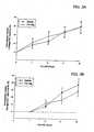
- LV systolic posterior wall thicknesswas reduced and septal wall thickness was increased in a time dependent manner post-MI.
- LV posterior wall thicknesswas similar to baseline values, and was comparatively higher than the saline group at 14 days post-MI.
- LV posterior wall thickness computed as a change from baseline and from 7 day post-MI valuesare shown in Figures 2A and 2B , respectively.
- LV posterior wall thicknessfell in both groups, but was higher in the Fib-Alg group when compared to the saline group at 14 days post-MI.
- LV end-diastolic volumeincreased in a time dependent manner in both groups, however, LV end-diastolic volume tended to be lower in the Fib-Alg group.
- LV end-diastolic volumewas computed as a change from individual baseline or 7 day post-MI values and is shown in Figures 3A and 3B , respectively.
- a trend for a lower LV end-diastolic volumewas observed in the Fib-Alg group when compared to respective 7 day values, but this did not reach statistical significance. While LV ejection fraction fell in both groups post-MI, LV ejection faction tended to be higher in the Fib-Alg group at 28 days post-MI.
- Figures 5A, 5B and 5CSerial changes in the radio-opaque markers positioned within the MI region are shown in Figures 5A, 5B and 5C.
- Figure 5Aillustrates an end-diastolic marker area
- Figure 5Billustrates an end-diastolic marker area as a function from baseline values.
- End-diastolic marker areaincreased in a time dependent fashion in the saline group, and this increase was blunted in the Fib-Alg group.
- the degree of marker expansion, indicative of infarct expansionwas significantly lower in the Fib-Alg group when compared to the saline group.
- myocardial injection of Fib-Alg at 7 days post-MIreduced the degree of infarct expansion that progressively occurs by 28 days post-MI.
- LV geometrywas assessed by ventriculography.
- LV volumes and ejection fractionpresented as individual data values and mean values for each group are shown in Figures 6A and 6B .
- LV end-diastolic volumeincreased significantly in both MI groups, and tended to be lower in the Fib-Alg group.
- LV ejection fractionwas reduced in both MI groups, and tended to be higher in the Fib-Alg group.
- these global indices of volume and functionremained similar between both groups.
- LV radial wall stress for the remote and MI regionswere computed and are shown in Figures 7A and 7B , respectively.
- Regional radial wall stresswas within the MI region in both post-MI groups, but appeared to be the highest in the saline group.
- MMP activitytended to be higher in the saline group when compared to reference control values. However, this did not reach statistical significance. In contrast, MMP interstitial activity was lower in the Fib-Alg group when compared to control and MI only values.
- MI sizebased upon tetrazolium staining was 26 ⁇ 7% and when normalized by perimeter or by gravimetric methods, revealed a similar MI size between groups. The results are shown in Figures 10A and 10B .
- LV regional myocardial stiffnesswas calculated from the sonomicrometry crystals placed within the central portion of the MI and constructing regional pressure-dimension relationships with transient caval occlusion. The results from this analysis are shown in Figure 11 .
- Regional myocardial stiffnesswas significantly increased within the MI region in both post-MI groups.
- the regional stiffness constantwas higher in the Fib-Alg group, but this did not reach statistical significance (p-0.19).
- FIG. 12Representative histological sections stained for hematoxyl in and eosin are shown in Figure 12 .
- a greater degree of inflammatory response and apparent neovascularizationcould be observed within the border and MI regions of the Fib-Alg group.
- An intense inflammatory responsecould be readily appreciated in the MI region of the Fib-Alg group which was more focally distributed around amorphous densities- likely that of injected composite material.
- Figure 13illustrates representative photomicrographs for Alcian blue tissue sections which were counter stained with nuclear fast red. Intense blue staining, likely reflective of proteoglycans and other glycosaminoglycans, could be readily observed within the border and MI regions of both groups.
- Figure 14Aillustrates representative histochemical staining for neutrophils using a myeloperoxidase staining approach.
- the relative intensity of neutrophil stainingincreased in the border region and increased further within the MI region in both groups.
- LV myocardial morphometric measurements of percent collagen within each LV region and placed in comparison to reference control valuesis shown in Figures 15A and 15B .
- the values for each animalare plotted along with the mean values for each group.
- Relative collagen volume fractionincreased within the border and MI regions in both MI groups.
- relative collagen volume fractionwas increased in the border region and reduced within the MI region in the Fib-Alg group.
- Capillary density as determined by lectin positive vesselswas determined in full thickness LV sections and the results are summarized in Figure 16 .
- Capillary density within the MI regionwas reduced in both post-MI groups compared to reference control values.
- capillary densitywas reduced within the border and MI regions when compared to respective remote region values.
- capillary densitywas similar to controls within the border region and not different from respective remote regions in the Fib-Alg group.
- Soluble collagenthat is collagen that could be extracted by a buffered salt solution and homogenization, was increased within the MI regions of both groups as shown in Figure 17A . This is reflective of newly synthesized and incomplete collagen cross-linking, which would be more vulnerable to degradation. However, soluble collagen content was decreased within the MI region in the Fib-Alg group, indicative of a more highly cross-linked fibrillar collagen within this region. Total collagen content, as determined by hydroxyproline measurements of acid digested myocardial samples was increased in a gradual fashion from the remote to MI regions as illustrated in Figure 17 B. These results are consistent with a post-MI fibrotic response, with no significant differences between MI groups.
- LV MMP profiles for MMP-9 and MMP-2, as determined by zymography,are shown in Figures 18A and 18B , respectively.
- MMP-9 levelswere increased significantly within the MI region in both groups. While the relative levels of MMP-9 appeared higher in the Fib-Alg group, these levels did not reach statistical significance. Thus, the robust inflammatory response observed by histological analysis was not associated with a significant increase in relative MMP-9 levels, a primary proteolytic product of neutrophils.
- Relative MMP-2 levelswere significantly increased within the MI regions of both MI groups, but were significantly and substantially lower in the Fib-Alg group when compared to respective saline values. This was most notable for the lower molecular weight form of MMP-2 (68 kDa) which reflects the active form of MMP-2.
- LV MMP-2 values and MMP-2 values normalized to a controlare shown in Figures 19A and 19B .
- Plasma MMP-9 levelswere not detectable within the plasma samples collected in the post-MI period. Plasma levels for MMP-2 were higher than control values at all post-MI time points in the saline group. However, in the Fib-Alg group, plasma MMP-2 levels were not different from controls after 7 days post-MI. In addition, plasma MMP-2 levels were reduced from saline values in the Fib-Alg groups at several post-MI time points.
- Fib-Alg injection into the MI regioncaused a time dependent increase in posterior LV wall thickness when compared to reference vehicle MI values.
- the rate of MI expansionwas significantly attenuated and global indices of LV geometry and function appeared to be improved with Fib-Alg injection.
- the relative fall in LV fractional shorteningwas reduced and a measure of LV pump function, cardiac index was increased.
- Likely mechanisms for the significant attenuation in the infarct expansion process with Fib-Algare improved preservation of the fibrillar collagen matrix as discussed in a subsequent section.
- MMP-9Relative levels of MMP-9 were increased to a similar fashion in the MI groups. Again, this is a somewhat surprising finding due to the fact that a greater inflammatory response was observed in the Fib-Alg group. Moreover, myocardial MMP-2 levels, a ubiquitous MMP found in the myocardium and activated during myocardial remodeling, was reduced in the Fib-Alg MI region. Taken together, this would result in a net reduction in proteolytic activity consistent with the in-vivo microdialysis measurements. Additional confirmation of the relative reduction in MMP-2 levels in the Fib-Alg group is the relative reduction in circulating MMP-2 levels in the post-MI period.
- Fib-Alg composite materialin the post-MI period attenuated infarct expansion and afforded favorable effects on regional and global LV function.
- the mechanisms by which the Fib-Alg injection modified the acceleration of infarct expansionwas by altering the cellular infiltrate and collagen composition within the MI and border regions which favored mature scar formation.
- Devices which can be used to deliver modified or combined components of the gelation systemsinclude, but are not limited to, dual-needle left-ventricle injection devices, dual-needle transvascular wall injection devices and dual syringes.
- Methods of access to use the minimally invasive (i.e., percutaneous or endoscopic) injection devicesinclude access via the femoral artery or the sub-xiphoid.
- "Xiphoid” or “xiphoid process”is a pointed cartilage attached to the lower end of the breastbone or sternum, the smallest and lowest division of the sternum. Both methods are known by those skilled in the art.
- Dual syringe 400can include first barrel 410 and second barrel 420 adjacent to one another and connected at a proximal end 455, distal end 460 and middle region 465 by plates 440, 445 and 450, respectively.
- barrels 410 and 420can be connected by less than three plates.
- Each barrel 410 and 420includes plunger 415 and plunger 425, respectively.
- Barrels 410 and 420can terminate at a distal end into needles 430 and 435, respectively, for extruding a substance.
- barrels 410 and 420can terminate into cannula protrusions for extruding a substance. Barrels 410 and 420 should be in close enough proximity to each other such that the substances in each respective barrel are capable of mixing with one another to form a bioscaffolding in the treatment area, e.g., a post-infarct myocardial region.
- Dual syringe 400can be constructed of any metal or plastic which is minimally reactive or completely unreactive with the formulations described in the present invention. In some examples, dual syringe 400 includes a pre-mixing chamber attached to distal end 465.
- first barrel 410can include a first mixture of a modified two-component gelation system and second barrel 420 can include a second mixture of a modified two-component gelation system according to any of the embodiments described previously.
- a therapeutic amount of the resulting gelis between about 25 ⁇ L to about 200 ⁇ L, preferably about 50 ⁇ L.
- Dual syringe 400can be used during, for example, an open chest surgical procedure.
- Figures 21A-21Billustrate an example of a dual-needle injection device which can be used to deliver the compositions of the present invention.
- Delivery assembly 500includes lumen 510 which may house delivery lumens, guidewire lumens and/or other lumens. Lumen 510, in this example, extends between distal portion 505 and proximal end 515 of delivery assembly 500.
- delivery assembly 500includes first needle 520 movably disposed within delivery lumen 530.
- Delivery lumen 530is, for example, a polymer tubing of a suitable material (e.g., polyamides, polyolefins, polyurethanes, etc.).
- First needle 520is, for example, a stainless steel hypotube that extends a length of the delivery assembly.
- First needle 520includes a lumen with an inside diameter of, for example, 0.08 inches (0.20 centimeters).
- first needle 520has a needle length on the order of about 40 inches (about 1.6 meters) from distal portion 505 to proximal portion 515.
- Lumen 510also includes auxiliary lumen 540 extending, in this example, co-linearly along the length of the catheter (from a distal portion 505 to proximal portion 515).
- Auxiliary lumen 540is, for example, a polymer tubing of a suitable material (e.g., polyamides, polyolefins, polyurethanes, etc.).
- auxiliary lumen 540is terminated at a delivery end of second needle 550 and co-linearly aligned with a delivery end of needle 520.
- Auxiliary lumen 540may be terminated to a delivery end of second needle 550 with a radiation-curable adhesive, such as an ultraviolet curable adhesive.
- Second needle 550is, for example, a stainless steel hypotube that is joined co-linearly to the end of main needle 520 by, for example, solder (illustrated as joint 555). Second needle 550 has a length on the order of about 0.08 inches (0.20 centimeters).
- Figure 21Bshows a cross-sectional front view through line A-A' of delivery assembly 500. Figure 21B shows main needle 520 and second needle 550 in a co-linear alignment.
- auxiliary lumen 540is terminated to auxiliary side arm 460.
- Auxiliary side arm 560includes a portion extending co-linearly with main needle 520.
- Auxiliary side arm 560is, for example, a stainless steel hypotube material that may be soldered to main needle 520 (illustrated as joint 565).
- Auxiliary side arm 560has a co-linear length on the order of about, in one example, 1.2 inches (3 centimeters).
- the proximal end of main needle 520includes adaptor 570 for accommodating a substance delivery device.
- Adaptor 570is, for example, a molded female luer housing.
- a proximal end of auxiliary side arm 560includes adaptor 580 to accommodate a substance delivery device (e.g., a female luer housing).
- a gelmay be formed by a combination (mixing, contact, etc.) of a first mixture of a modified two-component gelation system and a second mixture of a modified two-component gelation system.
- a first mixturemay be introduced by a one cubic centimeters syringe at adaptor 570 through main needle 520.
- a second mixturemay be introduced with a one cubic centimeter syringe at adaptor 580.
- Figures 22A-22Cillustrate an alternative example of a dual-needle injection device which can be used to deliver two-component gel compositions of the present invention.
- the catheter assembly 600provides a system for delivering substances, such as modified two-component gel compositions, to or through a desired area of a blood vessel (a physiological lumen) or tissue in order to treat a myocardial infarct region.
- the catheter assembly 600is similar to the catheter assembly 600 described in commonly-owned, U.S. Patent Application No. 6,554,801 , titled "Directional Needle Injection Drug Delivery Device".
- catheter assembly 600is defined by elongated catheter body 650 having proximal portion 620 and distal portion 610.
- Guidewire cannula 670is formed within catheter body (from proximal portion 610 to distal portion 620) for allowing catheter assembly 600 to be fed and maneuvered over guidewire 680.
- Balloon 630is incorporated at distal portion 610 of catheter assembly 600 and is in fluid communication with inflation cannula 660 of catheter assembly 600.
- Balloon 630can be formed from balloon wall or membrane 635 which is selectively inflatable to dilate from a collapsed configuration to a desired and controlled expanded configuration. Balloon 630 can be selectively dilated (inflated) by supplying a fluid into inflation cannula 660 at a predetermined rate of pressure through inflation port 665 (located at proximal end 620). Balloon wall 635 is selectively deflatable, after inflation, to return to the collapsed configuration or a deflated profile. Balloon 630 may be dilated (inflated) by the introduction of a liquid into inflation cannula 660. Liquids containing treatment and/or diagnostic agents may also be used to inflate balloon 630.
- balloon 630may be made of a material that is permeable to such treatment and/or diagnostic liquids.
- the fluidcan be supplied into inflation cannula 660 at a predetermined pressure, for example, between about one and 20 atmospheres.
- the specific pressuredepends on various factors, such as the thickness of balloon wall 635, the material from which balloon wall 635 is made, the type of substance employed and the flowrate that is desired.
- Catheter assembly 600also includes at least two substance delivery assemblies 605a and 605b (not shown; see Figures 22B-22C ) for injecting a substance into a myocardial infarct region.
- substance delivery assembly 605aincludes needle 615a movably disposed within hollow delivery lumen 625a.
- Delivery assembly 605bincludes needle 615b movably disposed within hollow delivery lumen 625b (not shown; see FIGS. 6B-6C ). Delivery lumen 625a and delivery lumen 625b each extend between distal portion 610 and proximal portion 620.
- Delivery lumen 625a and delivery lumen 625bcan be made from any suitable material, such as polymers and copolymers of polyamides, polyolefins, polyurethanes and the like. Access to the proximal end of delivery lumen 625a or delivery lumen 625b for insertion of needle 615a or 615b, respectively is provided through hub 635 (located at proximal end 620). Delivery lumens 625a and 625b may be used to deliver first and second mixtures of a modified two-component gel composition to a post-myocardial infarct region.
- Figure 22Bshows a cross-section of catheter assembly 600 through line A-A' of Figure 22A (at distal portion 610).
- Figure 22Cshows a cross-section of catheter assembly 600 through line B-B'of Figure 22A .
- delivery assemblies 605a and 605bare adjacent to each other. The proximity of delivery assemblies 605a and 605b allows each mixture of the modified two-component gelation system to rapidly gel when delivered to a treatment site, such as a post-myocardial infarct region.
Landscapes
- Health & Medical Sciences (AREA)
- Chemical & Material Sciences (AREA)
- Life Sciences & Earth Sciences (AREA)
- Medicinal Chemistry (AREA)
- Animal Behavior & Ethology (AREA)
- General Health & Medical Sciences (AREA)
- Public Health (AREA)
- Veterinary Medicine (AREA)
- Dermatology (AREA)
- Oral & Maxillofacial Surgery (AREA)
- Transplantation (AREA)
- Epidemiology (AREA)
- Engineering & Computer Science (AREA)
- Biomedical Technology (AREA)
- Chemical Kinetics & Catalysis (AREA)
- Cell Biology (AREA)
- Dispersion Chemistry (AREA)
- Organic Chemistry (AREA)
- Zoology (AREA)
- Botany (AREA)
- Urology & Nephrology (AREA)
- Hematology (AREA)
- Developmental Biology & Embryology (AREA)
- General Chemical & Material Sciences (AREA)
- Heart & Thoracic Surgery (AREA)
- Cardiology (AREA)
- Nuclear Medicine, Radiotherapy & Molecular Imaging (AREA)
- Bioinformatics & Cheminformatics (AREA)
- Pharmacology & Pharmacy (AREA)
- Materials For Medical Uses (AREA)
- Medicinal Preparation (AREA)
- Medicines That Contain Protein Lipid Enzymes And Other Medicines (AREA)
- Polymers & Plastics (AREA)
Description
- Post-myocardial infarction treatments and compositions.
- Ischemic heart disease typically results from an imbalance between the myocardial blood flow and the metabolic demand of the myocardium. Progressive atherosclerosis with increasing occlusion of coronary arteries leads to a reduction in coronary blood flow. "Atherosclerosis" is a type of arteriosclerosis in which cells including smooth muscle cells and macrophages, fatty substances, cholesterol, cellular waste product, calcium and fibrin build up in the inner lining of a body vessel. "Arteriosclerosis" refers to the thickening and hardening of arteries. Blood flow can be further decreased by additional events such as changes in circulation that lead to hypoperfusion, vasospasm or thrombosis.
- Myocardial infarction (MI) is one form of heart disease that often results from the sudden lack of supply of oxygen and other nutrients. The lack of blood supply is a result of a closure of the coronary artery (or any other artery feeding the heart) which nourishes a particular part of the heart muscle. The cause of this event is generally attributed to arteriosclerosis in coronary vessels.
- Formerly, it was believed that an MI was caused from a slow progression of closure from, for example, 95% then to 100%. However, an MI can also be a result of minor blockages where, for example, there is a rupture of the cholesterol plaque resulting in blood clotting within the artery. Thus, the flow of blood is blocked and downstream cellular damage occurs. This damage can cause irregular rhythms that can be fatal, even though the remaining muscle is strong enough to pump a sufficient amount of blood. As a result of this insult to the heart tissue, scar tissue tends to naturally form.
- Various procedures, including mechanical and therapeutic agent application procedures, are known for reopening blocked arties. An example of a mechanical procedure includes balloon angioplasty with stenting, while an example of a therapeutic agent application includes the administration of a thrombolytic agent, such as urokinase. Such procedures do not, however, treat actual tissue damage to the heart. Other systemic drugs, such as ACE-inhibitors and Beta-blockers, may be effective in reducing cardiac load post-MI, although a significant portion of the population that experiences a major MI ultimately develop heart failure.
- An important component in the progression to heart failure is remodeling of the heart due to mismatched mechanical forces between the infarcted region and the healthy tissue resulting in uneven stress and strain distribution in the left ventricle (LV). Once an MI occurs, remodeling of the heart begins. The principle components of the remodeling event include myocyte death, edema and inflammation, followed by fibroblast infiltration and collagen deposition, and finally scar formation from extra-cellular matrix (ECM) deposition. The principle component of the scar is collagen which is non-contractile and causes strain on the heart with each beat. Non-contractility causes poor pump performance as seen by low ejection fraction (EF) and akinetic or diskinetic local wall motion. Low EF leads to high residual blood volume in the ventricle, causes additional wall stress and leads to eventual infarct expansion via scar stretching and thinning and border-zone cell apoptosis. In addition, the remote-zone thickens as a result of higher stress which impairs systolic pumping while the infarct region experiences significant thinning because mature myocytes of an adult are not regenerated. Myocyte loss is a major etiologic factor of wall thinning and chamber dilation that may ultimately lead to progression of cardiac myopathy. In other areas, remote regions experience hypertrophy (thickening) resulting in an overall enlargement of the left ventricle. This is the end result of the remodelling cascade. These changes also correlate with physiological changes that result in increase in blood pressure and worsening systolic and diastolic performance.
- A bioscaffolding can be formed within a post-myocardial infarct region sufficient to cause attenuation of a rate of myocardial infarct expansion. The bioscaffolding can be formed within the post-myocardial infarct region by combining components of two-component systems.
- The present invention provides a first two-component gelation system and a second two-component gelation system for use in forming a bioscaffolding in vivo within a post-myocardial infarct tissue, wherein the bioscaffolding comprises a first two-component gelation system of a fibrin glue and a second two-component gelation system of an alginate construct comprising an alginate or an alginate conjugate as a first component and a salt as a second component having a different storage modulus than the first two-component gelation system.
- The bioscaffolding can be formed from a mixture of gel components of different two-component gelation systems. In some embodiments, a bioscaffolding can be formed by mixing at least two different components (which do not gel upon mixing) of at least two different two-component gelation systems to form a first mixture and by mixing at least two different components (other than the components that make up the first mixture and which do not gel upon mixing) of the at least two different two-component gelation systems to form a second mixture. A treatment agent, such as a cell type or growth factor, can be added to either the first mixture of the second mixture. The first mixture can then be co-injected with the second mixture to form a bioscaffolding in an infarct region for treatment thereof. The first and second mixtures can be co-injected with a dual-lumen delivery device, which can include, but are not limited to, a dual syringe, a dual-needle left ventricle injection device, a dual-needle transvascular wall injection device and the like.
- In some embodiments, a bioscaffolding can be formed by mixing at least two different gelation components (which do not gel upon mixing) to form a first mixture. A treatment agent, such as a cell type or a growth factor, can be added to the first mixture. The first mixture can then be co-injected with a second gelation component to form a bioscaffolding on or within an infarct region for treatment thereof. The first mixture and the gelation component can be co-injected with, a dual-lumen delivery device, which can include, but are not limited to, a dual syringe, a dual-needle left-ventricle injection device, a dual-needle transvascular wall injection device and the like.
Figures 1A-1B illustrate the progression of heart damage once the buildup of plaque in an artery induces an infarct to occur.Figures 2A-2B illustrate left ventricular posterior wall thicknesses for experimental groups.Figures 3A-3B illustrate left ventricular end-diastolic volumes for experimental groups.Figures 4A-4B illustrate relative changes in left ventricular ejection fraction for experimental groups.Figures 5A-5C illustrate serial changes in radio-opaque markers positioned within myocardial infarct regions.Figure 6A illustrates left ventricular volume for experimental groups.Figure 6B illustrates left ventricular ejection fraction for experimental groups.Figure 7A illustrate left ventricular wall stress for remote regions of experimental groups.Figure 7B illustrates left ventricular wall stress for myocardial infarct regions.Figures 8A-8B illustrate interstitial matrix metallopeptidase (MMP) activity for experimental groups.Figure 9 illustrates myocardial blood flow to tissue regions of the experimental groups.Figures 10A-10B illustrate percentage of myocardial infarction regions for experimental groups.Figure 11 illustrates regional myocardial stiffness for experimental groups.Figure 12 illustrates histological tissue sections stained for hematoxylin and eosin for experimental groups.Figure 13 illustrates photomicrographs for Alcian blue sections counter stained with nuclear fast red.Figure 14A illustrates histochemical staining for neutrophils.Figure 14B illustrates immunohistochemical staining for macrophages.Figures 15A-15B illustrate left ventricular myocardial morphometric measurements of percent collagen within each left ventricular region for experimental groups.Figure 16 illustrates capillary density in full thickness left ventricular sections for experimental groups.Figure 17A illustrates levels of soluble collagen for experimental groups.Figure 17B illustrates levels of hydroxyproline for experimental groups.Figure 18A illustrates left ventricular MMP profiles of MMP-9 for experimental groups.Figure 18B illustrates left ventricular MMP profiles of MMP-2 for experimental groups.Figures 19A-19B illustrate MMP-2 profiles for experimental groups.Figure 20 illustrates an embodiment of a dual bore delivery device.Figures 21A-21B illustrate an alternative embodiment of a dual bore delivery device.Figures 22A-22C illustrate a second alternative embodiment of a dual bore delivery device.Figures 1A-1B illustrate the progression of heart damage once the buildup of plaque induces an infarct to occur.Figure 1A illustrates asite 10 where blockage and restricted blood flow can occur from, for example, a thrombus or embolus.Figure 1B illustratesresultant damage area 20 to the left ventricle that can result from the lack of oxygen and nutrient flow carried by the blood to the inferior region left of the heart. Thedamage area 20 will likely undergo remodeling, and eventually scarring, resulting in a non-functional area.- A bioscaffolding formed of two components and applied in situ to the left heart ventricle can be used to treat post-myocardial infarction tissue damage.
- The bioscaffolding may be a gel formed from a gelation system. "Bioscaffolding" and "two-component gelation system" and "two-component gel system" and "gelation system" and "composite material" are hereinafter used interchangeably. The two-component gelation system includes, alginate construct systems and fibrin glues. Each component of the two-component gelation system may be co-injected to an infarct region by a dual-lumen delivery device. Examples of dual-lumen delivery devices include, but are not limited to, a dual syringe, dual-needle left-ventricle injection devices, dual-needle transvascular wall injection devices and the like.
- At least one cell type may be co-injected with at least one component of the two-component gelation system to an infarct region.
- The cells may be mixed with at least one component of the two-componcnt gelation system before the two-components arc co-injected to the infarct region. Examples of cell types, include, but are not limited to, localized cardiac progenitor cells, mesenchymal stem cells, bone marrow derived mononuclear cells, adipose stem cells, embryonic stem cells, umbilical cord blood derived stem cells, smooth muscle cells or skeletal myoblasts.
- One of the two-component gelation system includes a fibrin glue. Fibrin glue consists of two main components, fibrinogen and thrombin. Fibrinogen is a plasma glycoprotein of about 340 kiloDaltons (kDa) in its endogenous state. Fibrinogen is a symmetrical dimer comprised of six paired polypeptide chains, alpha, beta and gamma chains. On the alpha and beta chains, there is a small peptide sequence called a fibrinopeptide which prevents fibrinogen from spontaneously forming polymers with itself. Fibrinogen may be modified with proteins. Thrombin is a coagulation protein. When combined in equal volumes, thrombin converts the fibrinogen to fibrin by enzymatic action at a rate determined by the concentration of thrombin. The result is a biocompatible gel which gelates when combined at the infarct region. Fibrin glue can undergo gelation between about 5 to about 60 seconds. Examples of other fibrin glue-like systems include, but are not limited to, Tisseel™ (Baxter), Beriplast P™ (Aventis Behring), Biocol® (LFB, France), Crosseal™ (Omrix Biopharmaceuticals, Ltd.), Hemaseel HMN® (Haemacure Corp.), Bolheal (Kaketsuken Pharma, Japan) and CoStasis® (Angiotech Pharmaceuticals).
- One of the two-component gelation system is an alginate construct system. One component may be an alginate conjugate (or alginate alone) which can include alginate and a protein constitucnt. The second component may be a salt. Examples of alginate conjugates can include, but are not limited to, alginate-collagen, alginate-laminin, alginate-elastin, alginate-collagen-laminin and alginate-hyaluronic acid in which the collagen, laminin, elastin, collagen-laminin or hyaluronic acid is covalently bonded (or not bonded) to alginate. Examples of salts which can be used to gel the alginate constructs include, but are not limited to, calcium chloride (CaCl2), barium chloride (BaCl2) or strontium chloride (SrCl2). When the components are combined, for example, alginate-collagen and calcium chloride, the resulting gel has a storage modulus of approximately 1 kiloPascal.
- The alginate construct may be alginate-gelatin. The molecular weight of the gelatin may be in the approximate range of 5 kDa to 100 kDa. The relatively low molecular weight of gelatin offers processing advantages in that it is more soluble and has lower viscosity than hydrogels of higher molecular weight. Another advantage of gelatin is that it contains from 1 to 4 RGD (arginine-glycine-aspartic acid peptide sequence) sites per molecule. RGD is a common cell adhesion ligand and would increase the retention of cells within the infarct zone where the bioscaffolding is formed. The cells retained by the RGD sites may be cells co-injected with the bioscaffolding components or dispersed throughout a component of the system.
- The gelatin may be a porcine gelatin or a recombinant human gelatin. The porcine gelatin is a hydrolyzed
type 1 collagen extracted from porcine skin. In one embodiment, the molecular weight of the porcine gelatin is approximately 20 kDa. The human gelatin is produced by bacteria using human genetic material. The human recombinant gelatin is equivalent to the porcine gelatin but may reduce the likelihood of an immune response when injected into an infarct region of a human subject. - Alginate is a linear polysaccharide derived from seaweed and contains mannuronic (M) and guluronic acid (G), presented in both alternating blocks and alternating individual residues. It is possible to use some of the carboxyl groups of the alginate as sites to graft useful cell adhesion ligarids, such as collagen, laminin, elastin and other peptide fragments of the ECM matrix, forming an alginate conjugate, because alginate does not have RGD groups for cell retention.
- An alginate-gelatin conjugate is valuable because it combines characteristics of alginate with characteristics of gelatin, which include, but are not limited to, RGD sites and immunocompatibility. Characteristics of alginate include rapid, almost instantaneous gelation, and an immune stimulating effect. The alginate-gelatin conjugate can be formed of approximately 1% to 30% and more particularly approximately 10% to 20% gelatin (either porcine or human recombinant) and approximately 80% to 90% alginate. The relatively lower proportion of alginate-gelatin conjugate is used to retain gelation capacity once combined with pure alginate because the alginate carboxyl groups of alginate that cause the gelation may be bound up in the alginate-gelatin conjugate.
- Two-component gelation systems exhibit different characteristics relative to one another including, but not limited to, pore size, storage modulus and gelation time. The gelation system behaves as a sieving media and therefore includes small pores. "Pore size" refers to small, vacuous openings within the gel. "Storage modulus" refers to the strength or the stiffness of the material upon gelation. Storage modulus can be measured by a rheometric instrument. "Gelation time" refers to the kinetics of gelation, the decrease in viscous modulus. Alginate constructs can gel within about 1 second, while fibrin glues can gel between about 5 seconds and about 60 seconds.
- In examples in which cells are co-injected with the two-component gelation system, or mixed with one component before combining the two components, the gelation system can exhibit different characteristics relative to one another relating to the cells. Such characteristics can include, but are not limited to, morphology of the cells, cell survivability, encapsulation efficiency and/or cell concentration. "Morphology" refers to the physical structure of the cells. In the case of hMSC, the natural morphology is a flattened spindle-shaped morphology. "Cell survivability" is the amount of time that the cells remain viable within the gel post-injection. "Encapsulation efficiency" refers to the fraction of the initial number of cells in suspension that are entrapped within the gel. "Cell concentration" is the encapsulation efficiency divided by the volume of gel formed.
- A characteristic which affects the encapsulation efficiency is the difference in viscosity (η) of the two components. If the difference in viscosity between the two components of the gelation system is large, then the encapsulation efficiency is high only when cells are in the high viscosity component. However, if the viscosity of each individual component is lowered without compromising the gelation kinetics, the encapsulation efficiency increases dramatically. For a catheter-based delivery system, low viscosity components are very helpful for successful delivery. A successful application of the two components (which are in solution before delivery) can be dependent upon low viscosity of the individual components.
- A bioscaffolding can be formed from a mixture of gel components of different gelation systems. For example, a bioscaffolding can be formed by mixing at least two different components (which do not gel upon mixing under standard cath lab process conditions) of at least two different two-component gelation systems to form a first mixture, and, by mixing at least two different components (other than the components that make up the first mixture and which do not gel upon mixing under standard cath lab process conditions) of the at least two different two-component gelation systems to form a second mixture. "Gel" generally refers to a semirigid colloidal material formed upon the combination of two different components or two different mixtures. A treatment agent, such as a cell type or a growth factor, can be added to either the first mixture or the second mixture. The first mixture can then be co-injected with the second mixture to form a bioscaffolding in an infarct region for treatment thereof. In some embodiments, a bioscaffolding can be formed by mixing at least two different gelation components (which do not gel upon mixing under standard cath lab process conditions) to form a first mixture. A treatment agent, such as a cell type or a growth factor, can be added to the first mixture. The first mixture can then be co-injected with a gelation component to form a bioscaffolding on an infarct region for treatment thereof. The treatment agent may be co-injected with the first mixture or the gelation component without first interdispersing the treatment agent within the first mixture or the gelation component.
- In some embodiments, an alginate construct system can include an alginate-gelatin solution as a first component and a calcium chloride solution as a second component. Human mesenchymal stem cells (hMSC) may be suspended in one component of the gelation system. hMSC are thought to be capable of both self renewal and differentiation into bone, cartilage, muscle, tendon and fat. hMSC also give rise to a variety of mature cell types via a step-wise maturation process called mesengenesis. The natural morphology of hMSC is elongated and spindle shaped. The gelatin provides RGD sites for cellular adhesion i.e. adhesion of hMSC. Alginate construct systems exhibit rapid gelling kinetics. When combined, alginate-gelatin and calcium chloride gel to form a bioscaffolding in less than 1 second. The resulting gel has a storage modulus of approximately 1 kiloPascal. In application, cell survivability has been observed up to at least 12 days. Encapsulation efficiency is approximately 99%. However, the small pore size of alginate construct systems, which is from about 2 nm to about 500 nm, can lead to low cell spreadability as observed by the round morphology of the hMSC cells over time. "Cell spreading" refers to the naturally occurring morphology of cells. Advantages of alginate construct systems include, but are not limited to, enhanced immune response (a controlled foreign body response) to effect positive remodeling of the injured myocardium, and immunoprotectivity, by shielding via its small pore size, the encapsulated cells from this enhanced immune response (protected from host immune response), instantaneous gelation kinetics, substantial or complete non-adherence to a needle when injected, and- long term cell viability. Furthermore, alginate construct systems degrade slowly (at least 8 weeksin vivo).
- Fibrin glue can include fibrinogen (modified or not modified with protein constituents) as a first component and thrombin as a second component.
- Human mesenchymal stem cells (hMSC) may be suspended in one component of the gelation system. Fibrin glue systems exhibit fast gelling kinetics, but not as rapid as alginate construct systems. When combined, fibrinogen and thrombin gel form a bioscaffolding in about 5 seconds to about 10 seconds. The resulting gel has a storage modulus of approximately 3 kiloPascals which is higher than that of alginate construct systems. A higher storage modulus may improve mechanical reinforcement at the infarct region. In application, cell survivability has been observed up to at least 12 days. The pore size of fibrin glue systems is from about 1.5 microns to about 2.5 microns and can lead to high cell spreadability of hMSC cells. That is, hMSC cells have been observed to have an elongate and stellate morphology which is more natural to their endogenous state when compared to the morphology observed in alginate construct systems alone. Advantages of fibrin glue include, but are not limited to, material strength, promotion of angiogenesis, good cytocompatibility (compatible with cell growth), good cell morphology (elongated and stellate) and high cell proliferation in fibrinogen. One further characteristic of fibrin based gels is that they degrade within 2 weeksin vivo.
- A bioscaffolding may be formed from mixing components of at least two gelation systems. For example, a first component of a first two-component gel and a first component of a second two-component gel can be combined to form a first mixture. A second component of a first two-component gel and a second component of the second two-component gel can be combined to form a second mixture. Cells can be suspended within either the first mixture or the second mixture. When the two mixtures are combined, a bioscaffolding including at least some advantageous characteristics of both gelation systems can be realized. In some embodiments, a bioscaffolding can be formed by mixing at least two different gelation components to form a first mixture. When the first mixture is combined with a gelation salt, a bioscaffolding including at least some advantageous characteristics of the individual components can be realized. It should be appreciated that a number of different combinations of gelation components can be mixed together in different ratios to accentuate various advantageous characteristics of the individual gelation systems. Furthermore, the concentration of the individual components, either singly or combined, can influence certain characteristics of the bioscaffolding, such as viscosity and encapsulation efficiency.
- The bioscaffolding may be formed of an alginatc and fibrin glue composite material as follows.
- A fibrin glue kit Tisseel™ including modified fibrinogen and thrombin was obtained from Baxter. Fibrinogen was reconstituted as directed and then further diluted with water to half of its original concentration. The diluted fibrinogen solution was mixed with a 0.5% alginate-collagen solution in a 1:1 ratio to form a first mixture. hMSC were suspended in a thrombin solution (which contains 40mM CaCl2 ) at a concentration of 2.96 x 10⁷ cells/mL. 200 microliters of the first mixture was combined with the thrombin in a 1:1 ratio. Encapsulation efficiency of the resulting gel was measured at 91.39 ± 6.78%. The viscosity of the suspension component was measured at approximately 7 cp.
- A fibrin glue kit Tisseel™ including modified fibrinogen and thrombin was obtained from Baxter. Fibrinogen was reconstituted as directed and then further diluted with water to half of its original concentration. The diluted fibrinogen solution was mixed with a 0.5% alginate-collagen solution in a 1:1 ratio to form a first mixture. hMSC were suspended in the first mixture at a concentration of 5.51 x 10⁷ cells/mL. The first mixture was combined with the second mixture comprising thrombin and a 2% CaCl2 solution in a 1:1 ratio. Encapsulation cfficicncy of the resulting gel was measured at 99.42 ± 0.12%. The viscosity of the suspension component was measured at approximately 6 cp.
- In one example, a bioscaffolding can be formed by mixing components of more than one gelation system. A Tisseel™ kit including a mixture of fibrinogen, fibronectin, factor XIII and plasminogen obtained from Baxter can be dissolved in an aprotinin solution. The combination is then mixed with gelatin-grafted alginate to form a first mixture. A second mixture is formed by combining thrombin (of a fibrin glue system) and calcium chloride. A storage modulus of the combined mixtures may range from approximately 0.05 kiloPascals to 150 kiloPascals and more specifically from approximately 1 kiloPascal to 5 kiloPascals. A viscosity of the combined mixtures may be approximately 1 cp to 40 cp. In some embodiments, hMSC can be added to an individual component or to the first or second mixture.
- The following experimental results using a composite material including alginate and fibrin glue illustrate how a bioscaffolding may be used to attenuate infarct expansion in a subject having tissues undergoing post-MI remodeling. Specifically, the results demonstrate that injection of a composite material such as alginate and fibrin glue, without seeded cells and/or growth factors, influence changes in LV geometry and pump function during the post-MI period.
- In particular, in the following experiment, MI was surgically induced in adult pigs. It is noted that pigs are a recognized model for understanding post-MI remodeling in humans therefore the treatments disclosed herein are further applicable to humans. It has previously been shown that significant LV remodeling occurs over a one month period and is accompanied by both regional and global abnormalities in LV performance. Accordingly, the experiment disclosed herein examined the effects of forming a bioscaffolding in the MI region in this pig model with respect to LV global and regional geometry and function. The experiment further examined the relationship of these changes to biochemical/histological indices of LV myocardial remodeling up to 4 weeks post-MI.
- The experiment simulated a reasonable post-MI time period by which a myocardial intervention such as injections of components of a bioscaffolding could be considered reasonable. The initial wound healing phase and the beginning of mature scar formation occurs approximately 7 days post-MI. Wound healing is a process which begins with injury and ends with scar formation. It can be separated into three phases: inflammation, proliferation, and remodeling, but these phases overlap to some degree. In the inflammation phase, fibrin is deposited and acts as a road for future cell infiltration. Neutrophils come in and attack any microbes. Macrophages follow and clean up cell debris and release cytokines to attract and stimulate fibroblasts for the proliferation phase. After 2-3 days, the inflammation phase is complete. In the proliferation phase, fibroblasts come in and release connective tissue, collagen, which supports new blood vessel formation. Fibroblastic and granulation tissue synthesis takes place during this phase. This phase takes 1-2 weeks to reach peak fibroblast cell numbers and continues for an additional 2-3 weeks. Contraction also begins during this phase, fibroblasts differentiate into myofibroblasts. This can last for several weeks, but peaks at 5-10 days post wounding. The remodeling phase is marked by the deposition and degradation of collagen coming to equilibrium. During this phase, collagen type III is converted to collagen type I.
- It is recognized that interruption or interference of the initial post MI-wound healing response is associated with adverse LV remodeling. Accordingly, in the present study, the composite injections were performed at 7 days post-MI in order to avoid the confounding influences surrounding the acute phase of an MI. It is further contemplated that the composite injections may be performed any time during the proliferation phase. Representatively, injections may be made, for example, 7 to 28 days post wounding, 7 to 14 days post wounding or 7 to 10 days post wounding.
- For the purposes of assessment of LV regional geometry, radio-opaque markers were placed at the initial surgery and allowed for serial assessment infarct expansion. For the purposes of assessing serial changes in LV global geometry, echocardiography was performed which was then complimented by a ventriculographic assessment of LV geometry at 28 days post-MI. At 28 days post-MI, a full hemodynamic biochemical and histological assessment was performed.
- For the purposes of this experiment, the bioscaffolding components injected into the tissue will include fibrin glue and alginate and will be referred to as composite injection within the text and Fib-Alg in the tables and figures.
- In order to address whether confounding effects of the procedure of injection influenced the results over and above that of the composite injection, saline injections carried out in identical fashion to the composite injections were performed in the protocol. For the purposes of this experiment, this group will be termed the saline injection group.
- In order to allow for reference comparisons to normal, age matched controls that were not subjected to MI or to injections were included in the protocol. These measurements served as reference control values for hemodynamics, blood values and biochemistry/histology results.
- Permanent coronary ligation was performed in mature pigs (e.g., Yorkshire, n=21, 25 kg) obtained from Hambone Farms, Orangeburg, SC in order to induce a posterior-lateral MI. On the day of the surgery, the pigs were sedated using 200 mg of benzodiazapam PO obtained from ESI-Elkins-Sinn Inc, NJ, and placed in a custom designed sling. A transthoracic echocardiogram (e.g., a 3 MHz transducer; Sonos 5500, Agilent Technologies) was performed in order to obtain baseline measurements of LV end-diastolic volume, ejection fraction and wall thickness. The pigs were induced with isoflurane (e.g., 3%/1.5 L/minute) and nitrous oxide (e.g., 0.5 L/minute) and intubated. A sterile left thoracotomy was performed and a catheter connected to an access port (e.g., a 7F obtained from Access Technologies) was placed in the thoracic aorta and the access port placed in a subcutaneous pocket. Next, 4 stainless steel markers (e.g., beads: 1.6 mm outer diameter obtained from VNUS Medical Systems, Sunnyvale, CA) were sutured onto the myocardium centrally located between OM1 and OM2 and 2 cm below the circumflex artery. The markers were placed for an intermarker distance of exactly 1 cm (using in-field calibrated instruments) such that the markers formed a precise quadrilateral array. Two additional markers, placed exactly 1 cm apart, were sutured onto the thoracic wall to serve as an internal calibration. Next, an intravenous bolus of lidocaine (e.g., 1%, 3 mg/kg) was administered and MI induced by direct ligation of the first 2 obtuse marginal branches, OM1 and OM2, at the origin from the circumflex coronary artery (e.g., 4.0 Proline). The incision was closed in layers. All animals were treated and cared for in accordance with the National Institute of Health "Guide for the Care and Use of Laboratory Animals (National Research Council, Washington, 1996)."
- At 7 days post-MI, the pigs were sedated as described in the preceding section and LV echocardiography performed. Aortic blood pressures and blood samples were collected from the indwelling access port. Using the LV echocardiographic and blood pressure measurements, regional peak circumferential wall stress was calculated using conventional techniques. The blood samples were collected in chilled EDTA tubes, centrifuged and the plasma decanted and stored at - 70 degrees C. The radio-opaque markers were then visualized and digitized using conventional techniques. Briefly, the fluoroscopic images were recorded from orthogonal views and digitized (e.g., 30 frames/s, ATI All-in-Wonder Radeon, Thornhill, Ontario, Canada) and gated to the ECG in order to identify the end-diastolic frames. The marker coordinates from the corresponding orthogonal frames were merged to determine end-diastolic marker area. End-diastolic marker area was computed from five consecutive cardiac cycles.
- Following the measurements described above, the pigs were assigned, in alternating fashion, to undergo myocardial injections with the composite material or saline. Following assignment to a treatment protocol, the pigs were returned to the operating room and anesthetized with isoflurane as described in the preceding section. An intravenous bolus of lidocaine (e.g., 3 mg/kg) and magnesium chloride (e.g., 30 mg/mL) was delivered followed by a continuous infusion of lidocaine (e.g., 30 mL/hr). The initial thoracotomy site was reopened, the thoracic space irrigated, and the MI region visualized. Using the radio-opaque markers as a frame of reference, a sterile injection guide of plastic laminate (e.g., 0.25 mm) was sutured onto the MI region.
- The injection guide encompassed the MI region circumscribed by the Obtuse Marginals OM1 and OM2 and extended into the border region of the MI. The injection guide contained a perforated grid with perforations at exactly 0.5 cm intervals and therefore contained 25 perforations over a 2x2 cm area. Thus, the placement of this injection guide provided a means to deliver 25 injections within a precise circumscribed region and pattern.
- The composite injection used was made of components of different two-component gel systems. The first component was a mixture of "Sealer Protein Concentrate" (Baxter Tisseel™ kit, a mixture of fibrinogen, fibronectin, factor XIII, plasminogen), dissolved in an aprotinin solution, then mixed with gelatin-grafted alginate. The second component consisted of a thrombin and calcium chloride solution obtained from Baxter. The components were prepared under sterile conditions. The fibrin/alginate mixture and the thrombin/calcium chloride mixture were drawn into separate 1 mL syringes, loaded onto an injection device such as a double barreled injection device connected to a 26 gauge needle. This system provided a means to inject 100 µL of both materials simultaneously with mixing only occurring within the injection needle and at the site of injection. Precisely 200 µL of the injectate was placed into the myocardium at an injection depth of 0.5 cm for each of the 25 injection sites resulting in a total volume of 5 mL being placed into the myocardial wall. The composite material polymerized immediately at injection and therefore there was no retrograde flow of the injectate. For the saline injections, the identical syringe system and injection protocol were followed. Following the injections, the grid was removed, and the thoracic space closed and evacuated of air.
- LV echocardiography, marker measurements and blood collections were then repeated at
day - Following the final set of serial measurements, the pigs were initially anesthetized using 50 µg of intravenous sufentanyl (ESI-Elkins-Sinn Inc, NJ) and intubated. Anesthesia was maintained throughout the procedure by delivery of 0.5% isoflurane and 60 mg/hr of intravenously administered morphine (ESI). An intravenous lactated Ringers solution of 400 mL/hr was maintained throughout the protocol. This anesthetic protocol resulted in a deep anesthetic plane and stable hemodynamic profile.
- A multi-lumened thermodilution catheter (e.g. a 7.5F obtained from Baxter Healthcare Corp., Irvine, CA) was positioned in the pulmonary artery via the right external jugular vein. An 8F introducer with side-arm was placed in the right carotid for blood pressure measurements and subsequent placement of the ventriculographic catheter. A sternotomy was performed and a vascular ligature placed around the inferior vena cava in order to perform transient caval occlusion.
- A previously calibrated microtipped transducer (e.g., a 7.5 F obtained fromm Millar Instruments Inc, Houston, TX) was placed in the LV through a small apical stab wound. The entire posterior-lateral aspect of the LV was carefully exposed and piezoelectric crystals (e.g., 2 mm obtained from Sonometrics, Ontario) positioned in the central portion of the radio-opaque quadrilateral array. From this crystal array, LV dimension and wall thickness were recorded at a sampling frequency of 100 Hz and digitized. A microdialysis probe containing a 4 mm membrane was inserted in the mid-myocardial region between the crystal pairs. The microdialysis probe was connected to a precision infusion pump and controller system. A flow rate of 2.5 µL/min was established and an iso-osmotic dialysis solution containing a fluorescent substrate for the matrix metalloproteinases (MMPs) was infused at a concentration of 60 µM. The dialysate was passed through a micro-fluorescence detector (e.g., FIAlab Instruments, Inc, of Bellevue, WA) where the solution was subjected to an excitation/emission wavelength of 280/360 nm, and the output digitized using FlAlab ver. 5. The digitized fluorescent output is directly proportional to interstitial myocardial MMP activity.
- At the completion of the instrumentation, fluorescent microspheres (e.g., 3X10⁶ obtained from Molecular Probes, Eugene, OR) of specific emission spectra, were injected into the LV. A reference aortic blood sample was simultaneously withdrawn at a rate of 7 mL/min initiated 5 seconds prior to injection and continued for 120 seconds following injection. Steady state hemodynamics and microdialysis MMP measurements were obtained for 60 minutes. Steady state hemodynamics included systemic and pulmonary artery pressures, cardiac output, and LV pressures. Following steady-state measurements, LV pre-load was altered by sequential occlusion and release of the inferior vena cava and isochronal measurements of LV pressure and dimensions recorded. From the digitized pressure-dimension data, regional myocardial stiffness of the MI region was computed.
- LV volumes and ejection fraction were then determined by ventriculography. A 6F pigtail catheter was placed into the LV via the carotid introducer and connected to a pressure-infusion system containing the radio-opaque dye solution. Nonionic contrast material (30 cc) was injected into the LV and the opacified image filmed at 60 frames/sec in the 30 degree right anterior oblique position.
- For the purposes of obtaining reference control values, 5 age and weight matched pigs were instrumented in the identical fashion and the entire series of LV myocardial function measurements performed.
- Following the final set of measurements, the great vessels were cross-clamped and the heart removed. The LV was quickly separated and placed in iced saline. A full thickness circumferential ring (e.g., 1 cm) was prepared where the sectioning was performed using the central portion of the marker quadrilateral array as the frame of reference. This section was used for tetrazolium staining and computation of MI size using planimetry. This myocardial ring was then processed for blood flow measurements by potassium hydroxide digestion and fluorimetry. The remaining LV was divided into MI region, border region (defined as the 2 cm region surrounding the MI) and the remote region. These myocardial sections were flash frozen for biochemical analysis, or placed in formalin for subsequent histological staining.
- LV myocardial samples weighing approximately 0.25 g from each region were lyophilized and subjected to hydrochloric acid digestion for hydroxyproline measurements in order to determine total collagen content. In parallel samples, the myocardium was homogenized and centrifuged and the supernatant subjected to biochemical measurement of soluble collagen using the picrosirius method. Relative MMP-2 and MMP-9 levels were determined by substrate zymography. Briefly, LV myocardial extracts (10 µg protein) were subjected to electrophoresis followed by gelatin (e.g.,
Novex 10% zymogram gel, 0.1% gelatin obtained from Invitrogen) zymography. The size-fractionated MMP proteolytic regions were quantified by densitometry using a Gel Pro Analyzer obtained from Media Cybernetics. - Plasma measurements of MMP-2 and MMP-9 were performed using an enzyme linked multiplex suspension array obtained from Bio-Rad Laboratories of Hercules, CA and flow cytometric detection (Luminex). This approach provided a sensitivity for MMP-2 at less than 25 pg/mL and for MMP-9 at less than 7 pg/mL with an intra-assay coefficient of variation of less than 15%. Using MMP-2 and MMP-9 standards for calibration, a high linearity for this assay approach was obtained (0.99, p<0,001). All plasma samples were measured in duplicate.
- LV sections of 5 µm were stained with picro-sirius red and the relative collagen percent area for the MI, border and remote regions were determined using computer assisted morphometric methods as described previously. In addition, LV sections were stained with the lectin GSA-B4 in order to identify capillary endothelium and compute capillary density using computer morphometric methods. Immunohistochemical studies on the paraffin-embedded sections were performed using primary antibodies directed against macrophages (MAC-3, CL8943A, Cedarlane, 1:200) and lymphocytes (CD4, CL012A, Cedarlane, 1:200, GEA4023-1, Genex, 1:250). Visualization of the lectin and primary antisera binding sites was performed using a 3',3'-diaminobenzidine-hydrogen peroxide substrate obtained from Vector Labs. Sections were imaged on an inverted microscope and the images were digitized using conventional techniques. Negative controls were utilized in all staining protocols and included substitution with nonimmune anti-sera. Routine histological staining was also performed in order to examine relative tissue structure in the remote, border and MI regions by hematoxylin and eosin, as well as by Alcian blue counter stained with nuclear fast red.
- All of the data collected in this study were obtained in a blinded fashion and remained coded until the end of the study. The observers for the echocardiography, marker, and ventriculography measurements were blinded to the treatment assignments throughout the study protocol. Serial measurements of LV geometry and function were compared using a two-way analysis of variance (ANOVA) model. Single point measurements were compared between treatment and control groups using a one-way ANOVA. Following the ANOVA, pair-wise comparisons were performed using unpaired t-tests corrected for number of comparisons. In addition, comparisons to baseline values and to 7-day post MI values were performed as computing the percent change from respective individual values and subjecting these computations to a t-test. For the biochemical and morphometric studies, comparisons to reference control values were performed using a two way ANOVA in which the treatment effects were group and region. All statistical analyses were performed using the STATA® statistical software package obtained from Statacorp of College Station, TX. Values of p<0.05 were considered to be statistically significant.
- All 21 pigs entered into the study survived the initial instrumentation and MI induction. At 24 hours following the initial MI induction, plasma troponin values were higher than reference control values by over 5-fold (25.6±3.2 U/mL, p<0.05) with no difference between those randomized to the Fib/Alg group (29.3±5.2 U/mL) and those randomized to the saline group (23.2±4.0 U/mL). At 7 days post-MI, of the 11 pigs assigned to the composite injection (Fib-Alg) group, 2 pigs developed intraoperative refractory ventricular fibrillation during the injection procedure. None of the 10 pigs assigned to the saline injection group developed refractory arrhythmogenesis during the 7 day post-MI procedure. An additional pig in the Fib-Alg group developed refractory ventricular tachycardia on
post-MI day 14. One pig in the saline injection group developed ventricular tachycardia onpost-MI day 28, but was successfully cardioverted for the LV function and hemodynamic measurements. Thus, the final sample sizes for this study that completed the 28 day post-MI protocol were 8 in the Fib-Alg group and 9 in the Saline group. - Serial LV echocardiographic measurements are shown in Table 1 below.
Table 1. Serial Changes in Echocardiographically-Derived Parameters Following Myocardial Infarction (MI): Effects of Saline or Fibrin-Alginate Injection at 7 days Post-MI Time Post-MI (days) Baseline 7 14 21 28 Heart Rate (bpm) Saline 119±6 117±8 116±4 118±2 129±24 Fib-Alg 130±9 123±4 135±6 123±4 121±7 Posterior Wall thickness at End-systole (cm) Saline 1.10±0.05 0.73±0.08# 0.69±0.07# 0.73±0.09# 0.67±0.08# Fib-Alg 1.13±0.04 0.80±0.06# 1.07±0.11*+ 0.90±0.12 0.86±0.11# Septal Wall thickness at End-systole (cm) Saline 1.10±0.03 1.05±0.03 1.09±0.05 1.18±0.05+ 1.23±0.07#+ Fib-Alg 1.04±0.03 1.05±0.04 1.10±0.03 1.19±0.04#+ 1.20±0.04#+ End-diastolic Volume (mL) Saline 47.5±2.1 55.3±1.9# 59.4±1.5# 69.9±2.2#+ 75.6±3.2#+ Fib-Alg 45.3±1.0 55.5±1.6# 62.1±2.7# 64.9±2.9#+ 71.7±3.9#+ Ejection Fraction (%) Saline 67.0±0.9 53.2±1.1# 49.8±1.6# 47.0±1.9#+ 45.6±3.2#+ Fib-Alg 68.1±1.0 54.7±1.7# 51.2±1.2# 48.2±3.8#+ 49.0±2.6# LV Mass/Body Weight (g/kg) Saline 4.4±0.4 4.2±0.3 3.8±0.3 3.8±0.3 3.5±0.2 Fib-Alg 3.7±0.6 3.6±0.5 3.6±0.5 3.1±0.4 3.4±0.5 Values presented as Mean ± SEM. Sample Size: Saline: n=9, Fib-Alg: n=8.
#p<0.05 vs. Baseline.+p<0.05 vs. 7 days post-MI. *p<0.05 vs. Saline. - LV systolic posterior wall thickness was reduced and septal wall thickness was increased in a time dependent manner post-MI. However in the Fib-Alg group, LV posterior wall thickness was similar to baseline values, and was comparatively higher than the saline group at 14 days post-MI.
- LV posterior wall thickness computed as a change from baseline and from 7 day post-MI values are shown in
Figures 2A and2B , respectively. LV posterior wall thickness fell in both groups, but was higher in the Fib-Alg group when compared to the saline group at 14 days post-MI. LV end-diastolic volume increased in a time dependent manner in both groups, however, LV end-diastolic volume tended to be lower in the Fib-Alg group. - LV end-diastolic volume was computed as a change from individual baseline or 7 day post-MI values and is shown in
Figures 3A and 3B , respectively. A trend for a lower LV end-diastolic volume was observed in the Fib-Alg group when compared to respective 7 day values, but this did not reach statistical significance. While LV ejection fraction fell in both groups post-MI, LV ejection faction tended to be higher in the Fib-Alg group at 28 days post-MI. - The relative changes in LV ejection fraction as a function from baseline values and from 7 day post-MI values are shown in
Figures 4A and 4B , respectively. This analysis revealed that the decline in LV ejection fraction by 28 days post-MI was less pronounced in the Fib-Alg group. - Serial changes in the radio-opaque markers positioned within the MI region are shown in
Figures 5A, 5B and5C. Figure 5A illustrates an end-diastolic marker area andFigure 5B illustrates an end-diastolic marker area as a function from baseline values. End-diastolic marker area increased in a time dependent fashion in the saline group, and this increase was blunted in the Fib-Alg group. As shown as a function of 7 day post-MI values inFigure 5C , the degree of marker expansion, indicative of infarct expansion, was significantly lower in the Fib-Alg group when compared to the saline group. Thus, myocardial injection of Fib-Alg at 7 days post-MI reduced the degree of infarct expansion that progressively occurs by 28 days post-MI. - At the completion of the 28 day post-MI protocol, additional LV myocardial measurements of geometry and function were performed. These measurements were done with full instrumentation and a surgical procedure. During the placement of the sonomicrometry crystals, 2 pigs in the Fib/Alg group and 1 pig in the saline group developed refractory ventricular fibrillation. Thus, the hemodynamics on Table 2 below are for a sample size of 6 and 8 respectively.
Table 2. Hemodynamic Parameters at 28 days Following Myocardial Infarction (MI): Effects of Saline or Fibrin-Alginate (Fib-Alg) Injection at 7 days Post-MI Control Saline Fib-Alg Heart Rate (bpm) 106±6 114±7 135±9C LV Peak Pressure (mmHg) 113±2 110±2 107±2 LV End-diastolic Pressure (mmHg) 10±1 10±1 11±1 Peak dP/dt (mmHg/s) 1372±121 1668±212 1814±161 Aortic Systolic Pressure (mmHg) 113±2 112±1 107±4 Aortic Diastolic Pressure (mmHg) 80±4 75±3 71±4 Mean Aortic Pressure (mmHg) 94±1 93±3 88±4 PA Systolic Pressure (mmHg) 27±2 27±1 32±3 PA Diastolic Pressure (mmHg) 17±2 21±1 21±3 Mean PA Pressure (mmHg) 22±2 21±1 26±4 Cardiac Output (L/min) 3.8±0.6 3.5±0.2 3.8±0.2 Cardiac Index (mL/min/Kg) 109±4 87±4C 98±5 Systemic Vascular Resistance (dyne.s.cm-5) 2047±180 2168±145 1888±157 Pulmonary Vascular Resistance (dyne.s.cm-5) 322±46 333±30 402±65 Sample Size 5 8 6 Values presented as Mean ± SEM.
Cp<0.05 vs. Control. *p<0.05 vs. Saline.
PA: Pulmonary Artery - Resting ambient heart rate was higher in the Fib-Alg group when compared to the saline group. Mean arterial pressure was slightly lower in the post-MI groups, but was not significantly different from controls; indicative of hemodynamic stability. Cardiac output was slightly lower in the saline group, and when indexed to body weight, was reduced from reference controls. Cardiac index was unchanged from control values in the Fib-Alg group.
- LV geometry was assessed by ventriculography. LV volumes and ejection fraction presented as individual data values and mean values for each group are shown in
Figures 6A and6B . Similar to the LV echocardiographic measurements, as shown inFigure 6A , LV end-diastolic volume increased significantly in both MI groups, and tended to be lower in the Fib-Alg group. As shown inFigure 6B , LV ejection fraction was reduced in both MI groups, and tended to be higher in the Fib-Alg group. However, these global indices of volume and function remained similar between both groups. - Using the combined LV echocardiography and aortic pressure measurements, LV radial wall stress for the remote and MI regions were computed and are shown in
Figures 7A and7B , respectively. Regional radial wall stress was within the MI region in both post-MI groups, but appeared to be the highest in the saline group. - In-vivo microdialysis was performed in order to measure interstitial MMP activity within the MI region. The results are shown in
Figures 8A and8B . MMP activity tended to be higher in the saline group when compared to reference control values. However, this did not reach statistical significance. In contrast, MMP interstitial activity was lower in the Fib-Alg group when compared to control and MI only values. - Myocardial blood flow, normalized to the remote region (region served by LAD) was reduced in the MI region in both the saline and Fib-Alg groups as illustrated in
Figure 9 . Computed MI size based upon tetrazolium staining was 26±7% and when normalized by perimeter or by gravimetric methods, revealed a similar MI size between groups. The results are shown inFigures 10A and10B . - LV regional myocardial stiffness was calculated from the sonomicrometry crystals placed within the central portion of the MI and constructing regional pressure-dimension relationships with transient caval occlusion. The results from this analysis are shown in
Figure 11 . Regional myocardial stiffness was significantly increased within the MI region in both post-MI groups. The regional stiffness constant was higher in the Fib-Alg group, but this did not reach statistical significance (p-0.19). - Representative histological sections stained for hematoxyl in and eosin are shown in
Figure 12 . A greater degree of inflammatory response and apparent neovascularization could be observed within the border and MI regions of the Fib-Alg group. An intense inflammatory response could be readily appreciated in the MI region of the Fib-Alg group which was more focally distributed around amorphous densities- likely that of injected composite material. Figure 13 illustrates representative photomicrographs for Alcian blue tissue sections which were counter stained with nuclear fast red. Intense blue staining, likely reflective of proteoglycans and other glycosaminoglycans, could be readily observed within the border and MI regions of both groups.Figure 14A illustrates representative histochemical staining for neutrophils using a myeloperoxidase staining approach. The relative intensity of neutrophil staining increased in the border region and increased further within the MI region in both groups. However, the degree of myeloperoxidase appeared increased within both of these regions in the Fib-Alg group.- Immunohistochemical staining for macrophages is shown in
Figure 14 B. Macrophage positive regions were observed within the border and MI regions of both groups, but the number of macrophage positive cells appeared increased in the Fib-Alg group. - LV myocardial morphometric measurements of percent collagen within each LV region and placed in comparison to reference control values is shown in
Figures 15A and15B . The values for each animal are plotted along with the mean values for each group. Relative collagen volume fraction increased within the border and MI regions in both MI groups. However, relative collagen volume fraction was increased in the border region and reduced within the MI region in the Fib-Alg group. - Capillary density as determined by lectin positive vessels was determined in full thickness LV sections and the results are summarized in
Figure 16 . Capillary density within the MI region was reduced in both post-MI groups compared to reference control values. In the saline group, capillary density was reduced within the border and MI regions when compared to respective remote region values. In marked contrast, capillary density was similar to controls within the border region and not different from respective remote regions in the Fib-Alg group. - Soluble collagen, that is collagen that could be extracted by a buffered salt solution and homogenization, was increased within the MI regions of both groups as shown in
Figure 17A . This is reflective of newly synthesized and incomplete collagen cross-linking, which would be more vulnerable to degradation. However, soluble collagen content was decreased within the MI region in the Fib-Alg group, indicative of a more highly cross-linked fibrillar collagen within this region. Total collagen content, as determined by hydroxyproline measurements of acid digested myocardial samples was increased in a gradual fashion from the remote to MI regions as illustrated inFigure 17 B. These results are consistent with a post-MI fibrotic response, with no significant differences between MI groups. - LV MMP profiles for MMP-9 and MMP-2, as determined by zymography, are shown in
Figures 18A and18B , respectively. MMP-9 levels were increased significantly within the MI region in both groups. While the relative levels of MMP-9 appeared higher in the Fib-Alg group, these levels did not reach statistical significance. Thus, the robust inflammatory response observed by histological analysis was not associated with a significant increase in relative MMP-9 levels, a primary proteolytic product of neutrophils. Relative MMP-2 levels were significantly increased within the MI regions of both MI groups, but were significantly and substantially lower in the Fib-Alg group when compared to respective saline values. This was most notable for the lower molecular weight form of MMP-2 (68 kDa) which reflects the active form of MMP-2. LV MMP-2 values and MMP-2 values normalized to a control are shown inFigures 19A and19B . - Despite multiple dilution series, plasma MMP-9 levels were not detectable within the plasma samples collected in the post-MI period. Plasma levels for MMP-2 were higher than control values at all post-MI time points in the saline group. However, in the Fib-Alg group, plasma MMP-2 levels were not different from controls after 7 days post-MI. In addition, plasma MMP-2 levels were reduced from saline values in the Fib-Alg groups at several post-MI time points.
- The key findings of this experiment can be summarized into 3 broad areas: (1) LV function and hemodynamics, (2) myocardial structure, and (3) myocardial biochemistry.
- The main findings in this area was that the Fib-Alg injection into the MI region caused a time dependent increase in posterior LV wall thickness when compared to reference vehicle MI values. In the Fib-Alg group, the rate of MI expansion was significantly attenuated and global indices of LV geometry and function appeared to be improved with Fib-Alg injection. Specifically, the relative fall in LV fractional shortening was reduced and a measure of LV pump function, cardiac index was increased. Likely mechanisms for the significant attenuation in the infarct expansion process with Fib-Alg are improved preservation of the fibrillar collagen matrix as discussed in a subsequent section. The basis for the improvements in LV pump function were likely due to changes in LV load as regional wall stress patterns and systemic vascular resistance tended to be lower in the Fib-Alg group. All of these favorable effects on local and global LV geometry were not due to differences in initial MI sizes or blood flow patterns.
- One of the more prominent features from the imaging studies was the increased LV posterior wall thickness following Fib-Alg injection. At the histological level, a clear and robust inflammatory response was observed by both histochemical and immunohistochemical methods. Specifically, increased neutrophil infiltrate within the MI region was observed at one month post-MI in with Fib-Alg injection and this was associated with a higher number of macrophages. Interestingly, however, this was not associated with increased proteolytic activity as assessed by in-vivo microdialysis. In fact, MMP proteolytic activity was reduced within the MI region in the Fib-Alg group. Total soluble collagen within the MI region was reduced with Fib-Alg injection whereas total biochemical content of collagen was similar between the MI groups. This suggest that greater cross-linking of collagen occurred in the Fib-Alg group which would improve infarct stiffening and favor improved tethering of the border region. Indeed, relative myocardial stiffness appeared higher in the Fib-Alg group and relative percent collagen by morphometry was increased within the border region in the Fib-Alg group. Thus, the increased inflammatory response observed in the Fib-Alg group did not appear to cause increased collagen degradation, but rather facilitated a maturation of collagen and scar formation within the MI and border regions.
- Relative levels of MMP-9 were increased to a similar fashion in the MI groups. Again, this is a somewhat surprising finding due to the fact that a greater inflammatory response was observed in the Fib-Alg group. Moreover, myocardial MMP-2 levels, a ubiquitous MMP found in the myocardium and activated during myocardial remodeling, was reduced in the Fib-Alg MI region. Taken together, this would result in a net reduction in proteolytic activity consistent with the in-vivo microdialysis measurements. Additional confirmation of the relative reduction in MMP-2 levels in the Fib-Alg group is the relative reduction in circulating MMP-2 levels in the post-MI period.
- The injection of a Fib-Alg composite material in the post-MI period attenuated infarct expansion and afforded favorable effects on regional and global LV function. The mechanisms by which the Fib-Alg injection modified the acceleration of infarct expansion was by altering the cellular infiltrate and collagen composition within the MI and border regions which favored mature scar formation.
- Devices which can be used to deliver modified or combined components of the gelation systems include, but are not limited to, dual-needle left-ventricle injection devices, dual-needle transvascular wall injection devices and dual syringes. Methods of access to use the minimally invasive (i.e., percutaneous or endoscopic) injection devices include access via the femoral artery or the sub-xiphoid. "Xiphoid" or "xiphoid process" is a pointed cartilage attached to the lower end of the breastbone or sternum, the smallest and lowest division of the sternum. Both methods are known by those skilled in the art.
Figure 20 illustrates an example of a dual syringe device which can be used to deliver the compositions of the present invention.Dual syringe 400 can includefirst barrel 410 andsecond barrel 420 adjacent to one another and connected at a proximal end 455,distal end 460 andmiddle region 465 byplates 440, 445 and 450, respectively. In some examples,barrels barrel Barrels needles barrels Barrels Dual syringe 400 can be constructed of any metal or plastic which is minimally reactive or completely unreactive with the formulations described in the present invention. In some examples,dual syringe 400 includes a pre-mixing chamber attached todistal end 465.- In some applications,
first barrel 410 can include a first mixture of a modified two-component gelation system andsecond barrel 420 can include a second mixture of a modified two-component gelation system according to any of the embodiments described previously. A therapeutic amount of the resulting gel is between about 25 µL to about 200 µL, preferably about 50 µL.Dual syringe 400 can be used during, for example, an open chest surgical procedure. Figures 21A-21B illustrate an example of a dual-needle injection device which can be used to deliver the compositions of the present invention.Delivery assembly 500 includes lumen 510 which may house delivery lumens, guidewire lumens and/or other lumens. Lumen 510, in this example, extends between distal portion 505 and proximal end 515 ofdelivery assembly 500.- In one example,
delivery assembly 500 includes first needle 520 movably disposed within delivery lumen 530. Delivery lumen 530 is, for example, a polymer tubing of a suitable material (e.g., polyamides, polyolefins, polyurethanes, etc.). First needle 520 is, for example, a stainless steel hypotube that extends a length of the delivery assembly. First needle 520 includes a lumen with an inside diameter of, for example, 0.08 inches (0.20 centimeters). In one example for a retractable needle catheter, first needle 520 has a needle length on the order of about 40 inches (about 1.6 meters) from distal portion 505 to proximal portion 515. Lumen 510 also includes auxiliary lumen 540 extending, in this example, co-linearly along the length of the catheter (from a distal portion 505 to proximal portion 515). Auxiliary lumen 540 is, for example, a polymer tubing of a suitable material (e.g., polyamides, polyolefins, polyurethanes, etc.). At distal portion 505, auxiliary lumen 540 is terminated at a delivery end of second needle 550 and co-linearly aligned with a delivery end of needle 520. Auxiliary lumen 540 may be terminated to a delivery end of second needle 550 with a radiation-curable adhesive, such as an ultraviolet curable adhesive. Second needle 550 is, for example, a stainless steel hypotube that is joined co-linearly to the end of main needle 520 by, for example, solder (illustrated as joint 555). Second needle 550 has a length on the order of about 0.08 inches (0.20 centimeters).Figure 21B shows a cross-sectional front view through line A-A' ofdelivery assembly 500.Figure 21B shows main needle 520 and second needle 550 in a co-linear alignment. - Referring to
Figure 21A , at proximal portion 515, auxiliary lumen 540 is terminated toauxiliary side arm 460. Auxiliary side arm 560 includes a portion extending co-linearly with main needle 520. Auxiliary side arm 560 is, for example, a stainless steel hypotube material that may be soldered to main needle 520 (illustrated as joint 565). Auxiliary side arm 560 has a co-linear length on the order of about, in one example, 1.2 inches (3 centimeters). - The proximal end of main needle 520 includes adaptor 570 for accommodating a substance delivery device. Adaptor 570 is, for example, a molded female luer housing. Similarly, a proximal end of auxiliary side arm 560 includes adaptor 580 to accommodate a substance delivery device (e.g., a female luer housing).
- The design configuration described above with respect to
Figures 21A -21B is suitable for introducing modified two-component gel compositions of the present invention. For example, a gel may be formed by a combination (mixing, contact, etc.) of a first mixture of a modified two-component gelation system and a second mixture of a modified two-component gelation system. Representatively, a first mixture may be introduced by a one cubic centimeters syringe at adaptor 570 through main needle 520. At the same time or shortly before or after, a second mixture may be introduced with a one cubic centimeter syringe at adaptor 580. When the first and second components combine at the exit of delivery assembly 500 (at an infarct region), the materials combine (mix, contact) to form a bioerodable gel. Figures 22A-22C illustrate an alternative example of a dual-needle injection device which can be used to deliver two-component gel compositions of the present invention. In general, thecatheter assembly 600 provides a system for delivering substances, such as modified two-component gel compositions, to or through a desired area of a blood vessel (a physiological lumen) or tissue in order to treat a myocardial infarct region. Thecatheter assembly 600 is similar to thecatheter assembly 600 described in commonly-owned,U.S. Patent Application No. 6,554,801 , titled "Directional Needle Injection Drug Delivery Device".- In one example,
catheter assembly 600 is defined by elongated catheter body 650 having proximal portion 620 and distal portion 610. Guidewire cannula 670 is formed within catheter body (from proximal portion 610 to distal portion 620) for allowingcatheter assembly 600 to be fed and maneuvered over guidewire 680. Balloon 630 is incorporated at distal portion 610 ofcatheter assembly 600 and is in fluid communication with inflation cannula 660 ofcatheter assembly 600. - Balloon 630 can be formed from balloon wall or membrane 635 which is selectively inflatable to dilate from a collapsed configuration to a desired and controlled expanded configuration. Balloon 630 can be selectively dilated (inflated) by supplying a fluid into inflation cannula 660 at a predetermined rate of pressure through inflation port 665 (located at proximal end 620). Balloon wall 635 is selectively deflatable, after inflation, to return to the collapsed configuration or a deflated profile. Balloon 630 may be dilated (inflated) by the introduction of a liquid into inflation cannula 660. Liquids containing treatment and/or diagnostic agents may also be used to inflate balloon 630. In one example, balloon 630 may be made of a material that is permeable to such treatment and/or diagnostic liquids. To inflate balloon 630, the fluid can be supplied into inflation cannula 660 at a predetermined pressure, for example, between about one and 20 atmospheres. The specific pressure depends on various factors, such as the thickness of balloon wall 635, the material from which balloon wall 635 is made, the type of substance employed and the flowrate that is desired.
Catheter assembly 600 also includes at least two substance delivery assemblies 605a and 605b (not shown; seeFigures 22B-22C ) for injecting a substance into a myocardial infarct region. In one example, substance delivery assembly 605a includes needle 615a movably disposed within hollow delivery lumen 625a. Delivery assembly 605b includes needle 615b movably disposed within hollow delivery lumen 625b (not shown; seeFIGS. 6B-6C ). Delivery lumen 625a and delivery lumen 625b each extend between distal portion 610 and proximal portion 620. Delivery lumen 625a and delivery lumen 625b can be made from any suitable material, such as polymers and copolymers of polyamides, polyolefins, polyurethanes and the like. Access to the proximal end of delivery lumen 625a or delivery lumen 625b for insertion of needle 615a or 615b, respectively is provided through hub 635 (located at proximal end 620). Delivery lumens 625a and 625b may be used to deliver first and second mixtures of a modified two-component gel composition to a post-myocardial infarct region.Figure 22B shows a cross-section ofcatheter assembly 600 through line A-A' ofFigure 22A (at distal portion 610).Figure 22C shows a cross-section ofcatheter assembly 600 through line B-B'ofFigure 22A . In some embodiments, delivery assemblies 605a and 605b are adjacent to each other. The proximity of delivery assemblies 605a and 605b allows each mixture of the modified two-component gelation system to rapidly gel when delivered to a treatment site, such as a post-myocardial infarct region.
Claims (13)
- A first two-component gelation system and a second two-component gelation system for use in forming a bioscaffolding in vivo within a post-myocardial infarct tissue, wherein the bioscaffolding comprises a first two-component gelation system of a fibrin glue and a second two-component gelation system of an alginate construct comprising an alginate or an alginate conjugate as a first component and a salt as a second component, having a different storage modulus than the first two-component gelation system.
- A first two-component gelation system and a second two-component gelation system for use according to claim 1, wherein (a) the first component of the first two-component gelation system comprises gelatin grafted alginate and a first component of the second two-component gelation system is a fibrinogen solution and (b) a second component of the first two-component gelation system is thrombin and a second component of the second two-component gelation system is calcium chloride.
- A first two-component gelation system and a second two-component gelation system for use according to claim 1, wherein the bioscaffolding is formed during a proliferation phase occurring after a myocardial infarction.
- A first two-component gelation system and a second two-component gelation system for use according to claim 1, wherein the bioscaffolding is formed seven days or more after occurrence of a myocardial infarction.
- A first two-component gelation system and a second two-component gelation system for use according to claim 1, further comprising:a bioscaffolding within a border region of the post-myocardial infarct region.
- A first two-component gelation system and a second two-component gelation system for use in accordance with claim 1 to cause a time dependent increase in posterior left ventricular wall thickness.
- A first two-component gelation system and a second two-component gelation system for use according to claim 6, wherein (a) a first component of the first two-component gelation system comprises gelatin grafted alginate and a first component of the second two-component gelation system is a fibrinogen solution and (b) a second component of the first two-component gelation system is thrombin and a second component of the second two-component gelation system is calcium chloride.
- A first two-component gelation system and a second two-component gelation system for use according to claim 1 to cause a maturation of myocardial scar formation.
- A first two-component gelation system and a second two-component gelation system for use according to claim 8, to also decrease matrix metalloproteinase activity.
- A first two-component gelation system and a second two-component gelation system for use according to claim 8, to also increase collagen type I.
- A first two-component gelation system and a second two-component gelation system for use according to claim 8, to also increase capillary density.
- A first two-component gelation system and a second two-component gelation system for use according to claim 8, to also increase fibroblast infiltration.
- A first two-component gelation system and a second two-component gelation system for use according to claim 8, wherein the bioscaffolding comprises a first two-component gelation system and a second two-component gelation system wherein (a) a first component of the first two-component gelation system comprises gelatin grafted alginate and a first component of the second two-component gelation system is a fibrinogen solution and (b) a second component of the first two-component gelation system is thrombin and a second component of the second two-component gelation system is calcium chloride.
Priority Applications (1)
| Application Number | Priority Date | Filing Date | Title |
|---|---|---|---|
| EP12151788.2AEP2452699B1 (en) | 2007-01-18 | 2008-01-18 | Scaffolding for modifying myocardial infarction expansion |
Applications Claiming Priority (3)
| Application Number | Priority Date | Filing Date | Title |
|---|---|---|---|
| US88559307P | 2007-01-18 | 2007-01-18 | |
| US12/016,180US9005672B2 (en) | 2006-11-17 | 2008-01-17 | Methods of modifying myocardial infarction expansion |
| PCT/US2008/051505WO2008089452A2 (en) | 2007-01-18 | 2008-01-18 | Methods of modifying myocardial infarction expansion |
Related Child Applications (2)
| Application Number | Title | Priority Date | Filing Date |
|---|---|---|---|
| EP12151788.2ADivisionEP2452699B1 (en) | 2007-01-18 | 2008-01-18 | Scaffolding for modifying myocardial infarction expansion |
| EP12151788.2ADivision-IntoEP2452699B1 (en) | 2007-01-18 | 2008-01-18 | Scaffolding for modifying myocardial infarction expansion |
Publications (2)
| Publication Number | Publication Date |
|---|---|
| EP2109469A2 EP2109469A2 (en) | 2009-10-21 |
| EP2109469B1true EP2109469B1 (en) | 2014-06-04 |
Family
ID=39636755
Family Applications (2)
| Application Number | Title | Priority Date | Filing Date |
|---|---|---|---|
| EP08727952.7AActiveEP2109469B1 (en) | 2007-01-18 | 2008-01-18 | Methods of modifying myocardial infarction expansion |
| EP12151788.2AActiveEP2452699B1 (en) | 2007-01-18 | 2008-01-18 | Scaffolding for modifying myocardial infarction expansion |
Family Applications After (1)
| Application Number | Title | Priority Date | Filing Date |
|---|---|---|---|
| EP12151788.2AActiveEP2452699B1 (en) | 2007-01-18 | 2008-01-18 | Scaffolding for modifying myocardial infarction expansion |
Country Status (4)
| Country | Link |
|---|---|
| US (2) | US9005672B2 (en) |
| EP (2) | EP2109469B1 (en) |
| JP (1) | JP5542449B2 (en) |
| WO (1) | WO2008089452A2 (en) |
Cited By (2)
| Publication number | Priority date | Publication date | Assignee | Title |
|---|---|---|---|---|
| US11389569B2 (en) | 2017-04-03 | 2022-07-19 | University of Pittsburgh—of the Commonwealth System of Higher Education | Biodegradable, porous, thermally responsive injectable hydrogel as soft tissue defect filler |
| US11590162B2 (en) | 2016-10-07 | 2023-02-28 | University of Pittsburgh—of the Commonwealth System of Higher Education | Biodegradable, antioxidant, thermally responsive injectable hydrogel and uses therefor |
Families Citing this family (24)
| Publication number | Priority date | Publication date | Assignee | Title |
|---|---|---|---|---|
| US7361368B2 (en) | 2002-06-28 | 2008-04-22 | Advanced Cardiovascular Systems, Inc. | Device and method for combining a treatment agent and a gel |
| US7641643B2 (en)* | 2003-04-15 | 2010-01-05 | Abbott Cardiovascular Systems Inc. | Methods and compositions to treat myocardial conditions |
| US8821473B2 (en) | 2003-04-15 | 2014-09-02 | Abbott Cardiovascular Systems Inc. | Methods and compositions to treat myocardial conditions |
| US8187621B2 (en) | 2005-04-19 | 2012-05-29 | Advanced Cardiovascular Systems, Inc. | Methods and compositions for treating post-myocardial infarction damage |
| US20080125745A1 (en) | 2005-04-19 | 2008-05-29 | Shubhayu Basu | Methods and compositions for treating post-cardial infarction damage |
| US8828433B2 (en)* | 2005-04-19 | 2014-09-09 | Advanced Cardiovascular Systems, Inc. | Hydrogel bioscaffoldings and biomedical device coatings |
| US9539410B2 (en) | 2005-04-19 | 2017-01-10 | Abbott Cardiovascular Systems Inc. | Methods and compositions for treating post-cardial infarction damage |
| US9005672B2 (en) | 2006-11-17 | 2015-04-14 | Abbott Cardiovascular Systems Inc. | Methods of modifying myocardial infarction expansion |
| US8741326B2 (en)* | 2006-11-17 | 2014-06-03 | Abbott Cardiovascular Systems Inc. | Modified two-component gelation systems, methods of use and methods of manufacture |
| US8192760B2 (en) | 2006-12-04 | 2012-06-05 | Abbott Cardiovascular Systems Inc. | Methods and compositions for treating tissue using silk proteins |
| US8377033B2 (en) | 2010-09-08 | 2013-02-19 | Abbott Cardiovascular Systems Inc. | Methods of modifying myocardial infarction expansion |
| US10589002B2 (en) | 2014-04-14 | 2020-03-17 | University of Pittsburgh—of the Commonwealth System of Higher Education | Biodegradable, thermally responsive injectable hydrogel for treatment of ischemic cardiomyopathy |
| US20190255116A1 (en)* | 2016-10-18 | 2019-08-22 | Osaka University | Kit for preparing disease-treating agent, disease-treating agent and method for preparing disease-treating agent |
| CA3066361A1 (en) | 2017-06-07 | 2018-12-13 | Shifamed Holdings, Llc | Intravascular fluid movement devices, systems, and methods of use |
| WO2019094963A1 (en) | 2017-11-13 | 2019-05-16 | Shifamed Holdings, Llc | Intravascular fluid movement devices, systems, and methods of use |
| CN112004563B (en) | 2018-02-01 | 2024-08-06 | 施菲姆德控股有限责任公司 | Intravascular blood pump and methods of use and manufacture |
| US12161857B2 (en) | 2018-07-31 | 2024-12-10 | Shifamed Holdings, Llc | Intravascular blood pumps and methods of use |
| WO2020073047A1 (en) | 2018-10-05 | 2020-04-09 | Shifamed Holdings, Llc | Intravascular blood pumps and methods of use |
| WO2021011473A1 (en) | 2019-07-12 | 2021-01-21 | Shifamed Holdings, Llc | Intravascular blood pumps and methods of manufacture and use |
| US11654275B2 (en) | 2019-07-22 | 2023-05-23 | Shifamed Holdings, Llc | Intravascular blood pumps with struts and methods of use and manufacture |
| US12121713B2 (en) | 2019-09-25 | 2024-10-22 | Shifamed Holdings, Llc | Catheter blood pumps and collapsible blood conduits |
| WO2021062265A1 (en) | 2019-09-25 | 2021-04-01 | Shifamed Holdings, Llc | Intravascular blood pump systems and methods of use and control thereof |
| EP4501393A3 (en) | 2019-09-25 | 2025-04-09 | Shifamed Holdings, LLC | Catheter blood pumps and collapsible pump housings |
| EP4072650A4 (en) | 2019-12-11 | 2024-01-10 | Shifamed Holdings, LLC | Descending aorta and vena cava blood pumps |
Family Cites Families (330)
| Publication number | Priority date | Publication date | Assignee | Title |
|---|---|---|---|---|
| GB2194144B (en) | 1986-08-22 | 1990-04-25 | American Cyanamid Co | Stable pharmaceutical gel preparation |
| US2512569A (en) | 1947-09-26 | 1950-06-20 | Jacob A Saffir | Hypodermic needle |
| US3144868A (en) | 1960-10-21 | 1964-08-18 | Mario E Jascalevich | Drainage and feeding cannulae |
| US3584624A (en) | 1969-02-24 | 1971-06-15 | Vincent L De Ciutiis | Flexible intravenous catheter provided with cutting tip means |
| US3780733A (en) | 1972-07-24 | 1973-12-25 | Manzor M Martinez | Catheter |
| US3890976A (en) | 1972-10-26 | 1975-06-24 | Medical Products Corp | Catheter tip assembly |
| US3804097A (en)* | 1972-12-14 | 1974-04-16 | P Rudie | Method of irrigating and treating an abcess |
| US6436135B1 (en) | 1974-10-24 | 2002-08-20 | David Goldfarb | Prosthetic vascular graft |
| US4141973A (en)* | 1975-10-17 | 1979-02-27 | Biotrics, Inc. | Ultrapure hyaluronic acid and the use thereof |
| JPS60126170A (en) | 1983-12-14 | 1985-07-05 | テルモ株式会社 | Catheter and its production |
| US5128326A (en) | 1984-12-06 | 1992-07-07 | Biomatrix, Inc. | Drug delivery systems based on hyaluronans derivatives thereof and their salts and methods of producing same |
| US4617186A (en) | 1984-12-28 | 1986-10-14 | Alcon Laboratories, Inc. | Sustained release drug delivery system utilizing bioadhesive polymers |
| JPS61205446A (en) | 1985-03-08 | 1986-09-11 | Nippon Carbide Ind Co Ltd | Artificial food composition |
| US4824436A (en) | 1985-04-09 | 1989-04-25 | Harvey Wolinsky | Method for the prevention of restenosis |
| JPS61205446U (en) | 1985-06-14 | 1986-12-25 | ||
| US5000185A (en) | 1986-02-28 | 1991-03-19 | Cardiovascular Imaging Systems, Inc. | Method for intravascular two-dimensional ultrasonography and recanalization |
| US4794931A (en)* | 1986-02-28 | 1989-01-03 | Cardiovascular Imaging Systems, Inc. | Catheter apparatus, system and method for intravascular two-dimensional ultrasonography |
| US5026350A (en) | 1986-10-09 | 1991-06-25 | Hakko Electric Machine Works Co., Ltd. | Set of double needles for injecting liquid medicine |
| CH673117A5 (en)* | 1986-12-10 | 1990-02-15 | Ajinomoto Kk | |
| US5606019A (en) | 1987-10-29 | 1997-02-25 | Protien Polymer Technologies, Inc. | Synthetic protein as implantables |
| FR2627984B1 (en) | 1988-03-03 | 1990-08-17 | Sanofi Sa | PULVERULENT COMPOSITION BASED ON ALGINATE FOR DENTAL IMPRESSIONS |
| US5588432A (en) | 1988-03-21 | 1996-12-31 | Boston Scientific Corporation | Catheters for imaging, sensing electrical potentials, and ablating tissue |
| US5372138A (en) | 1988-03-21 | 1994-12-13 | Boston Scientific Corporation | Acousting imaging catheters and the like |
| US4869717A (en) | 1988-04-25 | 1989-09-26 | Adair Edwin Lloyd | Gas insufflation needle with instrument port |
| US5116317A (en)* | 1988-06-16 | 1992-05-26 | Optimed Technologies, Inc. | Angioplasty catheter with integral fiber optic assembly |
| US5843156A (en) | 1988-08-24 | 1998-12-01 | Endoluminal Therapeutics, Inc. | Local polymeric gel cellular therapy |
| US5575815A (en) | 1988-08-24 | 1996-11-19 | Endoluminal Therapeutics, Inc. | Local polymeric gel therapy |
| EP0431046B1 (en) | 1988-08-24 | 1995-05-03 | SLEPIAN, Marvin J. | Biodegradable polymeric endoluminal sealing |
| US5634946A (en)* | 1988-08-24 | 1997-06-03 | Focal, Inc. | Polymeric endoluminal paving process |
| US5092848A (en)* | 1988-10-13 | 1992-03-03 | Deciutiis Vincent L | Intravenous catheter with built-in cutting tip and method for making the same |
| US5162430A (en) | 1988-11-21 | 1992-11-10 | Collagen Corporation | Collagen-polymer conjugates |
| JPH0662678B2 (en) | 1988-11-28 | 1994-08-17 | 電気化学工業株式会社 | Hyaluronic acid-immobilized protein and method for producing the same |
| US5049130A (en) | 1988-12-23 | 1991-09-17 | Cardiovascular Imaging Systems, Inc. | System and method for pressure filling of catheters |
| DE3908183C5 (en)* | 1989-03-14 | 2005-01-05 | Kiekert Ag | Motor vehicle door lock |
| US5109859A (en)* | 1989-10-04 | 1992-05-05 | Beth Israel Hospital Association | Ultrasound guided laser angioplasty |
| US5024234A (en) | 1989-10-17 | 1991-06-18 | Cardiovascular Imaging Systems, Inc. | Ultrasonic imaging catheter with guidewire channel |
| US5485486A (en)* | 1989-11-07 | 1996-01-16 | Qualcomm Incorporated | Method and apparatus for controlling transmission power in a CDMA cellular mobile telephone system |
| AU6747790A (en) | 1989-11-13 | 1991-06-13 | President And Fellows Of Harvard College | Extraluminal regulation of the growth and repair of tubular structures in vivo |
| US5674192A (en) | 1990-12-28 | 1997-10-07 | Boston Scientific Corporation | Drug delivery |
| US5811533A (en) | 1990-06-11 | 1998-09-22 | Nexstar Pharmaceuticals, Inc. | High-affinity oligonucleotide ligands to vascular endothelial growth factor (VEGF) |
| FR2668698B1 (en) | 1990-11-06 | 1997-06-06 | Ethnor | SURGICAL INSTRUMENT FORMING TROCART. |
| US5202745A (en)* | 1990-11-07 | 1993-04-13 | Hewlett-Packard Company | Polarization independent optical coherence-domain reflectometry |
| DE9190181U1 (en) | 1990-12-10 | 1993-07-29 | Howmedica Inc. (n.d.Ges.d.Staates Delaware), New York, N.Y. | Device for interstitial laser energy delivery |
| ATE157269T1 (en)* | 1990-12-17 | 1997-09-15 | Cardiovascular Imaging Systems | VASCULAR CATHETER HAVING A LOW PROFILE DISTAL END |
| US5171217A (en) | 1991-02-28 | 1992-12-15 | Indiana University Foundation | Method for delivery of smooth muscle cell inhibitors |
| US6134003A (en) | 1991-04-29 | 2000-10-17 | Massachusetts Institute Of Technology | Method and apparatus for performing optical measurements using a fiber optic imaging guidewire, catheter or endoscope |
| US5465147A (en) | 1991-04-29 | 1995-11-07 | Massachusetts Institute Of Technology | Method and apparatus for acquiring images using a ccd detector array and no transverse scanner |
| JP3479069B2 (en) | 1991-04-29 | 2003-12-15 | マサチューセッツ・インステチュート・オブ・テクノロジー | Method and apparatus for optical imaging and measurement |
| SE9101853D0 (en)* | 1991-06-17 | 1991-06-17 | Jonas Wadstroem | IMPROVED TISSUE ASHESIVE |
| US5270300A (en) | 1991-09-06 | 1993-12-14 | Robert Francis Shaw | Methods and compositions for the treatment and repair of defects or lesions in cartilage or bone |
| US5291267A (en)* | 1992-01-22 | 1994-03-01 | Hewlett-Packard Company | Optical low-coherence reflectometry using optical amplification |
| WO1993015781A1 (en) | 1992-02-10 | 1993-08-19 | Advanced Cardiovascular Systems, Inc. | Composite material having a lubricous surface for catheter use |
| WO1993016642A1 (en) | 1992-02-21 | 1993-09-02 | Boston Scientific Corporation | Ultrasound imaging guidewire |
| US6231881B1 (en) | 1992-02-24 | 2001-05-15 | Anton-Lewis Usala | Medium and matrix for long-term proliferation of cells |
| US5573934A (en) | 1992-04-20 | 1996-11-12 | Board Of Regents, The University Of Texas System | Gels for encapsulation of biological materials |
| WO1993019739A1 (en) | 1992-03-30 | 1993-10-14 | Alza Corporation | Viscous suspensions of controlled-release drug particles |
| US5306250A (en)* | 1992-04-02 | 1994-04-26 | Indiana University Foundation | Method and apparatus for intravascular drug delivery |
| JP3063935B2 (en) | 1992-05-12 | 2000-07-12 | 宇部興産株式会社 | Polyimide siloxane composition having adhesiveness, heat resistance and curl resistance |
| US5336252A (en) | 1992-06-22 | 1994-08-09 | Cohen Donald M | System and method for implanting cardiac electrical leads |
| US5365325A (en) | 1992-08-10 | 1994-11-15 | Hitachi, Ltd. | Method of multi-color recording using electro-photography process and apparatus therefor wherein mixed colors generation is prevented |
| US5672153A (en) | 1992-08-12 | 1997-09-30 | Vidamed, Inc. | Medical probe device and method |
| DE4235506A1 (en) | 1992-10-21 | 1994-04-28 | Bavaria Med Tech | Drug injection catheter |
| US5981568A (en) | 1993-01-28 | 1999-11-09 | Neorx Corporation | Therapeutic inhibitor of vascular smooth muscle cells |
| US6663881B2 (en) | 1993-01-28 | 2003-12-16 | Neorx Corporation | Therapeutic inhibitor of vascular smooth muscle cells |
| US6491938B2 (en) | 1993-05-13 | 2002-12-10 | Neorx Corporation | Therapeutic inhibitor of vascular smooth muscle cells |
| US6689608B1 (en)* | 1993-02-01 | 2004-02-10 | Massachusetts Institute Of Technology | Porous biodegradable polymeric materials for cell transplantation |
| US5709854A (en) | 1993-04-30 | 1998-01-20 | Massachusetts Institute Of Technology | Tissue formation by injecting a cell-polymeric solution that gels in vivo |
| EP2025353A2 (en) | 1993-04-30 | 2009-02-18 | Massachusetts Institute of Technology | Injectable polysaccharide-cell compositions |
| US5437632A (en) | 1993-06-02 | 1995-08-01 | Target Therapeutics, Inc. | Variable stiffness balloon catheter |
| US5725551A (en)* | 1993-07-26 | 1998-03-10 | Myers; Gene | Method and apparatus for arteriotomy closure |
| IS4223A (en) | 1993-11-03 | 1995-05-04 | Astra Ab | Apparatus for mixing pharmaceutical composition with another substance |
| US5527322A (en)* | 1993-11-08 | 1996-06-18 | Perclose, Inc. | Device and method for suturing of internal puncture sites |
| JP2833456B2 (en)* | 1993-11-22 | 1998-12-09 | 株式会社東芝 | Insertable ultrasound system |
| US5380292A (en)* | 1993-12-22 | 1995-01-10 | Wilson-Cook Medical, Inc. | Gastrointestinal needle mechanism |
| US5795331A (en) | 1994-01-24 | 1998-08-18 | Micro Therapeutics, Inc. | Balloon catheter for occluding aneurysms of branch vessels |
| FR2715855A1 (en) | 1994-02-10 | 1995-08-11 | Floch Serge | Protector for syringe used with beta-radiation emitter |
| EP0748215B1 (en) | 1994-02-17 | 2003-05-28 | New York Blood Center, Inc. | Biologic bioadhesive compositions containing fibrin glue and liposomes, methods of preparation and use |
| US6334872B1 (en)* | 1994-02-18 | 2002-01-01 | Organogenesis Inc. | Method for treating diseased or damaged organs |
| DE4408108A1 (en)* | 1994-03-10 | 1995-09-14 | Bavaria Med Tech | Catheter for injecting a fluid or a drug |
| US5464395A (en) | 1994-04-05 | 1995-11-07 | Faxon; David P. | Catheter for delivering therapeutic and/or diagnostic agents to the tissue surrounding a bodily passageway |
| ATE395418T1 (en) | 1994-04-13 | 2008-05-15 | Biotransplant Inc | ALPHA(1,3)GALACTOSYLTRANSFERASE NEGATIVE PIG |
| ATE310839T1 (en) | 1994-04-29 | 2005-12-15 | Scimed Life Systems Inc | STENT WITH COLLAGEN |
| US6056744A (en) | 1994-06-24 | 2000-05-02 | Conway Stuart Medical, Inc. | Sphincter treatment apparatus |
| US5621610A (en)* | 1994-06-30 | 1997-04-15 | Compaq Computer Corporation | Collapsible computer keyboard structure with associated collapsible pointing stick |
| US5580856A (en) | 1994-07-15 | 1996-12-03 | Prestrelski; Steven J. | Formulation of a reconstituted protein, and method and kit for the production thereof |
| IL110367A (en)* | 1994-07-19 | 2007-05-15 | Colbar Lifescience Ltd | Collagen-based matrix |
| US6152141A (en) | 1994-07-28 | 2000-11-28 | Heartport, Inc. | Method for delivery of therapeutic agents to the heart |
| US5516532A (en)* | 1994-08-05 | 1996-05-14 | Children's Medical Center Corporation | Injectable non-immunogenic cartilage and bone preparation |
| US5740808A (en)* | 1996-10-28 | 1998-04-21 | Ep Technologies, Inc | Systems and methods for guilding diagnostic or therapeutic devices in interior tissue regions |
| ATE198979T1 (en)* | 1994-10-12 | 2001-02-15 | Focal Inc | TARGETED DISHES ADMINISTERED USING BIODEGRADABLE POLYMERS |
| US6099864A (en) | 1994-12-02 | 2000-08-08 | The United States Of America As Represented By The Administrator Of The National Aeronautics And Space Administration | In situ activation of microcapsules |
| FR2727692A1 (en)* | 1994-12-05 | 1996-06-07 | Europ Propulsion | GAS EXTRACTION DEVICE FOR A CHEMICAL STEAM INFILTRATION OVEN OR VAPOR DEPOSIT IN A PLANT FOR MANUFACTURING PARTS OF COMPOSITE MATERIAL |
| CA2208554A1 (en) | 1994-12-28 | 1996-07-04 | Omrix Biopharmaceuticals S.A. | Device for applying one or several fluids |
| US5919570A (en) | 1995-02-01 | 1999-07-06 | Schneider Inc. | Slippery, tenaciously adhering hydrogel coatings containing a polyurethane-urea polymer hydrogel commingled with a poly(N-vinylpyrrolidone) polymer hydrogel, coated polymer and metal substrate materials, and coated medical devices |
| US5551427A (en) | 1995-02-13 | 1996-09-03 | Altman; Peter A. | Implantable device for the effective elimination of cardiac arrhythmogenic sites |
| US5869127A (en) | 1995-02-22 | 1999-02-09 | Boston Scientific Corporation | Method of providing a substrate with a bio-active/biocompatible coating |
| US5580714A (en) | 1995-03-08 | 1996-12-03 | Celox Laboratories, Inc. | Cryopreservation solution |
| US5906934A (en) | 1995-03-14 | 1999-05-25 | Morphogen Pharmaceuticals, Inc. | Mesenchymal stem cells for cartilage repair |
| US5554389A (en) | 1995-04-07 | 1996-09-10 | Purdue Research Foundation | Urinary bladder submucosa derived tissue graft |
| US5669883A (en) | 1995-04-12 | 1997-09-23 | Symbiosis Corporation | Veress needle and cannula assembly |
| US6251104B1 (en) | 1995-05-10 | 2001-06-26 | Eclipse Surgical Technologies, Inc. | Guiding catheter system for ablating heart tissue |
| US5919449A (en) | 1995-05-30 | 1999-07-06 | Diacrin, Inc. | Porcine cardiomyocytes and their use in treatment of insufficient cardiac function |
| US5900433A (en) | 1995-06-23 | 1999-05-04 | Cormedics Corp. | Vascular treatment method and apparatus |
| US5693029A (en) | 1995-07-10 | 1997-12-02 | World Medical Manufacturing Corporation | Pro-cell intra-cavity therapeutic agent delivery device |
| US6102904A (en) | 1995-07-10 | 2000-08-15 | Interventional Technologies, Inc. | Device for injecting fluid into a wall of a blood vessel |
| US6143211A (en) | 1995-07-21 | 2000-11-07 | Brown University Foundation | Process for preparing microparticles through phase inversion phenomena |
| AU7254896A (en) | 1995-10-05 | 1997-04-28 | Genentech Inc. | Improved angiogenesis using hepatocyte growth factor |
| DE69633411T2 (en)* | 1995-10-13 | 2005-10-20 | Transvascular, Inc., Menlo Park | METHOD AND DEVICE FOR PREVENTING ARTERIAL ATTRACTIONS AND / OR FOR CARRYING OUT OTHER TRANSVASCULAR INTERVENTIONS |
| US6726677B1 (en)* | 1995-10-13 | 2004-04-27 | Transvascular, Inc. | Stabilized tissue penetrating catheters |
| US5642234A (en) | 1995-10-30 | 1997-06-24 | Lumatec Industries, Inc. | Illuminated magnifying lens assembly |
| US6482231B1 (en) | 1995-11-20 | 2002-11-19 | Giovanni Abatangelo | Biological material for the repair of connective tissue defects comprising mesenchymal stem cells and hyaluronic acid derivative |
| DK0876165T3 (en)* | 1995-12-18 | 2007-08-06 | Angiotech Biomaterials Corp | Crosslinked polymer compositions and methods for their preparation |
| US6458889B1 (en)* | 1995-12-18 | 2002-10-01 | Cohesion Technologies, Inc. | Compositions and systems for forming crosslinked biomaterials and associated methods of preparation and use |
| US5941868A (en) | 1995-12-22 | 1999-08-24 | Localmed, Inc. | Localized intravascular delivery of growth factors for promotion of angiogenesis |
| US6632436B2 (en) | 1996-01-25 | 2003-10-14 | Genitrix Llc | Vaccine compositions and method of modulating immune responses |
| WO1997033633A1 (en) | 1996-03-15 | 1997-09-18 | Juridical Foundation The Chemo-Sero-Therapeutic Research Institute | Tissue adhesive applied with spray |
| CN1219872A (en) | 1996-05-24 | 1999-06-16 | 血管技术药物公司 | Compositions and methods for treating or preventing diseases of the body passage |
| US5939323A (en) | 1996-05-28 | 1999-08-17 | Brown University | Hyaluronan based biodegradable scaffolds for tissue repair |
| US5785689A (en) | 1996-07-18 | 1998-07-28 | Act Medical, Inc. | Endoscopic catheter sheath position control |
| US5655548A (en) | 1996-09-16 | 1997-08-12 | Circulation, Inc. | Method for treatment of ischemic heart disease by providing transvenous myocardial perfusion |
| US5957941A (en) | 1996-09-27 | 1999-09-28 | Boston Scientific Corporation | Catheter system and drive assembly thereof |
| US5827313A (en) | 1996-09-27 | 1998-10-27 | Boston Scientific Corporation | Device for controlled longitudinal movement of an operative element within a catheter sheath and method |
| US5722403A (en)* | 1996-10-28 | 1998-03-03 | Ep Technologies, Inc. | Systems and methods using a porous electrode for ablating and visualizing interior tissue regions |
| US5980887A (en) | 1996-11-08 | 1999-11-09 | St. Elizabeth's Medical Center Of Boston | Methods for enhancing angiogenesis with endothelial progenitor cells |
| EP0842640A1 (en) | 1996-11-13 | 1998-05-20 | Sulzer Osypka GmbH | Heart catheter with electrode positioned on a distensible element |
| ZA9710342B (en) | 1996-11-25 | 1998-06-10 | Alza Corp | Directional drug delivery stent and method of use. |
| US6120520A (en) | 1997-05-27 | 2000-09-19 | Angiotrax, Inc. | Apparatus and methods for stimulating revascularization and/or tissue growth |
| US6102926A (en) | 1996-12-02 | 2000-08-15 | Angiotrax, Inc. | Apparatus for percutaneously performing myocardial revascularization having means for sensing tissue parameters and methods of use |
| US5730732A (en)* | 1996-12-04 | 1998-03-24 | Ethicon, Inc. | Non-magnetic stainless steel surgical needle |
| GB9700624D0 (en) | 1997-01-14 | 1997-03-05 | Danbiosyst Uk | Drug delivery composition |
| US5935160A (en) | 1997-01-24 | 1999-08-10 | Cardiac Pacemakers, Inc. | Left ventricular access lead for heart failure pacing |
| JP3134287B2 (en) | 1997-01-30 | 2001-02-13 | 株式会社ニッショー | Catheter assembly for endocardial suture surgery |
| US5980551A (en) | 1997-02-07 | 1999-11-09 | Endovasc Ltd., Inc. | Composition and method for making a biodegradable drug delivery stent |
| JP4116683B2 (en) | 1997-02-28 | 2008-07-09 | ツェー・エス・エル・ベーリング・ゲー・エム・ベー・ハー | Sustained release topical delivery formulation |
| US5968064A (en) | 1997-02-28 | 1999-10-19 | Lumend, Inc. | Catheter system for treating a vascular occlusion |
| US6045565A (en)* | 1997-11-04 | 2000-04-04 | Scimed Life Systems, Inc. | Percutaneous myocardial revascularization growth factor mediums and method |
| US6093177A (en) | 1997-03-07 | 2000-07-25 | Cardiogenesis Corporation | Catheter with flexible intermediate section |
| US6086582A (en)* | 1997-03-13 | 2000-07-11 | Altman; Peter A. | Cardiac drug delivery system |
| US6443949B2 (en) | 1997-03-13 | 2002-09-03 | Biocardia, Inc. | Method of drug delivery to interstitial regions of the myocardium |
| US6416510B1 (en) | 1997-03-13 | 2002-07-09 | Biocardia, Inc. | Drug delivery catheters that attach to tissue and methods for their use |
| KR20010006027A (en)* | 1997-04-03 | 2001-01-15 | 포인트 바이오메디칼 코퍼레이션 | Intravesical drug delivery system |
| US5984908A (en) | 1997-04-10 | 1999-11-16 | Chase Medical Inc | Venous return catheter having integral support member |
| US6099832A (en)* | 1997-05-28 | 2000-08-08 | Genzyme Corporation | Transplants for myocardial scars |
| US6110459A (en) | 1997-05-28 | 2000-08-29 | Mickle; Donald A. G. | Transplants for myocardial scars and methods and cellular preparations |
| CA2240757C (en) | 1997-07-14 | 2001-08-28 | Matsushita Electric Industrial Co., Ltd. | Blood vessel puncturing device |
| DE19734220C2 (en) | 1997-08-07 | 2000-01-13 | Pulsion Verwaltungs Gmbh & Co | Catheter system with an insertion wire |
| AU8913998A (en) | 1997-08-28 | 1999-03-22 | Boston Scientific Corporation | System for implanting a cross-linked polysaccharide fiber and methods of formingand inserting the fiber |
| AU9127098A (en)* | 1997-09-04 | 1999-03-22 | Osiris Therapeutics, Inc. | Ligands that modulate differentiation of mesenchymal stem cells |
| US6050949A (en)* | 1997-09-22 | 2000-04-18 | Scimed Life Systems, Inc. | Catheher system having connectable distal and proximal portions |
| US6183444B1 (en)* | 1998-05-16 | 2001-02-06 | Microheart, Inc. | Drug delivery module |
| US6179809B1 (en)* | 1997-09-24 | 2001-01-30 | Eclipse Surgical Technologies, Inc. | Drug delivery catheter with tip alignment |
| US6890546B2 (en) | 1998-09-24 | 2005-05-10 | Abbott Laboratories | Medical devices containing rapamycin analogs |
| US6371935B1 (en)* | 1999-01-22 | 2002-04-16 | Cardeon Corporation | Aortic catheter with flow divider and methods for preventing cerebral embolization |
| CA2305726A1 (en) | 1997-10-07 | 1999-04-15 | Regents Of The University Of California | Treating occlusive peripheral vascular disease and coronary disease with combinations of heparin and an adenoside a2 agonist, or with adenosine |
| IL122153A (en) | 1997-11-10 | 2005-03-20 | Alomone Labs Ltd | Biocompatible polymeric coating material |
| US6146373A (en) | 1997-10-17 | 2000-11-14 | Micro Therapeutics, Inc. | Catheter system and method for injection of a liquid embolic composition and a solidification agent |
| US6391311B1 (en) | 1998-03-17 | 2002-05-21 | Genentech, Inc. | Polypeptides having homology to vascular endothelial cell growth factor and bone morphogenetic protein 1 |
| US6458095B1 (en) | 1997-10-22 | 2002-10-01 | 3M Innovative Properties Company | Dispenser for an adhesive tissue sealant having a housing with multiple cavities |
| US6749617B1 (en) | 1997-11-04 | 2004-06-15 | Scimed Life Systems, Inc. | Catheter and implants for the delivery of therapeutic agents to tissues |
| US6151525A (en) | 1997-11-07 | 2000-11-21 | Medtronic, Inc. | Method and system for myocardial identifier repair |
| US6183432B1 (en)* | 1997-11-13 | 2001-02-06 | Lumend, Inc. | Guidewire and catheter with rotating and reciprocating symmetrical or asymmetrical distal tip |
| US6197324B1 (en)* | 1997-12-18 | 2001-03-06 | C. R. Bard, Inc. | System and methods for local delivery of an agent |
| US6217527B1 (en)* | 1998-09-30 | 2001-04-17 | Lumend, Inc. | Methods and apparatus for crossing vascular occlusions |
| US6371992B1 (en)* | 1997-12-19 | 2002-04-16 | The Regents Of The University Of California | Acellular matrix grafts: preparation and use |
| US6231546B1 (en) | 1998-01-13 | 2001-05-15 | Lumend, Inc. | Methods and apparatus for crossing total occlusions in blood vessels |
| US6221425B1 (en) | 1998-01-30 | 2001-04-24 | Advanced Cardiovascular Systems, Inc. | Lubricious hydrophilic coating for an intracorporeal medical device |
| DE69900746T2 (en)* | 1998-01-30 | 2002-08-14 | Scios Inc., Mountain View | CONTROLLED ADMINISTRATION OF PEPTIDES OR PROTEINS |
| ES2255155T3 (en) | 1998-02-05 | 2006-06-16 | Biosense Webster, Inc. | DEVICE FOR THE INTRACARDIAC ADMINISTRATION OF PHARMACOS. |
| EP0938871A3 (en) | 1998-02-27 | 2001-03-07 | ECLIPSE SURGICAL TECHNOLOGIES, Inc. | Surgical apparatus |
| US6201608B1 (en)* | 1998-03-13 | 2001-03-13 | Optical Biopsy Technologies, Inc. | Method and apparatus for measuring optical reflectivity and imaging through a scattering medium |
| US6175669B1 (en)* | 1998-03-30 | 2001-01-16 | The Regents Of The Universtiy Of California | Optical coherence domain reflectometry guidewire |
| AU762604B2 (en)* | 1998-03-31 | 2003-06-26 | Transvascular, Inc. | Catheters, systems and methods for percutaneous in situ arterio-venous bypass |
| US5979449A (en) | 1998-04-09 | 1999-11-09 | Steer; Eugene Lyle | Oral appliance device and method for use thereof for appetite suppression |
| JP2002512200A (en) | 1998-04-17 | 2002-04-23 | アンジオジェニックス, インコーポレイテッド | Therapeutic angiogenic factors and methods of use |
| US6206914B1 (en)* | 1998-04-30 | 2001-03-27 | Medtronic, Inc. | Implantable system with drug-eluting cells for on-demand local drug delivery |
| JP4583597B2 (en)* | 1998-05-05 | 2010-11-17 | ボストン サイエンティフィック リミテッド | Smooth end stent |
| US6447504B1 (en) | 1998-07-02 | 2002-09-10 | Biosense, Inc. | System for treatment of heart tissue using viability map |
| US6613070B2 (en)* | 1998-08-04 | 2003-09-02 | Baxter International Inc. | System and method for sealing vascular penetrations with hemostatic gels |
| US6102887A (en)* | 1998-08-11 | 2000-08-15 | Biocardia, Inc. | Catheter drug delivery system and method for use |
| CA2339330A1 (en)* | 1998-08-13 | 2000-02-24 | University Of Southern California | Methods to increase blood flow to ischemic tissue |
| US6632457B1 (en) | 1998-08-14 | 2003-10-14 | Incept Llc | Composite hydrogel drug delivery systems |
| US6191144B1 (en)* | 1998-08-17 | 2001-02-20 | Warner-Lambert Company | Method of using angiotensin converting enzyme inhibitor to stimulate angiogenesis |
| EP1105169A1 (en) | 1998-08-20 | 2001-06-13 | Cook Incorporated | Coated implantable medical device |
| DK1107791T3 (en) | 1998-09-04 | 2007-09-03 | Scios Inc | Hydrogel preparations for controlled release administration of growth factors |
| US6630457B1 (en) | 1998-09-18 | 2003-10-07 | Orthogene Llc | Functionalized derivatives of hyaluronic acid, formation of hydrogels in situ using same, and methods for making and using same |
| US6044298A (en) | 1998-10-13 | 2000-03-28 | Cardiac Pacemakers, Inc. | Optimization of pacing parameters based on measurement of integrated acoustic noise |
| US6162202A (en) | 1998-10-26 | 2000-12-19 | Sicurelli; Robert | Flexible syringe needle |
| CA2349168C (en) | 1998-11-10 | 2009-01-06 | Denki Kagaku Kogyo Kabushiki Kaisha | Hyaluronic acid gel, method of its production and medical material containing it |
| US6761887B1 (en) | 1998-11-16 | 2004-07-13 | Osiris Therapeutics, Inc. | Alginate layer system for chondrogenic differentiation of human mesenchymal stem cells |
| DE19855890A1 (en) | 1998-12-03 | 2000-06-08 | Nerlich Michael | Porous composite matrix, its production and use |
| ES2228165T3 (en) | 1998-12-09 | 2005-04-01 | Cook Incorporated | HOLLOW NEEDLE, CURVED, SUPERELASTIC, FOR MEDICAL USE. |
| US6193763B1 (en)* | 1998-12-17 | 2001-02-27 | Robert A. Mackin | Apparatus and method for contemporaneous treatment and fluoroscopic mapping of body tissue |
| US6328229B1 (en) | 1998-12-18 | 2001-12-11 | Cohesion Technologies, Inc. | Low volume mixing spray head for mixing and dispensing of two reactive fluid components |
| US6338717B1 (en)* | 1998-12-22 | 2002-01-15 | Asahi Kogaku Kogyo Kabushiki Kaisha | Tip of ultrasonic endoscope |
| US6210392B1 (en)* | 1999-01-15 | 2001-04-03 | Interventional Technologies, Inc. | Method for treating a wall of a blood vessel |
| AU3347000A (en) | 1999-01-19 | 2000-08-01 | Children's Hospital Of Philadelphia, The | Hydrogel compositions for controlled delivery of virus vectors and methods of use thereof |
| US6217554B1 (en)* | 1999-02-12 | 2001-04-17 | Pharmaspec Corporation | Methods and apparatus for delivering substances into extravascular tissue |
| US7615373B2 (en) | 1999-02-25 | 2009-11-10 | Virginia Commonwealth University Intellectual Property Foundation | Electroprocessed collagen and tissue engineering |
| US20020090725A1 (en) | 2000-11-17 | 2002-07-11 | Simpson David G. | Electroprocessed collagen |
| US20020081732A1 (en) | 2000-10-18 | 2002-06-27 | Bowlin Gary L. | Electroprocessing in drug delivery and cell encapsulation |
| US6777231B1 (en) | 1999-03-10 | 2004-08-17 | The Regents Of The University Of California | Adipose-derived stem cells and lattices |
| JP3678602B2 (en) | 1999-03-17 | 2005-08-03 | 住友ベークライト株式会社 | Syringe fixed injection device |
| US6432119B1 (en) | 1999-03-17 | 2002-08-13 | Angiotrax, Inc. | Apparatus and methods for performing percutaneous myocardial revascularization and stimulating angiogenesis using autologous materials |
| US6296602B1 (en) | 1999-03-17 | 2001-10-02 | Transfusion Technologies Corporation | Method for collecting platelets and other blood components from whole blood |
| US6312725B1 (en) | 1999-04-16 | 2001-11-06 | Cohesion Technologies, Inc. | Rapid gelling biocompatible polymer composition |
| US6192271B1 (en)* | 1999-04-20 | 2001-02-20 | Michael Hayman | Radiotherapy stent |
| US6858229B1 (en)* | 1999-04-26 | 2005-02-22 | California Institute Of Technology | In situ forming hydrogels |
| US6159443A (en) | 1999-04-29 | 2000-12-12 | Vanderbilt University | X-ray guided drug delivery |
| JP3063935U (en) | 1999-05-17 | 1999-12-10 | 川澄化学工業株式会社 | Two-component simultaneous mixing dispenser and plunger coupling member |
| US6511472B1 (en) | 1999-05-21 | 2003-01-28 | Microtherapeutics, Inc. | Interface needle and method for creating a blunt interface between delivered liquids |
| US6283947B1 (en) | 1999-07-13 | 2001-09-04 | Advanced Cardiovascular Systems, Inc. | Local drug delivery injection catheter |
| US6494862B1 (en) | 1999-07-13 | 2002-12-17 | Advanced Cardiovascular Systems, Inc. | Substance delivery apparatus and a method of delivering a therapeutic substance to an anatomical passageway |
| US7815590B2 (en) | 1999-08-05 | 2010-10-19 | Broncus Technologies, Inc. | Devices for maintaining patency of surgically created channels in tissue |
| US6709427B1 (en) | 1999-08-05 | 2004-03-23 | Kensey Nash Corporation | Systems and methods for delivering agents into targeted tissue of a living being |
| US7288096B2 (en) | 2003-01-17 | 2007-10-30 | Origin Medsystems, Inc. | Apparatus for placement of cardiac defibrillator and pacer |
| DE60042414D1 (en) | 1999-08-27 | 2009-07-30 | Angiodevice Internat Gmbh | INTERPENETRATING POLYMER NETWORKS FOR USE |
| US6358258B1 (en)* | 1999-09-14 | 2002-03-19 | Abbott Laboratories | Device and method for performing end-to-side anastomosis |
| US6385476B1 (en) | 1999-09-21 | 2002-05-07 | Biosense, Inc. | Method and apparatus for intracardially surveying a condition of a chamber of a heart |
| US6368285B1 (en)* | 1999-09-21 | 2002-04-09 | Biosense, Inc. | Method and apparatus for mapping a chamber of a heart |
| US20040229856A1 (en) | 1999-09-21 | 2004-11-18 | Baskaran Chandrasekar | Local delivery of 17-beta estradiol for preventing vascular intimal hyperplasia and for improving vascular endothelium function after vascular injury |
| PT1214077E (en) | 1999-09-21 | 2004-05-31 | Inst Cardiologie Montreal | LOCAL LIBRARY OF 17-BETA ESTRADIOL TO IMPROVE THE VASCULAR FUNCTION OF ENDOTHELIA IN THE SEQUENCE OF VASCULAR WOUNDS |
| WO2001024775A1 (en) | 1999-10-01 | 2001-04-12 | Essentia Biosystems, Inc. | Gel-forming compositions |
| CN1214781C (en) | 1999-10-05 | 2005-08-17 | 核转移治疗公司 | Hybrid matrices and hybrid matrix mixtures |
| US6916488B1 (en) | 1999-11-05 | 2005-07-12 | Biocure, Inc. | Amphiphilic polymeric vesicles |
| US6748258B1 (en) | 1999-11-05 | 2004-06-08 | Scimed Life Systems, Inc. | Method and devices for heart treatment |
| US6992172B1 (en)* | 1999-11-12 | 2006-01-31 | Fibrogen, Inc. | Recombinant gelatins |
| US6360129B1 (en)* | 1999-12-13 | 2002-03-19 | Cardiac Pacemakers, Inc. | Mannitol/hydrogel cap for tissue-insertable connections |
| US6241710B1 (en) | 1999-12-20 | 2001-06-05 | Tricardia Llc | Hypodermic needle with weeping tip and method of use |
| US6706034B1 (en)* | 1999-12-30 | 2004-03-16 | Advanced Cardiovascular Systems, Inc. | Process for agent retention in biological tissues |
| US6547806B1 (en) | 2000-02-04 | 2003-04-15 | Ni Ding | Vascular sealing device and method of use |
| US6964975B2 (en) | 2000-02-18 | 2005-11-15 | Kirin Beer Kabushiki Kaisha | Isoxazole and thiazole compounds and use thereof as medicine |
| US6346098B1 (en)* | 2000-03-07 | 2002-02-12 | The Board Of Trustees Of The Leland Stanford Junior University | Methods and kits for locally administering an active agent to an interstitial space of a host |
| AUPQ618400A0 (en) | 2000-03-13 | 2000-04-06 | Baramy Engineering Pty Ltd | A gross pollutant trap |
| US6458098B1 (en) | 2000-03-17 | 2002-10-01 | Nozomu Kanesaka | Vascular therapy device |
| US6683192B2 (en) | 2000-03-30 | 2004-01-27 | Curis, Inc. | Small organic molecule regulators of cell proliferation |
| US6478776B1 (en) | 2000-04-05 | 2002-11-12 | Biocardia, Inc. | Implant delivery catheter system and methods for its use |
| US6589549B2 (en) | 2000-04-27 | 2003-07-08 | Macromed, Incorporated | Bioactive agent delivering system comprised of microparticles within a biodegradable to improve release profiles |
| AU2001273041A1 (en) | 2000-06-26 | 2002-01-08 | Board Of Regents Of The University Of Nebraska | Methods for use of delivery composition for expanding, activating, committing ormobilizing one or more pluripotent, self-renewing and committed stem cells |
| TWI257307B (en) | 2000-07-12 | 2006-07-01 | Orthologic Corp | Pharmaceutical composition for cardiac tissue repair |
| US6548081B2 (en)* | 2000-07-28 | 2003-04-15 | Anika Therapeutics, Inc. | Bioabsorbable composites of derivatized hyaluronic acid and other biodegradable, biocompatible polymers |
| US6478775B1 (en) | 2000-10-02 | 2002-11-12 | Genyx Medical Inc. | Device for delivering non-biodegradable bulking composition to a urological site |
| US6808483B1 (en) | 2000-10-03 | 2004-10-26 | Paul A. Spence | Implantable heart assist devices and methods |
| US6554801B1 (en)* | 2000-10-26 | 2003-04-29 | Advanced Cardiovascular Systems, Inc. | Directional needle injection drug delivery device and method of use |
| WO2002040070A2 (en) | 2000-11-15 | 2002-05-23 | Bio Syntech Canada Inc. | Method for restoring a damaged or degenerated intervertebral disc |
| US20020072706A1 (en) | 2000-12-11 | 2002-06-13 | Thomas Hiblar | Transluminal drug delivery catheter |
| US6846319B2 (en) | 2000-12-14 | 2005-01-25 | Core Medical, Inc. | Devices for sealing openings through tissue and apparatus and methods for delivering them |
| US6692466B1 (en)* | 2000-12-21 | 2004-02-17 | Advanced Cardiovascular Systems, Inc. | Drug delivery catheter with retractable needle |
| US6599267B1 (en) | 2000-12-22 | 2003-07-29 | Advanced Cardiovascular Systems, Inc. | Transluminal injection device for intravascular drug delivery |
| AU2002249958B2 (en)* | 2001-01-16 | 2007-11-08 | Vascular Therapies, Inc. | Implantable device containing resorbable matrix material and anti-proliferative drugs for preventing or treating failure of hemodialysis vascular access and other vascular grafts |
| US6602241B2 (en) | 2001-01-17 | 2003-08-05 | Transvascular, Inc. | Methods and apparatus for acute or chronic delivery of substances or apparatus to extravascular treatment sites |
| US6612977B2 (en)* | 2001-01-23 | 2003-09-02 | American Medical Systems Inc. | Sling delivery system and method of use |
| ATE312076T1 (en) | 2001-02-22 | 2005-12-15 | Anika Therapeutics Inc | THIOL-MODIFIED HYALURONAN DERIVATIVES |
| US6777000B2 (en) | 2001-02-28 | 2004-08-17 | Carrington Laboratories, Inc. | In-situ gel formation of pectin |
| ES2257539T3 (en) | 2001-02-28 | 2006-08-01 | Rex Medical, L.P. | APPLIANCE TO SUPPLY ABLATIVE FLUID FOR THE TREATMENT OF INJURIES. |
| US6565530B2 (en)* | 2001-02-28 | 2003-05-20 | Scimed Life Systems, Inc. | Immobilizing objects in the body |
| WO2002072191A2 (en) | 2001-03-12 | 2002-09-19 | Chachques Juan C | Method of providing a dynamic cellular cardiac support |
| WO2002072166A1 (en) | 2001-03-13 | 2002-09-19 | Biocure, Inc. | Compositions for drug delivery |
| WO2002076344A1 (en) | 2001-03-23 | 2002-10-03 | Durect Corporation | Delivery of drugs from sustained release devices implanted in myocardial tissue or in the pericardial space |
| WO2002078439A2 (en) | 2001-03-30 | 2002-10-10 | The Arizona Board Of Regents On Behalf Of The University Of Arizona | Prevascularized constructs for implantation to provide blood perfusion |
| US7396582B2 (en) | 2001-04-06 | 2008-07-08 | Advanced Cardiovascular Systems, Inc. | Medical device chemically modified by plasma polymerization |
| US20020188170A1 (en) | 2001-04-27 | 2002-12-12 | Santamore William P. | Prevention of myocardial infarction induced ventricular expansion and remodeling |
| US7311731B2 (en)* | 2001-04-27 | 2007-12-25 | Richard C. Satterfield | Prevention of myocardial infarction induced ventricular expansion and remodeling |
| US6887857B2 (en) | 2001-04-27 | 2005-05-03 | Scimed Life Systems, Inc. | Microparticle protection of therapeutic agents |
| US6628988B2 (en) | 2001-04-27 | 2003-09-30 | Cardiac Pacemakers, Inc. | Apparatus and method for reversal of myocardial remodeling with electrical stimulation |
| US6660034B1 (en) | 2001-04-30 | 2003-12-09 | Advanced Cardiovascular Systems, Inc. | Stent for increasing blood flow to ischemic tissues and a method of using the same |
| US6702744B2 (en)* | 2001-06-20 | 2004-03-09 | Advanced Cardiovascular Systems, Inc. | Agents that stimulate therapeutic angiogenesis and techniques and devices that enable their delivery |
| US6723067B2 (en)* | 2001-07-26 | 2004-04-20 | David H. Nielson | Apparatus for delivering aerosolized fibrin endoscopically to a wound |
| CA2456806C (en) | 2001-08-08 | 2011-10-18 | Brown University Research Foundation | Methods for micronization of hydrophobic drugs |
| US6663596B2 (en) | 2001-08-13 | 2003-12-16 | Scimed Life Systems, Inc. | Delivering material to a patient |
| JP2003062089A (en) | 2001-08-24 | 2003-03-04 | Sumitomo Bakelite Co Ltd | Connector for medical application |
| ITVR20010098A1 (en) | 2001-09-11 | 2003-03-11 | Consorzio Per Gli Studi Uni | PROCEDURE FOR OBTAINING SILK FIBROIN HYDROGELS. |
| US7037289B2 (en)* | 2001-09-12 | 2006-05-02 | 3M Innovative Properties Company | Apparatus and methods for dispensing an adhesive tissue sealant |
| US6790455B2 (en) | 2001-09-14 | 2004-09-14 | The Research Foundation At State University Of New York | Cell delivery system comprising a fibrous matrix and cells |
| US7112587B2 (en) | 2001-09-21 | 2006-09-26 | Reddy Us Therapeutics, Inc. | Methods and compositions of novel triazine compounds |
| EP1438060B1 (en) | 2001-09-28 | 2015-11-11 | Esperion Therapeutics Inc. | Prevention and treatment of restenosis by local administration of drug |
| US6893431B2 (en) | 2001-10-15 | 2005-05-17 | Scimed Life Systems, Inc. | Medical device for delivering patches |
| EP1451306A1 (en) | 2001-11-06 | 2004-09-01 | Medtronic, Inc. | Method and system for myocardial infarction repair |
| WO2003040235A1 (en)* | 2001-11-07 | 2003-05-15 | Universität Zürich | Synthetic matrix for controlled cell ingrowth and tissue regeneration |
| US7035092B2 (en)* | 2001-11-08 | 2006-04-25 | Apple Computer, Inc. | Computer controlled display device |
| US8608661B1 (en) | 2001-11-30 | 2013-12-17 | Advanced Cardiovascular Systems, Inc. | Method for intravascular delivery of a treatment agent beyond a blood vessel wall |
| US6973349B2 (en) | 2001-12-05 | 2005-12-06 | Cardiac Pacemakers, Inc. | Method and apparatus for minimizing post-infarct ventricular remodeling |
| US7172758B2 (en)* | 2002-01-29 | 2007-02-06 | Colb A Mark | Endothelialization of vascular surfaces |
| US7169127B2 (en)* | 2002-02-21 | 2007-01-30 | Boston Scientific Scimed, Inc. | Pressure apron direct injection catheter |
| JP4229621B2 (en) | 2002-03-05 | 2009-02-25 | 修 加藤 | Chemical injection catheter |
| AU2003224710A1 (en) | 2002-03-18 | 2003-10-08 | Carnegie Mellon University | Method and apparatus for preparing biomimetic scaffold |
| US7131997B2 (en) | 2002-03-29 | 2006-11-07 | Scimed Life Systems, Inc. | Tissue treatment |
| US20040106896A1 (en) | 2002-11-29 | 2004-06-03 | The Regents Of The University Of California | System and method for forming a non-ablative cardiac conduction block |
| EP3351376B1 (en) | 2002-06-24 | 2023-04-12 | Tufts University | Silk biomaterials and methods of use thereof |
| US7361368B2 (en) | 2002-06-28 | 2008-04-22 | Advanced Cardiovascular Systems, Inc. | Device and method for combining a treatment agent and a gel |
| US7063679B2 (en)* | 2002-09-20 | 2006-06-20 | Flowmedica, Inc. | Intra-aortic renal delivery catheter |
| US7008411B1 (en)* | 2002-09-30 | 2006-03-07 | Advanced Cardiovascular Systems, Inc. | Method and apparatus for treating vulnerable plaque |
| US7438692B2 (en) | 2002-10-18 | 2008-10-21 | Mark Tsonton | Localization mechanism for an MRI compatible biopsy device |
| WO2004058305A2 (en) | 2002-12-24 | 2004-07-15 | An-Go-Gen Inc. | Encapsulated cell for therapy |
| DE60331367D1 (en) | 2002-12-30 | 2010-04-01 | Angiotech Int Ag | ACTIVE COMPOSITION OF FAST GELING POLYMERIC COMPOSITION |
| US7393339B2 (en) | 2003-02-21 | 2008-07-01 | C. R. Bard, Inc. | Multi-lumen catheter with separate distal tips |
| US20050015048A1 (en)* | 2003-03-12 | 2005-01-20 | Chiu Jessica G. | Infusion treatment agents, catheters, filter devices, and occlusion devices, and use thereof |
| US7250041B2 (en) | 2003-03-12 | 2007-07-31 | Abbott Cardiovascular Systems Inc. | Retrograde pressure regulated infusion |
| WO2004082491A1 (en) | 2003-03-18 | 2004-09-30 | Robert Ganz | Method and device for delivering a substance to tissue layers |
| US7641643B2 (en) | 2003-04-15 | 2010-01-05 | Abbott Cardiovascular Systems Inc. | Methods and compositions to treat myocardial conditions |
| US8038991B1 (en) | 2003-04-15 | 2011-10-18 | Abbott Cardiovascular Systems Inc. | High-viscosity hyaluronic acid compositions to treat myocardial conditions |
| JP2006525405A (en) | 2003-05-05 | 2006-11-09 | ベン‐グリオン ユニバーシティ オブ ザ ネゲヴ リサーチ アンド デベロップメント オーソリティ | Injectable cross-linked polymer preparations and their use |
| EP1649008A2 (en)* | 2003-07-16 | 2006-04-26 | Boston Scientific Limited | Aligned scaffolds for improved myocardial regeneration |
| US7129210B2 (en) | 2003-07-23 | 2006-10-31 | Covalent Medical, Inc. | Tissue adhesive sealant |
| WO2005018697A2 (en)* | 2003-08-13 | 2005-03-03 | Medtronic, Inc. | Active agent delivery systems, including a single layer of a miscible polymer blend, medical devices, and methods |
| US7998112B2 (en)* | 2003-09-30 | 2011-08-16 | Abbott Cardiovascular Systems Inc. | Deflectable catheter assembly and method of making same |
| CN1913929A (en) | 2003-12-10 | 2007-02-14 | 细胞生物工程公司 | Methods and compositions for reconstruction of soft tissue features |
| US7273469B1 (en) | 2003-12-31 | 2007-09-25 | Advanced Cardiovascular Systems, Inc. | Modified needle catheter for directional orientation delivery |
| CA2552677A1 (en) | 2004-01-13 | 2005-07-28 | Vasogenix Pharmaceuticals, Inc. | Controlled release cgrp delivery composition for cardiovascular and renal indications |
| US20050186240A1 (en) | 2004-02-23 | 2005-08-25 | Ringeisen Timothy A. | Gel suitable for implantation and delivery system |
| BRPI0510477B1 (en) | 2004-04-28 | 2023-01-17 | Angiotech Biomaterials Corporation | METHOD FOR FORMING A GEL AND DEVICE FOR USE IN MEDICAL APPLICATIONS |
| US20060293612A1 (en) | 2004-06-24 | 2006-12-28 | Boston Scientific Scimed, Inc. | Apparatus and method for treating occluded vasculature |
| JP2006014570A (en) | 2004-06-29 | 2006-01-12 | Muscle Corp | Power regeneration method and converter device using the same |
| JP5255274B2 (en) | 2004-07-06 | 2013-08-07 | スリーディー マトリックス, インコーポレイテッド | Purified amphiphilic peptide composition and use thereof |
| GB0419653D0 (en) | 2004-09-04 | 2004-10-06 | Psimedica Ltd | Needle assembly |
| US20060122150A1 (en) | 2004-09-30 | 2006-06-08 | Argentieri Dennis C | Pharmaceutical composition and method for treating a joint-capsule arthropathy |
| WO2007008250A2 (en) | 2004-12-07 | 2007-01-18 | Gelwell Biotech Corporation | Biomaterials for guided tissue regeneration and target drug delivery |
| US7854944B2 (en) | 2004-12-17 | 2010-12-21 | Advanced Cardiovascular Systems, Inc. | Tissue regeneration |
| US8187621B2 (en) | 2005-04-19 | 2012-05-29 | Advanced Cardiovascular Systems, Inc. | Methods and compositions for treating post-myocardial infarction damage |
| US8303972B2 (en)* | 2005-04-19 | 2012-11-06 | Advanced Cardiovascular Systems, Inc. | Hydrogel bioscaffoldings and biomedical device coatings |
| US8828433B2 (en)* | 2005-04-19 | 2014-09-09 | Advanced Cardiovascular Systems, Inc. | Hydrogel bioscaffoldings and biomedical device coatings |
| JP4762785B2 (en) | 2005-06-02 | 2011-08-31 | 西川ゴム工業株式会社 | Gelatin sponge |
| DK1917029T3 (en)* | 2005-07-27 | 2013-04-29 | Lilly Co Eli | Process for treating cancer cells and creating a modified cancer cell that provokes an immunogenic response |
| WO2007048831A2 (en) | 2005-10-27 | 2007-05-03 | Coloplast A/S | Biodegradable scaffold with ecm material |
| US7732190B2 (en)* | 2006-07-31 | 2010-06-08 | Advanced Cardiovascular Systems, Inc. | Modified two-component gelation systems, methods of use and methods of manufacture |
| US9005672B2 (en)* | 2006-11-17 | 2015-04-14 | Abbott Cardiovascular Systems Inc. | Methods of modifying myocardial infarction expansion |
| US8741326B2 (en) | 2006-11-17 | 2014-06-03 | Abbott Cardiovascular Systems Inc. | Modified two-component gelation systems, methods of use and methods of manufacture |
| US8192760B2 (en) | 2006-12-04 | 2012-06-05 | Abbott Cardiovascular Systems Inc. | Methods and compositions for treating tissue using silk proteins |
| US8221744B2 (en) | 2007-09-19 | 2012-07-17 | Abbott Cardiovascular Systems Inc. | Cytocompatible alginate gels |
| US7942854B1 (en) | 2008-01-29 | 2011-05-17 | Abbott Cardiovascular Systems Inc. | Agent delivery catheter including an anchor and injection needle |
| US8377033B2 (en) | 2010-09-08 | 2013-02-19 | Abbott Cardiovascular Systems Inc. | Methods of modifying myocardial infarction expansion |
- 2008
- 2008-01-17USUS12/016,180patent/US9005672B2/ennot_activeExpired - Fee Related
- 2008-01-18EPEP08727952.7Apatent/EP2109469B1/enactiveActive
- 2008-01-18EPEP12151788.2Apatent/EP2452699B1/enactiveActive
- 2008-01-18JPJP2009546553Apatent/JP5542449B2/ennot_activeExpired - Fee Related
- 2008-01-18WOPCT/US2008/051505patent/WO2008089452A2/enactiveApplication Filing
- 2015
- 2015-04-13USUS14/685,474patent/US9775930B2/ennot_activeExpired - Fee Related
Cited By (2)
| Publication number | Priority date | Publication date | Assignee | Title |
|---|---|---|---|---|
| US11590162B2 (en) | 2016-10-07 | 2023-02-28 | University of Pittsburgh—of the Commonwealth System of Higher Education | Biodegradable, antioxidant, thermally responsive injectable hydrogel and uses therefor |
| US11389569B2 (en) | 2017-04-03 | 2022-07-19 | University of Pittsburgh—of the Commonwealth System of Higher Education | Biodegradable, porous, thermally responsive injectable hydrogel as soft tissue defect filler |
Also Published As
| Publication number | Publication date |
|---|---|
| EP2452699B1 (en) | 2016-04-20 |
| JP5542449B2 (en) | 2014-07-09 |
| JP2010516349A (en) | 2010-05-20 |
| EP2109469A2 (en) | 2009-10-21 |
| US20170258961A9 (en) | 2017-09-14 |
| US20150209473A1 (en) | 2015-07-30 |
| US9775930B2 (en) | 2017-10-03 |
| US9005672B2 (en) | 2015-04-14 |
| US20150017265A9 (en) | 2015-01-15 |
| WO2008089452A3 (en) | 2009-08-06 |
| US20090022817A1 (en) | 2009-01-22 |
| EP2452699A1 (en) | 2012-05-16 |
| WO2008089452A2 (en) | 2008-07-24 |
Similar Documents
| Publication | Publication Date | Title |
|---|---|---|
| EP2109469B1 (en) | Methods of modifying myocardial infarction expansion | |
| US8741326B2 (en) | Modified two-component gelation systems, methods of use and methods of manufacture | |
| US8486387B2 (en) | Modified two-component gelation systems, methods of use and methods of manufacture | |
| US8377033B2 (en) | Methods of modifying myocardial infarction expansion | |
| Rong et al. | Alginate-calcium microsphere loaded with thrombin: a new composite biomaterial for hemostatic embolization | |
| EP2332553A1 (en) | Methods and compositions to treat myocardial conditions | |
| EP4523713A1 (en) | Injectable anti-heart failure hydrogel having myocardial tissue repair functionality, method for preparing same, and use thereof | |
| JP2008518944A (en) | Injection of fibrin sealant using reconstituted components in spinal cord applications | |
| JP2008518676A (en) | Injection of fibrin sealant without corticosteroids for spinal cord application | |
| KR20070085361A (en) | Injection of fibrin sealants containing anesthetics in spinal cord applications | |
| JP2009530411A (en) | Methods and methods for treating damaged heart tissue | |
| US8747385B2 (en) | Methods and compositions to treat myocardial conditions | |
| KR101110317B1 (en) | Smart Release Support for Thymosin Beta-4-derived Ac-SDPPP Peptides for Tissue Regeneration | |
| Horak et al. | Hydrogels in endovascular embolization: IV. Effect of radiopaque spherical particles on the living tissue | |
| HK1169043A (en) | Modified two-component gelation systems, methods of use and methods of manufacture |
Legal Events
| Date | Code | Title | Description |
|---|---|---|---|
| PUAI | Public reference made under article 153(3) epc to a published international application that has entered the european phase | Free format text:ORIGINAL CODE: 0009012 | |
| 17P | Request for examination filed | Effective date:20090817 | |
| AK | Designated contracting states | Kind code of ref document:A2 Designated state(s):AT BE BG CH CY CZ DE DK EE ES FI FR GB GR HR HU IE IS IT LI LT LU LV MC MT NL NO PL PT RO SE SI SK TR | |
| AX | Request for extension of the european patent | Extension state:AL BA MK RS | |
| 17Q | First examination report despatched | Effective date:20100115 | |
| RIN1 | Information on inventor provided before grant (corrected) | Inventor name:MUKHERJEE, RUPAK Inventor name:BASU, SHUBHAYU Inventor name:SHEEHY, ALEXANDER Inventor name:SPINALE, FRANCIS, G. Inventor name:MICHAL, EUGENE, T. | |
| REG | Reference to a national code | Ref country code:DE Ref legal event code:R079 Ref document number:602008032577 Country of ref document:DE Free format text:PREVIOUS MAIN CLASS: A61L0026000000 Ipc:A61L0027200000 | |
| GRAP | Despatch of communication of intention to grant a patent | Free format text:ORIGINAL CODE: EPIDOSNIGR1 | |
| RIC1 | Information provided on ipc code assigned before grant | Ipc:A61L 27/20 20060101AFI20131203BHEP Ipc:A61L 27/50 20060101ALI20131203BHEP Ipc:A61L 27/52 20060101ALI20131203BHEP Ipc:A61L 27/26 20060101ALI20131203BHEP | |
| INTG | Intention to grant announced | Effective date:20131220 | |
| GRAS | Grant fee paid | Free format text:ORIGINAL CODE: EPIDOSNIGR3 | |
| GRAA | (expected) grant | Free format text:ORIGINAL CODE: 0009210 | |
| AK | Designated contracting states | Kind code of ref document:B1 Designated state(s):AT BE BG CH CY CZ DE DK EE ES FI FR GB GR HR HU IE IS IT LI LT LU LV MC MT NL NO PL PT RO SE SI SK TR | |
| AX | Request for extension of the european patent | Extension state:AL BA MK RS | |
| REG | Reference to a national code | Ref country code:GB Ref legal event code:FG4D | |
| REG | Reference to a national code | Ref country code:CH Ref legal event code:EP | |
| REG | Reference to a national code | Ref country code:AT Ref legal event code:REF Ref document number:670670 Country of ref document:AT Kind code of ref document:T Effective date:20140615 | |
| REG | Reference to a national code | Ref country code:IE Ref legal event code:FG4D Ref country code:NL Ref legal event code:T3 | |
| REG | Reference to a national code | Ref country code:DE Ref legal event code:R096 Ref document number:602008032577 Country of ref document:DE Effective date:20140717 | |
| REG | Reference to a national code | Ref country code:AT Ref legal event code:MK05 Ref document number:670670 Country of ref document:AT Kind code of ref document:T Effective date:20140604 | |
| PG25 | Lapsed in a contracting state [announced via postgrant information from national office to epo] | Ref country code:LT Free format text:LAPSE BECAUSE OF FAILURE TO SUBMIT A TRANSLATION OF THE DESCRIPTION OR TO PAY THE FEE WITHIN THE PRESCRIBED TIME-LIMIT Effective date:20140604 Ref country code:FI Free format text:LAPSE BECAUSE OF FAILURE TO SUBMIT A TRANSLATION OF THE DESCRIPTION OR TO PAY THE FEE WITHIN THE PRESCRIBED TIME-LIMIT Effective date:20140604 Ref country code:GR Free format text:LAPSE BECAUSE OF FAILURE TO SUBMIT A TRANSLATION OF THE DESCRIPTION OR TO PAY THE FEE WITHIN THE PRESCRIBED TIME-LIMIT Effective date:20140905 Ref country code:NO Free format text:LAPSE BECAUSE OF FAILURE TO SUBMIT A TRANSLATION OF THE DESCRIPTION OR TO PAY THE FEE WITHIN THE PRESCRIBED TIME-LIMIT Effective date:20140904 Ref country code:CY Free format text:LAPSE BECAUSE OF FAILURE TO SUBMIT A TRANSLATION OF THE DESCRIPTION OR TO PAY THE FEE WITHIN THE PRESCRIBED TIME-LIMIT Effective date:20140604 | |
| REG | Reference to a national code | Ref country code:LT Ref legal event code:MG4D | |
| PG25 | Lapsed in a contracting state [announced via postgrant information from national office to epo] | Ref country code:HR Free format text:LAPSE BECAUSE OF FAILURE TO SUBMIT A TRANSLATION OF THE DESCRIPTION OR TO PAY THE FEE WITHIN THE PRESCRIBED TIME-LIMIT Effective date:20140604 Ref country code:LV Free format text:LAPSE BECAUSE OF FAILURE TO SUBMIT A TRANSLATION OF THE DESCRIPTION OR TO PAY THE FEE WITHIN THE PRESCRIBED TIME-LIMIT Effective date:20140604 Ref country code:AT Free format text:LAPSE BECAUSE OF FAILURE TO SUBMIT A TRANSLATION OF THE DESCRIPTION OR TO PAY THE FEE WITHIN THE PRESCRIBED TIME-LIMIT Effective date:20140604 Ref country code:SE Free format text:LAPSE BECAUSE OF FAILURE TO SUBMIT A TRANSLATION OF THE DESCRIPTION OR TO PAY THE FEE WITHIN THE PRESCRIBED TIME-LIMIT Effective date:20140604 | |
| PG25 | Lapsed in a contracting state [announced via postgrant information from national office to epo] | Ref country code:CZ Free format text:LAPSE BECAUSE OF FAILURE TO SUBMIT A TRANSLATION OF THE DESCRIPTION OR TO PAY THE FEE WITHIN THE PRESCRIBED TIME-LIMIT Effective date:20140604 Ref country code:ES Free format text:LAPSE BECAUSE OF FAILURE TO SUBMIT A TRANSLATION OF THE DESCRIPTION OR TO PAY THE FEE WITHIN THE PRESCRIBED TIME-LIMIT Effective date:20140604 Ref country code:RO Free format text:LAPSE BECAUSE OF FAILURE TO SUBMIT A TRANSLATION OF THE DESCRIPTION OR TO PAY THE FEE WITHIN THE PRESCRIBED TIME-LIMIT Effective date:20140604 Ref country code:PT Free format text:LAPSE BECAUSE OF FAILURE TO SUBMIT A TRANSLATION OF THE DESCRIPTION OR TO PAY THE FEE WITHIN THE PRESCRIBED TIME-LIMIT Effective date:20141006 Ref country code:SK Free format text:LAPSE BECAUSE OF FAILURE TO SUBMIT A TRANSLATION OF THE DESCRIPTION OR TO PAY THE FEE WITHIN THE PRESCRIBED TIME-LIMIT Effective date:20140604 Ref country code:EE Free format text:LAPSE BECAUSE OF FAILURE TO SUBMIT A TRANSLATION OF THE DESCRIPTION OR TO PAY THE FEE WITHIN THE PRESCRIBED TIME-LIMIT Effective date:20140604 | |
| PG25 | Lapsed in a contracting state [announced via postgrant information from national office to epo] | Ref country code:PL Free format text:LAPSE BECAUSE OF FAILURE TO SUBMIT A TRANSLATION OF THE DESCRIPTION OR TO PAY THE FEE WITHIN THE PRESCRIBED TIME-LIMIT Effective date:20140604 Ref country code:IS Free format text:LAPSE BECAUSE OF FAILURE TO SUBMIT A TRANSLATION OF THE DESCRIPTION OR TO PAY THE FEE WITHIN THE PRESCRIBED TIME-LIMIT Effective date:20141004 | |
| REG | Reference to a national code | Ref country code:DE Ref legal event code:R097 Ref document number:602008032577 Country of ref document:DE | |
| PLBE | No opposition filed within time limit | Free format text:ORIGINAL CODE: 0009261 | |
| STAA | Information on the status of an ep patent application or granted ep patent | Free format text:STATUS: NO OPPOSITION FILED WITHIN TIME LIMIT | |
| PG25 | Lapsed in a contracting state [announced via postgrant information from national office to epo] | Ref country code:IT Free format text:LAPSE BECAUSE OF FAILURE TO SUBMIT A TRANSLATION OF THE DESCRIPTION OR TO PAY THE FEE WITHIN THE PRESCRIBED TIME-LIMIT Effective date:20140604 Ref country code:DK Free format text:LAPSE BECAUSE OF FAILURE TO SUBMIT A TRANSLATION OF THE DESCRIPTION OR TO PAY THE FEE WITHIN THE PRESCRIBED TIME-LIMIT Effective date:20140604 | |
| 26N | No opposition filed | Effective date:20150305 | |
| REG | Reference to a national code | Ref country code:DE Ref legal event code:R097 Ref document number:602008032577 Country of ref document:DE Effective date:20150305 | |
| PG25 | Lapsed in a contracting state [announced via postgrant information from national office to epo] | Ref country code:BE Free format text:LAPSE BECAUSE OF FAILURE TO SUBMIT A TRANSLATION OF THE DESCRIPTION OR TO PAY THE FEE WITHIN THE PRESCRIBED TIME-LIMIT Effective date:20140604 | |
| PG25 | Lapsed in a contracting state [announced via postgrant information from national office to epo] | Ref country code:SI Free format text:LAPSE BECAUSE OF FAILURE TO SUBMIT A TRANSLATION OF THE DESCRIPTION OR TO PAY THE FEE WITHIN THE PRESCRIBED TIME-LIMIT Effective date:20140604 | |
| REG | Reference to a national code | Ref country code:CH Ref legal event code:PL | |
| PG25 | Lapsed in a contracting state [announced via postgrant information from national office to epo] | Ref country code:LU Free format text:LAPSE BECAUSE OF FAILURE TO SUBMIT A TRANSLATION OF THE DESCRIPTION OR TO PAY THE FEE WITHIN THE PRESCRIBED TIME-LIMIT Effective date:20150118 | |
| PG25 | Lapsed in a contracting state [announced via postgrant information from national office to epo] | Ref country code:MC Free format text:LAPSE BECAUSE OF FAILURE TO SUBMIT A TRANSLATION OF THE DESCRIPTION OR TO PAY THE FEE WITHIN THE PRESCRIBED TIME-LIMIT Effective date:20140604 | |
| PG25 | Lapsed in a contracting state [announced via postgrant information from national office to epo] | Ref country code:CH Free format text:LAPSE BECAUSE OF NON-PAYMENT OF DUE FEES Effective date:20150131 Ref country code:LI Free format text:LAPSE BECAUSE OF NON-PAYMENT OF DUE FEES Effective date:20150131 | |
| REG | Reference to a national code | Ref country code:FR Ref legal event code:ST Effective date:20150930 | |
| PG25 | Lapsed in a contracting state [announced via postgrant information from national office to epo] | Ref country code:FR Free format text:LAPSE BECAUSE OF NON-PAYMENT OF DUE FEES Effective date:20150202 | |
| PG25 | Lapsed in a contracting state [announced via postgrant information from national office to epo] | Ref country code:MT Free format text:LAPSE BECAUSE OF FAILURE TO SUBMIT A TRANSLATION OF THE DESCRIPTION OR TO PAY THE FEE WITHIN THE PRESCRIBED TIME-LIMIT Effective date:20140604 | |
| PGFP | Annual fee paid to national office [announced via postgrant information from national office to epo] | Ref country code:IE Payment date:20161229 Year of fee payment:10 Ref country code:GB Payment date:20161228 Year of fee payment:10 | |
| PGFP | Annual fee paid to national office [announced via postgrant information from national office to epo] | Ref country code:NL Payment date:20170109 Year of fee payment:10 | |
| PGFP | Annual fee paid to national office [announced via postgrant information from national office to epo] | Ref country code:DE Payment date:20170131 Year of fee payment:10 | |
| PG25 | Lapsed in a contracting state [announced via postgrant information from national office to epo] | Ref country code:HU Free format text:LAPSE BECAUSE OF FAILURE TO SUBMIT A TRANSLATION OF THE DESCRIPTION OR TO PAY THE FEE WITHIN THE PRESCRIBED TIME-LIMIT; INVALID AB INITIO Effective date:20080118 Ref country code:BG Free format text:LAPSE BECAUSE OF FAILURE TO SUBMIT A TRANSLATION OF THE DESCRIPTION OR TO PAY THE FEE WITHIN THE PRESCRIBED TIME-LIMIT Effective date:20140604 | |
| PG25 | Lapsed in a contracting state [announced via postgrant information from national office to epo] | Ref country code:TR Free format text:LAPSE BECAUSE OF FAILURE TO SUBMIT A TRANSLATION OF THE DESCRIPTION OR TO PAY THE FEE WITHIN THE PRESCRIBED TIME-LIMIT Effective date:20140604 | |
| REG | Reference to a national code | Ref country code:DE Ref legal event code:R119 Ref document number:602008032577 Country of ref document:DE | |
| REG | Reference to a national code | Ref country code:NL Ref legal event code:MM Effective date:20180201 | |
| GBPC | Gb: european patent ceased through non-payment of renewal fee | Effective date:20180118 | |
| PG25 | Lapsed in a contracting state [announced via postgrant information from national office to epo] | Ref country code:DE Free format text:LAPSE BECAUSE OF NON-PAYMENT OF DUE FEES Effective date:20180801 | |
| REG | Reference to a national code | Ref country code:IE Ref legal event code:MM4A | |
| PG25 | Lapsed in a contracting state [announced via postgrant information from national office to epo] | Ref country code:GB Free format text:LAPSE BECAUSE OF NON-PAYMENT OF DUE FEES Effective date:20180118 Ref country code:NL Free format text:LAPSE BECAUSE OF NON-PAYMENT OF DUE FEES Effective date:20180201 | |
| PG25 | Lapsed in a contracting state [announced via postgrant information from national office to epo] | Ref country code:IE Free format text:LAPSE BECAUSE OF NON-PAYMENT OF DUE FEES Effective date:20180118 |