EP1892001A1 - Medical device for extracorporeal blood treatment - Google Patents
Medical device for extracorporeal blood treatmentDownload PDFInfo
- Publication number
- EP1892001A1 EP1892001A1EP06119406AEP06119406AEP1892001A1EP 1892001 A1EP1892001 A1EP 1892001A1EP 06119406 AEP06119406 AEP 06119406AEP 06119406 AEP06119406 AEP 06119406AEP 1892001 A1EP1892001 A1EP 1892001A1
- Authority
- EP
- European Patent Office
- Prior art keywords
- blood
- camera
- medical device
- color
- image
- Prior art date
- Legal status (The legal status is an assumption and is not a legal conclusion. Google has not performed a legal analysis and makes no representation as to the accuracy of the status listed.)
- Withdrawn
Links
- 239000008280bloodSubstances0.000titleclaimsabstractdescription163
- 210000004369bloodAnatomy0.000titleclaimsabstractdescription163
- 238000011282treatmentMethods0.000titleclaimsdescription22
- 238000012544monitoring processMethods0.000claimsabstractdescription12
- 238000000034methodMethods0.000claimsabstractdescription11
- 230000009466transformationEffects0.000claimsabstractdescription7
- 238000005286illuminationMethods0.000claimsdescription18
- 230000017531blood circulationEffects0.000claimsdescription12
- 238000001228spectrumMethods0.000claimsdescription10
- 238000001514detection methodMethods0.000claimsdescription9
- 230000004913activationEffects0.000claimsdescription2
- 238000001454recorded imageMethods0.000abstract1
- 238000011156evaluationMethods0.000description15
- 238000000502dialysisMethods0.000description13
- 230000002792vascularEffects0.000description12
- 238000013459approachMethods0.000description4
- 239000003086colorantSubstances0.000description4
- 230000004087circulationEffects0.000description3
- 238000004891communicationMethods0.000description3
- 230000000474nursing effectEffects0.000description3
- 230000002572peristaltic effectEffects0.000description3
- 241001631457CannulaSpecies0.000description2
- 230000007423decreaseEffects0.000description2
- 239000000385dialysis solutionSubstances0.000description2
- 210000000245forearmAnatomy0.000description2
- 230000006870functionEffects0.000description2
- 238000001631haemodialysisMethods0.000description2
- 230000000322hemodialysisEffects0.000description2
- 239000003550markerSubstances0.000description2
- 229910052754neonInorganic materials0.000description2
- GKAOGPIIYCISHV-UHFFFAOYSA-Nneon atomChemical compound[Ne]GKAOGPIIYCISHV-UHFFFAOYSA-N0.000description2
- 238000009832plasma treatmentMethods0.000description2
- 239000000243solutionSubstances0.000description2
- 238000012546transferMethods0.000description2
- 230000001960triggered effectEffects0.000description2
- 241001136792AlleSpecies0.000description1
- 206010016717FistulaDiseases0.000description1
- OIRDTQYFTABQOQ-UHTZMRCNSA-NVidarabineChemical compoundC1=NC=2C(N)=NC=NC=2N1[C@@H]1O[C@H](CO)[C@@H](O)[C@@H]1OOIRDTQYFTABQOQ-UHTZMRCNSA-N0.000description1
- 230000002159abnormal effectEffects0.000description1
- 230000009471actionEffects0.000description1
- 210000000617armAnatomy0.000description1
- 230000004872arterial blood pressureEffects0.000description1
- 210000001367arteryAnatomy0.000description1
- 230000006399behaviorEffects0.000description1
- 230000036772blood pressureEffects0.000description1
- 210000004204blood vesselAnatomy0.000description1
- 230000008859changeEffects0.000description1
- 230000002950deficientEffects0.000description1
- 230000003890fistulaEffects0.000description1
- 238000005534hematocritMethods0.000description1
- 238000002615hemofiltrationMethods0.000description1
- 239000007943implantSubstances0.000description1
- 238000001802infusionMethods0.000description1
- 239000007788liquidSubstances0.000description1
- 239000011159matrix materialSubstances0.000description1
- 238000005259measurementMethods0.000description1
- 239000012528membraneSubstances0.000description1
- 210000000056organAnatomy0.000description1
- 230000002265preventionEffects0.000description1
- 238000012502risk assessmentMethods0.000description1
- 238000000926separation methodMethods0.000description1
- 238000001356surgical procedureMethods0.000description1
- 210000004243sweatAnatomy0.000description1
- 210000003462veinAnatomy0.000description1
- 230000000007visual effectEffects0.000description1
Images


Classifications
- A—HUMAN NECESSITIES
- A61—MEDICAL OR VETERINARY SCIENCE; HYGIENE
- A61M—DEVICES FOR INTRODUCING MEDIA INTO, OR ONTO, THE BODY; DEVICES FOR TRANSDUCING BODY MEDIA OR FOR TAKING MEDIA FROM THE BODY; DEVICES FOR PRODUCING OR ENDING SLEEP OR STUPOR
- A61M1/00—Suction or pumping devices for medical purposes; Devices for carrying-off, for treatment of, or for carrying-over, body-liquids; Drainage systems
- A61M1/36—Other treatment of blood in a by-pass of the natural circulatory system, e.g. temperature adaptation, irradiation ; Extra-corporeal blood circuits
- A61M1/3621—Extra-corporeal blood circuits
- A61M1/3653—Interfaces between patient blood circulation and extra-corporal blood circuit
- A61M1/3656—Monitoring patency or flow at connection sites; Detecting disconnections
- A—HUMAN NECESSITIES
- A61—MEDICAL OR VETERINARY SCIENCE; HYGIENE
- A61M—DEVICES FOR INTRODUCING MEDIA INTO, OR ONTO, THE BODY; DEVICES FOR TRANSDUCING BODY MEDIA OR FOR TAKING MEDIA FROM THE BODY; DEVICES FOR PRODUCING OR ENDING SLEEP OR STUPOR
- A61M1/00—Suction or pumping devices for medical purposes; Devices for carrying-off, for treatment of, or for carrying-over, body-liquids; Drainage systems
- A61M1/36—Other treatment of blood in a by-pass of the natural circulatory system, e.g. temperature adaptation, irradiation ; Extra-corporeal blood circuits
- A61M1/3621—Extra-corporeal blood circuits
- A61M1/3653—Interfaces between patient blood circulation and extra-corporal blood circuit
- A61M1/3659—Cannulae pertaining to extracorporeal circulation
Definitions
- the present inventionrelates to a medical device for extracorporeal blood treatment with a blood treatment device, which is connectable via extracorporeal lines to the bloodstream of a patient, a blood pump, a control unit for controlling the blood pump and monitoring of operating conditions and with a camera for receiving the extracorporeal circuit on a place of treatment patients.
- the inventionrelates to a medical device in which, due to leaks in the extracorporeal circulation or errors in patient access, blood loss can occur in the patient.
- a typical blood treatmentis dialysis treatment.
- Dialysis treatmentsare usually done in special buildings. In these buildings usually 20 to 50 treatment stations are arranged, which are spread over several rooms. The nurse is responsible for monitoring the patient, but may not always be near the patient as it has multiple patients to care for. Thus, the task of the medical device is to recognize dangers to the patient, to perform appropriate safety control and to call the nursing staff to the patient. In the event of extracorporeal blood loss due to disconnection in the venous return, patient safety is currently only ensured by careful monitoring by the nursing staff, as commonly used venous pressure monitoring as a protection system does not always detect blood loss.
- vascular accessis understood to be any type of access to the patient's blood vessel system, but in particular the connection between the patient's artery and vein.
- An effective safety device for the prevention of blood lossdoes not exist so far in the double needle treatment. At present, it is only by sticking the leads leading to or from a vascular accessway that one makes do with patches.
- Conventional dialysis machinesmeasure the resistance between the device and the patient when the blood returns.
- the bloodis delivered through the cannula into the patient at a rate of 200 to 600 ml / min. Only the resistance of the cannula is largely in the pressure monitoring range of the dialysis machine.
- the dialysis machinereacts due to secondary influences such as the pressure drop at the venous pressure transducer. The pressure drop is however, depending on the blood flow, hematocrit, cannula and vascular pressure of the patient.
- EP 1 574 178 A1describes a medical treatment system in which a video camera is directed to the treatment station. The image of the video camera is displayed on the screen of a remote doctor's place. In this way, the doctor can visually see or monitor the patient.
- WO 99/24145 A1describes a pair of electrodes mounted near the cannula and connected to the dialysis machine with two leads. As the needle slips, the effluent causes a conductive connection between the electrodes. This is detected by the dialysis machine. The control of the dialysis machine stops the blood flow and alerts the staff. This procedure requires additional manipulations which must be carefully carried out by the personnel. In addition, false alarms can occur if sweat settles between the electrodes.
- WO 01/47581 A1describes an array of electrodes. This improves the recognition in which a generator capacitively couples the current capacitively between the arterial and venous lines. The voltage drop caused by the current flow in the blood in the line is evaluated. When one of the cannulas slips out, the current decreases, which is detected. The disadvantage of this method is that a not completely slid out needle is not recognized.
- DE 198 48 235 C1represents a system that evaluates arterial and venous pressure to detect slippage of the venous cannula.
- a disadvantage of this methodis that the dynamic behavior of the extracorporeal circulation is included, which can lead to incorrect evaluations.
- This methodalso does not solve the problem of indirect measurement of blood loss because extracorporeal pressure is not a measure of blood loss.
- WO 03/86506 A1describes an electrical contact that is introduced directly into the blood. By means of constant current and evaluation of the voltage drop it is recognized whether the cannula has slipped out. Again, the disadvantage is that a not completely slid out cannula is not recognized. In addition, even the hose system becomes more expensive, since the electrical contacts must be introduced.
- the inventionhas the object of providing a medical device with extracorporeal blood circulation in such a way that excessive blood loss of a patient during the treatment is detected with high security.
- the medical deviceis characterized by claim 1. It has a color camera which is connected to a recognition device for detecting the color of blood and the size of the image area occupied by the blood.
- the camerais aimed at the treatment center or patients thereon. It captures images whose color information is evaluated by an analyzer. Leaking or leaked blood is recognized by its typical color. The amount of spilled blood is evaluated by the size of the image area occupied by the blood.
- the area occupied by blooddoes not have to be contiguous but may consist of several individual areas.
- the blood accessesare usually applied to the forearm of a patient. From there hoses lead to the medical device (eg dialyzer). The blood accesses are glued to the arm of the patient with patches. A blood leak that occurs at one of the vascular accesses, is located in the immediate vicinity of the patient's body, so it is important to distinguish in the camera image between the patient's blood and skin.
- a prerequisite for the reliable detection of defective blood accessesis that the blood accesses are visible to the camera. This means that the patient does not cover the blood accesses with a blanket or the like.
- the area covered with leaking bloodis determined from the pixels exposed to the color of blood.
- the number of blood pixelscan be related to the total number of pixels of the camera image.
- RGB formatcolor space
- Each pixelis composed of three values, which weight the colors red, green and blue respectively in their intensity. Each weight is between 0 and 255.
- An illustrative representation of this color spaceis the RGB color cube. There are about 16 million colors possible. On the main diagonal, the gray values are from black (0, 0, 0) to white (255, 255, 255). Other colors are z. Red at (255, 0, 0) and yellow (255, 255, 0).
- the RGB formatis not particularly well suited because it is difficult to define concrete boundaries.
- a preferred embodiment of the inventiontherefore provides a transformation device which transforms signals output in the RGB color space into another color space.
- This other color spaceis preferably the YUV color space.
- the transformationmakes a distinction between blood and non-blood pixels easier to achieve.
- the rotation matrixmaps the main diagonal of the RGB color space (gray values) to the Y axis of the YUV color space.
- the aimis to bring about a separation of color and brightness information.
- the value rangesare 0 to 255 for the Y component and -128 to +127 for the U and V components.
- the color informationis stored in the U and V components, and the brightness information, which strongly depends on the prevailing illumination, is stored in the Y component.
- the criteria for blood discriminationdepend on the illumination spectrum of the particular illumination at which the image is captured by the camera. For neon lighting, for example, other limits or criteria apply than with incandescent lighting or daylight.
- a lighting sensormay be provided which controls means for selecting the stored criteria in dependence on the detected illumination spectrum.
- the illuminationwith a defined illumination spectrum and to use a separate defined illumination source for taking the camera images.
- a separate defined illumination sourcefor taking the camera images.
- Thiscan for example consist of a flash unit whose illuminance is so great that it covers extraneous light, so that the evaluation of the color spectrum of a pixel image can be made on the basis of the illumination spectrum of the defined illumination source.
- the inventionalso makes it possible to detect the propagation velocity of a blood spot with the camera by detecting or calculating the increase of the area with the color of blood within a defined time. This type of evaluation can also be carried out in such a way that at high propagation velocity of the blood, the frame rate of the camera is automatically increased.
- the blood losscan also be detected by providing means for summing the image pixels or an area of the color of blood across multiple images of the camera to determine blood loss.
- the outline of the area occupied by blood (or several areas)can also be used.
- a blood detectormay be provided, the activation of the only in the event of detection of blood Camera allows.
- this blood detectoris a red detector that detects a red liquid in the tube system.
- the cameramay have a self-aligning drive that aligns the camera with a reference mark.
- This reference markersuch as a tag or a button, is affixed to the target area, for example, on the patient's forearm. If the reference mark is not found, an alarm is issued.
- the transfer of images from the camera to the deviceis via cable or over a wireless connection. It is also possible to provide several cameras that record the target area from different directions.
- the devicepreferably has a reading device which reads an identification of the operating personnel, for example a machine-readable personnel card.
- the data of this identificationare stored so that it is documented which person has handled the device at what time and which settings have been made.
- the reading of the identificationalso provides information that a caregiver is present at the treatment center. This can be used for the evaluation of the signals indicating the blood loss.
- the previous blood surfacecan be used as reference variable, so that only changes that have occurred since the appearance of the treatment person are registered.
- the method according to the inventioncan also be used for monitoring extracorporeal blood circuits for leakage, in which no blood treatment takes place.
- Examplesinclude a blood sample and a circulatory system during surgery.
- FIG. 1shows a medical device 10 for extracorporeal blood treatment.
- This devicehas a device base 11 in which the mechanical components are located and which carries on its front side a console 12 in which two externally accessible blood pumps 13 are arranged.
- the blood pumpsare peristaltic pumps whose hoses are inserted from the front.
- control unit 14On the device base 11 is the control unit 14, which also forms the interface for communication with the user.
- the control unit 14has here a touchscreen monitor, via which the user can call up various menus and query operating states and enter data and commands.
- a card reader 15is provided, into which the user can insert a machine-readable identification card.
- a camera 17is attached on an infusion rod 16. This is a digital color camera. The captured by the camera 17 frames are transmitted to the control unit 14. The camera 17 is directed to a patient place, z. B. a couch on which the patient lies during the blood treatment. In this way, the extracorporeal circuit is detected by the camera image.
- the patient's bodyis connected to the device 10 via hoses.
- FIG. 2schematically shows a patient P.
- One arm of the patientis provided with an arterial access 20 and a venous access 21.
- an arterial tube 22leads to the blood pump 13.
- the chamber 25bis a dialysis fluid chamber through which dialysis fluid flows.
- the bloodAfter leaving the chamber 25a, the blood flows into a venous tubing 23 which is connected to the venous vascular port 21. In this way, a blood circulation is formed.
- the arterial line 22includes a pressure gauge 27 for measuring arterial blood pressure.
- the venous linecontains a pressure gauge 28 for measuring venous blood pressure.
- the venous line 23includes a red detector 29, which detects the presence of blood in the tubing and reports to the control unit 14.
- the pressure gauge 27, 28are connected to the control unit 14. This controls the entire operation of the device and monitors the functions described as well as a number of other functions, which are not explained here.
- a Schlauchabsperrklemme 30is provided in the venous line 23 controlled by the control unit 14. Also in the arterial line 22 is a shut-off device in the form of the blood pump 13.
- the blood pumpis a peristaltic pump, which is continuously squeezed by a squeezing organ. At standstill of the blood pump, this acts as a shut-off that closes the tubing.
- a lighting sensor 31which detects the type of illumination light, for example neon light, UV light or warm light. Depending on this, the criteria for blood detection are changed.
- FIG. 3shows the image taken by the camera 17 of the area to be observed.
- the vascular access 20,21are attached and from these lead the tubing 22, 23 to the blood pump.
- a reference mark 35is mounted, which is detectable by the camera 17.
- the camera 17is adjusted by a motion drive (not shown) so that the reference mark 35 is located at a certain position in the camera image. This ensures that the camera is always aimed at the desired target area, regardless of patient movement.
- the control unit 14 in FIG. 2is a computer with a storage unit. It also carries out all monitoring and control procedures, as well as the alarm generation.
- the control unit 14is connected to a display, operation and communication unit 14a.
- an extracorporeal blood flow of 50 to 600 ml / minis established in a patient with the blood pump by pulling the blood out of the arterial cannula by means of a peristaltic pump and via the venous cannula is returned.
- the bloodis fed into lines which are connected to the components such as cannulas, pressure transducers and dialyzer.
- the control and monitoringis done by means of a control, computing and storage unit.
- the parameters for the patients to be treatedare entered via the display, operating and communication unit.
- the control unitstops the blood pump and closes the tube shut-off clamp.
- a visual and audible alarmis triggered. This protects the patient from further damage because there is no further blood loss.
- the images produced by the digital cameraare usually in RGB format. Each pixel is composed of three values, which weight the colors red, green and blue. Each weight is between 0 and 255.
- the image contents present in the RGB color spaceare transformed by a transformation device into another color space. This is preferably the YUV color space, which is better suited for blood detection. The goal of the transformation is to make the distinction between blood and non-blood pixels. In addition to the YUV color space, other color spaces such. As HSV and Lab are used.
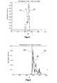
- FIGS. 4, 5 and 6show various recorded histograms for a specific illumination. In this case, the values Y, U and V are plotted along the abscissa and the relative frequency along the ordinate. The U and V axes are each shifted +128.
- FIG. 5shows the frequency distribution 41 for the value U in blood.
- Figure 7shows the pixel distribution of blood in the UV plane.
- FIG. 8shows the frequency distribution of component Y, where curve 45 is blood and curve 46 is skin color. Both can be well distinguished from each other except for an overlapping area.
- Similar or similar rulescan also be set for the other color spaces, using the principle of the learning data (reference). It is also possible to send a specific light (between infrared and ultraviolet) for image acquisition from the camera for image acquisition.
- both solutionsare subject to the principle of image sequence. This means that images are taken at a distance of a few seconds and that the current image gets its rating from the history.
- the blood pixelsare used as a sum or area as a measure of blood loss.
- reference markscan be made by the camera or a laser beam, which can focus on the camera.
- the third stepleads to the decision whether the critical blood loss is reached or not.
- N blood tN image A image ⁇ X ⁇ Q loss of blood ⁇ t
- N blood (t)is the pixel count of the color of blood
- Ais the image area
- Q blood lossis the leaked bloodstream
- tis the time.
- kis the sequential number of a picture.
- the extracorporeal blood circulationis stopped and an alarm is triggered if the sum of the blood loss is greater than a specified limit.
Landscapes
- Health & Medical Sciences (AREA)
- Heart & Thoracic Surgery (AREA)
- Vascular Medicine (AREA)
- Life Sciences & Earth Sciences (AREA)
- Engineering & Computer Science (AREA)
- Anesthesiology (AREA)
- Biomedical Technology (AREA)
- Hematology (AREA)
- Cardiology (AREA)
- Animal Behavior & Ethology (AREA)
- General Health & Medical Sciences (AREA)
- Public Health (AREA)
- Veterinary Medicine (AREA)
- External Artificial Organs (AREA)
- Measurement Of The Respiration, Hearing Ability, Form, And Blood Characteristics Of Living Organisms (AREA)
- Image Analysis (AREA)
Abstract
Description
Translated fromGermanDie vorliegende Erfindung betrifft ein medizinisches Gerät zur extrakorporalen Blutbehandlung mit einer Blutbehandlungsvorrichtung, die über extrakorporale Leitungen mit dem Blutkreislauf eines Patienten verbindbar ist, einer Blutpumpe, einer Steuereinheit zum Steuern der Blutpumpe und zur Überwachung von Betriebszuständen und mit einer Kamera zur Aufnahme des extrakorporalen Kreislaufs eines auf einem Behandlungsplatz befindlichen Patienten. Insbesondere betrifft die Erfindung ein medizinisches Gerät, bei dem aufgrund von Undichtigkeiten im extrakorporalen Kreislauf oder von Fehlern am Patientenzugang Blutverlust beim Patienten entstehen kann.The present invention relates to a medical device for extracorporeal blood treatment with a blood treatment device, which is connectable via extracorporeal lines to the bloodstream of a patient, a blood pump, a control unit for controlling the blood pump and monitoring of operating conditions and with a camera for receiving the extracorporeal circuit on a place of treatment patients. In particular, the invention relates to a medical device in which, due to leaks in the extracorporeal circulation or errors in patient access, blood loss can occur in the patient.
Eine typische Blutbehandlung ist die Dialysebehandlung. Dialyse-Behandlungen werden in der Regel in speziellen Gebäuden durchgeführt. In diesen Gebäuden sind gewöhnlich 20 bis 50 Behandlungsplätze angeordnet, die sich auf mehrere Räume verteilen. Das Pflegepersonal ist für die Überwachung des Patienten zuständig, kann jedoch nicht immer in der Nähe des Patienten sein, da es mehrere Patienten zu betreuen hat. Damit kommt dem Medizingerät die Aufgabe zu, Gefahren für den Patienten zu erkennen, entsprechende Sicherheitssteuerung vorzunehmen und das Pflegepersonal zum Patienten zu rufen. Beim Auftreten eines extrakorporalen Blutverlustes aufgrund einer Diskonnektion im venösen Rücklauf ist die Patienten-Sicherheit zur Zeit nur durch eine sorgfältige Überwachung durch das Pflegepersonal sichergestellt, da üblich verwendete venöse Drucküberwachung als Schutzsystem, nicht in jedem Fall den Blutverlust erkennt.A typical blood treatment is dialysis treatment. Dialysis treatments are usually done in special buildings. In these buildings usually 20 to 50 treatment stations are arranged, which are spread over several rooms. The nurse is responsible for monitoring the patient, but may not always be near the patient as it has multiple patients to care for. Thus, the task of the medical device is to recognize dangers to the patient, to perform appropriate safety control and to call the nursing staff to the patient. In the event of extracorporeal blood loss due to disconnection in the venous return, patient safety is currently only ensured by careful monitoring by the nursing staff, as commonly used venous pressure monitoring as a protection system does not always detect blood loss.
Bei der extrakorporalen Blutbehandlung, beispielsweise einer Hämodialyse- oder Plasmabehandlung, strömt das Blut eines Patienten von einem arteriellen Gefäßzugang über einen Filter zu einem venösen Gefäßzugang. Als Zugang zum Blutgefäßsystem wir häufig operativ eine arteriell-venöse Fistel angelegt, die im Allgemeinen mit einer arteriellen und venösen Kanüle punktiert wird. Ebenso ist der Einsatz eines Gefäßimplantats (Shunt) möglich. Unter einem Gefäßzugang wird jede Art des Zugangs zu dem Blutgefäßsystem des Patienten verstanden, insbesondere aber die Verbindung zwischen Arterie und Vene des Patienten. Eine wirksame Sicherheitseinrichtung zur Vermeidung von Blutverlust gibt es bislang bei der Doppelnadel-Behandlung nicht. Zur Zeit behilft man sich lediglich durch Verkleben der Leitungen, die zu oder von einem Gefäßzugang führt, mit Pflastern. Herkömmliche Dialysegeräte messen bei der Rückgabe des Blutes den Widerstand zwischen Gerät und Patient. Dabei wird das Blut mit einer Geschwindigkeit von 200 bis 600 ml/min durch die Kanüle in den Patienten gefördert. Allein der Widerstand der Kanüle liegt dabei größtenteils im Drucküberwachungsbereich des Dialysegerätes. Beim Herausrutschen der venösen Kanüle fließt Blut aus dem arteriellen Gefäßzugang über das Dialysegerät aus dem Patienten. Das Dialysegerät reagiert aufgrund sekundärer Einflüsse wie dem Druckabfall am venösen Druckaufnehmer. Der Druckabfall ist jedoch abhängig vom Blutfluss, Hämatokrit, Kanüle und Gefäßdruck des Patienten. Im Formalfall stellt das Pflegepersonal so nahe wie möglich einen unteren Grenzwert an den aktuellen venösen Druck, ohne genau den Druck zu kennen, der sich bei herausgerutschter Kanüle einstellt. Prinzipiell sind zwei Möglichkeiten vorhanden, nämlich das Dialysegerät gibt Alarm, ohne dass die Kanüle herausgerutscht ist und ruft somit das Pflegepersonal zum Gerät oder das Dialysegerät gibt keinen Alarm, wenn die Kanüle herausgerutscht ist und der Patient verliert Blut.In extracorporeal blood treatment, for example, a hemodialysis or plasma treatment, the blood of a patient flows from an arterial vascular access via a filter to a venous vascular access. As an approach to the vascular system, we often surgically apply an arterial-venous fistula, which is generally punctured with an arterial and venous cannula. Likewise, the use of a vascular implant (shunt) is possible. A vascular access is understood to be any type of access to the patient's blood vessel system, but in particular the connection between the patient's artery and vein. An effective safety device for the prevention of blood loss does not exist so far in the double needle treatment. At present, it is only by sticking the leads leading to or from a vascular accessway that one makes do with patches. Conventional dialysis machines measure the resistance between the device and the patient when the blood returns. The blood is delivered through the cannula into the patient at a rate of 200 to 600 ml / min. Only the resistance of the cannula is largely in the pressure monitoring range of the dialysis machine. When slipping out of the venous cannula blood from the arterial vascular access flows through the dialysis machine from the patient. The dialysis machine reacts due to secondary influences such as the pressure drop at the venous pressure transducer. The pressure drop is however, depending on the blood flow, hematocrit, cannula and vascular pressure of the patient. In the formal case, caregivers set a lower limit to the current venous pressure as close as possible, without knowing exactly the pressure that occurs when the cannula slips out. In principle, there are two options, namely the dialysis machine gives alarm without the cannula has slid out and thus calls the nursing staff to the device or the dialysis machine gives no alarm when the cannula has slipped out and the patient loses blood.
Bei allen Systemen ist keine Erkennung eines Blutverlustes möglich, wenn dieser zum Beispiel an einer Verbindungsstelle im extrakorporalen Schlauchsystem entsteht.In all systems, no detection of blood loss is possible when it arises, for example, at a junction in the extracorporeal tube system.
Der Erfindung liegt die Aufgabe zugrunde, ein medizinisches Gerät mit extrakorporalem Blutkreislauf derart auszubilden, dass übermäßiger Blutverlust eines Patienten während der Behandlung mit hoher Sicherheit erkannt wird.The invention has the object of providing a medical device with extracorporeal blood circulation in such a way that excessive blood loss of a patient during the treatment is detected with high security.
Das medizinische Gerät nach der vorliegenden Erfindung ist durch den Patentanspruch 1 bezeichnet. Es weist eine Farbbildkamera auf, die mit einer Erkennungseinrichtung zur Erkennung der Farbe von Blut und der Größe der von dem Blut eingenommenen Bildfläche verbunden ist. Die Kamera ist auf den Behandlungsplatz bzw. den darauf befindlichen Patienten gerichtet. Sie nimmt Bilder auf, deren Farbinformation von einer Analysiervorrichtung ausgewertet wird. Dabei wird auslaufendes oder ausgelaufenes Blut an seiner typischen Farbe erkannt. Die Menge des ausgelaufenen Blutes wird anhand der Größe der von dem Blut eingenommenen Bildfläche bewertet.The medical device according to the present invention is characterized by
Die von Blut eingenommene Fläche muss nicht zusammenhängend sein sondern kann sich aus mehreren Einzelflächen zusammensetzen.The area occupied by blood does not have to be contiguous but may consist of several individual areas.
Die Auswertevorrichtung, an die die Kamera die Bilder liefert, führt folgende Abläufe aus:
- digitale Bilder vom Patienten und dem extrakorporalen Kreislauf aufzunehmen,
- Pixel-Informationen in einen Farbraum transferieren, der die Unterscheidung von Haut zu Blut ermöglicht,
- Pixel als Blut oder nicht Blut klassifizieren,
- Blutpixel summieren,
- die summierten Pixel mit einem Grenzwert vergleichen,
- alternativ Blutflecken feststellen und deren Vergrößerung bewerten,
- die Blutpumpe stoppen und die Schlauchabsperrklemme schließen, wenn der Grenzwert überschritten ist und Alarm geben.
- taking digital pictures of the patient and the extracorporeal circuit,
- Transfer pixel information into a color space that allows the distinction from skin to blood
- Classify pixels as blood or not blood,
- Sum up blood pixels,
- compare the summed pixels to a threshold,
- alternatively detect blood spots and evaluate their magnification,
- Stop the blood pump and close the tube locking clamp if the limit is exceeded and give an alarm.
Die Blutzugänge werden normalerweise am Unterarm eines Patienten angelegt. Von dort führen Schläuche zu dem medizinischen Gerät (z. B. Dialysator). Die Blutzugänge werden mit Pflastern an dem Arm des Patienten verklebt. Ein Blutaustritt, der an einem der Gefäßzugänge auftritt, befindet sich in unmittelbarer Nähe des Patientenkörpers, so dass es darauf ankommt, im Kamerabild zwischen Blut und Haut des Patienten zu unterscheiden.The blood accesses are usually applied to the forearm of a patient. From there hoses lead to the medical device (eg dialyzer). The blood accesses are glued to the arm of the patient with patches. A blood leak that occurs at one of the vascular accesses, is located in the immediate vicinity of the patient's body, so it is important to distinguish in the camera image between the patient's blood and skin.
Je nach Menge des Blutverlustes kann dieser tödlich sein. In der Risikobetrachtung wird dies für einen Wert von ca. 500 ml angenommen. Im Einzelnen ist dieser Wert von der persönlichen körperlichen Verfassung des Patienten abhängig.Depending on the amount of blood loss, this can be fatal. In the risk analysis this is assumed for a value of approx. 500 ml. In detail, this value depends on the personal physical condition of the patient.
Vorraussetzung für das sichere Erkennen von fehlerhaften Blutzugängen ist, dass die Blutzugänge für die Kamera sichtbar sind. Dies bedeutet, dass der Patient die Blutzugänge nicht mit einer Decke o. dgl. bedeckt.A prerequisite for the reliable detection of defective blood accesses is that the blood accesses are visible to the camera. This means that the patient does not cover the blood accesses with a blanket or the like.
Bei einer Digitalkamera wird die Fläche, die mit auslaufendem Blut bedeckt ist, anhand der Pixel ermittelt, die mit der Farbe von Blut belichtet sind. Die Zahl der Blutpixel kann ins Verhältnis gesetzt werden zu der Gesamtzahl der Pixel des Kamerabildes.In a digital camera, the area covered with leaking blood is determined from the pixels exposed to the color of blood. The number of blood pixels can be related to the total number of pixels of the camera image.
Die von einer Digitalkamera erzeugten Bilder liegen in der Regel im RGB-Format (Farbraum) vor. Jeder Bildpunkt (Pixel) setzt sich aus drei Werten zusammen, welche die Farben Rot, Grün und Blau gewichten bzw. in ihrer Intensität darstellen. Jede Gewichtung liegt zwischen 0 und 255. Pro Komponente des Farbmerkmalsvektors ergeben sich 8 Bit bzw. 3 x 8 Bit = 24 Bit pro Bildpunkt. Eine anschauliche Darstellung dieses Farbraumes ist der RGB-Farbwürfel. Dabei sind ungefähr 16 Millionen Farben möglich. Auf der Hauptdiagonalen sind die Grauwerte von schwarz (0, 0, 0) bis weiß (255, 255, 255) zu finden. Weitere Farben sind z. B. rot bei (255, 0, 0) und gelb (255, 255, 0).The images generated by a digital camera are usually in RGB format (color space). Each pixel is composed of three values, which weight the colors red, green and blue respectively in their intensity. Each weight is between 0 and 255. Per component of the color feature vector results in 8 bits or 3 x 8 bits = 24 bits per pixel. An illustrative representation of this color space is the RGB color cube. There are about 16 million colors possible. On the main diagonal, the gray values are from black (0, 0, 0) to white (255, 255, 255). Other colors are z. Red at (255, 0, 0) and yellow (255, 255, 0).
Für die Bluterkennung ist das RGB-Format nicht besonders gut geeignet, weil sich konkrete Abgrenzungen nur schwer definieren lassen. Eine bevorzugte Ausgestaltung der Erfindung sieht daher eine Transformationsvorrichtung vor, die im RGB-Farbraum ausgegebene Signale in einen anderen Farbraum transformiert. Dieser andere Farbraum ist vorzugsweise der YUV-Farbraum. Die Transformation bewirkt, dass eine Unterscheidung zwischen Blut- und Nichtblut-Pixeln besser herbeizuführen ist.For blood detection, the RGB format is not particularly well suited because it is difficult to define concrete boundaries. A preferred embodiment of the invention therefore provides a transformation device which transforms signals output in the RGB color space into another color space. This other color space is preferably the YUV color space. The transformation makes a distinction between blood and non-blood pixels easier to achieve.
Der YUV-Farbraum besteht aus einer Helligkeitskomponente Y und zwei Farbkomponenten U und V. Er geht aus dem RGB-Farbraum durch eine lineare Transformation hervor:
Durch die Drehmatrix wird die Hauptdiagonale des RGB-Farbraumes (Grauwerte) auf die Y-Achse des YUV-Farbraumes abgebildet. Ziel ist, eine Trennung von Farb- und Helligkeitsinformation herbeizuführen. Die Wertebereiche liegen für die Y-Komponente bei 0 bis 255 und für die U- und die V-Komponenten bei -128 bis +127.The rotation matrix maps the main diagonal of the RGB color space (gray values) to the Y axis of the YUV color space. The aim is to bring about a separation of color and brightness information. The value ranges are 0 to 255 for the Y component and -128 to +127 for the U and V components.
Die Farbinformation ist in den U- und V-Komponenten gespeichert und die Helligkeitsinformation, die stark von der vorherrschenden Beleuchtung abhängt, in der Y-Komponente.The color information is stored in the U and V components, and the brightness information, which strongly depends on the prevailing illumination, is stored in the Y component.
Beispiele für die Definition der Grenzwerte bzw. Kriterien im YUV-Farbraum werden später beschrieben.Examples of the definition of the limit values or criteria in the YUV color space will be described later.
Die Kriterien für die Blut-Diskriminierung hängen von dem Beleuchtungsspektrum der jeweiligen Beleuchtung ab, bei der das Bild von der Kamera aufgenommen wird. So gelten bei Neonbeleuchtung andere Grenzwerte bzw. Kriterien als bei Glühlampenbeleuchtung oder Tageslicht. Zur Feststellung der jeweiligen Beleuchtungsart kann ein Beleuchtungssensor vorgesehen sein, der Mittel zum Auswählen der gespeicherten Kriterien in Abhängigkeit von dem detektierten Beleuchtungsspektrum steuert.The criteria for blood discrimination depend on the illumination spectrum of the particular illumination at which the image is captured by the camera. For neon lighting, for example, other limits or criteria apply than with incandescent lighting or daylight. To determine the respective type of illumination, a lighting sensor may be provided which controls means for selecting the stored criteria in dependence on the detected illumination spectrum.
Alternativ besteht die Möglichkeit, die Beleuchtung mit einem definierten Beleuchtungsspektrum vorzunehmen und für die Aufnahme der Kamerabilder eine eigene definierte Beleuchtungsquelle einzusetzen. Diese kann beispielsweise aus einem Blitzlichtgerät bestehen, dessen Beleuchtungsstärke so groß ist, dass sie Fremdlicht überdeckt, so dass die Auswertung des Farbspektrums eines Pixel-Bildes auf der Basis des Beleuchtungsspektrums der definierten Beleuchtungsquelle erfolgen kann.Alternatively, it is possible to make the illumination with a defined illumination spectrum and to use a separate defined illumination source for taking the camera images. This can for example consist of a flash unit whose illuminance is so great that it covers extraneous light, so that the evaluation of the color spectrum of a pixel image can be made on the basis of the illumination spectrum of the defined illumination source.
Die Erfindung ermöglicht es auch, die Ausbreitungsgeschwindigkeit eines Blutflecks mit der Kamera zu erfassen, indem die Vergrößerung der Fläche mit der Farbe von Blut innerhalb einer definierten Zeit erfasst bzw. berechnet wird. Diese Art der Auswertung kann auch in der Weise erfolgen, dass bei hoher Ausbreitungsgeschwindigkeit des Blutes die Bildfolgefrequenz der Kamera automatisch erhöht wird.The invention also makes it possible to detect the propagation velocity of a blood spot with the camera by detecting or calculating the increase of the area with the color of blood within a defined time. This type of evaluation can also be carried out in such a way that at high propagation velocity of the blood, the frame rate of the camera is automatically increased.
Der Blutverlust kann auch dadurch festgestellt werden, dass eine Einrichtung zum Summieren der Bild-Pixel oder einer Fläche mit der Farbe von Blut über mehrere Bilder der Kamera hinweg zur Ermittlung des Blutverlustes vorgesehen ist. Dabei kann auch der Umriss der von Blut eingenommenen Fläche (oder mehrerer Flächen) herangezogen werden.The blood loss can also be detected by providing means for summing the image pixels or an area of the color of blood across multiple images of the camera to determine blood loss. The outline of the area occupied by blood (or several areas) can also be used.
Damit die Kameraüberwachung auf das Auslaufen von Blut erst dann durchgeführt wird, wenn der Patient an das extrakorporale Schlauchsystem angeschlossen ist und das Blut in diesen strömt, kann in dem extrakorporalen Leitungssystem ein Blutdetektor vorgesehen sein, der nur im Falle einer Erkennung von Blut die Aktivierung der Kamera zulässt. Dieser Blutdetektor ist beispielsweise ein Rot-Detektor, der eine rote Flüssigkeit in dem Schlauchsystem erkennt.So that the camera monitoring is performed on the leakage of blood only when the patient is connected to the extracorporeal tube system and the blood flows into this, in the extracorporeal line system, a blood detector may be provided, the activation of the only in the event of detection of blood Camera allows. For example, this blood detector is a red detector that detects a red liquid in the tube system.
Damit die Kamera das richtige Zielgebiet aufnimmt, kann sie einen Antrieb zur Selbstausrichtung aufweisen, der die Kamera auf eine Referenzmarkierung ausrichtet. Diese Referenzmarkierung, beispielsweise ein Tag oder ein Button, wird auf dem Zielgebiet befestigt, beispielsweise auf dem Unterarm des Patienten. Wenn die Referenzmarkierung nicht gefunden wird, erfolgt eine Alarmgabe.For the camera to pick up the correct target area, it may have a self-aligning drive that aligns the camera with a reference mark. This reference marker, such as a tag or a button, is affixed to the target area, for example, on the patient's forearm. If the reference mark is not found, an alarm is issued.
Die Übertragung der Bilder von der Kamera zu dem Gerät erfolgt über Kabel oder über eine drahtlose Verbindung. Es besteht auch die Möglichkeit, mehrere Kameras vorzusehen, die aus unterschiedlichen Blickrichtungen das Zielgebiet aufnehmen.The transfer of images from the camera to the device is via cable or over a wireless connection. It is also possible to provide several cameras that record the target area from different directions.
Das Gerät weist vorzugsweise eine Lesevorrichtung auf, die eine Identifikation des Bedienungspersonals liest, beispielsweise eine maschinenlesbare Personalkarte. Die Daten dieser Identifikation werden gespeichert, so dass dokumentiert wird, welche Person an dem Gerät zu welcher Zeit hantiert hat und welche Einstellungen vorgenommen wurden.The device preferably has a reading device which reads an identification of the operating personnel, for example a machine-readable personnel card. The data of this identification are stored so that it is documented which person has handled the device at what time and which settings have been made.
Das Einlesen der Identifikation gibt auch Aufschluss darüber, dass eine Pflegeperson an dem Behandlungsplatz anwesend ist. Dies kann für die Auswertung der Signale, die den Blutverlust angeben, herangezogen werden. So kann beispielsweise die bisherige Blutfläche als Referenzgröße herangezogen werden, so dass nur Veränderungen, die seit dem Erscheinen der Behandlungsperson aufgetreten sind, registriert werden.The reading of the identification also provides information that a caregiver is present at the treatment center. This can be used for the evaluation of the signals indicating the blood loss. Thus, for example, the previous blood surface can be used as reference variable, so that only changes that have occurred since the appearance of the treatment person are registered.
Das erfindungsgemäße Verfahren kann auch zur Überwachung extrakorporaler Blutkreisläufe auf Undichtigkeit angewendet werden, bei denen keine Blutbehandlung erfolgt. Beispiele hierfür sind eine Blutabnahme und ein Blutkreislaufsystem während einer Operation.The method according to the invention can also be used for monitoring extracorporeal blood circuits for leakage, in which no blood treatment takes place. Examples include a blood sample and a circulatory system during surgery.
Im Folgenden werden unter Bezugnahme auf die Zeichnungen Ausführungsbeispiele der Erfindung näher erläutert.In the following, embodiments of the invention will be explained in more detail with reference to the drawings.
Es zeigen:
- Fig. 1
- eine perspektivische Ansicht des medizinischen Gerätes in Form eines Gerätes für die Hämodialyse, Hämofiltration oder Plasmabehandlung,
- Fig. 2
- eine schematische Darstellung der wesentlichen Funktionsteile eines Dialysegerätes,
- Fig. 3
- die Darstellung eines Armes eines Patienten mit den Gefäßzugängen, wie von der Kamera aufgenommen,
- Fig. 4-6
- Histogramme der Farbe von Blut im YUV-Farbraum mit Beispielen für Grenzwerte, die die Kriterien für die Bluterkennung bilden, wobei U und V um +128 verschoben sind,
- Fig. 7
- ein Beispiel einer Pixelverteilung in der UV-Ebene mit Beispielen für Grenzwerte,
- Fig. 8
- ein Diagramm der Häufigkeiten über der Helligkeitskomponente Y im YUV-Farbraum für die Bewertung von Blut und Haut.
- Fig. 1
- a perspective view of the medical device in the form of a device for hemodialysis, hemofiltration or plasma treatment,
- Fig. 2
- a schematic representation of the essential functional parts of a dialysis machine,
- Fig. 3
- the representation of an arm of a patient with the vascular accesses, as recorded by the camera,
- Fig. 4-6
- Histograms of the color of blood in the YUV color space with examples of thresholds that form the criteria for blood detection, where U and V are shifted by +128,
- Fig. 7
- an example of pixel distribution in the UV plane with examples of limits,
- Fig. 8
- a graph of the frequencies over the brightness component Y in the YUV color space for the evaluation of blood and skin.
Figur 1 zeigt ein medizinisches Gerät 10 zur extrakorporalen Blutbehandlung. Dieses Gerät weist einen Gerätesockel 11 auf, in dem sich die mechanischen Komponenten befinden und der an seiner Vorderseite eine Konsole 12 trägt, in der zwei von außen zugängliche Blutpumpen 13 angeordnet sind. Die Blutpumpen sind Schlauchpumpen, deren Schläuche von der Frontseite her eingelegt werden.FIG. 1 shows a
Auf dem Gerätesockel 11 befindet sich die Steuereinheit 14, welche auch das Interface für die Kommunikation mit dem Benutzer bildet. Die Steuereinheit 14 weist hier einen Touchscreenmonitor auf, über den der Benutzer verschiedene Menüs aufrufen und Betriebszustände abfragen sowie Daten und Befehle eingeben kann. Ein Kartenleser 15 ist vorgesehen, in den der Benutzer eine maschinenlesbare Identifikationskarte einführen kann.On the
An einer Infusionsstange 16 ist eine Kamera 17 befestigt. Hierbei handelt es sich um eine digitale Farbbildkamera. Die von der Kamera 17 aufgenommenen Einzelbilder werden an die Steuereinheit 14 übertragen. Die Kamera 17 ist auf einen Patientenplatz gerichtet, z. B. eine Liege, auf der der Patient während der Blutbehandlung liegt. Auf diese Weise wird der extrakorporale Kreislauf von dem Kamerabild erfasst.On an
Der Patientenkörper wird über Schläuche an das Gerät 10 angeschlossen.The patient's body is connected to the
In Figur 2 ist ein Patient P schematisch dargestellt. Ein Arm des Patienten ist mit einem arteriellen Zugang 20 und einem venösen Zugang 21 versehen. Von dem arteriellen Zugang 20 führt eine arterielle Schlauchleitung 22 zu der Blutpumpe 13. Diese pumpt das Blut durch die Blutkammer 25a einer Behandlungsvorrichtung 25 in Form eines Dialysators, dessen beide Kammern 25a, 25b durch eine Membran 26 getrennt sind. Die Kammer 25b ist eine Dialysierflüssigkeitskammer, die von Dialysierflüssigkeit durchflossen wird.FIG. 2 schematically shows a patient P. One arm of the patient is provided with an
Nach Verlassen der Kammer 25a strömt das Blut in eine venöse Schlauchleitung 23, die mit dem venösen Gefäßanschluss 21 verbunden ist. Auf diese Weise wird ein Blutkreislauf gebildet.After leaving the
Die arterielle Leitung 22 enthält einen Druckmesser 27 zur Messung des arteriellen Blutdrucks. In gleicher Weise enthält die venöse Leitung einen Druckmesser 28 zur Messung des venösen Blutdrucks. Ferner enthält die venöse Leitung 23 einen Rot-Detektor 29, der die Anwesenheit von Blut in der Schlauchleitung erkennt und an die Steuereinheit 14 meldet. Auch die Druckmesser 27, 28 sind mit der Steuereinheit 14 verbunden. Diese steuert den gesamten Betrieb des Gerätes und überwacht die beschriebenen Funktionen sowie eine Reihe anderer Funktionen, die hier nicht näher erläutert werden.The
Um den Blutkreislauf absperren zu können, ist in der venösen Leitung 23 eine von der Steuereinheit 14 gesteuerte Schlauchabsperrklemme 30 vorgesehen. Auch in der arteriellen Leitung 22 befindet sich ein Absperrorgan in Form der Blutpumpe 13. Die Blutpumpe ist eine Schlauchpumpe, die von einem Quetschorgan fortlaufend abgequetscht wird. Bei Stillstand der Blutpumpe wirkt diese als Absperrorgan, dass die Schlauchleitung verschließt.In order to be able to shut off the blood circulation, a
Die Kamera 17 und ihr Anschluss an die Steuereinheit 14 sind in Figur 2 erkennbar. Außerdem ist ein Beleuchtungssensor 31 vorgesehen, der den Typ des Beleuchtungslichtes erkennt, beispielsweise Neonlicht, Licht mit UV-Lichtanteil oder warmes Leuchtenlicht. In Abhängigkeit davon werden die Kriterien für die Bluterkennung verändert.The
Figur 3 zeigt das von der Kamera 17 aufgenommene Bild des zu beobachtenden Bereichs. Am Arm des Patienten, der auf einem Behandlungsplatz liegt, sind die Gefäßzugänge 20,21 angebracht und von diesen führen die Schlauchleitungen 22, 23 zu der Blutpumpe. Am Körper des Patienten, hier am Arm, ist eine Referenzmarkierung 35 angebracht, die von der Kamera 17 detektierbar ist. Die Kamera 17 wird von einem (nicht dargestellten) Bewegungsantrieb so eingestellt, dass die Referenzmarkierung 35 sich an einer bestimmten Stelle im Kamerabild befindet. Auf diese Weise wird sichergestellt, dass unabhängig von Bewegungen des Patienten die Kamera stets auf das gewünschte Zielgebiet gerichtet ist.FIG. 3 shows the image taken by the
Die Steuereinheit 14 in Figur 2 ist ein Computer mit Speichereinheit. Sie führt auch sämtliche Überwachungs- und Steuerabläufe durch, sowie die Alarmerzeugungen. Die Steuereinheit 14 ist mit einer Anzeige-, Bedien- und Kommunikationseinheit 14a verbunden.The
Bei der Dialyse wird bei einem Patienten mit der Blutpumpe ein extrakorporaler Blutfluss von 50 bis 600 ml/min aufgebaut, indem das Blut mittels einer Schlauchpumpe aus der arteriellen Kanüle gezogen und über die venöse Kanüle zurückgegeben wird. Das Blut wird in Leitungen geführt, die an die Komponenten, wie Kanülen, Druckaufnehmer und Dialysator, angeschlossen werden. Die Steuerung und Überwachung geschieht mittels einer Steuer-, Rechen- und Speichereinheit. Die Parameter führ den zu behandelten Patienten werden über die Anzeige-, Bedien- und Kommunikationseinheit eingegeben. Für die Unterbrechung des Blutflusses stoppt die Steuereinheit die Blutpumpe und schließt de Schlauchabsperrklemme. Außerdem wird ein optischer und akustischer Alarm ausgelöst. Damit wird der Patient vor weiterem Schaden geschützt, weil es zu keinem weiteren Blutverlust kommen kann.In dialysis, an extracorporeal blood flow of 50 to 600 ml / min is established in a patient with the blood pump by pulling the blood out of the arterial cannula by means of a peristaltic pump and via the venous cannula is returned. The blood is fed into lines which are connected to the components such as cannulas, pressure transducers and dialyzer. The control and monitoring is done by means of a control, computing and storage unit. The parameters for the patients to be treated are entered via the display, operating and communication unit. For the interruption of blood flow, the control unit stops the blood pump and closes the tube shut-off clamp. In addition, a visual and audible alarm is triggered. This protects the patient from further damage because there is no further blood loss.
Die von der Digitalkamera erzeugten Bilder liegen in der Regel im RGB-Format vor. Jeder einzelne Bildpunkt setzt sich aus drei Werten zusammen, welche die Farben Rot, Grün und Blau gewichten. Jede Gewichtung liegt zwischen 0 und 255. Die im RGB-Farbraum vorliegenden Bildinhalte werden durch eine Transformationsvorrichtung in einen anderen Farbraum transformiert. Hierbei handelt es sich vorzugsweise um den YUV-Farbraum, der für die Bluterkennung besser geeignet ist. Ziel der Transformierung ist es, die Unterscheidung zwischen Blut- und Nichtblut-Pixeln herbeizuführen. Außer dem YUV-Farbraum können auch andere Farbräume wie z. B. HSV und Lab benutzt werden.The images produced by the digital camera are usually in RGB format. Each pixel is composed of three values, which weight the colors red, green and blue. Each weight is between 0 and 255. The image contents present in the RGB color space are transformed by a transformation device into another color space. This is preferably the YUV color space, which is better suited for blood detection. The goal of the transformation is to make the distinction between blood and non-blood pixels. In addition to the YUV color space, other color spaces such. As HSV and Lab are used.
Für die Abgrenzung zwischen Pixeln der Farbe von Blut und der Farbe von Nichtblut werden scharfe Grenzen in dem Farbraum eingeführt. Liegt das zu untersuchende Pixel innerhalb des durch die Grenzen markierten Unterraumes, wird es als Blutpixel klassifiziert. Die Grenzen oder Kriterien der Komponenten Y, U und V ergeben sich aufgrund von Lerndaten, die beim Aufnehmen von Blut unter verschiedenen Beleuchtungsspektren aufgenommen werden. In den Figuren 4, 5 und 6 sind verschiedene aufgenommene Histogramme bei einer bestimmten Beleuchtung dargestellt. Hierbei ist entlang der Abszisse jeweils der Wert Y, U und V aufgetragen und entlang der Ordinate die relative Häufigkeit. Die U- und die V-Achse sind jeweils um +128 verschoben.For the demarcation between pixels of the color of blood and the color of non-blood, sharp boundaries are introduced in the color space. If the pixel to be examined lies within the subspace marked by the boundaries, it is classified as a blood pixel. The limits or criteria of the components Y, U and V result from learning data taken when taking blood under different illumination spectra. FIGS. 4, 5 and 6 show various recorded histograms for a specific illumination. In this case, the values Y, U and V are plotted along the abscissa and the relative frequency along the ordinate. The U and V axes are each shifted +128.
Die Kurve 40 in Figur 4 zeigt die Häufigkeitsverteilung des Helligkeitswertes Y für Blut. Man erkennt, dass Blut bei Werten von Y zwischen G1=10 und G2=100 vorliegen kann.The
Figur 5 zeigt die Häufigkeitsverteilung 41 für den Wert U bei Blut. Die Kurve 41 liegt zwischen den Grenzen G3 = 96 und G4 = etwa 130.FIG. 5 shows the
Figur 6 zeigt die Häufigkeitsverteilung 42 für V von G5=140 bis G6=210.FIG. 6 shows the
Figur 7 zeigt die Pixelverteilung von Blut in der UV-Ebene.Figure 7 shows the pixel distribution of blood in the UV plane.
Die größte Störgröße bei der Unterscheidung, ob ein Pixel Blut darstellt oder nicht, ist die Haut. Um diese zu eliminieren hat sich gezeigt, dass der YUV Farbraum mit einer scharfen Grenze bei der Y-Komponente geeignet ist, Blut und Haut zu unterscheiden.The biggest disturbance in distinguishing whether a pixel is blood or not is the skin. In order to eliminate this, it has been shown that the YUV color space with a sharp limit in the Y component is capable of distinguishing blood and skin.
Figur 8 zeigt die Häufigkeitsverteilung der Komponente Y, wobei die Kurve 45 Blut und die Kurve 46 Hautfarbe bedeutet. Beide lassen sich bis auf einen Überschneidungsbereich gut voneinander unterscheiden.FIG. 8 shows the frequency distribution of component Y, where
Für die Bewertung der Pixel können nachfolgende Modellierungen angewandt werden:
- Explizit YUV
- P1:
- Y_[10, 90]
- P2:
- U_[96, (588 - V)/3.27]
- P3:
- U_[102, (588 - V)/3.27]
- P4:
- V_[184, 242]
- P5:
- V_[140, 184]
- R:
- Wenn [P1 und [(P2 und P4) oder (P3 und P5)]] Dann [Pixel = Blut] Bei der obigen Schreibweise besteht der Parameter P1 darin, dass Y zwischen 10 und 90 liegt. Die Regel R gibt diejenigen Bedingungen an, die erfüllt sein müssen, damit ein Pixel des Kamerabildes als Blut erkannt wird, und bedeutet eine Bool'sche logische Verknüpfung.
- Nichtparametrisch UV/Explizit Y
Dieser Ansatz kombiniert die nichtparametrische Modellierung der UV-Komponenten mit einer expliziten Modellierung der Y-Komponente. Die Auswertung des nichtparametrischen Modells erfolgt über einen Schwellwert. Übersteigt der Wert einen vorgegebenen Grenzwert und ist Y_[10, 90] erfüllt, wird der Bildpunkt als Blutpixel klassifiziert. - Parametrisch UV/Explizit Y
Dieser Ansatz kombiniert die parametrische Modellierung der UV-Komponenten mit einer expliziten Modellierung der Y-Komponente. Die Auswertung des parametrischen Modells erfolgt über den Mahalanobis-Distanz. Liegt der berechnete Abstand unterhalb eines vorgegebenen Schwellwertes und ist Y_[10, 90] erfüllt, wird der Bildpunkt als Blutpixel klassifiziert. - Parametrisch YUV
In diesem Ansatz werden alle drei Farbraumkomponenten mit Hilfe einer Gaußverteilung modelliert. Für die Parameter der Gaußverteilung werden die Lerndaten herangezogen. Die Auswertung erfolgt durch den Mahalanodisabstand. Ist der Abstand zwischen Bildpunkt und Gaußverteilung kleiner als ein Schwellwert, wird dieser Bildpunkt als Blutpixel klassifiziert.
- Explicit YUV
- P1:
- Y_ [10, 90]
- P2:
- U_ [96, (588 - V) /3.27]
- P3:
- U_ [102, (588 - V) /3.27]
- P4:
- V_ [184, 242]
- P5:
- V_ [140, 184]
- R:
- If [P1 and [(P2 and P4) or (P3 and P5)]] Then [pixel = blood] In the above notation, the parameter P1 is that Y is between 10 and 90. The rule R indicates those conditions that must be satisfied for a pixel of the camera image to be recognized as blood, and means a Boolean logical operation.
- Nonparametric UV / Explicit Y
This approach combines the nonparametric modeling of the UV components with an explicit modeling of the Y component. The evaluation of the nonparametric model takes place via a threshold value. If the value exceeds a predetermined limit and Y_ [10, 90] is satisfied, the pixel is classified as a blood pixel. - Parametric UV / Explicit Y
This approach combines the parametric modeling of the UV components with an explicit modeling of the Y component. The evaluation of the parametric model is done via the Mahalanobis distance. If the calculated distance is below a predetermined threshold value and Y_ [10, 90] is satisfied, the pixel is classified as a blood pixel. - Parametric YUV
In this approach, all three color space components are modeled using a Gaussian distribution. The learning data is used for the parameters of the Gaussian distribution. The evaluation is done by the Mahalanodis distance. If the distance between pixel and Gaussian distribution is smaller than a threshold value, this pixel is classified as a blood pixel.
Gleiche oder ähnliche Regeln können auch für die anderen Farbräume aufgestellt werden, wobei das Prinzip der Lerndaten (Referenz) herangezogen wird. Außerdem ist es möglich, zur Bildaufnahme ein spezifisches Licht (zwischen Infrarot und Ultraviolett) zur Bildaufnahme von der Kamera auszusenden.Similar or similar rules can also be set for the other color spaces, using the principle of the learning data (reference). It is also possible to send a specific light (between infrared and ultraviolet) for image acquisition from the camera for image acquisition.
Für die weitere Verwertung der gewonnenen Blutpixel sind zwei Lösungen anwendbar. Beiden Lösungen unterliegt das Prinzip der Bildfolge. Das heißt, dass Bilder in einem Abstand von einigen Sekunden aufgenommen werden und dass das aktuelle Bild seine Bewertung aus der Historie bezieht. Mit dieser Methode werden die Blutpixel als Summe oder als Fläche als Maß für den Blutverlust herangezogen. Um Fehlalarme zu vermeiden, können Referenzmarkierungen, die von der Kamera oder einem Laserstrahl abgetastet werden, angebracht werden, auf die sich die Kamera fokussieren kann.For the further utilization of the obtained blood pixels two solutions are applicable. Both solutions are subject to the principle of image sequence. This means that images are taken at a distance of a few seconds and that the current image gets its rating from the history. With this method, the blood pixels are used as a sum or area as a measure of blood loss. To avoid false alarms, reference marks can be made by the camera or a laser beam, which can focus on the camera.
Im weiteren geht es darum, mit den klassifizierten Blutpixeln in der Bilderabfolge eine Entscheidung zu fällen, ob ein extrakorporaler Blutaustritt vorliegt oder nicht.Furthermore, it is a matter of making a decision with the classified blood pixels in the image sequence as to whether an extracorporeal blood discharge is present or not.
Hierbei werden folgende Schritte ausgeführt:
- 1. Aufnahme des Bildes
- 2. Klassifikation der Blutpixel im Bild
- 3. Auswertung der Bildfolge der klassifizierten Blutpixel
- 1. Take the picture
- 2. Classification of the blood pixels in the picture
- 3. Evaluation of the image sequence of the classified blood pixels
Der dritte Schritt mündet in die Entscheidung, ob der kritische Blutverlust erreicht ist oder nicht. Mit der nachfolgenden Beziehung wird die maximale Zeit zwischen zwei Bildern ermittelt:
Aus dem Zusammenhang zwischen dem Volumen des ausgetretenen Blutes VBlut und der daraus entstandenen Blutfläche ABlut, gibt es einen Faktor x, der die Ausbreitungsgeschwindigkeit der Blutfläche darstellt. Der Faktor x für die Ausbreitungsgeschwindigkeit, Fläche pro Volumen, ist von der Beschaffenheit der Oberfläche, auf der sich das Blut ausbreitet, abhängig und wird aus einer Tabelle entnommen:
Die Pixel im Bild, die einen Blutfleck abbilden, werden über den Zusammenhang Oberfläche des aufgenommenen Bildausschnitts und Auflösung der Kamera in Bezug gebracht. Bei einer Kamera mit einer Pixelanzahl NBild ergibt sich der nachfolgende Zusammenhang für die Blutpixelanzahl:
Hierin ist NBlut(t) die Pixelzahl der Farbe von Blut, ABild die Bildfläche und QBlutverlust der ausgetretene Blutstrom, t ist die Zeit.Here, Nblood (t) is the pixel count of the color of blood, A is theimage area, and Qblood loss is the leaked bloodstream, t is the time.
Zwischen zwei Bildern ergibt sich der Blutverlust ΔV aus der nachfolgenden Berechnung:
k ist die laufende Nummer eines Bildes.k is the sequential number of a picture.
Dabei wird zu Grunde gelegt, dass vor dem Anlegen des Patienten an den extrakorporalen Kreislauf kein Blutverlust vorhanden ist. Das Starten der Kamera beziehungsweise die Bewertung der Bilder erfolgt manuell und/oder durch Sensoren und/oder durch Betriebszustände, die extrakorporales Blutfluss annehmen lassen. Der Blutverlust ergibt sich aus der Summe der Deltavolumen bis zum ersten Bild oder bis zu dem Bild, bei dem die Anwesenheit des Bedienpersonal detektiert wird. Das Erkennen von dem Bediener geschieht mittels Bilderkennung durch die Kamera, Identifikation durch Chipkarte, Tastatureingabe usw.It is based on the assumption that no blood loss is present before the patient is attached to the extracorporeal circulation. Starting the camera or the evaluation of the images is done manually and / or by sensors and / or by operating states that can accept extracorporeal blood flow. The blood loss results from the sum of the delta volumes to the first image or to the image in which the presence of the operator is detected. The recognition of the operator is done by means of image recognition by the camera, identification by chip card, keyboard input, etc.
Der extrakorporale Blutkreislauf wird gestoppt und ein Alarm ausgelöst, wenn die Summe des Blutverlustes größer als ein festgelegter Grenzwert ist.The extracorporeal blood circulation is stopped and an alarm is triggered if the sum of the blood loss is greater than a specified limit.
Abnormale Zustände wie
- a) kurzfristige Abdeckung des zu überwachenden Bildausschnitts
- b) Patient bewegt sich und damit eine Änderung des Bildausschnitts
- c) Auftauchen von blutähnlichen Flecken (Zeitschriften, Kleidungsstücke)
- 1. Sollte die Anzahl der Blutpixel deutlich sinken, so wird ein Hinweis gegeben, der das Personal auffordert, die Kamera neu auszurichten (Fall a).
- 2. Negativer Blutverlust (weniger Blutpixel als das Startbild) wird ignoriert oder als Startbild gesetzt (Fall b).
- 3. Ergibt sich ein höherer Blutverlust (Blutpixel) als extrakorporal gefördert, so wird dies ignoriert und ein Hinweis ausgegeben, der das Personal zu korrigierenden Maßnahmen auffordert (Fall c).
- 4. Beim Fall b kann über eine motorische Kamera kompensiert werden, indem eine Bilderkennung oder Markierung die Kamera ausrichtet.
- 5. Liegen einzelne Blutpixel nicht in einer Flächenanordnung, so werden diese für die Auswertung ignoriert.
- a) short-term coverage of the image section to be monitored
- b) Patient moves and thus a change in the image section
- c) emergence of blood-like spots (magazines, garments)
- 1. Should the number of blood pixels decrease significantly, a note is given to prompt the staff to realign the camera (case a).
- 2. Negative blood loss (less blood pixels than the startup image) is ignored or set as the startup image (case b).
- 3. If a higher blood loss (blood pixels) results in an extracorporeal promotion, this is ignored and a message is issued asking staff to take corrective action (case c).
- 4. In case b, a motorized camera can be compensated by aligning an image recognition or marker with the camera.
- 5. If individual blood pixels are not in a surface arrangement, these are ignored for the evaluation.
Claims (18)
Translated fromGermandadurch gekennzeichnet,
dass die Kamera (17) eine Farbbildkamera ist, die mit einer Erkennungseinrichtung zur Erkennung der Farbe von Blut und der Größe der von dem Blut eingenommen Bildfläche verbunden ist.Medical device for extracorporeal blood treatment, comprising a treatment device (25) which can be connected to the bloodstream of a patient via extracorporeal lines (22, 23), at least one blood pump (13), a control unit (14) for controlling the blood pump and for monitoring Operating conditions, and a camera (17), which is directable to a treatment center,
characterized,
in that the camera (17) is a color image camera which is connected to a recognition device for detecting the color of blood and the size of the image surface occupied by the blood.
dadurch gekennzeichnet,
dass mindestens eine Farbbildkamera verwendet wird, deren Farbsignale zur Erkennung der Farbe von Blut ausgewertet werden und dass aus den Farbsignalen die Größe der von dem Blut eingenommenen Bildfläche ermittelt wird.Method for monitoring the operation of a medical device for extracorporeal blood treatment, in which a camera receives a patient located at a treatment center,
characterized,
in that at least one color image camera is used, whose color signals are evaluated for detecting the color of blood, and that the color signals determine the size of the image area occupied by the blood.
Priority Applications (6)
| Application Number | Priority Date | Filing Date | Title |
|---|---|---|---|
| EP06119406AEP1892001A1 (en) | 2006-08-23 | 2006-08-23 | Medical device for extracorporeal blood treatment |
| PCT/EP2007/057914WO2008022880A1 (en) | 2006-08-23 | 2007-07-31 | Medical appliance for extracorporeal blood treatment |
| CN2007800310773ACN101505813B (en) | 2006-08-23 | 2007-07-31 | medical device for extracorporeal blood treatment |
| EP07788100.1AEP2054106B1 (en) | 2006-08-23 | 2007-07-31 | Medical device for extracorporeal blood treatment |
| US12/377,322US8529485B2 (en) | 2006-08-23 | 2007-07-31 | Medical apparatus for extracorporeal blood treatment |
| RU2009110260/14ARU2437682C2 (en) | 2006-08-23 | 2007-07-31 | Medical apparatus for extracorporal blood purification |
Applications Claiming Priority (1)
| Application Number | Priority Date | Filing Date | Title |
|---|---|---|---|
| EP06119406AEP1892001A1 (en) | 2006-08-23 | 2006-08-23 | Medical device for extracorporeal blood treatment |
Publications (1)
| Publication Number | Publication Date |
|---|---|
| EP1892001A1true EP1892001A1 (en) | 2008-02-27 |
Family
ID=37663158
Family Applications (2)
| Application Number | Title | Priority Date | Filing Date |
|---|---|---|---|
| EP06119406AWithdrawnEP1892001A1 (en) | 2006-08-23 | 2006-08-23 | Medical device for extracorporeal blood treatment |
| EP07788100.1AActiveEP2054106B1 (en) | 2006-08-23 | 2007-07-31 | Medical device for extracorporeal blood treatment |
Family Applications After (1)
| Application Number | Title | Priority Date | Filing Date |
|---|---|---|---|
| EP07788100.1AActiveEP2054106B1 (en) | 2006-08-23 | 2007-07-31 | Medical device for extracorporeal blood treatment |
Country Status (5)
| Country | Link |
|---|---|
| US (1) | US8529485B2 (en) |
| EP (2) | EP1892001A1 (en) |
| CN (1) | CN101505813B (en) |
| RU (1) | RU2437682C2 (en) |
| WO (1) | WO2008022880A1 (en) |
Cited By (6)
| Publication number | Priority date | Publication date | Assignee | Title |
|---|---|---|---|---|
| WO2009038833A1 (en)* | 2007-09-21 | 2009-03-26 | Baxter International Inc. | Access disconnect system with optical and other sensors |
| WO2009042259A1 (en)* | 2007-09-24 | 2009-04-02 | Baxter International Inc. | Detecting access disconnect by pattern recognition |
| DE102014111403A1 (en)* | 2014-08-11 | 2016-02-11 | Fresenius Medical Care Deutschland Gmbh | A method of monitoring the alignment and / or positioning of a blood tubing of an extracorporeal blood circuit in an access area |
| US9925323B2 (en) | 2009-12-17 | 2018-03-27 | Gambro Lundia Ab | Apparatus for extracorporeal blood treatment and method of operation |
| DE102017102169A1 (en) | 2017-02-03 | 2018-08-09 | B. Braun Avitum Ag | Device for extracorporeal blood treatment with automatic respiratory rate monitoring |
| CN109870316A (en)* | 2018-09-11 | 2019-06-11 | 江苏大学镇江流体工程装备技术研究院 | A kind of axial flow blood pump cavitation experimental bench |
Families Citing this family (70)
| Publication number | Priority date | Publication date | Assignee | Title |
|---|---|---|---|---|
| US11164672B2 (en) | 2010-01-22 | 2021-11-02 | Deka Products Limited Partnership | System and apparatus for electronic patient care |
| US9151646B2 (en) | 2011-12-21 | 2015-10-06 | Deka Products Limited Partnership | System, method, and apparatus for monitoring, regulating, or controlling fluid flow |
| US9789247B2 (en) | 2011-12-21 | 2017-10-17 | Deka Products Limited Partnership | Syringe pump, and related method and system |
| US9677555B2 (en) | 2011-12-21 | 2017-06-13 | Deka Products Limited Partnership | System, method, and apparatus for infusing fluid |
| US10242159B2 (en) | 2010-01-22 | 2019-03-26 | Deka Products Limited Partnership | System and apparatus for electronic patient care |
| US11244745B2 (en) | 2010-01-22 | 2022-02-08 | Deka Products Limited Partnership | Computer-implemented method, system, and apparatus for electronic patient care |
| US9295778B2 (en) | 2011-12-21 | 2016-03-29 | Deka Products Limited Partnership | Syringe pump |
| US20110313789A1 (en) | 2010-01-22 | 2011-12-22 | Deka Products Limited Partnership | Electronic patient monitoring system |
| US9744300B2 (en) | 2011-12-21 | 2017-08-29 | Deka Products Limited Partnership | Syringe pump and related method |
| US11881307B2 (en) | 2012-05-24 | 2024-01-23 | Deka Products Limited Partnership | System, method, and apparatus for electronic patient care |
| US10453157B2 (en) | 2010-01-22 | 2019-10-22 | Deka Products Limited Partnership | System, method, and apparatus for electronic patient care |
| US10911515B2 (en) | 2012-05-24 | 2021-02-02 | Deka Products Limited Partnership | System, method, and apparatus for electronic patient care |
| US11210611B2 (en) | 2011-12-21 | 2021-12-28 | Deka Products Limited Partnership | System, method, and apparatus for electronic patient care |
| CA3065357C (en) | 2011-05-18 | 2023-04-25 | Solodex Llc | Continuous anesthesia nerve conduction apparatus, system and method |
| US8986283B2 (en) | 2011-05-18 | 2015-03-24 | Solo-Dex, Llc | Continuous anesthesia nerve conduction apparatus, system and method thereof |
| JP5794597B2 (en)* | 2011-07-09 | 2015-10-14 | ガウス サージカルGauss Surgical | Extracorporeal blood volume estimation and surgically removed sample counting system and method |
| US9646375B2 (en) | 2011-07-09 | 2017-05-09 | Gauss Surgical, Inc. | Method for setting a blood transfusion parameter |
| US9870625B2 (en)* | 2011-07-09 | 2018-01-16 | Gauss Surgical, Inc. | Method for estimating a quantity of a blood component in a fluid receiver and corresponding error |
| US10426356B2 (en)* | 2011-07-09 | 2019-10-01 | Gauss Surgical, Inc. | Method for estimating a quantity of a blood component in a fluid receiver and corresponding error |
| US9652655B2 (en) | 2011-07-09 | 2017-05-16 | Gauss Surgical, Inc. | System and method for estimating extracorporeal blood volume in a physical sample |
| US11295846B2 (en) | 2011-12-21 | 2022-04-05 | Deka Products Limited Partnership | System, method, and apparatus for infusing fluid |
| US12196364B2 (en) | 2011-12-21 | 2025-01-14 | DEKA Research Products Limited Partnership | System, method, and apparatus for clamping |
| US10488848B2 (en) | 2011-12-21 | 2019-11-26 | Deka Products Limited Partnership | System, method, and apparatus for monitoring, regulating, or controlling fluid flow |
| US9746093B2 (en) | 2011-12-21 | 2017-08-29 | Deka Products Limited Partnership | Flow meter and related system and apparatus |
| US11217340B2 (en) | 2011-12-21 | 2022-01-04 | Deka Products Limited Partnership | Syringe pump having a pressure sensor assembly |
| US9435455B2 (en) | 2011-12-21 | 2016-09-06 | Deka Products Limited Partnership | System, method, and apparatus for monitoring, regulating, or controlling fluid flow |
| US10722645B2 (en) | 2011-12-21 | 2020-07-28 | Deka Products Limited Partnership | Syringe pump, and related method and system |
| US9372486B2 (en) | 2011-12-21 | 2016-06-21 | Deka Products Limited Partnership | System, method, and apparatus for monitoring, regulating, or controlling fluid flow |
| US9724466B2 (en) | 2011-12-21 | 2017-08-08 | Deka Products Limited Partnership | Flow meter |
| US9746094B2 (en) | 2011-12-21 | 2017-08-29 | Deka Products Limited Partnership | Flow meter having a background pattern with first and second portions |
| US9675756B2 (en) | 2011-12-21 | 2017-06-13 | Deka Products Limited Partnership | Apparatus for infusing fluid |
| US10563681B2 (en) | 2011-12-21 | 2020-02-18 | Deka Products Limited Partnership | System, method, and apparatus for clamping |
| US10228683B2 (en) | 2011-12-21 | 2019-03-12 | Deka Products Limited Partnership | System, method, and apparatus for monitoring, regulating, or controlling fluid flow |
| EP2660743B1 (en) | 2012-05-03 | 2022-03-02 | B. Braun Avitum AG | Method for customization of user interface of a medical apparatus for extracorporal blood treatment; medical apparatus for extracorporal blood treatment |
| CN109738621B (en) | 2012-05-14 | 2021-05-11 | 高斯外科公司 | System and method for estimating amount of blood component in liquid tank |
| IN2014DN10121A (en) | 2012-05-14 | 2015-08-21 | Gauss Surgical | |
| US10424060B2 (en) | 2012-07-09 | 2019-09-24 | Gauss Surgical, Inc. | Method for estimating blood component quantities in surgical textiles |
| DE102012021805A1 (en) | 2012-11-08 | 2014-05-08 | Fresenius Medical Care Deutschland Gmbh | Apparatus and method for monitoring the treatment of a patient |
| US9759343B2 (en) | 2012-12-21 | 2017-09-12 | Deka Products Limited Partnership | Flow meter using a dynamic background image |
| USD749206S1 (en) | 2013-11-06 | 2016-02-09 | Deka Products Limited Partnership | Apparatus to control fluid flow through a tube |
| USD752209S1 (en) | 2013-11-06 | 2016-03-22 | Deka Products Limited Partnership | Apparatus to control fluid flow through a tube |
| USD745661S1 (en) | 2013-11-06 | 2015-12-15 | Deka Products Limited Partnership | Apparatus to control fluid flow through a tube |
| USD751689S1 (en) | 2013-11-06 | 2016-03-15 | Deka Products Limited Partnership | Apparatus to control fluid flow through a tube |
| USD751690S1 (en) | 2013-11-06 | 2016-03-15 | Deka Products Limited Partnership | Apparatus to control fluid flow through a tube |
| WO2015127189A1 (en) | 2014-02-21 | 2015-08-27 | Deka Products Limited Partnership | Syringe pump having a pressure sensor assembly |
| WO2015161003A1 (en) | 2014-04-15 | 2015-10-22 | Gauss Surgical, Inc. | Method for estimating a quantity of a blood component in a fluid canister |
| US9824441B2 (en) | 2014-04-15 | 2017-11-21 | Gauss Surgical, Inc. | Method for estimating a quantity of a blood component in a fluid canister |
| EP3193975B1 (en) | 2014-09-18 | 2022-07-06 | DEKA Products Limited Partnership | Apparatus and method for infusing fluid through a tube by appropriately heating the tube |
| CN104316527A (en)* | 2014-11-07 | 2015-01-28 | 昆山韦睿医疗科技有限公司 | Automatic detection method for discharged dialyzate, and automatic detection device for discharged dialyzate |
| US10555675B2 (en) | 2015-05-15 | 2020-02-11 | Gauss Surgical, Inc. | Method for projecting blood loss of a patient during a surgery |
| US11504037B2 (en) | 2015-05-15 | 2022-11-22 | Gauss Surgical, Inc. | Systems and methods for assessing fluids from a patient |
| US10789710B2 (en) | 2015-05-15 | 2020-09-29 | Gauss Surgical, Inc. | Methods and systems for characterizing fluids from a patient |
| EP3393539B1 (en) | 2015-12-23 | 2023-12-27 | Gauss Surgical, Inc. | System and method for estimating an amount of a blood component in a volume of fluid |
| CN108697845B (en) | 2016-01-28 | 2021-09-17 | 德卡产品有限公司 | Apparatus for monitoring, regulating or controlling fluid flow |
| USD905848S1 (en) | 2016-01-28 | 2020-12-22 | Deka Products Limited Partnership | Apparatus to control fluid flow through a tube |
| USD854145S1 (en) | 2016-05-25 | 2019-07-16 | Deka Products Limited Partnership | Apparatus to control fluid flow through a tube |
| US11826545B2 (en) | 2016-09-08 | 2023-11-28 | Fresenius Medical Care Holdings, Inc. | Optical blood detection system |
| CN108085179A (en)* | 2016-11-23 | 2018-05-29 | 重庆市农业科学院 | For cleaning the liquid of the spray gun of sprinkling fertilizer, medicine |
| WO2018125812A1 (en) | 2017-01-02 | 2018-07-05 | Gauss Surgical, Inc. | Tracking surgical items with prediction of duplicate imaging of items |
| US11229368B2 (en) | 2017-01-13 | 2022-01-25 | Gauss Surgical, Inc. | Fluid loss estimation based on weight of medical items |
| JP7208897B2 (en)* | 2017-07-10 | 2023-01-19 | テルモ株式会社 | Pressure sensing device and extracorporeal circulation device |
| US11013840B2 (en)* | 2018-06-14 | 2021-05-25 | Fresenius Medical Care Holdings, Inc. | Smart cart for a dialysis machine |
| JP7205094B2 (en)* | 2018-07-20 | 2023-01-17 | ニプロ株式会社 | Needle removal prevention device |
| JP7047185B2 (en) | 2018-08-16 | 2022-04-04 | デカ・プロダクツ・リミテッド・パートナーシップ | Medical pump |
| CN109498857B (en)* | 2018-12-29 | 2020-10-16 | 刘铁楠 | Drainage monitoring system and method based on image recognition |
| CN110193103A (en)* | 2019-04-26 | 2019-09-03 | 广州医科大学附属第二医院 | A kind of hemodialysis vascular access monitoring device |
| WO2021021596A1 (en) | 2019-07-26 | 2021-02-04 | Deka Products Limited Partnership | Apparatus for monitoring, regulating, or controlling fluid flow |
| USD964563S1 (en) | 2019-07-26 | 2022-09-20 | Deka Products Limited Partnership | Medical flow clamp |
| CN112734717B (en)* | 2020-12-31 | 2024-05-17 | 复旦大学附属华山医院 | Vascular access monitoring method and system for dialysis patients |
| JP2022183465A (en)* | 2021-05-31 | 2022-12-13 | 日本電産コパル株式会社 | Medical monitoring devices and medical monitoring systems |
Citations (6)
| Publication number | Priority date | Publication date | Assignee | Title |
|---|---|---|---|---|
| DE4014572A1 (en)* | 1990-05-07 | 1991-11-14 | Alexander Staeblein | Medical monitoring sensor for bleeding under bandage - responds to change in bandaging material i.e. dampness and/or colour to release alarm via evaluating circuit |
| DE19848235C1 (en)* | 1998-10-20 | 2000-03-16 | Fresenius Medical Care De Gmbh | Method for monitoring supply to vessel and extra-corporeal blood treatment device for monitoring supply to vessel; inputs blood circulation pressure to computer to calculate values to identify errors in supply during dialysis |
| WO2002080764A1 (en)* | 2001-04-05 | 2002-10-17 | Rhode Island Hospital, A Lifespan Partner | Non-invasive determination of blood components |
| US20030194894A1 (en)* | 2002-04-10 | 2003-10-16 | Ramesh Wariar | Access disconnection systems and methods |
| WO2004074822A1 (en)* | 2003-02-21 | 2004-09-02 | Leica Microsystems Semiconductor Gmbh | Method, device and software for the optical inspection of a semi-conductor substrate |
| EP1574178A2 (en)* | 2004-03-13 | 2005-09-14 | B. Braun Medizintechnologie GmbH | Medical treatment system |
Family Cites Families (8)
| Publication number | Priority date | Publication date | Assignee | Title |
|---|---|---|---|---|
| US5987346A (en)* | 1993-02-26 | 1999-11-16 | Benaron; David A. | Device and method for classification of tissue |
| US6125201A (en)* | 1997-06-25 | 2000-09-26 | Andrew Michael Zador | Method, apparatus and system for compressing data |
| AU9392798A (en) | 1997-11-07 | 1999-05-31 | Aksys, Ltd. | Blood line separation warning device for extracorporeal circuits |
| IT1308687B1 (en) | 1999-12-28 | 2002-01-09 | Gambro Dasco Spa | METHOD AND DEVICE FOR THE DETECTION OF ACCESS TO THE CARDIVASCULAR SYSTEM IN AN EXTRA-BODY BLOOD TREATMENT IN A |
| US20030128125A1 (en)* | 2002-01-04 | 2003-07-10 | Burbank Jeffrey H. | Method and apparatus for machine error detection by combining multiple sensor inputs |
| US20040064057A1 (en)* | 2002-07-31 | 2004-04-01 | Massachusetts Institute Of Technology | Measuring circulating blood volume through retinal vasculometry |
| JP4287646B2 (en)* | 2002-12-26 | 2009-07-01 | 株式会社ミツトヨ | Image reading device |
| JP4537681B2 (en)* | 2003-09-24 | 2010-09-01 | 株式会社東芝 | Blood flow analyzer |
- 2006
- 2006-08-23EPEP06119406Apatent/EP1892001A1/ennot_activeWithdrawn
- 2007
- 2007-07-31EPEP07788100.1Apatent/EP2054106B1/enactiveActive
- 2007-07-31RURU2009110260/14Apatent/RU2437682C2/ennot_activeIP Right Cessation
- 2007-07-31WOPCT/EP2007/057914patent/WO2008022880A1/enactiveApplication Filing
- 2007-07-31CNCN2007800310773Apatent/CN101505813B/enactiveActive
- 2007-07-31USUS12/377,322patent/US8529485B2/ennot_activeExpired - Fee Related
Patent Citations (6)
| Publication number | Priority date | Publication date | Assignee | Title |
|---|---|---|---|---|
| DE4014572A1 (en)* | 1990-05-07 | 1991-11-14 | Alexander Staeblein | Medical monitoring sensor for bleeding under bandage - responds to change in bandaging material i.e. dampness and/or colour to release alarm via evaluating circuit |
| DE19848235C1 (en)* | 1998-10-20 | 2000-03-16 | Fresenius Medical Care De Gmbh | Method for monitoring supply to vessel and extra-corporeal blood treatment device for monitoring supply to vessel; inputs blood circulation pressure to computer to calculate values to identify errors in supply during dialysis |
| WO2002080764A1 (en)* | 2001-04-05 | 2002-10-17 | Rhode Island Hospital, A Lifespan Partner | Non-invasive determination of blood components |
| US20030194894A1 (en)* | 2002-04-10 | 2003-10-16 | Ramesh Wariar | Access disconnection systems and methods |
| WO2004074822A1 (en)* | 2003-02-21 | 2004-09-02 | Leica Microsystems Semiconductor Gmbh | Method, device and software for the optical inspection of a semi-conductor substrate |
| EP1574178A2 (en)* | 2004-03-13 | 2005-09-14 | B. Braun Medizintechnologie GmbH | Medical treatment system |
Cited By (10)
| Publication number | Priority date | Publication date | Assignee | Title |
|---|---|---|---|---|
| WO2009038833A1 (en)* | 2007-09-21 | 2009-03-26 | Baxter International Inc. | Access disconnect system with optical and other sensors |
| US8187184B2 (en) | 2007-09-21 | 2012-05-29 | Baxter International, Inc. | Access disconnect system with optical and other sensors |
| WO2009042259A1 (en)* | 2007-09-24 | 2009-04-02 | Baxter International Inc. | Detecting access disconnect by pattern recognition |
| US7995816B2 (en) | 2007-09-24 | 2011-08-09 | Baxter International Inc. | Detecting access disconnect by pattern recognition |
| US9925323B2 (en) | 2009-12-17 | 2018-03-27 | Gambro Lundia Ab | Apparatus for extracorporeal blood treatment and method of operation |
| DE102014111403A1 (en)* | 2014-08-11 | 2016-02-11 | Fresenius Medical Care Deutschland Gmbh | A method of monitoring the alignment and / or positioning of a blood tubing of an extracorporeal blood circuit in an access area |
| WO2016023868A1 (en)* | 2014-08-11 | 2016-02-18 | Fresenius Medical Care Deutschland Gmbh | Method for monitoring the alignment and/or positioning of a blood tube line of an extracorporeal blood circuit in an access region |
| DE102017102169A1 (en) | 2017-02-03 | 2018-08-09 | B. Braun Avitum Ag | Device for extracorporeal blood treatment with automatic respiratory rate monitoring |
| US11191882B2 (en) | 2017-02-03 | 2021-12-07 | B. Braun Avitum Ag | Apparatus for extracorporeal blood treatment with automatic monitoring of respiratory rate |
| CN109870316A (en)* | 2018-09-11 | 2019-06-11 | 江苏大学镇江流体工程装备技术研究院 | A kind of axial flow blood pump cavitation experimental bench |
Also Published As
| Publication number | Publication date |
|---|---|
| EP2054106A1 (en) | 2009-05-06 |
| RU2437682C2 (en) | 2011-12-27 |
| CN101505813B (en) | 2012-02-01 |
| US20100087770A1 (en) | 2010-04-08 |
| US8529485B2 (en) | 2013-09-10 |
| EP2054106B1 (en) | 2013-05-29 |
| WO2008022880A1 (en) | 2008-02-28 |
| CN101505813A (en) | 2009-08-12 |
| RU2009110260A (en) | 2010-09-27 |
Similar Documents
| Publication | Publication Date | Title |
|---|---|---|
| EP2054106B1 (en) | Medical device for extracorporeal blood treatment | |
| US12112477B2 (en) | Semi-automated system for real-time wound image segmentation and photogrammetry on a mobile platform | |
| EP3481457B1 (en) | Method and system with a smartphone for detecting symptoms of peritonitis | |
| EP1704882B1 (en) | Blood treatment apparatus with alarm device | |
| DE69725617T2 (en) | Medical infusion pump | |
| EP2533827B1 (en) | Device and method for monitoring a vascular access for an extracorporeal blood treatment | |
| EP1044695A2 (en) | Method and device for determination of blood flow in a vessel access | |
| DE112012003387T5 (en) | Devices, methods and systems for detecting leaks in medical devices | |
| DE102016001710B4 (en) | Device for extracorporeal blood treatment with an evaluation and control unit | |
| DE69210873T2 (en) | Peristaltic intravenous infusion pump with indication of a selected application | |
| DE102007050060A1 (en) | Device and method for issuing medical data | |
| DE102016118673A1 (en) | System for the early detection of post-operative bleeding | |
| DE102016005213A1 (en) | Blood treatment machine | |
| Wild et al. | Digital measurement and analysis of wounds based on colour segmentation | |
| WO1999053835A1 (en) | Device and method for measuring intravascular pressure and for low contaminating introduction of catheters, for example, in blood vessels | |
| DE102017102169A1 (en) | Device for extracorporeal blood treatment with automatic respiratory rate monitoring | |
| EP3104770B1 (en) | Cover unit and device for monitoring the connectors of a tubing system | |
| DE102018106226A1 (en) | Apparatus and method for monitoring access to a patient | |
| DE102014111403A1 (en) | A method of monitoring the alignment and / or positioning of a blood tubing of an extracorporeal blood circuit in an access area | |
| DE102024207458B3 (en) | Measuring system for intraoperative bleeding volume | |
| EP4603117A1 (en) | Medical infusion system and associated procedure | |
| EP4136498A1 (en) | Medical device having a display and having a processing unit, and method therefor | |
| DE102022102274A1 (en) | Restless button access hemorrhages | |
| DE102019107540A1 (en) | Monitoring system for a blood treatment device for monitoring particularly hygiene-relevant conditions | |
| EP4529684A1 (en) | Signalling device for medical devices and procedures |
Legal Events
| Date | Code | Title | Description |
|---|---|---|---|
| PUAI | Public reference made under article 153(3) epc to a published international application that has entered the european phase | Free format text:ORIGINAL CODE: 0009012 | |
| AK | Designated contracting states | Kind code of ref document:A1 Designated state(s):AT BE BG CH CY CZ DE DK EE ES FI FR GB GR HU IE IS IT LI LT LU LV MC NL PL PT RO SE SI SK TR | |
| AX | Request for extension of the european patent | Extension state:AL BA HR MK YU | |
| RAP1 | Party data changed (applicant data changed or rights of an application transferred) | Owner name:B. BRAUN AVITUM AG | |
| AKX | Designation fees paid | ||
| STAA | Information on the status of an ep patent application or granted ep patent | Free format text:STATUS: THE APPLICATION IS DEEMED TO BE WITHDRAWN | |
| 18D | Application deemed to be withdrawn | Effective date:20080828 | |
| REG | Reference to a national code | Ref country code:DE Ref legal event code:8566 |