EP1886139B1 - Methods for analyzing inter-cellular phenomena - Google Patents
Methods for analyzing inter-cellular phenomenaDownload PDFInfo
- Publication number
- EP1886139B1 EP1886139B1EP06758550AEP06758550AEP1886139B1EP 1886139 B1EP1886139 B1EP 1886139B1EP 06758550 AEP06758550 AEP 06758550AEP 06758550 AEP06758550 AEP 06758550AEP 1886139 B1EP1886139 B1EP 1886139B1
- Authority
- EP
- European Patent Office
- Prior art keywords
- cell
- cells
- conjugate
- image data
- type
- Prior art date
- Legal status (The legal status is an assumption and is not a legal conclusion. Google has not performed a legal analysis and makes no representation as to the accuracy of the status listed.)
- Active
Links
Images


Classifications
- G—PHYSICS
- G01—MEASURING; TESTING
- G01N—INVESTIGATING OR ANALYSING MATERIALS BY DETERMINING THEIR CHEMICAL OR PHYSICAL PROPERTIES
- G01N33/00—Investigating or analysing materials by specific methods not covered by groups G01N1/00 - G01N31/00
- G01N33/48—Biological material, e.g. blood, urine; Haemocytometers
- G01N33/50—Chemical analysis of biological material, e.g. blood, urine; Testing involving biospecific ligand binding methods; Immunological testing
- G01N33/5005—Chemical analysis of biological material, e.g. blood, urine; Testing involving biospecific ligand binding methods; Immunological testing involving human or animal cells
- G01N33/5008—Chemical analysis of biological material, e.g. blood, urine; Testing involving biospecific ligand binding methods; Immunological testing involving human or animal cells for testing or evaluating the effect of chemical or biological compounds, e.g. drugs, cosmetics
- G01N33/5044—Chemical analysis of biological material, e.g. blood, urine; Testing involving biospecific ligand binding methods; Immunological testing involving human or animal cells for testing or evaluating the effect of chemical or biological compounds, e.g. drugs, cosmetics involving specific cell types
- G01N33/5047—Cells of the immune system
- G01N33/505—Cells of the immune system involving T-cells
- G—PHYSICS
- G01—MEASURING; TESTING
- G01J—MEASUREMENT OF INTENSITY, VELOCITY, SPECTRAL CONTENT, POLARISATION, PHASE OR PULSE CHARACTERISTICS OF INFRARED, VISIBLE OR ULTRAVIOLET LIGHT; COLORIMETRY; RADIATION PYROMETRY
- G01J3/00—Spectrometry; Spectrophotometry; Monochromators; Measuring colours
- G01J3/28—Investigating the spectrum
- G01J3/2889—Rapid scan spectrometers; Time resolved spectrometry
- G—PHYSICS
- G01—MEASURING; TESTING
- G01J—MEASUREMENT OF INTENSITY, VELOCITY, SPECTRAL CONTENT, POLARISATION, PHASE OR PULSE CHARACTERISTICS OF INFRARED, VISIBLE OR ULTRAVIOLET LIGHT; COLORIMETRY; RADIATION PYROMETRY
- G01J3/00—Spectrometry; Spectrophotometry; Monochromators; Measuring colours
- G01J3/28—Investigating the spectrum
- G01J3/30—Measuring the intensity of spectral lines directly on the spectrum itself
- G01J3/36—Investigating two or more bands of a spectrum by separate detectors
- G—PHYSICS
- G01—MEASURING; TESTING
- G01J—MEASUREMENT OF INTENSITY, VELOCITY, SPECTRAL CONTENT, POLARISATION, PHASE OR PULSE CHARACTERISTICS OF INFRARED, VISIBLE OR ULTRAVIOLET LIGHT; COLORIMETRY; RADIATION PYROMETRY
- G01J3/00—Spectrometry; Spectrophotometry; Monochromators; Measuring colours
- G01J3/28—Investigating the spectrum
- G01J3/44—Raman spectrometry; Scattering spectrometry ; Fluorescence spectrometry
- G01J3/4406—Fluorescence spectrometry
- G—PHYSICS
- G01—MEASURING; TESTING
- G01N—INVESTIGATING OR ANALYSING MATERIALS BY DETERMINING THEIR CHEMICAL OR PHYSICAL PROPERTIES
- G01N15/00—Investigating characteristics of particles; Investigating permeability, pore-volume or surface-area of porous materials
- G01N15/10—Investigating individual particles
- G01N15/14—Optical investigation techniques, e.g. flow cytometry
- G01N15/1429—Signal processing
- G01N15/1433—Signal processing using image recognition
- G—PHYSICS
- G01—MEASURING; TESTING
- G01N—INVESTIGATING OR ANALYSING MATERIALS BY DETERMINING THEIR CHEMICAL OR PHYSICAL PROPERTIES
- G01N15/00—Investigating characteristics of particles; Investigating permeability, pore-volume or surface-area of porous materials
- G01N15/10—Investigating individual particles
- G01N15/14—Optical investigation techniques, e.g. flow cytometry
- G01N15/1468—Optical investigation techniques, e.g. flow cytometry with spatial resolution of the texture or inner structure of the particle
- G01N15/147—Optical investigation techniques, e.g. flow cytometry with spatial resolution of the texture or inner structure of the particle the analysis being performed on a sample stream
- G—PHYSICS
- G01—MEASURING; TESTING
- G01N—INVESTIGATING OR ANALYSING MATERIALS BY DETERMINING THEIR CHEMICAL OR PHYSICAL PROPERTIES
- G01N21/00—Investigating or analysing materials by the use of optical means, i.e. using sub-millimetre waves, infrared, visible or ultraviolet light
- G01N21/17—Systems in which incident light is modified in accordance with the properties of the material investigated
- G01N21/47—Scattering, i.e. diffuse reflection
- G01N21/49—Scattering, i.e. diffuse reflection within a body or fluid
- G01N21/53—Scattering, i.e. diffuse reflection within a body or fluid within a flowing fluid, e.g. smoke
- G—PHYSICS
- G01—MEASURING; TESTING
- G01N—INVESTIGATING OR ANALYSING MATERIALS BY DETERMINING THEIR CHEMICAL OR PHYSICAL PROPERTIES
- G01N21/00—Investigating or analysing materials by the use of optical means, i.e. using sub-millimetre waves, infrared, visible or ultraviolet light
- G01N21/62—Systems in which the material investigated is excited whereby it emits light or causes a change in wavelength of the incident light
- G01N21/63—Systems in which the material investigated is excited whereby it emits light or causes a change in wavelength of the incident light optically excited
- G01N21/64—Fluorescence; Phosphorescence
- G01N21/6428—Measuring fluorescence of fluorescent products of reactions or of fluorochrome labelled reactive substances, e.g. measuring quenching effects, using measuring "optrodes"
- G—PHYSICS
- G01—MEASURING; TESTING
- G01N—INVESTIGATING OR ANALYSING MATERIALS BY DETERMINING THEIR CHEMICAL OR PHYSICAL PROPERTIES
- G01N21/00—Investigating or analysing materials by the use of optical means, i.e. using sub-millimetre waves, infrared, visible or ultraviolet light
- G01N21/62—Systems in which the material investigated is excited whereby it emits light or causes a change in wavelength of the incident light
- G01N21/63—Systems in which the material investigated is excited whereby it emits light or causes a change in wavelength of the incident light optically excited
- G01N21/64—Fluorescence; Phosphorescence
- G01N21/645—Specially adapted constructive features of fluorimeters
- G01N21/6456—Spatial resolved fluorescence measurements; Imaging
- G01N21/6458—Fluorescence microscopy
- G—PHYSICS
- G01—MEASURING; TESTING
- G01N—INVESTIGATING OR ANALYSING MATERIALS BY DETERMINING THEIR CHEMICAL OR PHYSICAL PROPERTIES
- G01N33/00—Investigating or analysing materials by specific methods not covered by groups G01N1/00 - G01N31/00
- G01N33/48—Biological material, e.g. blood, urine; Haemocytometers
- G01N33/50—Chemical analysis of biological material, e.g. blood, urine; Testing involving biospecific ligand binding methods; Immunological testing
- G01N33/5005—Chemical analysis of biological material, e.g. blood, urine; Testing involving biospecific ligand binding methods; Immunological testing involving human or animal cells
- G—PHYSICS
- G06—COMPUTING OR CALCULATING; COUNTING
- G06V—IMAGE OR VIDEO RECOGNITION OR UNDERSTANDING
- G06V20/00—Scenes; Scene-specific elements
- G06V20/60—Type of objects
- G06V20/69—Microscopic objects, e.g. biological cells or cellular parts
- G—PHYSICS
- G01—MEASURING; TESTING
- G01N—INVESTIGATING OR ANALYSING MATERIALS BY DETERMINING THEIR CHEMICAL OR PHYSICAL PROPERTIES
- G01N15/00—Investigating characteristics of particles; Investigating permeability, pore-volume or surface-area of porous materials
- G01N15/10—Investigating individual particles
- G01N2015/1006—Investigating individual particles for cytology
- G—PHYSICS
- G01—MEASURING; TESTING
- G01N—INVESTIGATING OR ANALYSING MATERIALS BY DETERMINING THEIR CHEMICAL OR PHYSICAL PROPERTIES
- G01N15/00—Investigating characteristics of particles; Investigating permeability, pore-volume or surface-area of porous materials
- G01N15/10—Investigating individual particles
- G01N15/14—Optical investigation techniques, e.g. flow cytometry
- G01N2015/1497—Particle shape
- G—PHYSICS
- G01—MEASURING; TESTING
- G01N—INVESTIGATING OR ANALYSING MATERIALS BY DETERMINING THEIR CHEMICAL OR PHYSICAL PROPERTIES
- G01N21/00—Investigating or analysing materials by the use of optical means, i.e. using sub-millimetre waves, infrared, visible or ultraviolet light
- G01N21/62—Systems in which the material investigated is excited whereby it emits light or causes a change in wavelength of the incident light
- G01N21/63—Systems in which the material investigated is excited whereby it emits light or causes a change in wavelength of the incident light optically excited
- G01N21/64—Fluorescence; Phosphorescence
- G01N2021/6417—Spectrofluorimetric devices
- G01N2021/6421—Measuring at two or more wavelengths
- G—PHYSICS
- G01—MEASURING; TESTING
- G01N—INVESTIGATING OR ANALYSING MATERIALS BY DETERMINING THEIR CHEMICAL OR PHYSICAL PROPERTIES
- G01N21/00—Investigating or analysing materials by the use of optical means, i.e. using sub-millimetre waves, infrared, visible or ultraviolet light
- G01N21/62—Systems in which the material investigated is excited whereby it emits light or causes a change in wavelength of the incident light
- G01N21/63—Systems in which the material investigated is excited whereby it emits light or causes a change in wavelength of the incident light optically excited
- G01N21/64—Fluorescence; Phosphorescence
- G01N21/6428—Measuring fluorescence of fluorescent products of reactions or of fluorochrome labelled reactive substances, e.g. measuring quenching effects, using measuring "optrodes"
- G01N2021/6439—Measuring fluorescence of fluorescent products of reactions or of fluorochrome labelled reactive substances, e.g. measuring quenching effects, using measuring "optrodes" with indicators, stains, dyes, tags, labels, marks
- G01N2021/6441—Measuring fluorescence of fluorescent products of reactions or of fluorochrome labelled reactive substances, e.g. measuring quenching effects, using measuring "optrodes" with indicators, stains, dyes, tags, labels, marks with two or more labels
Definitions
- the present inventionrelates generally to methods for detecting and quantifying molecules in, on, and between intact cells, and more specifically, to methods of analyzing the distribution of molecules effecting inter-cellular communication.
- T cell differentiation and maturationinvolves direct contact between dendritic cells and the maturing T cells, during which the T cells are selected for survival on the basis of their ability to recognize antigens associated with "self” without triggering an immune response, thereby preventing auto-immunity.
- T cellsOnce mature, T cells frequently interact physically with antigen presenting cells, which expose the T cell to non-self antigens from bacteria, viruses, etc., ultimately triggering expansion of T cell populations that elicits a sustained immune response against the antigen source presented.
- the study of inter-cellular communicationis greatly facilitated by imagery of the cells in contact.
- the imagerywould be acquired from living cells; since it is likely that fixation of the cells would disrupt biochemical signaling mechanisms. Since the cells would be alive and therefore highly dynamic, it would be desirable to acquire multiple images of conjugated cells simultaneously. It would also be desirable to image the cells directly in fluid suspension, since immune cells are generally non-adherent and contact with a foreign surface could perturb the signaling process. Finally, it would be desirable to provide a sufficiently analytical throughput such that studies could employ the relatively rare primary T cell and antigen presenting cell conjugates obtained from whole blood, as opposed to model systems of cultured cells, to best model in vivo behavior.
- aspects of the present inventionrelate to the collection of multispectral images from a population of objects, and the analysis of the collected images to measure at least one characteristic of the population, using photometric and/or morphometric features identifiable in the collection of images.
- the objectsare biological cells.
- both photometric and morphometric featuresare used in the analysis.
- the plurality of images for each individual objectare collected simultaneously.
- At least one aspect of the inventionis directed to labeling at least a subset of the population of objects before using an imaging instrument to collect image data on the population of cells (or objects).
- the present inventioncan be implemented using N-1 unique labels, where N is the number of different object types to be distinguished, as well as being implemented using as many labels as there are different object types, or even more labels than there are object types.
- Exemplary steps that can be used to analyze objects such as biological cells in accord with an aspect of the present inventioninclude collecting image data from a population of objects and identifying a subpopulation of objects from the image data for further analysis.
- the objectsare biological cells and the subpopulation corresponds to conjugated cells.
- a particular feature of the objects in the subpopulationis identified for further study.
- the term featureis intended to refer to a particular structure, region, or portion of an object that can be readily discerned.
- one analysismay focus on the nucleus or internal volume of each object in the subpopulation, while another analysis may focus on the cell membrane or outer boundary of each object in the subpopulation.
- the feature selected for further studyis the synapse between cell conjugates.
- the feature selected for further studyis identified. Using photometric and/or morphometric data from the collected images, at least one characteristic of the selected feature is measured.
- the image data for the subpopulationcan be manipulated using several different techniques.
- An exemplary techniqueis referred to as gating, a manipulation of data relating to photometric or morphometric imaging.
- a further exemplary techniqueis backgating, which involves further defining a subset of the gated data.
- signal processingis performed on the collected image data to reduce crosstalk and enhance spatial resolution, particularly for image data collected using simultaneous multi-channel imaging.
- the characteristic being measuredinvolves the synapse between conjugated cells.
- the conjugated cellsmay represent a subpopulation of the overall population of objects that were imaged.
- the present inventionenables the quantization of the redistribution of cellular molecules due to the conjugation of different biological cells. Significantly, such quantization is not feasible with standard microscopy and flow cytometry.
- the imagery collected from a population of biological cellsincludes collection of at least one of brightfield and darkfield imagery, and fluorescent imagery.
- molecules suspected of playing a role in the communication pathwayare fluorescently labeled, enabling changes in molecular quantities and molecular distributions as a result of the inter-cellular interaction to be detected.
- the collection of brightfield and/or darkfield imageryaugments fluorescent labeling by eliminating the need for separate fluorescent identifying markers merely to distinguish the T cell from the antigen presenting cell, thereby allowing the use of more fluorescent probes for the simultaneous study of different molecules involved in the communication pathway.
- the population of biological cells being imagedcomprises relatively rare primary T cell and antigen presenting cell conjugates obtained from whole blood.
- An aspect of the present inventionrelates to the use od a system and a method for imaging and analyzing conjugated biological cells entrained in a flow of fluid.
- a plurality of images of biological cellsare collected simultaneously; the plurality of images including at least two of the following types of images: a brightfield image, a darkfield image, and a fluorescent image.
- Imagesare collected for a population of biological cells (or objects with discernable morphological features). Once the imagery has been collected, the images can be processed to identify a subpopulation of images.
- the images in the subpopulationare processed to identify points of contact (i.e., synapses) between cell conjugates in the subpopulation. Further processing of the images in the subpopulation is performed to measure at least one characteristic at the identified synapses.
- population of objectsrefers to a group of objects including a plurality of objects.
- a population of objectsmust include more than one object.
- ImageStream®(Amnis Corporation, Seattle WA) makes great strides in achieving each of the above noted principle characteristics.
- the ImageStream® instrumentis a commercial embodiment of the flow imaging systems described above in detail with respect to FIGURES 1-19 .
- an aspect of the present inventioninvolves processing the image data collected to measure at least one characteristic at a synapse between conjugated cells encompassed in the imaged population.
- a preferred image analysis software packageis IDEALS® (Amnis Corporation, Seattle WA).
- the IDEAS® packageevaluates nearly 200 features for every cell, including multiple morphologic and fluorescence intensity measurements, which can be used to define and characterize cell populations.
- the IDEAS® packageenables the user to define biologically relevant cell subpopulations, and analyze subpopulations using standard cytometry analyses, such as gating and backgating. It should be understood, however, that other image analysis methods or software packages can be implemented in the present invention, and the preferred image analysis software package is intended to be exemplary, rather than limiting the invention.
- FIGURE 1Ais a schematic diagram of a preferred flow imaging system 510 (functionally descriptive of the ImageStream® platform) that uses TDI when capturing images of objects 502 (such as biological cells), entrained in a fluid flow 504.
- System 510includes a velocity detecting subsystem that is used to synchronize a TDI imaging detector 508 with the flow of fluid through the system.
- imaging system 510is capable of simultaneously collecting a plurality of images of an object.
- imaging system 510is configured for multi-spectral imaging, and can operate with six spectral channels: DAPI fluorescence (400-460 nm), Darkfield (460 - 500 nm), FITC fluorescence (500-560 nm), PE fluorescence (560 - 595 nm), Brightfield (595 - 650 nm), and Deep Red (650 - 700 nm).
- the TDI detectorcan provide 10 bit digital resolution per pixel.
- the numeric aperture of the imaging system used with this inventionis typically 0.75, with a pixel size of approximately 0.5 microns. However, those skilled in the art will recognize that this flow imaging system is neither limited to six spectral channels nor limited to either the stated aperture size or pixel size and resolution.
- Moving objects 502are illuminated using a light source 506.
- the light sourcemay be a laser, a light emitting diode, a filament lamp, or a gas discharge arc lamp, and the system may include optical conditioning elements such as lenses, apertures, and filters that are employed to deliver broadband or one or more desired wavelengths or wavebands of light to the object with an intensity required for detection of the velocity and one or more other characteristics of the object.
- Light from the objectis split into two light paths by a beam splitter 503. Light traveling along one of the light paths is directed to the velocity detector subsystem, and light traveling along the other light path is directed to TDI imaging detector 508.
- a plurality of lenses 507are used to direct light along the paths in a desired direction, and to focus the light.
- a filter or a set of filterscan be included to deliver to the velocity detection subsystem and/or TDI imaging detector 508, only a narrow band of wavelengths of the light corresponding to, for example, the wavelengths emitted by fluorescent or phosphorescent molecules in/on the object, or light having the wavelength(s) provided by the light source 506, so that light from non-desired sources is substantially eliminated.
- the velocity detector subsystemincludes an optical grating 505a that modulates light from the object as a function of frequency, a light sensitive detector 505b (such as a photomultiplier tube or a solid-state photodetector), a signal conditioning unit 505c, a velocity computation unit 505d, and a timing control unit 505e that assures that TDI imaging detector 508 is synchronized to the flow of fluid 504 through the system.
- the optical gratingpreferably comprises a plurality of alternating transparent and opaque bars that modulate the light received from the object, producing modulated light having a frequency of modulation that corresponds to the velocity of the object from which the light was received.
- the optical magnification and the ruling pitch of the optical gratingare chosen such that the widths of the bars are approximately the size of the objects being illuminated.
- the light collected from cells or other objectsis alternately blocked and transmitted through the ruling of the optical grating as the object traverses the interrogation region, i.e., the field of view.
- the modulated lightis directed toward a light sensitive detector, producing a signal that can be analyzed by a processor to determine the velocity of the object.
- the velocity measurement subsystemis used to provide timing signals to TDI imaging detector 508.
- signal conditioning unit 505ccomprises a programmable computing device, although an ASIC chip or a digital oscilloscope can also be used for this purpose.
- the frequency of the photodetector signalis measured, and the velocity of the object is computed as a function of that frequency.
- the velocity dependent signalis periodically delivered to a TDI detector timing control 505e to adjust the clock rate of TDI imaging detector 508.
- TDI detector clock rateis adjusted to match the velocity of the image of the object over the TDI detector to within a small tolerance selected to ensure that longitudinal image smearing in the output signal of the TDI detector is within acceptable limits.
- the velocity update ratemust occur frequently enough to keep the clock frequency within the tolerance band as flow (object) velocity varies.
- Beam splitter 503has been employed to divert a portion of light from an object 502 to light sensitive detector 505b, and a portion of light from object 502a to TDI imaging detector 508.
- TDI imaging detector 508In the light path directed toward TDI imaging detector 508 there is a plurality of stacked dichroic filters 509, which separate light from object 502a into a plurality of wavelengths. Note that one of lenses 507 is used to form an image of object 502a on TDI imaging detector 508.
- the theory of operation of a TDI detectoris as follows. As objects travel through a flow tube 511 ( FIGURE 1 ) and pass through the volume imaged by the TDI detector, light from the objects form images of the objects that travel across the face of the TDI detector.
- the TDI detectorpreferably comprises a charge coupled device (CCD) array, which is specially designed to allow charge to be transferred on each clock cycle in a row-by-row format, so that a given line of charge remains locked to or synchronized with a line in the image. The row of charge is clocked out of the array into a memory when it reaches the bottom of the array.
- CCDcharge coupled device
- the intensity of each line of the signal produced by the TDI detector corresponding to an image of an objectis integrated over time as the image and corresponding resulting signal propagate over the CCD array.
- This techniquegreatly improves the signal-to-noise ratio of the TDI detector compared to non-integrating type detectors - a feature of great value when responding to images from low-level fluorescence emission of an object.
- Proper operation of the TDI detectorrequires that the charge signal be clocked across the CCD array in synchronization with the rate at which the image of the object moves across the CCD array.
- An accurate clock signal to facilitate this synchronizationcan be provided by determining the velocity of the object, and the present invention uses an accurate estimate of the object's velocity, and thus, of the velocity of the image as it moves over the CCD array of the TDI detector.
- a flow imaging system of this typeis disclosed in commonly assigned U.S. Patent No. 6,249,341 ,
- FIGURE 2is a pictorial representation of images produced by the flow imaging system of FIGURE 1 .
- a column 520labeled “BF,” includes images created by the absorption of light from light source 506 by spherical objects 502 entrained in fluid flow 504.
- the "BF” labelrefers to "brightfield,” a term derived from a method for creating contrast in an image whereby light is passed through a region and the absorption of light by objects in the region produces dark areas in the image. The background field is thus bright, while the objects are dark.
- column 520is the "brightfield channel.” It should be understood that the inclusion of a brightfield image is exemplary, rather than limiting of the scope of the present invention.
- the present inventionutilizes a combination of brightfield images and fluorescent images, or darkfield images and fluorescent images.
- the remaining three columns 522, 524, and 526 shown in FIGURE 2are respectively labeled " ⁇ 1" " ⁇ 2," and " ⁇ 3" These columns include images produced using light that has been emitted by an object entrained in the fluid flow. Preferably, such light is emitted through the process of fluorescence (as opposed to images produced using reflected light).
- fluorescenceis the emission of light (or other electromagnetic radiation) by a substance that has been stimulated by the absorption of incident radiation. Generally, fluorescence persists only for as long as the stimulating radiation persists. Many substances (particularly fluorescent dyes) can be identified based on the spectrum of the light that is produced when they fluoresce. Columns 522, 524, and 526 are thus referred to as "fluorescence channels.”
- Additional exemplary flow imaging systemsare disclosed in commonly assigned U.S. Patent No. 6,211,955 and U.S. Patent No. 6,608,682
- the imaging systems described above in detail and incorporated herein by referencehave considerable advantages over more conventional systems employed for the acquisition of images of biological cell populations. These advantages arise from the use in several of the imaging systems of an optical dispersion system, in combination with a TDI detector that produces an output signal in response to the images of cells and other objects that are directed onto the TDI detector.
- multiple images of a single objectcan be collected at one time.
- the image of each objectcan be spectrally decomposed to discriminate object features by absorption, scatter, reflection or probe emissions using a common TDI detector for analysis.
- Other systemsinclude a plurality of detectors, each dedicated to a single spectral channel.
- Imaging systemscan be employed to determine morphological, photometric, and spectral characteristics of cells and other objects by measuring optical signals including light scatter, reflection, absorption, fluorescence, phosphorescence, luminescence, etc.
- Morphological parametersinclude nuclear area, perimeter, texture or spatial frequency content, centroid position, shape (i.e., round, elliptical, barbell-shaped, etc.), volume, and ratios of any of these parameters. Similar parameters can also be determined for the cytoplasm of cells with the present invention. Photometric measurements with the invention enable the determination of nuclear optical density, cytoplasm optical density, background optical density, and the ratios of any of these values.
- An object being imaged with the present inventioncan either be stimulated into fluorescence or phosphorescence to emit light, or may be luminescent, producing light without stimulation.
- the light from the objectis imaged on the TDI detector of the present invention to determine the presence and amplitude of the emitted light, the number of discrete positions in a cell or other object from which the light signal(s) originate(s), the relative placement of the signal sources, and the color (wavelength or waveband) of the light emitted at each position in the object.
- An initial application of the imaging system comprising the present inventionwill likely be employed as a cell analyzer to determine one or more of the parameters listed above, for cells entrained in a fluid flowing through the imaging system.
- this inventioncan be used for imaging other moving objects, where the objects have identifiable photometric and morphometric features.
- aspects of the present inventioninvolve both the collection of multispectral images from a population of biological cells, and the analysis of the collected images to measure at least one characteristic exhibited at a cellular feature, such as the synapse of conjugated cells identified from the multispectral images collected.
- an aspect of the present inventionrelates to the use of both photometric and morphometric features derived from multi-mode imagery of objects (e.g ., cells) in flow to discriminate cell features in heterogeneous populations of cells, including in both non-adherent and adherent cell types.
- a preferred flow imaging systeme.g., the ImageStream® platform
- the ImageStream® platformis a commercial embodiment based on the imaging systems described in detail above.
- cellsare hydrodynamically focused into a core stream and orthogonally illuminated for both darkfield and fluorescence imaging.
- the cellsare simultaneously trans-illuminated via a spectrally-limited source (e.g., filtered white light or a light emitting diode) for brightfield imaging.
- a spectrally-limited sourcee.g., filtered white light or a light emitting diode
- Lightis collected from the cells with an imaging objective lens and is projected on a charge-coupled detector (CCD).
- CCDcharge-coupled detector
- the optical systemhas a numeric aperture of 0.75 and the CCD pixel size in object space is 0.5 ⁇ 2 , allowing high resolution imaging at event rates of approximately 100 cells per second.
- Each pixelis digitized with 10 bits of intensity resolution, providing a minimum dynamic range of three decades per pixel.
- the spread of signals over multiple pixelsresults in an effective dynamic range that typically exceeds four decades per image.
- the sensitivity of the CCDcan be independently controlled for each multispectral image, resulting in a total of approximately six decades of dynamic range across all the images associated with an object.
- the ImageStream® platformrepresents a particularly preferred flow imaging system for acquiring image data in accord with the present invention
- the ImageStream® platformis intended to represent an exemplary imaging system, rather than limiting the invention. Any imaging instrument capable of collecting images of a population of biological cells sufficient to enable the image analysis described in greater detail below to be achieved, can be implemented in accord with the present invention.
- the ImageStream® platformprior to projection on the CCD, the light is passed through a spectral decomposition optical system that directs different spectral bands to different lateral positions across the detector (such spectral decomposition is discussed in detail above in connection with the description of the various preferred embodiments of imaging systems).
- a spectral decomposition optical systemthat directs different spectral bands to different lateral positions across the detector (such spectral decomposition is discussed in detail above in connection with the description of the various preferred embodiments of imaging systems).
- an imageis optically decomposed into a set of a plurality of sub-images (preferably 6 sub-images; brightfield, darkfield, and 4 different fluorescent images), each corresponding to a different spectral (i.e., color) component and spatially isolated from the remaining sub-images.
- This processallows for identification and quantization of signals within the cell by physically separating on the detector, signals that may originate from overlapping regions of the cell.
- Spectral decompositionalso allows multimode imaging: the simultaneous
- the CCDmay be operated using a technique called time-delay-integration (TDI), a specialized detector readout mode that preserves sensitivity and image quality even with fast relative movement between the detector and the objects being imaged.
- TDItime-delay-integration
- image photonsare converted to photo charges in an array of pixels.
- the photo chargesare continuously shifted from pixel to pixel down the detector, parallel to the axis of flow. If the photo charge shift rate is synchronized with the velocity of the flowing cell's image, the effect is similar to physically panning a camera: image streaking is avoided despite signal integration times that are orders of magnitude longer than in conventional flow cytometry.
- an instrumentmay operate at a continuous data rate of approximately 30 mega pixels per second and integrate signals from each object for 10 milliseconds, allowing the detection of even faint fluorescent probes within cell images that are acquired at relatively high speed.
- Careful attention to pump and fluidic system design to achieve highly laminar, non-pulsatile floweliminates any cell rotation or lateral translation on the time scale of the imaging process ( see, e.g., U.S. Patent No. 6,532,061 ).
- a real-time algorithmanalyzes every pixel read from the CCD to detect the presence of object images and calculate a number of basic morphometric and photometric features, which can be used as criteria for data storage.
- Data files encompassing 10,000-20,000 cellsare typically about 100 MB in size and, therefore, can be stored and analyzed using standard personal computers.
- the TDI readout processoperates continuously without any "dead time", which means every cell can be imaged and the coincidental imaging of two or more cells at a time either in contact or not, presents no barrier to data acquisition.
- morphological parametersmay be basic (e.g ., nuclear shape) or may be complex (e.g ., identifying cytoplasm size as the difference between cell size and nuclear size).
- morphological parametersmay include nuclear area, perimeter, texture or spatial frequency content, centroid position, shape ( i.e ., round, elliptical, barbell-shaped, etc.), volume, and ratios of any of these parameters.
- Morphological parametersmay also include cytoplasm size, texture or spatial frequency content, volume and the like, of cells.
- photometric measurements with the aforementioned imaging systemcan enable the determination of nuclear optical density, cytoplasm optical density, background optical density, and the ratios of any of these values.
- An object being imagedcan be stimulated into fluorescence or phosphorescence to emit light, or may be luminescent wherein light is produced without stimulation.
- the light from the objectmay be imaged on a TDI detector of the imaging system to determine the presence and amplitude of the emitted light, the number of discrete positions in a cell or other object from which the light signal(s) originate(s), the relative placement of the signal sources, and the color (wavelength or waveband) of the light emitted at each position in the object.
- the present disclosureprovides methods of using both photometric and morphometric features derived from multi-mode imagery of objects in flow. Such methods can be employed as a cell analyzer to determine one or more cell states or types, and cell features, in heterogeneous populations of cells when entrained in a fluid flowing through an imaging system. It should also be understood that these exemplary methods might be used for imaging and distinguishing other moving objects that have identifiable photometric and morphometric features.
- gatingrefers to a subset of data relating to photometric or morphometric imaging.
- a gatemay be a numerical or graphical boundary of a subset of data that can be used to define the characteristics of particles to be further analyzed.
- gateshave been defined, for example, as a plot boundary that encompasses "in focus" cells, or sperm cells with tails, or sperm cells without tails, or cells other than sperm cells, or sperm cell aggregates, or cell debris.
- backgatingmay be a subset of the subset data. For example, a forward scatter versus a side scatter plot in combination with a histogram from an additional marker may be used to backgate a subset of cells within the initial subset of cells.
- a light sourcecan also be used to stimulate emission of light from the object.
- a cell having been contacted with probe conjugated to a fluorochromee.g ., such as FITC, PE, APC, Cy3, Cy5, or Cy5.5
- a fluorochromee.g ., such as FITC, PE, APC, Cy3, Cy5, or Cy5.5
- Light sourcesmay alternatively be used for causing the excitation of fluorochrome probes on an object, enabling a TDI detector to image fluorescent spots produced by the probes on the TDI detector at different locations as a result of the spectral dispersion of the light from the object that is provided by prism.
- the disposition of these fluorescent spots on the TDI detector surfacewill depend upon their emission spectra and their location in the object.
- Each light sourcemay produce light that can either be coherent, non-coherent, broadband or narrowband light, depending upon the application of the imaging system desired.
- a tungsten filament light sourcecan be used for applications in which a narrowband light source is not required.
- narrowband laser lightis preferred, since it also enables a spectrally decomposed, non-distorted image of the object to be produced from light scattered by the object. This scattered light image will be separately resolved from the fluorescent spots produced on a TDI detector, so long as the emission spectra of any of the spots are at different wavelengths than the wavelength of the laser light.
- the light sourcecan be either of the continuous wave (CW) or pulsed type, and preferably is a pulsed laser. If a pulsed type illumination source is employed, the extended integration period associated with TDI detection can allow the integration of signals from multiple pulses. Furthermore, it is not necessary for the light to be pulsed in synchronization with the TDI detector.
- FIGURE 3is a flow chart 400 schematically illustrating exemplary steps that can be used to analyze objects such as biological cells in accord with an aspect of the present invention.
- an imaging systemsuch as the exemplary imaging system described above in detail, is used to collect image data from a population of biological cells.
- a useridentifies a subpopulation of images for further analysis.
- image data corresponding to a subpopulation of conjugated cellsi.e ., cells that were joined together
- were selected for further analysiswere selected for further analysis.
- a particular morphological feature exhibited in the subpopulationis selected for further analysis.
- the synapse of the conjugated cellsi.e ., the portion of the conjugated cells where the cells are joined together
- image data corresponding to the selected morphological feature for the selected subpopulation of imagesis analyzed to identify at least one characteristic of the morphological feature.
- the techniques of the present inventioncan be used to analyze image data to measure at least one characteristic of the synapse exhibited by conjugated cells, where only one of two different cell types are labeled.
- the present inventioncan be implemented using N-1 unique probes, where N is the number of different cell types to be distinguished in the conjugates under study, as well as being implemented using as many probes as there are different cell types, or even more probes than there are cell types.
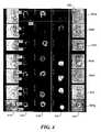
- FIGURE 4is a composite image formed from exemplary image data collected using an ImageStream® platform.
- the ImageStream® platformis a commercial embodiment based on the flow imaging instrument discussed above in detail with respect to FIGURE 1 .
- Each of columns 410-418corresponds to a different channel (i.e. a different spectral image) that was simultaneously collected.
- each image in a box 426represents the same conjugate of two biological cells.
- each other rowrepresents the same object or objects.
- the image datawas collected based on a population of more than 10,000 antigen-specific T cells and antigen pulsed antigen presenting cells.
- the T cellswere stained with HLA (human leukocyte antigen)-FITC (fluorescein isothiocyanate), resulting in green stained T cells.
- HLAhuman leukocyte antigen
- FITCfluorescein isothiocyanate
- the antigen presenting cellswhere stained with CD86-PE (phycoerythrin), resulting in red stained antigen presenting cells.
- Both the green stained T cells and the red stained antigen presenting cellswere incubated together at 37°C for 30 minutes, and thereafter introduced into an ImageStream® flow imaging instrument, such that a plurality of different spectral images of each cell were simultaneously collected as the cells passed through the flow imaging instrument.
- the image data for the population of stained T cells and stained antigen presenting cellswere then evaluated using the IDEAS® software.
- the use of that particular software packageis simply exemplary, and not intended to limit the invention. Those of ordinary skill in the art will recognize that other image analysis software packages are available.
- the subpopulationshould include only conjugates with limited numbers of cells (i.e., 2 - 3) per conjugate, although the technique being described herein is also applicable to conjugates containing larger numbers of cells.
- the subpopulation of cell conjugateswas then further limited to include only cell conjugates including a limited number of cells, in which each of the cells were considered to be in the same focal plane. The process of defining the subpopulation is described in detail below.
- the image analysis softwareemployed enabled two different mechanisms to be utilized to define the subpopulation, each of which are described below.
- Column 410includes brightfield images of cell conjugates. Each cell conjugate in column 410 clearly includes two co-joined cells. Column 412 is a color image, clearly indicating that each cell conjugate includes one green stained T cell 420 and one red stained antigen presenting cell 422. As will be described in greater detail below, each image in column 414 is based on the use of the mask to identify the T cell portion of the cell conjugate, and each image in column 416 is based on the use of the mask to identify the antigen presenting cell portion of the cell conjugate.
- Column 418includes brightfield images of cell conjugates with the synapse portion of each cell conjugate highlighted by a mask 424a-424g.
- the image data encompassed by the masked synapses in the subpopulationcan then be analyzed to identify at least one characteristic.
- the characteristic being measured in the exemplary study involving T cells and antigen presenting cellswill be discussed in detail below.
- the analysis performed in the exemplary studyis based on first identifying cell clusters containing at least one T cell and one antigen presenting cell, narrowing in on clusters consisting of only a single T cell and a single antigen presenting cell (column 412), defining the region of contact (i.e., the synapse) between the two cells (column 418), and defining the region of each cell outside of the synapse (columns 414 and 416).
- the mean intensity of the red signalwhich is a probe specific for the cell surface marker CD 86, can be quantitated separately for the synaptic and extra-synaptic region of each antigen presenting cell. If the mean CD 86 intensity within the synapse is ratioed over the mean CD 86 intensity outside of the synapse, CD 86 migration to, or exclusion from, the synapse will be revealed as an increase or decrease of the ratio from unity, respectively.
- a dot plot 426 in the left portion of FIGURE 5is a plot of green intensity (x-axis) versus red intensity (y-axis) for every object in the data file.
- Lone T cellsare HLA-bright but CD86-dim, placing them in the lower right quadrant of the plot.
- Lone antigen presenting cellsare HLA-dim but CD86-bright, placing them in the upper left quadrant of the plot.
- Conjugates including at least one T cell and one antigen presenting cellappear in the upper right quadrant of the plot.
- This gating techniqueis one example of selecting a subpopulation (the method step of block 404 in FIGURE 3 ).
- the screenshot of FIGURE 5includes brightfield images 410a, spectral images 434 corresponding to the CD 86 red stain, spectral images 436 corresponding to the FTIC green stain, and darkfield images 438.
- the double-positive population gated in region 428contains not only conjugates of one T cell with one antigen presenting cell, but likely also includes clusters of three or more cells, whose inclusion is undesirable (because the exemplary study intended to focus on simple conjugates including one antigen presenting cell and one T cell).
- a second scatter plot 430 in FIGURE 5illustrates the use of brightfield imagery to eliminate conjugates of more than two cells. All cells in the data file are plotted using brightfield area (x-axis) versus brightfield aspect ratio (y-axis). The double positive population is "back-gated" onto the plot by changing the symbol and color of each object that fell into the double-positive gate defined in the other dot plot.
- the double-positive populationtends to be larger and spans a wider range of aspect ratios than the single-positive population, which consists primarily of single cells.
- the subset of the double-positive objects that primarily includes conjugates of just two cellswas selected using a rectangular gate 432.
- the boundaries of the gatewere determined by defining a population of two-cell conjugates from the double-positive image gallery shown in the upper left of the figure and back-gating this population on the dot plot to determine the range of typical area and aspect ratio values for two-cell conjugates.
- an operatorcan view the image data collected to visually identify one or more cell conjugates including one T cell and one antigen presenting cell, select that cell conjugate, and instruct the image analysis software to identify all other cell conjugates whose image data strongly correlates with the specific cell conjugate identified by the operator (using the gating and backgating techniques noted above).
- the region of overlap between the antigen presenting cell and the T cellcan be defined in multiple ways.
- One method of defining the region of overlapis to generate a calculated image that is a transform of the signals in pixels at each location in the spectral images (for instance the product of the signals), such that overlapping regions exhibit greatly increased pixel values.
- a simple threshold function set at a level above the untransformed pixels in either spectral imagewill then preferentially select for pixels in the calculated image that are in common between the two spectral images.
- the pixel addresses in commoncan then be used to refer to the original signal levels for quantization both within and outside the synapse.
- Another methodis to employ binary masks, one per cell, that delineate the extent of each cell image.
- the extent of the maskis limited by the ability to detect image signals over the background level.
- two spectral images of the same cell clustersuch as those shown in dot plot 426 of FIGURE 4 . If each cell is labeled with a different color (red/green in this case), the red and green spectral channels (columns 414 and 416) will each contain an image of only one of the cells.
- each image maskwill correspond to only one cell. Since masks are binary, a Boolean AND operation using the (x,y) coordinates of the pixels comprising each mask will yield a new mask containing only those pixels in common between the two cell images (i.e. mask 424a).
- This methodologyis extensible to conjugates of more than two cell types as long as each cell type bears a unique probe and the Boolean AND operations are executed pair-wise for each image and summed using an OR function.
- Still another methoddoes not require the use of a unique probe for each cell in the conjugate. Instead, a probe-free imaging modality such as brightfield
- the masking technique noted abovewill now be described in greater detail.
- the images of the over 10,000 objects (in this case, biological cells) initially imagedwere first analyzed to identify a subpopulation of cell conjugates.
- the initial discrimination processidentified an initial subpopulation of 1095 objects (i.e., cell conjugates) from the over 10,000 objects initially imaged.
- the subpopulation of cell conjugateswas then further defined to include cell conjugates including a limited number of cells, and which include cells which are both in focus. In this manner, a subpopulation of 178 cell conjugates was selected from the 1095 objects included in the initial subpopulation.
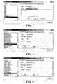
- FIGURE 6is a screenshot of a graphical user interface from the exemplary image analysis software used to generate masks for Channels 3 and 4.
- FIGURE 7is a screenshot of a graphical user interface from the exemplary image analysis software used to generate a synapse mask using Boolean logic to combine the masks for Channels 3 and 4. Examples of the masks described, as well as brightfield and fluorescent images of the conjugates, are shown in FIGURE 4 .
- FIGURE 8is a screenshot of a graphical user interface from the exemplary image analysis software used to determine the mean signal intensities of CD86 in the entire antigen presenting cell (CD86 mean intensity), while FIGURE 9 is a screenshot of a graphical user interface from the exemplary image analysis software used to determine the mean signal intensities of CD86 only within the interaction site (i.e., the synapse) between the T cell and the antigen presenting cell (CD86 Synapse Intensity).
- FIGURE 10is a screenshot of a graphical user interface from the exemplary image analysis software used to normalize the signal intensity at the T cell/antigen presenting cell interface.
- FIGURE 11is a chart showing the frequency verses the mean synapse intensity of CD86.
- FIGURE 12is a chart showing the frequency of conjugates containing both T cells and antigen presenting cells.
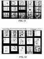
- FIGURE 13includes images of conjugates with a ratio approximating 0.9.
- FIGURE 14includes images of conjugates with a ratio approximating 0.3.
- FIGURE 15includes images of conjugates with a ratio approximating 0.5.
- FIGURE 16is a chart showing frequency verses the mean synapse intensity of CD86 for conjugates with an aspect ratio approximating 0.5.
- FIGURE 17is a chart showing the frequency of cell conjugates considered to be in reasonably good focus.
- FIGURE 18is a chart showing frequency verses the normalized mean synapse intensity of CD86.
- FIGURE 19and the following related discussion are intended to provide a brief, general description of a suitable computing environment for practicing the present invention, where the image processing required is implemented using a computing device functionally related to that shown in FIGURE 19 .
- An exemplary computing system 150 suitable for implementing the image processing required in the present inventionincludes a processing unit 154 that is functionally coupled to an input device 152, and an output device 162, e.g., a display.
- Processing unit 154include a central processing unit (CPU 158) that executes machine instructions comprising an image processing/image analysis program for implementing the functions of the present invention (analyzing a plurality of images simultaneously collected for members of a population of objects to enable at least one characteristic exhibited by members of the population to be measured).
- the machine instructionsimplement functions generally consistent with those described above, with reference to the flowchart of FIGURE 3 , as well as the exemplary screenshots.
- CPUs suitable for this purposeare available from Intel Corporation, AMD Corporation, Motorola Corporation, and other sources.
- RAMrandom access memory
- non-volatile memory 160typically includes read only memory (ROM) and some form of memory storage, such as a hard drive, optical drive, etc.
- ROMread only memory
- CPU 158Such storage devices are well known in the art.
- Machine instructions and dataare temporarily loaded into RAM 156 from non-volatile memory 160.
- operating system software and ancillary softwareWhile not separately shown, it should be understood that a power supply is required to provide the electrical power needed to energize computing system 150.
- Input device 152can be any device or mechanism that allows input into the operating environment. This includes, but is not limited to a mouse, keyboard, microphone, modem, pointing, or other input devices. While not specifically shown in FIGURE 19 , it should be understood that computing system 150 is logically coupled to an imaging system such as that schematically illustrated in FIGURE 1 , such that the image data collected is available to computing system 150 to achieve the desired image processing. Of course, rather than logically coupling the computing system directly to the imaging system, data collected by the imaging system can simply be transferred to the computing system by means of many different data transfer devices, such as portable memory media. Output device 162 will most typically comprise a monitor or computer display designed for human perception of output.
Landscapes
- Health & Medical Sciences (AREA)
- Physics & Mathematics (AREA)
- Life Sciences & Earth Sciences (AREA)
- Immunology (AREA)
- Chemical & Material Sciences (AREA)
- General Physics & Mathematics (AREA)
- Engineering & Computer Science (AREA)
- General Health & Medical Sciences (AREA)
- Spectroscopy & Molecular Physics (AREA)
- Analytical Chemistry (AREA)
- Pathology (AREA)
- Biochemistry (AREA)
- Biomedical Technology (AREA)
- Molecular Biology (AREA)
- Cell Biology (AREA)
- Hematology (AREA)
- Urology & Nephrology (AREA)
- Nuclear Medicine, Radiotherapy & Molecular Imaging (AREA)
- Dispersion Chemistry (AREA)
- Food Science & Technology (AREA)
- Signal Processing (AREA)
- Medicinal Chemistry (AREA)
- Tropical Medicine & Parasitology (AREA)
- Biotechnology (AREA)
- Microbiology (AREA)
- Bioinformatics & Cheminformatics (AREA)
- Toxicology (AREA)
- Chemical Kinetics & Catalysis (AREA)
- Optics & Photonics (AREA)
- Theoretical Computer Science (AREA)
- Multimedia (AREA)
- Investigating, Analyzing Materials By Fluorescence Or Luminescence (AREA)
- Measuring Or Testing Involving Enzymes Or Micro-Organisms (AREA)
- Investigating Or Analysing Materials By Optical Means (AREA)
- Apparatus Associated With Microorganisms And Enzymes (AREA)
- Investigating Or Analysing Biological Materials (AREA)
Abstract
Description
- The present invention relates generally to methods for detecting and quantifying molecules in, on, and between intact cells, and more specifically, to methods of analyzing the distribution of molecules effecting inter-cellular communication.
- Communication between cells of the immune system is integral to immune function. The process of T cell differentiation and maturation involves direct contact between dendritic cells and the maturing T cells, during which the T cells are selected for survival on the basis of their ability to recognize antigens associated with "self" without triggering an immune response, thereby preventing auto-immunity. Once mature, T cells frequently interact physically with antigen presenting cells, which expose the T cell to non-self antigens from bacteria, viruses, etc., ultimately triggering expansion of T cell populations that elicits a sustained immune response against the antigen source presented.
- The study of inter-cellular communication is greatly facilitated by imagery of the cells in contact. Ideally, the imagery would be acquired from living cells; since it is likely that fixation of the cells would disrupt biochemical signaling mechanisms. Since the cells would be alive and therefore highly dynamic, it would be desirable to acquire multiple images of conjugated cells simultaneously. It would also be desirable to image the cells directly in fluid suspension, since immune cells are generally non-adherent and contact with a foreign surface could perturb the signaling process. Finally, it would be desirable to provide a sufficiently analytical throughput such that studies could employ the relatively rare primary T cell and antigen presenting cell conjugates obtained from whole blood, as opposed to model systems of cultured cells, to best modelin vivo behavior.
- Gordy et al, J. of Immunol., 172 (2004), 2030-2038, discloses a fluorescence cell imaging process for visualizing the antigen presentation by actin-mediated targeting of glycolipid-enriched membrane domains to the immune synapse of B cell APCs.
- George et al, Cytometry, 59A (2004), 237-245, is a paper of the present inventors regarding the use multispectral imaging of morphologically distinct cell populations using a multispectral imaging cytometer apparatus.
- There is a recognized need in the art for techniques that permit detection and quantization of cell conjugates in flow, which would provide an opportunity to
- study suspension-based cell lines and primary cells. Furthermore, methods for preparing cells in suspension for multi-spectral analysis are needed. The present invention meets such needs, and further provides other related advantages.
- The embodiments of the present invention are:
- 1. A method for studying inter-cellular communication between living biological cells, comprising the steps of:
- (a) providing a population of living biological cells, the population comprising individual biological cells and cell conjugates including a plurality of biological cells;
- (b) providing an imaging system configured to enable a plurality of images of cell conjugates and individual cells to be simultaneously acquired;
- (c) using the imaging system to image the population of living biological cells, such that for each individual cell and for each cell conjugate in the population, a plurality of images of the cell or cell conjugate are simultaneously acquired, while there is relative movement between the population of living biological cells and the imaging system, to generate a set of image data corresponding to the population;
- (d) processing the image data to distinguish cell conjugates from individual cells, thereby defining a first subpopulation of conjugated cells and a second subpopulation of individual biological cells; and
- (e) analyzing a synapse between individual biological cells in each cell conjugate using image data for the first subpopulation.
- 2. The method of 1, wherein the initial population of biological cells comprises a first type of cell and a second type of cell, and further comprising the step of removing from the image data for the first subpopulation any cell conjugate not consisting of a single cell of the first type and a single cell of the second type.
- 3. The method of 2, further comprising the step of removing from the image data, for the first subpopulation, image data for any cell conjugates in which cells in the conjugate are in different focal planes.
- 4. The method of 2, wherein the step of removing from the image data, for the first subpopulation, any cell conjugate not consisting of a single cell of the first type and a single cell of the second type comprises the steps of:
- (a) identifying image data for at least one cell conjugate consisting of a single cell of the first type and a single cell of the second type;
- (b) based on the image data identified, determining a typical area and aspect ratio for cell conjugates consisting of a single cell of the first type and a single cell of the second type; and
- (c) removing from the image data for the first subpopulation any cell conjugate having an area or aspect ratio that does not correspond to the typical area and aspect ratio determined in subparagraph (b).
- 5. The method of 2, wherein the step of removing from the image data, for the first subpopulation, any cell conjugate not consisting of a single cell of the first type and a single cell of the second type comprises the steps of:
- (a) identifying image data for at least one cell conjugate consisting of a single cell of the first type and a single cell of the second type; and
- (b) removing from the image data for the first subpopulation any cell conjugate whose image data does not strongly correlate with the image data identified in subparagraph (a).
- 6. The method of any of 2-5, wherein the biological cells comprise T cells and antigen presenting cells.
- 7. The method of 6, wherein each T cell and each antigen presenting cell in the initial population of biological cells is stained before any image data is acquired, such that each T cell is stained with a different stain than each antigen presenting cell, and further comprising the step of determining for each cell conjugate in the first subpopulation whether an amount of CD 86 present in the synapse is relatively larger than or relatively smaller than an amount of CD 86 in extra-synaptic portions of the antigen presenting cell in the cell conjugate.
- 8. The method of 7, further comprising the step of normalizing a signal intensity at the synapse for each cell conjugate in the first subpopulation by dividing a mean intensity of the CD86 signal at the synapse by a mean intensity of the CD 86 signal from the antigen presenting cell as a whole.
- 9. The method of any of 1-6, wherein the synapse for each cell conjugate in the first subpopulation is defined by:
- (a) generating a calculated image that is a transform of pixels in the plurality of images acquired for that cell conjugate, such that overlapping regions exhibit greatly increased pixel values;
- (b) applying a threshold function to preferentially select for pixels in the calculated image that are in common between the plurality of images acquired for that cell conjugate; and
- (c) using addresses for the common pixel selected to quantify pixels within and outside of the synapse for that cell conjugate.
- 10. The method of any of 1-6, wherein before any image data is acquired each first type of cell is stained with a first dye, and each second type of cell is stained with a second dye, and further comprising the step of defining the synapse for each cell conjugate in the first subpopulation by:
- (a) employing a first binary mask defining an extent of the first type of cell in the cell conjugate;
- (b) employing a second binary mask defining an extent of the second type of cell in the cell conjugate; and
- (c) generating a synapse mask defining an extent of the synapse for that cell conjugate based on overlapping regions of the first and second binary masks.
- 11. Use of an imaging system for studying inter-cellular communication between living biological cells, by generating and analyzing a plurality of images collected for each member of a population of living biological cells while there is relative movement between each member and the imaging system, the imaging system comprising:
- (a) a collection lens disposed so that light traveling from the object passes through the collection lens and travels along a collection path;
- (b) a spectral dispersing component disposed in the collection path so as to receive the light that has passed through the collection lens, dispersing the light into a plurality of separate light beams, each light beam being directed away from the dispersing component in a different predetermined direction;
- (c) an imaging lens disposed to receive the light beams from the dispersing component, producing a plurality of images corresponding to each of the light beams, each image being projected by the imaging lens toward a different predetermined location;
- (d) a detector disposed to receive the plurality of images, the plurality of images comprising at least two of the following types of images: a brightfield image, a darkfield image, and a fluorescent image:
- 12. The use of 11, wherein the initial population of biological cells comprises a first type of cell and a second type of cell, and the processor is further configured to implement the step of removing from the image data, for the first subpopulation, any cell conjugate not consisting of a single cell of the first type and a single cell of the second type.
- 13. The use of 11, wherein the processor is further configured to implement the steps of:
- (a) identifying image data for at least one cell conjugate consisting of a single cell of a first type and a single cell of a second type; and
- (b) removing from the image data, for the first subpopulation, any cell conjugate whose image data does not strongly correlate with the image data identified in subparagraph (a).
- 14. The use of any of 11-13, wherein the biological cells comprise T cells and antigen presenting cells.
- 15. The use of 14, wherein each T cell and each antigen presenting cell is stained before any image data is acquired, each T cell being stained with a different stain than each antigen presenting cell, and the processor is further configured to implement the step of determining for each cell conjugate in the first subpopulation whether an amount of CD 86 present in the synapse is relatively larger than or relatively smaller than an amount of CD 86 in extra-synaptic portions of the antigen presenting cell in the cell conjugate:
- 16. The use of 12, wherein the processor is further configured to define the synapse for each cell conjugate in the first subpopulation by implementing at least one series of steps selected from a group consisting of two series of steps; wherein:
- (a) the first series of steps comprises:
- (i) generating a calculated image that is a transform of pixels in the plurality of images acquired for that cell conjugate, such that overlapping regions exhibit greatly increased pixel values;
- (ii) applying a threshold function to preferentially select for pixels in the calculated image that are in common between the plurality of images acquired for that cell conjugate; and
- (iii) using addresses for the common pixel selected to quantify pixels within and outside of the synapse for that cell conjugate; and
- (b) the second series of steps comprises:
- (i) employing a first binary mask defining an extent of the first type of cell in the cell conjugate;
- (ii) employing a second binary mask defining an extent of the second type of cell in the cell conjugate; and
- (iii) generating a synapse mask defining an extent of the synapse for that cell conjugate based on overlapping regions of the first and second binary masks, the second series of steps being appropriate when each first type of cell is stained with a first dye, and each second type of cell is stained with a second dye, before any image data is acquired.
- (a) the first series of steps comprises:
- Aspects of the present invention relate to the collection of multispectral images from a population of objects, and the analysis of the collected images to measure at least one characteristic of the population, using photometric and/or morphometric features identifiable in the collection of images. In an exemplary application, the objects are biological cells. In a particularly preferred implementation, both photometric and morphometric features are used in the analysis. In a particularly preferred, but not limiting implementation, the plurality of images for each individual object are collected simultaneously.
- To facilitate analysis, at least one aspect of the invention is directed to labeling at least a subset of the population of objects before using an imaging instrument to collect image data on the population of cells (or objects). In general, the present invention can be implemented using N-1 unique labels, where N is the number of different object types to be distinguished, as well as being implemented using as many labels as there are different object types, or even more labels than there are object types.
- Exemplary steps that can be used to analyze objects such as biological cells in accord with an aspect of the present invention include collecting image data from a population of objects and identifying a subpopulation of objects from the image data for further analysis. The objects are biological cells and the subpopulation corresponds to conjugated cells. Next, a particular feature of the objects in the subpopulation is identified for further study. The term feature is intended to refer to a particular structure, region, or portion of an object that can be readily discerned. For example, one analysis may focus on the nucleus or internal volume of each object in the subpopulation, while another analysis may focus on the cell membrane or outer boundary of each object in the subpopulation. In a particularly preferred aspect of the present invention, the feature selected for further study is the synapse between cell conjugates. For each object in the subpopulation, the feature selected for further study is identified. Using photometric and/or morphometric data from the collected images, at least one characteristic of the selected feature is measured.
- Once a feature has been identified for analysis, the image data for the subpopulation can be manipulated using several different techniques. An exemplary technique is referred to as gating, a manipulation of data relating to photometric or morphometric imaging. A further exemplary technique is backgating, which involves further defining a subset of the gated data.
- While not strictly required, preferably signal processing is performed on the collected image data to reduce crosstalk and enhance spatial resolution, particularly for image data collected using simultaneous multi-channel imaging.
- The characteristic being measured involves the synapse between conjugated cells. The conjugated cells may represent a subpopulation of the overall population of objects that were imaged. In a particularly preferred, yet not limiting embodiment, the present invention enables the quantization of the redistribution of cellular molecules due to the conjugation of different biological cells. Significantly, such quantization is not feasible with standard microscopy and flow cytometry.
- Analyzing the synapse between conjugated cells will facilitate the study of inter-cellular communication. In a preferred implementation of the present invention, the imagery collected from a population of biological cells includes collection of at least one of brightfield and darkfield imagery, and fluorescent imagery. In such an implementation, molecules suspected of playing a role in the communication pathway are fluorescently labeled, enabling changes in molecular quantities and molecular distributions as a result of the inter-cellular interaction to be detected. The collection of brightfield and/or darkfield imagery augments fluorescent labeling by eliminating the need for separate fluorescent identifying markers merely to distinguish the T cell from the antigen presenting cell, thereby allowing the use of more fluorescent probes for the simultaneous study of different molecules involved in the communication pathway. In one aspect of the present invention, the population of biological cells being imaged comprises relatively rare primary T cell and antigen presenting cell conjugates obtained from whole blood.
- The foregoing aspects and many of the attendant advantages of this invention will become more readily appreciated as the same becomes better understood by reference to the following detailed description, when taken in conjunction with the accompanying drawings, wherein:
FIGURE 1 is a schematic diagram of an exemplary flow imaging system that can be used to simultaneously collect a plurality of images from an object in flow;FIGURE 2 is a pictorial representation of an image recorded by the flow imaging system ofFIGURE 1 ;FIGURE 3 is a flow chart of the overall method steps implemented in one aspect of the present invention;FIGURE 4 is a composite image formed from exemplary image data collected using the flow imaging system ofFIGURE 1 and analyzed according to the method steps ofFIGURE 3 ;FIGURES 5-10 are exemplary graphical user interfaces used to implement the method steps ofFIGURE 3 ;FIGURE 11 graphically illustrates the frequency verses the mean synapse intensity of CD86 for a subpopulation analyzed according to the method steps ofFIGURE 3 ;FIGURE 12 graphically illustrates the frequency of a subpopulation of cell conjugates containing both T cells and antigen-presenting cells;FIGURE 13 includes images of cell conjugates with an aspect ratio of approximately 0.9;FIGURE 14 includes images of cell conjugates with an aspect ratio of approximately 0.3;FIGURE 15 shows images of cell conjugates with an aspect ratio of approximately 0.5;FIGURE 16 graphically illustrates frequency verses the mean synapse intensity of CD86 for conjugates with an aspect ratio approximating 0.5;FIGURE 17 graphically illustrates the frequency of cell conjugates considered to be in reasonably good focus;FIGURE 18 graphically illustrates frequency verses the normalized mean synapse intensity of CD86 for a subpopulation collected and analyzed according to the method steps ofFIGURE 3 ; andFIGURE 19 schematically illustrates an exemplary computing system used to implement the method steps ofFIGURE 3 .- An aspect of the present invention relates to the use od a system and a method for imaging and analyzing conjugated biological cells entrained in a flow of fluid. In at least one embodiment, a plurality of images of biological cells are collected simultaneously; the plurality of images including at least two of the following types of images: a brightfield image, a darkfield image, and a fluorescent image. Images are collected for a population of biological cells (or objects with discernable morphological features). Once the imagery has been collected, the images can be processed to identify a subpopulation of images. The images in the subpopulation are processed to identify points of contact (i.e., synapses) between cell conjugates in the subpopulation. Further processing of the images in the subpopulation is performed to measure at least one characteristic at the identified synapses.
- With respect to the following disclosure, and the claims it should be understood that the term population of objects refers to a group of objects including a plurality of objects. Thus, a population of objects must include more than one object.
- A preferred imaging system to be used in collecting the image data required in the present invention will incorporate the following principal characteristics:
- 1. high speed measurement;
- 2. the ability to process very large or continuous samples;
- 3. high spectral resolution and bandwidth;
- 4. good spatial resolution;
- 5. high sensitivity; and
- 6. low measurement variation.
- In particular, a recently developed imaging flow cytometer technology, termed ImageStream® (Amnis Corporation, Seattle WA) makes great strides in achieving each of the above noted principle characteristics. The ImageStream® instrument is a commercial embodiment of the flow imaging systems described above in detail with respect to
FIGURES 1-19 . These significant advancements in the art of flow cytometery are described in the following commonly assigned patents:U.S. Patent No. 6,249,341, issued on June 19, 2001 and entitled "Imaging And Analyzing Parameters of Small Moving Objects Such As Cells";U.S. Patent No. 6,211,955 issued on April 3, 2001 , also entitled "Imaging And Analyzing Parameters of Small Moving Objects Such As Cells";U.S. Patent No. 6,473,176, issued on October 29, 2002 , also entitled "Imaging And Analyzing Parameters of Small Moving Objects Such As Cells";U.S. Patent No. 6,583,865, issued on June 24, 2003 , entitled "Alternative Detector Configuration And Mode of Operation of A Time Delay Integration Particle Analyzer"; andU.S. Patent Application No. 09/989,031 entitled "Imaging And Analyzing Parameters of Small Moving Objects Such As Cells in Broad Flat Flow." While the ImageStream® platform represents a particularly preferred imaging instrument used to acquire the image data that will be processed in accord with the present invention, it should be understood that the present invention is not limited only to the use of that specific instrument. - As noted above, in addition to collecting image data from a population of biological cells, an aspect of the present invention involves processing the image data collected to measure at least one characteristic at a synapse between conjugated cells encompassed in the imaged population. A preferred image analysis software package is IDEALS® (Amnis Corporation, Seattle WA). The IDEAS® package evaluates nearly 200 features for every cell, including multiple morphologic and fluorescence intensity measurements, which can be used to define and characterize cell populations. The IDEAS® package enables the user to define biologically relevant cell subpopulations, and analyze subpopulations using standard cytometry analyses, such as gating and backgating. It should be understood, however, that other image analysis methods or software packages can be implemented in the present invention, and the preferred image analysis software package is intended to be exemplary, rather than limiting the invention.
FIGURE 1A is a schematic diagram of a preferred flow imaging system 510 (functionally descriptive of the ImageStream® platform) that uses TDI when capturing images of objects 502 (such as biological cells), entrained in afluid flow 504.System 510 includes a velocity detecting subsystem that is used to synchronize aTDI imaging detector 508 with the flow of fluid through the system. Significantly,imaging system 510 is capable of simultaneously collecting a plurality of images of an object. A particularly preferred implementation ofimaging system 510 is configured for multi-spectral imaging, and can operate with six spectral channels: DAPI fluorescence (400-460 nm), Darkfield (460 - 500 nm), FITC fluorescence (500-560 nm), PE fluorescence (560 - 595 nm), Brightfield (595 - 650 nm), and Deep Red (650 - 700 nm). The TDI detector can provide 10 bit digital resolution per pixel. The numeric aperture of the imaging system used with this invention is typically 0.75, with a pixel size of approximately 0.5 microns. However, those skilled in the art will recognize that this flow imaging system is neither limited to six spectral channels nor limited to either the stated aperture size or pixel size and resolution.- Moving
objects 502 are illuminated using alight source 506. The light source may be a laser, a light emitting diode, a filament lamp, or a gas discharge arc lamp, and the system may include optical conditioning elements such as lenses, apertures, and filters that are employed to deliver broadband or one or more desired wavelengths or wavebands of light to the object with an intensity required for detection of the velocity and one or more other characteristics of the object. Light from the object is split into two light paths by abeam splitter 503. Light traveling along one of the light paths is directed to the velocity detector subsystem, and light traveling along the other light path is directed toTDI imaging detector 508. A plurality oflenses 507 are used to direct light along the paths in a desired direction, and to focus the light. While not shown, a filter or a set of filters can be included to deliver to the velocity detection subsystem and/orTDI imaging detector 508, only a narrow band of wavelengths of the light corresponding to, for example, the wavelengths emitted by fluorescent or phosphorescent molecules in/on the object, or light having the wavelength(s) provided by thelight source 506, so that light from non-desired sources is substantially eliminated. - The velocity detector subsystem includes an
optical grating 505a that modulates light from the object as a function of frequency, a lightsensitive detector 505b (such as a photomultiplier tube or a solid-state photodetector), asignal conditioning unit 505c, avelocity computation unit 505d, and atiming control unit 505e that assures thatTDI imaging detector 508 is synchronized to the flow offluid 504 through the system. The optical grating preferably comprises a plurality of alternating transparent and opaque bars that modulate the light received from the object, producing modulated light having a frequency of modulation that corresponds to the velocity of the object from which the light was received. Preferably, the optical magnification and the ruling pitch of the optical grating are chosen such that the widths of the bars are approximately the size of the objects being illuminated. Thus, the light collected from cells or other objects is alternately blocked and transmitted through the ruling of the optical grating as the object traverses the interrogation region, i.e., the field of view. The modulated light is directed toward a light sensitive detector, producing a signal that can be analyzed by a processor to determine the velocity of the object. Thus, the velocity measurement subsystem is used to provide timing signals toTDI imaging detector 508. - Preferably,
signal conditioning unit 505c comprises a programmable computing device, although an ASIC chip or a digital oscilloscope can also be used for this purpose. The frequency of the photodetector signal is measured, and the velocity of the object is computed as a function of that frequency. The velocity dependent signal is periodically delivered to a TDIdetector timing control 505e to adjust the clock rate ofTDI imaging detector 508. Those of ordinary skill in the art will recognize that the TDI detector clock rate is adjusted to match the velocity of the image of the object over the TDI detector to within a small tolerance selected to ensure that longitudinal image smearing in the output signal of the TDI detector is within acceptable limits. The velocity update rate must occur frequently enough to keep the clock frequency within the tolerance band as flow (object) velocity varies. Beam splitter 503 has been employed to divert a portion of light from anobject 502 to lightsensitive detector 505b, and a portion of light from object 502a toTDI imaging detector 508. In the light path directed towardTDI imaging detector 508 there is a plurality of stackeddichroic filters 509, which separate light from object 502a into a plurality of wavelengths. Note that one oflenses 507 is used to form an image of object 502a onTDI imaging detector 508.- The theory of operation of a TDI detector, such as those employed in
system 510, is as follows. As objects travel through a flow tube 511 (FIGURE 1 ) and pass through the volume imaged by the TDI detector, light from the objects form images of the objects that travel across the face of the TDI detector. The TDI detector preferably comprises a charge coupled device (CCD) array, which is specially designed to allow charge to be transferred on each clock cycle in a row-by-row format, so that a given line of charge remains locked to or synchronized with a line in the image. The row of charge is clocked out of the array into a memory when it reaches the bottom of the array. The intensity of each line of the signal produced by the TDI detector corresponding to an image of an object is integrated over time as the image and corresponding resulting signal propagate over the CCD array. This technique greatly improves the signal-to-noise ratio of the TDI detector compared to non-integrating type detectors - a feature of great value when responding to images from low-level fluorescence emission of an object. Proper operation of the TDI detector requires that the charge signal be clocked across the CCD array in synchronization with the rate at which the image of the object moves across the CCD array. An accurate clock signal to facilitate this synchronization can be provided by determining the velocity of the object, and the present invention uses an accurate estimate of the object's velocity, and thus, of the velocity of the image as it moves over the CCD array of the TDI detector. A flow imaging system of this type is disclosed in commonly assignedU.S. Patent No. 6,249,341 , FIGURE 2 is a pictorial representation of images produced by the flow imaging system ofFIGURE 1 . Acolumn 520, labeled "BF," includes images created by the absorption of light fromlight source 506 byspherical objects 502 entrained influid flow 504. The "BF" label refers to "brightfield," a term derived from a method for creating contrast in an image whereby light is passed through a region and the absorption of light by objects in the region produces dark areas in the image. The background field is thus bright, while the objects are dark. Thus,column 520 is the "brightfield channel." It should be understood that the inclusion of a brightfield image is exemplary, rather than limiting of the scope of the present invention. Preferably, the present invention utilizes a combination of brightfield images and fluorescent images, or darkfield images and fluorescent images.- The remaining three
columns FIGURE 2 are respectively labeled "λ1" "λ2," and "λ3" These columns include images produced using light that has been emitted by an object entrained in the fluid flow. Preferably, such light is emitted through the process of fluorescence (as opposed to images produced using reflected light). As those of ordinary skill in the art will recognize, fluorescence is the emission of light (or other electromagnetic radiation) by a substance that has been stimulated by the absorption of incident radiation. Generally, fluorescence persists only for as long as the stimulating radiation persists. Many substances (particularly fluorescent dyes) can be identified based on the spectrum of the light that is produced when they fluoresce.Columns - Additional exemplary flow imaging systems are disclosed in commonly assigned
U.S. Patent No. 6,211,955 andU.S. Patent No. 6,608,682 The imaging systems described above in detail and incorporated herein by reference have considerable advantages over more conventional systems employed for the acquisition of images of biological cell populations. These advantages arise from the use in several of the imaging systems of an optical dispersion system, in combination with a TDI detector that produces an output signal in response to the images of cells and other objects that are directed onto the TDI detector. Significantly, multiple images of a single object can be collected at one time. The image of each object can be spectrally decomposed to discriminate object features by absorption, scatter, reflection or probe emissions using a common TDI detector for analysis. Other systems include a plurality of detectors, each dedicated to a single spectral channel. - These imaging systems can be employed to determine morphological, photometric, and spectral characteristics of cells and other objects by measuring optical signals including light scatter, reflection, absorption, fluorescence, phosphorescence, luminescence, etc. Morphological parameters include nuclear area, perimeter, texture or spatial frequency content, centroid position, shape (i.e., round, elliptical, barbell-shaped, etc.), volume, and ratios of any of these parameters. Similar parameters can also be determined for the cytoplasm of cells with the present invention. Photometric measurements with the invention enable the determination of nuclear optical density, cytoplasm optical density, background optical density, and the ratios of any of these values. An object being imaged with the present invention can either be stimulated into fluorescence or phosphorescence to emit light, or may be luminescent, producing light without stimulation. In each case, the light from the object is imaged on the TDI detector of the present invention to determine the presence and amplitude of the emitted light, the number of discrete positions in a cell or other object from which the light signal(s) originate(s), the relative placement of the signal sources, and the color (wavelength or waveband) of the light emitted at each position in the object.
- An initial application of the imaging system comprising the present invention will likely be employed as a cell analyzer to determine one or more of the parameters listed above, for cells entrained in a fluid flowing through the imaging system. However, it should also be understood that this invention can be used for imaging other moving objects, where the objects have identifiable photometric and morphometric features.
- As noted above, aspects of the present invention involve both the collection of multispectral images from a population of biological cells, and the analysis of the collected images to measure at least one characteristic exhibited at a cellular feature, such as the synapse of conjugated cells identified from the multispectral images collected. Thus, an aspect of the present invention relates to the use of both photometric and morphometric features derived from multi-mode imagery of objects (e.g., cells) in flow to discriminate cell features in heterogeneous populations of cells, including in both non-adherent and adherent cell types. Discussed in more detail below are methods for analyzing cells in suspension or flow, such that cells tend to aggregate, which may be combined with comprehensive multispectral imaging to provide morphometric and photometric features to allow, for example, the quantization of the redistribution of cellular molecules not feasible with standard microscopy and flow cytometry.
- As noted above, a preferred flow imaging system (e.g., the ImageStream® platform) can be used to simultaneously acquire multispectral images of cells in flow, to collect image data corresponding to brightfield, darkfield, and four channels of fluorescence. The ImageStream® platform is a commercial embodiment based on the imaging systems described in detail above. In general, cells are hydrodynamically focused into a core stream and orthogonally illuminated for both darkfield and fluorescence imaging. The cells are simultaneously trans-illuminated via a spectrally-limited source (e.g., filtered white light or a light emitting diode) for brightfield imaging. Light is collected from the cells with an imaging objective lens and is projected on a charge-coupled detector (CCD). The optical system has a numeric aperture of 0.75 and the CCD pixel size in object space is 0.5 µ2, allowing high resolution imaging at event rates of approximately 100 cells per second. Each pixel is digitized with 10 bits of intensity resolution, providing a minimum dynamic range of three decades per pixel. In practice, the spread of signals over multiple pixels results in an effective dynamic range that typically exceeds four decades per image. Additionally, the sensitivity of the CCD can be independently controlled for each multispectral image, resulting in a total of approximately six decades of dynamic range across all the images associated with an object. It should be understood that while the ImageStream® platform represents a particularly preferred flow imaging system for acquiring image data in accord with the present invention, the ImageStream® platform is intended to represent an exemplary imaging system, rather than limiting the invention. Any imaging instrument capable of collecting images of a population of biological cells sufficient to enable the image analysis described in greater detail below to be achieved, can be implemented in accord with the present invention.
- Referring again to the preferred imaging system, the ImageStream® platform, prior to projection on the CCD, the light is passed through a spectral decomposition optical system that directs different spectral bands to different lateral positions across the detector (such spectral decomposition is discussed in detail above in connection with the description of the various preferred embodiments of imaging systems). With this technique, an image is optically decomposed into a set of a plurality of sub-images (preferably 6 sub-images; brightfield, darkfield, and 4 different fluorescent images), each corresponding to a different spectral (i.e., color) component and spatially isolated from the remaining sub-images. This process allows for identification and quantization of signals within the cell by physically separating on the detector, signals that may originate from overlapping regions of the cell. Spectral decomposition also allows multimode imaging: the simultaneous detection of brightfield, darkfield, and multiple colors of fluorescence. The process of spectral decomposition occurs during the image formation process, rather than via digital image processing of a conventional composite image.
- The CCD may be operated using a technique called time-delay-integration (TDI), a specialized detector readout mode that preserves sensitivity and image quality even with fast relative movement between the detector and the objects being imaged. As with any CCD, image photons are converted to photo charges in an array of pixels. However, in TDI operation the photo charges are continuously shifted from pixel to pixel down the detector, parallel to the axis of flow. If the photo charge shift rate is synchronized with the velocity of the flowing cell's image, the effect is similar to physically panning a camera: image streaking is avoided despite signal integration times that are orders of magnitude longer than in conventional flow cytometry. For example, an instrument may operate at a continuous data rate of approximately 30 mega pixels per second and integrate signals from each object for 10 milliseconds, allowing the detection of even faint fluorescent probes within cell images that are acquired at relatively high speed. Careful attention to pump and fluidic system design to achieve highly laminar, non-pulsatile flow eliminates any cell rotation or lateral translation on the time scale of the imaging process (see, e.g.,
U.S. Patent No. 6,532,061 ). - A real-time algorithm analyzes every pixel read from the CCD to detect the presence of object images and calculate a number of basic morphometric and photometric features, which can be used as criteria for data storage. Data files encompassing 10,000-20,000 cells are typically about 100 MB in size and, therefore, can be stored and analyzed using standard personal computers. The TDI readout process operates continuously without any "dead time", which means every cell can be imaged and the coincidental imaging of two or more cells at a time either in contact or not, presents no barrier to data acquisition.
- Such an imaging system can be employed to determine morphological, photometric, and spectral characteristics of cells and other objects by measuring optical signals, including light scatter, reflection, absorption, fluorescence, phosphorescence, luminescence, etc. As used herein, morphological parameters may be basic (e.g., nuclear shape) or may be complex (e.g., identifying cytoplasm size as the difference between cell size and nuclear size). For example, morphological parameters may include nuclear area, perimeter, texture or spatial frequency content, centroid position, shape (i.e., round, elliptical, barbell-shaped, etc.), volume, and ratios of any of these parameters. Morphological parameters may also include cytoplasm size, texture or spatial frequency content, volume and the like, of cells. As used herein, photometric measurements with the aforementioned imaging system can enable the determination of nuclear optical density, cytoplasm optical density, background optical density, and the ratios of any of these values. An object being imaged can be stimulated into fluorescence or phosphorescence to emit light, or may be luminescent wherein light is produced without stimulation. In each case, the light from the object may be imaged on a TDI detector of the imaging system to determine the presence and amplitude of the emitted light, the number of discrete positions in a cell or other object from which the light signal(s) originate(s), the relative placement of the signal sources, and the color (wavelength or waveband) of the light emitted at each position in the object.
- The present disclosure provides methods of using both photometric and morphometric features derived from multi-mode imagery of objects in flow. Such methods can be employed as a cell analyzer to determine one or more cell states or types, and cell features, in heterogeneous populations of cells when entrained in a fluid flowing through an imaging system. It should also be understood that these exemplary methods might be used for imaging and distinguishing other moving objects that have identifiable photometric and morphometric features. As used herein, gating refers to a subset of data relating to photometric or morphometric imaging. For example, a gate may be a numerical or graphical boundary of a subset of data that can be used to define the characteristics of particles to be further analyzed. Here, gates have been defined, for example, as a plot boundary that encompasses "in focus" cells, or sperm cells with tails, or sperm cells without tails, or cells other than sperm cells, or sperm cell aggregates, or cell debris. Further, backgating may be a subset of the subset data. For example, a forward scatter versus a side scatter plot in combination with a histogram from an additional marker may be used to backgate a subset of cells within the initial subset of cells.
- In using an imaging system as described herein, it should be made clear that a separate light source is not required to produce an image of the object (cell), if the object is luminescent (i.e., if the object produces light). However, many of the applications of an imaging system as described herein will require that one or more light sources be used to provide light that is incident on the object being imaged. A person having ordinary skill in the art will know that the location of the light sources substantially affects the interaction of the incident light with the object and the kind of information that can be obtained from the images on a TDI detector.
- In addition to imaging an object with the light that is incident on it, a light source can also be used to stimulate emission of light from the object. For example, a cell having been contacted with probe conjugated to a fluorochrome (e.g., such as FITC, PE, APC, Cy3, Cy5, or Cy5.5) will fluoresce when excited by light, producing a corresponding characteristic emission spectra from any excited fluorochrome probe that can be imaged on a TDI detector. Light sources may alternatively be used for causing the excitation of fluorochrome probes on an object, enabling a TDI detector to image fluorescent spots produced by the probes on the TDI detector at different locations as a result of the spectral dispersion of the light from the object that is provided by prism. The disposition of these fluorescent spots on the TDI detector surface will depend upon their emission spectra and their location in the object.
- Each light source may produce light that can either be coherent, non-coherent, broadband or narrowband light, depending upon the application of the imaging system desired. Thus, a tungsten filament light source can be used for applications in which a narrowband light source is not required. For applications such as stimulating the emission of fluorescence from probes, narrowband laser light is preferred, since it also enables a spectrally decomposed, non-distorted image of the object to be produced from light scattered by the object. This scattered light image will be separately resolved from the fluorescent spots produced on a TDI detector, so long as the emission spectra of any of the spots are at different wavelengths than the wavelength of the laser light. The light source can be either of the continuous wave (CW) or pulsed type, and preferably is a pulsed laser. If a pulsed type illumination source is employed, the extended integration period associated with TDI detection can allow the integration of signals from multiple pulses. Furthermore, it is not necessary for the light to be pulsed in synchronization with the TDI detector.
- In embodiments of the present invention, it is to be understood that relative movement exists between the object being imaged and the imaging system. In most cases, it will be more convenient to move the object than to move the imaging system. It is also contemplated that in some cases, the object may remain stationary and the imaging system move relative to it. As a further alternative, both the imaging system and the object may be in motion, which movement may be in different directions and/or at different rates.
FIGURE 3 is aflow chart 400 schematically illustrating exemplary steps that can be used to analyze objects such as biological cells in accord with an aspect of the present invention. In ablock 402, an imaging system, such as the exemplary imaging system described above in detail, is used to collect image data from a population of biological cells. In ablock 404, a user identifies a subpopulation of images for further analysis. In the empirical study described below, image data corresponding to a subpopulation of conjugated cells (i.e., cells that were joined together) were selected for further analysis. In ablock 406, a particular morphological feature exhibited in the subpopulation is selected for further analysis. In the empirical study, the synapse of the conjugated cells (i.e., the portion of the conjugated cells where the cells are joined together) was selected as the morphological feature for further analysis. In ablock 408, image data corresponding to the selected morphological feature for the selected subpopulation of images is analyzed to identify at least one characteristic of the morphological feature.- Before using an imaging instrument to collect image data on the population of cells (or objects), it can be desirable to label one or more different types of objects, to facilitate later image analysis of the population. In the empirical study discussed in detail below, the overall population included two different types of biological cells, each of which was separately labeled. It should be understood however, that labeling each object to be imaged is exemplary, and not limiting on the invention. As will be discussed in greater detail below, the techniques of the present invention can be used to analyze image data to measure at least one characteristic of the synapse exhibited by conjugated cells, where only one of two different cell types are labeled. In general, the present invention can be implemented using N-1 unique probes, where N is the number of different cell types to be distinguished in the conjugates under study, as well as being implemented using as many probes as there are different cell types, or even more probes than there are cell types.
- While not strictly required, in a working embodiment of the present invention additional processing was implemented to reduce crosstalk and spatial resolution for the multi-channel imaging. The crosstalk reduction processing implemented therein is described in commonly assigned
U.S. Patent 6,763,149 Those of ordinary skill in the art will recognize other types of crosstalk reduction techniques could also be implemented. FIGURE 4 is a composite image formed from exemplary image data collected using an ImageStream® platform. As noted above, the ImageStream® platform is a commercial embodiment based on the flow imaging instrument discussed above in detail with respect toFIGURE 1 . Each of columns 410-418 corresponds to a different channel (i.e. a different spectral image) that was simultaneously collected. Thus, each image in abox 426 represents the same conjugate of two biological cells. Similarly, each other row represents the same object or objects. The image data was collected based on a population of more than 10,000 antigen-specific T cells and antigen pulsed antigen presenting cells. The T cells were stained with HLA (human leukocyte antigen)-FITC (fluorescein isothiocyanate), resulting in green stained T cells. The antigen presenting cells where stained with CD86-PE (phycoerythrin), resulting in red stained antigen presenting cells. Both the green stained T cells and the red stained antigen presenting cells were incubated together at 37°C for 30 minutes, and thereafter introduced into an ImageStream® flow imaging instrument, such that a plurality of different spectral images of each cell were simultaneously collected as the cells passed through the flow imaging instrument. The image data for the population of stained T cells and stained antigen presenting cells were then evaluated using the IDEAS® software. As indicated above, the use of that particular software package is simply exemplary, and not intended to limit the invention. Those of ordinary skill in the art will recognize that other image analysis software packages are available.- After image data from the entire population of stained T cells and stained antigen presenting cells was collected (collection of image data corresponds to block 402 of
FIGURE 3 ), the resulting images were analyzed to identify a cell conjugate (i.e., combinations of cells corresponding to a green stained T cell joined with a red stained antigen presenting cell) subpopulation. Note that defining a subpopulation corresponds to block 404 ofFIGURE 3 . Cell conjugates were identified as being objects staining positively for both FITC and Phycoerythrin (PE) stains. For the purposes of the empirical study, it was determined that the subpopulation should include only conjugates with limited numbers of cells (i.e., 2 - 3) per conjugate, although the technique being described herein is also applicable to conjugates containing larger numbers of cells. The subpopulation of cell conjugates was then further limited to include only cell conjugates including a limited number of cells, in which each of the cells were considered to be in the same focal plane. The process of defining the subpopulation is described in detail below. With respect to the empirical study, the image analysis software employed enabled two different mechanisms to be utilized to define the subpopulation, each of which are described below. - Before discussing the definition of the subpopulation further, it may be beneficial to describe
FIGURE 4 in greater detail.Column 410 includes brightfield images of cell conjugates. Each cell conjugate incolumn 410 clearly includes two co-joined cells.Column 412 is a color image, clearly indicating that each cell conjugate includes one green stainedT cell 420 and one red stainedantigen presenting cell 422. As will be described in greater detail below, each image incolumn 414 is based on the use of the mask to identify the T cell portion of the cell conjugate, and each image incolumn 416 is based on the use of the mask to identify the antigen presenting cell portion of the cell conjugate.Column 418 includes brightfield images of cell conjugates with the synapse portion of each cell conjugate highlighted by amask 424a-424g. The image data encompassed by the masked synapses in the subpopulation can then be analyzed to identify at least one characteristic. The characteristic being measured in the exemplary study involving T cells and antigen presenting cells will be discussed in detail below. - The analysis performed in the exemplary study is based on first identifying cell clusters containing at least one T cell and one antigen presenting cell, narrowing in on clusters consisting of only a single T cell and a single antigen presenting cell (column 412), defining the region of contact (i.e., the synapse) between the two cells (column 418), and defining the region of each cell outside of the synapse (
columns 414 and 416). Once the synaptic and extra-synaptic regions are defined for each cell conjugate, the mean intensity of the red signal, which is a probe specific for the cell surface marker CD 86, can be quantitated separately for the synaptic and extra-synaptic region of each antigen presenting cell. If the mean CD 86 intensity within the synapse is ratioed over the mean CD 86 intensity outside of the synapse, CD 86 migration to, or exclusion from, the synapse will be revealed as an increase or decrease of the ratio from unity, respectively. - A
dot plot 426 in the left portion ofFIGURE 5 (a screen shot from the exemplary imaging processing software) is a plot of green intensity (x-axis) versus red intensity (y-axis) for every object in the data file. Lone T cells are HLA-bright but CD86-dim, placing them in the lower right quadrant of the plot. Lone antigen presenting cells are HLA-dim but CD86-bright, placing them in the upper left quadrant of the plot. Conjugates including at least one T cell and one antigen presenting cell appear in the upper right quadrant of the plot. These double-positive cells are "gated" for subsequent analysis by drawing arectangular region 428 around the dot cluster in the upper right of the plot. This gating technique is one example of selecting a subpopulation (the method step ofblock 404 inFIGURE 3 ). Note that the screenshot ofFIGURE 5 includesbrightfield images 410a,spectral images 434 corresponding to the CD 86 red stain,spectral images 436 corresponding to the FTIC green stain, anddarkfield images 438. - The double-positive population gated in
region 428 contains not only conjugates of one T cell with one antigen presenting cell, but likely also includes clusters of three or more cells, whose inclusion is undesirable (because the exemplary study intended to focus on simple conjugates including one antigen presenting cell and one T cell). Asecond scatter plot 430 inFIGURE 5 illustrates the use of brightfield imagery to eliminate conjugates of more than two cells. All cells in the data file are plotted using brightfield area (x-axis) versus brightfield aspect ratio (y-axis). The double positive population is "back-gated" onto the plot by changing the symbol and color of each object that fell into the double-positive gate defined in the other dot plot. It is evident that the double-positive population tends to be larger and spans a wider range of aspect ratios than the single-positive population, which consists primarily of single cells. The subset of the double-positive objects that primarily includes conjugates of just two cells was selected using arectangular gate 432. The boundaries of the gate were determined by defining a population of two-cell conjugates from the double-positive image gallery shown in the upper left of the figure and back-gating this population on the dot plot to determine the range of typical area and aspect ratio values for two-cell conjugates. - Alternatively, an operator can view the image data collected to visually identify one or more cell conjugates including one T cell and one antigen presenting cell, select that cell conjugate, and instruct the image analysis software to identify all other cell conjugates whose image data strongly correlates with the specific cell conjugate identified by the operator (using the gating and backgating techniques noted above).
- In order to quantitate the intensity of molecular markers within the synaptic region (area of overlap or contact) relative to the regions outside of the synapse, it is first necessary to define the synapse. The region of overlap between the antigen presenting cell and the T cell can be defined in multiple ways. One method of defining the region of overlap is to generate a calculated image that is a transform of the signals in pixels at each location in the spectral images (for instance the product of the signals), such that overlapping regions exhibit greatly increased pixel values. A simple threshold function set at a level above the untransformed pixels in either spectral image will then preferentially select for pixels in the calculated image that are in common between the two spectral images. The pixel addresses in common can then be used to refer to the original signal levels for quantization both within and outside the synapse.
- Another method is to employ binary masks, one per cell, that delineate the extent of each cell image. The extent of the mask is limited by the ability to detect image signals over the background level. Consider two spectral images of the same cell cluster, such as those shown in
dot plot 426 ofFIGURE 4 . If each cell is labeled with a different color (red/green in this case), the red and green spectral channels (columns 414 and 416) will each contain an image of only one of the cells. By extension, each image mask will correspond to only one cell. Since masks are binary, a Boolean AND operation using the (x,y) coordinates of the pixels comprising each mask will yield a new mask containing only those pixels in common between the two cell images (i.e.mask 424a). This methodology is extensible to conjugates of more than two cell types as long as each cell type bears a unique probe and the Boolean AND operations are executed pair-wise for each image and summed using an OR function. - Still another method does not require the use of a unique probe for each cell in the conjugate. Instead, a probe-free imaging modality such as brightfield
- (transmitted light) is used to image and mask the entire conjugate. If the conjugate consists of an antigen presenting cell and a T cell, only one of the two cell types need be probed. The mask of the probed cell image would then be subtracted from the mask of the brightfield image using a Boolean XOR function to define the mask of the unprobed cell. The synapse can then be defined much as in the previous example, by dilating either the unprobed or the probed cell mask, and combining it with its counterpart using the Boolean AND function. This methodology has the advantage over the previous methods of only requiring N-1 unique probes, where N is the number of different cell types to be distinguished in the conjugates under study.
- The masking technique noted above will now be described in greater detail. The images of the over 10,000 objects (in this case, biological cells) initially imaged were first analyzed to identify a subpopulation of cell conjugates. The initial discrimination process identified an initial subpopulation of 1095 objects (i.e., cell conjugates) from the over 10,000 objects initially imaged. The subpopulation of cell conjugates was then further defined to include cell conjugates including a limited number of cells, and which include cells which are both in focus. In this manner, a subpopulation of 178 cell conjugates was selected from the 1095 objects included in the initial subpopulation. In terms of the imaging software employed, "User Modifications" were created in the "masks" for channels 3 (FITC) and 4 (PE), such that they tightly adhered to the boundary of the cell membrane, and defined for further use as "UM3" and UM4," respectively. Referring to
FIGURE 4 , images ,incolumn 414 correspond to the FITC mask (UM3), while images incolumn 416 correspond to the PE mask (UM4). FIGURE 6 is a screenshot of a graphical user interface from the exemplary image analysis software used to generate masks forChannels FIGURE 7 is a screenshot of a graphical user interface from the exemplary image analysis software used to generate a synapse mask using Boolean logic to combine the masks forChannels FIGURE 4 .FIGURE 8 is a screenshot of a graphical user interface from the exemplary image analysis software used to determine the mean signal intensities of CD86 in the entire antigen presenting cell (CD86 mean intensity), whileFIGURE 9 is a screenshot of a graphical user interface from the exemplary image analysis software used to determine the mean signal intensities of CD86 only within the interaction site (i.e., the synapse) between the T cell and the antigen presenting cell (CD86 Synapse Intensity).- A complex feature was then created to normalize the signal intensity at the T cell/antigen presenting cell interface (i.e., at the synapse) by dividing the mean intensity of the CD86 signal at the synapse by the mean intensity of CD86 on the antigen presenting cell as a whole.
FIGURE 10 is a screenshot of a graphical user interface from the exemplary image analysis software used to normalize the signal intensity at the T cell/antigen presenting cell interface. Using this approach, if there is significant redistribution of the molecule being measured (in this case CD86) to the synapse, the result of the calculation would be >1.0. In contrast, a number of <1.0 would be indicative of redistribution of the molecule in question away from the synapse. Finally, a calculated result approximating 1.0 would be indicative of no significant molecular redistribution on the cell membrane. The result of the empirical study is graphically illustrated inFIGURE 11 , which is a chart showing the frequency verses the mean synapse intensity of CD86. - A second, computer-assisted process was also used, in which the aspect ratio of the cells containing both red and green signals was determined to "objectify" identification of conjugates in which both cells were likely to be in the same focal plane. In this approach the "Aspect Ratio" of conjugates containing both T cells and antigen presenting cells was plotted.
FIGURE 12 is a chart showing the frequency of conjugates containing both T cells and antigen presenting cells. - Conjugates with a ratio approximating 0.9 (i.e., those cells with ratios between 0.8 and 1.0) were deemed likely to be conjugates in which the two cells were being viewed "end-on" relative to the objective lens, or conjugates of larger numbers of cells in a "ball" formation. This conjecture was verified by visual analysis of the gated subpopulation.
FIGURE 13 includes images of conjugates with a ratio approximating 0.9. - Conjugates with an aspect ratio approximating 0.3 (i.e., those cells with ratios between 0.2 and 0.4) were deemed likely to contain more than 2 cells per conjugate, and thus also to have a greater probability of containing cells that were out of focus. This conjecture was also verified by visual analysis of the gated subpopulation.
FIGURE 14 includes images of conjugates with a ratio approximating 0.3. - Conjugates with an aspect ratio approximating 0.5 (range between 0.4 and 0.6) were considered most likely to contain the highest proportion of 2 cells per conjugate, an expectation that was also verified by visual inspection of the data.
FIGURE 15 includes images of conjugates with a ratio approximating 0.5. - Calculation of the Mean Synapse CD86 Intensity (Normalized) was also performed, and also revealed a value of approximately 1.0 (consistent with the value determined using the mask based technique discussed above).
FIGURE 16 is a chart showing frequency verses the mean synapse intensity of CD86 for conjugates with an aspect ratio approximating 0.5. - Next a "Focus Gate" was imposed on the cells with an aspect ratio of 0.5, and the subpopulation of cells considered to be in reasonably good focus (i.e., those cells with a "Gradient Max" value in the brightfield channel of > 0.5) were evaluated.
FIGURE 17 is a chart showing the frequency of cell conjugates considered to be in reasonably good focus. - Calculation of the Mean Synapse CD86 Intensity (Normalized) was performed, and also revealed a value of approximately 1.0.
FIGURE 18 is a chart showing frequency verses the normalized mean synapse intensity of CD86. - As noted above, an aspect of the present invention involves image analysis of a plurality of images simultaneously collected from members of the population of objects. Reference has been made to an exemplary image analysis software package.
FIGURE 19 and the following related discussion are intended to provide a brief, general description of a suitable computing environment for practicing the present invention, where the image processing required is implemented using a computing device functionally related to that shown inFIGURE 19 . Those skilled in the art will appreciate that the required image processing may be implemented by many different types of computing devices, including a laptop and other portable computers, multiprocessor systems, networked computers, mainframe computers, hand-held computers, personal data assistants (PDAs), and on other types of computing devices that include a processor and a memory for storing machine instructions, which when implemented by the processor, result in the execution of a plurality of functions. - An
exemplary computing system 150 suitable for implementing the image processing required in the present invention includes aprocessing unit 154 that is functionally coupled to aninput device 152, and anoutput device 162, e.g., a display.Processing unit 154 include a central processing unit (CPU 158) that executes machine instructions comprising an image processing/image analysis program for implementing the functions of the present invention (analyzing a plurality of images simultaneously collected for members of a population of objects to enable at least one characteristic exhibited by members of the population to be measured). In at least one embodiment, the machine instructions implement functions generally consistent with those described above, with reference to the flowchart ofFIGURE 3 , as well as the exemplary screenshots. Those of ordinary skill in the art will recognize that CPUs suitable for this purpose are available from Intel Corporation, AMD Corporation, Motorola Corporation, and other sources. - Also included in
processing unit 154 are a random access memory 156 (RAM) andnon-volatile memory 160, which typically includes read only memory (ROM) and some form of memory storage, such as a hard drive, optical drive, etc. These memory devices are bi-directionally coupled toCPU 158. Such storage devices are well known in the art. Machine instructions and data are temporarily loaded intoRAM 156 fromnon-volatile memory 160. Also stored in memory are the operating system software and ancillary software. While not separately shown, it should be understood that a power supply is required to provide the electrical power needed to energizecomputing system 150. Input device 152 can be any device or mechanism that allows input into the operating environment. This includes, but is not limited to a mouse, keyboard, microphone, modem, pointing, or other input devices. While not specifically shown inFIGURE 19 , it should be understood thatcomputing system 150 is logically coupled to an imaging system such as that schematically illustrated inFIGURE 1 , such that the image data collected is available tocomputing system 150 to achieve the desired image processing. Of course, rather than logically coupling the computing system directly to the imaging system, data collected by the imaging system can simply be transferred to the computing system by means of many different data transfer devices, such as portable memory media.Output device 162 will most typically comprise a monitor or computer display designed for human perception of output.- Although the present invention has been described in connection with the preferred form of practicing it and modifications thereto, those of ordinary skill in the art will understand that many other modifications can be made to the present invention within the scope of the claims . Accordingly, it is not intended that the scope of the invention in any way be limited by the above description, but instead be determined entirely by reference to the claims.
Claims (16)
- A method for studying inter-cellular communication between living biological cells, comprising the steps of:(a) providing a population of living biological cells, the population comprising individual biological cells and cell conjugates including a plurality of biological cells;(b) providing an imaging system configured to enable a plurality of images of cell conjugates and individual cells to be simultaneously acquired;(c) using the imaging system to image the population of living biological cells, such that for each individual cell and for each cell conjugate in the population, a plurality of images of the cell or cell conjugate are simultaneously acquired, while there is relative movement between the population of living biological cells and the imaging system, to generate a set of image data corresponding to the population;(d) processing the image data to distinguish cell conjugates from individual cells, thereby defining a first subpopulation of conjugated cells and a second subpopulation of individual biological cells; and(e) analyzing a synapse between individual biological cells in each cell conjugate using image data for the first subpopulation.
- The method of Claim 1, wherein the initial population of biological cells comprises a first type of cell and a second type of cell, and further comprising the step of removing from the image data for the first subpopulation any cell conjugate not consisting of a single cell of the first type and a single cell of the second type.
- The method of Claim 2, further comprising the step of removing from the image data, for the first subpopulation, image data for any cell conjugates in which cells in the conjugate are in different focal planes.
- The method of Claim 2, wherein the step of removing from the image data, for the first subpopulation, any cell conjugate not consisting of a single cell of the first type and a single cell of the second type comprises the steps of:(a) identifying image data for at least one cell conjugate consisting of a single cell of the first type and a single cell of the second type;(b) based on the image data identified, determining a typical area and aspect ratio for cell conjugates consisting of a single cell of the first type and a single cell of the second type; and(c) removing from the image data for the first subpopulation any cell conjugate having an area or aspect ratio that does not correspond to the typical area and aspect ratio determined in subparagraph (b).
- The method of Claim 2, wherein the step of removing from the image data, for the first subpopulation, any cell conjugate not consisting of a single cell of the first type and a single cell of the second type comprises the steps of:(a) identifying image data for at least one cell conjugate consisting of a single cell of the first type and a single cell of the second type; and(b) removing from the image data for the first subpopulation any cell conjugate whose image data does not strongly correlate with the image data identified in subparagraph (a).
- The method of any of Claims 2-5, wherein the biological cells comprise T cells and antigen presenting cells.
- The method of Claim 6, wherein each T cell and each antigen presenting cell in the initial population of biological cells is stained before any image data is acquired, such that each T cell is stained with a different stain than each antigen presenting cell, and further comprising the step of determining for each cell conjugate in the first subpopulation whether an amount of CD 86 present in the synapse is relatively larger than or relatively smaller than an amount of CD 86 in extra-synaptic portions of the antigen presenting cell in the cell conjugate.
- The method of Claim 7, further comprising the step of normalizing a signal intensity at the synapse for each cell conjugate in the first subpopulation by dividing a mean intensity of the CD86 signal at the synapse by a mean intensity of the CD 86 signal from the antigen presenting cell as a whole.
- The method of any of Claims 1-6, wherein the synapse for each cell conjugate in the first subpopulation is defined by:(a) generating a calculated image that is a transform of pixels in the plurality of images acquired for that cell conjugate, such that overlapping regions exhibit greatly increased pixel values;(b) applying a threshold function to preferentially select for pixels in the calculated image that are in common between the plurality of images acquired for that cell conjugate; and(c) using addresses for the common pixel selected to quantify pixels within and outside of the synapse for that cell conjugate.
- The method of any of Claims 1-6, wherein before any image data is acquired each first type of cell is stained with a first dye, and each second type of cell is stained with a second dye, and further comprising the step of defining the synapse for each cell conjugate in the first subpopulation by:(a) employing a first binary mask defining an extent of the first type of cell in the cell conjugate;(b) employing a second binary mask defining an extent of the second type of cell in the cell conjugate; and(c) generating a synapse mask defining an extent of the synapse for that cell conjugate based on overlapping regions of the first and second binary masks.
- Use of an imaging system for studying inter-cellular communication between living biological cells, by generating and analyzing a plurality of images collected for each member of a population of living biological cells while there is relative movement between each member and the imaging system, the imaging system comprising:(a) a collection lens disposed so that light traveling from the object passes through the collection lens and travels along a collection path;(b) a spectral dispersing component disposed in the collection path so as to receive the light that has passed through the collection lens, dispersing the light into a plurality of separate light beams, each light beam being directed away from the dispersing component in a different predetermined direction;(c) an imaging lens disposed to receive the light beams from the dispersing component, producing a plurality of images corresponding to each of the light beams, each image being projected by the imaging lens toward a different predetermined location;(d) a detector disposed to receive the plurality of images, the plurality of images comprising at least two of the following types of images: a brightfield image, a darkfield image, and a fluorescent image:
- The use of Claim 11, wherein the initial population of biological cells comprises a first type of cell and a second type of cell, and the processor is further configured to implement the step of removing from the image data, for the first subpopulation, any cell conjugate not consisting of a single cell of the first type and a single cell of the second type.
- The use of Claim 11, wherein the processor is further configured to implement the steps of:(a) identifying image data for at least one cell conjugate consisting of a single cell of a first type and a single cell of a second type; and(b) removing from the image data, for the first subpopulation, any cell conjugate whose image data does not strongly correlate with the image data identified in subparagraph (a).
- The use of any of Claims 11-13, wherein the biological cells comprise T cells and antigen presenting cells.
- The use of Claim 14, wherein each T cell and each antigen presenting cell is stained before any image data is acquired, each T cell being stained with a different stain than each antigen presenting cell, and the processor is further configured to implement the step of determining for each cell conjugate in the first subpopulation whether an amount of CD 86 present in the synapse is relatively larger than or relatively smaller than an amount of CD 86 in extra-synaptic portions of the antigen presenting cell in the cell conjugate:
- The use of Claim 12, wherein the processor is further configured to define the synapse for each cell conjugate in the first subpopulation by implementing at least one series of steps selected from a group consisting of two series of steps; wherein:(a) the first series of steps comprises:(i) generating a calculated image that is a transform of pixels in the plurality of images acquired for that cell conjugate, such that overlapping regions exhibit greatly increased pixel values;(ii) applying a threshold function to preferentially select for pixels in the calculated image that are in common between the plurality of images acquired for that cell conjugate; and(iii) using addresses for the common pixel selected to quantify pixels within and outside of the synapse for that cell conjugate; and(b) the second series of steps comprises:(i) employing a first binary mask defining an extent of the first type of cell in the cell conjugate;(ii) employing a second binary mask defining an extent of the second type of cell in the cell conjugate; and(iii) generating a synapse mask defining an extent of the synapse for that cell conjugate based on overlapping regions of the first and second binary masks, the second series of steps being appropriate when each first type of cell is stained with a first dye, and each second type of cell is stained with a second dye, before any image data is acquired.
Applications Claiming Priority (2)
| Application Number | Priority Date | Filing Date | Title |
|---|---|---|---|
| US11/123,610US7450229B2 (en) | 1999-01-25 | 2005-05-04 | Methods for analyzing inter-cellular phenomena |
| PCT/US2006/015491WO2006118857A2 (en) | 2005-05-04 | 2006-04-20 | Methods for analyzing inter-cellular phenomena |
Publications (3)
| Publication Number | Publication Date |
|---|---|
| EP1886139A2 EP1886139A2 (en) | 2008-02-13 |
| EP1886139A4 EP1886139A4 (en) | 2008-07-16 |
| EP1886139B1true EP1886139B1 (en) | 2011-08-17 |
Family
ID=37308474
Family Applications (1)
| Application Number | Title | Priority Date | Filing Date |
|---|---|---|---|
| EP06758550AActiveEP1886139B1 (en) | 2005-05-04 | 2006-04-20 | Methods for analyzing inter-cellular phenomena |
Country Status (5)
| Country | Link |
|---|---|
| US (5) | US7450229B2 (en) |
| EP (1) | EP1886139B1 (en) |
| JP (1) | JP2008539724A (en) |
| AT (1) | ATE520984T1 (en) |
| WO (1) | WO2006118857A2 (en) |
Families Citing this family (162)
| Publication number | Priority date | Publication date | Assignee | Title |
|---|---|---|---|---|
| US20060257884A1 (en)* | 2004-05-20 | 2006-11-16 | Amnis Corporation | Methods for preparing and analyzing cells having chromosomal abnormalities |
| US7450229B2 (en)* | 1999-01-25 | 2008-11-11 | Amnis Corporation | Methods for analyzing inter-cellular phenomena |
| US8406498B2 (en) | 1999-01-25 | 2013-03-26 | Amnis Corporation | Blood and cell analysis using an imaging flow cytometer |
| US8005314B2 (en) | 2005-12-09 | 2011-08-23 | Amnis Corporation | Extended depth of field imaging for high speed object analysis |
| US8131053B2 (en) | 1999-01-25 | 2012-03-06 | Amnis Corporation | Detection of circulating tumor cells using imaging flow cytometry |
| US8885913B2 (en) | 1999-01-25 | 2014-11-11 | Amnis Corporation | Detection of circulating tumor cells using imaging flow cytometry |
| WO2002031583A1 (en) | 2000-10-12 | 2002-04-18 | Amnis Corporation | System and method for high numeric aperture imaging systems |
| US7280207B2 (en) | 2001-07-25 | 2007-10-09 | Applera Corporation | Time-delay integration in a flow cytometry system |
| US20040122704A1 (en)* | 2002-12-18 | 2004-06-24 | Sabol John M. | Integrated medical knowledge base interface system and method |
| US20040122706A1 (en)* | 2002-12-18 | 2004-06-24 | Walker Matthew J. | Patient data acquisition system and method |
| US20040122719A1 (en)* | 2002-12-18 | 2004-06-24 | Sabol John M. | Medical resource processing system and method utilizing multiple resource type data |
| US20040122702A1 (en)* | 2002-12-18 | 2004-06-24 | Sabol John M. | Medical data processing system and method |
| US20040122787A1 (en)* | 2002-12-18 | 2004-06-24 | Avinash Gopal B. | Enhanced computer-assisted medical data processing system and method |
| US20040122705A1 (en)* | 2002-12-18 | 2004-06-24 | Sabol John M. | Multilevel integrated medical knowledge base system and method |
| US20040122709A1 (en)* | 2002-12-18 | 2004-06-24 | Avinash Gopal B. | Medical procedure prioritization system and method utilizing integrated knowledge base |
| US20040122708A1 (en)* | 2002-12-18 | 2004-06-24 | Avinash Gopal B. | Medical data analysis method and apparatus incorporating in vitro test data |
| US20040122707A1 (en)* | 2002-12-18 | 2004-06-24 | Sabol John M. | Patient-driven medical data processing system and method |
| US7490085B2 (en)* | 2002-12-18 | 2009-02-10 | Ge Medical Systems Global Technology Company, Llc | Computer-assisted data processing system and method incorporating automated learning |
| JP4532915B2 (en)* | 2004-01-29 | 2010-08-25 | キヤノン株式会社 | Pattern recognition learning method, pattern recognition learning device, image input device, computer program, and computer-readable recording medium |
| US8953866B2 (en) | 2004-03-16 | 2015-02-10 | Amnis Corporation | Method for imaging and differential analysis of cells |
| EP1725854B1 (en) | 2004-03-16 | 2019-05-08 | Luminex Corporation | Method for imaging and differential analysis of cells |
| EP1800124B1 (en) | 2004-03-16 | 2011-12-21 | Amnis Corporation | Image based quantitation of molecular translocation |
| JP3929057B2 (en)* | 2004-03-31 | 2007-06-13 | キヤノン株式会社 | Luminescence intensity analysis method and apparatus |
| DE102004044626B4 (en)* | 2004-09-13 | 2008-11-20 | Leica Microsystems Cms Gmbh | Method for investigating transport processes |
| US20060136259A1 (en)* | 2004-12-17 | 2006-06-22 | General Electric Company | Multi-dimensional analysis of medical data |
| US20060136417A1 (en)* | 2004-12-17 | 2006-06-22 | General Electric Company | Method and system for search, analysis and display of structured data |
| CA2616204C (en)* | 2005-09-09 | 2015-12-01 | Labopharm Inc. | Sustained drug release composition |
| WO2007035864A2 (en) | 2005-09-20 | 2007-03-29 | Cell Biosciences, Inc. | Electrophoresis standards, methods and kits |
| US7796797B2 (en)* | 2005-09-28 | 2010-09-14 | Sysmex Corporation | Apparatus for obtaining an image of a blood cell and method for obtaining an image of a blood cell |
| US20070078873A1 (en)* | 2005-09-30 | 2007-04-05 | Avinash Gopal B | Computer assisted domain specific entity mapping method and system |
| DE102006022878B3 (en)* | 2006-05-15 | 2007-09-06 | Sartorius Biotech Gmbh | Acquiring an image of a sample in a transparent longitudinal sample vessel comprises illuminating the sample and/or vessel using a first illuminating arrangement, recording a first image of the sample and further processing |
| US7772579B2 (en)* | 2006-05-18 | 2010-08-10 | Massachusetts Institute Of Technology | Method and apparatus for simultaneously measuring a three dimensional position of a particle in a flow |
| WO2008010870A2 (en) | 2006-05-18 | 2008-01-24 | Massachusetts Institute Of Technology | Method and apparatus for measuring a position of a particle in a flow |
| JP5032792B2 (en)* | 2006-05-22 | 2012-09-26 | 浜松ホトニクス株式会社 | Cell sorter |
| US8406514B2 (en)* | 2006-07-10 | 2013-03-26 | Nikon Corporation | Image processing device and recording medium storing image processing program |
| MX2009002397A (en)* | 2006-09-05 | 2009-03-16 | Veridex Llc | Methods for ranking cellular images. |
| US8787633B2 (en)* | 2007-01-16 | 2014-07-22 | Purdue Research Foundation | System and method of organism identification |
| US9305334B2 (en) | 2007-03-08 | 2016-04-05 | Sync-Rx, Ltd. | Luminal background cleaning |
| US9629571B2 (en) | 2007-03-08 | 2017-04-25 | Sync-Rx, Ltd. | Co-use of endoluminal data and extraluminal imaging |
| US9968256B2 (en) | 2007-03-08 | 2018-05-15 | Sync-Rx Ltd. | Automatic identification of a tool |
| US11197651B2 (en) | 2007-03-08 | 2021-12-14 | Sync-Rx, Ltd. | Identification and presentation of device-to-vessel relative motion |
| US11064964B2 (en) | 2007-03-08 | 2021-07-20 | Sync-Rx, Ltd | Determining a characteristic of a lumen by measuring velocity of a contrast agent |
| US8781193B2 (en)* | 2007-03-08 | 2014-07-15 | Sync-Rx, Ltd. | Automatic quantitative vessel analysis |
| US10716528B2 (en) | 2007-03-08 | 2020-07-21 | Sync-Rx, Ltd. | Automatic display of previously-acquired endoluminal images |
| WO2008107905A2 (en) | 2007-03-08 | 2008-09-12 | Sync-Rx, Ltd. | Imaging and tools for use with moving organs |
| US9375164B2 (en) | 2007-03-08 | 2016-06-28 | Sync-Rx, Ltd. | Co-use of endoluminal data and extraluminal imaging |
| BRPI0810917A2 (en)* | 2007-05-03 | 2014-10-29 | One Lambda Inc | EXAMINATION METHODS FOR COMPULSORY INTERACTION USING ORIGINAL MICROPARTMENT GAMES AND GAMES |
| US7835561B2 (en)* | 2007-05-18 | 2010-11-16 | Visiongate, Inc. | Method for image processing and reconstruction of images for optical tomography |
| US8088593B2 (en) | 2007-10-02 | 2012-01-03 | Theranos, Inc. | Modular point-of-care devices, systems, and uses thereof |
| US8045140B2 (en)* | 2007-10-18 | 2011-10-25 | Stc.Unm | Method for multivariate analysis of confocal temporal image sequences for velocity estimation |
| JP5446868B2 (en)* | 2007-10-19 | 2014-03-19 | 株式会社ニコン | Program, computer and culture state analysis method |
| US7787112B2 (en) | 2007-10-22 | 2010-08-31 | Visiongate, Inc. | Depth of field extension for optical tomography |
| JP5378228B2 (en)* | 2007-10-29 | 2013-12-25 | シスメックス株式会社 | Cell analysis apparatus and cell analysis method |
| US8088715B2 (en)* | 2007-11-13 | 2012-01-03 | Ikonisys, Inc. | Detection of circulating tumor cells in peripheral blood with an automated scanning fluorescence microscope |
| AU2009212193B2 (en)* | 2008-02-08 | 2015-08-27 | Health Discovery Corporation | Method and system for analysis of flow cytometry data using support vector machines |
| WO2009102299A1 (en)* | 2008-02-11 | 2009-08-20 | Massachusetts Institute Of Technology | Particle detection and discrimination |
| US7920261B2 (en) | 2008-02-11 | 2011-04-05 | Massachusetts Institute Of Technology | Method and apparatus for detecting and discriminating particles in a fluid |
| US8143600B2 (en) | 2008-02-18 | 2012-03-27 | Visiongate, Inc. | 3D imaging of live cells with ultraviolet radiation |
| CA2719020C (en)* | 2008-03-21 | 2014-07-08 | Abbott Point Of Care, Inc. | Method and apparatus for analyzing individual cells or particulates using fluorescent quenching and/or bleaching |
| US8090183B2 (en) | 2009-03-12 | 2012-01-03 | Visiongate, Inc. | Pattern noise correction for pseudo projections |
| JP2010085194A (en)* | 2008-09-30 | 2010-04-15 | Sysmex Corp | Sample analyzer |
| JP5301232B2 (en)* | 2008-09-30 | 2013-09-25 | シスメックス株式会社 | Blood cell image display device, sample analysis system, blood cell image display method, and computer program |
| US20100115435A1 (en)* | 2008-10-10 | 2010-05-06 | Ronald Aaron Mickaels | Extended classification space and color model for the classification and display of multi-parameter data sets |
| US9144394B2 (en) | 2008-11-18 | 2015-09-29 | Sync-Rx, Ltd. | Apparatus and methods for determining a plurality of local calibration factors for an image |
| US9095313B2 (en) | 2008-11-18 | 2015-08-04 | Sync-Rx, Ltd. | Accounting for non-uniform longitudinal motion during movement of an endoluminal imaging probe |
| US9101286B2 (en) | 2008-11-18 | 2015-08-11 | Sync-Rx, Ltd. | Apparatus and methods for determining a dimension of a portion of a stack of endoluminal data points |
| US8855744B2 (en) | 2008-11-18 | 2014-10-07 | Sync-Rx, Ltd. | Displaying a device within an endoluminal image stack |
| US10362962B2 (en) | 2008-11-18 | 2019-07-30 | Synx-Rx, Ltd. | Accounting for skipped imaging locations during movement of an endoluminal imaging probe |
| US9974509B2 (en) | 2008-11-18 | 2018-05-22 | Sync-Rx Ltd. | Image super enhancement |
| US11064903B2 (en) | 2008-11-18 | 2021-07-20 | Sync-Rx, Ltd | Apparatus and methods for mapping a sequence of images to a roadmap image |
| JP5438962B2 (en)* | 2008-12-25 | 2014-03-12 | シスメックス株式会社 | Cell image display device |
| US8254023B2 (en)* | 2009-02-23 | 2012-08-28 | Visiongate, Inc. | Optical tomography system with high-speed scanner |
| CN102439416B (en)* | 2009-03-20 | 2015-11-25 | 伯乐实验室有限公司 | Serial Line-Scan Coded Multicolor Fluorescence Microscopy and Imaging Flow Cytometry |
| US8849006B2 (en)* | 2009-03-25 | 2014-09-30 | Quorum Technologies Inc. | Darkfield imaging system and methods for automated screening of cells |
| US8155420B2 (en) | 2009-05-21 | 2012-04-10 | Visiongate, Inc | System and method for detecting poor quality in 3D reconstructions |
| US8610085B2 (en)* | 2009-08-20 | 2013-12-17 | Bio-Rad Laboratories, Inc. | High-speed cellular cross sectional imaging |
| US8570370B2 (en) | 2009-08-31 | 2013-10-29 | Bio-Rad Laboratories, Inc. | Compact automated cell counter |
| US8451524B2 (en) | 2009-09-29 | 2013-05-28 | Amnis Corporation | Modifying the output of a laser to achieve a flat top in the laser's Gaussian beam intensity profile |
| US9310302B2 (en)* | 2009-10-12 | 2016-04-12 | Ventana Medical Systems, Inc. | Multi-modality contrast and brightfield context rendering for enhanced pathology determination and multi-analyte detection in tissue |
| WO2011072396A1 (en) | 2009-12-18 | 2011-06-23 | Fpinnovations | An on-line macrocontaminant analyser and method |
| WO2011087778A1 (en)* | 2009-12-22 | 2011-07-21 | The Children's Hospital Of Philadelphia | Automated quantitative multidimensional volumetric analysis and visualization |
| US9068916B2 (en)* | 2010-03-15 | 2015-06-30 | Bio-Rad Laboratories, Inc. | Microassembled imaging flow cytometer |
| SG10201501626SA (en) | 2010-04-06 | 2015-05-28 | Broad Inst Inc | Gene-Expression Profiling with Reduced Numbers of Transcript Measurements |
| US8817115B1 (en) | 2010-05-05 | 2014-08-26 | Amnis Corporation | Spatial alignment of image data from a multichannel detector using a reference image |
| RU2012157998A (en) | 2010-06-01 | 2014-07-20 | Тодос Медикал Лтд. | CANCER DIAGNOSTICS |
| JP5864559B2 (en)* | 2010-06-04 | 2016-02-17 | ライカ バイオシステムズ イメージング インコーポレイテッドAperio Technologies, Inc. | System and method for judging slide quality of digitized microscope slide |
| US9025850B2 (en)* | 2010-06-25 | 2015-05-05 | Cireca Theranostics, Llc | Method for analyzing biological specimens by spectral imaging |
| CN101943663B (en)* | 2010-07-09 | 2012-07-25 | 董珂 | Measuring analytical system and measuring analytical method for distinguishing diffraction image of particles automatically |
| US8892184B2 (en) | 2010-10-18 | 2014-11-18 | Siemens Medical Solutions Usa, Inc. | Systems and methods for reducing interference in a dual modality imaging system |
| CN103181154A (en) | 2010-10-29 | 2013-06-26 | 加利福尼亚大学董事会 | Mobile phone microscope apparatus and method for imaging |
| US10011884B2 (en) | 2010-11-12 | 2018-07-03 | Incelldx, Inc. | Methods and systems for predicting whether a subject has a cervical intraepithelial neoplasia (CIN) lesion from a suspension sample of cervical cells |
| US9779283B2 (en) | 2011-01-05 | 2017-10-03 | The Board Of Trustees Of The University Of Illinois | Automated prostate tissue referencing for cancer detection and diagnosis |
| US9230063B2 (en)* | 2011-01-05 | 2016-01-05 | The Board Of Trustees Of The University Of Illinois | Automated prostate tissue referencing for cancer detection and diagnosis |
| JP5945282B2 (en) | 2011-01-21 | 2016-07-05 | セラノス, インコーポレイテッド | System and method for maximizing sample usage |
| US8761486B2 (en)* | 2011-02-22 | 2014-06-24 | Bio-Rad Laboratories, Inc. | Line scan cytometry systems and methods |
| US8681215B2 (en) | 2011-04-29 | 2014-03-25 | ProteinSimple | Method and particle analyzer for determining a broad particle size distribution |
| US9719937B2 (en) | 2011-05-11 | 2017-08-01 | Todos Medical Ltd. | Diagnosis of cancer |
| WO2013002223A1 (en) | 2011-06-27 | 2013-01-03 | シスメックス株式会社 | Cell analyzer |
| US8840838B2 (en) | 2011-09-25 | 2014-09-23 | Theranos, Inc. | Centrifuge configurations |
| US8435738B2 (en) | 2011-09-25 | 2013-05-07 | Theranos, Inc. | Systems and methods for multi-analysis |
| US9632102B2 (en) | 2011-09-25 | 2017-04-25 | Theranos, Inc. | Systems and methods for multi-purpose analysis |
| US9664702B2 (en) | 2011-09-25 | 2017-05-30 | Theranos, Inc. | Fluid handling apparatus and configurations |
| US9268915B2 (en) | 2011-09-25 | 2016-02-23 | Theranos, Inc. | Systems and methods for diagnosis or treatment |
| US20140170735A1 (en) | 2011-09-25 | 2014-06-19 | Elizabeth A. Holmes | Systems and methods for multi-analysis |
| US9619627B2 (en) | 2011-09-25 | 2017-04-11 | Theranos, Inc. | Systems and methods for collecting and transmitting assay results |
| US8475739B2 (en) | 2011-09-25 | 2013-07-02 | Theranos, Inc. | Systems and methods for fluid handling |
| US9810704B2 (en) | 2013-02-18 | 2017-11-07 | Theranos, Inc. | Systems and methods for multi-analysis |
| US9250229B2 (en) | 2011-09-25 | 2016-02-02 | Theranos, Inc. | Systems and methods for multi-analysis |
| US10012664B2 (en) | 2011-09-25 | 2018-07-03 | Theranos Ip Company, Llc | Systems and methods for fluid and component handling |
| EP2602608B1 (en) | 2011-12-07 | 2016-09-14 | Imec | Analysis and sorting of biological cells in flow |
| US9746412B2 (en) | 2012-05-30 | 2017-08-29 | Iris International, Inc. | Flow cytometer |
| EP2863802B1 (en) | 2012-06-26 | 2020-11-04 | Sync-RX, Ltd. | Flow-related image processing in luminal organs |
| CA2878957A1 (en) | 2012-07-25 | 2014-01-30 | Theranos, Inc. | Image analysis and measurement of biological samples |
| US9513224B2 (en) | 2013-02-18 | 2016-12-06 | Theranos, Inc. | Image analysis and measurement of biological samples |
| JP5698208B2 (en)* | 2012-11-30 | 2015-04-08 | 株式会社Screenホールディングス | Image processing apparatus, image processing method, and image processing program |
| CA3209249A1 (en)* | 2013-02-18 | 2014-08-21 | Theranos Ip Company, Llc | Image analysis and measurement of biological samples |
| WO2014164757A1 (en)* | 2013-03-11 | 2014-10-09 | Roche Diagnostics Hematology, Inc. | Imaging blood cells |
| US9372143B2 (en)* | 2013-05-15 | 2016-06-21 | Captl Llc | Scanning image flow cytometer |
| ES2877332T3 (en) | 2013-05-28 | 2021-11-16 | Todos Medical Ltd | A method to indicate the presence of benign tumors by using a peripheral blood mononuclear cell (PBMC) sample |
| US9562860B1 (en) | 2013-06-19 | 2017-02-07 | Theranos, Inc. | Methods and devices for sample analysis |
| US10012589B2 (en) | 2013-08-16 | 2018-07-03 | The General Hospital Corporation | Portable diffraction-based imaging and diagnostic systems and methods |
| WO2015035229A2 (en) | 2013-09-05 | 2015-03-12 | Cellscope, Inc. | Apparatuses and methods for mobile imaging and analysis |
| KR20160117440A (en) | 2013-12-31 | 2016-10-10 | 메모리얼 슬로안-케터링 캔서 센터 | Systems, methods, and apparatus for multichannel imaging of fluorescent sources in real-time |
| US9784670B1 (en) | 2014-01-22 | 2017-10-10 | Theranos, Inc. | Unified detection system for fluorometry, luminometry and spectrometry |
| EP2913663A1 (en)* | 2014-02-28 | 2015-09-02 | IMEC vzw | Optical spectrometer with matched étendue |
| US20170052106A1 (en)* | 2014-04-28 | 2017-02-23 | The Broad Institute, Inc. | Method for label-free image cytometry |
| US9298968B1 (en)* | 2014-09-12 | 2016-03-29 | Flagship Biosciences, Inc. | Digital image analysis of inflammatory cells and mediators of inflammation |
| JP6800152B2 (en)* | 2014-11-10 | 2020-12-16 | ベンタナ メディカル システムズ, インコーポレイテッド | Classification of nuclei in histological images |
| US10900885B2 (en) | 2014-12-19 | 2021-01-26 | Captl Llc | Flow cytometry using hydrodynamically planar flow |
| WO2016099538A1 (en) | 2014-12-19 | 2016-06-23 | Captl Llc | Flow cytometry using hydrodynamically planar flow |
| US10215998B2 (en) | 2015-05-01 | 2019-02-26 | SuRe Optics, Inc. | Optical imaging systems with microlens array with integral structure |
| US20160320629A1 (en)* | 2015-05-01 | 2016-11-03 | SuRe Optics, Inc. | Fluidic Super Resolution Optical Imaging Systems With Microlens Array |
| US10365465B2 (en) | 2015-05-04 | 2019-07-30 | Versitech Limited | Apparatus and method for quantitative phase-gradient chirped-wavelength-encoded optical imaging |
| US10036698B2 (en) | 2015-06-19 | 2018-07-31 | Captl Llc | Time-sequential cytometry |
| US10460439B1 (en) | 2015-08-12 | 2019-10-29 | Cireca Theranostics, Llc | Methods and systems for identifying cellular subtypes in an image of a biological specimen |
| US11069054B2 (en) | 2015-12-30 | 2021-07-20 | Visiongate, Inc. | System and method for automated detection and monitoring of dysplasia and administration of immunotherapy and chemotherapy |
| EP3458857B8 (en) | 2016-05-19 | 2024-05-01 | The Board of Trustees of the Leland Stanford Junior University | Systems and methods for automated single cell cytological classification in flow |
| US20170333903A1 (en)* | 2016-05-20 | 2017-11-23 | The Board Of Trustees Of The Leland Stanford Junior University | Systems and Methods for Automated Single Cell Cytological Classification in Flow |
| EP3455789A1 (en) | 2016-06-10 | 2019-03-20 | Amnis Corporation | A method to combine brightfield and fluorescent channels for cell image segmentation and morphological analysis using images obtained from imaging flow cytometer (ifc) |
| FI3469338T3 (en) | 2016-06-10 | 2025-05-06 | Univ California | Image-based cell sorting systems and methods |
| US10768105B1 (en) | 2016-07-29 | 2020-09-08 | Labrador Diagnostics Llc | Image analysis and measurement of biological samples |
| US10345804B2 (en)* | 2016-10-04 | 2019-07-09 | General Electric Company | Method and system for remote processing and analysis of industrial asset inspection data |
| US9965702B1 (en) | 2016-12-27 | 2018-05-08 | Cesar Angeletti | Method for analysis and interpretation of flow cytometry data |
| DE102017203248B3 (en)* | 2017-02-28 | 2018-03-22 | Siemens Healthcare Gmbh | Method for determining a biopsy position, method for optimizing a position determination algorithm, position determination unit, medical imaging device, computer program products and computer-readable storage media |
| US9738937B1 (en) | 2017-03-31 | 2017-08-22 | Cellmax, Ltd. | Identifying candidate cells using image analysis |
| CN115931687A (en) | 2017-03-31 | 2023-04-07 | 生命技术公司 | Apparatus, system and method for imaging flow cytometry |
| DE102017107348B4 (en)* | 2017-04-05 | 2019-03-14 | Olympus Soft Imaging Solutions Gmbh | Method for the cytometric analysis of cell samples |
| GB2561863A (en)* | 2017-04-25 | 2018-10-31 | Sumitomo Chemical Co | Methods for colorimetric analysis |
| JP7042045B2 (en)* | 2017-08-10 | 2022-03-25 | シスメックス株式会社 | Sample analyzer |
| JP6999129B2 (en)* | 2017-09-06 | 2022-01-18 | 浜松ホトニクス株式会社 | Cell observation system and cell observation method |
| CN108186050B (en)* | 2018-01-03 | 2021-02-23 | 声泰特(成都)科技有限公司 | Doppler blood flow velocity imaging method and system based on ultrasonic channel data |
| EP3575848A1 (en)* | 2018-05-30 | 2019-12-04 | Siemens Healthcare Diagnostics Inc. | Analyser for three-dimensional analysis of a medical sample by means of a light field camera |
| US10611995B2 (en) | 2018-08-15 | 2020-04-07 | Deepcell, Inc. | Systems and methods for particle analysis |
| US11815507B2 (en) | 2018-08-15 | 2023-11-14 | Deepcell, Inc. | Systems and methods for particle analysis |
| WO2020139848A1 (en) | 2018-12-28 | 2020-07-02 | Becton, Dickinson And Company | Methods for spectrally resolving fluorophores of a sample and systems for same |
| WO2020190641A1 (en)* | 2019-03-21 | 2020-09-24 | Becton, Dickinson And Company | Light detection systems and methods of use thereof |
| WO2020197785A1 (en)* | 2019-03-27 | 2020-10-01 | Becton, Dickinson And Company | Systems for cell sorting based on frequency-encoded images and methods of use thereof |
| EP3989806A4 (en)* | 2019-06-27 | 2024-01-10 | Macquarie University | Diagnosis and monitoring of neurodegenerative diseases |
| CN115151811A (en) | 2020-02-26 | 2022-10-04 | 贝克顿·迪金森公司 | Light detection system with auxiliary light scattering detector and method of use thereof |
| US11200446B1 (en)* | 2020-08-31 | 2021-12-14 | Element Biosciences, Inc. | Single-pass primary analysis |
| WO2022165411A1 (en)* | 2021-02-01 | 2022-08-04 | Thrive Bioscience, Inc. | Plaque detection method for imaging of cells |
| WO2022268325A1 (en) | 2021-06-24 | 2022-12-29 | Leica Microsystems Cms Gmbh | Calibration object for calibrating an imaging system |
Family Cites Families (152)
| Publication number | Priority date | Publication date | Assignee | Title |
|---|---|---|---|---|
| US3555280A (en) | 1966-04-19 | 1971-01-12 | Hycon Mfg Co | Automatic focus sensor and control |
| US3497690A (en) | 1967-09-21 | 1970-02-24 | Bausch & Lomb | Method and apparatus for classifying biological cells by measuring the size and fluorescent response thereof |
| DE1597211B1 (en) | 1967-09-26 | 1970-06-11 | Fernseh Gmbh | Color television camera |
| JPS49133042A (en) | 1973-04-09 | 1974-12-20 | ||
| NL7807532A (en) | 1978-07-13 | 1980-01-15 | Akzo Nv | METAL IMMUNO TEST. |
| US4414575A (en) | 1980-11-21 | 1983-11-08 | Hitachi Denshi Kabushiki Kaisha | Autofocus system |
| US4677680A (en) | 1982-08-31 | 1987-06-30 | Dai Nippon Insatsu Kabushiki Kaisha | Method and device for inspecting image |
| US4703017C1 (en) | 1984-02-14 | 2001-12-04 | Becton Dickinson Co | Solid phase assay with visual readout |
| JP2531605B2 (en) | 1984-02-24 | 1996-09-04 | 株式会社東芝 | Image registration device |
| US5122453A (en) | 1984-03-28 | 1992-06-16 | Technicon Instruments Corporation | Method for discriminating surface stained lymphocytes |
| JPS60258671A (en) | 1984-06-05 | 1985-12-20 | Nec Corp | Processor |
| US5096807A (en) | 1985-03-06 | 1992-03-17 | Murex Corporation | Imaging immunoassay detection system with background compensation and its use |
| US4662742A (en)* | 1985-05-10 | 1987-05-05 | Becton, Dickinson And Company | Scatter/fluorescene beam splitter in a flow cytometry apparatus |
| US4845197A (en) | 1985-09-09 | 1989-07-04 | Agri-Diagnostics Associates | Monoclonal antibodies and methods for fungal pathogen detection |
| US5985549A (en) | 1985-10-22 | 1999-11-16 | University Of Massachusetts | Non-isotopic in-situ hybridization method for detection of nucleic acids |
| US4770992A (en) | 1985-11-27 | 1988-09-13 | Den Engh Gerrit J Van | Detection of specific DNA sequences by flow cytometry |
| US4777525A (en) | 1985-12-23 | 1988-10-11 | Preston Jr Kendall | Apparatus and method for a multi-resolution electro-optical imaging, display and storage/retrieval system |
| CA1338860C (en) | 1987-04-13 | 1997-01-21 | Peter Parker | Radiolabelling of proteins |
| US4786165A (en) | 1986-07-10 | 1988-11-22 | Toa Medical Electronics Co., Ltd. | Flow cytometry and apparatus therefor |
| JPH0664061B2 (en) | 1987-02-27 | 1994-08-22 | イーストマン コダック カンパニー | Immunoreactive reagents, their method of manufacture and their use for measuring immunoreactive species |
| EP0280559B1 (en) | 1987-02-27 | 1993-10-20 | EASTMAN KODAK COMPANY (a New Jersey corporation) | Agglutination immunoassay and kit for determination of a multivalent immune species using a buffered salt wash solution |
| US4857453A (en) | 1987-04-07 | 1989-08-15 | Syntex (U.S.A.) Inc. | Immunoassay device |
| ES2150428T3 (en) | 1987-04-27 | 2000-12-01 | Unilever Nv | SPECIFIC UNION TESTS. |
| US6150503A (en) | 1988-10-28 | 2000-11-21 | Pestka Biomedical Laboratories, Inc. | Phosphorylated fusion proteins |
| IL92124A (en) | 1988-10-28 | 1996-10-31 | Sidney Pestka | Recombinant proteins modified to contain non-naturally occurring phosphorylation sites |
| EP0462221A1 (en) | 1989-03-07 | 1991-12-27 | Syngene, Inc. | In-situ hybridization in suspension for detection or separation of cells |
| US5266486A (en) | 1989-05-12 | 1993-11-30 | Nvl Photronics Corporation | Method and apparatus for detecting biological activities in a specimen |
| JP3181050B2 (en) | 1990-04-20 | 2001-07-03 | 株式会社日立製作所 | Projection exposure method and apparatus |
| US5159642A (en) | 1990-07-13 | 1992-10-27 | Toa Medical Electronics Co., Ltd. | Particle image analyzing apparatus |
| US5141609A (en) | 1990-11-16 | 1992-08-25 | The Trustees Of The Leland Stanford Junior University | Method and device employing time-delayed integration for detecting sample components after separation |
| US5257182B1 (en)* | 1991-01-29 | 1996-05-07 | Neuromedical Systems Inc | Morphological classification system and method |
| US5784162A (en)* | 1993-08-18 | 1998-07-21 | Applied Spectral Imaging Ltd. | Spectral bio-imaging methods for biological research, medical diagnostics and therapy |
| US5817462A (en) | 1995-02-21 | 1998-10-06 | Applied Spectral Imaging | Method for simultaneous detection of multiple fluorophores for in situ hybridization and multicolor chromosome painting and banding |
| JPH0734012B2 (en) | 1991-02-27 | 1995-04-12 | 東亜医用電子株式会社 | Flow image cytometer |
| JP3084296B2 (en) | 1991-02-27 | 2000-09-04 | シスメックス株式会社 | Flow image cytometer |
| JP3121849B2 (en) | 1991-02-27 | 2001-01-09 | シスメックス株式会社 | Flow image cytometer |
| JP3084295B2 (en) | 1991-02-27 | 2000-09-04 | シスメックス株式会社 | Flow image cytometer |
| US5436144A (en) | 1991-04-08 | 1995-07-25 | Health Research, Inc. | Process for performing PCR in mammalian cells |
| US5646778A (en) | 1991-05-28 | 1997-07-08 | Discovision Associates | Optical beamsplitter |
| US5548395A (en) | 1991-09-20 | 1996-08-20 | Toa Medical Electronics Co., Ltd. | Particle analyzer |
| JP3212647B2 (en) | 1991-10-24 | 2001-09-25 | シスメックス株式会社 | Imaging flow cytometer |
| JP3102935B2 (en) | 1991-11-20 | 2000-10-23 | シスメックス株式会社 | Imaging flow cytometer |
| US5686960A (en) | 1992-01-14 | 1997-11-11 | Michael Sussman | Image input device having optical deflection elements for capturing multiple sub-images |
| US5422712A (en) | 1992-04-01 | 1995-06-06 | Toa Medical Electronics Co., Ltd. | Apparatus for measuring fluorescent spectra of particles in a flow |
| JP3145486B2 (en) | 1992-06-12 | 2001-03-12 | シスメックス株式会社 | Imaging flow cytometer |
| US5351311A (en) | 1992-07-28 | 1994-09-27 | The United States Of America As Represented By The Secretary Of The Navy | Neural network for detection and correction of local boundary misalignments between images |
| US6159686A (en) | 1992-09-14 | 2000-12-12 | Sri International | Up-converting reporters for biological and other assays |
| US5733721A (en)* | 1992-11-20 | 1998-03-31 | The Board Of Regents Of The University Of Oklahoma | Cell analysis method using quantitative fluorescence image analysis |
| JPH08506664A (en) | 1993-02-01 | 1996-07-16 | セック,リミテッド | Method and apparatus for DNA sequencing |
| EP0649014B1 (en) | 1993-09-16 | 2005-11-23 | Sysmex Corporation | Particle analyzing apparatus |
| JP3290786B2 (en) | 1993-11-26 | 2002-06-10 | シスメックス株式会社 | Particle analyzer |
| JP3344635B2 (en) | 1993-12-27 | 2002-11-11 | シャープ株式会社 | Color liquid crystal display |
| WO1995020148A1 (en) | 1994-01-21 | 1995-07-27 | Coulter Corporation | Viability probes for isolation, identification and/or analysis of cells |
| JPH07286953A (en) | 1994-04-19 | 1995-10-31 | Toa Medical Electronics Co Ltd | Imaging flow sight meter |
| JP3156503B2 (en) | 1994-05-27 | 2001-04-16 | 松下電器産業株式会社 | Driving method of solid-state imaging device and signal processing circuit of solid-state imaging device |
| WO1996009600A1 (en) | 1994-09-20 | 1996-03-28 | Neopath, Inc. | Apparatus for identification and integration of multiple cell patterns |
| US5621460A (en)* | 1994-10-11 | 1997-04-15 | Lockheed Martin Corporation | Optical differentiation between plants and background utilizing a single CCD camera |
| US5695934A (en) | 1994-10-13 | 1997-12-09 | Lynx Therapeutics, Inc. | Massively parallel sequencing of sorted polynucleotides |
| US5625048A (en) | 1994-11-10 | 1997-04-29 | The Regents Of The University Of California | Modified green fluorescent proteins |
| US5556790A (en) | 1994-12-05 | 1996-09-17 | Pettit; John W. | Method for Automated DNA sequencing |
| US6229913B1 (en) | 1995-06-07 | 2001-05-08 | The Trustees Of Columbia University In The City Of New York | Apparatus and methods for determining the three-dimensional shape of an object using active illumination and relative blurring in two-images due to defocus |
| US5844670A (en) | 1995-07-28 | 1998-12-01 | Ricoh Co., Ltd. | Method of and systems for measuring eccentricity of an aspherical lens surface |
| FI97665C (en) | 1995-11-21 | 1997-01-27 | Planmed Oy | Procedures and apparatus for photographing an object |
| US6007994A (en) | 1995-12-22 | 1999-12-28 | Yale University | Multiparametric fluorescence in situ hybridization |
| US5874304A (en) | 1996-01-18 | 1999-02-23 | University Of Florida Research Foundation, Inc. | Humanized green fluorescent protein genes and methods |
| US5764792A (en)* | 1996-01-19 | 1998-06-09 | Oncor, Inc. | Method and apparatus for processing images |
| JP3640461B2 (en) | 1996-04-03 | 2005-04-20 | シスメックス株式会社 | Particle analyzer |
| US5989835A (en) | 1997-02-27 | 1999-11-23 | Cellomics, Inc. | System for cell-based screening |
| US5959953A (en) | 1996-07-03 | 1999-09-28 | Zen Research Nv | Methods and apparatus for performing cross-talk correction in a multi-track optical disk reader based on magnification error |
| US6257807B1 (en)* | 1996-08-23 | 2001-07-10 | Widia Gmbh | Cutting insert for drilling and drill |
| US5929986A (en) | 1996-08-26 | 1999-07-27 | Kaiser Optical Systems, Inc. | Synchronous spectral line imaging methods and apparatus |
| US5760899A (en) | 1996-09-04 | 1998-06-02 | Erim International, Inc. | High-sensitivity multispectral sensor |
| US5754291A (en) | 1996-09-19 | 1998-05-19 | Molecular Dynamics, Inc. | Micro-imaging system |
| US5855753A (en) | 1996-11-26 | 1999-01-05 | The Trustees Of Princeton University | Method for electrohydrodynamically assembling patterned colloidal structures |
| AU730100B2 (en) | 1997-02-27 | 2001-02-22 | Cellomics, Inc. | A system for cell-based screening |
| US6259807B1 (en)* | 1997-05-14 | 2001-07-10 | Applied Imaging Corp. | Identification of objects of interest using multiple illumination schemes and finding overlap of features in corresponding multiple images |
| AU736321B2 (en) | 1997-05-23 | 2001-07-26 | Lynx Therapeutics, Inc. | System and apparatus for sequential processing of analytes |
| AU756945B2 (en) | 1997-05-23 | 2003-01-30 | Bioarray Solutions Ltd | Color-encoding and in-situ interrogation of matrix-coupled chemical compounds |
| US20040241759A1 (en) | 1997-06-16 | 2004-12-02 | Eileen Tozer | High throughput screening of libraries |
| US5926283A (en) | 1997-07-12 | 1999-07-20 | Optical Insights, Llc | Multi-spectral two dimensional imaging spectrometer |
| CA2298738A1 (en) | 1997-07-31 | 1999-02-11 | University Of California, Berkeley | Apparatus and methods for image and signal processing |
| US5900942A (en) | 1997-09-26 | 1999-05-04 | The United States Of America As Represented By Administrator Of National Aeronautics And Space Administration | Multi spectral imaging system |
| AUPP032897A0 (en) | 1997-11-12 | 1997-12-04 | University Of Queensland, The | Oligomer libraries |
| US6051835A (en) | 1998-01-07 | 2000-04-18 | Bio-Rad Laboratories, Inc. | Spectral imaging apparatus and methodology |
| US6510319B2 (en) | 1998-02-05 | 2003-01-21 | Lucent Technologies Inc. | Method for optimizing forward link power levels during soft handoffs in a wireless telecommunications network |
| JP3747621B2 (en) | 1998-03-26 | 2006-02-22 | コニカミノルタオプト株式会社 | Color projection display device |
| JPH11295208A (en) | 1998-04-13 | 1999-10-29 | Sysmex Corp | Particle image pickup apparatus |
| US20010012620A1 (en) | 1998-06-03 | 2001-08-09 | Rich Ivan N. | Method of reducing cell proliferation by inducing apoptosis in differentially selected cell subpopulations |
| US6150176A (en) | 1998-06-09 | 2000-11-21 | The Regents Of The University Of California | Fluorescent protein sensors for measuring the pH of a biological sample |
| AU5667499A (en) | 1998-07-27 | 2000-02-21 | Ljl Biosystems, Inc. | Apparatus and methods for identifying quenching effects in luminescence assays |
| EP1110090B1 (en) | 1998-09-03 | 2009-03-25 | Trellis Bioscience, Inc. | Multihued labels |
| US6330081B1 (en) | 1998-11-20 | 2001-12-11 | Agfa Corporation | Crosstalk cancellation in a multi-color CCD signal processor |
| JP3871456B2 (en) | 1998-12-10 | 2007-01-24 | シスメックス株式会社 | Particle image analyzer |
| US6549664B1 (en) | 1998-12-31 | 2003-04-15 | Siros Technologies, Inc. | Sparse modulation codes for holographic data storage |
| US20010006416A1 (en) | 1999-01-11 | 2001-07-05 | Johnson Paul E. | Ribbon flow cytometry apparatus and methods |
| US6256096B1 (en) | 1999-01-11 | 2001-07-03 | Softray | Flow cytometry apparatus and method |
| US6975400B2 (en) | 1999-01-25 | 2005-12-13 | Amnis Corporation | Imaging and analyzing parameters of small moving objects such as cells |
| US6473176B2 (en)* | 1999-01-25 | 2002-10-29 | Amnis Corporation | Imaging and analyzing parameters of small moving objects such as cells |
| US6580504B1 (en)* | 1999-01-25 | 2003-06-17 | Amnis Corporation | Multipass cavity for illumination and excitation of moving objects |
| US6249341B1 (en) | 1999-01-25 | 2001-06-19 | Amnis Corporation | Imaging and analyzing parameters of small moving objects such as cells |
| US6671044B2 (en) | 1999-01-25 | 2003-12-30 | Amnis Corporation | Imaging and analyzing parameters of small moving objects such as cells in broad flat flow |
| US7450229B2 (en)* | 1999-01-25 | 2008-11-11 | Amnis Corporation | Methods for analyzing inter-cellular phenomena |
| US6707551B2 (en)* | 2000-01-24 | 2004-03-16 | Amnis Corporation | Multipass cavity for illumination and excitation of moving objects |
| US7057732B2 (en)* | 1999-01-25 | 2006-06-06 | Amnis Corporation | Imaging platform for nanoparticle detection applied to SPR biomolecular interaction analysis |
| US6608682B2 (en)* | 1999-01-25 | 2003-08-19 | Amnis Corporation | Imaging and analyzing parameters of small moving objects such as cells |
| US20060257884A1 (en) | 2004-05-20 | 2006-11-16 | Amnis Corporation | Methods for preparing and analyzing cells having chromosomal abnormalities |
| EP1151319A1 (en) | 1999-02-19 | 2001-11-07 | Fox Chase Cancer Center | Methods of decomposing complex data |
| US6381363B1 (en) | 1999-03-15 | 2002-04-30 | Grass Valley (U.S.), Inc. | Histogram-based segmentation of images and video via color moments |
| US6330361B1 (en) | 1999-03-16 | 2001-12-11 | Litton Systems, Inc. | Adaptively aligned optical correlator and method |
| US6156465A (en) | 1999-04-12 | 2000-12-05 | Cymbolic Sciences Inc. | Crosstalk correction |
| WO2001011341A2 (en) | 1999-08-05 | 2001-02-15 | Cellomics, Inc. | A system for cell-based screening |
| US6716588B2 (en) | 1999-12-09 | 2004-04-06 | Cellomics, Inc. | System for cell-based screening |
| WO2001046675A2 (en) | 1999-12-23 | 2001-06-28 | Illumina, Inc. | Decoding of array sensors with microspheres |
| US6727066B2 (en) | 2000-07-28 | 2004-04-27 | Incyte Corporation | Genes expressed in treated human C3A liver cell cultures |
| US6934408B2 (en) | 2000-08-25 | 2005-08-23 | Amnis Corporation | Method and apparatus for reading reporter labeled beads |
| US6875973B2 (en)* | 2000-08-25 | 2005-04-05 | Amnis Corporation | Auto focus for a flow imaging system |
| US6778263B2 (en)* | 2000-08-25 | 2004-08-17 | Amnis Corporation | Methods of calibrating an imaging system using calibration beads |
| US6583865B2 (en) | 2000-08-25 | 2003-06-24 | Amnis Corporation | Alternative detector configuration and mode of operation of a time delay integration particle analyzer |
| US6507391B2 (en)* | 2000-08-25 | 2003-01-14 | Amnis Corporation | Measuring the velocity of small moving objects such as cells |
| US6608680B2 (en)* | 2000-08-25 | 2003-08-19 | Amnis Corporation | TDI imaging system for kinetic studies |
| WO2002018537A2 (en) | 2000-08-29 | 2002-03-07 | Yeda Research And Development Co. Ltd. | Methods of isolating genes encoding proteins of specific function and of screening for pharmaceutically active agents |
| WO2002026195A2 (en) | 2000-09-29 | 2002-04-04 | Clinomics Biosciences, Inc. | Oncology tissue microarrays |
| WO2002031467A1 (en)* | 2000-10-12 | 2002-04-18 | Amnis Corporation | Multipass cavity for illumination and excitation of moving objects |
| ATE277386T1 (en) | 2000-10-27 | 2004-10-15 | Amersham Biosciences Corp | METHOD FOR SCREENING CHEMICAL COMPOUNDS |
| US7033819B2 (en) | 2000-11-08 | 2006-04-25 | Surface Logix, Inc. | System for monitoring cell motility in real-time |
| US6873733B2 (en) | 2001-01-19 | 2005-03-29 | The Regents Of The University Of Colorado | Combined wavefront coding and amplitude contrast imaging systems |
| EP2089413A4 (en) | 2001-02-21 | 2009-10-28 | Amnis Corp | Method and apparatus for labeling and analyzing cellular components |
| US20020126275A1 (en) | 2001-03-12 | 2002-09-12 | Johnson Paul E. | LED illuminated particle detection apparatus and methods |
| WO2002073200A1 (en) | 2001-03-12 | 2002-09-19 | Cellomics, Inc. | Methods to increase the capacity of high content cell-based screening assays |
| US20030048931A1 (en) | 2001-03-23 | 2003-03-13 | Peter Johnson | Quantification and differentiation of tissue based upon quantitative image analysis |
| US8050868B2 (en) | 2001-03-26 | 2011-11-01 | Cellomics, Inc. | Methods for determining the organization of a cellular component of interest |
| US6519355B2 (en) | 2001-03-28 | 2003-02-11 | Alan C. Nelson | Optical projection imaging system and method for automatically detecting cells having nuclear and cytoplasmic densitometric features associated with disease |
| US7050620B2 (en) | 2001-03-30 | 2006-05-23 | Heckman Carol A | Method of assaying shape and structural features in cells |
| AU2002303234A1 (en) | 2001-04-02 | 2002-10-15 | Cytoprint, Inc. | Methods and apparatus for discovering, identifying and comparing biological activity mechanisms |
| EP1389956B1 (en) | 2001-04-25 | 2012-10-31 | Amnis Corporation | Method and apparatus for correcting crosstalk and spatial resolution for multichannel imaging |
| US6618140B2 (en)* | 2001-06-18 | 2003-09-09 | Amnis Corporation | Spectral deconvolution of fluorescent markers |
| US7190832B2 (en)* | 2001-07-17 | 2007-03-13 | Amnis Corporation | Computational methods for the segmentation of images of objects from background in a flow imaging instrument |
| WO2003076892A2 (en) | 2001-08-06 | 2003-09-18 | Vanderbilt University | Device and methods for measuring the response of at least one cell to a medium |
| US20030104439A1 (en) | 2001-11-30 | 2003-06-05 | Finch Rosalynde J. | Methods of identifying cellular target molecules |
| EP1316793A1 (en) | 2001-12-03 | 2003-06-04 | Christian Leist | Process and apparatus for characterization of a collection of cells |
| US7139415B2 (en) | 2001-12-05 | 2006-11-21 | The Regents Of The University Of California | Robotic microscopy systems |
| JP4249627B2 (en) | 2001-12-18 | 2009-04-02 | ザ ユニバーシティ オブ ロチェスター | Imaging to obtain an extended focal length using a multifocal aspheric lens |
| DE10209715A1 (en)* | 2002-03-06 | 2003-09-18 | Kramski Gmbh | Process for the production and / or processing of connector parts in progressive tools |
| US6658143B2 (en) | 2002-04-29 | 2003-12-02 | Amersham Biosciences Corp. | Ray-based image analysis for biological specimens |
| AU2003270687B2 (en) | 2002-09-13 | 2008-05-08 | Life Technologies Corporation | Interactive and automated tissue image analysis with global training database and variable-abstraction processing in cytological specimen classification and laser capture microdissection applications |
| JP4585511B2 (en) | 2003-03-27 | 2010-11-24 | バートロン メディカル イマジング、エルエルシー | Systems and methods for rapid identification of pathogens, bacteria and abnormal cells |
| US7180673B2 (en) | 2003-03-28 | 2007-02-20 | Cdm Optics, Inc. | Mechanically-adjustable optical phase filters for modifying depth of field, aberration-tolerance, anti-aliasing in optical systems |
| US7106502B1 (en) | 2003-08-21 | 2006-09-12 | The United States Of America As Represented By The Administrator Of National Aeronautics And Space Administration | Operation of a Cartesian robotic system in a compact microscope imaging system with intelligent controls |
| WO2005027730A2 (en) | 2003-09-19 | 2005-03-31 | The General Hospital Corporation | Fluorescence polarization imaging devices and methods |
| EP1800124B1 (en) | 2004-03-16 | 2011-12-21 | Amnis Corporation | Image based quantitation of molecular translocation |
| EP1725854B1 (en) | 2004-03-16 | 2019-05-08 | Luminex Corporation | Method for imaging and differential analysis of cells |
| GB2416043B (en) | 2004-07-06 | 2008-02-06 | Cairn Res Ltd | Optical imaging device |
- 2005
- 2005-05-04USUS11/123,610patent/US7450229B2/ennot_activeExpired - Lifetime
- 2006
- 2006-02-01USUS11/344,941patent/US7522758B2/ennot_activeExpired - Fee Related
- 2006-04-20EPEP06758550Apatent/EP1886139B1/enactiveActive
- 2006-04-20WOPCT/US2006/015491patent/WO2006118857A2/enactiveApplication Filing
- 2006-04-20ATAT06758550Tpatent/ATE520984T1/ennot_activeIP Right Cessation
- 2006-04-20JPJP2008510037Apatent/JP2008539724A/enactivePending
- 2008
- 2008-08-20USUS12/194,842patent/US7634125B2/ennot_activeExpired - Fee Related
- 2009
- 2009-01-29USUS12/362,170patent/US7634126B2/ennot_activeExpired - Fee Related
- 2009-09-29USUS12/569,612patent/US7925069B2/ennot_activeExpired - Fee Related
Also Published As
| Publication number | Publication date |
|---|---|
| US7450229B2 (en) | 2008-11-11 |
| US20090190822A1 (en) | 2009-07-30 |
| JP2008539724A (en) | 2008-11-20 |
| US7634125B2 (en) | 2009-12-15 |
| US7925069B2 (en) | 2011-04-12 |
| WO2006118857A2 (en) | 2006-11-09 |
| US20060068371A1 (en) | 2006-03-30 |
| US20060204071A1 (en) | 2006-09-14 |
| EP1886139A2 (en) | 2008-02-13 |
| US7522758B2 (en) | 2009-04-21 |
| US7634126B2 (en) | 2009-12-15 |
| WO2006118857A3 (en) | 2007-02-22 |
| EP1886139A4 (en) | 2008-07-16 |
| ATE520984T1 (en) | 2011-09-15 |
| US20090003681A1 (en) | 2009-01-01 |
| US20100021039A1 (en) | 2010-01-28 |
Similar Documents
| Publication | Publication Date | Title |
|---|---|---|
| EP1886139B1 (en) | Methods for analyzing inter-cellular phenomena | |
| US8660332B2 (en) | Blood and cell analysis using an imaging flow cytometer | |
| US8175371B2 (en) | Method for imaging and differential analysis of cells | |
| US8131053B2 (en) | Detection of circulating tumor cells using imaging flow cytometry | |
| JP4982385B2 (en) | Analysis of blood and cells using an imaging flow cytometer | |
| CA2598602A1 (en) | Image based quantitation of molecular translocation | |
| US8953866B2 (en) | Method for imaging and differential analysis of cells | |
| EP2660598A1 (en) | Method for imaging and differential analysis of cells | |
| Basiji | Multispectral imaging flow cytometry | |
| HK1095633B (en) | Method for imaging and differential analysis of cells | |
| HK1095633A (en) | Method for imaging and differential analysis of cells |
Legal Events
| Date | Code | Title | Description |
|---|---|---|---|
| PUAI | Public reference made under article 153(3) epc to a published international application that has entered the european phase | Free format text:ORIGINAL CODE: 0009012 | |
| 17P | Request for examination filed | Effective date:20071203 | |
| AK | Designated contracting states | Kind code of ref document:A2 Designated state(s):AT BE BG CH CY CZ DE DK EE ES FI FR GB GR HU IE IS IT LI LT LU LV MC NL PL PT RO SE SI SK TR | |
| RIN1 | Information on inventor provided before grant (corrected) | Inventor name:LYNCH, DAVID Inventor name:BASIJI, DAVID Inventor name:ORTYN, WILLIAM | |
| RIN1 | Information on inventor provided before grant (corrected) | Inventor name:BASIJI, DAVID Inventor name:ORTYN, WILLIAM Inventor name:LYNCH, DAVID | |
| A4 | Supplementary search report drawn up and despatched | Effective date:20080613 | |
| DAX | Request for extension of the european patent (deleted) | ||
| 17Q | First examination report despatched | Effective date:20080930 | |
| GRAP | Despatch of communication of intention to grant a patent | Free format text:ORIGINAL CODE: EPIDOSNIGR1 | |
| GRAS | Grant fee paid | Free format text:ORIGINAL CODE: EPIDOSNIGR3 | |
| GRAA | (expected) grant | Free format text:ORIGINAL CODE: 0009210 | |
| AK | Designated contracting states | Kind code of ref document:B1 Designated state(s):AT BE BG CH CY CZ DE DK EE ES FI FR GB GR HU IE IS IT LI LT LU LV MC NL PL PT RO SE SI SK TR | |
| REG | Reference to a national code | Ref country code:GB Ref legal event code:FG4D | |
| REG | Reference to a national code | Ref country code:CH Ref legal event code:EP | |
| REG | Reference to a national code | Ref country code:IE Ref legal event code:FG4D | |
| REG | Reference to a national code | Ref country code:DE Ref legal event code:R096 Ref document number:602006023854 Country of ref document:DE Effective date:20111027 | |
| REG | Reference to a national code | Ref country code:NL Ref legal event code:VDEP Effective date:20110817 | |
| LTIE | Lt: invalidation of european patent or patent extension | Effective date:20110817 | |
| PG25 | Lapsed in a contracting state [announced via postgrant information from national office to epo] | Ref country code:FI Free format text:LAPSE BECAUSE OF FAILURE TO SUBMIT A TRANSLATION OF THE DESCRIPTION OR TO PAY THE FEE WITHIN THE PRESCRIBED TIME-LIMIT Effective date:20110817 Ref country code:NL Free format text:LAPSE BECAUSE OF FAILURE TO SUBMIT A TRANSLATION OF THE DESCRIPTION OR TO PAY THE FEE WITHIN THE PRESCRIBED TIME-LIMIT Effective date:20110817 Ref country code:SE Free format text:LAPSE BECAUSE OF FAILURE TO SUBMIT A TRANSLATION OF THE DESCRIPTION OR TO PAY THE FEE WITHIN THE PRESCRIBED TIME-LIMIT Effective date:20110817 Ref country code:LT Free format text:LAPSE BECAUSE OF FAILURE TO SUBMIT A TRANSLATION OF THE DESCRIPTION OR TO PAY THE FEE WITHIN THE PRESCRIBED TIME-LIMIT Effective date:20110817 Ref country code:PT Free format text:LAPSE BECAUSE OF FAILURE TO SUBMIT A TRANSLATION OF THE DESCRIPTION OR TO PAY THE FEE WITHIN THE PRESCRIBED TIME-LIMIT Effective date:20111219 Ref country code:IS Free format text:LAPSE BECAUSE OF FAILURE TO SUBMIT A TRANSLATION OF THE DESCRIPTION OR TO PAY THE FEE WITHIN THE PRESCRIBED TIME-LIMIT Effective date:20111217 | |
| REG | Reference to a national code | Ref country code:AT Ref legal event code:MK05 Ref document number:520984 Country of ref document:AT Kind code of ref document:T Effective date:20110817 | |
| PG25 | Lapsed in a contracting state [announced via postgrant information from national office to epo] | Ref country code:CY Free format text:LAPSE BECAUSE OF FAILURE TO SUBMIT A TRANSLATION OF THE DESCRIPTION OR TO PAY THE FEE WITHIN THE PRESCRIBED TIME-LIMIT Effective date:20110817 Ref country code:PL Free format text:LAPSE BECAUSE OF FAILURE TO SUBMIT A TRANSLATION OF THE DESCRIPTION OR TO PAY THE FEE WITHIN THE PRESCRIBED TIME-LIMIT Effective date:20110817 Ref country code:GR Free format text:LAPSE BECAUSE OF FAILURE TO SUBMIT A TRANSLATION OF THE DESCRIPTION OR TO PAY THE FEE WITHIN THE PRESCRIBED TIME-LIMIT Effective date:20111118 Ref country code:SI Free format text:LAPSE BECAUSE OF FAILURE TO SUBMIT A TRANSLATION OF THE DESCRIPTION OR TO PAY THE FEE WITHIN THE PRESCRIBED TIME-LIMIT Effective date:20110817 Ref country code:LV Free format text:LAPSE BECAUSE OF FAILURE TO SUBMIT A TRANSLATION OF THE DESCRIPTION OR TO PAY THE FEE WITHIN THE PRESCRIBED TIME-LIMIT Effective date:20110817 Ref country code:AT Free format text:LAPSE BECAUSE OF FAILURE TO SUBMIT A TRANSLATION OF THE DESCRIPTION OR TO PAY THE FEE WITHIN THE PRESCRIBED TIME-LIMIT Effective date:20110817 | |
| PG25 | Lapsed in a contracting state [announced via postgrant information from national office to epo] | Ref country code:BE Free format text:LAPSE BECAUSE OF FAILURE TO SUBMIT A TRANSLATION OF THE DESCRIPTION OR TO PAY THE FEE WITHIN THE PRESCRIBED TIME-LIMIT Effective date:20110817 | |
| PG25 | Lapsed in a contracting state [announced via postgrant information from national office to epo] | Ref country code:SK Free format text:LAPSE BECAUSE OF FAILURE TO SUBMIT A TRANSLATION OF THE DESCRIPTION OR TO PAY THE FEE WITHIN THE PRESCRIBED TIME-LIMIT Effective date:20110817 Ref country code:CZ Free format text:LAPSE BECAUSE OF FAILURE TO SUBMIT A TRANSLATION OF THE DESCRIPTION OR TO PAY THE FEE WITHIN THE PRESCRIBED TIME-LIMIT Effective date:20110817 | |
| PG25 | Lapsed in a contracting state [announced via postgrant information from national office to epo] | Ref country code:EE Free format text:LAPSE BECAUSE OF FAILURE TO SUBMIT A TRANSLATION OF THE DESCRIPTION OR TO PAY THE FEE WITHIN THE PRESCRIBED TIME-LIMIT Effective date:20110817 Ref country code:IT Free format text:LAPSE BECAUSE OF FAILURE TO SUBMIT A TRANSLATION OF THE DESCRIPTION OR TO PAY THE FEE WITHIN THE PRESCRIBED TIME-LIMIT Effective date:20110817 Ref country code:RO Free format text:LAPSE BECAUSE OF FAILURE TO SUBMIT A TRANSLATION OF THE DESCRIPTION OR TO PAY THE FEE WITHIN THE PRESCRIBED TIME-LIMIT Effective date:20110817 | |
| PLBE | No opposition filed within time limit | Free format text:ORIGINAL CODE: 0009261 | |
| STAA | Information on the status of an ep patent application or granted ep patent | Free format text:STATUS: NO OPPOSITION FILED WITHIN TIME LIMIT | |
| PG25 | Lapsed in a contracting state [announced via postgrant information from national office to epo] | Ref country code:DK Free format text:LAPSE BECAUSE OF FAILURE TO SUBMIT A TRANSLATION OF THE DESCRIPTION OR TO PAY THE FEE WITHIN THE PRESCRIBED TIME-LIMIT Effective date:20110817 | |
| 26N | No opposition filed | Effective date:20120521 | |
| REG | Reference to a national code | Ref country code:DE Ref legal event code:R097 Ref document number:602006023854 Country of ref document:DE Effective date:20120521 | |
| PG25 | Lapsed in a contracting state [announced via postgrant information from national office to epo] | Ref country code:MC Free format text:LAPSE BECAUSE OF NON-PAYMENT OF DUE FEES Effective date:20120430 | |
| REG | Reference to a national code | Ref country code:CH Ref legal event code:PL | |
| REG | Reference to a national code | Ref country code:IE Ref legal event code:MM4A | |
| PG25 | Lapsed in a contracting state [announced via postgrant information from national office to epo] | Ref country code:IE Free format text:LAPSE BECAUSE OF NON-PAYMENT OF DUE FEES Effective date:20120420 Ref country code:LI Free format text:LAPSE BECAUSE OF NON-PAYMENT OF DUE FEES Effective date:20120430 Ref country code:CH Free format text:LAPSE BECAUSE OF NON-PAYMENT OF DUE FEES Effective date:20120430 | |
| PG25 | Lapsed in a contracting state [announced via postgrant information from national office to epo] | Ref country code:ES Free format text:LAPSE BECAUSE OF FAILURE TO SUBMIT A TRANSLATION OF THE DESCRIPTION OR TO PAY THE FEE WITHIN THE PRESCRIBED TIME-LIMIT Effective date:20111128 | |
| PG25 | Lapsed in a contracting state [announced via postgrant information from national office to epo] | Ref country code:BG Free format text:LAPSE BECAUSE OF FAILURE TO SUBMIT A TRANSLATION OF THE DESCRIPTION OR TO PAY THE FEE WITHIN THE PRESCRIBED TIME-LIMIT Effective date:20111117 | |
| PG25 | Lapsed in a contracting state [announced via postgrant information from national office to epo] | Ref country code:TR Free format text:LAPSE BECAUSE OF FAILURE TO SUBMIT A TRANSLATION OF THE DESCRIPTION OR TO PAY THE FEE WITHIN THE PRESCRIBED TIME-LIMIT Effective date:20110817 | |
| PG25 | Lapsed in a contracting state [announced via postgrant information from national office to epo] | Ref country code:LU Free format text:LAPSE BECAUSE OF NON-PAYMENT OF DUE FEES Effective date:20120420 | |
| PG25 | Lapsed in a contracting state [announced via postgrant information from national office to epo] | Ref country code:HU Free format text:LAPSE BECAUSE OF FAILURE TO SUBMIT A TRANSLATION OF THE DESCRIPTION OR TO PAY THE FEE WITHIN THE PRESCRIBED TIME-LIMIT Effective date:20060420 | |
| REG | Reference to a national code | Ref country code:FR Ref legal event code:PLFP Year of fee payment:11 | |
| REG | Reference to a national code | Ref country code:FR Ref legal event code:PLFP Year of fee payment:12 | |
| REG | Reference to a national code | Ref country code:FR Ref legal event code:PLFP Year of fee payment:13 | |
| REG | Reference to a national code | Ref country code:DE Ref legal event code:R081 Ref document number:602006023854 Country of ref document:DE Owner name:CYTEK BIOSCIENCES, INC., FREMONT, US Free format text:FORMER OWNER: AMNIS CORP., SEATTLE, WASH., US Ref country code:DE Ref legal event code:R082 Ref document number:602006023854 Country of ref document:DE Representative=s name:VOSSIUS & PARTNER PATENTANWAELTE RECHTSANWAELT, DE | |
| REG | Reference to a national code | Ref country code:GB Ref legal event code:732E Free format text:REGISTERED BETWEEN 20231019 AND 20231025 | |
| PGFP | Annual fee paid to national office [announced via postgrant information from national office to epo] | Ref country code:DE Payment date:20250429 Year of fee payment:20 | |
| PGFP | Annual fee paid to national office [announced via postgrant information from national office to epo] | Ref country code:GB Payment date:20250428 Year of fee payment:20 | |
| PGFP | Annual fee paid to national office [announced via postgrant information from national office to epo] | Ref country code:FR Payment date:20250425 Year of fee payment:20 |