EP1740091B1 - Method and apparatus for optical detection of mixed venous and arterial blood pulsation in tissue - Google Patents
Method and apparatus for optical detection of mixed venous and arterial blood pulsation in tissueDownload PDFInfo
- Publication number
- EP1740091B1 EP1740091B1EP05724862.7AEP05724862AEP1740091B1EP 1740091 B1EP1740091 B1EP 1740091B1EP 05724862 AEP05724862 AEP 05724862AEP 1740091 B1EP1740091 B1EP 1740091B1
- Authority
- EP
- European Patent Office
- Prior art keywords
- measure
- electromagnetic radiation
- obtaining
- phase difference
- radiation signals
- Prior art date
- Legal status (The legal status is an assumption and is not a legal conclusion. Google has not performed a legal analysis and makes no representation as to the accuracy of the status listed.)
- Expired - Lifetime
Links
- 230000010349pulsationEffects0.000titleclaimsdescription68
- 239000008280bloodSubstances0.000titleclaimsdescription34
- 210000004369bloodAnatomy0.000titleclaimsdescription34
- 238000000034methodMethods0.000titleclaimsdescription29
- 238000001514detection methodMethods0.000titledescription9
- 230000003287optical effectEffects0.000titledescription5
- 230000005670electromagnetic radiationEffects0.000claimsdescription28
- 230000002085persistent effectEffects0.000claimsdescription21
- 238000001914filtrationMethods0.000claimsdescription13
- 238000005314correlation functionMethods0.000claimsdescription3
- QVGXLLKOCUKJST-UHFFFAOYSA-Natomic oxygenChemical compound[O]QVGXLLKOCUKJST-UHFFFAOYSA-N0.000description23
- 229910052760oxygenInorganic materials0.000description23
- 239000001301oxygenSubstances0.000description23
- 238000012545processingMethods0.000description16
- 238000012935AveragingMethods0.000description13
- 230000004044responseEffects0.000description7
- 238000002106pulse oximetryMethods0.000description6
- 230000006793arrhythmiaEffects0.000description5
- 206010003119arrhythmiaDiseases0.000description5
- 238000010586diagramMethods0.000description5
- 230000006870functionEffects0.000description5
- 238000004364calculation methodMethods0.000description4
- 210000003128headAnatomy0.000description4
- 238000005259measurementMethods0.000description4
- 210000004197pelvisAnatomy0.000description4
- 102000001554HemoglobinsHuman genes0.000description3
- 108010054147HemoglobinsProteins0.000description3
- 230000001143conditioned effectEffects0.000description3
- 230000003750conditioning effectEffects0.000description3
- 230000000694effectsEffects0.000description3
- 210000001061foreheadAnatomy0.000description3
- 238000012797qualificationMethods0.000description3
- 230000002763arrhythmic effectEffects0.000description2
- 230000000747cardiac effectEffects0.000description2
- 230000008859changeEffects0.000description2
- 230000001934delayEffects0.000description2
- 230000003111delayed effectEffects0.000description2
- 230000001419dependent effectEffects0.000description2
- 230000001537neural effectEffects0.000description2
- 230000007959normoxiaEffects0.000description2
- 238000013442quality metricsMethods0.000description2
- 230000035939shockEffects0.000description2
- 238000001228spectrumMethods0.000description2
- 230000002792vascularEffects0.000description2
- 241001465754MetazoaSpecies0.000description1
- 238000010521absorption reactionMethods0.000description1
- 230000003044adaptive effectEffects0.000description1
- 230000002411adverseEffects0.000description1
- 210000003484anatomyAnatomy0.000description1
- 238000013459approachMethods0.000description1
- 238000013528artificial neural networkMethods0.000description1
- 239000000470constituentSubstances0.000description1
- 125000004122cyclic groupChemical group0.000description1
- 230000007423decreaseEffects0.000description1
- 238000009532heart rate measurementMethods0.000description1
- 238000001727in vivoMethods0.000description1
- 230000010354integrationEffects0.000description1
- 239000000203mixtureSubstances0.000description1
- 238000012544monitoring processMethods0.000description1
- 238000002496oximetryMethods0.000description1
- 238000011176poolingMethods0.000description1
- 238000003825pressingMethods0.000description1
- 230000008569processEffects0.000description1
- 230000029058respiratory gaseous exchangeEffects0.000description1
- 238000011477surgical interventionMethods0.000description1
- 238000013024troubleshootingMethods0.000description1
Images

Classifications
- A—HUMAN NECESSITIES
- A61—MEDICAL OR VETERINARY SCIENCE; HYGIENE
- A61B—DIAGNOSIS; SURGERY; IDENTIFICATION
- A61B5/00—Measuring for diagnostic purposes; Identification of persons
- A61B5/02—Detecting, measuring or recording for evaluating the cardiovascular system, e.g. pulse, heart rate, blood pressure or blood flow
- A—HUMAN NECESSITIES
- A61—MEDICAL OR VETERINARY SCIENCE; HYGIENE
- A61B—DIAGNOSIS; SURGERY; IDENTIFICATION
- A61B5/00—Measuring for diagnostic purposes; Identification of persons
- A61B5/145—Measuring characteristics of blood in vivo, e.g. gas concentration or pH-value ; Measuring characteristics of body fluids or tissues, e.g. interstitial fluid or cerebral tissue
- A61B5/1455—Measuring characteristics of blood in vivo, e.g. gas concentration or pH-value ; Measuring characteristics of body fluids or tissues, e.g. interstitial fluid or cerebral tissue using optical sensors, e.g. spectral photometrical oximeters
- A61B5/14551—Measuring characteristics of blood in vivo, e.g. gas concentration or pH-value ; Measuring characteristics of body fluids or tissues, e.g. interstitial fluid or cerebral tissue using optical sensors, e.g. spectral photometrical oximeters for measuring blood gases
- A—HUMAN NECESSITIES
- A61—MEDICAL OR VETERINARY SCIENCE; HYGIENE
- A61B—DIAGNOSIS; SURGERY; IDENTIFICATION
- A61B5/00—Measuring for diagnostic purposes; Identification of persons
Definitions
- the present inventionrelates in general to pulse oximetry, and in particular to the processing of signals generated by a pulse oximeter.
- a pulse oximeteris typically used to measure various blood characteristics including the blood oxygen saturation of hemoglobin in arterial blood and the pulse rate of the patient. Measurement of these characteristics has been accomplished by use of a non-invasive sensor that passes light through a portion of a patient's blood perfused tissue and photo-electrically senses the absorption and scattering of light in such tissue. The amount of light absorbed and scattered is then used to estimate the amount of blood constituent in the tissue using various algorithms known in the art.
- the "pulse" in pulse oximetrycomes from the time varying amount of arterial blood in the tissue during a cardiac cycle.
- the signal processed from the sensed optical measurementis the familiar plethysmographic waveform, which corresponds with the cyclic attenuation of optical energy through a portion of a patient's blood perfused tissue.
- Venous pulsationis one undesired artifact in pulse oximetry, and may be caused by a patient's medical condition.
- a pulse oximetry systembe designed which effectively and accurately detects and/or notifies a clinician of the presence of venous pulsation.
- the present inventionprovides a pulse oximeter that has the capability of detecting the presence of venous blood pulsation, or the presence of mixed venous and arterial blood pulsation in tissue.
- the present inventionprovides a method of detecting the presence of mixed venous and arterial blood pulsation in tissue.
- the methodincludes receiving first and second electromagnetic radiation signals from a blood perfused tissue portion corresponding to infrared and red wavelengths of light; obtaining a measure of a persistent phase difference between the first and second electromagnetic radiation signals; comparing the measure with a threshold value to form a comparison; detecting the presence or absence of venous pulsation using the comparison; and indicating the presence of venous pulsation if the venous pulsation has been detected.
- the measure of a phase differencemay be a measure of the openness of an ellipse on a Lissajous plot formed by comparing the first electromagnetic radiation signal against the second electromagnetic radiation signal.
- the present inventionprovides a device for detecting the presence of mixed venous and arterial blood pulsation in tissue.
- the deviceincludes a module for receiving first and second electromagnetic radiation signals from a blood perfused tissue portion corresponding to infrared and red wavelengths of light; a module for obtaining a measure of a phase difference between the first and second electromagnetic radiation signals; a module for comparing the measure with a threshold value to form a comparison; and a module for detecting the presence or absence of venous pulsation using the comparison.
- the methods and systems in accordance with the embodiments of the present inventionare directed towards detecting the presence or absence of venous or mixed venous and arterial pulsation in a blood perfused tissue.
- the inventionis particularly applicable to and will be explained by reference to measurements of oxygen saturation of hemoglobin in arterial blood and pulse or heart rate, as in pulse oximeter monitors and pulse oximetry sensors.
- a typical pulse oximetermeasures two physiological parameters, percent oxygen saturation of arterial blood hemoglobin (SpO 2 or sat) and pulse rate. Oxygen saturation can be estimated using various techniques. In one common technique, the photocurrent generated by the photo-detector is conditioned and processed to determine the ratio of modulation ratios (ratio of ratios) of the red to infrared (IR) signals. This modulation ratio has been observed to correlate well to arterial oxygen saturation. Pulse oximeters and sensors may be empirically calibrated by measuring the modulation ratio over a range of in vivo measured arterial oxygen saturations (SaO 2 ) on a set of patients, healthy volunteers, or animals.
- SaO 2in vivo measured arterial oxygen saturations
- the observed correlationis used in an inverse manner to estimate blood oxygen saturation (SpO 2 ) based on the measured value of modulation ratios of a patient.
- SpO 2blood oxygen saturation
- the estimation of oxygen saturation using modulation ratiosis described in U.S. Patent No. 5,853,364 , entitled “METHOD AND APPARATUS FOR ESTIMATING PHYSIOLOGICAL PARAMETERS USING MODEL-BASED ADAPTIVE FILTERING," issued December 29, 1998, and U.S. Patent No. 4,911,167 , entitled “METHOD AND APPARATUS FOR DETECTING OPTICAL PULSES,” issued March 27, 1990.
- the relationship between oxygen saturation and modulation ratiois described, for example, in U.S. Patent No.
- Fig. 1is a block diagram of one embodiment of a pulse oximeter that may be configured to implement the embodiments of the present invention.
- the venous pulsation detection embodiments of the present inventionmay be implemented as a data processing algorithm that is executed by the microprocessor 122, described below.
- Light from light source 110passes into a blood perfused tissue 112, and is scattered and detected by photodetector 114.
- a sensor 100 containing the light source and photodetectormay also contain an encoder 116 which provides signals indicative of the wavelength of light source 110 to allow the oximeter to select appropriate calibration coefficients for calculating oxygen saturation.
- Encoder 116may, for instance, be a resistor.
- the oximeterincludes a microprocessor 122 connected to an internal bus 124. Also connected to the bus is a RAM memory 126 and a display 128.
- a time processing unit (TPU) 130provides timing control signals to light drive circuitry 132 which controls when light source 110 is illuminated, and if multiple light sources are used, the multiplexed timing for the different light sources.
- TPU 130also controls the gating-in of signals from photodetector 114 through an amplifier 133 and a switching circuit 134. These signals are sampled at the proper time, depending upon which of multiple light sources is illuminated, if multiple light sources are used.
- the received signalis passed through an amplifier 136, a low pass filter 138, and an analog-to-digital converter 140.
- the digital datais then stored in a queued serial module (QSM) 142, for later downloading to RAM 126 as QSM 142 fills up.
- QSMqueued serial module
- microprocessor 122Based on the value of the received signals corresponding to the light received by photodetector 114, microprocessor 122 will calculate the oxygen saturation using various algorithms. These algorithms require coefficients, which may be empirically determined, corresponding to, for example, the wavelengths of light used. These are stored in a ROM 146. In a two-wavelength system, the particular set of coefficients chosen for any pair of wavelength spectra is determined by the value indicated by encoder 116 corresponding to a particular light source in a particular sensor 100. In one embodiment, multiple resistor values may be assigned to select different sets of coefficients. In another embodiment, the same resistors are used to select from among the coefficients appropriate for an infrared source paired with either a near red source or far red source.
- Control inputs 154may be, for instance, a switch on the pulse oximeter, a keyboard, or a port providing instructions from a remote host computer. Furthermore, any number of methods or algorithms may be used to determine a patient's pulse rate, oxygen saturation or any other desired physiological parameter.
- the embodiments of the present inventionmay be implemented as a part of a larger signal processing system used to process optical signals for the purposes of operating a pulse oximeter.
- a signal processing systemis shown in Fig. 2 , which is a block diagram 200 of a signal processing architecture of a pulse oximeter in accordance with one embodiment of the present invention.
- the signal processing architecture 200 in accordance with the embodiments of the present inventionmay be implemented as a software algorithm that is executed by a processor of a pulse oximeter.
- the system 200measures various signal metrics that are used to determine filter weighting coefficients. Signal metrics are things that indicate if a pulse is likely a plethysmograph or noise.
- Signal metricsmay be related to, for example, frequency (is it in the range of a human heart rate), shape (is it shaped like a cardiac pulse), rise time, etc.
- the system shown in Fig. 2calculates both the oxygen saturation, and the pulse rate, as well as detecting venous pulsation and sensor off and lost pulse conditions, which are described separately below.
- Block 202represents the operation of the Signal Conditioning block.
- the digitized red and IR signals or waveformsare received and are conditioned in this block by: (1) taking the 1 st derivative to get rid of baseline shift, (2) low pass filtering with fixed coefficients, and (3) dividing by a DC value to preserve the ratio.
- the function of the Signal Conditioning subsystemis to emphasize the higher frequencies that occur in the human plethysmograph and to attenuate low frequencies in which motion artifact is usually concentrated.
- the Signal Conditioning subsystemselects its filter coefficients (wide or narrow band) based on hardware characteristics identified during initialization.
- Inputs to block 202are digitized red and IR signals, and its outputs are pre-processed red and IR signals.
- Block 204represents the operation of the Pulse Identification and Qualification block.
- the low pass filtered digitized red and IR signalsare provided to this block to identify pulses, and qualify them as likely arterial pulses. This is done using a pre-trained neural network, and is primarily done on the IR signal.
- the pulseis identified by examining its amplitude, shape and frequency.
- An input to this blockis the average pulse period from block 208. This function changes the upfront qualification using the pulse rate.
- the output of block 204indicates the degree of arrhythmia and individual pulse quality.
- Inputs to block 204are: (1) pre-processed red and IR signals, (2) Average pulse period, and (3) lowpass waveforms from the low pass filter. Outputs from block 204 include: (1) degree of arrhythmia, (2) pulse amplitude variations, (3) individual pulse quality, (4) pulse beep notification, and (5) qualified pulse periods and age.
- Block 206is used to compute signal quality metrics.
- This blockdetermines the pulse shape (e.g., derivative skew), period variability, pulse amplitude and variability, Ratio of Ratios variability, and frequency content relative to pulse rate.
- Inputs to block 206include: (1) raw digitized red and IR signals, (2) degree of arrhythmia, individual pulse quality, pulse amplitude variation , (3) pre-processed red and IR signals, and (4) average pulse period.
- Outputs from block 206include: (1) lowpass and ensemble averaging filter weights, (2) metrics for sensor off detector, (3) normalized pre-processed waveforms, and (4) percent modulation.
- Block 208computes average pulse periods. This block (block 208) calculates the average pulse period from the pulses received. Inputs to block 208 include: qualified pulse periods and age. An output from block 208 includes the average pulse period.
- Block 210represents the functioning of the lowpass filter and ensemble averaging subsystem.
- Block 210low pass filters and ensemble averages normalized and preprocessed waveforms processed by block 206.
- the weights for the low pass filterare determined by the Signal Metrics block 206.
- the signalis also ensemble averaged (this attenuates frequencies other than those of interest near the pulse rate and its harmonics), with the ensemble averaging filter weights also determined by Signal Metrics block 206. Less weight is assigned if the signal is flagged as degraded. More weight is assigned if the signal is flagged as arrhythmic because ensemble-averaging is not appropriate during arrhythmia. Red and IR waveforms are processed separately, but with the same filtering weights.
- the filteringis delayed ( e.g., approximately one second) to allow the signal metrics to be calculated first.
- the filtersuse continuously variable weights. If samples are not to be ensemble-averaged, then the weighting for the previous filtered samples is set to zero in the weighted average, and the new samples are still processed through the signal processing algorithm. This block tracks the age of the signal and/or the accumulated amount of filtering (e.g., sum of response times and delays in processing). Too old a result will be flagged, if good pulses haven't been detected for a while.
- the inputs to block 210include: (1) normalized pre-processed red and IR signals, (2) average pulse period, (3) low pass filter weights and ensemble averaging filter weights, (4) ECG triggers, if available, and (5) IR fundamental, for zero-crossing triggers. Outputs from block 210 include: (1) filtered red and IR signals, and (2) age.
- Block 212represents operations that estimate the ratio-of-ratios variance for the filtered waveforms and calculate averaging weights.
- the variable weighting for the filteris controlled by the ratio-of-ratios variance.
- the effect of this variable-weight filteringis that the ratio-of-ratios changes slowly as artifact increases and changes quickly as artifact decreases.
- the subsystemhas two response modes, including fast and normal modes. For example, filtering in the fast mode targets an age metric of 3 seconds, and the target age may be 5 seconds in the normal mode. In the fast mode, the minimum weighting of the current value is clipped at a higher level.
- the inputs to block 212include: (1) filtered red and IR signals and age, (2) calibration coefficients, and (3) response mode (e.g., user speed settings).
- Outputs from block 212include an averaging weight for ratio-of-ratios calculation.
- the averaging weightis used as an input to block 214 along with filtered IR and Red waveforms to calculate averaged ratio of ratios and age.
- Block 216represents operations that calculate oxygen saturation. Saturation is calculated using an algorithm with the calibration coefficients and averaged ratio of ratios. Inputs to block 116 include: (1) Averaged Ratio-of-Ratios, and (2) calibration coefficients. An output from block 216 is the oxygen saturation value.
- Block 218low pass filters and ensemble averages the signal(s) conditioned by block 202, for the pulse rate identification.
- the weights for the low pass filterare determined by the Signal Metrics block 206.
- the signalis also ensemble averaged (this attenuates frequencies other than those of interest near the pulse rate and its harmonics), with the ensemble averaging filter weights also determined by Signal Metrics block 206. Less weight is assigned if the signal is flagged as degraded. More weight is assigned if the signal is flagged as arrhythmic because ensemble-averaging is not appropriate during arrhythmia. Red and IR are processed separately, but with.the same filtering weights.
- the filteringis delayed ( e.g., approximately one second) to allow the signal metrics to be calculated first.
- the filtersuse continuously variable weights. If samples are not to be ensemble-averaged, then the weighting for the previous filtered samples is set to zero in the weighted average, and the new samples are still processed through the signal processing algorithm.
- This block(block 218) tracks the age of the signal and/or the accumulated amount of filtering (sum of response times and delays in processing). Too old a result will be flagged (if good pulses haven't been detected for awhile).
- Inputs to block 218include: (1) pre-processed red and IR signals, (2) average pulse period, (3) lowpass filter weights and ensemble averaging filter weights, (4) ECG triggers, if available, and (5) IR fundamental, for zero-crossing triggers.
- Outputs from block 218include: (1) filtered red and IR signals and (2) age.
- Block 220calculates the pulse periods from the filtered waveforms, and its results are used only when a pulse is disqualified by block 204.
- Inputs to block 220include: (1) filtered red and IR signals and age, (2) average pulse period, (3) front end ID or noise floor, (4) and the kind or type of sensor that is used to detect the IR and Red energies.
- Output from block 220includes qualified pulse periods and age.
- Block 222calculates the pulse rate and average pulse period.
- This block(block 222) receives qualified pulse periods and age as inputs and provides (1) average pulse period and (2) pulse rate as outputs.
- Block 224receives as inputs the pre-processed red and IR signals and age from Block 202, and pulse rate and provides an indication of venous pulsation as an output.
- Block 224also provides an IR fundamental waveform in the time domain using a single-tooth comb filter which is output to the Ensemble Averaging filters ( e.g., block 210 and 218).
- Inputs to block 224include: (1) filtered red and IR signals and age and (2) pulse rate.
- Outputs from block 124include: an indication of venous pulsation and IR fundamental.
- block 224measures the "openness" of an IR-Red Lissajous plot to determine the whether a flag (e.g., Venous_Pulsation) should be set.
- the output flage.g., Venous_Pulsation
- the output flagis updated periodically ( e.g., every second).
- the IR fundamental waveformis output to the Ensemble Averaging filters.
- Block 226, or the Detect Sensor-Off and Loss of Pulse Amplitude blockuses a pre-trained neural net to determine whether the sensor is off the surface of the blood-perfused tissue, for example, of a patient.
- the inputs to the neural netare metrics that quantify several aspects of the behavior of the IR and Red values over the last several seconds. Samples are ignored by many of the system 200's subsystems while the signal state is either not indicative of a pulse being present, or indicative that a sensor is not on a monitoring site ( e.g., Pulse Present, Disconnect, Pulse Lost, Sensor May be Off, and Sensor Off).
- Inputs to block 226include: (1) signal quality metrics, and (2) the oximeter's LED brightness, amplifier gain, and (3) an ID indicating the oximeter's hardware configuration. Outputs from block 226 include a signal state including sensor-off indication.
- the function of block 226, Pulse lost and Pulse Search indicationsmay be derived using information from several parts of the signal processing architecture.
- the signal processing architecturewill not use the received IR and red waveforms to compute oxygen saturation or pulse rate if a valid sensor is not connected, or if the Sensor-Off or Loss of Pulse Amplitude are detected by the signal processing architecture.
- Venous pulsationis an undesirable artifact in pulse oximetry. Venous pulsation is particularly common on the head or forehead, where the vascular anatomy lacks valves to prevent venous blood from backing up and pooling. Venous pulsation may be caused by the patient's medical condition, or during surgical interventions that interfere with venous return.
- the effects of venous pulsationmay include: 1) oxygen saturation (e.g., SpO 2 ) readings reflecting a mix of venous and arterial blood, which would be substantially lower than the arterial oxygen saturation, thus resulting in incorrectly low oxygen saturation measurements, and 2) pulse rate readings that are double or even triple the patient's pulse rate, due to the prominent harmonics in the venous pressure wave.
- oxygen saturatione.g., SpO 2
- pulse rate readingsthat are double or even triple the patient's pulse rate, due to the prominent harmonics in the venous pressure wave.
- venous pulsationcan continue uninterrupted for hours. While the side effects of venous pulsation are highly visible to the clinician, their cause may not be. Venous pulsation at the site of an oximetry sensor may be mitigated by applying pressure to the site, such as with a headband.
- the embodiments of the present inventionprovide methodologies, including software-based methods for detecting the venous pulsation artifact.
- the detection of the presence of venous pulsationenables an oximeter to display a troubleshooting message to a clinician, who could then address and/or correct the problem.
- venous bloodhas a lower saturation than arterial blood.
- Normoxic subjectsi.e., healthy subjects breathing air at sea level
- the venous pulseoccurs after the arterial pulse, and has a different shape.
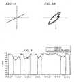
- the IR and red waveformswill have a significant and persistent phase difference if they include venous pulsation, as illustrated by the Lissajous plot of Fig. 3B , which resembles a fairly open ellipse. Shown in Figs. 3A-B are IR and Red waveforms that are AC-coupled and plotted in an x-y plot, with the IR waveform on the x-axis, and the Red waveform on the y-axis. Note that the trace of Fig. 3B does not go through the origin. On the other hand, the IR and Red waveforms will be in-phase if they only include arterial pulses, as illustrated by the Lissajous plot of Fig. 3A . Other artifacts, such as motion and noise may add out-of-phase components whose phase relationship and frequency content is unstable, and not as persistent as the venous pulsation induced phase difference.
- a waveform that is better suited for detecting phase differences between IR and Red waveformsis one that that contains just the waveform corresponding to the fundamental of the pulse rate, such as may be produced by an appropriate filter. Such a waveform is better suited for detecting phase differences between the IR and Red waveform that are of vascular origin.
- the detection of the presence of venous pulsationinvolves detecting persistent phase differences between the IR and Red waveforms.
- the detection of the phase differenceinvolves the detection of the phase difference between the IR and Red waveforms that have been filtered so as to contain just frequencies at or near the algorithm's pulse-rate or harmonics.
- Various techniques for detecting a persistent phase differenceare described below. Using one or a combination of these techniques, one embodiment of the present invention for detecting the presence of venous pulsation includes an algorithm that will:
- Open _ Lissajous _ Axis _ Ratio tmin ⁇ I ⁇ R t 2 + Re d t 2 , I ⁇ R t - 1 2 + Re d t - 1 2 .. I ⁇ R t - N + 1 2 + Re d t - N + 1 2 max ⁇ I ⁇ R t 2 , I ⁇ R t - 1 2 .. I ⁇ R t - N + 1 2
- IR and Redrefer to waveforms that have been processed per step 1 above, and N denotes the number of samples in the time-window.
- this metricis computed periodically (e.g., every second) from the most recent time window (e.g., four seconds) of data. It therefore covers a window of about several pulses.
- a preferred time windowe.g., four-second window
- An alternate embodiment of this metricinvolves only using the Red data in the denominator. The inclusion of the Red data only in the numerator makes this metric more sensitive to out-of-phase waveforms at low saturations, where the Red modulation is larger, than at high saturations.
- this ratiomay be fairly noisy, it may be filtered, clipped and integrated before being used to announce the presence or absence of venous pulsation.
- venous pulsationis reported if Venous_Pulsation_Integral is at least 1.0.
- this venous pulsation detection algorithmhas a response time of tens of seconds, depending on how rapidly the integral changes. This response time is adequate for the clinician's needs, as the body positions and circulatory conditions that create venous pulsation at the sensor site are not likely to change much faster than this. However, the response time can be changed to accommodate likely changes in body positions and/or circulatory conditions that create venous pulsation at the sensor site.
- phase differencemay be quantified by analyzing the cross-correlation function of the two waveforms as a function of a delay interval between them.
- phase differencemay be quantified in the frequency domain by subtracting the phases of the waveforms at a given frequency.
- One way to subtract the phases of two complex numbers, without having to directly compute inverse trigonometric functions,is by taking their complex conjugate, and dividing by the product of their magnitudes.
- a persistent phase-differencemay be detected at a harmonic of the pulse rate, in place of, or in combination with its fundamental.
- Fig. 4which is s a plot 300 of % SpO 2 vs. Time (hr.:min.:sec.) showing periods of venous pulsation during normoxia in a Trendelenburg position (i.e., a supine position on the operating table, which is inclined at varying angles so that the pelvis is higher than the head; used during and after operations in the pelvis or for shock) shows that the method in accordance with the embodiments of the present invention detects all four episodes of venous pulsation created by deliberately placing a normoxic volunteer in a Trendelenburg position.
- the first three episodes 402, 404, and 406were created without a headband, and venous pulsation was detected in 15-25 seconds.
- the final episode 408was created with a headband in place, so that venous pulsation developed more gradually and was announced in about one minute.
- a pulse oximeter having a venous pulsation detection and notification system in accordance with the embodiments of the present inventionis able to identify and notify a clinician of a majority of the low (e.g., 80s-low 90s) SpO 2 readings as those possibly caused by venous pulsations, so that a clinician can take appropriate corrective measures, such as tightening a headband that holds an oximeter sensor against a patient's forehead.
- a pulse oximeter having a venous pulsation detection and notification system in accordance with the embodiments of the present inventionis able to, during a desaturation event (i.e. SpO 2 less than 50%), not provide an indication of a venous pulsation event, and thus help a clinician be certain that the desaturation is real event.
- the present inventionwhich is related to detecting the presence of venous or mixed venous and arterial blood pulsation is tissue, may be embodied in other specific forms without departing from the essential characteristics thereof.
- the present embodimentshave been described in the time-domain, frequency-based methods are equally relevant to the embodiments of the present invention. Accordingly, the foregoing disclosure is intended to be illustrative, but not limiting, of the scope of the invention, which is set forth in the following claims.
Landscapes
- Health & Medical Sciences (AREA)
- Life Sciences & Earth Sciences (AREA)
- Physics & Mathematics (AREA)
- Medical Informatics (AREA)
- Surgery (AREA)
- Biophysics (AREA)
- Pathology (AREA)
- Engineering & Computer Science (AREA)
- Biomedical Technology (AREA)
- Heart & Thoracic Surgery (AREA)
- Veterinary Medicine (AREA)
- Molecular Biology (AREA)
- Public Health (AREA)
- Animal Behavior & Ethology (AREA)
- General Health & Medical Sciences (AREA)
- Optics & Photonics (AREA)
- Spectroscopy & Molecular Physics (AREA)
- Cardiology (AREA)
- Physiology (AREA)
- Measurement Of The Respiration, Hearing Ability, Form, And Blood Characteristics Of Living Organisms (AREA)
- Investigating Or Analysing Materials By Optical Means (AREA)
- Measuring Pulse, Heart Rate, Blood Pressure Or Blood Flow (AREA)
Description
- The present invention relates in general to pulse oximetry, and in particular to the processing of signals generated by a pulse oximeter.
- A pulse oximeter is typically used to measure various blood characteristics including the blood oxygen saturation of hemoglobin in arterial blood and the pulse rate of the patient. Measurement of these characteristics has been accomplished by use of a non-invasive sensor that passes light through a portion of a patient's blood perfused tissue and photo-electrically senses the absorption and scattering of light in such tissue. The amount of light absorbed and scattered is then used to estimate the amount of blood constituent in the tissue using various algorithms known in the art. The "pulse" in pulse oximetry comes from the time varying amount of arterial blood in the tissue during a cardiac cycle. The signal processed from the sensed optical measurement is the familiar plethysmographic waveform, which corresponds with the cyclic attenuation of optical energy through a portion of a patient's blood perfused tissue.
- Various physiological and/or external factors can adversely impact the accuracy and/or the reliability of physiological parameters that are estimated by a pulse oximeter. These undesirable factors are sometimes referred to as artifacts. Venous pulsation is one undesired artifact in pulse oximetry, and may be caused by a patient's medical condition.
- It is therefore desirable that a pulse oximetry system be designed which effectively and accurately detects and/or notifies a clinician of the presence of venous pulsation.
- It is an object of the present invention to provide for a method of detecting the presence of mixed venous and arterial blood pulsation in tissue and a respective pulse oximeter. This object can be achieved by the features as defined in the independent claims. Further enhancements are characterized in the dependent claims.
- The present invention provides a pulse oximeter that has the capability of detecting the presence of venous blood pulsation, or the presence of mixed venous and arterial blood pulsation in tissue.
- Certain aspects of the invention are set out in the independent claims. Certain preferred and optional features are set out in the dependent claims.
- In one embodiment, the present invention provides a method of detecting the presence of mixed venous and arterial blood pulsation in tissue. The method includes receiving first and second electromagnetic radiation signals from a blood perfused tissue portion corresponding to infrared and red wavelengths of light; obtaining a measure of a persistent phase difference between the first and second electromagnetic radiation signals; comparing the measure with a threshold value to form a comparison; detecting the presence or absence of venous pulsation using the comparison; and indicating the presence of venous pulsation if the venous pulsation has been detected.
- The measure of a phase difference may be a measure of the openness of an ellipse on a Lissajous plot formed by comparing the first electromagnetic radiation signal against the second electromagnetic radiation signal.
- In another embodiment, the present invention provides a device for detecting the presence of mixed venous and arterial blood pulsation in tissue. The device includes a module for receiving first and second electromagnetic radiation signals from a blood perfused tissue portion corresponding to infrared and red wavelengths of light; a module for obtaining a measure of a phase difference between the first and second electromagnetic radiation signals; a module for comparing the measure with a threshold value to form a comparison; and a module for detecting the presence or absence of venous pulsation using the comparison.
- For a fuller understanding of the nature and advantages of the embodiments of the present invention, reference should be made to the following detailed description taken in conjunction with the accompanying drawings.
Fig. 1 is a block diagram of an exemplary oximeter.Fig. 2 is a block diagram of the signal processing architecture of a pulse oximeter in accordance with one embodiment of the present invention.Figs. 3A-B are exemplary x-y, or Lissajous, plots of AC-coupled IR and Red pulse waveforms plotted against one another, showing in-phase (Fig. 3A ) and out of phase (Fig. 3B ) IR and Red pulse waveforms.Fig. 4 is a plot of % SpO2 vs. Time (hr.:min.:sec.) showing periods of venous pulsation during normoxia in Trendelenburg position (i.e., a supine position on the operating table, which is inclined at varying angles so that the pelvis is higher than the head; used during and after operations in the pelvis or for shock).- The methods and systems in accordance with the embodiments of the present invention are directed towards detecting the presence or absence of venous or mixed venous and arterial pulsation in a blood perfused tissue. The invention is particularly applicable to and will be explained by reference to measurements of oxygen saturation of hemoglobin in arterial blood and pulse or heart rate, as in pulse oximeter monitors and pulse oximetry sensors.
- A typical pulse oximeter measures two physiological parameters, percent oxygen saturation of arterial blood hemoglobin (SpO2 or sat) and pulse rate. Oxygen saturation can be estimated using various techniques. In one common technique, the photocurrent generated by the photo-detector is conditioned and processed to determine the ratio of modulation ratios (ratio of ratios) of the red to infrared (IR) signals. This modulation ratio has been observed to correlate well to arterial oxygen saturation. Pulse oximeters and sensors may be empirically calibrated by measuring the modulation ratio over a range of in vivo measured arterial oxygen saturations (SaO2) on a set of patients, healthy volunteers, or animals. The observed correlation is used in an inverse manner to estimate blood oxygen saturation (SpO2) based on the measured value of modulation ratios of a patient. The estimation of oxygen saturation using modulation ratios is described in
U.S. Patent No. 5,853,364 , entitled "METHOD AND APPARATUS FOR ESTIMATING PHYSIOLOGICAL PARAMETERS USING MODEL-BASED ADAPTIVE FILTERING," issued December 29, 1998, andU.S. Patent No. 4,911,167 , entitled "METHOD AND APPARATUS FOR DETECTING OPTICAL PULSES," issued March 27, 1990. The relationship between oxygen saturation and modulation ratio is described, for example, inU.S. Patent No. 5,645,059 , entitled "MEDICAL SENSOR WITH MODULATED ENCODING SCHEME," issued July 8, 1997. Most pulse oximeters extract the plethysmographic signal having first determined saturation or pulse rate, both of which are susceptible to interference. Fig. 1 is a block diagram of one embodiment of a pulse oximeter that may be configured to implement the embodiments of the present invention. The venous pulsation detection embodiments of the present invention may be implemented as a data processing algorithm that is executed by themicroprocessor 122, described below. Light fromlight source 110 passes into a blood perfusedtissue 112, and is scattered and detected byphotodetector 114. Asensor 100 containing the light source and photodetector may also contain anencoder 116 which provides signals indicative of the wavelength oflight source 110 to allow the oximeter to select appropriate calibration coefficients for calculating oxygen saturation.Encoder 116 may, for instance, be a resistor.Sensor 100 is connected to apulse oximeter 120. The oximeter includes amicroprocessor 122 connected to aninternal bus 124. Also connected to the bus is aRAM memory 126 and adisplay 128. A time processing unit (TPU) 130 provides timing control signals tolight drive circuitry 132 which controls whenlight source 110 is illuminated, and if multiple light sources are used, the multiplexed timing for the different light sources. TPU 130 also controls the gating-in of signals fromphotodetector 114 through anamplifier 133 and aswitching circuit 134. These signals are sampled at the proper time, depending upon which of multiple light sources is illuminated, if multiple light sources are used. The received signal is passed through anamplifier 136, alow pass filter 138, and an analog-to-digital converter 140. The digital data is then stored in a queued serial module (QSM) 142, for later downloading toRAM 126 as QSM 142 fills up. In one embodiment, there may be multiple parallel paths of separate amplifier, filter and A/D converters for multiple light wavelengths or spectra received.- Based on the value of the received signals corresponding to the light received by
photodetector 114,microprocessor 122 will calculate the oxygen saturation using various algorithms. These algorithms require coefficients, which may be empirically determined, corresponding to, for example, the wavelengths of light used. These are stored in aROM 146. In a two-wavelength system, the particular set of coefficients chosen for any pair of wavelength spectra is determined by the value indicated byencoder 116 corresponding to a particular light source in aparticular sensor 100. In one embodiment, multiple resistor values may be assigned to select different sets of coefficients. In another embodiment, the same resistors are used to select from among the coefficients appropriate for an infrared source paired with either a near red source or far red source. The selection between whether the near red or far red set will be chosen can be selected with a control input fromcontrol inputs 154.Control inputs 154 may be, for instance, a switch on the pulse oximeter, a keyboard, or a port providing instructions from a remote host computer. Furthermore, any number of methods or algorithms may be used to determine a patient's pulse rate, oxygen saturation or any other desired physiological parameter. - The brief description of an exemplary pulse oximeter set forth above, serves as a basis for describing the methods for detecting the presence of venous pulsation, which are described below. The embodiments of the present invention, which are used to detect and/or indicate the presence of venous pulsation or mixed venous and arterial pulsation are described below in conjunction with the block diagram of
Fig. 2 . - The embodiments of the present invention may be implemented as a part of a larger signal processing system used to process optical signals for the purposes of operating a pulse oximeter. Such a signal processing system is shown in
Fig. 2 , which is a block diagram 200 of a signal processing architecture of a pulse oximeter in accordance with one embodiment of the present invention. Thesignal processing architecture 200 in accordance with the embodiments of the present invention may be implemented as a software algorithm that is executed by a processor of a pulse oximeter. In addition to calculating oxygen saturation and pulse rate, thesystem 200 measures various signal metrics that are used to determine filter weighting coefficients. Signal metrics are things that indicate if a pulse is likely a plethysmograph or noise. Signal metrics may be related to, for example, frequency (is it in the range of a human heart rate), shape (is it shaped like a cardiac pulse), rise time, etc. The system shown inFig. 2 calculates both the oxygen saturation, and the pulse rate, as well as detecting venous pulsation and sensor off and lost pulse conditions, which are described separately below. Block 202 represents the operation of the Signal Conditioning block. The digitized red and IR signals or waveforms are received and are conditioned in this block by: (1) taking the 1st derivative to get rid of baseline shift, (2) low pass filtering with fixed coefficients, and (3) dividing by a DC value to preserve the ratio. The function of the Signal Conditioning subsystem is to emphasize the higher frequencies that occur in the human plethysmograph and to attenuate low frequencies in which motion artifact is usually concentrated. The Signal Conditioning subsystem selects its filter coefficients (wide or narrow band) based on hardware characteristics identified during initialization. Inputs to block 202 are digitized red and IR signals, and its outputs are pre-processed red and IR signals.Block 204 represents the operation of the Pulse Identification and Qualification block. The low pass filtered digitized red and IR signals are provided to this block to identify pulses, and qualify them as likely arterial pulses. This is done using a pre-trained neural network, and is primarily done on the IR signal. The pulse is identified by examining its amplitude, shape and frequency. An input to this block is the average pulse period fromblock 208. This function changes the upfront qualification using the pulse rate. The output ofblock 204 indicates the degree of arrhythmia and individual pulse quality. Inputs to block 204 are: (1) pre-processed red and IR signals, (2) Average pulse period, and (3) lowpass waveforms from the low pass filter. Outputs fromblock 204 include: (1) degree of arrhythmia, (2) pulse amplitude variations, (3) individual pulse quality, (4) pulse beep notification, and (5) qualified pulse periods and age.Block 206 is used to compute signal quality metrics. This block (block 206) determines the pulse shape (e.g., derivative skew), period variability, pulse amplitude and variability, Ratio of Ratios variability, and frequency content relative to pulse rate. Inputs to block 206 include: (1) raw digitized red and IR signals, (2) degree of arrhythmia, individual pulse quality, pulse amplitude variation , (3) pre-processed red and IR signals, and (4) average pulse period. Outputs fromblock 206 include: (1) lowpass and ensemble averaging filter weights, (2) metrics for sensor off detector, (3) normalized pre-processed waveforms, and (4) percent modulation.Block 208 computes average pulse periods. This block (block 208) calculates the average pulse period from the pulses received. Inputs to block 208 include: qualified pulse periods and age. An output fromblock 208 includes the average pulse period.Block 210 represents the functioning of the lowpass filter and ensemble averaging subsystem. Block 210 low pass filters and ensemble averages normalized and preprocessed waveforms processed byblock 206. The weights for the low pass filter are determined by theSignal Metrics block 206. The signal is also ensemble averaged (this attenuates frequencies other than those of interest near the pulse rate and its harmonics), with the ensemble averaging filter weights also determined bySignal Metrics block 206. Less weight is assigned if the signal is flagged as degraded. More weight is assigned if the signal is flagged as arrhythmic because ensemble-averaging is not appropriate during arrhythmia. Red and IR waveforms are processed separately, but with the same filtering weights. The filtering is delayed (e.g., approximately one second) to allow the signal metrics to be calculated first.- The filters use continuously variable weights. If samples are not to be ensemble-averaged, then the weighting for the previous filtered samples is set to zero in the weighted average, and the new samples are still processed through the signal processing algorithm. This block tracks the age of the signal and/or the accumulated amount of filtering (e.g., sum of response times and delays in processing). Too old a result will be flagged, if good pulses haven't been detected for a while. The inputs to block 210 include: (1) normalized pre-processed red and IR signals, (2) average pulse period, (3) low pass filter weights and ensemble averaging filter weights, (4) ECG triggers, if available, and (5) IR fundamental, for zero-crossing triggers. Outputs from
block 210 include: (1) filtered red and IR signals, and (2) age. Block 212 represents operations that estimate the ratio-of-ratios variance for the filtered waveforms and calculate averaging weights. The variable weighting for the filter is controlled by the ratio-of-ratios variance. The effect of this variable-weight filtering is that the ratio-of-ratios changes slowly as artifact increases and changes quickly as artifact decreases. The subsystem has two response modes, including fast and normal modes. For example, filtering in the fast mode targets an age metric of 3 seconds, and the target age may be 5 seconds in the normal mode. In the fast mode, the minimum weighting of the current value is clipped at a higher level. In other words, a low weight is assigned to the newest ratio-of-ratios calculation if there is noise present, and a high weight if no noise is present. The inputs to block 212 include: (1) filtered red and IR signals and age, (2) calibration coefficients, and (3) response mode (e.g., user speed settings). Outputs fromblock 212 include an averaging weight for ratio-of-ratios calculation. The averaging weight is used as an input to block 214 along with filtered IR and Red waveforms to calculate averaged ratio of ratios and age.Block 216 represents operations that calculate oxygen saturation. Saturation is calculated using an algorithm with the calibration coefficients and averaged ratio of ratios. Inputs to block 116 include: (1) Averaged Ratio-of-Ratios, and (2) calibration coefficients. An output fromblock 216 is the oxygen saturation value.- Block 218 low pass filters and ensemble averages the signal(s) conditioned by
block 202, for the pulse rate identification. The weights for the low pass filter are determined by theSignal Metrics block 206. The signal is also ensemble averaged (this attenuates frequencies other than those of interest near the pulse rate and its harmonics), with the ensemble averaging filter weights also determined bySignal Metrics block 206. Less weight is assigned if the signal is flagged as degraded. More weight is assigned if the signal is flagged as arrhythmic because ensemble-averaging is not appropriate during arrhythmia. Red and IR are processed separately, but with.the same filtering weights. The filtering is delayed (e.g., approximately one second) to allow the signal metrics to be calculated first. - The filters use continuously variable weights. If samples are not to be ensemble-averaged, then the weighting for the previous filtered samples is set to zero in the weighted average, and the new samples are still processed through the signal processing algorithm. This block (block 218) tracks the age of the signal and/or the accumulated amount of filtering (sum of response times and delays in processing). Too old a result will be flagged (if good pulses haven't been detected for awhile). Inputs to block 218 include: (1) pre-processed red and IR signals, (2) average pulse period, (3) lowpass filter weights and ensemble averaging filter weights, (4) ECG triggers, if available, and (5) IR fundamental, for zero-crossing triggers. Outputs from
block 218 include: (1) filtered red and IR signals and (2) age. Block 220, or the Filtered Pulse Identification and Qualification block, calculates the pulse periods from the filtered waveforms, and its results are used only when a pulse is disqualified byblock 204. Inputs to block 220 include: (1) filtered red and IR signals and age, (2) average pulse period, (3) front end ID or noise floor, (4) and the kind or type of sensor that is used to detect the IR and Red energies. Output fromblock 220 includes qualified pulse periods and age.Block 222, or the Average Pulse Periods and Calculate Pulse Rate block, calculates the pulse rate and average pulse period. This block (block 222) receives qualified pulse periods and age as inputs and provides (1) average pulse period and (2) pulse rate as outputs.Block 224, or the Detect Venous Pulsation block receives as inputs the pre-processed red and IR signals and age fromBlock 202, and pulse rate and provides an indication of venous pulsation as an output. Block 224 also provides an IR fundamental waveform in the time domain using a single-tooth comb filter which is output to the Ensemble Averaging filters (e.g., block 210 and 218). Inputs to block 224 include: (1) filtered red and IR signals and age and (2) pulse rate. Outputs fromblock 124 include: an indication of venous pulsation and IR fundamental. In one embodiment, block 224 measures the "openness" of an IR-Red Lissajous plot to determine the whether a flag (e.g., Venous_Pulsation) should be set. The output flag (e.g., Venous_Pulsation) is updated periodically (e.g., every second). In addition, the IR fundamental waveform is output to the Ensemble Averaging filters.Block 226, or the Detect Sensor-Off and Loss of Pulse Amplitude block, uses a pre-trained neural net to determine whether the sensor is off the surface of the blood-perfused tissue, for example, of a patient. The inputs to the neural net are metrics that quantify several aspects of the behavior of the IR and Red values over the last several seconds. Samples are ignored by many of thesystem 200's subsystems while the signal state is either not indicative of a pulse being present, or indicative that a sensor is not on a monitoring site (e.g., Pulse Present, Disconnect, Pulse Lost, Sensor May be Off, and Sensor Off). Inputs to block 226 include: (1) signal quality metrics, and (2) the oximeter's LED brightness, amplifier gain, and (3) an ID indicating the oximeter's hardware configuration. Outputs fromblock 226 include a signal state including sensor-off indication.- In the
architecture 200 described above, the function ofblock 226, Pulse lost and Pulse Search indications, may be derived using information from several parts of the signal processing architecture. In addition, the signal processing architecture will not use the received IR and red waveforms to compute oxygen saturation or pulse rate if a valid sensor is not connected, or if the Sensor-Off or Loss of Pulse Amplitude are detected by the signal processing architecture. - The brief description of an embodiment of a pulse oximeter signal processing architecture in accordance with the present invention, set forth above, serves as a basis for describing the methods and devices that are directed towards detecting the presence or absence of venous or mixed venous and arterial pulsation in a blood perfused tissue, as is generally indicated by
block 224 above. - Venous pulsation is an undesirable artifact in pulse oximetry. Venous pulsation is particularly common on the head or forehead, where the vascular anatomy lacks valves to prevent venous blood from backing up and pooling. Venous pulsation may be caused by the patient's medical condition, or during surgical interventions that interfere with venous return. The effects of venous pulsation may include: 1) oxygen saturation (e.g., SpO2) readings reflecting a mix of venous and arterial blood, which would be substantially lower than the arterial oxygen saturation, thus resulting in incorrectly low oxygen saturation measurements, and 2) pulse rate readings that are double or even triple the patient's pulse rate, due to the prominent harmonics in the venous pressure wave. In addition, in extreme cases, it is possible that an oximeter would fail to acquire oxygen saturation and/or pulse rate measurements.
- Unlike motion artifacts that may not be present at all times, (e.g., they come and go), venous pulsation can continue uninterrupted for hours. While the side effects of venous pulsation are highly visible to the clinician, their cause may not be. Venous pulsation at the site of an oximetry sensor may be mitigated by applying pressure to the site, such as with a headband.
- The embodiments of the present invention provide methodologies, including software-based methods for detecting the venous pulsation artifact. The detection of the presence of venous pulsation enables an oximeter to display a troubleshooting message to a clinician, who could then address and/or correct the problem.
- Mixed venous and arterial pulses may be distinguished from arterial pulses due to the following properties, described below. First, venous blood has a lower saturation than arterial blood. Normoxic subjects (i.e., healthy subjects breathing air at sea level) who place their head significantly below their heart may readily create SpO2 readings near 80% at the forehead if no pressure is applied to the sensor site. Second, the venous pulse occurs after the arterial pulse, and has a different shape.
- Due to these properties, the IR and red waveforms will have a significant and persistent phase difference if they include venous pulsation, as illustrated by the Lissajous plot of
Fig. 3B , which resembles a fairly open ellipse. Shown inFigs. 3A-B are IR and Red waveforms that are AC-coupled and plotted in an x-y plot, with the IR waveform on the x-axis, and the Red waveform on the y-axis. Note that the trace ofFig. 3B does not go through the origin. On the other hand, the IR and Red waveforms will be in-phase if they only include arterial pulses, as illustrated by the Lissajous plot ofFig. 3A . Other artifacts, such as motion and noise may add out-of-phase components whose phase relationship and frequency content is unstable, and not as persistent as the venous pulsation induced phase difference. - A waveform that is better suited for detecting phase differences between IR and Red waveforms is one that that contains just the waveform corresponding to the fundamental of the pulse rate, such as may be produced by an appropriate filter. Such a waveform is better suited for detecting phase differences between the IR and Red waveform that are of vascular origin.
- In one embodiment, the detection of the presence of venous pulsation involves detecting persistent phase differences between the IR and Red waveforms. As set forth above, preferably the detection of the phase difference, involves the detection of the phase difference between the IR and Red waveforms that have been filtered so as to contain just frequencies at or near the algorithm's pulse-rate or harmonics. Various techniques for detecting a persistent phase difference are described below. Using one or a combination of these techniques, one embodiment of the present invention for detecting the presence of venous pulsation includes an algorithm that will:
- 1. AC-couple the IR and Red waveforms, and preferably filter them so as to pass only frequencies at or near the pulse rate.
- 2. Over a time-window of at least one complete pulse, quantify the phase difference by quantifying the "openness of the ellipse" as (minimum distance from the origin) / (maximum distance from the x-axis). Alternatively the denominator of this ratio may be the maximum distance from the origin. A longer time-window will increase the likelihood that motion artifact or Gaussian noise would eventually produce samples near the origin, and reduce the likelihood of falsely reporting venous pulsation.
- 3. Integrate the difference between this open-ellipse metric and a threshold. The threshold controls how open the ellipse must be to eventually notify the user of venous pulsation. The threshold preferably varies with the calculated SpO2, because venous pulsation is less likely to be occurring if high SpO2 values are being calculated.
- 4. If desired, to control how long venous pulsation must persist before notifying the user, clip the integral at pre-determined high and/or low limits, and report venous pulsation whenever a predetermined integral threshold in between the high and/or low limits is exceeded.
- The description below, discloses how the "how open is the ellipse" metric is quantified. The metric is quantified as follows:

Where IR and Red refer to waveforms that have been processed perstep 1 above, and N denotes the number of samples in the time-window. - In one embodiment, this metric is computed periodically (e.g., every second) from the most recent time window (e.g., four seconds) of data. It therefore covers a window of about several pulses. A preferred time window (e.g., four-second window) assures that the waveforms have multiple pulse periods in which to come close to the origin if the waveforms are actually in-phase. An alternate embodiment of this metric involves only using the Red data in the denominator. The inclusion of the Red data only in the numerator makes this metric more sensitive to out-of-phase waveforms at low saturations, where the Red modulation is larger, than at high saturations.
- Because this ratio may be fairly noisy, it may be filtered, clipped and integrated before being used to announce the presence or absence of venous pulsation. The filtering, clipping and integration are described below, such that:


where' denotes the value from one second ago.


where: - Saturation is the oxygen saturation value.
- Open_Lissajous_Threshold is the threshold defined above in step 3 of the algorithm.
- In one embodiment, using the above approach, venous pulsation is reported ifVenous_Pulsation_Integral is at least 1.0.
- The threshold and weights in the above equations are developed empirically, and may change depending on various conditions. It is preferred that this venous pulsation detection algorithm has a response time of tens of seconds, depending on how rapidly the integral changes. This response time is adequate for the clinician's needs, as the body positions and circulatory conditions that create venous pulsation at the sensor site are not likely to change much faster than this. However, the response time can be changed to accommodate likely changes in body positions and/or circulatory conditions that create venous pulsation at the sensor site.
- In addition to the technique described above for quantifying a phase difference, many alternative techniques of detecting a persistent phase difference in accordance with the present invention are also available. For instance, the phase difference may be quantified by analyzing the cross-correlation function of the two waveforms as a function of a delay interval between them. Or the phase difference may be quantified in the frequency domain by subtracting the phases of the waveforms at a given frequency. One way to subtract the phases of two complex numbers, without having to directly compute inverse trigonometric functions, is by taking their complex conjugate, and dividing by the product of their magnitudes. Or alternatively, a persistent phase-difference may be detected at a harmonic of the pulse rate, in place of, or in combination with its fundamental.
Fig. 4 , which is s a plot 300 of % SpO2 vs. Time (hr.:min.:sec.) showing periods of venous pulsation during normoxia in a Trendelenburg position (i.e., a supine position on the operating table, which is inclined at varying angles so that the pelvis is higher than the head; used during and after operations in the pelvis or for shock) shows that the method in accordance with the embodiments of the present invention detects all four episodes of venous pulsation created by deliberately placing a normoxic volunteer in a Trendelenburg position. The first three episodes 402, 404, and 406 were created without a headband, and venous pulsation was detected in 15-25 seconds. The final episode 408 was created with a headband in place, so that venous pulsation developed more gradually and was announced in about one minute.- A pulse oximeter having a venous pulsation detection and notification system in accordance with the embodiments of the present invention is able to identify and notify a clinician of a majority of the low (e.g., 80s-low 90s) SpO2 readings as those possibly caused by venous pulsations, so that a clinician can take appropriate corrective measures, such as tightening a headband that holds an oximeter sensor against a patient's forehead. Likewise, a pulse oximeter having a venous pulsation detection and notification system in accordance with the embodiments of the present invention is able to, during a desaturation event (i.e. SpO2 less than 50%), not provide an indication of a venous pulsation event, and thus help a clinician be certain that the desaturation is real event.
- Accordingly, as will be understood by those of skill in the art, the present invention which is related to detecting the presence of venous or mixed venous and arterial blood pulsation is tissue, may be embodied in other specific forms without departing from the essential characteristics thereof. For example, while the present embodiments have been described in the time-domain, frequency-based methods are equally relevant to the embodiments of the present invention. Accordingly, the foregoing disclosure is intended to be illustrative, but not limiting, of the scope of the invention, which is set forth in the following claims.


Claims (16)
- A method of detecting the presence of mixed venous and arterial blood pulsation in tissue, comprising:receiving first and second electromagnetic radiation signals from a blood perfused tissue portion corresponding to infrared and red wavelengths of light;obtaining a measure of a persistent phase difference between said first and second electromagnetic radiation signals;comparing said measure with a threshold value to form a comparison;detecting the presence or absence of venous pulsation using said comparison; andindicating the presence of venous pulsation if said venous pulsation has been detected.
- The method of claim 1 further comprising filtering said first and second electromagnetic radiation signals before said obtaining said measure, to pass portions of said first and second electromagnetic radiation signals having frequencies at or near the pulse rate or harmonics of the pulse rate of said blood perfused tissue.
- The method of claim 1 or claim 2 wherein said obtaining a measure of a persistent phase difference comprises integrating said measure of a phase difference over a time period.
- The method according to one of the preceding claims, wherein said obtaining a measure of a persistent phase difference comprises obtaining a measure of the openness of an ellipse on a Lissajous plot formed by comparing the first electromagnetic radiation signal against the second electromagnetic radiation signal.
- The method according to claim 1 or claim 2, wherein said obtaining a measure of a persistent phase difference comprises analyzing a cross-correlation function of said first and second electromagnetic radiation signals, as a function of a delay interval between them.
- The method according to claim 1 or claim 2, wherein said obtaining a measure of a persistent phase difference comprises a frequency domain analysis and subtracting the phases of said first and second electromagnetic radiation signals at a frequency.
- The method of claim 6 wherein said subtracting the phases of said first and second electromagnetic radiation signals comprises taking the complex conjugate of said first and second electromagnetic radiation signals, and dividing said complex conjugate by the product of the magnitudes of said first and second electromagnetic radiation signals.
- The method according to one of the preceding claims, wherein said obtaining a measure of a persistent phase difference comprises obtaining said measure of a persistent phase difference at or near a fundamental or at or near a harmonic of a pulse rate of said blood perfused tissue.
- A device for detecting the presence of mixed venous and arterial blood pulsation in tissue, comprising:means (100) configured for receiving first and second electromagnetic radiation signals from a blood perfused tissue portion corresponding to infrared and red wavelengths of light;means (224) configured for obtaining a measure of a persistent phase difference between said first and second electromagnetic radiation signals;means (224) configured for comparing said measure with a threshold value to form a comparison;means (224) configured for detecting the presence or absence of venous pulsation using said comparison; andmeans configured for indicating the presence of venous pulsation if said venous pulsation has been detected.
- The device of claim 9 further comprising a filter (202) configured for filtering said first and second electromagnetic radiation signals before obtaining said measure, to pass portions of said first and second electromagnetic radiation signals having frequencies at or near the pulse rate or harmonics of the pulse rate of said blood perfused tissue.
- The device of claim 9 or claim 10, wherein said means (224) configured for obtaining a measure of a persistent phase difference comprises means for integrating said measure of a phase difference over a time period.
- The device of any one of claims 9 to 11 wherein said means (224) configured for obtaining a measure of a persistent phase difference is configured for obtaining a measure of the openness of an ellipse on a Lissajous plot formed by comparing the first electromagnetic radiation signal against the second electromagnetic radiation signal.
- The device of claim 9 or claim 10, wherein said means (224) configured for obtaining a measure of a persistent phase difference is configured for analyzing a cross-correlation function of said first and second electromagnetic radiation signals, as a function of a delay interval between them.
- The device of claim 9 or claim 10, wherein said means (224) configured for obtaining a measure of a persistent phase difference is configured for a frequency domain analysis and for subtracting the phases of said first and second electromagnetic radiation signals at a frequency.
- The device of claim 14 wherein said means configured for subtracting the phases of said first and second electromagnetic radiation signals is configured for taking the complex conjugate of said first and second electromagnetic radiation signals, and dividing said complex conjugate by the product of the magnitudes of said first and second electromagnetic radiation signals.
- The device of any one of claims 9 to 15, wherein said means configured for obtaining a measure of a persistent phase difference is configured for obtaining said measure of a persistent phase difference at or near a fundamental or at or near a harmonic of a pulse rate of said blood perfused tissue.
Applications Claiming Priority (2)
| Application Number | Priority Date | Filing Date | Title |
|---|---|---|---|
| US10/796,584US8611977B2 (en) | 2004-03-08 | 2004-03-08 | Method and apparatus for optical detection of mixed venous and arterial blood pulsation in tissue |
| PCT/US2005/007407WO2005087096A1 (en) | 2004-03-08 | 2005-03-07 | Method and apparatus for optical detection of mixed venous and arterial blood pulsation in tissue |
Publications (2)
| Publication Number | Publication Date |
|---|---|
| EP1740091A1 EP1740091A1 (en) | 2007-01-10 |
| EP1740091B1true EP1740091B1 (en) | 2015-05-06 |
Family
ID=34912589
Family Applications (1)
| Application Number | Title | Priority Date | Filing Date |
|---|---|---|---|
| EP05724862.7AExpired - LifetimeEP1740091B1 (en) | 2004-03-08 | 2005-03-07 | Method and apparatus for optical detection of mixed venous and arterial blood pulsation in tissue |
Country Status (9)
| Country | Link |
|---|---|
| US (1) | US8611977B2 (en) |
| EP (1) | EP1740091B1 (en) |
| JP (1) | JP2007527773A (en) |
| KR (1) | KR20070002036A (en) |
| CN (1) | CN1933772A (en) |
| AU (1) | AU2005221653A1 (en) |
| CA (1) | CA2557306C (en) |
| MX (1) | MXPA06010304A (en) |
| WO (1) | WO2005087096A1 (en) |
Families Citing this family (46)
| Publication number | Priority date | Publication date | Assignee | Title |
|---|---|---|---|---|
| GB0423289D0 (en)* | 2004-10-20 | 2004-11-24 | Huntleigh Technology Plc | DVT detection |
| US7647083B2 (en) | 2005-03-01 | 2010-01-12 | Masimo Laboratories, Inc. | Multiple wavelength sensor equalization |
| US20070100220A1 (en) | 2005-10-28 | 2007-05-03 | Baker Clark R Jr | Adjusting parameters used in pulse oximetry analysis |
| US7706852B2 (en) | 2006-01-30 | 2010-04-27 | Nellcor Puritan Bennett Llc | System and method for detection of unstable oxygen saturation |
| WO2007147069A2 (en)* | 2006-06-14 | 2007-12-21 | Advanced Brain Monitoring, Inc. | Method for measuring central venous pressure or respiratory effort |
| US8123695B2 (en) | 2006-09-27 | 2012-02-28 | Nellcor Puritan Bennett Llc | Method and apparatus for detection of venous pulsation |
| US8696593B2 (en)* | 2006-09-27 | 2014-04-15 | Covidien Lp | Method and system for monitoring intracranial pressure |
| US7680522B2 (en) | 2006-09-29 | 2010-03-16 | Nellcor Puritan Bennett Llc | Method and apparatus for detecting misapplied sensors |
| US8265723B1 (en) | 2006-10-12 | 2012-09-11 | Cercacor Laboratories, Inc. | Oximeter probe off indicator defining probe off space |
| US8109882B2 (en)* | 2007-03-09 | 2012-02-07 | Nellcor Puritan Bennett Llc | System and method for venous pulsation detection using near infrared wavelengths |
| US8229530B2 (en)* | 2007-03-09 | 2012-07-24 | Nellcor Puritan Bennett Llc | System and method for detection of venous pulsation |
| US8221326B2 (en)* | 2007-03-09 | 2012-07-17 | Nellcor Puritan Bennett Llc | Detection of oximetry sensor sites based on waveform characteristics |
| EP2139383B1 (en) | 2007-03-27 | 2013-02-13 | Masimo Laboratories, Inc. | Multiple wavelength optical sensor |
| US8374665B2 (en) | 2007-04-21 | 2013-02-12 | Cercacor Laboratories, Inc. | Tissue profile wellness monitor |
| EP2142097B1 (en)* | 2007-05-02 | 2019-01-09 | Sensitive Pty Ltd | Non-invasive measurement of blood oxygen saturation |
| US8352004B2 (en)* | 2007-12-21 | 2013-01-08 | Covidien Lp | Medical sensor and technique for using the same |
| US8346328B2 (en)* | 2007-12-21 | 2013-01-01 | Covidien Lp | Medical sensor and technique for using the same |
| US8532932B2 (en) | 2008-06-30 | 2013-09-10 | Nellcor Puritan Bennett Ireland | Consistent signal selection by signal segment selection techniques |
| US8862194B2 (en) | 2008-06-30 | 2014-10-14 | Covidien Lp | Method for improved oxygen saturation estimation in the presence of noise |
| US20100004518A1 (en) | 2008-07-03 | 2010-01-07 | Masimo Laboratories, Inc. | Heat sink for noninvasive medical sensor |
| US8515509B2 (en) | 2008-08-04 | 2013-08-20 | Cercacor Laboratories, Inc. | Multi-stream emitter for noninvasive measurement of blood constituents |
| US8478538B2 (en)* | 2009-05-07 | 2013-07-02 | Nellcor Puritan Bennett Ireland | Selection of signal regions for parameter extraction |
| US9839381B1 (en) | 2009-11-24 | 2017-12-12 | Cercacor Laboratories, Inc. | Physiological measurement system with automatic wavelength adjustment |
| WO2011069122A1 (en) | 2009-12-04 | 2011-06-09 | Masimo Corporation | Calibration for multi-stage physiological monitors |
| US8898037B2 (en)* | 2010-04-28 | 2014-11-25 | Nellcor Puritan Bennett Ireland | Systems and methods for signal monitoring using Lissajous figures |
| NZ605443A (en) | 2010-07-09 | 2014-11-28 | St Vincents Hospital Melbourne Ltd | Non-invasive measurement of blood oxygen saturation |
| US20130066175A1 (en)* | 2011-09-09 | 2013-03-14 | Nellcor Puritan Bennett Ireland | Venous oxygen saturation systems and methods |
| US8852095B2 (en) | 2011-10-27 | 2014-10-07 | Covidien Lp | Headband for use with medical sensor |
| US8870783B2 (en) | 2011-11-30 | 2014-10-28 | Covidien Lp | Pulse rate determination using Gaussian kernel smoothing of multiple inter-fiducial pulse periods |
| US9138181B2 (en) | 2011-12-16 | 2015-09-22 | Covidien Lp | Medical sensor for use with headband |
| US9370308B2 (en) | 2012-05-01 | 2016-06-21 | Nellcor Puritan Bennett Ireland | Angle distribution technique for analyzing a physiological sensor signal |
| CN103093132A (en)* | 2013-01-17 | 2013-05-08 | 谢幼宸 | Identification and authentication system based on hand palm back vein |
| US10215818B2 (en)* | 2013-04-23 | 2019-02-26 | Koninklijke Philips N.V. | Single coaxial interface for magnetic resonace (MR) coils |
| CN103519809B (en)* | 2013-10-22 | 2015-11-04 | 深圳先进技术研究院 | Oxygen metabolism parameter estimation method and system |
| US10478078B2 (en) | 2013-12-12 | 2019-11-19 | Koninklijke Philips N.V. | Device and method for determining vital signs of a subject |
| US10149624B2 (en)* | 2014-11-06 | 2018-12-11 | Koninklijke Philips N.V. | Method and device for measuring intracranial pressure, ICP, in a subject |
| US10342466B2 (en) | 2015-03-24 | 2019-07-09 | Covidien Lp | Regional saturation system with ensemble averaging |
| JP6558343B2 (en)* | 2016-10-17 | 2019-08-14 | オムロン株式会社 | Biosensor |
| WO2018078728A1 (en)* | 2016-10-25 | 2018-05-03 | パイオニア株式会社 | Fluid measuring device |
| JP6794219B2 (en)* | 2016-10-27 | 2020-12-02 | 日本光電工業株式会社 | Medical photometers and control methods for medical photometers |
| JP6857484B2 (en)* | 2016-10-27 | 2021-04-14 | 日本光電工業株式会社 | Medical photometer and control method of medical photometer |
| JP7020820B2 (en)* | 2017-08-29 | 2022-02-16 | 日本光電工業株式会社 | Pulse oximeter |
| ES3015552T3 (en) | 2018-07-16 | 2025-05-06 | Bbi Medical Innovations Llc | Perfusion and oxygenation measurement |
| CN113030813B (en)* | 2021-02-26 | 2022-02-22 | 厦门大学 | Magnetic resonance T2Quantitative imaging method and system |
| CN116649969B (en)* | 2023-07-25 | 2023-10-10 | 苏州晟智医疗科技有限公司 | Blood oxygen saturation measuring device, apparatus and storage medium |
| WO2025128655A1 (en)* | 2023-12-12 | 2025-06-19 | Google Llc | System and method for detection of pulselessness using a wearable computing device |
Citations (1)
| Publication number | Priority date | Publication date | Assignee | Title |
|---|---|---|---|---|
| US5662106A (en)* | 1993-03-26 | 1997-09-02 | Nellcor Incorporated | Oximeter with motion detection for alarm modification |
Family Cites Families (94)
| Publication number | Priority date | Publication date | Assignee | Title |
|---|---|---|---|---|
| US3638640A (en) | 1967-11-01 | 1972-02-01 | Robert F Shaw | Oximeter and method for in vivo determination of oxygen saturation in blood using three or more different wavelengths |
| US4911167A (en) | 1985-06-07 | 1990-03-27 | Nellcor Incorporated | Method and apparatus for detecting optical pulses |
| US4936679A (en) | 1985-11-12 | 1990-06-26 | Becton, Dickinson And Company | Optical fiber transducer driving and measuring circuit and method for using same |
| DE3877894T2 (en) | 1987-11-02 | 1993-06-24 | Sumitomo Electric Industries | ORGANIC LIGHT MEASURING PROBE. |
| US4854699A (en) | 1987-11-02 | 1989-08-08 | Nippon Colin Co., Ltd. | Backscatter oximeter |
| US4960126A (en) | 1988-01-15 | 1990-10-02 | Criticare Systems, Inc. | ECG synchronized pulse oximeter |
| JPH0288041A (en) | 1988-09-24 | 1990-03-28 | Misawahoomu Sogo Kenkyusho:Kk | Finger tip pulse wave sensor |
| US5873821A (en) | 1992-05-18 | 1999-02-23 | Non-Invasive Technology, Inc. | Lateralization spectrophotometer |
| US5564417A (en) | 1991-01-24 | 1996-10-15 | Non-Invasive Technology, Inc. | Pathlength corrected oximeter and the like |
| EP0374668A3 (en) | 1988-12-16 | 1992-02-05 | A.W. Faber - Castell GmbH & Co. | Fluorescent marking fluid |
| US5028787A (en) | 1989-01-19 | 1991-07-02 | Futrex, Inc. | Non-invasive measurement of blood glucose |
| US6183414B1 (en) | 1999-04-26 | 2001-02-06 | Michael S. Wysor | Technique for restoring plasticity to tissues of a male or female organ |
| JPH0315502U (en) | 1989-06-28 | 1991-02-15 | ||
| US5483646A (en) | 1989-09-29 | 1996-01-09 | Kabushiki Kaisha Toshiba | Memory access control method and system for realizing the same |
| EP0613653B1 (en) | 1990-02-15 | 1996-11-13 | Hewlett-Packard GmbH | Method for non-invasive measurement of oxygen saturation |
| IE77034B1 (en) | 1990-06-27 | 1997-11-19 | Futrex Inc | Non-invasive masurement of blood glucose |
| US6246894B1 (en) | 1993-02-01 | 2001-06-12 | In-Line Diagnostics Corporation | System and method for measuring blood urea nitrogen, blood osmolarity, plasma free hemoglobin and tissue water content |
| MX9702434A (en) | 1991-03-07 | 1998-05-31 | Masimo Corp | Signal processing apparatus. |
| US5490505A (en) | 1991-03-07 | 1996-02-13 | Masimo Corporation | Signal processing apparatus |
| DE4138702A1 (en) | 1991-03-22 | 1992-09-24 | Madaus Medizin Elektronik | METHOD AND DEVICE FOR THE DIAGNOSIS AND QUANTITATIVE ANALYSIS OF APNOE AND FOR THE SIMULTANEOUS DETERMINATION OF OTHER DISEASES |
| US6549795B1 (en) | 1991-05-16 | 2003-04-15 | Non-Invasive Technology, Inc. | Spectrophotometer for tissue examination |
| US5413100A (en) | 1991-07-17 | 1995-05-09 | Effets Biologiques Exercice | Non-invasive method for the in vivo determination of the oxygen saturation rate of arterial blood, and device for carrying out the method |
| US6987994B1 (en)* | 1991-09-03 | 2006-01-17 | Datex-Ohmeda, Inc. | Pulse oximetry SpO2 determination |
| GB2259904B (en)* | 1991-09-30 | 1995-06-28 | Mohammed Muzaffar Iqbal | A bag |
| ATE171027T1 (en) | 1992-01-25 | 1998-09-15 | Alsthom Cge Alcatel | METHOD FOR FACILITATING THE OPERATION OF TERMINAL DEVICES IN TELEVISION SYSTEMS |
| US6785568B2 (en) | 1992-05-18 | 2004-08-31 | Non-Invasive Technology Inc. | Transcranial examination of the brain |
| US6222189B1 (en) | 1992-07-15 | 2001-04-24 | Optix, Lp | Methods of enhancing optical signals by mechanical manipulation in non-invasive testing |
| US5730124A (en) | 1993-12-14 | 1998-03-24 | Mochida Pharmaceutical Co., Ltd. | Medical measurement apparatus |
| US5645059A (en) | 1993-12-17 | 1997-07-08 | Nellcor Incorporated | Medical sensor with modulated encoding scheme |
| US5830135A (en) | 1994-03-31 | 1998-11-03 | Bosque; Elena M. | Fuzzy logic alarm system for pulse oximeters |
| US5503148A (en)* | 1994-11-01 | 1996-04-02 | Ohmeda Inc. | System for pulse oximetry SPO2 determination |
| US7035697B1 (en) | 1995-05-30 | 2006-04-25 | Roy-G-Biv Corporation | Access control systems and methods for motion control |
| US5638816A (en) | 1995-06-07 | 1997-06-17 | Masimo Corporation | Active pulse blood constituent monitoring |
| US5853364A (en) | 1995-08-07 | 1998-12-29 | Nellcor Puritan Bennett, Inc. | Method and apparatus for estimating physiological parameters using model-based adaptive filtering |
| US6544193B2 (en) | 1996-09-04 | 2003-04-08 | Marcio Marc Abreu | Noninvasive measurement of chemical substances |
| US5830139A (en) | 1996-09-04 | 1998-11-03 | Abreu; Marcio M. | Tonometer system for measuring intraocular pressure by applanation and/or indentation |
| US6120460A (en) | 1996-09-04 | 2000-09-19 | Abreu; Marcio Marc | Method and apparatus for signal acquisition, processing and transmission for evaluation of bodily functions |
| US5871442A (en) | 1996-09-10 | 1999-02-16 | International Diagnostics Technologies, Inc. | Photonic molecular probe |
| EP0875199B1 (en) | 1996-09-10 | 2004-03-10 | Seiko Epson Corporation | Organism state measuring device and relaxation state indicator device |
| US6018673A (en)* | 1996-10-10 | 2000-01-25 | Nellcor Puritan Bennett Incorporated | Motion compatible sensor for non-invasive optical blood analysis |
| US5817008A (en) | 1996-10-31 | 1998-10-06 | Spacelabs Medical, Inc. | Conformal pulse oximetry sensor and monitor |
| US6487439B1 (en) | 1997-03-17 | 2002-11-26 | Victor N. Skladnev | Glove-mounted hybrid probe for tissue type recognition |
| CA2303803A1 (en) | 1997-06-17 | 1998-12-23 | Respironics, Inc. | Fetal oximetry system and sensor |
| US6353750B1 (en) | 1997-06-27 | 2002-03-05 | Sysmex Corporation | Living body inspecting apparatus and noninvasive blood analyzer using the same |
| FI973454L (en) | 1997-08-22 | 1999-02-23 | Instrumentarium Oy | A flexible device in a measuring sensor that detects the properties of living tissue |
| JP3576851B2 (en) | 1998-03-23 | 2004-10-13 | キヤノン株式会社 | Liquid crystal display, video camera |
| US6662030B2 (en) | 1998-05-18 | 2003-12-09 | Abbott Laboratories | Non-invasive sensor having controllable temperature feature |
| IL124787A0 (en) | 1998-06-07 | 1999-01-26 | Itamar Medical C M 1997 Ltd | Pressure applicator devices particularly useful for non-invasive detection of medical conditions |
| US6949081B1 (en) | 1998-08-26 | 2005-09-27 | Non-Invasive Technology, Inc. | Sensing and interactive drug delivery |
| JP4038280B2 (en) | 1998-09-14 | 2008-01-23 | シスメックス株式会社 | Non-invasive biological component measuring device |
| US6684090B2 (en) | 1999-01-07 | 2004-01-27 | Masimo Corporation | Pulse oximetry data confidence indicator |
| US6438399B1 (en) | 1999-02-16 | 2002-08-20 | The Children's Hospital Of Philadelphia | Multi-wavelength frequency domain near-infrared cerebral oximeter |
| US6675029B2 (en) | 1999-07-22 | 2004-01-06 | Sensys Medical, Inc. | Apparatus and method for quantification of tissue hydration using diffuse reflectance spectroscopy |
| US6618042B1 (en) | 1999-10-28 | 2003-09-09 | Gateway, Inc. | Display brightness control method and apparatus for conserving battery power |
| US6622095B2 (en) | 1999-11-30 | 2003-09-16 | Nihon Kohden Corporation | Apparatus for determining concentrations of hemoglobins |
| US6415236B2 (en) | 1999-11-30 | 2002-07-02 | Nihon Kohden Corporation | Apparatus for determining concentrations of hemoglobins |
| EP2308374B1 (en) | 1999-12-22 | 2012-07-18 | Orsense Ltd. | A method of optical measurements for determining various parameters of the patient's blood |
| US6419671B1 (en) | 1999-12-23 | 2002-07-16 | Visx, Incorporated | Optical feedback system for vision correction |
| EP1274343B1 (en) | 2000-04-17 | 2012-08-15 | Nellcor Puritan Bennett LLC | Pulse oximeter sensor with piece-wise function |
| US6480729B2 (en) | 2000-04-28 | 2002-11-12 | Alexander K. Mills | Method for determining blood constituents |
| US6449501B1 (en) | 2000-05-26 | 2002-09-10 | Ob Scientific, Inc. | Pulse oximeter with signal sonification |
| IL138884A (en) | 2000-10-05 | 2006-07-05 | Conmed Corp | Pulse oximeter and a method of its operation |
| US6819950B2 (en) | 2000-10-06 | 2004-11-16 | Alexander K. Mills | Method for noninvasive continuous determination of physiologic characteristics |
| US6606509B2 (en) | 2001-03-16 | 2003-08-12 | Nellcor Puritan Bennett Incorporated | Method and apparatus for improving the accuracy of noninvasive hematocrit measurements |
| US7239902B2 (en) | 2001-03-16 | 2007-07-03 | Nellor Puritan Bennett Incorporated | Device and method for monitoring body fluid and electrolyte disorders |
| US6591122B2 (en) | 2001-03-16 | 2003-07-08 | Nellcor Puritan Bennett Incorporated | Device and method for monitoring body fluid and electrolyte disorders |
| US6898451B2 (en) | 2001-03-21 | 2005-05-24 | Minformed, L.L.C. | Non-invasive blood analyte measuring system and method utilizing optical absorption |
| US20020156354A1 (en) | 2001-04-20 | 2002-10-24 | Larson Eric Russell | Pulse oximetry sensor with improved spring |
| SG126677A1 (en) | 2001-06-26 | 2006-11-29 | Meng Ting Choon | Method and device for measuring blood sugar level |
| DE10139379A1 (en) | 2001-08-10 | 2003-03-06 | Siemens Ag | Inductive motion sensor has sensor coils beside permanent magnet field generator |
| US6832113B2 (en) | 2001-11-16 | 2004-12-14 | Cardiac Pacemakers, Inc. | Non-invasive method and apparatus for cardiac pacemaker pacing parameter optimization and monitoring of cardiac dysfunction |
| WO2003063697A1 (en) | 2002-01-31 | 2003-08-07 | Btg International Limited | Venous pulse oximetry |
| CA2475726C (en) | 2002-02-14 | 2010-02-09 | Toshinori Kato | Apparatus for evaluating biological function |
| DE10213692B4 (en) | 2002-03-27 | 2013-05-23 | Weinmann Diagnostics Gmbh & Co. Kg | Method for controlling a device and device for measuring ingredients in the blood |
| US20080009689A1 (en) | 2002-04-09 | 2008-01-10 | Benaron David A | Difference-weighted somatic spectroscopy |
| US6690958B1 (en) | 2002-05-07 | 2004-02-10 | Nostix Llc | Ultrasound-guided near infrared spectrophotometer |
| JP4395068B2 (en) | 2002-07-15 | 2010-01-06 | イタマール メディカル リミテッド | Body surface probe, apparatus, and method for non-invasive detection of medical conditions |
| US6879850B2 (en)* | 2002-08-16 | 2005-04-12 | Optical Sensors Incorporated | Pulse oximeter with motion detection |
| US7828739B2 (en) | 2002-08-27 | 2010-11-09 | Precision Pulsus, Inc. | Apnea detection system |
| US6869402B2 (en) | 2002-08-27 | 2005-03-22 | Precision Pulsus, Inc. | Method and apparatus for measuring pulsus paradoxus |
| US6970792B1 (en)* | 2002-12-04 | 2005-11-29 | Masimo Laboratories, Inc. | Systems and methods for determining blood oxygen saturation values using complex number encoding |
| US6947781B2 (en) | 2002-12-13 | 2005-09-20 | Massachusetts Institute Of Technology | Vibratory venous and arterial oximetry sensor |
| US6754515B1 (en) | 2002-12-17 | 2004-06-22 | Kestrel Labs, Inc. | Stabilization of noisy optical sources in photoplethysmography |
| JP3944448B2 (en) | 2002-12-18 | 2007-07-11 | 浜松ホトニクス株式会社 | Blood measuring device |
| CN100403975C (en) | 2003-02-05 | 2008-07-23 | 皇家飞利浦电子股份有限公司 | Finger medical sensor |
| US6993372B2 (en) | 2003-06-03 | 2006-01-31 | Orsense Ltd. | Method and system for use in non-invasive optical measurements of blood parameters |
| CN100474320C (en) | 2003-08-20 | 2009-04-01 | 皇家飞利浦电子股份有限公司 | System and method for detecting signal artifacts |
| US7206621B2 (en) | 2003-08-27 | 2007-04-17 | Nihon Kohden Corporation | Pulse oximeter |
| US20050113651A1 (en) | 2003-11-26 | 2005-05-26 | Confirma, Inc. | Apparatus and method for surgical planning and treatment monitoring |
| WO2005077260A1 (en) | 2004-02-12 | 2005-08-25 | Biopeak Corporation | Non-invasive method and apparatus for determining a physiological parameter |
| US7534212B2 (en) | 2004-03-08 | 2009-05-19 | Nellcor Puritan Bennett Llc | Pulse oximeter with alternate heart-rate determination |
| US20050228248A1 (en) | 2004-04-07 | 2005-10-13 | Thomas Dietiker | Clip-type sensor having integrated biasing and cushioning means |
| US7551950B2 (en) | 2004-06-29 | 2009-06-23 | O2 Medtech, Inc,. | Optical apparatus and method of use for non-invasive tomographic scan of biological tissues |
| JP2012148070A (en) | 2010-12-27 | 2012-08-09 | Sun Medical Co Ltd | Dental composition discharging nozzle member and storage container kit |
- 2004
- 2004-03-08USUS10/796,584patent/US8611977B2/enactiveActive
- 2005
- 2005-03-07CACA2557306Apatent/CA2557306C/ennot_activeExpired - Lifetime
- 2005-03-07WOPCT/US2005/007407patent/WO2005087096A1/enactiveApplication Filing
- 2005-03-07MXMXPA06010304Apatent/MXPA06010304A/ennot_activeApplication Discontinuation
- 2005-03-07CNCNA2005800073711Apatent/CN1933772A/enactivePending
- 2005-03-07EPEP05724862.7Apatent/EP1740091B1/ennot_activeExpired - Lifetime
- 2005-03-07KRKR1020067020443Apatent/KR20070002036A/ennot_activeWithdrawn
- 2005-03-07AUAU2005221653Apatent/AU2005221653A1/ennot_activeAbandoned
- 2005-03-07JPJP2007502902Apatent/JP2007527773A/ennot_activeWithdrawn
Patent Citations (1)
| Publication number | Priority date | Publication date | Assignee | Title |
|---|---|---|---|---|
| US5662106A (en)* | 1993-03-26 | 1997-09-02 | Nellcor Incorporated | Oximeter with motion detection for alarm modification |
Non-Patent Citations (1)
| Title |
|---|
| "Oscilloscope X-Y Application", Retrieved from the Internet <URL:http://www3.ntu.edu.sg/home/askklee/scope/Page_6/AppOp12.html>* |
Also Published As
| Publication number | Publication date |
|---|---|
| EP1740091A1 (en) | 2007-01-10 |
| CA2557306A1 (en) | 2005-09-22 |
| CN1933772A (en) | 2007-03-21 |
| WO2005087096A1 (en) | 2005-09-22 |
| AU2005221653A1 (en) | 2005-09-22 |
| CA2557306C (en) | 2015-02-17 |
| MXPA06010304A (en) | 2007-04-17 |
| KR20070002036A (en) | 2007-01-04 |
| US8611977B2 (en) | 2013-12-17 |
| JP2007527773A (en) | 2007-10-04 |
| US20050197579A1 (en) | 2005-09-08 |
Similar Documents
| Publication | Publication Date | Title |
|---|---|---|
| EP1740091B1 (en) | Method and apparatus for optical detection of mixed venous and arterial blood pulsation in tissue | |
| EP2260758B1 (en) | Selection of ensemble averaging weights for a pulse oximeter based on signal quality metrics | |
| US9351674B2 (en) | Method for enhancing pulse oximetry calculations in the presence of correlated artifacts | |
| US7016715B2 (en) | Selection of preset filter parameters based on signal quality | |
| AU2005221642A1 (en) | Pulse oximeter with alternate heart-rate determination |
Legal Events
| Date | Code | Title | Description |
|---|---|---|---|
| PUAI | Public reference made under article 153(3) epc to a published international application that has entered the european phase | Free format text:ORIGINAL CODE: 0009012 | |
| 17P | Request for examination filed | Effective date:20061006 | |
| AK | Designated contracting states | Kind code of ref document:A1 Designated state(s):AT BE BG CH CY CZ DE DK EE ES FI FR GB GR HU IE IS IT LI LT LU MC NL PL PT RO SE SI SK TR | |
| DAX | Request for extension of the european patent (deleted) | ||
| REG | Reference to a national code | Ref country code:HK Ref legal event code:DE Ref document number:1102906 Country of ref document:HK | |
| 17Q | First examination report despatched | Effective date:20100427 | |
| RAP1 | Party data changed (applicant data changed or rights of an application transferred) | Owner name:NELLCOR PURITAN BENNETT LLC | |
| RAP1 | Party data changed (applicant data changed or rights of an application transferred) | Owner name:COVIDIEN LP | |
| REG | Reference to a national code | Ref country code:HK Ref legal event code:WD Ref document number:1102906 Country of ref document:HK | |
| GRAP | Despatch of communication of intention to grant a patent | Free format text:ORIGINAL CODE: EPIDOSNIGR1 | |
| INTG | Intention to grant announced | Effective date:20141120 | |
| GRAS | Grant fee paid | Free format text:ORIGINAL CODE: EPIDOSNIGR3 | |
| GRAA | (expected) grant | Free format text:ORIGINAL CODE: 0009210 | |
| AK | Designated contracting states | Kind code of ref document:B1 Designated state(s):AT BE BG CH CY CZ DE DK EE ES FI FR GB GR HU IE IS IT LI LT LU MC NL PL PT RO SE SI SK TR | |
| REG | Reference to a national code | Ref country code:GB Ref legal event code:FG4D | |
| REG | Reference to a national code | Ref country code:CH Ref legal event code:EP | |
| REG | Reference to a national code | Ref country code:IE Ref legal event code:FG4D | |
| REG | Reference to a national code | Ref country code:AT Ref legal event code:REF Ref document number:725125 Country of ref document:AT Kind code of ref document:T Effective date:20150615 | |
| REG | Reference to a national code | Ref country code:DE Ref legal event code:R096 Ref document number:602005046496 Country of ref document:DE Effective date:20150618 | |
| REG | Reference to a national code | Ref country code:AT Ref legal event code:MK05 Ref document number:725125 Country of ref document:AT Kind code of ref document:T Effective date:20150506 | |
| REG | Reference to a national code | Ref country code:NL Ref legal event code:MP Effective date:20150506 | |
| REG | Reference to a national code | Ref country code:LT Ref legal event code:MG4D | |
| PG25 | Lapsed in a contracting state [announced via postgrant information from national office to epo] | Ref country code:ES Free format text:LAPSE BECAUSE OF FAILURE TO SUBMIT A TRANSLATION OF THE DESCRIPTION OR TO PAY THE FEE WITHIN THE PRESCRIBED TIME-LIMIT Effective date:20150506 Ref country code:PT Free format text:LAPSE BECAUSE OF FAILURE TO SUBMIT A TRANSLATION OF THE DESCRIPTION OR TO PAY THE FEE WITHIN THE PRESCRIBED TIME-LIMIT Effective date:20150907 Ref country code:LT Free format text:LAPSE BECAUSE OF FAILURE TO SUBMIT A TRANSLATION OF THE DESCRIPTION OR TO PAY THE FEE WITHIN THE PRESCRIBED TIME-LIMIT Effective date:20150506 Ref country code:FI Free format text:LAPSE BECAUSE OF FAILURE TO SUBMIT A TRANSLATION OF THE DESCRIPTION OR TO PAY THE FEE WITHIN THE PRESCRIBED TIME-LIMIT Effective date:20150506 | |
| PG25 | Lapsed in a contracting state [announced via postgrant information from national office to epo] | Ref country code:GR Free format text:LAPSE BECAUSE OF FAILURE TO SUBMIT A TRANSLATION OF THE DESCRIPTION OR TO PAY THE FEE WITHIN THE PRESCRIBED TIME-LIMIT Effective date:20150807 Ref country code:AT Free format text:LAPSE BECAUSE OF FAILURE TO SUBMIT A TRANSLATION OF THE DESCRIPTION OR TO PAY THE FEE WITHIN THE PRESCRIBED TIME-LIMIT Effective date:20150506 Ref country code:BG Free format text:LAPSE BECAUSE OF FAILURE TO SUBMIT A TRANSLATION OF THE DESCRIPTION OR TO PAY THE FEE WITHIN THE PRESCRIBED TIME-LIMIT Effective date:20150806 Ref country code:IS Free format text:LAPSE BECAUSE OF FAILURE TO SUBMIT A TRANSLATION OF THE DESCRIPTION OR TO PAY THE FEE WITHIN THE PRESCRIBED TIME-LIMIT Effective date:20150906 | |
| PG25 | Lapsed in a contracting state [announced via postgrant information from national office to epo] | Ref country code:DK Free format text:LAPSE BECAUSE OF FAILURE TO SUBMIT A TRANSLATION OF THE DESCRIPTION OR TO PAY THE FEE WITHIN THE PRESCRIBED TIME-LIMIT Effective date:20150506 Ref country code:EE Free format text:LAPSE BECAUSE OF FAILURE TO SUBMIT A TRANSLATION OF THE DESCRIPTION OR TO PAY THE FEE WITHIN THE PRESCRIBED TIME-LIMIT Effective date:20150506 | |
| REG | Reference to a national code | Ref country code:DE Ref legal event code:R097 Ref document number:602005046496 Country of ref document:DE | |
| REG | Reference to a national code | Ref country code:FR Ref legal event code:PLFP Year of fee payment:12 | |
| PG25 | Lapsed in a contracting state [announced via postgrant information from national office to epo] | Ref country code:SK Free format text:LAPSE BECAUSE OF FAILURE TO SUBMIT A TRANSLATION OF THE DESCRIPTION OR TO PAY THE FEE WITHIN THE PRESCRIBED TIME-LIMIT Effective date:20150506 Ref country code:RO Free format text:LAPSE BECAUSE OF NON-PAYMENT OF DUE FEES Effective date:20150506 Ref country code:PL Free format text:LAPSE BECAUSE OF FAILURE TO SUBMIT A TRANSLATION OF THE DESCRIPTION OR TO PAY THE FEE WITHIN THE PRESCRIBED TIME-LIMIT Effective date:20150506 Ref country code:CZ Free format text:LAPSE BECAUSE OF FAILURE TO SUBMIT A TRANSLATION OF THE DESCRIPTION OR TO PAY THE FEE WITHIN THE PRESCRIBED TIME-LIMIT Effective date:20150506 | |
| PLBE | No opposition filed within time limit | Free format text:ORIGINAL CODE: 0009261 | |
| STAA | Information on the status of an ep patent application or granted ep patent | Free format text:STATUS: NO OPPOSITION FILED WITHIN TIME LIMIT | |
| 26N | No opposition filed | Effective date:20160209 | |
| PG25 | Lapsed in a contracting state [announced via postgrant information from national office to epo] | Ref country code:IT Free format text:LAPSE BECAUSE OF FAILURE TO SUBMIT A TRANSLATION OF THE DESCRIPTION OR TO PAY THE FEE WITHIN THE PRESCRIBED TIME-LIMIT Effective date:20150506 | |
| PG25 | Lapsed in a contracting state [announced via postgrant information from national office to epo] | Ref country code:SI Free format text:LAPSE BECAUSE OF FAILURE TO SUBMIT A TRANSLATION OF THE DESCRIPTION OR TO PAY THE FEE WITHIN THE PRESCRIBED TIME-LIMIT Effective date:20150506 | |
| PG25 | Lapsed in a contracting state [announced via postgrant information from national office to epo] | Ref country code:BE Free format text:LAPSE BECAUSE OF FAILURE TO SUBMIT A TRANSLATION OF THE DESCRIPTION OR TO PAY THE FEE WITHIN THE PRESCRIBED TIME-LIMIT Effective date:20150506 | |
| PG25 | Lapsed in a contracting state [announced via postgrant information from national office to epo] | Ref country code:MC Free format text:LAPSE BECAUSE OF FAILURE TO SUBMIT A TRANSLATION OF THE DESCRIPTION OR TO PAY THE FEE WITHIN THE PRESCRIBED TIME-LIMIT Effective date:20150506 Ref country code:LU Free format text:LAPSE BECAUSE OF FAILURE TO SUBMIT A TRANSLATION OF THE DESCRIPTION OR TO PAY THE FEE WITHIN THE PRESCRIBED TIME-LIMIT Effective date:20160307 | |
| REG | Reference to a national code | Ref country code:CH Ref legal event code:PL | |
| REG | Reference to a national code | Ref country code:IE Ref legal event code:MM4A | |
| PG25 | Lapsed in a contracting state [announced via postgrant information from national office to epo] | Ref country code:CH Free format text:LAPSE BECAUSE OF NON-PAYMENT OF DUE FEES Effective date:20160331 Ref country code:LI Free format text:LAPSE BECAUSE OF NON-PAYMENT OF DUE FEES Effective date:20160331 Ref country code:IE Free format text:LAPSE BECAUSE OF NON-PAYMENT OF DUE FEES Effective date:20160307 | |
| REG | Reference to a national code | Ref country code:FR Ref legal event code:PLFP Year of fee payment:13 | |
| PG25 | Lapsed in a contracting state [announced via postgrant information from national office to epo] | Ref country code:NL Free format text:LAPSE BECAUSE OF FAILURE TO SUBMIT A TRANSLATION OF THE DESCRIPTION OR TO PAY THE FEE WITHIN THE PRESCRIBED TIME-LIMIT Effective date:20150506 Ref country code:SE Free format text:LAPSE BECAUSE OF FAILURE TO SUBMIT A TRANSLATION OF THE DESCRIPTION OR TO PAY THE FEE WITHIN THE PRESCRIBED TIME-LIMIT Effective date:20150506 | |
| REG | Reference to a national code | Ref country code:FR Ref legal event code:PLFP Year of fee payment:14 | |
| PGFP | Annual fee paid to national office [announced via postgrant information from national office to epo] | Ref country code:DE Payment date:20180219 Year of fee payment:14 | |
| PG25 | Lapsed in a contracting state [announced via postgrant information from national office to epo] | Ref country code:CY Free format text:LAPSE BECAUSE OF FAILURE TO SUBMIT A TRANSLATION OF THE DESCRIPTION OR TO PAY THE FEE WITHIN THE PRESCRIBED TIME-LIMIT Effective date:20150506 Ref country code:HU Free format text:LAPSE BECAUSE OF FAILURE TO SUBMIT A TRANSLATION OF THE DESCRIPTION OR TO PAY THE FEE WITHIN THE PRESCRIBED TIME-LIMIT; INVALID AB INITIO Effective date:20050307 | |
| PGFP | Annual fee paid to national office [announced via postgrant information from national office to epo] | Ref country code:FR Payment date:20180220 Year of fee payment:14 | |
| PG25 | Lapsed in a contracting state [announced via postgrant information from national office to epo] | Ref country code:TR Free format text:LAPSE BECAUSE OF FAILURE TO SUBMIT A TRANSLATION OF THE DESCRIPTION OR TO PAY THE FEE WITHIN THE PRESCRIBED TIME-LIMIT Effective date:20150506 | |
| REG | Reference to a national code | Ref country code:DE Ref legal event code:R119 Ref document number:602005046496 Country of ref document:DE | |
| PG25 | Lapsed in a contracting state [announced via postgrant information from national office to epo] | Ref country code:DE Free format text:LAPSE BECAUSE OF NON-PAYMENT OF DUE FEES Effective date:20191001 | |
| PG25 | Lapsed in a contracting state [announced via postgrant information from national office to epo] | Ref country code:FR Free format text:LAPSE BECAUSE OF NON-PAYMENT OF DUE FEES Effective date:20190331 | |
| PGFP | Annual fee paid to national office [announced via postgrant information from national office to epo] | Ref country code:GB Payment date:20230221 Year of fee payment:19 | |
| GBPC | Gb: european patent ceased through non-payment of renewal fee | Effective date:20240307 | |
| PG25 | Lapsed in a contracting state [announced via postgrant information from national office to epo] | Ref country code:GB Free format text:LAPSE BECAUSE OF NON-PAYMENT OF DUE FEES Effective date:20240307 | |
| PG25 | Lapsed in a contracting state [announced via postgrant information from national office to epo] | Ref country code:GB Free format text:LAPSE BECAUSE OF NON-PAYMENT OF DUE FEES Effective date:20240307 |