EP1721159B1 - Breast cancer prognostics - Google Patents
Breast cancer prognosticsDownload PDFInfo
- Publication number
- EP1721159B1 EP1721159B1EP05732080.6AEP05732080AEP1721159B1EP 1721159 B1EP1721159 B1EP 1721159B1EP 05732080 AEP05732080 AEP 05732080AEP 1721159 B1EP1721159 B1EP 1721159B1
- Authority
- EP
- European Patent Office
- Prior art keywords
- gene
- protein
- expression
- adjuvant therapy
- breast cancer
- Prior art date
- Legal status (The legal status is an assumption and is not a legal conclusion. Google has not performed a legal analysis and makes no representation as to the accuracy of the status listed.)
- Expired - Lifetime
Links
- 206010006187Breast cancerDiseases0.000titleclaimsdescription100
- 208000026310Breast neoplasmDiseases0.000titleclaimsdescription100
- 108090000623proteins and genesProteins0.000claimsdescription238
- 230000014509gene expressionEffects0.000claimsdescription157
- 238000000034methodMethods0.000claimsdescription98
- 206010028980NeoplasmDiseases0.000claimsdescription80
- 108010038795estrogen receptorsProteins0.000claimsdescription76
- 102000015694estrogen receptorsHuman genes0.000claimsdescription76
- 239000000523sampleSubstances0.000claimsdescription70
- 102000004169proteins and genesHuman genes0.000claimsdescription58
- 108020004999messenger RNAProteins0.000claimsdescription51
- 238000009098adjuvant therapyMethods0.000claimsdescription35
- 238000004458analytical methodMethods0.000claimsdescription34
- 239000012472biological sampleSubstances0.000claimsdescription29
- 238000002493microarrayMethods0.000claimsdescription23
- 238000000370laser capture micro-dissectionMethods0.000claimsdescription22
- 239000003550markerSubstances0.000claimsdescription19
- 150000007523nucleic acidsChemical class0.000claimsdescription19
- 238000004393prognosisMethods0.000claimsdescription18
- 101150003160X geneProteins0.000claimsdescription15
- 238000005259measurementMethods0.000claimsdescription15
- 238000011282treatmentMethods0.000claimsdescription15
- 239000002299complementary DNASubstances0.000claimsdescription14
- 239000003153chemical reaction reagentSubstances0.000claimsdescription11
- 230000002018overexpressionEffects0.000claimsdescription11
- 230000035945sensitivityEffects0.000claimsdescription11
- 238000003757reverse transcription PCRMethods0.000claimsdescription9
- 238000003556assayMethods0.000claimsdescription8
- 238000002560therapeutic procedureMethods0.000claimsdescription8
- 230000003321amplificationEffects0.000claimsdescription7
- 239000000463materialSubstances0.000claimsdescription7
- 206010061289metastatic neoplasmDiseases0.000claimsdescription7
- 238000003199nucleic acid amplification methodMethods0.000claimsdescription7
- 238000003752polymerase chain reactionMethods0.000claimsdescription7
- 230000009452underexpressoinEffects0.000claimsdescription7
- 238000002360preparation methodMethods0.000claimsdescription6
- 238000010208microarray analysisMethods0.000claimsdescription5
- 238000002966oligonucleotide arrayMethods0.000claimsdescription5
- 230000001394metastastic effectEffects0.000claimsdescription4
- 239000000203mixtureSubstances0.000claimsdescription4
- 238000012567pattern recognition methodMethods0.000claimsdescription4
- 238000000611regression analysisMethods0.000claimsdescription3
- 230000004544DNA amplificationEffects0.000claimsdescription2
- 238000001574biopsyMethods0.000claimsdescription2
- 238000012512characterization methodMethods0.000claimsdescription2
- 230000011987methylationEffects0.000claimsdescription2
- 238000007069methylation reactionMethods0.000claimsdescription2
- 230000035772mutationEffects0.000claimsdescription2
- 102000039446nucleic acidsHuman genes0.000claimsdescription2
- 108020004707nucleic acidsProteins0.000claimsdescription2
- 238000000338in vitroMethods0.000claims2
- 108091032973(ribonucleotides)n+mProteins0.000claims1
- 235000018102proteinsNutrition0.000description51
- 238000012549trainingMethods0.000description40
- 238000012360testing methodMethods0.000description38
- 210000001519tissueAnatomy0.000description36
- 10115011119776 geneProteins0.000description34
- 210000004027cellAnatomy0.000description33
- 206010027476MetastasesDiseases0.000description27
- 230000000875corresponding effectEffects0.000description23
- 102100032912CD44 antigenHuman genes0.000description21
- 101000868273Homo sapiens CD44 antigenProteins0.000description21
- 230000009401metastasisEffects0.000description21
- 201000011510cancerDiseases0.000description17
- 102100024598Tumor necrosis factor ligand superfamily member 10Human genes0.000description16
- 238000010200validation analysisMethods0.000description16
- 101000830565Homo sapiens Tumor necrosis factor ligand superfamily member 10Proteins0.000description15
- 230000004083survival effectEffects0.000description14
- 108020004635Complementary DNAProteins0.000description12
- 238000010804cDNA synthesisMethods0.000description12
- 201000010099diseaseDiseases0.000description12
- 208000037265diseases, disorders, signs and symptomsDiseases0.000description12
- 239000002671adjuvantSubstances0.000description11
- 108090000765processed proteins & peptidesProteins0.000description11
- 102000003810Interleukin-18Human genes0.000description10
- 108090000171Interleukin-18Proteins0.000description10
- 230000000694effectsEffects0.000description10
- 230000037361pathwayEffects0.000description10
- 238000009121systemic therapyMethods0.000description10
- 238000013459approachMethods0.000description9
- 210000000349chromosomeAnatomy0.000description9
- 102100035692Importin subunit alpha-1Human genes0.000description8
- 102100022842Structural maintenance of chromosomes protein 4Human genes0.000description8
- 210000000481breastAnatomy0.000description8
- 230000004547gene signatureEffects0.000description8
- 239000013610patient sampleSubstances0.000description8
- 101000830600Homo sapiens Tumor necrosis factor ligand superfamily member 13Proteins0.000description7
- 102100024585Tumor necrosis factor ligand superfamily member 13Human genes0.000description7
- 238000003068pathway analysisMethods0.000description7
- 102000004196processed proteins & peptidesHuman genes0.000description7
- 238000001356surgical procedureMethods0.000description7
- 102100027085Dual specificity protein phosphatase 4Human genes0.000description6
- 101001057621Homo sapiens Dual specificity protein phosphatase 4Proteins0.000description6
- 101000735463Homo sapiens Protein mono-ADP-ribosyltransferase PARP4Proteins0.000description6
- 101000825726Homo sapiens Structural maintenance of chromosomes protein 4Proteins0.000description6
- 108090000708Proteasome Endopeptidase ComplexProteins0.000description6
- 102000004245Proteasome Endopeptidase ComplexHuman genes0.000description6
- 102100034931Protein mono-ADP-ribosyltransferase PARP4Human genes0.000description6
- 239000000427antigenSubstances0.000description6
- 108091007433antigensProteins0.000description6
- 102000036639antigensHuman genes0.000description6
- 238000011161developmentMethods0.000description6
- 108010011989karyopherin alpha 2Proteins0.000description6
- 210000004881tumor cellAnatomy0.000description6
- 102100033972Amyloid protein-binding protein 2Human genes0.000description5
- 102100030297Calcium uptake protein 1, mitochondrialHuman genes0.000description5
- 102000004190EnzymesHuman genes0.000description5
- 108090000790EnzymesProteins0.000description5
- 102100037854G1/S-specific cyclin-E2Human genes0.000description5
- 101000779309Homo sapiens Amyloid protein-binding protein 2Proteins0.000description5
- 101000991050Homo sapiens Calcium uptake protein 1, mitochondrialProteins0.000description5
- 101000738575Homo sapiens G1/S-specific cyclin-E2Proteins0.000description5
- 108091028043Nucleic acid sequenceProteins0.000description5
- 102100031463Serine/threonine-protein kinase PLK1Human genes0.000description5
- 102100037761Serine/threonine-protein phosphatase PP1-gamma catalytic subunitHuman genes0.000description5
- 102100040247Tumor necrosis factorHuman genes0.000description5
- 230000027455bindingEffects0.000description5
- 238000003745diagnosisMethods0.000description5
- 229940088598enzymeDrugs0.000description5
- 230000006870functionEffects0.000description5
- 238000011223gene expression profilingMethods0.000description5
- 210000005259peripheral bloodAnatomy0.000description5
- 239000011886peripheral bloodSubstances0.000description5
- 108010056274polo-like kinase 1Proteins0.000description5
- 238000001959radiotherapyMethods0.000description5
- 102000005962receptorsHuman genes0.000description5
- 108020003175receptorsProteins0.000description5
- 230000001105regulatory effectEffects0.000description5
- 230000009885systemic effectEffects0.000description5
- 1021000219083-mercaptopyruvate sulfurtransferaseHuman genes0.000description4
- 10210002154660S ribosomal protein L10Human genes0.000description4
- 102100039864ATPase family AAA domain-containing protein 2Human genes0.000description4
- 102100021893Apoptosis facilitator Bcl-2-like protein 14Human genes0.000description4
- 102100024080CASP8-associated protein 2Human genes0.000description4
- 102100040450Connector enhancer of kinase suppressor of ras 1Human genes0.000description4
- 108020004414DNAProteins0.000description4
- 206010061818Disease progressionDiseases0.000description4
- 206010061819Disease recurrenceDiseases0.000description4
- 102100028027Double-stranded RNA-binding protein Staufen homolog 1Human genes0.000description4
- 102100029108Elongation factor 1-alpha 2Human genes0.000description4
- 102100029091Exportin-2Human genes0.000description4
- 108090000652Flap endonucleasesProteins0.000description4
- 102000004150Flap endonucleasesHuman genes0.000description4
- 102100034523Histone H4Human genes0.000description4
- 101000753843Homo sapiens 3-mercaptopyruvate sulfurtransferaseProteins0.000description4
- 101000887284Homo sapiens ATPase family AAA domain-containing protein 2Proteins0.000description4
- 101000971069Homo sapiens Apoptosis facilitator Bcl-2-like protein 14Proteins0.000description4
- 101000910382Homo sapiens CASP8-associated protein 2Proteins0.000description4
- 101000749825Homo sapiens Connector enhancer of kinase suppressor of ras 1Proteins0.000description4
- 101000697574Homo sapiens Double-stranded RNA-binding protein Staufen homolog 1Proteins0.000description4
- 101000841231Homo sapiens Elongation factor 1-alpha 2Proteins0.000description4
- 101001067880Homo sapiens Histone H4Proteins0.000description4
- 101000592466Homo sapiens Proteasome subunit beta type-4Proteins0.000description4
- 101001095368Homo sapiens Serine/threonine-protein phosphatase PP1-gamma catalytic subunitProteins0.000description4
- 101000788527Homo sapiens TBC1 domain family member 30Proteins0.000description4
- 101000841520Homo sapiens Uridine-cytidine kinase-like 1Proteins0.000description4
- 101000788847Homo sapiens Zinc finger CCHC domain-containing protein 8Proteins0.000description4
- 108020001305NR1 subfamilyProteins0.000description4
- 102000034570NR1 subfamilyHuman genes0.000description4
- 108010044843Peptide Initiation FactorsProteins0.000description4
- 102000005877Peptide Initiation FactorsHuman genes0.000description4
- 102100033190Proteasome subunit beta type-4Human genes0.000description4
- 102000001253Protein KinaseHuman genes0.000description4
- 239000013614RNA sampleSubstances0.000description4
- 102100025226TBC1 domain family member 30Human genes0.000description4
- NKANXQFJJICGDU-QPLCGJKRSA-NTamoxifenChemical compoundC=1C=CC=CC=1C(/CC)=C(C=1C=CC(OCCN(C)C)=CC=1)/C1=CC=CC=C1NKANXQFJJICGDU-QPLCGJKRSA-N0.000description4
- 102100028644Tenascin-RHuman genes0.000description4
- 102100029155Uridine-cytidine kinase-like 1Human genes0.000description4
- 102100025400Zinc finger CCHC domain-containing protein 8Human genes0.000description4
- 238000004422calculation algorithmMethods0.000description4
- 230000004663cell proliferationEffects0.000description4
- 230000005750disease progressionEffects0.000description4
- 238000003018immunoassayMethods0.000description4
- 230000001965increasing effectEffects0.000description4
- 238000001325log-rank testMethods0.000description4
- 210000001165lymph nodeAnatomy0.000description4
- 238000012544monitoring processMethods0.000description4
- 238000005457optimizationMethods0.000description4
- 230000036961partial effectEffects0.000description4
- 230000001575pathological effectEffects0.000description4
- 229920001184polypeptidePolymers0.000description4
- 108060006633protein kinaseProteins0.000description4
- 238000007619statistical methodMethods0.000description4
- 230000014621translational initiationEffects0.000description4
- HONKEGXLWUDTCF-YFKPBYRVSA-N(2s)-2-amino-2-methyl-4-phosphonobutanoic acidChemical compoundOC(=O)[C@](N)(C)CCP(O)(O)=OHONKEGXLWUDTCF-YFKPBYRVSA-N0.000description3
- 102100028100Activating signal cointegrator 1Human genes0.000description3
- 102100021663Baculoviral IAP repeat-containing protein 5Human genes0.000description3
- 102100031025CCR4-NOT transcription complex subunit 2Human genes0.000description3
- 108010078791Carrier ProteinsProteins0.000description3
- 241000255581Drosophila <fruit fly, genus>Species0.000description3
- 102100029525F-box only protein 7Human genes0.000description3
- 102100039836F-box/LRR-repeat protein 12Human genes0.000description3
- 102100025637FACT complex subunit SPT16Human genes0.000description3
- 102100031885General transcription and DNA repair factor IIH helicase subunit XPBHuman genes0.000description3
- 102100029017Glucose-fructose oxidoreductase domain-containing protein 2Human genes0.000description3
- 102100021184Golgi membrane protein 1Human genes0.000description3
- 101000649017Homo sapiens Activating signal cointegrator 1Proteins0.000description3
- 101000919667Homo sapiens CCR4-NOT transcription complex subunit 2Proteins0.000description3
- 101000885591Homo sapiens F-box/LRR-repeat protein 12Proteins0.000description3
- 101000920748Homo sapiens General transcription and DNA repair factor IIH helicase subunit XPBProteins0.000description3
- 101001059180Homo sapiens Glucose-fructose oxidoreductase domain-containing protein 2Proteins0.000description3
- 101001040742Homo sapiens Golgi membrane protein 1Proteins0.000description3
- 101000988834Homo sapiens Hypoxanthine-guanine phosphoribosyltransferaseProteins0.000description3
- 101001134259Homo sapiens Methyltransferase-like protein 25BProteins0.000description3
- 101000616438Homo sapiens Microtubule-associated protein 4Proteins0.000description3
- 101000597273Homo sapiens PHD finger protein 11Proteins0.000description3
- 101000783851Homo sapiens Protein AAR2 homologProteins0.000description3
- 101001050612Homo sapiens Protein KHNYNProteins0.000description3
- 101000714164Homo sapiens Protein TESPA1Proteins0.000description3
- 101000881131Homo sapiens RNA/RNP complex-1-interacting phosphataseProteins0.000description3
- 101000785904Homo sapiens Serine/threonine-protein phosphatase 2A 56 kDa regulatory subunit delta isoformProteins0.000description3
- 101000642262Homo sapiens Spondin-1Proteins0.000description3
- 101000611183Homo sapiens Tumor necrosis factorProteins0.000description3
- 102100027735Hyaluronan mediated motility receptorHuman genes0.000description3
- 102100029098Hypoxanthine-guanine phosphoribosyltransferaseHuman genes0.000description3
- 102000018697Membrane ProteinsHuman genes0.000description3
- 108010052285Membrane ProteinsProteins0.000description3
- 102100034182Methyltransferase-like protein 25BHuman genes0.000description3
- 102100021794Microtubule-associated protein 4Human genes0.000description3
- 102100031688N-acetylgalactosamine-6-sulfataseHuman genes0.000description3
- 102100023057Neurofilament light polypeptideHuman genes0.000description3
- 102000012547Olfactory receptorsHuman genes0.000description3
- 108050002069Olfactory receptorsProteins0.000description3
- 102000016304Origin Recognition ComplexHuman genes0.000description3
- 108010067244Origin Recognition ComplexProteins0.000description3
- 102100030026Origin recognition complex subunit 3Human genes0.000description3
- 102000004316OxidoreductasesHuman genes0.000description3
- 108090000854OxidoreductasesProteins0.000description3
- 102100035126PHD finger protein 11Human genes0.000description3
- 102100026408Peptidyl-prolyl cis-trans isomerase FKBP2Human genes0.000description3
- 108091000080PhosphotransferaseProteins0.000description3
- 102100034391Porphobilinogen deaminaseHuman genes0.000description3
- 102100020958Protein AAR2 homologHuman genes0.000description3
- 108010029485Protein IsoformsProteins0.000description3
- 102000001708Protein IsoformsHuman genes0.000description3
- 102100023409Protein KHNYNHuman genes0.000description3
- 102000009516Protein Serine-Threonine KinasesHuman genes0.000description3
- 108010009341Protein Serine-Threonine KinasesProteins0.000description3
- 102100036493Protein TESPA1Human genes0.000description3
- 102000015799Qa-SNARE ProteinsHuman genes0.000description3
- 108010010469Qa-SNARE ProteinsProteins0.000description3
- 108090000740RNA-binding protein EWSProteins0.000description3
- 102000004229RNA-binding protein EWSHuman genes0.000description3
- 102100037566RNA/RNP complex-1-interacting phosphataseHuman genes0.000description3
- 108010034782Ribosomal Protein S6 KinasesProteins0.000description3
- 102000009738Ribosomal Protein S6 KinasesHuman genes0.000description3
- 108090000986Ribosomal protein L10Proteins0.000description3
- 108091006207SLC-TransporterProteins0.000description3
- 102000037054SLC-TransporterHuman genes0.000description3
- 102100026281Serine/threonine-protein phosphatase 2A 56 kDa regulatory subunit delta isoformHuman genes0.000description3
- 102100022056Serum response factorHuman genes0.000description3
- 102100036428Spondin-1Human genes0.000description3
- 102100023184Stromal cell-derived factor 2Human genes0.000description3
- 238000000692Student's t-testMethods0.000description3
- 102100027671Transcriptional repressor CTCFHuman genes0.000description3
- 102100022156Tumor necrosis factor receptor superfamily member 3Human genes0.000description3
- 230000030833cell deathEffects0.000description3
- 238000007405data analysisMethods0.000description3
- 230000003828downregulationEffects0.000description3
- 238000005516engineering processMethods0.000description3
- 229940011871estrogenDrugs0.000description3
- 239000000262estrogenSubstances0.000description3
- 238000010195expression analysisMethods0.000description3
- BTCSSZJGUNDROE-UHFFFAOYSA-Ngamma-aminobutyric acidChemical compoundNCCCC(O)=OBTCSSZJGUNDROE-UHFFFAOYSA-N0.000description3
- 230000036541healthEffects0.000description3
- 239000003446ligandSubstances0.000description3
- 238000004519manufacturing processMethods0.000description3
- 239000011159matrix materialSubstances0.000description3
- 238000010202multivariate logistic regression analysisMethods0.000description3
- 108010090677neurofilament protein LProteins0.000description3
- 102000020233phosphotransferaseHuman genes0.000description3
- 238000010837poor prognosisMethods0.000description3
- 238000000513principal component analysisMethods0.000description3
- 230000008439repair processEffects0.000description3
- 230000019491signal transductionEffects0.000description3
- 238000000528statistical testMethods0.000description3
- 108010020387tenascin RProteins0.000description3
- 230000003827upregulationEffects0.000description3
- XUHRVZXFBWDCFB-QRTDKPMLSA-N(3R)-4-[[(3S,6S,9S,12R,15S,18R,21R,24R,27R,28R)-12-(3-amino-3-oxopropyl)-6-[(2S)-butan-2-yl]-3-(2-carboxyethyl)-18-(hydroxymethyl)-28-methyl-9,15,21,24-tetrakis(2-methylpropyl)-2,5,8,11,14,17,20,23,26-nonaoxo-1-oxa-4,7,10,13,16,19,22,25-octazacyclooctacos-27-yl]amino]-3-[[(2R)-2-[[(3S)-3-hydroxydecanoyl]amino]-4-methylpentanoyl]amino]-4-oxobutanoic acidChemical compoundCCCCCCC[C@H](O)CC(=O)N[C@H](CC(C)C)C(=O)N[C@H](CC(O)=O)C(=O)N[C@@H]1[C@@H](C)OC(=O)[C@H](CCC(O)=O)NC(=O)[C@@H](NC(=O)[C@H](CC(C)C)NC(=O)[C@@H](CCC(N)=O)NC(=O)[C@H](CC(C)C)NC(=O)[C@@H](CO)NC(=O)[C@@H](CC(C)C)NC(=O)[C@@H](CC(C)C)NC1=O)[C@@H](C)CCXUHRVZXFBWDCFB-QRTDKPMLSA-N0.000description2
- 1021000388382-Hydroxyacid oxidase 2Human genes0.000description2
- 10210003468226S proteasome regulatory subunit 7Human genes0.000description2
- 10210002983028S ribosomal protein S28, mitochondrialHuman genes0.000description2
- 10210002810339S ribosomal protein L18, mitochondrialHuman genes0.000description2
- 10210003373140S ribosomal protein S9Human genes0.000description2
- 10210003768560S ribosomal protein L22Human genes0.000description2
- 10210002167160S ribosomal protein L29Human genes0.000description2
- 10210002675060S ribosomal protein L5Human genes0.000description2
- 102100022861ADP-ribosylation factor-like protein 5AHuman genes0.000description2
- 102100022974AP-2 complex subunit alpha-2Human genes0.000description2
- 102100040060AP-5 complex subunit mu-1Human genes0.000description2
- 102100038079AP2-associated protein kinase 1Human genes0.000description2
- 102100027452ATP-dependent DNA helicase Q4Human genes0.000description2
- 102100034064Actin-like protein 6AHuman genes0.000description2
- 102100033757Acyl-coenzyme A thioesterase 11Human genes0.000description2
- 102100032599Adhesion G protein-coupled receptor B3Human genes0.000description2
- 102100034594Angiopoietin-1Human genes0.000description2
- 102100031366Ankyrin-1Human genes0.000description2
- 102100036818Ankyrin-2Human genes0.000description2
- 102000004888Aquaporin 1Human genes0.000description2
- 108090001004Aquaporin 1Proteins0.000description2
- 102100023190Armadillo repeat-containing protein 1Human genes0.000description2
- 102100032311Aurora kinase AHuman genes0.000description2
- 102100027205B-cell antigen receptor complex-associated protein alpha chainHuman genes0.000description2
- 102100024507BMP-2-inducible protein kinaseHuman genes0.000description2
- 102100022440BatteninHuman genes0.000description2
- 102100026189Beta-galactosidaseHuman genes0.000description2
- 102100028726Bone morphogenetic protein 10Human genes0.000description2
- 102100022545Bone morphogenetic protein 8BHuman genes0.000description2
- 102100036302C-C chemokine receptor type 6Human genes0.000description2
- 102100036189C-X-C motif chemokine 3Human genes0.000description2
- 102100027207CD27 antigenHuman genes0.000description2
- 102100033787CMP-sialic acid transporterHuman genes0.000description2
- 102100029382CMRF35-like molecule 6Human genes0.000description2
- 102100039914CTP synthase 2Human genes0.000description2
- 102100025331Cadherin-8Human genes0.000description2
- 102100032537Calpain-2 catalytic subunitHuman genes0.000description2
- 102100036375Carbonic anhydrase-related proteinHuman genes0.000description2
- 201000009030CarcinomaDiseases0.000description2
- 102000014914Carrier ProteinsHuman genes0.000description2
- 102100037397Casein kinase I isoform gamma-1Human genes0.000description2
- 102100028633Cdc42-interacting protein 4Human genes0.000description2
- 108010091675Cellular Apoptosis Susceptibility ProteinProteins0.000description2
- 102100036179Centrosomal protein of 170 kDaHuman genes0.000description2
- 102100039361Chondrosarcoma-associated gene 2/3 proteinHuman genes0.000description2
- 102100032919Chromobox protein homolog 1Human genes0.000description2
- 102100026127Clathrin heavy chain 1Human genes0.000description2
- 102100036444Clathrin interactor 1Human genes0.000description2
- 102100025840Coiled-coil domain-containing protein 86Human genes0.000description2
- 102100029136Collagen alpha-1(II) chainHuman genes0.000description2
- 108020004394Complementary RNAProteins0.000description2
- 102100024109Cyclin-T1Human genes0.000description2
- 102100022302DNA polymerase betaHuman genes0.000description2
- 102100035474DNA polymerase kappaHuman genes0.000description2
- 230000004543DNA replicationEffects0.000description2
- 102100027642DNA-binding protein inhibitor ID-2Human genes0.000description2
- 102100039883DNA-directed RNA polymerase III subunit RPC5Human genes0.000description2
- 102100027617DNA/RNA-binding protein KIN17Human genes0.000description2
- 101150096494DYRK2 geneProteins0.000description2
- 102100039694Death-associated protein 1Human genes0.000description2
- 102100031262Deleted in malignant brain tumors 1 proteinHuman genes0.000description2
- 102100022735Diacylglycerol kinase alphaHuman genes0.000description2
- 102100024117Disks large homolog 2Human genes0.000description2
- 102100022839DnaJ homolog subfamily C member 8Human genes0.000description2
- 102100023275Dual specificity mitogen-activated protein kinase kinase 3Human genes0.000description2
- 102100023115Dual specificity tyrosine-phosphorylation-regulated kinase 2Human genes0.000description2
- 102100022166E3 ubiquitin-protein ligase NEURL1Human genes0.000description2
- 102100021558ER lumen protein-retaining receptor 3Human genes0.000description2
- 102100023794ETS domain-containing protein Elk-3Human genes0.000description2
- 102100039579ETS translocation variant 2Human genes0.000description2
- 102100021720Early growth response protein 4Human genes0.000description2
- 102100027261Ecotropic viral integration site 5 protein homologHuman genes0.000description2
- 102100031334Elongation factor 2Human genes0.000description2
- 102100037114Elongin-CHuman genes0.000description2
- 102100028773Endonuclease 8-like 3Human genes0.000description2
- 102100039540Exocyst complex component 7Human genes0.000description2
- 101710199743F-box only protein 7Proteins0.000description2
- 102100035323Fibroblast growth factor 18Human genes0.000description2
- 102100028073Fibroblast growth factor 5Human genes0.000description2
- 102100021064Fibroblast growth factor receptor substrate 3Human genes0.000description2
- 101710126873Fibroblast growth factor receptor substrate 3Proteins0.000description2
- 102100037057Forkhead box protein D1Human genes0.000description2
- 102100023374Forkhead box protein M1Human genes0.000description2
- 102100020828Four-jointed box protein 1Human genes0.000description2
- 102000006471FucosyltransferasesHuman genes0.000description2
- 108010019236FucosyltransferasesProteins0.000description2
- 102100033201G2/mitotic-specific cyclin-B2Human genes0.000description2
- 102100027541GTP-binding protein RhebHuman genes0.000description2
- 102100039788GTPase NRasHuman genes0.000description2
- 102100028652Gamma-enolaseHuman genes0.000description2
- 102100039397Gap junction beta-3 proteinHuman genes0.000description2
- 102100039687Glucose-6-phosphate exchanger SLC37A1Human genes0.000description2
- 102100023518Glutamine-dependent NAD(+) synthetaseHuman genes0.000description2
- 102100031181Glyceraldehyde-3-phosphate dehydrogenaseHuman genes0.000description2
- 102100023122Glycylpeptide N-tetradecanoyltransferase 2Human genes0.000description2
- 102100036685Growth arrest-specific protein 2Human genes0.000description2
- 102100034221Growth-regulated alpha proteinHuman genes0.000description2
- 102100040892Growth/differentiation factor 2Human genes0.000description2
- 102100034473H(+)/Cl(-) exchange transporter 6Human genes0.000description2
- 102100039330HMG box-containing protein 1Human genes0.000description2
- 108010042283HSP40 Heat-Shock ProteinsProteins0.000description2
- 102100029001Heparan sulfate 2-O-sulfotransferase 1Human genes0.000description2
- 102100023937Heparan sulfate glucosamine 3-O-sulfotransferase 1Human genes0.000description2
- 102100033994Heterogeneous nuclear ribonucleoproteins C1/C2Human genes0.000description2
- 102000005646Heterogeneous-Nuclear Ribonucleoprotein KHuman genes0.000description2
- 108010084680Heterogeneous-Nuclear Ribonucleoprotein KProteins0.000description2
- 102100034535Histone H3.1Human genes0.000description2
- 102100025110Homeobox protein Hox-A5Human genes0.000description2
- 102100028411Homeobox protein Hox-B3Human genes0.000description2
- 101001031584Homo sapiens 2-Hydroxyacid oxidase 2Proteins0.000description2
- 101001090865Homo sapiens 26S proteasome regulatory subunit 7Proteins0.000description2
- 101000727483Homo sapiens 28S ribosomal protein S28, mitochondrialProteins0.000description2
- 101001079807Homo sapiens 39S ribosomal protein L18, mitochondrialProteins0.000description2
- 101000974441Homo sapiens ADP-ribosylation factor-like protein 5AProteins0.000description2
- 101000757394Homo sapiens AP-2 complex subunit alpha-2Proteins0.000description2
- 101000890223Homo sapiens AP-5 complex subunit mu-1Proteins0.000description2
- 101000742699Homo sapiens AP2-associated protein kinase 1Proteins0.000description2
- 101000870662Homo sapiens ATP-dependent RNA helicase DDX3XProteins0.000description2
- 101000678396Homo sapiens Actin-related protein T1Proteins0.000description2
- 101000801224Homo sapiens Acyl-coenzyme A thioesterase 11Proteins0.000description2
- 101000796140Homo sapiens Ankyrin-1Proteins0.000description2
- 101000928344Homo sapiens Ankyrin-2Proteins0.000description2
- 101000684952Homo sapiens Armadillo repeat-containing protein 1Proteins0.000description2
- 101000798300Homo sapiens Aurora kinase AProteins0.000description2
- 101000914489Homo sapiens B-cell antigen receptor complex-associated protein alpha chainProteins0.000description2
- 101100218937Homo sapiens BMP2K geneProteins0.000description2
- 101000901683Homo sapiens BatteninProteins0.000description2
- 101000765010Homo sapiens Beta-galactosidaseProteins0.000description2
- 101000899368Homo sapiens Bone morphogenetic protein 8BProteins0.000description2
- 101000716068Homo sapiens C-C chemokine receptor type 6Proteins0.000description2
- 101000947193Homo sapiens C-X-C motif chemokine 3Proteins0.000description2
- 101000990034Homo sapiens CMRF35-like molecule 6Proteins0.000description2
- 101001101885Homo sapiens CTP synthase 2Proteins0.000description2
- 101000867692Homo sapiens Calpain-2 catalytic subunitProteins0.000description2
- 101001026384Homo sapiens Casein kinase I isoform gamma-1Proteins0.000description2
- 101000766830Homo sapiens Cdc42-interacting protein 4Proteins0.000description2
- 101000715694Homo sapiens Centrosomal protein of 170 kDaProteins0.000description2
- 101000745414Homo sapiens Chondrosarcoma-associated gene 2/3 proteinProteins0.000description2
- 101000797584Homo sapiens Chromobox protein homolog 1Proteins0.000description2
- 101000912851Homo sapiens Clathrin heavy chain 1Proteins0.000description2
- 101000851951Homo sapiens Clathrin interactor 1Proteins0.000description2
- 101000932708Homo sapiens Coiled-coil domain-containing protein 86Proteins0.000description2
- 101000902539Homo sapiens DNA polymerase betaProteins0.000description2
- 101001094659Homo sapiens DNA polymerase kappaProteins0.000description2
- 101000865085Homo sapiens DNA polymerase thetaProteins0.000description2
- 101001081582Homo sapiens DNA-binding protein inhibitor ID-2Proteins0.000description2
- 101000669240Homo sapiens DNA-directed RNA polymerase III subunit RPC5Proteins0.000description2
- 101001008941Homo sapiens DNA/RNA-binding protein KIN17Proteins0.000description2
- 101001052955Homo sapiens Dedicator of cytokinesis protein 4Proteins0.000description2
- 101000844721Homo sapiens Deleted in malignant brain tumors 1 proteinProteins0.000description2
- 101000951365Homo sapiens Disks large-associated protein 5Proteins0.000description2
- 101000903063Homo sapiens DnaJ homolog subfamily C member 8Proteins0.000description2
- 101001049990Homo sapiens Dual specificity tyrosine-phosphorylation-regulated kinase 2Proteins0.000description2
- 101000973232Homo sapiens E3 ubiquitin-protein ligase NEURL1Proteins0.000description2
- 101000898776Homo sapiens ER lumen protein-retaining receptor 3Proteins0.000description2
- 101000813735Homo sapiens ETS translocation variant 2Proteins0.000description2
- 101000896533Homo sapiens Early growth response protein 4Proteins0.000description2
- 101001057141Homo sapiens Ecotropic viral integration site 5 protein homologProteins0.000description2
- 101000881731Homo sapiens Elongin-CProteins0.000description2
- 101001123819Homo sapiens Endonuclease 8-like 3Proteins0.000description2
- 101000896557Homo sapiens Eukaryotic translation initiation factor 3 subunit BProteins0.000description2
- 101000813489Homo sapiens Exocyst complex component 7Proteins0.000description2
- 101000770958Homo sapiens Exportin-2Proteins0.000description2
- 101000836111Homo sapiens FACT complex subunit SPT16Proteins0.000description2
- 101000932133Homo sapiens Four-jointed box protein 1Proteins0.000description2
- 101000744505Homo sapiens GTPase NRasProteins0.000description2
- 101000608772Homo sapiens Galectin-7Proteins0.000description2
- 101001112831Homo sapiens Glutamine-dependent NAD(+) synthetaseProteins0.000description2
- 101000979544Homo sapiens Glycylpeptide N-tetradecanoyltransferase 2Proteins0.000description2
- 101001072710Homo sapiens Growth arrest-specific protein 2Proteins0.000description2
- 101001069921Homo sapiens Growth-regulated alpha proteinProteins0.000description2
- 101000710240Homo sapiens H(+)/Cl(-) exchange transporter 6Proteins0.000description2
- 101001048058Homo sapiens Heparan sulfate glucosamine 3-O-sulfotransferase 1Proteins0.000description2
- 101001067844Homo sapiens Histone H3.1Proteins0.000description2
- 101001077568Homo sapiens Homeobox protein Hox-A5Proteins0.000description2
- 101000839775Homo sapiens Homeobox protein Hox-B3Proteins0.000description2
- 101001081176Homo sapiens Hyaluronan mediated motility receptorProteins0.000description2
- 101000981952Homo sapiens KanadaptinProteins0.000description2
- 101001091320Homo sapiens Kelch-like protein 13Proteins0.000description2
- 101001008953Homo sapiens Kinesin-like protein KIF11Proteins0.000description2
- 101001008949Homo sapiens Kinesin-like protein KIF14Proteins0.000description2
- 101001006909Homo sapiens Kinetochore-associated protein 1Proteins0.000description2
- 101000777628Homo sapiens Leukocyte antigen CD37Proteins0.000description2
- 101001039207Homo sapiens Low-density lipoprotein receptor-related protein 8Proteins0.000description2
- 101001018064Homo sapiens Lysosomal-trafficking regulatorProteins0.000description2
- 101000576160Homo sapiens MOB kinase activator 3BProteins0.000description2
- 101000980026Homo sapiens Maternal embryonic leucine zipper kinaseProteins0.000description2
- 101001011886Homo sapiens Matrix metalloproteinase-16Proteins0.000description2
- 101001055427Homo sapiens Mediator of RNA polymerase II transcription subunit 13Proteins0.000description2
- 101000578932Homo sapiens Membrane-associated guanylate kinase, WW and PDZ domain-containing protein 2Proteins0.000description2
- 101000957259Homo sapiens Mitotic spindle assembly checkpoint protein MAD2AProteins0.000description2
- 101000630572Homo sapiens Molybdopterin-synthase sulfurtransferaseProteins0.000description2
- 101000635955Homo sapiens Myelin P2 proteinProteins0.000description2
- 101001066305Homo sapiens N-acetylgalactosamine-6-sulfataseProteins0.000description2
- 101000979323Homo sapiens NHP2-like protein 1Proteins0.000description2
- 101001121636Homo sapiens Nucleoporin p58/p45Proteins0.000description2
- 101001122126Homo sapiens Olfactory receptor 12D2Proteins0.000description2
- 101000586075Homo sapiens Origin recognition complex subunit 3Proteins0.000description2
- 101000871508Homo sapiens PTB domain-containing engulfment adapter protein 1Proteins0.000description2
- 101000891031Homo sapiens Peptidyl-prolyl cis-trans isomerase FKBP10Proteins0.000description2
- 101000891014Homo sapiens Peptidyl-prolyl cis-trans isomerase FKBP14Proteins0.000description2
- 101000914053Homo sapiens Peptidyl-prolyl cis-trans isomerase FKBP2Proteins0.000description2
- 101000600178Homo sapiens Peroxisomal membrane protein PEX14Proteins0.000description2
- 101000688606Homo sapiens Phosphatidylinositol 3,4,5-trisphosphate 5-phosphatase 2Proteins0.000description2
- 101001001513Homo sapiens Phosphatidylinositol 5-phosphate 4-kinase type-2 gammaProteins0.000description2
- 101000595918Homo sapiens Phospholipase A and acyltransferase 4Proteins0.000description2
- 101000870428Homo sapiens Phospholipase DDHD2Proteins0.000description2
- 101001049829Homo sapiens Potassium channel subfamily K member 5Proteins0.000description2
- 101001116674Homo sapiens Prefoldin subunit 2Proteins0.000description2
- 101000864677Homo sapiens Probable ATP-dependent RNA helicase DHX40Proteins0.000description2
- 101001071353Homo sapiens Probable G-protein coupled receptor 27Proteins0.000description2
- 101000580713Homo sapiens Probable RNA-binding protein 23Proteins0.000description2
- 101000729626Homo sapiens Probable phospholipid-transporting ATPase IIAProteins0.000description2
- 101000611943Homo sapiens Programmed cell death protein 4Proteins0.000description2
- 101000734646Homo sapiens Programmed cell death protein 6Proteins0.000description2
- 101000614345Homo sapiens Prolyl 4-hydroxylase subunit alpha-1Proteins0.000description2
- 101001105486Homo sapiens Proteasome subunit alpha type-7Proteins0.000description2
- 101000912957Homo sapiens Protein DEKProteins0.000description2
- 101001014035Homo sapiens Protein p13 MTCP-1Proteins0.000description2
- 101000643424Homo sapiens Protein phosphatase Slingshot homolog 1Proteins0.000description2
- 101000693465Homo sapiens Protein transport protein Sec61 subunit alpha isoform 2Proteins0.000description2
- 101001098560Homo sapiens Proteinase-activated receptor 2Proteins0.000description2
- 101000686031Homo sapiens Proto-oncogene tyrosine-protein kinase ROSProteins0.000description2
- 101001002193Homo sapiens Putative postmeiotic segregation increased 2-like protein 1Proteins0.000description2
- 101001130505Homo sapiens Ras GTPase-activating protein 2Proteins0.000description2
- 101000893689Homo sapiens Ras GTPase-activating protein-binding protein 1Proteins0.000description2
- 101000994792Homo sapiens Ras GTPase-activating-like protein IQGAP1Proteins0.000description2
- 101001130686Homo sapiens Ras-related protein Rab-22AProteins0.000description2
- 101001092206Homo sapiens Replication protein A 32 kDa subunitProteins0.000description2
- 101000856696Homo sapiens Rho GDP-dissociation inhibitor 2Proteins0.000description2
- 101000927774Homo sapiens Rho guanine nucleotide exchange factor 12Proteins0.000description2
- 101000927799Homo sapiens Rho guanine nucleotide exchange factor 6Proteins0.000description2
- 101000705806Homo sapiens Riboflavin kinaseProteins0.000description2
- 101001051706Homo sapiens Ribosomal protein S6 kinase beta-1Proteins0.000description2
- 101000984533Homo sapiens Ribosome biogenesis protein BMS1 homologProteins0.000description2
- 101000609947Homo sapiens Rod cGMP-specific 3',5'-cyclic phosphodiesterase subunit alphaProteins0.000description2
- 101000706557Homo sapiens SUN domain-containing protein 1Proteins0.000description2
- 101001047642Homo sapiens Serine/threonine-protein kinase LATS1Proteins0.000description2
- 101000576901Homo sapiens Serine/threonine-protein kinase MRCK alphaProteins0.000description2
- 101000707546Homo sapiens Splicing factor 3A subunit 1Proteins0.000description2
- 101000615382Homo sapiens Stromal membrane-associated protein 1Proteins0.000description2
- 101000577877Homo sapiens Stromelysin-3Proteins0.000description2
- 101000837443Homo sapiens T-complex protein 1 subunit betaProteins0.000description2
- 101100314271Homo sapiens TOR1B geneProteins0.000description2
- 101000596771Homo sapiens Transcription factor 7-like 2Proteins0.000description2
- 101000725972Homo sapiens Transcriptional repressor CTCFProteins0.000description2
- 101000836148Homo sapiens Transforming acidic coiled-coil-containing protein 2Proteins0.000description2
- 101000631620Homo sapiens Translocation protein SEC63 homologProteins0.000description2
- 101000663031Homo sapiens Transmembrane and coiled-coil domains protein 1Proteins0.000description2
- 101000633976Homo sapiens TuftelinProteins0.000description2
- 101000717428Homo sapiens UV excision repair protein RAD23 homolog AProteins0.000description2
- 101000607909Homo sapiens Ubiquitin carboxyl-terminal hydrolase 1Proteins0.000description2
- 101000760229Homo sapiens Ubiquitin carboxyl-terminal hydrolase 13Proteins0.000description2
- 101000761646Homo sapiens Ubiquitin-conjugating enzyme E2 G2Proteins0.000description2
- 101000775932Homo sapiens Vesicle-associated membrane protein-associated protein B/CProteins0.000description2
- 101000955064Homo sapiens WAP four-disulfide core domain protein 1Proteins0.000description2
- 101100107085Homo sapiens ZCCHC14 geneProteins0.000description2
- 101000788840Homo sapiens Zinc finger and BTB domain-containing protein 6Proteins0.000description2
- 101000744897Homo sapiens Zinc finger homeobox protein 4Proteins0.000description2
- 101000818737Homo sapiens Zinc finger protein 12Proteins0.000description2
- 101000782283Homo sapiens Zinc finger protein 623Proteins0.000description2
- 101000915618Homo sapiens Zinc finger protein 665Proteins0.000description2
- 101000964727Homo sapiens Zinc finger protein 74Proteins0.000description2
- QIGBRXMKCJKVMJ-UHFFFAOYSA-NHydroquinoneChemical compoundOC1=CC=C(O)C=C1QIGBRXMKCJKVMJ-UHFFFAOYSA-N0.000description2
- 108010056651Hydroxymethylbilane synthaseProteins0.000description2
- 102100022514Immunoglobulin superfamily member 1Human genes0.000description2
- 102100025310Integrin alpha-10Human genes0.000description2
- 102100036157Interferon gamma receptor 2Human genes0.000description2
- 102100022723Interleukin-22 receptor subunit alpha-1Human genes0.000description2
- 102100026797KanadaptinHuman genes0.000description2
- 102100034861Kelch-like protein 13Human genes0.000description2
- 102100027629Kinesin-like protein KIF11Human genes0.000description2
- 102100032241LactotransferrinHuman genes0.000description2
- 208000009625Lesch-Nyhan syndromeDiseases0.000description2
- 102100031586Leukocyte antigen CD37Human genes0.000description2
- 102100040705Low-density lipoprotein receptor-related protein 8Human genes0.000description2
- 102100026753Lymphokine-activated killer T-cell-originated protein kinaseHuman genes0.000description2
- 108010091221Lymphotoxin beta ReceptorProteins0.000description2
- 102100033472Lysosomal-trafficking regulatorHuman genes0.000description2
- 102100025931MOB kinase activator 3BHuman genes0.000description2
- 102100030200Matrix metalloproteinase-16Human genes0.000description2
- 102100026161Mediator of RNA polymerase II transcription subunit 13Human genes0.000description2
- 102100034820Mediator of RNA polymerase II transcription subunit 14Human genes0.000description2
- 101710151321MelanostatinProteins0.000description2
- 102100028328Membrane-associated guanylate kinase, WW and PDZ domain-containing protein 2Human genes0.000description2
- 102100031782Metallothionein-1LHuman genes0.000description2
- 102100031781Metallothionein-1XHuman genes0.000description2
- 102100031347Metallothionein-2Human genes0.000description2
- 102100021299Methyl-CpG-binding domain protein 2Human genes0.000description2
- 108060004795MethyltransferaseProteins0.000description2
- 102000016397MethyltransferaseHuman genes0.000description2
- 102100038792Mitotic spindle assembly checkpoint protein MAD2AHuman genes0.000description2
- 102100026101Molybdopterin-synthase sulfurtransferaseHuman genes0.000description2
- 206010028095Mucopolysaccharidosis IVDiseases0.000description2
- 102100023058NHP2-like protein 1Human genes0.000description2
- 102400000064Neuropeptide YHuman genes0.000description2
- 101000914065Neurospora crassa (strain ATCC 24698 / 74-OR23-1A / CBS 708.71 / DSM 1257 / FGSC 987) FK506-binding protein 2Proteins0.000description2
- 102100038485Nucleolar transcription factor 1Human genes0.000description2
- 102100025794Nucleoporin p58/p45Human genes0.000description2
- 102100036518Nucleoside diphosphate phosphatase ENTPD5Human genes0.000description2
- 102100025062Nucleosome-remodeling factor subunit BPTFHuman genes0.000description2
- 102100027089Olfactory receptor 12D2Human genes0.000description2
- 102100033719PTB domain-containing engulfment adapter protein 1Human genes0.000description2
- 102100034844Peptidyl-prolyl cis-trans isomerase EHuman genes0.000description2
- 102100040349Peptidyl-prolyl cis-trans isomerase FKBP10Human genes0.000description2
- 102100040350Peptidyl-prolyl cis-trans isomerase FKBP14Human genes0.000description2
- 102100029577Peroxisomal biogenesis factor 3Human genes0.000description2
- 102100037476Peroxisomal membrane protein PEX14Human genes0.000description2
- 102100024242Phosphatidylinositol 3,4,5-trisphosphate 5-phosphatase 2Human genes0.000description2
- 102100036159Phosphatidylinositol 5-phosphate 4-kinase type-2 gammaHuman genes0.000description2
- 102100026481Phosphoinositide 3-kinase regulatory subunit 4Human genes0.000description2
- 102100035200Phospholipase A and acyltransferase 4Human genes0.000description2
- 102100034179Phospholipase DDHD2Human genes0.000description2
- 102100026123PirinHuman genes0.000description2
- 102100023202Potassium channel subfamily K member 5Human genes0.000description2
- 102100024920Prefoldin subunit 2Human genes0.000description2
- 102100021983Pregnancy-specific beta-1-glycoprotein 9Human genes0.000description2
- 102100030094Probable ATP-dependent RNA helicase DHX40Human genes0.000description2
- 102100036938Probable G-protein coupled receptor 27Human genes0.000description2
- 102100027483Probable RNA-binding protein 23Human genes0.000description2
- 102100031600Probable phospholipid-transporting ATPase IIAHuman genes0.000description2
- 102100040992Programmed cell death protein 4Human genes0.000description2
- 102100034785Programmed cell death protein 6Human genes0.000description2
- 102100040477Prolyl 4-hydroxylase subunit alpha-1Human genes0.000description2
- 102100036735Prostate stem cell antigenHuman genes0.000description2
- 102100021201Proteasome subunit alpha type-7Human genes0.000description2
- 102100026113Protein DEKHuman genes0.000description2
- 108010059000Protein Phosphatase 1Proteins0.000description2
- 102100032702Protein jagged-1Human genes0.000description2
- 102100032733Protein jagged-2Human genes0.000description2
- 102100031352Protein p13 MTCP-1Human genes0.000description2
- 102100038675Protein phosphatase 1DHuman genes0.000description2
- 102100035704Protein phosphatase Slingshot homolog 1Human genes0.000description2
- 102100033947Protein regulator of cytokinesis 1Human genes0.000description2
- 102100025445Protein transport protein Sec61 subunit alpha isoform 2Human genes0.000description2
- 102100037132Proteinase-activated receptor 2Human genes0.000description2
- 102100023347Proto-oncogene tyrosine-protein kinase ROSHuman genes0.000description2
- 102100020953Putative postmeiotic segregation increased 2-like protein 1Human genes0.000description2
- 102100031427Ras GTPase-activating protein 2Human genes0.000description2
- 102100040854Ras GTPase-activating protein-binding protein 1Human genes0.000description2
- 102100034419Ras GTPase-activating-like protein IQGAP1Human genes0.000description2
- 102100031516Ras-related protein Rab-22AHuman genes0.000description2
- 102100035773Regulator of G-protein signaling 10Human genes0.000description2
- 102100035525Replication protein A 32 kDa subunitHuman genes0.000description2
- 101710202172Rho GDP-dissociation inhibitorProteins0.000description2
- 102100025622Rho GDP-dissociation inhibitor 2Human genes0.000description2
- 102100033193Rho guanine nucleotide exchange factor 12Human genes0.000description2
- 102100031289Riboflavin kinaseHuman genes0.000description2
- 102000004167Ribonuclease PHuman genes0.000description2
- 108090000621Ribonuclease PProteins0.000description2
- 102100024908Ribosomal protein S6 kinase beta-1Human genes0.000description2
- 102100022399Ribosome biogenesis protein NOP53Human genes0.000description2
- 102100039177Rod cGMP-specific 3',5'-cyclic phosphodiesterase subunit alphaHuman genes0.000description2
- 102100031130SUN domain-containing protein 1Human genes0.000description2
- 102100024031Serine/threonine-protein kinase LATS1Human genes0.000description2
- 102100025352Serine/threonine-protein kinase MRCK alphaHuman genes0.000description2
- 102100038376Serine/threonine-protein kinase PINK1, mitochondrialHuman genes0.000description2
- 101710168567Serine/threonine-protein kinase PINK1, mitochondrialProteins0.000description2
- 102100028234Serine/threonine-protein kinase VRK2Human genes0.000description2
- 102100030326Serpin B4Human genes0.000description2
- 108010042291Serum Response FactorProteins0.000description2
- 102100029594Sorting nexin-16Human genes0.000description2
- 102100021912Sperm-associated antigen 7Human genes0.000description2
- 102100031713Splicing factor 3A subunit 1Human genes0.000description2
- 101710088574Stromal cell-derived factor 2Proteins0.000description2
- 102100021249Stromal membrane-associated protein 1Human genes0.000description2
- 102100028847Stromelysin-3Human genes0.000description2
- 101710117916Structural maintenance of chromosomes protein 4Proteins0.000description2
- 108010002687SurvivinProteins0.000description2
- 102100036236Synaptonemal complex protein 2Human genes0.000description2
- 102000006467TATA-Box Binding ProteinHuman genes0.000description2
- 108010044281TATA-Box Binding ProteinProteins0.000description2
- 102100024547Tensin-1Human genes0.000description2
- 108010088950TensinsProteins0.000description2
- 102100034999Thymosin beta-4, Y-chromosomalHuman genes0.000description2
- 102100037453Torsin-1BHuman genes0.000description2
- 208000035317Total hypoxanthine-guanine phosphoribosyl transferase deficiencyDiseases0.000description2
- 102100035101Transcription factor 7-like 2Human genes0.000description2
- 102100024026Transcription factor E2F1Human genes0.000description2
- 102100027044Transforming acidic coiled-coil-containing protein 2Human genes0.000description2
- 102100029006Translocation protein SEC63 homologHuman genes0.000description2
- 102100037718Transmembrane and coiled-coil domains protein 1Human genes0.000description2
- 102100033598Triosephosphate isomeraseHuman genes0.000description2
- 102100022563Tubulin polymerization-promoting proteinHuman genes0.000description2
- 102100029243TuftelinHuman genes0.000description2
- 108060008682Tumor Necrosis FactorProteins0.000description2
- 102100031904Tumor protein D54Human genes0.000description2
- 102100020845UV excision repair protein RAD23 homolog AHuman genes0.000description2
- 102100039865Ubiquitin carboxyl-terminal hydrolase 1Human genes0.000description2
- 102100024720Ubiquitin carboxyl-terminal hydrolase 13Human genes0.000description2
- 102100024870Ubiquitin-conjugating enzyme E2 G2Human genes0.000description2
- 102100028718Ubiquitin-conjugating enzyme E2 SHuman genes0.000description2
- 102100038325Unconventional myosin-IXbHuman genes0.000description2
- 102100039838Uronyl 2-sulfotransferaseHuman genes0.000description2
- 102100038388Vasoactive intestinal polypeptide receptor 1Human genes0.000description2
- 101710137655Vasoactive intestinal polypeptide receptor 1Proteins0.000description2
- 102100032026Vesicle-associated membrane protein-associated protein B/CHuman genes0.000description2
- 102100038968WAP four-disulfide core domain protein 1Human genes0.000description2
- 208000008383Wilms tumorDiseases0.000description2
- 208000026448Wilms tumor 1Diseases0.000description2
- 102100022748Wilms tumor proteinHuman genes0.000description2
- 101710127857Wilms tumor proteinProteins0.000description2
- 102100039488XIAP-associated factor 1Human genes0.000description2
- 102100039102ZW10 interactorHuman genes0.000description2
- 102100028477Zinc finger CCHC domain-containing protein 14Human genes0.000description2
- 102100025396Zinc finger and BTB domain-containing protein 6Human genes0.000description2
- 102100039968Zinc finger homeobox protein 4Human genes0.000description2
- 101710185494Zinc finger proteinProteins0.000description2
- 102100021058Zinc finger protein 12Human genes0.000description2
- 102100023572Zinc finger protein 132Human genes0.000description2
- 102100036594Zinc finger protein 219Human genes0.000description2
- 102100021367Zinc finger protein 264Human genes0.000description2
- 102100028422Zinc finger protein 304Human genes0.000description2
- 102100028935Zinc finger protein 665Human genes0.000description2
- 102100040724Zinc finger protein 711Human genes0.000description2
- 102100040711Zinc finger protein 74Human genes0.000description2
- 102100023597Zinc finger protein 816Human genes0.000description2
- 102100020996Zinc finger protein ZFPM2Human genes0.000description2
- 238000011226adjuvant chemotherapyMethods0.000description2
- 230000006907apoptotic processEffects0.000description2
- 238000003491arrayMethods0.000description2
- 210000003719b-lymphocyteAnatomy0.000description2
- 230000008236biological pathwayEffects0.000description2
- 239000008280bloodSubstances0.000description2
- 102100022421cGMP-dependent protein kinase 2Human genes0.000description2
- 231100000504carcinogenesisToxicity0.000description2
- 230000003197catalytic effectEffects0.000description2
- 230000022131cell cycleEffects0.000description2
- 238000002512chemotherapyMethods0.000description2
- 239000003184complementary RNASubstances0.000description2
- 238000010276constructionMethods0.000description2
- 238000012937correctionMethods0.000description2
- 230000002596correlated effectEffects0.000description2
- 210000005220cytoplasmic tailAnatomy0.000description2
- 210000000172cytosolAnatomy0.000description2
- 230000034994deathEffects0.000description2
- 238000001514detection methodMethods0.000description2
- 230000004069differentiationEffects0.000description2
- 210000002919epithelial cellAnatomy0.000description2
- 229960003692gamma aminobutyric acidDrugs0.000description2
- 108091008053gene clustersProteins0.000description2
- 108020004445glyceraldehyde-3-phosphate dehydrogenaseProteins0.000description2
- 230000012010growthEffects0.000description2
- 238000009396hybridizationMethods0.000description2
- 230000028993immune responseEffects0.000description2
- 230000001976improved effectEffects0.000description2
- 108091008042inhibitory receptorsProteins0.000description2
- 108010085650interferon gamma receptorProteins0.000description2
- 230000000670limiting effectEffects0.000description2
- 210000002751lymphAnatomy0.000description2
- 230000007246mechanismEffects0.000description2
- 238000012775microarray technologyMethods0.000description2
- 208000010978mucopolysaccharidosis type 4Diseases0.000description2
- URPYMXQQVHTUDU-OFGSCBOVSA-Nnucleopeptide yChemical compoundC([C@@H](C(=O)N[C@@H]([C@@H](C)CC)C(=O)N[C@@H](CC(N)=O)C(=O)N[C@@H](CC(C)C)C(=O)N[C@@H]([C@@H](C)CC)C(=O)N[C@@H]([C@@H](C)O)C(=O)N[C@@H](CCCNC(N)=N)C(=O)N[C@@H](CCC(N)=O)C(=O)N[C@@H](CCCNC(N)=N)C(=O)N[C@@H](CC=1C=CC(O)=CC=1)C(N)=O)NC(=O)[C@H](CC=1NC=NC=1)NC(=O)[C@H](CCCNC(N)=N)NC(=O)[C@H](CC(C)C)NC(=O)[C@H](C)NC(=O)[C@H](CO)NC(=O)[C@H](CC=1C=CC(O)=CC=1)NC(=O)[C@H](CC=1C=CC(O)=CC=1)NC(=O)[C@H](CCCNC(N)=N)NC(=O)[C@H](C)NC(=O)[C@H](CC(C)C)NC(=O)[C@H](CC(O)=O)NC(=O)[C@H](CCC(O)=O)NC(=O)[C@H](C)NC(=O)[C@H]1N(CCC1)C(=O)[C@H](C)NC(=O)[C@H](CC(O)=O)NC(=O)[C@H](CCC(O)=O)NC(=O)CNC(=O)[C@H]1N(CCC1)C(=O)[C@H](CC(N)=O)NC(=O)[C@H](CC(O)=O)NC(=O)[C@H]1N(CCC1)C(=O)[C@H](CCCCN)NC(=O)[C@H](CO)NC(=O)[C@H]1N(CCC1)C(=O)[C@@H](N)CC=1C=CC(O)=CC=1)C1=CC=C(O)C=C1URPYMXQQVHTUDU-OFGSCBOVSA-N0.000description2
- 238000001558permutation testMethods0.000description2
- 239000002243precursorSubstances0.000description2
- 230000035755proliferationEffects0.000description2
- 238000012502risk assessmentMethods0.000description2
- 230000011664signalingEffects0.000description2
- 238000001228spectrumMethods0.000description2
- 102000005969steroid hormone receptorsHuman genes0.000description2
- 108020003113steroid hormone receptorsProteins0.000description2
- 229960001603tamoxifenDrugs0.000description2
- 102000004217thyroid hormone receptorsHuman genes0.000description2
- 108090000721thyroid hormone receptorsProteins0.000description2
- 238000013518transcriptionMethods0.000description2
- 230000035897transcriptionEffects0.000description2
- 238000007473univariate analysisMethods0.000description2
- OGNSCSPNOLGXSM-UHFFFAOYSA-N(+/-)-DABANatural productsNCCC(N)C(O)=OOGNSCSPNOLGXSM-UHFFFAOYSA-N0.000description1
- QYAPHLRPFNSDNH-MRFRVZCGSA-N(4s,4as,5as,6s,12ar)-7-chloro-4-(dimethylamino)-1,6,10,11,12a-pentahydroxy-6-methyl-3,12-dioxo-4,4a,5,5a-tetrahydrotetracene-2-carboxamide;hydrochlorideChemical compoundCl.C1=CC(Cl)=C2[C@](O)(C)[C@H]3C[C@H]4[C@H](N(C)C)C(=O)C(C(N)=O)=C(O)[C@@]4(O)C(=O)C3=C(O)C2=C1OQYAPHLRPFNSDNH-MRFRVZCGSA-N0.000description1
- 1021000383691-acyl-sn-glycerol-3-phosphate acyltransferase betaHuman genes0.000description1
- 1017100925611-acyl-sn-glycerol-3-phosphate acyltransferase betaProteins0.000description1
- 1021000359051-acylglycerol-3-phosphate O-acyltransferase ABHD5Human genes0.000description1
- 10115007640116 geneProteins0.000description1
- 10210003742517-beta-hydroxysteroid dehydrogenase 14Human genes0.000description1
- 1011500280742 geneProteins0.000description1
- 1017101867142-acylglycerol O-acyltransferase 1Proteins0.000description1
- KPGXRSRHYNQIFN-UHFFFAOYSA-N2-oxoglutaric acidChemical compoundOC(=O)CCC(=O)C(O)=OKPGXRSRHYNQIFN-UHFFFAOYSA-N0.000description1
- 1011500907243 geneProteins0.000description1
- 1021000408423-galactosyl-N-acetylglucosaminide 4-alpha-L-fucosyltransferase FUT3Human genes0.000description1
- 1021000393583-hydroxyacyl-CoA dehydrogenase type-2Human genes0.000description1
- 10115006637535 geneProteins0.000description1
- 10210003408840S ribosomal protein S4, X isoformHuman genes0.000description1
- 10115006452260 geneProteins0.000description1
- 10171018778860S ribosomal protein L22Proteins0.000description1
- 10210002324760S ribosomal protein L23aHuman genes0.000description1
- 101150003408ADK2 geneProteins0.000description1
- 102000017918ADRB3Human genes0.000description1
- 108060003355ADRB3Proteins0.000description1
- 108010022579ATP dependent 26S proteaseProteins0.000description1
- 101710132586ATP-dependent DNA helicase Q4Proteins0.000description1
- 102100034402ATP-dependent RNA helicase DDX39AHuman genes0.000description1
- 102100033391ATP-dependent RNA helicase DDX3XHuman genes0.000description1
- 108091006112ATPasesProteins0.000description1
- 102100021641Acetyl-CoA carboxylase 2Human genes0.000description1
- 101710159080Aconitate hydratase AProteins0.000description1
- 101710159078Aconitate hydratase BProteins0.000description1
- 102100020963Actin-binding LIM protein 1Human genes0.000description1
- 101710123570Actin-binding LIM protein 1Proteins0.000description1
- 101710165903Actin-like protein 6AProteins0.000description1
- 102100021028Activating signal cointegrator 1 complex subunit 1Human genes0.000description1
- 102000057290Adenosine TriphosphatasesHuman genes0.000description1
- 108010087905Adenovirus E1B ProteinsProteins0.000description1
- 102000003829Adenylate kinase 2Human genes0.000description1
- 108090000115Adenylate kinase 2Proteins0.000description1
- 101710096310Adhesion G protein-coupled receptor B3Proteins0.000description1
- 102100026609Aldehyde dehydrogenase family 3 member B1Human genes0.000description1
- 101710130543Aldehyde dehydrogenase family 3 member B1Proteins0.000description1
- 108700028369AllelesProteins0.000description1
- 102100022622Alpha-1,3-mannosyl-glycoprotein 2-beta-N-acetylglucosaminyltransferaseHuman genes0.000description1
- 102100040055Amyloid beta precursor like protein 1Human genes0.000description1
- 108010079054Amyloid beta-Protein PrecursorProteins0.000description1
- 102000014303Amyloid beta-Protein PrecursorHuman genes0.000description1
- 102000052588Anaphase-Promoting Complex-Cyclosome Apc5 SubunitHuman genes0.000description1
- 102100033723Anaphase-promoting complex subunit 15Human genes0.000description1
- 108010048154Angiopoietin-1Proteins0.000description1
- 102100030988Angiotensin-converting enzymeHuman genes0.000description1
- 102100036822Ankyrin repeat and KH domain-containing protein 1Human genes0.000description1
- 108010052469Apolipoprotein L1Proteins0.000description1
- 102000018757Apolipoprotein L1Human genes0.000description1
- 102100037325Apolipoprotein L6Human genes0.000description1
- 102100021569Apoptosis regulator Bcl-2Human genes0.000description1
- 102100034150Apoptosis-enhancing nucleaseHuman genes0.000description1
- 101000800383Arabidopsis thaliana Xanthoxin dehydrogenaseProteins0.000description1
- 108010024976AsparaginaseProteins0.000description1
- 241000351920Aspergillus nidulansSpecies0.000description1
- 101710192393Attachment protein G3PProteins0.000description1
- 102100032306Aurora kinase BHuman genes0.000description1
- 238000012935AveragingMethods0.000description1
- 241000271566AvesSpecies0.000description1
- 108090000145BacillolysinProteins0.000description1
- 102100023045Band 4.1-like protein 2Human genes0.000description1
- 108050003326Band 4.1-like protein 2Proteins0.000description1
- 102100034159Beta-3 adrenergic receptorHuman genes0.000description1
- 102000004506Blood ProteinsHuman genes0.000description1
- 108010017384Blood ProteinsProteins0.000description1
- 101710118482Bone morphogenetic protein 10Proteins0.000description1
- 108010014064CCCTC-Binding FactorProteins0.000description1
- 102100032986CCR4-NOT transcription complex subunit 8Human genes0.000description1
- 102100030154CDC42 small effector protein 1Human genes0.000description1
- 102100021786CMP-N-acetylneuraminate-poly-alpha-2,8-sialyltransferaseHuman genes0.000description1
- 101710150575CMP-sialic acid transporterProteins0.000description1
- 102100028372COP9 signalosome complex subunit 6Human genes0.000description1
- 101710097574Cadherin-8Proteins0.000description1
- 101100042630Caenorhabditis elegans sin-3 geneProteins0.000description1
- 102100032220Calcium and integrin-binding family member 2Human genes0.000description1
- 102100025172Calpain-1 catalytic subunitHuman genes0.000description1
- 101710124171Calpain-1 catalytic subunitProteins0.000description1
- 102000003900Calpain-2Human genes0.000description1
- 108090000232Calpain-2Proteins0.000description1
- 102100029968CalreticulinHuman genes0.000description1
- 108090000549CalreticulinProteins0.000description1
- 101710086403Carbonic anhydrase-related proteinProteins0.000description1
- 102100025474Carcinoembryonic antigen-related cell adhesion molecule 7Human genes0.000description1
- 208000005623CarcinogenesisDiseases0.000description1
- 102000021350Caspase recruitment domainsHuman genes0.000description1
- 108091011189Caspase recruitment domainsProteins0.000description1
- 101000726254Cavia porcellus Cysteine-rich secretory protein 2Proteins0.000description1
- 102000011068Cdc42Human genes0.000description1
- 108050001278Cdc42Proteins0.000description1
- 108010067225Cell Adhesion MoleculesProteins0.000description1
- 102000016289Cell Adhesion MoleculesHuman genes0.000description1
- 108091006146ChannelsProteins0.000description1
- 108050001186Chaperonin Cpn60Proteins0.000description1
- 102000052603ChaperoninsHuman genes0.000description1
- 102100029397Chloride channel CLIC-like protein 1Human genes0.000description1
- 108010077544ChromatinProteins0.000description1
- VYZAMTAEIAYCRO-UHFFFAOYSA-NChromiumChemical compound[Cr]VYZAMTAEIAYCRO-UHFFFAOYSA-N0.000description1
- 101150094716Ciao1 geneProteins0.000description1
- 108010081236Cleavage Stimulation FactorProteins0.000description1
- 102000005221Cleavage Stimulation FactorHuman genes0.000description1
- 102100040268Cleavage stimulation factor subunit 1Human genes0.000description1
- 101710159009Cleavage stimulation factor subunit 1Proteins0.000description1
- 102100040267Cleavage stimulation factor subunit 3Human genes0.000description1
- 108010067499Clk dual-specificity kinasesProteins0.000description1
- 102100029269Coatomer subunit alphaHuman genes0.000description1
- 102100037290Coatomer subunit gamma-1Human genes0.000description1
- 102000005870Coenzyme A LigasesHuman genes0.000description1
- 101710190395Collagen alpha-1(II) chainProteins0.000description1
- 102100022145Collagen alpha-1(IV) chainHuman genes0.000description1
- 206010009944Colon cancerDiseases0.000description1
- 102000016918Complement C3Human genes0.000description1
- 108010028780Complement C3Proteins0.000description1
- 102000000989Complement System ProteinsHuman genes0.000description1
- 108010069112Complement System ProteinsProteins0.000description1
- 102100032980Condensin-2 complex subunit G2Human genes0.000description1
- 108091004554Copper Transport ProteinsProteins0.000description1
- 102000021421Copper Transporter 1Human genes0.000description1
- 108091006566Copper transportersProteins0.000description1
- 102000037773Copper transportersHuman genes0.000description1
- 108010032748Cornified Envelope Proline-Rich ProteinsProteins0.000description1
- 102000007356Cornified Envelope Proline-Rich ProteinsHuman genes0.000description1
- 101710156796CornifinProteins0.000description1
- 102100030291Cornifin-BHuman genes0.000description1
- 108010037663CortactinProteins0.000description1
- 102000010958CortactinHuman genes0.000description1
- 102100026359Cyclic AMP-responsive element-binding protein 1Human genes0.000description1
- 108010060387Cyclin B2Proteins0.000description1
- 102000003909Cyclin EHuman genes0.000description1
- 108090000257Cyclin EProteins0.000description1
- 108010068106Cyclin TProteins0.000description1
- 102000015792Cyclin-Dependent Kinase 2Human genes0.000description1
- 108010024986Cyclin-Dependent Kinase 2Proteins0.000description1
- 102100035353Cyclin-dependent kinase 2-associated protein 1Human genes0.000description1
- 102400000011Cytochrome b-c1 complex subunit 9Human genes0.000description1
- 101800000778Cytochrome b-c1 complex subunit 9Proteins0.000description1
- 102100028997Cytochrome c oxidase subunit 6A2, mitochondrialHuman genes0.000description1
- 102100025644Cytochrome c oxidase subunit 7A2, mitochondrialHuman genes0.000description1
- 102000018832CytochromesHuman genes0.000description1
- 108010052832CytochromesProteins0.000description1
- NBSCHQHZLSJFNQ-GASJEMHNSA-ND-Glucose 6-phosphateChemical compoundOC1O[C@H](COP(O)(O)=O)[C@@H](O)[C@H](O)[C@H]1ONBSCHQHZLSJFNQ-GASJEMHNSA-N0.000description1
- 108010014790DAX-1 Orphan Nuclear ReceptorProteins0.000description1
- 238000000018DNA microarrayMethods0.000description1
- 102100036951DNA polymerase subunit gamma-1Human genes0.000description1
- 102100036218DNA replication complex GINS protein PSF2Human genes0.000description1
- 108010006124DNA-Activated Protein KinaseProteins0.000description1
- 102000005768DNA-Activated Protein KinaseHuman genes0.000description1
- 230000004568DNA-bindingEffects0.000description1
- 102100021045DNA-binding protein RFX7Human genes0.000description1
- 102100032266DNA-directed RNA polymerase III subunit RPC7Human genes0.000description1
- OAUWKHSGCCPXOD-UHFFFAOYSA-NDOC1Natural productsC1=CC(O)=C2C(CC(=O)NCCCCCNCCCNCCCNCCCN)=CNC2=C1OAUWKHSGCCPXOD-UHFFFAOYSA-N0.000description1
- 102100024352Dedicator of cytokinesis protein 4Human genes0.000description1
- 101710088194DehydrogenaseProteins0.000description1
- 201000008163Dentatorubral pallidoluysian atrophyDiseases0.000description1
- 102100022882Deoxyribonuclease-2-alphaHuman genes0.000description1
- 102100036912DesminHuman genes0.000description1
- 108010044052DesminProteins0.000description1
- 101710181033Diacylglycerol kinase alphaProteins0.000description1
- 101710185758Disks large homolog 2Proteins0.000description1
- 102100022263Disks large homolog 3Human genes0.000description1
- 102100037980Disks large-associated protein 5Human genes0.000description1
- 102100029721DnaJ homolog subfamily B member 1Human genes0.000description1
- 102100020746Dolichol-phosphate mannosyltransferase subunit 1Human genes0.000description1
- 108700003861Dominant GenesProteins0.000description1
- 102100032082Dr1-associated corepressorHuman genes0.000description1
- 108700041998Drosophila kelProteins0.000description1
- 101001092183Drosophila melanogaster Regulator of gene activityProteins0.000description1
- 102100023266Dual specificity mitogen-activated protein kinase kinase 2Human genes0.000description1
- 102100040862Dual specificity protein kinase CLK1Human genes0.000description1
- 102000002266Dual-Specificity PhosphatasesHuman genes0.000description1
- 108010000518Dual-Specificity PhosphatasesProteins0.000description1
- 206010013801Duchenne Muscular DystrophyDiseases0.000description1
- 102100025682Dystroglycan 1Human genes0.000description1
- 108010069091DystrophinProteins0.000description1
- 102000001039DystrophinHuman genes0.000description1
- 102100024108DystrophinHuman genes0.000description1
- 102000002578Dystrophin-Associated ProteinsHuman genes0.000description1
- 108010093446Dystrophin-Associated ProteinsProteins0.000description1
- 108010063774E2F1 Transcription FactorProteins0.000description1
- 101150115146EEF2 geneProteins0.000description1
- 101710116100Ectonucleoside triphosphate diphosphohydrolase 5Proteins0.000description1
- 101000600890Emericella nidulans (strain FGSC A4 / ATCC 38163 / CBS 112.46 / NRRL 194 / M139) G2-specific protein kinase nimAProteins0.000description1
- 102100031780EndonucleaseHuman genes0.000description1
- 108010042407EndonucleasesProteins0.000description1
- 108010009900Endothelial Protein C ReceptorProteins0.000description1
- 102000009839Endothelial Protein C ReceptorHuman genes0.000description1
- 102100030024Endothelial protein C receptorHuman genes0.000description1
- 101001122697Enterobacteria phage T4 Portal proteinProteins0.000description1
- 101001014639Enterobacteria phage T4 Tail fiber assembly helper proteinProteins0.000description1
- 101800003838Epidermal growth factorProteins0.000description1
- 102100033183Epithelial membrane protein 1Human genes0.000description1
- 101000641763Escherichia phage Mu Uncharacterized protein gp20Proteins0.000description1
- 101000763859Escherichia phage N15 Tail tip assembly protein IProteins0.000description1
- 101001052018Escherichia phage Phieco32 Putative tail tip fiber proteinProteins0.000description1
- 101710139370Eukaryotic translation elongation factor 2Proteins0.000description1
- 102100021699Eukaryotic translation initiation factor 3 subunit BHuman genes0.000description1
- 102100039207Exportin-THuman genes0.000description1
- 108091008794FGF receptorsProteins0.000description1
- 101710104425FK506-binding protein 2Proteins0.000description1
- 101150043847FOXD1 geneProteins0.000description1
- 102100020760Ferritin heavy chainHuman genes0.000description1
- 102100031510Fibrillin-2Human genes0.000description1
- 108090000380Fibroblast growth factor 5Proteins0.000description1
- 201000008808FibrosarcomaDiseases0.000description1
- 108010008599Forkhead Box Protein M1Proteins0.000description1
- 102100022633Fructose-2,6-bisphosphataseHuman genes0.000description1
- 102100037488G2 and S phase-expressed protein 1Human genes0.000description1
- 102000016407GABRQHuman genes0.000description1
- 102000001534GDP dissociation inhibitorHuman genes0.000description1
- 102100030708GTPase KRasHuman genes0.000description1
- 101710098409GTPase RhebL1Proteins0.000description1
- 108091006094GTPase-accelerating proteinsProteins0.000description1
- 102000044465Galectin-7Human genes0.000description1
- 102100039555Galectin-7Human genes0.000description1
- 101710082451Gap junction beta-3 proteinProteins0.000description1
- 101710179596Gene 3 proteinProteins0.000description1
- 101710103698Gene product 8Proteins0.000description1
- VFRROHXSMXFLSN-UHFFFAOYSA-NGlc6PNatural productsOP(=O)(O)OCC(O)C(O)C(O)C(O)C=OVFRROHXSMXFLSN-UHFFFAOYSA-N0.000description1
- 108010086246Glucagon-Like Peptide-1 ReceptorProteins0.000description1
- 102100032882Glucagon-like peptide 1 receptorHuman genes0.000description1
- 102100039684Glucose-6-phosphate exchanger SLC37A4Human genes0.000description1
- 102100038055Glutathione S-transferase theta-1Human genes0.000description1
- 102000006771GonadotropinsHuman genes0.000description1
- 108010086677GonadotropinsProteins0.000description1
- 102100022083GrancalcinHuman genes0.000description1
- 101710124985GrancalcinProteins0.000description1
- 108010090290Growth Differentiation Factor 2Proteins0.000description1
- 108010092964Guanine Nucleotide Dissociation InhibitorsProteins0.000description1
- 102100025296Guanine nucleotide-binding protein G(o) subunit alphaHuman genes0.000description1
- 102100028636HLA class II histocompatibility antigen, DR beta 4 chainHuman genes0.000description1
- 102100028640HLA class II histocompatibility antigen, DR beta 5 chainHuman genes0.000description1
- 108010040960HLA-DRB4 ChainsProteins0.000description1
- 108010016996HLA-DRB5 ChainsProteins0.000description1
- 101710201214HMG box-containing protein 1Proteins0.000description1
- 102000004447HSP40 Heat-Shock ProteinsHuman genes0.000description1
- 102000000039Heat Shock Transcription FactorHuman genes0.000description1
- 108050008339Heat Shock Transcription FactorProteins0.000description1
- 102100032606Heat shock factor protein 1Human genes0.000description1
- 102100031624Heat shock protein 105 kDaHuman genes0.000description1
- 229920002971Heparan sulfatePolymers0.000description1
- 101710096984Heparan sulfate 2-O-sulfotransferase 1Proteins0.000description1
- 102100031000Hepatoma-derived growth factorHuman genes0.000description1
- 102100039389Hepatoma-derived growth factor-related protein 3Human genes0.000description1
- 208000033640Hereditary breast cancerDiseases0.000description1
- 108010042968Heterogeneous-Nuclear Ribonucleoprotein Group CProteins0.000description1
- 102100023357Histone deacetylase complex subunit SAP30Human genes0.000description1
- 102100021090Homeobox protein Hox-A9Human genes0.000description1
- 102100028092Homeobox protein Nkx-3.1Human genes0.000description1
- 101710113826Homeobox protein Nkx-3.1Proteins0.000description1
- 101000929840Homo sapiens 1-acylglycerol-3-phosphate O-acyltransferase ABHD5Proteins0.000description1
- 101000806245Homo sapiens 17-beta-hydroxysteroid dehydrogenase 14Proteins0.000description1
- 101000893701Homo sapiens 3-galactosyl-N-acetylglucosaminide 4-alpha-L-fucosyltransferase FUT3Proteins0.000description1
- 101001035740Homo sapiens 3-hydroxyacyl-CoA dehydrogenase type-2Proteins0.000description1
- 101000732165Homo sapiens 40S ribosomal protein S4, X isoformProteins0.000description1
- 101000657066Homo sapiens 40S ribosomal protein S9Proteins0.000description1
- 101001108634Homo sapiens 60S ribosomal protein L10Proteins0.000description1
- 101001117935Homo sapiens 60S ribosomal protein L15Proteins0.000description1
- 101001097555Homo sapiens 60S ribosomal protein L22Proteins0.000description1
- 101001115494Homo sapiens 60S ribosomal protein L23aProteins0.000description1
- 101000676246Homo sapiens 60S ribosomal protein L29Proteins0.000description1
- 101000691083Homo sapiens 60S ribosomal protein L5Proteins0.000description1
- 101100490195Homo sapiens ACTL6A geneProteins0.000description1
- 101000580577Homo sapiens ATP-dependent DNA helicase Q4Proteins0.000description1
- 101000923749Homo sapiens ATP-dependent RNA helicase DDX39AProteins0.000description1
- 101000677540Homo sapiens Acetyl-CoA carboxylase 2Proteins0.000description1
- 101000784207Homo sapiens Activating signal cointegrator 1 complex subunit 1Proteins0.000description1
- 101000796801Homo sapiens Adhesion G protein-coupled receptor B3Proteins0.000description1
- 101000890407Homo sapiens Amyloid beta precursor like protein 1Proteins0.000description1
- 101000779315Homo sapiens Anaphase-promoting complex subunit 10Proteins0.000description1
- 101000733701Homo sapiens Anaphase-promoting complex subunit 15Proteins0.000description1
- 101000757457Homo sapiens Anaphase-promoting complex subunit 5Proteins0.000description1
- 101000924552Homo sapiens Angiopoietin-1Proteins0.000description1
- 101000773743Homo sapiens Angiotensin-converting enzymeProteins0.000description1
- 101000928335Homo sapiens Ankyrin repeat and KH domain-containing protein 1Proteins0.000description1
- 101000806784Homo sapiens Apolipoprotein L6Proteins0.000description1
- 101000971171Homo sapiens Apoptosis regulator Bcl-2Proteins0.000description1
- 101000780559Homo sapiens Apoptosis-enhancing nucleaseProteins0.000description1
- 101000896234Homo sapiens Baculoviral IAP repeat-containing protein 5Proteins0.000description1
- 101000780539Homo sapiens Beta-3 adrenergic receptorProteins0.000description1
- 101000859448Homo sapiens Beta/gamma crystallin domain-containing protein 1Proteins0.000description1
- 101000695367Homo sapiens Bone morphogenetic protein 10Proteins0.000description1
- 101000942586Homo sapiens CCR4-NOT transcription complex subunit 8Proteins0.000description1
- 101000914511Homo sapiens CD27 antigenProteins0.000description1
- 101000794295Homo sapiens CDC42 small effector protein 1Proteins0.000description1
- 101100383988Homo sapiens CLCC1 geneProteins0.000description1
- 101000616698Homo sapiens CMP-N-acetylneuraminate-poly-alpha-2,8-sialyltransferaseProteins0.000description1
- 101000860047Homo sapiens COP9 signalosome complex subunit 6Proteins0.000description1
- 101000935095Homo sapiens Cadherin-8Proteins0.000description1
- 101000943456Homo sapiens Calcium and integrin-binding family member 2Proteins0.000description1
- 101000914321Homo sapiens Carcinoembryonic antigen-related cell adhesion molecule 7Proteins0.000description1
- 101000776443Homo sapiens Centrosomal protein of 57 kDaProteins0.000description1
- 101000891801Homo sapiens Cleavage stimulation factor subunit 3Proteins0.000description1
- 101000770458Homo sapiens Coatomer subunit alphaProteins0.000description1
- 101000952964Homo sapiens Coatomer subunit gamma-1Proteins0.000description1
- 101000771163Homo sapiens Collagen alpha-1(II) chainProteins0.000description1
- 101000901150Homo sapiens Collagen alpha-1(IV) chainProteins0.000description1
- 101000942591Homo sapiens Condensin-2 complex subunit G2Proteins0.000description1
- 101000702152Homo sapiens Cornifin-BProteins0.000description1
- 101000711004Homo sapiens Cx9C motif-containing protein 4Proteins0.000description1
- 101000910488Homo sapiens Cyclin-T1Proteins0.000description1
- 101000737813Homo sapiens Cyclin-dependent kinase 2-associated protein 1Proteins0.000description1
- 101000944365Homo sapiens Cyclin-dependent kinase inhibitor 1CProteins0.000description1
- 101000915972Homo sapiens Cytochrome c oxidase subunit 6A2, mitochondrialProteins0.000description1
- 101000856741Homo sapiens Cytochrome c oxidase subunit 7A2, mitochondrialProteins0.000description1
- 101000746134Homo sapiens DNA endonuclease RBBP8Proteins0.000description1
- 101000804964Homo sapiens DNA polymerase subunit gamma-1Proteins0.000description1
- 101001075459Homo sapiens DNA-binding protein RFX7Proteins0.000description1
- 101000650600Homo sapiens DNA-directed RNA polymerase I subunit RPA2Proteins0.000description1
- 101001088210Homo sapiens DNA-directed RNA polymerase III subunit RPC7Proteins0.000description1
- 101000886250Homo sapiens Death-associated protein 1Proteins0.000description1
- 101000902850Homo sapiens Deoxyribonuclease-2-alphaProteins0.000description1
- 101001044817Homo sapiens Diacylglycerol kinase alphaProteins0.000description1
- 101001053980Homo sapiens Disks large homolog 2Proteins0.000description1
- 101000902100Homo sapiens Disks large homolog 3Proteins0.000description1
- 101000932202Homo sapiens Dolichol-phosphate mannosyltransferase subunit 1Proteins0.000description1
- 101000638315Homo sapiens Dr1-associated corepressorProteins0.000description1
- 101001115394Homo sapiens Dual specificity mitogen-activated protein kinase kinase 3Proteins0.000description1
- 101000855983Homo sapiens Dystroglycan 1Proteins0.000description1
- 101001053946Homo sapiens DystrophinProteins0.000description1
- 101001048720Homo sapiens ETS domain-containing protein Elk-3Proteins0.000description1
- 101001012038Homo sapiens Endothelial protein C receptorProteins0.000description1
- 101000745703Homo sapiens Exportin-TProteins0.000description1
- 101000917536Homo sapiens F-box only protein 7Proteins0.000description1
- 101001002987Homo sapiens Ferritin heavy chainProteins0.000description1
- 101000846890Homo sapiens Fibrillin-2Proteins0.000description1
- 101000878128Homo sapiens Fibroblast growth factor 18Proteins0.000description1
- 101001060267Homo sapiens Fibroblast growth factor 5Proteins0.000description1
- 101001029317Homo sapiens Forkhead box protein D1Proteins0.000description1
- 101000907578Homo sapiens Forkhead box protein M1Proteins0.000description1
- 101001026457Homo sapiens G2 and S phase-expressed protein 1Proteins0.000description1
- 101000713023Homo sapiens G2/mitotic-specific cyclin-B2Proteins0.000description1
- 101100465865Homo sapiens GINS2 geneProteins0.000description1
- 101000580032Homo sapiens GTP-binding protein RhebProteins0.000description1
- 101000584612Homo sapiens GTPase KRasProteins0.000description1
- 101000822412Homo sapiens Gamma-aminobutyric acid receptor subunit thetaProteins0.000description1
- 101001058231Homo sapiens Gamma-enolaseProteins0.000description1
- 101000889136Homo sapiens Gap junction beta-3 proteinProteins0.000description1
- 101000930910Homo sapiens Glucose-6-phosphatase catalytic subunit 1Proteins0.000description1
- 101000886172Homo sapiens Glucose-6-phosphate exchanger SLC37A1Proteins0.000description1
- 101001032462Homo sapiens Glutathione S-transferase theta-1Proteins0.000description1
- 101001039330Homo sapiens Golgin subfamily A member 5Proteins0.000description1
- 101000893585Homo sapiens Growth/differentiation factor 2Proteins0.000description1
- 101000857837Homo sapiens Guanine nucleotide-binding protein G(o) subunit alphaProteins0.000description1
- 101001035846Homo sapiens HMG box-containing protein 1Proteins0.000description1
- 101000867525Homo sapiens Heat shock factor protein 1Proteins0.000description1
- 101000866478Homo sapiens Heat shock protein 105 kDaProteins0.000description1
- 101000838692Homo sapiens Heparan sulfate 2-O-sulfotransferase 1Proteins0.000description1
- 101001017574Homo sapiens Heterogeneous nuclear ribonucleoproteins C1/C2Proteins0.000description1
- 101000686001Homo sapiens Histone deacetylase complex subunit SAP30Proteins0.000description1
- 101001041174Homo sapiens Homeobox protein Hox-A9Proteins0.000description1
- 101001019455Homo sapiens ICOS ligandProteins0.000description1
- 101001044371Homo sapiens Immunoglobulin superfamily member 1Proteins0.000description1
- 101000609277Homo sapiens Inactive serine protease PAMR1Proteins0.000description1
- 101000599951Homo sapiens Insulin-like growth factor IProteins0.000description1
- 101001078149Homo sapiens Integrin alpha-10Proteins0.000description1
- 101001044883Homo sapiens Interleukin-22 receptor subunit alpha-1Proteins0.000description1
- 101001056724Homo sapiens Intersectin-1Proteins0.000description1
- 101001042036Homo sapiens Isocitrate dehydrogenase [NAD] subunit alpha, mitochondrialProteins0.000description1
- 101001063878Homo sapiens Leukemia-associated protein 1Proteins0.000description1
- 101000984192Homo sapiens Leukocyte immunoglobulin-like receptor subfamily B member 3Proteins0.000description1
- 101000868279Homo sapiens Leukocyte surface antigen CD47Proteins0.000description1
- 101001065658Homo sapiens Leukocyte-specific transcript 1 proteinProteins0.000description1
- 101001065832Homo sapiens Low-density lipoprotein receptor class A domain-containing protein 4Proteins0.000description1
- 101000762967Homo sapiens Lymphokine-activated killer T-cell-originated protein kinaseProteins0.000description1
- 101000590691Homo sapiens MAGUK p55 subfamily member 2Proteins0.000description1
- 101001019104Homo sapiens Mediator of RNA polymerase II transcription subunit 14Proteins0.000description1
- 101000978431Homo sapiens Melanocortin receptor 3Proteins0.000description1
- 101001013797Homo sapiens Metallothionein-1LProteins0.000description1
- 101001013799Homo sapiens Metallothionein-1XProteins0.000description1
- 101001014059Homo sapiens Metallothionein-2Proteins0.000description1
- 101000615488Homo sapiens Methyl-CpG-binding domain protein 2Proteins0.000description1
- 101000990990Homo sapiens MidkineProteins0.000description1
- 101000648395Homo sapiens Mitochondrial import receptor subunit TOM70Proteins0.000description1
- 101000577126Homo sapiens Monocarboxylate transporter 4Proteins0.000description1
- 101000583841Homo sapiens Muscleblind-like protein 2Proteins0.000description1
- 101000968674Homo sapiens MutS protein homolog 4Proteins0.000description1
- 101000957351Homo sapiens Myc-associated zinc finger proteinProteins0.000description1
- 101000582994Homo sapiens Myelin regulatory factorProteins0.000description1
- 101000958753Homo sapiens Myosin-2Proteins0.000description1
- 101000958771Homo sapiens N-acylethanolamine-hydrolyzing acid amidaseProteins0.000description1
- 101000979735Homo sapiens NADH dehydrogenase [ubiquinone] 1 beta subcomplex subunit 8, mitochondrialProteins0.000description1
- 101000603223Homo sapiens NischarinProteins0.000description1
- 101001107586Homo sapiens Nuclear pore complex protein Nup107Proteins0.000description1
- 101000969031Homo sapiens Nuclear protein 1Proteins0.000description1
- 101000974356Homo sapiens Nuclear receptor coactivator 3Proteins0.000description1
- 101000603882Homo sapiens Nuclear receptor subfamily 1 group I member 3Proteins0.000description1
- 101000995046Homo sapiens Nuclear transcription factor Y subunit alphaProteins0.000description1
- 101000809045Homo sapiens Nucleolar transcription factor 1Proteins0.000description1
- 101001109719Homo sapiens NucleophosminProteins0.000description1
- 101000851976Homo sapiens Nucleoside diphosphate phosphatase ENTPD5Proteins0.000description1
- 101000934489Homo sapiens Nucleosome-remodeling factor subunit BPTFProteins0.000description1
- 101000599013Homo sapiens Olfactory receptor 3A2Proteins0.000description1
- 101001120817Homo sapiens Oligodendrocyte transcription factor 2Proteins0.000description1
- 101000839399Homo sapiens Oxidoreductase HTATIP2Proteins0.000description1
- 101000988394Homo sapiens PDZ and LIM domain protein 5Proteins0.000description1
- 101001126819Homo sapiens PH-interacting proteinProteins0.000description1
- 101000595929Homo sapiens POLG alternative reading frameProteins0.000description1
- 101001091203Homo sapiens Peptidyl-prolyl cis-trans isomerase EProteins0.000description1
- 101000987700Homo sapiens Peroxisomal biogenesis factor 3Proteins0.000description1
- 101001072749Homo sapiens Post-GPI attachment to proteins factor 6Proteins0.000description1
- 101000617728Homo sapiens Pregnancy-specific beta-1-glycoprotein 9Proteins0.000description1
- 101000914051Homo sapiens Probable cytosolic iron-sulfur protein assembly protein CIAO1Proteins0.000description1
- 101001123263Homo sapiens Proline-serine-threonine phosphatase-interacting protein 1Proteins0.000description1
- 101000614347Homo sapiens Prolyl 4-hydroxylase subunit alpha-2Proteins0.000description1
- 101001136592Homo sapiens Prostate stem cell antigenProteins0.000description1
- 101001089120Homo sapiens Proteasome subunit beta type-3Proteins0.000description1
- 101000920629Homo sapiens Protein 4.1Proteins0.000description1
- 101000873612Homo sapiens Protein bicaudal D homolog 1Proteins0.000description1
- 101000994434Homo sapiens Protein jagged-2Proteins0.000description1
- 101001056111Homo sapiens Protein maxProteins0.000description1
- 101000742054Homo sapiens Protein phosphatase 1DProteins0.000description1
- 101001135804Homo sapiens Protein tyrosine phosphatase receptor type C-associated proteinProteins0.000description1
- 101000651017Homo sapiens Pulmonary surfactant-associated protein A2Proteins0.000description1
- 101001130243Homo sapiens RAD51-associated protein 1Proteins0.000description1
- 101000712576Homo sapiens Ras-related C3 botulinum toxin substrate 3Proteins0.000description1
- 101001132279Homo sapiens Ras-related protein Rab-2AProteins0.000description1
- 101001092160Homo sapiens Regulator of G-protein signaling 10Proteins0.000description1
- 101000668416Homo sapiens Regulator of chromosome condensationProteins0.000description1
- 101001111655Homo sapiens Retinol dehydrogenase 11Proteins0.000description1
- 101001091984Homo sapiens Rho GTPase-activating protein 26Proteins0.000description1
- 101001104307Homo sapiens Ribonuclease 7Proteins0.000description1
- 101000974043Homo sapiens Ribosome biogenesis protein NOP53Proteins0.000description1
- 101000693721Homo sapiens SAM and SH3 domain-containing protein 1Proteins0.000description1
- 101000650863Homo sapiens SH2 domain-containing protein 1AProteins0.000description1
- 101100533908Homo sapiens SUPT16H geneProteins0.000description1
- 101000697608Homo sapiens Serine/threonine-protein kinase 38-likeProteins0.000description1
- 101001123846Homo sapiens Serine/threonine-protein kinase Nek1Proteins0.000description1
- 101000601441Homo sapiens Serine/threonine-protein kinase Nek2Proteins0.000description1
- 101000649931Homo sapiens Serine/threonine-protein kinase VRK2Proteins0.000description1
- 101001068219Homo sapiens Serine/threonine-protein phosphatase 4 catalytic subunitProteins0.000description1
- 101000701902Homo sapiens Serpin B4Proteins0.000description1
- 101000824035Homo sapiens Serum response factorProteins0.000description1
- 101000609926Homo sapiens Sister chromatid cohesion protein PDS5 homolog BProteins0.000description1
- 101000836291Homo sapiens Solute carrier organic anion transporter family member 1B1Proteins0.000description1
- 101000633186Homo sapiens Sorting nexin-16Proteins0.000description1
- 101000701625Homo sapiens Sp110 nuclear body proteinProteins0.000description1
- 101000618110Homo sapiens Sperm-associated antigen 7Proteins0.000description1
- 101000684994Homo sapiens Stromal cell-derived factor 2Proteins0.000description1
- 101000826406Homo sapiens Sulfotransferase 1C2Proteins0.000description1
- 101000643636Homo sapiens Synaptonemal complex protein 2Proteins0.000description1
- 101000648224Homo sapiens Syntaxin-8Proteins0.000description1
- 101000666730Homo sapiens T-complex protein 1 subunit alphaProteins0.000description1
- 101000653663Homo sapiens T-complex protein 1 subunit epsilonProteins0.000description1
- 101000658151Homo sapiens Thymosin beta-4, Y-chromosomalProteins0.000description1
- 101000679575Homo sapiens Trafficking protein particle complex subunit 2Proteins0.000description1
- 101000679577Homo sapiens Trafficking protein particle complex subunit 2BProteins0.000description1
- 101000904152Homo sapiens Transcription factor E2F1Proteins0.000description1
- 101000962473Homo sapiens Transcription factor MafGProteins0.000description1
- 101000635958Homo sapiens Transforming growth factor beta-2 proproteinProteins0.000description1
- 101000801742Homo sapiens Triosephosphate isomeraseProteins0.000description1
- 101000798757Homo sapiens Troponin C, skeletal muscleProteins0.000description1
- 101000851334Homo sapiens Troponin I, cardiac muscleProteins0.000description1
- 101000679400Homo sapiens Tubulin polymerization-promoting proteinProteins0.000description1
- 101000679857Homo sapiens Tumor necrosis factor receptor superfamily member 3Proteins0.000description1
- 101000636802Homo sapiens Tumor protein D54Proteins0.000description1
- 101000941126Homo sapiens U3 small nucleolar RNA-associated protein 18 homologProteins0.000description1
- 101000761741Homo sapiens Ubiquitin-conjugating enzyme E2 Q1Proteins0.000description1
- 101000837565Homo sapiens Ubiquitin-conjugating enzyme E2 SProteins0.000description1
- 101000958733Homo sapiens Unconventional myosin-IXbProteins0.000description1
- 101000671649Homo sapiens Upstream stimulatory factor 2Proteins0.000description1
- 101000854873Homo sapiens V-type proton ATPase 116 kDa subunit a 4Proteins0.000description1
- 101000667110Homo sapiens Vacuolar protein sorting-associated protein 13BProteins0.000description1
- 101000873111Homo sapiens Vesicle transport protein SEC20Proteins0.000description1
- 101000804821Homo sapiens WD repeat and SOCS box-containing protein 2Proteins0.000description1
- 101000854908Homo sapiens WD repeat-containing protein 11Proteins0.000description1
- 101000814514Homo sapiens XIAP-associated factor 1Proteins0.000description1
- 101000743863Homo sapiens ZW10 interactorProteins0.000description1
- 101000818631Homo sapiens Zinc finger imprinted 2Proteins0.000description1
- 101000976579Homo sapiens Zinc finger protein 132Proteins0.000description1
- 101000782130Homo sapiens Zinc finger protein 219Proteins0.000description1
- 101000818806Homo sapiens Zinc finger protein 264Proteins0.000description1
- 101000723909Homo sapiens Zinc finger protein 304Proteins0.000description1
- 101000915647Homo sapiens Zinc finger protein 473Proteins0.000description1
- 101000964741Homo sapiens Zinc finger protein 711Proteins0.000description1
- 101000931371Homo sapiens Zinc finger protein ZFPM2Proteins0.000description1
- 101001046427Homo sapiens cGMP-dependent protein kinase 2Proteins0.000description1
- 101000620751Homo sapiens mRNA export factor RAE1Proteins0.000description1
- 108010013214Hyaluronan ReceptorsProteins0.000description1
- 102000018866Hyaluronan ReceptorsHuman genes0.000description1
- 102100034980ICOS ligandHuman genes0.000description1
- 101150009156IGSF1 geneProteins0.000description1
- 102100029199Iduronate 2-sulfataseHuman genes0.000description1
- 101710096421Iduronate 2-sulfataseProteins0.000description1
- 102100037963Importin-7Human genes0.000description1
- 101710125784Importin-7Proteins0.000description1
- 102100039437Inactive serine protease PAMR1Human genes0.000description1
- 102100037852Insulin-like growth factor IHuman genes0.000description1
- 101710197061Interferon-induced protein 75Proteins0.000description1
- 102000014150InterferonsHuman genes0.000description1
- 108010050904InterferonsProteins0.000description1
- 108010038453Interleukin-2 ReceptorsProteins0.000description1
- 102000010789Interleukin-2 ReceptorsHuman genes0.000description1
- 102100040066Interleukin-27 receptor subunit alphaHuman genes0.000description1
- 101710089672Interleukin-27 receptor subunit alphaProteins0.000description1
- 102100025494Intersectin-1Human genes0.000description1
- 102100021332Isocitrate dehydrogenase [NAD] subunit alpha, mitochondrialHuman genes0.000description1
- 108700003486Jagged-1Proteins0.000description1
- 108010003046KSR-1 protein kinaseProteins0.000description1
- 238000010824Kaplan-Meier survival analysisMethods0.000description1
- 102000011781KaryopherinsHuman genes0.000description1
- 108010062228KaryopherinsProteins0.000description1
- 102100021001Kinase suppressor of Ras 1Human genes0.000description1
- 102100027631Kinesin-like protein KIF14Human genes0.000description1
- 102100023424Kinesin-like protein KIF2CHuman genes0.000description1
- 101710134369Kinesin-like protein KIF2CProteins0.000description1
- 102100028394Kinetochore-associated protein 1Human genes0.000description1
- ONIBWKKTOPOVIA-BYPYZUCNSA-NL-ProlineChemical compoundOC(=O)[C@@H]1CCCN1ONIBWKKTOPOVIA-BYPYZUCNSA-N0.000description1
- 102100031357L-lactate dehydrogenase C chainHuman genes0.000description1
- 108010028554LDL CholesterolProteins0.000description1
- 108010063045LactoferrinProteins0.000description1
- 102000004856LectinsHuman genes0.000description1
- 108090001090LectinsProteins0.000description1
- 102100030893Leukemia-associated protein 1Human genes0.000description1
- 102000017095Leukocyte Common AntigensHuman genes0.000description1
- 108010013709Leukocyte Common AntigensProteins0.000description1
- 102100025582Leukocyte immunoglobulin-like receptor subfamily B member 3Human genes0.000description1
- 102100032913Leukocyte surface antigen CD47Human genes0.000description1
- 108010011449Long-chain-fatty-acid-CoA ligaseProteins0.000description1
- 102100032094Low-density lipoprotein receptor class A domain-containing protein 4Human genes0.000description1
- 208000028018Lymphocytic leukaemiaDiseases0.000description1
- 108090000362Lymphotoxin-betaProteins0.000description1
- 108010068353MAP Kinase Kinase 2Proteins0.000description1
- 108010068355MAP Kinase Kinase 3Proteins0.000description1
- 102100034069MAP kinase-activated protein kinase 2Human genes0.000description1
- 101710141394MAP kinase-activated protein kinase 2Proteins0.000description1
- 102000043131MHC class II familyHuman genes0.000description1
- 108091054438MHC class II familyProteins0.000description1
- 102100026639MICOS complex subunit MIC60Human genes0.000description1
- 101710128942MICOS complex subunit MIC60Proteins0.000description1
- 108091007144MMP23AProteins0.000description1
- 101710125418Major capsid proteinProteins0.000description1
- 102100038560Maleylacetoacetate isomeraseHuman genes0.000description1
- 102000006722MannosyltransferasesHuman genes0.000description1
- 108010087568MannosyltransferasesProteins0.000description1
- 208000035346Margins of ExcisionDiseases0.000description1
- 102100024299Maternal embryonic leucine zipper kinaseHuman genes0.000description1
- 108010000684Matrix MetalloproteinasesProteins0.000description1
- 102000002274Matrix MetalloproteinasesHuman genes0.000description1
- 102000017351Melanocortin 3 receptorsHuman genes0.000description1
- 102100023726Melanocortin receptor 3Human genes0.000description1
- 241001599018MelanogasterSpecies0.000description1
- 108010037255Member 7 Tumor Necrosis Factor Receptor SuperfamilyProteins0.000description1
- 102000012750Membrane GlycoproteinsHuman genes0.000description1
- 108010090054Membrane GlycoproteinsProteins0.000description1
- 102100040514Metal regulatory transcription factor 1Human genes0.000description1
- 101710156334Metal regulatory transcription factor 1Proteins0.000description1
- 101710196487Metallothionein-1LProteins0.000description1
- 101710196503Metallothionein-1XProteins0.000description1
- 101710196499Metallothionein-2AProteins0.000description1
- 101710111879Methyl-CpG-binding domain protein 2Proteins0.000description1
- 102100025825Methylated-DNA-protein-cysteine methyltransferaseHuman genes0.000description1
- 102000009664Microtubule-Associated ProteinsHuman genes0.000description1
- 108010020004Microtubule-Associated ProteinsProteins0.000description1
- 102100030335MidkineHuman genes0.000description1
- 102100028811Mitochondrial import receptor subunit TOM70Human genes0.000description1
- 108700027653Mitogen-Activated Protein Kinase 9Proteins0.000description1
- 102100037809Mitogen-activated protein kinase 9Human genes0.000description1
- 102100025276Monocarboxylate transporter 4Human genes0.000description1
- 208000003445Mouth NeoplasmsDiseases0.000description1
- 208000025797Mucopolysaccharidosis type 4ADiseases0.000description1
- 241001529936MurinaeSpecies0.000description1
- 101000836290Mus musculus Solute carrier organic anion transporter family member 1B2Proteins0.000description1
- 102100030964Muscleblind-like protein 2Human genes0.000description1
- 102000010645MutS ProteinsHuman genes0.000description1
- 108010038272MutS ProteinsProteins0.000description1
- 102100021157MutS protein homolog 4Human genes0.000description1
- 102100038750Myc-associated zinc finger proteinHuman genes0.000description1
- 102000043365Myelin P2Human genes0.000description1
- 102100030738Myelin P2 proteinHuman genes0.000description1
- 102100030372Myelin regulatory factorHuman genes0.000description1
- 108060008487MyosinProteins0.000description1
- 102000003505MyosinHuman genes0.000description1
- 102100038303Myosin-2Human genes0.000description1
- 108010037801Myosin-Light-Chain PhosphataseProteins0.000description1
- WJXSXWBOZMVFPJ-NENRSDFPSA-NN-[(2R,3R,4R,5S,6R)-4,5-dihydroxy-6-methoxy-2,4-dimethyloxan-3-yl]-N-methylacetamideChemical compoundCO[C@@H]1O[C@H](C)[C@@H](N(C)C(C)=O)[C@@](C)(O)[C@@H]1OWJXSXWBOZMVFPJ-NENRSDFPSA-N0.000description1
- 102100038360N-acylethanolamine-hydrolyzing acid amidaseHuman genes0.000description1
- ZDZOTLJHXYCWBA-VCVYQWHSSA-NN-debenzoyl-N-(tert-butoxycarbonyl)-10-deacetyltaxolChemical compoundO([C@H]1[C@H]2[C@@](C([C@H](O)C3=C(C)[C@@H](OC(=O)[C@H](O)[C@@H](NC(=O)OC(C)(C)C)C=4C=CC=CC=4)C[C@]1(O)C3(C)C)=O)(C)[C@@H](O)C[C@H]1OC[C@]12OC(=O)C)C(=O)C1=CC=CC=C1ZDZOTLJHXYCWBA-VCVYQWHSSA-N0.000description1
- 102100024975NADH dehydrogenase [ubiquinone] 1 beta subcomplex subunit 8, mitochondrialHuman genes0.000description1
- 108020001364NR0 subfamilyProteins0.000description1
- 208000003788Neoplasm MicrometastasisDiseases0.000description1
- 102100035069Neuronal vesicle trafficking-associated protein 2Human genes0.000description1
- 101710085178Neuronal vesicle trafficking-associated protein 2Proteins0.000description1
- 102000035092Neutral proteasesHuman genes0.000description1
- 108091005507Neutral proteasesProteins0.000description1
- 208000014060Niemann-Pick diseaseDiseases0.000description1
- 102100038995NischarinHuman genes0.000description1
- 238000000636Northern blottingMethods0.000description1
- 102000007999Nuclear ProteinsHuman genes0.000description1
- 108010089610Nuclear ProteinsProteins0.000description1
- 102000004970Nuclear Receptor Coactivator 3Human genes0.000description1
- 108090001145Nuclear Receptor Coactivator 3Proteins0.000description1
- 102100021976Nuclear pore complex protein Nup107Human genes0.000description1
- 108090000163Nuclear pore complex proteinsProteins0.000description1
- 102000003789Nuclear pore complex proteinsHuman genes0.000description1
- 102100021133Nuclear protein 1Human genes0.000description1
- 102100039019Nuclear receptor subfamily 0 group B member 1Human genes0.000description1
- 102100038512Nuclear receptor subfamily 1 group I member 3Human genes0.000description1
- 102100034408Nuclear transcription factor Y subunit alphaHuman genes0.000description1
- 102100022400Nucleolar protein 3Human genes0.000description1
- 101710110176Nucleolar transcription factor 1Proteins0.000description1
- 102100022678NucleophosminHuman genes0.000description1
- 102100037786Olfactory receptor 3A2Human genes0.000description1
- 102100026058Oligodendrocyte transcription factor 2Human genes0.000description1
- 108091034117OligonucleotideProteins0.000description1
- 108700020796OncogeneProteins0.000description1
- 108010084438Oncogene Protein v-mafProteins0.000description1
- 101710201856Origin recognition complex subunit 3Proteins0.000description1
- 102100027952Oxidoreductase HTATIP2Human genes0.000description1
- 102100037600P2Y purinoceptor 1Human genes0.000description1
- 102100029181PDZ and LIM domain protein 5Human genes0.000description1
- 108010014971PDZ-binding kinaseProteins0.000description1
- 108010070641PEC-60 polypeptideProteins0.000description1
- 102000035195PeptidasesHuman genes0.000description1
- 108091005804PeptidasesProteins0.000description1
- 108010068204Peptide Elongation FactorsProteins0.000description1
- 102000002508Peptide Elongation FactorsHuman genes0.000description1
- 101710111212Peptidyl-prolyl cis-trans isomerase EProteins0.000description1
- 101710147154Peptidyl-prolyl cis-trans isomerase FKBP2Proteins0.000description1
- 101710141840Peroxisomal biogenesis factor 3Proteins0.000description1
- 102100031538Phosphatidylcholine-sterol acyltransferaseHuman genes0.000description1
- 108010022678Phosphofructokinase-2Proteins0.000description1
- 108010022181Phosphopyruvate HydrataseProteins0.000description1
- 101710176373PirinProteins0.000description1
- 108700023400Platelet-activating factor receptorsProteins0.000description1
- 108010000598Polycomb Repressive Complex 1Proteins0.000description1
- 101710189720Porphobilinogen deaminaseProteins0.000description1
- 101710170827Porphobilinogen deaminase, chloroplasticProteins0.000description1
- 101710124413Portal proteinProteins0.000description1
- 102100036591Post-GPI attachment to proteins factor 6Human genes0.000description1
- 102000004257Potassium ChannelHuman genes0.000description1
- 102100022783Potassium voltage-gated channel subfamily G member 1Human genes0.000description1
- 101710163348Potassium voltage-gated channel subfamily H member 8Proteins0.000description1
- 101710135787Pregnancy-specific beta-1-glycoprotein 9Proteins0.000description1
- 102100033237Pro-epidermal growth factorHuman genes0.000description1
- 102100026405Probable cytosolic iron-sulfur protein assembly protein CIAO1Human genes0.000description1
- 101710100896Probable porphobilinogen deaminaseProteins0.000description1
- 108010049404Prokaryotic Initiation Factor-3Proteins0.000description1
- ONIBWKKTOPOVIA-UHFFFAOYSA-NProlineNatural productsOC(=O)C1CCCN1ONIBWKKTOPOVIA-UHFFFAOYSA-N0.000description1
- 102100029026Proline-serine-threonine phosphatase-interacting protein 1Human genes0.000description1
- 102100040478Prolyl 4-hydroxylase subunit alpha-2Human genes0.000description1
- 102000008866Prostaglandin E receptorsHuman genes0.000description1
- 108010088540Prostaglandin E receptorsProteins0.000description1
- 101710120463Prostate stem cell antigenProteins0.000description1
- 102000015428Prostatic Secretory ProteinsHuman genes0.000description1
- 108010064730Prostatic Secretory ProteinsProteins0.000description1
- 239000004365ProteaseSubstances0.000description1
- 102100033755Proteasome subunit beta type-3Human genes0.000description1
- 102100031952Protein 4.1Human genes0.000description1
- 101710132455Protein A2Proteins0.000description1
- 102100031227Protein Dr1Human genes0.000description1
- 102000003923Protein Kinase CHuman genes0.000description1
- 102000005569Protein Phosphatase 1Human genes0.000description1
- 102100035898Protein bicaudal D homolog 1Human genes0.000description1
- 102100030728Protein disulfide-thiol oxidoreductaseHuman genes0.000description1
- 101710129491Protein disulfide-thiol oxidoreductaseProteins0.000description1
- 108700037966Protein jagged-1Proteins0.000description1
- 101710170213Protein jagged-2Proteins0.000description1
- 102100026558Protein maxHuman genes0.000description1
- 108010047313Protein phosphatase 2CProteins0.000description1
- 108050001955Protein regulator of cytokinesis 1Proteins0.000description1
- 102100022938Protein shisa-2 homologHuman genes0.000description1
- 101710178502Protein shisa-2 homologProteins0.000description1
- 102100036937Protein tyrosine phosphatase receptor type C-associated proteinHuman genes0.000description1
- 108010067787ProteoglycansProteins0.000description1
- 102000016611ProteoglycansHuman genes0.000description1
- 102100027773Pulmonary surfactant-associated protein A2Human genes0.000description1
- 108010080192Purinergic ReceptorsProteins0.000description1
- 102100031535RAD51-associated protein 1Human genes0.000description1
- 102000015097RNA Splicing FactorsHuman genes0.000description1
- 108010039259RNA Splicing FactorsProteins0.000description1
- 102000044126RNA-Binding ProteinsHuman genes0.000description1
- 101710105008RNA-binding proteinProteins0.000description1
- 102100033975Ran-binding protein 3Human genes0.000description1
- 101710176056Ran-binding protein 3Proteins0.000description1
- 102100031426Ras GTPase-activating protein 1Human genes0.000description1
- 102100033481Ras-related C3 botulinum toxin substrate 3Human genes0.000description1
- 102100034485Ras-related protein Rab-2AHuman genes0.000description1
- 108090000244Rat ProteinsProteins0.000description1
- 101000908582Rattus norvegicus ATP-dependent RNA helicase DDX39AProteins0.000description1
- 101000713302Rattus norvegicus Sodium-coupled neutral amino acid transporter 1Proteins0.000description1
- 101710148338Regulator of G-protein signaling 10Proteins0.000description1
- 102100037420Regulator of G-protein signaling 4Human genes0.000description1
- 101710140404Regulator of G-protein signaling 4Proteins0.000description1
- 102100039977Regulator of chromosome condensationHuman genes0.000description1
- 238000012952ResamplingMethods0.000description1
- 102100023916Retinol dehydrogenase 11Human genes0.000description1
- 102100035744Rho GTPase-activating protein 26Human genes0.000description1
- 102100033202Rho guanine nucleotide exchange factor 6Human genes0.000description1
- 101710205841Ribonuclease P protein component 3Proteins0.000description1
- 102100033795Ribonuclease P protein subunit p30Human genes0.000description1
- 102000002278Ribosomal ProteinsHuman genes0.000description1
- 108010000605Ribosomal ProteinsProteins0.000description1
- 108090000776Ribosomal protein L5Proteins0.000description1
- 102000003861Ribosomal protein S6Human genes0.000description1
- 108090000221Ribosomal protein S6Proteins0.000description1
- 108090000878Ribosomal protein S9Proteins0.000description1
- 102100027057Ribosome biogenesis protein BMS1 homologHuman genes0.000description1
- 101710178105Ribosome biogenesis protein NOP53Proteins0.000description1
- 241000283984RodentiaSpecies0.000description1
- 102100025543SAM and SH3 domain-containing protein 1Human genes0.000description1
- 102000033686SH3 domain binding proteinsHuman genes0.000description1
- 108091009674SH3 domain binding proteinsProteins0.000description1
- 102100040119SH3 domain-binding protein 5Human genes0.000description1
- 101710161472SH3 domain-binding protein 5Proteins0.000description1
- 102000000395SH3 domainsHuman genes0.000description1
- 108050008861SH3 domainsProteins0.000description1
- 108091006565SLC31A1Proteins0.000description1
- 108091006540SLC35A1Proteins0.000description1
- 108091006911SLC37A1Proteins0.000description1
- 240000004808Saccharomyces cerevisiaeSpecies0.000description1
- 101000912530Salmonella phage epsilon15 Tail spike proteinProteins0.000description1
- 102100025416Serine protease inhibitor Kazal-type 4Human genes0.000description1
- 102100027898Serine/threonine-protein kinase 38-likeHuman genes0.000description1
- 102100028751Serine/threonine-protein kinase Nek1Human genes0.000description1
- 102100037703Serine/threonine-protein kinase Nek2Human genes0.000description1
- 101710196279Serine/threonine-protein kinase VRK2Proteins0.000description1
- 102100034492Serine/threonine-protein phosphatase 4 catalytic subunitHuman genes0.000description1
- 102100037764Serine/threonine-protein phosphatase PP1-beta catalytic subunitHuman genes0.000description1
- 102000009105Short Chain Dehydrogenase-ReductasesHuman genes0.000description1
- 108010048287Short Chain Dehydrogenase-ReductasesProteins0.000description1
- 102000003838SialyltransferasesHuman genes0.000description1
- 108090000141SialyltransferasesProteins0.000description1
- 108010051611Signal Recognition ParticleProteins0.000description1
- 102000013598Signal recognition particleHuman genes0.000description1
- 101710187184Signal recognition particle 54 kDa proteinProteins0.000description1
- 102100031877Signal recognition particle 54 kDa proteinHuman genes0.000description1
- 101710150385Signal recognition particle 54 kDa protein 1Proteins0.000description1
- 101710150383Signal recognition particle 54 kDa protein 2Proteins0.000description1
- 101710150391Signal recognition particle 54 kDa protein 3Proteins0.000description1
- 101710128823Signal recognition particle 54 kDa protein homologProteins0.000description1
- 102100039163Sister chromatid cohesion protein PDS5 homolog BHuman genes0.000description1
- 102100027233Solute carrier organic anion transporter family member 1B1Human genes0.000description1
- 102000005157SomatostatinHuman genes0.000description1
- 108010056088SomatostatinProteins0.000description1
- 101710182510Sorting nexin-16Proteins0.000description1
- 102100030435Sp110 nuclear body proteinHuman genes0.000description1
- 101710098575Sperm-associated antigen 7Proteins0.000description1
- 101000897913Staphylococcus phage 44AHJD Major capsid proteinProteins0.000description1
- 102100023985Sulfotransferase 1C2Human genes0.000description1
- 108700037528Synaptonemal complex protein 2Proteins0.000description1
- 102000008168Syntaxin-16Human genes0.000description1
- 108010060438Syntaxin-16Proteins0.000description1
- 102100028808Syntaxin-8Human genes0.000description1
- 102000004402SyntrophinHuman genes0.000description1
- 108090000916SyntrophinProteins0.000description1
- 102100038410T-complex protein 1 subunit alphaHuman genes0.000description1
- 102100028679T-complex protein 1 subunit betaHuman genes0.000description1
- 210000001744T-lymphocyteAnatomy0.000description1
- 108700012411TNFSF10Proteins0.000description1
- 241000718541Tetragastris balsamiferaSpecies0.000description1
- 101710177519Thymosin beta-4, Y-chromosomalProteins0.000description1
- 101710120037Toxin CcdBProteins0.000description1
- 102000040945Transcription factorHuman genes0.000description1
- 108091023040Transcription factorProteins0.000description1
- 102100039188Transcription factor MafGHuman genes0.000description1
- 108010009583Transforming Growth FactorsProteins0.000description1
- 102000009618Transforming Growth FactorsHuman genes0.000description1
- 102100030737Transforming growth factor beta-2 proproteinHuman genes0.000description1
- 102100028747Transportin-2Human genes0.000description1
- 101710120728Transportin-2Proteins0.000description1
- 101710194411Triosephosphate isomerase 1Proteins0.000description1
- 102000013534Troponin CHuman genes0.000description1
- 102100032502Troponin C, skeletal muscleHuman genes0.000description1
- 102000013394Troponin IHuman genes0.000description1
- 108010065729Troponin IProteins0.000description1
- 102100036859Troponin I, cardiac muscleHuman genes0.000description1
- 102000001400TryptaseHuman genes0.000description1
- 108060005989TryptaseProteins0.000description1
- 108010080432Tumor Necrosis Factor Receptor-Associated Peptides and ProteinsProteins0.000description1
- 101710190248Tumor protein D54Proteins0.000description1
- 108010021433Type 3 Melanocortin ReceptorProteins0.000description1
- 102100024578Tyrosyl-DNA phosphodiesterase 2Human genes0.000description1
- 102100031348U3 small nucleolar RNA-associated protein 18 homologHuman genes0.000description1
- 108090000848UbiquitinProteins0.000description1
- 102000044159UbiquitinHuman genes0.000description1
- 102100024846Ubiquitin-conjugating enzyme E2 Q1Human genes0.000description1
- 102100040103Upstream stimulatory factor 2Human genes0.000description1
- 101710173285Uronyl 2-sulfotransferaseProteins0.000description1
- 102100020737V-type proton ATPase 116 kDa subunit a 4Human genes0.000description1
- 108010062352Vacuolar Sorting Protein VPS15Proteins0.000description1
- 102100035030Vesicle transport protein SEC20Human genes0.000description1
- 102100032336Voltage-dependent calcium channel gamma-like subunitHuman genes0.000description1
- 101710157388Voltage-dependent calcium channel gamma-like subunitProteins0.000description1
- 102100035329WD repeat and SOCS box-containing protein 2Human genes0.000description1
- 102100020705WD repeat-containing protein 11Human genes0.000description1
- 102100023034Wiskott-Aldrich syndrome proteinHuman genes0.000description1
- 101710199425XIAP-associated factor 1Proteins0.000description1
- 101710177447ZW10 interactorProteins0.000description1
- HCHKCACWOHOZIP-UHFFFAOYSA-NZincChemical compound[Zn]HCHKCACWOHOZIP-UHFFFAOYSA-N0.000description1
- 102100021114Zinc finger imprinted 2Human genes0.000description1
- 101710145568Zinc finger protein 132Proteins0.000description1
- 101710144160Zinc finger protein 219Proteins0.000description1
- 101710143901Zinc finger protein 264Proteins0.000description1
- 101710146816Zinc finger protein 304Proteins0.000description1
- 102100029024Zinc finger protein 473Human genes0.000description1
- 101710180877Zinc finger protein 6Proteins0.000description1
- 102100035815Zinc finger protein 623Human genes0.000description1
- 102100040639Zinc finger protein 83Human genes0.000description1
- 101710160490Zinc finger protein 83Proteins0.000description1
- 101710163906Zinc finger protein ZFPM2Proteins0.000description1
- MMWCIQZXVOZEGG-NCGNNJKGSA-N[(1r,2s,3s,4r,5r,6r)-2,3,5-trihydroxy-4,6-diphosphonooxycyclohexyl] dihydrogen phosphateChemical compoundO[C@H]1[C@H](O)[C@@H](OP(O)(O)=O)[C@H](OP(O)(O)=O)[C@H](O)[C@@H]1OP(O)(O)=OMMWCIQZXVOZEGG-NCGNNJKGSA-N0.000description1
- GYBNOAFGEKAZTA-QOLULZROSA-N[(6z,10e,14e)-3,7,11,15,19-pentamethylicosa-6,10,14,18-tetraenyl] dihydrogen phosphateChemical compoundOP(=O)(O)OCCC(C)CC\C=C(\C)CC\C=C(/C)CC\C=C(/C)CCC=C(C)CGYBNOAFGEKAZTA-QOLULZROSA-N0.000description1
- 230000002159abnormal effectEffects0.000description1
- 102000035181adaptor proteinsHuman genes0.000description1
- 108091005764adaptor proteinsProteins0.000description1
- 238000011353adjuvant radiotherapyMethods0.000description1
- 108010005168alpha 2 subunit adaptor protein complex 2Proteins0.000description1
- 230000004075alterationEffects0.000description1
- 150000001413amino acidsChemical class0.000description1
- 239000003098androgenSubstances0.000description1
- 150000001450anionsChemical class0.000description1
- 230000003466anti-cipated effectEffects0.000description1
- 230000000890antigenic effectEffects0.000description1
- 238000013528artificial neural networkMethods0.000description1
- 230000009286beneficial effectEffects0.000description1
- 102000003759beta-Hexosaminidase alpha ChainHuman genes0.000description1
- 108010057224beta-Hexosaminidase alpha ChainProteins0.000description1
- 239000011230binding agentSubstances0.000description1
- 108091008324binding proteinsProteins0.000description1
- 230000015572biosynthetic processEffects0.000description1
- 210000004369bloodAnatomy0.000description1
- 210000001185bone marrowAnatomy0.000description1
- 210000004556brainAnatomy0.000description1
- 108050003802cAMP-responsive element-binding protein 1Proteins0.000description1
- 101710143430cGMP-dependent protein kinase 2Proteins0.000description1
- 230000036952cancer formationEffects0.000description1
- 230000000747cardiac effectEffects0.000description1
- 210000000845cartilageAnatomy0.000description1
- 108700021031cdc GenesProteins0.000description1
- 210000000170cell membraneAnatomy0.000description1
- 230000024856cell surface receptor signaling pathwayEffects0.000description1
- 230000001413cellular effectEffects0.000description1
- 238000006243chemical reactionMethods0.000description1
- 238000000546chi-square testMethods0.000description1
- 210000003483chromatinAnatomy0.000description1
- 208000029742colonic neoplasmDiseases0.000description1
- 230000005757colony formationEffects0.000description1
- 238000009096combination chemotherapyMethods0.000description1
- 230000002860competitive effectEffects0.000description1
- 238000012790confirmationMethods0.000description1
- 239000013068control sampleSubstances0.000description1
- 238000002790cross-validationMethods0.000description1
- 102000003675cytokine receptorsHuman genes0.000description1
- 108010057085cytokine receptorsProteins0.000description1
- 230000001086cytosolic effectEffects0.000description1
- 230000007812deficiencyEffects0.000description1
- 230000001419dependent effectEffects0.000description1
- 210000005045desminAnatomy0.000description1
- 238000002405diagnostic procedureMethods0.000description1
- 229960003668docetaxelDrugs0.000description1
- 239000000555dodecyl gallateSubstances0.000description1
- 108010038305down-regulator of transcription 1Proteins0.000description1
- 239000012636effectorSubstances0.000description1
- 238000009261endocrine therapyMethods0.000description1
- 210000002472endoplasmic reticulumAnatomy0.000description1
- 230000037149energy metabolismEffects0.000description1
- 239000003623enhancerSubstances0.000description1
- 229940116977epidermal growth factorDrugs0.000description1
- 108010008594epithelial membrane protein-1Proteins0.000description1
- 230000006846excision repairEffects0.000description1
- 238000002474experimental methodMethods0.000description1
- 230000002349favourable effectEffects0.000description1
- 108010072251fetal Alzheimer antigenProteins0.000description1
- 108090000370fibroblast growth factor 18Proteins0.000description1
- 102000052178fibroblast growth factor receptor activity proteinsHuman genes0.000description1
- 238000009093first-line therapyMethods0.000description1
- 108060000864flotillinProteins0.000description1
- 102000010660flotillinHuman genes0.000description1
- 125000000524functional groupChemical group0.000description1
- 108020001507fusion proteinsProteins0.000description1
- 102000037865fusion proteinsHuman genes0.000description1
- 150000008195galaktosidesChemical class0.000description1
- 108010079934gamma-CrystallinsProteins0.000description1
- 102000013069gamma-CrystallinsHuman genes0.000description1
- 239000003626gastrointestinal polypeptideSubstances0.000description1
- 230000007274generation of a signal involved in cell-cell signalingEffects0.000description1
- 230000002068genetic effectEffects0.000description1
- 210000002288golgi apparatusAnatomy0.000description1
- 239000002622gonadotropinSubstances0.000description1
- 239000003102growth factorSubstances0.000description1
- 210000003494hepatocyteAnatomy0.000description1
- 108010052188hepatoma-derived growth factorProteins0.000description1
- 208000025581hereditary breast carcinomaDiseases0.000description1
- 210000003630histaminocyteAnatomy0.000description1
- 230000002962histologic effectEffects0.000description1
- 229940088597hormoneDrugs0.000description1
- 239000005556hormoneSubstances0.000description1
- 238000001794hormone therapyMethods0.000description1
- 102000048932human TRAPPC2Human genes0.000description1
- 108010003425hyaluronan-mediated motility receptorProteins0.000description1
- 238000003364immunohistochemistryMethods0.000description1
- 230000008676importEffects0.000description1
- 230000001939inductive effectEffects0.000description1
- 206010022000influenzaDiseases0.000description1
- 239000003112inhibitorSubstances0.000description1
- 238000011221initial treatmentMethods0.000description1
- 108010035006integrin alpha 10Proteins0.000description1
- 230000003993interactionEffects0.000description1
- 229940079322interferonDrugs0.000description1
- 108010027445interleukin-22 receptorProteins0.000description1
- 239000002523lectinSubstances0.000description1
- 210000000265leukocyteAnatomy0.000description1
- 238000000670ligand binding assayMethods0.000description1
- 208000012987lip and oral cavity carcinomaDiseases0.000description1
- 210000004185liverAnatomy0.000description1
- 102000004311liver X receptorsHuman genes0.000description1
- 108090000865liver X receptorsProteins0.000description1
- 238000007477logistic regressionMethods0.000description1
- 208000003747lymphoid leukemiaDiseases0.000description1
- 230000002132lysosomal effectEffects0.000description1
- 102100022885mRNA export factor RAE1Human genes0.000description1
- 238000007885magnetic separationMethods0.000description1
- 238000012423maintenanceMethods0.000description1
- 108010035293maleylacetoacetate isomeraseProteins0.000description1
- 230000036210malignancyEffects0.000description1
- 125000000311mannosyl groupChemical groupC1([C@@H](O)[C@@H](O)[C@H](O)[C@H](O1)CO)*0.000description1
- 238000013507mappingMethods0.000description1
- 230000008774maternal effectEffects0.000description1
- 238000000691measurement methodMethods0.000description1
- 230000021121meiosisEffects0.000description1
- 108040008770methylated-DNA-[protein]-cysteine S-methyltransferase activity proteinsProteins0.000description1
- 238000001531micro-dissectionMethods0.000description1
- 239000003147molecular markerSubstances0.000description1
- 201000002273mucopolysaccharidosis IIDiseases0.000description1
- 208000022018mucopolysaccharidosis type 2Diseases0.000description1
- 208000025113myeloid leukemiaDiseases0.000description1
- 108010021918myosin IXBProteins0.000description1
- AIDBFMRDPBVQGU-UHFFFAOYSA-Nn'-(3-aminopropyl)butane-1,4-diamine;n,n'-bis(3-aminopropyl)butane-1,4-diamineChemical compoundNCCCCNCCCN.NCCCNCCCCNCCCNAIDBFMRDPBVQGU-UHFFFAOYSA-N0.000description1
- 238000011328necessary treatmentMethods0.000description1
- 238000009099neoadjuvant therapyMethods0.000description1
- 230000001537neural effectEffects0.000description1
- 230000004766neurogenesisEffects0.000description1
- 229930027945nicotinamide-adenine dinucleotideNatural products0.000description1
- BOPGDPNILDQYTO-NNYOXOHSSA-Nnicotinamide-adenine dinucleotideChemical compoundC1=CCC(C(=O)N)=CN1[C@H]1[C@H](O)[C@H](O)[C@@H](COP(O)(=O)OP(O)(=O)OC[C@@H]2[C@H]([C@@H](O)[C@@H](O2)N2C3=NC=NC(N)=C3N=C2)O)O1BOPGDPNILDQYTO-NNYOXOHSSA-N0.000description1
- 230000003287optical effectEffects0.000description1
- 230000008520organizationEffects0.000description1
- 238000003909pattern recognitionMethods0.000description1
- 102000030769platelet activating factor receptorHuman genes0.000description1
- 230000029279positive regulation of transcription, DNA-dependentEffects0.000description1
- 230000002980postoperative effectEffects0.000description1
- 108020001213potassium channelProteins0.000description1
- 230000003334potential effectEffects0.000description1
- 230000008569processEffects0.000description1
- 238000012545processingMethods0.000description1
- 102000003998progesterone receptorsHuman genes0.000description1
- 108090000468progesterone receptorsProteins0.000description1
- 238000001243protein synthesisMethods0.000description1
- 101150055533prpf18 geneProteins0.000description1
- 230000002685pulmonary effectEffects0.000description1
- 238000003908quality control methodMethods0.000description1
- 150000003254radicalsChemical class0.000description1
- 102000016914ras ProteinsHuman genes0.000description1
- 238000003753real-time PCRMethods0.000description1
- 230000006798recombinationEffects0.000description1
- 238000005215recombinationMethods0.000description1
- 230000009467reductionEffects0.000description1
- 230000002829reductive effectEffects0.000description1
- 230000022532regulation of transcription, DNA-dependentEffects0.000description1
- 230000004044responseEffects0.000description1
- 230000002207retinal effectEffects0.000description1
- 238000012340reverse transcriptase PCRMethods0.000description1
- 206010039073rheumatoid arthritisDiseases0.000description1
- 108010025498ribosomal protein L29Proteins0.000description1
- 238000010187selection methodMethods0.000description1
- 210000002966serumAnatomy0.000description1
- 230000035939shockEffects0.000description1
- 230000007781signaling eventEffects0.000description1
- 210000002027skeletal muscleAnatomy0.000description1
- NHXLMOGPVYXJNR-ATOGVRKGSA-NsomatostatinChemical compoundC([C@H]1C(=O)N[C@H](C(N[C@@H](CO)C(=O)N[C@@H](CSSC[C@@H](C(=O)N[C@@H](CCCCN)C(=O)N[C@@H](CC(N)=O)C(=O)N[C@@H](CC=2C=CC=CC=2)C(=O)N[C@@H](CC=2C=CC=CC=2)C(=O)N[C@@H](CC=2C3=CC=CC=C3NC=2)C(=O)N[C@@H](CCCCN)C(=O)N[C@H](C(=O)N1)[C@@H](C)O)NC(=O)CNC(=O)[C@H](C)N)C(O)=O)=O)[C@H](O)C)C1=CC=CC=C1NHXLMOGPVYXJNR-ATOGVRKGSA-N0.000description1
- 229960000553somatostatinDrugs0.000description1
- 108010088201squamous cell carcinoma-related antigenProteins0.000description1
- 238000006467substitution reactionMethods0.000description1
- CXVGEDCSTKKODG-UHFFFAOYSA-NsulisobenzoneChemical compoundC1=C(S(O)(=O)=O)C(OC)=CC(O)=C1C(=O)C1=CC=CC=C1CXVGEDCSTKKODG-UHFFFAOYSA-N0.000description1
- 238000012706support-vector machineMethods0.000description1
- 239000004094surface-active agentSubstances0.000description1
- 208000024891symptomDiseases0.000description1
- 238000011521systemic chemotherapyMethods0.000description1
- 230000004797therapeutic responseEffects0.000description1
- 230000005029transcription elongationEffects0.000description1
- 230000001131transforming effectEffects0.000description1
- 230000014616translationEffects0.000description1
- 230000005945translocationEffects0.000description1
- 102000035160transmembrane proteinsHuman genes0.000description1
- 108091005703transmembrane proteinsProteins0.000description1
- 108010084736ubiquitin carrier proteinsProteins0.000description1
- 241001529453unidentified herpesvirusSpecies0.000description1
- VBEQCZHXXJYVRD-GACYYNSASA-NuroantheloneChemical compoundC([C@@H](C(=O)N[C@H](C(=O)N[C@@H](CS)C(=O)N[C@@H](CC(N)=O)C(=O)N[C@@H](CS)C(=O)N[C@H](C(=O)N[C@@H]([C@@H](C)CC)C(=O)NCC(=O)N[C@@H](CC=1C=CC(O)=CC=1)C(=O)N[C@@H](CO)C(=O)NCC(=O)N[C@@H](CC(O)=O)C(=O)N[C@@H](CCCNC(N)=N)C(=O)N[C@@H](CS)C(=O)N[C@@H](CCC(N)=O)C(=O)N[C@@H]([C@@H](C)O)C(=O)N[C@@H](CCCNC(N)=N)C(=O)N[C@@H](CC(O)=O)C(=O)N[C@@H](CC(C)C)C(=O)N[C@@H](CCCNC(N)=N)C(=O)N[C@@H](CC=1C2=CC=CC=C2NC=1)C(=O)N[C@@H](CC=1C2=CC=CC=C2NC=1)C(=O)N[C@@H](CCC(O)=O)C(=O)N[C@@H](CC(C)C)C(=O)N[C@@H](CCCNC(N)=N)C(O)=O)C(C)C)[C@@H](C)O)NC(=O)[C@H](CO)NC(=O)[C@H](CC(O)=O)NC(=O)[C@H](CC(C)C)NC(=O)[C@H](CO)NC(=O)[C@H](CCC(O)=O)NC(=O)[C@@H](NC(=O)[C@H](CC=1NC=NC=1)NC(=O)[C@H](CCSC)NC(=O)[C@H](CS)NC(=O)[C@@H](NC(=O)CNC(=O)CNC(=O)[C@H](CC(N)=O)NC(=O)[C@H](CC(C)C)NC(=O)[C@H](CS)NC(=O)[C@H](CC=1C=CC(O)=CC=1)NC(=O)CNC(=O)[C@H](CC(O)=O)NC(=O)[C@H](CC=1C=CC(O)=CC=1)NC(=O)[C@H](CO)NC(=O)[C@H](CO)NC(=O)[C@H]1N(CCC1)C(=O)[C@H](CS)NC(=O)CNC(=O)[C@H]1N(CCC1)C(=O)[C@H](CC=1C=CC(O)=CC=1)NC(=O)[C@H](CO)NC(=O)[C@@H](N)CC(N)=O)C(C)C)[C@@H](C)CC)C1=CC=C(O)C=C1VBEQCZHXXJYVRD-GACYYNSASA-N0.000description1
- 238000012800visualizationMethods0.000description1
- 108010015889zeta receptorProteins0.000description1
- 239000011701zincSubstances0.000description1
- 229910052725zincInorganic materials0.000description1
Images


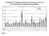

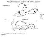

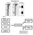


Classifications
- C—CHEMISTRY; METALLURGY
- C12—BIOCHEMISTRY; BEER; SPIRITS; WINE; VINEGAR; MICROBIOLOGY; ENZYMOLOGY; MUTATION OR GENETIC ENGINEERING
- C12Q—MEASURING OR TESTING PROCESSES INVOLVING ENZYMES, NUCLEIC ACIDS OR MICROORGANISMS; COMPOSITIONS OR TEST PAPERS THEREFOR; PROCESSES OF PREPARING SUCH COMPOSITIONS; CONDITION-RESPONSIVE CONTROL IN MICROBIOLOGICAL OR ENZYMOLOGICAL PROCESSES
- C12Q1/00—Measuring or testing processes involving enzymes, nucleic acids or microorganisms; Compositions therefor; Processes of preparing such compositions
- C12Q1/68—Measuring or testing processes involving enzymes, nucleic acids or microorganisms; Compositions therefor; Processes of preparing such compositions involving nucleic acids
- C12Q1/6876—Nucleic acid products used in the analysis of nucleic acids, e.g. primers or probes
- C12Q1/6883—Nucleic acid products used in the analysis of nucleic acids, e.g. primers or probes for diseases caused by alterations of genetic material
- C12Q1/6886—Nucleic acid products used in the analysis of nucleic acids, e.g. primers or probes for diseases caused by alterations of genetic material for cancer
- C—CHEMISTRY; METALLURGY
- C12—BIOCHEMISTRY; BEER; SPIRITS; WINE; VINEGAR; MICROBIOLOGY; ENZYMOLOGY; MUTATION OR GENETIC ENGINEERING
- C12Q—MEASURING OR TESTING PROCESSES INVOLVING ENZYMES, NUCLEIC ACIDS OR MICROORGANISMS; COMPOSITIONS OR TEST PAPERS THEREFOR; PROCESSES OF PREPARING SUCH COMPOSITIONS; CONDITION-RESPONSIVE CONTROL IN MICROBIOLOGICAL OR ENZYMOLOGICAL PROCESSES
- C12Q2600/00—Oligonucleotides characterized by their use
- C12Q2600/106—Pharmacogenomics, i.e. genetic variability in individual responses to drugs and drug metabolism
- C—CHEMISTRY; METALLURGY
- C12—BIOCHEMISTRY; BEER; SPIRITS; WINE; VINEGAR; MICROBIOLOGY; ENZYMOLOGY; MUTATION OR GENETIC ENGINEERING
- C12Q—MEASURING OR TESTING PROCESSES INVOLVING ENZYMES, NUCLEIC ACIDS OR MICROORGANISMS; COMPOSITIONS OR TEST PAPERS THEREFOR; PROCESSES OF PREPARING SUCH COMPOSITIONS; CONDITION-RESPONSIVE CONTROL IN MICROBIOLOGICAL OR ENZYMOLOGICAL PROCESSES
- C12Q2600/00—Oligonucleotides characterized by their use
- C12Q2600/112—Disease subtyping, staging or classification
- C—CHEMISTRY; METALLURGY
- C12—BIOCHEMISTRY; BEER; SPIRITS; WINE; VINEGAR; MICROBIOLOGY; ENZYMOLOGY; MUTATION OR GENETIC ENGINEERING
- C12Q—MEASURING OR TESTING PROCESSES INVOLVING ENZYMES, NUCLEIC ACIDS OR MICROORGANISMS; COMPOSITIONS OR TEST PAPERS THEREFOR; PROCESSES OF PREPARING SUCH COMPOSITIONS; CONDITION-RESPONSIVE CONTROL IN MICROBIOLOGICAL OR ENZYMOLOGICAL PROCESSES
- C12Q2600/00—Oligonucleotides characterized by their use
- C12Q2600/118—Prognosis of disease development
- C—CHEMISTRY; METALLURGY
- C12—BIOCHEMISTRY; BEER; SPIRITS; WINE; VINEGAR; MICROBIOLOGY; ENZYMOLOGY; MUTATION OR GENETIC ENGINEERING
- C12Q—MEASURING OR TESTING PROCESSES INVOLVING ENZYMES, NUCLEIC ACIDS OR MICROORGANISMS; COMPOSITIONS OR TEST PAPERS THEREFOR; PROCESSES OF PREPARING SUCH COMPOSITIONS; CONDITION-RESPONSIVE CONTROL IN MICROBIOLOGICAL OR ENZYMOLOGICAL PROCESSES
- C12Q2600/00—Oligonucleotides characterized by their use
- C12Q2600/154—Methylation markers
- C—CHEMISTRY; METALLURGY
- C12—BIOCHEMISTRY; BEER; SPIRITS; WINE; VINEGAR; MICROBIOLOGY; ENZYMOLOGY; MUTATION OR GENETIC ENGINEERING
- C12Q—MEASURING OR TESTING PROCESSES INVOLVING ENZYMES, NUCLEIC ACIDS OR MICROORGANISMS; COMPOSITIONS OR TEST PAPERS THEREFOR; PROCESSES OF PREPARING SUCH COMPOSITIONS; CONDITION-RESPONSIVE CONTROL IN MICROBIOLOGICAL OR ENZYMOLOGICAL PROCESSES
- C12Q2600/00—Oligonucleotides characterized by their use
- C12Q2600/158—Expression markers
Definitions
- This inventionrelates to breast cancer patient prognosis based on the gene expression profiles of patient biological samples.
- lymph-node-negative (LNN) breast cancer patientsare cured by local or regional treatment alone. Lancet (1998a); and Lancet (1998b). Guidelines have been developed to assist clinicians in selecting patients that should receive adjuvant therapy. Recommendations most widely used are the St. Gallen criteria (Goldhirsch et al. (2003)) and the National Institutes of Health (NIH) consensus criteria. Eifel et al. (2001). These guidelines indicate 85%-90% of LNN patients as candidates for adjuvant systemic therapy.
- the present inventionencompasses a method of assessing breast cancer status in a biological sample obtained from a breast cancer patient as defined in the claims.
- the present inventionencompasses a method of staging breast cancer in a biological sample from a breast cancer patient as defined in the claims.
- the present inventionencompasses a method of determining breast cancer patient treatment protocol by in a biological sample from a breast cancer patient as defined in the claims.
- the present disclosureprovides a method of treating a breast cancer patient by obtaining a biological sample from a breast cancer patient; and measuring the expression levels in the sample of genes selected from those encoding mRNA: corresponding to SEQ ID NOs: 1-111; or recognized by the probe sets selected from psids corresponding to SEQ ID NOs: 1-111 as depicted in Table 10 where the gene expression levels above or below pre-determined cut-off levels are indicate a high risk of recurrence and; treating the patient with adjuvant therapy if they are a high risk patient.
- the present disclosureprovides a method of cross validating a prognostic gene expression profile for breast cancer patients by obtaining gene expression data from a statistically significant number of patient biological samples; randomizing sample order; setting aside data from about 10% - 50% of samples; computing, for the remaining samples, for factor of interest on all variables and selecting variables that meet a p-value cutoff (p); selecting variables that fit a prediction model using a forward search and evaluating the training error until it hits a predetermined error rate; testing the prediction model on the left-out 10-50% of samples; repeating steps c., -g. with a new set of samples removed; and continuing steps c) -g) until 100% of samples have been tested and record classification performance.
- the present disclosureprovides a method of independently validating a prognostic gene expression profile for breast patients by obtaining gene expression data from a statistically significant number of patient biological samples; normalizing the source variabilities in the gene expression data; computing for factor of interest on all variables that were selected previously; and testing the prediction model on the sample and record classification performance.
- the present disclosureencompasses a method of generating a Relapse Hazard Score to enable prognosis of breast cancer patients by obtaining gene expression data from a statistically significant number of patient biological samples; applying univariate Cox's regression analysis to the data to obtain selected genes; applying weighted expression levels to the selected genes with standard Cox's coefficients to obtain a prediction model that can be applied as a relapse Hazard Score.
- the present disclosureencompasses a method of generating a breast cancer prognostic patient report by obtaining a biological sample from the patient; measuring gene expression of the sample; applying a Relapse Hazard Score to the results of step b.; and using the results obtained in step c. to generate the report and patient reports generated thereby.
- the present inventionencompasses a composition comprising a probe set consisiting of all of SEQ ID NOs: 1-111 in Table 10
- the present inventionencompasses the use of a kit for conducting an assay to determine breast cancer prognosis in a biological sample containing: materials for detecting isolated nucleic acid sequences, their complements, or portions thereof of a combination of genes consisting of those encoding mRNA: identified by SEQ ID NOs: 1-11 in Table 10.
- the present disclosureencompasses articles for assessing breast cancer status containing: materials for detecting isolated nucleic acid sequences, their complements, or portions thereof of a combination of genes selected from those encoding mRNA: corresponding to SEQ ID NOs: 1-111; or recognized by the probe sets selected from psids corresponding to SEQ ID NOs: 1-111 as depicted in Table 10.
- the present disclosureencompasses a diagnostic/prognostic portfolio containing isolated nucleic acid sequences, their complements, or portions thereof of a combination of genes selected from those encoding mRNA: corresponding to SEQ ID NOs: 1-111; or recognized by the probe sets selected from psids corresponding to SEQ ID NOs: 1-111 as depicted in Table 10 where the combination is sufficient to characterize breast cancer status or risk of relapse in a biological sample.
- the present inventionencompasses a method of assessing breast cancer status in a biological sample from a breast cancer patient as defined in the claims.
- the present inventionencompasses a method of staging breast cancer in a biological sample from a breast cancer patient; as defined by the claims.
- the methodutilizes any classification known in the art including the TNM system American Joint Committee on Cancer www.cancerstaging.org) and comparison to stages corresponding to patients with similar gene expression profiles.
- the present inventionencompasses a method of determining breast cancer patient treatment protocol in a biological sample from a breast cancer patient; as defined in the claims.
- the present inventionencompasses a method of treating a breast cancer patient in a biological sample from a breast cancer patient; as defined in the claims.
- the SEQ ID NOs:are 36-95 for estrogen receptor (ER) patients and 96-111 for ER negative patients.
- the samplecan be prepared by any method known in the art including bulk tissue preparation and laser capture microdissection. Bulk tissue preparations can be obtained from a biopsy or a surgical specimen.
- the above methodscan further include measuring the expression level of at least one gene encoding mRNA identified by SEQ ID NOs: 112-132; Table 10. They can also include measuring the expression level of at least one gene constitutively expressed in the sample.
- the above methodscan further include determining the estrogen receptor (ER) status of the sample.
- ER statuscan be measured by any method known in the art including measuring the expression level of at least one gene indicative of ER status, measuring the presence of ER in the sample and measuring immunohistochemically.
- the above methodscan be used with samples from any biological source.
- the sampleis obtained from a primary tumor.
- the above methodspreferably have a specificity of at least 40% and a sensitivity of at least at least 90%.
- the above methodscan be used where the expression pattern of the genes is compared to an expression pattern indicative of a relapse patient.
- the comparisoncan be by any method known in the art including comparison of expression patterns is conducted with pattern recognition methods.
- Pattern recognition methodscan be any known in the art including a Cox's proportional hazards analysis.
- the pre-determined cut-off levelsare at least 1.5-fold over- or under-expression in the sample relative to benign cells or normal tissue.
- the pre-determined cut-off levelshave at least a statistically significant p-value over-expression in the sample having metastatic cells relative to benign cells or normal tissue. More preferably, the p-value is less than 0.05.
- microarraycan be a cDNA array or an oligonucleotide array and can further contain one or more internal control reagents.
- PCRpolymerase chain reaction
- RT-PCRreverse transcription polymerase chain reaction
- the RT-PCRcan further contain one or more internal control reagents.
- gene expressionis detected by measuring or detecting a protein encoded by the gene. Any method known in the art can be used including detection by an antibody specific to the protein and measuring a characteristic of the gene. Suitable characteristics include, without limitation, DNA amplification, methylation, mutation and allelic variation.
- the present disclosureencompasses a method of cross validating a prognostic gene expression profile for breast cancer patients by obtaining gene expression data from a statistically significant number of patient biological samples; randomizing sample order; setting aside data from about 10% - 50% of samples; computing, for the remaining samples, for factor of interest on all variables and selecting variables that meet a p-value cutoff (p); selecting variables that fit a prediction model using a forward search and evaluating the training error until it hits a predetermined error rate; testing the prediction model on the left-out 10-50% of samples; repeating steps c., -g. with a new set of samples removed; and continuing steps c) -g) until 100% of samples have been tested and record classification performance.
- the cross validation methodcan be used where the gene expression data obtained in step h. is represented by genes selected from those encoding mRNA: corresponding to SEQ ID NOs: 3, 4, 10, 18, 37, 40, 42, 43, 45, 55, 58, 64, 67, 72-74, 76, 81, 85-86, 89, 97, 100-101, 110-111, 125 and 132-442, or 2, 3, 5, 12, 20, 25, 36, 37, 39, 40, 41, 42, 43, 45, 46, 51, 52, 53, 54, 55, 57, 58, 59, 60, 61, 62, 63, 64, 66, 67, 69, 70, 72, 73, 74, 76, 80, 81, 83, 84, 85, 86, 87, 88, 89, 90, 94, 97, 98, 101, 102, 104, 107, 110, 111, 132, 139, 142, 150, 151, 153, 154, 158,
- the present disclosureencompasses a method of independently validating a prognostic gene expression profile for breast patients by obtaining gene expression data from a statistically significant number of patient biological samples; normalizing the source variabilities in the gene expression data; computing for factor of interest on all variables that were selected previously; and testing the prediction model on the sample and record classification performance.
- the independent validation methodcan be used where the gene expression data obtained in step d. is represented by genes selected from those encoding mRNA: corresponding to SEQ ID NOs: 3, 4, 10, 18, 37, 40, 42, 43, 45, 55, 58, 64, 67, 72-74, 76, 81, 85-86, 89, 97, 100-101, 110-111, 125 and 132-442, or 2, 3, 5, 12, 20, 25, 36, 37, 39, 40, 41, 42, 43, 45, 46, 51, 52, 53, 54, 55, 57, 58, 59, 60, 61, 62, 63, 64, 66, 67, 69, 70, 72, 73, 74, 76, 80, 81, 83, 84, 85, 86, 87, 88, 89, 90, 94, 97, 98, 101, 102, 104, 107, 110, 111, 132, 139, 142, 150, 151, 153, 154, 158,
- the present inventionencompasses a method of generating a Relapse Hazard Score to enable prognosis of breast cancer patients by obtaining gene expression data from a statistically significant number of patient biological samples; applying univariate Cox's regression analysis to the data to obtain selected genes; applying weighted expression levels to the selected genes with standard Cox's coefficients to obtain a prediction model that can be applied as a relapse Hazard Score.
- the present inventionencompasses a method of generating a breast cancer prognostic patient report by obtaining a biological sample from the patient; measuring gene expression of the sample; applying a Relapse Hazard Score to the results of step b.; and using the results obtained in step c. to generate the report and patient reports generated thereby.
- the reportmay contain an assessment of patient outcome and/or probability of risk relative to the patient population.
- the present inventionencompasses a composition comprising a probe set consisiting of all of SEQ ID NOs: 1-111 in Table 10.
- the compositioncan further contain reagents for conducting a microarray analysis, and a medium through which said nucleic acid sequences, their complements, or portions thereof are assayed.
- the present inventionencompasses the use of a kit for conducting an assay to determine breast cancer prognosis in a biological sample containing: materials for detecting isolated nucleic acid sequences, their complements, or portions thereof of a combination of genes consisting of those encoding mRNA: identified by SEQ ID NOs: 1-111 in Table 10.
- the kitcan further contain reagents for conducting a microarray analysis, and a medium through which said nucleic acid sequences, their complements, or portions thereof are assayed.
- the present disclosureprovides articles for assessing breast cancer status containing: materials for detecting isolated nucleic acid sequences, their complements, or portions thereof of a combination of genes selected from those encoding mRNA: corresponding to SEQ ID NOs: 1-111; or recognized by the probe sets selected from psids corresponding to SEQ ID NOs: 1-111 as depicted in Table 10.
- the articlescan further contain reagents for conducting a microarray analysis, and a medium through which said nucleic acid sequences, their complements, or portions thereof are assayed.
- microarrays provided hereincan contain isolated nucleic acid sequences, their complements, or portions thereof of a combination of genes selected from those encoding mRNA: corresponding to SEQ ID NOs: 1-111; or recognized by the probe sets selected from of psids corresponding to SEQ ID NOs: 1-111 as depicted in Table 10 where the combination is sufficient to characterize breast cancer status or risk of relapse in a biological sample.
- the microarray measurement or characterizationis preferably at least 1.5-fold over- or under-expression.
- the microarraycan detect a pre-determined cut-off levels are at least 1.5-fold over- or under- expression in the sample relative to benign cells or normal tissue.
- the pre-determined cut-off levelshave at least a statistically significant p-value over-expression in the sample having metastatic cells relative to benign cells or normal tissue. More preferably, the p-value is less than 0.05.
- the present disclosureencompasses a diagnostic/prognostic portfolio containing isolated nucleic acid sequences, their complements, or portions thereof of a combination of genes selected from those encoding mRNA: corresponding to SEQ ID NOs: 1-111; or recognized by the probe sets selected from psids corresponding to SEQ ID NOs: 1-111 as depicted in Table 10 where the combination is sufficient to characterize breast cancer status or risk of relapse in a biological sample.
- the portfoliocan detect a pre-determined cut-off levels are at least 1.5-fold over- or under- expression in the sample relative to benign cells or normal tissue.
- the pre-determined cut-off levelshave at least a statistically significant p-value over-expression in the sample having metastatic cells relative to benign cells or normal tissue. More preferably, the p-value is less than 0.05.
- SEQ ID NOs: 1-650are summarized in Table 10.
- the markeris identified by a psid or Affymetrix GeneChip Designation and represents the gene encoding any variant, allele etc. corresponding to the given SEQ ID NO.
- the markeris also defined as the gene encoding mRNA recognized by the probe corresponding to the given psid. Certain markers are described in more detail below.
- M83, SEQ ID NO: 45is mentioned by US patent application publication numbers 20020110547 ; 20040009491 ; and 20030236392 , and PCT publication numbers WO0149716 ; WO0170979 ; WO03025138 ; WO03042661 ; WO03016475 ; WO2004063355 ; WO2004047728 ; and WO2004030615 .
- ABLIM-sSEQ ID NO: 43, is mentioned by US patent application publication numbers 20020131971 ; 20030087818 ; 20030166064 ; 20030236392 ; and 20040005560 and PCT publication numbers WO0036107 ; WO0159063 ; WO0160860 ; WO0174405 ; WO0177288 ; WO02059271 ; WO0212328 ; WO0224956 ; WO0229086 ; WO03025138 ; WO2004024097 ; WO2004037996 ; WO2004039956 ; WO2004043361 ; and WO2004047728 .
- SEQ ID NO: 54is mentioned by US patent application publication numbers 20020064872 ; 20020085998 ; 20020150581 ; 20020156011 ; 20020192678 ; 20030069180 ; 20030073623 ; 20030104366 ; 20040005560 ; 20040013663 ; and 20040058340 and PCT publication numbers WO0060076 ; WO0142467 ; WO0155322 ; WO0160860 ; WO0170979 ; WO0179286 ; WO196388 ; WO02057414 ; WO02059377 ; WO02060317 ; WO02086443 ; WO0231198 ; WO0250279 ; WO03004989 ; WO03025138 ; WO03042661 ; WO03062379 ; WO2004047728 ; WO2004048938 ; WO2004048938 ; WO2004063355 ; and
- C11ORF9, SEQ ID NO: 48is mentioned by US patent application publication numbers 20030065157 ; and 20030073623 and PCT publication numbers WO0056880 ; WO0058473 ; WO0159063 ; WO0174405 ; WO02059260 ; WO0229086 ; WO03101283 ; WO2004031413 ; and WO2004060270 .
- C3, SEQ ID NO: 46is mentioned by US patent application publication numbers 20040005560 ; and 20040058340 and PCT publication numbers WO9101999 ; WO0055350 ; WO0160860 ; WO0175067 ; WO0188088 ; WO0224956 ; WO0294629 ; WO03078572 ; WO2004028479 ; WO2004030615 ; WO2004041170 ; and WO2004060270 .
- CGI-41SEQ ID NO: 65
- US patent application publication numbers 20030073623and 20030096982 and PCT publication numbers WO9514772 ; WO9940100 ; WO0154472 ; WO0157188 ; WO03046152 ; WO2004060270 .
- CLN8, SEQ ID NO: 36is mentioned by EP 1 104 808 B1 , US patent application numbers 20030073623 ; and 20040009491 and PCT publication numbers WO9845437 ; WO9935158 ; WO0142451 ; WO0190304 ; WO02090526 ; WO02095010 ; WO02102993 ; WO02102994 ; WO03004622 ; WO04024892 ; and WO04041170 .
- CNK1SEQ ID NO: 60
- US patent application publication number 20040058340and PCT publication numbers WO9938972 ; and WO2004037996 .
- DUSP4, SEQ ID NO: 57is mentioned by US patent application publication numbers 20030073623 ; 20030194704 ; 20040058340 ; and 20040110194 and PCT publication numbers WO0100828 ; WO0204514 ; WO02059377 ; WO02103320 ; WO0224956 ; WO03004989 ; WO2004018641 ; WO2004023973 ; WO2004037996 ; WO2004039956 ; WO2004043361 ; WO2004047728 ; WO2004048938 ; and WO9423039 .
- FEN1SEQ ID NO: 62
- US patent number 5874283US patent application publication numbers 20020156011 ; 20030069180 ; 20030073623 ; and 20030194704 and PCT publication numbers WO0196388 ; WO03016475 ; WO03016500 ; WO03072035 ; WO03101283 ; WO2004022059 ; WO2004030615 ; WO2004039956 ; WO2004043361 ; WO2004047728 ; WO2004048938 ; WO2004060270 ; and WO2004079014 .
- FKBP2SEQ ID NO: 63
- GP73SEQ ID NO: 37, is mentioned by US patent application publication numbers 20030022239 ; 20030073623 ; 20030236392 ; 20040009478 ; 20040034196 ; and 20040058340 and PCT publication numbers WO0012708 ; WO0061610 ; WO0078961 ; WO0140466 ; WO0160860 ; WO0177168 ; WO0194629 ; WO02059260 ; WO02083876 ; WO0224956 ; WO03004989 ; WO2004024892 ; WO2004031414 ; and WO2004053081 .
- H4FHSEQ ID NO: 61
- US patent application publication number 20020151681and PCT publication numbers WO0055174 ; WO0175067 ; WO0224956 ; WO03016476 ; WO2004039943 .
- IL-18, SEQ ID NO: 39is mentioned by US patent number 6060283 , US patent application publication number 20040115636 and PCT publication numbers WO0194629 ; WO02057414 ; WO0224956 ; WO0229086 ; WO03025138 ; WO03042661 ; and WO9724441 .
- KIAA0748, SEQ ID NO: 56is mentioned by PCT publication numbers WO0021991 ; WO02094988 ; and WO2004003162 .
- KPNA2SEQ ID NO: 64
- MGC11335SEQ ID NO: 66
- US patent application publication numbers 20030073623 ; 20030219744 ; and 20030236392PCT publication numbers WO0144448 ; WO0153312 ; WO0157182 ; WO0188088 ; and WO2004030615 .
- OR12D2SEQ ID NO: 52, is mentioned by PCT publication numbers WO0127158 ; WO02068652 ; WO02081498 ; WO02081517 ; WO030000735 ; and WO03091388 .
- ORC3SEQ ID NO: 53, is mentioned by US patent application publication number 20040013663 and PCT publication numbers WO9810067 ; WO9958642 ; WO0060078 ; WO0175067 ; WO02059377 ; WO02070737 ; WO02102235 ; and WO03042661 .
- PLK1, SEQ ID NO: 59is mentioned by DE4329177 , US patent application publication numbers 20020081659 ; and 20020151681 and PCT publication numbers WO0055174 ; WO0055320 ; WO02070737 ; WO02086443 ; WO0224956 ; WO03003906 ; WO03016476 ; WO03018807 ; WO03025138 ; WO03042661 ; WO2004030615 ; WO2004037996 ; WO2004042022 ; WO2004047728 ; WO2004048938 ; WO2004063355 ; and WO2004070062 .
- PPP1CCSEQ ID NO: 42
- SMC4, SEQ ID NO: 55is mentioned by US patent application publication numbers 20020123619 ; 20020168637 ; and 20040058340 and PCT publication numbers WO9938972 ; WO9964576 ; WO0175067 ; WO0190154 ; WO0204514 ; WO02059377 ; WO02074237 ; WO02086443 ; WO02102235 ; WO03003906 ; WO03016475 ; WO03025138 ; WO03042661 ; WO2003094848 ; WO2004024892 ; WO2004047728 ; WO2004048938 ; WO2004063355 ; and WO2004074301 .
- TRDL-1SEQ ID NO: 44
- US patent numbers 6171787 ; and 6440694US patent application publication numbers 20020055474 ; 20020072089 ; 20020081659 ; 20030065157 ; 20030073623 ; and 20030219767 and PCT publication numbers WO9733902 ; WO9912965 ; WO9926976 ; WO9928462 ; WO9933980 ; WO9935170 ; WO9950416 ; WO9954460 ; WO0026244 ; WO0032776 ; WO0055320 ; WO012525 WO0177291 ; WO2004020593 ; WO2004024892 ; WO2004033637 ; and 04037996 .
- Yiflp, SEQ ID NO: 38is mentioned by US patent application publication numbers 20020131971 ; 20030166064 ; and 20040037842 and PCT publication numbers WO9933981 ; WO9947540 ; WO0173027 ; WO02060317 ; WO02070737 ; WO02076488 ; WO02095010 ; WO0224956 ; WO03004622 ; WO03038063 ; WO03042661 ; WO03054152 ; WO03064589 ; WO2004024892 ; WO2004037996 ; WO2004047728 ; and WO2004048938 .
- nucleic acid sequences having the potential to express proteins, peptides, or mRNAsuch sequences referred to as "genes" within the genome by itself is not determinative of whether a protein, peptide, or mRNA is expressed in a given cell. Whether or not a given gene capable of expressing proteins, peptides, or mRNA does so and to what extent such expression occurs, if at all, is determined by a variety of complex factors.
- assaying gene expressioncan provide useful information about the occurrence of important events such as tumorogenesis, metastasis, apoptosis, and other clinically relevant phenomena. Relative indications of the degree to which genes are active or inactive can be found in gene expression profiles.
- the gene expression profiles of this inventionare used to provide a prognosis and treat patients for breast cancer.
- Sample preparationrequires the collection of patient samples.
- Patient samples used in the inventive methodare those that are suspected of containing diseased cells such as epithelial cells taken from the primary tumor in a breast sample. Samples taken from surgical margins are also preferred. Most preferably, however, the sample is taken from a lymph node obtained from a breast cancer surgery. Laser Capture Microdissection (LCM) technology is one way to select the cells to be studied, minimizing variability caused by cell type heterogeneity. Consequently, moderate or small changes in gene expression between normal and cancerous cells can be readily detected. Samples can also comprise circulating epithelial cells extracted from peripheral blood. These can be obtained according to a number of methods but the most preferred method is the magnetic separation technique described in U.S. Patent 6,136,182 . Once the sample containing the cells of interest has been obtained, RNA is extracted and amplified and a gene expression profile is obtained, preferably via micro-array, for genes in the appropriate portfolios.
- LCMLaser Capture Microdissection
- Preferred methods for establishing gene expression profilesinclude determining the amount of RNA that is produced by a gene that can code for a protein or peptide. This is accomplished by reverse transcriptase PCR (RT-PCR), competitive RT-PCR, real time RT-PCR, differential display RT-PCR, Northern Blot analysis and other related tests. While it is possible to conduct these techniques using individual PCR reactions, it is best to amplify complementary DNA (cDNA) or complementary RNA (cRNA) produced from mRNA and analyze it via microarray. A number of different array configurations and methods for their production are known to those of skill in the art and are described in U.S.
- Patentssuch as: 5,445,934 ; 5,532,128 ; 5,556,752 ; 5,242,974 ; 5,384,261 ; 5,405,783 ; 5,412,087 ; 5,424,186 ; 5,429,807 ; 5,436,327 ; 5,472,672 ; 5,527,681 ; 5,529,756 ; 5,545,531 ; 5,554,501 ; 5,561,071 ; 5,571,639 ; 5,593,839 ; 5,599,695 ; 5,624,711 ; 5,658,734 ; and 5,700,637 .
- Microarray technologyallows for the measurement of the steady-state mRNA level of thousands of genes simultaneously thereby presenting a powerful tool for identifying effects such as the onset, arrest, or modulation of uncontrolled cell proliferation.
- Two microarray technologiesare currently in wide use. The first are cDNA arrays and the second are oligonucleotide arrays. Although differences exist in the construction of these chips, essentially all downstream data analysis and output are the same.
- the product of these analysesare typically measurements of the intensity of the signal received from a labeled probe used to detect a cDNA sequence from the sample that hybridizes to a nucleic acid sequence at a known location on the microarray.
- the intensity of the signalis proportional to the quantity of cDNA, and thus mRNA, expressed in the sample cells.
- mRNAmRNA
- Analysis of expression levelsis conducted by comparing signal intensities. This is best done by generating a ratio matrix of the expression intensities of genes in a test sample versus those in a control sample. For instance, the gene expression intensities from a diseased tissue can be compared with the expression intensities generated from normal tissue of the same type (e.g., diseased breast tissue sample vs. normal breast tissue sample). A ratio of these expression intensities indicates the fold-change in gene expression between the test and control samples.
- Gene expression profilescan also be displayed in a number of ways. The most common method is to arrange raw fluorescence intensities or ratio matrix into a graphical dendogram where columns indicate test samples and rows indicate genes. The data are arranged so genes that have similar expression profiles are proximal to each other. The expression ratio for each gene is visualized as a color. For example, a ratio less than one (indicating down-regulation) may appear in the blue portion of the spectrum while a ratio greater than one (indicating up-regulation) may appear as a color in the red portion of the spectrum.
- Commercially available computer software programsare available to display such data including GeneSpring from Agilent Technologies and Partek Discover TM and Partek Infer TM software from Partek®.
- Modulated genes used in the methods of the inventionare described in the Examples. Differentially expressed genes are either up- or down-regulated in patients with a relapse of breast cancer relative to those without a relapse. Up regulation and down regulation are relative terms meaning that a detectable difference (beyond the contribution of noise in the system used to measure it) is found in the amount of expression of the genes relative to some baseline. In this case, the baseline is the measured gene expression of a non-relapsing patient. The genes of interest in the diseased cells (from the relapsing patients) are then either up- or down-regulated relative to the baseline level using the same measurement method.
- Diseasedin this context, refers to an alteration of the state of a body that interrupts or disturbs, or has the potential to disturb, proper performance of bodily functions as occurs with the uncontrolled proliferation of cells.
- someoneis diagnosed with a disease when some aspect of that person's genotype or phenotype is consistent with the presence of the disease.
- the act of conducting a diagnosis or prognosisincludes the determination of disease/status issues such as determining the likelihood of relapse and therapy monitoring.
- therapy monitoringclinical judgments are made regarding the effect of a given course of therapy by comparing the expression of genes over time to determine whether the gene expression profiles have changed or are changing to patterns more consistent with normal tissue.
- levels of up- and down-regulationare distinguished based on fold changes of the intensity measurements of hybridized microarray probes.
- a 2.0 fold differenceis preferred for making such distinctions (or a p-value less than 0.05). That is, before a gene is said to be differentially expressed in diseased/relapsing versus normal/non-relapsing cells, the diseased cell is found to yield at least 2 times more, or 2 times less intensity than the normal cells. The greater the fold difference, the more preferred is use of the gene as a diagnostic or prognostic tool.
- Genes selected for the gene expression profiles of the instant inventionhave expression levels that result in the generation of a signal that is distinguishable from those of the normal or non-modulated genes by an amount that exceeds background using clinical laboratory instrumentation.
- Statistical valuescan be used to confidently distinguish modulated from non-modulated genes and noise. Statistical tests find the genes most significantly different between diverse groups of samples.
- the Student's T-testis an example of a robust statistical test that can be used to find significant differences between two groups. The lower the p-value, the more compelling the evidence that the gene is showing a difference between the different groups. Nevertheless, since microarrays measure more than one gene at a time, tens of thousands of statistical tests may be performed at one time. Because of this, one is unlikely to see small p-values just by chance and adjustments for this using a Sidak correction as well as a randomization/permutation experiment can be made. A p-value less than 0.05 by the T-test is evidence that the gene is significantly different.
- More compelling evidenceis a p-value less then 0.05 after the Sidak correction is factored in. For a large number of samples in each group, a p-value less than 0.05 after the randomization/permutation test is the most compelling evidence of a significant difference.
- Another parameter that can be used to select genes that generate a signal that is greater than that of the non-modulated gene or noiseis the use of a measurement of absolute signal difference.
- the signal generated by the modulated gene expressionis at least 20% different than those of the normal or non-modulated gene (on an absolute basis). It is even more preferred that such genes produce expression patterns that are at least 30% different than those of normal or non-modulated genes.
- Genescan be grouped so that information obtained about the set of genes in the group provides a sound basis for making a clinically relevant judgment such as a diagnosis, prognosis, or treatment choice. These sets of genes make up the portfolios of the invention. In this case, the judgments supported by the portfolios involve breast cancer and its chance of recurrence. As with most diagnostic markers, it is often desirable to use the fewest number of markers sufficient to make a correct medical judgment. This prevents a delay in treatment pending further analysis as well inappropriate use of time and resources.
- portfoliosare established such that the combination of genes in the portfolio exhibit improved sensitivity and specificity relative to individual genes or randomly selected combinations of genes.
- the sensitivity of the portfoliocan be reflected in the fold differences exhibited by a gene's expression in the diseased state relative to the normal state.
- Specificitycan be reflected in statistical measurements of the correlation of the signaling of gene expression with the condition of interest. For example, standard deviation can be a used as such a measurement. In considering a group of genes for inclusion in a portfolio, a small standard deviation in expression measurements correlates with greater specificity. Other measurements of variation such as correlation coefficients can also be used.
- One method of establishing gene expression portfoliosis through the use of optimization algorithms such as the mean variance algorithm widely used in establishing stock portfolios. This method is described in detail in US patent publication number 20030194734 . Essentially, the method calls for the establishment of a set of inputs (stocks in financial applications, expression as measured by intensity here) that will optimize the return (e.g., signal that is generated) one receives for using it while minimizing the variability of the return. Many commercial software programs are available to conduct such operations. "Wagner Associates Mean-Variance Optimization Application,” referred to as “Wagner Software” throughout this specification, is preferred. This software uses functions from the “Wagner Associates Mean-Variance Optimization Library" to determine an efficient frontier and optimal portfolios in the Markowitz sense is preferred. Use of this type of software requires that microarray data be transformed so that it can be treated as an input in the way stock return and risk measurements are used when the software is used for its intended financial analysis purposes.
- the process of selecting a portfoliocan also include the application of heuristic rules.
- such rulesare formulated based on biology and an understanding of the technology used to produce clinical results. More preferably, they are applied to output from the optimization method.
- the mean variance method of portfolio selectioncan be applied to microarray data for a number of genes differentially expressed in subjects with breast cancer. Output from the method would be an optimized set of genes that could include some genes that are expressed in peripheral blood as well as in diseased tissue. If samples used in the testing method are obtained from peripheral blood and certain genes differentially expressed in instances of breast cancer are differentially expressed in peripheral blood, then a heuristic rule can be applied in which a portfolio is selected from the efficient frontier excluding those that are differentially expressed in peripheral blood.
- the rulecan be applied prior to the formation of the efficient frontier by, for example, applying the rule during data pre-selection.
- heuristic rulescan be applied that are not necessarily related to the biology in question. For example, one can apply a rule that only a prescribed percentage of the portfolio can be represented by a particular gene or group of genes.
- Commercially available softwaresuch as the Wagner Software readily accommodates these types of heuristics. This can be useful, for example, when factors other than accuracy and precision (e.g., anticipated licensing fees) have an impact on the desirability of including one or more genes.
- One method of the inventioninvolves comparing gene expression profiles for various genes (or portfolios) to ascribe prognoses.
- the gene expression profiles of each of the genes comprising the portfolioare fixed in a medium such as a computer readable medium.
- a mediumsuch as a computer readable medium.
- Thiscan take a number of forms. For example, a table can be established into which the range of signals (e.g., intensity measurements) indicative of disease is input. Actual patient data can then be compared to the values in the table to determine whether the patient samples are normal or diseased.
- patterns of the expression signalse.g., fluorescent intensity
- the gene expression patterns from the gene portfolios used in conjunction with patient samplesare then compared to the expression patterns.
- Pattern comparison softwarecan then be used to determine whether the patient samples have a pattern indicative of recurrence of the disease. Of course, these comparisons can also be used to determine whether the patient is not likely to experience disease recurrence.
- the expression profiles of the samplesare then compared to the portfolio of a control cell. If the sample expression patterns are consistent with the expression pattern for recurrence of a breast cancer then (in the absence of countervailing medical considerations) the patient is treated as one would treat a relapse patient. If the sample expression patterns are consistent with the expression pattern from the normal/control cell then the patient is diagnosed negative for breast cancer.
- the preferred profiles of this disclosureare the 35-gene portfolio made up of the genes of SEQ ID NOs: 1-35, the 60-gene portfolio made up of the genes of SEQ ID NOs: 36-95 which is best used to prognosticate ER-positive patients, and the 16-gene portfolio made up of genes of SEQ ID NOs: 96-111 which is best used to prognosticate ER-negative patients. Most preferably, the portfolio is made up of genes of SEQ ID NOs: 36-111. This most preferred portfolio best segregates breast cancer patients irrespective of ER status at high risk of relapse from those who are not. Once the high-risk patients are identified they can then be treated with adjuvant therapy.
- the most preferred method for analyzing the gene expression pattern of a patient to determine prognosis of breast canceris through the use of a Cox's hazard analysis program.
- the analysisis conducted using S-Plus software (commercially available from Insightful Corporation).
- S-Plus softwarecommercially available from Insightful Corporation.
- a gene expression profileis compared to that of a profile that confidently represents relapse (i.e., expression levels for the combination of genes in the profile is indicative of relapse).
- the Cox's hazard model with the established thresholdis used to compare the similarity of the two profiles (known relapse versus patient) and then determines whether the patient profile exceeds the threshold.
- the patientis classified as one who will relapse and is accorded treatment such as adjuvant therapy. If the patient profile does not exceed the threshold then they are classified as a non-relapsing patient.
- Other analytical toolscan also be used to answer the same question such as, linear discriminate analysis, logistic regression and neural network approaches.
- the gene expression profiles of this inventioncan also be used in conjunction with other non-genetic diagnostic methods useful in cancer diagnosis, prognosis, or treatment monitoring.
- diagnostic power of the gene expression based methods described abovewith data from conventional markers such as serum protein markers (e.g., Cancer Antigen 27.29 ("CA 27.29”)).
- serum protein markerse.g., Cancer Antigen 27.29 (“CA 27.29”).
- CA 27.29Cancer Antigen 27.29
- bloodis periodically taken from a treated patient and then subjected to an enzyme immunoassay for one of the serum markers described above. When the concentration of the marker suggests the return of tumors or failure of therapy, a sample source amenable to gene expression analysis is taken.
- FNAfine needle aspirate
- Articlesinclude representations of the gene expression profiles useful for treating, diagnosing, prognosticating, and otherwise assessing diseases. These profile representations are reduced to a medium that can be automatically read by a machine such as computer readable media (magnetic, optical, and the like).
- the articlescan also include instructions for assessing the gene expression profiles in such media.
- the articlesmay comprise a CD ROM having computer instructions for comparing gene expression profiles of the portfolios of genes described above.
- the articlesmay also have gene expression profiles digitally recorded therein so that they may be compared with gene expression data from patient samples. Alternatively, the profiles can be recorded in different representational format. A graphical recordation is one such format. Clustering algorithms such as those incorporated in Partek DiscoverTM and Partek InferTM software from Partek® mentioned above can best assist in the visualization of such data.
- articles of manufactureare media or formatted assays used to reveal gene expression profiles. These can comprise, for example, microarrays in which sequence complements or probes are affixed to a matrix to which the sequences indicative of the genes of interest combine creating a readable determinant of their presence.
- articles according to the disclosurecan be fashioned into reagent kits for conducting hybridization, amplification, and signal generation indicative of the level of expression of the genes of interest for detecting breast cancer.
- Kits made according to the disclosureinclude formatted assays for determining the gene expression profiles. These can include all or some of the materials needed to conduct the assays such as reagents and instructions.
- Genes analyzed according to this inventionare typically related to full-length nucleic acid sequences that code for the production of a protein or peptide.
- identification of full-length sequencesis not necessary from an analytical point of view. That is, portions of the sequences or ESTs can be selected according to well-known principles for which probes can be designed to assess gene expression for the corresponding gene.
- Frozen tumor specimens from LNN patients treated during 1980-1995, but untreated with systemic neoadjuvant therapywere selected from our tumor bank at the Erasmus Medical Center (Rotterdam, Netherlands). All tumor samples were submitted to our reference laboratory from 25 regional hospitals for steroid hormone receptor measurements. The guidelines for primary treatment were similar for all hospitals. Tumors were selected in a manner to avoid bias. On the assumption of a 25-30% in 5 years, and a substantial loss of tumors because of quality control reasons, 436 invasive tumor samples were processed. Patients with a poor, intermediate, and good clinical outcome were included. Samples were rejected based on insufficient tumor content (53), poor RNA quality (77) or poor chip quality (20) leaving 286 samples eligible for further analysis.
- the study protocolwas approved by institutional medical ethics committee (MEC no. 02.953). Median age of patients at the time of surgery (breast conserving surgery: 219 patients; modified radical mastectomy: 67 patients) was 52 years (range, 26-83 years). Radiotherapy was given to 248 patients (87%) according to our institutional protocol. The proportions of patients who underwent breast conserving therapy and radiotherapy are normal for LNN disease. Patients were included regardless of radiotherapy status, as this study was not aimed to investigate the potential effects of a specific type of surgery or adjuvant radiotherapy. Furthermore, studies have shown that radiotherapy has no clear effect on distant disease recurrence. Early Breast Cancer Trialists (1995). Lymph node negativity was based on pathological examination by regional pathologists. Foekens et al. (1989a).
- ER (and PgR) levelswere measured by ligand binding assay or enzyme immunoassay (EIA) (Foekens et al. (1989b)) or by immunohistochemistry (in 9 tumors).
- EIAenzyme immunoassay
- the cutoff values used to classify patients as positive or negative for ER and PRwas 10fmol/mg protein or 10% positive tumor cells.
- Postoperative follow-upinvolved examination every 3 months during the first 2 years, every 6 months for years 3 to 5, and every 12 months from year 5.
- the date of diagnosis of metastasiswas defined as the date of confirmation of metastasis after symptoms reported by the patient, detection of clinical signs, or at regular follow-up.
- the median follow-up period of surviving patientswas 101 months (range, 20-171).
- 9333%) showed evidence of distant metastasis within 5 years and were counted as failures in the analysis of distant metastasis-free survival (DMFS).
- Eighty-three patients (29%)died after a previous relapse. Therefore, a total of 88 patients (31 %) were failures in the analysis of overall survival (OS).
- OSoverall survival
- Gene expression datawas filtered to include genes called "present" in two or more samples. 17,819 genes passed this filter and were used for hierarchical clustering. Before clustering, the expression level of each gene was divided by its median expression level in the patients. This standardization step limited the effect of the magnitude of expression of genes, and grouped together genes with similar patterns of expression in the clustering analysis. To identify patient subgroups, we carried out average linkage hierarchical clustering on both the genes and the samples using GeneSpring 6 ⁇ 0.
- Each patient subgroupwas analyzed separately in order to select markers.
- the patients in the ER-positive subgroupwere randomly allocated into training and testing sets of 80 and 129 patients, respectively.
- the patients in the ER-negative subgroupwere randomly divided into training and testing sets of 35 and 42 patients, respectively.
- the markers selected from each subgroup training setwere combined to form a single signature to predict tumor metastasis for both ER-positive and ER-negative patients in a subsequent independent validation.
- the sample size of the training setwas determined by a resampling method to ensure its statistical confidence level. Briefly, the number of patients in the training set started at 15 patients and was increased by steps of 5. For a given sample size, 10 training sets with randomly selected patients were made. A gene signature was constructed from each of training sets and then tested in a designated testing set of patients by analysis of receiver operating characteristic (ROC) curve with distant metastasis within 5 years as the defining point. The mean and the coefficient of variation (CV) of the area under the curve (AUC) for a given sample size were calculated. A minimum number of patients required for the training set were chosen at the point that the average AUC reached a plateau and the CV of the 10 AUC was below 5%.
- ROCreceiver operating characteristic
- Geneswere selected as follows. First, univariate Cox's proportional hazards regression was used to identify genes for which expression (on log 2 scale) was correlated with the length of DMFS. To reduce the effect of multiple testing and to test the robustness of the selected genes, the Cox's model was constructed with bootstrapping of the patients in the training set. Efron et al. (1981). Briefly, 400 bootstrap samples of the training set were constructed, each with 80 patients randomly chosen with replacement. A Cox's model was run on each of the bootstrap samples. A bootstrap score was created for each gene by removing the top and bottom 5% p-values and then averaging the inverses of the remaining bootstrap p-values. This score was used to rank the genes.
- the Relapse Scorewas used to calculate each patient's risk of distant metastasis.
- the scorewas defined as the linear combination of weighted expression signals with the standardized Cox's regression coefficient as the weight.
- the thresholdwas determined from the ROC curve of the training set to ensure 100% sensitivity and the highest specificity.
- the values of constants A of 313.5 and B of 280were chosen to center the threshold of RS to zero for both ER-positive and ER-negative patients.
- Patients with positive RS scoreswere classified into the poor prognosis group and patients with negative RS scores were classified into the good prognosis group.
- the gene signature and the cutoffwere validated in the testing set.
- a functional classwas assigned to each of the genes in the prognostic signature gene. Pathway analysis was done with Ingenuity 1.0 software (Ingenuity Systems, CA). Affymetrix probes were used as input to search for biological networks built by the software. Biological networks identified by the program were assessed in the context of general functional classes by GO ontology classification. Pathways with two or more genes in the prognostic signature were selected and evaluated.
- the ER-negative training grouphad a slightly higher proportion of larger tumors and, as expected, more poor grade tumors than the ER-positive training group.
- the validation group of 171 patientsdid not differ from the total group of 286 patients with respect to any of the patients or tumor characteristics.
- Each subgroupwas analyzed in order to select markers. Seventy-six genes were selected from patients in the training sets (60 for the ER-positive group, 16 for the ER-negative group) ( Fig. 2A , left ). With the selected genes and ER status taken together, a Cox's model to predict recurrence of cancer was built for all LNN patients. Validation of the 76-gene predictor in the 171 patient testing set produced an ROC with an AUC value of 0.694, sensitivity of 93% (52/56), and specificity of 48% (55/115) ( Fig. 2A , right).
- DMFSdistant metastasis free survival
- OSoverall survival
- the 76-gene profilealso represented a strong prognostic factor for the development of distant metastasis in the subgroups of 84 premenopausal patients (HR: 9.60), 87 postmenopausal patients (HR: 4.04) and 79 patients with tumor sizes of 10 to 20 mm (HR: 14.1) ( Fig. 3 ).
- Table 3Univariate and multivariable Cox's regression analyses are summarized in Table 3.
- Table 3Uni- and multivariable analyses for DMFS in the testing set of 171 LNN patients
- Genes implicated in disease progressionwere found including calpain2, origin recognition protein, dual specificity phosphatases, Rho-GDP dissociation inhibitor, TNF superfamily protein, complement component 3, microtubule-associated protein, protein phosphatase 1 and apoptosis regulator BCL-G. Furthermore, previously characterized prognostic genes such as cyclin E2 (Keyomarsi et al. (2002)) and CD44 (Herrera-Gayol et al. (1999)) were in the gene signature.
- this prognostic signaturewill yield a positive predictive value of 37% and a negative predictive value of 95%, on the assumption of a 25% rate of disease recurrence in LNN patients.
- this signaturecan be valuable for defining the risk of recurrence for the increasing proportion of T1 tumors ( ⁇ 2 cm).
- Comparison with the St Gallen and NIH guidelineswas instructive. Although ensuring the same number of the high-risk patients would receive the necessary treatment, our 76-gene signature would recommend systemic adjuvant chemotherapy to only 52% of the low-risk patients, as compared to 90% and 89% by the St. Gallen and NIH guidelines, respectively (Table 6). Table 6.
- the 76-gene signaturecan thus result in a reduction of the number of low-risk LNN patients who would be recommended to have unnecessary adjuvant systemic therapy.
- the 76-genes in the prognostic signaturebelong to many functional classes, suggesting that different paths could lead to disease progression.
- the signatureincluded well-characterized genes and 18 unknown genes. This finding could explain the superior performance of this signature as compared to other prognostic factors.
- genes involved in cell death, cell proliferation, and transcriptional regulationwere found in both patient groups stratified by ER status, the 60 genes selected for the ER-positive group and the 16 genes selected for the ER-negative group had no overlap. This result supports the idea that the extent of heterogeneity and the underlying mechanisms for disease progression could differ for the two ER-based subgroups of breast cancer patients.
- van de Vijver et al.included both node-negative and node-positive patients, who had or had not received adjuvant systemic therapy, and only women younger than 53 years. Furthermore, the microarray platforms used in the studies are different, Affymetrix vs. Agilent. Of the 70 genes of the van't Veer (2002) study, only 48 are present on the Affymetrix U133a array, while of our 76 genes only 38 are present on the Agilent array. There is a 3-gene overlap between the two signatures (cyclin E2, origin recognition complex, and TNF superfamily protein). Despite the apparent difference, both signatures included genes that identified several common pathways that might be involved in tumor recurrence. This finding supports the idea that while there might be redundancy in gene members, effective signatures could be required to include representation of specific pathways.
- estrogen receptoris an indicator for (adjuvant) tamoxifen treatment and is associated with patient outcomes.
- LCMlaser capture microdissection
- RNA sampleswere collected from 29 lymph-node-negative breast cancer patients (for tumor characteristics, Table 2) who had been surgically treated for a breast tumor and had not received neoadjuvant systemic therapy.
- H&E slideFor each patient tissue sample, we first used the H&E slide to evaluate the cell morphology.
- RNAwas isolated from both tumor cells obtained by LCM (PALM) performed on cryostat sections and from whole cryostat sections, i.e., bulk tissue of the same tumor.
- RNA sample qualitywas analyzed by an Agilent BioAnalyzer. The RNA samples were hybridized to Affymetrix human U133A chip that contains approximately 22,000 probe sets. The fluorescence was quantified and the intensities were normalized. Clustering Analysis and Principal Component Analysis were used to group patients with similar gene expression profiles. Genes that are differentially expressed between ER-positive and ER-negative samples were selected.
- RNA isolated from LCM procured breast cancer cellswas subjected to two-round T7 based amplification in target preparation, versus one round amplification with bulk tissue RNA.
- Expression levels of 21 control geneswere compared between LCM data set and bulk tissue set to demonstrate the fidelity of linear amplification.
- Control gene listSEQ ID NO: Name 112 protein phosphatase 2, regulatory subunit B (B56), delta isoform 113 CCCTC-binding factor (zinc finger protein) 114 solute carrier family 4 (anion exchanger), member 1, adaptor protein 115 ribonuclease P 116 hypothetical protein FLJ20188 117 KIAA0323 protein 118 cDNA FLJ12469 119 translation initiation factor eIF-2b delta subunit 120 heterogeneous nuclear ribonucleoprotein K 121 hydroxymethylbilane synthase 122 cDNA DKFZp58600222 123 chromosome 20 open reading frame 4 124 thyroid hormone receptor interactor 4 125 hypoxanthine phosphoribosyltransferase 1 (Lesch-Nyhan syndrome) 126 DnaJ (Hsp40) homolog, subfamily C, member 8 127 dual specificity phosphatase 11 (RNA/RNP complex 1-interacting) 128 calcium binding atopy-related auto
- Table 8Clinical characteristics of patients Characteristic No. of patients (%) Age in years ⁇ 40 1 (3) 40-44 5 (17) 45-49 8 (28) ⁇ 50 15 (52) Tumor diameter in mm ⁇ 20 11 (40) > 20 17(59) Histologic grade II (intermediate) 5 (17) III (poor) 12 (41) Estrogen-receptor status Negative 11 (40) Positive 17 (59) Surgery Breast-conserving therapy 26 (90) Mastectomy 3 (10) Chemotherapy No 29 (100) Hormonal therapy No 29 (100) Disease-free survival in months ⁇ 48 13 (45) >48 16 (55)
- Figure 4is a hierarchical clustering based on 5121 genes and shows that LCM and bulk tissue samples are completely separated based on global RNA expression profiles.
- Figure 5is a bar graph depicting the expression levels of 21 control genes (Table 6) in RNA isolates from LCM samples and bulk tissues and shows that an additional round of linear amplification used for RNA obtained by LCM did not cause differential expression of the control genes.
- Figure 6is a data analysis pathway and Figure 7 shows the PCA analysis with filtered gene sets.
- Figure 8is a pie chart where ER status was used to assign patient subgroups. Differentially expressed genes between ER-positive and ER-negative sub-clusters in both LCM and bulk tissue samples were defined by Student T-test.
- Figure 9is a series of bar graphs showing the results of pathway analysis by Gene Ontology for genes exclusively associated with ER in LCM samples, exclusively in bulk tissues, and for those that are common in both LCM and bulk tissue.
- This Examplereports the results of a validation study in which the 76-gene signature was used to predict outcomes of 132 patients obtained from 4 independent sources.
- this Examplefurther provides identification of substitutable components of the signature and describes how the substitutions lead to the identification of key pathways in an effective signature.
- Fresh frozen tissue sampleswere collected from 132 patients who had been surgically treated for a breast tumor and had not received adjuvant systemic therapy. The patient samples used were collected between 1980 and 1996 ( Fig. 10 ). For each patient tissue sample, an H&E slide was used to evaluate the cell morphology. Then total RNA samples were prepared and the sample quality was analyzed by Agilent BioAnalyzer. The RNA samples were analyzed by microarray analysis. The fluorescence was quantified and the intensities were normalized. A relapse hazard score was calculated for each patient based on the expression levels of the 76-gene signature. The patients were classified into good and poor outcome groups. Figures 11 and 12 .
- Figure 13In order to evaluate the robustness of this gene signature, two statistical analyses were designed and used. Figure 13 . First, gene selection and signature construction procedures that were used to discover the 76-gene signature were repeated. As shown in Table 8, ten training sets of 115 patients each were randomly selected from the total of 286 patients. The remaining patients were served as the testing set.
- the number of patients in a training setwas increased to 80% of the 286 patients and used the remaining 20% of the patients as the testing set. This selection procedure was also repeated 10 times. In both procedures, Kaplan-Meier survival curves were used to ensure no significant difference in disease free survival between the training and the testing pair. Genes were selected and a signature was built from each of the training sets using Cox's proportional-hazards regression. Each signature was validated in the corresponding testing set. Furthermore, the 76-gene prognostic signature was assigned into functional groups using GO ontology classification. Pathways that cover significant numbers of genes in the signature were selected (p-value ⁇ 0.05 and >2 hits). The selected pathways were also evaluated in all the prognostic signatures derived from different training sets.
- A.contains the results from 10 signatures using training sets of 115 patients and B. contains the results from 10 signatures using training sets of 80% of the patients.
- pombehomolog 182 209448_at BC002439 Tat-interacting protein (30kD) 183 220960_x_at RPL22 NM_000983 ribosomal protein L22 184 207320_x_at STAU NM_004602 staufen (Drosophila, RNA-binding protein) transcript variant T4 185 208948_s_at MGC:4921 BC000830 MGC:4921 186 213037_x_at AJ132258 staufen protein, partial Drosophila, RNA-binding protein) 187 200725_x_at RPL10 NM_006013 ribosomal protein L10 188 200937_s_at RPL5 NM_000969 ribosomal protein L5 189 208696_at PNAS-102 AF275798 PNAS-102 mRNA chaperonin containing TCP1, subunit 5 (epsilon) 190 209593_s_at FKSG18 AF317129 FK
- protein 1(negative cofactor 2 a) 292 204022_at AI668780 293 207660_at NM_004019 dystrophin (muscular dystrophy, Duchenne and Becker types), 294 215050_x_at BG325734 mitogen-activated protein kinase-activated protein kinase 2 295 204337_at AL514445 regulator of G-protein signaling 4 296 217687_at AA224446 297 221546_at BC000794 pre-mRNA splicing factor similar to S.
- cell adhesion molecule 7429 207695_s_at IGSF1 NM_001555 immunoglobulin superfamily, member 1 430 219249_s_at FLJ22041 NM_021939 hypothetical protein FLJ22041 431 213413_at FLJ13555 BG434174 FLJ13555 432 219793_at SNX16 NM_022133 sorting nexin 16 433 212035_s_at KIAA1067 AI817079 KIAA1067 protein 434 202730_s_at PDCD4 NM_014456 programmed cell death 4 435 202130_at sudD AA725102 suppressor of bimD6, Aspergillus nidulans homolog 436 206623_at PDE6A NM_000440 phosphodiesterase 6A, cGMP-specific, rod, ⁇ 437 222201_s_at KIAA1315 AB037736 KIAA1315 protein 438 221752_at
Landscapes
- Chemical & Material Sciences (AREA)
- Life Sciences & Earth Sciences (AREA)
- Health & Medical Sciences (AREA)
- Organic Chemistry (AREA)
- Proteomics, Peptides & Aminoacids (AREA)
- Engineering & Computer Science (AREA)
- Immunology (AREA)
- Pathology (AREA)
- Analytical Chemistry (AREA)
- Zoology (AREA)
- Genetics & Genomics (AREA)
- Wood Science & Technology (AREA)
- Physics & Mathematics (AREA)
- Biotechnology (AREA)
- Microbiology (AREA)
- Molecular Biology (AREA)
- Hospice & Palliative Care (AREA)
- Biophysics (AREA)
- Oncology (AREA)
- Biochemistry (AREA)
- Bioinformatics & Cheminformatics (AREA)
- General Engineering & Computer Science (AREA)
- General Health & Medical Sciences (AREA)
- Measuring Or Testing Involving Enzymes Or Micro-Organisms (AREA)
- Apparatus Associated With Microorganisms And Enzymes (AREA)
Description
- This invention relates to breast cancer patient prognosis based on the gene expression profiles of patient biological samples.
- Approximately 60-70% of lymph-node-negative (LNN) breast cancer patients are cured by local or regional treatment alone. Lancet (1998a); and Lancet (1998b). Guidelines have been developed to assist clinicians in selecting patients that should receive adjuvant therapy. Recommendations most widely used are the St. Gallen criteria (Goldhirsch et al. (2003)) and the National Institutes of Health (NIH) consensus criteria. Eifel et al. (2001). These guidelines indicate 85%-90% of LNN patients as candidates for adjuvant systemic therapy.
- There is a need to specifically identify a patient's risk of disease recurrence to ensure she receives appropriate therapy. Currently, there are few diagnostic tools available to identify at-risk patients. Gene expression patterns have been used to classify breast tumors into different clinically relevant subtypes. Perou et al. (2000); Sørlie et al. (2001); Sørlie et al. (2003); Gruvberger et al. (2001); van't Veer et al. (2002); van de Vijver et al. (2002); Ahr et al. (2002); Huang et al. (2003); Sotiriou et al. (2003); Woelfle et al. (2003); Ma et al. (2003); Ramaswamy et al. (2003); Chang et al. (2003); Sotiriou et al. (2003); and Hedenfalk et al. (2001).
- Currently in LNN patients, the decision to apply adjuvant therapy after surgical removal of the primary tumor, and which type (endocrine- and/or chemotherapy), largely depends on patient's age, menopausal status, tumor size, tumor grade, and the steroid hormone-receptor status. These factors are accounted for in guidelines such as St. Gallen criteria and the National Institutes of Health (NIH) consensus criteria. Based on these criteria more than 85%-90% of the LNN patients would be candidates to receive adjuvant systemic therapy. There is clearly a need to identify better prognostic factors for guiding selection of treatment choices.
- Recognizing the complexity of disease progression, we report here a comprehensive genome-wide assessment of gene expression to identify broadly applicable prognostic markers. Ntzani et al. (2003); and Wang et al. (2004).
- The present invention encompasses a method of assessing breast cancer status in a biological sample obtained from a breast cancer patient as defined in the claims.
- The present invention encompasses a method of staging breast cancer in a biological sample from a breast cancer patient as defined in the claims.
- The present invention encompasses a method of determining breast cancer patient treatment protocol by in a biological sample from a breast cancer patient as defined in the claims.
- The present disclosure provides a method of treating a breast cancer patient by obtaining a biological sample from a breast cancer patient; and measuring the expression levels in the sample of genes selected from those encoding mRNA: corresponding to SEQ ID NOs: 1-111; or recognized by the probe sets selected from psids corresponding to SEQ ID NOs: 1-111 as depicted in Table 10 where the gene expression levels above or below pre-determined cut-off levels are indicate a high risk of recurrence and; treating the patient with adjuvant therapy if they are a high risk patient.
- The present disclosure provides a method of cross validating a prognostic gene expression profile for breast cancer patients by obtaining gene expression data from a statistically significant number of patient biological samples; randomizing sample order; setting aside data from about 10% - 50% of samples; computing, for the remaining samples, for factor of interest on all variables and selecting variables that meet a p-value cutoff (p); selecting variables that fit a prediction model using a forward search and evaluating the training error until it hits a predetermined error rate; testing the prediction model on the left-out 10-50% of samples; repeating steps c., -g. with a new set of samples removed; and continuing steps c) -g) until 100% of samples have been tested and record classification performance.
- The present disclosure provides a method of independently validating a prognostic gene expression profile for breast patients by obtaining gene expression data from a statistically significant number of patient biological samples; normalizing the source variabilities in the gene expression data; computing for factor of interest on all variables that were selected previously; and testing the prediction model on the sample and record classification performance.
- The present disclosure encompasses a method of generating a Relapse Hazard Score to enable prognosis of breast cancer patients by obtaining gene expression data from a statistically significant number of patient biological samples; applying univariate Cox's regression analysis to the data to obtain selected genes; applying weighted expression levels to the selected genes with standard Cox's coefficients to obtain a prediction model that can be applied as a relapse Hazard Score.
- The present disclosure encompasses a method of generating a breast cancer prognostic patient report by obtaining a biological sample from the patient; measuring gene expression of the sample; applying a Relapse Hazard Score to the results of step b.; and using the results obtained in step c. to generate the report and patient reports generated thereby.
- The present invention encompasses a composition comprising a probe set consisiting of all of SEQ ID NOs: 1-111 in Table 10
- The present invention encompasses the use of a kit for conducting an assay to determine breast cancer prognosis in a biological sample containing: materials for detecting isolated nucleic acid sequences, their complements, or portions thereof of a combination of genes consisting of those encoding mRNA: identified by SEQ ID NOs: 1-11 in Table 10.
- The present disclosure encompasses articles for assessing breast cancer status containing: materials for detecting isolated nucleic acid sequences, their complements, or portions thereof of a combination of genes selected from those encoding mRNA: corresponding to SEQ ID NOs: 1-111; or recognized by the probe sets selected from psids corresponding to SEQ ID NOs: 1-111 as depicted in Table 10.
- The present disclosure encompasses a diagnostic/prognostic portfolio containing isolated nucleic acid sequences, their complements, or portions thereof of a combination of genes selected from those encoding mRNA: corresponding to SEQ ID NOs: 1-111; or recognized by the probe sets selected from psids corresponding to SEQ ID NOs: 1-111 as depicted in Table 10 where the combination is sufficient to characterize breast cancer status or risk of relapse in a biological sample.
Figure 1 depicts the profile for selection of samples for analysis and unsupervised clustering analysis of gene expression data for 286 patients with lymph node-negative (LNN) breast cancer. (A). Flow chart for selecting patient samples for analysis. ER status was used to identify patient subgroups. Each subgroup was then analyzed separately in order to select markers. The patients in a subgroup were assigned to a training set or a testing set. The markers selected from each subgroup were combined to form a single signature to predict tumor recurrence for all patients in the testing set as a whole. (B). Unsupervised clustering analysis of gene expression data of 286 LNN breast cancer patients. Left panel is a view of the 17,819 informative genes. Red indicates high relative expression; green indicates relative low expression. Each column is a sample and each row is a gene. Right panel shows enlarged views of two dominant gene clusters identified from the left view that had drastic differential expression between the two patient subgroups. The upper gene cluster has a group of 282 down-regulated genes in the ER-positive subgroup. The bottom gene cluster is represented by a group of 339 up-regulated genes in the ER-positive subgroup. The label bar at the bottom of each dendrogram indicates the patient ER status measured by routine assays.Figure 2 . Establishment of the 76-gene profile and Kaplan-Meier analysis for distant-metastasis-free (DMFS) and overall survival (OS). A. Selection of genes for the prognostic signature. Gene markers for ER-positive and ER-negative groups were selected from two training sets using ROC analysis. Sixty genes were from the ER-positive group and 16 genes were from the ER-negative group (left), and ROC curve of the 76-gene signature derived from the analysis of the 171 patients in the testing set (right). B. DMFS analysis (left) and OS analysis (right) in the validation set of 171 LNN patients. The risk of failure for each patient was assessed based on the 76-gene signature and the threshold was determined by the training set. The log rank test was used to test for differences.Figure 3 . DMFS and OS analysis in subgroups of LNN patients. DMFS (left) and OS (right) in 84 premenopausal patients (A), 87 postmenopausal patients (B), and 79 patients with tumors of a size ranging from 10-20 mm (C). Results are from the independent patients in the validation set. The risk of recurrence for each patient was assessed based on the 76-gene signature and the threshold was determined by the training set. The log rank test was used to test for differences.Figure 4 depicts the hierarchical clustering based on 5121 genes.Figure 5 is a bar graph depicting the expression levels of 21 control genes (Table 7).Figure 6 depicts a data analysis workflow.Figure 7 depicts PCA analysis with filtered gene sets.Figure 8 is a pie chart depicting ER status used to assign patient subgroups. Differentially expressed genes between ER-positive and ER-negative sub-clusters in both LCM and bulk tissue samples were defined by Student T-test.Figure 9 is a series of bar graphs depicting the results of pathway analysis by Gene Ontology for genes exclusively associated with ER in LCM samples, exclusively in bulk tissues, and for those that are common to both LCM and bulk tissue.Figure 10 shows the results of the independent validation. The flow chart inFigure 10 shows that the 132 patients were collected at four different sources. A relapse hazard score was calculated for each patient based on the expression levels of the 76-gene signature. The patients were classified into good and poor outcome groups.Figure 11 depicts A. clinical and pathological characteristics of patients and their tumors for the validation study and B. an ROC curve of the 76-gene signature in the validation study.Figure 12 depicts the classification result of the 132 patients in the validation study. Kaplan-Meier survival curve and log rank test on the predicted good and poor outcome groups are shown in the graph.Figure 13 depicts the results of pathway analysis. The flow chart inFigure 13 shows the method used for selecting samples for statistical analyses. A. The 286 patients were randomly divided into a training set of 115 patients and a testing set. B. The 286 patients were randomly divided into a training set of 80% of the patients and a testing set of 20% of the patients. The training set was used to select gene markers and to build a prognostic signature. The testing set was used for validation. Both procedures were repeated 10 times.Figure 14 depicts summaries of GO ontology analysis. Each bar represents a significantly over-represented pathway in the 76-gene signature (p<0.05). The standard deviation is calculated from the results of the alternative signatures. A. Results from 10 signatures using training sets of 115 patients. B. Results from 10 signatures using training sets of 80% of the patients.- The present invention encompasses a method of assessing breast cancer status in a biological sample from a breast cancer patient as defined in the claims.
- The present invention encompasses a method of staging breast cancer in a biological sample from a breast cancer patient; as defined by the claims. The method utilizes any classification known in the art including the TNM system American Joint Committee on Cancer www.cancerstaging.org) and comparison to stages corresponding to patients with similar gene expression profiles.
- The present invention encompasses a method of determining breast cancer patient treatment protocol in a biological sample from a breast cancer patient; as defined in the claims.
- The present invention encompasses a method of treating a breast cancer patient in a biological sample from a breast cancer patient; as defined in the claims.
- Preferably, the SEQ ID NOs: are 36-95 for estrogen receptor (ER) patients and 96-111 for ER negative patients.
- The sample can be prepared by any method known in the art including bulk tissue preparation and laser capture microdissection. Bulk tissue preparations can be obtained from a biopsy or a surgical specimen.
- The above methods can further include measuring the expression level of at least one gene encoding mRNA identified by SEQ ID NOs: 112-132; Table 10. They can also include measuring the expression level of at least one gene constitutively expressed in the sample.
- The above methods can further include determining the estrogen receptor (ER) status of the sample. ER status can be measured by any method known in the art including measuring the expression level of at least one gene indicative of ER status, measuring the presence of ER in the sample and measuring immunohistochemically.
- The above methods can be used with samples from any biological source. Preferably, the sample is obtained from a primary tumor.
- The above methods preferably have a specificity of at least 40% and a sensitivity of at least at least 90%.
- The above methods can be used where the expression pattern of the genes is compared to an expression pattern indicative of a relapse patient. The comparison can be by any method known in the art including comparison of expression patterns is conducted with pattern recognition methods. Pattern recognition methods can be any known in the art including a Cox's proportional hazards analysis.
- The above methods can be used where the pre-determined cut-off levels are at least 1.5-fold over- or under-expression in the sample relative to benign cells or normal tissue. Preferably, the pre-determined cut-off levels have at least a statistically significant p-value over-expression in the sample having metastatic cells relative to benign cells or normal tissue. More preferably, the p-value is less than 0.05.
- The above methods can be used where gene expression is measured on a microarray or gene chip. Gene chips and microarrays suitable for use herein are also included in the invention. The microarray can be a cDNA array or an oligonucleotide array and can further contain one or more internal control reagents.
- The above methods can be used where gene expression is determined by nucleic acid amplification conducted by polymerase chain reaction (PCR) of RNA extracted from the sample. The PCR can be reverse transcription polymerase chain reaction (RT-PCR). The RT-PCR can further contain one or more internal control reagents.
- The above methods can be used where gene expression is detected by measuring or detecting a protein encoded by the gene. Any method known in the art can be used including detection by an antibody specific to the protein and measuring a characteristic of the gene. Suitable characteristics include, without limitation, DNA amplification, methylation, mutation and allelic variation.
- The present disclosure encompasses a method of cross validating a prognostic gene expression profile for breast cancer patients by obtaining gene expression data from a statistically significant number of patient biological samples; randomizing sample order; setting aside data from about 10% - 50% of samples; computing, for the remaining samples, for factor of interest on all variables and selecting variables that meet a p-value cutoff (p); selecting variables that fit a prediction model using a forward search and evaluating the training error until it hits a predetermined error rate; testing the prediction model on the left-out 10-50% of samples; repeating steps c., -g. with a new set of samples removed; and continuing steps c) -g) until 100% of samples have been tested and record classification performance.
- The cross validation method can be used where the gene expression data obtained in step h. is represented by genes selected from those encoding mRNA: corresponding to SEQ ID NOs: 3, 4, 10, 18, 37, 40, 42, 43, 45, 55, 58, 64, 67, 72-74, 76, 81, 85-86, 89, 97, 100-101, 110-111, 125 and 132-442, or 2, 3, 5, 12, 20, 25, 36, 37, 39, 40, 41, 42, 43, 45, 46, 51, 52, 53, 54, 55, 57, 58, 59, 60, 61, 62, 63, 64, 66, 67, 69, 70, 72, 73, 74, 76, 80, 81, 83, 84, 85, 86, 87, 88, 89, 90, 94, 97, 98, 101, 102, 104, 107, 110, 111, 132, 139, 142, 150, 151, 153, 154, 158, 161, 163, 167, 170, 171, 173, 175, 181, 182, 183, 186, 188, 190, 192, 204, 206, 207, 212, 215, 218, 221, 223, 225, 228, 231, 232, 236, 238, 239, 240, 241, 242, 243, 246, 248, 249, 255, 257, 267, 269, 270, 271, 273, 280, 281, 282, 288, 290, 291, 299, 301, 304, 306, 311, 314, 315, 318, 327, 328, 338, 342, 346, 348, 354, 366, 368, 371, 375, 385, 388, 391, 395, 397, 402, 405, 409, 422, 424, 428, 429, 435, 436, 440, 651 and 443-650; or recognized by the probe sets selected from the group consisting of psids corresponding to SEQ ID NOs: 3, 4, 10, 18, 37, 40, 42, 43, 45, 55, 58, 64, 67, 72-74, 76, 81, 85-86, 89, 97, 100-101, 110-111, 125 and 132-442, or 2, 3, 5, 12, 20, 25, 36, 37, 39, 40, 41, 42, 43, 45, 46, 51, 52, 53, 54, 55, 57, 58, 59, 60, 61, 62, 63, 64, 66, 67, 69, 70, 72, 73, 74, 76, 80, 81, 83, 84, 85, 86, 87, 88, 89, 90, 94, 97, 98, 101, 102, 104, 107, 110, 111, 132, 139, 142, 150, 151, 153, 154, 158, 161, 163, 167, 170, 171, 173, 175, 181, 182, 183, 186, 188, 190, 192, 204, 206, 207, 212, 215, 218, 221, 223, 225, 228, 231, 232, 236, 238, 239, 240, 241, 242, 243, 246, 248, 249, 255, 257, 267, 269, 270, 271, 273, 280, 281, 282, 288, 290, 291, 299, 301, 304, 306, 311, 314, 315, 318, 327, 328, 338, 342, 346, 348, 354, 366, 368, 371, 375, 385, 388, 391, 395, 397, 402, 405, 409, 422, 424, 428, 429, 435, 436, 440, 651 and 443-650 as depicted in Table 10.
- The present disclosure encompasses a method of independently validating a prognostic gene expression profile for breast patients by obtaining gene expression data from a statistically significant number of patient biological samples; normalizing the source variabilities in the gene expression data; computing for factor of interest on all variables that were selected previously; and testing the prediction model on the sample and record classification performance.
- The independent validation method can be used where the gene expression data obtained in step d. is represented by genes selected from those encoding mRNA: corresponding to SEQ ID NOs: 3, 4, 10, 18, 37, 40, 42, 43, 45, 55, 58, 64, 67, 72-74, 76, 81, 85-86, 89, 97, 100-101, 110-111, 125 and 132-442, or 2, 3, 5, 12, 20, 25, 36, 37, 39, 40, 41, 42, 43, 45, 46, 51, 52, 53, 54, 55, 57, 58, 59, 60, 61, 62, 63, 64, 66, 67, 69, 70, 72, 73, 74, 76, 80, 81, 83, 84, 85, 86, 87, 88, 89, 90, 94, 97, 98, 101, 102, 104, 107, 110, 111, 132, 139, 142, 150, 151, 153, 154, 158, 161, 163, 167, 170, 171, 173, 175, 181, 182, 183, 186, 188, 190, 192, 204, 206, 207, 212, 215, 218, 221, 223, 225, 228, 231, 232, 236, 238, 239, 240, 241, 242, 243, 246, 248, 249, 255, 257, 267, 269, 270, 271, 273, 280, 281, 282, 288, 290, 291, 299, 301, 304, 306, 311, 314, 315, 318, 327, 328, 338, 342, 346, 348, 354, 366, 368, 371, 375, 385, 388, 391, 395, 397, 402, 405, 409, 422, 424, 428, 429, 435, 436, 440, 651 and 443-650; or recognized by the probe sets selected from the group consisting of psids corresponding to SEQ ID NOs: 3, 4, 10, 18, 37, 40, 42, 43, 45, 55, 58, 64, 67, 72-74, 76, 81, 85-86, 89, 97, 100-101, 110-111, 125 and 132-442, or 2, 3, 5, 12, 20, 25, 36, 37, 39, 40, 41, 42, 43, 45, 46, 51, 52, 53, 54, 55, 57, 58, 59, 60, 61, 62, 63, 64, 66, 67, 69, 70, 72, 73, 74, 76, 80, 81, 83, 84, 85, 86, 87, 88, 89, 90, 94, 97, 98, 101, 102, 104, 107, 110, 111, 132, 139, 142, 150, 151, 153, 154, 158, 161, 163, 167, 170, 171, 173, 175, 181, 182, 183, 186, 188, 190, 192, 204, 206, 207, 212, 215, 218, 221, 223, 225, 228, 231, 232, 236, 238, 239, 240, 241, 242, 243, 246, 248, 249, 255, 257, 267, 269, 270, 271, 273, 280, 281, 282, 288, 290, 291, 299, 301, 304, 306, 311, 314, 315, 318, 327, 328, 338, 342, 346, 348, 354, 366, 368, 371, 375, 385, 388, 391, 395, 397, 402, 405, 409, 422, 424, 428, 429, 435, 436, 440, 651 and 443-650 as depicted in Table 10.
- The present invention encompasses a method of generating a Relapse Hazard Score to enable prognosis of breast cancer patients by obtaining gene expression data from a statistically significant number of patient biological samples; applying univariate Cox's regression analysis to the data to obtain selected genes; applying weighted expression levels to the selected genes with standard Cox's coefficients to obtain a prediction model that can be applied as a relapse Hazard Score.
- The Relapse Hazard Score can be obtained by the formula:


- A and B are constants
- wi is the standardized Cox regression coefficient for X gene + marker
- xi is the expression value of ER + marker in log2 scale
- wj is the standardized Cox regression coefficient for X gene - marker
- xj is the expression value of ER - marker in log2 scale
- X gene is selected from the group consisting of those encoding mRNA :
- i.corresponding to SEQ ID NOs 1-111; or
- ii.recognized by the probe sets selected from the group consisting of psids corresponding to SEQ ID NOs: 1-111 as depicted in Table 10.
- The present invention encompasses a method of generating a breast cancer prognostic patient report by obtaining a biological sample from the patient; measuring gene expression of the sample; applying a Relapse Hazard Score to the results of step b.; and using the results obtained in step c. to generate the report and patient reports generated thereby. The report may contain an assessment of patient outcome and/or probability of risk relative to the patient population.
- The present invention encompasses a composition comprising a probe set consisiting of all of SEQ ID NOs: 1-111 in Table 10. The composition can further contain reagents for conducting a microarray analysis, and a medium through which said nucleic acid sequences, their complements, or portions thereof are assayed.
- The present invention encompasses the use of a kit for conducting an assay to determine breast cancer prognosis in a biological sample containing: materials for detecting isolated nucleic acid sequences, their complements, or portions thereof of a combination of genes consisting of those encoding mRNA: identified by SEQ ID NOs: 1-111 in Table 10. The kit can further contain reagents for conducting a microarray analysis, and a medium through which said nucleic acid sequences, their complements, or portions thereof are assayed.
- The present disclosure provides articles for assessing breast cancer status containing: materials for detecting isolated nucleic acid sequences, their complements, or portions thereof of a combination of genes selected from those encoding mRNA: corresponding to SEQ ID NOs: 1-111; or recognized by the probe sets selected from psids corresponding to SEQ ID NOs: 1-111 as depicted in Table 10. The articles can further contain reagents for conducting a microarray analysis, and a medium through which said nucleic acid sequences, their complements, or portions thereof are assayed.
- The microarrays provided herein can contain isolated nucleic acid sequences, their complements, or portions thereof of a combination of genes selected from those encoding mRNA: corresponding to SEQ ID NOs: 1-111; or recognized by the probe sets selected from of psids corresponding to SEQ ID NOs: 1-111 as depicted in Table 10 where the combination is sufficient to characterize breast cancer status or risk of relapse in a biological sample.
- The microarray measurement or characterization is preferably at least 1.5-fold over- or under-expression. The microarray can detect a pre-determined cut-off levels are at least 1.5-fold over- or under- expression in the sample relative to benign cells or normal tissue. Preferably, the pre-determined cut-off levels have at least a statistically significant p-value over-expression in the sample having metastatic cells relative to benign cells or normal tissue. More preferably, the p-value is less than 0.05.
- The present disclosure encompasses a diagnostic/prognostic portfolio containing isolated nucleic acid sequences, their complements, or portions thereof of a combination of genes selected from those encoding mRNA: corresponding to SEQ ID NOs: 1-111; or recognized by the probe sets selected from psids corresponding to SEQ ID NOs: 1-111 as depicted in Table 10 where the combination is sufficient to characterize breast cancer status or risk of relapse in a biological sample. The portfolio can detect a pre-determined cut-off levels are at least 1.5-fold over- or under- expression in the sample relative to benign cells or normal tissue. Preferably, the pre-determined cut-off levels have at least a statistically significant p-value over-expression in the sample having metastatic cells relative to benign cells or normal tissue. More preferably, the p-value is less than 0.05.
- SEQ ID NOs: 1-650 are summarized in Table 10. In each SEQ ID NO: 1-650, the marker is identified by a psid or Affymetrix GeneChip Designation and represents the gene encoding any variant, allele etc. corresponding to the given SEQ ID NO. The marker is also defined as the gene encoding mRNA recognized by the probe corresponding to the given psid. Certain markers are described in more detail below.
- M83, SEQ ID NO: 45, is mentioned by
US patent application publication numbers 20020110547 ;20040009491 ; and20030236392 , andPCT publication numbers WO0149716 WO0170979 WO03025138 WO03042661 WO03016475 WO2004063355 ;WO2004047728 ; andWO2004030615 . - ABLIM-s, SEQ ID NO: 43, is mentioned by
US patent application publication numbers 20020131971 ;20030087818 ;20030166064 ;20030236392 ; and20040005560 andPCT publication numbers WO0036107 WO0159063 WO0160860 WO0174405 WO0177288 WO02059271 WO0212328 WO0224956 WO0229086 WO03025138 WO2004024097 ;WO2004037996 ;WO2004039956 ;WO2004043361 ; andWO2004047728 . - ATAD2, SEQ ID NO: 54 is mentioned by
US patent application publication numbers 20020064872 ;20020085998 ;20020150581 ;20020156011 ;20020192678 ;20030069180 ;20030073623 ;20030104366 ;20040005560 ;20040013663 ; and20040058340 andPCT publication numbers WO0060076 WO0142467 WO0155322 WO0160860 WO0170979 WO0179286 WO196388 WO02057414 WO02059377 WO02060317 WO02086443 WO0231198 WO0250279 WO03004989 WO03025138 WO03042661 WO03062379 WO2004047728 ;WO2004048938 ;WO2004048938 ;WO2004063355 ; andWO9938972 - C11ORF9, SEQ ID NO: 48, is mentioned by
US patent application publication numbers 20030065157 ; and20030073623 andPCT publication numbers WO0056880 WO0058473 WO0159063 WO0174405 WO02059260 WO0229086 WO03101283 WO2004031413 ; andWO2004060270 . - C3, SEQ ID NO: 46, is mentioned by
US patent application publication numbers 20040005560 ; and20040058340 andPCT publication numbers WO9101999 WO0055350 WO0160860 WO0175067 WO0188088 WO0224956 WO0294629 WO03078572 WO2004028479 ;WO2004030615 ;WO2004041170 ; andWO2004060270 . - CGI-41, SEQ ID NO: 65, is mentioned by
US patent application publication numbers 20030073623 ; and20030096982 andPCT publication numbers WO9514772 WO9940100 WO0154472 WO0157188 WO03046152 WO2004060270 . - CLN8, SEQ ID NO: 36, is mentioned by
EP 1 104 808 B1US patent application numbers 20030073623 ; and20040009491 andPCT publication numbers WO9845437 WO9935158 WO0142451 WO0190304 WO02090526 WO02095010 WO02102993 WO02102994 WO03004622 WO04024892 WO04041170 - CNK1, SEQ ID NO: 60, is mentioned by
US patent application publication number 20040058340 andPCT publication numbers WO9938972 WO2004037996 . - DUSP4, SEQ ID NO: 57, is mentioned by
US patent application publication numbers 20030073623 ;20030194704 ;20040058340 ; and20040110194 andPCT publication numbers WO0100828 WO0204514 WO02059377 WO02103320 WO0224956 WO03004989 WO2004018641 ;WO2004023973 ;WO2004037996 ;WO2004039956 ;WO2004043361 ;WO2004047728 ;WO2004048938 ; andWO9423039 - FEN1, SEQ ID NO: 62, is mentioned by
US patent number 5874283 ,US patent application publication numbers 20020156011 ;20030069180 ;20030073623 ; and20030194704 andPCT publication numbers WO0196388 WO03016475 WO03016500 WO03072035 WO03101283 WO2004022059 ;WO2004030615 ;WO2004039956 ;WO2004043361 ;WO2004047728 ;WO2004048938 ;WO2004060270 ; andWO2004079014 . - FKBP2, SEQ ID NO: 63, is mentioned by
US patent application publication number 20030073623 andPCT publication numbers WO02059377 WO02070737 WO02081731 WO02083898 WO0224956 WO03016475 WO03054152 WO2004039956 ;WO2004043361 ; andWO9219745 - GP73, SEQ ID NO: 37, is mentioned by
US patent application publication numbers 20030022239 ;20030073623 ;20030236392 ;20040009478 ;20040034196 ; and20040058340 andPCT publication numbers WO0012708 WO0061610 WO0078961 WO0140466 WO0160860 WO0177168 WO0194629 WO02059260 WO02083876 WO0224956 WO03004989 WO2004024892 ;WO2004031414 ; andWO2004053081 . - H4FH, SEQ ID NO: 61, is mentioned by
US patent application publication number 20020151681 andPCT publication numbers WO0055174 WO0175067 WO0224956 WO03016476 WO2004039943 . - IL-18, SEQ ID NO: 39, is mentioned by
US patent number 6060283 ,US patent application publication number 20040115636 andPCT publication numbers WO0194629 WO02057414 WO0224956 WO0229086 WO03025138 WO03042661 WO9724441 - KIAA0748, SEQ ID NO: 56, is mentioned by
PCT publication numbers WO0021991 WO02094988 WO2004003162 . - KPNA2, SEQ ID NO: 64, is mentioned by
US patent application publication numbers 20020151681 ;20020156011 ;20030069180 ; and20040058340 andPCT publication numbers WO9712967 WO9964576 WO0055174 WO0146697 WO0170979 WO0196388 WO02057414 WO02059377 WO02086443 WO02102235 WO0224956 WO03010336 WO03025138 WO03027285 WO03042661 WO2004024097 ;WO2004024892 ;WO2004028479 ;WO2004037996 ;WO2004039956 ;WO2004043361 ;WO2004047728 ; andWO2004063355 . - MGC11335, SEQ ID NO: 66, is mentioned by
US patent application publication numbers 20030073623 ;20030219744 ; and20030236392 andPCT publication numbers WO0144448 WO0153312 WO0157182 WO0188088 WO2004030615 . - OR12D2, SEQ ID NO: 52, is mentioned by
PCT publication numbers WO0127158 WO02068652 WO02081498 WO02081517 WO030000735 WO03091388 - ORC3, SEQ ID NO: 53, is mentioned by
US patent application publication number 20040013663 andPCT publication numbers WO9810067 WO9958642 WO0060078 WO0175067 WO02059377 WO02070737 WO02102235 WO03042661 - PLK1, SEQ ID NO: 59, is mentioned by
DE4329177 ,US patent application publication numbers 20020081659 ; and20020151681 andPCT publication numbers WO0055174 WO0055320 WO02070737 WO02086443 WO0224956 WO03003906 WO03016476 WO03018807 WO03025138 WO03042661 WO2004030615 ;WO2004037996 ;WO2004042022 ;WO2004047728 ;WO2004048938 ;WO2004063355 ; andWO2004070062 . - PPP1CC, SEQ ID NO: 42, is mentioned by
US patent application publication numbers 20030073623 ; and20040013663 andPCT publication numbers WO03016476 WO0063438 WO0170979 WO0202623 WO02036766 WO02074237 WO0228999 WO03016475 WO2004030615 . - SMC4, SEQ ID NO: 55, is mentioned by
US patent application publication numbers 20020123619 ;20020168637 ; and20040058340 andPCT publication numbers WO9938972 WO9964576 WO0175067 WO0190154 WO0204514 WO02059377 WO02074237 WO02086443 WO02102235 WO03003906 WO03016475 WO03025138 WO03042661 WO2003094848 ;WO2004024892 ;WO2004047728 ;WO2004048938 ;WO2004063355 ; andWO2004074301 . - TRDL-1, SEQ ID NO: 44, is mentioned by
US patent numbers 6171787 ; and6440694 ,US patent application publication numbers 20020055474 ;20020072089 ;20020081659 ;20030065157 ;20030073623 ; and20030219767 andPCT publication numbers WO9733902 WO9912965 WO9926976 WO9928462 WO9933980 WO9935170 WO9950416 WO9954460 WO0026244 WO0032776 WO0055320 WO012525 WO0177291 WO2004020593 ;WO2004024892 ;WO2004033637 ; and04037996 - Yiflp, SEQ ID NO: 38, is mentioned by
US patent application publication numbers 20020131971 ;20030166064 ; and20040037842 andPCT publication numbers WO9933981 WO9947540 WO0173027 WO02060317 WO02070737 WO02076488 WO02095010 WO0224956 WO03004622 WO03038063 WO03042661 WO03054152 WO03064589 WO2004024892 ;WO2004037996 ;WO2004047728 ; andWO2004048938 . - Using Affymetrix Human U133a GeneChips the expression of 22,000 transcripts was analyzed using total RNA of frozen tumor samples from 286 lymph node negative (LNN) breast cancer patients who did not receive adjuvant systemic treatment. We found that genome-wide measures of gene expression can identify patterns of gene activity that subclassify tumors and provide an improved means for individual risk assessment in patients with lymph node-negative breast cancer.
- In a training set of 115 tumors we identified a 76-gene signature composed of 60 genes for ER-positive and 16 genes for ER-negative patients. This signature showed 93% sensitivity and 48% specificity in a subsequent independent testing set of 171 LNN patients. The gene profile was highly informative in identifying patients who develop distant metastasis within 5 years (hazard ratio, HR: 5.67, 95% confidence interval, CI: 2.59-12.4), even when corrected for traditional prognostic factors in multivariable analysis (HR: 5.55, CI: 2.46-12.5). The 76-gene profile also represented a strong prognostic factor for the development of metastasis in the subgroups of 84 premenopausal patients (HR: 9.60, p=1.6x10⁻⁴), 87 postmenopausal patients (HR: 4.04, p=1.7x10⁻³) and 79 patients with a tumor size ranging from 10 to 20 mm (HR: 14.1, p=2.3x10⁻⁶) , a group of patients for which disease prognosis is particularly difficult.
- In this study we provide results of an analysis of primary tumors from 286 LNN breast cancer patients of all age and tumor size groups. The patients did not receive adjuvant systemic therapy making this the first multigene assessment of prognosis without the potentially confounding contribution by predictive factors related to systemic treatment, properly validated by an independent test set, and unrestricted for tumor size and age. The gene expression based algorithm described can predict, with high confidence in LNN patients, the probability of developing distant recurrence.
- The mere presence or absence of particular nucleic acid sequences in a tissue sample has only rarely been found to have diagnostic or prognostic value. Information about the expression of various proteins, peptides or mRNA, on the other hand, is increasingly viewed as important. The mere presence of nucleic acid sequences having the potential to express proteins, peptides, or mRNA (such sequences referred to as "genes") within the genome by itself is not determinative of whether a protein, peptide, or mRNA is expressed in a given cell. Whether or not a given gene capable of expressing proteins, peptides, or mRNA does so and to what extent such expression occurs, if at all, is determined by a variety of complex factors. Irrespective of difficulties in understanding and assessing these factors, assaying gene expression can provide useful information about the occurrence of important events such as tumorogenesis, metastasis, apoptosis, and other clinically relevant phenomena. Relative indications of the degree to which genes are active or inactive can be found in gene expression profiles. The gene expression profiles of this invention are used to provide a prognosis and treat patients for breast cancer.
- Sample preparation requires the collection of patient samples. Patient samples used in the inventive method are those that are suspected of containing diseased cells such as epithelial cells taken from the primary tumor in a breast sample. Samples taken from surgical margins are also preferred. Most preferably, however, the sample is taken from a lymph node obtained from a breast cancer surgery. Laser Capture Microdissection (LCM) technology is one way to select the cells to be studied, minimizing variability caused by cell type heterogeneity. Consequently, moderate or small changes in gene expression between normal and cancerous cells can be readily detected. Samples can also comprise circulating epithelial cells extracted from peripheral blood. These can be obtained according to a number of methods but the most preferred method is the magnetic separation technique described in
U.S. Patent 6,136,182 . Once the sample containing the cells of interest has been obtained, RNA is extracted and amplified and a gene expression profile is obtained, preferably via micro-array, for genes in the appropriate portfolios. - Preferred methods for establishing gene expression profiles include determining the amount of RNA that is produced by a gene that can code for a protein or peptide. This is accomplished by reverse transcriptase PCR (RT-PCR), competitive RT-PCR, real time RT-PCR, differential display RT-PCR, Northern Blot analysis and other related tests. While it is possible to conduct these techniques using individual PCR reactions, it is best to amplify complementary DNA (cDNA) or complementary RNA (cRNA) produced from mRNA and analyze it via microarray. A number of different array configurations and methods for their production are known to those of skill in the art and are described in
U.S. Patents such as: 5,445,934 ;5,532,128 ;5,556,752 ;5,242,974 ;5,384,261 ;5,405,783 ;5,412,087 ;5,424,186 ;5,429,807 ;5,436,327 ;5,472,672 ;5,527,681 ;5,529,756 ;5,545,531 ;5,554,501 ;5,561,071 ;5,571,639 ;5,593,839 ;5,599,695 ;5,624,711 ;5,658,734 ; and5,700,637 . - Microarray technology allows for the measurement of the steady-state mRNA level of thousands of genes simultaneously thereby presenting a powerful tool for identifying effects such as the onset, arrest, or modulation of uncontrolled cell proliferation. Two microarray technologies are currently in wide use. The first are cDNA arrays and the second are oligonucleotide arrays. Although differences exist in the construction of these chips, essentially all downstream data analysis and output are the same. The product of these analyses are typically measurements of the intensity of the signal received from a labeled probe used to detect a cDNA sequence from the sample that hybridizes to a nucleic acid sequence at a known location on the microarray. Typically, the intensity of the signal is proportional to the quantity of cDNA, and thus mRNA, expressed in the sample cells. A large number of such techniques are available and useful. Preferred methods for determining gene expression can be found in
US Patents 6,271,002 ;6,218,122 ;6,218,114 ; and6,004,755 . - Analysis of expression levels is conducted by comparing signal intensities. This is best done by generating a ratio matrix of the expression intensities of genes in a test sample versus those in a control sample. For instance, the gene expression intensities from a diseased tissue can be compared with the expression intensities generated from normal tissue of the same type (e.g., diseased breast tissue sample vs. normal breast tissue sample). A ratio of these expression intensities indicates the fold-change in gene expression between the test and control samples.
- Gene expression profiles can also be displayed in a number of ways. The most common method is to arrange raw fluorescence intensities or ratio matrix into a graphical dendogram where columns indicate test samples and rows indicate genes. The data are arranged so genes that have similar expression profiles are proximal to each other. The expression ratio for each gene is visualized as a color. For example, a ratio less than one (indicating down-regulation) may appear in the blue portion of the spectrum while a ratio greater than one (indicating up-regulation) may appear as a color in the red portion of the spectrum. Commercially available computer software programs are available to display such data including GeneSpring from Agilent Technologies and Partek Discover™ and Partek Infer™ software from Partek®.
- Modulated genes used in the methods of the invention are described in the Examples. Differentially expressed genes are either up- or down-regulated in patients with a relapse of breast cancer relative to those without a relapse. Up regulation and down regulation are relative terms meaning that a detectable difference (beyond the contribution of noise in the system used to measure it) is found in the amount of expression of the genes relative to some baseline. In this case, the baseline is the measured gene expression of a non-relapsing patient. The genes of interest in the diseased cells (from the relapsing patients) are then either up- or down-regulated relative to the baseline level using the same measurement method. Diseased, in this context, refers to an alteration of the state of a body that interrupts or disturbs, or has the potential to disturb, proper performance of bodily functions as occurs with the uncontrolled proliferation of cells. Someone is diagnosed with a disease when some aspect of that person's genotype or phenotype is consistent with the presence of the disease. However, the act of conducting a diagnosis or prognosis includes the determination of disease/status issues such as determining the likelihood of relapse and therapy monitoring. In therapy monitoring, clinical judgments are made regarding the effect of a given course of therapy by comparing the expression of genes over time to determine whether the gene expression profiles have changed or are changing to patterns more consistent with normal tissue.
- Preferably, levels of up- and down-regulation are distinguished based on fold changes of the intensity measurements of hybridized microarray probes. A 2.0 fold difference is preferred for making such distinctions (or a p-value less than 0.05). That is, before a gene is said to be differentially expressed in diseased/relapsing versus normal/non-relapsing cells, the diseased cell is found to yield at least 2 times more, or 2 times less intensity than the normal cells. The greater the fold difference, the more preferred is use of the gene as a diagnostic or prognostic tool. Genes selected for the gene expression profiles of the instant invention have expression levels that result in the generation of a signal that is distinguishable from those of the normal or non-modulated genes by an amount that exceeds background using clinical laboratory instrumentation.
- Statistical values can be used to confidently distinguish modulated from non-modulated genes and noise. Statistical tests find the genes most significantly different between diverse groups of samples. The Student's T-test is an example of a robust statistical test that can be used to find significant differences between two groups. The lower the p-value, the more compelling the evidence that the gene is showing a difference between the different groups. Nevertheless, since microarrays measure more than one gene at a time, tens of thousands of statistical tests may be performed at one time. Because of this, one is unlikely to see small p-values just by chance and adjustments for this using a Sidak correction as well as a randomization/permutation experiment can be made. A p-value less than 0.05 by the T-test is evidence that the gene is significantly different. More compelling evidence is a p-value less then 0.05 after the Sidak correction is factored in. For a large number of samples in each group, a p-value less than 0.05 after the randomization/permutation test is the most compelling evidence of a significant difference.
- Another parameter that can be used to select genes that generate a signal that is greater than that of the non-modulated gene or noise is the use of a measurement of absolute signal difference. Preferably, the signal generated by the modulated gene expression is at least 20% different than those of the normal or non-modulated gene (on an absolute basis). It is even more preferred that such genes produce expression patterns that are at least 30% different than those of normal or non-modulated genes.
- Genes can be grouped so that information obtained about the set of genes in the group provides a sound basis for making a clinically relevant judgment such as a diagnosis, prognosis, or treatment choice. These sets of genes make up the portfolios of the invention. In this case, the judgments supported by the portfolios involve breast cancer and its chance of recurrence. As with most diagnostic markers, it is often desirable to use the fewest number of markers sufficient to make a correct medical judgment. This prevents a delay in treatment pending further analysis as well inappropriate use of time and resources.
- Preferably, portfolios are established such that the combination of genes in the portfolio exhibit improved sensitivity and specificity relative to individual genes or randomly selected combinations of genes. In the context of the instant invention, the sensitivity of the portfolio can be reflected in the fold differences exhibited by a gene's expression in the diseased state relative to the normal state. Specificity can be reflected in statistical measurements of the correlation of the signaling of gene expression with the condition of interest. For example, standard deviation can be a used as such a measurement. In considering a group of genes for inclusion in a portfolio, a small standard deviation in expression measurements correlates with greater specificity. Other measurements of variation such as correlation coefficients can also be used.
- One method of establishing gene expression portfolios is through the use of optimization algorithms such as the mean variance algorithm widely used in establishing stock portfolios. This method is described in detail in
US patent publication number 20030194734 . Essentially, the method calls for the establishment of a set of inputs (stocks in financial applications, expression as measured by intensity here) that will optimize the return (e.g., signal that is generated) one receives for using it while minimizing the variability of the return. Many commercial software programs are available to conduct such operations. "Wagner Associates Mean-Variance Optimization Application," referred to as "Wagner Software" throughout this specification, is preferred. This software uses functions from the "Wagner Associates Mean-Variance Optimization Library" to determine an efficient frontier and optimal portfolios in the Markowitz sense is preferred. Use of this type of software requires that microarray data be transformed so that it can be treated as an input in the way stock return and risk measurements are used when the software is used for its intended financial analysis purposes. - The process of selecting a portfolio can also include the application of heuristic rules. Preferably, such rules are formulated based on biology and an understanding of the technology used to produce clinical results. More preferably, they are applied to output from the optimization method. For example, the mean variance method of portfolio selection can be applied to microarray data for a number of genes differentially expressed in subjects with breast cancer. Output from the method would be an optimized set of genes that could include some genes that are expressed in peripheral blood as well as in diseased tissue. If samples used in the testing method are obtained from peripheral blood and certain genes differentially expressed in instances of breast cancer are differentially expressed in peripheral blood, then a heuristic rule can be applied in which a portfolio is selected from the efficient frontier excluding those that are differentially expressed in peripheral blood. Of course, the rule can be applied prior to the formation of the efficient frontier by, for example, applying the rule during data pre-selection.
- Other heuristic rules can be applied that are not necessarily related to the biology in question. For example, one can apply a rule that only a prescribed percentage of the portfolio can be represented by a particular gene or group of genes. Commercially available software such as the Wagner Software readily accommodates these types of heuristics. This can be useful, for example, when factors other than accuracy and precision (e.g., anticipated licensing fees) have an impact on the desirability of including one or more genes.
- One method of the invention involves comparing gene expression profiles for various genes (or portfolios) to ascribe prognoses. The gene expression profiles of each of the genes comprising the portfolio are fixed in a medium such as a computer readable medium. This can take a number of forms. For example, a table can be established into which the range of signals (e.g., intensity measurements) indicative of disease is input. Actual patient data can then be compared to the values in the table to determine whether the patient samples are normal or diseased. In a more sophisticated embodiment, patterns of the expression signals (e.g., fluorescent intensity) are recorded digitally or graphically.
- The gene expression patterns from the gene portfolios used in conjunction with patient samples are then compared to the expression patterns. Pattern comparison software can then be used to determine whether the patient samples have a pattern indicative of recurrence of the disease. Of course, these comparisons can also be used to determine whether the patient is not likely to experience disease recurrence. The expression profiles of the samples are then compared to the portfolio of a control cell. If the sample expression patterns are consistent with the expression pattern for recurrence of a breast cancer then (in the absence of countervailing medical considerations) the patient is treated as one would treat a relapse patient. If the sample expression patterns are consistent with the expression pattern from the normal/control cell then the patient is diagnosed negative for breast cancer.
- The preferred profiles of this disclosure are the 35-gene portfolio made up of the genes of SEQ ID NOs: 1-35, the 60-gene portfolio made up of the genes of SEQ ID NOs: 36-95 which is best used to prognosticate ER-positive patients, and the 16-gene portfolio made up of genes of SEQ ID NOs: 96-111 which is best used to prognosticate ER-negative patients. Most preferably, the portfolio is made up of genes of SEQ ID NOs: 36-111. This most preferred portfolio best segregates breast cancer patients irrespective of ER status at high risk of relapse from those who are not. Once the high-risk patients are identified they can then be treated with adjuvant therapy.
- In this invention, the most preferred method for analyzing the gene expression pattern of a patient to determine prognosis of breast cancer is through the use of a Cox's hazard analysis program. Most preferably, the analysis is conducted using S-Plus software (commercially available from Insightful Corporation). Using such methods, a gene expression profile is compared to that of a profile that confidently represents relapse (i.e., expression levels for the combination of genes in the profile is indicative of relapse). The Cox's hazard model with the established threshold is used to compare the similarity of the two profiles (known relapse versus patient) and then determines whether the patient profile exceeds the threshold. If it does, then the patient is classified as one who will relapse and is accorded treatment such as adjuvant therapy. If the patient profile does not exceed the threshold then they are classified as a non-relapsing patient. Other analytical tools can also be used to answer the same question such as, linear discriminate analysis, logistic regression and neural network approaches.
- Numerous other well-known methods of pattern recognition are available. The following references provide some examples:
- Weighted Voting: Golub et al. (1999).
- Support Vector Machines: Su et al. (2001); and Ramaswamy et al. (2001).
- K-nearest Neighbors: Ramaswamy (2001).
- Correlation Coefficients: van't Veer et al. (2002).
- The gene expression profiles of this invention can also be used in conjunction with other non-genetic diagnostic methods useful in cancer diagnosis, prognosis, or treatment monitoring. For example, in some circumstances it is beneficial to combine the diagnostic power of the gene expression based methods described above with data from conventional markers such as serum protein markers (e.g., Cancer Antigen 27.29 ("CA 27.29")). A range of such markers exists including such analytes as CA 27.29. In one such method, blood is periodically taken from a treated patient and then subjected to an enzyme immunoassay for one of the serum markers described above. When the concentration of the marker suggests the return of tumors or failure of therapy, a sample source amenable to gene expression analysis is taken. Where a suspicious mass exists, a fine needle aspirate (FNA) is taken and gene expression profiles of cells taken from the mass are then analyzed as described above. Alternatively, tissue samples may be taken from areas adjacent to the tissue from which a tumor was previously removed. This approach can be particularly useful when other testing produces ambiguous results.
- Articles include representations of the gene expression profiles useful for treating, diagnosing, prognosticating, and otherwise assessing diseases. These profile representations are reduced to a medium that can be automatically read by a machine such as computer readable media (magnetic, optical, and the like). The articles can also include instructions for assessing the gene expression profiles in such media. For example, the articles may comprise a CD ROM having computer instructions for comparing gene expression profiles of the portfolios of genes described above. The articles may also have gene expression profiles digitally recorded therein so that they may be compared with gene expression data from patient samples. Alternatively, the profiles can be recorded in different representational format. A graphical recordation is one such format. Clustering algorithms such as those incorporated in Partek Discover™ and Partek Infer™ software from Partek® mentioned above can best assist in the visualization of such data.
- Different types of articles of manufacture according to the disclosure are media or formatted assays used to reveal gene expression profiles. These can comprise, for example, microarrays in which sequence complements or probes are affixed to a matrix to which the sequences indicative of the genes of interest combine creating a readable determinant of their presence. Alternatively, articles according to the disclosure can be fashioned into reagent kits for conducting hybridization, amplification, and signal generation indicative of the level of expression of the genes of interest for detecting breast cancer.
- Kits made according to the disclosure include formatted assays for determining the gene expression profiles. These can include all or some of the materials needed to conduct the assays such as reagents and instructions.
- The invention is further illustrated by the following non-limiting examples.
- Examples: Genes analyzed according to this invention are typically related to full-length nucleic acid sequences that code for the production of a protein or peptide. One skilled in the art will recognize that identification of full-length sequences is not necessary from an analytical point of view. That is, portions of the sequences or ESTs can be selected according to well-known principles for which probes can be designed to assess gene expression for the corresponding gene.
- Frozen tumor specimens from LNN patients treated during 1980-1995, but untreated with systemic neoadjuvant therapy, were selected from our tumor bank at the Erasmus Medical Center (Rotterdam, Netherlands). All tumor samples were submitted to our reference laboratory from 25 regional hospitals for steroid hormone receptor measurements. The guidelines for primary treatment were similar for all hospitals. Tumors were selected in a manner to avoid bias. On the assumption of a 25-30% in 5 years, and a substantial loss of tumors because of quality control reasons, 436 invasive tumor samples were processed. Patients with a poor, intermediate, and good clinical outcome were included. Samples were rejected based on insufficient tumor content (53), poor RNA quality (77) or poor chip quality (20) leaving 286 samples eligible for further analysis.
- The study protocol was approved by institutional medical ethics committee (MEC no. 02.953). Median age of patients at the time of surgery (breast conserving surgery: 219 patients; modified radical mastectomy: 67 patients) was 52 years (range, 26-83 years). Radiotherapy was given to 248 patients (87%) according to our institutional protocol. The proportions of patients who underwent breast conserving therapy and radiotherapy are normal for LNN disease. Patients were included regardless of radiotherapy status, as this study was not aimed to investigate the potential effects of a specific type of surgery or adjuvant radiotherapy. Furthermore, studies have shown that radiotherapy has no clear effect on distant disease recurrence. Early Breast Cancer Trialists (1995). Lymph node negativity was based on pathological examination by regional pathologists. Foekens et al. (1989a).
- Prior to inclusion, all 286 tumor samples were confirmed to have sufficient (>70%) tumor and uniform involvement of tumor in H&E stained 5µm frozen sections. ER (and PgR) levels were measured by ligand binding assay or enzyme immunoassay (EIA) (Foekens et al. (1989b)) or by immunohistochemistry (in 9 tumors). The cutoff values used to classify patients as positive or negative for ER and PR was 10fmol/mg protein or 10% positive tumor cells. Postoperative follow-up involved examination every 3 months during the first 2 years, every 6 months for
years 3 to 5, and every 12 months fromyear 5. The date of diagnosis of metastasis was defined as the date of confirmation of metastasis after symptoms reported by the patient, detection of clinical signs, or at regular follow-up. The median follow-up period of surviving patients (n=198) was 101 months (range, 20-171). Of the 286 patients included, 93 (33%) showed evidence of distant metastasis within 5 years and were counted as failures in the analysis of distant metastasis-free survival (DMFS). Five patients (2%) died without evidence of disease and were censored at last follow-up. Eighty-three patients (29%) died after a previous relapse. Therefore, a total of 88 patients (31 %) were failures in the analysis of overall survival (OS). - Total RNA was isolated from 20 to 40 cryostat sections of 30 µm thickness (50-100 mg) with RNAzol B (Campro Scientific, Veenendaal, Netherlands). Biotinylated targets were prepared using published methods (Affymetrix, CA, Lipshutz et al. (1999)) and hybridized to the Affymetrix oligonucleotide microarray U133a GeneChip. Arrays were scanned using standard Affymetrix protocols. Each probe set was treated as a separate gene. Expression values were calculated using Affymetrix GeneChip analysis software MAS 5.0. Chips were rejected if average intensity was <40 or if the background signal >100. To normalize the chip signals, probe sets were scaled to a target intensity of 600, and scale mask files were not selected.
- Gene expression data was filtered to include genes called "present" in two or more samples. 17,819 genes passed this filter and were used for hierarchical clustering. Before clustering, the expression level of each gene was divided by its median expression level in the patients. This standardization step limited the effect of the magnitude of expression of genes, and grouped together genes with similar patterns of expression in the clustering analysis. To identify patient subgroups, we carried out average linkage hierarchical clustering on both the genes and the
samples using GeneSpring 6·0. - To identify genes that discriminate patients who developed distant metastases from those who remained metastasis-free for 5 years, we used two supervised class prediction approaches. In the first approach, 286 patients were randomly assigned to training and testing sets of 80 and 206 patients, respectively. Kaplan-Meier survival curves (Kaplan et al. (1958)) for the two sets were examined to ensure that there was no significant difference and no bias was introduced by the random selection of the training and testing sets. In the second approach, the patients were allocated to one of two subgroups stratified by ER status (
Fig. 1 ). - Each patient subgroup was analyzed separately in order to select markers. The patients in the ER-positive subgroup were randomly allocated into training and testing sets of 80 and 129 patients, respectively. The patients in the ER-negative subgroup were randomly divided into training and testing sets of 35 and 42 patients, respectively. The markers selected from each subgroup training set were combined to form a single signature to predict tumor metastasis for both ER-positive and ER-negative patients in a subsequent independent validation.
- The sample size of the training set was determined by a resampling method to ensure its statistical confidence level. Briefly, the number of patients in the training set started at 15 patients and was increased by steps of 5. For a given sample size, 10 training sets with randomly selected patients were made. A gene signature was constructed from each of training sets and then tested in a designated testing set of patients by analysis of receiver operating characteristic (ROC) curve with distant metastasis within 5 years as the defining point. The mean and the coefficient of variation (CV) of the area under the curve (AUC) for a given sample size were calculated. A minimum number of patients required for the training set were chosen at the point that the average AUC reached a plateau and the CV of the 10 AUC was below 5%.
- Genes were selected as follows. First, univariate Cox's proportional hazards regression was used to identify genes for which expression (on log2 scale) was correlated with the length of DMFS. To reduce the effect of multiple testing and to test the robustness of the selected genes, the Cox's model was constructed with bootstrapping of the patients in the training set. Efron et al. (1981). Briefly, 400 bootstrap samples of the training set were constructed, each with 80 patients randomly chosen with replacement. A Cox's model was run on each of the bootstrap samples. A bootstrap score was created for each gene by removing the top and bottom 5% p-values and then averaging the inverses of the remaining bootstrap p-values. This score was used to rank the genes. To construct a multiple gene signature, combinations of gene markers were tested by adding one gene at a time according to the rank order. ROC analysis using distant metastasis within 5 years as the defining point was performed to calculate the area under AUC for each signature with increasing number of genes until a maximum AUC value was reached.
- The Relapse Score (RS) was used to calculate each patient's risk of distant metastasis. The score was defined as the linear combination of weighted expression signals with the standardized Cox's regression coefficient as the weight.

where
- A and B are constants
- wi is the standardized Cox's regression coefficient for ER + marker
- xi is the expression value of ER + marker on a log2 scale
- wj is the standardized Cox's regression coefficient for ER - marker
- xj is the expression value of ER - marker on a log2 scale
- The threshold was determined from the ROC curve of the training set to ensure 100% sensitivity and the highest specificity. The values of constants A of 313.5 and B of 280 were chosen to center the threshold of RS to zero for both ER-positive and ER-negative patients. Patients with positive RS scores were classified into the poor prognosis group and patients with negative RS scores were classified into the good prognosis group. The gene signature and the cutoff were validated in the testing set. Kaplan-Meier survival plots and log-rank tests were used to assess the differences in time to distant metastasis of the predicted high and low risk groups. Odds ratios (OR) were calculated as the ratio of the odds of distant metastasis between the patients predicted to relapse and those predicted to remain relapse-free.
- Univariate and multivariable analyses with Cox's proportional hazards regression were done on the individual clinical variables with and without the gene signature. The HR and its 95% confidence interval (CI) were derived from these results. All statistical analyses were performed using S-Plus 6.1 software (Insightful, VA).
- A functional class was assigned to each of the genes in the prognostic signature gene. Pathway analysis was done with Ingenuity 1.0 software (Ingenuity Systems, CA). Affymetrix probes were used as input to search for biological networks built by the software. Biological networks identified by the program were assessed in the context of general functional classes by GO ontology classification. Pathways with two or more genes in the prognostic signature were selected and evaluated.
- Clinical and pathological features of the 286 patients are summarized in Table 1.
Table 1. Clinical and Pathological Characteristics of Patients and Their Tumors Characteristics All patients (%) ER-positive training set (%) ER-negative training set (%) Validation set (%) Number 286 80 35 171 Age (mean±SD) 54±12 54±13 54±13 54±12 ≤40 yr 36 (13) 12 (15) 3 (9) 21 (12) 41-55 yr 129 (45) 30 (38) 17 (49) 82 (48) 56-70 yr 89 (31) 28 (35) 11 (31) 50 (29) >70 yr 32 (11) 10 (13) 4 (11) 18 (11) Menopausal status Premenopausal 139(49) 39 (49) 16 (46) 84 (49) Postmenopausal 147 (51) 41 (51) 19 (54) 87 (51) T stage T1 146 (51) 38 (48) 14 (40) 94 (55) T2 132 (46) 41 (51) 19 (54) 72 (42) T3/4 8 (3) 1 (1) 2 (6) 5 (3) Grade Poor 148 (52) 37 (46) 24 (69) 87 (51) Moderate 42 (15) 12 (15) 3 (9) 27 (16) Good 7 (2) 2 (3) 2 (6) 3 (2) Unknown 89 (31) 29 (36) 6 (17) 54 (32) ER* Positive 209 (73) 80 (100) 0 (0) 129 (75) Negative 77 (27) 0 (0) 35 (100) 42 (25) PgR* Positive 165 (58) 59 (74) 5 (14) 101 (59) Negative 111 (39) 19 (24) 29 (83) 63 (37) Unknown 10 (3) 2 (2) 1 (3) 7 (4) Metastasis <5 years Yes 93 (33) 24 (30) 13 (37) 56 (33) No 183 (64) 51 (64) 17 (49) 115 (67) Censored if <5 yr 10 (3) 5 (6) 5 (14) 0 (0) *ER-positive and PgR positive: >10 fmol/mg protein or >10% positive tumor cells. - There were no differences in age or menopausal status. The ER-negative training group had a slightly higher proportion of larger tumors and, as expected, more poor grade tumors than the ER-positive training group. The validation group of 171 patients (129 ER-positive, 42 ER-negative) did not differ from the total group of 286 patients with respect to any of the patients or tumor characteristics.
- Two approaches were used to identify markers predictive of disease relapse. First, we randomly divided all the 286 patients (ER-positive and ER-negative combined) into a training set and a testing set. Thirty-five genes were selected from 80 patients in the training set and a Cox's model to predict the occurrence of distant metastasis was built. A moderate prognostic value was observed. Table 2. Unsupervised clustering analysis showed two distinct subgroups highly correlated with the tumor ER status (chi square test p <0.0001).
Figure 1B , which supported the second approach in which patients were first placed into subgroups based on ER status.Table 2 SEQ ID NO: Cox's coefficient p- value 1 4.008 0.00006 2 -3.649 0.00026 3 4.005 0.00006 4 -3.885 0.00010 5 -3.508 0.00045 6 -3.176 0.00150 7 3.781 0.00016 8 3.727 0.00019 9 -3.570 0.00036 10 -3.477 0.00051 11 3.555 0.00038 12 -3.238 0.00120 13 -3.238 0.00120 14 3.405 0.00066 15 3.590 0.00033 16 -3.157 0.00160 17 -3.622 0.00029 18 -3.698 0.00022 19 3.323 0.00089 20 -3.556 0.00038 21 -3.317 0.00091 22 -2.903 0.00370 23 -3.338 0.00085 24 -3.339 0.00084 25 -3.355 0.00079 26 3.713 0.00021 27 -3.325 0.00088 28 -2.984 0.00284 29 3.527 0.00042 30 -3.249 0.00116 31 -2.912 0.00360 32 3.118 0.00182 33 3.435 0.00059 34 -2.971 0.00297 35 3.282 0.00103 - Each subgroup was analyzed in order to select markers. Seventy-six genes were selected from patients in the training sets (60 for the ER-positive group, 16 for the ER-negative group) (
Fig. 2A ,left). With the selected genes and ER status taken together, a Cox's model to predict recurrence of cancer was built for all LNN patients. Validation of the 76-gene predictor in the 171 patient testing set produced an ROC with an AUC value of 0.694, sensitivity of 93% (52/56), and specificity of 48% (55/115) (Fig. 2A , right). Patients with a relapse score above the threshold of the prognostic signature have an 11-9-fold OR (95% CI: 4.04-35.1; p<0.0001) to develop distant metastasis within 5 years. As a control, randomly selected 76-gene sets were generated. These produced ROC with an average AUC value of 0.515, sensitivity of 91%, and specificity of 12% in the testing group. Patients stratified by such a gene set would have an odds ratio of 1.3 (0.50-3.90; p=0.8) for development of metastases, indicating a random classification. In addition, the Kaplan-Meier analyses for distant metastasis free survival (DMFS) and overall survival (OS) as a function of the 76-gene signature showed highly significant differences in time to metastasis between the groups predicted to have good and poor prognosis (Fig. 2 ). At 60 and 80 months, the respective absolute differences in DMFS between the groups with predicted good and poor prognosis were 40% (93% vs. 53%) and 39% (88% vs. 49%) and those in OS were 27% (97% vs. 70%) and 32% (95% vs. 63%), respectively. - The 76-gene profile also represented a strong prognostic factor for the development of distant metastasis in the subgroups of 84 premenopausal patients (HR: 9.60), 87 postmenopausal patients (HR: 4.04) and 79 patients with tumor sizes of 10 to 20 mm (HR: 14.1) (
Fig. 3 ). - Univariate and multivariable Cox's regression analyses are summarized in Table 3.
Table 3: Uni- and multivariable analyses for DMFS in the testing set of 171 LNN patients Univariate analysis Multivariable analysis* HR† (95% CI) † p-value HR† (95% CI) † p-value Age ‡ Age2 vs. Age 11.16 (0.51 - 2.65) 0.7180 1.14 (0.45 - 2.91) 0.7809 Age3 vs. Age 11.32 (0.56 - 3.10) 0.5280 0.87 (0.26 - 2.93) 0.8232 Age4 vs. Age 10.95 (0.32 - 2.82) 0.9225 0.61 (0.15 - 2.60) 0.5072 Menopausal status§ 1.24 (0.76 - 2.03) 0.3909 1.53 (0.68 - 3.44) 0.3056 Stage ∥ 1.08 (0.66 - 1.77) 0.7619 2.57 (0.23 - 29.4) 0.4468 Differentiation¶ 0.38 (0.16 - 0.90) 0.0281 0.60 (0.24 - 1.46) 0.2590 Tumor size** 1.06 (0.65 - 1.74) 0.8158 0.34 (0.03 - 3.90) 0.3849 ER†† 1.09 (0.61-1.98) 0.7649 1.05 (0.54 - 2.04) 0.8935 PR†† 0.83 (0.51 - 1.38) 0.4777 0.85 (0.47 - 1.53) 0.5882 76-gene signature 5.67 (2.59 - 12.4) 1.5 x 10⁻⁵ 5.55 (2.46 - 12.5) 3.6 x 10⁻⁵ *The multivariable model included 162 patients, due to missing values in 9 patients
†Hazard ratio and 95% confidence interval
‡Agel is ≤40 yr, Age2 is 41 to 55 yr, Age3 is 56 to 70 yr, Age4 is >70 yr
§Post-menopausal vs. pre-menopausal
∥ Stage: II & III vs. I
¶Grade: moderate/good vs. poor, unknown grade was included as a separate group
**Tumor size: >20 mm vs. ≤20 mm
††Positive vs. negative - Other than the 76-gene signature, only grade was significant in univariate analysis and moderate/good differentiation was associated with favorable DMFS. Multivariable regression estimation of HR for the occurrence of tumor metastasis within 5 years was 5.55 (p<0.0001), indicating that the 76-gene set represents an independent prognostic signature strongly associated with a higher risk of tumor metastasis. Univariate and multivariable analyses were also done separately for ER-positive and ER-negative patients the 76-gene signature was also an independent prognostic variable in the subgroups stratified by ER status.
- The function of the 76 genes (Table 4) in the prognostic signature was analyzed to relate the genes to biological pathways.
Table 4 ER Status SEQ ID NO. Std. Cox's coefficient Cox's p-value + 36 -3.83 0.00005 + 37 -3.865 0.00001 + 38 3.63 0.00002 + 39 -3.471 0.00016 + 40 3.506 0.00008 + 41 -3.476 0.00001 + 42 3.392 0.00006 + 43 -3.353 0.00080 + 44 -3.301 0.00038 + 45 3.101 0.00033 + 46 -3.174 0.00128 + 47 3.083 0.00020 + 48 3.336 0.00005 + 49 -3.054 0.00063 + 50 -3.025 0.00332 + 51 3.095 0.00044 + 52 -3.175 0.00031 + 53 -3.082 0.00086 + 54 3.058 0.00016 + 55 3.085 0.00009 + 56 -2.992 0.00040 + 57 -2.791 0.00020 + 58 -2.948 0.00039 + 59 2.931 0.00020 + 60 -2.896 0.00052 + 61 2.924 0.00050 + 62 2.915 0.00055 + 63 -2.968 0.00099 + 64 2.824 0.00086 + 65 -2.777 0.00398 + 66 -2.635 0.00160 + 67 -2.854 0.00053 + 68 2.842 0.00051 + 69 -2.835 0.00033 + 70 2.777 0.00164 + 71 -2.759 0.00222 + 72 -2.745 0.00086 + 73 2.79 0.00049 + 74 2.883 0.00031 + 75 -2.794 0.00139 + 76 -2.743 0.00088 + 77 -2.761 0.00164 + 78 -2.831 0.00535 + 79 2.659 0.00073 + 80 -2.715 0.00376 + 81 2.836 0.00029 + 82 -2.687 0.00438 + 83 -2.631 0.00226 + 84 -2.716 0.00089 + 85 2.703 0.00232 + 86 -2.641 0.00537 + 87 -2.686 0.00479 + 88 -2.654 0.00363 + 89 2.695 0.00095 + 90 -2.758 0.00222 + 91 2.702 0.00084 + 92 -2.694 0.00518 + 93 2.711 0.00049 + 94 -2.771 0.00156 + 95 2.604 0.00285 - 96 -3.495 0.00011 - 97 3.224 0.00036 - 98 -3.225 0.00041 - 99 -3.145 0.00057 - 100 -3.055 0.00075 - 101 -3.037 0.00091 - 102 -3.066 0.00072 - 103 3.06 0.00077 - 104 -2.985 0.00081 - 105 -2.983 0.00104 - 106 -3.022 0.00095 - 107 -3.054 0.00082 - 108 -3.006 0.00098 - 109 -2.917 0.00134 - 110 -2.924 0.00149 - 111 -2.882 0.0017 - Although 18 of the 76 genes have unknown function, several pathways or biochemical activities were identified that were well represented such as cell death, cell cycle and proliferation, DNA replication and repair and immune response (Table 5).
Table 5. Pathway analysis of the 76 genes from the prognostic signature Functional Class 76-gene signature Cell death TNFSF10, TNFSF13, MAP4, CD44, IL18, GAS2, NEFL, EEF1A2, BCLG, C3 Cell cycle CCNE2, CD44, MAP4, SMC4L1, TNFSF10, AP2A2, FEN1, KPNA2, ORC3L, PLK1 Proliferation CD44, IL18, TNFSF10, TNFSF13, PPP1CC, CAPN2, PLK1, SAT DNA replication, TNFSF10, SMC4L1, FEN1, ORC3L, KPNA2, SUPT16H, POLQ, recombination / repair ADPRTL1 Immune response TNFSF10, CD44, IL18, TNFSF13, ARHGDIB, C3 Growth PPP1CC, CD44, IL18, TNFSF10, SAT, HDGFRP3 Cellular assembly and organization MAP4, NEFL, TNFSF10, PLK1, AP2A2, SMC4L1 Transcription KPNA2, DUSP4, SUPT16H, DKFZP434E2220, PHF11, ETV2 Cell-to-cell signaling and interaction CD44, IL18, TNFSF10, TNFSF13, C3 Survival TNFSF10, TNFSF13, CD44, NEFL Development IL18, TNFSF10, COL2A1 Cell morphology CAPN2, CD44, TACC2 Protein synthesis IL18, TNFSF10, EEF1A2 ATP binding PRO2000, URKL1, ACACB DNA binding HIST1H4H, DKFZP434E2220, PHF11 Colony formation CD44, TNFSF10 Adhesion CD44, TMEM8 Neurogenesis CLN8, NEURL Golgi apparatus GOLPH2, BICD1 Kinase activity CNK1, URKL1 Transferase activity FUT3, ADPRTL1 - Genes implicated in disease progression were found including calpain2, origin recognition protein, dual specificity phosphatases, Rho-GDP dissociation inhibitor, TNF superfamily protein,
complement component 3, microtubule-associated protein,protein phosphatase 1 and apoptosis regulator BCL-G. Furthermore, previously characterized prognostic genes such as cyclin E2 (Keyomarsi et al. (2002)) and CD44 (Herrera-Gayol et al. (1999)) were in the gene signature. - We provide results of an analysis of primary tumors from 286 patients with lymph-node negative breast cancer of all age-groups and tumor sizes. The patients had not received adjuvant systemic therapy, so the multigene assessment of prognosis was not subject to potentially confounding contributions by predictive factors related to systemic treatment.
- This study revealed a 76-gene signature that accurately predicts distant tumor recurrence. This signature is applicable to all LNN breast cancer patients independently of age, tumor size and grade and ER status. In Cox's multivariable analysis for DMFS the 76-gene signature was the only significant variable, superseding the clinical variables, including grade. After 5 years, absolute differences in DMFS and OS between the patients with the good and poor 76-gene signatures were 40% and 27%, respectively. Of the patients with a good prognosis signatures, 7% developed distant metastases and 3% died within 5 years. If further validated, this prognostic signature will yield a positive predictive value of 37% and a negative predictive value of 95%, on the assumption of a 25% rate of disease recurrence in LNN patients. In particular, this signature can be valuable for defining the risk of recurrence for the increasing proportion of T1 tumors (<2 cm). Comparison with the St Gallen and NIH guidelines was instructive. Although ensuring the same number of the high-risk patients would receive the necessary treatment, our 76-gene signature would recommend systemic adjuvant chemotherapy to only 52% of the low-risk patients, as compared to 90% and 89% by the St. Gallen and NIH guidelines, respectively (Table 6).
Table 6. Comparison of the 76-gene signature and the current conventional consensus on treatment of breast cancer Method Patients guided to receive adjuvant chemotherapy in the testing set Metastatic disease at 5 years (%) Metastatic disease free at 5 years (%) St Gallen 52/55 (95) 104/115 (90) NIH 52/55 (95) 101/114 (89) 76- gene signature 52/56 (93) 60/115 (52) The conventional consensus criteria. St. Gallen: tumor ≥ 2cm, ER-negative, grade 2-3, patient <35 yr (either one of these criteria); NIH: tumor >1 cm. - The 76-gene signature can thus result in a reduction of the number of low-risk LNN patients who would be recommended to have unnecessary adjuvant systemic therapy.
- The 76-genes in the prognostic signature belong to many functional classes, suggesting that different paths could lead to disease progression. The signature included well-characterized genes and 18 unknown genes. This finding could explain the superior performance of this signature as compared to other prognostic factors. Although genes involved in cell death, cell proliferation, and transcriptional regulation were found in both patient groups stratified by ER status, the 60 genes selected for the ER-positive group and the 16 genes selected for the ER-negative group had no overlap. This result supports the idea that the extent of heterogeneity and the underlying mechanisms for disease progression could differ for the two ER-based subgroups of breast cancer patients.
- Comparison of our results with those of the study by van de Vijver et al. (2002) is difficult because of differences in patients, techniques and materials used. van de Vijver et al. included both node-negative and node-positive patients, who had or had not received adjuvant systemic therapy, and only women younger than 53 years. Furthermore, the microarray platforms used in the studies are different, Affymetrix vs. Agilent. Of the 70 genes of the van't Veer (2002) study, only 48 are present on the Affymetrix U133a array, while of our 76 genes only 38 are present on the Agilent array. There is a 3-gene overlap between the two signatures (cyclin E2, origin recognition complex, and TNF superfamily protein). Despite the apparent difference, both signatures included genes that identified several common pathways that might be involved in tumor recurrence. This finding supports the idea that while there might be redundancy in gene members, effective signatures could be required to include representation of specific pathways.
- The strengths of our study compared with the study of van de Vijver et al. (2002) are the larger number of untreated LNN patients (286 vs. 141), and the independence of our 76-gene signature with respect to age, menopausal status, and tumor size. The validation set of our patients is completely without overlap with the training set in contrast to 90% of other reports. Ransohoff (2004). In conclusion, as only approximately 30-40% of the untreated LNN patients develop tumor recurrence, the prognostic signature could provide a powerful tool to identify those patients at low risk preventing over treatment in substantial numbers of patients. The recommendation of adjuvant systemic therapy in patients with primary breast cancer could be guided in the future by this prognostic signature. The predictive value of our gene signature with respect to the efficacy of different modes of systemic therapy could be tested in the adjuvant setting or in patients with metastatic disease.
- Gene-expression profiling has been shown to be a powerful diagnostic and prognostic tool for a variety of cancer types. Almost exclusively in all cases bulk tumor RNA was used for hybridization on the chip. Estrogens play important roles in the development and growth of hormone-dependent tumors.
- About 75% of breast cancers express estrogen receptor (ER), which is an indicator for (adjuvant) tamoxifen treatment and is associated with patient outcomes.
- To gain insights into the mechanisms trigged by estrogen in breast epithelia cells and their association with tumorigenesis, laser capture microdissection (LCM) was used to procure histologically homogenous population of tumor cells from 29 early stage primary breast tumors, in combination with GeneChip expression analysis. Of these 29 patients, 11 were ER-negative and 17 were ER-positive based on quantitative ligand binding or enzyme immunoassays on tumor cytosols. For comparison, gene expression profiling was also obtained using bulk tissue RNA isolated from the same group of 29 patients.
- Fresh frozen tissue samples were collected from 29 lymph-node-negative breast cancer patients (for tumor characteristics, Table 2) who had been surgically treated for a breast tumor and had not received neoadjuvant systemic therapy. For each patient tissue sample, we first used the H&E slide to evaluate the cell morphology. RNA was isolated from both tumor cells obtained by LCM (PALM) performed on cryostat sections and from whole cryostat sections, i.e., bulk tissue of the same tumor. RNA sample quality was analyzed by an Agilent BioAnalyzer. The RNA samples were hybridized to Affymetrix human U133A chip that contains approximately 22,000 probe sets. The fluorescence was quantified and the intensities were normalized. Clustering Analysis and Principal Component Analysis were used to group patients with similar gene expression profiles. Genes that are differentially expressed between ER-positive and ER-negative samples were selected.
- Total RNA isolated from LCM procured breast cancer cells was subjected to two-round T7 based amplification in target preparation, versus one round amplification with bulk tissue RNA. Expression levels of 21 control genes (Table 7) were compared between LCM data set and bulk tissue set to demonstrate the fidelity of linear amplification.
Table 7: Control gene list SEQ ID NO: Name 112 protein phosphatase 2, regulatory subunit B (B56), delta isoform113 CCCTC-binding factor (zinc finger protein) 114 solute carrier family 4 (anion exchanger), member 1, adaptor protein115 ribonuclease P 116 hypothetical protein FLJ20188 117 KIAA0323 protein 118 cDNA FLJ12469 119 translation initiation factor eIF-2b delta subunit 120 heterogeneous nuclear ribonucleoprotein K 121 hydroxymethylbilane synthase 122 cDNA DKFZp58600222 123 chromosome 20open reading frame 4124 thyroid hormone receptor interactor 4125 hypoxanthine phosphoribosyltransferase 1 (Lesch-Nyhan syndrome) 126 DnaJ (Hsp40) homolog, subfamily C, member 8127 dual specificity phosphatase 11 (RNA/RNP complex 1-interacting) 128 calcium binding atopy-related autoantigen 1129 stromal cell-derived factor 2130 Ewing sarcoma breakpoint region 1131 CCR4-NOT transcription complex, subunit 2132 F-box only protein 7 - The results obtained are depicted in Table 8 and
Figures 4-9 .Table 8 Clinical characteristics of patients Characteristic No. of patients (%) Age in years < 40 1 (3) 40-44 5 (17) 45-49 8 (28) ≥ 50 15 (52) Tumor diameter in mm ≤ 20 11 (40) > 20 17(59) Histologic grade II (intermediate) 5 (17) III (poor) 12 (41) Estrogen-receptor status Negative 11 (40) Positive 17 (59) Surgery Breast-conserving therapy 26 (90) Mastectomy 3 (10) Chemotherapy No 29 (100) Hormonal therapy No 29 (100) Disease-free survival in months ≤48 13 (45) >48 16 (55) Figure 4 is a hierarchical clustering based on 5121 genes and shows that LCM and bulk tissue samples are completely separated based on global RNA expression profiles.Figure 5 is a bar graph depicting the expression levels of 21 control genes (Table 6) in RNA isolates from LCM samples and bulk tissues and shows that an additional round of linear amplification used for RNA obtained by LCM did not cause differential expression of the control genes.Figure 6 is a data analysis pathway andFigure 7 shows the PCA analysis with filtered gene sets.Figure 8 is a pie chart where ER status was used to assign patient subgroups. Differentially expressed genes between ER-positive and ER-negative sub-clusters in both LCM and bulk tissue samples were defined by Student T-test.Figure 9 is a series of bar graphs showing the results of pathway analysis by Gene Ontology for genes exclusively associated with ER in LCM samples, exclusively in bulk tissues, and for those that are common in both LCM and bulk tissue. - In summary, the results obtained show several important conclusions. First, genes related to cell proliferation and energy metabolism were seen differentially expressed in ER-/ER+ patients both in bulk tissue data set and LCM data set. Second, due to the enrichment of breast cancer cells via LCM, genes involved in cell surface receptor linked signal transduction, RAS signal transduction, JAK-STAT signal transduction and apoptosis were found associate to ER status. These genes were not identified in bulk data set. Third, microdissection provides a sensitive approach to studying epithelial tumor cells and an insight into signaling pathway associated with estrogen receptors. Therefore, it is clear that the application of the gene expression profile described herein to LCM isolated tumor cells is commensurate with results obtained in heterogeneous bulk tissue.
- This Example reports the results of a validation study in which the 76-gene signature was used to predict outcomes of 132 patients obtained from 4 independent sources.
- In addition, in order to evaluate the robustness of this gene signature, this Example further provides identification of substitutable components of the signature and describes how the substitutions lead to the identification of key pathways in an effective signature.
- Fresh frozen tissue samples were collected from 132 patients who had been surgically treated for a breast tumor and had not received adjuvant systemic therapy. The patient samples used were collected between 1980 and 1996 (
Fig. 10 ). For each patient tissue sample, an H&E slide was used to evaluate the cell morphology. Then total RNA samples were prepared and the sample quality was analyzed by Agilent BioAnalyzer. The RNA samples were analyzed by microarray analysis. The fluorescence was quantified and the intensities were normalized. A relapse hazard score was calculated for each patient based on the expression levels of the 76-gene signature. The patients were classified into good and poor outcome groups.Figures 11 and 12 . - In order to evaluate the robustness of this gene signature, two statistical analyses were designed and used.
Figure 13 . First, gene selection and signature construction procedures that were used to discover the 76-gene signature were repeated. As shown in Table 8, ten training sets of 115 patients each were randomly selected from the total of 286 patients. The remaining patients were served as the testing set. - Second, the number of patients in a training set was increased to 80% of the 286 patients and used the remaining 20% of the patients as the testing set. This selection procedure was also repeated 10 times. In both procedures, Kaplan-Meier survival curves were used to ensure no significant difference in disease free survival between the training and the testing pair. Genes were selected and a signature was built from each of the training sets using Cox's proportional-hazards regression. Each signature was validated in the corresponding testing set. Furthermore, the 76-gene prognostic signature was assigned into functional groups using GO ontology classification. Pathways that cover significant numbers of genes in the signature were selected (p-value <0.05 and >2 hits). The selected pathways were also evaluated in all the prognostic signatures derived from different training sets.
- In Table 9, A. contains the results from 10 signatures using training sets of 115 patients and B. contains the results from 10 signatures using training sets of 80% of the patients.
A B AUC of ROC 0.62 (0.55-0.70) AUC of ROC 0.62 (0.53-0.72) Sensitivity 86% (0.84 - 0.88) Sensitivity 83% (0.81 - 0.85) Specificity 34% (0.21 - 0.56) Specificity 46% (0.28 - 0.62) Freq. of relapse 33% Freq. of relapse 33 % PPV 40% (0.35 - 0.49) PPV 47% (0.32 - 0.58) NPV 81% (0.75 - 0.89) NPV 82% (0.78 - 0.89) Odds Ratio 3.5 (1.7-7.9) Odds Ratio 5.6 (1.7-15) - The results obtained in this Example show that:
- The 76-gene signature is successfully validated in 132 independent patients, giving an AUC value of 0.757 in the 132 LNN breast cancer patients from 4 independent sources. The signature shows 88% sensitivity and 41% specificity.
- The average AUC for the substitute signatures is 0.64 (95% CI: 0.53 -0.72). This result is consistent with that of the 76-gene predictor (AUC of 0.69). Twenty-one pathways over-represented in the 76-gene signature were also found in all the other prognostic signatures, suggesting that common biological pathways are involved in tumor recurrence.
- These results suggest that gene expression profiles provide a powerful approach to perform risk assessment of patient outcome. The data highlight the feasibility of a molecular prognostic assay that provides patients with a quantitative measurement of tumor relapse.
- Although the foregoing invention has been described in some detail by way of illustration and example for purposes of clarity of understanding, the descriptions and examples should not be construed as limiting the scope of the invention.
Table 10 Sequence identification SEQ ID NO: psid Gene Name Accession # Gene description 1 213165_at CDABP0086 AI041204 2 217432_s_at AF179281 iduronate 2-sulfatase (Hunter syndrome) 3 221500_s_at BE782754 syntaxin 16 / 4 208452_x_at MYO9B NM_004145 myosin IXB 5 220234_at CA8 NM_004056 carbonic anhydrase VIII 6 207865_s_at BMP8 NM_001720 bone morphogenetic protein 8 (osteogenic protein 2) 7 201769_at KIAA0171 NM_014666 KIAA0171 gene product 8 218940_at FLJ13920 NM_024558 hypothetical protein FLJ13920 9 209018_s_at BRPK BF432478 protein kinase BRPK 10 216647_at DKFZp586L1 824 AL117663 from clone DKFZp586L1824 11 213405_at DKFZp564E1 22 N95443 from clone DKFZp564E122 12 202921_s_at ANK2 N_001148 ankyrin 2, neuronal, transcript variant 1 13 208401_s_at U01157 glucagon-like peptide-1 receptor with CA dinucleotide repeat 14 218090_s_at WDR11 NM_018117 WD40 repeat domain 11 protein 15 218139_s_at FLJ10813 N_018229 hypothetical protein FLJ10813 16 202485_s_at MBD2 NM_003927 methyl-CpG binding domain protein 2, transcript variant 1 17 201357_s_at SF3A1 NM_005877 splicing factor 3a, subunit 1, 120kD 18 214616_at H3FD NM_003532 H3 histone family, member D 19 207719_x_at KIAA0470 NM_014812 KIAA0470 gene product 20 202734_at TRIP10 NM_004240 thyroid hormone receptor interactor 10 21 202175_at FLJ22678 NM_024536 hypothetical protein FLJ22678 22 213870_at AL031228 clone 1033B10 on chromosome 6p21.2-21.31 23 208967_s_at adk2 U39945 adenylate kinase 2 24 204312_x_at AI655737 cAMP responsive element binding protein 1 25 203815_at GSTT1 NM_000853 glutathione S-transferase ζ 1 26 207996_s_at C18ORF1 NM_004338 chromosome 18 open reading frame 1 27 221435_x_at HT036 NM_031207 hypothetical protein HT036 28 219987_at FLJ12684 NM_024534 hypothetical protein FLJ12684 29 221559_s_at MGC:2488 BC000229 clone MGC:2488 30 207007_at NR1I3 NM_005122 nuclear receptor subfamily 1, group I, mem 3 31 219265_at FLJ13204 NM_024761 hypothetical protein FLJ13204 32 40420_at AB015718 lok mRNA for protein kinase 33 202266_at AD022 NM_016614 TRAF and TNF receptor-associated protein 34 219522_at FJX1 NM_014344 putative secreted ligand homologous to fjx1 35 212334_at AKAP350C BE880245 AKAP350C, alternatively spliced 36 219340_s_at CLN8 AF123759 Putative transmembrane protein 37 217771_at GP73 NM_016548 Golgi membrane protein (LOC51280) 38 202418_at Yif1p NM_020470 Putative transmembrane protein; homolog of veast Golgi membrane protein 39 206295_at IL-18 NM_001562 Interleukin 18 40 201091_s_at BE748755 Heterochromatin-like protein 41 204015_s_at DUSP4 BC002671 Dual specificity phosphatase 4 42 200726_at PPP1CC NM_002710 Protein phosphatase 1, catalytic subunit, γ isoform 43 200965_s_at ABLIM-s NM_006720 Actin binding LIM protein 1, transcript variant 44 210314_x_at TRDL-1 AF114013 Tumor necrosis factor-related death ligand 1 γ 45 221882_s_at M83 AI636233 Five-span transmembrane protein 46 217767_at C3 NM_000064 Complement component 3 47 219588_s_at FLJ20311 NM_017760 hypothetical protein 48 204073_s_at C11ORF9 NM 013279 chromosome 11 open reading frame 9 49 212567_s_at AL523310 Putative translation initiation factor 50 211382_s_at TACC2 AF220152 51 201663_s_at CAP-C NM_005496 chromosome-associated polypeptide C 52 221344_at OR12D2 NM_013936 Olfactory receptor, family 12, subfamily D, member 2 53 210028_s_at ORC3 AF125507 Origin recognition complex subunit 3 54 218782_s_at PRO2000 NM_014109 PRO2000 protein 55 201664_at SMC4 AL136877 (Structural maintenance of chromosome 4, yeast)-like 56 219724_s_at KIAA0748 NM_014796 KIAA0748 gene product 57 204014_at DUSP4 NM_001394 Dual specificity phosphatase 4 58 212014_x_at CD44 AI493245 CD44 59 202240_at PLK1 NM_005030 Polo (Drosophila)-like kinase 1 60 204740_at CNK1 NM_006314 connector enhancer of KSR-like (Drosophila kinase suppressor of ras) 61 208180_s_at H4FH N_003543 H4 histone family, member H 62 204768_s_at FEN1 NM_004111 Flap structure-specific endonuclease 63 203391_at FKBP2 NM_004470 FK506-binding protein 2 64 211762_s_at KPNA2 BC005978 Karyopherin α 2 (RAG cohort 1, importin α 1) 65 218914_at CGI-41 NM_015997 CGI-41 protein 66 221028_s_at MGC11335 NM_030819 hypothetical protein MGC11335 67 211779_x_at MGC13188 BC006155 Clone MGC:13188 68 218883_s_at FLJ23468 NM_024629 hypothetical protein FLJ23468 69 204888_s_at AA772093 Neuralized (Drosophila)-like 70 217815_at FACTP140 NM_007192 Chromatin-specific transcription elongation factor, 140 kD subunit 71 201368_at Tis11d U07802 72 201288_at ARHGDIB NM_001175 Rho GDP dissociation inhibitor (GDI) β 73 201068_s_at PSMC2 NM_002803 Proteasome (prosome, macropain) 26S subunit, ATPase, 2 74 218478_s_at DKFZP434E 2220 NM_017612 hypothetical protein DKFZP434E2220 75 214919_s_at KIAA1085 R39094 76 209835_x_at BC004372 Similar to CD44 77 217471_at AL117652 78 203306_s_at SLC35A1 NM_006416 Solute carrier family 35 (CMP-sialic acid transporter), member 1 79 205034_at CCNE2 NM_004702 Cyclin E2 80 221816_s_at BF055474 Putative zinc finger protein NY-REN-34 antigen 81 219510_at POLQ NM_006596 Polymerase (DNA directed) ζ 82 217102_at AF041410 Malignancy-associated protein 83 208683_at CANP M23254 Ca2-activated neutral protease large subunit 84 215510 at AV693985 ets variant gene 2 85 218533_s_at FLJ20517 NM_017859 hypothetical protein FLJ20517 86 215633_x_at LST-1 N AV713720 mRNA for LST-1N protein 87 221928_at AI057637 Hs234898 ESTs, weakly similar to 2109260A B-cell growth factor 88 214806_at BICD U90030 Bicaudal-D 89 204540_at EEF1A2 NM_001958 eukaryotic translation elongation factor 1 α 2 90 221916_at BF055311 hypothetical protein 91 216693_x_at DKFZp434C1 722 AL133102 92 209500_x_at AF114012 tumor necrosis factor-related death liqand-1β 93 209534_at FLJ10418 AK001280 moderately similar to Hepatoma-derived growth factor 94 207118_s_at MMP23A NM_004659 matrix metalloproteinase 23A 95 211040_x_at BC006325 G-2 and S-phase expressed 1 96 218430_s_at FLJ12994 NM 022841 hypothetical protein FLJ 12994 97 217404_s_at X16468 α-1 type II collagen. 98 205848_at GAS2 NM_005256 qrowth arrest-specific 2 99 214915_at FLJ11780 AK021842 clone HEMBA1005931, weakly similar to zinc finger protein 83 100 216010_x_at D89324 α (1,31,4) fucosyltransferase 101 204631_at MYH2 NM_017534 myosin heavy polypep 2 skeletal muscle adult 102 202687_s_at U57059 Apo-2 ligand mRNA 103 221634_at BC000596 Similar to ribosomal protein L23a, clone MGC:2597 104 220886_at GABRQ NM_018558 γ-aminobutyric acid (GABA) receptor, ζ 105 202237_at ADPRTL1 NM_006437 ADP-ribosyltransferase (NAD+; poly (ADP-ribose) polymerase)-like 1 106 204218_at DKFZP564M 082 NM_014042 protein DKFZP564M082 107 221241_s_at BCLG NM_030766 apoptosis regulator BCL-G 108 209862_s_at BC001233 Similar to KIAA0092 gene product, clone MGC:4896 109 217019_at RPS4X AL137162 Contains novel gene and 5 part of gene for novel protein similar to X-linked ribosomal protein 4 110 210593 at M55580 spermidinespermine N1-acetyltransferase 111 216103_at KIAA0707 AB014607 KIAA0707 112 202513_s_at PPP2R5D NM_006245 protein phosphatase 2, regulatory subunit B (B56), delta isoform 113 202521_at CTCF NM_006565 CCCTC-binding factor (zinc finger protein) 114 218682_s_at SLC4A1AP NM_018158 solute carrier family 4 (anion exchanger member 1, adaptor protein 115 203436_at RPP30 NM_006413 ribonuclease P 116 220127_s_at FLJ20188 NM_017703 hypothetical protein FLJ20188 117 212355_at KIAA0323 AI075450 KIAA0323 protein 118 215158_s_at FLJ12469 AK022531 cDNA FLJ12469 119 209429_x_at AF112207 translation initiation factor elF-2b delta subunit 120 200097_s_at AI701949 heterogeneous nuclear ribonucleoprotein K 121 203040_s_at HMBS NM_000190 hydroxymethylbilane synthase 122 221647_s_at DKFZp586O 0222 AL136935 clone DKFZp586O0222 123 218089_at C20orf4 NM_015511 chromosome 20 open reading frame 4 DKFZP564N1363 124 203732_at TRIP4 NM_016213 thyroid hormone receptor interactor 4 125 202854_at HPRT1 NM_000194 hypoxanthine phosphoribosyltransferase 1 (Lesch-Nyhan syndrome) 126 205545_x_at DNAJC8 NM_014280 DnaJ (Hsp40) homolog, subfamily C, mem 8 127 202703_at DUSP11 NM_003584 dual specificity phosphatase 11 (RNA/RNP complex 1-interacting) 128 216903_s_at CBARA1 AK022697 calcium binding atopy-related autoantigen 1 129 203090_at SDF2 NM_006923 stromal cell-derived factor 2 130 209214_s_at EWSR1 BC004817 Ewing sarcoma breakpoint region 1 131 217798_at CNOT2 AI123426 CCR4-NOT transcription complex, subunit 2 132 201178_at FBXO7 NM_012179 F-box only protein 7 133 212160_at AI984005 exportin, tRNA (nuclear export receptor for tRNAs) 134 201111_at CSE1 AF053641 brain cellular apoptosis susceptibility protein 135 201112_s_at CSE1L NM_001316 chromosome segregation 1 (yeast homolog)-like 136 204817_at KIAA0165 NM_012291 extra spindle poles, S. cerevisiae, homolog of (KIAA0165) 137 215623_x_at FLJ11338 AK002200 highly similar to chromosome-associated polypeptide-C mRNA 138 38158_at KIAA0165 D79987 139 201076_at NHP2L1 NM_005008 non-histone chromosome protein 2 (S. cerevisiae)-like 1 140 201947_s_at CCT2 NM_006431 chaperonin containing TCP1, subunit 2 (beta) 141 202647_s_at NRAS NM_002524 neuroblastoma RAS viral (v-ras) oncogene homolog 142 202705_at CCNB2 NM_004701 cyclin B2 143 204009_s_at NM_004985 v-Ki-ras2 Kirsten rat sarcoma 2 viral oncogene homolog 144 204566_at PPM1D NM_003620 protein phosphatase 1 D magnesium-dependent, delta isoform 145 214710_s_at BE407516 146 202095_s_at BIRC5 NM_001168 baculoviral IAP repeat-containing 5 (survivin) 147 204900_x_at SAP30 NM_003864 sin3-associated polypeptide, 30kD 148 201986_at KIAA0593 AB011165 KIAA0593 protein mRNA, partial cds 149 201987_at AI984051 thyroid hormone receptor-associated protein, 240 kDa subunit 150 203605_at SRP54 NM_003136 signal recognition particle 54kD 151 213226_at AI346350 polymyositisscleroderma autoantigen 1 (75kD) 152 205757_at ENTPD5 NM_001249 ectonucleoside triphosphate diphosphohydrolase 5 153 212062_at KIAA0611 AB014511 mRNA for KIAA0611 protein 154 213007_at W74442 polymerase (DNA directed), γ 155 203362_s_at MAD2L1 NM_002358 MAD2 (mitotic arrest deficient, yeast, homolog)-like 1 156 204641_at NEK2 NM_002497 NIMA (never in mitosis gene a)-rel. kinase 2 157 206983 at CCR6 NM_004367 chemokine (C-C motif) receptor 6 158 210375_at EP3a2 X83858 prostaglandin E receptor 159 213933_at DKFZp586M 0723 AW242315 from clone DKFZp586M0723 160 201756_at RPA2 NM_002946 replication protein A2 (32kD) 161 208688_x_at elF3 U78525 eukaryotic translation initiation factor 162 212655_at KIAA0579 AB011151 KIAA0579 protein 163 213124_at BG538800 DKFZP434N043 protein 164 213520_at RECQL4 NM_004260 RecQ protein-like 4 165 218277_s_at FLJ22060 NM_024612 hypothetical protein FLJ22060 166 201938_at DOC1 NM_004642 deleted in oral cancer (mouse, homolog) 1 167 202692_s_at UBTF NM_014233 upstream binding transcription factor, RNA polymerase I 168 203616_at POLB NM_002690 polymerase (DNA directed), β 169 204407_at AF080255 lodestar protein 170 206188_at KIAA0628 NM 014789 KIAA0628 gene product 171 209831_x_at AB004574 deoxyribonuclease II, lysosomal 172 211980_at AI922605 collagen, type IV, alpha 1 173 214853_s_at AI091079 (Src homology 2 domain-containing) transforming protein 1 174 215888_at FLJ23236 AK026889 clone COL00725 175 216037_x_at AA664011 transcription factor 7-like 2 (T-cell specific, HMG-box) 176 202666_s_at BAF53A NM_004301 BAF53 177 204146_at BE966146 RAD51-interacting protein 178 203920_at NR1H3 NM_005693 nuclear receptor subfamily 1, group H, mem 3 179 205322_s_at AW182367 metal-regulatory transcription factor 1 180 206644_at NR0B1 NM_000475 nuclear receptor subfamily 0, group B, mem 1 181 201558_at RAE1 NM_003610 (RNA export 1, S. pombe) homolog 182 209448_at BC002439 Tat-interacting protein (30kD) 183 220960_x_at RPL22 NM_000983 ribosomal protein L22 184 207320_x_at STAU NM_004602 staufen (Drosophila, RNA-binding protein) transcript variant T4 185 208948_s_at MGC:4921 BC000830 MGC:4921 186 213037_x_at AJ132258 staufen protein, partial Drosophila, RNA-binding protein) 187 200725_x_at RPL10 NM_006013 ribosomal protein L10 188 200937_s_at RPL5 NM_000969 ribosomal protein L5 189 208696_at PNAS-102 AF275798 PNAS-102 mRNA chaperonin containing TCP1, subunit 5 (epsilon) 190 209593_s_at FKSG18 AF317129 FKSG18 191 209619_at K01144 MHC II antigen γ chain 192 218336_at PFDN2 NM_012394 prefoldin 2 193 219390_at FLJ20731 NM_017946 hypothetical protein FLJ20731 194 206976_s_at HSP105B NM_006644 heat shock 105kD 195 204444_at KNSL1 NM_004523 kinesin-like 1 196 206364_at KIAA0042 NM_014875 KIAA0042 gene product 197 209408_at U63743 mitotic centromere-associated kinesin 198 202629_at APPBP2 AV681579 amyloid β precursor protein (cytoplasmic tail)-binding protein 2 199 202630_at APPBP2 AA046411 amyloid β precursor protein (cytoplasmic tail)-binding protein 2 200 202631_s_at APPBP2 NM_006380 amyloid β precursor protein (cytoplasmic tail)-binding protein 2 201 210629_x_at AF000425 cLST1A splice variant 202 204670_x_at HLA-DRB5 NM_002125 MHC, class II, DR β 5 203 208306_x_at HLA-DRB4 NM_021983 MHC, class II, DR β 4 204 206654_s_at RPC32 NM_006467 polymerase (RNA) III (DNA directed) (32kD) 205 218360_at RAB22A NM_020673 RAB22A, member RAS oncogene family 206 209380_s_at CFTRMRP AF146074 ABC protein, TP-binding cassette, sub-family C 207 201114_x_at PSMA7 NM_002792 proteasome (prosome, macropain) subunit, α type, 7 208 202243_s_at PSMB4 NM_002796 proteasome (prosome, macropain) subunit, β type, 4 209 202244_at PSMB4 NM_002796 proteasome (prosome, macropain) subunit, β type, 4 210 203878_s_at MMP11 NM_005940 matrix metalloproteinase 11 (stromelysin 3) 211 216474_x_at AF206667 mast cell β I tryptase, alternatively spliced 212 217009_at AL121974 DNA sequence from clone RP3-417L20 on chromosome 6p12-21.3 213 202968_s_at Dyrk2 Y09216 mRNA for protein kinase, Dyrk2 214 202969_at AI216690 dual-specificity tyrosine-(Y)-phosphorylation regulated kinase 2 215 204092_s_at STK15 NM_003600 serinethreonine kinase 15 216 204171_at RPS6KB1 NM_003161 ribosomal protein S6 kinase, 70kD, polypep 1 217 204825_at KIAA0175 NM_014791 KIAA0175 gene product 218 208079_s_at STK6 NM_003158 serinethreonine kinase 6 219 219148_at TOPK NM_018492 PDZ-binding kinase; T-cell originated protein kinase 220 219813_at LATS1 NM_004690 LATS (large tumor suppressor, Drosophila) homolog 1 221 202779_s_at E2-EPF NM_014501 ubiquitin carrier protein 222 217978_s_at HSA243666 NM_017582 NICE-5 protein 223 210413_x_at SCCA2 U19557 squamous cell carcinoma antigen 2 224 219478_at WFDC1 NM_021197 WAP four-disulfide core domain 1 225 204319_s_at RGS10 NM 002925 regulator of G-protein signalling 10 226 204017_at KDELR3 NM_006855 KDEL (Lys-Asp-Glu-Leu) endoplasmic reticulum protein retention receptor 3 227 206150_at TNFRSF7 NM_001242 tumor necrosis factor receptor superfamily, member 7 228 205926_at WSX-1 NM_004843 class I cytokine receptor 229 205400_at WAS NM_000377 Wiskott-Aldrich syndrome (eczema-thrombocytopenia) 230 209539_at KIAA0006 D25304 mRNA for KIAA0006 gene 231 221922_at AW195581 KIAA0761 protein 232 200614_at CLTC NM_004859 clathrin, heavy polypeptide (Hc) 233 202550_s_at VAPB NM_004738 VAMP (vesicle-associated membrane protein)-associated protein B and C 234 212159_x_at AI125280 adaptor-related protein complex 2, α 2 subunit 235 202733_at P4HA2 NM_004199 procollagen-proline 2-oxoglutarate 4-dioxygenase (proline 4-hydroxylase) α polypep II 236 208905_at BC005299 cytochrome C, clone MGC:12367 237 32137_at JAG2 AF029778 Jagged2 238 201088_at KPNA2 NM_002266 karyopherin α 2 (RAG cohort 1, importin α 1) 239 202824_s_at TCEB1 NM_005648 transcription elongation factor B (SIII), polypeptide 1 (15kD, elongin C) 240 201584_s_at DDXL NM_005804 nuclear RNA helicase, DECD variant of DEAD box family 241 218461_at LOC51184 NM_016301 protein x 0004 (LOC51184) 242 204489_s_at CD44 NM_000610 CD44 243 204490_s_at CDw44 M24915 CDw44 antigen 244 207165_at HMMR NM_012485 hyaluronan-mediated motility receptor (RHAMM) 245 210916_s_at CD44 AF098641 CD44 isoform RC 246 212063_at CMPX1 BE903880 zinc finger protein 6 247 204470_at GRO1 NM_001511 GRO1 oncogene (melanoma growth stimulating activity, alpha) 248 207430_s_at MSMB NM_002443 microseminoprotein, β 249 210297_s_at U22178 prostatic secretory protein 57 250 213009_s_at FLJ12639 AK022701 cDNA FLJ12639 251 203536_s_at CIAO1 NM_004804 WD40 protein Ciao1 252 204026_s_at ZWINT NM_007057 ZW10 interactor 253 204435_at KIAA0410 NM_014778 KIAA0410 qene product 254 209271_at AB032251 BPTF mRNA for bromodomain PHD finger transcription factor 255 212074 at KIAA0810 BE972774 KIAA0810 protein 256 218009_s_at PRC1 NM_003981 protein regulator of cytokinesis 1 257 218768_at NUP107 NM_020401 nuclear pore complex protein 258 M33197_3_at GAPDH M33197 glyceraldehyde-3-phosphate dehydrogenase 259 203524_s_at MPST NM_021126 mercaptopyruvate sulfurtransferase 260 206335_at GALNS NM_000512 galactosamine (N-acetyl)-6-sulfate sulfatase (Morquio syndrome, mucopolysaccharidosis type IVA) 261 203503_s_at PEX14 NM_004565 peroxisomal biogenesis factor 14 262 202673_at DPM1 NM_003859 dolichyl-phosphate mannosyltransferase potypeptide 1, catalytic subunit 1 263 207543_s_at P4HA1 NM_000917 procollagen-proline, 2-oxoglutarate 4-dioxygenase (proline 4-hydroxylase), α polypeptide I 264 204192_at CD37 NM_001774 CD37 antigen 265 204960 at PTPRCAP NM_005608 protein tyrosine phosphatase, receptor type, C-associated protein 266 211991_s_at M27487 MHC class II DPw3-alpha-1 chain 267 200822_x_at TPI1 NM_000365 triosephosphate isomerase 1 268 219502_at FLJ10858 NM_018248 hypothetical protein FLJ10858 269 219499_at FLJ10578 NM_018144 hypothetical protein FLJ10578 270 209238_at BE966922 syntaxin 3A 271 212593_s_at N92498 272 201598_s_at INPPL1 NM_001567 inositol polyphosphate phosphatase-like 1 273 201760_s_at LOC55884 NM_018639 CS box-containing WD protein 274 222077_s_at AU153848 GTPase activating protein 275 203764_at KIAA0008 NM_014750 KIAA0008 gene product 276 59705_at AA911739 277 204070_at RARRES3 NM_004585 retinoic acid receptor responder (tazarotene induced) 3 278 212149_at AW470003 279 214039_s_at DKFZp586E1 124 T15777 DKFZp586E1124 280 217956_s_at MASA NM_021204 E-1 enzyme 281 200994_at BG291787 RAN binding protein 7 282 200995_at AI741392 283 205106_at MTCP1 NM_014221 mature T-cell proliferation 1 284 209035_at hMK-1 M69148 Midkine (neurite growth-promoting factor 2) 285 210311_at FGF5 AF171928 fibroblast growth factor 5 short variant 286 211251_x_at NFY-C U78774 NFY-C mRNA 287 220406_at TGFB2 NM_003238 transforming growth factor.β 2 288 206967 at CCNT1 NM_001240 cyclin T1 289 204252_at M68520 cdc2-related protein kinase mRNA 290 205955_at FLJ11136 NM 018336 hypothetical protein FLJ11136 291 203258_at DRAP1 NM_006442 DR1-assoc. protein 1 (negative cofactor 2 a) 292 204022_at AI668780 293 207660_at NM_004019 dystrophin (muscular dystrophy, Duchenne and Becker types), 294 215050_x_at BG325734 mitogen-activated protein kinase-activated protein kinase 2 295 204337_at AL514445 regulator of G-protein signaling 4 296 217687_at AA224446 297 221546_at BC000794 pre-mRNA splicing factor similar to S. cerevisiae Prp18 298 206546_at SYCP2 NM_014258 synaptonemal complex protein 2 299 206278_at D10202 mRNA for platelet-activating factor receptor 300 206429_at F2RL1 NM_005242 coagulation factor II (thrombin) receptor-like 1 301 216408_at AJ302584 gene for olfactory receptor, cell line BM28.7 302 221306_at GPR27 NM_018971 G protein-coupled receptor 27 303 221442_at MC3R NM_019888 melanocortin 3 receptor 304 201446_s_at BF692742 305 205018_s_at NM_005757 C3H-type zinc finger protein; similar to D. melanogaster muscleblind B protein (MBLL) 306 214379_at AI954458 307 214698_at AW190873 308 219336_s_at NM_015947 CGI-18 protein (LOC51008) 309 220760_x_at FLJ14345 NM_024733 hypothetical protein FLJ14345 310 221480_at BG180941 311 221615_at PPIE AF104013 peptidyl-prolyl cis-trans isomerase E 312 221923_s_at AA191576 nucleophosmin (nucleolar phosphoprotein B23, numatrin) 313 201211_s_at DDX3 AF061337 DEAD box RNA helicase DDX3 314 205638_at BAI3 NM_001704 brain-specific angiogenesis inhibitor 3 315 205881_at ZNF74 NM_003426 zinc finger protein 74 (Cos52) 316 206179_s_at NM_007030 brain-specific protein p25 alpha (p25) 317 206308_at AJ223333 mRNA for putative DNA methyltransferase 318 207361_at HBP1 NM_012257 HMG-box containing protein 1 319 208902_s_at BF431363 320 209603_at AI796169 321 214174_s_at BE043700 322 215747_s_at X06130 mRNA for cell cycle gene RCC1 323 216480_x_at AF10CALM AF060927 type I AF10CALM fusion protein 324 216711_s_at M73444 CCG1p mRNA 325 222115_x_at BC003693 Similar to RIKEN cDNA 3930401 K13 qene 326 221686_s_at DKFZp434J0 450 AL136869 DKFZp434J0450 327 210533_at MSH4 AF104243 meiosis-specific MutS homolog 328 217485_x_at hPMS3 D38435 hPMS3 mRNA 329 202162_s_at AI769416 CCR4-NOT transcription complex, subunit 8 330 202401_s_at SRF NM_003131 serum response factor (c-fos serum response element-binding transcription factor) 331 206067_s_at NM_024426 Wilms tumor 1 (WT1), transcript variant D 332 206127_at ELK3 NM_005230 ETS-domain protein (SRF accessory protein 2) 333 207402_at ZNF132 NM_003433 zinc finger protein 132 334 207768_at EGR4 NM_001965 early growth response 4 335 208414_s_at HOXB3 N_002146 homeo box B3 336 214879_x_at AY007087 clone TCCCIA00046, upstream transcription factor 2, c-fos interacting 337 219314_s_at ZNF219 NM_016423 zinc finger protein 219 338 219779_at FLJ20980 NM_024721 hypothetical protein FLJ20980 339 220653_at ZIM2 NM_015363 zinc finger, imprinted 2 340 219778_at FOG2 NM_012082 Friend of GATA2 341 203947_at CSTF3 NM_001326 cleavage stimulation factor, 3 pre-RNA, subunit 3,77kD 342 220096_at FLJ20378 NM_017795 hypothetical protein FLJ20378 343 201326_at BE737030 chaperonin cont'ing TCP1, subunit 6A (zeta 1) 344 206769_at TMSB4Y N_004202 thymosin, beta 4, Y chromosome 345 211197_s_at KIAA0653 AL355690 EST from clone 34465, full insert 346 204994_at MX2 NM_002463 myxovirus (influenza) resistance 2, murine homolog 347 201662_s_at D89053 Acyl-CoA synthetase 3 348 206141_at MOCS3 NM_014484 molybdopterin synthase sulfurylase 349 209992_at AB044805 6-phosphofructo-2-kinase heart isoform 350 210160_at BC000398 platelet-activating factor acetylhydrolase, isoform lb, beta subunit (30kD), 351 218016_s_at FLJ10509 NM_018119 hypothetical protein FLJ10509 352 220582_at FLJ12190 NM_025071 hypothetical protein FLJ12190 353 222294_s_at AW971415 354 202239_at ADPRTL1 NM_006437 ADP-ribosyltransferase (NAD+; poly(ADP-ribose) polymerase)-like 1 355 205342_s_at AF026303 sulfotransferase family, cytosolic, 1C, member 1 356 202294_at AI126490 357 201597_at COX7A2 NM 001865 Cyt C oxidase subunit VIIa polypeptide 2 (liver) 358 206353_at COX6A2 NM_005205 Cyt C oxidase subunit VIa polypeptide 2 359 218739_at LOC51099 NM 016006 CGI-58 protein 360 217557_s_at AV710357 361 202413_s_at USP1 NM_003368 ubiquitin specific protease 1 362 213661_at AI671186 DKFZP586H2123 protein 363 212729_at AI916274 KIAA1232 protein 364 202951_at BE048506 serine threonine protein kinase 365 207667_s_at MAP2K3 NM_002756 mitogen-activated protein kinase kinase 3 366 212565_at BE302191 KIAA0965 protein 367 212740_at BF740111 phosphoinositide-3-kinase, regulatory subunit 4, p150 368 213490_s_at AI762811 mitogen-activated protein kinase kinase 2 369 213595_s_at KIAA0451 AA127643 KIAA0451 qene product 370 220640_at CSNK1G1 NM_022048 casein kinase 1, gamma 1 371 207569_at ROS1 NM_002944 v-ros avian UR2 sarcoma virus oncogene homolog 1 372 209041_s_at BG395660 ubiquitin-conjugating enzyme E2G 2 373 207214_at PEC-60 NM_014471 gastrointestinal peptide 374 214425_at AV645756 alpha-1-microglobulinbikunin precursor 375 203650_at EPCR NM_006404 protein C receptor, endothelial 376 210733_at PRO1292 AF130055 FLB4941 PRO1292, translocating chain-associating membrane protein 377 220056_at IL22R NM_021258 interleukin 22 receptor 378 205049_s_at CD79A NM_001783 CD79A antigen (immunoglobulin-associated alpha) transcript variant 1 379 211245_x_at AF002256 killer cell inhibitory receptor homolog cl-9 mRNA, FEAn-like receptor, two domains, long cytoplasmic tail, 4 380 212128_s_at AW411370 dystroglycan 1 (dystrophin-associated glycoprotein 1) 381 205019_s_at VIPR1 NM_004624 vasoactive intestinal peptide receptor 1 382 206001_at NPY NM_000905 neuropeptide Y 383 216289_at MAMMA1002 427 AU148039 MAMMA1002427 384 208250_s_at DMBT1 NM_004406 deleted in malignant brain tumors 1 385 202091_at BC003087 binder of Arl Two, clone MGC:1121 386 205068_s_at BE671084 GTPase regulator associated with the focal adhesion kinase pp125(FAK) 387 221136_at GDF2 NM_016204 growth differentiation factor 2 388 202688_at TNFSF10 NM_003810 tumor necrosis factor (ligand) superfamily, member 10 389 214336_s_at TNFSF10 AI621079 coatomer protein complex, subunit α, TNF (ligand) superfamily, member 10 390 213055_at BF693956 CD47 antigen (Rh-related antigen, integrin-associated signal transducer) 391 201719_s_at EPB41 L2 NM_001431 erythrocyte membrane protein band 4.1-like 2 392 208353_x_at ANK1 NM_020480 ankyrin 1, erythrocytic transcript variant 7 393 215717_s_at X62009 partial mRNA for fibrillin 5 394 206826_at PMP2 NM_002677 peripheral myelin protein 2 395 207542_s_at AQP1 NM_000385 aquaporin 1 (channel-forming integral protein, 28kD) 396 207596_at PRO2176 NM_018515 hypothetical protein PRO2176 397 208297_s_at EVI5 NM_005665 ecotropic viral integration site5 398 217289_s_at G6PT AF097831 glucose-6-phosphate transporter 399 205972_at NM_006841 transporter protein; system N1 Na+ and H+-coupled glutamine transporter (G17) 400 218835_at SFTPA2 NM_006926 surfactant, pulmonary-associated protein A2 401 219716_at APOL6 NM_030641 apolipoprotein L, 6 402 218928_s_at SLC37A1 NM_018964 solute carrier family 37 (glycerol-3-phosphate transporter), member 1 403 214205_x_at FLJ12069 AK022131 FLJ12069 404 205008_s_at KIP2 NM_006383 DNA-dependent protein kinase catalytic subunit-interacting protein 2 405 211272_s_at DAGK1 AF064771 clone 24 diacylglycerol kinase α 406 206316_s_at KIAA0166 NM_014708 KIAA0166 gene product 407 209737_at KIAA0705 AB014605 KIAA0705 protein 408 213117_at FLJ10262 AW138594 hypothetical protein FLJ10262 409 217161_x_at X17406 cartilage specific proteoglycan 410 209436_at KIAA0762 AB018305 KIAA0762 protein 411 213993_at AI885290 spondin 1, (f-spondin) extracellular matrix protein 412 214354_x_at T91506 N-acylsphingosine amidohydrolase (acid ceramidase)-like 413 205799_s_at M95548 amino acid transport protein 414 213522_s_at AA527578 solute carrier family 16 (monocarboxylic acid transporters), member 3 415 212393_at AL096767 DNA sequence from clone 579N16 on chromosome 22 416 219179_at LOC51339 NM_016651 heptacellular carcinoma novel gene-3 protein 417 201502_s_at AI078167 nuclear factor of kappa light polypeptide gene enhancer in B-cells inhibitor, α 418 201512_s_at BC003633 translocase of outer mitochondrial membrane 70 (yeast) homolog A 419 201576_s_at GLB1 NM_000404 galactosidase, beta 1 420 201765_s_at AL523158 421 215155_at HEXA J04178 abnormal β-hexosaminidase α chain 422 203518_at CHS1 NM_000081 Chediak-Higashi syndrome 1 423 220801_s_at HAO2 NM_016527 hydroxyacid oxidase 2 (long chain) 424 204690_at STX8 NM_004853 syntaxin 8 425 201126_s_at MGAT1 NM_002406 mannosyl (α-,3-)-glycoprotein β-,2-N-acetylglucosaminyltransferase 426 206925_at SIAT8D NM_005668 sialyltransferase 8(α-2 8-polysialytransferase)D 427 205319_at PSCA NM_005672 prostate stem cell antigen 428 206199_at CEACAM7 NM_006890 carcinoembryonic antigen-rel. cell adhesion molecule 7 429 207695_s_at IGSF1 NM_001555 immunoglobulin superfamily, member 1 430 219249_s_at FLJ22041 NM_021939 hypothetical protein FLJ22041 431 213413_at FLJ13555 BG434174 FLJ13555 432 219793_at SNX16 NM_022133 sorting nexin 16 433 212035_s_at KIAA1067 AI817079 KIAA1067 protein 434 202730_s_at PDCD4 NM_014456 programmed cell death 4 435 202130_at sudD AA725102 suppressor of bimD6, Aspergillus nidulans homolog 436 206623_at PDE6A NM_000440 phosphodiesterase 6A, cGMP-specific, rod, α 437 222201_s_at KIAA1315 AB037736 KIAA1315 protein 438 221752_at KIAA1298 AL041728 KIAA1298 protein 439 205003_at KIAA0716 NM_014705 KIAA0716 gene product 441 205006_s_at NMT2 NM_004808 N-myristoyltransferase 2 442 205677_s_at DLEU1 NM_005887 deleted in lymphocytic leukemia, 1 443 204947_at E2F1 NM_005225 E2F transcription factor 1 444 207505_at PRKG2 NM_006259 protein kinase, cGMP-dependent, type II 445 211036_x_at BC006301 anaphase-promoting complex subunit 5, clone MGC:13295, 446 211269_s_at K03122 interleukin-2 receptor mRNA (short form), 447 202610_s_at TRAP170 AF135802 thyroid hormone receptor-associated protein complex component 448 204762_s_at BE670563 guanine nucleotide binding protein (G protein), alpha activating activity polypeptide O 449 201313_at ENO2 NM_001975 enolase 2, (y, neuronal) 450 203225_s_at FLJ11149 NM_018339 hypothetical protein FLJ11149 451 221567_at NOP AF064599 nucleolar protein Nop30 452 201518_at CBX1 NM_006807 chromobox homolog 1 (Drosophila HP1 beta) 453 204970_s_at NM_002359 v-maf musculoaponeurotic fibrosarcoma (avian) oncogene family, protein G (MAFG), 454 214615_at P2Y10 NM_014499 putative purinergic receptor 455 221386_at OR3A2 NM_002551 olfactory receptor family 3 subfamily A mem 2 456 200014_s_at HNRPC NM_004500 heterogeneous nuclear ribonucleoprotein C (C1C2) 457 200053_at SPAG7 NM_004890 sperm associated antigen 7 458 203462_x_at EIF3S9 NM_003751 translation initiation factor 3 sub 9 (eta 116kD) 459 205917_at ZNF264 NM_003417 zinc finger protein 264 460 207753_at ZNF304 NM_020657 zinc finger protein 304 461 209751_s_at AF291676 MBP-1 interacting protein-2A 462 217403_s_at BC228680 AC074331 chromosome 19, BAC CIT-HSPC_204F22 463 219571_s_at GIOT-3 NM_016265 GIOT-3 for gonadotropin inducible transcription repressor-3 464 203119_at MGC2574 NM_024098 hypothetical protein MGC2574 465 203721_s_at LOC51096 NM_016001 CGI-48 protein 466 204880_at MGMT NM_002412 O-6-methylquanine-DNA methyltransferase 467 205664_at KIN NM_012311 antigenic determinant of recA protein (mouse) homolog 468 212597_s_at AL079310 Novel human gene mapping to chromosome 22 469 212759_s_at AI703074 transcription factor 7-like 2 (T-cell specific, HMG-box) 470 213824_at AF221520 protein kinase C binding protein 2 471 218137_s_at FLJ13159 NM_021940 hypothetical protein FLJ13159 472 220445_s_at TRAG3 NM_004909 taxol resistance associated gene 3 473 203720_s_at NM_001983 excision repair cross-complementing rodent repair deficiency, complementation group 1 474 201046_s_at RAD23A NM_005053 RAD23 (S. cerevisiae) homolog A 475 202344_at HSF1 NM_005526 heat shock transcription factor 1 476 202580_x_at FOXM1 NM_021953 forkhead box M1 477 205690_s_at G10 NM_003910 maternal G10 transcript 478 206307_s_at FOXD1 NM_004472 forkhead box D1 479 213090_s_at AI744029 TATA box binding protein (TBP)-associated factor 480 218215_s_at NR1 H2 NM_007121 nuclear receptor subfamily 1, group H, mem 2 481 207469_s_at PIR NM_003662 Pirin 482 209062_x_at RAC3 AF010227 receptor-associated coactivator 3 483 200823_x_at RPL29 NM_000992 ribosomal protein L29 484 202868_s_at POP4 NM_006627 POP4 (processing of precursor, S. cerevisiae) homolog 485 217747_s_at RPS9 NM_001013 ribosomal protein S9 486 32723_at L02547 (clone pZ50-19) cleavage stimulation factor 50kDa subunit 487 212105_s_at BE910323 DEADH (Asp-Glu-Ala-AspHis) box polypep 9 488 207320_x_at NM_004602 staufen (Drosophila, RNA-binding protein) (STAU), transcript v. T4, 489 200081_s_at BE741754 ribosomal protein S6 490 217559_at A1001784 Highly similar to A42735 ribosomal protein L10, cytosolic 491 217907_at HSPC071 NM_014161 HSPC071 protein 492 213504_at W63732 COP9 subunit 6 (MOV34 homolog, 34 kD) 493 204102 s at EEF2 NM_001961 eukaryotic translation elongation factor 2 494 208696_at AF275798 PNAS-102 495 209275_s_at CLN3 AF015593 CLN3 protein 496 214591_at BF215673 Drosophila Kelch like protein 497 201642_at IFNGR2 NM_005534 interferon gamma receptor 2 (interferon qamma transducer 1) 498 219080_s_at CTPS2 NM_019857 CTP synthase II 499 202282_at HADH2 NM_004493 hydroxyacyl-CoenzymeA dehydrogenase typell 500 201453_x_at RHEB2 NM_005614 Ras homolog enriched in brain 2 501 208733_at NM_002865 RAB2, member RAS oncogene family 502 211004_s_at BC002553 Similar to aldehyde dehydrogenase 7 503 201400_at PSMB3 NM_002795 proteasome (prosome, macropain) sub β type3 504 208799_at BC004146 proteasome (prosome, macropain) sub β type5 505 216088_s_at AL078633 DNA sequence from clone RP5-1005F21 on chromosome 20 506 213912_at KIAA0984 AW134976 KIAA0984 protein 507 213913_s_at KIAA0984 AW134976 KIAA0984 protein 508 205356_at USP13 NM_003940 ubiquitin spec protease 13 (isopeptidase T-3) 509 208166_at MMP16 NM_022564 matrix metalloproteinase 16 (membrane-inserted) transcript v. 2, 510 202018_s_at LTF NM_002343 lactotransferrin 511 213829_x_at FLJ20478 AK000485 FLJ20478 512 212766_s_at AW294587 Similar to hypothetical protein FLJ12484, 513 202969_at Y09216 dual-specificity tyrosine-(Y)-phosphorylation regulated kinase 2 514 203218_at W37431 mitogen-activated protein kinase 9 515 204171_at RPS6KB1 NM_003161 ribosomal protein S6 kinase, 516 204906_at BC002363 ribosomal protein S6 kinase, 90kD, polypep 2, 517 205126_at VRK2 NM_006296 vaccinia related kinase 2 518 214716_at DKFZp434P0 116 AW504018 hypothetical protein DKFZp434P0116 519 37170_at HRIHFB2017 AB015331 HRIHFB2017 520 211474_s_at BC004948 MGC:10846, 521 204237_at CED-6 NM_016315 CED-6 protein 522 203005_at LTBR NM_002342 lymphotoxin β receptor (TNFR superfamily, member 3 523 211000_s_at gp130-RAPS AB015706 gp130 of the rheumatoid arthritis antigenic peptide-bearing soluble form 524 202679_at NPC1 NM_000271 Niemann-Pick disease, type C1 525 205282_at LRP8 NM_004631 low density lipoprotein receptor-related protein 8, apolipoprotein e receptor 526 207270_x_at CMRF35 NM_006678 CMRF35 leukocyte immunoglobulin-like receptor 527 211846_s_at AF110314 herpesvirus immunoglobulin-like receptor HlgR 528 201810_s_at AL562152 SH3-domain binding protein 5 (BTK-assoc) 529 213684_s_at BF671400 LIM protein (similar to rat protein kinase C-binding enigma) 530 206636_at RASA2 NM_006506 RAS p21 protein activator 2 531 219907_at SNT-2 NM_006653 suc1-associated neurotrophic factor target 2 (FGFR signalling adaptor) 532 201281_at GP110 NM_007002 cell membrane glycoprotein, 110000M(r) (surface antigen) 533 209462_at U48437 amyloid precursor-like protein 1 534 210880_s_at AB001467 Efs2, 535 212567_s_at AL523310 CS0DC001YN06 (3 prime) 536 202222_s_at DES NM_001927 desmin 537 219615_s_at KCNK5 NM_003740 potassium channel, subfamily K, mem 5 (TASK-2) 538 214210_at AL049764 clone RP3-362J20 on chrom 22q13.1-13.31 539 52078_at AI828080 540 203765_at GCL NM_012198 grancalcin 541 213308_at AB028945 cortactin SH3 domain-binding protein 542 203082_at KIAA0187 NM_014753 KIAA0187 gene product 543 212543_at AIM1 U83115 non-lens β gamma-crystallin like protein 544 209709_at RHAMM U29343 hyaluronan receptor 545 205807_s_at TUFT1 NM_020127 tuftelin 1 546 213895_at BF445047 epithelial membrane protein 1 547 201565_s_at ID2 NM_002166 inhibitor of DNA binding 2, dominant negative helix-loop-helix protein 548 202639_s_at AI689052 RAN binding protein 3 549 209230_s_at COM1 AF135266 p8 protein homolog 550 218626_at 4E-T NM_019843 elF4E-transporter 551 221506_s_at BG258639 karyopherin beta 2b, transportin 552 221521_s_at BC003186 HSPC037 protein, clone MGC:673, 553 202069_s_at AI826060 isocitrate dehydrogenase 3 (NAD+) α 554 203524_s_at MPST NM_021126 mercaptopyruvate sulfurtransferase 555 201916_s_at SEC63L NM_007214 SEC63, endoplasmic reticulum translocon component (S. cerevisiae) like 556 217776_at ARSDR1 AF167438 androgen-regulated short-chain dehydrogenasereductase 1 557 218623_at LOC51617 NM_015980 HMP19 protein 558 208777_s_at AF001212 26S proteasome subunit 9 559 203971_at SLC31A1 NM_001859 solute carrier family 31 (copper transporters), member 1 560 215243_s_at AF099730 connexin 31 (GJB3) gene, 561 201591_s_at NM_007184 imidazoline receptor candidate (I-1), 562 205466_s_at HS3ST1 NM_005114 heparan sulfate (glucosamine) 3-O-sulfotransferase 1 563 207829_s_at BNIP1 NM_013978 Bcl-2 adenovirus E1B 19kD-interacting protein 1 transcript variant BNIP1-a, 564 205742_at TNNI3 NM_000363 troponin I, cardiac 565 211178_s_at AF038602 CD2 binding protein 1 short form 566 219113_x_at LOC51171 NM_016246 retinal short-chain dehydrogenasereductase retSDR3 567 217807_s_at GLTSCR2 NM_015710 glioma tumor suppressor candidate region gene 2 568 206133_at HSXIAPAF1 NM_017523 XIAP associated factor-1 569 201379_s_at TPD52L2 N_003288 tumor protein D52-like 2 570 209373_at BC003179 MGC:4419, 571 210142_x_at AF117234 flotillin 572 218942_at FLJ22055 NM_024779 hypothetical protein FLJ22055 573 216716_at U15197 histo-blood group ABO protein, partial 3 UTR sequence. 574 218185_s_at FLJ10511 NM_018120 hypothetical protein FLJ10511 575 219215_s_at FLJ20327 NM_017767 hypothetical protein FLJ20327 576 214683_s_at AI251890 CDC-like kinase1 577 202612_s_at CRSP2 NM_004229 cofactor required for Sp1 transcriptional activation, subunit 2 (150kD) 578 208932_at BC001416 protein phosphatase 4 (formerly X), catalytic subunit, 579 206766_at ITGA10 AF112345 integrin α 10 subunit 580 216213_at AF155113 NY-REN-55 antigen 581 205609_at ANGPT1 NM_001146 angiopoietin 1 582 204644_at tNOX AF207881 tumor-assoc hydroquinone (NADH) oxidase 583 206812_at ADRB3 NM_000025 adrenergic, β-3-, receptor 584 202176_at ERCC3 NM_000122 excision repair cross-complementing rodent repair deficiency, complementation group 3 (xeroderma pigmentosum group B complementing) 585 219816_s_at FLJ10482 NM_018107 hypothetical protein FLJ10482 586 200934_at DEK NM_003472 DEK oncogene (DNA binding) 587 206098_at ZID NM_006626 zinc finger protein with interaction domain 588 206987_x_at FGF18 NM_003862 fibroblast growth factor 18 589 208392_x_at IFI75 NM_004510 interferon-induced protein 75, 52kD 590 213048_s_at W26593 SET translocation (myeloid leukemia-assoc) 591 220861_at AF118067 PRO1578 592 222115_x_at BC003693 Similar to RIKEN cDNA 3930401 K13 gene, 593 222236_s_at FLJ20246 AK000253 FLJ20246 594 41644_at KlAA0790 AB018333 KIAA0790 protein, 595 204107_at NM_002505 nuclear transcription factor Y, α 596 209706_at AF247704 homeobox protein NKX3.1 597 210174_at AF228413 hepatocyte transcription factor 598 213844_at HOXA5 NM_019102 homeo box A5 599 214108_at AI346181 MAX protein 600 212064_x_at AI471665 MYC-associated zinc finger protein (purine-binding transcription factor) 601 214316_x_at AI348935 calreticulin 602 201514_s_at G3BP NM_005754 Ras-GTPase-activating protein SH3-domain-binding protein 603 203092_at hTIM44 AF026030 putative mitochondrial inner membrane protein import receptor 604 208725_at AL031668 DNA sequence from clone RP1-64K7 on chromosome 20q11.21-11.23. 605 219819_s_at HSPC007 NM_014018 HSPC007 protein 606 200724 at BC003358 ribosomal protein L10, 607 214350_at AI762021 Syntrophin, beta 2 (dystrophin-associated protein A1, 59kD, basic component 2) 608 210197_at BC003622 Similar to inositol 1,3,4-triphosphate 56 kinase, 609 218150_at ARL5 NM_012097 ADP-ribosylation factor-like 5 610 214241_at AA723057 NADH dehydrogenase (ubiquinone) 1 beta subcomplex, 8 (19kD, ASHI) 611 206253_at DLG2 NM_001364 discs large (Dros) homolog 2 (chapsyn-110) 612 205435_s_at KIAA1048 NM_014911 KIAA1048 protein 613 215154_at DKFZp434G043 AL080134 DKFZp434G043 614 215771_x_at X15786 ret-II gene. 615 213795_s_at BF740139 clone RP4-534B8 chromosome 20 616 209042_s_at BC001738 Similar to ubiquitin-conjugating enzyme E2G 2 (homologous to yeast UBC7), 617 201602_s_at NM_002480 myosin phosphatase, target subunit 1 618 211133_x_at AF009643 clone 6 immunoglobulin-like transcript 5 protein 619 211245_x_at AF002256 killer cell inhibitory receptor homolog cl-9 620 210713_at U61166 SH3 domain-containing protein SH3P17 621 210840_s_at KIAA0051 D29640 KIAA0051 622 218157_x_at SPEC1 NM_020239 small protein effector 1 of Cdc42 623 208292_at BMP10 NM_014482 bone morphogenetic protein 10 624 210518 at AB035305 CDH8 mRNA for cadherin-8, 625 205064_at SPRR1B NM_003125 small proline-rich protein 1 B (cornifin) 626 206990_at TNR NM_003285 tenascin R (restrictin, janusin) 627 211304 x_at D50134 inward rectifying K channel, 628 203950_s_at CIC-6a NM_001286 chloride channel 6 (CLCN6), transcript variant 629 214211_at AA083483 ferritin, heavy polypeptide 1 630 211056_s_at BC006373 clone MGC: 12762 631 212185_x_at MT2A NM_005953 metallothionein 2A 632 203415_at PDCD6 NM_013232 programmed cell death 6 633 205388_at TNNC2 NM_003279 troponin C2, fast 634 208871 at D31840 DRPLA 635 218840_s_at FLJ10631 NM_018161 hypothetical protein FLJ10631 636 206400_at LGALS7 NM_002307 lectin galactoside-binding soluble 7 (galectin 7) 637 207733_x_at PSG9 NM_002784 pregnancy specific beta-1-glycoprotein 9 638 207850_at GRO3 NM 002090 GRO3 oncogene 639 210884_s_at AF168619 HE2 gamma1 640 220162_s_at LOC64170 NM_022352 caspase recruitment domain protein 9 641 209749_s_at AI623989 angiotensin I converting enzyme (peptidyl-dipeptidase A) 1 642 216010_x_at D89324 DNA for alpha (1,31,4) fucosyltransferase, 643 203970_s_at PEX3 NM_003630 peroxisomal biogenesis factor 3 644 203972_s_at AB035307 mRNA for Pex3p, 645 217749 at LOC51137 NM_016128 coat protein gamma-cop 646 207022_s_at LDHC1 NM_002301 lactate dehydrogenase C, transcript variant 1 647 209905_at AI246769 homeo box A9 648 209099_x_at HJ1 U73936 Jagged 1 649 222201_s_at KIAA1315 AB037736 KIAA1315 protein, 650 201335_s_at KIAA0382 NM_015313 KIAA0382 protein; 651 221748_s_at tensin AL046979 tensin 652 205139_s_at UST NM_005715 uronyl 2-sulfotransferase 653 210678_s_at U56418 lysophosphatidic acid acyltransferase-beta 654 215293_s_at DKFZp564E053 AL049261 DKFZp564E053 655 41858_at DKFZp564E053 AL049261 DKFZp564E053 656 218047_at FLJ12492 NM_024586 hypothetical protein FLJ12492 657 215069_at FLJ21412 AK025065 FLJ21412 658 201095_at DAP NM_004394 death-associated protein 659 220197_at ATP6N1B NM_020632 ATPase, H(+)-transporting, lysosomal, noncatalytic accessory protein 1 B 660 213351_s_at KIAA0779 AI934469 KIAA0779 661 203285_s_at HS2ST1 NM_012262 heparan sulfate 2-O-sulfotransferase 662 204326_x_at MT1L NM_002450 metallothionein 1L 663 208581_x_at MT1X NM_005952 metallothionein 1X 650 212690_at KIAA0725 AB018268 KIAA0725 protein, References cited 5,242,974 20030069180 WO01053312 WO02094988 WO04030615 5,384,261 20030073623 WO01054472 WO02095010 WO04031413 5,405,783 20030087818 WO01055322 WO02102235 WO04031414 5,412,087 20030096982 WO01057182 WO02102993 WO04033637 5,424,186 20030104366 WO01057188 WO02102994 WO04037996 5,429,807 20030124128 WO01059063 WO03000735 WO04039943 5,436,327 20030124148 WO01060860 WO03003906 WO04039956 5,445,934 20030128884 WO01070979 WO03004622 WO04041170 5,472,672 20030166064 WO01073027 WO03004989 WO04042022 5,527,681 20030194704 WO01075067 WO03010336 WO04043361 5,529,756 20030219744 WO01077168 WO03016475 WO04047728 5,532,128 20030219767 WO01077288 WO03016476 WO04048938 5,545,531 20030236392 WO01077291 WO03016500 WO04053081 5,554,501 20040005560 WO01088088 WO03018807 WO04060270 5,556,752 20040009478 WO01090154 WO03025138 WO04063355 5,561,071 20040009491 WO01090304 WO03027258 WO04070062 5,571,639 20040013663 WO01094629 WO03038063 WO04072265 5,593,839 20040034196 WO02002623 WO03042661 WO04074301 5,599,695 20040037842 WO02004514 WO03042661 WO04076613 5,624,711 20040058340 WO02012235 WO03042661 WO04079014 5,658,734 20040110194 WO02012328 WO03046152 WO91001999 5,700,637 20040115636 WO02024956 WO03054152 WO92019745 5874283 DE 432977 WO02028999 WO03062379 WO94023039 6,004,755 EP 1104808 WO02029086 WO03064589 WO95014772 6060283 WO00021991 WO02031198 WO03072035 WO97012967 6,136,182 WO00026244 WO02036766 WO03078572 WO97024441 6171787 WO00032776 WO02050279 WO03081201 WO97033902 6,218,114 WO00036107 WO02057414 WO03091388 WO98010067 6,218,122 WO00055320 WO02059260 WO03094848 WO98045437 6,271,002 WO00055350 WO02059271 WO03101283 WO99026976 6440694 WO00056880 WO02059377 WO04003162 W990033980 200200131971 WO00058473 WO02060317 WO04018641 WO99033981 20020055474 WO00060078 WO02068652 WO04020497 WO99035158 20020064872 WO00061610 WO02070737 WO04020593 WO99035170 20030065157 WO00063438 WO02074237 WO04022059 WO99038972 20020072089 WO00078961 WO02076488 WO04023973 WO99047540 20020081659 WO01000828 WO02081498 WO04024097 WO99050416 20020110547 WO01025256 WO02081517 WO04024892 WO99054460 20020123619 WO01027158 WO02081731 WO04028479 WO99058642 20020150581 WO01040466 WO02083876 WO99064576 20020151681 WO01042451 WO02083898 20020168637 WO01042467 WO02086443 20020192678 WO01044448 WO02090526 20030194734 WO01046697 WO02094629 20030022239 WO01049716 - Ahr et al. (2002) "Identification of high risk breast-cancer patients by gene-expression profiling" Lancet 359:131-132
- Chang et al. (2003) "Gene expression profiling for the prediction of therapeutic response to docetaxel in patients with breast cancer" Lancet 362:362-9
- Early Breast Cancer Trialists' Collaborative Group (1995) "Effects of radiotherapy and surgery in early breast cancer. An overview of the randomized trials" N Engl J Med 333:1444-1455
- Early Breast Cancer Trialists' Collaborative Group (1998a) "Polychemotherapy for early breast cancer: an overview of the randomized trials" Lancet 352:930-942
- Early Breast Cancer Trialists' Collaborative Group (1998b) "Tamoxifen for early breast cancer: an overview of randomized trials" Lancet 351:1451-1467
- Efron (1981) "Censored data and the bootstrap" J Am Stat Assoc 76:312-319
- Eifel et al. (2001) "National Institutes of Health Consensus Development Conference Statement: adjuvant therapy for breast cancer, November 1-3, 2000" J Natl Cancer Inst 93:979-989
- Foekens et al. (1989b) "Prognostic value of estrogen and progesterone receptors measured by enzyme immunoassays in human breast tumor cytosols" Cancer Res 49:5823-5828
- Foekens et al. (1989a) "Prognostic value of receptors for insulin-
- Goldhirsch et al. (2003) "Meeting highlights: Updated International Expert Consensus on the Primary Therapy of Early Breast Cancer" J Clin Oncol 21:3357-3365
- Golub et al. (1999) "Molecular classification of cancer: class discovery and class prediction by gene expression monitoring" Science 286:531-537
- Gruvberger et al. (2001) "Estrogen receptor status in breast cancer is associated with remarkably distinct gene expression patterns" Cancer Res 61:5979-5984
- Hedenfalk et al. (2001) "Gene-expression profiles in hereditary breast cancer" N Engl J Med 344:539-548
- Herrera-Gayol et al. (1999) "Adhesion proteins in the biology of breast cancer: contribution of CD44" Exp Mol Pathol 66:149-156
- Huang et al. (2003) "Gene expression predictors of breast cancer outcomes" Lancet 361:1590-1596
- Kaplan et al. (1958) "Non-parametric estimation of incomplete observations" J Am Stat Assoc 53:457-481
- Keyomarsi et al. (2002) "Cyclin E and survival in patients with breast cancer" N Engl J Med 347:1566-1575
- Lipshutz et al. (1999) "High density synthetic oligonucleotide arrays" Nat Genet 21:20-24
- Ma et al. (2003) "Gene expression profiles of human breast cancer progression" Proc Natl Acad Sci USA 100:5974-5979
- Ntzani et al. (2003) "Predictive ability of DNA microarrays for cancer outcomes and correlates: an empirical assessment" Lancet 362:1439-1444
- Perou et al. (2000) "Molecular portraits of human breast tumors" Nature 406:747-752
- Ramaswamy et al. (2001) "Multiclass cancer diagnosis using tumor gene expression signatures" Proc Natl Acad Sci USA 98:15149-15154
- Ramaswamy et al. (2003) "A molecular signature of metastasis in primary solid tumors" Nat Genet 33:1-6
- Ransohoff (2004) "Rules of evidence for cancer molecular-marker discovery and validation" Nat Rev Cancer 4:309-314
- Sørlie et al. (2001) "Gene expression patterns of breast carcinomas distinguish tumor subclasses with clinical implications" Proc Natl Acad Sci USA 98:10869-10874
- Sørlie et al. (2003) "Repeated observation of breast tumor subtypes in independent gene expression data sets" Proc Natl Acad Sci USA 100:8418-8423
- Sotiriou et al. (2003) "Gene expression profiles derived from fine needle aspiration correlate with response to systemic chemotherapy in breast cancer" Breast Cancer Res 4:R3
- Sotiriou et al. (2003) "Breast cancer classification and prognosis based on gene expression profiles from a population-based study" Proc Natl Acad Sci USA 100:10393-10398
- Su et al. (2001) "Molecular classification of human carcinomas by use of gene expression signatures" Cancer Res 61:7388-7393
- van de Vijver et al. (2002) "A gene expression signature as a predictor of survival in breast cancer" N Engl J Med 347:1999-2009
- van't Veer et al. (2002) "Gene expression profiling predicts clinical outcome of breast cancer" Nature 415:530-536
- Wang et al. (2004) "Gene expression profiles and molecular markers to predict recurrence of Dukes' B colon cancer" J Clin Oncol 22:1564-1571
- Woelfle et al. (2003) "Molecular signature associated with bone marrow micrometastasis in human breast cancer" Cancer Res 63:5679-5684


Claims (44)
- Anin vitro method of assessing breast cancer status comprising the steps of measuring in a biological sample from a breast cancer patient the expression levels of genes consisting of those encoding mRNA identified by all of SEQ ID NOs: 1-111 in Table 10; wherein the gene expression levels above or below pre-determined cut-off levels are indicative of breast cancer status.
- Anin vitro method of determining breast cancer patient treatment protocol comprising the steps of measuring in a biological sample from a breast cancer patient the expression levels of genes consisting of those encoding mRNA identified by all of SEQ ID NOs : 1-111 in Table 10; wherein the gene expression levels above or below pre-determined cut-off levels are sufficiently indicative of risk of recurrence to enable a physician to determine the degree and type of therapy recommended to prevent recurrence.
- Adjuvant therapy for use in treating a breast cancer patient at high risk of recurrence of breast cancer, wherein the patient is identified by measuring in a biological sample from a breast cancer patient the expression levels of genes consisting of those encoding mRNA identified by all of SEQ ID NOs: 1-111 in Table 10; wherein the gene expression levels above or below pre-determined cut-off levels indicate a high risk of recurrence.
- The method of claims 1 or 2, or the adjuvant therapy of claim 3 wherein the sample is prepared by a method selected from the group consisting of bulk tissue preparation and laser capture microdissection.
- The method or the adjuvant therapy of claim 4 wherein the bulk tissue preparation is obtained from a biopsy or a surgical specimen.
- The method of claim 1 or 2, or the adjuvant therapy of claim 3 further comprising measuring the expression level of at least one gene encoding mRNA identified by SEQ ID NOs: 112-132 in Table 10.
- The method of claim 1 or 2, or the adjuvant therapy of claim 3 further comprising measuring the expression level of at least one gene constitutively expressed in the sample.
- The method of claim 1 or 2, or the adjuvant therapy of claim 3 further comprising determining the estrogen receptor (ER) status of the sample.
- The method or the adjuvant therapy of claim 8 wherein the ER status is determined by measuring the expression level of at least one gene indicative of ER status.
- The method or the adjuvant therapy of claim 8 wherein the ER status is determined by measuring the presence of ER in the sample.
- The method or the adjuvant therapy of claim 10 wherein the presence of ER is measured immunohistochemically.
- The method of claim 1 or 2, or the adjuvant therapy of claim 3 wherein the sample is obtained from a primary tumor.
- The method of claim 1 or 2, or the adjuvant therapy of claim 3 wherein the specificity is at least about 40%.
- The method of claim 1 or 2, or the adjuvant therapy of claim 3 wherein the sensitivity is at least at least about 90%.
- The method of claim 1 or 2, or the adjuvant therapy of claim 3 wherein the expression pattern of the genes is compared to an expression pattern indicative of a relapse patient.
- The method or the adjuvant therapy of claim 15 wherein the comparison of expression patterns is conducted with pattern recognition methods.
- The method or the adjuvant therapy of claim 16 wherein the pattern recognition methods include the use of a Cox's proportional hazards analysis.
- The method of claim 1 or 2, or the adjuvant therapy of claim 3 wherein the pre-determined cut-off levels are at least 1.5-fold over-or under-expression in the sample relative to benign cells or normal tissue.
- The method of claim 1 or 2, or the adjuvant therapy of claim 3 wherein the pre-determined cut-off levels have at least a statistically significant p-value over-expression in the sample having metastatic cells relative to benign cells or normal tissue.
- The method or the adjuvant therapy of claim 19 wherein the p-value is less than 0.05.
- The method of claim 1 or 2, or the adjuvant therapy of claim 3 wherein gene expression is measured on a microarray or gene chip.
- The method or the adjuvant therapy of claim 21 wherein the microarray is a cDNA array or an oligonucleotide array.
- The method or the adjuvant therapy of claim 21 wherein the microarray or gene chip further comprises one or more internal control reagents.
- The method of claim 1 or 2, or the adjuvant therapy of claim 3 wherein gene expression is determined by nucleic acid amplification conducted by polymerase chain reaction (PCR) of RNA extracted from the sample.
- The method or the adjuvant therapy of claim 24 wherein said PCR is reverse transcription polymerase chain reaction (RT-PCR).
- The method or the adjuvant therapy of claim 25, wherein the RT-PCR further comprises one or more internal control reagents.
- The method of claim 1 or 2, or the adjuvant therapy of claim 3 wherein gene expression is detected by measuring or detecting a protein encoded by the gene.
- The method or the adjuvant therapy of claim 27 wherein the protein is detected by an antibody specific to the protein.
- The method of claim 1 or 2, or the adjuvant therapy of claim 3 wherein gene expression is detected by measuring a characteristic of the gene.
- The method or the adjuvant therapy of claim 29 wherein the characteristic measured is selected from the group consisting of DNA amplification, methylation, mutation and allelic variation.
- A method of generating a Relapse Hazard Score to enable prognosis of breast cancer patients comprising the steps of: a. obtaining gene expression data from a statistically significant number of patient biological samples; b. applying univariate Cox's regression analysis to the data to obtain selected genes; c. applying weighted expression levels to the selected genes with standard Cox's coefficients to obtain a prediction model that can be applied as a relapse Hazard Score, wherein the Relapse Hazard Score is obtained by the formula:

where A and B are constantswi is the standardized Cox's regression coefficient for X gene + markerxi is the expression value of ER + marker in log2 scalewj is the standardized Cox's regression coefficient for X gene - markerxj is the expression value of ER - marker in log2 scaleX gene is selected from the group consisting of those encoding mRNA :i.corresponding to SEQ ID NOs 1-111
 - A method of generating a breast cancer prognostic patient report comprising the steps of :a. obtaining a biological sample from the patient;b. measuring gene expression of the sample;c. applying a Relapse Hazard Score to the results of step b.; andd. using the results obtained in step c. to generate the report;wherein the Relapse Hazard Score is obtained by the formula:

where A and B are constantswi is the standardized Cox's regression coefficient for X gene + markerxi is the expression value of ER + marker in log2 scalewj is the standardized Cox's regression coefficient for X gene - markerxj is the expression value of ER - marker in log2 scaleX gene is selected from the group consisting of those encoding mRNA :i.corresponding to SEQ ID NOs 1 -111.
 - The method of claim 32 wherein the report contains an assessment of patient outcome and/or probability of risk relative to the patient population.
- A composition comprising a probe set consisting of all of SEQ ID NOs: 1-111 in Table 10.
- The use of a kit in a method for conducting an assay to determine breast cancer prognosis in a biological sample comprising: materials for detecting isolated nucleic acid sequences, their complements, or portions thereof of a combination of genes consisting of those encoding mRNA identified by all of SEQ ID NOs: 1-111 in Table 10.
- The use of claim 35 further comprising reagents for conducting a microarray analysis.
- The use of claim 35 further comprising a medium through which said nucleic acid sequences, their complements, or portions thereof are assayed.
- The use of a microarray or gene chip in the method of any one of claims 1 or 2, or the adjuvant therapy of claim 3.
- The use of claim 38 comprising isolated nucleic acid sequences, their complements, or portions thereof of a combination of genes consisting of those encoding mRNA identified by SEQ ID NOs: 1-111 in Table 10; where the combination is sufficient to characterize breast cancer status or risk of relapse in a biological sample.
- The use of claim 38 wherein the measurement or characterization is at least 1.5-fold over-or under-expression.
- The use of claim 38 wherein the measurement provides a statistically significant p-value over-or under-expression.
- The use of claim 38 wherein the p-value is less than 0.05.
- The use of claim 38, wherein the microarray comprises a cDNA array or an oligonucleotide array.
- The use of claim 38, wherein the microarray further comprises or more internal control reagents.


Priority Applications (1)
| Application Number | Priority Date | Filing Date | Title |
|---|---|---|---|
| EP10178199.5AEP2333112B1 (en) | 2004-02-20 | 2005-02-18 | Breast cancer prognostics |
Applications Claiming Priority (3)
| Application Number | Priority Date | Filing Date | Title |
|---|---|---|---|
| US10/783,271US20050186577A1 (en) | 2004-02-20 | 2004-02-20 | Breast cancer prognostics |
| US63443004P | 2004-12-08 | 2004-12-08 | |
| PCT/US2005/005711WO2005083429A2 (en) | 2004-02-20 | 2005-02-18 | Breast cancer prognostics |
Related Child Applications (2)
| Application Number | Title | Priority Date | Filing Date |
|---|---|---|---|
| EP10178199.5ADivisionEP2333112B1 (en) | 2004-02-20 | 2005-02-18 | Breast cancer prognostics |
| EP10178199.5ADivision-IntoEP2333112B1 (en) | 2004-02-20 | 2005-02-18 | Breast cancer prognostics |
Publications (2)
| Publication Number | Publication Date |
|---|---|
| EP1721159A2 EP1721159A2 (en) | 2006-11-15 |
| EP1721159B1true EP1721159B1 (en) | 2014-12-10 |
Family
ID=34915750
Family Applications (1)
| Application Number | Title | Priority Date | Filing Date |
|---|---|---|---|
| EP05732080.6AExpired - LifetimeEP1721159B1 (en) | 2004-02-20 | 2005-02-18 | Breast cancer prognostics |
Country Status (6)
| Country | Link |
|---|---|
| EP (1) | EP1721159B1 (en) |
| JP (1) | JP5089993B2 (en) |
| CA (1) | CA2556890C (en) |
| ES (1) | ES2504242T3 (en) |
| MX (1) | MXPA06009545A (en) |
| WO (1) | WO2005083429A2 (en) |
Families Citing this family (21)
| Publication number | Priority date | Publication date | Assignee | Title |
|---|---|---|---|---|
| IL177006A0 (en) | 2005-08-02 | 2006-12-10 | Veridex Llc | Predicting bone relapse of breast cancer |
| GB0526498D0 (en)* | 2005-12-28 | 2006-02-08 | Randox Lab Ltd | Method |
| US7892740B2 (en) | 2006-01-19 | 2011-02-22 | The University Of Chicago | Prognosis and therapy predictive markers and methods of use |
| JP2009543862A (en) | 2006-07-14 | 2009-12-10 | ザ リージェンツ オブ ザ ユニバーシティ オブ カリフォルニア | Cancer biomarkers and methods of use thereof |
| US20100022404A1 (en)* | 2006-10-05 | 2010-01-28 | Shinichi Hasako | Gene/protein marker for prediction or diagnosis of pharmacological efficacy of aurora a inhibitor |
| US20100234292A1 (en)* | 2007-04-16 | 2010-09-16 | Ipsogen | Methods of assessing a propensity of clinical outcome for a female mammal suffering from breast cancer |
| EP2048241B1 (en)* | 2007-10-08 | 2012-07-25 | Sividon Diagnostics GmbH | Method employing GAPDH as molecular markers for cancer prognosis |
| US20110144198A1 (en) | 2008-05-16 | 2011-06-16 | Atlas Antibodies Ab | Breast cancer prognostics |
| ES2338843B1 (en) | 2008-07-02 | 2011-01-24 | Centro De Investigaciones Energeticas, Medioambientales Y Tecnologicas | GENOMIC FOOTPRINT OF CANCER OF MAMA. |
| JP5461959B2 (en)* | 2008-10-31 | 2014-04-02 | 株式会社Dnaチップ研究所 | Glioma prognosis prediction method and kit used therefor |
| JP5940517B2 (en)* | 2010-03-31 | 2016-06-29 | ジヴィドン ダイアグノスティックス ゲゼルシャフト ミット ベシュレンクテル ハフツング | Methods for predicting breast cancer recurrence under endocrine therapy |
| JP5725274B2 (en) | 2010-04-22 | 2015-05-27 | 国立大学法人大阪大学 | Breast cancer prognosis testing method |
| ITPD20110201A1 (en)* | 2011-06-16 | 2012-12-17 | Univ Degli Studi Trieste | METHOD FOR THE PROGNOSIS OF BREAST CANCER BASED ON THE EXPRESSION OF ENZYME PROLYL ISOMERARASE PIN1 IN COMBINATION WITH MUTATIONS OF THE GENE TP53 |
| AU2015230677A1 (en)* | 2014-03-11 | 2016-10-27 | The Council Of The Queensland Institute Of Medical Research | Determining cancer agressiveness, prognosis and responsiveness to treatment |
| WO2015188273A1 (en)* | 2014-06-09 | 2015-12-17 | Biomark Technologies Inc. | Method of detecting cancer based on spermine/spermidine n'-acetyltransferase gene expression |
| WO2015195949A2 (en)* | 2014-06-18 | 2015-12-23 | Clear Gene, Inc. | Methods, compositions, and devices for rapid analysis of biological markers |
| WO2016090323A1 (en) | 2014-12-05 | 2016-06-09 | Prelude, Inc. | Dcis recurrence and invasive breast cancer |
| IT201700109459A1 (en)* | 2017-09-29 | 2019-03-29 | Univ Degli Studi Di Perugia | METHOD TO PERFORM PROGNOSIS OF BREAST CANCER, KITS AND THE USE OF THESE |
| CA3112792A1 (en) | 2018-09-14 | 2020-03-19 | Prelude Corporation | Method of selection for treatment of subjects at risk of invasive breast cancer |
| WO2021180047A1 (en)* | 2020-03-08 | 2021-09-16 | 北京舜景生物医药技术有限公司 | Use of gp73 inhibitor in preparation of medicine for treating diabetes |
| CN114974579B (en)* | 2022-04-20 | 2024-02-27 | 山东大学齐鲁医院 | Auxiliary judging system and equipment for prognosis of digestive tract submucosal tumor endoscopic treatment |
Family Cites Families (5)
| Publication number | Priority date | Publication date | Assignee | Title |
|---|---|---|---|---|
| AU2002243972A1 (en)* | 2001-02-13 | 2002-08-28 | Diadexus, Inc. | Compositions and methods relating to breast specific genes and proteins |
| WO2002099421A2 (en)* | 2001-05-18 | 2002-12-12 | Thomas Jefferson University | Specific microarrays for breast cancer screening |
| DE60239535D1 (en)* | 2001-06-18 | 2011-05-05 | Rosetta Inpharmatics Llc | DIAGNOSIS AND FORECASTING BREAST CANCER PATIENTS |
| US7473526B2 (en)* | 2002-03-29 | 2009-01-06 | Veridex, Llc | Breast cancer prognostic portfolio |
| CA2516182A1 (en)* | 2003-02-28 | 2004-09-16 | Bayer Pharmaceuticals Corporation | Expression profiles for breast cancer and methods of use |
- 2005
- 2005-02-18EPEP05732080.6Apatent/EP1721159B1/ennot_activeExpired - Lifetime
- 2005-02-18MXMXPA06009545Apatent/MXPA06009545A/enactiveIP Right Grant
- 2005-02-18ESES10178199.5Tpatent/ES2504242T3/ennot_activeExpired - Lifetime
- 2005-02-18JPJP2006554314Apatent/JP5089993B2/ennot_activeExpired - Fee Related
- 2005-02-18CACA2556890Apatent/CA2556890C/ennot_activeExpired - Fee Related
- 2005-02-18WOPCT/US2005/005711patent/WO2005083429A2/enactiveApplication Filing
Also Published As
| Publication number | Publication date |
|---|---|
| ES2504242T3 (en) | 2014-10-08 |
| MXPA06009545A (en) | 2007-03-07 |
| EP1721159A2 (en) | 2006-11-15 |
| CA2556890A1 (en) | 2005-09-09 |
| WO2005083429A8 (en) | 2013-07-11 |
| WO2005083429A3 (en) | 2006-07-13 |
| CA2556890C (en) | 2017-04-25 |
| WO2005083429A2 (en) | 2005-09-09 |
| JP2007528218A (en) | 2007-10-11 |
| JP5089993B2 (en) | 2012-12-05 |
Similar Documents
| Publication | Publication Date | Title |
|---|---|---|
| EP1721159B1 (en) | Breast cancer prognostics | |
| EP1410011B1 (en) | Diagnosis and prognosis of breast cancer patients | |
| JP4938672B2 (en) | Methods, systems, and arrays for classifying cancer, predicting prognosis, and diagnosing based on association between p53 status and gene expression profile | |
| US20070031873A1 (en) | Predicting bone relapse of breast cancer | |
| US7171311B2 (en) | Methods of assigning treatment to breast cancer patients | |
| EP2333112B1 (en) | Breast cancer prognostics | |
| US20110166028A1 (en) | Methods for predicting treatment response based on the expression profiles of biomarker genes in notch mediated cancers | |
| JP2009528825A (en) | Molecular analysis to predict recurrence of Dukes B colorectal cancer | |
| SG192108A1 (en) | Colon cancer gene expression signatures and methods of use | |
| US20090192045A1 (en) | Molecular staging of stage ii and iii colon cancer and prognosis | |
| US20110143946A1 (en) | Method for predicting the response of a tumor in a patient suffering from or at risk of developing recurrent gynecologic cancer towards a chemotherapeutic agent | |
| US20250137066A1 (en) | Compostions and methods for diagnosing lung cancers using gene expression profiles | |
| KR20180002882A (en) | Gene expression profile and its use for breast cancer | |
| CA2504403A1 (en) | Prognostic for hematological malignancy | |
| JP7471601B2 (en) | Molecular signatures and their use for identifying low-grade prostate cancer - Patents.com | |
| HK1252038B (en) | Gene expression profiles and uses thereof in breast cancer |
Legal Events
| Date | Code | Title | Description |
|---|---|---|---|
| PUAI | Public reference made under article 153(3) epc to a published international application that has entered the european phase | Free format text:ORIGINAL CODE: 0009012 | |
| 17P | Request for examination filed | Effective date:20060915 | |
| AK | Designated contracting states | Kind code of ref document:A2 Designated state(s):AT BE BG CH CY CZ DE DK EE ES FI FR GB GR HU IE IS IT LI LT LU MC NL PL PT RO SE SI SK TR | |
| AX | Request for extension of the european patent | Extension state:AL BA HR LV MK YU | |
| RIC1 | Information provided on ipc code assigned before grant | Ipc:G01N 33/50 20060101ALI20070226BHEP Ipc:G01N 33/53 20060101ALI20070226BHEP Ipc:C12Q 1/68 20060101AFI20070226BHEP | |
| DAX | Request for extension of the european patent (deleted) | ||
| 17Q | First examination report despatched | Effective date:20071116 | |
| RAP1 | Party data changed (applicant data changed or rights of an application transferred) | Owner name:JANSSEN DIAGNOSTICS, LLC | |
| GRAP | Despatch of communication of intention to grant a patent | Free format text:ORIGINAL CODE: EPIDOSNIGR1 | |
| INTG | Intention to grant announced | Effective date:20140626 | |
| GRAS | Grant fee paid | Free format text:ORIGINAL CODE: EPIDOSNIGR3 | |
| GRAA | (expected) grant | Free format text:ORIGINAL CODE: 0009210 | |
| AK | Designated contracting states | Kind code of ref document:B1 Designated state(s):AT BE BG CH CY CZ DE DK EE ES FI FR GB GR HU IE IS IT LI LT LU MC NL PL PT RO SE SI SK TR | |
| REG | Reference to a national code | Ref country code:GB Ref legal event code:FG4D | |
| REG | Reference to a national code | Ref country code:CH Ref legal event code:EP | |
| REG | Reference to a national code | Ref country code:IE Ref legal event code:FG4D | |
| REG | Reference to a national code | Ref country code:AT Ref legal event code:REF Ref document number:700696 Country of ref document:AT Kind code of ref document:T Effective date:20150115 | |
| REG | Reference to a national code | Ref country code:DE Ref legal event code:R096 Ref document number:602005045409 Country of ref document:DE Effective date:20150122 | |
| REG | Reference to a national code | Ref country code:AT Ref legal event code:MK05 Ref document number:700696 Country of ref document:AT Kind code of ref document:T Effective date:20141210 Ref country code:NL Ref legal event code:VDEP Effective date:20141210 | |
| REG | Reference to a national code | Ref country code:NL Ref legal event code:VDEP Effective date:20141210 | |
| PG25 | Lapsed in a contracting state [announced via postgrant information from national office to epo] | Ref country code:FI Free format text:LAPSE BECAUSE OF FAILURE TO SUBMIT A TRANSLATION OF THE DESCRIPTION OR TO PAY THE FEE WITHIN THE PRESCRIBED TIME-LIMIT Effective date:20141210 Ref country code:LT Free format text:LAPSE BECAUSE OF FAILURE TO SUBMIT A TRANSLATION OF THE DESCRIPTION OR TO PAY THE FEE WITHIN THE PRESCRIBED TIME-LIMIT Effective date:20141210 Ref country code:ES Free format text:LAPSE BECAUSE OF FAILURE TO SUBMIT A TRANSLATION OF THE DESCRIPTION OR TO PAY THE FEE WITHIN THE PRESCRIBED TIME-LIMIT Effective date:20141210 | |
| REG | Reference to a national code | Ref country code:LT Ref legal event code:MG4D | |
| PG25 | Lapsed in a contracting state [announced via postgrant information from national office to epo] | Ref country code:AT Free format text:LAPSE BECAUSE OF FAILURE TO SUBMIT A TRANSLATION OF THE DESCRIPTION OR TO PAY THE FEE WITHIN THE PRESCRIBED TIME-LIMIT Effective date:20141210 Ref country code:GR Free format text:LAPSE BECAUSE OF FAILURE TO SUBMIT A TRANSLATION OF THE DESCRIPTION OR TO PAY THE FEE WITHIN THE PRESCRIBED TIME-LIMIT Effective date:20150311 Ref country code:SE Free format text:LAPSE BECAUSE OF FAILURE TO SUBMIT A TRANSLATION OF THE DESCRIPTION OR TO PAY THE FEE WITHIN THE PRESCRIBED TIME-LIMIT Effective date:20141210 | |
| PG25 | Lapsed in a contracting state [announced via postgrant information from national office to epo] | Ref country code:NL Free format text:LAPSE BECAUSE OF FAILURE TO SUBMIT A TRANSLATION OF THE DESCRIPTION OR TO PAY THE FEE WITHIN THE PRESCRIBED TIME-LIMIT Effective date:20141210 | |
| PG25 | Lapsed in a contracting state [announced via postgrant information from national office to epo] | Ref country code:SK Free format text:LAPSE BECAUSE OF FAILURE TO SUBMIT A TRANSLATION OF THE DESCRIPTION OR TO PAY THE FEE WITHIN THE PRESCRIBED TIME-LIMIT Effective date:20141210 Ref country code:EE Free format text:LAPSE BECAUSE OF FAILURE TO SUBMIT A TRANSLATION OF THE DESCRIPTION OR TO PAY THE FEE WITHIN THE PRESCRIBED TIME-LIMIT Effective date:20141210 Ref country code:PT Free format text:LAPSE BECAUSE OF FAILURE TO SUBMIT A TRANSLATION OF THE DESCRIPTION OR TO PAY THE FEE WITHIN THE PRESCRIBED TIME-LIMIT Effective date:20150410 Ref country code:CZ Free format text:LAPSE BECAUSE OF FAILURE TO SUBMIT A TRANSLATION OF THE DESCRIPTION OR TO PAY THE FEE WITHIN THE PRESCRIBED TIME-LIMIT Effective date:20141210 Ref country code:RO Free format text:LAPSE BECAUSE OF FAILURE TO SUBMIT A TRANSLATION OF THE DESCRIPTION OR TO PAY THE FEE WITHIN THE PRESCRIBED TIME-LIMIT Effective date:20141210 | |
| PG25 | Lapsed in a contracting state [announced via postgrant information from national office to epo] | Ref country code:PL Free format text:LAPSE BECAUSE OF FAILURE TO SUBMIT A TRANSLATION OF THE DESCRIPTION OR TO PAY THE FEE WITHIN THE PRESCRIBED TIME-LIMIT Effective date:20141210 Ref country code:IS Free format text:LAPSE BECAUSE OF FAILURE TO SUBMIT A TRANSLATION OF THE DESCRIPTION OR TO PAY THE FEE WITHIN THE PRESCRIBED TIME-LIMIT Effective date:20150410 | |
| REG | Reference to a national code | Ref country code:DE Ref legal event code:R097 Ref document number:602005045409 Country of ref document:DE | |
| PG25 | Lapsed in a contracting state [announced via postgrant information from national office to epo] | Ref country code:LU Free format text:LAPSE BECAUSE OF FAILURE TO SUBMIT A TRANSLATION OF THE DESCRIPTION OR TO PAY THE FEE WITHIN THE PRESCRIBED TIME-LIMIT Effective date:20150218 | |
| REG | Reference to a national code | Ref country code:CH Ref legal event code:PL | |
| PLBE | No opposition filed within time limit | Free format text:ORIGINAL CODE: 0009261 | |
| STAA | Information on the status of an ep patent application or granted ep patent | Free format text:STATUS: NO OPPOSITION FILED WITHIN TIME LIMIT | |
| PG25 | Lapsed in a contracting state [announced via postgrant information from national office to epo] | Ref country code:MC Free format text:LAPSE BECAUSE OF FAILURE TO SUBMIT A TRANSLATION OF THE DESCRIPTION OR TO PAY THE FEE WITHIN THE PRESCRIBED TIME-LIMIT Effective date:20141210 Ref country code:DK Free format text:LAPSE BECAUSE OF FAILURE TO SUBMIT A TRANSLATION OF THE DESCRIPTION OR TO PAY THE FEE WITHIN THE PRESCRIBED TIME-LIMIT Effective date:20141210 Ref country code:LI Free format text:LAPSE BECAUSE OF NON-PAYMENT OF DUE FEES Effective date:20150228 Ref country code:CH Free format text:LAPSE BECAUSE OF NON-PAYMENT OF DUE FEES Effective date:20150228 | |
| 26N | No opposition filed | Effective date:20150911 | |
| REG | Reference to a national code | Ref country code:IE Ref legal event code:MM4A | |
| REG | Reference to a national code | Ref country code:FR Ref legal event code:ST Effective date:20151030 | |
| PG25 | Lapsed in a contracting state [announced via postgrant information from national office to epo] | Ref country code:IT Free format text:LAPSE BECAUSE OF FAILURE TO SUBMIT A TRANSLATION OF THE DESCRIPTION OR TO PAY THE FEE WITHIN THE PRESCRIBED TIME-LIMIT Effective date:20141210 | |
| PG25 | Lapsed in a contracting state [announced via postgrant information from national office to epo] | Ref country code:IE Free format text:LAPSE BECAUSE OF NON-PAYMENT OF DUE FEES Effective date:20150218 | |
| PG25 | Lapsed in a contracting state [announced via postgrant information from national office to epo] | Ref country code:SI Free format text:LAPSE BECAUSE OF FAILURE TO SUBMIT A TRANSLATION OF THE DESCRIPTION OR TO PAY THE FEE WITHIN THE PRESCRIBED TIME-LIMIT Effective date:20141210 Ref country code:FR Free format text:LAPSE BECAUSE OF NON-PAYMENT OF DUE FEES Effective date:20150302 | |
| PG25 | Lapsed in a contracting state [announced via postgrant information from national office to epo] | Ref country code:BG Free format text:LAPSE BECAUSE OF FAILURE TO SUBMIT A TRANSLATION OF THE DESCRIPTION OR TO PAY THE FEE WITHIN THE PRESCRIBED TIME-LIMIT Effective date:20141210 Ref country code:HU Free format text:LAPSE BECAUSE OF FAILURE TO SUBMIT A TRANSLATION OF THE DESCRIPTION OR TO PAY THE FEE WITHIN THE PRESCRIBED TIME-LIMIT; INVALID AB INITIO Effective date:20050218 | |
| PG25 | Lapsed in a contracting state [announced via postgrant information from national office to epo] | Ref country code:CY Free format text:LAPSE BECAUSE OF FAILURE TO SUBMIT A TRANSLATION OF THE DESCRIPTION OR TO PAY THE FEE WITHIN THE PRESCRIBED TIME-LIMIT Effective date:20141210 | |
| PG25 | Lapsed in a contracting state [announced via postgrant information from national office to epo] | Ref country code:TR Free format text:LAPSE BECAUSE OF FAILURE TO SUBMIT A TRANSLATION OF THE DESCRIPTION OR TO PAY THE FEE WITHIN THE PRESCRIBED TIME-LIMIT Effective date:20141210 | |
| PGFP | Annual fee paid to national office [announced via postgrant information from national office to epo] | Ref country code:DE Payment date:20200204 Year of fee payment:16 Ref country code:GB Payment date:20200206 Year of fee payment:16 | |
| PGFP | Annual fee paid to national office [announced via postgrant information from national office to epo] | Ref country code:BE Payment date:20200114 Year of fee payment:16 | |
| REG | Reference to a national code | Ref country code:DE Ref legal event code:R119 Ref document number:602005045409 Country of ref document:DE | |
| GBPC | Gb: european patent ceased through non-payment of renewal fee | Effective date:20210218 | |
| REG | Reference to a national code | Ref country code:BE Ref legal event code:MM Effective date:20210228 | |
| PG25 | Lapsed in a contracting state [announced via postgrant information from national office to epo] | Ref country code:GB Free format text:LAPSE BECAUSE OF NON-PAYMENT OF DUE FEES Effective date:20210218 Ref country code:DE Free format text:LAPSE BECAUSE OF NON-PAYMENT OF DUE FEES Effective date:20210901 | |
| PG25 | Lapsed in a contracting state [announced via postgrant information from national office to epo] | Ref country code:BE Free format text:LAPSE BECAUSE OF NON-PAYMENT OF DUE FEES Effective date:20210228 |