EP1675944B1 - Human corneal endothelial cells and methods of obtaining and culturing cells for corneal cell transplantation - Google Patents
Human corneal endothelial cells and methods of obtaining and culturing cells for corneal cell transplantationDownload PDFInfo
- Publication number
- EP1675944B1 EP1675944B1EP04794329AEP04794329AEP1675944B1EP 1675944 B1EP1675944 B1EP 1675944B1EP 04794329 AEP04794329 AEP 04794329AEP 04794329 AEP04794329 AEP 04794329AEP 1675944 B1EP1675944 B1EP 1675944B1
- Authority
- EP
- European Patent Office
- Prior art keywords
- cells
- endothelial cells
- corneal endothelial
- corneal
- culture
- Prior art date
- Legal status (The legal status is an assumption and is not a legal conclusion. Google has not performed a legal analysis and makes no representation as to the accuracy of the status listed.)
- Expired - Lifetime
Links
- 210000004027cellAnatomy0.000titleclaimsabstractdescription68
- 210000000399corneal endothelial cellAnatomy0.000titleclaimsabstractdescription53
- 238000000034methodMethods0.000titleclaimsabstractdescription27
- 238000002054transplantationMethods0.000titleclaimsabstractdescription19
- 238000012258culturingMethods0.000titleclaimsabstractdescription13
- 210000002889endothelial cellAnatomy0.000claimsabstractdescription28
- 102000010834Extracellular Matrix ProteinsHuman genes0.000claimsabstractdescription19
- 108010037362Extracellular Matrix ProteinsProteins0.000claimsabstractdescription19
- 238000000338in vitroMethods0.000claimsabstractdescription9
- 239000003102growth factorSubstances0.000claimsabstractdescription8
- 238000003306harvestingMethods0.000claimsabstractdescription5
- 230000002255enzymatic effectEffects0.000claimsabstractdescription3
- 230000008569processEffects0.000claimsabstractdescription3
- LOKCTEFSRHRXRJ-UHFFFAOYSA-Idipotassium trisodium dihydrogen phosphate hydrogen phosphate dichlorideChemical compoundP(=O)(O)(O)[O-].[K+].P(=O)(O)([O-])[O-].[Na+].[Na+].[Cl-].[K+].[Cl-].[Na+]LOKCTEFSRHRXRJ-UHFFFAOYSA-I0.000claimsdescription29
- 239000002953phosphate buffered salineSubstances0.000claimsdescription29
- 210000001519tissueAnatomy0.000claimsdescription21
- QTBSBXVTEAMEQO-UHFFFAOYSA-NAcetic acidChemical compoundCC(O)=OQTBSBXVTEAMEQO-UHFFFAOYSA-N0.000claimsdescription18
- 210000002744extracellular matrixAnatomy0.000claimsdescription18
- 230000003511endothelial effectEffects0.000claimsdescription14
- 239000000203mixtureSubstances0.000claimsdescription13
- 108090000623proteins and genesProteins0.000claimsdescription12
- 102000004169proteins and genesHuman genes0.000claimsdescription11
- 108010067306FibronectinsProteins0.000claimsdescription10
- 102000007547LamininHuman genes0.000claimsdescription10
- 108010085895LamininProteins0.000claimsdescription10
- 108091003079Bovine Serum AlbuminProteins0.000claimsdescription9
- 102000004266Collagen Type IVHuman genes0.000claimsdescription9
- 108010042086Collagen Type IVProteins0.000claimsdescription9
- 239000000243solutionSubstances0.000claimsdescription9
- NNRFRJQMBSBXGO-CIUDSAMLSA-N(3s)-3-[[2-[[(2s)-2-amino-5-(diaminomethylideneamino)pentanoyl]amino]acetyl]amino]-4-[[(1s)-1-carboxy-2-hydroxyethyl]amino]-4-oxobutanoic acidChemical compoundNC(N)=NCCC[C@H](N)C(=O)NCC(=O)N[C@@H](CC(O)=O)C(=O)N[C@@H](CO)C(O)=ONNRFRJQMBSBXGO-CIUDSAMLSA-N0.000claimsdescription7
- 101000829980Homo sapiens Ral guanine nucleotide dissociation stimulatorProteins0.000claimsdescription7
- 102100023320Ral guanine nucleotide dissociation stimulatorHuman genes0.000claimsdescription7
- 241000283690Bos taurusSpecies0.000claimsdescription6
- OKTJSMMVPCPJKN-UHFFFAOYSA-NCarbonChemical compound[C]OKTJSMMVPCPJKN-UHFFFAOYSA-N0.000claimsdescription6
- 229910052799carbonInorganic materials0.000claimsdescription6
- XLYOFNOQVPJJNP-UHFFFAOYSA-NwaterChemical compoundOXLYOFNOQVPJJNP-UHFFFAOYSA-N0.000claimsdescription6
- 108090000379Fibroblast growth factor 2Proteins0.000claimsdescription5
- 239000012153distilled waterSubstances0.000claimsdescription5
- 238000001727in vivoMethods0.000claimsdescription4
- 102000003974Fibroblast growth factor 2Human genes0.000claimsdescription3
- 238000002224dissectionMethods0.000claimsdescription3
- 229940098773bovine serum albuminDrugs0.000claimsdescription2
- 230000010261cell growthEffects0.000claimsdescription2
- 102000016359FibronectinsHuman genes0.000claims1
- 108010089975arginyl-glycyl-aspartyl-serineProteins0.000claims1
- 210000004087corneaAnatomy0.000abstractdescription38
- 239000001963growth mediumSubstances0.000abstractdescription8
- 210000000871endothelium cornealAnatomy0.000abstractdescription7
- 238000010899nucleationMethods0.000abstractdescription6
- 238000002955isolationMethods0.000abstract1
- 239000010410layerSubstances0.000description17
- 102100037362FibronectinHuman genes0.000description9
- 239000002609mediumSubstances0.000description7
- VHUUQVKOLVNVRT-UHFFFAOYSA-NAmmonium hydroxideChemical compound[NH4+].[OH-]VHUUQVKOLVNVRT-UHFFFAOYSA-N0.000description6
- IAZDPXIOMUYVGZ-UHFFFAOYSA-NDimethylsulphoxideChemical compoundCS(C)=OIAZDPXIOMUYVGZ-UHFFFAOYSA-N0.000description6
- 239000006285cell suspensionSubstances0.000description6
- 210000002555descemet membraneAnatomy0.000description5
- 239000012894fetal calf serumSubstances0.000description5
- 239000011159matrix materialSubstances0.000description5
- YWIVKILSMZOHHF-QJZPQSOGSA-Nsodium;(2s,3s,4s,5r,6r)-6-[(2s,3r,4r,5s,6r)-3-acetamido-2-[(2s,3s,4r,5r,6r)-6-[(2r,3r,4r,5s,6r)-3-acetamido-2,5-dihydroxy-6-(hydroxymethyl)oxan-4-yl]oxy-2-carboxy-4,5-dihydroxyoxan-3-yl]oxy-5-hydroxy-6-(hydroxymethyl)oxan-4-yl]oxy-3,4,5-trihydroxyoxane-2-Chemical compound[Na+].CC(=O)N[C@H]1[C@H](O)O[C@H](CO)[C@@H](O)[C@@H]1O[C@H]1[C@H](O)[C@@H](O)[C@H](O[C@H]2[C@@H]([C@@H](O[C@H]3[C@@H]([C@@H](O)[C@H](O)[C@H](O3)C(O)=O)O)[C@H](O)[C@@H](CO)O2)NC(C)=O)[C@@H](C(O)=O)O1YWIVKILSMZOHHF-QJZPQSOGSA-N0.000description5
- IJGRMHOSHXDMSA-UHFFFAOYSA-NAtomic nitrogenChemical compoundN#NIJGRMHOSHXDMSA-UHFFFAOYSA-N0.000description4
- 102000018386EGF Family of ProteinsHuman genes0.000description4
- 108010066486EGF Family of ProteinsProteins0.000description4
- 108090000386Fibroblast Growth Factor 1Proteins0.000description4
- 241000283973Oryctolagus cuniculusSpecies0.000description4
- 229920001436collagenPolymers0.000description4
- 230000000694effectsEffects0.000description4
- 238000011534incubationMethods0.000description4
- 230000000877morphologic effectEffects0.000description4
- 238000002360preparation methodMethods0.000description4
- 210000003491skinAnatomy0.000description4
- 102000008186CollagenHuman genes0.000description3
- 108010035532CollagenProteins0.000description3
- KCXVZYZYPLLWCC-UHFFFAOYSA-NEDTAChemical compoundOC(=O)CN(CC(O)=O)CCN(CC(O)=O)CC(O)=OKCXVZYZYPLLWCC-UHFFFAOYSA-N0.000description3
- 102100031706Fibroblast growth factor 1Human genes0.000description3
- FAPWRFPIFSIZLT-UHFFFAOYSA-MSodium chlorideChemical compound[Na+].[Cl-]FAPWRFPIFSIZLT-UHFFFAOYSA-M0.000description3
- 229920002385Sodium hyaluronatePolymers0.000description3
- 102000004887Transforming Growth Factor betaHuman genes0.000description3
- 108090001012Transforming Growth Factor betaProteins0.000description3
- 102000004142TrypsinHuman genes0.000description3
- 108090000631TrypsinProteins0.000description3
- 239000000908ammonium hydroxideSubstances0.000description3
- 238000004113cell cultureMethods0.000description3
- 239000011248coating agentSubstances0.000description3
- 238000000576coating methodMethods0.000description3
- 210000004379membraneAnatomy0.000description3
- 239000012528membraneSubstances0.000description3
- 229940010747sodium hyaluronateDrugs0.000description3
- 238000003860storageMethods0.000description3
- 239000012588trypsinSubstances0.000description3
- 229920000742CottonPolymers0.000description2
- 102000018233Fibroblast Growth FactorHuman genes0.000description2
- 108050007372Fibroblast Growth FactorProteins0.000description2
- 102100024785Fibroblast growth factor 2Human genes0.000description2
- 101710128836Large T antigenProteins0.000description2
- 241001494479PecoraSpecies0.000description2
- 229920004890Triton X-100Polymers0.000description2
- 208000027418Wounds and injuryDiseases0.000description2
- 210000004045bowman membraneAnatomy0.000description2
- 230000001413cellular effectEffects0.000description2
- 230000030944contact inhibitionEffects0.000description2
- 239000003599detergentSubstances0.000description2
- 201000010099diseaseDiseases0.000description2
- 208000037265diseases, disorders, signs and symptomsDiseases0.000description2
- 210000005081epithelial layerAnatomy0.000description2
- 239000012091fetal bovine serumSubstances0.000description2
- 230000012010growthEffects0.000description2
- 229940089982healonDrugs0.000description2
- 239000007788liquidSubstances0.000description2
- 230000007774longtermEffects0.000description2
- 239000000463materialSubstances0.000description2
- 229910052757nitrogenInorganic materials0.000description2
- 210000000056organAnatomy0.000description2
- 239000002245particleSubstances0.000description2
- 102000004196processed proteins & peptidesHuman genes0.000description2
- 108090000765processed proteins & peptidesProteins0.000description2
- 230000001172regenerating effectEffects0.000description2
- 239000002356single layerSubstances0.000description2
- APKFDSVGJQXUKY-KKGHZKTASA-NAmphotericin-BNatural productsO[C@H]1[C@@H](N)[C@H](O)[C@@H](C)O[C@H]1O[C@H]1C=CC=CC=CC=CC=CC=CC=C[C@H](C)[C@@H](O)[C@@H](C)[C@H](C)OC(=O)C[C@H](O)C[C@H](O)CC[C@@H](O)[C@H](O)C[C@H](O)C[C@](O)(C[C@H](O)[C@H]2C(O)=O)O[C@H]2C1APKFDSVGJQXUKY-KKGHZKTASA-N0.000description1
- 210000002237B-cell of pancreatic isletAnatomy0.000description1
- 241000282465CanisSpecies0.000description1
- 102000012422Collagen Type IHuman genes0.000description1
- 108010022452Collagen Type IProteins0.000description1
- 229920002307DextranPolymers0.000description1
- 102000003971Fibroblast Growth Factor 1Human genes0.000description1
- 102000002812Heat-Shock ProteinsHuman genes0.000description1
- 108010004889Heat-Shock ProteinsProteins0.000description1
- HTTJABKRGRZYRN-UHFFFAOYSA-NHeparinChemical compoundOC1C(NC(=O)C)C(O)OC(COS(O)(=O)=O)C1OC1C(OS(O)(=O)=O)C(O)C(OC2C(C(OS(O)(=O)=O)C(OC3C(C(O)C(O)C(O3)C(O)=O)OS(O)(=O)=O)C(CO)O2)NS(O)(=O)=O)C(C(O)=O)O1HTTJABKRGRZYRN-UHFFFAOYSA-N0.000description1
- 241001465754MetazoaSpecies0.000description1
- 102000043276OncogeneHuman genes0.000description1
- 108700020796OncogeneProteins0.000description1
- 229920006328StyrofoamPolymers0.000description1
- 239000004809TeflonSubstances0.000description1
- 229920006362Teflon®Polymers0.000description1
- 239000013504Triton X-100Substances0.000description1
- 230000004913activationEffects0.000description1
- 230000032683agingEffects0.000description1
- APKFDSVGJQXUKY-INPOYWNPSA-Namphotericin BChemical compoundO[C@H]1[C@@H](N)[C@H](O)[C@@H](C)O[C@H]1O[C@H]1/C=C/C=C/C=C/C=C/C=C/C=C/C=C/[C@H](C)[C@@H](O)[C@@H](C)[C@H](C)OC(=O)C[C@H](O)C[C@H](O)CC[C@@H](O)[C@H](O)C[C@H](O)C[C@](O)(C[C@H](O)[C@H]2C(O)=O)O[C@H]2C1APKFDSVGJQXUKY-INPOYWNPSA-N0.000description1
- 229960003942amphotericin bDrugs0.000description1
- 239000003708ampulSubstances0.000description1
- 238000010364biochemical engineeringMethods0.000description1
- 230000033228biological regulationEffects0.000description1
- 230000015572biosynthetic processEffects0.000description1
- 230000005587bubblingEffects0.000description1
- 244000309466calfSpecies0.000description1
- 230000003915cell functionEffects0.000description1
- 239000013553cell monolayerSubstances0.000description1
- 238000005119centrifugationMethods0.000description1
- 230000008859changeEffects0.000description1
- 239000003795chemical substances by applicationSubstances0.000description1
- 229940096422collagen type iDrugs0.000description1
- 238000005138cryopreservationMethods0.000description1
- 210000004748cultured cellAnatomy0.000description1
- 210000004292cytoskeletonAnatomy0.000description1
- 230000006378damageEffects0.000description1
- 230000002500effect on skinEffects0.000description1
- 210000005175epidermal keratinocyteAnatomy0.000description1
- 210000002919epithelial cellAnatomy0.000description1
- 210000002950fibroblastAnatomy0.000description1
- 239000012909foetal bovine serumSubstances0.000description1
- 230000008014freezingEffects0.000description1
- 238000007710freezingMethods0.000description1
- 230000006870functionEffects0.000description1
- ZDXPYRJPNDTMRX-UHFFFAOYSA-NglutamineNatural productsOC(=O)C(N)CCC(N)=OZDXPYRJPNDTMRX-UHFFFAOYSA-N0.000description1
- 229920000669heparinPolymers0.000description1
- 229960002897heparinDrugs0.000description1
- 238000012606in vitro cell cultureMethods0.000description1
- 238000010874in vitro modelMethods0.000description1
- 230000000977initiatory effectEffects0.000description1
- 208000014674injuryDiseases0.000description1
- 230000003834intracellular effectEffects0.000description1
- 238000001531micro-dissectionMethods0.000description1
- 230000004048modificationEffects0.000description1
- 238000012986modificationMethods0.000description1
- 208000015122neurodegenerative diseaseDiseases0.000description1
- 210000002569neuronAnatomy0.000description1
- 235000015097nutrientsNutrition0.000description1
- 239000008188pelletSubstances0.000description1
- 230000035790physiological processes and functionsEffects0.000description1
- 238000007747platingMethods0.000description1
- 230000035755proliferationEffects0.000description1
- 230000001737promoting effectEffects0.000description1
- 230000000644propagated effectEffects0.000description1
- 239000011241protective layerSubstances0.000description1
- 239000012679serum free mediumSubstances0.000description1
- 239000011734sodiumSubstances0.000description1
- 239000011780sodium chlorideSubstances0.000description1
- 239000008261styrofoamSubstances0.000description1
- 239000000758substrateSubstances0.000description1
- 239000000725suspensionSubstances0.000description1
- ZRKFYGHZFMAOKI-QMGMOQQFSA-NtgfbetaChemical compoundC([C@H](NC(=O)[C@H](C(C)C)NC(=O)CNC(=O)[C@H](CCC(O)=O)NC(=O)[C@H](CCCNC(N)=N)NC(=O)[C@H](CC(N)=O)NC(=O)[C@H](CC(C)C)NC(=O)[C@H]([C@@H](C)O)NC(=O)[C@H](CCC(O)=O)NC(=O)[C@H]([C@@H](C)O)NC(=O)[C@H](CC(C)C)NC(=O)CNC(=O)[C@H](C)NC(=O)[C@H](CO)NC(=O)[C@H](CCC(N)=O)NC(=O)[C@@H](NC(=O)[C@H](C)NC(=O)[C@H](C)NC(=O)[C@@H](NC(=O)[C@H](CC(C)C)NC(=O)[C@@H](N)CCSC)C(C)C)[C@@H](C)CC)C(=O)N[C@@H]([C@@H](C)O)C(=O)N[C@@H](C(C)C)C(=O)N[C@@H](CC=1C=CC=CC=1)C(=O)N[C@@H](C)C(=O)N1[C@@H](CCC1)C(=O)N[C@@H]([C@@H](C)O)C(=O)N[C@@H](CC(N)=O)C(=O)N[C@@H](CCC(O)=O)C(=O)N[C@@H](C)C(=O)N[C@@H](CC=1C=CC=CC=1)C(=O)N[C@@H](CCCNC(N)=N)C(=O)N[C@@H](C)C(=O)N[C@@H](CC(C)C)C(=O)N1[C@@H](CCC1)C(=O)N1[C@@H](CCC1)C(=O)N[C@@H](CCCNC(N)=N)C(=O)N[C@@H](CCC(O)=O)C(=O)N[C@@H](CCCNC(N)=N)C(=O)N[C@@H](CO)C(=O)N[C@@H](CCCNC(N)=N)C(=O)N[C@@H](CC(C)C)C(=O)N[C@@H](CC(C)C)C(O)=O)C1=CC=C(O)C=C1ZRKFYGHZFMAOKI-QMGMOQQFSA-N0.000description1
- 238000012090tissue culture techniqueMethods0.000description1
- 231100000027toxicologyToxicity0.000description1
- 210000001585trabecular meshworkAnatomy0.000description1
- 230000009466transformationEffects0.000description1
- 210000003606umbilical veinAnatomy0.000description1
- 241001430294unidentified retrovirusSpecies0.000description1
- 230000002792vascularEffects0.000description1
Images


Classifications
- C—CHEMISTRY; METALLURGY
- C12—BIOCHEMISTRY; BEER; SPIRITS; WINE; VINEGAR; MICROBIOLOGY; ENZYMOLOGY; MUTATION OR GENETIC ENGINEERING
- C12N—MICROORGANISMS OR ENZYMES; COMPOSITIONS THEREOF; PROPAGATING, PRESERVING, OR MAINTAINING MICROORGANISMS; MUTATION OR GENETIC ENGINEERING; CULTURE MEDIA
- C12N5/00—Undifferentiated human, animal or plant cells, e.g. cell lines; Tissues; Cultivation or maintenance thereof; Culture media therefor
- C12N5/06—Animal cells or tissues; Human cells or tissues
- C12N5/0602—Vertebrate cells
- A—HUMAN NECESSITIES
- A61—MEDICAL OR VETERINARY SCIENCE; HYGIENE
- A61L—METHODS OR APPARATUS FOR STERILISING MATERIALS OR OBJECTS IN GENERAL; DISINFECTION, STERILISATION OR DEODORISATION OF AIR; CHEMICAL ASPECTS OF BANDAGES, DRESSINGS, ABSORBENT PADS OR SURGICAL ARTICLES; MATERIALS FOR BANDAGES, DRESSINGS, ABSORBENT PADS OR SURGICAL ARTICLES
- A61L27/00—Materials for grafts or prostheses or for coating grafts or prostheses
- A61L27/36—Materials for grafts or prostheses or for coating grafts or prostheses containing ingredients of undetermined constitution or reaction products thereof, e.g. transplant tissue, natural bone, extracellular matrix
- A61L27/38—Materials for grafts or prostheses or for coating grafts or prostheses containing ingredients of undetermined constitution or reaction products thereof, e.g. transplant tissue, natural bone, extracellular matrix containing added animal cells
- A61L27/3839—Materials for grafts or prostheses or for coating grafts or prostheses containing ingredients of undetermined constitution or reaction products thereof, e.g. transplant tissue, natural bone, extracellular matrix containing added animal cells characterised by the site of application in the body
- A—HUMAN NECESSITIES
- A61—MEDICAL OR VETERINARY SCIENCE; HYGIENE
- A61L—METHODS OR APPARATUS FOR STERILISING MATERIALS OR OBJECTS IN GENERAL; DISINFECTION, STERILISATION OR DEODORISATION OF AIR; CHEMICAL ASPECTS OF BANDAGES, DRESSINGS, ABSORBENT PADS OR SURGICAL ARTICLES; MATERIALS FOR BANDAGES, DRESSINGS, ABSORBENT PADS OR SURGICAL ARTICLES
- A61L27/00—Materials for grafts or prostheses or for coating grafts or prostheses
- A61L27/36—Materials for grafts or prostheses or for coating grafts or prostheses containing ingredients of undetermined constitution or reaction products thereof, e.g. transplant tissue, natural bone, extracellular matrix
- A61L27/38—Materials for grafts or prostheses or for coating grafts or prostheses containing ingredients of undetermined constitution or reaction products thereof, e.g. transplant tissue, natural bone, extracellular matrix containing added animal cells
- A61L27/3804—Materials for grafts or prostheses or for coating grafts or prostheses containing ingredients of undetermined constitution or reaction products thereof, e.g. transplant tissue, natural bone, extracellular matrix containing added animal cells characterised by specific cells or progenitors thereof, e.g. fibroblasts, connective tissue cells, kidney cells
- A61L27/3808—Endothelial cells
- A—HUMAN NECESSITIES
- A61—MEDICAL OR VETERINARY SCIENCE; HYGIENE
- A61L—METHODS OR APPARATUS FOR STERILISING MATERIALS OR OBJECTS IN GENERAL; DISINFECTION, STERILISATION OR DEODORISATION OF AIR; CHEMICAL ASPECTS OF BANDAGES, DRESSINGS, ABSORBENT PADS OR SURGICAL ARTICLES; MATERIALS FOR BANDAGES, DRESSINGS, ABSORBENT PADS OR SURGICAL ARTICLES
- A61L27/00—Materials for grafts or prostheses or for coating grafts or prostheses
- A61L27/36—Materials for grafts or prostheses or for coating grafts or prostheses containing ingredients of undetermined constitution or reaction products thereof, e.g. transplant tissue, natural bone, extracellular matrix
- A61L27/38—Materials for grafts or prostheses or for coating grafts or prostheses containing ingredients of undetermined constitution or reaction products thereof, e.g. transplant tissue, natural bone, extracellular matrix containing added animal cells
- A61L27/3895—Materials for grafts or prostheses or for coating grafts or prostheses containing ingredients of undetermined constitution or reaction products thereof, e.g. transplant tissue, natural bone, extracellular matrix containing added animal cells using specific culture conditions, e.g. stimulating differentiation of stem cells, pulsatile flow conditions
- C—CHEMISTRY; METALLURGY
- C12—BIOCHEMISTRY; BEER; SPIRITS; WINE; VINEGAR; MICROBIOLOGY; ENZYMOLOGY; MUTATION OR GENETIC ENGINEERING
- C12N—MICROORGANISMS OR ENZYMES; COMPOSITIONS THEREOF; PROPAGATING, PRESERVING, OR MAINTAINING MICROORGANISMS; MUTATION OR GENETIC ENGINEERING; CULTURE MEDIA
- C12N5/00—Undifferentiated human, animal or plant cells, e.g. cell lines; Tissues; Cultivation or maintenance thereof; Culture media therefor
- C12N5/06—Animal cells or tissues; Human cells or tissues
- C12N5/0602—Vertebrate cells
- C12N5/0618—Cells of the nervous system
- C12N5/0621—Eye cells, e.g. cornea, iris pigmented cells
- A—HUMAN NECESSITIES
- A61—MEDICAL OR VETERINARY SCIENCE; HYGIENE
- A61L—METHODS OR APPARATUS FOR STERILISING MATERIALS OR OBJECTS IN GENERAL; DISINFECTION, STERILISATION OR DEODORISATION OF AIR; CHEMICAL ASPECTS OF BANDAGES, DRESSINGS, ABSORBENT PADS OR SURGICAL ARTICLES; MATERIALS FOR BANDAGES, DRESSINGS, ABSORBENT PADS OR SURGICAL ARTICLES
- A61L2430/00—Materials or treatment for tissue regeneration
- A61L2430/16—Materials or treatment for tissue regeneration for reconstruction of eye parts, e.g. intraocular lens, cornea
- C—CHEMISTRY; METALLURGY
- C12—BIOCHEMISTRY; BEER; SPIRITS; WINE; VINEGAR; MICROBIOLOGY; ENZYMOLOGY; MUTATION OR GENETIC ENGINEERING
- C12N—MICROORGANISMS OR ENZYMES; COMPOSITIONS THEREOF; PROPAGATING, PRESERVING, OR MAINTAINING MICROORGANISMS; MUTATION OR GENETIC ENGINEERING; CULTURE MEDIA
- C12N2501/00—Active agents used in cell culture processes, e.g. differentation
- C12N2501/10—Growth factors
- C12N2501/115—Basic fibroblast growth factor (bFGF, FGF-2)
- C—CHEMISTRY; METALLURGY
- C12—BIOCHEMISTRY; BEER; SPIRITS; WINE; VINEGAR; MICROBIOLOGY; ENZYMOLOGY; MUTATION OR GENETIC ENGINEERING
- C12N—MICROORGANISMS OR ENZYMES; COMPOSITIONS THEREOF; PROPAGATING, PRESERVING, OR MAINTAINING MICROORGANISMS; MUTATION OR GENETIC ENGINEERING; CULTURE MEDIA
- C12N2510/00—Genetically modified cells
- C—CHEMISTRY; METALLURGY
- C12—BIOCHEMISTRY; BEER; SPIRITS; WINE; VINEGAR; MICROBIOLOGY; ENZYMOLOGY; MUTATION OR GENETIC ENGINEERING
- C12N—MICROORGANISMS OR ENZYMES; COMPOSITIONS THEREOF; PROPAGATING, PRESERVING, OR MAINTAINING MICROORGANISMS; MUTATION OR GENETIC ENGINEERING; CULTURE MEDIA
- C12N2533/00—Supports or coatings for cell culture, characterised by material
- C12N2533/50—Proteins
- C12N2533/52—Fibronectin; Laminin
- C—CHEMISTRY; METALLURGY
- C12—BIOCHEMISTRY; BEER; SPIRITS; WINE; VINEGAR; MICROBIOLOGY; ENZYMOLOGY; MUTATION OR GENETIC ENGINEERING
- C12N—MICROORGANISMS OR ENZYMES; COMPOSITIONS THEREOF; PROPAGATING, PRESERVING, OR MAINTAINING MICROORGANISMS; MUTATION OR GENETIC ENGINEERING; CULTURE MEDIA
- C12N2533/00—Supports or coatings for cell culture, characterised by material
- C12N2533/70—Polysaccharides
- C—CHEMISTRY; METALLURGY
- C12—BIOCHEMISTRY; BEER; SPIRITS; WINE; VINEGAR; MICROBIOLOGY; ENZYMOLOGY; MUTATION OR GENETIC ENGINEERING
- C12N—MICROORGANISMS OR ENZYMES; COMPOSITIONS THEREOF; PROPAGATING, PRESERVING, OR MAINTAINING MICROORGANISMS; MUTATION OR GENETIC ENGINEERING; CULTURE MEDIA
- C12N2533/00—Supports or coatings for cell culture, characterised by material
- C12N2533/90—Substrates of biological origin, e.g. extracellular matrix, decellularised tissue
Definitions
- This patentdescribes improved methods of dissecting, seeding and subsequent propagation of pure culture of human corneal endothelial cells on extracellular matrix.
- the corneal portions of eyesmay need to be surgically repaired or replaced.
- the corneamay become scratched or scarred or otherwise physically damaged, greatly hindering sight.
- the corneais also subject to the effects of various degenerative diseases, mandating replacement if the patient is to have normal or even near normal vision.
- the cornea of the human eyeis a specialized structure made up of substantially parallel relatively compacted layers of tissue.
- the outermost or most superficial layer of the corneais the epithelial layer. This is a protective layer of tissue which regenerates if injured.
- Moving inwardly in the eyeis the base surface of the epithelial layer known as Bowman's membrane.
- Bowman's membraneMoving inwardly in the eye is the base surface of the epithelial layer known as Bowman's membrane.
- stroma of the corneawhich is an extra-cellular collagen architectural matrix with scattered keratocytic cells.
- the stroma layeris bounded at its deepest level by a cuticular, a cellular membrane, referred to as Descemet's membrane, which is followed by a monolayer of single cell thickness of specialized endothelial cells which forms the posterior surface of the cornea.
- the endothelial layerdoes not regenerate and when it is diseased, scratched or otherwise injured, it must be replaced.
- human corneal endotheliumdoes not normally replicate in vivo to replace cells lost due to injury or aging ( Murphy C, et al., Invest. Ophthalmology Vis. Sci. 1984; 25:312-322 ; Laing R A, et al., Exp. Eye Res. 1976; 22:587-594 ).
- human corneal cellscan be cultured in vitro with a growth factor-enriched, fetal calf serum-containing medium under normal tissue culture conditions ( Baum JL, et al., Arch. Ophthalmol. 97:1136-1140, 1979 ; Engelmann K, et al., Invest. Ophthalmol. Vis. Sci.
- the cultured cellscan be utilized to replace the loss of corneal endothelial cells it will greatly enhance the donor pool of human corneas. This is important as one may be able to augment the donor corneas currently rejected for transplantation procedures due to inadequate endothelial cell counts ( Gospodarowicz D, et al., Proc. Natl. Acad. Sci. (USA) 76:464-468, 1979 ; Gospodarowicz D, et al., Arch. Ophthalmol. 97:2163-2169, 1979 ).
- Tissue culture techniquesare being successfully used in developing tissue and organ equivalents.
- the basis for these techniquesinvolve collagen matrix structures, which are capable of being remodeled into functional tissue and organs by employing the right combination of living cells, nutrients, and culturing conditions.
- Tissue equivalentshave been described extensively in many patents, including U.S. Pat. Nos. 4,485,096 ; 4,485,097 ; 4,539,716 ; 4,546,500 ; 4,604,346 ; 4,837,379 ; and 5,827,641 , all of which are incorporated herein by reference.
- One successful application of the tissue equivalentis the living skin equivalent, which has morphology similar to actual human skin.
- the living skin equivalentis composed of two layers: the upper portion is made of differentiated and stratified human epidermal keratinocytes that cover a thicker, lower layer of human dermal fibroblasts in a collagen matrix. Bell, et al., "Recipes for Reconstituting Skin,” J. of Biochemical Engineering, 113:113-119 (1991 ).
- MIYATA ET AL"Effect of donor age on morphologic variation of cultured human corneal endothelial cells.”
- CORNEA. JAN 2001, vol. 20, no. 1, January 2001 (2001-01), pages 59-63, XP002312236 ISSN: 0277-3740discloses culturing of human corneal endothelial cells from donors of varying age and the effects thereof.
- BIOSISBIOSCIENCES INFORMATION SERVICE, PHILADELPHIA, PA, US; December 2002 (2002-12), AMANO SHIRO: "Transplantation of corneal endothelial cells.”
- XP002312237 Database accession no. PREV200300187415discloses culturing of human corneal endothelial cells on extracellular matrix from calf corneal endothelial cells by using basic FGF and foetal bovine serum.
- HCEChuman corneal endothelial cells
- the inventionrelates to a method of non-enzymatic harvesting and in vitro culturing human corneal endothelial cells for transplantation comprising the steps of:
- the present inventionprovides for a method of culturing HCEC on a novel extracellular matrix which enables the establishment of primary cultures from small specimens of HCEC (100-500 cells).
- the present inventionalso provides for a method which to expand these primary HCEC colonies via serial passage into large quantity of cells for transplantation and cryostorage for future use.
- a method for initiating primary cultures of corneal endothelial cells, including that of human origin, by using dissectionenables the culture to start from a low initial density (100-500 cells/mm 2 ) and expand from a seeding density of 1 to 32. These cells can be effectively passaged 7 to 8 times without losing their morphological integrity and physiological functions, such as formation of tight intracellular junctions and Na/K pump activation.
- the corneal endothelial culturescan be maintained in a commonly used fetal bovine serum (FBS) supplemented culture medium enriched with selected growth factors such as fibroblast growth factors 1 and 2 (FGF1, FGF2), epidermal growth factors (EGF), transforming growth factor ⁇ (TGF ⁇ ), endothelial cell growth factor (ECGF), and other growth factors known in the art of cell culture.
- FBSfetal bovine serum
- FGF1, FGF2epidermal growth factors
- TGF ⁇transforming growth factor ⁇
- ECGFendothelial cell growth factor
- the corneal endothelial cellsare propagated in a natural extracellular matrix as supplied by bovine corneal endothelial culture, or a synthetic attachment protein mixture containing such components as fibronectin, laminin, collagen type I and IV, and RGDS, or on a carbon plasma deposit known as diamond-like carbon (DLC), the culture will assume a more hexagonal morphology upon repeated passage at high split ratio (1:32 or 1:64) for up to 10 passages.
- DLCdiamond-like carbon
- Figure 1shows generation curves for long term serial propagation of cultured human endothelial cells on different substrates.
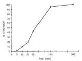
- Figure 2illustrates the effects of various attachment factors on the proliferation of cultured human corneal endothelial cells in the presence or absence of bFGF.
- Figure 3is a time curve of attachment of cultured human corneal endothelial cells onto the denuded human corneal buttons coated with attachment agents.
- the endothelial cellsare seeded onto membranes of a cell culture inset. These endothelial cells will form the inner layer, or basal layer, of the corneal equivalent.
- the preparation of such a denuded corneacan be achieved with treatment from a diluted detergent such as Triton X-100 (0.5-5%) or ammonium hydroxide (at concentrations ranging from 10 mM to 200 mM) for a time period ranging from 2 minutes to 60 minutes at a temperature of 4°C to 25°C.
- a diluted detergentsuch as Triton X-100 (0.5-5%) or ammonium hydroxide (at concentrations ranging from 10 mM to 200 mM) for a time period ranging from 2 minutes to 60 minutes at a temperature of 4°C to 25°C.
- the denuded corneaswill be washed extensively 8-10 times with phosphate buffered saline (PBS) to remove any detergent or ammonium hydroxide residue.
- PBSphosphate buffered saline
- a predetermined mixture of attachment proteins containing fibronectinranging from 0.1 ⁇ g to 500 ⁇ g/ml in PBS
- laminin0.1 ⁇ g to 500 ⁇ g/ml in PBS
- RGDSRGDS (0.01 ⁇ g to 100 ⁇ g/ml in PBS
- collagen type IVranging from 0.1 ⁇ g to 1000 ⁇ g in 0.1 M acetic acid
- the residual protein mixturewill be removed after the incubation period, and the cornea is rinsed three times with PBS and placed endothelial side up on a Teflon concave holder.
- An 11 mm diameter buttonwill be punched out with a size 11 trephine. This button will be ready to receive the cultured corneal endothelial cells.
- the cultured human endothelial cellswill be removed from the tissue culture dish with 0.05% trypsin and 0.02% EDTA in saline solution.
- the cell suspensionwill be counted with a Coulter Particle Counter (Z1 model, Beckman-Coulter) and a preparation of about 50,000 to 500,000 cells/ml, preferably about 200,000 cells in 200 ⁇ l of culture medium (DME-H16 with 5% fetal calf serum or a serum-free medium containing a mixture of attachment proteins such as fibronectin, laminin, and fibroblast growth factors (at 10 ng to 400 ng/ml) will be added carefully onto the denuded corneal button.
- DME-H16with 5% fetal calf serum
- serum-free mediumcontaining a mixture of attachment proteins such as fibronectin, laminin, and fibroblast growth factors (at 10 ng to 400 ng/ml) will be added carefully onto the denuded corneal button.
- a layer of 1% sodium hyaluronate, such as Healon® (Advanced Medical Optics, Santa Ana, CA) at approximately 0.1 to 0.5 ml,will be layered onto the cell suspension as a protectant.
- the corneal buttonwill then be incubated at 37°C in a 10% CO 2 incubator for a period of 10 minutes up to 24 hours.
- the coated corneal buttonwill be incubated for 20 minutes and the cornea will be rinsed three times with PBS at 25°C and ready for transplantation.
- a poly-gel stromacan be molded into a cornea shape, and the concave side (endothelial side) will be treated with a mixture of attachment proteins and growth factors such as fibronectin (ranging from 0.1 to 500 ⁇ g/ml in PBS), laminin (ranging from 0.1 to 500 ⁇ g/ml in PBS), RGDS (ranging from 0.01 to 100 ⁇ g/ml in PBS), collagen type IV (ranging from 0.1 ⁇ g to 1000 ⁇ g in 0.1 M acetic acid), FGF (10 to 400 ng/ml in PBS), EGF (10 to 400 ng/ml in PBS), or TGF ⁇ (1 to 100 ng/ml in PBS).
- fibronectinranging from 0.1 to 500 ⁇ g/ml in PBS
- lamininranging from 0.1 to 500 ⁇ g/ml in PBS
- RGDSranging from 0.01 to 100 ⁇ g/ml in PBS
- collagen type IVranging from 0.1 ⁇ g
- the artificial stromaAfter incubation at 4°C for a period ranging from 10 minutes to 2 hours, the artificial stroma will be rinsed three times with PBS, and cultured human corneal endothelial cells at a density of about 50,000 to about 10 6 cells/ml preferably about 150,000 to 250,000 cells/200 ml of culture medium (DMA-H16 with 5% FCS or a mixture of attachment proteins containing fibronectin, laminin, RGDS, and collagen type IV) will be added to a corneal button of 11mm diameter.
- culture mediumDMA-H16 with 5% FCS or a mixture of attachment proteins containing fibronectin, laminin, RGDS, and collagen type IV
- a layer of (10 mg/mL sodium hyaluronate, 0.1 to 0.5 ml)will be applied carefully onto the cell layer as a protectant, and the button will be incubated at 37°C in a 10% CO 2 incubation for a period ranging from 10 minutes to 24 hours.
- the artificial corneal buttonwill be rinsed 3 times with PBS after the incubation and will be ready for transplantation.
- the corneal endothelial cells used to form the endothelial layercan be derived from a variety of mammalian sources.
- Non-transformed corneal endothelial cells derived from sheep, rabbit, and cowshave been used.
- Mouse corneal endothelial cellshave been transformed with large T antigen of SV40. ( Muragaki, Y., et al., Eur. J. Biochem. 207(3):395-902 (1992 ).)
- Non-human cell types which can be usedalso include transformed mouse corneal endothelial cell lines, or normal corneal endothelial cells derived from sheep or rabbit.
- the normal rabbit endothelial cellscan be derived from enzymatically dissociated corneal endothelium or from explants of cornea and are serially cultivated in MSBM medium ( Johnson, W.E. et al., In Vitro Cell. Dev. Biol. 28A:429-435 (1992 )) modified by the addition of 50 ⁇ g/mL heparin and 0.4 ⁇ g/mL heparin binding growth factor-1 (MSBME).
- MSBM mediumJohnson, W.E. et al., In Vitro Cell. Dev. Biol. 28A:429-435 (1992 )
- MSBMEheparin binding growth factor-1
- endothelial cells from a non-corneal originare also disclosed.
- the non-corneal origin endothelial cells that have also been used in this inventioninclude ovine and canine vascular and human umbilical vein endothelial cells.
- the endothelial cellsmay be transformed with a recombinant retrovirus containing the large T antigen of SV40 (Muragaki, et al., 1992, supra). Transformed cells continue to grow in the corneal equivalent and form mounds on top of the acellular layer due to their lack of contact inhibition. Non-transformed cells will form a monolayer underlying the stromal cell-collagen layer.
- normal endothelial cellsmay be transfected as above, but with the addition of a recombinant construct that expresses a heat sensitive gene. These transformed cells will grow in continuous culture under reduced temperature. After establishment of a confluent endothelial cell layer, the temperature can be raised to deactivate the transforming gene, allowing the cells to resume their normal regulation and exhibit contact inhibition, to form an endothelial cell monolayer similar to the non-transformed cells. Most peptides are heat sensitive (with the exception of heat shock proteins) so that there is a wide choice of peptides that can be deactivated by raising culturing temperature. Transformation in this way also facilitates the use of hard to obtain and cultivate cell types such as human corneal endothelial cells.
- ECMextracellular matrix
- Bovine corneal endothelial cells (BCEC) in culturewill be seeded onto dishes in a DME-H16 medium containing 10% FCS, 5% CS, 5% Dextran, 300 ug/ml glutamine, 2.5 ug/ml Amphotericin B, and 50 ng/ml bFGF.
- the disheswill be treated with 20 mM NH 4 OH at a volume sufficient to cover at least 2/3 of the plate. After 5 minutes of shaking in a mechanical shaker, the NH 4 OH will be aspirated and the dished rinsed 5 times with PBS.
- the disheswill be stored at 4°C at least a week prior to use in order to eliminate any surviving BCEC.
- Laminin and fibronectinwill be dissolved in distilled water at a concentration of 100 ⁇ g/ml.
- Type IV collagenwill be dissolved in 0.6% v/v acetic acid/water. Laminin, fibronectin, and type IV collagen will be added to the ECM plates as needed for culture purposes.
- Example 1Non-enzymatic dissection of primary human corneal endothelial cells
- the corneal rims from human donors (after the central portion has been removed for transplantation) or whole donor corneaswill be rinsed in a large volume (50 ml) of phosphate buffered saline (PBS). They will then be placed in endothelial side up on a holder. The trabecular meshwork and remnants of iris will be removed carefully by micro-dissection. By using sharp pointed jeweler's forceps, the endothelial cell layers and the Descemet's membrane will be peeled off very carefully with great care taken not to include any underlying stromal tissue.
- PBSphosphate buffered saline
- This stepcan be confirmed by viewing the dissected Descemet's membrane under an inverted microscope to make sure it only carries the corneal endothelial cells on one side and nothing on the other side.
- the piece of tissuewill be placed onto an ECM coated 35 mm tissue culture dish or similarly suitable container, filled with approximately 0.5 ml of culture medium (DME-H16 with 15% fetal calf serum enriched with b-FGF at 250 ng/ml).
- the dishwill be incubated at 37°C in a 10% CO 2 incubator for 24 hours, and then another 1 ml of culture medium will be added.
- the samplewill be incubated without disturbance for about 7 days to see if a colony of corneal endothelial cells migrates outwards from the tissue sample, at which time (7 to 14 days after the sample is placed in culture) the medium is changed every other day until the cell count reaches about 200-500 cells.
- the cellsWhen the primary cell count from the tissue sample outgrowth reaches a number of between 150 to 750, preferably between about 200 to 500, the cells will be released from the dish with STV solution (0.05% trypsin, 0.02% EDTA in normal saline). The STV solution will be removed when the cells round up but are still attached to the culture dish. No centrifugation step is necessary since the remaining STV will be inactivated by the growth media containing 15% fetal calf serum.
- the corneal cellswill be placed onto a 60-mm ECM-coated dish (between about 250 to 1000 cells, preferably about 500 cells per dish). The medium will be changed every other day and b-FGF at a concentration of 250 ng/ml will be added at the time of medium change.
- the cellswill be passaged again at the same split ratio (1:16 to 1:64) or will be frozen in 10% DMSO, 15% FCS at a density of between about 5 x 10 5 to about 5 x 10 6 cells/ml, preferably about 10 6 cells/ml per ampoule and stored in liquid nitrogen for future use.
- the passagingcan be carried out for up to 8 times without loss of cell functions or morphological integrity.
- the corneal buttonwill be placed endothelial side up in a holder, and rinsed three times with PBS. Then a solution of ammonium hydroxide at a concentration ranging from 10 mM to 200 mM will be added carefully into the corneal button without spilling over the top. The cornea will be kept at temperatures of about 10°C to 25°C for a period of 5 minutes up to 2 hours. Then the ammonium hydroxide will be removed, and the inside of the cornea button rinsed approximately 10 times with PBS. A cotton swab will be slid gently across the endothelial surface to remove any residual cell skeletons or debris. The corneal button is rinsed again three times with PBS, punched with an 11 mm trephine, and is then ready for coating with cultured human corneal endothelial cells.
- the native corneal endotheliumcan be removed by adding Triton-X100 at a concentration of 0.5 to 5% in distilled water kept at 10°C for a period ranging from 5 minutes to 2 hours, and then processed as previously described. Furthermore, the corneal endothelium can be treated with distilled water for a period of 20 minutes to 2 hours at a temperature ranging from 4°C to 25°C. Then the cotton swab will be slid gently across the endothelial surface to remove the cell cytoskeleton and debris. The cornea will then be processed with an 11 mm trephination.
- Example 4Treatment of Denuded Corneas with Attachment Proteins and Growth Factors.
- the denuded cornea buttonAfter trephination, the denuded cornea button will be placed endothelial side up again in a holder.
- the specimenis allowed to incubate at 4°C for a time ranging from 5 minutes to 2 hours, at the end of which the cocktail will be removed and the cornea rinsed 3 times with PBS.
- the cultured cornea endothelial cellswill be removed from the culture dish with STV solution (0.05% trypsin, 0.02% EDTA in saline) as previously described. The cells will be centrifuged at 2000 rpm for 5 minutes and the culture medium removed. The cell pellet will be resuspended with 2 ml of DME-H16 medium containing fetal calf serum at a concentration of 0.1 to 5%. About 100 ⁇ l of the suspension will be counted in a Coulter Particle Counter to determine the cell number per ml.
- the cell concentrationwill be adjusted to about 5 x 10 5 to 10 7 cells/ml, preferably about 10 6 cells/ml and 200 ⁇ g of the cell suspension will be added carefully added into the 11mm trephined corneal button.
- a layer of sodium hyaluronate, at a concentration of about 10mg/ml, ranging from 0.2 to 0.5 ml (Healon®)will be overlaid carefully onto the cell suspension as a protectant.
- the corneal buttonwill then be incubated at 37°C in a 10% CO 2 incubator for a period of 20 minutes to 24 hours.
- the human corneal endothelial cell coated buttonwill be ready for transplantation.
- an artificial matrixcan be generated although such material may not be as effective in promoting the growth and morphological integrity (hexagonal shape) of the HCEC.
- fibronectin, laminin and RGDSwill be dissolved at concentrations of about 100 ⁇ g/ml in distilled water, and collagen type IV is dissolved at a concentration of about 1 mg/ml in 0.01% acetic acid.
- Basic FGFis dissolved at a concentration of about 100 ⁇ g/ml in bovine serum albumin (0.05% w/v). All the materials are mixed together in a 15 ml centrifuge tube and swirled gently to avoid bubbling. The mixture is then incubated at 4°C for two hours.
- the mixtureis dilute 1:10 with phosphate buffered saline, and then 1 ml of the solution is added to a 35 mm dish and store at 4°C for 1 hour. Prior to use the solution is aspirated and the cell suspension will be added to the dish.
Landscapes
- Health & Medical Sciences (AREA)
- Life Sciences & Earth Sciences (AREA)
- Engineering & Computer Science (AREA)
- Biomedical Technology (AREA)
- Chemical & Material Sciences (AREA)
- Zoology (AREA)
- Cell Biology (AREA)
- General Health & Medical Sciences (AREA)
- Bioinformatics & Cheminformatics (AREA)
- Biotechnology (AREA)
- Organic Chemistry (AREA)
- Wood Science & Technology (AREA)
- Genetics & Genomics (AREA)
- Animal Behavior & Ethology (AREA)
- Chemical Kinetics & Catalysis (AREA)
- Botany (AREA)
- Public Health (AREA)
- Veterinary Medicine (AREA)
- Epidemiology (AREA)
- Dermatology (AREA)
- Transplantation (AREA)
- Medicinal Chemistry (AREA)
- Oral & Maxillofacial Surgery (AREA)
- Microbiology (AREA)
- General Engineering & Computer Science (AREA)
- Biochemistry (AREA)
- Urology & Nephrology (AREA)
- Vascular Medicine (AREA)
- Neurosurgery (AREA)
- Neurology (AREA)
- Ophthalmology & Optometry (AREA)
- Developmental Biology & Embryology (AREA)
- Materials For Medical Uses (AREA)
- Micro-Organisms Or Cultivation Processes Thereof (AREA)
- Apparatus Associated With Microorganisms And Enzymes (AREA)
Abstract
Description
- This patent describes improved methods of dissecting, seeding and subsequent propagation of pure culture of human corneal endothelial cells on extracellular matrix.
- For various reasons, the corneal portions of eyes may need to be surgically repaired or replaced. For example, the cornea may become scratched or scarred or otherwise physically damaged, greatly hindering sight. The cornea is also subject to the effects of various degenerative diseases, mandating replacement if the patient is to have normal or even near normal vision.
- The cornea of the human eye is a specialized structure made up of substantially parallel relatively compacted layers of tissue. The outermost or most superficial layer of the cornea is the epithelial layer. This is a protective layer of tissue which regenerates if injured. Moving inwardly in the eye is the base surface of the epithelial layer known as Bowman's membrane. Immediately adjacent the Bowman's membrane is the stroma of the cornea, which is an extra-cellular collagen architectural matrix with scattered keratocytic cells. The stroma layer is bounded at its deepest level by a cuticular, a cellular membrane, referred to as Descemet's membrane, which is followed by a monolayer of single cell thickness of specialized endothelial cells which forms the posterior surface of the cornea. The endothelial layer does not regenerate and when it is diseased, scratched or otherwise injured, it must be replaced.
- In some animal species including human, the corneal endothelium does not normally replicate in vivo to replace cells lost due to injury or aging (Murphy C, et al., Invest. Ophthalmology Vis. Sci. 1984; 25:312-322;Laing R A, et al., Exp. Eye Res. 1976; 22:587-594). However, human corneal cells can be cultured in vitro with a growth factor-enriched, fetal calf serum-containing medium under normal tissue culture conditions (Baum JL, et al., Arch. Ophthalmol. 97:1136-1140, 1979;Engelmann K, et al., Invest. Ophthalmol. Vis. Sci. 29:1656-1662, 1998;Engelmann K, and Friedl P; In Vitro Cell Develop. Biol. 25:1065-1072, 1989). If the cultured cells can be utilized to replace the loss of corneal endothelial cells it will greatly enhance the donor pool of human corneas. This is important as one may be able to augment the donor corneas currently rejected for transplantation procedures due to inadequate endothelial cell counts (Gospodarowicz D, et al., Proc. Natl. Acad. Sci. (USA) 76:464-468, 1979;Gospodarowicz D, et al., Arch. Ophthalmol. 97:2163-2169, 1979). This pool of corneas, rejected due to low endothelial cell density, makes up to 30% of the total donated corneas annually (National Eye Institute: Summary report on the cornea task force. Invest Ophthalmol Vis Sci 12:391-397, 1973). Furthermore, a method to culture human corneal endothelial cells from a low initial density, and the ability to reseed the cells grown in vitro onto denuded corneal buttons, will enable the use of the recipient's own undamaged stroma for allo-cell and auto-stroma type of transplantation (Insler MS, and Lopez JG, Cornea 10:136-148, 1991).
- Tissue culture techniques are being successfully used in developing tissue and organ equivalents. The basis for these techniques involve collagen matrix structures, which are capable of being remodeled into functional tissue and organs by employing the right combination of living cells, nutrients, and culturing conditions. Tissue equivalents have been described extensively in many patents, including
U.S. Pat. Nos. 4,485,096 ;4,485,097 ;4,539,716 ;4,546,500 ;4,604,346 ;4,837,379 ; and5,827,641 , all of which are incorporated herein by reference. One successful application of the tissue equivalent is the living skin equivalent, which has morphology similar to actual human skin. The living skin equivalent is composed of two layers: the upper portion is made of differentiated and stratified human epidermal keratinocytes that cover a thicker, lower layer of human dermal fibroblasts in a collagen matrix.Bell, et al., "Recipes for Reconstituting Skin," J. of Biochemical Engineering, 113:113-119 (1991). - Studies have been done on culturing corneal epithelial and endothelial cells.Xie, et al., "A simplified technique for the short-term tissue culture of rabbit corneal cells," In Vitro Cellular & Developmental Biology, 25:20-22 (1989) andSimmons, et al., "Corneal Epithelial Wound Cloture in Tissue Culture: An in vitro Model of Ocular Irritancy," Toxicology and Applied Pharmacology, 88:13-23 (1987).
- ENGELMANN K ET AL: "[Endothelial cell transplantation and growth behaviour of the human corneal endothelium]" DER OPHTHALMOLOGE: ZEITSCHRIFT DER DEUTSCHEN OPHTHALMOLOGISCHEN GESELLSCHAFT. SEP 1999, vol. 96, no. 9, September 1999 (1999-09), pages 555-562, XP002312235 ISSN: 0941-293X, discloses the culturing of human corneal endothelial cells and a method ofin vivo transplantation of these cells onto the corneal membrane.
- MIYATA ET AL: "Effect of donor age on morphologic variation of cultured human corneal endothelial cells." CORNEA. JAN 2001, vol. 20, no. 1, January 2001 (2001-01), pages 59-63, XP002312236 ISSN: 0277-3740, discloses culturing of human corneal endothelial cells from donors of varying age and the effects thereof.
- DATABASE BIOSIS [Online] BIOSCIENCES INFORMATION SERVICE, PHILADELPHIA, PA, US; December 2002 (2002-12), AMANO SHIRO: "Transplantation of corneal endothelial cells." XP002312237Database accession no. PREV200300187415, discloses culturing of human corneal endothelial cells on extracellular matrix from calf corneal endothelial cells by using basic FGF and foetal bovine serum.
- Until the advent of the present invention, prior art methods of culturing human corneal endothelial cells (HCEC) encountered problems such as the fact that HCEC cells could only be seeded at high cell density (2000-5000 cells/square mm) therefore limiting the possibility to start a primary culture from small specimen, and that HCEC cells could not be passaged continuously at low seeding density (50-100 cells/square mm) which limits the ability to expand the HCEC stock for storage and future use.
- In a first aspect the invention relates to a method of non-enzymatic harvesting andin vitro culturing human corneal endothelial cells for transplantation comprising the steps of:
- a) dissecting human corneal endothelial cells from a tissue source; growing said corneal endothelial cells at a low initial density of 100-500 cells/mm2 in extracellular matrix coated (ECM) culture plates for a period of time, wherein said culture plates having been treated with a natural extracellular matrix as supplied by bovine corneal endothelial culture, or a synthetic attachment protein mixture, or growing said human corneal endothelial cells at a low initial density of 100-500 cells/mm2 on a carbon plasma deposit known as diamond-like carbon (DLC);
- b) passaging said cells into a secondary culture system wherein the secondary culture system is comprised of ECM coated culture plates and the addition of sufficient cellular growth factors;
- c) growing the human corneal endothelial cells to confluency; and
- d) harvesting the human corneal endothelial cells from the second culture system in sufficient quantities to be useful in transplantation to a subject in vivo.
- The present invention provides for a method of culturing HCEC on a novel extracellular matrix which enables the establishment of primary cultures from small specimens of HCEC (100-500 cells). The present invention also provides for a method which to expand these primary HCEC colonies via serial passage into large quantity of cells for transplantation and cryostorage for future use.
- A method for initiating primary cultures of corneal endothelial cells, including that of human origin, by using dissection enables the culture to start from a low initial density (100-500 cells/mm2) and expand from a seeding density of 1 to 32. These cells can be effectively passaged 7 to 8 times without losing their morphological integrity and physiological functions, such as formation of tight intracellular junctions and Na/K pump activation. The corneal endothelial cultures can be maintained in a commonly used fetal bovine serum (FBS) supplemented culture medium enriched with selected growth factors such as
fibroblast growth factors 1 and 2 (FGF1, FGF2), epidermal growth factors (EGF), transforming growth factor β (TGFβ), endothelial cell growth factor (ECGF), and other growth factors known in the art of cell culture. In particular, if the corneal endothelial cells are propagated in a natural extracellular matrix as supplied by bovine corneal endothelial culture, or a synthetic attachment protein mixture containing such components as fibronectin, laminin, collagen type I and IV, and RGDS, or on a carbon plasma deposit known as diamond-like carbon (DLC), the culture will assume a more hexagonal morphology upon repeated passage at high split ratio (1:32 or 1:64) for up to 10 passages. The generation of a large pool of corneal endothelial cells, especially that of human origin, can be banked in cryo-storage and used for future cell transplantation procedures. - It is therefore an object of the present invention to provide a method of cell culture of HCEC that can be used for the establishing and serial culturing of other cell types of human origins such as neurons, pancreatic beta cells, and chrondocytes.
- It is another object of the present invention to create HCEC in sufficient quantities of HCEC that can be used for other purposes.
- It is a further object of the present invention to provide an in vitro cell culture model of the human cornea.
- It is also an object of the present invention to provide a means for regenerating corneal endothelial cells in a cornea by replacing the damaged corneal endothelial cells with HCEC grown in culture system of the present invention.
- It is yet a further aspect of the present invention to provide a means for regenerating other types of damaged human and mammalian endothelial cells by replacing the damaged endothelial cells with endothelial cells grown in the culture system of the present invention.
- These and other objects of the invention, as well as many of the attendant advantages thereof, will become more readily apparent when reference is made to the following detailed description of the preferred embodiments.
- Figure 1 shows generation curves for long term serial propagation of cultured human endothelial cells on different substrates.
- Figure 2 illustrates the effects of various attachment factors on the proliferation of cultured human corneal endothelial cells in the presence or absence of bFGF.
- Figure 3 is a time curve of attachment of cultured human corneal endothelial cells onto the denuded human corneal buttons coated with attachment agents.
- In the first step of constructing the in vitro cornea model, the endothelial cells are seeded onto membranes of a cell culture inset. These endothelial cells will form the inner layer, or basal layer, of the corneal equivalent.
- We have developed a system where the native corneal endothelium from a donor cornea, whose endothelial cell population is inadequate in cell count or damaged due to disease or physical disruption, can be removed without harming the integrity of the Descemet's membrane and the structure and function of the stromal later. The preparation of such a denuded cornea can be achieved with treatment from a diluted detergent such as Triton X-100 (0.5-5%) or ammonium hydroxide (at concentrations ranging from 10 mM to 200 mM) for a time period ranging from 2 minutes to 60 minutes at a temperature of 4°C to 25°C. The denuded corneas will be washed extensively 8-10 times with phosphate buffered saline (PBS) to remove any detergent or ammonium hydroxide residue. The denuded corneas will then be ready for cell coating from the cultured human corneal endothelial cell stock.
- Prior to seeding the cells onto the denuded corneal surface, a predetermined mixture of attachment proteins containing fibronectin (ranging from 0.1 µg to 500 µg/ml in PBS), laminin (0.1 µg to 500 µg/ml in PBS), RGDS (0.01 µg to 100 µg/ml in PBS), collagen type IV (ranging from 0.1 µg to 1000 µg in 0.1 M acetic acid) will be added to the denuded surface (Descemet's membrane) and incubated at 4°C for a period ranging from 5 to 60 minutes. The residual protein mixture will be removed after the incubation period, and the cornea is rinsed three times with PBS and placed endothelial side up on a Teflon concave holder. An 11 mm diameter button will be punched out with a size 11 trephine. This button will be ready to receive the cultured corneal endothelial cells.
- The cultured human endothelial cells will be removed from the tissue culture dish with 0.05% trypsin and 0.02% EDTA in saline solution. The cell suspension will be counted with a Coulter Particle Counter (Z1 model, Beckman-Coulter) and a preparation of about 50,000 to 500,000 cells/ml, preferably about 200,000 cells in 200 µl of culture medium (DME-H16 with 5% fetal calf serum or a serum-free medium containing a mixture of attachment proteins such as fibronectin, laminin, and fibroblast growth factors (at 10 ng to 400 ng/ml) will be added carefully onto the denuded corneal button. A layer of 1% sodium hyaluronate, such as Healon® (Advanced Medical Optics, Santa Ana, CA) at approximately 0.1 to 0.5 ml, will be layered onto the cell suspension as a protectant. The corneal button will then be incubated at 37°C in a 10% CO2 incubator for a period of 10 minutes up to 24 hours. Alternatively, the coated corneal button will be incubated for 20 minutes and the cornea will be rinsed three times with PBS at 25°C and ready for transplantation.
- Alternatively, the process of maintaining human corneal endothelial cells in culture, expansion of the corneal endothelial cells, and the preparation of the attachment protein can be used to coat artificial cornea stroma generated from polymer-gel composition. Briefly, a poly-gel stroma can be molded into a cornea shape, and the concave side (endothelial side) will be treated with a mixture of attachment proteins and growth factors such as fibronectin (ranging from 0.1 to 500 µg/ml in PBS), laminin (ranging from 0.1 to 500 µg/ml in PBS), RGDS (ranging from 0.01 to 100 µg/ml in PBS), collagen type IV (ranging from 0.1 µg to 1000 µg in 0.1 M acetic acid), FGF (10 to 400 ng/ml in PBS), EGF (10 to 400 ng/ml in PBS), or TGFβ (1 to 100 ng/ml in PBS). After incubation at 4°C for a period ranging from 10 minutes to 2 hours, the artificial stroma will be rinsed three times with PBS, and cultured human corneal endothelial cells at a density of about 50,000 to about 106 cells/ml preferably about 150,000 to 250,000 cells/200 ml of culture medium (DMA-H16 with 5% FCS or a mixture of attachment proteins containing fibronectin, laminin, RGDS, and collagen type IV) will be added to a corneal button of 11mm diameter. A layer of (10 mg/mL sodium hyaluronate, 0.1 to 0.5 ml) will be applied carefully onto the cell layer as a protectant, and the button will be incubated at 37°C in a 10% CO2 incubation for a period ranging from 10 minutes to 24 hours. The artificial corneal button will be rinsed 3 times with PBS after the incubation and will be ready for transplantation.
- In alternative embodiments, the corneal endothelial cells used to form the endothelial layer can be derived from a variety of mammalian sources. Non-transformed corneal endothelial cells derived from sheep, rabbit, and cows have been used. Mouse corneal endothelial cells have been transformed with large T antigen of SV40. (Muragaki, Y., et al., Eur. J. Biochem. 207(3):395-902 (1992).) Non-human cell types which can be used also include transformed mouse corneal endothelial cell lines, or normal corneal endothelial cells derived from sheep or rabbit. The normal rabbit endothelial cells can be derived from enzymatically dissociated corneal endothelium or from explants of cornea and are serially cultivated in MSBM medium (Johnson, W.E. et al., In Vitro Cell. Dev. Biol. 28A:429-435 (1992)) modified by the addition of 50 µg/mL heparin and 0.4 µg/mL heparin binding growth factor-1 (MSBME).
- The use of endothelial cells from a non-corneal origin, is also disclosed. The non-corneal origin endothelial cells that have also been used in this invention include ovine and canine vascular and human umbilical vein endothelial cells. The endothelial cells may be transformed with a recombinant retrovirus containing the large T antigen of SV40 (Muragaki, et al., 1992, supra). Transformed cells continue to grow in the corneal equivalent and form mounds on top of the acellular layer due to their lack of contact inhibition. Non-transformed cells will form a monolayer underlying the stromal cell-collagen layer. Alternatively, normal endothelial cells may be transfected as above, but with the addition of a recombinant construct that expresses a heat sensitive gene. These transformed cells will grow in continuous culture under reduced temperature. After establishment of a confluent endothelial cell layer, the temperature can be raised to deactivate the transforming gene, allowing the cells to resume their normal regulation and exhibit contact inhibition, to form an endothelial cell monolayer similar to the non-transformed cells. Most peptides are heat sensitive (with the exception of heat shock proteins) so that there is a wide choice of peptides that can be deactivated by raising culturing temperature. Transformation in this way also facilitates the use of hard to obtain and cultivate cell types such as human corneal endothelial cells.
- Bovine corneal endothelial cells (BCEC) in culture will be seeded onto dishes in a DME-H16 medium containing 10% FCS, 5% CS, 5% Dextran, 300 ug/ml glutamine, 2.5 ug/ml Amphotericin B, and 50 ng/ml bFGF. At confluency, (7-10 days post seeding), the dishes will be treated with 20 mM NH4OH at a volume sufficient to cover at least 2/3 of the plate. After 5 minutes of shaking in a mechanical shaker, the NH4OH will be aspirated and the dished rinsed 5 times with PBS. The dishes will be stored at 4°C at least a week prior to use in order to eliminate any surviving BCEC. Laminin and fibronectin will be dissolved in distilled water at a concentration of 100 µg/ml. Type IV collagen will be dissolved in 0.6% v/v acetic acid/water. Laminin, fibronectin, and type IV collagen will be added to the ECM plates as needed for culture purposes.
- The corneal rims from human donors (after the central portion has been removed for transplantation) or whole donor corneas will be rinsed in a large volume (50 ml) of phosphate buffered saline (PBS). They will then be placed in endothelial side up on a holder. The trabecular meshwork and remnants of iris will be removed carefully by micro-dissection. By using sharp pointed jeweler's forceps, the endothelial cell layers and the Descemet's membrane will be peeled off very carefully with great care taken not to include any underlying stromal tissue. This step can be confirmed by viewing the dissected Descemet's membrane under an inverted microscope to make sure it only carries the corneal endothelial cells on one side and nothing on the other side. The piece of tissue will be placed onto an ECM coated 35 mm tissue culture dish or similarly suitable container, filled with approximately 0.5 ml of culture medium (DME-H16 with 15% fetal calf serum enriched with b-FGF at 250 ng/ml). The dish will be incubated at 37°C in a 10% CO2 incubator for 24 hours, and then another 1 ml of culture medium will be added. The sample will be incubated without disturbance for about 7 days to see if a colony of corneal endothelial cells migrates outwards from the tissue sample, at which time (7 to 14 days after the sample is placed in culture) the medium is changed every other day until the cell count reaches about 200-500 cells.
- When the primary cell count from the tissue sample outgrowth reaches a number of between 150 to 750, preferably between about 200 to 500, the cells will be released from the dish with STV solution (0.05% trypsin, 0.02% EDTA in normal saline). The STV solution will be removed when the cells round up but are still attached to the culture dish. No centrifugation step is necessary since the remaining STV will be inactivated by the growth media containing 15% fetal calf serum. The corneal cells will be placed onto a 60-mm ECM-coated dish (between about 250 to 1000 cells, preferably about 500 cells per dish). The medium will be changed every other day and b-FGF at a concentration of 250 ng/ml will be added at the time of medium change. At confluence (about 7 to 10 days after plating), the cells will be passaged again at the same split ratio (1:16 to 1:64) or will be frozen in 10% DMSO, 15% FCS at a density of between about 5 x 10⁵ to about 5 x 10⁶ cells/ml, preferably about 106 cells/ml per ampoule and stored in liquid nitrogen for future use. The passaging can be carried out for up to 8 times without loss of cell functions or morphological integrity.
- For each of the 5 ml of HCEC collected, 0.5 ml of DMSO was added to the cell suspension. Each 1.1 ml of the mixture was aliquoted into a 1.5 ml cryopreservation tube to yield an approximate 1 million cells per vial final concentration. The vials were then put into a Styrofoam box and let stand in a -80°C freezer for 24 hours. After 1 day, the ampoules were transfer into liquid nitrogen for long term storage.
- Human donor corneal buttons are obtained from the Eye Bank. These corneal buttons are deemed unsuitable for transplantation due to inadequate endothelial cell counts, but otherwise are healthy and disease free and obtained under eye banking guidelines.
- The corneal button will be placed endothelial side up in a holder, and rinsed three times with PBS. Then a solution of ammonium hydroxide at a concentration ranging from 10 mM to 200 mM will be added carefully into the corneal button without spilling over the top. The cornea will be kept at temperatures of about 10°C to 25°C for a period of 5 minutes up to 2 hours. Then the ammonium hydroxide will be removed, and the inside of the cornea button rinsed approximately 10 times with PBS. A cotton swab will be slid gently across the endothelial surface to remove any residual cell skeletons or debris. The corneal button is rinsed again three times with PBS, punched with an 11 mm trephine, and is then ready for coating with cultured human corneal endothelial cells.
- Alternatively, the native corneal endothelium can be removed by adding Triton-X100 at a concentration of 0.5 to 5% in distilled water kept at 10°C for a period ranging from 5 minutes to 2 hours, and then processed as previously described. Furthermore, the corneal endothelium can be treated with distilled water for a period of 20 minutes to 2 hours at a temperature ranging from 4°C to 25°C. Then the cotton swab will be slid gently across the endothelial surface to remove the cell cytoskeleton and debris. The cornea will then be processed with an 11 mm trephination.
- After trephination, the denuded cornea button will be placed endothelial side up again in a holder. A solution of attachment proteins containing fibronectin at a concentration ranging from 10 µg to 500 µg/ml in PBS, laminin (10 µg to 500 µg/ml in PBS), RGDS (1 µg to 100 µg/ml in PBS), collagen type IV (10 µg to 1000 µg in 0.1 M acetic acid), b-FGF (1 to 500 ng/ml in PBS), EGF (1 ng to 500 ng/ml in PBS) will be added carefully onto the denuded cornea button. The specimen is allowed to incubate at 4°C for a time ranging from 5 minutes to 2 hours, at the end of which the cocktail will be removed and the cornea rinsed 3 times with PBS.
- The cultured cornea endothelial cells will be removed from the culture dish with STV solution (0.05% trypsin, 0.02% EDTA in saline) as previously described. The cells will be centrifuged at 2000 rpm for 5 minutes and the culture medium removed. The cell pellet will be resuspended with 2 ml of DME-H16 medium containing fetal calf serum at a concentration of 0.1 to 5%. About 100 µl of the suspension will be counted in a Coulter Particle Counter to determine the cell number per ml. Then the cell concentration will be adjusted to about 5 x 10⁵ to 107 cells/ml, preferably about 106 cells/ml and 200 µg of the cell suspension will be added carefully added into the 11mm trephined corneal button. A layer of sodium hyaluronate, at a concentration of about 10mg/ml, ranging from 0.2 to 0.5 ml (Healon®) will be overlaid carefully onto the cell suspension as a protectant. The corneal button will then be incubated at 37°C in a 10% CO2 incubator for a period of 20 minutes to 24 hours. The human corneal endothelial cell coated button will be ready for transplantation.
- In an alternate embodiment, an artificial matrix can be generated although such material may not be as effective in promoting the growth and morphological integrity (hexagonal shape) of the HCEC.
- To generate the artificial matrix, fibronectin, laminin and RGDS will be dissolved at concentrations of about 100 µg/ml in distilled water, and collagen type IV is dissolved at a concentration of about 1 mg/ml in 0.01% acetic acid. Basic FGF is dissolved at a concentration of about 100 µg/ml in bovine serum albumin (0.05% w/v). All the materials are mixed together in a 15 ml centrifuge tube and swirled gently to avoid bubbling. The mixture is then incubated at 4°C for two hours.
- To coat the tissue culture dishes, the mixture is dilute 1:10 with phosphate buffered saline, and then 1 ml of the solution is added to a 35 mm dish and store at 4°C for 1 hour. Prior to use the solution is aspirated and the cell suspension will be added to the dish.
- Having described the invention, many modifications thereto will become apparent to those skilled in the art to which it pertains without deviation from the invention as defined by the scope of the appended claims.
Claims (7)
- An in vitro method of non-enzymatic harvesting and culturing human corneal endothelial cells for transplantation comprising the steps of:a) dissecting human corneal endothelial cells from a tissue source; growing said corneal endothelial cells at a low initial density of 100-500 cells/mm2 in extracellular matrix coated (ECM) culture plates for a period of time, wherein said culture plates having been treated with a natural extracellular matrix as supplied by bovine corneal endothelial culture, or a synthetic attachment protein mixture, or growing said human corneal endothelial cells at a low initial density of 100-500 cells/mm2 on a carbon plasma deposit known as diamond-like carbon (DLC);b) passaging said cells into a secondary culture system wherein the secondary culture system is comprised of ECM coated culture plates and the addition of sufficient cellular growth factors;c) growing the human corneal endothelial cells to confluency; andd) harvesting the human corneal endothelial cells from the second culture system in sufficient quantities to be useful in transplantation to a subject in vivo.
- The method of claim 1 wherein the dissection of the corneal endothelial cells further comprises the step of removing the cells from the tissue source such that the cells are dissected away from the stroma of said tissue source prior to growing in the first culture system.
- The method of claim 2 wherein said tissue source can be corneal buttons or rims.
- The method of claim 2 wherein the ECM is comprised of Bovine corneal endothelial cell extracellular matrix (BCE-ECM) or an artificially generated extracellular matrix (AG-ECM).
- The method of claim 4 wherein the artificially generated endothelial cell extracellular matrix (AG-ECM) is made by the process comprising:a) fibronectin, laminin and RGDS (Arg-Gly-Asp-Ser peptide) are prepared in a 100 µg/mL in distilled water;b) collagen type IV is prepared at a concentration of about 1 mg/mL in 0.01% acetic acid;c) basic fibroblast growth factor (bFGF) is prepared at a concentration of about 100 µg/mL in bovine serum albumin (0.05% w/v);d) the solutions of steps a, b, and c are mixed and then incubated at 4 °C for two hours; ande) the mixture of step d is diluted about 1:10 with phosphate buffered saline, and then a sufficient amount of the solution is added to a dish and allowed to stand at 4 °C for approximately 1 hour before use.
- The method of any of the preceding claims 1-5, wherein the corneal endothelial cells are grown at a density of 100-500 cells/mm2.
- The method of any of the preceding claims, wherein the cells are passaged at a high split ratio of 1:32 or 1:64.
Applications Claiming Priority (2)
| Application Number | Priority Date | Filing Date | Title |
|---|---|---|---|
| US51034403P | 2003-10-10 | 2003-10-10 | |
| PCT/US2004/032933WO2005038015A1 (en) | 2003-10-10 | 2004-10-07 | Human corneal endothelial cells and methods of obtaining and culturing cells for corneal cell transplantation |
Publications (2)
| Publication Number | Publication Date |
|---|---|
| EP1675944A1 EP1675944A1 (en) | 2006-07-05 |
| EP1675944B1true EP1675944B1 (en) | 2010-11-24 |
Family
ID=34465131
Family Applications (1)
| Application Number | Title | Priority Date | Filing Date |
|---|---|---|---|
| EP04794329AExpired - LifetimeEP1675944B1 (en) | 2003-10-10 | 2004-10-07 | Human corneal endothelial cells and methods of obtaining and culturing cells for corneal cell transplantation |
Country Status (11)
| Country | Link |
|---|---|
| US (1) | US20070275365A1 (en) |
| EP (1) | EP1675944B1 (en) |
| JP (1) | JP2007508015A (en) |
| KR (1) | KR20070053152A (en) |
| CN (1) | CN1898377A (en) |
| AT (1) | ATE489456T1 (en) |
| AU (1) | AU2004282525B2 (en) |
| CA (1) | CA2542133A1 (en) |
| DE (1) | DE602004030273D1 (en) |
| DK (1) | DK1675944T3 (en) |
| WO (1) | WO2005038015A1 (en) |
Families Citing this family (31)
| Publication number | Priority date | Publication date | Assignee | Title |
|---|---|---|---|---|
| CN1296479C (en)* | 2005-07-27 | 2007-01-24 | 中国海洋大学 | Constructing method for human corneal endothelium cell system |
| CN101384704B (en)* | 2005-10-12 | 2012-02-08 | 细胞生物工程公司 | Resorbable cornea button |
| WO2007044967A2 (en)* | 2005-10-13 | 2007-04-19 | Lai Shui T | Intrastromal refractive surgery by inducing shape change of the cornea |
| US7726811B2 (en) | 2006-02-14 | 2010-06-01 | Lai Shui T | Subjective wavefront refraction using continuously adjustable wave plates of Zernike function |
| EP2033044B1 (en) | 2006-06-15 | 2014-07-23 | Shui T. Lai | Method of manufacturing high visual acuity contact lenses |
| WO2008014330A2 (en) | 2006-07-25 | 2008-01-31 | Lai Shui T | Method of making high precision optics having a wavefront profile |
| FR2927632B1 (en)* | 2008-02-14 | 2013-07-19 | Basf Beauty Care Solutions F | CORNEA AND MUQUEUSE RECONSTRUCTED. |
| KR101032335B1 (en)* | 2008-04-23 | 2011-05-06 | 중앙대학교 산학협력단 | How to Obtain Germline Stem Cells from Mammalian Testis |
| WO2010099310A1 (en) | 2009-02-26 | 2010-09-02 | Wake Forest University Health Sciences | Endothelial scaffolds |
| CN101597592B (en)* | 2009-06-11 | 2011-12-28 | 山东省眼科研究所 | Human corneal endothelial cell culture solution as well as preparation method and application thereof |
| FR2976587B1 (en)* | 2011-06-20 | 2015-04-03 | Basf Beauty Care Solutions F | IN VITRO DOSING METHOD BY IMMUNOLOGICAL TECHNIQUE |
| EP2733201B1 (en)* | 2011-07-15 | 2018-02-21 | Osaka University | Method for preparing corneal endothelial cell |
| CA2848405C (en) | 2011-09-16 | 2020-06-30 | Wake Forest University Health Sciences | Fabrication of gelatin hydrogel sheet for the transplantation of corneal endothelium |
| SI2788472T1 (en) | 2011-12-06 | 2019-04-30 | Astellas Institute For Regenerative Medicine | Method of directed differentiation producing corneal endothelial cells |
| WO2013100208A1 (en)* | 2011-12-28 | 2013-07-04 | 京都府公立大学法人 | Normalization of culture of corneal endothelial cells |
| EP2809152B1 (en) | 2012-02-01 | 2019-11-20 | Nayacure Therapeutics Ltd | Method for inducing immune tolerance to organ transplants |
| EP2875124B1 (en)* | 2012-07-18 | 2019-03-27 | Singapore Health Services Pte Ltd | Cell culture of corneal endothelial cells |
| WO2014038866A1 (en)* | 2012-09-05 | 2014-03-13 | 아주대학교산학협력단 | Composition for treating angiogenic diseases using extracellular matrix membrane of cartilage-derived cell, and transplant material for cornea or conjunctiva |
| WO2014179716A2 (en) | 2013-05-03 | 2014-11-06 | Emmetrope Ophthalmics Llc | IDENTIFICATION AND ISOLATION OF HUMAN CORNEAL ENDOTHELIAL CELLS (HCECs) |
| JP6470286B2 (en) | 2013-11-27 | 2019-02-13 | 京都府公立大学法人 | Application of laminin to corneal endothelial cell culture |
| CN107073067A (en)* | 2014-10-31 | 2017-08-18 | 京都府公立大学法人 | Treatment of new corneas using laminin |
| US11918630B2 (en) | 2014-10-31 | 2024-03-05 | Kyoto Prefectural Public University Corporation | Treatment of retina and nerve using laminin |
| CN106497863B (en)* | 2015-09-07 | 2019-10-11 | 江苏齐氏生物科技有限公司 | A kind of separation, purifying and the cultural method of cornea of rats endothelial cell |
| CN106591216B (en)* | 2016-12-12 | 2021-07-27 | 深圳市眼科医院 | Human normal corneal epithelial cell and application thereof |
| JP6957893B2 (en)* | 2017-02-20 | 2021-11-02 | 東洋製罐グループホールディングス株式会社 | Cell culture method and cell culture system |
| CN108619568B (en)* | 2018-04-28 | 2020-11-20 | 山东省眼科研究所 | Mini-implant for cell transplantation and preparation method thereof |
| WO2020056392A1 (en) | 2018-09-14 | 2020-03-19 | University Of Miami | Dual-chamber vial for corneal graft preservation |
| JP6664755B1 (en)* | 2018-10-02 | 2020-03-13 | 学校法人同志社 | Method and container for storing corneal endothelial cells |
| KR102797964B1 (en)* | 2018-10-02 | 2025-04-22 | 학교법인 도시샤 | Method and container for preserving corneal endothelial cells |
| CN115197913A (en)* | 2021-04-13 | 2022-10-18 | 江苏齐氏生物科技有限公司 | Primary corneal endothelial cell culture solution and application thereof |
| WO2022235586A1 (en) | 2021-05-03 | 2022-11-10 | Astellas Institute For Regenerative Medicine | Methods of generating mature corneal endothelial cells |
Family Cites Families (2)
| Publication number | Priority date | Publication date | Assignee | Title |
|---|---|---|---|---|
| JP4554051B2 (en)* | 2000-09-08 | 2010-09-29 | Hoya株式会社 | How to rebuild the cornea |
| US7041506B2 (en)* | 2001-11-19 | 2006-05-09 | Becton Dickinson And Company | Peptides promoting cell adherence, growth and secretion |
- 2004
- 2004-10-07EPEP04794329Apatent/EP1675944B1/ennot_activeExpired - Lifetime
- 2004-10-07JPJP2006534292Apatent/JP2007508015A/enactivePending
- 2004-10-07AUAU2004282525Apatent/AU2004282525B2/ennot_activeCeased
- 2004-10-07USUS10/575,245patent/US20070275365A1/ennot_activeAbandoned
- 2004-10-07KRKR1020067006940Apatent/KR20070053152A/ennot_activeCeased
- 2004-10-07DEDE602004030273Tpatent/DE602004030273D1/ennot_activeExpired - Lifetime
- 2004-10-07ATAT04794329Tpatent/ATE489456T1/ennot_activeIP Right Cessation
- 2004-10-07CACA002542133Apatent/CA2542133A1/ennot_activeAbandoned
- 2004-10-07CNCNA2004800370728Apatent/CN1898377A/enactivePending
- 2004-10-07WOPCT/US2004/032933patent/WO2005038015A1/enactiveApplication Filing
- 2004-10-07DKDK04794329.5Tpatent/DK1675944T3/enactive
Also Published As
| Publication number | Publication date |
|---|---|
| AU2004282525B2 (en) | 2010-09-23 |
| DK1675944T3 (en) | 2011-02-28 |
| US20070275365A1 (en) | 2007-11-29 |
| ATE489456T1 (en) | 2010-12-15 |
| KR20070053152A (en) | 2007-05-23 |
| DE602004030273D1 (en) | 2011-01-05 |
| CA2542133A1 (en) | 2005-04-28 |
| JP2007508015A (en) | 2007-04-05 |
| WO2005038015A1 (en) | 2005-04-28 |
| EP1675944A1 (en) | 2006-07-05 |
| AU2004282525A1 (en) | 2005-04-28 |
| CN1898377A (en) | 2007-01-17 |
Similar Documents
| Publication | Publication Date | Title |
|---|---|---|
| EP1675944B1 (en) | Human corneal endothelial cells and methods of obtaining and culturing cells for corneal cell transplantation | |
| AU2004281694B2 (en) | Methods and compositions for growing corneal endothelial and related cells on biopolymers and creation of artifical corneal transplants | |
| Peh et al. | Human corneal endothelial cell expansion for corneal endothelium transplantation: an overview | |
| Koizumi et al. | Comparison of intact and denuded amniotic membrane as a substrate for cell-suspension culture of human limbal epithelial cells | |
| ABOALCHAMAT et al. | Morphological and functional analysis of immortalized human corneal endothelial cells after transplantation | |
| US8338175B2 (en) | Conjunctival tissue system | |
| US4959319A (en) | Process of corneal enhancement | |
| Venugopal et al. | Bioengineered corneal epithelial cell sheet from mesenchymal stem cells—A functional alternative to limbal stem cells for ocular surface reconstruction | |
| Insler et al. | Heterologous transplantation versus enhancement of human corneal endothelium | |
| He et al. | Growing human corneal epithelium on collagen shield and subsequent transfer to denuded cornea in vitro | |
| WO2007043255A1 (en) | Cultured corneal endothelial sheet and method of producing the same | |
| Insler et al. | Transplantation of cultured human neonatal corneal endothelium | |
| Parekh et al. | Reconstruction and regeneration of corneal endothelium: a review on current methods and future aspects | |
| BRPI0708039A2 (en) | feeder cells derived from tissue stem cells | |
| KR20230124246A (en) | Method of endotheliocyte isolation and culture in cornea | |
| RU2809076C1 (en) | Method of obtaining limbal stem cells in collagen hydrogel | |
| CN1255531C (en) | Method for configurating feeder-layer-free corneal eipithelium for frost self-corneal limbus stem cell | |
| KR101645901B1 (en) | Method for culturing limbal stem cell using amniotic membrane slide scaffold | |
| Cetinel et al. | Isolation of Corneal and Limbal Cells Form Human Donor Cornea | |
| Şahin et al. | ISOLATION OF CORNEAL AND LIMBAL CELLS FORM HUMAN DONO. | |
| Suresh et al. | Standardization of human corneal endothelial cell isolation and the use of denuded human amniotic membrane as a scaffold for human corneal endothelial cells | |
| Can | A Growing Research Area of In vitroCell Culture Technologies for Corneal Surface Regeneration: A Short Communication of Basic Steps to Cultivate Limbal Epithelial Stem Cells | |
| AHN et al. | Reconstruction of rabbit corneal layer composed of corneal fibroblasts and corneal epithelium on the lyophilized amniotic membrane | |
| Suresh et al. | Standardization of Human Corneal Endothelial Cell Isolation and the Use of | |
| CN117778321A (en) | Isolation and expansion of primary corneal stromal cells in humans using corneal lenses obtained during SMILE surgery |
Legal Events
| Date | Code | Title | Description |
|---|---|---|---|
| PUAI | Public reference made under article 153(3) epc to a published international application that has entered the european phase | Free format text:ORIGINAL CODE: 0009012 | |
| 17P | Request for examination filed | Effective date:20060510 | |
| AK | Designated contracting states | Kind code of ref document:A1 Designated state(s):AT BE BG CH CY CZ DE DK EE ES FI FR GB GR HU IE IT LI LU MC NL PL PT RO SE SI SK TR | |
| DAX | Request for extension of the european patent (deleted) | ||
| 17Q | First examination report despatched | Effective date:20071123 | |
| GRAP | Despatch of communication of intention to grant a patent | Free format text:ORIGINAL CODE: EPIDOSNIGR1 | |
| RIC1 | Information provided on ipc code assigned before grant | Ipc:C12N 5/079 20100101AFI20100412BHEP | |
| GRAS | Grant fee paid | Free format text:ORIGINAL CODE: EPIDOSNIGR3 | |
| GRAA | (expected) grant | Free format text:ORIGINAL CODE: 0009210 | |
| AK | Designated contracting states | Kind code of ref document:B1 Designated state(s):AT BE BG CH CY CZ DE DK EE ES FI FR GB GR HU IE IT LI LU MC NL PL PT RO SE SI SK TR | |
| REG | Reference to a national code | Ref country code:GB Ref legal event code:FG4D | |
| REG | Reference to a national code | Ref country code:CH Ref legal event code:EP | |
| REG | Reference to a national code | Ref country code:IE Ref legal event code:FG4D | |
| REF | Corresponds to: | Ref document number:602004030273 Country of ref document:DE Date of ref document:20110105 Kind code of ref document:P | |
| REG | Reference to a national code | Ref country code:DK Ref legal event code:T3 | |
| REG | Reference to a national code | Ref country code:SE Ref legal event code:TRGR | |
| REG | Reference to a national code | Ref country code:NL Ref legal event code:VDEP Effective date:20101124 | |
| PG25 | Lapsed in a contracting state [announced via postgrant information from national office to epo] | Ref country code:SI Free format text:LAPSE BECAUSE OF FAILURE TO SUBMIT A TRANSLATION OF THE DESCRIPTION OR TO PAY THE FEE WITHIN THE PRESCRIBED TIME-LIMIT Effective date:20101124 Ref country code:PT Free format text:LAPSE BECAUSE OF FAILURE TO SUBMIT A TRANSLATION OF THE DESCRIPTION OR TO PAY THE FEE WITHIN THE PRESCRIBED TIME-LIMIT Effective date:20110324 Ref country code:NL Free format text:LAPSE BECAUSE OF FAILURE TO SUBMIT A TRANSLATION OF THE DESCRIPTION OR TO PAY THE FEE WITHIN THE PRESCRIBED TIME-LIMIT Effective date:20101124 Ref country code:FI Free format text:LAPSE BECAUSE OF FAILURE TO SUBMIT A TRANSLATION OF THE DESCRIPTION OR TO PAY THE FEE WITHIN THE PRESCRIBED TIME-LIMIT Effective date:20101124 Ref country code:CY Free format text:LAPSE BECAUSE OF FAILURE TO SUBMIT A TRANSLATION OF THE DESCRIPTION OR TO PAY THE FEE WITHIN THE PRESCRIBED TIME-LIMIT Effective date:20101124 Ref country code:AT Free format text:LAPSE BECAUSE OF FAILURE TO SUBMIT A TRANSLATION OF THE DESCRIPTION OR TO PAY THE FEE WITHIN THE PRESCRIBED TIME-LIMIT Effective date:20101124 Ref country code:BG Free format text:LAPSE BECAUSE OF FAILURE TO SUBMIT A TRANSLATION OF THE DESCRIPTION OR TO PAY THE FEE WITHIN THE PRESCRIBED TIME-LIMIT Effective date:20110224 | |
| PG25 | Lapsed in a contracting state [announced via postgrant information from national office to epo] | Ref country code:GR Free format text:LAPSE BECAUSE OF FAILURE TO SUBMIT A TRANSLATION OF THE DESCRIPTION OR TO PAY THE FEE WITHIN THE PRESCRIBED TIME-LIMIT Effective date:20110225 | |
| PG25 | Lapsed in a contracting state [announced via postgrant information from national office to epo] | Ref country code:CZ Free format text:LAPSE BECAUSE OF FAILURE TO SUBMIT A TRANSLATION OF THE DESCRIPTION OR TO PAY THE FEE WITHIN THE PRESCRIBED TIME-LIMIT Effective date:20101124 Ref country code:ES Free format text:LAPSE BECAUSE OF FAILURE TO SUBMIT A TRANSLATION OF THE DESCRIPTION OR TO PAY THE FEE WITHIN THE PRESCRIBED TIME-LIMIT Effective date:20110307 Ref country code:BE Free format text:LAPSE BECAUSE OF FAILURE TO SUBMIT A TRANSLATION OF THE DESCRIPTION OR TO PAY THE FEE WITHIN THE PRESCRIBED TIME-LIMIT Effective date:20101124 Ref country code:EE Free format text:LAPSE BECAUSE OF FAILURE TO SUBMIT A TRANSLATION OF THE DESCRIPTION OR TO PAY THE FEE WITHIN THE PRESCRIBED TIME-LIMIT Effective date:20101124 | |
| PG25 | Lapsed in a contracting state [announced via postgrant information from national office to epo] | Ref country code:RO Free format text:LAPSE BECAUSE OF FAILURE TO SUBMIT A TRANSLATION OF THE DESCRIPTION OR TO PAY THE FEE WITHIN THE PRESCRIBED TIME-LIMIT Effective date:20101124 Ref country code:PL Free format text:LAPSE BECAUSE OF FAILURE TO SUBMIT A TRANSLATION OF THE DESCRIPTION OR TO PAY THE FEE WITHIN THE PRESCRIBED TIME-LIMIT Effective date:20101124 Ref country code:SK Free format text:LAPSE BECAUSE OF FAILURE TO SUBMIT A TRANSLATION OF THE DESCRIPTION OR TO PAY THE FEE WITHIN THE PRESCRIBED TIME-LIMIT Effective date:20101124 | |
| PLBE | No opposition filed within time limit | Free format text:ORIGINAL CODE: 0009261 | |
| STAA | Information on the status of an ep patent application or granted ep patent | Free format text:STATUS: NO OPPOSITION FILED WITHIN TIME LIMIT | |
| REG | Reference to a national code | Ref country code:GB Ref legal event code:S117 Free format text:REQUEST FILED; REQUEST FOR CORRECTION UNDER SECTION 117 FILED ON 12 MAY 2011 | |
| 26N | No opposition filed | Effective date:20110825 | |
| REG | Reference to a national code | Ref country code:DE Ref legal event code:R097 Ref document number:602004030273 Country of ref document:DE Effective date:20110825 | |
| PG25 | Lapsed in a contracting state [announced via postgrant information from national office to epo] | Ref country code:IT Free format text:LAPSE BECAUSE OF FAILURE TO SUBMIT A TRANSLATION OF THE DESCRIPTION OR TO PAY THE FEE WITHIN THE PRESCRIBED TIME-LIMIT Effective date:20101124 | |
| PG25 | Lapsed in a contracting state [announced via postgrant information from national office to epo] | Ref country code:MC Free format text:LAPSE BECAUSE OF NON-PAYMENT OF DUE FEES Effective date:20111031 | |
| REG | Reference to a national code | Ref country code:CH Ref legal event code:PL | |
| PG25 | Lapsed in a contracting state [announced via postgrant information from national office to epo] | Ref country code:CH Free format text:LAPSE BECAUSE OF NON-PAYMENT OF DUE FEES Effective date:20111031 Ref country code:LI Free format text:LAPSE BECAUSE OF NON-PAYMENT OF DUE FEES Effective date:20111031 | |
| REG | Reference to a national code | Ref country code:IE Ref legal event code:MM4A | |
| PG25 | Lapsed in a contracting state [announced via postgrant information from national office to epo] | Ref country code:IE Free format text:LAPSE BECAUSE OF NON-PAYMENT OF DUE FEES Effective date:20111007 | |
| REG | Reference to a national code | Ref country code:GB Ref legal event code:732E Free format text:REGISTERED BETWEEN 20130207 AND 20130214 | |
| PG25 | Lapsed in a contracting state [announced via postgrant information from national office to epo] | Ref country code:LU Free format text:LAPSE BECAUSE OF NON-PAYMENT OF DUE FEES Effective date:20111007 | |
| PG25 | Lapsed in a contracting state [announced via postgrant information from national office to epo] | Ref country code:TR Free format text:LAPSE BECAUSE OF FAILURE TO SUBMIT A TRANSLATION OF THE DESCRIPTION OR TO PAY THE FEE WITHIN THE PRESCRIBED TIME-LIMIT Effective date:20101124 | |
| PG25 | Lapsed in a contracting state [announced via postgrant information from national office to epo] | Ref country code:HU Free format text:LAPSE BECAUSE OF FAILURE TO SUBMIT A TRANSLATION OF THE DESCRIPTION OR TO PAY THE FEE WITHIN THE PRESCRIBED TIME-LIMIT Effective date:20101124 | |
| REG | Reference to a national code | Ref country code:FR Ref legal event code:PLFP Year of fee payment:12 | |
| REG | Reference to a national code | Ref country code:FR Ref legal event code:PLFP Year of fee payment:13 | |
| REG | Reference to a national code | Ref country code:FR Ref legal event code:PLFP Year of fee payment:14 | |
| REG | Reference to a national code | Ref country code:FR Ref legal event code:PLFP Year of fee payment:15 | |
| PGFP | Annual fee paid to national office [announced via postgrant information from national office to epo] | Ref country code:FR Payment date:20190913 Year of fee payment:16 | |
| REG | Reference to a national code | Ref country code:DE Ref legal event code:R082 Ref document number:602004030273 Country of ref document:DE Representative=s name:2K PATENT- UND RECHTSANWAELTE PARTNERSCHAFT MB, DE | |
| PGFP | Annual fee paid to national office [announced via postgrant information from national office to epo] | Ref country code:DE Payment date:20190924 Year of fee payment:16 Ref country code:SE Payment date:20191010 Year of fee payment:16 | |
| PGFP | Annual fee paid to national office [announced via postgrant information from national office to epo] | Ref country code:DK Payment date:20191010 Year of fee payment:16 | |
| PGFP | Annual fee paid to national office [announced via postgrant information from national office to epo] | Ref country code:GB Payment date:20191003 Year of fee payment:16 | |
| REG | Reference to a national code | Ref country code:DE Ref legal event code:R119 Ref document number:602004030273 Country of ref document:DE | |
| REG | Reference to a national code | Ref country code:DK Ref legal event code:EBP Effective date:20201031 | |
| REG | Reference to a national code | Ref country code:SE Ref legal event code:EUG | |
| GBPC | Gb: european patent ceased through non-payment of renewal fee | Effective date:20201007 | |
| PG25 | Lapsed in a contracting state [announced via postgrant information from national office to epo] | Ref country code:FR Free format text:LAPSE BECAUSE OF NON-PAYMENT OF DUE FEES Effective date:20201031 Ref country code:DE Free format text:LAPSE BECAUSE OF NON-PAYMENT OF DUE FEES Effective date:20210501 | |
| PG25 | Lapsed in a contracting state [announced via postgrant information from national office to epo] | Ref country code:SE Free format text:LAPSE BECAUSE OF NON-PAYMENT OF DUE FEES Effective date:20201008 Ref country code:GB Free format text:LAPSE BECAUSE OF NON-PAYMENT OF DUE FEES Effective date:20201007 | |
| PG25 | Lapsed in a contracting state [announced via postgrant information from national office to epo] | Ref country code:DK Free format text:LAPSE BECAUSE OF NON-PAYMENT OF DUE FEES Effective date:20201031 |