EP1670360B1 - Device for preventing formation of thrombi in the left atrial appendage - Google Patents
Device for preventing formation of thrombi in the left atrial appendageDownload PDFInfo
- Publication number
- EP1670360B1 EP1670360B1EP04780196AEP04780196AEP1670360B1EP 1670360 B1EP1670360 B1EP 1670360B1EP 04780196 AEP04780196 AEP 04780196AEP 04780196 AEP04780196 AEP 04780196AEP 1670360 B1EP1670360 B1EP 1670360B1
- Authority
- EP
- European Patent Office
- Prior art keywords
- liner
- laa
- anchor component
- left atrial
- atrial appendage
- Prior art date
- Legal status (The legal status is an assumption and is not a legal conclusion. Google has not performed a legal analysis and makes no representation as to the accuracy of the status listed.)
- Expired - Lifetime
Links
- 210000005248left atrial appendageAnatomy0.000titleclaimsabstractdescription120
- 230000015572biosynthetic processEffects0.000titleclaimsabstractdescription18
- 239000000463materialSubstances0.000claimsdescription46
- 238000000034methodMethods0.000claimsdescription25
- 239000004372Polyvinyl alcoholSubstances0.000claimsdescription12
- 230000001413cellular effectEffects0.000claimsdescription12
- 229920002451polyvinyl alcoholPolymers0.000claimsdescription12
- 229920001059synthetic polymerPolymers0.000claimsdescription11
- 230000001737promoting effectEffects0.000claimsdescription10
- -1metallic meshesPolymers0.000claimsdescription8
- 229920000728polyesterPolymers0.000claimsdescription8
- 239000004809TeflonSubstances0.000claimsdescription6
- 229920006362Teflon®Polymers0.000claimsdescription6
- 239000002184metalSubstances0.000claimsdescription6
- 229910052751metalInorganic materials0.000claimsdescription6
- 239000007769metal materialSubstances0.000claimsdescription6
- 229920001296polysiloxanePolymers0.000claimsdescription6
- 229920002635polyurethanePolymers0.000claimsdescription6
- 239000004814polyurethaneSubstances0.000claimsdescription6
- 230000002792vascularEffects0.000claimsdescription6
- 150000002739metalsChemical class0.000claimsdescription5
- 239000012781shape memory materialSubstances0.000claimsdescription5
- 238000004026adhesive bondingMethods0.000claimsdescription2
- 238000010030laminatingMethods0.000claimsdescription2
- 238000004519manufacturing processMethods0.000claimsdescription2
- 238000009958sewingMethods0.000claimsdescription2
- 238000001727in vivoMethods0.000abstractdescription18
- 208000007536ThrombosisDiseases0.000abstractdescription10
- 230000005012migrationEffects0.000abstractdescription4
- 238000013508migrationMethods0.000abstractdescription4
- 230000017531blood circulationEffects0.000abstractdescription3
- 210000001519tissueAnatomy0.000description18
- 239000008280bloodSubstances0.000description14
- 210000004369bloodAnatomy0.000description13
- 206010003658Atrial FibrillationDiseases0.000description11
- 210000005246left atriumAnatomy0.000description9
- 238000007634remodelingMethods0.000description8
- 230000033764rhythmic processEffects0.000description5
- PJVWKTKQMONHTI-UHFFFAOYSA-NwarfarinChemical compoundOC=1C2=CC=CC=C2OC(=O)C=1C(CC(=O)C)C1=CC=CC=C1PJVWKTKQMONHTI-UHFFFAOYSA-N0.000description5
- 101800001288Atrial natriuretic factorProteins0.000description4
- 102400001282Atrial natriuretic peptideHuman genes0.000description4
- 101800001890Atrial natriuretic peptideProteins0.000description4
- 230000002159abnormal effectEffects0.000description4
- 239000012620biological materialSubstances0.000description4
- NSQLIUXCMFBZME-MPVJKSABSA-NcarperitideChemical compoundC([C@H]1C(=O)NCC(=O)NCC(=O)N[C@@H](CCCNC(N)=N)C(=O)N[C@@H](CCSC)C(=O)N[C@@H](CC(O)=O)C(=O)N[C@@H](CCCNC(N)=N)C(=O)N[C@H](C(NCC(=O)N[C@@H](C)C(=O)N[C@@H](CCC(N)=O)C(=O)N[C@@H](CO)C(=O)NCC(=O)N[C@@H](CC(C)C)C(=O)NCC(=O)N[C@@H](CSSC[C@@H](C(=O)N1)NC(=O)[C@H](CO)NC(=O)[C@H](CO)NC(=O)[C@H](CCCNC(N)=N)NC(=O)[C@H](CCCNC(N)=N)NC(=O)[C@H](CC(C)C)NC(=O)[C@@H](N)CO)C(=O)N[C@@H](CC(N)=O)C(=O)N[C@@H](CO)C(=O)N[C@@H](CC=1C=CC=CC=1)C(=O)N[C@@H](CCCNC(N)=N)C(=O)N[C@@H](CC=1C=CC(O)=CC=1)C(O)=O)=O)[C@@H](C)CC)C1=CC=CC=C1NSQLIUXCMFBZME-MPVJKSABSA-N0.000description4
- 238000000576coating methodMethods0.000description4
- 238000013461designMethods0.000description4
- 230000006870functionEffects0.000description4
- 210000005245right atriumAnatomy0.000description4
- 229960005080warfarinDrugs0.000description4
- 102000010834Extracellular Matrix ProteinsHuman genes0.000description3
- 108010037362Extracellular Matrix ProteinsProteins0.000description3
- 238000013194cardioversionMethods0.000description3
- 210000004027cellAnatomy0.000description3
- 230000004087circulationEffects0.000description3
- 210000002744extracellular matrixAnatomy0.000description3
- 230000000968intestinal effectEffects0.000description3
- 238000012986modificationMethods0.000description3
- 230000004048modificationEffects0.000description3
- 0C(C1)C[C@@]2C[C@]1C*C2Chemical compoundC(C1)C[C@@]2C[C@]1C*C20.000description2
- 206010053567CoagulopathiesDiseases0.000description2
- 208000005189EmbolismDiseases0.000description2
- DGAQECJNVWCQMB-PUAWFVPOSA-MIlexoside XXIXChemical compoundC[C@@H]1CC[C@@]2(CC[C@@]3(C(=CC[C@H]4[C@]3(CC[C@@H]5[C@@]4(CC[C@@H](C5(C)C)OS(=O)(=O)[O-])C)C)[C@@H]2[C@]1(C)O)C)C(=O)O[C@H]6[C@@H]([C@H]([C@@H]([C@H](O6)CO)O)O)O.[Na+]DGAQECJNVWCQMB-PUAWFVPOSA-M0.000description2
- 208000004880PolyuriaDiseases0.000description2
- 208000032109Transient ischaemic attackDiseases0.000description2
- 230000002965anti-thrombogenic effectEffects0.000description2
- 230000001746atrial effectEffects0.000description2
- 230000036772blood pressureEffects0.000description2
- 230000035602clottingEffects0.000description2
- 239000011248coating agentSubstances0.000description2
- 150000001875compoundsChemical class0.000description2
- 230000035619diuresisEffects0.000description2
- 230000029142excretionEffects0.000description2
- 239000003292glueSubstances0.000description2
- 230000035876healingEffects0.000description2
- 210000002837heart atriumAnatomy0.000description2
- 238000002513implantationMethods0.000description2
- 238000003780insertionMethods0.000description2
- 230000037431insertionEffects0.000description2
- 210000003734kidneyAnatomy0.000description2
- 239000008177pharmaceutical agentSubstances0.000description2
- 230000012495positive regulation of renal sodium excretionEffects0.000description2
- 230000008569processEffects0.000description2
- 210000005247right atrial appendageAnatomy0.000description2
- 210000000813small intestineAnatomy0.000description2
- 229910052708sodiumInorganic materials0.000description2
- 239000011734sodiumSubstances0.000description2
- 230000003746surface roughnessEffects0.000description2
- 201000010875transient cerebral ischemiaDiseases0.000description2
- XLYOFNOQVPJJNP-UHFFFAOYSA-NwaterSubstancesOXLYOFNOQVPJJNP-UHFFFAOYSA-N0.000description2
- 241000283690Bos taurusSpecies0.000description1
- 102000008186CollagenHuman genes0.000description1
- 108010035532CollagenProteins0.000description1
- 102000012422Collagen Type IHuman genes0.000description1
- 108010022452Collagen Type IProteins0.000description1
- HTTJABKRGRZYRN-UHFFFAOYSA-NHeparinChemical compoundOC1C(NC(=O)C)C(O)OC(COS(O)(=O)=O)C1OC1C(OS(O)(=O)=O)C(O)C(OC2C(C(OS(O)(=O)=O)C(OC3C(C(O)C(O)C(O3)C(O)=O)OS(O)(=O)=O)C(CO)O2)NS(O)(=O)=O)C(C(O)=O)O1HTTJABKRGRZYRN-UHFFFAOYSA-N0.000description1
- 206010021137HypovolaemiaDiseases0.000description1
- 241000124008MammaliaSpecies0.000description1
- 208000001435ThromboembolismDiseases0.000description1
- 208000025865UlcerDiseases0.000description1
- 239000000853adhesiveSubstances0.000description1
- 230000001070adhesive effectEffects0.000description1
- 230000002411adverseEffects0.000description1
- 210000003484anatomyAnatomy0.000description1
- 238000004873anchoringMethods0.000description1
- 206010003119arrhythmiaDiseases0.000description1
- 230000006793arrhythmiaEffects0.000description1
- 239000000560biocompatible materialSubstances0.000description1
- 230000000740bleeding effectEffects0.000description1
- 210000004556brainAnatomy0.000description1
- 230000000747cardiac effectEffects0.000description1
- 238000006243chemical reactionMethods0.000description1
- 230000015271coagulationEffects0.000description1
- 238000005345coagulationMethods0.000description1
- 229920001436collagenPolymers0.000description1
- 238000004891communicationMethods0.000description1
- 230000001010compromised effectEffects0.000description1
- 230000008602contractionEffects0.000description1
- 229940072645coumadinDrugs0.000description1
- 230000003247decreasing effectEffects0.000description1
- 206010012601diabetes mellitusDiseases0.000description1
- 229940079593drugDrugs0.000description1
- 239000003814drugSubstances0.000description1
- 210000002889endothelial cellAnatomy0.000description1
- 210000003038endotheliumAnatomy0.000description1
- 239000000945fillerSubstances0.000description1
- 238000002594fluoroscopyMethods0.000description1
- 238000009472formulationMethods0.000description1
- 238000009499grossingMethods0.000description1
- 239000003102growth factorSubstances0.000description1
- 208000019622heart diseaseDiseases0.000description1
- 230000000004hemodynamic effectEffects0.000description1
- 229960002897heparinDrugs0.000description1
- 229920000669heparinPolymers0.000description1
- 239000000017hydrogelSubstances0.000description1
- 239000007943implantSubstances0.000description1
- 238000005470impregnationMethods0.000description1
- 238000011065in-situ storageMethods0.000description1
- 230000008595infiltrationEffects0.000description1
- 238000001764infiltrationMethods0.000description1
- 210000000936intestineAnatomy0.000description1
- 230000007246mechanismEffects0.000description1
- 239000000203mixtureSubstances0.000description1
- 230000002107myocardial effectEffects0.000description1
- 239000005445natural materialSubstances0.000description1
- HLXZNVUGXRDIFK-UHFFFAOYSA-Nnickel titaniumChemical compound[Ti].[Ti].[Ti].[Ti].[Ti].[Ti].[Ti].[Ti].[Ti].[Ti].[Ti].[Ni].[Ni].[Ni].[Ni].[Ni].[Ni].[Ni].[Ni].[Ni].[Ni].[Ni].[Ni].[Ni].[Ni]HLXZNVUGXRDIFK-UHFFFAOYSA-N0.000description1
- 229910001000nickel titaniumInorganic materials0.000description1
- 230000001453nonthrombogenic effectEffects0.000description1
- 239000000813peptide hormoneSubstances0.000description1
- 230000035699permeabilityEffects0.000description1
- 230000035790physiological processes and functionsEffects0.000description1
- 229920000642polymerPolymers0.000description1
- 230000035935pregnancyEffects0.000description1
- 102000004196processed proteins & peptidesHuman genes0.000description1
- 108090000765processed proteins & peptidesProteins0.000description1
- 230000000069prophylactic effectEffects0.000description1
- 238000009877renderingMethods0.000description1
- 238000011160researchMethods0.000description1
- 230000004044responseEffects0.000description1
- 238000007789sealingMethods0.000description1
- 230000028327secretionEffects0.000description1
- 230000002269spontaneous effectEffects0.000description1
- 208000037905systemic hypertensionDiseases0.000description1
- 210000004876tela submucosaAnatomy0.000description1
- 230000001225therapeutic effectEffects0.000description1
- 230000035922thirstEffects0.000description1
- 210000003813thumbAnatomy0.000description1
- 230000017423tissue regenerationEffects0.000description1
- 230000007838tissue remodelingEffects0.000description1
- 231100000397ulcerToxicity0.000description1
- 210000005243upper chamberAnatomy0.000description1
- 238000012800visualizationMethods0.000description1
Images


Classifications
- A—HUMAN NECESSITIES
- A61—MEDICAL OR VETERINARY SCIENCE; HYGIENE
- A61B—DIAGNOSIS; SURGERY; IDENTIFICATION
- A61B17/00—Surgical instruments, devices or methods
- A61B17/0057—Implements for plugging an opening in the wall of a hollow or tubular organ, e.g. for sealing a vessel puncture or closing a cardiac septal defect
- A—HUMAN NECESSITIES
- A61—MEDICAL OR VETERINARY SCIENCE; HYGIENE
- A61B—DIAGNOSIS; SURGERY; IDENTIFICATION
- A61B17/00—Surgical instruments, devices or methods
- A61B17/12—Surgical instruments, devices or methods for ligaturing or otherwise compressing tubular parts of the body, e.g. blood vessels or umbilical cord
- A61B17/12022—Occluding by internal devices, e.g. balloons or releasable wires
- A61B17/12099—Occluding by internal devices, e.g. balloons or releasable wires characterised by the location of the occluder
- A61B17/12122—Occluding by internal devices, e.g. balloons or releasable wires characterised by the location of the occluder within the heart
- A—HUMAN NECESSITIES
- A61—MEDICAL OR VETERINARY SCIENCE; HYGIENE
- A61B—DIAGNOSIS; SURGERY; IDENTIFICATION
- A61B17/00—Surgical instruments, devices or methods
- A61B17/12—Surgical instruments, devices or methods for ligaturing or otherwise compressing tubular parts of the body, e.g. blood vessels or umbilical cord
- A61B17/12022—Occluding by internal devices, e.g. balloons or releasable wires
- A61B17/12131—Occluding by internal devices, e.g. balloons or releasable wires characterised by the type of occluding device
- A61B17/12159—Solid plugs; being solid before insertion
- A—HUMAN NECESSITIES
- A61—MEDICAL OR VETERINARY SCIENCE; HYGIENE
- A61B—DIAGNOSIS; SURGERY; IDENTIFICATION
- A61B17/00—Surgical instruments, devices or methods
- A61B17/12—Surgical instruments, devices or methods for ligaturing or otherwise compressing tubular parts of the body, e.g. blood vessels or umbilical cord
- A61B17/12022—Occluding by internal devices, e.g. balloons or releasable wires
- A61B17/12131—Occluding by internal devices, e.g. balloons or releasable wires characterised by the type of occluding device
- A61B17/12168—Occluding by internal devices, e.g. balloons or releasable wires characterised by the type of occluding device having a mesh structure
- A61B17/12172—Occluding by internal devices, e.g. balloons or releasable wires characterised by the type of occluding device having a mesh structure having a pre-set deployed three-dimensional shape
- A—HUMAN NECESSITIES
- A61—MEDICAL OR VETERINARY SCIENCE; HYGIENE
- A61B—DIAGNOSIS; SURGERY; IDENTIFICATION
- A61B17/00—Surgical instruments, devices or methods
- A61B17/00234—Surgical instruments, devices or methods for minimally invasive surgery
- A61B2017/00238—Type of minimally invasive operation
- A61B2017/00243—Type of minimally invasive operation cardiac
- A—HUMAN NECESSITIES
- A61—MEDICAL OR VETERINARY SCIENCE; HYGIENE
- A61B—DIAGNOSIS; SURGERY; IDENTIFICATION
- A61B17/00—Surgical instruments, devices or methods
- A61B17/0057—Implements for plugging an opening in the wall of a hollow or tubular organ, e.g. for sealing a vessel puncture or closing a cardiac septal defect
- A61B2017/00575—Implements for plugging an opening in the wall of a hollow or tubular organ, e.g. for sealing a vessel puncture or closing a cardiac septal defect for closure at remote site, e.g. closing atrial septum defects
- A61B2017/00592—Elastic or resilient implements
- A—HUMAN NECESSITIES
- A61—MEDICAL OR VETERINARY SCIENCE; HYGIENE
- A61B—DIAGNOSIS; SURGERY; IDENTIFICATION
- A61B17/00—Surgical instruments, devices or methods
- A61B17/0057—Implements for plugging an opening in the wall of a hollow or tubular organ, e.g. for sealing a vessel puncture or closing a cardiac septal defect
- A61B2017/00575—Implements for plugging an opening in the wall of a hollow or tubular organ, e.g. for sealing a vessel puncture or closing a cardiac septal defect for closure at remote site, e.g. closing atrial septum defects
- A61B2017/00597—Implements comprising a membrane
- A—HUMAN NECESSITIES
- A61—MEDICAL OR VETERINARY SCIENCE; HYGIENE
- A61B—DIAGNOSIS; SURGERY; IDENTIFICATION
- A61B17/00—Surgical instruments, devices or methods
- A61B17/0057—Implements for plugging an opening in the wall of a hollow or tubular organ, e.g. for sealing a vessel puncture or closing a cardiac septal defect
- A61B2017/00575—Implements for plugging an opening in the wall of a hollow or tubular organ, e.g. for sealing a vessel puncture or closing a cardiac septal defect for closure at remote site, e.g. closing atrial septum defects
- A61B2017/00601—Implements entirely comprised between the two sides of the opening
- A—HUMAN NECESSITIES
- A61—MEDICAL OR VETERINARY SCIENCE; HYGIENE
- A61B—DIAGNOSIS; SURGERY; IDENTIFICATION
- A61B17/00—Surgical instruments, devices or methods
- A61B17/0057—Implements for plugging an opening in the wall of a hollow or tubular organ, e.g. for sealing a vessel puncture or closing a cardiac septal defect
- A61B2017/00575—Implements for plugging an opening in the wall of a hollow or tubular organ, e.g. for sealing a vessel puncture or closing a cardiac septal defect for closure at remote site, e.g. closing atrial septum defects
- A61B2017/00615—Implements with an occluder on one side of the opening and holding means therefor on the other
- A—HUMAN NECESSITIES
- A61—MEDICAL OR VETERINARY SCIENCE; HYGIENE
- A61B—DIAGNOSIS; SURGERY; IDENTIFICATION
- A61B17/00—Surgical instruments, devices or methods
- A61B17/0057—Implements for plugging an opening in the wall of a hollow or tubular organ, e.g. for sealing a vessel puncture or closing a cardiac septal defect
- A61B2017/00575—Implements for plugging an opening in the wall of a hollow or tubular organ, e.g. for sealing a vessel puncture or closing a cardiac septal defect for closure at remote site, e.g. closing atrial septum defects
- A61B2017/00632—Occluding a cavity, i.e. closing a blind opening
- A—HUMAN NECESSITIES
- A61—MEDICAL OR VETERINARY SCIENCE; HYGIENE
- A61B—DIAGNOSIS; SURGERY; IDENTIFICATION
- A61B17/00—Surgical instruments, devices or methods
- A61B17/12—Surgical instruments, devices or methods for ligaturing or otherwise compressing tubular parts of the body, e.g. blood vessels or umbilical cord
- A61B17/12022—Occluding by internal devices, e.g. balloons or releasable wires
- A61B2017/1205—Introduction devices
Definitions
- the present inventionrelates generally to a device that prevents the formation of thrombi in an anatomical appendage, such as the left atrial appendage.
- Arrhythmiasare abnormal heart rhythms. These abnormal heart rhythms may cause the heart to function less effectively. Atrial fibrillation (AF) is the most common abnormal heart rhythm. In AF, the two upper chambers of the heart (i.e., the atria) quiver rather than beat and, consequently, fail to entirely empty of blood. As the blood stagnates on the walls of the atria, it may form thrombi (i.e., clots). Under certain circumstances, these thrombi may re-enter the circulation and travel to the brain, causing a stroke or a transient ischemic attack (TIA).
- TIAtransient ischemic attack
- LAA 11left atrial appendage
- the LAA 11is a remnant of the original embryonic left atrium that develops during the third week of gestation and, as shown in FIGS. 1A and 1B, is located high on the free wall of the left atrium 12. Long, tubular, and hook-like in structure, the LAA 11 is connected to the left atrium 12 by a narrow junction 14, referred to as the "ostium".
- LAAThe precise physiological function of the LAA remains uncertain: recent reports suggest it may maintain and regulate pressure and volume in the left atrium; modulate the hemodynamic response during states of cardiac stress; mediate thirst in hypovolemia; and/or serve as the site of release of both the peptide hormone atrial natriuretic factor (ANF), which stimulates excretion of sodium and water by the kidneys and regulates blood pressure, and stretch sensitive receptors, which regulate heart rate, diuresis, and natriuresis.
- AMFatrial natriuretic factor
- the high rate of thrombus formation in the LAAis believed to be attributable to its physical characteristics; blood easily stagnates, and thereafter clots, in the long, tubular body of the LAA or at its narrow ostium.
- the right atrial appendage (RAA)which is a wide, triangular appendage connected to the right atrium by a broad ostium, is infrequently the site of thrombus formation.
- Thrombus formation in the LAAis further promoted by the numerous tissue folds 13 (i.e., crenellations) on its interior surface (FIG. 1B). These crenellations 13 are particularly hospitable to blood stagnation and clotting, especially when the heart is not functioning at maximum capacity.
- Thrombi formed in the LAAfrequently re-enter the circulation upon conversion of AF to normal rhythm ( i.e ., cardioversion).
- Warfarina blood thinner
- Warfarina blood thinner
- Warfarin administrationis, however, complicated by several factors. First, Warfarin is contraindicated for patients suffering from potential bleeding problems or ulcers. Second, Warfarin administration ideally begins approximately four weeks prior to cardioversion and continues for four weeks after cardioversion. This long course of treatment is often compromised due to emergency presentation and/or patient noncompliance.
- Certain patient subsetsare considered to be at an abnormally high risk of thrombus formation. Such patients include those over seventy-five (75) years of age, as well as those presenting with a history of thromboembolism, significant heart diseases, decreased LAA flow velocity, increased LAA size, spontaneous echogenic contrast, abnormal coagulation, diabetes mellitus, and/or systemic hypertension. For these high-risk patients, prophylactic intervention may be recommended. Current prophylaxes generally fall into three categories: (1) surgical ligation of the LAA as described, for example, in U.S. Patent No. 6,561,969 and U.S. Patent No.
- the embodiments of the present inventionprovides a device that modifies the left atrial appendage (LAA) to reduce the likelihood of thrombus formation therein.
- the deviceincludes a liner that is a material formed of a flexible, expandable, biocompatible material for covering or coating the inner surfaces of the appendage.
- the linermay be herein referred to also as a sock portion, the device further includes an anchor component.
- the liner of the deviceremodels the interior geometry of the LAA by smoothing its surface and reducing its volume.
- the linermay be formed of a biologic tissue that is remodeled in-vivo into natural tissue resembling the native endothelium of the LAA.
- the remodeled tissuedisplays functional characteristics similar to those of the native LAA tissue.
- the linerincludes a collagenous material derived from the intestinal lining of a warm-blooded mammal, for example, a pig.
- the anchor component of the deviceis self-expandable and helps to expand the liner upon deployment of the device in the LAA.
- the anchor componentalso prevents dislodgement and migration of the device in-vivo by ensuring the device is properly seated and completely sealed against the interior walls and ostium of the LAA.
- a device for promoting vascular flow within the left atrial appendage of a heartincludes a liner portion having a proximal end and a distal end, wherein the distal end is expandable and is adapted to be positioned adjacent to a plurality of surfaces of the interior wall of the left atrial appendage such that the liner smoothes the plurality of surfaces of the interior wall; and an anchor component attached to at least a portion of the liner, and adapted for securing the device within the left atrial appendage.
- the linercan be formed from natural or biologic tissue.
- the linercan include collagenous material and have a stiffness characteristic that adapts the liner to the surfaces of the interior wall of the LAA.
- the linerhas an appropriate flexibility and resilience which allows for the liner to expand within the LAA.
- the liner and anchor componentare expandable.
- the anchor componentis self-expandable in-vivo.
- the anchor componentis expanded with the assistance of a mechanical device, for example a balloon.
- the linercan further include a lip to seal a portion of the liner in the ostium of the LAA and can have at least one of a plurality of shapes such as, for example, but not limited to, spherical, tubular and conical.
- the linercan be or include material promoting cellular in-growth, for example, but not limited to, at least one of synthetic polymers, Teflon-based materials (ePTFE), polyvinyl alcohol (PVA), knitted or woven polyesters, metallic materials, metallic meshes, polyurethanes, and silicone.
- the anchor componenthas one of a plurality of shapes which are, for example, but not limited to, a sleeve-like structure, a tubular stent-like structure, a helical coil, a polymeric tube, a conical structure and a metallic mesh.
- the anchor componentis adapted to further include surface characteristics and attachment structures, for example, hooks and surface roughness to augment dislodgement resistance.
- the anchor component in an embodiment of the present inventioncan further include a lip at a proximal end to seat and seal at least a portion of the device against the ostium of the LAA.
- the anchor componentis made from material selected from a group including, but not limited to metals, shape memory materials, synthetic polymers, and bioresorbable materials.
- a method for promoting drainage of the vascular flow from the left atrial appendage of a heart using the device according to the inventionincludes positioning a device having a liner and an anchor component to prevent formation of thrombi.
- a catheter systemin a proximal end of the LAA, the proximal end having an ostium; expanding the liner of the device in-vivo in the LAA by using the expandable anchor component wherein the liner extends within the LAA to a distal end of the LAA; and remodeling a plurality of internal surfaces of the internal wall of the LAA using the liner thereby reducing the volume of the LAA and minimizing a plurality of crenellations in the LAA to promote drainage of the vascular flow from the LAA.
- the liner of the devicecan include one of a natural and biological tissue.
- the linercan include a collagenous material.
- the methodmay include sealing the ostium of the left atrial appendage by a portion of a lip of the liner.
- the linercan include material promoting cellular in-growth, for example, at least one of synthetic polymers, Teflon-based materials (ePTFE), polyvinyl alcohol (PVA), knitted or woven polyesters, metallic materials, metallic meshes, polyurethanes, or silicone.
- the anchor componentcan have one of a plurality of shapes, for example, a sleeve-like structure, a tubular stent-like structure, a helical coil, a polymeric tube, a conical structure or a metallic mesh.
- the anchor componentis adapted to further include surface characteristics and attachment structures, for example, but not limited to hooks and surface roughness to augment dislodgement resistance.
- a method for manufacturing a device for preventing formation of thrombi in an anatomical appendageincludes providing a liner having a proximal end and a distal end; and affixing an anchor component to at least the proximal end of the liner.
- the linerincludes material selected from a group comprising a natural and a biologic tissue.
- the linercan include material promoting cellular in-growth, for example, but not limited to, at least one of synthetic polymers, Teflon-based materials (ePTFE), polyvinyl alcohol (PVA), knitted or woven polyesters, metallic materials, metallic meshes, polyurethanes, and silicone.
- the anchor componenthas one of a plurality of shapes which is, for example, but not limited to, a sleeve-like structure, a tubular stent-like structure, a helical coil, a polymeric tube, a conical structure and a metallic mesh.
- the anchor componentis affixed to a portion of the liner by, for example, one of sewing, gluing, laminating or thermally ligating.
- the anchor componentincludes material selected from a group comprising metals, shape memory materials, synthetic polymers, and bioresorbable materials.
- a method for deploying in - vivo a device according to the invention for minimizing the formation of thrombi in the LAAincludes inserting a catheter into the right atrium through a lumen in the body, puncturing the septal tissue and placing a guidewire through the septal puncture and into the left atrium; positioning a dilator over the guidewire through the catheter, advancing the dilator and catheter through the septal puncture until they reach the ostium of the LAA, removing the dilator and guidewire and inserting a collapsed device to prevent formation of thrombi in the LAA into the catheter and deploying the device into the ostium of the LAA.
- the step of deployingincludes the anchor component of the device expanding in-vivo into the distal end of the LAA and in turn expanding the liner attached to at least a portion of the anchor component such that the liner is positioned proximate to the surfaces of the interior wall of the LAA.
- a dilator/catheter assemblyis placed over a guidewire, the dilator/catheter assembly is then advanced through the septal puncture until the assembly reaches the ostium of the LAA.
- FIGS. 1A and 1Bare schematic representations of a human heart, including the LAA;
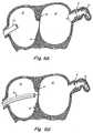
- FIG. 2Ais a schematic representation of a device according to one embodiment of the present invention, omitting the anchoring component
- FIG. 2Bis a schematic representation of the device of FIG. 2A deployed in the LAA in-vivo ;
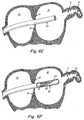
- FIG. 3is a schematic representation of a device according to the present invention.
- FIG. 4is a schematic representation of the device of FIG. 3 deployed in the LAA in-vivo;
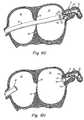
- FIGS 5A-5Care schematic representations of various devices according to further embodiments of the present invention.
- FIGS 6A-6Hare schematic representations of one method of delivering a device to an LAA.
- the present inventionprovides a device that modifies the LAA to reduce the likelihood of thrombus formation in the LAA during AF and, subsequently, stroke.
- this deviceaccomplishes its objective by remodeling the LAA in at least two ways.
- the devicereduces the volume of the LAA, thereby minimizing blood stasis during AF.
- the devicesmoothes the internal surface of the LAA, thereby obliterating the crenellations that impede blood flow which can lead to blood stagnation and clotting in the LAA.
- These modificationspermit blood to enter and exit the LAA more easily during AF.
- the devicepromotes or enhances the drainage of vascular flow from the LAA.
- the devicemodifies, rather than eliminates, the LAA and potentially maintains LAA function, it overcomes the controversy and potential drawbacks associated with current LAA obliteration or ligation procedures.
- distalrefers to the direction away from a catheter insertion location and “proximal” refers to the direction nearest the insertion location.
- the device 20 of a preferred embodiment of the present inventionincludes a liner 21, as shown in FIG. 2A.
- the liner or sock componentused interchangeably herein extends into the LAA 11, remodeling its interior geometry and surface (FIG. 2B).
- the liner 21reduces the volume of the LAA, thereby minimizing the access of blood to those areas in the LAA 11 in which it tends to stagnate and clot.
- the liner 21reduces the depth of the LAA 11 (FIG. 2B).
- the liner 21is generally closed at its distal end and may take any of a variety of shapes depending upon the particular anatomy of the patient's LAA.
- the sockmay be rounded (FIG. 2A), tubular (like a thumb or actual foot sock), or conical.
- the shape of the liner 21includes few, if any, sharp angles.
- the liner 21is a half sphere.
- the half sphere designprovides a smooth, broad interior surface and promotes drainage of blood from the LAA 11 during AF by, inter alia, reducing the depth of the LAA 11.
- the sock 21is fabricated from a natural or biologic material.
- biologic materialsundergo controlled remodeling in-vivo by the patient's own cells. This remodeling occurs as part of the normal healing process through cellular infiltration into the biologic material, over time replacing the biologic material with newly-formed native extracellular matrix (ECM).
- ECMextracellular matrix
- the blood-contacting surface of the liner or sock 21is covered with endothelial cells, rendering the exposed surface of the sock 21 non-thrombogenic.
- the remodeled tissueexhibits characteristics similar to that of the native LAA tissue, including distensibility to aid in modulation of left atrial pressure-volume relationships and release of both ANF, which stimulates excretion of sodium and water by the kidneys and regulates blood pressure, and stretch sensitive receptors, which regulate heart rate, diuresis, and natriuresis.
- the liner materialalso mimics the material characteristics of the native myocardial tissue, for example, but not limited to, the stiffness and permeability characteristics.
- the sock 21includes a collagenous material.
- the collagenous materialmay be derived from numerous sources, such as the submucosal tissue of a warm-blood vertebrae.
- the collagenous materialis derived from the lining of the porcine small intestine.
- These intestinal collagenous materialsare particularly suited for use in the present invention because their non-porous nature provides an "instant seal" upon deployment of the device 20 in the LAA 11, i.e ., communication between the blood behind the device 20 and the left atrium 12 ceases as soon as the device 20 is properly deployed in the LAA 11.
- the in-vivo remodeling of these intestinal collagenous materialsdoes not implicate sloughing of their components, which, correspondingly, reduces the likelihood of adverse embolic events following their implantation.
- the collagenous material(s)may be treated to remove non-collagenous components, for example, without limitation, cells, cellular debris, and extracellular matrix components. These "treated" collagenous materials are notably efficacious matrices for tissue regeneration and remodeling.
- Tissue derived from the porcine small intestine and subjected to this treatmentis referred to as "highly purified (acellular) bioengineered type I collagen derived from porcine submucosa.”
- Other tissue-engineered biomaterials suitable for use in the sock component 21include reconstituted human collagen, bovine derived products, and bladder intestine.
- the liner 21may instead be fabricated from a material capable of promoting cellular ingrowth. Such a material encourages the patient's natural cellular remodeling processes discussed previously. Once covered by natural tissue, the foreign material of the device 20 is insulated from the blood flowing through the LAA 11, prolonging its life and improving its dislodgement resistance.
- dislodgement resistancerefers to the ability of a device 20 to resist the tendency of the heart's contractions to separate the device 20 from the LAA 11. Generally, a high dislodgement resistance is desirable.
- Examples of materials capable of promoting cellular ingrowth and suitable for use in the embodiment of the present inventioninclude, without limitation, resorbable synthetic polymers, nonresorbable synthetic polymers, such as knitted and woven polyesters, Teflon-based materials (ePTFE), polyvinyl alcohol (PVA), metallic materials, metallic meshes, polyurethanes, silicone, or combinations of the foregoing materials.
- resorbable synthetic polymerssuch as knitted and woven polyesters, Teflon-based materials (ePTFE), polyvinyl alcohol (PVA), metallic materials, metallic meshes, polyurethanes, silicone, or combinations of the foregoing materials.
- the sock component 21may include a lip 23, as shown in FIG. 2A. Lip 23 properly seats and completely seals the sock component 21 in the ostium of the LAA 11 (FIG. 2B). Lip 23 may be of various shapes and sizes depending upon the application. For example, and as shown in FIGS. 2A and 2B, lip 23 may be circular.
- the device 20further includes an anchor component 32 (FIG. 3).
- the anchor component 32maintains the liner at its intended delivery site and seals the device to the LAA periphery (FIG. 4).
- the sock component 21is attached to the anchor 32.
- the liner 21 and anchor 32 componentsmay be attached by any appropriate method.
- the sock component 21may be sewn, glued, laminated, or thermally ligated to the anchor 32.
- One skilled in the artwill be capable of identifying other suitable means for attaching the sock component 21 to the anchor component 32 and of determining the precise means of attachment necessary for a given application.
- the anchor component 32 of the device 30is generally self-expandable and, upon deployment of the device 30 in-vivo, simultaneously expands the sock component 21.
- the anchor component 32expands in-vivo to a size greater than that of the LAA 11 and its ostium 14.
- the anchor component 32may expand to a size that is ten (10) to twenty (20) percent greater than the LAA ostium 14.
- the anchor componentis expanded with the assistance of a mechanical device, for example, a balloon.
- the expanded anchor component 32creates a friction between the device 30 and the LAA walls that helps to maintain the device 30 at its intended delivery site.
- the design of the anchor 32effectively seals the device against the LAA surface and ostium 14 in-vivo to further prevent dislodgement and migration of the device 30 following deployment (FIG. 4).
- the anchor 32may also be designed to facilitate retrieval and/or redeployment of the device.
- the anchor 32may be formed of various materials, such as, for example, metals, shape memory materials, or polymers (shape memory, synthetic, or bioresorbable). According to at least some embodiments, the anchor 32 is formed of a bioresorbable material.
- the anchor component 32may take any of a number of shapes capable of achieving the above-described functions.
- the anchor 32may be a sleeve-like structure that expands upon deployment in-vivo to exert radial pressure on the walls of the LAA 11 to hold the device 30 in place (FIG. 5A).
- the anchor component 32may be a tubular stent-like structure, a helical coil (FIG. 5B), a polymeric tube, a conical structure (FIG. 3), or a metallic mesh (FIG. 5C).
- the sleeve-like structureincludes an implant grade metal, for example, nitinol.
- the anchor 32may be further modified to increase the dislodgement resistance of the device 30; for example, the anchor 32 may include hooks and/or its surface may be roughened.
- the anchor 32is also physically attached to the interior surface of the LAA 11 by a suitable mechanism, such as a suture, weld, glue, or adhesive.
- the anchor 32may be attached to the interior surface of the LAA 11 by a glue that is light-activated and cured in situ, once the clinician confirms the device 30 has been properly deployed.
- a suitable mechanismsuch as a suture, weld, glue, or adhesive.
- the anchor 32may be attached to the interior surface of the LAA 11 by a glue that is light-activated and cured in situ, once the clinician confirms the device 30 has been properly deployed.
- the anchor componentincludes a lip 33 at its proximal end.
- This lip 33helps to seat and seal the device against the ostium 14 of the LAA 11, thereby ensuring blood is unable to leak into the left atrium 12 from behind the sock component 21 in the LAA 11.
- lip 33may take a variety of shapes and sizes, depending upon the particular application.
- the proximal ends of anchor component 32may be angled such that they engage the inner wall of the LAA 11 and crenellations 13. As the LAA contracts with the heart's rhythm, the angled ends become embedded in the inner wall of the LAA 11 and/or the crenellations 13, such that the device 30 is seated and sealed against the LAA tissue.
- the device 30 according to the present inventionmay be deployed in-vivo according to any suitable method known to those of skill in the art, only one of which will be described herein (FIGS. 6A-6E).
- a catheter 61Accessing the heart through the circulatory system, a catheter 61 enters the right atrium 10 (FIG. 6A).
- the septal tissue 15is punctured (FIG. 6B), and a guidewire 62 is placed through the puncture and into the left atrium 12 (FIG. 6C).
- a dilator 63is placed over the guidewire 62 through catheter 61, and the dilator 63 and catheter 61 are then advanced through the septal puncture (FIG. 6D).
- a dilator/catheter assemblyis placed over the guidewire and advanced to the ostium of the LAA.
- the catheter 61Once the catheter 61 has reached the ostium 14 of the LAA 11, the dilator 63 and guidewire 62 are removed, leaving only the catheter 61 in the left atrium (FIG. 6E).
- the size and shape of the LAA 11is then observed and measured using echocardiographic techniques and/or fluoroscopy so as to determine the necessary anchor size using the criteria described previously.
- the selected device 30is then collapsed, inserted into the catheter 61 (FIG. 6F), and deployed into the ostium 14 of the LAA 11 (FIG. 6G).
- the anchor component 32self expands into the LAA 11 and the sock component 21 follows (FIG. 6H).
- the clinicianconfirms the device 30 has been properly placed and an effective seal has been formed between the device 30 and the interior surface and ostium 14 of the LAA 11. If necessary, the clinician may reposition and/or retrieve device 30. Once satisfied with the deployment of the device 30, the catheter 61 is withdrawn from the right atrium 10.
- the devices described hereinmay be used with anti-thrombogenic compounds, including but not limited to heparin (ionic or covalently-bound) and peptides, to reduce thrombogenicity of the device and/or to enhance the cellular ingrowth of the septal tissue following deployment of the device in-vivo .
- the devices described hereinmay be used to deliver other drugs or pharmaceutical agents, for example, without limitation, growth factors or antibodies.
- the anti-thrombogenic compounds and/or pharmaceutical agentsmay be included in the device in several ways, including impregnation or coating of the sock component and/or anchor component.
- the devices described hereinmay include radiopaque fillers for x-ray visualization, cells to promote biocompatibility, echogenic coatings, lubricious coatings, and/or hydrogels.
Landscapes
- Health & Medical Sciences (AREA)
- Surgery (AREA)
- Life Sciences & Earth Sciences (AREA)
- Animal Behavior & Ethology (AREA)
- Public Health (AREA)
- Engineering & Computer Science (AREA)
- Biomedical Technology (AREA)
- Heart & Thoracic Surgery (AREA)
- Medical Informatics (AREA)
- Molecular Biology (AREA)
- Veterinary Medicine (AREA)
- General Health & Medical Sciences (AREA)
- Nuclear Medicine, Radiotherapy & Molecular Imaging (AREA)
- Reproductive Health (AREA)
- Vascular Medicine (AREA)
- Cardiology (AREA)
- Prostheses (AREA)
- Surgical Instruments (AREA)
- Bidet-Like Cleaning Device And Other Flush Toilet Accessories (AREA)
- Electrochromic Elements, Electrophoresis, Or Variable Reflection Or Absorption Elements (AREA)
- Aeration Devices For Treatment Of Activated Polluted Sludge (AREA)
Abstract
Description
- The present invention relates generally to a device that prevents the formation of thrombi in an anatomical appendage, such as the left atrial appendage.
- Arrhythmias are abnormal heart rhythms. These abnormal heart rhythms may cause the heart to function less effectively. Atrial fibrillation (AF) is the most common abnormal heart rhythm. In AF, the two upper chambers of the heart (i.e., the atria) quiver rather than beat and, consequently, fail to entirely empty of blood. As the blood stagnates on the walls of the atria, it may form thrombi (i.e., clots). Under certain circumstances, these thrombi may re-enter the circulation and travel to the brain, causing a stroke or a transient ischemic attack (TIA).
- Research has indicated that as many as ninety (90) percent of all thrombi formed during AF originate in the left atrial appendage (LAA). The
LAA 11 is a remnant of the original embryonic left atrium that develops during the third week of gestation and, as shown in FIGS. 1A and 1B, is located high on the free wall of theleft atrium 12.
Long, tubular, and hook-like in structure, theLAA 11 is connected to theleft atrium 12 by anarrow junction 14, referred to as the "ostium". The precise physiological function of the LAA remains uncertain: recent reports suggest it may maintain and regulate pressure and volume in the left atrium; modulate the hemodynamic response during states of cardiac stress; mediate thirst in hypovolemia; and/or serve as the site of release of both the peptide hormone atrial natriuretic factor (ANF), which stimulates excretion of sodium and water by the kidneys and regulates blood pressure, and stretch sensitive receptors, which regulate heart rate, diuresis, and natriuresis. - The high rate of thrombus formation in the LAA is believed to be attributable to its physical characteristics; blood easily stagnates, and thereafter clots, in the long, tubular body of the LAA or at its narrow ostium. In marked contrast, the right atrial appendage (RAA), which is a wide, triangular appendage connected to the right atrium by a broad ostium, is infrequently the site of thrombus formation. Thrombus formation in the LAA is further promoted by the numerous tissue folds 13 (i.e., crenellations) on its interior surface (FIG. 1B). These
crenellations 13 are particularly hospitable to blood stagnation and clotting, especially when the heart is not functioning at maximum capacity. Thrombi formed in the LAA frequently re-enter the circulation upon conversion of AF to normal rhythm (i.e., cardioversion). - Currently, therapeutic protocols attempt to minimize the likelihood of thrombus formation associated with AF. Blood thinners, such as Warfarin (Coumadin), are, therefore, frequently administered to AF patients. Warfarin administration is, however, complicated by several factors. First, Warfarin is contraindicated for patients suffering from potential bleeding problems or ulcers. Second, Warfarin administration ideally begins approximately four weeks prior to cardioversion and continues for four weeks after cardioversion. This long course of treatment is often compromised due to emergency presentation and/or patient noncompliance.
- Certain patient subsets are considered to be at an abnormally high risk of thrombus formation. Such patients include those over seventy-five (75) years of age, as well as those presenting with a history of thromboembolism, significant heart diseases, decreased LAA flow velocity, increased LAA size, spontaneous echogenic contrast, abnormal coagulation, diabetes mellitus, and/or systemic hypertension. For these high-risk patients, prophylactic intervention may be recommended. Current prophylaxes generally fall into three categories: (1) surgical ligation of the LAA as described, for example, in
U.S. Patent No. 6,561,969 andU.S. Patent No. 6,488,689 ; (2) implantation of an LAA occluder sufficient to prevent, or at least minimize, blood flow into the LAA as described, for example, inU.S. Patent No. 6,551,303 ,U.S. Patent No. 6,152,144 , (and published)U.S. Patent Appln. No. 2005/0070952 , and (3) placement of a filter in the LAA ostium to prevent clots formed therein from re-entering the circulatory system as described, for example, inWO 03/063732 - However, given the uncertain physiological role of the LAA, its obliteration and occlusion remain controversial. Reports have suggested that obliteration of the LAA may decrease atrial compliance and diminish ANF secretion. Furthermore, while properly positioned filter devices prevent migration of thrombi into the circulatory system, they cannot inhibit thrombus formation within the LAA. Consequently, in the event the filter device is dislodged or ineffectively sealed against the LAA ostium, problems plaguing many current filter designs, clots held at the LAA ostium by the filter will be released into the circulation.
- Thus, there remains a need in the art for a device capable of preventing thrombus formation in the LAA while maintaining the LAA's function. Such a device must demonstrate excellent dislodgement resistance and, ideally, would be repositionable and retrievable.
- The embodiments of the present invention, as defined in claim 1, provides a device that modifies the left atrial appendage (LAA) to reduce the likelihood of thrombus formation therein. The device includes a liner that is a material formed of a flexible, expandable, biocompatible material for covering or coating the inner surfaces of the appendage. The liner may be herein referred to also as a sock portion, the device further includes an anchor component.
- The liner of the device remodels the interior geometry of the LAA by smoothing its surface and reducing its volume. The liner may be formed of a biologic tissue that is remodeledin-vivo into natural tissue resembling the native endothelium of the LAA. In some embodiments, the remodeled tissue displays functional characteristics similar to those of the native LAA tissue. In particular embodiments, the liner includes a collagenous material derived from the intestinal lining of a warm-blooded mammal, for example, a pig.
- According to at least some embodiments, the anchor component of the device is self-expandable and helps to expand the liner upon deployment of the device in the LAA. The anchor component also prevents dislodgement and migration of the devicein-vivo by ensuring the device is properly seated and completely sealed against the interior walls and ostium of the LAA.
- According to one aspect of the present invention, a device for promoting vascular flow within the left atrial appendage of a heart, includes a liner portion having a proximal end and a distal end, wherein the distal end is expandable and is adapted to be positioned adjacent to a plurality of surfaces of the interior wall of the left atrial appendage such that the liner smoothes the plurality of surfaces of the interior wall; and an anchor component attached to at least a portion of the liner, and adapted for securing the device within the left atrial appendage. The liner can be formed from natural or biologic tissue. The liner can include collagenous material and have a stiffness characteristic that adapts the liner to the surfaces of the interior wall of the LAA. The liner has an appropriate flexibility and resilience which allows for the liner to expand within the LAA. The liner and anchor component are expandable. In an embodiment, the anchor component is self-expandablein-vivo. In an alternate embodiment, the anchor component is expanded with the assistance of a mechanical device, for example a balloon. The liner can further include a lip to seal a portion of the liner in the ostium of the LAA and can have at least one of a plurality of shapes such as, for example, but not limited to, spherical, tubular and conical.
- In an embodiment of the present invention, the liner can be or include material promoting cellular in-growth, for example, but not limited to, at least one of synthetic polymers, Teflon-based materials (ePTFE), polyvinyl alcohol (PVA), knitted or woven polyesters, metallic materials, metallic meshes, polyurethanes, and silicone. The anchor component has one of a plurality of shapes which are, for example, but not limited to, a sleeve-like structure, a tubular stent-like structure, a helical coil, a polymeric tube, a conical structure and a metallic mesh. In an embodiment of the present invention, the anchor component is adapted to further include surface characteristics and attachment structures, for example, hooks and surface roughness to augment dislodgement resistance. The anchor component in an embodiment of the present invention can further include a lip at a proximal end to seat and seal at least a portion of the device against the ostium of the LAA. The anchor component is made from material selected from a group including, but not limited to metals, shape memory materials, synthetic polymers, and bioresorbable materials.
- A method for promoting drainage of the vascular flow from the left atrial appendage of a heart using the device according to the invention includes positioning a device having a liner and an anchor component to prevent formation of thrombi. Through a catheter system in a proximal end of the LAA, the proximal end having an ostium; expanding the liner of the devicein-vivo in the LAA by using the expandable anchor component wherein the liner extends within the LAA to a distal end of the LAA; and remodeling a plurality of internal surfaces of the internal wall of the LAA using the liner thereby reducing the volume of the LAA and minimizing a plurality of crenellations in the LAA to promote drainage of the vascular flow from the LAA. The liner of the device can include one of a natural and biological tissue. The liner can include a collagenous material.
- The method may include sealing the ostium of the left atrial appendage by a portion of a lip of the liner. The liner can include material promoting cellular in-growth, for example, at least one of synthetic polymers, Teflon-based materials (ePTFE), polyvinyl alcohol (PVA), knitted or woven polyesters, metallic materials, metallic meshes, polyurethanes, or silicone. The anchor component can have one of a plurality of shapes, for example, a sleeve-like structure, a tubular stent-like structure, a helical coil, a polymeric tube, a conical structure or a metallic mesh. The anchor component is adapted to further include surface characteristics and attachment structures, for example, but not limited to hooks and surface roughness to augment dislodgement resistance.
- In accordance with another aspect of the present invention, a method for manufacturing a device for preventing formation of thrombi in an anatomical appendage includes providing a liner having a proximal end and a distal end; and affixing an anchor component to at least the proximal end of the liner. The liner includes material selected from a group comprising a natural and a biologic tissue. The liner can include material promoting cellular in-growth, for example, but not limited to, at least one of synthetic polymers, Teflon-based materials (ePTFE), polyvinyl alcohol (PVA), knitted or woven polyesters, metallic materials, metallic meshes, polyurethanes, and silicone. The anchor component has one of a plurality of shapes which is, for example, but not limited to, a sleeve-like structure, a tubular stent-like structure, a helical coil, a polymeric tube, a conical structure and a metallic mesh. The anchor component is affixed to a portion of the liner by, for example, one of sewing, gluing, laminating or thermally ligating. The anchor component includes material selected from a group comprising metals, shape memory materials, synthetic polymers, and bioresorbable materials.
- A method for deployingin-vivo a device according to the invention for minimizing the formation of thrombi in the LAA includes inserting a catheter into the right atrium through a lumen in the body, puncturing the septal tissue and placing a guidewire through the septal puncture and into the left atrium; positioning a dilator over the guidewire through the catheter, advancing the dilator and catheter through the septal puncture until they reach the ostium of the LAA, removing the dilator and guidewire and inserting a collapsed device to prevent formation of thrombi in the LAA into the catheter and deploying the device into the ostium of the LAA. The step of deploying includes the anchor component of the device expandingin-vivo into the distal end of the LAA and in turn expanding the liner attached to at least a portion of the anchor component such that the liner is positioned proximate to the surfaces of the interior wall of the LAA. In an embodiment, a dilator/catheter assembly is placed over a guidewire, the dilator/catheter assembly is then advanced through the septal puncture until the assembly reaches the ostium of the LAA.
- The foregoing and other objects, features and advantages of the device for preventing formulation of thrombi will be apparent from the following more particular description of preferred embodiments of the device as illustrated in the accompanying drawings in which like reference characters refer to the same parts throughout the different views. The drawings are not necessarily to scale, emphasis instead being placed upon illustrating the principles of the invention.
- FIGS. 1A and 1B are schematic representations of a human heart, including the LAA;
- FIG. 2A is a schematic representation of a device according to one embodiment of the present invention, omitting the anchoring component;
- FIG. 2B is a schematic representation of the device of FIG. 2A deployed in the LAAin-vivo ;
- FIG. 3 is a schematic representation of a device according to the present invention;
- FIG. 4 is a schematic representation of the device of FIG. 3 deployed in the LAAin-vivo;
- FIGS 5A-5C are schematic representations of various devices according to further embodiments of the present invention; and
- FIGS 6A-6H are schematic representations of one method of delivering a device to an LAA.
- The present invention provides a device that modifies the LAA to reduce the likelihood of thrombus formation in the LAA during AF and, subsequently, stroke. According to at least some embodiments, this device accomplishes its objective by remodeling the LAA in at least two ways. First, the device reduces the volume of the LAA, thereby minimizing blood stasis during AF. Second, the device smoothes the internal surface of the LAA, thereby obliterating the crenellations that impede blood flow which can lead to blood stagnation and clotting in the LAA. These modifications permit blood to enter and exit the LAA more easily during AF. Thus, the device promotes or enhances the drainage of vascular flow from the LAA. Advantageously, because the device modifies, rather than eliminates, the LAA and potentially maintains LAA function, it overcomes the controversy and potential drawbacks associated with current LAA obliteration or ligation procedures.
- In the description herein "distal" refers to the direction away from a catheter insertion location and "proximal" refers to the direction nearest the insertion location.
- The
device 20 of a preferred embodiment of the present invention includes aliner 21, as shown in FIG. 2A. When deployedin-vivo, the liner or sock component, used interchangeably herein extends into theLAA 11, remodeling its interior geometry and surface (FIG. 2B). According to at least some embodiments, theliner 21 reduces the volume of the LAA, thereby minimizing the access of blood to those areas in theLAA 11 in which it tends to stagnate and clot. In particular embodiments, theliner 21 reduces the depth of the LAA 11 (FIG. 2B). - The
liner 21 is generally closed at its distal end and may take any of a variety of shapes depending upon the particular anatomy of the patient's LAA. For example, the sock may be rounded (FIG. 2A), tubular (like a thumb or actual foot sock), or conical. According to at least some embodiments, the shape of theliner 21 includes few, if any, sharp angles. In particular embodiments, and as shown in FIG. 4, theliner 21 is a half sphere. The half sphere design provides a smooth, broad interior surface and promotes drainage of blood from theLAA 11 during AF by,inter alia, reducing the depth of theLAA 11. - In at least some embodiments, the
sock 21 is fabricated from a natural or biologic material. Such biologic materials undergo controlled remodelingin-vivo by the patient's own cells. This remodeling occurs as part of the normal healing process through cellular infiltration into the biologic material, over time replacing the biologic material with newly-formed native extracellular matrix (ECM). As part of this healing process, the blood-contacting surface of the liner orsock 21 is covered with endothelial cells, rendering the exposed surface of thesock 21 non-thrombogenic. Often, the remodeled tissue exhibits characteristics similar to that of the native LAA tissue, including distensibility to aid in modulation of left atrial pressure-volume relationships and release of both ANF, which stimulates excretion of sodium and water by the kidneys and regulates blood pressure, and stretch sensitive receptors, which regulate heart rate, diuresis, and natriuresis. The liner material also mimics the material characteristics of the native myocardial tissue, for example, but not limited to, the stiffness and permeability characteristics. - One skilled in the art will, of course, recognize that numerous natural and biologic materials are suitable for use in the
liner 21 of thedevice 20. For example, in at least some embodiments, thesock 21 includes a collagenous material. The collagenous material may be derived from numerous sources, such as the submucosal tissue of a warm-blood vertebrae. In particular embodiments, the collagenous material is derived from the lining of the porcine small intestine. These intestinal collagenous materials are particularly suited for use in the present invention because their non-porous nature provides an "instant seal" upon deployment of thedevice 20 in theLAA 11,i.e., communication between the blood behind thedevice 20 and theleft atrium 12 ceases as soon as thedevice 20 is properly deployed in theLAA 11. Further, thein-vivo remodeling of these intestinal collagenous materials does not implicate sloughing of their components, which, correspondingly, reduces the likelihood of adverse embolic events following their implantation. In particular embodiments, the collagenous material(s) may be treated to remove non-collagenous components, for example, without limitation, cells, cellular debris, and extracellular matrix components. These "treated" collagenous materials are notably efficacious matrices for tissue regeneration and remodeling. Tissue derived from the porcine small intestine and subjected to this treatment is referred to as "highly purified (acellular) bioengineered type I collagen derived from porcine submucosa." Other tissue-engineered biomaterials suitable for use in thesock component 21 include reconstituted human collagen, bovine derived products, and bladder intestine. - According to some embodiments of the present invention, the
liner 21 may instead be fabricated from a material capable of promoting cellular ingrowth. Such a material encourages the patient's natural cellular remodeling processes discussed previously. Once covered by natural tissue, the foreign material of thedevice 20 is insulated from the blood flowing through theLAA 11, prolonging its life and improving its dislodgement resistance. As used herein, the term "dislodgement resistance" refers to the ability of adevice 20 to resist the tendency of the heart's contractions to separate thedevice 20 from theLAA 11. Generally, a high dislodgement resistance is desirable. Examples of materials capable of promoting cellular ingrowth and suitable for use in the embodiment of the present invention include, without limitation, resorbable synthetic polymers, nonresorbable synthetic polymers, such as knitted and woven polyesters, Teflon-based materials (ePTFE), polyvinyl alcohol (PVA), metallic materials, metallic meshes, polyurethanes, silicone, or combinations of the foregoing materials. - The
sock component 21 may include alip 23, as shown in FIG. 2A.Lip 23 properly seats and completely seals thesock component 21 in the ostium of the LAA 11 (FIG. 2B).Lip 23 may be of various shapes and sizes depending upon the application. For example, and as shown in FIGS. 2A and 2B,lip 23 may be circular. - According to the present invention, the
device 20 further includes an anchor component 32 (FIG. 3). Theanchor component 32 maintains the liner at its intended delivery site and seals the device to the LAA periphery (FIG. 4). As shown in FIG. 3, thesock component 21 is attached to theanchor 32. Theliner 21 andanchor 32 components may be attached by any appropriate method. For example and according to some embodiments, thesock component 21 may be sewn, glued, laminated, or thermally ligated to theanchor 32. One skilled in the art will be capable of identifying other suitable means for attaching thesock component 21 to theanchor component 32 and of determining the precise means of attachment necessary for a given application. - The
anchor component 32 of thedevice 30 according to a preferred embodiment of the present invention is generally self-expandable and, upon deployment of thedevice 30in-vivo, simultaneously expands thesock component 21. According to at least some embodiments, theanchor component 32 expandsin-vivo to a size greater than that of theLAA 11 and itsostium 14. For example, theanchor component 32 may expand to a size that is ten (10) to twenty (20) percent greater than theLAA ostium 14. In an alternate embodiment, the anchor component is expanded with the assistance of a mechanical device, for example, a balloon. - The expanded
anchor component 32 creates a friction between thedevice 30 and the LAA walls that helps to maintain thedevice 30 at its intended delivery site. The design of theanchor 32 effectively seals the device against the LAA surface andostium 14in-vivo to further prevent dislodgement and migration of thedevice 30 following deployment (FIG. 4). In at least some embodiments, theanchor 32 may also be designed to facilitate retrieval and/or redeployment of the device. Theanchor 32 may be formed of various materials, such as, for example, metals, shape memory materials, or polymers (shape memory, synthetic, or bioresorbable). According to at least some embodiments, theanchor 32 is formed of a bioresorbable material. - The
anchor component 32 may take any of a number of shapes capable of achieving the above-described functions. Theanchor 32 may be a sleeve-like structure that expands upon deploymentin-vivo to exert radial pressure on the walls of theLAA 11 to hold thedevice 30 in place (FIG. 5A). According to such an embodiment, theanchor component 32 may be a tubular stent-like structure, a helical coil (FIG. 5B), a polymeric tube, a conical structure (FIG. 3), or a metallic mesh (FIG. 5C). In particular embodiments, the sleeve-like structure includes an implant grade metal, for example, nitinol. Theanchor 32 may be further modified to increase the dislodgement resistance of thedevice 30; for example, theanchor 32 may include hooks and/or its surface may be roughened. According to some embodiments, theanchor 32 is also physically attached to the interior surface of theLAA 11 by a suitable mechanism, such as a suture, weld, glue, or adhesive. For example, theanchor 32 may be attached to the interior surface of theLAA 11 by a glue that is light-activated and curedin situ, once the clinician confirms thedevice 30 has been properly deployed. One skilled in the art will be capable of determining the precise anchor design and modifications necessary for a particular application. - In at least some embodiments, the anchor component includes a
lip 33 at its proximal end. Thislip 33 helps to seat and seal the device against theostium 14 of theLAA 11, thereby ensuring blood is unable to leak into theleft atrium 12 from behind thesock component 21 in theLAA 11. As shown in FIGS 4 and 5,lip 33 may take a variety of shapes and sizes, depending upon the particular application. In other embodiments, the proximal ends ofanchor component 32 may be angled such that they engage the inner wall of theLAA 11 andcrenellations 13. As the LAA contracts with the heart's rhythm, the angled ends become embedded in the inner wall of theLAA 11 and/or thecrenellations 13, such that thedevice 30 is seated and sealed against the LAA tissue. - The
device 30 according to the present invention may be deployedin-vivo according to any suitable method known to those of skill in the art, only one of which will be described herein (FIGS. 6A-6E). Accessing the heart through the circulatory system, acatheter 61 enters the right atrium 10 (FIG. 6A). Theseptal tissue 15 is punctured (FIG. 6B), and aguidewire 62 is placed through the puncture and into the left atrium 12 (FIG. 6C). Adilator 63 is placed over theguidewire 62 throughcatheter 61, and thedilator 63 andcatheter 61 are then advanced through the septal puncture (FIG. 6D). In an alternate embodiment, a dilator/catheter assembly is placed over the guidewire and advanced to the ostium of the LAA. Once thecatheter 61 has reached theostium 14 of theLAA 11, thedilator 63 and guidewire 62 are removed, leaving only thecatheter 61 in the left atrium (FIG. 6E). The size and shape of theLAA 11 is then observed and measured using echocardiographic techniques and/or fluoroscopy so as to determine the necessary anchor size using the criteria described previously. The selecteddevice 30 is then collapsed, inserted into the catheter 61 (FIG. 6F), and deployed into theostium 14 of the LAA 11 (FIG. 6G). Following deployment, theanchor component 32 self expands into theLAA 11 and thesock component 21 follows (FIG. 6H). The clinician then confirms thedevice 30 has been properly placed and an effective seal has been formed between thedevice 30 and the interior surface andostium 14 of theLAA 11. If necessary, the clinician may reposition and/or retrievedevice 30. Once satisfied with the deployment of thedevice 30, thecatheter 61 is withdrawn from theright atrium 10. - One skilled in the art will recognize that the devices described herein may be used with anti-thrombogenic compounds, including but not limited to heparin (ionic or covalently-bound) and peptides, to reduce thrombogenicity of the device and/or to enhance the cellular ingrowth of the septal tissue following deployment of the devicein-vivo. Similarly, the devices described herein may be used to deliver other drugs or pharmaceutical agents, for example, without limitation, growth factors or antibodies. The anti-thrombogenic compounds and/or pharmaceutical agents may be included in the device in several ways, including impregnation or coating of the sock component and/or anchor component. Further, the devices described herein may include radiopaque fillers for x-ray visualization, cells to promote biocompatibility, echogenic coatings, lubricious coatings, and/or hydrogels.
- While the present device has been described with reference to a LAA, one skilled in the art will further recognize that the devices described herein may be used in other anatomical appendages. Further, the steps of any methods or process flow, for example, methods used to deploy the embodiments of the present invention may be taken in sequence other than those described.
- Having described preferred embodiments of the invention, it should be apparent that various modifications may be made without departing from the scope of the invention, which is defined in the claims.
Claims (28)
- A device for promoting drainage of the vascular flow from the left atrial appendage of a heart, the left atrial appendage having an ostium and an interior wall, said device comprising:a liner (21) having a proximal end and a distal end, the liner (21) adapted to be positioned adjacent to a plurality of surfaces of the interior wall of the left atrial appendage such that the liner (21) smoothes the plurality of surfaces of the interior wall, wherein said liner (21) is closed at the distal end; andan anchor component (32) attached to at least a portion of the liner, the anchor component (32) adapted for securing at least a portion of the device within the left atrial appendage.
- The device of claim 1, wherein the liner (21) comprises one of a natural and biologic tissue.
- The device of claim 1, wherein the line (21) comprises collagenous material.
- The device of claim 3, wherein the collagenous material is treated to remove cellular components.
- The device of claim 1, wherein the liner (21) is expandable.
- The device of claim 1, wherein the anchor component (32) is self-expandable.
- The device of claim 1, wherein the liner (21) further comprises a lip (23) to seal at least a portion of the liner (21) in the ostium of the LAA.
- The device of claim 1 wherein the liner (21) has at least one of a plurality of shapes such as, for example, spherical, tubular and conical.
- The device of claim 1, wherein the liner (21) comprises material promoting cellular in-growth, for example, at least one of synthetic polymers, Teflon-based materials (ePTFE), polyvinyl alcohol (PVA), knitted polyesters, woven polyesters, metallic materials, metallic meshes, polyurethanes, and silicone.
- The device of claim 1, wherein the anchor component (32) has one of a plurality of shapes, for example, a sleeve-like structure, a tubular stent-like structure, a helical coil, a polymeric tube, a conical structure and a metallic mesh.
- The device of claim 1, wherein the anchor component (32) is adapted to further comprise surface characteristics and attachment structures to augment dislodgement resistance.
- The device of claim 1, wherein the anchor component (32) further comprises a lip (33) at a proximal end to seat and seal at least a portion of the device against the ostium of the left atrial appendage.
- The device of claim 1, wherein the anchor component (32) comprises material selected from a group comprising metals, shape memory materials, synthetic polymers, and bioresorbable materials.
- The device of claim 1, wherein the anchor component (32) is adapted to exert radial pressure on at least one wall of the left atrial appendage.
- The device of claim 1, wherein the anchor component (32) is adapted to being affixed to at least a portion of an interior surface of the left atrial appendage.
- The device of claim 1, wherein the liner (32) has a material stiffness characteristic that adapts the liner to the surface of the interior wall of the LAA and smooth the plurality of the surfaces of the interior wall.
- A kit comprising:a device according to claim 1 to prevent formation of thrombi in the left atrial appendage; andinstructions for use.
- A method for manufacturing a device for preventing formation of thrombi in the left atrial appendage of the heart, wherein said device promotes drainage of the vascular flow from the left atrial appendage, said method comprising the steps of:providing a liner (21) having a proximal end and a distal end wherein said distal end is closed; andaffixing an anchor (32) component to at least a portion of the proximal end of the liner.
- The method of claim 18, wherein the line (21) comprises material selected from a group comprising a natural and a biologic tissue.
- The method of claim 18, wherein the liner (21) comprises material promoting cellular in- growth, for example, at least one of synthetic polymers, Teflon-based materials(ePTFE), polyvinyl alcohol (PVA), knitted polyesters, woven polyesters, metallic materials, metallic meshes, polyurethanes, and silicone.
- The method of claim 18, wherein the anchor component (32) has one of a plurality of shapes, for example, a sleeve-like structure, a tubular stent-like structure, a helical coil, a polymeric tube, a conical structure and a metallic mesh.
- The method of claim 18, wherein the anchor component (32) is adapted to further comprise surface characteristics and attachment structures to augment dislodgement resistance.
- The method of claim 18, wherein the liner (21) has at least one of a plurality of shapes such as, for example, spherical, tubular and conical.
- The method of claim 18, wherein the anchor component (32) further comprises a lip (33) at a proximal end to seat and seal at least a portion of the device against an ostium of the appendage.
- The method of claim 18, wherein the liner (21) further comprises a lip (23) to seal at least a portion of the liner to an ostium of the appendage.
- The method of claim 18, wherein the anchor component (32) is affixed to at least a portion of the liner (21) by, for example, one of sewing, gluing, laminating and thermally ligating.
- The method of claim 18, wherein the anchor component (32) comprises material selected from a group consisting of metals, shape memory materials, synthetic polymers, and bioresorbable materials.
- The method of claim 18, wherein the anchor component (32) is adapted for exerting radial pressure on at least one of a wall of the appendage.
Applications Claiming Priority (2)
| Application Number | Priority Date | Filing Date | Title |
|---|---|---|---|
| US50270203P | 2003-09-12 | 2003-09-12 | |
| PCT/US2004/025316WO2005034764A1 (en) | 2003-09-12 | 2004-08-05 | Device and methods for preventing formation of thrombi in the left atrial appendage |
Publications (2)
| Publication Number | Publication Date |
|---|---|
| EP1670360A1 EP1670360A1 (en) | 2006-06-21 |
| EP1670360B1true EP1670360B1 (en) | 2007-10-17 |
Family
ID=34434849
Family Applications (1)
| Application Number | Title | Priority Date | Filing Date |
|---|---|---|---|
| EP04780196AExpired - LifetimeEP1670360B1 (en) | 2003-09-12 | 2004-08-05 | Device for preventing formation of thrombi in the left atrial appendage |
Country Status (8)
| Country | Link |
|---|---|
| US (1) | US8097015B2 (en) |
| EP (1) | EP1670360B1 (en) |
| JP (1) | JP2007504888A (en) |
| AT (1) | ATE375754T1 (en) |
| CA (1) | CA2538480A1 (en) |
| DE (1) | DE602004009598T2 (en) |
| ES (1) | ES2295932T3 (en) |
| WO (1) | WO2005034764A1 (en) |
Cited By (1)
| Publication number | Priority date | Publication date | Assignee | Title |
|---|---|---|---|---|
| US11684775B2 (en) | 2014-08-26 | 2023-06-27 | Medtronic, Inc. | Interventional medical device and method of use |
Families Citing this family (99)
| Publication number | Priority date | Publication date | Assignee | Title |
|---|---|---|---|---|
| US8992567B1 (en) | 2001-04-24 | 2015-03-31 | Cardiovascular Technologies Inc. | Compressible, deformable, or deflectable tissue closure devices and method of manufacture |
| US8961541B2 (en) | 2007-12-03 | 2015-02-24 | Cardio Vascular Technologies Inc. | Vascular closure devices, systems, and methods of use |
| US20080109030A1 (en) | 2001-04-24 | 2008-05-08 | Houser Russell A | Arteriotomy closure devices and techniques |
| US6793665B2 (en) | 2001-06-18 | 2004-09-21 | Rex Medical, L.P. | Multiple access vein filter |
| WO2002102280A2 (en) | 2001-06-18 | 2002-12-27 | Rex Medical, L.P. | Removable vein filter |
| US6866679B2 (en)* | 2002-03-12 | 2005-03-15 | Ev3 Inc. | Everting stent and stent delivery system |
| EP1670360B1 (en)* | 2003-09-12 | 2007-10-17 | NMT Medical, Inc. | Device for preventing formation of thrombi in the left atrial appendage |
| US20050107867A1 (en)* | 2003-11-17 | 2005-05-19 | Taheri Syde A. | Temporary absorbable venous occlusive stent and superficial vein treatment method |
| US7704266B2 (en) | 2004-01-22 | 2010-04-27 | Rex Medical, L.P. | Vein filter |
| US8211140B2 (en) | 2004-01-22 | 2012-07-03 | Rex Medical, L.P. | Vein filter |
| US8162972B2 (en) | 2004-01-22 | 2012-04-24 | Rex Medical, Lp | Vein filter |
| US7806846B2 (en)* | 2004-03-30 | 2010-10-05 | Nmt Medical, Inc. | Restoration of flow in LAA via tubular conduit |
| US20050234543A1 (en)* | 2004-03-30 | 2005-10-20 | Nmt Medical, Inc. | Plug for use in left atrial appendage |
| US8409219B2 (en) | 2004-06-18 | 2013-04-02 | Medtronic, Inc. | Method and system for placement of electrical lead inside heart |
| US7927346B2 (en)* | 2004-09-10 | 2011-04-19 | Stryker Corporation | Diversion device to increase cerebral blood flow |
| ES2444590T3 (en) | 2004-09-27 | 2014-02-25 | Rex Medical, L.P. | Venous filter |
| CN102525591B (en) | 2005-01-25 | 2014-12-10 | 泰科医疗集团有限合伙公司 | Structures for permanent occlusion of a hollow anatomical structure |
| US8165674B2 (en) | 2005-03-02 | 2012-04-24 | Backbeat Medical, Inc. | Methods and apparatus to increase secretion of endogenous naturetic hormones |
| CA2609687C (en) | 2005-05-24 | 2014-04-22 | Inspire M.D Ltd. | Stent apparatuses for treatment via body lumens and methods of use |
| US8961586B2 (en) | 2005-05-24 | 2015-02-24 | Inspiremd Ltd. | Bifurcated stent assemblies |
| US8043323B2 (en) | 2006-10-18 | 2011-10-25 | Inspiremd Ltd. | In vivo filter assembly |
| US8062309B2 (en)* | 2005-08-19 | 2011-11-22 | Boston Scientific Scimed, Inc. | Defect occlusion apparatus, system, and method |
| US7766906B2 (en) | 2005-08-19 | 2010-08-03 | Boston Scientific Scimed, Inc. | Occlusion apparatus |
| US7998095B2 (en)* | 2005-08-19 | 2011-08-16 | Boston Scientific Scimed, Inc. | Occlusion device |
| US7824397B2 (en)* | 2005-08-19 | 2010-11-02 | Boston Scientific Scimed, Inc. | Occlusion apparatus |
| US7837619B2 (en)* | 2005-08-19 | 2010-11-23 | Boston Scientific Scimed, Inc. | Transeptal apparatus, system, and method |
| US7972359B2 (en)* | 2005-09-16 | 2011-07-05 | Atritech, Inc. | Intracardiac cage and method of delivering same |
| US10143456B2 (en)* | 2005-10-07 | 2018-12-04 | Alex Javois | Left atrial appendage occlusion device |
| US9017361B2 (en)* | 2006-04-20 | 2015-04-28 | Covidien Lp | Occlusive implant and methods for hollow anatomical structure |
| US8652201B2 (en)* | 2006-04-26 | 2014-02-18 | The Cleveland Clinic Foundation | Apparatus and method for treating cardiovascular diseases |
| EP2018139B1 (en)* | 2006-04-26 | 2017-03-01 | The Cleveland Clinic Foundation | Apparatus and method for treating cardiovascular diseases |
| EP2015681B1 (en) | 2006-05-03 | 2018-03-28 | Datascope Corp. | Tissue closure device |
| US7869874B2 (en) | 2006-09-25 | 2011-01-11 | G&L Consulting, Llc | Methods and apparatus to stimulate heart atria |
| CA2666712C (en)* | 2006-10-18 | 2015-03-31 | Asher Holzer | Filter assemblies |
| WO2008047369A2 (en) | 2006-10-18 | 2008-04-24 | Inspiremd Ltd. | Knitted stent jackets |
| EP2263605A1 (en)* | 2006-11-20 | 2010-12-22 | SeptRx, Inc. | Device and method for preventing the undesired passage of emboli from a venous blood pool to an arterial blood pool |
| EP3292837B1 (en) | 2006-11-22 | 2022-11-09 | Inspire M.D Ltd | Optimized stent jacket |
| US8784469B2 (en) | 2011-06-30 | 2014-07-22 | Ghassan S. Kassab | Devices, systems, and methods for inverting and closing the left atrial appendage |
| JP5631597B2 (en) | 2007-03-12 | 2014-11-26 | スタウト メディカル グループ,エル.ピー. | Expandable mounting device and method |
| EP2460476B1 (en)* | 2007-04-16 | 2020-11-25 | Occlutech Holding AG | Occluder for closing a cardiac auricle and manufacturing method therefor |
| CA2687777A1 (en) | 2007-05-31 | 2008-12-18 | Rex Medical, L.P. | Fallopian tube occlusion device |
| WO2008150346A1 (en)* | 2007-05-31 | 2008-12-11 | Rex Medical, L.P. | Closure device for left atrial appendage |
| EP2205162B1 (en) | 2007-11-02 | 2015-09-09 | Stout Medical Group LP | Expandable attachment device |
| US20090171386A1 (en) | 2007-12-28 | 2009-07-02 | Aga Medical Corporation | Percutaneous catheter directed intravascular occlusion devices |
| US20110178539A1 (en)* | 2008-07-11 | 2011-07-21 | Holmes Jr David R | Left atrial appendage occlusion devices |
| EP2358280A4 (en)* | 2008-12-19 | 2015-05-06 | Covidien Lp | Method and apparatus for storage and/or introduction of implant for hollow anatomical structure |
| EP2416711A4 (en) | 2009-04-09 | 2017-06-07 | Cardiovascular Technologies, Inc. | Tissue closure devices, device and systems for delivery, kits and methods therefor |
| US20110054515A1 (en) | 2009-08-25 | 2011-03-03 | John Bridgeman | Device and method for occluding the left atrial appendage |
| EP3607888A1 (en) | 2009-09-07 | 2020-02-12 | Aeeg Ab | Device and kit for closure of a body lumen puncture |
| US20110082495A1 (en)* | 2009-10-02 | 2011-04-07 | Ruiz Carlos E | Apparatus And Methods For Excluding The Left Atrial Appendage |
| EP2579789A2 (en)* | 2010-06-10 | 2013-04-17 | Jeffrey W. Chambers | Systems for preventing formation of blood clots in the left atrium |
| US9198668B2 (en) | 2011-08-04 | 2015-12-01 | Cook Medical Technologies Llc | Cerebral aneurysm closure device |
| WO2014018907A1 (en) | 2012-07-26 | 2014-01-30 | University Of Louisville Research Foundation, Inc. | Atrial appendage closure device and related methods |
| US9008769B2 (en) | 2012-12-21 | 2015-04-14 | Backbeat Medical, Inc. | Methods and systems for lowering blood pressure through reduction of ventricle filling |
| JP6423851B2 (en) | 2013-03-13 | 2018-11-14 | アーロン・ヴィ・カプラン | Device for emptying the left atrial appendage |
| US10617425B2 (en) | 2014-03-10 | 2020-04-14 | Conformal Medical, Inc. | Devices and methods for excluding the left atrial appendage |
| US11399842B2 (en) | 2013-03-13 | 2022-08-02 | Conformal Medical, Inc. | Devices and methods for excluding the left atrial appendage |
| US9089414B2 (en) | 2013-03-22 | 2015-07-28 | Edwards Lifesciences Corporation | Device and method for increasing flow through the left atrial appendage |
| WO2014190317A2 (en)* | 2013-05-24 | 2014-11-27 | Cedars-Sinai Medical Center | Left atrial appendage occlusion devices and methods |
| US9526522B2 (en) | 2013-09-27 | 2016-12-27 | Medtronic, Inc. | Interventional medical systems, tools, and assemblies |
| US10300286B2 (en) | 2013-09-27 | 2019-05-28 | Medtronic, Inc. | Tools and assemblies thereof for implantable medical devices |
| CN103598902B (en) | 2013-11-14 | 2017-01-25 | 先健科技(深圳)有限公司 | Left aurcle plugging device |
| WO2015077356A1 (en) | 2013-11-19 | 2015-05-28 | Wheeler William K | Fastener applicator with interlock |
| JP6661539B2 (en) | 2013-12-20 | 2020-03-11 | テルモ株式会社 | Vessel closure |
| PT3125780T (en) | 2014-03-31 | 2018-04-09 | Jitmed Sp Z O O | Left atrial appendage occlusion device |
| AU2015249283B2 (en)* | 2014-04-25 | 2019-07-18 | Flow Medtech, Llc | Left atrial appendage occlusion device |
| US9675798B2 (en) | 2014-08-26 | 2017-06-13 | Medtronic, Inc. | Interventional medical systems, devices, and components thereof |
| CA2999169A1 (en) | 2014-09-19 | 2016-03-24 | Flow Medtech, Inc. | Left atrial appendage occlusion device delivery system |
| US20160206458A1 (en)* | 2015-01-16 | 2016-07-21 | Gi Dynamics, Inc. | Atraumatic gastrointestinal anchor, and delivery and retrieval systems for same |
| US10548579B2 (en) | 2015-07-29 | 2020-02-04 | Cardiac Pacemakers, Inc. | Left atrial appendage implant |
| US10342982B2 (en) | 2015-09-11 | 2019-07-09 | Backbeat Medical, Inc. | Methods and systems for treating cardiac malfunction |
| CN108882941B (en) | 2015-11-13 | 2021-08-24 | 心脏起搏器公司 | Bioabsorbable left atrial appendage closure with endothelialization-promoting surface |
| US10485658B2 (en) | 2016-04-22 | 2019-11-26 | Backbeat Medical, Inc. | Methods and systems for controlling blood pressure |
| US10143823B2 (en) | 2016-04-29 | 2018-12-04 | Medtronic, Inc. | Interventional medical systems and improved assemblies thereof and associated methods of use |
| US11426172B2 (en) | 2016-10-27 | 2022-08-30 | Conformal Medical, Inc. | Devices and methods for excluding the left atrial appendage |
| WO2018081466A2 (en) | 2016-10-27 | 2018-05-03 | Conformal Medical, Inc. | Devices and methods for excluding the left atrial appendage |
| EP3629945A4 (en)* | 2017-05-25 | 2021-03-03 | Terumo Corporation | ADHESIVE LOCKING SYSTEMS |
| US12402885B2 (en) | 2017-09-23 | 2025-09-02 | Universität Zürich | Medical occlusion device |
| EP3459469A1 (en) | 2017-09-23 | 2019-03-27 | Universität Zürich | Medical occluder device |
| US10888363B2 (en) | 2017-12-06 | 2021-01-12 | Stout Medical Group, L.P. | Attachment device and method for use |
| US11234706B2 (en) | 2018-02-14 | 2022-02-01 | Boston Scientific Scimed, Inc. | Occlusive medical device |
| JP7348199B2 (en) | 2018-03-28 | 2023-09-20 | データスコープ コーポレイション | Device for atrial appendage exclusion |
| WO2020093012A1 (en) | 2018-11-01 | 2020-05-07 | Terumo Corporation | Occlusion systems |
| US12268394B2 (en) | 2019-02-08 | 2025-04-08 | Conformal Medical, Inc. | Devices and methods for excluding the left atrial appendage |
| US12144508B2 (en) | 2019-02-08 | 2024-11-19 | Conformal Medical, Inc. | Devices and methods for excluding the left atrial appendage |
| CN113873957B (en) | 2019-03-25 | 2025-06-24 | 拉米纳公司 | Devices and systems for treating the left atrial appendage |
| CN114126540A (en) | 2019-07-17 | 2022-03-01 | 波士顿科学医学有限公司 | Continuously covered left atrial appendage implant |
| EP4563099A3 (en) | 2019-08-30 | 2025-08-06 | Boston Scientific Scimed, Inc. | Left atrial appendage implant with sealing disk |
| JP7531236B2 (en) | 2019-09-26 | 2024-08-09 | ウニベルシタット チューリッヒ | Left atrial appendage closure device |
| WO2021195085A1 (en) | 2020-03-24 | 2021-09-30 | Boston Scientific Scimed, Inc. | Medical system for treating a left atrial appendage |
| US12303116B2 (en) | 2020-03-24 | 2025-05-20 | Laminar, Inc. | Devices, systems, and methods for occluding cavities within the body |
| CA3175525A1 (en) | 2020-04-28 | 2021-11-04 | Terumo Corporation | Occlusion systems |
| JP7627761B2 (en) | 2020-11-30 | 2025-02-06 | ボストン サイエンティフィック サイムド,インコーポレイテッド | Implantable passive mean pressure sensor |
| US12383201B2 (en) | 2021-02-03 | 2025-08-12 | Boston Scientific Scimed, Inc. | Medical system for treating a left atrial appendage |
| EP4358872A1 (en) | 2021-06-22 | 2024-05-01 | Boston Scientific Scimed, Inc. | Left atrial appendage implant |
| CN117615720A (en) | 2021-07-08 | 2024-02-27 | 波士顿科学医学有限公司 | Left auricle closing device |
| WO2023038929A1 (en) | 2021-09-08 | 2023-03-16 | Boston Scientific Scimed, Inc. | Occlusive implant with multi-sharpness split tip soft tissue anchors |
| WO2024226604A1 (en)* | 2023-04-28 | 2024-10-31 | Medtronic, Inc. | Left atrial appendage occlusion assemblies |
| US20250204917A1 (en)* | 2023-12-20 | 2025-06-26 | Boston Scientific Scimed, Inc. | Left atrial appendage closure device with atraumatic pectinate anchor |
Family Cites Families (49)
| Publication number | Priority date | Publication date | Assignee | Title |
|---|---|---|---|---|
| FR2641692A1 (en) | 1989-01-17 | 1990-07-20 | Nippon Zeon Co | Plug for closing an opening for a medical application, and device for the closure plug making use thereof |
| US5382261A (en)* | 1992-09-01 | 1995-01-17 | Expandable Grafts Partnership | Method and apparatus for occluding vessels |
| AU4926193A (en) | 1992-09-21 | 1994-04-12 | Vitaphore Corporation | Embolization plugs for blood vessels |
| US5306234A (en) | 1993-03-23 | 1994-04-26 | Johnson W Dudley | Method for closing an atrial appendage |
| US5607444A (en)* | 1993-12-02 | 1997-03-04 | Advanced Cardiovascular Systems, Inc. | Ostial stent for bifurcations |
| US6171329B1 (en) | 1994-12-19 | 2001-01-09 | Gore Enterprise Holdings, Inc. | Self-expanding defect closure device and method of making and using |
| US6132438A (en) | 1995-06-07 | 2000-10-17 | Ep Technologies, Inc. | Devices for installing stasis reducing means in body tissue |
| US5823198A (en) | 1996-07-31 | 1998-10-20 | Micro Therapeutics, Inc. | Method and apparatus for intravasculer embolization |
| US6447539B1 (en) | 1996-09-16 | 2002-09-10 | Transvascular, Inc. | Method and apparatus for treating ischemic heart disease by providing transvenous myocardial perfusion |
| WO1998019719A1 (en) | 1996-11-05 | 1998-05-14 | Purdue Research Foundation | Myocardial graft constructs |
| US6096071A (en)* | 1998-03-26 | 2000-08-01 | Yadav; Jay S. | Ostial stent |
| EP1006890B1 (en) | 1997-08-04 | 2006-09-20 | Boston Scientific Limited | Occlusion system for aneurysm repair |
| US6036720A (en) | 1997-12-15 | 2000-03-14 | Target Therapeutics, Inc. | Sheet metal aneurysm neck bridge |
| US6007558A (en) | 1998-09-25 | 1999-12-28 | Nitinol Medical Technologies, Inc. | Removable embolus blood clot filter |
| US7128073B1 (en)* | 1998-11-06 | 2006-10-31 | Ev3 Endovascular, Inc. | Method and device for left atrial appendage occlusion |
| US7713282B2 (en) | 1998-11-06 | 2010-05-11 | Atritech, Inc. | Detachable atrial appendage occlusion balloon |
| US7044134B2 (en) | 1999-11-08 | 2006-05-16 | Ev3 Sunnyvale, Inc | Method of implanting a device in the left atrial appendage |
| US6152144A (en)* | 1998-11-06 | 2000-11-28 | Appriva Medical, Inc. | Method and device for left atrial appendage occlusion |
| US6312457B1 (en)* | 1999-04-01 | 2001-11-06 | Boston Scientific Corporation | Intraluminal lining |
| US6712836B1 (en) | 1999-05-13 | 2004-03-30 | St. Jude Medical Atg, Inc. | Apparatus and methods for closing septal defects and occluding blood flow |
| US6488689B1 (en) | 1999-05-20 | 2002-12-03 | Aaron V. Kaplan | Methods and apparatus for transpericardial left atrial appendage closure |
| US6328689B1 (en) | 2000-03-23 | 2001-12-11 | Spiration, Inc., | Lung constriction apparatus and method |
| US6423051B1 (en)* | 1999-09-16 | 2002-07-23 | Aaron V. Kaplan | Methods and apparatus for pericardial access |
| CA2383595A1 (en) | 1999-09-20 | 2001-03-29 | Erik J. Van Der Burg | Method and apparatus for closing a body lumen |
| US6231561B1 (en) | 1999-09-20 | 2001-05-15 | Appriva Medical, Inc. | Method and apparatus for closing a body lumen |
| US6551303B1 (en) | 1999-10-27 | 2003-04-22 | Atritech, Inc. | Barrier device for ostium of left atrial appendage |
| US6652555B1 (en) | 1999-10-27 | 2003-11-25 | Atritech, Inc. | Barrier device for covering the ostium of left atrial appendage |
| US6689150B1 (en) | 1999-10-27 | 2004-02-10 | Atritech, Inc. | Filter apparatus for ostium of left atrial appendage |
| US6994092B2 (en) | 1999-11-08 | 2006-02-07 | Ev3 Sunnyvale, Inc. | Device for containing embolic material in the LAA having a plurality of tissue retention structures |
| US20010041914A1 (en) | 1999-11-22 | 2001-11-15 | Frazier Andrew G.C. | Tissue patch deployment catheter |
| US6650923B1 (en) | 2000-04-13 | 2003-11-18 | Ev3 Sunnyvale, Inc. | Method for accessing the left atrium of the heart by locating the fossa ovalis |
| EP1309289A2 (en)* | 2000-08-18 | 2003-05-14 | Atritech, Inc. | Expandable implant devices for filtering blood flow from atrial appendages |
| JP4671582B2 (en) | 2000-08-29 | 2011-04-20 | アーロン ヴィー カプラン | Method and apparatus for closing the left atrial appendage around the heart |
| EP1318766A2 (en) | 2000-09-21 | 2003-06-18 | Atritech, Inc. | Apparatus for implanting devices in atrial appendages |
| US6408981B1 (en) | 2000-09-27 | 2002-06-25 | Saint-Gobain Performance Plastics Corporation | Extruded monolithic foam earplug |
| US6666861B1 (en) | 2000-10-05 | 2003-12-23 | James R. Grabek | Atrial appendage remodeling device and method |
| IL157732A0 (en)* | 2001-03-08 | 2004-03-28 | Atritech Inc | Atrial filter implants |
| CA2449468A1 (en) | 2001-06-04 | 2002-12-12 | Albert Einstein Healthcare Network | Cardiac stimulating apparatus having a blood clot filter and atrial pacer |
| US7011671B2 (en) | 2001-07-18 | 2006-03-14 | Atritech, Inc. | Cardiac implant device tether system and method |
| US20030023266A1 (en)* | 2001-07-19 | 2003-01-30 | Borillo Thomas E. | Individually customized atrial appendage implant device |
| US6893431B2 (en) | 2001-10-15 | 2005-05-17 | Scimed Life Systems, Inc. | Medical device for delivering patches |
| US20030181942A1 (en)* | 2002-01-25 | 2003-09-25 | Sutton Gregg S. | Atrial appendage blood filtration systems |
| US7278430B2 (en)* | 2002-03-01 | 2007-10-09 | Arvik Enterprises, Llc | Blood vessel occlusion device |
| AU2003241467A1 (en) | 2002-05-14 | 2003-12-02 | University Of Pittsburgh | Device and method of use for functional isolation of animal or human tissues |
| US20040073241A1 (en) | 2002-10-11 | 2004-04-15 | Spiration, Inc. | Implantable tissue constriction device and method for suppressing leakage of fluid from resectioned body tissue |
| EP1670360B1 (en)* | 2003-09-12 | 2007-10-17 | NMT Medical, Inc. | Device for preventing formation of thrombi in the left atrial appendage |
| US20050234540A1 (en) | 2004-03-12 | 2005-10-20 | Nmt Medical, Inc. | Dilatation systems and methods for left atrial appendage |
| US7806846B2 (en) | 2004-03-30 | 2010-10-05 | Nmt Medical, Inc. | Restoration of flow in LAA via tubular conduit |
| US20050234543A1 (en) | 2004-03-30 | 2005-10-20 | Nmt Medical, Inc. | Plug for use in left atrial appendage |
- 2004
- 2004-08-05EPEP04780196Apatent/EP1670360B1/ennot_activeExpired - Lifetime
- 2004-08-05USUS10/911,980patent/US8097015B2/ennot_activeExpired - Fee Related
- 2004-08-05JPJP2006526080Apatent/JP2007504888A/enactivePending
- 2004-08-05ATAT04780196Tpatent/ATE375754T1/ennot_activeIP Right Cessation
- 2004-08-05WOPCT/US2004/025316patent/WO2005034764A1/enactiveIP Right Grant
- 2004-08-05ESES04780196Tpatent/ES2295932T3/ennot_activeExpired - Lifetime
- 2004-08-05DEDE602004009598Tpatent/DE602004009598T2/ennot_activeExpired - Lifetime
- 2004-08-05CACA002538480Apatent/CA2538480A1/ennot_activeAbandoned
Cited By (1)
| Publication number | Priority date | Publication date | Assignee | Title |
|---|---|---|---|---|
| US11684775B2 (en) | 2014-08-26 | 2023-06-27 | Medtronic, Inc. | Interventional medical device and method of use |
Also Published As
| Publication number | Publication date |
|---|---|
| ATE375754T1 (en) | 2007-11-15 |
| JP2007504888A (en) | 2007-03-08 |
| DE602004009598T2 (en) | 2008-07-24 |
| EP1670360A1 (en) | 2006-06-21 |
| US8097015B2 (en) | 2012-01-17 |
| ES2295932T3 (en) | 2008-04-16 |
| DE602004009598D1 (en) | 2007-11-29 |
| WO2005034764A1 (en) | 2005-04-21 |
| CA2538480A1 (en) | 2005-04-21 |
| US20050070952A1 (en) | 2005-03-31 |
Similar Documents
| Publication | Publication Date | Title |
|---|---|---|
| EP1670360B1 (en) | Device for preventing formation of thrombi in the left atrial appendage | |
| US20240074766A1 (en) | Devices and methods for excluding the left atrial appendage | |
| US11696749B2 (en) | Fistula grafts and related methods and systems for treating fistulae | |
| US10470749B2 (en) | Fistula grafts and related methods and systems useful for treating gastrointestinal and other fistulae | |
| US9427233B2 (en) | Vascular occlusion devices and methods | |
| US8480707B2 (en) | Closure device and method for occluding a bodily passageway | |
| US20050234540A1 (en) | Dilatation systems and methods for left atrial appendage | |
| US20090099596A1 (en) | Closure device for left atrial appendage | |
| US20170119362A1 (en) | Device and Method for Closure of Atrial Septal Defects | |
| US7806846B2 (en) | Restoration of flow in LAA via tubular conduit | |
| JP2003529384A (en) | Method and device for occluding left atrial appendage | |
| CN223311290U (en) | Aortic intracavity stent with external expansion saccule |
Legal Events
| Date | Code | Title | Description |
|---|---|---|---|
| PUAI | Public reference made under article 153(3) epc to a published international application that has entered the european phase | Free format text:ORIGINAL CODE: 0009012 | |
| 17P | Request for examination filed | Effective date:20060412 | |
| AK | Designated contracting states | Kind code of ref document:A1 Designated state(s):AT BE BG CH CY CZ DE DK EE ES FI FR GB GR HU IE IT LI LU MC NL PL PT RO SE SI SK TR | |
| 17Q | First examination report despatched | Effective date:20060719 | |
| DAX | Request for extension of the european patent (deleted) | ||
| GRAP | Despatch of communication of intention to grant a patent | Free format text:ORIGINAL CODE: EPIDOSNIGR1 | |
| RTI1 | Title (correction) | Free format text:DEVICE FOR PREVENTING FORMATION OF THROMBI IN THE LEFT ATRIAL APPENDAGE | |
| GRAS | Grant fee paid | Free format text:ORIGINAL CODE: EPIDOSNIGR3 | |
| GRAA | (expected) grant | Free format text:ORIGINAL CODE: 0009210 | |
| AK | Designated contracting states | Kind code of ref document:B1 Designated state(s):AT BE BG CH CY CZ DE DK EE ES FI FR GB GR HU IE IT LI LU MC NL PL PT RO SE SI SK TR | |
| REG | Reference to a national code | Ref country code:GB Ref legal event code:FG4D | |
| REG | Reference to a national code | Ref country code:CH Ref legal event code:EP | |
| REG | Reference to a national code | Ref country code:IE Ref legal event code:FG4D | |
| REF | Corresponds to: | Ref document number:602004009598 Country of ref document:DE Date of ref document:20071129 Kind code of ref document:P | |
| REG | Reference to a national code | Ref country code:ES Ref legal event code:FG2A Ref document number:2295932 Country of ref document:ES Kind code of ref document:T3 | |
| PG25 | Lapsed in a contracting state [announced via postgrant information from national office to epo] | Ref country code:LI Free format text:LAPSE BECAUSE OF FAILURE TO SUBMIT A TRANSLATION OF THE DESCRIPTION OR TO PAY THE FEE WITHIN THE PRESCRIBED TIME-LIMIT Effective date:20071017 Ref country code:CH Free format text:LAPSE BECAUSE OF FAILURE TO SUBMIT A TRANSLATION OF THE DESCRIPTION OR TO PAY THE FEE WITHIN THE PRESCRIBED TIME-LIMIT Effective date:20071017 Ref country code:SE Free format text:LAPSE BECAUSE OF FAILURE TO SUBMIT A TRANSLATION OF THE DESCRIPTION OR TO PAY THE FEE WITHIN THE PRESCRIBED TIME-LIMIT Effective date:20080117 | |
| REG | Reference to a national code | Ref country code:CH Ref legal event code:PL | |
| PG25 | Lapsed in a contracting state [announced via postgrant information from national office to epo] | Ref country code:SI Free format text:LAPSE BECAUSE OF FAILURE TO SUBMIT A TRANSLATION OF THE DESCRIPTION OR TO PAY THE FEE WITHIN THE PRESCRIBED TIME-LIMIT Effective date:20071017 Ref country code:PL Free format text:LAPSE BECAUSE OF FAILURE TO SUBMIT A TRANSLATION OF THE DESCRIPTION OR TO PAY THE FEE WITHIN THE PRESCRIBED TIME-LIMIT Effective date:20071017 Ref country code:PT Free format text:LAPSE BECAUSE OF FAILURE TO SUBMIT A TRANSLATION OF THE DESCRIPTION OR TO PAY THE FEE WITHIN THE PRESCRIBED TIME-LIMIT Effective date:20080317 Ref country code:BG Free format text:LAPSE BECAUSE OF FAILURE TO SUBMIT A TRANSLATION OF THE DESCRIPTION OR TO PAY THE FEE WITHIN THE PRESCRIBED TIME-LIMIT Effective date:20080117 | |
| ET | Fr: translation filed | ||
| PG25 | Lapsed in a contracting state [announced via postgrant information from national office to epo] | Ref country code:AT Free format text:LAPSE BECAUSE OF FAILURE TO SUBMIT A TRANSLATION OF THE DESCRIPTION OR TO PAY THE FEE WITHIN THE PRESCRIBED TIME-LIMIT Effective date:20071017 | |
| PG25 | Lapsed in a contracting state [announced via postgrant information from national office to epo] | Ref country code:DK Free format text:LAPSE BECAUSE OF FAILURE TO SUBMIT A TRANSLATION OF THE DESCRIPTION OR TO PAY THE FEE WITHIN THE PRESCRIBED TIME-LIMIT Effective date:20071017 Ref country code:CZ Free format text:LAPSE BECAUSE OF FAILURE TO SUBMIT A TRANSLATION OF THE DESCRIPTION OR TO PAY THE FEE WITHIN THE PRESCRIBED TIME-LIMIT Effective date:20071017 | |
| PLBE | No opposition filed within time limit | Free format text:ORIGINAL CODE: 0009261 | |
| STAA | Information on the status of an ep patent application or granted ep patent | Free format text:STATUS: NO OPPOSITION FILED WITHIN TIME LIMIT | |
| PG25 | Lapsed in a contracting state [announced via postgrant information from national office to epo] | Ref country code:RO Free format text:LAPSE BECAUSE OF FAILURE TO SUBMIT A TRANSLATION OF THE DESCRIPTION OR TO PAY THE FEE WITHIN THE PRESCRIBED TIME-LIMIT Effective date:20071017 Ref country code:SK Free format text:LAPSE BECAUSE OF FAILURE TO SUBMIT A TRANSLATION OF THE DESCRIPTION OR TO PAY THE FEE WITHIN THE PRESCRIBED TIME-LIMIT Effective date:20071017 | |
| 26N | No opposition filed | Effective date:20080718 | |
| PGFP | Annual fee paid to national office [announced via postgrant information from national office to epo] | Ref country code:ES Payment date:20080826 Year of fee payment:5 Ref country code:NL Payment date:20080824 Year of fee payment:5 | |
| PGFP | Annual fee paid to national office [announced via postgrant information from national office to epo] | Ref country code:FR Payment date:20080818 Year of fee payment:5 Ref country code:IT Payment date:20080825 Year of fee payment:5 | |
| PG25 | Lapsed in a contracting state [announced via postgrant information from national office to epo] | Ref country code:GR Free format text:LAPSE BECAUSE OF FAILURE TO SUBMIT A TRANSLATION OF THE DESCRIPTION OR TO PAY THE FEE WITHIN THE PRESCRIBED TIME-LIMIT Effective date:20080118 | |
| PG25 | Lapsed in a contracting state [announced via postgrant information from national office to epo] | Ref country code:FI Free format text:LAPSE BECAUSE OF FAILURE TO SUBMIT A TRANSLATION OF THE DESCRIPTION OR TO PAY THE FEE WITHIN THE PRESCRIBED TIME-LIMIT Effective date:20071017 | |
| PGFP | Annual fee paid to national office [announced via postgrant information from national office to epo] | Ref country code:BE Payment date:20080922 Year of fee payment:5 | |
| PG25 | Lapsed in a contracting state [announced via postgrant information from national office to epo] | Ref country code:MC Free format text:LAPSE BECAUSE OF NON-PAYMENT OF DUE FEES Effective date:20080831 | |
| PG25 | Lapsed in a contracting state [announced via postgrant information from national office to epo] | Ref country code:EE Free format text:LAPSE BECAUSE OF FAILURE TO SUBMIT A TRANSLATION OF THE DESCRIPTION OR TO PAY THE FEE WITHIN THE PRESCRIBED TIME-LIMIT Effective date:20071017 | |
| PG25 | Lapsed in a contracting state [announced via postgrant information from national office to epo] | Ref country code:IE Free format text:LAPSE BECAUSE OF NON-PAYMENT OF DUE FEES Effective date:20080805 Ref country code:CY Free format text:LAPSE BECAUSE OF FAILURE TO SUBMIT A TRANSLATION OF THE DESCRIPTION OR TO PAY THE FEE WITHIN THE PRESCRIBED TIME-LIMIT Effective date:20071017 | |
| BERE | Be: lapsed | Owner name:NMT MEDICAL, INC. Effective date:20090831 | |
| REG | Reference to a national code | Ref country code:NL Ref legal event code:V1 Effective date:20100301 | |
| REG | Reference to a national code | Ref country code:FR Ref legal event code:ST Effective date:20100430 | |
| PG25 | Lapsed in a contracting state [announced via postgrant information from national office to epo] | Ref country code:BE Free format text:LAPSE BECAUSE OF NON-PAYMENT OF DUE FEES Effective date:20090831 | |
| PG25 | Lapsed in a contracting state [announced via postgrant information from national office to epo] | Ref country code:FR Free format text:LAPSE BECAUSE OF NON-PAYMENT OF DUE FEES Effective date:20090831 Ref country code:HU Free format text:LAPSE BECAUSE OF FAILURE TO SUBMIT A TRANSLATION OF THE DESCRIPTION OR TO PAY THE FEE WITHIN THE PRESCRIBED TIME-LIMIT Effective date:20080418 Ref country code:NL Free format text:LAPSE BECAUSE OF NON-PAYMENT OF DUE FEES Effective date:20100301 Ref country code:LU Free format text:LAPSE BECAUSE OF NON-PAYMENT OF DUE FEES Effective date:20080805 | |
| PG25 | Lapsed in a contracting state [announced via postgrant information from national office to epo] | Ref country code:TR Free format text:LAPSE BECAUSE OF FAILURE TO SUBMIT A TRANSLATION OF THE DESCRIPTION OR TO PAY THE FEE WITHIN THE PRESCRIBED TIME-LIMIT Effective date:20071017 | |
| REG | Reference to a national code | Ref country code:ES Ref legal event code:FD2A Effective date:20090806 | |
| PG25 | Lapsed in a contracting state [announced via postgrant information from national office to epo] | Ref country code:IT Free format text:LAPSE BECAUSE OF NON-PAYMENT OF DUE FEES Effective date:20090805 | |
| REG | Reference to a national code | Ref country code:DE Ref legal event code:R082 Ref document number:602004009598 Country of ref document:DE Representative=s name:HUSS, FLOSDORFF & PARTNER GBR, DE | |
| REG | Reference to a national code | Ref country code:GB Ref legal event code:732E Free format text:REGISTERED BETWEEN 20110908 AND 20110914 | |
| PG25 | Lapsed in a contracting state [announced via postgrant information from national office to epo] | Ref country code:ES Free format text:LAPSE BECAUSE OF NON-PAYMENT OF DUE FEES Effective date:20090806 | |
| REG | Reference to a national code | Ref country code:DE Ref legal event code:R082 Ref document number:602004009598 Country of ref document:DE Representative=s name:HUSS, FLOSDORFF & PARTNER GBR, DE Effective date:20110923 Ref country code:DE Ref legal event code:R081 Ref document number:602004009598 Country of ref document:DE Owner name:W.L. GORE & ASSOCIATES, INC., FLAGSTAFF, US Free format text:FORMER OWNER: NMT MEDICAL, INC., BOSTON, MASS., US Effective date:20110923 | |
| PGFP | Annual fee paid to national office [announced via postgrant information from national office to epo] | Ref country code:DE Payment date:20200721 Year of fee payment:17 Ref country code:GB Payment date:20200722 Year of fee payment:17 | |
| REG | Reference to a national code | Ref country code:DE Ref legal event code:R119 Ref document number:602004009598 Country of ref document:DE | |
| GBPC | Gb: european patent ceased through non-payment of renewal fee | Effective date:20210805 | |
| PG25 | Lapsed in a contracting state [announced via postgrant information from national office to epo] | Ref country code:GB Free format text:LAPSE BECAUSE OF NON-PAYMENT OF DUE FEES Effective date:20210805 Ref country code:DE Free format text:LAPSE BECAUSE OF NON-PAYMENT OF DUE FEES Effective date:20220301 |