EP1609802A1 - Transcriptional factor inducing apoptosis in cancer cell - Google Patents
Transcriptional factor inducing apoptosis in cancer cellDownload PDFInfo
- Publication number
- EP1609802A1 EP1609802A1EP04714461AEP04714461AEP1609802A1EP 1609802 A1EP1609802 A1EP 1609802A1EP 04714461 AEP04714461 AEP 04714461AEP 04714461 AEP04714461 AEP 04714461AEP 1609802 A1EP1609802 A1EP 1609802A1
- Authority
- EP
- European Patent Office
- Prior art keywords
- transcriptional factor
- cancer cells
- cells
- cancer
- transcriptional
- Prior art date
- Legal status (The legal status is an assumption and is not a legal conclusion. Google has not performed a legal analysis and makes no representation as to the accuracy of the status listed.)
- Withdrawn
Links
Images


Classifications
- C—CHEMISTRY; METALLURGY
- C07—ORGANIC CHEMISTRY
- C07K—PEPTIDES
- C07K14/00—Peptides having more than 20 amino acids; Gastrins; Somatostatins; Melanotropins; Derivatives thereof
- C07K14/435—Peptides having more than 20 amino acids; Gastrins; Somatostatins; Melanotropins; Derivatives thereof from animals; from humans
- C07K14/46—Peptides having more than 20 amino acids; Gastrins; Somatostatins; Melanotropins; Derivatives thereof from animals; from humans from vertebrates
- C07K14/47—Peptides having more than 20 amino acids; Gastrins; Somatostatins; Melanotropins; Derivatives thereof from animals; from humans from vertebrates from mammals
- C07K14/4701—Peptides having more than 20 amino acids; Gastrins; Somatostatins; Melanotropins; Derivatives thereof from animals; from humans from vertebrates from mammals not used
- C07K14/4702—Regulators; Modulating activity
- A—HUMAN NECESSITIES
- A61—MEDICAL OR VETERINARY SCIENCE; HYGIENE
- A61K—PREPARATIONS FOR MEDICAL, DENTAL OR TOILETRY PURPOSES
- A61K38/00—Medicinal preparations containing peptides
- A61K38/16—Peptides having more than 20 amino acids; Gastrins; Somatostatins; Melanotropins; Derivatives thereof
- A61K38/17—Peptides having more than 20 amino acids; Gastrins; Somatostatins; Melanotropins; Derivatives thereof from animals; from humans
- A61K38/1703—Peptides having more than 20 amino acids; Gastrins; Somatostatins; Melanotropins; Derivatives thereof from animals; from humans from vertebrates
- A61K38/1709—Peptides having more than 20 amino acids; Gastrins; Somatostatins; Melanotropins; Derivatives thereof from animals; from humans from vertebrates from mammals
- A—HUMAN NECESSITIES
- A61—MEDICAL OR VETERINARY SCIENCE; HYGIENE
- A61P—SPECIFIC THERAPEUTIC ACTIVITY OF CHEMICAL COMPOUNDS OR MEDICINAL PREPARATIONS
- A61P35/00—Antineoplastic agents
- C—CHEMISTRY; METALLURGY
- C07—ORGANIC CHEMISTRY
- C07K—PEPTIDES
- C07K14/00—Peptides having more than 20 amino acids; Gastrins; Somatostatins; Melanotropins; Derivatives thereof
- C07K14/435—Peptides having more than 20 amino acids; Gastrins; Somatostatins; Melanotropins; Derivatives thereof from animals; from humans
- C07K14/46—Peptides having more than 20 amino acids; Gastrins; Somatostatins; Melanotropins; Derivatives thereof from animals; from humans from vertebrates
- C07K14/47—Peptides having more than 20 amino acids; Gastrins; Somatostatins; Melanotropins; Derivatives thereof from animals; from humans from vertebrates from mammals
- C07K14/4701—Peptides having more than 20 amino acids; Gastrins; Somatostatins; Melanotropins; Derivatives thereof from animals; from humans from vertebrates from mammals not used
- C07K14/4746—Peptides having more than 20 amino acids; Gastrins; Somatostatins; Melanotropins; Derivatives thereof from animals; from humans from vertebrates from mammals not used p53
Definitions
- the present inventionrelates to a transcriptional factor inducing apoptosis of cancer cells.
- a tumor suppressor gene p53is a transcription factor which specifically binds to DNA sequences of target genes such as p21, p53R2 or p53AIP1 and controls transcriptional activity of these genes. Therefore the mechanism of cellular tumorigenesis involving p53 has been extensively studied (Cell Technology vol. 22, No.1, 23-28 (2003)).
- p53AIP1is a protein localized to mitochodria, which controls mitochondrial membrane potential and releases cytochrome c, and, therefore, has a function to induce positive apoptotic effect (Oda K et al., Cell, vol. 102, 849-862, 2000; Matsuda K et al., Cancer Res., vol.
- the p53AIP1has a strong correlation to phosphorylation of Ser46, which has an important role in p53-dependent induction of apoptosis (Japanese Patent Application No. 2001-292953), and the expression of p53AIP1 gene induces apoptosis of cancer cells. For example, when cells are exposed to a strong stress such as generation of DNA damage, it is believed that Ser46 of transcriptional factor p53 is phosphorylated by the function of p53DINP1 and so forth, the expression of p53AIP1 is enhanced and apoptosis of cancer cells is induced (Cell Technology vol. 22, No.1, 23-28 (2003)).
- the purpose of the present inventionis to provide a new method for cancer treatment by inducing apoptosis exclusively in cancer cells and by killing them.
- the inventorshave been studied in solving above-described purpose and found that an about 170 kDa protein, probably attributable to clathrin heavy chains, was co-precipitated with p53 as p53 was expressed in cancer cells and then that the transcriptional activity of p53AIP1 promoter was enhanced when an expression vector carrying cDNA of clathrin heavy chain were transfected into nuclei of cancer cells. Based on these findings, the inventors concluded that the clathrin heavy chains and p53 are associated to compose a transcriptional factor and the transcriptional factor enhances the transcriptional activity of p53AIP1 promoter and induces apoptosis of cancer cells.
- the present inventionis a transcriptional factor, comprising p53 (SEQ ID NO:1) or a mutated type p53, wherein one or more amino acids are deleted, substituted or added with respect to the amino sequence of p53, and clathrin heavy chains (SEQ ID NO:2) and having an activity to induce apoptosis of cancer cells.
- p53SEQ ID NO:1
- clathrin heavy chainsSEQ ID NO:2
- said mutated p53could be p53, wherein at least the serine residue at amino acid position 46 (hereinafter abbr. as Ser46) is substituted, preferably Ser46 is substituted to Phe.
- the present inventionis a cancer treatment drug, comprising the transcriptional factor as an effective component.
- Clathrinhas heavy and light chains and, generally, functions for endocytosis, i.e. import of materials from cell surface into cytoplasm.
- the present inventorsfound that the clathrin heavy chains do not associate with light chains in cancer cells but bind to p53, enhance the transcriptional activity of p53 and induce apoptosis.
- Clathrinwas found to bind strongly to the p53, whose S46 is substituted to phenylalanine, and to induce strong apoptosis.
- injecting the transcriptional factor of the present invention or injecting only clathrin heavy chains into cancer cells by some kind of methodsthese bind to p53 inside cancer cells, which induce apoptosis of cancer cells, then kill them.
- Using such transcriptional factorwould enable specific induction of apoptosis of cancer cells and their death without damaging normal cells more effectively than previous methods. This method, therefore, provides more effective methods for cancer treatment than previous ones.
- pcDNA3 plasmid(Invitrogen) was cleaved at the restriction sites XhoI and XbaI and inserted with the double-strand DNA (SEQ ID NO: 8) coding a Flag sequence (GDYKDDDDK) comprising of 9 amino acid residues (pcDNA-f).
- the prepared pcDNA3-f plasmidwas cleaved at the restriction sites BamHI and XhoI and was inserted with wild or mutated type human p53 genes (SEQ ID NO:9) with BamHI and XhoI ends (pcDNA-p53-f). The maps of these vectors are shown in Figure 2.
- the inventorsprepared a reporter plasmid, wherein about 500 bp DNA containing p53 binding sequence in the intron 1 of p53AIP1 gene was inserted to the upstream of luciferase gene in pGL3-promoter vector (Promega).
- pGL3-promoter vector(Promega) was cleaved at the restriction sites SacI and BgIII and was inserted with p53AIP1 intron 1 (about 500 bp, SEQ ID NO: 10) containing a p53 binding sequence with SacI and BglII ends.
- the map of the vectoris shown in Figure 3.
- the reporteris hereinafter referred to as [p53AIP1pro. reporter].
- clathrin heavy chain expression vector(hereinafter, referred to as [pc-CHC]) was prepared by insertion of clathrin heavy chain gene (gifted from Kazusa DNA Research Institute, SEQ ID NO: 11) to pcDNA3 plasmid (Invitrogen, Ref. Figure 2). The map of the vector is shown in Figure 4.
- the examinationwas performed by the following procedures.
Landscapes
- Health & Medical Sciences (AREA)
- Life Sciences & Earth Sciences (AREA)
- Chemical & Material Sciences (AREA)
- Organic Chemistry (AREA)
- Medicinal Chemistry (AREA)
- General Health & Medical Sciences (AREA)
- Zoology (AREA)
- Gastroenterology & Hepatology (AREA)
- Proteomics, Peptides & Aminoacids (AREA)
- Molecular Biology (AREA)
- Toxicology (AREA)
- Genetics & Genomics (AREA)
- Biophysics (AREA)
- Biochemistry (AREA)
- Public Health (AREA)
- Animal Behavior & Ethology (AREA)
- Veterinary Medicine (AREA)
- Pharmacology & Pharmacy (AREA)
- Engineering & Computer Science (AREA)
- Epidemiology (AREA)
- Immunology (AREA)
- Bioinformatics & Cheminformatics (AREA)
- Marine Sciences & Fisheries (AREA)
- Chemical Kinetics & Catalysis (AREA)
- General Chemical & Material Sciences (AREA)
- Nuclear Medicine, Radiotherapy & Molecular Imaging (AREA)
- Medicines That Contain Protein Lipid Enzymes And Other Medicines (AREA)
- Micro-Organisms Or Cultivation Processes Thereof (AREA)
- Pharmaceuticals Containing Other Organic And Inorganic Compounds (AREA)
- Peptides Or Proteins (AREA)
Abstract
Description
The present invention relates to a transcriptional factor inducing apoptosis ofcancer cells.
A tumor suppressor gene p53 is a transcription factor which specifically bindsto DNA sequences of target genes such as p21, p53R2 or p53AIP1 and controlstranscriptional activity of these genes. Therefore the mechanism of cellulartumorigenesis involving p53 has been extensively studied (Cell Technology vol. 22,No.1, 23-28 (2003)). Among various target genes controlling p53, p53AIP1 is aprotein localized to mitochodria, which controls mitochondrial membrane potentialand releases cytochrome c, and, therefore, has a function to induce positive apoptoticeffect (Oda K et al., Cell, vol. 102, 849-862, 2000; Matsuda K et al., Cancer Res., vol.62, 2883-2889, 2002). The p53AIP1 has a strong correlation to phosphorylation ofSer46, which has an important role in p53-dependent induction of apoptosis(Japanese Patent Application No. 2001-292953), and the expression of p53AIP1 geneinduces apoptosis of cancer cells. For example, when cells are exposed to a strongstress such as generation of DNA damage, it is believed that Ser46 of transcriptionalfactor p53 is phosphorylated by the function of p53DINP1 and so forth, the expressionof p53AIP1 is enhanced and apoptosis of cancer cells is induced (Cell Technology vol.22, No.1, 23-28 (2003)).
On the other hand, to induce apoptosis of cancer cells, various anticancerdrugs such as cisplatin have been used and most of them induce apoptosis bygenerating DNA damage in cancer cells and in normal cells, which sometimes leads toside effects. Radiation therapy of cancer also induces side effects by generating DNAdamage in normal cells.
Furthermore, attempts to induce apoptosis and to kill cancer cells by transfectingcancer cells with adenovirus vectors containing p53 gene, or Bax or p53AIP1,inducible by p53, have been tried but not been successful.
Furthermore, attempts to induce apoptosis and to kill cancer cells by transfectingcancer cells with adenovirus vectors containing p53 gene, or Bax or p53AIP1,inducible by p53, have been tried but not been successful.
The purpose of the present invention is to provide a new method for cancertreatment by inducing apoptosis exclusively in cancer cells and by killing them.
The inventors have been studied in solving above-described purpose andfound that an about 170 kDa protein, probably attributable to clathrin heavy chains,was co-precipitated with p53 as p53 was expressed in cancer cells and then that thetranscriptional activity of p53AIP1 promoter was enhanced when an expression vectorcarrying cDNA of clathrin heavy chain were transfected into nuclei of cancer cells.Based on these findings, the inventors concluded that the clathrin heavy chains andp53 are associated to compose a transcriptional factor and the transcriptional factorenhances the transcriptional activity of p53AIP1 promoter and induces apoptosis ofcancer cells.
That is, the present invention is a transcriptional factor, comprising p53 (SEQID NO:1) or a mutated type p53, wherein one or more amino acids are deleted,substituted or added with respect to the amino sequence of p53, and clathrin heavychains (SEQ ID NO:2) and having an activity to induce apoptosis of cancer cells.These proteins could be extracted and purified from tissues or cells of human andothers, or could be obtained from transformants having the DNA coding theseproteins.
Moreover, said mutated p53 could be p53, wherein at least the serine residueat amino acid position 46 (hereinafter abbr. as Ser46) is substituted, preferably Ser46is substituted to Phe.
Furthermore, the present invention is a cancer treatment drug, comprisingthe transcriptional factor as an effective component.
Clathrin has heavy and light chains and, generally, functions for endocytosis,i.e. import of materials from cell surface into cytoplasm. However, the presentinventors found that the clathrin heavy chains do not associate with light chains incancer cells but bind to p53, enhance the transcriptional activity of p53 and induceapoptosis. Clathrin was found to bind strongly to the p53, whose S46 is substitutedto phenylalanine, and to induce strong apoptosis.
By injecting the transcriptional factor of the present invention or injectingonly clathrin heavy chains into cancer cells by some kind of methods, these bind to p53 inside cancer cells, which induce apoptosis of cancer cells, then kill them.
Using such transcriptional factor would enable specific induction of apoptosisof cancer cells and their death without damaging normal cells more effectively thanprevious methods. This method, therefore, provides more effective methods forcancer treatment than previous ones.
The following Examples illustrate the invention, however, these are notconstructed to limit the scope of the invention.
In this Test Example, the inventors prepared the following five kind of vectors(named generically as (pcDNA-p53-f]) used in the subsequent Examples.
For preparation various pcDNA-p53-f, pcDNA3 plasmid (Invitrogen) wascleaved at the restriction sites XhoI and XbaI and inserted with the double-strandDNA (SEQ ID NO: 8) coding a Flag sequence (GDYKDDDDK) comprising of 9 amino acid residues (pcDNA-f). The prepared pcDNA3-f plasmid was cleaved at therestriction sites BamHI and XhoI and was inserted with wild or mutated type humanp53 genes (SEQ ID NO:9) with BamHI and XhoI ends (pcDNA-p53-f). The maps ofthese vectors are shown in Figure 2.
In this Test Example, the inventors prepared a reporter plasmid, whereinabout 500 bp DNA containing p53 binding sequence in theintron 1 of p53AIP1 genewas inserted to the upstream of luciferase gene in pGL3-promoter vector (Promega).
pGL3-promoter vector (Promega) was cleaved at the restriction sites SacI andBgIII and was inserted with p53AIP1 intron 1 (about 500 bp, SEQ ID NO: 10)containing a p53 binding sequence with SacI and BglII ends. The map of the vectoris shown in Figure 3. The reporter is hereinafter referred to as [p53AIP1pro.reporter].
With the similar procedures as Test Example 1, clathrin heavy chainexpression vector (hereinafter, referred to as [pc-CHC]) was prepared by insertion ofclathrin heavy chain gene (gifted from Kazusa DNA Research Institute, SEQ ID NO:11) to pcDNA3 plasmid (Invitrogen, Ref. Figure 2). The map of the vector is shown inFigure 4.
In this Test Example, five kinds of expression vectors prepared in the TestExample 1 were transfected to human osteosarcoma-derived cells and the cell lysatewas examined.
The examination was performed by the following procedures.

In this Example, five kinds of expression vectors prepared in TestExamplel was transfected to human non-small cell lung cancer-derivedcells and the cell lysate was examined.
The examination was according to the following procedures.

| Cell lysis buffer | |
| 50 mM Tris-HCI (pH7.2) | 10 µg/ml antipain |
| 150 | 10 µg/ |
| 2 | 10 µg/ml chymostatin |
| 0.1 | 10 µg/ml leupeptin |
| 0.1 mM EGTA | 10 µg/ml E64 |
| 0.5 | 10 µg/ |
| 1 mM Na3V04 | 0.1 % NP-40 |
| 10 mM NaF |
- 1) Cells in a 15 cm culture dish was transfected according to the procedure in TestExample 5.
- 2) After cell harvest, cells were suspended in 1 ml of the hypotonic buffer*1 shown inTable 4 and the plasma membrane was disrupted by a homogenizer.
Hypotonic buffer *1 10 mM HEPES-KOH (pH7. 9) 10 µg/ml antipain 10 mM KCl 10 µg/ml pepstatin 1.5 mM MgCl 210 µg/ml chymostatin 0.5 mM DTT 10 µg/ ml leupeptin 1 mM Na3V04 10 µg/ml E64 50 mM NaF 10 µg/ml PMSF Hypertonic buffer*2 25 mM HEPES-KOH (pH7.9) 10 µg/ml antipain 420 mM KCl 10 µg/ml pepstatin 10 % glycerol 10 µg/ml chymostatin 0.1 mM DTT 10 µg/ ml leupeptin 1 mM Na3V04 10 µg/ml E64 50 mM NaF 10 µg/ml PMSF - 3) The cell debris was centrifuged at 600 x g, for 5 min at 4°C and nuclear fractionwas separated from cytoplasmic fraction.
- 4) Nuclear fraction was sufficiently separated from cytoplasmic proteins by repeatingthe centrifugation in 2) and 3) for two times.
- 5) The nuclear fraction was suspended in 0.2 ml of the hypertonic buffer*2 in Table 4,kept on ice for 30 min and proteins were eluted from nuclei.
- 6) Insoluble fraction was removed from nuclear fraction by centrifugating at 10 k x gfor 5 min at 4°C.
- 7) Proteins from cytoplasmic and nuclear fractions were Western blotted, i.e theproteins were loaded on SDS-PAGE, transferred to PVDF membrane and stainedwith anti-clathrin heavy chain antibody as described in Test Example 5. The
- 7) centrifugation at 15 krpm for 15 min at 4°C.
- 8) The supernatant was incubated with 0.2 ml of anti-FLAG-anti-agarosebeads (Sigma) for over night.
- 9) After the incubation, beads were washed with the above cell lysis bufferfor 4 times.
- 10) Then, p53 protein was eluted from the beads with the above cell lysisbuffer containing 1.5 mg/ml FLAG peptide, competitively.
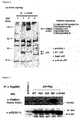
- 11) The eluted fraction was loaded on SDS-PAGE and stained with silver.The results were shown in Figure 6. AS the results, a 170 kDa proteinwas strongly co-precipitated with p53 in S46F substituent. Analysis bymass spectroscopy of the protein revealed the amino acid sequence ofLHIIEVGTPPTGNQPFPK,
IVLDNSVFSEHR, VANVELYYR, QLPLVKPYLR and VDKLDASESLR(SEQ ID NO: 3~7). These amino acid sequences correspond to aminoacid position 228-245, 1011-1022, 1398-1406, 1444-1453 and 1610-1620of clathrin heavy chain (SEQ ID NO:2). The results show that the 170kDa protein is identical to clathrin heavy chain. - 12) The identification of the 170 kD protein was performed after the proteinwas concentrated to 20 times more than the amount used for silverstaining, loaded on SDS-PAGE, stained with CBB, cut out the targetband, digested by trypsin and analyzed by use of mass spectroscopy.
- 13) Hundredth part of the eluate obtained in the procedure 10) wasperformed Western blotting, i.e. the eluate was loaded on SDS-PAGE,transferred to PVDF membrane and stained with anti-clathrin heavychain antibody (Transduction Laboratories) or with anti-p53 antibody(Santa Cruz). In this Example, the bands were visualized byhorseradish peroxidase-conjugated anti-mouse IgG secondary antibody(Amersham) and ECL kit. The results are shown in Figure 7. Clathrinheavy chains were found to bind strongly to S46Fsubstituent.
In this Test Example, nuclear extract from human non-small celllung cancer-derived cells used in Test Example 5 was examined. The testwas according to the following procedures. amount of proteins from nuclear fraction was five times more than that ofcytoplasmic fraction. The results are shown in Figure 8. Previously,clathrin was considered to be localized only in cytoplasm, however, a part ofclathrin was confirmed to be localized in nuclei.
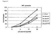
In this Example, experiments were performed according to theprocedure in test Example 4 except that the DNA in the DNA solution of 2)was prepared according to Table 5 and used pc-CHC prepared in TestExample 3 as the DNA transfected. The results are shown in Figure 9. For the DNA solution, pc-CHC (400 ng)and pcDNA 3.1 (400 ng) solutions are referred to as clathrin + and clathrin-, respectively. Clathrin heavy chain cDNA, inserted to an expressionvector and transfected together with p53, was confirmed to enhance thetranscriptional activity of p53AIP1 promoter. Especially, the enhancingeffect of S46F substituent was predominant.


Claims (5)
- A transcriptional factor, comprising p53 or a mutated type p53, wherein one or moreamino acids are deleted, substituted or added with respect to the amino sequence ofp53, and clathrin heavy chains and having an activity to induce apoptosis of cancercells.
- The transcriptional factor of claim 1, wherein at least the serine residue at aminoacid position 46 is substituted in said mutated type p53.
- The transcriptional factor of claim 2, wherein said serine residue at amino acidposition 46 is substituted to phenylalanine.
- A cancer treatment drug comprising the transcriptional factor of any one of claims 1to 3 as an effective component.
- A method for treating cancer comprising injecting the transcriptional factor of anyone of claims 1 to 3, or clathrin heavy chains to a cancer cell.
Applications Claiming Priority (3)
| Application Number | Priority Date | Filing Date | Title |
|---|---|---|---|
| JP2003048658 | 2003-02-26 | ||
| JP2003048658 | 2003-02-26 | ||
| PCT/JP2004/002238WO2004076483A1 (en) | 2003-02-26 | 2004-02-25 | Transcriptional factor inducing apoptosis in cancer cell |
Publications (2)
| Publication Number | Publication Date |
|---|---|
| EP1609802A1true EP1609802A1 (en) | 2005-12-28 |
| EP1609802A4 EP1609802A4 (en) | 2006-06-21 |
Family
ID=32923299
Family Applications (1)
| Application Number | Title | Priority Date | Filing Date |
|---|---|---|---|
| EP04714461AWithdrawnEP1609802A4 (en) | 2003-02-26 | 2004-02-25 | TRANSCRIPTIONAL FACTOR INDUCING APOPTOSIS IN A CANCER CELL |
Country Status (5)
| Country | Link |
|---|---|
| US (1) | US20060159692A1 (en) |
| EP (1) | EP1609802A4 (en) |
| JP (1) | JPWO2004076483A1 (en) |
| CA (1) | CA2517285A1 (en) |
| WO (1) | WO2004076483A1 (en) |
Cited By (11)
| Publication number | Priority date | Publication date | Assignee | Title |
|---|---|---|---|---|
| US8859723B2 (en) | 2010-08-13 | 2014-10-14 | Aileron Therapeutics, Inc. | Peptidomimetic macrocycles |
| US8889632B2 (en) | 2007-01-31 | 2014-11-18 | Dana-Farber Cancer Institute, Inc. | Stabilized p53 peptides and uses thereof |
| US8927500B2 (en) | 2012-02-15 | 2015-01-06 | Aileron Therapeutics, Inc. | Peptidomimetic macrocycles |
| US8987414B2 (en) | 2012-02-15 | 2015-03-24 | Aileron Therapeutics, Inc. | Triazole-crosslinked and thioether-crosslinked peptidomimetic macrocycles |
| US9096684B2 (en) | 2011-10-18 | 2015-08-04 | Aileron Therapeutics, Inc. | Peptidomimetic macrocycles |
| US9604919B2 (en) | 2012-11-01 | 2017-03-28 | Aileron Therapeutics, Inc. | Disubstituted amino acids and methods of preparation and use thereof |
| US10023613B2 (en) | 2015-09-10 | 2018-07-17 | Aileron Therapeutics, Inc. | Peptidomimetic macrocycles as modulators of MCL-1 |
| US10253067B2 (en) | 2015-03-20 | 2019-04-09 | Aileron Therapeutics, Inc. | Peptidomimetic macrocycles and uses thereof |
| US10301351B2 (en) | 2007-03-28 | 2019-05-28 | President And Fellows Of Harvard College | Stitched polypeptides |
| US10471120B2 (en) | 2014-09-24 | 2019-11-12 | Aileron Therapeutics, Inc. | Peptidomimetic macrocycles and uses thereof |
| US10905739B2 (en) | 2014-09-24 | 2021-02-02 | Aileron Therapeutics, Inc. | Peptidomimetic macrocycles and formulations thereof |
Families Citing this family (3)
| Publication number | Priority date | Publication date | Assignee | Title |
|---|---|---|---|---|
| CN1944655B (en)* | 2006-09-04 | 2010-05-12 | 王尚武 | Recombinant adenovirus of target-oriented coexpressed new p53 and P53AIP1 |
| US11235062B2 (en)* | 2009-03-06 | 2022-02-01 | Metaqor Llc | Dynamic bio-nanoparticle elements |
| US11096901B2 (en) | 2009-03-06 | 2021-08-24 | Metaqor Llc | Dynamic bio-nanoparticle platforms |
Family Cites Families (1)
| Publication number | Priority date | Publication date | Assignee | Title |
|---|---|---|---|---|
| AU2156600A (en)* | 1998-11-25 | 2000-06-13 | Cold Spring Harbor Laboratory | Methods and reagents for increasing proliferative capacity and preventing replicative senescence |
- 2004
- 2004-02-25JPJP2005502912Apatent/JPWO2004076483A1/enactivePending
- 2004-02-25CACA002517285Apatent/CA2517285A1/ennot_activeAbandoned
- 2004-02-25WOPCT/JP2004/002238patent/WO2004076483A1/ennot_activeApplication Discontinuation
- 2004-02-25USUS10/546,829patent/US20060159692A1/ennot_activeAbandoned
- 2004-02-25EPEP04714461Apatent/EP1609802A4/ennot_activeWithdrawn
Non-Patent Citations (5)
| Title |
|---|
| CHEN X ET AL: "p53 levels, functional domains and DNA damage determine the extent of the apoptotic response of tumour cells" GENES AND DEVELOPMENT, vol. 10, no. 19, 1996, pages 2438-2451, XP000876795 Cold Springs Harbour NY, US.* |
| MATSUDA K. ET AL: "p53AIP1 Regulates the Mitochondrial Apoptotic Pathway" CANCER RESEARCH, vol. 62, 15 March 2002 (2002-03-15), pages 2883-2889, XP002378916* |
| ODA K ET AL: "p53AIP1, a potential mediator of p53-dependent apoptosis and its regulation by ser-46-phosphorylated p53" CELL, CELL PRESS, CAMBRIDGE, NA, US, vol. 102, no. 6, 15 September 2000 (2000-09-15), pages 849-862, XP002957682 ISSN: 0092-8674* |
| See also references ofWO2004076483A1* |
| WETTEY F. ET AL: "Controlled Elimination of Clathrin Heavy-Chain Expression in DT40 Lymphocytes" SCIENCE, vol. 297, 30 August 2002 (2002-08-30), pages 1521-1525, XP002378917* |
Cited By (21)
| Publication number | Priority date | Publication date | Assignee | Title |
|---|---|---|---|---|
| US8889632B2 (en) | 2007-01-31 | 2014-11-18 | Dana-Farber Cancer Institute, Inc. | Stabilized p53 peptides and uses thereof |
| US9527896B2 (en) | 2007-01-31 | 2016-12-27 | Dana-Farber Cancer Institute, Inc. | Stabilized p53 peptides and uses thereof |
| US10202431B2 (en) | 2007-01-31 | 2019-02-12 | Aileron Therapeutics, Inc. | Stabilized P53 peptides and uses thereof |
| US10301351B2 (en) | 2007-03-28 | 2019-05-28 | President And Fellows Of Harvard College | Stitched polypeptides |
| US9957299B2 (en) | 2010-08-13 | 2018-05-01 | Aileron Therapeutics, Inc. | Peptidomimetic macrocycles |
| US8859723B2 (en) | 2010-08-13 | 2014-10-14 | Aileron Therapeutics, Inc. | Peptidomimetic macrocycles |
| US10308699B2 (en) | 2011-10-18 | 2019-06-04 | Aileron Therapeutics, Inc. | Peptidomimetic macrocycles |
| US9096684B2 (en) | 2011-10-18 | 2015-08-04 | Aileron Therapeutics, Inc. | Peptidomimetic macrocycles |
| US9522947B2 (en) | 2011-10-18 | 2016-12-20 | Aileron Therapeutics, Inc. | Peptidomimetic macrocycles |
| US9505804B2 (en) | 2012-02-15 | 2016-11-29 | Aileron Therapeutics, Inc. | Peptidomimetic macrocycles |
| US10213477B2 (en) | 2012-02-15 | 2019-02-26 | Aileron Therapeutics, Inc. | Peptidomimetic macrocycles |
| US10227380B2 (en) | 2012-02-15 | 2019-03-12 | Aileron Therapeutics, Inc. | Triazole-crosslinked and thioether-crosslinked peptidomimetic macrocycles |
| US8987414B2 (en) | 2012-02-15 | 2015-03-24 | Aileron Therapeutics, Inc. | Triazole-crosslinked and thioether-crosslinked peptidomimetic macrocycles |
| US8927500B2 (en) | 2012-02-15 | 2015-01-06 | Aileron Therapeutics, Inc. | Peptidomimetic macrocycles |
| US9845287B2 (en) | 2012-11-01 | 2017-12-19 | Aileron Therapeutics, Inc. | Disubstituted amino acids and methods of preparation and use thereof |
| US9604919B2 (en) | 2012-11-01 | 2017-03-28 | Aileron Therapeutics, Inc. | Disubstituted amino acids and methods of preparation and use thereof |
| US10669230B2 (en) | 2012-11-01 | 2020-06-02 | Aileron Therapeutics, Inc. | Disubstituted amino acids and methods of preparation and use thereof |
| US10471120B2 (en) | 2014-09-24 | 2019-11-12 | Aileron Therapeutics, Inc. | Peptidomimetic macrocycles and uses thereof |
| US10905739B2 (en) | 2014-09-24 | 2021-02-02 | Aileron Therapeutics, Inc. | Peptidomimetic macrocycles and formulations thereof |
| US10253067B2 (en) | 2015-03-20 | 2019-04-09 | Aileron Therapeutics, Inc. | Peptidomimetic macrocycles and uses thereof |
| US10023613B2 (en) | 2015-09-10 | 2018-07-17 | Aileron Therapeutics, Inc. | Peptidomimetic macrocycles as modulators of MCL-1 |
Also Published As
| Publication number | Publication date |
|---|---|
| EP1609802A4 (en) | 2006-06-21 |
| CA2517285A1 (en) | 2004-09-10 |
| WO2004076483A1 (en) | 2004-09-10 |
| JPWO2004076483A1 (en) | 2006-06-01 |
| US20060159692A1 (en) | 2006-07-20 |
Similar Documents
| Publication | Publication Date | Title |
|---|---|---|
| Aït-Ali et al. | Rod-derived cone viability factor promotes cone survival by stimulating aerobic glycolysis | |
| Flinterman et al. | E1A activates transcription of p73 and Noxa to induce apoptosis | |
| JP4351896B2 (en) | Pharmaceutical composition for cancer treatment comprising p38 / JTV-1 as active ingredient and screening method for pharmaceutical composition for cancer treatment | |
| EP1609802A1 (en) | Transcriptional factor inducing apoptosis in cancer cell | |
| Greenhalgh et al. | Biological evidence that SOCS-2 can act either as an enhancer or suppressor of growth hormone signaling | |
| EP3118212B1 (en) | Cell penetrating peptide and method for delivering biologically active substance using same | |
| EP2666857B1 (en) | Nucleic acid construct for expressing oxidative stress indicator and use thereof | |
| Evstafieva et al. | Apoptosis-related fragmentation, translocation, and properties of human prothymosin alpha | |
| Bissonnette et al. | The apoptotic and transcriptional transactivation activities of p53 can be dissociated | |
| WO2009124137A2 (en) | Method of suppressing gene transcription through histone lysine methylation | |
| Rodriguez-Enfedaque et al. | FGF1 nuclear translocation is required for both its neurotrophic activity and its p53-dependent apoptosis protection | |
| Günther et al. | Dissection of Drosophila MTF-1 reveals a domain for differential target gene activation upon copper overload vs. copper starvation | |
| EP3765483A1 (en) | Micropeptides and uses thereof | |
| Nakayama et al. | Txnip C247S mutation protects the heart against acute myocardial infarction | |
| US20020169123A1 (en) | Regulating apoptosis in TRAIL-resistant cancer cells, while protecting normal, non-cancerous cells | |
| Yuan et al. | Induction of p53 in mouse cells decreases mutagenesis by UV radiation | |
| Moore et al. | Aging-associated truncated form of p53 interacts with wild-type p53 and alters p53 stability, localization, and activity | |
| EP2687537B1 (en) | Polypeptide drug against hepatitis b virus x protein | |
| KR100739118B1 (en) | Novel use of the MBS1 protein or gene encoding it | |
| KR102125005B1 (en) | Novel use of 53bp1 | |
| AU784293B2 (en) | Human P53 mutations and a genetic system in yeast for functional indentification of human P53 mutations | |
| Hasegawa et al. | A novel role for acinus and MCM2 as host-specific signaling enhancers of DNA-damage-induced apoptosis in association with viral protein gp70 | |
| KR100760320B1 (en) | An anticancer composition comprising a recombinant vector expressing an importin α gene and a recombinant 355 gene | |
| JP4750364B2 (en) | Preventive or therapeutic agent for diseases associated with apoptosis | |
| US7256260B1 (en) | Human p53 mutations and a genetic system in yeast for functional identification of human p53 mutations |
Legal Events
| Date | Code | Title | Description |
|---|---|---|---|
| PUAI | Public reference made under article 153(3) epc to a published international application that has entered the european phase | Free format text:ORIGINAL CODE: 0009012 | |
| 17P | Request for examination filed | Effective date:20050824 | |
| AK | Designated contracting states | Kind code of ref document:A1 Designated state(s):AT BE BG CH CY CZ DE DK EE ES FI FR GB GR HU IE IT LI LU MC NL PT RO SE SI SK TR | |
| AX | Request for extension of the european patent | Extension state:AL LT LV MK | |
| DAX | Request for extension of the european patent (deleted) | ||
| RIC1 | Information provided on ipc code assigned before grant | Ipc:A61K 35/00 20060101ALI20060509BHEP Ipc:A61K 38/00 20060101ALI20060509BHEP Ipc:C12N 15/12 20060101ALI20060509BHEP Ipc:C07K 14/47 20060101AFI20040915BHEP | |
| A4 | Supplementary search report drawn up and despatched | Effective date:20060524 | |
| RAP1 | Party data changed (applicant data changed or rights of an application transferred) | Owner name:JAPAN AS REPRESENTED BY PRESIDENT OF NATIONAL CANC Owner name:JAPAN SCIENCE AND TECHNOLOGY AGENCY | |
| RAP1 | Party data changed (applicant data changed or rights of an application transferred) | Owner name:JAPAN SCIENCE AND TECHNOLOGY AGENCY | |
| 17Q | First examination report despatched | Effective date:20060905 | |
| 17Q | First examination report despatched | Effective date:20060905 | |
| STAA | Information on the status of an ep patent application or granted ep patent | Free format text:STATUS: THE APPLICATION IS DEEMED TO BE WITHDRAWN | |
| 18D | Application deemed to be withdrawn | Effective date:20070710 |