EP1601697B1 - Antibody purification by Protein A and ion exchange chromatography - Google Patents
Antibody purification by Protein A and ion exchange chromatographyDownload PDFInfo
- Publication number
- EP1601697B1 EP1601697B1EP04715906AEP04715906AEP1601697B1EP 1601697 B1EP1601697 B1EP 1601697B1EP 04715906 AEP04715906 AEP 04715906AEP 04715906 AEP04715906 AEP 04715906AEP 1601697 B1EP1601697 B1EP 1601697B1
- Authority
- EP
- European Patent Office
- Prior art keywords
- antibody
- protein
- binding
- igg
- contaminant
- Prior art date
- Legal status (The legal status is an assumption and is not a legal conclusion. Google has not performed a legal analysis and makes no representation as to the accuracy of the status listed.)
- Revoked
Links
Images


Classifications
- C—CHEMISTRY; METALLURGY
- C07—ORGANIC CHEMISTRY
- C07K—PEPTIDES
- C07K1/00—General methods for the preparation of peptides, i.e. processes for the organic chemical preparation of peptides or proteins of any length
- C07K1/14—Extraction; Separation; Purification
- C07K1/36—Extraction; Separation; Purification by a combination of two or more processes of different types
- B—PERFORMING OPERATIONS; TRANSPORTING
- B01—PHYSICAL OR CHEMICAL PROCESSES OR APPARATUS IN GENERAL
- B01D—SEPARATION
- B01D15/00—Separating processes involving the treatment of liquids with solid sorbents; Apparatus therefor
- B01D15/08—Selective adsorption, e.g. chromatography
- B01D15/26—Selective adsorption, e.g. chromatography characterised by the separation mechanism
- B01D15/36—Selective adsorption, e.g. chromatography characterised by the separation mechanism involving ionic interaction, e.g. ion-exchange, ion-pair, ion-suppression or ion-exclusion
- B01D15/361—Ion-exchange
- B01D15/362—Cation-exchange
- B—PERFORMING OPERATIONS; TRANSPORTING
- B01—PHYSICAL OR CHEMICAL PROCESSES OR APPARATUS IN GENERAL
- B01D—SEPARATION
- B01D15/00—Separating processes involving the treatment of liquids with solid sorbents; Apparatus therefor
- B01D15/08—Selective adsorption, e.g. chromatography
- B01D15/26—Selective adsorption, e.g. chromatography characterised by the separation mechanism
- B01D15/36—Selective adsorption, e.g. chromatography characterised by the separation mechanism involving ionic interaction, e.g. ion-exchange, ion-pair, ion-suppression or ion-exclusion
- B01D15/361—Ion-exchange
- B01D15/363—Anion-exchange
- B—PERFORMING OPERATIONS; TRANSPORTING
- B01—PHYSICAL OR CHEMICAL PROCESSES OR APPARATUS IN GENERAL
- B01D—SEPARATION
- B01D15/00—Separating processes involving the treatment of liquids with solid sorbents; Apparatus therefor
- B01D15/08—Selective adsorption, e.g. chromatography
- B01D15/26—Selective adsorption, e.g. chromatography characterised by the separation mechanism
- B01D15/38—Selective adsorption, e.g. chromatography characterised by the separation mechanism involving specific interaction not covered by one or more of groups B01D15/265 and B01D15/30 - B01D15/36, e.g. affinity, ligand exchange or chiral chromatography
- B01D15/3804—Affinity chromatography
- B01D15/3809—Affinity chromatography of the antigen-antibody type, e.g. protein A, G or L chromatography
- C—CHEMISTRY; METALLURGY
- C07—ORGANIC CHEMISTRY
- C07K—PEPTIDES
- C07K14/00—Peptides having more than 20 amino acids; Gastrins; Somatostatins; Melanotropins; Derivatives thereof
- C07K14/195—Peptides having more than 20 amino acids; Gastrins; Somatostatins; Melanotropins; Derivatives thereof from bacteria
- C07K14/305—Peptides having more than 20 amino acids; Gastrins; Somatostatins; Melanotropins; Derivatives thereof from bacteria from Micrococcaceae (F)
- C07K14/31—Peptides having more than 20 amino acids; Gastrins; Somatostatins; Melanotropins; Derivatives thereof from bacteria from Micrococcaceae (F) from Staphylococcus (G)
- C—CHEMISTRY; METALLURGY
- C07—ORGANIC CHEMISTRY
- C07K—PEPTIDES
- C07K16/00—Immunoglobulins [IGs], e.g. monoclonal or polyclonal antibodies
- C07K16/06—Immunoglobulins [IGs], e.g. monoclonal or polyclonal antibodies from serum
- C07K16/065—Purification, fragmentation
Definitions
- the present inventionrelates to the field of antibody purification in biotechnological production. It is an object of the present invention to describe a novel process for purification of such antibody.
- Protein A chromatographyis widely used in industrial manufacturing of antibodies since allowing for almost complete purification of antibodies, that is usually IgG, in a single step from cell culture supernatants. Protein A affinity columns inevitably are subject to some degree of leakage of ligand from the column upon repeated runs. Partly, this may be due to proteolytic clipping of protein A from the column; in industrial manufacture of antibody for pharmaceutical applications, no protease inhibitor cocktails may be added for regulatory reasons. Unfortunately, this protein A or protein A fragment contaminants retain their affinity for IgG and are difficult to remove from the purified antibody due to ongoing complex formation.
- Balint et al.demonstrated that such IgG-Protein complexes can be separated from uncomplexed IgG by gel filtration. Low through-put and loss in antibody yield are the disadvantages of this method.
- US 4,983,722teaches selective separation of contaminating protein A from a protein-A purified antibody preparation by absorbing the mixture to an anion exchanger material and to separate both components by sequentially eluting the antibodies and protein A under conditions of increasing ionic strength. This resolution method is highly dependent on the pI of the antibody which is specific and highly variable for a given antibody. Further, throughput is limited by the steepness of the salt gradient required for obtaining separation.
- a method of purifying an antibodycomprises the steps of: First, purifying an antibody by means of protein A affinity chromatography wherein the protein A is a native protein A or a functional derivative thereof.
- Protein Ais a cell surface protein found in Staphylococcus aureus. It has the property of binding the Fc region of a mammalian antibody, in particular of IgG class antibodies. Within a given class of antibodies, the affinity slightly varies with regard to species origin and antibody subclass or allotype (reviewed in Surolia, A. et al., 1982, Protein A: Nature's universal ,antibody', TIBS 7, 74-76; Langone et al., 1982, Protein A of staphylococcus aureus and related immunoglobulin receptors, Advances in Immunology 32:157-252). Protein A can be isolated directly from cultures of S.
- aureusthat are secreting protein A or is more conveniently recombinantly expressed in E.coli (Lofdahl et al., 1983, Proc. Natl. Acad. Sci. USA 80:697-701). Its use for purification of antibodies, in particular monoclonal IgG, is amply described in the prior art ( e.g. Langone et al., supra; Hjelm et al, 1972; FEBS Lett. 28: 73-76).
- protein Ais coupled to a solid matrix such as crosslinked, uncharged agarose (Sepharose, freed from charged fraction of natural agarose), trisacryl, crosslinked dextrane or silica-based materials.
- Protein Abinds with high affinity and high specificity to the Fc portion of IgG, that is the C ⁇ 2-Cy3 interface region of IgG as described in Langone et al., 1982, supra. In particular, it binds strongly to the human allotypes or subclasses IgG1, IgG2, IgG3 and the mouse allotypes or subclasses IgG2a, IgG2b, IgG3.
- Protein Aalso exhibits an affinity for the Fab region of immunoglobulins that are encoded by the V H gene family, V H III (Sasso et al., 1991, J. Immunol, 61: 3026-3031; Hilson et al., J Exp. Med., 178: 331-336 (1993)).
- the sequence of the gene coding for protein Arevealed two functionally distinct regions (Uhlen et al., J . Biol. Chem.,259: 1695-1702 (1984); Lofdahl et al., Proc. Nutl. Acad. Sci.(USA), 80: 697-701 (1983)).
- the amino-terminal regioncontains five highly homologous IgG-binding domains (termed E, D, A, B and C), and the carboxy terminal region anchors the protein to the cell wall and membrane. All five IgG-binding domains of protein A bind to IgG via the Fc region, involving e.g. in human IgG-Fc residues 252-254, 433-435 and 311, as shown for the crystal structure in Deisenhofer et al. (1981, Biochemistry 20: 2361-2370) and in Sauer-Eriksson et al. (1995, Structure 3: 265-278) in case of the B-domain of protein A.
- each of the IgG-binding domains A to E of protein Ais sufficient for binding to the Fc-portion of an IgG.
- An IgG antibodyis to be understood as an antibody of such allotype that it can be bound to protein A in a high-affinity mode. Further, apart from the Fc portions of the antibody that are relevant for binding to protein A, such antibody must not correspond to a naturally occuring antibody. In particular in its variable chain regions portions, it can be an engineered chimeric or CDR-grafied antibody as are routinely devised in the art.
- An IgG-antibodyis to be understood as an IgG-type antibody, in short.
- An interaction compliant with such value for the binding constantis termed 'high affinity binding' in the present context.
- such functional derivative of protein Acomprises at least part of a functional IgG binding domain of wild-type protein A which domain is selected from the natural domains E,D,A,B, C or engineered muteins thereof which have retained IgG binding functionality.
- An example of suchis the functional 59 aminoacid 'Z'-fragment of domain B of protein A which domain may be used for antibody purification as set forth in US 6013763.
- an antibody binding fragmentcomprises at least two intact Fc binding domains as defined in this paragraph.
- An example of suchare the recombinant protein A sequences disclosed e.g. in EP-282 308 and EP-284 368, both from Repligen Corporation.
- Protein A fragmentsthat are engineered to allow of single-point attachement.
- Single point attachmentmeans that the protein moiety is attached via a single covalent bond to a chromatographic support material of the protein A affinity chromatography.
- Such single-point attachmentby means of suitably reactive residues which further are ideally placed at an exposed amino acid position, namely in a loop, close to the N- or C-terminus or elsewhere on the outer circumference of the protein fold.
- Suitable reactive groupsare e.g. sulfhydryl or amino functions. More preferably, such recombinant protein A or functional fragment thereof comprises a cysteine in its amino acid sequence.
- the cysteineis comprised in a segment that consists of the last 30 amino acids of the C-terminus of the amino acid sequence of the recombinant protein A or functional fragment thereof.
- the recombinant protein A or functional fragment thereofis attached by at least 50% via a thioether sulphur bond to the chromatographic support or matrix material of the protein A-affinity chromatography medium.
- a thioether sulphur bondto the chromatographic support or matrix material of the protein A-affinity chromatography medium.
- thioetheris to be understood narrowly as a -S- bonding scheme irrespective of chemical context, deviating in this regard from normal chemical language; it is possible, for instance, that said -S- 'thioether' bridge is part of a larger functional group such as e.g. a thioester or a mixed acetal, deviating in this regard in the context of the present application from the reacitivity-based normal language of chemists.
- the thioether bridgeis a thioether bridge in its ordinary, narrow chemical meaning.
- Such bridging thioether groupcan be e.g.
- the protein A or functional protein A derivativeis the recombinant protein A disclosed in US 6399750 which comprises a juxtaterminal, engineered cysteine residue and is,preferably by at least 50%, more preferably by at least 70%, coupled to the chromatographic support material through the sulphur atom of said cysteine residue as the sole point of attachment.
- such couplinghas been achieved by means of epoxide mediated activation, more preferably either by means of 1,4-bis-(2,3-epoxypropoxy) butane activation of e.g.
- an agarose matrixsuch as Sepharose Fast Flow (agarose beads crosslinked with epichlorohydrin, Amersham Biosciences, UK) or by means of epichlorohydrin activation of e.g. an agarose matrix such as Sepharose FF.
- the first ion exchangeris an anion exchanger, in particular a quaternary amine-based anion exchanger such as Sepharose Q TM FF (Amersham-Biosciences/Pharmacia), most preferably it is an anion exchanger having the functional exchanger group Q coupled to a matrix support which group Q is N,N,N-Trimethylamino-methyl, most preferably the anion exchanger is Sepharose Q TM FF from Pharmacia/Amersham Biosciences.
- the quarternary amino groupis a strong exchanger which further is not susceptible to changes in pH of the loading/wash buffer.
- the fast flow exchanger matrixis based on 45-165 ⁇ m agarose beads having a high degree of crosslinking for higher physical stability; further sepharose is devoid of the charged, sulfated molecule fraction of natural agarose and does not allow for unspecific matrix adsorption of antibody, even under condition of high antibody loads.
- An example of such an embodimentcan be found in the experimental section.
- a contaminant protein Ais any type of functional, IgG binding offspring of a protein A or a functional derivative thereof as defined above which is obtained upon eluting bound antibody from a protein A affinity chromatography column.
- Such contaminant protein A speciesmay result e.g. from hydrolysis of peptide bonds which is very likely to occur by means of enzyme action in particular in industrial manufacturing.
- Protein A chromatographyis applied as an early step in downstream processing when the crudely purified, fresh product solution still harbors considerable protease activity.
- Dying cells in the cell culture broth or cells disrupted in initial centrifugation or filtration stepsare likely to have set free proteases; for regulatory purposes, supplementation of the cell culture broth with protease inhibitors prior or in the course of downstream processing is usually not accomplished, in contrast to biochemical research practice.
- Examplesare Phenyl-methyl-sulfonyl-chloride (PMSF) or ⁇ -caproic acid.
- PMSFPhenyl-methyl-sulfonyl-chloride
- ⁇ -caproic acidSuch chemical agents are undesirable as an additives in the production of biopharmaceuticals.
- recombinant functional derivatives or fragments of protein Aare less protease resistant than wild-type protein A, depending on the tertiary structure of the protein fold.
- Amino acid segments linking individual IgG binding domainsmight be exposed once the total number of binding domains is reduced. Interdomain contacts may possible contribute to the stability of domain folding. It might also be that binding of antibody by protein A or said functional derivatives thereof influences or facilitates susceptibility to protease action, due to conformational changes induced upon binding of the antibody. Again, wild-type or full length protein A or functional, engineered fragments thereof might behave differently.
- contaminant protein Astill is functional, IgG binding protein and thus is associated with the protein A-purified antibody when loaded onto the subsequent ion exchange separation medium. The high-affinity based association of contaminant protein A with the purified antibody is the reason why it is difficult to efficiently separate contaminant protein A from purified antibody.
- the antibody sought to be purifiedis harvested from a cell culture prior to purifying the antibody be means of protein A affinity chromatography.
- said cell cultureis a mammalian cell culture. Mammalian cells have large compartments called lysosomes harboring degradating enyzmes which are disrupted upon cell death or harvest.
- said cell culturemay be a myeloma cell culture such as e.g. NSO cells (Galfre, G. and Milstein, C. Methods Enzymology , 1981, 73,3).
- Myeloma cellsare plasmacytoma cells, i.e. cells of lymphoid cell lineage.
- An exemplary NSO cell lineis e.g.
- NS0have been found able to give rise to extremly high product yields, in particular if used for production of recombinant antibodies.
- NSO cellshave been found to give reproducibly rise to much higher levels of contaminant protein A than other host cell types at least with certain protein A affinity chromatography systems employing recombinant, shortened fragments of wild-type protein A which recombinant protein A is possibly single-point attached protein A.
- Streamline TM rProtein A affinity chromatography resin(Amersham Biosciences; essentially thioester single-point attached recombinant protein A as described in US 6,399,750).
- Levels of about or in excess of 1000 ng contaminant protein A/mg antibodycould be obtained with Streamline TM rProtein A affinity columns.
- the method of the present inventiondistinguishes from the prior art in efficiently reducing contaminant protein A from such elevated levels to ⁇ 1 ng/mg antibody in a single, fast purification step, that is with a purification factor of about 1000x.
- the antibody that is to be purified by means of protein A affinity chromatographyis not treated as to inactivate proteases at or after harvest, more preferably is not in admixture with at least one exogenously supplemented protease inhibitor after harvest.
- said protease inhibitoris selected from the group consisting of PMSF, specific proteinase inhibiting peptides as described in Laskowski et al., 1980, Protein inhibitors of proteinases, Ann. Rev. Biochem. 49, 593-626, and epsilon-caproic acid.
- the contaminant protein Ais reduced to a concentration of ⁇ 10 ng/mg antibody, more preferably ⁇ 4 ng/mg antibody, most preferably ⁇ 1 ng/mg antibody in the flow-through of the first ion-exchanger, wherein antibody is preferably to be understood as to refer to IgG.
- the Elisa assay method for validation of these threshold valuesis described in detail in the experimental section; it should be noted that acidification of the sample to a pH ⁇ 4, preferably in the presence of a mild detergent, is crucial for accurate determination of the amount of leaked protein A.
- At least 70%, more preferably at least 80%, most preferably at least 90% of the antibody loaded onto the first ion exchangercan be recovered in the flow-through of the ion-exchanger.
- the first ion exchangeris an anion exchanger resin; protein A can be bound by both types of resin as described (EP-289 129 B1).
- the first ion exchanger or anion exchangercan be operated in the column mode at a certain flow rate or in batch operation mode, by submerging the ion exchange resin into the mildly agitated sample solution and further exchanging liquid media by filtration subsequently.
- suitable conditions of pH and ionic strength for loading the first ion exchangerwhich conditions result in retaining the antibody in the flow through whilst the protein A contaminant is bound and thus removed from the antibody.
- the method according to the present inventionallows of faster separation of antibody from contaminant protein A.
- Examples of functional groups of such first, anion exchanger that are attached to a matrix supportare e.g. primary, secondary, and particularly tertiary or quaternary animo groups such as aminoethyl, diethylaminoethyl, trimethylaminoethyl, trimethylaminomethyl and diethyl-(2-hydroxypropyl)-aminoethyl.
- Suitable chromatographic support matrixes for the anion exchangerare known in the art. Examples are agarose-based resins and beads, dextran beads, polystyrene beads and polystyrene/divinyl-benzene resins.
- the ion exchangeris a quaternary amine-based anion exchanger mounted on an agarose matrix such as e.g. Sepharose CL-6B or Sepharose Fast Flow (FF) from Amersham-Biosciences/Pharmacia.
- an agarose matrixsuch as e.g. Sepharose CL-6B or Sepharose Fast Flow (FF) from Amersham-Biosciences/Pharmacia.
- FFSepharose Fast Flow
- Sepharose Q TMfrom Amersham-Biosciences/Pharmacia.
- the antibodyis a monoclonal antibody that has an isoelectric point (pI) which is at least two pH units above, that is it is more basic than, the pI of the protein A used in the preceding protein A affinity chromatography step; e.g.
- the antibodyis a monoclonal antibody that has an isoelectric point (pI) which is at least 6.5 or above, more preferably is 7.0 or above, most preferably has an pI of at least 7.5 or above. It should be noted that this refers to the pI of the actually harvested and purified antibody, not the pI that can be simply predicted from the amino acid sequence alone.
- the actually purified antibody moleculemay have undergone further modifications of the polypeptide backbone such as glycosylation, which modifications may add charged moieties and thus may have changed the pI of the molecule.
- the microheterogenity of posttranslational processing of the antibody proteine.g. a monoclonal antibody protein
- the 'pI of an antibody'refers to that share of antibody product molecules whose pI is within the preferred range of pI as specified above. All further definitions of this description, such as the %-proportion of antibody recovered after a given purification step, refer to said pl-compliant share of antibody only.
- the mode of operation of a first anion exchangerrequires buffer exchange of the acidic or neutralized eluate from the protein A affinity chromatography step with the equilibration buffer of the first anion exchanger.
- Equilibration buffer and loading bufferare identical in the method of the present invention. Commonly employed ultrafiltration devices such as sold by Amicon or Millipore can be expediently used for that purpose; those avoid the dilution effects whilst using e.g. low molecular weight porous filtration matrices such as Sephadex G-25.
- the equilibration bufferpreferably has a salt concentration of a displacer salt such as e.g.
- the pH of the equilibration bufferis preferably in the range of pH 6.5 to pH 9.0, more preferably is in the range of pH 7.5 to pH 8.5, most preferably is in the range of pH 7.9 to pH 8.4.
- N-terminal amino function of a proteinhas a pKs value of about 9.25, thus binding of contaminant protein A and any other already negatively charged protein to an anion exchanger will get stronger at more basic pH; for a given application, the pH of the loading buffer might need finetuning for optimal discrimination of binding and non-binding for a given pair of antibody and contaminant protein A having differing pI values and different content of cysteine and histidine side chains which may contribute to changes in charge within the selected ranges of pH.
- a more basic pHinterferes with proteinA-antibody interactions as will do any increase in ionic strength; likewise, ionic strength needs finetuning to balance prevention of binding of antibody with the need to abolish binding of contaminant protein A.
- the ionic strength of the bufferis usually inversely correlated with the pH value; the more strongly protein A gets bound to the anion exchanger depending on pH, the more salt is tolerated for preventing binding of antibody and for interfering with potential proteinA-antibody interactions.
- the above given ranges for pH and displacer saltthus are to be understood as to be correlated: The lower the pH, the less salt is found permissible within the above given preferred ranges for working the invention.
- Further salt freightis added by the pH buffering substance, further increasing the ionic strength of the solution.
- the ionic strengthcan be determined by measuring the conductivity of the equilibration buffer.
- the term 'conductivity'refers to the ability of an aqueous solution to conduct an electric current between two electrodes measures the total amount of ions further taking charge and ion motility into account. Therefore, with an increasing amount of ions present in the aqueous solution, the solution will have a higher conductivity.
- the unit of measurement for conductivityis mS/cm (milliSiemens/cm), and can be measured using a commercially available conductivity meter, e.g. from Topac Inc. (Hingham, MA/U.S.A.) or Honeywell.
- the loading or equilibration buffer for the first anion exchange stephas a conductivity 0.5-5 mS/cm, more preferably of from 1-3 mS/cm, most preferably of from 1.25-2.5 mS/cm. Ideally, it has a conductivity of about 2 mS/cm.
- suitable buffer saltscan be found in Good, N.E. (1986, Biochemistry 5:467-476).
- E.g. Tris.HCl buffer or a sodium hydrogen phosphate buffer as customarily employedare suitable buffering substances. The concentration of the buffer substance is customarily in the range of e.g.
- the buffer substanceis a phosphate buffer.
- Hydrogenphosphatehas a low elution strength, in particular if employed at a pH at or below pH 8, and further excels by particularly low chaotropic properties.
- column operation modeis preferred for the first anion exchanger step.
- a flow rate of about 10 to 60 ml/hcan be advantageously employed.
- the loading concentration of antibody loadedcan favorably be in the range of 10 to 30 mg antibody/ml exchange resin. It goes without saying that the use of extremly diluted samples would give rise to decreased yield of antibody, as is known to the skilled person.
- the antibody sought to be purifiedis collected in the low-through of the loading operation including about one column volume of wash with the same equilibration buffer.
- the pH of the flow-throughmay be adjusted to neutral pH for improving stability and preventing aggregation and/or precipitation of antibody protein.
- the antibodyis ready for use in applications or may be deemed to require further polishing by customary purification methods.
- the first ion exchange stepis followed by a second ion exchange step in which second step the antibody is loaded and bound by the second ion exchange medium and is eluted with a buffer other than the loading buffer, by means of increased salt and/or pH, as an essentially monomeric, non-aggregated antibody.
- the second ion exchangeris a cation exchanger.
- Such combination of a protein A chromatography step followed by a first anion exchanger and a second cation exchanger stepis novel. It is well known that most trace contaminant proteins from cell culture broth have much lower pI values than antibodies, in particular IgG antibodies; cation exchange will therefore allow of efficient removal both of aggregated antibody and potential infectious agents such as virus capsids as well as of protein contaminants other than antibody. Due to speedy operation, highly efficient recovery of antibody after loading, binding to and elution from the co and high capacity of loading, it allows also of repeated, cyclic operation with a single batch of antibody with additive effect of the purification factor achieved in a single round of binding and elution.
- the pH of the loading bufferis about pH 4 to 7, more preferably pH 4.01 to 6, most preferably pH 4.02 to 5.5.
- the antibodyis eluted from the cation exchanger with a salt gradient in the range of from 0.1 to 1.2 M salt, wherein the salt preferably is an alkaline metal salt, more preferably a lithium, potassium or sodium salt.
- elutiontakes place at a pH of from pH 7 to 8 in order to have maximum aggregate removal and minimal damage to antibody due to acidic conditions.
- the elutiontakes place at a an acid pH of from pH 4 to 7, more preferably 4.01 to 6 for maximizing removal of contaminant protein A; levels as low as ⁇ 0.4 ng/mg antibody can be realized in this way.
- This second cation exchanger steprenders traditional gel filtration moot whilst allowing of high-capacity as well as fast operation as is typical for ion exchangers. Ion exchangers support loads of 10-30 mg antibody/ml resin.
- the purification method of a first anion exchanger and a second cation exchanger step in the aftermath of protein A chromatographyrenders clinical grade antibody in the absence of a further, terminal size exclusion chromatography (SEC) step which SEC step would have a molecular weight cut off suitable for separating antibody aggregates and/or antibody-protein A complexes from monomeric antibody such as an normal IgG.
- SECterminal size exclusion chromatography
- the most appealing feature of the method of the present inventionis that purifying antibody via an anion exchanger in a non-binding or flow-through mode, the capacity of the column is not all limiting the through-put of material; the capacity is only decisive with regard to minor amounts of contaminant protein A retain. This saves a lot of processing time and material resources whilst allowing for very efficient removal of protein A contaminant.
- the detection rabbit antibodywas equally purchased from Sigma-Aldrich (#3775). After coating the protein by unspecific adsoprtion process, the coated protein is used to retain protein A-specific protein A capture antibody which capture antibody is further detected with bioinylated rabbit anti-protein A and streptavidin-horseradish peroxidase. Tetramethyl benzidine is used as the chromogenic substrate. Samples of unknown concentration are read off against a standard curve using the very parent-protein A or -protein A derivative of the contaminant protein A sought to be detected. Coating at acidic pH as well as proper preparation of the standard has proven important.
- Preparation of the protein standardwas carried out at best immediately prior to use of the standard for coating the microtiter plates.
- a 1mg/ml stock solutionwas prepared and kept at -65°C in a freezer; after thawing, monomeric character of protein A was assayed from an aliquot loaded on non-reducing SDS-PAGE.
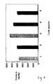
- the concentration of protein standardwas determined by Bradford assay (Bradford et al., 1976, Anal. Biochem. 72:248-254; Splittgerber et al., 1989, Anal. Biochem. 179:198-201) as well as by automated amino acid analysis. The result of such pretreatment is shown in Fig.
- Lane 1by means of non-reducing 10% SDS-PAGE for a staphylococcal protein A standard (lane 1: native protein A; lane 2: after pretreatment) and pure, uncoupled Streamline TM recombinant protein A (provided by courtesy of Pharmacia, now Amersham-Biosciences; lane 4: native recombinant protein A; lane 5: after pretreatment).
- Lane 1is a molecular weight marker with the corresponding molecular masses being denoted on the vertical axis.
- the other halfis spiked with the same volume of sample buffer; thus the dilution factor of the product sample due to spiking is accounted for.
- Both types of preparationwill be referred to as 'spiked sample' in the following.
- the sample bufferwas made up from 7.51 g Glycine (base), 5.84 g NaCl, 0.5 ml Triton X-100 to a volume of 1 L with deionized or bidestillated water.
- the antibody concentrations in the sampleswere pre-determined by customary Elisa's well known in the art.
- a further standard solutionwas spiked with an equal amount of a known standard antibody of comparable constant region affinity for protein A, to determine efficiency of the acidification step and to unravel any potential systematic error introduced by antibody binding to and thus scavenging protein A from capture in the assay.
- AcidificationTo 450 ⁇ l of spiked sample or standard is added 200 ul of 0.2 M citrate/0.05% Triton X-100 buffer at pH 3.0. All samples were done in triplicate. Further, dilutions of sample were prepared and tested in triplicate since the assay works optimal for antibody concentrations being in the range of 1 mg/ml and 0.2 mg/ml.
- the acidification stepis crucial in the present assay to liberate contaminant protein A or A fragments which were otherwise bound to the excess of antibody present in the sample solution.
- Coating bufferwas made up from 1.59 g/L Na2CO3, 2.93 g/L NaHCO3 and 0.20 g/L sodium azide. The pH of the buffer was adjusted to pH 9.6. Add 100 ⁇ l antibody solution per well comprising antibody in an amount sufficient as not to show saturation for the protein A standard. Cover plate with plastic film and place in humidity chamber. Incubate at 37°C overnight for approximately 18 hours.
- washing buffer[NaCI 5.8 g/L, Na 2 HPO 4 1.15 g/L, NaH 2 PO.H 2 0 0.26 g/L, EDTA 3.7 g/L, Tween-20 0.2 g/L, butanol 10 ml/L, pH 7.2], and tap dry.
- Add 250 ⁇ l blocking buffer[coating buffer with 0.5 % casein hammarsten] to each well and incubate for 2 hours at ambient temperature on a benchtop orbital shaker (speed 120 rpm). Rinse all wells three times with at least 300 ⁇ l washing buffer, and tap dry.
- the substrate solutionis prepared like this: A stock solution is prepared by dissolving 10 mg TMB in 1 ml DMSO. 10 ⁇ l of that stock, further 10 ⁇ l of H 2 O 2 are added to a 2.05 % (w/w) sodium acetate aequeous solution that was adjusted to pH 6.0 with 0.5 M citric acid. It goes without saying that all water used for preparing any reagent of the assay is of highest quality, that is deionized ultrapure or at least bidestillated water. The substrate solution is incubated at ambient temperature for 8-11 minutes on a shaker. The reaction is then stopped by adding 50 ⁇ l per well of stopping solution [13% H 2 SO 4 ]. Within 10 min.
- Fig. 2shows the levels of leaked recombinant protein A in antibody eluates from Streamline TM recombinant protein A chromatography with single-point attached protein A in thioether linkage.
- the cycle numberrefers to repeated use after elution with 1 M sodium chloride and re-equilibration.
- Fig. 3further provides data on insubstantially reduced leakage of contaminant protein A during repeated runs of the protein A affinity chromatography with the same affinity matrix material ; wild-type protein A multipoint-attached Sepharose 4 FF (Amersham-Biosciences) was repeatedly used as described in section 2.1 below and the level of contaminant protein A in the eluate, before any further processing of eluate, was determined by Elisa as described above.
- Cell culture supernatant from a NSO myeloma cell culturewas crudely purifed by centrifugation and in depth filtration and concentrated by ultrafiltration; ultrafiltration was also used to exchange the culture fluid to PBS pH 7.5.
- the titer of the antibody #5 produced by the cellswas 0.2 mg/ml, a total of 1 L buffer-exchanged supernatant was loaded.
- the pI of the monoclonal antibody #5was 8.5.
- the protein A Streamline TM column(5.0 ml volume) was previously equilibrated with 10 column volumes of 50 mM glycine/glycinate pH 8.8, 4.3 M NaCl; flow rate was at 200 cm/h.
- the columnwas operated at a flow rate of 50 cmh- 1 ; loading capacity was about 20 mg/ml matrix material).
- loading capacitywas about 20 mg/ml matrix material.
- the columnwas washed with at least 10 column volumes of glycine equilibration buffer supplemented with additional 200 mM NaCl and 0.1 % Tween-20. Elution was achieved with elution buffer made up of 0.1 M glycine/HCl pH 4.0 buffer.
- fractions of eluate comprising the antibody peakwere neutralized with an adequate aliquot of 0.5 M TrisHCl pH 7.5 and buffer exchanged with an Amicon diafiltration device with loading/equilibration buffer (10mM Tris/HCl pH 8.0, 50 mM NaCl) for the subsequent anion exchanger step for preventing longer exposure to acidic pH.
- the antibody concentration and the contaminant protein A concentrationwere determined as described above.
- the level of contaminant protein A in the eluateamounted to 1434 ng/mg antibody before and amounted to 1650 ng/mg antibody after diafiltration.
- the recovery of antibody based on the titer of the buffer exchanged supernatant solution prior to loadingwas 81%; the concentration of antibody in the diafiltrated solution was 3.6 mg/ml.
- the purified antibody from section 2.1was further processed as described: A 5.0 ml Q-Sepharose FF column (Amersham-Biosciences) was packed 10 ml of 0.1 M NaOH, followed by 2 column volumes of 0.1 M Tris pH 8, and equilibrated in 10 column volumes of 10 mM Tris pH 8/50 mM NaCl, at a flow rate of 75 cm/h. After equilibration, the flow rate was reduced to 50 cm/h.
- the Q-Sepharose columnwas recycled for further use by separate elution in 2M NaCl and further equilibration as described above.
- the antibody from exp. 2.2. purified in a non-binding mode by Q-Sepharose anion exchangewas used.
- SP-Sepharose FFallowed of a flow rate of 100 cm/h with a reproducible yield of 93% antibody after loading, washing and elution of the antibody from the cation exchanger.
- the pH of the antibody solution obtained after Sepharose Q purification stepwas adjusted to pH 4.5-5.0 with 50 mM acetate buffer pH 4.5.
- the loading capacitywas set with 10 mg/ml matrix material at a conductivity of load of 17 mS/cm.
- the 50 mM acetate bufferwas further used for washing to baseline.
- a 50 mM Na acetate pH 4.5, 1 M NaCl high salt bufferwas used for elution of antibody; monomeric antibody eluted first, whereas aggregates used to elute in the tail fractions at high ionic strength.
- This multipoint-attached Streamline TM protein A-affinity matrixwas custom made and supplied by Pharmacia Biotech (now Amersham-Pharmacia). It was made up by the manufacturer by coupling the same 34 kD Streamline TM -type recombinant protein A having a terminal Cys residue to the same Sepharose matrix material, but used traditional CNBr chemistry for activation and coupling instead of epoxide-mediated activation and selective reaction conditions for coupling of-SH groups only (see product information from manufacturer). The method of exp. 2.1 was repeated and the level of contaminant protein A was determined with 353 ng/mg antibody.
- the Miles Patent(No: 4,983,722) claims that binding DEAE Sepharose used as a second chromatography step with a salt gradient (0.025M to 0.25M NaCl) for elution can reduce the leached Protein A content in the eluate to less than 15ng/mg antibody (range of protein A was 0.9 to 14 ng/mg of antibody).
- Table 2Comparison of Protein A residues in eluate samples of 6A1 Antibody purified on single and multipoint attached Protein A affinity matrices Matrix Sample Protein A levels (ng/mg) rProtein A Sepharose (single point attached) Protein A eluate 20.2 rmp Protein A Sepharose (multi-point attached) Protein A eluate 2.16 Native Protein A Sepharose (multipoint attached) Protein A eluate ⁇ 2.0
- 6A1 antibody(pI 6.5 - 7.5) included two chromatography steps consisting of MabSelect Protein A step followed by Q-Sepharose anion exchange chromatography (non-binding), or DEAE Sepharose chromatography (binding) step.
- the culture supernatant containing 6A1 antibodywas purified on a MabSelect column (30ml), connected to an AKTA FPLC system. The conditions used were as described in the table above.
- the antibodywas eluted using 0.1M glycine pH 3.5. Following elution the eluate pH was adjusted to pH 7.0, and then the eluate sample was divided into 5 aliquots; each aliquot was then diafiltered into a different buffer for anion exchange chromatography. The first aliquot was diafiltered into 50mMTrisHCl pH8 /75mMNaCl for Q-Sepharose chromatography run 1.
- the second aliquotwas diafiltered into 50mMTrisHCl pH8 /100 mMNaCl for Q-Sepharose chromatography Run 2.
- the third aliquotwas diafiltered into 20mM sodium phosphate pH6.5 /80 mM Nacl for Q-Sepharose chromatography Run 3.
- Aliquots four and fivewere buffer exchanged into 25mMTris HCl pH 8.0/25 mMNaCl for evaluation of binding DEAE Sepharose method described in Miles patent.
- Run 4The difference between Runs 4 & 5 is that in Run 4 the main peak was collected as one fraction and diafiltered into standard phosphate buffered saline prior to analysis whereas in Run 5, the elution peak was fractionated and dialysed into a phosphate buffer prepared as described in the Miles Patent.
- a high pI antibody(pI 9.0-9.3) was purified using Protein A Affinity Chromatography (MabSelect - single point attached recombinant Protein A matrix), followed by Q-Sepharose anion exchange chromatography (under non binding conditions; for removal of trace contaminants) followed by SP-Sepharose cation exchange chromatography (under binding conditions for removal of aggregates).
- Table 8rProtein A ELISA Results after each chromatography step Sample ID rProtein A levels(ng/mg) Antibody concentration (mg/ml) MabSelect Protein A eluate (after conc/diaf) 2.64 46.7 Q-Sepharose eluate ⁇ 4 8.10 SP-Sepharose eluate pool F(1-16) ⁇ 4 1.79 Table 9: GP-HPLC Analysis of SP-Sepharose fractions Sample ID % Aggregates Absorbance (A 280 ) SP eluate pool F(1-3) 0.57 11.2 SP eluate pool F(4-6) 1.10 2.7 SP eluate pool F(7-9) 2.07 0.655 SP eluate pool F(10-12) 2.12 0.351 SP eluate pool F(13-15) 2.56 0.208
Landscapes
- Chemical & Material Sciences (AREA)
- Health & Medical Sciences (AREA)
- Organic Chemistry (AREA)
- Analytical Chemistry (AREA)
- Life Sciences & Earth Sciences (AREA)
- Molecular Biology (AREA)
- Medicinal Chemistry (AREA)
- Genetics & Genomics (AREA)
- General Health & Medical Sciences (AREA)
- Biophysics (AREA)
- Proteomics, Peptides & Aminoacids (AREA)
- Biochemistry (AREA)
- Chemical Kinetics & Catalysis (AREA)
- Immunology (AREA)
- Gastroenterology & Hepatology (AREA)
- Peptides Or Proteins (AREA)
Description
- The present invention relates to the field of antibody purification in biotechnological production. It is an object of the present invention to describe a novel process for purification of such antibody.
- Protein A chromatography is widely used in industrial manufacturing of antibodies since allowing for almost complete purification of antibodies, that is usually IgG, in a single step from cell culture supernatants. Protein A affinity columns inevitably are subject to some degree of leakage of ligand from the column upon repeated runs. Partly, this may be due to proteolytic clipping of protein A from the column; in industrial manufacture of antibody for pharmaceutical applications, no protease inhibitor cocktails may be added for regulatory reasons. Unfortunately, this protein A or protein A fragment contaminants retain their affinity for IgG and are difficult to remove from the purified antibody due to ongoing complex formation. Removal is mandatory since protein A which is a bacterial protein will elicit an unwanted immune response; further, model complexes formed by adding protein A to monomeric IgG have been reported to activate Fc-bearing leukocytes and the complement system to generate oxidant and anaphylatoxin activity in vitro (Balint et al., Cancer Res. 44, 734, 1984).
- The commercialisation of recombinant Protein A species as set forth in US6,399,750 which recombinant species is attached to the column matrix via a single thioester bond allowed for higher capacity protein A columns. As a concomittant disadvantage, the leakage rate of such recombinant Protein A matrices is often drastically increased in contrast to many traditional, multi-point attached natural Protein A matrices obtained by CNBr coupling. Protein A contaminant removal should therefore proceed without concomitant removal of complexed IgG.
- Balint et al. (ibd.) demonstrated that such IgG-Protein complexes can be separated from uncomplexed IgG by gel filtration. Low through-put and loss in antibody yield are the disadvantages of this method.
- US 4,983,722 teaches selective separation of contaminating protein A from a protein-A purified antibody preparation by absorbing the mixture to an anion exchanger material and to separate both components by sequentially eluting the antibodies and protein A under conditions of increasing ionic strength. This resolution method is highly dependent on the pI of the antibody which is specific and highly variable for a given antibody. Further, throughput is limited by the steepness of the salt gradient required for obtaining separation.
- Jiskoot et al. (Developments in Biological Standardisation, 71, 73, 1990) discloses the use of anionic exchange columns as a means of removing contaminant Protein A from an antibody preparation, however, unlike the present invention, the flow-through from the ion exchange step contains no antibody and is therefore waste material.
- It is an object of the present invention to devise another method for separating protein A or protein A fragments from antibody, preferably an IgG, which method avoids the disadvantages of the prior art. According to the present invention, such object is solved according to the
independent claims - According to the present invention, a method of purifying an antibody is devised which method comprises the steps of: First, purifying an antibody by means of protein A affinity chromatography wherein the protein A is a native protein A or a functional derivative thereof.
- Second, loading the purified antibody on an ion exchange material under conditions which allow for binding of the protein A or its functional derivative and third, collecting the antibody, preferably collecting at least 70%, more preferably collecting at least 80%, most preferably collecting at least 90% of the amount of antibody loaded onto the ion exchange material in the flow-through of the ion exchanger whilst any contaminant protein A or protein A derivative is bound to the ion exchange material.
- Protein A is a cell surface protein found in Staphylococcus aureus. It has the property of binding the Fc region of a mammalian antibody, in particular of IgG class antibodies. Within a given class of antibodies, the affinity slightly varies with regard to species origin and antibody subclass or allotype (reviewed in Surolia, A. et al., 1982, Protein A: Nature's universal ,antibody', TIBS 7, 74-76; Langone et al., 1982, Protein A of staphylococcus aureus and related immunoglobulin receptors, Advances in Immunology 32:157-252). Protein A can be isolated directly from cultures of S. aureus that are secreting protein A or is more conveniently recombinantly expressed in E.coli (Lofdahl et al., 1983, Proc. Natl. Acad. Sci. USA 80:697-701). Its use for purification of antibodies, in particular monoclonal IgG, is amply described in the prior art ( e.g. Langone et al., supra; Hjelm et al, 1972; FEBS Lett. 28: 73-76). For use in protein A affinity chromatography, protein A is coupled to a solid matrix such as crosslinked, uncharged agarose (Sepharose, freed from charged fraction of natural agarose), trisacryl, crosslinked dextrane or silica-based materials. Methods for such are commonly known in the art, e.g. coupling via primary amino functions of the protein to a CNBr-activated matrix. Protein A binds with high affinity and high specificity to the Fc portion of IgG, that is the Cγ2-Cy3 interface region of IgG as described in Langone et al., 1982, supra. In particular, it binds strongly to the human allotypes or subclasses IgG1, IgG2, IgG3 and the mouse allotypes or subclasses IgG2a, IgG2b, IgG3. Protein A also exhibits an affinity for the Fab region of immunoglobulins that are encoded by the VH gene family, VH III (Sasso et al., 1991, J. Immunol, 61: 3026-3031; Hilson et al., J Exp. Med., 178: 331-336 (1993)). The sequence of the gene coding for protein A revealed two functionally distinct regions (Uhlen et al., J . Biol. Chem.,259: 1695-1702 (1984); Lofdahl et al., Proc. Nutl. Acad. Sci.(USA), 80: 697-701 (1983)). The amino-terminal region contains five highly homologous IgG-binding domains (termed E, D, A, B and C), and the carboxy terminal region anchors the protein to the cell wall and membrane. All five IgG-binding domains of protein A bind to IgG via the Fc region, involving e.g. in human IgG-Fc residues 252-254, 433-435 and 311, as shown for the crystal structure in Deisenhofer et al. (1981, Biochemistry 20: 2361-2370) and in Sauer-Eriksson et al. (1995, Structure 3: 265-278) in case of the B-domain of protein A. The finding of two essentially contiguous main binding sites in the Fc portion has been confirmed in the NMR-solution study of Gouda et al., 1998, Biochemsitry 37:129-136. In principle, each of the IgG-binding domains A to E of protein A is sufficient for binding to the Fc-portion of an IgG.
- Further, certain alleles of the VH3 family in man have been found to mediate optionally binding of human Ig by protein A (Ibrahim et al., 1993, J. Immunol. 151:3597-3603; V-region mediated binding of human Ig by protein A ). In the context of the present application, in another, separate object of the present invention, everything that has been said applying to Fc-region binding of antibody to protein A applies likewise to the binding of antibodies via such VH3 family protein A-binding allele in case that the Fc-region of such antibody did not allow on itself for high-affinity protein A binding. It may be considered an equivalent embodiment of the prinicipal, Fc-based method of the present invention; the latter is further described in the subsequent sections.
- An IgG antibody is to be understood as an antibody of such allotype that it can be bound to protein A in a high-affinity mode. Further, apart from the Fc portions of the antibody that are relevant for binding to protein A, such antibody must not correspond to a naturally occuring antibody. In particular in its variable chain regions portions, it can be an engineered chimeric or CDR-grafied antibody as are routinely devised in the art. An IgG-antibody is to be understood as an IgG-type antibody, in short.
- A functional derivative of protein A is characterized by a binding constant of at least K=10-8 M, preferably K=10-9 M for the Fc portion of mouse IgG2a or human IgG1. An interaction compliant with such value for the binding constant is termed 'high affinity binding' in the present context. Preferably, such functional derivative of protein A comprises at least part of a functional IgG binding domain of wild-type protein A which domain is selected from the natural domains E,D,A,B, C or engineered muteins thereof which have retained IgG binding functionality. An example of such is the functional 59 aminoacid 'Z'-fragment of domain B of protein A which domain may be used for antibody purification as set forth in US 6013763. Preferably, however, an antibody binding fragment comprises at least two intact Fc binding domains as defined in this paragraph. An example of such are the recombinant protein A sequences disclosed e.g. in EP-282 308 and EP-284 368, both from Repligen Corporation.
- Alone or in combination with a protein A or a functional protein A-fragment or derivative as defined in the preceding sections, further preferred are protein A fragments that are engineered to allow of single-point attachement. Single point attachment means that the protein moiety is attached via a single covalent bond to a chromatographic support material of the protein A affinity chromatography. Such single-point attachment by means of suitably reactive residues which further are ideally placed at an exposed amino acid position, namely in a loop, close to the N- or C-terminus or elsewhere on the outer circumference of the protein fold. Suitable reactive groups are e.g. sulfhydryl or amino functions. More preferably, such recombinant protein A or functional fragment thereof comprises a cysteine in its amino acid sequence. Most preferably, the cysteine is comprised in a segment that consists of the last 30 amino acids of the C-terminus of the amino acid sequence of the recombinant protein A or functional fragment thereof. In a further preferred embodiment of such type, the recombinant protein A or functional fragment thereof is attached by at least 50% via a thioether sulphur bond to the chromatographic support or matrix material of the protein A-affinity chromatography medium. An example of such an embodiment is described e.g. in US 6399750 from Pharmacia and is commercially available under the brandnames of Streamline™ or MabSelect™ from Amersham-Biosciences, depending on the nature of the support matrix used. In the present context, thioether is to be understood narrowly as a -S- bonding scheme irrespective of chemical context, deviating in this regard from normal chemical language; it is possible, for instance, that said -S- 'thioether' bridge is part of a larger functional group such as e.g. a thioester or a mixed acetal, deviating in this regard in the context of the present application from the reacitivity-based normal language of chemists. Preferably, the thioether bridge is a thioether bridge in its ordinary, narrow chemical meaning. Such bridging thioether group can be e.g. generated by reacting the sulfhydryl-group of a cysteine residue of the protein A with an epoxide group harbored on the activated chromatographic support material. With a terminal cysteine residue, such reaction can be carried out under conditions suitable as to allow only for coupling of an exposed, unique sulfhydrylgroup of a protein as to result in single-point attachment of such protein only.
- In a particularly preferred embodiment, the protein A or functional protein A derivative is the recombinant protein A disclosed in US 6399750 which comprises a juxtaterminal, engineered cysteine residue and is,preferably by at least 50%, more preferably by at least 70%, coupled to the chromatographic support material through the sulphur atom of said cysteine residue as the sole point of attachment. Further preferred, such coupling has been achieved by means of epoxide mediated activation, more preferably either by means of 1,4-bis-(2,3-epoxypropoxy) butane activation of e.g. an agarose matrix such as Sepharose Fast Flow (agarose beads crosslinked with epichlorohydrin, Amersham Biosciences, UK) or by means of epichlorohydrin activation of e.g. an agarose matrix such as Sepharose FF. Further preferred in combination with afore said preferred embodiment according to this paragraph is that the first ion exchanger is an anion exchanger, in particular a quaternary amine-based anion exchanger such as Sepharose Q™ FF (Amersham-Biosciences/Pharmacia), most preferably it is an anion exchanger having the functional exchanger group Q coupled to a matrix support which group Q is N,N,N-Trimethylamino-methyl, most preferably the anion exchanger is Sepharose Q™ FF from Pharmacia/Amersham Biosciences. The quarternary amino group is a strong exchanger which further is not susceptible to changes in pH of the loading/wash buffer. The fast flow exchanger matrix is based on 45-165 µm agarose beads having a high degree of crosslinking for higher physical stability; further sepharose is devoid of the charged, sulfated molecule fraction of natural agarose and does not allow for unspecific matrix adsorption of antibody, even under condition of high antibody loads. An example of such an embodiment can be found in the experimental section.
- A contaminant protein A is any type of functional, IgG binding offspring of a protein A or a functional derivative thereof as defined above which is obtained upon eluting bound antibody from a protein A affinity chromatography column. Such contaminant protein A species may result e.g. from hydrolysis of peptide bonds which is very likely to occur by means of enzyme action in particular in industrial manufacturing. Protein A chromatography is applied as an early step in downstream processing when the crudely purified, fresh product solution still harbors considerable protease activity. Dying cells in the cell culture broth or cells disrupted in initial centrifugation or filtration steps are likely to have set free proteases; for regulatory purposes, supplementation of the cell culture broth with protease inhibitors prior or in the course of downstream processing is usually not accomplished, in contrast to biochemical research practice. Examples are Phenyl-methyl-sulfonyl-chloride (PMSF) or ε-caproic acid. Such chemical agents are undesirable as an additives in the production of biopharmaceuticals. It is further possible that recombinant functional derivatives or fragments of protein A are less protease resistant than wild-type protein A, depending on the tertiary structure of the protein fold. Amino acid segments linking individual IgG binding domains might be exposed once the total number of binding domains is reduced. Interdomain contacts may possible contribute to the stability of domain folding. It might also be that binding of antibody by protein A or said functional derivatives thereof influences or facilitates susceptibility to protease action, due to conformational changes induced upon binding of the antibody. Again, wild-type or full length protein A or functional, engineered fragments thereof might behave differently. Preferably, contaminant protein A still is functional, IgG binding protein and thus is associated with the protein A-purified antibody when loaded onto the subsequent ion exchange separation medium. The high-affinity based association of contaminant protein A with the purified antibody is the reason why it is difficult to efficiently separate contaminant protein A from purified antibody.
- Preferably, the antibody sought to be purified is harvested from a cell culture prior to purifying the antibody be means of protein A affinity chromatography. More preferably, said cell culture is a mammalian cell culture. Mammalian cells have large compartments called lysosomes harboring degradating enyzmes which are disrupted upon cell death or harvest. In particular, said cell culture may be a myeloma cell culture such as e.g. NSO cells (Galfre, G. and Milstein, C. Methods Enzymology , 1981, 73,3). Myeloma cells are plasmacytoma cells, i.e. cells of lymphoid cell lineage. An exemplary NSO cell line is e.g. cell line ECACC No. 85110503, freely available from the European Collection of Cell Cultures (ECACC), Centre for Applied Microbiology & Research, Salisbury, Wiltshire SP4 OJG, United Kingdom. NS0 have been found able to give rise to extremly high product yields, in particular if used for production of recombinant antibodies. In return, NSO cells have been found to give reproducibly rise to much higher levels of contaminant protein A than other host cell types at least with certain protein A affinity chromatography systems employing recombinant, shortened fragments of wild-type protein A which recombinant protein A is possibly single-point attached protein A. An example of such is Streamline™ rProtein A affinity chromatography resin (Amersham Biosciences; essentially thioester single-point attached recombinant protein A as described in US 6,399,750). Levels of about or in excess of 1000 ng contaminant protein A/mg antibody could be obtained with Streamline™ rProtein A affinity columns. The method of the present invention distinguishes from the prior art in efficiently reducing contaminant protein A from such elevated levels to < 1 ng/mg antibody in a single, fast purification step, that is with a purification factor of about 1000x.
- Further preferred is, alone or in combination with the preceding paragraph, that the antibody that is to be purified by means of protein A affinity chromatography is not treated as to inactivate proteases at or after harvest, more preferably is not in admixture with at least one exogenously supplemented protease inhibitor after harvest. Most preferably, said protease inhibitor is selected from the group consisting of PMSF, specific proteinase inhibiting peptides as described in Laskowski et al., 1980, Protein inhibitors of proteinases, Ann. Rev. Biochem. 49, 593-626, and epsilon-caproic acid.
- Operation of protein A affinity chromatography has been widely described in the technical literature and does not need to be further described. Another example apart from the above cited is e.g. Duhamel et al.,
J. Immunological Methods 31, (1979) 211-217, pH Gradient elution of human IgG1, IgG2 and IgG4 from protein A-Sepharose. - Preferably, the contaminant protein A is reduced to a concentration of <10 ng/mg antibody, more preferably < 4 ng/mg antibody, most preferably < 1 ng/mg antibody in the flow-through of the first ion-exchanger, wherein antibody is preferably to be understood as to refer to IgG. The Elisa assay method for validation of these threshold values is described in detail in the experimental section; it should be noted that acidification of the sample to a
pH ≤ 4, preferably in the presence of a mild detergent, is crucial for accurate determination of the amount of leaked protein A. It goes without saying that his is threshold is to be understood such as that the loading capacity of the first ion-exchanger for protein A binding is never exceeded, leading inevitably to break-through of contaminant protein A. A suitable Elisa-based method for assaying protein A or protein A fragments is described in US 4,983,722. Suitable anti-protein A antibodies are commercially available, e.g. from Sigma-Aldrich. In particular when using derivatives of protein A which derivatives have been engineered to harbor additional sulfhydryl groups, proper maintenance of the protein standard is important. It may be important to verify the monomeric character of such pure protein A derivative used as a standard for quantification of the test sample, since covalent di- or multimers formed via -S-S- bridges could lead to wrong results. Verification can be easily achieved by SDS-PAGE analysis under reducing and non-reducing conditions, as is customary in the art. Reduction of such protein A derivative- standard solution by means of DTT or beta-mercaptoethanol helps accordingly to circumvent errors of measurement in the ELISA-technique. - Further preferred, in the method according to the present invention at least 70%, more preferably at least 80%, most preferably at least 90% of the antibody loaded onto the first ion exchanger can be recovered in the flow-through of the ion-exchanger.
- The first ion exchanger is an anion exchanger resin; protein A can be bound by both types of resin as described (EP-289 129 B1). The first ion exchanger or anion exchanger can be operated in the column mode at a certain flow rate or in batch operation mode, by submerging the ion exchange resin into the mildly agitated sample solution and further exchanging liquid media by filtration subsequently. Taking into account the pI of a given antibody, it is possible to define suitable conditions of pH and ionic strength for loading the first ion exchanger, which conditions result in retaining the antibody in the flow through whilst the protein A contaminant is bound and thus removed from the antibody. As has been said before, the method according to the present invention allows of faster separation of antibody from contaminant protein A. Examples of functional groups of such first, anion exchanger that are attached to a matrix support are e.g. primary, secondary, and particularly tertiary or quaternary animo groups such as aminoethyl, diethylaminoethyl, trimethylaminoethyl, trimethylaminomethyl and diethyl-(2-hydroxypropyl)-aminoethyl. Suitable chromatographic support matrixes for the anion exchanger are known in the art. Examples are agarose-based resins and beads, dextran beads, polystyrene beads and polystyrene/divinyl-benzene resins. Most preferably, the ion exchanger is a quaternary amine-based anion exchanger mounted on an agarose matrix such as e.g. Sepharose CL-6B or Sepharose Fast Flow (FF) from Amersham-Biosciences/Pharmacia. An example of such is Sepharose Q™ from Amersham-Biosciences/Pharmacia. Further preferred in conjunction with the use of a first anion exchanger is that the antibody is a monoclonal antibody that has an isoelectric point (pI) which is at least two pH units above, that is it is more basic than, the pI of the protein A used in the preceding protein A affinity chromatography step; e.g. whereas native protein A has a pI of about 5.0, Streamline recombinant protein A has a pI of about 4.5. Preferably, the antibody is a monoclonal antibody that has an isoelectric point (pI) which is at least 6.5 or above, more preferably is 7.0 or above, most preferably has an pI of at least 7.5 or above. It should be noted that this refers to the pI of the actually harvested and purified antibody, not the pI that can be simply predicted from the amino acid sequence alone. The actually purified antibody molecule may have undergone further modifications of the polypeptide backbone such as glycosylation, which modifications may add charged moieties and thus may have changed the pI of the molecule. Upon determination of pI for product antibody by means of isoelectric focusing (IEF), the microheterogenity of posttranslational processing of the antibody protein, e.g. a monoclonal antibody protein, leads to a wider pI-range for individual product antibody glycoprotein molecules, them resembling a smear in an IEF gel rather than a single band and thus a specific numeric value for at least the majority of product. The 'pI of an antibody' refers to that share of antibody product molecules whose pI is within the preferred range of pI as specified above. All further definitions of this description, such as the %-proportion of antibody recovered after a given purification step, refer to said pl-compliant share of antibody only.
- The mode of operation of a first anion exchanger requires buffer exchange of the acidic or neutralized eluate from the protein A affinity chromatography step with the equilibration buffer of the first anion exchanger. Equilibration buffer and loading buffer are identical in the method of the present invention. Commonly employed ultrafiltration devices such as sold by Amicon or Millipore can be expediently used for that purpose; those avoid the dilution effects whilst using e.g. low molecular weight porous filtration matrices such as Sephadex G-25. The equilibration buffer preferably has a salt concentration of a displacer salt such as e.g. sodium chloride in the range of 1 to 150 mM, more preferably of from 5 to 110 mM, most preferably of from 20 to 100 mM salt. The pH of the equilibration buffer is preferably in the range of pH 6.5 to pH 9.0, more preferably is in the range of pH 7.5 to pH 8.5, most preferably is in the range of pH 7.9 to pH 8.4. It should be kept in mind that N-terminal amino function of a protein has a pKs value of about 9.25, thus binding of contaminant protein A and any other already negatively charged protein to an anion exchanger will get stronger at more basic pH; for a given application, the pH of the loading buffer might need finetuning for optimal discrimination of binding and non-binding for a given pair of antibody and contaminant protein A having differing pI values and different content of cysteine and histidine side chains which may contribute to changes in charge within the selected ranges of pH. Further, a more basic pH interferes with proteinA-antibody interactions as will do any increase in ionic strength; likewise, ionic strength needs finetuning to balance prevention of binding of antibody with the need to abolish binding of contaminant protein A. It goes without saying for the skilled artisan that the ionic strength of the buffer is usually inversely correlated with the pH value; the more strongly protein A gets bound to the anion exchanger depending on pH, the more salt is tolerated for preventing binding of antibody and for interfering with potential proteinA-antibody interactions. Thus, the above given ranges for pH and displacer salt thus are to be understood as to be correlated: The lower the pH, the less salt is found permissible within the above given preferred ranges for working the invention. Further salt freight is added by the pH buffering substance, further increasing the ionic strength of the solution. The ionic strength can be determined by measuring the conductivity of the equilibration buffer. The term 'conductivity' refers to the ability of an aqueous solution to conduct an electric current between two electrodes measures the total amount of ions further taking charge and ion motility into account. Therefore, with an increasing amount of ions present in the aqueous solution, the solution will have a higher conductivity. The unit of measurement for conductivity is mS/cm (milliSiemens/cm), and can be measured using a commercially available conductivity meter, e.g. from Topac Inc. (Hingham, MA/U.S.A.) or Honeywell. In the context of the present application, all numerical values pertain to the specifc conductivity at 25°C. Preferably, the loading or equilibration buffer for the first anion exchange step has a conductivity 0.5-5 mS/cm, more preferably of from 1-3 mS/cm, most preferably of from 1.25-2.5 mS/cm. Ideally, it has a conductivity of about 2 mS/cm. Examples of suitable buffer salts can be found in Good, N.E. (1986, Biochemistry 5:467-476). E.g. Tris.HCl buffer or a sodium hydrogen phosphate buffer as customarily employed are suitable buffering substances. The concentration of the buffer substance is customarily in the range of e.g. 10-40 mM buffer salt. Amongst potential anion species useful for devising a buffer, those having lower specific strength of anion elution as compared to chloride, which property of low elution strength is approximately inversely correlated with the density of ionic charge and is approximately proportional to the ionic size, are preferred. Empirical comparisons of strength of anionic elution are tabulated in the standard textbooks of biochemistry. More preferably, the buffer substance is a phosphate buffer. Hydrogenphosphate has a low elution strength, in particular if employed at a pH at or below pH 8, and further excels by particularly low chaotropic properties.
- Whereas batch mode operation is possible, column operation mode is preferred for the first anion exchanger step. In that case, a flow rate of about 10 to 60 ml/h can be advantageously employed. The loading concentration of antibody loaded can favorably be in the range of 10 to 30 mg antibody/ml exchange resin. It goes without saying that the use of extremly diluted samples would give rise to decreased yield of antibody, as is known to the skilled person. The antibody sought to be purified is collected in the low-through of the loading operation including about one column volume of wash with the same equilibration buffer. The pH of the flow-through may be adjusted to neutral pH for improving stability and preventing aggregation and/or precipitation of antibody protein.
- After the first anion exchanger, the antibody is ready for use in applications or may be deemed to require further polishing by customary purification methods. In a further preferred embodiment, the first ion exchange step is followed by a second ion exchange step in which second step the antibody is loaded and bound by the second ion exchange medium and is eluted with a buffer other than the loading buffer, by means of increased salt and/or pH, as an essentially monomeric, non-aggregated antibody. 'Essentially' means less than 5% in this context. Preferably, alone or in combination with a preferred embodiment described in the preceding sections, the second ion exchanger is a cation exchanger. Such combination of a protein A chromatography step followed by a first anion exchanger and a second cation exchanger step is novel. It is well known that most trace contaminant proteins from cell culture broth have much lower pI values than antibodies, in particular IgG antibodies; cation exchange will therefore allow of efficient removal both of aggregated antibody and potential infectious agents such as virus capsids as well as of protein contaminants other than antibody. Due to speedy operation, highly efficient recovery of antibody after loading, binding to and elution from the co and high capacity of loading, it allows also of repeated, cyclic operation with a single batch of antibody with additive effect of the purification factor achieved in a single round of binding and elution. Preferably, the pH of the loading buffer is about
pH 4 to 7, more preferably pH 4.01 to 6, most preferably pH 4.02 to 5.5. Further preferred, the the antibody is eluted from the cation exchanger with a salt gradient in the range of from 0.1 to 1.2 M salt, wherein the salt preferably is an alkaline metal salt, more preferably a lithium, potassium or sodium salt. Preferably, elution takes place at a pH of from pH 7 to 8 in order to have maximum aggregate removal and minimal damage to antibody due to acidic conditions. Optionally preferred, the elution takes place at a an acid pH of frompH 4 to 7, more preferably 4.01 to 6 for maximizing removal of contaminant protein A; levels as low as < 0.4 ng/mg antibody can be realized in this way. This second cation exchanger step renders traditional gel filtration moot whilst allowing of high-capacity as well as fast operation as is typical for ion exchangers. Ion exchangers support loads of 10-30 mg antibody/ml resin. In a particularly preferred embodiment, the purification method of a first anion exchanger and a second cation exchanger step in the aftermath of protein A chromatography renders clinical grade antibody in the absence of a further, terminal size exclusion chromatography (SEC) step which SEC step would have a molecular weight cut off suitable for separating antibody aggregates and/or antibody-protein A complexes from monomeric antibody such as an normal IgG. - On a general note, the method of the present invention can not be exploited for antibodies that have been raised against protein A-borne epitopes. Such antibodies are disclaimed, though this is an obvious limitation to the skilled artisan.
- The most appealing feature of the method of the present invention is that purifying antibody via an anion exchanger in a non-binding or flow-through mode, the capacity of the column is not all limiting the through-put of material; the capacity is only decisive with regard to minor amounts of contaminant protein A retain. This saves a lot of processing time and material resources whilst allowing for very efficient removal of protein A contaminant.
- Numerous Elisas for testing of protein A or recombinant protein A have been described (see US 4983722 and references described in there). For all work described below, a simple sandwich Elisa was used in which capture anti-protein A antibody coated on a flat-bottomed 96 well microtiter plate (Nunc™) retains the protein A. Bound protein A is then detected an a biotinylated anti-protein A detection antibody, which allows for further binding of streptavidin conjugated horseradish peroxidase (Amersham #RPN 1231). Commercially available anti-protein A rabbit antibody (raised against the naturalS. aureus protein A) for capture is available from Sigma-Aldrich (#P-3775). It was this antibody which was used through-out this study. The detection rabbit antibody was equally purchased from Sigma-Aldrich (#3775). After coating the protein by unspecific adsoprtion process, the coated protein is used to retain protein A-specific protein A capture antibody which capture antibody is further detected with bioinylated rabbit anti-protein A and streptavidin-horseradish peroxidase. Tetramethyl benzidine is used as the chromogenic substrate. Samples of unknown concentration are read off against a standard curve using the very parent-protein A or -protein A derivative of the contaminant protein A sought to be detected. Coating at acidic pH as well as proper preparation of the standard has proven important. In particular for recombinant protein A's engineered to carry additional cysteine residue such as e.g. Streamline protein A™ (Amersham Biosciences, formerly Pharmacia), the standard solution was found to require pretreatment with a reducing sulfhydryl agent to ensure monomeric state of the protein standard solution.
Wild-type protein A standard, in contrast, is commercially available from a number of companies, e.g. Sigma-Aldrich/Switzerland (#P6031) or Pharmacia (#17-0770-01) and does not require such pretreatment. For the below described experiments observing leakage of contaminant protein A from Streamline™ matrix, samples of unconjugated recombinant protein A obtained from the manufacturer were used as a standard. - Pure recombinant protein A-Cys as commercially in the Streamline™ protein A affinity chromatography (Amersham Biosciences) column material was obtained freeze-dried from Pharmacia/Amersham Biosciences. Up to 20 mg/ml protein were dissolved in 0.1M Tris pH 8 containing 0.5 M NaCl, 1mM EDTA and 20 mM dithioerythritol, incubated for 15-30 min. at room temperature and desalted with a disposable PD-10 gel filtration column (Amersham Biosciences). All buffers used for handling the standard solution before coating should be N2-treated to prevent oxidation of the thiol groups. Preparation of the protein standard was carried out at best immediately prior to use of the standard for coating the microtiter plates. Optionally, a 1mg/ml stock solution was prepared and kept at -65°C in a freezer; after thawing, monomeric character of protein A was assayed from an aliquot loaded on non-reducing SDS-PAGE. The concentration of protein standard was determined by Bradford assay (Bradford et al., 1976, Anal. Biochem. 72:248-254; Splittgerber et al., 1989, Anal. Biochem. 179:198-201) as well as by automated amino acid analysis. The result of such pretreatment is shown in Fig. 1 by means of non-reducing 10% SDS-PAGE for a staphylococcal protein A standard (lane 1: native protein A; lane 2: after pretreatment) and pure, uncoupled Streamline™ recombinant protein A (provided by courtesy of Pharmacia, now Amersham-Biosciences; lane 4: native recombinant protein A; lane 5: after pretreatment).
Lane 1 is a molecular weight marker with the corresponding molecular masses being denoted on the vertical axis. The recombinant protein A from Pharmacia harboring an additional Cys residue shifts after reduction to lower molecular weight; a monomeric band at about 34 kD is preserved and much more intense, stemming obviously from dissociation of disulfide bridged dimers. - By two dilution steps, 1: 200.000 dilution of the 1 mg/ml protein A standard stock solution was prepared to provide the top standard at 50 ng/ml. Thereof, dilutions down to 0.2 ng/ml were prepared for assaying the standard curve. Further, the dilutions of the standard ('spinking solutions') were used for spiking of duplicate product samples to be tested in order to exclude presence of interfering substances in the sample.
For final product sample testing, every sample is divided into 2 equal volumes of 500 µl. One is spiked with the 1000 ng/ml spiking solution, or the 10 µg/ml solution if appropriate, to give a final protein A content of 10 ng protein A per mg of antibody. The other half is spiked with the same volume of sample buffer; thus the dilution factor of the product sample due to spiking is accounted for. Both types of preparation will be referred to as 'spiked sample' in the following. The sample buffer was made up from 7.51 g Glycine (base), 5.84 g NaCl, 0.5 ml Triton X-100 to a volume of 1 L with deionized or bidestillated water.
For optimal accuracy measurements, the antibody concentrations in the samples were pre-determined by customary Elisa's well known in the art. A further standard solution was spiked with an equal amount of a known standard antibody of comparable constant region affinity for protein A, to determine efficiency of the acidification step and to unravel any potential systematic error introduced by antibody binding to and thus scavenging protein A from capture in the assay. - Acidification: To 450 µl of spiked sample or standard is added 200 ul of 0.2 M citrate/0.05% Triton X-100 buffer at pH 3.0. All samples were done in triplicate. Further, dilutions of sample were prepared and tested in triplicate since the assay works optimal for antibody concentrations being in the range of 1 mg/ml and 0.2 mg/ml. The acidification step is crucial in the present assay to liberate contaminant protein A or A fragments which were otherwise bound to the excess of antibody present in the sample solution.
- Coating buffer was made up from 1.59 g/L Na2CO3, 2.93 g/L NaHCO3 and 0.20 g/L sodium azide. The pH of the buffer was adjusted to pH 9.6. Add 100 µl antibody solution per well comprising antibody in an amount sufficient as not to show saturation for the protein A standard. Cover plate with plastic film and place in humidity chamber. Incubate at 37°C overnight for approximately 18 hours. Rinse all
wells 3 times with at least 300 µl washing buffer [NaCI 5.8 g/L, Na2HPO4 1.15 g/L, NaH2PO.H20 0.26 g/L, EDTA 3.7 g/L, Tween-20 0.2 g/L,butanol 10 ml/L, pH 7.2], and tap dry. Add 250 µl blocking buffer [coating buffer with 0.5 % casein hammarsten] to each well and incubate for 2 hours at ambient temperature on a benchtop orbital shaker (speed 120 rpm). Rinse all wells three times with at least 300 µl washing buffer, and tap dry. - Plate out standards and samples including any spiked samples with 100 µl/well. Cover plate with plastic film and incubate for 90 minutes at ambient temperature on an orbital benchtop shaker. Rinse all wells three times with at least 300 µl washing buffer, and tap dry. Dilute biotinylated rabbit anti-protein A at the previously determined optimal dilution. Add 100 µl/well, cover plate with plastic film and incubate for 90 minutes at ambient temperature on an orbital shaker. Repeat rinsing.
Dilute strepavidin-horseradish peroxidase at the previously-determined optimal dilution using conjugate buffer [Na2HPO4 1.15 g/L, NaCl 5.84 g/L, NaH2PO4.H20 0.26 g/L, EDTA 3.73 g/L, Triton X-100 0.05% (v/v), pH 7.2]. Add 100 µl/well, cover plate in plastic film and incubate for 45 minutes at ambient temperatur on an orbital shaker. Repeat rinsing. Add 100 µl freshly-prepared tetramethyl-benzidine (TMB, ICN product number #980502) substrate solution.
The substrate solution is prepared like this: A stock solution is prepared by dissolving 10 mg TMB in 1 ml DMSO. 10 µl of that stock, further 10 µl of H2O2 are added to a 2.05 % (w/w) sodium acetate aequeous solution that was adjusted to pH 6.0 with 0.5 M citric acid. It goes without saying that all water used for preparing any reagent of the assay is of highest quality, that is deionized ultrapure or at least bidestillated water.
The substrate solution is incubated at ambient temperature for 8-11 minutes on a shaker. The reaction is then stopped by adding 50 µl per well of stopping solution [13% H2SO4]. Within 10 min. after addition of the stopping solution, the absorbance of the wells at wavelength 450 nm is determined on a plate-reading spectrophotometer.
The detection limit for such Elisa is 0.2 ng/ml Protein A, with a working range of from 0.2 to 50 ng/ml. The interassay variability is less than 10%. Fig. 2 shows the levels of leaked recombinant protein A in antibody eluates from Streamline™ recombinant protein A chromatography with single-point attached protein A in thioether linkage. The cycle number refers to repeated use after elution with 1 M sodium chloride and re-equilibration. Whereas leakage from cell culture broth from hybridoma cell culture was typically in the order of 500 ppm, other cell types gave levels as high as 1000 ppm. An overview on the rate of leakage from differently sourced matrices is given in Table 1; chromatography was performed according to manufacturer' instruction.Table 1 Matrix Supplier Coupling chemistry Typical leakage p.p.m Working Capacity (mgml-1) Flow Rate (cmh-1) Native Protein A Sepharose 4FF Amersham-Biosciences Multi -point attached CNBr 10 -20 5-20 30 - 300 rmp Protein A Sepharose Amersham-Biosciences Multi point attached 10 -20 5-20 30 - 300 Poros A High Capacity Applied Biosystems Multi point attached 10-50 10 500-1000 Protein A Ceramic HyperD Biosepra Multi point attached Up to 300 10-20 200-500 rProtein A Sepharose Amersham-Biosciences Single point attached Thioether linkage 50-1000 20-20 30-300 MabSelect Amersham-Biosciences Single point attached Thioether linkage 50-1000 20-40 500 STREAMLINE rProtein A Amersham-Biosciences Single point attached Thioether linkage 50-1000 20-40 200-400 - Fig. 3 further provides data on insubstantially reduced leakage of contaminant protein A during repeated runs of the protein A affinity chromatography with the same affinity matrix material ; wild-type protein A multipoint-attached
Sepharose 4 FF (Amersham-Biosciences) was repeatedly used as described in section 2.1 below and the level of contaminant protein A in the eluate, before any further processing of eluate, was determined by Elisa as described above. - Cell culture supernatant from a NSO myeloma cell culture was crudely purifed by centrifugation and in depth filtration and concentrated by ultrafiltration; ultrafiltration was also used to exchange the culture fluid to PBS pH 7.5. The titer of the
antibody # 5 produced by the cells was 0.2 mg/ml, a total of 1 L buffer-exchanged supernatant was loaded. The pI of themonoclonal antibody # 5 was 8.5. The protein A Streamline™ column (5.0 ml volume) was previously equilibrated with 10 column volumes of 50 mM glycine/glycinate pH 8.8, 4.3 M NaCl; flow rate was at 200 cm/h. For loading, the column was operated at a flow rate of 50 cmh-1; loading capacity was about 20 mg/ml matrix material). Before elution, the column was washed with at least 10 column volumes of glycine equilibration buffer supplemented with additional 200 mM NaCl and 0.1 % Tween-20. Elution was achieved with elution buffer made up of 0.1 M glycine/HCl pH 4.0 buffer. Immediately after elution, fractions of eluate comprising the antibody peak were neutralized with an adequate aliquot of 0.5 M TrisHCl pH 7.5 and buffer exchanged with an Amicon diafiltration device with loading/equilibration buffer (10mM Tris/HCl pH 8.0, 50 mM NaCl) for the subsequent anion exchanger step for preventing longer exposure to acidic pH.
The antibody concentration and the contaminant protein A concentration were determined as described above. The level of contaminant protein A in the eluate amounted to 1434 ng/mg antibody before and amounted to 1650 ng/mg antibody after diafiltration. The recovery of antibody based on the titer of the buffer exchanged supernatant solution prior to loading was 81%; the concentration of antibody in the diafiltrated solution was 3.6 mg/ml. - The purified antibody from section 2.1 was further processed as described: A 5.0 ml Q-Sepharose FF column (Amersham-Biosciences) was packed 10 ml of 0.1 M NaOH, followed by 2 column volumes of 0.1 M Tris pH 8, and equilibrated in 10 column volumes of 10 mM Tris pH 8/50 mM NaCl, at a flow rate of 75 cm/h. After equilibration, the flow rate was reduced to 50 cm/h. 6 ml of the diafiltrated antibody solution was loaded onto the column and the flow-through was collected for further processing; the flow-throught was continued to be collected until, after having loaded the column with the initial 6 ml and having continued thereafter with pure loading or
equilibration buffer 10 mM Tris pH 8, 50 mM NaCI, the absorption of the flow-through monitored at 280 nm was back to baseline. The total recovery of antibody in the flow-through was 23 mg antibody (87%). Determination of the level of contaminant protein A resulted in < 3 ng/mg antibody.
Further processing of this Q-Sepharose purified antibody batch by gel filtration (size exclusion chromatography, SEC) over Sephacryl S-300 in 10 mM Phosphate pH 7.0,140 mM NaCl buffer at a flow rate of 10 cm/h with a loading ratio of 15 mg antibody per ml gel was found not change this trace contaminant protein A level substantially any more. By experience, SEC may be used to further reduce levels of about 30-100 ng/mg contaminant protein A to about 1-5 ng/mg. Thus SEC has a very low purification factor with regard to trace amounts of protein A, possibly accounting for affinity interactions in between antibody and contminant A.
However, due to the unavoidable dilution of sample and slow processing with allows for same decay of the antibody protein, SEC will allow for 70% recovery only of the amount of antibody loaded. This means SEC will unavoidably result in loss of material whilst requiring much time. - The Q-Sepharose column was recycled for further use by separate elution in 2M NaCl and further equilibration as described above.
- In a further experiment, the antibody from exp. 2.2. purified in a non-binding mode by Q-Sepharose anion exchange was used. Instead of testing a further, final SEC purification step, the antibody harvested in the flow-through of the Sepharose Q column was subjected to a second cation exchanger step with a SP-Sepharose FF (SP=Sulphopropyl-) matrix from Amersham-Biosciences. The SP-Sepharose FF allowed of a flow rate of 100 cm/h with a reproducible yield of 93% antibody after loading, washing and elution of the antibody from the cation exchanger.
For loading, the pH of the antibody solution obtained after Sepharose Q purification step was adjusted to pH 4.5-5.0 with 50 mM acetate buffer pH 4.5. The loading capacity was set with 10 mg/ml matrix material at a conductivity of load of 17 mS/cm. The 50 mM acetate buffer was further used for washing to baseline. A 50 mM Na acetate pH 4.5, 1 M NaCl high salt buffer was used for elution of antibody; monomeric antibody eluted first, whereas aggregates used to elute in the tail fractions at high ionic strength. Use of a less steep salt gradient by implementation of a salt gradient in the elution buffer before pumping on the column is equally feasible; direct application of a high-salt buffer results in less diluted antibody and consequently more precisely sampling and shorter times of residence in the acidic solution. After elution, the acidic buffer was quickly exchanged for PBS pH 7.5. The level of contaminant protein A in the pooled eluate was determined with < 0.4 ng/mg antibody, the antibody was shown to be >99% monomeric by menas of size exclusion HPLC. - This multipoint-attached Streamline™ protein A-affinity matrix was custom made and supplied by Pharmacia Biotech (now Amersham-Pharmacia). It was made up by the manufacturer by coupling the same 34 kD Streamline™ -type recombinant protein A having a terminal Cys residue to the same Sepharose matrix material, but used traditional CNBr chemistry for activation and coupling instead of epoxide-mediated activation and selective reaction conditions for coupling of-SH groups only (see product information from manufacturer). The method of exp. 2.1 was repeated and the level of contaminant protein A was determined with 353 ng/mg antibody. This means that the mode of coupling partly accounts for increased protein leakage from high-capacity, single-point attached recombinant protein A affinity matrices; the modifications in amino acid sequence introduced into such recombinant protein A as compared to full-length wild-type protein A contribute considerably to increased protein leakage, too.
- The Miles Patent (No: 4,983,722) claims that binding DEAE Sepharose used as a second chromatography step with a salt gradient (0.025M to 0.25M NaCl) for elution can reduce the leached Protein A content in the eluate to less than 15ng/mg antibody (range of protein A was 0.9 to 14 ng/mg of antibody).
Table 2: Comparison of Protein A residues in eluate samples of 6A1 Antibody purified on single and multipoint attached Protein A affinity matrices Matrix Sample Protein A levels (ng/mg) rProtein A Sepharose (single point attached) Protein A eluate 20.2 rmp Protein A Sepharose (multi-point attached) Protein A eluate 2.16 Native Protein A Sepharose (multipoint attached) Protein A eluate <2.0 - The aim of these experiments was to confirm these results using MabSelect ( new single point attached rProtein A matrix) with a lower pI antibody (pI 6.5-7.5), and to directly compare the non-binding Q-Sepharose method (using different equilibration/loading buffers) with the Miles Patent method.
- The purification of 6A1 antibody (pI 6.5 - 7.5) included two chromatography steps consisting of MabSelect Protein A step followed by Q-Sepharose anion exchange chromatography (non-binding), or DEAE Sepharose chromatography (binding) step. See L0 9007 and L0 9375
MabSelect Protein A Chromatography: Column matrix Mab Select recombinant Protein A (single point attached rPA) Column dimensions 1.6 cm internal diameter x 15 cm bed height Column volume 30 mL Operational flow rate 500 cm/hr (16.80mL/min) Clean 6M guanidine HCL (2 column volumes) Loading capacity 35 mg /ml matrix Equilibration 50mM glycine/glycinate pH 8.0/250mM NaCL (8 column volumes) Post load wash 50mM glycine/glycinate pH 8.0/250mM NaCL (8 column volumes) Elution buffer 100mM glycine pH 3.50 (6 column volumes) Wash 100mM Citric acid pH 2.1 (2 column volumes) - The culture supernatant containing 6A1 antibody was purified on a MabSelect column (30ml), connected to an AKTA FPLC system. The conditions used were as described in the table above. The antibody was eluted using 0.1M glycine pH 3.5. Following elution the eluate pH was adjusted to pH 7.0, and then the eluate sample was divided into 5 aliquots; each aliquot was then diafiltered into a different buffer for anion exchange chromatography.
The first aliquot was diafiltered into 50mMTrisHCl pH8 /75mMNaCl for Q-Sepharose chromatography run 1. The second aliquot was diafiltered into 50mMTrisHCl pH8 /100 mMNaCl for Q-Sepharose chromatography Run 2. The third aliquot was diafiltered into 20mM sodium phosphate pH6.5 /80 mM Nacl for Q-Sepharose chromatography Run 3. Aliquots four and five were buffer exchanged into 25mMTris HCl pH 8.0/25 mMNaCl for evaluation of binding DEAE Sepharose method described in Miles patent. The difference betweenRuns 4 & 5 is that inRun 4 the main peak was collected as one fraction and diafiltered into standard phosphate buffered saline prior to analysis whereas inRun 5, the elution peak was fractionated and dialysed into a phosphate buffer prepared as described in the Miles Patent.
The conditions for each of the five column runs are described below:Q- Sepharose Chromatography: Run 1 Column matrix Q-Sepharose Fast Flow Column dimensions 1.6 cm internal diameter x 8 cm bed height Column volume 16 mL Column preparation Packed in 0.1 M Sodium Hydroxide at 150 cm/hr Operational flow rate 100 cm/hr (3.35mL/min) Clean O.1M Sodium Hydroxide (2 column volumes) Loading capacity 15 mg /ml matrix Equilibration 50mM TrisHCl pH 8.0/75mM NaCl (8 column volumes) Post load wash 50mM TrisHCl pH 8.0/75mM NaCl (5 column volumes) Strip buffer 2 M Sodium Chloride (2 column volumes) Wash 0.1M Sodium Hydroxide (2 column volumes) Q- Sepharose Chromatography: Run 2 Column matrix Q-Sepharose Fast Flow Column dimensions 1.6 cm internal diameter x 8 cm bed height Column volume 16 mL Column preparation Packed in 0.1 M Sodium Hydroxide at 150 cm/hr Operational flow rate 100 cm/hr (3.35mL/min) Clean 0.1M Sodium Hydroxide (2 column volumes) Loading capacity 7.5 mg /ml matrix Equilibration 50mM TrisHCl pH 8.0/100mM NaCl (8 column volumes) Post load wash 50mM TrisHCl pH 8.0/100mM NaCl (5 column volumes) Strip buffer 2 M Sodium Chloride (2 column volumes) Wash 0.1M Sodium Hydroxide (2 column volumes) Q- Sepharose Chromatography: Run 3 Column matrix Q-Sepharose Fast Flow Column dimensions 1.6 cm internal diameter x 8 cm bed height Column volume 16 mL Column preparation Packed in 0.1 M Sodium Hydroxide at 150 cm/hr Operational flow rate 100 cm/hr (3.35mL/min) Clean 0.1M Sodium Hydroxide (2 column volumes) Loading capacity 7.5 mg /ml matrix Equilibration 20mM Sodium phosphate pH 6.5/80mM NaCl Post load wash 20mM Sodium phosphate pH 6.5/80mM NaCl (5 column volumes) Strip buffer 2M Sodium Chloride (2 column volumes) Wash 0.1M Sodium Hydroxide (2 column volumes) DEAE Sepharose: Run 4 Column matrix DEAE Sepharose Column dimensions 1.6 cm internal diameter x 8 cm bed height Column volume 16 mL Column preparation Packed in equilibration buffer at 150 cm/hr Operational flow rate 100 cm/hr (3.35mL/min) Clean 0.1M Sodium Hydroxide (2 column volumes) Loading capacity 7.5 mg /ml matrix Equilibration 25mM TrisHCl pH 8.6/25mM NaCI (8 column volumes) Post load wash 25mM TrisHCl pH 8.6/25mM NaCl (5 column volumes) Elution buffer 25mM TrisHCl pH 8.6/25mM NaCl To 25mM TrisHCl pH 8.6/250mM NaCl (10 column volumes) Wash 2M Sodium Chloride (2 column volumes) DEAE Sepharose binding method: Run 5 (Miles method) Column matrix DEAE Sepharose Column dimensions 1.6 cm internal diameter x 8 cm bed height Column volume 16 mL Column preparation Packed in equilibration buffer at 150 cm/hr Operational flow rate 100 cm/hr (3.35mL/min) Clean 0.1M Sodium Hydroxide (2 column volumes) Loading capacity 7.5 mg /ml matrix Equilibration 25mM TrisHCl pH 8.6/25mM NaCl (8 column volumes) Post load wash 25mM TrisHCl pH 8.6/25mM NaCl (5 column volumes) Elution buffer 25mM TrisHCl pH 8.6/25mM NaCl To 25mM TrisHCl pH 8.6/250mM Nacl (10 column volumes) Wash 2M Sodium Chloride (2 column volumes) - The properties of the different buffers used in this study are shown in Table 3. The elution profiles for each of the anion exchange chromatography runs are shown in Figures 2-5.
- Eluate samples generated from the 5 ion exchange runs were assayed for Protein A levels in the rPA ELISA. The results are shown in Table 4.
Table 3: Buffers used in this study Equilibration Run Conductivity Resin pH Buffer number (ms/cm) 50mM TrisHCI pH 1 10.74 Q-Sepharose 8.00 8.0 / 75mM NaCl (non-binding) 50mM TrisHCl pH 2 12.85 Q-Sepharose 8.01 8.0 / 100mM NaCl (non-binding) 20mM Sodium 3 10.20 Q-Sepharose 6.50 phosphate pH 6.5 / (non-binding) 80mM NaCl 25mM TrisHCl pH 4/5 3.35 DEAE- 8.60 8.6/25mM NaCl Sepharose (binding) 25mM TrisHCl pH 4/5 24.54 DEAE- 8.61 8.6/250 mM NaCl* Sepharose (binding) * Gradient elution buffer - Fractions across the elution profile of DEAE-Sepharose Run 5 (Miles method) were collected and analysed in the rProtein A ELISA; the results are shown in Table 5.
Table 4: rProtein A ELISA Results: Sample ID rProtein A Antibody % Elution levels concentration Recovery Volumes (ng/mg) (mg /ml) (CV's)* Q-Sepharose < 0.4 1.42 82 4.5 eluateRun 1 Q-Sepharose 2.94 1.49 70 3.5 eluateRun 2 Q-Sepharose 0.73 1.86 85 3.4 eluateRun 3DEAE Sepharose eluate poolRun 41.72 2.16 75 2.5 (pool of all fractions) DEAE Sepharose eluate pool (Miles Method) Run 51.55 1.83 73 3 (pool of fractions 2 to 6)*Where CV's denotes column volumes Table 5: Levels of rProtein A in Eluate fractions across the elution peak obtained during binding DEAE-Sepharose separation (Miles Method); Run 5.Fraction Number rProtein A levels (ng/mg) Absorbance (A280) 1 3.33 0.018 2 0.4 0.108 3 0.4 0.22 4 0.4 0.169 5 2.01 0.092 6 16.7 0.042 7 6.38 0.016 - The highest recovery (85%) and best clearance of rProtein A for this antibody (6A1; pI 6.5 - 7.5) was obtained under non-binding conditions on Q-Sepharose using 20 mM sodium phosphate pH 6.5/80 mM NaCl buffer (corresponding to Run 3).
Run 1 also showed good recovery (82%) and rProtein A clearance however, the elution volume for this run was significantly higher than expected for a non-binding method; suggesting partial retardation of the antibody on the column in this buffer system. Increasing the NaCl concentration (Run 2) resulted in lower rProtein A clearance, hence the buffer system used inRun 3 was more appropriate for this antibody. It has been our previous observation that the buffer system used inRun 1 is more appropriate for high pI antibodies and that used inRun 3 is particularly useful for neutral or slightly acidic antibodies. These experiments were done at similar capacities (7.5 mg/ml resin) we would expect to be able to use this non-binding method at much higher capacities (>30 mg/ml). We would expect this non-binding method to be applicable to many anion exchangers for example Q-Hyper D in addition to anion exchange membrane adsorbers (such as Mustang Q, Intercept Q and Sartobind Q). We would also expect this process to be more applicable to large scale production compared to the Miles method as higher capacities etc can be applied. - In the case of Run 5 (Miles method), fractionation of rProtein A was observed across the main elution peak as shown in table 5 . Careful pooling of fractions is therefore required to ensure good clearance of rProtein A. This had an impact on recovery (73%) and even in this case did not give as good clearance as obtained with the non-binding method. For the Miles method therefore it is more difficult to achieve good clearance and high recovery for cell lines/antibodies in cases where very high leakage is observed (such as that commonly obtained with single point attached matrices)
- Data from
Run 5 is representative of the conditions described in the Miles patent.
An overview of method comparisons and the data obtained is shown in Table 6, below.
- A high pI antibody (pI 9.0-9.3) was purified using Protein A Affinity Chromatography (MabSelect - single point attached recombinant Protein A matrix), followed by Q-Sepharose anion exchange chromatography (under non binding conditions; for removal of trace contaminants) followed by SP-Sepharose cation exchange chromatography (under binding conditions for removal of aggregates).

MabSelect Protein A Chromatography: Column matrix Mab Select recombinant Protein A (single point attached rPA) Column dimensions 1.6 cm internal diameter x 15 cm bed height Column volume 30 mL Operational flow rate 500 cm/hr (16.80mL/min) Clean 6M guanidine HCL (2 column volumes) Loading capacity 35 mg /ml matrix Equilibration 50mM glycine/glycinate pH 8.0/250mM NaCL (8 column volumes) Post load wash 50mM glycine/glycinate pH 8.0/250mM NaCL (8 column volumes) Elution buffer 100mM glycine pH 3.50 (6 column volumes) Wash 100mM Citric acid pH 2.1 (2 column volumes) - Culture supernatant containing high pI antibody was purified on a MabSelect Protein A Affinity column (30ml), connected to an AKTA FPLC system. The conditions used were as described in the table above. The antibody was eluted using O.1M glycine pH 3.5. Following elution, the eluate was held at pH 3.69 (no adjustment required) for 60 min (low pH virus inactivation step), and then neutralised to pH 8 using 2 M Tris Base. Three cycles on Protein A were performed; product recovery was determined by A280nm and is shown in Table 7 for each cycle.
Table 7: % Recovery on Mab Select Protein A Column Cycle Number % Recovery 1 81 2 81 3 80 - After MabSelect Protein A chromatography the eluates from each of the three cycles were pooled together and buffer exchanged into 25mM Tris pH 8.0 (Q-Sepharose equilibration buffer) using an Amicon stirred cell concentrator fitted with 10kDa Millipore membrane.
Q- Sepharose Chromatography: Column matrix Q-Sepharose Fast Flow Column dimensions 1.6 cm internal diameter x 15 cm bed height Column volume 30 mL Column preparation Packed in 0.1 M Sodium Hydroxide at 225 cm/hr Operational flow rate 150 cm/hr (5.0 mL/min) Clean O.1M Sodium Hydroxide (2 column volumes) Loading capacity 40 mg /ml matrix Equilibration 20mM Tris pH 8.0 (8 column volumes) Post load wash 20mM Tris pH 8.0 (5 column volumes) Strip buffer 20mM Tris pH 8.0 /2M NaCl (2 column volumes) Wash 0.1 M Sodium Hydroxide (2 column volumes) - 40ml of concentrated/diafiltered MabSelect Protein A eluate was loaded on Q-Sepharose column at a loading capacity of 40mg/ml matrix. The column was operated in a non-binding mode and the unbound fraction containing the antibody was collected. The recovery on this step was 69% by A280. This is slightly lower than obtained under these conditions for this antibody and may be due inaccurate estimation of the load volume due to hold up volume in the FPLC sample pump.
- Following Q-Sepharose chromatography, the unbound fraction was concentrated to 13.98 mg/ml and diafiltered into SP-Sepharose equilibration buffer (25mM Sodium acetate pH5.0/25mM NaCl) using an Amicon stirred cell fitted with 10kDa Millipore Ultrafilteration membrane.
SP- Sepharose Chromatography: Column matrix Q-Sepharose Fast Flow Column dimensions 1.6 cm internal diameter x 15 cm bed height Column volume 30 mL Column preparation Packed in 0.1 M Sodium Hydroxide at 150 cm/hr Operational flow rate 100 cm/hr (3.35mL/min) Clean 0.1M Sodium Hydroxide (2 column volumes) Loading capacity 10 mg /ml matrix Equilibration 25mM Sodium acetate pH 5.00 /25mM NaCl (8 column volumes) Post load wash 25mM Sodium acetate pH 5.00/25mM NaCl (6column volumes) Elution 25mM Sodium acetate pH 5.00/186mM NaCl (25column volumes) Strip buffer 25mM Sodium acetate pH 5.00/2M NaCl (2column volumes) Wash 0.1M Sodium Hydroxide (2 column volumes) - 24ml of buffer exchanged Q-Sepharose eluate was loaded onto the SP-Sepharose column at a loading capacity of 10mg/ml matrix. The column was operated in a binding mode; the eluate was collected as fractions. Fractions across the elution profile were analysed by GP-HPLC to determine aggregate levels results and are shown in Table 8. Samples following each chromatography step were collected and analysed for rProtein A residues, results are presented in Table 9.
Table 8: rProtein A ELISA Results after each chromatography step Sample ID rProtein A levels(ng/mg) Antibody concentration (mg/ml) MabSelect Protein A eluate (after conc/diaf) 2.64 46.7 Q-Sepharose eluate <4 8.10 SP-Sepharose eluate pool F(1-16) <4 1.79 Table 9: GP-HPLC Analysis of SP-Sepharose fractions Sample ID % Aggregates Absorbance (A280) SP eluate pool F(1-3) 0.57 11.2 SP eluate pool F(4-6) 1.10 2.7 SP eluate pool F(7-9) 2.07 0.655 SP eluate pool F(10-12) 2.12 0.351 SP eluate pool F(13-15) 2.56 0.208 - Conclusion: Fractionation of aggregates was observed during step elution of the antibody peak; see Table 4. With the aggregate enriched fractions eluting later (on the tail end of the elution peak) compared to non-aggregate containing fractions. The tail fractions can be omitted from the main pool to obtain 99% monomeric pool and still have high recovery (>95%).


Claims (19)
- Method of purifying an antibody comprising the steps ofa. purifying an antibody by means of protein A affinity chromatography,b. loading the purified antibody comprising a protein A-contaminant, wherein said protein A-contaminant is obtained upon eluting bound antibody from said protein A affinity chromatography column, on an anion exchange material under conditions which allow for binding of the protein A and which conditions result in retaining the antibody in the flow-through whilst the protein A-contaminant is bound and thus removed from the antibody, andc. collecting at least 70% of the amount of antibody loaded onto the anion exchange material in the flow-through of the ion exchanger whilst the contaminant protein A is bound to the ion exchange material
- Method according to claim 1,characterized in that the method comprises, after the first anion exchanger step, the step of further purifying said antibody, preferably allowing of removal of aggregated antibody.
- Method according to claim 2,characterized in that the method comprises the step of further purifying said antibody by removal of aggregated antibody.
- Method according to claim 1,characterized in that the protein A is a protein A comprising at least part of a functional IgG-binding domain of wild-type protein A selected from the natural domains E,D,A,B,C or engineered muteins thereof which have retained IgG binding functionality, and wherein said protein A ischaracterized by a binding constant of at least K=10-8 M for the Fc portion of human IgG1 or mouse IgG2a.
- Method according to claim 1,characterized in that the protein A is a recombinant protein A comprising a cysteine in the last 30 amino acids of the C-terminus of the amino acid sequence of said recombinant protein A and is coupled to the chromatographic support material through the sulphur atom of said cysteine residue via a thioether bond as the sole point of attachment.
- Method according to one of the preceding claims,characterized in that the protein A or the protein A-contaminant is reduced to a concentration of<lng/mg IgG in the flow-through of the ion-exchanger.
- Method according to one of the preceding claims,characterized in that the antibody has a pI of at least 6.5 or above.
- Method according to one of the preceding claims,characterized in that the antibody is a monoclonal antibody, preferably an IgG antibody wherein the IgG antibody may be chimeric or CDR-grafted IgG antibody.
- Method according to claim 8,characterized in that the IgG antibody is at least in those portions of the antibody that are relevant for binding to a protein A, preferably in its Fc portion that is relevant for binding to protein A, more preferably in the Cγ2-Cy3 interface region of IgG comprising the binding sites for protein A, of such species origin and IgG subclass origin which origins allow for high affinity binding to protein A.
- Method according to claim 9,characterized in that the IgG antibody is selected from the group consisting of human IgGI, IgG2 and IgG4 with regard to the Fc portion of the antibody.
- Method according to claim 8,characterized in that the antibody is harvested from a cell culture prior to purifying the antibody by means of protein A affinity chromatography.
- Method according to claim 11,characterized in that the antibody is harvested from a mammalian cell culture.
- Method according to claim 11 or 12,characterized in that the antibody that is to be purified by means of protein A affinity chromatography is not treated as to inactivate proteases, preferably is not in admixture with at least one protease inhibitor.
- Method of purifying an antibody comprising the steps of:a. Purifying an antibody by means of protein A affinity chromatography,b. loading the purified antibody comprising a protein A-contaminant, wherein said protein A-contaminant is obtained upon eluting bound antibody from said protein A affinity chromatography column, on a first anion exchange material under conditions which allow for binding of the protein A and which conditions result in retaining the antibody in the flow-through whilst the protein A-contaminant is bound and thus removed from the antibody,c. Collecting the antibody loaded onto the anion exchange material in the flow-through of the ion exchanger whilst the contaminant protein A is bound to the ion exchange material,d. And further purifying the antibody by loading on, binding to and eluting it from a second ion exchanger.
- Method according to claim 14,characterized in that at least 70% of the amount of antibody loaded onto the first anion exchange material are recovered in the flow-through.
- Method according to one claim 15,characterized in that the antibody has a µl of at least 7.5 or above.
- Method according to claim 15,characterized in that the second ion exchanger is a cation exchanger.
- Method according to claims 14 or 17,characterized in that the purified antibody is monomeric antibody and that the second ion exchange step allows of removal of aggregated antibody.
- Method according to one of the preceding claims, wherein the antibody is an IgG antibody.
Applications Claiming Priority (5)
| Application Number | Priority Date | Filing Date | Title |
|---|---|---|---|
| GBGB0304576.2AGB0304576D0 (en) | 2003-02-28 | 2003-02-28 | Protein a chromatography |
| GB0304576 | 2003-02-28 | ||
| US46497303P | 2003-04-24 | 2003-04-24 | |
| US464973P | 2003-04-24 | ||
| PCT/EP2004/002041WO2004076485A1 (en) | 2003-02-28 | 2004-03-01 | Antibody purification by protein a and ion exchange chromatography |
Publications (2)
| Publication Number | Publication Date |
|---|---|
| EP1601697A1 EP1601697A1 (en) | 2005-12-07 |
| EP1601697B1true EP1601697B1 (en) | 2007-05-30 |
Family
ID=32929391
Family Applications (1)
| Application Number | Title | Priority Date | Filing Date |
|---|---|---|---|
| EP04715906ARevokedEP1601697B1 (en) | 2003-02-28 | 2004-03-01 | Antibody purification by Protein A and ion exchange chromatography |
Country Status (6)
| Country | Link |
|---|---|
| EP (1) | EP1601697B1 (en) |
| AU (1) | AU2004215653B2 (en) |
| CA (1) | CA2516518A1 (en) |
| DK (1) | DK1601697T3 (en) |
| PT (1) | PT1601697E (en) |
| WO (1) | WO2004076485A1 (en) |
Cited By (16)
| Publication number | Priority date | Publication date | Assignee | Title |
|---|---|---|---|---|
| EP1601697A1 (en) | 2003-02-28 | 2005-12-07 | Lonza Biologics plc | Antibody purification by Protein A and ion exchange chromatography |
| US7820799B2 (en) | 2005-06-17 | 2010-10-26 | Janssen Alzheimer Immunotherapy | Methods of purifying Fc region containing proteins |
| US7847071B2 (en) | 2003-02-28 | 2010-12-07 | Lonza Biologics Plc. | Antibody purification by protein a and ion exchange chromatography |
| US8592555B2 (en) | 2008-08-11 | 2013-11-26 | Emd Millipore Corporation | Immunoglobulin-binding proteins with improved specificity |
| US8754196B2 (en) | 2011-06-08 | 2014-06-17 | Emd Millipore Corporation | Chromatography matrices including novel Staphylococcus aureus protein A based ligands |
| US9505803B2 (en) | 2009-12-18 | 2016-11-29 | Novartis Ag | Wash solution and method for affinity chromatography |
| US9765135B2 (en) | 2014-12-19 | 2017-09-19 | Chugai Seiyaku Kabushiki Kaisha | Anti-C5 antibodies |
| US9969800B2 (en) | 2015-02-05 | 2018-05-15 | Chugai Seiyaku Kabushiki Kaisha | IL-8 antibodies |
| US10000560B2 (en) | 2014-12-19 | 2018-06-19 | Chugai Seiyaku Kabushiki Kaisha | Anti-myostatin antibodies, polypeptides containing variant Fc regions, and methods of use |
| US10072050B2 (en) | 2008-12-24 | 2018-09-11 | Emd Millipore Corporation | Caustic stable chromatography ligands |
| US10253100B2 (en) | 2011-09-30 | 2019-04-09 | Chugai Seiyaku Kabushiki Kaisha | Therapeutic antigen-binding molecule with a FcRn-binding domain that promotes antigen clearance |
| US11359009B2 (en) | 2015-12-25 | 2022-06-14 | Chugai Seiyaku Kabushiki Kaisha | Anti-myostatin antibodies and methods of use |
| US11608374B2 (en) | 2017-01-30 | 2023-03-21 | Chugai Seiyaku Kabushiki Kaisha | Anti-sclerostin antibodies and methods of use |
| US11851486B2 (en) | 2017-05-02 | 2023-12-26 | National Center Of Neurology And Psychiatry | Method for predicting and evaluating therapeutic effect in diseases related to IL-6 and neutrophils |
| US12264196B2 (en) | 2013-04-29 | 2025-04-01 | Hoffmann-La Roche Inc. | Fc-receptor binding modified asymmetric antibodies and methods of use |
| US12398203B2 (en) | 2014-01-15 | 2025-08-26 | Hoffmann-La Roche Inc. | Method for producing bispecific antibodies comprising a Y436A Fc-region mutation for improved protein A-binding |
Families Citing this family (55)
| Publication number | Priority date | Publication date | Assignee | Title |
|---|---|---|---|---|
| SI3095793T1 (en) | 2003-07-28 | 2020-07-31 | Genentech, Inc. | Reducing protein a leaching during protein a affinity chromatography |
| US20050165222A1 (en)* | 2003-10-15 | 2005-07-28 | Applera Corporation | Method of reducing leachate from protein a affinity media |
| CN1898266B (en) | 2003-10-27 | 2011-09-21 | 惠氏公司 | Removal of High Molecular Weight Aggregates Using Hydroxyapatite Chromatography |
| SE0400501D0 (en) | 2004-02-27 | 2004-02-27 | Amersham Biosciences Ab | Antibody purification |
| SE0402558D0 (en)* | 2004-10-21 | 2004-10-21 | Amersham Biosciences Ab | A process for the purification of antibodies |
| KR20070072510A (en)* | 2004-08-30 | 2007-07-04 | 론자 바이올로직스 피엘씨 | Affinity- and Ion-Exchange Chromatography for Antibody Purification |
| CA2582113C (en)* | 2004-10-21 | 2013-07-30 | Ge Healthcare Bio-Sciences Ab | A method of antibody purification |
| WO2006096489A2 (en)* | 2005-03-08 | 2006-09-14 | Pharmacia & Upjohn Company Llc | Anti-m-csf antibody compositions having reduced levels of endotoxin |
| PT1869065T (en)* | 2005-03-11 | 2020-06-18 | Wyeth Llc | A method of weak partitioning chromatography |
| CN1830495B (en)* | 2005-03-11 | 2010-12-15 | 广东天普生化医药股份有限公司 | Blood purification method and system based on affinity absorption |
| TWI320788B (en) | 2005-05-25 | 2010-02-21 | Hoffmann La Roche | Method for the purification of antibodies |
| PL1942939T5 (en) | 2005-09-30 | 2021-10-11 | Medimmune Limited | Interleukin-13 antibody composition |
| KR20150064254A (en) | 2006-04-05 | 2015-06-10 | 애브비 바이오테크놀로지 리미티드 | Antibody purification |
| DK2061803T4 (en)* | 2006-08-28 | 2022-12-05 | Ares Trading Sa | PROCEDURE FOR PURIFICATION OF FC-CONTAINING PROTEINS |
| MX346523B (en) | 2006-09-13 | 2017-03-23 | Abbvie Inc | Cell culture improvements. |
| JP5064558B2 (en) | 2007-06-01 | 2012-10-31 | エフ.ホフマン−ラ ロシュ アーゲー | Immunoglobulin purification |
| CN107469077A (en) | 2008-04-11 | 2017-12-15 | 中外制药株式会社 | The antigen binding molecules combined repeatedly with the antigen of multiple molecules |
| CN102027010A (en)* | 2008-05-15 | 2011-04-20 | 诺沃-诺迪斯克有限公司 | Antibody purification method |
| TWI440469B (en) | 2008-09-26 | 2014-06-11 | Chugai Pharmaceutical Co Ltd | Improved antibody molecules |
| ES2699434T3 (en) | 2008-10-31 | 2019-02-11 | Wyeth Llc | Purification of acidic proteins using ceramic hydroxyapatite chromatography |
| EP2360183B1 (en) | 2008-12-19 | 2016-07-27 | Takeda Pharmaceutical Company Limited | Antibody purification method by mixed mode chromatography utilizing an arginine-containing loading solution |
| JP2012513425A (en) | 2008-12-22 | 2012-06-14 | エフ.ホフマン−ラ ロシュ アーゲー | Immunoglobulin purification |
| KR101224468B1 (en) | 2009-05-20 | 2013-01-23 | 주식회사 파멥신 | Bispecific antibody having a novel form and use thereof |
| WO2011050071A2 (en) | 2009-10-20 | 2011-04-28 | Abbott Laboratories | Isolation and purification of anti-il-13 antibodies using protein a affinity chromatography |
| US8277649B2 (en) | 2009-12-14 | 2012-10-02 | General Electric Company | Membranes and associated methods for purification of antibodies |
| EP2695889A1 (en) | 2009-12-29 | 2014-02-12 | Dr. Reddy's Laboratories Limited | Protein purification by ion exchange |
| BR112013013354A2 (en) | 2010-11-30 | 2020-10-06 | Chugai Seiyaku Kabushiki Kaisha | antigen-binding molecule capable of binding to a plurality of antigen molecules repeatedly |
| MX357821B (en)* | 2010-12-21 | 2018-07-25 | Hoffmann La Roche | Isoform enriched antibody preparation and method for obtaining it. |
| EP2679681B2 (en) | 2011-02-25 | 2023-11-15 | Chugai Seiyaku Kabushiki Kaisha | FcgammaRIIB-specific FC antibody |
| US9062106B2 (en) | 2011-04-27 | 2015-06-23 | Abbvie Inc. | Methods for controlling the galactosylation profile of recombinantly-expressed proteins |
| US9096648B2 (en) | 2011-08-19 | 2015-08-04 | Emd Millipore Corporation | Methods of reducing level of one or more impurities in a sample during protein purification |
| WO2013047748A1 (en) | 2011-09-30 | 2013-04-04 | 中外製薬株式会社 | Antigen-binding molecule promoting disappearance of antigens having plurality of biological activities |
| CA2854477A1 (en)* | 2011-11-21 | 2013-05-30 | Genentech, Inc. | Purification of anti-c-met antibodies |
| US9334319B2 (en) | 2012-04-20 | 2016-05-10 | Abbvie Inc. | Low acidic species compositions |
| US9067990B2 (en) | 2013-03-14 | 2015-06-30 | Abbvie, Inc. | Protein purification using displacement chromatography |
| WO2013158273A1 (en) | 2012-04-20 | 2013-10-24 | Abbvie Inc. | Methods to modulate c-terminal lysine variant distribution |
| US9512214B2 (en) | 2012-09-02 | 2016-12-06 | Abbvie, Inc. | Methods to control protein heterogeneity |
| CA2890979A1 (en)* | 2012-11-15 | 2014-05-22 | Genentech, Inc. | Ionic strength-mediated ph gradient ion exchange chromatography |
| WO2014143205A1 (en) | 2013-03-12 | 2014-09-18 | Abbvie Inc. | Human antibodies that bind human tnf-alpha and methods of preparing the same |
| US9017687B1 (en) | 2013-10-18 | 2015-04-28 | Abbvie, Inc. | Low acidic species compositions and methods for producing and using the same using displacement chromatography |
| WO2014151878A2 (en) | 2013-03-14 | 2014-09-25 | Abbvie Inc. | Methods for modulating protein glycosylation profiles of recombinant protein therapeutics using monosaccharides and oligosacharides |
| PL2986639T3 (en) | 2013-04-18 | 2022-01-10 | Institut National De La Santé Et De La Recherche Médicale (Inserm) | Composition with reduced immunogenicity |
| US9598667B2 (en) | 2013-10-04 | 2017-03-21 | Abbvie Inc. | Use of metal ions for modulation of protein glycosylation profiles of recombinant proteins |
| US9085618B2 (en) | 2013-10-18 | 2015-07-21 | Abbvie, Inc. | Low acidic species compositions and methods for producing and using the same |
| US9181337B2 (en) | 2013-10-18 | 2015-11-10 | Abbvie, Inc. | Modulated lysine variant species compositions and methods for producing and using the same |
| US20150139988A1 (en) | 2013-11-15 | 2015-05-21 | Abbvie, Inc. | Glycoengineered binding protein compositions |
| DK3116891T3 (en) | 2014-03-10 | 2020-05-04 | Richter Gedeon Nyrt | Immunoglobulin purification by purification steps |
| JP6747985B2 (en) | 2014-06-13 | 2020-08-26 | ルピン アトランティス ホールディングス エスエー | Method for Purifying TNFR:Fc Fusion Protein |
| US11725044B2 (en) | 2014-10-15 | 2023-08-15 | Xenothera | Method for producing polyclonal antibodies with improved complement-dependent cytotoxicity |
| WO2016103139A1 (en)* | 2014-12-22 | 2016-06-30 | Dr. Reddy's Laboratories | Method for reduction of aggregates |
| ES2967627T3 (en) | 2015-02-27 | 2024-05-03 | Chugai Pharmaceutical Co Ltd | Composition to treat diseases related to IL-6 |
| US11053308B2 (en) | 2016-08-05 | 2021-07-06 | Chugai Seiyaku Kabushiki Kaisha | Method for treating IL-8-related diseases |
| CN113795515A (en) | 2019-04-30 | 2021-12-14 | 芝诺锡拉公司 | Combination of polyclonal antibodies and anti-PD 1 or anti-PDL 1 antibodies for the treatment of cancer |
| BR112023018268A2 (en)* | 2021-03-09 | 2023-10-31 | Japan Chem Res | METHOD FOR PRODUCING A FUSION PROTEIN |
| WO2023012321A1 (en) | 2021-08-06 | 2023-02-09 | Roche Diagnostics Gmbh | Chimeric igg-fc-binding ligand polypeptide and uses thereof for igg affinity purification |
Family Cites Families (6)
| Publication number | Priority date | Publication date | Assignee | Title |
|---|---|---|---|---|
| US5084559A (en)* | 1987-03-27 | 1992-01-28 | Repligen Corporation | Protein a domain mutants |
| US4983722A (en)* | 1988-06-08 | 1991-01-08 | Miles Inc. | Removal of protein A from antibody preparations |
| JPH07155194A (en)* | 1993-12-10 | 1995-06-20 | Teijin Ltd | Method for purifying protein |
| US5429746A (en)* | 1994-02-22 | 1995-07-04 | Smith Kline Beecham Corporation | Antibody purification |
| SE9503925D0 (en)* | 1995-11-07 | 1995-11-07 | Pharmacia Biotech Ab | Separation medium for IgG |
| AU2004215653B2 (en) | 2003-02-28 | 2011-03-17 | Lonza Biologics Plc. | Antibody purification by protein A and ion exchange chromatography |
- 2004
- 2004-03-01AUAU2004215653Apatent/AU2004215653B2/ennot_activeCeased
- 2004-03-01EPEP04715906Apatent/EP1601697B1/ennot_activeRevoked
- 2004-03-01WOPCT/EP2004/002041patent/WO2004076485A1/enactiveIP Right Grant
- 2004-03-01PTPT04715906Tpatent/PT1601697E/enunknown
- 2004-03-01CACA002516518Apatent/CA2516518A1/ennot_activeAbandoned
- 2004-03-01DKDK04715906Tpatent/DK1601697T3/enactive
Cited By (34)
| Publication number | Priority date | Publication date | Assignee | Title |
|---|---|---|---|---|
| US7847071B2 (en) | 2003-02-28 | 2010-12-07 | Lonza Biologics Plc. | Antibody purification by protein a and ion exchange chromatography |
| EP1601697A1 (en) | 2003-02-28 | 2005-12-07 | Lonza Biologics plc | Antibody purification by Protein A and ion exchange chromatography |
| US7820799B2 (en) | 2005-06-17 | 2010-10-26 | Janssen Alzheimer Immunotherapy | Methods of purifying Fc region containing proteins |
| US7825223B2 (en) | 2005-06-17 | 2010-11-02 | Janssen Alzheimer Immunotherapy | Methods of purifying anti A β antibodies |
| US8440799B2 (en) | 2005-06-17 | 2013-05-14 | Janssen Alzheimer Immunotherapy | Methods of purifying anti A β antibodies |
| US8592555B2 (en) | 2008-08-11 | 2013-11-26 | Emd Millipore Corporation | Immunoglobulin-binding proteins with improved specificity |
| US9920112B2 (en) | 2008-08-11 | 2018-03-20 | Emd Millipore Corporation | Immunoglobulin-binding proteins with improved specificity |
| US10072050B2 (en) | 2008-12-24 | 2018-09-11 | Emd Millipore Corporation | Caustic stable chromatography ligands |
| US11084851B2 (en) | 2008-12-24 | 2021-08-10 | Emd Millipore Corporation | Caustic stable chromatography ligands |
| US9505803B2 (en) | 2009-12-18 | 2016-11-29 | Novartis Ag | Wash solution and method for affinity chromatography |
| US9018305B2 (en) | 2011-06-08 | 2015-04-28 | Emd Millipore Corporation | Chromatography matrices including novel Staphylococcus aureus protein a based ligands |
| US9376474B1 (en) | 2011-06-08 | 2016-06-28 | Emd Millipore Corporation | Chromatography matrices including novel Staphylococcus aureus protein a based ligands |
| US9234010B2 (en) | 2011-06-08 | 2016-01-12 | Emd Millipore Corporation | Chromatography matrices including novel Staphylococcus aureus protein A based ligands |
| US8895706B2 (en) | 2011-06-08 | 2014-11-25 | Emd Millipore Corporation | Chromatography matrices including novel Staphylococcus aureus protein A based ligands |
| US8754196B2 (en) | 2011-06-08 | 2014-06-17 | Emd Millipore Corporation | Chromatography matrices including novel Staphylococcus aureus protein A based ligands |
| US10253100B2 (en) | 2011-09-30 | 2019-04-09 | Chugai Seiyaku Kabushiki Kaisha | Therapeutic antigen-binding molecule with a FcRn-binding domain that promotes antigen clearance |
| US12264196B2 (en) | 2013-04-29 | 2025-04-01 | Hoffmann-La Roche Inc. | Fc-receptor binding modified asymmetric antibodies and methods of use |
| US12398203B2 (en) | 2014-01-15 | 2025-08-26 | Hoffmann-La Roche Inc. | Method for producing bispecific antibodies comprising a Y436A Fc-region mutation for improved protein A-binding |
| US10023630B2 (en) | 2014-12-19 | 2018-07-17 | Chugai Seiyaku Kabushiki Kaisha | Methods of neutralizing C5 with anti-C5 antibodies |
| US11454633B2 (en) | 2014-12-19 | 2022-09-27 | Chugai Seiyaku Kabushiki Kaisha | Anti-myostatin antibodies, polypeptides containing variant Fc regions, and methods of use |
| US9765135B2 (en) | 2014-12-19 | 2017-09-19 | Chugai Seiyaku Kabushiki Kaisha | Anti-C5 antibodies |
| US10738111B2 (en) | 2014-12-19 | 2020-08-11 | Chugai Seiyaku Kabushiki Kaisha | Anti-myostatin antibodies, polypeptides containing variant Fc regions, and methods of use |
| US10000560B2 (en) | 2014-12-19 | 2018-06-19 | Chugai Seiyaku Kabushiki Kaisha | Anti-myostatin antibodies, polypeptides containing variant Fc regions, and methods of use |
| US12169205B2 (en) | 2014-12-19 | 2024-12-17 | Chugai Seiyaku Kabushiki Kaisha | Anti-myostatin antibodies, polypeptides containing variant fc regions, and methods of use |
| US11597760B2 (en) | 2014-12-19 | 2023-03-07 | Chugai Seiyaku Kabushiki Kaisha | Method of detecting the presence of complement C5 |
| US10385122B2 (en) | 2014-12-19 | 2019-08-20 | Chugai Seiyaku Kabushiki Kaisha | Nucleic acids encoding anti-C5 antibodies |
| US11180548B2 (en) | 2015-02-05 | 2021-11-23 | Chugai Seiyaku Kabushiki Kaisha | Methods of neutralizing IL-8 biological activity |
| US9969800B2 (en) | 2015-02-05 | 2018-05-15 | Chugai Seiyaku Kabushiki Kaisha | IL-8 antibodies |
| US10519229B2 (en) | 2015-02-05 | 2019-12-31 | Chugai Seiyaku Kabushiki Kaisha | Nucleic acids encoding IL-8 antibodies |
| US11359009B2 (en) | 2015-12-25 | 2022-06-14 | Chugai Seiyaku Kabushiki Kaisha | Anti-myostatin antibodies and methods of use |
| US12252532B2 (en) | 2015-12-25 | 2025-03-18 | Chugai Seiyaku Kabushiki Kaisha | Anti-myostatin antibodies and methods of use |
| US11608374B2 (en) | 2017-01-30 | 2023-03-21 | Chugai Seiyaku Kabushiki Kaisha | Anti-sclerostin antibodies and methods of use |
| US12209121B2 (en) | 2017-01-30 | 2025-01-28 | Chugai Seiyaku Kabushiki Kaisha | Anti-sclerostin antibodies and methods of use |
| US11851486B2 (en) | 2017-05-02 | 2023-12-26 | National Center Of Neurology And Psychiatry | Method for predicting and evaluating therapeutic effect in diseases related to IL-6 and neutrophils |
Also Published As
| Publication number | Publication date |
|---|---|
| DK1601697T3 (en) | 2007-10-01 |
| PT1601697E (en) | 2007-09-04 |
| CA2516518A1 (en) | 2004-09-10 |
| AU2004215653B2 (en) | 2011-03-17 |
| WO2004076485A1 (en) | 2004-09-10 |
| AU2004215653A1 (en) | 2004-09-10 |
| EP1601697A1 (en) | 2005-12-07 |
Similar Documents
| Publication | Publication Date | Title |
|---|---|---|
| EP1601697B1 (en) | Antibody purification by Protein A and ion exchange chromatography | |
| US7847071B2 (en) | Antibody purification by protein a and ion exchange chromatography | |
| US20080312425A1 (en) | Ion Exchange Chromatography and Purification of Antibodies | |
| US5429746A (en) | Antibody purification | |
| AU2005296351B2 (en) | A method of antibody purification | |
| EP2831096B1 (en) | Affinity chromatography matrix | |
| KR101753569B1 (en) | Chromatographic method for purifying fc-containing proteins | |
| US20120149875A1 (en) | Affinity chromatography matrix | |
| US20130178608A1 (en) | Protein purification by ion exchange | |
| US20160272673A1 (en) | Isolation and purification of dvd-igs | |
| KR20150033739A (en) | A method for polishing albumin | |
| JP7715446B2 (en) | Method for the isolation of antibodies or antibody fragments lacking an Fc region capable of binding to protein A | |
| HK1117547A (en) | Affinity- plus ion exchange- chromatography for purifying antibodies | |
| HK1013806B (en) | Antibody purification |
Legal Events
| Date | Code | Title | Description |
|---|---|---|---|
| PUAI | Public reference made under article 153(3) epc to a published international application that has entered the european phase | Free format text:ORIGINAL CODE: 0009012 | |
| 17P | Request for examination filed | Effective date:20050928 | |
| AK | Designated contracting states | Kind code of ref document:A1 Designated state(s):AT BE BG CH CY CZ DE DK EE ES FI FR GB GR HU IE IT LI LU MC NL PL PT RO SE SI SK TR | |
| AX | Request for extension of the european patent | Extension state:AL LT LV MK | |
| RAX | Requested extension states of the european patent have changed | Extension state:LT Payment date:20050928 Extension state:LV Payment date:20050928 | |
| GRAP | Despatch of communication of intention to grant a patent | Free format text:ORIGINAL CODE: EPIDOSNIGR1 | |
| RIC1 | Information provided on ipc code assigned before grant | Ipc:C07K 1/36 20060101ALI20061006BHEP Ipc:C07K 16/06 20060101AFI20061006BHEP Ipc:C07K 17/02 20060101ALI20061006BHEP | |
| RTI1 | Title (correction) | Free format text:ANTIBODY PURIFICATION BY PROTEIN A AND ION EXCHANGE CHROMATOGRAPHY | |
| GRAS | Grant fee paid | Free format text:ORIGINAL CODE: EPIDOSNIGR3 | |
| GRAA | (expected) grant | Free format text:ORIGINAL CODE: 0009210 | |
| AK | Designated contracting states | Kind code of ref document:B1 Designated state(s):AT BE BG CH CY CZ DE DK EE ES FI FR GB GR HU IE IT LI LU MC NL PL PT RO SE SI SK TR | |
| AX | Request for extension of the european patent | Extension state:LT LV | |
| REG | Reference to a national code | Ref country code:GB Ref legal event code:FG4D | |
| REG | Reference to a national code | Ref country code:CH Ref legal event code:EP | |
| REG | Reference to a national code | Ref country code:IE Ref legal event code:FG4D | |
| REF | Corresponds to: | Ref document number:602004006725 Country of ref document:DE Date of ref document:20070712 Kind code of ref document:P | |
| REG | Reference to a national code | Ref country code:PT Ref legal event code:SC4A Free format text:AVAILABILITY OF NATIONAL TRANSLATION Effective date:20070823 | |
| REG | Reference to a national code | Ref country code:SE Ref legal event code:TRGR | |
| REG | Reference to a national code | Ref country code:DK Ref legal event code:T3 | |
| REG | Reference to a national code | Ref country code:GR Ref legal event code:EP Ref document number:20070402544 Country of ref document:GR | |
| PG25 | Lapsed in a contracting state [announced via postgrant information from national office to epo] | Ref country code:PL Free format text:LAPSE BECAUSE OF FAILURE TO SUBMIT A TRANSLATION OF THE DESCRIPTION OR TO PAY THE FEE WITHIN THE PRESCRIBED TIME-LIMIT Effective date:20070530 | |
| LTIE | Lt: invalidation of european patent or patent extension | Effective date:20070530 | |
| ET | Fr: translation filed | ||
| REG | Reference to a national code | Ref country code:HU Ref legal event code:AG4A Ref document number:E002086 Country of ref document:HU | |
| REG | Reference to a national code | Ref country code:ES Ref legal event code:FG2A Ref document number:2288252 Country of ref document:ES Kind code of ref document:T3 | |
| PG25 | Lapsed in a contracting state [announced via postgrant information from national office to epo] | Ref country code:SI Free format text:LAPSE BECAUSE OF FAILURE TO SUBMIT A TRANSLATION OF THE DESCRIPTION OR TO PAY THE FEE WITHIN THE PRESCRIBED TIME-LIMIT Effective date:20070530 Ref country code:BG Free format text:LAPSE BECAUSE OF FAILURE TO SUBMIT A TRANSLATION OF THE DESCRIPTION OR TO PAY THE FEE WITHIN THE PRESCRIBED TIME-LIMIT Effective date:20070830 | |
| PLBI | Opposition filed | Free format text:ORIGINAL CODE: 0009260 | |
| PLAZ | Examination of admissibility of opposition: despatch of communication + time limit | Free format text:ORIGINAL CODE: EPIDOSNOPE2 | |
| PLAB | Opposition data, opponent's data or that of the opponent's representative modified | Free format text:ORIGINAL CODE: 0009299OPPO | |
| PLAQ | Examination of admissibility of opposition: information related to despatch of communication + time limit deleted | Free format text:ORIGINAL CODE: EPIDOSDOPE2 | |
| 26 | Opposition filed | Opponent name:ELI LILLY AND COMPANY Effective date:20080229 Opponent name:GE HEALTHCARE BIO-SCIENCES AB Effective date:20080229 Opponent name:HEXAL BIOTECH GMBH Effective date:20080229 Opponent name:F.HOFFMANN-LA ROCHE AG Effective date:20080227 Opponent name:LABORATOIRE FRANCAIS DU FRACTIONNEMENT ET DES BIOT Effective date:20080228 Opponent name:SYMPHOGEN A/S / BIOVITRUM AB Effective date:20080228 Opponent name:WYETH Effective date:20080229 | |
| PLAX | Notice of opposition and request to file observation + time limit sent | Free format text:ORIGINAL CODE: EPIDOSNOBS2 | |
| R26 | Opposition filed (corrected) | Opponent name:ELI LILLY AND COMPANY Effective date:20080229 Opponent name:HEXAL BIOTECH GMBH Effective date:20080229 Opponent name:GE HEALTHCARE BIO-SCIENCES AB Effective date:20080229 Opponent name:LABORATOIRE FRANCAIS DU FRACTIONNEMENT ET DES BIOT Effective date:20080228 Opponent name:F.HOFFMANN-LA ROCHE AG Effective date:20080227 Opponent name:WYETH Effective date:20080229 Opponent name:SYMPHOGEN A/S / BIOVITRUM AB Effective date:20080228 | |
| PG25 | Lapsed in a contracting state [announced via postgrant information from national office to epo] | Ref country code:RO Free format text:LAPSE BECAUSE OF FAILURE TO SUBMIT A TRANSLATION OF THE DESCRIPTION OR TO PAY THE FEE WITHIN THE PRESCRIBED TIME-LIMIT Effective date:20070530 | |
| NLR1 | Nl: opposition has been filed with the epo | Opponent name:LABORATOIRE FRANEAIS DU FRACTIONNEMENT ET DES BIOT Opponent name:ELI LILLY AND COMPANY Opponent name:HEXAL BIOTECH GMBH Opponent name:F.HOFFMANN-LA ROCHE AG Opponent name:GE HEALTHCARE BIO-SCIENCES AB Opponent name:WYETH Opponent name:SYMPHOGEN A/S / BIOVITRUM AB | |
| NLR1 | Nl: opposition has been filed with the epo | Opponent name:HEXAL BIOTECH GMBH Opponent name:LABORATOIRE FRANEAIS DU FRACTIONNEMENT ET DES BIOT Opponent name:F.HOFFMANN-LA ROCHE AG Opponent name:SYMPHOGEN A/S / BIOVITRUM AB Opponent name:GE HEALTHCARE BIO-SCIENCES AB Opponent name:WYETH Opponent name:ELI LILLY AND COMPANY | |
| PLAB | Opposition data, opponent's data or that of the opponent's representative modified | Free format text:ORIGINAL CODE: 0009299OPPO | |
| PLAF | Information modified related to communication of a notice of opposition and request to file observations + time limit | Free format text:ORIGINAL CODE: EPIDOSCOBS2 | |
| PLAS | Information related to reply of patent proprietor to notice(s) of opposition deleted | Free format text:ORIGINAL CODE: EPIDOSDOBS3 | |
| PLBB | Reply of patent proprietor to notice(s) of opposition received | Free format text:ORIGINAL CODE: EPIDOSNOBS3 | |
| PLAB | Opposition data, opponent's data or that of the opponent's representative modified | Free format text:ORIGINAL CODE: 0009299OPPO | |
| PLBB | Reply of patent proprietor to notice(s) of opposition received | Free format text:ORIGINAL CODE: EPIDOSNOBS3 | |
| PLBB | Reply of patent proprietor to notice(s) of opposition received | Free format text:ORIGINAL CODE: EPIDOSNOBS3 | |
| PG25 | Lapsed in a contracting state [announced via postgrant information from national office to epo] | Ref country code:EE Free format text:LAPSE BECAUSE OF FAILURE TO SUBMIT A TRANSLATION OF THE DESCRIPTION OR TO PAY THE FEE WITHIN THE PRESCRIBED TIME-LIMIT Effective date:20070530 | |
| PG25 | Lapsed in a contracting state [announced via postgrant information from national office to epo] | Ref country code:CY Free format text:LAPSE BECAUSE OF FAILURE TO SUBMIT A TRANSLATION OF THE DESCRIPTION OR TO PAY THE FEE WITHIN THE PRESCRIBED TIME-LIMIT Effective date:20070530 | |
| PG25 | Lapsed in a contracting state [announced via postgrant information from national office to epo] | Ref country code:TR Free format text:LAPSE BECAUSE OF FAILURE TO SUBMIT A TRANSLATION OF THE DESCRIPTION OR TO PAY THE FEE WITHIN THE PRESCRIBED TIME-LIMIT Effective date:20070530 | |
| PG25 | Lapsed in a contracting state [announced via postgrant information from national office to epo] | Ref country code:IT Free format text:LAPSE BECAUSE OF NON-PAYMENT OF DUE FEES Effective date:20090301 | |
| PGRI | Patent reinstated in contracting state [announced from national office to epo] | Ref country code:IT Effective date:20110616 | |
| PLAB | Opposition data, opponent's data or that of the opponent's representative modified | Free format text:ORIGINAL CODE: 0009299OPPO | |
| R26 | Opposition filed (corrected) | Opponent name:HEXAL BIOTECH GMBH Effective date:20080229 Opponent name:WYETH Effective date:20080229 Opponent name:SYMPHOGEN A/S / BIOVITRUM AB Effective date:20080228 Opponent name:F.HOFFMANN-LA ROCHE AG Effective date:20080227 Opponent name:GE HEALTHCARE BIO-SCIENCES AB Effective date:20080229 Opponent name:ELI LILLY AND COMPANY Effective date:20080229 Opponent name:LABORATOIRE FRANCAIS DU FRACTIONNEMENT ET DES BIOT Effective date:20080228 | |
| PLAB | Opposition data, opponent's data or that of the opponent's representative modified | Free format text:ORIGINAL CODE: 0009299OPPO | |
| R26 | Opposition filed (corrected) | Opponent name:GE HEALTHCARE BIO-SCIENCES AB Effective date:20080229 Opponent name:SYMPHOGEN A/S / BIOVITRUM AB Effective date:20080228 Opponent name:HEXAL BIOTECH GMBH Effective date:20080229 Opponent name:ELI LILLY AND COMPANY Effective date:20080229 Opponent name:F.HOFFMANN-LA ROCHE AG Effective date:20080227 Opponent name:WYETH Effective date:20080229 Opponent name:LABORATOIRE FRANCAIS DU FRACTIONNEMENT ET DES BIOT Effective date:20080228 | |
| TPAC | Observations by third parties | Free format text:ORIGINAL CODE: EPIDOSNTIPA | |
| PLAB | Opposition data, opponent's data or that of the opponent's representative modified | Free format text:ORIGINAL CODE: 0009299OPPO | |
| PLAB | Opposition data, opponent's data or that of the opponent's representative modified | Free format text:ORIGINAL CODE: 0009299OPPO | |
| PGFP | Annual fee paid to national office [announced via postgrant information from national office to epo] | Ref country code:IT Payment date:20120319 Year of fee payment:9 | |
| PLAB | Opposition data, opponent's data or that of the opponent's representative modified | Free format text:ORIGINAL CODE: 0009299OPPO | |
| R26 | Opposition filed (corrected) | Opponent name:GE HEALTHCARE BIO-SCIENCES AB Effective date:20080229 Opponent name:HEXAL BIOTECH GMBH Effective date:20080229 Opponent name:SYMPHOGEN A/S / BIOVITRUM AB Effective date:20080228 Opponent name:LABORATOIRE FRANCAIS DU FRACTIONNEMENT ET DES BIOT Effective date:20080228 Opponent name:WYETH Effective date:20080229 Opponent name:F.HOFFMANN-LA ROCHE AG Effective date:20080227 Opponent name:ELI LILLY AND COMPANY Effective date:20080229 | |
| R26 | Opposition filed (corrected) | Opponent name:LABORATOIRE FRANCAIS DU FRACTIONNEMENT ET DES BIOT Effective date:20080228 Opponent name:WYETH LLC Effective date:20080229 Opponent name:F.HOFFMANN-LA ROCHE AG Effective date:20080227 Opponent name:ELI LILLY AND COMPANY Effective date:20080229 Opponent name:GE HEALTHCARE BIO-SCIENCES AB Effective date:20080229 Opponent name:HEXAL BIOTECH GMBH Effective date:20080229 Opponent name:SYMPHOGEN A/S / BIOVITRUM AB Effective date:20080228 | |
| R26 | Opposition filed (corrected) | Opponent name:GE HEALTHCARE BIO-SCIENCES AB Effective date:20080229 Opponent name:SYMPHOGEN A/S / BIOVITRUM AB Effective date:20080228 Opponent name:F.HOFFMANN-LA ROCHE AG Effective date:20080227 Opponent name:ELI LILLY AND COMPANY Effective date:20080229 Opponent name:HEXAL BIOTECH GMBH Effective date:20080229 Opponent name:WYETH LLC Effective date:20080229 Opponent name:LABORATOIRE FRANCAIS DU FRACTIONNEMENT ET DES BIOT Effective date:20080228 | |
| RIC2 | Information provided on ipc code assigned after grant | Ipc:C07K 17/02 20060101ALI20120723BHEP Ipc:C07K 14/31 20060101ALI20120723BHEP Ipc:C07K 16/06 20060101AFI20120723BHEP Ipc:C07K 1/36 20060101ALI20120723BHEP Ipc:B01D 15/38 20060101ALI20120723BHEP Ipc:B01D 15/36 20060101ALI20120723BHEP | |
| RDAF | Communication despatched that patent is revoked | Free format text:ORIGINAL CODE: EPIDOSNREV1 | |
| APBM | Appeal reference recorded | Free format text:ORIGINAL CODE: EPIDOSNREFNO | |
| APBP | Date of receipt of notice of appeal recorded | Free format text:ORIGINAL CODE: EPIDOSNNOA2O | |
| APAH | Appeal reference modified | Free format text:ORIGINAL CODE: EPIDOSCREFNO | |
| APBQ | Date of receipt of statement of grounds of appeal recorded | Free format text:ORIGINAL CODE: EPIDOSNNOA3O | |
| PGFP | Annual fee paid to national office [announced via postgrant information from national office to epo] | Ref country code:LU Payment date:20130109 Year of fee payment:10 | |
| PGFP | Annual fee paid to national office [announced via postgrant information from national office to epo] | Ref country code:MC Payment date:20130109 Year of fee payment:10 Ref country code:SE Payment date:20130107 Year of fee payment:10 Ref country code:IE Payment date:20130121 Year of fee payment:10 Ref country code:FI Payment date:20130109 Year of fee payment:10 Ref country code:CZ Payment date:20130218 Year of fee payment:10 Ref country code:DK Payment date:20130115 Year of fee payment:10 Ref country code:HU Payment date:20130109 Year of fee payment:10 | |
| PLBP | Opposition withdrawn | Free format text:ORIGINAL CODE: 0009264 | |
| PGFP | Annual fee paid to national office [announced via postgrant information from national office to epo] | Ref country code:SK Payment date:20130204 Year of fee payment:10 Ref country code:NL Payment date:20130107 Year of fee payment:10 Ref country code:BE Payment date:20130107 Year of fee payment:10 | |
| PGFP | Annual fee paid to national office [announced via postgrant information from national office to epo] | Ref country code:PT Payment date:20130108 Year of fee payment:10 Ref country code:AT Payment date:20130109 Year of fee payment:10 | |
| PGFP | Annual fee paid to national office [announced via postgrant information from national office to epo] | Ref country code:GR Payment date:20130401 Year of fee payment:10 | |
| REG | Reference to a national code | Ref country code:PT Ref legal event code:MM4A Free format text:LAPSE DUE TO NON-PAYMENT OF FEES Effective date:20140901 | |
| REG | Reference to a national code | Ref country code:DK Ref legal event code:EBP Effective date:20140331 | |
| REG | Reference to a national code | Ref country code:NL Ref legal event code:V1 Effective date:20141001 | |
| PG25 | Lapsed in a contracting state [announced via postgrant information from national office to epo] | Ref country code:FI Free format text:LAPSE BECAUSE OF NON-PAYMENT OF DUE FEES Effective date:20140301 Ref country code:CZ Free format text:LAPSE BECAUSE OF NON-PAYMENT OF DUE FEES Effective date:20140301 Ref country code:LU Free format text:LAPSE BECAUSE OF NON-PAYMENT OF DUE FEES Effective date:20140301 | |
| REG | Reference to a national code | Ref country code:SE Ref legal event code:EUG | |
| REG | Reference to a national code | Ref country code:AT Ref legal event code:MM01 Ref document number:363491 Country of ref document:AT Kind code of ref document:T Effective date:20140301 | |
| REG | Reference to a national code | Ref country code:GR Ref legal event code:ML Ref document number:20070402544 Country of ref document:GR Effective date:20141002 | |
| PG25 | Lapsed in a contracting state [announced via postgrant information from national office to epo] | Ref country code:SE Free format text:LAPSE BECAUSE OF NON-PAYMENT OF DUE FEES Effective date:20140302 | |
| REG | Reference to a national code | Ref country code:SK Ref legal event code:MM4A Ref document number:E 2356 Country of ref document:SK Effective date:20140301 | |
| PG25 | Lapsed in a contracting state [announced via postgrant information from national office to epo] | Ref country code:PT Free format text:LAPSE BECAUSE OF NON-PAYMENT OF DUE FEES Effective date:20140901 | |
| REG | Reference to a national code | Ref country code:IE Ref legal event code:MM4A | |
| PG25 | Lapsed in a contracting state [announced via postgrant information from national office to epo] | Ref country code:SK Free format text:LAPSE BECAUSE OF NON-PAYMENT OF DUE FEES Effective date:20140301 Ref country code:GR Free format text:LAPSE BECAUSE OF NON-PAYMENT OF DUE FEES Effective date:20141002 Ref country code:IE Free format text:LAPSE BECAUSE OF NON-PAYMENT OF DUE FEES Effective date:20140301 | |
| PG25 | Lapsed in a contracting state [announced via postgrant information from national office to epo] | Ref country code:AT Free format text:LAPSE BECAUSE OF NON-PAYMENT OF DUE FEES Effective date:20140301 Ref country code:NL Free format text:LAPSE BECAUSE OF NON-PAYMENT OF DUE FEES Effective date:20141001 Ref country code:HU Free format text:LAPSE BECAUSE OF NON-PAYMENT OF DUE FEES Effective date:20140302 | |
| PG25 | Lapsed in a contracting state [announced via postgrant information from national office to epo] | Ref country code:IT Free format text:LAPSE BECAUSE OF NON-PAYMENT OF DUE FEES Effective date:20140301 | |
| PG25 | Lapsed in a contracting state [announced via postgrant information from national office to epo] | Ref country code:DK Free format text:LAPSE BECAUSE OF NON-PAYMENT OF DUE FEES Effective date:20140331 | |
| REG | Reference to a national code | Ref country code:FR Ref legal event code:PLFP Year of fee payment:13 | |
| PLAB | Opposition data, opponent's data or that of the opponent's representative modified | Free format text:ORIGINAL CODE: 0009299OPPO | |
| PG25 | Lapsed in a contracting state [announced via postgrant information from national office to epo] | Ref country code:MC Free format text:LAPSE BECAUSE OF NON-PAYMENT OF DUE FEES Effective date:20140331 | |
| R26 | Opposition filed (corrected) | Opponent name:LABORATOIRE FRANCAIS DU FRACTIONNEMENT ET DES BIOT Effective date:20080228 | |
| REG | Reference to a national code | Ref country code:FR Ref legal event code:PLFP Year of fee payment:14 | |
| PG25 | Lapsed in a contracting state [announced via postgrant information from national office to epo] | Ref country code:BE Free format text:LAPSE BECAUSE OF NON-PAYMENT OF DUE FEES Effective date:20140331 | |
| PLAB | Opposition data, opponent's data or that of the opponent's representative modified | Free format text:ORIGINAL CODE: 0009299OPPO | |
| R26 | Opposition filed (corrected) | Opponent name:HEXAL BIOTECH GMBH Effective date:20080229 | |
| PLAB | Opposition data, opponent's data or that of the opponent's representative modified | Free format text:ORIGINAL CODE: 0009299OPPO | |
| R26 | Opposition filed (corrected) | Opponent name:GE HEALTHCARE BIO-SCIENCES AB Effective date:20080229 | |
| REG | Reference to a national code | Ref country code:FR Ref legal event code:PLFP Year of fee payment:15 | |
| PGFP | Annual fee paid to national office [announced via postgrant information from national office to epo] | Ref country code:GB Payment date:20180319 Year of fee payment:15 Ref country code:DE Payment date:20180219 Year of fee payment:15 Ref country code:CH Payment date:20180322 Year of fee payment:15 | |
| PLAB | Opposition data, opponent's data or that of the opponent's representative modified | Free format text:ORIGINAL CODE: 0009299OPPO | |
| PGFP | Annual fee paid to national office [announced via postgrant information from national office to epo] | Ref country code:FR Payment date:20180306 Year of fee payment:15 | |
| R26 | Opposition filed (corrected) | Opponent name:WYETH LLC Effective date:20080229 | |
| PLAB | Opposition data, opponent's data or that of the opponent's representative modified | Free format text:ORIGINAL CODE: 0009299OPPO | |
| PLAB | Opposition data, opponent's data or that of the opponent's representative modified | Free format text:ORIGINAL CODE: 0009299OPPO | |
| PGFP | Annual fee paid to national office [announced via postgrant information from national office to epo] | Ref country code:ES Payment date:20180404 Year of fee payment:15 | |
| R26 | Opposition filed (corrected) | Opponent name:HEXAL BIOTECH GMBH Effective date:20080229 | |
| R26 | Opposition filed (corrected) | Opponent name:LABORATOIRE FRANCAIS DU FRACTIONNEMENT ET DES BIOT Effective date:20080228 | |
| REG | Reference to a national code | Ref country code:DE Ref legal event code:R064 Ref document number:602004006725 Country of ref document:DE Ref country code:DE Ref legal event code:R103 Ref document number:602004006725 Country of ref document:DE | |
| APBU | Appeal procedure closed | Free format text:ORIGINAL CODE: EPIDOSNNOA9O | |
| PLAB | Opposition data, opponent's data or that of the opponent's representative modified | Free format text:ORIGINAL CODE: 0009299OPPO | |
| R26 | Opposition filed (corrected) | Opponent name:HEXAL BIOTECH GMBH Effective date:20080229 | |
| RDAG | Patent revoked | Free format text:ORIGINAL CODE: 0009271 | |
| STAA | Information on the status of an ep patent application or granted ep patent | Free format text:STATUS: PATENT REVOKED | |
| REG | Reference to a national code | Ref country code:CH Ref legal event code:PL | |
| 27W | Patent revoked | Effective date:20181011 | |
| GBPR | Gb: patent revoked under art. 102 of the ep convention designating the uk as contracting state | Effective date:20181011 | |
| REG | Reference to a national code | Ref country code:SK Ref legal event code:MC4A Ref document number:E 2356 Country of ref document:SK Effective date:20181011 | |
| REG | Reference to a national code | Ref country code:AT Ref legal event code:MA03 Ref document number:363491 Country of ref document:AT Kind code of ref document:T Effective date:20181011 |