EP1368638B1 - Method and apparatus for improving the accuracy of noninvasive hematocrit measurements - Google Patents
Method and apparatus for improving the accuracy of noninvasive hematocrit measurementsDownload PDFInfo
- Publication number
- EP1368638B1 EP1368638B1EP02709832AEP02709832AEP1368638B1EP 1368638 B1EP1368638 B1EP 1368638B1EP 02709832 AEP02709832 AEP 02709832AEP 02709832 AEP02709832 AEP 02709832AEP 1368638 B1EP1368638 B1EP 1368638B1
- Authority
- EP
- European Patent Office
- Prior art keywords
- wavelength
- tissue
- blood
- hemoglobin
- water
- Prior art date
- Legal status (The legal status is an assumption and is not a legal conclusion. Google has not performed a legal analysis and makes no representation as to the accuracy of the status listed.)
- Expired - Lifetime
Links
- 238000005259measurementMethods0.000titleclaimsabstractdescription59
- 238000005534hematocritMethods0.000titleclaimsabstractdescription43
- 238000000034methodMethods0.000titleclaimsabstractdescription38
- 210000004369bloodAnatomy0.000claimsabstractdescription78
- 239000008280bloodSubstances0.000claimsabstractdescription78
- 102000001554HemoglobinsHuman genes0.000claimsabstractdescription45
- 108010054147HemoglobinsProteins0.000claimsabstractdescription45
- XLYOFNOQVPJJNP-UHFFFAOYSA-NwaterSubstancesOXLYOFNOQVPJJNP-UHFFFAOYSA-N0.000claimsabstractdescription34
- 239000012223aqueous fractionSubstances0.000claimsabstractdescription29
- 238000010521absorption reactionMethods0.000claimsdescription32
- 230000003287optical effectEffects0.000claimsdescription32
- 238000001228spectrumMethods0.000claimsdescription24
- 238000012545processingMethods0.000claimsdescription14
- 230000005855radiationEffects0.000claimsdescription14
- 239000000523sampleSubstances0.000claimsdescription13
- 238000001514detection methodMethods0.000claimsdescription12
- 206010058990Venous occlusionDiseases0.000claimsdescription9
- 230000010349pulsationEffects0.000claimsdescription9
- 230000008859changeEffects0.000claimsdescription7
- 239000006096absorbing agentSubstances0.000claimsdescription4
- 108010058237plasma protein fractionProteins0.000claimsdescription4
- 229940081857plasma protein fractionDrugs0.000claimsdescription4
- 238000002798spectrophotometry methodMethods0.000claimsdescription4
- 239000013307optical fiberSubstances0.000claimsdescription3
- 230000008569processEffects0.000claimsdescription2
- 230000008320venous blood flowEffects0.000claimsdescription2
- 230000001678irradiating effectEffects0.000claims11
- 230000023077detection of light stimulusEffects0.000claims2
- 230000002792vascularEffects0.000claims1
- 210000004204blood vesselAnatomy0.000abstractdescription11
- 238000004422calculation algorithmMethods0.000abstractdescription10
- 238000002834transmittanceMethods0.000abstractdescription7
- 230000000694effectsEffects0.000abstractdescription3
- 238000000985reflectance spectrumMethods0.000abstractdescription3
- 210000001519tissueAnatomy0.000description60
- 238000004088simulationMethods0.000description8
- 208000007502anemiaDiseases0.000description5
- 230000008901benefitEffects0.000description5
- 230000006835compressionEffects0.000description5
- 238000007906compressionMethods0.000description5
- 210000003743erythrocyteAnatomy0.000description5
- 210000004027cellAnatomy0.000description4
- 238000010586diagramMethods0.000description4
- 238000012544monitoring processMethods0.000description4
- 230000000541pulsatile effectEffects0.000description4
- 238000013461designMethods0.000description3
- 230000014509gene expressionEffects0.000description3
- 102000004506Blood ProteinsHuman genes0.000description2
- 108010017384Blood ProteinsProteins0.000description2
- 238000000862absorption spectrumMethods0.000description2
- 238000004458analytical methodMethods0.000description2
- 238000013459approachMethods0.000description2
- QVGXLLKOCUKJST-UHFFFAOYSA-Natomic oxygenChemical compound[O]QVGXLLKOCUKJST-UHFFFAOYSA-N0.000description2
- 150000001875compoundsChemical class0.000description2
- 238000006073displacement reactionMethods0.000description2
- 210000000624ear auricleAnatomy0.000description2
- 230000001939inductive effectEffects0.000description2
- 239000011159matrix materialSubstances0.000description2
- 229910052760oxygenInorganic materials0.000description2
- 239000001301oxygenSubstances0.000description2
- 238000002106pulse oximetryMethods0.000description2
- 238000005070samplingMethods0.000description2
- 230000035945sensitivityEffects0.000description2
- 230000003595spectral effectEffects0.000description2
- 208000023275Autoimmune diseaseDiseases0.000description1
- 238000012935AveragingMethods0.000description1
- 208000002720MalnutritionDiseases0.000description1
- 210000000577adipose tissueAnatomy0.000description1
- 238000004164analytical calibrationMethods0.000description1
- 230000008033biological extinctionEffects0.000description1
- 230000005540biological transmissionEffects0.000description1
- 230000017531blood circulationEffects0.000description1
- 238000010241blood samplingMethods0.000description1
- 210000001124body fluidAnatomy0.000description1
- 239000010839body fluidSubstances0.000description1
- 210000004556brainAnatomy0.000description1
- 238000004364calculation methodMethods0.000description1
- 230000001413cellular effectEffects0.000description1
- 238000005119centrifugationMethods0.000description1
- 238000001311chemical methods and processMethods0.000description1
- 239000003153chemical reaction reagentSubstances0.000description1
- 238000004737colorimetric analysisMethods0.000description1
- 239000000470constituentSubstances0.000description1
- 230000008602contractionEffects0.000description1
- 238000003745diagnosisMethods0.000description1
- 230000035487diastolic blood pressureEffects0.000description1
- 208000037265diseases, disorders, signs and symptomsDiseases0.000description1
- 208000035475disorderDiseases0.000description1
- 239000003814drugSubstances0.000description1
- 230000002500effect on skinEffects0.000description1
- 239000003792electrolyteSubstances0.000description1
- 238000007572expansion measurementMethods0.000description1
- 238000002474experimental methodMethods0.000description1
- 210000003722extracellular fluidAnatomy0.000description1
- 230000008713feedback mechanismEffects0.000description1
- 102000034238globular proteinsHuman genes0.000description1
- 108091005896globular proteinsProteins0.000description1
- 230000036541healthEffects0.000description1
- 208000015181infectious diseaseDiseases0.000description1
- 208000017169kidney diseaseDiseases0.000description1
- 150000002632lipidsChemical class0.000description1
- 239000007788liquidSubstances0.000description1
- 239000004973liquid crystal related substanceSubstances0.000description1
- 230000008338local blood flowEffects0.000description1
- 230000001071malnutritionEffects0.000description1
- 235000000824malnutritionNutrition0.000description1
- 238000004519manufacturing processMethods0.000description1
- 238000013178mathematical modelMethods0.000description1
- 238000010297mechanical methods and processMethods0.000description1
- 230000005226mechanical processes and functionsEffects0.000description1
- 238000012986modificationMethods0.000description1
- 230000004048modificationEffects0.000description1
- 208000015380nutritional deficiency diseaseDiseases0.000description1
- 210000000056organAnatomy0.000description1
- 230000001575pathological effectEffects0.000description1
- 238000003825pressingMethods0.000description1
- 238000012628principal component regressionMethods0.000description1
- 238000003672processing methodMethods0.000description1
- 102000004169proteins and genesHuman genes0.000description1
- 108090000623proteins and genesProteins0.000description1
- 230000009467reductionEffects0.000description1
- 230000000284resting effectEffects0.000description1
- 238000012216screeningMethods0.000description1
- 238000000926separation methodMethods0.000description1
- 239000007787solidSubstances0.000description1
- 239000000126substanceSubstances0.000description1
- 238000006467substitution reactionMethods0.000description1
- 239000000758substrateSubstances0.000description1
- 210000003462veinAnatomy0.000description1
Images


Classifications
- A—HUMAN NECESSITIES
- A61—MEDICAL OR VETERINARY SCIENCE; HYGIENE
- A61B—DIAGNOSIS; SURGERY; IDENTIFICATION
- A61B5/00—Measuring for diagnostic purposes; Identification of persons
- A61B5/68—Arrangements of detecting, measuring or recording means, e.g. sensors, in relation to patient
- A61B5/6801—Arrangements of detecting, measuring or recording means, e.g. sensors, in relation to patient specially adapted to be attached to or worn on the body surface
- A61B5/6813—Specially adapted to be attached to a specific body part
- A61B5/6825—Hand
- A61B5/6826—Finger
- A—HUMAN NECESSITIES
- A61—MEDICAL OR VETERINARY SCIENCE; HYGIENE
- A61B—DIAGNOSIS; SURGERY; IDENTIFICATION
- A61B5/00—Measuring for diagnostic purposes; Identification of persons
- A61B5/0059—Measuring for diagnostic purposes; Identification of persons using light, e.g. diagnosis by transillumination, diascopy, fluorescence
- A—HUMAN NECESSITIES
- A61—MEDICAL OR VETERINARY SCIENCE; HYGIENE
- A61B—DIAGNOSIS; SURGERY; IDENTIFICATION
- A61B5/00—Measuring for diagnostic purposes; Identification of persons
- A61B5/145—Measuring characteristics of blood in vivo, e.g. gas concentration or pH-value ; Measuring characteristics of body fluids or tissues, e.g. interstitial fluid or cerebral tissue
- A61B5/14535—Measuring characteristics of blood in vivo, e.g. gas concentration or pH-value ; Measuring characteristics of body fluids or tissues, e.g. interstitial fluid or cerebral tissue for measuring haematocrit
- A—HUMAN NECESSITIES
- A61—MEDICAL OR VETERINARY SCIENCE; HYGIENE
- A61B—DIAGNOSIS; SURGERY; IDENTIFICATION
- A61B5/00—Measuring for diagnostic purposes; Identification of persons
- A61B5/145—Measuring characteristics of blood in vivo, e.g. gas concentration or pH-value ; Measuring characteristics of body fluids or tissues, e.g. interstitial fluid or cerebral tissue
- A61B5/14546—Measuring characteristics of blood in vivo, e.g. gas concentration or pH-value ; Measuring characteristics of body fluids or tissues, e.g. interstitial fluid or cerebral tissue for measuring analytes not otherwise provided for, e.g. ions, cytochromes
- A—HUMAN NECESSITIES
- A61—MEDICAL OR VETERINARY SCIENCE; HYGIENE
- A61B—DIAGNOSIS; SURGERY; IDENTIFICATION
- A61B5/00—Measuring for diagnostic purposes; Identification of persons
- A61B5/68—Arrangements of detecting, measuring or recording means, e.g. sensors, in relation to patient
- A61B5/6801—Arrangements of detecting, measuring or recording means, e.g. sensors, in relation to patient specially adapted to be attached to or worn on the body surface
- A61B5/683—Means for maintaining contact with the body
- A61B5/6838—Clamps or clips
- G—PHYSICS
- G01—MEASURING; TESTING
- G01N—INVESTIGATING OR ANALYSING MATERIALS BY DETERMINING THEIR CHEMICAL OR PHYSICAL PROPERTIES
- G01N21/00—Investigating or analysing materials by the use of optical means, i.e. using sub-millimetre waves, infrared, visible or ultraviolet light
- G01N21/17—Systems in which incident light is modified in accordance with the properties of the material investigated
- G01N21/25—Colour; Spectral properties, i.e. comparison of effect of material on the light at two or more different wavelengths or wavelength bands
- G01N21/31—Investigating relative effect of material at wavelengths characteristic of specific elements or molecules, e.g. atomic absorption spectrometry
- G01N21/35—Investigating relative effect of material at wavelengths characteristic of specific elements or molecules, e.g. atomic absorption spectrometry using infrared light
- G01N21/359—Investigating relative effect of material at wavelengths characteristic of specific elements or molecules, e.g. atomic absorption spectrometry using infrared light using near infrared light
- G—PHYSICS
- G01—MEASURING; TESTING
- G01N—INVESTIGATING OR ANALYSING MATERIALS BY DETERMINING THEIR CHEMICAL OR PHYSICAL PROPERTIES
- G01N33/00—Investigating or analysing materials by specific methods not covered by groups G01N1/00 - G01N31/00
- G01N33/48—Biological material, e.g. blood, urine; Haemocytometers
- G01N33/50—Chemical analysis of biological material, e.g. blood, urine; Testing involving biospecific ligand binding methods; Immunological testing
- G01N33/72—Chemical analysis of biological material, e.g. blood, urine; Testing involving biospecific ligand binding methods; Immunological testing involving blood pigments, e.g. haemoglobin, bilirubin or other porphyrins; involving occult blood
- G01N33/721—Haemoglobin
- A—HUMAN NECESSITIES
- A61—MEDICAL OR VETERINARY SCIENCE; HYGIENE
- A61B—DIAGNOSIS; SURGERY; IDENTIFICATION
- A61B5/00—Measuring for diagnostic purposes; Identification of persons
- A61B5/02—Detecting, measuring or recording for evaluating the cardiovascular system, e.g. pulse, heart rate, blood pressure or blood flow
- A61B5/021—Measuring pressure in heart or blood vessels
- A61B5/022—Measuring pressure in heart or blood vessels by applying pressure to close blood vessels, e.g. against the skin; Ophthalmodynamometers
Definitions
- This inventionrelates to systems and methods for spectrophotometric measurement of biochemical compounds in the skin for non-invasive medical diagnosis and monitoring. Specifically, the present invention relates to the determination of the hematocrit or the absolute concentration of hemoglobin in the blood by multiple-wavelength optical plethysmography.
- the total concentration of hemoglobin in blood (Hb T ) or the hematocrit (Hct), defined as the fraction or percentage of red cells in whole blood,are primary variables used by physicians to assess the health of a patient.
- the hematocritis the fraction of the total blood volume occupied by the red blood cells, and hemoglobin is the principal active constituent of red blood cells. Approximately 34% of the red cell volume is occupied by hemoglobin.
- a value of Hb T less than 10 g/dl or Hct ⁇ 0.30indicates an anemic state which can impair the normal functions of the body. Severe anemia can lead to death when the quantity of hemoglobin becomes insufficient to supply oxygen to the brain and other vital organs.
- Patients with kidney disease, pregnant women, and young children in developing countriesare especially susceptible to chronic anemia. Acute anemia resulting from loss of blood, infection, or autoimmune disorders can be life-threatening and requires close monitoring.
- Hctin clinical medicine is to puncture the skin, draw blood from a vein or capillary into a small-diameter tube, and measure the solid (packed-cell) fraction that remains after centrifugation of the blood.
- Measurement of Hb T in accordance with standard practicealso requires drawing a blood sample, which is then subjected to a chemical or mechanical process to lyse the red cells and release the liquid hemoglobin. After transferring the hemoglobin to a cuvette, its concentration is measured either by direct spectrophotometry or by colorimetry following the addition of a chemical reagent.
- Schmitt et al.Proc. SPIE,1992, Vol. 1641, pp. 150-161 ) adapted the principles of pulse oximetry to the noninvasive measurement of hematocrit of blood in intact skin.
- the methodis based on the measurement of the ratios of the pulsatile (ac) and non-pulsatile (dc) components of the light transmitted through a blood-perfused tissue within two spectral bands in which the molar extinction coefficients of oxygenated hemoglobin (HbO 2 ) and deoxygenated hemoglobin (Hb) are nearly the same.
- the absorption of hemoglobinis the dominate contributor to the attenuation of light in blood; in the other band (1200 ⁇ ⁇ 1550 nm), the absorption of water dominates. Therefore, the absorption of water serves as a measure of the plasma (non-cellular) fraction of the blood which does not contain hemoglobin.
- a hematocrit monitoring systembased on a similar method has been disclosed by Steuer et al. (U.S. Patent 5,499,627 ). In this disclosure, the influence of the optical properties of extravascular interstitial fluid on the accuracy of the measurement was recognized and the addition of a third wavelength was proposed to reduce this influence.
- the first limitationis the failure of the available mathematical algorithms used in the prior art devices to account for the fact that the blood vessels displace the extravascular tissue when they expand, because the tissue is essentially incompressible. Because of the incompressibility of tissue, the change in the diffuse transmission of light through tissue observed during arterial pulsation depends on the difference between the optical properties of the blood and the surrounding gelatinous tissue matrix. Therefore, to obtain an accurate measure of the absolute values of the hemoglobin concentration in the blood, one must also account for the optical properties of the tissue that surrounds the blood vessels in the skin. Measurement of the ac/dc ratios alone, regardless of the number of wavelengths at which the measurement is made, cannot compensate entirely for the variations in the scattering and absorption properties of the skin of different subjects.
- the second limitationis the reliance of the prior art methods on the measurement of small pulsatile changes in the blood volume induced by contractions of the heart.
- the pulsatile (ac) component of intensities measured at wavelengths greater than 1250 nmare usually less than one percent of the mean (dc) intensity.
- dcmean
- Patent 5,499,627alleviates this problem by inducing a larger blood volume change, it also introduces large changes in the scattering coefficient of the bulk tissue which can complicate calibration of instruments based on this technique, because the compression is occurring at the same location as where the hematocrit measurements are taken.
- US-A-5499627 , US-A-5833602 or US-B1-6178342provide the computation of a tissue water fraction and the computation of a hematocrit value, using said tissue water fraction.
- the objective of embodiments of the present inventionis to provide a more reliable and accurate measurement of hematocrit (Hct) by noninvasive means.
- the changes in the intensities of light of multiple wavelengths transmitted through or reflected light from a tissue locationare recorded immediately before and after occluding the flow of venous blood from the tissue location with an occlusion device positioned near the tissue location.
- the light intensities measured within a particular band of near-infrared wavelengthsdecrease in proportion to the volume of hemoglobin in the tissue location; those intensities measured within a separate band of wavelengths in which water absorbs respond to the difference between the water fractions within the blood and the displaced tissue volume.
- a mathematical algorithm applied to the time-varying intensitiesyields a quantitative estimate of the absolute concentration of hemoglobin in the blood.
- the tissue water fractionis determined before the occlusion cycle begins by measuring the diffuse transmittance or reflectance spectra of the tissue at selected wavelengths.
- An important feature of the embodiments of this inventionis that it incorporates a means of compensating for natural variations in the water fraction of skin of different individuals. Such variations affect both the scattering and absorption coefficients of skin in the near-infrared region of the spectrum and are a primary source of error in hematocrit estimates derived from the assumption that the optical coefficients of the extravascular tissue are fixed quantities.
- An equally important feature of the embodiments of this inventionis that the relatively large change in blood volume induced by the venous occlusion facilitates the measurement of small differences between the optical properties of the blood and the extravascular tissue. Thus, better accuracy can be obtained compared to methods that rely on arterial blood pulsations. It should be understood, however, that the mathematical algorithm on which the present invention is based applies equally well to intensity changes induced by natural arterial pulsations or compression of the skin.
- the absorption of the light that occurs within the volume probed by the light that reaches the detectorcan be described approximately by a modified form of the Beer-Lambert law, which quantifies the diffusely reflected or transmitted intensity I before and after expansion of the vessel by an increase in the volume of blood ⁇ V ,
- dcl ⁇ V T ⁇ ⁇ a b ⁇ V b + ⁇ a t ⁇ V t
- expansion log ( 1 / I ⁇ ) ac + dcl ⁇ V T ⁇ ⁇ a b ⁇ V b + ⁇ ⁇ V + ⁇ a t ⁇ V t - ⁇ ⁇ V
- lis the effective length of the optical path between the source and detector
- V Tis the sample volume
- V t and V bare, respectively, the volumes of extravascular tissue and blood within V T .
- the variables ⁇ a b and ⁇ a trepresent the optical attenuation coefficients of the blood and the extravascular tissue, respectively.
- the second term in the brackets on the right side of Eq. 2accounts for the displacement of the original volume of tissue by the same volume of blood, which leads to the observation that the difference between the log-transformed spectra before and after expansion (this differential spectrum is referred to as the 'blood pulse spectrum' in the remainder of this disclosure) depends on the difference between the optical attenuation coefficients of the blood and extravascular tissue, not on ⁇ a b alone:
- Hemoglobin, water, and the plasma proteinsare the main contributors to the absorption of near-infrared light in blood.
- Fig. 2shows the absorption spectra of water 202, globular protein 204, and the oxygenated 206 and deoxygenated 208 forms of hemoglobin (HbO 2 and Hb) in the band of wavelengths between 800 and 1800 nm. It is possible to choose wavelengths at which absorption by the plasma proteins is negligible compared to absorption by water and hemoglobin.
- wavelengths ⁇ 1 and ⁇ 2are chosen such that 0.34 ⁇ H ⁇ ⁇ a Hb > > ⁇ a w at ⁇ 1 and ⁇ a w > > 0.34 ⁇ H ⁇ ⁇ a Hb at ⁇ 2 .
- absorption at the first wavelengthwill be primarily due to hemoglobin and absorption at the second wavelength will be primarily due to water.
- ⁇ s rbc and v irepresent the scattering cross section and volume of a single red blood cell, respectively.
- the particular form of the function relating ⁇ s b and H in Eq. 7has been found by experiment (see Steinke and Shepherd, Applied Optics, 1988, Vol. 27, pp. 4027-4033 ) and the parabolic dependence of ⁇ s t on f w arises from its dependence on the density of scatterers in the tissue (see Schmitt and Kumar, Applied Optics, 1998, Vol. 37, pp. 2788-2797 ).
- f ppis controlled within narrow bounds (0.06-0.08) by feedback mechanisms within the body. Therefore, it also can be treated as a constant in most situations.
- f wvaries considerably from individual to individual.
- the water fraction in the skin of elderly or obese patientscan be low as 0.5 ; in the skin of young adults, the bulk water fraction is typically between 0.65 and 0.75, but the local water fraction can approach 1.0 in well-vascularized areas. In the face of such variations, the terms in Eq. 8 that depend on f w cannot be neglected.
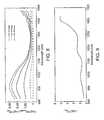
- FIG. 3Plotted in Fig. 3 are results of numerical simulations of light propagation in skin that show the predicted errors in the measurement of hemoglobin caused by variations in the fraction of water in the extravascular tissue (see Schmitt et al., Proc. SPIE, 1996, Vol. 2678, pp. 442-453 , for a description of the simulation method).
- the simulationaccounts for the normal variations in blood volume, oxygen saturation, and skin density that one would expect to observe in healthy adults.
- Fig. 4shows the predicted errors under the same conditions, except in this case the water fraction in the extravascular tissue was fixed at zero (a case equivalent to the assumption of no tissue displacement during blood vessel expansion).
- the relatively large errors in the predicted values of hematocrit in Fig. 3 compared to those in Fig. 4indicates that sensitivity to tissue water variations degrades the accuracy of the two-wavelength ratiometric method.
- ⁇1310 ⁇ nm D ⁇ ⁇
- ⁇805 ⁇ nm - k " D ⁇ ⁇
- ⁇970 ⁇ nm D ⁇ ⁇
- ⁇805 ⁇ nm + k ⁇
- this general expressionenables precise measurement of the absolute tissue water fraction f w .
- Eq. 10is not, however, the only possible algorithm for determination of tissue water fraction.
- Other methods and algorithmsincluding those disclosed by the inventor herein in a co-pending patent application assigned to the assignee herein, and titled: Device and Method for Monitoring Body Fluid and Electrolyte Disorders, Attorney Docket: TTC-09103-017200US will also yield accurate estimates of tissue water fraction.
- the apparatus depicted in Fig. 6has several features that facilitate the accurate measurement of Hct noninvasively.
- the solenoid-operated clamp 602occludes the venous return from the finger 604 by applying pressure around the circumference of the finger 604 via the rotary solenoid 616 which is coupled to the clamp 602.
- the applied pressureis adjusted to a level just above the value of the diastolic blood pressure.
- Microprocessor 606controls the timing of the occlusion cycle, data acquisition and processing to determine the value of Hct.
- the microprocessor-controlled data acquisition systembegins to record the electrical signals generated by photodetector 608.
- the photodetector 608is mounted on compressible rubber pad (not shown) or spring-loaded post (not shown) which maintains contact with the palmar side of the finger 604 without restricting its expansion during the occlusion period.
- the signalsare digitized by the analog to digital (A/D) converter 618, they are amplified by the preamp 612 and normalized to ensure their proportionality to the intensities of the light transmitted through the finger from the light-emitting diode (LED) sources 610, which are mounted close together on the same substrate (not shown).
- A/Danalog to digital
- the signalsare multiplexed by turning the LEDs on in sequence to permit near-simultaneous measurement of the intensities by a single photodetector. After approximately five seconds have elapsed, the clamp 602 releases automatically and the finger 604 can be removed. A short time later, Hct is displayed on the display panel 614 as a percentage along with the calculated value of Hb T in g/dl.
- the display panel 614is a built-in liquid-crystal (LCD) panel.
- light emission sources and opticsmay include sources other than LEDs such as incandescent light sources or white light sources which are tuned to emit radiation at appropriate wavelengths.
- a miniature solenoid 616 for performing the occlusion, the light emission 610 and detection optics 608, processing device 606, and display 614are all contained within a handheld device 600. Actuation of the solenoid triggers the start of measurement cycle. The difference between the logarithms of the intensities measured at specific wavelengths in the band between 800-1000 nm in which hemoglobin is the dominant absorber and between 1250 - 1600 nm in which water is the dominant absorber are recorded immediately before and immediately after occlusion.
- these measured differencesare combined with an estimate of the extravascular water fraction derived from the weighted sum of the derivatives of the transmittance or reflectance spectra of the tissue measured in an overlapping band of wavelengths.
- substitute occlusion meanssuch as a pneumatic or hydraulic-operated clamps. Additional alternate embodiments use other algorithms for determining the tissue water fraction as described above.
- Fig. 7shows the timing of the data acquisition and processing during the measurement cycle.
- a pressure transducersenses the presence of a finger and actuates the occlusion device which in turn starts the data acquisition sequence. The sequence starts automatically after the microprocessor has detected the presence of the finger.
- the LEDsare turned on to record the average values of the before-expansion diffuse transmittances log[ I ( ⁇ 1)] dc , ..., log[ I ( ⁇ n )] dc over an interval of 0.5-1.0 second. These dc measurements are used both for the before venous-expansion values to determine Hct as well as measurements of tissue water.
- An additional advantage of the venous-occlusion methodis that it facilitates the detection and removal of any asynchronous noise component of the time-varying intensities caused by the sudden expansion of the blood vessels.
- Ballistic waves generated by expanding vesselscan temporarily alter the scattering coefficient of the bulk tissue and produce optical artifacts. Similar artifacts associated with natural arterial blood pulsations are harder to remove because they occur at almost the same time as the upstroke of the optical plethysmogram.
- the change in the blood volume brought about as a result of venous occlusionwill drown out any such ballistic waves and hence minimize any potential optical artifacts.
- the design of the device and the microprocessorintegrates the method and apparatus for reducing the effect of noise on measuring physiological parameters as described in U.S. Pat. No.
- Figs. 8 and 9show examples of a set of pulse spectra D ( ⁇ ) measured as a function of time shortly after occlusion of the blood flow to the index finger, along with the corresponding log[ I ( ⁇ )] dc spectrum of the finger.
- the magnitudes of these spectra at selected wavelengthscontain the information required for the determination of Hct according to Eq. 8 and Eq. 10 and other algorithms used to measure tissue water as described above.
- Fig. 10shows a manual version of a reflectance sensor 1000 designed for application to the tip of a finger or toe.
- This embodimentrelies on partial, instead of full, venous occlusion from any well-perfused area of the skin by applying compression to an adjacent area with an appropriately shaped probe.
- a pressure transducer 1004 mounted on the end of the occluder 1006senses the applied pressure and controls the timing of the data acquisition.
- the light sources 1008 and detector 1010 mounted in a miniature spring-loaded probe 1012record the decrease in the diffusely reflected intensity during the occlusion cycle.
- This embodimentis more suitable for rapid screening for anemia in a large population of subjects.
- the lightimpinges on and is collected from the skin directly by mounting the detector and light sources at the tip of the sensor.
- the light emission and detectionare positioned locally in the device housing.
- the light emission and detectionare conducted to and from a remote unit containing the sources and the photodetector via optical fibers.
- full-spectrum processing methodssuch as partial-least squares analysis and principal component regression, may also be applied to the measured spectra to improve the accuracy of the hemoglobin estimates.
- Additional embodiments which implement these techniquesemploy a white-light source and a grating detector to measure the transmittances or reflectances from blood-perfused tissue over a continuous range of wavelengths.
- Reflected rather than transmitted intensitiescan be measured by placing the light sources on the same side of the blood-perfused tissue as the light detector.
- the separation between the sources and detectorsis an important variable that influences the probing depth as well as the sensitivity of the measured intensities to scattering variations.
- light sources or light emission optics other then LED'sincluding and not limited to narrowband light sources appropriately tuned to the desired wavelengths and associated light detection optics may be placed within the probe housing which is placed near the tissue location or may be positioned within a remote unit; and which deliver light to and receive light from the probe location via optical fibers.
Landscapes
- Health & Medical Sciences (AREA)
- Life Sciences & Earth Sciences (AREA)
- Physics & Mathematics (AREA)
- Engineering & Computer Science (AREA)
- Molecular Biology (AREA)
- Pathology (AREA)
- Biomedical Technology (AREA)
- General Health & Medical Sciences (AREA)
- Veterinary Medicine (AREA)
- Surgery (AREA)
- Animal Behavior & Ethology (AREA)
- Heart & Thoracic Surgery (AREA)
- Public Health (AREA)
- Biophysics (AREA)
- Medical Informatics (AREA)
- Immunology (AREA)
- Hematology (AREA)
- Chemical & Material Sciences (AREA)
- Analytical Chemistry (AREA)
- Urology & Nephrology (AREA)
- Biochemistry (AREA)
- General Physics & Mathematics (AREA)
- Spectroscopy & Molecular Physics (AREA)
- Optics & Photonics (AREA)
- Microbiology (AREA)
- Cell Biology (AREA)
- Food Science & Technology (AREA)
- Medicinal Chemistry (AREA)
- Biotechnology (AREA)
- Measurement Of The Respiration, Hearing Ability, Form, And Blood Characteristics Of Living Organisms (AREA)
- Investigating Or Analysing Materials By Optical Means (AREA)
Abstract
Description
- This invention relates to systems and methods for spectrophotometric measurement of biochemical compounds in the skin for non-invasive medical diagnosis and monitoring. Specifically, the present invention relates to the determination of the hematocrit or the absolute concentration of hemoglobin in the blood by multiple-wavelength optical plethysmography.
- The total concentration of hemoglobin in blood (HbT) or the hematocrit (Hct), defined as the fraction or percentage of red cells in whole blood, are primary variables used by physicians to assess the health of a patient. The hematocrit is the fraction of the total blood volume occupied by the red blood cells, and hemoglobin is the principal active constituent of red blood cells. Approximately 34% of the red cell volume is occupied by hemoglobin. A value of HbT less than 10 g/dl or Hct < 0.30 indicates an anemic state which can impair the normal functions of the body. Severe anemia can lead to death when the quantity of hemoglobin becomes insufficient to supply oxygen to the brain and other vital organs. Patients with kidney disease, pregnant women, and young children in developing countries are especially susceptible to chronic anemia. Acute anemia resulting from loss of blood, infection, or autoimmune disorders can be life-threatening and requires close monitoring.
- The conventional means employed to measure Hct in clinical medicine is to puncture the skin, draw blood from a vein or capillary into a small-diameter tube, and measure the solid (packed-cell) fraction that remains after centrifugation of the blood. Measurement of HbT in accordance with standard practice also requires drawing a blood sample, which is then subjected to a chemical or mechanical process to lyse the red cells and release the liquid hemoglobin. After transferring the hemoglobin to a cuvette, its concentration is measured either by direct spectrophotometry or by colorimetry following the addition of a chemical reagent.
- Although a number of methods have been developed to make these sampling and processing steps less cumbersome, no device is yet available to physicians for the reliable and accurate measurement of Hct or HbT that obviates blood sampling.
- A number of researchers and inventors have recognized the value of a completely noninvasive method for measurement of hematocrit or total hemoglobin concentration.Schmitt et al. (Proc. SPIE,1992, Vol. 1641, pp. 150-161) adapted the principles of pulse oximetry to the noninvasive measurement of hematocrit of blood in intact skin. The method is based on the measurement of the ratios of the pulsatile (ac) and non-pulsatile (dc) components of the light transmitted through a blood-perfused tissue within two spectral bands in which the molar extinction coefficients of oxygenated hemoglobin (HbO2) and deoxygenated hemoglobin (Hb) are nearly the same. In one of the wavelength bands (800 ≤ λ ≤ 1000 nm), the absorption of hemoglobin is the dominate contributor to the attenuation of light in blood; in the other band (1200 ≤ λ≤ 1550 nm), the absorption of water dominates. Therefore, the absorption of water serves as a measure of the plasma (non-cellular) fraction of the blood which does not contain hemoglobin. A hematocrit monitoring system based on a similar method has been disclosed by
Steuer et al. (U.S. Patent 5,499,627 ). In this disclosure, the influence of the optical properties of extravascular interstitial fluid on the accuracy of the measurement was recognized and the addition of a third wavelength was proposed to reduce this influence. The concept of adding more wavelengths to improve accuracy was extended further byKuenster (U.S. Patent 5,377,674 ) andAoyagi et al. (U.S. Patent 5,720,284 ).Steuer et al. (U.S. Patent 5,499,627 ) also recognized the difficulty of obtaining a reliable plethysmographic pulse in the water absorption band (its amplitude is typically 4-10 times smaller than in the hemoglobin absorption band). To alleviate this problem,Steuer et al. (U.S. Patent 5,499,627 ) proposed a method for inducing an artificial pulse by mechanical compression of the tissue at the location of hematocrit measurements. - In spite of these earlier advances, measuring the absolute concentration of hemoglobin in blood accurately and reliably remains difficult in practice. This difficulty stems mainly from two limitations.
- The first limitation is the failure of the available mathematical algorithms used in the prior art devices to account for the fact that the blood vessels displace the extravascular tissue when they expand, because the tissue is essentially incompressible. Because of the incompressibility of tissue, the change in the diffuse transmission of light through tissue observed during arterial pulsation depends on the difference between the optical properties of the blood and the surrounding gelatinous tissue matrix. Therefore, to obtain an accurate measure of the absolute values of the hemoglobin concentration in the blood, one must also account for the optical properties of the tissue that surrounds the blood vessels in the skin. Measurement of the ac/dc ratios alone, regardless of the number of wavelengths at which the measurement is made, cannot compensate entirely for the variations in the scattering and absorption properties of the skin of different subjects. This problem is not important in conventional pulse oximetry because the attenuation of light in blood greatly exceeds that in the surrounding tissue at the wavelengths at which ac/dc ratios are measured (typically 660 nm and 910 nm). The same is not true in the measurement of HbT by optical plethysmography, however, which relies on the measurement of pulsations resulting from optical absorption of water in the blood. Because the volume fraction of water in blood is close to that of the extravascular tissue matrix, the difference between the absorptivities of blood and the surrounding tissue is small within water absorption bands. Moreover, the difference between the scattering properties of blood and the surrounding tissue vary with their relative water contents. Accordingly, one limitation of the prior art devices and methods used to noninvasively measure hematocrit or hemoglobin has been the inaccurate measurement of tissue water.
- The second limitation is the reliance of the prior art methods on the measurement of small pulsatile changes in the blood volume induced by contractions of the heart. When the water contents of the blood and the extravascular tissues are nearly the same, the pulsatile (ac) component of intensities measured at wavelengths greater than 1250 nm are usually less than one percent of the mean (dc) intensity. Even using the most advanced circuitry and signal-processing techniques, the amplitudes of such small pulsations are difficult to measure reliably. Although mechanical compression of the tissue, as proposed by
Steuer et al. (U.S. Patent 5,499,627 ), alleviates this problem by inducing a larger blood volume change, it also introduces large changes in the scattering coefficient of the bulk tissue which can complicate calibration of instruments based on this technique, because the compression is occurring at the same location as where the hematocrit measurements are taken. - None of
US-A-5499627 ,US-A-5833602 orUS-B1-6178342 provide the computation of a tissue water fraction and the computation of a hematocrit value, using said tissue water fraction. - Therefore, there exists a need for more reliable and accurate measurement of hematocrit by non-invasive means.
- The objective of embodiments of the present invention is to provide a more reliable and accurate measurement of hematocrit (Hct) by noninvasive means. The changes in the intensities of light of multiple wavelengths transmitted through or reflected light from a tissue location are recorded immediately before and after occluding the flow of venous blood from the tissue location with an occlusion device positioned near the tissue location. As the venous return stops and the incoming arterial blood expands the blood vessels, the light intensities measured within a particular band of near-infrared wavelengths decrease in proportion to the volume of hemoglobin in the tissue location; those intensities measured within a separate band of wavelengths in which water absorbs respond to the difference between the water fractions within the blood and the displaced tissue volume. A mathematical algorithm applied to the time-varying intensities yields a quantitative estimate of the absolute concentration of hemoglobin in the blood. To compensate for the effect of the unknown fraction of water in the extravascular tissue on the Hct measurement, the tissue water fraction is determined before the occlusion cycle begins by measuring the diffuse transmittance or reflectance spectra of the tissue at selected wavelengths.
- An important feature of the embodiments of this invention is that it incorporates a means of compensating for natural variations in the water fraction of skin of different individuals. Such variations affect both the scattering and absorption coefficients of skin in the near-infrared region of the spectrum and are a primary source of error in hematocrit estimates derived from the assumption that the optical coefficients of the extravascular tissue are fixed quantities.
- An equally important feature of the embodiments of this invention is that the relatively large change in blood volume induced by the venous occlusion facilitates the measurement of small differences between the optical properties of the blood and the extravascular tissue. Thus, better accuracy can be obtained compared to methods that rely on arterial blood pulsations. It should be understood, however, that the mathematical algorithm on which the present invention is based applies equally well to intensity changes induced by natural arterial pulsations or compression of the skin.
- For a fuller understanding of the nature and advantages of the embodiments of the present invention, reference should be made to the following detailed description taken in conjunction with the accompanying drawings.
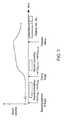
- Fig. 1 is a diagram of an expanding blood vessel in the skin through which light scatters.
- Fig. 2 is a graph of the near-infrared absorption spectra of the compounds having the major influence on the transcutaneous measurement of hematocrit.
- Fig. 3 is a graph of the predicted versus actual hematocrit values obtained by numerical simulation of the two-wavelength ratiometric method accounting for the normal variation in the water fraction,fw.
- Fig. 4 is a graph of the predicted versus actual hemoglobin values obtained by numerical simulation of the two-wavelength ratiometric method with the water fraction,fw, fixed at zero.
- Fig. 5 is a graph of the predicted versus actual hematocrit values obtained by numerical simulation of a previously disclosed three-wavelength ratiometric method with normal variation in the water fraction,fw, included.
- Fig. 6 is a block diagram of a handheld apparatus for noninvasive measurement and display of hematocrit and total hemoglobin concentration in the blood.
- Fig. 7 is a timing diagram of the data acquisition process used to measure light intensities for determination of hematocrit by the venous-occlusion method.
- Fig. 8 is a graph of the pulse spectra measured from the finger of a healthy adult subject.
- Fig. 9 is a graph of the dc spectrum measured from the finger of a healthy adult subject.
- Fig. 10 is a diagram of a mechanically operated reflectance sensor for rapid measurement of hematocrit via the venous-occlusion method.
- To understand the principles that underlie the invention, first consider a
small blood vessel 102 embedded in theskin 104 on which light 106 impinges from thesurface 108 as shown in Fig. 1. A fraction of the incident photons scatter through the blood before being captured by the detector. When thevessel 102 expands its volume, the probability of photons being absorbed or scattered by the blood inside the vessel increases. The absorption of the light that occurs within the volume probed by the light that reaches the detector (the effective sample volume) can be described approximately by a modified form of the Beer-Lambert law, which quantifies the diffusely reflected or transmitted intensityI before and after expansion of the vessel by an increase in the volume of blood ΔV,

whereℓ is the effective length of the optical path between the source and detector,VT is the sample volume, andVt andVb are, respectively, the volumes of extravascular tissue and blood withinVT. The variables


- Hemoglobin, water, and the plasma proteins are the main contributors to the absorption of near-infrared light in blood. Fig. 2 shows the absorption spectra of
water 202,globular protein 204, and the oxygenated 206 and deoxygenated 208 forms of hemoglobin (HbO2 and Hb) in the band of wavelengths between 800 and 1800 nm. It is possible to choose wavelengths at which absorption by the plasma proteins is negligible compared to absorption by water and hemoglobin. For such wavelengths, the absorption coefficient of the blood equals approximately


- Now suppose that a pair of wavelengths λ1 and λ2 is chosen such that


- This equation (Eq. 6) is still incomplete because it does not account for difference between the scattering coefficients of the blood and surrounding tissues, Δµs(λ), a variable that depends on the hematocrit and tissue water fraction according to

where


- A complete expression that relatesH and the measured ratio of intensity difference,R, can now be written as

- Plotted in Fig. 3 are results of numerical simulations of light propagation in skin that show the predicted errors in the measurement of hemoglobin caused by variations in the fraction of water in the extravascular tissue (seeSchmitt et al., Proc. SPIE, 1996, Vol. 2678, pp. 442-453, for a description of the simulation method). The simulation accounts for the normal variations in blood volume, oxygen saturation, and skin density that one would expect to observe in healthy adults. For comparison, Fig. 4 shows the predicted errors under the same conditions, except in this case the water fraction in the extravascular tissue was fixed at zero (a case equivalent to the assumption of no tissue displacement during blood vessel expansion). The relatively large errors in the predicted values of hematocrit in Fig. 3 compared to those in Fig. 4 indicates that sensitivity to tissue water variations degrades the accuracy of the two-wavelength ratiometric method.
- Performing ratiometric measurements at more than two wavelengths can reduce the errors that result from changes in the optical properties of the extravascular tissue, but cannot eliminate them. Fig. 5 evaluates the performance of a three-wavelength ratiometric algorithm modeled after the algorithm suggested by
Steuer et al. (U.S. Patent 5,499,627 ),
- The regression constantsk', k", andk"' used in the simulation were chosen to give the best fit between the actual and predicted hematocrits. Although somewhat improved, the predicted errors of the hematocrit measurement in Fig. 5 are still large compared to those obtained for the non-zero, fixedfw case (Fig. 3). As can be seen from Fig. 5, measuring ratios of the blood pulse spectrum at additional wavelengths does not overcome the inherent dependence of the magnitude of the spectrum on the optical properties of the extravascular tissue.
- From the preceding analysis it can be appreciated that measuring the blood pulse spectrum on a body site at whichfw is small and constant would improve the measurement accuracy. The earlobe, in which many of the blood vessels are embedded in adipose tissue, comes closest to satisfying this requirement. However, in many applications the earlobe is an inconvenient measurements site and its adipose content varies from individual to individual.
- A more robust approach to reducing the errors caused by tissue water variations is to measurefw and use the measured value in the prediction equation (Eq. 8). In a preferred embodiment of the present invention, the tissue water fraction is derived from diffuse light intensities measured at a set of wavelengths within the same band of near-infrared wavelengths (800- 1800 nm) used to measure the blood pulse spectrum. Intensity ratios are recorded when the skin is in the resting state (before blood volume expansion) and are then log-transformed and combined according to

- For specific sets of wavelengths, λ3 - λ6 and constantsk1 -k3, this general expression enables precise measurement of the absolute tissue water fractionfw. The values of the constants can be determined from mathematical models or by empirical calibration. The results of numerical simulations suggest thatfw values derived from Eq. 10 for λ3 = 850 nm, λ4 = 1370 nm, λ5 = 1250 nm, λ6 = 1140 nm, are accurate to within ±1% over the physiological range of blood volume and scattering variations. One important feature of this particular choice of wavelengths is that intensities measured at the longest and shortest wavelengths λ3 = 850 nm, λ4 =1370 nm, can also be used in the calculation of the ratioR at wavelengths λ1. and λ2 in Eq. 5. That is, for λ1 = λ4 = 1370 nm and λ2 = λ3 = 850 nm, measurements at four rather than six wavelengths are required to determine the hematocrit. Reducing the number of measurement wavelengths lowers the manufacturing cost of portable devices that employ discrete light-emitting diodes as light sources. Another advantage of overlapping the wavelengths used to measureR andfw is that differences in the optical path lengths that determine the geometry of sample volume are minimized. Eq. 10 is not, however, the only possible algorithm for determination of tissue water fraction. Other methods and algorithms, including those disclosed by the inventor herein in a co-pending patent application assigned to the assignee herein, and titled: Device and Method for Monitoring Body Fluid and Electrolyte Disorders, Attorney Docket: TTC-09103-017200US will also yield accurate estimates of tissue water fraction.
- Although the key concepts that underlie the disclosed methods for noninvasive Hct measurement are embodied in Eqs. 8 and 10, the design of apparatus with which the required intensities are measured also plays an equally crucial role. In particular, the magnitudes of the optical signals from whichR andfw are derived must be large enough to ensure minimal interference from electronic noise as well as from noise related to physiological variables, which include body movements and spatial heterogeneity in local blood flow.
- The apparatus depicted in Fig. 6 has several features that facilitate the accurate measurement of Hct noninvasively. The solenoid-operated
clamp 602 occludes the venous return from thefinger 604 by applying pressure around the circumference of thefinger 604 via therotary solenoid 616 which is coupled to theclamp 602. The applied pressure is adjusted to a level just above the value of the diastolic blood pressure. As a result, the arterial blood continues to flow unimpeded into the fingertip until the flow stops when the blood vessels distend to their maximum filling volumes.Microprocessor 606 controls the timing of the occlusion cycle, data acquisition and processing to determine the value of Hct. Before the start of occlusion cycle, the microprocessor-controlled data acquisition system begins to record the electrical signals generated byphotodetector 608. Thephotodetector 608 is mounted on compressible rubber pad (not shown) or spring-loaded post (not shown) which maintains contact with the palmar side of thefinger 604 without restricting its expansion during the occlusion period. Before the signals are digitized by the analog to digital (A/D)converter 618, they are amplified by thepreamp 612 and normalized to ensure their proportionality to the intensities of the light transmitted through the finger from the light-emitting diode (LED)sources 610, which are mounted close together on the same substrate (not shown). The signals are multiplexed by turning the LEDs on in sequence to permit near-simultaneous measurement of the intensities by a single photodetector. After approximately five seconds have elapsed, theclamp 602 releases automatically and thefinger 604 can be removed. A short time later, Hct is displayed on thedisplay panel 614 as a percentage along with the calculated value of HbT in g/dl. In one embodiment, thedisplay panel 614 is a built-in liquid-crystal (LCD) panel. - In alternate embodiments, light emission sources and optics may include sources other than LEDs such as incandescent light sources or white light sources which are tuned to emit radiation at appropriate wavelengths.
- In one embodiment of the invention, a
miniature solenoid 616 for performing the occlusion, thelight emission 610 anddetection optics 608,processing device 606, and display 614 are all contained within ahandheld device 600. Actuation of the solenoid triggers the start of measurement cycle. The difference between the logarithms of the intensities measured at specific wavelengths in the band between 800-1000 nm in which hemoglobin is the dominant absorber and between 1250 - 1600 nm in which water is the dominant absorber are recorded immediately before and immediately after occlusion. To calculate the hematocrit, these measured differences are combined with an estimate of the extravascular water fraction derived from the weighted sum of the derivatives of the transmittance or reflectance spectra of the tissue measured in an overlapping band of wavelengths. Alternate embodiments use substitute occlusion means such as a pneumatic or hydraulic-operated clamps. Additional alternate embodiments use other algorithms for determining the tissue water fraction as described above. - Fig. 7 shows the timing of the data acquisition and processing during the measurement cycle. A pressure transducer senses the presence of a finger and actuates the occlusion device which in turn starts the data acquisition sequence. The sequence starts automatically after the microprocessor has detected the presence of the finger. Before the solenoid is activated, the LEDs are turned on to record the average values of the before-expansion diffuse transmittances log[I(λ1)]dc, ..., log[I(λn)]dc over an interval of 0.5-1.0 second. These dc measurements are used both for the before venous-expansion values to determine Hct as well as measurements of tissue water. Recording of the transmittances proceeds continuously at a fast sampling rate after the solenoid activates and the finger clamp closes. The after-expansion transmittances log[I(λ1)]ac+dc,...,log[I(λn)]ac+dc are recorded as averaged values calculated over an interval of one-half second or less just prior to the maximum of the blood volume expansion, as determined from the magnitude ofD(λ)|λ=λ
I . It is important to perform the after-expansion measurements within an interval no longer than a few seconds after venous blood flow from the finger ceases, because the elevated venous pressure can lead to desaturation of the blood and loss of water through the capillaries, factors that may influence the accuracy of the hemoglobin measurement. The fractional change in the blood volume induced by the venous occlusion is typically an order of magnitude greater than that produced by normal arterial pulsations. This signal enhancement, combined with the reduction of noise that results from longer averaging times, gives the venous-occlusion method a significant advantage over optical plethysmography based on the measurement of natural blood pulsations. An additional advantage of the venous-occlusion method is that it facilitates the detection and removal of any asynchronous noise component of the time-varying intensities caused by the sudden expansion of the blood vessels. Ballistic waves generated by expanding vessels can temporarily alter the scattering coefficient of the bulk tissue and produce optical artifacts. Similar artifacts associated with natural arterial blood pulsations are harder to remove because they occur at almost the same time as the upstroke of the optical plethysmogram. The change in the blood volume brought about as a result of venous occlusion will drown out any such ballistic waves and hence minimize any potential optical artifacts. The design of the device and the microprocessor integrates the method and apparatus for reducing the effect of noise on measuring physiological parameters as described inU.S. Pat. No. 5,853,364 , assigned to Nellcor Puritan Bennett, Inc., now a division of the assignee of the present invention. Additionally, the design of the device and the microprocessor also integrates the electronic processor as described inU.S. Pat. No. 5,348,004 , assigned to Nellcor Incorporated, now a division of the assignee of the present invention. - Figs. 8 and 9 show examples of a set of pulse spectraD(λ) measured as a function of time shortly after occlusion of the blood flow to the index finger, along with the corresponding log[I(λ)]dc spectrum of the finger. The magnitudes of these spectra at selected wavelengths contain the information required for the determination of Hct according to Eq. 8 and Eq. 10 and other algorithms used to measure tissue water as described above.
- An additional embodiment of the device is shown in Fig. 10. Fig. 10 shows a manual version of a
reflectance sensor 1000 designed for application to the tip of a finger or toe. This embodiment relies on partial, instead of full, venous occlusion from any well-perfused area of the skin by applying compression to an adjacent area with an appropriately shaped probe. When theskin 1002 is compressed, apressure transducer 1004 mounted on the end of theoccluder 1006 senses the applied pressure and controls the timing of the data acquisition. As the blood volume increases in the area of theskin 1002 proximal to theoccluder 1006, thelight sources 1008 anddetector 1010 mounted in a miniature spring-loaded probe 1012 record the decrease in the diffusely reflected intensity during the occlusion cycle. This embodiment is more suitable for rapid screening for anemia in a large population of subjects. - In the embodiment depicted in Fig. 10, the light impinges on and is collected from the skin directly by mounting the detector and light sources at the tip of the sensor. Likewise, in the automatic embodiment shown in Fig. 6, the light emission and detection are positioned locally in the device housing. In alternate embodiments of the automatic and manual versions of the device, the light emission and detection are conducted to and from a remote unit containing the sources and the photodetector via optical fibers.
- Individuals familiar with the art of spectral processing will realize that full-spectrum processing methods, such as partial-least squares analysis and principal component regression, may also be applied to the measured spectra to improve the accuracy of the hemoglobin estimates. Additional embodiments which implement these techniques, employ a white-light source and a grating detector to measure the transmittances or reflectances from blood-perfused tissue over a continuous range of wavelengths.
- A number of variations of the apparatus will be apparent to those skilled in the art of tissue optics. Reflected rather than transmitted intensities can be measured by placing the light sources on the same side of the blood-perfused tissue as the light detector. The separation between the sources and detectors is an important variable that influences the probing depth as well as the sensitivity of the measured intensities to scattering variations. By operating the apparatus in the reflection mode with a distance of 2-3 millimeters between the light sources and detectors, the effective optical path can be confined to the well-perfused dermal layer. Operation in the reflection mode has the additional benefit of permitting measurements to be made on parts of the body besides the appendages. Moreover, light sources or light emission optics other then LED's including and not limited to narrowband light sources appropriately tuned to the desired wavelengths and associated light detection optics may be placed within the probe housing which is placed near the tissue location or may be positioned within a remote unit; and which deliver light to and receive light from the probe location via optical fibers. These equivalents and alternatives along with obvious changes and modifications are intended to be included within the scope of the present invention. Accordingly, the foregoing disclosure is intended to be illustrative, but not limiting, of the scope of the invention which is set forth in the following claims.


Claims (24)
- A device for measuring hematocrit values using optical spectrophotometry comprising:a probe housing configured to be placed proximal to a tissue location which is being monitored;an occlusion device (602, 1006) connected to said housing and configured to magnify the fractional change in the vascular blood volume to a value greater than said fractional change produced by normal arterial pulsations;light emission optics (610, 1008) connected to said housing and configured to direct radiation at said tissue location;light detection optics (608, 1010) connected to said housing and configured to receive radiation from said tissue location;characterized by
a processing device (606) connected to said housing and configured to process radiation from said light emission optics (610, 1008) and said light detection optics (608, 1010) to compute a tissue water fraction and compute hematocrit values, using said tissue water fraction. - The device of claim 1, further comprising a pressure transducer (1004) connected to said housing and positioned to be placed adjacent to said tissue location, and configured to interface with said processing device (606) and said occlusion device (602, 1006) to provide an input to said processing device (606) to control the timing of the data acquisition by said processing device (606).
- The device of claim 1, wherein said processing device (606) is adapted to
receive at least two sets of optical measurements at at least a first and a second wavelength, where for each wavelength two optical measurements are obtained corresponding to measurements before and after a venous occlusion conducted by said occlusion device to obtain before and after occlusion measurements at each wavelength;
combine said before and after occlusion measurements at each wavelength to determine a blood pulse spectrum at each wavelength;
combine said blood pulse spectra at each wavelength to obtain a ratio of said blood pulse spectra; and
combine said ratio with measurements of tissue water fractions to determine the concentration of hemoglobin in the blood. - The device of claim 1, further comprising a display device (614) connected to said probe housing and configured to display said hematocrit values.
- The device of claim 1, further comprising means for calculating hemoglobin.
- The device of claim 1, wherein said light emission optics (610, 1008) are tuned to emit radiation at at least a first and a second wavelength, where said first wavelength is within a band of wavelengths where hemoglobin is the dominant absorber and where said second wavelength is within a band of wavelengths where water is the dominant absorber.
- The device of claim 1, wherein said light emission optics (610, 1008) are tuned to emit radiation at at least a first and a second wavelength, where said first wavelength is within a band of wavelengths in the range of approximately 800-1000 nm and where said second wavelength is within a band in the range of approximately 1250-1600 nm.
- The device of claim 1, wherein said light emission optics (61.0, 1008) are tuned to emit radiation at at least a first and a second wavelength, where said first and said second wavelengths are related to each other such that 0.34HµaHb>>µaw at said first wavelength and µaw>>0.34HµaHb at said second wavelength, where
H is the hematocrit value,
µaHb is the sum of the absorption coefficient of the two forms of hemoglobin, and
µaw is the absorption coefficient of water. - The device of claim 1, wherein said light emission optics (610, 1008) are tuned to emit radiation at at least a first and a second wavelength, where said first wavelength is in a range approximately between and including 805 to 850 nm and said second wavelength is in a range approximately between and including 1310 to 1370 nm.
- The device of claim 1, wherein said light emission optics (610, 1008) and said light detection optics (608, 1010) are mounted within said probe housing and positioned with appropriate alignment to enable detection in a transmissive mode.
- The device of claim 1, wherein said light emission optics (1008) and said light detection optics (1010) are mounted within said probe housing and positioned with appropriate alignment to enable detection in a reflective mode.
- The device of claim 1, wherein said light emission optics (610, 1008) and said light detection optics (608, 1010) are placed within a remote unit and which deliver light to and receive light from said probe housing via optical fibers.
- The device of claim 1, wherein said light emission optics (610, 1008) comprise at least one of a (a) incandescent light source, (b) narrowband light source, wherein a narrowband light source comprises one of a light emitting diode ("LED") and a filtered white light source.
- The device of claim 1, wherein said processing device (606) is adapted to receive at least two sets of optical measurements, where the first set of optical measurements corresponds to the detection of light whose absorption is primarily due to hemoglobin, and where the second set of optical measurements corresponds to the detection of light whose absorption is primarily due to water, and where a combination of said at least two sets of optical measurements provides a measure of said hematocrit value.
- The device of claim 3, wherein said processing device (606) is adapted to determine hematocrit based on optical measurements from the relation
 H is the hematocrit value;fw is the tissue water fraction;fpp is the plasma protein fraction;R is the ratio of magnitudes of the blood pulse spectrum;µaHb(λ1) is the sum of the absorption coefficient of the two forms of hemoglobin at a first wavelength;µaw(λ2) is the absorption coefficient of water at a second wavelength;Δµs(λ1) is the difference between the scattering coefficients of the blood and surrounding tissue at a first wavelength;Δµs(λ2) is the difference between the scattering coefficients of the blood and surrounding tissue at a second wavelength; and0.34 is the fraction of the red cell volume occupied by hemoglobin, which is assumed to be constant.
 - A method of measuring hematocrit values at a tissue location using optical spectrophotometry comprising:irradiating said tissue location and processing received signals from said tissue location to compute a tissue water fraction;occluding the venous blood flow adjacent to said tissue location;repeat irradiating said tissue location;detecting radiation from said tissue following said repeat irradiating; andcalculating a hematocrit value from said detected radiation using said tissue water fraction.
- The method of claim 16 wherein said calculating comprises:receiving at least two sets of optical measurements at at least a first and a second wavelength, where for each wavelength two optical measurements are obtained corresponding to measurements before and after said occluding to obtain before and after occlusion measurements at each wavelength;combining said before and after occlusion measurements at each wavelength to determine a blood pulse spectrum at each wavelength;combining said blood pulse spectra: at each wavelength to obtain a ratio of said blood pulse spectra; andcombining said ratio with said tissue water fraction to determine the hematocrit value in the blood.
- The method of claim 16 further comprising displaying said hematocrit values.
- The method of claim 16 further comprising calculating hemoglobin.
- The method of claim 16 wherein said irradiating comprises irradiating said tissue location using light emission optics tuned to emit radiation at at least a first and a second wavelength, where said first wavelength is within a band of wavelengths where absorption will be primarily due to hemoglobin and where said second wavelength is within a band of wavelengths where absorption will be primarily due to water.
- The method of claim 16 wherein said irradiating comprises irradiating said tissue location using light emission optics tuned to emit radiation at at least a first and a second wavelength, where said first wavelength is within a band of wavelengths in the range of approximately 800-1000 nm and where said second wavelength is within a band in the range of approximately 1250-1600 nm.
- The method of claim 16 wherein said irradiating comprises irradiating said tissue location using light emission optics tuned to emit radiation at at least a first and a second wavelength, where said first and said second wavelengths are related to each other such that 0.34HµaHb>>µaw at said first wavelengths and µaw>>0.34HµaHb at said second wavelength, where
H is the hematocrit value,
µaHb is the sum of the absorption coefficient of the two forms of hemoglobin, and
µaw is the absorption coefficient of water. - The method of claim 16 wherein said irradiating comprises irradiating said tissue location using light emission optics tuned to emit radiation at at least a first and a second wavelength, where said first wavelength is in a range approximately between and including 805 to 850 nm and said second wavelength is in a range approximately between and including 1310 to 1370 nm.
- The method of claim 17 wherein said calculating comprises determining hematocrit based on optical measurements from the relation

where:H is the hematocrit value;fw is the tissue water fraction;fpp is the plasma protein fraction;R is the ratio of magnitudes of the blood pulse spectrum;µaHb(λ1) is the sum of the absorption coefficient of the two forms of hemoglobin at a first wavelength;µaw(λ2) is the absorption coefficient of water at a second wavelength;Δµs(λ1) is the difference between the scattering coefficients of the blood and surrounding tissue at a first wavelength;Δµs(λ2) is the difference between the scattering coefficients of the blood and surrounding tissue at a second wavelength; and0.34 is the fraction of the red cell volume occupied by hemoglobin, which is assumed to be constant.


Applications Claiming Priority (3)
| Application Number | Priority Date | Filing Date | Title |
|---|---|---|---|
| US811328 | 2001-03-16 | ||
| US09/811,328US6606509B2 (en) | 2001-03-16 | 2001-03-16 | Method and apparatus for improving the accuracy of noninvasive hematocrit measurements |
| PCT/US2002/007760WO2002075289A2 (en) | 2001-03-16 | 2002-03-13 | Method and apparatus for improving the accuracy of noninvasive hematocrit measurements |
Publications (2)
| Publication Number | Publication Date |
|---|---|
| EP1368638A2 EP1368638A2 (en) | 2003-12-10 |
| EP1368638B1true EP1368638B1 (en) | 2007-11-28 |
Family
ID=25206245
Family Applications (1)
| Application Number | Title | Priority Date | Filing Date |
|---|---|---|---|
| EP02709832AExpired - LifetimeEP1368638B1 (en) | 2001-03-16 | 2002-03-13 | Method and apparatus for improving the accuracy of noninvasive hematocrit measurements |
Country Status (7)
| Country | Link |
|---|---|
| US (1) | US6606509B2 (en) |
| EP (1) | EP1368638B1 (en) |
| JP (1) | JP4176480B2 (en) |
| CA (1) | CA2441017C (en) |
| DE (1) | DE60223787T2 (en) |
| ES (1) | ES2299558T3 (en) |
| WO (1) | WO2002075289A2 (en) |
Cited By (1)
| Publication number | Priority date | Publication date | Assignee | Title |
|---|---|---|---|---|
| RU2807526C1 (en)* | 2022-11-29 | 2023-11-15 | Общество с ограниченной ответственностью "НПП-Волга" | Method of non-invasive measurement of fractional water content in human blood |
Families Citing this family (182)
| Publication number | Priority date | Publication date | Assignee | Title |
|---|---|---|---|---|
| US6018673A (en) | 1996-10-10 | 2000-01-25 | Nellcor Puritan Bennett Incorporated | Motion compatible sensor for non-invasive optical blood analysis |
| US8932227B2 (en) | 2000-07-28 | 2015-01-13 | Lawrence A. Lynn | System and method for CO2 and oximetry integration |
| US20060161071A1 (en) | 1997-01-27 | 2006-07-20 | Lynn Lawrence A | Time series objectification system and method |
| US9042952B2 (en) | 1997-01-27 | 2015-05-26 | Lawrence A. Lynn | System and method for automatic detection of a plurality of SPO2 time series pattern types |
| US9521971B2 (en) | 1997-07-14 | 2016-12-20 | Lawrence A. Lynn | System and method for automatic detection of a plurality of SPO2 time series pattern types |
| US20070191697A1 (en) | 2006-02-10 | 2007-08-16 | Lynn Lawrence A | System and method for SPO2 instability detection and quantification |
| US7400918B2 (en)* | 1998-07-04 | 2008-07-15 | Edwards Lifesciences | Measurement of blood oxygen saturation |
| WO2000001294A1 (en)* | 1998-07-04 | 2000-01-13 | Whitland Research Limited | Non-invasive measurement of blood analytes |
| US6675031B1 (en) | 1999-04-14 | 2004-01-06 | Mallinckrodt Inc. | Method and circuit for indicating quality and accuracy of physiological measurements |
| US20060195041A1 (en) | 2002-05-17 | 2006-08-31 | Lynn Lawrence A | Centralized hospital monitoring system for automatically detecting upper airway instability and for preventing and aborting adverse drug reactions |
| US9053222B2 (en) | 2002-05-17 | 2015-06-09 | Lawrence A. Lynn | Patient safety processor |
| US7239902B2 (en)* | 2001-03-16 | 2007-07-03 | Nellor Puritan Bennett Incorporated | Device and method for monitoring body fluid and electrolyte disorders |
| US6591122B2 (en)* | 2001-03-16 | 2003-07-08 | Nellcor Puritan Bennett Incorporated | Device and method for monitoring body fluid and electrolyte disorders |
| US7657292B2 (en)* | 2001-03-16 | 2010-02-02 | Nellcor Puritan Bennett Llc | Method for evaluating extracellular water concentration in tissue |
| US8135448B2 (en)* | 2001-03-16 | 2012-03-13 | Nellcor Puritan Bennett Llc | Systems and methods to assess one or more body fluid metrics |
| US6754516B2 (en) | 2001-07-19 | 2004-06-22 | Nellcor Puritan Bennett Incorporated | Nuisance alarm reductions in a physiological monitor |
| KR100493157B1 (en)* | 2002-08-02 | 2005-06-03 | 삼성전자주식회사 | Probe using in measuring organism signal and system for measuring organism signal comprising the same |
| US7006856B2 (en) | 2003-01-10 | 2006-02-28 | Nellcor Puritan Bennett Incorporated | Signal quality metrics design for qualifying data for a physiological monitor |
| US7016715B2 (en) | 2003-01-13 | 2006-03-21 | Nellcorpuritan Bennett Incorporated | Selection of preset filter parameters based on signal quality |
| US7545963B2 (en) | 2003-04-04 | 2009-06-09 | Lumidigm, Inc. | Texture-biometrics sensor |
| US7627151B2 (en) | 2003-04-04 | 2009-12-01 | Lumidigm, Inc. | Systems and methods for improved biometric feature definition |
| US7668350B2 (en) | 2003-04-04 | 2010-02-23 | Lumidigm, Inc. | Comparative texture analysis of tissue for biometric spoof detection |
| US7751594B2 (en) | 2003-04-04 | 2010-07-06 | Lumidigm, Inc. | White-light spectral biometric sensors |
| CA2521304A1 (en)* | 2003-04-04 | 2004-10-21 | Lumidigm, Inc. | Multispectral biometric sensor |
| US7460696B2 (en) | 2004-06-01 | 2008-12-02 | Lumidigm, Inc. | Multispectral imaging biometrics |
| DE10315574A1 (en)* | 2003-04-05 | 2004-10-28 | ETH Zürich | Device for determining blood flow in an organ |
| JP2007516009A (en) | 2003-10-03 | 2007-06-21 | アカデミッシュ メディシュ セントラム | Systems and methods for image processing of substrate reflections. |
| US7120479B2 (en) | 2004-02-25 | 2006-10-10 | Nellcor Puritan Bennett Inc. | Switch-mode oximeter LED drive with a single inductor |
| US7190985B2 (en) | 2004-02-25 | 2007-03-13 | Nellcor Puritan Bennett Inc. | Oximeter ambient light cancellation |
| US7534212B2 (en) | 2004-03-08 | 2009-05-19 | Nellcor Puritan Bennett Llc | Pulse oximeter with alternate heart-rate determination |
| US7194293B2 (en) | 2004-03-08 | 2007-03-20 | Nellcor Puritan Bennett Incorporated | Selection of ensemble averaging weights for a pulse oximeter based on signal quality metrics |
| US8611977B2 (en)* | 2004-03-08 | 2013-12-17 | Covidien Lp | Method and apparatus for optical detection of mixed venous and arterial blood pulsation in tissue |
| US7277741B2 (en) | 2004-03-09 | 2007-10-02 | Nellcor Puritan Bennett Incorporated | Pulse oximetry motion artifact rejection using near infrared absorption by water |
| US7508965B2 (en) | 2004-06-01 | 2009-03-24 | Lumidigm, Inc. | System and method for robust fingerprint acquisition |
| US8229185B2 (en) | 2004-06-01 | 2012-07-24 | Lumidigm, Inc. | Hygienic biometric sensors |
| US8787630B2 (en)* | 2004-08-11 | 2014-07-22 | Lumidigm, Inc. | Multispectral barcode imaging |
| US9131861B2 (en) | 2004-11-30 | 2015-09-15 | Academisch Medisch Centrum | Pulsed lighting imaging systems and methods |
| US7647083B2 (en) | 2005-03-01 | 2010-01-12 | Masimo Laboratories, Inc. | Multiple wavelength sensor equalization |
| US7392075B2 (en) | 2005-03-03 | 2008-06-24 | Nellcor Puritan Bennett Incorporated | Method for enhancing pulse oximetry calculations in the presence of correlated artifacts |
| US7801338B2 (en)* | 2005-04-27 | 2010-09-21 | Lumidigm, Inc. | Multispectral biometric sensors |
| US20060270919A1 (en)* | 2005-05-11 | 2006-11-30 | Mytek, Llc | Biomarkers sensing |
| JP5148054B2 (en)* | 2005-09-15 | 2013-02-20 | オリンパスメディカルシステムズ株式会社 | Imaging system |
| US7254432B2 (en)* | 2005-08-17 | 2007-08-07 | Orsense Ltd. | Method and device for non-invasive measurements of blood parameters |
| US7725146B2 (en) | 2005-09-29 | 2010-05-25 | Nellcor Puritan Bennett Llc | System and method for pre-processing waveforms |
| US7725147B2 (en) | 2005-09-29 | 2010-05-25 | Nellcor Puritan Bennett Llc | System and method for removing artifacts from waveforms |
| US20070106126A1 (en) | 2005-09-30 | 2007-05-10 | Mannheimer Paul D | Patient monitoring alarm escalation system and method |
| US20070100220A1 (en) | 2005-10-28 | 2007-05-03 | Baker Clark R Jr | Adjusting parameters used in pulse oximetry analysis |
| US7668579B2 (en) | 2006-02-10 | 2010-02-23 | Lynn Lawrence A | System and method for the detection of physiologic response to stimulation |
| US8702606B2 (en) | 2006-03-21 | 2014-04-22 | Covidien Lp | Patient monitoring help video system and method |
| KR100786277B1 (en)* | 2006-04-17 | 2007-12-17 | 삼성전자주식회사 | Pulse measuring device and method |
| US8073518B2 (en)* | 2006-05-02 | 2011-12-06 | Nellcor Puritan Bennett Llc | Clip-style medical sensor and technique for using the same |
| US8255025B2 (en)* | 2006-06-09 | 2012-08-28 | Nellcor Puritan Bennett Llc | Bronchial or tracheal tissular water content sensor and system |
| US8380271B2 (en) | 2006-06-15 | 2013-02-19 | Covidien Lp | System and method for generating customizable audible beep tones and alarms |
| US8175346B2 (en) | 2006-07-19 | 2012-05-08 | Lumidigm, Inc. | Whole-hand multispectral biometric imaging |
| CN103336941A (en) | 2006-07-19 | 2013-10-02 | 光谱辨识公司 | Multibiometric multispectral imager |
| US7995808B2 (en) | 2006-07-19 | 2011-08-09 | Lumidigm, Inc. | Contactless multispectral biometric capture |
| US8355545B2 (en) | 2007-04-10 | 2013-01-15 | Lumidigm, Inc. | Biometric detection using spatial, temporal, and/or spectral techniques |
| US7801339B2 (en) | 2006-07-31 | 2010-09-21 | Lumidigm, Inc. | Biometrics with spatiospectral spoof detection |
| US7804984B2 (en) | 2006-07-31 | 2010-09-28 | Lumidigm, Inc. | Spatial-spectral fingerprint spoof detection |
| US8064975B2 (en) | 2006-09-20 | 2011-11-22 | Nellcor Puritan Bennett Llc | System and method for probability based determination of estimated oxygen saturation |
| US8180419B2 (en)* | 2006-09-27 | 2012-05-15 | Nellcor Puritan Bennett Llc | Tissue hydration estimation by spectral absorption bandwidth measurement |
| US8123695B2 (en)* | 2006-09-27 | 2012-02-28 | Nellcor Puritan Bennett Llc | Method and apparatus for detection of venous pulsation |
| US8696593B2 (en) | 2006-09-27 | 2014-04-15 | Covidien Lp | Method and system for monitoring intracranial pressure |
| US7922665B2 (en) | 2006-09-28 | 2011-04-12 | Nellcor Puritan Bennett Llc | System and method for pulse rate calculation using a scheme for alternate weighting |
| US7643858B2 (en)* | 2006-09-28 | 2010-01-05 | Nellcor Puritan Bennett Llc | System and method for detection of brain edema using spectrophotometry |
| US8116852B2 (en)* | 2006-09-29 | 2012-02-14 | Nellcor Puritan Bennett Llc | System and method for detection of skin wounds and compartment syndromes |
| US20080082338A1 (en)* | 2006-09-29 | 2008-04-03 | O'neil Michael P | Systems and methods for secure voice identification and medical device interface |
| US7698002B2 (en) | 2006-09-29 | 2010-04-13 | Nellcor Puritan Bennett Llc | Systems and methods for user interface and identification in a medical device |
| US8068890B2 (en) | 2006-09-29 | 2011-11-29 | Nellcor Puritan Bennett Llc | Pulse oximetry sensor switchover |
| US7706896B2 (en) | 2006-09-29 | 2010-04-27 | Nellcor Puritan Bennett Llc | User interface and identification in a medical device system and method |
| US8160668B2 (en) | 2006-09-29 | 2012-04-17 | Nellcor Puritan Bennett Llc | Pathological condition detector using kernel methods and oximeters |
| US20080097175A1 (en)* | 2006-09-29 | 2008-04-24 | Boyce Robin S | System and method for display control of patient monitor |
| US20080081956A1 (en) | 2006-09-29 | 2008-04-03 | Jayesh Shah | System and method for integrating voice with a medical device |
| US8175667B2 (en) | 2006-09-29 | 2012-05-08 | Nellcor Puritan Bennett Llc | Symmetric LED array for pulse oximetry |
| US7925511B2 (en) | 2006-09-29 | 2011-04-12 | Nellcor Puritan Bennett Llc | System and method for secure voice identification in a medical device |
| US8068891B2 (en) | 2006-09-29 | 2011-11-29 | Nellcor Puritan Bennett Llc | Symmetric LED array for pulse oximetry |
| US8728059B2 (en) | 2006-09-29 | 2014-05-20 | Covidien Lp | System and method for assuring validity of monitoring parameter in combination with a therapeutic device |
| US7848891B2 (en) | 2006-09-29 | 2010-12-07 | Nellcor Puritan Bennett Llc | Modulation ratio determination with accommodation of uncertainty |
| US8265723B1 (en) | 2006-10-12 | 2012-09-11 | Cercacor Laboratories, Inc. | Oximeter probe off indicator defining probe off space |
| US7952692B2 (en)* | 2006-12-12 | 2011-05-31 | Orsense Ltd. | Method and apparatus for determination of analyte concentration |
| US8175665B2 (en) | 2007-03-09 | 2012-05-08 | Nellcor Puritan Bennett Llc | Method and apparatus for spectroscopic tissue analyte measurement |
| US8346327B2 (en) | 2007-03-09 | 2013-01-01 | Covidien Lp | Method for identification of sensor site by local skin spectrum data |
| US8265724B2 (en) | 2007-03-09 | 2012-09-11 | Nellcor Puritan Bennett Llc | Cancellation of light shunting |
| US8109882B2 (en) | 2007-03-09 | 2012-02-07 | Nellcor Puritan Bennett Llc | System and method for venous pulsation detection using near infrared wavelengths |
| US8357090B2 (en)* | 2007-03-09 | 2013-01-22 | Covidien Lp | Method and apparatus for estimating water reserves |
| US8229530B2 (en)* | 2007-03-09 | 2012-07-24 | Nellcor Puritan Bennett Llc | System and method for detection of venous pulsation |
| US8280469B2 (en) | 2007-03-09 | 2012-10-02 | Nellcor Puritan Bennett Llc | Method for detection of aberrant tissue spectra |
| US20080221411A1 (en)* | 2007-03-09 | 2008-09-11 | Nellcor Puritan Bennett Llc | System and method for tissue hydration estimation |
| US8221326B2 (en)* | 2007-03-09 | 2012-07-17 | Nellcor Puritan Bennett Llc | Detection of oximetry sensor sites based on waveform characteristics |
| US8690864B2 (en) | 2007-03-09 | 2014-04-08 | Covidien Lp | System and method for controlling tissue treatment |
| US8285010B2 (en) | 2007-03-21 | 2012-10-09 | Lumidigm, Inc. | Biometrics based on locally consistent features |
| EP2139383B1 (en) | 2007-03-27 | 2013-02-13 | Masimo Laboratories, Inc. | Multiple wavelength optical sensor |
| US8374665B2 (en) | 2007-04-21 | 2013-02-12 | Cercacor Laboratories, Inc. | Tissue profile wellness monitor |
| US8320981B1 (en) | 2007-06-29 | 2012-11-27 | Pacesetter, Inc. | Enhanced optical sensor module |
| JP4569615B2 (en)* | 2007-09-25 | 2010-10-27 | ブラザー工業株式会社 | Printing device |
| US8204567B2 (en) | 2007-12-13 | 2012-06-19 | Nellcor Puritan Bennett Llc | Signal demodulation |
| US8092993B2 (en) | 2007-12-31 | 2012-01-10 | Nellcor Puritan Bennett Llc | Hydrogel thin film for use as a biosensor |
| US8275553B2 (en) | 2008-02-19 | 2012-09-25 | Nellcor Puritan Bennett Llc | System and method for evaluating physiological parameter data |
| US8750953B2 (en) | 2008-02-19 | 2014-06-10 | Covidien Lp | Methods and systems for alerting practitioners to physiological conditions |
| US8140272B2 (en) | 2008-03-27 | 2012-03-20 | Nellcor Puritan Bennett Llc | System and method for unmixing spectroscopic observations with nonnegative matrix factorization |
| US8437822B2 (en) | 2008-03-28 | 2013-05-07 | Covidien Lp | System and method for estimating blood analyte concentration |
| US8364224B2 (en) | 2008-03-31 | 2013-01-29 | Covidien Lp | System and method for facilitating sensor and monitor communication |
| US8112375B2 (en) | 2008-03-31 | 2012-02-07 | Nellcor Puritan Bennett Llc | Wavelength selection and outlier detection in reduced rank linear models |
| US20090247853A1 (en)* | 2008-03-31 | 2009-10-01 | Nellcor Puritan Bennett Llc | Non-Invasive Total Hemoglobin Measurement by Spectral Optical Coherence Tomography |
| US8292809B2 (en) | 2008-03-31 | 2012-10-23 | Nellcor Puritan Bennett Llc | Detecting chemical components from spectroscopic observations |
| JP5474937B2 (en) | 2008-05-07 | 2014-04-16 | ローレンス エー. リン, | Medical disorder pattern search engine |
| USD626562S1 (en) | 2008-06-30 | 2010-11-02 | Nellcor Puritan Bennett Llc | Triangular saturation pattern detection indicator for a patient monitor display panel |
| US8862194B2 (en) | 2008-06-30 | 2014-10-14 | Covidien Lp | Method for improved oxygen saturation estimation in the presence of noise |
| US9895068B2 (en) | 2008-06-30 | 2018-02-20 | Covidien Lp | Pulse oximeter with wait-time indication |
| USD626561S1 (en) | 2008-06-30 | 2010-11-02 | Nellcor Puritan Bennett Llc | Circular satseconds indicator and triangular saturation pattern detection indicator for a patient monitor display panel |
| US20100004518A1 (en) | 2008-07-03 | 2010-01-07 | Masimo Laboratories, Inc. | Heat sink for noninvasive medical sensor |
| US8515509B2 (en) | 2008-08-04 | 2013-08-20 | Cercacor Laboratories, Inc. | Multi-stream emitter for noninvasive measurement of blood constituents |
| EP2168475B1 (en)* | 2008-09-25 | 2012-03-07 | NeMoDevices AG | Device for diagnosis and/or therapy of physiological characteristics of a selected portion of a body by optical reflectance or optical transmission |
| US8433382B2 (en) | 2008-09-30 | 2013-04-30 | Covidien Lp | Transmission mode photon density wave system and method |
| US8406865B2 (en)* | 2008-09-30 | 2013-03-26 | Covidien Lp | Bioimpedance system and sensor and technique for using the same |
| US8386000B2 (en) | 2008-09-30 | 2013-02-26 | Covidien Lp | System and method for photon density wave pulse oximetry and pulse hemometry |
| US8968193B2 (en) | 2008-09-30 | 2015-03-03 | Covidien Lp | System and method for enabling a research mode on physiological monitors |
| US8423112B2 (en) | 2008-09-30 | 2013-04-16 | Covidien Lp | Medical sensor and technique for using the same |
| US8417309B2 (en) | 2008-09-30 | 2013-04-09 | Covidien Lp | Medical sensor |
| US11395594B2 (en) | 2008-10-29 | 2022-07-26 | Flashback Technologies, Inc. | Noninvasive monitoring for fluid resuscitation |
| US11857293B2 (en) | 2008-10-29 | 2024-01-02 | Flashback Technologies, Inc. | Rapid detection of bleeding before, during, and after fluid resuscitation |
| US11382571B2 (en) | 2008-10-29 | 2022-07-12 | Flashback Technologies, Inc. | Noninvasive predictive and/or estimative blood pressure monitoring |
| US20110172545A1 (en)* | 2008-10-29 | 2011-07-14 | Gregory Zlatko Grudic | Active Physical Perturbations to Enhance Intelligent Medical Monitoring |
| US11406269B2 (en) | 2008-10-29 | 2022-08-09 | Flashback Technologies, Inc. | Rapid detection of bleeding following injury |
| US11395634B2 (en) | 2008-10-29 | 2022-07-26 | Flashback Technologies, Inc. | Estimating physiological states based on changes in CRI |
| US11478190B2 (en) | 2008-10-29 | 2022-10-25 | Flashback Technologies, Inc. | Noninvasive hydration monitoring |
| US12201405B2 (en) | 2008-10-29 | 2025-01-21 | Flashback Technologies, Inc. | Assessing effectiveness of CPR |
| US8512260B2 (en)* | 2008-10-29 | 2013-08-20 | The Regents Of The University Of Colorado, A Body Corporate | Statistical, noninvasive measurement of intracranial pressure |
| EP2348959B1 (en)* | 2008-10-31 | 2018-09-19 | Covidien LP | System for facilitating observation of monitored physiologic data |
| US20100113908A1 (en)* | 2008-10-31 | 2010-05-06 | Nellcor Puritan Bennett Llc | System And Method For Facilitating Observation Of Monitored Physiologic Data |
| US8221319B2 (en)* | 2009-03-25 | 2012-07-17 | Nellcor Puritan Bennett Llc | Medical device for assessing intravascular blood volume and technique for using the same |
| US8509869B2 (en) | 2009-05-15 | 2013-08-13 | Covidien Lp | Method and apparatus for detecting and analyzing variations in a physiologic parameter |
| US8494786B2 (en) | 2009-07-30 | 2013-07-23 | Covidien Lp | Exponential sampling of red and infrared signals |
| US8494606B2 (en) | 2009-08-19 | 2013-07-23 | Covidien Lp | Photoplethysmography with controlled application of sensor pressure |
| US8872908B2 (en) | 2009-08-26 | 2014-10-28 | Lumidigm, Inc | Dual-imager biometric sensor |
| US8688183B2 (en) | 2009-09-03 | 2014-04-01 | Ceracor Laboratories, Inc. | Emitter driver for noninvasive patient monitor |
| US8704666B2 (en) | 2009-09-21 | 2014-04-22 | Covidien Lp | Medical device interface customization systems and methods |
| US8494604B2 (en) | 2009-09-21 | 2013-07-23 | Covidien Lp | Wavelength-division multiplexing in a multi-wavelength photon density wave system |
| US8788001B2 (en) | 2009-09-21 | 2014-07-22 | Covidien Lp | Time-division multiplexing in a multi-wavelength photon density wave system |
| CA2771856A1 (en)* | 2009-09-24 | 2011-03-31 | Nellcor Puritan Bennett Llc | Determination of a physiological parameter |
| US8923945B2 (en)* | 2009-09-24 | 2014-12-30 | Covidien Lp | Determination of a physiological parameter |
| US8571621B2 (en)* | 2009-09-24 | 2013-10-29 | Covidien Lp | Minimax filtering for pulse oximetry |
| US8798704B2 (en) | 2009-09-24 | 2014-08-05 | Covidien Lp | Photoacoustic spectroscopy method and system to discern sepsis from shock |
| US8376955B2 (en) | 2009-09-29 | 2013-02-19 | Covidien Lp | Spectroscopic method and system for assessing tissue temperature |
| US8515511B2 (en) | 2009-09-29 | 2013-08-20 | Covidien Lp | Sensor with an optical coupling material to improve plethysmographic measurements and method of using the same |
| US9554739B2 (en) | 2009-09-29 | 2017-01-31 | Covidien Lp | Smart cable for coupling a medical sensor to an electronic patient monitor |
| US8401608B2 (en)* | 2009-09-30 | 2013-03-19 | Covidien Lp | Method of analyzing photon density waves in a medical monitor |
| US9839381B1 (en) | 2009-11-24 | 2017-12-12 | Cercacor Laboratories, Inc. | Physiological measurement system with automatic wavelength adjustment |
| WO2011069122A1 (en) | 2009-12-04 | 2011-06-09 | Masimo Corporation | Calibration for multi-stage physiological monitors |
| US9603559B2 (en) | 2009-12-24 | 2017-03-28 | Children's Medical Center Corporation | Capillary refill time diagnostic apparatus and methods |
| US8570149B2 (en) | 2010-03-16 | 2013-10-29 | Lumidigm, Inc. | Biometric imaging using an optical adaptive interface |
| US8391943B2 (en) | 2010-03-31 | 2013-03-05 | Covidien Lp | Multi-wavelength photon density wave system using an optical switch |
| US8498683B2 (en) | 2010-04-30 | 2013-07-30 | Covidien LLP | Method for respiration rate and blood pressure alarm management |
| US9380982B2 (en) | 2010-07-28 | 2016-07-05 | Covidien Lp | Adaptive alarm system and method |
| US8930145B2 (en) | 2010-07-28 | 2015-01-06 | Covidien Lp | Light focusing continuous wave photoacoustic spectroscopy and its applications to patient monitoring |
| AU2012207287B2 (en)* | 2011-01-19 | 2015-12-17 | The Regents Of The University Of California | Apparatus, systems, and methods for tissue oximetry and perfusion imaging |
| US8610769B2 (en) | 2011-02-28 | 2013-12-17 | Covidien Lp | Medical monitor data collection system and method |
| EP2915481A1 (en)* | 2011-03-15 | 2015-09-09 | Terumo Kabushiki Kaisha | Body moisture meter |
| CA2871608C (en) | 2011-07-22 | 2020-07-07 | Flashback Technologies, Inc. | Hemodynamic reserve monitor and hemodialysis control |
| US9833146B2 (en) | 2012-04-17 | 2017-12-05 | Covidien Lp | Surgical system and method of use of the same |
| RU2517155C1 (en)* | 2013-03-15 | 2014-05-27 | Белорусский Государственный Университет (Бгу) | Method for determining haemoglobin derivative concentrations in biological tissues |
| US10582885B2 (en) | 2014-05-21 | 2020-03-10 | Koninklijke Philips N.V. | Device and method for noninvasively determining the hematocrit value of a subject |
| CN105161011B (en)* | 2015-08-11 | 2018-12-04 | 京东方科技集团股份有限公司 | Display panel and preparation method thereof, display device and intelligent wearable device |
| JP6698422B2 (en)* | 2016-05-17 | 2020-05-27 | 日本電信電話株式会社 | Apparatus and method for measuring hematocrit value |
| JP6134429B1 (en)* | 2016-09-23 | 2017-05-24 | 東京瓦斯株式会社 | Detection apparatus and detection method |
| US10206619B1 (en)* | 2017-04-28 | 2019-02-19 | Maxim Integrated Products, Inc. | Device and method for monitoring body hydration |
| KR102422021B1 (en) | 2017-07-27 | 2022-07-15 | 삼성전자주식회사 | Photo detector selection apparatus and method, scattering coefficient measurement apparatus and method |
| US10352922B2 (en)* | 2017-08-16 | 2019-07-16 | Thermo Finnigan Llc | System and method of measuring hematocrit |
| US11039768B2 (en) | 2018-01-09 | 2021-06-22 | Medtronic Monitoring, Inc. | System and method for non-invasive monitoring of hemoglobin |
| US11051727B2 (en) | 2018-01-09 | 2021-07-06 | Medtronic Monitoring, Inc. | System and method for non-invasive monitoring of advanced glycation end-products (AGE) |
| US11154224B2 (en) | 2018-01-09 | 2021-10-26 | Medtronic Monitoring, Inc. | System and method for non-invasive monitoring of hematocrit concentration |
| US12089930B2 (en) | 2018-03-05 | 2024-09-17 | Marquette University | Method and apparatus for non-invasive hemoglobin level prediction |
| PL424897A1 (en)* | 2018-03-15 | 2019-09-23 | Tex Life&Healthcare Spółka Z Ograniczoną Odpowiedzialnością | Method for non-invasive monitoring of body hydration |
| ES3015552T3 (en) | 2018-07-16 | 2025-05-06 | Bbi Medical Innovations Llc | Perfusion and oxygenation measurement |
| KR102640331B1 (en)* | 2018-10-19 | 2024-02-26 | 삼성전자주식회사 | Apparatus and method for estimating bio-information, and apparatus for supporting bio-information estimation |
| US11125738B2 (en) | 2018-11-06 | 2021-09-21 | Thermo Finnigan Llc | Blood sample analysis systems and methods |
| US11918386B2 (en) | 2018-12-26 | 2024-03-05 | Flashback Technologies, Inc. | Device-based maneuver and activity state-based physiologic status monitoring |
| EP3986260B1 (en) | 2019-06-20 | 2025-08-13 | Medici Technologies, LLC | Hydration assessment system |
| WO2021257899A1 (en) | 2020-06-19 | 2021-12-23 | Bioverativ Therapeutics Inc. | Methods for treating hemophilia a and population pharmacokinetics tools for determining treatments and uses thereof |
| US11191460B1 (en) | 2020-07-15 | 2021-12-07 | Shani Biotechnologies LLC | Device and method for measuring blood components |
| US12121351B1 (en)* | 2021-01-15 | 2024-10-22 | Airware, Inc. | Pluggable distal measurement interface |
| CN114778418B (en)* | 2022-06-17 | 2022-09-27 | 深圳安侣医学科技有限公司 | Hemoglobin analysis method and system based on micro-amplification digital image |
Family Cites Families (27)
| Publication number | Priority date | Publication date | Assignee | Title |
|---|---|---|---|---|
| JPS5725217B2 (en)* | 1974-10-14 | 1982-05-28 | ||
| DE3723881A1 (en) | 1987-07-18 | 1989-01-26 | Nicolay Gmbh | METHOD FOR DETERMINING THE OXYGEN SATURATION OF THE BLOOD OF A LIVING ORGANISM AND ELECTRONIC CIRCUIT, AND DEVICE FOR CARRYING OUT THIS METHOD |
| US4883055A (en) | 1988-03-11 | 1989-11-28 | Puritan-Bennett Corporation | Artificially induced blood pulse for use with a pulse oximeter |
| JPH06103257B2 (en) | 1988-12-19 | 1994-12-14 | 大塚電子株式会社 | Method and apparatus for measuring absorption coefficient of substance using light scattering |
| US5111817A (en)* | 1988-12-29 | 1992-05-12 | Medical Physics, Inc. | Noninvasive system and method for enhanced arterial oxygen saturation determination and arterial blood pressure monitoring |
| US5086781A (en) | 1989-11-14 | 1992-02-11 | Bookspan Mark A | Bioelectric apparatus for monitoring body fluid compartments |
| DE3938759A1 (en) | 1989-11-23 | 1991-05-29 | Philips Patentverwaltung | NON-INVASIVE OXIMETER ARRANGEMENT |
| US5224478A (en) | 1989-11-25 | 1993-07-06 | Colin Electronics Co., Ltd. | Reflecting-type oxymeter probe |
| US5372136A (en) | 1990-10-06 | 1994-12-13 | Noninvasive Medical Technology Corporation | System and method for noninvasive hematocrit monitoring |
| US5277181A (en) | 1991-12-12 | 1994-01-11 | Vivascan Corporation | Noninvasive measurement of hematocrit and hemoglobin content by differential optical analysis |
| WO1993013706A2 (en) | 1992-01-17 | 1993-07-22 | The Government Of The United States Of America, As Represented By The Secretary Of The Department Of Health And Human Services | Optical method for monitoring arterial blood hematocrit |
| US5337745A (en) | 1992-03-10 | 1994-08-16 | Benaron David A | Device and method for in vivo qualitative or quantative measurement of blood chromophore concentration using blood pulse spectrophotometry |
| US5377674A (en) | 1992-05-08 | 1995-01-03 | Kuestner; J. Todd | Method for non-invasive and in-vitro hemoglobin concentration measurement |
| US5355880A (en)* | 1992-07-06 | 1994-10-18 | Sandia Corporation | Reliable noninvasive measurement of blood gases |
| US6222189B1 (en)* | 1992-07-15 | 2001-04-24 | Optix, Lp | Methods of enhancing optical signals by mechanical manipulation in non-invasive testing |
| US5282467A (en) | 1992-08-13 | 1994-02-01 | Duke University | Non-invasive method for detecting deep venous thrombosis in the human body |
| DE4242232C2 (en)* | 1992-12-15 | 1998-12-10 | Burkhard Kuhls | Device and method for the non-invasive determination of the concentration of polarizing substances in the human body |
| US5348004A (en) | 1993-03-31 | 1994-09-20 | Nellcor Incorporated | Electronic processor for pulse oximeter |
| US6178342B1 (en) | 1993-09-09 | 2001-01-23 | Vasamedics | Surface perfusion pressure monitoring system |
| US5833602A (en) | 1993-09-20 | 1998-11-10 | Osemwota; Omoigui | Process of continuous noninvasive hemometry |
| DE19612425C2 (en) | 1995-03-31 | 2000-08-31 | Nihon Kohden Corp | Apparatus for measuring hemoglobin concentration |
| US5638816A (en)* | 1995-06-07 | 1997-06-17 | Masimo Corporation | Active pulse blood constituent monitoring |
| US5853364A (en) | 1995-08-07 | 1998-12-29 | Nellcor Puritan Bennett, Inc. | Method and apparatus for estimating physiological parameters using model-based adaptive filtering |
| US5827181A (en) | 1995-09-07 | 1998-10-27 | Hewlett-Packard Co. | Noninvasive blood chemistry measurement method and system |
| US6064898A (en)* | 1998-09-21 | 2000-05-16 | Essential Medical Devices | Non-invasive blood component analyzer |
| US6402690B1 (en)* | 1999-04-23 | 2002-06-11 | Massachusetts Institute Of Technology | Isolating ring sensor design |
| US6400971B1 (en)* | 1999-10-12 | 2002-06-04 | Orsense Ltd. | Optical device for non-invasive measurement of blood-related signals and a finger holder therefor |
- 2001
- 2001-03-16USUS09/811,328patent/US6606509B2/ennot_activeExpired - Lifetime
- 2002
- 2002-03-13DEDE60223787Tpatent/DE60223787T2/ennot_activeExpired - Lifetime
- 2002-03-13WOPCT/US2002/007760patent/WO2002075289A2/enactiveIP Right Grant
- 2002-03-13EPEP02709832Apatent/EP1368638B1/ennot_activeExpired - Lifetime
- 2002-03-13ESES02709832Tpatent/ES2299558T3/ennot_activeExpired - Lifetime
- 2002-03-13JPJP2002573656Apatent/JP4176480B2/ennot_activeExpired - Fee Related
- 2002-03-13CACA2441017Apatent/CA2441017C/ennot_activeExpired - Fee Related
Cited By (1)
| Publication number | Priority date | Publication date | Assignee | Title |
|---|---|---|---|---|
| RU2807526C1 (en)* | 2022-11-29 | 2023-11-15 | Общество с ограниченной ответственностью "НПП-Волга" | Method of non-invasive measurement of fractional water content in human blood |
Also Published As
| Publication number | Publication date |
|---|---|
| WO2002075289A3 (en) | 2002-11-21 |
| CA2441017C (en) | 2011-07-12 |
| JP2004523320A (en) | 2004-08-05 |
| DE60223787T2 (en) | 2008-11-27 |
| EP1368638A2 (en) | 2003-12-10 |
| US6606509B2 (en) | 2003-08-12 |
| WO2002075289A2 (en) | 2002-09-26 |
| DE60223787D1 (en) | 2008-01-10 |
| ES2299558T3 (en) | 2008-06-01 |
| US20020165439A1 (en) | 2002-11-07 |
| JP4176480B2 (en) | 2008-11-05 |
| CA2441017A1 (en) | 2002-09-26 |
| HK1063072A1 (en) | 2004-12-10 |
Similar Documents
| Publication | Publication Date | Title |
|---|---|---|
| EP1368638B1 (en) | Method and apparatus for improving the accuracy of noninvasive hematocrit measurements | |
| US7239902B2 (en) | Device and method for monitoring body fluid and electrolyte disorders | |
| US6591122B2 (en) | Device and method for monitoring body fluid and electrolyte disorders | |
| US7254432B2 (en) | Method and device for non-invasive measurements of blood parameters | |
| US6064898A (en) | Non-invasive blood component analyzer | |
| DK2034893T3 (en) | Measurement of tissue oxygenation | |
| US6456862B2 (en) | Method for non-invasive spectrophotometric blood oxygenation monitoring | |
| JP2005253478A (en) | Hemoglobin analyzer | |
| Aldrich et al. | Length-normalized pulse photoplethysmography: a noninvasive method to measure blood hemoglobin, Bronx, NY | |
| HK1063072B (en) | Method and apparatus for improving the accuracy of noninvasive hematocrit measurements | |
| Cysewska-Sobusiak | Optoelectronic blood oximetry as a tool of health safety monitoring |
Legal Events
| Date | Code | Title | Description |
|---|---|---|---|
| PUAI | Public reference made under article 153(3) epc to a published international application that has entered the european phase | Free format text:ORIGINAL CODE: 0009012 | |
| 17P | Request for examination filed | Effective date:20030926 | |
| AK | Designated contracting states | Kind code of ref document:A2 Designated state(s):AT BE CH CY DE DK ES FI FR GB GR IE IT LI LU MC NL PT SE TR | |
| 17Q | First examination report despatched | Effective date:20040526 | |
| REG | Reference to a national code | Ref country code:HK Ref legal event code:DE Ref document number:1063072 Country of ref document:HK | |
| 17Q | First examination report despatched | Effective date:20040526 | |
| GRAP | Despatch of communication of intention to grant a patent | Free format text:ORIGINAL CODE: EPIDOSNIGR1 | |
| RIC1 | Information provided on ipc code assigned before grant | Ipc:G01N 33/72 20060101ALI20070530BHEP Ipc:A61B 5/00 20060101ALI20070530BHEP Ipc:G01N 21/35 20060101AFI20070530BHEP | |
| GRAS | Grant fee paid | Free format text:ORIGINAL CODE: EPIDOSNIGR3 | |
| GRAA | (expected) grant | Free format text:ORIGINAL CODE: 0009210 | |
| AK | Designated contracting states | Kind code of ref document:B1 Designated state(s):AT BE CH CY DE DK ES FI FR GB GR IE IT LI LU MC NL PT SE TR | |
| REG | Reference to a national code | Ref country code:IE Ref legal event code:FG4D | |
| REG | Reference to a national code | Ref country code:CH Ref legal event code:EP | |
| REF | Corresponds to: | Ref document number:60223787 Country of ref document:DE Date of ref document:20080110 Kind code of ref document:P | |
| REG | Reference to a national code | Ref country code:HK Ref legal event code:GR Ref document number:1063072 Country of ref document:HK | |
| PG25 | Lapsed in a contracting state [announced via postgrant information from national office to epo] | Ref country code:CH Free format text:LAPSE BECAUSE OF FAILURE TO SUBMIT A TRANSLATION OF THE DESCRIPTION OR TO PAY THE FEE WITHIN THE PRESCRIBED TIME-LIMIT Effective date:20071128 Ref country code:LI Free format text:LAPSE BECAUSE OF FAILURE TO SUBMIT A TRANSLATION OF THE DESCRIPTION OR TO PAY THE FEE WITHIN THE PRESCRIBED TIME-LIMIT Effective date:20071128 Ref country code:NL Free format text:LAPSE BECAUSE OF FAILURE TO SUBMIT A TRANSLATION OF THE DESCRIPTION OR TO PAY THE FEE WITHIN THE PRESCRIBED TIME-LIMIT Effective date:20071128 Ref country code:SE Free format text:LAPSE BECAUSE OF FAILURE TO SUBMIT A TRANSLATION OF THE DESCRIPTION OR TO PAY THE FEE WITHIN THE PRESCRIBED TIME-LIMIT Effective date:20080228 | |
| NLV1 | Nl: lapsed or annulled due to failure to fulfill the requirements of art. 29p and 29m of the patents act | ||
| PG25 | Lapsed in a contracting state [announced via postgrant information from national office to epo] | Ref country code:FI Free format text:LAPSE BECAUSE OF FAILURE TO SUBMIT A TRANSLATION OF THE DESCRIPTION OR TO PAY THE FEE WITHIN THE PRESCRIBED TIME-LIMIT Effective date:20071128 | |
| REG | Reference to a national code | Ref country code:CH Ref legal event code:PL | |
| REG | Reference to a national code | Ref country code:ES Ref legal event code:FG2A Ref document number:2299558 Country of ref document:ES Kind code of ref document:T3 | |
| PG25 | Lapsed in a contracting state [announced via postgrant information from national office to epo] | Ref country code:AT Free format text:LAPSE BECAUSE OF FAILURE TO SUBMIT A TRANSLATION OF THE DESCRIPTION OR TO PAY THE FEE WITHIN THE PRESCRIBED TIME-LIMIT Effective date:20071128 | |
| PG25 | Lapsed in a contracting state [announced via postgrant information from national office to epo] | Ref country code:DK Free format text:LAPSE BECAUSE OF FAILURE TO SUBMIT A TRANSLATION OF THE DESCRIPTION OR TO PAY THE FEE WITHIN THE PRESCRIBED TIME-LIMIT Effective date:20071128 | |
| ET | Fr: translation filed | ||
| PG25 | Lapsed in a contracting state [announced via postgrant information from national office to epo] | Ref country code:BE Free format text:LAPSE BECAUSE OF FAILURE TO SUBMIT A TRANSLATION OF THE DESCRIPTION OR TO PAY THE FEE WITHIN THE PRESCRIBED TIME-LIMIT Effective date:20071128 | |
| PG25 | Lapsed in a contracting state [announced via postgrant information from national office to epo] | Ref country code:PT Free format text:LAPSE BECAUSE OF FAILURE TO SUBMIT A TRANSLATION OF THE DESCRIPTION OR TO PAY THE FEE WITHIN THE PRESCRIBED TIME-LIMIT Effective date:20080428 | |
| PLBE | No opposition filed within time limit | Free format text:ORIGINAL CODE: 0009261 | |
| STAA | Information on the status of an ep patent application or granted ep patent | Free format text:STATUS: NO OPPOSITION FILED WITHIN TIME LIMIT | |
| PG25 | Lapsed in a contracting state [announced via postgrant information from national office to epo] | Ref country code:MC Free format text:LAPSE BECAUSE OF NON-PAYMENT OF DUE FEES Effective date:20080331 | |
| 26N | No opposition filed | Effective date:20080829 | |
| PG25 | Lapsed in a contracting state [announced via postgrant information from national office to epo] | Ref country code:GR Free format text:LAPSE BECAUSE OF FAILURE TO SUBMIT A TRANSLATION OF THE DESCRIPTION OR TO PAY THE FEE WITHIN THE PRESCRIBED TIME-LIMIT Effective date:20080229 | |
| PG25 | Lapsed in a contracting state [announced via postgrant information from national office to epo] | Ref country code:CY Free format text:LAPSE BECAUSE OF FAILURE TO SUBMIT A TRANSLATION OF THE DESCRIPTION OR TO PAY THE FEE WITHIN THE PRESCRIBED TIME-LIMIT Effective date:20071128 | |
| PG25 | Lapsed in a contracting state [announced via postgrant information from national office to epo] | Ref country code:LU Free format text:LAPSE BECAUSE OF NON-PAYMENT OF DUE FEES Effective date:20080313 | |
| PG25 | Lapsed in a contracting state [announced via postgrant information from national office to epo] | Ref country code:TR Free format text:LAPSE BECAUSE OF FAILURE TO SUBMIT A TRANSLATION OF THE DESCRIPTION OR TO PAY THE FEE WITHIN THE PRESCRIBED TIME-LIMIT Effective date:20071128 | |
| PGFP | Annual fee paid to national office [announced via postgrant information from national office to epo] | Ref country code:IT Payment date:20120326 Year of fee payment:11 | |
| PGFP | Annual fee paid to national office [announced via postgrant information from national office to epo] | Ref country code:ES Payment date:20130326 Year of fee payment:12 Ref country code:IE Payment date:20130325 Year of fee payment:12 Ref country code:FR Payment date:20130405 Year of fee payment:12 Ref country code:DE Payment date:20130327 Year of fee payment:12 Ref country code:GB Payment date:20130327 Year of fee payment:12 | |
| REG | Reference to a national code | Ref country code:DE Ref legal event code:R082 Ref document number:60223787 Country of ref document:DE Representative=s name:DF-MP, DE | |
| REG | Reference to a national code | Ref country code:FR Ref legal event code:TP Owner name:COVIDIEN LP., US Effective date:20131001 Ref country code:FR Ref legal event code:CD Owner name:COVIDIEN LP., US Effective date:20131001 Ref country code:FR Ref legal event code:CJ Effective date:20131001 | |
| REG | Reference to a national code | Ref country code:DE Ref legal event code:R082 Ref document number:60223787 Country of ref document:DE Representative=s name:DF-MP, DE Effective date:20130925 Ref country code:DE Ref legal event code:R081 Ref document number:60223787 Country of ref document:DE Owner name:COVIDIEN LP, US Free format text:FORMER OWNER: NELLCOR PURITAN BENNETT INC., PLEASANTON, US Effective date:20130925 Ref country code:DE Ref legal event code:R082 Ref document number:60223787 Country of ref document:DE Representative=s name:DF-MP DOERRIES FRANK-MOLNIA & POHLMAN PATENTAN, DE Effective date:20130925 Ref country code:DE Ref legal event code:R081 Ref document number:60223787 Country of ref document:DE Owner name:COVIDIEN LP, MANSFIELD, US Free format text:FORMER OWNER: NELLCOR PURITAN BENNETT INC., PLEASANTON, CALIF., US Effective date:20130925 | |
| REG | Reference to a national code | Ref country code:GB Ref legal event code:732E Free format text:REGISTERED BETWEEN 20140306 AND 20140312 | |
| REG | Reference to a national code | Ref country code:DE Ref legal event code:R119 Ref document number:60223787 Country of ref document:DE | |
| GBPC | Gb: european patent ceased through non-payment of renewal fee | Effective date:20140313 | |
| REG | Reference to a national code | Ref country code:FR Ref legal event code:ST Effective date:20141128 | |
| REG | Reference to a national code | Ref country code:IE Ref legal event code:MM4A | |
| REG | Reference to a national code | Ref country code:DE Ref legal event code:R119 Ref document number:60223787 Country of ref document:DE Effective date:20141001 | |
| PG25 | Lapsed in a contracting state [announced via postgrant information from national office to epo] | Ref country code:FR Free format text:LAPSE BECAUSE OF NON-PAYMENT OF DUE FEES Effective date:20140331 Ref country code:IE Free format text:LAPSE BECAUSE OF NON-PAYMENT OF DUE FEES Effective date:20140313 Ref country code:DE Free format text:LAPSE BECAUSE OF NON-PAYMENT OF DUE FEES Effective date:20141001 Ref country code:GB Free format text:LAPSE BECAUSE OF NON-PAYMENT OF DUE FEES Effective date:20140313 | |
| PG25 | Lapsed in a contracting state [announced via postgrant information from national office to epo] | Ref country code:IT Free format text:LAPSE BECAUSE OF NON-PAYMENT OF DUE FEES Effective date:20140313 | |
| REG | Reference to a national code | Ref country code:ES Ref legal event code:FD2A Effective date:20150428 | |
| PG25 | Lapsed in a contracting state [announced via postgrant information from national office to epo] | Ref country code:ES Free format text:LAPSE BECAUSE OF NON-PAYMENT OF DUE FEES Effective date:20140314 |