EP1364627A1 - Bioremodelable collagen graft prosthesis - Google Patents
Bioremodelable collagen graft prosthesisDownload PDFInfo
- Publication number
- EP1364627A1 EP1364627A1EP03019053AEP03019053AEP1364627A1EP 1364627 A1EP1364627 A1EP 1364627A1EP 03019053 AEP03019053 AEP 03019053AEP 03019053 AEP03019053 AEP 03019053AEP 1364627 A1EP1364627 A1EP 1364627A1
- Authority
- EP
- European Patent Office
- Prior art keywords
- prosthesis
- layer
- collagen
- implanted
- diameter
- Prior art date
- Legal status (The legal status is an assumption and is not a legal conclusion. Google has not performed a legal analysis and makes no representation as to the accuracy of the status listed.)
- Granted
Links
- 102000008186CollagenHuman genes0.000titleclaimsdescription81
- 108010035532CollagenProteins0.000titleclaimsdescription81
- 229920001436collagenPolymers0.000titleclaimsdescription81
- 238000006065biodegradation reactionMethods0.000claimsabstractdescription10
- 239000000463materialSubstances0.000claimsdescription22
- 230000000968intestinal effectEffects0.000claimsdescription16
- 239000000835fiberSubstances0.000claimsdescription13
- 230000002792vascularEffects0.000claimsdescription12
- LMDZBCPBFSXMTL-UHFFFAOYSA-N1-Ethyl-3-(3-dimethylaminopropyl)carbodiimideSubstancesCCN=C=NCCCN(C)CLMDZBCPBFSXMTL-UHFFFAOYSA-N0.000claimsdescription10
- FPQQSJJWHUJYPU-UHFFFAOYSA-N3-(dimethylamino)propyliminomethylidene-ethylazanium;chlorideChemical compoundCl.CCN=C=NCCCN(C)CFPQQSJJWHUJYPU-UHFFFAOYSA-N0.000claimsdescription10
- 102000013373fibrillar collagenHuman genes0.000claimsdescription10
- 108060002894fibrillar collagenProteins0.000claimsdescription10
- 210000001367arteryAnatomy0.000claimsdescription8
- 239000002253acidSubstances0.000claimsdescription6
- 239000003431cross linking reagentSubstances0.000claimsdescription6
- 239000007943implantSubstances0.000claimsdescription5
- 210000003238esophagusAnatomy0.000claimsdescription4
- 210000003101oviductAnatomy0.000claimsdescription4
- 210000003708urethraAnatomy0.000claimsdescription4
- 210000003462veinAnatomy0.000claimsdescription4
- 239000003102growth factorSubstances0.000claimsdescription3
- 210000005036nerveAnatomy0.000claimsdescription3
- 230000008929regenerationEffects0.000claimsdescription3
- 238000011069regeneration methodMethods0.000claimsdescription3
- 239000003242anti bacterial agentSubstances0.000claimsdescription2
- 229940088710antibiotic agentDrugs0.000claimsdescription2
- 239000003146anticoagulant agentSubstances0.000claimsdescription2
- 229940127219anticoagulant drugDrugs0.000claims1
- 229920002988biodegradable polymerPolymers0.000claims1
- 239000004621biodegradable polymerSubstances0.000claims1
- 230000009977dual effectEffects0.000abstractdescription2
- 210000004027cellAnatomy0.000description25
- 238000000034methodMethods0.000description20
- 210000004876tela submucosaAnatomy0.000description14
- 230000003872anastomosisEffects0.000description13
- 210000001519tissueAnatomy0.000description12
- 229920001343polytetrafluoroethylenePolymers0.000description11
- 239000004810polytetrafluoroethyleneSubstances0.000description11
- HTTJABKRGRZYRN-UHFFFAOYSA-NHeparinChemical compoundOC1C(NC(=O)C)C(O)OC(COS(O)(=O)=O)C1OC1C(OS(O)(=O)=O)C(O)C(OC2C(C(OS(O)(=O)=O)C(OC3C(C(O)C(O)C(O3)C(O)=O)OS(O)(=O)=O)C(CO)O2)NS(O)(=O)=O)C(C(O)=O)O1HTTJABKRGRZYRN-UHFFFAOYSA-N0.000description10
- KFSLWBXXFJQRDL-UHFFFAOYSA-NPeracetic acidChemical compoundCC(=O)OOKFSLWBXXFJQRDL-UHFFFAOYSA-N0.000description10
- 229960002897heparinDrugs0.000description10
- 229920000669heparinPolymers0.000description10
- 241000282465CanisSpecies0.000description9
- 238000004132cross linkingMethods0.000description9
- 239000000243solutionSubstances0.000description8
- 210000004204blood vesselAnatomy0.000description7
- 238000007634remodelingMethods0.000description7
- QTBSBXVTEAMEQO-UHFFFAOYSA-NAcetic acidNatural productsCC(O)=OQTBSBXVTEAMEQO-UHFFFAOYSA-N0.000description6
- 230000015572biosynthetic processEffects0.000description6
- 230000008021depositionEffects0.000description6
- 238000002513implantationMethods0.000description6
- 239000002202Polyethylene glycolSubstances0.000description5
- 229920001223polyethylene glycolPolymers0.000description5
- 210000000813small intestineAnatomy0.000description5
- 238000010186stainingMethods0.000description5
- 208000007536ThrombosisDiseases0.000description4
- 238000004140cleaningMethods0.000description4
- LOKCTEFSRHRXRJ-UHFFFAOYSA-Idipotassium trisodium dihydrogen phosphate hydrogen phosphate dichlorideChemical compoundP(=O)(O)(O)[O-].[K+].P(=O)(O)([O-])[O-].[Na+].[Na+].[Cl-].[K+].[Cl-].[Na+]LOKCTEFSRHRXRJ-UHFFFAOYSA-I0.000description4
- 229940079593drugDrugs0.000description4
- 239000003814drugSubstances0.000description4
- 210000002889endothelial cellAnatomy0.000description4
- 210000000936intestineAnatomy0.000description4
- JVTAAEKCZFNVCJ-UHFFFAOYSA-Nlactic acidChemical compoundCC(O)C(O)=OJVTAAEKCZFNVCJ-UHFFFAOYSA-N0.000description4
- 230000014759maintenance of locationEffects0.000description4
- 210000004877mucosaAnatomy0.000description4
- 239000002953phosphate buffered salineSubstances0.000description4
- 239000011148porous materialSubstances0.000description4
- 230000008569processEffects0.000description4
- 230000001954sterilising effectEffects0.000description4
- 238000004659sterilization and disinfectionMethods0.000description4
- 238000003466weldingMethods0.000description4
- 241000283690Bos taurusSpecies0.000description3
- 108010054218Factor VIIIProteins0.000description3
- 102000001690Factor VIIIHuman genes0.000description3
- FAPWRFPIFSIZLT-UHFFFAOYSA-MSodium chlorideChemical compound[Na+].[Cl-]FAPWRFPIFSIZLT-UHFFFAOYSA-M0.000description3
- HEMHJVSKTPXQMS-UHFFFAOYSA-MSodium hydroxideChemical compound[OH-].[Na+]HEMHJVSKTPXQMS-UHFFFAOYSA-M0.000description3
- 230000001413cellular effectEffects0.000description3
- 239000000512collagen gelSubstances0.000description3
- 210000000805cytoplasmAnatomy0.000description3
- 239000006185dispersionSubstances0.000description3
- 238000011156evaluationMethods0.000description3
- 229960000301factor viiiDrugs0.000description3
- 239000011521glassSubstances0.000description3
- 210000002540macrophageAnatomy0.000description3
- 210000000651myofibroblastAnatomy0.000description3
- 210000000056organAnatomy0.000description3
- 230000005855radiationEffects0.000description3
- 239000002904solventSubstances0.000description3
- 238000012360testing methodMethods0.000description3
- 102000016942ElastinHuman genes0.000description2
- 108010014258ElastinProteins0.000description2
- 102000004190EnzymesHuman genes0.000description2
- 108090000790EnzymesProteins0.000description2
- SXRSQZLOMIGNAQ-UHFFFAOYSA-NGlutaraldehydeChemical compoundO=CCCCC=OSXRSQZLOMIGNAQ-UHFFFAOYSA-N0.000description2
- 101000947178Homo sapiens Platelet basic proteinProteins0.000description2
- 102100036154Platelet basic proteinHuman genes0.000description2
- 102000010780Platelet-Derived Growth FactorHuman genes0.000description2
- 108010038512Platelet-Derived Growth FactorProteins0.000description2
- 229920000954PolyglycolidePolymers0.000description2
- 102000007327ProtaminesHuman genes0.000description2
- 108010007568ProtaminesProteins0.000description2
- 229960001716benzalkoniumDrugs0.000description2
- CYDRXTMLKJDRQH-UHFFFAOYSA-NbenzododeciniumChemical compoundCCCCCCCCCCCC[N+](C)(C)CC1=CC=CC=C1CYDRXTMLKJDRQH-UHFFFAOYSA-N0.000description2
- KRKNYBCHXYNGOX-UHFFFAOYSA-Ncitric acidNatural productsOC(=O)CC(O)(C(O)=O)CC(O)=OKRKNYBCHXYNGOX-UHFFFAOYSA-N0.000description2
- 229920002549elastinPolymers0.000description2
- 239000000499gelSubstances0.000description2
- 230000002706hydrostatic effectEffects0.000description2
- 210000003127kneeAnatomy0.000description2
- 239000004310lactic acidSubstances0.000description2
- 235000014655lactic acidNutrition0.000description2
- 230000007774longtermEffects0.000description2
- 238000004519manufacturing processMethods0.000description2
- 239000011159matrix materialSubstances0.000description2
- BDAGIHXWWSANSR-UHFFFAOYSA-Nmethanoic acidNatural productsOC=OBDAGIHXWWSANSR-UHFFFAOYSA-N0.000description2
- 230000001453nonthrombogenic effectEffects0.000description2
- 230000000704physical effectEffects0.000description2
- 229920000747poly(lactic acid)Polymers0.000description2
- 239000004633polyglycolic acidSubstances0.000description2
- 239000004626polylactic acidSubstances0.000description2
- 229920000642polymerPolymers0.000description2
- -1polytetrafluoroethylenePolymers0.000description2
- 238000002360preparation methodMethods0.000description2
- 229940048914protamineDrugs0.000description2
- 238000010791quenchingMethods0.000description2
- 230000000171quenching effectEffects0.000description2
- 230000008439repair processEffects0.000description2
- 238000002791soakingMethods0.000description2
- 239000011780sodium chlorideSubstances0.000description2
- 238000001356surgical procedureMethods0.000description2
- 239000003356suture materialSubstances0.000description2
- XLYOFNOQVPJJNP-UHFFFAOYSA-NwaterSubstancesOXLYOFNOQVPJJNP-UHFFFAOYSA-N0.000description2
- OSWFIVFLDKOXQC-UHFFFAOYSA-N4-(3-methoxyphenyl)anilineChemical compoundCOC1=CC=CC(C=2C=CC(N)=CC=2)=C1OSWFIVFLDKOXQC-UHFFFAOYSA-N0.000description1
- PXRKCOCTEMYUEG-UHFFFAOYSA-N5-aminoisoindole-1,3-dioneChemical compoundNC1=CC=C2C(=O)NC(=O)C2=C1PXRKCOCTEMYUEG-UHFFFAOYSA-N0.000description1
- 206010002329AneurysmDiseases0.000description1
- 102000012422Collagen Type IHuman genes0.000description1
- 108010022452Collagen Type IProteins0.000description1
- 102000009123FibrinHuman genes0.000description1
- 108010073385FibrinProteins0.000description1
- BWGVNKXGVNDBDI-UHFFFAOYSA-NFibrin monomerChemical compoundCNC(=O)CNC(=O)CNBWGVNKXGVNDBDI-UHFFFAOYSA-N0.000description1
- 108010025020Nerve Growth FactorProteins0.000description1
- 102000007072Nerve Growth FactorsHuman genes0.000description1
- 241000283973Oryctolagus cuniculusSpecies0.000description1
- 241001494479PecoraSpecies0.000description1
- 239000004698PolyethyleneSubstances0.000description1
- ONIBWKKTOPOVIA-UHFFFAOYSA-NProlineNatural productsOC(=O)C1CCCN1ONIBWKKTOPOVIA-UHFFFAOYSA-N0.000description1
- 241000282887SuidaeSpecies0.000description1
- 102000004142TrypsinHuman genes0.000description1
- 108090000631TrypsinProteins0.000description1
- XTXRWKRVRITETP-UHFFFAOYSA-NVinyl acetateChemical compoundCC(=O)OC=CXTXRWKRVRITETP-UHFFFAOYSA-N0.000description1
- 238000010306acid treatmentMethods0.000description1
- 230000009471actionEffects0.000description1
- 239000000853adhesiveSubstances0.000description1
- 238000004026adhesive bondingMethods0.000description1
- 230000001070adhesive effectEffects0.000description1
- 230000002411adverseEffects0.000description1
- 238000007605air dryingMethods0.000description1
- 238000004458analytical methodMethods0.000description1
- 230000000702anti-platelet effectEffects0.000description1
- 230000002785anti-thrombosisEffects0.000description1
- 210000000709aortaAnatomy0.000description1
- 239000007864aqueous solutionSubstances0.000description1
- 230000003143atherosclerotic effectEffects0.000description1
- 239000012298atmosphereSubstances0.000description1
- 239000000560biocompatible materialSubstances0.000description1
- 239000008280bloodSubstances0.000description1
- 210000004369bloodAnatomy0.000description1
- 239000007853buffer solutionSubstances0.000description1
- 230000015556catabolic processEffects0.000description1
- 230000036755cellular responseEffects0.000description1
- 238000005119centrifugationMethods0.000description1
- 239000000919ceramicSubstances0.000description1
- 239000003795chemical substances by applicationSubstances0.000description1
- 239000011248coating agentSubstances0.000description1
- 238000000576coating methodMethods0.000description1
- 239000011243crosslinked materialSubstances0.000description1
- 231100000433cytotoxicToxicity0.000description1
- 230000001472cytotoxic effectEffects0.000description1
- 229920006237degradable polymerPolymers0.000description1
- 238000006731degradation reactionMethods0.000description1
- 230000001419dependent effectEffects0.000description1
- 238000013461designMethods0.000description1
- 239000003599detergentSubstances0.000description1
- 230000000916dilatatory effectEffects0.000description1
- 238000007598dipping methodMethods0.000description1
- 210000001951dura materAnatomy0.000description1
- 230000000694effectsEffects0.000description1
- 238000001493electron microscopyMethods0.000description1
- 238000000605extractionMethods0.000description1
- 210000000109fascia lataAnatomy0.000description1
- 210000001105femoral arteryAnatomy0.000description1
- 210000003191femoral veinAnatomy0.000description1
- 229950003499fibrinDrugs0.000description1
- 238000001914filtrationMethods0.000description1
- 239000000834fixativeSubstances0.000description1
- 235000019253formic acidNutrition0.000description1
- 239000003292glueSubstances0.000description1
- 206010020718hyperplasiaDiseases0.000description1
- 230000002390hyperplastic effectEffects0.000description1
- 210000003111iliac veinAnatomy0.000description1
- 230000028993immune responseEffects0.000description1
- 208000015181infectious diseaseDiseases0.000description1
- 210000004969inflammatory cellAnatomy0.000description1
- 230000002757inflammatory effectEffects0.000description1
- 210000002414legAnatomy0.000description1
- 230000001404mediated effectEffects0.000description1
- 238000002483medicationMethods0.000description1
- 244000005700microbiomeSpecies0.000description1
- 238000000386microscopyMethods0.000description1
- 239000000203mixtureSubstances0.000description1
- 238000012986modificationMethods0.000description1
- 230000004048modificationEffects0.000description1
- 239000000178monomerSubstances0.000description1
- 230000003562morphometric effectEffects0.000description1
- 238000013425morphometryMethods0.000description1
- 230000003387muscularEffects0.000description1
- 230000003204osmotic effectEffects0.000description1
- 230000010412perfusionEffects0.000description1
- 239000004417polycarbonateSubstances0.000description1
- 229920000515polycarbonatePolymers0.000description1
- 229920000573polyethylenePolymers0.000description1
- 229920002635polyurethanePolymers0.000description1
- 239000004814polyurethaneSubstances0.000description1
- 238000012545processingMethods0.000description1
- 230000000541pulsatile effectEffects0.000description1
- 239000002994raw materialSubstances0.000description1
- 239000012925reference materialSubstances0.000description1
- 230000002787reinforcementEffects0.000description1
- 230000004044responseEffects0.000description1
- 210000003752saphenous veinAnatomy0.000description1
- 210000002460smooth muscleAnatomy0.000description1
- 210000000329smooth muscle myocyteAnatomy0.000description1
- 238000010561standard procedureMethods0.000description1
- 238000003756stirringMethods0.000description1
- 239000000126substanceSubstances0.000description1
- 229920002994synthetic fiberPolymers0.000description1
- 210000002435tendonAnatomy0.000description1
- 230000002885thrombogenetic effectEffects0.000description1
- GPRLSGONYQIRFK-MNYXATJNSA-NtritonChemical compound[3H+]GPRLSGONYQIRFK-MNYXATJNSA-N0.000description1
- 239000012588trypsinSubstances0.000description1
- 238000007631vascular surgeryMethods0.000description1
- 238000005406washingMethods0.000description1
- 210000004269weibel-palade bodyAnatomy0.000description1
Images


Classifications
- A—HUMAN NECESSITIES
- A61—MEDICAL OR VETERINARY SCIENCE; HYGIENE
- A61L—METHODS OR APPARATUS FOR STERILISING MATERIALS OR OBJECTS IN GENERAL; DISINFECTION, STERILISATION OR DEODORISATION OF AIR; CHEMICAL ASPECTS OF BANDAGES, DRESSINGS, ABSORBENT PADS OR SURGICAL ARTICLES; MATERIALS FOR BANDAGES, DRESSINGS, ABSORBENT PADS OR SURGICAL ARTICLES
- A61L27/00—Materials for grafts or prostheses or for coating grafts or prostheses
- A61L27/50—Materials characterised by their function or physical properties, e.g. injectable or lubricating compositions, shape-memory materials, surface modified materials
- A61L27/58—Materials at least partially resorbable by the body
- A—HUMAN NECESSITIES
- A61—MEDICAL OR VETERINARY SCIENCE; HYGIENE
- A61F—FILTERS IMPLANTABLE INTO BLOOD VESSELS; PROSTHESES; DEVICES PROVIDING PATENCY TO, OR PREVENTING COLLAPSING OF, TUBULAR STRUCTURES OF THE BODY, e.g. STENTS; ORTHOPAEDIC, NURSING OR CONTRACEPTIVE DEVICES; FOMENTATION; TREATMENT OR PROTECTION OF EYES OR EARS; BANDAGES, DRESSINGS OR ABSORBENT PADS; FIRST-AID KITS
- A61F2/00—Filters implantable into blood vessels; Prostheses, i.e. artificial substitutes or replacements for parts of the body; Appliances for connecting them with the body; Devices providing patency to, or preventing collapsing of, tubular structures of the body, e.g. stents
- A61F2/02—Prostheses implantable into the body
- A61F2/04—Hollow or tubular parts of organs, e.g. bladders, tracheae, bronchi or bile ducts
- A61F2/06—Blood vessels
- A—HUMAN NECESSITIES
- A61—MEDICAL OR VETERINARY SCIENCE; HYGIENE
- A61L—METHODS OR APPARATUS FOR STERILISING MATERIALS OR OBJECTS IN GENERAL; DISINFECTION, STERILISATION OR DEODORISATION OF AIR; CHEMICAL ASPECTS OF BANDAGES, DRESSINGS, ABSORBENT PADS OR SURGICAL ARTICLES; MATERIALS FOR BANDAGES, DRESSINGS, ABSORBENT PADS OR SURGICAL ARTICLES
- A61L27/00—Materials for grafts or prostheses or for coating grafts or prostheses
- A61L27/14—Macromolecular materials
- A61L27/22—Polypeptides or derivatives thereof, e.g. degradation products
- A61L27/24—Collagen
- A—HUMAN NECESSITIES
- A61—MEDICAL OR VETERINARY SCIENCE; HYGIENE
- A61L—METHODS OR APPARATUS FOR STERILISING MATERIALS OR OBJECTS IN GENERAL; DISINFECTION, STERILISATION OR DEODORISATION OF AIR; CHEMICAL ASPECTS OF BANDAGES, DRESSINGS, ABSORBENT PADS OR SURGICAL ARTICLES; MATERIALS FOR BANDAGES, DRESSINGS, ABSORBENT PADS OR SURGICAL ARTICLES
- A61L27/00—Materials for grafts or prostheses or for coating grafts or prostheses
- A61L27/28—Materials for coating prostheses
- A61L27/34—Macromolecular materials
- A—HUMAN NECESSITIES
- A61—MEDICAL OR VETERINARY SCIENCE; HYGIENE
- A61L—METHODS OR APPARATUS FOR STERILISING MATERIALS OR OBJECTS IN GENERAL; DISINFECTION, STERILISATION OR DEODORISATION OF AIR; CHEMICAL ASPECTS OF BANDAGES, DRESSINGS, ABSORBENT PADS OR SURGICAL ARTICLES; MATERIALS FOR BANDAGES, DRESSINGS, ABSORBENT PADS OR SURGICAL ARTICLES
- A61L27/00—Materials for grafts or prostheses or for coating grafts or prostheses
- A61L27/36—Materials for grafts or prostheses or for coating grafts or prostheses containing ingredients of undetermined constitution or reaction products thereof, e.g. transplant tissue, natural bone, extracellular matrix
- A61L27/3604—Materials for grafts or prostheses or for coating grafts or prostheses containing ingredients of undetermined constitution or reaction products thereof, e.g. transplant tissue, natural bone, extracellular matrix characterised by the human or animal origin of the biological material, e.g. hair, fascia, fish scales, silk, shellac, pericardium, pleura, renal tissue, amniotic membrane, parenchymal tissue, fetal tissue, muscle tissue, fat tissue, enamel
- A61L27/3629—Intestinal tissue, e.g. small intestinal submucosa
- A—HUMAN NECESSITIES
- A61—MEDICAL OR VETERINARY SCIENCE; HYGIENE
- A61L—METHODS OR APPARATUS FOR STERILISING MATERIALS OR OBJECTS IN GENERAL; DISINFECTION, STERILISATION OR DEODORISATION OF AIR; CHEMICAL ASPECTS OF BANDAGES, DRESSINGS, ABSORBENT PADS OR SURGICAL ARTICLES; MATERIALS FOR BANDAGES, DRESSINGS, ABSORBENT PADS OR SURGICAL ARTICLES
- A61L27/00—Materials for grafts or prostheses or for coating grafts or prostheses
- A61L27/36—Materials for grafts or prostheses or for coating grafts or prostheses containing ingredients of undetermined constitution or reaction products thereof, e.g. transplant tissue, natural bone, extracellular matrix
- A61L27/3641—Materials for grafts or prostheses or for coating grafts or prostheses containing ingredients of undetermined constitution or reaction products thereof, e.g. transplant tissue, natural bone, extracellular matrix characterised by the site of application in the body
- A—HUMAN NECESSITIES
- A61—MEDICAL OR VETERINARY SCIENCE; HYGIENE
- A61L—METHODS OR APPARATUS FOR STERILISING MATERIALS OR OBJECTS IN GENERAL; DISINFECTION, STERILISATION OR DEODORISATION OF AIR; CHEMICAL ASPECTS OF BANDAGES, DRESSINGS, ABSORBENT PADS OR SURGICAL ARTICLES; MATERIALS FOR BANDAGES, DRESSINGS, ABSORBENT PADS OR SURGICAL ARTICLES
- A61L27/00—Materials for grafts or prostheses or for coating grafts or prostheses
- A61L27/36—Materials for grafts or prostheses or for coating grafts or prostheses containing ingredients of undetermined constitution or reaction products thereof, e.g. transplant tissue, natural bone, extracellular matrix
- A61L27/3683—Materials for grafts or prostheses or for coating grafts or prostheses containing ingredients of undetermined constitution or reaction products thereof, e.g. transplant tissue, natural bone, extracellular matrix subjected to a specific treatment prior to implantation, e.g. decellularising, demineralising, grinding, cellular disruption/non-collagenous protein removal, anti-calcification, crosslinking, supercritical fluid extraction, enzyme treatment
- A61L27/3687—Materials for grafts or prostheses or for coating grafts or prostheses containing ingredients of undetermined constitution or reaction products thereof, e.g. transplant tissue, natural bone, extracellular matrix subjected to a specific treatment prior to implantation, e.g. decellularising, demineralising, grinding, cellular disruption/non-collagenous protein removal, anti-calcification, crosslinking, supercritical fluid extraction, enzyme treatment characterised by the use of chemical agents in the treatment, e.g. specific enzymes, detergents, capping agents, crosslinkers, anticalcification agents
- A—HUMAN NECESSITIES
- A61—MEDICAL OR VETERINARY SCIENCE; HYGIENE
- A61F—FILTERS IMPLANTABLE INTO BLOOD VESSELS; PROSTHESES; DEVICES PROVIDING PATENCY TO, OR PREVENTING COLLAPSING OF, TUBULAR STRUCTURES OF THE BODY, e.g. STENTS; ORTHOPAEDIC, NURSING OR CONTRACEPTIVE DEVICES; FOMENTATION; TREATMENT OR PROTECTION OF EYES OR EARS; BANDAGES, DRESSINGS OR ABSORBENT PADS; FIRST-AID KITS
- A61F2/00—Filters implantable into blood vessels; Prostheses, i.e. artificial substitutes or replacements for parts of the body; Appliances for connecting them with the body; Devices providing patency to, or preventing collapsing of, tubular structures of the body, e.g. stents
- A61F2/02—Prostheses implantable into the body
- A61F2/30—Joints
- A61F2/3094—Designing or manufacturing processes
- A—HUMAN NECESSITIES
- A61—MEDICAL OR VETERINARY SCIENCE; HYGIENE
- A61F—FILTERS IMPLANTABLE INTO BLOOD VESSELS; PROSTHESES; DEVICES PROVIDING PATENCY TO, OR PREVENTING COLLAPSING OF, TUBULAR STRUCTURES OF THE BODY, e.g. STENTS; ORTHOPAEDIC, NURSING OR CONTRACEPTIVE DEVICES; FOMENTATION; TREATMENT OR PROTECTION OF EYES OR EARS; BANDAGES, DRESSINGS OR ABSORBENT PADS; FIRST-AID KITS
- A61F2/00—Filters implantable into blood vessels; Prostheses, i.e. artificial substitutes or replacements for parts of the body; Appliances for connecting them with the body; Devices providing patency to, or preventing collapsing of, tubular structures of the body, e.g. stents
- A61F2/02—Prostheses implantable into the body
- A61F2/30—Joints
- A61F2002/30001—Additional features of subject-matter classified in A61F2/28, A61F2/30 and subgroups thereof
- A61F2002/30003—Material related properties of the prosthesis or of a coating on the prosthesis
- A61F2002/3006—Properties of materials and coating materials
- A61F2002/30062—(bio)absorbable, biodegradable, bioerodable, (bio)resorbable, resorptive
- A—HUMAN NECESSITIES
- A61—MEDICAL OR VETERINARY SCIENCE; HYGIENE
- A61F—FILTERS IMPLANTABLE INTO BLOOD VESSELS; PROSTHESES; DEVICES PROVIDING PATENCY TO, OR PREVENTING COLLAPSING OF, TUBULAR STRUCTURES OF THE BODY, e.g. STENTS; ORTHOPAEDIC, NURSING OR CONTRACEPTIVE DEVICES; FOMENTATION; TREATMENT OR PROTECTION OF EYES OR EARS; BANDAGES, DRESSINGS OR ABSORBENT PADS; FIRST-AID KITS
- A61F2/00—Filters implantable into blood vessels; Prostheses, i.e. artificial substitutes or replacements for parts of the body; Appliances for connecting them with the body; Devices providing patency to, or preventing collapsing of, tubular structures of the body, e.g. stents
- A61F2/02—Prostheses implantable into the body
- A61F2/30—Joints
- A61F2002/30001—Additional features of subject-matter classified in A61F2/28, A61F2/30 and subgroups thereof
- A61F2002/30108—Shapes
- A61F2002/30199—Three-dimensional shapes
- A61F2002/30224—Three-dimensional shapes cylindrical
- A61F2002/30235—Three-dimensional shapes cylindrical tubular, e.g. sleeves
- A—HUMAN NECESSITIES
- A61—MEDICAL OR VETERINARY SCIENCE; HYGIENE
- A61F—FILTERS IMPLANTABLE INTO BLOOD VESSELS; PROSTHESES; DEVICES PROVIDING PATENCY TO, OR PREVENTING COLLAPSING OF, TUBULAR STRUCTURES OF THE BODY, e.g. STENTS; ORTHOPAEDIC, NURSING OR CONTRACEPTIVE DEVICES; FOMENTATION; TREATMENT OR PROTECTION OF EYES OR EARS; BANDAGES, DRESSINGS OR ABSORBENT PADS; FIRST-AID KITS
- A61F2/00—Filters implantable into blood vessels; Prostheses, i.e. artificial substitutes or replacements for parts of the body; Appliances for connecting them with the body; Devices providing patency to, or preventing collapsing of, tubular structures of the body, e.g. stents
- A61F2/02—Prostheses implantable into the body
- A61F2/30—Joints
- A61F2002/30001—Additional features of subject-matter classified in A61F2/28, A61F2/30 and subgroups thereof
- A61F2002/30316—The prosthesis having different structural features at different locations within the same prosthesis; Connections between prosthetic parts; Special structural features of bone or joint prostheses not otherwise provided for
- A61F2002/30535—Special structural features of bone or joint prostheses not otherwise provided for
- A—HUMAN NECESSITIES
- A61—MEDICAL OR VETERINARY SCIENCE; HYGIENE
- A61F—FILTERS IMPLANTABLE INTO BLOOD VESSELS; PROSTHESES; DEVICES PROVIDING PATENCY TO, OR PREVENTING COLLAPSING OF, TUBULAR STRUCTURES OF THE BODY, e.g. STENTS; ORTHOPAEDIC, NURSING OR CONTRACEPTIVE DEVICES; FOMENTATION; TREATMENT OR PROTECTION OF EYES OR EARS; BANDAGES, DRESSINGS OR ABSORBENT PADS; FIRST-AID KITS
- A61F2/00—Filters implantable into blood vessels; Prostheses, i.e. artificial substitutes or replacements for parts of the body; Appliances for connecting them with the body; Devices providing patency to, or preventing collapsing of, tubular structures of the body, e.g. stents
- A61F2/02—Prostheses implantable into the body
- A61F2/30—Joints
- A61F2/3094—Designing or manufacturing processes
- A61F2002/30971—Laminates, i.e. layered products
- A—HUMAN NECESSITIES
- A61—MEDICAL OR VETERINARY SCIENCE; HYGIENE
- A61F—FILTERS IMPLANTABLE INTO BLOOD VESSELS; PROSTHESES; DEVICES PROVIDING PATENCY TO, OR PREVENTING COLLAPSING OF, TUBULAR STRUCTURES OF THE BODY, e.g. STENTS; ORTHOPAEDIC, NURSING OR CONTRACEPTIVE DEVICES; FOMENTATION; TREATMENT OR PROTECTION OF EYES OR EARS; BANDAGES, DRESSINGS OR ABSORBENT PADS; FIRST-AID KITS
- A61F2210/00—Particular material properties of prostheses classified in groups A61F2/00 - A61F2/26 or A61F2/82 or A61F9/00 or A61F11/00 or subgroups thereof
- A61F2210/0004—Particular material properties of prostheses classified in groups A61F2/00 - A61F2/26 or A61F2/82 or A61F9/00 or A61F11/00 or subgroups thereof bioabsorbable
- A—HUMAN NECESSITIES
- A61—MEDICAL OR VETERINARY SCIENCE; HYGIENE
- A61F—FILTERS IMPLANTABLE INTO BLOOD VESSELS; PROSTHESES; DEVICES PROVIDING PATENCY TO, OR PREVENTING COLLAPSING OF, TUBULAR STRUCTURES OF THE BODY, e.g. STENTS; ORTHOPAEDIC, NURSING OR CONTRACEPTIVE DEVICES; FOMENTATION; TREATMENT OR PROTECTION OF EYES OR EARS; BANDAGES, DRESSINGS OR ABSORBENT PADS; FIRST-AID KITS
- A61F2230/00—Geometry of prostheses classified in groups A61F2/00 - A61F2/26 or A61F2/82 or A61F9/00 or A61F11/00 or subgroups thereof
- A61F2230/0063—Three-dimensional shapes
- A61F2230/0069—Three-dimensional shapes cylindrical
- A—HUMAN NECESSITIES
- A61—MEDICAL OR VETERINARY SCIENCE; HYGIENE
- A61F—FILTERS IMPLANTABLE INTO BLOOD VESSELS; PROSTHESES; DEVICES PROVIDING PATENCY TO, OR PREVENTING COLLAPSING OF, TUBULAR STRUCTURES OF THE BODY, e.g. STENTS; ORTHOPAEDIC, NURSING OR CONTRACEPTIVE DEVICES; FOMENTATION; TREATMENT OR PROTECTION OF EYES OR EARS; BANDAGES, DRESSINGS OR ABSORBENT PADS; FIRST-AID KITS
- A61F2240/00—Manufacturing or designing of prostheses classified in groups A61F2/00 - A61F2/26 or A61F2/82 or A61F9/00 or A61F11/00 or subgroups thereof
- A61F2240/001—Designing or manufacturing processes
- A—HUMAN NECESSITIES
- A61—MEDICAL OR VETERINARY SCIENCE; HYGIENE
- A61F—FILTERS IMPLANTABLE INTO BLOOD VESSELS; PROSTHESES; DEVICES PROVIDING PATENCY TO, OR PREVENTING COLLAPSING OF, TUBULAR STRUCTURES OF THE BODY, e.g. STENTS; ORTHOPAEDIC, NURSING OR CONTRACEPTIVE DEVICES; FOMENTATION; TREATMENT OR PROTECTION OF EYES OR EARS; BANDAGES, DRESSINGS OR ABSORBENT PADS; FIRST-AID KITS
- A61F2250/00—Special features of prostheses classified in groups A61F2/00 - A61F2/26 or A61F2/82 or A61F9/00 or A61F11/00 or subgroups thereof
- A61F2250/0058—Additional features; Implant or prostheses properties not otherwise provided for
- A—HUMAN NECESSITIES
- A61—MEDICAL OR VETERINARY SCIENCE; HYGIENE
- A61F—FILTERS IMPLANTABLE INTO BLOOD VESSELS; PROSTHESES; DEVICES PROVIDING PATENCY TO, OR PREVENTING COLLAPSING OF, TUBULAR STRUCTURES OF THE BODY, e.g. STENTS; ORTHOPAEDIC, NURSING OR CONTRACEPTIVE DEVICES; FOMENTATION; TREATMENT OR PROTECTION OF EYES OR EARS; BANDAGES, DRESSINGS OR ABSORBENT PADS; FIRST-AID KITS
- A61F2310/00—Prostheses classified in A61F2/28 or A61F2/30 - A61F2/44 being constructed from or coated with a particular material
- A61F2310/00005—The prosthesis being constructed from a particular material
- A61F2310/00365—Proteins; Polypeptides; Degradation products thereof
Definitions
- the present inventionis in the field of implantable biological prostheses.
- the present inventionis a resilient, biocompatible two- or three-layered tissue prosthesis which can be engineered in flat sheets or in tubes with various luminal diameters and thicknesses. At least one layer is composed of collagen or a collagenous material.
- the present inventionis gradually degraded and bioremodeled by the host's cells which replace the implanted prosthesis and assume its shape.
- This inventionis directed to a prosthesis, which, when implanted into a mammalian host, undergoes controlled biodegradation accompanied by adequate living cell replacement, or neo-tissue formation, such that the original implanted prosthesis is bioremodeled by the host's cells before it is digested by host enzymes.
- the prosthesis of this inventioncomprises at least two layers: (a) at least one layer is composed of collagen or a collagenous material; and (b) at least one layer is composed of material which provides structural stability, and is pliable, semi-permeable, and suturable.
- the two-layered prosthesishas an inner (luminal) layer which provides a smooth, thrombosis-resistant flow surface and an outer structural layer which provides structural stability, and is pliable, semi-permeable, and suturable.
- the prosthesishas three layers: an inner (luminal) layer which acts as a smooth, thrombosis-resistant flow surface; a middle structural layer which provides structural stability, and is pliable, semi-permeable, and suturable; and, an outer (abluminal) layer.
- the outer layers of both the two-layer or the three-layer prosthesisadds strength to the graft and allows the patient's host cells to attach and grow into the graft thereby facilitating the bioremodeling.
- the inventionis also directed to methods for preparing bioremodelable two-or three-layer tubular blood vessel prostheses by (a) forming a tubular structural layer that is pliable, semi-permeable, and suturable; (b) forming an inner layer to act as a smooth flow surface comprising deposition of acid-extracted fibrillar collagen onto the luminal surface of said structural layer of step (a); and, (c) creating the lumen.
- the inner layermay also be treated with drugs for anti-thrombotic effect, such as heparin or other appropriate agent(s).
- the prosthesisis next implanted into a mammalian host where it undergoes controlled biodegradation accompanied by adequate living cell replacement, or neo-tissue formation, such that the original implanted prosthesis is bioremodeled by the host's cells.
- This inventionis directed to prostheses, which, when implanted into a mammalian host, serve as a functioning replacement for a body part, or tissue structure, and will undergo controlled biodegradation occurring concomitantly with bioremodeling by the host's cells.
- the prosthesis of this inventionin its various embodiments, thus has dual properties: First, it functions as a substitute body part and second, while still functioning as a substitute body party, it functions as a bioremodeling template for the ingrowth of host cells.
- the vascular graft prosthesismay be tubular or flat. Tubular grafts will be used as a conduit to bypass or replace arteries or veins. When formed into flat sheets, the prosthesis can be used as a vascular or intra-cardiac patch. In addition, the prosthesis can be implanted to replace diseased or damaged organs, including the esophagus, intestine, bowel, urethra, and fallopian tubes. These organs all have a basic tubular shape with an outer surface and a luminal surface. Further, the prosthesis can be used as a conduit for nerve regrowth and regeneration.
- the prosthesis of this inventionhas increased resiliency or "spring-open” or “spring-back” properties.
- Spring back propertiesare important for applications such as a vascular tubes or patches.
- the second function of the prosthesisis that of a template for bioremodeling.
- Bioremodelingis used herein to mean the production of structural collagen, vascularization, and epithelialization by the ingrowth of host cells at a rate faster than the loss of biomechanical strength of the implanted prosthesis due to biodegradation by host enzymes.
- the prosthesisretains the distinct characteristics of the originally implanted prosthesis while it is remodeled by the body into all, or substantially all, "self” and as such is functional as a functioning tissue structure.
- the prosthesisis made of at least two layers: (a) at least one layer is composed of collagen or a collagenous material that has a smooth, uniform diameter geometry and is non-thrombogenic and (2) at least one layer which provides structural stabilityand biomechanical properties
- the mechanical integritymeans that the prosthesis is non-dilating and non-aneurysmal during bioremodeling, and additionally is pliable and suturable.
- the term “pliable”means good handling properties.
- suturablemeans that the mechanical properties of the layer include suture retention which permits needles and suture materials to pass through the prosthesis material at the time of suturing of the prosthesis to sections of natural vessel, a process known as anastomosis.
- vascular (blood vessel) graftsDuring suturing, such vascular (blood vessel) grafts must not tear as a result of the tensile forces applied to them by the suture, nor should they tear when the suture is knotted.
- Suturability of vascular graftsi.e., the ability of grafts to resist tearing while being sutured, is related to the intrinsic mechanical strength of the prosthesis material, the thickness of the graft, the tension applied to the suture, and the rate at which the knot is pulled closed.
- the prosthesis of this inventionis particularly directed to use as a bypass or replacement of small diameter blood vessels in the host patient.
- a small diameter tubeis less than 6 mm, typically around 3 to 4 mm.
- a medium diameter tubeis between 6 to 12 mm.
- a large diameter tubeis greater than 12 mm.
- the various vascular diameter sizes in adult humansare as follows: the diameter of aortic vessels is from about 12 to 22 mm; the diameter of the iliac vein is from 8 to 12 mm; the diameter of the superficial femoral vein is 6 mm. Above the knee, the femoral is 6 mm; across the knee, the femoral is 4 to 6 mm.
- the combination of the two layers of the prosthesis of this invention when used as a tubular vascular graftwork advantageously by combining a smooth thrombosis resistant flow surface on the inner (luminal) collagenous layer with the structural layer which, in addition to its other properties, aids in preventing luminal creep, that is maintaining the nominal diameter.
- Dilatation (or aneurysmal) failureoccurs when the pulsatile pressure and forces exceed the ability of the graft to resist an increase in diameter. Dilatation or aneurysm formation is an increase in diameter beyond nominal. This occurs in both prosthesis as well as in atherosclerotic arteries.
- non-dilatatingmeans that the biomechanical properties of the prosthesis impart durability so that the diameter of the prosthesis is not stretched, distended, or expanded beyond normal limits after implantation. As is described below, total dilatation of the implanted prosthesis of this invention is within acceptable limits.
- the prosthesis of this inventionacquires a resistance to dilatation as a function of post-implantation cellular bioremodeling by replacement of structural collagen by host cells at a faster rate than the loss of mechanical strength of the implanted materials due from biodegradation and remodeling.
- Figure 1Ashows a two layer prosthesis with an outer collagenous layer and an inner structural layer.
- Figure 1Bshows a prosthesis with three layers: inner and an outer collagenous layers and a middle structural layer.
- Figure 1Cshows a two layer prosthesis with an inner collagenous layer and an outer structural layer.
- the two layer prosthesis shown by Figure 1Ais useful as a replacement for vessels or hollow organs which can tolerate a less smooth inner or luminal surface, such as the esophagus, intestine, bowel, urethra, or fallopian tubes.
- the outer collagenous layeradds strength to the graft and allows the host's cells to attach to it, permitting ingrowth into the graft.
- the prosthesis as shown in Figure 1B and in Figure 1C with an inner, smooth collagenous layerare useful as blood vessel replacements.
- the inner collagenous layerfunctions as a smooth flow surface.
- the structural layermay bemade from bioremodelable collagen or collagenous materials; or biodegradable polymeric materials, such as polylactic or polyglycolic acid, or combinations thereof; or biostable polymers, such as polytetrafluoroethylene (PTFE), polyethylene, or combinations thereof.
- bioremodelable collagen or collagenous materialssuch as polylactic or polyglycolic acid, or combinations thereof
- biostable polymerssuch as polytetrafluoroethylene (PTFE), polyethylene, or combinations thereof.
- PTFEpolytetrafluoroethylene
- collagenous material from collagenous parts of tissue from the mammalian bodyis used to make this layer.
- tissueincludes but is not limited to intestine, fascia lata, or dura mater.
- the most preferred material for use as a structural layeris the tunica submucosa layer of the small intestine, termed herein the "intestinal collagen layer.”
- the structural layerwill typically have a thickness of between about 50 microns to about 150 microns, more preferably between about 75 microns to about 125 microns. These dimensions are for a intestinal collagen layer after mechanical cleaning, but before tubulation by heat welding and crosslinking, as described below; both mechanical cleaning and heat welding significantly reduce the "apparent" thickness of the intestinal collagen layer.
- crosslinking collagenous materialWhen collagenous material of tissue origin is used to form the structural layer, it may be crosslinked to provide strength to the structure. Crosslinking collagenous material also provides some stiffness to the material to improve handling properties. Additionally, crosslinking collagenous material on a mandrel yields a tube of a more uniform diameter than if the material had not been crosslinked. This minimizes the risk of thrombosis which can be enhanced when there is discontinuity in the geometry of the vessel.
- Crosslinking agentsshould be selected so as to produce a biocompatible material capable of being bioremodeled by host cells. Various types of crosslinking agents are known and can be used; this is discussed below with the preferred embodiment.
- a preferred crosslinking agentis 1-ethyl-3-(3-dimethylaminopropyl) carbodiimide hydrochloride (EDC).
- EDC1-ethyl-3-(3-dimethylaminopropyl) carbodiimide hydrochloride
- crosslinking agentsthat cannot be used on the prosthesis of this invention since they will produce a crosslinked material that will not undergo remodeling by host cells.
- Glutaraldehydefor example, is not useful for crosslinking with this invention as the residual of the glutaraldehyde monomer and lower molecular polymers are cytotoxic. Therefore, it would prevent cell ingrowth and bioremodeling.
- the structural layerat least when made with bioremodelable collagen or collagenous materials, such as the intestinal collagen layer, will be "semi-permeable,” that is, permitting the ingrowth of host cells for remodeling or for deposition of the collagenous layer, as described below.
- Crosslinking ICLrenders the material relatively less permeable as measured by water porosity testing.
- the other layer of the prosthesisis the collagenous layer, the function of which is to act as a smooth flow surface for whatever its ultimate application.
- its functionis to provide a smooth contacting surface, particularly a blood contact flow surface.
- This smooth collagenous layermay be made from acid-extracted fibrillar or non-fibrillar collagen, which is predominantly type I collagen, but may also include type 3 or 4 collagen, or both.
- the collagen usedmay be derived from any number of mammalian sources, typically bovine, porcine, or ovine skin and tendons.
- the collagenpreferably has been processed by acid extraction to result in a fibril dispersion or gel of high purity.
- Collagenmay be acid-extracted from the collagen source using a weak acid, such as acetic, citric, or formic acid. Once extracted into solution, the collagen can be salt-precipitated using NaCl and recovered, using standard techniques such as centrifugation or filtration. Details of acid extracted collagen are described, for example, in U.S. 5,106,949, incorporated herein by'reference.
- Collagen dispersions or gels for use in the present inventionare generally at a concentration of about 1 to 10 mg/ml, preferably, from about 2 to 6 mg/ml, and most preferably at about 2 to 4 mg/ml and at pH of about 2 to 4.
- a preferred solvent for the collagenis dilute acetic acid, e.g., about 0.05 to 0.1%.
- Other conventional solvents for collagenmay be used as long as such solvents are compatible.
- the collagenous layercan include mechanically sheared or chopped collagen fibers.
- the chopped collagen fibersimprove the spring-back performance of the collagenous layer.
- the chopped fiberscan be added to the collagen solution used for formation of the acid-extracted collagen gel.
- the properties of the construct incorporating the fibersmay be varied by variations in the length and diameter of the fiber; variations on the proportion of the fiber used, and partially crosslinking fibers.
- the length of the fiberscan range from 5 cm to 5.0 cm, and will typically be incorporated into the collagen gel at a concentration of 5 to 60.
- the formation of the inner or outer collagenous layercan incorporate previously formed collagen threads.
- a helix, or braid of micron diameter collagen threadcould be incorporated as part of the formation of the collagen inner layer.
- the diameter size of the helix or braid of collagen threadcan range from 25 to 50 microns, preferably 25 to 40 microns.
- the properties of the collagen layercan be varied by the geometry of the thread used for the reinforcement. The functionality of the design is dependent on the geometry of the braid or twist. Many of these will also effect the physical properties (i.e, compliance, radial strength, kink resistance, suture retention). Physical properties of the thread may also be varied by crosslinking.
- Some portion or all of the fibers usedcould be polylactic acid.
- the physical and degradation properties of the lactic acid fibers themselvescan be manipulated by varying the molecular weight, as well as the use of the D or L racemes or a mixture of D/L forms of lactic acid.
- Other fibers fabricated from degradable polymerscould also be used, such as polyglycolic acid, caprolacatone, and polydioxinone.
- the described two-layered prosthesishas an inner (luminal) surface composed of acid-extracted fibrillar collagen and the outer (abluminal) structural layer composed of mammalian tunica submucosa from the small intestine.
- Flat prosthesiscan be similarly prepared with the described methods by using a flat form instead of a mandrel to produce the prosthesis.
- the tunica submucosa of the small intestineis harder and stiffer than the surrounding tissue, and the rollers squeeze the softer components from the submucosa.
- the submucosamay be chemically cleaned to remove substances other than collagen using, for example, by soaking in buffer solutions at 4°C, without the use of any detergents such as Triton or SDS, or by soaking with NaOH or trypsin, or other known cleaning techniques.
- the intestinal collagen layer(ICL) should be sterilized, preferably with the use of dilute peracetic acid solutions as described in U.S. patent application serial number 08/177,618, incorporated herein by reference.
- Other sterilization systems for use with collagenare known in the art and can be used.
- the ICLmay be tubulated by various alternative means or combinations thereof.
- the ICL materialmay be formed into a tube in either the normal or the everted position, but the everted position is preferred.
- the tubemay be made mechanically by suturing, using alternating knot stitches with suitable suture material.
- the knot stitchis advantageous as it allows the tube to be trimmed and shaped by the surgeon at the time of implantation without unraveling.
- Other processes to seam the submucosamay include adhesive bonding, such as the use of fibrin-based glues or industrial-type adhesives such as polyurethane, vinyl acetate or polyepoxy. Heat bonding techniques may also be used including heat welding or laser welding of the seam, followed by quenching, to seal the sides of the thus formed tube.
- tubulation techniquesthe ends of the sides may be either butt ended or overlapped. If the sides are overlapped, the seam may be trimmed once the tube is formed. In addition, these tubulation techniques are typically done on a mandrel so as to determine the desired diameter.
- the thus formed structural tubecan be kept on a mandrel or other suitable spindle for further processing.
- the prosthesisis preferably crosslinked, using a suitable crosslinking agent, such as (1-ethyl-3-(3-dimethylaminopropyl) carbodiimide hydrochloride (EDC).
- EDC(1-ethyl-3-(3-dimethylaminopropyl) carbodiimide hydrochloride
- Crosslinking the prosthesisalso aids in prevent luminal creep, in keeping the tube diameter uniform, and in increasing the burst strength. It is believed that crosslinking the intestinal collagen layer also improves the suture retention strength by improving resistance to crack propagation.
- Bovine collagenmay be deposited on the internal surface of the submucosa as described in example 5 of U.S. Patent Application 07/505,678, incorporated herein by reference. Briefly, the structural intestinal collagen layer is sealed at one end by luer fittings and the collagen dispersion fills the tube. This step may also be accomplished as described in the above-referenced patent application using a hydrostatic pressure head. The inner layer of collagen can also be deposited by flowing collagen into both ends of the tube simultaneously. The tube is then placed into a bath of 20% polyethylene glycol (PEG) in isotonic phosphate buffered saline (PBS), pH about 7.
- PEGpolyethylene glycol
- PBSisotonic phosphate buffered saline
- the osmotic gradient between the internal collagen solution and outer PEG solution in combinationcause a simultaneous concentration and deposition of the collagen along the lumen of the internal structural layer wall.
- the tubeis then removed from the PEG bath, and a glass rod with a diameter desired diameter of the prosthesis lumen is inserted into the collagen solution.
- the prosthesisis then allowed to dry.
- the tubeis then rehydrated in PBS. This process allows the collagenous layer to fill slight irregularities in the intestinal structural layer, thus resulting in a prosthesis of uniform thickness.
- the procedurealso facilitates the bonding of the collagen gel to the intestinal collagen layer.
- a collagenous layer of varying thickness and densitycan be produced by changing the deposition conditions which can be determined by routine parameter changes. The same procedures can be used to apply the collagen to the outer surface of the submucosa to create a three-layer prosthesis.
- the prosthesis constructis thrombogenic in small diameter blood vessel replacements. It can only be used in vascular applications in high flow (large diameter) vessels. Therefore, the prosthesis must be rendered non-thrombogenic if to be useful for small diameter blood vessel repair or replacement.
- Heparincan be applied to the prosthesis, by a variety of well-known techniques.
- heparincan be applied to the prosthesis in the following three ways.
- BA-Hepbenzalkonium heparin
- EDCcan be used to activate the heparin, then to covalently bond the heparin to the collagen fiber.
- EDCcan be used to activate the collagen, then covalently bond protamine to the collagen and then ionically bond heparin to the protamine.
- Many other heparin coating, bonding, and attachment proceduresare well known in the art which could also be used.
- the drugsmay include for example, growth factors to promote vascularization and epitheliazation, such as macrophage derived growth factor (MDGF), platelet derived growth factor (PDGF), endothelial cell derived growth factor (ECDGF); antibiotics to fight any potential infection from the surgery implant; or nerve growth factors incorporated into the inner collagenous layer when the prosthesis is used as a conduit for nerve regeneration.
- MDGFmacrophage derived growth factor
- PDGFplatelet derived growth factor
- ECDGFendothelial cell derived growth factor
- antibioticsto fight any potential infection from the surgery implant
- nerve growth factors incorporated into the inner collagenous layerwhen the prosthesis is used as a conduit for nerve regeneration.
- the treatment of the abluminal (outer) layermay also be done in a manner similar to that for the luminal (inner layer).
- the completed two or three layer prosthesiscan be laser drilled to create micron sized pores through the completed prosthesis for aid in cell ingrowth using an excimer laser at either KrF or XeF wavelengths.
- the pore sizecan vary from 20 to 100 microns, but is preferably from about 30 to 60 microns and spacing can vary, but about 500 microns on center is preferred.
- the completed graftis then sterilized.
- the preferred methodis to use peracetic acid as described in U.S. patent application serial number 08/177,618, incorporated herein by reference.
- Sterilizationmay also be accomplished by subjecting the prosthesis to a gamma radiation treatment ( 60 Co) of 10.0 to 25.0 kGy.
- the radiation doseeliminates all microorganisms without adversely affecting the biomechanical properties of the prosthesis.
- the small intestine of a pigwas harvested and mechanically stripped so that the tunica submucosa is delaminated from the tunica muscularis and the luminal portion of the tunica mucosa of the section of small intestine.
- the machinewas a striper, crusher machine for the mechanical removal of the mucosa and muscularis layers from the submucosal layers using a combination of mechanical action (crushing) and washing using hot water.
- the intestinal layerwas machine cleaned so that the submucosa layer solely remains.
- the submucosawas decontaminated or sterilized with 0.1% peracetic acid for 18 hours at 4°C and then washed after the peracetic acid treatment.
- This machine cleaned intestinal collagen layer(ICL) was mounted and stretched on a frame so that it was under slight tension both radially and longitudinally.
- Coarse running basting stitches(6-0 Novafil®) were applied to form a small diameter tube, with the submucosa in the everted position.
- the stretched ICL tissuewas cut in half to overlap and form flaps.
- a fine seam through both layers of ICLwas formed using 6-0 Novafil® suture with an alternating knot stitch so that a final internal diameter of 4.5 mm was obtained.
- the flapswere then removed.
- the small-diameter ICL tube, used as the structural layerwas placed onto a 4.5 mm glass rod and crosslinked with 100 mmol EDC (Pierce) for 18 hours at room temperature.
- the machine cleaned intestinal collagen layer (ICL) in a fully hydrated conditionwas mounted and wrapped on a mandrel so that the ends overlapped.
- the ICL wrapped mandrelwas heated to 62°C plus or minus 10°C for 15 minutes in a moist atmosphere, followed by quenching at 4°C in iced aqueous solution for 5 minutes.
- the tubulated ICLwas then crosslinked with EDC for 6 to 18 hours, rinsed, and removed from the mandrel.
- DFCdistal end pulposar collagen
- the pressurewill depend on the height of the collagen reservoir.
- the collagenwas allowed to fill the lumen of the ICL tube and was then placed into a stirring bath of 20% MW 8000 polyethylene glycol (Sigma Chemical Co.) for 16 hours at 4°C.The apparatus was then dismantled. To fix the luminal diameter, a 4 mm diameter glass rod was placed into the collagen-filled ICL tube. The prosthesis was then allowed to dry for 18 hours at 4° C.
- a layer of acid extracted fibrillar collagenwas deposited onto a 4.0 mm diameter porous ceramic mandrel as described in Example 4, U.S. Patent Application 07/505,678, incorporated herein by reference, for 6 hours and dehydrated at 4°C.
- the ICL tubeas described above, was placed over the dried collagen and a second layer of dense fibrillar collagen (DFC), as described above, was applied for 18 hours to the outside (abluminal) of the ICL.
- DFCdense fibrillar collagen
- Poreswere drilled in the ICL/DFC or the DFC/ICL/DFC using an excimer laser at either KrF or XeF wavelengths.
- the pore sizewas about 50 microns and spacing was 500 microns on center.
- the constructwas rehydrated in 4°C 1M PBS for 6 hours.
- the prosthesiswas treated with application of benzalkonium heparin in isopropranol. Sterilization was accomplished with 0.1% peracetic acid for 18 hours at 4°C.
- the prosthesiswas packaged and sterilized with 10.0 to 25.0 kGy of gamma radiation ( 60 Co). (The prosthesis can also be shipped dry and rehydrated in saline, prior to implantation.)
- Proline7-0
- the graftswere 1.5 cm in length and 3 mm in diameter. No anti-platelet medications were administered post-operatively.
- the cellular responsewas more myofibroblastic than inflammatory. Significant amounts of new collagen as well as small amounts of elastin were readily identified. About 50 percent of the cast collagen had been remodeled. Endothelial cells as identified by SEM appearance, TEM (Weibel-Palade bodies) and positive Factor VIII staining, covered the surface of the remodeling construct.
- the matrix surrounding the myofibroblasts(as identified with g-actin) stained prominently for collagen.
- the stromademonstrated well organized predominantly radially and longitudinally oriented myofibroblasts and host produced collagen. Significant amounts of elastin could be identified. Greater than 90 percent of the implanted collagen had been remodeled. No Ram-11 staining macrophages were identified.
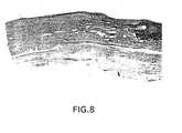
- Figures 4-9show the results of a comparison of three-layer prothesis with a similarly configured contra-lateral reference material, e-PTFE, in a canine femoral artery study.
- the graftswere implanted in canines as femoral interposition prosthesis. Grafts were explanted from 30 to 256 days.
- Histological evaluation of the three-layer collagen graftdemonstrated cellular ingrowth into the graft at 30 days, with more than 90 percent of the graft collagen remodeled by 90 days; and a mature 'neo-artery' at 180 days.
- Host tissuebridged the anastomosis by 60 days with the anastomosis only demarcated by the non-resorbable sutures.
- the predominant cell type in the neo-arterywas a positive g-Actin staining smooth muscle like cell.
- the surface of the remodeled graftwas lined by endothelial cells as demonstrated by SEM, TEM and Factor VIII staining.
- Figure 4is a Masson's trichrome stain (10x) of the proximal anastomosis of the three-layer prosthesis at 256 days compared with Figure 5 of an e-PTFE graft.
- Figure 6is also a Masson's trichrome stain at 25x of the proximal anastomosis of a three-layer prosthesis at 256 days compared with Figure 7 of an e-PTFE graft.
- Figure 8is a Verhoeff's elastic stain (10x) of the proximal anastomosis of a three-layer prosthesis implanted as a canine femoral interposition prosthesis at 256 days compared with Figure 9 of an e-PTFE graft.
Landscapes
- Health & Medical Sciences (AREA)
- Life Sciences & Earth Sciences (AREA)
- Chemical & Material Sciences (AREA)
- Veterinary Medicine (AREA)
- Public Health (AREA)
- Oral & Maxillofacial Surgery (AREA)
- Transplantation (AREA)
- General Health & Medical Sciences (AREA)
- Animal Behavior & Ethology (AREA)
- Biomedical Technology (AREA)
- Medicinal Chemistry (AREA)
- Engineering & Computer Science (AREA)
- Epidemiology (AREA)
- Dermatology (AREA)
- Chemical Kinetics & Catalysis (AREA)
- Botany (AREA)
- Vascular Medicine (AREA)
- Molecular Biology (AREA)
- Pulmonology (AREA)
- Gastroenterology & Hepatology (AREA)
- Biophysics (AREA)
- Urology & Nephrology (AREA)
- Zoology (AREA)
- Cardiology (AREA)
- Heart & Thoracic Surgery (AREA)
- General Chemical & Material Sciences (AREA)
- Materials For Medical Uses (AREA)
- Prostheses (AREA)
- Display Devices Of Pinball Game Machines (AREA)
Abstract


Description
- This invention is in the field of implantable biologicalprostheses. The present invention is a resilient,biocompatible two- or three-layered tissue prosthesis whichcan be engineered in flat sheets or in tubes with variousluminal diameters and thicknesses. At least one layer iscomposed of collagen or a collagenous material. The presentinvention is gradually degraded and bioremodeled by the host'scells which replace the implanted prosthesis and assume itsshape.
- Each year approximately 300,000 coronary bypass proceduresare performed in the United States. The typical treatment forsmall diameter artery replacement has been for surgeons to usethe patient's own vessels, usually the saphenous vein from theleg. However, in many cases, the use of the patient's ownvessels is not practical because the veins are either damaged,diseased or are not available. In these cases, syntheticmaterials are used, but with unsatisfactory long-term results.It is still a continuing goal of researchers to developprostheses which can successfully be used to replace or repairmammalian tissue, particularly blood vessels.
- This invention is directed to a prosthesis, which, whenimplanted into a mammalian host, undergoes controlled biodegradation accompanied by adequate living cellreplacement, or neo-tissue formation, such that the originalimplanted prosthesis is bioremodeled by the host's cellsbefore it is digested by host enzymes. The prosthesis of thisinvention comprises at least two layers: (a) at least onelayer is composed of collagen or a collagenous material; and(b) at least one layer is composed of material which providesstructural stability, and is pliable, semi-permeable, andsuturable. In the preferred embodiment of this invention, thetwo-layered prosthesis has an inner (luminal) layer whichprovides a smooth, thrombosis-resistant flow surface and anouter structural layer which provides structural stability,and is pliable, semi-permeable, and suturable. In anotherpreferred embodiment of this invention the prosthesis hasthree layers: an inner (luminal) layer which acts as asmooth, thrombosis-resistant flow surface; a middle structurallayer which provides structural stability, and is pliable,semi-permeable, and suturable; and, an outer (abluminal)layer. The outer layers of both the two-layer or the three-layerprosthesis adds strength to the graft and allows thepatient's host cells to attach and grow into the graft therebyfacilitating the bioremodeling.
- The invention is also directed to methods for preparingbioremodelable two-or three-layer tubular blood vesselprostheses by (a) forming a tubular structural layer that ispliable, semi-permeable, and suturable; (b) forming an innerlayer to act as a smooth flow surface comprising deposition of acid-extracted fibrillar collagen onto the luminal surface ofsaid structural layer of step (a); and, (c) creating thelumen. The inner layer may also be treated with drugs foranti-thrombotic effect, such as heparin or other appropriateagent(s). The prosthesis is next implanted into a mammalianhost where it undergoes controlled biodegradation accompaniedby adequate living cell replacement, or neo-tissue formation,such that the original implanted prosthesis is bioremodeled bythe host's cells.
- Figure 1A, 1B and 1C are schematic cross-sectional view ofthe preferred prosthesis in accordance with the presentinvention.
- Figure 2 is a Masson's trichrome stain (10x) a three-layerprosthesis of this invention prior to implantation.
- Figure 3 is a Masson's trichrome stain (25x) a three-layerprosthesis of this invention prior to implantation.
- Figure 4 is a Masson's trichrome stain (10x) of theproximal anastomosis of a three-layer prosthesis of thisinvention implanted as a canine femoral interpositionprosthesis (256 days).
- Figure 5 is a Masson's trichrome stain (10x) of theproximal anastomosis of an e-PTFE graft implanted as a caninefemoral interposition prosthesis (256 days).
- Figure 6 is a Masson's trichrome stain (25x) of theproximal anastomosis of a three-layer prosthesis of this invention implanted as a canine femoral interpositionprosthesis (256 days).
- Figure 7 is a Masson's trichrome stain (25x) of theproximal anastomosis of an e-PTFE graft implanted as a caninefemoral interposition prosthesis (256 days).
- Figure 8 is a Verhoeff's elastic stain (10x) of theproximal anastomosis of a three-layer prosthesis of thisinvention implanted as a canine femoral interpositionprosthesis (256 days).
- Figure 9 is a Verhoeff's elastic stain (10x) of theproximal anastomosis of an e-PTFE graft implanted as a caninefemoral interposition prosthesis (256 days).
- This invention is directed to prostheses, which, whenimplanted into a mammalian host, serve as a functioningreplacement for a body part, or tissue structure, and willundergo controlled biodegradation occurring concomitantly withbioremodeling by the host's cells. The prosthesis of thisinvention, in its various embodiments, thus has dualproperties: First, it functions as a substitute body part andsecond, while still functioning as a substitute body party, itfunctions as a bioremodeling template for the ingrowth of hostcells.
- When the prosthesis of this invention functions assubstitute body part, it is preferably used as a vasculargraft. The vascular graft prosthesis may be tubular or flat.Tubular grafts will be used as a conduit to bypass or replace arteries or veins. When formed into flat sheets, theprosthesis can be used as a vascular or intra-cardiac patch.In addition, the prosthesis can be implanted to replacediseased or damaged organs, including the esophagus,intestine, bowel, urethra, and fallopian tubes. These organsall have a basic tubular shape with an outer surface and aluminal surface. Further, the prosthesis can be used as aconduit for nerve regrowth and regeneration.
- The prosthesis of this invention has increased resiliencyor "spring-open" or "spring-back" properties. Spring backproperties are important for applications such as a vasculartubes or patches.
- The second function of the prosthesis is that of a templatefor bioremodeling. "Bioremodeling" is used herein to mean theproduction of structural collagen, vascularization, andepithelialization by the ingrowth of host cells at a ratefaster than the loss of biomechanical strength of theimplanted prosthesis due to biodegradation by host enzymes.The prosthesis retains the distinct characteristics of theoriginally implanted prosthesis while it is remodeled by thebody into all, or substantially all, "self" and as such isfunctional as a functioning tissue structure.
- The prosthesis is made of at least two layers: (a) at leastone layer is composed of collagen or a collagenous materialthat has a smooth, uniform diameter geometry and is non-thrombogenicand (2) at least one layer which providesstructural stabilityand biomechanical properties The mechanical integrity means that the prosthesis is non-dilatingand non-aneurysmal during bioremodeling, and additionally ispliable and suturable. The term "pliable" means good handlingproperties. The term "suturable" means that the mechanicalproperties of the layer include suture retention which permitsneedles and suture materials to pass through the prosthesismaterial at the time of suturing of the prosthesis to sectionsof natural vessel, a process known as anastomosis. Duringsuturing, such vascular (blood vessel) grafts must not tear asa result of the tensile forces applied to them by the suture,nor should they tear when the suture is knotted. Suturabilityof vascular grafts, i.e., the ability of grafts to resisttearing while being sutured, is related to the intrinsicmechanical strength of the prosthesis material, the thicknessof the graft, the tension applied to the suture, and the rateat which the knot is pulled closed.
- The prosthesis of this invention is particularly directedto use as a bypass or replacement of small diameter bloodvessels in the host patient. As used herein, and as isunderstood by those of skill in the art, a small diameter tubeis less than 6 mm, typically around 3 to 4 mm. A mediumdiameter tube is between 6 to 12 mm. A large diameter tube isgreater than 12 mm. As an example, the various vasculardiameter sizes in adult humans are as follows: the diameterof aortic vessels is from about 12 to 22 mm; the diameter ofthe iliac vein is from 8 to 12 mm; the diameter of the superficial femoral vein is 6 mm. Above the knee, the femoralis 6 mm; across the knee, the femoral is 4 to 6 mm.
- The combination of the two layers of the prosthesis of thisinvention when used as a tubular vascular graft workadvantageously by combining a smooth thrombosis resistant flowsurface on the inner (luminal) collagenous layer with thestructural layer which, in addition to its other properties,aids in preventing luminal creep, that is maintaining thenominal diameter. Dilatation (or aneurysmal) failure occurswhen the pulsatile pressure and forces exceed the ability ofthe graft to resist an increase in diameter. Dilatation oraneurysm formation is an increase in diameter beyond nominal.This occurs in both prosthesis as well as in atheroscleroticarteries. As used herein, the term "non-dilatating" meansthat the biomechanical properties of the prosthesis impartdurability so that the diameter of the prosthesis is notstretched, distended, or expanded beyond normal limits afterimplantation. As is described below, total dilatation of theimplanted prosthesis of this invention is within acceptablelimits. The prosthesis of this invention acquires aresistance to dilatation as a function of post-implantationcellular bioremodeling by replacement of structural collagenby host cells at a faster rate than the loss of mechanicalstrength of the implanted materials due from biodegradationand remodeling.
- Various tubular configurations are embodied by thisprosthesis as shown in Figures 1A, 1B, and 1C. Figure 1A shows a two layer prosthesis with an outer collagenous layerand an inner structural layer. Figure 1B shows a prosthesiswith three layers: inner and an outer collagenous layers anda middle structural layer. Figure 1C shows a two layerprosthesis with an inner collagenous layer and an outerstructural layer.
- Each of these various embodiments has applicability forparticular graft replacements. The two layer prosthesis shownby Figure 1A, with an outer collagenous layer and the innerstructural layer, is useful as a replacement for vessels orhollow organs which can tolerate a less smooth inner orluminal surface, such as the esophagus, intestine, bowel,urethra, or fallopian tubes. The outer collagenous layer addsstrength to the graft and allows the host's cells to attach toit, permitting ingrowth into the graft. In contrast, theprosthesis as shown in Figure 1B and in Figure 1C with aninner, smooth collagenous layer are useful as blood vesselreplacements. The inner collagenous layer functions as asmooth flow surface.
- The structural layer may bemade from bioremodelablecollagen or collagenous materials; or biodegradable polymericmaterials, such as polylactic or polyglycolic acid, orcombinations thereof; or biostable polymers, such aspolytetrafluoroethylene (PTFE), polyethylene, or combinationsthereof. In the preferred embodiment, collagenous materialfrom collagenous parts of tissue from the mammalian body isused to make this layer. Such tissue includes but is not limited to intestine, fascia lata, or dura mater. The mostpreferred material for use as a structural layer is the tunicasubmucosa layer of the small intestine, termed herein the"intestinal collagen layer." As used herein, the structurallayer will typically have a thickness of between about 50microns to about 150 microns, more preferably between about 75microns to about 125 microns. These dimensions are for aintestinal collagen layer after mechanical cleaning, butbefore tubulation by heat welding and crosslinking, asdescribed below; both mechanical cleaning and heat weldingsignificantly reduce the "apparent" thickness of theintestinal collagen layer.
- When collagenous material of tissue origin is used to formthe structural layer, it may be crosslinked to providestrength to the structure. Crosslinking collagenous materialalso provides some stiffness to the material to improvehandling properties. Additionally, crosslinking collagenousmaterial on a mandrel yields a tube of a more uniform diameterthan if the material had not been crosslinked. This minimizesthe risk of thrombosis which can be enhanced when there isdiscontinuity in the geometry of the vessel. Crosslinkingagents should be selected so as to produce a biocompatiblematerial capable of being bioremodeled by host cells. Varioustypes of crosslinking agents are known and can be used; thisis discussed below with the preferred embodiment. A preferredcrosslinking agent is 1-ethyl-3-(3-dimethylaminopropyl)carbodiimide hydrochloride (EDC). There are certain crosslinking agents that cannot be used on the prosthesis ofthis invention since they will produce a crosslinked materialthat will not undergo remodeling by host cells.Glutaraldehyde, for example, is not useful for crosslinkingwith this invention as the residual of the glutaraldehydemonomer and lower molecular polymers are cytotoxic.Therefore, it would prevent cell ingrowth and bioremodeling.
- The structural layer, at least when made withbioremodelable collagen or collagenous materials, such as theintestinal collagen layer, will be "semi-permeable," that is,permitting the ingrowth of host cells for remodeling or fordeposition of the collagenous layer, as described below.Crosslinking ICL renders the material relatively lesspermeable as measured by water porosity testing.
- The other layer of the prosthesis is the collagenous layer,the function of which is to act as a smooth flow surface forwhatever its ultimate application. When used as the inner,luminal layer of the prosthesis, its function is to provide asmooth contacting surface, particularly a blood contact flowsurface.
- This smooth collagenous layer may be made from acid-extractedfibrillar or non-fibrillar collagen, which ispredominantly type I collagen, but may also include type 3 or4 collagen, or both. The collagen used may be derived fromany number of mammalian sources, typically bovine, porcine, orovine skin and tendons. The collagen preferably has beenprocessed by acid extraction to result in a fibril dispersion or gel of high purity. Collagen may be acid-extracted fromthe collagen source using a weak acid, such as acetic, citric,or formic acid. Once extracted into solution, the collagencan be salt-precipitated using NaCl and recovered, usingstandard techniques such as centrifugation or filtration.Details of acid extracted collagen are described, for example,in U.S. 5,106,949, incorporated herein by'reference.
- Collagen dispersions or gels for use in the presentinvention are generally at a concentration of about 1 to 10mg/ml, preferably, from about 2 to 6 mg/ml, and mostpreferably at about 2 to 4 mg/ml and at pH of about 2 to 4. Apreferred solvent for the collagen is dilute acetic acid,e.g., about 0.05 to 0.1%. Other conventional solvents forcollagen may be used as long as such solvents are compatible.
- Additionally, in another embodiment of the invention, thecollagenous layer can include mechanically sheared or choppedcollagen fibers. The chopped collagen fibers improve thespring-back performance of the collagenous layer. The choppedfibers can be added to the collagen solution used forformation of the acid-extracted collagen gel. The propertiesof the construct incorporating the fibers may be varied byvariations in the length and diameter of the fiber; variationson the proportion of the fiber used, and partiallycrosslinking fibers. The length of the fibers can range from5 cm to 5.0 cm, and will typically be incorporated into thecollagen gel at a concentration of 5 to 60.
- In another embodiment of the invention, the formation ofthe inner or outer collagenous layer can incorporatepreviously formed collagen threads. For example, a helix, orbraid of micron diameter collagen thread could be incorporatedas part of the formation of the collagen inner layer. Thediameter size of the helix or braid of collagen thread canrange from 25 to 50 microns, preferably 25 to 40 microns.Thus, the properties of the collagen layer can be varied bythe geometry of the thread used for the reinforcement. Thefunctionality of the design is dependent on the geometry ofthe braid or twist. Many of these will also effect thephysical properties (i.e, compliance, radial strength, kinkresistance, suture retention). Physical properties of thethread may also be varied by crosslinking.
- Some portion or all of the fibers used could be polylacticacid. The physical and degradation properties of the lacticacid fibers themselves can be manipulated by varying themolecular weight, as well as the use of the D or L racemes ora mixture of D/L forms of lactic acid. Other fibersfabricated from degradable polymers could also be used, suchas polyglycolic acid, caprolacatone, and polydioxinone.
- To further describe the prosthesis of this invention, theprocess of making a small diameter two layered tubularprosthesis will be described in detail below. The describedtwo-layered prosthesis has an inner (luminal) surface composed of acid-extracted fibrillar collagen and the outer (abluminal)structural layer composed of mammalian tunica submucosa fromthe small intestine. Flat prosthesis can be similarlyprepared with the described methods by using a flat forminstead of a mandrel to produce the prosthesis.
- The submucosa, or the intestinal collagen layer, from amammalian source, typically pigs, cows, or sheep, ismechanically cleaned by squeezing the raw material betweenopposing rollers to remove the muscular layers (tunicamuscularis) and the mucosa (tunica mucosa). The tunicasubmucosa of the small intestine is harder and stiffer thanthe surrounding tissue, and the rollers squeeze the softercomponents from the submucosa. As the mechanically cleanedsubmucosa may have some hidden, visibly nonapparent damagethat affects the consistency of the mechanical properties, thesubmucosa may be chemically cleaned to remove substances otherthan collagen using, for example, by soaking in buffersolutions at 4°C, without the use of any detergents such asTriton or SDS, or by soaking with NaOH or trypsin, or otherknown cleaning techniques.
- After cleaning, the intestinal collagen layer (ICL) shouldbe sterilized, preferably with the use of dilute peraceticacid solutions as described in U.S. patent application serialnumber 08/177,618, incorporated herein by reference. Othersterilization systems for use with collagen are known in theart and can be used.
- The ICL may be tubulated by various alternative means orcombinations thereof. The ICL material may be formed into atube in either the normal or the everted position, but theeverted position is preferred. The tube may be mademechanically by suturing, using alternating knot stitches withsuitable suture material. The knot stitch is advantageous asit allows the tube to be trimmed and shaped by the surgeon atthe time of implantation without unraveling. Other processesto seam the submucosa may include adhesive bonding, such asthe use of fibrin-based glues or industrial-type adhesivessuch as polyurethane, vinyl acetate or polyepoxy. Heatbonding techniques may also be used including heat welding orlaser welding of the seam, followed by quenching, to seal thesides of the thus formed tube. Other mechanical means arepossible, such as using pop rivets or staples. With thesetubulation techniques, the ends of the sides may be eitherbutt ended or overlapped. If the sides are overlapped, theseam may be trimmed once the tube is formed. In addition,these tubulation techniques are typically done on a mandrel soas to determine the desired diameter.
- The thus formed structural tube can be kept on a mandrel orother suitable spindle for further processing. To control thebiodegradation rates and therefore the rate of prosthesisstrength decrease during bioremodeling, the prosthesis ispreferably crosslinked, using a suitable crosslinking agent,such as (1-ethyl-3-(3-dimethylaminopropyl) carbodiimidehydrochloride (EDC). Crosslinking the prosthesis also aids in prevent luminal creep, in keeping the tube diameter uniform,and in increasing the burst strength. It is believed thatcrosslinking the intestinal collagen layer also improves thesuture retention strength by improving resistance to crackpropagation.
- Bovine collagen may be deposited on the internal surface ofthe submucosa as described in example 5 of U.S. PatentApplication 07/505,678, incorporated herein by reference.Briefly, the structural intestinal collagen layer is sealed atone end by luer fittings and the collagen dispersion fills thetube. This step may also be accomplished as described in theabove-referenced patent application using a hydrostaticpressure head. The inner layer of collagen can also bedeposited by flowing collagen into both ends of the tubesimultaneously. The tube is then placed into a bath of 20%polyethylene glycol (PEG) in isotonic phosphate bufferedsaline (PBS), pH about 7. The osmotic gradient between theinternal collagen solution and outer PEG solution incombination cause a simultaneous concentration and depositionof the collagen along the lumen of the internal structurallayer wall. The tube is then removed from the PEG bath, and aglass rod with a diameter desired diameter of the prosthesislumen is inserted into the collagen solution. The prosthesisis then allowed to dry. The tube is then rehydrated in PBS.This process allows the collagenous layer to fill slightirregularities in the intestinal structural layer, thus resulting in a prosthesis of uniform thickness. The procedurealso facilitates the bonding of the collagen gel to theintestinal collagen layer. A collagenous layer of varyingthickness and density can be produced by changing thedeposition conditions which can be determined by routineparameter changes. The same procedures can be used to applythe collagen to the outer surface of the submucosa to create athree-layer prosthesis.
- The prosthesis construct is thrombogenic in small diameterblood vessel replacements. It can only be used in vascularapplications in high flow (large diameter) vessels.
Therefore, the prosthesis must be rendered non-thrombogenic ifto be useful for small diameter blood vessel repair orreplacement. - Heparin can be applied to the prosthesis, by a variety ofwell-known techniques. For illustration, heparin can beapplied to the prosthesis in the following three ways. First,benzalkonium heparin (BA-Hep) solution can be applied to theprosthesis by dipping the prosthesis in the solution and thenair-drying it. This procedure treats the collagen with anionically bound BA-Hep complex. Second, EDC can be used toactivate the heparin, then to covalently bond the heparin tothe collagen fiber. Third, EDC can be used to activate thecollagen, then covalently bond protamine to the collagen andthen ionically bond heparin to the protamine. Many other heparin coating, bonding, and attachment procedures are wellknown in the art which could also be used.
- Treatment of the inner layer with drugs in addition toheparin may be accomplished. The drugs may include forexample, growth factors to promote vascularization andepitheliazation, such as macrophage derived growth factor(MDGF), platelet derived growth factor (PDGF), endothelialcell derived growth factor (ECDGF); antibiotics to fight anypotential infection from the surgery implant; or nerve growthfactors incorporated into the inner collagenous layer when theprosthesis is used as a conduit for nerve regeneration. Thetreatment of the abluminal (outer) layer may also be done in amanner similar to that for the luminal (inner layer).
- If the structural layer is made of ICL which is crosslinkedthe completed two or three layer prosthesis can be laserdrilled to create micron sized pores through the completedprosthesis for aid in cell ingrowth using an excimer laser ateither KrF or XeF wavelengths. The pore size can vary from 20to 100 microns, but is preferably from about 30 to 60 micronsand spacing can vary, but about 500 microns on center ispreferred.
- The completed graft is then sterilized. The preferredmethod is to use peracetic acid as described in U.S. patentapplication serial number 08/177,618, incorporated herein byreference. Sterilization may also be accomplished by subjecting the prosthesis to a gamma radiation treatment(60Co) of 10.0 to 25.0 kGy. The radiation dose eliminates allmicroorganisms without adversely affecting the biomechanicalproperties of the prosthesis.
- Various tests, analysis and performance parameters havebeen developed over the years for vascular graft prosthesisand can be used by those of skill in the art to evaluate theprosthesis characteristics. These methods are detailed inAbbottet al., "Evaluation and performance standards forarterial prostheses,"Journal of Vascular Surgery, Volume 17,pages 746-756 (1993) and "American National Standard forVascular Graft Prostheses,"American National StandardsInstitute (1986).
- The following examples further describe the materials andmethods used in carrying out the invention. The examples arenot intended to limit the invention in any manner.
- The small intestine of a pig was harvested and mechanicallystripped so that the tunica submucosa is delaminated from thetunica muscularis and the luminal portion of the tunica mucosaof the section of small intestine. (The machine was astriper, crusher machine for the mechanical removal of themucosa and muscularis layers from the submucosal layers usinga combination of mechanical action (crushing) and washingusing hot water.) This was accomplished by running the intact intestine through a series of rollers that strip awaysuccessive layers. The intestinal layer was machine cleanedso that the submucosa layer solely remains. The submucosa wasdecontaminated or sterilized with 0.1% peracetic acid for 18hours at 4°C and then washed after the peracetic acidtreatment.
- This machine cleaned intestinal collagen layer (ICL) wasmounted and stretched on a frame so that it was under slighttension both radially and longitudinally. Coarse runningbasting stitches (6-0 Novafil®) were applied to form a smalldiameter tube, with the submucosa in the everted position.The stretched ICL tissue was cut in half to overlap and formflaps. A fine seam through both layers of ICL was formedusing 6-0 Novafil® suture with an alternating knot stitch sothat a final internal diameter of 4.5 mm was obtained. Theflaps were then removed. The small-diameter ICL tube, used asthe structural layer, was placed onto a 4.5 mm glass rod andcrosslinked with 100 mmol EDC (Pierce) for 18 hours at roomtemperature.
- The machine cleaned intestinal collagen layer (ICL) in afully hydrated condition was mounted and wrapped on a mandrelso that the ends overlapped. The ICL wrapped mandrel washeated to 62°C plus or minus 10°C for 15 minutes in a moistatmosphere, followed by quenching at 4°C in iced aqueoussolution for 5 minutes. The tubulated ICL was thencrosslinked with EDC for 6 to 18 hours, rinsed, and removedfrom the mandrel.
- Polycarbonate barbs (luer lock fittings that are funnelshaped on one end) were placed tightly in either end of thetube and then the tube was placed horizontally in a depositionfixture. A 15 ml reservoir of 2.5 mg/ml acid-extractedfibrillar collagen, termed "dense fibrillar collagen" ("DFC")(U.S. patent application, serial no. 07/772,579, incorporatedherein by reference) with a hydrostatic pressure head of 150mmHg (for 5 feet) was attached via the barbs. (The pressurewill depend on the height of the collagen reservoir.) Thecollagen was allowed to fill the lumen of the ICL tube and wasthen placed into a stirring bath of 20% MW 8000 polyethyleneglycol (Sigma Chemical Co.) for 16 hours at 4°C.Theapparatus was then dismantled. To fix the luminal diameter, a4 mm diameter glass rod was placed into the collagen-filledICL tube. The prosthesis was then allowed to dry for 18 hoursat 4° C.
- A layer of acid extracted fibrillar collagen was depositedonto a 4.0 mm diameter porous ceramic mandrel as described inExample 4, U.S. Patent Application 07/505,678, incorporatedherein by reference, for 6 hours and dehydrated at 4°C.
- The ICL tube, as described above, was placed over the driedcollagen and a second layer of dense fibrillar collagen (DFC),as described above, was applied for 18 hours to the outside(abluminal) of the ICL.
- Pores were drilled in the ICL/DFC or the DFC/ICL/DFC usingan excimer laser at either KrF or XeF wavelengths. The pore size was about 50 microns and spacing was 500 microns oncenter.
- The construct was rehydrated in 4°C 1M PBS for 6 hours.The prosthesis was treated with application of benzalkoniumheparin in isopropranol. Sterilization was accomplished with0.1% peracetic acid for 18 hours at 4°C.
- The prosthesis was packaged and sterilized with 10.0 to25.0 kGy of gamma radiation (60Co). (The prosthesis can alsobe shipped dry and rehydrated in saline, prior toimplantation.)
- Three-layer prosthesis were implanted in the infra-renalaorta of rabbits using standard surgical techniques. Proline,7-0, was used to construct end to end anatomoses to theadjacent arteries. The grafts were 1.5 cm in length and 3 mmin diameter. No anti-platelet medications were administeredpost-operatively.
- Following pressure perfusion with McDowells-Trump fixative,the grafts were explanted, and submitted for light andelectron microscopy. Specimens from 30, 60, 90, 120, and 180day implants were available. Materials were examined with H/E,VonGieson elastica, Masson's Trichrome, g-Actin, FactorVIII, and Ram-11 (macrophage) stains, and polarizedmicroscopy. Qualitative morphometric comparisons were made tostained non-implanted retention samples.
- Histological evaluations demonstrated that the graft wasreadily invaded by host cells. The luminal collagen wasresorbed and remodeled with the production of new collagen byhost myofibroblasts. The ICL was readily invaded, repopulatedby host cells, and remodeled. Endothelial cellswere demonstrated on the luminal surface of the prosthesis.
- At 30 days, large numbers of mononuclear inflammatory cellswere seen on both the luminal and abluminal surfaces of thecollagen. Modest numbers of Ram-11 positively stainingmacrophages, were observed. At the surface of the castcollagen, there was cell mediated collagen resporption andremodeling. There was minimal loss of the collagen bulk atthis time.
- At 60 days, the cellular response was more myofibroblasticthan inflammatory. Significant amounts of new collagen aswell as small amounts of elastin were readily identified.About 50 percent of the cast collagen had been remodeled.Endothelial cells as identified by SEM appearance, TEM(Weibel-Palade bodies) and positive Factor VIII staining,covered the surface of the remodeling construct.
- At 90 days, the matrix surrounding the myofibroblasts (asidentified with g-actin) stained prominently for collagen.The cytoplasm of cells themselves had reduced amounts ofcytoplasm as compared to previous timepoints.
- At 120 days, the stroma demonstrated well organizedpredominantly radially and longitudinally orientedmyofibroblasts and host produced collagen. Significant amounts of elastin could be identified. Greater than 90percent of the implanted collagen had been remodeled. No Ram-11staining macrophages were identified.
- At 180 days, cells and the matrix of the neo-artery werequite mature. The cells were small with minimal cytoplasm.The collagen was dense and distinctly radially andlongitudinally oriented.
- There was no histological evidence of an immune response toeither the luminal collagen layer or the abluminal ICL layer.No grafts became dilated or aneurysmal.
- Both two-layer and three-layer small diameter prosthesiswere implanted and evaluated over time for patency andremodeling.
- Figures 4-9 show the results of a comparison of three-layerprothesis with a similarly configured contra-lateral referencematerial, e-PTFE, in a canine femoral artery study. Thegrafts were implanted in canines as femoral interpositionprosthesis. Grafts were explanted from 30 to 256 days.
- Histological evaluation of the three-layer collagen graftdemonstrated cellular ingrowth into the graft at 30 days, withmore than 90 percent of the graft collagen remodeled by 90days; and a mature 'neo-artery' at 180 days. Host tissuebridged the anastomosis by 60 days with the anastomosis onlydemarcated by the non-resorbable sutures. The predominantcell type in the neo-artery was a positive g-Actin staining smooth muscle like cell. The surface of the remodeled graftwas lined by endothelial cells as demonstrated by SEM, TEM andFactor VIII staining.
- In contrast, at times to 256 days, no ingrowth into thee-PTFE artery was observed either across the anastomosis oralong the body of the graft. Only a thin smooth muscle cellhyperplastic response was demonstrated extending from theadjacent artery a short distance on the graft's luminalsurface. The graft was encapsulated by mature fibrous tissuewith no evidence of cellular or tissue extension into thegraft.
- Figure 4 is a Masson's trichrome stain (10x) of theproximal anastomosis of the three-layer prosthesis at 256 dayscompared with Figure 5 of an e-PTFE graft. Figure 6 is also aMasson's trichrome stain at 25x of the proximal anastomosis ofa three-layer prosthesis at 256 days compared with Figure 7 ofan e-PTFE graft.
- Figure 8 is a Verhoeff's elastic stain (10x) of theproximal anastomosis of a three-layer prosthesis implanted asa canine femoral interposition prosthesis at 256 days comparedwith Figure 9 of an e-PTFE graft.
- Although the foregoing invention has been described indetail by way of illustration and example for purposes ofclarity of understanding, it will be obvious to one skilled inthe art that changes and modifications may be practiced withinthe scope of the invention, as limited only by the scope ofthe appended claims.
Claims (9)
- A prosthesis, which, when implanted into a mammalianpatient, undergoes controlled biodegradation occurring withadequate living cell replacement such that the originalimplanted prosthesis is bioremodeled by the patient's livingcells.
- A prosthesis comprising at least two layers:(a) at least one layer is composed of collagen or acollagenous material, which can act as a smooth flow surface;and(b) at least one layer which provides structuralstability, and is pliable, semi-permeable, and suturable,
- The prosthesis of claim 2 wherein said wherein layerand wherein said prosthesis is either flat or tubular.(a) is comprised of acid extracted collagen, preferablyfibrillar collagen, and optionally is composed of choppedcollagen fibers or collagen threads; and layer (b) iscomprised of intestinal collagen, a biodegradable polymersheet or tube, or a synthetic collagen sheet or mesh.
- A two-layer tubular prosthesis, with a smalldiameter lumen, which, when implanted into a mammalianpatient, undergoes controlled biodegradation occurring withadequate living cell replacement such that the original implanted prosthesis is bioremodeled by the patient's livingcells, comprising:(a) an outer (abluminal) tubulated sterilized andcrosslinked structural layer composed of intestinal collagenlayer comprising a diameter of about 50 to about 150 microns;and(b) an inner acid-extracted fibrillar collagen layer;wherein the lumen of said prosthesis is less than 6 mm.
- A three-layer tubular prosthesis, with a smalldiameter lumen, which, when implanted into a mammalianpatient, undergoes controlled biodegradation occurring withadequate living cell replacement such that the originalimplanted prosthesis is bioremodeled by the patient's livingcells, comprising:wherein the lumen of said prosthesis is less than 6 mm.(a) an outer (abluminal) layer comprising acid extractedcollagen;(b) a middle sterilized and crosslinked structural layercomposed of intestinal collagen layer comprising a diameter ofabout 50 to about 150 microns;(c) an inner acid-extracted fibrillar collagen layer;
- The prosthesis of any of claims 2-5 wherein saidstructural or collagen layer is crosslinked with acrosslinking agent, preferably 1-ethyl-3-(3-dimethylaminopropyl)carbodiimide hydrochloride (EDC).
- The prosthesis of any of claims 2-5 wherein thelumen diameter of said prosthesis is less than 6 mm,optionally less than 4 mm.
- The prosthesis of any of claims 2-5 wherein saidprosthesis is implanted in a patient for use as (a) an implantfor small diameter arteries or veins; (b) an implant for avascular patch; (c) a substitute for the esophagus, bowel,urethra, fallopian tubes, arteries and veins; (d) for patchinga section of the esophagus, bowel, urethra, and fallopiantubes; or, (e) a conduit for nerve regrowth and regeneration.
- The prosthesis of claim 2 wherein said collagenouslayer additionally contains an anticoagulant, one or moreantibiotics, one or more growth factors.
Applications Claiming Priority (3)
| Application Number | Priority Date | Filing Date | Title |
|---|---|---|---|
| US19806294A | 1994-02-18 | 1994-02-18 | |
| US198062 | 1994-02-18 | ||
| EP95911032AEP0746268B1 (en) | 1994-02-18 | 1995-02-17 | Bioremodelable collagen graft prosthesis |
Related Parent Applications (1)
| Application Number | Title | Priority Date | Filing Date |
|---|---|---|---|
| EP95911032ADivisionEP0746268B1 (en) | 1994-02-18 | 1995-02-17 | Bioremodelable collagen graft prosthesis |
Publications (2)
| Publication Number | Publication Date |
|---|---|
| EP1364627A1true EP1364627A1 (en) | 2003-11-26 |
| EP1364627B1 EP1364627B1 (en) | 2007-09-12 |
Family
ID=22731845
Family Applications (2)
| Application Number | Title | Priority Date | Filing Date |
|---|---|---|---|
| EP95911032AExpired - LifetimeEP0746268B1 (en) | 1994-02-18 | 1995-02-17 | Bioremodelable collagen graft prosthesis |
| EP03019053AExpired - LifetimeEP1364627B1 (en) | 1994-02-18 | 1995-02-17 | Method for producing a bioremodelable collagen graft prosthesis |
Family Applications Before (1)
| Application Number | Title | Priority Date | Filing Date |
|---|---|---|---|
| EP95911032AExpired - LifetimeEP0746268B1 (en) | 1994-02-18 | 1995-02-17 | Bioremodelable collagen graft prosthesis |
Country Status (7)
| Country | Link |
|---|---|
| EP (2) | EP0746268B1 (en) |
| JP (1) | JP3765828B2 (en) |
| AT (2) | ATE372747T1 (en) |
| CA (1) | CA2183056C (en) |
| DE (2) | DE69531859T2 (en) |
| MX (1) | MX9603249A (en) |
| WO (1) | WO1995022301A1 (en) |
Cited By (4)
| Publication number | Priority date | Publication date | Assignee | Title |
|---|---|---|---|---|
| WO2007002260A3 (en)* | 2005-06-21 | 2007-03-29 | Cook Inc | Implantable graft to close a fistula |
| EP1738780A3 (en)* | 2005-06-30 | 2008-12-31 | Codman & Shurtleff, Inc. | Collagen device and method of preparing the same |
| US7722665B2 (en) | 2006-07-07 | 2010-05-25 | Graft Technologies, Inc. | System and method for providing a graft in a vascular environment |
| WO2023170408A1 (en)* | 2022-03-08 | 2023-09-14 | Newtec Vascular Products Ltd | Vascular stent |
Families Citing this family (40)
| Publication number | Priority date | Publication date | Assignee | Title |
|---|---|---|---|---|
| US6334872B1 (en) | 1994-02-18 | 2002-01-01 | Organogenesis Inc. | Method for treating diseased or damaged organs |
| US5733337A (en)* | 1995-04-07 | 1998-03-31 | Organogenesis, Inc. | Tissue repair fabric |
| US20020095218A1 (en) | 1996-03-12 | 2002-07-18 | Carr Robert M. | Tissue repair fabric |
| SE9601243D0 (en)* | 1996-03-29 | 1996-03-29 | Hans Arne Hansson | Promotion of regeneration of organized tissues |
| PL331765A1 (en)* | 1996-08-23 | 1999-08-02 | Cook Biotech Inc | Trnsplant prosthesis, materials and methods |
| AU743779B2 (en)* | 1996-12-10 | 2002-02-07 | Cook Biotech, Inc. | Tubular grafts from purified submucosa |
| US5993844A (en) | 1997-05-08 | 1999-11-30 | Organogenesis, Inc. | Chemical treatment, without detergents or enzymes, of tissue to form an acellular, collagenous matrix |
| US6004333A (en)* | 1997-10-31 | 1999-12-21 | Ethicon Endo-Surgery, Inc. | Prosthetic with collagen for tissue repair |
| EP1082071B1 (en)* | 1998-06-05 | 2009-03-04 | Organogenesis Inc. | Method of preparing a vascular prosthesis |
| WO1999062427A1 (en) | 1998-06-05 | 1999-12-09 | Organogenesis Inc. | Bioengineered vascular graft support prostheses |
| CA2334435C (en) | 1998-06-05 | 2010-10-05 | Organogenesis Inc. | Bioengineered vascular graft prostheses |
| ATE423577T1 (en) | 1998-06-05 | 2009-03-15 | Organogenesis Inc | BIOLOGICALLY MODELED IMPLANTABLE PROSTHESES |
| CZ301649B6 (en)* | 2000-06-28 | 2010-05-12 | Ed. Geistlich Soehne Ag Fur Chemische Industrie Incorporated Under The Laws Of Switzerland | Tube for regeneration of nerves and process for producing thereof |
| US6638312B2 (en) | 2000-08-04 | 2003-10-28 | Depuy Orthopaedics, Inc. | Reinforced small intestinal submucosa (SIS) |
| US20020103542A1 (en)* | 2000-09-18 | 2002-08-01 | Bilbo Patrick R. | Methods for treating a patient using a bioengineered flat sheet graft prostheses |
| WO2003007786A2 (en) | 2001-07-16 | 2003-01-30 | Depuy Products, Inc. | Porous delivery scaffold and method |
| US20050027307A1 (en) | 2001-07-16 | 2005-02-03 | Schwartz Herbert Eugene | Unitary surgical device and method |
| AU2002313694B2 (en) | 2001-07-16 | 2007-08-30 | Depuy Products, Inc. | Cartilage repair apparatus and method |
| AU2002364228B2 (en) | 2001-12-21 | 2007-06-28 | Organogenesis Inc. | System and method for forming bioengineered tubular graft prostheses |
| WO2003101312A1 (en)* | 2002-06-03 | 2003-12-11 | Nmt Medical, Inc. | Device with biological tissue scaffold for intracardiac defect closure |
| US7056337B2 (en)* | 2003-10-21 | 2006-06-06 | Cook Incorporated | Natural tissue stent |
| US7569233B2 (en) | 2004-05-04 | 2009-08-04 | Depuy Products, Inc. | Hybrid biologic-synthetic bioabsorbable scaffolds |
| US7354627B2 (en) | 2004-12-22 | 2008-04-08 | Depuy Products, Inc. | Method for organizing the assembly of collagen fibers and compositions formed therefrom |
| EP1863545B1 (en)* | 2005-03-19 | 2015-11-18 | Cook Biotech, Inc. | Prosthetic implants including ECM composite material |
| SE528738C2 (en)* | 2005-06-16 | 2007-02-06 | Sinova Safetech Innovation Ab | intestinal Implants |
| US7595062B2 (en) | 2005-07-28 | 2009-09-29 | Depuy Products, Inc. | Joint resurfacing orthopaedic implant and associated method |
| RU2481114C2 (en)* | 2005-10-18 | 2013-05-10 | Огенодженесис, Инк. | Bioengineered collagen construct, modified intestinal collagen layer, processed tissue matrix and method of reconstruction or replacement of damaged tissue |
| DE102005054941A1 (en)* | 2005-11-17 | 2007-05-31 | Gelita Ag | nerve |
| US8105634B2 (en) | 2006-08-15 | 2012-01-31 | Anthrogenesis Corporation | Umbilical cord biomaterial for medical use |
| WO2008060377A2 (en) | 2006-10-04 | 2008-05-22 | Anthrogenesis Corporation | Placental or umbilical cord tissue compositions |
| KR20190112868A (en) | 2006-10-06 | 2019-10-07 | 안트로제네시스 코포레이션 | Native(telopeptide) Placental Collagen Compositions |
| JP5702515B2 (en) | 2007-12-28 | 2015-04-15 | 東洋紡株式会社 | Nerve regeneration induction tube |
| AU2009201541B2 (en) | 2008-04-23 | 2014-12-04 | Integra Lifesciences Corporation | Flowable collagen material for dural closure |
| JP2011206203A (en)* | 2010-03-29 | 2011-10-20 | Kawasumi Lab Inc | Nerve regeneration tube and method for manufacturing the same |
| JP5987138B2 (en)* | 2010-08-31 | 2016-09-07 | 国立研究開発法人農業・食品産業技術総合研究機構 | Cylindrical structure and manufacturing method thereof |
| ES2726105T3 (en) | 2011-12-20 | 2019-10-01 | Lifecell Corp | Fluidizable fabric products |
| AU2012355463C1 (en) | 2011-12-20 | 2016-09-22 | Lifecell Corporation | Sheet tissue products |
| AU2013212592B2 (en) | 2012-01-24 | 2016-06-30 | Lifecell Corporation | Elongated tissue matrices |
| US10071184B2 (en) | 2012-02-29 | 2018-09-11 | Collagen Matrix, Inc. | Collagen-coated tissue-based membranes |
| EP4142816B1 (en) | 2020-05-01 | 2024-03-27 | Askel Healthcare Ltd. | Method for preparing a three-dimensional scaffold for medical use |
Citations (13)
| Publication number | Priority date | Publication date | Assignee | Title |
|---|---|---|---|---|
| US505678A (en) | 1893-09-26 | Brick-cutting machinery | ||
| GB2153235A (en)* | 1984-01-30 | 1985-08-21 | Meadox Medicals Inc | Drug delivery collagen-coated synthetic vascular graft |
| US4539716A (en)* | 1981-03-19 | 1985-09-10 | Massachusetts Institute Of Technology | Fabrication of living blood vessels and glandular tissues |
| WO1989010100A1 (en)* | 1988-04-22 | 1989-11-02 | Uab Research Foundation | Elastomeric polypeptides as vascular prosthetic materials |
| US4902508A (en)* | 1988-07-11 | 1990-02-20 | Purdue Research Foundation | Tissue graft composition |
| US4902289A (en)* | 1982-04-19 | 1990-02-20 | Massachusetts Institute Of Technology | Multilayer bioreplaceable blood vessel prosthesis |
| EP0397500A2 (en)* | 1989-05-10 | 1990-11-14 | United States Surgical Corporation | Synthetic semiabsorbable yarn, fabric and tubular prosthesis |
| US5024671A (en)* | 1988-09-19 | 1991-06-18 | Baxter International Inc. | Microporous vascular graft |
| US5026381A (en)* | 1989-04-20 | 1991-06-25 | Colla-Tec, Incorporated | Multi-layered, semi-permeable conduit for nerve regeneration comprised of type 1 collagen, its method of manufacture and a method of nerve regeneration using said conduit |
| US5106949A (en)* | 1989-09-15 | 1992-04-21 | Organogenesis, Inc. | Collagen compositions and methods for preparation thereof |
| EP0493788A1 (en)* | 1990-12-29 | 1992-07-08 | MUDR. MILAN KRAJICEK CSc. | Three-layer vascular prosthesis |
| US5256418A (en)* | 1990-04-06 | 1993-10-26 | Organogenesis, Inc. | Collagen constructs |
| US5263983A (en)* | 1988-03-09 | 1993-11-23 | Terumo Kabushiki Kaisha | Medical material and prosthetic skin in which cells can invade |
- 1995
- 1995-02-17WOPCT/US1995/002025patent/WO1995022301A1/enactiveIP Right Grant
- 1995-02-17DEDE69531859Tpatent/DE69531859T2/ennot_activeExpired - Lifetime
- 1995-02-17MXMX9603249Apatent/MX9603249A/enactiveIP Right Grant
- 1995-02-17JPJP52193295Apatent/JP3765828B2/ennot_activeExpired - Fee Related
- 1995-02-17EPEP95911032Apatent/EP0746268B1/ennot_activeExpired - Lifetime
- 1995-02-17EPEP03019053Apatent/EP1364627B1/ennot_activeExpired - Lifetime
- 1995-02-17CACA002183056Apatent/CA2183056C/ennot_activeExpired - Fee Related
- 1995-02-17ATAT03019053Tpatent/ATE372747T1/ennot_activeIP Right Cessation
- 1995-02-17ATAT95911032Tpatent/ATE250901T1/ennot_activeIP Right Cessation
- 1995-02-17DEDE69535595Tpatent/DE69535595T2/ennot_activeExpired - Lifetime
Patent Citations (13)
| Publication number | Priority date | Publication date | Assignee | Title |
|---|---|---|---|---|
| US505678A (en) | 1893-09-26 | Brick-cutting machinery | ||
| US4539716A (en)* | 1981-03-19 | 1985-09-10 | Massachusetts Institute Of Technology | Fabrication of living blood vessels and glandular tissues |
| US4902289A (en)* | 1982-04-19 | 1990-02-20 | Massachusetts Institute Of Technology | Multilayer bioreplaceable blood vessel prosthesis |
| GB2153235A (en)* | 1984-01-30 | 1985-08-21 | Meadox Medicals Inc | Drug delivery collagen-coated synthetic vascular graft |
| US5263983A (en)* | 1988-03-09 | 1993-11-23 | Terumo Kabushiki Kaisha | Medical material and prosthetic skin in which cells can invade |
| WO1989010100A1 (en)* | 1988-04-22 | 1989-11-02 | Uab Research Foundation | Elastomeric polypeptides as vascular prosthetic materials |
| US4902508A (en)* | 1988-07-11 | 1990-02-20 | Purdue Research Foundation | Tissue graft composition |
| US5024671A (en)* | 1988-09-19 | 1991-06-18 | Baxter International Inc. | Microporous vascular graft |
| US5026381A (en)* | 1989-04-20 | 1991-06-25 | Colla-Tec, Incorporated | Multi-layered, semi-permeable conduit for nerve regeneration comprised of type 1 collagen, its method of manufacture and a method of nerve regeneration using said conduit |
| EP0397500A2 (en)* | 1989-05-10 | 1990-11-14 | United States Surgical Corporation | Synthetic semiabsorbable yarn, fabric and tubular prosthesis |
| US5106949A (en)* | 1989-09-15 | 1992-04-21 | Organogenesis, Inc. | Collagen compositions and methods for preparation thereof |
| US5256418A (en)* | 1990-04-06 | 1993-10-26 | Organogenesis, Inc. | Collagen constructs |
| EP0493788A1 (en)* | 1990-12-29 | 1992-07-08 | MUDR. MILAN KRAJICEK CSc. | Three-layer vascular prosthesis |
Cited By (9)
| Publication number | Priority date | Publication date | Assignee | Title |
|---|---|---|---|---|
| US8795710B2 (en) | 2004-02-09 | 2014-08-05 | Codman & Shurtleff, Inc. | Collagen device and method of preparing the same |
| WO2007002260A3 (en)* | 2005-06-21 | 2007-03-29 | Cook Inc | Implantable graft to close a fistula |
| EP2989995A1 (en)* | 2005-06-21 | 2016-03-02 | Cook Medical Technologies LLC | Implantable graft to close a fistula |
| EP1738780A3 (en)* | 2005-06-30 | 2008-12-31 | Codman & Shurtleff, Inc. | Collagen device and method of preparing the same |
| AU2006202593B2 (en)* | 2005-06-30 | 2012-04-12 | Integra Lifesciences Corporation | Collagen device and method of preparing the same |
| US7722665B2 (en) | 2006-07-07 | 2010-05-25 | Graft Technologies, Inc. | System and method for providing a graft in a vascular environment |
| US8123797B2 (en) | 2006-07-07 | 2012-02-28 | Graft Technologies, Inc. | System and method for providing a graft in a vascular environment |
| US8808362B2 (en) | 2006-07-07 | 2014-08-19 | Graft Technologies, Inc. | System and method for providing a graft in a vascular environment |
| WO2023170408A1 (en)* | 2022-03-08 | 2023-09-14 | Newtec Vascular Products Ltd | Vascular stent |
Also Published As
| Publication number | Publication date |
|---|---|
| EP0746268A4 (en) | 1998-04-01 |
| DE69531859D1 (en) | 2003-11-06 |
| JP3765828B2 (en) | 2006-04-12 |
| CA2183056A1 (en) | 1995-08-24 |
| EP0746268B1 (en) | 2003-10-01 |
| ATE372747T1 (en) | 2007-09-15 |
| WO1995022301A1 (en) | 1995-08-24 |
| DE69535595D1 (en) | 2007-10-25 |
| JPH09509085A (en) | 1997-09-16 |
| DE69531859T2 (en) | 2004-07-29 |
| EP0746268A1 (en) | 1996-12-11 |
| ATE250901T1 (en) | 2003-10-15 |
| MX9603249A (en) | 1997-07-31 |
| DE69535595T2 (en) | 2008-06-05 |
| CA2183056C (en) | 2001-07-10 |
| EP1364627B1 (en) | 2007-09-12 |
Similar Documents
| Publication | Publication Date | Title |
|---|---|---|
| EP1364627B1 (en) | Method for producing a bioremodelable collagen graft prosthesis | |
| US6334872B1 (en) | Method for treating diseased or damaged organs | |
| US5733337A (en) | Tissue repair fabric | |
| US7909886B2 (en) | Tissue repair fabric | |
| US7887576B2 (en) | Endoluminal device with extracellular matrix material and methods | |
| CA2267408C (en) | Tubular submucosal graft constructs | |
| US7244444B2 (en) | Graft material, stent graft and method | |
| WO2005115275A1 (en) | Endoluminal device with extracellular matrix material and methods | |
| US20080095818A1 (en) | Tubular Structure Based on Hyaluronic Acid Derivatives for the Preparation of Vascular and Urethral Graft | |
| MXPA97007655A (en) | Tej repair fabric | |
| AU774634B2 (en) | Tubular submucosal graft constructs | |
| Guidoin et al. | Biotextiles as medical implants: 15. Vascular prostheses for open surgery |
Legal Events
| Date | Code | Title | Description |
|---|---|---|---|
| PUAI | Public reference made under article 153(3) epc to a published international application that has entered the european phase | Free format text:ORIGINAL CODE: 0009012 | |
| AC | Divisional application: reference to earlier application | Ref document number:0746268 Country of ref document:EP Kind code of ref document:P | |
| AK | Designated contracting states | Kind code of ref document:A1 Designated state(s):AT BE CH DE DK ES FR GB GR IE IT LI LU MC NL PT SE | |
| RIN1 | Information on inventor provided before grant (corrected) | Inventor name:CARR, ROBERT, M, JR. Inventor name:TERMIN. PAUL, L. Inventor name:CONDON, KIMBERLIE, D. | |
| 17P | Request for examination filed | Effective date:20040122 | |
| RIN1 | Information on inventor provided before grant (corrected) | Inventor name:TERMIN. PAUL, L. Inventor name:CONDON, KIMBERLIE, D. Inventor name:CARR, ROBERT, M, JR. | |
| 17Q | First examination report despatched | Effective date:20040408 | |
| AKX | Designation fees paid | Designated state(s):AT BE CH DE DK ES FR GB GR IE IT LI LU MC NL PT SE | |
| 17Q | First examination report despatched | Effective date:20040408 | |
| GRAP | Despatch of communication of intention to grant a patent | Free format text:ORIGINAL CODE: EPIDOSNIGR1 | |
| RTI1 | Title (correction) | Free format text:METHOD FOR PRODUCING A BIOREMODELABLE COLLAGEN GRAFT PROSTHESIS | |
| GRAS | Grant fee paid | Free format text:ORIGINAL CODE: EPIDOSNIGR3 | |
| GRAA | (expected) grant | Free format text:ORIGINAL CODE: 0009210 | |
| AC | Divisional application: reference to earlier application | Ref document number:0746268 Country of ref document:EP Kind code of ref document:P | |
| AK | Designated contracting states | Kind code of ref document:B1 Designated state(s):AT BE CH DE DK ES FR GB GR IE IT LI LU MC NL PT SE | |
| REG | Reference to a national code | Ref country code:GB Ref legal event code:FG4D | |
| REG | Reference to a national code | Ref country code:CH Ref legal event code:EP | |
| REF | Corresponds to: | Ref document number:69535595 Country of ref document:DE Date of ref document:20071025 Kind code of ref document:P | |
| REG | Reference to a national code | Ref country code:IE Ref legal event code:FG4D | |
| PG25 | Lapsed in a contracting state [announced via postgrant information from national office to epo] | Ref country code:LI Free format text:LAPSE BECAUSE OF FAILURE TO SUBMIT A TRANSLATION OF THE DESCRIPTION OR TO PAY THE FEE WITHIN THE PRESCRIBED TIME-LIMIT Effective date:20070912 Ref country code:CH Free format text:LAPSE BECAUSE OF FAILURE TO SUBMIT A TRANSLATION OF THE DESCRIPTION OR TO PAY THE FEE WITHIN THE PRESCRIBED TIME-LIMIT Effective date:20070912 Ref country code:AT Free format text:LAPSE BECAUSE OF FAILURE TO SUBMIT A TRANSLATION OF THE DESCRIPTION OR TO PAY THE FEE WITHIN THE PRESCRIBED TIME-LIMIT Effective date:20070912 | |
| NLV1 | Nl: lapsed or annulled due to failure to fulfill the requirements of art. 29p and 29m of the patents act | ||
| ET | Fr: translation filed | ||
| PG25 | Lapsed in a contracting state [announced via postgrant information from national office to epo] | Ref country code:BE Free format text:LAPSE BECAUSE OF FAILURE TO SUBMIT A TRANSLATION OF THE DESCRIPTION OR TO PAY THE FEE WITHIN THE PRESCRIBED TIME-LIMIT Effective date:20070912 | |
| REG | Reference to a national code | Ref country code:CH Ref legal event code:PL | |
| PG25 | Lapsed in a contracting state [announced via postgrant information from national office to epo] | Ref country code:NL Free format text:LAPSE BECAUSE OF FAILURE TO SUBMIT A TRANSLATION OF THE DESCRIPTION OR TO PAY THE FEE WITHIN THE PRESCRIBED TIME-LIMIT Effective date:20070912 Ref country code:GR Free format text:LAPSE BECAUSE OF FAILURE TO SUBMIT A TRANSLATION OF THE DESCRIPTION OR TO PAY THE FEE WITHIN THE PRESCRIBED TIME-LIMIT Effective date:20071213 Ref country code:ES Free format text:LAPSE BECAUSE OF FAILURE TO SUBMIT A TRANSLATION OF THE DESCRIPTION OR TO PAY THE FEE WITHIN THE PRESCRIBED TIME-LIMIT Effective date:20071223 | |
| PG25 | Lapsed in a contracting state [announced via postgrant information from national office to epo] | Ref country code:PT Free format text:LAPSE BECAUSE OF FAILURE TO SUBMIT A TRANSLATION OF THE DESCRIPTION OR TO PAY THE FEE WITHIN THE PRESCRIBED TIME-LIMIT Effective date:20080212 | |
| PG25 | Lapsed in a contracting state [announced via postgrant information from national office to epo] | Ref country code:SE Free format text:LAPSE BECAUSE OF FAILURE TO SUBMIT A TRANSLATION OF THE DESCRIPTION OR TO PAY THE FEE WITHIN THE PRESCRIBED TIME-LIMIT Effective date:20071212 | |
| PLBE | No opposition filed within time limit | Free format text:ORIGINAL CODE: 0009261 | |
| STAA | Information on the status of an ep patent application or granted ep patent | Free format text:STATUS: NO OPPOSITION FILED WITHIN TIME LIMIT | |
| PG25 | Lapsed in a contracting state [announced via postgrant information from national office to epo] | Ref country code:DK Free format text:LAPSE BECAUSE OF FAILURE TO SUBMIT A TRANSLATION OF THE DESCRIPTION OR TO PAY THE FEE WITHIN THE PRESCRIBED TIME-LIMIT Effective date:20070912 | |
| 26N | No opposition filed | Effective date:20080613 | |
| PG25 | Lapsed in a contracting state [announced via postgrant information from national office to epo] | Ref country code:MC Free format text:LAPSE BECAUSE OF NON-PAYMENT OF DUE FEES Effective date:20080228 | |
| PG25 | Lapsed in a contracting state [announced via postgrant information from national office to epo] | Ref country code:IE Free format text:LAPSE BECAUSE OF NON-PAYMENT OF DUE FEES Effective date:20080218 | |
| PG25 | Lapsed in a contracting state [announced via postgrant information from national office to epo] | Ref country code:LU Free format text:LAPSE BECAUSE OF NON-PAYMENT OF DUE FEES Effective date:20080217 | |
| PG25 | Lapsed in a contracting state [announced via postgrant information from national office to epo] | Ref country code:IT Free format text:LAPSE BECAUSE OF NON-PAYMENT OF DUE FEES Effective date:20080229 | |
| PGFP | Annual fee paid to national office [announced via postgrant information from national office to epo] | Ref country code:DE Payment date:20140228 Year of fee payment:20 | |
| PGFP | Annual fee paid to national office [announced via postgrant information from national office to epo] | Ref country code:FR Payment date:20140228 Year of fee payment:20 | |
| PGFP | Annual fee paid to national office [announced via postgrant information from national office to epo] | Ref country code:GB Payment date:20140227 Year of fee payment:20 | |
| REG | Reference to a national code | Ref country code:DE Ref legal event code:R071 Ref document number:69535595 Country of ref document:DE | |
| REG | Reference to a national code | Ref country code:GB Ref legal event code:PE20 Expiry date:20150216 | |
| PG25 | Lapsed in a contracting state [announced via postgrant information from national office to epo] | Ref country code:GB Free format text:LAPSE BECAUSE OF EXPIRATION OF PROTECTION Effective date:20150216 |