EP1352061B1 - Method for inhibiting the expression of a target gene - Google Patents
Method for inhibiting the expression of a target geneDownload PDFInfo
- Publication number
- EP1352061B1 EP1352061B1EP02710786AEP02710786AEP1352061B1EP 1352061 B1EP1352061 B1EP 1352061B1EP 02710786 AEP02710786 AEP 02710786AEP 02710786 AEP02710786 AEP 02710786AEP 1352061 B1EP1352061 B1EP 1352061B1
- Authority
- EP
- European Patent Office
- Prior art keywords
- accordance
- dsrna
- medicament
- strand
- target gene
- Prior art date
- Legal status (The legal status is an assumption and is not a legal conclusion. Google has not performed a legal analysis and makes no representation as to the accuracy of the status listed.)
- Expired - Lifetime
Links
- 108090000623proteins and genesProteins0.000titleclaimsabstractdescription113
- 230000014509gene expressionEffects0.000titleclaimsabstractdescription76
- 230000002401inhibitory effectEffects0.000titleclaimsabstractdescription16
- 238000000034methodMethods0.000titleclaimsdescription56
- 125000003729nucleotide groupChemical group0.000claimsabstractdescription53
- 239000002773nucleotideSubstances0.000claimsabstractdescription52
- 230000000295complement effectEffects0.000claimsabstractdescription44
- 241000282414Homo sapiensSpecies0.000claimsdescription149
- 108091032973(ribonucleotides)n+mProteins0.000claimsdescription143
- 210000004027cellAnatomy0.000claimsdescription115
- 102000040650(ribonucleotides)n+mHuman genes0.000claimsdescription90
- 239000003814drugSubstances0.000claimsdescription43
- 239000000126substanceSubstances0.000claimsdescription38
- 102000004169proteins and genesHuman genes0.000claimsdescription36
- 241001465754MetazoaSpecies0.000claimsdescription27
- 241000700605VirusesSpecies0.000claimsdescription27
- 108091081021Sense strandProteins0.000claimsdescription26
- 241000726445ViroidsSpecies0.000claimsdescription16
- 229920002477rna polymerPolymers0.000claimsdescription13
- 101710132601Capsid proteinProteins0.000claimsdescription12
- 101710094648Coat proteinProteins0.000claimsdescription12
- 102100021181Golgi phosphoprotein 3Human genes0.000claimsdescription12
- 101710125418Major capsid proteinProteins0.000claimsdescription12
- 101710141454NucleoproteinProteins0.000claimsdescription12
- 101710083689Probable capsid proteinProteins0.000claimsdescription12
- 238000002347injectionMethods0.000claimsdescription12
- 239000007924injectionSubstances0.000claimsdescription12
- 230000015572biosynthetic processEffects0.000claimsdescription11
- 230000037396body weightEffects0.000claimsdescription11
- 230000001965increasing effectEffects0.000claimsdescription11
- 241001505332Polyomavirus sp.Species0.000claimsdescription8
- 210000000234capsidAnatomy0.000claimsdescription8
- 230000003993interactionEffects0.000claimsdescription8
- 101150066553MDR1 geneProteins0.000claimsdescription6
- 238000012228RNA interference-mediated gene silencingMethods0.000claimsdescription6
- 230000009368gene silencing by RNAEffects0.000claimsdescription6
- 229910052739hydrogenInorganic materials0.000claimsdescription6
- 230000015556catabolic processEffects0.000claimsdescription5
- 238000006731degradation reactionMethods0.000claimsdescription5
- 230000008569processEffects0.000claimsdescription5
- 108090000565Capsid ProteinsProteins0.000claimsdescription4
- 108010001857Cell Surface ReceptorsProteins0.000claimsdescription4
- 108020004635Complementary DNAProteins0.000claimsdescription4
- 108090000695CytokinesProteins0.000claimsdescription4
- 108700020796OncogeneProteins0.000claimsdescription4
- 102000035195PeptidasesHuman genes0.000claimsdescription4
- 108091005804PeptidasesProteins0.000claimsdescription4
- 108091000054PrionProteins0.000claimsdescription4
- 241000251539Vertebrata <Metazoa>Species0.000claimsdescription4
- 230000033115angiogenesisEffects0.000claimsdescription4
- 230000006907apoptotic processEffects0.000claimsdescription4
- 239000007853buffer solutionSubstances0.000claimsdescription4
- 230000022131cell cycleEffects0.000claimsdescription4
- 238000010494dissociation reactionMethods0.000claimsdescription4
- 230000005593dissociationsEffects0.000claimsdescription4
- 210000005260human cellAnatomy0.000claimsdescription4
- 239000001257hydrogenSubstances0.000claimsdescription4
- 230000002209hydrophobic effectEffects0.000claimsdescription4
- 230000001939inductive effectEffects0.000claimsdescription4
- 239000002502liposomeSubstances0.000claimsdescription4
- 229910021645metal ionInorganic materials0.000claimsdescription4
- 230000001394metastastic effectEffects0.000claimsdescription4
- 206010061289metastatic neoplasmDiseases0.000claimsdescription4
- 239000002777nucleosideSubstances0.000claimsdescription4
- -1nucleoside thiophosphatesChemical group0.000claimsdescription4
- 244000052769pathogenSpecies0.000claimsdescription4
- 235000019833proteaseNutrition0.000claimsdescription4
- 244000000009viral human pathogenSpecies0.000claimsdescription4
- 238000001802infusionMethods0.000claimsdescription3
- 230000001225therapeutic effectEffects0.000claimsdescription3
- 241000124008MammaliaSpecies0.000claimsdescription2
- ZCCUUQDIBDJBTK-UHFFFAOYSA-NpsoralenChemical compoundC1=C2OC(=O)C=CC2=CC2=C1OC=C2ZCCUUQDIBDJBTK-UHFFFAOYSA-N0.000claims6
- RBTBFTRPCNLSDE-UHFFFAOYSA-N3,7-bis(dimethylamino)phenothiazin-5-iumChemical compoundC1=CC(N(C)C)=CC2=[S+]C3=CC(N(C)C)=CC=C3N=C21RBTBFTRPCNLSDE-UHFFFAOYSA-N0.000claims3
- VXGRJERITKFWPL-UHFFFAOYSA-N4',5'-DihydropsoralenNatural productsC1=C2OC(=O)C=CC2=CC2=C1OCC2VXGRJERITKFWPL-UHFFFAOYSA-N0.000claims3
- OVONXEQGWXGFJD-UHFFFAOYSA-N4-sulfanylidene-1h-pyrimidin-2-oneChemical compoundSC=1C=CNC(=O)N=1OVONXEQGWXGFJD-UHFFFAOYSA-N0.000claims3
- JUJWROOIHBZHMG-UHFFFAOYSA-NPyridineChemical groupC1=CC=NC=C1JUJWROOIHBZHMG-UHFFFAOYSA-N0.000claims3
- 230000001588bifunctional effectEffects0.000claims3
- TXFLGZOGNOOEFZ-UHFFFAOYSA-Nbis(2-chloroethyl)amineChemical compoundClCCNCCClTXFLGZOGNOOEFZ-UHFFFAOYSA-N0.000claims3
- 229940099500cystamineDrugs0.000claims3
- 102000006240membrane receptorsHuman genes0.000claims3
- 229960000907methylthioninium chlorideDrugs0.000claims3
- 230000001717pathogenic effectEffects0.000claims3
- 125000001863phosphorothioyl groupChemical group*P(*)(*)=S0.000claims3
- 125000000561purinyl groupChemical classN1=C(N=C2N=CNC2=C1)*0.000claims3
- 238000004519manufacturing processMethods0.000claims2
- 238000002405diagnostic procedureMethods0.000claims1
- 108020004414DNAProteins0.000description151
- 210000002966serumAnatomy0.000description53
- 108091005957yellow fluorescent proteinsProteins0.000description53
- 108010043121Green Fluorescent ProteinsProteins0.000description51
- 239000005090green fluorescent proteinSubstances0.000description51
- 102000004144Green Fluorescent ProteinsHuman genes0.000description49
- 230000000692anti-sense effectEffects0.000description42
- 238000011534incubationMethods0.000description42
- OKKJLVBELUTLKV-UHFFFAOYSA-NMethanolChemical compoundOCOKKJLVBELUTLKV-UHFFFAOYSA-N0.000description39
- 230000005764inhibitory processEffects0.000description38
- 238000001890transfectionMethods0.000description28
- 239000000499gelSubstances0.000description27
- 235000018102proteinsNutrition0.000description27
- 239000000872bufferSubstances0.000description21
- LOKCTEFSRHRXRJ-UHFFFAOYSA-Idipotassium trisodium dihydrogen phosphate hydrogen phosphate dichlorideChemical compoundP(=O)(O)(O)[O-].[K+].P(=O)(O)([O-])[O-].[Na+].[Na+].[Cl-].[K+].[Cl-].[Na+]LOKCTEFSRHRXRJ-UHFFFAOYSA-I0.000description21
- 239000002953phosphate buffered salineSubstances0.000description21
- 102000001301EGF receptorHuman genes0.000description19
- 108060006698EGF receptorProteins0.000description19
- 239000007983Tris bufferSubstances0.000description19
- 238000002474experimental methodMethods0.000description19
- XLYOFNOQVPJJNP-UHFFFAOYSA-NwaterChemical compoundOXLYOFNOQVPJJNP-UHFFFAOYSA-N0.000description19
- WEVYAHXRMPXWCK-UHFFFAOYSA-NAcetonitrileChemical compoundCC#NWEVYAHXRMPXWCK-UHFFFAOYSA-N0.000description18
- LENZDBCJOHFCAS-UHFFFAOYSA-NtrisChemical compoundOCC(N)(CO)COLENZDBCJOHFCAS-UHFFFAOYSA-N0.000description18
- 241000699666Mus <mouse, genus>Species0.000description16
- 108091003079Bovine Serum AlbuminProteins0.000description15
- FAPWRFPIFSIZLT-UHFFFAOYSA-MSodium chlorideChemical compound[Na+].[Cl-]FAPWRFPIFSIZLT-UHFFFAOYSA-M0.000description14
- 238000001262western blotMethods0.000description14
- PEDCQBHIVMGVHV-UHFFFAOYSA-NGlycerineChemical compoundOCC(O)COPEDCQBHIVMGVHV-UHFFFAOYSA-N0.000description12
- 239000002609mediumSubstances0.000description12
- 239000013612plasmidSubstances0.000description12
- 210000002950fibroblastAnatomy0.000description11
- 239000012528membraneSubstances0.000description11
- LFQSCWFLJHTTHZ-UHFFFAOYSA-NEthanolChemical compoundCCOLFQSCWFLJHTTHZ-UHFFFAOYSA-N0.000description10
- ROOXNKNUYICQNP-UHFFFAOYSA-Nammonium persulfateChemical compound[NH4+].[NH4+].[O-]S(=O)(=O)OOS([O-])(=O)=OROOXNKNUYICQNP-UHFFFAOYSA-N0.000description10
- 238000004113cell cultureMethods0.000description10
- 238000001502gel electrophoresisMethods0.000description10
- 108020004999messenger RNAProteins0.000description10
- 210000001519tissueAnatomy0.000description10
- 238000000520microinjectionMethods0.000description9
- 239000012679serum free mediumSubstances0.000description9
- 238000012546transferMethods0.000description9
- QTBSBXVTEAMEQO-UHFFFAOYSA-NAcetic acidChemical compoundCC(O)=OQTBSBXVTEAMEQO-UHFFFAOYSA-N0.000description8
- ISWSIDIOOBJBQZ-UHFFFAOYSA-NPhenolChemical compoundOC1=CC=CC=C1ISWSIDIOOBJBQZ-UHFFFAOYSA-N0.000description8
- XSQUKJJJFZCRTK-UHFFFAOYSA-NUreaChemical compoundNC(N)=OXSQUKJJJFZCRTK-UHFFFAOYSA-N0.000description8
- 239000003153chemical reaction reagentSubstances0.000description8
- 239000012894fetal calf serumSubstances0.000description8
- 239000000203mixtureSubstances0.000description8
- KCXVZYZYPLLWCC-UHFFFAOYSA-NEDTAChemical compoundOC(=O)CN(CC(O)=O)CCN(CC(O)=O)CC(O)=OKCXVZYZYPLLWCC-UHFFFAOYSA-N0.000description7
- 241000711549Hepacivirus CSpecies0.000description7
- 101000851181Homo sapiens Epidermal growth factor receptorProteins0.000description7
- 229920001213Polysorbate 20Polymers0.000description7
- 229940098773bovine serum albuminDrugs0.000description7
- 229940071106ethylenediaminetetraacetateDrugs0.000description7
- 102000045108human EGFRHuman genes0.000description7
- 229910052757nitrogenInorganic materials0.000description7
- 239000012188paraffin waxSubstances0.000description7
- 239000000256polyoxyethylene sorbitan monolaurateSubstances0.000description7
- 235000010486polyoxyethylene sorbitan monolaurateNutrition0.000description7
- 239000011780sodium chlorideSubstances0.000description7
- 238000010186stainingMethods0.000description7
- 239000006228supernatantSubstances0.000description7
- HEDRZPFGACZZDS-UHFFFAOYSA-NChloroformChemical compoundClC(Cl)ClHEDRZPFGACZZDS-UHFFFAOYSA-N0.000description6
- 206010009944Colon cancerDiseases0.000description6
- IAZDPXIOMUYVGZ-UHFFFAOYSA-NDimethylsulphoxideChemical compoundCS(C)=OIAZDPXIOMUYVGZ-UHFFFAOYSA-N0.000description6
- 101100510329Drosophila melanogaster Pkc53E geneProteins0.000description6
- DHMQDGOQFOQNFH-UHFFFAOYSA-NGlycineChemical compoundNCC(O)=ODHMQDGOQFOQNFH-UHFFFAOYSA-N0.000description6
- KFZMGEQAYNKOFK-UHFFFAOYSA-NIsopropanolChemical compoundCC(C)OKFZMGEQAYNKOFK-UHFFFAOYSA-N0.000description6
- 108091026898Leader sequence (mRNA)Proteins0.000description6
- 241001529936MurinaeSpecies0.000description6
- KWYHDKDOAIKMQN-UHFFFAOYSA-NN,N,N',N'-tetramethylethylenediamineChemical compoundCN(C)CCN(C)CKWYHDKDOAIKMQN-UHFFFAOYSA-N0.000description6
- 229930193140NeomycinNatural products0.000description6
- 108090000315Protein Kinase CProteins0.000description6
- 102000003923Protein Kinase CHuman genes0.000description6
- 101100296591Schizosaccharomyces pombe (strain 972 / ATCC 24843) pck2 geneProteins0.000description6
- 238000013459approachMethods0.000description6
- 238000010790dilutionMethods0.000description6
- 239000012895dilutionSubstances0.000description6
- VHJLVAABSRFDPM-QWWZWVQMSA-NdithiothreitolChemical compoundSC[C@@H](O)[C@H](O)CSVHJLVAABSRFDPM-QWWZWVQMSA-N0.000description6
- 208000005017glioblastomaDiseases0.000description6
- 210000003734kidneyAnatomy0.000description6
- 229960004927neomycinDrugs0.000description6
- 101150037186pkc-1 geneProteins0.000description6
- 238000000164protein isolationMethods0.000description6
- 230000009467reductionEffects0.000description6
- SUKJFIGYRHOWBL-UHFFFAOYSA-Nsodium hypochloriteChemical compound[Na+].Cl[O-]SUKJFIGYRHOWBL-UHFFFAOYSA-N0.000description6
- 239000000243solutionSubstances0.000description6
- UCSJYZPVAKXKNQ-HZYVHMACSA-NstreptomycinChemical compoundCN[C@H]1[C@H](O)[C@@H](O)[C@H](CO)O[C@H]1O[C@@H]1[C@](C=O)(O)[C@H](C)O[C@H]1O[C@@H]1[C@@H](NC(N)=N)[C@H](O)[C@@H](NC(N)=N)[C@H](O)[C@H]1OUCSJYZPVAKXKNQ-HZYVHMACSA-N0.000description6
- 238000003786synthesis reactionMethods0.000description6
- HRPVXLWXLXDGHG-UHFFFAOYSA-NAcrylamideChemical compoundNC(=O)C=CHRPVXLWXLXDGHG-UHFFFAOYSA-N0.000description5
- 239000006144Dulbecco’s modified Eagle's mediumSubstances0.000description5
- 241000725303Human immunodeficiency virusSpecies0.000description5
- 210000004369bloodAnatomy0.000description5
- 239000008280bloodSubstances0.000description5
- 238000001514detection methodMethods0.000description5
- 239000012153distilled waterSubstances0.000description5
- 238000002073fluorescence micrographMethods0.000description5
- 239000001963growth mediumSubstances0.000description5
- 210000002216heartAnatomy0.000description5
- 238000010438heat treatmentMethods0.000description5
- ZIUHHBKFKCYYJD-UHFFFAOYSA-Nn,n'-methylenebisacrylamideChemical compoundC=CC(=O)NCNC(=O)C=CZIUHHBKFKCYYJD-UHFFFAOYSA-N0.000description5
- 238000000926separation methodMethods0.000description5
- 238000002415sodium dodecyl sulfate polyacrylamide gel electrophoresisMethods0.000description5
- 102000007260Deoxyribonuclease IHuman genes0.000description4
- 108010008532Deoxyribonuclease IProteins0.000description4
- 101150039808Egfr geneProteins0.000description4
- ZHNUHDYFZUAESO-UHFFFAOYSA-NFormamideChemical compoundNC=OZHNUHDYFZUAESO-UHFFFAOYSA-N0.000description4
- 241000699670Mus sp.Species0.000description4
- 239000002033PVDF binderSubstances0.000description4
- 229960000583acetic acidDrugs0.000description4
- 229910001870ammonium persulfateInorganic materials0.000description4
- 235000019395ammonium persulphateNutrition0.000description4
- 238000004458analytical methodMethods0.000description4
- 102000055102bcl-2-Associated XHuman genes0.000description4
- 108700000707bcl-2-Associated XProteins0.000description4
- 239000004202carbamideSubstances0.000description4
- 230000008859changeEffects0.000description4
- 238000001962electrophoresisMethods0.000description4
- 108700021358erbB-1 GenesProteins0.000description4
- 239000012362glacial acetic acidSubstances0.000description4
- 238000004128high performance liquid chromatographyMethods0.000description4
- 238000009396hybridizationMethods0.000description4
- 238000001727in vivoMethods0.000description4
- 210000000056organAnatomy0.000description4
- 238000002264polyacrylamide gel electrophoresisMethods0.000description4
- 239000000047productSubstances0.000description4
- 239000012064sodium phosphate bufferSubstances0.000description4
- 230000009261transgenic effectEffects0.000description4
- HNSDLXPSAYFUHK-UHFFFAOYSA-N1,4-bis(2-ethylhexyl) sulfosuccinateChemical compoundCCCCC(CC)COC(=O)CC(S(O)(=O)=O)C(=O)OCC(CC)CCCCHNSDLXPSAYFUHK-UHFFFAOYSA-N0.000description3
- 102000053602DNAHuman genes0.000description3
- 108010043940Ephrin-A3Proteins0.000description3
- 102100033940Ephrin-A3Human genes0.000description3
- 241000283074Equus asinusSpecies0.000description3
- 102100031181Glyceraldehyde-3-phosphate dehydrogenaseHuman genes0.000description3
- 239000004471GlycineSubstances0.000description3
- ZDXPYRJPNDTMRX-VKHMYHEASA-NL-glutamineChemical compoundOC(=O)[C@@H](N)CCC(N)=OZDXPYRJPNDTMRX-VKHMYHEASA-N0.000description3
- 229930182816L-glutamineNatural products0.000description3
- 206010028980NeoplasmDiseases0.000description3
- 238000000636Northern blottingMethods0.000description3
- 229930182555PenicillinNatural products0.000description3
- JGSARLDLIJGVTE-MBNYWOFBSA-NPenicillin GChemical compoundN([C@H]1[C@H]2SC([C@@H](N2C1=O)C(O)=O)(C)C)C(=O)CC1=CC=CC=C1JGSARLDLIJGVTE-MBNYWOFBSA-N0.000description3
- 108090000631TrypsinProteins0.000description3
- 102000004142TrypsinHuman genes0.000description3
- 102000005789Vascular Endothelial Growth FactorsHuman genes0.000description3
- 108010019530Vascular Endothelial Growth FactorsProteins0.000description3
- 150000001298alcoholsChemical class0.000description3
- 230000003698anagen phaseEffects0.000description3
- 210000004102animal cellAnatomy0.000description3
- UDSAIICHUKSCKT-UHFFFAOYSA-Nbromophenol blueChemical compoundC1=C(Br)C(O)=C(Br)C=C1C1(C=2C=C(Br)C(O)=C(Br)C=2)C2=CC=CC=C2S(=O)(=O)O1UDSAIICHUKSCKT-UHFFFAOYSA-N0.000description3
- 229910002091carbon monoxideInorganic materials0.000description3
- 238000001816coolingMethods0.000description3
- 230000007423decreaseEffects0.000description3
- 238000001378electrochemiluminescence detectionMethods0.000description3
- 238000011156evaluationMethods0.000description3
- 229960002743glutamineDrugs0.000description3
- 108020004445glyceraldehyde-3-phosphate dehydrogenaseProteins0.000description3
- 239000012145high-salt bufferSubstances0.000description3
- 230000001404mediated effectEffects0.000description3
- 230000002018overexpressionEffects0.000description3
- 229940049954penicillinDrugs0.000description3
- 102000005962receptorsHuman genes0.000description3
- 108020003175receptorsProteins0.000description3
- 238000010583slow coolingMethods0.000description3
- 239000011550stock solutionSubstances0.000description3
- 229960005322streptomycinDrugs0.000description3
- 238000012360testing methodMethods0.000description3
- 230000001052transient effectEffects0.000description3
- 238000003146transient transfectionMethods0.000description3
- 239000012588trypsinSubstances0.000description3
- 238000005406washingMethods0.000description3
- JKMHFZQWWAIEOD-UHFFFAOYSA-N2-[4-(2-hydroxyethyl)piperazin-1-yl]ethanesulfonic acidChemical compoundOCC[NH+]1CCN(CCS([O-])(=O)=O)CC1JKMHFZQWWAIEOD-UHFFFAOYSA-N0.000description2
- HSTOKWSFWGCZMH-UHFFFAOYSA-N3,3'-diaminobenzidineChemical compoundC1=C(N)C(N)=CC=C1C1=CC=C(N)C(N)=C1HSTOKWSFWGCZMH-UHFFFAOYSA-N0.000description2
- NGSWKAQJJWESNS-UHFFFAOYSA-N4-coumaric acidChemical compoundOC(=O)C=CC1=CC=C(O)C=C1NGSWKAQJJWESNS-UHFFFAOYSA-N0.000description2
- 10210002680272 kDa type IV collagenaseHuman genes0.000description2
- 239000004160Ammonium persulphateSubstances0.000description2
- IJGRMHOSHXDMSA-UHFFFAOYSA-NAtomic nitrogenChemical compoundN#NIJGRMHOSHXDMSA-UHFFFAOYSA-N0.000description2
- 241000283707CapraSpecies0.000description2
- 108010067770Endopeptidase KProteins0.000description2
- 108010043945Ephrin-A1Proteins0.000description2
- 102100040954Ephrin-A1Human genes0.000description2
- 241001295925GegenesSpecies0.000description2
- 239000007995HEPES bufferSubstances0.000description2
- WZUVPPKBWHMQCE-UHFFFAOYSA-NHaematoxylinChemical compoundC12=CC(O)=C(O)C=C2CC2(O)C1C1=CC=C(O)C(O)=C1OC2WZUVPPKBWHMQCE-UHFFFAOYSA-N0.000description2
- 101000624499Haloarcula marismortui (strain ATCC 43049 / DSM 3752 / JCM 8966 / VKM B-1809) 30S ribosomal protein S19Proteins0.000description2
- 101000627872Homo sapiens 72 kDa type IV collagenaseProteins0.000description2
- 101001012157Homo sapiens Receptor tyrosine-protein kinase erbB-2Proteins0.000description2
- 102100030201Matrix metalloproteinase-15Human genes0.000description2
- 102100030200Matrix metalloproteinase-16Human genes0.000description2
- 102100030219Matrix metalloproteinase-17Human genes0.000description2
- CTQNGGLPUBDAKN-UHFFFAOYSA-NO-XyleneChemical compoundCC1=CC=CC=C1CCTQNGGLPUBDAKN-UHFFFAOYSA-N0.000description2
- 229940124158Protease/peptidase inhibitorDrugs0.000description2
- 102100030086Receptor tyrosine-protein kinase erbB-2Human genes0.000description2
- 101710100969Receptor tyrosine-protein kinase erbB-3Proteins0.000description2
- 102100029986Receptor tyrosine-protein kinase erbB-3Human genes0.000description2
- 102100029981Receptor tyrosine-protein kinase erbB-4Human genes0.000description2
- 101710100963Receptor tyrosine-protein kinase erbB-4Proteins0.000description2
- 102000006747Transforming Growth Factor alphaHuman genes0.000description2
- 101800004564Transforming growth factor alphaProteins0.000description2
- 108010042352Urokinase Plasminogen Activator ReceptorsProteins0.000description2
- 102000004504Urokinase Plasminogen Activator ReceptorsHuman genes0.000description2
- 239000002671adjuvantSubstances0.000description2
- 235000001014amino acidNutrition0.000description2
- 150000001413amino acidsChemical class0.000description2
- 230000001174ascending effectEffects0.000description2
- 238000005119centrifugationMethods0.000description2
- 238000006243chemical reactionMethods0.000description2
- 238000003776cleavage reactionMethods0.000description2
- 239000012154double-distilled waterSubstances0.000description2
- 230000000694effectsEffects0.000description2
- 238000005516engineering processMethods0.000description2
- 102000012803ephrinHuman genes0.000description2
- 108060002566ephrinProteins0.000description2
- DEFVIWRASFVYLL-UHFFFAOYSA-Nethylene glycol bis(2-aminoethyl)tetraacetic acidChemical compoundOC(=O)CN(CC(O)=O)CCOCCOCCN(CC(O)=O)CC(O)=ODEFVIWRASFVYLL-UHFFFAOYSA-N0.000description2
- 238000000605extractionMethods0.000description2
- 239000012634fragmentSubstances0.000description2
- 239000012737fresh mediumSubstances0.000description2
- 239000003102growth factorSubstances0.000description2
- 238000003306harvestingMethods0.000description2
- 238000000338in vitroMethods0.000description2
- 230000003834intracellular effectEffects0.000description2
- PHTQWCKDNZKARW-UHFFFAOYSA-NisoamylolChemical compoundCC(C)CCOPHTQWCKDNZKARW-UHFFFAOYSA-N0.000description2
- 238000002955isolationMethods0.000description2
- 238000011068loading methodMethods0.000description2
- 239000003550markerSubstances0.000description2
- 238000010899nucleationMethods0.000description2
- 210000000496pancreasAnatomy0.000description2
- 239000000137peptide hydrolase inhibitorSubstances0.000description2
- 102000013415peroxidase activity proteinsHuman genes0.000description2
- 108040007629peroxidase activity proteinsProteins0.000description2
- 238000006116polymerization reactionMethods0.000description2
- 229920002981polyvinylidene fluoridePolymers0.000description2
- 239000000843powderSubstances0.000description2
- 238000012545processingMethods0.000description2
- 238000011002quantificationMethods0.000description2
- JQXXHWHPUNPDRT-WLSIYKJHSA-NrifampicinChemical compoundO([C@](C1=O)(C)O/C=C/[C@@H]([C@H]([C@@H](OC(C)=O)[C@H](C)[C@H](O)[C@H](C)[C@@H](O)[C@@H](C)\C=C\C=C(C)/C(=O)NC=2C(O)=C3C([O-])=C4C)C)OC)C4=C1C3=C(O)C=2\C=N\N1CC[NH+](C)CC1JQXXHWHPUNPDRT-WLSIYKJHSA-N0.000description2
- 229960001225rifampicinDrugs0.000description2
- 239000000523sampleSubstances0.000description2
- 239000012723sample bufferSubstances0.000description2
- 229920006395saturated elastomerPolymers0.000description2
- 230000007017scissionEffects0.000description2
- 235000020183skimmed milkNutrition0.000description2
- 239000011734sodiumSubstances0.000description2
- DAEPDZWVDSPTHF-UHFFFAOYSA-Msodium pyruvateChemical compound[Na+].CC(=O)C([O-])=ODAEPDZWVDSPTHF-UHFFFAOYSA-M0.000description2
- 238000009331sowingMethods0.000description2
- MPLHNVLQVRSVEE-UHFFFAOYSA-Ntexas redChemical compound[O-]S(=O)(=O)C1=CC(S(Cl)(=O)=O)=CC=C1C(C1=CC=2CCCN3CCCC(C=23)=C1O1)=C2C1=C(CCC1)C3=[N+]1CCCC3=C2MPLHNVLQVRSVEE-UHFFFAOYSA-N0.000description2
- 210000003462veinAnatomy0.000description2
- 239000008096xyleneSubstances0.000description2
- 1011500847501 geneProteins0.000description1
- OSBLTNPMIGYQGY-UHFFFAOYSA-N2-amino-2-(hydroxymethyl)propane-1,3-diol;2-[2-[bis(carboxymethyl)amino]ethyl-(carboxymethyl)amino]acetic acid;boric acidChemical compoundOB(O)O.OCC(N)(CO)CO.OC(=O)CN(CC(O)=O)CCN(CC(O)=O)CC(O)=OOSBLTNPMIGYQGY-UHFFFAOYSA-N0.000description1
- KIWODJBCHRADND-UHFFFAOYSA-N3-anilino-4-[1-[3-(1-imidazolyl)propyl]-3-indolyl]pyrrole-2,5-dioneChemical compoundO=C1NC(=O)C(C=2C3=CC=CC=C3N(CCCN3C=NC=C3)C=2)=C1NC1=CC=CC=C1KIWODJBCHRADND-UHFFFAOYSA-N0.000description1
- NGSWKAQJJWESNS-ZZXKWVIFSA-M4-HydroxycinnamateNatural productsOC1=CC=C(\C=C\C([O-])=O)C=C1NGSWKAQJJWESNS-ZZXKWVIFSA-M0.000description1
- DFYRUELUNQRZTB-UHFFFAOYSA-NAcetovanilloneNatural productsCOC1=CC(C(C)=O)=CC=C1ODFYRUELUNQRZTB-UHFFFAOYSA-N0.000description1
- 102100022900Actin, cytoplasmic 1Human genes0.000description1
- 108010085238ActinsProteins0.000description1
- 102000007299AmphiregulinHuman genes0.000description1
- 108010033760AmphiregulinProteins0.000description1
- 102100034608Angiopoietin-2Human genes0.000description1
- 108010048036Angiopoietin-2Proteins0.000description1
- 108090001008AvidinProteins0.000description1
- 101800001382BetacellulinProteins0.000description1
- 206010006187Breast cancerDiseases0.000description1
- 208000026310Breast neoplasmDiseases0.000description1
- 101001051783Caenorhabditis elegans Protein kinase C-like 2Proteins0.000description1
- 102100037182Cation-independent mannose-6-phosphate receptorHuman genes0.000description1
- ZEOWTGPWHLSLOG-UHFFFAOYSA-NCc1ccc(cc1-c1ccc2c(n[nH]c2c1)-c1cnn(c1)C1CC1)C(=O)Nc1cccc(c1)C(F)(F)FChemical compoundCc1ccc(cc1-c1ccc2c(n[nH]c2c1)-c1cnn(c1)C1CC1)C(=O)Nc1cccc(c1)C(F)(F)FZEOWTGPWHLSLOG-UHFFFAOYSA-N0.000description1
- 102000000844Cell Surface ReceptorsHuman genes0.000description1
- 108091026890Coding regionProteins0.000description1
- 101100181139Drosophila melanogaster Pkcdelta geneProteins0.000description1
- 108010043942Ephrin-A2Proteins0.000description1
- 102100033919Ephrin-A2Human genes0.000description1
- 108010043938Ephrin-A4Proteins0.000description1
- 102100033942Ephrin-A4Human genes0.000description1
- 108010043939Ephrin-A5Proteins0.000description1
- 102100033941Ephrin-A5Human genes0.000description1
- 102000006397Ephrin-B1Human genes0.000description1
- 108010044099Ephrin-B1Proteins0.000description1
- 101800003838Epidermal growth factorProteins0.000description1
- 241000701533Escherichia virus T4Species0.000description1
- 101150009958FLT4 geneProteins0.000description1
- 108090000386Fibroblast Growth Factor 1Proteins0.000description1
- 102000003971Fibroblast Growth Factor 1Human genes0.000description1
- 102100028412Fibroblast growth factor 10Human genes0.000description1
- 102100028413Fibroblast growth factor 11Human genes0.000description1
- 102100028417Fibroblast growth factor 12Human genes0.000description1
- 102100035290Fibroblast growth factor 13Human genes0.000description1
- 102100035292Fibroblast growth factor 14Human genes0.000description1
- 102100035307Fibroblast growth factor 16Human genes0.000description1
- 108050002072Fibroblast growth factor 16Proteins0.000description1
- 102100035308Fibroblast growth factor 17Human genes0.000description1
- 102100035323Fibroblast growth factor 18Human genes0.000description1
- 102100031734Fibroblast growth factor 19Human genes0.000description1
- 108090000379Fibroblast growth factor 2Proteins0.000description1
- 102100031361Fibroblast growth factor 20Human genes0.000description1
- 108090000376Fibroblast growth factor 21Proteins0.000description1
- 102000003973Fibroblast growth factor 21Human genes0.000description1
- 102100024804Fibroblast growth factor 22Human genes0.000description1
- 102100024802Fibroblast growth factor 23Human genes0.000description1
- 102100028043Fibroblast growth factor 3Human genes0.000description1
- 102100028072Fibroblast growth factor 4Human genes0.000description1
- 102100028073Fibroblast growth factor 5Human genes0.000description1
- 102100028075Fibroblast growth factor 6Human genes0.000description1
- 102100028071Fibroblast growth factor 7Human genes0.000description1
- 102100037680Fibroblast growth factor 8Human genes0.000description1
- 102100037665Fibroblast growth factor 9Human genes0.000description1
- 102100023593Fibroblast growth factor receptor 1Human genes0.000description1
- 101710182386Fibroblast growth factor receptor 1Proteins0.000description1
- 102100023600Fibroblast growth factor receptor 2Human genes0.000description1
- 101710182389Fibroblast growth factor receptor 2Proteins0.000description1
- 102100027842Fibroblast growth factor receptor 3Human genes0.000description1
- 101710182396Fibroblast growth factor receptor 3Proteins0.000description1
- 102100027844Fibroblast growth factor receptor 4Human genes0.000description1
- 101150048336Flt1 geneProteins0.000description1
- 101710113436GTPase KRasProteins0.000description1
- 241000237858GastropodaSpecies0.000description1
- 108010010803GelatinProteins0.000description1
- WQZGKKKJIJFFOK-GASJEMHNSA-NGlucoseNatural productsOC[C@H]1OC(O)[C@H](O)[C@@H](O)[C@@H]1OWQZGKKKJIJFFOK-GASJEMHNSA-N0.000description1
- 102100036263Glutamyl-tRNA(Gln) amidotransferase subunit C, mitochondrialHuman genes0.000description1
- 238000010867Hoechst stainingMethods0.000description1
- 101100118545Holotrichia diomphalia EGF-like geneProteins0.000description1
- 101001028831Homo sapiens Cation-independent mannose-6-phosphate receptorProteins0.000description1
- 101000917237Homo sapiens Fibroblast growth factor 10Proteins0.000description1
- 101000917236Homo sapiens Fibroblast growth factor 11Proteins0.000description1
- 101000917234Homo sapiens Fibroblast growth factor 12Proteins0.000description1
- 101000878181Homo sapiens Fibroblast growth factor 14Proteins0.000description1
- 101000878124Homo sapiens Fibroblast growth factor 17Proteins0.000description1
- 101000878128Homo sapiens Fibroblast growth factor 18Proteins0.000description1
- 101000846394Homo sapiens Fibroblast growth factor 19Proteins0.000description1
- 101000846532Homo sapiens Fibroblast growth factor 20Proteins0.000description1
- 101001051971Homo sapiens Fibroblast growth factor 22Proteins0.000description1
- 101001051973Homo sapiens Fibroblast growth factor 23Proteins0.000description1
- 101001060280Homo sapiens Fibroblast growth factor 3Proteins0.000description1
- 101001060274Homo sapiens Fibroblast growth factor 4Proteins0.000description1
- 101001060267Homo sapiens Fibroblast growth factor 5Proteins0.000description1
- 101001060265Homo sapiens Fibroblast growth factor 6Proteins0.000description1
- 101001060261Homo sapiens Fibroblast growth factor 7Proteins0.000description1
- 101001027382Homo sapiens Fibroblast growth factor 8Proteins0.000description1
- 101001027380Homo sapiens Fibroblast growth factor 9Proteins0.000description1
- 101000917134Homo sapiens Fibroblast growth factor receptor 4Proteins0.000description1
- 101001001786Homo sapiens Glutamyl-tRNA(Gln) amidotransferase subunit C, mitochondrialProteins0.000description1
- 101001034652Homo sapiens Insulin-like growth factor 1 receptorProteins0.000description1
- 101000599951Homo sapiens Insulin-like growth factor IProteins0.000description1
- 101001076292Homo sapiens Insulin-like growth factor IIProteins0.000description1
- 101001013150Homo sapiens Interstitial collagenaseProteins0.000description1
- 101000946124Homo sapiens Lipocalin-1Proteins0.000description1
- 101000577881Homo sapiens Macrophage metalloelastaseProteins0.000description1
- 101001011906Homo sapiens Matrix metalloproteinase-14Proteins0.000description1
- 101001011884Homo sapiens Matrix metalloproteinase-15Proteins0.000description1
- 101001011886Homo sapiens Matrix metalloproteinase-16Proteins0.000description1
- 101001011887Homo sapiens Matrix metalloproteinase-17Proteins0.000description1
- 101000627852Homo sapiens Matrix metalloproteinase-25Proteins0.000description1
- 101000990902Homo sapiens Matrix metalloproteinase-9Proteins0.000description1
- 101000990908Homo sapiens Neutrophil collagenaseProteins0.000description1
- 101001126417Homo sapiens Platelet-derived growth factor receptor alphaProteins0.000description1
- 101000971468Homo sapiens Protein kinase C zeta typeProteins0.000description1
- 101001081576Homo sapiens Putative DNA-binding protein inhibitor ID-2BProteins0.000description1
- 101000763328Homo sapiens RISC-loading complex subunit TARBP2Proteins0.000description1
- 101000990915Homo sapiens Stromelysin-1Proteins0.000description1
- 101000577874Homo sapiens Stromelysin-2Proteins0.000description1
- 101000577877Homo sapiens Stromelysin-3Proteins0.000description1
- 101001017896Homo sapiens U6 snRNA-associated Sm-like protein LSm1Proteins0.000description1
- 108010001336Horseradish PeroxidaseProteins0.000description1
- 241000701806Human papillomavirusSpecies0.000description1
- 102100039688Insulin-like growth factor 1 receptorHuman genes0.000description1
- 102100037852Insulin-like growth factor IHuman genes0.000description1
- 102100025947Insulin-like growth factor IIHuman genes0.000description1
- 108010020950Integrin beta3Proteins0.000description1
- 102000008607Integrin beta3Human genes0.000description1
- 101150088608Kdr geneProteins0.000description1
- 102100034724Lipocalin-1Human genes0.000description1
- 206010058467Lung neoplasm malignantDiseases0.000description1
- 108700041567MDR GenesProteins0.000description1
- 102100027998Macrophage metalloelastaseHuman genes0.000description1
- 239000004425MakrolonSubstances0.000description1
- 102000000380Matrix Metalloproteinase 1Human genes0.000description1
- 102100030216Matrix metalloproteinase-14Human genes0.000description1
- 108090000560Matrix metalloproteinase-15Proteins0.000description1
- 108090000561Matrix metalloproteinase-16Proteins0.000description1
- 108090000585Matrix metalloproteinase-17Proteins0.000description1
- 102100024129Matrix metalloproteinase-24Human genes0.000description1
- 108050005214Matrix metalloproteinase-24Proteins0.000description1
- 102100024131Matrix metalloproteinase-25Human genes0.000description1
- 102100030412Matrix metalloproteinase-9Human genes0.000description1
- 101150095652Mmp14 geneProteins0.000description1
- 241000699660Mus musculusSpecies0.000description1
- 101710135898Myc proto-oncogene proteinProteins0.000description1
- 102100038895Myc proto-oncogene proteinHuman genes0.000description1
- 102000014413NeuregulinHuman genes0.000description1
- 108050003475NeuregulinProteins0.000description1
- 102100030411Neutrophil collagenaseHuman genes0.000description1
- 239000000020NitrocelluloseSubstances0.000description1
- 101800001020Non-structural protein 4AProteins0.000description1
- 101800001019Non-structural protein 4BProteins0.000description1
- 101800001014Non-structural protein 5AProteins0.000description1
- 239000004677NylonSubstances0.000description1
- 108091034117OligonucleotideProteins0.000description1
- 241000283283Orcinus orcaSpecies0.000description1
- 108700020962PeroxidaseProteins0.000description1
- 102000003992PeroxidasesHuman genes0.000description1
- 108010051742Platelet-Derived Growth Factor beta ReceptorProteins0.000description1
- 102100030485Platelet-derived growth factor receptor alphaHuman genes0.000description1
- 102100026547Platelet-derived growth factor receptor betaHuman genes0.000description1
- 102100037596Platelet-derived growth factor subunit AHuman genes0.000description1
- 102100040990Platelet-derived growth factor subunit BHuman genes0.000description1
- 102100033237Pro-epidermal growth factorHuman genes0.000description1
- 102100029837ProbetacellulinHuman genes0.000description1
- 108010078137Protein Kinase C-epsilonProteins0.000description1
- 102000014458Protein Kinase C-epsilonHuman genes0.000description1
- 102100024924Protein kinase C alpha typeHuman genes0.000description1
- 101710109947Protein kinase C alpha typeProteins0.000description1
- 102100021538Protein kinase C zeta typeHuman genes0.000description1
- 101710201186Protein kinase C-like 1Proteins0.000description1
- 102000004022Protein-Tyrosine KinasesHuman genes0.000description1
- 108090000412Protein-Tyrosine KinasesProteins0.000description1
- 108010087776Proto-Oncogene Proteins c-mybProteins0.000description1
- 102000009096Proto-Oncogene Proteins c-mybHuman genes0.000description1
- 108010019674Proto-Oncogene Proteins c-sisProteins0.000description1
- 102100027635Putative DNA-binding protein inhibitor ID-2BHuman genes0.000description1
- 102100026965RISC-loading complex subunit TARBP2Human genes0.000description1
- 238000002123RNA extractionMethods0.000description1
- 239000013614RNA sampleSubstances0.000description1
- 101800001554RNA-directed RNA polymeraseProteins0.000description1
- 238000011530RNeasy Mini KitMethods0.000description1
- VMHLLURERBWHNL-UHFFFAOYSA-MSodium acetateChemical compound[Na+].CC([O-])=OVMHLLURERBWHNL-UHFFFAOYSA-M0.000description1
- 102100030416Stromelysin-1Human genes0.000description1
- 102100028848Stromelysin-2Human genes0.000description1
- 102100028847Stromelysin-3Human genes0.000description1
- 101710172711Structural proteinProteins0.000description1
- 108700005078Synthetic GenesProteins0.000description1
- 239000008051TBE bufferSubstances0.000description1
- 108010017842TelomeraseProteins0.000description1
- 102100032938Telomerase reverse transcriptaseHuman genes0.000description1
- 101710150448Transcriptional regulator MycProteins0.000description1
- 102000009618Transforming Growth FactorsHuman genes0.000description1
- 108010009583Transforming Growth FactorsProteins0.000description1
- 102000016549Vascular Endothelial Growth Factor Receptor-2Human genes0.000description1
- 102000016663Vascular Endothelial Growth Factor Receptor-3Human genes0.000description1
- IZOBIZVXEKNCNN-ZNQIEUMMSA-N[(1r,2r,3's,4e,5s)-4-hexa-2,4-diynylidenespiro[3,6-dioxabicyclo[3.1.0]hexane-2,6'-oxane]-3'-yl] 3-methylbutanoateChemical compoundCC#CC#C\C=C([C@H]1O[C@H]11)\O[C@@]21CC[C@H](OC(=O)CC(C)C)CO2IZOBIZVXEKNCNN-ZNQIEUMMSA-N0.000description1
- 230000001594aberrant effectEffects0.000description1
- 230000004913activationEffects0.000description1
- 238000007605air dryingMethods0.000description1
- 230000003321amplificationEffects0.000description1
- 238000010171animal modelMethods0.000description1
- 239000008346aqueous phaseSubstances0.000description1
- 239000012298atmosphereSubstances0.000description1
- 230000003305autocrineEffects0.000description1
- 230000035578autophosphorylationEffects0.000description1
- DHCLVCXQIBBOPH-UHFFFAOYSA-Nbeta-glycerol phosphateNatural productsOCC(CO)OP(O)(O)=ODHCLVCXQIBBOPH-UHFFFAOYSA-N0.000description1
- GHRQXJHBXKYCLZ-UHFFFAOYSA-Lbeta-glycerolphosphateChemical compound[Na+].[Na+].CC(CO)OOP([O-])([O-])=OGHRQXJHBXKYCLZ-UHFFFAOYSA-L0.000description1
- 230000008033biological extinctionEffects0.000description1
- 230000033228biological regulationEffects0.000description1
- 238000001574biopsyMethods0.000description1
- 230000001851biosynthetic effectEffects0.000description1
- 230000000903blocking effectEffects0.000description1
- KGBXLFKZBHKPEV-UHFFFAOYSA-Nboric acidChemical compoundOB(O)OKGBXLFKZBHKPEV-UHFFFAOYSA-N0.000description1
- 239000004327boric acidSubstances0.000description1
- 210000004556brainAnatomy0.000description1
- 238000009395breedingMethods0.000description1
- 230000001488breeding effectEffects0.000description1
- WHLPIOPUASGRQN-UHFFFAOYSA-Nbutyl 2-methylprop-2-enoate;methyl 2-methylprop-2-enoateChemical compoundCOC(=O)C(C)=C.CCCCOC(=O)C(C)=CWHLPIOPUASGRQN-UHFFFAOYSA-N0.000description1
- 230000000747cardiac effectEffects0.000description1
- 238000005266castingMethods0.000description1
- 230000003197catalytic effectEffects0.000description1
- 239000006143cell culture mediumSubstances0.000description1
- 230000024245cell differentiationEffects0.000description1
- 230000010261cell growthEffects0.000description1
- 239000013553cell monolayerSubstances0.000description1
- 230000033077cellular processEffects0.000description1
- 208000019065cervical carcinomaDiseases0.000description1
- 238000012512characterization methodMethods0.000description1
- 230000000052comparative effectEffects0.000description1
- 230000006552constitutive activationEffects0.000description1
- 210000004748cultured cellAnatomy0.000description1
- 235000018417cysteineNutrition0.000description1
- XUJNEKJLAYXESH-UHFFFAOYSA-NcysteineNatural productsSCC(N)C(O)=OXUJNEKJLAYXESH-UHFFFAOYSA-N0.000description1
- 230000001086cytosolic effectEffects0.000description1
- 230000018044dehydrationEffects0.000description1
- 238000006297dehydration reactionMethods0.000description1
- 230000001419dependent effectEffects0.000description1
- 238000013461designMethods0.000description1
- 230000029087digestionEffects0.000description1
- 239000000975dyeSubstances0.000description1
- 238000004043dyeingMethods0.000description1
- 239000003623enhancerSubstances0.000description1
- 230000007613environmental effectEffects0.000description1
- 229940116977epidermal growth factorDrugs0.000description1
- 208000021045exocrine pancreatic carcinomaDiseases0.000description1
- 102000003684fibroblast growth factor 13Human genes0.000description1
- 108090000047fibroblast growth factor 13Proteins0.000description1
- WSFSSNUMVMOOMR-UHFFFAOYSA-NformaldehydeSubstancesO=CWSFSSNUMVMOOMR-UHFFFAOYSA-N0.000description1
- 239000008273gelatinSubstances0.000description1
- 229920000159gelatinPolymers0.000description1
- 235000019322gelatineNutrition0.000description1
- 235000011852gelatine dessertsNutrition0.000description1
- 239000011521glassSubstances0.000description1
- 239000008103glucoseSubstances0.000description1
- 239000008187granular materialSubstances0.000description1
- 238000003384imaging methodMethods0.000description1
- 238000011532immunohistochemical stainingMethods0.000description1
- 238000003780insertionMethods0.000description1
- 230000037431insertionEffects0.000description1
- 108010021518integrin beta5Proteins0.000description1
- 210000000936intestineAnatomy0.000description1
- 238000010253intravenous injectionMethods0.000description1
- 238000011835investigationMethods0.000description1
- 230000002147killing effectEffects0.000description1
- 239000003446ligandSubstances0.000description1
- 239000007788liquidSubstances0.000description1
- 210000004185liverAnatomy0.000description1
- HWYHZTIRURJOHG-UHFFFAOYSA-NluminolChemical compoundO=C1NNC(=O)C2=C1C(N)=CC=C2HWYHZTIRURJOHG-UHFFFAOYSA-N0.000description1
- 210000004072lungAnatomy0.000description1
- 201000005296lung carcinomaDiseases0.000description1
- 239000006166lysateSubstances0.000description1
- 230000003211malignant effectEffects0.000description1
- 210000004962mammalian cellAnatomy0.000description1
- 210000001161mammalian embryoAnatomy0.000description1
- 230000007246mechanismEffects0.000description1
- QSHDDOUJBYECFT-UHFFFAOYSA-NmercuryChemical compound[Hg]QSHDDOUJBYECFT-UHFFFAOYSA-N0.000description1
- 229910052753mercuryInorganic materials0.000description1
- 125000002496methyl groupChemical group[H]C([H])([H])*0.000description1
- 244000005700microbiomeSpecies0.000description1
- 238000007431microscopic evaluationMethods0.000description1
- 230000001617migratory effectEffects0.000description1
- 238000002156mixingMethods0.000description1
- 239000003068molecular probeSubstances0.000description1
- 229920001220nitrocellulosPolymers0.000description1
- 238000012758nuclear stainingMethods0.000description1
- 238000003199nucleic acid amplification methodMethods0.000description1
- 229920001778nylonPolymers0.000description1
- 208000008443pancreatic carcinomaDiseases0.000description1
- 210000004923pancreatic tissueAnatomy0.000description1
- 239000008188pelletSubstances0.000description1
- 238000005191phase separationMethods0.000description1
- 230000003032phytopathogenic effectEffects0.000description1
- 108010017843platelet-derived growth factor AProteins0.000description1
- 229920002401polyacrylamidePolymers0.000description1
- 229920000515polycarbonatePolymers0.000description1
- 229920000136polysorbatePolymers0.000description1
- 239000002244precipitateSubstances0.000description1
- 238000002360preparation methodMethods0.000description1
- 108090000765processed proteins & peptidesProteins0.000description1
- 230000035755proliferationEffects0.000description1
- 230000002035prolonged effectEffects0.000description1
- 108010027883protein kinase C etaProteins0.000description1
- 108010008359protein kinase C lambdaProteins0.000description1
- 239000012460protein solutionSubstances0.000description1
- 238000000746purificationMethods0.000description1
- 238000011158quantitative evaluationMethods0.000description1
- 102000027426receptor tyrosine kinasesHuman genes0.000description1
- 108091008598receptor tyrosine kinasesProteins0.000description1
- 230000002829reductive effectEffects0.000description1
- 230000001105regulatory effectEffects0.000description1
- 230000033764rhythmic processEffects0.000description1
- 239000012146running bufferSubstances0.000description1
- 230000019491signal transductionEffects0.000description1
- 239000001632sodium acetateSubstances0.000description1
- 235000017281sodium acetateNutrition0.000description1
- 229940054269sodium pyruvateDrugs0.000description1
- FJPYVLNWWICYDW-UHFFFAOYSA-Msodium;5,5-diphenylimidazolidin-1-ide-2,4-dioneChemical compound[Na+].O=C1[N-]C(=O)NC1(C=1C=CC=CC=1)C1=CC=CC=C1FJPYVLNWWICYDW-UHFFFAOYSA-M0.000description1
- 239000011122softwoodSubstances0.000description1
- 210000000952spleenAnatomy0.000description1
- 239000012192staining solutionSubstances0.000description1
- 238000003756stirringMethods0.000description1
- 210000002784stomachAnatomy0.000description1
- 239000000758substrateSubstances0.000description1
- 230000001629suppressionEffects0.000description1
- 239000008399tap waterSubstances0.000description1
- 235000020679tap waterNutrition0.000description1
- 238000010257thawingMethods0.000description1
- 229940124597therapeutic agentDrugs0.000description1
- 210000001541thymus glandAnatomy0.000description1
- 238000013518transcriptionMethods0.000description1
- 230000035897transcriptionEffects0.000description1
- 239000012096transfection reagentSubstances0.000description1
- 238000011830transgenic mouse modelMethods0.000description1
- 102000035160transmembrane proteinsHuman genes0.000description1
- 108091005703transmembrane proteinsProteins0.000description1
- 235000002374tyrosineNutrition0.000description1
- 150000003668tyrosinesChemical class0.000description1
- VBEQCZHXXJYVRD-GACYYNSASA-NuroantheloneChemical compoundC([C@@H](C(=O)N[C@H](C(=O)N[C@@H](CS)C(=O)N[C@@H](CC(N)=O)C(=O)N[C@@H](CS)C(=O)N[C@H](C(=O)N[C@@H]([C@@H](C)CC)C(=O)NCC(=O)N[C@@H](CC=1C=CC(O)=CC=1)C(=O)N[C@@H](CO)C(=O)NCC(=O)N[C@@H](CC(O)=O)C(=O)N[C@@H](CCCNC(N)=N)C(=O)N[C@@H](CS)C(=O)N[C@@H](CCC(N)=O)C(=O)N[C@@H]([C@@H](C)O)C(=O)N[C@@H](CCCNC(N)=N)C(=O)N[C@@H](CC(O)=O)C(=O)N[C@@H](CC(C)C)C(=O)N[C@@H](CCCNC(N)=N)C(=O)N[C@@H](CC=1C2=CC=CC=C2NC=1)C(=O)N[C@@H](CC=1C2=CC=CC=C2NC=1)C(=O)N[C@@H](CCC(O)=O)C(=O)N[C@@H](CC(C)C)C(=O)N[C@@H](CCCNC(N)=N)C(O)=O)C(C)C)[C@@H](C)O)NC(=O)[C@H](CO)NC(=O)[C@H](CC(O)=O)NC(=O)[C@H](CC(C)C)NC(=O)[C@H](CO)NC(=O)[C@H](CCC(O)=O)NC(=O)[C@@H](NC(=O)[C@H](CC=1NC=NC=1)NC(=O)[C@H](CCSC)NC(=O)[C@H](CS)NC(=O)[C@@H](NC(=O)CNC(=O)CNC(=O)[C@H](CC(N)=O)NC(=O)[C@H](CC(C)C)NC(=O)[C@H](CS)NC(=O)[C@H](CC=1C=CC(O)=CC=1)NC(=O)CNC(=O)[C@H](CC(O)=O)NC(=O)[C@H](CC=1C=CC(O)=CC=1)NC(=O)[C@H](CO)NC(=O)[C@H](CO)NC(=O)[C@H]1N(CCC1)C(=O)[C@H](CS)NC(=O)CNC(=O)[C@H]1N(CCC1)C(=O)[C@H](CC=1C=CC(O)=CC=1)NC(=O)[C@H](CO)NC(=O)[C@@H](N)CC(N)=O)C(C)C)[C@@H](C)CC)C1=CC=C(O)C=C1VBEQCZHXXJYVRD-GACYYNSASA-N0.000description1
- 239000012224working solutionSubstances0.000description1
- NLIVDORGVGAOOJ-MAHBNPEESA-Mxylene cyanolChemical compound[Na+].C1=C(C)C(NCC)=CC=C1C(\C=1C(=CC(OS([O-])=O)=CC=1)OS([O-])=O)=C\1C=C(C)\C(=[NH+]/CC)\C=C/1NLIVDORGVGAOOJ-MAHBNPEESA-M0.000description1
Images


Classifications
- C—CHEMISTRY; METALLURGY
- C12—BIOCHEMISTRY; BEER; SPIRITS; WINE; VINEGAR; MICROBIOLOGY; ENZYMOLOGY; MUTATION OR GENETIC ENGINEERING
- C12N—MICROORGANISMS OR ENZYMES; COMPOSITIONS THEREOF; PROPAGATING, PRESERVING, OR MAINTAINING MICROORGANISMS; MUTATION OR GENETIC ENGINEERING; CULTURE MEDIA
- C12N15/00—Mutation or genetic engineering; DNA or RNA concerning genetic engineering, vectors, e.g. plasmids, or their isolation, preparation or purification; Use of hosts therefor
- C12N15/09—Recombinant DNA-technology
- C12N15/11—DNA or RNA fragments; Modified forms thereof; Non-coding nucleic acids having a biological activity
- C12N15/113—Non-coding nucleic acids modulating the expression of genes, e.g. antisense oligonucleotides; Antisense DNA or RNA; Triplex- forming oligonucleotides; Catalytic nucleic acids, e.g. ribozymes; Nucleic acids used in co-suppression or gene silencing
- C12N15/1138—Non-coding nucleic acids modulating the expression of genes, e.g. antisense oligonucleotides; Antisense DNA or RNA; Triplex- forming oligonucleotides; Catalytic nucleic acids, e.g. ribozymes; Nucleic acids used in co-suppression or gene silencing against receptors or cell surface proteins
- A—HUMAN NECESSITIES
- A61—MEDICAL OR VETERINARY SCIENCE; HYGIENE
- A61P—SPECIFIC THERAPEUTIC ACTIVITY OF CHEMICAL COMPOUNDS OR MEDICINAL PREPARATIONS
- A61P25/00—Drugs for disorders of the nervous system
- A61P25/28—Drugs for disorders of the nervous system for treating neurodegenerative disorders of the central nervous system, e.g. nootropic agents, cognition enhancers, drugs for treating Alzheimer's disease or other forms of dementia
- A—HUMAN NECESSITIES
- A61—MEDICAL OR VETERINARY SCIENCE; HYGIENE
- A61P—SPECIFIC THERAPEUTIC ACTIVITY OF CHEMICAL COMPOUNDS OR MEDICINAL PREPARATIONS
- A61P31/00—Antiinfectives, i.e. antibiotics, antiseptics, chemotherapeutics
- A61P31/12—Antivirals
- A—HUMAN NECESSITIES
- A61—MEDICAL OR VETERINARY SCIENCE; HYGIENE
- A61P—SPECIFIC THERAPEUTIC ACTIVITY OF CHEMICAL COMPOUNDS OR MEDICINAL PREPARATIONS
- A61P33/00—Antiparasitic agents
- A61P33/02—Antiprotozoals, e.g. for leishmaniasis, trichomoniasis, toxoplasmosis
- A61P33/06—Antimalarials
- A—HUMAN NECESSITIES
- A61—MEDICAL OR VETERINARY SCIENCE; HYGIENE
- A61P—SPECIFIC THERAPEUTIC ACTIVITY OF CHEMICAL COMPOUNDS OR MEDICINAL PREPARATIONS
- A61P35/00—Antineoplastic agents
- A—HUMAN NECESSITIES
- A61—MEDICAL OR VETERINARY SCIENCE; HYGIENE
- A61P—SPECIFIC THERAPEUTIC ACTIVITY OF CHEMICAL COMPOUNDS OR MEDICINAL PREPARATIONS
- A61P35/00—Antineoplastic agents
- A61P35/04—Antineoplastic agents specific for metastasis
- A—HUMAN NECESSITIES
- A61—MEDICAL OR VETERINARY SCIENCE; HYGIENE
- A61P—SPECIFIC THERAPEUTIC ACTIVITY OF CHEMICAL COMPOUNDS OR MEDICINAL PREPARATIONS
- A61P37/00—Drugs for immunological or allergic disorders
- A61P37/02—Immunomodulators
- A—HUMAN NECESSITIES
- A61—MEDICAL OR VETERINARY SCIENCE; HYGIENE
- A61P—SPECIFIC THERAPEUTIC ACTIVITY OF CHEMICAL COMPOUNDS OR MEDICINAL PREPARATIONS
- A61P43/00—Drugs for specific purposes, not provided for in groups A61P1/00-A61P41/00
- A—HUMAN NECESSITIES
- A61—MEDICAL OR VETERINARY SCIENCE; HYGIENE
- A61P—SPECIFIC THERAPEUTIC ACTIVITY OF CHEMICAL COMPOUNDS OR MEDICINAL PREPARATIONS
- A61P5/00—Drugs for disorders of the endocrine system
- A—HUMAN NECESSITIES
- A61—MEDICAL OR VETERINARY SCIENCE; HYGIENE
- A61P—SPECIFIC THERAPEUTIC ACTIVITY OF CHEMICAL COMPOUNDS OR MEDICINAL PREPARATIONS
- A61P9/00—Drugs for disorders of the cardiovascular system
- C—CHEMISTRY; METALLURGY
- C12—BIOCHEMISTRY; BEER; SPIRITS; WINE; VINEGAR; MICROBIOLOGY; ENZYMOLOGY; MUTATION OR GENETIC ENGINEERING
- C12N—MICROORGANISMS OR ENZYMES; COMPOSITIONS THEREOF; PROPAGATING, PRESERVING, OR MAINTAINING MICROORGANISMS; MUTATION OR GENETIC ENGINEERING; CULTURE MEDIA
- C12N15/00—Mutation or genetic engineering; DNA or RNA concerning genetic engineering, vectors, e.g. plasmids, or their isolation, preparation or purification; Use of hosts therefor
- C12N15/09—Recombinant DNA-technology
- C12N15/11—DNA or RNA fragments; Modified forms thereof; Non-coding nucleic acids having a biological activity
- C12N15/111—General methods applicable to biologically active non-coding nucleic acids
- C—CHEMISTRY; METALLURGY
- C12—BIOCHEMISTRY; BEER; SPIRITS; WINE; VINEGAR; MICROBIOLOGY; ENZYMOLOGY; MUTATION OR GENETIC ENGINEERING
- C12N—MICROORGANISMS OR ENZYMES; COMPOSITIONS THEREOF; PROPAGATING, PRESERVING, OR MAINTAINING MICROORGANISMS; MUTATION OR GENETIC ENGINEERING; CULTURE MEDIA
- C12N15/00—Mutation or genetic engineering; DNA or RNA concerning genetic engineering, vectors, e.g. plasmids, or their isolation, preparation or purification; Use of hosts therefor
- C12N15/09—Recombinant DNA-technology
- C12N15/11—DNA or RNA fragments; Modified forms thereof; Non-coding nucleic acids having a biological activity
- C12N15/113—Non-coding nucleic acids modulating the expression of genes, e.g. antisense oligonucleotides; Antisense DNA or RNA; Triplex- forming oligonucleotides; Catalytic nucleic acids, e.g. ribozymes; Nucleic acids used in co-suppression or gene silencing
- C12N15/1131—Non-coding nucleic acids modulating the expression of genes, e.g. antisense oligonucleotides; Antisense DNA or RNA; Triplex- forming oligonucleotides; Catalytic nucleic acids, e.g. ribozymes; Nucleic acids used in co-suppression or gene silencing against viruses
- A—HUMAN NECESSITIES
- A61—MEDICAL OR VETERINARY SCIENCE; HYGIENE
- A61K—PREPARATIONS FOR MEDICAL, DENTAL OR TOILETRY PURPOSES
- A61K38/00—Medicinal preparations containing peptides
- C—CHEMISTRY; METALLURGY
- C12—BIOCHEMISTRY; BEER; SPIRITS; WINE; VINEGAR; MICROBIOLOGY; ENZYMOLOGY; MUTATION OR GENETIC ENGINEERING
- C12N—MICROORGANISMS OR ENZYMES; COMPOSITIONS THEREOF; PROPAGATING, PRESERVING, OR MAINTAINING MICROORGANISMS; MUTATION OR GENETIC ENGINEERING; CULTURE MEDIA
- C12N2310/00—Structure or type of the nucleic acid
- C12N2310/10—Type of nucleic acid
- C12N2310/14—Type of nucleic acid interfering nucleic acids [NA]
- C—CHEMISTRY; METALLURGY
- C12—BIOCHEMISTRY; BEER; SPIRITS; WINE; VINEGAR; MICROBIOLOGY; ENZYMOLOGY; MUTATION OR GENETIC ENGINEERING
- C12N—MICROORGANISMS OR ENZYMES; COMPOSITIONS THEREOF; PROPAGATING, PRESERVING, OR MAINTAINING MICROORGANISMS; MUTATION OR GENETIC ENGINEERING; CULTURE MEDIA
- C12N2310/00—Structure or type of the nucleic acid
- C12N2310/50—Physical structure
- C12N2310/53—Physical structure partially self-complementary or closed
- C—CHEMISTRY; METALLURGY
- C12—BIOCHEMISTRY; BEER; SPIRITS; WINE; VINEGAR; MICROBIOLOGY; ENZYMOLOGY; MUTATION OR GENETIC ENGINEERING
- C12N—MICROORGANISMS OR ENZYMES; COMPOSITIONS THEREOF; PROPAGATING, PRESERVING, OR MAINTAINING MICROORGANISMS; MUTATION OR GENETIC ENGINEERING; CULTURE MEDIA
- C12N2320/00—Applications; Uses
- C12N2320/50—Methods for regulating/modulating their activity
- Y—GENERAL TAGGING OF NEW TECHNOLOGICAL DEVELOPMENTS; GENERAL TAGGING OF CROSS-SECTIONAL TECHNOLOGIES SPANNING OVER SEVERAL SECTIONS OF THE IPC; TECHNICAL SUBJECTS COVERED BY FORMER USPC CROSS-REFERENCE ART COLLECTIONS [XRACs] AND DIGESTS
- Y02—TECHNOLOGIES OR APPLICATIONS FOR MITIGATION OR ADAPTATION AGAINST CLIMATE CHANGE
- Y02A—TECHNOLOGIES FOR ADAPTATION TO CLIMATE CHANGE
- Y02A50/00—TECHNOLOGIES FOR ADAPTATION TO CLIMATE CHANGE in human health protection, e.g. against extreme weather
- Y02A50/30—Against vector-borne diseases, e.g. mosquito-borne, fly-borne, tick-borne or waterborne diseases whose impact is exacerbated by climate change
Definitions
- the inventionrelates to a method, a use and a medicament for inhibiting the expression of a target gene.
- WO 99/32619 and WO 00/44895disclose methods for inhibiting the expression of medically or biotechnologically interesting genes by means of a double-stranded ribonucleic acid (dsRNA).
- dsRNAdouble-stranded ribonucleic acid
- the object of the present inventionis to eliminate the disadvantages of the prior art.
- a method, a use and a medicamentare to be indicated with which an even more efficient inhibition of the expression of a target gene can be achieved.
- a “target gene” in the sense of the inventionis understood as meaning the DNA strand of the double-stranded DNA in the cell which is complementary to a DNA strand which serves as a template during transcription, including all transcribed regions is.
- the "target gene”is therefore generally the thread.
- the one strand or antisense strand (as1)may be complementary to an RNA transipt formed in the expression of the target gene or its processing product, eg. an mRNA.
- “Insertion”is understood as inclusion in the cell. The recording can be done by the cell itself; it can also be mediated by adjuvants or adjuvants.
- overhangis meant a terminal single-stranded supernatant which does not have Matson & Crick paired nucleotides.
- double-stranded structureis meant a structure in which the nucleotides of the single strands are essentially paired according to Watson & Crick. In the context of the present invention, a double-stranded structure can also have individual mismatches.
- the dsRNA Ihas the overhang at the 3 'end of the one strand or antisense strand as1.
- the dsRNA Ican also be smooth at one end.
- the smooth endis advantageously on the side of dsRNA 1, which is the 5 'end of one strand (antsin strand, as1).
- the dsRNA Ishows on the one hand a very good effectiveness and on the other hand a high stability in the living organism. The overall effectiveness in vivo is excellent.
- the overhangis expediently formed from 1 to 4 nucleotides, preferably from 1 or 2 nucleotides.
- the effectiveness of the methodcan be further increased if at least one further dsRNA II designed in accordance with the dsRNA I according to the invention is introduced into the cell, wherein the one strand or at least a portion of the one strand of the double-stranded structure of the dsRNA I is complementary to a first region of the sense strand of the target gene, and wherein another strand or at least a portion of the further strand of the double-stranded structure of the further dsRNA II is complementary to a second region of the sense strand of the target gene.
- the inhibition of the expression of the target geneis significantly increased in this case.
- the first and the second regionmay partially overlap, adjoin one another or be spaced apart from one another.
- the dsRNA I and / or the further dsRNA IIhave a length of less than 25 consecutive nucleotide pairs.
- a length in the range between 19 and 23 nucleotide pairshas proven to be particularly effective.
- the efficiencycan be further increased if single-stranded overhangs of 1 to 4 nucleotides are present on the double strands, which are preferably formed from 19 to 23 nucleotide pairs.
- the target genemay, according to a further embodiment feature, have one of the sequences SQ001 to SQ140 reproduced in the attached sequence listing. It may also be selected from the group consisting of oncogene, cytokine gene, Id protein gene, prion gene, genes for expression of angiogenesis-inducing molecules, adhesion molecules and cell surface receptors, genes of proteins involved in metastatic and / or invasive Processes involved, genes of proteinases as well as apoptosis and cell cycle regulatory molecules and genes for expression of the EGF receptor.
- the target genemay be, in particular, the MDR1 gene. In this context, it is possible to use one of the sequences SQ141-173 or a dsRNA I / II combined from respectively belonging antisense (as) and sense sequences (ss).
- the expressionis inhibited according to the principle of RNA interference.
- the target geneis expediently expressed in pathogenic organisms, preferably in plasmodia. It may be part of a virus or viroid, in particular a human pathogenic virus or viroid. The virus or viroid may also be an animal or phytopathogenic virus or viroid.
- the unpaired nucleotidesare substituted by nucleoside thiophosphates.
- At least one end of the dsRNA I / IIcan be modified to counteract degradation in the cell or dissociation into the single strands.
- the cohesion of the double-stranded structure brought about by the complementary nucleotide pairsis increased by at least one chemical linkage.
- the chemical linkagemay be formed by covalent or ionic bonding, hydrogen bonding, hydrophobic interactions, preferably van der Waals or stacking interactions, or by metal ion coordination. It has further been found useful and increasing stability when the chemical linkage is formed near one end. Further advantageous embodiments with regard to the chemical linkage can be taken from the features of claims 24 to 30, without the need for further explanation.
- the dsRNA I / IIcan be particularly easily introduced into the cell when it is encased in micellar structures, advantageously in liposomes.
- the coat proteincan be derived from the polyomavirus.
- the coat proteinmay be the virus protein 1 and / or the virus protein 2 of the polyomavirus.
- one strand of the dsRNA I / II(as1 / 2) is complementary to the primary or processed RNA transcript of the target gene.
- the cellmay be a vertebrate cell or a human cell.
- the dsRNA I / IIadvantageously already in a quantity of at most 5 mg / kg body weight per day a mammal, preferably a human, can be administered. Already in this low dose excellent effectiveness is achieved.
- dsRNA I / IIcan be taken up for administration in a buffer solution and then administered orally or by injection or infusion intravenously, intratumorally, inhalatively, intraperitoneally.
- the inventionfurther provides the use of a double-stranded ribonucleic acid (dsRNA I) for inhibiting the expression of a target gene in a cell, wherein the dsRNA I has a double-stranded structure formed from at most 49 consecutive nucleotide pairs, and wherein one strand (antisense strand, as1) or at least a portion of the one strand (as1) of the double-stranded structure is complementary to the sense strand of the target gene, and wherein the dsRNA I has an overhang formed from 1 to 4 nucleotides at least at one end.
- dsRNA Idouble-stranded ribonucleic acid
- dsRNA Idouble-stranded ribonucleic acid
- the first and the second ribonucleic acid dsRNA I / dsRNAIIhave at their two ends E1 and E2 single-stranded, formed from about 1 to 4 unpaired nucleotides section.
- Two possible variantsare shown (variants 1 and 2), variant 2 having a smooth end (E2). However, the smooth end may also lie in another variant at the other end (E1).
- FIG. 2shows schematically a target gene located on a DNA.
- the target geneis indicated by a black bar. It has a first area B1 and a second area B2.
- one strand of the first dsRNA I (as1) or the second dsRNA II (as2)is complementary to the corresponding region B1 or B2 on the target gene.
- the expression of the target geneis particularly effectively inhibited when the dsRNA I / dsRNA II has single-stranded segments at its ends E1, E2.
- the single-stranded sectionscan be formed both on the strand as1 or as2 and on the opposite strand (ss1 or ss2) or on the strand as1, as2 and on the opposite strand.
- the areas B1 and B2may be spaced apart as shown in FIG. But you can also border or overlap.
- Double -stranded RNAsderived from sequences of the Yellow Fluorescent Proteins (YFP), a variant of the GFP (green fluorescent protein) of the Alge Aequoria victoria , were produced and microinjected together with a YFP-encoding plasmid into fibroblasts. Subsequently, the fluorescence decrease compared to cells without dsRNA was evaluated.
- YFPYellow Fluorescent Proteins
- GFPgreen fluorescent protein
- RNA synthesizerExedite 8909, Applied Biosystems, Rothstadt, Germany
- conventional chemical methodsthe RNA single strands and the single strands complementary to them from the sequence protocols SQ148, 149 and SQ159 were synthesized. Subsequently, the purification was carried out with the aid of HPLC.
- the hybridization of the single strands to the double strandwas carried out by heating the stoichiometric mixture of the single strands in 10 mM sodium phosphate buffer, pH 6.8, 100 mM NaCl, to 90 ° C and then slowly cooling to room temperature over 6 hours. The resulting dsRNAs were microinjected into the test cells.
- the test system used for these cell culture experimentswas the murine fibroblast cell line NIH / 3T3, ECACC no. 93061524 (European Collection of Animal Cell Culture).
- the plasmid pcDNA-YFPwas used, which contains a 800 bp Bam HI / Eco RI-YFP fragment in the corresponding restriction sites of the vector pcDNA3.
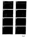
- the expression of YFPwas studied under the influence of concomitant transfected sequence homologous dsRNA. The evaluation under the fluorescence microscope at the earliest 3 hours after injection by green fluorescence.
- the cellswere cultivated in DMEM with 4.5 g / l glucose, 10% fetal calf serum (FCS), 2 mM L-glutamine, penicillin / streptomycin (100 IU / 100 ug / ml, biochrome) in the incubator under 5% CO 2 atmosphere at 37 ° C. The cells were passaged every 3 days to keep them in the exponential growth phase. One day prior to transfection, the cells were trypsinized (10x trypsin / TEDTA, Biochrom) and 0.3x10 5 cell density in coated petri dishes (CORNING® Cell Culture Dish, 35mm, Corning Inc., Corning, Conn. USA).
- Petri disheswere incubated with 0.2% gelatin (Biochrom) for at least 30 minutes at 37 ° C, washed once with PBS and immediately used for seeding the cells.
- CELLocate Coverslipsfrom Eppendorf (square size 55 ⁇ m) were used.
- the Petri disheswere removed from the incubator for about 10 minutes. Approximately 50 cells were microinjected per dish and batch (FemtoJet, micromanipulator 5171, Eppendorf).
- glass capillaries(FemtoTip) from Eppendorf with a tip internal diameter of 0.5 ⁇ m were used. The injection time was 0.8 seconds and the pressure 30 hPa.
- the microinjectionswere performed on an Olympus IX50 microscope with fluorescence device.
- the injection buffer usedwas 14 mM NaCl, 3 mM KCl, 10 mM KH 2 PO 4 , pH 7.0 containing 0.01 ⁇ g / ⁇ l pcDNA-YFP.
- the intracellular YFP fluorescencewas evaluated on the microscope: simultaneously red and green fluorescent cells: microinjection was successful, no inhibition of YFP expression by dsRNA is observed; or they are control cells into which no dsRNA has been injected; only red-fluorescent cells: microinjection was successful, the dsRNA inhibits YFP expression.
- the effectiveness of inhibiting YFP expression after transiently transfecting a YFP-encoding plasmid based on RNA interference with dsRNAscan be modulated by designing the 3 'ends and the length of the base-paired region.
- transiently transfected NIH / 3T3 cellsfibroblasts from NIH Swiss mouse embryo, ECCAC (European collection of animal cell culture) No. 93061524) and HELA-S3 (human cervical carcinoma cells, DSMZ (German Collection of Microorganisms and Cell Cultures) No. ACC 161).
- the plasmid pcDNA-YFPwas used, which contains an 800 bp Bam HI / Eco RI-YFP fragment in the corresponding cleavage sites of the vector pcDNA3.
- Double-stranded RNAsderived from the sequence of yellow fluorescent protein (YFP) were prepared and transiently transfected into the fibroblasts together with the plasmid pcDNA-YFP (The specific dsRNAs used are complementary in their antisense strands to corresponding portions of the gene sequences of both YFP and GFP). After 48 hours, the fluorescence decrease was quantified. The controls were cells transfected with either pcDNA-YFP or pcDNA-YFP alone and a control dsRNA (not derived from the YFP sequence).
- RNA synchizerExadite 8909, Applied Biosystems, Meiterstadt, Germany
- conventional chemical methodsthe RNA single strands and complementary single strands shown in the sequence protocols were synthesized. Subsequently, the crude synthesis products were purified by means of HPLC.
- the hybridization of the single strands to the double strandwas carried out by heating the stoichiometric mixture of the single strands in 10 mM sodium phosphate buffer, pH 6.8, 100 mM NaCl, to 80-90 ° C and subsequent slow cooling to room temperature over 6 hours.
- the cellswere passaged every 3 days. Twenty-four hours before the transfection was carried out, the cells were trypsinized (10 ⁇ trypsin / EDTA, Biochrom, Germany) and with a cell density of 1.0 ⁇ 10 4 cells / well in a 96-well plate (multi-well 96-well flat-bottomed dish, Labor Schubert & Weiss GmbH) seeded in 150 ul growth medium.
- Transfectionwas performed with Lipofectamine Plus TM Reagent (Life Technologies) according to the manufacturer's instructions. 0.15 ⁇ g of pcDNA-YFP plasmid were used per well. The total transfection volume was 60 ⁇ l. In each case 3-fold samples were prepared.
- the plasmid DNAwas first complexed with the dsRNA. For this purpose the plasmid DNA and dsRNA was diluted in serum-free medium and per 0.1 ⁇ g of plasmid DNA 1 ⁇ l PLUS Reagent employed incubated (in a volume of 10 ⁇ l) and, after mixing for 15 minutes at room temperature.
- the fluorescence of the cellson the fluorescence microscope (IX50-S8F2, fluorescence unit U-ULS100Hg, burner U-RFL-T200, Olympus) with a USH-I02D mercury lamp (USHIO Inc., Tokyo, Japan) equipped with a WIB fluorescent cube and a digital CCD camera (Orca IIIm, Hamamatsu) and C4742-95 camera controller).
- the evaluation of the fluorescence imageswas carried out with the analysis software 3.1 (Soft Imaging System GmbH, Germany).
- a nuclear staining(Hoechst-staining) was carried out- For this purpose, the cells in 100 .mu.l Methylcarnoy (75% methanol, 25% glacial acetic acid) first for 5 and then again for 10 minutes in Methylcarnoy fixed. After air-drying, the fixed cells were incubated for 30 minutes in the dark with 100 ⁇ l per well of Hoechst dye (75 ng / ml).
- FIGS. 3 to 9summarize the results for the inhibition of YFP expression by dsRNA in cultured cells:
- Figure 3shows that YFP expression is most effectively inhibited following transient cotransfection of mouse fibroblasts with the YFP plasmid and dsRNA specifically targeted against the YFP sequence when the 3 'ends of the 22 and 19 base pairs containing regions of the dsRNAs single-stranded sections of 2 nucleotides (nt).
- the dsRNA S1 with smooth 3 'ends at a concentration of 1 nMshows no inhibitory effects on YFP expression
- the dsRNAsinhibit S7 (19 nucleotide pairs) and S4 (22 nucleotide pairs) with 2nt overhangs at both 3 'ends, the YFP expression by 50 and 70% compared to the corresponding control dsRNAs K3 and K2.
- the blunt-ended dsRNA designated as S1inhibits YFP expression by ⁇ 65%, while the inhibition of YFP expression by the S4 dsRNA ⁇ 93% ( Figure 4).
- the inhibitory effect of the dsRNAs designated S4 and S7is concentration-dependent ( Figures 3 and 4, see also Figure 7).
- Figure 4shows that for efficient suppression of YFP gene expression, single-stranded formation is not necessary at both 3 'ends (sense and antisense strand). To achieve the most effective inhibition of YFP expression, only the 2nt overhang at the 3 'end on the antisense strand is necessary. Thus, the inhibition of YFP expression at a concentration of 1 nM for the two dsRNAs S4 (with 2nt overhangs on both 3 'ends) and S1A / S4B (with a 2nt overhang on the 3' end of the antisense). Stranges) at ⁇ 70%.
- dsRNAdouble-stranded RNA
- the aimis to increase the effectiveness, found in cell cultures, of dsRNA-mediated inhibition of gene expression of target genes for use in vivo . This is achieved by an improved stability of the dsRNAs in the serum and by a consequent prolonged residence time of the molecule in the circulation or the associated increased effective concentration of the functional molecule.
- Serum stability of GFP expression-inhibiting dsRNAswas tested ex vivo in murine and human serum.
- Incubation with human or murine serum with the corresponding dsRNAwas carried out at 37.degree. In each case, 85 ⁇ l of serum were incubated with 15 ⁇ l of 10 ⁇ M dsRNA. After certain incubation times (30 min, 1h, 2h, 4h, 8h, 12h, 24h) the samples were frozen at -80 ° C. As a control, dsRNA without serum (+85 ⁇ l ddH 2 O) and dsRNA with serum at time 0 were used.
- the upper aqueous phase(approximately 200 ⁇ l) was removed and a first DNase I and then subjected to a proteinase K - subjected to digestion: addition of 20 ⁇ l 10xfach DNase I buffer (100 mM Tris, pH 7.5, 25 mM MgCl 2 , 1 mM CaCl 2 ) and 10 U DNase I (D7291, Sigma-Aldrich), 30 min incubation at 37 ° C, re-addition of 6 U DNase I and incubation for a further 20 min at 37 ° C, addition of 5 ⁇ 1 Proteinase K (20 mg / ml, 04-1075, Peqlab, Germany) and incubated at 37 ° C for 30 min.
- 10xfach DNase I buffer100 mM Tris, pH 7.5, 25 mM MgCl 2 , 1 mM CaCl 2
- 10 U DNase ID7291, Sigma-Aldrich
- a phenol extractionwas performed.
- 500 .mu.l of phenol: chloroform: IAA25: 24: 1 was added, vortexed for 30 sec at the highest stage, 10 min at 12,000xg, 4 ° C, centrifuged, the supernatant removed and successively with 40 ul 3 M Na Ac (Sodium acetate), pH 5.2, and 1 ml of 100% EtOH, in between mixed well and precipitated for at least 1 h at -80 ° C.
- the precipitatewas pelleted by centrifugation at 12,000xg for 30 min and 4 ° C, washed with 70% EtOH and recentrifuged (10 min, 12,000xg, 4 ° C).
- RNA gel loading7 M urea, 1 x TBE (0.09 M Tris-borate, 0.002 M EDTA (ethylene diamine tetraacetate), 0.02% (w / v) bromophenol blue, 0.02% (w / v) xylene cyanol) and stored until gel application at -20 ° C.
- urea gel electrophoresisan analytical, denaturing polyacrylamide gel electrophoresis (analytical PAGE) was performed.
- the urea gelswere prepared shortly before run: 7M urea (21 g) was dissolved in 25 ml of 40% aqueous acrylamide / bisacrylamide stock solution (Rotiphoresis gel, A515.1, Roth) and 5 ml of 10x TBE (108 g Tris, 55 g boric acid , 9.3 g EDTA per L distilled water) was dissolved with stirring and made up to 50 ml with Aqua the more.
- TEMEDN, N, N ', N'-tetramethylethylenediamine
- 10% APSammonium peroxydisulfate
- the runwas for about 2 hours at a constant current flow of 40 mA.
- the gelwas stained for 30 min at RT (room temperature) with Stains all staining solution (20 ml Stains all stock solution (200 mg stains all dissolved in 200 ml formamide) with 200 ml distilled water and 180 ml formamide) and stained Then background dyeing by rinsing in distilled water. for 45 min away.
- the gelswere photographed with the Photocopy System Image Master VDS from Pharmacia.
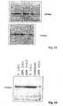
- dsRNAs without single-stranded regions at the 3 'endsare significantly more stable in human and mouse serum than dsRNAs with single-stranded 2nt overhangs at the 3' ends ( Figures 10 to 14 and 17).
- dsRNAs with 2nt overhangs at both 3 'endssignificantly decrease the stability in human and murine serum.
- S7Figures 12 and 13
- K3Figure 14
- dsRNAAs an optimal compromise with respect to the biological efficacy of dsRNA, the use of dsRNA with a smooth end and a single-stranded region of 2 nucleotides may be considered, with the single-stranded overhang at the 3 'end of the antisense strand.
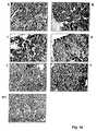
- GFP laboratory miceexpressing the Green Fluorescent Protein (GFP) in all protein biosynthetic cells, double-stranded RNA (dsRNA) derived from the GFP sequence, and nonspecific dsRNA, respectively, were intravenously injected into the tail vein. At the end of the experiment, the animals were killed and analyzed GFP expression in tissue sections and plasma.
- dsRNAdouble-stranded RNA
- RNA synthesizerExedite 8909, Applied Biosystems, Rothstadt, Germany
- the crude synthesis productswere purified by means of HPLC.
- the columns usedwere NucleoPac PA-100, 9x250 mm from Dionex; as low-salt buffer 20 mM Tris, 10 mM NaClO 4 , pH 6.8, 10% acetonitrile and as high-salt buffer 20 mM Tris, 400 mM NaClO 4 , pH 6.8, 10% acetonitrile.
- the flowwas 3 ml / minute.
- the hybridization of the single strands to the double strandwas carried out by heating the stoichiometric mixture of the single strands in 10 mM sodium phosphate buffer, pH 6, 8, 100 mM NaCl, to 80-90 ° C and subsequent slow cooling for 6 hours to room temperature.
- the transgenic laboratory strain TgN (GFPU) 5Nagy(The Jackson Laboratory, Bar Harbor, ME, USA) was used which expresses GFP (with a beta-actin promoter and a CMV intermediate early enhancer) in all cells examined so far (Hadjantonakis AK et al., 1993, Mech. Dev. 76: 79-90; Hadjantonakis AK et al., 1998 Nature Genetics 19: 220-222).
- GFP transgenic micecan be differentiated from the corresponding wild type (WT) by fluorescence (with a UV hand lamp). For breeding each of the corresponding WT was mated with a heterozygous GFP type.
- the experimentwas carried out according to the German animal welfare regulations.
- the animalswere kept under controlled environmental conditions in groups of 3-5 animals in type III Makrolon cages from Ehret, Emmendingen, at a constant temperature of 22 ° C. and a light-dark rhythm of 12 h.
- the animalsreceived tap water and standard feed Altromin 1324 pelleted (Altromin) ad libitum.
- RNA isolation in RNAeasy-Protect(Qiagen).
- the bloodwas kept on ice immediately after removal for 30 min, mixed, centrifuged for 5 min at 2000 rpm (Mini spin, Eppendorf), the supernatant was removed and stored at -80 ° C. (referred to here as plasma).
- tissue pieceswere dehydrated in an ascending alcohol series at RT (room temperature): 40 min each 70 min methanol, 80% methanol, 2 x 96% methanol and 3 x 100% isopropanol. Thereafter, the tissues were heated in 100% isopropanol to 60 ° C in the incubator, then incubated for 1h in an isopropanol / paraffin mixture at 60 ° C and 3 x for 2h in paraffin and then embedded in paraffin.
- tissue sections of 3 ⁇ m cut thicknesswere prepared with a rotary microtome (Leica), mounted on slides (superfrost, bird) and incubated for 30 min at 60 ° C. in an incubator.
- the snailswere deparaffinized 3 x 5 min in xylene, rehydrated in a descending alcohol series (3 x 3 min 100% ethanol, 2 x 2 min 95% ethanol) and then in 3% H 2 O 2 / methanol for 20 min to block endogenous peroxidases All incubation steps were subsequently carried out in a moist chamber. After washing with PBS for 3 ⁇ 3 minutes, the first antibody (goat anti-GFP, sc-5384, Santa Cruz Biotechnology) was incubated 1: 500 in 1% BSA / PBS overnight at 4 ° C.
- Hematoxylin III according to Gillwas used as a nuclear counterstain. After dehydration in an ascending alcohol series and 3 x 5 min xylene, the sections were washed with Entellan (Merck) set. The microscopic evaluation of the staining was carried out with the IX50 microscope from Olympus, equipped with a CCD camera (Hamamatsu).
- isolation buffer50 mM HEPES, pH 7.5, 150 mM NaCl, 1 mM EDTA, 2.5 mM EGTA, 10% glycerol, 0.1% Tween, 1 mM DTT, 10 mM ⁇ -glycerol-phosohate; 1 mM NaF; 0.1 mM Na 3 VO 4 with a protease inhibitor tablet "Complete” from Roche
- DIAX 900dispersing tool 6G, Heidolph
- the electrophoretic separation of the proteinswas carried out in a Multigel Long electrophoresis chamber from Biometra with a denaturing, discontinuous 15% SDS-PAGE (polyacrylamide gel electrophoresis) according to Lämmli (Nature 277: 680-685, 1970).
- a separating gelwas first cast with 1.5 mm thickness: 7.5 ml of acrylamide / bisacrylamide (30%, 0.9%), 3.8 ml of 1.5 M Tris / HCl, pH 8.4, 150 ul 10 % SDS, 3.3 ml double-distilled water, 250 ⁇ l ammonium persulphate (10%), 9 ⁇ l TEMED (N, N, N ', N'-tetramethylenediamine) and until fully polymerized with 0.1% SDS overlays.
- the collecting gelwas poured: 0.83 ⁇ l of acrylamide / bisacrylamide (30% / 0.9%), 630 ⁇ l of 1 M Tris / HCl, pH 6.8, 3.4 ml of bidistilled water. , 50 ⁇ l 10% SDS, 50 ⁇ l 10% ammonium persulphate, 5 ⁇ l TEMED.
- the proteinswere mixed with an appropriate amount of 4X sample buffer (200 mM Tris, pH 6.8, 4% SDS, 100 mM DTT (dithiothreitol), 0.02% bromophenol blue, 20% glycerol) Denatured for 5 min in the heating block at 100 ° C, after cooling on ice briefly spun down and applied to the gel.
- 4X sample buffer200 mM Tris, pH 6.8, 4% SDS, 100 mM DTT (dithiothreitol), 0.02% bromophenol blue, 20% glycerol
- the same amounts of plasma or proteinwere used per lane (3 l plasma each and 25 ⁇ g total protein, respectively).
- the electrophoresiswas carried out at RT under water and constant at 50 V.
- the length standard usedwas the Bio-Rad protein gel marker (Kaleidoscope Prestained Standard).
- the transfer of the proteins from SDS-PAGE to a PVDF (polyvinylidene fluoride) membrane (Hybond-P, Amersham)was carried out in a semidry method according to Kyhse-Anderson (J. Biochem. Biophys., Methods 10: 203-210, 1984) at RT and a constant current of 0, 8 mA / cm 2 for 1.5 h.
- the transfer buffer usedwas a tris / glycine buffer (39 mM glycine, 46 mM Tris, 0.1% SDS and 20% methanol).
- FIGS. 18 to 20show the inhibition of GFP expression after intravenous injection of specific GFP-directed dsRNA with immunoperoxidase stains against GFP on 3 ⁇ m paraffin sections.
- GFP-targeted dsRNAwith a double-stranded region of 22 nucleotide (nt) pairs with no overhangs at the 3 'ends (D) and the corresponding nonspecific control dsRNA (B) and GFP-specific dsRNA with a 19 nucleotide pair extensive double-stranded region with 2nt overhangs at the 3 'ends (E) and the corresponding nonspecific control dsRNA (C) applied at 12 hour intervals over 5 days.
- FIG. 21shows the inhibition of GFP expression in the plasma, in FIG. 22 in the kidney and in FIG. 23 in the heart. In Fig. 23 total protein isolates from different animals are plotted. In each case the same total protein amounts per lane were applied.
- dsRNAanimals of groups B and C
- GFP expressionis not reduced in animals that did not receive any dsRNA.
- the EGFR familyconsists of 4 members, EGFR (ErbB1), HER2 (ErbB2), HER3 (ErbB3) and HER4 (ErbB4) with a transmembrane domain, a cysteine-rich extracellular domain and an intracellular catalytic domain.
- the sequence of EGFR, a 170 kDa protein,has been known since 1984 (Ullrich et al., 1984).
- the EGFRis activated by peptide growth factors such as EGF, transforming growth factor (TGF ⁇ ), amphiregulin, betacellulin, EGF (EGF-like growth factor) and neuregulins.
- Ligand bindinginduces the formation of homo- or heterodimers with subsequent autophosphorylation of cytoplasmic tyrosines (Ullrich & Schlessinger, 1990, Alroy & Yarden, 1997).
- the phosphorylated amino acidsprovide the binding sites for a variety of proteins involved in the proximal steps of signal transduction in a complex network.
- the EGFRisc is involved in a wide range of tumors and thus a suitable target for therapeutic approaches (Huang & Harari, 1999).
- the mechanisms leading to aberrant EGFR activationmay be due to overexpression, amplification, constitutive activation of mutant receptor forms or autocrine loops (Voldborg et al., 1997).
- Overexpression of the EGFRhas been described for a number of tumors, e.g. Breast cancer (Walker & Dearing, 1999), non-small lung carcinoma (Fontanini et al., 1998), pancreatic carcinoma, colon carcinoma (Salomon et al., 1995), and glioblastomas (Rieske et al., 1998). Especially for malignant glioblastomas, no efficient and specific therapeutic agents are available.
- RNA synthesizerExedite 8909, Applied Biosystems, Rothstadt, Germany
- conventional chemical methodsthe RNA single strands shown in the sequence listing and the single strands complementary to them were synthesized.
- the crude synthesis productswere purified by means of HPLC.
- the flowwas 3 ml / minute.
- the hybridization of the single strands to the double strandwas carried out by heating the stoichiometric mixture of the single strands in 10 mM sodium phosphate buffer, pH 6.8, 100 mM NaCl, to 80-90 ° C and subsequent slow cooling for 6 hours to room temperature.
- the cellswere trypsinized (10x trypsin / EDTA, Biochrom, Germany) and seeded at a cell density of 5 ⁇ 10 5 cells / well in a 6-well plate (6-well dishes, laboratory Schubert & Weiss GmbH) in 1.5 ml growth medium.
- the dsRNAwas applied by transfection with the OLIGOFECTAMINE TM Reagent (Life Technologies) according to the manufacturer's instructions. The total transfection volume was 1 ml.
- the dsRNAwas diluted in serum-free medium: 0.5 ⁇ l of a 20 ⁇ M stock solution for EGFR-directed dsRNA and 9.5 ⁇ l of a 20 ⁇ M stock of nonspecific dsRNA (K1A / K2B) were added per well. diluted with 175 ⁇ l serum-free medium (200 nM dsRNA in the transfection mixture or 10 nM specific EGFR dsRNA).
- the OLIGOFECTAMINE TM Reagentwas also diluted in serum-free medium: 3 ⁇ l per well of 12 ⁇ l medium and then incubated for 10 min at room temperature. Thereafter, the diluted OLIGOFECTAMINE TM reagent was added to the dsRNAs diluted in medium, mixed and incubated for a further 20 min at RT. During the incubation, a medium change was performed. The cells were washed 1 x with 1 ml of serum-free medium and incubated with 800 ul serum-free medium until the addition of dsRNA / OLlGOFECTAMINE TM Reagent in the incubator. After adding 200 ⁇ l of dsRNA / OLIGOFECTAMINE TM reagent per well, the cells were incubated in the incubator until protein isolation.
- the cellswere harvested and protein isolation performed. For this purpose, the medium was removed and the cell monolayer washed once with PBS. After addition of 200 ⁇ l protein isolation buffer (1x protease inhibitor Complete, Roche, 50 mM HEPES, pH 7.5, 150 mM NaCl, 1 mM EDTA, 2.5 mM EGTA, 10% glycerol, 0.1% Tween-20, 1 mM DTT, 10 mM ⁇ -glycerol phosphate, 1 mM NaF, 0.1 mM Na 3 VO 4 ) The cells are detached using a cell scraper, incubated on ice for 10 min, transferred to an Eppendorf tube and stored at -80 ° C for at least 30 min.
- protein isolation buffer1x protease inhibitor Complete, Roche, 50 mM HEPES, pH 7.5, 150 mM NaCl, 1 mM EDTA, 2.5 mM EGTA, 10% glycerol, 0.1% Tween-20, 1 m
- the lysatewas homogenized for 10 sec with a disperser (DIAX 900, dispersing tool 6G, Heidolph Instruments GmbH & Co KG, Schwabach) at level 3, incubated on ice for 10 min and at 14,000xg, 4 ° for 15 min C (3K30, Sigma) centrifuged. With the supernatant, a protein determination according to Bradford was performed with the Roti®-Nanoquant system of Roth (Roth GmbH & Co., Düsseldorf) according to the manufacturer. 200 ⁇ l of protein solution were mixed in a suitable dilution with 800 ⁇ l of 1x working solution and the extinction in half-microcuvettes at 450 and 590 nm against distilled water. measured in a Beckman spectrophotometer (DU 250). For the calibration line corresponding BSA dilutions were used (perfused BSA, Sigma).
- the electrophoretic separation of the proteinswas carried out in a Multigel Long electrophoresis chamber from Biometra with a denaturing, discontinuous 7.5% SDS-PAGE (polyacrylamide gel electrophoresis) according to Lämmli (Nature 277: 680-685, 19970).
- a separating gel with 1.5 mm thicknesswas first cast: 3.75 ml of acrylamide / bisacrylamide (30%, 0.9%), 3.8 ml of 1 M Tris / HCl, pH 8.4, 150 ⁇ l of 10% SDS , 7.15 ml of double-distilled water, 150 .mu.l of ammonium persulfate (10%), 9 .mu.l of TEMED (N, N, N ', N'-tetramethylenediamine) and overcoated with 0.1% SDS until complete polymerization.
- the collection gelwas poured: 0.83 ml of acrylamide / bisacrylamide (30% / 0.9%), 630 ⁇ l of 1 M Tris / HCl, pH 6.8, 3.4 ml of bidistilled water, 50 ⁇ l of 10% SDS, 50 ⁇ l 10% ammonium persulfate, 5 ⁇ l TEMED.
- the protein sampleswere spiked 1: 3 with 4x sample buffer (200 mM Tris, pH 6.8, 4% SDS, 100 mM DTT (dithiothreitol), 0.02% bromophenol blue, 20% glycerol) for 5 Denatured min at 100 ° C, after cooling on ice briefly spun down and applied to the gel. Per lane, 35 ⁇ g total protein was applied. The gel was water-cooled at RT and constant at 50 V. The length standard used was the kaleidoscope protein gel marker (BioRad).
- the transfer of the proteins from SDS-PAGE to a PVDF (polyvinylidene fluoride) membrane (Hybond-P, Amersham)was carried out in a semidry method according to Kyhse-Anderson (J. Biochem. Biophys., Methods 10: 203-210, 1984) at RT and a constant current of 0.5 mA / cm 2 for 1.5 h.
- the transfer buffers usedwere: cathodic buffer (30 mM Tris, 40 mM glycine, 10% methanol, 0.01% SDS, pH 9.4), anodic buffer I (300 mM Tris, pH 10.4, 10% methanol) and anodic buffer II (30 mM Tris, pH 10.4, 10% methanol).
- the blot membranewas incubated for 1 h at RT after transfer to 1% skimmed milk powder / PBS / 0.1% Tween-20. Thereafter, it was washed three times with 0.1% Tween-20 / PBS for 3 minutes. All subsequent ones Antibody incubations and washes were done in 0.1% Tween-20 / PBS. Incubation with the primary antibody (human EGFR extracellular domain, IgG specific, Cat No. AF231, R & D Systems) was carried out on a shaker for 2 h at RT in a concentration of 1.5 ⁇ g / ml.
- the primary antibodyhuman EGFR extracellular domain, IgG specific, Cat No. AF231, R & D Systems
- FIG. 24shows the corresponding Western Blot analysis, which shows that the EGFR gene specifically directed against the EGFR gene with a 2nt overhang at the 3 'end of the antisense strand retains EGFR expression after transfection into U-RNA. 87 significantly inhibit MG cells from the corresponding controls. This inhibition of the expression of an endogenous gene by specific dsRNA thus confirms the results given in Example II for the inhibition of the expression of an artificial gene introduced into the cell after transient transfection. The ES-7 or ES-8-mediated inhibition of EGFR expression is significantly lower.
- the dsRNAs used in FIG. 24are shown in Table 3.
- MDR1multidrug resistance gene 1
- RNA interference experiments4 short double-stranded ribonucleic acids R1-R4 were used, the sequences of which are shown in Table 4).
- the ribonucleic acidsare homologous with portions of the coding sequence of MDR1 (Sequence Listing SQ 30).
- Sequences R1-R3consist of a 22-mer sense and a 24-mer antisense strand, with the resulting double strand at the 3'-end of the antisense strand having a 2-nucleotide overhang (0-22-2).
- the sequence R4corresponds to R1, but it consists of a 19-mer double strand with 2-nucleotide overhangs at each 3'-end (2-19-2).
- sequences shown in Table 4are shown again in the Sequence Listing as sequences SQ141-147, 152, 153, 158.
- the dsRNAswere transfected at a concentration of 175 nM in each case as double batches in the cells, which were seeded the day before in 12-well plates à 3.8 x 10 5 cells / well.
- 93.3 ⁇ l of EC-R bufferTransMessenger Kit, Qiagen, Hilden
- Enhancer-Rper transfection batch
- 3.5 ⁇ l of the respective 20 ⁇ M dsRNAwas added, mixed well and left for 5 minutes at room temperature incubated.
- the transfection batcheswere vigorously mixed for 10 seconds and incubated for 10 minutes at room temperature.
- the mediumwas aspirated from the cells, washed once with phosphate buffered saline (PBS) and 200 ⁇ l of fresh medium without FCS per well was added to the cells.
- PBSphosphate buffered saline
- 100 ⁇ l FCS-free mediumwere pipetted to the transfection batches, mixed, and the mixture was pipetted dropwise to the cells (the dsRNA concentration of 175 ⁇ M is based on 400 ⁇ l medium total volume).
- the dsRNA / trans-messenger complexeswere incubated for 4 hours at 37 ° C with the cells in FCS-free medium. Thereafter, a medium change was performed, the fresh medium containing 10 ⁇ M rifampicin and 10% FCS.
- Controlwas performed using a nonspecific dsRNA sequence that has no homology with the MDR1 gene sequence (K) and a MOCK transfection containing all reagents except dsRNA.
- the cellswere harvested after 24, 48 and 72 hours and the total RNA extracted with the Qiagen RNeasy mini-kit. 10 ⁇ g total RNA of each sample was electrophoresed on a 1% agarose-formaldehyde gel, blotted onto a nylon membrane and probed with 5'- ⁇ 32 P-dCTP randomly labeled, specific probes first against MDR1 and after stripping of the blot hybridized against GAPDH as an internal control and exposed to X-ray films.
- the X-ray filmswere digitized (Image Master, VDS Pharmacia) and quantified with the Image-Quant software. An alignment of the MDR1-specific bands with the corresponding GAPDH bands was performed.
- Figures 25 and 26show Northern blots ( Figures 25a, 26a) with quantitative evaluation of the MDR1-specific bands after alignment with the corresponding GAPDH values ( Figures 25b, 26b).
- Reduction of MDR1 mRNA by up to 55% compared to MOCK transfection and by up to 45% compared to non-specific control transfectionwas observed.
- the dsRNA constructs designated R1, R2, R3Table 4
- R1, R2, R3Table 4
- the R4 dsRNA constructsno significant reduction was observed over the controls after 48 hours ( Figures 26a and 26b).
- Figures 25a and 25bAt 74 h, a significantly greater reduction in MDR1 mRNA levels with R1, R2, and R3 was observed over the controls compared to the 48 h values ( Figures 25a and 25b).
- constructs with a 2nt overhang at the 3 'end of the antisense strand and a double stranded region of 22 nucleotide pairsrelatively independent of the particular MDR1 gene homologous sequence region (after 48 h, Figure 26b) reduce the MDR1 mRNA level more efficient than constructs with 2nt overhangs at the 3 'ends of both strands (antisense and sense strands) and a double stranded region of 19 nucleotide pairs.
- the resultsthus corroborate the inhibition of EGFR gene expression by specific dsRNAs after transfection in U-87 MG cells described in Example IV.
- the Trans Stammionserfizienzwas in a separate experiment using a Texas-Red-labeled DNA oligonucleotide (TexRed-A (GATC) 5 T; also 175 nM transfected) determined ( Figures 27a, 27b,. 400-fold magnification, 48 h after transfection). It was about 50% based on the red fluorescent cells compared to the total cell number. Taking into account the transfection rate of the cells of about 50%, the observed reduction in MDR1 mRNA levels is approximately 45-55% (compared to the controls), suggesting that in all cells with specific dsRNA were successfully transfected, the MDR1 mRNA was almost completely and specifically degraded.
Landscapes
- Health & Medical Sciences (AREA)
- Life Sciences & Earth Sciences (AREA)
- Engineering & Computer Science (AREA)
- Genetics & Genomics (AREA)
- Biomedical Technology (AREA)
- Organic Chemistry (AREA)
- Chemical & Material Sciences (AREA)
- Bioinformatics & Cheminformatics (AREA)
- General Health & Medical Sciences (AREA)
- Biotechnology (AREA)
- Molecular Biology (AREA)
- General Engineering & Computer Science (AREA)
- Wood Science & Technology (AREA)
- Zoology (AREA)
- Veterinary Medicine (AREA)
- Public Health (AREA)
- General Chemical & Material Sciences (AREA)
- Chemical Kinetics & Catalysis (AREA)
- Medicinal Chemistry (AREA)
- Nuclear Medicine, Radiotherapy & Molecular Imaging (AREA)
- Pharmacology & Pharmacy (AREA)
- Animal Behavior & Ethology (AREA)
- Biochemistry (AREA)
- Microbiology (AREA)
- Plant Pathology (AREA)
- Physics & Mathematics (AREA)
- Biophysics (AREA)
- Virology (AREA)
- Oncology (AREA)
- Immunology (AREA)
- Neurology (AREA)
- Neurosurgery (AREA)
- Tropical Medicine & Parasitology (AREA)
- Communicable Diseases (AREA)
- Diabetes (AREA)
- Endocrinology (AREA)
- Psychiatry (AREA)
- Heart & Thoracic Surgery (AREA)
- Hospice & Palliative Care (AREA)
- Cardiology (AREA)
Abstract
Description
Translated fromGermanDie Erfindung betrifft ein Verfahren, eine Verwendung und ein Medikament zur Hemmung der Expression eines Zielgens.The invention relates to a method, a use and a medicament for inhibiting the expression of a target gene.
Aus der WO 99/32619 sowie der WO 00/44895 sind Verfahren zur Hemmung der Expression von medizinisch oder biotechnologisch interessanten Genen mit Hilfe einer doppelsträngigen Ribonukleinsäure (dsRNA) bekannt. Die bekannten Verfahren sind zwar hoch effektiv. Es besteht gleichwohl das Bedürfnis, deren Effizienz weiter zu steigern.WO 99/32619 and WO 00/44895 disclose methods for inhibiting the expression of medically or biotechnologically interesting genes by means of a double-stranded ribonucleic acid (dsRNA). The known methods are indeed highly effective. Nevertheless, there is a need to further increase their efficiency.
Aufgabe der vorliegenden Erfindung ist es, die Nachteile nach dem Stand der Technik zu beseitigen. Es sollen insbesondere ein Verfahren, eine Verwendung und ein Medikament angegeben werden, mit denen eine noch effizientere Hemmung der Expresion eines Zielgens erreichbar ist.The object of the present invention is to eliminate the disadvantages of the prior art. In particular, a method, a use and a medicament are to be indicated with which an even more efficient inhibition of the expression of a target gene can be achieved.
Diese Aufgabe wird durch die Merkmale der Ansprüche 1, 41 und 81 gelöst. Vorteilhafte Ausgestaltungen ergeben sich aus den Merkmalen der Ansprüche 2 bis 40, 42 bis 80 und 82 bis 103.This object is solved by the features of
Mit den erfindungsgemäß beanspruchten Merkmalen wird überraschenderweise eine drastische Erhöhung der Effektivität der Hemmung der Expression eines Zielgens in vitro und in vivo erreicht. Durch die besondere Ausbildung der Enden der dsRNA kann sowohl deren Effizienz bei der Vermittlung der hemmenden Wirkung auf die Expression eines Zielgens als auch deren Stabilität gezielt beeinflusst werden. Durch die Vergößerung der Stabilität wird die wirksame Konzentration in der Zelle erhöht.With the features claimed according to the invention, a dramatic increase in the effectiveness of the inhibition of the expression of a target gene in vitro and in vivo is surprisingly achieved. Due to the special design of the ends of the dsRNA, both their efficiency in mediating the inhibitory effect on the expression of a target gene and their stability can be specifically influenced. Increasing the stability increases the effective concentration in the cell.
Unter einem "Zielgen" im Sinne der Erfindung wird der DNA-Strang der doppelsträngigen DNA in der Zelle verstanden, welcher koplementär zu einem bei der Transkription als Matritze dienenden DNA-Strang einschließlich aller transkibierten Bereiche ist. Bei dem "Zielgen" handelt es sich also im allgemeienen um den Sinnstrang. Der eine Strang bzw. Antisinnstrang (as1) kann komplementär zu einem bei der Expression des Zielgens gebildeten RNA-Transkipt oder deren Prozessierungsprodukt, z .B. eine mRNA, sein. Unter "Einführen" wird die Aufnahme in die Zelle verstanden. Die Aufnahme kann durch die Zelle selbst erfolgen; sie kann auch durch Hilfsstoffe oder Hilfsmittel vermittelt werden. Unter einem "Überhang" wird ein endständiger einzelsträngiger Überstand verstanden, welcher nicht nach Matson & Crick gepaarte Nukleotide aufweist. Unter einer "doppelsträngigen Struktur" wird eine Struktur verstanden, bei der die Nukleotide der Einzelstränge im wesentlichen nach Watson & Crick gepaart sind. Im Rahmen der vorliegenden Erfindung kann eine doppelsträngige Struktur auch einzelne Fehlpaarungen ("Mismatches") aufweisen.A "target gene" in the sense of the invention is understood as meaning the DNA strand of the double-stranded DNA in the cell which is complementary to a DNA strand which serves as a template during transcription, including all transcribed regions is. The "target gene" is therefore generally the thread. The one strand or antisense strand (as1) may be complementary to an RNA transipt formed in the expression of the target gene or its processing product, eg. an mRNA. "Insertion" is understood as inclusion in the cell. The recording can be done by the cell itself; it can also be mediated by adjuvants or adjuvants. By "overhang" is meant a terminal single-stranded supernatant which does not have Matson & Crick paired nucleotides. By a "double-stranded structure" is meant a structure in which the nucleotides of the single strands are essentially paired according to Watson & Crick. In the context of the present invention, a double-stranded structure can also have individual mismatches.
Nach einer besonderes vorteilhaften Ausgestaltung weist die dsRNA I den Überhang am 3'-Ende des einen Strangs bzw. Antisinnstrangs as1 auf. Die dsRNA I kann auch an einem Ende glatt ausgebildet sein. In diesem Fall befindet sich das glatte Ende vorteilhafterweise auf der Seite der dsRNA 1, die das 5'-Ende des einen Strangs (Antsinnstrang; as1). In dieser Ausbildung zeigt die dsRNA I einerseits eine sehr gute Effektivität und andererseits eine hohe Stabilität im lebenden Organismus. Die Effektivität insgesamt in vivo ist hervorragend. Der Überhang ist zweckmäßigerweise aus 1 bis 4 Nukleotiden, vorzugsweise aus 1 oder 2 Nukleotiden, gebildet.According to a particularly advantageous embodiment, the dsRNA I has the overhang at the 3 'end of the one strand or antisense strand as1. The dsRNA I can also be smooth at one end. In this case, the smooth end is advantageously on the side of
Nach einem weiteren Ausgestaltungsmerkmal kann die Effektivität des Verfahrens weiter erhöht werden, wenn zumindest eine entsprechend der erfindungsgemäßen dsRNA I ausgebildete weitere dsRNA II in die Zelle eingeführt wird, wobei der eine Strang oder zumindest ein Abschnitt des einen Strangs der doppelsträngigen Struktur der dsRNA I komplementär zu einem ersten Bereich des Sinnstrangs des Zielgens ist, und wobei ein weiterer Strang oder zumindest ein Abschnitt des weiteren Strangs der doppelsträngigen Struktur der weiteren dsRNA II komplementär zu einem zweiten Bereich des Sinnstrangs des Zielgens ist. Die Hemmung der Expression des Zielgens ist in diesem Fall deutlich gesteigert. Der erste und der zweite Bereich können abschnittsweise überlappen, aneinander grenzen oder auch voneinander beabstandet sein.According to a further embodiment feature, the effectiveness of the method can be further increased if at least one further dsRNA II designed in accordance with the dsRNA I according to the invention is introduced into the cell, wherein the one strand or at least a portion of the one strand of the double-stranded structure of the dsRNA I is complementary to a first region of the sense strand of the target gene, and wherein another strand or at least a portion of the further strand of the double-stranded structure of the further dsRNA II is complementary to a second region of the sense strand of the target gene. The inhibition of the expression of the target gene is significantly increased in this case. The first and the second region may partially overlap, adjoin one another or be spaced apart from one another.
Es hat sich weiter als vorteilhaft erwiesen, wenn die dsRNA I und/oder die weitere dsRNA II eine Länge von weniger als 25 aufeinander folgenden Nukleotidpaaren aufweisen. Als besonders effektiv hat sich eine Länge im Bereich zwischen 19 und 23 Nukleotidpaaren erwiesen. Die Effizienz kann weiter gesteigert werden, wenn an den vorzugsweise aus 19 bis 23 Nukleotidpaaren gebildeten Doppelsträngen einzelsträngige Überhänge von 1 bis 4 Nukleotiden vorhanden sind.It has also proved to be advantageous if the dsRNA I and / or the further dsRNA II have a length of less than 25 consecutive nucleotide pairs. A length in the range between 19 and 23 nucleotide pairs has proven to be particularly effective. The efficiency can be further increased if single-stranded overhangs of 1 to 4 nucleotides are present on the double strands, which are preferably formed from 19 to 23 nucleotide pairs.
Das Zielgen kann nach einem weiteren Ausgestaltungsmerkmal eine der in dem anhängenden Sequenzprotokoll wiedergegebenen Sequenzen SQ001 bis SQ140 aufweisen. Es kann auch aus der folgenden Gruppe ausgewählt sein: Onkogen, Cytokin-Gen, Id-Protein-Gen, Priongen, Gene zur Expression von Angiogenese induzierenden Molekülen, von Adhäsions-Molekülen und Zelloberflächenrezeptoren, Gene von Proteinen, die an metastasierenden und/oder invasiven Prozessen beteiligt sind, Gene von Proteinasen sowie Apoptose- und Zellzyklusregulierende Molekülen sowie Gene zur Expression des EGF-Rezeptors. Beim Zielgen kann es sich insbesondere um das MDR1-Gen handeln. Es kann in diesem Zusammenhang eine der Sequenzen SQ141 - 173 bestehende bzw. ein aus jeweils zusammengehörenden Antisinn (as)- und Sinnsequenzen (ss) kombinierte dsRNA I/II verwendet werden.The target gene may, according to a further embodiment feature, have one of the sequences SQ001 to SQ140 reproduced in the attached sequence listing. It may also be selected from the group consisting of oncogene, cytokine gene, Id protein gene, prion gene, genes for expression of angiogenesis-inducing molecules, adhesion molecules and cell surface receptors, genes of proteins involved in metastatic and / or invasive Processes involved, genes of proteinases as well as apoptosis and cell cycle regulatory molecules and genes for expression of the EGF receptor. The target gene may be, in particular, the MDR1 gene. In this context, it is possible to use one of the sequences SQ141-173 or a dsRNA I / II combined from respectively belonging antisense (as) and sense sequences (ss).
Nach einem weiteren vorteilhaften Ausgestaltungsmerkmal wird die Expression nach dem Prinzip der RNA-Interferenz gehemmt.According to a further advantageous embodiment, the expression is inhibited according to the principle of RNA interference.
Das Zielgen wird zweckmäßigerweise in pathogenen Organismen, vorzugsweise in Plasmodien, exprimiert. Es kann Bestandteil eines Virus oder Viroids, insbesondere eines humanpathogenen Virus oder Viroids, sein. Das Virus oder Viroid kann auch ein tier- oder pflanzenpathogenes Virus oder Viroid sein.The target gene is expediently expressed in pathogenic organisms, preferably in plasmodia. It may be part of a virus or viroid, in particular a human pathogenic virus or viroid. The virus or viroid may also be an animal or phytopathogenic virus or viroid.
Nach einem weiteren Ausgestaltungsmerkmal ist vorgesehen, dass die ungepaarten Nukleotide durch Nukleosidthiophosphate substituiert sind.According to a further embodiment feature, it is provided that the unpaired nucleotides are substituted by nucleoside thiophosphates.
Zumindest ein Ende der dsRNA I/II kann modifiziert werden, um einem Abbau in der Zelle oder einer Dissoziation in die Einzelstränge entgegenzuwirken. Vorteilhafterweise wird dazu der durch die komplementären Nukleotidpaare bewirkte Zusammenhalt der doppelsträngigen Struktur durch mindestens eine chemische Verknüpfung erhöht. Die chemische Verknüpfung kann durch eine kovalente oder ionische Bindung, eine Wasserstoffbrückenbindung, hydrophobe Wechselwirkungen, vorzugsweise van-der-Waals- oder Stapelungswechelwirkungen, oder durch Metall-Ionenkoordination gebildet werden. Es hat sich weiter als zweckmäßig und die Stabilität erhöhend erwiesen, wenn die chemische Verknüpfung in der Nähe des einen Endes gebildet ist. Weitere vorteilhafte Ausgestaltungen hinsichtlich der chemischen Verknüpfung können den Merkmalen der Ansprüche 24 bis 30 entnommen werden, ohne dass es dafür einer näheren Erläuterung bedarf.At least one end of the dsRNA I / II can be modified to counteract degradation in the cell or dissociation into the single strands. Advantageously, the cohesion of the double-stranded structure brought about by the complementary nucleotide pairs is increased by at least one chemical linkage. The chemical linkage may be formed by covalent or ionic bonding, hydrogen bonding, hydrophobic interactions, preferably van der Waals or stacking interactions, or by metal ion coordination. It has further been found useful and increasing stability when the chemical linkage is formed near one end. Further advantageous embodiments with regard to the chemical linkage can be taken from the features of
Die dsRNA I/II kann dann besonders einfach in die Zelle eingeschleust werden, wenn sie in micellare Strukturen, vorteilhafterweise in Liposomen, eingeschlossen wird. Zum Transport der dsRNA I/II in die Zelle hat es sich auch als vorteilhaft erwiesen, dass diese an mindestens ein von einem Virus stammendes, davon abgeleitetes oder ein synthetisch hergestelltes virales Hüllprotein gebunden, damit assoziiert oder davon umgeben werden. Das Hüllprotein kann vom Polyomavirus abgeleitet sein. Das Hüllprotein kann insbesondere das Virus-Protein 1 und/oder das Virus-Protein 2 des Polyomavirus enthalten. Nach einer weiteren Ausgestaltung ist vorgesehen, dass bei Bildung eines Kapsids oder kapsidartigen Gebildes aus dem Hüllprotein die eine Seite zum Inneren des Kapsids oder kapsidartigen Gebildes gewandt ist. Ferner ist es von Vorteil, dass der eine Strang der dsRNA I/II (as1/2) zum primären oder prozessierten RNA-Transkript des Zielgens komplementär ist. Die Zelle kann eine Vertebratenzelle oder eine menschliche Zelle sein.The dsRNA I / II can be particularly easily introduced into the cell when it is encased in micellar structures, advantageously in liposomes. For the transport of the dsRNA I / II into the cell, it has also proved to be advantageous that these are bound to, associated with or surrounded by at least one virus-derived, derived or synthetically produced viral coat protein. The coat protein can be derived from the polyomavirus. In particular, the coat protein may be the
Weiterhin hat es sich gezeigt, dass die dsRNA I/II vorteilhafterweise bereits in einer Menge von höchstens 5 mg/kg Körpergewicht pro Tag einem Säugetier, vorzugsweise einem Menschen, verabreicht werden kann. Bereits in dieser geringen Dosis wird eine ausgezeichnete Effektivität erzielt.Furthermore, it has been shown that the dsRNA I / II advantageously already in a quantity of at most 5 mg / kg body weight per day a mammal, preferably a human, can be administered. Already in this low dose excellent effectiveness is achieved.
Überraschenderweise hat sich gezeigt, dass die dsRNA I/II zur Applikation in eine Pufferlösung aufgenommen und dann oral oder mittels Injektion oder Infusion intravenös, intratumoral, inhalativ, intraperitoneal verabreicht werden kann.Surprisingly, it has been shown that the dsRNA I / II can be taken up for administration in a buffer solution and then administered orally or by injection or infusion intravenously, intratumorally, inhalatively, intraperitoneally.
Erfindungsgemäß ist weiterhin die Verwendung einer doppelsträngigen Ribonukleinsäure (dsRNA I) zur Hemmung der Expression eines Zielgens in einer Zelle vorgesehen, wobei die dsRNA I eine doppelsträngige aus höchstens 49 aufeinander folgenden Nukleotidpaaren gebildete Struktur aufweist, und wobei ein Strang (Antisinnstrang; as1) oder zumindest ein Abschnitt des einen Strangs (as1) der doppelsträngigen Struktur komplementär zum Sinnstrang des Zielgens ist, und wobei die dsRNA I zumindest an einem Ende einen aus 1 bis 4 Nukleotiden gebildeten Überhang aufweist.The invention further provides the use of a double-stranded ribonucleic acid (dsRNA I) for inhibiting the expression of a target gene in a cell, wherein the dsRNA I has a double-stranded structure formed from at most 49 consecutive nucleotide pairs, and wherein one strand (antisense strand, as1) or at least a portion of the one strand (as1) of the double-stranded structure is complementary to the sense strand of the target gene, and wherein the dsRNA I has an overhang formed from 1 to 4 nucleotides at least at one end.
Nach weiterer Maßgabe der Erfindung ist ein Medikament zur Hemmung der Expression eines Zielgens in einer Zelle vorgesehen, enthaltend eine doppelsträngige Ribonukleinsäure (dsRNA I) in einer zur Hemmung der Expression des Zielgens ausreichenden Menge, wobei die dsRNA I eine doppelsträngige aus höchstens 49 aufeinander folgenden Nukleotidpaaren Struktur aufweist, und wobei ein Strang (as1) oder zumindest ein Abschnitt des einen Strangs (as1) der doppelsträngigen Struktur komplementär zum Sinnstrang des Zielgens ist, und wobei die dsRNA I zumindest an einem Ende einen aus 1 bis 4 Nukleotiden gebildeten Überhang aufweist.According to another aspect of the invention, there is provided a medicament for inhibiting the expression of a target gene in a cell containing a double-stranded ribonucleic acid (dsRNA I) in an amount sufficient to inhibit the expression of the target gene An amount wherein the dsRNA I has a double stranded structure of at most 49 consecutive nucleotide pairs, and wherein a strand (as1) or at least a portion of the one strand (as1) of the double stranded structure is complementary to the sense strand of the target gene, and wherein the dsRNA I is at least has an overhang formed from 1 to 4 nucleotides at one end.
Wegen der weiteren vorteilhaften Ausgestaltung der dsRNA I/II wird auf die vorangegangenen Ausführungen verwiesen.Because of the further advantageous embodiment of the dsRNA I / II, reference is made to the preceding statements.
Die Erfindung wird nachfolgend anhand der Zeichnungen und Ausführungsbeispiele beispielhaft erläutert. Es zeigen:
- Fig. 1a, b
- schematisch eine erste und zweite doppelsträngige RNA und
- Fig. 2
- schematisch ein Zielgen,
- Fig. 3
- relative YFP-Fluoreszenz nach Applikation verschiedener dsRNA in NIH/3T3-Zellen (erstes Experiment),
- Fig. 4
- relative YFP-Fluoreszenz nach Applikation verschiedener dsRNA in NIH/3T3-Zellen (zweites Experiment),
- Fig. 5
- relative YFP-Fluoreszenz nach Applikation verschiedener dsRNA in NIH/3T3-Zellen (drittes Experiment),
- Fig. 6
- relative YFP-Fluoreszenz nach Applikation verschiedener dsRNA in NIH/3T3-Zellen (viertes Experiment),
- Fig. 7
- relative YFP-Fluoreszenz nach Applikation verschiedener dsRNA in HeLa-S3-Zellen (fünftes Experiment),
- Fig. 8
- fluoreszenzmikroskopische Aufnahmen von NIH/3T3-Zellen nach Transfektion mit pcDNA-YFP bzw nach Kotransfektion mit pcDNA-YFP und verschiedenen dsRNAs,
- Fig. 9
- fluoreszenzmikroskopische Aufnahmen von HeLa-S3-Zellen nach Transfektion mit pcDNA-YFP bzw. nach Kotransfektion mit pcDNA-YFP und verschiedenen dsRNAs,
- Fig. 10
- gelelektrophoretische Auftrennung von S1 nach Inkubation in Maus-Serum,
- Fig. 11
- gelelektrophoretische Auftrennung von S1 nach Inkubation in humanem Serum,
- Fig. 12
- gelelektrophoretische Auftrennung von S7 nach Inkubation in Maus-Serum,
- Fig. 13
- gelelektrophoretische Auftrennung von S7 nach Inkubation in humanem Serum,
- Fig. 14
- gelelektrophoretische Auftrennung von K3 nach Inkubation in Maus-Serum,
- Fig. 15
- gelelektrophoretische Auftrennung von PKC1/2 nach Inkubation in Maus-Serum,
- Fig. 16
- gelelektrophoretische Auftrennung von S1A/S4B nach Inkubation in humanem Serum,
- Fig. 17
- gelelektrophoretische Auftrennung von K2 nach Inkubation in humanem Serum und
- Fig. 18
- GFP-spezifische Immunoperoxidase-Färbung an Nieren-Paraffinschnitten transgener GFP-Mäuse,
- Fig. 19
- GFP-spezifische Immunoperoxidase-Färbung an Herz-Paraffinschnitten transgener GFP-Mäuse,
- Fig. 20
- GFP-spezifische Immunoperoxidase-Färbung an Pankreas-Paraffinschritten transgener GFP-Mäuse,
- Fig. 21
- Western-Blot-Analyse der GFP-Expression im Plasma,
- Fig. 22
- Western-Blot-Analyse der GFP-Expression in der Niere,
- Fig. 23
- Western-Blot-Analyse der GFP-Expression im Herz,
- Fgi. 24
- Western-Blot-Analyse der EGFR-Expression in U-87 MG Glioblastom-Zellen,
- Fig. 25a
- Northern-Blot-Analyse des MDRI mRNA-Niveaus in der Kolonkarzinom-Zelllinie LS174T, wobei die Zellen nach 74 Stunden geerntet wurden,
- Fig. 25b
- Quantifizierung der Banden nach Fig. 25a, wobei die Mittelwerte aus zwei Werten dargestellt sind,
- Fig. 26a
- Northern-Blot-Analyse des MDRI mRNA-Niveaus in der Kolonkarzinom-Zelllinie LS174T, wobei die Zellen nach 48 Stunden geerntet wurden,
- Fig. 26b
- Quantifizierung der Banden nach Fig. 26a, wobei die Mittelwerte aus zwei Werten dargestellt sind,
- Fig. 27
- vergleichende Darstellung einer durchlicht- und fluoreszenzmikroskopischen Aufnahme einer
Transfektion mit 175 nM dsRNA (Sequenz R1 in Tabelle 4).
- Fig. 1a, b
- schematically a first and second double-stranded RNA and
- Fig. 2
- schematically a target gene,
- Fig. 3
- relative YFP fluorescence after application of different dsRNA in NIH / 3T3 cells (first experiment),
- Fig. 4
- relative YFP fluorescence after application of different dsRNA in NIH / 3T3 cells (second experiment),
- Fig. 5
- relative YFP fluorescence after application of different dsRNA in NIH / 3T3 cells (third experiment),
- Fig. 6
- relative YFP fluorescence after application of different dsRNA in NIH / 3T3 cells (fourth experiment),
- Fig. 7
- relative YFP fluorescence after application of different dsRNA in HeLa-S3 cells (fifth experiment),
- Fig. 8
- fluorescence micrographs of NIH / 3T3 cells after transfection with pcDNA-YFP or after cotransfection with pcDNA-YFP and various dsRNAs,
- Fig. 9
- fluorescence microscopic images of HeLa-S3 cells after transfection with pcDNA-YFP or after cotransfection with pcDNA-YFP and various dsRNAs,
- Fig. 10
- gel electrophoretic separation of S1 after incubation in mouse serum,
- Fig. 11
- gel electrophoretic separation of S1 after incubation in human serum,
- Fig. 12
- gel electrophoretic separation of S7 after incubation in mouse serum,
- Fig. 13
- gel electrophoretic separation of S7 after incubation in human serum,
- Fig. 14
- gel electrophoretic separation of K3 after incubation in mouse serum,
- Fig. 15
- gel electrophoresis separation of PKC1 / 2 after incubation in mouse serum,
- Fig. 16
- gel electrophoretic separation of S1A / S4B after incubation in human serum,
- Fig. 17
- gel electrophoretic separation of K2 after incubation in human serum and
- Fig. 18
- GFP-specific immunoperoxidase staining on renal paraffin sections of transgenic GFP mice,
- Fig. 19
- GFP-specific immunoperoxidase staining on cardiac paraffin sections of transgenic GFP mice,
- Fig. 20
- GFP-specific immunoperoxidase staining on pancreatic paraffin steps of transgenic GFP mice,
- Fig. 21
- Western blot analysis of GFP expression in plasma,
- Fig. 22
- Western blot analysis of GFP expression in the kidney,
- Fig. 23
- Western blot analysis of GFP expression in the heart,
- Fgi. 24
- Western blot analysis of EGFR expression in U-87 MG glioblastoma cells,
- Fig. 25a
- Northern blot analysis of MDRI mRNA levels in colon carcinoma cell line LS174T harvesting cells after 74 hours,
- Fig. 25b
- Quantification of the bands of Fig. 25a, the mean values being represented by two values,
- Fig. 26a
- Northern blot analysis of MDRI mRNA levels in colon carcinoma cell line LS174T, harvesting the cells after 48 hours,
- Fig. 26b
- Quantification of the bands of Fig. 26a, the mean values being represented by two values,
- Fig. 27
- comparative representation of a transillumination and fluorescence micrograph of a transfection with 175 nM dsRNA (sequence R1 in Table 4).
Die in den Fig. 1a und 1b schematisch gezeigten doppelsträngigen Ribonukleinsäuren dsRNA I und dsRNA II weisen jeweils ein ersces Ende E1 und ein zweites Ende E2 auf. Die erste und die zweite Ribonukleinsäure dsRNA I/dsRNAII weisen an ihren beiden Enden E1 und E2 einzelsträngige, aus etwa 1 bis 4 ungepaarten Nukleotiden gebildete Abschnitts auf. Es sind zwei mögliche Varianten dargestellt (Variante 1 und 2), wobei Variante 2 ein glattes Ende (E2) aufweist. Das glatte Ende kann jedoch auch in einer weiteren Variante am anderen Ende (E1) liegen.The double-stranded ribonucleic acids dsRNA I and dsRNA II shown schematically in FIGS. 1 a and 1 b each have an
In Fig. 2 ist schematisch ein auf einer DNA befindliches Zielgen gezeigt. Das Zielgen ist durch einen schwarzen Balken kenntlich gemacht. Es weist einen ersten Bereich B1 und einen zweiten Bereich B2 auf.FIG. 2 shows schematically a target gene located on a DNA. The target gene is indicated by a black bar. It has a first area B1 and a second area B2.
Jeweils der eine Strang der ersten dsRNA I (as1) bzw. der zweiten dsRNA II (as2) ist komplementär zum entsprechenden Bereich B1 bzw. B2 auf dem Zielgen.In each case one strand of the first dsRNA I (as1) or the second dsRNA II (as2) is complementary to the corresponding region B1 or B2 on the target gene.
Die Expression des Zielgens wird dann besonders wirkungsvoll gehemmt, wenn die dsRNA I/dsRNA II an ihren Enden E1, E2 einzelsträngige Abschnitte aufweist. Die einzelsträngigen Abschnitte können sowohl am Strang as1 oder as2 als auch am Gegenstrang (ss1 bzw. ss2) oder am Strang as1, as2 und am Gegenstrang ausgebildet sein.The expression of the target gene is particularly effectively inhibited when the dsRNA I / dsRNA II has single-stranded segments at its ends E1, E2. The single-stranded sections can be formed both on the strand as1 or as2 and on the opposite strand (ss1 or ss2) or on the strand as1, as2 and on the opposite strand.
Die Bereiche B1 und B2 können, wie in Fig. 2 gezeigt, von einander beabstandet sein. Sie können aber auch aneinander grenzen oder überlappen.The areas B1 and B2 may be spaced apart as shown in FIG. But you can also border or overlap.
Es wurden aus Sequenzen des Yellow Fluorescent Proteine (YFP), einer Variante des GFP (Grün-fluoreszierendes Protein) der AlgeAequoria victoria abgeleitete doppelsträngige RNAs (dsRNAs) hergestellt und zusammen mit einem YFP-kodierenden Plasmid in Fibroblasten mikroinjiziert. Anschließend wurde die Fluoreszenzabnahme gegenüber Zellen ohne dsRNA ausgewertet.Double -stranded RNAs (dsRNAs) derived from sequences of the Yellow Fluorescent Proteins (YFP), a variant of the GFP (green fluorescent protein) of the AlgeAequoria victoria , were produced and microinjected together with a YFP-encoding plasmid into fibroblasts. Subsequently, the fluorescence decrease compared to cells without dsRNA was evaluated.
Mittels eines RNA-Synthesizer (Typ Expedite 8909, Applied Biosystems, Weiterstadt, Deutschland) und herkömmlicher chemischer Verfahren wurden die aus den Sequenzprotokollen SQ148, 149 und SQ159 ersichtlichen RNA-Einzelstränge und die zu ihnen komplementären Einzelstränge synthetisiert. Anschließend erfolgte die Reinigung mit Hilfe der HPLC. Die Hybridisierung der Einzelstränge zum Doppelstrang erfolgte durch Erhitzen des stöchiometrischen Gemischs der Einzelstränge in 10 mM Natriumphosphatpuffer, pH 6,8, 100 mM NaCl, auf 90°C und nachfolgendes langsames Abkühlen über 6 Stunden auf Raumtemperatur. Die so erhaltenen dsRNAs wurden in die Testzellen mikroinjiziert.By means of an RNA synthesizer (Expedite 8909, Applied Biosystems, Weiterstadt, Germany) and conventional chemical methods, the RNA single strands and the single strands complementary to them from the sequence protocols SQ148, 149 and SQ159 were synthesized. Subsequently, the purification was carried out with the aid of HPLC. The hybridization of the single strands to the double strand was carried out by heating the stoichiometric mixture of the single strands in 10 mM sodium phosphate buffer, pH 6.8, 100 mM NaCl, to 90 ° C and then slowly cooling to room temperature over 6 hours. The resulting dsRNAs were microinjected into the test cells.
Als Testsystem für diese Zellkultur-Experimente diente die murine Fibroblasten-Zellinie NIH/3T3, ECACC No. 93061524 (European Collection of Animal Cell Culture). Für die Mikroinjektionen wurde das Plasmid pcDNA-YFP verwendet, das ein 800bp großes Bam HI/Eco RI-YFP-Fragment in den entsprechenden Restriktionsschnittstellen des Vectors pcDNA3 enthält. Die Expression des YFP wurde unter dem Einfluß gleichzeitig mittransfizierter sequenzhomologer dsRNA untersucht. Die Auswertung unter dem Fluoreszenzmikroskop erfolgte frühestens 3 Stunden nach Injektion anhand der grünen Fluoreszenz.The test system used for these cell culture experiments was the murine fibroblast cell line NIH / 3T3, ECACC no. 93061524 (European Collection of Animal Cell Culture). For the microinjections, the plasmid pcDNA-YFP was used, which contains a 800 bp Bam HI / Eco RI-YFP fragment in the corresponding restriction sites of the vector pcDNA3. The expression of YFP was studied under the influence of concomitant transfected sequence homologous dsRNA. The evaluation under the fluorescence microscope at the earliest 3 hours after injection by green fluorescence.
Die Kultivierung der Zellen erfolgte in DMEM mit 4,5 g/l Glucose, 10 % fötalem Kälberserum (FCS), 2 mM L-Glutamin, Penicillin/Streptomycin (100 IE/100 µg/ml, Biochrom) im Brutschrank unter 5 % CO2-Atmosphäre bei 37°C. Die Zellen wurden alle 3 Tage passagiert, um sie in der exponentiellen Wachstumsphase zu halten. Einen Tag vor der Durchführung der Transfektion wurden die Zellen trypsiniert (10x Trypsin/TEDTA, Biochrom) und mit einer Zelldichte von 0,3 x 10⁵ Zellen in beschichteten Petrischalen (CORNING® Cell Culture Dish, 35 mm, Corning Inc., Corning, USA) ausgesät. Die Petrischalen wurden mit 0,2 % Gelatine (Biochrom) für mindestens 30 Minuten bei 37°C inkubiert, einmal mit PBS gewaschen und sofort für die Aussaat der Zellen verwendet. Um ein Wiederfinden individueller Zellen zu ermöglichen, wurden CELLocate Coverslips der Fa. Eppendorf (Square size 55 µm) verwendet.The cells were cultivated in DMEM with 4.5 g / l glucose, 10% fetal calf serum (FCS), 2 mM L-glutamine, penicillin / streptomycin (100 IU / 100 ug / ml, biochrome) in the incubator under 5% CO2 atmosphere at 37 ° C. The cells were passaged every 3 days to keep them in the exponential growth phase. One day prior to transfection, the cells were trypsinized (10x trypsin / TEDTA, Biochrom) and 0.3x10⁵ cell density in coated petri dishes (CORNING® Cell Culture Dish, 35mm, Corning Inc., Corning, Conn. USA). The Petri dishes were incubated with 0.2% gelatin (Biochrom) for at least 30 minutes at 37 ° C, washed once with PBS and immediately used for seeding the cells. In order to enable a retrieval of individual cells, CELLocate Coverslips from Eppendorf (square size 55 μm) were used.
Zur Durchführung der Mikroinjektion wurden die Petrischalen ca. 10 Minuten aus dem Brutschrank genommen. Pro Schale und Ansatz wurden ca. 50 Zellen mikroinjiziert (FemtoJet; Mikromanipulator 5171, Eppendorf). Für die Mikroinjektion wurden Glaskapillaren (FemtoTip) der Firma Eppendorf mit einem Spitzeninnendurchmesser von 0,5 µm verwendet. Die Injektionsdauer betrug 0,8 Sekunden und der Druck 30 hPa. Durchgeführt wurden die Mikroinjektionen an einem Olympus IX50 Mikroskop mit Fluoreszenzeinrichtung. Als Injektionspuffer wurde 14 mM NaCl, 3 mM KCl, 10 mM KH2PO4, pH 7,0 verwendet, der 0,01 µg/µl pcDNA-YFP enthielt. Zur Überprüfung einer erfolgreichen Mikroinjektion wurde der Injektionslösung jeweils 0,08% (w/v) an Dextran-70000 gekoppeltes Texas-Rot (Molecular Probes, Leiden, Niederlande) zugesetzt. Um die Inhibition der YFP-Expression mit spezifischer dsRNA zu untersuchen, wurden der Injektionslösung dsRNAs zugegeben: Ansatz 1: 0,1 µM dsRNA (Sequenzprotokoll SQ148/149); Ansatz 2: 0,1 µM dsRNA (Sequenzprotokoll SQ148/159); Ansatz 3: ohne RNA. Nach der Mikroinjektion wurden die Zellen für mindestens drei weitere Stunden im Brutschrank inkubiert. Danach wurden die intrazelluläre YFP-Fluoreszenz am Mikroskop ausgewertet: gleichzeitig rot und grün-fluoreszierende Zellen: Mikroinjektion war erfolgreich, es wird keine Inhibition der YFP-Expression durch dsRNA beobachtet; bzw. es handelt sich um Kontrollzellen, in die keine dsRNA injiziert wurde; nur rot-fluoreszierende Zellen: Mikroinjektion war erfolgreich, die dsRNA inhibiert YFP-Expression.To carry out the microinjection, the Petri dishes were removed from the incubator for about 10 minutes. Approximately 50 cells were microinjected per dish and batch (FemtoJet, micromanipulator 5171, Eppendorf). For microinjection, glass capillaries (FemtoTip) from Eppendorf with a tip internal diameter of 0.5 μm were used. The injection time was 0.8 seconds and the pressure 30 hPa. The microinjections were performed on an Olympus IX50 microscope with fluorescence device. The injection buffer used was 14 mM NaCl, 3 mM KCl, 10 mM KH2 PO4 , pH 7.0 containing 0.01 μg / μl pcDNA-YFP. To test for a successful microinjection, 0.08% (w / v) of dextran-70000 coupled Texas Red (Molecular Probes, Leiden, Netherlands) was added to each of the injection solutions. To study the inhibition of YFP expression with specific dsRNA, the Injection solution dsRNA added: batch 1: 0.1 μM dsRNA (sequence listing SQ148 / 149); Batch 2: 0.1 μM dsRNA (Sequence Listing SQ148 / 159); Approach 3: without RNA. After microinjection, cells were incubated in the incubator for at least three additional hours. Thereafter, the intracellular YFP fluorescence was evaluated on the microscope: simultaneously red and green fluorescent cells: microinjection was successful, no inhibition of YFP expression by dsRNA is observed; or they are control cells into which no dsRNA has been injected; only red-fluorescent cells: microinjection was successful, the dsRNA inhibits YFP expression.
Bei einer dsRNA-Konzentration von 0,1µM konnte beim Einsatz der dsRNA mit den an beiden 3'-Enden um je zwei Nukleotide überstehenden Einzelstrangbereichen (Sequenzprotokoll SQ148/159) eine merklich erhöhte Hemmung der Expression des YFP-Gens in Fibroblasten beobachtet werden im Vergleich zur dsRNA ohne überstehende Einzelstrangenden (Tabelle 1).At a dsRNA concentration of 0.1μ M, a markedly increased inhibition of the expression of the YFP gene in fibroblasts could be observed when using the dsRNA with the single strand regions projecting at two 3 'ends by two nucleotides each (sequence listing SQ148 / 159) compared to dsRNA without supernatants (Table 1).
Die Verwendung von kurzen, 19-25 Basenpaare enthaltenden, dsRNA-Molekülen mit Überhängen aus wenigen, vorzugsweise 1 bis 3 nicht-basengepaarten, einzelsträngigen Nukleotiden ermöglicht somit eine vergleichsweise stärkere Hemmung der Genexpression in Säugerzellen als die Verwendung von dsRNAs mit derselben Anzahl von Basenpaaren ohne die entsprechenden Einzelstrangüberhänge bei jeweils gleichen RNA-Konzentrationen.
Die Effektivität der Inhibition der YFP-Expression nach transienter Transfektion eines YFP-codierenden Plasmids auf der Basis der RNA-Interferenz mit dsRNAs läßt sich durch Gestaltung der 3'-Enden und der Länge des basengepaarten Bereichs modulieren.The effectiveness of inhibiting YFP expression after transiently transfecting a YFP-encoding plasmid based on RNA interference with dsRNAs can be modulated by designing the 3 'ends and the length of the base-paired region.
Zum Wirksamkeitsnachweis der dsRNA bei der spezifischen Inhibition der Genexpression wurden transient transfizierte NIH/3T3-Zellen (Fibroblasten aus NIH Swiss Mausembryo, ECCAC (European collection of animal cell culture) Nr. 93061524) und HELA-S3 (humane cervikale Karzinomzellen, DSMZ (Deutsche Sammlung von Mikroorganismen und Zellkulturen) Nr. ACC 161) verwendet. Für die Transfektion wurde das Plasmid pcDNA-YFP verwendet, das ein 800 bp großes Bam HI /Eco RI-YFP-Fragment in den entsprechenden Schnittstellen des Vektors pcDNA3 enthält. Aus der Sequenz des gelb-fluoreszierenden Proteins (YFP) abgeleitete doppelsträngige RNAs (dsRNAs) wurden hergestellt und zusammen mit dem Plasmid pcDNA-YFP transient in die Fibroblasten transfiziert (Die verwendeten spezifischen dsRNAs sind in ihren Antisinn-Strängen komplementär zu entsprechenden Abschnitten der Gensequenzen von sowohl YFP als auch GFP). Nach 48 Stunden wurde die Fluoreszenzabnahme quantifiziert. Als Kontrollen fungierten Zellen, die entweder nur mit pcDNA-YFP oder mit pcDNA-YFP und einer Kontroll-dsRNA (nicht aus der YFP-Sequenz abgeleitet) transfiziert wurden.To demonstrate the efficacy of the dsRNA in the specific inhibition of gene expression, transiently transfected NIH / 3T3 cells (fibroblasts from NIH Swiss mouse embryo, ECCAC (European collection of animal cell culture) No. 93061524) and HELA-S3 (human cervical carcinoma cells, DSMZ (German Collection of Microorganisms and Cell Cultures) No. ACC 161). For transfection, the plasmid pcDNA-YFP was used, which contains an 800 bp Bam HI / Eco RI-YFP fragment in the corresponding cleavage sites of the vector pcDNA3. Double-stranded RNAs (dsRNAs) derived from the sequence of yellow fluorescent protein (YFP) were prepared and transiently transfected into the fibroblasts together with the plasmid pcDNA-YFP (The specific dsRNAs used are complementary in their antisense strands to corresponding portions of the gene sequences of both YFP and GFP). After 48 hours, the fluorescence decrease was quantified. The controls were cells transfected with either pcDNA-YFP or pcDNA-YFP alone and a control dsRNA (not derived from the YFP sequence).
Mittels eines RNA-Synchesizers (Typ Expedite 8909, Applied Biosystems, Meiterstadt, Deutschland) und herkömmlicher chemischer Verfahren wurden die aus den Sequenzprotokollen ersichtlichen RNA-Eintelstränge und die zu ihnen komplementären Einzelstränge synthetisiert. Anschließend erfolgte die Reinigung der rohen Syntheseprodukte mit Hilfe der HPLC. Verwendet wurde die Säule NucleoPac PA-100, 9x250 mm, der Fa. Dionex; als Niedersalz-Puffer 20 mM Tris, 10 mM NaClO4, pH 6,8, 10% Acetonitril und als Hochsalz-Puffer 20 mM Tris, 400 mM NaClO4, pH 6,8, 10% Acetonitril. Der Fluß betrug 3 ml/ Minute. Die Hybridisierung der Einzelstränge zum Doppelstrang erfolgte durch Erhitzen des stöchiometrischen Gemischs der Einzelstränge in 10 mM Natriumphosphatpuffer, pH 6,8, 100 mM NaCl, auf 80-90°C und nachfolgendes langsames Abkühlen über 6 Stunden auf Raumtemperatur.By means of an RNA synchizer (Expedite 8909, Applied Biosystems, Meiterstadt, Germany) and conventional chemical methods, the RNA single strands and complementary single strands shown in the sequence protocols were synthesized. Subsequently, the crude synthesis products were purified by means of HPLC. The column NucleoPac PA-100, 9x250 mm, from Dionex, was used; as low-
Alle Zellkulturarbeiten wurden unter sterilen Bedingungen in einer entsprechenden Werkbank (HS18, Hera Safe, Kendro, Heraeus) durchgeführt. Die Kultivierung der NIH/3T3-Zellen und der HELA-S3 erfolgte im Brutschrank (CO2-Inkubator T20, Hera cell, Kendro, Heraeus) bei 37°C, 5% CO2 und gesättigter Luftfeuchtigkeit in DMEM (Dulbecco's modified eagle medium, Biochrom), für die Mausfibroblasten, und Ham's F12 für die HELA-Zellen mit 10% FCS (fetal calf serum, Biochrom), 2 mM L-Glutamin (Biochrom) und Penicillin/Streptomycin (100 IE/100 µg/ml, Biochrom). Um die Zellen in der exponentiellen Wachstumsphase zu halten, wurden die Zellen alle 3 Tage passagiert. 24 Stunden vor der Durchführung der Transfektion wurden die Zellen trypsiniert (10x Trypsin/EDTA, Biochrom, Deutschland) und mit einer Zelldichte von 1,0 x 10⁴ Zellen/Vertiefung in einer 96-Loch-Platte (Multiwell Schalen 96-Well Flachboden, Labor Schubert & Weiss GmbH) in 150 µl Wachstumsmedium ausgesät.All cell culture work was carried out under sterile conditions in a corresponding workbench (HS18, Hera Safe, Kendro, Heraeus). Cultivation of the NIH / 3T3 cells and the HELA-S3 was carried out in the incubator (CO2 incubator T20, Hera cell, Kendro, Heraeus) at 37 ° C, 5% CO2 and saturated Humidity in DMEM (Dulbecco's modified eagle medium, Biochrom) for the mouse fibroblasts, and Ham's F12 for the HELA cells with 10% fetal calf serum (FCS), 2 mM L-glutamine (Biochrom), and penicillin / streptomycin (100 IE / 100 μg / ml, biochrome). In order to keep the cells in the exponential growth phase, the cells were passaged every 3 days. Twenty-four hours before the transfection was carried out, the cells were trypsinized (10 × trypsin / EDTA, Biochrom, Germany) and with a cell density of 1.0 × 10⁴ cells / well in a 96-well plate (multi-well 96-well flat-bottomed dish, Labor Schubert & Weiss GmbH) seeded in 150 ul growth medium.
Die Transfektion wurde mit Lipofectamine Plus™ Reagent (Life Technologies) gemäß den Angaben des Herstellers durchgeführt. Pro Well wurden 0,15 µg pcDNA-YFP-Plasmid eingesetzt. Das Gesamt-Transfektionsvolumen betrug 60µl. Es wurden jeweils3-fach-Proben angesetzt. Die Plasmid-DNA wurde zuerst zusammen mit der dsRNA komplexiert. Dazu wurde die Plasmid-DNA und die dsRNA in serumfreiem Medium verdünnt und pro 0,1µg Plasmid-DNA 1µl PLUS Reagent eingesetzt (in einem Volumen von 10µl) und nach dem Mischen für 15 Minuten bei Raumtemperatur inkubiert. Während der Inkubation wurde pro 0,1µg Plasmid-DNA 0,5µl Lipofectamine in insgesamt 10µl serumfreiem Medium verdünnt, gut gemischt, zu dem Plasmid/dsRNA/PLUS-Gemisch zugegeben und nochmals 15 Minuten inkubiert. Während der Inkubation wurde ein Mediumwechsel durchgeführt. Die Zellen wurden dazu 1 x mit 200µl serumfreiem Medium gewaschen und danach mit 40µl serumfreiem Medium bis zur Zugabe von DNA/dsRNA/PLUS/Lipofectamine weiter im Brutschrank inkubiert. Nach der Zugabe von 20µl DNA/dsRNA/PLUS/Lipofectamine pro Well wurden die Zellen für 2,5 Stunden im Brutschrank inkubiert. Anschließend wurden die Zellen nach der Inkubation 1 x mit 200 µl Wachstumsmedium gewaschen und für 24 Stunden bis zur Detektion der Fluoreszenz in 200µl Wachstumsmedium im Brutschrank inkubiert.Transfection was performed with Lipofectamine Plus™ Reagent (Life Technologies) according to the manufacturer's instructions. 0.15 μg of pcDNA-YFP plasmid were used per well. The total transfection volume was 60μ l. In each case 3-fold samples were prepared. The plasmid DNA was first complexed with the dsRNA. For this purpose the plasmid DNA and dsRNA was diluted in serum-free medium and per 0.1μ g of
24 Stunden nach dem letzten Mediumwechsel wurde die Fluoreszenz der Zellen am Fluoreszenz-Mikroskop (IX50-S8F2, Fluoreszenz-Einheit U-ULS100Hg, Brenner U-RFL-T200, Olympus) mit einer USH-I02D-Quecksilber-Lampe (USHIO Inc., Tokyo, Japan), ausgestattet mit einem WIB-Fluoreszenz-Würfel und einer digitalen CCD-Kamera (Orca IIIm, Hamamatsu) und C4742-95 Kamera-Controller) photographiert. Die Auswertung der Fluoreszenzaufnahmen erfolgte mit der analysis-Software 3.1 (Soft Imaging Sytem GmbH, Deutschland). Um die YFP-Fluoreszenz in Relation zur Zelldichte zu setzen, wurde eine Zellkernfärbung (Hoechst-Staining) durchgeführt- Dazu wurden die Zellen in 100µl Methylcarnoy (75% Methanol, 25% Eisessig) zuerst für 5 und danach nochmals für 10 Minuten in Methylcarnoy fixiert. Nach dem Lufttrocknen wurden die fixierten Zellen für 30 Minuten im Dunkeln mit 100µl pro Well Hoechst-Farbstoff (75 ng/ml) inkubiert. Nach 2maligem Waschen mit PBS (PBS Dulbecco w/o Ca2+, Mg2+, Biochrom) wurden die Hoechst-gefärbten Zellen unter dem Fluoreszenz-Mikroskop (Olympus, WU-Fluoreszenz-Würfel für Hoechst) photographiert.
In den Fig. 3 bis 9 sind die Ergebnisse zur Inhibition der YFP-Expression durch dsRNA in kultivierten Zellen zusammengefasst:24 hours after the last medium change, the fluorescence of the cells on the fluorescence microscope (IX50-S8F2, fluorescence unit U-ULS100Hg, burner U-RFL-T200, Olympus) with a USH-I02D mercury lamp (USHIO Inc., Tokyo, Japan) equipped with a WIB fluorescent cube and a digital CCD camera (Orca IIIm, Hamamatsu) and C4742-95 camera controller). The evaluation of the fluorescence images was carried out with the analysis software 3.1 (Soft Imaging System GmbH, Germany). To set the YFP fluorescence in relation to the cell density, a nuclear staining (Hoechst-staining) was carried out- For this purpose, the cells in 100.mu.l Methylcarnoy (75% methanol, 25% glacial acetic acid) first for 5 and then again for 10 minutes in Methylcarnoy fixed. After air-drying, the fixed cells were incubated for 30 minutes in the dark with 100μl per well of Hoechst dye (75 ng / ml). After washing twice with PBS (PBS Dulbecco w / o Ca2+ , Mg2+ , biochrome), the Hoechst-stained cells were photographed under the fluorescence microscope (Olympus, WU fluorescent cube for Hoechst).
FIGS. 3 to 9 summarize the results for the inhibition of YFP expression by dsRNA in cultured cells:
In Fig. 3, 4, 5 und 6 sind die Effekte von YFP-spezifischen dsRNAs und von Kontroll-dsRNAs auf die YFP-Expression in NIH/3T3-Mausfibroblasten nach transienter Transfektion zusammengefasst. Die Experimente wurden wie im Versuchsprotokoll beschrieben durchgeführt. Die Konzentration der dsRNA bezieht sich auf die Konzentration im Medium während der Transfektionsreaktion. Die Bezeichnungen für die dsRNAs sind der Tabelle 2 zu entnehmen. Dargestellt ist die relative Fluoreszenz pro Bildausschnitt in Flächenprozent. Pro Well wurden 3 verschiedene Bildausschnitte ausgewertet. Die Mittelwerte ergeben sich aus den 3-fach-Ansätzen.
In den Fig. 7 und 9 ist die spezifische Inhibition der YFP-Genexpression durch dsRNAs in HELA-S3-Zellen dargestellt.
In Fig. 7 ist die hemmende Wirkung unterschiedlich gestalteter dsRNA-Konstrukte (Tabelle 2) in verschiedenen Konzentrationen auf die Expression von YFP in EieLa-Zellen dargestellt.
Fig. 8 zeigt repräsentative fluoreszenzmikroskopische Aufnahmen von transient mit YFP transfizierten NIH/3T3-Mausfibroblasten ohne dsRNA und mit spezifisch gegen YFP gerichteten dsRNAs (x 100 Vergrößerung).
- 8A: YFP-Kontrolle
- 8B: S1, 10 nM
- 8C: S4, 10 nM
- 8D: S7, 10 nM
- 8E: S7/S11, 1 nM
- 8F: S7/S12, 1 nM
Figures 7 and 9 show the specific inhibition of YFP gene expression by dsRNAs in HELA-S3 cells.
FIG. 7 shows the inhibitory effect of differently designed dsRNA constructs (Table 2) in various concentrations on the expression of YFP in EieLa cells.
Figure 8 shows representative fluorescence micrographs of transient YFP-transfected NIH / 3T3 mouse fibroblasts without dsRNA and with specific YFP-targeted dsRNAs (x100 magnification).
- 8A: YFP control
- 8B: S1, 10nM
- 8C: S4, 10nM
- 8D: S7, 10nM
- 8E: S7 / S11, 1nM
- 8F: S7 / S12, 1nM
Fig. 9 zeigt repräsentative fluoreszenzmikroskopische Aufnahmen von transient mit YFP transfizierten HELA-3S-Zellen ohne dsRNA und mit spezifisch gegen YFP gerichteten dsRNAs (x 100 Vergrößerung).
- 9A: K2-Kontrolle, 10 nM
- 9B : S1, 10 nM
- 9C: S4, 10 nM
- 9D: S7, 10 nM
- 9E: S7/11, 1 nM
- 9F: S7/12, 1 nM
- 9G: S1A/S4B, 10 nM
- 9H: YFP-Kontrolle
- 9A: K2 control, 10 nM
- 9B: S1, 10nM
- 9C: S4, 10nM
- 9D: S7, 10nM
- 9E: S7 / 11, 1nM
- 9F: S7 / 12, 1nM
- 9G: S1A / S4B, 10nM
- 9H: YFP control
Fig. 3 zeigt, dass die YFP-Expression nach transienter Kotransfektion von Mausfibroblasten mit dem YFP-Plasmid und spezifisch gegen die YFP-Sequenz gerichteten dsRNAs dann besonders wirkungsvoll gehemmt wird, wenn die 3'-Enden der 22 und 19 Basenpaare enthaltenden Bereiche der dsRNAs einzelsträngige Abschnitte von 2 Nukleotiden (nt) aufweisen. Während die dsRNA S1 mit glatten 3'-Enden bei einer Konzentration von 1 nM (bezogen auf die Konzentration im Zellkultur-Medium während der Durchführung der Transfektion) keine inhibitorischen Effekte auf die YFP-Expression zeigt, inhibieren die dsRNAs S7 (19 Nukleotidpaare) und S4 (22 Nukleotidpaare) mit jeweils 2nt Überhängen an beiden 3'-Enden die YFP-Expression um 50 bzw. um 70% im Vergleich zu den entsprechenden Kontroll-dsRNAs K3 und K2. Bei einer Konzentration von 10 nM inhibiert die als S1 bezeichnete dsRNA mit glatten Enden die YFP-Expression um ~65%, während die Inhibition der YFP-Expression durch die S4 dsRNA ~93% beträgt (Fig. 4). Der inhibitorische Effekt der mit S4 und S7 bezeichneten dsRNAs ist konzentrationsabhängig (Fig. 3 und 4, siehe auch Fig. 7).Figure 3 shows that YFP expression is most effectively inhibited following transient cotransfection of mouse fibroblasts with the YFP plasmid and dsRNA specifically targeted against the YFP sequence when the 3 'ends of the 22 and 19 base pairs containing regions of the dsRNAs single-stranded sections of 2 nucleotides (nt). While the dsRNA S1 with smooth 3 'ends at a concentration of 1 nM (based on the concentration in the cell culture medium during the transfection) shows no inhibitory effects on YFP expression, the dsRNAs inhibit S7 (19 nucleotide pairs) and S4 (22 nucleotide pairs) with 2nt overhangs at both 3 'ends, the YFP expression by 50 and 70% compared to the corresponding control dsRNAs K3 and K2. At a concentration of 10 nM, the blunt-ended dsRNA designated as S1 inhibits YFP expression by ~65%, while the inhibition of YFP expression by the S4 dsRNA ~ 93% (Figure 4). The inhibitory effect of the dsRNAs designated S4 and S7 is concentration-dependent (Figures 3 and 4, see also Figure 7).
Fig. 4 zeigt, dass für die effiziente Unterdrückung der YFP-Genexpression die einzelsträngige Ausbildung nicht an beiden 3'-Enden (auf Sinn- und Antisinn-Strang) notwendig ist. Um eine möglichst effektive Inhibition der YFP-Expression zu erreichen, ist lediglich der 2nt-Überhang am 3'-Ende auf dem Antisinn-Strang notwendig. So liegt die Inhibition der YFP-Expression bei einer Konzentration von 1 nM bei den beiden dsRNAs S4 (mit 2nt-Überhängen auf beiden 3'-Enden) und S1A/S4B (mit einem 2nt-Überhang auf dem 3'-Ende des Antisinn-Stranges) bei ~70%. Befindet sich dagegen der 2nt-Überhang auf dem 3'-Ende des Sinn-Stranges (und das 3'-Ende des Antisinn-Stranges trägt keinen einzelsträngigen Bereich), so liegt die Inhibition der YFP-Genexpression lediglich bei 50%. Analog ist die Inhibition bei höheren Konzentrationen deutlich besser, wenn mindestens das 3'-Ende des Antisinn-Stranges einen 2nt-Überhang trägt.Figure 4 shows that for efficient suppression of YFP gene expression, single-stranded formation is not necessary at both 3 'ends (sense and antisense strand). To achieve the most effective inhibition of YFP expression, only the 2nt overhang at the 3 'end on the antisense strand is necessary. Thus, the inhibition of YFP expression at a concentration of 1 nM for the two dsRNAs S4 (with 2nt overhangs on both 3 'ends) and S1A / S4B (with a 2nt overhang on the 3' end of the antisense). Stranges) at ~ 70%. On the other hand, there is the 2nt overhang on the 3 'end of the sense strand (and the 3' end of the antisense strand carries no single-stranded region), inhibition of YFP gene expression is only 50%. Similarly, inhibition at higher concentrations is significantly better when at least the 3 'end of the antisense strand carries a 2nt overhang.
Eine deutlichere Hemmung der YFP-Expression wird erreicht, wenn der basengepaarte Bereich 21 Nukleotid-Paare statt 22 (S1 und S4), 20 (S13 bzw. S13/14) oder 19 (S7) umfasst (Fig. 5, 6 und 7). So beträgt die Inhibition der YFP-Expression durch S1 (22 Basenpaarungen mit glatten Enden) in einer Konzentration von 5 nM ~40%, während die Inhibition durch S7/S12 (21 Basenpaarungen mit glatten Enden), ebenfalls mit 5 nM bei ~92% liegt. Weist die dsRNA mit 21 Basenpaarungen noch einen 2nt-Überhang am Antisinnstrang-3'-Ende (S7/S11) auf, so liegt die Inhibition bei ~ 97% (verglichen mit ~73% Inhibition durch S4 und ~70% Inhibition durch S7).Clearer inhibition of YFP expression is achieved when the base-paired region comprises 21 nucleotide pairs instead of 22 (S1 and S4), 20 (S13 and S13 / 14) or 19 (S7), respectively (Figures 5, 6 and 7). , Thus, inhibition of YFP expression by S1 (22 blunt base pairs) at a concentration of 5 nM ~ 40%, while inhibition by S7 / S12 (21 blunt base pairs), also at 5 nM at ~ 92% lies. If the 21 base pair dsRNA still has a 2nt overhang at the antisense strand 3'-end (S7 / S11), then the inhibition is ~ 97% (compared to ~ 73% inhibition by S4 and ~ 70% inhibition by S7) ,
Ziel ist es, die in den Zellkulturen gefundene Effektivität der durch dsRNAs vermittelten Hemmung der Genexpression von Zielgenen für den Einsatzin vivo zu steigern. Dies wird durch eine verbesserte Stabilität der dsRNAs im Serum und durch eine daraus resultierende verlängerte Verweilzeit des Moleküls im Kreislauf bzw. die damit verbundenen erhöhtewirksame- Konzentration des funktionellen Moleküls erreicht.The aim is to increase the effectiveness, found in cell cultures, of dsRNA-mediated inhibition of gene expression of target genes for usein vivo . This is achieved by an improved stability of the dsRNAs in the serum and by a consequent prolonged residence time of the molecule in the circulation or the associated increased effective concentration of the functional molecule.
Die Serumstabilität der die GFP-Expression hemmenden dsRNAs wurdeex vivo in murinem und humanem Serum getestet.Serum stability of GFP expression-inhibiting dsRNAs was testedex vivo in murine and human serum.
Die Inkubation mit humanem bzw. murinem Serum mit der entsprechenden dsRNA erfolgte bei 37°C. Es wurden je 85 µl Serum mit 15 µl 10µM dsRNA inkubiert. Nach bestimmten Inkubationszeiten (30 min, 1h, 2h, 4h, 8h, 12h, 24h) wurden die Proben bei -80°C eingefroren. Als Kontrolle wurde dsRNA ohne Serum (+85 µl ddH2O) und dsRNA mit Serum zum Zeitpunkt 0 verwendet.Incubation with human or murine serum with the corresponding dsRNA was carried out at 37.degree. In each case, 85 μl of serum were incubated with 15 μl of 10μM dsRNA. After certain incubation times (30 min, 1h, 2h, 4h, 8h, 12h, 24h) the samples were frozen at -80 ° C. As a control, dsRNA without serum (+85 μl ddH2 O) and dsRNA with serum at
Für die Isolierung der dsRNA aus dem Inkubationsansatz, die auf Eis erfolgte, wurden jeweils 400 µl 0,1% SDS zu den Ansätzen gegeben und diese einer Phenolextraktion unterzogen: Pro Ansatz wurden 500 µl Phenol : Chloroform : Isoamylalkohol (IAA, 25:24:1, Roti® -Phenol, Roth, Karlsruhe) zugegeben und für 30 sec auf höchster Stufe gevorcext (Vortex Genie-2; Sciencific Industries). Nach 10minütiger Inkubation auf Eis erfolgte die Phasentrennung durch Zentrifugation bei 12.000xg, 4°C, für 10 min (Sigma 3K30, Rotor 12131-H). Die obere wässrige Phase (ca. 200µl) wurde abgenommen und zuerst einem DNase I- und danach einem Proteinase K - Verdau unterzogen: Zugabe von 20µl 10xfach DNaseI-Puffer (100 mM Tris, pH 7,5, 25 mM MgCl2, 1 mM CaCl2) und 10 U DNase I (D7291, Sigma-Aldrich), 30 min Inkubation bei 37°C, erneute Zugabe von 6 U DNase I und Inkubation für weitere 20 min bei 37°C, Zugabe von 5µl Proteinase K (20 mg/ml, 04-1075, Peqlab, Deutschland) und 30 min Inkubation bei 37°C. Danach wurde eine Phenolextraktion durchgeführt. Dazu wurde 500 µl Phenol : Chloroform : IAA (25:24:1) zugegeben, 30 sec auf höchster Stufe gevortext, 10 min bei 12.000xg, 4°C, zentrifugiert, der Überstand abgenommen und nacheinander mit 40 µl 3 M Na-Ac (Natriumacetat), pH 5,2, und 1 ml 100% EtOH versetzt, dazwischen gut gemischt und für mindestens 1 h bei -80°C gefällt. Das Präzipitat wurde durch Zentrifugation bei 12.000xg für 30 min und 4°C pelletiert, mit 70% EtOH gewaschen und erneut zentrifugiert (10 min, 12.000xg, 4°C). Das luftgetrocknete Pellet wurde in 30µl RNA-Gelauftragspuffer (7 M Harnstoff, 1 x TBE (0,09 M Tris-Borat, 0,002 M EDTA (Ethylendiamintetraacetat), 0,02% (w/v) Bromphenolblau, 0,02% (w/v) Xylencyanol) aufgenommen und bis zum Gelauftrag bei -20°C gelagert.For the isolation of the dsRNA from the incubation approach, which was carried out on ice, in each case 400 .mu.l of 0.1% SDS were added to the batches and subjected to phenol extraction: per batch, 500 .mu.l phenol: chloroform: isoamyl alcohol (IAA, 25:24: 1, Roti® -Phenol, Roth, Karlsruhe) and vortexed for 30 sec at the highest level (Vortex Genie-2, Sciencific Industries). After incubation on ice for 10 minutes, the phase separation was carried out by centrifugation at 12,000 × g, 4 ° C., for 10 min (Sigma 3K30, rotor 12131-H). The upper aqueous phase (approximately 200μ l) was removed and a first DNase I and then subjected to a proteinase K - subjected to digestion: addition of 20μ l 10xfach DNase I buffer (100 mM Tris, pH 7.5, 25 mM MgCl2 , 1 mM CaCl2 ) and 10 U DNase I (D7291, Sigma-Aldrich), 30 min incubation at 37 ° C, re-addition of 6 U DNase I and incubation for a further 20 min at 37 ° C, addition of 5
Zur Charakterisierung der dsRNA wurde eine analytische, denaturierende Polyacrylamid-Gelelektrophorese (analytische PAGE) durchgeführt. Die Harnstoffgele wurden kurz vor dem Lauf hergestellt: 7M Harnstoff (21g) wurde in 25 ml 40% wässrige Acrylamid/Bisacrylamid Stammlösung (Rotiphorese-Gel, A515.1, Roth) und 5 ml 10 x TBE (108 g Tris, 55 g Borsäure, 9,3 g EDTA pro L Aqua dest.) unter Rühren gelöst und auf 50 ml mit Aqua desto aufgefüllt. Kurz vor dem Gießen wurden 50 µl TEMED (N,N,N',N'-Tetramethylethylendiamin) und 500 µl 10% APS (Ammoniumperoxidisulfat) zugesetzt. Nach dem Auspolymerisieren wurde das Gel in eine vertikale Elektrophorese-Apparatur (Merck, Darmstadt) eingesetzt und ein Vorlauf für 30 min bei konstant 40 mA Stromstärke durchgeführt. Als Laufpuffer wurde 1 x TBE-Puffer verwendet. Vor dem Auftrag auf das Gel wurden die RNA-Proben für 5 min bei 100°C erhitzt, auf Eis abgekühlt und für 20 sec in einer Tischzentrifuge (Eppendorf, minispin) abzentrifugiert. Es wurden je 15 µl auf das Gel aufgetragen. Der Lauf erfolgte für ca. 2h bei einem konstanten Stromfluß von 40 mA. Nach dem Lauf wurde das Gel 30 min bei RT (Raumtemperatur) mit Stains all-Färbelösung (20 ml Stains all Stammlösung (200 mg Stains all in 200 ml Formamid gelöst) mit 200 ml Aqua dest. und 180 ml Formamid versetzt) gefärbt und die Hintergrundfärbung danach durch Spülen in Aqua dest. für 45 min entfernt. Die Gele wurden mit dem Photodokumentationssystem Image Master VDS von Pharmacia photographiert.For the characterization of the dsRNA, an analytical, denaturing polyacrylamide gel electrophoresis (analytical PAGE) was performed. The urea gels were prepared shortly before run: 7M urea (21 g) was dissolved in 25 ml of 40% aqueous acrylamide / bisacrylamide stock solution (Rotiphoresis gel, A515.1, Roth) and 5 ml of 10x TBE (108 g Tris, 55 g boric acid , 9.3 g EDTA per L distilled water) was dissolved with stirring and made up to 50 ml with Aqua the more. Shortly before casting, 50 μl of TEMED (N, N, N ', N'-tetramethylethylenediamine) and 500 μl of 10% APS (ammonium peroxydisulfate) were added. After complete polymerization, the gel was placed in a vertical electrophoresis apparatus (Merck, Darmstadt) and a flow was carried out for 30 min at a constant current of 40 mA. The running buffer used was 1 × TBE buffer. Before application to the gel, the RNA samples were heated for 5 min at 100 ° C, cooled on ice and centrifuged for 20 sec in a bench top centrifuge (Eppendorf, minispin). 15 μl each were applied to the gel. The run was for about 2 hours at a constant current flow of 40 mA. After the run, the gel was stained for 30 min at RT (room temperature) with Stains all staining solution (20 ml Stains all stock solution (200 mg stains all dissolved in 200 ml formamide) with 200 ml distilled water and 180 ml formamide) and stained Then background dyeing by rinsing in distilled water. for 45 min away. The gels were photographed with the Photocopy System Image Master VDS from Pharmacia.
Die Fig. 10 bis 17 zeigen die Serumstabilität der dsRNA nach Inkubation mit humanem bzw. murinem Serum und nachfolgender elektrophoretischer Auftrennung im 20%igem 7M Harnstoffgel.
Fig. 10: Inkubation von S1 (0-22-0) in Maus-Serum
- 1. zum Zeitpunkt 0 (ohne Serum)
- 2.
zum Zeitpunkt 0 - 3. für 30 Minuten
- 4. für 1 Stunde
- 5. für 2 Stunden
- 6. für 4 Stunden
- 7. für 12 Stunden
- 8. 2 µl 100 µM S1 ohne Inkubation S1A) Sinnstrang S1 (10
µl 20 µM S1A) S1B) Antisinnstrang S1 (10µl 20 µM S13)
- 1. 2 µl 100 µM S1 unbehandelt (ohne Inkubation)
- 2. für 30 Minuten
- 3. für 2 Stunden
- 4. für 4 Stunden
- 5. für 6 Stunden
- 6. für 8 Stunden
- 7. für 12 Stunden
- 8. für 24 Stunden S1A) Sinnstrang S1 (10
µl 20µM S1A) S1B) Antisinnstrang S1 (10µl 20 µM S1B)
- 1. zum Zeitpunkt 0 (ohne Serum)
- 2. für 30 Minuten
- 3. für 4 Stunden
- 4. für 12 Stunden
- 1. Sinnstrang S7 (10
µl 20 µM S7A) - 2. Antisinnstrang S7 (10
µl 20 µM S7B) - 3. für 30 Minuten
- 4. für 1 Stunde
- 5. für 2 Stunden
- 6. für 4 Stunden
- 7. für 6 Stunden
- 8. für 12 Stunden
- 9. für 24 Stunden
- 10. zum Zeitpunkt 0 (ohne Serum)
- 1. Sinnstrang K3 (10
µl 20 µM K3A) - 2. Antisinnstrang K3 (10
µl 20 µM K3B) - 3. zum Zeitpunkt 0 (ohne Serum)
- 4. zum Zeitpunkt 0 (mit Serum)
- 5. für 30 Minuten
- 6. für 1 Stunde
- 7. für 2 Stunden
- 8. für 4 Stunden
- 9. für 12 Stunden
- 1. für 30 Minuten
- 2. für 1 Stunde
- 3. für 2 Stunden
- 4. für 4 Stunden
- 5. für 12 Stunden
- 6. 2µl 100µM PKC1/2 (unbehandelt)
- 1. zum Zeitpunkt 0 (ohne Serum)
- 2. für 24 Stunden
- 3. für 12 Stunden
- 4. für 8 Stunden
- 5. für 6 Stunden
- 6. für 4 Stunden
- 7. für 2 Stunden
- 8. für 30 Minuten
- 9. Sinnstrang S1A (10
µl 20 µM S1A) - 10. Antisinnstrang S4B (10
µl 20 uM S4B)
- 1. Sinnstrang K2 (10
µl 20 µM K2A) - 2. Antisinnstrang K2 (10
µl 20 µM K2B) - 3. zum Zeitpunkt 0 (ohne Serum)
- 4. für 30 Minuten
- 5. für 2 Stunden
- 6. für 4 Stunden
- 7. für 6 Stunden
- 8. für 8 Stunden
- 9. für 12 Stunden
- 10. für 24 Stunden
Fig. 10: Incubation of S1 (0-22-0) in mouse serum
- 1. at time 0 (without serum)
- 2. at
time 0 - 3rd for 30 minutes
- 4th for 1 hour
- 5. for 2 hours
- 6. for 4 hours
- 7. for 12 hours
- 8. 2 μl 100 μM S1 without incubation S1A) sense strand S1 (10
μl 20 μM S1A) S1B) antisense strand S1 (10μl 20 μM S13)
- 1. 2 μl 100 μM S1 untreated (without incubation)
- 2nd for 30 minutes
- 3rd for 2 hours
- 4th for 4 hours
- 5. for 6 hours
- 6. for 8 hours
- 7. for 12 hours
- 8 for 24 hours S1A) sense strandμ M S1A) S1B) antisense strand S1 (10 ul 20uM S1B S1 (10 ul 20)
- 1. at time 0 (without serum)
- 2nd for 30 minutes
- 3rd for 4 hours
- 4th for 12 hours
- 1st sense strand S7 (10
μl 20 μM S7A) - 2. Antisense strain S7 (10
μl 20 μM S7B) - 3rd for 30 minutes
- 4th for 1 hour
- 5. for 2 hours
- 6. for 4 hours
- 7. for 6 hours
- 8. for 12 hours
- 9. for 24 hours
- 10. at time 0 (without serum)
- 1st sense strand K3 (10
μl 20 μM K3A) - 2. Antisense strain K3 (10
μl 20 μM K3B) - 3. at time 0 (without serum)
- 4. at time 0 (with serum)
- 5. for 30 minutes
- 6. for 1 hour
- 7. for 2 hours
- 8. for 4 hours
- 9. for 12 hours
- 1st for 30 minutes
- 2nd for 1 hour
- 3rd for 2 hours
- 4th for 4 hours
- 5. for 12 hours
- 6. 2μ l 100μ M PKC1 / 2 (untreated)
- 1. at time 0 (without serum)
- 2nd for 24 hours
- 3rd for 12 hours
- 4th for 8 hours
- 5. for 6 hours
- 6. for 4 hours
- 7. for 2 hours
- 8. for 30 minutes
- 9. sense strand S1A (10
μl 20 μM S1A) - 10. Antisense strand S4B (10
μl 20 μM S4B)
- 1st sense strand K2 (10
μl 20 μM K2A) - 2. Antisense strand K2 (10
μl 20 μM K2B) - 3. at time 0 (without serum)
- 4th for 30 minutes
- 5. for 2 hours
- 6. for 4 hours
- 7. for 6 hours
- 8. for 8 hours
- 9. for 12 hours
- 10. for 24 hours
dsRNAs ohne einzelsträngige Bereiche an den 3'-Enden sind im Serum sowohl von Mensch und Maus wesentlich stabiler als dsRNAs mit einzelsträngigen 2nt-Überhängen an den 3'-Enden (Fig. 10 bis 14 und 17). Nach 12 bzw. 24 Stunden Inkubation von S1 in murinem bzw. humanem Serum ist noch immer eine Bande in der ursprünglichen Größe fast vollständig erhalten. Dagegen nimmt bei dsRNAs mit 2nt-Überhängen an beiden 3'-Enden die Stabilität in humanem als auch im murinen Serum deutlich ab. Bereits nach 4 Stunden Inkubation von S7 (Fig. 12 und 13) oder K3 (Fig. 14) ist keine Bande in der Originalgröße mehr detektierbar.dsRNAs without single-stranded regions at the 3 'ends are significantly more stable in human and mouse serum than dsRNAs with single-stranded 2nt overhangs at the 3' ends (Figures 10 to 14 and 17). After incubation of S1 in murine or human serum for 12 or 24 hours, a band of the original size is still almost completely preserved. In contrast, dsRNAs with 2nt overhangs at both 3 'ends significantly decrease the stability in human and murine serum. After four hours incubation of S7 (Figures 12 and 13) or K3 (Figure 14), no band in the original size is detectable.
Um die Stabilität von dsRNA im Serum zu erhöhen, ist es ausreichend, wenn die dsRNA ein glattes Ende besitzt. Im Maus-Serum ist nach 4 Stunden Inkubation (Fig. 15, Bahn 4) die Bande in der Originalgröße kaum abgebaut im Vergleich zu S7 (nach 4 Stunden vollständiger Abbau; Fig. 12, Bahn 3).In order to increase the stability of dsRNA in the serum, it is sufficient if the dsRNA has a smooth end. In the mouse serum after 4 hours of incubation (FIG. 15, lane 4) Barely degraded in original size compared to S7 (complete degradation after 4 hours, Figure 12, lane 3).
Als optimaler Kompromiß hinsichtlich der biologischen Wirksamkeit von dsRNA kann die Verwendung von dsRNA mit einem glattem Ende und einem einzelsträngigem Bereich von 2 Nukleotiden angesehen werden, wobei sich der einzelsträngige Überhang am 3'-Ende des Antisinn-Stranges befinden sollte.As an optimal compromise with respect to the biological efficacy of dsRNA, the use of dsRNA with a smooth end and a single-stranded region of 2 nucleotides may be considered, with the single-stranded overhang at the 3 'end of the antisense strand.
Die hier verwendeten Sequenzen sind aus der nachstehenden Tabelle 2 und den Sequenzprotokollen GQ148-151- und 153-167 ersichtlich.
Es wurde "GFP-Labormäusen", die das Grün-fluoreszierende Protein (GFP) in allen Proteinbiosynthese betreibenden Zellen exprimieren, doppelsträngige RNA (dsRNA), die aus der GFP-Sequenz abgeleitet wurde, bzw. unspezifische dsRNA intravenös in die Schwanzvene injiziert. Am Versuchsende wurden die Tiere getötet und die GFP-Expression in Gewebeschnitten und im Plasma analysiert."GFP laboratory mice" expressing the Green Fluorescent Protein (GFP) in all protein biosynthetic cells, double-stranded RNA (dsRNA) derived from the GFP sequence, and nonspecific dsRNA, respectively, were intravenously injected into the tail vein. At the end of the experiment, the animals were killed and analyzed GFP expression in tissue sections and plasma.
Mittels eines RNA-Synthesizers (Typ Expedite 8909, Applied Biosystems, Weiterstadt, Deutschland) und herkömmlicher chemischer Verfahren wurden die aus den Sequenzprotokollen ersichtlichen RNA-Einzelstränge und die zu ihnen komplementären Einzelstränge synthetisiert. Anschließend erfolgte die Reinigung der rohen Syntheseprodukte mit Hilfe der HPLC. Als Säulen wurden NucleoPac PA-100, 9x250 mm der Fa. Dionex, verwendet; als Niedersalz-Puffer 20 m.N Tris, 10 mM NaClO4, pH 6,8, 10% Acetonitril und als Hochsalz-Puffer 20 mM Tris, 400 mM NaClO4, pH 6,8, 10% Acetonitril. Der Fluß betrug 3 ml/Minute. Die Hybridisierung der Einzelstränge zum Doppelstrang erfolgte durch Erhitzen des stöchiometrischen Gemischs der Einzelstränge in 10 mM Natriumphosphatpuffer, pH 6, 8, 100 mM NaCl, auf 80-90°C und nachfolgendes langsames Abkühlen über 6 Stunden auf Raumtemperatur.By means of an RNA synthesizer (Expedite 8909, Applied Biosystems, Weiterstadt, Germany) and conventional chemical methods, the RNA single strands shown in the sequence listing and the single strands complementary to them were synthesized. Subsequently, the crude synthesis products were purified by means of HPLC. The columns used were NucleoPac PA-100, 9x250 mm from Dionex; as low-
Es wurde der transgene Labormausstamm TgN(GFPU)5Nagy (The Jackson Laboratory, Bar Harbor, ME, USA) verwendet, der GFP (mit einem beta-Aktin-Promotor und einem CMV intermediate early enhancer) in allen bisher untersuchten Zellen exprimiert (Hadjantonakis AK et al., 1993, Mech. Dev. 76: 79-90; Hadjantonakis AK et al., 1998 Nature Genetics 19: 220-222). GFP-transgene Mäuse lassen sich eindeutig anhand der Fluoreszenz (mit einer UV-Handlampe) von den entsprechenden Wildtypen (WT) unterscheiden. Für die Zucht wurde jeweils der entsprechende WT mit einem heterozygotem GFP-Typ verpaart.The transgenic laboratory strain TgN (GFPU) 5Nagy (The Jackson Laboratory, Bar Harbor, ME, USA) was used which expresses GFP (with a beta-actin promoter and a CMV intermediate early enhancer) in all cells examined so far (Hadjantonakis AK et al., 1993, Mech. Dev. 76: 79-90; Hadjantonakis AK et al., 1998 Nature Genetics 19: 220-222). Clearly, GFP transgenic mice can be differentiated from the corresponding wild type (WT) by fluorescence (with a UV hand lamp). For breeding each of the corresponding WT was mated with a heterozygous GFP type.
Die Versuchsdurchführung erfolgte gemäß den deutschen Tierschutzbestimmungen. Die Tiere wurden unter kontrollierten Umweltbedingungen in Gruppen von 3-5 Tieren in Typ III Makrolon-Käfigen der Fa. Ehret, Emmendingen, bei einer konstanten Temperatur von 22°C und einem Hell-Dunkel-Rhythmus von 12h gehalten. Als Sägemehleinstreu wurde Weichholzgranulat 8/15 der Fa. Altromin, Lage, verwendet. Die Tiere erhielten Leitungswasser und Standardfutter Altromin 1324 pelletiert (Altromin) ad libitum.The experiment was carried out according to the German animal welfare regulations. The animals were kept under controlled environmental conditions in groups of 3-5 animals in type III Makrolon cages from Ehret, Emmendingen, at a constant temperature of 22 ° C. and a light-dark rhythm of 12 h.
Für die Versuchsdurchführung wurden die heterozygoten GFP-Tiere zu je 3 Tieren gruppenweise in Käfigen wie oben beschrieben gehalten. Die Injektionen der dsRNA-Lösung erfolgten intravenös (i.v.) in die Schwanzvene im 12h-Turnus (zwischen 530 und 700 sowie zwischen 1730 und 1900 Uhr) über 5 Tage hinweg. Das Injektionsvolumen betrug 60 µl pro 10 g Körpergewicht und die Dosis betrug 2,5 mg dsRNA bzw. 50 µg pro kg Körpergewicht. Die Einteilung in die Gruppen war wie folgt:
- Gruppe A:
- PBS (phosphate buffered saline) je 60 µl pro 10 g Körpergewicht,
- Gruppe B:
- 2,5 mg pro kg Körpergewicht einer unspezifischen Kontroll-dsRNA (K1-Kontrolle mit glatten Enden und einem Doppelstrangbereich von 22 Nukleotidpaaren),
- Gruppe C:
- 2,5 mg pro kg Körpergewicht einer weiteren unspezifischen Kontroll-dsRNA (K3-Kontrolle mit 2nt-Überhängen an beiden 3'-Enden und einem Doppelstrangbereich von 19 Nukleotidpaaren),
- Gruppe D:
- 2,5 mg pro kg Körpergewicht dsRNA (spezifisch gegen GFP gerichtet, im weiteren als S1 be- zeichnet, mit glatten Enden und einem Doppelstrangbereich von 22 Nukleotidpaaren),
- Gruppe E:
- 2,5 mg dsRNA pro kg Körpergewicht (spezifisch gegen GFP gerichtet, im Weiteren als S7 bezeichnet, mit 2nt-Überhängen an den 3'-Enden beider Stränge und einem Doppelstrangbereich von 19 Nukleotidpaaren)
- Gruppe F:
- 50 µg S1-dsRNA pro kg Körpergewicht (also 1/50 der Dosis der Gruppe D).
- Group A:
- PBS (phosphate buffered saline) 60 μl per 10 g body weight,
- Group B:
- 2.5 mg per kg body weight of nonspecific control dsRNA (blunt-ended K1 control and a double-stranded region of 22 nucleotide pairs),
- Group C:
- 2.5 mg per kg of body weight of another nonspecific control dsRNA (K3 control with 2nt overhangs at both 3 'ends and a double stranded region of 19 nucleotide pairs),
- Group D:
- 2.5 mg per kg body weight of dsRNA (specifically directed against GFP, hereinafter referred to as S1). characterized with blunt ends and a double-stranded region of 22 nucleotide pairs),
- Group E:
- 2.5 mg dsRNA per kg body weight (specifically directed against GFP, hereafter referred to as S7, with 2nt overhangs at the 3 'ends of both strands and a double stranded region of 19 nucleotide pairs)
- Group F:
- 50 μg S1-dsRNA per kg of body weight (
ie 1/50 of the dose of group D).
Nach der letzten Injektion von insgesamt 10 Injektionen wurden die Tiere nach 14-20h getötet und Organe und Blut wie beschrieben entnommen.After the last injection of 10 injections in total, the animals were killed after 14-20 h and organs and blood were removed as described.
Sofort nach dem Töten der Tiere durch CO2-Inhalation wurden Blut und verschiedene Organe entnommen (Thymus, Lunge, Herz, Milz, Magen, Darm, Pankreas, Gehirn, Niere und Leber). Die Organe wurden kurz in kaltem, sterilem PBS gespült und mit einem sterilen Skalpell zerteilt. Ein Teil wurde für immunhistochemische Färbungen in Methyl Carnoys (MC, 60% Methanol, 30% Chloroform, 10% Eisessig) für 24h fixiert, ein Teil für Gefrierschnitte und für Proteinisolierungen sofort in flüssigem Stickstoff schockgefroren und bei -80°C gelagert und ein weiterer, kleinerer Teil wurde für RNA-Isolierungen in RNAeasy-Protect (Qiagen) bei -80°C eingefroren. Das Blut wurde sofort nach der Entnahme 30 min auf Eis gehalten, gemixt, 5 min bei 2000 rpm (Mini spin, Eppendorf) zentrifugiert, der Überstand abgenommen und bei -80°C gelagert (hier als Plasma bezeichnet).Immediately after killing the animals by CO2 inhalation, blood and various organs were removed (thymus, lung, heart, spleen, stomach, intestine, pancreas, brain, kidney and liver). The organs were rinsed briefly in cold, sterile PBS and dissected with a sterile scalpel. One part was fixed for immunohistochemical staining in methyl carnoys (MC, 60% methanol, 30% chloroform, 10% glacial acetic acid) for 24 h, one part frozen sections and for protein isolations immediately snap frozen in liquid nitrogen and stored at -80 ° C and another , smaller portion was frozen at -80 ° C for RNA isolation in RNAeasy-Protect (Qiagen). The blood was kept on ice immediately after removal for 30 min, mixed, centrifuged for 5 min at 2000 rpm (Mini spin, Eppendorf), the supernatant was removed and stored at -80 ° C. (referred to here as plasma).
Nach 24h Fixierung der Gewebe in MC wurden die Gewebestücke in einer aufsteigenden Alkoholreihe bei RT (Raumtemperatur) dehydriert: je 40 min 70% Methanol, 80% Methanol, 2 x 96% Methanol und 3 x 100% Isopropanol. Danach wurden die Gewebe in 100% Isopropanol auf 60°C im Brutschrank erwärmt, nachfolgend für 1h in einem Isopropanol/Paraffin-Gemisch bei 60°C und 3 x für 2h in Paraffin inkubiert und sodann in Paraffin eingebettet. Für Immunperoxidase-Färbungen wurden mit einem Rotationsmikrotom (Leica) Gewebeschnitte von 3 µm Schnittdikke angefertigt, auf Objektträger (Superfrost, Vogel) aufgezogen und für 30 min bei 60°C im Brutschrank inkubiert.After 24 h fixation of the tissues in MC, the tissue pieces were dehydrated in an ascending alcohol series at RT (room temperature): 40 min each 70 min methanol, 80% methanol, 2 x 96% methanol and 3 x 100% isopropanol. Thereafter, the tissues were heated in 100% isopropanol to 60 ° C in the incubator, then incubated for 1h in an isopropanol / paraffin mixture at 60 ° C and 3 x for 2h in paraffin and then embedded in paraffin. For immunoperoxidase staining, tissue sections of 3 μm cut thickness were prepared with a rotary microtome (Leica), mounted on slides (superfrost, bird) and incubated for 30 min at 60 ° C. in an incubator.
Die Schniefe wurden 3 x 5 min in Xylol deparaffiniert, in einer absteigenden Alkoholreihe (3 x 3 min 100% Ethanol, 2 x 2 min 95% Ethanol) rehydriert und danach 20 min in 3% H2O2/Methanol zum Blocken endogener Peroxidasen inkubiert Alle Inkubationsschritte wurden im Folgenden in einer feuchten Kammer durchgeführt. Nach 3 x 3 min Waschen mit PBS wurde mit dem 1. Antikörper (goat anti-GFP, sc-5384, Santa Cruz Biotechnology) 1:500 in 1% BSA/PBS über Nacht bei 4°C inkubiert. Die Inkubation mit dem biotinyliertem Sekundärantikörper (donkey anti-goat; Santa Cruz Biotechnology; 1:2000 Verdünnung) erfolgte für 30 min bei RT, danach wurde für 30 min mit Avidin D Peroxidase (1:2000-Verdünnung, Vector Laboratories) inkubiert. Nach jeder Antikörperinkubation wurden die Schnitte 3 x 3 min in PBS gewaschen und Pufferreste mit Zellstoff von den Schnitten entfernt. Alle Antikörper wurden in 1% Rinderserumalbumin (BSA)/PBS verdünnt. Die Färbung mit 3,3'-Diaminobenzidin (DAB) wurde mit dem DAB Substrat Kit (Vector Laboratories) nach Herstellerangaben durchgeführt. Als nukleäre Gegenfärbung wurde Hämatoxylin III nach Gill (Merck) verwendet. Nach der Dehydrierung in einer aufsteigenden Alkoholreihe und 3 x 5 min Xylol wurden die Schnitte mit Entellan (Merck) eingedeckt. Die mikroskopische Auswertung der Färbung erfolgte mit dem IX50 Mikroskop von Olympus, ausgestattet mit einer CCD-Camera (Hamamatsu).The snails were deparaffinized 3 x 5 min in xylene, rehydrated in a descending alcohol series (3 x 3 min 100% ethanol, 2 x 2 min 95% ethanol) and then in 3% H2 O2 / methanol for 20 min to block endogenous peroxidases All incubation steps were subsequently carried out in a moist chamber. After washing with PBS for 3 × 3 minutes, the first antibody (goat anti-GFP, sc-5384, Santa Cruz Biotechnology) was incubated 1: 500 in 1% BSA / PBS overnight at 4 ° C. Incubation with the biotinylated secondary antibody (donkey anti-goat; Santa Cruz Biotechnology; 1: 2000 dilution) was carried out for 30 min at RT, then incubated for 30 min with avidin D peroxidase (1: 2000 dilution, Vector Laboratories). After each antibody incubation, the sections were washed 3 x 3 min in PBS and buffer residues with pulp removed from the sections. All antibodies were diluted in 1% bovine serum albumin (BSA) / PBS. The staining with 3,3'-diaminobenzidine (DAB) was carried out with the DAB Substrate Kit (Vector Laboratories) according to the manufacturer's instructions. Hematoxylin III according to Gill (Merck) was used as a nuclear counterstain. After dehydration in an ascending alcohol series and 3 x 5 min xylene, the sections were washed with Entellan (Merck) set. The microscopic evaluation of the staining was carried out with the IX50 microscope from Olympus, equipped with a CCD camera (Hamamatsu).
Zu den noch gefrorenen Gewebestücken wurden jeweils 800µl Isolierungspuifer (50 mM HEPES, pH 7,5; 150 mM NaCl; 1 mM EDTA; 2,5 mM EGTA; 10% Glycerol; 0,1% Tween; 1 mM DTT; 10 mM β-Glycerol-Phosohat; 1 mM NaF; 0, 1 mM Na3VO4 mit einer Protease-Inhibitor-Tablette "Complete" von Roche) zugegeben und 2 x 30 Sekunden mit einem Ultraturrax (DIAX 900, Dispergierwerkzeug 6G, Heidolph) homogenisiert, dazwischen auf Eis abgekühlt. Nach 30 Minuten Inkubation auf Eis wurde gemischt und für 20 Minuten bei 10.000xg, 4 °C, zentrifugiert (3K30, Sigma). Der Überstand wurde erneut 10 Minuten auf Eis inkubiert, gemischt und 20 Minuten bei 15.000xg, 4°C, zentrifugiert. Mit dem Überstand wurde eine Proteinbestimmung nach Bradford, 1976, modifiziert nach Zor & Selinger, 1996, mit dem Roti-Nanoquant-System von Roth nach den Angaben des Herstellers durchgeführt. Für die Protein-Eichgerade wurde BSA (bovines Serumalbumin) in Konzentrationen von 10 bis 100 µg/ml eingesetzt.To the still frozen pieces of tissue were added in each case 800μl isolation buffer (50 mM HEPES, pH 7.5, 150 mM NaCl, 1 mM EDTA, 2.5 mM EGTA, 10% glycerol, 0.1% Tween, 1 mM DTT, 10 mM β-glycerol-phosohate; 1 mM NaF; 0.1 mM Na3 VO4 with a protease inhibitor tablet "Complete" from Roche) and 2 × 30 seconds with an Ultraturrax (DIAX 900, dispersing tool 6G, Heidolph) homogenized, cooled in between on ice. After incubation on ice for 30 minutes, it was mixed and centrifuged for 20 minutes at 10,000xg, 4 ° C (3K30, Sigma). The supernatant was again incubated on ice for 10 minutes, mixed and centrifuged for 20 minutes at 15,000xg, 4 ° C. With the supernatant, a protein determination according to Bradford, 1976, modified according to Zor & Selinger, 1996, was carried out with Roth's Roti-Nanoquant system according to the manufacturer's instructions. For the protein calibration line, BSA (bovine serum albumin) was used in concentrations of 10 to 100 μg / ml.
Die elektrophoretische Auftrennung der Proteine erfolgte in einer Multigel-Long Elektrophoresekammer von Biometra mit einer denaturierenden, diskontinuierlichen 15% SDS-PAGE (Polyacrylamid Gelelektrophorese) nach Lämmli (Nature 277: 680-685, 1970). Dazu wurde zunächst ein Trenngel mit 1,5 mm Dicke gegossen: 7,5 ml Acrylamid/Bisacrylamid (30%, 0,9%), 3,8 ml 1,5 M Tris/HCl, pH 8,4, 150 µl 10% SDS, 3,3 ml Aqua bidest., 250 µl Ammoniumpersulfat (10%), 9 µl TEMED (N,N,N',N'-Tetramethylendiamin) und bis zum Auspolymerisieren mit 0,1% SDS überschichtet. Danach wurde das Sammelgel gegossen: 0,83 µl Acrylamid/Bisacrylamid (30%/0,9%), 630 µl 1 M Tris/HCl, pH 6,8, 3,4 ml Aqua bidest. , 50 µl 10% SDS, 50 µl 10% Ammoniumpersulfat, 5 µl TEMED.The electrophoretic separation of the proteins was carried out in a Multigel Long electrophoresis chamber from Biometra with a denaturing, discontinuous 15% SDS-PAGE (polyacrylamide gel electrophoresis) according to Lämmli (Nature 277: 680-685, 1970). For this purpose, a separating gel was first cast with 1.5 mm thickness: 7.5 ml of acrylamide / bisacrylamide (30%, 0.9%), 3.8 ml of 1.5 M Tris / HCl, pH 8.4, 150
Vor dem Auftrag auf das Gel wurden die Proteine mit einer entsprechenden Menge an 4fach Probenpuffer (200 mM Tris, pH 6,8, 4% SDS, 100 mM DTT (Dithiotreithol), 0,02% Bromphenolblau, 20% Glycerin) versetzt, für 5 min im Heizblock bei 100°C denaturiert, nach dem Abkühlen auf Eis kurz abzentrifugiert und auf das Gel aufgetragen. Pro Bahn wurde die gleichen Plasma- bzw. Proteinmengen eingesetzt (je 3µl Plasma bzw. 25 µg Gesamtprotein). Die Elektrophorese erfolgte wassergekühlt bei RT und konstant 50 V. Als Längenstandard wurde der Proteingelmarker von Bio-Rad (Kaleidoscope Prestained Standard) verwendet.Before loading onto the gel, the proteins were mixed with an appropriate amount of 4X sample buffer (200 mM Tris, pH 6.8, 4% SDS, 100 mM DTT (dithiothreitol), 0.02% bromophenol blue, 20% glycerol) Denatured for 5 min in the heating block at 100 ° C, after cooling on ice briefly spun down and applied to the gel. The same amounts of plasma or protein were used per lane (3 l plasma each and 25 μg total protein, respectively). The electrophoresis was carried out at RT under water and constant at 50 V. The length standard used was the Bio-Rad protein gel marker (Kaleidoscope Prestained Standard).
Der Transfer der Proteine vom SDS-PAGE auf eine PVDF (Polyvenyldifluorid)-Membran (Hybond-P, Amersham) erfolgte im semidry Verfahren nach Kyhse-Anderson (J. Biochem. Biophys. Methods 10: 203-210, 1984) bei RT und einer konstanten Stromstärke von 0, 8 mA/cm2 für 1,5 h. Als Transferpuffer wurde ein Tris/Glycin-Puffer eingesetzt (39 mM Glycin, 46 mM Tris, 0,1 % SDS und 20% Methanol). Zum Überprüfen des elektrophoretischen Transfers wurden sowohl die Gele nach dem Blotten als auch die Blotmembranen nach der Immundetektion mit Coomassie gefärbt (0,1% Coomassie G250, 45% Methanol, 10% Eisessig). Zum Absättigen unspezifischer Bindungen wurde die Blotmembran nach dem Transfer in 1% Magermilchpulver/PBS für 1h bei RT inkubiert. Danach wurde je dreimal für 3 min mit 0,1% Tween-20/PBS gewaschen. Alle nachfolgenden Antikörperinkubationen und Waschschritte erfolgten in 0,1% Tween-20/ PBS. Die Inkubation mit dem Primärantikörper (goat anti-GFP, sc-5384, Santa Cruz Biotechnology) in einer Verdünnung von 1:1000 erfolgte für 1h bei RT. Danach wurde 3 x 5 min gewaschen und für 1h bei RT mit dem Sekundärantikörper (donkey anti-goat IgG Hoseradish Peroxidase gelabelt, Santa Cruz Biotechnology)in einer Verdünnung von 1 : 10.000 inkubiert. Die Detektion erfolgte mit dem ECL-System von Amersham nach den Angaben des Herstellers.The transfer of the proteins from SDS-PAGE to a PVDF (polyvinylidene fluoride) membrane (Hybond-P, Amersham) was carried out in a semidry method according to Kyhse-Anderson (J. Biochem. Biophys., Methods 10: 203-210, 1984) at RT and a constant current of 0, 8 mA / cm2 for 1.5 h. The transfer buffer used was a tris / glycine buffer (39 mM glycine, 46 mM Tris, 0.1% SDS and 20% methanol). To check the electrophoretic transfer both the gels after blotting and the blot membranes after immunodetection with Coomassie were stained (0.1% Coomassie G250, 45% methanol, 10% glacial acetic acid). To saturate unspecific binding, the blot membrane was incubated for 1 h at RT after transfer to 1% skimmed milk powder / PBS. Thereafter, it was washed three times each time for 3 minutes with 0.1% Tween-20 / PBS. All subsequent antibody incubations and washes were done in 0.1% Tween-20 / PBS. Incubation with the primary antibody (goat anti-GFP, sc-5384, Santa Cruz Biotechnology) in a dilution of 1: 1000 was carried out for 1 h at RT. It was then washed 3 × 5 min and incubated for 1 h at RT with the secondary antibody (donkey anti-goat IgG Hoseradish peroxidase labeled, Santa Cruz Biotechnology) at a dilution of 1: 10,000. The detection was carried out with the ECL system from Amersham according to the manufacturer's instructions.
In den Fig. 18 bis 20 ist die Inhibition der GFP-Expression nach intravenöser Injektion von spezifisch gegen GFP gerichteter dsRNA mit Immunperoxidase-Färbungen gegen GFP an 3 µm Paraffinschnitten dargestellt. Im Versuchsverlauf wurde gegen GFP gerichtete dsRNA mit einem doppelsträngigen Bereich von 22 Nukleotid-(nt) paaren ohne Überhänge an den 3'-Enden (D) und die entsprechende unspezifische Kontroll-dsRNA (B) sowie spezifisch gegen GFP gerichtete dsRNA mit einem 19 Nukleotidpaare umfassenden Doppelstrangbereich mit 2nt-Überhängen an den 3'-Enden (E) und die entsprechende unspezifische Kontroll-dsRNA (C) im 12 Stunden-Turnus über 5 Tage hinweg appliziert. (F) erhielt 1/50 der Dosis von Gruppe D. Als weitere Kontrolle wurden Tiere ohne dsRNA-Gabe (A) bzw. WT-Tiere untersucht. Die Fig. 18 zeigt die Inhibition der GFP-Expression in Nierenschnitten, Fig. 19 in Herz- und Fig. 20 in Pankreasgewebe. In den Fig. 21 bis 23 sind Western Blot-Analysen der GFP-Expression in Plasma und Geweben dargestellt. In der Fig. 21 ist die Inhibition der GFP-Expression im Plasma, in Fig. 22 in der Niere und in Fig. 23 in Herz gezeigt. In Fig. 23 sind Gesamtproteinisolate aus verschiedenen Tieren aufgetragen. Es wurden jeweils gleiche Gesamtproteinmengen pro Bahn aufgetragen. In den Tieren, denen unspezifische Kontroll-dsRNA verabreicht wurde (Tiere der Gruppen B und C), ist die GFP-Expression gegenüber Tieren, die keinerlei dsRNA erhielten, nicht reduziert. Tiere, die spezifisch gegen GFP gerichtete dsRNA mit 2nt-Überhängen an den 3'-Enden beider Stränge und einen 19 Nukleotidpaare umfassenden Doppelstrangbereich erhielten, zeigten eine signifikant inhibierte GFP-Expression in den untersuchten Geweben (Herz, Niere, Pankreas und Blut), verglichen mit unbehandelten Tieren (Fig. 18 bis 23). Bei den Tieren der Gruppen D und F, denen spezifisch gegen GFP gerichtete dsRNA mit glatten Enden und einem 22 Nukleotidpaare umfassenden Doppelstrangbereich appliziert wurde, zeigten nur jene Tiere, die die dsRNA in einer Dosis von 50 µg/kg Körpergewicht pro Tag erhielten, eine spezifische Inhibition der GFP-Expression, die allerdings weniger deutlich ausgeprägt war als die der Tiere in Gruppe E.FIGS. 18 to 20 show the inhibition of GFP expression after intravenous injection of specific GFP-directed dsRNA with immunoperoxidase stains against GFP on 3 μm paraffin sections. In the course of the experiment, GFP-targeted dsRNA with a double-stranded region of 22 nucleotide (nt) pairs with no overhangs at the 3 'ends (D) and the corresponding nonspecific control dsRNA (B) and GFP-specific dsRNA with a 19 nucleotide pair extensive double-stranded region with 2nt overhangs at the 3 'ends (E) and the corresponding nonspecific control dsRNA (C) applied at 12 hour intervals over 5 days. (F) received 1/50 dose of Group D. As a further control, animals without dsRNA (A) and WT animals, respectively, were tested. Figure 18 shows the inhibition of GFP expression in kidney sections, Figure 19 in heart and Figure 20 in pancreatic tissue. FIGS. 21 to 23 show Western blot analyzes of GFP expression in plasma and tissues. FIG. 21 shows the inhibition of GFP expression in the plasma, in FIG. 22 in the kidney and in FIG. 23 in the heart. In Fig. 23 total protein isolates from different animals are plotted. In each case the same total protein amounts per lane were applied. In the animals given non-specific control dsRNA (animals of groups B and C), GFP expression is not reduced in animals that did not receive any dsRNA. Animals showing specific GFP-targeted dsRNA with 2nt overhangs at the 3 'ends both strands and a double-stranded region containing 19 nucleotide pairs showed significantly inhibited GFP expression in the tissues examined (heart, kidney, pancreas and blood) compared to untreated animals (FIGS. 18 to 23). In animals of groups D and F, which were specifically challenged with GFP-targeted dsRNA with blunt ends and a double-stranded region comprising 22 nucleotides pairs, only those animals receiving the dsRNA at a dose of 50 μg / kg body weight per day showed a specific Inhibition of GFP expression, which, however, was less pronounced than that of animals in group E.
Die zusammenfassende Auswertung von GFP-Inhibition in den Gewebeschnitten und im Western Blot ergibt, dass die Inhibition der GFP-Expression im Blut und in der Niere am stärksten ist (Fig. 18, 21 und 22).The summary evaluation of GFP inhibition in the tissue sections and Western blot shows that the inhibition of GFP expression is highest in the blood and in the kidney (Figures 18, 21 and 22).
Der Epidermal Growth Factor (=EGF))-Rezeptor (=EGFR) gehört zu den Rezeptor-Tyrosinkinasen, transmembranen Proteinen mit einer intrinsischen Tyrosinkinase-Aktivität, die an der Kontrolle einer Reihe von zellulären Prozessen wie Zellwachstum, Zelldifferenzierungen, migratorischen Prozessen oder der Zellvitalität beteiligt sind (Übersicht in: Van der Geer et al. 1994). Die Familie der EGFR besteht aus 4 Mitgliedern, EGFR (ErbB1), HER2 (ErbB2), HER3 (ErbB3) und HER4 (ErbB4) mit einer transmembranen Domäne, einer cysteinreichen extrazellulären Domäne und einer intrazellullären katalytischen Domäne. Die Sequenz des EGFR, einem 170 kDa Protein, ist seit 1984 bekannt (Ullrich et al., 1984).The Epidermal Growth Factor (= EGF)) receptor (= EGFR) belongs to the receptor tyrosine kinases, transmembrane proteins with an intrinsic tyrosine kinase activity that control a number of cellular processes such as cell growth, cell differentiation, migratory processes or cell vitality (reviewed in: Van der Geer et al., 1994). The EGFR family consists of 4 members, EGFR (ErbB1), HER2 (ErbB2), HER3 (ErbB3) and HER4 (ErbB4) with a transmembrane domain, a cysteine-rich extracellular domain and an intracellular catalytic domain. The sequence of EGFR, a 170 kDa protein, has been known since 1984 (Ullrich et al., 1984).
Aktiviert wird der EGFR durch Peptid-Wachstumsfaktoren wie EGF, TGFα (transforming growth factor), Amphiregulin, Betacellulin, HB-EGF (heparin-binding EGF-like growth factor) und Neureguline. Ligandenbindung induziert die Bildung von Homo- oder Heterodimeren mit nachfolgender Autophosphorylierung zytoplasmatischer Tyrosine (Ullrich & Schlessinger, 1990; Alroy & Yarden, 1997). Die phosphorylierten Aminosäuren bilden die Bindungsstellen für eine Vielzahl von Proteinen, die an den proximalen Schritten der Signalweiterleitung in einem komplexen Netzwerk beteiligt sind. Der EGFR isc an den verschiedensten Tumorerkrankungen beteiligt und damit ein geeignetes Target für therapeutische Ansätze (Huang & Harari, 1999). Die Mechanismen, die zu einer aberranten EGFR-Aktivierung führen, können auf Überexpression, Amplifikation, konstitutiver Aktivierung mutanter Rezeptor-Formen oder autokrinen Loops beruhen (Voldborg et al., 1997). Eine Überexpression des EGFR wurde für eine Reihe von Tumoren beschrieben, wie z.B. Brustkrebs (Walker & Dearing, 1999), Nicht-Klein-Lungenkarzinom (Fontanini et al., 1998), Pankreaskarzinomen, Kolonkarzinom (Salomon et al., 1995) und Glioblastomen (Rieske et al., 1998). Insbesondere für maligne Glioblastome sind bisher keine effizienten und spezifischen Therapeutika verfügbar.The EGFR is activated by peptide growth factors such as EGF, transforming growth factor (TGFα), amphiregulin, betacellulin, EGF (EGF-like growth factor) and neuregulins. Ligand binding induces the formation of homo- or heterodimers with subsequent autophosphorylation of cytoplasmic tyrosines (Ullrich & Schlessinger, 1990, Alroy & Yarden, 1997). The phosphorylated amino acids provide the binding sites for a variety of proteins involved in the proximal steps of signal transduction in a complex network. The EGFR isc is involved in a wide range of tumors and thus a suitable target for therapeutic approaches (Huang & Harari, 1999). The mechanisms leading to aberrant EGFR activation may be due to overexpression, amplification, constitutive activation of mutant receptor forms or autocrine loops (Voldborg et al., 1997). Overexpression of the EGFR has been described for a number of tumors, e.g. Breast cancer (Walker & Dearing, 1999), non-small lung carcinoma (Fontanini et al., 1998), pancreatic carcinoma, colon carcinoma (Salomon et al., 1995), and glioblastomas (Rieske et al., 1998). Especially for malignant glioblastomas, no efficient and specific therapeutic agents are available.
Zum Nachweis der Wirksamkeit der dsRNA bei der spezifischen Inhibition der EGFR-Genexpression wurden U-87 MG-Zellen (humane Glioblastomzellen), ECCAC (European collection of animal cell culture) Nr. 89081402, verwendet, die mit spezifisch gegen den EGF-Rezeptor (Sequenzprotokoll SQ 51) gerichteten dsRNA transfiziert wurden. Nach ca. 72 Stunden Inkubation wurden die Zellen geerntet, Protein isoliert und im Western Blot verfahren die EGFR-Expression untersucht.To demonstrate the efficacy of the dsRNA in the specific inhibition of EGFR gene expression, U-87 MG cells (human glioblastoma cells), ECCAC (European collection of animal cell culture) No. 89081402, were used, which are specifically anti-EGF receptor ( Sequence Listing SQ 51) transfected dsRNA were transfected. After approx. 72 hours of incubation, the cells were harvested, protein was isolated and the Western blot method was used to examine EGFR expression.
Mittels eines RNA-Synthesizers (Typ Expedite 8909, Applied Biosystems, Weiterstadt, Deutschland) und herkömmlicher chemischer Verfahren wurden die aus den Sequenzprotokollen ersichtlichen RNA-Einzelstränge und die zu ihnen komplementären Einzelstränge synthetisiert. Anschließend erfolgte die Reinigung der rohen Syntheseprodukte mit Hilfe der HPLC. Verwendet wurde die Säule NucleoPac PA-100, 9x250 mm, der Fa. Dionex; als Niedersalz-Puffer 20 mM Tris, 10 mM NaClO4, pH 6,8, 10% Acetonitril 1 und als Hochsalz-Puffer 20 mM Tris, 400 mM NaClO4, pH 6,8, 10% Acetonitril. Der Fluß betrug 3 ml/Minute. Die Hybridisierung der Einzelstränge zum Doppelstrang erfolgte durch Erhitzen des stöchiometrischen Gemischs der Einzelstränge in 10 mM Natriumpnosphatpuffer, pH 6,8, 100 mM NaCl, auf 80-90°C und nachfolgendes langsames Abkühlen über 6 Stunden auf Raumtemperatur.By means of an RNA synthesizer (Expedite 8909, Applied Biosystems, Weiterstadt, Germany) and conventional chemical methods, the RNA single strands shown in the sequence listing and the single strands complementary to them were synthesized. Subsequently, the crude synthesis products were purified by means of HPLC. The column NucleoPac PA-100, 9x250 mm, from Dionex, was used; as low-
Alle Zellkulturarbeiten wurden unter sterilen Bedingungen in einer entsprechenden werkbank (HS18, Hera Safe, Kendro, Heraeus) durchgeführt. Die Kultivierung der U-87 MG-Zellen erfolgte im Brutschrank (CO2-Inkubator T20, Hera cell, Kendro, Heraeus) bei 37°C, 5% CO2 und gesättigter Luftfeuchtigkeit in DMEM (Dulbecco's modified eagle medium, Biochrom) mit 10% FCS (fetal calf serum, Biochrom), 2 mM L-Glutamin (Biochrom), 1 mM Natrium-Pyruvat (Biochrom), 1xNEAA (Nonessetial Aminoacids, Biochrom) und Penicillin/Streptomycin (100 IE/100 µg/ml, Biochrom). Um die Zellen in der exponentiellen Wachstumsphase zu halten, wurden die Zellen alle 3 Tage passagiert. 24 Stunden vor der Applikation der dsRNA mittels Transfektion wurden die Zellen trypsiniert (10x Trypsin/EDTA, Biochrom, Deutschland) und mit einer Zelldichte von 5 x 10⁵ Zellen/Vertiefung in einer 6-Well-Platte (6-Well Schalen, Labor Schubert & Weiss GmbH) in 1,5 ml Wachstumsmedium ausgesät.All cell culture work was carried out under sterile conditions in a corresponding workbench (HS18, Hera Safe, Kendro, Heraeus). Cultivation of the U-87 MG cells was carried out in the incubator (CO2 incubator T20, Hera cell, Kendro, Heraeus) at 37 ° C., 5% CO2 and saturated air humidity in DMEM (Dulbecco's modified eagle medium, Biochrom) with 10 % FCS (fetal calf serum, biochrome), 2 mM L-glutamine (biochrome), 1 mM sodium pyruvate (biochrome), 1xNEAA (nonessestic aminoacids, biochrome) and penicillin / streptomycin (100 IU / 100 μg / ml, biochrome) , In order to keep the cells in the exponential growth phase, the cells were passaged every 3 days. 24 hours before the application of the dsRNA by transfection the cells were trypsinized (10x trypsin / EDTA, Biochrom, Germany) and seeded at a cell density of 5 × 10⁵ cells / well in a 6-well plate (6-well dishes, laboratory Schubert & Weiss GmbH) in 1.5 ml growth medium.
Die Applikation der dsRNA erfolgte mittels Transfektion mit dem OLIGOFECTAMINE™ Reagent (Life Technologies) gemäß den Angaben des Herstellers. Das Gesamt-Transfektionsvolumen betrug 1 ml. Zuerst wurde die dsRNA in serumfreiem Medium verdünnt: Dazu wurden pro Well 0,5 µl einer 20 µM Stammlösung spezifisch gegen EGFR gerichteten dsRNA und 9,5 µl einer 20 µM Stammlösung unspezifischer dsRNA (K1A/K2B) mit 175 µl serumfreiem Medium verdünnt (200 nM dsRNA im Transfektionsansatz bzw. 10 nM spezifische EGFR-dsRNA). Das OLIGOFECTAMINE™ Reagent wurde ebenfalls in serumfreien Medium verdünnt: pro Well 3 µl mit 12 µl Medium und danach 10 min bei Raumtemperatur inkubiert. Danach wurde das verdünnte OLIGOFECTAMINE™ Reagent zu den in Medium verdünnten dsRNAs gegeben, gemischt und für weitere 20 min bei RT inkubiert. Während der Inkubation wurde ein Mediumwechsel durchgeführt. Die Zellen wurden dazu 1 x mit 1 ml serumfreiem Medium gewaschen und mit 800 µl serumfreiem Medium bis zur Zugabe von dsRNA/OLlGOFECTAMINE™ Reagent weiter im Brutschrank inkubiert. Nach der Zugabe von 200 µl dsRNA/OLIGOFECTAMINE™ Reagent pro Well wurden die Zellen bis zur Proteinisolierung weiter im Brutschrank inkubiert.The dsRNA was applied by transfection with the OLIGOFECTAMINE™ Reagent (Life Technologies) according to the manufacturer's instructions. The total transfection volume was 1 ml. First, the dsRNA was diluted in serum-free medium: 0.5 μl of a 20 μM stock solution for EGFR-directed dsRNA and 9.5 μl of a 20 μM stock of nonspecific dsRNA (K1A / K2B) were added per well. diluted with 175 μl serum-free medium (200 nM dsRNA in the transfection mixture or 10 nM specific EGFR dsRNA). The OLIGOFECTAMINE™ Reagent was also diluted in serum-free medium: 3 μl per well of 12 μl medium and then incubated for 10 min at room temperature. Thereafter, the diluted OLIGOFECTAMINE™ reagent was added to the dsRNAs diluted in medium, mixed and incubated for a further 20 min at RT. During the incubation, a medium change was performed. The cells were washed 1 x with 1 ml of serum-free medium and incubated with 800 ul serum-free medium until the addition of dsRNA / OLlGOFECTAMINE™ Reagent in the incubator. After adding 200 μl of dsRNA / OLIGOFECTAMINE™ reagent per well, the cells were incubated in the incubator until protein isolation.
Ca. 72 Stunden nach der Transfektion wurden die Zellen geerntet und eine Proteinisolierung durchgeführt. Dazu wurde das Medium abgenommen und das Zellmonolayer 1 x mit PBS gewaschen. Nach Zugabe von 200 µl Proteinisolierungspuffer (1x Protease-Inhibitor "Complete", Roche, 50 mM HEPES, pH 7,5, 150 mM NaCl, 1 mM EDTA, 2,5 mM EGTA, 10% Glyzerin, 0,1% Tween-20, 1 mM DTT, 10 mM β-Glycerinphosphat, 1 mM NaF, 0,1 mM Na3VO4) wurden die Zellen mit Hilfe eines Zellschabers abgelöst, 10 min auf Eis inkubiert, in ein Eppendorf-Reaktionsgefäß überführt und bei -80°C für mindestens 30 min gelagert. Nach dem Auftauen wurde das Lysat für 10 sec mit einem Dispergierer (DIAX 900, Dispergierwerkzeug 6G, Heidolph-Instruments GmbH & Co KG, Schwabach) auf Stufe 3 homogenisiert, für 10 min auf Eis inkubiert und für 15 min bei 14.000xg, 4°C (3K30, Sigma) zentrifugiert. Mit dem Überstand wurde eine Proteinbestimmung nach Bradford mit dem Roti®-Nanoquant-System von Roth (Roth GmbH & Co., Karlsruhe) nach angeben des Herstellers durchgeführt. Dazu wurden je 200 µl Proteinlösung in geeigneter Verdünnung mit 800 µl 1x Arbeitslösung gemischt und die Extinktion in Halbmikroküvetten bei 450 und 590 nm gegen Aqua dest. in einem Beckman-Spektralphotometer (DU 250) gemessen. Für die Eichgerade wurden entsprechende BSA-Verdünnungen verwendet (perliertes BSA, Sigma).Approximately 72 hours after transfection, the cells were harvested and protein isolation performed. For this purpose, the medium was removed and the cell monolayer washed once with PBS. After addition of 200 μl protein isolation buffer (1x protease inhibitor Complete, Roche, 50 mM HEPES, pH 7.5, 150 mM NaCl, 1 mM EDTA, 2.5 mM EGTA, 10% glycerol, 0.1% Tween-20, 1 mM DTT, 10 mM β-glycerol phosphate, 1 mM NaF, 0.1 mM Na3 VO4 ) The cells are detached using a cell scraper, incubated on ice for 10 min, transferred to an Eppendorf tube and stored at -80 ° C for at least 30 min. After thawing, the lysate was homogenized for 10 sec with a disperser (DIAX 900, dispersing tool 6G, Heidolph Instruments GmbH & Co KG, Schwabach) at
Die elektrophoretische Auftrennung der Proteine erfolgte in einer Multigel-Long Elektrophoresekammer von Biometra mit einer denaturierenden, diskontinuierlichen 7,5% SDS-PAGE (Polyacrylamid Gelelektrophorese) nach Lämmli (Nature 277: 680-685, 19970). Dazu wurde zunächst ein Trenngel mit 1,5 mm Dikke gegossen: 3,75 ml Acrylamid/Bisaacrylamid (30%, 0,9%), 3,8 ml 1 M Tris/HCl, pH 8,4, 150 µl 10% SDS, 7,15 ml Aqua bidest., 150 µl Ammoniumpersulfat (10%), 9 µl TEMED (N, N, N', N'-Tetramethylendiamin) und bis zum Auspolymerisieren mit 0,1% SDS überschichtet. Danach wurde das Sammelgel gegossen: 0,83 ml Acrylamid/Bisacrylamid (30%/0,9%), 630 µl 1 M Tris/HCl, pH 6,8, 3,4 ml Aqua bidest., 50 µl 10% SDS, 50 µl 10% Ammoniumpersulfat, 5 µl TEMED.The electrophoretic separation of the proteins was carried out in a Multigel Long electrophoresis chamber from Biometra with a denaturing, discontinuous 7.5% SDS-PAGE (polyacrylamide gel electrophoresis) according to Lämmli (Nature 277: 680-685, 19970). For this purpose, a separating gel with 1.5 mm thickness was first cast: 3.75 ml of acrylamide / bisacrylamide (30%, 0.9%), 3.8 ml of 1 M Tris / HCl, pH 8.4, 150 μl of 10% SDS , 7.15 ml of double-distilled water, 150 .mu.l of ammonium persulfate (10%), 9 .mu.l of TEMED (N, N, N ', N'-tetramethylenediamine) and overcoated with 0.1% SDS until complete polymerization. Thereafter, the collection gel was poured: 0.83 ml of acrylamide / bisacrylamide (30% / 0.9%), 630 μl of 1 M Tris / HCl, pH 6.8, 3.4 ml of bidistilled water, 50 μl of 10% SDS, 50
Für den Auftrag auf das Gel wurden die Proteinproben 1:3 mit 4x Probenpuffer (200 mM Tris, pH 6,8, 4% SDS, 100 mM DTT (Dithiotreithol), 0,02% Bromphenolblau, 20% Glycerin) versetzt, für 5 min bei 100°C denaturiert, nach dem Abkühlen auf Eis kurz abzentrifugiert und auf das Gel aufgetragen. Pro Bahn wurden 35 µg Gesamtprotein aufgetragen. Der Gelauf erfolgte wassergekühlt bei RT und konstant 50 V. Als Längenstandard wurde der Kaleidoskop-Proteingelmarker (BioRad)) verwendet.For application to the gel, the protein samples were spiked 1: 3 with 4x sample buffer (200 mM Tris, pH 6.8, 4% SDS, 100 mM DTT (dithiothreitol), 0.02% bromophenol blue, 20% glycerol) for 5 Denatured min at 100 ° C, after cooling on ice briefly spun down and applied to the gel. Per lane, 35 μg total protein was applied. The gel was water-cooled at RT and constant at 50 V. The length standard used was the kaleidoscope protein gel marker (BioRad).
Der Transfer der Proteine vom SDS-PAGE auf eine PVDF (Polyvenyldifluorid) -Membran (Hybond-P, Amersham) erfolgte im semidry Verfahren nach Kyhse-Anderson (J. Biochem. Biophys. Methods 10: 203-210, 1984) bei RT und einer konstanten Stromstärke von 0, 5 mA/cm2 für 1,5 h. Als Transferpuffer wurden verwendet: Kathodenpuffer (30 mM Tris, 40 mM Glycin, 10% Methanol, 0,01% SDS; pH 9,4), Anodenpuffer I (300 mM Tris, pH 10,4, 10% Methanol) und Anodenpuffer II (30 mM Tris, pH 10,4, 10% Methanol). Vor dem Zusammensetzen des Blotstapels mit 3MM Whatman-Papier (Schleicher & Schüll) wurden das Gel in Kathodenpuffer und die PVDF-Membran (zuvor 30 sec in 100% Methanol) in Anodenpuffer II inkubiert (5 min): 2 Lagen 3MM-Papier (Anodenpuffer I), 1 Lage 3MM-Papier (Anodenpuffer II), PVDF-Membran, Gel, 3 Lagen 3MM-Papier (Kathodenpuffer). Zum Überprüfen des elektrophoretischen Transfers wurden sowohl die Gele nach dem Blotten als auch die Blotmembranen nach der Immundetektion mit Coomassie gefärbt (0,1% Coomassie G250, 45% Methanol, 10% Eisessig).The transfer of the proteins from SDS-PAGE to a PVDF (polyvinylidene fluoride) membrane (Hybond-P, Amersham) was carried out in a semidry method according to Kyhse-Anderson (J. Biochem. Biophys., Methods 10: 203-210, 1984) at RT and a constant current of 0.5 mA / cm2 for 1.5 h. The transfer buffers used were: cathodic buffer (30 mM Tris, 40 mM glycine, 10% methanol, 0.01% SDS, pH 9.4), anodic buffer I (300 mM Tris, pH 10.4, 10% methanol) and anodic buffer II (30 mM Tris, pH 10.4, 10% methanol). Prior to assembling the blot stack with 3MM Whatman paper (Schleicher & Schull), the gel was incubated in cathodic buffer and the PVDF membrane (previously 30 sec in 100% methanol) in anode buffer II (5 min): 2 plies of 3MM paper (anode buffer I), 1 ply 3MM paper (anode buffer II), PVDF membrane, gel, 3 ply 3MM paper (cathode buffer). To check the electrophoretic transfer both the gels after blotting and the blot membranes after immunodetection with Coomassie were stained (0.1% Coomassie G250, 45% methanol, 10% glacial acetic acid).
Die Blotmembran wurde nach dem Transfer in 1% Magermilchpulver/PBS/0,1% Tween-20 für 1h bei RT inkubiert. Danach wurde dreimal für 3 min mit 0,1% Tween-20/PBS gewaschen. Alle nachfolgenden Antiköperinkubationen und Waschschritte erfolgten in 0,1% Tween-20/ PBS. Die Inkubation mit dem Primärantikörper (human EGFR extracellular domain, specific goat IgG, Cat-Nr. AF231, R&D Systems) erfolgte auf einem Schüttler für 2h bei RT in einer Konzentration von 1,5µg/ml. Danach wurde 3 x 5 min gewaschen und für 1h bei RT mit dem Sekundärantikörper (donkey anti-goat IgG Horseradish Peroxidase gelabelt, Santa Cruz Biotechnology) inkubiert (1:10.000 verdünnt). Nach dem Waschen (3 x 3min in PBS/0,1% Tween-20) erfolgte sofort die Detektion mittels ECL-Reaktion (enhanced chemiluminescence): Zu 18 ml Aqua dest. wurden 200 µl Lösung A (250 mM Luminol, Roth, gelöst in DMSO), 89 µl Lösung B (90 mM p-Coumarsäure, Sigma, gelöst in DMSO) und 2 ml 30% H2O2-Lösung pipettiert. Je nach Membrangröße wurden 4-6 ml direkt auf die Membran pipettiert, 1 min bei RT inkubiert und danach sofort ein Röntgenfilm (Biomax MS, Kodak) aufgelegt.The blot membrane was incubated for 1 h at RT after transfer to 1% skimmed milk powder / PBS / 0.1% Tween-20. Thereafter, it was washed three times with 0.1% Tween-20 / PBS for 3 minutes. All subsequent ones Antibody incubations and washes were done in 0.1% Tween-20 / PBS. Incubation with the primary antibody (human EGFR extracellular domain, IgG specific, Cat No. AF231, R & D Systems) was carried out on a shaker for 2 h at RT in a concentration of 1.5μ g / ml. It was then washed 3 × 5 min and incubated for 1 h at RT with the secondary antibody (donkey anti-goat IgG horseradish peroxidase labeled, Santa Cruz Biotechnology) (diluted 1: 10,000). After washing (3 × 3 min in PBS / 0.1% Tween-20), the detection was carried out immediately by ECL reaction (enhanced chemiluminescence): To 18 ml of distilled water. 200 μl of solution A (250 mM luminol, Roth, dissolved in DMSO), 89 μl of solution B (90 mM p-coumaric acid, Sigma, dissolved in DMSO) and 2 ml of 30% H2 O2 solution were pipetted. Depending on the size of the membrane, 4-6 ml were pipetted directly onto the membrane, incubated for 1 min at room temperature and immediately followed by X-ray film (Biomax MS, Kodak).
Die hier verwendeten Sequenzen sind in der nachstehenden Tabelle 3 sowie in den Sequenzprotokollen SQ153, 157, 158, 168-173 wiedergegeben.
24 Stunden nach dem Aussäen der Zellen wurden diese mit 10 nM dsRNA wie angegeben (Oligofectamine) transfiziert. Nach 72 Stunden wurden die Zellen geerntet und Protein isoliert. Die Auftrennung der Proteine erfolgte im 7,5% SDS-PAGE. Pro Bahn wurden je 35 µg Gesamtprotein aufgetragen. In Fig. 24 ist die entsprechende Western Blot-Analyse gezeigt, aus der hervorgeht, dass sich mit der spezifisch gegen das EGFR-Gen gerichteten dsRNA mit einem 2nt-Überhang am 3'-Ende des AntisinnStrangs die EGFR-Expression nach Transfektion in U-87 MG-Zellen signifikant gegenüber den entsprechenden Kontrollen inhibieren lässt. Diese Inhibition der Expression eines endogenen Gens durch spezifische dsRNA bestätigt somit die in Ausführungsbeispiel II angeführten Ergebnisse zur Inhibition der Expression eines nach transienter Transfektion in die Zelle eingebrachten artifiziellen Gens. Die durch ES-7 bzw. ES-8 vermittelte Inhibition der EGFR-Expression ist deutlich geringer. Die in Fig. 24 verwendeten dsRNAs sind Tabelle 3 zu entnehmen.24 hours after seeding the cells, they were transfected with 10 nM dsRNA as indicated (oligofectamine). After 72 hours, the cells were harvested and protein isolated. The separation of the proteins took place in 7.5% SDS-PAGE. Per track 35 μg total protein was applied. FIG. 24 shows the corresponding Western Blot analysis, which shows that the EGFR gene specifically directed against the EGFR gene with a 2nt overhang at the 3 'end of the antisense strand retains EGFR expression after transfection into U-RNA. 87 significantly inhibit MG cells from the corresponding controls. This inhibition of the expression of an endogenous gene by specific dsRNA thus confirms the results given in Example II for the inhibition of the expression of an artificial gene introduced into the cell after transient transfection. The ES-7 or ES-8-mediated inhibition of EGFR expression is significantly lower. The dsRNAs used in FIG. 24 are shown in Table 3.
Derin vitro Nachweis für das Blockieren der MDR1-Expression wurde in der Kolonkarzinom-Zellinie LS174T (ATCC -American Type Culture Collection; Tom et al., 1976) durchgeführt. Von dieser Zellinie ist bekannt, daß die Expression von MDR1 durch Zugabe von Rifampicin zum Kulturmedium induzierbar ist (Geick et al., 2001). Transfektionen wurden mit verschiedenen käuflichen Transfektions-Kits (Lipofectamine, Oligofectamine, beide Invitrogen; TransMessenger, Qiagen) durchgeführt, wobei der TransMessenger Transfektions-Kit sich als für diese Zellinie am geeignetsten herausstellte.In vitro detection for blocking MDR1 expression was performed in the colon carcinoma cell line LS174T (ATCC -American Type Culture Collection , Tom et al., 1976). From It is known from this cell line that the expression of MDR1 can be induced by adding rifampicin to the culture medium (Geick et al., 2001). Transfections were performed with various commercially available transfection kits (Lipofectamine, Oligofectamine, both Invitrogen, TransMessenger, Qiagen), the TransMessenger transfection kit being found to be most suitable for this cell line.
Zur Durchführung der RNA-Interferenz-Experimente wurden 4 kurze doppelsträngige Ribonukleinsäuren R1-R4 eingesetzt, deren Sequenzen in Tabelle 4) gezeigt sind. Die Ribonukleinsäuren sind mit Abschnitten der kodierenden Sequenz von MDR1 (Sequenzprotokoll SQ 30)homolog. Die Sequenzen R1 - R3 bestehen aus einem 22-mer Sinn- und einem 24-mer Antisinn-Strang, wobei der entstehende Doppelscrang am 3'-Ende des Antisinn-Stranges einen 2-Nukleotid-Überhang aufweist (0-22-2). Die Sequenz R4 entspricht R1, jedoch besteht sie aus einem 19-mer Doppelscrang mit je 2-Nukleotid-Überhängen an jedem 3'-Ende (2-19-2).
Die in Tabelle 4 gezeigten Sequenzen sind nochmals im Sequenzprotokoll als Sequenzen SQ141-147, 152, 153, 158 wiedergegeben. Die dsRNAs wurden in einer Konzentration von 175 nM jeweils als doppelte Ansätze in die Zellen transfiziert, welche am Tag zuvor in 12-Loch-Platten à 3,8 x 10⁵ Zellen/Vertiefung ausgesät wurden. Dazu wurden pro Transfektionsansatz 93,3 µl EC-R-Puffer (TransMessenger Kit, Qiagen, Hilden) mit 3,2 µl Enhancer-R vermengt und danach 3,5 µl der jeweiligen 20 µM dsRNA zugegeben, gut gemischt und 5 Minuten bei Raumtemperatur inkubiert. Nach Zugabe von jeweils 6 µl TransMessenger Transfection Reagent wurden die Transfektionsansätze 10 Sekunden kräftig gemischt und 10 Minuten bei Raumtemperatur inkubiert. In der Zwischenzeit wurde das Medium von den Zellen abgesaugt, einmal mit PBS (Phosphate buffered saline) gewaschen und 200 µl frisches Medium ohne FCS pro Vertiefung auf die Zellen gegeben. Nach Ablauf der 10-minütigen Inkubationszeit wurden je 100 µl FCS-freies Medium zu den Transfektionsansätzen pipettiert, gemischt, und die Mischung tropfenweise zu den Zellen pipettiert (die dsRNA-Konzentration von 175 µM bzieht sich auf 400 µl Medium Gesamtvolumen). Die dsRNA/Trans-Messenger-Komplexe wurden 4 Stunden bei 37°C mit den Zellen in FCS-freiem Medium inkubiert. Danach wurde ein Mediumwechsel durchgeführt, wobei das frische Medium 10 µM Rifampicin und 10% FCS enthielt. Als Kontrolle wurde eine unspezifische dsRNA-Sequenz, die keinerlei Homologie mit der MDR1-Gensequenz aufweist,eingesetzt (K) und eine MOCK-Transfektion durchgeführt, die alle Reagenzien außer dsRNA enthielt.The sequences shown in Table 4 are shown again in the Sequence Listing as sequences SQ141-147, 152, 153, 158. The dsRNAs were transfected at a concentration of 175 nM in each case as double batches in the cells, which were seeded the day before in 12-well plates à 3.8 x 10⁵ cells / well. To this end, 93.3 μl of EC-R buffer (TransMessenger Kit, Qiagen, Hilden) were mixed with 3.2 μl of Enhancer-R per transfection batch and then 3.5 μl of the respective 20 μM dsRNA was added, mixed well and left for 5 minutes at room temperature incubated. After addition of in each
Die Zellen wurden nach 24, 48 und 72 Stunden geerntet und die Gesamt-RNA mit dem RNeasy-Mini-Kit von Qiagen extrahiert. 10 µg Gesamt-RNA jeder Probe wurden auf einem 1%igen Agarose-Formaldehyd-Gel elektrophoretisch aufgetrennt, auf eine Nylon-Membran geblottet und mit 5'-α32P-dCTP random-markierten, spezifischen Sonden zuerst gegen MDR1 und nach dem Strippen des Blots gegen GAPDH als interne Kontrolle hybridisiert und auf Röntgenfilmen exponiert.The cells were harvested after 24, 48 and 72 hours and the total RNA extracted with the Qiagen RNeasy mini-kit. 10 μg total RNA of each sample was electrophoresed on a 1% agarose-formaldehyde gel, blotted onto a nylon membrane and probed with 5'-α32 P-dCTP randomly labeled, specific probes first against MDR1 and after stripping of the blot hybridized against GAPDH as an internal control and exposed to X-ray films.
Die Röntgenfilme wurden digitalisiert (Image Master, VDS Pharmacia) und mit der Image-Quant-Software quantifiziert. Dabei wurde ein Abgleich der MDR1-spezifischen Banden mit den entsprechenden GAPDH-Banden durchgeführt.The X-ray films were digitized (Image Master, VDS Pharmacia) and quantified with the Image-Quant software. An alignment of the MDR1-specific bands with the corresponding GAPDH bands was performed.
Die Fig. 25 und 26 zeigen Northern-Blots (Fig. 25a, 26a) mit quantitativer Auswertung der MDR1-spezifischen Banden nach Abgleich mit den entsprechenden GAPDH-Werten (Fig. 25b, 26b). Es konnte eine Reduktion der MDR1-mRNA um bis zu 55 % im Vergleich zur MOCK-Transfektion und um bis zu 45 % im Vergleich zur unspezifischen Kontroll-Transfektion beobachtet werden. Nach 48 h ist eine signifikante Reduktion des MDR1-mRNA-Niveaus mit den als R1, R2, R3 (Tabelle 4) bezeichneten dsRNA-Konstrukten erreicht worden. Mit den R4-dsRNA-Konstrukten wurde nach 48 h keine signifikante Reduktion gegenüber den Kontrollen beobachtet (Fig. 26a und 26b).
Nach 74 h war eine deutlich stärkere Reduktion des MDR1-mRNA-Levels mit R1, R2 und R3 gegenüber den Kontrollen im Vergleich zu den 48 h-Werten zu beobachten (Fig. 25a und 25b).Figures 25 and 26 show Northern blots (Figures 25a, 26a) with quantitative evaluation of the MDR1-specific bands after alignment with the corresponding GAPDH values (Figures 25b, 26b). Reduction of MDR1 mRNA by up to 55% compared to MOCK transfection and by up to 45% compared to non-specific control transfection was observed. After 48 hours, a significant reduction in MDR1 mRNA levels has been achieved with the dsRNA constructs designated R1, R2, R3 (Table 4). With the R4 dsRNA constructs, no significant reduction was observed over the controls after 48 hours (Figures 26a and 26b).
At 74 h, a significantly greater reduction in MDR1 mRNA levels with R1, R2, and R3 was observed over the controls compared to the 48 h values (Figures 25a and 25b).
Mit R4 konnte konnte zu diesem Zeitpunkt ebenfalls eine siginifikante Verringerung des MDR1-mRNA-Niveaus erzielt werden. Somit reduzieren die Konstrukte mit einem 2nt-Überhang am 3'-Ende des Antisinnstrangs und einem doppelsträngigen Bereich aus 22 Nukleotidpaaren, relativ unabhängig von dem jeweiligen zum MDR1-Gen homologen Sequenzbereich (nach 48 h; Fig. 26b) das MDR1-mRNA-Level effizienter als die Konstrukte mit mit 2nt-Überhängen an den 3'-Enden beider Stränge (Antisinn- und Sinnstrang) und einem Doppelstrangbereich von 19 Nukleotidpaaren. Die Ergebnisse bekräftigen damit die in Ausführungsbeispiel IV beschriebene Inhibition der EGFR-Genexpression durch spezifische dsRNAs nach Transfektion in U-87 MG-Zellen.With R4, a significant reduction in MDR1 mRNA levels could also be achieved at this time. Thus, constructs with a 2nt overhang at the 3 'end of the antisense strand and a double stranded region of 22 nucleotide pairs, relatively independent of the particular MDR1 gene homologous sequence region (after 48 h, Figure 26b) reduce the MDR1 mRNA level more efficient than constructs with 2nt overhangs at the 3 'ends of both strands (antisense and sense strands) and a double stranded region of 19 nucleotide pairs. The results thus corroborate the inhibition of EGFR gene expression by specific dsRNAs after transfection in U-87 MG cells described in Example IV.
Die Transfektionserfizienz wurde in einem getrennten Experiment mit Hilfe eines Texas-Red-markierten DNA-Oligonukleotids (TexRed-A(GATC)5T; ebenfalls 175 nM transfiziert) ermittelt (Fig. 27a, 27b; 400fache Vergrößerung, 48h nach Transfektion). Sie betrug etwa 50% auf der Grundlage der rot fluoreszierenden Zellen im Vergleich zur Gesamtzellzahl. Berücksichtigt man die Transfektionsrate der Zellen von etwa 50%, so legt die beobachtete Verringerung des MDR1-mRNA-Niveaus um ca. 45-55% liegt (verglichen mit den Kontrollen), den Schluss nahe, dass in allen Zellen, die mit spezifischer dsRNA erfolgreich transfiziert werden konnten, die MDR1-mRNA nahezu vollständig und spezifisch abgebaut wurde.The Transfektionserfizienz was in a separate experiment using a Texas-Red-labeled DNA oligonucleotide (TexRed-A (GATC)5 T; also 175 nM transfected) determined (Figures 27a, 27b,. 400-fold magnification, 48 h after transfection). It was about 50% based on the red fluorescent cells compared to the total cell number. Taking into account the transfection rate of the cells of about 50%, the observed reduction in MDR1 mRNA levels is approximately 45-55% (compared to the controls), suggesting that in all cells with specific dsRNA were successfully transfected, the MDR1 mRNA was almost completely and specifically degraded.
- Alroy I & Yarden Y (1997) : The Erb signalling network in embryogenesis and oncogenesis: signal deversification through combinatorial ligand-receptor interactions. FEBS Letters 410: 83-86.Alroy I & Yarden Y (1997): The Erb signaling network in embryogenesis and oncogenesis: signal deversification through combinatorial ligand-receptor interactions. FEBS Letters 410: 83-86.
- Bass,B.L., 2000. Double-stranded RNA as a template for gene silencing. Cell 101, 235-238.Bass, B.L., 2000. Double-stranded RNA as a template for gene silencing. Cell 101, 235-238.
- Bosher,J.M. and Labouesse,M., 2000. RNA interference: genetic wand and genetic watchdog. Nature Cell Biology 2, E31-E36.Bosher, J. M. and Labouesse, M., 2000. RNA interference: genetic wall and genetic watchdog.
Nature Cell Biology 2, E31-E36. - Bradford MM (1975): Rapid and sensitive method for the quantitation of microgram quantities of protein utilizing the principle of protein-dye binding. Anal. Biochem. 72: 243-254.Bradford MM (1975): Rapid and sensitive method for the quantification of microgram quantities of protein utilizing the principle of protein-dye binding. Anal. Biochem. 72: 243-254.
- Caplen,N.J., Fleenor,J., Fire,A., and Morgan,R.A., 2000. dsRNA-mediated gene silencing in culturedDrosophila cells: a tissue culture model for the analysis of RNA interference. Gene 252, 95-105.Caplen, NJ, Fleenor, J., Fire, A., and Morgan, RA, 2000. dsRNA-mediated gene silencing in culturedDrosophila cells: a tissue culture model for the analysis of RNA interference. Gene 252, 95-105.
- Clemens,J.C., Worby, C.A., Simonson-Leff,N., Muda, M., Maehama,T., Hemmings,B.A., and Dixon,J.E., 2000. Use of double-stranded RNA interference inDrosophila cell lines to dissect signal transduction pathways.Proc.Natl.Acad.Sci.USA 97, 6499-6503.Clemens, JC, Worby, CA, Simonson-Leff, N., Muda, M., Maehama, T., Hemmings, BA, and Dixon, JE, 2000. Use of double-stranded RNA interference inDrosophila cells lines to dissect signal transduction pathways.Proc.Natl.Acad.Sci.USA 97, 6499-6503.
- Cobleigh MA, Vogel CL, Tripathy D, Robert NJ, Scholl S, Fehrenbacher L, Wolter JM, Paton V, Shak S, Liebermann G & Slamon DJ (1999): Multinational study of the efficacy and safety of humanized anti-HER2 monoclonal antibody in women who have HER2-overexpressing metastatic breast cancer that has progressed after chemotherapy for metastatic disease. Journal of Clinical Oncology 17: 2639-2648.Cobleigh MA, Vogel CL, Tripathy D, Robert NJ, Scholl S, Fehrenbacher L, Wolter JM, Paton V, Shak S, Liebermann G & Slamon DJ (1999): Multinational study of the efficacy and safety of humanized anti-HER2 monoclonal antibody in women who have HER2 overexpressing metastatic breast cancer that has progressed after chemotherapy for metastatic disease. Journal of Clinical Oncology 17: 2639-2648.
- Ding,S.w. , 2000. RNA silencing. Curr. Opin. Biotechnol. 11, 152-156.Ding, S.w. , 2000. RNA silencing. Curr. Opin. Biotechnol. 11, 152-156.
- Fire,A., Xu,S., Montgomery,M.K., Kostas,S.A., Driver, S.E., and Mello,C.C., 1998. Potent and specific genetic interference by double-stranded RNA inCaenorhabditis elegans. Nature 391, 806-811.Fire, A., Xu, S., Montgomery, MK, Kostas, SA, Driver, SE, and Mello, CC, 1998. Potent and specific genetic interference by double-stranded RNA inCaenorhabditis elegans. Nature 391, 806-811.
- Fire, A., 1999. RNA-triggered gene silencing. Trends Genet. 15, 358-363.Fire, A., 1999. RNA-triggered gene silencing. Trends Genet. 15, 358-363.
- Freier, S.M., Kierzek, R., Jaeger, J.A-, Sugimoto, N., Caruthers, M.H., Neilson, T., and Turner,D.H., 1986. Improved free-energy parameters for prediction of RNA duplex stability. Proc. Natl. Acad. Sci. USA 83, 9373-9377Freier, S.M., Kierzek, R., Jaeger, J.A., Sugimoto, N., Caruthers, M.H., Neilson, T., and Turner, D.H., 1986. Improved free-energy parameters for prediction of RNA duplex stability. Proc. Natl.
Acad. Sci. USA 83, 9373-9377 - Geick, A., Eichelbaum, M., Burk, O. (2001). Nuclear receptor response elements mediate induction of intestinal MDR1 by rifampin. J. Biol. Chem. 276 (18), 14581-14587.Geick, A., Eichelbaum, M., Burk, O. (2001). Nuclear receptor response elements mediate induction of intestinal MDR1 by rifampin. J. Biol. Chem. 276 (18), 14581-14587.
- Fontanini G, De Laurentiis M, Vignati S, Chine S, Lucchi M, Silvestri V, Mussi A, De Placido S, Tortora G, Bianco AR, Gullick W, Angeletti CA, Bevilaqua G & Ciardiello F (1998): Evaluation of epidermal growth factor-related growth factors and receptors and of neoangiogenesis in completely resected stage I-IIIA non-small-cell lung cancer: amphiregulin and microvessel count are independent prognostic factors of survival. Clinical Cancer Research 4: 241-249.Fontanini G, De Laurentiis M, Vignati S, Chine S, Lucchi M, Silvestri V, Mussi A, De Placido S, Tortora G, Bianco AR, Gullick W, Angeletti CA, Bevilaqua G & Ciardiello F (1998): Evaluation of epidermal I-IIIA non-small-cell lung cancer: amphiregulin and microvessel count are independent prognostic factors of survival. Clinical Cancer Research 4: 241-249.
- Hammond,S.M., Bernstein,E., Beach,D., and Hannon,G.J., 2000. An RNA-directed nuclease mediates post-transcriptional gene silencing inDrosophila cells. Nature 404, 293-296.Hammond, SM, Bernstein, E., Beach, D., and Hannon, GJ, 2000. RNA-directed nuclease mediates post-transcriptional gene silencing inDrosophila cells. Nature 404, 293-296.
- Higgins, C.F. (1995). The ABC of channel regulation. Cell, 82, 693-696.Higgins, C.F. (1995). The ABC of channel regulation. Cell, 82, 693-696.
- Hadjantonakis AK, Gertsenstein M, Ikawa M, Okabe M & Nagy A (1993): Generating green fluorescent mice by germline transmission of green fluorescent ES cells. Mech. Dev. 76: 79-90.Hadjantonakis AK, Gertsenstein M, Ikawa M, Okabe M & Nagy A (1993): Generating green fluorescent mice by germline transmission of green fluorescent ES cells. Mech. Dev. 76: 79-90.
- hadjantonakis AK, Gertsenstein M, Ikawa M, Okabe M & Nagy A (1998): Non-invasive sexing of preimplantation mammalian embryos. Nature Genetics 19: 220-222.hadjantonakis AK, Gertsenstein M, Ikawa M, Okabe M & Nagy A (1998): Non-invasive sexing of preimplantation mammalian embryos. Nature Genetics 19: 220-222.
- Kyhse-Anderson J (1984) : Electroblotting of multiple gels: A simple apparatus without buffer tank for rapid transfer of proteins from polyacrylamide to nitrocellulose. J. Biochem. Biophys. Methods 10: 203-210.Kyhse-Anderson J (1984): Electroblotting of multiple gels: A simple apparatus without buffer tank for rapid transfer of proteins from polyacrylamide to nitrocellulose. J. Biochem. Biophys. Methods 10: 203-210.
- Lämmli UK (1970) : Cleavage of structural proteins during the assembly of the head of bacteriophage T4. Nature 277: 680-685.Lammli UK (1970): Cleavage of structural proteins during the assembly of the head of bacteriophage T4. Nature 277: 680-685.
- Loo, T.W., and Clarke, D.M. (1999)Biochem. Cell Biol. 77, 11-23.Loo, TW, and Clarke, DM (1999)Biochem. Cell Biol. 77, 11-23.
- Huang SM & Harari PM (1999): Epidermal growth factor receptor inhibition in cancer therapy: biology, rationale and preliminary clinical results. Investigational New Drugs 17: 259-269.Huang SM & Harari PM (1999): Epidermal growth factor receptor inhibition in cancer therapy: biology, rational and preliminary clinical results. Investigational New Drugs 17: 259-269.
- Limmer,S., Hofmann,H.-P., Ott,G., and Sprinzl,M., 1993. The 3'-terminal end (NCCA) of tRNA determines the structure and stability of the aminoacyl acceptor stem. Proc. Natl. Acad. Sci. USA 90 , 6199-6202.Limmer, S., Hofmann, H.-P., Ott, G., and Sprinzl, M., 1993. The 3'-terminal end (NCCA) of tRNA determines the structure and stability of the aminoacyl acceptor system. Proc. Natl. Acad. Sci. USA 90, 6199-6202.
- Montgomery, M.K. and Fire, A., 1998. Double-stranded RNA as a mediator in sequence-specific genetic silencing and cosuppression. Trends Genet. 14, 255-258.Montgomery, M.K. and Fire, A., 1998. Double-stranded RNA as a mediator in sequence-specific genetic silencing and cosuppression. Trends Genet. 14, 255-258.
- Montgomery, M. K., Xu, S., and Fire, A., 1998. RNA as a carget of double-stranded RNA-mediated genetic interference inCaenorhabditis elegans. Proc. Natl. Acad. Sci. USA 95, 15502-15507.Montgomery, MK, Xu, S., and Fire, A., 1998. RNA as a carget of double-stranded RNA-mediated genetic interference inCaenorhabditis elegans. Proc. Natl. Acad. Sci. USA 95, 15502-15507.
- Rieske P, Kordek R, Bartkowiak J, Debiec-Rychter M, Bienhat W & Liberski PP (1998): A comparative study of epidermal growth factor (EGFR) and mdm2 gene amplification and protein immunoreactivity in human glioblastomas. Polish Journal of Pathology 49: 145-149.Rieske P, Kordek R, Bartkowiak J, Debiec-Rychter M, Bienhat W & Liberski PP (1998): A comparative study of epidermal growth factor (EGFR) and mdm2 gene amplification and protein immunoreactivity in human glioblastomas. Polish Journal of Pathology 49: 145-149.
- Robert:, J. (1999). Multidrug resistance in oncology: diagnostic and therapeutic approaches. Europ J Clin Invest 29, 536-545.Robert :, J. (1999). Multidrug resistance in oncology: diagnostic and therapeutic approaches. Europ J Clin Invest 29, 536-545.
- Stavrovskaya, A.A. (2000) Biochemistry (Moscow) 65 (1), 95-106.Stavrovskaya, A.A. (2000) Biochemistry (Moscow) 65 (1), 95-106.
- Salomon DS, Brandt R, Ciardiello F & Normanno N (1995): Epidermal growth factor related peptides and their receptors in human malignancies: Critical Reviews in Oncology and Haematology 19: 183-232.Salomon DS, Brandt R, Ciardiello F & Normanno N (1995): Epidermal growth factor related peptides and their receptors in human malignancies: Critical Reviews in Oncology and Hematology 19: 183-232.
- Tom, B.H., Rutzky, L.P., Jakstys, M.M., Oyasu, R., Kaye, C.I., Kahan, B.D. (1976), In vitro, 12, 180-191.Tom, BH, Rutzky, LP, Jakstys, MM, Oyasu, R., Kaye, CI, Kahan, BD (1976), In Vitro, 12, 180-191.
- Tsuruo, T., Iida, H., Tsukagoshi, S., Sakurai, Y. (1981). Overcoming of vincristine resistance in P388 leukemia in vivo and in vitro through enhanced cytotoxicity of vincristine and vinblastine by verapamil.Cancer Res, 41, 1367-72.Tsuruo, T., Iida, H., Tsukagoshi, S., Sakurai, Y. (1981). Overcoming of vincristine resistance in P388 leukemia in vivo and in vitro by enhanced cytotoxicity of vincristine and vinblastine by verapamil.Cancer Res , 41, 1367-72.
- Ui-Tei, K., Zenno,S., Miyata, Y., and Saigo, K., 2000. Sensitive assay of RNA interference inDrosophila and Chinese hamster cultured cells using firefly luciferase gene as target. FEBS Lett. 479, 79-82.Ui-Tei, K., Zenno, S., Miyata, Y., and Saigo, K., 2000. Sensitive assay of RNA interference inDrosophila and Chinese hamster cultured cells using firefly luciferase gene as target. FEBS Lett. 479, 79-82.
- Ullrich A, Coussens L, Hayflick JS, Dull TJ, Gray A, Tam AW, Lee J, Yarden Y, Liebermann TA, Schlessinger J et al. (1984) : Human epidermal growth factor receptor cDMA sequences and aberrant expression of the amplified gene in A431 epidermoid carcinoma cells. Nature 309: 418-425.Ullrich A, Coussens L, Hayflick JS, Dull TJ, Gray A, Tam AW, Lee J, Yarden Y, Liebermann TA, Schlessinger J et al. (1984): Human epidermal growth factor receptor cDMA sequences and aberrant expression of the amplified gene in A431 epidermoid carcinoma cells. Nature 309: 418-425.
- Ullrich A & Schlessinger J (1990): Signal transduction by receptors with tyrosine kinase activity. Cell 61: 203-212.Ullrich A & Schlessinger J (1990): Signal transduction by receptors with tyrosine kinase activity. Cell 61: 203-212.
- Van der Geer P, Hunter T & Linberg RA (1994) : Receptor protein-tyrosine kinases and their signal transduction pathways. Annual review in Cell Biology 10: 251-337.Van der Geer P, Hunter T & Linberg RA (1994): Receptor protein-tyrosine kinases and their signal transduction pathways. Annual review in Cell Biology 10: 251-337.
- Voldborg BR, Damstrup L, Spang-Thopmsen M & Poulsen HS (1997): Epidermal growth factor Receptor (EGFR) and EGFR mutations, function and possible role in clinical trials. Annuals of Oncology 8: 1197-1206.Voldborg BR, Damstrup L, Spang-Thompsen M & Poulsen HS (1997): Epidermal Growth Factor Receptor (EGFR) and EGFR mutations, function and possible role in clinical trials. Annuals of Oncology 8: 1197-1206.
- Walker RA & Dearing SJ (1999): Expression of epidermal growth factor receptor mRNA and protein in primary breast carcinomas. Breast Cancer Research Treatment 53: 167-176.Walker RA & Dearing SJ (1999): Expression of epidermal growth factor receptor mRNA and protein in primary breast carcinomas. Breast Cancer Research Treatment 53: 167-176.
- Zamore,P.D., Tuschl, T., Sharp, P.A., and Bartel,D.P., 2000. RNAi: double-stranded RNA directs the ATP-dependent cleavage of mRNA at 21 to 23 nucleotide intervals. Cell 101 , 25-33.Zamore, P.D., Tuschl, T., Sharp, P.A., and Bartel, D.P., 2000. RNAi: double-stranded RNA directs the ATP-dependent cleavage of mRNA at 21 to 23 nucleotide intervals. Cell 101, 25-33.
- Zor T & Selinger Z (1996) : Linearization of the Bradford protein assay increases its sensitivity: theoretical and experimental studies. Anal. Biochem. 236: 302-308.Zor T & Selinger Z (1996): Linearization of the Bradford protein assay increases its sensitivity: theoretical and experimental studies. Anal. Biochem. 236: 302-308.
- <110> Ribopharma AG<110> Ribopharma AG
- <120> Verfahren zur Hemmung der Expression eines Zielgens<120> Method of inhibiting the expression of a target gene
- <130><130>
- <140>
<141><140>
<141> - <160> 142<160> 142
- <170> PatentIn Ver. 2.1<170> Patent In Ver. 2.1
- <210> 1
<221> 2955
<212> DNA
<213> Homo sapiens<210> 1
<221> 2955
<212> DNA
<213> Homo sapiens - <300>
<302> Eph A1
<310> NM00532<300>
<302> Eph A1
<310> NM00532 - <300>
<302> ephrin A1
<310> NM00532<300>
<302> ephrin A1
<310> NM00532 - <400> 1
 <400> 1
- <210> 2
<211> 3042
<212> DNA
<213> Homo sapiens<210> 2
<211> 3042
<212> DNA
<213> Homo sapiens - <300>
<302> ephrin A2
<310> XM002088<300>
<302> ephrin A2
<310> XM002088 - <400> 2
 <400> 2
- <210> 3
<211> 2953
<212> DNA
<213> Homo sapiens<210> 3
<211> 2953
<212> DNA
<213> Homo sapiens - <300>
<302> ephrin A3
<310> NM005233<300>
<302> ephrin A3
<310> NM005233 - <400> 3
 <400> 3
- <210> 4
<211> 2784
<212> DNA
<213> Homo sapiens<210> 4
<211> 2784
<212> DNA
<213> Homo sapiens - <300>
<302> ephrin A4
<310> XM002578<300>
<302> ephrin A4
<310> XM002578 - <400> 4
 <400> 4
- <210> 5
<211> 2997
<212> DNA
<213> Homo sapiens<210> 5
<211> 2997
<212> DNA
<213> Homo sapiens - <300>
<302> ephrin A7
<310> XM004485<300>
<302> ephrin A7
<310> XM004485 - <400> 5
 <400> 5
- <210> 6
<211> 3217
<212> DNA
<213> Homo sapiens<210> 6
<211> 3217
<212> DNA
<213> Homo sapiens - <300>
<302> ephrin A8
<310> XM001921<300>
<302> ephrin A8
<310> XM001921 - <400> 6
 <400> 6
- <210> 7
<211> 1497
<212> DNA
<213> Homo sapiens<210> 7
<211> 1497
<212> DNA
<213> Homo sapiens - <300>
<308> U83508<300>
<308> U83508 - <300>
<302> angiopoietin 2
<310> U83508<300>
<302>angiopoietin 2
<310> U83508 - <400> 7<400> 7

- <210> 8
<211> 3417
<212> DNA
<213> Homo sapiens<210> 8
<211> 3417
<212> DNA
<213> Homo sapiens - <300>
<310> XM001924<300>
<310> XM001924 - <300>
<302> Tiel<300>
<302> Tiel - <400> 8
 <400> 8
- <210> 9
<211> 3375
<212> DNA
<213> Homo sapiens<210> 9
<211> 3375
<212> DNA
<213> Homo sapiens - <300>
<302> TEK
<310> L06139<300>
<302> TEK
<310> L06139 - <400> 9
 <400> 9
- <210> 10
<211> 2409
<212> DNA
<213> Homo sapiens<210> 10
<211> 2409
<212> DNA
<213> Homo sapiens - <300><300>
- <300>
<302> beta5 integrin
<310> X53002<300>
<302> beta5 integrin
<310> X53002 - <400> 10
 <400> 10
- <210> 11
<211> 2367
<212> DNA
<213> Homo sapiens<210> 11
<211> 2367
<212> DNA
<213> Homo sapiens - <300>
<302> beta3 integrin
<310> MM000212<300>
<302> beta3 integrin
<310> MM000212 - <400> 11<400> 11

- <210> 12
<211> 3147
<212> DNA
<213> Homo sapiens<210> 12
<211> 3147
<212> DNA
<213> Homo sapiens - <300>
<302> alpha v intergrin
<310> NM0022210<300>
<302> alpha v intergrin
<310> NM0022210 - <400> 12<400> 12

- <210> 13
<211> 402
<212> DNA
<213> Homo sapiens<210> 13
<211> 402
<212> DNA
<213> Homo sapiens - <300>
<302> CaSm (cancer associated SM-like oncogene)
<310> AF000177<300>
<302> CaSm (cancer associated SM-like oncogene)
<310> AF000177 - <400> 13<400> 13

- <210> 14
<211> 1923
<212> DNA
<213> Homo sapiens<210> 14
<211> 1923
<212> DNA
<213> Homo sapiens - <300>
<302> c-myb
<310> NM005375<300>
<302> c-myb
<310> NM005375 - <400> 14
 <400> 14
- <210> 15
<211> 544
<212> DNA
<213> Homo sapiens<210> 15
<211> 544
<212> DNA
<213> Homo sapiens - <300>
<302> c-myc
<310> J00120<300>
<302> c-myc
<310> J00120 - <400> 15<400> 15

- <210> 16
<211> 618
<212> DNA
<213> Homo sapiens<210> 16
<211> 618
<212> DNA
<213> Homo sapiens - <300>
<302> ephrin-A1
<310> NM004428<300>
<302> ephrin-A1
<310> NM004428 - <400> 16<400> 16

- <210> 17
<211> 642
<212> DNA
<213> Homo sapiens<210> 17
<211> 642
<212> DNA
<213> Homo sapiens - <400> 17
 <400> 17
- <210> 18
<211> 717
<212> DNA
<213> Homo sapiens<210> 18
<211> 717
<212> DNA
<213> Homo sapiens - <300>
<302> ephrin-A3
<310> XM001787<300>
<302> ephrin-A3
<310> XM001787 - <400> 18<400> 18

- <210> 19
<211> 606
<212> DNA
<213> Homo sapiens<210> 19
<211> 606
<212> DNA
<213> Homo sapiens - <300>
<302> ephrin-A3
<310> XM001784<300>
<302> ephrin-A3
<310> XM001784 - <400> 19<400> 19

- <210> 20
<211> 687
<212> DNA
<213> Homo sapiens<210> 20
<211> 687
<212> DNA
<213> Homo sapiens - <300>
<302> ephrin-A5
<310> NM001962<300>
<302> ephrin-A5
<310> NM001962 - <400> 20<400> 20

- <210> 21
<211> 2955
<212> DNA
<213> Homo sapiens<210> 21
<211> 2955
<212> DNA
<213> Homo sapiens - <400> 21
 <400> 21
- <210> 22
<211> 3168
<212> DNA
<213> Homo sapiens<210> 22
<211> 3168
<212> DNA
<213> Homo sapiens - <400> 22
 <400> 22
- <210> 23
<211> 2997
<212> DNA
<213> Homo sapiens<210> 23
<211> 2997
<212> DNA
<213> Homo sapiens - <400> 23
 <400> 23
- <210> 24
<211> 2964
<212> DNA
<213> Homo sapiens<210> 24
<211> 2964
<212> DNA
<213> Homo sapiens - <400> 24<400> 24

- <210> 25
<211> 1041
<212> DNA
<213> Homo sapiens<210> 25
<211> 1041
<212> DNA
<213> Homo sapiens - <300>
<302> ephrin-B1
<310> NM004429<300>
<302> ephrin-B1
<310> NM004429 - <400> 25<400> 25

- <210> 26
<211> 1002
<212> DNA
<213> Homo sapiens<210> 26
<211> 1002
<212> DNA
<213> Homo sapiens - <300><300>
- <400> 26<400> 26

- <210> 27
<211> 1023
<212> DNA
<213> Homo sapiens<210> 27
<211> 1023
<212> DNA
<213> Homo sapiens - <400> 27<400> 27

- <210> 28
<211> 3399
<212> DNA
<213> Homo sapiens<210> 28
<211> 3399
<212> DNA
<213> Homo sapiens - <300>
<302> telomerase reverse transcriptase
<310> AF015950<300>
<302> telomerase reverse transcriptase
<310> AF015950 - <400> 28
 <400> 28
- <210> 29
<211> 567
<212> DNA
<213> Homo sapiens<210> 29
<211> 567
<212> DNA
<213> Homo sapiens - <300>
<302> K-ras
<310> M54968<300>
<302> K-ras
<310> M54968 - <400> 29<400> 29

- <210> 30
<211> 3840
<212> DNA
<213> Homo sapiens<210> 30
<211> 3840
<212> DNA
<213> Homo sapiens - <300>
<302> mdr-1
<310> AF016535<300>
<302> mdr-1
<310> AF016535 - <400> 30
 <400> 30
- <210> 31
<211> 1318
<212> DNA
<213> Homo sapiens<210> 31
<211> 1318
<212> DNA
<213> Homo sapiens - <300>
<302> UPAR (urokinase-type plasminogen activator receptor)
<310> XM009232<300>
<302> UPAR (urokinase-type plasminogen activator receptor)
<310> XM009232 - <400> 31<400> 31

- <210> 32
<211> 636
<212> DNA
<213> Homo sapiens<210> 32
<211> 636
<212> DNA
<213> Homo sapiens - <300>
<302> Bak
<310> U16811<300>
<302> Bak
<310> U16811 - <400> 32<400> 32

- <210> 33
<211> 579
<212> DNA
<213> Homo sapiens<210> 33
<211> 579
<212> DNA
<213> Homo sapiens - <300>
<302> Bax alpha
<310> L22473<300>
<302> Bax alpha
<310> L22473 - <400> 33<400> 33

- <210> 34
<211> 657
<212> DNA
<213> Homo sapiens<210> 34
<211> 657
<212> DNA
<213> Homo sapiens - <300>
<302> Bax beta
<310> L22474<300>
<302> Bax beta
<310> L22474 - <400> 34<400> 34

- <210> 35
<211> 432
<212> DNA
<213> Homo sapiens<210> 35
<211> 432
<212> DNA
<213> Homo sapiens - <300>
<302> Bax delta
<310> U19599<300>
<302> Bax delta
<310> U19599 - <400> 35<400> 35

- <210> 36
<211> 495
<212> DNA
<213> Homo sapiens<210> 36
<211> 495
<212> DNA
<213> Homo sapiens - <300>
<302> Bax epsolin
<310> AF007826<300>
<302> Bax epsolin
<310> AF007826 - <400> 36<400> 36

- <210> 37
<211> 582
<212> DNA
<213> Homo sapiens<210> 37
<211> 582
<212> DNA
<213> Homo sapiens - <300>
<302> bcl-w
<310> U59747<300>
<302> bcl-w
<310> U59747 - <400> 37<400> 37

- <210> 38
<211> 2481
<212> DNA
<213> Homo sapiens<210> 38
<211> 2481
<212> DNA
<213> Homo sapiens - <300>
<302> HIF-alpha
<310> U22431<300>
<302> HIF-alpha
<310> U22431 - <400> 38
 <400> 38
- <210> 39
<211> 481
<212> DNA
<213> Homo sapiens<210> 39
<211> 481
<212> DNA
<213> Homo sapiens - <300>
<302> ID1
<310> X77956<300>
<302> ID1
<310> X77956 - <400> 39<400> 39

- <210> 40
<211> 110
<212> DNA
<213> Homo sapiens<210> 40
<211> 110
<212> DNA
<213> Homo sapiens - <300>
<302> ID2B
<310> M96843<300>
<302> ID2B
<310> M96843 - <400> 40<400> 40

- <210> 41
<211> 486
<212> DNA
<213> Homo sapiens<210> 41
<211> 486
<212> DNA
<213> Homo sapiens - <300>
<302> ID4
<310> Y07958<300>
<302> ID4
<310> Y07958 - <400> 41<400> 41

- <210> 42
<211> 462
<212> DNA
<213> Homo sapiens<210> 42
<211> 462
<212> DNA
<213> Homo sapiens - <300>
<302> IGF1
<310> NM000618<300>
<302> IGF1
<310> NM000618 - <400> 42<400> 42

- <210> 43
<211> 591
<212> DNA
<213> Homo sapiens<210> 43
<211> 591
<212> DNA
<213> Homo sapiens - <300>
<302> PDGFA
<310> NM002607<300>
<302> PDGFA
<310> NM002607 - <400> 43
 <400> 43
- <210> 44
<211> 528
<212> DNA
<213> Homo sapiens<210> 44
<211> 528
<212> DNA
<213> Homo sapiens - <300>
<302> PDGFRA
<310> XM003568<300>
<302> PDGFRA
<310> XM003568 - <400> 44<400> 44

- <210> 45
<211> 1911
<212> DNA
<213> Homo sapiens<210> 45
<211> 1911
<212> DNA
<213> Homo sapiens - <300>
<302> PDGFRB
<310> XM003790<300>
<302> PDGFRB
<310> XM003790 - <400> 45
 <400> 45
- <210> 46
<211> 1176
<212> DNA
<213> Homo sapiens<210> 46
<211> 1176
<212> DNA
<213> Homo sapiens - <300>
<302> TGFbetal
<310> NM000660<300>
<302> TGFbetal
<310> NM000660 - <400> 45<400> 45

- <210> 47
<211> 1245
<212> DNA
<213> Homo sapiens<210> 47
<211> 1245
<212> DNA
<213> Homo sapiens - <300>
<302> TGFbeta2
<310> NM003238<300>
<302> TGFbeta2
<310> NM003238 - <400> 47
 <400> 47
- <210> 48
<211> 1239
<212> DNA
<213> Homo sapiens<210> 48
<211> 1239
<212> DNA
<213> Homo sapiens - <300>
<302> TGFbeta3
<310> XM007417<300>
<302> TGFbeta3
<310> XM007417 - <400> 48<400> 48

- <210> 49
<211> 1704
<212> DNA
<213> Homo sapiens<210> 49
<211> 1704
<212> DNA
<213> Homo sapiens - <300>
<302> TGFbetaR2
<310> XM003094<300>
<302> TGFbetaR2
<310> XM003094 - <400> 49
 <400> 49
- <210> 50
<211> 609
<212> DNA
<213> Homo sapiens<210> 50
<211> 609
<212> DNA
<213> Homo sapiens - <300>
<302> TCFbeta3
<310> XM001924<300>
<302> TCFbeta3
<310> XM001924 - <400> 50<400> 50

- <210> 51
<211> 3633
<212> DNA
<213> Homo sapiens<210> 51
<211> 3633
<212> DNA
<213> Homo sapiens - <300>
<302> EGFR
<310> X00588<300>
<302> EGFR
<310> X00588 - <400> 51<400> 51

- <210> 52
<211> 3768
<212> DNA
<213> Homo sapiens<210> 52
<211> 3768
<212> DNA
<213> Homo sapiens - <300>
<302> ERBB2
<310> NM004448<300>
<302> ERBB2
<310> NM004448 - <400> 52
 <400> 52
- <210> 53
<211> 1986
<212> DNA
<213> Homo sapiens<210> 53
<211> 1986
<212> DNA
<213> Homo sapiens - <300>
<302> ERBB3
<310> XM006723<300>
<302> ERBB3
<310> XM006723 - <400> 53<400> 53

- <210> 54
<211> 1437
<212> DNA
<213> Homo sapiens<210> 54
<211> 1437
<212> DNA
<213> Homo sapiens - <300>
<302> ERBB4
<310> XM002260<300>
<302> ERBB4
<310> XM002260 - <400> 54<400> 54

- <210> 55
<211> 627
<212> DNA
<213> Homo sapiens<210> 55
<211> 627
<212> DNA
<213> Homo sapiens - <300>
<302> FGF10
<310> NM4004465<300>
<302> FGF10
<310> NM4004465 - <400> 55<400> 55

- <210> 56
<211> 679
<212> DNA
<213> Homo sapiens<210> 56
<211> 679
<212> DNA
<213> Homo sapiens - <300>
<302> FGF11
<310> XM008660<300>
<302> FGF11
<310> XM008660 - <400> 56<400> 56

- <210> 57
<211> 732
<212> DNA
<213> Homo sapiens<210> 57
<211> 732
<212> DNA
<213> Homo sapiens - <300>
<302> FGF12
<310> NM021032<300>
<302> FGF12
<310> NM021032 - <400> 57<400> 57

- <210> 58
<211> 738
<212> DNA
<213> Homo sapiens<210> 58
<211> 738
<212> DNA
<213> Homo sapiens - <300>
<302> FGF13
<310> XM010269<300>
<302> FGF13
<310> XM010269 - <400> 58
 <400> 58
- <210> 59
<211> 624
<212> DNA
<213> Homo sapiens<210> 59
<211> 624
<212> DNA
<213> Homo sapiens - <300>
<302> FGF16
<310> NM003868<300>
<302> FGF16
<310> NM003868 - <400> 59<400> 59

- <210> 60
<211> 551
<212> DNA
<213> Homo sapiens<210> 60
<211> 551
<212> DNA
<213> Homo sapiens - <300>
<302> FGF17
<310> XM005316<300>
<302> FGF17
<310> XM005316 - <400> 60<400> 60

- <210> 61
<211> 624
<212> DNA
<213> Homo sapiens<210> 61
<211> 624
<212> DNA
<213> Homo sapiens - <300>
<302> FGF18
<310> AF075292<300>
<302> FGF18
<310> AF075292 - <400> 61<400> 61

- <210> 62
<211> 651
<212> DNA
<213> Homo sapiens<210> 62
<211> 651
<212> DNA
<213> Homo sapiens - <300>
<302> FGF19
<310> AF110400<300>
<302> FGF19
<310> AF110400 - <400> 62<400> 62

- <210> 63
<211> 468
<212> DNA
<213> Homo sapiens<210> 63
<211> 468
<212> DNA
<213> Homo sapiens - <400> 63<400> 63

- <210> 64
<211> 636
<212> DNA
<213> Homo sapiens<210> 64
<211> 636
<212> DNA
<213> Homo sapiens - <300>
<302> FGF20
<310> NM019851<300>
<302> FGF20
<310> NM019851 - <400> 64<400> 64

- <210> 65
<211> 630
<212> DNA
<213> Homo sapiens<210> 65
<211> 630
<212> DNA
<213> Homo sapiens - <300>
<302> FGF21
<310> XM009100<300>
<302> FGF21
<310> XM009100 - <400> 65<400> 65

- <210> 66
<211> 513
<212> DNA
<213> Homo sapiens<210> 66
<211> 513
<212> DNA
<213> Homo sapiens - <300>
<302> FGF22
<310> XM009271<300>
<302> FGF22
<310> XM009271 - <400> 66<400> 66

- <210> 67
<211> 621
<212> DNA
<213> Homo sapiens<210> 67
<211> 621
<212> DNA
<213> Homo sapiens - <300>
<302> FGF4
<310> NM002007<300>
<302> FGF4
<310> NM002007 - <400> 67<400> 67

- <210> 68
<211> 597
<212> DNA
<213> Homo sapiens<210> 68
<211> 597
<212> DNA
<213> Homo sapiens - <300>
<302> FGF6
<310> MM020996<300>
<302> FGF6
<310> MM020996 - <400> 68<400> 68

- <210> 69
<212> 150
<212> DNA
<213> Homo sapiens<210> 69
<212> 150
<212> DNA
<213> Homo sapiens - <300>
<302> FGF7
<310> XM007559<300>
<302> FGF7
<310> XM007559 - <400> 69<400> 69

- <210> 70
<211> 628
<212> DNA
<213> Homo sapiens<210> 70
<211> 628
<212> DNA
<213> Homo sapiens - <300>
<302> FGF9
<310> XM007105<300>
<302> FGF9
<310> XM007105 - <400> 70<400> 70

- <210> 71
<311> 2469
<212> DNA
<213>Homo sapiens<210> 71
<311> 2469
<212> DNA
<213> Homo sapiens - <300>
<302> FGFR1
<310> NM00604<300>
<302> FGFR1
<310> NM00604 - <400> 71
 <400> 71
- <210> 72
<211> 2409
<212> DNA
<213> Homo sapiens<210> 72
<211> 2409
<212> DNA
<213> Homo sapiens - <300>
<302> FGFR4
<310> XM003910<300>
<302> FGFR4
<310> XM003910 - <400> 72<400> 72

- <210> 73
<212> 1695
<212> DNA
<213> Homo sapiens<210> 73
<212> 1695
<212> DNA
<213> Homo sapiens - <300>
<302> MT2MMP
<310> D86331<300>
<302> MT2MMP
<310> D86331 - <400> 73<400> 73

- <210> 74
<211> 1824
<212> DNA
<213> Homo sapiens<210> 74
<211> 1824
<212> DNA
<213> Homo sapiens - <300>
<302> MT3MMP
<310> D85511<300>
<302> MT3MMP
<310> D85511 - <400> 74
 <400> 74
- <210> 75
<211> 1818
<212> DNA
<213> Homo sapiens<210> 75
<211> 1818
<212> DNA
<213> Homo sapiens - <300>
<302> MT4MMP
<310> AB021225<300>
<302> MT4MMP
<310> AB021225 - <400> 75
 <400> 75
- <210> 76
<211> 1938
<212> DNA
<213> Homo sapiens<210> 76
<211> 1938
<212> DNA
<213> Homo sapiens - <300>
<302> MT5MMP
<310> AB021227<300>
<302> MT5MMP
<310> AB021227 - <400> 76<400> 76

- <210> 77
<211> 1689
<212> DNA
<213> Homo sapiens<210> 77
<211> 1689
<212> DNA
<213> Homo sapiens - <300>
<302> MT6MMP
<310> AJ27137<300>
<302> MT6MMP
<310> AJ27137 - <400> 77
 <400> 77
- <210> 78
<212> 1749
<212> DNA
<213> Homo sapiens<210> 78
<212> 1749
<212> DNA
<213> Homo sapiens - <300>
<302> MTMMP
<310> X90925<300>
<302> MTMMP
<310> X90925 - <400> 78
 <400> 78
- <210> 79
<211> 744
<212> DNA
<213> Homo sapiens<210> 79
<211> 744
<212> DNA
<213> Homo sapiens - <300>
<302> FGF1
<310> XM003647<300>
<302> FGF1
<310> XM003647 - <400> 79<400> 79

- <210> 80
<211> 468
<212> DNA
<213> Homo sapiens<210> 80
<211> 468
<212> DNA
<213> Homo sapiens - <300>
<302> FGF2
<310> NM002006<300>
<302> FGF2
<310> NM002006 - <400> 80<400> 80

- <210> 81
<211> 756
<212> DNA
<213> Homo sapiens<210> 81
<211> 756
<212> DNA
<213> Homo sapiens - <300>
<302> FGF23
<310> NM020638<300>
<302> FGF23
<310> NM020638 - <400> 81<400> 81

- <210> 82
<211> 720
<212> DNA
<213> Homo sapiens<210> 82
<211> 720
<212> DNA
<213> Homo sapiens - <300>
<302> FGF3
<310> NM005247<300>
<302> FGF3
<310> NM005247 - <400> 82<400> 82

- <210> 83
<211> 807
<212> DNA
<213> Homo sapiens<210> 83
<211> 807
<212> DNA
<213> Homo sapiens - <300>
<302> FGF5
<310> NM004464<300>
<302> FGF5
<310> NM004464 - <400> 83
 <400> 83
- <210> 84
<211> 649
<212> DNA
<213> Homo sapiens<210> 84
<211> 649
<212> DNA
<213> Homo sapiens - <300>
<302> FGF8
<310> NM006119<300>
<302> FGF8
<310> NM006119 - <400> 84<400> 84

- <210> 85
<211> 2466
<212> DNA
<213> Homo sapiens<210> 85
<211> 2466
<212> DNA
<213> Homo sapiens - <300>
<302> FGFR2
<310> NM000141<300>
<302> FGFR2
<310> NM000141 - <400> 85
 <400> 85
- <210> 86
<211> 2421
<212> DNA
<213> Homo sapiens<210> 86
<211> 2421
<212> DNA
<213> Homo sapiens - <300>
<302> FGFR3
<310> NM000142<300>
<302> FGFR3
<310> NM000142 - <400> 86
 <400> 86
- <210> 87
<211> 2102
<212> DNA
<213> Homo sapiens<210> 87
<211> 2102
<212> DNA
<213> Homo sapiens - <300>
<302> HGF
<310> E08541<300>
<302> HGF
<310> E08541 - <400> 87<400> 87

- <210> 88
<211> 360
<212> DNA
<213> Homo sapiens<210> 88
<211> 360
<212> DNA
<213> Homo sapiens - <300>
<302> ID3
<310> XM001539<300>
<302> ID3
<310> XM001539 - <400> 88<400> 88

- <210> 89
<211> 743
<212> DNA
<313> Homo sapiens<210> 89
<211> 743
<212> DNA
<313> Homo sapiens - <300>
<302> IGF2
<310> NM000612<300>
<302> IGF2
<310> NM000612 - <400> 89<400> 89

- <210> 90
<211> 7476
<212> DNA
<213> Homo sapiens<210> 90
<211> 7476
<212> DNA
<213> Homo sapiens - <300>
<302> IGF2R
<310> NM000876<300>
<302> IGF2R
<310> NM000876 - <400> 90

 <400> 90
- <210> 91
<211> 4104
<212> DNA
<213> Homo sapiens<210> 91
<211> 4104
<212> DNA
<213> Homo sapiens - <300>
<302> IGF1R
<310> NM000875<300>
<302> IGF1R
<310> NM000875 - <400> 91

 <400> 91
- <210> 92
<211> 726
<212> DNA
<213> Homo sapiens<210> 92
<211> 726
<212> DNA
<213> Homo sapiens - <300>
<302> PDGFB
<310> NM002608<300>
<302> PDGFB
<310> NM002608 - <400> 92<400> 92

- <210> 93
<212> 1512
<212> DNA
<213> Homo sapiens<210> 93
<212> 1512
<212> DNA
<213> Homo sapiens - <300>
<302> TGFbetaR1
<310> NM004612<300>
<302> TGFbetaR1
<310> NM004612 - <400> 93
 <400> 93
- <210> 94
<211> 4044
<212> DNA
<213> Homo sapiens<210> 94
<211> 4044
<212> DNA
<213> Homo sapiens - <300>
<302> Flk1
<310> AF035121<300>
<302> Flk1
<310> AF035121 - <400> 94
 <400> 94
- <210> 95
<222> 4017
<212> DNA
<213> Homo sapiens<210> 95
<222> 4017
<212> DNA
<213> Homo sapiens - <300>
<302> Flt1
<310> AF063657<300>
<302> Flt1
<310> AF063657 - <400> 95
 <400> 95
- <210> 96
<211> 3897
<212> DNA
<213> Homo sapiens<210> 96
<211> 3897
<212> DNA
<213> Homo sapiens - <300>
<302> Flt4
<310> XM003852<300>
<302> Flt4
<310> XM003852 - <400> 96
 <400> 96
- <210> 97
<211> 4071
<212> DNA<210> 97
<211> 4071
<212> DNA - <213> Homo sapiens<213> Homo sapiens
- <300>
<302> KDR
<310> AF063658<300>
<302> KDR
<310> AF063658 - <400> 97
 <400> 97
- <210> 98
<211> 1410
<212> DNA
<213> Homo sapiens<210> 98
<211> 1410
<212> DNA
<213> Homo sapiens - <300>
<302> MMP1
<310> M13509<300>
<302> MMP1
<310> M13509 - <400> 98<400> 98

- <210> 99
<211> 1743
<212> DNA
<213> Homo sapiens<210> 99
<211> 1743
<212> DNA
<213> Homo sapiens - <300>
<302> MMP10
<310> XM006269<300>
<302> MMP10
<310> XM006269 - <400> 99
 <400> 99
- <210> 100
<211> 1467
<212> DNA
<213> Homo sapiens<210> 100
<211> 1467
<212> DNA
<213> Homo sapiens - <300>
<302> MMP11
<310> XM009873<300>
<302> MMP11
<310> XM009873 - <400> 100
 <400> 100
- <210> 101
<211> 1653
<212> DNA
<213> Homo sapiens<210> 101
<211> 1653
<212> DNA
<213> Homo sapiens - <300>
<302> MMP12
<310> XM006272<300>
<302> MMP12
<310> XM006272 - <400> 101<400> 101

- <210> 102
<211> 1416
<212> DNA
<213> Homo sapiens<210> 102
<211> 1416
<212> DNA
<213> Homo sapiens - <400> 102
 <400> 102
- <210> 103
<211> 1749
<212> DNA
<213> Homo sapiens<210> 103
<211> 1749
<212> DNA
<213> Homo sapiens - <300>
<302> MMP14
<310> NM004995<300>
<302> MMP14
<310> NM004995 - <400> 103<400> 103

- <210> 104
<211> 2010
<212> DNA
<213> Homo sapiens<210> 104
<211> 2010
<212> DNA
<213> Homo sapiens - <300>
<302> MMP15
<310> NM002428<300>
<302> MMP15
<310> NM002428 - <400> 104<400> 104

- <210> 105
<211> 1824
<212> DNA
<213> Homo sapiens<210> 105
<211> 1824
<212> DNA
<213> Homo sapiens - <300>
<302> MMP16
<310> NM005941<300>
<302> MMP16
<310> NM005941 - <400> 105
 <400> 105
- <210> 106
<211> 1550
<212> DNA
<213> Homo sapiens<210> 106
<211> 1550
<212> DNA
<213> Homo sapiens - <300>
<302> MMP17
<310> NM004141<300>
<302> MMP17
<310> NM004141 - <400> 106<400> 106

- <210> 107
<211> 1983
<212> DNA
<213> Homo sapiens<210> 107
<211> 1983
<212> DNA
<213> Homo sapiens - <300>
<302> MMP2
<310> NM004530<300>
<302> MMP2
<310> NM004530 - <400> 107<400> 107

- <210> 108
<211> 1434
<212> DNA
<213> Homo sapiens<210> 108
<211> 1434
<212> DNA
<213> Homo sapiens - <300>
<302> MMP2
<310> XM006271<300>
<302> MMP2
<310> XM006271 - <300>
<302> MMP3
<310> XM006271<300>
<302> MMP3
<310> XM006271 - <400> 108
 <400> 108
- <210> 109
<211> 1404
<212> DNA
<213> Homo sapiens<210> 109
<211> 1404
<212> DNA
<213> Homo sapiens - <300>
<302> MMP8
<310> NM002424<300>
<302> MMP8
<310> NM002424 - <400> 109<400> 109

- <210> 110
<211> 2124
<212> DNA
<213> Homo sapiens<210> 110
<211> 2124
<212> DNA
<213> Homo sapiens - <300>
<302> MMP9
<310> XM009491<300>
<302> MMP9
<310> XM009491 - <400> 110<400> 110

- <210> 111
<211> 2019
<212> DNA
<213> Homo sapiens<210> 111
<211> 2019
<212> DNA
<213> Homo sapiens - <300>
<302> PKC alpha
<310> NM002737<300>
<302> PKC alpha
<310> NM002737 - <400> 111
 <400> 111
- <210> 112
<211> 2022
<212> DNA
<213> Homo sapiens<210> 112
<211> 2022
<212> DNA
<213> Homo sapiens - <300>
<302> PKC beta
<310> X07109<300>
<302> PKC beta
<310> X07109 - <400> 112
 <400> 112
- <210> 113
<211> 2031
<212> DNA
<213> Homo sapiens<210> 113
<211> 2031
<212> DNA
<213> Homo sapiens - <300>
<302> PKC delta
<310> NM006254<300>
<302> PKC delta
<310> NM006254 - <400> 113<400> 113

- <210> 114
<211> 2049
<212> DNA
<213> Homo sapiens<210> 114
<211> 2049
<212> DNA
<213> Homo sapiens - <300>
<302> PKC eta
<310> NM006255<300>
<302> PKC eta
<310> NM006255 - <400> 114<400> 114

- <210> 115
<211> 948
<212> DNA
<213> Homo sapiens<210> 115
<211> 948
<212> DNA
<213> Homo sapiens - <300>
<302> PKC epsilon
<310> XM002370<300>
<302> PKC epsilon
<310> XM002370 - <400> 115
 <400> 115
- <210> 116
<211> 1764
<212> DNA
<213> Homo sapiens<210> 116
<211> 1764
<212> DNA
<213> Homo sapiens - <300>
<302> PKC iota
<310> NM002740<300>
<302> PKC iota
<310> NM002740 - <400> 116<400> 116

- <210> 117
<211> 2451
<212> DNA
<213> Homo sapiens<210> 117
<211> 2451
<212> DNA
<213> Homo sapiens - <300>
<302> PKC mu
<310> XM007234<300>
<302> PKC mu
<310> XM007234 - <400> 117<400> 117

- <210> 118
<211> 2673
<212> DNA
<213> Homo sapiens<210> 118
<211> 2673
<212> DNA
<213> Homo sapiens - <300>
<302> PKC nu
<310> NM005813<300>
<302> PKC nu
<310> NM005813 - <400> 118
 <400> 118
- <210> 119
<211> 2121
<212> DNA
<213> Homo sapiens<210> 119
<211> 2121
<212> DNA
<213> Homo sapiens - <300>
<302> PKC tau
<310> NM006257<300>
<302> PKC tau
<310> NM006257 - <400> 119
 <400> 119
- <210> 120
<211> 1779
<212> DNA
<213> Homo sapiens<210> 120
<211> 1779
<212> DNA
<213> Homo sapiens - <300>
<302> PKC zeta
<310> NM2744<300>
<302> PKC zeta
<310> NM2744 - <400> 120
 <400> 120
- <210> 121
<211> 576
<212> DNA
<213> Homo sapiens<210> 121
<211> 576
<212> DNA
<213> Homo sapiens - <300>
<302> VEGF
<310> NM003376<300>
<302> VEGF
<310> NM003376 - <400> 121<400> 121

- <210> 122
<211> 624
<212> DNA
<213> Homo sapiens<210> 122
<211> 624
<212> DNA
<213> Homo sapiens - <300>
<302> VEGF B
<310> NM003377<300>
<302> VEGF B
<310> NM003377 - <400> 122<400> 122

- <210> 123
<211> 1260
<212> DNA
<213> Homo sapiens<210> 123
<211> 1260
<212> DNA
<213> Homo sapiens - <300>
<302> VEGP C
<310> NM005429<300>
<302> VEGP C
<310> NM005429 - <400> 123
 <400> 123
- <210> 124
<212> 1074
<212> DNA
<213> Homo sapiens<210> 124
<212> 1074
<212> DNA
<213> Homo sapiens - <300>
<302> VEGF D
<310> AJ000135<300>
<302> VEGF D
<310> AJ000135 - <400> 124<400> 124

- <210> 125
<211> 1314
<212> DNA
<213> Homo sapiens<210> 125
<211> 1314
<212> DNA
<213> Homo sapiens - <300>
<302> E2F
<310> M96577<300>
<302> E2F
<310> M96577 - <400> 125
 <400> 125
- <210> 126
<211> 166
<212> DNA
<213> Human papillomavirus<210> 126
<211> 166
<212> DNA
<213> Human papillomavirus - <300>
<302> EBER-1
<310> Jo2078<300>
<302> EBER-1
<310> Jo2078 - <400> 126<400> 126

- <210> 127
<211> 172
<212> DNA
<213> Hepatitis C virus<210> 127
<211> 172
<212> DNA
<213> Hepatitis C virus - <300>
<302> EBER-2
<310> J02078<300>
<302> EBER-2
<310> J02078 - <400> 127<400> 127

- <210> 128
<211> 651
<212> DNA
<213> Hepatitis C virus<210> 128
<211> 651
<212> DNA
<213> Hepatitis C virus - <300>
<302> NS2
<310> AJ238799<300>
<302> NS2
<310> AJ238799 - <400> 128<400> 128

- <210> 129
<211> 161
<212> DNA
<213> Hepatitis C virus<210> 129
<211> 161
<212> DNA
<213> Hepatitis C virus - <300>
<302> NS4A
<310> AJ235799<300>
<302> NS4A
<310> AJ235799 - <400> 129<400> 129

- <210> 130
<211> 783
<212> DNA
<213> Hepatitis C virus<210> 130
<211> 783
<212> DNA
<213> Hepatitis C virus - <300>
<302> NS4B
<310> AJ238799<300>
<302> NS4B
<310> AJ238799 - <400> 130<400> 130

- <210> 131
<211> 1341
<212> DNA
<213> Hepatitis C virus<210> 131
<211> 1341
<212> DNA
<213> Hepatitis C virus - <300>
<302> NS5A
<310> AJ238799<300>
<302> NS5A
<310> AJ238799 - <400> 131<400> 131

- <210> 132
<211> 1772
<212> DNA
<213> Hepatitis C virus<210> 132
<211> 1772
<212> DNA
<213> Hepatitis C virus - <300>
<302> NS5B
<310> AJ238799<300>
<302> NS5B
<310> AJ238799 - <400> 132
 <400> 132
- <210> 133
<211> 1892
<212> DNA
<213> Hepatitis C virus<210> 133
<211> 1892
<212> DNA
<213> Hepatitis C virus - <300>
<302> NS3
<310> AJ238799<300>
<302> NS3
<310> AJ238799 - <400> 133<400> 133

- <210> 134
<211> 822
<212> DNA
<213> Homo sapiens<210> 134
<211> 822
<212> DNA
<213> Homo sapiens - <300>
<302> stmn cell factor
<310> M59964<300>
<302> stmn cell factor
<310> M59964 - <400> 134
 <400> 134
- <210> 135
<211> 483
<212> DNA
<213> Homo sapiens<210> 135
<211> 483
<212> DNA
<213> Homo sapiens - <300>
<302> TGFalpha
<310> AF123238<300>
<302> TGFalpha
<310> AF123238 - <400> 135<400> 135

- <210> 136
<211> 1071
<212> DNA
<213> Homo sapiens<210> 136
<211> 1071
<212> DNA
<213> Homo sapiens - <300>
<302> GD3 synthase
<310> NM003034<300>
<302> GD3 synthase
<310> NM003034 - <400> 136
 <400> 136
- <210> 137
<211> 744
<212> DNA
<213> Homo sapiens<210> 137
<211> 744
<212> DNA
<213> Homo sapiens - <300>
<302> FGF14
<310> NM004115<300>
<302> FGF14
<310> NM004115 - <400> 137<400> 137

- <210> 138
<211> 1503
<212> DNA
<213> Human immunodeficiency virus<210> 138
<211> 1503
<212> DNA
<213> Human immunodeficiency virus - <300>
<302> gag (HIV)
<310> NC001802<300>
<302> gag (HIV)
<310> NC001802 - <400> 138
 <400> 138
- <210> 139
<211> 1101
<212> DNA
<213> Human immunodeficiency virus<210> 139
<211> 1101
<212> DNA
<213> Human immunodeficiency virus - <300>
<302> TARBP2
<310> NM004178<300>
<302> TARBP2
<310> NM004178 - <400> 139<400> 139

- <210> 140
<211> 219
<212> DNA
<213> Human immunodeficiency virus<210> 140
<211> 219
<212> DNA
<213> Human immunodeficiency virus - <300>
<302> TAT (HIV)
<310> U44023<300>
<302> TAT (HIV)
<310> U44023 - <400> 140<400> 140

- <210> 141
<211> 22
<212> RNA
<213> Künstliche Sequenz<210> 141
<211> 22
<212> RNA
<213> Artificial sequence - <220>
<223> Beschreibung der künstlichen Sequenz: Sense-Strang (R1A) einer dsRNA, die homolog zur MDR-1-Sequenz ist<220>
<223> Description of artificial sequence: sense strand (R1A) of a dsRNA homologous to the MDR-1 sequence - <400> 141<400> 141

- <210> 142
<211> 24
<212> RNA
<213> Künstliche Sequenz<210> 142
<211> 24
<212> RNA
<213> Artificial sequence - <220>
<223> Beschreibung der künstlichen Sequenz: antisense-Strang (R1B) einer dsRNA, die komplementär zur MDR-1-Sequenz ist<220>
<223> Description of the artificial sequence: antisense strand (R1B) of a dsRNA that is complementary to the MDR-1 sequence - <400> 142<400> 142

- <210> 143
<211> 22
<222> RNA
<213> Künszliche Sequenz<210> 143
<211> 22
<222> RNA
<213> Interesting sequence - <220>
<223> Beschreibung der künstlichen Sequenz: sense-Strang (R2A) einer dsRNA, die homolog zur MDR-1- Sequenz ist<220>
<223> Description of the artificial sequence: sense strand (R2A) of a dsRNA homologous to the MDR-1 sequence - <400> 143<400> 143

- <210> 144
<211> 22
<212> RNA
<213> Künstliche Sequenz<210> 144
<211> 22
<212> RNA
<213> Artificial sequence - <220>
<223> Beschreibung der künstlichen Sequenz: sense-Strang (R3A) einer dsRNA, die homolog zur Sequenz des MDR 1-Gens ist<220>
<223> Description of the artificial sequence: sense strand (R3A) of a dsRNA homologous to the sequence of theMDR 1 gene - <400> 144<400> 144

- <210> 145
<211> 24
<212> RNA
<213> Künstliche Sequenz<210> 145
<211> 24
<212> RNA
<213> Artificial sequence - <220>
<223> Beschreibung der künstlichen Sequenz: antisense-Strang (R3B) einer dsRNA, die komplementär zur MDR-1-Sequenz ist<220>
<223> Description of the artificial sequence: antisense strand (R3B) of a dsRNA that is complementary to the MDR-1 sequence - <400> 145<400> 145

- <210> 146
<211> 21
<212> RNA
<213> Künstliche Sequenz<210> 146
<211> 21
<212> RNA
<213> Artificial sequence - <220>
<223> Beschreibung der künstlichen Sequenz: sense-Strang (R4A) einer dsRNA, die homolog zur MDR-1-Sequenz ist<220>
<223> Description of artificial sequence: sense strand (R4A) of a dsRNA homologous to the MDR-1 sequence - <400> 146<400> 146

- <210> 147
<211> 21
<212> RNA
<213> Künstliche Sequenz<210> 147
<211> 21
<212> RNA
<213> Artificial sequence - <220>
<223> Beschreibung der künstlichen Sequenz: antisense-Strang (R4B) einer dsRNA, die komplementär zur MD3-1-Sequenz ist<220>
<223> Description of the artificial sequence: antisense strand (R4B) of a dsRNA that is complementary to the MD3-1 sequence - <400> 147<400> 147

- <210> 148
<211> 22
<212> RNA
<213> Künstliche Sequenz<210> 148
<211> 22
<212> RNA
<213> Artificial sequence - <220>
<223> Beschreibung der künstlichen Sequenz: sense-Strang (S1A) einer dsRNA, die homolog zur YFP- bzw. GFP-Sequenz ist<220>
<223> Description of the artificial sequence: sense strand (S1A) of a dsRNA homologous to the YFP or GFP sequence, respectively - <400> 148<400> 148

- <210> 149
<211> 22
<212> RNA
<213> Künstliche Sequenz<210> 149
<211> 22
<212> RNA
<213> Artificial sequence - <220>
<223> Beschreibung der künstlichen Sequenz: antisense-Strang (S1B) einer dsRNA, die komplementär zur YFP- bzw. GFP-Sequenz ist<220>
<223> Description of artificial sequence: antisense strand (S1B) of a dsRNA that is complementary to the YFP or GFP sequence, respectively - <400> 149<400> 149

- <210> 150
<211> 21
<212> RNA
<213> Künstliche Sequenz<210> 150
<211> 21
<212> RNA
<213> Artificial sequence - <220>
<223> Beschreibung der künstlichen Sequenz: antisense-Strang (S7A) einer dsRNA, die homolog zur YFP- bzw. GFP-Sequenz ist<220>
<223> Description of the artificial sequence: antisense strand (S7A) of a dsRNA homologous to the YFP or GFP sequence - <400> 150<400> 150

- <210> 151
<211> 21
<212> RNA
<213> Künstliche Sequenz<210> 151
<211> 21
<212> RNA
<213> Artificial sequence - <220>
<223> Beschreibung der künstlichen Sequenz: antisense-Strang (S7B) einer dsRNA, die komplementär zur YFP- bzw. GFP-Sequenz ist<220>
<223> Description of the artificial sequence: antisense strand (S7B) of a dsRNA that is complementary to the YFP or GFP sequence, respectively - <400> 151<400> 151

- <210> 152
<211> 24
<212> RNA
<213> Künstliche Sequenz<210> 152
<211> 24
<212> RNA
<213> Artificial sequence - <220>
<223> Beschreibung der künstlichen Sequenz: antisense-Strang (R2B) einer dsRNA, die komplementär zur MDR-1-Sequenz ist<220>
<223> Description of the artificial sequence: antisense strand (R2B) of a dsRNA that is complementary to the MDR-1 sequence - <400> 152<400> 152

- <210> 153
<211> 22
<212> RNA
<213> Künstliche Sequenz<210> 153
<211> 22
<212> RNA
<213> Artificial sequence - <220>
<223> Beschreibung der künstlichen Sequenz: sense-Strang (K1A) einer dsRNA, die homolog zur 5'-UTR der Neomycin-Sequenz ist<220>
<223> Description of artificial sequence: sense strand (K1A) of a dsRNA homologous to the 5 'UTR of the neomycin sequence - <400> 153<400> 153

- <210> 154
<211> 22
<212> RNA
<213> Künstliche Sequenz<210> 154
<211> 22
<212> RNA
<213> Artificial sequence - <220>
<223> Beschreibung der künstlichen Sequenz: antisense-Strang (K1B) einer dsRNA, die komplementär zur 5'-UTR der Neomycin-Sequenz ist<220>
<223> Description of the artificial sequence: antisense strand (K1B) of a dsRNA that is complementary to the 5 'UTR of the neomycin sequence - <400> 154<400> 154

- <210> 155
<211> 21
<212> RNA
<213> Künstliche Sequenz<210> 155
<211> 21
<212> RNA
<213> Artificial sequence - <220>
<223> Beschreibung der künstlichen Sequenz: sense-Strang (K3A) einer dsRNA, die homolog zur 5'-UTR der Neomycin-Sequenz ist<220>
<223> Description of artificial sequence: sense strand (K3A) of a dsRNA homologous to the 5 'UTR of the neomycin sequence - <400> 155<400> 155

- <210> 156
<211> 21
<212> RNA
<213> Künstliche Sequenz<210> 156
<211> 21
<212> RNA
<213> Artificial sequence - <220>
<223> Beschreibung der künstlichen Sequenz: antisense-Strang (K3B) einer dsRNA, die komplementär zur 5'-UTR der Neomycin-Sequenz ist<220>
<223> Description of the artificial sequence: antisense strand (K3B) of a dsRNA that is complementary to the 5 'UTR of the neomycin sequence - <400> 156<400> 156

- <210> 157
<211> 24
<212> RNA
<213> Künstliche Sequenz<210> 157
<211> 24
<212> RNA
<213> Artificial sequence - <220>
<223> Beschreibung der künstlichen Sequenz: sense-Strang (K2A) einer dsRNA, die homolog zur 5'-UTR der Neomycin-Sequenz ist<220>
<223> Description of artificial sequence: sense strand (K2A) of a dsRNA homologous to the 5 'UTR of the neomycin sequence - <400> 157<400> 157

- <210> 158
<211> 24
<212> RNA
<213> Künstliche Sequenz<210> 158
<211> 24
<212> RNA
<213> Artificial sequence - <220>
<223> Beschreibung der künstlichen Sequenz: antisense-Strang (K2B) einer dsRNA, die komplementär zur 5'-UTR der Neomycin-Sequenz ist<220>
<223> Description of the artificial sequence: antisense strand (K2B) of a dsRNA that is complementary to the 5 'UTR of the neomycin sequence - <400> 158<400> 158

- <210> 159
<211> 24
<212> RNA
<213> Künstliche Sequenz<210> 159
<211> 24
<212> RNA
<213> Artificial sequence - <220>
<223> Beschreibung der künstlichen Sequenz: antisense-Strang (S4B) einer dsRNA, die komplementär zur YFP-bzw. GFP-Sequenz ist<220>
<223> Description of the artificial sequence: antisense strand (S4B) of a dsRNA complementary to the YFP resp. GFP sequence is - <400> 159<400> 159

- <210> 160
<211> 24
<212> RNA
<213> Künstliche Sequenz<210> 160
<211> 24
<212> RNA
<213> Artificial sequence - <220>
<223> Beschreibung der künstlichen Sequenz: sense-Strang (PKC1 A) einer dsRNA, die homolog zur Proteinkinase C-Sequenz ist<220>
<223> Description of the artificial sequence: sense strand (PKC1A) of a dsRNA homologous to the protein kinase C sequence - <400> 160<400> 160

- <210> 161
<211> 22
<212> RNA
<213> Künstliche Sequenz<210> 161
<211> 22
<212> RNA
<213> Artificial sequence - <220>
<223> Beschreibung der künstlichen Sequenz: antisense-Strang (PKC2 B) einer dsRNA, die komplementär zur Proteinkinase C-Sequenz ist<220>
<223> Description of the artificial sequence: antisense strand (PKC2 B) of a dsRNA that is complementary to the protein kinase C sequence - <400> 161<400> 161

- <210> 162
<211> 21
<212> RNA
<213> Künstliche Sequenz<210> 162
<211> 21
<212> RNA
<213> Artificial sequence - <220>
<223> Beschreibung der künstlichen Sequenz: antisense-Strang (S12B) einer dsRNA, die komplementär zur YFP- bzw. GFP-Sequenz ist<220>
<223> Description of artificial sequence: antisense strand (S12B) of a dsRNA that is complementary to the YFP or GFP sequence, respectively - <400> 162<400> 162

- <210> 163
<211> 23
<212> RNA
<213> Künstliche Sequenz<210> 163
<211> 23
<212> RNA
<213> Artificial sequence - <220>
<223> Beschreibung der künstlichen Sequenz: antisense-Strang (S11B) einer dsRNA, die komplementär zur YFP- bzw. GFP-Sequenz ist<220>
<223> Description of artificial sequence: antisense strand (S11B) of a dsRNA that is complementary to the YFP or GFP sequence, respectively - <400> 163<400> 163

- <210> 164
<211> 20
<212> RNA
<213> Künstliche Sequenz<210> 164
<211> 20
<212> RNA
<213> Artificial sequence - <220>
<223> Beschreibung der künstlichen Sequenz: sense-Strang (S13A) einer dsRNA, die homolog zur YFP- bzw. GFP-Sequenz ist<220>
<223> Description of artificial sequence: sense strand (S13A) of a dsRNA homologous to the YFP or GFP sequence, respectively - <400> 164<400> 164

- <210> 165
<211> 22
<212> RNA
<213> Künstliche Sequenz<210> 165
<211> 22
<212> RNA
<213> Artificial sequence - <220>
<223> Beschreibung der künstlichen Sequenz: antisense-Strang (S13B) einer dsRNA, die komplementär zur YFP- bzw. GFP-Sequenz ist<220>
<223> Description of artificial sequence: antisense strand (S13B) of a dsRNA that is complementary to the YFP or GFP sequence, respectively - <400> 155<400> 155

- <210> 166
<211> 20
<212> RNA
<213> Künstliche Sequenz<210> 166
<211> 20
<212> RNA
<213> Artificial sequence - <220>
<223> Beschreibung der künstlichen Sequenz: antisense-Strang (S14B) einer dsRNA, die komplementär zur YFP- bzw. GFP-Sequenz ist<220>
<223> Description of the artificial sequence: antisense strand (S14B) of a dsRNA that is complementary to the YFP or GFP sequence, respectively - <400> 166<400> 166

- <210> 167
<211> 24
<212> RNA
<213> Künstliche Sequenz<210> 167
<211> 24
<212> RNA
<213> Artificial sequence - <220>
<223> Beschreibung der künstlichen Sequenz: sense-Strang (S4A) einer dsRNA, die homolog zur YFP- bzw. GFP-Sequenz ist<220>
<223> Description of artificial sequence: sense strand (S4A) of a dsRNA homologous to the YFP or GFP sequence, respectively - <400> 167<400> 167

- <210> 168
<211> 21
<212> RNA
<213> Künstliche Sequenz<210> 168
<211> 21
<212> RNA
<213> Artificial sequence - <220>
<223> Beschreibung der künstlichen Sequenz: sense-Strang (ES-7A) einer dsRNA, die homolog zur humanen EGFR-Sequenz ist<220>
<223> Description of artificial sequence: sense strand (ES-7A) of a dsRNA homologous to the human EGFR sequence - <400> 168<400> 168

- <210> 169
<211> 21
<212> RNA
<213> Künstliche Sequenz<210> 169
<211> 21
<212> RNA
<213> Artificial sequence - <220>
<223> Beschreibung der künstlichen Sequenz: antisense-Strang (ES-7B) einer dsRNA, die komplementär zur humanen EGFR-Sequenz ist<220>
<223> Description of the artificial sequence: antisense strand (ES-7B) of a dsRNA that is complementary to the human EGFR sequence - <400> 169<400> 169

- <210> 170
<211> 22
<212> RNA
<213> Künstliche Sequenz<210> 170
<211> 22
<212> RNA
<213> Artificial sequence - <220>
<223> Beschreibung der künstlichen Sequenz: sense-Strang (ES-8A) einer dsRNA, die homolog zur humanen EGFR-Sequenz ist<220>
<223> Description of the artificial sequence: sense strand (ES-8A) of a dsRNA homologous to the human EGFR sequence - <400> 170<400> 170

- <210> 171
<211> 22
<212> RNA
<213> Künstliche Sequenz<210> 171
<211> 22
<212> RNA
<213> Artificial sequence - <220>
<223> Beschreibung der künstlichen Sequenz: antisense-Strang (ES-8B) einer dsRNA, die komplementär zur humanen EGFR-Sequenz ist<220>
<223> Description of the artificial sequence: antisense strand (ES-8B) of a dsRNA that is complementary to the human EGFR sequence - <400> 171<400> 171

- <210> 172
<211> 22
<212> RNA
<213> Künstliche Sequenz<210> 172
<211> 22
<212> RNA
<213> Artificial sequence - <220>
<223> Beschreibung der künstlichen Sequenz: sense-Strang (ES-2A) einer dsRNA, die homolog zur humanen EGFR-Sequenz ist<220>
<223> Description of artificial sequence: sense strand (ES-2A) of a dsRNA homologous to the human EGFR sequence - <400> 172<400> 172

- <210> 173
<211> 24
<212> RNA
<213> Künstliche Sequenz<210> 173
<211> 24
<212> RNA
<213> Artificial sequence - <220>
<223> Beschreibung der künstlichen Sequenz: antisense-Strang (ES-5B) einer dsRNA, die komplementär zur humanen EGFR-Sequenz ist<220>
<223> Description of the artificial sequence: antisense strand (ES-5B) of a dsRNA that is complementary to the human EGFR sequence - <400> 173<400> 173


Claims (103)
- Method for inhibiting the expression of a target gene in a cell, comprising the following steps:Introduction of at least one double-stranded ribonucleic acid (dsRNA I) in a quantity sufficient to inhibit expression of the target gene,wherein dsRNA I exhibits a double-stranded structure made up from a maximum of 49 successive nucleotide pairs, and wherein one strand (as1) or at least one segment of said one strand (as1) of the double-stranded structure is complementary to the target gene,wherein the dsRNA I exhibits an overhang consisting of 1 to 4 nucleotides at the 3'-end of said one strand (as1) and wherein the dsRNA I is blunt ended at an end (E1, E2) containing the 5'-end of said one strand (as1), wherein the target gene is the MDR1 gene or exhibits one of the sequences SQ001 to SQ140,and wherein any method for the surgical or therapeutic treatment of a human or animal body, and any diagnostic method which is performed on the human or animal body, is excluded.
- Method in accordance with claim 1, wherein the overhang is made up from 1 or 2 nucleotides.
- Method in accordance with any one of the preceding claims, wherein at least one further double-stranded ribonucleic acid (dsRNA II) having a configuration according to the dsRNA I as defined in the preceding claims, is introduced into the cell, wherein said one strand (as1) or at least one segment of said one strand (as1) of dsRNA I is complementary to a first region (B1) of the target gene, and wherein another strand (as2) or at least one segment of the other strand (as2) of dsRNA II is complementary to a second region (B2) of the target gene.
- Method in accordance with any one of the preceding claims, wherein dsRNA I and/or dsRNA II exhibit/s a length of fewer than 25, preferably 19 to 23 successive nucleotide pairs.
- Method in accordance with claim 3 or 4, wherein the first (B1) and the second region (B2) overlap segmentally or adjoin each other.
- Method in accordance with claim 3 or 4, wherein the first (B1) and the second region (B2) are separated from each other.
- Method in accordance with any one of the preceding claims, wherein the target gene is selected from the following group: oncogene, cytokine gene, Id protein gene, prion gene; genes of angiogenesis-inducing molecules, of adhesion molecules, and of cell-surface receptors; genes of proteins involved in metastatic and/or invasive processes; genes of proteinases as well as of molecules that regulate apoptosis and the cell cycle.
- Method in accordance with any one of the preceding claims, wherein a dsRNA construct being combined of antisense- (as1/2) and sense sequences (ss1/2) of the sequences SQ141-173 belonging together each are used as the dsRNA I/II.
- Method in accordance with any one of the preceding claims, wherein expression is inhibited according to the principle of RNA interference.
- Method in accordance with any one of the preceding claims, wherein the target gene is expressed in pathogenic organisms.
- Method in accordance with any one of the preceding claims, wherein the target gene is a component of a virus or viroid.
- Method in accordance with claim 11, wherein the virus is a human pathogenic virus or viroid.
- Method in accordance with claim 11, wherein the virus or viroid is a virus or viroid that is pathogenic in animals.
- Method in accordance with any one of the preceding claims, wherein unpaired nucleotides are substituted by nucleoside thiophosphates.
- Method in accordance with any one of the preceding claims, wherein at least one end (E1, E2) of dsRNA I/II is modified in order to counter degradation in the cell or dissociation in the individual strands.
- Method in accordance with any one of the preceding claims, wherein the cohesion of the double-stranded structure effected by the complementary nucleotide pairs is increased by at least one chemical linkage.
- Method in accordance with claim 16, wherein the chemical linkage is formed either by a covalent or ionic bond, a hydrogen bond, hydrophobic interactions, preferably by means of van der Waals or stacking interactions, or by means of metal-ion coordination.
- Method in accordance with claims 16 or 17, wherein the chemical linkage is formed in the vicinity of said one end (E1, E2).
- Method in accordance with any one of claims 16 to 18, wherein the chemical linkage is created by one or several linkage groups, wherein the linkage groups are preferably poly-(oxyphosphinico-oxy-1,3-propandiol) and/or oligoethyleneglycol chains.
- Method in accordance with any one of claims 16 to 18, wherein the chemical linkage is formed by using branched nucleotide analogs instead of nucleotides.
- Method in accordance with any one of claims 16 to 18, wherein the chemical linkage is formed by purine analogs.
- Method in accordance with any one of claims 16 to 18, wherein the chemical linkage is formed by azabenzene units.
- Method in accordance with any one of claims 16 to 18, wherein at least one of the following groups is used in creating the chemical linkage: methylene blue; bifunctional groups, preferably bis-(2-chlorethyl)-amine; N-acetyl-N'-(p-glyoxyl-benzoyl)-cystamine; 4-thiouracil; psoralen.
- Method in accordance with any one of claims 16 to 18, wherein the chemical linkage is formed by thiophosphoryl groups that are attached in the vicinity of the ends (E1, E2) of the double-stranded region.
- Method in accordance with any one of claims 16 to 18, wherein the chemical linkage is generated by triple helix bonds that are present in the vicinity of the ends (E1, E2).
- Method in accordance with any one of the preceding claims, wherein the dsRNA I/II is enclosed in micellar structures, most advantageously in liposomes.
- Method in accordance with any one of the preceding claims, wherein the dsRNA I/II is bound to, associated with, or enclosed by at least one viral coat protein that stems from a virus, is derived from it, or is synthetically generated.
- Method in accordance with claim 27, wherein the coat protein is derived from polyomavirus.
- Method in accordance with claim 27 or 28, wherein the coat protein contains Virus Protein 1 (VP1) and/or Virus Protein 2 (VP2) of the polyomavirus.
- Method in accordance with any one of claims 27 to 29, wherein at the formation of a capsid or capsid-like structure from the coat protein, the one side is turned toward the inside of the capsid or capsid-like structure.
- Method in accordance with any one of the preceding claims, wherein the one strand (as1, as2) of dsRNA I/II is complementary to the primary or processed RNA transcript of the target gene.
- Method in accordance with any one of the preceding claims, wherein the cell is a vertebrate cell or a human cell.
- Method in accordance with any one of the preceding claims, wherein the dsRNA I/II is taken up in a buffer solution for application.
- Use of a double-stranded ribonucleic acid (dsRNA I) for the manufacture of a medicament,
wherein dsRNA I exhibits a double-stranded structure made up from a maximum of 49 successive nucleotide pairs, and wherein one strand (as1) or at least one segment of said one strand (as1) of the double-stranded structure is complementary to the target gene,
wherein the dsRNA I exhibits an overhang consisting of 1 to 4 nucleotides at the 3'-end of said one strand (as1) and wherein the dsRNA I is blunt ended at an end (E2) containing the 5'-end of said one strand (as1), wherein the target gene is the MDR1 gene or exhibits one of the sequences SQ001 to SQ140. - Use in accordance with claim 34, wherein the overhang is made up from 1 or 2 nucleotides.
- Use in accordance with claim 34 or 35, wherein at least one further double-stranded ribonucleic acid (dsRNA II) having a configuration according to dsRNA I as defined in claim 34 or 35, is used for the manufacture of the medicament, wherein said one strand (as1) or at least one segment of said one strand (as1) of the dsRNA I is complementary to a first region (B1) of the sense strand of the target gene, and wherein another strand (as2) or at least one segment of the other strand (as2) of dsRNA II is complementary to a second region (B2) of the target gene.
- Use in accordance with any one of claims 34 to 36, wherein the dsRNA I and/or dsRNA II exhibit/s a length of fewer than 25, preferably 19 to 23, successive nucleotide pairs.
- Use in accordance with claim 36 or 37, wherein the first (B1) and the second region (B2) overlap segmentally or adjoin each other.
- Use in accordance with claim 36 or 37, wherein the first (B1) and the second region (B2) are separated from each other.
- Use in accordance with any one of claims 34 to 39, wherein the target gene is selected from the following group: oncogene, cytokine gene, Id protein gene, prion gene; genes of angiogenesis-inducing molecules, of adhesion molecules, and of cell-surface receptors; genes of proteins involved in metastatic and/or invasive processes; genes of proteinases as well as of molecules that regulate apoptosis and the cell cycle.
- Use in accordance with any one of claims 34 to 40, wherein a dsRNA construct being combined of antisense-(as1/2) and sense sequences (ssl/2) of the sequences SQ141-173 belonging together each are used as the dsRNA I/II.
- Use in accordance with any one of claims 34 to 41, wherein expression is inhibited according to the principle of RNA interference.
- Use in accordance with any one of claims 34 to 42, wherein the target gene is expressed in pathogenic organisms.
- Use in accordance with any one of claims 34 to 43, wherein the target gene is a component of a virus or viroid.
- Use in accordance with claim 44, wherein the virus is a human pathogenic virus or viroid.
- Use in accordance with claim 44, wherein the virus or viroid is a virus or viroid that is pathogenic in animals.
- Use in accordance with any one of claims 34 to 46, wherein unpaired nucleotides are substituted by nucleoside thiophosphates.
- Use in accordance with any one of claims 34 to 47, wherein at least one end (E1, E2) of the dsRNA is modified in order to counter degradation in the cell or dissociation in the individual strands.
- Use in accordance with any one of claims 34 to 48, wherein the cohesion of the double-stranded structure effected by the complementary nucleotide pairs is increased by at least one chemical linkage.
- Use in accordance with claim 49, wherein the chemical linkage is formed either by a covalent or ionic bond, a hydrogen bond, hydrophobic interactions, preferably by means of van der Waals or stacking interactions, or by means of metal-ion coordination.
- Use in accordance with claim 49 or 50, wherein the chemical linkage is formed in the vicinity of said one end (E1, E2).
- Use in accordance with any one of claims 49 to 51, wherein the chemical linkage is created by one or several linkage groups, wherein the linkage groups are preferably poly-(oxyphosphinico-oxy-1,3-propandiol) and/or oligoethyleneglycol chains.
- Use in accordance with any one of claims 49 to 51, wherein the chemical linkage is formed by using branched nucleotide analogs instead of nucleotides.
- Use in accordance with any one of claims 49 to 51, wherein the chemical linkage is formed by purine analogs.
- Use in accordance with any one of claims 49 to 51, wherein the chemical linkage is formed by azabenzene units.
- Use in accordance with any one of claims 49 to 51, wherein at least one of the following groups is used in creating the chemical linkage: methylene blue; bifunctional groups, preferably bis-(2-chlorethyl)-amine; N-acetyl-N'-(p-glyoxyl-benzoyl)-cystamine; 4-thiouracil; psoralen.
- Use in accordance with any one of claims 49 to 51, wherein the chemical linkage is formed by thiophosphoryl groups that are attached in the vicinity of the ends (E1, E2) of the double-stranded region.
- Use in accordance with any one of claims 49 to 51, wherein the chemical linkage is generated by triple helix bonds that are present in the vicinity of the ends (E1, E2).
- Use in accordance with any one of claims 34 to 58, wherein the dsRNA I/II is enclosed in micellar structures, most advantageously in liposomes.
- Use in accordance with any one of claims 34 to 59, wherein the dsRNA I/II is bound to, associated with, or enclosed by at least one viral coat protein that stems from a virus, is derived from it, or is synthetically generated.
- Use in accordance with claim 60, wherein the coat protein is derived from polyomavirus.
- Use in accordance with claims 60 or 61, wherein the coat protein contains Virus Protein 1 (VP1) and/or Virus Protein 2 (VP2) of the polyomavirus.
- Use in accordance with any one of claims 60 to 62, wherein at the formation of a capsid or capsid-like structure from the coat protein, the one side is turned toward the inside of the capsid or capsid-like structure.
- Use in accordance with any one of claims 34 to 63, wherein the one strand (as1, as2) of dsRNA I/II is complementary to the primary or processed RNA transcript of the target gene.
- Use in accordance with any one of claims 34 to 64, wherein the cell is a vertebrate cell or a human cell.
- Use in accordance with any one of claims 34 to 65, wherein the medicament is suitable for the delivery of a dose of at most 5 mg per kg body weight per day of dsRNA I/II to a mammal, preferably a human.
- Use in accordance with any one of claims 34 to 66, wherein the dsRNA I/II is taken up in a buffer solution for application.
- Use in accordance with any one of claims 34 to 67, wherein the medicament is suitable for application of dsRNA I/II orally, by inhalation, or by injection or infusion intravenously, intratumorally, or intraperitoneally.
- Medicament for inhibiting the expression of a target gene in a cell, containing a double-stranded ribonucleic acid (dsRNA I) in a dosage sufficient to inhibit the expression of the target gene,
wherein dsRNA I exhibits a double-stranded structure made up from a maximum of 49 successive nucleotide pairs, and wherein one strand (as1) or at least one segment of said one strand (as1) of the double-stranded structure is complementary to the target gene,
wherein the dsRNA I exhibits an overhang consisting of 1 to 4 nucleotides at the 3'-end of said one strand (as1) and wherein the dsRNA I is blunt ended at an end (E2) containing the 5'-end of said one strand (as1), wherein the target gene is the MDR1 gene or exhibits one of the sequences SQ001 to SQ140. - Medicament in accordance with claim 69, wherein the overhang is made up from 1 or 2 nucleotides.
- Medicament in accordance with any one of claims 69 or 70 containing at least one further double-stranded ribonucleic acid (dsRNA II) having a configuration according to the dsRNA I as defined in claims 69 or 70, wherein said one strand (as1) or at least one segment of said one strand (as1) of dsRNA I is complementary to a first region (31) of the target gene, and wherein another strand (as2) or at least one segment of the other strand (as2) of dsRNA II is complementary to a second region (B2) of the target gene.
- Medicament in accordance with any one of claims 69 to 71, wherein the dsRNA I and/or dsRNA II exhibit/s a length of fewer than 25, preferably in 19 to 23, successive nucleotide pairs.
- Medicament in accordance with claim 71 or 72, wherein the first (B1) and the second region (B2) overlap segmentally or adjoin each other.
- Medicament in accordance with any one of claims 69 to 73, wherein the target gene is selected from the following group: oncogene, cytokine gene, Id protein gene, prion gene; genes of angiogenesis-inducing molecules, of adhesion molecules, and of cell-surface receptors; genes of proteins involved in metastatic and/or invasive processes; genes of proteinases as well as of molecules that regulate apoptosis and the cell cycle.
- Medicament in accordance with any one of claims 69 to 74, wherein a dsRNA construct being combined of antisense-(as1/2) and sense sequences (ss1/2) of the sequences SQ141-173 belonging together each are used as the dsRNA.
- Medicament in accordance with any one of claims 69 to 75, wherein expression is inhibited according to the principle of RNA interference.
- Medicament in accordance with any one of claims 69 to 76, wherein the target gene can be expressed in pathogenic organisms.
- Medicament in accordance with any one of claims 69 to 77, wherein the target gene is a component of a virus or viroid.
- Medicament in accordance with claim 78, wherein the virus is a human pathogenic virus or viroid.
- Medicament in accordance with claim 78, wherein the virus or viroid is a virus or viroid that is pathogenic in animals.
- Medicament in accordance with any one of claims 69 to 80, wherein unpaired nucleotides are substituted by nucleoside thiophosphates.
- Medicament in accordance with any one of claims 69 to 81, wherein at least one end (E1, E2) of the dsRNA is modified in order to counter degradation in the cell or dissociation in the individual strands.
- Medicament in accordance with any one of claims 69 to 82, wherein the cohesion of the double-stranded structure effected by the complementary nucleotide pairs is increased by at least one chemical linkage.
- Medicament in accordance with claim 83, wherein the chemical linkage is formed either by a covalent or ionic bond, a hydrogen bond, hydrophobic interactions, preferably by means of van der Waals or stacking interactions, or by means of metal-ion coordination.
- Medicament in accordance with claim 83 or 84, wherein the chemical linkage is formed in the vicinity of said one end (E1, E2).
- Medicament in accordance with any one of claims 83 to 85, wherein the chemical linkage is created by one or several linkage groups, wherein the linkage groups are preferably poly-(oxyphosphinico-oxy-1,3-propandiol) and/or oligoethyleneglycol chains.
- Medicament in accordance with any one of claims 83 to 85, wherein the chemical linkage is formed by using branched nucleotide analogs instead of nucleotides.
- Medicament in accordance with any one of claims 83 to 85, wherein the chemical linkage is formed by purine analogs.
- Medicament in accordance with any one of claims 83 to 85, wherein the chemical linkage is formed by azabenzene units.
- Medicament in accordance with any one of claims 83 to 85, wherein at least one of the following groups is used in creating the chemical linkage: methylene blue; bifunctional groups, preferably bis-(2-chlorethyl)-amine; N-acetyl-N'-(p-glyoxyl-benzoyl)-cystamine; 4-thiouracil; psoralen.
- Medicament in accordance with any one of claims 83 to 85, wherein the chemical linkage is formed by thiophosphoryl groups that are attached in the vicinity of the ends (E1, E2) of the double-stranded region.
- Medicament in accordance with any one of claims 83 to 85, wherein the chemical linkage is generated by triple helix bonds that are present in the vicinity of the ends (E1, E2).
- Medicament in accordance with any one of claims 69 to 92, wherein the dsRNA I/II is enclosed in micellar structures, most advantageously in liposomes.
- Medicament in accordance with any one of claims 69 to 93, wherein the dsRNA I is bound to, associated with, or enclosed by at least one viral coat protein that stems from a virus, is derived from it, or is synthetically generated.
- Medicament in accordance with claim 94, wherein the coat protein is derived from polyomavirus.
- Medicament in accordance with claims 94 or 95, wherein the coat protein contains Virus Protein 1 (VP1) and/or Virus Protein 2 (VP2) of the polyomavirus.
- Medicament in accordance with any one of claims 94 to 96, wherein at the formation of a capsid or capsid-like structure from the coat protein, the one side is turned toward the inside of the capsid or capsid-like structure.
- Medicament in accordance with any one of claims 69 to 97, wherein the one strand (as1, as2) of dsRNA I is complementary to the primary or processed RNA transcript of the target gene.
- Medicament in accordance with any one of claims 69 to 98, wherein the cell is a vertebrate cell or a human cell.
- Medicament in accordance with any one of claims 71, 72 or 74 to 99, wherein the first (B1) and the second region (B2) are separated from each other.
- Medicament in accordance with any one of claims 69 to 100, wherein a maximum amount of 5 mg of the dsRNA is contained in each dosing unit.
- Medicament in accordance with any one of claims 69 to 101, wherein the dsRNA is taken up in a buffer solution.
- Medicament in accordance with any one of claims 69 to 102, wherein the dsRNA is suitable to be administered orally, by inhalation, or by injection or infusion intravenously, intratumorally, or intraperitoneally.
Applications Claiming Priority (9)
| Application Number | Priority Date | Filing Date | Title |
|---|---|---|---|
| DE10100586ADE10100586C1 (en) | 2001-01-09 | 2001-01-09 | Inhibiting gene expression in cells, useful for e.g. treating tumors, by introducing double-stranded complementary oligoRNA having unpaired terminal bases |
| DE10100586 | 2001-01-09 | ||
| DE10155280 | 2001-10-26 | ||
| DE10155280 | 2001-10-26 | ||
| DE10158411 | 2001-11-29 | ||
| DE10158411 | 2001-11-29 | ||
| DE10160151ADE10160151A1 (en) | 2001-01-09 | 2001-12-07 | Inhibiting expression of target gene, useful e.g. for inhibiting oncogenes, by administering double-stranded RNA complementary to the target and having an overhang |
| DE10160151 | 2001-12-07 | ||
| PCT/EP2002/000152WO2002055693A2 (en) | 2001-01-09 | 2002-01-09 | Method for inhibiting the expression of a target gene |
Publications (3)
| Publication Number | Publication Date |
|---|---|
| EP1352061A2 EP1352061A2 (en) | 2003-10-15 |
| EP1352061B1true EP1352061B1 (en) | 2006-05-31 |
| EP1352061B9 EP1352061B9 (en) | 2007-03-07 |
Family
ID=27437921
Family Applications (1)
| Application Number | Title | Priority Date | Filing Date |
|---|---|---|---|
| EP02710786AExpired - LifetimeEP1352061B9 (en) | 2001-01-09 | 2002-01-09 | Method for inhibiting the expression of a target gene |
Country Status (8)
| Country | Link |
|---|---|
| EP (1) | EP1352061B9 (en) |
| JP (1) | JP4210116B2 (en) |
| CN (1) | CN1630724A (en) |
| AT (1) | ATE328075T1 (en) |
| AU (1) | AU2002229701B2 (en) |
| CA (1) | CA2432350A1 (en) |
| DE (2) | DE10160151A1 (en) |
| WO (1) | WO2002055693A2 (en) |
Cited By (28)
| Publication number | Priority date | Publication date | Assignee | Title |
|---|---|---|---|---|
| US7893036B2 (en) | 2001-07-12 | 2011-02-22 | University Of Massachusetts | In vivo production of small interfering RNAs that mediate gene silencing |
| US8071562B2 (en) | 2007-12-01 | 2011-12-06 | Mirna Therapeutics, Inc. | MiR-124 regulated genes and pathways as targets for therapeutic intervention |
| US8084600B2 (en) | 2006-05-04 | 2011-12-27 | Novartis Ag | Short interfering ribonucleic acid (siRNA) with improved pharmacological properties |
| US8097716B2 (en) | 2003-08-28 | 2012-01-17 | Novartis Ag | Interfering RNA duplex having blunt-ends and 3′-modifications |
| US8329463B2 (en) | 2000-12-01 | 2012-12-11 | MAX-PLANCK-Gesellschaft zur Förderung der Wissenschaften e.V. | RNA interference mediating small RNA molecules |
| US8361714B2 (en) | 2007-09-14 | 2013-01-29 | Asuragen, Inc. | Micrornas differentially expressed in cervical cancer and uses thereof |
| US8664189B2 (en) | 2008-09-22 | 2014-03-04 | Rxi Pharmaceuticals Corporation | RNA interference in skin indications |
| US8815818B2 (en) | 2008-07-18 | 2014-08-26 | Rxi Pharmaceuticals Corporation | Phagocytic cell delivery of RNAI |
| US9074211B2 (en) | 2008-11-19 | 2015-07-07 | Rxi Pharmaceuticals Corporation | Inhibition of MAP4K4 through RNAI |
| US9080171B2 (en) | 2010-03-24 | 2015-07-14 | RXi Parmaceuticals Corporation | Reduced size self-delivering RNAi compounds |
| US9089610B2 (en) | 2008-08-19 | 2015-07-28 | Nektar Therapeutics | Complexes of small-interfering nucleic acids |
| US9095504B2 (en) | 2010-03-24 | 2015-08-04 | Rxi Pharmaceuticals Corporation | RNA interference in ocular indications |
| US9340786B2 (en) | 2010-03-24 | 2016-05-17 | Rxi Pharmaceuticals Corporation | RNA interference in dermal and fibrotic indications |
| US9493774B2 (en) | 2009-01-05 | 2016-11-15 | Rxi Pharmaceuticals Corporation | Inhibition of PCSK9 through RNAi |
| US9745574B2 (en) | 2009-02-04 | 2017-08-29 | Rxi Pharmaceuticals Corporation | RNA duplexes with single stranded phosphorothioate nucleotide regions for additional functionality |
| US10047388B2 (en) | 2004-05-28 | 2018-08-14 | Asuragen, Inc. | Methods and compositions involving MicroRNA |
| US10131904B2 (en) | 2008-02-11 | 2018-11-20 | Rxi Pharmaceuticals Corporation | Modified RNAi polynucleotides and uses thereof |
| US10487149B2 (en) | 2016-04-01 | 2019-11-26 | Avidity Biosciences, Inc. | Nucleic acid-polypeptide compositions and uses thereof |
| US10808247B2 (en) | 2015-07-06 | 2020-10-20 | Phio Pharmaceuticals Corp. | Methods for treating neurological disorders using a synergistic small molecule and nucleic acids therapeutic approach |
| US10844385B2 (en) | 2016-04-01 | 2020-11-24 | Avidity Biosciences, Inc. | Beta-catenin nucleic acids and uses thereof |
| US10858656B2 (en) | 2016-04-01 | 2020-12-08 | Avidity Biosciences, Inc. | KRAS nucleic acids and uses thereof |
| US10876121B2 (en) | 2016-04-01 | 2020-12-29 | Avidity Biosciences, Inc. | EGFR nucleic acids and uses thereof |
| US10881743B2 (en) | 2017-12-06 | 2021-01-05 | Avidity Biosciences, Inc. | Compositions and methods of treating muscle atrophy and myotonic dystrophy |
| US10900039B2 (en) | 2014-09-05 | 2021-01-26 | Phio Pharmaceuticals Corp. | Methods for treating aging and skin disorders using nucleic acids targeting Tyr or MMP1 |
| US10934550B2 (en) | 2013-12-02 | 2021-03-02 | Phio Pharmaceuticals Corp. | Immunotherapy of cancer |
| US11021707B2 (en) | 2015-10-19 | 2021-06-01 | Phio Pharmaceuticals Corp. | Reduced size self-delivering nucleic acid compounds targeting long non-coding RNA |
| US11279934B2 (en) | 2014-04-28 | 2022-03-22 | Phio Pharmaceuticals Corp. | Methods for treating cancer using nucleic acids targeting MDM2 or MYCN |
| US11525137B2 (en) | 2020-03-19 | 2022-12-13 | Avidity Biosciences, Inc. | Compositions and methods of treating Facioscapulohumeral muscular dystrophy |
Families Citing this family (150)
| Publication number | Priority date | Publication date | Assignee | Title |
|---|---|---|---|---|
| DE19956568A1 (en) | 1999-01-30 | 2000-08-17 | Roland Kreutzer | Method and medicament for inhibiting the expression of a given gene |
| US7601494B2 (en) | 1999-03-17 | 2009-10-13 | The University Of North Carolina At Chapel Hill | Method of screening candidate compounds for susceptibility to biliary excretion |
| DE10100586C1 (en) | 2001-01-09 | 2002-04-11 | Ribopharma Ag | Inhibiting gene expression in cells, useful for e.g. treating tumors, by introducing double-stranded complementary oligoRNA having unpaired terminal bases |
| US7829693B2 (en) | 1999-11-24 | 2010-11-09 | Alnylam Pharmaceuticals, Inc. | Compositions and methods for inhibiting expression of a target gene |
| WO2003070918A2 (en) | 2002-02-20 | 2003-08-28 | Ribozyme Pharmaceuticals, Incorporated | Rna interference by modified short interfering nucleic acid |
| US8202979B2 (en) | 2002-02-20 | 2012-06-19 | Sirna Therapeutics, Inc. | RNA interference mediated inhibition of gene expression using chemically modified short interfering nucleic acid |
| PT1309726E (en) | 2000-03-30 | 2010-03-08 | Whitehead Biomedical Inst | RNA INTERFERENCE-SPECIFIC RNA INTERFERENCE MEDIATORS |
| US7767802B2 (en) | 2001-01-09 | 2010-08-03 | Alnylam Pharmaceuticals, Inc. | Compositions and methods for inhibiting expression of anti-apoptotic genes |
| EP1438405A1 (en)* | 2001-01-09 | 2004-07-21 | Ribopharma AG | Use of a double-stranded ribonucleic acid for specifically inhibiting the expression of a given target gene |
| US8546143B2 (en) | 2001-01-09 | 2013-10-01 | Alnylam Pharmaceuticals, Inc. | Compositions and methods for inhibiting expression of a target gene |
| WO2003035869A1 (en)* | 2001-10-26 | 2003-05-01 | Ribopharma Ag | Use of a double-stranded ribonucleic acid for specifically inhibiting the expression of a given target gene |
| US20050148530A1 (en) | 2002-02-20 | 2005-07-07 | Sirna Therapeutics, Inc. | RNA interference mediated inhibition of vascular endothelial growth factor and vascular endothelial growth factor receptor gene expression using short interfering nucleic acid (siNA) |
| US7517864B2 (en) | 2001-05-18 | 2009-04-14 | Sirna Therapeutics, Inc. | RNA interference mediated inhibition of vascular endothelial growth factor and vascular endothelial growth factor receptor gene expression using short interfering nucleic acid (siNA) |
| US9994853B2 (en) | 2001-05-18 | 2018-06-12 | Sirna Therapeutics, Inc. | Chemically modified multifunctional short interfering nucleic acid molecules that mediate RNA interference |
| DK1409506T3 (en) | 2001-07-23 | 2012-08-06 | Univ Leland Stanford Junior | Methods and compositions for RNAi-mediated inhibition of gene expression in mammals |
| US10590418B2 (en) | 2001-07-23 | 2020-03-17 | The Board Of Trustees Of The Leland Stanford Junior University | Methods and compositions for RNAi mediated inhibition of gene expression in mammals |
| US7745418B2 (en) | 2001-10-12 | 2010-06-29 | Alnylam Pharmaceuticals, Inc. | Compositions and methods for inhibiting viral replication |
| DE10163098B4 (en)* | 2001-10-12 | 2005-06-02 | Alnylam Europe Ag | Method for inhibiting the replication of viruses |
| EP1438409A1 (en)* | 2001-10-26 | 2004-07-21 | Ribopharma AG | Use of a double strand ribonucleic acid for treating an infection with a positive-strand rna-virus |
| DE10202419A1 (en) | 2002-01-22 | 2003-08-07 | Ribopharma Ag | Method of inhibiting expression of a target gene resulting from chromosome aberration |
| US9181551B2 (en) | 2002-02-20 | 2015-11-10 | Sirna Therapeutics, Inc. | RNA interference mediated inhibition of gene expression using chemically modified short interfering nucleic acid (siNA) |
| EP1432724A4 (en) | 2002-02-20 | 2006-02-01 | Sirna Therapeutics Inc | RNA inhibition mediated inhibition of MAP KINASE GENES |
| US9657294B2 (en) | 2002-02-20 | 2017-05-23 | Sirna Therapeutics, Inc. | RNA interference mediated inhibition of gene expression using chemically modified short interfering nucleic acid (siNA) |
| JP2006506961A (en) | 2002-05-23 | 2006-03-02 | セプティア, インコーポレイテッド | Regulation of PTP1B signal transduction by RNA interference |
| US7148342B2 (en) | 2002-07-24 | 2006-12-12 | The Trustees Of The University Of Pennyslvania | Compositions and methods for sirna inhibition of angiogenesis |
| JP4705370B2 (en)* | 2002-08-05 | 2011-06-22 | サイレンス・セラピューティクス・アーゲー | Newer forms of interfering RNA molecules |
| US20050042646A1 (en) | 2002-08-05 | 2005-02-24 | Davidson Beverly L. | RNA interference suppresion of neurodegenerative diseases and methods of use thereof |
| US20040241854A1 (en) | 2002-08-05 | 2004-12-02 | Davidson Beverly L. | siRNA-mediated gene silencing |
| US20080274989A1 (en) | 2002-08-05 | 2008-11-06 | University Of Iowa Research Foundation | Rna Interference Suppression of Neurodegenerative Diseases and Methods of Use Thereof |
| EP2258847B2 (en)* | 2002-08-05 | 2020-07-01 | Silence Therapeutics GmbH | Futher novel forms of interfering RNA molecules |
| EP1389637B1 (en)* | 2002-08-05 | 2012-05-30 | Silence Therapeutics Aktiengesellschaft | Blunt-ended interfering RNA molecules |
| JP4339852B2 (en) | 2002-08-12 | 2009-10-07 | ニュー・イングランド・バイオラブズ・インコーポレイティッド | Methods and compositions for gene silencing |
| US7892793B2 (en) | 2002-11-04 | 2011-02-22 | University Of Massachusetts | Allele-specific RNA interference |
| WO2006006948A2 (en) | 2002-11-14 | 2006-01-19 | Dharmacon, Inc. | METHODS AND COMPOSITIONS FOR SELECTING siRNA OF IMPROVED FUNCTIONALITY |
| US10011836B2 (en) | 2002-11-14 | 2018-07-03 | Thermo Fisher Scientific Inc. | Methods and compositions for selecting siRNA of improved functionality |
| US9879266B2 (en) | 2002-11-14 | 2018-01-30 | Thermo Fisher Scientific Inc. | Methods and compositions for selecting siRNA of improved functionality |
| US9719092B2 (en) | 2002-11-14 | 2017-08-01 | Thermo Fisher Scientific Inc. | RNAi targeting CNTD2 |
| US9771586B2 (en) | 2002-11-14 | 2017-09-26 | Thermo Fisher Scientific Inc. | RNAi targeting ZNF205 |
| US9839649B2 (en) | 2002-11-14 | 2017-12-12 | Thermo Fisher Scientific Inc. | Methods and compositions for selecting siRNA of improved functionality |
| US9228186B2 (en) | 2002-11-14 | 2016-01-05 | Thermo Fisher Scientific Inc. | Methods and compositions for selecting siRNA of improved functionality |
| US9719094B2 (en) | 2002-11-14 | 2017-08-01 | Thermo Fisher Scientific Inc. | RNAi targeting SEC61G |
| DE10254214A1 (en)* | 2002-11-20 | 2004-06-09 | Beiersdorf Ag | Oligoribonucleotides for the treatment of degenerative skin symptoms by RNA interference |
| JP4526228B2 (en)* | 2002-11-22 | 2010-08-18 | 隆 森田 | Novel therapeutic methods and therapeutic agents using RNAi |
| DE10302421A1 (en)* | 2003-01-21 | 2004-07-29 | Ribopharma Ag | New double-stranded interfering RNA, useful for inhibiting hepatitis C virus, has one strand linked to a lipophilic group to improve activity and eliminate the need for transfection auxiliaries |
| US20060178327A1 (en)* | 2003-05-30 | 2006-08-10 | Yeung Wah Hin A | Inhibition of gene expression by delivery of specially selected double stranded or other forms of small interfering RNA precursors enabling the formation and function of small interfering RNA in vivo and in vitro |
| EP2251039A3 (en) | 2003-05-30 | 2010-12-08 | Nippon Shinyaku Co., Ltd. | Oligo double-stranded rna inhibiting the expression of bcl-2 and pharmaceutical composition containing the same |
| US7459547B2 (en) | 2003-06-02 | 2008-12-02 | University Of Massachusetts | Methods and compositions for controlling efficacy of RNA silencing |
| US7750144B2 (en) | 2003-06-02 | 2010-07-06 | University Of Massachusetts | Methods and compositions for enhancing the efficacy and specificity of RNA silencing |
| US8309704B2 (en) | 2003-06-02 | 2012-11-13 | University Of Massachusetts | Methods and compositions for enhancing the efficacy and specificity of RNAi |
| EP1486564A1 (en)* | 2003-06-13 | 2004-12-15 | Ribopharma AG | SiRNA with increased stability in serum |
| ES2905724T3 (en)* | 2003-06-13 | 2022-04-11 | Alnylam Europe Ag | Double-stranded ribonucleic acid with high efficacy in an organism |
| CA2579638C (en) | 2003-09-12 | 2016-04-19 | University Of Massachusetts | Rna interference targeting non-disease causing single nucleotide polymorphisms within a gene encoding a gain-of-function mutant huntingtin protein |
| US8680063B2 (en) | 2003-09-12 | 2014-03-25 | University Of Massachusetts | RNA interference for the treatment of gain-of-function disorders |
| US20060134787A1 (en) | 2004-12-22 | 2006-06-22 | University Of Massachusetts | Methods and compositions for enhancing the efficacy and specificity of single and double blunt-ended siRNA |
| JP2007523649A (en) | 2004-02-10 | 2007-08-23 | サーナ・セラピューティクス・インコーポレイテッド | Inhibition of gene expression via RNA interference using multifunctional short interfering nucleic acids (multifunctional siNA) |
| WO2005078848A2 (en) | 2004-02-11 | 2005-08-25 | University Of Tennessee Research Foundation | Inhibition of tumor growth and invasion by anti-matrix metalloproteinase dnazymes |
| US20070265220A1 (en) | 2004-03-15 | 2007-11-15 | City Of Hope | Methods and compositions for the specific inhibition of gene expression by double-stranded RNA |
| CA2559955C (en) | 2004-03-15 | 2016-02-16 | City Of Hope | Methods and compositions for the specific inhibition of gene expression by double-stranded rna |
| AU2005238034A1 (en) | 2004-04-23 | 2005-11-10 | The Trustees Of Columbia University In The City Of New York | Inhibition of hairless protein mRNA |
| US10508277B2 (en) | 2004-05-24 | 2019-12-17 | Sirna Therapeutics, Inc. | Chemically modified multifunctional short interfering nucleic acid molecules that mediate RNA interference |
| CA2579574A1 (en) | 2004-07-23 | 2007-01-04 | The University Of North Carolina At Chapel Hill | Methods and materials for determining pain sensitivity and predicting and treating related disorders |
| WO2006022689A1 (en)* | 2004-08-06 | 2006-03-02 | Applera Corporation | Method and compositions for treating diseases targeting cd51 |
| KR100939274B1 (en) | 2004-08-16 | 2010-01-29 | 쿠아크 파마수티칼스 인코퍼레이티드 | Therapeutic Uses of Altipi801 Inhibitors |
| US7655768B2 (en) | 2004-08-26 | 2010-02-02 | Nippon Shinyaku Co., Ltd. | Galactose derivative, drug carrier and medicinal composition |
| JPWO2006025154A1 (en)* | 2004-08-30 | 2008-05-08 | 国立大学法人岐阜大学 | Modified oligonucleotide |
| PT1799269T (en) | 2004-09-28 | 2016-10-04 | Quark Pharmaceuticals Inc | Oligoribonucleotides and methods of use thereof for treatment of alopecia, acute renal failure and other diseases |
| EP2284265B1 (en) | 2004-11-12 | 2015-01-07 | Asuragen, Inc. | Methods and compositions involving miRNA and miRNA inhibitor molecules |
| WO2007011702A2 (en) | 2005-07-15 | 2007-01-25 | The University Of North Carolina At Chapel Hill | Use of egfr inhibitors to prevent or treat obesity |
| WO2007022506A2 (en) | 2005-08-18 | 2007-02-22 | University Of Massachusetts | Methods and compositions for treating neurological disease |
| WO2007031333A2 (en) | 2005-09-15 | 2007-03-22 | Novosom Ag | Improvements in or relating to amphoteric liposomes |
| EP1931789B1 (en) | 2005-09-20 | 2016-05-04 | BASF Plant Science GmbH | Methods for controlling gene expression using ta-siran |
| EP1934348B1 (en) | 2005-10-11 | 2018-05-02 | Ben-Gurion University Of The Negev Research And Development Authority | Compositions for silencing the expression of vdac1 and uses thereof |
| DOP2007000015A (en) | 2006-01-20 | 2007-08-31 | Quark Biotech Inc | THERAPEUTIC USES OF RTP801 INHIBITORS |
| US7825099B2 (en) | 2006-01-20 | 2010-11-02 | Quark Pharmaceuticals, Inc. | Treatment or prevention of oto-pathologies by inhibition of pro-apoptotic genes |
| EP2003140B1 (en) | 2006-03-01 | 2010-01-20 | Nippon Shinyaku Co., Ltd. | Galactose derivative, drug carrier and medicinal composition |
| US7910566B2 (en) | 2006-03-09 | 2011-03-22 | Quark Pharmaceuticals Inc. | Prevention and treatment of acute renal failure and other kidney diseases by inhibition of p53 by siRNA |
| FR2898908A1 (en) | 2006-03-24 | 2007-09-28 | Agronomique Inst Nat Rech | PROCESS FOR PREPARING DIFFERENTIATED AVIAN CELLS AND GENES INVOLVED IN MAINTAINING PLURIPOTENCE |
| EP2026843A4 (en) | 2006-06-09 | 2011-06-22 | Quark Pharmaceuticals Inc | Therapeutic uses of inhibitors of rtp801l |
| US20080089932A1 (en) | 2006-10-13 | 2008-04-17 | Steffen Panzner | Amphoteric liposomes, a method of formulating an amphoteric liposome and a method of loading an amphoteric liposome |
| JP2010507387A (en) | 2006-10-25 | 2010-03-11 | クアーク・ファーマスーティカルス、インコーポレイテッド | Novel siRNA and method of using the same |
| WO2008092153A2 (en) | 2007-01-26 | 2008-07-31 | University Of Louisville Research Foundation, Inc. | Modification of exosomal components for use as a vaccine |
| US7872119B2 (en) | 2007-02-26 | 2011-01-18 | Quark Pharmaceuticals, Inc. | Inhibitors of RTP801 and their use in disease treatment |
| WO2008109546A2 (en)* | 2007-03-02 | 2008-09-12 | Mdrna, Inc. | Nucleic acid compounds for inhibiting tgfbr gene expression and uses thereof |
| US7812002B2 (en) | 2007-03-21 | 2010-10-12 | Quark Pharmaceuticals, Inc. | Oligoribonucleotide inhibitors of NRF2 and methods of use thereof for treatment of cancer |
| WO2008116094A2 (en) | 2007-03-21 | 2008-09-25 | Brookhaven Science Associates, Llc | Combined hairpin-antisense compositions and methods for modulating expression |
| DE102007029471A1 (en) | 2007-06-20 | 2008-12-24 | Novosom Ag | New optional cationic sterols |
| HRP20140522T1 (en) | 2007-06-27 | 2014-08-15 | Quark Pharmaceuticals, Inc. | PREPARATIONS AND METHODS FOR INHIBITING PROAPOPTOTIC GENE EXPRESSION |
| WO2009044392A2 (en) | 2007-10-03 | 2009-04-09 | Quark Pharmaceuticals, Inc. | Novel sirna structures |
| US8614311B2 (en) | 2007-12-12 | 2013-12-24 | Quark Pharmaceuticals, Inc. | RTP801L siRNA compounds and methods of use thereof |
| KR100949791B1 (en)* | 2007-12-18 | 2010-03-30 | 이동기 | Novel siRNA structure and its use to minimize off-target effect and not saturate the RAN mechanism |
| EP2990487A1 (en) | 2008-05-08 | 2016-03-02 | Asuragen, INC. | Compositions and methods related to mirna modulation of neovascularization or angiogenesis |
| JP5524189B2 (en) | 2008-06-06 | 2014-06-18 | クォーク ファーマシューティカルズ インコーポレーティッド | Compositions and methods for the treatment of otic disorders |
| TWI455944B (en) | 2008-07-01 | 2014-10-11 | Daiichi Sankyo Co Ltd | Double-stranded polynucleotides |
| NZ593743A (en) | 2008-12-04 | 2012-07-27 | Opko Ophthalmics Llc | Compositions and methods for selective inhibition of pro-angiogenic vegf isoforms |
| EP2823810B9 (en) | 2009-07-09 | 2019-10-30 | Novosom Verwaltungs GmbH | Amphoteric liposomes comprising imino lipids |
| WO2011035065A1 (en) | 2009-09-17 | 2011-03-24 | Nektar Therapeutics | Monoconjugated chitosans as delivery agents for small interfering nucleic acids |
| CA2776568A1 (en) | 2009-11-26 | 2011-06-03 | Quark Pharmaceuticals, Inc. | Sirna compounds comprising terminal substitutions |
| US8293718B2 (en) | 2009-12-18 | 2012-10-23 | Novartis Ag | Organic compositions to treat HSF1-related diseases |
| WO2011083147A1 (en) | 2010-01-08 | 2011-07-14 | Cemm-Forschungsinstitut Für Molekulare Medizin Gmbh | Wave1 inhibition in the medical intervention of inflammatory diseases and/or infections caused by a pathogen |
| US20120296403A1 (en) | 2010-02-10 | 2012-11-22 | Novartis Ag | Methods and compounds for muscle growth |
| EP2561068A1 (en) | 2010-04-19 | 2013-02-27 | Medizinische Universität Innsbruck | Tmem195 encodes for tetrahydrobiopterin-dependent alkylglycerol monooxygenase activity |
| US9243246B2 (en) | 2010-08-24 | 2016-01-26 | Sirna Therapeutics, Inc. | Single-stranded RNAi agents containing an internal, non-nucleic acid spacer |
| US20130224192A1 (en) | 2010-09-02 | 2013-08-29 | Institut National De La Sante Et De La Recherche Medicale (Inserm) | Method for the prognosis of the progression of cancer |
| EP2433644A1 (en) | 2010-09-22 | 2012-03-28 | IMBA-Institut für Molekulare Biotechnologie GmbH | Breast cancer therapeutics |
| DK2631291T3 (en) | 2010-10-22 | 2019-06-11 | Olix Pharmaceuticals Inc | NUCLEAR ACID MOLECULES INDUCING RNA INTERFERENCE AND USES THEREOF |
| EP3766975A1 (en) | 2010-10-29 | 2021-01-20 | Sirna Therapeutics, Inc. | Rna interference mediated inhibition of gene expression using short interfering nucleic acid (sina) |
| US9833439B2 (en) | 2011-05-25 | 2017-12-05 | Universite Paris Descartes | ERK inhibitors for use in treating spinal muscular atrophy |
| AU2012279237B2 (en) | 2011-07-01 | 2016-09-29 | Ngm Biopharmaceuticals, Inc. | Compositions, uses and methods for treatment of metabolic disorders and diseases |
| US9644241B2 (en) | 2011-09-13 | 2017-05-09 | Interpace Diagnostics, Llc | Methods and compositions involving miR-135B for distinguishing pancreatic cancer from benign pancreatic disease |
| WO2013053919A2 (en) | 2011-10-14 | 2013-04-18 | Inserm | Biomarkers of renal disorders |
| WO2013105022A2 (en) | 2012-01-09 | 2013-07-18 | Novartis Ag | Organic compositions to treat beta-catenin-related diseases |
| WO2013104798A1 (en) | 2012-01-12 | 2013-07-18 | Cemm - Center For Molecular Medicine Of The Austrian Academy Of Sciences | Modulators of immune responses |
| EP2853597B1 (en) | 2012-05-22 | 2018-12-26 | Olix Pharmaceuticals, Inc. | Rna-interference-inducing nucleic acid molecule able to penetrate into cells, and use therefor |
| EP2700949A1 (en) | 2012-08-24 | 2014-02-26 | IMG Institut für medizinische Genomforschung Planungsgesellschaft M.B.H. | Use of biliverdin reductase proteins as cancer marker |
| WO2014068070A1 (en) | 2012-10-31 | 2014-05-08 | INSERM (Institut National de la Santé et de la Recherche Médicale) | Methods for preventing antiphospholipid syndrome (aps) |
| US9290557B2 (en) | 2012-11-28 | 2016-03-22 | Ngm Biopharmaceuticals, Inc. | Compositions comprising variants and fusions of FGF19 polypeptides |
| NZ630484A (en) | 2012-11-28 | 2017-04-28 | Ngm Biopharmaceuticals Inc | Compositions and methods for treatment of metabolic disorders and diseases |
| KR102724421B1 (en) | 2012-12-27 | 2024-10-30 | 엔지엠 바이오파마슈티컬스, 아이엔씨. | Methods for modulating bile acid homeostasis and treatment of bile acid disorders and diseases |
| US9273107B2 (en) | 2012-12-27 | 2016-03-01 | Ngm Biopharmaceuticals, Inc. | Uses and methods for modulating bile acid homeostasis and treatment of bile acid disorders and diseases |
| CN105828878A (en) | 2013-10-28 | 2016-08-03 | 恩格姆生物制药公司 | Cancer Models and Related Methods |
| EP3097122B9 (en) | 2014-01-24 | 2020-11-11 | NGM Biopharmaceuticals, Inc. | Antibodies binding beta klotho domain 2 and methods of use thereof |
| AU2015228844B2 (en) | 2014-03-11 | 2019-08-15 | Cellectis | Method for generating T-cells compatible for allogenic transplantation |
| WO2015183890A2 (en) | 2014-05-28 | 2015-12-03 | Ngm Biopharmaceuticals, Inc. | Methods and compositions for the treatment of metabolic disorders and diseases |
| EP3155005A4 (en) | 2014-06-16 | 2018-07-11 | NGM Biopharmaceuticals, Inc. | Methods and uses for modulating bile acid homeostasis and treatment of bile acid disorders and diseases |
| RU2729161C2 (en) | 2014-10-23 | 2020-08-04 | ЭнДжиЭм БАЙОФАРМАСЬЮТИКАЛЗ, ИНК. | Pharmaceutical compositions containing peptide versions, and methods of using them |
| US10434144B2 (en) | 2014-11-07 | 2019-10-08 | Ngm Biopharmaceuticals, Inc. | Methods for treatment of bile acid-related disorders and prediction of clinical sensitivity to treatment of bile acid-related disorders |
| EP3265493B1 (en) | 2015-03-02 | 2024-01-10 | 180 Therapeutics LP | Method of treating a localized fibrotic disorder using an il-33 antagonist |
| EP3862005A1 (en) | 2015-07-06 | 2021-08-11 | Phio Pharmaceuticals Corp. | Nucleic acid molecules targeting superoxide dismutase 1 (sod1) |
| US10800843B2 (en) | 2015-07-29 | 2020-10-13 | Ngm Biopharmaceuticals, Inc. | Beta klotho-binding proteins |
| WO2017029391A1 (en) | 2015-08-20 | 2017-02-23 | INSERM (Institut National de la Santé et de la Recherche Médicale) | New method for treating cancer |
| EP3888672A1 (en) | 2015-11-09 | 2021-10-06 | NGM Biopharmaceuticals, Inc. | Methods for treatment of bile acid-related disorders |
| EP4454637A3 (en) | 2015-11-16 | 2025-01-08 | Olix Pharmaceuticals, Inc. | Treatment of age-related macular degeneration using rna complexes that target myd88 or tlr3 |
| EP3411481A4 (en) | 2016-02-02 | 2020-02-26 | Olix Pharmaceuticals, Inc. | TREATMENT OF ANGIOGENESE-ASSOCIATED DISEASES WITH RNA COMPLEXES TARGETING ANGPT2 AND PDGFB |
| EP3411480A4 (en) | 2016-02-02 | 2020-01-22 | Olix Pharmaceuticals, Inc. | TREATMENT OF ATOPIC DERMATITIS AND ASTHMA USING RNA COMPLEXES THAT TARGETE IL4R, TRPA1, OR F2RL1 |
| US11072777B2 (en) | 2016-03-04 | 2021-07-27 | University Of Louisville Research Foundation, Inc. | Methods and compositions for ex vivo expansion of very small embryonic-like stem cells (VSELs) |
| WO2017167910A1 (en) | 2016-03-31 | 2017-10-05 | Ethris Gmbh | Novel minimal utr sequences |
| CA3020487C (en) | 2016-04-11 | 2022-05-31 | Olix Pharmaceuticals, Inc. | Treatment of idiopathic pulmonary fibrosis using rna complexes that target connective tissue growth factor |
| KR101916652B1 (en) | 2016-06-29 | 2018-11-08 | 올릭스 주식회사 | Compounds improving RNA interference of small interfering RNA and use thereof |
| US20190367930A1 (en) | 2016-07-29 | 2019-12-05 | Danmarks Tekniske Universitet | Methods for decoupling cell growth from production of biochemicals and recombinant polypeptides |
| EP3503882A4 (en) | 2016-08-26 | 2020-07-29 | NGM Biopharmaceuticals, Inc. | Methods of treating fibroblast growth factor 19-mediated cancers and tumors |
| EP3580339A4 (en) | 2017-02-10 | 2020-12-23 | Research & Business Foundation Sungkyunkwan University | LONG DOUBLE STRANDED RNA FOR RNA INTERFERENCE |
| US20200199541A1 (en) | 2017-06-16 | 2020-06-25 | Imba - Institut Für Molekulare Biotechnologie Gmbh | Blood vessel organoid, methods of producing and using said organoids |
| CN108535480B (en)* | 2018-03-05 | 2020-03-06 | 南通大学附属医院 | Application of EphA8 gene in the preparation of anti-breast cancer drugs and diagnostic kits |
| CN108490180B (en)* | 2018-03-12 | 2020-03-10 | 南通大学附属医院 | Application of EphA8 gene in preparation of gastric cancer drugs and diagnostic kit thereof |
| US20240156729A1 (en) | 2021-02-26 | 2024-05-16 | Ethris Gmbh | Formulations for aerosol formation and aerosols for the delivery of nucleic acid |
| CN120129532A (en) | 2022-08-26 | 2025-06-10 | 埃泽瑞斯公司 | Stable lipid or lipidoid nanoparticle suspensions |
| EP4327829A1 (en) | 2022-08-26 | 2024-02-28 | Ethris GmbH | Stabilization of lipid or lipidoid nanoparticle suspensions |
| WO2025012671A1 (en) | 2023-07-07 | 2025-01-16 | Institute National De La Sante Et De La Recherche Medicale (Inserm) | Sars-cov-2 5'utr sequences and linkage thereof to coding sequences |
| EP4512403A1 (en) | 2023-08-22 | 2025-02-26 | Friedrich-Schiller-Universität Jena | Neuropeptide b and w-receptor as a target for treating mood disorders and/or chronic stress |
| WO2025045767A1 (en) | 2023-08-25 | 2025-03-06 | Ethris Gmbh | Stabilized lipid and lipidoid nanoparticle formulations with specific surfactant properties for enhanced pharmaceutical applications |
Citations (2)
| Publication number | Priority date | Publication date | Assignee | Title |
|---|---|---|---|---|
| WO2001075164A2 (en)* | 2000-03-30 | 2001-10-11 | Whitehead Institute For Biomedical Research | Rna sequence-specific mediators of rna interference |
| WO2002044321A2 (en)* | 2000-12-01 | 2002-06-06 | MAX-PLANCK-Gesellschaft zur Förderung der Wissenschaften e.V. | Rna interference mediating small rna molecules |
Family Cites Families (7)
| Publication number | Priority date | Publication date | Assignee | Title |
|---|---|---|---|---|
| RU94046425A (en)* | 1992-07-02 | 1997-03-20 | Хайбрайдон | Self-stabilized oligonucleotide and method of genetic expression inhibition |
| DE19631919C2 (en)* | 1996-08-07 | 1998-07-16 | Deutsches Krebsforsch | Anti-sense RNA with secondary structure |
| US6506559B1 (en)* | 1997-12-23 | 2003-01-14 | Carnegie Institute Of Washington | Genetic inhibition by double-stranded RNA |
| EP2314700A1 (en)* | 1999-01-28 | 2011-04-27 | Medical College of Georgia Research Institute, Inc | Composition and method for in vivo and in vitro attenuation of gene expression using double stranded RNA |
| DE19956568A1 (en)* | 1999-01-30 | 2000-08-17 | Roland Kreutzer | Method and medicament for inhibiting the expression of a given gene |
| DE10100588A1 (en)* | 2001-01-09 | 2002-07-18 | Ribopharma Ag | Inhibiting expression of target genes, useful e.g. for treating tumors, by introducing into cells two double-stranded RNAs that are complementary to the target |
| DE10100587C1 (en)* | 2001-01-09 | 2002-11-21 | Ribopharma Ag | Inhibiting expression of target genes, e.g. oncogenes, in cells, by introduction of complementary double-stranded oligoribonucleotide, after treating the cell with interferon |
- 2001
- 2001-12-07DEDE10160151Apatent/DE10160151A1/ennot_activeWithdrawn
- 2002
- 2002-01-09DEDE50206993Tpatent/DE50206993D1/ennot_activeExpired - Lifetime
- 2002-01-09WOPCT/EP2002/000152patent/WO2002055693A2/enactiveIP Right Grant
- 2002-01-09CNCNA028035577Apatent/CN1630724A/enactivePending
- 2002-01-09EPEP02710786Apatent/EP1352061B9/ennot_activeExpired - Lifetime
- 2002-01-09ATAT02710786Tpatent/ATE328075T1/enactive
- 2002-01-09JPJP2002556740Apatent/JP4210116B2/ennot_activeExpired - Lifetime
- 2002-01-09CACA002432350Apatent/CA2432350A1/ennot_activeAbandoned
- 2002-01-09AUAU2002229701Apatent/AU2002229701B2/ennot_activeExpired
Patent Citations (2)
| Publication number | Priority date | Publication date | Assignee | Title |
|---|---|---|---|---|
| WO2001075164A2 (en)* | 2000-03-30 | 2001-10-11 | Whitehead Institute For Biomedical Research | Rna sequence-specific mediators of rna interference |
| WO2002044321A2 (en)* | 2000-12-01 | 2002-06-06 | MAX-PLANCK-Gesellschaft zur Förderung der Wissenschaften e.V. | Rna interference mediating small rna molecules |
Cited By (79)
| Publication number | Priority date | Publication date | Assignee | Title |
|---|---|---|---|---|
| US8329463B2 (en) | 2000-12-01 | 2012-12-11 | MAX-PLANCK-Gesellschaft zur Förderung der Wissenschaften e.V. | RNA interference mediating small RNA molecules |
| US8530438B2 (en) | 2001-07-12 | 2013-09-10 | University Of Massachusetts | Vivo production of small interfering RNAs that mediate gene silencing |
| US9850487B2 (en) | 2001-07-12 | 2017-12-26 | University Of Massachusetts | In vivo production of small interfering RNAs that mediate gene silencing |
| US9175287B2 (en) | 2001-07-12 | 2015-11-03 | University Of Massachusetts | In vivo production of small interfering RNAs that mediate gene silencing |
| US8232260B2 (en) | 2001-07-12 | 2012-07-31 | University Of Massachusetts | In vivo production of small interfering RNAs that mediate gene silencing |
| US10731155B2 (en) | 2001-07-12 | 2020-08-04 | University Of Massachusetts | In vivo production of small interfering RNAs that mediate gene silencing |
| US8557785B2 (en) | 2001-07-12 | 2013-10-15 | University Of Massachusetts | In vivo production of small interfering RNAS that mediate gene silencing |
| US7893036B2 (en) | 2001-07-12 | 2011-02-22 | University Of Massachusetts | In vivo production of small interfering RNAs that mediate gene silencing |
| US8097716B2 (en) | 2003-08-28 | 2012-01-17 | Novartis Ag | Interfering RNA duplex having blunt-ends and 3′-modifications |
| US10047388B2 (en) | 2004-05-28 | 2018-08-14 | Asuragen, Inc. | Methods and compositions involving MicroRNA |
| US8084600B2 (en) | 2006-05-04 | 2011-12-27 | Novartis Ag | Short interfering ribonucleic acid (siRNA) with improved pharmacological properties |
| US8404832B2 (en) | 2006-05-04 | 2013-03-26 | Novartis Ag | Short interfering ribonucleic acid (siRNA) for oral administration |
| US8957041B2 (en) | 2006-05-04 | 2015-02-17 | Novartis Ag | Short interfering ribonucleic acid (siRNA) for oral administration |
| US8344128B2 (en) | 2006-05-04 | 2013-01-01 | Novartis Ag | Short interfering ribonucleic acid (siRNA) for oral administration |
| US8404831B2 (en) | 2006-05-04 | 2013-03-26 | Novartis Ag | Short interfering ribonucleic acid (siRNA) for oral administration |
| US9493771B2 (en) | 2006-05-04 | 2016-11-15 | Novartis Ag | Short interfering ribonucleic acid (siRNA) for oral administration |
| US8361714B2 (en) | 2007-09-14 | 2013-01-29 | Asuragen, Inc. | Micrornas differentially expressed in cervical cancer and uses thereof |
| US9080215B2 (en) | 2007-09-14 | 2015-07-14 | Asuragen, Inc. | MicroRNAs differentially expressed in cervical cancer and uses thereof |
| US8071562B2 (en) | 2007-12-01 | 2011-12-06 | Mirna Therapeutics, Inc. | MiR-124 regulated genes and pathways as targets for therapeutic intervention |
| US10131904B2 (en) | 2008-02-11 | 2018-11-20 | Rxi Pharmaceuticals Corporation | Modified RNAi polynucleotides and uses thereof |
| US8815818B2 (en) | 2008-07-18 | 2014-08-26 | Rxi Pharmaceuticals Corporation | Phagocytic cell delivery of RNAI |
| US9433684B2 (en) | 2008-08-19 | 2016-09-06 | Nektar Therapeutics | Conjugates of small-interfering nucleic acids |
| US9089610B2 (en) | 2008-08-19 | 2015-07-28 | Nektar Therapeutics | Complexes of small-interfering nucleic acids |
| US11396654B2 (en) | 2008-09-22 | 2022-07-26 | Phio Pharmaceuticals Corp. | Neutral nanotransporters |
| US10138485B2 (en) | 2008-09-22 | 2018-11-27 | Rxi Pharmaceuticals Corporation | Neutral nanotransporters |
| US9303259B2 (en) | 2008-09-22 | 2016-04-05 | Rxi Pharmaceuticals Corporation | RNA interference in skin indications |
| US9175289B2 (en) | 2008-09-22 | 2015-11-03 | Rxi Pharmaceuticals Corporation | Reduced size self-delivering RNAi compounds |
| US10774330B2 (en) | 2008-09-22 | 2020-09-15 | Phio Pharmaceuticals Corp. | Reduced size self-delivering RNAI compounds |
| US10815485B2 (en) | 2008-09-22 | 2020-10-27 | Phio Pharmaceuticals Corp. | RNA interference in skin indications |
| US8664189B2 (en) | 2008-09-22 | 2014-03-04 | Rxi Pharmaceuticals Corporation | RNA interference in skin indications |
| US9938530B2 (en) | 2008-09-22 | 2018-04-10 | Rxi Pharmaceuticals Corporation | RNA interference in skin indications |
| US10041073B2 (en) | 2008-09-22 | 2018-08-07 | Rxi Pharmaceuticals Corporation | Reduced size self-delivering RNAi compounds |
| US10876119B2 (en) | 2008-09-22 | 2020-12-29 | Phio Pharmaceuticals Corp. | Reduced size self-delivering RNAI compounds |
| US8796443B2 (en) | 2008-09-22 | 2014-08-05 | Rxi Pharmaceuticals Corporation | Reduced size self-delivering RNAi compounds |
| US9074211B2 (en) | 2008-11-19 | 2015-07-07 | Rxi Pharmaceuticals Corporation | Inhibition of MAP4K4 through RNAI |
| US11254940B2 (en) | 2008-11-19 | 2022-02-22 | Phio Pharmaceuticals Corp. | Inhibition of MAP4K4 through RNAi |
| US10167471B2 (en) | 2009-01-05 | 2019-01-01 | Rxi Pharmaceuticals Corporation | Inhibition of PCSK9 through RNAI |
| US9493774B2 (en) | 2009-01-05 | 2016-11-15 | Rxi Pharmaceuticals Corporation | Inhibition of PCSK9 through RNAi |
| US11667915B2 (en) | 2009-02-04 | 2023-06-06 | Phio Pharmaceuticals Corp. | RNA duplexes with single stranded phosphorothioate nucleotide regions for additional functionality |
| US10479992B2 (en) | 2009-02-04 | 2019-11-19 | Phio Pharmaceuticals Corp. | RNA duplexes with single stranded phosphorothioate nucleotide regions for additional functionality |
| US9745574B2 (en) | 2009-02-04 | 2017-08-29 | Rxi Pharmaceuticals Corporation | RNA duplexes with single stranded phosphorothioate nucleotide regions for additional functionality |
| US10240149B2 (en) | 2010-03-24 | 2019-03-26 | Phio Pharmaceuticals Corp. | Reduced size self-delivering RNAi compounds |
| US9080171B2 (en) | 2010-03-24 | 2015-07-14 | RXi Parmaceuticals Corporation | Reduced size self-delivering RNAi compounds |
| US10662430B2 (en) | 2010-03-24 | 2020-05-26 | Phio Pharmaceuticals Corp. | RNA interference in ocular indications |
| US11584933B2 (en) | 2010-03-24 | 2023-02-21 | Phio Pharmaceuticals Corp. | RNA interference in ocular indications |
| US9095504B2 (en) | 2010-03-24 | 2015-08-04 | Rxi Pharmaceuticals Corporation | RNA interference in ocular indications |
| US9340786B2 (en) | 2010-03-24 | 2016-05-17 | Rxi Pharmaceuticals Corporation | RNA interference in dermal and fibrotic indications |
| US11118178B2 (en) | 2010-03-24 | 2021-09-14 | Phio Pharmaceuticals Corp. | Reduced size self-delivering RNAI compounds |
| US10184124B2 (en) | 2010-03-24 | 2019-01-22 | Phio Pharmaceuticals Corp. | RNA interference in ocular indications |
| US10934550B2 (en) | 2013-12-02 | 2021-03-02 | Phio Pharmaceuticals Corp. | Immunotherapy of cancer |
| US11279934B2 (en) | 2014-04-28 | 2022-03-22 | Phio Pharmaceuticals Corp. | Methods for treating cancer using nucleic acids targeting MDM2 or MYCN |
| US11926828B2 (en) | 2014-09-05 | 2024-03-12 | Phio Pharmaceuticals Corp. | Methods for treating aging and skin disorders using nucleic acids targeting TYR or MMP1 |
| US10900039B2 (en) | 2014-09-05 | 2021-01-26 | Phio Pharmaceuticals Corp. | Methods for treating aging and skin disorders using nucleic acids targeting Tyr or MMP1 |
| US10808247B2 (en) | 2015-07-06 | 2020-10-20 | Phio Pharmaceuticals Corp. | Methods for treating neurological disorders using a synergistic small molecule and nucleic acids therapeutic approach |
| US11021707B2 (en) | 2015-10-19 | 2021-06-01 | Phio Pharmaceuticals Corp. | Reduced size self-delivering nucleic acid compounds targeting long non-coding RNA |
| US10787519B2 (en) | 2016-04-01 | 2020-09-29 | Avidity Biosciences, Inc. | Nucleic acid-polypeptide compositions and uses thereof |
| US10487149B2 (en) | 2016-04-01 | 2019-11-26 | Avidity Biosciences, Inc. | Nucleic acid-polypeptide compositions and uses thereof |
| US12234290B2 (en) | 2016-04-01 | 2025-02-25 | Avidity Biosciences, Inc. | Nucleic acid-polypeptide compositions and uses thereof |
| US12351637B2 (en) | 2016-04-01 | 2025-07-08 | Avidity Biosciences, Inc. | Nucleic acid-polypeptide compositions and uses thereof |
| US10844385B2 (en) | 2016-04-01 | 2020-11-24 | Avidity Biosciences, Inc. | Beta-catenin nucleic acids and uses thereof |
| US10876121B2 (en) | 2016-04-01 | 2020-12-29 | Avidity Biosciences, Inc. | EGFR nucleic acids and uses thereof |
| US10800848B2 (en) | 2016-04-01 | 2020-10-13 | Avidity Biosciences, Inc. | Nucleic acid-polypeptide compositions and uses thereof |
| US10858656B2 (en) | 2016-04-01 | 2020-12-08 | Avidity Biosciences, Inc. | KRAS nucleic acids and uses thereof |
| US10550188B2 (en) | 2016-04-01 | 2020-02-04 | Avidity Biosciences, Inc. | Nucleic acid-polypeptide compositions and uses thereof |
| US11712478B2 (en) | 2017-12-06 | 2023-08-01 | Avidity Biosciences, Inc. | Compositions and methods of treating muscle atrophy and myotonic dystrophy |
| US11872287B2 (en) | 2017-12-06 | 2024-01-16 | Avidity Biosciences, Inc. | Compositions and methods of treating muscle atrophy and myotonic dystrophy |
| US11576980B2 (en) | 2017-12-06 | 2023-02-14 | Avidity Biosciences, Inc. | Compositions and methods of treating muscle atrophy and myotonic dystrophy |
| US11583591B2 (en) | 2017-12-06 | 2023-02-21 | Avidity Biosciences Llc | Compositions and methods of treating muscle atrophy and myotonic dystrophy |
| US11554176B2 (en) | 2017-12-06 | 2023-01-17 | Avidity Biosciences, Inc. | Compositions and methods of treating muscle atrophy and myotonic dystrophy |
| US10881743B2 (en) | 2017-12-06 | 2021-01-05 | Avidity Biosciences, Inc. | Compositions and methods of treating muscle atrophy and myotonic dystrophy |
| US11497814B2 (en) | 2017-12-06 | 2022-11-15 | Avidity Biosciences, Inc. | Compositions and methods of treating muscle atrophy and myotonic dystrophy |
| US12263224B2 (en) | 2017-12-06 | 2025-04-01 | Avidity Biosciences, Inc. | Compositions and methods of treating muscle atrophy and myotonic dystrophy |
| US11253607B2 (en) | 2017-12-06 | 2022-02-22 | Avidity Biosciences, Inc. | Compositions and methods of treating muscle atrophy and myotonic dystrophy |
| US11246941B2 (en) | 2017-12-06 | 2022-02-15 | Avidity Biosciences, Inc. | Compositions and methods of treating muscle atrophy and myotonic dystrophy |
| US12049629B2 (en) | 2020-03-19 | 2024-07-30 | Avidity Biosciences, Inc. | Compositions and methods of treating Facioscapulohumeral muscular dystrophy |
| US12104156B2 (en) | 2020-03-19 | 2024-10-01 | Avidity Biosciences, Inc. | Compositions and methods of treating facioscapulohumeral muscular dystrophy |
| US11999955B2 (en) | 2020-03-19 | 2024-06-04 | Avidity Biosciences, Inc. | Compositions and methods of treating facioscapulohumeral muscular dystrophy |
| US11555190B2 (en) | 2020-03-19 | 2023-01-17 | Avidity Biosciences, Inc. | Compositions and methods of treating Facioscapulohumeral muscular dystrophy |
| US11525137B2 (en) | 2020-03-19 | 2022-12-13 | Avidity Biosciences, Inc. | Compositions and methods of treating Facioscapulohumeral muscular dystrophy |
Also Published As
| Publication number | Publication date |
|---|---|
| EP1352061B9 (en) | 2007-03-07 |
| WO2002055693A2 (en) | 2002-07-18 |
| WO2002055693A3 (en) | 2003-07-17 |
| DE10160151A1 (en) | 2003-06-26 |
| EP1352061A2 (en) | 2003-10-15 |
| ATE328075T1 (en) | 2006-06-15 |
| JP2004519458A (en) | 2004-07-02 |
| DE50206993D1 (en) | 2006-07-06 |
| JP4210116B2 (en) | 2009-01-14 |
| CN1630724A (en) | 2005-06-22 |
| AU2002229701B2 (en) | 2006-08-10 |
| CA2432350A1 (en) | 2002-07-18 |
Similar Documents
| Publication | Publication Date | Title |
|---|---|---|
| EP1352061B1 (en) | Method for inhibiting the expression of a target gene | |
| DE60310944T3 (en) | OTHER NEW FORMS OF INTERFERING RNS MOLECULES | |
| DE10100586C1 (en) | Inhibiting gene expression in cells, useful for e.g. treating tumors, by introducing double-stranded complementary oligoRNA having unpaired terminal bases | |
| DE69533697T2 (en) | ANTISENSE OLIGONUCLEOTIDE MODULATION OF Raf GENE EXPRESSION | |
| DE69432934T2 (en) | OLIGONUCLEOTIDE MODULATION OF THE EXPRESSION OF MULTI-ACTIVE SUBSTANCE RESISTANCE-ASSOCIATED PROTEIN | |
| DE69620906T2 (en) | ANTISENSE OLIGONUCLEOTIDE MODULATION OF THE RAF GENE EXPRESSION | |
| KR102825946B1 (en) | Treatment of atopic dermatitis and asthma using RNA complexes targeting IL4Rα, TRPA1, or F2RL1 | |
| IL194419A (en) | Dsrna for inhibiting the expression of human eg5 gene in a cell, a pharmaceutical composition comprising same, method and vector | |
| TW202315943A (en) | Optimized anti-flt1 oligonucleotide compounds for treatment of preeclampsia and other angiogenic disorders | |
| DE10202419A1 (en) | Method of inhibiting expression of a target gene resulting from chromosome aberration | |
| CA2860676A1 (en) | Organic compositions to treat beta-catenin-related diseases | |
| CA3151996A1 (en) | Oligonucleotide conjugate compositions and methods of use | |
| DE10100587C1 (en) | Inhibiting expression of target genes, e.g. oncogenes, in cells, by introduction of complementary double-stranded oligoribonucleotide, after treating the cell with interferon | |
| JP2016180011A (en) | Reagents and methods for treating cancer | |
| US20160040126A1 (en) | Regulation of differentiation into dopaminergic neurons by metalloprotease | |
| WO2003035082A1 (en) | Drug for inhibiting expression of a target gene | |
| KR20210039495A (en) | Synthetic gene capable of feedback, target seed match cassette, and use thereof | |
| DE10322662A1 (en) | New DNA type 10-23 enzyme, useful for treating e.g. pain, and related short interfering RNA, directed against the vanillin receptor or picorna viruses, contains specific nucleotide modifications for improved stability | |
| DE69534197T2 (en) | DOUBLE-STRONG OLIGONUCLEOTIDE AND CINCINOSTATIC AGENT CONTAINING THIS AS AN ACTIVE INGREDIENT | |
| WO2005033310A1 (en) | Pim-1 specific dsrna compounds | |
| US11466272B2 (en) | Nucleic acid suppressing expression of APCS | |
| JPH10506003A (en) | Pleiotrophin antisense oligonucleotides | |
| Chen et al. | Aminoguanidine-assembled functional DNA tetrahedron alleviates acute lung injury by targeting the cGAS-STING signaling pathway | |
| JP2013039036A (en) | NUCLEIC ACID INHIBITING EXPRESSION OF HIF-2α | |
| WO2025088215A1 (en) | Methods of treatment of melanocytic disease |
Legal Events
| Date | Code | Title | Description |
|---|---|---|---|
| PUAI | Public reference made under article 153(3) epc to a published international application that has entered the european phase | Free format text:ORIGINAL CODE: 0009012 | |
| 17P | Request for examination filed | Effective date:20030527 | |
| AK | Designated contracting states | Kind code of ref document:A2 Designated state(s):AT BE CH CY DE DK ES FI FR GB GR IE IT LI LU MC NL PT SE TR | |
| AX | Request for extension of the european patent | Extension state:AL LT LV MK RO SI | |
| REG | Reference to a national code | Ref country code:SE Ref legal event code:TRCL | |
| GBC | Gb: translation of claims filed (gb section 78(7)/1977) | ||
| RIN1 | Information on inventor provided before grant (corrected) | Inventor name:LIMMER, SYLVIA Inventor name:KREUTZER, ROLAND Inventor name:LIMMER, STEPHAN Inventor name:HADWIGER, PHILIPP | |
| TCNL | Nl: translation of patent claims filed | ||
| RAP1 | Party data changed (applicant data changed or rights of an application transferred) | Owner name:RIBOPHARMA AG | |
| 17Q | First examination report despatched | Effective date:20040506 | |
| GRAP | Despatch of communication of intention to grant a patent | Free format text:ORIGINAL CODE: EPIDOSNIGR1 | |
| RAP1 | Party data changed (applicant data changed or rights of an application transferred) | Owner name:ALNYLAM EUROPE AG | |
| GRAS | Grant fee paid | Free format text:ORIGINAL CODE: EPIDOSNIGR3 | |
| RIN1 | Information on inventor provided before grant (corrected) | Inventor name:HADWIGER, PHILIPP Inventor name:LIMMER, SYLVIA Inventor name:KREUTZER, ROLAND Inventor name:LIMMER, STEPHAN | |
| GRAA | (expected) grant | Free format text:ORIGINAL CODE: 0009210 | |
| AK | Designated contracting states | Kind code of ref document:B1 Designated state(s):AT BE CH CY DE DK ES FI FR GB GR IE IT LI LU MC NL PT SE TR | |
| PG25 | Lapsed in a contracting state [announced via postgrant information from national office to epo] | Ref country code:NL Free format text:LAPSE BECAUSE OF FAILURE TO SUBMIT A TRANSLATION OF THE DESCRIPTION OR TO PAY THE FEE WITHIN THE PRESCRIBED TIME-LIMIT Effective date:20060531 Ref country code:FI Free format text:LAPSE BECAUSE OF FAILURE TO SUBMIT A TRANSLATION OF THE DESCRIPTION OR TO PAY THE FEE WITHIN THE PRESCRIBED TIME-LIMIT Effective date:20060531 | |
| REG | Reference to a national code | Ref country code:GB Ref legal event code:FG4D Free format text:NOT ENGLISH Ref country code:CH Ref legal event code:EP | |
| REG | Reference to a national code | Ref country code:IE Ref legal event code:FG4D Free format text:LANGUAGE OF EP DOCUMENT: GERMAN | |
| REF | Corresponds to: | Ref document number:50206993 Country of ref document:DE Date of ref document:20060706 Kind code of ref document:P | |
| PG25 | Lapsed in a contracting state [announced via postgrant information from national office to epo] | Ref country code:SE Free format text:LAPSE BECAUSE OF FAILURE TO SUBMIT A TRANSLATION OF THE DESCRIPTION OR TO PAY THE FEE WITHIN THE PRESCRIBED TIME-LIMIT Effective date:20060831 Ref country code:DK Free format text:LAPSE BECAUSE OF FAILURE TO SUBMIT A TRANSLATION OF THE DESCRIPTION OR TO PAY THE FEE WITHIN THE PRESCRIBED TIME-LIMIT Effective date:20060831 | |
| PG25 | Lapsed in a contracting state [announced via postgrant information from national office to epo] | Ref country code:ES Free format text:LAPSE BECAUSE OF FAILURE TO SUBMIT A TRANSLATION OF THE DESCRIPTION OR TO PAY THE FEE WITHIN THE PRESCRIBED TIME-LIMIT Effective date:20060911 | |
| GBT | Gb: translation of ep patent filed (gb section 77(6)(a)/1977) | Effective date:20060922 | |
| PG25 | Lapsed in a contracting state [announced via postgrant information from national office to epo] | Ref country code:PT Free format text:LAPSE BECAUSE OF FAILURE TO SUBMIT A TRANSLATION OF THE DESCRIPTION OR TO PAY THE FEE WITHIN THE PRESCRIBED TIME-LIMIT Effective date:20061031 | |
| NLV1 | Nl: lapsed or annulled due to failure to fulfill the requirements of art. 29p and 29m of the patents act | ||
| REG | Reference to a national code | Ref country code:CH Ref legal event code:NV Representative=s name:GLN GRESSET & LAESSER NEUCHATEL CABINET DE CONSEIL | |
| PG25 | Lapsed in a contracting state [announced via postgrant information from national office to epo] | Ref country code:MC Free format text:LAPSE BECAUSE OF NON-PAYMENT OF DUE FEES Effective date:20070131 | |
| PLBI | Opposition filed | Free format text:ORIGINAL CODE: 0009260 | |
| PLAX | Notice of opposition and request to file observation + time limit sent | Free format text:ORIGINAL CODE: EPIDOSNOBS2 | |
| 26 | Opposition filed | Opponent name:SIRNA THERAPEUTICS Effective date:20070228 | |
| RIN2 | Information on inventor provided after grant (corrected) | Inventor name:LIMMER, SYLVIA Inventor name:HADWIGER, PHILIPP Inventor name:LIMMER, STEPHAN Inventor name:KREUTZER, ROLAND | |
| PLAF | Information modified related to communication of a notice of opposition and request to file observations + time limit | Free format text:ORIGINAL CODE: EPIDOSCOBS2 | |
| RAP2 | Party data changed (patent owner data changed or rights of a patent transferred) | Owner name:ALNYLAM EUROPE AG | |
| PLBB | Reply of patent proprietor to notice(s) of opposition received | Free format text:ORIGINAL CODE: EPIDOSNOBS3 | |
| BERE | Be: lapsed | Owner name:ALNYLAM EUROPE A.G. Effective date:20070131 | |
| PG25 | Lapsed in a contracting state [announced via postgrant information from national office to epo] | Ref country code:BE Free format text:LAPSE BECAUSE OF NON-PAYMENT OF DUE FEES Effective date:20070131 | |
| PG25 | Lapsed in a contracting state [announced via postgrant information from national office to epo] | Ref country code:GR Free format text:LAPSE BECAUSE OF FAILURE TO SUBMIT A TRANSLATION OF THE DESCRIPTION OR TO PAY THE FEE WITHIN THE PRESCRIBED TIME-LIMIT Effective date:20060901 | |
| PG25 | Lapsed in a contracting state [announced via postgrant information from national office to epo] | Ref country code:FR Free format text:LAPSE BECAUSE OF NON-PAYMENT OF DUE FEES Effective date:20070131 | |
| PLCK | Communication despatched that opposition was rejected | Free format text:ORIGINAL CODE: EPIDOSNREJ1 | |
| PG25 | Lapsed in a contracting state [announced via postgrant information from national office to epo] | Ref country code:CY Free format text:LAPSE BECAUSE OF FAILURE TO SUBMIT A TRANSLATION OF THE DESCRIPTION OR TO PAY THE FEE WITHIN THE PRESCRIBED TIME-LIMIT Effective date:20060531 Ref country code:LU Free format text:LAPSE BECAUSE OF NON-PAYMENT OF DUE FEES Effective date:20070109 | |
| PG25 | Lapsed in a contracting state [announced via postgrant information from national office to epo] | Ref country code:IT Free format text:LAPSE BECAUSE OF NON-PAYMENT OF DUE FEES Effective date:20070109 Ref country code:TR Free format text:LAPSE BECAUSE OF FAILURE TO SUBMIT A TRANSLATION OF THE DESCRIPTION OR TO PAY THE FEE WITHIN THE PRESCRIBED TIME-LIMIT Effective date:20060531 | |
| APBM | Appeal reference recorded | Free format text:ORIGINAL CODE: EPIDOSNREFNO | |
| APBP | Date of receipt of notice of appeal recorded | Free format text:ORIGINAL CODE: EPIDOSNNOA2O | |
| APAH | Appeal reference modified | Free format text:ORIGINAL CODE: EPIDOSCREFNO | |
| APBQ | Date of receipt of statement of grounds of appeal recorded | Free format text:ORIGINAL CODE: EPIDOSNNOA3O | |
| PLAB | Opposition data, opponent's data or that of the opponent's representative modified | Free format text:ORIGINAL CODE: 0009299OPPO | |
| R26 | Opposition filed (corrected) | Opponent name:SIRNA THERAPEUTICS Effective date:20070228 | |
| REG | Reference to a national code | Ref country code:CH Ref legal event code:PFA Owner name:ALNYLAM EUROPE AG, DE Free format text:FORMER OWNER: ALNYLAM EUROPE AG, DE | |
| APBU | Appeal procedure closed | Free format text:ORIGINAL CODE: EPIDOSNNOA9O | |
| PLBN | Opposition rejected | Free format text:ORIGINAL CODE: 0009273 | |
| STAA | Information on the status of an ep patent application or granted ep patent | Free format text:STATUS: OPPOSITION REJECTED | |
| 27O | Opposition rejected | Effective date:20140311 | |
| REG | Reference to a national code | Ref country code:DE Ref legal event code:R100 Ref document number:50206993 Country of ref document:DE Effective date:20140311 | |
| REG | Reference to a national code | Ref country code:CH Ref legal event code:PFA Owner name:ALNYLAM EUROPE AG, DE Free format text:FORMER OWNER: ALNYLAM EUROPE AG, DE | |
| REG | Reference to a national code | Ref country code:CH Ref legal event code:NV Representative=s name:BOVARD SA NEUCHATEL CONSEILS EN PROPRIETE INTE, CH | |
| PGFP | Annual fee paid to national office [announced via postgrant information from national office to epo] | Ref country code:CH Payment date:20210203 Year of fee payment:20 Ref country code:IE Payment date:20210127 Year of fee payment:20 | |
| PGFP | Annual fee paid to national office [announced via postgrant information from national office to epo] | Ref country code:GB Payment date:20210128 Year of fee payment:20 Ref country code:AT Payment date:20201221 Year of fee payment:20 Ref country code:DE Payment date:20210127 Year of fee payment:20 | |
| REG | Reference to a national code | Ref country code:DE Ref legal event code:R071 Ref document number:50206993 Country of ref document:DE | |
| REG | Reference to a national code | Ref country code:CH Ref legal event code:PL | |
| REG | Reference to a national code | Ref country code:GB Ref legal event code:PE20 Expiry date:20220108 | |
| REG | Reference to a national code | Ref country code:AT Ref legal event code:MK07 Ref document number:328075 Country of ref document:AT Kind code of ref document:T Effective date:20220109 | |
| REG | Reference to a national code | Ref country code:IE Ref legal event code:MK9A | |
| PG25 | Lapsed in a contracting state [announced via postgrant information from national office to epo] | Ref country code:IE Free format text:LAPSE BECAUSE OF EXPIRATION OF PROTECTION Effective date:20220109 Ref country code:GB Free format text:LAPSE BECAUSE OF EXPIRATION OF PROTECTION Effective date:20220108 |