EP1342783A1 - An active VEGF being resistant to proteolysis - Google Patents
An active VEGF being resistant to proteolysisDownload PDFInfo
- Publication number
- EP1342783A1 EP1342783A1EP02005186AEP02005186AEP1342783A1EP 1342783 A1EP1342783 A1EP 1342783A1EP 02005186 AEP02005186 AEP 02005186AEP 02005186 AEP02005186 AEP 02005186AEP 1342783 A1EP1342783 A1EP 1342783A1
- Authority
- EP
- European Patent Office
- Prior art keywords
- vegf
- variant
- variants
- nucleic acids
- amino acid
- Prior art date
- Legal status (The legal status is an assumption and is not a legal conclusion. Google has not performed a legal analysis and makes no representation as to the accuracy of the status listed.)
- Withdrawn
Links
- 102000005789Vascular Endothelial Growth FactorsHuman genes0.000titleclaimsabstractdescription151
- 108010019530Vascular Endothelial Growth FactorsProteins0.000titleclaimsabstractdescription151
- 230000017854proteolysisEffects0.000titledescription2
- 108010073929Vascular Endothelial Growth Factor AProteins0.000claimsabstractdescription150
- 150000007523nucleic acidsChemical class0.000claimsabstractdescription12
- ONIBWKKTOPOVIA-UHFFFAOYSA-NProlineNatural productsOC(=O)C1CCCN1ONIBWKKTOPOVIA-UHFFFAOYSA-N0.000claimsabstractdescription11
- 235000004279alanineNutrition0.000claimsabstractdescription10
- 108020004707nucleic acidsProteins0.000claimsabstractdescription10
- 102000039446nucleic acidsHuman genes0.000claimsabstractdescription10
- 239000013598vectorSubstances0.000claimsabstractdescription9
- QNAYBMKLOCPYGJ-REOHCLBHSA-NL-alanineChemical compoundC[C@H](N)C(O)=OQNAYBMKLOCPYGJ-REOHCLBHSA-N0.000claimsabstractdescription8
- 239000003814drugSubstances0.000claimsabstractdescription6
- 206010052428WoundDiseases0.000claimsdescription40
- 208000027418Wounds and injuryDiseases0.000claimsdescription40
- 229940012957plasminDrugs0.000claimsdescription32
- 230000001684chronic effectEffects0.000claimsdescription30
- 230000035772mutationEffects0.000claimsdescription23
- 150000001413amino acidsChemical class0.000claimsdescription19
- 235000001014amino acidNutrition0.000claimsdescription14
- 239000004475ArginineSubstances0.000claimsdescription9
- ODKSFYDXXFIFQN-UHFFFAOYSA-NarginineNatural productsOC(=O)C(N)CCCNC(N)=NODKSFYDXXFIFQN-UHFFFAOYSA-N0.000claimsdescription9
- 201000002816chronic venous insufficiencyDiseases0.000claimsdescription5
- 201000002282venous insufficiencyDiseases0.000claimsdescription5
- 206010028980NeoplasmDiseases0.000claimsdescription4
- 108010076504Protein Sorting SignalsProteins0.000claimsdescription4
- 238000004519manufacturing processMethods0.000claimsdescription4
- 206010012601diabetes mellitusDiseases0.000claimsdescription3
- 208000017520skin diseaseDiseases0.000claimsdescription3
- 208000031104Arterial Occlusive diseaseDiseases0.000claimsdescription2
- 206010053567CoagulopathiesDiseases0.000claimsdescription2
- 201000005569GoutDiseases0.000claimsdescription2
- 201000005708Granuloma AnnulareDiseases0.000claimsdescription2
- 206010025282LymphoedemaDiseases0.000claimsdescription2
- 206010027476MetastasesDiseases0.000claimsdescription2
- 206010056969Necrobiosis lipoidica diabeticorumDiseases0.000claimsdescription2
- 208000018262Peripheral vascular diseaseDiseases0.000claimsdescription2
- 208000004210Pressure UlcerDiseases0.000claimsdescription2
- 206010047115VasculitisDiseases0.000claimsdescription2
- 208000017733acquired polycythemia veraDiseases0.000claimsdescription2
- 208000015294blood coagulation diseaseDiseases0.000claimsdescription2
- 208000037976chronic inflammationDiseases0.000claimsdescription2
- 208000037893chronic inflammatory disorderDiseases0.000claimsdescription2
- 230000009852coagulant defectEffects0.000claimsdescription2
- 238000012217deletionMethods0.000claimsdescription2
- 230000037430deletionEffects0.000claimsdescription2
- 230000005764inhibitory processEffects0.000claimsdescription2
- 238000003780insertionMethods0.000claimsdescription2
- 230000037431insertionEffects0.000claimsdescription2
- 208000002502lymphedemaDiseases0.000claimsdescription2
- 208000030159metabolic diseaseDiseases0.000claimsdescription2
- 201000008043necrobiosis lipoidicaDiseases0.000claimsdescription2
- 208000037244polycythemia veraDiseases0.000claimsdescription2
- 208000009954pyoderma gangrenosumDiseases0.000claimsdescription2
- 208000007056sickle cell anemiaDiseases0.000claimsdescription2
- 230000002792vascularEffects0.000claimsdescription2
- 230000006698inductionEffects0.000claims2
- 208000023661Haematological diseaseDiseases0.000claims1
- 210000004027cellAnatomy0.000description26
- 108090000623proteins and genesProteins0.000description24
- 102000004169proteins and genesHuman genes0.000description23
- 235000018102proteinsNutrition0.000description22
- 230000015556catabolic processEffects0.000description21
- 238000011534incubationMethods0.000description21
- 238000006731degradation reactionMethods0.000description19
- 239000012530fluidSubstances0.000description19
- 101100007328Cocos nucifera COS-1 geneProteins0.000description15
- 230000004071biological effectEffects0.000description14
- 229940024606amino acidDrugs0.000description13
- FAPWRFPIFSIZLT-UHFFFAOYSA-MSodium chlorideChemical compound[Na+].[Cl-]FAPWRFPIFSIZLT-UHFFFAOYSA-M0.000description12
- 108010065920Insulin LisproProteins0.000description10
- AIXUQKMMBQJZCU-IUCAKERBSA-NLys-ProChemical compoundNCCCC[C@H](N)C(=O)N1CCC[C@H]1C(O)=OAIXUQKMMBQJZCU-IUCAKERBSA-N0.000description10
- 230000000694effectsEffects0.000description10
- WOVKYSAHUYNSMH-RRKCRQDMSA-N5-bromodeoxyuridineChemical compoundC1[C@H](O)[C@@H](CO)O[C@H]1N1C(=O)NC(=O)C(Br)=C1WOVKYSAHUYNSMH-RRKCRQDMSA-N0.000description8
- ONIBWKKTOPOVIA-BYPYZUCNSA-NL-ProlineChemical compoundOC(=O)[C@@H]1CCCN1ONIBWKKTOPOVIA-BYPYZUCNSA-N0.000description8
- ZDXPYRJPNDTMRX-VKHMYHEASA-NL-glutamineChemical compoundOC(=O)[C@@H](N)CCC(N)=OZDXPYRJPNDTMRX-VKHMYHEASA-N0.000description8
- 239000007983Tris bufferSubstances0.000description8
- 239000003102growth factorSubstances0.000description8
- LENZDBCJOHFCAS-UHFFFAOYSA-NtrisChemical compoundOCC(N)(CO)COLENZDBCJOHFCAS-UHFFFAOYSA-N0.000description8
- 239000012634fragmentSubstances0.000description7
- 230000001965increasing effectEffects0.000description7
- 125000003295alanine groupChemical groupN[C@@H](C)C(=O)*0.000description6
- 238000002703mutagenesisMethods0.000description6
- 231100000350mutagenesisToxicity0.000description6
- 239000011780sodium chlorideSubstances0.000description6
- 238000001262western blotMethods0.000description6
- 230000029663wound healingEffects0.000description6
- 230000001154acute effectEffects0.000description5
- 238000004458analytical methodMethods0.000description5
- 238000003776cleavage reactionMethods0.000description5
- 210000002889endothelial cellAnatomy0.000description5
- 230000007017scissionEffects0.000description5
- 238000002415sodium dodecyl sulfate polyacrylamide gel electrophoresisMethods0.000description5
- 230000000638stimulationEffects0.000description5
- 102000004207Neuropilin-1Human genes0.000description4
- 108090000772Neuropilin-1Proteins0.000description4
- 229920002684SepharosePolymers0.000description4
- 108010053099Vascular Endothelial Growth Factor Receptor-2Proteins0.000description4
- 239000000872bufferSubstances0.000description4
- 230000003197catalytic effectEffects0.000description4
- 230000008859changeEffects0.000description4
- 238000002474experimental methodMethods0.000description4
- 230000013595glycosylationEffects0.000description4
- 238000006206glycosylation reactionMethods0.000description4
- 238000010348incorporationMethods0.000description4
- 239000000243solutionSubstances0.000description4
- 238000002965ELISAMethods0.000description3
- KDXKERNSBIXSRK-UHFFFAOYSA-NLysineNatural productsNCCCCC(N)C(O)=OKDXKERNSBIXSRK-UHFFFAOYSA-N0.000description3
- 206010029113NeovascularisationDiseases0.000description3
- 230000033115angiogenesisEffects0.000description3
- 208000037265diseases, disorders, signs and symptomsDiseases0.000description3
- 239000013604expression vectorSubstances0.000description3
- 238000000034methodMethods0.000description3
- 108020003175receptorsProteins0.000description3
- 102000005962receptorsHuman genes0.000description3
- 230000001225therapeutic effectEffects0.000description3
- 108091003079Bovine Serum AlbuminProteins0.000description2
- 108020004414DNAProteins0.000description2
- 239000006144Dulbecco’s modified Eagle's mediumSubstances0.000description2
- 102000004190EnzymesHuman genes0.000description2
- 108090000790EnzymesProteins0.000description2
- 102100024785Fibroblast growth factor 2Human genes0.000description2
- 108090000379Fibroblast growth factor 2Proteins0.000description2
- HTTJABKRGRZYRN-UHFFFAOYSA-NHeparinChemical compoundOC1C(NC(=O)C)C(O)OC(COS(O)(=O)=O)C1OC1C(OS(O)(=O)=O)C(O)C(OC2C(C(OS(O)(=O)=O)C(OC3C(C(O)C(O)C(O3)C(O)=O)OS(O)(=O)=O)C(CO)O2)NS(O)(=O)=O)C(C(O)=O)O1HTTJABKRGRZYRN-UHFFFAOYSA-N0.000description2
- 239000004472LysineSubstances0.000description2
- 241000699670Mus sp.Species0.000description2
- 108091034117OligonucleotideProteins0.000description2
- 102000012479Serine ProteasesHuman genes0.000description2
- 108010022999Serine ProteasesProteins0.000description2
- 102000016549Vascular Endothelial Growth Factor Receptor-2Human genes0.000description2
- JLCPHMBAVCMARE-UHFFFAOYSA-N[3-[[3-[[3-[[3-[[3-[[3-[[3-[[3-[[3-[[3-[[3-[[5-(2-amino-6-oxo-1H-purin-9-yl)-3-[[3-[[3-[[3-[[3-[[3-[[5-(2-amino-6-oxo-1H-purin-9-yl)-3-[[5-(2-amino-6-oxo-1H-purin-9-yl)-3-hydroxyoxolan-2-yl]methoxy-hydroxyphosphoryl]oxyoxolan-2-yl]methoxy-hydroxyphosphoryl]oxy-5-(5-methyl-2,4-dioxopyrimidin-1-yl)oxolan-2-yl]methoxy-hydroxyphosphoryl]oxy-5-(6-aminopurin-9-yl)oxolan-2-yl]methoxy-hydroxyphosphoryl]oxy-5-(6-aminopurin-9-yl)oxolan-2-yl]methoxy-hydroxyphosphoryl]oxy-5-(6-aminopurin-9-yl)oxolan-2-yl]methoxy-hydroxyphosphoryl]oxy-5-(6-aminopurin-9-yl)oxolan-2-yl]methoxy-hydroxyphosphoryl]oxyoxolan-2-yl]methoxy-hydroxyphosphoryl]oxy-5-(5-methyl-2,4-dioxopyrimidin-1-yl)oxolan-2-yl]methoxy-hydroxyphosphoryl]oxy-5-(4-amino-2-oxopyrimidin-1-yl)oxolan-2-yl]methoxy-hydroxyphosphoryl]oxy-5-(5-methyl-2,4-dioxopyrimidin-1-yl)oxolan-2-yl]methoxy-hydroxyphosphoryl]oxy-5-(5-methyl-2,4-dioxopyrimidin-1-yl)oxolan-2-yl]methoxy-hydroxyphosphoryl]oxy-5-(6-aminopurin-9-yl)oxolan-2-yl]methoxy-hydroxyphosphoryl]oxy-5-(6-aminopurin-9-yl)oxolan-2-yl]methoxy-hydroxyphosphoryl]oxy-5-(4-amino-2-oxopyrimidin-1-yl)oxolan-2-yl]methoxy-hydroxyphosphoryl]oxy-5-(4-amino-2-oxopyrimidin-1-yl)oxolan-2-yl]methoxy-hydroxyphosphoryl]oxy-5-(4-amino-2-oxopyrimidin-1-yl)oxolan-2-yl]methoxy-hydroxyphosphoryl]oxy-5-(6-aminopurin-9-yl)oxolan-2-yl]methoxy-hydroxyphosphoryl]oxy-5-(4-amino-2-oxopyrimidin-1-yl)oxolan-2-yl]methyl [5-(6-aminopurin-9-yl)-2-(hydroxymethyl)oxolan-3-yl] hydrogen phosphatePolymersCc1cn(C2CC(OP(O)(=O)OCC3OC(CC3OP(O)(=O)OCC3OC(CC3O)n3cnc4c3nc(N)[nH]c4=O)n3cnc4c3nc(N)[nH]c4=O)C(COP(O)(=O)OC3CC(OC3COP(O)(=O)OC3CC(OC3COP(O)(=O)OC3CC(OC3COP(O)(=O)OC3CC(OC3COP(O)(=O)OC3CC(OC3COP(O)(=O)OC3CC(OC3COP(O)(=O)OC3CC(OC3COP(O)(=O)OC3CC(OC3COP(O)(=O)OC3CC(OC3COP(O)(=O)OC3CC(OC3COP(O)(=O)OC3CC(OC3COP(O)(=O)OC3CC(OC3COP(O)(=O)OC3CC(OC3COP(O)(=O)OC3CC(OC3COP(O)(=O)OC3CC(OC3COP(O)(=O)OC3CC(OC3COP(O)(=O)OC3CC(OC3CO)n3cnc4c(N)ncnc34)n3ccc(N)nc3=O)n3cnc4c(N)ncnc34)n3ccc(N)nc3=O)n3ccc(N)nc3=O)n3ccc(N)nc3=O)n3cnc4c(N)ncnc34)n3cnc4c(N)ncnc34)n3cc(C)c(=O)[nH]c3=O)n3cc(C)c(=O)[nH]c3=O)n3ccc(N)nc3=O)n3cc(C)c(=O)[nH]c3=O)n3cnc4c3nc(N)[nH]c4=O)n3cnc4c(N)ncnc34)n3cnc4c(N)ncnc34)n3cnc4c(N)ncnc34)n3cnc4c(N)ncnc34)O2)c(=O)[nH]c1=OJLCPHMBAVCMARE-UHFFFAOYSA-N0.000description2
- 238000001042affinity chromatographyMethods0.000description2
- 230000015572biosynthetic processEffects0.000description2
- 238000010504bond cleavage reactionMethods0.000description2
- 238000001516cell proliferation assayMethods0.000description2
- 125000004122cyclic groupChemical group0.000description2
- 239000000539dimerSubstances0.000description2
- 208000035475disorderDiseases0.000description2
- 238000010828elutionMethods0.000description2
- 229940088598enzymeDrugs0.000description2
- 239000013613expression plasmidSubstances0.000description2
- 239000012894fetal calf serumSubstances0.000description2
- 238000002523gelfiltrationMethods0.000description2
- 102000035122glycosylated proteinsHuman genes0.000description2
- 108091005608glycosylated proteinsProteins0.000description2
- 230000035876healingEffects0.000description2
- 229960002897heparinDrugs0.000description2
- 229920000669heparinPolymers0.000description2
- 230000001976improved effectEffects0.000description2
- 230000003993interactionEffects0.000description2
- 238000011835investigationMethods0.000description2
- 238000005259measurementMethods0.000description2
- 230000008569processEffects0.000description2
- 230000002797proteolythic effectEffects0.000description2
- 238000000746purificationMethods0.000description2
- 230000010076replicationEffects0.000description2
- 230000006641stabilisationEffects0.000description2
- 238000011105stabilizationMethods0.000description2
- UCSJYZPVAKXKNQ-HZYVHMACSA-NstreptomycinChemical compoundCN[C@H]1[C@H](O)[C@@H](O)[C@H](CO)O[C@H]1O[C@@H]1[C@](C=O)(O)[C@H](C)O[C@H]1O[C@@H]1[C@@H](NC(N)=N)[C@H](O)[C@@H](NC(N)=N)[C@H](O)[C@H]1OUCSJYZPVAKXKNQ-HZYVHMACSA-N0.000description2
- 239000000758substrateSubstances0.000description2
- 230000000699topical effectEffects0.000description2
- 241000024188AndalaSpecies0.000description1
- 102000009840AngiopoietinsHuman genes0.000description1
- 108010009906AngiopoietinsProteins0.000description1
- DCXYFEDJOCDNAF-UHFFFAOYSA-NAsparagineNatural productsOC(=O)C(N)CC(N)=ODCXYFEDJOCDNAF-UHFFFAOYSA-N0.000description1
- 108091035707Consensus sequenceProteins0.000description1
- 108090000695CytokinesProteins0.000description1
- 102000004127CytokinesHuman genes0.000description1
- 206010063560Excessive granulation tissueDiseases0.000description1
- 108700024394ExonProteins0.000description1
- 108090000386Fibroblast Growth Factor 1Proteins0.000description1
- 102100031706Fibroblast growth factor 1Human genes0.000description1
- 102000003886GlycoproteinsHuman genes0.000description1
- 108090000288GlycoproteinsProteins0.000description1
- 241000238631HexapodaSpecies0.000description1
- 101000808011Homo sapiens Vascular endothelial growth factor AProteins0.000description1
- CKLJMWTZIZZHCS-REOHCLBHSA-NL-aspartic acidChemical compoundOC(=O)[C@@H](N)CC(O)=OCKLJMWTZIZZHCS-REOHCLBHSA-N0.000description1
- 229930182816L-glutamineNatural products0.000description1
- HNDVDQJCIGZPNO-YFKPBYRVSA-NL-histidineChemical compoundOC(=O)[C@@H](N)CC1=CN=CN1HNDVDQJCIGZPNO-YFKPBYRVSA-N0.000description1
- 101710128836Large T antigenProteins0.000description1
- 230000004988N-glycosylationEffects0.000description1
- 229930182555PenicillinNatural products0.000description1
- JGSARLDLIJGVTE-MBNYWOFBSA-NPenicillin GChemical compoundN([C@H]1[C@H]2SC([C@@H](N2C1=O)C(O)=O)(C)C)C(=O)CC1=CC=CC=C1JGSARLDLIJGVTE-MBNYWOFBSA-N0.000description1
- 102000035195PeptidasesHuman genes0.000description1
- 108091005804PeptidasesProteins0.000description1
- 239000004365ProteaseSubstances0.000description1
- 101100372762Rattus norvegicus Flt1 geneProteins0.000description1
- 238000012300Sequence AnalysisMethods0.000description1
- MTCFGRXMJLQNBG-UHFFFAOYSA-NSerineNatural productsOCC(N)C(O)=OMTCFGRXMJLQNBG-UHFFFAOYSA-N0.000description1
- 102000004142TrypsinHuman genes0.000description1
- 108090000631TrypsinProteins0.000description1
- 108091008605VEGF receptorsProteins0.000description1
- 102000009484Vascular Endothelial Growth Factor ReceptorsHuman genes0.000description1
- 241000700605VirusesSpecies0.000description1
- 206010048629Wound secretionDiseases0.000description1
- 239000002253acidSubstances0.000description1
- 238000010171animal modelMethods0.000description1
- 238000013459approachMethods0.000description1
- 229960001230asparagineDrugs0.000description1
- 235000009582asparagineNutrition0.000description1
- 229940009098aspartateDrugs0.000description1
- 210000004204blood vesselAnatomy0.000description1
- 239000007853buffer solutionSubstances0.000description1
- 238000012512characterization methodMethods0.000description1
- 238000006243chemical reactionMethods0.000description1
- 238000010367cloningMethods0.000description1
- 239000002299complementary DNASubstances0.000description1
- 239000003636conditioned culture mediumSubstances0.000description1
- 230000007423decreaseEffects0.000description1
- 230000002950deficientEffects0.000description1
- 230000006735deficitEffects0.000description1
- 238000011033desaltingMethods0.000description1
- 238000001514detection methodMethods0.000description1
- 201000010099diseaseDiseases0.000description1
- 210000001339epidermal cellAnatomy0.000description1
- 230000010502episomal replicationEffects0.000description1
- 238000011156evaluationMethods0.000description1
- 238000004108freeze dryingMethods0.000description1
- ZDXPYRJPNDTMRX-UHFFFAOYSA-NglutamineNatural productsOC(=O)C(N)CCC(N)=OZDXPYRJPNDTMRX-UHFFFAOYSA-N0.000description1
- 210000001126granulation tissueAnatomy0.000description1
- 230000037313granulation tissue formationEffects0.000description1
- 239000001963growth mediumSubstances0.000description1
- 230000002489hematologic effectEffects0.000description1
- HNDVDQJCIGZPNO-UHFFFAOYSA-NhistidineNatural productsOC(=O)C(N)CC1=CN=CN1HNDVDQJCIGZPNO-UHFFFAOYSA-N0.000description1
- 102000058223human VEGFAHuman genes0.000description1
- 230000006872improvementEffects0.000description1
- 238000000338in vitroMethods0.000description1
- 230000002779inactivationEffects0.000description1
- 230000001939inductive effectEffects0.000description1
- 230000002401inhibitory effectEffects0.000description1
- 230000000977initiatory effectEffects0.000description1
- 230000010354integrationEffects0.000description1
- 230000007774longtermEffects0.000description1
- 239000011159matrix materialSubstances0.000description1
- 230000007246mechanismEffects0.000description1
- 108020004999messenger RNAProteins0.000description1
- 238000013508migrationMethods0.000description1
- 230000005012migrationEffects0.000description1
- 230000002297mitogenic effectEffects0.000description1
- 230000007935neutral effectEffects0.000description1
- 230000001575pathological effectEffects0.000description1
- 229940049954penicillinDrugs0.000description1
- 230000007030peptide scissionEffects0.000description1
- 230000030114positive regulation of endothelial cell proliferationEffects0.000description1
- 230000003389potentiating effectEffects0.000description1
- 230000002062proliferating effectEffects0.000description1
- 125000001500prolyl groupChemical group[H]N1C([H])(C(=O)[*])C([H])([H])C([H])([H])C1([H])[H]0.000description1
- 125000000719pyrrolidinyl groupChemical group0.000description1
- 230000009467reductionEffects0.000description1
- 239000012146running bufferSubstances0.000description1
- 238000000926separation methodMethods0.000description1
- 125000003607serino groupChemical class[H]N([H])[C@]([H])(C(=O)[*])C(O[H])([H])[H]0.000description1
- 238000001228spectrumMethods0.000description1
- 230000004936stimulating effectEffects0.000description1
- 229960005322streptomycinDrugs0.000description1
- 239000013589supplementSubstances0.000description1
- 230000036962time dependentEffects0.000description1
- 210000001519tissueAnatomy0.000description1
- 239000012096transfection reagentSubstances0.000description1
- 230000009466transformationEffects0.000description1
- 238000011820transgenic animal modelMethods0.000description1
- 239000012588trypsinSubstances0.000description1
- 210000003606umbilical veinAnatomy0.000description1
- 241000701447unidentified baculovirusSpecies0.000description1
Images

Classifications
- C—CHEMISTRY; METALLURGY
- C07—ORGANIC CHEMISTRY
- C07K—PEPTIDES
- C07K14/00—Peptides having more than 20 amino acids; Gastrins; Somatostatins; Melanotropins; Derivatives thereof
- C07K14/435—Peptides having more than 20 amino acids; Gastrins; Somatostatins; Melanotropins; Derivatives thereof from animals; from humans
- C07K14/52—Cytokines; Lymphokines; Interferons
- A—HUMAN NECESSITIES
- A61—MEDICAL OR VETERINARY SCIENCE; HYGIENE
- A61K—PREPARATIONS FOR MEDICAL, DENTAL OR TOILETRY PURPOSES
- A61K38/00—Medicinal preparations containing peptides
- A61K38/16—Peptides having more than 20 amino acids; Gastrins; Somatostatins; Melanotropins; Derivatives thereof
- A61K38/17—Peptides having more than 20 amino acids; Gastrins; Somatostatins; Melanotropins; Derivatives thereof from animals; from humans
- A61K38/18—Growth factors; Growth regulators
- A61K38/1858—Platelet-derived growth factor [PDGF]
- A61K38/1866—Vascular endothelial growth factor [VEGF]
- A—HUMAN NECESSITIES
- A61—MEDICAL OR VETERINARY SCIENCE; HYGIENE
- A61P—SPECIFIC THERAPEUTIC ACTIVITY OF CHEMICAL COMPOUNDS OR MEDICINAL PREPARATIONS
- A61P1/00—Drugs for disorders of the alimentary tract or the digestive system
- A61P1/04—Drugs for disorders of the alimentary tract or the digestive system for ulcers, gastritis or reflux esophagitis, e.g. antacids, inhibitors of acid secretion, mucosal protectants
- A—HUMAN NECESSITIES
- A61—MEDICAL OR VETERINARY SCIENCE; HYGIENE
- A61P—SPECIFIC THERAPEUTIC ACTIVITY OF CHEMICAL COMPOUNDS OR MEDICINAL PREPARATIONS
- A61P17/00—Drugs for dermatological disorders
- A—HUMAN NECESSITIES
- A61—MEDICAL OR VETERINARY SCIENCE; HYGIENE
- A61P—SPECIFIC THERAPEUTIC ACTIVITY OF CHEMICAL COMPOUNDS OR MEDICINAL PREPARATIONS
- A61P17/00—Drugs for dermatological disorders
- A61P17/02—Drugs for dermatological disorders for treating wounds, ulcers, burns, scars, keloids, or the like
- A—HUMAN NECESSITIES
- A61—MEDICAL OR VETERINARY SCIENCE; HYGIENE
- A61P—SPECIFIC THERAPEUTIC ACTIVITY OF CHEMICAL COMPOUNDS OR MEDICINAL PREPARATIONS
- A61P19/00—Drugs for skeletal disorders
- A61P19/06—Antigout agents, e.g. antihyperuricemic or uricosuric agents
- A—HUMAN NECESSITIES
- A61—MEDICAL OR VETERINARY SCIENCE; HYGIENE
- A61P—SPECIFIC THERAPEUTIC ACTIVITY OF CHEMICAL COMPOUNDS OR MEDICINAL PREPARATIONS
- A61P29/00—Non-central analgesic, antipyretic or antiinflammatory agents, e.g. antirheumatic agents; Non-steroidal antiinflammatory drugs [NSAID]
- A—HUMAN NECESSITIES
- A61—MEDICAL OR VETERINARY SCIENCE; HYGIENE
- A61P—SPECIFIC THERAPEUTIC ACTIVITY OF CHEMICAL COMPOUNDS OR MEDICINAL PREPARATIONS
- A61P3/00—Drugs for disorders of the metabolism
- A—HUMAN NECESSITIES
- A61—MEDICAL OR VETERINARY SCIENCE; HYGIENE
- A61P—SPECIFIC THERAPEUTIC ACTIVITY OF CHEMICAL COMPOUNDS OR MEDICINAL PREPARATIONS
- A61P3/00—Drugs for disorders of the metabolism
- A61P3/08—Drugs for disorders of the metabolism for glucose homeostasis
- A61P3/10—Drugs for disorders of the metabolism for glucose homeostasis for hyperglycaemia, e.g. antidiabetics
- A—HUMAN NECESSITIES
- A61—MEDICAL OR VETERINARY SCIENCE; HYGIENE
- A61P—SPECIFIC THERAPEUTIC ACTIVITY OF CHEMICAL COMPOUNDS OR MEDICINAL PREPARATIONS
- A61P35/00—Antineoplastic agents
- A—HUMAN NECESSITIES
- A61—MEDICAL OR VETERINARY SCIENCE; HYGIENE
- A61P—SPECIFIC THERAPEUTIC ACTIVITY OF CHEMICAL COMPOUNDS OR MEDICINAL PREPARATIONS
- A61P35/00—Antineoplastic agents
- A61P35/04—Antineoplastic agents specific for metastasis
- A—HUMAN NECESSITIES
- A61—MEDICAL OR VETERINARY SCIENCE; HYGIENE
- A61P—SPECIFIC THERAPEUTIC ACTIVITY OF CHEMICAL COMPOUNDS OR MEDICINAL PREPARATIONS
- A61P7/00—Drugs for disorders of the blood or the extracellular fluid
- A—HUMAN NECESSITIES
- A61—MEDICAL OR VETERINARY SCIENCE; HYGIENE
- A61P—SPECIFIC THERAPEUTIC ACTIVITY OF CHEMICAL COMPOUNDS OR MEDICINAL PREPARATIONS
- A61P7/00—Drugs for disorders of the blood or the extracellular fluid
- A61P7/06—Antianaemics
- A—HUMAN NECESSITIES
- A61—MEDICAL OR VETERINARY SCIENCE; HYGIENE
- A61P—SPECIFIC THERAPEUTIC ACTIVITY OF CHEMICAL COMPOUNDS OR MEDICINAL PREPARATIONS
- A61P7/00—Drugs for disorders of the blood or the extracellular fluid
- A61P7/10—Antioedematous agents; Diuretics
- A—HUMAN NECESSITIES
- A61—MEDICAL OR VETERINARY SCIENCE; HYGIENE
- A61P—SPECIFIC THERAPEUTIC ACTIVITY OF CHEMICAL COMPOUNDS OR MEDICINAL PREPARATIONS
- A61P9/00—Drugs for disorders of the cardiovascular system
Definitions
- the inventionrelates to vascular endothelial growth factor (VEGF), in which the alanine at AS position 111 is replaced by proline.
- VEGFvascular endothelial growth factor
- the arginine at AS position 110can be substituted for another amino acid.
- the inventionalso relates to derivatives of the VEGF according to the invention, nucleic acids, expression systems, medicaments and the use of the VEGF mutants of the invention for the treatment of chronic wounds.
- neoangiogenesisAn important stage in cutaneous wound healing is the formation of a granulation tissue.
- the migration of newly formed vessels (neoangiogenesis)is closely related to the latter.
- Numerous experimental and clinical studiesshow that chronic wounds are characterized by disturbed angiogenesis and thus reduced granulation tissue formation.
- mediatorsare known to stimulate angiogenesis during wound healing. These include factors that, in addition to stimulating endothelial cells, also activate mesenchymal and / or epidermal cells (bFGF, aFGF, TGF- a , PDGF) and, on the other hand, so-called endothelial cell-specific factors whose receptors are essentially limited to endothelial cells (VEGF , Angiopoietin).
- bFGFmesenchymal and / or epidermal cells
- aFGFmesenchymal and / or epidermal cells
- VEGFendothelial cell-specific factors whose receptors are essentially limited to endothelial cells
- VEGFAngiopoietin
- VEGF 165The cleavage of VEGF 165 by plasmin leads to the separation of the carboxyl-terminal domain, which is encoded by exon 7. While the binding properties of VEGF to the VEGF receptors Flt-1 and Flk-1 / KDR are determined via exons 3 and 4, exon 7 is of critical importance in the interaction of VEGF with neuropilin-1 (Keyt et al. 1996). Neuropilin -1 is a 130 kDa glycoprotein of the cell surface. Only recently has its role in potentiating the mitogenic effect of VEGF on endothelial cells has been described (Soker et al. 1998). The interaction of neuropilin-1 with Flk-1 / KDR seems to be important, since the binding of VEGF to neuropilin-1 has no signal effect.
- Plasminbelongs to the class of serine proteases. These enzymes are able to break peptide bonds. The cleavage takes place via a so-called catalytic triad. The eponymous serine, but also the amino acids histidine and aspartate play a role in the catalytic center play an important role, since the peptide cleavage process takes place via it (Stryer 1987, p. 231 ff). Although the mechanism of bond cleavage is identical for all serine proteases, they differ significantly in their substrate specificity. Like trypsin, plasmin cleaves peptide bonds according to the basic amino acids lysine and arginine.
- the substrate specificity of plasminwhich is determined by the structure of the catalytic center, means that plasmin cannot cleave each of these bonds.
- the peptide bond cleavagecan only be catalyzed if the corresponding protein segments are able to interact with the catalytic center of the enzyme (Powers et al . 1993; Stryer 1987). So far, no clear consensus sequence for a plasmin interface is known.
- the present inventionhas for its object to provide improved means for healing chronic wounds.
- VEGFvascular endothelial growth factor
- the inventionalso relates to mutants (variants) of VEGF in which the arginine at AS position 110 is additionally replaced by another amino acid.
- the mutants of VEGF according to the inventionare preferably present as one of the splice variants VEGF 121 , VEGF 145 , VEGF 165 , VEGF 183 , VEGF 189 or VEGF 206 .
- the mutants of VEGF according to the inventionnot only have a significantly increased stability towards plasmin, but also an activity comparable to wild-type VEGF. Surprisingly, the VEGF variants according to the invention also have a significantly increased stability in chronic wound fluids.
- the mutationswere carried out at a location critical for the biological activity of the VEGF molecule. It was feared that a change in the protein structure in this area would have a negative effect on the activity of VEGF 165 .
- the amino acid prolinewhich is introduced at position 111 according to the invention, is a cyclic ⁇ -imino acid.
- prolineacts e.g. as a strong ⁇ -helix crusher. It is therefore particularly surprising that the replacement of the alanine at position 111 with proline produces a mutant of VEGF which is stable to the protease plasmin, stable in chronic wound fluids and at the same time an activity corresponding to the wild-type protein having.
- the inventionrelates in particular to VEGF variants of the two sequences Seq. No. 1 or Seq. No. Second
- the inventionalso relates to variants of the VEGF mutants listed above, in which the amino acid sequences are modified or derivatized or contain mutations, insertions or deletions.
- Such VEGF variantspreferably have an activity comparable to or higher than that of the wild-type VEGF.
- the VEGF variants according to the inventioncan also have a signal sequence.
- the signal sequencecan connect the amino acid chain of the VEGF variant at the N-terminal and the sequence exhibit.
- the inventionalso relates to nucleic acids which code for the above-mentioned VEGF mutants and vectors for the expression of VEGF which contain such nucleic acids.
- the inventionalso relates to a medicament which contains the above-mentioned mutants of VEGF, and to the use of the VEGF mutants for the production of a medicament for the treatment of chronic wounds caused by vascular changes such as chronic venous insufficiency (CVI), primary / secondary lymphedema , arterial occlusive disease, metabolic Diseases such as diabetes mellitus, gout or pressure ulcers, chronic inflammatory diseases such as pyoderma gangrenosum, vasculitis, perforating dermatoses such as necrobiosis lipoidica diabeticorum and granuloma annulare, hematological.
- CVIchronic venous insufficiency
- primary / secondary lymphedemaCAD
- arterial occlusive diseaseCAD
- metabolic Diseasessuch as diabetes mellitus, gout or pressure ulcers
- chronic inflammatory diseasessuch as pyoderma gangrenosum, vasculitis
- perforating dermatosessuch
- Underlying diseasessuch as coagulation disorders, sickle cell anemia and polycythemia vera, tumors such as primary cutaneous tumors and exulcerated metastases, as well as for plasmin inhibition, for inducing neoangiogenesis and / or for inhibiting matrix degradation.
- the topical application of growth factorsrepresents a new therapeutic concept in wound healing. Improvement in the healing of chronic wounds has been observed in a large number of clinical studies with the use of EGF, bFGF, PDWHF and PDGF (Scharffetter-Kochanek et al. 2000). However, it remains critical to note that the results of these studies have fallen short of the expectations that existed in view of the good effectiveness of these mediators in the animal model (Lawrence et al. 1994). An essential explanation for this limited effectiveness of the growth factors is surely the increased proteolytic activity in the chronic wound environment, which leads to the degradation of the topically applied invoices. This makes it clear that local wound management through the application of growth factors represents a promising new therapeutic strategy.
- VEGF mutants according to the inventionare particularly suitable for topical treatment of chronic wounds due to their high stability in wound fluid.
- VEGF 165By means of targeted mutagenesis, four mutants were produced by making targeted amino acid exchanges at Arg 110 and Ala 111 .
- the cDNA encoding human VEGF 165was cloned into the SV40 replication expression vector pcDNA 3.1 (Invitrogen, De Schelp, NL) using the BamHI and EcoRI interfaces in the cloning site.
- the Gene Editor TM system from Promega (Mannheim)was used for targeted in vitro mutagenesis. This is based on the attachment of oligonucleotides which carry the corresponding mutation to the starting sequence.
- the initial sequence of VEGF 165 in the field of mutationsis:
- the mutagenesis primers usedare listed in each case with the amino acid sequences thus obtained.
- the areas with the bases or amino acids changed compared to the wild-type sequenceare marked in italics.
- mutants Mut Pro and Mut Lys-Proare mutants according to the invention, while the Mut Gln and Mut Ala are produced and investigated for comparison purposes.
- the VEGF 165 expression vectors obtainedwere used in the further investigations.
- VEGF 165 proteinwas expressed in eukaryotic COS-1 cells.
- the expression vector pcDNA 3.1 usedcontains an SV-40 Origin of Replication. This serves to amplify the vector in cells that express a large T antigen of the SV-40 virus.
- the COS-1 cells usedhave a corresponding element integrated in the genome, so that there is an episomal replication of the vector. As a result, expression of the target protein VEGF is achieved for several days without the stable integration (transformation) of the vector into the cell genome.
- the COS-1 cellswere transfected with the expression plasmids obtained in the mutagenesis. For this purpose, the Superfect Transfection Reagent (QIAGEN, Hilden) was used according to the manufacturer's instructions.
- VEGF 165Like a variety of growth factors, VEGF 165 also has a heparin binding site located at the basic C-terminus. The binding to heparin was used for the purification of the protein by means of affinity chromatography (Mohanraj et al. 1995). The VEGF and VEGF variants were isolated by the following steps:
- the VEGF obtainedwas examined by SDS-PAGE.
- the VEGF protein obtained from COS-1 cellsdiffers in its running behavior in SDS-PAGE from the commercially available VEGF 165 protein (R&D Systems) used.
- VEGF 165 proteinR&D Systems
- a further band with a molecular weight that is several kDa highercan be seen.
- the reason for this double band of the VEGF protein expressed in COS-1 cellsis an altered glycosylation of the growth factor. In COS-1 cells, when VEGF is expressed, two differently glycosylated proteins are formed.
- One form(42 kDa) is identical in its glycosylation to the previously used recombinant VEGF 165 , which was generated in insect cells with a baculovirus expression system (R&D Systems, Figure 1, lane 1). It shows N-glycosylation at the amino acid asparagine at position 74 (Gospodarowicz et al. 1989; Keck et al. 1989).
- the second band with a higher molecular weight (45 kDa)arises due to a further glycosylation of the protein.
- the difference in glycosylationis known for expression in COS cells and has no effect on the biological activity of the growth factor (R&D Systems).
- the four purified mutant VEGF proteinswere first examined for their stability towards the protease plasmin. It was examined whether the mutations lead to a changed degradation behavior compared to wild-type VEGF.
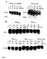
- Figure 1shows the results of an incubation of the VEGF wild type and the VEGF mutants with plasmin.
- Incubation of the VEGF wild type (A, lane 6-10) synthesized in COS-1 cellsalready shows a degradation of the growth factor after 15 minutes. It is difficult to determine the exact size of the resulting fragments using SDS-PAGE, since the signals overlap with the two bands of the differently glycosylated protein.
- the degradation patternis similar to that of the commercially available VEGF 165 ( Figure 1A, lanes 1-5). After 45 minutes, a fragment with a molecular weight of 38 kDa can be detected. This corresponds to the 110 dimer fragment of the less glycosylated VEGF variant.
- FIG. 1BThe results of the incubation of mutated proteins can be seen in FIG. 1B (lanes 1-17).
- two bands for the differently glycosylated variants of the VEGF proteincan be detected at the time of zero incubation.
- due to the higher signal intensitythese cannot be clearly differentiated from one another as with the VEGF 165 wild type.
- the mutated proteinshows no change in running behavior up to 240 minutes after incubation.
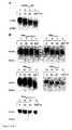
- the degradation of the VEGF mutants in wound fluid from patients with acute and chronic woundswas analyzed.
- the VEGF 165 wild type and all VEGF mutantswere incubated in acute wound fluid, no degradation was detectable after 240 minutes.
- Figure 2shows the effect of chronic wound fluid on the stability of the VEGF proteins.
- Incubation of the VEGF wild type (FIG. 2A, lane 1-4) synthesized in COS-1 cells for 240 minutesshows a degradation of the growth factor with a fragment of approx. 38 kDa. This corresponds to the 110 dimer fragment of the less glycosylated VEGF variant.
- the VEGF 165 mutantsshow a different degradation behavior when incubated in chronic wound fluid.
- a mutation process comparable to the wild typecan be observed in the mutations Mut Gln (FIG. 2B, lanes 13-16 and Mut Ala (lanes 5-8). Fragments with a molecular weight of approx. 38 kDa.
- the analysis of the mutants Mut Pro (lanes 9-12) and Mut Lys-Pro (lanes 1-4 , 17-20)shows a degradation behavior which differs from the wild type and the mutants Mut Ala and Mut Gln .
- the SDS-PAGEshows a stable signal at 42 and 45 kDa. This indicates a stabilization of the mutated proteins Mut Pro and Mut Lys-Pro in the chronic wound fluid.
- This difference in the degradation behavior of the mutants with neutral / non-polar amino acid and those with prolinesuggest that in the chronic wound environment, besides plasmin, other proteases are involved in the breakdown of VEGF.
- Degradationcan be observed for all mutated proteins 240 minutes after incubation in chronic wound fluid. This does not lead to the formation of clearly defined degradation fragments; rather, a diffuse signal between 38 and 45 kDa is produced after 240 min. This is presumably proteolysis in the region of the first 20 amino acids (recognition site of the antibody), since the signal strength decreases significantly after 240 min.
- the resultsindicate that the VEGF mutants with proline at position 111 are initially stabilized in the chronic wound fluid, but are degraded in the long term. Comparable results were observed in wound fluids from three different patients with chronic venous insufficiency. The experiments for the various wound fluids were repeated at least twice ( Figure 2B: patient X lanes 1-4; patient Y lanes 17-20). The resulting band pattern always remained the same.
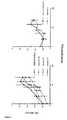
- FIG. 2Cshows a densitometric evaluation of the degradation of VEGF wild type and Mut Lys-Pro .
- the aim of the investigationwas to quantify the stabilization of the VEGF mutant in the chronic wound fluid.
- the time-dependent change in signal strength in the amount of the output signal (range 42-45 kDa) compared to the signal at time zerowas determined.
- the densitometric densities measured at different timesare shown as a percentage of the output signal.
- the VEGF mutantshows a stronger signal in the 42-45 kDa range compared to the VEGF wild type and that there is therefore intact VEGF 165 protein.
- This observationsuggests that this mutation leads to improved stability and bioactivity of the VEGF protein in the chronic wound environment.
- the difference between wild type and mutantis statistically significant as early as 20 minutes after the incubation.
- the measurementswere performed for three independent experiments with identical wound fluid.
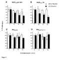
- HUVE cellshuman umbilical vein endothelial cells
- the HUVE cellswere cultivated with the addition of various VEGF mutants, then incubated and fixed for 6 hours with BrdU solution, after which an ELISA was carried out using a BrdU-specific antibody.
- VEGF concentrationsbetween 1 ng / ml and 25 ng / ml were used.
- Commercially available recombinant VEGF 165 protein (R&D Systems) and VEGF 165 wild type synthesized in COS-1 cellsshowed half-maximum stimulation of BrdU incorporation at approximately 3 ng / ml (FIG. 3).
- the mutated VEGF proteinsare characterized by a stimulation of endothelial cell proliferation comparable to the VEGF wild type synthesized in COS-1 cells.
- the maximum stimulation of all proteins synthesized in COS-1 cellswas less than that of the commercially available VEGF 165 wild type.
- the difference between the two curvescan be explained by the different expression systems and purification methods of the proteins (Mohanraj et al. 1995).
- the biological activity of VEGF 165is therefore not significantly influenced by the mutations carried out.
- the VEGF proteinswere incubated with plasmin and then the biological activity was examined for HUVE cells using a BrdU proliferation assay.
- the incubation of the VEGF 165 wild types in plasminleads to a significant reduction in the biological activity (FIG. 4 A, B).
- the mutants Mut Ala and Mut Lys-Proshow no significant loss of activity after incubation with plasmin (FIG. 4 C, D). These results underline the "plasmin resistance" of the mutants shown in the Western blot (FIG. 1) and show that the mutated proteins are stable even after incubation with plasmin.
Landscapes
- Health & Medical Sciences (AREA)
- Life Sciences & Earth Sciences (AREA)
- Chemical & Material Sciences (AREA)
- Organic Chemistry (AREA)
- Medicinal Chemistry (AREA)
- General Health & Medical Sciences (AREA)
- Veterinary Medicine (AREA)
- Animal Behavior & Ethology (AREA)
- Pharmacology & Pharmacy (AREA)
- Public Health (AREA)
- Chemical Kinetics & Catalysis (AREA)
- Nuclear Medicine, Radiotherapy & Molecular Imaging (AREA)
- General Chemical & Material Sciences (AREA)
- Bioinformatics & Cheminformatics (AREA)
- Engineering & Computer Science (AREA)
- Diabetes (AREA)
- Hematology (AREA)
- Proteomics, Peptides & Aminoacids (AREA)
- Gastroenterology & Hepatology (AREA)
- Zoology (AREA)
- Obesity (AREA)
- Epidemiology (AREA)
- Molecular Biology (AREA)
- Pain & Pain Management (AREA)
- Rheumatology (AREA)
- Genetics & Genomics (AREA)
- Vascular Medicine (AREA)
- Biophysics (AREA)
- Biochemistry (AREA)
- Immunology (AREA)
- Toxicology (AREA)
- Dermatology (AREA)
- Endocrinology (AREA)
- Oncology (AREA)
- Cardiology (AREA)
- Heart & Thoracic Surgery (AREA)
- Physical Education & Sports Medicine (AREA)
- Emergency Medicine (AREA)
- Medicines That Contain Protein Lipid Enzymes And Other Medicines (AREA)
- Peptides Or Proteins (AREA)
Abstract
Description
Translated fromGermanGegenstand der Erfindung ist Vascular Endothelial Growth Factor (VEGF), bei dem das Alanin an AS-Position 111 gegen Prolin ausgetauscht ist. Das Arginin an AS-Position 110 kann dabei gegen eine andere Aminosäure substituiert sein. Die Erfindung betrifft auch Derivate der erfindungsgemäßen VEGF, Nucleinsäuren, Expressionssysteme, Arzneimittel und die Verwendung der VEGF-Mutanten der Erfindung zur Behandlung chronischer Wunden.The invention relates to vascular endothelial growth factor (VEGF), in which the alanine at AS position 111 is replaced by proline. The arginine at
Ein wichtiges Stadium der kutanen Wundheilung ist die Ausbildung eines Granulationsgewebes. Eng mit letzterem verbunden ist das Einwandern neugebildeter Gefäße (Neoangiogenese). Zahlreiche experimentelle und klinische Studien zeigen, dass chronische Wunden durch eine gestörte Angiogenese und somit verminderte Ausbildung eines Granulationsgewebes charakterisiert sind.An important stage in cutaneous wound healing is the formation of a granulation tissue. The migration of newly formed vessels (neoangiogenesis) is closely related to the latter. Numerous experimental and clinical studies show that chronic wounds are characterized by disturbed angiogenesis and thus reduced granulation tissue formation.
Es sind eine Vielzahl von Mediatoren bekannt, die die Angiogenese während der Wundheilung stimulieren. Zu diesen zählen zum einen solche Faktoren, die neben der Stimulation von Endothelzellen auch mesenchymale und/oder epidermale Zellen aktivieren (bFGF, aFGF, TGF-a, PDGF) und zum anderen sogenannte endothelzellspezifische Faktoren, deren Rezeptoren im wesentlichen auf Endothelzellen beschränkt sind (VEGF, Angiopoietin). Eine Vielzahl physiologischer und pathologischer Reaktionen unter Beteiligung der Blutgefäße korreliert mit einer erhöhten Expression von VEGF und seiner Rezeptoren, so dass VEGF eine zentrale Rolle in der Angiogenese der Haut zukommt. Erste Hinweise für die mögliche Bedeutung des VEGF bei Wundheilungsstörungen boten sich auf der Grundlage von Experimenten zur VEGF Expression bei diabetischen Mäusen (db/db Mäuse) (Franket al. 1995). In diesem Modell konnte gezeigt werden, dass die Wundheilungsstörung mit einer verminderten VEGF Expression korreliert. Die Rolle von VEGF bei der Wundheilung konnte kürzlich durch ein weiteres transgenes Tiermodell (Fukumuraet al., 1998) und den Nachweis von VEGF im Wundsekret akuter humaner Wunden unterstützt werden (Nissenet al., 1998).A variety of mediators are known to stimulate angiogenesis during wound healing. These include factors that, in addition to stimulating endothelial cells, also activate mesenchymal and / or epidermal cells (bFGF, aFGF, TGF-a , PDGF) and, on the other hand, so-called endothelial cell-specific factors whose receptors are essentially limited to endothelial cells (VEGF , Angiopoietin). A large number of physiological and pathological reactions involving the blood vessels correlate with an increased expression of VEGF and its receptors, so that VEGF plays a central role in the angiogenesis of the skin. First Evidence of the possible importance of VEGF in wound healing disorders was provided on the basis of experiments on VEGF expression in diabetic mice (db / db mice) (Franket al. 1995). In this model it could be shown that the wound healing disorder correlates with a reduced VEGF expression. The role of VEGF in wound healing has recently been supported by another transgenic animal model (Fukumuraet al., 1998) and the detection of VEGF in the wound secretion of acute human wounds (Nissenet al ., 1998).
Weiterhin wurde gezeigt, dass die mRNA von VEGF und seinen Rezeptoren im Gewebe chronischer Wunden vermehrt exprimiert werden (Laueret al., 2000). Untersuchungen mittels SDS-PAGE zeigen jedoch einen Abbau des VEGF-Proteins im chronischen Wundmilieu, im Gegensatz zur akuten Wunde. Dieser Abbau führt zu einer signifikanten Einbuße der biologischen Aktivität und kann somit, trotz der erhöhten Expression der VEGF-Rezeptoren, einer defizienten Stimulation der Neoangiogenese im chronischen Wundmilieu zugrunde liegen. Wie oben erläutert, konnte gezeigt werden, dass Plasmin an der Spaltung von VEGF im chronischen Wundmilieu beteiligt ist (Laueret al., 2000).Furthermore, it was shown that the mRNA of VEGF and its receptors is expressed more in the tissue of chronic wounds (Laueret al., 2000). However, studies using SDS-PAGE show a breakdown of the VEGF protein in the chronic wound environment, in contrast to the acute wound. This degradation leads to a significant loss of biological activity and can therefore, despite the increased expression of the VEGF receptors, be the basis for a deficient stimulation of neoangiogenesis in the chronic wound environment. As explained above, it could be shown that plasmin is involved in the cleavage of VEGF in the chronic wound environment (Laueret al., 2000).
Die Spaltung von VEGF165 durch Plasmin führt zur Abtrennung der carboxylterminalen Domäne, die vom Exon 7 kodiert wird. Während die Bindungseigenschaften von VEGF an die VEGF-Rezeptoren Flt-1 und Flk-1/KDR über Exon 3 und 4 bestimmt werden, besitzt Exon 7 eine kritische Bedeutung in der Interaktion von VEGF mit Neuropilin-1 (Keytet al. 1996). Neuropilin -1 ist ein 130 kDa Glykoprotein der Zelloberfläche. Erst vor kurzem wurde seine Rolle bei der Potenzierung des mitogenen Effekts von VEGF auf Endothelzellen beschrieben (Sokeret al. 1998). Dabei scheint die Interaktion von Neuropilin-1 mit Flk-1/KDR bedeutsam, da die alleinige Bindung von VEGF an Neuropilin-1 keine Signalwirkung ausübt.The cleavage of VEGF165 by plasmin leads to the separation of the carboxyl-terminal domain, which is encoded by
Plasmin gehört zur Klasse der Serinproteasen. Diese Enzyme sind in der Lage, Peptidbindungen zu spalten. Die Spaltung erfolgt über eine sogenannte katalytische Triade. Dabei spielen im katalytischen Zentrum insbesondere das namensgebende Serin, aber auch die Aminosäuren Histidin und Aspartat eine wesentliche Rolle, da über sie der Prozeß der Peptidspaltung erfolgt (Stryer 1987, S. 231 ff). Obwohl der Mechanismus der Bindungsspaltung bei allen Serinproteasen identisch ist, unterscheiden sie sich deutlich in ihrer Substratspezifität. So spaltet Plasmin, ebenso wie Trypsin, Peptidbindungen nach den basischen Aminosäuren Lysin und Arginin. Die Substratspezifität von Plasmin, die durch die Struktur des katalytischen Zentrums bestimmt wird, führt jedoch dazu, dass Plasmin nicht jede dieser Bindungen spalten kann. Die Katalyse der Peptidbindungsspaltung kann nur erfolgen, wenn die entsprechenden Proteinabschnitte in der Lage sind mit dem katalytischen Zentrum des Enzyms zu interagieren (Powerset al. 1993; Stryer 1987). Bisher ist keine eindeutige Konsensussequenz für eine Plasmin-Schnittstelle bekannt.Plasmin belongs to the class of serine proteases. These enzymes are able to break peptide bonds. The cleavage takes place via a so-called catalytic triad. The eponymous serine, but also the amino acids histidine and aspartate play a role in the catalytic center play an important role, since the peptide cleavage process takes place via it (Stryer 1987, p. 231 ff). Although the mechanism of bond cleavage is identical for all serine proteases, they differ significantly in their substrate specificity. Like trypsin, plasmin cleaves peptide bonds according to the basic amino acids lysine and arginine. However, the substrate specificity of plasmin, which is determined by the structure of the catalytic center, means that plasmin cannot cleave each of these bonds. The peptide bond cleavage can only be catalyzed if the corresponding protein segments are able to interact with the catalytic center of the enzyme (Powerset al . 1993; Stryer 1987). So far, no clear consensus sequence for a plasmin interface is known.
Der vorliegenden Erfindung liegt die Aufgabe zugrunde, verbesserte Mittel zur Heilung von chronischen Wunden bereitzustellen.The present invention has for its object to provide improved means for healing chronic wounds.
Überraschenderweise wird diese Aufgabe gelöst durch die erfindungsgemäße Bereitstellung von Vascular Endothelial Growth Factor (VEGF)-Varianten, wobei das Alanin an AS-Position 111 des Wildtyp VEGF gegen Prolin ausgetauscht ist. Gegenstand der Erfindung sind auch Mutanten (Varianten) des VEGF, bei denen zusätzlich das Arginin an AS-Position 110 gegen eine andere Aminosäure ausgetauscht ist.Surprisingly, this object is achieved by the provision of vascular endothelial growth factor (VEGF) variants according to the invention, the alanine at AS position 111 of the wild-type VEGF being replaced by proline. The invention also relates to mutants (variants) of VEGF in which the arginine at
Die erfindungsgemässen Mutanten von VEGF liegen vorzugsweise als eine der Splice-Varianten VEGF121, VEGF145, VEGF165, VEGF183, VEGF189 oder VEGF206.vor.The mutants of VEGF according to the invention are preferably present as one of the splice variants VEGF121 , VEGF145 , VEGF165 , VEGF183 , VEGF189 or VEGF206 .
Die erfindungsgemäßen Mutanten von VEGF weisen nicht nur eine deutlich erhöhte Stabilität gegenüber Plasmin, sondern auch eine dem Wildtyp-VEGF vergleichbare Aktivität auf. Überraschenderweise weisen die erfindungsgemäßen VEGF-Varianten darüber hinaus eine deutlich erhöhte Stabilität in chronischen Wundflüssigkeiten auf.The mutants of VEGF according to the invention not only have a significantly increased stability towards plasmin, but also an activity comparable to wild-type VEGF. Surprisingly, the VEGF variants according to the invention also have a significantly increased stability in chronic wound fluids.
Die Mutationen wurden an einer für die biologische Aktivität des VEGF-Moleküls kritischen Stelle durchgeführt. Somit war zu befürchten, dass eine Veränderung der Proteinstruktur in diesem Bereich die Aktivität von VEGF165 negativ beeinflusst. Bei der Aminosäure Prolin, die erfindungsgemäß an Position 111 eingeführt wird, handelt es sich um eine zyklische α-Iminosäure.The mutations were carried out at a location critical for the biological activity of the VEGF molecule. It was feared that a change in the protein structure in this area would have a negative effect on the activity of VEGF165 . The amino acid proline, which is introduced at position 111 according to the invention, is a cyclic α-imino acid.
Durch die zyklische Form des Pyrrolidin-Restes besitzt sie eine starre Konformation, die sich auch auf die Struktur der jeweiligen Proteine auswirkt. So fungiert Prolin z.B. als starker α-Helix-Brecher. Es ist daher in besonderem Masse überraschend, dass gerade durch den Ersatz des Alanins an Position 111 gegen Prolin eine Mutante von VEGF erzeugt wird, die stabil gegenüber der Protease Plasmin ist, stabil in chronischen Wundflüssigkeiten ist und zugleich noch eine dem Wildtyp-Protein entsprechende Aktivität aufweist.Due to the cyclic form of the pyrrolidine residue, it has a rigid conformation, which also affects the structure of the respective proteins. So proline acts e.g. as a strong α-helix crusher. It is therefore particularly surprising that the replacement of the alanine at position 111 with proline produces a mutant of VEGF which is stable to the protease plasmin, stable in chronic wound fluids and at the same time an activity corresponding to the wild-type protein having.
Gegenstand der Erfindung sind insbesondere VEGF-Varianten der beiden Sequenzen Seq. No. 1 oder Seq. No. 2.The invention relates in particular to VEGF variants of the two sequences Seq. No. 1 or Seq. No. Second
Gegenstand der Erfindung sind auch Varianten der oben aufgeführten VEGF-Mutanten, bei denen die Aminosäuresequenzen modifiziert oder derivatisiert sind oder Mutationen, Insertionen oder Deletionen enthalten. Insbesondere betrifft dies VEGF-Varianten, bei denen einzelne weiter Aminosäuren ausgetauscht sind und solche, die glykosyliert, amidiert, acetyliert, sulfatiert oder phosphoryliert sind. Vorzugsweise haben solche VEGF-Varianten eine dem Wildtyp-VEGF vergleichbare oder höhere Aktivität.The invention also relates to variants of the VEGF mutants listed above, in which the amino acid sequences are modified or derivatized or contain mutations, insertions or deletions. This applies in particular to VEGF variants in which individual further amino acids are exchanged and those which are glycosylated, amidated, acetylated, sulfated or phosphorylated. Such VEGF variants preferably have an activity comparable to or higher than that of the wild-type VEGF.
Die erfindungsgemäßen VEGF-Varianten können auch eine Signalsequenz aufweisen. Die Signalsequenz kann N-terminal an die Aminosäurekette der VEGF-Variante anschließen und die Sequenz
Gegenstand der Erfindung sind auch Nucleinsäuren, die für die oben benannten VEGF-Mutanten kodieren, und Vektoren zur Expression von VEGF, die solche Nucleinsäuren enthalten.The invention also relates to nucleic acids which code for the above-mentioned VEGF mutants and vectors for the expression of VEGF which contain such nucleic acids.
Gegenstand der Erfindung ist auch ein Arzneimittel, das die oben benannten Mutanten des VEGF enthält, sowie die Verwendung der VEGF-Mutanten zur Herstellung eines Arzneimittels zur Behandlung von chronischen Wunden, verursacht durch Gefäßveränderungen wie chronisch-venöse Insuffizienz (CVI), primäres/sekundäres Lymphödem, arterielle Verschlußkrankheit, metabolische Erkrankungen wie Diabetes mellitus, Gicht oder Dekubitus, chronisch entzündlichen Erkrankungen wie Pyoderma gangraenosum, Vaskulitis, perforierende Dermatosen wie Necrobiosis lipoidica diabeticorum und Granuloma annulare, hämatologische. Grunderkrankungen wie Gerinnungsstörungen, Sichelzellenanämie und Polycythemia vera, Tumoren wie primäre kutane Tumoren und exulzerierte Metastasen, sowie zur Plasmininhibition, zur Induktion der Neoangiogenese und/oder zur Inhibition der Matrixdegradierung.The invention also relates to a medicament which contains the above-mentioned mutants of VEGF, and to the use of the VEGF mutants for the production of a medicament for the treatment of chronic wounds caused by vascular changes such as chronic venous insufficiency (CVI), primary / secondary lymphedema , arterial occlusive disease, metabolic Diseases such as diabetes mellitus, gout or pressure ulcers, chronic inflammatory diseases such as pyoderma gangrenosum, vasculitis, perforating dermatoses such as necrobiosis lipoidica diabeticorum and granuloma annulare, hematological. Underlying diseases such as coagulation disorders, sickle cell anemia and polycythemia vera, tumors such as primary cutaneous tumors and exulcerated metastases, as well as for plasmin inhibition, for inducing neoangiogenesis and / or for inhibiting matrix degradation.
Die topische Anwendung von Wachstumsfaktoren stellt in der Wundheilung ein neuartiges Therapiekonzept dar. Eine Verbesserung der Heilung chronischer Wunden konnte in einer Vielzahl von klinischen Studien mit der Anwendung von EGF, bFGF, PDWHF und PDGF beobachtet werden (Scharffetter-Kochaneket al. 2000). Es bleibt jedoch kritisch anzumerken, dass die Ergebnisse dieser Studien hinter den Erwartungen zurückgeblieben sind, die angesichts der guten Wirksamkeit dieser Mediatoren im Tiermodell bestanden haben (Lawrenceet al. 1994). Eine wesentliche Erklärung für diese eingeschränkte Wirksamkeit der Wachstumsfaktoren ist sicherlich die erhöhte proteolytische Aktivität im chronischen Wundmilieu, die zur Degradation der topisch applizierten Fakturen führt. Somit wird deutlich, dass das lokale Wundmanagement durch die Applikation von Wachstumsfaktoren eine vielversprechende neue therapeutische Strategie darstellt. Jedoch ist es notwendig Strategien zu entwickeln, die die proteolytische Aktivität im chronischen Wundmilieu kontrollieren. Die Herstellung von Masterzytokinen mit erhöhter Stabilität im chronischen Wundmilieu stellt dabei sicherlich einen neuartigen Therapieansatz dar. Die erfindungsgemäßen VEGF-Mutanten eignen sich aufgrund ihrer hohen Stabilität in Wundflüssigkeit in besonderem Maße zur topischen Behandlung von chronischen Wunden.The topical application of growth factors represents a new therapeutic concept in wound healing. Improvement in the healing of chronic wounds has been observed in a large number of clinical studies with the use of EGF, bFGF, PDWHF and PDGF (Scharffetter-Kochaneket al. 2000). However, it remains critical to note that the results of these studies have fallen short of the expectations that existed in view of the good effectiveness of these mediators in the animal model (Lawrenceet al. 1994). An essential explanation for this limited effectiveness of the growth factors is surely the increased proteolytic activity in the chronic wound environment, which leads to the degradation of the topically applied invoices. This makes it clear that local wound management through the application of growth factors represents a promising new therapeutic strategy. However, it is necessary to develop strategies that control proteolytic activity in the chronic wound environment. The production of master cytokines with increased stability in the chronic wound environment is certainly a new therapeutic approach. The VEGF mutants according to the invention are particularly suitable for topical treatment of chronic wounds due to their high stability in wound fluid.
Mittels zielgerichteter Mutagenese wurden vier Mutanten hergestellt, indem gezielte Aminosäure-Austausche bei Arg110 und Ala111 vorgenommen wurden. Der cDNA, die für humanen VEGF165 kodiert, wurde in den SV40 Replikations Expressionsvektor pcDNA 3.1 (Fa. Invitrogen, De Schelp, NL) kloniert unter Verwendung der Schnittstellen BamHI und EcoRI in der Cloning Site. Für die zielgerichtetein vitro Mutagenese wurde das Gene Editor™-System der Firma Promega (Mannheim) verwendet. Dieses basiert auf der Anlagerung von Oligonukleotiden, welche die entsprechenden Mutation tragen, an die Ausgangssequenz. Die Ausgangssequenz des VEGF165 im Bereich der Mutationen ist:
Zur Einführung der Mutationen wurden die folgenden "Missmatch"-Oligonucleotide als Primer verwendet:


Die verwendeten Mutagenese-Primer werden jeweils mit den dadurch erhaltenen veränderten Aminosäuresequenzen aufgeführt. Kursiv markiert sind die Bereiche mit den gegenüber der Wildtypsequenz veränderten Basen oder Aminosäuren.The mutagenesis primers used are listed in each case with the amino acid sequences thus obtained. The areas with the bases or amino acids changed compared to the wild-type sequence are marked in italics.
Bei Mutation 1 wurde das Arginin110 gegen ein unpolares Alanin ausgetauscht. Bei Mutation 2 wurde an der selben Position ein polares, ungeladenes Glutamin eingeführt. Bei der Mutante 3 wurde nicht das basische Arginin110, sondern das Alanin an Position 111 gegen ein Prolin ausgetauscht. Bei der Mutante 4 wurden zwei Aminosäuren ausgetauscht. Hier wurden anstelle von Arginin110 und Alanin111 Lysin und Prolin eingefügt. Nach der Durchführung der Mutagenese erfolgte die Verifizierung der Mutationen durch Sequenzanalyse. Die erhaltenen VEGF-Mutanten hatten bei Aminosäuren 109-112 folgende Sequenzen:
- VEGF165-Wildtyp: -Asp109 Arg110 Ala111 Arg112-
- MutGln : -Asp109 Gln110 Ala111 Arg112-
- MutAla : -Asp109 Ala110 Ala111 Arg112-
- MutPro : -Asp109 Arg110 Pro111 Arg112-
- MutLys-Pro : -Asp109 Lys110 Pro111 Arg112-
- VEGF165 -Wildtyp: -Asp109 Arg110 Ala111 Arg112 -
- MutGln : -Asp109 Gln110 Ala111 Arg112 -
- MutAla : -Asp109 Ala110 Ala111 Arg112 -
- CouragePro : -Asp109 Arg110 Pro111 Arg112 -
- MutLys-Pro : -Asp109 Lys110 Pro111 Arg112 -
Die Mutanten MutPro und MutLys-Pro sind erfindungsgemäße Mutanten, während die MutGln und MutAla zu Vergleichszwecken hergestellt und untersucht werden. Die erhaltenen VEGF165-Expressionsvektoren wurden in den weiteren Untersuchungen verwendet.The mutants MutPro and MutLys-Pro are mutants according to the invention, while the MutGln and MutAla are produced and investigated for comparison purposes. The VEGF165 expression vectors obtained were used in the further investigations.
Die Expression von VEGF165 Protein erfolgte in eukaryontischen COS-1-Zellen. Der verwendete Expressionsvektor pcDNA 3.1 enthält einen SV-40 Origin of Replication. Dieser dient zur Amplifikation des Vektors in Zellen, die ein Large T-Antigen des SV-40 Viruses exprimieren. Die verwendeten COS-1 Zellen besitzen ein entsprechendes im Genom integriertes Element, so dass es hier zu einer episomalen Replikation des Vektors kommt. Dadurch wird ohne die stabile Integration (Transformation) des Vektors ins Zellgenom eine Expression des Zielproteins VEGF für mehrere Tage erreicht. Die COS-1 Zellen wurden mit den bei der Mutagenese erhaltenen Expressionsplasmiden transfiziert. Dazu wurde das Superfect Transfection Reagent (QIAGEN, Hilden) nach den Vorschriften des Herstellers verwendet.VEGF165 protein was expressed in eukaryotic COS-1 cells. The expression vector pcDNA 3.1 used contains an SV-40 Origin of Replication. This serves to amplify the vector in cells that express a large T antigen of the SV-40 virus. The COS-1 cells used have a corresponding element integrated in the genome, so that there is an episomal replication of the vector. As a result, expression of the target protein VEGF is achieved for several days without the stable integration (transformation) of the vector into the cell genome. The COS-1 cells were transfected with the expression plasmids obtained in the mutagenesis. For this purpose, the Superfect Transfection Reagent (QIAGEN, Hilden) was used according to the manufacturer's instructions.
Wie eine Vielzahl von Wachstumsfaktoren besitzt auch VEGF165 eine Heparin-Bindungsstelle, die am basischen C-Terminus lokalisiert ist. Die Bindung an Heparin wurde für die Aufreinigung des Proteins mittels Affinitätschromatographie genutzt (Mohanrajet al. 1995). Die Isolation der VEGF und VEGF-Varianten erfolgte durch die folgenden Schritte:Like a variety of growth factors, VEGF165 also has a heparin binding site located at the basic C-terminus. The binding to heparin was used for the purification of the protein by means of affinity chromatography (Mohanrajet al. 1995). The VEGF and VEGF variants were isolated by the following steps:
Die mit den Expressionsplasmiden transformierten COS-1-Zellen wurden in Serum-freiem DMEM (Dulbecco's modified-Eagle's Medium), enthaltend 10% fetales Kälberserum (FCS), 2 mM L-Glutamin, Penicillin (10 U/ml) und Streptomycin (10 µg/ml) und ITS Supplement (Sigma, Deisenhofen), kultiviert. Konditioniertes Medium (200 ml) wurde nach 48h gesammelt und für 4 Stunden mit 5 ml Heparin-Sepharose (Pharmacia, Freiburg) bei 4 °C inkubiert. Die Heparin-Sepharose wurde in eine Säule gepackt. Diese wurde bei einer Flußrate von 25 ml/h mit Kulturmedium beladen. Es wurden folgende Schritte durchgeführt:
- A: Affinitätschromatographie mit Heparin-Sepharose
- 1. Waschen: 0,1 M NaCl; 20 mM Tris/
pH - 2. Waschen: 0,3 M NaCl; 20 mM Tris/
pH - 3. Elution: 0,9 M NaCl; 20 mM Tris/
pH
- 1. Waschen: 0,1 M NaCl; 20 mM Tris/
- B: Analyse der erhaltenen Fraktionen durch Western-Blot Analyse
- C: Entsalzung der VEGF-haltigen Fraktionen durch Gelfiltration
Laufpuffer: 10 mM Tris/pH - D: Lyophilisierung der Lösung und Konzentrationsbestimmung durch ELISA
- A: Affinity chromatography with heparin-Sepharose
- 1. Wash: 0.1 M NaCl; 20 mM Tris / pH 7.2
- 2. Wash: 0.3 M NaCl; 20 mM Tris / pH 7.2
- 3. Elution: 0.9 M NaCl; 20 mM Tris / pH 7.2
- B: Analysis of the fractions obtained by Western blot analysis
- C: Desalting of the VEGF-containing fractions by gel filtration
Running buffer: 10 mM Tris / pH 7.2 - D: Lyophilization of the solution and concentration determination by ELISA
Das erhaltene VEGF wurde durch SDS-PAGE untersucht. Das aus COS-1-Zellen gewonnene VEGF-Protein unterscheidet sich in seinem Laufverhalten in der SDS-PAGE von dem verwendeten, kommerziell erhältlichen VEGF165 Protein (Firma R&D Systems). Zusätzlich zu dem bei 42 kDA zu detektierende Signal (Figur 1, Bahn 6) ist noch eine weitere Bande mit einem einige kDa höheren Molekulargewicht zu erkennen. Grund für diese Doppelbande des in COS-1-Zellen exprimierten VEGF-Proteins ist eine veränderte Glykosylierung des Wachstumsfaktors. In COS-1-Zellen kommt es bei der Expression von VEGF zur Bildung zweier unterschiedlich glykosylierter Proteine. Eine Form (42 kDa) ist in ihrer Glykosylierung identisch mit dem bisher verwendeten rekombinanten VEGF165, das in Insektenzellen mit einem Baculovirus-Expressionssystem erzeugt wurde (R&D Systems, Figur 1, Bahn 1). Es weist an der Aminosäure Asparagin bei Position 74 eine N-Glykosylierung auf (Gospodarowiczet al. 1989; Kecket al. 1989). Die zweite Bande bei höherem Molekulargewicht (45 kDa) entsteht auf Grund einer weiteren Glykosylierung des Proteins. Der Unterschied in der Glykosylierung ist für die Expression in COS-Zellen bekannt und hat keinen Effekt auf die biologische Aktivität des Wachstumsfaktors (R&D Systems).The VEGF obtained was examined by SDS-PAGE. The VEGF protein obtained from COS-1 cells differs in its running behavior in SDS-PAGE from the commercially available VEGF165 protein (R&D Systems) used. In addition to the signal to be detected at 42 kDA (FIG. 1, lane 6), a further band with a molecular weight that is several kDa higher can be seen. The reason for this double band of the VEGF protein expressed in COS-1 cells is an altered glycosylation of the growth factor. In COS-1 cells, when VEGF is expressed, two differently glycosylated proteins are formed. One form (42 kDa) is identical in its glycosylation to the previously used recombinant VEGF165 , which was generated in insect cells with a baculovirus expression system (R&D Systems, Figure 1, lane 1). It shows N-glycosylation at the amino acid asparagine at position 74 (Gospodarowiczet al. 1989; Kecket al. 1989). The second band with a higher molecular weight (45 kDa) arises due to a further glycosylation of the protein. The difference in glycosylation is known for expression in COS cells and has no effect on the biological activity of the growth factor (R&D Systems).
Die vier aufgereinigten mutierten VEGF-Proteine wurden zunächst auf ihre Stabilität gegenüber der Protease Plasmin untersucht. Es wurde untersucht, ob die vorgenommenen Mutationen zu einem verändertem Degradationsverhalten im Vergleich zu Wildtyp VEGF führen.The four purified mutant VEGF proteins were first examined for their stability towards the protease plasmin. It was examined whether the mutations lead to a changed degradation behavior compared to wild-type VEGF.
Figur 1 zeigt die Ergebnisse einer Inkubation des VEGF-Wildtyps und der VEGF-Mutanten mit Plasmin. Die Inkubation des in COS-1-Zellen synthetisierten VEGF-Wildtyps (A, Bahn 6-10) zeigt schon nach 15 Minuten eine Degradation des Wachstumsfaktors. Dabei ist die exakte Größenbestimmung der entstehenden Fragmente mittels SDS-PAGE schwierig, da sich die Signale mit den beiden Banden des unterschiedlich glykosylierten Proteins überlagern. Das Degradationsmuster ist jedoch dem des kommerziell erhältlichen VEGF165 ähnlich (Figur 1A, Bahn 1 - 5). So kann nach 45 Minuten ein Fragment mit einem Molekulargewicht von 38 kDa detektiert werden. Dieses entspricht dem 110 Dimer-Fragment der weniger glykosylierten VEGF-Variante. Diese Ergebnisse zeigen deutlich, dass auch das VEGF-Protein, das in den COS-1-Zellen exprimiert wurde, unter den gewählten Bedingungen von Plasmin gespalten wird.Figure 1 shows the results of an incubation of the VEGF wild type and the VEGF mutants with plasmin. Incubation of the VEGF wild type (A, lane 6-10) synthesized in COS-1 cells already shows a degradation of the growth factor after 15 minutes. It is difficult to determine the exact size of the resulting fragments using SDS-PAGE, since the signals overlap with the two bands of the differently glycosylated protein. However, the degradation pattern is similar to that of the commercially available VEGF165 (Figure 1A, lanes 1-5). After 45 minutes, a fragment with a molecular weight of 38 kDa can be detected. This corresponds to the 110 dimer fragment of the less glycosylated VEGF variant. These results clearly show that the VEGF protein, which was expressed in the COS-1 cells, is cleaved by plasmin under the chosen conditions.
In Figur 1B (Bahn 1 - 17) sind die Ergebnisse der Inkubation mutierter Proteine zu sehen. Zunächst ist die Inkubation der Mutation Arginin zu Alanin dargestellt (Bahn 1 - 5). Zum Zeitpunkt Null der Inkubation sind wie beim Wildtyp zwei Banden für die unterschiedlich glykosylierten Varianten des VEGF-Proteins zu detektieren. Diese sind hier allerdings auf Grund der höheren Signalintensität nicht so eindeutig voneinander zu differenzieren wie beim VEGF165-Wildtyp. Im Gegensatz zum VEGF-Wildtyp zeigt das mutierte Protein bis zu 240 Minuten nach Inkubation keinerlei Veränderung im Laufverhalten.The results of the incubation of mutated proteins can be seen in FIG. 1B (lanes 1-17). First, the incubation of the arginine mutation to alanine is shown (lanes 1-5). As with the wild type, two bands for the differently glycosylated variants of the VEGF protein can be detected at the time of zero incubation. However, due to the higher signal intensity, these cannot be clearly differentiated from one another as with the VEGF165 wild type. In contrast to the VEGF wild type, the mutated protein shows no change in running behavior up to 240 minutes after incubation.
Diese Beobachtung legt die Vermutung nahe, dass die Mutation Arginin110 zu Alanin110 zur Inaktivierung der Plasmin-Schnittstelle geführt hat. Wie in Figur 1B weiter dargestellt, weisen auch die drei weiteren Mutanten MutPro, MutGln und MutLys-Pro nach Inkubation mit Plasmin über 240 Minuten eine vergleichbare Stabilität der Signalbanden bei 45 und 42 kDA auf. Eine Kontrolle, bei der VEGF165-Wildtyp über 4 Stunden bei 37 °C mit Plasminpuffer inkubiert wurde, wird nicht degradiert (Bahnen 18 und 19). Insgesamt weisen diese Experimente darauf hin, dass die erzeugten und aufgereinigten VEGF-Mutanten stabil gegenüber der Protease Plasmin sind.This observation suggests that the arginine110 mutation to alanine110 led to the inactivation of the plasmin interface. As further shown in FIG. 1B, the three further mutants MutPro , MutGln and MutLys-Pro after incubation with plasmin for 240 minutes showed comparable stability of the signal bands at 45 and 42 kDA. A control in which VEGF165 wild type was incubated with plasmin buffer for 4 hours at 37 ° C. is not degraded (
Im nächsten Schritt wurde die Degradation der VEGF-Mutanten in Wundflüssigkeit von Patienten mit akuten und chronischen Wunden analysiert. Bei Inkubation des VEGF165 Wildtyps und aller VEGF-Mutanten in akuter Wundflüssigkeit war keine Degradation nach 240 Minuten nachweisbar.In the next step, the degradation of the VEGF mutants in wound fluid from patients with acute and chronic wounds was analyzed. When the VEGF165 wild type and all VEGF mutants were incubated in acute wound fluid, no degradation was detectable after 240 minutes.
Figur 2 zeigt den Effekt chronischer Wundflüssigkeit auf die Stabilität der VEGF-Proteine. Die Inkubation des in COS-1-Zellen synthetisierten VEGF-Wildtyps (Figur 2A, Bahn 1-4) über 240 Minuten zeigt eine Degradation des Wachstumsfaktors mit einem Fragment von ca. 38 kDa. Dieses entspricht dem 110 Dimer-Fragment der weniger glykosylierten VEGF-Variante.Figure 2 shows the effect of chronic wound fluid on the stability of the VEGF proteins. Incubation of the VEGF wild type (FIG. 2A, lane 1-4) synthesized in COS-1 cells for 240 minutes shows a degradation of the growth factor with a fragment of approx. 38 kDa. This corresponds to the 110 dimer fragment of the less glycosylated VEGF variant.
Im Gegensatz zum Wildtyp zeigen die VEGF165-Mutanten ein anderes Degradationsverhalten bei der Inkubation in chronische Wundflüssigkeit. Auf der einen Seite ist in den Mutationen MutGln (Figur 2B, Bahnen 13 - 16 und MutAla (Bahnen 5 - 8) ein Degradationsprozeß zu beobachten, der mit dem Wildtyp vergleichbar ist. Es entstehen bereits nach 20 min Fragmente mit einem Molekulargewicht von ca. 38 kDa.In contrast to the wild type, the VEGF165 mutants show a different degradation behavior when incubated in chronic wound fluid. On the one hand, a mutation process comparable to the wild type can be observed in the mutations MutGln (FIG. 2B, lanes 13-16 and MutAla (lanes 5-8). Fragments with a molecular weight of approx. 38 kDa.
Auf der anderen Seite zeigt die Analyse der Mutanten MutPro (Bahnen 9 - 12) und MutLys-Pro (Bahnen 1 - 4, 17 - 20) ein vom Wildtyp und den Mutanten MutAla und MutGln abweichendes Abbauverhalten. Bis zu 60 Minuten nach Inkubation zeigt sich im SDS-PAGE ein stabiles Signal bei 42 und 45 kDa. Dies weist auf eine Stabilisierung der mutierten Proteine MutPro und MutLys-Pro in der chronischen Wundflüssigkeit hin. Dieser Unterschied im Degradationsverhalten von den Mutanten mit neutraler/unpolarer Aminosäure und denen mit Prolin legt die Vermutung nahe, dass im chronischen Wundmilieu neben Plasmin weitere Proteasen am Abbau von VEGF beteiligt sind.On the other hand, the analysis of the mutants MutPro (lanes 9-12) and MutLys-Pro (lanes1-4 , 17-20) shows a degradationbehavior which differs from the wild type and the mutants MutAla and MutGln . Up to 60 minutes after incubation, the SDS-PAGE shows a stable signal at 42 and 45 kDa. This indicates a stabilization of the mutated proteins MutPro and MutLys-Pro in the chronic wound fluid. This difference in the degradation behavior of the mutants with neutral / non-polar amino acid and those with proline suggest that in the chronic wound environment, besides plasmin, other proteases are involved in the breakdown of VEGF.
240 Minuten nach der Inkubation in chronischer Wundflüssigkeit ist bei allen mutierten Proteinen eine Degradierung zu beobachten. Dabei kommt es nicht zur Bildung klar definierter Abbaufragmente, vielmehr entsteht nach 240 min ein diffuses Signal zwischen 38 und 45 kDa. Vermutlich handelt sich hierbei um Proteolyse im Bereich der ersten 20 Aminosäuren (Erkennungsstelle des Antikörpers), da die Signalstärke nach 240 min deutlich abnimmt.Degradation can be observed for all mutated
Zusammenfassend deuten die Ergebnisse darauf hin, dass die VEGF-Mutanten mit Prolin an Position 111 zunächst in der chronischen Wundflüssigkeit stabilisiert sind, langfristig jedoch degradiert werden. Es wurden vergleichbare Ergebnisse in den Wundflüssigkeiten von drei verschiedenen Patienten mit chronisch venöser Insuffizienz beobachtet. Die Experimente für die verschiedenen Wundflüssigkeiten wurden mindestens zweimal wiederholt (Figur 2B: Patient X Bahnen 1 - 4; Patient Y Bahnen 17 - 20). Dabei blieb das entstehende Bandenmuster stets gleich.In summary, the results indicate that the VEGF mutants with proline at position 111 are initially stabilized in the chronic wound fluid, but are degraded in the long term. Comparable results were observed in wound fluids from three different patients with chronic venous insufficiency. The experiments for the various wound fluids were repeated at least twice (Figure 2B: patient X lanes 1-4; patient Y lanes 17-20). The resulting band pattern always remained the same.
In Figur 2C ist eine densitometrische Auswertung des Abbaus von VEGF-Wildtyp und MutLys-Pro dargestellt. Ziel der Untersuchung war die Quantifizierung der Stabilisierung der VEGF-Mutante in der chronischen Wundflüssigkeit. Zu diesem Zweck wurde die zeitabhängige Veränderung der Signalstärke in der Höhe des Ausgangssignals (Bereich 42-45 kDa) im Vergleich zum Signal am Zeitpunkt Null bestimmt. Die zu den unterschiedlichen Zeitpunkten gemessenen densitometrischen Dichten sind als Prozent des Ausgangssignals dargestellt. In dieser densitometrischen Untersuchung wird deutlich, dass zu jedem Zeitpunkt der Messung die VEGF-Mutante im Vergleich zum VEGF-Wildtyp ein stärkeres Signal im Bereich 42-45 kDa zeigt und somit intaktes VEGF165-Protein vorliegt. Diese Beobachtung legt die Vermutung nahe, dass diese Mutation zu einer verbesserten Stabilität und Bioaktivität des VEGF-Proteins im chronischen Wundmilieu führt. Bereits 20 Minuten nach der Inkubation ist der Unterschied zwischen Wildtyp und Mutante statistisch signifikant. Die Messungen wurden für drei unabhängige Experimente mit identischer Wundflüssigkeit durchgeführt.FIG. 2C shows a densitometric evaluation of the degradation of VEGF wild type and MutLys-Pro . The aim of the investigation was to quantify the stabilization of the VEGF mutant in the chronic wound fluid. For this purpose, the time-dependent change in signal strength in the amount of the output signal (range 42-45 kDa) compared to the signal at time zero was determined. The densitometric densities measured at different times are shown as a percentage of the output signal. In this densitometric study it becomes clear that at any time during the measurement the VEGF mutant shows a stronger signal in the 42-45 kDa range compared to the VEGF wild type and that there is therefore intact VEGF165 protein. This observation suggests that this mutation leads to improved stability and bioactivity of the VEGF protein in the chronic wound environment. The difference between wild type and mutant is statistically significant as early as 20 minutes after the incubation. The measurements were performed for three independent experiments with identical wound fluid.
Es wurde untersucht, ob die Mutationen einen Einfluß auf die biologische Aktivität des VEGF-Moleküls haben. Die biologische Aktivität wurde mittels eines BrdU-Proliferations-Assays (Roche Diagnostics, Mannheim) auf Human Umbilical Vein Endothelial Cells (HUVE-Zellen) nach Angaben des Herstellers getestet. Dabei wurden die HUVE-Zellen unter Zugabe unterschiedlicher VEGF-Mutanten kultiviert, danach 6 Stunden mit BrdU-Lösung inkubiert und fixiert, wonach ein ELISA unter Verwendung eines BrdU-spezifischen Antikörpers durchgeführt wurde.It was examined whether the mutations have an influence on the biological activity of the VEGF molecule. The biological activity was tested by means of a BrdU proliferation assay (Roche Diagnostics, Mannheim) on human umbilical vein endothelial cells (HUVE cells) according to the manufacturer's instructions. The HUVE cells were cultivated with the addition of various VEGF mutants, then incubated and fixed for 6 hours with BrdU solution, after which an ELISA was carried out using a BrdU-specific antibody.
Es kamen VEGF-Konzentrationen zwischen 1 ng/ml und 25 ng/ml zum Einsatz. Kommerziell erhältliches rekombinantes VEGF165-Protein (R&D Sytems) und in COS-1-Zellen synthetisierter VEGF165-Wildtyp zeigte eine halb-maximale Stimulation der BrdU-Inkorporation bei ca. 3 ng/ml (Figur 3). Die mutierten VEGF-Proteine sind durch eine der in COS-1-Zellen synthetisierten VEGF-Wildtyp vergleichbare Stimulation der Endothelzellenproliferation charakterisiert. Die maximale Stimulation aller in COS-1-Zellen synthetisierten Proteine war geringer als die durch kommerziell erhältlichen VEGF165-Wildtyp. Der Unterschied zwischen den beiden Kurvenverläufen kann durch die unterschiedlichen Expressionssysteme und Aufreinigungsmethoden der Proteine begründet werden (Mohanrajet al. 1995). Die biologische Aktivität von VEGF165 wird also durch die durchgeführten Mutationen nicht signifikant beeinflusst.VEGF concentrations between 1 ng / ml and 25 ng / ml were used. Commercially available recombinant VEGF165 protein (R&D Systems) and VEGF165 wild type synthesized in COS-1 cells showed half-maximum stimulation of BrdU incorporation at approximately 3 ng / ml (FIG. 3). The mutated VEGF proteins are characterized by a stimulation of endothelial cell proliferation comparable to the VEGF wild type synthesized in COS-1 cells. The maximum stimulation of all proteins synthesized in COS-1 cells was less than that of the commercially available VEGF165 wild type. The difference between the two curves can be explained by the different expression systems and purification methods of the proteins (Mohanrajet al. 1995). The biological activity of VEGF165 is therefore not significantly influenced by the mutations carried out.
Im folgenden wurde der Frage nachgegangen, inwieweit die biologische Aktivität des VEGF165-Wildtyps und der VEGF-Mutanten nach Plasmininkubation beeinflusst wird. Zu diesem Zweck wurden die VEGF-Proteine mit Plasmin inkubiert und anschließend die biologische Aktivität mittels eines BrdU-Proliferationsassay auf HUVE-Zellen untersucht.In the following the question was investigated to what extent the biological activity of the VEGF165 wild type and the VEGF mutants is influenced after plasmin incubation. For this purpose, the VEGF proteins were incubated with plasmin and then the biological activity was examined for HUVE cells using a BrdU proliferation assay.
In der graphischen Darstellung (Figur 4) ist die BrdU-Inkorporation als Prozent des Ausgangssignal (Zeitpunkt t = 0) dargestellt. Die Inkubation der VEGF-Wildtypen (in COS-1-Zellen synthetisiert und von R&D Systems) sowie der VEGF-Mutanten MutAla und MutLys-Pro in Plasminpuffer bei 37 °C zeigt keine Beeinträchtigung der biologische Aktivität der Proteine (Figur 4 A-D). Im Gegensatz dazu führt die Inkubation der VEGF165-Wildtypen in Plasmin zu einer deutlichen Verringerung der biologischen Aktivität (Figur 4 A, B). Schon 20 Minuten nach Inkubation zeigt sich ein Aktivitätsverlust von mindestens 20%, der dann bis auf ca. 50% der Ausgangaktivität nach 240 Minuten weiter abfällt. Die Mutanten MutAla und MutLys-Pro zeigen keinen signifikanten Aktivitätsverlust nach Inkubation mit Plasmin (Figur 4 C, D). Diese Ergebnisse unterstreichen die im Western-Blot dargestellte "Plasmin-Resistenz" der Mutanten (Figur 1) und zeigen, dass die mutierten Proteine auch nach Inkubation mit Plasmin stabil sind.The BrdU incorporation is shown in the graphic representation (FIG. 4) as a percentage of the output signal (time t = 0). Incubation of the VEGF wild types (synthesized in COS-1 cells and from R&D Systems) and the VEGF mutants MutAla and MutLys-Pro in plasmin buffer at 37 ° C shows no impairment of the biological activity of the proteins (FIG. 4 AD) , In contrast, the incubation of the VEGF165 wild types in plasmin leads to a significant reduction in the biological activity (FIG. 4 A, B). Already 20 minutes after incubation there is a loss of activity of at least 20%, which then drops further to about 50% of the initial activity after 240 minutes. The mutants MutAla and MutLys-Pro show no significant loss of activity after incubation with plasmin (FIG. 4 C, D). These results underline the "plasmin resistance" of the mutants shown in the Western blot (FIG. 1) and show that the mutated proteins are stable even after incubation with plasmin.
Die eingefügten Mutationen haben also eine Hemmung der VEGF-Spaltung durch Plasmin zur Folge. Durch die Mutation von Ala111 zu Pro111 kann eine Stabilisierung von VEGF und damit eine erhöhten biologischen Aktivität im chronischen Wundmilieu herbeigeführt werden.
- Figur 1: Die VEGF165-Mutationen sind resistent gegenüber der Spaltung durch Plasmin. Die Figur zeigt die Inkubation von VEGF165 und den mutierten Proteinen in einer Plasminlösung [0,01 U/ml] oder Pufferlösung (B,
Bahnen 18, 19) für die angegebenen Zeiträume. Die Analyse des Degradationsverhaltens erfolgte durch Western-Blot und Immundetektion. - Figur 2: Die Mutation von Ala111 zu Pro111 erhöht die Stabilität von VEGF in chronischer Wundflüssigkeit. A) In COS-1-Zellen exprimierter VEGF165-Wildtyp und B) die VEGF-Varianten wurden für die angegebenen Zeiträume in chronischer Wundflüssigkeit inkubiert und das Degradationsverhalten durch Immundetektion dargestellt. Dabei wurden Wundflüssigkeiten von zwei unterschiedlichen Patienten untersucht: Patient X, Bahnen 1 - 16; Patient Y: Bahnen 17 - 20). C) Densitometrische Darstellung der Degradation von VEGF-Wildtyp und MutLys-Pro in chronischer Wundflüssigkeit. Dargestellt ist die relative Signalstärke aus drei unabhängig durchgeführten Western-Blot-Analysen (MW +/- SD).
- Figur 3: Die VEGF-Mutanten sind biologisch aktiv. VEGF165-Wildtyp und VEGF-Mutanten wurden jeweils in steigenden Konzentrationen mit HUVE-Zellen inkubiert. Dargestellt ist die durch BrdU-Elisa bestimmte Einbaurate des Basen-Analogons in die DNA der proliferierenden Zellen (MW +/- SD; n=3).
- Figur 4 : Plasmin verändert nicht die biologische Aktivität der VEGF165-Mutanten. Gegenübergestellt ist die relative BrdU Inkorporation in HUVE-Zellen durch Stimulation mit VEGF165-Wildtyp (A, B), MutAla (C) und MutLys-Pro (D) nach Inkubation des angegebenen Proteins in Puffer oder Plasmin (Mittelwerte +/-SD; n = 3).
- Figure 1: The VEGF165 mutations are resistant to cleavage by plasmin. The figure shows the incubation of VEGF165 and the mutated proteins in a plasmin solution [0.01 U / ml] or buffer solution (B,
lanes 18, 19) for the indicated periods. The degradation behavior was analyzed by Western blot and immunodetection. - Figure 2: The mutation of Ala111 to Pro111 increases the stability of VEGF in chronic wound fluid. A) VEGF165 wild type expressed in COS-1 cells and B) the VEGF variants were incubated for the specified periods in chronic wound fluid and the degradation behavior was shown by immunodetection. Wound fluids of two different types were found Patients examined: patient X, lanes 1-16; Patient Y: lanes 17-20). C) Densitometric representation of the degradation of VEGF wild type and MutLys-Pro in chronic wound fluid. The relative signal strength from three independent Western blot analyzes (MW +/- SD) is shown.
- Figure 3: The VEGF mutants are biologically active. VEGF165 wild type and VEGF mutants were incubated in increasing concentrations with HUVE cells. The rate of incorporation of the base analogue into the DNA of the proliferating cells determined by BrdU-Elisa is shown (MW +/- SD; n = 3).
- Figure 4: Plasmin does not change the biological activity of the VEGF165 mutants. The relative BrdU incorporation in HUVE cells is compared by stimulation with VEGF165 wild type (A, B), MutAla (C) and MutLys-Pro (D) after incubation of the specified protein in buffer or plasmin (mean values +/- SD; n = 3).
- Frank S, Hübner G, Breier G,et al., J Biol Chem 270:12607-12613, 1995.
- Fukumura D, Xavier R, Sugiura T,et al., Cell 94:715-725, 1998.
- Gospodarowicz D, Abraham J, Schilling J, Proc Natl Acad Sci 86:7311-7315, 1989.
- Keck P, Hauser S, Krivi G,et al., Science 246:1309-1312, 1989.
- Keyt B, Berleau L, Ngyen H, Chen Het al., J Biol Chem 271:7788-7795, 1996.
- Lauer G, Sollberg S, Cole M, Flamme I, Stürzebecher J, Mann K, Krieg T, Eming S, J Invest Dermatol 115:12-18, 2000.
- Lawrence W, Diegelann R, Clin Dermatol 12:157-169, 1994.
- Mohanraj D, Olson T, Ramakrishnan S,et al., Growth Factors 12:17-27, 1995.
- Nissen N, Polverini P, Koch A,et al., Am J Path 152:1445-1452, 1998.
- Poers J, Odake S, Oleksyszyn J, Hori Het al., AAS 42:3-18, 1993.
- Scharffetter-Kochanek, Meewes C, Eming S,et al., Z. Hautkrankheiten 74:239-249, 1999.
- Soker S, Takashima S, Miao H, Neufeld G, Klagsbrun Met al., Cell 92:735-745, 1998.
- Stryer T, in Biochemie. 4. Auflage 1987; Spektrum Akademischer Verlag.


- Frank S, Huebner G, Breier G,et al ., J Biol Chem 270: 12607-12613, 1995.
- Fukumura D, Xavier R, Sugiura T,et al ., Cell 94: 715-725, 1998.
- Gospodarowicz D, Abraham J, Schilling J, Proc Natl Acad Sci 86: 7311-7315, 1989.
- Keck P, Hauser S, Krivi G,et al ., Science 246: 1309-1312, 1989.
- Keyt B, Berleau L, Ngyen H, Chen Het al ., J Biol Chem 271: 7788-7795, 1996.
- Lauer G, Sollberg S, Cole M, Flamme I, Sturzbecher J, Mann K, Krieg T, Eming S, J Invest Dermatol 115: 12-18, 2000.
- Lawrence W, Diegelann R, Clin Dermatol 12: 157-169, 1994.
- Mohanraj D, Olson T, Ramakrishnan S,et al ., Growth Factors 12: 17-27, 1995.
- Nissen N, Polverini P, Koch A,et al ., Am J Path 152: 1445-1452, 1998.
- Poers J, Odake S, Oleksyszyn J, Hori Het al ., AAS 42: 3-18, 1993.
- Scharffetter-Kochanek, Meewes C, Eming S,et al ., Z. Skin Diseases 74: 239-249, 1999.
- Soker S, Takashima S, Miao H, Neufeld G, Klagsbrun Met al ., Cell 92: 735-745, 1998.
- Stryer T, in biochemistry. 4th edition 1987; Spectrum academic publisher.
Claims (10)
Translated fromGerman


bis 6.Nucleic acids coding for VEGF variants according to one of claims 1
until 6.
Priority Applications (13)
| Application Number | Priority Date | Filing Date | Title |
|---|---|---|---|
| EP02005186AEP1342783A1 (en) | 2002-03-08 | 2002-03-08 | An active VEGF being resistant to proteolysis |
| DK03720320TDK1485483T3 (en) | 2002-03-08 | 2003-03-06 | Proteolysis resistant active VEGF |
| US10/506,893US7491696B2 (en) | 2002-03-08 | 2003-03-06 | Proteolysis resistant active VEGF |
| DE50310541TDE50310541D1 (en) | 2002-03-08 | 2003-03-06 | PROTEOLYSERESISTERTIVE ACTIVE VEGF |
| CA002518248ACA2518248A1 (en) | 2002-03-08 | 2003-03-06 | Proteolysis-resistant active vegf |
| ES03720320TES2312767T3 (en) | 2002-03-08 | 2003-03-06 | ACTIVE VEGF RESISTANT TO PROTEOLISIS. |
| EP03720320AEP1485483B1 (en) | 2002-03-08 | 2003-03-06 | Proteolysis resistant active vegf |
| AU2003223956AAU2003223956A1 (en) | 2002-03-08 | 2003-03-06 | Proteolysis resistant active vegf |
| PT03720320TPT1485483E (en) | 2002-03-08 | 2003-03-06 | Proteolysis resistant active vegf |
| PCT/EP2003/002289WO2003076625A1 (en) | 2002-03-08 | 2003-03-06 | Proteolysis resistant active vegf |
| AT03720320TATE409230T1 (en) | 2002-03-08 | 2003-03-06 | PROTEOLYSIS RESISTANT ACTIVE VEGF |
| US12/349,531US8168590B2 (en) | 2002-03-08 | 2009-01-07 | Use of proteolysis resistant active VEGF |
| US13/445,016US20120258915A1 (en) | 2002-03-08 | 2012-04-12 | Proteolysis Resistant Active VEGF |
Applications Claiming Priority (1)
| Application Number | Priority Date | Filing Date | Title |
|---|---|---|---|
| EP02005186AEP1342783A1 (en) | 2002-03-08 | 2002-03-08 | An active VEGF being resistant to proteolysis |
Publications (1)
| Publication Number | Publication Date |
|---|---|
| EP1342783A1true EP1342783A1 (en) | 2003-09-10 |
Family
ID=27741149
Family Applications (2)
| Application Number | Title | Priority Date | Filing Date |
|---|---|---|---|
| EP02005186AWithdrawnEP1342783A1 (en) | 2002-03-08 | 2002-03-08 | An active VEGF being resistant to proteolysis |
| EP03720320AExpired - LifetimeEP1485483B1 (en) | 2002-03-08 | 2003-03-06 | Proteolysis resistant active vegf |
Family Applications After (1)
| Application Number | Title | Priority Date | Filing Date |
|---|---|---|---|
| EP03720320AExpired - LifetimeEP1485483B1 (en) | 2002-03-08 | 2003-03-06 | Proteolysis resistant active vegf |
Country Status (10)
| Country | Link |
|---|---|
| US (3) | US7491696B2 (en) |
| EP (2) | EP1342783A1 (en) |
| AT (1) | ATE409230T1 (en) |
| AU (1) | AU2003223956A1 (en) |
| CA (1) | CA2518248A1 (en) |
| DE (1) | DE50310541D1 (en) |
| DK (1) | DK1485483T3 (en) |
| ES (1) | ES2312767T3 (en) |
| PT (1) | PT1485483E (en) |
| WO (1) | WO2003076625A1 (en) |
Families Citing this family (1)
| Publication number | Priority date | Publication date | Assignee | Title |
|---|---|---|---|---|
| CN105646697B (en)* | 2016-04-08 | 2019-03-19 | 新乡医学院 | VEGF165b mutant and its preparation method and application |
Family Cites Families (2)
| Publication number | Priority date | Publication date | Assignee | Title |
|---|---|---|---|---|
| US5219739A (en)* | 1989-07-27 | 1993-06-15 | Scios Nova Inc. | DNA sequences encoding bVEGF120 and hVEGF121 and methods for the production of bovine and human vascular endothelial cell growth factors, bVEGF120 and hVEGF121 |
| US6020473A (en)* | 1995-08-25 | 2000-02-01 | Genentech, Inc. | Nucleic acids encoding variants of vascular endothelial cell growth factor |
- 2002
- 2002-03-08EPEP02005186Apatent/EP1342783A1/ennot_activeWithdrawn
- 2003
- 2003-03-06DKDK03720320Tpatent/DK1485483T3/enactive
- 2003-03-06EPEP03720320Apatent/EP1485483B1/ennot_activeExpired - Lifetime
- 2003-03-06DEDE50310541Tpatent/DE50310541D1/ennot_activeExpired - Lifetime
- 2003-03-06USUS10/506,893patent/US7491696B2/ennot_activeExpired - Fee Related
- 2003-03-06WOPCT/EP2003/002289patent/WO2003076625A1/enactiveIP Right Grant
- 2003-03-06ATAT03720320Tpatent/ATE409230T1/enactive
- 2003-03-06CACA002518248Apatent/CA2518248A1/ennot_activeAbandoned
- 2003-03-06PTPT03720320Tpatent/PT1485483E/enunknown
- 2003-03-06AUAU2003223956Apatent/AU2003223956A1/ennot_activeAbandoned
- 2003-03-06ESES03720320Tpatent/ES2312767T3/ennot_activeExpired - Lifetime
- 2009
- 2009-01-07USUS12/349,531patent/US8168590B2/ennot_activeExpired - Fee Related
- 2012
- 2012-04-12USUS13/445,016patent/US20120258915A1/ennot_activeAbandoned
Non-Patent Citations (9)
| Title |
|---|
| KEYT B A ET AL: "THE CARBOXYL-TERMINAL DOMAIN (111-165) OF VASCULAR ENDOTHELIAL GROWTH IS CRITICAL FOR ITS MITOGENIC POTENCY", JOURNAL OF BIOLOGICAL CHEMISTRY, AMERICAN SOCIETY OF BIOLOGICAL CHEMISTS, BALTIMORE, MD, US, vol. 271, no. 13, 29 March 1996 (1996-03-29), pages 7788 - 7795, XP000993198, ISSN: 0021-9258* |
| LAUER G ET AL: "Expression of the soluble variant of the vascular endothelial growth factor receptor Flt-1 in chronic wounds.", WOUND REPAIR AND REGENERATION, vol. 8, no. 5, September 2000 (2000-09-01), Tenth Annual Meeting of the European Tissue Repair Society;Brussels, Belgium; May 24-27, 2000, pages A422, XP001105397, ISSN: 1067-1927* |
| LAUER GEREON ET AL: "Expression and proteolysis of vascular endothelial growth factor is increased in chronic wounds.", JOURNAL OF INVESTIGATIVE DERMATOLOGY, vol. 115, no. 1, July 2000 (2000-07-01), pages 12 - 18, XP001099095, ISSN: 0022-202X* |
| LAUER GEREON ET AL: "Generation of a novel proteolysis resistant vascular endothelial growth factor(165) variant by a site-directed mutation at the plasmin sensitive cleavage site.", FEBS LETTERS. NETHERLANDS 6 NOV 2002, vol. 531, no. 2, 6 November 2002 (2002-11-06), pages 309 - 313, XP002221581, ISSN: 0014-5793* |
| LAUER GEREON ET AL: "VEGF expression in chronic non-healing wounds.", ARCHIVES OF DERMATOLOGICAL RESEARCH, vol. 293, no. 1-2, P145, February 2001 (2001-02-01), XXVIII Annual Meeting of the Arbeitsgemeinschaft Dermatologische Forschung in Cooperation with the German Dermatological Society;Munich, Germany; February 15-17, 2001, pages 79, XP001105900, ISSN: 0340-3696* |
| MARKERT Y ET AL: "Increased proteolytic resistance of ribonuclease A by protein engineering.", PROTEIN ENGINEERING. ENGLAND OCT 2001, vol. 14, no. 10, October 2001 (2001-10-01), pages 791 - 796, XP002221579, ISSN: 0269-2139* |
| NISSEN NICHOLAS N ET AL: "Vascular endothelial growth factor mediates angiogenic activity during the proliferative phase of wound healing.", AMERICAN JOURNAL OF PATHOLOGY, vol. 152, no. 6, June 1998 (1998-06-01), pages 1445 - 1452, XP001104917, ISSN: 0002-9440* |
| ROBINSON C J ET AL: "The splice variants of vascular endothelial growth factor (VEGF) and their receptors.", JOURNAL OF CELL SCIENCE. ENGLAND MAR 2001, vol. 114, no. Pt 5, March 2001 (2001-03-01), pages 853 - 865, XP001105017, ISSN: 0021-9533* |
| WU X C ET AL: "Engineering of plasmin-resistant forms of streptokinase and their production in Bacillus subtilis: streptokinase with longer functional half-life.", APPLIED AND ENVIRONMENTAL MICROBIOLOGY. UNITED STATES MAR 1998, vol. 64, no. 3, March 1998 (1998-03-01), pages 824 - 829, XP002221580, ISSN: 0099-2240* |
Also Published As
| Publication number | Publication date |
|---|---|
| EP1485483A1 (en) | 2004-12-15 |
| ATE409230T1 (en) | 2008-10-15 |
| US8168590B2 (en) | 2012-05-01 |
| EP1485483B1 (en) | 2008-09-24 |
| ES2312767T3 (en) | 2009-03-01 |
| WO2003076625A1 (en) | 2003-09-18 |
| AU2003223956A1 (en) | 2003-09-22 |
| US20120258915A1 (en) | 2012-10-11 |
| US7491696B2 (en) | 2009-02-17 |
| US20060148687A1 (en) | 2006-07-06 |
| DK1485483T3 (en) | 2009-01-05 |
| DE50310541D1 (en) | 2008-11-06 |
| US20110009316A1 (en) | 2011-01-13 |
| CA2518248A1 (en) | 2003-09-18 |
| PT1485483E (en) | 2008-12-03 |
Similar Documents
| Publication | Publication Date | Title |
|---|---|---|
| DE69433562T2 (en) | PROSAPOSIN AND CYTOKIN-DEPENDENT PEPTIDES AS A THERAPEUTICALLY ACTIVE AGENT | |
| DE69900816T2 (en) | MODIFIED CILIARY NEUROTROPHIC FACTOR (CNTF), AND METHOD FOR THE PRODUCTION AND USE THEREOF | |
| DE69029934T2 (en) | NEUROTROPHER FACTOR FROM THE BRAIN | |
| DE69419958T2 (en) | Use of the keratinocyte growth factor (KGF) for the manufacture of drugs | |
| DE3688203T2 (en) | USE OF THE TRANSFORMATION GROWTH FACTOR TO PROMOTE WIND HEALING AND COMPOSITION THEREFOR. | |
| DE3877753T2 (en) | TRANSFORMING GROWTH FACTOR BETA. | |
| DE3689191T2 (en) | PURIFIED PROTEIN WITH ANGIOGENIC EFFECT AND THE PRODUCTION THEREOF. | |
| US5545719A (en) | Nerve growth peptides | |
| DE68925199T2 (en) | PEPTIDES AND POLYPEPTIDES DERIVED FROM THE SUBMAXILLAR Glands OF THE RAT, THEIR CORRESPONDING MONOCLONAL AND POLYCLONAL ANTIBODIES, CORRESPONDING HYBRIDOMES AND THE USE OF THESE COMPOUNDS IN DIAGNOSIS IN DETERMINATIONS AND FOR PHARMACEUTICALS | |
| DE69102651T2 (en) | PROTEASER-RESISTANT PDGF AND ITS USES. | |
| DE60207043T2 (en) | HISTIDINE-rich GLYCOPROTEIN (HRGP) FOR INHIBITING ANGIOGENESIS | |
| DE69906064T2 (en) | GLYCOPROTEINS WITH LIPID-MOBILIZING PROPERTIES AND THEIR THERAPEUTIC USE | |
| DE69016867T2 (en) | Acylated epidermal growth factor. | |
| AT400444B (en) | METHOD FOR PRODUCING ONCOSTATIN M | |
| EP1485483B1 (en) | Proteolysis resistant active vegf | |
| EP0677107B1 (en) | Clotting inhibitor made from protostomia saliva | |
| EP0354934A1 (en) | Neutrophil-activating polypeptide, process for its manufacture and its use as a drug and diagnosticum | |
| EP1267916B1 (en) | Medicament containing a tissue inhibitor of metalloproteinases-2 (timp-2) as an osteo-anabolically active substance | |
| DE60025260T2 (en) | Use of secretory leucocyte protease inhibitor for keloids and scars | |
| DE69417872T2 (en) | INHIBITOR PEPTIDE SPECIFIC FOR CATHEPSIN-L | |
| EP1023445B1 (en) | Cadherin derived growth factor and the application thereof | |
| EP0808901A2 (en) | Pharmaceutical preparation with factor VIII procoagulant activity and vWF-binding activity | |
| DE69531955T2 (en) | PROGRESSIVE CHANGE OF SIGNAL PROTEINS AND PEPTIDES AND THE PRODUCED SUPERAGONISTS AND ANTAGONISTS | |
| EP0896584A2 (en) | BIOLOGICALLY ACTIVE PROTEIN (COLLAGEN FRAGMENT HF-COLL-18/514cf) FOR INHIBITING THE GROWTH OF TUMOURS AND CAPILLARY PROFILERATIONS | |
| WO1996008562A1 (en) | Biologically active molecules derived from neurotrophins |
Legal Events
| Date | Code | Title | Description |
|---|---|---|---|
| PUAI | Public reference made under article 153(3) epc to a published international application that has entered the european phase | Free format text:ORIGINAL CODE: 0009012 | |
| AK | Designated contracting states | Kind code of ref document:A1 Designated state(s):AT BE CH CY DE DK ES FI FR GB GR IE IT LI LU MC NL PT SE TR | |
| AX | Request for extension of the european patent | Extension state:AL LT LV MK RO SI | |
| AKX | Designation fees paid | ||
| REG | Reference to a national code | Ref country code:DE Ref legal event code:8566 | |
| STAA | Information on the status of an ep patent application or granted ep patent | Free format text:STATUS: THE APPLICATION IS DEEMED TO BE WITHDRAWN | |
| 18D | Application deemed to be withdrawn | Effective date:20040311 |