EP1296607B1 - Device for biopsy and treatment of breast tumors - Google Patents
Device for biopsy and treatment of breast tumorsDownload PDFInfo
- Publication number
- EP1296607B1 EP1296607B1EP01946481AEP01946481AEP1296607B1EP 1296607 B1EP1296607 B1EP 1296607B1EP 01946481 AEP01946481 AEP 01946481AEP 01946481 AEP01946481 AEP 01946481AEP 1296607 B1EP1296607 B1EP 1296607B1
- Authority
- EP
- European Patent Office
- Prior art keywords
- cannula
- tumor
- biopsy
- lumen
- medical device
- Prior art date
- Legal status (The legal status is an assumption and is not a legal conclusion. Google has not performed a legal analysis and makes no representation as to the accuracy of the status listed.)
- Expired - Lifetime
Links
- 238000001574biopsyMethods0.000titleclaimsdescription48
- 238000011282treatmentMethods0.000titleabstractdescription13
- 208000026310Breast neoplasmDiseases0.000titledescription8
- 238000002679ablationMethods0.000claimsdescription19
- 238000003780insertionMethods0.000claimsdescription6
- 230000037431insertionEffects0.000claimsdescription6
- 229920001296polysiloxanePolymers0.000claimsdescription3
- 238000005070samplingMethods0.000claimsdescription2
- 239000012858resilient materialSubstances0.000claims1
- 206010028980NeoplasmDiseases0.000abstractdescription81
- 230000003902lesionEffects0.000abstractdescription42
- 238000003745diagnosisMethods0.000abstractdescription6
- 238000007789sealingMethods0.000abstractdescription4
- 238000000034methodMethods0.000description25
- 210000000481breastAnatomy0.000description24
- 210000001519tissueAnatomy0.000description23
- 239000000523sampleSubstances0.000description21
- 201000011510cancerDiseases0.000description13
- 238000007710freezingMethods0.000description7
- 230000008014freezingEffects0.000description7
- 230000001681protective effectEffects0.000description5
- 206010006187Breast cancerDiseases0.000description4
- 238000002271resectionMethods0.000description4
- 238000001816coolingMethods0.000description3
- 238000003384imaging methodMethods0.000description3
- 239000007788liquidSubstances0.000description3
- 238000002604ultrasonographyMethods0.000description3
- XKRFYHLGVUSROY-UHFFFAOYSA-NArgonChemical compound[Ar]XKRFYHLGVUSROY-UHFFFAOYSA-N0.000description2
- IJGRMHOSHXDMSA-UHFFFAOYSA-NAtomic nitrogenChemical compoundN#NIJGRMHOSHXDMSA-UHFFFAOYSA-N0.000description2
- FAPWRFPIFSIZLT-UHFFFAOYSA-MSodium chlorideChemical compound[Na+].[Cl-]FAPWRFPIFSIZLT-UHFFFAOYSA-M0.000description2
- 230000006978adaptationEffects0.000description2
- 229920000295expanded polytetrafluoroethylenePolymers0.000description2
- 239000012530fluidSubstances0.000description2
- 239000007789gasSubstances0.000description2
- 238000000608laser ablationMethods0.000description2
- 239000000463materialSubstances0.000description2
- 229920001343polytetrafluoroethylenePolymers0.000description2
- 239000004810polytetrafluoroethyleneSubstances0.000description2
- 238000007674radiofrequency ablationMethods0.000description2
- 239000011780sodium chlorideSubstances0.000description2
- 238000010257thawingMethods0.000description2
- 238000012285ultrasound imagingMethods0.000description2
- 206010001233Adenoma benignDiseases0.000description1
- 206010002091AnaesthesiaDiseases0.000description1
- 229920000049Carbon (fiber)Polymers0.000description1
- 208000007659FibroadenomaDiseases0.000description1
- 208000000571Fibrocystic breast diseaseDiseases0.000description1
- 208000006994Precancerous ConditionsDiseases0.000description1
- 206010039897SedationDiseases0.000description1
- 206010046798Uterine leiomyomaDiseases0.000description1
- 238000011298ablation treatmentMethods0.000description1
- 210000000577adipose tissueAnatomy0.000description1
- 230000037005anaesthesiaEffects0.000description1
- 239000003242anti bacterial agentSubstances0.000description1
- 229940088710antibiotic agentDrugs0.000description1
- 229910052786argonInorganic materials0.000description1
- 230000003190augmentative effectEffects0.000description1
- 230000004888barrier functionEffects0.000description1
- 239000012620biological materialSubstances0.000description1
- 210000004556brainAnatomy0.000description1
- 201000003149breast fibroadenomaDiseases0.000description1
- 208000011803breast fibrocystic diseaseDiseases0.000description1
- 239000004917carbon fiberSubstances0.000description1
- 239000003795chemical substances by applicationSubstances0.000description1
- 238000012790confirmationMethods0.000description1
- 239000002537cosmeticSubstances0.000description1
- 208000037765diseases and disordersDiseases0.000description1
- 239000006185dispersionSubstances0.000description1
- 238000002224dissectionMethods0.000description1
- 230000000694effectsEffects0.000description1
- 238000005516engineering processMethods0.000description1
- 238000007387excisional biopsyMethods0.000description1
- 238000007667floatingMethods0.000description1
- 239000012634fragmentSubstances0.000description1
- 230000002962histologic effectEffects0.000description1
- 238000007386incisional biopsyMethods0.000description1
- 238000009413insulationMethods0.000description1
- 238000001990intravenous administrationMethods0.000description1
- 238000012977invasive surgical procedureMethods0.000description1
- 210000003734kidneyAnatomy0.000description1
- 201000010260leiomyomaDiseases0.000description1
- 231100000518lethalToxicity0.000description1
- 230000001665lethal effectEffects0.000description1
- 210000004185liverAnatomy0.000description1
- 238000002690local anesthesiaMethods0.000description1
- 238000009607mammographyMethods0.000description1
- QSHDDOUJBYECFT-UHFFFAOYSA-NmercuryChemical compound[Hg]QSHDDOUJBYECFT-UHFFFAOYSA-N0.000description1
- 229910052753mercuryInorganic materials0.000description1
- 229910052751metalInorganic materials0.000description1
- 239000002184metalSubstances0.000description1
- 239000002905metal composite materialSubstances0.000description1
- VNWKTOKETHGBQD-UHFFFAOYSA-NmethaneChemical compoundCVNWKTOKETHGBQD-UHFFFAOYSA-N0.000description1
- 238000012544monitoring processMethods0.000description1
- 229910052757nitrogenInorganic materials0.000description1
- 238000010899nucleationMethods0.000description1
- 210000003101oviductAnatomy0.000description1
- 238000002559palpationMethods0.000description1
- 206010033675panniculitisDiseases0.000description1
- 239000012188paraffin waxSubstances0.000description1
- -1polytetrafluoroethylenePolymers0.000description1
- 238000002360preparation methodMethods0.000description1
- 230000000069prophylactic effectEffects0.000description1
- 210000002307prostateAnatomy0.000description1
- 230000036280sedationEffects0.000description1
- 238000000926separation methodMethods0.000description1
- 238000010008shearingMethods0.000description1
- 210000004872soft tissueAnatomy0.000description1
- 210000004003subcutaneous fatAnatomy0.000description1
- 210000004304subcutaneous tissueAnatomy0.000description1
- 239000000126substanceSubstances0.000description1
- 238000002560therapeutic procedureMethods0.000description1
- 210000003813thumbAnatomy0.000description1
- 238000010792warmingMethods0.000description1
- XLYOFNOQVPJJNP-UHFFFAOYSA-NwaterSubstancesOXLYOFNOQVPJJNP-UHFFFAOYSA-N0.000description1
Images


Classifications
- A—HUMAN NECESSITIES
- A61—MEDICAL OR VETERINARY SCIENCE; HYGIENE
- A61B—DIAGNOSIS; SURGERY; IDENTIFICATION
- A61B18/00—Surgical instruments, devices or methods for transferring non-mechanical forms of energy to or from the body
- A61B18/02—Surgical instruments, devices or methods for transferring non-mechanical forms of energy to or from the body by cooling, e.g. cryogenic techniques
- A—HUMAN NECESSITIES
- A61—MEDICAL OR VETERINARY SCIENCE; HYGIENE
- A61B—DIAGNOSIS; SURGERY; IDENTIFICATION
- A61B10/00—Instruments for taking body samples for diagnostic purposes; Other methods or instruments for diagnosis, e.g. for vaccination diagnosis, sex determination or ovulation-period determination; Throat striking implements
- A61B10/0041—Detection of breast cancer
- A—HUMAN NECESSITIES
- A61—MEDICAL OR VETERINARY SCIENCE; HYGIENE
- A61B—DIAGNOSIS; SURGERY; IDENTIFICATION
- A61B10/00—Instruments for taking body samples for diagnostic purposes; Other methods or instruments for diagnosis, e.g. for vaccination diagnosis, sex determination or ovulation-period determination; Throat striking implements
- A61B10/02—Instruments for taking cell samples or for biopsy
- A61B10/0233—Pointed or sharp biopsy instruments
- A61B10/0283—Pointed or sharp biopsy instruments with vacuum aspiration, e.g. caused by retractable plunger or by connected syringe
- A—HUMAN NECESSITIES
- A61—MEDICAL OR VETERINARY SCIENCE; HYGIENE
- A61B—DIAGNOSIS; SURGERY; IDENTIFICATION
- A61B18/00—Surgical instruments, devices or methods for transferring non-mechanical forms of energy to or from the body
- A61B2018/00053—Mechanical features of the instrument of device
- A61B2018/00273—Anchoring means for temporary attachment of a device to tissue
- A61B2018/00291—Anchoring means for temporary attachment of a device to tissue using suction
Definitions
- the device and system described belowprovide for diagnosis and treatment of tumors within the breast.
- the device according to claim 1includes structures which permit the surgeon to secure a suspect mass or tumor within the breast for an extended period of time and for several biopsies, coring procedures, or resections.
- the suspect mass or tumoris secured to a cannula for the entire diagnostic and treatment procedure, or subsets of the procedure such as biopsy or ablation. This allows the placement of the cannula with a single step utilizing methods such as ultrasound to guide the cannula toward the tumor.
Landscapes
- Health & Medical Sciences (AREA)
- Surgery (AREA)
- Life Sciences & Earth Sciences (AREA)
- Nuclear Medicine, Radiotherapy & Molecular Imaging (AREA)
- Medical Informatics (AREA)
- Engineering & Computer Science (AREA)
- Biomedical Technology (AREA)
- Heart & Thoracic Surgery (AREA)
- Otolaryngology (AREA)
- Molecular Biology (AREA)
- Animal Behavior & Ethology (AREA)
- General Health & Medical Sciences (AREA)
- Public Health (AREA)
- Veterinary Medicine (AREA)
- Surgical Instruments (AREA)
- Medicines Containing Antibodies Or Antigens For Use As Internal Diagnostic Agents (AREA)
- Apparatus For Radiation Diagnosis (AREA)
Abstract
Description
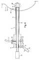
Figure 7 illustrates and adaptation of the cannula to provideadditional protection to the skin. The
Claims (9)
- A device for performing a biopsy of a mass of tissuewithin a human patient, said device comprising:characterized by comprising:a cannula (5) adapted for insertion into the body of thepatient, said cannula having a distal end (6) and aproximal end (7), and a lumen (9) extending through thecannula and defining a proximal opening and a distalopening in the cannula;a fitting (10) disposed on the proximal end of the cannula,said fittingenabling a vacuum to be applied to the lumen when anelongated medical device (20,24,27) is received in thelumen; andan airtight seal (12) in the proximal opening of the cannula,said airtight seal permittingelongated medical devices (20,24,27) to beintroduced and removed through the seal while substantiallymaintaining the airtight seal.
- A device according to claim 1,characterized in that theairtight seal (12) comprises a plug (13) of resilient material,preferably silicone, and the plug preferably being provided with aslit (14) for receiving the elongate medical device (20,24,27)therein.
- A device according to claim 2,characterized bycomprising a stopper (15) for insertion into the slit (14) whenthe valve (12) is not occupied by the elongate medical device(20,24,27).
- A device according to any of claims 1 to 3,characterized bycomprising an additional valve (16) allowing a vacuum in thecannula (5) to be broken.
- A device according to any of claims 1 to 4,characterized inthat the cannula (5) has substantially intact sidewalls.
- A system for treating or sampling of a mass of tissuewithin a human patient, said system comprising:a device according to any of claims 1 to 5;a source of vacuum pressure (11) operably connected to thefitting (10); andan elongate medical device (20,24,27) capable of insertionthrough the airtight seal (12) and into the cannula (5),said elongate medical device being long enough to extendfrom the proximal end (7) of the cannula to a distanceoutside the distal opening (6) of the cannula.
- A system according to claim 6,characterized in that theelongate medical device is a biopsy needle (20,24).
- A system according to claim 6,characterized in that theelongate medical device is a cryoprobe (27).
- A system according to claim 6,characterized in that theelongate medical device is an ablation device (27) suitable forablation of the mass.
Applications Claiming Priority (3)
| Application Number | Priority Date | Filing Date | Title |
|---|---|---|---|
| US09/598,124US6494844B1 (en) | 2000-06-21 | 2000-06-21 | Device for biopsy and treatment of breast tumors |
| US598124 | 2000-06-21 | ||
| PCT/US2001/019454WO2001097702A1 (en) | 2000-06-21 | 2001-06-19 | Device for biopsy and treatment of breast tumors |
Publications (3)
| Publication Number | Publication Date |
|---|---|
| EP1296607A1 EP1296607A1 (en) | 2003-04-02 |
| EP1296607A4 EP1296607A4 (en) | 2003-07-30 |
| EP1296607B1true EP1296607B1 (en) | 2005-09-07 |
Family
ID=24394333
Family Applications (1)
| Application Number | Title | Priority Date | Filing Date |
|---|---|---|---|
| EP01946481AExpired - LifetimeEP1296607B1 (en) | 2000-06-21 | 2001-06-19 | Device for biopsy and treatment of breast tumors |
Country Status (8)
| Country | Link |
|---|---|
| US (3) | US6494844B1 (en) |
| EP (1) | EP1296607B1 (en) |
| JP (1) | JP2004508850A (en) |
| AT (1) | ATE303767T1 (en) |
| AU (2) | AU6852701A (en) |
| CA (1) | CA2412826A1 (en) |
| DE (1) | DE60113261T2 (en) |
| WO (1) | WO2001097702A1 (en) |
Cited By (1)
| Publication number | Priority date | Publication date | Assignee | Title |
|---|---|---|---|---|
| DE102007020582A1 (en)* | 2006-12-19 | 2008-06-26 | Erbe Elektromedizin Gmbh | A cryosurgical instrument and method for separating a tissue sample from surrounding tissue of a biological tissue to be treated |
Families Citing this family (187)
| Publication number | Priority date | Publication date | Assignee | Title |
|---|---|---|---|---|
| US6494844B1 (en)* | 2000-06-21 | 2002-12-17 | Sanarus Medical, Inc. | Device for biopsy and treatment of breast tumors |
| US6758824B1 (en) | 2000-11-06 | 2004-07-06 | Suros Surgical Systems, Inc. | Biopsy apparatus |
| WO2002069808A2 (en) | 2000-11-06 | 2002-09-12 | Suros Surgical Systems, Inc. | Biopsy apparatus |
| US20030069502A1 (en) | 2001-05-29 | 2003-04-10 | Makin Inder Raj. S. | Ultrasound feedback in medically-treated patients |
| WO2003026719A2 (en)* | 2001-09-27 | 2003-04-03 | Galil Medical Ltd. | Cryoplasty apparatus and method |
| CA2461627A1 (en)* | 2001-09-27 | 2003-04-03 | Roni Zvuloni | Apparatus and method for cryosurgical treatment of tumors of the breast |
| US7769432B2 (en)* | 2001-12-10 | 2010-08-03 | Board Of Trustees Of The University Of Arkansas | Minimally invasive diagnosis and treatment for breast cancer |
| US8840608B2 (en) | 2002-03-15 | 2014-09-23 | The General Hospital Corporation | Methods and devices for selective disruption of fatty tissue by controlled cooling |
| EP1917935B1 (en) | 2002-03-15 | 2011-01-12 | The General Hospital Corporation | Method for selective disruption of fatty tissue by controlled cooling |
| EP1524940B1 (en) | 2002-03-19 | 2011-08-24 | Bard Dublin ITC Limited | Biopsy device and biopsy needle module that can be inserted into the biopsy device |
| US8109885B2 (en) | 2002-03-19 | 2012-02-07 | C. R. Bard, Inc. | Biopsy device for removing tissue specimens using a vacuum |
| US6789545B2 (en)* | 2002-10-04 | 2004-09-14 | Sanarus Medical, Inc. | Method and system for cryoablating fibroadenomas |
| US7347829B2 (en)* | 2002-10-07 | 2008-03-25 | Suros Surgical Systems, Inc. | Introduction system for minimally invasive surgical instruments |
| US20070260267A1 (en)* | 2002-10-07 | 2007-11-08 | Nicoson Zachary R | Localizing obturator |
| US8123698B2 (en)* | 2002-10-07 | 2012-02-28 | Suros Surgical Systems, Inc. | System and method for minimally invasive disease therapy |
| US20040147917A1 (en)* | 2003-01-23 | 2004-07-29 | Mueller Richard L. | Device and method for treatment of breast tissue with electromagnetic radiation |
| US20040215177A1 (en)* | 2003-04-24 | 2004-10-28 | Scimed Life Systems, Inc. | Therapeutic apparatus having insulated region at the insertion area |
| US20050113816A1 (en)* | 2003-05-23 | 2005-05-26 | Whitmore Willet F.Iii | Instrument guide with capture and release in an image plane |
| US7909815B2 (en)* | 2003-05-23 | 2011-03-22 | Civco Medical Instruments Co., Inc. | Instrument guide for use with needles and catheters |
| US7179232B2 (en)* | 2003-06-27 | 2007-02-20 | Depuy Acromed, Inc. | Controlled orifice sampling needle |
| US20120289859A9 (en)* | 2003-08-27 | 2012-11-15 | Nicoson Zachary R | System and method for minimally invasive disease therapy |
| US8172770B2 (en) | 2005-09-28 | 2012-05-08 | Suros Surgical Systems, Inc. | System and method for minimally invasive disease therapy |
| JP4500315B2 (en) | 2003-10-14 | 2010-07-14 | シュロス・サージカル・システムズ・インコーポレーテッド | Vacuum assisted biopsy needle set |
| US7988642B2 (en) | 2003-10-14 | 2011-08-02 | Suros Surgical Systems, Inc. | Vacuum assisted biopsy device |
| US8048003B2 (en) | 2003-10-14 | 2011-11-01 | Suros Surgical Systems, Inc. | Vacuum assisted biopsy device |
| US8357103B2 (en) | 2003-10-14 | 2013-01-22 | Suros Surgical Systems, Inc. | Vacuum assisted biopsy needle set |
| US7699783B2 (en)* | 2004-04-08 | 2010-04-20 | Techniscan, Inc. | Method for imaging and treating a breast |
| US7473250B2 (en) | 2004-05-21 | 2009-01-06 | Ethicon Endo-Surgery, Inc. | Ultrasound medical system and method |
| US7708751B2 (en) | 2004-05-21 | 2010-05-04 | Ethicon Endo-Surgery, Inc. | MRI biopsy device |
| US9638770B2 (en) | 2004-05-21 | 2017-05-02 | Devicor Medical Products, Inc. | MRI biopsy apparatus incorporating an imageable penetrating portion |
| US8932233B2 (en) | 2004-05-21 | 2015-01-13 | Devicor Medical Products, Inc. | MRI biopsy device |
| US7670282B2 (en) | 2004-06-14 | 2010-03-02 | Pneumrx, Inc. | Lung access device |
| EP3542736A1 (en)* | 2004-06-16 | 2019-09-25 | PneumRx, Inc. | Intra-bronchial lung volume reduction system |
| DE102004030155B4 (en)* | 2004-06-22 | 2020-04-23 | Robert Bosch Gmbh | Metering device and method for operating the same |
| CA2570261C (en) | 2004-07-08 | 2014-06-10 | Pneumrx, Inc. | Pleural effusion treatment device, method and material |
| US7766891B2 (en) | 2004-07-08 | 2010-08-03 | Pneumrx, Inc. | Lung device with sealing features |
| JP4814229B2 (en) | 2004-07-09 | 2011-11-16 | バード ペリフェラル ヴァスキュラー インコーポレイテッド | Transport device for biopsy device |
| US7440793B2 (en)* | 2004-07-22 | 2008-10-21 | Sunita Chauhan | Apparatus and method for removing abnormal tissue |
| US8060183B2 (en) | 2004-10-13 | 2011-11-15 | Suros Surgical Systems, Inc. | Site marker visible under multiple modalities |
| US8442623B2 (en)* | 2004-10-13 | 2013-05-14 | Suros Surgical Systems, Inc. | Site marker visible under multiple modalities |
| WO2006058195A2 (en) | 2004-11-23 | 2006-06-01 | Pneumrx, Inc. | Steerable device for accessing a target site and methods |
| US7517321B2 (en) | 2005-01-31 | 2009-04-14 | C. R. Bard, Inc. | Quick cycle biopsy system |
| US7942873B2 (en)* | 2005-03-25 | 2011-05-17 | Angiodynamics, Inc. | Cavity ablation apparatus and method |
| US8263109B2 (en) | 2005-05-09 | 2012-09-11 | Boston Scientific Scimed, Inc. | Injectable bulking compositions |
| US7862552B2 (en) | 2005-05-09 | 2011-01-04 | Boston Scientific Scimed, Inc. | Medical devices for treating urological and uterine conditions |
| US20060251581A1 (en)* | 2005-05-09 | 2006-11-09 | Mcintyre Jon T | Method for treatment of uterine fibroid tumors |
| US7556622B2 (en) | 2005-05-18 | 2009-07-07 | Suros Surgical Systems, Inc. | Selectively openable tissue filter |
| US7850683B2 (en) | 2005-05-20 | 2010-12-14 | Myoscience, Inc. | Subdermal cryogenic remodeling of muscles, nerves, connective tissue, and/or adipose tissue (fat) |
| US7713266B2 (en) | 2005-05-20 | 2010-05-11 | Myoscience, Inc. | Subdermal cryogenic remodeling of muscles, nerves, connective tissue, and/or adipose tissue (fat) |
| EP1921998B8 (en) | 2005-08-10 | 2021-07-07 | C.R.Bard, Inc. | Single-insertion, multiple sampling biopsy device with linear drive |
| ES2539578T3 (en) | 2005-08-10 | 2015-07-02 | C.R. Bard, Inc. | Multi-sample biopsy device and single insert with various transport systems |
| US20080200834A1 (en)* | 2005-09-28 | 2008-08-21 | Mark Joseph L | Introducer device for improved imaging |
| US20070149959A1 (en)* | 2005-12-23 | 2007-06-28 | Sanarus Medical, Inc. | Cryoprobe for low pressure systems |
| US20070156125A1 (en)* | 2005-12-30 | 2007-07-05 | Russell Delonzor | Encodable cryogenic device |
| US7854754B2 (en) | 2006-02-22 | 2010-12-21 | Zeltiq Aesthetics, Inc. | Cooling device for removing heat from subcutaneous lipid-rich cells |
| US7670299B2 (en) | 2006-03-07 | 2010-03-02 | Ethincon Endo-Surgery, Inc. | Device for minimally invasive internal tissue removal |
| US7806834B2 (en) | 2006-03-07 | 2010-10-05 | Devicor Medical Products, Inc. | Device for minimally invasive internal tissue removal |
| US8157837B2 (en) | 2006-03-13 | 2012-04-17 | Pneumrx, Inc. | Minimally invasive lung volume reduction device and method |
| US9402633B2 (en) | 2006-03-13 | 2016-08-02 | Pneumrx, Inc. | Torque alleviating intra-airway lung volume reduction compressive implant structures |
| US8888800B2 (en) | 2006-03-13 | 2014-11-18 | Pneumrx, Inc. | Lung volume reduction devices, methods, and systems |
| US7465278B2 (en) | 2006-03-29 | 2008-12-16 | Ethicon Endo-Surgery, Inc. | Device for minimally invasive internal tissue removal |
| JP2009534156A (en)* | 2006-04-24 | 2009-09-24 | トーマス・ジェファーソン・ユニバーシティ | Cryoneedle and cryotherapy system |
| KR101039758B1 (en) | 2006-04-28 | 2011-06-09 | 젤티크 애스세틱스, 인코포레이티드. | Cryoprotectants for use with therapeutic devices for improved cooling of subcutaneous lipid-rich cells |
| US20140025056A1 (en)* | 2006-05-24 | 2014-01-23 | Kambiz Dowlatshahi | Image-guided removal and thermal therapy of breast cancer |
| EP3417792B1 (en) | 2006-08-21 | 2022-03-02 | C. R. Bard, Inc. | Self-contained handheld biopsy needle |
| WO2008029408A1 (en)* | 2006-09-08 | 2008-03-13 | Arbel Medical Ltd. | Method and device for combined treatment |
| US20080077201A1 (en) | 2006-09-26 | 2008-03-27 | Juniper Medical, Inc. | Cooling devices with flexible sensors |
| US8192474B2 (en) | 2006-09-26 | 2012-06-05 | Zeltiq Aesthetics, Inc. | Tissue treatment methods |
| US9132031B2 (en) | 2006-09-26 | 2015-09-15 | Zeltiq Aesthetics, Inc. | Cooling device having a plurality of controllable cooling elements to provide a predetermined cooling profile |
| SI2086418T1 (en) | 2006-10-06 | 2011-05-31 | Bard Peripheral Vascular Inc | Tissue handling system with reduced operator exposure |
| US8262586B2 (en) | 2006-10-24 | 2012-09-11 | C. R. Bard, Inc. | Large sample low aspect ratio biopsy needle |
| US9254162B2 (en) | 2006-12-21 | 2016-02-09 | Myoscience, Inc. | Dermal and transdermal cryogenic microprobe systems |
| US8409185B2 (en)* | 2007-02-16 | 2013-04-02 | Myoscience, Inc. | Replaceable and/or easily removable needle systems for dermal and transdermal cryogenic remodeling |
| US20090306646A1 (en)* | 2007-05-14 | 2009-12-10 | Bsd Medical Corporation | Apparatus and method for injection enhancement of selective heating of a deposit in tissues in a body |
| US8423152B2 (en)* | 2007-05-14 | 2013-04-16 | Bsd Medical Corporation | Apparatus and method for selectively heating a deposit in fatty tissue in a body |
| US9387036B2 (en)* | 2007-05-14 | 2016-07-12 | Pyrexar Medical Inc. | Apparatus and method for selectively heating a deposit in fatty tissue in a body |
| WO2008143901A2 (en)* | 2007-05-15 | 2008-11-27 | Techniscan, Inc. | Improved imaging system |
| US20080287839A1 (en) | 2007-05-18 | 2008-11-20 | Juniper Medical, Inc. | Method of enhanced removal of heat from subcutaneous lipid-rich cells and treatment apparatus having an actuator |
| US7866223B2 (en)* | 2007-06-04 | 2011-01-11 | Swift & Company | Method of obtaining samples of meat to assay for microbial contamination |
| DE102008026635B4 (en)* | 2007-06-26 | 2010-10-28 | Erbe Elektromedizin Gmbh | Kryobiopsiesonde |
| US8523927B2 (en) | 2007-07-13 | 2013-09-03 | Zeltiq Aesthetics, Inc. | System for treating lipid-rich regions |
| US8380299B2 (en)* | 2007-07-30 | 2013-02-19 | Nuvue Therapeutics, Inc. | Fluid flowing device and method for tissue diagnosis or therapy |
| WO2009026471A1 (en) | 2007-08-21 | 2009-02-26 | Zeltiq Aesthetics, Inc. | Monitoring the cooling of subcutaneous lipid-rich cells, such as the cooling of adipose tissue |
| US8202229B2 (en) | 2007-10-01 | 2012-06-19 | Suros Surgical Systems, Inc. | Surgical device |
| US8808200B2 (en) | 2007-10-01 | 2014-08-19 | Suros Surgical Systems, Inc. | Surgical device and method of using same |
| WO2009065061A1 (en) | 2007-11-14 | 2009-05-22 | Myoscience, Inc. | Pain management using cryogenic remodeling |
| US8241225B2 (en) | 2007-12-20 | 2012-08-14 | C. R. Bard, Inc. | Biopsy device |
| US20090247901A1 (en)* | 2008-03-25 | 2009-10-01 | Brian Zimmer | Latching side removal spacer |
| US20090247900A1 (en)* | 2008-03-25 | 2009-10-01 | Brian Zimmer | Push button adjustable spacer |
| US8043316B2 (en)* | 2008-05-02 | 2011-10-25 | Suros Surgical Systems, Inc. | Adjustable spacer |
| US8449478B2 (en)* | 2008-05-16 | 2013-05-28 | Conquest Medical Technologies | Biopsy device |
| JP5233031B2 (en)* | 2008-07-15 | 2013-07-10 | 株式会社デージーエス・コンピュータ | Cryotherapy planning device and cryotherapy device |
| US8845627B2 (en)* | 2008-08-22 | 2014-09-30 | Boston Scientific Scimed, Inc. | Regulating pressure to lower temperature in a cryotherapy balloon catheter |
| US9173669B2 (en) | 2008-09-12 | 2015-11-03 | Pneumrx, Inc. | Enhanced efficacy lung volume reduction devices, methods, and systems |
| WO2010036732A1 (en) | 2008-09-25 | 2010-04-01 | Zeltiq Aesthetics, Inc. | Treatment planning systems and methods for body contouring applications |
| US9332973B2 (en) | 2008-10-01 | 2016-05-10 | Covidien Lp | Needle biopsy device with exchangeable needle and integrated needle protection |
| US11298113B2 (en) | 2008-10-01 | 2022-04-12 | Covidien Lp | Device for needle biopsy with integrated needle protection |
| US9782565B2 (en) | 2008-10-01 | 2017-10-10 | Covidien Lp | Endoscopic ultrasound-guided biliary access system |
| US9186128B2 (en) | 2008-10-01 | 2015-11-17 | Covidien Lp | Needle biopsy device |
| US8968210B2 (en) | 2008-10-01 | 2015-03-03 | Covidien LLP | Device for needle biopsy with integrated needle protection |
| RU2394521C1 (en)* | 2008-12-15 | 2010-07-20 | Государственное образовательное учреждение высшего профессионального образования Новгородский государственный университет имени Ярослава Мудрого | Method of minimally invasive removal of breast cancer and related device for implementation thereof |
| US8603073B2 (en) | 2008-12-17 | 2013-12-10 | Zeltiq Aesthetics, Inc. | Systems and methods with interrupt/resume capabilities for treating subcutaneous lipid-rich cells |
| JP5642087B2 (en) | 2008-12-22 | 2014-12-17 | ミオサイエンス インコーポレーティッド | Integrated cryosurgery system with refrigerant and power supply |
| WO2010083281A1 (en)* | 2009-01-15 | 2010-07-22 | Boston Scientific Scimed, Inc. | Controlling depth of cryoablation |
| WO2010124109A1 (en) | 2009-04-22 | 2010-10-28 | Nuvue Therapeutics, Inc. | Fluid flowing device and method for tissue diagnosis or therapy |
| CA2760610C (en) | 2009-04-30 | 2017-09-19 | Zeltiq Aesthetics, Inc. | Device, system and method of removing heat from subcutaneous lipid-rich cells |
| WO2010135352A1 (en) | 2009-05-18 | 2010-11-25 | Pneumrx, Inc. | Cross-sectional modification during deployment of an elongate lung volume reduction device |
| US8529468B2 (en) | 2009-07-01 | 2013-09-10 | Suros Surgical Systems, Inc. | Surgical system |
| US9173641B2 (en) | 2009-08-12 | 2015-11-03 | C. R. Bard, Inc. | Biopsy apparatus having integrated thumbwheel mechanism for manual rotation of biopsy cannula |
| US8430824B2 (en) | 2009-10-29 | 2013-04-30 | Bard Peripheral Vascular, Inc. | Biopsy driver assembly having a control circuit for conserving battery power |
| US8283890B2 (en) | 2009-09-25 | 2012-10-09 | Bard Peripheral Vascular, Inc. | Charging station for battery powered biopsy apparatus |
| CA2787374A1 (en) | 2010-01-25 | 2011-07-28 | Zeltiq Aesthetics, Inc. | Home-use applicators for non-invasively removing heat from subcutaneous lipid-rich cells via phase change coolants, and associated devices, systems and methods |
| US9332970B2 (en)* | 2010-02-25 | 2016-05-10 | Kohala Inc. | Full core biopsy device |
| US20110224576A1 (en)* | 2010-03-12 | 2011-09-15 | Biotex, Inc. | Methods and devices for tissue collection and analysis |
| US8597201B2 (en) | 2010-03-30 | 2013-12-03 | Siteselect Medical Technologies, Inc. | Tissue excision device with a flexible transection blade |
| TWI455722B (en) | 2010-06-04 | 2014-10-11 | Pfizer Vaccines Llc | Conjugates for the prevention or treatment of nicotine addiction |
| US8676338B2 (en) | 2010-07-20 | 2014-03-18 | Zeltiq Aesthetics, Inc. | Combined modality treatment systems, methods and apparatus for body contouring applications |
| US10722395B2 (en) | 2011-01-25 | 2020-07-28 | Zeltiq Aesthetics, Inc. | Devices, application systems and methods with localized heat flux zones for removing heat from subcutaneous lipid-rich cells |
| ES2833525T3 (en)* | 2011-07-21 | 2021-06-15 | Massachusetts Gen Hospital | Apparatus for deteriorating and removing grease |
| EP2609895B1 (en)* | 2011-12-28 | 2015-11-04 | The Cleveland Clinic Foundation | Endoluminal prosthesis with valve arrangement |
| CN104159534B (en) | 2012-01-13 | 2017-02-22 | 肌肉科技股份有限公司 | Skin protection for subdermal cryogenic remodeling for cosmetic and other treatments |
| EP2802279B1 (en) | 2012-01-13 | 2017-08-16 | Myoscience, Inc. | Cryogenic needle with freeze zone regulation |
| WO2013106860A1 (en) | 2012-01-13 | 2013-07-18 | Myoscience, Inc. | Cryogenic probe filtration system |
| US9017318B2 (en) | 2012-01-20 | 2015-04-28 | Myoscience, Inc. | Cryogenic probe system and method |
| EP2838435B1 (en) | 2012-04-16 | 2020-03-25 | Hathaway, Jeff M. | Biopsy device |
| WO2014186319A1 (en)* | 2013-05-13 | 2014-11-20 | The Johns Hopkins University | Encapsulated cryoprobe for flexible bronchoscope |
| US20140073907A1 (en) | 2012-09-12 | 2014-03-13 | Convergent Life Sciences, Inc. | System and method for image guided medical procedures |
| US9295454B2 (en)* | 2012-09-21 | 2016-03-29 | Ko-Pen Wang | Double lumen or double wire endobronchial ultrasound-guided histology needle (EBUS) |
| EP4039236A1 (en) | 2013-02-20 | 2022-08-10 | Cytrellis Biosystems, Inc. | System for tightening a region of skin |
| US9545523B2 (en) | 2013-03-14 | 2017-01-17 | Zeltiq Aesthetics, Inc. | Multi-modality treatment systems, methods and apparatus for altering subcutaneous lipid-rich tissue |
| US9844460B2 (en) | 2013-03-14 | 2017-12-19 | Zeltiq Aesthetics, Inc. | Treatment systems with fluid mixing systems and fluid-cooled applicators and methods of using the same |
| US9295512B2 (en) | 2013-03-15 | 2016-03-29 | Myoscience, Inc. | Methods and devices for pain management |
| WO2014146126A1 (en) | 2013-03-15 | 2014-09-18 | Myoscience, Inc. | Cryogenic blunt dissection methods and devices |
| US9610112B2 (en) | 2013-03-15 | 2017-04-04 | Myoscience, Inc. | Cryogenic enhancement of joint function, alleviation of joint stiffness and/or alleviation of pain associated with osteoarthritis |
| WO2014146127A1 (en) | 2013-03-15 | 2014-09-18 | Myoscience, Inc. | Methods and systems for treatment of spasticity |
| CA2902221A1 (en) | 2013-03-20 | 2014-09-25 | Bard Peripheral Vascular, Inc. | Biopsy device |
| AU2014306273B2 (en) | 2013-08-09 | 2019-07-11 | Cytrellis Biosystems, Inc. | Methods and apparatuses for skin treatment using non-thermal tissue ablation |
| US10130409B2 (en) | 2013-11-05 | 2018-11-20 | Myoscience, Inc. | Secure cryosurgical treatment system |
| ES2726985T3 (en) | 2013-11-05 | 2019-10-11 | Bard Inc C R | Biopsy device that has integrated vacuum |
| US20150216719A1 (en) | 2014-01-31 | 2015-08-06 | Zeltiq Aesthetics, Inc | Treatment systems and methods for treating cellulite and for providing other treatments |
| US10675176B1 (en) | 2014-03-19 | 2020-06-09 | Zeltiq Aesthetics, Inc. | Treatment systems, devices, and methods for cooling targeted tissue |
| USD777338S1 (en) | 2014-03-20 | 2017-01-24 | Zeltiq Aesthetics, Inc. | Cryotherapy applicator for cooling tissue |
| US10952891B1 (en) | 2014-05-13 | 2021-03-23 | Zeltiq Aesthetics, Inc. | Treatment systems with adjustable gap applicators and methods for cooling tissue |
| US10568759B2 (en) | 2014-08-19 | 2020-02-25 | Zeltiq Aesthetics, Inc. | Treatment systems, small volume applicators, and methods for treating submental tissue |
| US10935174B2 (en) | 2014-08-19 | 2021-03-02 | Zeltiq Aesthetics, Inc. | Stress relief couplings for cryotherapy apparatuses |
| US10390838B1 (en) | 2014-08-20 | 2019-08-27 | Pneumrx, Inc. | Tuned strength chronic obstructive pulmonary disease treatment |
| JP6678674B2 (en) | 2014-09-05 | 2020-04-08 | ペイブ,エルエルシー | Improvements for a complete core biopsy device |
| CA2967636A1 (en) | 2014-11-14 | 2016-05-19 | Cytrellis Biosystems, Inc. | Devices and methods for ablation of the skin |
| WO2016178656A1 (en) | 2015-05-01 | 2016-11-10 | C. R. Bard, Inc. | Biopsy device |
| US10159971B2 (en)* | 2015-05-03 | 2018-12-25 | Clear Labs Inc. | Apparatus and method for economic, fast and easy sampling of food and environmental samples |
| US11154418B2 (en) | 2015-10-19 | 2021-10-26 | Zeltiq Aesthetics, Inc. | Vascular treatment systems, cooling devices, and methods for cooling vascular structures |
| US20170119432A1 (en) | 2015-10-28 | 2017-05-04 | Warsaw Orthopedic, Inc. | Nerve and soft tissue surgical device |
| HK1259174A1 (en) | 2016-01-07 | 2019-11-29 | Zeltiq Aesthetics, Inc. | Temperature-dependent adhesion between applicator and skin during cooling of tissue |
| US10765552B2 (en) | 2016-02-18 | 2020-09-08 | Zeltiq Aesthetics, Inc. | Cooling cup applicators with contoured heads and liner assemblies |
| US10786224B2 (en) | 2016-04-21 | 2020-09-29 | Covidien Lp | Biopsy devices and methods of use thereof |
| US10555831B2 (en) | 2016-05-10 | 2020-02-11 | Zeltiq Aesthetics, Inc. | Hydrogel substances and methods of cryotherapy |
| US11382790B2 (en) | 2016-05-10 | 2022-07-12 | Zeltiq Aesthetics, Inc. | Skin freezing systems for treating acne and skin conditions |
| US10682297B2 (en) | 2016-05-10 | 2020-06-16 | Zeltiq Aesthetics, Inc. | Liposomes, emulsions, and methods for cryotherapy |
| EP4349396A3 (en) | 2016-05-13 | 2024-05-01 | Pacira CryoTech, Inc. | Systems for locating and treating with cold therapy |
| CN109922740B (en) | 2016-09-21 | 2022-08-23 | 希特利斯生物系统有限公司 | Device and method for cosmetic skin reconstruction |
| US11076879B2 (en) | 2017-04-26 | 2021-08-03 | Zeltiq Aesthetics, Inc. | Shallow surface cryotherapy applicators and related technology |
| US20180310977A1 (en)* | 2017-04-28 | 2018-11-01 | Kyphon SÀRL | Introducer and cryoprobe |
| US11737805B2 (en) | 2017-09-14 | 2023-08-29 | The Regents Of The University Of California | Cryoablation devices and related methods |
| EP3709918B1 (en) | 2017-11-15 | 2025-06-18 | Pacira CryoTech, Inc. | Integrated cold therapy and electrical stimulation systems for locating and treating nerves |
| CN119950019A (en)* | 2017-11-28 | 2025-05-09 | 杭州诺诚医疗器械有限公司 | Ablation needle assembly and ablation system |
| US11331161B2 (en) | 2018-03-23 | 2022-05-17 | Covidien Lp | Surgical assemblies facilitating tissue marking and methods of use thereof |
| US11065372B2 (en)* | 2018-03-27 | 2021-07-20 | Gyrus Acmi, Inc. | Needle system restrictor |
| WO2019213205A1 (en) | 2018-05-01 | 2019-11-07 | The Johns Hopkins University | Carbon dioxide-based percutaneous cryosurgical system |
| WO2020028472A1 (en) | 2018-07-31 | 2020-02-06 | Zeltiq Aesthetics, Inc. | Methods, devices, and systems for improving skin characteristics |
| RU2704779C1 (en)* | 2018-10-09 | 2019-10-30 | Андрей Анатольевич Анохин | Device for removal of soft tissue mass |
| EP3636162B1 (en) | 2018-10-09 | 2023-07-19 | BibbInstruments AB | Biopsy instrument and kit of parts |
| US10610280B1 (en) | 2019-02-02 | 2020-04-07 | Ayad K. M. Agha | Surgical method and apparatus for destruction and removal of intraperitoneal, visceral, and subcutaneous fat |
| CN109805968B (en)* | 2019-03-05 | 2024-08-06 | 上海医萃医疗科技中心(有限合伙) | Device for establishing a closed negative pressure tunnel in the lungs guided by CT |
| CN109758190A (en)* | 2019-03-11 | 2019-05-17 | 南京市第一医院 | An improved biopsy needle |
| US11517294B2 (en) | 2019-05-07 | 2022-12-06 | Covidien Lp | Biopsy devices and methods of use thereof |
| WO2020236691A1 (en)* | 2019-05-20 | 2020-11-26 | Innoblative Designs, Inc. | Minimally invasive assembly for lung ablation |
| CN113116502A (en)* | 2019-12-30 | 2021-07-16 | 杭州诺诚医疗器械有限公司 | Puncture needle assembly and ablation needle assembly |
| US11633224B2 (en) | 2020-02-10 | 2023-04-25 | Icecure Medical Ltd. | Cryogen pump |
| CA3179231A1 (en) | 2020-04-08 | 2021-10-14 | Bibbinstruments Ab | Biopsy instrument, kit of parts and method |
| KR102416005B1 (en)* | 2020-04-29 | 2022-06-30 | 연세대학교 산학협력단 | Apparatus and Method for Cutting and Retrieving Breast Specimen |
| JP2023538213A (en) | 2020-08-14 | 2023-09-07 | イクテロ メディカル,インコーポレイテッド | Systems, devices, and methods for ablation and defunctionalization of the gallbladder |
| WO2022204092A1 (en)* | 2021-03-23 | 2022-09-29 | Overture Life, Inc. | Cryostorage device |
| US12426934B2 (en) | 2022-02-28 | 2025-09-30 | Icecure Medical Ltd. | Cryogen flow control |
| US12215811B2 (en) | 2022-07-18 | 2025-02-04 | Icecure Medical Ltd. | Cryogenic system connector |
| CN118557270A (en)* | 2023-02-28 | 2024-08-30 | 上海澍能医疗科技有限公司 | Biopsy and ablation device, biopsy and ablation system |
| CN116746966B (en)* | 2023-03-23 | 2023-11-28 | 上海导向医疗系统有限公司 | Low-temperature freezing rotary cutting device |
| DE102023124190B3 (en) | 2023-09-07 | 2024-09-19 | Karl Storz Se & Co. Kg | Device for guiding a biopsy element, system for performing a biopsy and method for operating such a system |
Family Cites Families (28)
| Publication number | Priority date | Publication date | Assignee | Title |
|---|---|---|---|---|
| US4644951A (en) | 1985-09-16 | 1987-02-24 | Concept, Inc. | Vacuum sleeve for a surgical appliance |
| US4784156A (en)* | 1987-09-16 | 1988-11-15 | Garg Rakesh K | Cannula including a valve structure and associated instrument elements and method for using same |
| US5056532A (en) | 1989-07-25 | 1991-10-15 | Medtronic, Inc. | Esophageal pacing lead |
| US5505210A (en)* | 1989-11-06 | 1996-04-09 | Mectra Labs, Inc. | Lavage with tissue cutting cannula |
| US5056523A (en) | 1989-11-22 | 1991-10-15 | Board Of Regents, The University Of Texas System | Precision breast lesion localizer |
| US5172701A (en) | 1990-02-28 | 1992-12-22 | Medical Device Technologies, Inc. | Single use automated soft tissue aspiration biopsy device |
| US5027827A (en) | 1990-03-28 | 1991-07-02 | Cody Michael P | Vacuum biopsy apparatus |
| US5353804A (en) | 1990-09-18 | 1994-10-11 | Peb Biopsy Corporation | Method and device for percutaneous exisional breast biopsy |
| US5300046A (en)* | 1992-03-30 | 1994-04-05 | Symbiosis Corporation | Thoracentesis sheath catheter assembly |
| US5314406A (en)* | 1992-10-09 | 1994-05-24 | Symbiosis Corporation | Endoscopic electrosurgical suction-irrigation instrument |
| US5649547A (en) | 1994-03-24 | 1997-07-22 | Biopsys Medical, Inc. | Methods and devices for automated biopsy and collection of soft tissue |
| US5526822A (en) | 1994-03-24 | 1996-06-18 | Biopsys Medical, Inc. | Method and apparatus for automated biopsy and collection of soft tissue |
| US5868673A (en) | 1995-03-28 | 1999-02-09 | Sonometrics Corporation | System for carrying out surgery, biopsy and ablation of a tumor or other physical anomaly |
| US5769086A (en) | 1995-12-06 | 1998-06-23 | Biopsys Medical, Inc. | Control system and method for automated biopsy device |
| US6505629B1 (en)* | 1996-07-23 | 2003-01-14 | Endocare, Inc. | Cryosurgical system with protective warming feature |
| US5902310A (en) | 1996-08-12 | 1999-05-11 | Ethicon Endo-Surgery, Inc. | Apparatus and method for marking tissue |
| US5913857A (en) | 1996-08-29 | 1999-06-22 | Ethicon End0-Surgery, Inc. | Methods and devices for collection of soft tissue |
| US5810806A (en) | 1996-08-29 | 1998-09-22 | Ethicon Endo-Surgery | Methods and devices for collection of soft tissue |
| WO1998041157A1 (en) | 1997-03-17 | 1998-09-24 | Boris Rubinsky | Freezing method for controlled removal of fatty tissue by liposuction |
| US6041787A (en)* | 1997-03-17 | 2000-03-28 | Rubinsky; Boris | Use of cryoprotective agent compounds during cryosurgery |
| US6017316A (en) | 1997-06-18 | 2000-01-25 | Biopsys Medical | Vacuum control system and method for automated biopsy device |
| US6540693B2 (en) | 1998-03-03 | 2003-04-01 | Senorx, Inc. | Methods and apparatus for securing medical instruments to desired locations in a patients body |
| US6331166B1 (en) | 1998-03-03 | 2001-12-18 | Senorx, Inc. | Breast biopsy system and method |
| US5964716A (en) | 1998-05-14 | 1999-10-12 | Ethicon Endo-Surgery, Inc. | Method of use for a multi-port biopsy instrument |
| US5944673A (en) | 1998-05-14 | 1999-08-31 | Ethicon Endo-Surgery, Inc. | Biopsy instrument with multi-port needle |
| US6007497A (en) | 1998-06-30 | 1999-12-28 | Ethicon Endo-Surgery, Inc. | Surgical biopsy device |
| US6494844B1 (en)* | 2000-06-21 | 2002-12-17 | Sanarus Medical, Inc. | Device for biopsy and treatment of breast tumors |
| DE502006008733D1 (en) | 2005-11-09 | 2011-02-24 | Xyzmo Software Gmbh | EKTRONIC SIGNATURE OF AN ELECTRONIC DOCUMENT |
- 2000
- 2000-06-21USUS09/598,124patent/US6494844B1/ennot_activeExpired - Lifetime
- 2001
- 2001-06-19ATAT01946481Tpatent/ATE303767T1/ennot_activeIP Right Cessation
- 2001-06-19CACA002412826Apatent/CA2412826A1/ennot_activeAbandoned
- 2001-06-19AUAU6852701Apatent/AU6852701A/enactivePending
- 2001-06-19JPJP2002503180Apatent/JP2004508850A/enactivePending
- 2001-06-19AUAU2001268527Apatent/AU2001268527B2/ennot_activeCeased
- 2001-06-19EPEP01946481Apatent/EP1296607B1/ennot_activeExpired - Lifetime
- 2001-06-19DEDE60113261Tpatent/DE60113261T2/ennot_activeExpired - Lifetime
- 2001-06-19WOPCT/US2001/019454patent/WO2001097702A1/enactiveIP Right Grant
- 2002
- 2002-12-16USUS10/321,136patent/US6945942B2/ennot_activeExpired - Fee Related
- 2005
- 2005-09-16USUS11/229,250patent/US20060009712A1/ennot_activeAbandoned
Cited By (1)
| Publication number | Priority date | Publication date | Assignee | Title |
|---|---|---|---|---|
| DE102007020582A1 (en)* | 2006-12-19 | 2008-06-26 | Erbe Elektromedizin Gmbh | A cryosurgical instrument and method for separating a tissue sample from surrounding tissue of a biological tissue to be treated |
Also Published As
| Publication number | Publication date |
|---|---|
| CA2412826A1 (en) | 2001-12-27 |
| AU6852701A (en) | 2002-01-02 |
| DE60113261D1 (en) | 2005-10-13 |
| US6494844B1 (en) | 2002-12-17 |
| ATE303767T1 (en) | 2005-09-15 |
| US20060009712A1 (en) | 2006-01-12 |
| EP1296607A1 (en) | 2003-04-02 |
| WO2001097702A1 (en) | 2001-12-27 |
| EP1296607A4 (en) | 2003-07-30 |
| DE60113261T2 (en) | 2006-06-08 |
| US20030093008A1 (en) | 2003-05-15 |
| US6945942B2 (en) | 2005-09-20 |
| AU2001268527B2 (en) | 2005-10-13 |
| JP2004508850A (en) | 2004-03-25 |
Similar Documents
| Publication | Publication Date | Title |
|---|---|---|
| EP1296607B1 (en) | Device for biopsy and treatment of breast tumors | |
| AU2001268527A1 (en) | Device for biopsy and treatment of breast tumors | |
| US6540694B1 (en) | Device for biopsy tumors | |
| JP4108473B2 (en) | Tumor biopsy device | |
| US6176834B1 (en) | Minimally invasive biopsy device | |
| CA2443966C (en) | Surgical biopsy device | |
| US20020087152A1 (en) | Systems and methods for delivering a probe into tissue | |
| Saliken et al. | CT for monitoring cryotherapy. | |
| AU2002211568A1 (en) | Device for biopsy of tumors | |
| AU2002258866A1 (en) | Surgical biopsy device | |
| KR20070117552A (en) | Gynecological Ablation Method and System | |
| JP2007527729A (en) | Apparatus and method for protecting tissue during cryoablation | |
| Whitworth et al. | Cryoablation and cryolocalization in the management of breast disease | |
| AU2005239746B2 (en) | Device for biopsy and treatment of breast tumors | |
| WO2007139555A1 (en) | Tissue protective system and method for thermoablative therapies | |
| AU2007205759A1 (en) | Method for biopsy of tumors | |
| AU2005232255A1 (en) | Device for biopsy of tumors |
Legal Events
| Date | Code | Title | Description |
|---|---|---|---|
| PUAI | Public reference made under article 153(3) epc to a published international application that has entered the european phase | Free format text:ORIGINAL CODE: 0009012 | |
| 17P | Request for examination filed | Effective date:20030121 | |
| AK | Designated contracting states | Designated state(s):AT BE CH CY DE DK ES FI FR GB GR IE IT LI LU MC NL PT SE TR Kind code of ref document:A1 Designated state(s):AT BE CH CY DE DK ES FI FR GB GR IE IT LI LU MC NL PT SE TR | |
| AX | Request for extension of the european patent | Extension state:AL LT LV MK RO SI | |
| RIN1 | Information on inventor provided before grant (corrected) | Inventor name:STABINSKY, SETH Inventor name:VAN BLADEL, KEVIN H. Inventor name:MIKUS, PAUL Inventor name:FOY, GLENN Inventor name:ZINDEL, LISA | |
| A4 | Supplementary search report drawn up and despatched | Effective date:20030618 | |
| 17Q | First examination report despatched | Effective date:20041215 | |
| GRAP | Despatch of communication of intention to grant a patent | Free format text:ORIGINAL CODE: EPIDOSNIGR1 | |
| GRAS | Grant fee paid | Free format text:ORIGINAL CODE: EPIDOSNIGR3 | |
| GRAA | (expected) grant | Free format text:ORIGINAL CODE: 0009210 | |
| AK | Designated contracting states | Kind code of ref document:B1 Designated state(s):AT BE CH CY DE DK ES FI FR GB GR IE IT LI LU MC NL PT SE TR | |
| PG25 | Lapsed in a contracting state [announced via postgrant information from national office to epo] | Ref country code:BE Free format text:LAPSE BECAUSE OF FAILURE TO SUBMIT A TRANSLATION OF THE DESCRIPTION OR TO PAY THE FEE WITHIN THE PRESCRIBED TIME-LIMIT Effective date:20050907 Ref country code:NL Free format text:LAPSE BECAUSE OF FAILURE TO SUBMIT A TRANSLATION OF THE DESCRIPTION OR TO PAY THE FEE WITHIN THE PRESCRIBED TIME-LIMIT Effective date:20050907 Ref country code:FI Free format text:LAPSE BECAUSE OF FAILURE TO SUBMIT A TRANSLATION OF THE DESCRIPTION OR TO PAY THE FEE WITHIN THE PRESCRIBED TIME-LIMIT Effective date:20050907 Ref country code:AT Free format text:LAPSE BECAUSE OF FAILURE TO SUBMIT A TRANSLATION OF THE DESCRIPTION OR TO PAY THE FEE WITHIN THE PRESCRIBED TIME-LIMIT Effective date:20050907 Ref country code:LI Free format text:LAPSE BECAUSE OF FAILURE TO SUBMIT A TRANSLATION OF THE DESCRIPTION OR TO PAY THE FEE WITHIN THE PRESCRIBED TIME-LIMIT Effective date:20050907 Ref country code:CH Free format text:LAPSE BECAUSE OF FAILURE TO SUBMIT A TRANSLATION OF THE DESCRIPTION OR TO PAY THE FEE WITHIN THE PRESCRIBED TIME-LIMIT Effective date:20050907 | |
| REG | Reference to a national code | Ref country code:GB Ref legal event code:FG4D | |
| REG | Reference to a national code | Ref country code:CH Ref legal event code:EP | |
| REG | Reference to a national code | Ref country code:IE Ref legal event code:FG4D | |
| REF | Corresponds to: | Ref document number:60113261 Country of ref document:DE Date of ref document:20051013 Kind code of ref document:P | |
| PG25 | Lapsed in a contracting state [announced via postgrant information from national office to epo] | Ref country code:SE Free format text:LAPSE BECAUSE OF FAILURE TO SUBMIT A TRANSLATION OF THE DESCRIPTION OR TO PAY THE FEE WITHIN THE PRESCRIBED TIME-LIMIT Effective date:20051207 Ref country code:GR Free format text:LAPSE BECAUSE OF FAILURE TO SUBMIT A TRANSLATION OF THE DESCRIPTION OR TO PAY THE FEE WITHIN THE PRESCRIBED TIME-LIMIT Effective date:20051207 Ref country code:DK Free format text:LAPSE BECAUSE OF FAILURE TO SUBMIT A TRANSLATION OF THE DESCRIPTION OR TO PAY THE FEE WITHIN THE PRESCRIBED TIME-LIMIT Effective date:20051207 | |
| PG25 | Lapsed in a contracting state [announced via postgrant information from national office to epo] | Ref country code:ES Free format text:LAPSE BECAUSE OF FAILURE TO SUBMIT A TRANSLATION OF THE DESCRIPTION OR TO PAY THE FEE WITHIN THE PRESCRIBED TIME-LIMIT Effective date:20051218 | |
| PG25 | Lapsed in a contracting state [announced via postgrant information from national office to epo] | Ref country code:PT Free format text:LAPSE BECAUSE OF FAILURE TO SUBMIT A TRANSLATION OF THE DESCRIPTION OR TO PAY THE FEE WITHIN THE PRESCRIBED TIME-LIMIT Effective date:20060207 | |
| NLV1 | Nl: lapsed or annulled due to failure to fulfill the requirements of art. 29p and 29m of the patents act | ||
| REG | Reference to a national code | Ref country code:CH Ref legal event code:PL | |
| ET | Fr: translation filed | ||
| PG25 | Lapsed in a contracting state [announced via postgrant information from national office to epo] | Ref country code:MC Free format text:LAPSE BECAUSE OF NON-PAYMENT OF DUE FEES Effective date:20060630 | |
| PLBE | No opposition filed within time limit | Free format text:ORIGINAL CODE: 0009261 | |
| STAA | Information on the status of an ep patent application or granted ep patent | Free format text:STATUS: NO OPPOSITION FILED WITHIN TIME LIMIT | |
| 26N | No opposition filed | Effective date:20060608 | |
| PG25 | Lapsed in a contracting state [announced via postgrant information from national office to epo] | Ref country code:TR Free format text:LAPSE BECAUSE OF FAILURE TO SUBMIT A TRANSLATION OF THE DESCRIPTION OR TO PAY THE FEE WITHIN THE PRESCRIBED TIME-LIMIT Effective date:20050907 Ref country code:LU Free format text:LAPSE BECAUSE OF NON-PAYMENT OF DUE FEES Effective date:20060619 | |
| PG25 | Lapsed in a contracting state [announced via postgrant information from national office to epo] | Ref country code:CY Free format text:LAPSE BECAUSE OF FAILURE TO SUBMIT A TRANSLATION OF THE DESCRIPTION OR TO PAY THE FEE WITHIN THE PRESCRIBED TIME-LIMIT Effective date:20050907 | |
| PGFP | Annual fee paid to national office [announced via postgrant information from national office to epo] | Ref country code:IE Payment date:20100625 Year of fee payment:10 Ref country code:FR Payment date:20100630 Year of fee payment:10 | |
| PGFP | Annual fee paid to national office [announced via postgrant information from national office to epo] | Ref country code:IT Payment date:20100624 Year of fee payment:10 | |
| PGFP | Annual fee paid to national office [announced via postgrant information from national office to epo] | Ref country code:DE Payment date:20100629 Year of fee payment:10 Ref country code:GB Payment date:20100625 Year of fee payment:10 | |
| GBPC | Gb: european patent ceased through non-payment of renewal fee | Effective date:20110619 | |
| PG25 | Lapsed in a contracting state [announced via postgrant information from national office to epo] | Ref country code:IT Free format text:LAPSE BECAUSE OF NON-PAYMENT OF DUE FEES Effective date:20110619 | |
| REG | Reference to a national code | Ref country code:FR Ref legal event code:ST Effective date:20120229 | |
| REG | Reference to a national code | Ref country code:IE Ref legal event code:MM4A | |
| REG | Reference to a national code | Ref country code:DE Ref legal event code:R119 Ref document number:60113261 Country of ref document:DE Effective date:20120103 | |
| PG25 | Lapsed in a contracting state [announced via postgrant information from national office to epo] | Ref country code:DE Free format text:LAPSE BECAUSE OF NON-PAYMENT OF DUE FEES Effective date:20120103 Ref country code:FR Free format text:LAPSE BECAUSE OF NON-PAYMENT OF DUE FEES Effective date:20110630 Ref country code:IE Free format text:LAPSE BECAUSE OF NON-PAYMENT OF DUE FEES Effective date:20110619 | |
| PG25 | Lapsed in a contracting state [announced via postgrant information from national office to epo] | Ref country code:GB Free format text:LAPSE BECAUSE OF NON-PAYMENT OF DUE FEES Effective date:20110619 |