EP1047341B1 - Transluminal methods and apparatus for closing, forming attachments to, and/or forming anastomotic junctions in, luminal anatomical structures - Google Patents
Transluminal methods and apparatus for closing, forming attachments to, and/or forming anastomotic junctions in, luminal anatomical structuresDownload PDFInfo
- Publication number
- EP1047341B1 EP1047341B1EP98931643AEP98931643AEP1047341B1EP 1047341 B1EP1047341 B1EP 1047341B1EP 98931643 AEP98931643 AEP 98931643AEP 98931643 AEP98931643 AEP 98931643AEP 1047341 B1EP1047341 B1EP 1047341B1
- Authority
- EP
- European Patent Office
- Prior art keywords
- needle
- lumen
- anatomical structure
- wall
- catheter
- Prior art date
- Legal status (The legal status is an assumption and is not a legal conclusion. Google has not performed a legal analysis and makes no representation as to the accuracy of the status listed.)
- Expired - Lifetime
Links
- 210000003484anatomyAnatomy0.000titleclaimsabstractdescription137
- 238000000034methodMethods0.000titleabstractdescription44
- 239000000463materialSubstances0.000claimsabstractdescription30
- 210000004204blood vesselAnatomy0.000abstractdescription82
- 210000001519tissueAnatomy0.000description29
- 230000003872anastomosisEffects0.000description25
- 230000000149penetrating effectEffects0.000description23
- 210000003462veinAnatomy0.000description12
- 239000008280bloodSubstances0.000description10
- 210000004369bloodAnatomy0.000description10
- 239000003356suture materialSubstances0.000description9
- 206010002329AneurysmDiseases0.000description8
- 230000017531blood circulationEffects0.000description7
- 238000013461designMethods0.000description6
- 238000004804windingMethods0.000description6
- 206010052428WoundDiseases0.000description4
- 208000027418Wounds and injuryDiseases0.000description4
- 239000000853adhesiveSubstances0.000description4
- 230000001070adhesive effectEffects0.000description4
- 210000001367arteryAnatomy0.000description4
- 210000004351coronary vesselAnatomy0.000description4
- 239000012530fluidSubstances0.000description4
- 210000000936intestineAnatomy0.000description4
- 210000005166vasculatureAnatomy0.000description4
- 238000004873anchoringMethods0.000description3
- 230000000694effectsEffects0.000description3
- 238000001356surgical procedureMethods0.000description3
- 208000001750EndoleakDiseases0.000description2
- 206010064396Stent-graft endoleakDiseases0.000description2
- 206010042618Surgical procedure repeatedDiseases0.000description2
- 238000007792additionMethods0.000description2
- 230000004075alterationEffects0.000description2
- 230000015572biosynthetic processEffects0.000description2
- 238000012217deletionMethods0.000description2
- 230000037430deletionEffects0.000description2
- 238000011161developmentMethods0.000description2
- 239000013013elastic materialSubstances0.000description2
- 229920000295expanded polytetrafluoroethylenePolymers0.000description2
- 238000003384imaging methodMethods0.000description2
- 238000011065in-situ storageMethods0.000description2
- 238000007373indentationMethods0.000description2
- 238000012986modificationMethods0.000description2
- 230000004048modificationEffects0.000description2
- 208000022211Arteriovenous MalformationsDiseases0.000description1
- 206010003658Atrial FibrillationDiseases0.000description1
- 206010010356Congenital anomalyDiseases0.000description1
- 206010063560Excessive granulation tissueDiseases0.000description1
- 206010016717FistulaDiseases0.000description1
- 201000008450Intracranial aneurysmDiseases0.000description1
- 206010028980NeoplasmDiseases0.000description1
- FAPWRFPIFSIZLT-UHFFFAOYSA-MSodium chlorideChemical compound[Na+].[Cl-]FAPWRFPIFSIZLT-UHFFFAOYSA-M0.000description1
- 208000007536ThrombosisDiseases0.000description1
- 206010046996Varicose veinDiseases0.000description1
- 239000002671adjuvantSubstances0.000description1
- 210000000702aorta abdominalAnatomy0.000description1
- 210000002376aorta thoracicAnatomy0.000description1
- 208000021328arterial occlusionDiseases0.000description1
- 210000005249arterial vasculatureAnatomy0.000description1
- 230000005744arteriovenous malformationEffects0.000description1
- 210000001008atrial appendageAnatomy0.000description1
- 239000011324beadSubstances0.000description1
- 230000036772blood pressureEffects0.000description1
- 210000001124body fluidAnatomy0.000description1
- 210000001953common bile ductAnatomy0.000description1
- 238000004891communicationMethods0.000description1
- 238000007906compressionMethods0.000description1
- 230000006835compressionEffects0.000description1
- 238000010276constructionMethods0.000description1
- 238000002788crimpingMethods0.000description1
- 230000009977dual effectEffects0.000description1
- 230000010102embolizationEffects0.000description1
- 230000002708enhancing effectEffects0.000description1
- 210000003238esophagusAnatomy0.000description1
- 230000003890fistulaEffects0.000description1
- 210000001126granulation tissueAnatomy0.000description1
- 239000007943implantSubstances0.000description1
- 238000009434installationMethods0.000description1
- 238000002357laparoscopic surgeryMethods0.000description1
- 238000013508migrationMethods0.000description1
- 230000005012migrationEffects0.000description1
- 210000004165myocardiumAnatomy0.000description1
- 238000012907on board imagingMethods0.000description1
- 210000003101oviductAnatomy0.000description1
- 210000000277pancreatic ductAnatomy0.000description1
- 230000002093peripheral effectEffects0.000description1
- 239000004033plasticSubstances0.000description1
- 229920000728polyesterPolymers0.000description1
- 230000000750progressive effectEffects0.000description1
- 230000008439repair processEffects0.000description1
- 238000002271resectionMethods0.000description1
- 238000007789sealingMethods0.000description1
- 238000000926separation methodMethods0.000description1
- 239000000126substanceSubstances0.000description1
- 230000001225therapeutic effectEffects0.000description1
- 238000012285ultrasound imagingMethods0.000description1
- 238000011144upstream manufacturingMethods0.000description1
- 210000000626ureterAnatomy0.000description1
- 210000003708urethraAnatomy0.000description1
- 208000027185varicose diseaseDiseases0.000description1
Images


Classifications
- A—HUMAN NECESSITIES
- A61—MEDICAL OR VETERINARY SCIENCE; HYGIENE
- A61B—DIAGNOSIS; SURGERY; IDENTIFICATION
- A61B17/00—Surgical instruments, devices or methods
- A61B17/11—Surgical instruments, devices or methods for performing anastomosis; Buttons for anastomosis
- A—HUMAN NECESSITIES
- A61—MEDICAL OR VETERINARY SCIENCE; HYGIENE
- A61B—DIAGNOSIS; SURGERY; IDENTIFICATION
- A61B17/00—Surgical instruments, devices or methods
- A61B17/04—Surgical instruments, devices or methods for suturing wounds; Holders or packages for needles or suture materials
- A61B17/0401—Suture anchors, buttons or pledgets, i.e. means for attaching sutures to bone, cartilage or soft tissue; Instruments for applying or removing suture anchors
- A—HUMAN NECESSITIES
- A61—MEDICAL OR VETERINARY SCIENCE; HYGIENE
- A61B—DIAGNOSIS; SURGERY; IDENTIFICATION
- A61B17/00—Surgical instruments, devices or methods
- A61B17/04—Surgical instruments, devices or methods for suturing wounds; Holders or packages for needles or suture materials
- A61B17/0469—Suturing instruments for use in minimally invasive surgery, e.g. endoscopic surgery
- A—HUMAN NECESSITIES
- A61—MEDICAL OR VETERINARY SCIENCE; HYGIENE
- A61B—DIAGNOSIS; SURGERY; IDENTIFICATION
- A61B17/00—Surgical instruments, devices or methods
- A61B17/04—Surgical instruments, devices or methods for suturing wounds; Holders or packages for needles or suture materials
- A61B17/06—Needles ; Sutures; Needle-suture combinations; Holders or packages for needles or suture materials
- A—HUMAN NECESSITIES
- A61—MEDICAL OR VETERINARY SCIENCE; HYGIENE
- A61B—DIAGNOSIS; SURGERY; IDENTIFICATION
- A61B17/00—Surgical instruments, devices or methods
- A61B17/064—Surgical staples, i.e. penetrating the tissue
- A—HUMAN NECESSITIES
- A61—MEDICAL OR VETERINARY SCIENCE; HYGIENE
- A61B—DIAGNOSIS; SURGERY; IDENTIFICATION
- A61B17/00—Surgical instruments, devices or methods
- A61B17/12—Surgical instruments, devices or methods for ligaturing or otherwise compressing tubular parts of the body, e.g. blood vessels or umbilical cord
- A—HUMAN NECESSITIES
- A61—MEDICAL OR VETERINARY SCIENCE; HYGIENE
- A61B—DIAGNOSIS; SURGERY; IDENTIFICATION
- A61B17/00—Surgical instruments, devices or methods
- A61B17/04—Surgical instruments, devices or methods for suturing wounds; Holders or packages for needles or suture materials
- A61B17/0482—Needle or suture guides
- A—HUMAN NECESSITIES
- A61—MEDICAL OR VETERINARY SCIENCE; HYGIENE
- A61B—DIAGNOSIS; SURGERY; IDENTIFICATION
- A61B17/00—Surgical instruments, devices or methods
- A61B17/12—Surgical instruments, devices or methods for ligaturing or otherwise compressing tubular parts of the body, e.g. blood vessels or umbilical cord
- A61B17/12009—Implements for ligaturing other than by clamps or clips, e.g. using a loop with a slip knot
- A61B17/12013—Implements for ligaturing other than by clamps or clips, e.g. using a loop with a slip knot for use in minimally invasive surgery, e.g. endoscopic surgery
- A—HUMAN NECESSITIES
- A61—MEDICAL OR VETERINARY SCIENCE; HYGIENE
- A61B—DIAGNOSIS; SURGERY; IDENTIFICATION
- A61B17/00—Surgical instruments, devices or methods
- A61B17/00234—Surgical instruments, devices or methods for minimally invasive surgery
- A61B2017/00292—Surgical instruments, devices or methods for minimally invasive surgery mounted on or guided by flexible, e.g. catheter-like, means
- A—HUMAN NECESSITIES
- A61—MEDICAL OR VETERINARY SCIENCE; HYGIENE
- A61B—DIAGNOSIS; SURGERY; IDENTIFICATION
- A61B17/00—Surgical instruments, devices or methods
- A61B2017/00535—Surgical instruments, devices or methods pneumatically or hydraulically operated
- A61B2017/00557—Surgical instruments, devices or methods pneumatically or hydraulically operated inflatable
- A—HUMAN NECESSITIES
- A61—MEDICAL OR VETERINARY SCIENCE; HYGIENE
- A61B—DIAGNOSIS; SURGERY; IDENTIFICATION
- A61B17/00—Surgical instruments, devices or methods
- A61B2017/00831—Material properties
- A61B2017/00946—Material properties malleable
- A—HUMAN NECESSITIES
- A61—MEDICAL OR VETERINARY SCIENCE; HYGIENE
- A61B—DIAGNOSIS; SURGERY; IDENTIFICATION
- A61B17/00—Surgical instruments, devices or methods
- A61B17/04—Surgical instruments, devices or methods for suturing wounds; Holders or packages for needles or suture materials
- A61B17/0401—Suture anchors, buttons or pledgets, i.e. means for attaching sutures to bone, cartilage or soft tissue; Instruments for applying or removing suture anchors
- A61B2017/0409—Instruments for applying suture anchors
- A—HUMAN NECESSITIES
- A61—MEDICAL OR VETERINARY SCIENCE; HYGIENE
- A61B—DIAGNOSIS; SURGERY; IDENTIFICATION
- A61B17/00—Surgical instruments, devices or methods
- A61B17/04—Surgical instruments, devices or methods for suturing wounds; Holders or packages for needles or suture materials
- A61B17/0401—Suture anchors, buttons or pledgets, i.e. means for attaching sutures to bone, cartilage or soft tissue; Instruments for applying or removing suture anchors
- A61B2017/0419—H-fasteners
- A—HUMAN NECESSITIES
- A61—MEDICAL OR VETERINARY SCIENCE; HYGIENE
- A61B—DIAGNOSIS; SURGERY; IDENTIFICATION
- A61B17/00—Surgical instruments, devices or methods
- A61B17/04—Surgical instruments, devices or methods for suturing wounds; Holders or packages for needles or suture materials
- A61B17/0401—Suture anchors, buttons or pledgets, i.e. means for attaching sutures to bone, cartilage or soft tissue; Instruments for applying or removing suture anchors
- A61B2017/0446—Means for attaching and blocking the suture in the suture anchor
- A61B2017/0454—Means for attaching and blocking the suture in the suture anchor the anchor being crimped or clamped on the suture
- A—HUMAN NECESSITIES
- A61—MEDICAL OR VETERINARY SCIENCE; HYGIENE
- A61B—DIAGNOSIS; SURGERY; IDENTIFICATION
- A61B17/00—Surgical instruments, devices or methods
- A61B17/04—Surgical instruments, devices or methods for suturing wounds; Holders or packages for needles or suture materials
- A61B17/0401—Suture anchors, buttons or pledgets, i.e. means for attaching sutures to bone, cartilage or soft tissue; Instruments for applying or removing suture anchors
- A61B2017/0446—Means for attaching and blocking the suture in the suture anchor
- A61B2017/0458—Longitudinal through hole, e.g. suture blocked by a distal suture knot
- A—HUMAN NECESSITIES
- A61—MEDICAL OR VETERINARY SCIENCE; HYGIENE
- A61B—DIAGNOSIS; SURGERY; IDENTIFICATION
- A61B17/00—Surgical instruments, devices or methods
- A61B17/04—Surgical instruments, devices or methods for suturing wounds; Holders or packages for needles or suture materials
- A61B17/0401—Suture anchors, buttons or pledgets, i.e. means for attaching sutures to bone, cartilage or soft tissue; Instruments for applying or removing suture anchors
- A61B2017/0464—Suture anchors, buttons or pledgets, i.e. means for attaching sutures to bone, cartilage or soft tissue; Instruments for applying or removing suture anchors for soft tissue
- A—HUMAN NECESSITIES
- A61—MEDICAL OR VETERINARY SCIENCE; HYGIENE
- A61B—DIAGNOSIS; SURGERY; IDENTIFICATION
- A61B17/00—Surgical instruments, devices or methods
- A61B17/04—Surgical instruments, devices or methods for suturing wounds; Holders or packages for needles or suture materials
- A61B17/06—Needles ; Sutures; Needle-suture combinations; Holders or packages for needles or suture materials
- A61B17/06004—Means for attaching suture to needle
- A61B2017/06047—Means for attaching suture to needle located at the middle of the needle
- A—HUMAN NECESSITIES
- A61—MEDICAL OR VETERINARY SCIENCE; HYGIENE
- A61B—DIAGNOSIS; SURGERY; IDENTIFICATION
- A61B17/00—Surgical instruments, devices or methods
- A61B17/11—Surgical instruments, devices or methods for performing anastomosis; Buttons for anastomosis
- A61B2017/1107—Surgical instruments, devices or methods for performing anastomosis; Buttons for anastomosis for blood vessels
- A—HUMAN NECESSITIES
- A61—MEDICAL OR VETERINARY SCIENCE; HYGIENE
- A61B—DIAGNOSIS; SURGERY; IDENTIFICATION
- A61B17/00—Surgical instruments, devices or methods
- A61B17/11—Surgical instruments, devices or methods for performing anastomosis; Buttons for anastomosis
- A61B2017/1135—End-to-side connections, e.g. T- or Y-connections
- A—HUMAN NECESSITIES
- A61—MEDICAL OR VETERINARY SCIENCE; HYGIENE
- A61B—DIAGNOSIS; SURGERY; IDENTIFICATION
- A61B17/00—Surgical instruments, devices or methods
- A61B17/11—Surgical instruments, devices or methods for performing anastomosis; Buttons for anastomosis
- A61B2017/1139—Side-to-side connections, e.g. shunt or X-connections
Definitions
- the present inventionrelates generally to medical devices and methods, and more particularly to intraluminal devices for passing attachment apparatus (e.g., connector devices, staples, etc.) or connector material (e.g., suture thread, wire, cord, filament, monofilament, etc.) into or through the wall of a luminal anatomical structure (e.g., a blood vessel or other anatomical conduit) for the purpose of : i) closing the lumen of the anatomical structure ii).
- attachment apparatuse.g., connector devices, staples, etc.
- connector materiale.g., suture thread, wire, cord, filament, monofilament, etc.
- attachment apparatuse.g., connector devices, staples, etc.
- connector materiale.g., suture thread, wire, cord, filament, monofilament, etc.
- a luminal anatomical structuree.g., a blood vessel or other anatomical conduit
- i) closing the lumen of the anatomical structureii) forming an anastomotic junction between separate anatomical structures (or between approximated segments of the same anatomical structure), and/or iii) attaching an article (e.g., an endoluminal, extraluminal or transluminal graft) or other apparatus to the wall of the anatomical structure.
- WO96/40356(Whayne James, G. et al ) describes devices and procedures for non-surgically repositioning an atrial appendage using a grasping catheter and for fixing the appendage in a new position, thereby minimizing thrombus formation especially for patients with atrial fibrillation.
- US 5810851(InBae Yoon) describes a guide used to position a suture spring device in anatomical tissue in an elastically deformed, expanded state which is subsequently removed to permit the suture spring device to move from the elastically deformed, expanded state toward a relaxed, contracted state to apply a predetermined compression to the tissue engaged by the device.
- US 4621639(James A Transue et al ) describes a hand-operated surgical instrument for closing wounds with a fastener.
- the instrumenthas a movable operating member extending from a movable handle which actuates the operating member.
- a hydraulic actuatorcomprising first and second cylinders is connected between the handle and the operating member for effecting the movement of the operating member when the handle is moved relative to the instrument.
- a biasing memberis provided for returning the operating member and the handle means to the unactuated position.
- the surgical instrumentincludes a housing and a needle extending outwardly from one end of the housing.
- a plurality of fastenersare located within the housing such that when the operating member is actuated, the fasteners are forced out through the needle by the force of the operating member.
- Examples of medical procedures wherein it is desirable to close the lumen of a blood vesselinclude: a) procedures intended to diminish or block the flow of blood into vascular aneurysms (e.g., cerebral aneurysms); b) procedures intended to occlude the side branches which emanate from a segment of a peripheral vein (e.g., to prepare the vein segment for use as an in situ bypass conduit); c) procedures intended to occlude varicose veins; d) transvascular, catheter-based procedures for bypassing obstructed, diseased or injured arteries as described in United States Patent Nos.
- vascular aneurysmse.g., cerebral aneurysms
- b) procedures intended to occlude the side branches which emanate from a segment of a peripheral veine.g., to prepare the vein segment for use as an in situ bypass conduit
- c) procedures intended to occlude varicose veinsd) transvascular, catheter-based procedures for bypassing obstructed, disease
- 6190353 and 5830222is a coronary artery bypass procedure wherein a passageway-forming catheter is transluminally advanced into the coronary vasculature, and a tissue-penetrating element is passed out of the catheter and through the wall of the vessel in which the catheter is positioned to create at least one blood flow passageway (e.g., a puncture tract or interstitial tunnel) between an obstructed coronary artery and an adjacent coronary vein. Arterial blood then flows from the obstructed coronary artery into the adjacent coronary vein.
- a blood flow passagewaye.g., a puncture tract or interstitial tunnel
- a single arteriovenous passagewayis formed (i.e., a "first" blood flow passageway) and the lumen of the coronary vein is blocked or closed off immediately proximal to such first blood flow passageway, such that arterial blood will enter the vein and will be forced to flow through the vein, in the retrograde direction. In this manner, the arterial blood from the obstructed artery may retroperfuse the myocardium through the coronary vein.
- one or more secondary arteriovenous passagewayse.g., puncture tracts or interstitial tunnels
- These secondary passageway(s)allow the rerouted arterial blood to re-enter the coronary arterial tree after having bypassed the arterial obstruction.
- the lumen of the coronary veinmay additionally be blocked or closed off at location(s) distal to such secondary passageway(s), to cause the rerouted arterial blood to re-enter the arterial vasculature, as desired.
- anastomotic connectionsare frequently formed in luminal anatomical structures for the purpose of connecting opposing transected ends or openings formed in anatomical conduit(s) (e.g., blood vessel, intestine, etc.) or for connecting an opening formed in an anatomical conduit to another anatomical structure.
- anatomical conduit(s)e.g., blood vessel, intestine, etc.
- joinder(s)may be accomplished by either 1) end-to-end, 2) end-to-side , or 3) side to side anastomosis.
- the usual surgical techniquerequires that the luminal anatomical conduit(s) be maneuvered into proximity and placed in abutting juxtaposition, such that the ends or openings of the anatomical conduit(s) are in alignment with one another. Thereafter, sutures, staples or other connecting apparatuses are passed through the walls of the juxtapositioned anatomical conduit(s) to form the desired anastomotic connection therebetween.
- Anastomotic connections of this typeare frequently performed during surgical procedures wherein a diseased or injured segment of an anatomical conduit (e.g., blood vessel, intestine, etc.) has been resected and removed, and the opposing cut ends of the conduit are then reconnected (by end-to-end, side to side, or end to side anastomosis) to permit continued flow of bodily fluids or other matter through the conduit.
- anatomical conduite.g., blood vessel, intestine, etc.
- Examples of medical procedures wherein it is desirable to anchor or attach a graft or other apparatus to the wall of a blood vessel or other luminal anatomical conduitinclude certain endovascular grafting procedures wherein a tubular graft is placed within the lumen of an aneurysmic blood vessel to create a neo-lumen or artificial flow conduit through the aneurysm, thereby eliminating the exertion of blood pressure on the aneurysm and allowing the aneurysmic space to subsequently become filled in with granulation tissue.
- endovascular grafting procedureshave heretofore been used to treat aneurysms of the abdominal aorta, as well as aneurysms of the descending thoracic aorta.
- the endovascular graftswhich have heretofore been used for these procedures typically incorporate or are combined with one or more radially expandable stents which are radially expanded in situ to anchor the tubular graft to the wall of the blood vessel at sites upstream and downstream of the aneurysm.
- the graftmay undergo undesirable migration or slippage, or blood may leak into the anneurysmic sac (sometimes referred to as an "endoleak").
- intraluminal devices and methodswhich are useable to i) fully or partially close the lumen of a luminal anatomical structure (e.g., a blood vessel), ii) form anastomotic junctions between or connections to luminal anatomical structure(s) and/or iii) attach an endoluminal, extraluminal or transluminal graft or other apparatus to the wall of a luminal anatomical structure.
- a luminal anatomical structuree.g., a blood vessel
- an intraluminal devicewhich is useable to occlude luminal anatomical structure by way of a closure device referred to herein, for purposes of convenience only, as a "clock spring" occluder.
- This devicecomprises an elongate catheter which is insertable into a luminal structure, and a resilient coil which is advancable out of the catheter and at least partially through the wall of the luminal anatomical structure to occlude the lumen of the anatomical structure.
- This resilient coilmay optionally have one or more engagement members (e.g., barbs or hooks) formed thereon to enhance its engagement or gripping of the anatomical structure and to prevent the coil from slipping or pulling back through the puncture tract through which it is advanced into or through the anatomical structure wall.
- a first end of the resilient coilis advancable out of the catheter and at least partially through the wall of the luminal structure (e.g., blood vessel) within which the catheter is positioned. Thereafter, the coil is further advanced within or outside of the wall of the anatomical structure so as to fully or partially encircle or surround the lumen of the anatomical structure (e.g., it may slidably advance around the adventitial surface of a blood vessel).
- the optional engagement memberswill protrude into, grip, adhere to or otherwise engage the wall of the luminal anatomical structure in a manner which will prevent the coil from slipping or pulling back through the puncture tract which it has-formed into or through the anatomical structure wall and/or to prevent the from uncoiling in any way which would cause the lumen of the anatomical structure to return to its open configuration.
- the cathetermay be extracted and removed, leaving the coil in place.
- the coilis biased to a coiled or closed configuration such that it will then draw the wall of the luminal structure inwardly so as to fully or partially close the lumen of the anatomical structure.
- this devicecomprises a catheter and an occluder/connector apparatus which is advancable out of the catheter and at least partially through the wall of the luminal anatomical structure to occlude or for attachments to/anastomoses in that anatomical structure.
- this occluder/connector apparatuswill be referred to herebelow as a T-occluder/connector device.
- this devicecomprises an elongate catheter having a hollow puncturing member (e.g.
- Each such T-occluder/connector devicegenerally comprises an elongate link (e.g., a thread, wire, strand, cord, etc.) having first and second engagement members (e.g., crossbars, flanges, hooks, barbs, adhesive, clips, etc.) formed on either end thereof.
- first and second engagement memberse.g., crossbars, flanges, hooks, barbs, adhesive, clips, etc.
- a first one of the engagement members of a T-occluder/connector deviceis advanced out of the puncturing member to engage the wall of the luminal anatomical structure at a first location.
- the puncturing memberis retracted into the lumen of the anatomical structure and is moved or reoriented therewithin.
- the puncturing memberis advanced (a second time) at least partially through the wall of the luminal anatomical structure, at a second location thereon.
- the second engagement member of that T-occluder/connector deviceis expelled from the puncturing member to engage the wall of the anatomical structure at the second location.
- This proceduremay be repeated to install the desired number of T-oecluder/connector devices at the desired locations about the wall of the luminal anatomical structure.
- the puncturing element and catheterare removed, leaving the previously installed T-occluder/connector device(s) in place.
- the link portion(s) of the T-occluder/connector device(s)may be formed of rigid, pliable, elastic, nonelastic, malleable, nonmalleable, retractable or nonretractable material to exert the desired amount of inward pulling force upon the engagement members. This inward pulling of the engagement members results in the desired occlusion of the lumen of the anatomical structure or the desired anastomosis in or attachment to its wall.
- an intraluminal devicewhich is useable to occlude the lumen of an anatomical structure (e.g., a blood vessel) by way of a closure device referred to herebelow for purposes of convenience only as a "Twist Clip Occluder”.
- This devicegenerally comprises an elongate catheter having an elongate twistable clip member formed of bendable (i.e., malleable) material loaded in the catheter and advance able from the catheter and at least partially through the wall of the luminal anatomical structure.
- the catheter(or a secondary twisting tool) is rotated so as to twist the clip member to a closed configuration wherein it will draw the wall of the luminal anatomical structure inwardly so as to fully or partially close its lumen.
- catheter-based devicesfor installing sutures, staples or other connector apparatus into the wall of one or more luminal anatomical structure(s) (e.g., blood vessel(s), intestine(s), duct(s), or other anatomical conduit(s)).
- luminal anatomical structure(s)e.g., blood vessel(s), intestine(s), duct(s), or other anatomical conduit(s)
- these deviceswill be referred to herein as “intraluminal suturing devices”
- various types of connector materialse.g., wire, staples, absorbable sutures, nonabsorbable sutures, etc.
- One type of intraluminal suturing devicecomprises an elongate rigid or pliable catheter which is advance able into the lumen of a luminal anatomical structure, and which has i.) a tissue inlet opening into which a portion of the wall of the luminal structure may be caused to intrude (i.e., lapse, invaginate, extend inwardly, etc. ) and ii.) an axially reciprocating penetrating member (e.g., a needle) having a suture thread, staple, wire or other connector material attached thereto.
- a tissue inlet openinginto which a portion of the wall of the luminal structure may be caused to intrude (i.e., lapse, invaginate, extend inwardly, etc. )
- an axially reciprocating penetrating membere.g., a needle having a suture thread, staple, wire or other connector material attached thereto.
- the penetrating memberis mounted within the catheter, adjacent the tissue inlet opening and generally parallel to the longitudinal axis of the catheter, such that the penetrating member may be alternately passed back and forth through the portion(s) of the luminal structure wall which protrude into the tissue inlet opening.
- this inboard needle type devicemay be used by advancing the catheter into the lumen of the anatomical structure and causing a portion of the wall of the anatomical structure to intrude into the tissue inlet opening of the catheter such that some of the tissue is positioned in the path of the axially reciprocating penetrating member.
- the penetrating member, with its attached suture thread, staple or other connector material,is then passed through the intruding mass of tissue a first time.
- the cathetermay then repositioned within the luminal anatomical structure (e.g., rotated and/or longitudinally advanced and/or longitudinally retracted ) and another portion of the wall of the anatomical structure will caused to intrude into the tissue inlet opening such that some of that tissue is located in the path of the axially reciprocating penetrating member.
- the penetrating member, with its attached suture thread, staple or other connector material,is then once again passed through the intruding mass of tissue.
- the suture material or other connector materialwill be drawn taught and knotted in the nature of a "purse string" so as to draw the wall of the anatomical structure inwardly and to fully or partially close its lumen. These operational steps may be repeated numerous times, as necessary, to form the desired anastomosis in, or connection to, the wall(s) of the luminal anatomical structure(s).
- the suture material or other connector materialwill be knotted or tied (one or more times) without having been drawn taught, so as to allow the lumen to remain open, while forming the desired anastomotic connection in, or attachment to, the wall of the luminal structure.
- An alternative type of intraluminal suturing devicegenerally comprises an elongate rigid or pliable catheter which is advance able into the lumen of a luminal anatomical structure, such catheter having i.) a first penetrating member lumen from which a penetrating member may pass out of a first opening in the side of the catheter, ii.) a second penetrating member lumen into which the penetrating member may pass through a second opening formed in the side of the catheter, and iii.) a preshaped, pliable penetrating member which is passable a) from the first lumen, b) out of the first opening, c.)through an adjacent portion of the wall of the luminal anatomical structure, d) into the second opening and d) into the second lumen.
- a suture thread, or other connector material as described hereaboveis attached to the penetrating member such that it is drawn by the penetrating member through the tissue.
- this outboard needle type of intraluminal suturing devicemay be used by advancing the catheter into the lumen of an anatomical structure such that the first and second openings of the catheter are located adjacent a first portion of the wall of the anatomical structure.
- the penetrating member (and the suture thread or other connector material attached thereto)is then passed from first lumen, out of the first opening, through the first portion of tissue, into the second opening and into the second lumen.
- the catheterwill then be repositioned (e.g., rotated and/or longitudinally advanced and/or longitudinally retracted) such that a second portion of tissue is adjacent the first and second openings of the catheter, and the foregoing procedural steps are then repeated.
- a device for forming attachments to or anastomoses in, a luminal anatomical structuregenerally comprises an elongate catheter wich is insertable into the lumen of the luminal anatomical structure and a hollow needle disposed within said catheter, said needle having a sharpened distal tip, a needle lumen which extends longitudinally therethrough, and an outlet opening formed in communication with said needle lumen.
- the needleis advancable out of the catheter, after the catheter has been inserted into the luminal anatomical structure, such that the sharpened distal tip of the needle will puncture through the wall of the anatomical structure and the outlet opening of the needle will become situated outside of the luminal anatomical structure.
- At least one attachment apparatusis initially loaded into the lumen of the needle.
- Each attachment apparatusgenerally comprises a flexible link (e.g., a cord, thread, strand, or elongate member) having first and second engagement members (e.g., t-bars, flanges, arms, etc.) formed on opposite ends thereof.
- first and second engagement memberse.g., t-bars, flanges, arms, etc.
- These engagement membersare able to advance, one at a time, out of the outlet opening of the needle.
- the engagement membersWhile loaded in the lumpen of the needle, the engagement members may be connected to one another, in a chain-like fashion, so that they may be advanced and/or retracted as a unit.
- the needleis advanced (a first time) out of the catheter and through a first location on the wall of the luminal anatomical structure. Thereafter, one engagement member is passed out of the outlet opening of the needle so that it becomes deployed outside of or within the wall of the luminal anatomical structure so as to engage the wall.
- the needleis then retracted into the catheter and the catheter is repositioned (e.g., rotated and/or longitudinally advanced/retracted) within the lumen of the anatomical structure.
- the needleis then advanced (a second time) out of the catheter and through a second location on the wall of the luminal anatomical structure.
- the second engagement memberis then advanced out of the outlet opening of the needle so that it becomes deployed outside or within the wall of the anatomical structure and will engage the wall, adjacent the second location.
- the needleis then retracted into the catheter, leaving the two (2) engagement members in abutting engagement with the first and second loactions on the wall of the atomical structure, with the link portion of the attachment member traversing therebetween.
- this proceduremay then be repeated one or more times.
- the catheter and accompanying needleare removed from the body, leaving the previously installed attachment members in place.
- the attachment membersmay be formed of absorbable or non-absorbable material, depending of the nature of the application.
- This devicemay be used to attach various items (e.g., endoluminal grafts, stents) to the wall of a luminal anatomical structure, or may be utilized to form an anastomotic junction between the approximated ends of one or more anatomical conduit(s) (e.g., the opposing cut ends of a blood vessel, fallopian tube, ureter, urethra, pancreatic duct, common bile duct, esophagus, intestine or other conduit which has been cut or resected).
- anatomical conduit(s)e.g., the opposing cut ends of a blood vessel, fallopian tube, ureter, urethra, pancreatic duct, common bile duct, esophagus, intestine or other conduit which has been cut or resected.
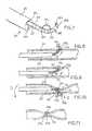
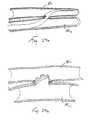
- a clock spring occluder 20comprises a generally spiral-shaped coil having a first outwardly oriented end 20a and a second inwardly oriented end 20b.
- the clock spring occludermay further have a multiplicity of outwardly extending engagement members, such as protrusions or barbs.20c which, as will be discussed below, enhance the ability of the occluder 20 to securely grasp and occlude the lumen of an anatomical structure such as a blood vessel.
- the clock spring occluder 20is deployed through the lumen of the catheter 18, as shown schematically in Figure 1 .
- the catheter 18is inserted and positioned within the vessel 16 such that side opening 18a formed at the distal end of the catheter 18 is aligned with and juxtapositioned with a portion of the vessel wall 16, at the location where it is desired to occlude the vessel.
- the first outwardly oriented end 20a of the clock spring occluder 20is then advanced through the side opening 18a of the catheter 18 and through at least a portion of the vessel wall 16.
- the outward end 20a of the device 20may optionally be sharpened.
- the clock spring occluder 20is then advanced through the puncture site so that it fully or partially surrounds the lumen.22 of the vessel and engages the vessel wall.
- the occluder device 20is biased to a coiled configuration which causes it to pull the wall of the vessel inwardly so as to collapse and close the lumen of the vessel at the desired location, as shown in Figure 4 .
- one or more engagement memberssuch as outwardly projecting barbs 20c may be formed on the occluder device 20 to prevent the device 20 from migrating, slipping or pulling through the wall 16 of the vessel.
- the catheter 18is then removed, as illustrated in Figure 5 .
- the occluder 20will resiliently retract to its preformed coiled configuration to cause the vessel to assume a closed "hourglass" shape, as further illustrated by Figure 6 .
- the tightness of the coiled configuration of the occluder device 20will determine whether the lumen of the vessel is fully or partially occluded, as desired.
- FIGS 7-11 and 23-23bshow a transluminal device 24 which is useable for ligating a luminal structure, such as a blood vessel, or for the other applications such as anchoring or attaching an endoluminal graft or other endoluminal apparatus to the wall of the luminal structure (e.g., blood vessel).
- a luminal structuresuch as a blood vessel
- anchoring or attaching an endoluminal graft or other endoluminal apparatusto the wall of the luminal structure (e.g., blood vessel).
- This intraluminal device 24comprises an elongate rigid or pliable catheter 26 having a first lumen 26a and a second lumen 26b extending longitudinally therethrough.
- a hollow needle 28which has an elongate slot 28b formed in one side thereof adjacent its distal end 28a, is slidably disposed within the first lumen 26a of the catheter 26 so as to be advancable out of the catheter, as shown.
- the second lumen 26bmay be used for passage of a guidewire, to permit the catheter 26 to be advanced over a pre-positioned guidewire.
- the T-occluder apparatusgenerally comprises an elongate link member such as a pliable cord 30c and first 30a, 30a' and second 30b, 30b' engagement members attached to the other end thereof.
- the engagement members 30a, 30a', 30b, 30b'are in the form of cross members, but it will be appreciated that these engagement members 30a, 30a', 30b, 30b' may be any suitable type of structure or material (e.g., projection, flange; hook, barb, adhesive, staple, etc.)capable of engaging and/or connecting to the wall of the luminal anatomical structure to be occluded.
- the cross-members 30a, 30a', 30b, 30b'are initially disposed in direct alignment within the hollow inner bore of the needle 28, such that the cross-members 30a, 30a', 30b, 30b' are in serial end-to-end alignment within the bore, and are substantially parallel to the longitudinal axis of the needle 28. In this manner, some or all of the cord 30c may protrude out of the elongate slot 28b formed in the side of the needle, in the manner shown in Figure 7 .
- a push rod 32is slidably disposed within the bore of the needle 28, behind the T-occluder apparatus 30, 30' loaded therewithin. In this manner, the push rod 28 may be utilized to expel the first and second cross-members 30a,30a' ,30b, 30b' out of the distal end 28a of the needle 28.
- the cord 30cmay be formed of elastic material which is elastically biased to a shortened configuration such that the cord 30c will pull or draw the cross-members 30a, 30b inwardly toward a common central point or location.
- the cord 30c'may be formed of material which is capable of being wound (i.e., a malleable material which can be plastically deformed, twined or kinked or a pliable material capable of being tied or knotted) such that the overall length of the cord 30c' becomes shortened, thereby drawing the cross members 30a', 30b' inwardly toward a common central point or location.
- the catheter 26is advanced to its desired location within a blood vessel or other luminal anatomical structure.
- the positioning of the catheter 26may be guided or verified by any suitable imaging or guidance system and, optionally, a fiberoptic endoscope, ultrasound imaging system, or any other on-board imaging system may be incorporated into the catheter to provide an image of the area adjacent a view port 26b or other imaging location on the catheter 26 (see Figure 7 )
- the needle 28is advanced out of the catheter 26 and the distal end 28a of the needle is passed fully or partially through the wall of the luminal anatomical structure, at a first location.
- the push rod 32is then advanced through needle 28 in the distal direction such that the first engagement member 30a, 30a' of the first T-occluder 30, 30' is expelled out of the needle 28 so that it will engage the wall of the luminal anatomical structure, adjacent the first location.
- the needle 28is retracted into the lumen of the anatomical structure 34 and the catheter is moved (e.g., rotated approximately 180 degrees).
- the needle 28is once again advanced such that the distal end 28a of the needle 28 passes fully or partially through the wall of the luminal anatomical structure 34 at a second location which is diametrically opposite the first location.
- the push rod 32is then once again advanced in the distal direction to expel the second engagement member 30b, 30b' out of the distal end 28a of the needle and into engagement with the wall of the anatomical structure 34 adjacent the second location.
- the elastic cord 30cwill resiliently shorten and will pull the engagement members 30a, 30b inwardly so as to collapse the wall of the anatomical structure 34 and fully or partially close its lumen, as desired.
- the needle 28When the design of Figures 12-15 is installed in the foregoing manner, the needle 28 will then be withdrawn from or retracted into the catheter 26 and a winding tool 36 will then be advanced from the catheter into contact with the windable cord 30c'.
- This winding tool 36may be a hook (as shown) or any other suitable apparatus capable of twining, deforming, crimping, tying or knotting the cord 30c'--in accordance with the particular type of material of which the cord 30c' is formed (e.g., plastically deformable or pliable/knottable).
- This winding tool 36is then used to wind (i.e., plastically deform, knot, twine or tie) the cord 30c' in a manner which causes the cord 30c' to shorten, thereby pulling the engagement members 30a', 30b' inwardly to cause the wall of the anatomical structure 34 to collapse and its lumen to become fully or partially closed, as desired.
- the winding tool 36may then be disengaged from the wound cord 30c' and removed, or alternatively may be designed to be detached from the catheter 26 and left in place within the wound cord 30c'.
- the winding tool 36may be configured to detach from the catheter 36 by separation of a weakened "tear away" area formed in the tool 36 such that the distal portion of the tool 36 will break away when sufficient proximally directed or rotational force is applied thereto.
- the force necessary to break winding tool 36will be less than the force necessary to dislodge the hook member 36 from the attachment cord 30c' that is wrapped thereabout.
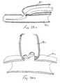
- Twist Clip Apparatusalso described herein
- this apparatusmay comprise an elongate rigid or pliable catheter 3 8 having a hollow lumen extending longitudinally therethrough and a side opening 38a formed near its distal end. Disposed within the lumen of the catheter 38 is a twist clip 42.
- the twist clip 42is formed of malleable material and comprises a first straight suction 42a and second section 28b, which extends generally perpendicular to the first straight section 42a.
- an outwardly curved tip 42cis formed on the distal end of the second section 42b
- the twist clip 42and more particularly the outwardly curved tip 42c thereof, is initially advanced some distance out of the side opening 38a and to pierce through the vessel wall 40.
- the catheter 38is rotated, as indicated by the arrow on Figure 14 , so that the clip 42 advances fully or partially around the wall of the anatomical structure 40 to cause its lumen to become fully or partially closed, as shown in Figure 17 ..
- the clip 42is thus secured to the outside of the wall while simultaneously forming an intraluminal closure.
- twist clip 42may be configured to detach from.catheter 38 by forming a weakened or break-away area in the clip such that the clip 42 will break away when sufficient proximally directed or rotational force is applied thereto. In this respect, it will be recognized that the force necessary to break twist clip 42 free from.the catheter 38 will be less than the tension necessary to dislodge the clip 42 from the wall of the anatomical structure 40
- Figures 18-22bare directed to an intraluminal suturing device 50, 50' having an inboard penetrating member 54, 54', and Figures 23a-26 are directed to an alternative intraluminal suturing device 50" having an outboard penetrating member 530.
- intraluminal suturing devicesmay be used to repair tears or ruptures in anatomical conduits, to occlude anatomical conduits ( see, Figures 19a-19b described herebelow), to anastomose or join approximated segments of anatomical conduit ( see , Figure 18a (line AN) and/or Figures 28a-29b described herebelow) or to anchor or attach various articles (e.g., an endovascular graft) to the wall of an anatomical conduit ( see, Figures 20 , 27a and 27b described herebelow).
- various articlese.g., an endovascular graft
- these devices 50, 50'generally comprise an elongate pliable catheter body 52, 52' having a proximal end PE, a distal end DE, and a hollow lumen 54, 54' which extends longitudinally therethrough.
- a side opening 56, 56'is formed on one side of the catheter body 52, 52' near the distal end DE thereof.
- a balloon 58, 58' or other suitable type of lateral pressure exerting membermay optionally be mounted on the outer surface of the catheter body 52, 52' at a location which is substantially diametrically opposite the location of the side opening 56, 56'.
- An axially moveable needle advancement/retraction member 64 or 64'is mounted within the lumen 54, 54' of the catheter body 52, 52'.
- a tissue penetrating membersuch as a needle, 74, 74' is mounted within the lumen 54, 54'of the catheter body 52, 52' and is axially moveable back and forth, by way of the needle advancement/retraction member 64, 64'.
- the needle 74, 74'is reciprocally moveable back and forth between i) a first position wherein the needle 74, 74' is proximal to the side opening 56, 56', and ii) a second position wherein the needle 74, 74' is distal to the side opening 56, 56'.
- a first portion of the wall of the luminal anatomical structure (e.g., blood vessel) wherein the catheter body 52, 52' is positionedmay be drawn or otherwise caused to intrude (e.g., invaginate, lapse) into the side opening 56, 56' such that the needle 74, 74' may be advanced or retracted through the intruding portion of tissue.
- intrudee.g., invaginate, lapse
- a length of connector material 88, 88'such as suture thread, which is attached to the needle 74, 74', will be drawn through the intruding tissue to form a stitch in the wall of the luminal anatomical structure.
- the a continuous suture 88, 88"will be passed through multiple locations about the wall of the anatomical structure and the suture 88, 88' will be drawn taught in the nature of a "purse string", thereby collapsing the wall of the anatomical structure inwardly and causing its lumen to become fully or partially closed, as desired.
- the suture material 88, 88'is to be used to attach or anchor an article or apparatus (e.g., an endoluminal tube graft) to the wall of the luminal anatomical structure
- an article or apparatuse.g., an endoluminal tube graft
- the intraluminal suturing devicewill be used to place continuous or interrupted stitches through the article/apparatus and the wall of the anatomical structure, so as to anchor the article/apparatus to the anatomical structure as desired.
- the needle advancing/retracting member 64comprises an elongate shaft 66 having a bifurcated, generally "C” shaped distal portion 68.
- This C-shaped distal portion 68comprises a generally “C” shaped member 70 having first and second needle abutting indentations 72a, 72b formed on its directly opposing ends, as shown.
- a cross-member 60is formed within or attached to the tubular catheter body 52 on opposite side thereof, such that the cross-member 60 traverses the lumen 54 of the catheter at a location directly beneath the side opening 56.
- An indentation 62is formed in the upper surface of the cross-member 60 immediately beneath the side opening 56.
- the "C" shaped distal portion 68 of the needle advancing/retracting member 64partially surrounds the cross-member 60, as shown.
- a dual-tipped needle 74is mounted within the lumen 5.4 above the cross-member 60.
- Needle abutting surfaces 72a, 72b of the C-shapod portion 68 of the needle advancing/retracting member 64are preferably of a concave shape so as to axially receive the sharpened ends of the needle 74. In this manner, when the needle advancing/retracting member 64 is advanced in the distal direction, the first needle abutting surface 72a will push the needle 74 past the side opening 56 and to an advanced position distal to the side opening 56, but still within the lumen 54 of the catheter body 52.
- the second needle abutting surface 72bwill drive the needle 74 in the proximal direction, past the side opening 56, and to a retracted position which is proximal to the side opening 56, but still within the lumen 54 of the catheter body 52.
- a proximal connector assembly 80may be mounted on the proximal end PE of the catheter body 52.
- Such proximal connector assembly 80incorporates a slidable actuator knob 82 which is connected to the needle advancing/retracting member 64 such that when the actuation knob 82 is advanced in the distal direction, the needle advancing/retracting member 64 will advance in the distal direction, and when the actuation knob 82 is retracted in the proximal direction, the needle, advancing/retracting member 64 will retract in the proximal direction.
- the preferred proximal connector assembly 80may have a balloon inflation port 84 connected to a balloon inflation lumen 89 which extends longitudinally through the catheter body 52 and which terminates distally in the interior of the balloon 58. In this manner, balloon inflation fluid may be injected and withdrawn through the balloon inflation port 84 to effect inflation and deflation of the balloon 58, as desired.
- a suture passage aperture 75is formed vertically through the cross-member 60 at a location immediately beneath the center of the side opening 56 and the strand of suture material 88 which extends into the proximal port of the proximal connector assembly 80 continues through the suture lumen 53 of the catheter body, upwardly through the suture passage aperture 75 formed in cross-member 60, and is attached to the dual tip needle 74 at a location between the opposite sharpened ends of the needle 74, and preferably at the approximate longitudinal mid-point of the needle 74.
- the device of Figures 18-18amay be utilized to place suture(s) in the wall of a blood vessel by inserting the pliable catheter body 52 into the patient's vasculature and advancing the catheter body 52 through the vasculature until the side opening 56 is located at the site at which the first stitch is to be placed.
- the diameter of the catheter body 52will be as large as the inner diameter of the blood vessel lumen such that a portion of the blood vessel wall will automatically intrude into the side opening of the catheter body.
- the diameter of the catheter body 52will be less than the inner diameter of the blood vessel and the side balloon 58 will be used to compress the catheter laterally against the wall of the vessel.
- a syringemay be attached to the balloon inflation port 84 and utilized to infuse a balloon inflation fluid (e.g., saline solution) into the balloon 58 to inflate the balloon 58.
- a balloon inflation fluide.g., saline solution
- the inflated balloon 58will contact the wall of the vessel and will propel the catheter body 52 laterally against the wall of the vessel opposite the location of the balloon 58.
- the side opening 56will become compressed against the blood vessel wall BVW and a portion of the tissue of the blood vessel wall BVW will intrude (e.g., invaginate or lapse) into the side opening 56 and into the lumen 54 of the catheter int he path of the needle 74.
- suctionmay optionally be applied to the lumen 54 of the catheter body.
- the operatorwill advance the actuator knob 52 in the distal direction so as to cause the needle advancing/retracting member 64 to also advance in the distal direction.
- the first needle abutting surface 72a of the needle advancing/retracting member 64will abut against the first sharpened end of the needle 74 and will drive the needle in the distal direction, through the portion of the blood vessel wall BVW which intrudes into the lumen 54 of the catheter body 52.
- the balloon 58may be deflated (if used) and any optional suction will be terminated.
- the catheter bodywill be repositioned (e.g., rotated slightly and/or moved longitudinally) so that the side opening 56 becomes positioned next to a second location on the blood vessel wall BVW.
- the balloon 58may be once again inflated (if necessary) so as to press the side opening 56 of the catheter body 52 laterally against the second location on the blood vessel wall BVW, and any optional suction may be applied so as to cause another portion of the blood vessel wall BVW to intrude inwardly into the side opening 56.
- the operatorwill retract the actuator knob 82 so as to cause the needle advancing/retracting member 64 to retract in the proximal direction driving the needle 74 in the proximal direction, through the second portion of the blood vessel wall BVW which has been caused to intrude inwardly through the side opening 56.
- the balloon 58will be once again deflated (if used) and any optional suction will be terminated.
- the catheter body 52may again be repositioned, and the above-described procedure repeated as many times as necessary to form the desired suture line in the blood vessel wall BVW.
- the device 50may be retracted and a knot pusher of the type commonly used in laparoscopic surgery may be utilized to form a tie or knot in the suture material 88.
- a knot pusherof the type commonly used in laparoscopic surgery
- the suture 88may be pulled taught in a purse string fashion, so as to collapse and close the lumen of the blood vessel in the manner shown in Figure 19b .
- the suture materialmay interrupted or continuous and may be tied off or knotted using the knot pusher, without drawing the lumen of the blood vessel closed.
- This proceduremay be used to form a radial purse string closure or anastomosis in a blood vessel as shown in Figures 19-19a .
- this proceduremay be used to anchor an article or apparatus, such as an endoluminal graft 90 for bridging of an aneurism AN, as shown in Figure 20 .
- the procedure illustrated in Figure 20may offer advantages over prior art methods wherein endoluminal grafts were held in place or anchored by way of a radially expandable stent for frictional engagement of the graft 90 to the blood vessel wall BVW.
- the intraluminal suturing devices 50, 50', 50"may be used to sew the opposite ends of a pliable tube graft (e.g., woven polyester or expanded polytetrafluoroethylene (ePTFE)) to the blood vessel wall BVW so as to anchor and hold the tube graft 90 at its desired position, without the need for radially expandable stents or other hardware required for frictionally engaging or anchoring the graft 80 to the blood vessel wall BVW.
- a pliable tube grafte.g., woven polyester or expanded polytetrafluoroethylene (ePTFE)
- Figures 22, 22a, and 22bshow a variation of the above-described device wherein the intraluminal suturing device 50' has a modified catheter body 52' which has a proximal end PE, a distal end DE, a main lumen 54' extending longitudinally therethrough.
- a side opening 56'is formed into the main lumen 54' at a first location on one side of the catheter body 52', and an optional balloon 58' may be formed on the catheter body 52' at a location diametrically opposite the side opening 56'.
- a balloon inflation lumen 89'extends longitudinally through the catheter body 52' to permit inflation fluid to be infused into and withdrawn from the balloon 58'.
- the needle advancing/retracting member 64'has a sharpened distal tip
- a first needle connectoris formed on the needle advancing/retracting member 64' near its distal end and a corresponding second needle connector (e.g., a notch) 102 is formed adjacent the proximal end of the needle 74'.
- needle holding members 108are formed within the main lumen 54' of the catheter body 52' at a position distal to the side opening 56', such needle holding members 108 being constructed to frictionally engage and prevent rotation of the needle 74' while in its second position distal to the side opening 56'.
- the suture material 88'is attached to the needle 74' at a location between its proximal end 107 and distal end 106, and preferably at the approximate mid-point of the needle 74'.
- the catheter body 52'When this variation of the intraluminal suturing device shown in Figures 22, 22a, 22b is used to place sutures in a blood vessel, the catheter body 52' will be advanced transluminally through the vasculature until the side opening 56 is positioned at the site at which it is desired to place sutures through the wall of the blood vessel. Thereafter, the balloon inflation fluid may be infused (if needed) through the balloon inflation lumen 89' to inflate the balloon 58', thereby compressing the side opening 56' against a first location on the blood vessel wall in a manner which causes the blood vessel wall to intrude into the main lumen 54'. Optionally, suction may be applied to the main lumen 54' to draw the tissue inwardly through the side opening 56'.
- the needle advancing/retracting member 64'is advanced in the distil direction so as to drive the needle 74' through the portion of the blood vessel wall which has been drawn thorough the side opening 56' and to its second position located distal to the side opening 56'.
- the needle holding members 108will frictionally engage and prevent rotation of the needle, and the needle advancing/retracting member 64' will be slightly rotated so as to disengage the first and second needle connectors 100, 102 from each other, thereby releasing the needle advancement/retraction member 64' from the needle 74'.
- the needle advancing/retracting memberis withdrawn proximally to a position proximal to the side opening 56'.
- the balloon 58'is deflated (if previously inflated), and the catheter body 52' is repositioned such that the side opening 56' is positioned adjacent a second location on the blood vessel wall. Thereafter the balloon 58' is once again inflated (if necessary) and any optional suction desired may be applied to the main lumen 54' so as to draw a second portion of the blood vessel wall into the main lumen 54' through the side opening 56'. Thereafter, the needle advancing/retracting member 64' is advanced in the distal direction such that the sharpened distal tip of the needle advancing/retracting member 64' will pass through the intruding tissue.
- the needle advancing/retracting member 64'After the needle advancing/retracting member 64' has been advanced until its needle connector 100 is located in alignment with the needle connector 102 formed on the proximal end of the needle 74', the needle advancing/retracting member 64' will be slightly rotated so as to engage its needle connecting member 100 with that 102 of the needle 74', thus reconnecting the advancing/retracting member 64' to the needle 74'. Thereafter, the needle advancing/retracting member 64' is again withdrawn in a proximal direction so as to pull the needle 74' back through the tissue and to its first position wherein the needle 74' is positioned proximal to the side opening 56'. Thereafter, the above-described steps are again repeated as many times as necessary to form the desired stitch or suture line in the blood vessel wall.
- the catheter 52'may be removed and the suture may be drawn closed so as to ligate the blood vessel, or otherwise tied or knotted to anchor or affix an article or apparatus to the wall of the blood vessel, as described in detail hereabove.
- Figures 23a-26show another intraluminal suturing device 50"', which incorporates an "outboard" tissue penetrating member or needle 530 that extends out of the catheter body 502 so as to pass connector material such as suture thread 529 through the wall of a luminal anatomical structure, such as a blood vessel, within which the catheter body 502 is inserted.
- this device 50"'does not require that any portion of the wall of the luminal anatomical structure be caused to intrude into the catheter body, as with the above-described inboard needle embodiments of the device 50, 50".
- this device 50"'comprises an elongate, rigid or pliable, catheter body 502 having an irregularly shaped main lumen 504 formed within a distal portion of the catheter body 102.
- the main lumen 504is generally of an hourglass or dumbbell cross-sectional shape which defines an upper portion 506, a lower portion 508 and a communicating channel 510 which extends between the upper portion 506 and the lower portion 508.
- This main lumen 504is formed in its entirety only in a distal portion of the catheter body 502 and all portions of the main lumen 504 terminate distally in a closed distal end 505.
- the upper portion 506 and the communicating channel 510terminate proximally in a closed proximal end 507 within the catheter body 502, but the lower portion 508 continues through the proximal end of the catheter body 502 and through a proximal opening (not shown).
- a needle outlet branch lumen 516extends from the upper portion 506 of the main lumen 504 and through a first opening formed in the side of the catheter body 502.
- a needle inlet branch lumen 518extends from the upper portion 506 of the main lumen 504 to a second opening formed in the side of the catheter 502 distal to, and preferably in alignment with, the first opening.
- an arcuate dip ADmay be formed in the portion of the main lumen 504 which passes between the needle outlet branch lumen 516 and the needle inlet branch lumen 518.
- a suture passage slit 511is formed longitudinally in the portion of the catheter body 502 located between the needle outlet branch lumen 516 and the needle inlet branch lumen 518. Such slit 511 provides a narrow suture passage channel which extends downwardly from the upper surface of the catheter body 502 into the upper portion 506 of the main lumen 504.
- a preformed resilient penetrating membersuch as a curved:needle 530 is slideably disposed, in a straightened configuration; within the upper portion 506 of the main lumen 504.
- the preferred needle 530has a straight shaft SH, a curves segment CS on the distal end of the straight shaft SH, a straight segment SS on the distal end of the curved segment CS, and a sharpened distal tip DT.
- a needle advancing/retracting member 520 of the type shown in Figures 25 and 25ais slidably disposed within the catheter body 502 and is used to alternately advance and retract the needle 530, as will be described in more detail herebelow.
- the preferred needle advancing/retracting member 520comprises and elongate shaft 528 having a clip 524 disposed on its distal end, and a control knob 522 on its proximal end.
- the control knob 522is coupled to the clip 524 by way of a mechanical linkage (not shown) which extends through he shaft 528.
- the clip 524comprises first and second laterally extending arms 526a, 526b, the ends of which are pivotally attached to a linkage hub 528.
- the clipresides slidably within the main lumen 504 of the catheter 502, with the hub disposed in the lower portion 508 and the arms 526a, 526b extending through the connecting channel 510 and into the upper portion 506, on either side of the needle 530, such that the needle 530 may be captured and grasped between the distal ends of the arms 526a, 526b.
- the shaft 528 of the needle advancing/retracting member 520extends proximally through the lower portion 508 of the lumen and extends out of the proximal lumen opening (not shown) such that the control knob 522 is accessible to the operator during the procedure.
- a length of connector material, such as suture thread 528is connected to the proximal end of the needle 530 and extends through the proximal segment of the lower lumen portion 508, along side the shaft 528 of the needle advancing/retracting member 520.
- the catheter 502is advanced, distal end first, through the body and the first and second openings which communicate with the needle outlet branch lumen 516 and needle inlet branch lumen 518 are positioned adjacent a first location on the wall of a luminal anatomical structure such as a blood vessel.
- the needle 530is initially positioned within the upper lumen potion 506, proximal to the location at which the needle outlet branch lumen 516 diverges therefrom.
- the needle advancing/retracting member 520is initially positioned and deployed such that its arms 526a, 526b are grasping the needle 530 near its proximal end.
- the needle advancing/retracting member 520 and needle 530are advanced in the distal direction.
- the distal tip of the needleis configured or biased to automatically advance into the needle outlet branch lumen 516 and out of the first outlet opening as shown.
- the needle advancing/retracting member 520 and needle 530are further advanced such that the needle will penetrate fully or partially through the tissue of the wall of the luminal anatomical structure.
- the resilient needle 530will assume its preformed configuration as shown in Figure 26 . This preformed shape of the needle 530 causes its distal end to reenter the catheter 502 through the needle inlet branch lumen 518, as shown in Figure 23c .
- the needle inlet branch lumen 518may be chamfered or tapered such that it is largest in diameter at the second opening in the side of the catheter and progressive narrows as it extends inwardly, thereby enhancing the ability of the distal tip DT of the needle 530 to locate and pass into the needle inlet branch lumen 518 in the manner shown.
- the control knob 522is then rotated in the direction which causes the arms 526a, 526b to pivot away from each other, thereby releasing the needle from the grip of the clip 524.
- the communicating channel 510 of the lumen 504is wide enough and preferably somewhat tapered, to allow the arms 526a, 526b of the clip 524 to separate sufficiently to release the needle 530 from the grip of the clip 524.
- the needle advancing/retracting member 520is then advanced further in the distal direction until the clip 524 becomes positioned adjacent the distal end of the needle (e.g., at the junction of the main lumen 504 and the needle inlet branch lumen 518).
- the control knob 522is then rotated in the direction which causes the arms 526a, 526b of the clip to pivot toward each other so as to capture and grasp the distal end of the needle 530 therebetween.
- the needle advancing/retracting member 520is then further advanced in the distal direction until the clip 524 abuts against the distal end 505 of the main lumen 504 and the proximal end of the needle 530 has been pulled into the distal portion of the lumen 504 with the suture thread 529 in tow.
- control knob 522is rotated to loosen the clip 524 and release the needle 530, and the needle advancing/retracting member 520 is retracted in the proximal direction until the clip becomes repositioned adjacent the proximal end of the needle 530.
- control knob 522is rotated to tighten the clip 524 so that it once again grips the needle 530 and the needle/advancing/retracting member is then further withdrawn in the proximal direction.
- the needle advancing/retracting member 520is withdrawn in the proximal direction until the needle 530 has returned to its starting position.
- the suture thread 529is concurrently pulled through the slit 511 such that the needle 530 and suture thread 529 are then prepared to repeat the above summarized steps shown in Figures 23a-23h .
- the cathetermay be rotationally and/or longitudinally repositioned, and the above summarized steps may be repeated, as necessary to form the desired number of stitches in the wall of the anatomical structure.
- One or more knotsmay be placed in the suture thread 629 to secure the stitch(es) using an appropriate knot-pusher device of the type known in the art and used in various endoscopic or port-access surgical procedures.
- FIGS 27-27cshow another device 200 which utilizes attachment member(s) 208 having a structure similar to T-Occluder device 30 shown in figures 7-11 and described hereabove.
- This device 200comprises a transluminal catheter 202, which may be of rigid or pliable construction, and a slotted, hollow needle 204 which is advancable out of the catheter 202.
- the slotted, hollow needle 204is formed of resilient or superelastic material, and is biased to a curved configuration, such that the distal portion of the needle 204 will assume a curved configuration as it is advanced out of a distal end opening 220 of the catheter, 202.
- the sharpened distal tip 214 of the needle 204will puncture fully or partially through the wall of the anatomical conduit (e.g., blood vessel) within which the distal end of the catheter 202 is positioned.
- the needle 204may be deflected or otherwise caused to exit the catheter through an opening in the side of the catheter 202, rather than through the distal end thereof.
- the preferred needle 204is constructed to carry a plurality of attachment members 208 and to facilitate the installation of those attachment members 208 in the wall of a luminal anatomical structure (e.g., a blood vessel).
- the elongate slot 210is preferably formed only on one side of a distal portion of the needle 204, such that the proximal shaft of the needle remains in tact and unspotted.
- One or more of these attachment members 208are loaded into the lumen of the needle 204 in the manner shown in Figure 27 .
- Each attachment member 208comprises a pliable link 212 formed of suture thread, a pliable filament, a strand of plastic, or other suitable material.
- Engagement members 209are formed upon opposite ends of each link 212.
- the engagement members 209are in the nature of cross members, each-such cross member having a longitudinal axis LA-T which is substantially perpendicular to the longitudinal axis LA-L of the link 212.
- each such engagement member 209will be coupled to one another in a chain-like fashion, while they remain loaded in the lumen of the needle 204, but each such engagement member 209 will become uncoupled from its neighboring engagement member 209 when it is expelled out of the distal tip 214 of the needle 204.
- each such engagement member 209has a first connector 230 formed on one end thereof, and a second connector 232 formed on the other end thereof.
- the first connector 230 of one engagement member 209is connected to, but disconnectable from, the second connector 232 of a neighboring engagement member 209 within the lumen of the needle 204.
- This chaining or linking of the engagement members 209allows them to be moved longitudinally in unison so long as they are sequentially packed in the lumen of the needle 204.
- the second connector 232 of that engagement member 209will disconnect and separate from the first connector 230 of the neighboring engagement member 209.
- the next neighboring engagement member 209remains within the lumen of the needle 204, and remains coupled to any subsequent engagement members 209 which are also within the needle lumen, as illustrated in Figure 23 .
- the connectors 230, 232 formed on the engagement members 209may be constructed and configured in many suitable ways, and may create various types of suitable interconnections (e.g., mechanical, frictional, adhesive, magnetic, etc.).
- the first connector 230 of each engagement member 209comprises a first notch 234 having a receiving aperture 236 associated therewith
- the second connector 232 of each engagement member 209comprises a second notch 238 having a raised projection 240 (e.g., a lug, post, tongue, boss, etc.) associated therewith.
- the second connector 232 of one engagement member 209is received within the receiving aperture 236 of the first connector 230 of the neighboring engagement member 209, so as to couple or link those engagement members 209, while they remain in sequential, contiguous alignment within the lumen of the needle 204.
- the links 212 of these interconnected attachment members 208protrude, in looped fashion, out of the slot 210 of the needle 204, as shown.
- An ejector/retraction member(not shown) is passed through the lumen of the needle 204, proximal to the proximal-most engagement member 209 loaded into the lumen of the needle.
- Such ejector/retraction memberis connected to or grasps the proximal-most engagement member, and is useable to selectively expel one engagement member 209 at a time from the distal tip 214 of the needle 204.
- the catheter 202is inserted and advanced transluminally until the distal end of the catheter 202 is positioned in the lumen of a luminal anatomical structure, at the location where an anastomosis or attachment is to made.
- the needle 204is then advanced out of the distal end aperture 220 of the catheter 202 until the sharpened distal tip 214 of the needle has punctured fully or partially through the wall of the luminal anatomical structure.
- the ejection/retractor member(not shown) is advanced in the distal direction to expel the first (i.e., distal-most) one of the engagement members 209 out of the end of the needle 204.
- the force exerted by its link 212will cause that engagement member 209 to assume an orientation which causes its second connector 323 to become uncoupled from the first connector 230 of the next engagement member 209.
- the ejection/retractor membermay then be retracted in the proximal direction to pull any protruding portion of the next engagement member 209 back into the lumen of the needle 204.
- the first expelled engagement member 209is deployed outside of the luminal anatomical structure such that it will abut against or engage the outer surface of the luminal anatomical structure (e.g., the adventitial surface or layer of a blood vessel) as illustrated in Figures 23 a, b and c .
- the needle 204is then retracted into the catheter 202, and the catheter 202 is repositioned (i.e., moved longitudinally and/or rotated) such that the needle 204 becomes aimed at a second location on the wall of the luminal anatomical structure.
- the needleis then readvanced through the wall of the luminal anatomical structure and the above-described procedure repeated to place the second engagement member 209 at a second location outside of the luminal anatomical structure, with the link 212 which joins those two engagement members 209 traversing through the lumen of the anatomical structure.
- This entire proceduremay then be repeated one or more times until the desired suture line or row(s) of attachment members 208 have been installed to form the desired attachment or anastomosis.
- the engagement members 209will be initially loaded into the lumen of the needle 204 in a chain like fashion and all of the engagement members 209, except for those on either end of the chain, will have two (2) links connected thereto, as shown in the figures.
- This device 200may be used for many purposes, including a.) attaching or anchoring an endoluminal graft, stent or other item/apparatus to the luminal anatomical structure, b.) connecting the luminal anatomical structure to another anatomical structure, or c.) forming an anastomotic junction between the luminal anatomical structures and another anatomical structure or between two approximated cut ends of the same luminal anatomical structure (as may occur following transection or resection of that luminal anatomical structure).
- Figures 23a and 23bspecifically illustrate one way in which this device may be used to anchor the end(s) of a tubular endoluminal graft 300 to the wall of a blood vessel BV.
- the tubular graft 300is placed within the lumen of the blood vessel BV at a desired location (e.g., such that it extends through an aneurysmic, diseased or.injured portion of that blood vessel BV).
- the catheter 202is then advanced into the luminal anatomical structure and positioned such that the outlet opening 220 at the distal end of the catheter is adjacent one end of the graft 300.
- a series of the attachment members 208are then installed, in an annular array, to anchor that end of the graft 300 to the wall of the blood vessel BV. The same procedure may then be repeated to anchor the other end of the tube graft 300 as well. In this manner, the graft 300 is firmly held in place and prevented from migrating within the blood vessel.
- the links 212 of the attachment members 208may be formed of elastic material to accomplis such tight approximation of the graft, or the links 212 may be formed of malleable material and twisted or deformed to a shortened configuration to pull the graft tightly against the blood vessel wall.
- a sealing substancee.g., a biologically compatible adhesive
- space-occupying memberse.g., embolization coils, beads, gelfoam, etc.
- embolization coils, beads, gelfoam, etc.may be applied or deposited about the end(s) of the graft 300 to prevent such leakage of blood around the graft.
- Figure 27cshows another possible application wherein the device 200 is used to form an end to end anastomosis between transected ends TE of an anatomical conduit AC such as a blood vessel, duct, tube or passageway of the body.
- the engagement members 209are installed in a staggered radial array (i.e. a zig-zag array around the anastomotic junction) so as to connect the transected ends TE of the anatomical conduit AC in abutting approximation and the desired alignment.
- Such staggered positioning of the engagement members 209may be accomplished by longitudinally advancing and retracting the catheter, back and forth, in conjunction with each incremental rotation thereof, to cause the engagement members 209 to be installed around, and on opposite sides of, the anastomotic junction, as shown in Figure 27c .
- Figure 27dshows another possible application wherein a device similar to that shown in Figure 27 has been used to install a series of interrupted attachment members 208' to form an end-to-side anastomosis between two anatomical conduits.
- These interrupted attachmentrr members 208'differ from the continuous attachment members 208 shown in Figures 27-27c in a single link portion 212' is connected to each engagement member 209', rather than having some engagement members 209 which have two link portions 212 of adjacent attachment apparatus 209 both connected thereto.
- these attachment apparatus 208'are installed in an interrupted rather than continuous fashion.
- devices 5, 50', 50", 50"' and 200which have been disclosed hereabove as being useable to form anastomotic junctions between two (2) anatomical conduits or two (2) segments of the same anatomical conduit, may be used to form numerous types of anastomotic junctions, and will not be limited to only the end-to-end type junctions specifically shown in Figures 18 or 27c . Rather, these devices 50, 50', 50", 50"' or 200 may additionally be used to form end-to-side junctions as illustrated schematically in figures 28a & 28b , and/or side-to-side junctions as illustrated schematically in Figures 29a & 29b .
Landscapes
- Health & Medical Sciences (AREA)
- Surgery (AREA)
- Life Sciences & Earth Sciences (AREA)
- Heart & Thoracic Surgery (AREA)
- Molecular Biology (AREA)
- Veterinary Medicine (AREA)
- Engineering & Computer Science (AREA)
- Biomedical Technology (AREA)
- Public Health (AREA)
- Medical Informatics (AREA)
- Nuclear Medicine, Radiotherapy & Molecular Imaging (AREA)
- Animal Behavior & Ethology (AREA)
- General Health & Medical Sciences (AREA)
- Reproductive Health (AREA)
- Vascular Medicine (AREA)
- Rheumatology (AREA)
- Surgical Instruments (AREA)
- Prostheses (AREA)
- Materials For Medical Uses (AREA)
Abstract
Description
- The present invention relates generally to medical devices and methods, and more particularly to intraluminal devices for passing attachment apparatus (e.g., connector devices, staples, etc.) or connector material (e.g., suture thread, wire, cord, filament, monofilament, etc.) into or through the wall of a luminal anatomical structure (e.g., a blood vessel or other anatomical conduit) for the purpose of : i) closing the lumen of the anatomical structure ii).
- In modem medical practice, it is sometimes desirable to pass attachment apparatus (e.g., connector devices, staples, etc.) or connector material (e.g., suture thread, wire, cord, filament, monofilament, etc.) into or through the wall of a luminal anatomical structure (e.g., a blood vessel or other anatomical conduit) for the purpose of; i) closing the lumen of the anatomical structure, ii) forming an anastomotic junction between separate anatomical structures (or between approximated segments of the same anatomical structure), and/or iii) attaching an article (e.g., an endoluminal, extraluminal or transluminal graft) or other apparatus to the wall of the anatomical structure. Some of these types of medical procedures are summarized as follows:
WO96/40356 (Whayne James, G. et al US 5810851 (InBae Yoon) describes a guide used to position a suture spring device in anatomical tissue in an elastically deformed, expanded state which is subsequently removed to permit the suture spring device to move from the elastically deformed, expanded state toward a relaxed, contracted state to apply a predetermined compression to the tissue engaged by the device.US 4621639 (James A Transue et al ) describes a hand-operated surgical instrument for closing wounds with a fastener. The instrument has a movable operating member extending from a movable handle which actuates the operating member. A hydraulic actuator comprising first and second cylinders is connected between the handle and the operating member for effecting the movement of the operating member when the handle is moved relative to the instrument. A biasing member is provided for returning the operating member and the handle means to the unactuated position. The surgical instrument includes a housing and a needle extending outwardly from one end of the housing. A plurality of fasteners are located within the housing such that when the operating member is actuated, the fasteners are forced out through the needle by the force of the operating member.- Examples of medical procedures wherein it is desirable to close the lumen of a blood vessel include: a) procedures intended to diminish or block the flow of blood into vascular aneurysms (e.g., cerebral aneurysms); b) procedures intended to occlude the side branches which emanate from a segment of a peripheral vein (e.g., to prepare the vein segment for use as an in situ bypass conduit); c) procedures intended to occlude varicose veins; d) transvascular, catheter-based procedures for bypassing obstructed, diseased or injured arteries as described in United States Patent Nos.
6190353 and5830222 ; e) procedures intended to block or diminish blood flow to a tumor; f) procedures intended to close congenital or acquired arteriovenous malformations; g) procedures intended to temporarily or permanently block blood flow through a vessel as an adjuvant to placement of an implant or apparatus within the blood vessel (e.g., placement of an endovascular graft for treatment of an aneurysm or other therapeutic intervention); and procedures intended to close an interstitial puncture tract or fistula which has been created for use in performing another medical procedure. Included among the catheter-based arterial bypass procedures described inUS Patent Nos. 6190353 and5830222 is a coronary artery bypass procedure wherein a passageway-forming catheter is transluminally advanced into the coronary vasculature, and a tissue-penetrating element is passed out of the catheter and through the wall of the vessel in which the catheter is positioned to create at least one blood flow passageway (e.g., a puncture tract or interstitial tunnel) between an obstructed coronary artery and an adjacent coronary vein. Arterial blood then flows from the obstructed coronary artery into the adjacent coronary vein. In one variation of the procedure, a single arteriovenous passageway is formed (i.e., a "first" blood flow passageway) and the lumen of the coronary vein is blocked or closed off immediately proximal to such first blood flow passageway, such that arterial blood will enter the vein and will be forced to flow through the vein, in the retrograde direction. In this manner, the arterial blood from the obstructed artery may retroperfuse the myocardium through the coronary vein. In another variation of the procedure, one or more secondary arteriovenous passageways (e.g., puncture tracts or interstitial tunnels) may be formed between the coronary vein into which the arterial blood has been shunted, and the obstructed artery or another coronary artery. These secondary passageway(s) allow the rerouted arterial blood to re-enter the coronary arterial tree after having bypassed the arterial obstruction. In cases wherein such secondary blood flow passageways are formed, the lumen of the coronary vein may additionally be blocked or closed off at location(s) distal to such secondary passageway(s), to cause the rerouted arterial blood to re-enter the arterial vasculature, as desired. - Various types of anastomotic connections are frequently formed in luminal anatomical structures for the purpose of connecting opposing transected ends or openings formed in anatomical conduit(s) (e.g., blood vessel, intestine, etc.) or for connecting an opening formed in an anatomical conduit to another anatomical structure. When joining the juxtaposed ends or openings of a singular anatomical conduit which has been transected, or when joining the juxtaposed ends or openings of two (2) different anatomical conduits, such joinder(s) may be accomplished by either 1) end-to-end, 2) end-to-side , or 3) side to side anastomosis. Irrespective of which type of anastomotic connection is being formed, the usual surgical technique requires that the luminal anatomical conduit(s) be maneuvered into proximity and placed in abutting juxtaposition, such that the ends or openings of the anatomical conduit(s) are in alignment with one another. Thereafter, sutures, staples or other connecting apparatuses are passed through the walls of the juxtapositioned anatomical conduit(s) to form the desired anastomotic connection therebetween. Anastomotic connections of this type are frequently performed during surgical procedures wherein a diseased or injured segment of an anatomical conduit (e.g., blood vessel, intestine, etc.) has been resected and removed, and the opposing cut ends of the conduit are then reconnected (by end-to-end, side to side, or end to side anastomosis) to permit continued flow of bodily fluids or other matter through the conduit.
- Examples of medical procedures wherein it is desirable to anchor or attach a graft or other apparatus to the wall of a blood vessel or other luminal anatomical conduit include certain endovascular grafting procedures wherein a tubular graft is placed within the lumen of an aneurysmic blood vessel to create a neo-lumen or artificial flow conduit through the aneurysm, thereby eliminating the exertion of blood pressure on the aneurysm and allowing the aneurysmic space to subsequently become filled in with granulation tissue. These endovascular grafting procedures have heretofore been used to treat aneurysms of the abdominal aorta, as well as aneurysms of the descending thoracic aorta. The endovascular grafts which have heretofore been used for these procedures typically incorporate or are combined with one or more radially expandable stents which are radially expanded in situ to anchor the tubular graft to the wall of the blood vessel at sites upstream and downstream of the aneurysm. However, in the event that these stent(s) fail to establish sound frictional engagement with the blood vessel wall, the graft may undergo undesirable migration or slippage, or blood may leak into the anneurysmic sac (sometimes referred to as an "endoleak").
- Thus, in view of the above-mentioned undesirable complications associated with the use of radially expandable stents to frictionally anchor a graft or other apparatus to the wall of a blood vessel (or other luminal anatomical structure) there exists a need in the art for the development of new endoluminal suturing devices which may be used to suture the opposite ends of a endoluminal tube graft (or other article) to the surrounding wall of a blood vessel or other tubular anatomical conduit, thereby ensuring sound and permanent placement of the graft or other article. Also, in view of the other types of medical procedures described hereabove, there also exists a need in the art for the development of new transluminal methods and apparatus for closing the lumen of a luminal structure and/or for forming an anastomotic connections within or to the wall of the luminal structure.
- Described herein are intraluminal devices and methods which are useable to i) fully or partially close the lumen of a luminal anatomical structure (e.g., a blood vessel), ii) form anastomotic junctions between or connections to luminal anatomical structure(s) and/or iii) attach an endoluminal, extraluminal or transluminal graft or other apparatus to the wall of a luminal anatomical structure.
- According to an aspect of the present invention, there is provided a device for fully or partially closing the lumen of an anatomical structure as claimed in Claim 1.
- In accordance with a first embodiment of the invention, there is provided an intraluminal device which is useable to occlude luminal anatomical structure by way of a closure device referred to herein, for purposes of convenience only, as a "clock spring" occluder. This device comprises an elongate catheter which is insertable into a luminal structure, and a resilient coil which is advancable out of the catheter and at least partially through the wall of the luminal anatomical structure to occlude the lumen of the anatomical structure. This resilient coil may optionally have one or more engagement members (e.g., barbs or hooks) formed thereon to enhance its engagement or gripping of the anatomical structure and to prevent the coil from slipping or pulling back through the puncture tract through which it is advanced into or through the anatomical structure wall. Generally, a first end of the resilient coil is advancable out of the catheter and at least partially through the wall of the luminal structure (e.g., blood vessel) within which the catheter is positioned. Thereafter, the coil is further advanced within or outside of the wall of the anatomical structure so as to fully or partially encircle or surround the lumen of the anatomical structure (e.g., it may slidably advance around the adventitial surface of a blood vessel). The optional engagement members (hooks or barbs), if present, will protrude into, grip, adhere to or otherwise engage the wall of the luminal anatomical structure in a manner which will prevent the coil from slipping or pulling back through the puncture tract which it has-formed into or through the anatomical structure wall and/or to prevent the from uncoiling in any way which would cause the lumen of the anatomical structure to return to its open configuration.
- After the coil has been fully advanced out of the catheter, the catheter may be extracted and removed, leaving the coil in place. The coil is biased to a coiled or closed configuration such that it will then draw the wall of the luminal structure inwardly so as to fully or partially close the lumen of the anatomical structure.
- Also described herein, there is provided another intraluminal device, which is useable to occlude or to form anastomoses in/attachments to a luminal anatomical structure. This device comprises a catheter and an occluder/connector apparatus which is advancable out of the catheter and at least partially through the wall of the luminal anatomical structure to occlude or for attachments to/anastomoses in that anatomical structure. For purposes of convenience only this occluder/connector apparatus will be referred to herebelow as a T-occluder/connector device. In general, this device comprises an elongate catheter having a hollow puncturing member (e.g. a needle) which is advancable out of the catheter and through the wall of the anatomical structure. One or more of the T-occluder/connector device(s) is/are loaded into the lumen of the puncturing member. Each such T-occluder/connector device generally comprises an elongate link (e.g., a thread, wire, strand, cord, etc.) having first and second engagement members (e.g., crossbars, flanges, hooks, barbs, adhesive, clips, etc.) formed on either end thereof. After the catheter has been advanced to the desired site within the luminal structure, the puncturing member is advanced out of the catheter and at least partially through the wall of the luminal structure, at a first location. Thereafter a first one of the engagement members of a T-occluder/connector device is advanced out of the puncturing member to engage the wall of the luminal anatomical structure at a first location. Then, the puncturing member is retracted into the lumen of the anatomical structure and is moved or reoriented therewithin. Thereafter, the puncturing member is advanced (a second time) at least partially through the wall of the luminal anatomical structure, at a second location thereon. Thereafter, the second engagement member of that T-occluder/connector device is expelled from the puncturing member to engage the wall of the anatomical structure at the second location. This procedure may be repeated to install the desired number of T-oecluder/connector devices at the desired locations about the wall of the luminal anatomical structure. When the procedure is completed, the puncturing element and catheter are removed, leaving the previously installed T-occluder/connector device(s) in place. Depending on the particular application, the link portion(s) of the T-occluder/connector device(s) may be formed of rigid, pliable, elastic, nonelastic, malleable, nonmalleable, retractable or nonretractable material to exert the desired amount of inward pulling force upon the engagement members. This inward pulling of the engagement members results in the desired occlusion of the lumen of the anatomical structure or the desired anastomosis in or attachment to its wall.
- Also described herein, there is provided another intraluminal device which is useable to occlude the lumen of an anatomical structure (e.g., a blood vessel) by way of a closure device referred to herebelow for purposes of convenience only as a "Twist Clip Occluder". This device generally comprises an elongate catheter having an elongate twistable clip member formed of bendable (i.e., malleable) material loaded in the catheter and advance able from the catheter and at least partially through the wall of the luminal anatomical structure. After the clip member has been advanced into or around the wall of the anatomical structure, the catheter (or a secondary twisting tool) is rotated so as to twist the clip member to a closed configuration wherein it will draw the wall of the luminal anatomical structure inwardly so as to fully or partially close its lumen.
- Also described herein, there are provided catheter-based devices for installing sutures, staples or other connector apparatus into the wall of one or more luminal anatomical structure(s) (e.g., blood vessel(s), intestine(s), duct(s), or other anatomical conduit(s)). For purposes of convenience only, these devices will be referred to herein as "intraluminal suturing devices", although it will be appreciated that various types of connector materials (e.g., wire, staples, absorbable sutures, nonabsorbable sutures, etc.) may be installed by use of these devices.
- One type of intraluminal suturing device comprises an elongate rigid or pliable catheter which is advance able into the lumen of a luminal anatomical structure, and which has i.) a tissue inlet opening into which a portion of the wall of the luminal structure may be caused to intrude (i.e., lapse, invaginate, extend inwardly, etc. ) and ii.) an axially reciprocating penetrating member (e.g., a needle) having a suture thread, staple, wire or other connector material attached thereto. The penetrating member is mounted within the catheter, adjacent the tissue inlet opening and generally parallel to the longitudinal axis of the catheter, such that the penetrating member may be alternately passed back and forth through the portion(s) of the luminal structure wall which protrude into the tissue inlet opening. In operation, this inboard needle type device may be used by advancing the catheter into the lumen of the anatomical structure and causing a portion of the wall of the anatomical structure to intrude into the tissue inlet opening of the catheter such that some of the tissue is positioned in the path of the axially reciprocating penetrating member. The penetrating member, with its attached suture thread, staple or other connector material, is then passed through the intruding mass of tissue a first time. If it is desired to pass the suture material or other connector material through the wall of the anatomical structure more than once (e.g., to form an uninterrupted suture line) the catheter may then repositioned within the luminal anatomical structure (e.g., rotated and/or longitudinally advanced and/or longitudinally retracted ) and another portion of the wall of the anatomical structure will caused to intrude into the tissue inlet opening such that some of that tissue is located in the path of the axially reciprocating penetrating member. The penetrating member, with its attached suture thread, staple or other connector material, is then once again passed through the intruding mass of tissue. These operational steps are repeated as many times as necessary to form the desired anastomosis or connection.
- In some applications, the suture material or other connector material will be drawn taught and knotted in the nature of a "purse string" so as to draw the wall of the anatomical structure inwardly and to fully or partially close its lumen. These operational steps may be repeated numerous times, as necessary, to form the desired anastomosis in, or connection to, the wall(s) of the luminal anatomical structure(s). In other applications, the suture material or other connector material will be knotted or tied (one or more times) without having been drawn taught, so as to allow the lumen to remain open, while forming the desired anastomotic connection in, or attachment to, the wall of the luminal structure.
- An alternative type of intraluminal suturing device generally comprises an elongate rigid or pliable catheter which is advance able into the lumen of a luminal anatomical structure, such catheter having i.) a first penetrating member lumen from which a penetrating member may pass out of a first opening in the side of the catheter, ii.) a second penetrating member lumen into which the penetrating member may pass through a second opening formed in the side of the catheter, and iii.) a preshaped, pliable penetrating member which is passable a) from the first lumen, b) out of the first opening, c.)through an adjacent portion of the wall of the luminal anatomical structure, d) into the second opening and d) into the second lumen. A suture thread, or other connector material as described hereabove, is attached to the penetrating member such that it is drawn by the penetrating member through the tissue.
- In operation, this outboard needle type of intraluminal suturing device may be used by advancing the catheter into the lumen of an anatomical structure such that the first and second openings of the catheter are located adjacent a first portion of the wall of the anatomical structure. The penetrating member (and the suture thread or other connector material attached thereto) is then passed from first lumen, out of the first opening, through the first portion of tissue, into the second opening and into the second lumen. In applications where it is desired to pass the suture thread or other connector material repeatedly through the wall of the anatomical structure (e.g., an uninterrupted suture line) the catheter will then be repositioned (e.g., rotated and/or longitudinally advanced and/or longitudinally retracted) such that a second portion of tissue is adjacent the first and second openings of the catheter, and the foregoing procedural steps are then repeated.
- Also described herein, there is provided a device for forming attachments to or anastomoses in, a luminal anatomical structure. This device generally comprises an elongate catheter wich is insertable into the lumen of the luminal anatomical structure and a hollow needle disposed within said catheter, said needle having a sharpened distal tip, a needle lumen which extends longitudinally therethrough, and an outlet opening formed in communication with said needle lumen. The needle is advancable out of the catheter, after the catheter has been inserted into the luminal anatomical structure, such that the sharpened distal tip of the needle will puncture through the wall of the anatomical structure and the outlet opening of the needle will become situated outside of the luminal anatomical structure. At least one attachment apparatus is initially loaded into the lumen of the needle. Each attachment apparatus generally comprises a flexible link (e.g., a cord, thread, strand, or elongate member) having first and second engagement members (e.g., t-bars, flanges, arms, etc.) formed on opposite ends thereof. These engagement members are able to advance, one at a time, out of the outlet opening of the needle. While loaded in the lumpen of the needle, the engagement members may be connected to one another, in a chain-like fashion, so that they may be advanced and/or retracted as a unit. After the catheter has been inserted into the luminal anatomical structure, the needle is advanced (a first time) out of the catheter and through a first location on the wall of the luminal anatomical structure. Thereafter, one engagement member is passed out of the outlet opening of the needle so that it becomes deployed outside of or within the wall of the luminal anatomical structure so as to engage the wall. The needle is then retracted into the catheter and the catheter is repositioned (e.g., rotated and/or longitudinally advanced/retracted) within the lumen of the anatomical structure. The needle is then advanced (a second time) out of the catheter and through a second location on the wall of the luminal anatomical structure. The second engagement member is then advanced out of the outlet opening of the needle so that it becomes deployed outside or within the wall of the anatomical structure and will engage the wall, adjacent the second location. The needle is then retracted into the catheter, leaving the two (2) engagement members in abutting engagement with the first and second loactions on the wall of the atomical structure, with the link portion of the attachment member traversing therebetween. Depending on the number of attachment members required to form the desired attachment or anastomosis, this procedure may then be repeated one or more times. When the desired attachment or anastomosis has been competed, the catheter and accompanying needle are removed from the body, leaving the previously installed attachment members in place. The attachment members may be formed of absorbable or non-absorbable material, depending of the nature of the application.
- This device may be used to attach various items (e.g., endoluminal grafts, stents) to the wall of a luminal anatomical structure, or may be utilized to form an anastomotic junction between the approximated ends of one or more anatomical conduit(s) (e.g., the opposing cut ends of a blood vessel, fallopian tube, ureter, urethra, pancreatic duct, common bile duct, esophagus, intestine or other conduit which has been cut or resected).
- Further objects and advantages of the present invention may become apparent to those skilled in the art upon reading and understanding of the following detailed description, and the accompanying drawings.
Figure 1 is a partial longitudinal sectional view of an adjacent artery and vein wherein a lumen closing device has been used to install a clock-spring occluder apparatus of the present invention to close the lumen of the vein.Figure 1a is an elevational view of a preferred clock spring occluder apparatus of the present invention, which forms a component of the device shown inFigure 1 .Figures 2-5 are a step-by-step showing of a preferred method for using the device ofFigure 1 to accomplish closure of a blood vessel.Figure 6 is a cross sectional view through line 6-6 ofFigure 5 .Figure 7 is a partial perspective view of intraluminal device which incorporates a T-occluder apparatus for closing the lumen of a blood vessel.Figures 8-11 are step-by-step showings of a method by which the device ofFigure 7 is used to effect closure of the lumen of a blood vessel using a T-occluder apparatus having an elastic link.Figures 12-17 are step-by-step showings of a method for using the device ofFigure 7 to effect closure of the lumpen of a blood vessel using a T-occluder apparatus having a twistable or knottable link.Figure 18 is a perspective view of an intraluminal suturing device having an inboard tissue-penetrating element which is useable to pass a connector material (e.g., suture thread) through the wall of a luminal anatomical structure.Figure 18a is a longitudinal sectional view of a portion of the device ofFigure 18 .Figure 19a-19b are step-by-stcp showings of a preferred method of using the device ofFigures 18-18a to place a continuous "purse string" suture which is useable to ligate the lumen of the anatomical structure.Figure 20 is a longitudinal sectional view of a segment of a blood vessel illustrating the manner in which the device ofFigures 18-18a may be used to suture the ends of an endoluminal tube graft within a blood vessel so as to bridge an aneurysm.Figure 21 is an enlarged longitudinal sectional view of the distal portion of the device shown inFigures 18-18a , operatively positioned within a blood vessel.Figure 21a is a perspective view of the C-shaped distal segment of the preferred needle advancing/retracing component of the device ofFigures 18-18a &21 .Figure 21b is a cross sectional view through line 21b-21b ofFigure 21 .Figure 21c is a cross sectional view through line 21c-21c ofFigure 21 .Figure 21d is a cross sectional view through line 21d-21d ofFigure 21 .Figure 22 is a perspective view of another intralumittal suturing device, having an inboard tissue penetrating member which is moved by way of a modified advancing/retracting component.Figures 23a-23h are partial longitudinal sectional views showing, in step-by-step fashion, a preferred method of using an intraluminal suturing device having an outboard tissue penetrating member.Figure 24a is a cross-sectional view throughline 24a-24a ofFigure 23a .Figure 24b is a cross-sectional view through line 24b-24b ofFigure 23a .Figure 25 is a perspective view of the needle advancing/retracting member of the device shown inFigures 23-24 .Figure 25a is a distal end view of the needle advancing/retracting member ofFigure 25 .Figure 26 is an enlarged, partial, side elevational view of the tissue penetrating member of the device ofFigures 23-24 .Figure 27 is a partial perspective view of a device for forming attachments to and or anastomoses in, the wall of a luminal anatomical structure using modified T-connector apparatus.Figure 27a is a cut-away perspective view of a segment of a blood vessel, showing the manner in which the device ofFigure 27 may be used to anchor an endoluminal graft within a blood vessel.Figure 27b is an end perspective view of the showing ofFigure 27a .Figure 27c is a cut-away perspective view of a tubular anatomical structure which has been transected and subsequently reconnected, by end-to-end anastomosis, using the device ofFigure 27 .Figure 27d is a perspective view of two (2) tubular anatomical conduits which have been prepared and approximated in end-to-side fashion, and which have been anastomosed by way of interrupted connector bars which are a variation of the device shown inFigure 27 .Figure 28a is a longitudinal sectional view of two (2) anatomical conduits being connected by end-to-side anastomosis using an intraluminal device.Figure 28b is a longitudinal sectional view of the two (2) anatomical conduits ofFigure 28a , after undergoing end-to-side anastomosis by an intraluminal device.Figure 29a is a longitudinal sectional view of two (2) Anatomical conduits being connected by side-to-side anastomosis using an intraluminal device.Figure 29b is a longitudinal sectional view of the two (2) anatomical conduits ofFigure 29a , after undergoing side-to-side anastomosis by an intraluminal device.- A
clock spring occluder 20, more clearly seen in Figure 2a, comprises a generally spiral-shaped coil having a first outwardly orientedend 20a and a second inwardly orientedend 20b. The clock spring occluder may further have a multiplicity of outwardly extending engagement members, such as protrusions or barbs.20c which, as will be discussed below, enhance the ability of theoccluder 20 to securely grasp and occlude the lumen of an anatomical structure such as a blood vessel. - The
clock spring occluder 20 is deployed through the lumen of thecatheter 18, as shown schematically inFigure 1 . Thecatheter 18 is inserted and positioned within thevessel 16 such thatside opening 18a formed at the distal end of thecatheter 18 is aligned with and juxtapositioned with a portion of thevessel wall 16, at the location where it is desired to occlude the vessel. The first outwardly orientedend 20a of theclock spring occluder 20 is then advanced through theside opening 18a of thecatheter 18 and through at least a portion of thevessel wall 16. To facilitate such puncture throughvessel wall 16, theoutward end 20a of thedevice 20 may optionally be sharpened. Theclock spring occluder 20 is then advanced through the puncture site so that it fully or partially surrounds the lumen.22 of the vessel and engages the vessel wall. - The
occluder device 20 is biased to a coiled configuration which causes it to pull the wall of the vessel inwardly so as to collapse and close the lumen of the vessel at the desired location, as shown inFigure 4 . - To enhance the ability of the
clock spring occluder 20 to engage and pull the vessel wall, one or more engagement members, such as outwardly projecting barbs 20c may be formed on theoccluder device 20 to prevent thedevice 20 from migrating, slipping or pulling through thewall 16 of the vessel. Once the entire length of theclock spring occluder 20 has been advanced through theside opening 18a, thecatheter 18 is then removed, as illustrated inFigure 5 . After thecatheter 18 has been removed, theoccluder 20 will resiliently retract to its preformed coiled configuration to cause the vessel to assume a closed "hourglass" shape, as further illustrated byFigure 6 . The tightness of the coiled configuration of theoccluder device 20 will determine whether the lumen of the vessel is fully or partially occluded, as desired. Figures 7-11 and23-23b show atransluminal device 24 which is useable for ligating a luminal structure, such as a blood vessel, or for the other applications such as anchoring or attaching an endoluminal graft or other endoluminal apparatus to the wall of the luminal structure (e.g., blood vessel).- This
intraluminal device 24 comprises an elongate rigid orpliable catheter 26 having afirst lumen 26a and a second lumen 26b extending longitudinally therethrough. Ahollow needle 28, which has an elongate slot 28b formed in one side thereof adjacent itsdistal end 28a, is slidably disposed within thefirst lumen 26a of thecatheter 26 so as to be advancable out of the catheter, as shown. The second lumen 26b may be used for passage of a guidewire, to permit thecatheter 26 to be advanced over a pre-positioned guidewire. - Disposed within the hollow bore of the
curved needle 28 is a T-occluder apparatus 30 or 30'. The T-occluder apparatus generally comprises an elongate link member such as apliable cord 30c and first 30a, 30a' and second 30b, 30b' engagement members attached to the other end thereof. In the particular design shown in the drawings, theengagement members engagement members needle 28, such that the cross-members 30a, 30a', 30b, 30b' are in serial end-to-end alignment within the bore, and are substantially parallel to the longitudinal axis of theneedle 28. In this manner, some or all of thecord 30c may protrude out of the elongate slot 28b formed in the side of the needle, in the manner shown inFigure 7 . - A
push rod 32 is slidably disposed within the bore of theneedle 28, behind the T-occluder apparatus 30, 30' loaded therewithin. In this manner, thepush rod 28 may be utilized to expel the first and second cross-members 30a,30a' ,30b, 30b' out of thedistal end 28a of theneedle 28. - In one design shown in
Figures 7-11 , thecord 30c may be formed of elastic material which is elastically biased to a shortened configuration such that thecord 30c will pull or draw the cross-members 30a, 30b inwardly toward a common central point or location. In another design shown inFigures 12-15 thecord 30c' may be formed of material which is capable of being wound (i.e., a malleable material which can be plastically deformed, twined or kinked or a pliable material capable of being tied or knotted) such that the overall length of thecord 30c' becomes shortened, thereby drawing thecross members 30a', 30b' inwardly toward a common central point or location. - In normal operation, the
catheter 26 is advanced to its desired location within a blood vessel or other luminal anatomical structure. The positioning of thecatheter 26 may be guided or verified by any suitable imaging or guidance system and, optionally, a fiberoptic endoscope, ultrasound imaging system, or any other on-board imaging system may be incorporated into the catheter to provide an image of the area adjacent a view port 26b or other imaging location on the catheter 26 (seeFigure 7 ) Once thecatheter 26 has been placed in the desired position, theneedle 28 is advanced out of thecatheter 26 and thedistal end 28a of the needle is passed fully or partially through the wall of the luminal anatomical structure, at a first location. Thepush rod 32 is then advanced throughneedle 28 in the distal direction such that thefirst engagement member occluder 30, 30' is expelled out of theneedle 28 so that it will engage the wall of the luminal anatomical structure, adjacent the first location. Therereafter, as shown inFigure 9 , theneedle 28 is retracted into the lumen of theanatomical structure 34 and the catheter is moved (e.g., rotated approximately 180 degrees). Thereafter, theneedle 28 is once again advanced such that thedistal end 28a of theneedle 28 passes fully or partially through the wall of the luminalanatomical structure 34 at a second location which is diametrically opposite the first location. (seeFigures 10 and13 ) Thepush rod 32 is then once again advanced in the distal direction to expel thesecond engagement member distal end 28a of the needle and into engagement with the wall of theanatomical structure 34 adjacent the second location. - When the design of
Figures 7-11 is installed in the foregoing manner, theelastic cord 30c will resiliently shorten and will pull theengagement members anatomical structure 34 and fully or partially close its lumen, as desired. - When the design of
Figures 12-15 is installed in the foregoing manner, theneedle 28 will then be withdrawn from or retracted into thecatheter 26 and a windingtool 36 will then be advanced from the catheter into contact with thewindable cord 30c'. This windingtool 36 may be a hook (as shown) or any other suitable apparatus capable of twining, deforming, crimping, tying or knotting thecord 30c'--in accordance with the particular type of material of which thecord 30c' is formed (e.g., plastically deformable or pliable/knottable). This windingtool 36 is then used to wind (i.e., plastically deform, knot, twine or tie) thecord 30c' in a manner which causes thecord 30c' to shorten, thereby pulling theengagement members 30a', 30b' inwardly to cause the wall of theanatomical structure 34 to collapse and its lumen to become fully or partially closed, as desired. As will be recognized, the windingtool 36 may then be disengaged from thewound cord 30c' and removed, or alternatively may be designed to be detached from thecatheter 26 and left in place within thewound cord 30c'. In this regard, the windingtool 36 may be configured to detach from thecatheter 36 by separation of a weakened "tear away" area formed in thetool 36 such that the distal portion of thetool 36 will break away when sufficient proximally directed or rotational force is applied thereto. In this respect, it will be recognized that the force necessary to break windingtool 36 will be less than the force necessary to dislodge thehook member 36 from theattachment cord 30c' that is wrapped thereabout. - Referring now to
Figures 16 and 17 , there is shown another intraluminal apparatus for occluding.blood flow at a specific site within a luminal anatomical structure such as a blood vessel. As shown inFigure 16 , this apparatus may comprise an elongate rigid or pliable catheter 3 8 having a hollow lumen extending longitudinally therethrough and aside opening 38a formed near its distal end. Disposed within the lumen of thecatheter 38 is atwist clip 42. The twist clip 42.is formed of malleable material and comprises a firststraight suction 42a and second section 28b, which extends generally perpendicular to the firststraight section 42a. An outwardly curved tip 42c is formed on the distal end of the second section 42b As illustrated, thetwist clip 42, and more particularly the outwardly curved tip 42c thereof, is initially advanced some distance out of theside opening 38a and to pierce through the vessel wall 40. Thereafter, thecatheter 38 is rotated, as indicated by the arrow onFigure 14 , so that theclip 42 advances fully or partially around the wall of the anatomical structure 40 to cause its lumen to become fully or partially closed, as shown inFigure 17 .. By virtue of the shape of the outwardly curved tip 42c, theclip 42 is thus secured to the outside of the wall while simultaneously forming an intraluminal closure. After such intraluminal closure is formed, thecatheter 38 is axially retracted through the lumen of the anatomical structure 40, causing thetwist clip 42 to detach and remain embedded within or about the wall of the anatomical structure 40, as.shown. As will be recognized,twist clip 42 may be configured to detachfrom.catheter 38 by forming a weakened or break-away area in the clip such that theclip 42 will break away when sufficient proximally directed or rotational force is applied thereto. In this respect, it will be recognized that the force necessary to breaktwist clip 42 free from.thecatheter 38 will be less than the tension necessary to dislodge theclip 42 from the wall of the anatomical structure 40 Figures 18-22b are directed to anintraluminal suturing device 50, 50' having aninboard penetrating member 54, 54', andFigures 23a-26 are directed to an alternativeintraluminal suturing device 50" having an outboard penetratingmember 530. These intraluminal suturing devices may be used to repair tears or ruptures in anatomical conduits, to occlude anatomical conduits (see,Figures 19a-19b described herebelow), to anastomose or join approximated segments of anatomical conduit (see,Figure 18a (line AN) and/orFigures 28a-29b described herebelow) or to anchor or attach various articles (e.g., an endovascular graft) to the wall of an anatomical conduit (see,Figures 20 ,27a and 27b described herebelow).- As shown in
Figures 18-21d and22-22c , thesedevices 50, 50' generally comprise an elongatepliable catheter body 52, 52' having a proximal end PE, a distal end DE, and ahollow lumen 54, 54' which extends longitudinally therethrough. Aside opening 56, 56' is formed on one side of thecatheter body 52, 52' near the distal end DE thereof. Aballoon 58, 58' or other suitable type of lateral pressure exerting member (e.g., an extendable foot) may optionally be mounted on the outer surface of thecatheter body 52, 52' at a location which is substantially diametrically opposite the location of theside opening 56, 56'. An axially moveable needle advancement/retraction member 64 or 64' is mounted within thelumen 54, 54' of thecatheter body 52, 52'. A tissue penetrating member such as a needle, 74, 74' is mounted within thelumen 54, 54'of thecatheter body 52, 52' and is axially moveable back and forth, by way of the needle advancement/retraction member 64, 64'. Theneedle 74, 74' is reciprocally moveable back and forth between i) a first position wherein theneedle 74, 74' is proximal to theside opening 56, 56', and ii) a second position wherein theneedle 74, 74' is distal to theside opening 56, 56'. - A first portion of the wall of the luminal anatomical structure (e.g., blood vessel) wherein the
catheter body 52, 52' is positioned may be drawn or otherwise caused to intrude (e.g., invaginate, lapse) into theside opening 56, 56' such that theneedle 74, 74' may be advanced or retracted through the intruding portion of tissue. In this manner, a length ofconnector material 88, 88' such as suture thread, which is attached to theneedle 74, 74', will be drawn through the intruding tissue to form a stitch in the wall of the luminal anatomical structure. In examples wherein the suture line is to be used to close or occlude the anatomical structure, the acontinuous suture suture 88, 88' will be drawn taught in the nature of a "purse string", thereby collapsing the wall of the anatomical structure inwardly and causing its lumen to become fully or partially closed, as desired. In other applications, wherein thesuture material 88, 88' is to be used to attach or anchor an article or apparatus (e.g., an endoluminal tube graft) to the wall of the luminal anatomical structure, such article or apparatus will be pre-positioned adjacent the wall of the anatomical structure and the intraluminal suturing device will be used to place continuous or interrupted stitches through the article/apparatus and the wall of the anatomical structure, so as to anchor the article/apparatus to the anatomical structure as desired. - In the particular device shown in
Figures 18-18a the needle advancing/retractingmember 64 comprises anelongate shaft 66 having a bifurcated, generally "C" shapeddistal portion 68. This C-shapeddistal portion 68 comprises a generally "C" shapedmember 70 having first and secondneedle abutting indentations 72a, 72b formed on its directly opposing ends, as shown. A cross-member 60 is formed within or attached to thetubular catheter body 52 on opposite side thereof, such that the cross-member 60 traverses thelumen 54 of the catheter at a location directly beneath theside opening 56. An indentation 62 is formed in the upper surface of the cross-member 60 immediately beneath theside opening 56. The "C" shapeddistal portion 68 of the needle advancing/retractingmember 64 partially surrounds the cross-member 60, as shown. A dual-tippedneedle 74 is mounted within the lumen 5.4 above the cross-member 60.Needle abutting surfaces 72a, 72b of the C-shapod portion 68 of the needle advancing/retractingmember 64 are preferably of a concave shape so as to axially receive the sharpened ends of theneedle 74. In this manner, when the needle advancing/retractingmember 64 is advanced in the distal direction, the first needle abutting surface 72a will push theneedle 74 past theside opening 56 and to an advanced position distal to theside opening 56, but still within thelumen 54 of thecatheter body 52. Thereafter, when the needle advancing/retractingmember 64 is retracted in the proximal direction, the secondneedle abutting surface 72b will drive theneedle 74 in the proximal direction, past theside opening 56, and to a retracted position which is proximal to theside opening 56, but still within thelumen 54 of thecatheter body 52. - A
proximal connector assembly 80 may be mounted on the proximal end PE of thecatheter body 52. Suchproximal connector assembly 80 incorporates a slidable actuator knob 82 which is connected to the needle advancing/retractingmember 64 such that when the actuation knob 82 is advanced in the distal direction, the needle advancing/retractingmember 64 will advance in the distal direction, and when the actuation knob 82 is retracted in the proximal direction, the needle, advancing/retractingmember 64 will retract in the proximal direction. Additionally, the preferredproximal connector assembly 80 may have aballoon inflation port 84 connected to aballoon inflation lumen 89 which extends longitudinally through thecatheter body 52 and which terminates distally in the interior of theballoon 58. In this manner, balloon inflation fluid may be injected and withdrawn through theballoon inflation port 84 to effect inflation and deflation of theballoon 58, as desired. Asuture passage aperture 75 is formed vertically through the cross-member 60 at a location immediately beneath the center of theside opening 56 and the strand ofsuture material 88 which extends into the proximal port of theproximal connector assembly 80 continues through thesuture lumen 53 of the catheter body, upwardly through thesuture passage aperture 75 formed incross-member 60, and is attached to thedual tip needle 74 at a location between the opposite sharpened ends of theneedle 74, and preferably at the approximate longitudinal mid-point of theneedle 74. - The device of
Figures 18-18a may be utilized to place suture(s) in the wall of a blood vessel by inserting thepliable catheter body 52 into the patient's vasculature and advancing thecatheter body 52 through the vasculature until theside opening 56 is located at the site at which the first stitch is to be placed. In some instances, the diameter of thecatheter body 52 will be as large as the inner diameter of the blood vessel lumen such that a portion of the blood vessel wall will automatically intrude into the side opening of the catheter body. In other instances, the diameter of thecatheter body 52 will be less than the inner diameter of the blood vessel and theside balloon 58 will be used to compress the catheter laterally against the wall of the vessel. In this regard, a syringe may be attached to theballoon inflation port 84 and utilized to infuse a balloon inflation fluid (e.g., saline solution) into theballoon 58 to inflate theballoon 58. Theinflated balloon 58 will contact the wall of the vessel and will propel thecatheter body 52 laterally against the wall of the vessel opposite the location of theballoon 58. In this manner, theside opening 56 will become compressed against the blood vessel wall BVW and a portion of the tissue of the blood vessel wall BVW will intrude (e.g., invaginate or lapse) into theside opening 56 and into thelumen 54 of the catheter int he path of theneedle 74. To facilitate such intrusion of the blood vessel wall BVW into theside opening 56 suction may optionally be applied to thelumen 54 of the catheter body. - After the blood vessel wall BVW has been caused to intrude inwardly into the
side opening 56, the operator will advance theactuator knob 52 in the distal direction so as to cause the needle advancing/retractingmember 64 to also advance in the distal direction. In this manner, the first needle abutting surface 72a of the needle advancing/retractingmember 64 will abut against the first sharpened end of theneedle 74 and will drive the needle in the distal direction, through the portion of the blood vessel wall BVW which intrudes into thelumen 54 of thecatheter body 52. After theneedle 74 has been fully advanced through the intruding tissue of the blood vessel wall BVW, theballoon 58 may be deflated (if used) and any optional suction will be terminated. Thereafter, the catheter body will be repositioned (e.g., rotated slightly and/or moved longitudinally) so that theside opening 56 becomes positioned next to a second location on the blood vessel wall BVW. Theballoon 58 may be once again inflated (if necessary) so as to press theside opening 56 of thecatheter body 52 laterally against the second location on the blood vessel wall BVW, and any optional suction may be applied so as to cause another portion of the blood vessel wall BVW to intrude inwardly into theside opening 56. Thereafter, the operator will retract the actuator knob 82 so as to cause the needle advancing/retractingmember 64 to retract in the proximal direction driving theneedle 74 in the proximal direction, through the second portion of the blood vessel wall BVW which has been caused to intrude inwardly through theside opening 56. After theneedle 74 has been fully advanced through such intruding tissue, theballoon 58 will be once again deflated (if used) and any optional suction will be terminated. Thereafter, thecatheter body 52 may again be repositioned, and the above-described procedure repeated as many times as necessary to form the desired suture line in the blood vessel wall BVW. After the desired stitch or suture line has been installed, thedevice 50 may be retracted and a knot pusher of the type commonly used in laparoscopic surgery may be utilized to form a tie or knot in thesuture material 88. In applications wherein it is desired to use thesuture material 88 to draw the lumen of the blood vessel closed, thesuture 88 may be pulled taught in a purse string fashion, so as to collapse and close the lumen of the blood vessel in the manner shown inFigure 19b . In other applications, the suture material may interrupted or continuous and may be tied off or knotted using the knot pusher, without drawing the lumen of the blood vessel closed. This procedure may be used to form a radial purse string closure or anastomosis in a blood vessel as shown inFigures 19-19a . Alternatively, this procedure may be used to anchor an article or apparatus, such as an endoluminal graft 90 for bridging of an aneurism AN, as shown inFigure 20 . The procedure illustrated inFigure 20 may offer advantages over prior art methods wherein endoluminal grafts were held in place or anchored by way of a radially expandable stent for frictional engagement of the graft 90 to the blood vessel wall BVW. In this regard, theintraluminal suturing devices graft 80 to the blood vessel wall BVW. Figures 22, 22a, and 22b show a variation of the above-described device wherein the intraluminal suturing device 50' has a modified catheter body 52' which has a proximal end PE, a distal end DE, a main lumen 54' extending longitudinally therethrough. A side opening 56' is formed into the main lumen 54' at a first location on one side of the catheter body 52', and an optional balloon 58' may be formed on the catheter body 52' at a location diametrically opposite the side opening 56'. As in the above-described intraluminal suturing device, a balloon inflation lumen 89' extends longitudinally through the catheter body 52' to permit inflation fluid to be infused into and withdrawn from the balloon 58'. In this variation, the needle advancing/retracting member 64' has a sharpened distal tip A first needle connector is formed on the needle advancing/retracting member 64' near its distal end and a corresponding second needle connector (e.g., a notch) 102 is formed adjacent the proximal end of the needle 74'. Additionally,needle holding members 108 are formed within the main lumen 54' of the catheter body 52' at a position distal to the side opening 56', suchneedle holding members 108 being constructed to frictionally engage and prevent rotation of the needle 74' while in its second position distal to the side opening 56'. The suture material 88' is attached to the needle 74' at a location between its proximal end 107 and distal end 106, and preferably at the approximate mid-point of the needle 74'.- When this variation of the intraluminal suturing device shown in
Figures 22, 22a, 22b is used to place sutures in a blood vessel, the catheter body 52' will be advanced transluminally through the vasculature until theside opening 56 is positioned at the site at which it is desired to place sutures through the wall of the blood vessel. Thereafter, the balloon inflation fluid may be infused (if needed) through the balloon inflation lumen 89' to inflate the balloon 58', thereby compressing the side opening 56' against a first location on the blood vessel wall in a manner which causes the blood vessel wall to intrude into the main lumen 54'. Optionally, suction may be applied to the main lumen 54' to draw the tissue inwardly through the side opening 56'. Thereafter, the needle advancing/retracting member 64' is advanced in the distil direction so as to drive the needle 74' through the portion of the blood vessel wall which has been drawn thorough the side opening 56' and to its second position located distal to the side opening 56'. After the needle has been advanced to such second position, theneedle holding members 108 will frictionally engage and prevent rotation of the needle, and the needle advancing/retracting member 64' will be slightly rotated so as to disengage the first andsecond needle connectors needle connector 100 is located in alignment with theneedle connector 102 formed on the proximal end of the needle 74', the needle advancing/retracting member 64' will be slightly rotated so as to engage itsneedle connecting member 100 with that 102 of the needle 74', thus reconnecting the advancing/retracting member 64' to the needle 74'. Thereafter, the needle advancing/retracting member 64' is again withdrawn in a proximal direction so as to pull the needle 74' back through the tissue and to its first position wherein the needle 74' is positioned proximal to the side opening 56'. Thereafter, the above-described steps are again repeated as many times as necessary to form the desired stitch or suture line in the blood vessel wall. - After the desired stitch or suture line has been placed in the wall of the blood vessel, the catheter 52' may be removed and the suture may be drawn closed so as to ligate the blood vessel, or otherwise tied or knotted to anchor or affix an article or apparatus to the wall of the blood vessel, as described in detail hereabove.
Figures 23a-26 show anotherintraluminal suturing device 50"', which incorporates an "outboard" tissue penetrating member orneedle 530 that extends out of thecatheter body 502 so as to pass connector material such assuture thread 529 through the wall of a luminal anatomical structure, such as a blood vessel, within which thecatheter body 502 is inserted. In this manner, thisdevice 50"' does not require that any portion of the wall of the luminal anatomical structure be caused to intrude into the catheter body, as with the above-described inboard needle embodiments of thedevice - As shown, this
device 50"' comprises an elongate, rigid or pliable,catheter body 502 having an irregularly shapedmain lumen 504 formed within a distal portion of thecatheter body 102. In the device shown, themain lumen 504 is generally of an hourglass or dumbbell cross-sectional shape which defines anupper portion 506, alower portion 508 and a communicating channel 510 which extends between theupper portion 506 and thelower portion 508. Thismain lumen 504 is formed in its entirety only in a distal portion of thecatheter body 502 and all portions of themain lumen 504 terminate distally in a closeddistal end 505. Theupper portion 506 and the communicating channel 510 terminate proximally in a closedproximal end 507 within thecatheter body 502, but thelower portion 508 continues through the proximal end of thecatheter body 502 and through a proximal opening (not shown). - A needle
outlet branch lumen 516 extends from theupper portion 506 of themain lumen 504 and through a first opening formed in the side of thecatheter body 502. A needleinlet branch lumen 518 extends from theupper portion 506 of themain lumen 504 to a second opening formed in the side of thecatheter 502 distal to, and preferably in alignment with, the first opening. Also, an arcuate dip AD may be formed in the portion of themain lumen 504 which passes between the needleoutlet branch lumen 516 and the needleinlet branch lumen 518. Additionally, as shown inFigure 24b , a suture passage slit 511 is formed longitudinally in the portion of thecatheter body 502 located between the needleoutlet branch lumen 516 and the needleinlet branch lumen 518.Such slit 511 provides a narrow suture passage channel which extends downwardly from the upper surface of thecatheter body 502 into theupper portion 506 of themain lumen 504. - A preformed resilient penetrating member, such as a curved:
needle 530 is slideably disposed, in a straightened configuration; within theupper portion 506 of themain lumen 504. As shown inFigure 26 , thepreferred needle 530 has a straight shaft SH, a curves segment CS on the distal end of the straight shaft SH, a straight segment SS on the distal end of the curved segment CS, and a sharpened distal tip DT. - A needle advancing/retracting member 520 of the type shown in
Figures 25 and 25a is slidably disposed within thecatheter body 502 and is used to alternately advance and retract theneedle 530, as will be described in more detail herebelow. The preferred needle advancing/retracting member 520 comprises andelongate shaft 528 having aclip 524 disposed on its distal end, and acontrol knob 522 on its proximal end. Thecontrol knob 522 is coupled to theclip 524 by way of a mechanical linkage (not shown) which extends through heshaft 528. Theclip 524 comprises first and second laterally extending arms 526a, 526b, the ends of which are pivotally attached to alinkage hub 528. The clip resides slidably within themain lumen 504 of thecatheter 502, with the hub disposed in thelower portion 508 and the arms 526a, 526b extending through the connecting channel 510 and into theupper portion 506, on either side of theneedle 530, such that theneedle 530 may be captured and grasped between the distal ends of the arms 526a, 526b. Theshaft 528 of the needle advancing/retracting member 520 extends proximally through thelower portion 508 of the lumen and extends out of the proximal lumen opening (not shown) such that thecontrol knob 522 is accessible to the operator during the procedure. - A length of connector material, such as
suture thread 528 is connected to the proximal end of theneedle 530 and extends through the proximal segment of thelower lumen portion 508, along side theshaft 528 of the needle advancing/retracting member 520. - In routine operation, the
catheter 502 is advanced, distal end first, through the body and the first and second openings which communicate with the needleoutlet branch lumen 516 and needleinlet branch lumen 518 are positioned adjacent a first location on the wall of a luminal anatomical structure such as a blood vessel. As shown inFigure 23a , theneedle 530 is initially positioned within theupper lumen potion 506, proximal to the location at which the needleoutlet branch lumen 516 diverges therefrom. Also, as shown inFigure 23a , the needle advancing/retracting member 520 is initially positioned and deployed such that its arms 526a, 526b are grasping theneedle 530 near its proximal end. - Thereafter, as shown in
Figure 23b , the needle advancing/retracting member 520 andneedle 530 are advanced in the distal direction. The distal tip of the needle is configured or biased to automatically advance into the needleoutlet branch lumen 516 and out of the first outlet opening as shown. - As shown in
Figure 23c , the needle advancing/retracting member 520 andneedle 530 are further advanced such that the needle will penetrate fully or partially through the tissue of the wall of the luminal anatomical structure. As it advances out of thecatheter 502 and through the tissue, theresilient needle 530 will assume its preformed configuration as shown inFigure 26 . This preformed shape of theneedle 530 causes its distal end to reenter thecatheter 502 through the needleinlet branch lumen 518, as shown inFigure 23c . The needleinlet branch lumen 518 may be chamfered or tapered such that it is largest in diameter at the second opening in the side of the catheter and progressive narrows as it extends inwardly, thereby enhancing the ability of the distal tip DT of theneedle 530 to locate and pass into the needleinlet branch lumen 518 in the manner shown. - Referring to
Figure 23d , thecontrol knob 522 is then rotated in the direction which causes the arms 526a, 526b to pivot away from each other, thereby releasing the needle from the grip of theclip 524. As shown inFigure 24b the communicating channel 510 of thelumen 504 is wide enough and preferably somewhat tapered, to allow the arms 526a, 526b of theclip 524 to separate sufficiently to release theneedle 530 from the grip of theclip 524. - In the manner shown in
Figure 23d , the needle advancing/retracting member 520 is then advanced further in the distal direction until theclip 524 becomes positioned adjacent the distal end of the needle (e.g., at the junction of themain lumen 504 and the needle inlet branch lumen 518). Thecontrol knob 522 is then rotated in the direction which causes the arms 526a, 526b of the clip to pivot toward each other so as to capture and grasp the distal end of theneedle 530 therebetween. - As shown in
Figure 23 e , the needle advancing/retracting member 520 is then further advanced in the distal direction until theclip 524 abuts against thedistal end 505 of themain lumen 504 and the proximal end of theneedle 530 has been pulled into the distal portion of thelumen 504 with thesuture thread 529 in tow. - Thereafter, as illustrated in
Figure 23f , thecontrol knob 522 is rotated to loosen theclip 524 and release theneedle 530, and the needle advancing/retracting member 520 is retracted in the proximal direction until the clip becomes repositioned adjacent the proximal end of theneedle 530. - Thereafter, as shown in
Figure 23g , thecontrol knob 522 is rotated to tighten theclip 524 so that it once again grips theneedle 530 and the needle/advancing/retracting member is then further withdrawn in the proximal direction. - As shown in
Figure 23h , the needle advancing/retracting member 520 is withdrawn in the proximal direction until theneedle 530 has returned to its starting position. Thesuture thread 529 is concurrently pulled through theslit 511 such that theneedle 530 andsuture thread 529 are then prepared to repeat the above summarized steps shown inFigures 23a-23h . - The catheter may be rotationally and/or longitudinally repositioned, and the above summarized steps may be repeated, as necessary to form the desired number of stitches in the wall of the anatomical structure. One or more knots may be placed in the suture thread 629 to secure the stitch(es) using an appropriate knot-pusher device of the type known in the art and used in various endoscopic or port-access surgical procedures.
Figures 27-27c show anotherdevice 200 which utilizes attachment member(s) 208 having a structure similar to T-Occluder device 30 shown infigures 7-11 and described hereabove. Thisdevice 200 comprises atransluminal catheter 202, which may be of rigid or pliable construction, and a slotted,hollow needle 204 which is advancable out of thecatheter 202. In the particular device shown, the slotted,hollow needle 204 is formed of resilient or superelastic material, and is biased to a curved configuration, such that the distal portion of theneedle 204 will assume a curved configuration as it is advanced out of a distal end opening 220 of the catheter, 202. In this manner the sharpeneddistal tip 214 of theneedle 204 will puncture fully or partially through the wall of the anatomical conduit (e.g., blood vessel) within which the distal end of thecatheter 202 is positioned. It will be appreciated, however; that various other catheter/needle designs may be employed wherein theneedle 204 may be deflected or otherwise caused to exit the catheter through an opening in the side of thecatheter 202, rather than through the distal end thereof. Thepreferred needle 204 is constructed to carry a plurality ofattachment members 208 and to facilitate the installation of thoseattachment members 208 in the wall of a luminal anatomical structure (e.g., a blood vessel). In this regard, theelongate slot 210 is preferably formed only on one side of a distal portion of theneedle 204, such that the proximal shaft of the needle remains in tact and unspotted. One or more of theseattachment members 208 are loaded into the lumen of theneedle 204 in the manner shown inFigure 27 . Eachattachment member 208 comprises apliable link 212 formed of suture thread, a pliable filament, a strand of plastic, or other suitable material.Engagement members 209 are formed upon opposite ends of eachlink 212. In the particular device shown, theengagement members 209 are in the nature of cross members, each-such cross member having a longitudinal axis LA-T which is substantially perpendicular to the longitudinal axis LA-L of thelink 212. In some devices, the engagement members will be coupled to one another in a chain-like fashion, while they remain loaded in the lumen of theneedle 204, but eachsuch engagement member 209 will become uncoupled from its neighboringengagement member 209 when it is expelled out of thedistal tip 214 of theneedle 204. With respect to the particular cross member-type engagement members 209 shown inFigures 23-23c , eachsuch engagement member 209 has afirst connector 230 formed on one end thereof, and asecond connector 232 formed on the other end thereof. Thefirst connector 230 of oneengagement member 209 is connected to, but disconnectable from, thesecond connector 232 of a neighboringengagement member 209 within the lumen of theneedle 204. This chaining or linking of theengagement members 209 allows them to be moved longitudinally in unison so long as they are sequentially packed in the lumen of theneedle 204. However, when oneengagement member 209 is expelled from theneedle 204 and caused to assume an orientation which is non-coaxial to and/or not in end-to-end alignment with its neighboringengagement member 209, thesecond connector 232 of thatengagement member 209 will disconnect and separate from thefirst connector 230 of the neighboringengagement member 209. In this manner the nextneighboring engagement member 209 remains within the lumen of theneedle 204, and remains coupled to anysubsequent engagement members 209 which are also within the needle lumen, as illustrated inFigure 23 . Theconnectors engagement members 209 may be constructed and configured in many suitable ways, and may create various types of suitable interconnections (e.g., mechanical, frictional, adhesive, magnetic, etc.). In the particular device shown, thefirst connector 230 of eachengagement member 209 comprises afirst notch 234 having a receivingaperture 236 associated therewith, and thesecond connector 232 of eachengagement member 209 comprises a second notch 238 having a raised projection 240 (e.g., a lug, post, tongue, boss, etc.) associated therewith. Thesecond connector 232 of oneengagement member 209 is received within the receivingaperture 236 of thefirst connector 230 of the neighboringengagement member 209, so as to couple or link thoseengagement members 209, while they remain in sequential, contiguous alignment within the lumen of theneedle 204. Thelinks 212 of theseinterconnected attachment members 208 protrude, in looped fashion, out of theslot 210 of theneedle 204, as shown.
An ejector/retraction member (not shown) is passed through the lumen of theneedle 204, proximal to theproximal-most engagement member 209 loaded into the lumen of the needle. Such ejector/retraction member is connected to or grasps the proximal-most engagement member, and is useable to selectively expel oneengagement member 209 at a time from thedistal tip 214 of theneedle 204. In operation, thecatheter 202 is inserted and advanced transluminally until the distal end of thecatheter 202 is positioned in the lumen of a luminal anatomical structure, at the location where an anastomosis or attachment is to made. Theneedle 204 is then advanced out of thedistal end aperture 220 of thecatheter 202 until the sharpeneddistal tip 214 of the needle has punctured fully or partially through the wall of the luminal anatomical structure. Thereafter, the ejection/retractor member (not shown) is advanced in the distal direction to expel the first (i.e., distal-most) one of theengagement members 209 out of the end of theneedle 204. As thefirst engagement member 209 passes out of the end of theneedle 204 the force exerted by itslink 212 will cause thatengagement member 209 to assume an orientation which causes its second connector 323 to become uncoupled from thefirst connector 230 of thenext engagement member 209. The ejection/retractor member may then be retracted in the proximal direction to pull any protruding portion of thenext engagement member 209 back into the lumen of theneedle 204. In this manner, the first expelledengagement member 209 is deployed outside of the luminal anatomical structure such that it will abut against or engage the outer surface of the luminal anatomical structure (e.g., the adventitial surface or layer of a blood vessel) as illustrated inFigures 23 a, b and c . Theneedle 204 is then retracted into thecatheter 202, and thecatheter 202 is repositioned (i.e., moved longitudinally and/or rotated) such that theneedle 204 becomes aimed at a second location on the wall of the luminal anatomical structure. The needle is then readvanced through the wall of the luminal anatomical structure and the above-described procedure repeated to place thesecond engagement member 209 at a second location outside of the luminal anatomical structure, with thelink 212 which joins those twoengagement members 209 traversing through the lumen of the anatomical structure. This entire procedure may then be repeated one or more times until the desired suture line or row(s) ofattachment members 208 have been installed to form the desired attachment or anastomosis. In at least some applications, theengagement members 209 will be initially loaded into the lumen of theneedle 204 in a chain like fashion and all of theengagement members 209, except for those on either end of the chain, will have two (2) links connected thereto, as shown in the figures. This will cause the engagement members to form a continuous suture line, attachment or anastomosis in the wall of the luminal anatomical structure. 30 Thisdevice 200 may be used for many purposes, including a.) attaching or anchoring an endoluminal graft, stent or other item/apparatus to the luminal anatomical structure, b.) connecting the luminal anatomical structure to another anatomical structure, or c.) forming an anastomotic junction between the luminal anatomical structures and another anatomical structure or between two approximated cut ends of the same luminal anatomical structure (as may occur following transection or resection of that luminal anatomical structure).Figures 23a and 23b specifically illustrate one way in which this device may be used to anchor the end(s) of atubular endoluminal graft 300 to the wall of a blood vessel BV. As shown inFigures 27a and 27b , thetubular graft 300 is placed within the lumen of the blood vessel BV at a desired location (e.g., such that it extends through an aneurysmic, diseased or.injured portion of that blood vessel BV). Thecatheter 202 is then advanced into the luminal anatomical structure and positioned such that theoutlet opening 220 at the distal end of the catheter is adjacent one end of thegraft 300. A series of theattachment members 208 are then installed, in an annular array, to anchor that end of thegraft 300 to the wall of the blood vessel BV. The same procedure may then be repeated to anchor the other end of thetube graft 300 as well. In this manner, thegraft 300 is firmly held in place and prevented from migrating within the blood vessel. In applications wherein it is desired to have the end(s) of thegraft 300 held tightly against the wall of the blood vessel BV to prevent blood from seeping into or entering the space between the outer surface of the graft and the wall of the blood vessel (e.g., an "endoleak"), thelinks 212 of theattachment members 208 may be formed of elastic material to accomplis such tight approximation of the graft, or thelinks 212 may be formed of malleable material and twisted or deformed to a shortened configuration to pull the graft tightly against the blood vessel wall. Alternatively a sealing substance (e.g., a biologically compatible adhesive) or space-occupying members (e.g., embolization coils, beads, gelfoam, etc.) may be applied or deposited about the end(s) of thegraft 300 to prevent such leakage of blood around the graft.Figure 27c shows another possible application wherein thedevice 200 is used to form an end to end anastomosis between transected ends TE of an anatomical conduit AC such as a blood vessel, duct, tube or passageway of the body. In this application, theengagement members 209 are installed in a staggered radial array (i.e. a zig-zag array around the anastomotic junction) so as to connect the transected ends TE of the anatomical conduit AC in abutting approximation and the desired alignment. Such staggered positioning of theengagement members 209 may be accomplished by longitudinally advancing and retracting the catheter, back and forth, in conjunction with each incremental rotation thereof, to cause theengagement members 209 to be installed around, and on opposite sides of, the anastomotic junction, as shown inFigure 27c .Figure 27d shows another possible application wherein a device similar to that shown inFigure 27 has been used to install a series of interrupted attachment members 208' to form an end-to-side anastomosis between two anatomical conduits. These interrupted attachmentrr members 208' differ from thecontinuous attachment members 208 shown inFigures 27-27c in a single link portion 212' is connected to each engagement member 209', rather than having someengagement members 209 which have twolink portions 212 ofadjacent attachment apparatus 209 both connected thereto. Those skilled in the art will understand that, in this manner, these attachment apparatus 208' are installed in an interrupted rather than continuous fashion.- Those skilled int he art will further understand and appreciate that the
devices 5, 50', 50", 50"' and 200 which have been disclosed hereabove as being useable to form anastomotic junctions between two (2) anatomical conduits or two (2) segments of the same anatomical conduit, may be used to form numerous types of anastomotic junctions, and will not be limited to only the end-to-end type junctions specifically shown inFigures 18 or27c . Rather, thesedevices figures 28a & 28b , and/or side-to-side junctions as illustrated schematically inFigures 29a & 29b . - It is to be further appreciated and understood that the invention as been described hereabove with reference to certain presently preferred embodiments or examples only, and no attempt has been made to exhaustively describe all possible embodiments or examples in which the invention may be practiced, or take physical form. Indeed, various additions, deletions, modifications, and alterations may be made to the above-described embodiments and examples without departing from the intended scope of the invention. Accordingly, it is intended that all such additions, deletions, modifications, and alterations be included within the scope of the following claims.
Claims (10)
- A device for fully or partially closing the lumen (22) of an anatomical structure, said anatomical structure having a wall (16) and a lumen (22) which is substantially defined by said wall (16), said device comprising:an elongate catheter (18) which is insertable into the lumen (22) of the anatomical structure; anda resilient coil (20) which is advancable out of the elongate catheter (18) and at least partially through the wall (16) of the lumen (22) of the anatomical structure,characterised in that the resilient coil (20) has a first deployed configuration when it is further advanced within or outside of the wall (16) of the lumen (22) of the anatomical structure whereby the resilient coil .(20) fully or partially encircles or surrounds the lumen (22) of the anatomical structure, said resilient coil (20) having further a second deployed closed configuration whereby it will cause the wall (16) to collapse inwardly in a manner which at least partially closes the lumen (22) of the anatomical structure.
- A device as claimed in claim 1 wherein the resilient coil (20) has at least one engagement member (20c) formed thereon to engage the wall of the anatomical structure
- The device of Claim 1 or Claim 2 wherein said catheter (18) is removable from the body after said resilient coil (20) has been passed therefrom, such that said resilient coil (20) will remain implanted within the anatomical structure to at least partially close the lumen (22) of the anatomical structure.
- The device of any of the preceding claims wherein said resilient coil (20) is formed of malleable material.
- The device of any of claim 2 to 4 inclusive wherein said at least one engagement member (20c) comprises a barb.
- The device of any of claim 2 to 4 inclusive wherein said at least one engagement member (20c) comprises a hook.
- The device of any of claim 2 to 4 inclusive wherein said at least one engagement member (20c) is a projection.
- The device of any of claim 2 to 4 inclusive wherein said at least one engagement member (20c) is a cross member.
- The device of any of the preceding claims wherein the coil (20) has an outward end (20a) that is sharpened to facilitate its advancement at least partially through the wall (16) of the lumen (22) of the anatomical structure.
- The device of any of the preceding claims wherein the elongate catheter (18) has a side opening (18a) and the coil (20) is advanceable out of the side opening.
Applications Claiming Priority (3)
| Application Number | Priority Date | Filing Date | Title |
|---|---|---|---|
| US896307 | 1997-06-28 | ||
| US08/896,307US6071292A (en) | 1997-06-28 | 1997-06-28 | Transluminal methods and devices for closing, forming attachments to, and/or forming anastomotic junctions in, luminal anatomical structures |
| PCT/US1998/013342WO1999021490A1 (en) | 1997-06-28 | 1998-06-25 | Transluminal methods and apparatus for closing, forming attachments to, and/or forming anastomotic junctions in, luminal anatomical structures |
Publications (3)
| Publication Number | Publication Date |
|---|---|
| EP1047341A1 EP1047341A1 (en) | 2000-11-02 |
| EP1047341A4 EP1047341A4 (en) | 2006-02-01 |
| EP1047341B1true EP1047341B1 (en) | 2010-12-22 |
Family
ID=25405985
Family Applications (1)
| Application Number | Title | Priority Date | Filing Date |
|---|---|---|---|
| EP98931643AExpired - LifetimeEP1047341B1 (en) | 1997-06-28 | 1998-06-25 | Transluminal methods and apparatus for closing, forming attachments to, and/or forming anastomotic junctions in, luminal anatomical structures |
Country Status (8)
| Country | Link |
|---|---|
| US (4) | US6071292A (en) |
| EP (1) | EP1047341B1 (en) |
| AT (1) | ATE492221T1 (en) |
| AU (1) | AU8171298A (en) |
| CA (1) | CA2295133A1 (en) |
| DE (1) | DE69842068D1 (en) |
| IL (1) | IL133670A0 (en) |
| WO (1) | WO1999021490A1 (en) |
Cited By (10)
| Publication number | Priority date | Publication date | Assignee | Title |
|---|---|---|---|---|
| US9199067B2 (en) | 2009-01-29 | 2015-12-01 | Tufts Medical Center, Inc. | Endovascular cerebrospinal fluid shunt |
| US9387311B1 (en) | 2014-10-31 | 2016-07-12 | Cerevasc, Llc | Methods and systems for treating hydrocephalus |
| US9545263B2 (en) | 2014-06-19 | 2017-01-17 | Limflow Gmbh | Devices and methods for treating lower extremity vasculature |
| US10272230B2 (en) | 2015-10-30 | 2019-04-30 | Cerevasc, Llc | Systems and methods for treating hydrocephalus |
| US10398580B2 (en) | 2004-09-08 | 2019-09-03 | Limflow Gmbh | Minimally invasive surgical apparatus and methods |
| US10543308B2 (en) | 2017-04-10 | 2020-01-28 | Limflow Gmbh | Methods for routing a guidewire from a first vessel and through a second vessel in lower extremity vasculature |
| US11116943B2 (en) | 2018-10-09 | 2021-09-14 | Limflow Gmbh | Methods for accessing pedal veins |
| US11612397B2 (en) | 2019-11-01 | 2023-03-28 | Limflow Gmbh | Devices and methods for increasing blood perfusion to a distal extremity |
| US11850390B2 (en) | 2018-03-08 | 2023-12-26 | CereVasc, Inc. | Systems and methods for minimally invasive drug delivery to a subarachnoid space |
| US12036375B2 (en) | 2016-10-11 | 2024-07-16 | CereVasc, Inc. | Methods and systems for treating hydrocephalus |
Families Citing this family (727)
| Publication number | Priority date | Publication date | Assignee | Title |
|---|---|---|---|---|
| US8795332B2 (en) | 2002-09-30 | 2014-08-05 | Ethicon, Inc. | Barbed sutures |
| EP0984727A4 (en) | 1996-11-08 | 2000-05-24 | Thomas J Fogarty | TRANSMYOCARDIAL VASCULARIZATION METHOD AND DEVICE |
| US5931855A (en) | 1997-05-21 | 1999-08-03 | Frank Hoffman | Surgical methods using one-way suture |
| US6071292A (en)* | 1997-06-28 | 2000-06-06 | Transvascular, Inc. | Transluminal methods and devices for closing, forming attachments to, and/or forming anastomotic junctions in, luminal anatomical structures |
| US5944750A (en)* | 1997-06-30 | 1999-08-31 | Eva Corporation | Method and apparatus for the surgical repair of aneurysms |
| US6063114A (en)* | 1997-09-04 | 2000-05-16 | Kensey Nash Corporation | Connector system for vessels, ducts, lumens or hollow organs and methods of use |
| NL1007349C2 (en)* | 1997-10-24 | 1999-04-27 | Suyker Wilhelmus Joseph Leonardus | System for the mechanical production of anastomoses between hollow structures; as well as device and applicator for use therewith. |
| US6217527B1 (en)* | 1998-09-30 | 2001-04-17 | Lumend, Inc. | Methods and apparatus for crossing vascular occlusions |
| US6231546B1 (en) | 1998-01-13 | 2001-05-15 | Lumend, Inc. | Methods and apparatus for crossing total occlusions in blood vessels |
| US20050171478A1 (en)* | 1998-01-13 | 2005-08-04 | Selmon Matthew R. | Catheter system for crossing total occlusions in vasculature |
| US7520890B2 (en)* | 1998-01-26 | 2009-04-21 | Phillips Peter W | Reinforced graft and method of deployment |
| EP1685808B1 (en) | 1998-01-30 | 2016-09-14 | St.Jude Medical ATG, Inc. | Device for use in closing septal defects and an installation assembly for such device |
| FR2774277B1 (en) | 1998-02-03 | 2000-06-30 | Cogent Sarl | RESORBABLE PROSTHETIC FASTENING CLIP |
| US7063711B1 (en) | 1998-05-29 | 2006-06-20 | By-Pass, Inc. | Vascular surgery |
| US6979338B1 (en) | 1998-05-29 | 2005-12-27 | By-Pass Inc. | Low profile anastomosis connector |
| US7060084B1 (en) | 1998-05-29 | 2006-06-13 | By-Pass, Inc. | Vascular closure device |
| US20040087985A1 (en)* | 1999-03-19 | 2004-05-06 | Amir Loshakove | Graft and connector delivery |
| KR20010052459A (en) | 1998-05-29 | 2001-06-25 | 바이-패스, 인크. | Methods and devices for vascular surgery |
| US20050283188A1 (en)* | 1998-05-29 | 2005-12-22 | By-Pass, Inc. | Vascular closure device |
| US6726704B1 (en) | 1998-05-29 | 2004-04-27 | By-Pass, Inc. | Advanced closure device |
| EP1085837A4 (en) | 1998-05-29 | 2004-06-02 | By Pass Inc | Vascular port device |
| US20040049221A1 (en)* | 1998-05-29 | 2004-03-11 | By-Pass, Inc. | Method and apparatus for forming apertures in blood vessels |
| US20050101983A1 (en)* | 1998-05-29 | 2005-05-12 | By-Pass,Inc. | Method and apparatus for forming apertures in blood vessels |
| US20040073247A1 (en)* | 1998-05-29 | 2004-04-15 | By-Pass, Inc. | Method and apparatus for forming apertures in blood vessels |
| US6945980B2 (en) | 1998-06-03 | 2005-09-20 | Medtronic, Inc. | Multiple loop tissue connector apparatus and methods |
| US6613059B2 (en) | 1999-03-01 | 2003-09-02 | Coalescent Surgical, Inc. | Tissue connector apparatus and methods |
| US6607541B1 (en)* | 1998-06-03 | 2003-08-19 | Coalescent Surgical, Inc. | Tissue connector apparatus and methods |
| US6641593B1 (en) | 1998-06-03 | 2003-11-04 | Coalescent Surgical, Inc. | Tissue connector apparatus and methods |
| JP4187930B2 (en)* | 1998-08-03 | 2008-11-26 | アンソン メディカル リミテッド | Arterial repair device and method |
| US6406488B1 (en)* | 1998-08-27 | 2002-06-18 | Heartstent Corporation | Healing transmyocardial implant |
| JP2002524196A (en) | 1998-09-10 | 2002-08-06 | パーカーディア,インコーポレイティド | Transmyocardial shunt for left ventricular revascularization and its mounting mechanism |
| US6261304B1 (en) | 1998-09-10 | 2001-07-17 | Percardia, Inc. | Delivery methods for left ventricular conduit |
| US6641610B2 (en) | 1998-09-10 | 2003-11-04 | Percardia, Inc. | Valve designs for left ventricular conduits |
| US6196230B1 (en) | 1998-09-10 | 2001-03-06 | Percardia, Inc. | Stent delivery system and method of use |
| AU6384699A (en) | 1998-09-10 | 2000-04-03 | Percardia, Inc. | Tmr shunt |
| US6254564B1 (en) | 1998-09-10 | 2001-07-03 | Percardia, Inc. | Left ventricular conduit with blood vessel graft |
| US6290728B1 (en) | 1998-09-10 | 2001-09-18 | Percardia, Inc. | Designs for left ventricular conduit |
| US7314477B1 (en) | 1998-09-25 | 2008-01-01 | C.R. Bard Inc. | Removable embolus blood clot filter and filter delivery unit |
| US6254601B1 (en)* | 1998-12-08 | 2001-07-03 | Hysterx, Inc. | Methods for occlusion of the uterine arteries |
| DE69931018T2 (en) | 1998-12-30 | 2006-11-23 | Ethicon, Inc. | Thread belay device |
| US7578828B2 (en) | 1999-01-15 | 2009-08-25 | Medtronic, Inc. | Methods and devices for placing a conduit in fluid communication with a target vessel |
| US20040097990A1 (en)* | 1999-01-27 | 2004-05-20 | Zhao Iris Ginron | Graft material, device & method of making |
| US6855123B2 (en) | 2002-08-02 | 2005-02-15 | Flow Cardia, Inc. | Therapeutic ultrasound system |
| US8506519B2 (en) | 1999-02-16 | 2013-08-13 | Flowcardia, Inc. | Pre-shaped therapeutic catheter |
| US8118822B2 (en) | 1999-03-01 | 2012-02-21 | Medtronic, Inc. | Bridge clip tissue connector apparatus and methods |
| US6695859B1 (en) | 1999-04-05 | 2004-02-24 | Coalescent Surgical, Inc. | Apparatus and methods for anastomosis |
| US6409697B2 (en) | 1999-05-04 | 2002-06-25 | Heartstent Corporation | Transmyocardial implant with forward flow bias |
| US6997951B2 (en)* | 1999-06-30 | 2006-02-14 | Edwards Lifesciences Ag | Method and device for treatment of mitral insufficiency |
| US6638237B1 (en) | 1999-08-04 | 2003-10-28 | Percardia, Inc. | Left ventricular conduits and methods for delivery |
| US6302892B1 (en)* | 1999-08-04 | 2001-10-16 | Percardia, Inc. | Blood flow conduit delivery system and method of use |
| US6253768B1 (en) | 1999-08-04 | 2001-07-03 | Percardia, Inc. | Vascular graft bypass |
| JP4108882B2 (en)* | 1999-08-04 | 2008-06-25 | オリンパス株式会社 | Endoscope wall fixture |
| US7033372B1 (en) | 1999-08-04 | 2006-04-25 | Percardia, Inc. | Corkscrew reinforced left ventricle to coronary artery channel |
| US8529583B1 (en) | 1999-09-03 | 2013-09-10 | Medtronic, Inc. | Surgical clip removal apparatus |
| US6605053B1 (en) | 1999-09-10 | 2003-08-12 | Percardia, Inc. | Conduit designs and related methods for optimal flow control |
| US6159197A (en)* | 1999-09-17 | 2000-12-12 | Richard R. Heuser | Method and apparatus for treating body tissues and bodily fluid vessels |
| US20070265563A1 (en)* | 2006-05-11 | 2007-11-15 | Heuser Richard R | Device for treating chronic total occlusion |
| US6231561B1 (en)* | 1999-09-20 | 2001-05-15 | Appriva Medical, Inc. | Method and apparatus for closing a body lumen |
| US6926730B1 (en) | 2000-10-10 | 2005-08-09 | Medtronic, Inc. | Minimally invasive valve repair procedure and apparatus |
| US7887551B2 (en)* | 1999-12-02 | 2011-02-15 | Smith & Nephew, Inc. | Soft tissue attachment and repair |
| US7153312B1 (en) | 1999-12-02 | 2006-12-26 | Smith & Nephew Inc. | Closure device and method for tissue repair |
| US7296577B2 (en)* | 2000-01-31 | 2007-11-20 | Edwards Lifescience Ag | Transluminal mitral annuloplasty with active anchoring |
| US6989028B2 (en)* | 2000-01-31 | 2006-01-24 | Edwards Lifesciences Ag | Medical system and method for remodeling an extravascular tissue structure |
| GB2359024A (en) | 2000-02-09 | 2001-08-15 | Anson Medical Ltd | Fixator for arteries |
| EP1261282B1 (en) | 2000-03-03 | 2013-09-25 | C. R. Bard, Inc. | Endoscopic tissue apposition device with multiple suction ports |
| US7993368B2 (en) | 2003-03-13 | 2011-08-09 | C.R. Bard, Inc. | Suture clips, delivery devices and methods |
| US6916327B2 (en) | 2000-03-07 | 2005-07-12 | Metacardia, Inc. | Device for creating an anastomosis, including penetration structure and eversion structure |
| AU2001228774A1 (en)* | 2000-03-20 | 2001-10-03 | By-Pass, Inc. | Graft delivery system |
| JP2004500209A (en)* | 2000-03-20 | 2004-01-08 | バイ−パス・インク. | Transfer of grafts and connectors |
| US6953476B1 (en)* | 2000-03-27 | 2005-10-11 | Neovasc Medical Ltd. | Device and method for treating ischemic heart disease |
| IL153753A0 (en)* | 2002-12-30 | 2003-07-06 | Neovasc Medical Ltd | Varying-diameter vascular implant and balloon |
| US6551332B1 (en) | 2000-03-31 | 2003-04-22 | Coalescent Surgical, Inc. | Multiple bias surgical fastener |
| US6485461B1 (en) | 2000-04-04 | 2002-11-26 | Insulet, Inc. | Disposable infusion device |
| US6585716B2 (en)* | 2000-04-05 | 2003-07-01 | Biocardia, Inc. | Method of treating the heart |
| US6854467B2 (en) | 2000-05-04 | 2005-02-15 | Percardia, Inc. | Methods and devices for delivering a ventricular stent |
| US8105351B2 (en) | 2001-05-18 | 2012-01-31 | C.R. Bard, Inc. | Method of promoting tissue adhesion |
| ES2435094T3 (en) | 2000-05-19 | 2013-12-18 | C.R. Bard, Inc. | Device and method of tissue capture and suturing |
| WO2001095809A1 (en)* | 2000-06-14 | 2001-12-20 | Sterilis, Inc. | Suturing method and apparatus |
| US6464665B1 (en)* | 2000-07-05 | 2002-10-15 | Richard R. Heuser | Catheter apparatus and method for arterializing a vein |
| US7204847B1 (en)* | 2000-07-28 | 2007-04-17 | C. R. Bard, Inc. | Implant anchor systems |
| US6709442B2 (en) | 2000-09-01 | 2004-03-23 | Onux Medical, Inc. | Vascular bypass grafting instrument and method |
| CA2441883A1 (en) | 2000-09-01 | 2002-03-07 | Onux Medical, Inc. | Multi-fastener surgical apparatus and method |
| CA2421133C (en) | 2000-09-08 | 2012-06-26 | Insulet Corporation | Devices, systems and methods for patient infusion |
| US6669669B2 (en)* | 2001-10-12 | 2003-12-30 | Insulet Corporation | Laminated patient infusion device |
| US7485124B2 (en)* | 2000-10-19 | 2009-02-03 | Ethicon Endo-Surgery, Inc. | Surgical instrument having a fastener delivery mechanism |
| US6602286B1 (en)* | 2000-10-26 | 2003-08-05 | Ernst Peter Strecker | Implantable valve system |
| US7033373B2 (en) | 2000-11-03 | 2006-04-25 | Satiety, Inc. | Method and device for use in minimally invasive placement of space-occupying intragastric devices |
| ES2281457T3 (en) | 2000-11-09 | 2007-10-01 | Insulet Corporation | TRANSCUTANEOUS SUPPLY MEDIA. |
| AU2002235159A1 (en) | 2000-12-05 | 2002-06-18 | Lumend, Inc. | Catheter system for vascular re-entry from a sub-intimal space |
| US6623509B2 (en) | 2000-12-14 | 2003-09-23 | Core Medical, Inc. | Apparatus and methods for sealing vascular punctures |
| US6896692B2 (en) | 2000-12-14 | 2005-05-24 | Ensure Medical, Inc. | Plug with collet and apparatus and method for delivering such plugs |
| US8083768B2 (en) | 2000-12-14 | 2011-12-27 | Ensure Medical, Inc. | Vascular plug having composite construction |
| US6846319B2 (en) | 2000-12-14 | 2005-01-25 | Core Medical, Inc. | Devices for sealing openings through tissue and apparatus and methods for delivering them |
| US6890343B2 (en) | 2000-12-14 | 2005-05-10 | Ensure Medical, Inc. | Plug with detachable guidewire element and methods for use |
| AU2002239709B2 (en) | 2000-12-21 | 2007-02-15 | Insulet Corporation | Medical apparatus remote control and method |
| US6976990B2 (en) | 2001-01-25 | 2005-12-20 | Percardia, Inc. | Intravascular ventriculocoronary bypass via a septal passageway |
| US7510576B2 (en)* | 2001-01-30 | 2009-03-31 | Edwards Lifesciences Ag | Transluminal mitral annuloplasty |
| US8214015B2 (en)* | 2001-02-06 | 2012-07-03 | Medtronic Vascular, Inc. | In vivo localization and tracking of tissue penetrating catheters using magnetic resonance imaging |
| EP1381408A4 (en) | 2001-02-22 | 2007-06-13 | Insulet Corp | Modular infusion device and method |
| US20050125011A1 (en)* | 2001-04-24 | 2005-06-09 | Spence Paul A. | Tissue fastening systems and methods utilizing magnetic guidance |
| US20060069429A1 (en)* | 2001-04-24 | 2006-03-30 | Spence Paul A | Tissue fastening systems and methods utilizing magnetic guidance |
| US8202315B2 (en) | 2001-04-24 | 2012-06-19 | Mitralign, Inc. | Catheter-based annuloplasty using ventricularly positioned catheter |
| US7037334B1 (en) | 2001-04-24 | 2006-05-02 | Mitralign, Inc. | Method and apparatus for catheter-based annuloplasty using local plications |
| EP1389076A4 (en)* | 2001-04-24 | 2007-02-28 | Dhc Systems Inc | Method and apparatus for catheter-based annuloplasty using local plications |
| US6619291B2 (en)* | 2001-04-24 | 2003-09-16 | Edwin J. Hlavka | Method and apparatus for catheter-based annuloplasty |
| JP4255288B2 (en)* | 2001-05-18 | 2009-04-15 | ユーシーエル、バイオメディカ、パブリック、リミテッド、カンパニー | Flexible device for puncturing and joining tissue |
| US6997932B2 (en)* | 2001-05-21 | 2006-02-14 | Boston Scientific Scimed, Inc. | Suture passer |
| US7083629B2 (en) | 2001-05-30 | 2006-08-01 | Satiety, Inc. | Overtube apparatus for insertion into a body |
| US6558400B2 (en) | 2001-05-30 | 2003-05-06 | Satiety, Inc. | Obesity treatment tools and methods |
| US7338514B2 (en) | 2001-06-01 | 2008-03-04 | St. Jude Medical, Cardiology Division, Inc. | Closure devices, related delivery methods and tools, and related methods of use |
| US7056331B2 (en) | 2001-06-29 | 2006-06-06 | Quill Medical, Inc. | Suture method |
| US20030014075A1 (en)* | 2001-07-16 | 2003-01-16 | Microvention, Inc. | Methods, materials and apparatus for deterring or preventing endoleaks following endovascular graft implanation |
| US20060052821A1 (en) | 2001-09-06 | 2006-03-09 | Ovalis, Inc. | Systems and methods for treating septal defects |
| US6702835B2 (en) | 2001-09-07 | 2004-03-09 | Core Medical, Inc. | Needle apparatus for closing septal defects and methods for using such apparatus |
| US6776784B2 (en)* | 2001-09-06 | 2004-08-17 | Core Medical, Inc. | Clip apparatus for closing septal defects and methods of use |
| CA2462509A1 (en) | 2001-10-04 | 2003-04-10 | Neovasc Medical Ltd. | Flow reducing implant |
| US20040078028A1 (en)* | 2001-11-09 | 2004-04-22 | Flaherty J. Christopher | Plunger assembly for patient infusion device |
| US7318833B2 (en) | 2001-12-19 | 2008-01-15 | Nmt Medical, Inc. | PFO closure device with flexible thrombogenic joint and improved dislodgement resistance |
| EP1467661A4 (en) | 2001-12-19 | 2008-11-05 | Nmt Medical Inc | Septal occluder and associated methods |
| US7147661B2 (en) | 2001-12-20 | 2006-12-12 | Boston Scientific Santa Rosa Corp. | Radially expandable stent |
| US20100016943A1 (en) | 2001-12-20 | 2010-01-21 | Trivascular2, Inc. | Method of delivering advanced endovascular graft |
| US7163546B2 (en)* | 2001-12-21 | 2007-01-16 | Mirizzi Michael S | Method and apparatus for avulsion of varicose veins |
| SE524709C2 (en)* | 2002-01-11 | 2004-09-21 | Edwards Lifesciences Ag | Device for delayed reshaping of a heart vessel and a heart valve |
| US8075616B2 (en)* | 2001-12-28 | 2011-12-13 | Edwards Lifesciences Ag | Apparatus for applying a compressive load on body tissue |
| EP1471835A4 (en) | 2002-01-14 | 2008-03-19 | Nmt Medical Inc | Patent foramen ovale (pfo) closure method and device |
| US6949118B2 (en) | 2002-01-16 | 2005-09-27 | Percardia, Inc. | Encased implant and methods |
| AU2003216141A1 (en)* | 2002-01-25 | 2003-09-02 | Biocure, Inc. | Coaxial delivery device |
| AU2003212875A1 (en)* | 2002-01-31 | 2003-09-02 | Battelle Memorial Institute | Anastomosis device and method |
| US7008397B2 (en) | 2002-02-13 | 2006-03-07 | Percardia, Inc. | Cardiac implant and methods |
| US9204956B2 (en) | 2002-02-20 | 2015-12-08 | C. R. Bard, Inc. | IVC filter with translating hooks |
| US6692457B2 (en) | 2002-03-01 | 2004-02-17 | Insulet Corporation | Flow condition sensor assembly for patient infusion device |
| US6830558B2 (en) | 2002-03-01 | 2004-12-14 | Insulet Corporation | Flow condition sensor assembly for patient infusion device |
| FR2836816B1 (en) | 2002-03-08 | 2005-01-28 | Sofradim Production | APPARATUS FOR STORING, DISPENSING AND LAYING SURGICAL I-LAYER FASTENERS |
| CA2477220C (en) | 2002-03-14 | 2007-11-06 | Jeffrey E. Yeung | Suture anchor and approximating device |
| JP2005521447A (en) | 2002-03-25 | 2005-07-21 | エヌエムティー メディカル インコーポレイテッド | Closure clip of patent foramen ovale (PFO) |
| GB2387040B (en)* | 2002-03-28 | 2004-03-10 | Wheeler & Clinch Ltd | A contact |
| US7007698B2 (en) | 2002-04-03 | 2006-03-07 | Boston Scientific Corporation | Body lumen closure |
| US7191015B2 (en)* | 2002-04-11 | 2007-03-13 | Medtronic Vascular, Inc. | Devices and methods for transluminal or transthoracic interstitial electrode placement |
| AU2003221976A1 (en)* | 2002-04-16 | 2003-11-03 | Tyco Healthcare Group Lp | Method and apparatus for anastomosis including an expandable anchor |
| US6908447B2 (en)* | 2002-04-18 | 2005-06-21 | Scimed Life Systems, Inc. | Anti-reflux ureteral stents and methods |
| US20040153032A1 (en)* | 2002-04-23 | 2004-08-05 | Garribotto John T. | Dispenser for patient infusion device |
| US6656159B2 (en) | 2002-04-23 | 2003-12-02 | Insulet Corporation | Dispenser for patient infusion device |
| US6656158B2 (en) | 2002-04-23 | 2003-12-02 | Insulet Corporation | Dispenser for patient infusion device |
| US6960192B1 (en) | 2002-04-23 | 2005-11-01 | Insulet Corporation | Transcutaneous fluid delivery system |
| US20050238507A1 (en)* | 2002-04-23 | 2005-10-27 | Insulet Corporation | Fluid delivery device |
| JP3854190B2 (en)* | 2002-04-26 | 2006-12-06 | 株式会社ジェイテクト | Motor control device |
| US7976564B2 (en)* | 2002-05-06 | 2011-07-12 | St. Jude Medical, Cardiology Division, Inc. | PFO closure devices and related methods of use |
| US7494496B2 (en)* | 2002-05-17 | 2009-02-24 | Ucl Biomedica Plc | Device for transfixing and joining tissue |
| GB0211378D0 (en)* | 2002-05-17 | 2002-06-26 | Univ London | A device for transfixing and joining tissue |
| WO2003101312A1 (en) | 2002-06-03 | 2003-12-11 | Nmt Medical, Inc. | Device with biological tissue scaffold for intracardiac defect closure |
| AU2003240549A1 (en) | 2002-06-05 | 2003-12-22 | Nmt Medical, Inc. | Patent foramen ovale (pfo) closure device with radial and circumferential support |
| US6723072B2 (en) | 2002-06-06 | 2004-04-20 | Insulet Corporation | Plunger assembly for patient infusion device |
| US20030181843A1 (en)* | 2002-06-11 | 2003-09-25 | Scout Medical Technologies, Llc | Device and method providing arterial blood flow for perfusion of ischemic myocardium |
| US7125413B2 (en)* | 2002-06-20 | 2006-10-24 | Scimed Life Systems, Inc. | Endoscopic fundoplication devices and methods for treatment of gastroesophageal reflux disease |
| US7018360B2 (en)* | 2002-07-16 | 2006-03-28 | Insulet Corporation | Flow restriction system and method for patient infusion device |
| US6936054B2 (en)* | 2002-07-22 | 2005-08-30 | Boston Scientific Scimed, Inc. | Placing sutures |
| US9955994B2 (en) | 2002-08-02 | 2018-05-01 | Flowcardia, Inc. | Ultrasound catheter having protective feature against breakage |
| US8133236B2 (en) | 2006-11-07 | 2012-03-13 | Flowcardia, Inc. | Ultrasound catheter having protective feature against breakage |
| US7630752B2 (en)* | 2002-08-06 | 2009-12-08 | Stereotaxis, Inc. | Remote control of medical devices using a virtual device interface |
| US6746460B2 (en)* | 2002-08-07 | 2004-06-08 | Satiety, Inc. | Intra-gastric fastening devices |
| WO2004014257A1 (en)* | 2002-08-08 | 2004-02-19 | Neovasc Medical Ltd. | Geometric flow regulator |
| WO2004014474A1 (en)* | 2002-08-08 | 2004-02-19 | Neovasc Medical Ltd. | Flow reducing implant |
| US6773450B2 (en)* | 2002-08-09 | 2004-08-10 | Quill Medical, Inc. | Suture anchor and method |
| US8551126B2 (en) | 2002-08-22 | 2013-10-08 | Ams Research Corporation | Anastomosis device and related methods |
| US8764775B2 (en) | 2002-08-22 | 2014-07-01 | Ams Research Corporation | Anastomosis device and related methods |
| US9307991B2 (en) | 2002-08-22 | 2016-04-12 | Ams Research, Llc | Anastomosis device and related methods |
| US7137963B2 (en) | 2002-08-26 | 2006-11-21 | Flowcardia, Inc. | Ultrasound catheter for disrupting blood vessel obstructions |
| US7604608B2 (en) | 2003-01-14 | 2009-10-20 | Flowcardia, Inc. | Ultrasound catheter and methods for making and using same |
| US7220233B2 (en) | 2003-04-08 | 2007-05-22 | Flowcardia, Inc. | Ultrasound catheter devices and methods |
| US7335180B2 (en) | 2003-11-24 | 2008-02-26 | Flowcardia, Inc. | Steerable ultrasound catheter |
| US6942677B2 (en) | 2003-02-26 | 2005-09-13 | Flowcardia, Inc. | Ultrasound catheter apparatus |
| US7214233B2 (en) | 2002-08-30 | 2007-05-08 | Satiety, Inc. | Methods and devices for maintaining a space occupying device in a relatively fixed location within a stomach |
| US7033384B2 (en) | 2002-08-30 | 2006-04-25 | Satiety, Inc. | Stented anchoring of gastric space-occupying devices |
| WO2004021873A2 (en) | 2002-09-06 | 2004-03-18 | C.R. Bard, Inc. | Integrated endoscope and accessory treatment device |
| US20040138704A1 (en)* | 2002-09-06 | 2004-07-15 | Gambale Richard A. | Tissue capturing devices |
| US7326219B2 (en) | 2002-09-09 | 2008-02-05 | Wilk Patent Development | Device for placing transmyocardial implant |
| AU2003270549A1 (en)* | 2002-09-09 | 2004-03-29 | Brian Kelleher | Device and method for endoluminal therapy |
| US10383755B2 (en) | 2002-09-09 | 2019-08-20 | Brian Kelleher | Device and method for endoluminal therapy |
| US8066724B2 (en)* | 2002-09-12 | 2011-11-29 | Medtronic, Inc. | Anastomosis apparatus and methods |
| US20040054375A1 (en)* | 2002-09-18 | 2004-03-18 | Liganex Llc | Methods and apparatus for vessel ligation |
| US8100940B2 (en) | 2002-09-30 | 2012-01-24 | Quill Medical, Inc. | Barb configurations for barbed sutures |
| US20040088003A1 (en) | 2002-09-30 | 2004-05-06 | Leung Jeffrey C. | Barbed suture in combination with surgical needle |
| US7128727B2 (en) | 2002-09-30 | 2006-10-31 | Flaherty J Christopher | Components and methods for patient infusion device |
| US7144384B2 (en)* | 2002-09-30 | 2006-12-05 | Insulet Corporation | Dispenser components and methods for patient infusion device |
| US8105345B2 (en) | 2002-10-04 | 2012-01-31 | Medtronic, Inc. | Anastomosis apparatus and methods |
| US7300459B2 (en) | 2002-10-17 | 2007-11-27 | Heuser Richard R | Stent with covering and differential dilation |
| US20050119735A1 (en)* | 2002-10-21 | 2005-06-02 | Spence Paul A. | Tissue fastening systems and methods utilizing magnetic guidance |
| EP1555949A4 (en)* | 2002-10-21 | 2009-07-01 | Mitralign Inc | Method and apparatus for performing catheter-based annuloplasty using local plications |
| US20040098096A1 (en)* | 2002-10-22 | 2004-05-20 | The University Of Miami | Endograft device to inhibit endoleak and migration |
| US7220237B2 (en) | 2002-10-23 | 2007-05-22 | Satiety, Inc. | Method and device for use in endoscopic organ procedures |
| EP1556117A1 (en) | 2002-10-25 | 2005-07-27 | NMT Medical, Inc. | Expandable sheath tubing |
| US9060844B2 (en) | 2002-11-01 | 2015-06-23 | Valentx, Inc. | Apparatus and methods for treatment of morbid obesity |
| US8070743B2 (en) | 2002-11-01 | 2011-12-06 | Valentx, Inc. | Devices and methods for attaching an endolumenal gastrointestinal implant |
| US6656194B1 (en) | 2002-11-05 | 2003-12-02 | Satiety, Inc. | Magnetic anchoring devices |
| US7455675B2 (en)* | 2002-11-06 | 2008-11-25 | Angiodynamics, Inc. | Device and method for withdrawing a tubular body part |
| WO2004052213A1 (en) | 2002-12-09 | 2004-06-24 | Nmt Medical, Inc. | Septal closure devices |
| US20040116866A1 (en)* | 2002-12-17 | 2004-06-17 | William Gorman | Skin attachment apparatus and method for patient infusion device |
| EA200500885A1 (en)* | 2002-12-28 | 2006-06-30 | Али Мохаммед Али Хассаниен | THE DEVICE OF DISPOSABLE USE FOR MULTIPLE DECOMPOSITION OF SURFACE VARICOSIS |
| US7763062B2 (en)* | 2003-01-21 | 2010-07-27 | Boston Scientific Scimed, Inc. | Method and system for delivering and implanting a graft |
| US20040147868A1 (en)* | 2003-01-27 | 2004-07-29 | Earl Bardsley | Myocardial implant with collar |
| US7166088B2 (en) | 2003-01-27 | 2007-01-23 | Heuser Richard R | Catheter introducer system |
| US9314235B2 (en) | 2003-02-05 | 2016-04-19 | Smith & Nephew, Inc. | Tissue anchor and insertion tool |
| US20040254600A1 (en)* | 2003-02-26 | 2004-12-16 | David Zarbatany | Methods and devices for endovascular mitral valve correction from the left coronary sinus |
| US7658747B2 (en) | 2003-03-12 | 2010-02-09 | Nmt Medical, Inc. | Medical device for manipulation of a medical implant |
| EP1605987B1 (en)* | 2003-03-13 | 2008-02-20 | Medtronic Vascular, Inc. | Optically guided penetration catheters and their methods of use |
| US20050049675A1 (en)* | 2003-03-28 | 2005-03-03 | Board Of Regents, The University Of Texas System | Medical devices and related methods |
| DE10316177B4 (en)* | 2003-04-10 | 2007-05-31 | Cardiac Pacemakers, Inc., St. Paul | Pacemaker electrode arrangement |
| US20040267306A1 (en) | 2003-04-11 | 2004-12-30 | Velocimed, L.L.C. | Closure devices, related delivery methods, and related methods of use |
| US8372112B2 (en) | 2003-04-11 | 2013-02-12 | St. Jude Medical, Cardiology Division, Inc. | Closure devices, related delivery methods, and related methods of use |
| US7175638B2 (en) | 2003-04-16 | 2007-02-13 | Satiety, Inc. | Method and devices for modifying the function of a body organ |
| US20050182366A1 (en)* | 2003-04-18 | 2005-08-18 | Insulet Corporation | Method For Visual Output Verification |
| CN1822794B (en) | 2003-05-16 | 2010-05-26 | C.R.巴德有限公司 | Single cannula, multiple needle endoscopic suturing system |
| US7935128B2 (en)* | 2003-05-21 | 2011-05-03 | Boston Scientific Scimed, Inc. | Remotely-reloadable suturing device |
| JP4145200B2 (en)* | 2003-06-06 | 2008-09-03 | オリンパス株式会社 | Suture device |
| JP4166632B2 (en)* | 2003-06-06 | 2008-10-15 | オリンパス株式会社 | Suture device |
| US8480706B2 (en) | 2003-07-14 | 2013-07-09 | W.L. Gore & Associates, Inc. | Tubular patent foramen ovale (PFO) closure device with catch system |
| US9861346B2 (en) | 2003-07-14 | 2018-01-09 | W. L. Gore & Associates, Inc. | Patent foramen ovale (PFO) closure device with linearly elongating petals |
| EP2481356B1 (en) | 2003-07-14 | 2013-09-11 | W.L. Gore & Associates, Inc. | Tubular patent foramen ovale (PFO) closure device with catch system |
| EP1646332B1 (en) | 2003-07-18 | 2015-06-17 | Edwards Lifesciences AG | Remotely activated mitral annuloplasty system |
| US7182769B2 (en) | 2003-07-25 | 2007-02-27 | Medtronic, Inc. | Sealing clip, delivery systems, and methods |
| US8192455B2 (en) | 2003-08-13 | 2012-06-05 | Board Of Supervisors Of Louisiana State University And Agricultural And Mechanical College | Compressive device for percutaneous treatment of obesity |
| CA2536368A1 (en) | 2003-08-19 | 2005-03-03 | Nmt Medical, Inc. | Expandable sheath tubing |
| US20050043749A1 (en) | 2003-08-22 | 2005-02-24 | Coalescent Surgical, Inc. | Eversion apparatus and methods |
| US7402141B2 (en)* | 2003-08-27 | 2008-07-22 | Heuser Richard R | Catheter guidewire system using concentric wires |
| US20070276408A1 (en)* | 2003-09-02 | 2007-11-29 | Filipi Charles J | Suturing Devices and Methods |
| US7758510B2 (en) | 2003-09-19 | 2010-07-20 | Flowcardia, Inc. | Connector for securing ultrasound catheter to transducer |
| US20050065760A1 (en)* | 2003-09-23 | 2005-03-24 | Robert Murtfeldt | Method for advising patients concerning doses of insulin |
| US7645229B2 (en)* | 2003-09-26 | 2010-01-12 | Armstrong David N | Instrument and method for endoscopic visualization and treatment of anorectal fistula |
| US8394114B2 (en) | 2003-09-26 | 2013-03-12 | Medtronic, Inc. | Surgical connection apparatus and methods |
| US20050070847A1 (en)* | 2003-09-29 | 2005-03-31 | Van Erp Wilhelmus Petrus Martinus Maria | Rapid-exchange balloon catheter with hypotube shaft |
| US7097650B2 (en) | 2003-10-14 | 2006-08-29 | Satiety, Inc. | System for tissue approximation and fixation |
| US7914543B2 (en) | 2003-10-14 | 2011-03-29 | Satiety, Inc. | Single fold device for tissue fixation |
| US7004176B2 (en)* | 2003-10-17 | 2006-02-28 | Edwards Lifesciences Ag | Heart valve leaflet locator |
| US8852229B2 (en) | 2003-10-17 | 2014-10-07 | Cordis Corporation | Locator and closure device and method of use |
| US7361183B2 (en) | 2003-10-17 | 2008-04-22 | Ensure Medical, Inc. | Locator and delivery device and method of use |
| US7419498B2 (en) | 2003-10-21 | 2008-09-02 | Nmt Medical, Inc. | Quick release knot attachment system |
| US7499759B2 (en) | 2003-10-24 | 2009-03-03 | Cardiac Pacemakers, Inc. | Distal or proximal fixation of over-the-tether myocardial leads |
| US20050137672A1 (en)* | 2003-10-24 | 2005-06-23 | Cardiac Pacemakers, Inc. | Myocardial lead |
| US7666203B2 (en) | 2003-11-06 | 2010-02-23 | Nmt Medical, Inc. | Transseptal puncture apparatus |
| US8292910B2 (en) | 2003-11-06 | 2012-10-23 | Pressure Products Medical Supplies, Inc. | Transseptal puncture apparatus |
| US20050273119A1 (en) | 2003-12-09 | 2005-12-08 | Nmt Medical, Inc. | Double spiral patent foramen ovale closure clamp |
| US7879047B2 (en) | 2003-12-10 | 2011-02-01 | Medtronic, Inc. | Surgical connection apparatus and methods |
| US7361180B2 (en) | 2004-05-07 | 2008-04-22 | Usgi Medical, Inc. | Apparatus for manipulating and securing tissue |
| US7347863B2 (en) | 2004-05-07 | 2008-03-25 | Usgi Medical, Inc. | Apparatus and methods for manipulating and securing tissue |
| US20050251189A1 (en)* | 2004-05-07 | 2005-11-10 | Usgi Medical Inc. | Multi-position tissue manipulation assembly |
| US20050177228A1 (en)* | 2003-12-16 | 2005-08-11 | Solem Jan O. | Device for changing the shape of the mitral annulus |
| US8864822B2 (en) | 2003-12-23 | 2014-10-21 | Mitralign, Inc. | Devices and methods for introducing elements into tissue |
| US7166127B2 (en)* | 2003-12-23 | 2007-01-23 | Mitralign, Inc. | Tissue fastening systems and methods utilizing magnetic guidance |
| US20050154401A1 (en)* | 2004-01-08 | 2005-07-14 | Scimed Life Systems, Inc. | Suturing device for implantable device |
| US20050192626A1 (en) | 2004-01-30 | 2005-09-01 | Nmt Medical, Inc. | Devices, systems, and methods for closure of cardiac openings |
| US20050177176A1 (en) | 2004-02-05 | 2005-08-11 | Craig Gerbi | Single-fold system for tissue approximation and fixation |
| CA2556228C (en) | 2004-02-13 | 2014-05-13 | Satiety, Inc. | Methods for reducing hollow organ volume |
| US7632287B2 (en)* | 2004-02-20 | 2009-12-15 | Endogastric Solutions, Inc. | Tissue fixation devices and assemblies for deploying the same |
| US20050187565A1 (en)* | 2004-02-20 | 2005-08-25 | Baker Steve G. | Tissue fixation devices and a transoral endoscopic gastroesophageal flap valve restoration device and assembly using same |
| US8961540B2 (en) | 2004-02-20 | 2015-02-24 | Endogastric Solutions, Inc. | Tissue fixation assembly having prepositioned fasteners and method |
| US20060009792A1 (en)* | 2004-02-20 | 2006-01-12 | Esophyx, Inc. | Tissue fixation assembly having prepositioned fasteners and method |
| MXPA06009971A (en) | 2004-02-27 | 2007-08-08 | Satiety Inc | Methods and devices for reducing hollow organ volume. |
| EP1737349A1 (en) | 2004-03-03 | 2007-01-03 | NMT Medical, Inc. | Delivery/recovery system for septal occluder |
| US8449560B2 (en) | 2004-03-09 | 2013-05-28 | Satiety, Inc. | Devices and methods for placement of partitions within a hollow body organ |
| US9028511B2 (en) | 2004-03-09 | 2015-05-12 | Ethicon Endo-Surgery, Inc. | Devices and methods for placement of partitions within a hollow body organ |
| US8628547B2 (en) | 2004-03-09 | 2014-01-14 | Ethicon Endo-Surgery, Inc. | Devices and methods for placement of partitions within a hollow body organ |
| US8252009B2 (en) | 2004-03-09 | 2012-08-28 | Ethicon Endo-Surgery, Inc. | Devices and methods for placement of partitions within a hollow body organ |
| US7066944B2 (en)* | 2004-03-11 | 2006-06-27 | Laufer Michael D | Surgical fastening system |
| US20070233170A1 (en)* | 2004-03-23 | 2007-10-04 | Michael Gertner | Extragastric Balloon |
| US20060142790A1 (en)* | 2004-03-23 | 2006-06-29 | Michael Gertner | Methods and devices to facilitate connections between body lumens |
| US7946976B2 (en) | 2004-03-23 | 2011-05-24 | Michael Gertner | Methods and devices for the surgical creation of satiety and biofeedback pathways |
| WO2006049725A2 (en) | 2004-03-23 | 2006-05-11 | Minimus Surgical Systems | Surgical systems and devices to enhance gastric restriction therapies |
| US7255675B2 (en)* | 2004-03-23 | 2007-08-14 | Michael Gertner | Devices and methods to treat a patient |
| AU2005231323B2 (en) | 2004-03-26 | 2011-03-31 | Ethicon Endo-Surgery, Inc | Systems and methods for treating obesity |
| US7993397B2 (en)* | 2004-04-05 | 2011-08-09 | Edwards Lifesciences Ag | Remotely adjustable coronary sinus implant |
| US20050267524A1 (en) | 2004-04-09 | 2005-12-01 | Nmt Medical, Inc. | Split ends closure device |
| US12303105B2 (en) | 2004-04-12 | 2025-05-20 | Boston Scientific Scimed, Inc. | Luminal structure anchoring devices and methods |
| US8425539B2 (en) | 2004-04-12 | 2013-04-23 | Xlumena, Inc. | Luminal structure anchoring devices and methods |
| US9713465B1 (en)* | 2004-04-19 | 2017-07-25 | Granit Medical Innovation Llc | Surgical closure device and associated method |
| US8361110B2 (en) | 2004-04-26 | 2013-01-29 | W.L. Gore & Associates, Inc. | Heart-shaped PFO closure device |
| US20050251178A1 (en)* | 2004-05-03 | 2005-11-10 | Tirabassi Michael V | Hooked rod delivery system for use in minimally invasive surgery |
| US7842053B2 (en) | 2004-05-06 | 2010-11-30 | Nmt Medical, Inc. | Double coil occluder |
| US8308760B2 (en) | 2004-05-06 | 2012-11-13 | W.L. Gore & Associates, Inc. | Delivery systems and methods for PFO closure device with two anchors |
| US7704268B2 (en) | 2004-05-07 | 2010-04-27 | Nmt Medical, Inc. | Closure device with hinges |
| US8257394B2 (en) | 2004-05-07 | 2012-09-04 | Usgi Medical, Inc. | Apparatus and methods for positioning and securing anchors |
| EP1748732A1 (en) | 2004-05-07 | 2007-02-07 | NMT Medical, Inc. | Catching mechanisms for tubular septal occluder |
| US20050267520A1 (en) | 2004-05-12 | 2005-12-01 | Modesitt D B | Access and closure device and method |
| US10548592B2 (en) | 2004-05-14 | 2020-02-04 | Ethicon, Inc. | Suture methods and devices |
| US20050261711A1 (en)* | 2004-05-24 | 2005-11-24 | Olympus Corporation | Treatment method and endoscope apparatus |
| US20050278013A1 (en)* | 2004-05-26 | 2005-12-15 | Matthew Rust | Method for endovascular bypass stent graft delivery |
| US20050288694A1 (en)* | 2004-06-23 | 2005-12-29 | Stepehen Solomon | Adjustable percutaneous stomach lumen restriction device |
| US20050288689A1 (en)* | 2004-06-25 | 2005-12-29 | Kammerer Gene W | Applicator and method for deploying a surgical fastener |
| TWI280383B (en)* | 2004-06-29 | 2007-05-01 | Japan Ae Power Systems Corp | Partial discharge detecting sensor, and detecting device, and gas insulated electric apparatus provided with a partial discharge detecting sensor |
| US7678133B2 (en) | 2004-07-10 | 2010-03-16 | Arstasis, Inc. | Biological tissue closure device and method |
| US8545532B2 (en) | 2004-08-02 | 2013-10-01 | V.V.T. Med Ltd. | Device and method for treating a vessel |
| US7704267B2 (en) | 2004-08-04 | 2010-04-27 | C. R. Bard, Inc. | Non-entangling vena cava filter |
| US7515970B2 (en) | 2004-08-18 | 2009-04-07 | Cardiac Pacemakers, Inc. | Transeptal lead |
| US20090012429A1 (en)* | 2004-08-25 | 2009-01-08 | Heuser Richard R | Catheter guidewire system using concentric wires |
| US8545418B2 (en) | 2004-08-25 | 2013-10-01 | Richard R. Heuser | Systems and methods for ablation of occlusions within blood vessels |
| US7540852B2 (en) | 2004-08-26 | 2009-06-02 | Flowcardia, Inc. | Ultrasound catheter devices and methods |
| EP1827247B8 (en) | 2004-09-24 | 2020-05-06 | W.L. Gore & Associates, Inc. | Occluder device double securement system for delivery/recovery of such occluder device |
| US7794473B2 (en) | 2004-11-12 | 2010-09-14 | C.R. Bard, Inc. | Filter delivery system |
| US20060135966A1 (en)* | 2004-11-15 | 2006-06-22 | Laurent Schaller | Catheter-based tissue remodeling devices and methods |
| US20060106288A1 (en) | 2004-11-17 | 2006-05-18 | Roth Alex T | Remote tissue retraction device |
| JP5111112B2 (en) | 2004-12-08 | 2012-12-26 | エックスルミナ, インコーポレイテッド | Device for performing needle-guided therapy |
| US7211110B2 (en)* | 2004-12-09 | 2007-05-01 | Edwards Lifesciences Corporation | Diagnostic kit to assist with heart valve annulus adjustment |
| US8226592B2 (en)* | 2004-12-15 | 2012-07-24 | Rox Medical, Inc. | Method of treating COPD with artificial arterio-venous fistula and flow mediating systems |
| US8221343B2 (en) | 2005-01-20 | 2012-07-17 | Flowcardia, Inc. | Vibrational catheter devices and methods for making same |
| DE102005003632A1 (en) | 2005-01-20 | 2006-08-17 | Fraunhofer-Gesellschaft zur Förderung der angewandten Forschung e.V. | Catheter for the transvascular implantation of heart valve prostheses |
| US20060167481A1 (en)* | 2005-01-25 | 2006-07-27 | Esophyx, Inc. | Slitted tissue fixation devices and assemblies for deploying the same |
| US20060178633A1 (en)* | 2005-02-03 | 2006-08-10 | Insulet Corporation | Chassis for fluid delivery device |
| US7601160B2 (en)* | 2005-02-04 | 2009-10-13 | Zuli Holdings, Ltd | Device and methods for non-surgical clipping of aneurysms |
| US8267954B2 (en) | 2005-02-04 | 2012-09-18 | C. R. Bard, Inc. | Vascular filter with sensing capability |
| JP5030797B2 (en) | 2005-02-07 | 2012-09-19 | アイビー スポーツ メディシン、エルエルシー | System and method for fully internal suture fixation of implant placement and soft tissue repair |
| US8128640B2 (en) | 2005-02-07 | 2012-03-06 | Ivy Sports Medicine LLC | System and method for all-inside suture fixation for implant attachment and soft tissue repair |
| US8636756B2 (en) | 2005-02-18 | 2014-01-28 | Ams Research Corporation | Anastomosis device and surgical tool actuation mechanism configurations |
| WO2006102213A1 (en) | 2005-03-18 | 2006-09-28 | Nmt Medical, Inc. | Catch member for pfo occluder |
| US8333777B2 (en) | 2005-04-22 | 2012-12-18 | Benvenue Medical, Inc. | Catheter-based tissue remodeling devices and methods |
| US8663236B2 (en)* | 2005-04-26 | 2014-03-04 | Usgi Medical Inc. | Transgastric abdominal access |
| US20060253130A1 (en)* | 2005-05-03 | 2006-11-09 | Esophyx, Inc. | Tissue fixation assemblies having a plurality of fasteners ready for serial deployment |
| US8926654B2 (en) | 2005-05-04 | 2015-01-06 | Cordis Corporation | Locator and closure device and method of use |
| US8088144B2 (en) | 2005-05-04 | 2012-01-03 | Ensure Medical, Inc. | Locator and closure device and method of use |
| CA2607580C (en) | 2005-05-12 | 2016-12-20 | C.R. Bard Inc. | Removable embolus blood clot filter |
| US12115057B2 (en) | 2005-05-12 | 2024-10-15 | C.R. Bard, Inc. | Tubular filter |
| CN103190942A (en) | 2005-05-12 | 2013-07-10 | 阿尔斯塔西斯公司 | Access and closure device and method |
| US8613754B2 (en) | 2005-05-12 | 2013-12-24 | C. R. Bard, Inc. | Tubular filter |
| US8668705B2 (en) | 2005-05-20 | 2014-03-11 | Neotract, Inc. | Latching anchor device |
| US8603106B2 (en) | 2005-05-20 | 2013-12-10 | Neotract, Inc. | Integrated handle assembly for anchor delivery system |
| US10925587B2 (en) | 2005-05-20 | 2021-02-23 | Neotract, Inc. | Anchor delivery system |
| US8491606B2 (en) | 2005-05-20 | 2013-07-23 | Neotract, Inc. | Median lobe retraction apparatus and method |
| US8157815B2 (en) | 2005-05-20 | 2012-04-17 | Neotract, Inc. | Integrated handle assembly for anchor delivery system |
| US10195014B2 (en) | 2005-05-20 | 2019-02-05 | Neotract, Inc. | Devices, systems and methods for treating benign prostatic hyperplasia and other conditions |
| US8425535B2 (en) | 2005-05-20 | 2013-04-23 | Neotract, Inc. | Multi-actuating trigger anchor delivery system |
| US8394113B2 (en) | 2005-05-20 | 2013-03-12 | Neotract, Inc. | Coiled anchor device |
| US7758594B2 (en) | 2005-05-20 | 2010-07-20 | Neotract, Inc. | Devices, systems and methods for treating benign prostatic hyperplasia and other conditions |
| US9364212B2 (en) | 2005-05-20 | 2016-06-14 | Neotract, Inc. | Suture anchoring devices and methods for use |
| US9549739B2 (en) | 2005-05-20 | 2017-01-24 | Neotract, Inc. | Devices, systems and methods for treating benign prostatic hyperplasia and other conditions |
| US9149266B2 (en) | 2005-05-20 | 2015-10-06 | Neotract, Inc. | Deforming anchor device |
| US7896891B2 (en) | 2005-05-20 | 2011-03-01 | Neotract, Inc. | Apparatus and method for manipulating or retracting tissue and anatomical structure |
| US8216254B2 (en) | 2005-05-20 | 2012-07-10 | Neotract, Inc. | Anchor delivery system with replaceable cartridge |
| US8333776B2 (en) | 2005-05-20 | 2012-12-18 | Neotract, Inc. | Anchor delivery system |
| US9504461B2 (en) | 2005-05-20 | 2016-11-29 | Neotract, Inc. | Anchor delivery system |
| US8945152B2 (en) | 2005-05-20 | 2015-02-03 | Neotract, Inc. | Multi-actuating trigger anchor delivery system |
| US8834492B2 (en) | 2005-05-20 | 2014-09-16 | Neotract, Inc. | Continuous indentation lateral lobe apparatus and method |
| US7771443B2 (en) | 2005-05-20 | 2010-08-10 | Ams Research Corporation | Anastomosis device approximating structure configurations |
| US7717928B2 (en) | 2005-05-20 | 2010-05-18 | Ams Research Corporation | Anastomosis device configurations and methods |
| US7909836B2 (en) | 2005-05-20 | 2011-03-22 | Neotract, Inc. | Multi-actuating trigger anchor delivery system |
| US8628542B2 (en) | 2005-05-20 | 2014-01-14 | Neotract, Inc. | Median lobe destruction apparatus and method |
| US7645286B2 (en) | 2005-05-20 | 2010-01-12 | Neotract, Inc. | Devices, systems and methods for retracting, lifting, compressing, supporting or repositioning tissues or anatomical structures |
| US8529584B2 (en) | 2005-05-20 | 2013-09-10 | Neotract, Inc. | Median lobe band implant apparatus and method |
| US9585651B2 (en) | 2005-05-26 | 2017-03-07 | Usgi Medical, Inc. | Methods and apparatus for securing and deploying tissue anchors |
| US8298291B2 (en) | 2005-05-26 | 2012-10-30 | Usgi Medical, Inc. | Methods and apparatus for securing and deploying tissue anchors |
| US20060282088A1 (en)* | 2005-06-08 | 2006-12-14 | Ryan Timothy J | Apparatus and methods for delivering sutures |
| US8777967B2 (en) | 2005-06-09 | 2014-07-15 | Xlumena, Inc. | Methods and devices for anchoring to tissue |
| US8784437B2 (en) | 2005-06-09 | 2014-07-22 | Xlumena, Inc. | Methods and devices for endosonography-guided fundoplexy |
| US20060293709A1 (en) | 2005-06-24 | 2006-12-28 | Bojarski Raymond A | Tissue repair device |
| US8579936B2 (en) | 2005-07-05 | 2013-11-12 | ProMed, Inc. | Centering of delivery devices with respect to a septal defect |
| US8951285B2 (en) | 2005-07-05 | 2015-02-10 | Mitralign, Inc. | Tissue anchor, anchoring system and methods of using the same |
| WO2007021340A1 (en) | 2005-08-09 | 2007-02-22 | C.R. Bard Inc | Embolus blood clot filter and delivery system |
| US7846179B2 (en) | 2005-09-01 | 2010-12-07 | Ovalis, Inc. | Suture-based systems and methods for treating septal defects |
| US9259267B2 (en) | 2005-09-06 | 2016-02-16 | W.L. Gore & Associates, Inc. | Devices and methods for treating cardiac tissue |
| WO2007030433A2 (en) | 2005-09-06 | 2007-03-15 | Nmt Medical, Inc. | Removable intracardiac rf device |
| US11020141B2 (en) | 2005-09-12 | 2021-06-01 | Bridgepoint Medical, Inc. | Endovascular devices and methods |
| US7918870B2 (en) | 2005-09-12 | 2011-04-05 | Bridgepoint Medical, Inc. | Endovascular devices and methods |
| WO2007033052A2 (en) | 2005-09-12 | 2007-03-22 | Bridgepoint Medical, Inc. | Endovascular devices and methods for exploiting intramural space |
| US8083727B2 (en) | 2005-09-12 | 2011-12-27 | Bridgepoint Medical, Inc. | Endovascular devices and methods for exploiting intramural space |
| US8025655B2 (en)* | 2005-09-12 | 2011-09-27 | Bridgepoint Medical, Inc. | Endovascular devices and methods |
| US7875041B2 (en)* | 2005-09-28 | 2011-01-25 | Olympus Medical Systems Corp. | Suturing method for penetrating hole |
| US8702753B2 (en)* | 2005-09-28 | 2014-04-22 | Olympus Medical Systems Corp. | Method for suturing perforation and suture instrument |
| US9131999B2 (en) | 2005-11-18 | 2015-09-15 | C.R. Bard Inc. | Vena cava filter with filament |
| US20070167981A1 (en) | 2005-12-22 | 2007-07-19 | Nmt Medical, Inc. | Catch members for occluder devices |
| US8267942B2 (en)* | 2005-12-23 | 2012-09-18 | Ethicon, Inc. | Systems and methods for closing a vessel wound |
| US8728121B2 (en)* | 2006-01-13 | 2014-05-20 | Olympus Medical Systems Corp. | Puncture needle and medical procedure using puncture needle that is performed via natural orifice |
| US20070219411A1 (en)* | 2006-01-13 | 2007-09-20 | Olympus Medical Systems Corp. | Overtube and endoscopic treatment system |
| US8721657B2 (en)* | 2006-01-13 | 2014-05-13 | Olympus Medical Systems Corp. | Medical instrument |
| US8241279B2 (en)* | 2006-02-23 | 2012-08-14 | Olympus Medical Systems Corp. | Overtube and natural opening medical procedures using the same |
| US20080255422A1 (en)* | 2006-01-13 | 2008-10-16 | Olympus Medical Systems Corp. | Medical device |
| US7785333B2 (en) | 2006-02-21 | 2010-08-31 | Olympus Medical Systems Corp. | Overtube and operative procedure via bodily orifice |
| US20070167676A1 (en)* | 2006-01-13 | 2007-07-19 | Olympus Medical Systems Corp. | Overtube and medical procedure via natural orifice using the same |
| US7374567B2 (en)* | 2006-01-25 | 2008-05-20 | Heuser Richard R | Catheter system for connecting adjacent blood vessels |
| US8062321B2 (en) | 2006-01-25 | 2011-11-22 | Pq Bypass, Inc. | Catheter system for connecting adjacent blood vessels |
| US20070203572A1 (en)* | 2006-01-25 | 2007-08-30 | Heuser Richard R | Catheter system with stent apparatus for connecting adjacent blood vessels |
| US20070203515A1 (en)* | 2006-01-25 | 2007-08-30 | Heuser Richard R | Catheter system for connecting adjacent blood vessels |
| US8870913B2 (en) | 2006-03-31 | 2014-10-28 | W.L. Gore & Associates, Inc. | Catch system with locking cap for patent foramen ovale (PFO) occluder |
| US8551135B2 (en) | 2006-03-31 | 2013-10-08 | W.L. Gore & Associates, Inc. | Screw catch mechanism for PFO occluder and method of use |
| US8814947B2 (en) | 2006-03-31 | 2014-08-26 | W.L. Gore & Associates, Inc. | Deformable flap catch mechanism for occluder device |
| US9005220B2 (en) | 2006-04-04 | 2015-04-14 | C.R. Bard, Inc. | Suturing devices and methods with energy emitting elements |
| US9282984B2 (en) | 2006-04-05 | 2016-03-15 | Flowcardia, Inc. | Therapeutic ultrasound system |
| US8187297B2 (en) | 2006-04-19 | 2012-05-29 | Vibsynt, Inc. | Devices and methods for treatment of obesity |
| US8070768B2 (en) | 2006-04-19 | 2011-12-06 | Vibrynt, Inc. | Devices and methods for treatment of obesity |
| US7976554B2 (en) | 2006-04-19 | 2011-07-12 | Vibrynt, Inc. | Devices, tools and methods for performing minimally invasive abdominal surgical procedures |
| US8585733B2 (en) | 2006-04-19 | 2013-11-19 | Vibrynt, Inc | Devices, tools and methods for performing minimally invasive abdominal surgical procedures |
| US8342183B2 (en) | 2006-04-19 | 2013-01-01 | Vibrynt, Inc. | Devices and methods for treatment of obesity |
| US8398668B2 (en) | 2006-04-19 | 2013-03-19 | Vibrynt, Inc. | Devices and methods for treatment of obesity |
| US8556925B2 (en) | 2007-10-11 | 2013-10-15 | Vibrynt, Inc. | Devices and methods for treatment of obesity |
| WO2007133366A2 (en) | 2006-05-02 | 2007-11-22 | C. R. Bard, Inc. | Vena cava filter formed from a sheet |
| AU2007268046B2 (en) | 2006-05-22 | 2012-08-16 | Covidien Lp | Method and apparatus for meniscal repair |
| US8876842B2 (en) | 2006-05-22 | 2014-11-04 | Covidien Lp | Meniscal repair device |
| US9326842B2 (en) | 2006-06-05 | 2016-05-03 | C. R . Bard, Inc. | Embolus blood clot filter utilizable with a single delivery system or a single retrieval system in one of a femoral or jugular access |
| US8870916B2 (en)* | 2006-07-07 | 2014-10-28 | USGI Medical, Inc | Low profile tissue anchors, tissue anchor systems, and methods for their delivery and use |
| US8036757B2 (en) | 2006-09-10 | 2011-10-11 | Seth Worley | Pacing lead and method for pacing in the pericardial space |
| US7875053B2 (en)* | 2006-09-15 | 2011-01-25 | Cardica, Inc. | Apparatus and method for closure of patent foramen ovale |
| US7569063B2 (en)* | 2006-10-13 | 2009-08-04 | Sofradim Production Sas | Instrument for storing and dispensing a surgical fastener |
| US8066725B2 (en) | 2006-10-17 | 2011-11-29 | Ams Research Corporation | Anastomosis device having improved safety features |
| US20080103441A1 (en)* | 2006-10-26 | 2008-05-01 | Cook Incorporated | Inside out t-fastener system |
| US8246643B2 (en) | 2006-11-07 | 2012-08-21 | Flowcardia, Inc. | Ultrasound catheter having improved distal end |
| US7993264B2 (en) | 2006-11-09 | 2011-08-09 | Ams Research Corporation | Orientation adapter for injection tube in flexible endoscope |
| US8277466B2 (en) | 2006-11-14 | 2012-10-02 | Ams Research Corporation | Anastomosis device and method |
| US8491525B2 (en) | 2006-11-17 | 2013-07-23 | Ams Research Corporation | Systems, apparatus and associated methods for needleless delivery of therapeutic fluids |
| US10888354B2 (en)* | 2006-11-21 | 2021-01-12 | Bridgepoint Medical, Inc. | Endovascular devices and methods for exploiting intramural space |
| US9060802B2 (en) | 2006-11-21 | 2015-06-23 | Bridgepoint Medical, Inc. | Endovascular devices and methods for exploiting intramural space |
| US11298511B2 (en) | 2006-11-21 | 2022-04-12 | Bridgepoint Medical, Inc. | Endovascular devices and methods for exploiting intramural space |
| US8784439B1 (en)* | 2006-11-28 | 2014-07-22 | Stephen V. Ward | Percutaneous medical procedures and devices for closing vessels using mechanical closures |
| ATE514382T1 (en)* | 2006-11-30 | 2011-07-15 | Wilson Cook Medical Inc | FABRIC ANCHOR FOR SEAM CLOSURE OF PERFORATIONS |
| WO2008085290A2 (en) | 2006-12-28 | 2008-07-17 | Vibrynt, Inc. | Devices and methods for treatment of obesity |
| US8444637B2 (en)* | 2006-12-29 | 2013-05-21 | St. Jude Medical, Atrial Filbrillation Division, Inc. | Steerable ablation device |
| US20080177249A1 (en)* | 2007-01-22 | 2008-07-24 | Heuser Richard R | Catheter introducer system |
| ES2310964B1 (en)* | 2007-01-25 | 2009-12-18 | Pedro Guillen Garcia | SURGICAL INSTRUMENT OF SUTURE OF THE SCHEMETIC MUSCLE AND SURGICAL TECHNIQUE THAT USES THE SAME. |
| US8845658B2 (en)* | 2007-02-26 | 2014-09-30 | Boston Scientific Scimed, Inc. | Hemostatic clip and delivery system |
| US20080228266A1 (en)* | 2007-03-13 | 2008-09-18 | Mitralign, Inc. | Plication assistance devices and methods |
| US11660190B2 (en) | 2007-03-13 | 2023-05-30 | Edwards Lifesciences Corporation | Tissue anchors, systems and methods, and devices |
| US8911461B2 (en) | 2007-03-13 | 2014-12-16 | Mitralign, Inc. | Suture cutter and method of cutting suture |
| US20080234813A1 (en)* | 2007-03-20 | 2008-09-25 | Heuser Richard R | Percutaneous Interventional Cardiology System for Treating Valvular Disease |
| IL182155A0 (en)* | 2007-03-25 | 2007-07-24 | Nir Lilach | Anastomosis suturing device and methods thereof |
| US9005242B2 (en) | 2007-04-05 | 2015-04-14 | W.L. Gore & Associates, Inc. | Septal closure device with centering mechanism |
| US7896915B2 (en) | 2007-04-13 | 2011-03-01 | Jenavalve Technology, Inc. | Medical device for treating a heart valve insufficiency |
| US8915943B2 (en) | 2007-04-13 | 2014-12-23 | Ethicon, Inc. | Self-retaining systems for surgical procedures |
| US20080251263A1 (en)* | 2007-04-16 | 2008-10-16 | Mahmoud Hamed Hamdy | Deformed pipe method of extinguishing oil fires |
| US9138562B2 (en) | 2007-04-18 | 2015-09-22 | W.L. Gore & Associates, Inc. | Flexible catheter system |
| US8133242B1 (en)* | 2007-04-27 | 2012-03-13 | Q-Tech Medical Incorporated | Image-guided extraluminal occlusion |
| US8956419B2 (en) | 2007-05-14 | 2015-02-17 | Boston Scientific Scimed, Inc. | Open lumen stent |
| US9155532B2 (en) | 2007-05-25 | 2015-10-13 | Cook Medical Technologies Llc | Medical devices, systems and methods for closing perforations |
| US8740937B2 (en) | 2007-05-31 | 2014-06-03 | Cook Medical Technologies Llc | Suture lock |
| US20080300609A1 (en)* | 2007-06-04 | 2008-12-04 | Bechara Tabet | anastomosis device and method of using same |
| MX2009013568A (en)* | 2007-06-11 | 2010-04-21 | Valentx Inc | Endoscopic delivery devices and methods. |
| WO2009003049A2 (en) | 2007-06-25 | 2008-12-31 | Micro Vention, Inc. | Self-expanding prosthesis |
| US8758366B2 (en) | 2007-07-09 | 2014-06-24 | Neotract, Inc. | Multi-actuating trigger anchor delivery system |
| JP5226792B2 (en) | 2007-09-25 | 2013-07-03 | クック メディカル テクノロジーズ エルエルシー | Medical instruments, devices and methods for using tissue anchors |
| US8226701B2 (en) | 2007-09-26 | 2012-07-24 | Trivascular, Inc. | Stent and delivery system for deployment thereof |
| US8663309B2 (en) | 2007-09-26 | 2014-03-04 | Trivascular, Inc. | Asymmetric stent apparatus and method |
| US8066755B2 (en) | 2007-09-26 | 2011-11-29 | Trivascular, Inc. | System and method of pivoted stent deployment |
| ES2398779T3 (en) | 2007-09-27 | 2013-03-21 | Ethicon Llc | Self-retaining sutures that include tissue retention elements with enhanced strength |
| US10159557B2 (en) | 2007-10-04 | 2018-12-25 | Trivascular, Inc. | Modular vascular graft for low profile percutaneous delivery |
| US20090093824A1 (en)* | 2007-10-04 | 2009-04-09 | Hasan Jafar S | Wound closure fasteners and device for tissue approximation and fastener application |
| US20090093826A1 (en)* | 2007-10-05 | 2009-04-09 | Cardica, Inc. | Patent Foramen Ovale Closure System |
| US8632556B2 (en) | 2007-10-22 | 2014-01-21 | Bridgepoint Medical, Inc. | Methods and devices for crossing chronic total occlusions |
| US7850649B2 (en) | 2007-11-09 | 2010-12-14 | Ams Research Corporation | Mechanical volume control for injection devices |
| US8328861B2 (en) | 2007-11-16 | 2012-12-11 | Trivascular, Inc. | Delivery system and method for bifurcated graft |
| US8083789B2 (en) | 2007-11-16 | 2011-12-27 | Trivascular, Inc. | Securement assembly and method for expandable endovascular device |
| US9308068B2 (en) | 2007-12-03 | 2016-04-12 | Sofradim Production | Implant for parastomal hernia |
| US10022250B2 (en) | 2007-12-12 | 2018-07-17 | Intact Vascular, Inc. | Deployment device for placement of multiple intraluminal surgical staples |
| US10166127B2 (en) | 2007-12-12 | 2019-01-01 | Intact Vascular, Inc. | Endoluminal device and method |
| US9603730B2 (en) | 2007-12-12 | 2017-03-28 | Intact Vascular, Inc. | Endoluminal device and method |
| US8128677B2 (en) | 2007-12-12 | 2012-03-06 | Intact Vascular LLC | Device and method for tacking plaque to a blood vessel wall |
| US9375327B2 (en) | 2007-12-12 | 2016-06-28 | Intact Vascular, Inc. | Endovascular implant |
| US7896911B2 (en) | 2007-12-12 | 2011-03-01 | Innovasc Llc | Device and method for tacking plaque to blood vessel wall |
| WO2009082596A1 (en)* | 2007-12-18 | 2009-07-02 | Wilson-Cook Medical, Inc. | Device and method for placement of tissue anchors |
| WO2009086172A2 (en) | 2007-12-19 | 2009-07-09 | Angiotech Pharmaceuticals, Inc. | Self-retaining sutures with heat-contact mediated retainers |
| US8916077B1 (en) | 2007-12-19 | 2014-12-23 | Ethicon, Inc. | Self-retaining sutures with retainers formed from molten material |
| US8118834B1 (en) | 2007-12-20 | 2012-02-21 | Angiotech Pharmaceuticals, Inc. | Composite self-retaining sutures and method |
| US8615856B1 (en) | 2008-01-30 | 2013-12-31 | Ethicon, Inc. | Apparatus and method for forming self-retaining sutures |
| US8875607B2 (en) | 2008-01-30 | 2014-11-04 | Ethicon, Inc. | Apparatus and method for forming self-retaining sutures |
| US8337425B2 (en) | 2008-02-05 | 2012-12-25 | Bridgepoint Medical, Inc. | Endovascular device with a tissue piercing distal probe and associated methods |
| EP3785756B1 (en) | 2008-02-05 | 2025-03-26 | Boston Scientific Scimed, Inc. | Crossing occlusions in blood vessels |
| US11992238B2 (en) | 2008-02-05 | 2024-05-28 | Boston Scientific Scimed, Inc. | Endovascular device with a tissue piercing distal probe and associated methods |
| ES2706295T3 (en) | 2008-02-21 | 2019-03-28 | Ethicon Llc | Method and apparatus for raising retainers in self-retaining sutures |
| US9044318B2 (en) | 2008-02-26 | 2015-06-02 | Jenavalve Technology Gmbh | Stent for the positioning and anchoring of a valvular prosthesis |
| BR112012021347A2 (en) | 2008-02-26 | 2019-09-24 | Jenavalve Tecnology Inc | stent for positioning and anchoring a valve prosthesis at an implantation site in a patient's heart |
| US8641732B1 (en) | 2008-02-26 | 2014-02-04 | Ethicon, Inc. | Self-retaining suture with variable dimension filament and method |
| US8998933B2 (en)* | 2008-02-28 | 2015-04-07 | Medtronic, Inc. | Surgical fastening clips, systems and methods for proximating tissue |
| US20130165967A1 (en) | 2008-03-07 | 2013-06-27 | W.L. Gore & Associates, Inc. | Heart occlusion devices |
| US8177836B2 (en) | 2008-03-10 | 2012-05-15 | Medtronic, Inc. | Apparatus and methods for minimally invasive valve repair |
| US20090248142A1 (en) | 2008-03-25 | 2009-10-01 | Medtronic Vascular, Inc. | Methods, Devices and Systems for Treating Venous Insufficiency |
| US20090254105A1 (en)* | 2008-04-04 | 2009-10-08 | Medtronic Vascular, Inc. | Anastomotic connectors |
| US20100256661A1 (en)* | 2009-04-06 | 2010-10-07 | Zeev Brandeis | Apparatus and method for enabling perforating vein ablation |
| WO2009126746A2 (en)* | 2008-04-11 | 2009-10-15 | Cedars-Sinai Medical Center | Methods and devices for treatment of fistulas |
| SG188784A1 (en) | 2008-04-15 | 2013-04-30 | Ethicon Llc | Self-retaining sutures with bi-directional retainers or uni-directional retainers |
| FR2930137B1 (en)* | 2008-04-18 | 2010-04-23 | Corevalve Inc | TREATMENT EQUIPMENT FOR A CARDIAC VALVE, IN PARTICULAR A MITRAL VALVE. |
| JP2011518026A (en)* | 2008-04-22 | 2011-06-23 | エシコン・エンド−サージェリィ・インコーポレイテッド | Method and apparatus for providing direction to a surgical tool |
| WO2009134346A2 (en) | 2008-04-28 | 2009-11-05 | David Bryan Robinson | Methods and apparatus for crossing occlusions in blood vessels |
| US8454632B2 (en) | 2008-05-12 | 2013-06-04 | Xlumena, Inc. | Tissue anchor for securing tissue layers |
| US8961560B2 (en) | 2008-05-16 | 2015-02-24 | Ethicon, Inc. | Bidirectional self-retaining sutures with laser-marked and/or non-laser marked indicia and methods |
| WO2009156866A2 (en) | 2008-06-27 | 2009-12-30 | Sofradim Production | Biosynthetic implant for soft tissue repair |
| JP2011528605A (en) | 2008-07-21 | 2011-11-24 | アルスタシス,インコーポレイテッド | Device, method, and kit for forming a tube in tissue |
| US20100023022A1 (en)* | 2008-07-25 | 2010-01-28 | Zeiner Mark S | Reloadable laparoscopic fastener deploying device with disposable cartridge use in a gastric volume reduction procedure |
| WO2010014825A1 (en) | 2008-07-30 | 2010-02-04 | Neotract, Inc. | Slotted anchor device |
| US8328828B2 (en)* | 2008-08-11 | 2012-12-11 | Board Of Trustees Of The University Of Arkansas | Device for performing an anastomosis |
| US20100036400A1 (en)* | 2008-08-11 | 2010-02-11 | Aboud Emad T | Vascular anastomosis device |
| WO2010022060A1 (en) | 2008-08-19 | 2010-02-25 | Wilson-Cook Medical Inc. | Apparatus for removing lymph nodes or anchoring into tissue during a translumenal procedure |
| US7959598B2 (en) | 2008-08-20 | 2011-06-14 | Asante Solutions, Inc. | Infusion pump systems and methods |
| US8192461B2 (en) | 2008-09-11 | 2012-06-05 | Cook Medical Technologies Llc | Methods for facilitating closure of a bodily opening using one or more tacking devices |
| EP2341841B1 (en) | 2008-09-29 | 2019-12-04 | C.R.Bard, Inc. | Endoscopic suturing device |
| EP2352440B1 (en) | 2008-11-03 | 2019-02-20 | Ethicon LLC | Length of self-retaining suture and device for using the same |
| CN103705280B (en) | 2008-11-26 | 2017-11-14 | 史密夫和内修有限公司 | Tissue repair device |
| US8377095B2 (en) | 2008-12-05 | 2013-02-19 | Cook Medical Technologies, LLC | Tissue anchors for purse-string closure of perforations |
| AU2009324819B2 (en) | 2008-12-09 | 2014-04-17 | Cook Medical Technologies Llc | Retractable tacking device |
| EP2389122B1 (en) | 2008-12-19 | 2015-03-04 | Cook Medical Technologies LLC | Clip devices |
| US8388349B2 (en) | 2009-01-14 | 2013-03-05 | Ams Research Corporation | Anastomosis deployment force training tool |
| CA2757554A1 (en) | 2009-04-03 | 2010-10-07 | Cook Medical Technologies Llc | Tissue anchors and medical devices for rapid deployment of tissue anchors |
| EP2413809B1 (en) | 2009-04-03 | 2014-10-08 | Cook Medical Technologies LLC | Medical devices for rapid deployment and fixation of tissue anchors |
| US8518060B2 (en) | 2009-04-09 | 2013-08-27 | Medtronic, Inc. | Medical clip with radial tines, system and method of using same |
| US8109943B2 (en)* | 2009-04-10 | 2012-02-07 | Gordian Surgical, LLC | Systems and methods for suture anchor deployment |
| US9364259B2 (en) | 2009-04-21 | 2016-06-14 | Xlumena, Inc. | System and method for delivering expanding trocar through a sheath |
| US8668704B2 (en) | 2009-04-24 | 2014-03-11 | Medtronic, Inc. | Medical clip with tines, system and method of using same |
| WO2010127083A2 (en)* | 2009-04-30 | 2010-11-04 | Mayo Foundation For Medical Education And Research | Body lumen occlusion apparatus and methods |
| EP2429374B1 (en) | 2009-05-01 | 2013-09-25 | Cook Medical Technologies LLC | Medical device for suturing perforations |
| JP2012527970A (en) | 2009-05-28 | 2012-11-12 | クック メディカル テクノロジーズ エルエルシー | Hail-fastening device and hail-fastening device deployment method |
| JP5535313B2 (en) | 2009-05-29 | 2014-07-02 | エックスルミナ, インコーポレイテッド | Device and method for deploying a stent across adjacent tissue layers |
| US8226566B2 (en) | 2009-06-12 | 2012-07-24 | Flowcardia, Inc. | Device and method for vascular re-entry |
| US20120029556A1 (en) | 2009-06-22 | 2012-02-02 | Masters Steven J | Sealing device and delivery system |
| US9636094B2 (en) | 2009-06-22 | 2017-05-02 | W. L. Gore & Associates, Inc. | Sealing device and delivery system |
| FR2949688B1 (en) | 2009-09-04 | 2012-08-24 | Sofradim Production | FABRIC WITH PICOTS COATED WITH A BIORESORBABLE MICROPOROUS LAYER |
| DE102009060987A1 (en) | 2009-12-07 | 2011-06-09 | Deutsches Zentrum für Luft- und Raumfahrt e.V. | Surgical manipulation instrument |
| DE102009056982A1 (en) | 2009-12-07 | 2011-06-09 | Deutsches Zentrum für Luft- und Raumfahrt e.V. | Surgical manipulation instrument |
| US8518064B2 (en)* | 2009-12-17 | 2013-08-27 | Cook Medical Technologies Llc | Method for anchoring occlusion plug |
| US10959840B2 (en) | 2010-01-20 | 2021-03-30 | Micro Interventional Devices, Inc. | Systems and methods for affixing a prosthesis to tissue |
| US10058314B2 (en) | 2010-01-20 | 2018-08-28 | Micro Interventional Devices, Inc. | Tissue closure device and method |
| US10743854B2 (en) | 2010-01-20 | 2020-08-18 | Micro Interventional Devices, Inc. | Tissue closure device and method |
| US9980708B2 (en) | 2010-01-20 | 2018-05-29 | Micro Interventional Devices, Inc. | Tissue closure device and method |
| US20110178537A1 (en)* | 2010-01-20 | 2011-07-21 | Whitman Michael P | Tissue repair implant and delivery device and method |
| US10433956B2 (en)* | 2010-02-24 | 2019-10-08 | Medtronic Ventor Technologies Ltd. | Mitral prosthesis and methods for implantation |
| EP4183441A1 (en) | 2010-02-26 | 2023-05-24 | The Board of Trustees of the Leland Stanford Junior University | Systems for endoluminal valve creation |
| US8211121B1 (en) | 2010-03-06 | 2012-07-03 | Q-Tech Medical Incorporated | Methods and apparatus for image-guided extraluminal occlusion using clamping jaws |
| US8357195B2 (en) | 2010-04-15 | 2013-01-22 | Medtronic, Inc. | Catheter based annuloplasty system and method |
| US9795482B2 (en) | 2010-04-27 | 2017-10-24 | Medtronic, Inc. | Prosthetic heart valve devices and methods of valve repair |
| NZ626274A (en) | 2010-05-04 | 2015-03-27 | Ethicon Llc | Laser cutting system and methods for creating self-retaining sutures |
| US10856978B2 (en) | 2010-05-20 | 2020-12-08 | Jenavalve Technology, Inc. | Catheter system |
| WO2011147849A1 (en) | 2010-05-25 | 2011-12-01 | Jenavalve Technology Inc. | Prosthetic heart valve and transcatheter delivered endoprosthesis comprising a prosthetic heart valve and a stent |
| EP3305209B1 (en) | 2010-06-09 | 2025-01-08 | C.R. Bard Inc. | Instruments for delivering transfascial sutures and transfascial suture assemblies. |
| EP3155978B1 (en) | 2010-06-11 | 2022-04-13 | Cilag GmbH International | Suture delivery tools for endoscopic and robot-assisted surgery |
| EP2582306B1 (en) | 2010-06-15 | 2023-03-29 | Avenu Medical, Inc. | Systems and methods for creating arteriovenous (av) fistulas |
| WO2011159825A1 (en) | 2010-06-15 | 2011-12-22 | Caymus Medical, Inc. | Intravascular arterial to venous anastomosis and tissue welding catheter |
| US9101453B2 (en)* | 2010-06-17 | 2015-08-11 | Greg Harold Albers | Urological repair apparatus and method |
| US11007296B2 (en) | 2010-11-03 | 2021-05-18 | Ethicon, Inc. | Drug-eluting self-retaining sutures and methods relating thereto |
| JP6013352B2 (en) | 2010-11-09 | 2016-10-25 | エシコン・エルエルシーEthicon LLC | Emergency indwelling suture and package |
| US8747386B2 (en) | 2010-12-16 | 2014-06-10 | Ams Research Corporation | Anastomosis device and related methods |
| US20130245534A1 (en)* | 2011-01-11 | 2013-09-19 | Amsel Medical Corporation | Injectable valve and other flow control elements |
| US10820895B2 (en) | 2011-01-11 | 2020-11-03 | Amsel Medical Corporation | Methods and apparatus for fastening and clamping tissue |
| US10398445B2 (en) | 2011-01-11 | 2019-09-03 | Amsel Medical Corporation | Method and apparatus for clamping tissue layers and occluding tubular body structures |
| EP3113692B1 (en) | 2011-01-11 | 2025-09-10 | Amsel Medical Corporation | Apparatus for occluding a blood vessel and/or other tubular structures |
| US12070222B2 (en) | 2011-01-11 | 2024-08-27 | Amsel Medical Corporation | Apparatus and method for temporary occlusion of blood vessels |
| US20120283758A1 (en)* | 2011-01-11 | 2012-11-08 | Arnold Miller | Method and apparatus for treating varicose veins |
| US8942829B2 (en) | 2011-01-20 | 2015-01-27 | Medtronic, Inc. | Trans-septal lead anchoring |
| US8864777B2 (en) | 2011-01-28 | 2014-10-21 | Anchor Orthopedics Xt Inc. | Methods for facilitating tissue puncture |
| US9592044B2 (en) | 2011-02-09 | 2017-03-14 | C. R. Bard, Inc. | T-fastener suture delivery system |
| US8454656B2 (en) | 2011-03-01 | 2013-06-04 | Medtronic Ventor Technologies Ltd. | Self-suturing anchors |
| US9445898B2 (en) | 2011-03-01 | 2016-09-20 | Medtronic Ventor Technologies Ltd. | Mitral valve repair |
| FR2972626B1 (en) | 2011-03-16 | 2014-04-11 | Sofradim Production | PROSTHETIC COMPRISING A THREE-DIMENSIONAL KNIT AND ADJUSTED |
| CN103889340B (en) | 2011-03-23 | 2018-09-28 | 伊西康有限责任公司 | Self-retaining variable loop suture |
| US9161749B2 (en) | 2011-04-14 | 2015-10-20 | Neotract, Inc. | Method and apparatus for treating sexual dysfunction |
| EP4335393A3 (en) | 2011-04-20 | 2024-06-05 | The Board of Trustees of the Leland Stanford Junior University | Systems for endoluminal valve creation |
| US9138230B1 (en) | 2011-04-29 | 2015-09-22 | Avenu Medical, Inc. | Systems and methods for creating arteriovenous (AV) fistulas |
| US10285831B2 (en) | 2011-06-03 | 2019-05-14 | Intact Vascular, Inc. | Endovascular implant |
| US20130172931A1 (en) | 2011-06-06 | 2013-07-04 | Jeffrey M. Gross | Methods and devices for soft palate tissue elevation procedures |
| FR2977789B1 (en) | 2011-07-13 | 2013-07-19 | Sofradim Production | PROSTHETIC FOR UMBILIC HERNIA |
| FR2977790B1 (en) | 2011-07-13 | 2013-07-19 | Sofradim Production | PROSTHETIC FOR UMBILIC HERNIA |
| US9693767B1 (en)* | 2011-07-15 | 2017-07-04 | David Foreman | Suture passer instruments and methods for their use |
| US9770232B2 (en) | 2011-08-12 | 2017-09-26 | W. L. Gore & Associates, Inc. | Heart occlusion devices |
| WO2013029669A1 (en) | 2011-08-31 | 2013-03-07 | St. Jude Medical Ab | Physiologically adapted cardiac resynchronization therapy |
| US9526603B2 (en) | 2011-09-30 | 2016-12-27 | Covidien Lp | Reversible stiffening of light weight mesh |
| WO2013062933A1 (en) | 2011-10-24 | 2013-05-02 | C.R. Bard, Inc. | Instruments for delivering transfascial sutures, transfascial suture assemblies and methods of transfascial suturing |
| CA2854593C (en) | 2011-11-04 | 2019-07-16 | Caymus Medical, Inc. | Systems and methods for percutaneous intravascular access and guidewire placement |
| US9924938B2 (en) | 2011-11-07 | 2018-03-27 | C.R. Bard, Inc. | Instruments for delivering transfascial sutures and methods of transfascial suturing |
| US9039721B2 (en) | 2011-11-07 | 2015-05-26 | C.R. Bard, Inc. | Instruments for delivering transfascial sutures and methods of transfascial suturing |
| WO2013069436A1 (en) | 2011-11-07 | 2013-05-16 | オリンパスメディカルシステムズ株式会社 | Implant placement device |
| US9078648B2 (en) | 2011-11-07 | 2015-07-14 | C.R. Bard, Inc. | Instruments for delivering transfascial sutures and methods of transfascial suturing |
| FR2985170B1 (en) | 2011-12-29 | 2014-01-24 | Sofradim Production | PROSTHESIS FOR INGUINAL HERNIA |
| FR2985271B1 (en) | 2011-12-29 | 2014-01-24 | Sofradim Production | KNITTED PICOTS |
| US8382775B1 (en) | 2012-01-08 | 2013-02-26 | Vibrynt, Inc. | Methods, instruments and devices for extragastric reduction of stomach volume |
| US9314362B2 (en) | 2012-01-08 | 2016-04-19 | Vibrynt, Inc. | Methods, instruments and devices for extragastric reduction of stomach volume |
| US9603615B2 (en) | 2012-01-18 | 2017-03-28 | C.R. Bard, Inc. | Vascular re-entry device |
| CN110464520A (en) | 2012-01-25 | 2019-11-19 | 因特脉管有限公司 | Intracavitary unit and method |
| EP2806936A1 (en)* | 2012-01-26 | 2014-12-03 | Cook Medical Technologies LLC | Catheter for positioning a wire guide |
| EP3628247B1 (en) | 2012-02-07 | 2022-08-10 | Intervene, Inc. | System for endoluminal valve creation |
| JP6182545B2 (en) | 2012-02-08 | 2017-08-16 | アベヌ メディカル インコーポレイテッドAvenu Medical,Inc. | Intravascular arteriovenous anastomosis and tissue junction catheter |
| KR20140133918A (en) | 2012-03-13 | 2014-11-20 | 스미스 앤드 네퓨, 인크. | Surgical needle |
| US9381335B2 (en) | 2012-03-21 | 2016-07-05 | Ams Research Corporation | Bladder wall drug delivery system |
| US9821145B2 (en) | 2012-03-23 | 2017-11-21 | Pressure Products Medical Supplies Inc. | Transseptal puncture apparatus and method for using the same |
| US10292801B2 (en) | 2012-03-29 | 2019-05-21 | Neotract, Inc. | System for delivering anchors for treating incontinence |
| EP4406568A3 (en) | 2012-03-30 | 2024-10-16 | Insulet Corporation | Fluid delivery device with transcutaneous access tool, insertion mechanism and blood glucose monitoring for use therewith |
| US8992595B2 (en) | 2012-04-04 | 2015-03-31 | Trivascular, Inc. | Durable stent graft with tapered struts and stable delivery methods and devices |
| US9498363B2 (en) | 2012-04-06 | 2016-11-22 | Trivascular, Inc. | Delivery catheter for endovascular device |
| JP6360042B2 (en) | 2012-05-17 | 2018-07-18 | ボストン サイエンティフィック サイムド,インコーポレイテッドBoston Scientific Scimed,Inc. | Method and device for access across adjacent tissue layers |
| US20130317481A1 (en) | 2012-05-25 | 2013-11-28 | Arstasis, Inc. | Vascular access configuration |
| US20130317438A1 (en) | 2012-05-25 | 2013-11-28 | Arstasis, Inc. | Vascular access configuration |
| US9681975B2 (en) | 2012-05-31 | 2017-06-20 | Valentx, Inc. | Devices and methods for gastrointestinal bypass |
| US9050168B2 (en) | 2012-05-31 | 2015-06-09 | Valentx, Inc. | Devices and methods for gastrointestinal bypass |
| US20130324906A1 (en) | 2012-05-31 | 2013-12-05 | Valen Tx, Inc. | Devices and methods for gastrointestinal bypass |
| US10130353B2 (en) | 2012-06-29 | 2018-11-20 | Neotract, Inc. | Flexible system for delivering an anchor |
| WO2014018954A1 (en)* | 2012-07-27 | 2014-01-30 | Venovation Inc. | Apparatus and methods for closing vessels |
| RU2640564C2 (en) | 2012-08-02 | 2018-01-09 | Бард Периферэл Васкьюлар | Ultrasonic catheter system |
| FR2994185B1 (en) | 2012-08-02 | 2015-07-31 | Sofradim Production | PROCESS FOR THE PREPARATION OF A POROUS CHITOSAN LAYER |
| FR2995778B1 (en) | 2012-09-25 | 2015-06-26 | Sofradim Production | ABDOMINAL WALL REINFORCING PROSTHESIS AND METHOD FOR MANUFACTURING THE SAME |
| FR2995788B1 (en) | 2012-09-25 | 2014-09-26 | Sofradim Production | HEMOSTATIC PATCH AND PREPARATION METHOD |
| FR2995779B1 (en) | 2012-09-25 | 2015-09-25 | Sofradim Production | PROSTHETIC COMPRISING A TREILLIS AND A MEANS OF CONSOLIDATION |
| AU2013322268B2 (en) | 2012-09-28 | 2017-08-31 | Sofradim Production | Packaging for a hernia repair device |
| US9439710B2 (en) | 2012-11-14 | 2016-09-13 | Avenu Medical, Inc. | Intravascular arterial to venous anastomosis and tissue welding catheter |
| WO2014110460A1 (en) | 2013-01-10 | 2014-07-17 | Intervene, Inc. | Systems and methods for endoluminal valve creation |
| US10828019B2 (en) | 2013-01-18 | 2020-11-10 | W.L. Gore & Associates, Inc. | Sealing device and delivery system |
| ES2980140T3 (en) | 2013-02-21 | 2024-09-30 | Boston Scient Scimed Inc | Devices for forming an anastomosis |
| US11083451B2 (en) | 2013-03-11 | 2021-08-10 | Boston Scientific Scimed, Inc. | Tissue anchor with insertion device |
| US9757264B2 (en) | 2013-03-13 | 2017-09-12 | Valentx, Inc. | Devices and methods for gastrointestinal bypass |
| EP2967645A1 (en) | 2013-03-15 | 2016-01-20 | Smith & Nephew, Inc. | Surgical needle |
| CN110101422A (en) | 2013-05-07 | 2019-08-09 | 阿姆泽尔医药公司 | For occluding vascular and/or the method and apparatus that two objects are fixed together |
| FR3006578B1 (en) | 2013-06-07 | 2015-05-29 | Sofradim Production | PROSTHESIS BASED ON TEXTILE FOR LAPAROSCOPIC PATHWAY |
| FR3006581B1 (en) | 2013-06-07 | 2016-07-22 | Sofradim Production | PROSTHESIS BASED ON TEXTILE FOR LAPAROSCOPIC PATHWAY |
| GB2516423B (en) | 2013-07-10 | 2015-07-15 | Cook Medical Technologies Llc | Vascular closure device |
| US9561324B2 (en) | 2013-07-19 | 2017-02-07 | Bigfoot Biomedical, Inc. | Infusion pump system and method |
| US9301777B2 (en) | 2013-07-29 | 2016-04-05 | Invatec S.P.A. | Occlusion bypassing apparatuses and methods for bypassing an occlusion in a blood vessel |
| US9308356B2 (en) | 2013-07-29 | 2016-04-12 | Invatec S.P.A. | Occlusion bypassing apparatuses and methods for bypassing an occlusion in a blood vessel |
| US10070866B1 (en) | 2013-08-01 | 2018-09-11 | Avenu Medical, Inc. | Percutaneous arterial to venous anastomosis clip application catheter system and methods |
| US10070851B2 (en) | 2013-08-02 | 2018-09-11 | Covidien Lp | Devices, systems, and methods for wound closure |
| US9848879B2 (en) | 2013-08-02 | 2017-12-26 | Covidien Lp | Devices, systems, and methods for wound closure |
| US10299828B2 (en) | 2013-08-02 | 2019-05-28 | Escala Medical Ltd. | Anchor delivery system and method |
| US9364642B2 (en) | 2013-08-14 | 2016-06-14 | Invatec S.P.A. | Balloon catheter systems and methods for bypassing an occlusion in a blood vessel |
| US9320874B2 (en) | 2013-08-15 | 2016-04-26 | Invatec S.P.A. | Catheter systems with a blocking mechanism and methods for bypassing an occlusion in a blood vessel |
| ITRM20130483A1 (en)* | 2013-08-28 | 2015-03-01 | Moro Leo | SURGICAL KIT FOR INTERCEPTION AND TIGHTENING OF A BLOOD VASE |
| CN105491978A (en) | 2013-08-30 | 2016-04-13 | 耶拿阀门科技股份有限公司 | Radially collapsible frame for a prosthetic valve and method for manufacturing such a frame |
| US10070857B2 (en) | 2013-08-31 | 2018-09-11 | Mitralign, Inc. | Devices and methods for locating and implanting tissue anchors at mitral valve commissure |
| US10285684B2 (en) | 2013-09-26 | 2019-05-14 | Medos International Sàrl | Self-assembling suture anchor assembly, surgical kit, and surgical repair method |
| WO2015048565A2 (en) | 2013-09-27 | 2015-04-02 | Intervene, Inc. | Visualization devices, systems, and methods for informing intravascular procedures on blood vessel valves |
| US9737696B2 (en) | 2014-01-15 | 2017-08-22 | Tufts Medical Center, Inc. | Endovascular cerebrospinal fluid shunt |
| JP6637430B2 (en) | 2014-01-15 | 2020-01-29 | タフツ メディカル センター, インク.Tufts Medical Center, Inc. | Intravascular cerebrospinal fluid shunt |
| GB2523989B (en) | 2014-01-30 | 2020-07-29 | Insulet Netherlands B V | Therapeutic product delivery system and method of pairing |
| US9446222B2 (en) | 2014-03-05 | 2016-09-20 | Invatec S.P.A. | Catheter assemblies and methods for stabilizing a catheter assembly within a subintimal space |
| US10772672B2 (en) | 2014-03-06 | 2020-09-15 | Avenu Medical, Inc. | Systems and methods for percutaneous access and formation of arteriovenous fistulas |
| WO2015148581A1 (en) | 2014-03-24 | 2015-10-01 | Intervene, Inc. | Devices, systems, and methods for controlled hydrodissection of vessel walls |
| GB2525647B (en) | 2014-05-01 | 2016-06-08 | Cook Medical Technologies Llc | Implantable medical device with twisted element |
| US9808230B2 (en) | 2014-06-06 | 2017-11-07 | W. L. Gore & Associates, Inc. | Sealing device and delivery system |
| US10456557B2 (en) | 2014-08-14 | 2019-10-29 | Invatec S.P.A. | Occlusion bypassing apparatus with varying flexibility and methods for bypassing an occlusion in a blood vessel |
| EP3000432B1 (en) | 2014-09-29 | 2022-05-04 | Sofradim Production | Textile-based prosthesis for treatment of inguinal hernia |
| EP3000433B1 (en) | 2014-09-29 | 2022-09-21 | Sofradim Production | Device for introducing a prosthesis for hernia treatment into an incision and flexible textile based prosthesis |
| WO2016061044A1 (en) | 2014-10-15 | 2016-04-21 | Smith & Nephew, Inc. | Anchor/ implant deployment device and tissue repair methods relate thereto |
| WO2016061046A1 (en)* | 2014-10-15 | 2016-04-21 | Smith & Nephew, Inc. | Arthroscopic meniscal tear repair device |
| EP3029189B1 (en) | 2014-12-05 | 2021-08-11 | Sofradim Production | Prosthetic porous knit, method of making same and hernia prosthesis |
| WO2016100574A2 (en) | 2014-12-16 | 2016-06-23 | Intervene, Inc. | Intravascular devices, systems, and methods for the controlled dissection of body lumens |
| WO2016100948A1 (en) | 2014-12-19 | 2016-06-23 | Cardiac Pacemakers, Inc. | Medical lead anchoring |
| US9433520B2 (en) | 2015-01-29 | 2016-09-06 | Intact Vascular, Inc. | Delivery device and method of delivery |
| US9375336B1 (en) | 2015-01-29 | 2016-06-28 | Intact Vascular, Inc. | Delivery device and method of delivery |
| EP3059255B1 (en) | 2015-02-17 | 2020-05-13 | Sofradim Production | Method for preparing a chitosan-based matrix comprising a fiber reinforcement member |
| US10499904B2 (en) | 2015-02-17 | 2019-12-10 | Smith & Nephew, Inc. | Anchor insertion system and method of use thereof |
| JP2018505756A (en) | 2015-02-18 | 2018-03-01 | インシュレット コーポレイション | Fluid delivery and infusion device and method of use thereof |
| EP3270825B1 (en) | 2015-03-20 | 2020-04-22 | JenaValve Technology, Inc. | Heart valve prosthesis delivery system |
| JP6527244B2 (en)* | 2015-03-26 | 2019-06-05 | ボストン サイエンティフィック サイムド,インコーポレイテッドBoston Scientific Scimed,Inc. | Biobased Expandable Occlusion Device, Assembly, and Kit |
| EP3085337B1 (en) | 2015-04-24 | 2022-09-14 | Sofradim Production | Prosthesis for supporting a breast structure |
| US10709555B2 (en) | 2015-05-01 | 2020-07-14 | Jenavalve Technology, Inc. | Device and method with reduced pacemaker rate in heart valve replacement |
| WO2016187262A1 (en)* | 2015-05-18 | 2016-11-24 | Mayo Foundation For Medical Education And Research | Intra-cardiac echocardiography with magnetic coupling |
| ES2676072T3 (en) | 2015-06-19 | 2018-07-16 | Sofradim Production | Synthetic prosthesis comprising a knitted fabric and a non-porous film and method of forming it |
| CN108366822B (en) | 2015-08-21 | 2021-02-19 | 艾凡诺医疗公司 | System and method for percutaneous arteriovenous fistula |
| US10172632B2 (en) | 2015-09-22 | 2019-01-08 | Medtronic Vascular, Inc. | Occlusion bypassing apparatus with a re-entry needle and a stabilization tube |
| US10327791B2 (en) | 2015-10-07 | 2019-06-25 | Medtronic Vascular, Inc. | Occlusion bypassing apparatus with a re-entry needle and a distal stabilization balloon |
| US10716896B2 (en) | 2015-11-24 | 2020-07-21 | Insulet Corporation | Wearable automated medication delivery system |
| US10413665B2 (en) | 2015-11-25 | 2019-09-17 | Insulet Corporation | Wearable medication delivery device |
| DE102015121374B4 (en) | 2015-12-08 | 2021-03-18 | Otto-Von-Guericke-Universität Magdeburg | Implant system comprising a feed unit and a clip for closing tubular anatomical structures |
| US10993824B2 (en) | 2016-01-01 | 2021-05-04 | Intact Vascular, Inc. | Delivery device and method of delivery |
| EP3374905A1 (en) | 2016-01-13 | 2018-09-19 | Bigfoot Biomedical, Inc. | User interface for diabetes management system |
| HK1256995A1 (en) | 2016-01-14 | 2019-10-11 | Bigfoot Biomedical, Inc. | Occlusion resolution in medication delivery devices, systems, and methods |
| US10806859B2 (en) | 2016-01-14 | 2020-10-20 | Bigfoot Biomedical, Inc. | Adjusting insulin delivery rates |
| EP3195830B1 (en) | 2016-01-25 | 2020-11-18 | Sofradim Production | Prosthesis for hernia repair |
| US10363342B2 (en) | 2016-02-04 | 2019-07-30 | Insulet Corporation | Anti-inflammatory cannula |
| US10646247B2 (en) | 2016-04-01 | 2020-05-12 | Intervene, Inc. | Intraluminal tissue modifying systems and associated devices and methods |
| WO2017195125A1 (en) | 2016-05-13 | 2017-11-16 | Jenavalve Technology, Inc. | Heart valve prosthesis delivery system and method for delivery of heart valve prosthesis with introducer sheath and loading system |
| US12383166B2 (en) | 2016-05-23 | 2025-08-12 | Insulet Corporation | Insulin delivery system and methods with risk-based set points |
| US10932769B2 (en) | 2016-05-26 | 2021-03-02 | Ivy Sports Medicine, Llc | System and method for all-inside suture fixation for implant attachment and soft tissue repair |
| USD806862S1 (en) | 2016-06-06 | 2018-01-02 | Medsource International Llc | Safety I.V. catheter |
| US10765807B2 (en) | 2016-09-23 | 2020-09-08 | Insulet Corporation | Fluid delivery device with sensor |
| EP3312325B1 (en) | 2016-10-21 | 2021-09-22 | Sofradim Production | Method for forming a mesh having a barbed suture attached thereto and the mesh thus obtained |
| JP2019533539A (en) | 2016-11-11 | 2019-11-21 | アベニュ メディカル インコーポレイテッド | System and method for percutaneous endovascular access and wire placement |
| US20180140321A1 (en) | 2016-11-23 | 2018-05-24 | C. R. Bard, Inc. | Catheter With Retractable Sheath And Methods Thereof |
| EP3500161A4 (en) | 2016-12-12 | 2020-01-08 | Bigfoot Biomedical, Inc. | ALARMS AND WARNINGS FOR MEDICINE DELIVERY DEVICES AND RELATED SYSTEMS AND METHODS |
| US11596726B2 (en) | 2016-12-17 | 2023-03-07 | C.R. Bard, Inc. | Ultrasound devices for removing clots from catheters and related methods |
| US10758256B2 (en) | 2016-12-22 | 2020-09-01 | C. R. Bard, Inc. | Ultrasonic endovascular catheter |
| US10500334B2 (en) | 2017-01-13 | 2019-12-10 | Bigfoot Biomedical, Inc. | System and method for adjusting insulin delivery |
| US10881792B2 (en) | 2017-01-13 | 2021-01-05 | Bigfoot Biomedical, Inc. | System and method for adjusting insulin delivery |
| EP3568859A1 (en) | 2017-01-13 | 2019-11-20 | Bigfoot Biomedical, Inc. | Insulin delivery methods, systems and devices |
| US10758675B2 (en) | 2017-01-13 | 2020-09-01 | Bigfoot Biomedical, Inc. | System and method for adjusting insulin delivery |
| WO2018138658A1 (en) | 2017-01-27 | 2018-08-02 | Jenavalve Technology, Inc. | Heart valve mimicry |
| US10582983B2 (en) | 2017-02-06 | 2020-03-10 | C. R. Bard, Inc. | Ultrasonic endovascular catheter with a controllable sheath |
| WO2018156548A1 (en) | 2017-02-22 | 2018-08-30 | Insulet Corporation | Needle insertion mechanisms for drug containers |
| EP3398554B1 (en) | 2017-05-02 | 2025-06-25 | Sofradim Production | Prosthesis for inguinal hernia repair |
| US11660218B2 (en) | 2017-07-26 | 2023-05-30 | Intact Vascular, Inc. | Delivery device and method of delivery |
| EP3687600B1 (en) | 2017-09-26 | 2022-04-27 | Insulet Corporation | Needle mechanism module for drug delivery device |
| US11147931B2 (en) | 2017-11-17 | 2021-10-19 | Insulet Corporation | Drug delivery device with air and backflow elimination |
| ES2953556T3 (en) | 2017-12-23 | 2023-11-14 | Teleflex Life Sciences Ltd | Expandable Tissue Docking Apparatus |
| USD928199S1 (en) | 2018-04-02 | 2021-08-17 | Bigfoot Biomedical, Inc. | Medication delivery device with icons |
| US11213288B2 (en) | 2018-05-02 | 2022-01-04 | Covidien Lp | Port site closure instrument |
| US11234690B2 (en) | 2018-05-02 | 2022-02-01 | Covidien Lp | Method and device for closing a port site incision |
| EP4492399A3 (en) | 2018-05-04 | 2025-03-26 | Insulet Corporation | Safety constraints for a control algorithm-based drug delivery system |
| US10470797B1 (en) | 2018-07-17 | 2019-11-12 | SlipStream, LLC | Systems and methods for vascular access |
| US11191548B2 (en)* | 2018-09-18 | 2021-12-07 | Amsel Medical Corporation | Method and apparatus for intraluminally occluding hollow or tubular body structures |
| WO2020069406A1 (en) | 2018-09-28 | 2020-04-02 | Insulet Corporation | Activity mode for artificial pancreas system |
| US11565039B2 (en) | 2018-10-11 | 2023-01-31 | Insulet Corporation | Event detection for drug delivery system |
| EP3653171B1 (en) | 2018-11-16 | 2024-08-21 | Sofradim Production | Implants suitable for soft tissue repair |
| USD920343S1 (en) | 2019-01-09 | 2021-05-25 | Bigfoot Biomedical, Inc. | Display screen or portion thereof with graphical user interface associated with insulin delivery |
| CN113891686B (en) | 2019-01-23 | 2024-12-27 | 冲击波医疗公司 | Flow changing device with cover |
| CN114173685A (en)* | 2019-04-01 | 2022-03-11 | 都柏林圣三一学院教务长、研究员、基金会学者及董事会其他成员 | wound closure |
| US11497552B2 (en) | 2019-07-09 | 2022-11-15 | Juad, Inc. | Apparatus, systems and methods for transvascular access to the brain |
| EP4017384A1 (en) | 2019-08-22 | 2022-06-29 | Edwards Lifesciences Corporation | Puncture needles |
| CA3169051A1 (en) | 2019-09-10 | 2021-03-18 | Medsource International Llc | An intravenous catheter device |
| US11801344B2 (en) | 2019-09-13 | 2023-10-31 | Insulet Corporation | Blood glucose rate of change modulation of meal and correction insulin bolus quantity |
| US11935637B2 (en) | 2019-09-27 | 2024-03-19 | Insulet Corporation | Onboarding and total daily insulin adaptivity |
| CR20220218A (en) | 2019-11-14 | 2022-08-22 | Edwards Lifesciences Corp | Transcatheter medical implant delivery |
| EP4354455A3 (en) | 2019-12-06 | 2024-07-10 | Insulet Corporation | Techniques and devices providing adaptivity and personalization in diabetes treatment |
| US11833329B2 (en) | 2019-12-20 | 2023-12-05 | Insulet Corporation | Techniques for improved automatic drug delivery performance using delivery tendencies from past delivery history and use patterns |
| AU2021206190A1 (en) | 2020-01-06 | 2022-08-18 | Insulet Corporation | Prediction of meal and/or exercise events based on persistent residuals |
| WO2021158580A1 (en) | 2020-02-03 | 2021-08-12 | Insulet Corporation | Use of fuzzy logic in predicting user behavior affecting blood glucose concentration |
| US11551802B2 (en) | 2020-02-11 | 2023-01-10 | Insulet Corporation | Early meal detection and calorie intake detection |
| US11547800B2 (en) | 2020-02-12 | 2023-01-10 | Insulet Corporation | User parameter dependent cost function for personalized reduction of hypoglycemia and/or hyperglycemia in a closed loop artificial pancreas system |
| US11986630B2 (en) | 2020-02-12 | 2024-05-21 | Insulet Corporation | Dual hormone delivery system for reducing impending hypoglycemia and/or hyperglycemia risk |
| US11324889B2 (en) | 2020-02-14 | 2022-05-10 | Insulet Corporation | Compensation for missing readings from a glucose monitor in an automated insulin delivery system |
| US11607493B2 (en) | 2020-04-06 | 2023-03-21 | Insulet Corporation | Initial total daily insulin setting for user onboarding |
| US12064330B2 (en) | 2020-04-28 | 2024-08-20 | Covidien Lp | Implantable prothesis for minimally invasive hernia repair |
| EP4171723B1 (en) | 2020-06-30 | 2025-09-17 | Vonova Inc. | Transcatheter electroctode array |
| WO2022020197A1 (en) | 2020-07-22 | 2022-01-27 | Insulet Corporation | Open-loop insulin delivery basal parameters based on insulin delivery records |
| US11684716B2 (en) | 2020-07-31 | 2023-06-27 | Insulet Corporation | Techniques to reduce risk of occlusions in drug delivery systems |
| CN114286646B (en) | 2020-08-03 | 2024-03-08 | 泰利福生命科学有限公司 | Handle and cassette system for medical intervention |
| EP4221785A1 (en) | 2020-09-30 | 2023-08-09 | Insulet Corporation | Drug delivery device with integrated optical-based glucose monitor |
| WO2022072618A1 (en) | 2020-09-30 | 2022-04-07 | Insulet Corporation | Secure wireless communications between a glucose monitor and other devices |
| WO2022133218A1 (en) | 2020-12-18 | 2022-06-23 | Insulet Corporation | Adhesive pad with a metallic coil for securing an on-body medical device |
| US11160925B1 (en) | 2021-01-29 | 2021-11-02 | Insulet Corporation | Automatic drug delivery system for delivery of a GLP-1 therapeutic |
| US12318563B1 (en) | 2021-02-02 | 2025-06-03 | Taurus Vascular, Inc. | Methods and systems for transcaval treatment of aneurysms |
| US12310837B1 (en) | 2024-01-22 | 2025-05-27 | Taurus Vascular, Inc. | Methods and systems for transcaval treatment of aneurysms |
| EP4305636A1 (en) | 2021-03-10 | 2024-01-17 | Insulet Corporation | A medicament delivery device with an adjustable and piecewise analyte level cost component to address persistent positive analyte level excursions |
| US11904140B2 (en) | 2021-03-10 | 2024-02-20 | Insulet Corporation | Adaptable asymmetric medicament cost component in a control system for medicament delivery |
| US12337123B2 (en) | 2021-05-06 | 2025-06-24 | Medsource Labs, Llc | Safety intravenous cannula |
| EP4101482A1 (en) | 2021-06-07 | 2022-12-14 | Insulet Corporation | Exercise safety prediction based on physiological conditions |
| US11738144B2 (en) | 2021-09-27 | 2023-08-29 | Insulet Corporation | Techniques enabling adaptation of parameters in aid systems by user input |
| US11439754B1 (en) | 2021-12-01 | 2022-09-13 | Insulet Corporation | Optimizing embedded formulations for drug delivery |
| US12186497B2 (en) | 2022-01-14 | 2025-01-07 | Medsource International Llc | Intravenous cannula |
| WO2024102411A1 (en) | 2022-11-09 | 2024-05-16 | Jenavalve Technology, Inc. | Catheter system for sequential deployment of an expandable implant |
| US12097355B2 (en) | 2023-01-06 | 2024-09-24 | Insulet Corporation | Automatically or manually initiated meal bolus delivery with subsequent automatic safety constraint relaxation |
| WO2024155994A1 (en) | 2023-01-20 | 2024-07-25 | Texas Medical Center | Methods and systems for transcaval treatment of aneurysms |
Family Cites Families (38)
| Publication number | Priority date | Publication date | Assignee | Title |
|---|---|---|---|---|
| US3875648A (en)* | 1973-04-04 | 1975-04-08 | Dennison Mfg Co | Fastener attachment apparatus and method |
| US4006747A (en)* | 1975-04-23 | 1977-02-08 | Ethicon, Inc. | Surgical method |
| US4235238A (en)* | 1978-05-11 | 1980-11-25 | Olympus Optical Co., Ltd. | Apparatus for suturing coeliac tissues |
| US4621639A (en)* | 1982-02-03 | 1986-11-11 | Ethicon, Inc | Surgical instrument with hydraulic actuator |
| US4512338A (en) | 1983-01-25 | 1985-04-23 | Balko Alexander B | Process for restoring patency to body vessels |
| US5104399A (en)* | 1986-12-10 | 1992-04-14 | Endovascular Technologies, Inc. | Artificial graft and implantation method |
| US4553543A (en)* | 1984-03-05 | 1985-11-19 | Amarasinghe Disamodha C | Suturing assembly and method |
| US4696300A (en)* | 1985-04-11 | 1987-09-29 | Dennison Manufacturing Company | Fastener for joining materials |
| US4669473A (en)* | 1985-09-06 | 1987-06-02 | Acufex Microsurgical, Inc. | Surgical fastener |
| US4705040A (en)* | 1985-11-18 | 1987-11-10 | Medi-Tech, Incorporated | Percutaneous fixation of hollow organs |
| US5100420A (en)* | 1989-07-18 | 1992-03-31 | United States Surgical Corporation | Apparatus and method for applying surgical clips in laparoscopic or endoscopic procedures |
| FR2689388B1 (en)* | 1992-04-07 | 1999-07-16 | Celsa Lg | PERFECTIONALLY RESORBABLE BLOOD FILTER. |
| US5342393A (en)* | 1992-08-27 | 1994-08-30 | Duke University | Method and device for vascular repair |
| US5304184A (en)* | 1992-10-19 | 1994-04-19 | Indiana University Foundation | Apparatus and method for positive closure of an internal tissue membrane opening |
| US6036699A (en)* | 1992-12-10 | 2000-03-14 | Perclose, Inc. | Device and method for suturing tissue |
| US5527322A (en)* | 1993-11-08 | 1996-06-18 | Perclose, Inc. | Device and method for suturing of internal puncture sites |
| US5431666A (en)* | 1994-02-24 | 1995-07-11 | Lasersurge, Inc. | Surgical suture instrument |
| US5470337A (en)* | 1994-05-17 | 1995-11-28 | Moss; Gerald | Surgical fastener |
| WO1995035065A1 (en)* | 1994-06-17 | 1995-12-28 | Heartport, Inc. | Surgical stapling instrument and method thereof |
| US5732872A (en)* | 1994-06-17 | 1998-03-31 | Heartport, Inc. | Surgical stapling instrument |
| US5582616A (en) | 1994-08-05 | 1996-12-10 | Origin Medsystems, Inc. | Surgical helical fastener with applicator |
| US5554162A (en)* | 1994-12-02 | 1996-09-10 | Delange; Gregory S. | Method and device for surgically joining luminal structures |
| US5713908A (en)* | 1995-01-09 | 1998-02-03 | Jameel; Irfan Mufty | Laparascopic suturing instrument |
| US5695504A (en)* | 1995-02-24 | 1997-12-09 | Heartport, Inc. | Devices and methods for performing a vascular anastomosis |
| US5976159A (en)* | 1995-02-24 | 1999-11-02 | Heartport, Inc. | Surgical clips and methods for tissue approximation |
| US5653744A (en)* | 1995-04-27 | 1997-08-05 | Khouri Biomedical Research, Inc. | Device and method for vascular anastomosis |
| US6132438A (en)* | 1995-06-07 | 2000-10-17 | Ep Technologies, Inc. | Devices for installing stasis reducing means in body tissue |
| US5810846A (en)* | 1995-08-03 | 1998-09-22 | United States Surgical Corporation | Vascular hole closure |
| US5810851A (en)* | 1996-03-05 | 1998-09-22 | Yoon; Inbae | Suture spring device |
| US5782844A (en)* | 1996-03-05 | 1998-07-21 | Inbae Yoon | Suture spring device applicator |
| WO1998028942A1 (en) | 1996-12-20 | 1998-07-02 | Nct Group, Inc. | Electroacoustic transducers comprising vibrating panels |
| US5797930A (en)* | 1996-12-26 | 1998-08-25 | Dan Siev | Surgical implement and method of suturing |
| US6155264A (en)* | 1997-03-06 | 2000-12-05 | Scimed Life Systems, Inc. | Percutaneous bypass by tunneling through vessel wall |
| US6026814A (en)* | 1997-03-06 | 2000-02-22 | Scimed Life Systems, Inc. | System and method for percutaneous coronary artery bypass |
| US6071292A (en)* | 1997-06-28 | 2000-06-06 | Transvascular, Inc. | Transluminal methods and devices for closing, forming attachments to, and/or forming anastomotic junctions in, luminal anatomical structures |
| US6626919B1 (en)* | 1997-12-29 | 2003-09-30 | Lee L. Swanstrom | Method and apparatus for attaching or locking an implant to an anatomic vessel or hollow organ wall |
| US6206895B1 (en)* | 1999-07-13 | 2001-03-27 | Scion Cardio-Vascular, Inc. | Suture with toggle and delivery system |
| US6358258B1 (en)* | 1999-09-14 | 2002-03-19 | Abbott Laboratories | Device and method for performing end-to-side anastomosis |
- 1997
- 1997-06-28USUS08/896,307patent/US6071292A/ennot_activeExpired - Lifetime
- 1998
- 1998-06-25DEDE69842068Tpatent/DE69842068D1/ennot_activeExpired - Lifetime
- 1998-06-25WOPCT/US1998/013342patent/WO1999021490A1/enactiveApplication Filing
- 1998-06-25AUAU81712/98Apatent/AU8171298A/ennot_activeAbandoned
- 1998-06-25ATAT98931643Tpatent/ATE492221T1/ennot_activeIP Right Cessation
- 1998-06-25ILIL13367098Apatent/IL133670A0/enunknown
- 1998-06-25CACA002295133Apatent/CA2295133A1/ennot_activeAbandoned
- 1998-06-25EPEP98931643Apatent/EP1047341B1/ennot_activeExpired - Lifetime
- 2000
- 2000-04-05USUS09/543,251patent/US6287317B1/ennot_activeExpired - Lifetime
- 2001
- 2001-07-18USUS09/908,337patent/US6491707B2/ennot_activeExpired - Lifetime
- 2002
- 2002-09-27USUS10/259,210patent/US7056325B1/ennot_activeExpired - Lifetime
Cited By (24)
| Publication number | Priority date | Publication date | Assignee | Title |
|---|---|---|---|---|
| US11446170B2 (en) | 2004-09-08 | 2022-09-20 | Limflow Gmbh | Minimally invasive surgical apparatus and methods |
| US10398580B2 (en) | 2004-09-08 | 2019-09-03 | Limflow Gmbh | Minimally invasive surgical apparatus and methods |
| US9199067B2 (en) | 2009-01-29 | 2015-12-01 | Tufts Medical Center, Inc. | Endovascular cerebrospinal fluid shunt |
| US9545263B2 (en) | 2014-06-19 | 2017-01-17 | Limflow Gmbh | Devices and methods for treating lower extremity vasculature |
| US10596356B2 (en) | 2014-06-19 | 2020-03-24 | Limflow Gmbh | Methods for placing a stent-graft to cover collateral vessels in lower extremity vasculature |
| US9724501B2 (en) | 2014-10-31 | 2017-08-08 | Cerevasc, Llc | Methods and systems for treating hydrocephalus |
| US10058686B2 (en) | 2014-10-31 | 2018-08-28 | Cerevasc, Llc | Methods and systems for treating hydrocephalus |
| US10279154B2 (en) | 2014-10-31 | 2019-05-07 | Cerevasc, Llc | Methods and systems for treating hydrocephalus |
| US9662479B2 (en) | 2014-10-31 | 2017-05-30 | Cerevasc, Llc | Methods and systems for treating hydrocephalus |
| US9545505B2 (en) | 2014-10-31 | 2017-01-17 | Cerevasc, Llc | Methods and systems for treating hydrocephalus |
| US9669195B2 (en) | 2014-10-31 | 2017-06-06 | Cerevasc, Llc | Methods and systems for treating hydrocephalus |
| US9387311B1 (en) | 2014-10-31 | 2016-07-12 | Cerevasc, Llc | Methods and systems for treating hydrocephalus |
| US10272230B2 (en) | 2015-10-30 | 2019-04-30 | Cerevasc, Llc | Systems and methods for treating hydrocephalus |
| US12036375B2 (en) | 2016-10-11 | 2024-07-16 | CereVasc, Inc. | Methods and systems for treating hydrocephalus |
| US11826504B2 (en) | 2017-04-10 | 2023-11-28 | Limflow Gmbh | Methods for routing a guidewire from a first vessel and through a second vessel in lower extremity vasculature |
| US10543308B2 (en) | 2017-04-10 | 2020-01-28 | Limflow Gmbh | Methods for routing a guidewire from a first vessel and through a second vessel in lower extremity vasculature |
| US11850390B2 (en) | 2018-03-08 | 2023-12-26 | CereVasc, Inc. | Systems and methods for minimally invasive drug delivery to a subarachnoid space |
| US11129965B2 (en) | 2018-10-09 | 2021-09-28 | Limflow Gmbh | Devices and methods for catheter alignment |
| US11478614B2 (en) | 2018-10-09 | 2022-10-25 | Limflow Gmbh | Method for accessing pedal veins for deep vein arterialization |
| US11850379B2 (en) | 2018-10-09 | 2023-12-26 | Limflow Gmbh | Devices and methods for catheter alignment |
| US11311700B2 (en) | 2018-10-09 | 2022-04-26 | Limflow Gmbh | Methods for accessing pedal veins |
| US11116943B2 (en) | 2018-10-09 | 2021-09-14 | Limflow Gmbh | Methods for accessing pedal veins |
| US11612397B2 (en) | 2019-11-01 | 2023-03-28 | Limflow Gmbh | Devices and methods for increasing blood perfusion to a distal extremity |
| US12096938B2 (en) | 2019-11-01 | 2024-09-24 | Limflow Gmbh | Devices and methods for increasing blood perfusion to a distal extremity |
Also Published As
| Publication number | Publication date |
|---|---|
| WO1999021490A1 (en) | 1999-05-06 |
| DE69842068D1 (en) | 2011-02-03 |
| US7056325B1 (en) | 2006-06-06 |
| EP1047341A1 (en) | 2000-11-02 |
| US20010039426A1 (en) | 2001-11-08 |
| AU8171298A (en) | 1999-05-17 |
| IL133670A0 (en) | 2001-04-30 |
| ATE492221T1 (en) | 2011-01-15 |
| US6071292A (en) | 2000-06-06 |
| US6287317B1 (en) | 2001-09-11 |
| JP2002512550A (en) | 2002-04-23 |
| CA2295133A1 (en) | 1999-05-06 |
| EP1047341A4 (en) | 2006-02-01 |
| JP4097045B2 (en) | 2008-06-04 |
| US6491707B2 (en) | 2002-12-10 |
Similar Documents
| Publication | Publication Date | Title |
|---|---|---|
| EP1047341B1 (en) | Transluminal methods and apparatus for closing, forming attachments to, and/or forming anastomotic junctions in, luminal anatomical structures | |
| US7150750B2 (en) | Method and device for endoscopic suturing | |
| US6607541B1 (en) | Tissue connector apparatus and methods | |
| US8118822B2 (en) | Bridge clip tissue connector apparatus and methods | |
| US9456811B2 (en) | Vascular closure methods and apparatuses | |
| US7892255B2 (en) | Tissue connector apparatus and methods | |
| US20110238090A1 (en) | Methods and devices for delivering sutures in tissue | |
| US20050171561A1 (en) | Device and method for suturing tissue | |
| US20080287967A1 (en) | Device and Methods for Suturing Tissue | |
| JP2001507972A (en) | Loop surgical fasteners, instruments, and methods for minimally invasive vascular and endoscopic surgery | |
| JPH11500642A (en) | Apparatus and method for performing vascular anastomosis | |
| JPH10513383A (en) | Expandable transluminal graft prosthesis for aneurysm treatment and method of implantation | |
| JP2005530559A (en) | Improved management system and management method for wound sites | |
| US20120071901A1 (en) | Methods and devices for delivering sutures in tissue | |
| JP2002516694A (en) | Tissue connector device and method | |
| CN116782834B (en) | Access site management system for percutaneous vascular access | |
| JP2024061757A (en) | Closing device for tissue opening | |
| JP4097045B6 (en) | Transluminal method and apparatus for at least one of closing a luminal anatomical structure, forming an attachment to the luminal anatomical structure, and forming an anastomosis to the luminal anatomical structure | |
| JP4750683B2 (en) | Transluminal method and apparatus for at least one of closing a luminal anatomical structure, forming an attachment to the luminal anatomical structure, and forming an anastomosis to the luminal anatomical structure | |
| MXPA99011715A (en) | Transluminal methods and apparatus for closing, forming attachments to, and/or forming anastomotic junctions in, luminal anatomical structures | |
| CN116458950A (en) | Ligation stitching instrument with suspension function and application method thereof |
Legal Events
| Date | Code | Title | Description |
|---|---|---|---|
| PUAI | Public reference made under article 153(3) epc to a published international application that has entered the european phase | Free format text:ORIGINAL CODE: 0009012 | |
| 17P | Request for examination filed | Effective date:20000118 | |
| AK | Designated contracting states | Kind code of ref document:A1 Designated state(s):AT BE CH CY DE DK ES FI FR GB GR IE IT LI LU MC NL PT SE | |
| RIC1 | Information provided on ipc code assigned before grant | Ipc:7A 61B 17/06 B Ipc:7A 61B 17/064 B Ipc:7A 61B 17/04 B Ipc:7A 61B 17/12 B Ipc:7A 61B 17/11 A | |
| A4 | Supplementary search report drawn up and despatched | Effective date:20051216 | |
| 17Q | First examination report despatched | Effective date:20070202 | |
| RAP1 | Party data changed (applicant data changed or rights of an application transferred) | Owner name:MEDTRONIC VASCULAR, INC. | |
| GRAP | Despatch of communication of intention to grant a patent | Free format text:ORIGINAL CODE: EPIDOSNIGR1 | |
| GRAS | Grant fee paid | Free format text:ORIGINAL CODE: EPIDOSNIGR3 | |
| GRAA | (expected) grant | Free format text:ORIGINAL CODE: 0009210 | |
| AK | Designated contracting states | Kind code of ref document:B1 Designated state(s):AT BE CH CY DE DK ES FI FR GB GR IE IT LI LU MC NL PT SE | |
| REG | Reference to a national code | Ref country code:GB Ref legal event code:FG4D | |
| REG | Reference to a national code | Ref country code:CH Ref legal event code:EP | |
| REG | Reference to a national code | Ref country code:IE Ref legal event code:FG4D | |
| REF | Corresponds to: | Ref document number:69842068 Country of ref document:DE Date of ref document:20110203 Kind code of ref document:P | |
| REG | Reference to a national code | Ref country code:DE Ref legal event code:R096 Ref document number:69842068 Country of ref document:DE Effective date:20110203 | |
| REG | Reference to a national code | Ref country code:NL Ref legal event code:VDEP Effective date:20101222 | |
| PG25 | Lapsed in a contracting state [announced via postgrant information from national office to epo] | Ref country code:SE Free format text:LAPSE BECAUSE OF FAILURE TO SUBMIT A TRANSLATION OF THE DESCRIPTION OR TO PAY THE FEE WITHIN THE PRESCRIBED TIME-LIMIT Effective date:20101222 Ref country code:CY Free format text:LAPSE BECAUSE OF FAILURE TO SUBMIT A TRANSLATION OF THE DESCRIPTION OR TO PAY THE FEE WITHIN THE PRESCRIBED TIME-LIMIT Effective date:20101222 Ref country code:AT Free format text:LAPSE BECAUSE OF FAILURE TO SUBMIT A TRANSLATION OF THE DESCRIPTION OR TO PAY THE FEE WITHIN THE PRESCRIBED TIME-LIMIT Effective date:20101222 Ref country code:FI Free format text:LAPSE BECAUSE OF FAILURE TO SUBMIT A TRANSLATION OF THE DESCRIPTION OR TO PAY THE FEE WITHIN THE PRESCRIBED TIME-LIMIT Effective date:20101222 | |
| PG25 | Lapsed in a contracting state [announced via postgrant information from national office to epo] | Ref country code:PT Free format text:LAPSE BECAUSE OF FAILURE TO SUBMIT A TRANSLATION OF THE DESCRIPTION OR TO PAY THE FEE WITHIN THE PRESCRIBED TIME-LIMIT Effective date:20110422 Ref country code:ES Free format text:LAPSE BECAUSE OF FAILURE TO SUBMIT A TRANSLATION OF THE DESCRIPTION OR TO PAY THE FEE WITHIN THE PRESCRIBED TIME-LIMIT Effective date:20110402 Ref country code:BE Free format text:LAPSE BECAUSE OF FAILURE TO SUBMIT A TRANSLATION OF THE DESCRIPTION OR TO PAY THE FEE WITHIN THE PRESCRIBED TIME-LIMIT Effective date:20101222 Ref country code:GR Free format text:LAPSE BECAUSE OF FAILURE TO SUBMIT A TRANSLATION OF THE DESCRIPTION OR TO PAY THE FEE WITHIN THE PRESCRIBED TIME-LIMIT Effective date:20110323 | |
| PG25 | Lapsed in a contracting state [announced via postgrant information from national office to epo] | Ref country code:NL Free format text:LAPSE BECAUSE OF FAILURE TO SUBMIT A TRANSLATION OF THE DESCRIPTION OR TO PAY THE FEE WITHIN THE PRESCRIBED TIME-LIMIT Effective date:20101222 | |
| PLBE | No opposition filed within time limit | Free format text:ORIGINAL CODE: 0009261 | |
| STAA | Information on the status of an ep patent application or granted ep patent | Free format text:STATUS: NO OPPOSITION FILED WITHIN TIME LIMIT | |
| PG25 | Lapsed in a contracting state [announced via postgrant information from national office to epo] | Ref country code:DK Free format text:LAPSE BECAUSE OF FAILURE TO SUBMIT A TRANSLATION OF THE DESCRIPTION OR TO PAY THE FEE WITHIN THE PRESCRIBED TIME-LIMIT Effective date:20101222 | |
| 26N | No opposition filed | Effective date:20110923 | |
| PG25 | Lapsed in a contracting state [announced via postgrant information from national office to epo] | Ref country code:IT Free format text:LAPSE BECAUSE OF FAILURE TO SUBMIT A TRANSLATION OF THE DESCRIPTION OR TO PAY THE FEE WITHIN THE PRESCRIBED TIME-LIMIT Effective date:20101222 | |
| REG | Reference to a national code | Ref country code:DE Ref legal event code:R097 Ref document number:69842068 Country of ref document:DE Effective date:20110923 | |
| REG | Reference to a national code | Ref country code:CH Ref legal event code:PL | |
| GBPC | Gb: european patent ceased through non-payment of renewal fee | Effective date:20110625 | |
| PG25 | Lapsed in a contracting state [announced via postgrant information from national office to epo] | Ref country code:LI Free format text:LAPSE BECAUSE OF NON-PAYMENT OF DUE FEES Effective date:20110630 Ref country code:CH Free format text:LAPSE BECAUSE OF NON-PAYMENT OF DUE FEES Effective date:20110630 | |
| PG25 | Lapsed in a contracting state [announced via postgrant information from national office to epo] | Ref country code:GB Free format text:LAPSE BECAUSE OF NON-PAYMENT OF DUE FEES Effective date:20110625 | |
| PG25 | Lapsed in a contracting state [announced via postgrant information from national office to epo] | Ref country code:MC Free format text:LAPSE BECAUSE OF NON-PAYMENT OF DUE FEES Effective date:20110630 | |
| PG25 | Lapsed in a contracting state [announced via postgrant information from national office to epo] | Ref country code:LU Free format text:LAPSE BECAUSE OF NON-PAYMENT OF DUE FEES Effective date:20110625 | |
| REG | Reference to a national code | Ref country code:FR Ref legal event code:PLFP Year of fee payment:19 | |
| REG | Reference to a national code | Ref country code:FR Ref legal event code:PLFP Year of fee payment:20 | |
| PGFP | Annual fee paid to national office [announced via postgrant information from national office to epo] | Ref country code:IE Payment date:20170628 Year of fee payment:20 Ref country code:FR Payment date:20170627 Year of fee payment:20 | |
| PGFP | Annual fee paid to national office [announced via postgrant information from national office to epo] | Ref country code:DE Payment date:20170628 Year of fee payment:20 | |
| REG | Reference to a national code | Ref country code:DE Ref legal event code:R071 Ref document number:69842068 Country of ref document:DE | |
| REG | Reference to a national code | Ref country code:IE Ref legal event code:MK9A | |
| PG25 | Lapsed in a contracting state [announced via postgrant information from national office to epo] | Ref country code:IE Free format text:LAPSE BECAUSE OF EXPIRATION OF PROTECTION Effective date:20180625 |