EP1029414B1 - Video rectoscope - Google Patents
Video rectoscopeDownload PDFInfo
- Publication number
- EP1029414B1 EP1029414B1EP98951652AEP98951652AEP1029414B1EP 1029414 B1EP1029414 B1EP 1029414B1EP 98951652 AEP98951652 AEP 98951652AEP 98951652 AEP98951652 AEP 98951652AEP 1029414 B1EP1029414 B1EP 1029414B1
- Authority
- EP
- European Patent Office
- Prior art keywords
- sheath
- camera head
- endoscope
- endoscope according
- insertion member
- Prior art date
- Legal status (The legal status is an assumption and is not a legal conclusion. Google has not performed a legal analysis and makes no representation as to the accuracy of the status listed.)
- Expired - Lifetime
Links
- 238000003780insertionMethods0.000claimsdescription61
- 230000037431insertionEffects0.000claimsdescription61
- 230000003287optical effectEffects0.000claimsdescription16
- 210000000436anusAnatomy0.000claimsdescription12
- 239000012530fluidSubstances0.000claimsdescription6
- 238000001356surgical procedureMethods0.000claimsdescription3
- 210000000664rectumAnatomy0.000abstractdescription19
- 210000001072colonAnatomy0.000abstractdescription8
- 238000000034methodMethods0.000abstractdescription7
- 210000001599sigmoid colonAnatomy0.000abstractdescription2
- 210000000936intestineAnatomy0.000description22
- 239000000523sampleSubstances0.000description17
- 230000007246mechanismEffects0.000description10
- 229920003023plasticPolymers0.000description7
- 238000005452bendingMethods0.000description6
- 230000002262irrigationEffects0.000description6
- 238000003973irrigationMethods0.000description6
- 239000004033plasticSubstances0.000description6
- 238000007689inspectionMethods0.000description4
- 239000000463materialSubstances0.000description4
- 230000000968intestinal effectEffects0.000description3
- 239000004417polycarbonateSubstances0.000description3
- 229920000515polycarbonatePolymers0.000description3
- -1polyethylenePolymers0.000description3
- 238000004659sterilization and disinfectionMethods0.000description3
- 239000004698PolyethyleneSubstances0.000description2
- 238000001574biopsyMethods0.000description2
- 230000001276controlling effectEffects0.000description2
- 229920000573polyethylenePolymers0.000description2
- 206010033372Pain and discomfortDiseases0.000description1
- 229910000639Spring steelInorganic materials0.000description1
- 230000003247decreasing effectEffects0.000description1
- 238000003745diagnosisMethods0.000description1
- 239000013013elastic materialSubstances0.000description1
- 230000008030eliminationEffects0.000description1
- 238000003379elimination reactionMethods0.000description1
- 210000003608feceAnatomy0.000description1
- 208000015181infectious diseaseDiseases0.000description1
- 210000002429large intestineAnatomy0.000description1
- 239000007788liquidSubstances0.000description1
- 239000000314lubricantSubstances0.000description1
- 238000004519manufacturing processMethods0.000description1
- 235000019645odorNutrition0.000description1
- 229920001296polysiloxanePolymers0.000description1
- 230000001105regulatory effectEffects0.000description1
- 239000012858resilient materialSubstances0.000description1
- 229910001220stainless steelInorganic materials0.000description1
- 239000010935stainless steelSubstances0.000description1
- 230000001954sterilising effectEffects0.000description1
- 239000000126substanceSubstances0.000description1
- 210000001215vaginaAnatomy0.000description1
- 238000011179visual inspectionMethods0.000description1
Images


Classifications
- H—ELECTRICITY
- H04—ELECTRIC COMMUNICATION TECHNIQUE
- H04N—PICTORIAL COMMUNICATION, e.g. TELEVISION
- H04N23/00—Cameras or camera modules comprising electronic image sensors; Control thereof
- H04N23/50—Constructional details
- H04N23/555—Constructional details for picking-up images in sites, inaccessible due to their dimensions or hazardous conditions, e.g. endoscopes or borescopes
- A—HUMAN NECESSITIES
- A61—MEDICAL OR VETERINARY SCIENCE; HYGIENE
- A61B—DIAGNOSIS; SURGERY; IDENTIFICATION
- A61B1/00—Instruments for performing medical examinations of the interior of cavities or tubes of the body by visual or photographical inspection, e.g. endoscopes; Illuminating arrangements therefor
- A61B1/31—Instruments for performing medical examinations of the interior of cavities or tubes of the body by visual or photographical inspection, e.g. endoscopes; Illuminating arrangements therefor for the rectum, e.g. proctoscopes, sigmoidoscopes, colonoscopes
- A—HUMAN NECESSITIES
- A61—MEDICAL OR VETERINARY SCIENCE; HYGIENE
- A61B—DIAGNOSIS; SURGERY; IDENTIFICATION
- A61B1/00—Instruments for performing medical examinations of the interior of cavities or tubes of the body by visual or photographical inspection, e.g. endoscopes; Illuminating arrangements therefor
- A61B1/00002—Operational features of endoscopes
- A61B1/00043—Operational features of endoscopes provided with output arrangements
- A61B1/00045—Display arrangement
- A61B1/0005—Display arrangement combining images e.g. side-by-side, superimposed or tiled
- A—HUMAN NECESSITIES
- A61—MEDICAL OR VETERINARY SCIENCE; HYGIENE
- A61B—DIAGNOSIS; SURGERY; IDENTIFICATION
- A61B1/00—Instruments for performing medical examinations of the interior of cavities or tubes of the body by visual or photographical inspection, e.g. endoscopes; Illuminating arrangements therefor
- A61B1/00064—Constructional details of the endoscope body
- A61B1/00071—Insertion part of the endoscope body
- A61B1/0008—Insertion part of the endoscope body characterised by distal tip features
- A61B1/00087—Tools
- A—HUMAN NECESSITIES
- A61—MEDICAL OR VETERINARY SCIENCE; HYGIENE
- A61B—DIAGNOSIS; SURGERY; IDENTIFICATION
- A61B1/00—Instruments for performing medical examinations of the interior of cavities or tubes of the body by visual or photographical inspection, e.g. endoscopes; Illuminating arrangements therefor
- A61B1/00064—Constructional details of the endoscope body
- A61B1/00071—Insertion part of the endoscope body
- A61B1/0008—Insertion part of the endoscope body characterised by distal tip features
- A61B1/00096—Optical elements
- A—HUMAN NECESSITIES
- A61—MEDICAL OR VETERINARY SCIENCE; HYGIENE
- A61B—DIAGNOSIS; SURGERY; IDENTIFICATION
- A61B1/00—Instruments for performing medical examinations of the interior of cavities or tubes of the body by visual or photographical inspection, e.g. endoscopes; Illuminating arrangements therefor
- A61B1/00147—Holding or positioning arrangements
- A61B1/00151—Holding or positioning arrangements using everted tubes
- A—HUMAN NECESSITIES
- A61—MEDICAL OR VETERINARY SCIENCE; HYGIENE
- A61B—DIAGNOSIS; SURGERY; IDENTIFICATION
- A61B1/00—Instruments for performing medical examinations of the interior of cavities or tubes of the body by visual or photographical inspection, e.g. endoscopes; Illuminating arrangements therefor
- A61B1/00163—Optical arrangements
- A61B1/00174—Optical arrangements characterised by the viewing angles
- A61B1/00179—Optical arrangements characterised by the viewing angles for off-axis viewing
- A—HUMAN NECESSITIES
- A61—MEDICAL OR VETERINARY SCIENCE; HYGIENE
- A61B—DIAGNOSIS; SURGERY; IDENTIFICATION
- A61B1/00—Instruments for performing medical examinations of the interior of cavities or tubes of the body by visual or photographical inspection, e.g. endoscopes; Illuminating arrangements therefor
- A61B1/005—Flexible endoscopes
- A61B1/0051—Flexible endoscopes with controlled bending of insertion part
- A61B1/0052—Constructional details of control elements, e.g. handles
- A61B1/0053—Constructional details of control elements, e.g. handles using distributed actuators, e.g. artificial muscles
- A—HUMAN NECESSITIES
- A61—MEDICAL OR VETERINARY SCIENCE; HYGIENE
- A61B—DIAGNOSIS; SURGERY; IDENTIFICATION
- A61B1/00—Instruments for performing medical examinations of the interior of cavities or tubes of the body by visual or photographical inspection, e.g. endoscopes; Illuminating arrangements therefor
- A61B1/00142—Instruments for performing medical examinations of the interior of cavities or tubes of the body by visual or photographical inspection, e.g. endoscopes; Illuminating arrangements therefor with means for preventing contamination, e.g. by using a sanitary sheath
- A—HUMAN NECESSITIES
- A61—MEDICAL OR VETERINARY SCIENCE; HYGIENE
- A61B—DIAGNOSIS; SURGERY; IDENTIFICATION
- A61B1/00—Instruments for performing medical examinations of the interior of cavities or tubes of the body by visual or photographical inspection, e.g. endoscopes; Illuminating arrangements therefor
- A61B1/04—Instruments for performing medical examinations of the interior of cavities or tubes of the body by visual or photographical inspection, e.g. endoscopes; Illuminating arrangements therefor combined with photographic or television appliances
- A61B1/05—Instruments for performing medical examinations of the interior of cavities or tubes of the body by visual or photographical inspection, e.g. endoscopes; Illuminating arrangements therefor combined with photographic or television appliances characterised by the image sensor, e.g. camera, being in the distal end portion
- A—HUMAN NECESSITIES
- A61—MEDICAL OR VETERINARY SCIENCE; HYGIENE
- A61B—DIAGNOSIS; SURGERY; IDENTIFICATION
- A61B1/00—Instruments for performing medical examinations of the interior of cavities or tubes of the body by visual or photographical inspection, e.g. endoscopes; Illuminating arrangements therefor
- A61B1/06—Instruments for performing medical examinations of the interior of cavities or tubes of the body by visual or photographical inspection, e.g. endoscopes; Illuminating arrangements therefor with illuminating arrangements
- A61B1/07—Instruments for performing medical examinations of the interior of cavities or tubes of the body by visual or photographical inspection, e.g. endoscopes; Illuminating arrangements therefor with illuminating arrangements using light-conductive means, e.g. optical fibres
Definitions
- the present inventionrelates generally to medical probes, and specifically to endoscopes.
- the physicianTo perform an examination using the rigid rectoscope (or sigmoidoscope), the physician first inserts a speculum through the anus.
- An obturatorhaving a blunt, rounded distal end, is fitted inside the speculum and protrudes distally therefrom to ease the insertion.
- the physicianwithdraws the obturator and closes the proximal end of the speculum with a plug.
- a special telescopeis fitted into the plug, which enables the physician to view the inside of the rectum and sigmoid at the distal end of the speculum, either by direct vision or using a video camera coupled proximally to the telescope.
- a light source in an external consoleis coupled proximally to the speculum by fiberoptics.
- the plugalso includes one or more working channels, which are used for irrigation, insufflation or insertion of surgical tools through the speculum.
- rectoscopes and sigmoidoscopes of this typeare available commercially.
- Karl Storz GmbHof Tuttlingen, Germany, offers the model 24911S rectoscope, which may be used with a rod lens telescope (for example, model 24946B) to view the rectum either under direct view or using a Karl Storz Endovision video camera.
- the rectoscope and accessoriesare made from stainless steel to allow disinfection and autoclaving between uses.
- Rectoscopes and sigmoidoscopes having a disposable speculum and obturatorare also available, such as the KleenSpec disposable sigmoidoscope produced by Welch Allyn Inc., of Skaneateles Falls, New York.
- the speculum and obturatorare made of plastic.
- Flexible sigmoidoscopes and colonoscopesare also known in the art. Most such scopes include a fiberoptic bundle for conveying images from their distal end, within the intestine, to an eyepiece or video camera fixed to the proximal end. Some such scopes, such as the Pentax ES-3801 PVE Video Sigmoidoscope, made by Asahi Optical Company of Tokyo, have a CCD detector with an objective lens at its distal end. By comparison with rigid rectoscopes and sigmoidoscopes, however, flexible scopes are costly, fragile and difficult to clean.
- Document US5354302discloses an endoscope with leaves at the distal end, that can be closed during insertion and opened apart during examination.
- a rectoscopecomprises an elongate, generally rigid insertion member having a video camera head at its distal end.
- the insertion member and camera headare covered by a disposable sheath, which includes a transparent distal portion, preferably including an optical window, covering the camera head.
- the camera headincludes an image detector array, preferably a CCD array, as is known in the art, along with a light source for illuminating an area of the intestine viewed by the camera head and an objective lens, which focuses an image of the area onto the array.
- the sheathwhich is preferably made of a biocompatible plastic material, is removed and discarded after use.
- the rectoscopeis used to examine and treat areas of the colon of a patient, specifically the rectal and sigmoid portions of the colon.
- the present inventioncan provide substantially improved image quality, because the video camera head is placed at the distal end of the rectoscope, near the area under view.
- the distal positioning of the camera headenables the present rectoscope to achieve both higher magnification and a wider field of view than video rectoscopes and sigmoidoscopes known in the art, in which the camera head views the intestine through a telescope from the proximal end of the scope.
- the sheathcomprises a narrowed, generally bluntly pointed tip, to ease insertion of the rectoscope through the anus:
- the narrowed tipthus performs the function of the obturator used in insertion of rectoscopes and sigmoidoscopes known in the art. Elimination of the obturator relieves a physician using the rectoscope of unpleasant odors and possible exposure to infection that are commonly associated with removal of the obturator.
- the optical axis of the camera headis angled relative to the longitudinal axis of the insertion member, and the distal end of the sheath is likewise angled, thus defining the narrowed tip.
- the sheathhas a bulbous protrusion at the tip, which further eases insertion of the rectoscope through the anus.
- a mirroris fixed to the bulbous protrusion, which is shaped so that the mirror is positioned within the field of view of the camera head and is oriented obliquely relative to the optical axis thereof An image of the inner wall of the rectum or sigmoid is reflected from the mirror and captured by the camera head.
- the mirrorthus enables the physician to see an image from an alternative view angle, which may be useful particularly in observing and controlling the movement of tools that the physician inserts into the rectum using the rectoscope, as described below.
- the narrowed tip of the sheathis formed by a plurality of leaves, formed of a resilient material, which butt together tightly in a closed configuration to cover the transparent portion of the sheath and the camera head.
- the closed leavesprotect the transparent portion from fouling by fecal matter, lubricants or other substances.
- the leavesare drawn apart, preferably by pulling the sheath back proximally, so as to expose the transparent portion of the sheath and allow the camera head to receive images of the area of the intestine under view.
- the sheathincludes one or more working channels, which may be used for suction, irrigation, insufflation, passage of surgical tools or other functions known in the art.
- the rectoscopeincludes a handle, to which the sheath is attached, preferably by a quick connect/disconnect fitting.
- the handleincludes user controls for regulating such functions as suction, irrigation and insufflation through the working channels.
- one or more of the working channelsinclude a one-way valve, to prevent back-flow of fluid (gas or liquid) used in irrigation or insufflation through the channels.
- the insertion membermay include a flexible, resilient longitudinal section. This section allows the rectoscope to bend during insertion into the rectum, to make the insertion easier and less painful for the patient. Such bending is not possible using rigid endoscopes known in the art, and it differs from the bending of flexible endoscopes, which do not have the rigidity to maintain a particular shape and require complex steering mechanisms to control their angle and direction of bend.
- the rectoscope sheathcomprises one or more flexible, longitudinally- disposed inflatable bladders, preferably at least two such bladders disposed radially within the sheath on different sides of the flexible section of the insertion member.
- the bladdersare placed on opposing sides of the sheath.
- the bladdersare connected by respective valves to a suitable pressure source. When the pressure in one of the bladders is increased, the bladder lengthens, whereas when the pressure is decreased, the bladder tends to shorten.
- the bend angle of the sheath and of the insertion member thereinis controlled by varying the pressure in the one or more bladders.
- a generally rigid rectal insert having a curved channel therethroughis inserted into the rectum, up to the sigmoid flexure.
- the insertis turned within the rectum so that the exit from the channel at the sigmoid flexure opens into the intestine in a direction generally along the axis of the sigmoid.
- the rectoscope including the flexible section, as described hereinaboveis then inserted through the channel and is forced by the rigidity of the insert to bend toward the sigmoid axis.
- the combination of the insert and rectoscopethus allows endoscopic inspection of a portion of the intestine that can generally be reached only using a more costly flexible endoscope.
- the rectal insert described hereincan also be used in conjunction with flexible endoscopes known in the art, making it easier to pass the endoscope through the sigmoid and reducing pain and the possibility of accidental perforation of the intestine.
- endoscopesin accordance with such preferred embodiments are referred to herein generally as rectoscopes, it will be appreciated that such endoscopes may similarly be adapted and applied to serve as sigmoidoscopes.
- the principles of the present inventionmay similarly be applied to produce rigid endoscopes for insertion into other body cavities, such as the throat, the vagina, or any other cavity large enough to receive an endoscope having a video camera head a its distal end, as described herein.
- a video endoscopeincluding:
- the camera headincludes a detector array and an objective lens, which forms an image on the detector array.
- the camera headis inclined obliquely relative to a longitudinal axis of the insertion member.
- the camera headincludes a light source, most preferably including one or more LEDs.
- the substantially transparent section of the distal portion of the sheathincludes an optical window.
- the distal portion of the sheathincludes a narrowed tip, which facilitates insertion of the endoscope through the anus. Further preferably, the narrowed tip includes a bulbous protrusion.
- the narrowed tipincludes two or more leaves, which are closed during insertion through the anus and open apart after insertion to expose the transparent section of the distal portion of the sheath. To open the leaves, the sheath is shifted in a proximal direction drawn relative to the insertion member.
- the sheathcontains a working channel passing longitudinally therealong. Further preferably, the sheath includes a one-way valve within the working channel, for preventing back-flow of fluid therethrough.
- the endoscopeincludes a handle, fixed to the proximal end of the insertion member, and the handle includes user controls for controlling the passage of fluid through the working channel.
- the sheathincludes an inflatable bladder disposed longitudinally within the sheath, such that inflation of the bladder causes its length to increase so that the insertion member bends.
- a mirroris fixed in a position distal to the camera head within a field of view thereof and oriented obliquely relative to an optical axis of the camera head, so that the camera head captures an image reflected from the mirror.
- the image reflected from the mirrorincludes an area of a physiological structure in which a surgical procedure is to be performed using the endoscope.
- a steering mechanism for an elongate medical probeincluding at least one controllably inflatable element fixed to the probe, such that a longitudinal dimension of the element changes responsive to inflation thereof, thereby causing the probe to bend.
- the at least one inflatable elementincludes two elements disposed radially on different sides of the probe, so as to bend the probe in different, respective directions.
- the two elementsinclude first and second elements on opposing sides of the probe, such that the first element is inflated and the second element is deflated so as to bend the probe toward the side on which the second element is fixed.
- the at least one inflatable elementincludes an expandable wall section on an outer surface thereof, which expands and contracts responsive to changes in the inflation of the element.
- the mechanismincludes a pressure source which is controllably applied to the inflatable element so as control the bending of the probe.
- the probeincludes a generally rigid endoscope, having a flexible section which is bent by the mechanism, wherein the endoscope preferably includes a disposable sheath, which includes the at least one inflatable element.
- a generally rigid rectal inserthaving a distal end which is inserted into the rectum of a patient, the insert having a curved channel therethrough through which an endoscope is inserted into the sigmoid portion of the colon of a patient.
- the distal end of the insertis brought into proximity with the patient's sigmoid flexure, and the channel is curved so as to open laterally out of the distal end into the flexure. Further preferably, the insert is rotatable within the rectum so as to align the opening of the channel at the distal end thereof with the intestine.
- fitting the sheathincludes fitting a sheath having a narrowed tip, and inserting the member includes first inserting the narrowed tip into the cavity.
- the narrowed tipis opened after insertion to expose the substantially transparent portion of the sheath covering the camera head, wherein opening the narrowed tip preferably includes shifting at least a portion of the sheath in a proximal direction over the insertion member so as to separate two or more leaves that meet to form the narrowed tip.
- receiving the video imagesincludes illuminating the inside of the body cavity using a light source in the video camera head.
- fitting the disposable sheathincludes fitting a sheath having at least one working channel passing therethrough.
- the body cavityis insufflated by passing a pressurized fluid through the working channel.
- providing the insertion memberincludes providing a member at least a longitudinal portion of which is flexible and resilient, and inserting the member includes flexing the flexible, resilient portion.
- inserting the memberincludes inserting an insert into the body cavity having a curved channel therethrough, and passing the member through the channel.
- flexing the flexible, resilient portionincludes inflating an inflatable bladder disposed within the sheath so that the length of the bladder increases, thereby bending the portion.
- inserting the insertion memberincludes inserting the member through the anus of the subject, and receiving the images includes receiving images within the rectum or, alternatively or additionally, receiving images within the sigmoid.
- a method for steering a medical probeincluding:
- fixing the inflatable elementincludes fixing a plurality of elements on different sides of the probe, so that each element causes the probe to bend in a different, respective direction.
- the medical probeincludes a generally rigid endoscope at least a section of which is flexible, and wherein controllably inflating the element causes the flexible section of the endoscope to bend.
- a method for inserting an endoscope into the colon of a patientincluding:
- the insertis rotated to align the opening at the distal end of the insert with the colon before passing the endoscope therethrough.
- passing the endoscopeincludes passing a flexible endoscope through the channel or, alternatively, passing a generally rigid endoscope including a flexible portion through the channel, such that the flexible portion is bent by the curve of the channel.
- Fig. 1is a schematic, partly sectional illustration of a video rectoscope 20, in accordance with a preferred embodiment of the present invention.
- Rectoscope 20comprises an insertion section 22, which is inserted into the rectum of a subject, and a handle 24, which is grasped by a physician making the examination.
- Insertion section 22includes an insertion member 28, fixed to handle 24, and a disposable sheath 26, covering the insertion member.
- Member 28includes a video camera head 30 at the member's distal end, connected by wires 32 to handle 24.
- sheath 26comprises a biocompatible plastic material, such as polycarbonate, and is attached to handle 24 using a quick-connect fitting, as is known in the art. After use, sheath 26 is removed and disposed of
- Fig. 2is a schematic, sectional illustration showing details of the distal end of insertion section 22, including camera head 30.
- the camera headcomprises a detector array chip 46, preferably comprising a color CCD array 50, such as a Sony ICX087AKB CCD chip, with a suitable optical filter 48 affixed thereto.
- One or more lamps 42illuminate an area of the intestine under view by the camera head. An image of the area is received through an optical window 36 in sheath 26 and focused onto array 50 by an objective lens 44.
- Signals generated by chip 46are conveyed by wires 32 to handle 24, and from there via a cable 38 ( Fig. 1 ) to a video console, where the signals are processed and used to drive a video display.
- the video console and display(not shown in the figures) use standard video electronics, well known in the art.
- handle 24may include a power source, such as one or more batteries, processing electronics and a video transmitter, and convey processed signals from chip 46 to the video console over a wireless link.
- Lamps 42preferably comprise white-light LEDs, such as T1 White L3-W31N LEDs produced by Sloan AG of Basel, Switzerland
- the lampsare powered by electrical current conveyed over wires 32 and cable 38 from the console.
- the lampsmay be powered by batteries in handle 24, as described above.
- the use of lamps 42alleviates the complication and expense of using a high-power fiberoptic light source to illuminate the area under view, as in endoscopes known in the art.
- Sheath 26also includes a transparent area surrounding the window to allow light from lamps 42 to reach the area under view, but this area need not be of optical quality. In fact, all of sheath 26 may be made of transparent plastic if desired.
- optical axis of lens 44 and array 50is angled obliquely relative to the longitudinal axis of member 28.
- Window 36 and the distal end of sheath 26are similarly angled. This angling allows camera head 30 to view an area of the intestinal wall relative near the distal end of section 22, rather than pointing straight down the intestine as in rigid rectoscopes and sigmoidoscopes known in the art.
- section 22can be made relatively narrow and pointed at its distal end, so that insertion through the subject's anus is easier and less painful, without the need for a separate obturator.
- a bulbous projection 34is formed at the distal tip of sheath 26, shaped so as to penetrate the anus with a minimum of discomfort to the patient.
- Sheath 26preferably includes at least one working channel 38, which terminates in an opening 52 at the distal end of the sheath.
- Channel 38may be used to inserted surgical tools, such as biopsy forceps, into the intestine and perform surgical procedures under view of camera head 30.
- surgical toolssuch as biopsy forceps
- the angled orientation of the camera head, as described above,is particularly advantageous for viewing such procedures, since the surgical tool will engage the intestinal wall within the camera head's area of view.
- channel 38may be used for suction, irrigation and/or insufflation of the intestine, by connecting the channel to a suitable pump or gas source, as is known in the art.
- a suitable pump or gas sourceas is known in the art.
- the connectionis made via handle 24, and the suction, irrigation or insufflation is controlled by user controls 40 on the handle.
- sheath 26preferably includes a one-way valve 54, such as a leaflet valve, as is known in the art, in channel 38. After a pressurized gas has been passed through the channel to insufflate the intestine, valve 54 closes to prevent back-flow of the gas and maintain the pressure in the intestine.
- Figs. 3A and 3Bare schematic, sectional illustrations showing a rectoscope 60 including a disposable sheath 62, in accordance with another preferred embodiment of the present invention.
- Rectoscope 60includes a generally rigid insertion member 28 with a video camera head 30 at its distal end, substantially as described above with reference to Figs. 1 and 2 , although in rectoscope 60, the optical axis of the camera head is generally aligned with the longitudinal axis of member 28.
- Sheath 62comprises an outer layer 64 and an inner layer 66.
- the inner layerpreferably comprises a generally rigid plastic material, such as polycarbonate, and fits snugly over member 28.
- Layer 66includes a transparent distal portion, preferably including an optical window 68, as described above .
- Outer layer 64is preferably made of a flexible, resilient plastic, such as polyethylene, and includes two or more leaves 70, preferably three such leaves.
- leaves 70butt together, closing off the distal end of the rectoscope and protecting window 68.
- the leavespreferably form a bluntly pointed structure, which eases the insertion.
- leaves 70are opened to expose window 68 and allow camera head 30 to receive images of the interior of the intestine.
- the leavesare opened by drawing outer layer 64 in a proximal direction, out of the rectum (to the right in Figs. 3A and 3B ), relative to inner layer 66.
- Each leaf 70preferably includes an inner protrusion 72, which engages the surface of inner layer 66 and forces the leaf outward, as shown in the figure.
- sheath 62may be returned to the configuration of Fig. 3A for removal through the anus.
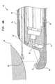
- Fig. 4Bis a schematic illustration showing a video image 90 captured using rectoscope 76 in the configuration of Fig. 4A .
- The-upper part of the imageshows tool 88 and structure 84 as seen by camera head 30 directly through window 36.
- the image of the toolis foreshortened, because of the viewing angle of the camera head. It may therefore be difficult to judge the distance from the tool to the structure based on such an image.
- An inset 92 in the lower portion of image 90shows tool 88 and structure 84 as seen via mirror 82. In this case, the angle from which the tool is viewed allows its position and distance from structure 84 to be seen clearly and controlled with greater accuracy.
- rectoscope 106The ability of rectoscope 106 to bend allows it to be inserted into the rectum, up to the sigmoid, more easily and with less pain and discomfort to the patient than a fully rigid rectoscope or sigmoidoscope would cause.
- Rigid endoscopes known in the artcannot have a flexible section like rectoscope 106, since bending of the flexible section would disrupt the optical axis of the telescope that is used for endoscopic viewing.
- flexible sigmoidoscopes known in the artcan be bent and inserted with less patient discomfort than rigid scopes, such flexible scopes require complicated and costly steering mechanisms.
- the present inventionallows a measure of flexibility without the necessity of a steering mechanism.
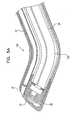
- Fig. 5Bis a schematic, sectional illustration showing a rectal insert 102, for use in endoscopic examination and treatment of intestine 96 of a patient.
- Insert 102is particularly useful for guiding rectoscope 106 into the intestine, but may also be used in conjunction with conventional flexible endoscopes known in the art.
- Insert 102is preferably made of a generally rigid plastic material, such as polycarbonate, but may be somewhat flexible so as to ease its insertion.
- the inserthas a curved channel 104 passing therethrough, with an inner diameter large enough for an endoscope, such as rectoscope 106, to be inserted through the channel.
- Insert 102is inserted through rectum 98 up to sigmoid flexure 100 of intestine 96.
- the insertis turned so that channel 104 opens into sigmoid portion 101 of the intestine, as shown in Fig. 5B .
- Rectoscope 106is then passed through the insert and bends along the curve in the channel.
- the distal end of the rectoscope, including camera head 30,is pushed out into sigmoid 101, allowing inspection and treatment of a portion of intestine 96 that would otherwise be accessible only using a conventional flexible endoscope.
- Insert 102also protects the area of flexure 100 from pressure due to the rectoscope (or to a conventional flexible endoscope inserted therethrough) that could otherwise cause pain or even perforation of the intestine.
- Fig. 6is a schematic, sectional illustration showing a rectoscope 110, including a steering mechansim 111, in accordance with another preferred-embodiment of the present invention.
- Rectoscope 110comprises insertion member 28 and camera head 30, covered by a disposable sheath 112 with optical window 36 at a distal end thereof, as in other preferred embodiments described above.
- Member 28 of rectoscope 110either has a flexible section, as described above with reference to Fig. 5A , or is made flexible over substantially its entire length, as shown in Fig. 6 .
- Steering mechanism 111comprises two flexible, elongate bladders 118 and 120, contained within sheath 112 and disposed radially on opposing sides of member 28.
- Bladders 118 and 120have respective expandable wall sections 114 and 116, preferably accordion-type walls, as shown in Fig. 6 .
- the bladdersare coupled via valves 124 to a pressure source 126, for example, a gas bottle or pump, which may be contained either in a handle 122 of rectoscope 110 or in a separate console.
- a pressure source 126for example, a gas bottle or pump, which may be contained either in a handle 122 of rectoscope 110 or in a separate console.
- a pressure source 126for example, a gas bottle or pump, which may be contained either in a handle 122 of rectoscope 110 or in a separate console.
- a pressure source 126for example, a gas bottle or pump, which may be contained either in a handle 122 of rectoscope 110 or in
Landscapes
- Health & Medical Sciences (AREA)
- Life Sciences & Earth Sciences (AREA)
- Surgery (AREA)
- Engineering & Computer Science (AREA)
- Biomedical Technology (AREA)
- Molecular Biology (AREA)
- Pathology (AREA)
- Radiology & Medical Imaging (AREA)
- Nuclear Medicine, Radiotherapy & Molecular Imaging (AREA)
- Biophysics (AREA)
- Physics & Mathematics (AREA)
- Heart & Thoracic Surgery (AREA)
- Medical Informatics (AREA)
- Optics & Photonics (AREA)
- Animal Behavior & Ethology (AREA)
- General Health & Medical Sciences (AREA)
- Public Health (AREA)
- Veterinary Medicine (AREA)
- Multimedia (AREA)
- Signal Processing (AREA)
- Endoscopes (AREA)
Abstract
Description
- The present invention relates generally to medical probes, and specifically to endoscopes.
- Various types of endoscopes are known in the art for visual inspection and diagnosis of the rectum and sigmoid of the large intestine. Rigid rectoscopes and sigmoidoscopes are most commonly used for this purpose, since they are durable, easy to use and relatively inexpensive.
- To perform an examination using the rigid rectoscope (or sigmoidoscope), the physician first inserts a speculum through the anus. An obturator, having a blunt, rounded distal end, is fitted inside the speculum and protrudes distally therefrom to ease the insertion. Once the speculum is in place, the physician withdraws the obturator and closes the proximal end of the speculum with a plug. A special telescope is fitted into the plug, which enables the physician to view the inside of the rectum and sigmoid at the distal end of the speculum, either by direct vision or using a video camera coupled proximally to the telescope. In order to illuminate the area under view, a light source in an external console is coupled proximally to the speculum by fiberoptics. Typically, the plug also includes one or more working channels, which are used for irrigation, insufflation or insertion of surgical tools through the speculum.
- Various rectoscopes and sigmoidoscopes of this type are available commercially. For example, Karl Storz GmbH, of Tuttlingen, Germany, offers the model 24911S rectoscope, which may be used with a rod lens telescope (for example, model 24946B) to view the rectum either under direct view or using a Karl Storz Endovision video camera. The rectoscope and accessories are made from stainless steel to allow disinfection and autoclaving between uses.
- Rectoscopes and sigmoidoscopes having a disposable speculum and obturator are also available, such as the KleenSpec disposable sigmoidoscope produced by Welch Allyn Inc., of Skaneateles Falls, New York. The speculum and obturator are made of plastic. The speculum clips or screws onto a multi-use handle, to which a light source, telescope, video camera and instruments may be attached.
- Flexible sigmoidoscopes and colonoscopes are also known in the art. Most such scopes include a fiberoptic bundle for conveying images from their distal end, within the intestine, to an eyepiece or video camera fixed to the proximal end. Some such scopes, such as the Pentax ES-3801 PVE Video Sigmoidoscope, made by Asahi Optical Company of Tokyo, have a CCD detector with an objective lens at its distal end. By comparison with rigid rectoscopes and sigmoidoscopes, however, flexible scopes are costly, fragile and difficult to clean.
- Document
US5354302 discloses an endoscope with leaves at the distal end, that can be closed during insertion and opened apart during examination. - It is an object of some aspects of the present invention to provide an improved endoscope, and particularly an improved rectoscope.
- It is an object of some aspects of the present invention to provide a video rectoscope that gives improved visibility of anatomical features within the rectum.
- It is another object of some aspects of the present invention to provide a rectoscope that is easy and convenient for medical personnel to use.
- It is a further object of some aspects of the present invention to provide a rectoscope including an easily-replaceable disposable portion, thus minimizing or eliminating the need for disinfection and sterilization between uses.
- It is yet another object of some aspects of the present invention to provide methods and apparatus that facilitate insertion of an endoscope, particularly a rectoscope or sigmoidoscope, into a body cavity.
- It is still another object of some aspects of the present invention to provide an improved steering mechanism for an endoscope.
- In preferred embodiments of the present invention, a rectoscope comprises an elongate, generally rigid insertion member having a video camera head at its distal end. The insertion member and camera head are covered by a disposable sheath, which includes a transparent distal portion, preferably including an optical window, covering the camera head. The camera head includes an image detector array, preferably a CCD array, as is known in the art, along with a light source for illuminating an area of the intestine viewed by the camera head and an objective lens, which focuses an image of the area onto the array. The sheath, which is preferably made of a biocompatible plastic material, is removed and discarded after use. The rectoscope is used to examine and treat areas of the colon of a patient, specifically the rectal and sigmoid portions of the colon.
- By comparison with rigid rectoscopes and sigmoidoscopes known in the art, the present invention can provide substantially improved image quality, because the video camera head is placed at the distal end of the rectoscope, near the area under view. The distal positioning of the camera head enables the present rectoscope to achieve both higher magnification and a wider field of view than video rectoscopes and sigmoidoscopes known in the art, in which the camera head views the intestine through a telescope from the proximal end of the scope.
- In preferred embodiments of the present invention, the sheath comprises a narrowed, generally bluntly pointed tip, to ease insertion of the rectoscope through the anus: The narrowed tip thus performs the function of the obturator used in insertion of rectoscopes and sigmoidoscopes known in the art. Elimination of the obturator relieves a physician using the rectoscope of unpleasant odors and possible exposure to infection that are commonly associated with removal of the obturator.
- In some preferred embodiments of the present invention, the optical axis of the camera head is angled relative to the longitudinal axis of the insertion member, and the distal end of the sheath is likewise angled, thus defining the narrowed tip. Preferably, the sheath has a bulbous protrusion at the tip, which further eases insertion of the rectoscope through the anus.
- In one such preferred embodiment, a mirror is fixed to the bulbous protrusion, which is shaped so that the mirror is positioned within the field of view of the camera head and is oriented obliquely relative to the optical axis thereof An image of the inner wall of the rectum or sigmoid is reflected from the mirror and captured by the camera head. The mirror thus enables the physician to see an image from an alternative view angle, which may be useful particularly in observing and controlling the movement of tools that the physician inserts into the rectum using the rectoscope, as described below.
- In the present invention, the narrowed tip of the sheath is formed by a plurality of leaves, formed of a resilient material, which butt together tightly in a closed configuration to cover the transparent portion of the sheath and the camera head. During insertion through the anus, the closed leaves protect the transparent portion from fouling by fecal matter, lubricants or other substances. After the rectoscope has been inserted, the leaves are drawn apart, preferably by pulling the sheath back proximally, so as to expose the transparent portion of the sheath and allow the camera head to receive images of the area of the intestine under view.
- In some preferred embodiments of the present invention, the sheath includes one or more working channels, which may be used for suction, irrigation, insufflation, passage of surgical tools or other functions known in the art. The rectoscope includes a handle, to which the sheath is attached, preferably by a quick connect/disconnect fitting. Preferably, the handle includes user controls for regulating such functions as suction, irrigation and insufflation through the working channels. Further preferably, one or more of the working channels include a one-way valve, to prevent back-flow of fluid (gas or liquid) used in irrigation or insufflation through the channels.
- Although rectoscopes in accordance with the present invention are described herein as being generally rigid, in some preferred embodiments the insertion member may include a flexible, resilient longitudinal section. This section allows the rectoscope to bend during insertion into the rectum, to make the insertion easier and less painful for the patient. Such bending is not possible using rigid endoscopes known in the art, and it differs from the bending of flexible endoscopes, which do not have the rigidity to maintain a particular shape and require complex steering mechanisms to control their angle and direction of bend.
- In one such preferred embodiment, the rectoscope sheath comprises one or more flexible, longitudinally- disposed inflatable bladders, preferably at least two such bladders disposed radially within the sheath on different sides of the flexible section of the insertion member. Preferably, the bladders are placed on opposing sides of the sheath. The bladders are connected by respective valves to a suitable pressure source. When the pressure in one of the bladders is increased, the bladder lengthens, whereas when the pressure is decreased, the bladder tends to shorten. Thus, the bend angle of the sheath and of the insertion member therein is controlled by varying the pressure in the one or more bladders.
- In one example, a generally rigid rectal insert having a curved channel therethrough is inserted into the rectum, up to the sigmoid flexure. The insert is turned within the rectum so that the exit from the channel at the sigmoid flexure opens into the intestine in a direction generally along the axis of the sigmoid. The rectoscope including the flexible section, as described hereinabove, is then inserted through the channel and is forced by the rigidity of the insert to bend toward the sigmoid axis. The combination of the insert and rectoscope, thus allows endoscopic inspection of a portion of the intestine that can generally be reached only using a more costly flexible endoscope. The rectal insert described herein can also be used in conjunction with flexible endoscopes known in the art, making it easier to pass the endoscope through the sigmoid and reducing pain and the possibility of accidental perforation of the intestine.
- Although preferred embodiments are described herein with reference to inspection and treatment of the rectum, and endoscopes in accordance with such preferred embodiments are referred to herein generally as rectoscopes, it will be appreciated that such endoscopes may similarly be adapted and applied to serve as sigmoidoscopes. The principles of the present invention may similarly be applied to produce rigid endoscopes for insertion into other body cavities, such as the throat, the vagina, or any other cavity large enough to receive an endoscope having a video camera head a its distal end, as described herein.
- There is therefore provided, in accordance with a preferred embodiment of the present invention, a video endoscope, including:
- a generally rigid, elongate insertion member, having distal and proximal ends;
- a video camera head fixed at the distal end of the insertion member; and
- a sheath, which fits over and covers the insertion member, the sheath having a distal portion covering the video camera head, at least a section of which distal portion is substantially transparent.
- Preferably, the camera head includes a detector array and an objective lens, which forms an image on the detector array.
- In a preferred embodiment, the camera head is inclined obliquely relative to a longitudinal axis of the insertion member.
- Preferably, the camera head includes a light source, most preferably including one or more LEDs.
- The substantially transparent section of the distal portion of the sheath includes an optical window.
- The distal portion of the sheath includes a narrowed tip, which facilitates insertion of the endoscope through the anus. Further preferably, the narrowed tip includes a bulbous protrusion.
- The narrowed tip includes two or more leaves, which are closed during insertion through the anus and open apart after insertion to expose the transparent section of the distal portion of the sheath. To open the leaves, the sheath is shifted in a proximal direction drawn relative to the insertion member.
- Preferably, the sheath contains a working channel passing longitudinally therealong. Further preferably, the sheath includes a one-way valve within the working channel, for preventing back-flow of fluid therethrough.
- Preferably, the endoscope includes a handle, fixed to the proximal end of the insertion member, and the handle includes user controls for controlling the passage of fluid through the working channel.
- In a preferred embodiment, at least a longitudinal section of the insertion member is flexible and resilient. Preferably, the sheath includes an inflatable bladder disposed longitudinally within the sheath, such that inflation of the bladder causes its length to increase so that the insertion member bends.
- In another preferred embodiment, a mirror is fixed in a position distal to the camera head within a field of view thereof and oriented obliquely relative to an optical axis of the camera head, so that the camera head captures an image reflected from the mirror. Preferably, the image reflected from the mirror includes an area of a physiological structure in which a surgical procedure is to be performed using the endoscope.
- There is further provided, in accordance with a preferred embodiment of the present invention, a steering mechanism for an elongate medical probe, including at least one controllably inflatable element fixed to the probe, such that a longitudinal dimension of the element changes responsive to inflation thereof, thereby causing the probe to bend.
- Preferably, the at least one inflatable element includes two elements disposed radially on different sides of the probe, so as to bend the probe in different, respective directions. Further preferably, the two elements include first and second elements on opposing sides of the probe, such that the first element is inflated and the second element is deflated so as to bend the probe toward the side on which the second element is fixed.
- Preferably, the at least one inflatable element includes an expandable wall section on an outer surface thereof, which expands and contracts responsive to changes in the inflation of the element.
- Preferably, the mechanism includes a pressure source which is controllably applied to the inflatable element so as control the bending of the probe.
- In a preferred embodiment, the probe includes a generally rigid endoscope, having a flexible section which is bent by the mechanism, wherein the endoscope preferably includes a disposable sheath, which includes the at least one inflatable element.
- There is moreover provided, a generally rigid rectal insert, having a distal end which is inserted into the rectum of a patient, the insert having a curved channel therethrough through which an endoscope is inserted into the sigmoid portion of the colon of a patient.
- Preferably, the distal end of the insert is brought into proximity with the patient's sigmoid flexure, and the channel is curved so as to open laterally out of the distal end into the flexure. Further preferably, the insert is rotatable within the rectum so as to align the opening of the channel at the distal end thereof with the intestine.
- There is also provided, not belonging to the invention a method of endoscopic inspection, including:
- providing an elongate, generally rigid insertion member having a video camera head at a distal end of the member;
- fitting a disposable sheath over the member, such that the camera head is covered by a substantially transparent portion of the sheath;
- inserting the insertion member, covered by the sheath, into a body cavity of a subject; and
- receiving video images within the body cavity from the camera head.
- Preferably, fitting the sheath includes fitting a sheath having a narrowed tip, and inserting the member includes first inserting the narrowed tip into the cavity.
- In a preferred example, the narrowed tip is opened after insertion to expose the substantially transparent portion of the sheath covering the camera head, wherein opening the narrowed tip preferably includes shifting at least a portion of the sheath in a proximal direction over the insertion member so as to separate two or more leaves that meet to form the narrowed tip.
- Preferably, receiving the video images includes illuminating the inside of the body cavity using a light source in the video camera head.
- Further preferably, fitting the disposable sheath includes fitting a sheath having at least one working channel passing therethrough. In a preferred example, the body cavity is insufflated by passing a pressurized fluid through the working channel.
- In a preferred example, providing the insertion member includes providing a member at least a longitudinal portion of which is flexible and resilient, and inserting the member includes flexing the flexible, resilient portion. Preferably, inserting the member includes inserting an insert into the body cavity having a curved channel therethrough, and passing the member through the channel. Alternatively or additionally, flexing the flexible, resilient portion includes inflating an inflatable bladder disposed within the sheath so that the length of the bladder increases, thereby bending the portion.
- Preferably, inserting the insertion member includes inserting the member through the anus of the subject, and receiving the images includes receiving images within the rectum or, alternatively or additionally, receiving images within the sigmoid.
- There is further provided, not forming part of the invention a method for steering a medical probe, including:
- fixing an inflatable element to the probe; and
- controllably inflating the element so as to change the length thereof, thus causing the probe to bend.
- Preferably, fixing the inflatable element includes fixing a plurality of elements on different sides of the probe, so that each element causes the probe to bend in a different, respective direction.
- In a preferred example, the medical probe includes a generally rigid endoscope at least a section of which is flexible, and wherein controllably inflating the element causes the flexible section of the endoscope to bend.
- There is additionally provided, not forming part of the invention, a method for inserting an endoscope into the colon of a patient, including:
- providing a generally rigid insert having proximal and distal ends and having a curved channel passing therethrough with a lateral opening at the distal end;
- inserting the insert into the patient's rectum so that the distal end is in proximity to the sigmoid flexure of the colon; and
- passing the endoscope through the channel and into the sigmoid colon.
- Preferably, the insert is rotated to align the opening at the distal end of the insert with the colon before passing the endoscope therethrough.
- Preferably, passing the endoscope includes passing a flexible endoscope through the channel or, alternatively, passing a generally rigid endoscope including a flexible portion through the channel, such that the flexible portion is bent by the curve of the channel.
- The-present invention will be more fully understood from the following detailed description of the preferred embodiments thereof, taken together with the drawings in which:
Fig. 1 is a schematic, partly sectional illustration of a rectoscope, in accordance with a preferred embodiment of the present invention;Fig. 2 is a sectional, detail view of a distal portion of the rectoscope ofFig. 1 ;Figs. 3A and 3B are schematic, sectional illustrations of a rectoscope covered by a sheath in closed and open configurations thereof, respectively, in accordance with another preferred embodiment of the present invention;Fig. 4A is a schematic, sectional view of the distal portion of a rectoscope, in accordance with another preferred embodiment of the present invention;Fig. 4B is a schematic illustration of a video image captured by the rectoscope ofFig. 4A ;Fig. 5A is a schematic, sectional view of the distal portion of a rectoscope, in accordance with a preferred embodiment of the present invention;Fig. 5B is a schematic, sectional illustration of a rectal insert, useful particularly in conjunction with the rectoscope ofFig. 5A , andFig. 6 is a schematic, sectional illustration of a rectoscope, in accordance with still another preferred embodiment of the present invention.- Reference is now made to
Fig. 1 , which is a schematic, partly sectional illustration of avideo rectoscope 20, in accordance with a preferred embodiment of the present invention.Rectoscope 20 comprises aninsertion section 22, which is inserted into the rectum of a subject, and ahandle 24, which is grasped by a physician making the examination.Insertion section 22 includes aninsertion member 28, fixed to handle 24, and adisposable sheath 26, covering the insertion member.Member 28 includes avideo camera head 30 at the member's distal end, connected bywires 32 to handle 24. Preferably,sheath 26 comprises a biocompatible plastic material, such as polycarbonate, and is attached to handle 24 using a quick-connect fitting, as is known in the art. After use,sheath 26 is removed and disposed of Fig. 2 is a schematic, sectional illustration showing details of the distal end ofinsertion section 22, includingcamera head 30. The camera head comprises adetector array chip 46, preferably comprising acolor CCD array 50, such as a Sony ICX087AKB CCD chip, with a suitable optical filter 48 affixed thereto. One ormore lamps 42 illuminate an area of the intestine under view by the camera head. An image of the area is received through anoptical window 36 insheath 26 and focused ontoarray 50 by an objective lens 44.- Signals generated by
chip 46 are conveyed bywires 32 to handle 24, and from there via a cable 38 (Fig. 1 ) to a video console, where the signals are processed and used to drive a video display. The video console and display (not shown in the figures) use standard video electronics, well known in the art. Alternatively, handle 24 may include a power source, such as one or more batteries, processing electronics and a video transmitter, and convey processed signals fromchip 46 to the video console over a wireless link. Lamps 42 preferably comprise white-light LEDs, such as T1 White L3-W31N LEDs produced by Sloan AG of Basel, Switzerland The lamps are powered by electrical current conveyed overwires 32 andcable 38 from the console. Alternatively, the lamps may be powered by batteries inhandle 24, as described above. In either case, the use oflamps 42 alleviates the complication and expense of using a high-power fiberoptic light source to illuminate the area under view, as in endoscopes known in the art.Window 36 is shown inFig. 2 as being relatively small, in order to simplify production and reduce the cost ofsheath 26.Sheath 26 also includes a transparent area surrounding the window to allow light fromlamps 42 to reach the area under view, but this area need not be of optical quality. In fact, all ofsheath 26 may be made of transparent plastic if desired.- It will be observed that the optical axis of lens 44 and
array 50 is angled obliquely relative to the longitudinal axis ofmember 28.Window 36 and the distal end ofsheath 26 are similarly angled. This angling allowscamera head 30 to view an area of the intestinal wall relative near the distal end ofsection 22, rather than pointing straight down the intestine as in rigid rectoscopes and sigmoidoscopes known in the art. As a result of this arrangement,section 22 can be made relatively narrow and pointed at its distal end, so that insertion through the subject's anus is easier and less painful, without the need for a separate obturator. Preferably, abulbous projection 34 is formed at the distal tip ofsheath 26, shaped so as to penetrate the anus with a minimum of discomfort to the patient. Sheath 26 preferably includes at least one workingchannel 38, which terminates in anopening 52 at the distal end of the sheath.Channel 38 may be used to inserted surgical tools, such as biopsy forceps, into the intestine and perform surgical procedures under view ofcamera head 30. The angled orientation of the camera head, as described above, is particularly advantageous for viewing such procedures, since the surgical tool will engage the intestinal wall within the camera head's area of view.- Additionally or alternatively,
channel 38 may be used for suction, irrigation and/or insufflation of the intestine, by connecting the channel to a suitable pump or gas source, as is known in the art. Preferably, the connection is made viahandle 24, and the suction, irrigation or insufflation is controlled byuser controls 40 on the handle. Generally, it is desirable to insufflate the intestine during examination, so as to force the intestinal wall away fromwindow 36 and thus providecamera head 30 with a clearer view of the wall. For this purpose,sheath 26 preferably includes a one-way valve 54, such as a leaflet valve, as is known in the art, inchannel 38. After a pressurized gas has been passed through the channel to insufflate the intestine,valve 54 closes to prevent back-flow of the gas and maintain the pressure in the intestine. Figs. 3A and 3B are schematic, sectional illustrations showing arectoscope 60 including adisposable sheath 62, in accordance with another preferred embodiment of the present invention.Rectoscope 60 includes a generallyrigid insertion member 28 with avideo camera head 30 at its distal end, substantially as described above with reference toFigs. 1 and2 , although inrectoscope 60, the optical axis of the camera head is generally aligned with the longitudinal axis ofmember 28.Sheath 62 comprises anouter layer 64 and aninner layer 66. The inner layer preferably comprises a generally rigid plastic material, such as polycarbonate, and fits snugly overmember 28.Layer 66 includes a transparent distal portion, preferably including anoptical window 68, as described above .Outer layer 64 is preferably made of a flexible, resilient plastic, such as polyethylene, and includes two or more leaves 70, preferably three such leaves.- As shown in
Fig. 3A , during insertion ofrectoscope 60 through the anus, leaves 70 butt together, closing off the distal end of the rectoscope and protectingwindow 68. The leaves preferably form a bluntly pointed structure, which eases the insertion. - As shown in
Fig. 3B , after the insertion, leaves 70 are opened to exposewindow 68 and allowcamera head 30 to receive images of the interior of the intestine. Preferably, the leaves are opened by drawingouter layer 64 in a proximal direction, out of the rectum (to the right inFigs. 3A and 3B ), relative toinner layer 66. Eachleaf 70 preferably includes aninner protrusion 72, which engages the surface ofinner layer 66 and forces the leaf outward, as shown in the figure. After the rectoscopy procedure is completed,sheath 62 may be returned to the configuration ofFig. 3A for removal through the anus. Fig. 4A is a schematic, sectional illustration showing the distal portion of arectoscope 76, in accordance with another preferred embodiment of the present invention.Rectoscope 76 is generally similar torectoscope 20, shown inFigs. 1 and2 , and comprisesinsertion member 28 withcamera head 30 at its distal end.Rectoscope 76 is covered by asheath 78, similar tosheath 26 except thatsheath 78 includes abulbous protrusion 80 that extends into the field of view ofcamera head 30. Amirror 82 is fixed toprotrusion 80, in such a position and orientation thereon that an image of aphysiological structure 84, generally the rectal wall, is reflected from the mirror, throughwindow 36, and is captured bycamera head 30. In the example shown inFig. 4A , asurgical tool 88 is inserted through workingchannel 38 insheath 78 and is used to take a biopsy sample at apoint 86 onstructure 84.Fig. 4B is a schematic illustration showing avideo image 90 captured usingrectoscope 76 in the configuration ofFig. 4A . The-upper part of the image showstool 88 andstructure 84 as seen bycamera head 30 directly throughwindow 36. The image of the tool is foreshortened, because of the viewing angle of the camera head. It may therefore be difficult to judge the distance from the tool to the structure based on such an image. Aninset 92 in the lower portion ofimage 90 showstool 88 andstructure 84 as seen viamirror 82. In this case, the angle from which the tool is viewed allows its position and distance fromstructure 84 to be seen clearly and controlled with greater accuracy.Fig. 5A is a schematic, sectional illustration of the distal portion of arectoscope 106, in accordance with mother preferred embodiment of the present invention.Rectoscope 106 is substantially similar torectoscope 20, shown inFigs. 1 and2 , except thatinsertion member 28 of rectoscope includes at least one flexible,resilient section 108.Section 108 preferably includes an elastic element, for example, a spring steel insert, which normally holds the section straight, but allows it to bend in response to lateral forces exerted thereon. Further preferably,sheath 26 ofrectoscope 106 includes flexible, elastic material, for example, polyethylene or silicone, so that the sheath can bend and stretch accordingly whensection 108 bends.- The ability of
rectoscope 106 to bend allows it to be inserted into the rectum, up to the sigmoid, more easily and with less pain and discomfort to the patient than a fully rigid rectoscope or sigmoidoscope would cause. Rigid endoscopes known in the art cannot have a flexible section likerectoscope 106, since bending of the flexible section would disrupt the optical axis of the telescope that is used for endoscopic viewing. Although flexible sigmoidoscopes known in the art can be bent and inserted with less patient discomfort than rigid scopes, such flexible scopes require complicated and costly steering mechanisms. By placingcamera head 30 at the distal end ofrectoscope 106, the present invention allows a measure of flexibility without the necessity of a steering mechanism. Fig. 5B is a schematic, sectional illustration showing arectal insert 102, for use in endoscopic examination and treatment ofintestine 96 of a patient.Insert 102 is particularly useful for guidingrectoscope 106 into the intestine, but may also be used in conjunction with conventional flexible endoscopes known in the art.Insert 102 is preferably made of a generally rigid plastic material, such as polycarbonate, but may be somewhat flexible so as to ease its insertion. The insert has acurved channel 104 passing therethrough, with an inner diameter large enough for an endoscope, such asrectoscope 106, to be inserted through the channel.Insert 102 is inserted throughrectum 98 up tosigmoid flexure 100 ofintestine 96. The insert is turned so thatchannel 104 opens intosigmoid portion 101 of the intestine, as shown inFig. 5B .Rectoscope 106 is then passed through the insert and bends along the curve in the channel. The distal end of the rectoscope, includingcamera head 30, is pushed out intosigmoid 101, allowing inspection and treatment of a portion ofintestine 96 that would otherwise be accessible only using a conventional flexible endoscope.Insert 102 also protects the area offlexure 100 from pressure due to the rectoscope (or to a conventional flexible endoscope inserted therethrough) that could otherwise cause pain or even perforation of the intestine.Fig. 6 is a schematic, sectional illustration showing arectoscope 110, including asteering mechansim 111, in accordance with another preferred-embodiment of the present invention.Rectoscope 110 comprisesinsertion member 28 andcamera head 30, covered by adisposable sheath 112 withoptical window 36 at a distal end thereof, as in other preferred embodiments described above.Member 28 ofrectoscope 110 either has a flexible section, as described above with reference toFig. 5A , or is made flexible over substantially its entire length, as shown inFig. 6 .Steering mechanism 111 comprises two flexible,elongate bladders 118 and 120, contained withinsheath 112 and disposed radially on opposing sides ofmember 28.Bladders 118 and 120 have respectiveexpandable wall sections Fig. 6 . The bladders are coupled viavalves 124 to apressure source 126, for example, a gas bottle or pump, which may be contained either in ahandle 122 ofrectoscope 110 or in a separate console. By operatingvalves 124,bladders 118 and 120 are pressurized to respective pressures P1 and P2. In the example shown inFig. 6 , P1 is greater than P2, causingwall section 114 ofbladder 118 to expand, andwall section 116 of bladder 120 to contract, thereby causingmember 28 to bend.Mechanism 111 thus provides a simple, low cost, disposable means for controllably bending and steeringrectoscope 110, by inflating and deflatingbladders 118 and 120 insheath 112. Additional bladders may be positioned along the length of the rectoscope and/or radially around it in order to give greater control of the bend shape and direction of the rectoscope. It will be appreciated that such pressure-driven steering mechanisms, either disposable or reusable, can also be applied to endoscopes of other types.- It will further be appreciated that the preferred embodiments described above are cited by way of example, and the full scope of the invention is limited only by the claims.
Claims (15)
- An endoscope (60), comprising:a generally rigid, elongate insertion member (28), having distal and proximal ends; anda removable sheath (62), which comprises inner and outer layers (66,64) which fit over and cover the insertion member, with the outer layer (64) having a distal portion comprising a narrowed tip, which facilitates insertion of the endoscope and which comprises two or more leaves (70), which are closed during insertion and open apart after insertion to expose the transparent section of the distal portion of the sheath,characterized in that
the endoscope is for insertion in an anus;
a video camera head (30) is fixed at the distal end of the insertion member;
the inner layer (66) of the sheath has a distal portion covering the video camera head, the distal portion being transparent; andin that
each leaf comprises an inner protrusion (72) which engages the surface of the inner layer (66) of the sheath (62) and forces the leaf outward as the outer layer (64) is moved in a proximal direction relative to the inner layer (66). - An endoscope according to claim 1, wherein the camera head comprises a detector array.
- An endoscope according to claim 2, wherein the camera head comprises an objective lens, which forms an image on the detector array.
- An endoscope according to any of the preceding claims, wherein the camera head is inclined obliquely relative to a longitudinal axis of the insertion member.
- An endoscope according to any of the preceding claims, wherein the camera head comprises a light source.
- An endoscope according to claim 5, wherein the light source comprises one or more LEDs.
- An endoscope according to any of the preceding claims, wherein the substantially transparent section of the distal portion of the sheath comprises an optical window.
- An endoscope according to any of the preceding claims, wherein to open the leaves, the sheath is shifted in a proximal direction drawn relative to the insertion member.
- An endoscope according to any of the preceding claims, wherein the sheath contains a working channel passing longitudinally therealong.
- An endoscope according to claim 9, wherein the sheath comprises a one-way valve within the working channel, for preventing back-flow of fluid therethrough.
- An endoscope according to claim 9 or 10, wherein the endoscope comprises a handle, fixed to the proximal end of the insertion member, and wherein the handle comprises user controls for controlling the passage of fluid through the working channel.
- An endoscope according to any of the preceding claims, wherein the insertion member at least a longitudinal section of the insertion member is flexible and resilient.
- An endoscope according to claim 12, wherein the sheath comprises an inflatable bladder disposed longitudinally within the sheath, such that inflation of the bladder causes its length to increase so that the insertion member bends.
- An endoscope according to any of the preceding claims, and comprising a mirror fixed in a position distal to the camera head within a field of view thereof and oriented obliquely relative to an optical axis of the camera head, so that the camera head captures an image reflected from the mirror.
- An endoscope according to claim 14, wherein the image reflected from the mirror includes an area of a physiological structure in which a surgical procedure is to be performed using the endoscope.
Applications Claiming Priority (3)
| Application Number | Priority Date | Filing Date | Title |
|---|---|---|---|
| IL12211197AIL122111A (en) | 1997-11-04 | 1997-11-04 | Video rectoscope |
| IL12211197 | 1997-11-04 | ||
| PCT/IL1998/000529WO1999023812A2 (en) | 1997-11-04 | 1998-11-02 | Video rectoscope |
Publications (3)
| Publication Number | Publication Date |
|---|---|
| EP1029414A2 EP1029414A2 (en) | 2000-08-23 |
| EP1029414A4 EP1029414A4 (en) | 2004-08-11 |
| EP1029414B1true EP1029414B1 (en) | 2008-03-26 |
Family
ID=11070816
Family Applications (1)
| Application Number | Title | Priority Date | Filing Date |
|---|---|---|---|
| EP98951652AExpired - LifetimeEP1029414B1 (en) | 1997-11-04 | 1998-11-02 | Video rectoscope |
Country Status (11)
| Country | Link |
|---|---|
| US (2) | US6447444B1 (en) |
| EP (1) | EP1029414B1 (en) |
| JP (1) | JP2001521806A (en) |
| KR (1) | KR100630624B1 (en) |
| CN (1) | CN1225229C (en) |
| AT (1) | ATE390079T1 (en) |
| AU (1) | AU752555B2 (en) |
| CA (1) | CA2309151C (en) |
| DE (1) | DE69839309T2 (en) |
| IL (1) | IL122111A (en) |
| WO (1) | WO1999023812A2 (en) |
Cited By (1)
| Publication number | Priority date | Publication date | Assignee | Title |
|---|---|---|---|---|
| EP3993689B1 (en)* | 2019-10-16 | 2023-06-07 | es endomed solutions GmbH | Anti-contamination device for endoscopes with sideways optics |
Families Citing this family (226)
| Publication number | Priority date | Publication date | Assignee | Title |
|---|---|---|---|---|
| JP3596340B2 (en)* | 1999-03-18 | 2004-12-02 | 株式会社日立製作所 | Surgical insertion device |
| US6139489A (en)* | 1999-10-05 | 2000-10-31 | Ethicon Endo-Surgery, Inc. | Surgical device with integrally mounted image sensor |
| US6517477B1 (en)* | 2000-01-27 | 2003-02-11 | Scimed Life Systems, Inc. | Catheter introducer system for exploration of body cavities |
| DE50014197D1 (en) | 2000-02-12 | 2007-05-10 | Leica Microsystems | surgical microscope |
| US8888688B2 (en) | 2000-04-03 | 2014-11-18 | Intuitive Surgical Operations, Inc. | Connector device for a controllable instrument |
| US6468203B2 (en) | 2000-04-03 | 2002-10-22 | Neoguide Systems, Inc. | Steerable endoscope and improved method of insertion |
| US6610007B2 (en) | 2000-04-03 | 2003-08-26 | Neoguide Systems, Inc. | Steerable segmented endoscope and method of insertion |
| US8517923B2 (en) | 2000-04-03 | 2013-08-27 | Intuitive Surgical Operations, Inc. | Apparatus and methods for facilitating treatment of tissue via improved delivery of energy based and non-energy based modalities |
| US6692430B2 (en) | 2000-04-10 | 2004-02-17 | C2Cure Inc. | Intra vascular imaging apparatus |
| IL135571A0 (en) | 2000-04-10 | 2001-05-20 | Doron Adler | Minimal invasive surgery imaging system |
| IL137430A0 (en)* | 2000-07-20 | 2001-07-24 | In Sight Medical | A series of new illuminating devices for medical use |
| IL138237A (en) | 2000-09-04 | 2008-12-29 | Stryker Gi Ltd | Double sleeve endoscope |
| US20020133058A1 (en)* | 2000-09-26 | 2002-09-19 | Calderwood Mitchell C. | Prophylactic cover and drape for endoscopic camera system |
| JP4617059B2 (en) | 2001-04-20 | 2011-01-19 | パワー メディカル インターベンションズ, エルエルシー | Imaging device |
| US8038602B2 (en)* | 2001-10-19 | 2011-10-18 | Visionscope Llc | Portable imaging system employing a miniature endoscope |
| EP1463441A4 (en) | 2001-12-11 | 2009-01-21 | C2Cure Inc | Apparatus, method and system for intravascular photographic imaging |
| US7081097B2 (en)* | 2002-01-04 | 2006-07-25 | Vision Sciences, Inc. | Endoscope sheath assemblies having an attached biopsy sampling device |
| EP1478263B1 (en) | 2002-01-30 | 2011-03-09 | Tyco Healthcare Group LP | Surgical imaging device |
| WO2003098913A2 (en) | 2002-05-16 | 2003-11-27 | Cbyond Inc. | Miniature camera head |
| DE10229540A1 (en)* | 2002-07-01 | 2004-01-29 | Wolfram Henning | Protective cover for video cameras |
| CA2722645A1 (en) | 2002-07-31 | 2004-02-19 | Power Medical Interventions, Llc | Orifice introducer device |
| CN100346738C (en)* | 2002-08-15 | 2007-11-07 | 赛特莱恩技术有限公司 | Endoscope sleeve dispenser |
| AU2003275336A1 (en)* | 2002-09-30 | 2004-04-23 | Power Medical Interventions, Inc. | Self-contained sterilizable surgical system |
| US7452329B2 (en)* | 2003-01-21 | 2008-11-18 | Thd S.P.A. | Retractor for operations on the arteria haemorroidalis |
| US20040199052A1 (en) | 2003-04-01 | 2004-10-07 | Scimed Life Systems, Inc. | Endoscopic imaging system |
| US20050245789A1 (en) | 2003-04-01 | 2005-11-03 | Boston Scientific Scimed, Inc. | Fluid manifold for endoscope system |
| US8118732B2 (en) | 2003-04-01 | 2012-02-21 | Boston Scientific Scimed, Inc. | Force feedback control system for video endoscope |
| US7578786B2 (en) | 2003-04-01 | 2009-08-25 | Boston Scientific Scimed, Inc. | Video endoscope |
| US7591783B2 (en) | 2003-04-01 | 2009-09-22 | Boston Scientific Scimed, Inc. | Articulation joint for video endoscope |
| EP2505125A1 (en) | 2003-04-29 | 2012-10-03 | Aircraft Medical Limited | Laryngoscope with camera attachment |
| JP3854946B2 (en)* | 2003-05-30 | 2006-12-06 | オリンパス株式会社 | Endoscope |
| DE10334100A1 (en)* | 2003-07-25 | 2005-03-03 | Viktor Josef Wimmer | Flexible endoscope with longitudinal axial channels |
| US6908428B2 (en)* | 2003-09-04 | 2005-06-21 | Sightline Technologies Ltd. | Sleeve for endoscopic tools |
| ITMI20032278A1 (en)* | 2003-11-21 | 2005-05-22 | Ethicon Endo Surgery Inc | DIAGNOSIS DEVICE |
| US20050165272A1 (en)* | 2003-12-01 | 2005-07-28 | Yuta Okada | Endoscope system |
| JP4567343B2 (en)* | 2004-02-02 | 2010-10-20 | コデン株式会社 | Endoscope guide and endoscope system |
| US8100822B2 (en)* | 2004-03-16 | 2012-01-24 | Macroplata Systems, Llc | Anoscope for treating hemorrhoids without the trauma of cutting or the use of an endoscope |
| US20050234305A1 (en)* | 2004-04-15 | 2005-10-20 | Frederick Licciardi | Medical device and system for providing an image |
| US7300397B2 (en) | 2004-07-29 | 2007-11-27 | C2C Cure, Inc. | Endoscope electronics assembly |
| US7335159B2 (en)* | 2004-08-26 | 2008-02-26 | Scimed Life Systems, Inc. | Endoscope having auto-insufflation and exsufflation |
| AU2005278773A1 (en)* | 2004-09-03 | 2006-03-09 | Stryker Gi Ltd. | Optical head for endoscope |
| US20060069313A1 (en)* | 2004-09-30 | 2006-03-30 | Couvillon Lucien A Jr | Medical devices with light emitting regions |
| US7479106B2 (en) | 2004-09-30 | 2009-01-20 | Boston Scientific Scimed, Inc. | Automated control of irrigation and aspiration in a single-use endoscope |
| US7241263B2 (en) | 2004-09-30 | 2007-07-10 | Scimed Life Systems, Inc. | Selectively rotatable shaft coupler |
| JP2008514363A (en) | 2004-09-30 | 2008-05-08 | ボストン サイエンティフィック リミテッド | Multifunctional endoscope system for use in electrosurgical applications |
| US7597662B2 (en) | 2004-09-30 | 2009-10-06 | Boston Scientific Scimed, Inc. | Multi-fluid delivery system |
| US8083671B2 (en) | 2004-09-30 | 2011-12-27 | Boston Scientific Scimed, Inc. | Fluid delivery system for use with an endoscope |
| WO2006039522A2 (en) | 2004-09-30 | 2006-04-13 | Boston Scientific Scimed, Inc. | Adapter for use with digital imaging medical device |
| EP1799096A2 (en) | 2004-09-30 | 2007-06-27 | Boston Scientific Scimed, Inc. | System and method of obstruction removal |
| IL165482A0 (en)* | 2004-11-30 | 2006-01-15 | Sphereview Ltd | A compact mobile gynecology system for observationimaging and image analysis |
| US20080015569A1 (en) | 2005-02-02 | 2008-01-17 | Voyage Medical, Inc. | Methods and apparatus for treatment of atrial fibrillation |
| EP1868486A4 (en)* | 2005-04-01 | 2012-11-14 | Verathon Medical Canada Ulc | Video retractor |
| JP4754871B2 (en)* | 2005-05-11 | 2011-08-24 | オリンパスメディカルシステムズ株式会社 | End of the endoscope |
| US8097003B2 (en) | 2005-05-13 | 2012-01-17 | Boston Scientific Scimed, Inc. | Endoscopic apparatus with integrated variceal ligation device |
| US7846107B2 (en) | 2005-05-13 | 2010-12-07 | Boston Scientific Scimed, Inc. | Endoscopic apparatus with integrated multiple biopsy device |
| US7553278B2 (en)* | 2005-06-01 | 2009-06-30 | Cannuflow, Inc. | Protective cap for arthroscopic instruments |
| US7738683B2 (en)* | 2005-07-22 | 2010-06-15 | Carestream Health, Inc. | Abnormality detection in medical images |
| US20100010308A1 (en)* | 2005-08-17 | 2010-01-14 | Michael Braun | Hygiene protector for endoscopes, endoscope with hygiene protector, and method for fitting a hygiene protector onto an endoscope |
| US8052597B2 (en) | 2005-08-30 | 2011-11-08 | Boston Scientific Scimed, Inc. | Method for forming an endoscope articulation joint |
| US20070073107A1 (en)* | 2005-09-23 | 2007-03-29 | Percutaneous Systems, Inc. | Methods and systems for sheathing tubular medical instruments |
| US20070072466A1 (en)* | 2005-09-27 | 2007-03-29 | Manabu Miyamoto | Instrument for endoscope |
| US8537366B2 (en) | 2005-10-11 | 2013-09-17 | Duke University | Systems and methods for endoscopic angle-resolved low coherence interferometry |
| US7967759B2 (en) | 2006-01-19 | 2011-06-28 | Boston Scientific Scimed, Inc. | Endoscopic system with integrated patient respiratory status indicator |
| JP2009531078A (en)* | 2006-03-02 | 2009-09-03 | タラス メディカル, インコーポレイテッド | Artificial bone |
| US8888684B2 (en) | 2006-03-27 | 2014-11-18 | Boston Scientific Scimed, Inc. | Medical devices with local drug delivery capabilities |
| US7794393B2 (en)* | 2006-04-13 | 2010-09-14 | Larsen Dane M | Resectoscopic device and method |
| US7955255B2 (en) | 2006-04-20 | 2011-06-07 | Boston Scientific Scimed, Inc. | Imaging assembly with transparent distal cap |
| US8202265B2 (en) | 2006-04-20 | 2012-06-19 | Boston Scientific Scimed, Inc. | Multiple lumen assembly for use in endoscopes or other medical devices |
| DE602007012852D1 (en)* | 2006-05-04 | 2011-04-14 | Navab Nassir | Interactive virtual mirror device for visualizing virtual objects in endoscopic applications |
| US9055906B2 (en) | 2006-06-14 | 2015-06-16 | Intuitive Surgical Operations, Inc. | In-vivo visualization systems |
| CN103815860A (en) | 2006-07-21 | 2014-05-28 | 昂科斯科公司 | Protective probe tip, particularly for use on fiber-optic probe used in endoscopic application |
| US7798957B2 (en)* | 2006-07-31 | 2010-09-21 | Chang Stanley F | Colonoscope guide and method of use for improved colonoscopy |
| US9226648B2 (en)* | 2006-12-21 | 2016-01-05 | Intuitive Surgical Operations, Inc. | Off-axis visualization systems |
| WO2008095052A2 (en) | 2007-01-30 | 2008-08-07 | Loma Vista Medical, Inc., | Biological navigation device |
| US7655004B2 (en) | 2007-02-15 | 2010-02-02 | Ethicon Endo-Surgery, Inc. | Electroporation ablation apparatus, system, and method |
| CN101293124B (en)* | 2007-04-29 | 2010-09-08 | 曾世一 | A genital lighting device |
| GB0708761D0 (en)* | 2007-05-04 | 2007-06-13 | Evexar Medical Ltd | Improvements in and relating to medical instruments |
| NO327762B1 (en)* | 2007-05-23 | 2009-09-21 | Hovda Olav | Rectoscope device |
| US9241610B2 (en) | 2009-12-08 | 2016-01-26 | Medeon Biodesign, Inc. | Devices and methods for removal of debris from the objective lens of an endoscope |
| ES2708428T3 (en) | 2007-06-08 | 2019-04-09 | Medeon Biodesign Inc | Devices for the elimination of residues from the objective of an endoscope |
| US10307041B2 (en) | 2007-06-08 | 2019-06-04 | Medeon Biodesign, Inc. | Lens cover modification |
| US20090048486A1 (en)* | 2007-08-08 | 2009-02-19 | Wilson-Cook Medical Inc. | Distal Tip for an Endoscope |
| US20090054728A1 (en)* | 2007-08-21 | 2009-02-26 | Trusty Robert M | Manipulatable guide system and methods for natural orifice translumenal endoscopic surgery |
| US20090073456A1 (en)* | 2007-09-13 | 2009-03-19 | Duke University | Apparatuses, systems, and methods for low-coherence interferometry (lci) |
| DE202007014897U1 (en)* | 2007-10-25 | 2009-03-05 | Dr. Fritz Gmbh | Endoscope with lockable instrument channel |
| AU2009204187B2 (en)* | 2008-01-08 | 2015-02-05 | Oncoscope, Inc. | Systems and methods for tissue examination, diagnostic, treatment, and/or monitoring |
| TWI343246B (en)* | 2008-01-31 | 2011-06-11 | Tien Sheng Chen | Anus check device and anus check set |
| EP2303100B1 (en) | 2008-04-16 | 2020-02-12 | United States Endoscopy Group, Inc. | Adaptor for a water bottle of an endoscope |
| US8550988B2 (en)* | 2008-04-21 | 2013-10-08 | Covidien Lp | Endoscopic cleaner |
| US20090270686A1 (en) | 2008-04-29 | 2009-10-29 | Ethicon Endo-Surgery, Inc. | Methods and devices for maintaining visibility during surgical procedures |
| US20090299374A1 (en) | 2008-06-02 | 2009-12-03 | Loma Vista Medical, Inc. | Inflatable medical devices |
| JP5103293B2 (en)* | 2008-06-17 | 2012-12-19 | 富士フイルム株式会社 | Electronic endoscope |
| US8888792B2 (en) | 2008-07-14 | 2014-11-18 | Ethicon Endo-Surgery, Inc. | Tissue apposition clip application devices and methods |
| CN102112163A (en)* | 2008-07-28 | 2011-06-29 | 脊柱诊察公司 | Penetrating member with direct visualization |
| JP4435252B2 (en)* | 2008-09-08 | 2010-03-17 | オリンパス株式会社 | Anal video endoscope |
| US8628468B2 (en)* | 2008-10-01 | 2014-01-14 | David L. Zisow | Device for anchoring a trocar |
| US20100081994A1 (en)* | 2008-10-01 | 2010-04-01 | David Leslie Zisow | Self Retaining Laparoscopic Trocar System-Zisow Trocar Sleeve System |
| US20100094082A1 (en)* | 2008-10-09 | 2010-04-15 | Jay Iinuma | Medical examining device with an angularly offset fiber optic channel |
| US20100094167A1 (en)* | 2008-10-09 | 2010-04-15 | Jay Iinuma | Medical examining device with fiber optic receiving channel and sampling channel |
| US8157834B2 (en) | 2008-11-25 | 2012-04-17 | Ethicon Endo-Surgery, Inc. | Rotational coupling device for surgical instrument with flexible actuators |
| US8361066B2 (en) | 2009-01-12 | 2013-01-29 | Ethicon Endo-Surgery, Inc. | Electrical ablation devices |
| JP5052553B2 (en)* | 2009-03-19 | 2012-10-17 | オリンパス株式会社 | Treatment endoscope |
| JP5087035B2 (en)* | 2009-03-25 | 2012-11-28 | オリンパス株式会社 | Cover-type treatment endoscope and endoscope cover |
| US8361041B2 (en) | 2009-04-09 | 2013-01-29 | University Of Utah Research Foundation | Optically guided feeding tube, catheters and associated methods |
| US9254245B2 (en) | 2009-04-09 | 2016-02-09 | University Of Utah | Optically guided medical tube and control unit assembly and methods of use |
| US9101287B2 (en) | 2011-03-07 | 2015-08-11 | Endochoice Innovation Center Ltd. | Multi camera endoscope assembly having multiple working channels |
| US11864734B2 (en) | 2009-06-18 | 2024-01-09 | Endochoice, Inc. | Multi-camera endoscope |
| US11278190B2 (en) | 2009-06-18 | 2022-03-22 | Endochoice, Inc. | Multi-viewing element endoscope |
| US12137873B2 (en) | 2009-06-18 | 2024-11-12 | Endochoice, Inc. | Compact multi-viewing element endoscope system |
| US9706903B2 (en) | 2009-06-18 | 2017-07-18 | Endochoice, Inc. | Multiple viewing elements endoscope system with modular imaging units |
| US9101268B2 (en) | 2009-06-18 | 2015-08-11 | Endochoice Innovation Center Ltd. | Multi-camera endoscope |
| WO2010146587A1 (en) | 2009-06-18 | 2010-12-23 | Peer Medical Ltd. | Multi-camera endoscope |
| US9901244B2 (en) | 2009-06-18 | 2018-02-27 | Endochoice, Inc. | Circuit board assembly of a multiple viewing elements endoscope |
| US9713417B2 (en) | 2009-06-18 | 2017-07-25 | Endochoice, Inc. | Image capture assembly for use in a multi-viewing elements endoscope |
| US9872609B2 (en) | 2009-06-18 | 2018-01-23 | Endochoice Innovation Center Ltd. | Multi-camera endoscope |
| US9402533B2 (en) | 2011-03-07 | 2016-08-02 | Endochoice Innovation Center Ltd. | Endoscope circuit board assembly |
| US10165929B2 (en) | 2009-06-18 | 2019-01-01 | Endochoice, Inc. | Compact multi-viewing element endoscope system |
| US9492063B2 (en) | 2009-06-18 | 2016-11-15 | Endochoice Innovation Center Ltd. | Multi-viewing element endoscope |
| US11547275B2 (en) | 2009-06-18 | 2023-01-10 | Endochoice, Inc. | Compact multi-viewing element endoscope system |
| US8926502B2 (en) | 2011-03-07 | 2015-01-06 | Endochoice, Inc. | Multi camera endoscope having a side service channel |
| US9642513B2 (en) | 2009-06-18 | 2017-05-09 | Endochoice Inc. | Compact multi-viewing element endoscope system |
| CN102686142B (en) | 2009-08-31 | 2014-10-29 | 布拉蔻诊断公司 | In-line gas adapter for endoscopic apparatus |
| WO2011032067A1 (en) | 2009-09-14 | 2011-03-17 | Bracco Diagnostics Inc. | In-line gas adaptor for endoscopic apparatus |
| DE102009044303B4 (en)* | 2009-10-21 | 2012-04-19 | Karlsruher Institut für Technologie | Fast optical tomography |
| US20110098704A1 (en) | 2009-10-28 | 2011-04-28 | Ethicon Endo-Surgery, Inc. | Electrical ablation devices |
| US8888687B2 (en)* | 2009-10-28 | 2014-11-18 | Boston Scientific Scimed, Inc. | Method and apparatus related to a flexible assembly at a distal end portion of a medical device |
| US9028483B2 (en) | 2009-12-18 | 2015-05-12 | Ethicon Endo-Surgery, Inc. | Surgical instrument comprising an electrode |
| KR101067973B1 (en)* | 2009-12-29 | 2011-09-26 | 국립암센터 | Surgical instrument |
| WO2011091369A1 (en) | 2010-01-22 | 2011-07-28 | Duke University | Multiple window processing schemes for spectroscopic optical coherence tomography (oct) and fourier domain low coherence interferometry |
| US9823127B2 (en) | 2010-01-22 | 2017-11-21 | Duke University | Systems and methods for deep spectroscopic imaging of biological samples with use of an interferometer and spectrometer |
| US8956346B2 (en) | 2010-05-14 | 2015-02-17 | Rainbow Medical, Ltd. | Reflectance-facilitated ultrasound treatment and monitoring |
| US9242122B2 (en) | 2010-05-14 | 2016-01-26 | Liat Tsoref | Reflectance-facilitated ultrasound treatment and monitoring |
| US12220105B2 (en) | 2010-06-16 | 2025-02-11 | Endochoice, Inc. | Circuit board assembly of a multiple viewing elements endoscope |
| WO2012009486A2 (en) | 2010-07-13 | 2012-01-19 | Loma Vista Medical, Inc. | Inflatable medical devices |
| MX350734B (en) | 2010-09-08 | 2017-09-15 | Covidien Lp | Catheter with imaging assembly. |
| EP2618718B1 (en) | 2010-09-20 | 2020-04-15 | EndoChoice Innovation Center Ltd. | Multi-camera endoscope having fluid channels |
| US9560953B2 (en) | 2010-09-20 | 2017-02-07 | Endochoice, Inc. | Operational interface in a multi-viewing element endoscope |
| JP2012090741A (en)* | 2010-10-26 | 2012-05-17 | Fujifilm Corp | Endoscope device |
| CN103403605A (en) | 2010-10-28 | 2013-11-20 | 恩多巧爱思创新中心有限公司 | Optical systems for multi-sensor endoscopes |
| US12204087B2 (en) | 2010-10-28 | 2025-01-21 | Endochoice, Inc. | Optical systems for multi-sensor endoscopes |
| US10188436B2 (en) | 2010-11-09 | 2019-01-29 | Loma Vista Medical, Inc. | Inflatable medical devices |
| US9320419B2 (en) | 2010-12-09 | 2016-04-26 | Endochoice Innovation Center Ltd. | Fluid channeling component of a multi-camera endoscope |
| US11889986B2 (en) | 2010-12-09 | 2024-02-06 | Endochoice, Inc. | Flexible electronic circuit board for a multi-camera endoscope |
| CN107361721B (en) | 2010-12-09 | 2019-06-18 | 恩多巧爱思创新中心有限公司 | Flexible electronic circuit boards for multi-camera endoscopes |
| EP2672878B1 (en) | 2011-02-07 | 2017-11-22 | Endochoice Innovation Center Ltd. | Multi-element cover for a multi-camera endoscope |
| US9282948B2 (en)* | 2011-02-22 | 2016-03-15 | Cook Medical Technologies Llc | Total core biopsy device and method of use |
| US9233241B2 (en) | 2011-02-28 | 2016-01-12 | Ethicon Endo-Surgery, Inc. | Electrical ablation devices and methods |
| US9254169B2 (en) | 2011-02-28 | 2016-02-09 | Ethicon Endo-Surgery, Inc. | Electrical ablation devices and methods |
| US9049987B2 (en) | 2011-03-17 | 2015-06-09 | Ethicon Endo-Surgery, Inc. | Hand held surgical device for manipulating an internal magnet assembly within a patient |
| CN102253057B (en)* | 2011-04-13 | 2013-06-05 | 中国科学院深圳先进技术研究院 | Endoscope system and measurement method using endoscope system |
| JP6318088B2 (en) | 2011-07-26 | 2018-04-25 | アンフォラ メディカル, インコーポレイテッド | Apparatus and method for modulating pelvic nerve tissue |
| JP5863006B2 (en)* | 2011-09-06 | 2016-02-16 | 学校法人 中央大学 | Fluid supply device |
| EP2604172B1 (en) | 2011-12-13 | 2015-08-12 | EndoChoice Innovation Center Ltd. | Rotatable connector for an endoscope |
| CA2798716A1 (en) | 2011-12-13 | 2013-06-13 | Peermedical Ltd. | Removable tip endoscope |
| US9707414B2 (en) | 2012-02-14 | 2017-07-18 | Rainbow Medical Ltd. | Reflectance-facilitated ultrasound treatment and monitoring |
| US10492662B2 (en) | 2012-03-27 | 2019-12-03 | Medigus Ltd. | Integrated endoscope irrigation |
| EP2830477A1 (en) | 2012-03-30 | 2015-02-04 | Bracco Diagnostics Inc. | Water bottle cap assemblies for an endoscopic device |
| CN102626301A (en)* | 2012-04-28 | 2012-08-08 | 无锡市第二人民医院 | Multifunctional portable wireless medical endoscope |
| US9427255B2 (en) | 2012-05-14 | 2016-08-30 | Ethicon Endo-Surgery, Inc. | Apparatus for introducing a steerable camera assembly into a patient |
| JP2014012043A (en)* | 2012-07-03 | 2014-01-23 | Coden Co Ltd | Pipe endoscope |
| US9078662B2 (en) | 2012-07-03 | 2015-07-14 | Ethicon Endo-Surgery, Inc. | Endoscopic cap electrode and method for using the same |
| US9560954B2 (en) | 2012-07-24 | 2017-02-07 | Endochoice, Inc. | Connector for use with endoscope |
| US9545290B2 (en) | 2012-07-30 | 2017-01-17 | Ethicon Endo-Surgery, Inc. | Needle probe guide |
| US10314649B2 (en) | 2012-08-02 | 2019-06-11 | Ethicon Endo-Surgery, Inc. | Flexible expandable electrode and method of intraluminal delivery of pulsed power |
| US9277957B2 (en) | 2012-08-15 | 2016-03-08 | Ethicon Endo-Surgery, Inc. | Electrosurgical devices and methods |
| US9198835B2 (en) | 2012-09-07 | 2015-12-01 | Covidien Lp | Catheter with imaging assembly with placement aid and related methods therefor |
| US9517184B2 (en) | 2012-09-07 | 2016-12-13 | Covidien Lp | Feeding tube with insufflation device and related methods therefor |
| USD716841S1 (en) | 2012-09-07 | 2014-11-04 | Covidien Lp | Display screen with annotate file icon |
| USD717340S1 (en) | 2012-09-07 | 2014-11-11 | Covidien Lp | Display screen with enteral feeding icon |
| USD735343S1 (en) | 2012-09-07 | 2015-07-28 | Covidien Lp | Console |
| US9736342B2 (en) | 2012-10-19 | 2017-08-15 | Milwaukee Electric Tool Corporation | Visual inspection device |
| US10098527B2 (en) | 2013-02-27 | 2018-10-16 | Ethidcon Endo-Surgery, Inc. | System for performing a minimally invasive surgical procedure |
| US20140275768A1 (en)* | 2013-03-13 | 2014-09-18 | Covidien Lp | Thoracic Scope With Skirt And Gap |
| US9993142B2 (en) | 2013-03-28 | 2018-06-12 | Endochoice, Inc. | Fluid distribution device for a multiple viewing elements endoscope |
| US9986899B2 (en) | 2013-03-28 | 2018-06-05 | Endochoice, Inc. | Manifold for a multiple viewing elements endoscope |
| US10499794B2 (en) | 2013-05-09 | 2019-12-10 | Endochoice, Inc. | Operational interface in a multi-viewing element endoscope |
| CN103431905A (en)* | 2013-08-09 | 2013-12-11 | 广州宝胆医疗器械科技有限公司 | Mammary gland scope with inflation air bags |
| CN103385689A (en)* | 2013-08-09 | 2013-11-13 | 广州宝胆医疗器械科技有限公司 | Armpit mirror with inflating air sac |
| CN103385688A (en)* | 2013-08-09 | 2013-11-13 | 广州宝胆医疗器械科技有限公司 | Sigmoidoscope with inflating air sac |
| CN103385687A (en)* | 2013-08-09 | 2013-11-13 | 广州半山医疗器械科技有限公司 | Rectoscope with inflating air sac |
| CN103622662A (en)* | 2013-11-05 | 2014-03-12 | 乐陵市信诺医疗器械有限公司 | High-definition colonoscopy system |
| EP3068280B1 (en)* | 2013-11-14 | 2021-06-02 | Clph, Llc | Apparatus and systems for epicardial imaging and injection |
| WO2015077684A1 (en)* | 2013-11-22 | 2015-05-28 | Duke University | Colposcopes having light emitters and image capture devices and associated methods |
| US11547446B2 (en)* | 2014-01-13 | 2023-01-10 | Trice Medical, Inc. | Fully integrated, disposable tissue visualization device |
| US9913576B2 (en) | 2014-02-02 | 2018-03-13 | Neil Anjon Ray | Laparoscope |
| WO2015120079A1 (en) | 2014-02-04 | 2015-08-13 | Amphora Medical, Inc. | Devices and methods for treating conditions caused by affarent nerve signals |
| US20150313633A1 (en)* | 2014-05-05 | 2015-11-05 | Rainbow Medical Ltd. | Pericardial access device |
| JP6315379B2 (en)* | 2014-05-23 | 2018-04-25 | 株式会社トップ | Endoscope hood |
| JP2017516620A (en) | 2014-05-23 | 2017-06-22 | アンフォラ メディカル, インコーポレイテッド | Methods and devices for treatment of pelvic conditions |
| CN104161513A (en)* | 2014-07-04 | 2014-11-26 | 重庆邮电大学 | Minimally-invasive inflatable type rectum impedance characteristic detecting device |
| CN104042179A (en)* | 2014-07-04 | 2014-09-17 | 王刚 | Integrated medical endoscope system |
| EP3217857B1 (en) | 2014-11-12 | 2022-02-09 | Invuity, Inc. | Improved thermally controlled illumination devices |
| EP3261513B1 (en) | 2015-02-27 | 2025-09-10 | Covidien LP | Oblique tip endoscope with zero degree field angle |
| US20160296104A1 (en)* | 2015-04-10 | 2016-10-13 | Rememdia LC | Biocompatible Sheath for Optical Device |
| JP6930062B2 (en) | 2015-05-12 | 2021-09-01 | レビー、エイブラハム | Dynamic field endoscope |
| USD791317S1 (en) | 2015-07-31 | 2017-07-04 | Covidien Lp | Sheath and endoscope with oblique tips |
| USD790697S1 (en) | 2015-07-31 | 2017-06-27 | Covidien Lp | Endoscope with oblique tip |
| USD782026S1 (en) | 2015-08-04 | 2017-03-21 | Covidien Lp | Endoscope sheath with oblique tip |
| US10327812B2 (en) | 2015-11-04 | 2019-06-25 | Rainbow Medical Ltd. | Pericardial access device |
| CN106667425A (en)* | 2015-11-11 | 2017-05-17 | 上海嘉望内窥镜技术有限公司 | Electronic sleeve wearing hard tube rectum endoscope |
| US20170265892A1 (en) | 2016-03-16 | 2017-09-21 | Coopersurgical, Inc. | Introducer Sheaths for Endoscopes and Related Methods |
| TWI586391B (en)* | 2016-06-23 | 2017-06-11 | 何中庸 | An airbag tube apparatus and method of operation |
| CN107115091A (en)* | 2017-04-01 | 2017-09-01 | 中国人民解放军第七五医院 | A kind of new soft big colonoscopy |
| US20180296070A1 (en)* | 2017-04-17 | 2018-10-18 | Olympus Corporation | Method for observing the branch portion of the hole and method for operating the endoscope system |
| US10806326B2 (en)* | 2017-04-17 | 2020-10-20 | Olympus Corporation | Method for observing the branch portion of the hole and method for operating the endoscope system |
| JP7056847B2 (en)* | 2017-08-17 | 2022-04-19 | 270 サージカル リミテッド | Multi-camera surgical lighting device with variable diameter |
| EP3691510A4 (en) | 2017-10-04 | 2021-06-09 | Duke University | COLPOSCOPES, MAMMOSCOPES, AND INSERTION AIDS WITH CURVED ENDS AND RELATED PROCEDURES |
| US10667842B2 (en) | 2017-11-24 | 2020-06-02 | Rainbow Medical Ltd. | Pericardial needle mechanism |
| CN107951460A (en)* | 2017-11-30 | 2018-04-24 | 江苏合生电子科技有限公司 | A kind of electronics colonoscopy easy to detection |
| FR3075031B1 (en)* | 2017-12-18 | 2020-01-17 | Imv Technologies | VAGINAL PENETRATION ASSISTANCE APPARATUS FOR RECEIVING A WORKING TOOL |
| DE102018110082A1 (en)* | 2018-04-26 | 2019-10-31 | avateramedical GmBH | Sterile endoscope cover |
| US12073559B2 (en) | 2018-10-04 | 2024-08-27 | Duke University | Methods for automated detection of cervical pre-cancers with a low-cost, point-of-care, pocket colposcope |
| GB2577912A (en) | 2018-10-10 | 2020-04-15 | Surgease Innovations Ltd | Digital endoscope |
| GB201820602D0 (en)* | 2018-12-18 | 2019-01-30 | Artimed As | Medical visual examination and sampling device |
| EP3955797B1 (en)* | 2019-04-18 | 2024-02-28 | Ivy Next Ltd. | Air speculum |
| CN110013208A (en)* | 2019-05-13 | 2019-07-16 | 杭州好克光电仪器有限公司 | An ultra-wide-angle endoscope structure |
| US11154187B2 (en)* | 2019-05-29 | 2021-10-26 | Peter H. Kwon | Apparatus and method for in vivo cleaning of an optical lens of a surgical visualization device |
| CN110755020B (en)* | 2019-11-14 | 2025-02-28 | 江苏欧曼电子设备有限公司 | Endoscope cover |
| CN110897598A (en)* | 2019-12-10 | 2020-03-24 | 自贡市第四人民医院(自贡市急救中心) | Disposable electronic naso-laryngoscope sleeve and preparation method thereof |
| US11850106B2 (en) | 2020-05-06 | 2023-12-26 | Covidien Lp | Cleaning cap for a surgical access device |
| CN111603131A (en)* | 2020-05-28 | 2020-09-01 | 苏州阿酷育医疗科技有限公司 | Imaging module and hysteroscope |
| JP7645471B2 (en)* | 2021-02-01 | 2025-03-14 | パナソニックIpマネジメント株式会社 | Oral cleaning device, oral cleaning system, oral cleaning method, and oral cleaning program |
| WO2022175799A1 (en)* | 2021-02-19 | 2022-08-25 | Tankou Conrad Sokoundjou | Medical insertion device |
| DE102021108188B4 (en)* | 2021-03-31 | 2022-11-10 | Alpaka Technology UG (haftungsbeschränkt) | Video endoscope and method for monitoring a sterile barrier of a video endoscope |
| CN113520325A (en)* | 2021-07-27 | 2021-10-22 | 郑州光超医疗科技有限公司 | High-resolution optical scanning probe with quickly replaceable protective sleeve |
| CN116898387B (en)* | 2023-09-12 | 2023-11-17 | 吉林大学第一医院 | Mirror detection device for treating anal fistula |
Family Cites Families (39)
| Publication number | Priority date | Publication date | Assignee | Title |
|---|---|---|---|---|
| US2305462A (en)* | 1940-06-20 | 1942-12-15 | Wolf Richard | Cystoscopic instrument |
| US2621651A (en)* | 1949-06-02 | 1952-12-16 | American Cystoscope Makers Inc | Instrument for dilating and examining the esophagus |
| US3677262A (en) | 1970-07-23 | 1972-07-18 | Henry J Zukowski | Surgical instrument illuminating endotracheal tube inserter |
| US4353358A (en)* | 1980-08-28 | 1982-10-12 | Emerson Reynolds L | Sigmoidoscope |
| US4444462A (en) | 1981-08-28 | 1984-04-24 | Sumitomo Electric Industries, Ltd. | Picture image observation system |
| US4655673A (en) | 1983-05-10 | 1987-04-07 | Graham S. Hawkes | Apparatus providing tactile feedback to operators of remotely controlled manipulators |
| US4776845A (en)* | 1986-06-27 | 1988-10-11 | Davis Emsley A | Rectal-stomal insert apparatus |
| US4868644A (en)* | 1987-04-01 | 1989-09-19 | Olympus Optical Co. | Electronic endoscope with solid state imaging device |
| JP2592455B2 (en)* | 1987-06-11 | 1997-03-19 | オリンパス光学工業株式会社 | Light source device for endoscope |
| US4858001A (en)* | 1987-10-08 | 1989-08-15 | High-Tech Medical Instrumentation, Inc. | Modular endoscopic apparatus with image rotation |
| US4893613A (en)* | 1987-11-25 | 1990-01-16 | Hake Lawrence W | Endoscope construction with means for controlling rigidity and curvature of flexible endoscope tube |
| US4852551A (en)* | 1988-04-22 | 1989-08-01 | Opielab, Inc. | Contamination-free endoscope valves for use with a disposable endoscope sheath |
| US5176630A (en)* | 1988-09-22 | 1993-01-05 | Aegis Medical, Inc. | Rectal insertion device and control valve means therefor |
| US4878485A (en)* | 1989-02-03 | 1989-11-07 | Adair Edwin Lloyd | Rigid video endoscope with heat sterilizable sheath |
| US5301061A (en)* | 1989-07-27 | 1994-04-05 | Olympus Optical Co., Ltd. | Endoscope system |
| DE3935256C1 (en)* | 1989-10-23 | 1991-01-03 | Bauerfeind, Peter, Dr., 8264 Waldkraiburg, De | |
| JP3081628B2 (en)* | 1989-10-30 | 2000-08-28 | オリンパス光学工業株式会社 | Electronic endoscope |
| US5203319A (en)* | 1990-06-18 | 1993-04-20 | Welch Allyn, Inc. | Fluid controlled biased bending neck |
| JPH05208014A (en) | 1991-04-10 | 1993-08-20 | Olympus Optical Co Ltd | Treating tool |
| US5201908A (en)* | 1991-06-10 | 1993-04-13 | Endomedical Technologies, Inc. | Sheath for protecting endoscope from contamination |
| JP3421038B2 (en) | 1992-09-01 | 2003-06-30 | エドウィン エル アデアー, | Sterilizable endoscope having a detachable disposable tube assembly |
| WO1994009694A1 (en)* | 1992-10-28 | 1994-05-11 | Arsenault, Dennis, J. | Electronic endoscope |
| US5354302A (en)* | 1992-11-06 | 1994-10-11 | Ko Sung Tao | Medical device and method for facilitating intra-tissue visual observation and manipulation of distensible tissues |
| US5879288A (en)* | 1992-11-25 | 1999-03-09 | Olympus Optical Co., Ltd. | Endoscope system including both reusable-type and cover-type endoscopes |
| US5674182A (en)* | 1993-02-26 | 1997-10-07 | Olympus Optical Co., Ltd. | Endoscope system including endoscope and protection cover |
| JPH08500045A (en)* | 1993-06-03 | 1996-01-09 | ゾーメッド トリース,インコーポレイテッド | Disposable endoscopic sheath |
| US5817015A (en)* | 1993-06-22 | 1998-10-06 | Adair; Edwin L. | Endoscope with reusable core and disposable sheath with passageways |
| US5392766A (en)* | 1993-10-06 | 1995-02-28 | Innerdyne Medical, Inc. | System and method for cleaning viewing scope lenses |
| US5487661A (en)* | 1993-10-08 | 1996-01-30 | Dentsply International, Inc. | Portable dental camera and system |
| US5408992A (en) | 1993-11-05 | 1995-04-25 | British Technology Group Usa Inc. | Endoscopic device for intraoral use |
| US5626607A (en) | 1995-04-03 | 1997-05-06 | Heartport, Inc. | Clamp assembly and method of use |
| JP3461974B2 (en)* | 1995-05-31 | 2003-10-27 | 株式会社町田製作所 | Endoscope |
| US6007484A (en)* | 1995-09-15 | 1999-12-28 | Image Technologies Corporation | Endoscope having elevation and azimuth control of camera |
| US5807239A (en)* | 1996-05-17 | 1998-09-15 | Conceptus, Inc. | Transcervical ostium access device and method |
| DE29615401U1 (en)* | 1996-09-04 | 1996-10-31 | Ellerbrock, Christof, 55218 Ingelheim | Intraoral camera |
| US5779624A (en)* | 1996-12-05 | 1998-07-14 | Boston Scientific Corporation | Sigmoid splint device for endoscopy |
| AU9207398A (en)* | 1997-08-27 | 1999-03-16 | Pinotage, Llc | Controllable multi-directional surgical positioning device |
| JP3429685B2 (en)* | 1997-10-06 | 2003-07-22 | オリンパス光学工業株式会社 | Endoscope guide tube |
| US6241740B1 (en) | 1998-04-09 | 2001-06-05 | Origin Medsystems, Inc. | System and method of use for ligating and cutting tissue |
- 1997
- 1997-11-04ILIL12211197Apatent/IL122111A/ennot_activeIP Right Cessation
- 1998
- 1998-11-02DEDE69839309Tpatent/DE69839309T2/ennot_activeExpired - Lifetime
- 1998-11-02CNCNB988125013Apatent/CN1225229C/ennot_activeExpired - Fee Related
- 1998-11-02EPEP98951652Apatent/EP1029414B1/ennot_activeExpired - Lifetime
- 1998-11-02JPJP2000519546Apatent/JP2001521806A/enactivePending
- 1998-11-02AUAU97589/98Apatent/AU752555B2/ennot_activeCeased
- 1998-11-02KRKR1020007004875Apatent/KR100630624B1/ennot_activeExpired - Fee Related
- 1998-11-02USUS09/530,717patent/US6447444B1/ennot_activeExpired - Lifetime
- 1998-11-02WOPCT/IL1998/000529patent/WO1999023812A2/enactiveIP Right Grant
- 1998-11-02CACA002309151Apatent/CA2309151C/ennot_activeExpired - Fee Related
- 1998-11-02ATAT98951652Tpatent/ATE390079T1/ennot_activeIP Right Cessation
- 2002
- 2002-07-19USUS10/198,072patent/US6855107B2/ennot_activeExpired - Fee Related
Cited By (1)
| Publication number | Priority date | Publication date | Assignee | Title |
|---|---|---|---|---|
| EP3993689B1 (en)* | 2019-10-16 | 2023-06-07 | es endomed solutions GmbH | Anti-contamination device for endoscopes with sideways optics |
Also Published As
| Publication number | Publication date |
|---|---|
| DE69839309T2 (en) | 2009-04-09 |
| IL122111A0 (en) | 1998-04-05 |
| CA2309151A1 (en) | 1999-05-14 |
| US6855107B2 (en) | 2005-02-15 |
| US20030032860A1 (en) | 2003-02-13 |
| ATE390079T1 (en) | 2008-04-15 |
| IL122111A (en) | 2004-06-01 |
| KR20010031806A (en) | 2001-04-16 |
| DE69839309D1 (en) | 2008-05-08 |
| AU9758998A (en) | 1999-05-24 |
| WO1999023812A2 (en) | 1999-05-14 |
| EP1029414A4 (en) | 2004-08-11 |
| WO1999023812A3 (en) | 1999-07-22 |
| CN1283086A (en) | 2001-02-07 |
| CA2309151C (en) | 2008-07-22 |
| US6447444B1 (en) | 2002-09-10 |
| CN1225229C (en) | 2005-11-02 |
| JP2001521806A (en) | 2001-11-13 |
| KR100630624B1 (en) | 2006-10-02 |
| AU752555B2 (en) | 2002-09-19 |
| EP1029414A2 (en) | 2000-08-23 |
Similar Documents
| Publication | Publication Date | Title |
|---|---|---|
| EP1029414B1 (en) | Video rectoscope | |
| CN115348831B (en) | Modular endoscopy systems and methods | |
| JP5407036B2 (en) | Treatment endoscope | |
| EP0316244B1 (en) | Video equipped endoscope with needle probe | |
| US20060189844A1 (en) | Endoscopic devide | |
| US20170319047A1 (en) | Device for use in hysteroscopy | |
| EA001010B1 (en) | Removable cover of an endoscope shaft | |
| US20150080764A1 (en) | Disposable instrument including working channels for endoscopy | |
| US6506150B1 (en) | Self-retaining endoscope | |
| CN116261417A (en) | Method and system for disposable endoscopes | |
| JP2010506669A (en) | Optical surgical device and method of use thereof | |
| US20220240760A1 (en) | Single use endoscopes, cannulas, and obturators with integrated vision and illumination | |
| JP2023521171A (en) | Endoscopic combined device with locking element | |
| US20200367732A1 (en) | Endoscope | |
| EP2443987A1 (en) | Endoscope and endoscope system | |
| NO343400B1 (en) | Disposable endoscope system | |
| CN116634916A (en) | Air peep device | |
| WO2022156872A2 (en) | Flexible endoscopes | |
| RU2806722C1 (en) | Videorectoscope | |
| JPH06261855A (en) | Endoscope device of endoscope cover system | |
| JP3704103B2 (en) | Endoscope guide tube | |
| CZ20001618A3 (en) | Videorektoskop | |
| CN120614998A (en) | Non-sterile flexible cystoscopes, reusable flexible uroscope kits, and disposable flexible cystoscope housings | |
| WO2024263711A2 (en) | Systems and methods for robotic endoluminal suturing instrument and needle driver | |
| JP2025532158A (en) | Improved endoscope tip |
Legal Events
| Date | Code | Title | Description |
|---|---|---|---|
| PUAI | Public reference made under article 153(3) epc to a published international application that has entered the european phase | Free format text:ORIGINAL CODE: 0009012 | |
| 17P | Request for examination filed | Effective date:20000602 | |
| AK | Designated contracting states | Kind code of ref document:A2 Designated state(s):AT BE CH CY DE DK ES FI FR GB GR IE IT LI LU MC NL PT SE | |
| AX | Request for extension of the european patent | Free format text:AL PAYMENT 20000602;LT PAYMENT 20000602;LV PAYMENT 20000602;MK PAYMENT 20000602;RO PAYMENT 20000602;SI PAYMENT 20000602 | |
| RIC1 | Information provided on ipc code assigned before grant | Ipc:7A 61B 1/01 B Ipc:7A 61B 1/005 B Ipc:7A 61B 1/31 A | |
| A4 | Supplementary search report drawn up and despatched | Effective date:20040625 | |
| 17Q | First examination report despatched | Effective date:20041026 | |
| RAP1 | Party data changed (applicant data changed or rights of an application transferred) | Owner name:STRYKER GI LTD | |
| RAP1 | Party data changed (applicant data changed or rights of an application transferred) | Owner name:STRYKER GI LTD | |
| GRAP | Despatch of communication of intention to grant a patent | Free format text:ORIGINAL CODE: EPIDOSNIGR1 | |
| RAP1 | Party data changed (applicant data changed or rights of an application transferred) | Owner name:STRYKER GI LTD | |
| GRAS | Grant fee paid | Free format text:ORIGINAL CODE: EPIDOSNIGR3 | |
| GRAA | (expected) grant | Free format text:ORIGINAL CODE: 0009210 | |
| AK | Designated contracting states | Kind code of ref document:B1 Designated state(s):AT BE CH CY DE DK ES FI FR GB GR IE IT LI LU MC NL PT SE | |
| AX | Request for extension of the european patent | Extension state:AL LT LV MK RO SI | |
| REG | Reference to a national code | Ref country code:GB Ref legal event code:FG4D | |
| REG | Reference to a national code | Ref country code:IE Ref legal event code:FG4D Ref country code:CH Ref legal event code:EP | |
| REF | Corresponds to: | Ref document number:69839309 Country of ref document:DE Date of ref document:20080508 Kind code of ref document:P | |
| PG25 | Lapsed in a contracting state [announced via postgrant information from national office to epo] | Ref country code:FI Free format text:LAPSE BECAUSE OF FAILURE TO SUBMIT A TRANSLATION OF THE DESCRIPTION OR TO PAY THE FEE WITHIN THE PRESCRIBED TIME-LIMIT Effective date:20080326 | |
| PG25 | Lapsed in a contracting state [announced via postgrant information from national office to epo] | Ref country code:AT Free format text:LAPSE BECAUSE OF FAILURE TO SUBMIT A TRANSLATION OF THE DESCRIPTION OR TO PAY THE FEE WITHIN THE PRESCRIBED TIME-LIMIT Effective date:20080326 | |
| NLV1 | Nl: lapsed or annulled due to failure to fulfill the requirements of art. 29p and 29m of the patents act | ||
| LTIE | Lt: invalidation of european patent or patent extension | Effective date:20080326 | |
| PG25 | Lapsed in a contracting state [announced via postgrant information from national office to epo] | Ref country code:BE Free format text:LAPSE BECAUSE OF FAILURE TO SUBMIT A TRANSLATION OF THE DESCRIPTION OR TO PAY THE FEE WITHIN THE PRESCRIBED TIME-LIMIT Effective date:20080326 | |
| PG25 | Lapsed in a contracting state [announced via postgrant information from national office to epo] | Ref country code:SE Free format text:LAPSE BECAUSE OF FAILURE TO SUBMIT A TRANSLATION OF THE DESCRIPTION OR TO PAY THE FEE WITHIN THE PRESCRIBED TIME-LIMIT Effective date:20080626 Ref country code:PT Free format text:LAPSE BECAUSE OF FAILURE TO SUBMIT A TRANSLATION OF THE DESCRIPTION OR TO PAY THE FEE WITHIN THE PRESCRIBED TIME-LIMIT Effective date:20080901 Ref country code:ES Free format text:LAPSE BECAUSE OF FAILURE TO SUBMIT A TRANSLATION OF THE DESCRIPTION OR TO PAY THE FEE WITHIN THE PRESCRIBED TIME-LIMIT Effective date:20080707 | |
| PG25 | Lapsed in a contracting state [announced via postgrant information from national office to epo] | Ref country code:NL Free format text:LAPSE BECAUSE OF FAILURE TO SUBMIT A TRANSLATION OF THE DESCRIPTION OR TO PAY THE FEE WITHIN THE PRESCRIBED TIME-LIMIT Effective date:20080326 | |
| ET | Fr: translation filed | ||
| PG25 | Lapsed in a contracting state [announced via postgrant information from national office to epo] | Ref country code:DK Free format text:LAPSE BECAUSE OF FAILURE TO SUBMIT A TRANSLATION OF THE DESCRIPTION OR TO PAY THE FEE WITHIN THE PRESCRIBED TIME-LIMIT Effective date:20080326 | |
| PLBE | No opposition filed within time limit | Free format text:ORIGINAL CODE: 0009261 | |
| STAA | Information on the status of an ep patent application or granted ep patent | Free format text:STATUS: NO OPPOSITION FILED WITHIN TIME LIMIT | |
| 26N | No opposition filed | Effective date:20081230 | |
| PG25 | Lapsed in a contracting state [announced via postgrant information from national office to epo] | Ref country code:MC Free format text:LAPSE BECAUSE OF NON-PAYMENT OF DUE FEES Effective date:20081130 | |
| REG | Reference to a national code | Ref country code:CH Ref legal event code:PL | |
| PG25 | Lapsed in a contracting state [announced via postgrant information from national office to epo] | Ref country code:IT Free format text:LAPSE BECAUSE OF FAILURE TO SUBMIT A TRANSLATION OF THE DESCRIPTION OR TO PAY THE FEE WITHIN THE PRESCRIBED TIME-LIMIT Effective date:20080326 | |
| PG25 | Lapsed in a contracting state [announced via postgrant information from national office to epo] | Ref country code:CY Free format text:LAPSE BECAUSE OF FAILURE TO SUBMIT A TRANSLATION OF THE DESCRIPTION OR TO PAY THE FEE WITHIN THE PRESCRIBED TIME-LIMIT Effective date:20080326 | |
| PG25 | Lapsed in a contracting state [announced via postgrant information from national office to epo] | Ref country code:LI Free format text:LAPSE BECAUSE OF NON-PAYMENT OF DUE FEES Effective date:20081130 Ref country code:IE Free format text:LAPSE BECAUSE OF NON-PAYMENT OF DUE FEES Effective date:20081103 Ref country code:CH Free format text:LAPSE BECAUSE OF NON-PAYMENT OF DUE FEES Effective date:20081130 | |
| PG25 | Lapsed in a contracting state [announced via postgrant information from national office to epo] | Ref country code:LU Free format text:LAPSE BECAUSE OF NON-PAYMENT OF DUE FEES Effective date:20081102 | |
| PG25 | Lapsed in a contracting state [announced via postgrant information from national office to epo] | Ref country code:GR Free format text:LAPSE BECAUSE OF FAILURE TO SUBMIT A TRANSLATION OF THE DESCRIPTION OR TO PAY THE FEE WITHIN THE PRESCRIBED TIME-LIMIT Effective date:20080627 | |
| REG | Reference to a national code | Ref country code:FR Ref legal event code:PLFP Year of fee payment:18 | |
| REG | Reference to a national code | Ref country code:FR Ref legal event code:PLFP Year of fee payment:19 | |
| REG | Reference to a national code | Ref country code:FR Ref legal event code:PLFP Year of fee payment:20 | |
| PGFP | Annual fee paid to national office [announced via postgrant information from national office to epo] | Ref country code:FR Payment date:20170918 Year of fee payment:20 | |
| PGFP | Annual fee paid to national office [announced via postgrant information from national office to epo] | Ref country code:DE Payment date:20171025 Year of fee payment:20 | |
| PGFP | Annual fee paid to national office [announced via postgrant information from national office to epo] | Ref country code:GB Payment date:20171101 Year of fee payment:20 | |
| REG | Reference to a national code | Ref country code:DE Ref legal event code:R071 Ref document number:69839309 Country of ref document:DE | |
| REG | Reference to a national code | Ref country code:GB Ref legal event code:PE20 Expiry date:20181101 | |
| PG25 | Lapsed in a contracting state [announced via postgrant information from national office to epo] | Ref country code:GB Free format text:LAPSE BECAUSE OF EXPIRATION OF PROTECTION Effective date:20181101 |