EP0975340B1 - Therapeutic inhibitor of vascular smooth muscle cells - Google Patents
Therapeutic inhibitor of vascular smooth muscle cellsDownload PDFInfo
- Publication number
- EP0975340B1 EP0975340B1EP98914366AEP98914366AEP0975340B1EP 0975340 B1EP0975340 B1EP 0975340B1EP 98914366 AEP98914366 AEP 98914366AEP 98914366 AEP98914366 AEP 98914366AEP 0975340 B1EP0975340 B1EP 0975340B1
- Authority
- EP
- European Patent Office
- Prior art keywords
- cytochalasin
- cells
- cytoskeletal inhibitor
- smooth muscle
- therapeutic
- Prior art date
- Legal status (The legal status is an assumption and is not a legal conclusion. Google has not performed a legal analysis and makes no representation as to the accuracy of the status listed.)
- Expired - Lifetime
Links
- 210000004509vascular smooth muscle cellAnatomy0.000titledescription90
- 229940124788therapeutic inhibitorDrugs0.000title1
- 239000003112inhibitorSubstances0.000claimsabstractdescription278
- 230000003436cytoskeletal effectEffects0.000claimsabstractdescription256
- 239000003814drugSubstances0.000claimsabstractdescription98
- 230000002792vascularEffects0.000claimsabstractdescription60
- 208000014674injuryDiseases0.000claimsabstractdescription57
- 230000008733traumaEffects0.000claimsabstractdescription51
- 208000031481Pathologic ConstrictionDiseases0.000claimsabstractdescription24
- 208000037803restenosisDiseases0.000claimsabstractdescription24
- 230000036262stenosisEffects0.000claimsabstractdescription24
- 208000037804stenosisDiseases0.000claimsabstractdescription24
- 239000008194pharmaceutical compositionSubstances0.000claimsabstractdescription17
- 239000002552dosage formSubstances0.000claimsdescription155
- 230000001225therapeutic effectEffects0.000claimsdescription86
- 238000013268sustained releaseMethods0.000claimsdescription75
- 239000012730sustained-release formSubstances0.000claimsdescription75
- JVHIPYJQMFNCEK-UHFFFAOYSA-NcytochalasinNatural productsN1C(=O)C2(C(C=CC(C)CC(C)CC=C3)OC(C)=O)C3C(O)C(=C)C(C)C2C1CC1=CC=CC=C1JVHIPYJQMFNCEK-UHFFFAOYSA-N0.000claimsdescription60
- 239000011159matrix materialSubstances0.000claimsdescription59
- 239000000203mixtureSubstances0.000claimsdescription58
- ZMAODHOXRBLOQO-UHFFFAOYSA-Ncytochalasin-ANatural productsN1C(=O)C23OC(=O)C=CC(=O)CCCC(C)CC=CC3C(O)C(=C)C(C)C2C1CC1=CC=CC=C1ZMAODHOXRBLOQO-UHFFFAOYSA-N0.000claimsdescription47
- 238000002399angioplastyMethods0.000claimsdescription39
- RCINICONZNJXQF-MZXODVADSA-NtaxolChemical compoundO([C@@H]1[C@@]2(C[C@@H](C(C)=C(C2(C)C)[C@H](C([C@]2(C)[C@@H](O)C[C@H]3OC[C@]3([C@H]21)OC(C)=O)=O)OC(=O)C)OC(=O)[C@H](O)[C@@H](NC(=O)C=1C=CC=CC=1)C=1C=CC=CC=1)O)C(=O)C1=CC=CC=C1RCINICONZNJXQF-MZXODVADSA-N0.000claimsdescription39
- 229930012538PaclitaxelNatural products0.000claimsdescription38
- 229960001592paclitaxelDrugs0.000claimsdescription38
- 239000007788liquidSubstances0.000claimsdescription32
- 239000007787solidSubstances0.000claimsdescription29
- 239000012528membraneSubstances0.000claimsdescription28
- 210000004204blood vesselAnatomy0.000claimsdescription24
- 239000013078crystalSubstances0.000claimsdescription19
- 238000001356surgical procedureMethods0.000claimsdescription17
- 238000002360preparation methodMethods0.000claimsdescription8
- 239000013081microcrystalSubstances0.000claimsdescription4
- 241000237519BivalviaSpecies0.000claims1
- 235000020639clamNutrition0.000claims1
- 238000000034methodMethods0.000abstractdescription77
- 230000002401inhibitory effectEffects0.000abstractdescription41
- 229940124597therapeutic agentDrugs0.000abstractdescription40
- 239000000824cytostatic agentSubstances0.000abstractdescription13
- 208000032594Vascular RemodelingDiseases0.000abstractdescription11
- 210000004027cellAnatomy0.000description257
- GBOGMAARMMDZGR-UHFFFAOYSA-NUNPD149280Natural productsN1C(=O)C23OC(=O)C=CC(O)CCCC(C)CC=CC3C(O)C(=C)C(C)C2C1CC1=CC=CC=C1GBOGMAARMMDZGR-UHFFFAOYSA-N0.000description171
- GBOGMAARMMDZGR-TYHYBEHESA-Ncytochalasin BChemical compoundC([C@H]1[C@@H]2[C@@H](C([C@@H](O)[C@@H]3/C=C/C[C@H](C)CCC[C@@H](O)/C=C/C(=O)O[C@@]23C(=O)N1)=C)C)C1=CC=CC=C1GBOGMAARMMDZGR-TYHYBEHESA-N0.000description170
- GBOGMAARMMDZGR-JREHFAHYSA-Ncytochalasin BNatural productsC[C@H]1CCC[C@@H](O)C=CC(=O)O[C@@]23[C@H](C=CC1)[C@H](O)C(=C)[C@@H](C)[C@@H]2[C@H](Cc4ccccc4)NC3=OGBOGMAARMMDZGR-JREHFAHYSA-N0.000description170
- 210000001367arteryAnatomy0.000description130
- 239000000562conjugateSubstances0.000description95
- IMUQLZLGWJSVMV-UOBFQKKOSA-Nroridin ANatural productsCC(O)C1OCCC(C)C(O)C(=O)OCC2CC(=CC3OC4CC(OC(=O)C=C/C=C/1)C(C)(C23)C45CO5)CIMUQLZLGWJSVMV-UOBFQKKOSA-N0.000description92
- 230000000694effectsEffects0.000description91
- NSFWWJIQIKBZMJ-YKNYLIOZSA-NRoridin AChemical compoundC([C@]12[C@]3(C)[C@H]4C[C@H]1O[C@@H]1C=C(C)CC[C@@]13COC(=O)[C@@H](O)[C@H](C)CCO[C@H](\C=C\C=C/C(=O)O4)[C@H](O)C)O2NSFWWJIQIKBZMJ-YKNYLIOZSA-N0.000description84
- 210000000329smooth muscle myocyteAnatomy0.000description83
- 239000002245particleSubstances0.000description74
- -1e.g.Natural products0.000description73
- 102000014914Carrier ProteinsHuman genes0.000description70
- 108091008324binding proteinsProteins0.000description70
- 108090000765processed proteins & peptidesProteins0.000description70
- 210000004231tunica mediaAnatomy0.000description60
- 239000003795chemical substances by applicationSubstances0.000description59
- 239000002609mediumSubstances0.000description57
- 238000011282treatmentMethods0.000description56
- 241001465754MetazoaSpecies0.000description55
- 230000005764inhibitory processEffects0.000description55
- YMWUJEATGCHHMB-UHFFFAOYSA-NDichloromethaneChemical compoundClCClYMWUJEATGCHHMB-UHFFFAOYSA-N0.000description51
- 230000027455bindingEffects0.000description51
- 238000012360testing methodMethods0.000description48
- 210000001519tissueAnatomy0.000description47
- 230000014616translationEffects0.000description45
- 238000000338in vitroMethods0.000description44
- 230000008069intimal proliferationEffects0.000description44
- 238000001243protein synthesisMethods0.000description42
- 238000003556assayMethods0.000description39
- 230000001413cellular effectEffects0.000description39
- 238000006243chemical reactionMethods0.000description36
- 210000002464muscle smooth vascularAnatomy0.000description35
- 238000001802infusionMethods0.000description34
- 239000000243solutionSubstances0.000description34
- 239000004530micro-emulsionSubstances0.000description33
- 230000035755proliferationEffects0.000description33
- 239000002953phosphate buffered salineSubstances0.000description32
- 239000011859microparticleSubstances0.000description31
- 235000018102proteinsNutrition0.000description31
- 102000004169proteins and genesHuman genes0.000description31
- 108090000623proteins and genesProteins0.000description31
- 241000282898Sus scrofaSpecies0.000description30
- 239000002904solventSubstances0.000description30
- 230000002503metabolic effectEffects0.000description29
- 230000006820DNA synthesisEffects0.000description28
- 239000003550markerSubstances0.000description28
- 241000282414Homo sapiensSpecies0.000description27
- 230000004663cell proliferationEffects0.000description27
- 238000001727in vivoMethods0.000description26
- ZMANZCXQSJIPKH-UHFFFAOYSA-NTriethylamineChemical compoundCCN(CC)CCZMANZCXQSJIPKH-UHFFFAOYSA-N0.000description25
- 238000011084recoveryMethods0.000description25
- 230000015572biosynthetic processEffects0.000description24
- 238000002474experimental methodMethods0.000description24
- 210000001105femoral arteryAnatomy0.000description24
- JVTAAEKCZFNVCJ-UHFFFAOYSA-Nlactic acidChemical compoundCC(O)C(O)=OJVTAAEKCZFNVCJ-UHFFFAOYSA-N0.000description24
- 102000004196processed proteins & peptidesHuman genes0.000description24
- 239000003981vehicleSubstances0.000description24
- 229940079593drugDrugs0.000description23
- 210000004379membraneAnatomy0.000description23
- 239000002105nanoparticleSubstances0.000description23
- HKSZLNNOFSGOKW-FYTWVXJKSA-NstaurosporineChemical compoundC12=C3N4C5=CC=CC=C5C3=C3CNC(=O)C3=C2C2=CC=CC=C2N1[C@H]1C[C@@H](NC)[C@@H](OC)[C@]4(C)O1HKSZLNNOFSGOKW-FYTWVXJKSA-N0.000description23
- HKSZLNNOFSGOKW-UHFFFAOYSA-Nent-staurosporineNatural productsC12=C3N4C5=CC=CC=C5C3=C3CNC(=O)C3=C2C2=CC=CC=C2N1C1CC(NC)C(OC)C4(C)O1HKSZLNNOFSGOKW-UHFFFAOYSA-N0.000description22
- 238000011534incubationMethods0.000description22
- WOVKYSAHUYNSMH-RRKCRQDMSA-N5-bromodeoxyuridineChemical compoundC1[C@H](O)[C@@H](CO)O[C@H]1N1C(=O)NC(=O)C(Br)=C1WOVKYSAHUYNSMH-RRKCRQDMSA-N0.000description21
- AEMRFAOFKBGASW-UHFFFAOYSA-NGlycolic acidPolymersOCC(O)=OAEMRFAOFKBGASW-UHFFFAOYSA-N0.000description21
- 150000001875compoundsChemical class0.000description21
- 238000010790dilutionMethods0.000description21
- 239000012895dilutionSubstances0.000description21
- 150000002148estersChemical class0.000description21
- 238000013508migrationMethods0.000description21
- 229920000642polymerPolymers0.000description21
- QTBSBXVTEAMEQO-UHFFFAOYSA-NAcetic acidChemical compoundCC(O)=OQTBSBXVTEAMEQO-UHFFFAOYSA-N0.000description20
- 210000002808connective tissueAnatomy0.000description20
- 239000004094surface-active agentSubstances0.000description20
- 239000000499gelSubstances0.000description19
- 230000005012migrationEffects0.000description19
- 210000002460smooth muscleAnatomy0.000description19
- XLYOFNOQVPJJNP-UHFFFAOYSA-NwaterSubstancesOXLYOFNOQVPJJNP-UHFFFAOYSA-N0.000description19
- FAPWRFPIFSIZLT-UHFFFAOYSA-MSodium chlorideChemical compound[Na+].[Cl-]FAPWRFPIFSIZLT-UHFFFAOYSA-M0.000description18
- IPCSVZSSVZVIGE-UHFFFAOYSA-Nhexadecanoic acidChemical compoundCCCCCCCCCCCCCCCC(O)=OIPCSVZSSVZVIGE-UHFFFAOYSA-N0.000description18
- 210000003668pericyteAnatomy0.000description18
- 210000004351coronary vesselAnatomy0.000description17
- 102000008186CollagenHuman genes0.000description16
- 108010035532CollagenProteins0.000description16
- 239000000427antigenSubstances0.000description16
- 102000036639antigensHuman genes0.000description16
- 108091007433antigensProteins0.000description16
- 239000011324beadSubstances0.000description16
- 229920001436collagenPolymers0.000description16
- 230000007774longtermEffects0.000description16
- 239000011541reaction mixtureSubstances0.000description16
- 239000011780sodium chlorideSubstances0.000description16
- 229910001868waterInorganic materials0.000description16
- PEDCQBHIVMGVHV-UHFFFAOYSA-NGlycerineChemical compoundOCC(O)COPEDCQBHIVMGVHV-UHFFFAOYSA-N0.000description15
- 238000011156evaluationMethods0.000description15
- 238000009472formulationMethods0.000description15
- 238000007828protein synthesis assayMethods0.000description15
- 238000000576coating methodMethods0.000description14
- 229920001296polysiloxanePolymers0.000description14
- 238000003786synthesis reactionMethods0.000description14
- 241000282887SuidaeSpecies0.000description13
- IQFYYKKMVGJFEH-XLPZGREQSA-NThymidineChemical compoundO=C1NC(=O)C(C)=CN1[C@@H]1O[C@H](CO)[C@@H](O)C1IQFYYKKMVGJFEH-XLPZGREQSA-N0.000description13
- 230000001028anti-proliverative effectEffects0.000description13
- 239000011248coating agentSubstances0.000description13
- 230000008595infiltrationEffects0.000description13
- 238000001764infiltrationMethods0.000description13
- 229920001983poloxamerPolymers0.000description13
- 230000001988toxicityEffects0.000description13
- 231100000419toxicityToxicity0.000description13
- 229930013292trichotheceneNatural products0.000description13
- IAZDPXIOMUYVGZ-UHFFFAOYSA-NDimethylsulphoxideChemical compoundCS(C)=OIAZDPXIOMUYVGZ-UHFFFAOYSA-N0.000description12
- KRKNYBCHXYNGOX-UHFFFAOYSA-Ncitric acidChemical compoundOC(=O)CC(O)(C(O)=O)CC(O)=OKRKNYBCHXYNGOX-UHFFFAOYSA-N0.000description12
- 239000000839emulsionSubstances0.000description12
- 210000003038endotheliumAnatomy0.000description12
- 239000010410layerSubstances0.000description12
- 230000017074necrotic cell deathEffects0.000description12
- 239000003921oilSubstances0.000description12
- 230000008520organizationEffects0.000description12
- 239000000126substanceSubstances0.000description12
- 239000006144Dulbecco’s modified Eagle's mediumSubstances0.000description11
- 108010001336Horseradish PeroxidaseProteins0.000description11
- 231100000002MTT assayToxicity0.000description11
- 238000000134MTT assayMethods0.000description11
- 230000006727cell lossEffects0.000description11
- 230000008602contractionEffects0.000description11
- 230000003013cytotoxicityEffects0.000description11
- 231100000135cytotoxicityToxicity0.000description11
- 230000001965increasing effectEffects0.000description11
- 239000000047productSubstances0.000description11
- 230000002829reductive effectEffects0.000description11
- YBJHBAHKTGYVGT-ZKWXMUAHSA-N(+)-BiotinChemical compoundN1C(=O)N[C@@H]2[C@H](CCCCC(=O)O)SC[C@@H]21YBJHBAHKTGYVGT-ZKWXMUAHSA-N0.000description10
- 241000283690Bos taurusSpecies0.000description10
- 241000283707CapraSpecies0.000description10
- 206010028851NecrosisDiseases0.000description10
- VYPSYNLAJGMNEJ-UHFFFAOYSA-NSilicium dioxideChemical compoundO=[Si]=OVYPSYNLAJGMNEJ-UHFFFAOYSA-N0.000description10
- 208000007536ThrombosisDiseases0.000description10
- SDZRWUKZFQQKKV-JHADDHBZSA-Ncytochalasin DChemical compoundC([C@H]1[C@@H]2[C@@H](C([C@@H](O)[C@H]\3[C@]2([C@@H](/C=C/[C@@](C)(O)C(=O)[C@@H](C)C/C=C/3)OC(C)=O)C(=O)N1)=C)C)C1=CC=CC=C1SDZRWUKZFQQKKV-JHADDHBZSA-N0.000description10
- 231100000433cytotoxicToxicity0.000description10
- 230000001472cytotoxic effectEffects0.000description10
- 230000007423decreaseEffects0.000description10
- PCHJSUWPFVWCPO-UHFFFAOYSA-NgoldChemical compound[Au]PCHJSUWPFVWCPO-UHFFFAOYSA-N0.000description10
- 229910052737goldInorganic materials0.000description10
- 239000010931goldSubstances0.000description10
- 230000003902lesionEffects0.000description10
- 210000004185liverAnatomy0.000description10
- 235000019198oilsNutrition0.000description10
- WRIDQFICGBMAFQ-UHFFFAOYSA-N(E)-8-Octadecenoic acidNatural productsCCCCCCCCCC=CCCCCCCC(O)=OWRIDQFICGBMAFQ-UHFFFAOYSA-N0.000description9
- LQJBNNIYVWPHFW-UHFFFAOYSA-N20:1omega9c fatty acidNatural productsCCCCCCCCCCC=CCCCCCCCC(O)=OLQJBNNIYVWPHFW-UHFFFAOYSA-N0.000description9
- QSBYPNXLFMSGKH-UHFFFAOYSA-N9-HeptadecensaeureNatural productsCCCCCCCC=CCCCCCCCC(O)=OQSBYPNXLFMSGKH-UHFFFAOYSA-N0.000description9
- 102000007469ActinsHuman genes0.000description9
- 108010085238ActinsProteins0.000description9
- ZMXDDKWLCZADIW-UHFFFAOYSA-NN,N-DimethylformamideChemical compoundCN(C)C=OZMXDDKWLCZADIW-UHFFFAOYSA-N0.000description9
- 239000005642Oleic acidSubstances0.000description9
- ZQPPMHVWECSIRJ-UHFFFAOYSA-NOleic acidNatural productsCCCCCCCCC=CCCCCCCCC(O)=OZQPPMHVWECSIRJ-UHFFFAOYSA-N0.000description9
- 235000021314Palmitic acidNutrition0.000description9
- 229920001213Polysorbate 20Polymers0.000description9
- 235000021355Stearic acidNutrition0.000description9
- 230000002411adverseEffects0.000description9
- 210000004369bloodAnatomy0.000description9
- 239000008280bloodSubstances0.000description9
- 125000003178carboxy groupChemical group[H]OC(*)=O0.000description9
- 230000021615conjugationEffects0.000description9
- 230000006378damageEffects0.000description9
- 210000002216heartAnatomy0.000description9
- QXJSBBXBKPUZAA-UHFFFAOYSA-Nisooleic acidNatural productsCCCCCCCC=CCCCCCCCCC(O)=OQXJSBBXBKPUZAA-UHFFFAOYSA-N0.000description9
- 230000000877morphologic effectEffects0.000description9
- WQEPLUUGTLDZJY-UHFFFAOYSA-Nn-Pentadecanoic acidNatural productsCCCCCCCCCCCCCCC(O)=OWQEPLUUGTLDZJY-UHFFFAOYSA-N0.000description9
- QIQXTHQIDYTFRH-UHFFFAOYSA-Noctadecanoic acidChemical compoundCCCCCCCCCCCCCCCCCC(O)=OQIQXTHQIDYTFRH-UHFFFAOYSA-N0.000description9
- OQCDKBAXFALNLD-UHFFFAOYSA-Noctadecanoic acidNatural productsCCCCCCCC(C)CCCCCCCCC(O)=OOQCDKBAXFALNLD-UHFFFAOYSA-N0.000description9
- ZQPPMHVWECSIRJ-KTKRTIGZSA-Noleic acidChemical compoundCCCCCCCC\C=C/CCCCCCCC(O)=OZQPPMHVWECSIRJ-KTKRTIGZSA-N0.000description9
- 239000000256polyoxyethylene sorbitan monolaurateSubstances0.000description9
- 229920001184polypeptidePolymers0.000description9
- 230000008569processEffects0.000description9
- 239000008117stearic acidSubstances0.000description9
- IJGRMHOSHXDMSA-UHFFFAOYSA-NAtomic nitrogenChemical compoundN#NIJGRMHOSHXDMSA-UHFFFAOYSA-N0.000description8
- FBPFZTCFMRRESA-KVTDHHQDSA-ND-MannitolChemical compoundOC[C@@H](O)[C@@H](O)[C@H](O)[C@H](O)COFBPFZTCFMRRESA-KVTDHHQDSA-N0.000description8
- 238000002835absorbanceMethods0.000description8
- 229960000583acetic acidDrugs0.000description8
- 238000010171animal modelMethods0.000description8
- 210000001715carotid arteryAnatomy0.000description8
- 230000001085cytostatic effectEffects0.000description8
- 239000010432diamondSubstances0.000description8
- 231100000673dose–response relationshipToxicity0.000description8
- 125000001301ethoxy groupChemical group[H]C([H])([H])C([H])([H])O*0.000description8
- 125000000524functional groupChemical group0.000description8
- 238000004128high performance liquid chromatographyMethods0.000description8
- 210000003712lysosomeAnatomy0.000description8
- 230000001868lysosomic effectEffects0.000description8
- 239000000463materialSubstances0.000description8
- 210000003632microfilamentAnatomy0.000description8
- 239000013642negative controlSubstances0.000description8
- 210000000056organAnatomy0.000description8
- 235000010486polyoxyethylene sorbitan monolaurateNutrition0.000description8
- 210000002966serumAnatomy0.000description8
- 230000002459sustained effectEffects0.000description8
- 238000004809thin layer chromatographyMethods0.000description8
- 150000003327trichothecene derivativesChemical class0.000description8
- 108020004414DNAProteins0.000description7
- LFQSCWFLJHTTHZ-UHFFFAOYSA-NEthanolChemical compoundCCOLFQSCWFLJHTTHZ-UHFFFAOYSA-N0.000description7
- LYCAIKOWRPUZTN-UHFFFAOYSA-NEthylene glycolChemical compoundOCCOLYCAIKOWRPUZTN-UHFFFAOYSA-N0.000description7
- 229930195725MannitolNatural products0.000description7
- 101000762949Pseudomonas aeruginosa (strain ATCC 15692 / DSM 22644 / CIP 104116 / JCM 14847 / LMG 12228 / 1C / PRS 101 / PAO1) Exotoxin AProteins0.000description7
- 230000008901benefitEffects0.000description7
- 239000000872bufferSubstances0.000description7
- 238000010168coupling processMethods0.000description7
- 239000012043crude productSubstances0.000description7
- 239000012091fetal bovine serumSubstances0.000description7
- 230000002706hydrostatic effectEffects0.000description7
- 230000003834intracellular effectEffects0.000description7
- 238000001990intravenous administrationMethods0.000description7
- 230000001788irregularEffects0.000description7
- 238000002372labellingMethods0.000description7
- 239000000594mannitolSubstances0.000description7
- 235000010355mannitolNutrition0.000description7
- PSHKMPUSSFXUIA-UHFFFAOYSA-Nn,n-dimethylpyridin-2-amineChemical compoundCN(C)C1=CC=CC=N1PSHKMPUSSFXUIA-UHFFFAOYSA-N0.000description7
- 239000012071phaseSubstances0.000description7
- 239000000741silica gelSubstances0.000description7
- 229910002027silica gelInorganic materials0.000description7
- WFDIJRYMOXRFFG-UHFFFAOYSA-NAcetic anhydrideChemical compoundCC(=O)OC(C)=OWFDIJRYMOXRFFG-UHFFFAOYSA-N0.000description6
- CSCPPACGZOOCGX-UHFFFAOYSA-NAcetoneChemical compoundCC(C)=OCSCPPACGZOOCGX-UHFFFAOYSA-N0.000description6
- QOSSAOTZNIDXMA-UHFFFAOYSA-NDicylcohexylcarbodiimideChemical compoundC1CCCCC1N=C=NC1CCCCC1QOSSAOTZNIDXMA-UHFFFAOYSA-N0.000description6
- 102000010834Extracellular Matrix ProteinsHuman genes0.000description6
- 108010037362Extracellular Matrix ProteinsProteins0.000description6
- 206010016654FibrosisDiseases0.000description6
- 108010010803GelatinProteins0.000description6
- OKKJLVBELUTLKV-UHFFFAOYSA-NMethanolChemical compoundOCOKKJLVBELUTLKV-UHFFFAOYSA-N0.000description6
- HEMHJVSKTPXQMS-UHFFFAOYSA-MSodium hydroxideChemical compound[OH-].[Na+]HEMHJVSKTPXQMS-UHFFFAOYSA-M0.000description6
- 150000001413amino acidsChemical class0.000description6
- 230000002095anti-migrative effectEffects0.000description6
- 239000012298atmosphereSubstances0.000description6
- 238000006065biodegradation reactionMethods0.000description6
- 239000000512collagen gelSubstances0.000description6
- 238000009792diffusion processMethods0.000description6
- 230000010339dilationEffects0.000description6
- 239000003085diluting agentSubstances0.000description6
- 210000002889endothelial cellAnatomy0.000description6
- 210000002744extracellular matrixAnatomy0.000description6
- 230000004761fibrosisEffects0.000description6
- 239000012530fluidSubstances0.000description6
- 230000006870functionEffects0.000description6
- 239000008273gelatinSubstances0.000description6
- 229920000159gelatinPolymers0.000description6
- 235000019322gelatineNutrition0.000description6
- 235000011852gelatine dessertsNutrition0.000description6
- 235000011187glycerolNutrition0.000description6
- 125000002887hydroxy groupChemical group[H]O*0.000description6
- RAXXELZNTBOGNW-UHFFFAOYSA-NimidazoleNatural productsC1=CNC=N1RAXXELZNTBOGNW-UHFFFAOYSA-N0.000description6
- 239000007943implantSubstances0.000description6
- 238000010348incorporationMethods0.000description6
- 238000002347injectionMethods0.000description6
- 239000007924injectionSubstances0.000description6
- 239000004816latexSubstances0.000description6
- 229920000126latexPolymers0.000description6
- 239000003446ligandSubstances0.000description6
- 210000002540macrophageAnatomy0.000description6
- 230000014759maintenance of locationEffects0.000description6
- 125000002924primary amino groupChemical group[H]N([H])*0.000description6
- 230000009467reductionEffects0.000description6
- 230000000717retained effectEffects0.000description6
- 231100001274therapeutic indexToxicity0.000description6
- FALRKNHUBBKYCC-UHFFFAOYSA-N2-(chloromethyl)pyridine-3-carbonitrileChemical compoundClCC1=NC=CC=C1C#NFALRKNHUBBKYCC-UHFFFAOYSA-N0.000description5
- NAIODHJWOHMDJX-UHFFFAOYSA-NCytochalasin CNatural productsN1C(=O)C23C(OC(C)=O)C=CC(C)(O)C(=O)C(C)CC=CC2C(O)C(C)=C(C)C3C1CC1=CC=CC=C1NAIODHJWOHMDJX-UHFFFAOYSA-N0.000description5
- XEKOWRVHYACXOJ-UHFFFAOYSA-NEthyl acetateChemical compoundCCOC(C)=OXEKOWRVHYACXOJ-UHFFFAOYSA-N0.000description5
- 241001529936MurinaeSpecies0.000description5
- 241000699670Mus sp.Species0.000description5
- NQTADLQHYWFPDB-UHFFFAOYSA-NN-HydroxysuccinimideChemical compoundON1C(=O)CCC1=ONQTADLQHYWFPDB-UHFFFAOYSA-N0.000description5
- 241001504519Papio ursinusSpecies0.000description5
- DNIAPMSPPWPWGF-UHFFFAOYSA-NPropylene glycolChemical compoundCC(O)CODNIAPMSPPWPWGF-UHFFFAOYSA-N0.000description5
- 108010090804StreptavidinProteins0.000description5
- 208000027418Wounds and injuryDiseases0.000description5
- 239000002253acidSubstances0.000description5
- 230000009471actionEffects0.000description5
- 235000001014amino acidNutrition0.000description5
- 230000009286beneficial effectEffects0.000description5
- 229960002685biotinDrugs0.000description5
- 235000020958biotinNutrition0.000description5
- 239000011616biotinSubstances0.000description5
- 230000017531blood circulationEffects0.000description5
- 230000037396body weightEffects0.000description5
- 150000001718carbodiimidesChemical class0.000description5
- 239000000969carrierSubstances0.000description5
- 239000003153chemical reaction reagentSubstances0.000description5
- 230000001684chronic effectEffects0.000description5
- 230000004087circulationEffects0.000description5
- 230000008878couplingEffects0.000description5
- 238000005859coupling reactionMethods0.000description5
- NAIODHJWOHMDJX-NGFXLRBHSA-Ncytochalasin cChemical compoundC1([C@@H]2C(C)=C(C)[C@@H](O)[C@@H]3\C=C/C[C@@H](C([C@](C)(O)\C=C/[C@@H](OC(C)=O)[C@]32C(=O)N1)=O)C)CC1=CC=CC=C1NAIODHJWOHMDJX-NGFXLRBHSA-N0.000description5
- 238000001514detection methodMethods0.000description5
- 230000003511endothelial effectEffects0.000description5
- 230000007246mechanismEffects0.000description5
- 230000004060metabolic processEffects0.000description5
- 230000000269nucleophilic effectEffects0.000description5
- 230000035515penetrationEffects0.000description5
- 238000003359percent control normalizationMethods0.000description5
- 230000002093peripheral effectEffects0.000description5
- 239000000546pharmaceutical excipientSubstances0.000description5
- 239000004810polytetrafluoroethyleneSubstances0.000description5
- 229920001343polytetrafluoroethylenePolymers0.000description5
- 230000028327secretionEffects0.000description5
- 229940014800succinic anhydrideDrugs0.000description5
- FIAFUQMPZJWCLV-UHFFFAOYSA-NsuraminChemical compoundOS(=O)(=O)C1=CC(S(O)(=O)=O)=C2C(NC(=O)C3=CC=C(C(=C3)NC(=O)C=3C=C(NC(=O)NC=4C=C(C=CC=4)C(=O)NC=4C(=CC=C(C=4)C(=O)NC=4C5=C(C=C(C=C5C(=CC=4)S(O)(=O)=O)S(O)(=O)=O)S(O)(=O)=O)C)C=CC=3)C)=CC=C(S(O)(=O)=O)C2=C1FIAFUQMPZJWCLV-UHFFFAOYSA-N0.000description5
- 229960005314suraminDrugs0.000description5
- TUNFSRHWOTWDNC-HKGQFRNVSA-Ntetradecanoic acidChemical compoundCCCCCCCCCCCCC[14C](O)=OTUNFSRHWOTWDNC-HKGQFRNVSA-N0.000description5
- 238000011287therapeutic doseMethods0.000description5
- 238000002560therapeutic procedureMethods0.000description5
- LZAJKCZTKKKZNT-PMNGPLLRSA-NtrichotheceneChemical compoundC12([C@@]3(CC[C@H]2OC2C=C(CCC23C)C)C)CO1LZAJKCZTKKKZNT-PMNGPLLRSA-N0.000description5
- 238000005406washingMethods0.000description5
- IAKHMKGGTNLKSZ-INIZCTEOSA-N(S)-colchicineChemical compoundC1([C@@H](NC(C)=O)CC2)=CC(=O)C(OC)=CC=C1C1=C2C=C(OC)C(OC)=C1OCIAKHMKGGTNLKSZ-INIZCTEOSA-N0.000description4
- LDVVTQMJQSCDMK-UHFFFAOYSA-N1,3-dihydroxypropan-2-yl formateChemical compoundOCC(CO)OC=OLDVVTQMJQSCDMK-UHFFFAOYSA-N0.000description4
- RKDVKSZUMVYZHH-UHFFFAOYSA-N1,4-dioxane-2,5-dioneChemical compoundO=C1COC(=O)CO1RKDVKSZUMVYZHH-UHFFFAOYSA-N0.000description4
- WEVYAHXRMPXWCK-UHFFFAOYSA-NAcetonitrileChemical compoundCC#NWEVYAHXRMPXWCK-UHFFFAOYSA-N0.000description4
- 108090001008AvidinProteins0.000description4
- VTYYLEPIZMXCLO-UHFFFAOYSA-LCalcium carbonateChemical compound[Ca+2].[O-]C([O-])=OVTYYLEPIZMXCLO-UHFFFAOYSA-L0.000description4
- 241000282472Canis lupus familiarisSpecies0.000description4
- 238000002965ELISAMethods0.000description4
- WSFSSNUMVMOOMR-UHFFFAOYSA-NFormaldehydeChemical compoundO=CWSFSSNUMVMOOMR-UHFFFAOYSA-N0.000description4
- WZUVPPKBWHMQCE-UHFFFAOYSA-NHaematoxylinChemical compoundC12=CC(O)=C(O)C=C2CC2(O)C1C1=CC=C(O)C(O)=C1OC2WZUVPPKBWHMQCE-UHFFFAOYSA-N0.000description4
- 108091005975MyofilamentsProteins0.000description4
- 238000011887NecropsyMethods0.000description4
- 240000002853Nelumbo nuciferaSpecies0.000description4
- 235000006508Nelumbo nuciferaNutrition0.000description4
- 235000006510Nelumbo pentapetalaNutrition0.000description4
- 206010028980NeoplasmDiseases0.000description4
- SNIOPGDIGTZGOP-UHFFFAOYSA-NNitroglycerinChemical compound[O-][N+](=O)OCC(O[N+]([O-])=O)CO[N+]([O-])=OSNIOPGDIGTZGOP-UHFFFAOYSA-N0.000description4
- 239000000006NitroglycerinSubstances0.000description4
- 229910019142PO4Inorganic materials0.000description4
- NBIIXXVUZAFLBC-UHFFFAOYSA-NPhosphoric acidChemical compoundOP(O)(O)=ONBIIXXVUZAFLBC-UHFFFAOYSA-N0.000description4
- 108010067787ProteoglycansProteins0.000description4
- 102000016611ProteoglycansHuman genes0.000description4
- 241000700159RattusSpecies0.000description4
- 208000005392SpasmDiseases0.000description4
- WYURNTSHIVDZCO-UHFFFAOYSA-NTetrahydrofuranChemical compoundC1CCOC1WYURNTSHIVDZCO-UHFFFAOYSA-N0.000description4
- GWEVSGVZZGPLCZ-UHFFFAOYSA-NTitan oxideChemical compoundO=[Ti]=OGWEVSGVZZGPLCZ-UHFFFAOYSA-N0.000description4
- 206010057469Vascular stenosisDiseases0.000description4
- 230000002776aggregationEffects0.000description4
- 238000004220aggregationMethods0.000description4
- 150000001298alcoholsChemical class0.000description4
- 229920002988biodegradable polymerPolymers0.000description4
- 239000004621biodegradable polymerSubstances0.000description4
- 150000001720carbohydratesChemical class0.000description4
- 125000002843carboxylic acid groupChemical group0.000description4
- 230000030833cell deathEffects0.000description4
- 235000010980celluloseNutrition0.000description4
- 229920002678cellulosePolymers0.000description4
- 239000001913celluloseSubstances0.000description4
- 238000012512characterization methodMethods0.000description4
- 230000015271coagulationEffects0.000description4
- 238000005345coagulationMethods0.000description4
- 230000002860competitive effectEffects0.000description4
- 239000002131composite materialSubstances0.000description4
- 230000003247decreasing effectEffects0.000description4
- WGLUMOCWFMKWIL-UHFFFAOYSA-Ndichloromethane;methanolChemical compoundOC.ClCClWGLUMOCWFMKWIL-UHFFFAOYSA-N0.000description4
- 239000006185dispersionSubstances0.000description4
- 239000003937drug carrierSubstances0.000description4
- 238000005516engineering processMethods0.000description4
- 239000000284extractSubstances0.000description4
- 239000012737fresh mediumSubstances0.000description4
- 239000012362glacial acetic acidSubstances0.000description4
- 229960003711glyceryl trinitrateDrugs0.000description4
- 206010020718hyperplasiaDiseases0.000description4
- 238000011065in-situ storageMethods0.000description4
- 239000004310lactic acidSubstances0.000description4
- 235000014655lactic acidNutrition0.000description4
- 230000033001locomotionEffects0.000description4
- 230000005923long-lasting effectEffects0.000description4
- HQKMJHAJHXVSDF-UHFFFAOYSA-Lmagnesium stearateChemical compound[Mg+2].CCCCCCCCCCCCCCCCCC([O-])=O.CCCCCCCCCCCCCCCCCC([O-])=OHQKMJHAJHXVSDF-UHFFFAOYSA-L0.000description4
- 239000002207metaboliteSubstances0.000description4
- 210000003470mitochondriaAnatomy0.000description4
- 229910052757nitrogenInorganic materials0.000description4
- 239000002736nonionic surfactantSubstances0.000description4
- 238000007911parenteral administrationMethods0.000description4
- WEXRUCMBJFQVBZ-UHFFFAOYSA-NpentobarbitalChemical compoundCCCC(C)C1(CC)C(=O)NC(=O)NC1=OWEXRUCMBJFQVBZ-UHFFFAOYSA-N0.000description4
- 239000010452phosphateSubstances0.000description4
- NBIIXXVUZAFLBC-UHFFFAOYSA-KphosphateChemical compound[O-]P([O-])([O-])=ONBIIXXVUZAFLBC-UHFFFAOYSA-K0.000description4
- 239000004033plasticSubstances0.000description4
- 229920003023plasticPolymers0.000description4
- 229920001223polyethylene glycolPolymers0.000description4
- 229920000223polyglycerolChemical class0.000description4
- 238000007634remodelingMethods0.000description4
- 230000008439repair processEffects0.000description4
- 229930183944roridinNatural products0.000description4
- 150000003839saltsChemical class0.000description4
- 239000002356single layerSubstances0.000description4
- 230000015590smooth muscle cell migrationEffects0.000description4
- 230000009870specific bindingEffects0.000description4
- 231100000331toxicToxicity0.000description4
- 230000002588toxic effectEffects0.000description4
- 230000000472traumatic effectEffects0.000description4
- 230000006442vascular toneEffects0.000description4
- 230000029663wound healingEffects0.000description4
- TYLVGQKNNUHXIP-MHHARFCSSA-N10-deacetyltaxolChemical compoundO([C@H]1[C@H]2[C@@](C([C@H](O)C3=C(C)[C@@H](OC(=O)[C@H](O)[C@@H](NC(=O)C=4C=CC=CC=4)C=4C=CC=CC=4)C[C@]1(O)C3(C)C)=O)(C)[C@@H](O)C[C@H]1OC[C@]12OC(=O)C)C(=O)C1=CC=CC=C1TYLVGQKNNUHXIP-MHHARFCSSA-N0.000description3
- 229920002134Carboxymethyl cellulosePolymers0.000description3
- 108010059480Chondroitin Sulfate ProteoglycansProteins0.000description3
- 102000005598Chondroitin Sulfate ProteoglycansHuman genes0.000description3
- 108010047041Complementarity Determining RegionsProteins0.000description3
- 102000004190EnzymesHuman genes0.000description3
- 108090000790EnzymesProteins0.000description3
- 102000009123FibrinHuman genes0.000description3
- 108010073385FibrinProteins0.000description3
- BWGVNKXGVNDBDI-UHFFFAOYSA-NFibrin monomerChemical compoundCNC(=O)CNC(=O)CNBWGVNKXGVNDBDI-UHFFFAOYSA-N0.000description3
- 241000233866FungiSpecies0.000description3
- 102000003886GlycoproteinsHuman genes0.000description3
- 108090000288GlycoproteinsProteins0.000description3
- 206010020880HypertrophyDiseases0.000description3
- KFZMGEQAYNKOFK-UHFFFAOYSA-NIsopropanolChemical compoundCC(C)OKFZMGEQAYNKOFK-UHFFFAOYSA-N0.000description3
- ROHFNLRQFUQHCH-YFKPBYRVSA-NL-leucineChemical compoundCC(C)C[C@H](N)C(O)=OROHFNLRQFUQHCH-YFKPBYRVSA-N0.000description3
- ROHFNLRQFUQHCH-UHFFFAOYSA-NLeucineNatural productsCC(C)CC(N)C(O)=OROHFNLRQFUQHCH-UHFFFAOYSA-N0.000description3
- 241000124008MammaliaSpecies0.000description3
- ZDZOTLJHXYCWBA-VCVYQWHSSA-NN-debenzoyl-N-(tert-butoxycarbonyl)-10-deacetyltaxolChemical compoundO([C@H]1[C@H]2[C@@](C([C@H](O)C3=C(C)[C@@H](OC(=O)[C@H](O)[C@@H](NC(=O)OC(C)(C)C)C=4C=CC=CC=4)C[C@]1(O)C3(C)C)=O)(C)[C@@H](O)C[C@H]1OC[C@]12OC(=O)C)C(=O)C1=CC=CC=C1ZDZOTLJHXYCWBA-VCVYQWHSSA-N0.000description3
- 102000003992PeroxidasesHuman genes0.000description3
- 206010057249PhagocytosisDiseases0.000description3
- RVGRUAULSDPKGF-UHFFFAOYSA-NPoloxamerChemical compoundC1CO1.CC1CO1RVGRUAULSDPKGF-UHFFFAOYSA-N0.000description3
- 229920002472StarchPolymers0.000description3
- 206010052428WoundDiseases0.000description3
- 238000009825accumulationMethods0.000description3
- 239000004480active ingredientSubstances0.000description3
- 239000002671adjuvantSubstances0.000description3
- 229910052782aluminiumInorganic materials0.000description3
- XAGFODPZIPBFFR-UHFFFAOYSA-NaluminiumChemical compound[Al]XAGFODPZIPBFFR-UHFFFAOYSA-N0.000description3
- 238000004458analytical methodMethods0.000description3
- 239000003963antioxidant agentSubstances0.000description3
- 235000006708antioxidantsNutrition0.000description3
- 230000036772blood pressureEffects0.000description3
- 201000011510cancerDiseases0.000description3
- 235000014633carbohydratesNutrition0.000description3
- 125000004432carbon atomChemical groupC*0.000description3
- 235000010948carboxy methyl celluloseNutrition0.000description3
- 239000001768carboxy methyl celluloseSubstances0.000description3
- 239000008112carboxymethyl-celluloseSubstances0.000description3
- 210000000170cell membraneAnatomy0.000description3
- 230000012292cell migrationEffects0.000description3
- 230000008859changeEffects0.000description3
- 238000012412chemical couplingMethods0.000description3
- 239000007795chemical reaction productSubstances0.000description3
- WORJEOGGNQDSOE-UHFFFAOYSA-Nchloroform;methanolChemical compoundOC.ClC(Cl)ClWORJEOGGNQDSOE-UHFFFAOYSA-N0.000description3
- 229920001577copolymerPolymers0.000description3
- 239000004064cosurfactantSubstances0.000description3
- 238000004132cross linkingMethods0.000description3
- XUJNEKJLAYXESH-UHFFFAOYSA-NcysteineNatural productsSCC(N)C(O)=OXUJNEKJLAYXESH-UHFFFAOYSA-N0.000description3
- 235000018417cysteineNutrition0.000description3
- 230000000445cytocidal effectEffects0.000description3
- 201000010099diseaseDiseases0.000description3
- 208000037265diseases, disorders, signs and symptomsDiseases0.000description3
- 230000012202endocytosisEffects0.000description3
- 229950003499fibrinDrugs0.000description3
- 239000011888foilSubstances0.000description3
- 235000012631food intakeNutrition0.000description3
- 239000003102growth factorSubstances0.000description3
- 230000035876healingEffects0.000description3
- 229920000669heparinPolymers0.000description3
- 230000007062hydrolysisEffects0.000description3
- 238000006460hydrolysis reactionMethods0.000description3
- 238000012744immunostainingMethods0.000description3
- 238000001155isoelectric focusingMethods0.000description3
- 230000002147killing effectEffects0.000description3
- JJTUDXZGHPGLLC-UHFFFAOYSA-NlactideChemical compoundCC1OC(=O)C(C)OC1=OJJTUDXZGHPGLLC-UHFFFAOYSA-N0.000description3
- 125000005647linker groupChemical group0.000description3
- 238000004519manufacturing processMethods0.000description3
- 201000001441melanomaDiseases0.000description3
- 125000002496methyl groupChemical group[H]C([H])([H])*0.000description3
- 239000004005microsphereSubstances0.000description3
- 230000002438mitochondrial effectEffects0.000description3
- 238000007491morphometric analysisMethods0.000description3
- 210000003365myofibrilAnatomy0.000description3
- VLKZOEOYAKHREP-UHFFFAOYSA-Nn-HexaneChemical compoundCCCCCCVLKZOEOYAKHREP-UHFFFAOYSA-N0.000description3
- 230000006715negative regulation of smooth muscle cell proliferationEffects0.000description3
- 230000003287optical effectEffects0.000description3
- 239000006072pasteSubstances0.000description3
- 239000008188pelletSubstances0.000description3
- 108040007629peroxidase activity proteinsProteins0.000description3
- 230000008782phagocytosisEffects0.000description3
- 230000000144pharmacologic effectEffects0.000description3
- 229920000136polysorbatePolymers0.000description3
- 239000003755preservative agentSubstances0.000description3
- 230000002265preventionEffects0.000description3
- 230000002062proliferating effectEffects0.000description3
- 230000002035prolonged effectEffects0.000description3
- BOLDJAUMGUJJKM-LSDHHAIUSA-Nrenifolin DNatural productsCC(=C)[C@@H]1Cc2c(O)c(O)ccc2[C@H]1CC(=O)c3ccc(O)cc3OBOLDJAUMGUJJKM-LSDHHAIUSA-N0.000description3
- 238000011160researchMethods0.000description3
- 210000003491skinAnatomy0.000description3
- 238000001179sorption measurementMethods0.000description3
- 230000006641stabilisationEffects0.000description3
- 238000011105stabilizationMethods0.000description3
- 239000008107starchSubstances0.000description3
- 235000019698starchNutrition0.000description3
- 239000000758substrateSubstances0.000description3
- 239000000725suspensionSubstances0.000description3
- 231100000057systemic toxicityToxicity0.000description3
- 239000003826tabletSubstances0.000description3
- 239000000454talcSubstances0.000description3
- 229910052623talcInorganic materials0.000description3
- 235000012222talcNutrition0.000description3
- 229940063683taxotereDrugs0.000description3
- BCNZYOJHNLTNEZ-UHFFFAOYSA-Ntert-butyldimethylsilyl chlorideChemical compoundCC(C)(C)[Si](C)(C)ClBCNZYOJHNLTNEZ-UHFFFAOYSA-N0.000description3
- 125000003396thiol groupChemical group[H]S*0.000description3
- 230000032258transportEffects0.000description3
- 210000003956transport vesicleAnatomy0.000description3
- 230000007306turnoverEffects0.000description3
- 238000007631vascular surgeryMethods0.000description3
- 230000035899viabilityEffects0.000description3
- 238000003260vortexingMethods0.000description3
- GVJHHUAWPYXKBD-IEOSBIPESA-Nα-tocopherolChemical compoundOC1=C(C)C(C)=C2O[C@@](CCC[C@H](C)CCC[C@H](C)CCCC(C)C)(C)CCC2=C1CGVJHHUAWPYXKBD-IEOSBIPESA-N0.000description3
- DNISEZBAYYIQFB-PHDIDXHHSA-N(2r,3r)-2,3-diacetyloxybutanedioic acidChemical compoundCC(=O)O[C@@H](C(O)=O)[C@H](C(O)=O)OC(C)=ODNISEZBAYYIQFB-PHDIDXHHSA-N0.000description2
- VBICKXHEKHSIBG-UHFFFAOYSA-N1-monostearoylglycerolChemical compoundCCCCCCCCCCCCCCCCCC(=O)OCC(O)COVBICKXHEKHSIBG-UHFFFAOYSA-N0.000description2
- LZAJKCZTKKKZNT-QMIVOQANSA-N12,13-epoxytrichothec-9-eneChemical compoundC([C@@]12[C@@]3(CC[C@H]1O[C@@H]1C=C(CC[C@@]13C)C)C)O2LZAJKCZTKKKZNT-QMIVOQANSA-N0.000description2
- 10200000848212E7 AntigenHuman genes0.000description2
- 10801002056712E7 AntigenProteins0.000description2
- ZUYKJZQOPXDNOK-UHFFFAOYSA-N2-(ethylamino)-2-thiophen-2-ylcyclohexan-1-one;hydrochlorideChemical compoundCl.C=1C=CSC=1C1(NCC)CCCCC1=OZUYKJZQOPXDNOK-UHFFFAOYSA-N0.000description2
- AZKSAVLVSZKNRD-UHFFFAOYSA-M3-(4,5-dimethylthiazol-2-yl)-2,5-diphenyltetrazolium bromideChemical compound[Br-].S1C(C)=C(C)N=C1[N+]1=NC(C=2C=CC=CC=2)=NN1C1=CC=CC=C1AZKSAVLVSZKNRD-UHFFFAOYSA-M0.000description2
- YWLXLRUDGLRYDR-ZHPRIASZSA-N5beta,20-epoxy-1,7beta,10beta,13alpha-tetrahydroxy-9-oxotax-11-ene-2alpha,4alpha-diyl 4-acetate 2-benzoateChemical compoundO([C@H]1[C@H]2[C@@](C([C@H](O)C3=C(C)[C@@H](O)C[C@]1(O)C3(C)C)=O)(C)[C@@H](O)C[C@H]1OC[C@]12OC(=O)C)C(=O)C1=CC=CC=C1YWLXLRUDGLRYDR-ZHPRIASZSA-N0.000description2
- YGKUXRWMCOUTAL-LGUVXVKNSA-N70852-29-8Chemical compoundN([C@@H](CC=1C2=CC=CC=C2NC=1)[C@@H]1[C@@H]([C@]2(O[C@H]22)C)C)C(=O)[C@@]11[C@H]2\C=C\C[C@H](C)CC(=O)CCC1=OYGKUXRWMCOUTAL-LGUVXVKNSA-N0.000description2
- BTBUEUYNUDRHOZ-UHFFFAOYSA-NBorateChemical compound[O-]B([O-])[O-]BTBUEUYNUDRHOZ-UHFFFAOYSA-N0.000description2
- 108091003079Bovine Serum AlbuminProteins0.000description2
- QFOHBWFCKVYLES-UHFFFAOYSA-NButylparabenChemical compoundCCCCOC(=O)C1=CC=C(O)C=C1QFOHBWFCKVYLES-UHFFFAOYSA-N0.000description2
- OYPRJOBELJOOCE-UHFFFAOYSA-NCalciumChemical compound[Ca]OYPRJOBELJOOCE-UHFFFAOYSA-N0.000description2
- YGKUXRWMCOUTAL-UHFFFAOYSA-NCytochalasin GNatural productsC12OC2(C)C(C)C2C(CC=3C4=CC=CC=C4NC=3)NC(=O)C22C1C=CCC(C)CC(=O)CCC2=OYGKUXRWMCOUTAL-UHFFFAOYSA-N0.000description2
- KPQRGEZMOJERCR-UHFFFAOYSA-NCytochalasin QphoNatural productsN1C(=O)C2(C(C=CC(C)(O)CC(C)CC=C3)O)C3C(O)C(O)(C)C(C)C2C1CC1=CC=CC=C1KPQRGEZMOJERCR-UHFFFAOYSA-N0.000description2
- FBPFZTCFMRRESA-FSIIMWSLSA-ND-GlucitolNatural productsOC[C@H](O)[C@H](O)[C@@H](O)[C@H](O)COFBPFZTCFMRRESA-FSIIMWSLSA-N0.000description2
- FBPFZTCFMRRESA-JGWLITMVSA-ND-glucitolChemical compoundOC[C@H](O)[C@@H](O)[C@H](O)[C@H](O)COFBPFZTCFMRRESA-JGWLITMVSA-N0.000description2
- AOJJSUZBOXZQNB-TZSSRYMLSA-NDoxorubicinChemical compoundO([C@H]1C[C@@](O)(CC=2C(O)=C3C(=O)C=4C=CC=C(C=4C(=O)C3=C(O)C=21)OC)C(=O)CO)[C@H]1C[C@H](N)[C@H](O)[C@H](C)O1AOJJSUZBOXZQNB-TZSSRYMLSA-N0.000description2
- ZRALSGWEFCBTJO-UHFFFAOYSA-NGuanidineChemical compoundNC(N)=NZRALSGWEFCBTJO-UHFFFAOYSA-N0.000description2
- 206010019233HeadachesDiseases0.000description2
- HTTJABKRGRZYRN-UHFFFAOYSA-NHeparinChemical compoundOC1C(NC(=O)C)C(O)OC(COS(O)(=O)=O)C1OC1C(OS(O)(=O)=O)C(O)C(OC2C(C(OS(O)(=O)=O)C(OC3C(C(O)C(O)C(O3)C(O)=O)OS(O)(=O)=O)C(CO)O2)NS(O)(=O)=O)C(C(O)=O)O1HTTJABKRGRZYRN-UHFFFAOYSA-N0.000description2
- 206010061218InflammationDiseases0.000description2
- XUJNEKJLAYXESH-REOHCLBHSA-NL-CysteineChemical compoundSC[C@H](N)C(O)=OXUJNEKJLAYXESH-REOHCLBHSA-N0.000description2
- KTJYLXIAFCVVBF-UHFFFAOYSA-NMAB 4Chemical compoundC1=2CC(C(C)(C)O)OC=2C(C(=O)C(C)CC)=C(O)C2=C1OC(=O)C=C2CCCKTJYLXIAFCVVBF-UHFFFAOYSA-N0.000description2
- 229920000168Microcrystalline cellulosePolymers0.000description2
- 102000002151Microfilament ProteinsHuman genes0.000description2
- 108010040897Microfilament ProteinsProteins0.000description2
- 241000699666Mus <mouse, genus>Species0.000description2
- MYFXYOGWMOARGF-UHFFFAOYSA-NMyrotoxin ANatural productsC1CC(C)=CC2OC3CC4OC(=O)C=CCC=C(C5O)OCCC65OC6C(=O)OCC21C4(C)C31CO1MYFXYOGWMOARGF-UHFFFAOYSA-N0.000description2
- DRZCTJUHSSLCEB-UHFFFAOYSA-NMytoxin ANatural productsC1OC(=O)C2OC2(C2O)CCOC2(C(=O)C)CCC=CC(=O)OC2CC3OC4C=C(C)CCC41C2(C)C31CO1DRZCTJUHSSLCEB-UHFFFAOYSA-N0.000description2
- ITCSWEBPTQLQKN-UHFFFAOYSA-NNivalenolNatural productsCC1=CC2OC3C(O)C(O)C(C2(CO)CC1=O)C34CO4ITCSWEBPTQLQKN-UHFFFAOYSA-N0.000description2
- 208000012868OvergrowthDiseases0.000description2
- 229920002684SepharosePolymers0.000description2
- MTCFGRXMJLQNBG-UHFFFAOYSA-NSerineNatural productsOCC(N)C(O)=OMTCFGRXMJLQNBG-UHFFFAOYSA-N0.000description2
- CDBYLPFSWZWCQE-UHFFFAOYSA-LSodium CarbonateChemical compound[Na+].[Na+].[O-]C([O-])=OCDBYLPFSWZWCQE-UHFFFAOYSA-L0.000description2
- UIIMBOGNXHQVGW-UHFFFAOYSA-MSodium bicarbonateChemical compound[Na+].OC([O-])=OUIIMBOGNXHQVGW-UHFFFAOYSA-M0.000description2
- DBMJMQXJHONAFJ-UHFFFAOYSA-MSodium laurylsulphateChemical compound[Na+].CCCCCCCCCCCCOS([O-])(=O)=ODBMJMQXJHONAFJ-UHFFFAOYSA-M0.000description2
- 229930006000SucroseNatural products0.000description2
- NKANXQFJJICGDU-QPLCGJKRSA-NTamoxifenChemical compoundC=1C=CC=CC=1C(/CC)=C(C=1C=CC(OCCN(C)C)=CC=1)/C1=CC=CC=C1NKANXQFJJICGDU-QPLCGJKRSA-N0.000description2
- 240000008042Zea maysSpecies0.000description2
- 235000005824Zea mays ssp. parviglumisNutrition0.000description2
- 235000002017Zea mays subsp maysNutrition0.000description2
- 230000001154acute effectEffects0.000description2
- 230000001464adherent effectEffects0.000description2
- 239000000443aerosolSubstances0.000description2
- 239000000556agonistSubstances0.000description2
- 230000004075alterationEffects0.000description2
- 229910000147aluminium phosphateInorganic materials0.000description2
- 125000003277amino groupChemical group0.000description2
- 239000005557antagonistSubstances0.000description2
- 230000000890antigenic effectEffects0.000description2
- 238000013459approachMethods0.000description2
- GCIKKGSNXSCKCP-UHFFFAOYSA-Naspochalasin C or DNatural productsC1=C(C)CCC(O)C(O)C=CC(=O)C23C1C=C(C)C(C)C2C(CC(C)C)NC3=OGCIKKGSNXSCKCP-UHFFFAOYSA-N0.000description2
- GCIKKGSNXSCKCP-PJWJRCBPSA-Naspochalasin cChemical compoundC(/[C@H](O)[C@@H](O)CC/C(C)=C/1)=C\C(=O)[C@]23[C@@H]\1C=C(C)[C@@H](C)[C@H]2[C@H](CC(C)C)NC3=OGCIKKGSNXSCKCP-PJWJRCBPSA-N0.000description2
- OHDRQQURAXLVGJ-HLVWOLMTSA-Nazane;(2e)-3-ethyl-2-[(e)-(3-ethyl-6-sulfo-1,3-benzothiazol-2-ylidene)hydrazinylidene]-1,3-benzothiazole-6-sulfonic acidChemical compound[NH4+].[NH4+].S/1C2=CC(S([O-])(=O)=O)=CC=C2N(CC)C\1=N/N=C1/SC2=CC(S([O-])(=O)=O)=CC=C2N1CCOHDRQQURAXLVGJ-HLVWOLMTSA-N0.000description2
- WPYMKLBDIGXBTP-UHFFFAOYSA-Nbenzoic acidChemical compoundOC(=O)C1=CC=CC=C1WPYMKLBDIGXBTP-UHFFFAOYSA-N0.000description2
- 239000012620biological materialSubstances0.000description2
- 230000033558biomineral tissue developmentEffects0.000description2
- 238000001574biopsyMethods0.000description2
- SNCZNSNPXMPCGN-UHFFFAOYSA-NbutanediamideChemical compoundNC(=O)CCC(N)=OSNCZNSNPXMPCGN-UHFFFAOYSA-N0.000description2
- 229910052791calciumInorganic materials0.000description2
- 229910000019calcium carbonateInorganic materials0.000description2
- IGDIDZAQDRAJRB-UPGMHYFXSA-NcalonectrinChemical compoundC([C@@]12[C@]3(C)C[C@H]([C@H]1O[C@@H]1C=C(C)CC[C@@]13COC(=O)C)OC(C)=O)O2IGDIDZAQDRAJRB-UPGMHYFXSA-N0.000description2
- 239000007894capletSubstances0.000description2
- 239000002775capsuleSubstances0.000description2
- 150000001732carboxylic acid derivativesChemical group0.000description2
- 230000015556catabolic processEffects0.000description2
- OUMWCYMRLMEZJH-VOXRAUTJSA-Nchaetoglobosin AChemical compoundN([C@@H](CC=1C2=CC=CC=C2NC=1)[C@@H]1[C@@H]([C@]2(O[C@H]22)C)C)C(=O)[C@@]11[C@H]2\C=C\C[C@H](C)\C=C(C)\[C@@H](O)C(=O)\C=C\C1=OOUMWCYMRLMEZJH-VOXRAUTJSA-N0.000description2
- 229960001338colchicineDrugs0.000description2
- 230000000052comparative effectEffects0.000description2
- 230000001447compensatory effectEffects0.000description2
- 238000013270controlled releaseMethods0.000description2
- 235000005822cornNutrition0.000description2
- 208000029078coronary artery diseaseDiseases0.000description2
- 239000007822coupling agentSubstances0.000description2
- 238000005520cutting processMethods0.000description2
- 231100000409cytocidalToxicity0.000description2
- 230000001086cytosolic effectEffects0.000description2
- 238000002784cytotoxicity assayMethods0.000description2
- 231100000263cytotoxicity testToxicity0.000description2
- 238000013481data captureMethods0.000description2
- 238000006731degradation reactionMethods0.000description2
- 230000003111delayed effectEffects0.000description2
- 230000001419dependent effectEffects0.000description2
- 230000000916dilatatory effectEffects0.000description2
- LOKCTEFSRHRXRJ-UHFFFAOYSA-Idipotassium trisodium dihydrogen phosphate hydrogen phosphate dichlorideChemical compoundP(=O)(O)(O)[O-].[K+].P(=O)(O)([O-])[O-].[Na+].[Na+].[Cl-].[K+].[Cl-].[Na+]LOKCTEFSRHRXRJ-UHFFFAOYSA-I0.000description2
- 238000006073displacement reactionMethods0.000description2
- 238000002224dissectionMethods0.000description2
- 238000000635electron micrographMethods0.000description2
- 238000001493electron microscopyMethods0.000description2
- 235000019439ethyl acetateNutrition0.000description2
- 125000001495ethyl groupChemical group[H]C([H])([H])C([H])([H])*0.000description2
- 229940093476ethylene glycolDrugs0.000description2
- 230000001747exhibiting effectEffects0.000description2
- 210000003722extracellular fluidAnatomy0.000description2
- 239000000194fatty acidSubstances0.000description2
- 239000000835fiberSubstances0.000description2
- 210000002950fibroblastAnatomy0.000description2
- 239000000945fillerSubstances0.000description2
- 239000000706filtrateSubstances0.000description2
- 238000001914filtrationMethods0.000description2
- 235000013305foodNutrition0.000description2
- 239000012634fragmentSubstances0.000description2
- XGCUCFKWVIWWNW-CAYGJDLQSA-Nfusarenone xChemical compoundC([C@@]12[C@@]3(C)[C@@]4(CO)[C@H](O)C(=O)C(C)=C[C@H]4O[C@@H]1[C@H](O)[C@H]3OC(=O)C)O2XGCUCFKWVIWWNW-CAYGJDLQSA-N0.000description2
- 239000011521glassSubstances0.000description2
- 239000003365glass fiberSubstances0.000description2
- 239000001963growth mediumSubstances0.000description2
- 150000008282halocarbonsChemical class0.000description2
- 229960002897heparinDrugs0.000description2
- BXWNKGSJHAJOGX-UHFFFAOYSA-Nhexadecan-1-olChemical compoundCCCCCCCCCCCCCCCCOBXWNKGSJHAJOGX-UHFFFAOYSA-N0.000description2
- 238000010562histological examinationMethods0.000description2
- 230000003301hydrolyzing effectEffects0.000description2
- 230000002209hydrophobic effectEffects0.000description2
- 235000010979hydroxypropyl methyl celluloseNutrition0.000description2
- 229920003088hydroxypropyl methyl cellulosePolymers0.000description2
- 238000011532immunohistochemical stainingMethods0.000description2
- 230000001976improved effectEffects0.000description2
- 238000000099in vitro assayMethods0.000description2
- 239000005414inactive ingredientSubstances0.000description2
- 208000015181infectious diseaseDiseases0.000description2
- 230000004054inflammatory processEffects0.000description2
- 239000004615ingredientSubstances0.000description2
- 230000000977initiatory effectEffects0.000description2
- 229910052500inorganic mineralInorganic materials0.000description2
- NOESYZHRGYRDHS-UHFFFAOYSA-NinsulinChemical compoundN1C(=O)C(NC(=O)C(CCC(N)=O)NC(=O)C(CCC(O)=O)NC(=O)C(C(C)C)NC(=O)C(NC(=O)CN)C(C)CC)CSSCC(C(NC(CO)C(=O)NC(CC(C)C)C(=O)NC(CC=2C=CC(O)=CC=2)C(=O)NC(CCC(N)=O)C(=O)NC(CC(C)C)C(=O)NC(CCC(O)=O)C(=O)NC(CC(N)=O)C(=O)NC(CC=2C=CC(O)=CC=2)C(=O)NC(CSSCC(NC(=O)C(C(C)C)NC(=O)C(CC(C)C)NC(=O)C(CC=2C=CC(O)=CC=2)NC(=O)C(CC(C)C)NC(=O)C(C)NC(=O)C(CCC(O)=O)NC(=O)C(C(C)C)NC(=O)C(CC(C)C)NC(=O)C(CC=2NC=NC=2)NC(=O)C(CO)NC(=O)CNC2=O)C(=O)NCC(=O)NC(CCC(O)=O)C(=O)NC(CCCNC(N)=N)C(=O)NCC(=O)NC(CC=3C=CC=CC=3)C(=O)NC(CC=3C=CC=CC=3)C(=O)NC(CC=3C=CC(O)=CC=3)C(=O)NC(C(C)O)C(=O)N3C(CCC3)C(=O)NC(CCCCN)C(=O)NC(C)C(O)=O)C(=O)NC(CC(N)=O)C(O)=O)=O)NC(=O)C(C(C)CC)NC(=O)C(CO)NC(=O)C(C(C)O)NC(=O)C1CSSCC2NC(=O)C(CC(C)C)NC(=O)C(NC(=O)C(CCC(N)=O)NC(=O)C(CC(N)=O)NC(=O)C(NC(=O)C(N)CC=1C=CC=CC=1)C(C)C)CC1=CN=CN1NOESYZHRGYRDHS-UHFFFAOYSA-N0.000description2
- 230000003993interactionEffects0.000description2
- 238000013152interventional procedureMethods0.000description2
- 210000000936intestineAnatomy0.000description2
- 238000007918intramuscular administrationMethods0.000description2
- 238000010255intramuscular injectionMethods0.000description2
- 239000007927intramuscular injectionSubstances0.000description2
- 210000003734kidneyAnatomy0.000description2
- 238000011068loading methodMethods0.000description2
- 239000006210lotionSubstances0.000description2
- 210000004072lungAnatomy0.000description2
- 235000019359magnesium stearateNutrition0.000description2
- 230000006680metabolic alterationEffects0.000description2
- 239000003094microcapsuleSubstances0.000description2
- 235000019813microcrystalline celluloseNutrition0.000description2
- 239000008108microcrystalline celluloseSubstances0.000description2
- 229940016286microcrystalline celluloseDrugs0.000description2
- 230000003278mimic effectEffects0.000description2
- 235000010755mineralNutrition0.000description2
- 239000002480mineral oilSubstances0.000description2
- 238000012544monitoring processMethods0.000description2
- 238000013425morphometryMethods0.000description2
- 210000003205muscleAnatomy0.000description2
- 230000003387muscularEffects0.000description2
- BDPRKFBFVAVFJK-UHFFFAOYSA-Nmyrotoxin DNatural productsO1C2C=C(C)C(OC(=O)C)CC2(C23C)COC(=O)C4OC4(C4O)CCOC4=CCC=CC(=O)OC3CC1C12CO1BDPRKFBFVAVFJK-UHFFFAOYSA-N0.000description2
- VMGAPWLDMVPYIA-HIDZBRGKSA-Nn'-amino-n-iminomethanimidamideChemical compoundN\N=C\N=NVMGAPWLDMVPYIA-HIDZBRGKSA-N0.000description2
- 229930014626natural productNatural products0.000description2
- 230000008692neointimal formationEffects0.000description2
- 230000036963noncompetitive effectEffects0.000description2
- 231100000956nontoxicityToxicity0.000description2
- 239000002773nucleotideSubstances0.000description2
- 125000003729nucleotide groupChemical group0.000description2
- 239000002674ointmentSubstances0.000description2
- 239000003960organic solventSubstances0.000description2
- QELSKZZBTMNZEB-UHFFFAOYSA-Np-hydroxybenzoic acid propyl esterNatural productsCCCOC(=O)C1=CC=C(O)C=C1QELSKZZBTMNZEB-UHFFFAOYSA-N0.000description2
- 230000001575pathological effectEffects0.000description2
- 230000007170pathologyEffects0.000description2
- 230000000149penetrating effectEffects0.000description2
- 229960001412pentobarbitalDrugs0.000description2
- 239000000863peptide conjugateSubstances0.000description2
- 239000008363phosphate bufferSubstances0.000description2
- 238000013310pig modelMethods0.000description2
- 229960000502poloxamerDrugs0.000description2
- 229920001606poly(lactic acid-co-glycolic acid)Polymers0.000description2
- 229920001451polypropylene glycolPolymers0.000description2
- 239000011148porous materialSubstances0.000description2
- 239000000843powderSubstances0.000description2
- 239000002244precipitateSubstances0.000description2
- 239000002243precursorSubstances0.000description2
- 238000000159protein binding assayMethods0.000description2
- 238000000746purificationMethods0.000description2
- 230000005855radiationEffects0.000description2
- 230000009257reactivityEffects0.000description2
- 108020003175receptorsProteins0.000description2
- 102000005962receptorsHuman genes0.000description2
- 230000004044responseEffects0.000description2
- 230000002441reversible effectEffects0.000description2
- 229930194532satratoxinNatural products0.000description2
- 238000009738saturatingMethods0.000description2
- 230000035939shockEffects0.000description2
- 235000019333sodium laurylsulphateNutrition0.000description2
- 239000002689soilSubstances0.000description2
- 235000010356sorbitolNutrition0.000description2
- 239000000600sorbitolSubstances0.000description2
- 241000894007speciesSpecies0.000description2
- 239000007921spraySubstances0.000description2
- 239000008223sterile waterSubstances0.000description2
- 150000003431steroidsChemical class0.000description2
- 238000003756stirringMethods0.000description2
- 238000007920subcutaneous administrationMethods0.000description2
- 239000005720sucroseSubstances0.000description2
- 235000000346sugarNutrition0.000description2
- 230000008093supporting effectEffects0.000description2
- 238000004114suspension cultureMethods0.000description2
- 239000006188syrupSubstances0.000description2
- 235000020357syrupNutrition0.000description2
- 230000009885systemic effectEffects0.000description2
- FPGGTKZVZWFYPV-UHFFFAOYSA-Mtetrabutylammonium fluorideChemical compound[F-].CCCC[N+](CCCC)(CCCC)CCCCFPGGTKZVZWFYPV-UHFFFAOYSA-M0.000description2
- 230000008719thickeningEffects0.000description2
- FYSNRJHAOHDILO-UHFFFAOYSA-Nthionyl chlorideChemical compoundClS(Cl)=OFYSNRJHAOHDILO-UHFFFAOYSA-N0.000description2
- 229960001594tiletamine hydrochlorideDrugs0.000description2
- 239000004408titanium dioxideSubstances0.000description2
- 235000010215titanium dioxideNutrition0.000description2
- 239000003053toxinSubstances0.000description2
- 231100000765toxinToxicity0.000description2
- 108700012359toxinsProteins0.000description2
- 230000008736traumatic injuryEffects0.000description2
- URAYPUMNDPQOKB-UHFFFAOYSA-NtriacetinChemical compoundCC(=O)OCC(OC(C)=O)COC(C)=OURAYPUMNDPQOKB-UHFFFAOYSA-N0.000description2
- 150000003626triacylglycerolsChemical class0.000description2
- 210000003932urinary bladderAnatomy0.000description2
- 235000015112vegetable and seed oilNutrition0.000description2
- 239000008158vegetable oilSubstances0.000description2
- 210000003462veinAnatomy0.000description2
- BPICBUSOMSTKRF-UHFFFAOYSA-NxylazineChemical compoundCC1=CC=CC(C)=C1NC1=NCCCS1BPICBUSOMSTKRF-UHFFFAOYSA-N0.000description2
- 229960001600xylazineDrugs0.000description2
- OUMWCYMRLMEZJH-UHFFFAOYSA-N(13E,17E,21E)-6,7beta-epoxy-19beta-hydroxy-10-indol-3-yl-16alpha,18-dimethyl-[13]cytochalasa-13,17,21-triene-1,20,23-trioneNatural productsC12OC2(C)C(C)C2C(CC=3C4=CC=CC=C4NC=3)NC(=O)C22C1C=CCC(C)C=C(C)C(O)C(=O)C=CC2=OOUMWCYMRLMEZJH-UHFFFAOYSA-N0.000description1
- LWUKARFJDWIVFQ-FVPSTRAOSA-N(1R,4Z,8R,9S,10S,11R,13R,18R,22E,24R,26R,27R)-26,27-dihydroxy-9,15-dimethylspiro[7,12,20,25,28-pentaoxahexacyclo[21.4.3.18,11.01,24.89,18.013,18]hentriaconta-4,14,22-triene-10,2'-oxirane]-6,21-dioneChemical compoundCC1=C[C@@H]2[C@@]3(CC1)COC(=O)/C=C/4\CCO[C@]5([C@@H]4O[C@H]([C@@H]5O)O)CC/C=C\C(=O)O[C@H]6[C@]3([C@]7(CO7)[C@@H](C6)O2)CLWUKARFJDWIVFQ-FVPSTRAOSA-N0.000description1
- PXEBOIUZEXXBGH-HRJXPNSYSA-N(1S,2R,7R,9R,10R,11S,12S)-2-(hydroxymethyl)-1,5-dimethylspiro[8-oxatricyclo[7.2.1.02,7]dodec-5-ene-12,2'-oxirane]-10,11-diolChemical compoundC([C@@]12[C@@]3([C@H](O)[C@@H](O)[C@H]1O[C@@H]1C=C(CC[C@@]13CO)C)C)O2PXEBOIUZEXXBGH-HRJXPNSYSA-N0.000description1
- KXUADWPFUZOYLZ-PDFJPCBASA-N(1S,6S,8E,10S,12E,14S,15S,17R,18S,19S,20S)-20-benzyl-6-hydroxy-8,10,17,18-tetramethyl-2,16-dioxa-21-azatetracyclo[12.8.0.01,19.015,17]docosa-8,12-diene-3,7,22-trioneChemical compoundC[C@H]1[C@H]2[C@H](Cc3ccccc3)NC(=O)[C@]22OC(=O)CC[C@H](O)C(=O)\C(C)=C\[C@@H](C)C\C=C\[C@H]2[C@@H]2O[C@]12CKXUADWPFUZOYLZ-PDFJPCBASA-N0.000description1
- UVGHPGOONBRLCX-NJSLBKSFSA-N(2,5-dioxopyrrolidin-1-yl) 6-[5-[(3as,4s,6ar)-2-oxo-1,3,3a,4,6,6a-hexahydrothieno[3,4-d]imidazol-4-yl]pentanoylamino]hexanoateChemical compoundC([C@H]1[C@H]2NC(=O)N[C@H]2CS1)CCCC(=O)NCCCCCC(=O)ON1C(=O)CCC1=OUVGHPGOONBRLCX-NJSLBKSFSA-N0.000description1
- WWNGAYLSKFTSBS-XDYPTIOJSA-N(20Z,22Z)-25,26-dihydroxy-10,16-dimethylspiro[2,5,13,18,27,31-hexaoxaheptacyclo[22.4.3.114,17.01,3.07,12.07,16.024,28]dotriaconta-10,20,22-triene-15,2'-oxirane]-4,19-dioneChemical compoundC1CC(C)=CC2OC3CC4OC(=O)\C=C/C=C\C5(C6OC(O)C5O)OCCC56OC5C(=O)OCC21C4(C)C31CO1WWNGAYLSKFTSBS-XDYPTIOJSA-N0.000description1
- AOFUBOWZWQFQJU-SNOJBQEQSA-N(2r,3s,4s,5r)-2,5-bis(hydroxymethyl)oxolane-2,3,4-triol;(2s,3r,4s,5s,6r)-6-(hydroxymethyl)oxane-2,3,4,5-tetrolChemical compoundOC[C@H]1O[C@](O)(CO)[C@@H](O)[C@@H]1O.OC[C@H]1O[C@H](O)[C@H](O)[C@@H](O)[C@@H]1OAOFUBOWZWQFQJU-SNOJBQEQSA-N0.000description1
- 125000004178(C1-C4) alkyl groupChemical group0.000description1
- ASOKPJOREAFHNY-UHFFFAOYSA-N1-HydroxybenzotriazoleChemical compoundC1=CC=C2N(O)N=NC2=C1ASOKPJOREAFHNY-UHFFFAOYSA-N0.000description1
- LMDZBCPBFSXMTL-UHFFFAOYSA-N1-ethyl-3-(3-dimethylaminopropyl)carbodiimideChemical compoundCCN=C=NCCCN(C)CLMDZBCPBFSXMTL-UHFFFAOYSA-N0.000description1
- FDCJDKXCCYFOCV-UHFFFAOYSA-N1-hexadecoxyhexadecaneChemical compoundCCCCCCCCCCCCCCCCOCCCCCCCCCCCCCCCCFDCJDKXCCYFOCV-UHFFFAOYSA-N0.000description1
- OMAFFHIGWTVZOH-UHFFFAOYSA-O1-methyl-2h-tetrazol-1-iumChemical classC[N+]1=CN=NN1OMAFFHIGWTVZOH-UHFFFAOYSA-O0.000description1
- IIZPXYDJLKNOIY-JXPKJXOSSA-N1-palmitoyl-2-arachidonoyl-sn-glycero-3-phosphocholineChemical compoundCCCCCCCCCCCCCCCC(=O)OC[C@H](COP([O-])(=O)OCC[N+](C)(C)C)OC(=O)CCC\C=C/C\C=C/C\C=C/C\C=C/CCCCCIIZPXYDJLKNOIY-JXPKJXOSSA-N0.000description1
- TYLVGQKNNUHXIP-IDZUEFMLSA-N10-Deacetyl-7-epi-taxolNatural productsO=C(O[C@@H]1C(C)=C2[C@@H](O)C(=O)[C@@]3(C)[C@H](O)C[C@@H]4[C@@](OC(=O)C)([C@H]3[C@H](OC(=O)c3ccccc3)[C@](O)(C2(C)C)C1)CO4)[C@H](O)[C@@H](NC(=O)c1ccccc1)c1ccccc1TYLVGQKNNUHXIP-IDZUEFMLSA-N0.000description1
- 22993018298610-DeacetyltaxolNatural products0.000description1
- 10171017551614 kDa zinc-binding proteinProteins0.000description1
- ODSNARDHJFFSRH-UHFFFAOYSA-N2,3,3a,4,5,6,7,7a-octahydro-1h-isoindoleChemical groupC1CCCC2CNCC21ODSNARDHJFFSRH-UHFFFAOYSA-N0.000description1
- JKYNCKNIVHDOKU-UHFFFAOYSA-N2,3,3a,4,5,6,7,7a-octahydroisoindol-1-oneChemical groupC1CCCC2C(=O)NCC21JKYNCKNIVHDOKU-UHFFFAOYSA-N0.000description1
- QXYLYYZZWZQACI-UHFFFAOYSA-N2,3,4,5-tetrafluorophenolChemical compoundOC1=CC(F)=C(F)C(F)=C1FQXYLYYZZWZQACI-UHFFFAOYSA-N0.000description1
- XZIDTOHMJBOSOX-UHFFFAOYSA-N2,3,6-TBAChemical compoundOC(=O)C1=C(Cl)C=CC(Cl)=C1ClXZIDTOHMJBOSOX-UHFFFAOYSA-N0.000description1
- VVHSWJHVTAZPBN-ROIOMPOHSA-N2447-92-9Chemical compoundC12([C@@]3([C@H](O)C[C@H]2O[C@@H]2C=C([C@H]4O[C@H]4[C@@]23C)C)C)CO1VVHSWJHVTAZPBN-ROIOMPOHSA-N0.000description1
- RANGFOQREJPKIH-UHFFFAOYSA-N3,15-diacetyl deoxynivalenolNatural productsCC(=O)OCC12C(O)C(=O)C(C)=CC1OC1C(OC(C)=O)CC2(C)C11CO1RANGFOQREJPKIH-UHFFFAOYSA-N0.000description1
- HSTOKWSFWGCZMH-UHFFFAOYSA-N3,3'-diaminobenzidineChemical compoundC1=C(N)C(N)=CC=C1C1=CC=C(N)C(N)=C1HSTOKWSFWGCZMH-UHFFFAOYSA-N0.000description1
- FPQQSJJWHUJYPU-UHFFFAOYSA-N3-(dimethylamino)propyliminomethylidene-ethylazanium;chlorideChemical compoundCl.CCN=C=NCCCN(C)CFPQQSJJWHUJYPU-UHFFFAOYSA-N0.000description1
- HVCOBJNICQPDBP-UHFFFAOYSA-N3-[3-[3,5-dihydroxy-6-methyl-4-(3,4,5-trihydroxy-6-methyloxan-2-yl)oxyoxan-2-yl]oxydecanoyloxy]decanoic acid;hydrateChemical compoundO.OC1C(OC(CC(=O)OC(CCCCCCC)CC(O)=O)CCCCCCC)OC(C)C(O)C1OC1C(O)C(O)C(O)C(C)O1HVCOBJNICQPDBP-UHFFFAOYSA-N0.000description1
- ADFIQZBYNGPCGY-HTJQZXIKSA-N3-acetyldeoxynivalenolChemical compoundC([C@]12[C@]3(C)C[C@H]([C@H]2O[C@H]2[C@@]3([C@H](O)C(=O)C(C)=C2)CO)OC(=O)C)O1ADFIQZBYNGPCGY-HTJQZXIKSA-N0.000description1
- IDGRYIRJIFKTAN-UHFFFAOYSA-N3-acetyldeoxynivalenolNatural productsCC(=O)OCC12C(O)C(=O)C(C)=CC1OC1C(O)CC2(C)C11CO1IDGRYIRJIFKTAN-UHFFFAOYSA-N0.000description1
- ZTOJFFHGPLIVKC-UHFFFAOYSA-N3-ethyl-2-[(3-ethyl-6-sulfo-1,3-benzothiazol-2-ylidene)hydrazinylidene]-1,3-benzothiazole-6-sulfonic acidChemical compoundS1C2=CC(S(O)(=O)=O)=CC=C2N(CC)C1=NN=C1SC2=CC(S(O)(=O)=O)=CC=C2N1CCZTOJFFHGPLIVKC-UHFFFAOYSA-N0.000description1
- HQFLTUZKIRYQSP-UHFFFAOYSA-N3-ethyl-2h-1,3-benzothiazole-6-sulfonic acidChemical compoundOS(=O)(=O)C1=CC=C2N(CC)CSC2=C1HQFLTUZKIRYQSP-UHFFFAOYSA-N0.000description1
- KJDSORYAHBAGPP-UHFFFAOYSA-N4-(3,4-diaminophenyl)benzene-1,2-diamine;hydron;tetrachlorideChemical compoundCl.Cl.Cl.Cl.C1=C(N)C(N)=CC=C1C1=CC=C(N)C(N)=C1KJDSORYAHBAGPP-UHFFFAOYSA-N0.000description1
- FCLYQYOJLQDQNA-BNWDXEGFSA-N4-DeacetylneosolaniolChemical compoundC([C@@]12[C@]3(C)[C@H](O)[C@@H](O)[C@H]1O[C@@H]1C=C(C)[C@@H](O)C[C@@]13COC(=O)C)O2FCLYQYOJLQDQNA-BNWDXEGFSA-N0.000description1
- YIFMFXZUYBFVFL-UHFFFAOYSA-N4-acetoxyscirpenediolNatural productsCC(=O)OC1C(O)C2OC3C=C(C)CCC3(CO)C1(C)C21CO1YIFMFXZUYBFVFL-UHFFFAOYSA-N0.000description1
- LVSPDZAGCBEQAV-UHFFFAOYSA-N4-chloronaphthalen-1-olChemical compoundC1=CC=C2C(O)=CC=C(Cl)C2=C1LVSPDZAGCBEQAV-UHFFFAOYSA-N0.000description1
- HIQIXEFWDLTDED-UHFFFAOYSA-N4-hydroxy-1-piperidin-4-ylpyrrolidin-2-oneChemical compoundO=C1CC(O)CN1C1CCNCC1HIQIXEFWDLTDED-UHFFFAOYSA-N0.000description1
- BTJIUGUIPKRLHP-UHFFFAOYSA-N4-nitrophenolChemical compoundOC1=CC=C([N+]([O-])=O)C=C1BTJIUGUIPKRLHP-UHFFFAOYSA-N0.000description1
- NAEWXXDGBKTIMN-OWTACEMYSA-N53760-19-3Chemical compoundC([C@H]1[C@@H]2[C@@H](C([C@@H](O)[C@H]\3[C@]2([C@@H](/C=C/[C@](C)(O)C[C@@H](C)C/C=C/3)OC(C)=O)C(=O)N1)=C)C)C1=CC=CC=C1NAEWXXDGBKTIMN-OWTACEMYSA-N0.000description1
- XZWOQFZHIMDODQ-KQQUZDAGSA-N7'-deoxo-2'-deoxy-2',3'-epoxy-7'-(1-hydroxyethyl)verrucarin aChemical compoundO1C(=O)\C=C\C=C\C(C(O)C)OCCC2(C)OC2C(=O)OCC23CCC(C)=CC2OC2CC1C3(C)C21CO1XZWOQFZHIMDODQ-KQQUZDAGSA-N0.000description1
- VUEFRYQBOMQOMV-CGCQETNGSA-N7,19-dihydroxy-10-(1h-indol-3-yl)-16,18-dimethyl-(7s,13e,16s,17e,19r,21e)-(13)cytochalasa-5,13,17,21-tetraene-1,20,23-trioneChemical compoundO=C1\C=C\C(=O)[C@H](O)\C(C)=C/[C@@H](C)C\C=C\[C@H]2[C@H](O)C(C)=C(C)[C@@H]3[C@@]21C(=O)N[C@H]3CC1=CNC2=CC=CC=C12VUEFRYQBOMQOMV-CGCQETNGSA-N0.000description1
- PBCZSGKMGDDXIJ-HQCWYSJUSA-N7-hydroxystaurosporineChemical compoundN([C@H](O)C1=C2C3=CC=CC=C3N3C2=C24)C(=O)C1=C2C1=CC=CC=C1N4[C@H]1C[C@@H](NC)[C@@H](OC)[C@]3(C)O1PBCZSGKMGDDXIJ-HQCWYSJUSA-N0.000description1
- XSUVNTHNQMGPIL-LACSLYJWSA-N709j50qeiqChemical compoundC([C@@]12[C@@]3([C@H](O)C[C@H]1O[C@@H]1C=C(CC[C@@]13C)C)C)O2XSUVNTHNQMGPIL-LACSLYJWSA-N0.000description1
- BVKOTAZOMFVYMH-YBCLPTENSA-N72363-47-4Chemical compoundO=C([C@@H](O)CC\C(C)=C/1)\C=C/C(=O)[C@]23[C@@H]\1C=C(C)[C@@H](C)[C@H]2[C@H](CC(C)C)NC3=OBVKOTAZOMFVYMH-YBCLPTENSA-N0.000description1
- AZWOSJCABFILKS-XZRSCLCVSA-N79648-72-9Chemical compoundC([C@H]1[C@@H]2[C@@H]([C@]3(O[C@H]3[C@H]\3[C@]2(C(/C=C/C(=O)[C@H](OC(C)=O)\C(C)=C/[C@@H](C)C/C=C/3)=O)C(=O)N1)C)C)C1=CC=CC=C1AZWOSJCABFILKS-XZRSCLCVSA-N0.000description1
- PBCZSGKMGDDXIJ-UHFFFAOYSA-N7beta-hydroxystaurosporineNatural productsC12=C3N4C5=CC=CC=C5C3=C3C(O)NC(=O)C3=C2C2=CC=CC=C2N1C1CC(NC)C(OC)C4(C)O1PBCZSGKMGDDXIJ-UHFFFAOYSA-N0.000description1
- BIQPSTSCCIEMEI-HXZSLLSJSA-N8-acetylneosolaniolChemical compoundC([C@@]12[C@]3(C)[C@H](OC(C)=O)[C@@H](O)[C@H]1O[C@@H]1C=C(C)[C@@H](OC(C)=O)C[C@@]13COC(=O)C)O2BIQPSTSCCIEMEI-HXZSLLSJSA-N0.000description1
- GJCOSYZMQJWQCA-UHFFFAOYSA-N9H-xantheneChemical compoundC1=CC=C2CC3=CC=CC=C3OC2=C1GJCOSYZMQJWQCA-UHFFFAOYSA-N0.000description1
- 244000215068Acacia senegalSpecies0.000description1
- ADFIQZBYNGPCGY-UHFFFAOYSA-NAcetyldeoxynivalenolNatural productsC1=C(C)C(=O)C(O)C2(CO)C1OC1C(OC(=O)C)CC2(C)C21CO2ADFIQZBYNGPCGY-UHFFFAOYSA-N0.000description1
- 101710092934Actin, cytoskeletalProteins0.000description1
- 241001103808Albifimbria verrucariaSpecies0.000description1
- 239000005995Aluminium silicateSubstances0.000description1
- 108020000948Antisense OligonucleotidesProteins0.000description1
- 201000001320AtherosclerosisDiseases0.000description1
- 108050001427Avidin/streptavidinProteins0.000description1
- 238000011725BALB/c mouseMethods0.000description1
- WOVKYSAHUYNSMH-UHFFFAOYSA-NBROMODEOXYURIDINENatural productsC1C(O)C(CO)OC1N1C(=O)NC(=O)C(Br)=C1WOVKYSAHUYNSMH-UHFFFAOYSA-N0.000description1
- 229930190007BaccatinNatural products0.000description1
- 241000132016BaccharisSpecies0.000description1
- 239000005711Benzoic acidSubstances0.000description1
- 208000031648Body Weight ChangesDiseases0.000description1
- 235000014698Brassica juncea var multisectaNutrition0.000description1
- 235000006008Brassica napus var napusNutrition0.000description1
- 240000000385Brassica napus var. napusSpecies0.000description1
- 235000006618Brassica rapa subsp oleiferaNutrition0.000description1
- 235000004977Brassica sinapistrumNutrition0.000description1
- 239000004255Butylated hydroxyanisoleSubstances0.000description1
- 239000004322Butylated hydroxytolueneSubstances0.000description1
- NLZUEZXRPGMBCV-UHFFFAOYSA-NButylhydroxytolueneChemical compoundCC1=CC(C(C)(C)C)=C(O)C(C(C)(C)C)=C1NLZUEZXRPGMBCV-UHFFFAOYSA-N0.000description1
- HEDRZPFGACZZDS-UHFFFAOYSA-NCHCl3SubstancesClC(Cl)ClHEDRZPFGACZZDS-UHFFFAOYSA-N0.000description1
- YOSIWRQXBHJIKL-ZRLLFBEYSA-NC[C@H]1[C@H]2[C@H](Cc3ccccc3)NC(=O)[C@@]22[C@@H](\C=C/C[C@H](C)C(=O)[C@@H](C)\C=C/[C@H]2OC(C)=O)[C@H](O)C1=CChemical compoundC[C@H]1[C@H]2[C@H](Cc3ccccc3)NC(=O)[C@@]22[C@@H](\C=C/C[C@H](C)C(=O)[C@@H](C)\C=C/[C@H]2OC(C)=O)[C@H](O)C1=CYOSIWRQXBHJIKL-ZRLLFBEYSA-N0.000description1
- VFEKKHXLJKMKBO-RSTZQMKSSA-NC[C@H]1[C@H]2[C@H](Cc3ccccc3)NC(=O)[C@@]22[C@H](C=C1C)\C=C/C[C@H](C)C(=O)[C@](C)(O)\C=C/[C@H]2OC(C)=OChemical compoundC[C@H]1[C@H]2[C@H](Cc3ccccc3)NC(=O)[C@@]22[C@H](C=C1C)\C=C/C[C@H](C)C(=O)[C@](C)(O)\C=C/[C@H]2OC(C)=OVFEKKHXLJKMKBO-RSTZQMKSSA-N0.000description1
- 101100191768Caenorhabditis elegans pbs-4 geneProteins0.000description1
- 229940127291Calcium channel antagonistDrugs0.000description1
- 239000004215Carbon black (E152)Substances0.000description1
- BVKZGUZCCUSVTD-UHFFFAOYSA-LCarbonateChemical compound[O-]C([O-])=OBVKZGUZCCUSVTD-UHFFFAOYSA-L0.000description1
- 208000024172Cardiovascular diseaseDiseases0.000description1
- DBXFAPJCZABTDR-KUEXGRMWSA-NCephalomannineNatural productsO=C(O[C@@H]1C(C)=C2[C@@H](OC(=O)C)C(=O)[C@]3(C)[C@@H](O)C[C@@H]4[C@](OC(=O)C)([C@H]3[C@H](OC(=O)c3ccccc3)[C@@](O)(C2(C)C)C1)CO4)[C@@H](O)[C@H](NC(=O)/C(=C\C)/C)c1ccccc1DBXFAPJCZABTDR-KUEXGRMWSA-N0.000description1
- VUEFRYQBOMQOMV-UHFFFAOYSA-NChaetoglobosin BNatural productsO=C1C=CC(=O)C(O)C(C)=CC(C)CC=CC2C(O)C(C)=C(C)C3C21C(=O)NC3CC1=CNC2=CC=CC=C12VUEFRYQBOMQOMV-UHFFFAOYSA-N0.000description1
- FTBNYQWFSWKCKW-UHFFFAOYSA-NChaetoglobosin DNatural productsOC1C(=C)C(C)C2C(CC=3C4=CC=CC=C4NC=3)NC(=O)C22C1C=CCC(C)C=C(C)C(O)C(=O)C=CC2=OFTBNYQWFSWKCKW-UHFFFAOYSA-N0.000description1
- XSYISNGSIFFBMR-UHFFFAOYSA-NChaetoglobosin JNatural productsN1C(=O)C2(C(C=CC(=O)C(O)C(C)=CC(C)CC=C3)=O)C3C=C(C)C(C)C2C1CC1=CNC2=CC=CC=C12XSYISNGSIFFBMR-UHFFFAOYSA-N0.000description1
- RSYKJHLWNMXRKZ-UHFFFAOYSA-NChaetoglobosin KNatural productsC12OC2(C)C(CC)C2C(C(C)C=3C4=CC=CC=C4NC=3)NC(=O)C22C1C=CCC(C)C=C(C)C(O)C(=O)C=CC2=ORSYKJHLWNMXRKZ-UHFFFAOYSA-N0.000description1
- 102100028757Chondroitin sulfate proteoglycan 4Human genes0.000description1
- 102000005853ClathrinHuman genes0.000description1
- 108010019874ClathrinProteins0.000description1
- 206010009944Colon cancerDiseases0.000description1
- 102000002585Contractile ProteinsHuman genes0.000description1
- 108010068426Contractile ProteinsProteins0.000description1
- RYGMFSIKBFXOCR-UHFFFAOYSA-NCopperChemical compound[Cu]RYGMFSIKBFXOCR-UHFFFAOYSA-N0.000description1
- 229920002261Corn starchPolymers0.000description1
- 229920000742CottonPolymers0.000description1
- 239000004971Cross linkerSubstances0.000description1
- LAQCZBYXNRANFU-WQOXNDGTSA-NCrotocinChemical compoundC([C@@]12[C@@]3(C)[C@@]4(C)[C@H]5O[C@H]5C(C)=C[C@H]4O[C@@H]1C[C@H]3OC(=O)\C=C/C)O2LAQCZBYXNRANFU-WQOXNDGTSA-N0.000description1
- VVHSWJHVTAZPBN-UHFFFAOYSA-NCrotocolNatural productsCC12C3OC3C(C)=CC1OC1CC(O)C2(C)C11CO1VVHSWJHVTAZPBN-UHFFFAOYSA-N0.000description1
- 244000303965Cyamopsis psoralioidesSpecies0.000description1
- ZMAODHOXRBLOQO-TZVKRXPSSA-NCytochalasin AChemical compoundC([C@H]1[C@@H]2[C@@H](C([C@@H](O)[C@@H]3/C=C/C[C@H](C)CCCC(=O)/C=C/C(=O)O[C@@]23C(=O)N1)=C)C)C1=CC=CC=C1ZMAODHOXRBLOQO-TZVKRXPSSA-N0.000description1
- LAJXCUNOQSHRJO-ZYGJITOWSA-NCytochalasin EChemical compoundC([C@H]1[C@@H]2[C@@H]([C@]3(O[C@H]3[C@@H]3/C=C/C[C@H](C)C(=O)[C@](C)(O)/C=C/OC(=O)O[C@@]23C(=O)N1)C)C)C1=CC=CC=C1LAJXCUNOQSHRJO-ZYGJITOWSA-N0.000description1
- XJJSRPWDIFDUMY-UHFFFAOYSA-NCytochalasin FNatural productsCC1CCCC(O)C=CC(=O)OC23C(C=CC1)C4(O)OC4(C)C(C)C2C(Cc5ccccc5)NC3=OXJJSRPWDIFDUMY-UHFFFAOYSA-N0.000description1
- UKQNIEMKORIOQM-UHFFFAOYSA-NCytochalasin JNatural productsN1C(=O)C2(C(C=CC(C)(O)CC(C)CC=C3)O)C3C(O)C(=C)C(C)C2C1CC1=CC=CC=C1UKQNIEMKORIOQM-UHFFFAOYSA-N0.000description1
- GGWOXIDGSYROGX-UHFFFAOYSA-NCytochalasin LNatural productsN1C(=O)C23OC(=O)C=CC(=O)C(OC(C)=O)C(C)=CC(C)CC=CC3C3OC3(C)C(C)C2C1CC1=CC=CC=C1GGWOXIDGSYROGX-UHFFFAOYSA-N0.000description1
- KXUADWPFUZOYLZ-UHFFFAOYSA-NCytochalasin MNatural productsN1C(=O)C23OC(=O)CCC(O)C(=O)C(C)=CC(C)CC=CC3C3OC3(C)C(C)C2C1CC1=CC=CC=C1KXUADWPFUZOYLZ-UHFFFAOYSA-N0.000description1
- WFSYATBEJTUDQA-UHFFFAOYSA-NCytochalasin NphoNatural productsN1C(=O)C23C(OC(C)=O)C=CC(C)(O)CC(C)CC=CC2C(O)C(C)=C(C)C3C1CC1=CC=CC=C1WFSYATBEJTUDQA-UHFFFAOYSA-N0.000description1
- UMHVFKLUODBPSC-UHFFFAOYSA-NCytochalasin OphoNatural productsN1C(=O)C23C(O)C=CC(C)(O)CC(C)CC=CC2C(O)C(C)=C(C)C3C1CC1=CC=CC=C1UMHVFKLUODBPSC-UHFFFAOYSA-N0.000description1
- AVASIWUXPVFFGK-UHFFFAOYSA-NCytochalasin PphoNatural productsN1C(=O)C2(C(C=CC(C)(O)CC(C)CC=C3)OC(C)=O)C3C(O)C(O)(C)C(C)C2C1CC1=CC=CC=C1AVASIWUXPVFFGK-UHFFFAOYSA-N0.000description1
- AVASIWUXPVFFGK-WRERFMELSA-NCytochalasin PphoChemical compoundC([C@H]1[C@@H]2[C@@H]([C@]([C@@H](O)[C@H]\3[C@]2([C@@H](/C=C/[C@](C)(O)C[C@@H](C)C/C=C/3)OC(C)=O)C(=O)N1)(C)O)C)C1=CC=CC=C1AVASIWUXPVFFGK-WRERFMELSA-N0.000description1
- XHEQULXTQLICFN-UHFFFAOYSA-NCytochalasin QNatural productsN1C(=O)C2(C(C=CC(C)(O)C(=O)C(C)CC=C3)OC(C)=O)C3C3OC3(C)C(C)C2C1CC1=CC=CC=C1XHEQULXTQLICFN-UHFFFAOYSA-N0.000description1
- IWNYMETYLRNJJP-UHFFFAOYSA-NCytochalasin SNatural productsCC1(O)C(C)C(O)C2C=CCC(C)CC(C)(O)C=CC(O)C2(C(N2)=O)C1C2CC1=CC=CC=C1IWNYMETYLRNJJP-UHFFFAOYSA-N0.000description1
- 102000004127CytokinesHuman genes0.000description1
- 108090000695CytokinesProteins0.000description1
- 102000010831Cytoskeletal ProteinsHuman genes0.000description1
- 108010037414Cytoskeletal ProteinsProteins0.000description1
- 101710112752CytotoxinProteins0.000description1
- 102100033215DNA nucleotidylexotransferaseHuman genes0.000description1
- 108010008286DNA nucleotidylexotransferaseProteins0.000description1
- 102000016928DNA-directed DNA polymeraseHuman genes0.000description1
- 108010014303DNA-directed DNA polymeraseProteins0.000description1
- 102000004163DNA-directed RNA polymerasesHuman genes0.000description1
- 108090000626DNA-directed RNA polymerasesProteins0.000description1
- 229920004934Dacron®Polymers0.000description1
- 101710088194DehydrogenaseProteins0.000description1
- GPCJCBIEJCXNKC-UHFFFAOYSA-NDesoxaphominNatural productsN1C(=O)C2(C(C=CC(O)CCCC(C)CC=C3)=O)C3C(O)C(=C)C(C)C2C1CC1=CC=CC=C1GPCJCBIEJCXNKC-UHFFFAOYSA-N0.000description1
- AUGQEEXBDZWUJY-UHFFFAOYSA-NDiacetoxyscirpenolNatural productsCC(=O)OCC12CCC(C)=CC1OC1C(O)C(OC(C)=O)C2(C)C11CO1AUGQEEXBDZWUJY-UHFFFAOYSA-N0.000description1
- CVJVDRZXGYXIET-UHFFFAOYSA-NDiacetylverrucarolNatural productsCC(=O)OCC12CCC(C)=CC1OC1CC(OC(C)=O)C2(C)C11CO1CVJVDRZXGYXIET-UHFFFAOYSA-N0.000description1
- 101000867232Escherichia coli Heat-stable enterotoxin IIProteins0.000description1
- 108010017707Fibronectin ReceptorsProteins0.000description1
- 239000004606Fillers/ExtendersSubstances0.000description1
- 229930091371FructoseNatural products0.000description1
- 239000005715FructoseSubstances0.000description1
- RFSUNEUAIZKAJO-ARQDHWQXSA-NFructoseChemical compoundOC[C@H]1O[C@](O)(CO)[C@@H](O)[C@@H]1ORFSUNEUAIZKAJO-ARQDHWQXSA-N0.000description1
- XGCUCFKWVIWWNW-UHFFFAOYSA-NFusarenone XNatural productsCC(=O)OC1C(O)C2OC3C=C(C)C(=O)C(O)C3(CO)C1(C)C21CO1XGCUCFKWVIWWNW-UHFFFAOYSA-N0.000description1
- 241000287828Gallus gallusSpecies0.000description1
- WQZGKKKJIJFFOK-GASJEMHNSA-NGlucoseNatural productsOC[C@H]1OC(O)[C@H](O)[C@@H](O)[C@@H]1OWQZGKKKJIJFFOK-GASJEMHNSA-N0.000description1
- SXRSQZLOMIGNAQ-UHFFFAOYSA-NGlutaraldehydeChemical compoundO=CCCCC=OSXRSQZLOMIGNAQ-UHFFFAOYSA-N0.000description1
- 229930186217GlycolipidNatural products0.000description1
- 108010078321Guanylate CyclaseProteins0.000description1
- 102000014469Guanylate cyclaseHuman genes0.000description1
- 229920000084Gum arabicPolymers0.000description1
- PNKLMTPXERFKEN-MLXHEQMXSA-NHT-2 toxinChemical compoundC([C@@]12[C@]3(C)[C@H](O)[C@@H](O)[C@H]1O[C@H]1[C@]3(COC(C)=O)C[C@@H](C(=C1)C)OC(=O)CC(C)C)O2PNKLMTPXERFKEN-MLXHEQMXSA-N0.000description1
- 244000020551Helianthus annuusSpecies0.000description1
- 208000032843HemorrhageDiseases0.000description1
- 241000282412HomoSpecies0.000description1
- 101000916489Homo sapiens Chondroitin sulfate proteoglycan 4Proteins0.000description1
- UFHFLCQGNIYNRP-UHFFFAOYSA-NHydrogenChemical compound[H][H]UFHFLCQGNIYNRP-UHFFFAOYSA-N0.000description1
- 108010054477Immunoglobulin Fab FragmentsProteins0.000description1
- 102000001706Immunoglobulin Fab FragmentsHuman genes0.000description1
- 108010067060Immunoglobulin Variable RegionProteins0.000description1
- 102000017727Immunoglobulin Variable RegionHuman genes0.000description1
- 102000004877InsulinHuman genes0.000description1
- 108090001061InsulinProteins0.000description1
- 102100034343IntegraseHuman genes0.000description1
- KEEQQEKLEZRLDS-BTSIIUDZSA-NIsororidin ENatural productsCC(O)C1OCCC(=CC(=O)OCC23CCC(=CC2OC4CC(OC(=O)C=C/C=C/1)C3(C)C45CO5)C)CKEEQQEKLEZRLDS-BTSIIUDZSA-N0.000description1
- 108010076876KeratinsProteins0.000description1
- 102000011782KeratinsHuman genes0.000description1
- 229940124091KeratolyticDrugs0.000description1
- FBOZXECLQNJBKD-ZDUSSCGKSA-NL-methotrexateChemical compoundC=1N=C2N=C(N)N=C(N)C2=NC=1CN(C)C1=CC=C(C(=O)N[C@@H](CCC(O)=O)C(O)=O)C=C1FBOZXECLQNJBKD-ZDUSSCGKSA-N0.000description1
- COLNVLDHVKWLRT-QMMMGPOBSA-NL-phenylalanineChemical compoundOC(=O)[C@@H](N)CC1=CC=CC=C1COLNVLDHVKWLRT-QMMMGPOBSA-N0.000description1
- QIVBCDIJIAJPQS-VIFPVBQESA-NL-tryptophaneChemical compoundC1=CC=C2C(C[C@H](N)C(O)=O)=CNC2=C1QIVBCDIJIAJPQS-VIFPVBQESA-N0.000description1
- KDXKERNSBIXSRK-UHFFFAOYSA-NLysineNatural productsNCCCCC(N)C(O)=OKDXKERNSBIXSRK-UHFFFAOYSA-N0.000description1
- 239000004472LysineSubstances0.000description1
- 239000004907Macro-emulsionSubstances0.000description1
- 108091022875MicrotubuleProteins0.000description1
- 102000029749MicrotubuleHuman genes0.000description1
- 229920000881Modified starchPolymers0.000description1
- 208000007101Muscle CrampDiseases0.000description1
- 241001508687Mustela ermineaSpecies0.000description1
- ZSRVBNXAPSQDFY-UHFFFAOYSA-NMycobactin PNatural productsOCC12CCC(C)=CC1OC1CC(O)C2(C)C11CO1ZSRVBNXAPSQDFY-UHFFFAOYSA-N0.000description1
- 231100000678MycotoxinToxicity0.000description1
- 102000003505MyosinHuman genes0.000description1
- 108060008487MyosinProteins0.000description1
- 241000223251MyrotheciumSpecies0.000description1
- CHJJGSNFBQVOTG-UHFFFAOYSA-NN-methyl-guanidineNatural productsCNC(N)=NCHJJGSNFBQVOTG-UHFFFAOYSA-N0.000description1
- 208000034827NeointimaDiseases0.000description1
- TVZHDVCTOCZDNE-UHFFFAOYSA-NNeosolaniolNatural productsCC(=O)OCC12CC(O)C(C)=CC1OC1C(O)C(OC(C)=O)C2(C)C11CO1TVZHDVCTOCZDNE-UHFFFAOYSA-N0.000description1
- 206010029113NeovascularisationDiseases0.000description1
- FAIIFDPAEUKBEP-UHFFFAOYSA-NNilvadipineChemical compoundCOC(=O)C1=C(C#N)NC(C)=C(C(=O)OC(C)C)C1C1=CC=CC([N+]([O-])=O)=C1FAIIFDPAEUKBEP-UHFFFAOYSA-N0.000description1
- IOVCWXUNBOPUCH-UHFFFAOYSA-NNitrous acidChemical compoundON=OIOVCWXUNBOPUCH-UHFFFAOYSA-N0.000description1
- UKOTXHQERFPCBU-YQPARWETSA-NNivalenolChemical compoundC([C@]12[C@@]3([C@H](O)[C@@H](O)[C@H]1O[C@@H]1C=C(C([C@@H](O)[C@@]13CO)=O)C)C)O2UKOTXHQERFPCBU-YQPARWETSA-N0.000description1
- ORKLEZFXASNLFJ-DYLQFHMVSA-NO([C@H]1C[C@H]2OC[C@]2([C@@H]2[C@]1(C)C([C@H](O)C1=C(C)[C@@H](OC(=O)[C@H](O)[C@@H](NC(=O)C=3C=CC=CC=3)C=3C=CC=CC=3)C[C@@](C1(C)C)(O)[C@H]2OC(=O)C=1C=CC=CC=1)=O)OC(=O)C)[C@@H]1OC[C@@H](O)[C@H](O)[C@H]1OChemical compoundO([C@H]1C[C@H]2OC[C@]2([C@@H]2[C@]1(C)C([C@H](O)C1=C(C)[C@@H](OC(=O)[C@H](O)[C@@H](NC(=O)C=3C=CC=CC=3)C=3C=CC=CC=3)C[C@@](C1(C)C)(O)[C@H]2OC(=O)C=1C=CC=CC=1)=O)OC(=O)C)[C@@H]1OC[C@@H](O)[C@H](O)[C@H]1OORKLEZFXASNLFJ-DYLQFHMVSA-N0.000description1
- 240000007817Olea europaeaSpecies0.000description1
- 241000283973Oryctolagus cuniculusSpecies0.000description1
- 102000015731Peptide HormonesHuman genes0.000description1
- 108010038988Peptide HormonesProteins0.000description1
- 102000004160Phosphoric Monoester HydrolasesHuman genes0.000description1
- 108090000608Phosphoric Monoester HydrolasesProteins0.000description1
- 108091000080PhosphotransferaseProteins0.000description1
- 229920003171Poly (ethylene oxide)Polymers0.000description1
- 239000004698PolyethyleneSubstances0.000description1
- 229920000954PolyglycolidePolymers0.000description1
- 229920001710PolyorthoesterPolymers0.000description1
- 229920002701Polyoxyl 40 StearatePolymers0.000description1
- 239000004743PolypropyleneSubstances0.000description1
- 239000004793PolystyreneSubstances0.000description1
- 229940123924Protein kinase C inhibitorDrugs0.000description1
- NNDRVGKCDJPTHD-UHFFFAOYSA-NProtophominNatural productsCC1=C(C)C(=O)C2C=CCC(C)CCCCC=CC(=O)C2(C(N2)=O)C1C2CC1=CC=CC=C1NNDRVGKCDJPTHD-UHFFFAOYSA-N0.000description1
- ILSZZTCTDIUCOJ-UHFFFAOYSA-NProxiphominNatural productsN1C(=O)C2(C(C=CCCCCC(C)CC=C3)=O)C3C=C(C)C(C)C2C1CC1=CC=CC=C1ILSZZTCTDIUCOJ-UHFFFAOYSA-N0.000description1
- 108010092799RNA-directed DNA polymeraseProteins0.000description1
- 206010038563ReocclusionDiseases0.000description1
- WWNGAYLSKFTSBS-WWSNNFSYSA-NRoritoxin BNatural productsCC1=CC2OC3CC4OC(=O)C=C/C=C/C56OCCC7(OC7C(=O)OCC2(CC1)C4(C)C38CO8)C5OC(O)C6OWWNGAYLSKFTSBS-WWSNNFSYSA-N0.000description1
- SJCPVUVBUQDGKN-ZDAFHCPXSA-NSatratoxin FNatural productsCC(=O)C12OCCC3(OC3C(=O)OCC45CCC(=CC4OC6CC(OC(=O)C=C/C=C/1)C5(C)C67CO7)C)C2OSJCPVUVBUQDGKN-ZDAFHCPXSA-N0.000description1
- HISRAYIPWNHFKY-JDVXGTLTSA-NSatratoxin GNatural productsCC(O)C12OCCC3(OC3C(=O)OCC4CCCC5OC6CC(OC(=O)C=C/C=C/1)C(C)(C45)C67CO7)C2OHISRAYIPWNHFKY-JDVXGTLTSA-N0.000description1
- PMZURENOXWZQFD-UHFFFAOYSA-LSodium SulfateChemical compound[Na+].[Na+].[O-]S([O-])(=O)=OPMZURENOXWZQFD-UHFFFAOYSA-L0.000description1
- CZMRCDWAGMRECN-UGDNZRGBSA-NSucroseChemical compoundO[C@H]1[C@H](O)[C@@H](CO)O[C@@]1(CO)O[C@@H]1[C@H](O)[C@@H](O)[C@H](O)[C@@H](CO)O1CZMRCDWAGMRECN-UGDNZRGBSA-N0.000description1
- 235000019486Sunflower oilNutrition0.000description1
- 102000019197Superoxide DismutaseHuman genes0.000description1
- 108010012715Superoxide dismutaseProteins0.000description1
- XZPCNXORIOQSOG-UHFFFAOYSA-NTAN-1030ANatural productsC12=C3N4C5=CC=CC=C5C3=C3CNC(=O)C3=C2C2=CC=CC=C2N1C1CC(=NO)C(OC)C4(C)O1XZPCNXORIOQSOG-UHFFFAOYSA-N0.000description1
- 102000007000TenascinHuman genes0.000description1
- 108010008125TenascinProteins0.000description1
- BGNXCDMCOKJUMV-UHFFFAOYSA-NTert-ButylhydroquinoneChemical compoundCC(C)(C)C1=CC(O)=CC=C1OBGNXCDMCOKJUMV-UHFFFAOYSA-N0.000description1
- 102000004338TransferrinHuman genes0.000description1
- 108090000901TransferrinProteins0.000description1
- 108060008539TransglutaminaseProteins0.000description1
- HNEGCRMUYSKRRR-UHFFFAOYSA-NTrichoderminNatural productsCC(=O)OC1CC2OC3C=C(C)CCC3(C)C1(C)C21CO1HNEGCRMUYSKRRR-UHFFFAOYSA-N0.000description1
- XSUVNTHNQMGPIL-UHFFFAOYSA-NTrichodermolNatural productsCC12CCC(C)=CC1OC1CC(O)C2(C)C11CO1XSUVNTHNQMGPIL-UHFFFAOYSA-N0.000description1
- LJWZOKOFCBPNAG-UHFFFAOYSA-NTrichothecinNatural productsCC=CC(=O)OC1CC2OC3C=C(C)C(=O)CC3(C)C1(C)C21CO1LJWZOKOFCBPNAG-UHFFFAOYSA-N0.000description1
- BURHPZJXARNGQY-UHFFFAOYSA-NTrichothecoloneNatural productsCC12CC(=O)C(C)=CC1OC1CC(O)C2(C)C11CO1BURHPZJXARNGQY-UHFFFAOYSA-N0.000description1
- YZCKVEUIGOORGS-NJFSPNSNSA-NTritiumChemical compound[3H]YZCKVEUIGOORGS-NJFSPNSNSA-N0.000description1
- 102000005937TropomyosinHuman genes0.000description1
- 108010030743TropomyosinProteins0.000description1
- 102000004142TrypsinHuman genes0.000description1
- 108090000631TrypsinProteins0.000description1
- QIVBCDIJIAJPQS-UHFFFAOYSA-NTryptophanNatural productsC1=CC=C2C(CC(N)C(O)=O)=CNC2=C1QIVBCDIJIAJPQS-UHFFFAOYSA-N0.000description1
- 102000007537Type II DNA TopoisomerasesHuman genes0.000description1
- 108010046308Type II DNA TopoisomerasesProteins0.000description1
- 238000005411Van der Waals forceMethods0.000description1
- 206010072810Vascular wall hypertrophyDiseases0.000description1
- 206010047163VasospasmDiseases0.000description1
- UDLWSISPUSEJTG-UHFFFAOYSA-NVerrucarin ANatural productsCC1CCOC(=O)C=CCCC(=O)OC2CC3OC4C=C(C)CCC4(COC(=O)C1O)C2(C)C35CO5UDLWSISPUSEJTG-UHFFFAOYSA-N0.000description1
- WUZWNHCQLPXCLS-NNUXYFOWSA-NVerrucarin BNatural productsCC1=CC2OC3CC4OC(=O)C=C/C=C/C(=O)OCCC5(C)OC5C(=O)OCC2(CC1)C4(C)C36CO6WUZWNHCQLPXCLS-NNUXYFOWSA-N0.000description1
- GXCGYHWSYNQVHU-UEWFSQRBSA-NVerrucarin JNatural productsCC1=CC2OC3CC4OC(=O)C=C/C=C/C(=O)OCCC(=C/C(=O)OCC2(CC1)C4(C)C35CO5)CGXCGYHWSYNQVHU-UEWFSQRBSA-N0.000description1
- GXCGYHWSYNQVHU-GYDJLPFWSA-NVerrucarin jChemical compoundC([C@@]12[C@@]3(C)[C@]45COC(=O)/C=C(C)/CCOC(=O)\C=C\C=C/C(=O)O[C@@H]3C[C@H]1O[C@@H]4C=C(CC5)C)O2GXCGYHWSYNQVHU-GYDJLPFWSA-N0.000description1
- LWUKARFJDWIVFQ-UHFFFAOYSA-NVertisporinNatural productsC1CC(C)=CC2OC3CC4OC(=O)C=CCCC5(C6OC(O)C5O)OCCC6=CC(=O)OCC21C4(C)C31CO1LWUKARFJDWIVFQ-UHFFFAOYSA-N0.000description1
- TVXBFESIOXBWNM-UHFFFAOYSA-NXylitolNatural productsOCCC(O)C(O)C(O)CCOTVXBFESIOXBWNM-UHFFFAOYSA-N0.000description1
- YOSIWRQXBHJIKL-UHFFFAOYSA-NZygosporin ENatural productsN1C(=O)C2(C(C=CC(C)C(=O)C(C)CC=C3)OC(C)=O)C3C(O)C(=C)C(C)C2C1CC1=CC=CC=C1YOSIWRQXBHJIKL-UHFFFAOYSA-N0.000description1
- APXVRVLJIANRPI-UHFFFAOYSA-NZygosporin FNatural productsN1C(=O)C2(C(C=CC(C)(O)C(=O)C(C)CC=C3)OC(C)=O)C3C(OC(C)=O)C(=C)C(C)C2C1CC1=CC=CC=C1APXVRVLJIANRPI-UHFFFAOYSA-N0.000description1
- VFEKKHXLJKMKBO-UHFFFAOYSA-NZygosporin GNatural productsCC1CC=C/C2C=C(C)C(C)C3C(Cc4ccccc4)NC(=O)C23C(OC(=O)C)C=CC(C)(O)C1=OVFEKKHXLJKMKBO-UHFFFAOYSA-N0.000description1
- RANGFOQREJPKIH-CHPZEFIJSA-N[(1R,2R,3S,7R,9R,10R,12S)-10-acetyloxy-3-hydroxy-1,5-dimethyl-4-oxospiro[8-oxatricyclo[7.2.1.02,7]dodec-5-ene-12,2'-oxirane]-2-yl]methyl acetateChemical compoundC([C@@]12[C@]3(C)C[C@H]([C@H]1O[C@@H]1C=C(C)C(=O)[C@@H](O)[C@@]13COC(=O)C)OC(C)=O)O2RANGFOQREJPKIH-CHPZEFIJSA-N0.000description1
- ARKWUSFUCGIADI-LQTPHHOGSA-N[(1S,2R,3S,4R,7R,9R,10R,11S,12S)-11-acetyloxy-3,4,10-trihydroxy-1,5-dimethylspiro[8-oxatricyclo[7.2.1.02,7]dodec-5-ene-12,2'-oxirane]-2-yl]methyl acetateChemical compoundCC(=O)OC[C@]12[C@H](O)[C@H](O)C(C)=C[C@H]1O[C@@H]1[C@H](O)[C@@H](OC(C)=O)[C@@]2(C)[C@]11CO1ARKWUSFUCGIADI-LQTPHHOGSA-N0.000description1
- GGWOXIDGSYROGX-XZRSCLCVSA-N[(1S,4E,7R,8Z,10S,12E,14S,15S,17R,18S,19S,20S)-20-benzyl-8,10,17,18-tetramethyl-3,6,22-trioxo-2,16-dioxa-21-azatetracyclo[12.8.0.01,19.015,17]docosa-4,8,12-trien-7-yl] acetateChemical compoundC[C@H]1[C@H]2[C@H](Cc3ccccc3)NC(=O)[C@]22OC(=O)\C=C\C(=O)[C@H](OC(C)=O)\C(C)=C/[C@@H](C)C\C=C\[C@H]2[C@@H]2O[C@]12CGGWOXIDGSYROGX-XZRSCLCVSA-N0.000description1
- OHDKWRQTPPAVRR-QXMHVHEDSA-N[2-acetyloxy-3-[(z)-octadec-9-enoxy]propyl] acetateChemical compoundCCCCCCCC\C=C/CCCCCCCCOCC(OC(C)=O)COC(C)=OOHDKWRQTPPAVRR-QXMHVHEDSA-N0.000description1
- 230000002159abnormal effectEffects0.000description1
- 230000005856abnormalityEffects0.000description1
- 235000010489acacia gumNutrition0.000description1
- 239000000205acacia gumSubstances0.000description1
- 125000000218acetic acid groupChemical groupC(C)(=O)*0.000description1
- PBCJIPOGFJYBJE-UHFFFAOYSA-Nacetonitrile;hydrateChemical compoundO.CC#NPBCJIPOGFJYBJE-UHFFFAOYSA-N0.000description1
- 238000005903acid hydrolysis reactionMethods0.000description1
- 150000007513acidsChemical class0.000description1
- 230000004913activationEffects0.000description1
- 239000012190activatorSubstances0.000description1
- 230000007059acute toxicityEffects0.000description1
- 231100000403acute toxicityToxicity0.000description1
- 150000001263acyl chloridesChemical class0.000description1
- GFFGJBXGBJISGV-UHFFFAOYSA-Nadenyl groupChemical groupN1=CN=C2N=CNC2=C1NGFFGJBXGBJISGV-UHFFFAOYSA-N0.000description1
- 229940009456adriamycinDrugs0.000description1
- 230000000274adsorptive effectEffects0.000description1
- 235000010443alginic acidNutrition0.000description1
- 229920000615alginic acidPolymers0.000description1
- 125000001931aliphatic groupChemical group0.000description1
- 229910052783alkali metalInorganic materials0.000description1
- 229940087168alpha tocopherolDrugs0.000description1
- 235000012211aluminium silicateNutrition0.000description1
- 125000003368amide groupChemical group0.000description1
- 150000001408amidesChemical class0.000description1
- 230000003872anastomosisEffects0.000description1
- 238000002583angiographyMethods0.000description1
- 239000010775animal oilSubstances0.000description1
- 125000000129anionic groupChemical group0.000description1
- 239000000611antibody drug conjugateSubstances0.000description1
- 239000002246antineoplastic agentSubstances0.000description1
- 229940045988antineoplastic drug protein kinase inhibitorsDrugs0.000description1
- 230000003078antioxidant effectEffects0.000description1
- 239000000074antisense oligonucleotideSubstances0.000description1
- 238000012230antisense oligonucleotidesMethods0.000description1
- 230000036528appetiteEffects0.000description1
- 235000019789appetiteNutrition0.000description1
- 239000003125aqueous solventSubstances0.000description1
- 239000007900aqueous suspensionSubstances0.000description1
- 239000008135aqueous vehicleSubstances0.000description1
- 230000008321arterial blood flowEffects0.000description1
- 125000003118aryl groupChemical group0.000description1
- 229930194440aspochalasinNatural products0.000description1
- BVKOTAZOMFVYMH-UHFFFAOYSA-Naspochalasin BNatural productsC1=C(C)CCC(O)C(=O)C=CC(=O)C23C1C=C(C)C(C)C2C(CC(C)C)NC3=OBVKOTAZOMFVYMH-UHFFFAOYSA-N0.000description1
- QVGXLLKOCUKJST-UHFFFAOYSA-Natomic oxygenChemical compound[O]QVGXLLKOCUKJST-UHFFFAOYSA-N0.000description1
- 230000004888barrier functionEffects0.000description1
- 239000007640basal mediumSubstances0.000description1
- 239000002585baseSubstances0.000description1
- 210000002469basement membraneAnatomy0.000description1
- 239000000440bentoniteSubstances0.000description1
- 229910000278bentoniteInorganic materials0.000description1
- SVPXDRXYRYOSEX-UHFFFAOYSA-NbentoquatamChemical compoundO.O=[Si]=O.O=[Al]O[Al]=OSVPXDRXYRYOSEX-UHFFFAOYSA-N0.000description1
- 235000010233benzoic acidNutrition0.000description1
- 125000001797benzyl groupChemical group[H]C1=C([H])C([H])=C(C([H])=C1[H])C([H])([H])*0.000description1
- WQZGKKKJIJFFOK-VFUOTHLCSA-Nbeta-D-glucoseChemical compoundOC[C@H]1O[C@@H](O)[C@H](O)[C@@H](O)[C@@H]1OWQZGKKKJIJFFOK-VFUOTHLCSA-N0.000description1
- 235000013361beverageNutrition0.000description1
- 239000003833bile saltSubstances0.000description1
- 239000011230binding agentSubstances0.000description1
- 239000003124biologic agentSubstances0.000description1
- 230000004071biological effectEffects0.000description1
- 229960000074biopharmaceuticalDrugs0.000description1
- XFLVBMBRLSCJAI-UHFFFAOYSA-Nbiotin amideNatural productsN1C(=O)NC2C(CCCCC(=O)N)SCC21XFLVBMBRLSCJAI-UHFFFAOYSA-N0.000description1
- 108091006004biotinylated proteinsProteins0.000description1
- 230000004579body weight changeEffects0.000description1
- 210000000988bone and boneAnatomy0.000description1
- 229910021538boraxInorganic materials0.000description1
- 210000004556brainAnatomy0.000description1
- 239000006172buffering agentSubstances0.000description1
- 235000019282butylated hydroxyanisoleNutrition0.000description1
- CZBZUDVBLSSABA-UHFFFAOYSA-Nbutylated hydroxyanisoleChemical compoundCOC1=CC=C(O)C(C(C)(C)C)=C1.COC1=CC=C(O)C=C1C(C)(C)CCZBZUDVBLSSABA-UHFFFAOYSA-N0.000description1
- 229940043253butylated hydroxyanisoleDrugs0.000description1
- 235000010354butylated hydroxytolueneNutrition0.000description1
- 229940095259butylated hydroxytolueneDrugs0.000description1
- 229940067596butylparabenDrugs0.000description1
- 239000011575calciumSubstances0.000description1
- 239000000480calcium channel blockerSubstances0.000description1
- 235000013539calcium stearateNutrition0.000description1
- 239000008116calcium stearateSubstances0.000description1
- 238000004364calculation methodMethods0.000description1
- 108091000084calmodulin bindingProteins0.000description1
- 102000028861calmodulin bindingHuman genes0.000description1
- 102000006783calponinHuman genes0.000description1
- 108010086826calponinProteins0.000description1
- 239000000828canola oilSubstances0.000description1
- BVKZGUZCCUSVTD-UHFFFAOYSA-Ncarbonic acidChemical classOC(O)=OBVKZGUZCCUSVTD-UHFFFAOYSA-N0.000description1
- 239000003054catalystSubstances0.000description1
- 230000003197catalytic effectEffects0.000description1
- 238000004113cell cultureMethods0.000description1
- 230000032823cell divisionEffects0.000description1
- 230000003915cell functionEffects0.000description1
- 230000011748cell maturationEffects0.000description1
- 230000015861cell surface bindingEffects0.000description1
- 239000002458cell surface markerSubstances0.000description1
- 239000006285cell suspensionSubstances0.000description1
- 230000003833cell viabilityEffects0.000description1
- 108091092356cellular DNAProteins0.000description1
- 230000030570cellular localizationEffects0.000description1
- 230000019522cellular metabolic processEffects0.000description1
- 239000004568cementSubstances0.000description1
- 238000005119centrifugationMethods0.000description1
- DBXFAPJCZABTDR-WBYYIXQISA-NcephalomannineChemical compoundO([C@@H]1[C@]2(O)C[C@@H](C(=C([C@@H](OC(C)=O)C(=O)[C@]3(C)[C@@H](O)C[C@H]4OC[C@]4([C@H]31)OC(C)=O)C2(C)C)C)OC(=O)[C@H](O)[C@@H](NC(=O)C(/C)=C/C)C=1C=CC=CC=1)C(=O)C1=CC=CC=C1DBXFAPJCZABTDR-WBYYIXQISA-N0.000description1
- 229960000541cetyl alcoholDrugs0.000description1
- 229930191237chaetoglobosinNatural products0.000description1
- FPNAKNFLJIQADW-UHFFFAOYSA-Nchaetoglobosin ENatural productsO=C1CCC(O)C(=O)C(C)=CC(C)CC=CC2C(O)C(C)=C(C)C3C21C(=O)NC3CC1=CNC2=CC=CC=C12FPNAKNFLJIQADW-UHFFFAOYSA-N0.000description1
- FPNAKNFLJIQADW-CNYNBRRPSA-Nchaetoglobosin EChemical compoundO=C1CC[C@H](O)C(=O)\C(C)=C\[C@@H](C)C\C=C\[C@H]2[C@H](O)C(C)=C(C)[C@@H]3[C@@]21C(=O)N[C@H]3CC1=CNC2=CC=CC=C12FPNAKNFLJIQADW-CNYNBRRPSA-N0.000description1
- KTFGDHPTDQFFRL-USMZZGTESA-Nchaetoglobosin FChemical compoundN([C@@H](CC=1C2=CC=CC=C2NC=1)[C@@H]1[C@@H]([C@]2(O[C@H]22)C)C)C(=O)[C@@]11[C@H]2\C=C\C[C@H](C)\C=C(C)\C(=O)[C@@H](O)CCC1=OKTFGDHPTDQFFRL-USMZZGTESA-N0.000description1
- XSYISNGSIFFBMR-YGCMOINNSA-Nchaetoglobosin JChemical compoundN1C(=O)[C@@]2(C(/C=C/C(=O)[C@H](O)/C(C)=C/[C@@H](C)C/C=C/3)=O)[C@@H]\3C=C(C)[C@@H](C)[C@H]2[C@@H]1CC1=CNC2=CC=CC=C12XSYISNGSIFFBMR-YGCMOINNSA-N0.000description1
- LXPGDDICGFGPQK-UHFFFAOYSA-Nchaetoglobosin ONatural productsO=C1CCC(=O)C(O)C(C)=CC(C)CC=CC2C(O)C(C)=C(C)C3C21C(=O)NC3CC1=CNC2=CC=CC=C12LXPGDDICGFGPQK-UHFFFAOYSA-N0.000description1
- RIZAHVBYKWUPHQ-GNNSKLCBSA-Nchaetoglobosin cChemical compoundN([C@@H](CC=1C2=CC=CC=C2NC=1)[C@@H]1[C@@H]([C@]2(O[C@H]22)C)C)C(=O)[C@@]11[C@H]2\C=C\C[C@H](C)\C=C(C)\C(=O)C(=O)CCC1=ORIZAHVBYKWUPHQ-GNNSKLCBSA-N0.000description1
- FTBNYQWFSWKCKW-MRSLDMDQSA-Nchaetoglobosin dChemical compoundN([C@@H](CC=1C2=CC=CC=C2NC=1)[C@@H]1[C@@H](C([C@H]2O)=C)C)C(=O)[C@@]11[C@H]2\C=C\C[C@H](C)\C=C(C)/[C@@H](O)C(=O)\C=C\C1=OFTBNYQWFSWKCKW-MRSLDMDQSA-N0.000description1
- COZBDBUQXIMMKP-ZEUDSHDYSA-Nchaetoglobosin gChemical compoundO=C1CCC(=O)C(=O)\C(C)=C/[C@@H](C)C\C=C\[C@H]2[C@H](O)C(C)=C(C)[C@@H]3[C@@]21C(=O)N[C@H]3CC1=CNC2=CC=CC=C12COZBDBUQXIMMKP-ZEUDSHDYSA-N0.000description1
- RSYKJHLWNMXRKZ-RVUXUPINSA-Nchaetoglobosin kChemical compoundN([C@H]([C@@H]1[C@@H]([C@]2(O[C@H]22)C)CC)[C@@H](C)C=3C4=CC=CC=C4NC=3)C(=O)[C@@]11[C@H]2\C=C\C[C@H](C)\C=C(C)/[C@@H](O)C(=O)\C=C\C1=ORSYKJHLWNMXRKZ-RVUXUPINSA-N0.000description1
- OUMWCYMRLMEZJH-ZZDWKPFVSA-Nchaetoglobosin-ANatural productsC[C@@H]1CC=C[C@H]2[C@@H]3O[C@]3(C)[C@H](C)[C@@H]4[C@@H](Cc5c[nH]c6ccccc56)NC(=O)[C@]24C(=O)C=CC(=O)[C@H](O)C(=C1)COUMWCYMRLMEZJH-ZZDWKPFVSA-N0.000description1
- DBZXOXDMOFFTPG-UHFFFAOYSA-Nchaetoglobosin-FNatural productsCC1CC=CC2C3OC3(C)C(C)C4C(Cc5c[nH]c6ccccc56)NC(=O)C24CCCC(O)C(=O)C(=C1)CDBZXOXDMOFFTPG-UHFFFAOYSA-N0.000description1
- 229930187278chaetoglobosinsNatural products0.000description1
- RIZAHVBYKWUPHQ-UHFFFAOYSA-Nchaetoglobosins CNatural productsC12OC2(C)C(C)C2C(CC=3C4=CC=CC=C4NC=3)NC(=O)C22C1C=CCC(C)C=C(C)C(=O)C(=O)CCC2=ORIZAHVBYKWUPHQ-UHFFFAOYSA-N0.000description1
- COZBDBUQXIMMKP-UHFFFAOYSA-Nchaetoglobosins GNatural productsO=C1CCC(=O)C(=O)C(C)=CC(C)CC=CC2C(O)C(C)=C(C)C3C21C(=O)NC3CC1=CNC2=CC=CC=C12COZBDBUQXIMMKP-UHFFFAOYSA-N0.000description1
- 230000009920chelationEffects0.000description1
- ZAXZBJSXSOISTF-LYFQSNBGSA-Nchembl152377Chemical compoundC([C@@]12[C@@]3([C@H](O)[C@@H](O)[C@H]1O[C@@H]1C=C([C@H](C[C@@]13CO)O)C)C)O2ZAXZBJSXSOISTF-LYFQSNBGSA-N0.000description1
- CXWYFIYZAZBQGQ-IDLUQWPUSA-Nchembl452486Chemical compoundC([C@H]1[C@@H]2[C@@H]([C@]3(O[C@H]3[C@@H]3/C=C/C[C@H](C)CCC[C@H](O)/C=C/C(=O)O[C@@]23C(=O)N1)C)C)C1=CC=CC=C1CXWYFIYZAZBQGQ-IDLUQWPUSA-N0.000description1
- DMUBZPWTFAPROZ-CEAYGSLDSA-Nchembl481643Chemical compoundC([C@H]1[C@@H]2[C@@H](C([C@@H](O)[C@H]\3[C@]2([C@@H](/C=C/[C@@](C)(O)C(=O)[C@@H](C)C/C=C/3)O)C(=O)N1)=C)C)C1=CC=CC=C1DMUBZPWTFAPROZ-CEAYGSLDSA-N0.000description1
- ZSRVBNXAPSQDFY-OJVARPOJSA-Nchembl504718Chemical compoundC([C@@]12[C@@]3([C@H](O)C[C@H]1O[C@@H]1C=C(CC[C@@]13CO)C)C)O2ZSRVBNXAPSQDFY-OJVARPOJSA-N0.000description1
- 239000003638chemical reducing agentSubstances0.000description1
- 239000005482chemotactic factorSubstances0.000description1
- 239000002820chemotaxinSubstances0.000description1
- VDQQXEISLMTGAB-UHFFFAOYSA-Nchloramine TChemical compound[Na+].CC1=CC=C(S(=O)(=O)[N-]Cl)C=C1VDQQXEISLMTGAB-UHFFFAOYSA-N0.000description1
- FOCAUTSVDIKZOP-UHFFFAOYSA-Nchloroacetic acidChemical compoundOC(=O)CClFOCAUTSVDIKZOP-UHFFFAOYSA-N0.000description1
- 229940106681chloroacetic acidDrugs0.000description1
- 239000003593chromogenic compoundSubstances0.000description1
- 235000015165citric acidNutrition0.000description1
- 229930193282clathrinNatural products0.000description1
- 230000007012clinical effectEffects0.000description1
- 238000010367cloningMethods0.000description1
- 239000003240coconut oilSubstances0.000description1
- 235000019864coconut oilNutrition0.000description1
- 230000011382collagen catabolic processEffects0.000description1
- 238000004040coloringMethods0.000description1
- 238000004440column chromatographyMethods0.000description1
- 230000002995comedolytic effectEffects0.000description1
- 238000004891communicationMethods0.000description1
- 230000000295complement effectEffects0.000description1
- 238000009833condensationMethods0.000description1
- 230000005494condensationEffects0.000description1
- 230000001143conditioned effectEffects0.000description1
- 235000009508confectioneryNutrition0.000description1
- 239000000470constituentSubstances0.000description1
- 230000030944contact inhibitionEffects0.000description1
- 239000000356contaminantSubstances0.000description1
- 239000003433contraceptive agentSubstances0.000description1
- 230000002254contraceptive effectEffects0.000description1
- 229910052802copperInorganic materials0.000description1
- 239000010949copperSubstances0.000description1
- 239000002285corn oilSubstances0.000description1
- 239000008120corn starchSubstances0.000description1
- 238000007887coronary angioplastyMethods0.000description1
- 238000012937correctionMethods0.000description1
- 239000006071creamSubstances0.000description1
- LAQCZBYXNRANFU-VQRCBPKLSA-NcrotocinNatural productsCC=C/C(=O)O[C@@H]1C[C@H]2O[C@@H]3C=C(C)[C@@H]4O[C@@H]4[C@]3(C)[C@]1(C)[C@@]25CO5LAQCZBYXNRANFU-VQRCBPKLSA-N0.000description1
- 239000012297crystallization seedSubstances0.000description1
- 210000004748cultured cellAnatomy0.000description1
- 230000001186cumulative effectEffects0.000description1
- ATDGTVJJHBUTRL-UHFFFAOYSA-Ncyanogen bromideChemical compoundBrC#NATDGTVJJHBUTRL-UHFFFAOYSA-N0.000description1
- OCZJGPJVAQGKFA-UHFFFAOYSA-Ncytochalasin HNatural productsN1C(=O)C2(C(C=CC(C)(O)CC(C)CC=C3)OC(C)=O)C3C=C(CO)C(C)C2C1CC1=CC=CC=C1OCZJGPJVAQGKFA-UHFFFAOYSA-N0.000description1
- AZWOSJCABFILKS-UHFFFAOYSA-Ncytochalasin KNatural productsN1C(=O)C2(C(C=CC(=O)C(OC(C)=O)C(C)=CC(C)CC=C3)=O)C3C3OC3(C)C(C)C2C1CC1=CC=CC=C1AZWOSJCABFILKS-UHFFFAOYSA-N0.000description1
- KPIFCQLJNVZJNN-UHFFFAOYSA-Ncytochalasin ONatural productsN1C(=O)C2(C(C=CC(C)(O)C(=O)C(C)CC=C3)OC(C)=O)C3C(O)C(O)(C)C(C)C2C1CC1=CC=CC=C1KPIFCQLJNVZJNN-UHFFFAOYSA-N0.000description1
- KHJAUVJHBOZECO-LHTVRZEWSA-Ncytochalasin RNatural productsC[C@H]1C[C@H]2O[C@@H]2[C@H]3[C@@H]4O[C@]4(C)[C@@H](C)[C@H]5[C@H](Cc6ccccc6)NC(=O)[C@@]35[C@H](OC(=O)C)C=C[C@@](C)(O)C1=OKHJAUVJHBOZECO-LHTVRZEWSA-N0.000description1
- HMZOTJQYCSCDHG-UHFFFAOYSA-Ncytochalasin UNatural productsN1C(=O)C23OOC(=O)CC4OC4=C(C=O)CCCC(C)CC=CC3C(O)C(=C)C(C)C2C1CC1=CC=CC=C1HMZOTJQYCSCDHG-UHFFFAOYSA-N0.000description1
- UKQNIEMKORIOQM-GRIAELCMSA-Ncytochalasin jChemical compoundC([C@H]1[C@@H]2[C@@H](C(C(O)[C@H]\3C2([C@@H](/C=C/C(C)(O)CC(C)C/C=C/3)O)C(=O)N1)=C)C)C1=CC=CC=C1UKQNIEMKORIOQM-GRIAELCMSA-N0.000description1
- WFSYATBEJTUDQA-WQNUPBJZSA-Ncytochalasin nChemical compoundC([C@H]1[C@@H]2C(C)=C(C)[C@@H](O)[C@@H]3/C=C/C[C@@H](C[C@@](C)(O)/C=C/[C@@H](OC(C)=O)[C@]32C(=O)N1)C)C1=CC=CC=C1WFSYATBEJTUDQA-WQNUPBJZSA-N0.000description1
- XHEQULXTQLICFN-QDNKZWAGSA-Ncytochalasin qChemical compoundC([C@H]1[C@@H]2[C@@H]([C@]3(O[C@H]3[C@H]\3[C@]2([C@@H](/C=C/[C@@](C)(O)C(=O)[C@@H](C)C/C=C/3)OC(C)=O)C(=O)N1)C)C)C1=CC=CC=C1XHEQULXTQLICFN-QDNKZWAGSA-N0.000description1
- IWNYMETYLRNJJP-RCKNNXTOSA-Ncytochalasin sChemical compoundC([C@H]1[C@H]2[C@]3(C(N1)=O)[C@H](O)/C=C/[C@](C)(O)C[C@@H](C)C/C=C/[C@H]3[C@H](O)[C@@H]([C@@]2(C)O)C)C1=CC=CC=C1IWNYMETYLRNJJP-RCKNNXTOSA-N0.000description1
- DZPQCIIHBSGJDD-QZPZKAQASA-Ncytochalasin-ENatural productsC[C@@H]1CC=C[C@@H]2[C@H](O)[C@@H](C)C(=C3[C@H](Cc4ccccc4)NC(=O)[C@@]23OC(=O)OC=C[C@](C)(O)C1=O)CDZPQCIIHBSGJDD-QZPZKAQASA-N0.000description1
- 231100000599cytotoxic agentToxicity0.000description1
- 239000002619cytotoxinSubstances0.000description1
- 238000000354decomposition reactionMethods0.000description1
- 239000008367deionised waterSubstances0.000description1
- 229910021641deionized waterInorganic materials0.000description1
- 230000002939deleterious effectEffects0.000description1
- 239000000551dentifriceSubstances0.000description1
- LINOMUASTDIRTM-QGRHZQQGSA-NdeoxynivalenolChemical compoundC([C@@]12[C@@]3(C[C@@H](O)[C@H]1O[C@@H]1C=C(C([C@@H](O)[C@@]13CO)=O)C)C)O2LINOMUASTDIRTM-QGRHZQQGSA-N0.000description1
- 229930002954deoxynivalenolNatural products0.000description1
- 230000008021depositionEffects0.000description1
- 238000001212derivatisationMethods0.000description1
- 238000011161developmentMethods0.000description1
- 230000018109developmental processEffects0.000description1
- 239000008121dextroseSubstances0.000description1
- AUGQEEXBDZWUJY-NMAPUUFXSA-NdiacetoxyscirpenolChemical compoundC([C@@]12[C@]3(C)[C@H](OC(C)=O)[C@@H](O)[C@H]1O[C@@H]1C=C(C)CC[C@@]13COC(=O)C)O2AUGQEEXBDZWUJY-NMAPUUFXSA-N0.000description1
- CVJVDRZXGYXIET-UPGMHYFXSA-NdiacetylverrucarolChemical compoundC([C@@]12[C@]3(C)[C@H](OC(C)=O)C[C@H]1O[C@@H]1C=C(C)CC[C@@]13COC(=O)C)O2CVJVDRZXGYXIET-UPGMHYFXSA-N0.000description1
- IJGRMHOSHXDMSA-UHFFFAOYSA-OdiazyniumChemical group[NH+]#NIJGRMHOSHXDMSA-UHFFFAOYSA-O0.000description1
- 235000014113dietary fatty acidsNutrition0.000description1
- SWSQBOPZIKWTGO-UHFFFAOYSA-NdimethylaminoamidineNatural productsCN(C)C(N)=NSWSQBOPZIKWTGO-UHFFFAOYSA-N0.000description1
- 235000019329dioctyl sodium sulphosuccinateNutrition0.000description1
- BVTBRVFYZUCAKH-UHFFFAOYSA-Ldisodium seleniteChemical compound[Na+].[Na+].[O-][Se]([O-])=OBVTBRVFYZUCAKH-UHFFFAOYSA-L0.000description1
- 238000004090dissolutionMethods0.000description1
- 238000009826distributionMethods0.000description1
- VHJLVAABSRFDPM-QWWZWVQMSA-NdithiothreitolChemical compoundSC[C@@H](O)[C@H](O)CSVHJLVAABSRFDPM-QWWZWVQMSA-N0.000description1
- 238000001035dryingMethods0.000description1
- 239000008157edible vegetable oilSubstances0.000description1
- 210000004177elastic tissueAnatomy0.000description1
- 239000003792electrolyteSubstances0.000description1
- 230000008030eliminationEffects0.000description1
- 238000003379elimination reactionMethods0.000description1
- 239000012156elution solventSubstances0.000description1
- 238000013156embolectomyMethods0.000description1
- 239000003995emulsifying agentSubstances0.000description1
- 230000002708enhancing effectEffects0.000description1
- 231100000655enterotoxinToxicity0.000description1
- 230000007071enzymatic hydrolysisEffects0.000description1
- 238000006047enzymatic hydrolysis reactionMethods0.000description1
- YQGOJNYOYNNSMM-UHFFFAOYSA-NeosinChemical compound[Na+].OC(=O)C1=CC=CC=C1C1=C2C=C(Br)C(=O)C(Br)=C2OC2=C(Br)C(O)=C(Br)C=C21YQGOJNYOYNNSMM-UHFFFAOYSA-N0.000description1
- 230000003628erosive effectEffects0.000description1
- BLWANJSERJECRU-ULWXFTFZSA-Nethyl (2s)-2-[[(2s)-2-[[(2s)-2-[[(2s)-1-[(2s)-6-amino-2-[[(2s)-5-(diaminomethylideneamino)-2-(methylamino)pentanoyl]amino]hexanoyl]pyrrolidine-2-carbonyl]amino]-3-(1h-indol-3-yl)propanoyl]amino]-3,3-dimethylbutanoyl]amino]-4-methylpentanoateChemical compoundN([C@@H](CC=1C2=CC=CC=C2NC=1)C(=O)N[C@H](C(=O)N[C@@H](CC(C)C)C(=O)OCC)C(C)(C)C)C(=O)[C@@H]1CCCN1C(=O)[C@H](CCCCN)NC(=O)[C@H](CCCN=C(N)N)NCBLWANJSERJECRU-ULWXFTFZSA-N0.000description1
- CEIPQQODRKXDSB-UHFFFAOYSA-Nethyl 3-(6-hydroxynaphthalen-2-yl)-1H-indazole-5-carboximidate dihydrochlorideChemical groupCl.Cl.C1=C(O)C=CC2=CC(C3=NNC4=CC=C(C=C43)C(=N)OCC)=CC=C21CEIPQQODRKXDSB-UHFFFAOYSA-N0.000description1
- 239000002038ethyl acetate fractionSubstances0.000description1
- 229960001617ethyl hydroxybenzoateDrugs0.000description1
- DNJIEGIFACGWOD-UHFFFAOYSA-Nethyl mercaptaneNatural productsCCSDNJIEGIFACGWOD-UHFFFAOYSA-N0.000description1
- 230000007717exclusionEffects0.000description1
- 230000028023exocytosisEffects0.000description1
- 108010036236extracellular matrix receptorProteins0.000description1
- 210000001723extracellular spaceAnatomy0.000description1
- 229930195729fatty acidNatural products0.000description1
- 230000002349favourable effectEffects0.000description1
- 238000003818flash chromatographyMethods0.000description1
- 239000006260foamSubstances0.000description1
- 230000002538fungal effectEffects0.000description1
- 150000002270gangliosidesChemical class0.000description1
- 210000001035gastrointestinal tractAnatomy0.000description1
- 238000005227gel permeation chromatographyMethods0.000description1
- 239000007903gelatin capsuleSubstances0.000description1
- 238000010353genetic engineeringMethods0.000description1
- YQEMORVAKMFKLG-UHFFFAOYSA-Nglycerine monostearateNatural productsCCCCCCCCCCCCCCCCCC(=O)OC(CO)COYQEMORVAKMFKLG-UHFFFAOYSA-N0.000description1
- SVUQHVRAGMNPLW-UHFFFAOYSA-Nglycerol monostearateNatural productsCCCCCCCCCCCCCCCCC(=O)OCC(O)COSVUQHVRAGMNPLW-UHFFFAOYSA-N0.000description1
- 239000001087glyceryl triacetateSubstances0.000description1
- 235000013773glyceryl triacetateNutrition0.000description1
- 150000002334glycolsChemical class0.000description1
- 230000013595glycosylationEffects0.000description1
- 238000006206glycosylation reactionMethods0.000description1
- 210000002288golgi apparatusAnatomy0.000description1
- 230000012010growthEffects0.000description1
- 238000001631haemodialysisMethods0.000description1
- 210000003709heart valveAnatomy0.000description1
- 230000000322hemodialysisEffects0.000description1
- ZFGMDIBRIDKWMY-PASTXAENSA-NheparinChemical compoundCC(O)=N[C@@H]1[C@@H](O)[C@H](O)[C@@H](COS(O)(=O)=O)O[C@@H]1O[C@@H]1[C@@H](C(O)=O)O[C@@H](O[C@H]2[C@@H]([C@@H](OS(O)(=O)=O)[C@@H](O[C@@H]3[C@@H](OC(O)[C@H](OS(O)(=O)=O)[C@H]3O)C(O)=O)O[C@@H]2O)CS(O)(=O)=O)[C@H](O)[C@H]1OZFGMDIBRIDKWMY-PASTXAENSA-N0.000description1
- 210000003701histiocyteAnatomy0.000description1
- 230000002962histologic effectEffects0.000description1
- 108010036358human chondroitin sulfate proteoglycan receptorProteins0.000description1
- 102000011975human chondroitin sulfate proteoglycan receptorHuman genes0.000description1
- 210000004408hybridomaAnatomy0.000description1
- 230000036571hydrationEffects0.000description1
- 238000006703hydration reactionMethods0.000description1
- 229930195733hydrocarbonNatural products0.000description1
- 150000002430hydrocarbonsChemical class0.000description1
- 239000001257hydrogenSubstances0.000description1
- 229910052739hydrogenInorganic materials0.000description1
- WGCNASOHLSPBMP-UHFFFAOYSA-NhydroxyacetaldehydeNatural productsOCC=OWGCNASOHLSPBMP-UHFFFAOYSA-N0.000description1
- XSEOYPMPHHCUBN-FGYWBSQSSA-Nhydroxylated lecithinChemical compoundCCCCCCCCCCCCCCCCCC(=O)OC(COP([O-])(=O)OCC[N+](C)(C)C)COC(=O)CCCCCCC[C@@H](O)[C@H](O)CCCCCCCCXSEOYPMPHHCUBN-FGYWBSQSSA-N0.000description1
- 239000001866hydroxypropyl methyl celluloseSubstances0.000description1
- UFVKGYZPFZQRLF-UHFFFAOYSA-Nhydroxypropyl methyl celluloseChemical compoundOC1C(O)C(OC)OC(CO)C1OC1C(O)C(O)C(OC2C(C(O)C(OC3C(C(O)C(O)C(CO)O3)O)C(CO)O2)O)C(CO)O1UFVKGYZPFZQRLF-UHFFFAOYSA-N0.000description1
- 230000002390hyperplastic effectEffects0.000description1
- 238000003384imaging methodMethods0.000description1
- 230000008105immune reactionEffects0.000description1
- 230000036039immunityEffects0.000description1
- 238000003018immunoassayMethods0.000description1
- 229940127121immunoconjugateDrugs0.000description1
- 238000003364immunohistochemistryMethods0.000description1
- 230000001771impaired effectEffects0.000description1
- 238000012750in vivo screeningMethods0.000description1
- 125000006302indol-3-yl methyl groupChemical group[H]N1C([H])=C(C2=C([H])C([H])=C([H])C([H])=C12)C([H])([H])*0.000description1
- 239000003978infusion fluidSubstances0.000description1
- 108091006086inhibitor proteinsProteins0.000description1
- 238000011221initial treatmentMethods0.000description1
- 238000003780insertionMethods0.000description1
- 230000037431insertionEffects0.000description1
- 229940125396insulinDrugs0.000description1
- 102000006495integrinsHuman genes0.000description1
- 108010044426integrinsProteins0.000description1
- 238000002608intravascular ultrasoundMethods0.000description1
- 238000010253intravenous injectionMethods0.000description1
- 229960004903invert sugarDrugs0.000description1
- 238000011835investigationMethods0.000description1
- 239000002563ionic surfactantSubstances0.000description1
- 230000007794irritationEffects0.000description1
- 125000000959isobutyl groupChemical group[H]C([H])([H])C([H])(C([H])([H])[H])C([H])([H])*0.000description1
- CVRCKYSJULOPFH-UHFFFAOYSA-Nisocytochalasin NNatural productsN1C(=O)C23C(OC(C)=O)C=CC(C)(O)C(=O)C(C)CC=CC2C(O)C2(C)OC2(C)C3C1CC1=CC=CC=C1CVRCKYSJULOPFH-UHFFFAOYSA-N0.000description1
- 235000015110jelliesNutrition0.000description1
- NLYAJNPCOHFWQQ-UHFFFAOYSA-NkaolinChemical compoundO.O.O=[Al]O[Si](=O)O[Si](=O)O[Al]=ONLYAJNPCOHFWQQ-UHFFFAOYSA-N0.000description1
- 230000001530keratinolytic effectEffects0.000description1
- 150000002596lactonesChemical class0.000description1
- 238000013147laser angioplastyMethods0.000description1
- 235000010445lecithinNutrition0.000description1
- 239000000787lecithinSubstances0.000description1
- 229940067606lecithinDrugs0.000description1
- 230000000670limiting effectEffects0.000description1
- 150000002632lipidsChemical class0.000description1
- 239000007791liquid phaseSubstances0.000description1
- XGZVUEUWXADBQD-UHFFFAOYSA-Llithium carbonateChemical compound[Li+].[Li+].[O-]C([O-])=OXGZVUEUWXADBQD-UHFFFAOYSA-L0.000description1
- 229910052808lithium carbonateInorganic materials0.000description1
- 210000005228liver tissueAnatomy0.000description1
- 239000000314lubricantSubstances0.000description1
- 229920002521macromoleculePolymers0.000description1
- ZLNQQNXFFQJAID-UHFFFAOYSA-Lmagnesium carbonateChemical compound[Mg+2].[O-]C([O-])=OZLNQQNXFFQJAID-UHFFFAOYSA-L0.000description1
- 239000001095magnesium carbonateSubstances0.000description1
- 229910000021magnesium carbonateInorganic materials0.000description1
- 239000000395magnesium oxideSubstances0.000description1
- CPLXHLVBOLITMK-UHFFFAOYSA-Nmagnesium oxideInorganic materials[Mg]=OCPLXHLVBOLITMK-UHFFFAOYSA-N0.000description1
- AXZKOIWUVFPNLO-UHFFFAOYSA-Nmagnesium;oxygen(2-)Chemical compound[O-2].[Mg+2]AXZKOIWUVFPNLO-UHFFFAOYSA-N0.000description1
- 125000005439maleimidyl groupChemical groupC1(C=CC(N1*)=O)=O0.000description1
- 231100000682maximum tolerated doseToxicity0.000description1
- 230000001404mediated effectEffects0.000description1
- HEBKCHPVOIAQTA-UHFFFAOYSA-Nmeso ribitolNatural productsOCC(O)C(O)C(O)COHEBKCHPVOIAQTA-UHFFFAOYSA-N0.000description1
- 108020004999messenger RNAProteins0.000description1
- 230000037353metabolic pathwayEffects0.000description1
- 229910052751metalInorganic materials0.000description1
- 239000002184metalSubstances0.000description1
- 229960000485methotrexateDrugs0.000description1
- 125000001570methylene groupChemical group[H]C([H])([*:1])[*:2]0.000description1
- 229960002216methylparabenDrugs0.000description1
- 210000004688microtubuleAnatomy0.000description1
- 230000001617migratory effectEffects0.000description1
- 235000013336milkNutrition0.000description1
- 210000004080milkAnatomy0.000description1
- 239000011707mineralSubstances0.000description1
- 235000010446mineral oilNutrition0.000description1
- 230000004898mitochondrial functionEffects0.000description1
- 239000003226mitogenSubstances0.000description1
- 230000011278mitosisEffects0.000description1
- 230000000394mitotic effectEffects0.000description1
- 238000002156mixingMethods0.000description1
- 230000003020moisturizing effectEffects0.000description1
- 235000013379molassesNutrition0.000description1
- 238000000302molecular modellingMethods0.000description1
- 229940125645monoclonal antibody drugDrugs0.000description1
- 238000002625monoclonal antibody therapyMethods0.000description1
- 238000004264monolayer cultureMethods0.000description1
- 210000005087mononuclear cellAnatomy0.000description1
- 229910052901montmorilloniteInorganic materials0.000description1
- 210000000663muscle cellAnatomy0.000description1
- 239000002636mycotoxinSubstances0.000description1
- 230000002107myocardial effectEffects0.000description1
- 208000031225myocardial ischemiaDiseases0.000description1
- BDPRKFBFVAVFJK-NBEUFRNYSA-Nmyrotoxin bChemical compoundC([C@@]12[C@H]3C[C@H]4OC(=O)\C=C/C\C=C5/OCC[C@]6([C@H]5O)O[C@@H]6C(=O)OC[C@]5([C@]24C)C[C@@H](C(=C[C@H]5O3)C)OC(=O)C)O1BDPRKFBFVAVFJK-NBEUFRNYSA-N0.000description1
- LXIQXFWXXPJPEE-NEYDLDDESA-Nmytoxin bChemical compoundC([C@@]12[C@@]3(C)[C@@]45CCC(C)=C[C@H]4O[C@@H]1C[C@H]3OC(=O)/C=C/CC[C@@]1(OCC\C(=C/C(=O)OC5)[C@H]1O)C(=O)C)O2LXIQXFWXXPJPEE-NEYDLDDESA-N0.000description1
- XRPITCBWOUOJTH-UHFFFAOYSA-Nn,n-diethylpyridin-2-amineChemical compoundCCN(CC)C1=CC=CC=N1XRPITCBWOUOJTH-UHFFFAOYSA-N0.000description1
- 230000035407negative regulation of cell proliferationEffects0.000description1
- 230000014508negative regulation of coagulationEffects0.000description1
- 230000001613neoplastic effectEffects0.000description1
- TVZHDVCTOCZDNE-WVJYZQHISA-NneosolaniolChemical compoundC([C@@]12[C@]3(C)[C@H](OC(C)=O)[C@@H](O)[C@H]1O[C@@H]1C=C(C)[C@@H](O)C[C@@]13COC(=O)C)O2TVZHDVCTOCZDNE-WVJYZQHISA-N0.000description1
- BIQPSTSCCIEMEI-UHFFFAOYSA-Nneosolaniol 8-monoacetateNatural productsCC(=O)OCC12CC(OC(C)=O)C(C)=CC1OC1C(O)C(OC(C)=O)C2(C)C11CO1BIQPSTSCCIEMEI-UHFFFAOYSA-N0.000description1
- 230000007935neutral effectEffects0.000description1
- 230000003472neutralizing effectEffects0.000description1
- 229960005366nilvadipineDrugs0.000description1
- 229940053934norethindroneDrugs0.000description1
- VIKNJXKGJWUCNN-XGXHKTLJSA-NnorethisteroneChemical compoundO=C1CC[C@@H]2[C@H]3CC[C@](C)([C@](CC4)(O)C#C)[C@@H]4[C@@H]3CCC2=C1VIKNJXKGJWUCNN-XGXHKTLJSA-N0.000description1
- CSYVMZMEBKUDRQ-BNWDXEGFSA-Nnt-2 toxinChemical compoundC([C@@]12[C@@]3(C)[C@@]4(CO)C[C@H](O)C(C)=C[C@H]4O[C@@H]1[C@H](O)[C@H]3OC(=O)C)O2CSYVMZMEBKUDRQ-BNWDXEGFSA-N0.000description1
- 235000021313oleic acidNutrition0.000description1
- 239000004006olive oilSubstances0.000description1
- 238000005457optimizationMethods0.000description1
- 210000003463organelleAnatomy0.000description1
- 150000002894organic compoundsChemical class0.000description1
- 230000003204osmotic effectEffects0.000description1
- 239000001301oxygenSubstances0.000description1
- 229910052760oxygenInorganic materials0.000description1
- 238000004806packaging method and processMethods0.000description1
- 238000012856packingMethods0.000description1
- 206010033675panniculitisDiseases0.000description1
- 239000012188paraffin waxSubstances0.000description1
- 230000036961partial effectEffects0.000description1
- 230000037361pathwayEffects0.000description1
- 239000000813peptide hormoneSubstances0.000description1
- 239000002304perfumeSubstances0.000description1
- 230000010412perfusionEffects0.000description1
- 210000003200peritoneal cavityAnatomy0.000description1
- 210000000680phagosomeAnatomy0.000description1
- 239000008251pharmaceutical emulsionSubstances0.000description1
- COLNVLDHVKWLRT-UHFFFAOYSA-NphenylalanineNatural productsOC(=O)C(N)CC1=CC=CC=C1COLNVLDHVKWLRT-UHFFFAOYSA-N0.000description1
- 230000026731phosphorylationEffects0.000description1
- 238000006366phosphorylation reactionMethods0.000description1
- 102000020233phosphotransferaseHuman genes0.000description1
- 230000000704physical effectEffects0.000description1
- 230000004962physiological conditionEffects0.000description1
- 230000035479physiological effects, processes and functionsEffects0.000description1
- 230000006461physiological responseEffects0.000description1
- 229920001993poloxamer 188Polymers0.000description1
- 229920000747poly(lactic acid)Polymers0.000description1
- 229920000728polyesterPolymers0.000description1
- 239000005020polyethylene terephthalateSubstances0.000description1
- 239000010695polyglycolSubstances0.000description1
- 229920000151polyglycolPolymers0.000description1
- 229920002959polymer blendPolymers0.000description1
- 238000006116polymerization reactionMethods0.000description1
- 235000010482polyoxyethylene sorbitan monooleateNutrition0.000description1
- 239000000244polyoxyethylene sorbitan monooleateSubstances0.000description1
- 229940099429polyoxyl 40 stearateDrugs0.000description1
- 229920006324polyoxymethylenePolymers0.000description1
- 229920001155polypropylenePolymers0.000description1
- 229940068977polysorbate 20Drugs0.000description1
- 229920000053polysorbate 80Polymers0.000description1
- 229940068968polysorbate 80Drugs0.000description1
- 229940068965polysorbatesDrugs0.000description1
- 229920002223polystyrenePolymers0.000description1
- 239000001267polyvinylpyrrolidoneSubstances0.000description1
- 235000013855polyvinylpyrrolidoneNutrition0.000description1
- 239000013641positive controlSubstances0.000description1
- 230000036515potencyEffects0.000description1
- 238000002953preparative HPLCMethods0.000description1
- 230000002335preservative effectEffects0.000description1
- 230000001737promoting effectEffects0.000description1
- HXHCOXPZCUFAJI-UHFFFAOYSA-Nprop-2-enoic acid;styreneChemical compoundOC(=O)C=C.C=CC1=CC=CC=C1HXHCOXPZCUFAJI-UHFFFAOYSA-N0.000description1
- KIWATKANDHUUOB-UHFFFAOYSA-Npropan-2-yl 2-hydroxypropanoateChemical compoundCC(C)OC(=O)C(C)OKIWATKANDHUUOB-UHFFFAOYSA-N0.000description1
- 230000000069prophylactic effectEffects0.000description1
- 229960003415propylparabenDrugs0.000description1
- 230000001681protective effectEffects0.000description1
- 239000003881protein kinase C inhibitorSubstances0.000description1
- 239000003909protein kinase inhibitorSubstances0.000description1
- NNDRVGKCDJPTHD-NMYAVHRMSA-NprotophominChemical compoundCC1=C(C)C(=O)C2\C=C\CC(C)CCCC\C=C/C(=O)C2(C(N2)=O)C1C2CC1=CC=CC=C1NNDRVGKCDJPTHD-NMYAVHRMSA-N0.000description1
- 150000003856quaternary ammonium compoundsChemical class0.000description1
- 238000004064recyclingMethods0.000description1
- 230000001105regulatory effectEffects0.000description1
- 230000010076replicationEffects0.000description1
- 230000000979retarding effectEffects0.000description1
- 210000003660reticulumAnatomy0.000description1
- 238000004007reversed phase HPLCMethods0.000description1
- 238000012552reviewMethods0.000description1
- 210000003705ribosomeAnatomy0.000description1
- OSJFVOAURBIGTC-SFDUFSHASA-Nrk-286cChemical compoundC12=C3N4C5=CC=CC=C5C3=C3CNC(=O)C3=C2C2=CC=CC=C2N1[C@@H]1C[C@H](O)[C@H](OC)[C@@]4(C)O1OSJFVOAURBIGTC-SFDUFSHASA-N0.000description1
- XZWOQFZHIMDODQ-UOBFQKKOSA-Nroridin DNatural productsCC(O)C1OCCC2(C)OC2C(=O)OCC34CCC(=CC3OC5CC(OC(=O)C=C/C=C/1)C4(C)C56CO6)CXZWOQFZHIMDODQ-UOBFQKKOSA-N0.000description1
- FIEWYRUUMFMNTA-NMRGUGIHSA-Nroridin ENatural productsCC(O)C1OCCC=C/C(=O)OC23CCC(=CC2CC4CC(CC(=O)C=C/C=C/1)C3(C)C45CO5)CFIEWYRUUMFMNTA-NMRGUGIHSA-N0.000description1
- IWFOIUWPNYEUAI-DGKUEWFHSA-Nroridin HNatural productsCC1OC2CC(=C/C(=O)OCC34CCC(=CC3OC5CC(OC(=O)C=C/C=C/C1O2)C4(C)C56CO6)C)CIWFOIUWPNYEUAI-DGKUEWFHSA-N0.000description1
- KEEQQEKLEZRLDS-FLGSVKSYSA-Nroridin eChemical compoundC([C@@]12[C@]3(C)[C@H]4C[C@H]1O[C@@H]1C=C(C)CC[C@@]13COC(=O)/C=C(C)/CCO[C@H](\C=C\C=C/C(=O)O4)[C@H](O)C)O2KEEQQEKLEZRLDS-FLGSVKSYSA-N0.000description1
- IWFOIUWPNYEUAI-WUCVSPPTSA-Nroridin hChemical compoundC([C@@]12[C@]3(C)[C@H]4C[C@H]1O[C@@H]1C=C(C)CC[C@@]13COC(=O)/C=C(C)/C[C@@H]1O[C@@H]([C@@H](\C=C\C=C/C(=O)O4)O1)C)O2IWFOIUWPNYEUAI-WUCVSPPTSA-N0.000description1
- BBRXIMOXXQYCCP-JGSKCACISA-Nroritoxin ANatural productsCC1=CC2OC3CC4OC(=O)C=C/C=C/C56OCCC(=C/C(=O)OCC2(CC1)C4(C)C37CO7)C5OC(O)C6OBBRXIMOXXQYCCP-JGSKCACISA-N0.000description1
- CITZZDPFYHUTBE-WWSNNFSYSA-Nroritoxin DNatural productsCC1=CC2OC3CC4OC(=O)C=C/C=C/C56OCCC7(OC7C(=O)OCC2(CC1)C4(C)C38CO8)C5OC(=O)C6OCITZZDPFYHUTBE-WWSNNFSYSA-N0.000description1
- 210000003935rough endoplasmic reticulumAnatomy0.000description1
- 238000009666routine testMethods0.000description1
- 210000003752saphenous veinAnatomy0.000description1
- MUACSCLQRGEGOE-JOBFPFSDSA-Nsatratoxin HNatural productsCC(O)C12OCCC(=C/C(=O)OCC34CCC(=CC3OC5CC(OC(=O)C=C/C=C/1)C4(C)C56CO6)C)C2OMUACSCLQRGEGOE-JOBFPFSDSA-N0.000description1
- GTONGKBINDTWOM-QXMOYCCXSA-Nsatratoxin gChemical compoundO1C(=O)\C=C/C=C/C(C(O)C)(C2O)OCCC32OC3C(=O)OCC23CCC(C)=CC2OC2CC1C3(C)C21CO1GTONGKBINDTWOM-QXMOYCCXSA-N0.000description1
- MUACSCLQRGEGOE-RXLTUBJDSA-Nsatratoxin hChemical compoundC([C@@]12[C@]3(C)[C@H]4C[C@H]1O[C@@H]1C=C(C)CC[C@@]13COC(=O)C=C1CCO[C@@](/C=C/C=C/C(=O)O4)(C1O)[C@@H](O)C)O2MUACSCLQRGEGOE-RXLTUBJDSA-N0.000description1
- 239000012047saturated solutionSubstances0.000description1
- 238000013341scale-upMethods0.000description1
- PXEBOIUZEXXBGH-UHFFFAOYSA-NscirpenetriolNatural productsOCC12CCC(C)=CC1OC1C(O)C(O)C2(C)C11CO1PXEBOIUZEXXBGH-UHFFFAOYSA-N0.000description1
- 238000007789sealingMethods0.000description1
- 230000024053secondary metabolic processEffects0.000description1
- 230000003248secreting effectEffects0.000description1
- 238000000926separation methodMethods0.000description1
- 239000012679serum free mediumSubstances0.000description1
- 239000000377silicon dioxideSubstances0.000description1
- 235000012239silicon dioxideNutrition0.000description1
- 229920005573silicon-containing polymerPolymers0.000description1
- 238000001542size-exclusion chromatographyMethods0.000description1
- 238000004513sizingMethods0.000description1
- 210000002027skeletal muscleAnatomy0.000description1
- 230000027849smooth muscle hyperplasiaEffects0.000description1
- 230000029547smooth muscle hypertrophyEffects0.000description1
- 239000000344soapSubstances0.000description1
- 229910000030sodium bicarbonateInorganic materials0.000description1
- 235000017557sodium bicarbonateNutrition0.000description1
- 229910000029sodium carbonateInorganic materials0.000description1
- NRHMKIHPTBHXPF-TUJRSCDTSA-Msodium cholateChemical compound[Na+].C([C@H]1C[C@H]2O)[C@H](O)CC[C@]1(C)[C@@H]1[C@@H]2[C@@H]2CC[C@H]([C@@H](CCC([O-])=O)C)[C@@]2(C)[C@@H](O)C1NRHMKIHPTBHXPF-TUJRSCDTSA-M0.000description1
- 238000002415sodium dodecyl sulfate polyacrylamide gel electrophoresisMethods0.000description1
- 239000001488sodium phosphateSubstances0.000description1
- 229910000162sodium phosphateInorganic materials0.000description1
- 239000011781sodium seleniteSubstances0.000description1
- 229960001471sodium seleniteDrugs0.000description1
- 235000015921sodium seleniteNutrition0.000description1
- 235000010339sodium tetraborateNutrition0.000description1
- 239000007909solid dosage formSubstances0.000description1
- 238000005063solubilizationMethods0.000description1
- 230000007928solubilizationEffects0.000description1
- 238000000638solvent extractionMethods0.000description1
- 238000000527sonicationMethods0.000description1
- 238000004611spectroscopical analysisMethods0.000description1
- 210000000952spleenAnatomy0.000description1
- 239000003381stabilizerSubstances0.000description1
- 238000010186stainingMethods0.000description1
- 230000001954sterilising effectEffects0.000description1
- 238000004659sterilization and disinfectionMethods0.000description1
- 239000003270steroid hormoneSubstances0.000description1
- 239000011550stock solutionSubstances0.000description1
- 210000002784stomachAnatomy0.000description1
- 238000003860storageMethods0.000description1
- 210000004304subcutaneous tissueAnatomy0.000description1
- 150000005846sugar alcoholsChemical class0.000description1
- 150000008163sugarsChemical class0.000description1
- 239000002600sunflower oilSubstances0.000description1
- 239000006228supernatantSubstances0.000description1
- 238000006557surface reactionMethods0.000description1
- 238000011477surgical interventionMethods0.000description1
- 229920002994synthetic fiberPolymers0.000description1
- 229920001059synthetic polymerPolymers0.000description1
- NOTOVTQRFFVBSB-VZPCOZJFSA-Nt-2 acetateChemical compoundC([C@@]12[C@]3(C)[C@H](OC(C)=O)[C@@H](OC(C)=O)[C@H]1O[C@H]1[C@]3(COC(C)=O)C[C@@H](C(=C1)C)OC(=O)CC(C)C)O2NOTOVTQRFFVBSB-VZPCOZJFSA-N0.000description1
- 229960001603tamoxifenDrugs0.000description1
- ZXQPCJUWQQAWKY-UHFFFAOYSA-Ntan 999Chemical compoundC12=C3N4C5=CC(OC)=CC=C5C3=C3CNC(=O)C3=C2C2=CC=CC=C2N1C1CC(=NC)C(OC)C4(C)O1ZXQPCJUWQQAWKY-UHFFFAOYSA-N0.000description1
- 230000008685targetingEffects0.000description1
- WBWWGRHZICKQGZ-HZAMXZRMSA-Ntaurocholic acidChemical classC([C@H]1C[C@H]2O)[C@H](O)CC[C@]1(C)[C@@H]1[C@@H]2[C@@H]2CC[C@H]([C@@H](CCC(=O)NCCS(O)(=O)=O)C)[C@@]2(C)[C@@H](O)C1WBWWGRHZICKQGZ-HZAMXZRMSA-N0.000description1
- 229940052907telazolDrugs0.000description1
- YLQBMQCUIZJEEH-UHFFFAOYSA-NtetrahydrofuranNatural productsC=1C=COC=1YLQBMQCUIZJEEH-UHFFFAOYSA-N0.000description1
- 229920001169thermoplasticPolymers0.000description1
- 239000004416thermosoftening plasticSubstances0.000description1
- 239000002562thickening agentSubstances0.000description1
- 150000007970thio estersChemical class0.000description1
- 150000003573thiolsChemical class0.000description1
- 210000000115thoracic cavityAnatomy0.000description1
- 238000013151thrombectomyMethods0.000description1
- 229960000984tocofersolanDrugs0.000description1
- 238000011200topical administrationMethods0.000description1
- 230000000699topical effectEffects0.000description1
- 239000012049topical pharmaceutical compositionSubstances0.000description1
- 231100000041toxicology testingToxicity0.000description1
- QAIPRVGONGVQAS-DUXPYHPUSA-Ntrans-caffeic acidChemical classOC(=O)\C=C\C1=CC=C(O)C(O)=C1QAIPRVGONGVQAS-DUXPYHPUSA-N0.000description1
- 238000013518transcriptionMethods0.000description1
- 230000035897transcriptionEffects0.000description1
- 238000012546transferMethods0.000description1
- 239000012581transferrinSubstances0.000description1
- 230000009466transformationEffects0.000description1
- 102000003601transglutaminaseHuman genes0.000description1
- 238000013519translationMethods0.000description1
- 238000002054transplantationMethods0.000description1
- 238000011269treatment regimenMethods0.000description1
- 229960002622triacetinDrugs0.000description1
- HNEGCRMUYSKRRR-IKIFYQGPSA-NtrichoderminChemical compoundC([C@@]12[C@@]3(C)[C@@]4(C)CCC(C)=C[C@H]4O[C@@H]1C[C@H]3OC(=O)C)O2HNEGCRMUYSKRRR-IKIFYQGPSA-N0.000description1
- LJWZOKOFCBPNAG-HULHSAFCSA-NtrichothecinChemical compoundC([C@@]12[C@@]3(C)[C@@]4(C)CC(=O)C(C)=C[C@H]4O[C@@H]1C[C@H]3OC(=O)\C=C/C)O2LJWZOKOFCBPNAG-HULHSAFCSA-N0.000description1
- BURHPZJXARNGQY-LACSLYJWSA-NtrichothecoloneChemical compoundC([C@@]12[C@@]3([C@H](O)C[C@H]1O[C@@H]1C=C(C(C[C@@]13C)=O)C)C)O2BURHPZJXARNGQY-LACSLYJWSA-N0.000description1
- 230000001960triggered effectEffects0.000description1
- BSVBQGMMJUBVOD-UHFFFAOYSA-Ntrisodium borateChemical compound[Na+].[Na+].[Na+].[O-]B([O-])[O-]BSVBQGMMJUBVOD-UHFFFAOYSA-N0.000description1
- RYFMWSXOAZQYPI-UHFFFAOYSA-Ktrisodium phosphateChemical compound[Na+].[Na+].[Na+].[O-]P([O-])([O-])=ORYFMWSXOAZQYPI-UHFFFAOYSA-K0.000description1
- 229910052722tritiumInorganic materials0.000description1
- 239000012588trypsinSubstances0.000description1
- 210000004026tunica intimaAnatomy0.000description1
- MEYZYGMYMLNUHJ-UHFFFAOYSA-NtunicamycinNatural productsCC(C)CCCCCCCCCC=CC(=O)NC1C(O)C(O)C(CC(O)C2OC(C(O)C2O)N3C=CC(=O)NC3=O)OC1OC4OC(CO)C(O)C(O)C4NC(=O)CMEYZYGMYMLNUHJ-UHFFFAOYSA-N0.000description1
- 238000007492two-way ANOVAMethods0.000description1
- 229920001664tyloxapolPolymers0.000description1
- MDYZKJNTKZIUSK-UHFFFAOYSA-NtyloxapolChemical compoundO=C.C1CO1.CC(C)(C)CC(C)(C)C1=CC=C(O)C=C1MDYZKJNTKZIUSK-UHFFFAOYSA-N0.000description1
- 229960004224tyloxapolDrugs0.000description1
- 235000002374tyrosineNutrition0.000description1
- 150000003668tyrosinesChemical class0.000description1
- 238000011870unpaired t-testMethods0.000description1
- 238000002562urinalysisMethods0.000description1
- 210000001604vasa vasorumAnatomy0.000description1
- 210000003556vascular endothelial cellAnatomy0.000description1
- 231100000216vascular lesionToxicity0.000description1
- 230000006439vascular pathologyEffects0.000description1
- 230000007995vascular wound healingEffects0.000description1
- 210000005166vasculatureAnatomy0.000description1
- 235000013311vegetablesNutrition0.000description1
- 229930190906verrucarinNatural products0.000description1
- NLUGUZJQJYVUHS-IDXDZYHTSA-Nverrucarin AChemical compoundC([C@@]12[C@@]3(C)[C@@]45CCC(C)=C[C@H]4O[C@@H]1C[C@H]3OC(=O)\C=C/C=C/C(=O)OCC[C@H]([C@@H](C(=O)OC5)O)C)O2NLUGUZJQJYVUHS-IDXDZYHTSA-N0.000description1
- WUZWNHCQLPXCLS-HBGYRMRFSA-Nverrucarin-bChemical compoundC([C@@]12[C@@]3(C)[C@]45COC(=O)[C@H]6O[C@]6(C)CCOC(=O)\C=C\C=C/C(=O)O[C@@H]3C[C@H]1O[C@@H]4C=C(CC5)C)O2WUZWNHCQLPXCLS-HBGYRMRFSA-N0.000description1
- NLUGUZJQJYVUHS-UHFFFAOYSA-Nverrucarina ANatural productsC1OC(=O)C(O)C(C)CCOC(=O)C=CC=CC(=O)OC2CC3OC4C=C(C)CCC41C2(C)C31CO1NLUGUZJQJYVUHS-UHFFFAOYSA-N0.000description1
- JXLYSJRDGCGARV-CFWMRBGOSA-NvinblastineChemical compoundC([C@H](C[C@]1(C(=O)OC)C=2C(=CC3=C([C@]45[C@H]([C@@]([C@H](OC(C)=O)[C@]6(CC)C=CCN([C@H]56)CC4)(O)C(=O)OC)N3C)C=2)OC)C[C@@](C2)(O)CC)N2CCC2=C1NC1=CC=CC=C21JXLYSJRDGCGARV-CFWMRBGOSA-N0.000description1
- 229960003048vinblastineDrugs0.000description1
- 230000000007visual effectEffects0.000description1
- 239000011800void materialSubstances0.000description1
- LINOMUASTDIRTM-UHFFFAOYSA-Nvomitoxin hydrateNatural productsOCC12C(O)C(=O)C(C)=CC1OC1C(O)CC2(C)C11CO1LINOMUASTDIRTM-UHFFFAOYSA-N0.000description1
- 239000001993waxSubstances0.000description1
- 238000005303weighingMethods0.000description1
- 230000004584weight gainEffects0.000description1
- 235000019786weight gainNutrition0.000description1
- 239000000080wetting agentSubstances0.000description1
- 229920001285xanthan gumPolymers0.000description1
- 239000000811xylitolSubstances0.000description1
- 235000010447xylitolNutrition0.000description1
- HEBKCHPVOIAQTA-SCDXWVJYSA-NxylitolChemical compoundOC[C@H](O)[C@@H](O)[C@H](O)COHEBKCHPVOIAQTA-SCDXWVJYSA-N0.000description1
- 229960002675xylitolDrugs0.000description1
- XOOUIPVCVHRTMJ-UHFFFAOYSA-Lzinc stearateChemical compound[Zn+2].CCCCCCCCCCCCCCCCCC([O-])=O.CCCCCCCCCCCCCCCCCC([O-])=OXOOUIPVCVHRTMJ-UHFFFAOYSA-L0.000description1
- FAWYVFPVBYQAHD-UHFFFAOYSA-Nzygosporin DNatural productsN1C(=O)C2(C(C=CC(C)(O)C(=O)C(C)CC=C3)O)C3C(=O)C(C)C(C)C2C1CC1=CC=CC=C1FAWYVFPVBYQAHD-UHFFFAOYSA-N0.000description1
- APXVRVLJIANRPI-VUOSUOSOSA-Nzygosporin fChemical compoundC([C@H]1[C@@H]2[C@@H](C([C@@H](OC(C)=O)[C@H]\3[C@]2([C@@H](/C=C/[C@@](C)(O)C(=O)[C@@H](C)C/C=C/3)OC(C)=O)C(=O)N1)=C)C)C1=CC=CC=C1APXVRVLJIANRPI-VUOSUOSOSA-N0.000description1
- 239000002076α-tocopherolSubstances0.000description1
- 235000004835α-tocopherolNutrition0.000description1
- DGVVWUTYPXICAM-UHFFFAOYSA-Nβ‐MercaptoethanolChemical compoundOCCSDGVVWUTYPXICAM-UHFFFAOYSA-N0.000description1
Images


Classifications
- A—HUMAN NECESSITIES
- A61—MEDICAL OR VETERINARY SCIENCE; HYGIENE
- A61K—PREPARATIONS FOR MEDICAL, DENTAL OR TOILETRY PURPOSES
- A61K31/00—Medicinal preparations containing organic active ingredients
- A61K31/33—Heterocyclic compounds
- A61K31/395—Heterocyclic compounds having nitrogen as a ring hetero atom, e.g. guanethidine or rifamycins
- A61K31/40—Heterocyclic compounds having nitrogen as a ring hetero atom, e.g. guanethidine or rifamycins having five-membered rings with one nitrogen as the only ring hetero atom, e.g. sulpiride, succinimide, tolmetin, buflomedil
- A61K31/403—Heterocyclic compounds having nitrogen as a ring hetero atom, e.g. guanethidine or rifamycins having five-membered rings with one nitrogen as the only ring hetero atom, e.g. sulpiride, succinimide, tolmetin, buflomedil condensed with carbocyclic rings, e.g. carbazole
- A—HUMAN NECESSITIES
- A61—MEDICAL OR VETERINARY SCIENCE; HYGIENE
- A61K—PREPARATIONS FOR MEDICAL, DENTAL OR TOILETRY PURPOSES
- A61K31/00—Medicinal preparations containing organic active ingredients
- A—HUMAN NECESSITIES
- A61—MEDICAL OR VETERINARY SCIENCE; HYGIENE
- A61K—PREPARATIONS FOR MEDICAL, DENTAL OR TOILETRY PURPOSES
- A61K31/00—Medicinal preparations containing organic active ingredients
- A61K31/04—Nitro compounds
- A—HUMAN NECESSITIES
- A61—MEDICAL OR VETERINARY SCIENCE; HYGIENE
- A61K—PREPARATIONS FOR MEDICAL, DENTAL OR TOILETRY PURPOSES
- A61K31/00—Medicinal preparations containing organic active ingredients
- A61K31/13—Amines
- A61K31/131—Amines acyclic
- A—HUMAN NECESSITIES
- A61—MEDICAL OR VETERINARY SCIENCE; HYGIENE
- A61K—PREPARATIONS FOR MEDICAL, DENTAL OR TOILETRY PURPOSES
- A61K31/00—Medicinal preparations containing organic active ingredients
- A61K31/13—Amines
- A61K31/135—Amines having aromatic rings, e.g. ketamine, nortriptyline
- A—HUMAN NECESSITIES
- A61—MEDICAL OR VETERINARY SCIENCE; HYGIENE
- A61K—PREPARATIONS FOR MEDICAL, DENTAL OR TOILETRY PURPOSES
- A61K31/00—Medicinal preparations containing organic active ingredients
- A61K31/13—Amines
- A61K31/135—Amines having aromatic rings, e.g. ketamine, nortriptyline
- A61K31/138—Aryloxyalkylamines, e.g. propranolol, tamoxifen, phenoxybenzamine
- A—HUMAN NECESSITIES
- A61—MEDICAL OR VETERINARY SCIENCE; HYGIENE
- A61K—PREPARATIONS FOR MEDICAL, DENTAL OR TOILETRY PURPOSES
- A61K31/00—Medicinal preparations containing organic active ingredients
- A61K31/16—Amides, e.g. hydroxamic acids
- A61K31/165—Amides, e.g. hydroxamic acids having aromatic rings, e.g. colchicine, atenolol, progabide
- A—HUMAN NECESSITIES
- A61—MEDICAL OR VETERINARY SCIENCE; HYGIENE
- A61K—PREPARATIONS FOR MEDICAL, DENTAL OR TOILETRY PURPOSES
- A61K31/00—Medicinal preparations containing organic active ingredients
- A61K31/16—Amides, e.g. hydroxamic acids
- A61K31/17—Amides, e.g. hydroxamic acids having the group >N—C(O)—N< or >N—C(S)—N<, e.g. urea, thiourea, carmustine
- A—HUMAN NECESSITIES
- A61—MEDICAL OR VETERINARY SCIENCE; HYGIENE
- A61K—PREPARATIONS FOR MEDICAL, DENTAL OR TOILETRY PURPOSES
- A61K31/00—Medicinal preparations containing organic active ingredients
- A61K31/33—Heterocyclic compounds
- A61K31/335—Heterocyclic compounds having oxygen as the only ring hetero atom, e.g. fungichromin
- A61K31/337—Heterocyclic compounds having oxygen as the only ring hetero atom, e.g. fungichromin having four-membered rings, e.g. taxol
- A—HUMAN NECESSITIES
- A61—MEDICAL OR VETERINARY SCIENCE; HYGIENE
- A61K—PREPARATIONS FOR MEDICAL, DENTAL OR TOILETRY PURPOSES
- A61K31/00—Medicinal preparations containing organic active ingredients
- A61K31/33—Heterocyclic compounds
- A61K31/335—Heterocyclic compounds having oxygen as the only ring hetero atom, e.g. fungichromin
- A61K31/35—Heterocyclic compounds having oxygen as the only ring hetero atom, e.g. fungichromin having six-membered rings with one oxygen as the only ring hetero atom
- A61K31/352—Heterocyclic compounds having oxygen as the only ring hetero atom, e.g. fungichromin having six-membered rings with one oxygen as the only ring hetero atom condensed with carbocyclic rings, e.g. methantheline
- A—HUMAN NECESSITIES
- A61—MEDICAL OR VETERINARY SCIENCE; HYGIENE
- A61K—PREPARATIONS FOR MEDICAL, DENTAL OR TOILETRY PURPOSES
- A61K31/00—Medicinal preparations containing organic active ingredients
- A61K31/33—Heterocyclic compounds
- A61K31/335—Heterocyclic compounds having oxygen as the only ring hetero atom, e.g. fungichromin
- A61K31/365—Lactones
- A—HUMAN NECESSITIES
- A61—MEDICAL OR VETERINARY SCIENCE; HYGIENE
- A61K—PREPARATIONS FOR MEDICAL, DENTAL OR TOILETRY PURPOSES
- A61K31/00—Medicinal preparations containing organic active ingredients
- A61K31/33—Heterocyclic compounds
- A61K31/395—Heterocyclic compounds having nitrogen as a ring hetero atom, e.g. guanethidine or rifamycins
- A61K31/40—Heterocyclic compounds having nitrogen as a ring hetero atom, e.g. guanethidine or rifamycins having five-membered rings with one nitrogen as the only ring hetero atom, e.g. sulpiride, succinimide, tolmetin, buflomedil
- A61K31/4025—Heterocyclic compounds having nitrogen as a ring hetero atom, e.g. guanethidine or rifamycins having five-membered rings with one nitrogen as the only ring hetero atom, e.g. sulpiride, succinimide, tolmetin, buflomedil not condensed and containing further heterocyclic rings, e.g. cromakalim
- A—HUMAN NECESSITIES
- A61—MEDICAL OR VETERINARY SCIENCE; HYGIENE
- A61K—PREPARATIONS FOR MEDICAL, DENTAL OR TOILETRY PURPOSES
- A61K31/00—Medicinal preparations containing organic active ingredients
- A61K31/33—Heterocyclic compounds
- A61K31/395—Heterocyclic compounds having nitrogen as a ring hetero atom, e.g. guanethidine or rifamycins
- A61K31/40—Heterocyclic compounds having nitrogen as a ring hetero atom, e.g. guanethidine or rifamycins having five-membered rings with one nitrogen as the only ring hetero atom, e.g. sulpiride, succinimide, tolmetin, buflomedil
- A61K31/403—Heterocyclic compounds having nitrogen as a ring hetero atom, e.g. guanethidine or rifamycins having five-membered rings with one nitrogen as the only ring hetero atom, e.g. sulpiride, succinimide, tolmetin, buflomedil condensed with carbocyclic rings, e.g. carbazole
- A61K31/4035—Isoindoles, e.g. phthalimide
- A—HUMAN NECESSITIES
- A61—MEDICAL OR VETERINARY SCIENCE; HYGIENE
- A61K—PREPARATIONS FOR MEDICAL, DENTAL OR TOILETRY PURPOSES
- A61K31/00—Medicinal preparations containing organic active ingredients
- A61K31/33—Heterocyclic compounds
- A61K31/395—Heterocyclic compounds having nitrogen as a ring hetero atom, e.g. guanethidine or rifamycins
- A61K31/40—Heterocyclic compounds having nitrogen as a ring hetero atom, e.g. guanethidine or rifamycins having five-membered rings with one nitrogen as the only ring hetero atom, e.g. sulpiride, succinimide, tolmetin, buflomedil
- A61K31/407—Heterocyclic compounds having nitrogen as a ring hetero atom, e.g. guanethidine or rifamycins having five-membered rings with one nitrogen as the only ring hetero atom, e.g. sulpiride, succinimide, tolmetin, buflomedil condensed with other heterocyclic ring systems, e.g. ketorolac, physostigmine
- A—HUMAN NECESSITIES
- A61—MEDICAL OR VETERINARY SCIENCE; HYGIENE
- A61K—PREPARATIONS FOR MEDICAL, DENTAL OR TOILETRY PURPOSES
- A61K31/00—Medicinal preparations containing organic active ingredients
- A61K31/33—Heterocyclic compounds
- A61K31/395—Heterocyclic compounds having nitrogen as a ring hetero atom, e.g. guanethidine or rifamycins
- A61K31/495—Heterocyclic compounds having nitrogen as a ring hetero atom, e.g. guanethidine or rifamycins having six-membered rings with two or more nitrogen atoms as the only ring heteroatoms, e.g. piperazine or tetrazines
- A61K31/505—Pyrimidines; Hydrogenated pyrimidines, e.g. trimethoprim
- A61K31/519—Pyrimidines; Hydrogenated pyrimidines, e.g. trimethoprim ortho- or peri-condensed with heterocyclic rings
- A—HUMAN NECESSITIES
- A61—MEDICAL OR VETERINARY SCIENCE; HYGIENE
- A61K—PREPARATIONS FOR MEDICAL, DENTAL OR TOILETRY PURPOSES
- A61K31/00—Medicinal preparations containing organic active ingredients
- A61K31/33—Heterocyclic compounds
- A61K31/395—Heterocyclic compounds having nitrogen as a ring hetero atom, e.g. guanethidine or rifamycins
- A61K31/55—Heterocyclic compounds having nitrogen as a ring hetero atom, e.g. guanethidine or rifamycins having seven-membered rings, e.g. azelastine, pentylenetetrazole
- A61K31/551—Heterocyclic compounds having nitrogen as a ring hetero atom, e.g. guanethidine or rifamycins having seven-membered rings, e.g. azelastine, pentylenetetrazole having two nitrogen atoms, e.g. dilazep
- A—HUMAN NECESSITIES
- A61—MEDICAL OR VETERINARY SCIENCE; HYGIENE
- A61K—PREPARATIONS FOR MEDICAL, DENTAL OR TOILETRY PURPOSES
- A61K31/00—Medicinal preparations containing organic active ingredients
- A61K31/70—Carbohydrates; Sugars; Derivatives thereof
- A61K31/7028—Compounds having saccharide radicals attached to non-saccharide compounds by glycosidic linkages
- A61K31/7034—Compounds having saccharide radicals attached to non-saccharide compounds by glycosidic linkages attached to a carbocyclic compound, e.g. phloridzin
- A61K31/704—Compounds having saccharide radicals attached to non-saccharide compounds by glycosidic linkages attached to a carbocyclic compound, e.g. phloridzin attached to a condensed carbocyclic ring system, e.g. sennosides, thiocolchicosides, escin, daunorubicin
- A—HUMAN NECESSITIES
- A61—MEDICAL OR VETERINARY SCIENCE; HYGIENE
- A61K—PREPARATIONS FOR MEDICAL, DENTAL OR TOILETRY PURPOSES
- A61K47/00—Medicinal preparations characterised by the non-active ingredients used, e.g. carriers or inert additives; Targeting or modifying agents chemically bound to the active ingredient
- A61K47/30—Macromolecular organic or inorganic compounds, e.g. inorganic polyphosphates
- A61K47/34—Macromolecular compounds obtained otherwise than by reactions only involving carbon-to-carbon unsaturated bonds, e.g. polyesters, polyamino acids, polysiloxanes, polyphosphazines, copolymers of polyalkylene glycol or poloxamers
- A—HUMAN NECESSITIES
- A61—MEDICAL OR VETERINARY SCIENCE; HYGIENE
- A61K—PREPARATIONS FOR MEDICAL, DENTAL OR TOILETRY PURPOSES
- A61K47/00—Medicinal preparations characterised by the non-active ingredients used, e.g. carriers or inert additives; Targeting or modifying agents chemically bound to the active ingredient
- A61K47/50—Medicinal preparations characterised by the non-active ingredients used, e.g. carriers or inert additives; Targeting or modifying agents chemically bound to the active ingredient the non-active ingredient being chemically bound to the active ingredient, e.g. polymer-drug conjugates
- A61K47/51—Medicinal preparations characterised by the non-active ingredients used, e.g. carriers or inert additives; Targeting or modifying agents chemically bound to the active ingredient the non-active ingredient being chemically bound to the active ingredient, e.g. polymer-drug conjugates the non-active ingredient being a modifying agent
- A61K47/68—Medicinal preparations characterised by the non-active ingredients used, e.g. carriers or inert additives; Targeting or modifying agents chemically bound to the active ingredient the non-active ingredient being chemically bound to the active ingredient, e.g. polymer-drug conjugates the non-active ingredient being a modifying agent the modifying agent being an antibody, an immunoglobulin or a fragment thereof, e.g. an Fc-fragment
- A61K47/6801—Drug-antibody or immunoglobulin conjugates defined by the pharmacologically or therapeutically active agent
- A61K47/6803—Drugs conjugated to an antibody or immunoglobulin, e.g. cisplatin-antibody conjugates
- A61K47/6807—Drugs conjugated to an antibody or immunoglobulin, e.g. cisplatin-antibody conjugates the drug or compound being a sugar, nucleoside, nucleotide, nucleic acid, e.g. RNA antisense
- A61K47/6809—Antibiotics, e.g. antitumor antibiotics anthracyclins, adriamycin, doxorubicin or daunomycin
- A—HUMAN NECESSITIES
- A61—MEDICAL OR VETERINARY SCIENCE; HYGIENE
- A61K—PREPARATIONS FOR MEDICAL, DENTAL OR TOILETRY PURPOSES
- A61K47/00—Medicinal preparations characterised by the non-active ingredients used, e.g. carriers or inert additives; Targeting or modifying agents chemically bound to the active ingredient
- A61K47/50—Medicinal preparations characterised by the non-active ingredients used, e.g. carriers or inert additives; Targeting or modifying agents chemically bound to the active ingredient the non-active ingredient being chemically bound to the active ingredient, e.g. polymer-drug conjugates
- A61K47/51—Medicinal preparations characterised by the non-active ingredients used, e.g. carriers or inert additives; Targeting or modifying agents chemically bound to the active ingredient the non-active ingredient being chemically bound to the active ingredient, e.g. polymer-drug conjugates the non-active ingredient being a modifying agent
- A61K47/68—Medicinal preparations characterised by the non-active ingredients used, e.g. carriers or inert additives; Targeting or modifying agents chemically bound to the active ingredient the non-active ingredient being chemically bound to the active ingredient, e.g. polymer-drug conjugates the non-active ingredient being a modifying agent the modifying agent being an antibody, an immunoglobulin or a fragment thereof, e.g. an Fc-fragment
- A61K47/6801—Drug-antibody or immunoglobulin conjugates defined by the pharmacologically or therapeutically active agent
- A61K47/6803—Drugs conjugated to an antibody or immunoglobulin, e.g. cisplatin-antibody conjugates
- A61K47/6811—Drugs conjugated to an antibody or immunoglobulin, e.g. cisplatin-antibody conjugates the drug being a protein or peptide, e.g. transferrin or bleomycin
- A61K47/6817—Toxins
- A61K47/6831—Fungal toxins, e.g. alpha sarcine, mitogillin, zinniol or restrictocin
- A—HUMAN NECESSITIES
- A61—MEDICAL OR VETERINARY SCIENCE; HYGIENE
- A61K—PREPARATIONS FOR MEDICAL, DENTAL OR TOILETRY PURPOSES
- A61K47/00—Medicinal preparations characterised by the non-active ingredients used, e.g. carriers or inert additives; Targeting or modifying agents chemically bound to the active ingredient
- A61K47/50—Medicinal preparations characterised by the non-active ingredients used, e.g. carriers or inert additives; Targeting or modifying agents chemically bound to the active ingredient the non-active ingredient being chemically bound to the active ingredient, e.g. polymer-drug conjugates
- A61K47/51—Medicinal preparations characterised by the non-active ingredients used, e.g. carriers or inert additives; Targeting or modifying agents chemically bound to the active ingredient the non-active ingredient being chemically bound to the active ingredient, e.g. polymer-drug conjugates the non-active ingredient being a modifying agent
- A61K47/68—Medicinal preparations characterised by the non-active ingredients used, e.g. carriers or inert additives; Targeting or modifying agents chemically bound to the active ingredient the non-active ingredient being chemically bound to the active ingredient, e.g. polymer-drug conjugates the non-active ingredient being a modifying agent the modifying agent being an antibody, an immunoglobulin or a fragment thereof, e.g. an Fc-fragment
- A61K47/6835—Medicinal preparations characterised by the non-active ingredients used, e.g. carriers or inert additives; Targeting or modifying agents chemically bound to the active ingredient the non-active ingredient being chemically bound to the active ingredient, e.g. polymer-drug conjugates the non-active ingredient being a modifying agent the modifying agent being an antibody, an immunoglobulin or a fragment thereof, e.g. an Fc-fragment the modifying agent being an antibody or an immunoglobulin bearing at least one antigen-binding site
- A61K47/6843—Medicinal preparations characterised by the non-active ingredients used, e.g. carriers or inert additives; Targeting or modifying agents chemically bound to the active ingredient the non-active ingredient being chemically bound to the active ingredient, e.g. polymer-drug conjugates the non-active ingredient being a modifying agent the modifying agent being an antibody, an immunoglobulin or a fragment thereof, e.g. an Fc-fragment the modifying agent being an antibody or an immunoglobulin bearing at least one antigen-binding site the antibody targeting a material from animals or humans
- A—HUMAN NECESSITIES
- A61—MEDICAL OR VETERINARY SCIENCE; HYGIENE
- A61K—PREPARATIONS FOR MEDICAL, DENTAL OR TOILETRY PURPOSES
- A61K47/00—Medicinal preparations characterised by the non-active ingredients used, e.g. carriers or inert additives; Targeting or modifying agents chemically bound to the active ingredient
- A61K47/50—Medicinal preparations characterised by the non-active ingredients used, e.g. carriers or inert additives; Targeting or modifying agents chemically bound to the active ingredient the non-active ingredient being chemically bound to the active ingredient, e.g. polymer-drug conjugates
- A61K47/69—Medicinal preparations characterised by the non-active ingredients used, e.g. carriers or inert additives; Targeting or modifying agents chemically bound to the active ingredient the non-active ingredient being chemically bound to the active ingredient, e.g. polymer-drug conjugates the conjugate being characterised by physical or galenical forms, e.g. emulsion, particle, inclusion complex, stent or kit
- A61K47/6921—Medicinal preparations characterised by the non-active ingredients used, e.g. carriers or inert additives; Targeting or modifying agents chemically bound to the active ingredient the non-active ingredient being chemically bound to the active ingredient, e.g. polymer-drug conjugates the conjugate being characterised by physical or galenical forms, e.g. emulsion, particle, inclusion complex, stent or kit the form being a particulate, a powder, an adsorbate, a bead or a sphere
- A61K47/6923—Medicinal preparations characterised by the non-active ingredients used, e.g. carriers or inert additives; Targeting or modifying agents chemically bound to the active ingredient the non-active ingredient being chemically bound to the active ingredient, e.g. polymer-drug conjugates the conjugate being characterised by physical or galenical forms, e.g. emulsion, particle, inclusion complex, stent or kit the form being a particulate, a powder, an adsorbate, a bead or a sphere the form being an inorganic particle, e.g. ceramic particles, silica particles, ferrite or synsorb
- A—HUMAN NECESSITIES
- A61—MEDICAL OR VETERINARY SCIENCE; HYGIENE
- A61K—PREPARATIONS FOR MEDICAL, DENTAL OR TOILETRY PURPOSES
- A61K47/00—Medicinal preparations characterised by the non-active ingredients used, e.g. carriers or inert additives; Targeting or modifying agents chemically bound to the active ingredient
- A61K47/50—Medicinal preparations characterised by the non-active ingredients used, e.g. carriers or inert additives; Targeting or modifying agents chemically bound to the active ingredient the non-active ingredient being chemically bound to the active ingredient, e.g. polymer-drug conjugates
- A61K47/69—Medicinal preparations characterised by the non-active ingredients used, e.g. carriers or inert additives; Targeting or modifying agents chemically bound to the active ingredient the non-active ingredient being chemically bound to the active ingredient, e.g. polymer-drug conjugates the conjugate being characterised by physical or galenical forms, e.g. emulsion, particle, inclusion complex, stent or kit
- A61K47/6921—Medicinal preparations characterised by the non-active ingredients used, e.g. carriers or inert additives; Targeting or modifying agents chemically bound to the active ingredient the non-active ingredient being chemically bound to the active ingredient, e.g. polymer-drug conjugates the conjugate being characterised by physical or galenical forms, e.g. emulsion, particle, inclusion complex, stent or kit the form being a particulate, a powder, an adsorbate, a bead or a sphere
- A61K47/6927—Medicinal preparations characterised by the non-active ingredients used, e.g. carriers or inert additives; Targeting or modifying agents chemically bound to the active ingredient the non-active ingredient being chemically bound to the active ingredient, e.g. polymer-drug conjugates the conjugate being characterised by physical or galenical forms, e.g. emulsion, particle, inclusion complex, stent or kit the form being a particulate, a powder, an adsorbate, a bead or a sphere the form being a solid microparticle having no hollow or gas-filled cores
- A—HUMAN NECESSITIES
- A61—MEDICAL OR VETERINARY SCIENCE; HYGIENE
- A61K—PREPARATIONS FOR MEDICAL, DENTAL OR TOILETRY PURPOSES
- A61K9/00—Medicinal preparations characterised by special physical form
- A61K9/0012—Galenical forms characterised by the site of application
- A61K9/0019—Injectable compositions; Intramuscular, intravenous, arterial, subcutaneous administration; Compositions to be administered through the skin in an invasive manner
- A61K9/0024—Solid, semi-solid or solidifying implants, which are implanted or injected in body tissue
- A—HUMAN NECESSITIES
- A61—MEDICAL OR VETERINARY SCIENCE; HYGIENE
- A61K—PREPARATIONS FOR MEDICAL, DENTAL OR TOILETRY PURPOSES
- A61K9/00—Medicinal preparations characterised by special physical form
- A61K9/0012—Galenical forms characterised by the site of application
- A61K9/0048—Eye, e.g. artificial tears
- A61K9/0051—Ocular inserts, ocular implants
- A—HUMAN NECESSITIES
- A61—MEDICAL OR VETERINARY SCIENCE; HYGIENE
- A61L—METHODS OR APPARATUS FOR STERILISING MATERIALS OR OBJECTS IN GENERAL; DISINFECTION, STERILISATION OR DEODORISATION OF AIR; CHEMICAL ASPECTS OF BANDAGES, DRESSINGS, ABSORBENT PADS OR SURGICAL ARTICLES; MATERIALS FOR BANDAGES, DRESSINGS, ABSORBENT PADS OR SURGICAL ARTICLES
- A61L31/00—Materials for other surgical articles, e.g. stents, stent-grafts, shunts, surgical drapes, guide wires, materials for adhesion prevention, occluding devices, surgical gloves, tissue fixation devices
- A61L31/08—Materials for coatings
- A—HUMAN NECESSITIES
- A61—MEDICAL OR VETERINARY SCIENCE; HYGIENE
- A61L—METHODS OR APPARATUS FOR STERILISING MATERIALS OR OBJECTS IN GENERAL; DISINFECTION, STERILISATION OR DEODORISATION OF AIR; CHEMICAL ASPECTS OF BANDAGES, DRESSINGS, ABSORBENT PADS OR SURGICAL ARTICLES; MATERIALS FOR BANDAGES, DRESSINGS, ABSORBENT PADS OR SURGICAL ARTICLES
- A61L31/00—Materials for other surgical articles, e.g. stents, stent-grafts, shunts, surgical drapes, guide wires, materials for adhesion prevention, occluding devices, surgical gloves, tissue fixation devices
- A61L31/14—Materials characterised by their function or physical properties, e.g. injectable or lubricating compositions, shape-memory materials, surface modified materials
- A61L31/148—Materials at least partially resorbable by the body
- A—HUMAN NECESSITIES
- A61—MEDICAL OR VETERINARY SCIENCE; HYGIENE
- A61L—METHODS OR APPARATUS FOR STERILISING MATERIALS OR OBJECTS IN GENERAL; DISINFECTION, STERILISATION OR DEODORISATION OF AIR; CHEMICAL ASPECTS OF BANDAGES, DRESSINGS, ABSORBENT PADS OR SURGICAL ARTICLES; MATERIALS FOR BANDAGES, DRESSINGS, ABSORBENT PADS OR SURGICAL ARTICLES
- A61L31/00—Materials for other surgical articles, e.g. stents, stent-grafts, shunts, surgical drapes, guide wires, materials for adhesion prevention, occluding devices, surgical gloves, tissue fixation devices
- A61L31/14—Materials characterised by their function or physical properties, e.g. injectable or lubricating compositions, shape-memory materials, surface modified materials
- A61L31/16—Biologically active materials, e.g. therapeutic substances
- A—HUMAN NECESSITIES
- A61—MEDICAL OR VETERINARY SCIENCE; HYGIENE
- A61P—SPECIFIC THERAPEUTIC ACTIVITY OF CHEMICAL COMPOUNDS OR MEDICINAL PREPARATIONS
- A61P43/00—Drugs for specific purposes, not provided for in groups A61P1/00-A61P41/00
- A—HUMAN NECESSITIES
- A61—MEDICAL OR VETERINARY SCIENCE; HYGIENE
- A61P—SPECIFIC THERAPEUTIC ACTIVITY OF CHEMICAL COMPOUNDS OR MEDICINAL PREPARATIONS
- A61P7/00—Drugs for disorders of the blood or the extracellular fluid
- A—HUMAN NECESSITIES
- A61—MEDICAL OR VETERINARY SCIENCE; HYGIENE
- A61P—SPECIFIC THERAPEUTIC ACTIVITY OF CHEMICAL COMPOUNDS OR MEDICINAL PREPARATIONS
- A61P9/00—Drugs for disorders of the cardiovascular system
- A—HUMAN NECESSITIES
- A61—MEDICAL OR VETERINARY SCIENCE; HYGIENE
- A61P—SPECIFIC THERAPEUTIC ACTIVITY OF CHEMICAL COMPOUNDS OR MEDICINAL PREPARATIONS
- A61P9/00—Drugs for disorders of the cardiovascular system
- A61P9/14—Vasoprotectives; Antihaemorrhoidals; Drugs for varicose therapy; Capillary stabilisers
- A—HUMAN NECESSITIES
- A61—MEDICAL OR VETERINARY SCIENCE; HYGIENE
- A61L—METHODS OR APPARATUS FOR STERILISING MATERIALS OR OBJECTS IN GENERAL; DISINFECTION, STERILISATION OR DEODORISATION OF AIR; CHEMICAL ASPECTS OF BANDAGES, DRESSINGS, ABSORBENT PADS OR SURGICAL ARTICLES; MATERIALS FOR BANDAGES, DRESSINGS, ABSORBENT PADS OR SURGICAL ARTICLES
- A61L2300/00—Biologically active materials used in bandages, wound dressings, absorbent pads or medical devices
- A61L2300/40—Biologically active materials used in bandages, wound dressings, absorbent pads or medical devices characterised by a specific therapeutic activity or mode of action
- A61L2300/416—Anti-neoplastic or anti-proliferative or anti-restenosis or anti-angiogenic agents, e.g. paclitaxel, sirolimus
- A—HUMAN NECESSITIES
- A61—MEDICAL OR VETERINARY SCIENCE; HYGIENE
- A61L—METHODS OR APPARATUS FOR STERILISING MATERIALS OR OBJECTS IN GENERAL; DISINFECTION, STERILISATION OR DEODORISATION OF AIR; CHEMICAL ASPECTS OF BANDAGES, DRESSINGS, ABSORBENT PADS OR SURGICAL ARTICLES; MATERIALS FOR BANDAGES, DRESSINGS, ABSORBENT PADS OR SURGICAL ARTICLES
- A61L2300/00—Biologically active materials used in bandages, wound dressings, absorbent pads or medical devices
- A61L2300/40—Biologically active materials used in bandages, wound dressings, absorbent pads or medical devices characterised by a specific therapeutic activity or mode of action
- A61L2300/432—Inhibitors, antagonists
- A—HUMAN NECESSITIES
- A61—MEDICAL OR VETERINARY SCIENCE; HYGIENE
- A61L—METHODS OR APPARATUS FOR STERILISING MATERIALS OR OBJECTS IN GENERAL; DISINFECTION, STERILISATION OR DEODORISATION OF AIR; CHEMICAL ASPECTS OF BANDAGES, DRESSINGS, ABSORBENT PADS OR SURGICAL ARTICLES; MATERIALS FOR BANDAGES, DRESSINGS, ABSORBENT PADS OR SURGICAL ARTICLES
- A61L2300/00—Biologically active materials used in bandages, wound dressings, absorbent pads or medical devices
- A61L2300/60—Biologically active materials used in bandages, wound dressings, absorbent pads or medical devices characterised by a special physical form
- A61L2300/602—Type of release, e.g. controlled, sustained, slow
- A—HUMAN NECESSITIES
- A61—MEDICAL OR VETERINARY SCIENCE; HYGIENE
- A61L—METHODS OR APPARATUS FOR STERILISING MATERIALS OR OBJECTS IN GENERAL; DISINFECTION, STERILISATION OR DEODORISATION OF AIR; CHEMICAL ASPECTS OF BANDAGES, DRESSINGS, ABSORBENT PADS OR SURGICAL ARTICLES; MATERIALS FOR BANDAGES, DRESSINGS, ABSORBENT PADS OR SURGICAL ARTICLES
- A61L2300/00—Biologically active materials used in bandages, wound dressings, absorbent pads or medical devices
- A61L2300/60—Biologically active materials used in bandages, wound dressings, absorbent pads or medical devices characterised by a special physical form
- A61L2300/606—Coatings
- A—HUMAN NECESSITIES
- A61—MEDICAL OR VETERINARY SCIENCE; HYGIENE
- A61L—METHODS OR APPARATUS FOR STERILISING MATERIALS OR OBJECTS IN GENERAL; DISINFECTION, STERILISATION OR DEODORISATION OF AIR; CHEMICAL ASPECTS OF BANDAGES, DRESSINGS, ABSORBENT PADS OR SURGICAL ARTICLES; MATERIALS FOR BANDAGES, DRESSINGS, ABSORBENT PADS OR SURGICAL ARTICLES
- A61L2300/00—Biologically active materials used in bandages, wound dressings, absorbent pads or medical devices
- A61L2300/60—Biologically active materials used in bandages, wound dressings, absorbent pads or medical devices characterised by a special physical form
- A61L2300/62—Encapsulated active agents, e.g. emulsified droplets
- A61L2300/622—Microcapsules
- A—HUMAN NECESSITIES
- A61—MEDICAL OR VETERINARY SCIENCE; HYGIENE
- A61L—METHODS OR APPARATUS FOR STERILISING MATERIALS OR OBJECTS IN GENERAL; DISINFECTION, STERILISATION OR DEODORISATION OF AIR; CHEMICAL ASPECTS OF BANDAGES, DRESSINGS, ABSORBENT PADS OR SURGICAL ARTICLES; MATERIALS FOR BANDAGES, DRESSINGS, ABSORBENT PADS OR SURGICAL ARTICLES
- A61L2300/00—Biologically active materials used in bandages, wound dressings, absorbent pads or medical devices
- A61L2300/60—Biologically active materials used in bandages, wound dressings, absorbent pads or medical devices characterised by a special physical form
- A61L2300/63—Crystals
- G—PHYSICS
- G01—MEASURING; TESTING
- G01N—INVESTIGATING OR ANALYSING MATERIALS BY DETERMINING THEIR CHEMICAL OR PHYSICAL PROPERTIES
- G01N2800/00—Detection or diagnosis of diseases
- G01N2800/32—Cardiovascular disorders
- G01N2800/323—Arteriosclerosis, Stenosis
Definitions
- PTCAPercutaneous transluminal coronary angioplasty
- PTCAcan relieve myocardial ischemia in patients with coronary artery disease by reducing lumen obstruction and improving coronary flow.
- the use of this surgical procedurehas grown rapidly, with 39,000 procedures performed in 1983, nearly 150,000 in 1987, 200,000 in 1988, 250,000 in 1989, and over 500,000 PTCAs per year are estimated by 1994 (Popma et al., Amer. J. Med ., 88 : 16N-24N (1990); Fanelli et al, Amer. Heart Jour. , 119 : 357-368 (1990); Johnson et al., Circulation , 78 (Suppl.
- Heparinis an example of one such compound, which reportedly inhibits smooth muscle cell proliferation in vitro but when used in vivo has the potential adverse side effect of inhibiting coagulation. Heparin peptides, while having reduced anti-coagulant activity, have the undesirable pharmacological property of having a short pharmacological half-life.
- inhibitory agentsmay have systemic toxicity that could create an unacceptable level of risk for patients with cardiovascular disease.
- inhibitory agentsmight interfere with vascular wound healing following surgery and that could either delay healing or weaken the structure or elasticity of the newly healed vessel wall.
- inhibitory agents which kill smooth muscle cellscould damage surrounding endothelium and/or other medial smooth muscle cells. Dead and dying cells also release mitogenic agents that might stimulate additional smooth muscle cell proliferation and exacerbate stenosis.
- delivery of therapeutically effective levels of an inhibitory agentmay be problematic from several standpoints: namely, a) delivery of a large number of molecules into the intercellular spaces between smooth muscle cells may be necessary, i.e., to establish favorable conditions for allowing a therapeutically effective dose of molecules to cross the cell membrane; b) directing an inhibitory drug into the proper intracellular compartment, i.e., where its action is exerted, may be difficult to control; and, c) optimizing the association of the inhibitory drug with its intracellular target, e.g, a ribosome, while minimizing intercellular redistribution of the drug, e.g. to neighboring cells, may be difficult.
- the present inventionprovides the use of cytoskeletal inhibitors in the preparation of medicaments to inhibit or reduce dimunition in the vessel lumen area of a mammalian vessel, e.g. for administration to a procedurally traumatized, e.g., by an angioplasty procedure, mammalian vessel.
- Preferred cytoskeletal inhibitor in the practice of the present inventioninclude, for example, taxol and analogs or derivatives thereof such as taxotere, or a cytochalasin, such as cytochalasin B, cytochalasin C, cytochalasin D, or analogs or derivatives thereof.
- cytoskeletal inhibitorsare effective to biologically stent the vessel, inhibit or reduce vascular remodeling of the vessel, inhibit or reduce vascular smooth muscle cell proliferation, or any combination thereof.
- the administration of the cytoskeletal inhibitorspreferably is carried out during the procedure which traumatizes the vessel, e.g., during the angioplasty or other vascular surgical procedure.
- the inventionalso provides pharmaceutical compositions and dosage forms containing cytoskeletal inhibitors for use in the present method, as well as kits containing them.
- biologically stentingmeans the fixation of the vascular lumen in a dilated state near its maximal systolic diameter.
- the method of biologically stenting a vesselcomprises the administration of an effective amount of a cytoskeletal inhibitor to the blood vessel.
- the cytoskeletal inhibitoris dispersed in a pharmaceutically acceptable liquid carrier, e.g., about 0.1 to about 10 ⁇ g for cytochalasin B/ml of vehicle, and preferably administered locally via a catheter.
- a portion of the amount administeredpenetrates to at least about 6 to 9 cell layers of the inner tunica media of the vessel and so is effective to biologically stent the vessel.
- Preferred embodiments of the inventioncontain a cytochalasin or an analog thereof dispersed in a pharmaceutically acceptable liquid carrier at about 0.001 to about 25 ⁇ g per ml of aqueous vehicle.
- Preferred catheter administration conditionsinclude employing a catheter to deliver about 4 to about 25 ml of a composition comprising the cytoskeletal inhibitor dispersed or dissolved in a pharmaceutically acceptable liquid vehicle.
- the cytoskeletal inhibitoris delivered at a hub pressure of about 3 to about 8 atm, more preferably about 4 to about 5 atm, for about 0.5 to about 5 minutes, more preferably for about 0.7 to about 3 minutes.
- Preferred hydrostatic head pressures for catheter administrationinclude about 0.3 to about 1.0 atm, more preferably about 0.5 to about 0.75 atm.
- the amount of cytoskeletal inhibitoris controlled so as to allow vascular smooth muscle cells to continue to synthesize protein, which is required to repair minor cell trauma, and to secrete interstitial matrix, thereby facilitating the fixation of the vascular lumen in a dilated state near its maximal systolic diameter, i.e., to provide a biological stent of the vessel.
- the cytoskeletal inhibitoris administered directly or substantially directly to the traumatized area of the vascular smooth muscle tissue.
- the medicaments, pharmaceutical compositions and dosage formsmay be used to inhibit or reduce vascular remodeling of a traumatized mammalian blood vessel, by administering an effective amount of the cytoskeletal inhibitor to the traumatized blood vessel.
- cytochalasin Bhad a two logarithmic therapeutic index (TI).
- TIlogarithmic therapeutic index
- a large therapeutic indexallows the diffusion of therapeutic levels of the agent from the delivery system, e.g., an implantable device, without toxicity to cells immediately adjacent to the exit port of the system.
- cytochalasin B and taxolboth inhibit intimal proliferation in vessels subjected to a procedural vascular trauma. This inhibition results in a more rapid and complete endothelialization of the vessel wall following the trauma.
- the cytoskeletal inhibitoris administered in situ, by means of an implantable device, wherein the cytoskeletal inhibitor is releasably embedded in, coated on, or embedded in and coated on, the implantable device.
- the crystalline cytoskeletal inhibitoris releasably embedded in, or dispersed in, a adventitial wrap, e.g., a silicone membrane.
- a preferred therapeutic implantable device of the inventioncomprises about 5 to about 70, preferably about 7 to about 50, and more preferably about 10 to about 30, weight percent of a cytochalasin, e.g., cytochalasin B or an analog thereof, per weight percent of the adventitial wrap.
- Another preferred therapeutic implantable device of the inventioncomprises about 1 to about 70, preferably about 2 to about 50, and more preferably about 3 to about 10, weight percent of taxol or an analog thereof per weight percent of the adventitial wrap.
- a preferred therapeutic implantable device of the inventioncomprises about 30 to about 70, preferably about 30 to about 60, and more preferably about 30 to about 50, weight percent of taxol or an analog thereof per weight percent of the adventitial wrap.
- the crystalline cytoskeletal inhibitormay be suspended in a vehicle which yields a solution comprising the crystals, i.e., it is a saturated solution.
- the present inventionprovides the use of a dosage form comprising a cytoskeletal inhibitor, wherein the cytoskeletal inhibitor is in substantially crystalline form and wherein the crystals are of a size which results in sustained release of the cytoskeletal inhibitor, for the preparation of a medicament to inhibit or reduce diminution in vessel lumen area of a procedurally traumatised mammalian blood vessel, wherein said dosage form is administered to the mammalian blood vessel.
- the present inventionprovides the use of a dosage form comprising a cytochalasin or an analog thereof in solid form in a non-liquid matrix for the preparation of a medicament to inhibit or reduce diminution in vessel lumen area of a mammalian blood vessel wherein said dosage form is administered to the mammalian blood vessel wherein the non-liquid matrix is a gel, paste or membrane.
- the crystalsmay be administered to the blood vessel as an emulsion or microemulsion.
- the amount of the cytoskeletal inhibitoris effective to inhibit or reduce diminution in vessel lumen area of the mammalian blood vessel.
- the emulsion or micro emulsionis in sustained release dosage form.
- the microemulsionis about 5 nm to about 1000 nm, and more preferably about 30 nm to about 300 nm, in diameter.
- the crystalsmay comprise microparticles or nanoparticles comprising the cytoskeletal inhibitor, e.g., cytochalasin, taxol, or analogs thereof.
- the sustained release dosage formis administered via an implantable device which is not a catheter, preferably not a catheter used to perform bloodless angioplasty. The amount administered is effective inhibit or reduce diminution in vessel lumen area of the mammalian blood vessel.
- the sustained release dosage formpreferably comprises microparticles of4 to about 50 microns in diameter.
- the sustained release dosage formcan also preferably comprise about 2 to about 50, and more preferably greater than 3 and less than 10, microns in diameter.
- preferred sizesinclude about 10 to about 5000, more preferably about 20 to about 500, and more preferably about 50 to about 200, nanometers.
- the inventionprovides a medicament for administering to a procedurally traumatized mammalian blood vessel containing a substantially pure substantially solid form of a cytochalasin or an analog thereof effective to inhibit or reduce diminution in vessel lumen area of the mammalian blood vessel.
- Preferred solid formsinclude, but are not limited to, microparticles or nanoparticles comprising the cytochalasin or analog thereof, crystals or microcrystals of the cytochalasin or analog thereof, microparticles or nanoparticles comprising crystals or microcrystals of the cytochalasin or analog thereof, or a gel or paste comprising the cytochalasin or analog thereof.
- kitscomprising, separately packaged, at least one implantable device adapted for the in situ delivery, preferably local delivery, of at least one cytoskeletal inhibitor to a site in the lumen of a traumatized mammalian vessel and at least one unit dosage form of the cytoskeletal inhibitor as described hereinbefore adapted for delivery by said device.
- the delivery of the unit dosage form to the traumatized vessel via the deviceis effective to biologically stent the vessel, inhibit or reduce the vascular remodeling of the vessel, inhibit or reduce vascular smooth muscle cell proliferation, or any combination thereof.
- the unit dosage-formmay comprise the crystalline cytoskeletal inhibitor in an emulsion or microemulsion comprising at least one cytoskeletal inhibitor.
- the delivery of the unit dosage form via the device to the traumatized mammalian vesselis effective to inhibit or reduce diminution in vessel lumen diameter in the vessel.
- the unit dosage formmay alternatively comprise the crystalline cytoskeletal inhibitor in microparticles or nanoparticles e.g. comprising taxol or an analog thereof.
- the kitalso comprises a second unit dosage form comprising a pharmaceutically acceptable liquid carrier vehicle for dispersing said microparticles or said nanoparticles prior to delivery.
- the delivery of the dispersed microparticles or nanoparticles to the traumatized mammalian vesselis effective to inhibit or reduce diminution in vessel lumen diameter in the vessel.
- compositions suitable for administration by means of an implantable devicecomprising an amount of a cytochalasin or an analog thereof (preferably substantially pure) in solid form, preferably crystalline, effective to inhibit or reduce stenosis or restenosis of a mammalian vessel traumatized by a surgical procedure; and a pharmaceutically acceptable non-liquid release matrix for said cytochalasin or analog thereof, wherein the non-liquid release matrix is a gel, paste or membrane, e.g., a silicone membrane, and the device is a shunt or an adventitial wrap.
- the inventionalso provides a pharmaceutical composition
- a pharmaceutical compositioncomprising an amount of a substantially pure solid cytochalasin or analog thereof effective to inhibit or reduce stenosis or restenosis of a mammalian vessel traumatized by a surgical procedure, wherein said cytochalasin or analog thereof is in crystalline form.
- the crystalline cytochalasin in said compositionmay be in an emulsion or microemulsion.
- the inventionalso provides therapeutic devices.
- One embodiment of the inventioncomprises a therapeutic shunt comprising an amount of a cytoskeletal inhibitor in solid form effective to inhibit stenosis or reduce restenosis following placement of the therapeutic shunt.
- Compositions or medicaments of the inventionmay be adminstered using a therapeutic artificial graft.
- Another embodiment of the inventioncomprises a therapeutic adventitial wrap comprising an amount of a cytoskeletal inhibitor in solid form effective to inhibit stenosis or reduce restenosis following placement of the wrap.
- a therapeutic devicecomprising a stent overlaid with an artificial graft, wherein the artificial graft comprises about 5 to 70 weight percent of a cytoskeletal inhibitor in solid form.
- the medicaments, compositions, kits and devices described abovemay be used to inhibit diminution of vessel lumen diameter.
- the cytoskeletal inhibitoris preferably administered via an implantable device, wherein the implantable device is not a catheter which has a first and a second expansile member, i.e., balloons, which are disposed on opposite sides of the vessel area to be treated in order to isolate the portion of the vessel to be treated prior to cytoskeletal inhibitor administration.
- the isolated portion of the vesselis not washed to remove blood prior to cytoskeletal inhibitor administration ("bloodless angioplasty").
- "Isolated,” as used above,does not mean occlusive contact of the actual treatment area by the catheter balloon, which is preferred in the practice of the present invention.
- bloodless angioplastysuch as that described in Slepian, U.S. Patent No. 5,328,471, i.e., in which the region to be treated is washed, may introduce trauma or further trauma to the vessel, may increase the potential for complications and is not necessary to achieve a beneficial effect.
- medicaments, compositions, kits and devices of the invention containing a dosage form of a cytochalasin or an analog thereof in a non-liquid vehicle or matrix effective inhibit or reduce diminution in vessel lumen area of a mammalian blood vesselmay be administered to the vessel.
- the dosage formis a substantially solid dosage form.
- the non-liquid vehicle or matrixis a gel, paste, or a membrane, but does not include microparticles, nanoparticles, and the like, which comprises the cytochalasin or analog thereof.
- the inventionalso provides a kit comprising, preferably separately packaged, an implantable device adapted for the delivery of at least one cytoskeletal inhibitor to a site in the lumen of a traumatized mammalian vessel and a unit dosage form comprising at least one cytoskeletal inhibitor, wherein the administration of at least a portion of the unit dosage form is effective to inhibit or reduce diminution in vessel lumen diameter of the vessel.
- the deviceis not a catheter which has a first and a second expansile member which are disposed on opposite sides of the region to be treated so as to isolate a portion of the vessel to be treated prior to administration or wherein the isolated portion of the vessel is not washed to remove blood prior to administration.
- the unit dosage formmay comprise a vial comprising about 10 to about 30 ml of about 0.001 pg to about 25 ⁇ g of a cytoskeletal inhibitor, preferably a cytochalasin, per ml of liquid vehicle, wherein the unit dosage form is adapted for delivery via an implantable device, and wherein the vial is labeled for use in treating or inhibiting stenosis or restenosis.
- the unit dosage formcomprises a vial comprising about 10 to about 30 ml of about 0.01 ⁇ g to about 10 ⁇ g of cytochalasin B per ml of liquid vehicle.
- the volume present in a vialmay be greater than, or about the same as, the volume present in the implantable device.
- the volume present in the implantable devicemay be greater than, or about the same as, the volume administered.
- the volume administeredmay be greater than, or about the same as, the volume which has a beneficial effect.
- the unit dosagemay comprise a vial comprising a cytostatic amount of a cytoskeletal inhibitor in a pharmaceutically acceptable liquid vehicle.
- the cytoskeletal inhibitorcomprises a cytochalasin, taxol, or an analog thereof.
- Therapeutic conjugatemeans a vascular smooth muscle or an interstitial matrix binding protein coupled (e.g., optionally through a linker moiety) to a cytoskeletal inhibitor.
- Therapeutic conjugates used according to the invention as described hereinare obtained by coupling a vascular smooth muscle binding protein to a cytoskeletal inhibitor.
- the vascular smooth muscle binding proteinperforms the function of targeting the therapeutic conjugate to vascular smooth muscle cells or pericytes
- the cytoskeletal inhibitorperforms the function of inhibiting the cellular activity or proliferation of the smooth muscle cell or pericyte.
- Therapeutic agentas referred to herein is a cytoskeletal inhibitor.
- Target and markerare used interchangeably in describing the present conjugates to mean a molecule recognized in a specific manner by the matrix or vascular smooth muscle binding protein, e.g., an antigen, polypeptide antigen or cell surface carbohydrate (e.g., a glycolipid, glycoprotein; or proteoglycan) that is expressed on the cell surface membranes of a vascular smooth muscle cell or a matrix structure.
- an antigen, polypeptide antigen or cell surface carbohydratee.g., a glycolipid, glycoprotein; or proteoglycan
- Epitopeis used to refer to a specific site within the “target” molecule that is bound by the matrix or smooth muscle binding protein, e.g., a sequence of three or more amino acids or saccharides.
- Coupledis used to mean covalent or non-covalent chemical association (i.e., hydrophobic association as through van der Waals forces or charge-charge interactions) of the matrix or vascular smooth muscle binding protein with the therapeutic agent, including by chelation.
- the binding proteinsare associated with the cytoskeletal inhibitors by means of covalent bonding.
- Linkermeans a moiety that couples the matrix or smooth muscle binding protein to a cytoskeletal inhibitor, e.g., as derived from an organic chemical coupling agent.
- substantiallypure means at least about 90%, preferably at least about 98%, and more preferably at least about 99%, free of contaminants when assayed by methods conventionally employed by the art
- substantially solid or crystallinemeans at least about 90%, preferably at least about 98%, and more preferably at least about 99%, free of non -solid or non-crystalline forms or phases when assayed by methods conventionally employed by the art.
- “Migration” of smooth muscle cellsmeans movement of these cells in vivo from the medial layers of a vessel into the intima, which may also be studied in vitro by following the motion of a cell from one location to another (e.g., using time-lapse cinematography or a video recorder and manual counting of smooth muscle cell migration out of a defined area in the tissue culture over time).
- “Proliferation”means an increase in cell number, i.e., by mitosis of the cells.
- smooth muscle cellsdoes not refer to neoplastic vascular smooth muscle cells, i.e., cancer cells.
- Implantable devicemeans any material that is capable of retaining and releasing a cytoskeletal inhibitor so as to deliver it in situ in a controlled fashion to a mammalian vessel.
- An implantable deviceincludes devices which are placed in the lumen of the vessel, e.g., an indwelling catheter or stent, or on the exterior of a vessel, e.g., an adventitial wrap, mesh or covering, or which become a part of the vessel itself, for example to replace a portion of a diseased or traumatized vessel, e.g., a synthetic graft.
- the implantable devicemay comprise the cytoskeletal inhibitor in a form which is releasably embedded in and/or coated on the device.
- the cytoskeletal inhibitormay also be releasably embedded in and/or coated on a pharmaceutically acceptable release carrier matrix, which may be applied to and/or embedded in the device or administered directly to a vessel.
- a matrix useful in certain embodiments of the inventionincludes, but is not limited to, microparticles, nanoparticles, a gel, a paste, or a permeable membrane.
- An implantable devicemay be implanted for a limited amount of time, e.g., catheter or infusion needle delivery of a cytoskeletal inhibitor, or for a prolonged period of time, e.g., a stent or graft.
- Vessels, into which the implantable device of the invention may be insertedinclude, but are not limited to, coronary, femoral, carotid and peripheral vessels.
- Abnormal or pathological or inappropriatewith respect to an activity or proliferation means division, growth or migration of cells, but not cancer cells, that occurs more rapidly or to a significantly greater extent than typically occurs in a normally functioning cell of the same type or in lesions not found in healthy tissue.
- “Expressed”means mRNA transcription and translation with resultant synthesis, glycosylation, and/or secretion of a polypeptide by a cell, e.g., CSPG synthesized by a vascular smooth muscle cell or pericyte.
- Vascular remodelingmeans a diminution in vessel lumen volume, diameter or area that is not the result of neointimal thickening or smooth muscle cell proliferation, and which generally occurs after a procedural vascular trauma.
- vascular remodelinga reduction in the area (“constriction") circumscribed by the internal elastic lamina or membrane (IEL) without significant amounts of neointimal formation.
- IELinternal elastic lamina or membrane
- vascular remodelingSee Isner, Circ. , 89 2937 (1994).
- the luminal cross-sectional area of a vesselcan be measured by direct planimetering, e.g., by intravascular ultrasound (IVUS) or at necropsy.
- vascular remodelingdoes not include compensatory enlargement of a vessel which accompanies neointimal proliferation so as to accommodate the intimal increase. This compensatory enlargement has also been referred to as “positive" vascular remodeling.
- sustained releasemeans a dosage form designed to release a cytoskeletal inhibitor therefrom for a time period from about 0.0005 to about 180, preferably from about 1-3 to about 150, and more preferably from about 30 to about 120, days. Release over a longer time period is also contemplated as “sustained release” in the context of the present invention. Moreover, it is contemplated that the methods described herein can be practiced with a locally or systemically administered sustained release dosage form.
- Dosage formincludes a formulation comprising a free (non-targeted or non-binding partner associated) cytoskeletal inhibitor, as well as a sustained release formulation comprising a cytoskeletal inhibitor.
- sustained release formulationscan comprise microparticles or nanoparticles, microemulsions, biodegradable or non-biodegradable polymeric materials, or any combination thereof, comprising a cytoskeletal inhibitor dispersed therein, as well as crystalline forms of the cytoskeletal inhibitor as described hereinbefore.
- a targeted or binding partner associated dosage formincludes a sustained release therapeutic formulation comprising microparticles or nanoparticles, microemulsions, and/or biodegradable or non-biodegradable polymeric materials.
- the sustained release dosage formis linked to one or more binding proteins or peptides, so as to deliver a cytoskeletal inhibitor dispersed therein to a target cell population which binds to the binding protein or peptide.
- Cytochalasinincludes a fungal metabolite exhibiting an inhibitory effect on target cellular metabolism, including prevention of contraction or migration of vascular smooth muscle cells.
- cytochalasinsinhibit the polymerization of monomeric actin (G-actin) to polymeric form (F-actin), thereby inhibiting cell functions requiring cytoplasmic micro filaments.
- Cytochalasinstypically are derived from phenylalanine (cytochalasins), tryptophan (chaetoglobosins), or leucine (aspochalasins), resulting in a benzyl, indol-3 -yl methyl or isobutyl group, respectively, at position C-3 of a substituted perhydroisoindole-1-one moiety (Formula I or II).
- the perhydroisoindole moietyin turn contains an 11-, 13- or 14-atom carbocyclic- or oxygen-containing ring linked to positions C-8 and C-9. All naturally occurring cytochalasins contain a methyl group at C-5; a methyl or methylene group at C-12; and a methyl group at C-14 or C-16.
- cytochalasinsinclude cytochalasin A, cytochalasin B, cytochalasin C, cytochalasin D, cytochalasin E, cytochalasin F, cytochalasin G, cytochalasin H, cytochalasin J, cytochalasin K, cytochalasin L, cytochalasin M, cytochalasin N, cytochalasin O, cytochalasin P, cytochalasin Q, cytochalasin R, cytochalasin S, chaetoglobosin A, chaetoglobosin B, chaetoglobosin C, chaetoglobosin D, chaetoglobosin E, chaetoglobosin F, chaetoglobosin G, chaetoglobosin J, chaetoglobosin K, deoxaphomin, proxipho
- cytochalasin derivativesare set forth in Japanese Patent Nos. 72 01,925; 72 14,219; 72 08,533; 72 23,394; 72 01924; and 72 04,164.
- Cytochalasin Bis used in this description as a typical cytochalasin.
- taxolincludes taxol as well as functional analogs, equivalents or derivatives thereof.
- derivatives and analogs of taxolinclude, but are not limited to, taxotere, baccatin, 10-deacetyltaxol, 7-xylosyl-10-deacetyltaxol, cephalomannine, 10-deacetyl-7-epitaxol, 7 epitaxol, 10-deacetylbaccatin III, 10-deacetylcephaolmannine and analogs or derivatives disclosed in Springfield et al. (New Trends in Nat. Prod. Chem. , 26 , 219 (1986)), Bringli et al.
- Microcyclic trichotheceneis intended to mean any one of the group of structurally related sesquiterpenoid macrocyclic mycotoxins produced by several species of fungi and characterized by the 12,13-epoxytrichothec-9-ene basic structure, e.g., verrucarins and roridins that are the products of secondary metabolism in the soil fungi Myrothecium verrucaria and Myrothecium roridium .
- trichothecenesThere are two broad classes of trichothecenes: those that have only a central sesquiterpenoid structure and those that have an additional macrocyclic ring (simple and macrocyclic trichothecenes, respectively).
- the simple trichothecenesmay be subdivided into three groups (i.e., Group A, B, and C) as described in U.S. Patent Nos. 4,744,981 and 4,906,452 (incorporated herein by reference).
- Group A simple trichothecenesinclude: scirpene, roridin C, dihydrotrichothecene, scirpen-4, 8-diol, verrucarol, scirpentriol, T-2 tetraol, pentahydroxyscirpene, 4-deacetylneosolaniol, trichodermin, deacetylcalonectrin, calonectrin, diacetylverrucarol, 4-monoacetoxyscirpenol, 4,15-diacetoxyscirpenol, 7-hydroxydiacetoxyscirpenol, 8-hydroxydiacetoxy-scirpenol (neosolaniol), 7,8-dihydroxydiacetoxyscirpenol, 7-hydroxy-8-acetyldiacetoxyscirpenol, 8-acetylneosolaniol, NT-1, NT-2, HT-2, T-2,
- Group B simple trichothecenesinclude: trichothecolone, trichothecin, deoxynivalenol, 3-acetyldeoxynivalenol, 5-acetyldeoxynivalenol, 3,15-diacetyldeoxynivalenol, nivalenol, 4-acetylnivalenol (fusarenon-X), 4,15-idacetylnivalenol, 4,7,15-triacetylnivalenol, and tetra-acetylnivalenol.
- Representative examples of Group C simple trichothecenesinclude: crotocol and crotocin.
- Representative macrocyclic trichothecenesinclude verrucarin A, verrucarin B, verrucarin J (Satratoxin C), roridin A, roridin D, roridin E (satratoxin D), roridin H, satratoxin F, satratoxin G, satratoxin H, vertisporin, mytoxin A, mytoxin C, mytoxin B, myrotoxin A, myrotoxin B, myrotoxin C, myrotoxin D, roritoxin A, roritoxin B, and roritoxin D.
- trichothecenesesquiterpenoid ring structure
- Baccharinsisolated from the higher plant Baccharis megapotamica , and these are described in the literature, for instance as disclosed by Jarvis et al. (Chemistry of Alleopathy, ACS Symposium Series No. 268: ed. A.C. Thompson, 1984, pp. 149-159) and Jarvis & Mazzola ( Acc. Chem Res. 15 :338-395, 1982)).
- Trichothecenesare also produced by soil fungi of the class Fungi imperfecti (Bamburg, J.R Proc. Molec. Subcell. Biol . 8 :41-110, 1983))
- Staurosporinincludes staurosporin, a protein kinase C inhibitor of the following formula (III), as well as diindoloalkaloids having one of the following general structures: More specifically, the term “staurosporin” includes K-252 (see, for example, Japanese Patent Application No. 62,164,626), BMY-41950 (U.S. Patent No. 5,015,578), UCN-01 (U.S. Patent No. 4,935,415), TAN-999 (Japanese Patent Application No. 01,149,791), TAN-1030A (Japanese Patent Application No. 01,246,288), RK-286C (Japanese Patent Application No.
- Derivatives of staurosporininclude those discussed in Japanese Patent Application Nos. 03,72,485; 01,143,877; 02,09,819 and 03,220,194, as well as in PCT International Application Nos. WO 89 07,105 and WO 91 09,034 and European Patent Application Nos. EP 410,389 and EP 296,110.
- Derivatives of K-252, a natural productare known. See, for example, Japanese Patent Application Nos.
- smooth muscle cells and pericytesinclude those cells derived from the medial layers of vessels and adventitial vessels which proliferate in intimal hyperplastic vascular sites following injury, such as that caused during PTCA.
- Characteristics of smooth muscle cellsinclude a histological morphology (under light microscopic examination) of a spindle shape with an oblong nucleus located centrally in the cell with nucleoli present and myofibrils in the sarcoplasm. Under electron microscopic examination, smooth muscle cells have long slender mitochondria in the juxtanuclear sarcoplasrn, a few tubular elements of granular endoplasmic reticulum, and numerous clusters of free ribosomes.
- a small Golgi complexmay also be located near one pole of the nucleus.
- the majority of the sarcoplasmis occupied by thin, parallel myofilaments that may be, for the most part, oriented to the long axis of the muscle cell.
- actin containing myofibrilsmay be arranged in bundles with mitochondria interspersed among them.
- Scattered through the contractile substance of the cellmay also be oval dense areas, with similar dense areas distributed at intervals along the inner aspects of the plasmalemma.
- Pericytesinclude a histological morphology (under light microscopic examination) characterized by an irregular cell shape. Pericytes are found within the basement membrane that surrounds vascular endothelial cells and their identity may be confirmed by positive immuno-staining with antibodies specific for alpha smooth muscle actin (e.g., anti-alpha-sml, Biomakor, Rehovot, Israel), HMW-MAA, and pericyte ganglioside antigens e.g., MAb 3G5 (Schlingemann et al., Am. J.
- cytokeratinsi.e., epithelial and fibroblast markers
- von Willdebrand factori.e., an endothelial marker
- the term "procedural vascular trauma”includes the effects of surgical/mechanical interventions into mammalian vasculature, but does not include vascular trauma due to the organic vascular pathologies, i.e., diseases and infections.
- procedural vascular traumas within the scope of the treatments to which medicaments, compositions, kits and devices of the invention may be appliedinclude (1) organ transplantation, such as heart, kidney, liver and the like, e.g., involving vessel anastomosis; (2) vascular surgery, e.g., coronary bypass surgery, biopsy, heart valve replacement, atheroectomy, thrombectomy, and the like; (3) transcatheter vascular therapies (TVT) including angioplasty, e.g., laser angioplasty and PTCA procedures, employing balloon catheters, and indwelling catheters; (4) vascular grafting using natural or synthetic materials, such as in saphenous vein coronary bypass grafts, dacron and venous grafts used for peripheral arterial reconstruction, etc.; (5) placement of a mechanical shunt, e.g., a PTFE hemodialysis shunt used for arteriovenous communications; and (6) placement of an intravascular stent, which may be metallic, plastic or a
- Cytoskeletal inhibitors useful in the dosage forms, medicaments, kits and pharmaceutical compositions of the inventioninclude cytoskeletal inhibitors which biologically stent a vessel and/or reduce or inhibit vascular remodeling and/or inhibit or reduce vascular smooth muscle cell proliferation following a procedural vascular trauma.
- the cytoskeletal inhibitors used in the dosage forms, medicaments, kit and pharmaceutical compositions of the inventionare selected to inhibit a cellular activity of a vascular smooth muscle cell, e.g., proliferation, migration, increase in cell volume, increase in extracellular matrix synthesis (e.g., collagens, proteoglycans, and the like), or secretion of extracellular matrix materials by the cell.
- the therapeutic agentis a cytoskeletal inhibitors and is preferably: a) a "cytostatic agent" which acts to prevent or delay cell division in proliferating cells by inhibiting replication of DNA or by inhibiting spindle fiber formation and the like; b) an inhibitor of migration of vascular smooth muscle cells from the medial wall into the intima, e.g., an "anti-migratory agent” e.g., a cytochalasin; c) as an inhibitor of the intracellular increase in cell volume (i.e., the tissue volume occupied by a cell; a "cytoskeletal inhibitor”); d) an inhibitor that blocks cellular protein synthesis and/or secretion or organization of extracellular matrix (i.e., an "anti-matrix agent”); or any combination thereof.
- a cytostatic agentwhich acts to prevent or delay cell division in proliferating cells by inhibiting replication of DNA or by inhibiting spindle fiber formation and the like
- cytostatic agentsinclude, e.g., modified toxins, methotrexate, adriamycin, radionuclides (e.g., see Fritzberg et al., U.S. Patent No. 4,897,255), protein kinase inhibitors (e.g., staurosporin), taxol or analogs thereof (e.g., taxotere), inhibitors of specific enzymes (such as the nuclear enzyme DNA topoisomerase II and DNA polymerase, RNA polymerase, adenyl guanyl cyclase), superoxide dismutase inhibitors, terminal deoxynucleotidyl- transferase, reverse transcriptase, antisense oligonucleotides that suppress smooth muscle cell proliferation and the like, which when delivered into a cellular compartment at an appropriate dosage will act to impair proliferation of a smooth muscle cell or pericyte without killing the cell.
- specific enzymessuch as the nuclear enzyme DNA topoisomerase
- anti-migratory agentsinclude inhibitors (i.e., agonists and antagonists, and competitive or non-competitive inhibitors) of chemotactic factors and' their receptors (e.g., complement chemotaxins such as C5a, C5a desarg or C4a; extracellular matrix factors, e.g., collagen degradation fragments), or of intracellular cytoskeletal proteins involved in locomotion (e.g., actin, cytoskeletal elements, and phosphatases and kinases involved in locomotion).
- Representative examplesinclude caffeic acid derivatives and nilvadipine (a calcium antagonist), and steroid hormones.
- Preferred anti-migratory therapeutic agentsare the cytochalasins.
- cytoskeletal inhibitorsa subset of cytostatic agents, include colchicine, vinblastin, cytochalasins, taxol, or analogs or derivatives thereof that act on microtubule and microfilament networks within a cell.
- Preferred cytoskeletal inhibitorsinclude cytochalasin B and taxol.
- anti-matrix agentsinclude inhibitors (i. e., agonists and antagonists and competitive and non-competitive inhibitors) of matrix synthesis, secretion and assembly, organizational cross-linking (e.g., transglutaminases cross-linking collagen), and matrix remodeling (e.g., following wound healing).
- a representative example in this category of anti-matrix agentsis colchicine, an inhibitor of secretion of extracellular matrix.
- Another exampleis tamoxifen for which evidence exists regarding its capability to organize and/or stabilize as well as diminish smooth muscle cell proliferation following angioplasty. The organization or stabilization may stem from the blockage of vascular smooth muscle cell maturation into a pathologically proliferating form.
- cytoskeletal inhibitors useful in the practice of the inventionmay be determined by methods well known to the art.
- a cytoskeletal inhibitor falling within the scope of the invention as described hereinexhibits one or more of the following characteristics.
- the cytoskeletal inhibitor:
- Methods to measure luminal cross-sectional area, volume or diameterinclude, but are not limited to, angiography, ultrasonic evaluation, fluoroscopic imaging, fiber optic endoscopic examination or biopsy and histology.
- a cytoskeletal inhibitor employed hereinwill have all four properties; however, the first and third are generally more important than the second and fourth for practice of the present invention in the methods hereinbefore described. It was found that cytochalasin B administration can result in a biological stenting effect.
- the biological stenting effectcan be achieved using a single infusion of the therapeutic agent into the traumatized region of the vessel wall at a dose concentration ranging from about 0.1 micrograms/ml to about 10.0 micrograms/ml (Example 16).
- cell migration and cell adherence in in vitro assaysmay be used for determining the concentration at which a therapeutically effective dosage will be reached in the fluid space in the vessel wall created by an infusion catheter.
- a cytoskeletal inhibitor useful in the sustained release embodiments of the present inventionexhibits one or more of the following characteristics.
- a useful cytoskeletal inhibitorexhibiting one or more-of the preceding properties
- the cytoskeletal inhibitoris subjected to a second testing protocol that involves longer exposure of vascular smooth muscle cells (VSMC) to the cytoskeletal inhibitor.
- VSMCvascular smooth muscle cells
- an inhibitor useful in the sustained release embodiments of the present inventionalso exhibits the following characteristics:
- anti-proli ferative agentsdemonstrate a 50% or greater inhibition of cell proliferation in the tunica media vascular smooth muscle cells, as indicated by a 1 hour "BRDU flash labeling" prior to tissue collection and histological evaluation (Example 13). If an inhibitor is effective in this assay to inhibit intimal smooth muscle proliferation by 50% or more with a single exposure, it does not require administration in a sustained release dosage form.
- Inhibitorsare evaluated for sustained release if the systemic toxicity and potential therapeutic index appear to permit intravenous administration to achieve the 50% inhibition threshold, or if the inhibitor is amenable to local delivery to the vascular smooth muscle cells via sustained release at an effective antiproliferative dose. Inhibitors are evaluated in a sustained release dosage form for dose optimization and efficacy studies.
- anti-proliferative inhibitors useful in the practice of the present inventiondecrease vascular stenosis by 50% in balloon traumatized pig femoral arteries and, more preferably, decrease vascular stenosis to a similar extent in pig coronary arteries.
- Inhibition of cellular proliferationis the primary characteristic of inhibitors useful in sustained release dosage forms.
- a preferred cytoskeletal inhibitorexhibits a differential between 3 H-leucine and 3 Hthymidine uptake so that it can be administered at cytostatic doses.
- cytotoxicity studiesshould indicate that prolonged exposure to the cytoskeletal inhibitors would not adversely impact the target cells.
- BRDU pulsingshould indicate that the cytoskeletal inhibitor is effective to inhibit target cell proliferation. Any convenient method for evaluating the capability of an cytoskeletal inhibitor to inhibit cell proliferation may alternatively be employed, however.
- Sustained release dosage form embodiments of the inventionmay comprise microparticles, nanoparticles or microemulsions having a cytoskeletal inhibitor dispersed therein or may comprise the cytoskeletal inhibitor in pure, preferably crystalline, solid form.
- microparticle dosage formscomprising pure, preferably crystalline, cytoskeletal inhibitors are preferred as described hereinbefore.
- the therapeutic dosage formsmay be of any configuration suitable for sustained release. Preferred sustained release therapeutic dosage forms exhibit one or more of the following characteristics:
- Nanoparticle sustained release therapeutic dosage formsare preferably biodegradable and, optionally, bind to the vascular smooth muscle cells and enter those cells, primarily by endocytosis.
- the biodegradation of the nanoparticlesoccurs over time (e.g., 30 to 120 days) in prelysosomic vesicles and lysosomes.
- Preferred larger microparticle therapeutic dosage form embodiments of the present inventionrelease the cytoskeletal inhibitors for subsequent target cell uptake with only a few of the smaller microparticles entering the cell by phagocytosis.
- a practitioner in the artwill appreciate that the precise mechanism by which a target cell assimilates and metabolizes a dosage form of the present invention depends on the morphology, physiology and metabolic processes of those cells.
- the size of the particle sustained release therapeutic dosage formsis also important with respect to the mode of cellular assimilation.
- the smaller nanoparticlescan flow with the interstitial fluid between cells and penetrate the infused tissue.
- the larger microparticlestend to be more easily trapped interstitially in the infused primary tissue, and thus are useful to deliver anti-proliferative cytoskeletal inhibitors.
- the sustained release dosage form embodimentsmay comprise an emulsion or a micro emulsion having a cytoskeletal inhibitor dispersed therein.
- Microemulsionsare generally defined as thermodynamically stable, isotropically clear dispersions of two immiscible liquids stabilized by interfacial films of surface-active molecules. Microemulsions can form spontaneously. The formation of microemulsions. usually involves a combination of three to five components, namely, an oil, water, a surfactant, a cosurfactant and an electrolyte.
- all pharmaceutical emulsions designed for parenteral administrationare of the oil-in-water (o/w) type.
- a parenteral drug microemulsioncan be useful for delivery of poorly water-soluble drugs, stabilization of hydrolytically susceptible compounds, reduction of irritation from or toxicity of intravenously administered drugs, preparation of sustained-release dosage forms, and directed delivery of drugs to various organs.
- HLBhydrophilic-lipophilic balance
- microemulsionsare formed using surfactants (or emulsifiers) which have an HLB value in the range of about 3 to 6 while (o/w) microemulsions are formed using surfactants which have an HLB value in the range of about 8 to 18.
- the surfactantpreferably exhibits low solubility in both the oil and water phases, and is preferentially absorbed at the water-oil interface with concomitant lowering of interfacial tension.
- interfacial tensionis less than 2 x 10 -2 dyn/cm, a stable microemulsion can form.
- Microemulsionsare typically substantially non-opaque, that is they are transparent or opalescent when viewed by optical microscopic means. In the undisturbed state, they are optically isotropic (non-birefringent) when examined under polarized light.
- the dispersed phasetypically comprises particles or droplets which are normally between 5 and 200 nm in size and this gives rise to their optical transparency. These particles may be spherical although other structures are feasible.
- the role of the cosurfactantis to increase the interfacial fluidity by penetrating the surfactant film and consequently creating a disordered film due to the void space among surfactant molecules.
- the use of a cosurfactant in microemulsionsis however optional and alcohol-free self-emulsifying emulsions and microemulsions have been described (see, for instance, Pouton et al., Int. Journal of Pharmaceutics , 27 , 335-348, 1985 and Osborne et al., J. Disp. Sci. Tech. , 9 , 415-423, 1988).
- Microemulsionscan form spontaneously, without the need for a high input of energy and are therefore easy to prepare and scale up for commercial applications; they have thermodynamic stability due to their small particle size and therefore have a long shelf life. They have an isotropically clear appearance so that they may be monitored by spectroscopic means. They have a relatively low viscosity and are therefore easy to transport and mix. They also have a large interfacial area which accelerates surface reactions. They have a low interfacial tension which permits flexible and high penetrating power. Also, they offer the possibility of improved drug solubilization and protection against enzymatic hydrolysis. In addition, microemulsions may undergo phase inversion upon addition of an excess of the dispersed phase or in response to a temperature change and this is a property of these systems that can affect drug release from microemulsions both in vitro and in vivo.
- oilis used herein in a general sense to identify a large class of substances whether of mineral, vegetable, animal, essential, synthetic or edible origin, as long as those substances are pharmaceutically acceptable.
- tri-fatty acid esters of glycerolhaving about 9-83, more preferably about 21-60 and more preferably about 21-45 carbon atoms, are useful oils to prepare microemulsions.
- Preferred triglyceridesare short chain (9-15 carbon atoms) and medium chain (21-45 carbon atoms) triglycerides.
- glycerol triestersincludes natural, edible oils such as canola, corn, olive, sunflower and coconut oils, the decanoic esters, and chemically synthesized oils, e.g., triacetin, 1-oleyl-2,3-diacetyl glycerol and the like.
- the alcohols that are useful in microemulsionsinclude but are not limited to ethanol; propylene glycol (as CH 2 OH-CH 2 -CH 2 OH and/or CH 3 -CHOH-CH 2 OH); glycerol; C 5 -C 12 mono- and di-saccharide sugars, for example, dextrose, sucrose, fructose, in pure form, or in other forms, e.g., molasses, brown sugar, invert sugar, refinery syrup, corn syrup; and sugar alcohols such as sorbitol, xylitol and mannitol.
- the alcoholsmay be used individually or in mixtures of two or more thereof. Moreover, these alcohols are preferentially soluble in water rather than in the oils with which they are used.
- the surfactants which may be useful to prepare microemulsionsinclude all those ionic and nonionic surfactants which are useful in orally ingestible products, intended for use by humans, e.g., food, beverages, confections, pharmaceuticals and dentifrice.
- the selection of the surfactants for use with a particular oildepends on the HLB (hydrophile-lipophile balance) value of the surfactants.
- ionic (anionic) surfactantsinclude myristic acid, palmitic acid, stearic acid, oleic acid, monoglyceride ester of diacetyltartaric acid, diglyceride ester of diacetyltartaric acid, monoglyceride ester of citric acid and salts thereof, diglyceride ester of citric acid, monoglyceride ester of lactic acid, diglyceride ester of lactic acid, dioctyl sodium sulfosuccinate monoglyceride ester of phosphoric acid, diglyceride ester of phosphoric acid, lecithin, hydroxylated lecithin.
- nonionic surfactantsinclude polysorbates, sorbitan ester of myristic acid, sorbitan ester of palmitic acid, sorbitan ester of stearic acid, sorbitan ester of oleic acid, polyglycerol esters of myristic acid, polyglycerol esters of palmitic acid, polyglycerol esters of stearic acid, polyglycerol esters of oleic acid, monoglyceride ester of myristic acid, monoglyceride ester of palmitic acid, monoglyceride ester of stearic acid, monoglyceride ester of oleic acid, diglyceride ester of myristic acid, diglyceride ester of palmitic acid, diglyceride ester of stearic acid, diglyceride ester of oleic acid, (ethoxy) n monoglyceride of myristic acid, (ethoxy) n monoglyceride of myristic acid
- any of the conventional salts or surface-active derivativescan be used, provided, of course, that they are pharmaceutically acceptable.
- One skilled in the artcan select a particular salt or derivative by conducting routine tests, if necessary.
- the alkali metal salts and the taurocholate derivativesare typical of such compounds.
- a nonionic surfactantpreferably includes an alkaline oxide condensate of an organic compound which contains one or more hydroxyl groups.
- an organic compoundwhich contains one or more hydroxyl groups.
- ethoxylated and/or propoxylated alcohols or esters or mixtures thereofare commonly available and are well known to those skilled in the art.
- Suitable surfactantsinclude, but are not limited to, TYLOXAPOL; POLOXAMER 4070; POLOXAMER.188; POLYOXYL 40 Stearate; POLYSORBATE 80, and POLYSORBATE 20, as well as various compounds sold under the trade name TWEEN (ICI America, Inc., Wilmington, Del., U.S.A), and PLURONIC F - 68 (trade name of BASF, Ludwigshafen, Germany for a copolymer of polyoxyethylene and polyoxypropylene).
- surfactants useful in the inventioncan include, bile salt, sodium cholate, a mixture of 80 wt % ethyleneglycol 1000 monocetylether and 20 wt % ethyleneglycol 400, and polyoxyethylenether.
- the amount of surfactant in the inventionis 5-10 wt % of the total amount of case and capsule material. If the amount of surfactants is less than 0.5% wt %, the emulsion cannot be formed.
- compositions comprising a microemulsionpreferably also comprise a preservative, e.g., methyl-, ethyl-, propyl- and butylparaben which are medically accepted for parenteral administration.
- a preservativee.g., methyl-, ethyl-, propyl- and butylparaben which are medically accepted for parenteral administration.
- preservativesmay not be required if the compositions can be sterilized by autoclaving without essentially reducing their stability.
- the pharmaceutical compositions or medicaments of the present inventioncan also comprise an osmotic pressure regulator such as mannitol or glycerin, glycerin being preferred for parenteral administration and mannitol for oral administration.
- the compositions of the present inventionmay also comprise an antioxidant, e.g., ⁇ -tocopherol.
- microemulsionsThe preparation of microemulsions is well known to the art. See, for example, Wolf et al. (U.S. Patent No. 4,835,002); Lee et al. (U.S. Patent No. 5,362,424); Benita et al. (U.S. Patent No. 5,364,632); Owen et al. (WO 94/08604): Constantinides (WO 94/08605); Constantinides et al. (WO 94/19000); Constantinides et al. (WO 94/190001); and Constantinides et al. (WO 94/19003).
- microemulsions for use in the inventionare disclosed in U.S. Patent No. 5,478,860, U.S. Patent No. 4,389,330 and U.S. Patent No. 5,407,609.
- Preferred sustained release dosage form embodiments of the present inventioncomprise biodegradable microparticles or nanoparticles. More preferably, biodegradable microparticles or nanoparticles are formed of a polymer containing matrix that biodegrades by random, nonenzymatic, hydrolytic scissioning to release cytoskeletal inhibitor, thereby forming pores within the particulate structure.
- Polymers derived from the condensation of alpha hydroxycarboxylic acids and related lactonesare preferred.
- a particularly preferred moietyis formed of a mixture of thermoplastic polyesters (e.g., polylactide or polyglycolide) or a copolymer oflactide and glycolide components, such as poly(lactide-co-glycolide).
- thermoplastic polyesterse.g., polylactide or polyglycolide
- a copolymer oflactide and glycolide componentssuch as poly(lactide-co-glycolide).
- An exemplary structure, a random poly(DL-lactide-co-glycolide),is shown below, with the values of x and y being manipulable by a practitioner in the art to achieve desirable microparticle or nanoparticle properties.
- agents suitable for forming particulate dosage formsinclude polyorthoesters and polyacetals (Polymer Letters, 18:293 (1980) and polyorthocarbonates (U.S. Patent No. 4,093,709) and the like.
- Preferred lactic acid/glycolic acid polymer containing matrix particlesare prepared by emulsion-based processes, that constitute modified solvent extraction processes, see, for example, processes described by Cowsar et al., "Poly(Lactide-Co-Glycolide) Microcapsules for Contrdlled Release of Steroids," Methods Enzymology , 112:101-116, 1985 (steroid entrapment in microparticles); Eldridge et al., "Biodegradable and Biocompatible Poly(DL-Lactide-Co-Glycolide) Microspheres as an Adjuvant for Staphylococcal Enterotoxin B Toxoid Which Enhances the Level of Toxin Neutralizing Antibodies, Infection and Immunity , 59:2978-2986, 1991 (toxoid entrapment); Cohen et at., “Controlled Delivery Systems for Proteins Based on Poly(Lactic/Glycolic Acid) Microspheres," Pharmaceutical Research , 8(6)
- the procedure for forming particle dosage formsinvolves dissolving the polymer in a halogenated hydrocarbon solvent, dispersing a cytoskeletal inhibitor solution (preferably aqueous) therein, and adding an additional agent that acts as a solvent for the halogenated hydrocarbon solvent but not for the polymer.
- the polymerprecipitates out from the polymerhalogenated hydrocarbon solution onto droplets of the cytoskeletal inhibitor containing solution and entraps the cytoskeletal inhibitor.
- the cytoskeletal inhibitoris substantially uniformly dispersed within the sustained release dosage form embodiment of the present invention. Following particle formation, they are washed and hardened with an organic solvent. Water washing and aqueous nonionic surfactant washing steps follow, prior to drying at room temperature under vacuum.
- particulate dosage formscharacterized by a cytoskeletal inhibitor dispersed in the matrix of the particles, are sterilized prior to packaging, storage or administration. Sterilization may be conducted in any convenient manner therefor.
- the particlescan be irradiated with gamma radiation, provided that exposure to such radiation does not adversely impact the structure or function of the cytoskeletal inhibitor dispersed in the cytoskeletal inhibitor -polymer matrix or the binding protein/peptide attached thereto. If the cytoskeletal inhibitor or binding protein/peptide is so adversely impacted, the particle dosage forms can be produced under sterile conditions.
- the biodegradation ratedirectly effects the kinetics of cytoskeletal inhibitor release.
- the biodegradation rateis regulable by alteration of the composition or structure of the sustained release dosage form. For example, alteration of the lactide/glycolide ratio can be conducted, as described by Tice et al., "Biodegradable Controlled-Release Parenteral Systems," Pharmaceutical Technology , pp. 26-35, 1984; by inclusion of agents that alter the rate of polymer hydrolysis, such as citric acid and sodium carbonate, as described by Kent et al., "Microencapsulation of Water Soluble Active Polypeptides," U.S.
- Patent No. 4,675,189by altering the loading of cytoskeletal inhibitor in the lactide/glycolide polymer, the degradation rate being inversely proportional to the amount of cytoskeletal inhibitor contained therein, by judicious selection of an appropriate analog of a common family of cytoskeletal inhibitors that exhibit different potencies so as to alter said core loadings; and by variation of particle size, as described by Beck et al., "Poly(DL-Lactide-Co-Glycolide)/Norethisterone Microcapsules: An Injectable Biodegradable Contraceptive," Biol. Reprod., 28:186-195, 1983, or the like. All of the aforementioned methods of regulating biodegradation rate influence the intrinsic viscosity of the polymer containing matrix, thereby altering the hydration rate thereof
- the preferred lactide/glycolide structureis biocompatible with the mammalian physiological environment. Also, these preferred sustained release dosage forms have the advantage that biodegradation thereof forms lactic acid and glycolic acid, both normal metabolic products of mammals.
- Functional groups required for binding of the protein/peptide to the particle dosage formare optionally included in or on the particle matrix and are attached to the non-degradable or biodegradable polymeric units.
- Functional groups that are useful for this purposeinclude those that are reactive with peptides, e.g., carboxyl groups, amine groups, sulfhydryl groups and the like.
- Preferred binding enhancement moietiesinclude the terminal carboxyl groups of the preferred (lactide-glycolide) polymer containing matrix or the like.
- Cytoskeletal inhibitors useful in the sustained release dosage form embodiments of the present inventionpreferably are those that inhibit vascular smooth muscle cell activity without killing the cells (i.e., cytostatic cytoskeletal inhibitors).
- a cytostatic cytoskeletal inhibitorcan also be defined as a moiety capable of inhibiting one or more pathological activities of the target cells for a time sufficient to achieve a therapeutic benefit Preferred cytoskeletal inhibitors thus exhibit one or more of the following capabilities: inhibition of DNA synthesis prior to protein synthesis inhibition, or inhibition of migration of vascular smooth muscle cells into the intima.
- cytoskeletal inhibitorsdo not significantly inhibit protein synthesis (i.e., do not kill the target cells) and, therefore, facilitate cellular repair and matrix production, which in turn acts to stabilize the vascular wall lesion caused by angioplasty, by reducing smooth muscle cell proliferation.
- cytoskeletal inhibitorsare the cytochalasins, e.g., cytochalasin B (Sigma Chemical Co.), and taxol, or analogs or functional equivalents thereof. These compounds are cytostatic and have been shown to exert minimal protein synthesis inhibition and cytotoxicity at concentrations at which significant DNA synthesis inhibition occurs (see Example 8 and Figures 10A-10D).
- a useful protocol for identifying cytoskeletal inhibitors useful in sustained release dosage form embodiments of the present inventionis set forth in Example 8, for example.
- a cytoskeletal inhibitore.g., cytochalasin B
- cytochalasin Bis incorporated into biodegradable poly (DL-lactide-coglycolide) microparticles or into nanoparticles.
- the microparticlesare about 1 to about 50 ⁇ m, preferably 4 ⁇ m to about 15 ⁇ m, and more preferably about 2 to about 15 ⁇ m, in diameter.
- the nanoparticlesare about 5 to about 500 nanometers, preferably about 10 to about 250 nanometers, and more preferably about 50 to about 200 nanometers, in diameter.
- the microparticles or nanoparticles comprising the cytoskeletal inhibitorcan be further embedded in or on an implantable device, e.g., in a stent coating, or delivered in a suitable liquid vehicle by an implantable device, e.g., via an infusion catheter.
- the sustained release dosage formis biodegradable and, preferably, biodegrades over about 30-120 days.
- the sustained release dosage formis preferably administered during the procedural vascular trauma.
- a preferred sustained release dosage form of the inventioncomprises biodegradable microparticles, preferably about 2 to about 15 ⁇ m in diameter, which are tissue compatible and physically compatible with an implantable device, e.g., a needle infusion catheter or a microinfusion catheter.
- Another preferred sustained release dosage form of the inventioncomprises biodegradable nanoparticles, preferably about 50 to about 200 nanometers in diameter, which are tissue compatible and physically compatible with an implantable device, e.g., a needle infusion catheter or a microinfusion catheter.
- catheter pore or hole sizesare preferably about 0.1 to about 8 micron, more preferably about 0.2 to about 0.8 micron, in diameter.
- the cellular concentration of the cytoskeletal inhibitor that is attained in the tunica media and/or intima of the treated vesselis effective to inhibit vascular smooth muscle cell proliferation and migration, e.g., a cellular concentration at least about 0.1 ⁇ g/ml cytochalasin B is attained.
- the inhibition of the smooth muscle cellsresults in a more rapid and complete reendothelialization after a procedural vascular trauma, e.g., intraventional placement of the stent.
- the increased rate of re-endothelializationreduces loss in luminal cross-sectional area or diameter and reduces decreases in blood flow
- a preferred sustained release dosage form of the inventioncomprises a pure, solid crystalline form of a cytoskeletal inhibitor.
- This embodiment of the sustained release dosage form of the present inventionpreferably further comprises a tissue-compatible pharmaceutically acceptable matrix carrier that provides a supporting structure for the crystals, e.g., a shaped body of silicone, collagen gel retained in a collagen mesh, pluronic gel retained in a collagen mesh, or mannitol retained in a shaped body of silicone.
- sustained release dosage formscomprising cytochalasin B and a pharmaceutical matrix carrier preferably comprise about 5 to about 70%, more preferably about 7 to about 40%, and even more preferably about 5 to about 30%, weight percent of cytochalasin B/weight percent of the total matrix carrier-therapeutic agent sustained release dosage form.
- Sustained release dosage forms comprising taxol and a pharmaceutical matrix carrierpreferably comprise about 1 to about 70%, more preferably about 2 to about 50%, and even more preferably about 3 to about 8%, weight percent of taxol/weight percent of the total matrix carrier-therapeutic agent sustained release dosage form.
- sustained release dosage forms comprising a cytoskeletal inhibitor in substantially crystalline formcomprise from 3 to 50, and more preferably from 5 to 40 weight percent of the dosage form.
- Vascular smooth muscle cell binding proteins useful in the inventionbind to targets on the surface of vascular smooth muscle cells.
- a useful vascular smooth muscle binding proteinis a polypeptide, peptidic, or mimetic compound (as described below) that is capable of binding to a target or marker on a surface component of an intact or disrupted vascular smooth muscle cell. Such binding allows for either release of cytoskeletal inhibitors extracellularly in the immediate interstitial matrix with subsequent diffusion of cytoskeletal inhibitor into the remaining intact smooth muscle cells and/or internalization by the cell into an intracellular compartment of the entire targeted biodegradable moiety, thus permitting delivery of the cytoskeletal inhibitor thereto.
- targetse.g., polypeptides or carbohydrates, proteoglycans and the like, that are associated with the cell membranes of vascular smooth muscle cells are useful for selecting (e.g., by cloning) o r constructing (e.g., by genetic engineering or chemical synthesis) appropriately specific vascular smooth muscle binding proteins.
- Particularly useful "targets”are internalized by smooth muscle cells, e.g., as membrane constituent antigen turnover occurs in renewal. Internalization by cells may also occur by mechanisms involving phagolysosomes, clathrin-coated pits, receptor-mediated redistribution or endocytosis and the like.
- useful vascular smooth muscle binding proteinsinclude antibodies (e.g., monoclonal and polyclonal antibodies), F(ab') 2 Fab', Fab, and Fv fragments and/or complementarity determining regions (CDR) of those antibodies or functional equivalents thereof, (e.g., binding peptides and the like)); growth factors, cytokines, and polypeptide hormones and the like; and macromolecules recognizing extracellular matrix receptors (e.g., integrin and fibronectin receptors and the like).
- antibodiese.g., monoclonal and polyclonal antibodies
- growth factorse.g., cytokines, and polypeptide hormones and the like
- macromolecules recognizing extracellular matrix receptorse.g., integrin and
- a targetis exemplified by chondroitin sulfate proteoglycans (CSPGs) synthesized by vascular smooth muscle cells and pericytes, and a discrete portion (termed an epitope herein) of the CSPG molecule having an apparent molecular weight of about 250 kD is especially preferred as a target.
- the 250 kD targetis an N-linked glycoprotein that is a component of a larger 400 kD proteoglycan complex (Bumol et al., PNAS USA, 79 :1245-1249 (1982)).
- a vascular smooth muscle binding proteinis provided by the NR-AN-01 monoclonal antibody (a subculture of NR-ML-05) that binds to an epitope in a vascular smooth muscle CSPG target molecule.
- the monoclonal antibody designated NR-ML-05reportedly binds a 250 kD CSPG synthesized by melanoma cells (Morgan et al., U.S. Patent No. 4,897,255).
- NR-ML-05 binding to smooth muscle cellshas been disclosed (Fritzberg et al., U.S. Patent No. 4,879,225).
- the hybridoma, NR-ML-05, which secretes a monoclonal antibody which binds to the 400 kD CSPG,has been deposited with the American Type Culture Collection, Rockville, MD and granted Accession No. 9350.
- NR-ML-05is the parent of and structurally and functionally equivalent to, subclone NR-AN-0 1, disclosed herein.
- NR-AN-0 1is just one example of a vascular smooth muscle binding protein that specifically associates with the 400 kD CSPG target, and that other binding proteins associating with this target and other epitopes on this target (Bumol et al., PNAS USA, 79:1245-1249 (1982)) are also useful in the medicaments, compositions, devices, kits of the invention when the medicaments, compositions, devices or kits are used for treating stenosis following vascular surgical procedures, e.g., PTCA, preferred binding proteins, e.g., antibodies or fragments, for use in the practice of the invention have a binding affinity of> 10 4 liter/mole for the vascular smooth muscle 250 kD CSPG, and also the ability to be bound to and internalized by smooth muscle cells or pericytes.
- ammo acid residues involved in the multipoint kinetic association of the NR-AN-01 monoclonal antibody with a CSPG marker antigenic epitopecan be determined by computer-assisted molecular modeling and by the use of mutants having altered antibody binding affinity.
- the binding-site amino acids and three dimensional model of the NR-AN-01 antigen binding sitecan serve as a molecular model for constructing functional equivalents, e.g., short polypeptides ("minimal polypeptides"), that have binding affinity for a CSPG synthesized by vascular smooth muscle cells and pericytes.
- Three-dimensional modelingis also useful to construct other functional equivalents that mimic the binding of NR-AN-01 to its antigenic epitope, e.g., "mimetic” chemical compounds that mimic the three-dimensional aspects of NR-AN-01 binding to its epitope in a CSPG target antigen.
- mimal polypeptiderefers to an amino acid sequence of at least six amino acids in length.
- mimalrefers to an organic chemical oligomer or polymer constructed to achieve the proper spacing for binding to the amino acids of, for example, an NR-AN-01 CSPG target synthesized by vascular smooth muscle cells or pericytes.
- human monoclonal antibodies or "humanized” murine antibodies which bind to a vascular smooth muscle binding proteinare useful in the therapeutic conjugates.
- murine monoclonal antibodymay be "chimerized” by genetically recombining the nucleotide sequence encoding the murine Fv region (i.e., containing the antigen binding sites) with the nucleotide sequence encoding a human constant domain region and an Fc region, e.g., in a manner similar to that disclosed in European Patent Application No. 411,893. Some murine residues may also be retained within the human variable region framework domains to ensure proper target site binding characteristics.
- Humanized vascular smooth muscle binding partnerswill be recognized to have the advantage of decreasing the immunoreactivity of the antibody or polypeptide in the host recipient, and may be useful for increasing the in vivo half-life and reducing the possibility of adverse immune reactions to the conjugate.
- binding peptides for sustained release dosage forms adapted for restenosis treatmentare those that localize to intercellular stroma and matrix located between and among vascular smooth muscle cells. Such binding peptides can deliver the cytoskeletal inhibitor to the interstitial space between the target cells. The cytoskeletal inhibitor is released into the interstitial spaces for subsequent uptake by the vascular smooth muscle cells.
- Preferred binding peptides of this typeare associated with epitopes on collagen, extracellular glycoproteins, e.g., tenascin, reticulum and elastic fibers, cytokeratin and other intercellular matrix components.
- binding peptidesare also useful as binding peptides in this embodiment of the present invention. These binding peptides may be identified and constructed or isolated in accordance with known techniques.
- the interstitial matrix binding proteinbinds to a target epitope with an association constant of at least about 10 -4 M.
- Coupled methods for linking the cytoskeletal inhibitor through covalent or non-covalent bonds to the vascular smooth muscle binding proteininclude chemical cross-linkers and heterobifunctional cross-linking compounds (i.e., "linkers") that react to form a bond between reactive groups (such as hydroxyl, amino, amido, or sulfhydryl groups) in a cytoskeletal inhibitor and other reactive groups (of a similar nature) in the vascular smooth muscle binding protein.
- This bondmay be, for example, a peptide bond, disulfide bond, thioester bond, amide bond, thioether bond, and the like.
- conjugates of monoclonal antibodies with drugshave been summarized by Morgan and Foon (Monoclonal Antibody Therapy to Cancer: Preclinical Models and Investigations, Basic and Clinical Tumor Immunology, Vol. 2, Kluwer Academic Publishers, Hingham, MA) and by Uhr J. oflmmunol. 12:i-vii, 1984).
- the conjugatecontains a radionuclide cytostatic agent
- U.S. Patent No. 4,897,255, Fritzberg et al.is instructive of coupling methods that can be used to make the present conjugates.
- the choice of coupling methodwill be influenced by the choice of vascular smooth muscle binding protein or peptide, interstitial matrix binding protein or peptide and cytoskeletal inhibitor, and also by such physical properties as, e.g., shelf life stability, and/or by biological properties, e.g., half-life in cells and blood, intracellular compartmentalization route, and the like.
- sustained release dosage form embodiments of the present inventionpermit several alternative modes of attachment of the dosage forms to binding proteins or peptides.
- Dosage forms (sustained release-type) as described hereinare capable of binding to binding proteins/peptides through, for example, covalent linkages, intermediate ligand sandwich attachment, or non-covalent adsorption or partial entrapment.
- the preferred poly-lactic/glycolic acid particlesare formed with the cytoskeletal inhibitor dispersed therein, the uncharged polymer backbone is oriented both inward (with the quasi lipophilic therapeutic agent contained therein) and outward, along with a majority of the terminal carboxy groups.
- These surface carboxy groupsmay serve as covalent attachment sites when activated by, for example, a carbodiimide) for nucleophilic groups of the binding protein/peptide.
- nucleophilic groupsinclude lysine epsilon-amino groups (amide linkage), serine hydroxyl groups (ester linkage) or cysteine mercaptan groups (thioester linkage). Reactions with particular groups depend upon pH and the reduction state of the reaction conditions.
- the benztriazole-derivatized particlesi.e., activated imidate-bearing moieties
- a protein/peptide nucleophilic moietysuch as an available epsilon- amino moiety.
- binding protein/peptide nucleophilic moietiesinclude hydroxyl groups of serine, endogenous free thiols of cysteine, thiol groups resulting from reduction of binding protein/peptide disulfide bridges using reducing agents commonly employed for that purpose (e.g., cysteine, dithiothreitol, mercaptoethanol and the like) and the like.
- terminal carboxy groups of the poly- lactic/glycolic acid particlesare activatable by reaction with thionyl chloride to form an acyl chloride derivatized moiety.
- the derivatized particlesare then reacted with binding peptide/protein nucleophilic groups to form targeted dosage forms.
- Ligand sandwich attachment techniquesare useful alternatives to achieve sustained release dosage form attachment to binding protein/peptide. These techniques involve the formation of a primary peptide or protein shell using a protein that does not bind to the target cell population. Binding protein/peptide is then bound to the primary peptide or protein shell to provide the resultant particle with functional binding protein/peptide.
- An exemplary ligand sandwich approachinvolves covalent attachment of avidin or streptavidin to the particles through functional groups as described above with respect to the "direct" binding approach.
- the binding protein or peptideis derivatized, preferably minimally, via functionalized biotin (e.g., through active ester, hydrazide, iodoacetal, maleimidyl or like functional groups).
- Ligandi.e., binding peptide or protein/ functionalized biotin attachment to the available biotin binding sites of the avidin/streptavidin primary protein shell occurs through the use of a saturating amount of biotinylated protein/peptide.
- poly-lactic/glycolic acid particles having terminal carboxylic acid groupsare activated with carbodiimide and subsequently reacted with avidin or streptavidin.
- the binding protein or peptideis reacted with biotinamidocaproate N-hydroxysuccinimide ester at a 1-3 molar offering of biotin-containing compound to the binding protein/peptide to form a biotinylated binding protein/peptide.
- a molar excess of the biotinylated binding protein/peptideis incubated with the avidin-derivatized particles to form a targeted dosage form.
- the particle carboxy groupsare biotinylated (e.g., through carbodiimide activation of the carboxy group and subsequent reaction with amino alkyl biotinamide).
- the biotinylated particlesare then incubated with a saturating concentration (i.e., a molar excess) of avidin or streptavidin to form protein coated particles having free biotin binding sites. These coated particles are then capable of reaction with a molar excess of biotinylated binding protein formed as described above.
- Another optioninvolves avidin or streptavidin bound binding peptide or protein attachment to biotinylated particles.
- binding protein/peptide-particle attachmentcan be achieved by adsorption of the binding peptide to the particle, resulting from the nonionic character of the partially exposed polymer backbone of the particle.
- high ionic strength conditionse.g., 1.0 molar NaCl
- hydrogen and hydrophobic particle-binding protein/peptide bindingare favored.
- binding protein/peptidemay be partially entrapped in the particle polymeric matrix upon formation thereof. Under these circumstances, such entrapped binding protein/peptide provides residual selective binding character to the particle. Mild particle formation conditions, e.g., those employed by Cohen et al., Pharmaceutical Research , 8 : 713-720 (1991), are preferred so as to entrap the protein or peptide in the matrix. Entrapped binding proteins are also useful in target cell reattachment of a partially degraded particle that has undergone exocytosis. Binding proteins or peptides can be bound to other polymeric particle dosage forms (e.g., non-biodegradable dosage forms) having different exposed functional groups in accordance with the principles discussed above.
- non-biodegradable polymers useful in the practice of the present inventionare polystyrenes, polypropylenes, styrene acrylic acid and acrylate copolymers and the like. Such non-biodegradable polymers incorporate or can be derivatized to incorporate functional groups for attachment of binding protein/peptide, including carboxylic acid groups, aliphatic primary amino groups, aromatic amino groups and hydroxyl groups.
- Carboxylic acid functional groupsare coupled to binding protein or peptide using, for example, the reaction mechanisms set forth above for poly-lactic/glycolic acid biodegradable polymeric particle dosage forms.
- Primary amino functional groupsare coupled by, for example, reaction thereof with succinic anhydride to form a terminal carboxy moiety that can be bound to binding peptide/protein as described above. Additionally, primary amino groups can be activated with cyanogen bromide and form guanidine linkages with binding protein/peptide primary amino groups.
- Aromatic amino functional groupsare, for example, diazotized with nitrous acid to form diazonium moieties which react with binding protein/peptide tyrosines, thereby producing a diazo bond between the non-biodegradable particle and the binding protein/peptide.
- Hydroxyl functional groupsare coupled to binding protein/peptide primary amino groups by, for example, converting the hydroxyl moiety to a moiety comprising a terminal carboxylic acid functional group. This conversion can be accomplished thro ⁇ g h reaction with chloroacetic acid followed by reaction with carbodiimide.
- Sandwich, adsorption and entrapment techniques, discussed above with respect to biodegradable particles,are analogously applicable to nonbiodegradable particle-binding protein/peptide affixation.
- the amount of cytoskeletal inhibitor administeredis adjusted to treat vascular traumas of differing severity. For example, smaller doses are sufficient to treat lesser vascular trauma, e.g., to prevent vascular rejection following graft or transplant, while larger doses are sufficient to treat more extensive vascular trauma, e.g., to treat restenosis following angioplasty.
- a cytoskeletal inhibitor such as cytochalasin Bis administered at a systemic total dose of about 1 to about 24 ml, preferably about 1 to about 4 ml, at about 0.01 to about 10 ⁇ g cytochalasin B/ml of vehicle, preferably about 0.1 to about 10 ⁇ g cytochalasin B/ml of vehicle, and more preferably about 0.1 to about 8.0 ⁇ g cytochalasin B/ml of vehicle, although other dosages may prove beneficial.
- lower concentrations of a cytochalasinmay exert a therapeutic effect when a non-aqueous solvent is employed as the vehicle.
- cytochalasin Bresults in about 5 to about 40, preferably about 8 to about 30, lambda of the cytochalasin B-containing solution entering the interstitial space surrounding the cells of the tunica media, and about 0.01 to about 4, preferably about 0.05 to about 3, ml of the solution being exposed to the wall of the vessel by the transport of the solution to the adventitia. Moreover, these dosages may also exhibit anti-proliferative effects.
- Administration of medicaments, compositions, or dosage forms of the present inventionmay be continuous or intermittent, depending, for example, upon the recipient's physiological condition, whether the purpose of the administration is therapeutic or prophylactic, and other factors known to skilled practitioners.
- the administration of the medicaments, compositions or dosage forms of the inventionmay be essentially continuous over a preselected period of time or may be in a series of spaced doses, e.g., either before, during, or after procedural vascular trauma, before and during, before and after, during and after, or before, during and after the procedural vascular trauma.
- the administration of the medicaments, compositions or dosage formsis selected so as to not further damage the traumatized vessel.
- One or more suitable unit dosage forms comprising the cytoskeletal inhibitorcan be administered by a variety of routes including oral, or parenteral, including by rectal, transdermal, subcutaneous, intravenous, intramuscular, intrapulmonary and intranasal routes:
- routesincluding oral, or parenteral, including by rectal, transdermal, subcutaneous, intravenous, intramuscular, intrapulmonary and intranasal routes:
- a pharmaceutically acceptable carrier, diluent or excipientto form a pharmaceutical formulation preferably in unit dosage form.
- the total active ingredients in these formulationscan comprise from 0.1 to 99.9% by weight of the formulation.
- pharmaceutically acceptableit is meant the carrier, diluent, excipient, and/or salt must be compatible with the other ingredients of the formulation, and not deleterious to the recipient thereof.
- cytoskeletal inhibitorcan be prepared by procedures known in the art using well known and readily available ingredients.
- a cytochalasincan be formulated with common excipients, diluents, or carriers, and formed into tablets, capsules, suspensions, powders, and the like.
- excipients, diluents, and carriersthat are suitable for such formulations include the following fillers and extenders, e.g., starch, sugars, mannitol, and silicic derivatives; binding agents, for example, carboxymethyl cellulose, HPMC, and other cellulose derivatives, alginates, gelatin, and polyvinyl-pyrrolidone; moisturizing agents such as glycerol; disintegrating agents, e.g., calcium carbonate and sodium bicarbonate; agents for retarding dissolution, for example, paraffin; resorption accelerators such as quaternary ammonium compounds; surface active agents, e.g., cetyl alcohol, glycerol monostearate; adsorptive carriers such as kaolin and bentonite; and lubricants, for example, talc, calcium and magnesium stearate, and solid polyethyl glycols. See Fondy et al. (WO 88/10259).
- fillers and extenderse
- tablets or caplets containing a cytoskeletal inhibitorcan include buffering agents, e.g., calcium carbonate, magnesium oxide and magnesium carbonate.
- Caplets and tabletscan also include inactive ingredients such as cellulose, pregelatinized starch, silicon dioxide, hydroxypropyl methylcellulose, magnesium stearate, microcrystalline cellulose, starch, talc, titanium dioxide, benzoic acid, citric acid, corn starch, mineral oil, polypropylene glycol, sodium phosphate, and zinc stearate, and the like.
- Hard or soft gelatin capsules containing a cytoskeletal inhibitorcan contain inactive ingredients, for example, gelatin, microcrystalline cellulose, sodium lauryl sulfate, starch, talc, and titanium dioxide, and the like, as well as liquid vehicles such as polyethylene glycols (PEGs) and vegetable oil.
- inactive ingredientsfor example, gelatin, microcrystalline cellulose, sodium lauryl sulfate, starch, talc, and titanium dioxide, and the like, as well as liquid vehicles such as polyethylene glycols (PEGs) and vegetable oil.
- PEGspolyethylene glycols
- Cytoskeletal inhibitorscan also be formulated as elixirs or solutions for convenient oral administration or as solutions appropriate for parenteral administration, for instance by intramuscular, subcutaneous or intravenous routes.
- the cytoskeletal inhibitoris dispersed in a pharmaceutically acceptable carrier that is in liquid phase, and delivered via an implantable device, e.g., a catheter.
- a pharmaceutically acceptable carrierfor these purposes include generally employed carriers, such as phosphate buffered saline solution, water, emulsions (e.g., oil/water and water/oil emulsions) and wetting agents of various types.
- the pharmaceutical formulations of the cytoskeletal inhibitorscan take the form of an aqueous or anhydrous solution or dispersion, or alternatively the form of an emulsion or suspension. See Fondy et al. (WO90/13293).
- formulationscan contain pharmaceutically acceptable vehicles and adjuvants which are well known in the prior art. It is possible, for example, to prepare solutions using one or more organic solvent(s) that is/are acceptable from the physiological standpoint, chosen, in addition to water, from solvents, e.g., acetone, ethanol, isopropyl alcohol, glycol ethers such as the products sold under the name "Dowanol”, polyglycols and polyethylene glycols, C 1 -C 4 alkyl esters of short-chain acids, preferably ethyl or isopropyl lactate, fatty acid triglycerides, for example products marketed under the name "Miglyol", isopropyl myristate, animal, mineral and vegetable oils and polysiloxanes.
- solventse.g., acetone, ethanol, isopropyl alcohol
- glycol etherssuch as the products sold under the name "Dowanol”
- polyglycols and polyethylene glycols
- compositions and medicaments according to the inventioncan also contain thickening agents such as cellulose and/or cellulose derivatives. They can also contain gums, e.g., xanthan, guar or carbo gum or gum arabic, or alternatively polyethylene glycols, bentones and montmorillonites, and the like.
- thickening agentssuch as cellulose and/or cellulose derivatives. They can also contain gums, e.g., xanthan, guar or carbo gum or gum arabic, or alternatively polyethylene glycols, bentones and montmorillonites, and the like.
- an adjuvantchosen from antioxidants, surfactants, other preservatives, film-forming, keratolytic or comedolytic agents, perfumes and colorings.
- other active ingredientsmay be added, whether for the conditions described or some other condition.
- t-butylhydroquinonet-butylhydroquinone
- butylated hydroxyanisolebutylated hydroxytoluene and a-tocopherol and its derivatives
- the galenical forms chiefly conditioned for topical applicationtake the form of creams, milks, gels, dispersion or microemulsions, lotions thickened to a greater or lesser extent, impregnated pads, ointments or sticks, or alternatively the form of aerosol formulations in spray or foam form or alternatively in the form of a cake of soap.
- cytoskeletal inhibitorsare well suited to formulation as sustained release dosage forms and the like.
- the formulationscan be so constituted that they release the active ingredient only or preferably in a particular part of the intestinal tract, e.g., over a period of time.
- the coatings, envelopes, and protective matricesmay be made, for example, from polymeric substances or waxes.
- the local delivery of the cytoskeletal inhibitorscan be by a variety of techniques which administer the agent at or near the traumatized vascular site.
- site-specific or targeted local delivery techniquesare not intended to be limiting but to be illustrative of the techniques available.
- Examplesinclude local delivery catheters, such as an infusion catheter, an indwelling catheter, or a needle catheter, stets, synthetic grafts, adventitial wraps, shunts and stents or other implantable devices, site specific carriers, direct injection, or direct applications.
- Local delivery by an implantdescribes the surgical placement of a matrix that contains the cytoskeletal inhibitor into the lesion or traumatized area.
- the implanted matrixreleases the cytoskeletal inhibitor by diffusion, chemical reaction, or solvent activators. See, for example, Lange, Science, 249, 1527 (1990).
- An example of targeted local delivery by an implantis the use of a stent.
- Stentsare designed to mechanically prevent the collapse and reocclusion of the coronary arteries or other vessels. Incorporation of a cytoskeletal inhibitor into the stent can deliver the cytoskeletal inhibitor directly to the lesion. Local delivery of agents by this technique is described in Koh, Pharmaceutical Technology (October, 1990).
- a metallic, plastic or biodegradable intravascular stentwhich comprises the cytoskeletal inhibitor.
- the stentpreferably comprises a biodegradable coating, a porous or a permeable non-biodegradable coating, or a biodegradable or non-biodegradable membrane or synthetic graft sheath like coating, e.g., PTFE, comprising the cytoskeletal inhibitor.
- a more preferred embodiment of the inventionis a coated stent wherein the coating comprises a sustained-release dosage form of the cytoskeletal inhibitor.
- a biodegradable stentmay also have the therapeutic agent impregnated therein, i.e., in the stent matrix.
- a biodegradable stent with the cytoskeleta inhibitor impregnated thereincan be further coated with a biodegradable coating or with a porous non-biodegradable coating having the sustained release-dosage form of the cytoskeletal inhibitor dispersed therein.
- This stentcan provide a differential release rate of the cytoskeletal inhibitor, i.e., there can be an initial faster release rate of the cytoskeletal inhibitor from the coating, followed by delayed release of the cytoskeletal inhibitor impregnated in the stent matrix, upon degradation of the stent matrix.
- the intravascular stentalso provides a mechanical means of providing an increase in luminal area of a vessel.
- intravascular stentscomprising a cytoskeletal inhibitor which is an inhibitor of smooth muscle cell proliferation can also reduce or prevent intimal proliferation.
- This inhibition of intimal smooth muscle cells and stroma produced by the smooth muscle and pericytescan lead to a more rapid and complete re-endothelization following the intraventional placement of the vascular stent.
- the increased rate of re-endothelization and stabilization of the vessel wall following stent placementcan reduce the loss of luminal area and decreased blood flow due to vascular smooth muscle cell proliferation which is one of the primary causes of vascular stent failures.
- the wrapcomprises a pharmaceutically acceptable carrier matrix, e.g., a Pluronic gel which is free, or contained by a collagen mesh, which gel has dispersed therein a cytoskeletal inhibitor.
- a pharmaceutically acceptable carrier matrixe.g., a Pluronic gel which is free, or contained by a collagen mesh, which gel has dispersed therein a cytoskeletal inhibitor.
- a pluronic gelF-127, BASF
- F-127phosphate buffer, pH 7.0
- the cytoskeletal inhibitorwas added to the mixture prior to local administration.
- the mixturemay be applied directly to a surgically exposed artery wall, or may be applied to the surface of a bovine collagen mat (BioCore, Inc., Topeka, KS), which is then wrapped around the artery and the edges joined by sutures.
- a bovine collagen matBioCore, Inc., Topeka, KS
- Another embodiment of the inventionis the incorporation of the cytoskeletal inhibitor into the expanded nodal spaces of a PTFE (Impra, Inc., Tempe, AZ) vascular graft-like membrane which can surround, or be placed on the interior or on the exterior surface of, an interlumenal vascular stent, which comprises metal or a biodegradable or nonbiodegradable polymer.
- a PTFEImpra, Inc., Tempe, AZ
- the cytoskeletal inhibitor, or a sustained release dosage form of the cytoskeletal inhibitorfills the nodal spaces of the PTFE membrane wall and/or coats the inner and/or outer surfaces of the membrane.
- Yet another embodiment of the inventionis a mixture of a crystalline form of a cytoskeletal inhibitor in a bovine collagen gel (BioCore, Inc., Topeka, KS). Crystals varied in size from about 0.1 micron to about 1 mm. Generally, the crystals were pulverized to generate smaller sized crystals. This mixture is applied directly to the surface of the artery, and the surrounding subcutaneous tissues sutured around the vessel and the skin closed. Bovine collagen (BioCore, Inc., Topeka, KS) is dissolved in sterile saline (1:1) and the crystalline cytoskeletal inhibitor added. Alternatively, the collagen gel is applied to a bovine collagen mesh which is then wrapped around the vessel and the edges sutured to hold the mesh in place. The bovine collagen mesh (BioCore, Inc.) is cut to size, e.g., 1 cm x 1 cm, and the cytoskeletal inhibitor -collagen gel mixture is applied to the surface of the mesh.
- Bovine collagen gelBioCore, Inc., Top
- a further embodiment of the inventioncomprises the entrapment of crystalline cytoskeletal inhibitor in about a 0.1 to about 3, preferably about 0.5 to about 0.7, mm thick silicone membrane, e.g., silicone polymer Q-7 4840 (Dow Coming, Midland, MI).
- the polymer(part A and B, 1:1) is mixed with a spatula.
- An inert fillere.g., mannitol, is powdered and sieved to a fraction 53-75 mesh size. Mannitol and cytoskeletal inhibitor are mixed in predetermined proportions and then levigated with the polymer to form a composite.
- the compositeis filled in a slab mold and compressed to 5000 psi.
- the compositeis then cured at 80°C for 2 hours.
- the composite membraneis then cut to size, e.g., I cm x1 cm, wrapped around the artery and held in place by suturing the membrane edges together.
- a cytoskeletal inhibitormay also be coated onto the exterior of the wrap.
- the wrap and/or the coatingis preferably biodegradable. It is preferred that the cytoskeletal inhibitor is in sustained release dosage form.
- Another exampleis a delivery system in which a polymer that contains the cytoskeletal inhibitor is injected into the area of the lesion in liquid form. Theopolynmer then solidifies or cures to form the implant which is retained in situ . This technique is described in PCT WO 90/03768 (Donn, Apr. 19, 1990).
- Another exampleis the delivery of a cytoskeletal inhibitor by polymeric endoluminal sealing.
- This techniqueuses a catheter to apply a polymeric implant to the interior surface of the lumen.
- the cytoskeletal inhibitorincorporated into the biodegradable polymer implant and is thereby released at the surgical site.
- the techniqueis described in PCT WO 90/01969 (Schindler, Aug. 23, 1989).
- microparticlesmay be composed of substances such as proteins, lipids, carbohydrates or synthetic polymers. These microparticles have the cytoskeletal inhibitor incorporated throughout the microparticle or onto the microparticle as a coating. Delivery systems incorporating microparticles are described in Lange, Science , 249 ,1527 (1990) and Mathiowitz et al., J App. Poly. Sci , 26 , 809 (1981).
- the therapeutic agentsmay be formulated as is known in the art for direct application to a target area.
- Conventional forms for this purposeinclude wound dressings, coated bandages or other polymer coverings, ointments, lotions, pastes, jellies, sprays, and aerosols.
- the percent by weight of a cytoskeletal inhibitor present in a topical formulationwill depend on various. factors, but generally will be from 0.005% to 95% of the total weight of the formulation, and typically 1-25% by weight
- the medicaments, kits, compositions and dosage forms as described hereinbeforeare useful to treat or inhibit a diminution in vessel lumen volume, area and/or diameter associated with a procedural vascular trauma.
- vesselsincludes mammalian vessels, e.g., coronary vessels as well as peripheral, femoral and carotid vessels. It will be recognized that the medicaments, compositions, kits and dosage forms (both free and sustained release) of the invention are not restricted in use for therapy following angioplasty; rather, the usefulness of the medicaments, compositions, kits and dosage forms will be proscribed by their ability to inhibit cellular activities of smooth muscle cells and pericytes in the vascular wall.
- a vascular traumaincludes but is not limited to trauma associated with an interventional procedure, such as angioplasty, placement of a stent, shunt, stet, synthetic or natural graft, adventitial wrap, indwelling catheter or other implantable devices.
- Graftsinclude synthetic therapeutic agent-treated grafts, e.g., impregnated or coated grafts.
- the medicaments, compositions, kits and-dosage forms of the inventionare also useful in therapeutic modalities for enhancing the regrowth of endothelial cells in injured vascular tissues and in other wound sites including epithelial wounds.
- the medicaments, compositions, kits and dosage forms of the invention and conjugates as described hereinfind utility in inhibiting the migration and/or proliferation of smooth muscle cells or pericytes. Smooth muscle cells and pericytes have been implicated in the production of factors in vitro that inhibit endothelial cell proliferation, and their proliferation can also result in a physical barrier to establishing a continuous endothelium.
- the medicaments, compositions, kits and dosage forms of the invention and conjugates described hereinfind utility in promoting neo-angiogenesis and increased re-endothelialization, e.g., during wound healing, vessel grafts and following vascular surgery.
- the dosage formsmay also release therapeutic modalities that stimulate or accelerate up re-endothelialization of the damaged vessel wall.
- a cytoskeletal inhibitor capable of inhibiting the ability of vascular smooth muscle cells to contractmay be used, e.g. a cytochalasin.
- cytoskeletal inhibitors useful in the practice of this aspect of the present inventionare those capable of causing a traumatized artery to lose vascular tone, such that normal vascular hydrostatic pressure (i.e., blood pressure) expands the flaccid vessel to or near to its maximal physiological diameter.
- Loss of vascular tonemay be caused by cytoskeletal inhibitors that interfere with the formation or function of contractile proteins (e.g., actin, myosin, tropomyosin, caldesmon, calponin or the like). This interference can occur directly or indirectly through, for example, inhibition of calcium modulation, phosphorylation or other metabolic pathways implicated in contraction of vascular smooth muscle cells.
- Inhibition of cellular contractionmay operate through two mechanisms to reduce the degree of vascular stenosis.
- Second, inhibition of cellular contractioncauses the smooth muscle wall to relax and dilate under normal vascular hydrostatic pressure (i.e., blood pressure).
- the cytochalasinsinhibit smooth muscle cell contraction without abolishing the protein synthesis necessary for traumatized, post-angioplasty or other surgically- or disease-damaged, smooth muscle cells to repair themselves. Protein synthesis is also necessary for the smooth muscle cells to secrete matrix, which fixes or retains the lumen in a state near its maximum systolic diameter as the vascular lesion stabilizes (i.e., a biologicallyinduced stenting effect).
- This biological stenting effectnot only results in an expanded vessel luminal cross-sectional area or diameter and increased blood flow rate through the vessel, but also significantly reduces elastic recoil following angioplasty.
- Elastic recoilis an acute closure of the vessel associated with vasospasm or early relaxation of the muscular wall, due to trauma shock resulting from vessel overstretching by a balloon catheter during angioplasty.
- This spasm of the tunica mediawhich leads to decreases in the luminal cross-sectional area may occur within hours, days or weeks after the balloon dilation, as restoration of vascular muscle wall tone occurs.
- Biological stentingalso decreases the incidence of thrombus formation.
- cytochalasin Bfor example, the incidence of mural microthrombi was decreased as compared to the balloon traumatized arteries that were not treated with the cytoskeletal inhibitor.
- This phenomenonappears to be a secondary benefit that may result from the increased blood flow through the traumatized vessel, said benefit being obtained by the medicaments, compositions, kits, dosage forms and devices of the present invention.
- cytochalasin-Bmay also prevent the contraction and organization of platelets which is required for thrombus formation.
- Cytochalasinsare capable of generating a biological stenting effect on vascular smooth muscle cells. Cytochalasins are thought to inhibit both migration and contraction of vascular smooth muscle cells by interacting with actin. The cytochalasins interact with the ends of filamentous action to inhibit the elongation of the actin filaments. Low doses of cytochalasins (e.g., cytochalasin B) also disrupt microfilament networks of actin. In vitro data indicate that after vascular smooth muscle cells clear cytochalasin B, the cells regenerate enough polymerized actin to resume migration within about 24 hours. In vitro assessments reveal that vascular smooth muscle cells regain vascular tone within 2 to 4 days. It is during this recuperative period that the lumen diameter fixation and biological stenting effect occurs.
- cytochalasinse.g., cytochalasin B
- the cytoskeletal inhibitormay be targeted, but is preferably administered directly to the traumatized vessel prior to, during or following the angioplasty or other traumatic event
- the biological stenting effect of cytochalasin Bis achievable using a single infusion of about 1 to about 24 ml, preferably about 5 to about 15 ml, of a vehicle plus the cytoskeletal inhibitor into the traumatized region of the vessel wall at a dose concentration ranging from about 0.1 microgram of therapeutic agent/ml of vehicle to about 10.0 micrograms of cytoskeletal inhibitor /ml of vehicle.
- vascular smooth muscle cell migrationhas been demonstrated in the same dose range (Example 11); however, a sustained exposure of the vessel to the cytoskeletal inhibitor is preferable in order to maximize these anti-migratory effects. If the vascular smooth muscle cells cannot migrate into the intima, they cannot proliferate there. Should vascular smooth muscle cells migrate to the intima, a subsequently administered anti-proliferative sustained release dosage form can inhibit intimal proliferation.
- the sustained release dosage form embodiment of the present inventionincorporating a cytochalasin or other anti-proliferative cytoskeletal inhibitor, can be administered in combination with a free cytoskeletal inhibitor. In this manner, a biological stenting effect, as well as an anti-proliferative or anti-migratory effect, can be achieved in a single dosing protocol.
- the inventionprovides medicaments, compositions, kits and devices. These products may be used for treating a mammal having, or at risk of, diminution in vessel lumen volume, area or diameter, e.g., stenosis or restenosis of a blood vessel.
- the methodcomprises the administration of at least one cytoskeletal inhibitor in an amount effective to biologically stent a vessel, inhibit or reduce vascular remodeling of a vessel, inhibit or reduce vascular smooth muscle cell proliferation, or any combination thereof.
- the cytoskeletal inhibitorcan be administered before, during and/or after the procedure, or any combination thereof.
- a series of spaced doses of the cytoskeletal inhibitoris preferably administered before, during and/or after the traumatic procedure (e.g., angioplasty).
- the dosemay also be delivered locally, via an implantable device, e.g., a catheter, introduced into the afflicted vessel during the procedure.
- a sustained release dosage formis administered via the implantable device during the traumatic procedure.
- a series of follow-up dosescan be administered over time, preferably in a sustained release dosage form, for a time sufficient to substantially reduce the risk of, or to prevent, restenosis.
- a preferred therapeutic protocol duration for this purposeinvolves administration from about 3 to about 26 weeks after angioplasty.
- therapeutically/prophylactically effective dosages of the cytoskeletal inhibitors and dosage formswill be dependent on several factors, including, e.g.: a) the binding affinity of the vascular smooth muscle binding protein, if any, in the dosage form; b) the atmospheric pressure and duration of the pressure applied during infusion; c) the time over which the cytoskeletal inhibitor or dosage form administered resides at the vascular site; d) the nature of the cytoskeletal inhibitor employed; e) the nature of the vascular trauma and therapy desired; f) for sustained release dosage forms, the rate of release of the cytoskeletal inhibitor from the dosage form, and/or g) for sustained release dosage forms, the intercellular and/or intracellular localization of the dosage form.
- a therapeutically effective dosage of the cytoskeletal inhibitorwill be typically reached when the concentration of cytoskeletal inhibitor in the fluid space between the balloons of the catheter is in the range of about 10 -3 to 10 -12 M. It will be recognized from the Examples provided herewith that cytoskeletal inhibitors and dosage forms may only need to be delivered in an antiproliferative therapeutic dosage sufficient to expose the inner 10%, preferably the inner 20%, and more preferably the inner 99%, of the tunica media cells lining the lumen to the cytoskeletal inhibitor.
- This dosagecan be determined empirically, e.g., by a) infusing vessels from suitable animal model systems and using irrununohistochemical, fluorescent or electron microscopy methods to detect the agent and its effects (e.g., such as exemplified in the EXAMPLES below); and b) conducting suitable in vitro studies (see, for example, EXAMPLES 3, 4, and 5, below).
- cytoskeletal inhibitors and dosage formswill be dependent on factors including: a) the atmospheric pressure applied during infusion; b) the time over which the agent administered resides at the vascular site; c) the form of the cytoskeletal inhibitor employed; and/or d) the nature of the vascular trauma and therapy desired.
- Catheters which may be useful in the practice of the inventioninclude catheters such as those disclosed in Just et al. (U.S. Patent No. 5,232,444), Abusio et al. (U.S. Patent No. 5,213,576), Shapland et al. (U.S. Patent No.
- a therapeutically effective dosageis generally the pericellular agent dosage in smooth muscle cell tissue culture, i.e., a dosage at which at a continuous exposure results in a therapeutic effect between the toxic and minimal effective doses.
- This therapeutic levelis obtained in vivo by determining the size, number and cytoskeletal inhibitor concentration and release rate required for particles infused between the smooth muscle cells of the artery wall to maintain this pericellular therapeutic dosage.
- the dosage formshould release the cytoskeletal inhibitor at a rate that approximates the pericellular dose of the following exemplary therapeutic agents: from about 0.01 to about 100 micrograms/ml nitroglycerin, from about 1.0 to about 1000 micrograms/ml of suramin, from about 0.001 to about 100 micrograms/ml for cytochalasin, and from about 0.01 to about 105 nanograms/ml of staurosporin as well as from about 0.001 to about 100 micrograms/ml taxol.
- the sytemic doseresults in about 5 to about 40, preferably about 8 to about 30, lambda of the solution entering the interstitial space surrounding the cells of the tunica media, and about 0.01 to about 4, preferably about 0.05 to about 3, ml ofthe solution delivered to the wall of the vessel via the adventitia.
- a cellular therapeutic dose of, for example, cytochalasin B to vascular smooth muscle cells following balloon dilation traumacan be achieved by replacing the entire volume of the tunica media with the cytoskeletal inhibitor so as to produce a biostenting effect, i.e., all cells are exposed to a concentration of the cytoskeletal inhibitor effective to biologically stent the vessel.
- a dose response study which employed swine femoral arteriesshowed that if the entire tunica media was infused using an infusion catheter with cytochalasin B in a range of about 0.1 ⁇ g/ml of vehicle to 10.0 ⁇ g/ml of vehicle, a biostenting effect resulted.
- cytochalasin Bhas a wide therapeutic index which ranges from about 0.1 to about 10 ⁇ g/ml, with no evidence of toxicity at 10 ⁇ g/ml.
- Ten ⁇ g/mlis the maximum saturation concentration of cytochalasin B in saline. The therapeutic effect produced by cytochalasin B administration became more apparent over the 3 to 8 weeks following the balloon trauma.
- a cathetere.g., a MIC2 or MIC3 catheter, is filled with a volume of the cytoskeletal inhibitor in solution and delivered at a hub pressure which does not damage the vessel.
- a cathetere.g., a MIC2 or MIC3 catheter
- 9 to 24 ml (MIC2) or 5 to 10 ml (MIC3) of an 8.0 ⁇ g/ml cytochalasin B solutionis delivered at a hub pressure of 4 to 5 atmospheres for a total of 90 seconds infusion time.
- the amount of solution infusedmay vary because flow rate is determined by the tightness of fit. If the flow rate is below the recommended range, the fit is too tight to establish a uniform hydrostatic head, and therefore a uniform dose around the vessel wall does not exist. If the flow rate is above the recommended range, then the fit is too loose to establish the hydrostatic head required to force the solution into the interstitial region of the tunica media, and the dose to the individual cells is below the required therapeutic level.
- catheter administration of the cytoskeletal inhibitor to the tunica media and adventitiaproduces minimal or no damage to the vessel wall by jetting or accentuating dissections produced by the PTCA procedure.
- the hydrostatic head pressure at the interface of the infusion balloon and the vessel wallis between about 0.3 to about 1.5, preferably about 0.4 to about 0.8, and more preferably about 0.5 to about 0.75, atmospheres, so as to rapidly force the solution containing the cytoskeletal inhibitor into the tunica media interstitial space without rupturing the small vessels in the adventitia, the origin of which is exposed to the hydrostatic head.
- the administration of the cytoskeletal inhibitorresults in uniform delivery of the cytoskeletal inhibitor to the tunica media.
- the catheter administration of the cytoskeletal inhibitorresults in the delivery of an effective amount of the agent to the adventitia via the vasa vasorum, as well-as the tunica media.
- the cytoskeletal inhibitoris also uniformly distributed to the adventitia.
- Catheter administration of a cytoskeletal inhibitore.g., about 4 to about 24 ml of cytochalasin B at about 8.0 ⁇ g/ml, preferably results in a uniform pattern of cytoskeletal inhibitor delivery, at a depth of penetration to at least about the inner 10%, more preferably to at least about the inner 20%, even more preferably to the inner 100%, of the tunica media.
- a cytoskeletal inhibitordiffused from the inner 10% ofthe tunica media to its outer limits in a swine coronary study, it was found that the cells at the outer limits were exposed to a therapeutic dose.
- a preferred form of the cytoskeletal inhibitorincludes cytochalasin B in sterile saline at a concentration of about 8.0 ⁇ g/ml, in 30 ml vials, which should deliver the preferred cellular therapeutic dose described hereinabove needed to treat a single lesion.
- the amountis uniformly distributed to the inner 20% of the tunica media and uniformly distributed to the adventitia.
- a solution of a cytoskeletal inhibitoris infused, in vivo or ex vivo , into the walls of isolated vessels (arteries or veins) to be used for vascular grafts.
- the vessel that is to serve as the graftis excised or isolated and subsequently distended by an infusion of a solution of a cytoskeletal inhibitor.
- the infusionis accomplished by a hub pressure of about 4 to about 5 atm for a time period of from about 1 to about 4 minutes, preferably for about I to about 2 minutes. This infusion regime will result in the penetration of an efficacious dose of the cytoskeletal inhibitor to the smooth muscle cells of the vessel wall.
- the cytoskeletal inhibitorwill be at a concentration of from about 0.1 ⁇ g/ml to about 8.0 ⁇ g/ml of infusate.
- the cytoskeletal inhibitorwill be a cytochalasin, and most preferably, the cytoskeletal inhibitor employed will be cytochalasin B, or a functionally equivalent analogue thereof.
- peripheral vesselsthat are used for vascular grafts in other peripheral sites or in coronary artery bypass grafts, frequently fail due to post surgical stenosis. Since cytochalasin B infusion maintains the vascular luminal area in surgically traumatized vessels by virtue of its biological stenting activity, its administration in this process can retard the ability of the vessel to contract, resulting in a larger luminal diameter or cross-sectional area.
- cytochalasin Bin this manner can prevent the constriction or spasm that frequently occurs after vascular grafts are anastomosed to both their proximal and distal locations, that can lead to impaired function, if not total failure, of vascular grafts.
- the vessel stenting produced by cytochalasinsshould decrease the incidence of spasms, which can occur from a few days to several months following the graft procedure.
- the products of the inventionmay be used in situations in which angioplasty is not sufficient to open a blocked artery, such as those situations which require the insertion of an intravascular stent or shunt or other implantable devices.
- the inventionalso provides stents, adventitial wraps, synthetic grafts or shunts comprising the cytoskeletal inhibitor in solid form.
- a preferred cytoskeletal inhibitoris a cytochalasin, for example, cytochalasin B or an analog thereof which is a functional equivalent of cytochalasin B.
- Another preferred cytoskeletal inhibitor of the inventionis taxol or an analog of taxol which is a functional equivalent of taxol.
- the cytoskeletal inhibitoris in sustained release dosage form.
- an implantable devicee.g., an intravascular stent or shunt
- a mechanical means of providing an increase in luminal diameter of a vesselin addition to that provided via the biological stenting action and/or antiproliferative activity of the cytoskeletal inhibitor, such as cytochalasin B or taxol, releasably embedded therein or administered in solution or suspension during the interventional procedure.
- the placement of an implantable device comprising a cytoskeletal inhibitor which is an inhibitor of smooth muscle cell proliferationprovides an increased efficacy by reducing or preventing intimal proliferation. This inhibition of intimal smooth muscle cells and stroma produced by the smooth muscle allows for more rapid and complete re-endothelization following the intraventional placement of the device.
- KitsComprising an Implantable Delivery Device and At Least One Cytoskeletal Inhibitor of the Invention
- the inventionprovides a kit comprising packing material enclosing, separately packaged, at least one implantable device adapted for the delivery of a cytoskeletal inhibitor, e.g., a catheter, an adventitial wrap, a valve, a stent, a shunt or a synthetic graft, and at least one unit dosage form comprising a cytoskeletal inhibitor, as well as instruction means for their use, in accord with the present methods.
- a cytoskeletal inhibitore.g., a catheter, an adventitial wrap, a valve, a stent, a shunt or a synthetic graft
- the unit dosage formmay comprise an amount of at least one of the present cytoskeletal inhibitors effective to accomplish the therapeutic results described herein when delivered locally and/or systemically.
- a preferred embodiment of the inventionis a kit comprising a catheter adapted for the local delivery of at least one cytoskeletal inhibitor to a site in the lumen of a mammalian vessel, along with instruction means directing its use in accord with the present invention.
- the infusion cathetermay be conveniently a double balloon or quadruple balloon catheter with a permeable membrane.
- the kit of the inventioncomprises a non-catheter delivery device, e.g., an adventitial wrap, a valve, stent or shunt, for systemic or local delivery.
- a valve, stent, wrap or shunt useful in the methods of the inventioncan comprise a biodegradable coating or porous non-biodegradable coating, e.g., a PTFE membrane, having dispersed therein one or more cytoskeletal inhibitors as described herein, preferably a sustained release dosage form of the cytoskeletal inhibitor.
- kitscomprising a device adapted for the local delivery of at least two cytoskeletal inhibitors, a unit dosage of a first cytoskeletal inhibitor, and a unit dosage of a second cytoskeletal inhibitor, along with instruction means directing their use in accord with the present invention.
- the unit dosage forms of the first and second cytoskeletal inhibitorsmay be introduced via discrete lumens of a catheter, or mixed together prior to introduction into a single lumen of a catheter. If the unit dosage forms are introduced into discrete lumens of a catheter, the delivery of the cytoskeletal inhibitors to the vessel can occur simultaneously or sequentially.
- a single lumen cathetermay be employed to deliver a unit dosage form of one cytoskeletal inhibitor, followed by the reloading of the lumen with another cytoskeletal inhibitor and delivery of the other cytoskeletal inhibitor to the lumen of the vessel.
- Either or both unit dosagescan act to reduce the diminution in vessel lumen diameter at the target site.
- a unit dosage of one of the cytoskeletal inhibitorsmay be administered locally, e.g., via catheter, while a unit dosage of another cytoskeletal inhibitor is administered systemically, e.g., via oral administration.
- FIGURE 1Billustrates the binding of NR-AN-01 (a murine IgG2b MAb) to the smooth muscle cells in the vascular wall of an artery in a 24-year old male patient, 4 days after the i.v. administration of NR-AN-01.
- FIGURE 1Bis a photomicrograph of a histological section taken through the medial region of an arterial wall of the patient after NR-AN-01 administration, where the section was reacted ex vivo with HRP-conjugated goat anti-mouse IgG.
- the reaction of the HRP-conjugate with NR-AN-01 MAbwas visualized by adding 4-chloro-1-naphthol or 3,3'-diaminobenzidine tetrahydrochloride as a peroxidase substrate (chromogen).
- the reaction product of the substrateforms an insoluble purple or dark brown precipitate at the reaction site (shown at #2, FIGURE 1B).
- a counter stainwas used to visualize collagenous extracellular matrix material (shown at #2, FIGURE 1B).
- Smooth muscle cellsare visualized under microscopic examination as purple stained cells (FIGURE 1a and FIGURE 1B).
- This photomicrograph (FIGURE 1B)demonstrates te ability of the MAb to specifically bind to human vascular smooth muscle in vivo, and to be internalized by the cells and remain in the cells for extended periods.
- Conjugates of NR-AN-01 and Roridin Awere constructed by chemically coupling a hemisuccinate derivative of the trichothecene cytotoxin (as described below) to a monoclonal antibody designated NR-AN-01. Two conjugates were prepared, one coupled at the Roridin A 2' position and one at the 13' position. Two schemes were used in this synthesis, as depicted in FIGURE 2 and FIGURE 3. The conjugate was then purified from unreacted Roridin A by PD-10 SEPHAROSE® column chromatography (Pharmacia; Piscataway, NJ), analyzed by size exclusion high pressure liquid chromatography, and the column fractions were characterized by SDS-PAGE and isoelectric focusing (IEF), as described below.
- FIGURE 2shows diagrammatically the first reaction scheme for synthesis of Roridin A hemisuccinyl succinimidate (RA-HS-NHS) through a two step process with reagents: succinic anhydride, triethylamine (NEt 3 ) and dimethyl amino pyridine (DMAP) present in dichloromethane (CH 2 Cl 2 ) at room temperature (RT); and, N-hydroxysuccinimide (NHS) and dicyclohexyl carbodiimide (DCC) reagents also in CH 2 Cl 2 at RT.
- reagentssuccinic anhydride, triethylamine (NEt 3 ) and dimethyl amino pyridine (DMAP) present in dichloromethane (CH 2 Cl 2 ) at room temperature (RT); and, N-hydroxysuccinimide (NHS) and dicyclohexyl carbodiimide (DCC) reagents also in CH 2 Cl 2 at RT.
- NHSN-hydroxysucc
- FIGURE 3shows diagrammatically the second reaction scheme for synthesis of Roridin A hemisuccinyl succinimidate (RA-HS-NHS) through a five step process with reagents: t-butyl dimethyl silyl chloride (TBMS-Cl) and imidazole in dimethylformamide (DMF) at room temperature (RT); acetic anhydride, triethylamine (TEA), and diethylaminopyridine in dichloromethane (CH 2 Cl 2 ) at RT; succinic anhydride, triethylamine (TEA) and dimethylaminopyridine (DMAP) in (CH 2 Cl 2 ) at RT; and, N-hydroxysuccinimide (NHS) and dicyclohexyl carbodiimide (DCC) reagents.
- TBMS-Clt-butyl dimethyl silyl chloride
- DMFdimethylformamide
- RTroom temperature
- acetic anhydridetri
- Conjugation reactionswere performed at pH 8.0 in borate buffer in the presence of 25% dimethylsulfoxide (DMSO) solvent at room temperature with gentle mixing for 45 minutes prior to purification by gel permeation chromatography.
- DMSOdimethylsulfoxide
- the molar trichothecene drug precursor to antibody offeringswere 25:1 and 40:1 for the 2' and 13' Roridin A analogues ( 3 and 8 ), respectively.
- Antibody concentrationwas 0.9 to 1.0 mg/ml during the conjugation reaction.
- the 2' conjugatewas quick frozen in liquid nitrogen and then stored at -70°C until use.
- the 13' Roridin A NR-AN-01 conjugatewas stored frozen or refrigerated (i.e., 5-10°C).
- Protein concentrationwas determined by BCA assay using the copper reagent method (Pierce Chemical Corp.).
- Immunoassay of the Roridin A-antibody conjugateswas performed by either competitive ELISA using biotinylated-Ab with Streptavidin/Peroxidase detection or by a competitive cell binding assay using 125 I-labeled antibody. Alternatively, immunoreactivity was measured under conditions of antigen saturation in a cell binding assay wherein antibody was first trace labeled with 1-125 by the chloramine T method and then subsequently derivatized with 2' and 13' Roridin A precursors.
- A375 M/M and BO54 cellswere seeded in sterile 96 well microtiter plates at 2500 cells/well. Plates were wrapped in aluminum foil, and incubated at 37°C overnight in a humidified atmosphere of 5% CO 2 /95% air. After approximately 18 hr, incubation plates were removed and cells were fixed with 0.05% glutaraldehyde for 5 minutes to prevent membrane turnover. Following fixation, the plates were exhaustively washed with PBS containing 0.5% Tween-20®.
- NR-AN-01 bound to the cellswas quantified by measuring the absorbance at 415 nm and 490 nm using an ELISA plate reader equipped for data acquisition by a Compaq computer.
- FIGURE 4Agraphically depicts the results of in vitro studies in which A375m/m marker-positive cells were held at 4°C (i.e., to prevent membrane turnover) for 5 minutes (open squares, FIGURE 4A), 15 minutes (closed diamonds, FIGURE 4A), 30 minutes (closed squares, FIGURE 4A) or 60 minutes (open diamonds, FIGURE 4A) with different concentrations of NR-AN-01 (NRAN01 ⁇ g/ml).
- the binding of the NR-AN-01 MAb to the A375 cellswas quantified by washing to remove unbound antibody, adding HRP-conjugated goat anti-mouse IgG to react with the cell-bound MAb, washing to remove unbound goat second antibody, and adding 2,2'-azino-bis(3-ethylbenzthiazoline-6-sulfonic acid) (ABTS) substrate for peroxidase. Color development was monitored after 30 minutes at both 415 nm and 490 nm (ABS415,490).
- FIGURE 4Bgraphically depicts the results of in vitro studies conducted in a manner similar to those described above in regard to FIGURE 4A, but using BO54 marker-positive smooth muscle cells, i.e., instead of the A375 m/m cells.
- FIGURE 4A and FIGURE 4Bshow significant binding of NR-AN-01 to A375 and BO54 cells within 5 minutes at 4°C, even at the lowest dose of 20 ng/ml.
- the studies in EXAMPLE 4include experiments to determine the effects of long-term (i.e., 24 hour) treatment with the agents.
- the studies in EXAMPLE 5include experiments to determine the effects of "pulse” (i.e., 5 minute) treatment on cells. In both studies, the cellular specificity of the effects were evaluated by including "target" cells (i.e., cells bearing the CSPG "marker") and non-target cells.
- free-RAi.e., uncoupled
- the effects on cellular protein synthesis or metabolic activitywere evaluated either immediately following the treatment, or a "recovery period" was allowed (i.e., involving incubation of the cells overnight at 37°C) to determine the long-term effects of the agents on the cell populations.
- MTTmethyl-tetrazolium blue
- BO54smooth muscle
- the HT29 and M14 cellswere seeded in 96-well microtiter plates at a concentration of 5.0 x 10 3 cells/well, and the BO54 cells were seeded at 2.5 x 10 3 cells/well.
- Serial two-fold dilutions of free Roridin A and 2'RA-HS-NR-AN-01i.e., Roridin A coupled through a hemisuccinate (HS) coupling agent at the 2' position to NR-AN-01
- HShemisuccinate
- Test agentswere added (in duplicate) to microtiter wells (100 ml/well), and the plates were wrapped in aluminum foil and incubated at 37°C in a humidified atmosphere consisting of 5% CO 2 /95% air for 24 hours. After 24 hours, medium was removed (by aspiration), fresh DMEM was added (100 ml/well), and the cells were returned to incubate for an additional overnight (i.e., 16-18 hours) "recovery period". At the end of the "recovery period” cellular metabolic activity was determined by adding 20 ml to each well of a 5 mg/ml MTT solution.
- the plateswere covered and incubated at 37°C for 4 hours and then the reaction was developed by adding 100 ml/well of 10% SDS/0.1 N HCl.
- the dark blue solubilized formazan reaction productwas developed at room temperature after 16-18 hours and quantified using an ELISA microtiter plate reader at an absorbance of 570 nm.
- FIGURE 5Agraphically depicts the results of in vitro studies in which BO54 marker-positive smooth muscle cells were incubated with different concentrations of RA-NR-AN-01 (NRAN01-RA; open squares, FIGURE 5A) or free Roridin A (Free RA; closed diamonds, FIGURE 5A) for a period of 24 hours, washed, and then returned to culture for an additional 16-18 hour overnight (o/n) recovery period prior to testing metabolic activity in an MTT assay.
- RA-NR-AN-01NRAN01-RA; open squares, FIGURE 5A
- Free Roridin AFree RA; closed diamonds, FIGURE 5A
- the concentrations of Free RA and RA-NR-AN-01are expressed as the calculated concentration of Roridin A (in mg/ml plotted on a log scale) in the assay (i.e., rather than the total mg/ml of NR-AN-01 protein in the assay), so that direct comparisons could be made.
- the metabolic activity of the cells in the MTT assayis presented as the percentage of the metabolic activity measured in a control untreated culture of cells (i.e., % control).
- FIGURE 5Bgraphically depicts the results of in vitro studies conducted in a manner similar to those described above in regard to FIGURE 5A, but comparing the effects of only RA-NR-AN-01 (NRAN01-RA) on three different cell types: namely, BO54 marker-positive smooth muscle cells (BO54-NRAN01-RA; open squares, FIGURE 5B); HT29 marker-negative control cells (HT29-NRAN01-RA; closed diamonds, FIGURE 5B); and, M14 marker-positive cells (M14-NRAN01-RA; closed squares, FIGURE 5B).
- the concentrations in the present experimentare expressed in terms of ug/ml of Roridin A.
- Metabolic activity of the cellsis expressed in a manner similar to that in FIGURE 5A, i.e., as the percentage of activity measured in an untreated control culture of cells (% control).
- FIGURE 5A and FIGURE 5Bshow that metabolic activity measured in the MTT assay was significantly decreased in all populations of test cells, even 16-18 hours after a 24-hour incubation in either free Roridin A or the 2' or 13' RA-NR-AN-01 conjugates.
- the effects of the RA-NR-AN-01-conjugatesappeared to be non-specifically inhibitory for both target (BO54 and M14) and non-target (HT29) cells (FIGURES 5A and 5B). The inhibitory effects were observed at a free Roridin A or RA-conjugate concentration of>10 ng/ml.
- PEPseudomonas exotoxin
- FIGURE 6Agraphically depicts the results of in vitro studies conducted in a manner similar to those described above in regard to FIGURE 5A, but designed to study the metabolic effects of PE-NR-AN-01 (NRAN01-PE) on cells, i.e., rather than RA-NR-AN-01.
- Three different cell typeswere utilized: namely, BO54 marker-positive smooth muscle cells (BO54; open squares, FIGURE 6A); HT29 marker-negative control cells (HT29; closed diamonds, FIGURE 6A); and, M14 maker-positive cells (MT14; closed squares, FIGURE 6A).
- the concentration of conjugateis expressed in ⁇ g/ml NR-AN-01 protein (plotted on a log scale), and the metabolic activity is expressed as the percentage of the MTT activity measured in an untreated control culture (% control).
- FIGURE 6Bgraphically depicts the results of in vitro studies conducted in manner similar to those discussed above in regard to FIGURE 6A, but designed to compare the effects obtained with free PE (PE) to those obtained above, i.e., in FIGURE 6A, with PE-NR-AN-01.
- PEfree PE
- the cells, culture conditions, calculations, and presentation of the resultsare the same as in FIGURE 6A, above.
- FIGURE 6A and FIGURE 6Bshow that 24 hours exposure to PE-NR-AN-01 or free PE was non-specifically inhibitory to cells at concentrations of>100 ng/ml.
- Roridin A-NR-AN-01coupled through a hemisuccinyl (HS) at either the 2' position (2'RA-HS-NR-AN-01) or the 13' position (13'RA-HS-NR-AN-01) were employed. (In the case of 13'RA-HS-NR-AN-01, the 2' position of Roridin A was also acetylated.)
- the RA, 2' or 13'RA-NR-AN-01 conjugateswere diluted two fold in sterile DMEM over a range of concentrations from 400 ng/ml to 780 pg/ml of Roridin A.
- test sampleswere all normalized to Roridin A, so that direct comparisons could be made of the effects at comparable doses.
- Sampleswere aliquoted (in duplicate) into duplicate microtiter plates at 100 ml/well and incubated at room temperature for five minutes.
- the incubation mediumwas removed (i.e., by aspiration) and 3 H-leucine was added (as above).
- protein synthesis of the cellswas evaluated by incubating the cells with the 3 H-leucine for 4 hours at 37°C in a humidified chamber (as above), and all results are calculated by comparison with non-treated cells (i.e., 100% control).
- the 3 H-leucinewas removed, the cells were removed from the substrata by trypsin-treatment, aspirated (using a PHDTM cell harvester (Cambridge Technology, Inc., Cambridge, MA)) and collected by filtration on glass fiber filters. The glass fiber filters were dried and radioactivity quantified by liquid scintillation spectroscopy in a Beckman liquid scintillation counter.
- FIGURE 7Agraphically depicts the results of in vitro studies conducted to investigate the effects on control HT29 marker-negative cells of a 5 minute exposure to different concentrations of Roridin A (Free RA; open squares, FIGURE 7A), or 2'RA-NR-AN-01 (2'RA-NRAN01; closed squares, FIGURE 7A), or 13'RA-NR-AN-01 (13'RA-NRAN01; closed triangles, FIGURE 7A) conjugates.
- Roridin AFree RA; open squares, FIGURE 7A
- 2'RA-NR-AN-01(2'RA-NRAN01; closed squares, FIGURE 7A
- 13'RA-NR-AN-0113'RA-NRAN01; closed triangles, FIGURE 7A
- the concentrations of Free RA, 2'RA-NR-AN-01 or 13'NR-AN-01are expressed as the calculated concentration of Roridin A in the assay (in ⁇ g/ml plotted on a log scale), i.e., rather than the total ⁇ g/ml of NR-AN-01 protein, so that direct comparisons of the results can be made.
- the cellswere treated for 5 minutes, washed, and then returned to culture for 4 hours, during which time cellular protein synthesis was evaluated by adding 0.5 mCi/ml of 3 H-leucine to the culture medium. At the end of the 4 hour period, cellular proteins were collected and radioactivity was determined. The results are expressed as the percentage of the radioactivity recorded in a control (non-treated) HT29 cell culture (i:e., %control).
- FIGURE 7Bgraphically depicts the results of in vitro studies investigating the effects on control HT29 marker-negative cells of a 5 minute expose to different concentrations of Free RA (open squares, FIGURE 7B), 2'RA-NRAN01 (closed sqtiares, FIGURE 7B), or 13'RA-NRAN01 (closed triangles, FIGURE 7B), as described above in regard to FIGURE 7A, but in the present experiments the cells were incubated for a 16-18 hour recovery period (i.e., overnight; o/n) prior to testing protein synthesis in a four hour 3 H-leucine protein synthesis assay. The results are presented in a manner similar to those above in FIGURE 7A.
- FIGURE 7A and FIGURE 7Bshow the short-term and long-term effects, respectively, of RA, 2'RA-HS-NR-AN-01, and 13'RA-HS-NR-AN-01 on protein synthesis by HT29 control cells.
- the resultsshow a dose-response inhibition of cellular protein synthesis by the free Roridin A, but not by RA-NR-AN-01, in HT29 cells.
- the inhibition triggered by RA during the 5 minutes of incubationwas still manifest after the 16-18 hours recovery period (FIGURE 7B).
- treatment of non-target HT29 cells with 2'RA-HS-NR-AN-01 or 13'RA-HS-NR-AN-01did not result in detectable inhibition of protein synthesis.
- FIGURE 7Cgraphically depicts the results of in vitro studies investigating the effects on A375m/m marker-positive cells of a 5 minute exposure to different concentrations of Free RA (open squares, FIGURE 7C), 2'RA-NR-AN-01 (closed squares, FIGURE 7C) or 13'RA-NR-AN-01 (closed triangles, FIGURE 7C), as described above in regard to FIGURE 7A.
- the A375 cellswere incubated for 5 minutes in the test agent, washed, and tested for protein synthesis over the next 4 hours by adding 0.5 mCi/ml 3 H-leucine to the culture medium. The results of the experiments are plotted in a manner similar to those described, above, in regard to FIGURE 7A.
- FIGURE 7Dgraphically depicts the results of in vitro studies investigating the effects on A375 m/ml marker-positive cells of a 5 minute exposure to different concentrations of Free RA (open squares ,FIGURE 7D), 2'RA-NRAN01 (closed squares, FIGURE 7D), 13'RA-NRAN01 (closed triangles, FIGURE 7D), as described above in regard to FIGURE 7B.
- the A375 cellswere incubated for 5 minutes in the test agent, washed, and then returned to culture for a 16-18 hour recovery period (i.e., overnight; o/n Recovery), after which time protein synthesis was evaluated during a 4 hour 3 H-leucine protein synthesis assay.
- the results of the experimentsare plotted in a manner similar to those described above in regard to FIGURE 7A.
- FIGURES 7C and FIGURE 7Dshow the short-term and long-term effects, respectively, of RA, 2'RA-HS-NR-AN-01 and 13'-RA-HS-NR-AN-01 on protein synthesis by A375 target cells.
- Treatment of target cells with either the 2' or 13'RA-NR-AN-01 therapeutic conjugateresulted in a short-term inhibition of protein synthesis, i.e., observed immediately after the 5-minute pulse treatment (FIGURE 7C).
- FIGURE 7Dshows that Roridin A is non-specifically inhibitory to test cells (i.e., in a manner similar to FIGURE 7A and FIGURE 7B, above) and that its effect on the cells is manifest even after a 16-18 hour recovery period.
- the specific effects of the RA-NR-AN-01 conjugates on target cells during a "pulse” treatmentappear to be a property of the NR-AN-01 binding protein.
- MTT assayswere conducted at 48 hours following a 5-minute exposure of target and non-target cells to RA or RA-NR-AN-01 conjugates.
- Target cells in these studiesincluded BO54 and A375, and non-target cells included HT29 cells.
- Sterile 96 well microtiter plateswere seeded with 2500 cells/well, wrapped in aluminum foil and incubated in a humidified chamber containing 5% CO 2 /95% air for 16-18 hours.
- RARoridin A
- 2'RA-HS-NR-AN-01 and 13'RA-HS-NR-AN-01Serial two-fold dilutions of Roridin A (RA), 2'RA-HS-NR-AN-01 and 13'RA-HS-NR-AN-01 were prepared from 400 ng/ml to 780 pg/ml, and 100 ml aliquots of the dilutions were dispensed into duplicate wells. After 5 minutes exposure to the test samples, the cells were washed to remove the test samples, and fresh medium was added. The cells were allowed 48 hours of recovery prior to testing: i.e., plates were incubated for 48 hours, and then cellular metabolic activity was determined by adding 20 ml/well of a 5 mg/ml MTT solution.
- the plateswere covered and incubated at 37°C for 4 hours and then the reaction was developed as described above (see EXAMPLE 4, above).
- the dark blue solubilized formazan reaction productwas developed at room temperature after a 16-18 hour incubation.
- the sampleswere quantified using an ELISA microtiter plate reader at an absorbance of 570 nm.
- FIGURE 8Agraphically depicts the results of in vitro studies investigating the effects on BO54 marker-positive smooth muscle cells of a 5 minute exposure to different concentrations of Roridin A (open squares, FIGURE 8A), 2'RA-NR-AN-Ol (IVRAN01-2'RA; closed diamonds, FIGURE 8A), or 13' RA-NR-AN-01 (NRAN01-13'RA; closed squares, FIGURE 8A).
- the experimentswere conducted in a manner similar to those described above in regard to FIGURE 7B, but metabolic activity was assayed by MTT assay, i.e., rather than protein synthesis as in FIGURE 7B, and cells were also given 48 hours to recover (rather than 24 hours, as in FIGURE 7B).
- the results of the experimentsare plotted in a manner similar to those described (above) in regard to FIGURE 7A.
- FIGURE 8Bgraphically depicts the results of in vitro studies investigating the effects on A375 m/m marker-positive cells of a 5 minute exposure to different concentrations of Roridin A (open squares, FIGURE 8B), 2'RA-NR-AN-01 (NRAN01-2'RA; closed diamonds, FIGURE 8B), 13'RA-NR-AN-01 (NRAN01-13'RA; closed squares, FIGURE 8B).
- the experimentswere conducted (and the results plotted) in a manner similar to those described above in regard to FIGURE 8A.
- FIGURE 8Cgraphically depicts the results of in vitro studies investigating the effects on HT29 marker-negative cells of a 5 minute exposure to different concentrations of Roridin A (open squares, FIGURE 8C), 2'RA-NR-AN-01 (NRAN01-2'RA; closed diamonds, FIGURE 8C), 13'RA-NR-AN-01 (NRAN0l-13'RA; closed squares, FIGURE 8C).
- the experimentswere conducted (and the results plotted) in a manner similar to those described above in regard to FIGURE 8A.
- FIGURES 8A-8Cshow slight differences between the different RA-NR-AN-01 conjugates at the highest doses, but at the lower doses the 2' and 13'RA-NR-AN-01 did not significantly inhibit target cell (i.e., BO54 and A375) or non-target cell (i.e., HT29) metabolic activity over the long-term (i.e., 48 hours).
- target celli.e., BO54 and A375
- non-target celli.e., HT29
- the therapeutic conjugates described hereinare useful for inhibiting stenosis following vascular trauma or disease.
- vascular trauma that is induced during angioplastyis treated during the surgical procedure by removing the catheter used to perform the angioplasty, and inserting a balloon infusion catheter into the vessel.
- the infusion catheteris positioned with the instillation port (or, alternatively, a permeable membrane region) in the traumatized area of the vessel, and then pressure is applied to introduce the therapeutic conjugate.
- an infusion catheter with two balloonsmay be used, and when one balloon is inflated on either side of the trauma site a fluid space is created that can be filled with a suitable infusion fluid containing the therapeutic conjugate.
- HRPhorseradish peroxidase
- the coronary and femoral arteries of domestic pigs or non-human primateswere infused with NR-AN-01 for 45 seconds to 3 minutes at multiple pressures in the range of about 0.4 atmospheres (300 mm Hg) to 3 atmospheres.
- the vesselswere flushed with sterile saline and prepared for immunohistochemistry using HRP-conjugated goat anti-mouse IgG to detect the NR-AN-01 mouse IgG in the vessel wall. It was determined that full penetration was achieved of NR-AN-01 into these vessel walls at a pressure of 3 atmospheres after 3 minutes.
- Immunohistologywas also used to determine which animal model systems expressed the target antigen for NR-AN-01.
- Vascular tissue sections from readily available experimental animal specieswere exposed to NR-AN-01, washed, and reacted with HRP-conjugated goat anti-mouse IgG. Only non-human primates and swine were found to share the 250 kD NR-AN-01 target antigen with man.
- NR-AN-01could bind in a specific manner to its target antigen in vivo.
- the coronary and femoral arteries of domestic pigswere infused with therapeutic conjugates using an infusion catheter, the infusion sites were flushed with sterile saline, the surgical sites were then closed, and the animals were maintained for an additional 3-5 days.
- the vascular infusion siteswere excised and prepared for immunohistology, once again using goat anti-mouse IgG to identify NR-AN-01.
- NR-AN-01was identified in the vessel wall of swine coronary and femoral arteries 3-5 days after surgery, and the NR-AN-01 appeared to be associated only with vascular smooth muscle cells.
- Intimal smooth muscle proliferationthat follows balloon catheter-induced trauma is a good model to evaluate the therapeutic efficacy of conjugates for inhibiting smooth muscle cell activity in vivo in response to vascular trauma, including restenosis following angioplasty.
- Domestic pigswere used to study the effects of NR-AN-01 (i.e., termed vascular smooth muscle binding protein or simply VSMBP in these studies; and therapeutic conjugates with Roridin A are termed VSMBP - RA).
- VSMBP - RAvascular smooth muscle binding protein
- the events which normally follow balloon angioplasty in the porcine arteryhave been described previously (Steele et al., Circ. Res. , 57 : 105-112 (1985)).
- test protocolwas conducted in domestic pigs with NR-AN-01-Roridin A conjugates.
- Localized administration of the test conjugatesi.e., through a catheter into a region of traumatized vessel confined by temporary slip ligatures, was designed to reduce systemic toxicity while providing a high level of exposure for the target smooth muscle cells.
- This intra-artery route of administration in animal model studiessimulates the proposed route in human coronary arteries.
- the test protocolwas designed as an initial in vivo screening of intra-arteriolar, site specific, catheter administered, vascular smooth muscle binding protein (VSMBP) conjugates.
- VSMBPvascular smooth muscle binding protein
- Toxicity of free drugwas also evaluated, i.e., for pathobiological effects on arteriolar smooth muscle cells.
- the therapeutically effective dosage of the Roridin A-NR-AN-01 conjugatewas determined by in vitro studies, and the proper intra-arteriolar administration pressure was determined by administering free MAb and MAb conjugates to animals, as described above in Example 7.
- GROUP NO TREATMENT GROUP MATERIAL DESCRIPTION 1CONTROL, VSMBP VSMBP, 200 ⁇ g/ml in PBS, pH 6.5 2 CONTROL, PBS PBS, pH 6.5, in injection sterile water 3 CONTROL, DRUG Roridin A, 2.5 ⁇ g/ml in PBS, pH 6.5 4 TEST, CONJUGATE VSMBP-RA2' (200 ⁇ g/ml VSMBP & 2.5 ⁇ g/ml RA) 5 TEST, CONJUGATE VSMBP-RA13' (200 ⁇ g/ml VSMBP & 3.1 ⁇ g/ml RA) 6 TEST, CONJ+RA VSMBP-RA2' (200 ⁇ g/ml VSMBP & 2.5 ⁇ g/ml RA) PLUS free Roridin A (2.5 ⁇ g/ml) 7 TEST, CONJ+RA VSMBP-RA13' (200 ⁇ g/ml VSMBP & 3.1 ⁇ g/
- Test conjugates and control compoundswere administered as a single intra-artery infusion at the site of endothelial denuding and trauma induced by a balloon catheter. Both the carotid and femoral arteries were abraded over 1 cm to 2 cm of endothelium by intraluminal passage of a 23 cm, size 3 (femoral) and size 4 (carotid) Uresil Vascu-Flo® silicone occlusion balloon catheter (Uresil Technology Center, Skokie, IL), sufficiently distended with saline to generate slight resistance. This technique produced slight distension of the artery.
- proximal and distal slip ligatureswere placed near the ends of the abraded region, and a size 8 French, Infant Feeding Catheter (Cutter-Resiflex, Berkeley, CA) attached to an Inflation Pro® (USCI, C.R. Bard, Inc., Billerica, MA) pressure syringe was used to administer the test conjugates and control compounds directly to the denuded segment at a pressure of three atmospheres for three minutes.
- the slip ligatureswere removed after the three minute exposure period and arterial blood flow was re-established.
- branches of the femoral or carotid arterieswere ligated with 00 silk suture as required to attain pressurized infusion in the treated region.
- the largest distal branch of the femoral artery(the saphenous artery) was incised and used as an entry site for the catheters which were then passed into the main femoral artery. Following this catheterization procedure in the main femoral artery, the secondary branch was ligated. In these cases, ligation or incision was used to allow entry of the catheters and the opening was then closed with 3 to 4 sutures of 5-0 monosilamen polybutester (Novafil, D & G Monofil Inc., Monati, PR).
- the pigswere kept in 3 X 5 foot indoor runs with cement floors during the quarantine and surgical recovery periods. They were then transferred to indoor/outdoor pens for the remainder of the five week healing period prior to collection of tissues for histology.
- the animalsrecovered normally from surgery with no evidence of hemorrhage or inflammation at the surgical sites. All six animals were examined 5 to 6 days after treatment with a doppler stethoscope, and all arteries in each of the animals were patent. Post treatment all animals had normal appetite, activity and weight gain.
- the arterieswere measured and the location of ligatures and gross abnormalities noted.
- the arterieswere transected at 2 mm intervals and arranged in order in cryomolds with O.C.T. (optimum cutting temperature) compound (Tissue Tek®, Miles Laboratories Inc., Elkhart, IN) and frozen in liquid nitrogen.
- O.C.T.optimum cutting temperature
- the blockswere sectioned at 5 microns and stained with H&E, Massons Trichrome and Movats Pentachrome for morphological studies. Sections were also used for immunohistological staining of vascular smooth muscle.
- FIGURES 9A and 9BA cross-sections of one such traumatized artery is provided in FIGURES 9A and 9B. INTIMAL SMOOTH MUSCLE PROLIFERATION IN TRAUMATIZED AND TREATED PORCINE ARTERIES TREATMENT NO. ARTERIES EVALUATED INTIMAL SMC HYPERTROPHY ave.
- FIGURE 9Ashow (at 160x magnification) a cross-sectional of an untreated artery 5 weeks after angioplasty.
- Dominant histological features of the arteryinclude displacement of the endothelium (see #1 in FIGURE 9A) away from the internal elastic lamina (see #2, FIGURE 9A), apparently due to intimal smooth muscle proliferation (see #3, FIGURE 9A).
- FIGURE 9Bshow (at 160x magnification) a cross-section of a treated artery 5 weeks after angioplasty and infusion of the RA-NR-AN-01 therapeutic conjugate.
- the vessel in this sectionwas subjected to greater mechanical stresses than the vessel shown in FIGURE 9A, with multiple sites where the external elastic membrane was ruptured and associated proliferation of smooth muscle cells in the outer layers of the media was observed (i.e., see #4 in FIGURE 9B).
- Treatment with therapeutic conjugateinhibited intimal hypertrophy, as evidenced by the lack of displacement of the endothelium (see #1, FIGURE 9B) from the internal elastic lamina (see #2, FIGURE 9B).
- this inhibitory effect on intimal smooth muscle cellswas accomplished without inhibiting hypertrophy of medial smooth muscle cells in the areas where the external elastic membrane was ruptured (see #4, FIGURE 9B).
- therapeutic conjugate administered at the site following angioplastyresulted in approximately 95% inhibition of the smooth muscle hypertrophy that restricted the lumen of the untreated vessel (FIGURE 9A).
- the therapeutic conjugateexerted its effects on the smooth muscle cells migrating from the medial smooth muscle layers into the intima, without affecting either endothelium, or producing any signs of necrosis (i.e., cell death) in the smooth muscle cells in the medial layers of the arterial wall.
- necrosisi.e., cell death
- Vascular smooth muscle cells at 40,000 celts/mlwere seeded in sterile 24 well plates at 1 ml/well. The plates were incubated overnight at 37°C, 5% CO 2 , 95% air in a humidified atmosphere (saturation). Log dilutions of the therapeutic agent of interest were incubated with the vascular smooth muscle cells for 5 minutes or 24 hours. Samples of the therapeutic agents were diluted in DMEM:F-12 medium (Whittaker Bioproducts, Walkersville, Maryland) with 5% fetal bovine serum (FBS, Gibco BRL, Gaithersburg, MD) and 5% Serum Plus® (JRH Biosciences, Lenexa, KS).
- Vascular smooth muscle cellswere incubated in complete medium with 5% FBS (Gibco) overnight at 37°C in a humidified, 5% CO 2 environment in sterile 24 well plates.
- the mediumwas aspirated from the wells and serum free medium supplemented with growth factors (DMEM: F-12 basal medium supplemented with growth factor cocktail, catalog number 11884, which contains insulin (5 micrograms/ml), transferrin (5 micrograms/ml) and sodium selenite (5 nanograms/ml), available from Sigma Chemical, St. Louis, Missouri) was added. Cells were incubated in this medium for 24 hours.
- DMEMF-12 basal medium supplemented with growth factor cocktail, catalog number 11884, which contains insulin (5 micrograms/ml), transferrin (5 micrograms/ml) and sodium selenite (5 nanograms/ml), available from Sigma Chemical, St. Louis, Missouri
- Morphological Cytotoxicity Evaluation-Pulsed Exposurevascular smooth muscle cells were seeded at 4.0 x 10 4 cells/ml medium/well on a commercially prepared four well slide (Nunc, Inc., Naperville, Illinois). Enough slides were seeded to accommodate two pulsed exposure lengths (5 minutes and 24 hours) and prescribed increment evaluation points (24 hours to 128 hours). All slides were run in duplicate to reveal any assay anomalies. The therapeutic agent was diluted in the same medium used in the 3 H-leucine and 3 H-thymidine assays.
- the mediumwas aspirated and subsequently replaced with 1 ml of 10% neutral buffered formalin for one hour to allow for proper fixation.
- These fixed slideswere stained by hematoxylin (nuclear) and eosin (cytoplasmic) for morphologic evaluation and grading.
- the results of the 24 hour 3 H-leucine protein inhibition assay and the 24 hour 3 H-thymidine DNA synthesis inhibition assayare shown in Figs. 10A-10D for suramin, staurosporin, nitroglycerin and cytochalasin B, respectively. All of the tested compounds showed an available therapeutic range (area under the curve of 3 H-leucine assay is greater than that resulting from the 3 H-thymidine assay), indicating usefulness in the practice of sustained release dosage form embodiments. More specifically, the compounds inhibited the ability of vascular smooth muscle cells to undergo DNA synthesis in the presence of 5% FBS to a greater extent than they inhibited protein synthesis of vascular smooth muscle cells.
- the protein and DNA synthesis inhibitory effects of suramin, staurosporin, nitroglycerin and cytochalasin B during a 5 minute and 24 hour pulsed exposureare shown in Figure 10 A-D, respectively.
- vascular smooth muscle cellsThe ability of vascular smooth muscle cells to bind and internalize particles coated with binding protein or peptide was demonstrated with monoclonal antibody (NR-AN-01) coated gold beads both in vitro and in vivo.
- the cellswere exposed to the beads as monolayer and cell suspension cultures, and were examined at six time points (i.e., 1 minute, 5 minutes, 15 minutes, 30 minutes, 60 minutes and 24 hours) for binding and intemalization by electron microscopy.
- Table 3shows the results of the experimentation, indicating that the binding to the cell surface is specific.
- FIGURE 11shows a tangential section parallel to the inner surface of a smooth muscle cell characterized by numerous endocytic vesicles, several of which contain antibody coated gold beads in the process of being internalized by the cell. These endocytic vesicles with particles attached to cell surface antigens were stimulated to fuse with lysosomes at a higher than expected rate for normal cell surface membrane recycling. The resultant marked accumulation of internalized particles was observed at the 24 hour time point and is shown in FIGURE 12.
- NR-AN-01 coated gold beadswere infused via intravascular catheter, open ended with treated area occluded proximally and distally with slip ligatures, at 3 atm pressure applied for 3 minutes into the wall of a pig femoral artery immediately following balloon trauma.
- the bead internalization ratevaried with the degree of damage sustained by the vascular smooth muscle cell during the balloon trauma. Cells with minimal or no damage rapidly internalized the particles by endocytosis and phagocytosis, concentrating the internalized particles in lysosomes. Cells that were killed by the trauma exhibited surface bead binding. Cells that were damaged by the trauma but survived were characterized by bead surface binding with delayed internalization and lysosome concentration.
- FIGURE 13shows particulate concentration in the lysosomes in vivo at one week following bead administration.
- BO54 cells(baboon smooth muscle cells) were derived from explants of aortic baboon smooth muscle cells. Cells were expanded in DMEM (Dulbecco's Modified Eagle's Medium):F-12 medium (Whittaker Bioproducts, Walkersville, Maryland) with 5% fetal bovine serum (FBS, Gibco) and 5% Serum Plus® (JRH Biologicals) ("complete medium”), and a seed lot of cells was frozen in liquid nitrogen for future use at passage seven.
- DMEMDynabecco's Modified Eagle's Medium
- F-12 mediumWoodtaker Bioproducts, Walkersville, Maryland
- Serum Plus®JRH Biologicals
- Vascular smooth muscle cells at 40,000-50,000 cells/mlwere seeded and processed as described in Example 8, "5 minute exposure; 3 H-leucine uptake.”
- Log dilutions of staurosporin200 ng/ml, 20 ng/ml, 2 ng/ml, 0.2 ng/ml and 0.02 ng/ml
- cytochalasin Blog dilutions at 20 ⁇ g/ml, 2.0 ⁇ g/ml, 0.2 ⁇ g/ml, 0.02 ⁇ g/ml and 0.002 ⁇ g/ml were dispersed in complete medium. Complete medium was then added to the control wells.
- each agentwas diluted in Medium (1), as noted above.
- Medium (1)was aspirated from the wells, and aliquots of agent dilutions in Medium (1) were added in quadruplicate to the appropriate wells.
- Medium (1)was then added to the control wells.
- the mediumwas changed every 24 hours, and fresh agent was added to the test wells.
- each agentwas diluted in Medium (2), as noted above.
- Medium (1)was aspirated from the wells, and aliquots of agent dilutions in Medium (2) were added in quadruplicate to the appropriate wells.
- Medium (2)was then added to the control wells.
- the minimum effective dose (MED) of each agentwas determined as a percentage of the control that was treated with medium only; 50% of control values was chosen as the cytotoxicity benchmark.
- MEDminimum effective dose
- staurosporindemonstrated an MED of 100 ng/ml in the protein synthesis assay and 1 ng/ml in the DNA assay.
- the 24 hour MED for staurosporinwas 10 ng/ml in the protein synthesis assay and 1 ng/ml in the DNA synthesis assay. Both assays gave an MED of 1 ng/ml for a 120 hour exposure of staurosporin.
- cytochalasin Bdemonstrated an MED of 10 ⁇ g/ml in the protein synthesis assay as well as in the DNA assay.
- the 24 hour MED for cytochalasin Bas 1.0 ⁇ g/ml in the protein synthesis assay and 0.1 ⁇ g/ml in the DNA synthesis assay. Both assays gave an MED of approximately 0.1 ⁇ g/ml for a 120 hour exposure of staurosporin.
- Cytochalasin C and cytochalasin Dwere tested at 24 and 48 hour exposures using the same dilutions as described for cytochalasin B, above.
- cytochalasin Cdemonstrated an MED of 1.0 ⁇ g/ml in the protein synthesis assay and an MED of 0.01 ⁇ g/ml in the DNA synthesis assay.
- cytochalasin Cdemonstrated an MED of 0. 1 ⁇ g/ml in the protein synthesis assay and 0.01 ⁇ g/ml in the DNA synthesis assay.
- Cytochalasin Ddemonstrated an MED of 1.0 ⁇ g/ml in the 24 hour protein synthesis assay and an MED of 0.1 ⁇ g/ml in the 24 hr DNA synthesis assay.
- a 48 hour exposure to cytochalasin Dgave an MED ranging between 0.1 and 0.01 ⁇ g/ml in both the protein synthesis and DNA synthesis assays.
- Vascular smooth muscle cellswere derived from explants of baboon aortic smooth muscle, as described in Example 10. The cells were grown in flat bottom, six well tissue culture plates, which hold about 5 ml of medium. The vascular smooth muscle cells were plated at 200,000 cells/well and placed at 37 °C in a humidified 5% CO 2 incubator for 18 hours. The wells were then scratched with a sterile portion of a single edge razor blade that was held by clamp or pliers and was brought aseptically into contact with the bottom of the well at a 90° angle. The cells from a small area along the scratch were removed by a sterile cotton tipped applicator while the blade was in contact with the bottom of the well. After incubation, the presence of cells in the "scratched" area is indicative of cell migration across the scratch line. A control incubation showed significant cellular migration, and serves as the standard against which the migration of cells exposed to the therapeutic agent is compared.
- cytochalasin BSigma Chemical Co.
- DMSOdimethyl sulfoxide
- Table 4shows the results of these experiments.
- the migration resultsare expressed as "Grade in the Cleared Area of the Well / Grade in an Undisturbed Region of the Well.”
- the toxicity valuesrepresent a grade for all cells in each well.
- cytochalasin Binhibits the migration (+1 to +2) of vascular smooth muscle cells into the cleared area adjacent to the scratch at a dose of 0.1 ⁇ g/ml with only minimal (- to +1) morphological toxicity.
- the dataalso show that the treated cells (0.1 ⁇ g/ml) regain the ability to migrate (+3 to +4) following removal of the therapeutic agent, even after 10 days of continuous exposure to the therapeutic agent.
- Vascular smooth muscle cellswere exposed to an agent in one of two exposure formats:
- cytochalasin B(1 ⁇ g/ml; an anti-migration/contraction effective dose) and staurosporin (1 ng/ml; an anti-proliferative effective dose) exhibited a cytotoxicity grade of 1 (minimal) and 2 (mild), respectively.
- cytotoxicity grade1 (minimal) and 2 (mild), respectively.
- Independent studieshave indicated that a grade of 3 (moderate) or less is preferred for a cytostatic, anti-proliferative agent of the present invention.
- BRDU assayIn vivo vascular smooth muscle proliferation was quantitated by measuring incorporation of the base analog 5-bromo-2'-deoxyuridine (BRDU, available from Sigma Chemical Co.) into DNA during cellular DNA synthesis and proliferation. BRDU incorporation was demonstrated histochemically using commercially available anti-BRDU monoclonal antibodies. The 1 hour pulse labeling permits assessment of the number of cells undergoing division during the pulse period.
- BRDUbase analog 5-bromo-2'-deoxyuridine
- the BRDU pulse labeling protocol described aboveis used as a standard evaluation technique with in vivo pig vascular studies. Following surgical and treatment procedures (discussed, for example, in Examples 7 and 11 herein) and a post-surgical recovery period, pigs were sedated and pulsed with BRDU 1 hour prior to tissue collection.
- BRDUwas then administered intravenously via the lateral ear vein.
- Two ml of BRDU at a concentration of 50 mg/mlwas administered to each 30-40 lb pig.
- Test artery segmentswere then removed (a segment included normal vessel located proximally and, if possible, distally with respect to the treated artery segment).
- the artery segmentswere transected at 2 mm intervals; arranged in order in cryomolds with O.C.T.
- BRDU-labeled cell detectionAfter BRDU (1 g BRDU diluted in 17 ml sterile water and 3 ml 1 N NaOH) pulse labeling and test artery segment removal and sectioning (as above), immunohistochemical staining with anti-BRDU monoclonal antibody provides a visual means of determining a mitotic index over a specified time period. The immunohistochemical staining method was performed as follows:
- a positive immunohistological stainexhibits a brown color at the site(s) of reactivity.
- Cytocidal agentsinhibited BRDU uptake relative to a PBS control; however, cytochalasin B and staurosporin inhibited BRDU uptake (i.e., cell proliferation) without killing the vascular smooth muscle cells.
- the number of vascular smooth muscle cells labeled with BRDUwas assigned a grade at 400X magnification as follows:
- cytochalasin B and staurosporininhibited proliferation for 24 hours following balloon trauma (grade 1), yielding a BRDU labeling grade equivalent to that of a pre-trauma baseline (grade 1).
- PBS and monoclonal antibody controlsexhibited grade 2.5 to 4 BRDU labeling during the same time period.
- arteries treated with cytochalasin B or staurosporin, as well as PBS and monoclonal antibody controlsexhibited a BRDU labeling grade of 4.
- the anti-proliferative, non-cytocidal properties of cytochalasin B and staurosporinsuggest that these agents are amenable to sustained release dosage formulations for reduction of vascular stenosis.
- Antibody-coated latex particles(a model of an antibody-coated, sustained release dosage form) may be obtained using the following aseptic technique:
- the reaction mixtureis then diluted with 50 ml of 50 mM phosphate buffer, pH 6.6, containing 0.2% gelatin stabilizer (phosphate/gelatin buffer).
- the mixtureis centrifuged at 40,000 x g for 2 hr at 4-10°C.
- the supernatantis decanted, and the pellet is resuspended in 50 ml phosphate/gelatin buffer using low level sonication for 10 sec. Centrifugation is repeated, and the pellet is resuspended two times, followed by resuspension in the phosphate/gelatin buffer.
- the conjugated particlesare then lyophilized using standard protocols and sorbitol excipients.
- the particle aggregation effect of pH during antibody conjugationis presented in Table 7. Effect of pH During Antibody Conjugation - Particle Aggregation Particle Diameter pHDuring Conjugation Particle Aggregation +Tween 20® -Tween 20® 1.2 ⁇ m 8.5 ⁇ 5% ⁇ 2.5% 1.2 ⁇ m 7.0 ⁇ 20% ⁇ 10% 1.2 ⁇ m 5.5 10% 100% 0.4 ⁇ m 8.5 ⁇ 10% ⁇ 5% 0.4 ⁇ m 7.0 ⁇ 30% ⁇ 20% 0.4 ⁇ m 5.5 100% 100% 0.1 ⁇ m 8.5 ⁇ 20% ⁇ 10% 0.1 ⁇ m 7.0 ⁇ 50% ⁇ 40% 0.1 ⁇ m 5.5 100% 100% 100%
- proteins or peptidesmay be directly conjugated with sustained release dosage forms of the present invention. More specifically, poly-lactic/glycolic acid particulates having terminal carboxylic acid groups will be conjugated according to the procedure described herein or the alternative procedures described in the specification hereof.
- micewere injected (i.p.) with 50 mg/kg cytochalasin B.
- Control micewere injected with DMSO/Tween 20/carboxymethyl cellulose ("vehicle").
- vehicleDMSO/Tween 20/carboxymethyl cellulose
- the micewere sacrificed at 3, 12, 24 and 72 hours after cytochalasin B or vehicle administration.
- Organswere removed, homogenized, extracted and the amount of cytochalasin B in tissues quantitated by HPLC. About 75% of the cytochalasin B remained in the peritoneal cavity or at the injection site. Of the organs tested, the highest amount of cytochalasin B was found in the liver. Subsequent analyses showed that the maximum tolerated dose for cytochalsin B was 50 mg/kg, and that this dose may be administered every second day.
- Cytochalasin Bwas also administered intravenously to mice at 3.5 mg/kg (in methanol or Tween 20/carboxymethyl cellulose). Mice were sacrificed at 2 minutes, 15 minutes, 30 minutes, 3 hours and 12 hours after cytochalasin B administration and tissue extracts analyzed for cytochalasin by TLC. The maximal recovery of cytochalasin B from tissue extracts was 32%. The data showed that cytochalasin B was localized to the lung and the injection site, and that 3.5 mg/kg of cytochalasin B resulted in no acute toxicity. By 12 hours after administration, there were very low levels of cytochalasin B in tissues.
- 3 H-cytochalasin B(2 ⁇ g; 30 ⁇ Ci/ ⁇ g) was injected i.v. into BALB/c mice having urinary bladders that had been externally ligated. Animals were sacrificed at 15 minutes, 30 minutes, 2 hours or 16 hours post-injection. Organs were removed, blotted, weighed, air dried and assayed for radioactivity. Fifty - 73% of the total injected dose was accounted for in the tissues sampled (blood, heart, brain, muscle, bone, lung, liver, spleen, stomach, kidney, intestines, and urinary bladder). Clearance of 3 H-cytochalasin B from the blood was extremely rapid with less than 1% of the injected dose in circulation by 15 minutes.
- the cytotoxity of 3 H-cytochalasin B and its subsequent metaboliteswas also assessed by evaluating dose dependent changes in mitochondrial function by monitoring 3-[4, 5-dimethylthiazol-2yl]-2, 5-diphenyltetrazolium bromide (MTT) activity following a 24 hour exposure of human liver slices to the test agent (Table 10).
- MTT absorbance valuesreflect the mitochondrial viability in which high absorbance values represent viable mitochondria and while low absorbance values reflect nonfunctional mitochondria. Each value represents the mean +/- standard deviation optical density of triplicate liver samples.
- Tables 8-9indicated that 98% of the 3 H-cytochalasin B was metabolized within 24 hours of administration with greater than 80% of the total reactivity being present in the media and less than 20% in the tissue.
- Table 10indicated that 3 H-cytochalasin B or its subsequent metabolites was not cytotoxic.
- 3 H-cytochalasin B8 ⁇ g/ml was dissolved in saline; 1:1 dilution of saline and human plasma; or 1:1 dilution of saline and human whole blood. The mixtures were incubated for 20 hours at 37 °C and then analyzed by HPLC for stability and metabolism. 3 H-cytochalasin B was not metabolized when mixed with either saline or plasma. However, 3 H-cytochalasin B was metabolized by human whole blood with the metabolite having an HPLC retention time and profile consistent with that seen in the human liver slice assay (see above). Toxicity Studies .
- cytochalasin BTo determine the toxicity of cytochalasin B, rats (4 male and 4 female) were injected with 10 ⁇ g/ml cytochalasin B four times a day for 7 days (Table 11). Data regarding body weight changes, food consumption, food efficiency, hematology parameters, coagulation parameters, serum chemistry parameters, and gross necropsy findings were collected. The only parameter which suggested an adverse effect of cytochalasin B administration was an elevation of the mean relative heart weight in treated female animals. There were no gross or microscopic changes detected in the heart to account for the elevation.
- cytochalasin Bdaily administration of cytochalasin B to rats at a dose of 800 ⁇ g/kg (for a total injected dose of 5600 ⁇ g/kg) may have an effect on heart weight of female (but not male) rats.
- clotted bloodwas not routinely removed from the heart lumens prior to weighing the heart.
- cytochalasin Bwas administered intravenously to Sprauge-Dawley rats for seven days (Table 12). Data regarding food consumption, body weights, hematology parameters, clinical chemistry parameters, coagulation profiles, organ weights, clinical observations, gross necropsy findings and histopathology were collected. It was found that chronic intravenous administration of cytochalasin B at doses up to 600 ⁇ g/kg/day did not result in any indication of adverse effects or toxicity.
- Dose Level⁇ g/ML
- Dose VolumemL/kg
- Flow RatemL/min
- Pig femoral arterieswere traumatized as described in Example 7, and then treated with cytochalasin B. About 1.5 to about 2 ml of cytochalasin B at 0.1 ⁇ g/ml was infused into portions of the artery that had been separated from other portions by ligatures. The artery was then pressurized for 3 minutes and the fluid aspirated. Approximately 8 to about 30 lambda of the solution is retained in the interstitial space surrounding the cells in the tunica media. Ten femoral arteries (two arteries obtained from each of the 5 pigs that were treated according to the single dose protocol described in Example 7) were then evaluated histologically at 4 days or 3 weeks after cytochalasin B adminstration.
- the luminal area of the traumatized and cytochalasin B-treated segments of the arterieswere also compared to the luminal area of the normal, untreated region of the femoral artery proximal to the test area.
- the resultsshowed that the lumen area in the test region was approximately two times as large as the area of the normal control segment of the same artery.
- the negative control agents, PBS and monoclonal antibody NR-AN-01showed no increase or a slight decrease in lumen area as compared to the normal control segment of the same artery.
- cytochalasin B dose response studywas then conducted on 10 pigs, following the experimental protocol described in Example 7. Briefly, both femoral arteries in each of 2 pigs were treated with one of the following doses of cytochalasin B: 0.0 ⁇ g/ml (i.e., PBS negative control); 0.01 ⁇ g/ml; 0.10 ⁇ g/ml; 1.0 ⁇ g/ml; and 10.0 ⁇ g/ml as described above.
- the agentwas delivered by intraluminal catheter at 1 atm pressure for 3 minutes, and the arteries were evaluated 3 weeks later by the morphometric analysis system described above.
- the ratio of treated artery luminal area to proximal normal artery luminal areawas determined as a percent change in treated vs. normal area, or the artery lumen size (diameter or cross sectional area) of treated arteries relative to to traumatized but untreated arteries.
- a significant threshold effectwas observed at doses from 0.1 ⁇ g/ml (about a 140% increase) to 1.0 ⁇ g/ml (FIGURE 14).
- the subthreshold dose (0.01 ⁇ g/ml) and negative control (PBS)exhibited about a ⁇ 20% change in luminal area.
- the depolymerization of the myofilamentsis a normal physiological response of vascular smooth muscle cells to trauma, and is the first step in their transformation from a contractile to a secretory and migratory cell.
- Treatment of traumatized swine arteries with about 0.1 to about 10.0 ⁇ g/ml of cytochalasin Bdid not result in an increased rate, or more extensive depolymerization, of the myofibrils.
- the traumatized and treated areas of the arterydid not undergo the constriction or chronic geometrical (vascular) remodeling that normally occurred in sham controls in the pig, and which has been described in man, following PTCA.
- Cross sections of the arteryshowed a larger cross-sectional area of the total vessel, obtained by measuring the ratio of the vessel area inside the external elastic lamina (EEL) of the treated area to the mean of the total vessel area of the proximal and distal regions of the same artery, compared to arteries traumatized but not treated with cytochalasin B.
- EELexternal elastic lamina
- cytochalasin Badministered to swine vascular smooth muscle cells by infusion catheter following balloon dilation trauma resulted in a more extensive retention of the artery lumen size (diameter or cross-sectional area) than was produced by the dilating balloon. This effect was achieved by replacing the entire interstitial fluid volume between cells of the tunica media with the therapeutic agent.
- cytochalasin Bhas a wide therapeutic index which ranges from about 0.1 to 10 ⁇ g/ml, with no evidence of toxicity at 10 ⁇ g/ml. Ten ⁇ g/ml is the maximum saturation concentration of cytochalasin B in saline. Furthermore, the effect produced by cytochalasin B administration became more apparent over the 3 to 8 weeks following the balloon trauma. These data suggest that cytochalasin B acts as a "biological stent" when delivered to traumatized arteries.
- Balloon traumatized swine femoral arteries from control and treated (0.1 ⁇ g/ml cytochalasin B)were evaluated at 1, 4, 7, 14, or 21 days after intervention. Morphometric analysis on frozen histologic sections of artery showed that cytochalasin B treated arteries reached a state of sustained dilation which changed very little between days 7 and 21. A two way analysis of variance indicated a statistically significant difference (p ⁇ .05) in the artery lumen areas over three weeks between the cytochalasin B treated and diluent control groups.
- cytochalasin Bat 0.1, 1.5 or 10.0 ⁇ g/ml (Table 14, "Biostent") resulted in sustained arterial dilation of the coronary luminal area at three weeks after intervention.
- cytochalasin B administrationdid not result in myocardial or arterial lesions attributable to the cytochalasin B as evaluated histologically. No statistically and biologically significant changes attributable to cytochalasin B were seen in clinical chemistry parameters, hematology paremeters, body weights, blood pressure or electrocardiograms.
- Swine coronary arterieswere also traumatized by embolectomy or over-sized PTCA balloon, and then 8-16 ml of cytochalasin B at 8.0 ⁇ g/ml was infused into the arterial wall with a MIC catheter to achieve a therapeutic dose.
- Controlsincluded a diluent control and a traumatized untreated control, and all animals were sacrificed 4 weeks after intervention and coronary arteries were fixed by perfusion. Morphometry was performed on selected sections from proximal, treated and distal segments of coronary arteries (Table 15).
- cytoskeletal inhibitorsuch as cytochalasin B
- cytochalasin Bin an amount which can biologically stent a traumatized vessel may also be efficacious to inhibit or reduce the proliferation of vascular smooth muscle cells.
- cytochalasin B or taxol in a supporting structuree.g., a "wrap” was applied to the adventitial tissue surrounding a balloon traumatized rabbit carotid artery (Groups 1-11) or a balloon traumatized pig femoral artery (Group 12) (Table 16).
- the arteries in the animals in Group 1a and 1bwere treated with 20 mg of cytochalasin B in 1 g of a bovine collagen gel (BioCore, Inc., Topeka, KS) that was supported by, or enclosed in, a bovine collagen mesh wrap (Skin Temp-Biosynthetic Skin Dressing, BioCore, Inc., Topeka, KS).
- the cytochalasin B treated artery in animal 1233showed no intimal or adventital proliferation.
- the artery of the control (1249) animalhad moderate intimal proliferation, and heterophils and histiocytes were infiltrating into the wrap. Cell death in the tunica media was minimal.
- the arteries in the animals in Group 2a and 2bwere treated with cytochalasin B (30% wt/wt; 300 mg cytochalasin B/g silicone) in a silicone wrap (Q-7 4840, Dow Coming, Midland, MI).
- cytochalasin B(30% wt/wt; 300 mg cytochalasin B/g silicone) in a silicone wrap (Q-7 4840, Dow Coming, Midland, MI).
- SMCssmooth muscle cells
- mesenchymal tissueanimal 1229.
- necrosis of the SMCs in the outer zone of the tunica mediaIn areas that appeared to have been minimally traumatized by the torquable ballon there was minimal to no cellular necrosis.
- the vessels in the animals in Group 3a and 3bwere treated with 8 mg cytochalasin B in 100 mg of a pluronic gel (F-127, BASF) that was supported by a 1 cm x 1 cm bovine collagen mesh wrap.
- a pluronic gelF-127, BASF
- the cytochalasin B treated artery of animal 1250had mild intimal proliferation that was irregular in thickness.
- tunica media cellswere viable with the most significant loss (mild) being in the inner zone of the tunica media.
- the pluronic gel with cytochalasin Bstimulated a marked pyogranulomatous reaction in the adventitial and perivascular tissue.
- the tunica media cellswere viable. There appeared to be a mild cell loss from the inner zone of the tunica media.
- the artery of the control animal (1247)had mild, irregular intimal proliferation, complete endothelization with plump endothelial cells and viable cells in the tunica media.
- the arteries of animals 1248 and 1246showed reminants of the collagen wrap; however, the wrap was less resolved in the cytochalasin B treated animal (1248) than in the control (1246). This retardation of wrap resorbsion may result from cytochalasin B inhibition of macrophage migration and function. Both the treated and control had a moderate amount of intimal hyperplasia at 3 weeks, so there was no significant amount of intimal inhibition by cytochalasin B when administered in the pluronic gel. Foci of dystrophic mineralization were seen in the cytochalasin B treated artery. Thus, no inhibitory effect on the intima was observed in these animals (Group 3) at 1, 2 or 3 weeks after the initiation of treatment.
- the arteries of the animals in Group 4a and b that were treated with 100 mg cytochalasin B (10% wt/wt) in a 1 g silicone wraphad significant inhibition of intimal proliferation at all time points.
- cytochalasin B wrap-treated artery of animal 1259there was no intimal proliferation or adventitial fibrosis present at one week after treatment.
- Ectatic vesselswere present in the adventitia and the perivascular clot was unorganized and composed of only fibrin.
- the cytochalasin B wrap-treated artery of animal 1253had minimal intimal proliferation at two weeks, compared to the control (1258) wrap-treated artery which had maximal intimal proliferation.
- the intimal proliferation in the cytochalasin B wrap-treated arterywas irregular and appeared to be the result of organizing mural thombi by infiltrating SMC.
- the perivascular clotwas unorganized and remained fibinous in the cytochalasin B wrap-treated artery and well organized in the control artery.
- the control arteryhad minimal cell loss from the tunica media while the cytochalasin B wrap-treated artery had marked cell loss.
- the cytochalasin B wrap-treated artery(1251) showed minimal to no intimal proliferation.
- the intimal proliferationappeared to occur where there was less cell loss from the tunica media.
- There was early re-endothelization in the cytochalasin B wrap-treated arterybut the cells were often rounded, loosely attached and only a few scattered foci were present ( ⁇ 10%).
- the perivascular clot in the cytochalasin B wrap-treated arterywas unorganized and still consisted of fibrin, whereas the control artery was well organized with fibroblasts and collagen matrix. The control artery was completely thrombosed, there was marked initmal production in distal areas of the thrombus which were less completely organized.
- the arteries in the animals in Group 5a and bwere treated with 50 mg taxol in 1 g of a silicone wrap (5% wt/wt). This treatment showed marked inhibition of intimal proliferation at all time points.
- the taxol wrap-treated artery(animal 1278 at 1 week) had no intimal or adventitial proliferation and the perivascular clot was fibrinous and unorganized. There was a marked loss of tunica media cells and no endothelial lining present.
- the control (1279)had mild intimal proliferation with very early fibrosis of the fibrinous perivascular clot. The lumen was approximately 85% re-endothelized. Both the treated and control arteries had mild heterophil infiltration into the tunica media and adventitia.
- the artery from animal 1281which had been treated for at two weeks with taxol, had no intimal proliferation, minimal adventitial fribrosis and marked cell necrosis in the tunica media with mild heterophil infiltration. Focal areas of necrosis and dystrophic mineraliztion were present in the adventitia and perivascular clot tissue.
- the artery from the control animal (1280)had moderate intimal proliferation, with marked organization of the adventitia and perivascular clot. The lumen was 100% re-endothelized and the tunica media SMCs were viable in the control artery.
- the taxol wrap-treated artery (1242)had no intimal proliferation and was 50% re-endothelized with plump appearing endothelial cells. There was minimal organization of the perivascular clot and marked cell loss from the tunica media. There was mild infiltration of heterophils into the tunica media and marginating on the vessel lumenal surface.
- the control artery (1234)had marked intimal proliferation and fibrosis of the adventitia and perivascular clot. Cells in the tunica were viable.
- taxol-treated arteriesalso had a marked inhibition 2 weeks after the wrap was removed.
- Animal 1276had a taxol wrap for 2 weeks, then the wrap was removed and the animal sacrificed 3 weeks later.
- Following the 3 week recovery period from the taxol wrap removal (1276)there was only minimal intimal proliferation, except in a few focal areas that appeared to be thickened due to SMC organization of mural thrombi, in this artery.
- the adventitiawas well organized and there was a significant cell loss in the tunica media but the cells present were viable.
- the lumenwas approximately 90% re-endothelized.
- the control (1277) arteryhad marked intimal proliferation, well organized perivascular and adventical tissue and was 100% re-endothelized.
- cytochalasin B-treated arteries(10% wt/wt) showed no intimal proliferation for 2 weeks.
- the decrease in the release rate of the cytochalasin Bresulted in a mild intimal proliferation by week three after wrap placement.
- the arteriesshowed no intimal proliferation, and a marked necrosis of tunical media SMCs with moderate heterophil infiltration.
- platelet aggregates adhering to vessel wallAt two weeks (1265), the arteries were similar morphologically to the one week arteries.
- the arteriesshowed mild irregular intimal proliferation. Furthermore, heterophils were rare in the tunica media, the lumen was 70% re-endothelized and there was early fibrosis in the adventitia with unorganized perivascular clot still present, in the treated arteries. This indicated that by 3 weeks the level of therapeutic agent had fallen below therapeutic level within the artery wall; however, there was still enough drug to have an inhibitory effect on clot organization immediately adjacant to the wrap.
- the artery of animal 1254had variable intimal proliferation which ranged from none to mild, well developed adventitial fibrosis, marked cell loss in the tunic media and focal areas of dystrophic mineralization in the outer tunica media and adventitia. The mild intimal proliferation areas were at the ends of the wrap area, suggesting an infiltration from the adjacent untreated artery regions.
- the lumenwas approximately 60% re-endothelized.
- the artery of animal 1255had mild to moderate intimal proliferation, viable cells in the tunica media and well organized tissue in the adventitia. The lumen was 100% re-endothelized.
- the artery in animal 1256was completely thrombosed. Proximal to the chronic thrombus in the area of the wrap was an acute thrombus and there was moderate intimal proliferation.
- the arteries in the animals in Groups 10 and 11were treated with 10 mg cytochalasin B loaded in 1 g of a silicone wrap (1% wt/wt) that was applied to the artery for 2 weeks, surgically removed, and histologically evaluated 2 or 4 weeks later, respectively. No significant difference was seen by qualitative evaluation between the test and control animals.
- Animals 1304 and 1305had a cytochalasin B (1%) wrap for 2 weeks which was then removed. Two weeks after the removal the animal was sacrificed.
- the artery from animal 1304showed moderate initmal proliferation in most areas of the wrap, in areas of marked tunica media cell necrosis and wall dystrophic mineralization the proliferation was mild.
- the adventitia and perivascular clot areawas well organized.
- the artery from animal 1305was similar to the artery from animal 1304 morphologically.
- the artery from animal 1306showed marked intimal proliferation, no infiltrating heterophils in the intima or tunica media and was 100% re-endothelized.
- Animals 1307, 1308 and 1309were exposed to a cytochalasin B (1%) wrap for 2 weeks which was then removed. Four weeks after removal the animals were sacrificed.
- the artery from animal 1307had moderate initmal proliferation with focal areas of thickening due to mural thrombus organization by SMCs. There was significant loss of cells from the tunica media and the elastic elamina appear collapsed. A few heterophils were present in the adventitia. There were areas or sections in the wrap area with minimal intimal proliferation.
- the artery from animal 1308showed moderate intimal proliferation with areas of marked cell loss in the tunica media and dystrophic minearalization in the outer zone of the tunica media. The vessel was 100% re-endothelized.
- the artery from animal 1309had marked intimal proliferation with a well organized aventiticia and perivascular region. Animal 1311 was not evaluated due to thrombosis. The results of the artery from animal 1312 were quite variable, with sections showing a range of intimal proliferation, from mild to moderate. Endothelization appeared to be complete in these arteries.
- the arteries from Group 12 animalsthat were treated with 30% wt/wt cytochalasin B loaded silicone wraps showed significantly inhibited intimal proliferation for the first two weeks. While there was intimal proliferation in the arteries 3 weeks later, the proliferation was still less than the proliferation observed for the controls.
- intimal proliferation of traumatized pig arterieswas significantly inhibited with both cytochalasin B and taxol in sustained release dosage form.
- the best controlled sustained release of therapeutic agent, without the stimulation of secondary inflammatory reactionwas obtained with an adventitial wrap material comprising silicone.
- the silicone wrapsinhibited intimal proliferation with 30% and 10% loadings of cytochalasin B; however, as the level of release drops off between 2 and 3 weeks there was initiation of intimal proliferation.
- wrapswere left in place for 2 weeks then surgically removed and the arteries examined from 1 to 4 weeks later, there appeared to be an intimal proliferation rebound effect.
- the rebound effectoccurred when the intimal proliferation of the artery treated with the therapeutic agent approaches, but is still less than, the intimal proliferation in the control artery.
- the animal treated with taxolappeared to have less of a rebound effect than the cytochalasin B treated arteries.
- cytochalasin Badministered in crystalline form was evaluated in balloon traumatized swine femoral arteries after local delivery.
- a femoral artery of a Yorkshire crossbred swinewas balloon traumatized by overinflation and rotation of a Vascu-FloTM Silicone embolectomy catheter. Balloon trauma was immediately followed by intravascular delivery of 10 ⁇ g/ml 3 H-cytochalasin B crystals (Sigma Chemical Co., St. Louis, MO) in saline (saturated) for three minutes under 1 atm of pressure. Blood flow was resumed in the artery for five minutes prior to sacrifice of the animal.
- the ratsunderwent balloon trauma to their left carotid artery, followed by inter-arterial infusion of a solution containing 1 mg crystalline cytochalasin B in 300 ml vehicle (Hanks sterile salt solution with 0.5% Cremophor) or a diluent (saline) control.
- Animalswere sacrificed immediately after infusion, and 2, 4, 7 and 14 days post-trauma and infusion. Post-sacrifice, the left and the right (control) carotid arteries were removed.
- cytochalasin B crystalswere present on day 0 in intraluminal fibrin clots, adherent to the intima but rarely present in the adventitia. By day 2, the number of crystals diminished compared to day 0, and by day 4 crystals were not detectable by autoradiography.
- the autoradiographic resultscorrelated closely with quantitative assessment of 3 H-cytochalasin B by oxidation and scintillation counts in which approximately 8 ug of cytochalasin B was present over the treated length of artery on day 0 and slightly less than 2 ng was present by day 2. However, one of the two animals sacrificed on day 4 still had cytochalasin B levels above background.
- Morphometric analysis of left carotid arteries of crystalline cytochalasin B treated rats compared to diluent treated ratsshowed no statistically significant reduction in neointima formation.
- the five treated ratshad a higher mean luminal area and a smaller neointimal area than diluent treated control.
- Cytochalasin B and taxolwere administered periadventially.
- Three groups of seven adult male ratsunderwent balloon trauma of the left carotid artery immediately followed by periadventitial placement of either cytochalasin B crystals (7.8 - 11.8 mg/rat), taxol crystals (3.4 - 6.5 mg/rat), or no drug (control).
- the cytochalasin B and taxol crystalswere placed in a uniform pattern which covered the surgically exposed surface of the carotid artery, followed by closure of surrounding subcutaneous skin and tissues by sutures.
- Fourteen days later ratswere sacrificed and their carotid arteries processed for histologic and morphometric analysis.
- cytochalasin B treated animalsdied due to acute hemorrhage at the surgical site and hypovolemic shock prior to the 14 day sacrifice point.
- Two additional cytochalasin B treated and one taxol treated animalwere sacrificed with rapidly enlarging subcutaneous swelling and hemorrhage at the surgical site prior to the 14 day sacrifice point.
- All animals treated with either cytochalasin B or taxol crystalshad significant toxicity at the surgical site which was characterized by varying degrees of hemorrhage, necrosis of the vessel wall, necrosis of adjacent skeletal muscle and inflammation.
- both the taxol and cytochalasin B treated animalshad a delay in post-surgical weight gain.
- Taxol treated animalshad statistically significantly larger luminal areas and no neointimal proliferation when compared to the balloon traumatized, untreated control animals in a two-tailed t-test with p ⁇ 0.05. Cytochalasin B treated animals showed no statistical difference from the controls in luminal area, neointimal area, medial area, areas bounded by the internal and external elastic lamina or intimal to medial ratio.
- neointimal areawas 38% less than that of control animals.
Landscapes
- Health & Medical Sciences (AREA)
- Life Sciences & Earth Sciences (AREA)
- Chemical & Material Sciences (AREA)
- Veterinary Medicine (AREA)
- Animal Behavior & Ethology (AREA)
- General Health & Medical Sciences (AREA)
- Public Health (AREA)
- Medicinal Chemistry (AREA)
- Pharmacology & Pharmacy (AREA)
- Epidemiology (AREA)
- Engineering & Computer Science (AREA)
- Bioinformatics & Cheminformatics (AREA)
- Immunology (AREA)
- Molecular Biology (AREA)
- Heart & Thoracic Surgery (AREA)
- Organic Chemistry (AREA)
- Nuclear Medicine, Radiotherapy & Molecular Imaging (AREA)
- Chemical Kinetics & Catalysis (AREA)
- Vascular Medicine (AREA)
- General Chemical & Material Sciences (AREA)
- Surgery (AREA)
- Toxicology (AREA)
- Cardiology (AREA)
- Biomedical Technology (AREA)
- Inorganic Chemistry (AREA)
- Ophthalmology & Optometry (AREA)
- Proteomics, Peptides & Aminoacids (AREA)
- Biochemistry (AREA)
- Neurosurgery (AREA)
- Dermatology (AREA)
- Diabetes (AREA)
- Hematology (AREA)
- Ceramic Engineering (AREA)
- Zoology (AREA)
- Microbiology (AREA)
- Mycology (AREA)
- Pharmaceuticals Containing Other Organic And Inorganic Compounds (AREA)
- Medicinal Preparation (AREA)
- Medicines That Contain Protein Lipid Enzymes And Other Medicines (AREA)
Abstract
Description
- Percutaneous transluminal coronary angioplasty (PTCA) is widely usedas the primary treatment modality in many patients with coronary artery disease.PTCA can relieve myocardial ischemia in patients with coronary artery diseaseby reducing lumen obstruction and improving coronary flow. The use of thissurgical procedure has grown rapidly, with 39,000 procedures performed in1983, nearly 150,000 in 1987, 200,000 in 1988, 250,000 in 1989, andover 500,000 PTCAs per year are estimated by 1994 (Popma et al.,Amer. J.Med.,88: 16N-24N (1990); Fanelli et al,Amer. Heart Jour.,119: 357-368(1990); Johnson et al.,Circulation,78 (Suppl. II): II-82 (1988)). Stenosisfollowing PTCA remains a significant problem, with from 25% to 35% of thepatients developing restenosis within 1 to 3 months. Restenosis results insignificant morbidity and mortality and frequently necessitates furtherinterventions such as repeat angioplasty or coronary bypass surgery. As of 1993,no surgical intervention or post-surgical treatment has proven effective inpreventing restenosis.
- The processes responsible for stenosis after PTCA are not completelyunderstood but may result from a complex interplay among several differentbiologic agents and pathways. Viewed in histological sections, restenotic lesionsmay have an overgrowth of smooth muscle cells in the intimal layers of thevessel (Johnson et al.,Circulation,78 (Suppl. II): II-82 (1988)). Several possiblemechanisms for smooth muscle cell proliferation after PTCA have beensuggested (Popma et al.,Amer. J. Med.,88: 16N-24N (1990); Fanelli et al,Amer. Heart Jour.,119: 357-368 (1990); Liu et al.,Circulation,79: 1374-1387(1989); Clowes et al.,Circ. Res.,56: 139-145 (1985)).
- Compounds that reportedly suppress smooth muscle proliferation invitro (Liu et al.,Circulation,79: 1374-1387 (1989); Goldman et al.,Atherosclerosis,65: 215-225 (1987); Wolinsky et al.,JACC,15 (2): 475-481(1990)) may have undesirable pharmacological side effects when usedin vivo.Heparin is an example of one such compound, which reportedly inhibits smoothmuscle cell proliferationin vitro but when usedin vivo has the potential adverse side effect of inhibiting coagulation. Heparin peptides, while having reducedanti-coagulant activity, have the undesirable pharmacological property of havinga short pharmacological half-life. Attempts have been made to solve suchproblems by using a double balloon catheter, i.e., for regional delivery of thetherapeutic agent at the angioplasty site (e.g., Nabel et al.,Science, 244: 1342-1344(1989); U.S. Patent No. 4,824,436), and by using biodegradable materialsimpregnated with a drug, i.e., to compensate for problems of short half-life(e.g., Middlebrook et al.,Biochem. Pharm.,38 (18): 3101-3110 (1989); U.S.Patent No. 4,929,602).
- At least five considerations would, on their face, appear to preclude useof inhibitory drugs to prevent stenosis resulting from overgrowth of smoothmuscle cells. First, inhibitory agents may have systemic toxicity that couldcreate an unacceptable level of risk for patients with cardiovascular disease.Second, inhibitory agents might interfere with vascular wound healing followingsurgery and that could either delay healing or weaken the structure or elasticityof the newly healed vessel wall. Third, inhibitory agents which kill smoothmuscle cells could damage surrounding endothelium and/or other medial smoothmuscle cells. Dead and dying cells also release mitogenic agents that mightstimulate additional smooth muscle cell proliferation and exacerbate stenosis.Fourth, delivery of therapeutically effective levels of an inhibitory agent may beproblematic from several standpoints: namely, a) delivery of a large number ofmolecules into the intercellular spaces between smooth muscle cells may benecessary, i.e., to establish favorable conditions for allowing a therapeuticallyeffective dose of molecules to cross the cell membrane; b) directing an inhibitorydrug into the proper intracellular compartment, i.e., where its action is exerted,may be difficult to control; and, c) optimizing the association of the inhibitorydrug with its intracellular target, e.g, a ribosome, while minimizing intercellularredistribution of the drug, e.g. to neighboring cells, may be difficult. Fifth,because smooth muscle cell proliferation takes place over several weeks it wouldappeara priori that the inhibitory drugs should also be administered over severalweeks, perhaps continuously, to produce a beneficial effect.
- As is apparent from the foregoing, many problems remain to be solved inthe use of inhibitory drugs to effectively treat smooth muscle cell proliferation. Thus, there is a need for a method to inhibit or reduce stenosis due to proliferation ofvascular smooth muscle cells following traumatic injury to vessels such as occurs duringvascular surgery. There is also a need to deliver compounds to vascular smooth muscle cellswhich exert inhibitory effects over extended periods of time.
- The present invention provides the use of cytoskeletal inhibitors in the preparationof medicaments to inhibit or reduce dimunition in the vessel lumen area of a mammalianvessel, e.g. for administration to a procedurally traumatized, e.g., by an angioplastyprocedure, mammalian vessel. Preferred cytoskeletal inhibitor in the practice of the presentinvention, include, for example, taxol and analogs or derivatives thereof such as taxotere, ora cytochalasin, such as cytochalasin B, cytochalasin C, cytochalasin D, or analogs orderivatives thereof. The administration of such cytoskeletal inhibitors is effective tobiologically stent the vessel, inhibit or reduce vascular remodeling of the vessel, inhibit orreduce vascular smooth muscle cell proliferation, or any combination thereof. Theadministration of the cytoskeletal inhibitors preferably is carried out during the procedurewhich traumatizes the vessel, e.g., during the angioplasty or other vascular surgicalprocedure. The invention also provides pharmaceutical compositions and dosage formscontaining cytoskeletal inhibitors for use in the present method, as well as kits containingthem.
- Thus, medicaments, pharmaceutical compositions and dosage forms describedherein may be used to biologically stoat a traumatized mammalian blood vessel. As usedherein, "biological stenting" means the fixation of the vascular lumen in a dilated state nearits maximal systolic diameter. The method of biologically stenting a vessel comprises theadministration of an effective amount of a cytoskeletal inhibitor to the blood vessel.Preferably, the cytoskeletal inhibitor is dispersed in a pharmaceutically acceptable liquidcarrier, e.g., about 0.1 to about 10µg for cytochalasin B/ml of vehicle, and preferablyadministered locally via a catheter. Preferably, a portion of the amount administeredpenetrates to at least about 6 to 9 cell layers of the inner tunica media of the vessel and so iseffective to biologically stent the vessel. Preferred embodiments of the invention contain acytochalasin or an analog thereof dispersed in a pharmaceutically acceptable liquid carrier at about 0.001 to about 25µg per ml of aqueous vehicle.
- Preferred catheter administration conditions include employing a catheter to deliverabout 4 to about 25 ml of a composition comprising the cytoskeletal inhibitor dispersed ordissolved in a pharmaceutically acceptable liquid vehicle. The cytoskeletal inhibitor isdelivered at a hub pressure of about 3 to about 8 atm, more preferably about 4 to about 5atm, for about 0.5 to about 5 minutes, more preferably for about 0.7 to about 3 minutes.Preferred hydrostatic head pressures for catheter administration include about 0.3 to about1.0 atm, more preferably about 0.5 to about 0.75 atm. The amount of cytoskeletal inhibitoris controlled so as to allow vascular smooth muscle cells to continue to synthesize protein,which is required to repair minor cell trauma, and to secrete interstitial matrix, therebyfacilitating the fixation of the vascular lumen in a dilated state near its maximal systolicdiameter, i.e., to provide a biological stent of the vessel. Preferably, the cytoskeletalinhibitor is administered directly or substantially directly to the traumatized area of thevascular smooth muscle tissue.
- The medicaments, pharmaceutical compositions and dosage forms may be used toinhibit or reduce vascular remodeling of a traumatized mammalian blood vessel, byadministering an effective amount of the cytoskeletal inhibitor to the traumatized bloodvessel.
- As described hereinbelow, a dose response study showed that cytochalasin B had atwo logarithmic therapeutic index (TI). A large therapeutic index allows the diffusion oftherapeutic levels of the agent from the delivery system, e.g., an implantable device, withouttoxicity to cells immediately adjacent to the exit port of the system. Moreover, even at themaximum concentration of cytochalasin B in a liquid vehicle, there was little or no toxicityobserved in cells adjacent to the delivery system. It was also found that cytochalasin B andtaxol both inhibit intimal proliferation in vessels subjected to a procedural vascular trauma.This inhibition results in a more rapid and complete endothelialization of the vessel wallfollowing the trauma.
- In particular, the medicaments, compositions and dosage forms may be used toinhibit or reduce diminution in vessel lumen volume in a procedurally traumatizedmammalian blood vessel. In such methods an effective amount of cytoskeletal inhibitor isadministered to the blood vessel of a mammal, wherein the cytoskeletal inhibitor is in substantially pure substantially crystallineform and wherein the crystals are of a size which results in sustained release of thecytoskeletal inhibitor. Preferably, the crystals are of a size of about 0.1 micron to about 10mm, preferably about 1 micron to about 25 micron, in size. Methods to determine the size ofcrystals useful for sustained release are well known to the art. Preferably, the cytoskeletalinhibitor is administered in situ, by means of an implantable device, wherein thecytoskeletal inhibitor is releasably embedded in, coated on, or embedded in and coated on,the implantable device. Preferably, the crystalline cytoskeletal inhibitor is releasablyembedded in, or dispersed in, a adventitial wrap, e.g., a silicone membrane. For example, apreferred therapeutic implantable device of the invention comprises about 5 to about 70,preferably about 7 to about 50, and more preferably about 10 to about 30, weight percent ofa cytochalasin, e.g., cytochalasin B or an analog thereof, per weight percent of theadventitial wrap. Another preferred therapeutic implantable device of the inventioncomprises about 1 to about 70, preferably about 2 to about 50, and more preferably about 3to about 10, weight percent of taxol or an analog thereof per weight percent of theadventitial wrap. Alternatively, a preferred therapeutic implantable device of the inventioncomprises about 30 to about 70, preferably about 30 to about 60, and more preferably about30 to about 50, weight percent of taxol or an analog thereof per weight percent of theadventitial wrap. Alternatively, the crystalline cytoskeletal inhibitor may be suspended in avehicle which yields a solution comprising the crystals, i.e., it is a saturated solution.
- Thus in a first aspect the present invention provides the use of a dosage formcomprising a cytoskeletal inhibitor, wherein the cytoskeletal inhibitor is in substantiallycrystalline form and wherein the crystals are of a size which results in sustained release ofthe cytoskeletal inhibitor, for the preparation of a medicament to inhibit or reducediminution in vessel lumen area of a procedurally traumatised mammalian blood vessel,wherein said dosage form is administered to the mammalian blood vessel.
- In a second aspect, the present invention provides the use of a dosage formcomprising a cytochalasin or an analog thereof in solid form in a non-liquid matrix for thepreparation of a medicament to inhibit or reduce diminution in vessel lumen area of amammalian blood vessel wherein said dosage form is administered to the mammalian bloodvessel wherein the non-liquid matrix is a gel, paste or membrane.
- The crystals may be administered to the blood vessel as an emulsion ormicroemulsion. The amount of the cytoskeletal inhibitor is effective to inhibit or reducediminution in vessel lumen area of the mammalian blood vessel. Preferably, the emulsion or micro emulsion is in sustained release dosage form. Preferably, the microemulsion is about5 nm to about 1000 nm, and more preferably about 30 nm to about 300 nm, in diameter.
- The crystals may comprise microparticles or nanoparticles comprising thecytoskeletal inhibitor, e.g., cytochalasin, taxol, or analogs thereof. The sustained releasedosage form is administered via an implantable device which is not a catheter, preferablynot a catheter used to perform bloodless angioplasty. The amount administered is effectiveinhibit or reduce diminution in vessel lumen area of the mammalian blood vessel. Thesustained release dosage form preferably comprises microparticles of4 to about 50 micronsin diameter. The sustained release dosage form can also preferably comprise about 2 toabout 50, and more preferably greater than 3 and less than 10, microns in diameter. Fornanoparticles, preferred sizes include about 10 to about 5000, more preferably about 20 toabout 500, and more preferably about 50 to about 200, nanometers.
- Thus, the invention provides a medicament for administering to a procedurallytraumatized mammalian blood vessel containing a substantially pure substantially solidform of a cytochalasin or an analog thereof effective to inhibit or reduce diminution invessel lumen area of the mammalian blood vessel. Preferred solid forms include, but are notlimited to, microparticles or nanoparticles comprising the cytochalasin or analog thereof,crystals or microcrystals of the cytochalasin or analog thereof, microparticles ornanoparticles comprising crystals or microcrystals of the cytochalasin or analog thereof, or agel or paste comprising the cytochalasin or analog thereof.
- Also provided is a kit comprising, separately packaged, at least one implantabledevice adapted for the in situ delivery, preferably local delivery, of at least one cytoskeletalinhibitor to a site in the lumen of a traumatized mammalian vessel and at least one unitdosage form of the cytoskeletal inhibitor as described hereinbefore adapted for delivery bysaid device. The delivery of the unit dosage form to the traumatized vessel via the device iseffective to biologically stent the vessel, inhibit or reduce the vascular remodeling of thevessel, inhibit or reduce vascular smooth muscle cell proliferation, or any combinationthereof.
- The unit dosage-form may comprise the crystalline cytoskeletal inhibitor in anemulsion or microemulsion comprising at least one cytoskeletal inhibitor. The delivery ofthe unit dosage form via the device to the traumatized mammalian vessel is effective toinhibit or reduce diminution in vessel lumen diameter in the vessel.
- The unit dosage form may alternatively comprise the crystalline cytoskeletalinhibitor in microparticles or nanoparticles e.g. comprising taxol or an analog thereof.Preferably, the kit also comprises a second unit dosage form comprising a pharmaceuticallyacceptable liquid carrier vehicle for dispersing said microparticles or said nanoparticlesprior to delivery. The delivery of the dispersed microparticles or nanoparticles to thetraumatized mammalian vessel is effective to inhibit or reduce diminution in vessel lumendiameter in the vessel.
- Yet another embodiment of the invention is a pharmaceutical composition suitablefor administration by means of an implantable device. The composition comprises anamount of a cytochalasin or an analog thereof (preferably substantially pure) in solid form,preferably crystalline, effective to inhibit or reduce stenosis or restenosis of a mammalianvessel traumatized by a surgical procedure; and a pharmaceutically acceptable non-liquidrelease matrix for said cytochalasin or analog thereof, wherein the non-liquid release matrixis a gel, paste or membrane, e.g., a silicone membrane, and the device is a shunt or anadventitial wrap.
- The invention also provides a pharmaceutical composition comprising an amount ofa substantially pure solid cytochalasin or analog thereof effective to inhibit or reducestenosis or restenosis of a mammalian vessel traumatized by a surgical procedure, whereinsaid cytochalasin or analog thereof is in crystalline form.
- The crystalline cytochalasin in said composition may be in an emulsion ormicroemulsion.
- The invention also provides therapeutic devices. One embodiment of the inventioncomprises a therapeutic shunt comprising an amount of a cytoskeletal inhibitor in solid form effective to inhibit stenosis or reduce restenosis following placementof the therapeutic shunt. Compositions or medicaments of the invention may beadminstered using a therapeutic artificial graft. Another embodiment of the inventioncomprises a therapeutic adventitial wrap comprising an amount of a cytoskeletal inhibitor insolid form effective to inhibit stenosis or reduce restenosis following placement of the wrap.
- Also provided by the invention is a therapeutic device comprising a stent overlaidwith an artificial graft, wherein the artificial graft comprises about 5 to 70 weight percent ofa cytoskeletal inhibitor in solid form.
- The medicaments, compositions, kits and devices described above may be used toinhibit diminution of vessel lumen diameter. The cytoskeletal inhibitor is preferablyadministered via an implantable device, wherein the implantable device is not a catheterwhich has a first and a second expansile member, i.e., balloons, which are disposed onopposite sides of the vessel area to be treated in order to isolate the portion of the vessel tobe treated prior to cytoskeletal inhibitor administration. Preferably, the isolated portion ofthe vessel is not washed to remove blood prior to cytoskeletal inhibitor administration("bloodless angioplasty"). "Isolated," as used above, does not mean occlusive contact of theactual treatment area by the catheter balloon, which is preferred in the practice of thepresent invention. Moreover, bloodless angioplasty, such as that described in Slepian, U.S.Patent No. 5,328,471, i.e., in which the region to be treated is washed, may introducetrauma or further trauma to the vessel, may increase the potential for complications and isnot necessary to achieve a beneficial effect.
- As mentioned above, medicaments, compositions, kits and devices of the inventioncontaining a dosage form of a cytochalasin or an analog thereof in a non-liquid vehicle ormatrix effective inhibit or reduce diminution in vessel lumen area of a mammalian bloodvessel may be administered to the vessel. The dosage form is a substantially solid dosageform. The non-liquid vehicle or matrix is a gel, paste, or a membrane, but does not includemicroparticles, nanoparticles, and the like, which comprises the cytochalasin or analogthereof.
- As mentioned above, the invention also provides a kit comprising, preferablyseparately packaged, an implantable device adapted for the delivery of at least onecytoskeletal inhibitor to a site in the lumen of a traumatized mammalian vessel and a unitdosage form comprising at least one cytoskeletal inhibitor, wherein the administration of at least aportion of the unit dosage form is effective to inhibit or reduce diminution in vessel lumendiameter of the vessel. The device is not a catheter which has a first and a second expansilemember which are disposed on opposite sides of the region to be treated so as to isolate aportion of the vessel to be treated prior to administration or wherein the isolated portion ofthe vessel is not washed to remove blood prior to administration.
- As referred herein, the unit dosage form may comprise a vial comprising about 10 toabout 30 ml of about 0.001 pg to about 25 µg of a cytoskeletal inhibitor, preferably acytochalasin, per ml of liquid vehicle, wherein the unit dosage form is adapted for deliveryvia an implantable device, and wherein the vial is labeled for use in treating or inhibitingstenosis or restenosis. Preferably, the unit dosage form comprises a vial comprising about10 to about 30 ml of about 0.01 µg to about 10 µg of cytochalasin B per ml of liquidvehicle. Thus, the volume present in a vial may be greater than, or about the same as, thevolume present in the implantable device. Likewise, the volume present in the implantabledevice may be greater than, or about the same as, the volume administered. Similarly, thevolume administered may be greater than, or about the same as, the volume which has abeneficial effect.
- The unit dosage may comprise a vial comprising a cytostatic amount of acytoskeletal inhibitor in a pharmaceutically acceptable liquid vehicle. Preferably, thecytoskeletal inhibitor comprises a cytochalasin, taxol, or an analog thereof.
- Particularly preferred embodiments of the invention are described in the sub-claims.
- FIGURE 1A is a photomicrograph of vascular smooth muscle cells of a24-year-old male patient.
- FIGURE 1B is a photomicrograph of vascular smooth muscle cells in anartery of a 24-year-old male patient with vascular smooth muscle binding proteinbound to the cell surface and membrane. The patient received the vascularsmooth muscle binding protein by i.v.
administration 4 days before the arterialtissue was prepared for histology. - FIGURE 2 depicts a first scheme for chemical coupling of a therapeuticagent to a vascular smooth muscle binding protein.
- FIGURE 3 depicts a second scheme for chemical coupling of atherapeutic agent to a vascular smooth muscle binding protein.
- FIGURE 4A graphically depicts experimental data showing rapidbinding of vascular smooth muscle binding protein to marker-positive test cellsin vitro.
- FIGURE 4B graphically depicts experimental data showing rapid bindingof vascular smooth muscle binding protein to vascular smooth muscle cellsinvitro.
- FIGURE 5A presents graphically experimental data showing undesirablecytotoxicity of even low levels of therapeutic conjugate (i.e., RA-NR-AN-01),and the free RA therapeutic agent, when vascular smooth muscle cells weretreated for 24 hoursin vitro.
- FIGURE 5B graphically presents experimental data showing the effectsof RA-NR-AN-01 therapeutic conjugate on metabolic activity of marker-positiveand -negative cells. The data show undesirable nonspecific cytotoxicity of theconjugate for all these cells in a 24 hour treatmentin vitro. The non-specificityresults from extracellular hydrolysis of the coupling ligand which exposes thetested cells to free drug.
- FIGURE 6A graphically depicts experimental data showing undesirablenonspecific cytotoxicity of PE-NR-AN-01 therapeutic conjugate for marker-positiveand marker-negative test cells after 24 hours of treatmentin vitro, eventhough the 24 hour treatment was followed by an overnight recovery period priorto testing the metabolic activity.
- FIGURE 6B depicts experimental data showing nonspecific cytotoxicity of the freePE therapeutic agent on marker-positive and -negative test cells after 24 hours of treatmentin vitro.
- FIGURE 7A graphically presents experimental data showing that a short 5 minute"pulse" treatment, i.e., instead of 24 hours, followed by exposure to [3H]leucine, with freeRA therapeutic agent being nonspecifically cytotoxic, i.e., for control HT29 marker-negativecells, but, in contrast, the RA-NR-AN-01 therapeutic conjugate is not cytotoxic inthis "pulse" treatment.
- FIGURE 7B presents graphically experimental data showing that free RAtherapeutic agent is nonspecifically cytotoxic for control HT29 marker-negative cells, evenin a 5' "pulse" treatment followed by a 24 hour recovery period prior to [3H]leucineexposure, but, in contrast, the RA-NR-AN-01 therapeutic conjugate is not cytotoxic to cells.
- FIGURE 7C presents graphically results of experiments showing that "pulse"treatment of cellsin vitro with the RA-NR-AN-01 therapeutic conjugate inhibits cellularactivity in marker-positive A375 cells, as measured by protein synthesis.
- FIGURE 7D presents graphically experimental data showing that "pulse" treatmentof cellsin vitro with the RA-NR-AN-01 1 therapeutic conjugate did not exert long-lastinginhibitory effects on cellular activity in marker-positive cells, since protein synthesis in375 cells was not inhibited when the cells were allowed an overnight recovery period priorto testingin vitro.
- FIGURE 8A presents graphically experimental data showing that while a "pulse"treatment of cellsin vitro with free RA agent was nonspecifically cytotoxic, the RA-NR-AN-01therapeutic conjugate did not exert long-lasting inhibitory effects on cellular activityin vascular smooth muscle cells, as evidenced by metabolic activity in BO54 cells that wereallowed a 48 hour recovery period prior to testing.
- FIGURE 8B graphically depicts experimental data similar to those presented inFIGURE 8A, above,. but using a second marker-positive cell type, namely A3 75, the datashow that "pulse" treatment with the RA-NR-AN-Q1 therapeutic conjugate did not exertlong-lasting inhibitory effects on cellular activity, as measured by metabolic activity in A375 cells that were allowed a 48hour recovery period prior to testing.
- FIGURE 8C graphically depicts results similar to those presented inFIGURE 8A and FIGURE 8B, above, but using a marker-negative control celltype, namely HT29. The results show that the "pulse" treatment with the RA-NR-AN-01therapeutic conjugate did not exert long-lasting inhibitory effects onthe cellular activity of marker-negative control cells, as measured by metabolicactivity in HT29 cells that were allowed a 48 hour recovery period prior totesting.
- FIGURE 9A shows stenosis due to intimal smooth muscle cellproliferation in a histological section of an
untreated artery 5 weeks afterangioplasty in an animal model. - FIGURE 9B shows inhibition of stenosis in a histological section of anartery treated with therapeutic conjugate at 5 weeks after angioplasty in ananimal model.
- FIGURE 10A graphically depicts experimental data comparing proteinsynthesis and DNA synthesis inhibition capability of suramin with respect tovascular smooth muscle cells.
- FIGURE 10B graphically depicts experimental data comparing proteinsynthesis and DNA synthesis inhibition capability of staurosporin with respect tovascular smooth muscle cells.
- FIGURE 10C graphically depicts experimental data comparing proteinsynthesis and DNA synthesis inhibition capability of nitroglycerin with respectto vascular smooth muscle cells.
- FIGURE 10D graphically depicts experimental data comparing proteinsynthesis and DNA synthesis inhibition capability of cytochalasin B with respectto vascular smooth muscle cells.
- FIGURE 11 shows a tangential section parallel to the inner surface of asmooth muscle cell which is magnified 62,500 times and is characterized bynumerous endocytic vesicles, several of which contain antibody coated goldbeads in the process of being internalized by the cellinvitro.
- FIGURE 12 shows a smooth muscle cell which is magnified 62,500 times and ischaracterized by a marked accumulation of gold beads in lysosomes at 24 hours followingexposure of the cell to the beadsin vitro.
- FIGURE 13 shows a smooth muscle cell which is magnified 62,500 times and ischaracterized by accumulation of gold beads in lysosomesin vivo.
- FIGURE 14 depicts anin vivo dose response study of the effects of cytochalasin Bon the luminal area of pig femoral arteries.
- FIGURE 15 is a graph depicting the inhibition of smooth muscle cell proliferation intraumatized vessels over time by cytochalasin B (CB) or taxol (TAX) administered insilicone wraps (SW).
- FIGURE 16 is a graph depicting the inhibition of smooth muscle cell proliferation intraumatized vessels over time by 10% or 30% wt/wt CB in SW or 5% wt/wt TAX in SW.
- FIGURE 17 is a graph depicting the inhibition of smooth muscle cell proliferation intraumatized vessels over time by CB or TAX in silicone, CB in a collagen gel supported bya bovine collagen mesh (CG-CM) or CB in a pluronic gel supported by a bovine collagenmesh (PG-CW).
- "Therapeutic conjugate" means a vascular smooth muscle or an interstitial matrixbinding protein coupled (e.g., optionally through a linker moiety) to a cytoskeletal inhibitor.Therapeutic conjugates used according to the invention as described herein are obtained bycoupling a vascular smooth muscle binding protein to a cytoskeletal inhibitor. In thetherapeutic conjugate, the vascular smooth muscle binding protein performs the function oftargeting the therapeutic conjugate to vascular smooth muscle cells or pericytes, and thecytoskeletal inhibitor performs the function of inhibiting the cellular activity or proliferationof the smooth muscle cell or pericyte.
- "Therapeutic agent" as referred to herein is a cytoskeletal inhibitor.
- "Target" and "marker" are used interchangeably in describing the present conjugatesto mean a molecule recognized in a specific manner by the matrix or vascular smooth muscle binding protein, e.g., an antigen, polypeptide antigen or cell surfacecarbohydrate (e.g., a glycolipid, glycoprotein; or proteoglycan) that is expressed on the cellsurface membranes of a vascular smooth muscle cell or a matrix structure.
- "Epitope" is used to refer to a specific site within the "target" molecule that is boundby the matrix or smooth muscle binding protein, e.g., a sequence of three or more aminoacids or saccharides.
- "Coupled" is used to mean covalent or non-covalent chemical association (i.e.,hydrophobic association as through van der Waals forces or charge-charge interactions) ofthe matrix or vascular smooth muscle binding protein with the therapeutic agent, includingby chelation. Preferably, the binding proteins are associated with the cytoskeletal inhibitorsby means of covalent bonding.
- "Linker" means a moiety that couples the matrix or smooth muscle binding proteinto a cytoskeletal inhibitor, e.g., as derived from an organic chemical coupling agent.
- As used herein, "substantially" pure means at least about 90%, preferably at leastabout 98%, and more preferably at least about 99%, free of contaminants when assayed bymethods conventionally employed by the art
- As used herein, "substantially" solid or crystalline means at least about 90%,preferably at least about 98%, and more preferably at least about 99%, free of non -solid ornon-crystalline forms or phases when assayed by methods conventionally employed by theart.
- "Migration" of smooth muscle cells means movement of these cellsin vivo from themedial layers of a vessel into the intima, which may also be studiedin vitro by followingthe motion of a cell from one location to another (e.g., using time-lapse cinematography or avideo recorder and manual counting of smooth muscle cell migration out of a defined areain the tissue culture over time).
- "Proliferation" means an increase in cell number, i.e., by mitosis of the cells. Asused herein "smooth muscle cells" does not refer to neoplastic vascular smooth musclecells, i.e., cancer cells.
- "Implantable device" means any material that is capable of retaining and releasing acytoskeletal inhibitor so as to deliver it in situ in a controlled fashion to a mammalian vessel. An implantable device includes devices which are placed in the lumenof the vessel, e.g., an indwelling catheter or stent, or on the exterior of a vessel, e.g., anadventitial wrap, mesh or covering, or which become a part of the vessel itself, for exampleto replace a portion of a diseased or traumatized vessel, e.g., a synthetic graft. Theimplantable device may comprise the cytoskeletal inhibitor in a form which is releasablyembedded in and/or coated on the device. The cytoskeletal inhibitor may also be releasablyembedded in and/or coated on a pharmaceutically acceptable release carrier matrix, whichmay be applied to and/or embedded in the device or administered directly to a vessel. Forexample, a matrix useful in certain embodiments of the invention includes, but is not limitedto, microparticles, nanoparticles, a gel, a paste, or a permeable membrane. An implantabledevice may be implanted for a limited amount of time, e.g., catheter or infusion needledelivery of a cytoskeletal inhibitor, or for a prolonged period of time, e.g., a stent or graft.Vessels, into which the implantable device of the invention may be inserted, include, but arenot limited to, coronary, femoral, carotid and peripheral vessels.
- "Abnormal or pathological or inappropriate" with respect to an activity orproliferation means division, growth or migration of cells, but not cancer cells, that occursmore rapidly or to a significantly greater extent than typically occurs in a normallyfunctioning cell of the same type or in lesions not found in healthy tissue.
- "Expressed" means mRNA transcription and translation with resultant synthesis,glycosylation, and/or secretion of a polypeptide by a cell, e.g., CSPG synthesized by avascular smooth muscle cell or pericyte.
- "Vascular remodeling" means a diminution in vessel lumen volume, diameter orarea that is not the result of neointimal thickening or smooth muscle cell proliferation, andwhich generally occurs after a procedural vascular trauma. Thus, a reduction in the area("constriction") circumscribed by the internal elastic lamina or membrane (IEL) withoutsignificant amounts of neointimal formation is termed "vascular remodeling." See Isner,Circ.,89 2937 (1994). The luminal cross-sectional area of a vessel can be measured bydirect planimetering, e.g., by intravascular ultrasound (IVUS) or at necropsy. As usedherein, "vascular remodeling" does not include compensatory enlargement of a vessel which accompanies neointimal proliferation so as to accommodate the intimalincrease. This compensatory enlargement has also been referred to as "positive" vascularremodeling.
- "Sustained release" means a dosage form designed to release a cytoskeletal inhibitortherefrom for a time period from about 0.0005 to about 180, preferably from about 1-3 toabout 150, and more preferably from about 30 to about 120, days. Release over a longertime period is also contemplated as "sustained release" in the context of the presentinvention. Moreover, it is contemplated that the methods described herein can be practicedwith a locally or systemically administered sustained release dosage form.
- "Dosage form" includes a formulation comprising a free (non-targeted or non-bindingpartner associated) cytoskeletal inhibitor, as well as a sustained release formulationcomprising a cytoskeletal inhibitor. For example, sustained release formulations cancomprise microparticles or nanoparticles, microemulsions, biodegradable or non-biodegradablepolymeric materials, or any combination thereof, comprising a cytoskeletalinhibitor dispersed therein, as well as crystalline forms of the cytoskeletal inhibitor asdescribed hereinbefore. A targeted or binding partner associated dosage form includes asustained release therapeutic formulation comprising microparticles or nanoparticles,microemulsions, and/or biodegradable or non-biodegradable polymeric materials. Thesustained release dosage form is linked to one or more binding proteins or peptides, so as todeliver a cytoskeletal inhibitor dispersed therein to a target cell population which binds tothe binding protein or peptide.
- "Cytochalasin" includes a fungal metabolite exhibiting an inhibitory effect on targetcellular metabolism, including prevention of contraction or migration of vascular smoothmuscle cells. Preferably, cytochalasins inhibit the polymerization of monomeric actin (G-actin)to polymeric form (F-actin), thereby inhibiting cell functions requiring cytoplasmicmicro filaments. Cytochalasins typically are derived from phenylalanine (cytochalasins),tryptophan (chaetoglobosins), or leucine (aspochalasins), resulting in a benzyl, indol-3 -ylmethyl or isobutyl group, respectively, at position C-3 of a substituted perhydroisoindole-1-onemoiety (Formula I or II).

- The perhydroisoindole moiety in turn contains an 11-, 13- or 14-atomcarbocyclic- or oxygen-containing ring linked to positions C-8 and C-9. Allnaturally occurring cytochalasins contain a methyl group at C-5; a methyl ormethylene group at C-12; and a methyl group at C-14 or C-16. Exemplarycytochalasins include cytochalasin A, cytochalasin B, cytochalasin C,cytochalasin D, cytochalasin E, cytochalasin F, cytochalasin G, cytochalasin H,cytochalasin J, cytochalasin K, cytochalasin L, cytochalasin M, cytochalasin N,cytochalasin O, cytochalasin P, cytochalasin Q, cytochalasin R, cytochalasin S,chaetoglobosin A, chaetoglobosin B, chaetoglobosin C, chaetoglobosin D,chaetoglobosin E, chaetoglobosin F, chaetoglobosin G, chaetoglobosin J,chaetoglobosin K, deoxaphomin, proxiphomin, protophomin, zygosporin D,zygosporin E, zygosporin F, zygosporin G, aspochalasin B, aspochalasin C,aspochalasin D and the like, as well as functional equivalents and derivativesthereof. Certain cytochalasin derivatives are set forth in Japanese Patent Nos. 7201,925; 72 14,219; 72 08,533; 72 23,394; 72 01924; and 72 04,164.Cytochalasin B is used in this description as a typical cytochalasin.
- As referred to herein, "taxol" includes taxol as well as functional analogs,equivalents or derivatives thereof. For example, derivatives and analogs oftaxol include, but are not limited to, taxotere, baccatin, 10-deacetyltaxol, 7-xylosyl-10-deacetyltaxol,cephalomannine, 10-deacetyl-7-epitaxol, 7 epitaxol,10-deacetylbaccatin III, 10-deacetylcephaolmannine and analogs or derivativesdisclosed in Kingston et al. (NewTrends in Nat. Prod. Chem.,26, 219 (1986)),Bringli et al. (WO 93/17121), Golik et al. (EPA 639577), Kelly et al. (WO95/20582), and Cassady and Dourous (eds., In:Anticancer Agents Based onNatural Product Models, Academic Press, NY (1980)). Methods for preparing taxol and numerousanalogs and derivatives thereof are well known to the art.
- "Macrocyclic trichothecene" is intended to mean any one of the group ofstructurally related sesquiterpenoid macrocyclic mycotoxins produced by severalspecies of fungi and characterized by the 12,13-epoxytrichothec-9-ene basicstructure, e.g., verrucarins and roridins that are the products of secondarymetabolism in the soil fungiMyrothecium verrucaria andMyrotheciumroridium.
- There are two broad classes of trichothecenes: those that have only acentral sesquiterpenoid structure and those that have an additional macrocyclicring (simple and macrocyclic trichothecenes, respectively). The simpletrichothecenes may be subdivided into three groups (i.e., Group A, B, and C) asdescribed in U.S. Patent Nos. 4,744,981 and 4,906,452 (incorporated herein byreference). Representative examples of Group A simple trichothecenes include:scirpene, roridin C, dihydrotrichothecene, scirpen-4, 8-diol, verrucarol,scirpentriol, T-2 tetraol, pentahydroxyscirpene, 4-deacetylneosolaniol,trichodermin, deacetylcalonectrin, calonectrin, diacetylverrucarol,4-monoacetoxyscirpenol, 4,15-diacetoxyscirpenol, 7-hydroxydiacetoxyscirpenol,8-hydroxydiacetoxy-scirpenol (neosolaniol), 7,8-dihydroxydiacetoxyscirpenol,7-hydroxy-8-acetyldiacetoxyscirpenol, 8-acetylneosolaniol, NT-1, NT-2, HT-2,T-2, and acetyl T-2 toxin. Representative examples of Group B simpletrichothecenes include: trichothecolone, trichothecin, deoxynivalenol,3-acetyldeoxynivalenol, 5-acetyldeoxynivalenol, 3,15-diacetyldeoxynivalenol,nivalenol, 4-acetylnivalenol (fusarenon-X), 4,15-idacetylnivalenol,4,7,15-triacetylnivalenol, and tetra-acetylnivalenol. Representative examples ofGroup C simple trichothecenes include: crotocol and crotocin. Representativemacrocyclic trichothecenes include verrucarin A, verrucarin B, verrucarin J(Satratoxin C), roridin A, roridin D, roridin E (satratoxin D), roridin H,satratoxin F, satratoxin G, satratoxin H, vertisporin, mytoxin A, mytoxin C,mytoxin B, myrotoxin A, myrotoxin B, myrotoxin C, myrotoxin D, roritoxin A,roritoxin B, and roritoxin D. In addition, the general "trichothecene"sesquiterpenoid ring structure is also present in compounds termed "baccharins"isolated from the higher plantBaccharis megapotamica, and these are described in the literature, for instance as disclosed by Jarvis et al. (Chemistry ofAlleopathy, ACS Symposium Series No. 268: ed. A.C. Thompson, 1984, pp.149-159) and Jarvis & Mazzola (Acc. Chem Res.15:338-395, 1982)).Trichothecenes are also produced by soil fungi of the classFungi imperfecti(Bamburg, J.RProc. Molec. Subcell. Biol.8:41-110, 1983))
- "Staurosporin" includes staurosporin, a protein kinase C inhibitor of thefollowing formula (III),as well as diindoloalkaloids having one of the following general structures:
 More specifically, the term "staurosporin" includes K-252 (see, for example,Japanese Patent Application No. 62,164,626), BMY-41950 (U.S. Patent No.5,015,578), UCN-01 (U.S. Patent No. 4,935,415), TAN-999 (Japanese PatentApplication No. 01,149,791), TAN-1030A (Japanese Patent Application No.01,246,288), RK-286C (Japanese Patent Application No. 02,258,724) andfunctional equivalents and derivatives thereof. Derivatives of staurosporininclude those discussed in Japanese Patent Application Nos. 03,72,485;01,143,877; 02,09,819 and 03,220,194, as well as in PCT InternationalApplication Nos. WO 89 07,105 and WO 91 09,034 and European PatentApplication Nos. EP 410,389 and EP 296,110. Derivatives of K-252, a naturalproduct, are known. See, for example, Japanese Patent Application Nos.63,295,988; 62,240,689; 61,268,687; 62,155,284; 62,155,285; 62,120,388 and63,295,589, as well as PCT International Application No. WO 88 07,045 andEuropean Patent Application No. EP 323,171.
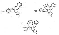
- As referred to herein, smooth muscle cells and pericytes include thosecells derived from the medial layers of vessels and adventitial vessels whichproliferate in intimal hyperplastic vascular sites following injury, such as thatcaused during PTCA. Characteristics of smooth muscle cells include ahistological morphology (under light microscopic examination) of a spindleshape with an oblong nucleus located centrally in the cell with nucleoli presentand myofibrils in the sarcoplasm. Under electron microscopic examination,smooth muscle cells have long slender mitochondria in the juxtanuclearsarcoplasrn, a few tubular elements of granular endoplasmic reticulum, andnumerous clusters of free ribosomes. A small Golgi complex may also belocated near one pole of the nucleus. The majority of the sarcoplasm is occupiedby thin, parallel myofilaments that may be, for the most part, oriented to thelong axis of the muscle cell. These actin containing myofibrils may be arrangedin bundles with mitochondria interspersed among them. Scattered through thecontractile substance of the cell may also be oval dense areas, with similar denseareas distributed at intervals along the inner aspects of the plasmalemma.
- Characteristics of pericytes include a histological morphology (underlight microscopic examination) characterized by an irregular cell shape.Pericytes are found within the basement membrane that surrounds vascular endothelial cells and their identity may be confirmed by positive immuno-staining withantibodies specific for alpha smooth muscle actin (e.g., anti-alpha-sml, Biomakor, Rehovot,Israel), HMW-MAA, and pericyte ganglioside antigens e.g., MAb 3G5 (Schlingemann etal.,Am. J. Pathol., 136 1393 -1405 (1990)); and, negative immuno-staining with antibodiesto cytokeratins (i.e., epithelial and fibroblast markers) and von Willdebrand factor (i.e., anendothelial marker). Both vascular smooth muscle cells and pericytes are positive byimmunostaining with the NR-AN-0 monoclonal antibody.
- As used herein, the term "procedural vascular trauma" includes the effects ofsurgical/mechanical interventions into mammalian vasculature, but does not includevascular trauma due to the organic vascular pathologies, i.e., diseases and infections.
- Thus, procedural vascular traumas within the scope of the treatments to whichmedicaments, compositions, kits and devices of the invention may be applied include (1)organ transplantation, such as heart, kidney, liver and the like, e.g., involving vesselanastomosis; (2) vascular surgery, e.g., coronary bypass surgery, biopsy, heart valvereplacement, atheroectomy, thrombectomy, and the like; (3) transcatheter vascular therapies(TVT) including angioplasty, e.g., laser angioplasty and PTCA procedures, employingballoon catheters, and indwelling catheters; (4) vascular grafting using natural or syntheticmaterials, such as in saphenous vein coronary bypass grafts, dacron and venous grafts usedfor peripheral arterial reconstruction, etc.; (5) placement of a mechanical shunt, e.g., a PTFEhemodialysis shunt used for arteriovenous communications; and (6) placement of anintravascular stent, which may be metallic, plastic or a biodegradable polymer. See U.S.patent application Serial No. 08/389,712, filed February 15, 1995.
- For a general discussion of implantable devices and biomaterials fromwhich they can be formed, see H. Kambic et al., "Biomaterials in Artificial Organs",Chem.Eng. News, 30 (April 14, 1986).
- Cytoskeletal inhibitors useful in the dosage forms, medicaments, kits andpharmaceutical compositions of the invention include cytoskeletal inhibitors whichbiologically stent a vessel and/or reduce or inhibit vascular remodeling and/or inhibit or reduce vascular smooth muscle cell proliferation following a proceduralvascular trauma. The cytoskeletal inhibitors used in the dosage forms, medicaments, kit andpharmaceutical compositions of the invention are selected to inhibit a cellular activity of avascular smooth muscle cell, e.g., proliferation, migration, increase in cell volume, increasein extracellular matrix synthesis (e.g., collagens, proteoglycans, and the like), or secretion ofextracellular matrix materials by the cell.
- The therapeutic agent is a cytoskeletal inhibitors and is preferably: a) a "cytostaticagent" which acts to prevent or delay cell division in proliferating cells by inhibitingreplication of DNA or by inhibiting spindle fiber formation and the like; b) an inhibitor ofmigration of vascular smooth muscle cells from the medial wall into the intima, e.g., an"anti-migratory agent" e.g., a cytochalasin; c) as an inhibitor of the intracellular increase incell volume (i.e., the tissue volume occupied by a cell; a "cytoskeletal inhibitor"); d) aninhibitor that blocks cellular protein synthesis and/or secretion or organization ofextracellular matrix (i.e., an "anti-matrix agent"); or any combination thereof.
- Representative examples of "cytostatic agents" include, e.g., modified toxins,methotrexate, adriamycin, radionuclides (e.g., see Fritzberg et al., U.S. Patent No.4,897,255), protein kinase inhibitors (e.g., staurosporin), taxol or analogs thereof (e.g.,taxotere), inhibitors of specific enzymes (such as the nuclear enzyme DNA topoisomerase IIand DNA polymerase, RNA polymerase, adenyl guanyl cyclase), superoxide dismutaseinhibitors, terminal deoxynucleotidyl- transferase, reverse transcriptase, antisenseoligonucleotides that suppress smooth muscle cell proliferation and the like, which whendelivered into a cellular compartment at an appropriate dosage will act to impairproliferation of a smooth muscle cell or pericyte without killing the cell.
- Representative examples of "anti-migratory agents" include inhibitors (i.e., agonistsand antagonists, and competitive or non-competitive inhibitors) of chemotactic factors and'their receptors (e.g., complement chemotaxins such as C5a, C5a desarg or C4a; extracellularmatrix factors, e.g., collagen degradation fragments), or of intracellular cytoskeletal proteinsinvolved in locomotion (e.g., actin, cytoskeletal elements, and phosphatases and kinasesinvolved in locomotion). Representative examples include caffeic acid derivatives and nilvadipine (a calcium antagonist), and steroidhormones. Preferred anti-migratory therapeutic agents are the cytochalasins.
- Representative examples of "cytoskeletal inhibitors", a subset of cytostatic agents,include colchicine, vinblastin, cytochalasins, taxol, or analogs or derivatives thereof that acton microtubule and microfilament networks within a cell. Preferred cytoskeletal inhibitorsinclude cytochalasin B and taxol.
- Representative examples of "anti-matrix agents" include inhibitors (i. e., agonists andantagonists and competitive and non-competitive inhibitors) of matrix synthesis, secretionand assembly, organizational cross-linking (e.g., transglutaminases cross-linking collagen),and matrix remodeling (e.g., following wound healing). A representative example in thiscategory of anti-matrix agents is colchicine, an inhibitor of secretion of extracellular matrix.Another example is tamoxifen for which evidence exists regarding its capability to organizeand/or stabilize as well as diminish smooth muscle cell proliferation following angioplasty.The organization or stabilization may stem from the blockage of vascular smooth musclecell maturation into a pathologically proliferating form.
- The identification of cytoskeletal inhibitors useful in the practice of the inventionmay be determined by methods well known to the art. For example, a cytoskeletal inhibitorfalling within the scope of the invention as described herein exhibits one or more of thefollowing characteristics. The cytoskeletal inhibitor:
- (i) results in retention of an expanded luminal cross-sectionalarea, diameter or volume of a vessel following angioplasty(e.g., PTCA, percutaneous transluminal angioplasty (PTA) orthe like) or other trauma, including atheroectomy (e.g.,rotoblater, laser and the like), coronary artery bypassprocedures and the like;
- (ii) facilitates an initial increase in luminal cross-sectionalarea, diameter or volume that does not result in or accentuatechronic stenosis of the lumen;
- (iii) inhibits target cell contraction or migration; and
- - (iv) is cytostatic.
- Methods to measure luminal cross-sectional area, volume or diameter include, butare not limited to, angiography, ultrasonic evaluation, fluoroscopic imaging, fiber opticendoscopic examination or biopsy and histology.
- Preferably, a cytoskeletal inhibitor employed herein will have all four properties;however, the first and third are generally more important than the second and fourth forpractice of the present invention in the methods hereinbefore described. It was found thatcytochalasin B administration can result in a biological stenting effect. The biologicalstenting effect can be achieved using a single infusion of the therapeutic agent into thetraumatized region of the vessel wall at a dose concentration ranging from about 0.1micrograms/ml to about 10.0 micrograms/ml (Example 16).
- In the case of cytoskeletal inhibitors displaying anti-migratory or anti-matrixproperties, cell migration and cell adherence inin vitro assays, respectively, may be used fordetermining the concentration at which a therapeutically effective dosage will be reached inthe fluid space in the vessel wall created by an infusion catheter.
- A cytoskeletal inhibitor useful in the sustained release embodiments of the presentinvention exhibits one or more of the following characteristics. The cytoskeletal inhibitor
- (i) causes the retention of an expanded luminal diameter or crosssectional area following angioplasty (e.g. PTCA, percutaneoustransluminal angioplasty (PTA) or the like) or other trauma,including atheroectomy (e.g. rotoblater, laser and the like),coronary artery bypass procedures or the like;
- (ii) inhibits target cell proliferation (e.g. following 5 minute and24 hour exposure to the agent,in vitro vascular smooth muscletissue cultures demonstrate a level of inhibition of3H-thymidineuptake and, preferably, display relatively less inhibition of3H-leucineuptake);
- (iii) at a dose sufficient to inhibit DNA synthesis, produces onlymild to moderate (e.g.,
grade - (iv) inhibits target cell contraction; and
- (v) is cytostatic.
- Upon identification-of a useful cytoskeletal inhibitor exhibiting one or more-of thepreceding properties, the cytoskeletal inhibitor is subjected to a second testing protocol thatinvolves longer exposure of vascular smooth muscle cells (VSMC) to the cytoskeletalinhibitor. For example, an inhibitor useful in the sustained release embodiments of thepresent invention also exhibits the following characteristics:
- (i) upon long term (e.g. 5 days) exposure, the inhibitorproduces the same or similarin vitro effect on vascularsmooth muscle tissue culture DNA synthesis and proteinsynthesis, as described above for the 5 minute and 24 hourexposures; and
- (ii) at an effective dose in the long termin vitro assay forDNA synthesis inhibition, the inhibitor exhibits mild tomoderate morphological cytotoxic effects over a longer term(e.g. 10 days).
- Further evaluation of potentially useful anti-proli ferative agents is conducted in aninvivo balloon traumatized pig femoral artery model. Preferably, these agents demonstrate a50% or greater inhibition of cell proliferation in the tunica media vascular smooth musclecells, as indicated by a 1 hour "BRDU flash labeling" prior to tissue collection andhistological evaluation (Example 13). If an inhibitor is effective in this assay to inhibitintimal smooth muscle proliferation by 50% or more with a single exposure, it does notrequire administration in a sustained release dosage form.
- Inhibitors are evaluated for sustained release if the systemic toxicity and potentialtherapeutic index appear to permit intravenous administration to achieve the 50% inhibitionthreshold, or if the inhibitor is amenable to local delivery to the vascular smooth musclecells via sustained release at an effective antiproliferative dose. Inhibitors are evaluated in asustained release dosage form for dose optimization and efficacy studies. Preferably, anti-proliferativeinhibitors useful in the practice of the present invention decrease vascularstenosis by 50% in balloon traumatized pig femoral arteries and, more preferably, decreasevascular stenosis to a similar extent in pig coronary arteries.
- Inhibition of cellular proliferation (i.e. DNA synthesis) is the primary characteristicof inhibitors useful in sustained release dosage forms. For example, a preferred cytoskeletalinhibitor exhibits a differential between3H-leucine and3Hthymidine uptake so that it can beadministered at cytostatic doses. Moreover, cytotoxicity studies should indicate that prolonged exposure to the cytoskeletal inhibitorswould not adversely impact the target cells. In addition, BRDU pulsing should indicate thatthe cytoskeletal inhibitor is effective to inhibit target cell proliferation. Any convenientmethod for evaluating the capability of an cytoskeletal inhibitor to inhibit cell proliferationmay alternatively be employed, however.
- Sustained release dosage form embodiments of the invention may comprisemicroparticles, nanoparticles or microemulsions having a cytoskeletal inhibitor dispersedtherein or may comprise the cytoskeletal inhibitor in pure, preferably crystalline, solid form.For sustained release administration, microparticle dosage forms comprising pure,preferably crystalline, cytoskeletal inhibitors are preferred as described hereinbefore. Thetherapeutic dosage forms may be of any configuration suitable for sustained release.Preferred sustained release therapeutic dosage forms exhibit one or more of the followingcharacteristics:
- microparticles (e.g., from about 0.01 micrometers to about 200 micrometers indiameter, preferably from about 0.5 to about 50 micrometers, and more preferably fromabout 2 to about 15 micrometers) or nanoparticles (e. g., from about 0.01 nanometer to about1000 nanometers in diameter, preferably from about 50 to about 200 nanometers), freeflowing powder structure comprising crystals or microcrystals;
- biodegradable structure designed to biodegrade over a period of time preferablybetween from about 0.5 to about 180 days, preferably from about 1 -3 to about 150 days, ornon-biodegradable structure to allow therapeutic agent diffusion to occur over a time periodof between from about 0.5 to about 180 days, more preferably from about 30 to about 120days;
- biocompatible with target tissue and the local physiological environment intowhich the dosage form to be administered, including yielding biocompatible biodegradationproducts;
- facilitate a stable and reproducible dispersion of cytoskeletal inhibitor therein,preferably to form a cytoskeletal inhibitor -polymer matrix, with active cytoskeletalinhibitor release occurring by one or both of the following routes: (1) diffusion of thecytoskeletal inhibitor through the dosage form (when the cytoskeletal inhibitor is soluble in the shaped polymer mixture defining the dimensions ofthe dosage form); or (2) release ofthe cytoskeletal inhibitor as the dosage form biodegrades;and/or
- for targeted dosage forms, having, preferably, from about 1 to about 10,000binding protein/peptide to dosage form bonds and more preferably, a maximum of about 1binding peptide to dosage form bond per 150 square angstroms of particle surface area. Thetotal number of binding protein/peptide to dosage form bonds depends upon the particle sizeused The binding proteins or peptides are capable of coupling to the particles of thetherapeutic dosage form through covalent ligand sandwich or non-covalent modalities as setforth herein.
- Nanoparticle sustained release therapeutic dosage forms are preferablybiodegradable and, optionally, bind to the vascular smooth muscle cells and enter thosecells, primarily by endocytosis. The biodegradation of the nanoparticles occurs over time(e.g., 30 to 120 days) in prelysosomic vesicles and lysosomes. Preferred larger microparticletherapeutic dosage form embodiments of the present invention release the cytoskeletalinhibitors for subsequent target cell uptake with only a few of the smaller microparticlesentering the cell by phagocytosis. A practitioner in the art will appreciate that the precisemechanism by which a target cell assimilates and metabolizes a dosage form of the presentinvention depends on the morphology, physiology and metabolic processes of those cells.The size of the particle sustained release therapeutic dosage forms is also important withrespect to the mode of cellular assimilation. For example, the smaller nanoparticles can flowwith the interstitial fluid between cells and penetrate the infused tissue. The largermicroparticles tend to be more easily trapped interstitially in the infused primary tissue, andthus are useful to deliver anti-proliferative cytoskeletal inhibitors.
- Alternatively, the sustained release dosage form embodiments may comprise anemulsion or a micro emulsion having a cytoskeletal inhibitor dispersed therein.Microemulsions are generally defined as thermodynamically stable, isotropically cleardispersions of two immiscible liquids stabilized by interfacial films of surface-activemolecules. Microemulsions can form spontaneously. The formation of microemulsions.usually involves a combination of three to five components, namely, an oil, water, a surfactant, a cosurfactant and an electrolyte.In general, all pharmaceutical emulsions designed for parenteral administrationare of the oil-in-water (o/w) type. A parenteral drug microemulsion can beuseful for delivery of poorly water-soluble drugs, stabilization of hydrolyticallysusceptible compounds, reduction of irritation from or toxicity of intravenouslyadministered drugs, preparation of sustained-release dosage forms, and directeddelivery of drugs to various organs.
- The tendency to form either a water-in-oil (w/o) or an oil-in-water (o/w)microemulsion is influenced by the properties of the oil and the surfactant.Surfactants are conveniently classified on an empirical scale known as thehydrophilic-lipophilic balance (HLB) which runs from 1 to 20. The HLB valueconcept of, and determination thereof for, surfactants is disclosed by Milton J.Rosen in "Surfactants & Interfacial Phenomena", J. Wiley & Sons, New York,NY, 1978, pages 242-245 or by Kirk-Othmer, Encyclopedia of ChemicalTechnology, 3rd Edition, Vol. 8, 1979, at pages 910-915.
- In general, (w/o) microemulsions are formed using surfactants (oremulsifiers) which have an HLB value in the range of about 3 to 6 while (o/w)microemulsions are formed using surfactants which have an HLB value in therange of about 8 to 18. It has long been recognized that low interfacial tensioncontributes to the thermodynamic stability of microemulsions. To achieve this,the surfactant preferably exhibits low solubility in both the oil and water phases,and is preferentially absorbed at the water-oil interface with concomitantlowering of interfacial tension. When interfacial tension is less than 2 x 10⁻²dyn/cm, a stable microemulsion can form. General reviews of microemulsionsare provided by Bhargava et al.,Pharm. Tech., 46-53, March 1987 and Kahlweit,Science,240, 617-621, 1988.
- Microemulsions are typically substantially non-opaque, that is they aretransparent or opalescent when viewed by optical microscopic means. In theundisturbed state, they are optically isotropic (non-birefringent) when examinedunder polarized light. The dispersed phase typically comprises particles ordroplets which are normally between 5 and 200 nm in size and this gives rise totheir optical transparency. These particles may be spherical although otherstructures are feasible.
- The role of the cosurfactant, usually a short-chain alcohol, is to increasethe interfacial fluidity by penetrating the surfactant film and consequentlycreating a disordered film due to the void space among surfactant molecules.The use of a cosurfactant in microemulsions is however optional and alcohol-freeself-emulsifying emulsions and microemulsions have been described (see,for instance, Pouton et al.,Int. Journal of Pharmaceutics,27, 335-348, 1985 andOsborne et al.,J. Disp. Sci. Tech.,9, 415-423, 1988).
- There are many advantages to the use of a microemulsion over aconventional emulsion (or macroemulsion) for drug transport (delivery).Microemulsions can form spontaneously, without the need for a high input ofenergy and are therefore easy to prepare and scale up for commercialapplications; they have thermodynamic stability due to their small particle sizeand therefore have a long shelf life. They have an isotropically clear appearanceso that they may be monitored by spectroscopic means. They have a relativelylow viscosity and are therefore easy to transport and mix. They also have a largeinterfacial area which accelerates surface reactions. They have a low interfacialtension which permits flexible and high penetrating power. Also, they offer thepossibility of improved drug solubilization and protection against enzymatichydrolysis. In addition, microemulsions may undergo phase inversion uponaddition of an excess of the dispersed phase or in response to a temperaturechange and this is a property of these systems that can affect drug release frommicroemulsions bothin vitro andin vivo.
- The term "oil" is used herein in a general sense to identify a large class ofsubstances whether of mineral, vegetable, animal, essential, synthetic or edibleorigin, as long as those substances are pharmaceutically acceptable. Forexample, tri-fatty acid esters of glycerol having about 9-83, more preferablyabout 21-60 and more preferably about 21-45 carbon atoms, are useful oils toprepare microemulsions. Preferred triglycerides are short chain (9-15 carbonatoms) and medium chain (21-45 carbon atoms) triglycerides. Thus, glyceroltriesters includes natural, edible oils such as canola, corn, olive, sunflower andcoconut oils, the decanoic esters, and chemically synthesized oils, e.g., triacetin,1-oleyl-2,3-diacetyl glycerol and the like.
- The alcohols that are useful in microemulsions include but are not limitedto ethanol; propylene glycol (as CH2OH-CH2-CH2OH and/or CH3-CHOH-CH2OH);glycerol; C5-C12 mono- and di-saccharide sugars, for example,dextrose, sucrose, fructose, in pure form, or in other forms, e.g., molasses,brown sugar, invert sugar, refinery syrup, corn syrup; and sugar alcohols such assorbitol, xylitol and mannitol. The alcohols may be used individually or inmixtures of two or more thereof. Moreover, these alcohols are preferentiallysoluble in water rather than in the oils with which they are used.
- The surfactants which may be useful to prepare microemulsions includeall those ionic and nonionic surfactants which are useful in orally ingestibleproducts, intended for use by humans, e.g., food, beverages, confections,pharmaceuticals and dentifrice. The selection of the surfactants for use with aparticular oil depends on the HLB (hydrophile-lipophile balance) value of thesurfactants.
- Various ionic (anionic) surfactants include myristic acid, palmitic acid,stearic acid, oleic acid, monoglyceride ester of diacetyltartaric acid, diglycerideester of diacetyltartaric acid, monoglyceride ester of citric acid and salts thereof,diglyceride ester of citric acid, monoglyceride ester of lactic acid, diglycerideester of lactic acid, dioctyl sodium sulfosuccinate monoglyceride ester ofphosphoric acid, diglyceride ester of phosphoric acid, lecithin, hydroxylatedlecithin. Various nonionic surfactants include polysorbates, sorbitan ester ofmyristic acid, sorbitan ester of palmitic acid, sorbitan ester of stearic acid,sorbitan ester of oleic acid, polyglycerol esters of myristic acid, polyglycerolesters of palmitic acid, polyglycerol esters of stearic acid, polyglycerol esters ofoleic acid, monoglyceride ester of myristic acid, monoglyceride ester of palmiticacid, monoglyceride ester of stearic acid, monoglyceride ester of oleic acid,diglyceride ester of myristic acid, diglyceride ester of palmitic acid, diglycerideester of stearic acid, diglyceride ester of oleic acid, (ethoxy)n monoglyceride ofmyristic acid, (ethoxy)n monoglyceride of palmitic acid, (ethoxy)nmonoglyceride of stearic acid, (ethoxy)n monoglyceride of oleic acid, wherein nis a whole number of 10 to 30, (ethoxy)n diglyceride of myristic acid, (ethoxy)ndiglyceride of palmitic acid, (ethoxy)n diglyceride of stearic acid, (ethoxy)ndiglyceride of oleic acid, wherein n is a whole number of 10 to 30, sucrose ester of myristic acid, ester of palmitic acid, ester of stearic acid, ester of oleic acid, propyleneglycol ester of myristic acid, ester of palmitic acid, ester of stearic acid and ester of oleicacid.
- Moreover, any of the conventional salts or surface-active derivatives can be used,provided, of course, that they are pharmaceutically acceptable. One skilled in the art canselect a particular salt or derivative by conducting routine tests, if necessary. In particular,the alkali metal salts and the taurocholate derivatives are typical of such compounds.
- A nonionic surfactant preferably includes an alkaline oxide condensate of an organiccompound which contains one or more hydroxyl groups. For example, ethoxylated and/orpropoxylated alcohols or esters or mixtures thereof are commonly available and are wellknown to those skilled in the art. Suitable surfactants include, but are not limited to,TYLOXAPOL; POLOXAMER 4070; POLOXAMER.188; POLYOXYL 40 Stearate;POLYSORBATE 80, and
POLYSORBATE 20, as well as various compounds sold underthe trade name TWEEN (ICI America, Inc., Wilmington, Del., U.S.A), and PLURONIC F -68 (trade name of BASF, Ludwigshafen, Germany for a copolymer of polyoxyethylene andpolyoxypropylene). - Some specific examples of surfactants useful in the invention can include, bile salt,sodium cholate, a mixture of 80 wt
% ethyleneglycol 1000 monocetylether and 20 wt %ethyleneglycol 400, and polyoxyethylenether. The amount of surfactant in the invention is5-10 wt % of the total amount of case and capsule material. If the amount of surfactants isless than 0.5% wt %, the emulsion cannot be formed. - Pharmaceutical compositions comprising a microemulsion preferably also comprisea preservative, e.g., methyl-, ethyl-, propyl- and butylparaben which are medically acceptedfor parenteral administration. However, preservatives may not be required if thecompositions can be sterilized by autoclaving without essentially reducing their stability. Ifdesired, the pharmaceutical compositions or medicaments of the present invention can alsocomprise an osmotic pressure regulator such as mannitol or glycerin, glycerin beingpreferred for parenteral administration and mannitol for oral administration. The compositions of the present invention may also comprise an antioxidant, e.g., α-tocopherol.
- The preparation of microemulsions is well known to the art. See, for example, Wolfet al. (U.S. Patent No. 4,835,002); Lee et al. (U.S. Patent No. 5,362,424); Benita et al. (U.S.Patent No. 5,364,632); Owen et al. (WO 94/08604): Constantinides (WO 94/08605);Constantinides et al. (WO 94/19000); Constantinides et al. (WO 94/190001); andConstantinides et al. (WO 94/19003).
- Preferred microemulsions for use in the invention are disclosed in U.S. Patent No.5,478,860, U.S. Patent No. 4,389,330 and U.S. Patent No. 5,407,609.
- Preferred sustained release dosage form embodiments of the present inventioncomprise biodegradable microparticles or nanoparticles. More preferably, biodegradablemicroparticles or nanoparticles are formed of a polymer containing matrix that biodegradesby random, nonenzymatic, hydrolytic scissioning to release cytoskeletal inhibitor, therebyforming pores within the particulate structure.
- Polymers derived from the condensation of alpha hydroxycarboxylic acids andrelated lactones are preferred. A particularly preferred moiety is formed of a mixture ofthermoplastic polyesters (e.g., polylactide or polyglycolide) or a copolymer oflactide andglycolide components, such as poly(lactide-co-glycolide). An exemplary structure, arandom poly(DL-lactide-co-glycolide), is shown below, with the values of x and y beingmanipulable by a practitioner in the art to achieve desirable microparticle or nanoparticleproperties.

- Other agents suitable for forming particulate dosage forms include polyorthoestersand polyacetals (Polymer Letters, 18:293 (1980) and polyorthocarbonates (U.S. Patent No.4,093,709) and the like.
- Preferred lactic acid/glycolic acid polymer containing matrix particles are preparedby emulsion-based processes, that constitute modified solvent extraction processes, see, forexample, processes described by Cowsar et al., "Poly(Lactide-Co-Glycolide) Microcapsulesfor Contrdlled Release of Steroids,"Methods Enzymology, 112:101-116, 1985 (steroidentrapment in microparticles); Eldridge et al., "Biodegradable and Biocompatible Poly(DL-Lactide-Co-Glycolide)Microspheres as an Adjuvant for Staphylococcal Enterotoxin BToxoid Which Enhances the Level of Toxin Neutralizing Antibodies,Infection andImmunity, 59:2978-2986, 1991 (toxoid entrapment); Cohen et at., "Controlled DeliverySystems for Proteins Based on Poly(Lactic/Glycolic Acid) Microspheres,"PharmaceuticalResearch, 8(6);713- 720, 1991 (enzyme entrapment); and Sanders et al., "ControlledRelease of a Luteinizing Hormone-Releasing Honnone Analogue from Poly(D,L-Lactide-CoGlycolide) Microspheres,"J. Pharmaceutical Science, 73(2)1294-1297, 1984 (peptideentrapment).
- In general, the procedure for forming particle dosage forms involves dissolving thepolymer in a halogenated hydrocarbon solvent, dispersing a cytoskeletal inhibitor solution(preferably aqueous) therein, and adding an additional agent that acts as a solvent for thehalogenated hydrocarbon solvent but not for the polymer. The polymer precipitates out fromthe polymerhalogenated hydrocarbon solution onto droplets of the cytoskeletal inhibitorcontaining solution and entraps the cytoskeletal inhibitor. Preferably the cytoskeletalinhibitor is substantially uniformly dispersed within the sustained release dosage formembodiment of the present invention. Following particle formation, they are washed andhardened with an organic solvent. Water washing and aqueous nonionic surfactant washingsteps follow, prior to drying at room temperature under vacuum.
- For biocompatibility purposes, particulate dosage forms, characterized by acytoskeletal inhibitor dispersed in the matrix of the particles, are sterilized prior topackaging, storage or administration. Sterilization may be conducted in any convenient manner therefor. For example, the particles can be irradiated with gammaradiation, provided that exposure to such radiation does not adversely impact the structureor function of the cytoskeletal inhibitor dispersed in the cytoskeletal inhibitor -polymermatrix or the binding protein/peptide attached thereto. If the cytoskeletal inhibitor orbinding protein/peptide is so adversely impacted, the particle dosage forms can be producedunder sterile conditions.
- Release of the cytoskeletal inhibitor from the particle dosage form embodiments ofthe present invention can occur as a result of both diffusion and particle matrix erosion. Thebiodegradation rate directly effects the kinetics of cytoskeletal inhibitor release. Thebiodegradation rate is regulable by alteration of the composition or structure of the sustainedrelease dosage form. For example, alteration of the lactide/glycolide ratio can be conducted,as described by Tice et al., "Biodegradable Controlled-Release Parenteral Systems,"Pharmaceutical Technology, pp. 26-35, 1984; by inclusion of agents that alter the rate ofpolymer hydrolysis, such as citric acid and sodium carbonate, as described by Kent et al.,"Microencapsulation of Water Soluble Active Polypeptides," U.S. Patent No. 4,675,189; byaltering the loading of cytoskeletal inhibitor in the lactide/glycolide polymer, thedegradation rate being inversely proportional to the amount of cytoskeletal inhibitorcontained therein, by judicious selection of an appropriate analog of a common family ofcytoskeletal inhibitors that exhibit different potencies so as to alter said core loadings; andby variation of particle size, as described by Beck et al., "Poly(DL-Lactide-Co-Glycolide)/NorethisteroneMicrocapsules: An Injectable Biodegradable Contraceptive,"Biol. Reprod., 28:186-195, 1983, or the like. All of the aforementioned methods ofregulating biodegradation rate influence the intrinsic viscosity of the polymer containingmatrix, thereby altering the hydration rate thereof
- The preferred lactide/glycolide structure is biocompatible with the mammalianphysiological environment. Also, these preferred sustained release dosage forms have theadvantage that biodegradation thereof forms lactic acid and glycolic acid, both normalmetabolic products of mammals.
- Functional groups required for binding of the protein/peptide to the particle dosageform are optionally included in or on the particle matrix and are attached to the non-degradable or biodegradable polymeric units. Functional groups that areuseful for this purpose include those that are reactive with peptides, e.g., carboxyl groups,amine groups, sulfhydryl groups and the like. Preferred binding enhancement moietiesinclude the terminal carboxyl groups of the preferred (lactide-glycolide) polymer containingmatrix or the like.
- Cytoskeletal inhibitors useful in the sustained release dosage form embodiments ofthe present invention preferably are those that inhibit vascular smooth muscle cell activitywithout killing the cells (i.e., cytostatic cytoskeletal inhibitors). A cytostatic cytoskeletalinhibitor can also be defined as a moiety capable of inhibiting one or more pathologicalactivities of the target cells for a time sufficient to achieve a therapeutic benefit Preferredcytoskeletal inhibitors thus exhibit one or more of the following capabilities: inhibition ofDNA synthesis prior to protein synthesis inhibition, or inhibition of migration of vascularsmooth muscle cells into the intima. These cytoskeletal inhibitors do not significantlyinhibit protein synthesis (i.e., do not kill the target cells) and, therefore, facilitate cellularrepair and matrix production, which in turn acts to stabilize the vascular wall lesion causedby angioplasty, by reducing smooth muscle cell proliferation.
- Preferred cytoskeletal inhibitors are the cytochalasins, e.g., cytochalasin B (SigmaChemical Co.), and taxol, or analogs or functional equivalents thereof. These compoundsare cytostatic and have been shown to exert minimal protein synthesis inhibition andcytotoxicity at concentrations at which significant DNA synthesis inhibition occurs (seeExample 8 and Figures 10A-10D). A useful protocol for identifying cytoskeletal inhibitorsuseful in sustained release dosage form embodiments of the present invention is set forth inExample 8, for example.
- To illustrate, a cytoskeletal inhibitor, e.g., cytochalasin B, is incorporated intobiodegradable poly (DL-lactide-coglycolide) microparticles or into nanoparticles. Themicroparticles are about 1 to about 50 µm, preferably 4 µm to about 15 µm, and morepreferably about 2 to about 15 µm, in diameter. The nanoparticles are about 5 to about 500nanometers, preferably about 10 to about 250 nanometers, and more preferably about 50 to about 200 nanometers, in diameter. The microparticles or nanoparticles comprising thecytoskeletal inhibitor can be further embedded in or on an implantable device, e.g., in astent coating, or delivered in a suitable liquid vehicle by an implantable device, e.g., via aninfusion catheter. Preferably, the sustained release dosage form is biodegradable and,preferably, biodegrades over about 30-120 days. The sustained release dosage form ispreferably administered during the procedural vascular trauma.
- A preferred sustained release dosage form of the invention comprises biodegradablemicroparticles, preferably about 2 to about 15 µm in diameter, which are tissue compatibleand physically compatible with an implantable device, e.g., a needle infusion catheter or amicroinfusion catheter. Another preferred sustained release dosage form of the inventioncomprises biodegradable nanoparticles, preferably about 50 to about 200 nanometers indiameter, which are tissue compatible and physically compatible with an implantabledevice, e.g., a needle infusion catheter or a microinfusion catheter. To deliver the sustainedrelease dosage forms by catheter, catheter pore or hole sizes are preferably about 0.1 toabout 8 micron, more preferably about 0.2 to about 0.8 micron, in diameter.
- The cellular concentration of the cytoskeletal inhibitor that is attained in the tunicamedia and/or intima of the treated vessel is effective to inhibit vascular smooth muscle cellproliferation and migration, e.g., a cellular concentration at least about 0.1 µg/mlcytochalasin B is attained. The inhibition of the smooth muscle cells results in a more rapidand complete reendothelialization after a procedural vascular trauma, e.g., intraventionalplacement of the stent. The increased rate of re-endothelialization reduces loss in luminalcross-sectional area or diameter and reduces decreases in blood flow
- A preferred sustained release dosage form of the invention comprises a pure, solidcrystalline form of a cytoskeletal inhibitor. This embodiment of the sustained release dosageform of the present invention preferably further comprises a tissue-compatiblepharmaceutically acceptable matrix carrier that provides a supporting structure for thecrystals, e.g., a shaped body of silicone, collagen gel retained in a collagen mesh, pluronicgel retained in a collagen mesh, or mannitol retained in a shaped body of silicone. Thus, for example, sustained release dosage forms comprisingcytochalasin B and a pharmaceutical matrix carrier preferably comprise about 5 to about70%, more preferably about 7 to about 40%, and even more preferably about 5 to about30%, weight percent of cytochalasin B/weight percent of the total matrix carrier-therapeuticagent sustained release dosage form. Sustained release dosage forms comprising taxol and apharmaceutical matrix carrier preferably comprise about 1 to about 70%, more preferablyabout 2 to about 50%, and even more preferably about 3 to about 8%, weight percent oftaxol/weight percent of the total matrix carrier-therapeutic agent sustained release dosageform. Generally sustained release dosage forms comprising a cytoskeletal inhibitor insubstantially crystalline form comprise from 3 to 50, and more preferably from 5 to 40weight percent of the dosage form.
- Vascular smooth muscle cell binding proteins useful in the invention bind to targetson the surface of vascular smooth muscle cells. A useful vascular smooth muscle bindingprotein is a polypeptide, peptidic, or mimetic compound (as described below) that is capableof binding to a target or marker on a surface component of an intact or disrupted vascularsmooth muscle cell. Such binding allows for either release of cytoskeletal inhibitorsextracellularly in the immediate interstitial matrix with subsequent diffusion of cytoskeletalinhibitor into the remaining intact smooth muscle cells and/or internalization by the cell intoan intracellular compartment of the entire targeted biodegradable moiety, thus permittingdelivery of the cytoskeletal inhibitor thereto. It will be recognized that specific targets, e.g.,polypeptides or carbohydrates, proteoglycans and the like, that are associated with the cellmembranes of vascular smooth muscle cells are useful for selecting (e.g., by cloning) o rconstructing (e.g., by genetic engineering or chemical synthesis) appropriately specificvascular smooth muscle binding proteins. Particularly useful "targets" are internalized bysmooth muscle cells, e.g., as membrane constituent antigen turnover occurs in renewal.Internalization by cells may also occur by mechanisms involving phagolysosomes, clathrin-coatedpits, receptor-mediated redistribution or endocytosis and the like.
- - --Representative examples of useful vascular smooth muscle binding proteins includeantibodies (e.g., monoclonal and polyclonal antibodies), F(ab')2 Fab', Fab, and Fv fragmentsand/or complementarity determining regions (CDR) of those antibodies or functionalequivalents thereof, (e.g., binding peptides and the like)); growth factors, cytokines, andpolypeptide hormones and the like; and macromolecules recognizing extracellular matrixreceptors (e.g., integrin and fibronectin receptors and the like).
- In a preferred embodiment, e.g., a "target" is exemplified by chondroitin sulfateproteoglycans (CSPGs) synthesized by vascular smooth muscle cells and pericytes, and adiscrete portion (termed an epitope herein) of the CSPG molecule having an apparentmolecular weight of about 250 kD is especially preferred as a target. The 250 kD target isan N-linked glycoprotein that is a component of a larger 400 kD proteoglycan complex(Bumol et al.,PNAS USA, 79:1245-1249 (1982)). In one presently preferred embodimentof the invention, a vascular smooth muscle binding protein is provided by the NR-AN-01monoclonal antibody (a subculture of NR-ML-05) that binds to an epitope in a vascularsmooth muscle CSPG target molecule. The monoclonal antibody designated NR-ML-05reportedly binds a 250 kD CSPG synthesized by melanoma cells (Morgan et al., U.S. PatentNo. 4,897,255).
- Smooth muscle cells and pericytes also reportedly synthesize a 250 kD CSPG aswell as other CSPGs (Schlingeman et al., supra). NR-ML-05 binding to smooth muscle cellshas been disclosed (Fritzberg et al., U.S. Patent No. 4,879,225). The hybridoma, NR-ML-05,which secretes a monoclonal antibody which binds to the 400 kD CSPG, has beendeposited with the American Type Culture Collection, Rockville, MD and grantedAccession No. 9350. NR-ML-05 is the parent of and structurally and functionallyequivalent to, subclone NR-AN-0 1, disclosed herein.
- It will be recognized that NR-AN-0 1 is just one example of a vascular smoothmuscle binding protein that specifically associates with the 400 kD CSPG target, and thatother binding proteins associating with this target and other epitopes on this target (Bumolet al.,PNAS USA, 79:1245-1249 (1982)) are also useful in the medicaments, compositions,devices, kits of the invention when the medicaments, compositions, devices or kits are usedfor treating stenosis following vascular surgical procedures, e.g., PTCA, preferred binding proteins, e.g., antibodies or fragments, for use in the practice of the invention have abinding affinity of> 104 liter/mole for the vascular smooth muscle 250 kD CSPG, and alsothe ability to be bound to and internalized by smooth muscle cells or pericytes.
- Further, it will be recognized that the ammo acid residues involved in the multipointkinetic association of the NR-AN-01 monoclonal antibody with a CSPG markerantigenic epitope (i.e., the amino acids constituting the complementarity determiningregions) can be determined by computer-assisted molecular modeling and by the use ofmutants having altered antibody binding affinity. The binding-site amino acids and threedimensional model of the NR-AN-01 antigen binding site can serve as a molecular modelfor constructing functional equivalents, e.g., short polypeptides ("minimal polypeptides"),that have binding affinity for a CSPG synthesized by vascular smooth muscle cells andpericytes.
- Three-dimensional modeling is also useful to construct other functional equivalentsthat mimic the binding of NR-AN-01 to its antigenic epitope, e.g., "mimetic" chemicalcompounds that mimic the three-dimensional aspects of NR-AN-01 binding to its epitope ina CSPG target antigen. As used herein, "minimal polypeptide" refers to an amino acidsequence of at least six amino acids in length. As used herein, the term "mimetic" refers toan organic chemical oligomer or polymer constructed to achieve the proper spacing forbinding to the amino acids of, for example, an NR-AN-01 CSPG target synthesized byvascular smooth muscle cells or pericytes.
- It is also envisioned that human monoclonal antibodies or "humanized" murineantibodies which bind to a vascular smooth muscle binding protein are useful in thetherapeutic conjugates. For example, murine monoclonal antibody may be "chimerized" bygenetically recombining the nucleotide sequence encoding the murine Fv region (i.e.,containing the antigen binding sites) with the nucleotide sequence encoding a humanconstant domain region and an Fc region, e.g., in a manner similar to that disclosed inEuropean Patent Application No. 411,893. Some murine residues may also be retainedwithin the human variable region framework domains to ensure proper target site bindingcharacteristics. Humanized vascular smooth muscle binding partners will be recognized to have the advantage of decreasing the immunoreactivity of theantibody or polypeptide in the host recipient, and may be useful for increasing thein vivohalf-life and reducing the possibility of adverse immune reactions to the conjugate.
- Also contemplated as useful binding peptides for sustained release dosage formsadapted for restenosis treatment are those that localize to intercellular stroma and matrixlocated between and among vascular smooth muscle cells. Such binding peptides candeliver the cytoskeletal inhibitor to the interstitial space between the target cells. Thecytoskeletal inhibitor is released into the interstitial spaces for subsequent uptake by thevascular smooth muscle cells. Preferred binding peptides of this type are associated withepitopes on collagen, extracellular glycoproteins, e.g., tenascin, reticulum and elastic fibers,cytokeratin and other intercellular matrix components. Minimal peptides, mimetic organicchemical compounds, human or humanized monoclonal antibodies and the like that localizeto intracellular stroma and matrix are also useful as binding peptides in this embodiment ofthe present invention. These binding peptides may be identified and constructed or isolatedin accordance with known techniques. In preferred embodiments of the present invention,the interstitial matrix binding protein binds to a target epitope with an association constantof at least about 10-4 M.
- Representative "coupling" methods for linking the cytoskeletal inhibitor throughcovalent or non-covalent bonds to the vascular smooth muscle binding protein includechemical cross-linkers and heterobifunctional cross-linking compounds (i.e., "linkers") thatreact to form a bond between reactive groups (such as hydroxyl, amino, amido, orsulfhydryl groups) in a cytoskeletal inhibitor and other reactive groups (of a similar nature)in the vascular smooth muscle binding protein. This bond may be, for example, a peptidebond, disulfide bond, thioester bond, amide bond, thioether bond, and the like.
- In one illustrative example, conjugates of monoclonal antibodies with drugs havebeen summarized by Morgan and Foon (Monoclonal Antibody Therapy to Cancer:Preclinical Models and Investigations, Basic and Clinical Tumor Immunology, Vol. 2,Kluwer Academic Publishers, Hingham, MA) and by Uhr J. oflmmunol. 12:i-vii, 1984). Inanother illustrative example where the conjugate contains a radionuclide cytostatic agent, U.S. Patent No. 4,897,255, Fritzberg etal., is instructive of coupling methods that can be used tomake the present conjugates.
- The choice of coupling method will be influenced by the choice of vascular smoothmuscle binding protein or peptide, interstitial matrix binding protein or peptide andcytoskeletal inhibitor, and also by such physical properties as, e.g., shelf life stability, and/orby biological properties, e.g., half-life in cells and blood, intracellular compartmentalizationroute, and the like.
- The physical and chemical character of the sustained release dosage formembodiments of the present invention permit several alternative modes of attachment of thedosage forms to binding proteins or peptides. Dosage forms (sustained release-type) asdescribed herein are capable of binding to binding proteins/peptides through, for example,covalent linkages, intermediate ligand sandwich attachment, or non-covalent adsorption orpartial entrapment. When the preferred poly-lactic/glycolic acid particles are formed withthe cytoskeletal inhibitor dispersed therein, the uncharged polymer backbone is orientedboth inward (with the quasi lipophilic therapeutic agent contained therein) and outward,along with a majority of the terminal carboxy groups. These surface carboxy groups mayserve as covalent attachment sites when activated by, for example, a carbodiimide) fornucleophilic groups of the binding protein/peptide. Such nucleophilic groups include lysineepsilon-amino groups (amide linkage), serine hydroxyl groups (ester linkage) or cysteinemercaptan groups (thioester linkage). Reactions with particular groups depend upon pH andthe reduction state of the reaction conditions.
- For example, poly-lactic/glycolic acid particles having terminal carboxylic acidgroups can be reacted with N-hydroxybenztriazole in the presence of a water solublecarbodiimide of the formula R-N=C=N-R' (wherein R is a 3-dimethylaminopropyl group orthe like and R' is an ethyl group or the like). The benztriazole-derivatized particles (i.e.,activated imidate-bearing moieties) are then reacted with a protein/peptide nucleophilicmoiety such as an available epsilon- amino moiety. Alternatively, p-nitrophenol,tetrafluorophenol, N-hydroxysuccinimide or like molecules are useful to form an activeester with the terminal carboxy groups of poly-lactic/glycolic acid particles in the presence of carbodiimide. Other binding protein/peptide nucleophilic moieties include hydroxylgroups of serine, endogenous free thiols of cysteine, thiol groups resulting from reduction ofbinding protein/peptide disulfide bridges using reducing agents commonly employed forthat purpose (e.g., cysteine, dithiothreitol, mercaptoethanol and the like) and the like.Additionally, the terminal carboxy groups of the poly- lactic/glycolic acid particles areactivatable by reaction with thionyl chloride to form an acyl chloride derivatized moiety.The derivatized particles are then reacted with binding peptide/protein nucleophilic groupsto form targeted dosage forms.
- Direct conjugation of sustained release dosage form to binding protein or peptidemay disrupt binding protein/peptide recognition of the target cell. Ligand sandwichattachment techniques are useful alternatives to achieve sustained release dosage formattachment to binding protein/peptide. These techniques involve the formation of a primarypeptide or protein shell using a protein that does not bind to the target cell population.Binding protein/peptide is then bound to the primary peptide or protein shell to provide theresultant particle with functional binding protein/peptide. An exemplary ligand sandwichapproach involves covalent attachment of avidin or streptavidin to the particles throughfunctional groups as described above with respect to the "direct" binding approach. Thebinding protein or peptide is derivatized, preferably minimally, via functionalized biotin(e.g., through active ester, hydrazide, iodoacetal, maleimidyl or like functional groups).Ligand (i.e., binding peptide or protein/ functionalized biotin) attachment to the availablebiotin binding sites of the avidin/streptavidin primary protein shell occurs through the use ofa saturating amount of biotinylated protein/peptide.
- For example, poly-lactic/glycolic acid particles having terminal carboxylic acidgroups are activated with carbodiimide and subsequently reacted with avidin or streptavidin.The binding protein or peptide is reacted with biotinamidocaproate N-hydroxysuccinimideester at a 1-3 molar offering of biotin-containing compound to the binding protein/peptide toform a biotinylated binding protein/peptide. A molar excess of the biotinylated bindingprotein/peptide is incubated with the avidin-derivatized particles to form a targeted dosageform.
- Alternatively, the particle carboxy groups are biotinylated (e.g., throughcarbodiimide activation of the carboxy group and subsequent reaction withamino alkyl biotinamide). The biotinylated particles are then incubated with asaturating concentration (i.e., a molar excess) of avidin or streptavidin to formprotein coated particles having free biotin binding sites. These coated particlesare then capable of reaction with a molar excess of biotinylated binding proteinformed as described above. Another option involves avidin or streptavidinbound binding peptide or protein attachment to biotinylated particles.
- In addition, binding protein/peptide-particle attachment can be achievedby adsorption of the binding peptide to the particle, resulting from the nonioniccharacter of the partially exposed polymer backbone of the particle. Under highionic strength conditions (e.g., 1.0 molar NaCl), hydrogen and hydrophobicparticle-binding protein/peptide binding are favored.
- Moreover, binding protein/peptide may be partially entrapped in theparticle polymeric matrix upon formation thereof. Under these circumstances,such entrapped binding protein/peptide provides residual selective bindingcharacter to the particle. Mild particle formation conditions, e.g., thoseemployed by Cohen et al.,Pharmaceutical Research,8: 713-720 (1991), arepreferred so as to entrap the protein or peptide in the matrix. Entrapped bindingproteins are also useful in target cell reattachment of a partially degraded particlethat has undergone exocytosis. Binding proteins or peptides can be bound toother polymeric particle dosage forms (e.g., non-biodegradable dosage forms)having different exposed functional groups in accordance with the principlesdiscussed above.
- Exemplary non-biodegradable polymers useful in the practice of thepresent invention are polystyrenes, polypropylenes, styrene acrylic acid andacrylate copolymers and the like. Such non-biodegradable polymers incorporateor can be derivatized to incorporate functional groups for attachment of bindingprotein/peptide, including carboxylic acid groups, aliphatic primary aminogroups, aromatic amino groups and hydroxyl groups.
- Carboxylic acid functional groups are coupled to binding protein orpeptide using, for example, the reaction mechanisms set forth above for poly-lactic/glycolicacid biodegradable polymeric particle dosage forms. Primary amino functional groups are coupled by, for example, reaction thereof with succinicanhydride to form a terminal carboxy moiety that can be bound to binding peptide/proteinas described above. Additionally, primary amino groups can be activated with cyanogenbromide and form guanidine linkages with binding protein/peptide primary amino groups.Aromatic amino functional groups are, for example, diazotized with nitrous acid to formdiazonium moieties which react with binding protein/peptide tyrosines, thereby producing adiazo bond between the non-biodegradable particle and the binding protein/peptide.Hydroxyl functional groups are coupled to binding protein/peptide primary amino groupsby, for example, converting the hydroxyl moiety to a moiety comprising a terminalcarboxylic acid functional group. This conversion can be accomplished throµgh reactionwith chloroacetic acid followed by reaction with carbodiimide. Sandwich, adsorption andentrapment techniques, discussed above with respect to biodegradable particles, areanalogously applicable to nonbiodegradable particle-binding protein/peptide affixation.
- The amount of cytoskeletal inhibitor administered is adjusted to treat vasculartraumas of differing severity. For example, smaller doses are sufficient to treat lesservascular trauma, e.g., to prevent vascular rejection following graft or transplant, while largerdoses are sufficient to treat more extensive vascular trauma, e.g., to treat restenosisfollowing angioplasty. Thus, to biologically stent a traumatized vessel a cytoskeletalinhibitor such as cytochalasin B is administered at a systemic total dose of about 1 to about24 ml, preferably about 1 to about 4 ml, at about 0.01 to about 10 µg cytochalasin B/ml ofvehicle, preferably about 0.1 to about 10 µg cytochalasin B/ml of vehicle, and morepreferably about 0.1 to about 8.0 µg cytochalasin B/ml of vehicle, although other dosagesmay prove beneficial. In particular, lower concentrations of a cytochalasin may exert atherapeutic effect when a non-aqueous solvent is employed as the vehicle. Theadministration of a sytemic dose of cytochalasin B results in about 5 to about 40, preferablyabout 8 to about 30, lambda of the cytochalasin B-containing solution entering theinterstitial space surrounding the cells of the tunica media, and about 0.01 to about 4,preferably about 0.05 to about 3, ml of the solution being exposed to the wall of the vesselby the transport of the solution to the adventitia. Moreover, these dosages may also exhibit anti-proliferativeeffects.
- Administration of medicaments, compositions, or dosage forms of the presentinvention may be continuous or intermittent, depending, for example, upon the recipient'sphysiological condition, whether the purpose of the administration is therapeutic orprophylactic, and other factors known to skilled practitioners. The administration of themedicaments, compositions or dosage forms of the invention may be essentially continuousover a preselected period of time or may be in a series of spaced doses, e.g., either before,during, or after procedural vascular trauma, before and during, before and after, during andafter, or before, during and after the procedural vascular trauma. Moreover, theadministration of the medicaments, compositions or dosage forms is selected so as to notfurther damage the traumatized vessel.
- One or more suitable unit dosage forms comprising the cytoskeletal inhibitor, whichmay be formulated for sustained release, can be administered by a variety of routesincluding oral, or parenteral, including by rectal, transdermal, subcutaneous, intravenous,intramuscular, intrapulmonary and intranasal routes: When the medicaments, compositionsand dosage forms of the invention are prepared for oral administration, they preferablycombined with a pharmaceutically acceptable carrier, diluent or excipient to form apharmaceutical formulation preferably in unit dosage form. The total active ingredients inthese formulations can comprise from 0.1 to 99.9% by weight of the formulation. By"pharmaceutically acceptable" it is meant the carrier, diluent, excipient, and/or salt must becompatible with the other ingredients of the formulation, and not deleterious to the recipientthereof.
- Pharmaceutical formulations containing the cytoskeletal inhibitor can be prepared byprocedures known in the art using well known and readily available ingredients. Forexample, a cytochalasin can be formulated with common excipients, diluents, or carriers,and formed into tablets, capsules, suspensions, powders, and the like. Examples ofexcipients, diluents, and carriers that are suitable for such formulations include thefollowing fillers and extenders, e.g., starch, sugars, mannitol, and silicic derivatives; bindingagents, for example, carboxymethyl cellulose, HPMC, and other cellulose derivatives,alginates, gelatin, and polyvinyl-pyrrolidone; moisturizing agents such as glycerol; disintegrating agents, e.g., calcium carbonate and sodium bicarbonate; agents forretarding dissolution, for example, paraffin; resorption accelerators such as quaternaryammonium compounds; surface active agents, e.g., cetyl alcohol, glycerol monostearate;adsorptive carriers such as kaolin and bentonite; and lubricants, for example, talc, calciumand magnesium stearate, and solid polyethyl glycols. See Fondy et al. (WO 88/10259).
- For example, tablets or caplets containing a cytoskeletal inhibitor can includebuffering agents, e.g., calcium carbonate, magnesium oxide and magnesium carbonate.Caplets and tablets can also include inactive ingredients such as cellulose, pregelatinizedstarch, silicon dioxide, hydroxypropyl methylcellulose, magnesium stearate,microcrystalline cellulose, starch, talc, titanium dioxide, benzoic acid, citric acid, cornstarch, mineral oil, polypropylene glycol, sodium phosphate, and zinc stearate, and the like.Hard or soft gelatin capsules containing a cytoskeletal inhibitor can contain inactiveingredients, for example, gelatin, microcrystalline cellulose, sodium lauryl sulfate, starch,talc, and titanium dioxide, and the like, as well as liquid vehicles such as polyethyleneglycols (PEGs) and vegetable oil.
- Cytoskeletal inhibitors can also be formulated as elixirs or solutions for convenientoral administration or as solutions appropriate for parenteral administration, for instance byintramuscular, subcutaneous or intravenous routes. In the practice of certain embodimentsof the present invention, the cytoskeletal inhibitor is dispersed in a pharmaceuticallyacceptable carrier that is in liquid phase, and delivered via an implantable device, e.g., acatheter. Useful pharmaceutically acceptable carriers for these purposes include generallyemployed carriers, such as phosphate buffered saline solution, water, emulsions (e.g.,oil/water and water/oil emulsions) and wetting agents of various types.
- The pharmaceutical formulations of the cytoskeletal inhibitors can take the form ofan aqueous or anhydrous solution or dispersion, or alternatively the form of an emulsion orsuspension. See Fondy et al. (WO90/13293).
- These formulations can contain pharmaceutically acceptable vehicles and adjuvantswhich are well known in the prior art. It is possible, for example, to prepare solutions usingone or more organic solvent(s) that is/are acceptable from the physiological standpoint,chosen, in addition to water, from solvents, e.g., acetone, ethanol, isopropyl alcohol, glycolethers such as the products sold under the name "Dowanol", polyglycols and polyethyleneglycols, C1-C4 alkyl esters of short-chain acids, preferably ethyl or isopropyl lactate, fattyacid triglycerides, for example products marketed under the name "Miglyol", isopropylmyristate, animal, mineral and vegetable oils and polysiloxanes.
- The compositions and medicaments according to the invention can also containthickening agents such as cellulose and/or cellulose derivatives. They can also containgums, e.g., xanthan, guar or carbo gum or gum arabic, or alternatively polyethylene glycols,bentones and montmorillonites, and the like.
- It is possible to add, if necessary, an adjuvant chosen from antioxidants, surfactants,other preservatives, film-forming, keratolytic or comedolytic agents, perfumes andcolorings. Also, other active ingredients may be added, whether for the conditions describedor some other condition.
- For example, among antioxidants, t-butylhydroquinone, butylated hydroxyanisole,butylated hydroxytoluene and a-tocopherol and its derivatives may be mentioned. Thegalenical forms chiefly conditioned for topical application take the form of creams, milks,gels, dispersion or microemulsions, lotions thickened to a greater or lesser extent,impregnated pads, ointments or sticks, or alternatively the form of aerosol formulations inspray or foam form or alternatively in the form of a cake of soap.
- Additionally, cytoskeletal inhibitors are well suited to formulation as sustainedrelease dosage forms and the like. The formulations can be so constituted that they releasethe active ingredient only or preferably in a particular part of the intestinal tract, e.g., over aperiod of time. The coatings, envelopes, and protective matrices may be made, for example,from polymeric substances or waxes.
- The local delivery of the cytoskeletal inhibitors can be by a variety of techniqueswhich administer the agent at or near the traumatized vascular site. Examples of site-specific or targeted local delivery techniques are notintended to be limiting but to be illustrative of the techniques available. Examples includelocal delivery catheters, such as an infusion catheter, an indwelling catheter, or a needlecatheter, stets, synthetic grafts, adventitial wraps, shunts and stents or other implantabledevices, site specific carriers, direct injection, or direct applications.
- Local delivery by an implant describes the surgical placement of a matrix thatcontains the cytoskeletal inhibitor into the lesion or traumatized area. The implanted matrixreleases the cytoskeletal inhibitor by diffusion, chemical reaction, or solvent activators. See,for example, Lange,Science, 249, 1527 (1990).
- An example of targeted local delivery by an implant is the use of a stent. Stents aredesigned to mechanically prevent the collapse and reocclusion of the coronary arteries orother vessels. Incorporation of a cytoskeletal inhibitor into the stent can deliver thecytoskeletal inhibitor directly to the lesion. Local delivery of agents by this technique isdescribed in Koh,Pharmaceutical Technology (October, 1990).
- For example, a metallic, plastic or biodegradable intravascular stent is employedwhich comprises the cytoskeletal inhibitor. The stent preferably comprises a biodegradablecoating, a porous or a permeable non-biodegradable coating, or a biodegradable or non-biodegradablemembrane or synthetic graft sheath like coating, e.g., PTFE, comprising thecytoskeletal inhibitor. A more preferred embodiment of the invention is a coated stentwherein the coating comprises a sustained-release dosage form of the cytoskeletal inhibitor.In an alternative embodiment, a biodegradable stent may also have the therapeutic agentimpregnated therein, i.e., in the stent matrix.
- A biodegradable stent with the cytoskeleta inhibitor impregnated therein can befurther coated with a biodegradable coating or with a porous non-biodegradable coatinghaving the sustained release-dosage form of the cytoskeletal inhibitor dispersed therein.This stent can provide a differential release rate of the cytoskeletal inhibitor, i.e., there canbe an initial faster release rate of the cytoskeletal inhibitor from the coating, followed bydelayed release of the cytoskeletal inhibitor impregnated in the stent matrix, upondegradation of the stent matrix. The intravascular stent also provides a mechanical means ofproviding an increase in luminal area of a vessel. Furthermore, the placement of intravascular stentscomprising a cytoskeletal inhibitor which is an inhibitor of smooth muscle cell proliferationcan also reduce or prevent intimal proliferation. This inhibition of intimal smooth musclecells and stroma produced by the smooth muscle and pericytes can lead to a more rapid andcomplete re-endothelization following the intraventional placement of the vascular stent.The increased rate of re-endothelization and stabilization of the vessel wall following stentplacement can reduce the loss of luminal area and decreased blood flow due to vascularsmooth muscle cell proliferation which is one of the primary causes of vascular stentfailures.
- Another example of targeted local delivery by an implant is the use of an adventitialwrap. The wrap comprises a pharmaceutically acceptable carrier matrix, e.g., a Pluronic gelwhich is free, or contained by a collagen mesh, which gel has dispersed therein acytoskeletal inhibitor. One embodiment of the invention is a pluronic gel (F-127, BASF)which is soluble at 4°C but solidifies at 37°C, e.g., on contact with fluid or tissue in ahuman. To prepare a pluronic gel containing wrap, 4 ml of phosphate buffer, pH 7.0(CircRes, vol. 76, April 1995), was added to 1 g of pluronic gel F-127, which was mixedovernight at 4°C. The cytoskeletal inhibitor was added to the mixture prior to localadministration. The mixture may be applied directly to a surgically exposed artery wall, ormay be applied to the surface of a bovine collagen mat (BioCore, Inc., Topeka, KS), whichis then wrapped around the artery and the edges joined by sutures.
- Another embodiment of the invention is the incorporation of the cytoskeletalinhibitor into the expanded nodal spaces of a PTFE (Impra, Inc., Tempe, AZ) vascular graft-likemembrane which can surround, or be placed on the interior or on the exterior surfaceof, an interlumenal vascular stent, which comprises metal or a biodegradable ornonbiodegradable polymer. The cytoskeletal inhibitor, or a sustained release dosage form ofthe cytoskeletal inhibitor, fills the nodal spaces of the PTFE membrane wall and/or coats theinner and/or outer surfaces of the membrane.
- Yet another embodiment of the invention is a mixture of a crystalline form of acytoskeletal inhibitor in a bovine collagen gel (BioCore, Inc., Topeka, KS). Crystals varied in size from about 0.1 micron to about 1 mm. Generally, the crystals werepulverized to generate smaller sized crystals. This mixture is applied directly to the surfaceof the artery, and the surrounding subcutaneous tissues sutured around the vessel and theskin closed. Bovine collagen (BioCore, Inc., Topeka, KS) is dissolved in sterile saline (1:1)and the crystalline cytoskeletal inhibitor added. Alternatively, the collagen gel is applied toa bovine collagen mesh which is then wrapped around the vessel and the edges sutured tohold the mesh in place. The bovine collagen mesh (BioCore, Inc.) is cut to size, e.g., 1 cm x1 cm, and the cytoskeletal inhibitor -collagen gel mixture is applied to the surface of themesh.
- A further embodiment of the invention comprises the entrapment of crystallinecytoskeletal inhibitor in about a 0.1 to about 3, preferably about 0.5 to about 0.7, mm thicksilicone membrane, e.g., silicone polymer Q-7 4840 (Dow Coming, Midland, MI). Thepolymer (part A and B, 1:1) is mixed with a spatula. An inert filler, e.g., mannitol, ispowdered and sieved to a fraction 53-75 mesh size. Mannitol and cytoskeletal inhibitor aremixed in predetermined proportions and then levigated with the polymer to form acomposite. The composite is filled in a slab mold and compressed to 5000 psi. Thecomposite is then cured at 80°C for 2 hours. The composite membrane is then cut to size,e.g., I cm x1 cm, wrapped around the artery and held in place by suturing the membraneedges together.
- A cytoskeletal inhibitor may also be coated onto the exterior of the wrap. The wrapand/or the coating is preferably biodegradable. It is preferred that the cytoskeletal inhibitoris in sustained release dosage form.
- Another example is a delivery system in which a polymer that contains thecytoskeletal inhibitor is injected into the area of the lesion in liquid form. Theopolynmerthen solidifies or cures to form the implant which is retainedin situ. This technique isdescribed in PCT WO 90/03768 (Donn, Apr. 19, 1990).
- Another example is the delivery of a cytoskeletal inhibitor by polymericendoluminal sealing. This technique uses a catheter to apply a polymeric implant to theinterior surface of the lumen. The cytoskeletal inhibitor incorporated into the biodegradablepolymer implant and is thereby released at the surgical site. The technique is described in PCT WO 90/01969 (Schindler, Aug. 23, 1989).
- Yet another example of local delivery is by direct injection of vesicles ormicroparticles into the lesion or artery wall adjacent to the lesion. These microparticles maybe composed of substances such as proteins, lipids, carbohydrates or synthetic polymers.These microparticles have the cytoskeletal inhibitor incorporated throughout themicroparticle or onto the microparticle as a coating. Delivery systems incorporatingmicroparticles are described in Lange,Science,249,1527 (1990) and Mathiowitz et al.,JApp. Poly. Sci,26, 809 (1981).
- For topical administration, the therapeutic agents may be formulated as is known inthe art for direct application to a target area. Conventional forms for this purpose includewound dressings, coated bandages or other polymer coverings, ointments, lotions, pastes,jellies, sprays, and aerosols. The percent by weight of a cytoskeletal inhibitor present in atopical formulation will depend on various. factors, but generally will be from 0.005% to95% of the total weight of the formulation, and typically 1-25% by weight
- The medicaments, kits, compositions and dosage forms as described hereinbeforeare useful to treat or inhibit a diminution in vessel lumen volume, area and/or diameterassociated with a procedural vascular trauma. As used herein, "vessels" includesmammalian vessels, e.g., coronary vessels as well as peripheral, femoral and carotid vessels.It will be recognized that the medicaments, compositions, kits and dosage forms (both freeand sustained release) of the invention are not restricted in use for therapy followingangioplasty; rather, the usefulness of the medicaments, compositions, kits and dosage formswill be proscribed by their ability to inhibit cellular activities of smooth muscle cells andpericytes in the vascular wall. Thus, a vascular trauma includes but is not limited to traumaassociated with an interventional procedure, such as angioplasty, placement of a stent,shunt, stet, synthetic or natural graft, adventitial wrap, indwelling catheter or otherimplantable devices. Grafts include synthetic therapeutic agent-treated grafts, e.g.,impregnated or coated grafts.
- The medicaments, compositions, kits and-dosage forms of the invention are alsouseful in therapeutic modalities for enhancing the regrowth of endothelial cells in injuredvascular tissues and in other wound sites including epithelial wounds. In these therapeuticmodalities, the medicaments, compositions, kits and dosage forms of the invention andconjugates as described herein find utility in inhibiting the migration and/or proliferation ofsmooth muscle cells or pericytes. Smooth muscle cells and pericytes have been implicatedin the production of factorsin vitro that inhibit endothelial cell proliferation, and theirproliferation can also result in a physical barrier to establishing a continuous endothelium.Thus, the medicaments, compositions, kits and dosage forms of the invention andconjugates described herein find utility in promoting neo-angiogenesis and increased re-endothelialization,e.g., during wound healing, vessel grafts and following vascular surgery.The dosage forms may also release therapeutic modalities that stimulate or accelerate up re-endothelializationof the damaged vessel wall.
- In some embodiments of the present invention a cytoskeletal inhibitor capable ofinhibiting the ability of vascular smooth muscle cells to contract may be used, e.g. acytochalasin. Exemplary cytoskeletal inhibitors useful in the practice of this aspect of thepresent invention are those capable of causing a traumatized artery to lose vascular tone,such that normal vascular hydrostatic pressure (i.e., blood pressure) expands the flaccidvessel to or near to its maximal physiological diameter. Loss of vascular tone may becaused by cytoskeletal inhibitors that interfere with the formation or function of contractileproteins (e.g., actin, myosin, tropomyosin, caldesmon, calponin or the like). Thisinterference can occur directly or indirectly through, for example, inhibition of calciummodulation, phosphorylation or other metabolic pathways implicated in contraction ofvascular smooth muscle cells.
- Inhibition of cellular contraction (i.e., loss of vascular tone) may operate throughtwo mechanisms to reduce the degree of vascular stenosis. First, inhibition of cellularcontraction for a prolonged period of time limits the number of smooth muscle cells thatmigrate from the tunica media into the intima, the thickening of which results in vascularluminal stenosis. Second, inhibition of cellular contraction causes the smooth muscle wall torelax and dilate under normal vascular hydrostatic pressure (i.e., blood pressure).
- The cytochalasins inhibit smooth muscle cell contraction without abolishing theprotein synthesis necessary for traumatized, post-angioplasty or other surgically- or disease-damaged,smooth muscle cells to repair themselves. Protein synthesis is also necessary forthe smooth muscle cells to secrete matrix, which fixes or retains the lumen in a state near itsmaximum systolic diameter as the vascular lesion stabilizes (i.e., a biologicallyinducedstenting effect).
- This biological stenting effect not only results in an expanded vessel luminal cross-sectionalarea or diameter and increased blood flow rate through the vessel, but alsosignificantly reduces elastic recoil following angioplasty. Elastic recoil is an acute closureof the vessel associated with vasospasm or early relaxation of the muscular wall, due totrauma shock resulting from vessel overstretching by a balloon catheter during angioplasty.This spasm of the tunica media which leads to decreases in the luminal cross-sectional areamay occur within hours, days or weeks after the balloon dilation, as restoration of vascularmuscle wall tone occurs.
- Recent observations during microscopic examination of atheroectomy specimenssuggest that elastic recoil may occur in up to 25% of angioplasty procedures classified assuccessful, based on the initial post-procedure angiogram. Because the biological stentingprocedure relaxes the artery wall following balloon angioplasty, the clinician can eliminateover-inflation and its resultant trauma shock as a means to diminish or delay the vesselspasm or elastic recoil. Reduction or elimination of over-inflation decreases trauma to themuscular wall of the vessel, thereby reducing the determinants of smooth muscle cellproliferation in the intima and, therefore, reducing the incidence or severity of restenosis.
- Biological stenting also decreases the incidence of thrombus formation. In pigfemoral arteries treated with cytochalasin B, for example, the incidence of muralmicrothrombi was decreased as compared to the balloon traumatized arteries that were nottreated with the cytoskeletal inhibitor. This phenomenon appears to be a secondary benefitthat may result from the increased blood flow through the traumatized vessel, said benefitbeing obtained by the medicaments, compositions, kits, dosage forms and devices of thepresent invention. In arteries treated with sustained release dosage forms of cytochalasin B, cytochalasin-B may also prevent the contraction and organization ofplatelets which is required for thrombus formation.
- Cytochalasins are capable of generating a biological stenting effect on vascularsmooth muscle cells. Cytochalasins are thought to inhibit both migration and contraction ofvascular smooth muscle cells by interacting with actin. The cytochalasins interact with theends of filamentous action to inhibit the elongation of the actin filaments. Low doses ofcytochalasins (e.g., cytochalasin B) also disrupt microfilament networks of actin.In vitrodata indicate that after vascular smooth muscle cells clear cytochalasin B, the cellsregenerate enough polymerized actin to resume migration within about 24 hours.In vitroassessments reveal that vascular smooth muscle cells regain vascular tone within 2 to 4days. It is during this recuperative period that the lumen diameter fixation and biologicalstenting effect occurs.
- The cytoskeletal inhibitor may be targeted, but is preferably administered directly tothe traumatized vessel prior to, during or following the angioplasty or other traumatic eventThe biological stenting effect of cytochalasin B, for example, is achievable using a singleinfusion of about 1 to about 24 ml, preferably about 5 to about 15 ml, of a vehicle plus thecytoskeletal inhibitor into the traumatized region of the vessel wall at a dose concentrationranging from about 0.1 microgram of therapeutic agent/ml of vehicle to about 10.0micrograms of cytoskeletal inhibitor /ml of vehicle.
- Inhibition of vascular smooth muscle cell migration (from the tunica media to theintima) has been demonstrated in the same dose range (Example 11); however, a sustainedexposure of the vessel to the cytoskeletal inhibitor is preferable in order to maximize theseanti-migratory effects. If the vascular smooth muscle cells cannot migrate into the intima,they cannot proliferate there. Should vascular smooth muscle cells migrate to the intima, asubsequently administered anti-proliferative sustained release dosage form can inhibitintimal proliferation. As a result, the sustained release dosage form embodiment of thepresent invention, incorporating a cytochalasin or other anti-proliferative cytoskeletalinhibitor, can be administered in combination with a free cytoskeletal inhibitor. In thismanner, a biological stenting effect, as well as an anti-proliferative or anti-migratory effect, can be achieved in a single dosingprotocol.
- The invention provides medicaments, compositions, kits and devices. Theseproducts may be used for treating a mammal having, or at risk of, diminution in vessellumen volume, area or diameter, e.g., stenosis or restenosis of a blood vessel. The methodcomprises the administration of at least one cytoskeletal inhibitor in an amount effective tobiologically stent a vessel, inhibit or reduce vascular remodeling of a vessel, inhibit orreduce vascular smooth muscle cell proliferation, or any combination thereof.
- For the prevention of vessel lumen diminution associated with procedural vasculartrauma, the cytoskeletal inhibitor can be administered before, during and/or after theprocedure, or any combination thereof. For example, for the prevention of restenosis, aseries of spaced doses of the cytoskeletal inhibitor, optionally, in sustained release dosageform, is preferably administered before, during and/or after the traumatic procedure (e.g.,angioplasty). The dose may also be delivered locally, via an implantable device, e.g., acatheter, introduced into the afflicted vessel during the procedure. Preferably, a sustainedrelease dosage form is administered via the implantable device during the traumaticprocedure. After the traumatic procedure is conducted, a series of follow-up doses can beadministered over time, preferably in a sustained release dosage form, for a time sufficientto substantially reduce the risk of, or to prevent, restenosis. A preferred therapeutic protocolduration for this purpose involves administration from about 3 to about 26 weeks afterangioplasty.
- It will be recognized by those skilled in the art that therapeutically/prophylacticallyeffective dosages of the cytoskeletal inhibitors and dosage forms will be dependent onseveral factors, including, e.g.: a) the binding affinity of the vascular smooth muscle bindingprotein, if any, in the dosage form; b) the atmospheric pressure and duration of the pressureapplied during infusion; c) the time over which the cytoskeletal inhibitor or dosage formadministered resides at the vascular site; d) the nature of the cytoskeletal inhibitoremployed; e) the nature of the vascular trauma and therapy desired; f) for sustained releasedosage forms, the rate of release of the cytoskeletal inhibitor from the dosage form, and/or g) for sustained release dosage forms, the intercellular and/orintracellular localization of the dosage form. Those skilled practitioners trained to deliverdrugs at therapeutically effective dosages (e.g., by monitoring drug levels and observingclinical effects in patients) will determine the optimal dosage for an individual patient basedon experience and professional judgment. Those skilled in the art will recognize thatinfiltration of the cytoskeletal inhibitor into the intimal layers of a traumatized vessel wall infree or sustained release dosage form is subject to variation and will need to be determinedon an individual basis.
- A therapeutically effective dosage of the cytoskeletal inhibitor will be typicallyreached when the concentration of cytoskeletal inhibitor in the fluid space between theballoons of the catheter is in the range of about 10-3 to 10-12 M. It will be recognized fromthe Examples provided herewith that cytoskeletal inhibitors and dosage forms may onlyneed to be delivered in an antiproliferative therapeutic dosage sufficient to expose the inner10%, preferably the inner 20%, and more preferably the inner 99%, of the tunica mediacells lining the lumen to the cytoskeletal inhibitor. This dosage can be determinedempirically, e.g., by a) infusing vessels from suitable animal model systems and usingirrununohistochemical, fluorescent or electron microscopy methods to detect the agent andits effects (e.g., such as exemplified in the EXAMPLES below); and b) conducting suitablein vitro studies (see, for example, EXAMPLES 3, 4, and 5, below).
- For example, with respect to catheter delivery, it will be recognized by those skilledin the art that therapeutically/prophylactically effective dosages of the cytoskeletalinhibitors and dosage forms will be dependent on factors including: a) the atmosphericpressure applied during infusion; b) the time over which the agent administered resides atthe vascular site; c) the form of the cytoskeletal inhibitor employed; and/or d) the nature ofthe vascular trauma and therapy desired. Catheters which may be useful in the practice ofthe invention include catheters such as those disclosed in Just et al. (U.S. Patent No.5,232,444), Abusio et al. (U.S. Patent No. 5,213,576), Shapland et al. (U.S. Patent No.5,282,785), Racchini et al. (U.S. Patent No. 5,458,568), Wolinsky (U.S. Patent No.4,824,436), Spears (U.S. Patent No. 4,512,762) and Shaffer et al. (U.S. Patent No. 5,049,132).
- A therapeutically effective dosage is generally the pericellular agent dosage insmooth muscle cell tissue culture, i.e., a dosage at which at a continuous exposure results ina therapeutic effect between the toxic and minimal effective doses. This therapeutic level isobtainedin vivo by determining the size, number and cytoskeletal inhibitor concentrationand release rate required for particles infused between the smooth muscle cells of the arterywall to maintain this pericellular therapeutic dosage. The dosage form should release thecytoskeletal inhibitor at a rate that approximates the pericellular dose of the followingexemplary therapeutic agents: from about 0.01 to about 100 micrograms/ml nitroglycerin,from about 1.0 to about 1000 micrograms/ml of suramin, from about 0.001 to about 100micrograms/ml for cytochalasin, and from about 0.01 to about 105 nanograms/ml ofstaurosporin as well as from about 0.001 to about 100 micrograms/ml taxol. Thus, forcytochalasin B, the sytemic dose results in about 5 to about 40, preferably about 8 to about30, lambda of the solution entering the interstitial space surrounding the cells of the tunicamedia, and about 0.01 to about 4, preferably about 0.05 to about 3, ml ofthe solutiondelivered to the wall of the vessel via the adventitia.
- The administration of a cellular therapeutic dose of, for example, cytochalasin B tovascular smooth muscle cells following balloon dilation trauma can be achieved byreplacing the entire volume of the tunica media with the cytoskeletal inhibitor so as toproduce a biostenting effect, i.e., all cells are exposed to a concentration of the cytoskeletalinhibitor effective to biologically stent the vessel. For example, a dose response study whichemployed swine femoral arteries showed that if the entire tunica media was infused using aninfusion catheter with cytochalasin B in a range of about 0.1 µg/ml of vehicle to 10.0 µg/mlof vehicle, a biostenting effect resulted. That is, a more extensive retention of the arterylumen size (diameter or cross-sectional area) was observed relative to the artery lumen sizeproduced by the dilating balloon. The therapeutic effect had a threshold level of 0.1 µg/mlcytochalasin B with no effect below this dose and no increase in therapeutic efficacy up to10 µ/ml cytochalasin B. Thus, cytochalasin B has a wide therapeutic index which ranges from about 0.1 to about 10 µg/ml, with no evidence of toxicity at 10 µg/ml. Ten µg/ml isthe maximum saturation concentration of cytochalasin B in saline. The therapeutic effectproduced by cytochalasin B administration became more apparent over the 3 to 8 weeksfollowing the balloon trauma.
- To achieve a cellular therapeutic dose to produce a biostenting effect, i.e., one whereeach cell of the tunica media is exposed to a therapeutic concentration range, e.g., about 0.1µg/ml to about 8.0 µg/ml of cytochalasin B, a catheter, e.g., a MIC2 or MIC3 catheter, isfilled with a volume of the cytoskeletal inhibitor in solution and delivered at a hub pressurewhich does not damage the vessel. For example, 9 to 24 ml (MIC2) or 5 to 10 ml (MIC3) ofan 8.0 µg/ml cytochalasin B solution is delivered at a hub pressure of 4 to 5 atmospheres fora total of 90 seconds infusion time. Delivery of the NR-ML-05 monoclonal antibody to thetunica media under these conditions was achieved in both the swine femoral and coronarymodels. With the hub pressure and exposure times held constant, the amount of solutioninfused may vary because flow rate is determined by the tightness of fit. If the flow rate isbelow the recommended range, the fit is too tight to establish a uniform hydrostatic head,and therefore a uniform dose around the vessel wall does not exist. If the flow rate is abovethe recommended range, then the fit is too loose to establish the hydrostatic head required toforce the solution into the interstitial region of the tunica media, and the dose to theindividual cells is below the required therapeutic level.
- It is also preferred that catheter administration of the cytoskeletal inhibitor to thetunica media and adventitia produces minimal or no damage to the vessel wall by jetting oraccentuating dissections produced by the PTCA procedure. Moreover, it is preferred that thehydrostatic head pressure at the interface of the infusion balloon and the vessel wall isbetween about 0.3 to about 1.5, preferably about 0.4 to about 0.8, and more preferably about0.5 to about 0.75, atmospheres, so as to rapidly force the solution containing thecytoskeletal inhibitor into the tunica media interstitial space without rupturing the smallvessels in the adventitia, the origin of which is exposed to the hydrostatic head.
- Preferably, the administration of the cytoskeletal inhibitor results in uniformdelivery of the cytoskeletal inhibitor to the tunica media. Moreover, the catheteradministration of the cytoskeletal inhibitor results in the delivery of an effective amount of the agent to the adventitia via the vasa vasorum, as well-as the tunica media.Preferably, the cytoskeletal inhibitor is also uniformly distributed to the adventitia. Catheteradministration of a cytoskeletal inhibitor, e.g., about 4 to about 24 ml of cytochalasin B atabout 8.0 µg/ml, preferably results in a uniform pattern of cytoskeletal inhibitor delivery, ata depth of penetration to at least about the inner 10%, more preferably to at least about theinner 20%, even more preferably to the inner 100%, of the tunica media. By the time acytoskeletal inhibitor diffused from the inner 10% ofthe tunica media to its outer limits in aswine coronary study, it was found that the cells at the outer limits were exposed to atherapeutic dose.
- A preferred form of the cytoskeletal inhibitor includes cytochalasin B in sterilesaline at a concentration of about 8.0 µg/ml, in 30 ml vials, which should deliver thepreferred cellular therapeutic dose described hereinabove needed to treat a single lesion.Preferably, the amount is uniformly distributed to the inner 20% of the tunica media anduniformly distributed to the adventitia.
- In another therapeutic method, a solution of a cytoskeletal inhibitor is infused,invivo orex vivo, into the walls of isolated vessels (arteries or veins) to be used for vasculargrafts. In this example, the vessel that is to serve as the graft is excised or isolated andsubsequently distended by an infusion of a solution of a cytoskeletal inhibitor. Preferablythe infusion is accomplished by a hub pressure of about 4 to about 5 atm for a time period offrom about 1 to about 4 minutes, preferably for about I to about 2 minutes. This infusionregime will result in the penetration of an efficacious dose of the cytoskeletal inhibitor tothe smooth muscle cells of the vessel wall. Preferably, the cytoskeletal inhibitor will be at aconcentration of from about 0.1 µg/ml to about 8.0 µg/ml of infusate. Preferably, thecytoskeletal inhibitor will be a cytochalasin, and most preferably, the cytoskeletal inhibitoremployed will be cytochalasin B, or a functionally equivalent analogue thereof.
- It is known to those of ordinary skill in the art that peripheral vessels that are usedfor vascular grafts in other peripheral sites or in coronary artery bypass grafts, frequentlyfail due to post surgical stenosis. Since cytochalasin B infusion maintains the vascularluminal area in surgically traumatized vessels by virtue of its biological stenting activity, its administration in this process can retard theability of the vessel to contract, resulting in a larger luminal diameter or cross-sectionalarea. Furthermore, it is an advantage of this embodiment that the administration ofcytochalasin B in this manner can prevent the constriction or spasm that frequently occursafter vascular grafts are anastomosed to both their proximal and distal locations, that canlead to impaired function, if not total failure, of vascular grafts. Thus, the vessel stentingproduced by cytochalasins should decrease the incidence of spasms, which can occur from afew days to several months following the graft procedure.
- In another use the products of the invention may be used in situations in whichangioplasty is not sufficient to open a blocked artery, such as those situations which requirethe insertion of an intravascular stent or shunt or other implantable devices. Thus, theinvention also provides stents, adventitial wraps, synthetic grafts or shunts comprising thecytoskeletal inhibitor in solid form. A preferred cytoskeletal inhibitor is a cytochalasin, forexample, cytochalasin B or an analog thereof which is a functional equivalent ofcytochalasin B. Another preferred cytoskeletal inhibitor of the invention is taxol or ananalog of taxol which is a functional equivalent of taxol. Preferably, the cytoskeletalinhibitor is in sustained release dosage form.
- Thus, an implantable device, e.g., an intravascular stent or shunt, provides amechanical means of providing an increase in luminal diameter of a vessel, in addition tothat provided via the biological stenting action and/or antiproliferative activity of thecytoskeletal inhibitor, such as cytochalasin B or taxol, releasably embedded therein oradministered in solution or suspension during the interventional procedure. Furthermore, theplacement of an implantable device comprising a cytoskeletal inhibitor which is an inhibitorof smooth muscle cell proliferation provides an increased efficacy by reducing orpreventing intimal proliferation. This inhibition of intimal smooth muscle cells and stromaproduced by the smooth muscle allows for more rapid and complete re-endothelizationfollowing the intraventional placement of the device.
- The invention provides a kit comprising packing material enclosing, separatelypackaged, at least one implantable device adapted for the delivery of a cytoskeletalinhibitor, e.g., a catheter, an adventitial wrap, a valve, a stent, a shunt or a synthetic graft,and at least one unit dosage form comprising a cytoskeletal inhibitor, as well as instructionmeans for their use, in accord with the present methods. The unit dosage form maycomprise an amount of at least one of the present cytoskeletal inhibitors effective toaccomplish the therapeutic results described herein when delivered locally and/orsystemically. A preferred embodiment of the invention is a kit comprising a catheteradapted for the local delivery of at least one cytoskeletal inhibitor to a site in the lumen of amammalian vessel, along with instruction means directing its use in accord with the presentinvention. In a preferred aspect, the infusion catheter may be conveniently a double balloonor quadruple balloon catheter with a permeable membrane.
- It is also envisioned that the kit of the invention comprises a non-catheter deliverydevice, e.g., an adventitial wrap, a valve, stent or shunt, for systemic or local delivery. Avalve, stent, wrap or shunt useful in the methods of the invention can comprise abiodegradable coating or porous non-biodegradable coating, e.g., a PTFE membrane, havingdispersed therein one or more cytoskeletal inhibitors as described herein, preferably asustained release dosage form of the cytoskeletal inhibitor.
- Another embodiment of the invention is a kit comprising a device adapted for thelocal delivery of at least two cytoskeletal inhibitors, a unit dosage of a first cytoskeletalinhibitor, and a unit dosage of a second cytoskeletal inhibitor, along with instruction meansdirecting their use in accord with the present invention. The unit dosage forms of the firstand second cytoskeletal inhibitors may be introduced via discrete lumens of a catheter, ormixed together prior to introduction into a single lumen of a catheter. If the unit dosageforms are introduced into discrete lumens of a catheter, the delivery of the cytoskeletalinhibitors to the vessel can occur simultaneously or sequentially. Moreover, a single lumencatheter may be employed to deliver a unit dosage form of one cytoskeletal inhibitor,followed by the reloading of the lumen with another cytoskeletal inhibitor and delivery of the other cytoskeletal inhibitor to the lumen ofthe vessel. Either or both unit dosages can act to reduce the diminution in vessel lumendiameter at the target site.
- Alternatively, a unit dosage of one of the cytoskeletal inhibitors may beadministered locally, e.g., via catheter, while a unit dosage of another cytoskeletal inhibitoris administered systemically, e.g., via oral administration.
- The invention will be better understood by making reference to the followingspecific examples.
- FIGURE 1B illustrates the binding of NR-AN-01 (a murine IgG2b MAb) to thesmooth muscle cells in the vascular wall of an artery in a 24-year old male patient, 4 daysafter the i.v. administration of NR-AN-01. FIGURE 1B is a photomicrograph of ahistological section taken through the medial region of an arterial wall of the patient afterNR-AN-01 administration, where the section was reacted ex vivo with HRP-conjugatedgoat anti-mouse IgG. The reaction of the HRP-conjugate with NR-AN-01 MAb wasvisualized by adding 4-chloro-1-naphthol or 3,3'-diaminobenzidine tetrahydrochloride as aperoxidase substrate (chromogen). The reaction product of the substrate forms an insolublepurple or dark brown precipitate at the reaction site (shown at #2, FIGURE 1B). A counterstain was used to visualize collagenous extracellular matrix material (shown at #2, FIGURE1B). Smooth muscle cells are visualized under microscopic examination as purple stainedcells (FIGURE 1a and FIGURE 1B). This photomicrograph (FIGURE 1B) demonstrates teability of the MAb to specifically bind to human vascular smooth musclein vivo, and to beinternalized by the cells and remain in the cells for extended periods.
- Conjugates of NR-AN-01 and Roridin A were constructed by chemically coupling ahemisuccinate derivative of the trichothecene cytotoxin (as described below) to a monoclonal antibody designated NR-AN-01. Two conjugates wereprepared, one coupled at the Roridin A 2' position and one at the 13' position.Two schemes were used in this synthesis, as depicted in FIGURE 2 andFIGURE 3. The conjugate was then purified from unreacted Roridin A byPD-10 SEPHAROSE® column chromatography (Pharmacia; Piscataway, NJ),analyzed by size exclusion high pressure liquid chromatography, and the columnfractions were characterized by SDS-PAGE and isoelectric focusing (IEF), asdescribed below.
- FIGURE 2 shows diagrammatically the first reaction scheme forsynthesis of Roridin A hemisuccinyl succinimidate (RA-HS-NHS) through a twostep process with reagents: succinic anhydride, triethylamine (NEt3) anddimethyl amino pyridine (DMAP) present in dichloromethane (CH2Cl2) at roomtemperature (RT); and, N-hydroxysuccinimide (NHS) and dicyclohexylcarbodiimide (DCC) reagents also in CH2Cl2 at RT.
- FIGURE 3 shows diagrammatically the second reaction scheme forsynthesis of Roridin A hemisuccinyl succinimidate (RA-HS-NHS) through a fivestep process with reagents: t-butyl dimethyl silyl chloride (TBMS-Cl) andimidazole in dimethylformamide (DMF) at room temperature (RT); aceticanhydride, triethylamine (TEA), and diethylaminopyridine in dichloromethane(CH2Cl2) at RT; succinic anhydride, triethylamine (TEA) anddimethylaminopyridine (DMAP) in (CH2Cl2) at RT; and, N-hydroxysuccinimide(NHS) and dicyclohexyl carbodiimide (DCC) reagents.
- To 0.5 g (0.94 mmol) of Roridin A, 15 ml of dichloromethane was added.To this solution with stirring was added 0.104 g (1.04 mmol) of succinicanhydride. To the reaction mixture, 0.2 ml of triethylamine in 5 mldichloromethane was added. To the homogeneous reaction mixture, a catalyticamount of dimethylaminopyridine was added and stirred at room temperaturefor 15 hours. Completion of the reaction was followed by thin layerchromatography (CH2Cl2 : CH3OH = 9.7 : 0.3 with few drops of acetic acid). Atthe end of the reaction, 0.3 ml of glacial acetic acid was added and the solventremoved under reduced pressure. The dried crude residue was partitionedbetween water and methylene chloride. The combined methylene chloride extracts (3 x 50 ml) were dried over anhydrous sodium sulfate, solvent wasremoved under vacuum and dried to yield 0.575 g (96%) of a crude mixture ofthree compounds. Preparative C18 HPLC separation of the crude mixturein 50% acetonitrile-water with 2% acetic acid yielded 0.36 g (60%) of 2 as awhite solid.
- To 0.3 g (0.476 mmol) of 2' Roridin A hemisuccinic acid in 30 mldichloromethane, 0.055 g (0.478 mmol) N-hydroxysuccinimide was added. Tothe clear reaction mixture, 0.108 g (0.524 mmol) dicyclohexylcarbodiimide wasadded. The reaction mixture was stirred at room temperature for 6 hours.Completion of the reaction was followed by TLC (CH2Cl2 : CH3OH = 9.7: 0.3with a few drops of acetic acid) as a developing solvent. A few drops of glacialacetic acid was added to the reaction mixture and the solvent was removed underreduced pressure. To the dried residue dichloromethane was added and theprecipitated DCU was filtered. Solvent from the filtrate was removed underreduced pressure to yield a white solid. From the crude product, 0.208 g (60%)of 3 was purified by preparative HPLC in 50% acetonitrile with 2% acetic acidas a mobile phase.
- To 72.3 mg (0.136 mmol) ofRoridin A in 0.5 ml dimethylformamidesolution, 0.055 g (0.367 mmol) t-butyldimethylsilyl chloride and 0.025 g(0.368 mmol) of imidazole were added. The reaction mixture was stirred atroom temperature for 15 hours. Completion of the reaction was followed bysilica gel thin layer chromatography using 1% MeOH-CHCl3 as a developingsolvent. Solvent from the reaction mixture was removed in vacuo and dried.The crude product was partitioned between water and methylene chloride.Solvent from the combined methylene chloride extracts was removed underreduced pressure and dried. The crude product was purified by flashchromatography using EtOAc : Hexane (1:3) as an eluting solvent. Solvent fromthe eluants was removed under reduced pressure to yield 0.66 g (75%) of 4 as asolid.
- To 0.1 g (0.155 mmol) of 13'-t-butyldimethylsilyl Roridin A in 10 mldichloromethane, 0.3 ml acetic anhydride, 0.2 ml triethylamine and a fewcrystals of dimethylaminopyridine were added and stored at room temperaturefor 2 hours. Completion of the reaction was followed by TLC in 1%methanol-methylene chloride as a developing solvent. Solvent was removedunder reduced pressure and purified by a silica gel column using 1%methanol-chloroform as an elution solvent. Solvent from the eluants wasremoved under vacuum to yield 0.085 g (80%) of5 as a solid.
- To 0.05 g (0.073 mmol) of 2' acetyl 13'-t-butyldimethylsilyl Roridin Ain 5 ml tetrahydrofuran, 0.3 ml of 1 M tetrabutyl-ammonium fluoride solution inTHF was added. The reaction mixture was stirred at room temperaturefor 2 hours. Completion of the reaction was followed by silica gel thin layerchromatography using 1% MeOH-CHCl3 as the developing solvent. Solventfrom the reaction mixture was removed under reduced pressure and dried. Thecrude product was purified on a silica gel column using 1% CH3OH - CHCl3 asan eluting solvent. Solvent from the combined eluants were removed undervacuum to yield 0.020 g (48%) of6 as a solid.
- To 0.05 g (0.087 mmol) of 2'-acetyl Roridin A in 1 ml ofdichloromethane, 0.025 g (0.25 mmol) succinic anhydride and 35 ml oftriethylamine was added. A few crystals of dimethylaminopyridine was added asa catalyst. The reaction mixture was stirred at room temperature for 24 hours.Completion of the reaction was followed by thin layer chromatography using 5%MeOH-CH2Cl2 as developing solvent. At the end of the
reaction 30 ml of glacialacetic acid was added. Solvent from the reaction mixture was removed underreduced pressure and dried. The crude product was partitioned between waterand ethyl acetate. Solvent from the combined ethyl acetate fractions wasremoved under reduced pressure. Crude product was purified by passingthrough a silica gel column to yield 0.039 g (66%) of7 as a white solid. - To 0.036 g (0.0050 mmol) of 2'-acetyl 13'-Roridin A hemisuccinic acidin 2 ml dichloromethane, 0.009 g (0.09 mmol) N-hydroxysuccinimide wasadded. To a stirred solution, 0.012 g (0.059 mmol) dicyclohexylcarbodiimidewas added. The reaction mixture was stirred at room temperature for 8 hours.Completion of the reaction was followed by silica gel thin layer chromatographyusing 5% MeOH-CH2Cl2 as a developing solvent. A few drops of glacial aceticacid was added to the reaction mixture. Solvent from the reaction mixture wasremoved under reduced pressure and dried. The crude product was purified on asilica gel column using 5% MeOH-CH2Cl2 as an eluting solvent Solvent fromthe combined eluants was removed under vacuum to yield 0.025 g (61%) of8 asa white solid.
- Conjugation reactions were performed at pH 8.0 in borate buffer in thepresence of 25% dimethylsulfoxide (DMSO) solvent at room temperature withgentle mixing for 45 minutes prior to purification by gel permeationchromatography. The molar trichothecene drug precursor to antibody offeringswere 25:1 and 40:1 for the 2' and 13' Roridin A analogues (3 and8),respectively. Antibody concentration was 0.9 to 1.0 mg/ml during theconjugation reaction.
- To 4.7 ml of 5.3 mg Ab/ml in phosphate buffered saline (i.e., PBS;150 mM NaCl, 6.7 mM Phosphate, pH 7.3) was added 10 ml PBS and 5 ml ofborate buffer (0.5 M, pH 8.0). With stirring gently to the reactionmixture, 6.3 ml of DMSO containing 1.37 mg of succinimidyl 2' Roridin Ahemisuccinate (3) was then added dropwise over a 15 second period.
- To purify, one ml reaction aliquots were applied to Pharmacia PD-10Sepharose® columns equilibrated in PBS. The eluted conjugate was collected in2.4 to 4.8 ml fractions. The PD-10 purified conjugate aliquots were then pooledand concentrated on an Amicon PM-10 DiAflo® concentrator to 1.5 to 2.0 mg of Ab/ml; sterile filtered through a 0.2 Gelman Acrodisc® and filled into sterile glass vials in 5ml volume.
- The 2' conjugate was quick frozen in liquid nitrogen and then stored at -70°C untiluse. The 13' Roridin A NR-AN-01 conjugate was stored frozen or refrigerated (i.e., 5-10°C).
- Protein concentration was determined by BCA assay using the copper reagentmethod (Pierce Chemical Corp.).
- Assessment of degree of antibody derivatization was performed by first hydrolyzingan aliquot of conjugate in 0.2 M carbonate, pH 10.3 for 4 hours (at room temperature for 2'conjugate or at 37°C for the 13' conjugate) followed by filtration through a PM-30membrane. The filtrate was then assayed for Roridin A on
C-I 8 reverse phase HPLC usinga mobile phase of 50:48:2 ratio CH3CN:H2O:HOAC, respectively. A 1.32 correction factorwas used to correct for parallel macrocyclic ring decomposition that gives polar productsduring the hydrolysis of the 13' conjugate. - Size exclusion chromatography on DuPont Zorbax® HPLC and isoelectric focusingusing Serva® gel plates (
pH 3 to 10) were also performed. No indication of aggregation wasobserved by HPLC. - Immunoassay of the Roridin A-antibody conjugates was performed by eithercompetitive ELISA using biotinylated-Ab with Streptavidin/Peroxidase detection or by acompetitive cell binding assay using125I-labeled antibody. Alternatively, immunoreactivitywas measured under conditions of antigen saturation in a cell binding assay whereinantibody was first trace labeled with 1-125 by the chloramine T method and thensubsequently derivatized with 2' and 13' Roridin A precursors.
- For administration by i.v. catheter, it is desirable that the therapeutic conjugates beadministered in less than 3 to 5 minutes, so that blood flow can be reestablished in thepatient. Therefore, studies were conducted to determine the binding kinetics of a smoothmuscle binding protein with a Ka of >109 liter/mole. Because human vascular smooth muscle cells growslowly in culture, and baboon smooth muscle cells were found to express thehuman CSPG cell surface marker, BO54 baboon artery smooth muscle cells andhuman A375 M/M (melanoma; ATCC #CRL1619) cells bearing CSPG surfacemarker were used in many of the studies described in the Examples, below.
- For the kinetic binding studies, A375 M/M and BO54 cells were seededin sterile 96 well microtiter plates at 2500 cells/well. Plates were wrapped inaluminum foil, and incubated at 37°C overnight in a humidified atmosphere of5% CO2/95% air. After approximately 18 hr, incubation plates were removedand cells were fixed with 0.05% glutaraldehyde for 5 minutes to preventmembrane turnover. Following fixation, the plates were exhaustively washedwith PBS containing 0.5% Tween-20®. Serial two-fold dilutions of anNR-AN-01 therapeutic conjugate containing Roridin A were prepared at proteinconcentrations of 10 mg/ml to 20 ng/ml, and each dilution was aliquoted intotwo wells. The plates were incubated at 4 ° C with the NR-AN-01 for 5, 15, 30,and 60 minutes, after which the unbound protein was removed by aspiration and100 ml of CS buffer was added (5% chicken serum/ 0.5% Tween-20® in PBS)to each well. CS buffer was removed and the NR-AN-01 therapeutic conjugatebound to the cells was visualized by adding 100 ml of HRP-conjugated goatanti-mouse IgG (Sigma Chemical Co., St. Louis, MO) to each well; incubating at4°C for 1 hr.; washing with PBS/0.05% Tween® to remove unbound goat IgG;and, adding 2,2'-azino-bis(3-ethylbenzthiazoline-6-sulfonic acid (ABTS)chromogenic substrate (i.e., for HRP). After incubating for 30 minutes, theamount of NR-AN-01 bound to the cells was quantified by measuring theabsorbance at 415 nm and 490 nm using an ELISA plate reader equipped fordata acquisition by a Compaq computer.
- FIGURE 4A graphically depicts the results ofin vitro studies in whichA375m/m marker-positive cells were held at 4°C (i.e., to prevent membraneturnover) for 5 minutes (open squares, FIGURE 4A), 15 minutes (closeddiamonds, FIGURE 4A), 30 minutes (closed squares, FIGURE 4A) or 60minutes (open diamonds, FIGURE 4A) with different concentrations of NR-AN-01(NRAN01 µg/ml). The binding of the NR-AN-01 MAb to the A375 cellswas quantified by washing to remove unbound antibody, adding HRP-conjugated goat anti-mouse IgG to react with the cell-bound MAb, washing toremove unbound goat second antibody, and adding 2,2'-azino-bis(3-ethylbenzthiazoline-6-sulfonicacid) (ABTS) substrate for peroxidase. Colordevelopment was monitored after 30 minutes at both 415 nm and 490 nm(ABS415,490).
- FIGURE 4B graphically depicts the results ofin vitro studies conductedin a manner similar to those described above in regard to FIGURE 4A, but usingBO54 marker-positive smooth muscle cells, i.e., instead of the A375 m/m cells.
- The results presented in FIGURE 4A and FIGURE 4B show significantbinding of NR-AN-01 to A375 and BO54 cells within 5 minutes at 4°C, even atthe lowest dose of 20 ng/ml.
- The effects of Roridin A (RA) and RA-NR-AN-01 conjugates on cellularprotein synthesis (i.e., by3H-leucine incorporation) and metabolic activity (i.e.,by mitochondrial MTT assay) were tested in the experiments detailed inEXAMPLE 5 and EXAMPLE 6, below. The studies in EXAMPLE 4 includeexperiments to determine the effects of long-term (i.e., 24 hour) treatment withthe agents. The studies in EXAMPLE 5 include experiments to determine theeffects of "pulse" (i.e., 5 minute) treatment on cells. In both studies, the cellularspecificity of the effects were evaluated by including "target" cells (i.e., cellsbearing the CSPG "marker") and non-target cells. For comparative purposes,free-RA (i.e., uncoupled) was also included in the studies. The effects oncellular protein synthesis or metabolic activity were evaluated eitherimmediately following the treatment, or a "recovery period" was allowed (i.e.,involving incubation of the cells overnight at 37°C) to determine the long-termeffects of the agents on the cell populations.
- While it is known that monoclonal antibody-drug conjugates may have adegree of specificity for cells bearing marker antigens when employedin vivo, ithas proven more difficult in many systems to demonstratein vitro specificity ofaction, especially with compounds that are lipophilic. Therefore, the present experiments were conducted in which the inhibitory effects of theNR-AN-01-Roridin A conjugate was tested on target and non-target cells over24 hours. The results with RA-NR-AN-01 were compared to the effect of freeRoridin A over the same 24-hour period. A modified methyl-tetrazolium blue(MTT) assay was utilized with 3-(4,5-dimethylthiazol-2-yl)-2,5-diphenyltetrazolium bromide (Sigma) to determine cellular metabolic activity. This assayis thought to measure cellular mitochondrial dehydrogenase activity. For someof these studies, M14 (melanoma) and BO54 (smooth muscle) cell lines wereused as marker-positive target cells and HT29 cells (colon carcinoma; ATCC#HTB38) were used as the non-target specificity control. In other studies, A375was used as a marker-positive cell. The HT29 and M14 cells were seeded in96-well microtiter plates at a concentration of 5.0 x 10³ cells/well, and the BO54cells were seeded at 2.5 x 10³ cells/well. Serial two-fold dilutions of freeRoridin A and 2'RA-HS-NR-AN-01 (i.e., Roridin A coupled through ahemisuccinate (HS) coupling agent at the 2' position to NR-AN-01) wereprepared in DMEM over a range of protein concentrations from 20 mg/ml to 40pg/ml. Test agents were added (in duplicate) to microtiter wells (100 ml/well),and the plates were wrapped in aluminum foil and incubated at 37°C in ahumidified atmosphere consisting of 5% CO2/95% air for 24 hours. After24 hours, medium was removed (by aspiration), fresh DMEM was added(100 ml/well), and the cells were returned to incubate for an additional overnight(i.e., 16-18 hours) "recovery period". At the end of the "recovery period"cellular metabolic activity was determined by adding 20 ml to each well of a 5mg/ml MTT solution. The plates were covered and incubated at 37°C for 4hours and then the reaction was developed by adding 100 ml/well of 10%SDS/0.1 N HCl. The dark blue solubilized formazan reaction product wasdeveloped at room temperature after 16-18 hours and quantified using an ELISAmicrotiter plate reader at an absorbance of 570 nm.
- FIGURE 5A graphically depicts the results ofin vitro studies in whichBO54 marker-positive smooth muscle cells were incubated with differentconcentrations of RA-NR-AN-01 (NRAN01-RA; open squares, FIGURE 5A) orfree Roridin A (Free RA; closed diamonds, FIGURE 5A) for a period of 24hours, washed, and then returned to culture for an additional 16-18 hour overnight (o/n) recovery period prior to testing metabolic activity in an MTTassay. The concentrations of Free RA and RA-NR-AN-01 are expressed as thecalculated concentration of Roridin A (in mg/ml plotted on a log scale) in theassay (i.e., rather than the total mg/ml of NR-AN-01 protein in the assay), sothat direct comparisons could be made. The metabolic activity of the cells in theMTT assay is presented as the percentage of the metabolic activity measured in acontrol untreated culture of cells (i.e., % control).
- FIGURE 5B graphically depicts the results ofin vitro studies conductedin a manner similar to those described above in regard to FIGURE 5A, butcomparing the effects of only RA-NR-AN-01 (NRAN01-RA) on three differentcell types: namely, BO54 marker-positive smooth muscle cells (BO54-NRAN01-RA;open squares, FIGURE 5B); HT29 marker-negative control cells(HT29-NRAN01-RA; closed diamonds, FIGURE 5B); and, M14 marker-positivecells (M14-NRAN01-RA; closed squares, FIGURE 5B). As describedabove in regard to FIGURE 5A, the concentrations in the present experiment areexpressed in terms of ug/ml of Roridin A. Metabolic activity of the cells isexpressed in a manner similar to that in FIGURE 5A, i.e., as the percentage ofactivity measured in an untreated control culture of cells (% control).
- The results presented in FIGURE 5A and FIGURE 5B show thatmetabolic activity measured in the MTT assay was significantly decreased in allpopulations of test cells, even 16-18 hours after a 24-hour incubation in eitherfree Roridin A or the 2' or 13' RA-NR-AN-01 conjugates. The effects of theRA-NR-AN-01-conjugates appeared to be non-specifically inhibitory for bothtarget (BO54 and M14) and non-target (HT29) cells (FIGURES 5A and 5B).The inhibitory effects were observed at a free Roridin A or RA-conjugateconcentration of>10 ng/ml.
- For comparative purposes, a second study was conducted in which theeffects ofPseudomonas exotoxin (PE) conjugates on cells were evaluated in asimilar protocol. For these studies, target and non-target cells were treated withPE or PE-NR-AN-01 for 24 hours, and then allowed a "recovery period" (asabove) before metabolic activity was tested in an MTT assay.
- FIGURE 6A graphically depicts the results ofin vitro studies conductedin a manner similar to those described above in regard to FIGURE 5A, but designed to study the metabolic effects of PE-NR-AN-01 (NRAN01-PE) oncells, i.e., rather than RA-NR-AN-01. Three different cell types were utilized:namely, BO54 marker-positive smooth muscle cells (BO54; open squares,FIGURE 6A); HT29 marker-negative control cells (HT29; closed diamonds,FIGURE 6A); and, M14 maker-positive cells (MT14; closed squares, FIGURE6A). In this study, the concentration of conjugate is expressed in µg/ml NR-AN-01protein (plotted on a log scale), and the metabolic activity is expressed as thepercentage of the MTT activity measured in an untreated control culture (%control).
- FIGURE 6B graphically depicts the results ofin vitro studies conductedin manner similar to those discussed above in regard to FIGURE 6A, butdesigned to compare the effects obtained with free PE (PE) to those obtainedabove, i.e., in FIGURE 6A, with PE-NR-AN-01. The cells, culture conditions,calculations, and presentation of the results are the same as in FIGURE 6A,above.
- The results presented in FIGURE 6A and FIGURE 6B show that 24hours exposure to PE-NR-AN-01 or free PE was non-specifically inhibitory tocells at concentrations of>100 ng/ml.
- While this type of non-specific inhibition was judged to be of potentialvalue for biological atheroectomy, it was not considered desirable for treatmentof restenosis following angioplasty where dead and dying cells may releasefactors that stimulate smooth muscle proliferation.
- Additional studies were conducted to evaluate the effects of a short-term,i.e., 5 minute, exposure to a Roridin A-containing therapeutic conjugate on cells.In these studies, both metabolic activity (measured in MTT assays) and cellularprotein synthesis (measured by3H-leucine incorporation) were evaluated.
- The effects of a 5-minute exposure to free Roridin A (RA) or atherapeutic conjugate were evaluated. Roridin A-NR-AN-01 coupled through ahemisuccinyl (HS) at either the 2' position (2'RA-HS-NR-AN-01) or the 13' position (13'RA-HS-NR-AN-01) were employed. (In the case of13'RA-HS-NR-AN-01, the 2' position of Roridin A was also acetylated.) TheRA, 2' or 13'RA-NR-AN-01 conjugates were diluted two fold in sterile DMEMover a range of concentrations from 400 ng/ml to 780 pg/ml of Roridin A. (Thetest samples were all normalized to Roridin A, so that direct comparisons couldbe made of the effects at comparable doses.) Samples were aliquoted (induplicate) into duplicate microtiter plates at 100 ml/well and incubated at roomtemperature for five minutes.
- Both short-term and long-term effects of the test samples onmarker-positive A375 and marker-negative HT29 cells were determined. Forstudying the short-term effects, 100 ml/well of [3H]-leucine (0.5 mCi/ml) wasadded immediately after the 5-minute treatment with conjugate (or RA) andprotein synthesis was evaluated over a four-hour period. For determining thelong-term effects, the cells were treated for 5 minutes, washed, and then returnedto culture for a 24-hour "recovery" period in DMEM medium containing either5% NBS/5% Serum Plus® (i.e., for A375 or HT29 cells) or 10% FBS (i.e., forBO54 cells). At the end of the "recovery" period, the incubation medium wasremoved (i.e., by aspiration) and3H-leucine was added (as above). In both cases(i.e., whether short-term or long-term), protein synthesis of the cells wasevaluated by incubating the cells with the3H-leucine for 4 hours at 37°C in ahumidified chamber (as above), and all results are calculated by comparison withnon-treated cells (i.e., 100% control). After 4 hours the3H-leucine was removed,the cells were removed from the substrata by trypsin-treatment, aspirated (usinga PHD™ cell harvester (Cambridge Technology, Inc., Cambridge, MA)) andcollected by filtration on glass fiber filters. The glass fiber filters were dried andradioactivity quantified by liquid scintillation spectroscopy in a Beckman liquidscintillation counter.
- FIGURE 7A graphically depicts the results ofin vitro studies conductedto investigate the effects on control HT29 marker-negative cells of a 5 minuteexposure to different concentrations of Roridin A (Free RA; open squares,FIGURE 7A), or 2'RA-NR-AN-01 (2'RA-NRAN01; closed squares, FIGURE7A), or 13'RA-NR-AN-01 (13'RA-NRAN01; closed triangles, FIGURE 7A)conjugates. The concentrations of Free RA, 2'RA-NR-AN-01 or 13'NR-AN-01 are expressed as the calculated concentration of Roridin A in the assay (in µg/mlplotted on a log scale), i.e., rather than the total µg/ml of NR-AN-01 protein, sothat direct comparisons of the results can be made. For these studies, the cellswere treated for 5 minutes, washed, and then returned to culture for 4 hours,during which time cellular protein synthesis was evaluated by adding 0.5 mCi/mlof3H-leucine to the culture medium. At the end of the 4 hour period, cellularproteins were collected and radioactivity was determined. The results areexpressed as the percentage of the radioactivity recorded in a control (non-treated)HT29 cell culture (i:e., %control).
- FIGURE 7B graphically depicts the results ofin vitro studiesinvestigating the effects on control HT29 marker-negative cells of a 5 minuteexpose to different concentrations of Free RA (open squares, FIGURE 7B),2'RA-NRAN01 (closed sqtiares, FIGURE 7B), or 13'RA-NRAN01 (closedtriangles, FIGURE 7B), as described above in regard to FIGURE 7A, but in thepresent experiments the cells were incubated for a 16-18 hour recovery period(i.e., overnight; o/n) prior to testing protein synthesis in a four hour3H-leucineprotein synthesis assay. The results are presented in a manner similar to thoseabove in FIGURE 7A.
- The results presented in FIGURE 7A and FIGURE 7B show theshort-term and long-term effects, respectively, of RA, 2'RA-HS-NR-AN-01, and13'RA-HS-NR-AN-01 on protein synthesis by HT29 control cells. The resultsshow a dose-response inhibition of cellular protein synthesis by the freeRoridin A, but not by RA-NR-AN-01, in HT29 cells. The inhibition triggeredby RA during the 5 minutes of incubation was still manifest after the 16-18hours recovery period (FIGURE 7B). In contrast, treatment of non-target HT29cells with 2'RA-HS-NR-AN-01 or 13'RA-HS-NR-AN-01 did not result indetectable inhibition of protein synthesis. Thus, these results (in contrast tothose obtained above over 24 hours) seem to suggest a surprising degree ofspecificity to thein vitro action of the NR-AN-01-conjugates when treatmentwas delivered in a 5-minute "pulse". However, it was also possible that theNR-AN-01-conjugate was inactive, and so additional experiments wereconducted to evaluate the effect of the conjugates on target cells.
- FIGURE 7C graphically depicts the results ofin vitro studiesinvestigating the effects on A375m/m marker-positive cells of a 5 minuteexposure to different concentrations of Free RA (open squares, FIGURE 7C),2'RA-NR-AN-01 (closed squares, FIGURE 7C) or 13'RA-NR-AN-01 (closedtriangles, FIGURE 7C), as described above in regard to FIGURE 7A. In thepresent studies, the A375 cells were incubated for 5 minutes in the test agent,washed, and tested for protein synthesis over the next 4 hours by adding0.5 mCi/ml3H-leucine to the culture medium. The results of the experiments areplotted in a manner similar to those described, above, in regard to FIGURE 7A.
- FIGURE 7D graphically depicts the results ofin vitro studiesinvestigating the effects on A375 m/ml marker-positive cells of a 5 minuteexposure to different concentrations of Free RA (open squares ,FIGURE 7D),2'RA-NRAN01 (closed squares, FIGURE 7D), 13'RA-NRAN01 (closedtriangles, FIGURE 7D), as described above in regard to FIGURE 7B. In thepresent studies, the A375 cells were incubated for 5 minutes in the test agent,washed, and then returned to culture for a 16-18 hour recovery period (i.e.,overnight; o/n Recovery), after which time protein synthesis was evaluatedduring a 4 hour3H-leucine protein synthesis assay. The results of theexperiments are plotted in a manner similar to those described above in regardto FIGURE 7A.
- The results presented in FIGURES 7C and FIGURE 7D show theshort-term and long-term effects, respectively, of RA, 2'RA-HS-NR-AN-01 and13'-RA-HS-NR-AN-01 on protein synthesis by A375 target cells. Treatment oftarget cells with either the 2' or 13'RA-NR-AN-01 therapeutic conjugate resultedin a short-term inhibition of protein synthesis, i.e., observed immediately afterthe 5-minute pulse treatment (FIGURE 7C). These findings, when combinedwith the findings in FIGURE 7A and FIGURE 7B, above, suggest that theRA-NR-AN-01 conjugates were active and that they were specifically inhibitoryfor target cells but not non-target cells. Interestingly, when "pulse" treated targetcells were returned to culture no long-term inhibitory effects were observed(FIGURE 7D). The results presented in FIGURES 7C and FIGURE 7D againshow that Roridin A is non-specifically inhibitory to test cells (i.e., in a mannersimilar to FIGURE 7A and FIGURE 7B, above) and that its effect on the cells is manifest even after a 16-18 hour recovery period. Thus, the specific effects ofthe RA-NR-AN-01 conjugates on target cells during a "pulse" treatment appearto be a property of the NR-AN-01 binding protein.
- The results obtained with BO54 arterial smooth muscle cells were similarto those obtained with the A375 cells, above, i.e., free Roridin A showed adose-response inhibition of protein synthesis in the short-term equated to be60%, 66%, and 90% of control at 200 ng/ml, 100 ng/ml, and 50 ng/ml; and inlong-term the effects on protein synthesis were equated to be 27%, 46%, and98% of control at the same dosages. In contrast, the 2' or 13'RA-NR-AN-01showed only 10-20% inhibition for short- or long-term effects on proteinsynthesis (i.e., >80% of control).
- Thus, the results show a short-term specific reversible effect ofRoridin A-conjugated NR-AN-01 on target cells when delivered as a "pulse"treatment. However, since only protein synthesis was evaluated in theseexperiments, it was possible that cellular metabolic activity might be affected inthe cells as a result of the "pulse" treatment. Therefore, additional studies wereconducted in which cellular metabolic activity was evaluated following "pulse"treatment.
- MTT assays were conducted at 48 hours following a 5-minute exposureof target and non-target cells to RA or RA-NR-AN-01 conjugates. Target cellsin these studies included BO54 and A375, and non-target cells included HT29cells. Sterile 96 well microtiter plates were seeded with 2500 cells/well,wrapped in aluminum foil and incubated in a humidified chamber containing5% CO2/95% air for 16-18 hours. Serial two-fold dilutions of Roridin A (RA),2'RA-HS-NR-AN-01 and 13'RA-HS-NR-AN-01 were prepared from 400 ng/mlto 780 pg/ml, and 100 ml aliquots of the dilutions were dispensed into duplicatewells. After 5 minutes exposure to the test samples, the cells were washed toremove the test samples, and fresh medium was added. The cells were allowed48 hours of recovery prior to testing: i.e., plates were incubated for 48 hours, andthen cellular metabolic activity was determined by adding 20 ml/well of a 5mg/ml MTT solution. The plates were covered and incubated at 37°C for4 hours and then the reaction was developed as described above (see EXAMPLE 4, above). The dark blue solubilized formazan reaction product was developedat room temperature after a 16-18 hour incubation. The samples were quantifiedusing an ELISA microtiter plate reader at an absorbance of 570 nm.
- FIGURE 8A graphically depicts the results ofin vitro studiesinvestigating the effects on BO54 marker-positive smooth muscle cells of a 5minute exposure to different concentrations of Roridin A (open squares,FIGURE 8A), 2'RA-NR-AN-Ol (IVRAN01-2'RA; closed diamonds, FIGURE8A), or 13' RA-NR-AN-01 (NRAN01-13'RA; closed squares, FIGURE 8A).The experiments were conducted in a manner similar to those described above inregard to FIGURE 7B, but metabolic activity was assayed by MTT assay, i.e.,rather than protein synthesis as in FIGURE 7B, and cells were also given 48hours to recover (rather than 24 hours, as in FIGURE 7B). The results of theexperiments are plotted in a manner similar to those described (above) in regardto FIGURE 7A.
- FIGURE 8B graphically depicts the results ofin vitro studiesinvestigating the effects on A375 m/m marker-positive cells of a 5 minuteexposure to different concentrations of Roridin A (open squares, FIGURE 8B),2'RA-NR-AN-01 (NRAN01-2'RA; closed diamonds, FIGURE 8B), 13'RA-NR-AN-01(NRAN01-13'RA; closed squares, FIGURE 8B). The experiments wereconducted (and the results plotted) in a manner similar to those described abovein regard to FIGURE 8A.
- FIGURE 8C graphically depicts the results ofin vitro studiesinvestigating the effects on HT29 marker-negative cells of a 5 minute exposureto different concentrations of Roridin A (open squares, FIGURE 8C), 2'RA-NR-AN-01(NRAN01-2'RA; closed diamonds, FIGURE 8C), 13'RA-NR-AN-01(NRAN0l-13'RA; closed squares, FIGURE 8C). The experiments wereconducted (and the results plotted) in a manner similar to those described abovein regard to FIGURE 8A.
- The results presented in FIGURES 8A-8C show slight differencesbetween the different RA-NR-AN-01 conjugates at the highest doses, but at thelower doses the 2' and 13'RA-NR-AN-01 did not significantly inhibit target cell(i.e., BO54 and A375) or non-target cell (i.e., HT29) metabolic activity over thelong-term (i.e., 48 hours). Thus, the results suggest that the short-term inhibition of target cell protein synthesis (FIGURES 7C-7D, above) does not result in long-termmetabolic effects on the cells, as measurable in MTT assays. That these assays were able todetect metabolic alterations in cells resulting from a 5 minute exposure is evidenced by theresults obtained with free Roridin A. In this case, free Roridin A was non-specificallyinhibitory to target and non-target cell types, even when the cells were exposed to the agentfor only 5 minutes and then returned to culture for the 48-hour recovery period (FIGURES8A-8C).
- Thus, the findings with free Roridin A suggest that the MTT assay was capable ofdetecting metabolic alterations induced during a 5-minute exposure. Taken together thesefinding suggest that RA-NR-AN-01 conjugates can specifically inhibit target cell activity(i.e., protein synthesis) when administered in a "pulse" treatment, and that these effects werereversible without significant long-term effects on either protein synthesis or cellularmetabolic activity (as measured in an MTT assay). Thesein vitro properties of the RA-NR-AN-01conjugates were judged to be highly useful for inhibition of smooth muscle cellactivityin vivo. Therefore, animal model studies were next conducted to evaluate the effectsof these therapeutic conjugatesin vivo.
- The therapeutic conjugates described herein are useful for inhibiting stenosisfollowing vascular trauma or disease. In an illustrative example, vascular trauma that isinduced during angioplasty is treated during the surgical procedure by removing the catheterused to perform the angioplasty, and inserting a balloon infusion catheter into the vessel.The infusion catheter is positioned with the instillation port (or, alternatively, a permeablemembrane region) in the traumatized area of the vessel, and then pressure is applied tointroduce the therapeutic conjugate. For example, an infusion catheter with two balloonsmay be used, and when one balloon is inflated on either side of the trauma site a fluid spaceis created that can be filled with a suitable infusion fluid containing the therapeuticconjugate. It has been reported previously that infusion of a horseradish peroxidase (HRP)marker enzyme at a pressure of 300 mm Hg over 45 seconds in dog or human coronaryarteries resulted in penetration of the HRP into the vessel wall (Goldman et aL,supra). However,HRP is a smaller molecule than NR-AN-01 and human and dog coronary arteriesare also considerably smaller than the carotid or femoral arteries in the presentdomestic pig model system. Experiments were therefore conducted todetermine, in a domestic pig model system, the infusion conditions suitable fordelivery of a therapeutic conjugate to the vascular smooth muscle cells in carotidand femoral arteries. Delivery conditions were monitored by evaluating thepenetration of the therapeutic conjugate into the vascular wall, and specificbinding of the therapeutic conjugate to the vascular smooth muscle cells in thevessel wall.
- Using an infusion catheter, the coronary and femoral arteries of domesticpigs or non-human primates were infused with NR-AN-01 for 45 seconds to3 minutes at multiple pressures in the range of about 0.4 atmospheres (300mm Hg) to 3 atmospheres. After infusion, the vessels were flushed with sterilesaline and prepared for immunohistochemistry using HRP-conjugated goatanti-mouse IgG to detect the NR-AN-01 mouse IgG in the vessel wall. It wasdetermined that full penetration was achieved of NR-AN-01 into these vesselwalls at a pressure of 3 atmospheres after 3 minutes.
- Immunohistology was also used to determine which animal modelsystems expressed the target antigen for NR-AN-01. Vascular tissue sectionsfrom readily available experimental animal species were exposed to NR-AN-01,washed, and reacted with HRP-conjugated goat anti-mouse IgG. Onlynon-human primates and swine were found to share the 250 kD NR-AN-01target antigen with man.
- To determine whether NR-AN-01 could bind in a specific manner to itstarget antigen in vivo, the coronary and femoral arteries of domestic pigs wereinfused with therapeutic conjugates using an infusion catheter, the infusion siteswere flushed with sterile saline, the surgical sites were then closed, and theanimals were maintained for an additional 3-5 days. At the end of this time, thevascular infusion sites were excised and prepared for immunohistology, onceagain using goat anti-mouse IgG to identify NR-AN-01. NR-AN-01 wasidentified in the vessel wall of swine coronary and femoral arteries 3-5 days aftersurgery, and the NR-AN-01 appeared to be associated only with vascular smooth muscle cells. These findings suggest that NR-AN-01 is capable of specificallybinding to its target antigenin vivo.
- Intimal smooth muscle proliferation that follows balloon catheter-inducedtrauma is a good model to evaluate the therapeutic efficacy ofconjugates for inhibiting smooth muscle cell activityin vivo in response tovascular trauma, including restenosis following angioplasty. Domestic pigs wereused to study the effects of NR-AN-01 (i.e., termed vascular smooth musclebinding protein or simply VSMBP in these studies; and therapeutic conjugateswith Roridin A are termed VSMBP - RA). The events which normally followballoon angioplasty in the porcine artery have been described previously (Steeleet al.,Circ. Res.,57: 105-112 (1985)). In these studies, dilation of the carotidartery using an oversized balloon (balloon: artery ratio approximately 1.5:1)resulted in complete endothelial denudation over an area of 1.5-2 cm in length.Although this length of traumatic injury was selected in an attempt to minimizethrombosis, there was still marked platelet deposition and thrombus formation.The procedure also resulted in dissection through the internal elastic lamina intothe arterial media and necrosis of medial smooth muscle cells. Intimalthickening due to smooth muscle proliferation was apparent 7 days after injuryand reached a mean maximum thickness of 85 mm at 14 days. The histologicalappearance of this neointima is very similar to the proliferative neointimal tissueof human restenosis (Schwartz et al.,Circ.,82: 2190-2200 (1990)).
- A single dose test protocol was conducted in domestic pigs withNR-AN-01-Roridin A conjugates. Localized administration of the testconjugates, i.e., through a catheter into a region of traumatized vessel confinedby temporary slip ligatures, was designed to reduce systemic toxicity whileproviding a high level of exposure for the target smooth muscle cells. Thisintra-artery route of administration in animal model studies simulates theproposed route in human coronary arteries. The test protocol was designed as aninitial in vivo screening of intra-arteriolar, site specific, catheter administered,vascular smooth muscle binding protein (VSMBP) conjugates.
- Toxicity of free drug was also evaluated, i.e., for pathobiological effectson arteriolar smooth muscle cells. The therapeutically effective dosage of theRoridin A-NR-AN-01 conjugate was determined byin vitro studies, and theproper intra-arteriolar administration pressure was determined by administeringfree MAb and MAb conjugates to animals, as described above in Example 7.
- Six domestic crossbred swine (Duroc X), weanling feeder pigs ofapproximately 30 pounds body weight, were used in the experiment. Theanimals were randomly assigned to the following treatment regimen where eachpig has four different treatments divided between the right and left carotid andfemoral arteries, one of which is a PBS control (Tables 1-3, below).
GROUP NO TREATMENT GROUP MATERIAL DESCRIPTION 1 CONTROL, VSMBP VSMBP, 200 µg/ml in PBS, pH 6.5 2 CONTROL, PBS PBS, pH 6.5, in injection sterile water 3 CONTROL, DRUG Roridin A, 2.5 µg/ml in PBS, pH 6.5 4 TEST, CONJUGATE VSMBP-RA2' (200 µg/ml VSMBP & 2.5 µg/ml RA) 5 TEST, CONJUGATE VSMBP-RA13' (200 µg/ml VSMBP & 3.1 µg/ml RA) 6 TEST, CONJ+RA VSMBP-RA2' (200 µg/ml VSMBP & 2.5 µg/ml RA) PLUS free Roridin A (2.5 µg/ml) 7 TEST, CONJ+RA VSMBP-RA13' (200 µg/ml VSMBP & 3.1 µg/ml RA) PLUS free Roridin A (2.5 µg/ml) - Test conjugates and control compounds were administered as a singleintra-artery infusion at the site of endothelial denuding and trauma induced by aballoon catheter. Both the carotid and femoral arteries were abraded over 1 cmto 2 cm of endothelium by intraluminal passage of a 23 cm, size 3 (femoral) and size 4 (carotid) Uresil Vascu-Flo® silicone occlusion balloon catheter (UresilTechnology Center, Skokie, IL), sufficiently distended with saline to generateslight resistance. This technique produced slight distension of the artery.Following this treatment, proximal and distal slip ligatures, 3-0 silk, were placednear the ends of the abraded region, and a
size 8 French, Infant Feeding Catheter(Cutter-Resiflex, Berkeley, CA) attached to an Inflation Pro® (USCI, C.R. Bard,Inc., Billerica, MA) pressure syringe was used to administer the test conjugatesand control compounds directly to the denuded segment at a pressure of threeatmospheres for three minutes. The slip ligatures were removed after the threeminute exposure period and arterial blood flow was re-established. In thesestudies, branches of the femoral or carotid arteries were ligated with 00 silksuture as required to attain pressurized infusion in the treated region. The largestdistal branch of the femoral artery (the saphenous artery) was incised and used asan entry site for the catheters which were then passed into the main femoralartery. Following this catheterization procedure in the main femoral artery, thesecondary branch was ligated. In these cases, ligation or incision was used toallow entry of the catheters and the opening was then closed with 3 to 4 suturesof 5-0 monosilamen polybutester (Novafil, D & G Monofil Inc., Monati, PR). - Following surgery, the pigs were kept in 3
X 5 foot indoor runs withcement floors during the quarantine and surgical recovery periods. They werethen transferred to indoor/outdoor pens for the remainder of the five weekhealing period prior to collection of tissues for histology. - The animals recovered normally from surgery with no evidence ofhemorrhage or inflammation at the surgical sites. All six animals wereexamined 5 to 6 days after treatment with a doppler stethoscope, and all arteriesin each of the animals were patent. Post treatment all animals had normalappetite, activity and weight gain.
- Five weeks following the traumatization and treatment of the arteries, theanimals were sedated with 0.6 ml Telazol® (tiletamine hydrochloride; A.H.Robins Co., Richmond, VA) and 0.5 ml xylazine (Lloyd Laboratories,Shenandoah, IA) per 30 lb body weight by intramuscular injection, heparinized (i.v. 2 ml sodium heparin, 1000 units/ml), and euthanized by i.v. pentobarbital.Both the right and left carotid and femoral arteries were removed with normalvessel included both proximal and distal to the treated segment. The arterieswere measured and the location of ligatures and gross abnormalities noted. Thearteries were transected at 2 mm intervals and arranged in order in cryomoldswith O.C.T. (optimum cutting temperature) compound (Tissue Tek®, MilesLaboratories Inc., Elkhart, IN) and frozen in liquid nitrogen. The blocks weresectioned at 5 microns and stained with H&E, Massons Trichrome and MovatsPentachrome for morphological studies. Sections were also used forimmunohistological staining of vascular smooth muscle.
- Histological examination of the step sections of the arteries revealedmarked inhibition of intimal smooth muscle proliferation in the regionstraumatized and treated with RA-NR-AN-01 conjugates (Table 2). Thisinhibition was evident even at sub-gross evaluation of the vessels. Theinhibition of intimal smooth muscle cell proliferation was produced withminimal or no histological evidence of smooth muscle cell death in the arterywall. A cross-sections of one such traumatized artery is provided inFIGURES 9A and 9B.
INTIMAL SMOOTH MUSCLE PROLIFERATION IN TRAUMATIZED AND TREATED PORCINE ARTERIES TREATMENT NO. ARTERIES EVALUATED INTIMAL SMC HYPERTROPHY ave. (range) Control, MAB 4 3.75 (3-4) Control, PBS 4 4 (4) Control, RA 2 4 (4) Test, 2'RA (High pressure) 1 1 (1) (Low pressure) 1 3 (3) Test, 13'RA (High pressure) 1 1(1) (Low pressure) 1 1(1) - The results presented in FIGURE 9A show (at 160x magnification) across-sectional of an
untreated artery 5 weeks after angioplasty. Dominanthistological features of the artery include displacement of the endothelium(see #1 in FIGURE 9A) away from the internal elastic lamina (see #2,FIGURE 9A), apparently due to intimal smooth muscle proliferation (see #3,FIGURE 9A). - The results presented in FIGURE 9B show (at 160x magnification) across-section of a treated
artery 5 weeks after angioplasty and infusion of theRA-NR-AN-01 therapeutic conjugate. The vessel in this section was subjectedto greater mechanical stresses than the vessel shown in FIGURE 9A, withmultiple sites where the external elastic membrane was ruptured and associatedproliferation of smooth muscle cells in the outer layers of the media wasobserved (i.e., see #4 in FIGURE 9B). Treatment with therapeutic conjugate inhibited intimal hypertrophy, as evidenced by the lack of displacement of theendothelium (see #1, FIGURE 9B) from the internal elastic lamina (see #2,FIGURE 9B). Surprisingly, this inhibitory effect on intimal smooth muscle cellswas accomplished without inhibiting hypertrophy of medial smooth muscle cellsin the areas where the external elastic membrane was ruptured (see #4,FIGURE 9B). - This is a highly fortunate result because wound healing proceeds in thetreated vessel without the adverse consequences of intimal hyperplasia andstenosis, or necrosis of smooth muscle cells in the media.
- In these histological studies, comparisons were also made of theeffectiveness of both the 2' and the 13' - Roridin A conjugate with the findingthat the 13' conjugate (i.e., 13'RA-HS-NR-AN-01) appeared to be more active ininhibiting intimal hyperplasia of smooth muscle cells than the 2' conjugate (i.e.,2' RA-HS-NR-AN-01). In this study, low pressure infusion of the 13' conjugateappeared to inhibit smooth muscle proliferation more effectively than highpressure and the 13' conjugate also appeared to be more effective than the2' conjugate.
- In FIGURE 9B, therapeutic conjugate administered at the site followingangioplasty resulted in approximately 95% inhibition of the smooth musclehypertrophy that restricted the lumen of the untreated vessel (FIGURE 9A).Significantly, the therapeutic conjugate exerted its effects on the smooth musclecells migrating from the medial smooth muscle layers into the intima, withoutaffecting either endothelium, or producing any signs of necrosis (i.e., cell death)in the smooth muscle cells in the medial layers of the arterial wall. Studies alsofailed to show any histological signs of mononuclear infiltration or fibrosis suchas might result from toxic effects on the vessel wall. Also, visible signs ofhealing were observed in the intimal layers of treated vessels and with re-growthof endothelium observed, i.e., endothelial cells growing over the thin layer ofsmooth muscle cells in the intima that lie between the endothelium and internalelastic lamina (i.e., #1 and #2, FIGURE 9B). These combined histologicalobservations suggest the highly desirable features of wound healing, re-growthof endothelium and improved vascular strength following treatment with a therapeutic conjugate that inhibits smooth muscle hyperplasia in the intimal layers of thevessel.
- The ability of various agents to inhibit DNA synthesis and protein synthesis in vascularsmooth muscle cells was tested.3H-leucine and3Hthymidine uptake and cytotoxicity assayswere conducted in accordance with the following protocols.
- 5 minute exposure3H-leucine uptake: Vascular smooth muscle cells at 40,000 celts/ml wereseeded in sterile 24 well plates at 1 ml/well. The plates were incubated overnight at 37°C,5% CO2, 95% air in a humidified atmosphere (saturation). Log dilutions of the therapeuticagent of interest were incubated with the vascular smooth muscle cells for 5 minutes or 24hours. Samples of the therapeutic agents were diluted in DMEM:F-12 medium (WhittakerBioproducts, Walkersville, Maryland) with 5% fetal bovine serum (FBS, Gibco BRL,Gaithersburg, MD) and 5% Serum Plus® (JRH Biosciences, Lenexa, KS). Followingtherapeutic agent incubation, the solution was aspirated, and 1 ml/well of 0.5 microcurie/ml3H-leucine in leucine-free DMEM (Dulbecco's Modified Eagle's Medium) with 5% SerumPlus® was added. The plates were re-incubated overnight at 37°C, 5% CO2 in a humidifiedatmosphere. The cells were visually graded using an inverted microscope using a scoringscale to determine viability and cell number. The 1 to 3 grade is based upon percent of cellviability and number compared to control wells, with 3=100%, 2=70%100% and 1=0%-70%.A record of this scoring assisted in determining the immediate cytotoxic effect of thetherapeutic agents. The medium was then aspirated, and the cells were washed twice withcold 5% TCA. 400 microliters of 0.2 M NaOH was added per well, and the plates wereincubated for two hours at room temperature on a rotating platform. 200 microliters per wellof the cell solution was transferred into plastic scintillation vials (Bio-Rad Laboratories),and 4 milliliters of Bio-Safe® II liquid scintillation fluid (Research Products InterCorp.,Mount Prospect, IL) was added prior to vortexing. Vials were counted on a BeckmanLS2800 liquid scintillation counter interfaced with Beckman "Data Capture" software for conversion to a Lotus 1-2-3® file andanalysis using Lotus 1-2-3®.
5 minute exposure;3H-thymidine uptake: Vascular smooth muscle cells wereincubated in complete medium with 5% FBS (Gibco) overnight at 37°C in ahumidified, 5% CO2 environment in sterile 24 well plates. The medium wasaspirated from the wells and serum free medium supplemented with growthfactors (DMEM: F-12 basal medium supplemented with growth factor cocktail,catalog number 11884, which contains insulin (5 micrograms/ml), transferrin (5micrograms/ml) and sodium selenite (5 nanograms/ml), available from SigmaChemical, St. Louis, Missouri) was added. Cells were incubated in this mediumfor 24 hours. For a 5 minute therapeutic agent exposure, log dilutions of thetherapeutic agent were incubated with the cells in complete medium. After 5minutes and medium aspiration, 1 ml/well of 1.0 microcurie/ml3H-thymidinedispersed in complete medium was added. The 24 hour exposure involvedincubation of the cells with 1 ml/well of 1.0 microcurie/ml of3H-thymidinedispersed in complete medium and log dilutions of the therapeutic agent beingtested. In both exposure trials, the cells were then incubated overnight at 37°Cin a humidified, 5% CO2 environment. The cells were visually scored forviability and cell number. Cells were washed and prepared for transfer intoplastic scintillation vials as described for the3H-leucine protocol. Vials werecounted on a Beckman LS2800 liquid scintillation counter interfaced withBeckman "Data Capture" software for conversion to a Lotus 1-2-3® file andanalysis using Lotus 1-2-3®. - These protocols are amenable to use with other target cell populations,especially adherent monolayer cell types.Morphological Cytotoxicity Evaluation-Pulsed Exposure: Vascular smoothmuscle cells were seeded at 4.0 x 10⁴ cells/ml medium/well on a commerciallyprepared four well slide (Nunc, Inc., Naperville, Illinois). Enough slides wereseeded to accommodate two pulsed exposure lengths (5 minutes and 24 hours)and prescribed increment evaluation points (24 hours to 128 hours). All slideswere run in duplicate to reveal any assay anomalies. The therapeutic agent wasdiluted in the same medium used in the3H-leucine and3H-thymidine assays.Each four well slide was concentration bracketed to one log greater concentration (well "B"), one log lower concentration (well "D") of the minimal effectiveconcentration (well "C"), as determined by the3H-leucine and3H-thymidine assaysdescribed above. As a control for normal morphology, one well (well "A") was leftuntreated (medium only). Incubation took place in a 37°C, 5% CO2 humidified incubator.After each of the two (5 minutes and 24 hours) exposure points, the agent medium wasaspirated from each well, including the untreated well. One milliliter of fresh medium wasthen added to replace the aspirated medium. Re-incubation followed until each of theincremented evaluation points were achieved. At those points, the medium was aspiratedand subsequently replaced with 1 ml of 10% neutral buffered formalin for one hour to allowfor proper fixation. These fixed slides were stained by hematoxylin (nuclear) and eosin(cytoplasmic) for morphologic evaluation and grading.
- The results of the 24 hour3H-leucine protein inhibition assay and the 24 hour3H-thymidineDNA synthesis inhibition assay are shown in Figs. 10A-10D for suramin,staurosporin, nitroglycerin and cytochalasin B, respectively. All of the tested compoundsshowed an available therapeutic range (area under the curve of3H-leucine assay is greaterthan that resulting from the3H-thymidine assay), indicating usefulness in the practice ofsustained release dosage form embodiments. More specifically, the compounds inhibitedthe ability of vascular smooth muscle cells to undergo DNA synthesis in the presence of 5%FBS to a greater extent than they inhibited protein synthesis of vascular smooth musclecells. The protein and DNA synthesis inhibitory effects of suramin, staurosporin,nitroglycerin and cytochalasin B during a 5 minute and 24 hour pulsed exposure are shownin Figure 10 A-D, respectively.
- The ability of vascular smooth muscle cells to bind and internalize particles coatedwith binding protein or peptide was demonstrated with monoclonal antibody (NR-AN-01)coated gold beads both in vitro and in vivo.
The vascular smooth muscle cell tissue cultures (BO54), an antigen positivecontrol cell line (A375) and an antigen negative control cell line (HT29) wereincubated with 10 nm gold beads, with one group coated with NR-AN-01 and asecond, uncoated control group. The cells were exposed to the beads asmonolayer and cell suspension cultures, and were examined at six time points(i.e., 1 minute, 5 minutes, 15 minutes, 30 minutes, 60 minutes and 24 hours) forbinding and intemalization by electron microscopy. - Table 3 shows the results of the experimentation, indicating that thebinding to the cell surface is specific. The relative grading system usedthroughout Table 3 represents a subjective assessment of particle binding,wherein 0 = none; 1 = minimal; 2 = mild; 3 = moderate; and 4 = marked. Ifaggregates of particles settled on the monolayer surface of both the smoothmuscle cells and the control cells, the particles were nonspecifically internalizedby macro and micro phagocytosis. When the cells were maintained in a cellsuspension, non-specific internalization was minimal or absent. Non-specificadherence of gold beads devoid of NR-AN-01 to surface mucin produced byHT29 cells was observed, resulting in modest non-specific internalizationthereof. Vascular smooth muscle cell uptake of NR-AN-01 targeted gold beadswas highly specific in cell suspension cultures.


- FIGURE 11 shows a tangential section parallel to the inner surface of asmooth muscle cell characterized by numerous endocytic vesicles, several of whichcontain antibody coated gold beads in the process of being internalized by the cell.These endocytic vesicles with particles attached to cell surface antigens werestimulated to fuse with lysosomes at a higher than expected rate for normal cellsurface membrane recycling. The resultant marked accumulation of internalizedparticles was observed at the 24 hour time point and is shown in FIGURE 12.
- The targeted gold bead vascular smooth muscle cell surface binding,internalization and lysosome concentration was observedinvivo as well. NR-AN-01coated gold beads were infused via intravascular catheter, open ended withtreated area occluded proximally and distally with slip ligatures, at 3 atm pressureapplied for 3 minutes into the wall of a pig femoral artery immediately followingballoon trauma. The bead internalization rate varied with the degree of damagesustained by the vascular smooth muscle cell during the balloon trauma. Cells withminimal or no damage rapidly internalized the particles by endocytosis andphagocytosis, concentrating the internalized particles in lysosomes. Cells that werekilled by the trauma exhibited surface bead binding. Cells that were damaged by thetrauma but survived were characterized by bead surface binding with delayedinternalization and lysosome concentration. FIGURE 13 shows particulateconcentration in the lysosomes invivo at one week following bead administration.
- The ability of staurosporin and cytochalasin to inhibit in vitro DNA andprotein synthesis in vascular smooth muscle cells was tested.3H-leucine and3H-thymidineuptake and cytotoxicity assays were conducted in accordance with thefollowing protocols.
- BO54 cells (baboon smooth muscle cells) were derived from explants ofaortic baboon smooth muscle cells. Cells were expanded in DMEM (Dulbecco'sModified Eagle's Medium):F-12 medium (Whittaker Bioproducts, Walkersville,Maryland) with 5% fetal bovine serum (FBS, Gibco) and 5% Serum Plus® (JRHBiologicals) ("complete medium"), and a seed lot of cells was frozen in liquidnitrogen for future use at passage seven.
- Vascular smooth muscle cells at 40,000-50,000 cells/ml were seeded andprocessed as described in Example 8, "5 minute exposure;3H-leucine uptake." Logdilutions of staurosporin (200 ng/ml, 20 ng/ml, 2 ng/ml, 0.2 ng/ml and 0.02 ng/ml)were dispersed in complete medium. For cytochalasin B, log dilutions at 20 µg/ml,2.0 µg/ml, 0.2 µg/ml, 0.02 µg/ml and 0.002 µg/ml were dispersed in completemedium. Complete medium was then added to the control wells. One ml/well ofeach therapeutic agent dilution was added in quadruplicate wells, and the agent ofinterest was incubated with the vascular smooth muscle cells for 5 min at roomtemperature in a sterile ventilated hood. Following therapeutic agent incubation, thewells were subsequently treated as described in Example 8, "5 minute exposure;3H-leucineuptake."
5 Minute Exposure;DNA Synthesis Assay: Vascular smooth muscle (BO54) cellswere seeded and processed in 24 well plates, as described above under "5 MinuteExposure: Protein Synthesis Assay." After 5 min incubation with the testtherapeutic agent, the medium was aspirated and 1 ml/well of 1.0 µCi/ml3H-thymidine(rather than3H-leucine) dispersed in complete medium was added. Thecells were then incubated overnight at 37°C in a humidified, 5% CO2 environment.The toxic effect of the therapeutic agent was then determined, as described in theProtein Synthesis Assay, above.
24 and 120 Hour Exposure; Protein Synthesis Assay: Vascular smooth muscle(BO54) cells at 20,000 cells/ml were seeded in sterile 24 well plates and incubatedin complete medium (1 ml/well) overnight at 37°C, 5% CO2, 95% air in a humidified atmosphere (saturation). Log dilutions of staurosporin (100 ng/ml, 10 ng/ml, 1ng/ml, 0.1 ng/ml and 0.01 ng/ml) were dispersed equentially in the two media, as describedbelow. For cytochalasin B, log dilutions at 10 µg/ml, 1.0 µg/ml, 0. 1 µg/ml, 0.01 µg/ml and0.001 µg/ml were dispersed sequentially in the two media, as described below: - Medium (1) = Complete medium; and
- Medium (2) = DMEM (leucine-free) with 0.5 µCi/ml3H-leucine. Medium (2) isused for the final 24 hour incubation period of the experiment. More specifically, in the 24hour assay, each agent was diluted in Medium (2), as noted above. Medium (1) wasaspirated from the wells, and aliquots of therapeutic agent dilutions in Medium (2) wereadded in quadruplicate to the appropriate wells. Medium (2) was then added to the controlwells.
- In the 120 hour assay, each agent was diluted in Medium (1), as noted above.Medium (1) was aspirated from the wells, and aliquots of agent dilutions in Medium (1)were added in quadruplicate to the appropriate wells. Medium (1) was then added to thecontrol wells. The medium was changed every 24 hours, and fresh agent was added to thetest wells. At 96 hr, (i.e., the fourth day), each agent was diluted in Medium (2), as notedabove. Medium (1) was aspirated from the wells, and aliquots of agent dilutions in Medium(2) were added in quadruplicate to the appropriate wells. Medium (2) was then added to thecontrol wells.
- The test agents in3H-leucine (and controls) were incubated overnight at 37°C, 5%CO2 in a humidified atmosphere. The toxic effect of the agents was then determined, asdescribed in the 5 Minute Exposure: Protein Synthesis Assay, described above. In addition,the changes in cells at each dilution were photographed using a Zeiss microscope (Zeiss,West Germany) at 320X. The medium was then aspirated, and the cells were processed withTCA, as described above.
24 and 120 Hour Exposure, DNA Synthesis Assay: This assay was performed according tothe procedure described for "24 and 120 Hour Exposure; Protein Synthesis Assay", exceptMedium (2) in this 24 & 120 hr DNA Synthesis Assay is: - Medium (2) = Complete Medium with 1.0 µCi/ml3H-thymidine. Medium (2) isused in the final 24 hour incubation of the experiment.
- These protein and DNA synthesis assays are amenable for use with other target cellpopulations, especially adherent monolayer cell types.
- Results: The minimum effective dose (MED) of each agent was determined as apercentage of the control that was treated with medium only; 50% of control values waschosen as the cytotoxicity benchmark. At a 5 min exposure, staurosporin demonstrated anMED of 100 ng/ml in the protein synthesis assay and 1 ng/ml in the DNA assay. The 24hour MED for staurosporin was 10 ng/ml in the protein synthesis assay and 1 ng/ml in theDNA synthesis assay. Both assays gave an MED of 1 ng/ml for a 120 hour exposure ofstaurosporin.
- At a 5 minute exposure, cytochalasin B demonstrated an MED of 10 µg/ml in theprotein synthesis assay as well as in the DNA assay. The 24 hour MED for cytochalasin Bas 1.0 µg/ml in the protein synthesis assay and 0.1 µg/ml in the DNA synthesis assay. Bothassays gave an MED of approximately 0.1 µg/ml for a 120 hour exposure of staurosporin.
- Cytochalasin C and cytochalasin D were tested at 24 and 48 hour exposures usingthe same dilutions as described for cytochalasin B, above. At 24 hours, cytochalasin Cdemonstrated an MED of 1.0 µg/ml in the protein synthesis assay and an MED of 0.01µg/ml in the DNA synthesis assay. At 48 hours, cytochalasin C demonstrated an MED of0. 1 µg/ml in the protein synthesis assay and 0.01 µg/ml in the DNA synthesis assay.Cytochalasin D demonstrated an MED of 1.0 µg/ml in the 24 hour protein synthesis assayand an MED of 0.1 µg/ml in the 24 hr DNA synthesis assay. A 48 hour exposure tocytochalasin D gave an MED ranging between 0.1 and 0.01 µg/ml in both the proteinsynthesis and DNA synthesis assays.
- Scratch assays to determine the extent of smooth muscle cell migrationinhibition by cytochalasin B were performed in accordance with the followingprotocol:
- Vascular smooth muscle cells (BO54) were derived from explants of baboonaortic smooth muscle, as described in Example 10. The cells were grown in flatbottom, six well tissue culture plates, which hold about 5 ml of medium. Thevascular smooth muscle cells were plated at 200,000 cells/well and placed at 37 °Cin a humidified 5% CO2 incubator for 18 hours. The wells were then scratched witha sterile portion of a single edge razor blade that was held by clamp or pliers andwas brought aseptically into contact with the bottom of the well at a 90° angle. Thecells from a small area along the scratch were removed by a sterile cotton tippedapplicator while the blade was in contact with the bottom of the well. Afterincubation, the presence of cells in the "scratched" area is indicative of cellmigration across the scratch line. A control incubation showed significant cellularmigration, and serves as the standard against which the migration of cells exposed tothe therapeutic agent is compared.
- Briefly, a stock solution of cytochalasin B (Sigma Chemical Co.) indimethyl sulfoxide (DMSO) at 1 mg/ml was prepared. Test dilutions ofcytochalasin B or control medium were added. Each experiment included two setsof plates:
- A set: Test agent exposure for 1, 3, 6, 8 and 10 days only; and
- B set: Test agent exposure for 1, 3, 6, 8 and 10 days, followed by a sevenday recovery time with control medium. Both sets of plates were fixed (10% formalin in PBS) and stained (0.02% crystalviolet) at the end of the timed exposures. Test concentrations for cytochalasin Bwere 1, 0.1 and 0.01 µg/ml, and a negative medium control was included. Freshmedium and drug were supplied 3 times per week.
- Table 4 shows the results of these experiments. In this Table, "M" indicatesMigration Grade, wherein - = no migration; +1 = minimal; +2 = mild; +3 =moderate; and +4 = marked (maximum density; limit of cell contact inhibition)migration of vascular smooth muscle cells into the cleared area adjacent to thescratch. In this Table, "T" denotes a morphological Toxicity Grade, wherein - = notoxicity; +1 = minimal; +2 = mild; +3 = moderate; and +4 = marked toxicity. Themigration results are expressed as "Grade in the Cleared Area of the Well / Grade inan Undisturbed Region of the Well." The toxicity values represent a grade for allcells in each well.
- The data indicate that cytochalasin B inhibits the migration (+1 to +2) ofvascular smooth muscle cells into the cleared area adjacent to the scratch at a doseof 0.1 µg/ml with only minimal (- to +1) morphological toxicity. The data alsoshow that the treated cells (0.1 µg/ml) regain the ability to migrate (+3 to +4)following removal of the therapeutic agent, even after 10 days of continuousexposure to the therapeutic agent.

- Vascular smooth muscle cells were exposed to an agent in one of two exposureformats:
- The pulse exposure protocol is described in Example 8 above (see "MorphologicalCytotoxicity Evaluation - Pulsed Exposure").
- The same methodology is used for continuous exposure morphological cytotoxicityevaluation as for the pulse exposure, except that the test wells were continuously exposed toagent in medium during the exposure period. The medium and agent were aspirated fromeach well daily, including from the untreated control well, and were replaced with 1 ml offresh medium and agent (or medium alone for control wells). Reincubation followed, untileach of the incremental evaluation points of the long term continuous exposure protocol wasachieved. These incremental evaluation time points were at 6, 24, 48, 72, 96, 120, 168, 216and 264 hours. At the designated time period, the appropriate cells were fixed, stained andevaluated as in the pulse exposure protocol. The results of a continuous exposureexperiment are shown in Table 5 for suramin, staurosporin and cytochalasin B. The 5 minand 24 hr data presented in Table 5 are correlates of the data contained in Figures 10A, 10Band 10C.

- At aninvitro effective dosage, cytochalasin B (1 µg/ml; an anti-migration/contractioneffective dose) and staurosporin (1 ng/ml; an anti-proliferativeeffective dose) exhibited a cytotoxicity grade of 1 (minimal) and 2 (mild),respectively. Independent studies have indicated that a grade of 3 (moderate) or lessis preferred for a cytostatic, anti-proliferative agent of the present invention.
- BRDU assay:Invivo vascular smooth muscle proliferation was quantitated bymeasuring incorporation of the base analog 5-bromo-2'-deoxyuridine (BRDU,available from Sigma Chemical Co.) into DNA during cellular DNA synthesis andproliferation. BRDU incorporation was demonstrated histochemically usingcommercially available anti-BRDU monoclonal antibodies. The 1 hour pulselabeling permits assessment of the number of cells undergoing division during thepulse period.
- The BRDU pulse labeling protocol described above is used as a standardevaluation technique with in vivo pig vascular studies. Following surgical andtreatment procedures (discussed, for example, in Examples 7 and 11 herein) and apost-surgical recovery period, pigs were sedated and pulsed with
BRDU 1 hourprior to tissue collection. - Briefly, the pigs were sedated with tiletamine hydrochloride and xylazine (asin Example 7, "Gross Pathology and Histological Evaluation") by intramuscularinjection. BRDU was then administered intravenously via the lateral ear vein. Twoml of BRDU at a concentration of 50 mg/ml was administered to each 30-40 lb pig.One hour later, the pigs were sacrificed by intravenously administered pentobarbital.Test artery segments were then removed (a segment included normal vessel locatedproximally and, if possible, distally with respect to the treated artery segment). Theartery segments were transected at 2 mm intervals; arranged in order in cryomoldswith O.C.T. (optimum cutting temperature) compound (Tissue Tek®, Miles Laboratories, Inc., Elkhart, IN); and frozen in liquid nitrogen. The blocks weresectioned at 5 microns and immunohistologically stained to detect BRDU using thefollowing procedure.
BRDU-labeled cell detection: After BRDU (1 g BRDU diluted in 17 ml sterilewater and 3 ml 1 N NaOH) pulse labeling and test artery segment removal andsectioning (as above), immunohistochemical staining with anti-BRDU monoclonalantibody provides a visual means of determining a mitotic index over a specifiedtime period. The immunohistochemical staining method was performed as follows: - 1) 5 µm sections of test artery were dehydrated in cold acetone (-20°C)for 10 minutes;
- 2) Sections were mounted on glass microscope slides, and the slideswere dried in a 37°C oven for 10 minutes;
- 3) Slides were rehydrated in PBS for 10 minutes;
- 4) Slides were subjected to Feulgen's acid hydrolysis using 1 N HCl,wherein two aliquots of 1 N HCl are preheated to 37°C and 60°Cprior to proceeding;
- 5) Slides were rinsed with 1 ml of 1 N HCl at 37°C for 1 min;
- 6) Slides were transferred to 60°C 1 N HCL for 15 min;
- 7) Slides were rinsed with 1 ml of 1 N HCl at 37°C for 1 min;
- 8) Slides were washed with room temperature PBS, using 3 changes ofPBS at 5 min intervals;
- 9) Endogenous, cross-reactive sites on the sections were blocked withnormal goat serum (1:25 in PBS) for 20 min;
- 10) Slides were washed with PBS, as in
step 8; - 11) Sections were incubated with mouse anti-BRDU antibody (DAKOCorporation, Carpinteria, CA) at 10 µg/ml for 30 min;
- 12) Slides were washed with PBS, as in
step 8; - 13) Sections were incubated with horseradish peroxidase-labeled(HRPO) goat anti-mouse IgG, (Jackson Immunoresearch Laboratories, Inc., West Grove, PA; diluted 1:20 in PBS) and 4%human AB serum for 30 min;
- 14) Slides were washed with PBS, as in
step 8; - 15) Sections were incubated with chromogen (3,3'-diaminobenzidine(DAB; Sigma) at 5 mg/ml in 200 ml PBS) and 200 µl of 30% H2O2for 10 min;
- 16) Slides were washed with PBS, as in
step 8; - 17) Samples were counterstained with Gill I hematoxylin (Gill I LemerLaboratories, Pittsburgh, PA; 30 dips);
- 18) Slides were washed with PBS, as in
step 8; rinsed with a bluingsolution (1 gm lithium carbonate in 500 ml dH2O); washed withdeionized water; and - 19) Test samples were then dehydrated, cleared and coverslipped.
- At the conclusion of this procedure, a positive immunohistological stainexhibits a brown color at the site(s) of reactivity.
- Cytocidal agents inhibited BRDU uptake relative to a PBS control; however,cytochalasin B and staurosporin inhibited BRDU uptake (i.e., cell proliferation)without killing the vascular smooth muscle cells. The number of vascular smoothmuscle cells labeled with BRDU was assigned a grade at 400X magnification asfollows:
- 1 = ≤ 1/high power field (HPF);
- 2 = 2 to 5/HPF;
- 3 = > 5 to ≤ 10/HPF; and
- 4 = > 10/HPF.
- Both cytochalasin B and staurosporin inhibited proliferation for 24 hoursfollowing balloon trauma (grade 1), yielding a BRDU labeling grade equivalent tothat of a pre-trauma baseline (grade 1). PBS and monoclonal antibody controlsexhibited grade 2.5 to 4 BRDU labeling during the same time period. At 4 dayspost-trauma, arteries treated with cytochalasin B or staurosporin, as well as PBS andmonoclonal antibody controls, exhibited a BRDU labeling grade of 4. The anti-proliferative, non-cytocidal properties of cytochalasin B and staurosporin suggestthat these agents are amenable to sustained release dosage formulations forreduction of vascular stenosis.
- Antibody-coated latex particles (a model of an antibody-coated, sustainedrelease dosage form) may be obtained using the following aseptic technique:
- To 4 ml 0.05 M sodium borate, pH 8.5, containing 0.01% Tween-20®(polyoxyethylene sorbitan monolaurate, Sigma) is added 0.5 ml PBS containing 5mg NR-AN-01 monoclonal antibody. To this solution at room temperature isadded, with vortexing, 2.5 ml of an aqueous suspension containing 50 mg of 1 µmdiameter carboxylated latex particles. Immediately thereafter, 0.50 ml of watercontaining 100 mg of freshly dissolved 1(3-dimethyl-aminopropyl)3-ethylcarbodiimide HCl is added with vortexing. The solution is then incubated withshaking for 1-2 hr at room temperature. The reaction mixture is then diluted with 50ml of 50 mM phosphate buffer, pH 6.6, containing 0.2% gelatin stabilizer(phosphate/gelatin buffer). The mixture is centrifuged at 40,000 x g for 2 hr at 4-10°C.The supernatant is decanted, and the pellet is resuspended in 50 mlphosphate/gelatin buffer using low level sonication for 10 sec. Centrifugation isrepeated, and the pellet is resuspended two times, followed by resuspension in thephosphate/gelatin buffer. The conjugated particles are then lyophilized usingstandard protocols and sorbitol excipients.
- (a) Sizing: Particle size homogeneity is assessed by laser anisotropy or, forparticles larger than 1 µm, by microscopic examination.
- (b) Specific Binding Assessment: Specific binding to smooth muscle cellsis determined by histological examination of tissue or cell pellet microtome slices after incubation of protein/peptide conjugates with conjugated particles, with orwithout blocker protein/peptide included in the incubation mixture. Preferreddetection techniques include second antibody assays (i.e., anti-mouse Ig) orcompetition assays (i.e., radioscintigraphic detection in conjunction withradioisotopically labeled protein/peptide conjugates).
- (c) Assessment of the extent of protein/peptide derivitization: Thisdetermination is performed by coating the latex particles with radioisotopicallylabeled antibody, followed by detection of radioactivity associated with the coatedparticles.
- The characterization of antibody-coated particles is described in Table 6.
Characterization of NR-AN-01-Coated Latex Particles Particle Diameter Offering of Ab/Particle µg Ab Bound/ Ab Molecules 5 mg Latex Per Particle 1.2 µm 40,000 42 3520 1.2 µm 84,000 66 5470 0.4 µm 32,000 99 3160 0.4 µm 64,000 140 4550 0.1 µm 932 140 65 - The particle aggregation effect of pH during antibody conjugation ispresented in Table 7.
Effect of pH During Antibody Conjugation - Particle Aggregation Particle Diameter pHDuring Conjugation Particle Aggregation + Tween 20® -Tween 20®1.2 µm 8.5 < 5% < 2.5% 1.2 µm 7.0 ≈ 20% ≈ 10% 1.2 µm 5.5 10% 100% 0.4 µm 8.5 <10% < 5% 0.4 µm 7.0 ≈ 30% ≈ 20% 0.4 µm 5.5 100% 100% 0.1 µm 8.5 <20% <10% 0.1 µm 7.0 ≈ 50% ≈ 40% 0.1 µm 5.5 100% 100% - These data suggest that proteins or peptides may be directly conjugated withsustained release dosage forms of the present invention. More specifically, poly-lactic/glycolicacid particulates having terminal carboxylic acid groups will beconjugated according to the procedure described herein or the alternative proceduresdescribed in the specification hereof.
- Biodistribution of Cytochalasin B. To determine the biodistribution ofcytochalasin B, mice were injected (i.p.) with 50 mg/kg cytochalasin B. Controlmice were injected with DMSO/
Tween 20/carboxymethyl cellulose ("vehicle").The mice were sacrificed at 3, 12, 24 and 72 hours after cytochalasin B or vehicleadministration. Organs were removed, homogenized, extracted and the amount ofcytochalasin B in tissues quantitated by HPLC. About 75% of the cytochalasin B remained in the peritoneal cavity or at the injection site. Of the organs tested, thehighest amount of cytochalasin B was found in the liver. Subsequent analysesshowed that the maximum tolerated dose for cytochalsin B was 50 mg/kg, and thatthis dose may be administered every second day. - Cytochalasin B was also administered intravenously to mice at 3.5 mg/kg (inmethanol or
Tween 20/carboxymethyl cellulose). Mice were sacrificed at 2 minutes,15 minutes, 30 minutes, 3 hours and 12 hours after cytochalasin B administrationand tissue extracts analyzed for cytochalasin by TLC. The maximal recovery ofcytochalasin B from tissue extracts was 32%. The data showed that cytochalasin Bwas localized to the lung and the injection site, and that 3.5 mg/kg of cytochalasin Bresulted in no acute toxicity. By 12 hours after administration, there were very lowlevels of cytochalasin B in tissues. - 3H-cytochalasin B (2 µg; 30 µCi/µg) was injected i.v. into BALB/c micehaving urinary bladders that had been externally ligated. Animals were sacrificed at15 minutes, 30 minutes, 2 hours or 16 hours post-injection. Organs were removed,blotted, weighed, air dried and assayed for radioactivity. Fifty - 73% of the totalinjected dose was accounted for in the tissues sampled (blood, heart, brain, muscle,bone, lung, liver, spleen, stomach, kidney, intestines, and urinary bladder).Clearance of3H-cytochalasin B from the blood was extremely rapid with less than1% of the injected dose in circulation by 15 minutes. Only liver, skeletal muscleand intestines showed significant retention of3H activity. All tissues had clearanceof3H activity to below 1.5% injected dose per gram of tissue by 16 hours.Cytochalasin B Metabolism.3H-cytochalasin B at a dose of 1.5 or 8 µg/ml wasmixed with viable or non-viable human liver slices and media, and the amount of3H-cytochalasin B in media or tissue assessed by HPLC (Tables 8 and 9).
- The cytotoxity of3H-cytochalasin B and its subsequent metabolites was alsoassessed by evaluating dose dependent changes in mitochondrial function bymonitoring 3-[4, 5-dimethylthiazol-2yl]-2, 5-diphenyltetrazolium bromide (MTT)activity following a 24 hour exposure of human liver slices to the test agent (Table10).
DISTRIBUTION OF3H CYTOCHALASIN B MEDIA AND HUMAN LIVER TISSUE 3H Cytochalasin B (µg/ml) Incubation System Incubation Period at 37°C (Hours) % Activity in Media % Activity in Tissue 1.5 Media/viable 1 84 16 1.5 tissue 4 84 16 1.5 24 91 9 8 Media/viable 1 88 12 8 tissue 4 84 16 8 24 89 11 8 Media/boiled 1 83 17 8 tissue 4 87 13 8 24 87 13 HUMAN LIVER SLICES METABOLISM STUDY NATURE OF TRITIUM ACTIVITY EXTENT OF METABOLIC CONVERSION 3H Cytochalasin B (µg/ml) Incubation System Incubation Period (Hours) %Non extractable Activity % Extractable Activity % Metabolized of Extractable Activity 8 Media/viable 1 0.5 99.5 24.2 8 tissue 4 1 99.0 59.4 8 24 1.3 98.7 98.0 1.5 Media/viable 1 0.5 99.5 43.0 1.5 tissue 4 0.6 99.4 77.0 1.5 24 0.4 99.6 98.7 8 Media/boiled 1 0.5 99.5 5 8 tissue 4 1 99.0 5 8 24 1.3 98.7 5 8 Media 1 NA NA 5 8 Only 4 NA NA 4 8 24 NA NA 5 MTT Absorbance in Human Liver Slices Exposed to3H-cytochalasin B for 24 hours. Dose Level (µg/ml) MTT Absorbance 0 (control) 0.639 +/- 0.188 0.1 0.844 +/- 0.014 1.5 0.841 +/- 0.081 8.0 0.850 +/- 0.082 a MTT absorbance values reflect the mitochondrial viability in which high absorbance values represent viable mitochondria and while low absorbance values reflect nonfunctional mitochondria. Each value represents the mean +/- standard deviation optical density of triplicate liver samples. - The results in Tables 8-9 indicated that 98% of the3H-cytochalasin B wasmetabolized within 24 hours of administration with greater than 80% of the totalreactivity being present in the media and less than 20% in the tissue. The results inTable 10 indicated that3H-cytochalasin B or its subsequent metabolites was notcytotoxic.
- To determine the metabolism of3H-cytochalasin B in human blood,3H-cytochalasinB (8 µg/ml) was dissolved in saline; 1:1 dilution of saline and humanplasma; or 1:1 dilution of saline and human whole blood. The mixtures wereincubated for 20 hours at 37 °C and then analyzed by HPLC for stability andmetabolism.3H-cytochalasin B was not metabolized when mixed with either salineor plasma. However,3H-cytochalasin B was metabolized by human whole bloodwith the metabolite having an HPLC retention time and profile consistent with thatseen in the human liver slice assay (see above).
Toxicity Studies. To determine the toxicity of cytochalasin B, rats (4 male and 4female) were injected with 10 µg/ml cytochalasin B four times a day for 7 days(Table 11). Data regarding body weight changes, food consumption, foodefficiency, hematology parameters, coagulation parameters, serum chemistryparameters, and gross necropsy findings were collected. The only parameter whichsuggested an adverse effect of cytochalasin B administration was an elevation of themean relative heart weight in treated female animals. There were no gross ormicroscopic changes detected in the heart to account for the elevation. Thus, dailyadministration of cytochalasin B to rats at a dose of 800 µg/kg (for a total injecteddose of 5600 µg/kg) may have an effect on heart weight of female (but not male)rats. However, clotted blood was not routinely removed from the heart lumens priorto weighing the heart.Group Number Number of Animals Treatment Dose Level (µg/mL) Route Dosing Frequency Dosing Duration Males Females 1 4 4 Biostent Control 0 IV 4 times/day (2 hrs apart) 7 days 2 4 4 Cytochalasin B 10 IV 4 times/day (2 hzs agart) 7 days - To determine the toxicity of chronic cytochalasin B administration,cytochalasin B was administered intravenously to Sprauge-Dawley rats for sevendays (Table 12). Data regarding food consumption, body weights, hematologyparameters, clinical chemistry parameters, coagulation profiles, organ weights,clinical observations, gross necropsy findings and histopathology were collected. Itwas found that chronic intravenous administration of cytochalasin B at doses up to600 µg/kg/day did not result in any indication of adverse effects or toxicity.
Group Number Number of Animals Treatment Dose Level (µg/mL) Route Dosing Frequency Dosing Duration Males Females 1 15 15 Control 0 Intravenous injection 4 times/day (≈2 hrs apart) 7 days 2 10 10 Cytochalasin B 1.3 7 days 3 10 10 3.9 7 days 4* 15 15 7.5 7 days 5* 15 15 7.5 7 days - A similar study in dogs (Table 13), which collected data on foodconsumption, body weights, hematology parameters, clinical chemistry parameters,coagulation profiles, organ weights, clinical observations, thoracic cavityausculation, opthalmic examination, urinalysis, gross necropsy findings, andhistopathology (high dose group), found that intravenous administration ofcytochalasin B for seven consecutive days to beagle dogs at doses up to 648µg/kg/day (a cumulative dose of 4,536 µg/kg) did not result in any indication ofadverse effects or toxicity.
Group Number Number ofAnimals Treatment Dose Level (µg/ML) Dose Volume (mL/kg); Flow Rate (mL/min) Route Dosing Frequency Dosing Duration 1* 6 6 Control 0 20;10 IV 4 times/day
(=6hrs apart)7 days 2 4 4 Cytochalasin B 1.3 20;10 IV 4 times/day
(=6brs apart)7 days 3 4 4 Cytochalasin B 4.4 20;10 IV 4 times/day
(=6hrs apart)7 days 4 6 6 Cytochalasin B 8.1 1 20;10 IV 4 times/day
(=6hrs apart)7 days 5 6 6 Cytochalasin B 8.1 1 20;10 IV 4 times/day
(=6hrs apart)7 days - Pig femoral arteries were traumatized as described in Example 7, and thentreated with cytochalasin B. About 1.5 to about 2 ml of cytochalasin B at 0.1 µg/mlwas infused into portions of the artery that had been separated from other portionsby ligatures. The artery was then pressurized for 3 minutes and the fluid aspirated.Approximately 8 to about 30 lambda of the solution is retained in the interstitialspace surrounding the cells in the tunica media. Ten femoral arteries (two arteriesobtained from each of the 5 pigs that were treated according to the single doseprotocol described in Example 7) were then evaluated histologically at 4 days or 3weeks after cytochalasin B adminstration. The maximal luminal area of each arterywas measured and calculated from digitized microscopic images by a BQ System IVcomputerized morphometric analysis system (R & M Biometrics, Inc., Nashville,TN). This experiment was repeated with 5 additional pigs (two arteries per pig;cytochalasin B dose = 0.1 µg/ml, applied for 3 minutes at 1 atm pressure; same timepoints). The data obtained from the two experiments were combined. Balloontraumatized pig arteries that had been treated with cytochalasin B displayed a largerluminal area at the 4 day and 3 week post-treatment time points, as compared toarteries treated with other test agents or controls.
- The luminal area of the traumatized and cytochalasin B-treated segments ofthe arteries were also compared to the luminal area of the normal, untreated regionof the femoral artery proximal to the test area. The results showed that the lumenarea in the test region was approximately two times as large as the area of thenormal control segment of the same artery. The negative control agents, PBS andmonoclonal antibody NR-AN-01, showed no increase or a slight decrease in lumenarea as compared to the normal control segment of the same artery.
- A cytochalasin B dose response study was then conducted on 10 pigs,following the experimental protocol described in Example 7. Briefly, both femoralarteries in each of 2 pigs were treated with one of the following doses ofcytochalasin B: 0.0 µg/ml (i.e., PBS negative control); 0.01 µg/ml; 0.10 µg/ml; 1.0 µg/ml; and 10.0 µg/ml as described above. The agent was delivered by intraluminalcatheter at 1 atm pressure for 3 minutes, and the arteries were evaluated 3 weekslater by the morphometric analysis system described above. The ratio of treatedartery luminal area to proximal normal artery luminal area was determined as apercent change in treated vs. normal area, or the artery lumen size (diameter or crosssectional area) of treated arteries relative to to traumatized but untreated arteries. Asignificant threshold effect was observed at doses from 0.1 µg/ml (about a 140%increase) to 1.0 µg/ml (FIGURE 14). The subthreshold dose (0.01 µg/ml) andnegative control (PBS) exhibited about a±20% change in luminal area.
- Electron micrographs revealed that within one hour of balloon trauma, therewas a depolymerization of the myofilaments in traumatized vessels, which arecontractive organelles. The depolymerization of the myofilaments is a normalphysiological response of vascular smooth muscle cells to trauma, and is the firststep in their transformation from a contractile to a secretory and migratory cell.Treatment of traumatized swine arteries with about 0.1 to about 10.0 µg/ml ofcytochalasin B did not result in an increased rate, or more extensivedepolymerization, of the myofibrils. However, electron micrographs showed thatthe myofilament reformation was retarded. Based on the sustained increase invessel diameter, the return to normal vascular contractility may be slowed moreextensively than is suggested by the retarded return to normal morphology.
- The traumatized and treated areas of the artery did not undergo theconstriction or chronic geometrical (vascular) remodeling that normally occurred insham controls in the pig, and which has been described in man, following PTCA.Cross sections of the artery showed a larger cross-sectional area of the total vessel,obtained by measuring the ratio of the vessel area inside the external elastic lamina(EEL) of the treated area to the mean of the total vessel area of the proximal anddistal regions of the same artery, compared to arteries traumatized but not treatedwith cytochalasin B. In arteries that were traumatized with a torquable balloon thatdamages the vessel wall without tearing the tunica media, there was a more uniformintimal proliferation and the larger vessel size (area inside the EEL) resulted in a larger luminal cross-sectional area and a significant decrease in restenosis. Whenthe vessels were extensively damaged and the tunica media was torn into, there wasextensive proliferation and thrombus remodeling that resulted in highly variableintimal proliferation.
- Thus, the administration of cytochalasin B to swine vascular smooth musclecells by infusion catheter following balloon dilation trauma resulted in a moreextensive retention of the artery lumen size (diameter or cross-sectional area) thanwas produced by the dilating balloon. This effect was achieved by replacing theentire interstitial fluid volume between cells of the tunica media with the therapeuticagent.
- Moreover, cytochalasin B has a wide therapeutic index which ranges fromabout 0.1 to 10 µg/ml, with no evidence of toxicity at 10 µg/ml. Ten µg/ml is themaximum saturation concentration of cytochalasin B in saline. Furthermore, theeffect produced by cytochalasin B administration became more apparent over the 3to 8 weeks following the balloon trauma. These data suggest that cytochalasin Bacts as a "biological stent" when delivered to traumatized arteries.
- Balloon traumatized swine femoral arteries from control and treated (0.1µg/ml cytochalasin B) were evaluated at 1, 4, 7, 14, or 21 days after intervention.Morphometric analysis on frozen histologic sections of artery showed thatcytochalasin B treated arteries reached a state of sustained dilation which changedvery little between days 7 and 21. A two way analysis of variance indicated astatistically significant difference (p<.05) in the artery lumen areas over three weeksbetween the cytochalasin B treated and diluent control groups.
- A dose-response study of balloon traumatized swine coronary arteriesshowed that treatment with cytochalasin B at 0.1, 1.5 or 10.0 µg/ml (Table 14,"Biostent") resulted in sustained arterial dilation of the coronary luminal area atthree weeks after intervention. Moreover, cytochalasin B administration did notresult in myocardial or arterial lesions attributable to the cytochalasin B as evaluatedhistologically. No statistically and biologically significant changes attributable to cytochalasin B were seen in clinical chemistry parameters, hematology paremeters,body weights, blood pressure or electrocardiograms.
SWINE CORONARY MORPHOMETRY DATA Group Dose (µg/ml) Days Post Surgery Mean % Lumen Area Std Dev P-value Unpaired T- test Saline 4 96.5 15.2 - 0.1 µg/ mL Biostent 4 97.0 17.1 0.48 1.5 µg/ mL Biostent 4 101.7 6.8 028 10 µg/ mL Biostent 4 103.9 14.1 0.25 Saline 21 78.7 19.1 - 0.1 µg/mL Biostent 21 98.0 15.5 0.004 1.5 µg/mL Biostent 21 100.9 18.5 0.002 10 µg/mL Biostent 21 110.9 19.2 0.004 - Swine coronary arteries were also traumatized by embolectomy or over-sizedPTCA balloon, and then 8-16 ml of cytochalasin B at 8.0 µg/ml was infused into thearterial wall with a MIC catheter to achieve a therapeutic dose. Controls included adiluent control and a traumatized untreated control, and all animals were sacrificed 4weeks after intervention and coronary arteries were fixed by perfusion.
Morphometry was performed on selected sections from proximal, treated and distalsegments of coronary arteries (Table 15).Swine Coronary Artery Study Group Neointimal Area Luminal Area Arterial Area Area std Area std Area std Untreated Control 1.88 1.62 0.77 0.38 2.19 1.07 Saline Treated Control 1.39 1.13 0.73 0.37 1.88 0.96 8.0 µg/ml Cytochalasin B 1.56 1.24 0.58 0.30 1.80 0.70 - While the data shown in Table 15, in contrast to previous studies, showedthat the local delivery of cytochalasin B did not result in a statistically significantincrease in luminal area, the data does show that there was a trend toward beneficialarterial remodeling as evidenced by larger arterial area bounded by the externalelastic lamina in the cytochalasin B treated arteries when compared to either of thecontrols. The diameter of, or the area within, the EEL of the artery can be comparedto controls as an indicator of the degree of vascular remodeling. The lack of astatistically significant increase in luminal area may be due to increased samplevariablility, increased neointimal formation, and/or increased degree of trauma.
- In summary, these studies demonstrate that the administration of acytoskeletal inhibitor, such as cytochalasin B, in an amount which can biologicallystent a traumatized vessel may also be efficacious to inhibit or reduce theproliferation of vascular smooth muscle cells.
- To determine the efficacy of the local, sustained release dosage forms ofcytochalasin B or taxol to inhibit restenosis, cytochalasin B or taxol in a supportingstructure, e.g., a "wrap," was applied to the adventitial tissue surrounding a balloontraumatized rabbit carotid artery (Groups 1-11) or a balloon traumatized pig femoralartery (Group 12) (Table 16).
- The arteries in the animals in Group 1a and 1b were treated with 20 mg ofcytochalasin B in 1 g of a bovine collagen gel (BioCore, Inc., Topeka, KS) that wassupported by, or enclosed in, a bovine collagen mesh wrap (Skin Temp-BiosyntheticSkin Dressing, BioCore, Inc., Topeka, KS). At 1 week post treatment, thecytochalasin B treated artery in animal 1233 showed no intimal or adventitalproliferation. There was marked cell death in the outer zone of the tunica mediawith heterophils infiltrating the tunica media. Heterophils were present outside thewrap, but cytochalasin B inhibited heterophils and macrophages from infiltrating thewrap. The artery of the control (1249) animal had moderate intimal proliferation,and heterophils and histiocytes were infiltrating into the wrap. Cell death in thetunica media was minimal.
- At 2 weeks post-treatment, there was minimal intimal and adventitalproliferation in the cytochalasin B wrap-treated area of the artery (animal 1224).The intima was loosely arranged and there was minimal heterophil infiltration.Syncytial giant cells were present. In the artery of the control wrap animal (animal1222), there was moderate intimal proliferation with heterophils and macrophage inthe wrap area. These cells were visible in the tunica media.
- At three weeks post-treatment, there was no intimal proliferation observed inthe cytochalasin B wrap-treated area of the artery (1244). Heterophils and syncytialgiant cells were present around the wrap. There was significant necrosis of the cellsin the tunica media with infiltrating heterophils and macrophages. No endotheliumwas present. In the control (animal 1232) artery, there was marked intimalproliferation, with well organized adventitia and perivascular tissue. Heterophilsand macrophages were infiltrating the wrap. The cells in the tunica media wereviable and there was a mural thrombus in the vessel lumen. Thus, inhibition of intimal proliferation was seen in the arteries of Group 1a and 1b animals treatedwith cytochalasin B, however, there was significant reaction to the wrap material.
- The arteries in the animals in Group 2a and 2b were treated withcytochalasin B (30% wt/wt; 300 mg cytochalasin B/g silicone) in a silicone wrap(Q-7 4840, Dow Coming, Midland, MI). One week post-trauma there was nosignificant intimal or adventitial proliferation of smooth muscle cells (SMCs) ormesenchymal tissue (animal 1229). There was significant necrosis of the SMCs inthe outer zone of the tunica media. In areas that appeared to havebeen minimally traumatized by the torquable ballon there was minimal to nocellular necrosis. This indicated that traumatized cells were more prone to die whenexposed to this dose of cytochalasin B but that this dose was not cytocidal tominimally traumatized or normal SMCs. There was minimal mononuclear andpolymorphoneuclear cell infiltration into the tunica media. A few heterophils wereseen infiltrating from the vessel lumen.
- In the control animal (1228), there was less cellular necrosis in the tunicamedia, and the necrosis present was located in the inner zone rather than the outerzone of the artery wall. Thus, cytochalasin B inhibition of cellular repair appears toincrease tunica media necrosis. The control also lacked tunica media or adventitialproliferation and organization of the perivascular clot. Cellular infiltration in anyarea was minimal.
- Two weeks after initiating cytochalasin B treatment there was completeinhibition of intimal proliferation and only minimal perivascular clot organizationwhich was primarily due to fibrin formation and not mesenchymal proliferation(animal 1227). There was mild infiltration of polymorphonuclear cells and minimalinfiltration of mononuclear cells into the tunica media and adventitia. Noendothelium was present in the wrap area, except for a few small isolated foci. Thecontrol artery (animal 1226) had moderate intimal proliferation and adventitialproliferation with mesenchymal organization of the perivascular clot area. Foci ofendothelial proliferation were larger and more extensive in the control animalcompared to the cytochalasin B treated vessel.
- With 3 weeks exposure (animal 1212) to cytochalasin B in a silicone wrap,the vessel showed marked cell loss in the tunica media which was most severe in theouter zone. Cellular infiltration in the tunica media and adventitia was minimal andendothelilzation was only present in a few focal areas. There was moderate,irregular intimal proliferation; however, the intimal cells and what few endothelialcells that were present were unorganized and lack polarity. The inhibition ofmigration by cytochalasin B resulted in this loss of organization or polarity. Theintimal proliferation was also mild in the control vessel (1230); however, the intimawas well organized and was almost completely endothelized. There was mininalcell loss from the tunica media. Thus, significant intimal inhibition was seen in thefirst two weeks in
Group 2 treated animals. - The vessels in the animals in Group 3a and 3b were treated with 8 mgcytochalasin B in 100 mg of a pluronic gel (F-127, BASF) that was supported by a 1cm x 1 cm bovine collagen mesh wrap. One week after treatment, the cytochalasinB treated artery of animal 1250 had mild intimal proliferation that was irregular inthickness. There was approximately 30% re-endothelization and the tunica mediacells were viable with the most significant loss (mild) being in the inner zone of thetunica media. There was a marked pyogranulomatous reaction to the pluronic gel inthe perivascular and adventitial region. Complete thrombosis of the control arteryfrom animal 1261 prevented its evaluation.
- At two weeks, the pluronic gel with cytochalasin B stimulated a markedpyogranulomatous reaction in the adventitial and perivascular tissue. There wasmild, irregular intimal proliferation and complete endothelization. The tunica mediacells were viable. There appeared to be a mild cell loss from the inner zone of thetunica media. The artery of the control animal (1247) had mild, irregular intimalproliferation, complete endothelization with plump endothelial cells and viable cellsin the tunica media. There was marked pyogranulomatous inflammatory reaction toreminants of the pluronic gel.
- At three weeks, the arteries of animals 1248 and 1246 showed reminants ofthe collagen wrap; however, the wrap was less resolved in the cytochalasin B treated animal (1248) than in the control (1246). This retardation of wrap resorbsion mayresult from cytochalasin B inhibition of macrophage migration and function. Boththe treated and control had a moderate amount of intimal hyperplasia at 3 weeks, sothere was no significant amount of intimal inhibition by cytochalasin B whenadministered in the pluronic gel. Foci of dystrophic mineralization were seen in thecytochalasin B treated artery. Thus, no inhibitory effect on the intima was observedin these animals (Group 3) at 1, 2 or 3 weeks after the initiation of treatment.
- The arteries of the animals in Group 4a and b that were treated with 100 mgcytochalasin B (10% wt/wt) in a 1 g silicone wrap had significant inhibition ofintimal proliferation at all time points. In the cytochalasin B wrap-treated artery ofanimal 1259, there was no intimal proliferation or adventitial fibrosis present at oneweek after treatment. Ectatic vessels were present in the adventitia and theperivascular clot was unorganized and composed of only fibrin. There was markedcell loss from the tunica media, especially in the outer zone. Heterophils were seeninfiltrating the tunica media. In the control artery (1206), there was no intimalproliferation at 1 week; however, there was early fibrous organization of theperivascular clot. Cellular loss from the tunical media was more diffuse than in thecytochalasin B wrap which was most severe in the outer zone.
- The cytochalasin B wrap-treated artery of animal 1253 had minimal intimalproliferation at two weeks, compared to the control (1258) wrap-treated arterywhich had maximal intimal proliferation. The intimal proliferation in thecytochalasin B wrap-treated artery was irregular and appeared to be the result oforganizing mural thombi by infiltrating SMC. There was only loose thin layers ofplatelets in the cytochalasin B wrap-treated artery, with margination of heterophils.There was no endothelization in the cytochalasin B wrap-treated artery and <20%endothelization in the control artery. The perivascular clot was unorganized andremained fibinous in the cytochalasin B wrap-treated artery and well organized inthe control artery. The control artery had minimal cell loss from the tunica mediawhile the cytochalasin B wrap-treated artery had marked cell loss.
- At three weeks, the cytochalasin B wrap-treated artery (1251) showedminimal to no intimal proliferation. The intimal proliferation appeared to occurwhere there was less cell loss from the tunica media. There was early re-endothelizationin the cytochalasin B wrap-treated artery, but the cells were oftenrounded, loosely attached and only a few scattered foci were present (<10%). Theperivascular clot in the cytochalasin B wrap-treated artery was unorganized and stillconsisted of fibrin, whereas the control artery was well organized with fibroblastsand collagen matrix. The control artery was completely thrombosed,there was marked initmal production in distal areas of the thrombus which were lesscompletely organized.
- The arteries in theanimals in Group 5a and b were treated with 50 mg taxolin 1 g of a silicone wrap (5% wt/wt). This treatment showed marked inhibition ofintimal proliferation at all time points. The taxol wrap-treated artery (animal 1278 at1 week) had no intimal or adventitial proliferation and the perivascular clot wasfibrinous and unorganized. There was a marked loss of tunica media cells and noendothelial lining present. The control (1279) had mild intimal proliferation withvery early fibrosis of the fibrinous perivascular clot. The lumen was approximately85% re-endothelized. Both the treated and control arteries had mild heterophilinfiltration into the tunica media and adventitia.
- The artery from animal 1281, which had been treated for at two weeks withtaxol, had no intimal proliferation, minimal adventitial fribrosis and marked cellnecrosis in the tunica media with mild heterophil infiltration. Focal areas ofnecrosis and dystrophic mineraliztion were present in the adventitia and perivascularclot tissue. The artery from the control animal (1280) had moderate intimalproliferation, with marked organization of the adventitia and perivascular clot. Thelumen was 100% re-endothelized and the tunica media SMCs were viable in thecontrol artery.
- At three weeks, the taxol wrap-treated artery (1242) had no intimalproliferation and was 50% re-endothelized with plump appearing endothelial cells.There was minimal organization of the perivascular clot and marked cell loss from the tunica media. There was mild infiltration of heterophils into the tunica mediaand marginating on the vessel lumenal surface. The control artery (1234) hadmarked intimal proliferation and fibrosis of the adventitia and perivascular clot.Cells in the tunica were viable.
- In Group 6a and 6b, taxol-treated arteries also had a marked
inhibition 2weeks after the wrap was removed. Animal 1276 had a taxol wrap for 2 weeks, thenthe wrap was removed and the animal sacrificed 3 weeks later. Following the 3week recovery period from the taxol wrap removal (1276) there was only minimalintimal proliferation, except in a few focal areas that appeared to be thickened due toSMC organization of mural thrombi, in this artery. The adventitia was wellorganized and there was a significant cell loss in the tunica media but the cellspresent were viable. The lumen was approximately 90% re-endothelized. Thecontrol (1277) artery had marked intimal proliferation, well organized perivascularand adventical tissue and was 100% re-endothelized. - The results observed for Group 7a animals demonstrated that cytochalasin B-treatedarteries (10% wt/wt) showed no intimal proliferation for 2 weeks. Thedecrease in the release rate of the cytochalasin B, however, resulted in a mild intimalproliferation by week three after wrap placement. At one week (1257), the arteriesshowed no intimal proliferation, and a marked necrosis of tunical media SMCs withmoderate heterophil infiltration. There was no endothelium and heterophils andmacrophages were marginated along the lumen surface. Moreover, there was noevidence of platelet aggregates adhering to vessel wall. At two weeks (1265), thearteries were similar morphologically to the one week arteries. By three weeks(1266), the arteries showed mild irregular intimal proliferation. Furthermore,heterophils were rare in the tunica media, the lumen was 70% re-endothelized andthere was early fibrosis in the adventitia with unorganized perivascular clot stillpresent, in the treated arteries. This indicated that by 3 weeks the level oftherapeutic agent had fallen below therapeutic level within the artery wall; however,there was still enough drug to have an inhibitory effect on clot organizationimmediately adjacant to the wrap.
- The arteries of Group 8a animals, which were treated with 10% cytochalasinB for 2 weeks, then the wrap was removed and vessels evaluated 2 weeks later, hadvariable intimal proliferation within and between animals. The artery of animal1254 had variable intimal proliferation which ranged from none to mild, welldeveloped adventitial fibrosis, marked cell loss in the tunic media and focal areas ofdystrophic mineralization in the outer tunica media and adventitia. The mild intimalproliferation areas were at the ends of the wrap area, suggesting an infiltration fromthe adjacent untreated artery regions. The lumen was approximately 60% re-endothelized.The artery of animal 1255 had mild to moderate intimal proliferation,viable cells in the tunica media and well organized tissue in the adventitia. Thelumen was 100% re-endothelized. The artery in animal 1256 was completelythrombosed. Proximal to the chronic thrombus in the area of the wrap was an acutethrombus and there was moderate intimal proliferation.
- While there was moderate intimal proliferation in the arteries of someanimals, the proliferation in these arteries was still less than the controls in Group9b. The mannitol control silicone wraps were on the artery for two weeks followingballoon trauma and then removed and the animal necropsied and the arteryhistologically evaluated 1 week following wrap removal. Two of the arteries (1267and 1268) had moderate intimal proliferation with 100% re-endothelization and onehad maximum proliferation. The one with maximum intimal prolifertion had anacute occluding thrombus present.
- The arteries in the animals in
Groups 10 and 11 were treated with 10 mgcytochalasin B loaded in 1 g of a silicone wrap (1% wt/wt) that was applied to theartery for 2 weeks, surgically removed, and histologically evaluated 2 or 4 weekslater, respectively. No significant difference was seen by qualitative evaluationbetween the test and control animals. Animals 1304 and 1305 had a cytochalasin B(1%) wrap for 2 weeks which was then removed. Two weeks after the removal theanimal was sacrificed. The artery from animal 1304 showed moderate initmalproliferation in most areas of the wrap, in areas of marked tunica media cell necrosisand wall dystrophic mineralization the proliferation was mild. There was 100% re-endothelization and no heterophils were present in the intima or tunica media. Theadventitia and perivascular clot area was well organized. The artery from animal1305 was similar to the artery from animal 1304 morphologically. The artery fromanimal 1306 showed marked intimal proliferation, no infiltrating heterophils in theintima or tunica media and was 100% re-endothelized. - Animals 1307, 1308 and 1309 were exposed to a cytochalasin B (1%) wrapfor 2 weeks which was then removed. Four weeks after removal the animals weresacrificed. The artery from animal 1307 had moderate initmal proliferation withfocal areas of thickening due to mural thrombus organization by SMCs. There wassignificant loss of cells from the tunica media and the elastic elamina appearcollapsed. A few heterophils were present in the adventitia. There were areas orsections in the wrap area with minimal intimal proliferation. The artery from animal1308 showed moderate intimal proliferation with areas of marked cell loss in thetunica media and dystrophic minearalization in the outer zone of the tunica media.The vessel was 100% re-endothelized. The artery from animal 1309 had markedintimal proliferation with a well organized aventiticia and perivascular region.Animal 1311 was not evaluated due to thrombosis. The results of the artery fromanimal 1312 were quite variable, with sections showing a range of intimalproliferation, from mild to moderate. Endothelization appeared to be complete inthese arteries.
- The arteries from
Group 12 animals (pig femoral arteries) that were treatedwith 30% wt/wt cytochalasin B loaded silicone wraps showed significantly inhibitedintimal proliferation for the first two weeks. While there was intimal proliferation inthearteries 3 weeks later, the proliferation was still less than the proliferationobserved for the controls.


- In summary, intimal proliferation of traumatized pig arteries wassignificantly inhibited with both cytochalasin B and taxol in sustained releasedosage form. The best controlled sustained release of therapeutic agent, without thestimulation of secondary inflammatory reaction, was obtained with an adventitialwrap material comprising silicone. The silicone wraps inhibited intimalproliferation with 30% and 10% loadings of cytochalasin B; however, as the level ofrelease drops off between 2 and 3 weeks there was initiation of intimal proliferation.When wraps were left in place for 2 weeks then surgically removed and the arteriesexamined from 1 to 4 weeks later, there appeared to be an intimal proliferationrebound effect. The rebound effect occurred when the intimal proliferation of theartery treated with the therapeutic agent approaches, but is still less than, the intimalproliferation in the control artery. The animal treated with taxol appeared to haveless of a rebound effect than the cytochalasin B treated arteries.
- Thein vivo tissue distribution of cytochalasin B administered in crystallineform was evaluated in balloon traumatized swine femoral arteries after localdelivery. A femoral artery of a Yorkshire crossbred swine was balloon traumatizedby overinflation and rotation of a Vascu-Flo™ Silicone embolectomy catheter.Balloon trauma was immediately followed by intravascular delivery of 10 µg/ml3H-cytochalasin B crystals (Sigma Chemical Co., St. Louis, MO) in saline(saturated) for three minutes under 1 atm of pressure. Blood flow was resumed inthe artery for five minutes prior to sacrifice of the animal. An analysis of the tissuedistribution of3H-cytochalasin B showed that this method was effective atdelivering 31 ug of3H-cytochalasin B which localized predominantly to theadventitia.3H-cytochalasin B was visualized histologically by the presence ofsilver grains in an autoradiographic emulsion. Thus, these results showed thatcrystalline cytochalasin B can be delivered locally to a vessel wallin vivo.
- Another study employed twenty, male, Sprague-Dawley rats. The ratsunderwent balloon trauma to their left carotid artery, followed by inter-arterialinfusion of a solution containing 1 mg crystalline cytochalasin B in 300 ml vehicle(Hanks sterile salt solution with 0.5% Cremophor) or a diluent (saline) control.Animals were sacrificed immediately after infusion, and 2, 4, 7 and 14 days post-traumaand infusion. Post-sacrifice, the left and the right (control) carotid arterieswere removed. Samples of arteries were obtained for quantitation of3H-cytochalasinB by oxidation and scintillation counting, histopathology,autoradiography and vascular morphometry. Histopathology documented uniform,circumferential balloon trauma in the arterial wall of the left carotid arteries.
- Autoradiographically, cytochalasin B crystals were present on
day 0 inintraluminal fibrin clots, adherent to the intima but rarely present in the adventitia.Byday 2, the number of crystals diminished compared today 0, and byday 4crystals were not detectable by autoradiography. The autoradiographic resultscorrelated closely with quantitative assessment of3H-cytochalasin B by oxidationand scintillation counts in which approximately 8 ug of cytochalasin B was presentover the treated length of artery onday 0 and slightly less than 2 ng was present byday 2. However, one of the two animals sacrificed onday 4 still had cytochalasin Blevels above background. Morphometric analysis of left carotid arteries ofcrystalline cytochalasin B treated rats compared to diluent treated rats showed nostatistically significant reduction in neointima formation. However, the five treatedrats had a higher mean luminal area and a smaller neointimal area than diluenttreated control. - Cytochalasin B and taxol were administered periadventially. Three groupsof seven adult male rats underwent balloon trauma of the left carotid arteryimmediately followed by periadventitial placement of either cytochalasin B crystals(7.8 - 11.8 mg/rat), taxol crystals (3.4 - 6.5 mg/rat), or no drug (control). Thecytochalasin B and taxol crystals were placed in a uniform pattern which coveredthe surgically exposed surface of the carotid artery, followed by closure ofsurrounding subcutaneous skin and tissues by sutures. Fourteen days later rats were sacrificed and their carotid arteries processed for histologic and morphometricanalysis.
- Two cytochalasin B treated animals died due to acute hemorrhage at thesurgical site and hypovolemic shock prior to the 14 day sacrifice point. Twoadditional cytochalasin B treated and one taxol treated animal were sacrificed withrapidly enlarging subcutaneous swelling and hemorrhage at the surgical site prior tothe 14 day sacrifice point. All animals treated with either cytochalasin B or taxolcrystals had significant toxicity at the surgical site which was characterized byvarying degrees of hemorrhage, necrosis of the vessel wall, necrosis of adjacentskeletal muscle and inflammation. In addition, both the taxol and cytochalasin Btreated animals had a delay in post-surgical weight gain.
- The three cytochalasin B treated, 6 taxol treated and 7 control animals whichsurvived to the 14 day sacrifice point were evaluated morphometrically. Taxoltreated animals had statistically significantly larger luminal areas and no neointimalproliferation when compared to the balloon traumatized, untreated control animalsin a two-tailed t-test with p < 0.05. Cytochalasin B treated animals showed nostatistical difference from the controls in luminal area, neointimal area, medial area,areas bounded by the internal and external elastic lamina or intimal to medial ratio.
- To farther evaluate the efficacy of crystalline taxol to inhibit neointimalformation in rats, four groups of 5-6 adult male rats underwent balloon trauma of theleft carotid artery followed immediately by periadventitial delivery of 1,0.1,0.01 or0 mg of taxol crystals in 500 mg of a pluronic polymer in gel matrix. Fourteen dayslater, the rats were sacrificed, serum was collected and their carotid arteries wereprocessed for histologic and morphometric analysis.
- Five animals (3 -1 mg and 2 - 0.01 mg) died post-surgically due to technicaldifficulties. Grossly, myonecrosis of the adjacent skeletal muscle (pale whiteregions of the musculature) was present in 3/3, 1/5, 0/4 and 0/5 animals in the 1 mg,0.1 mg, 0.01 mg and control groups, respectively. Histologically, myonecrosis wasconfirmed in the adjacent skeletal muscle and in some regions of the tunica media ofthe left carotid artery in the 1 mg treatment group but not in the other groups. Morphometrically, there was no statistical significance in luminal area, neointimalarea, area bounded by the internal elastic lamina, area of the tunica media, areabounded by the external elastic lamina or neointimal/medial ratio when comparedby analysis of variance using the excell data analysis software package.
- Periadventitial treatment of rat carotid arteries with 1 mg taxol crystals in500 mg of a pluronic gel resulted in gross myonecrosis of the adjacent musculature.While the number of animals surviving in this group was too low to assess forstatistical significance in the reduction of neointimal formation, neointimal area was38% less than that of control animals.
- For animals treated with 0.1 and 0.01 mg taxol, a reduction in theirneointimal area and neointimal/medial ratio was observed when compared to controlanimals, although this did not reach statistical significance given the small numberof animals per group. Moreover, animals in the lower dose groups showed no(0.01 mg), minimal (0.1 mg) or limited (1.0 mg) toxicity, indicating that lower dosesmay be efficacious and exhibit fewer adverse side effects than doses greater than1.0 mg.


Claims (34)
- The use of a dosage form comprising a cytoskeletalinhibitor, wherein the cytoskeletal inhibitor is insubstantially crystalline form and wherein the crystalsare of a size which results in sustained release of thecytoskeletal inhibitor, for the preparation of amedicament to inhibit or reduce diminution in vessellumen area of a procedurally traumatised mammalian bloodvessel, wherein said dosage form is administered to themammalian blood vessel.
- The use as claimed in claim 1 wherein thecytoskeletal inhibitor comprises taxol or an analogthereof or cytochalasin or an analog thereof.
- The use as claimed in claim 1 or 2 wherein thecytoskeletal inhibitor comprises about 5 to 40 weightpercent of the dosage form.
- The use as claimed in claim 1 or 2 wherein thecytoskeletal inhibitor comprises about 3 to 50 weightpercent of the dosage form.
- The use as claimed in any one of claims 1 to 4wherein the cytoskeletal inhibitor is administered viaan implantable device.
- The use as claimed in claim 5 wherein thecytoskeletal inhibitor is releasably embedded in, coatedon, or embedded in and coated on, the implantabledevice.
- The use as claimed in any one of claims 1 to 5wherein the crystalline form is dispersed in a liquidvehicle and is administered by a catheter.
- A dosage form as claimed in any one of claims 1 to7 for use as a medicament.
- The use as claimed in any one of claims 1 to 7wherein the administration is before or duringprocedural vascular trauma.
- The use as claimed in any one of claims 1 to 7wherein the administration is after procedural vasculartrauma.
- The use as claimed in any one of clams 1 to 7wherein the vessel is traumatized by angioplasty,placement of a stent, placement of a shunt, or naturalgrafting, or any combination thereof.
- The use of a dosage form comprising a cytochalasinor an analog thereof in solid form in a non-liquidmatrix for the preparation of a medicament to inhibit orreduce diminution in vessel lumen area of a mammalianblood vessel wherein said dosage form is administered tothe mammalian blood vessel wherein the non-liquid matrixis a gel, paste or membrane.
- The use of claim 12 wherein the cytochalasin oranalog thereof is 5 to 70 weight percent of the dosageform.
- The use as claimed in claim 12 wherein the dosageform comprises an analog of cytochalasin.
- The use as claimed in claim 12 wherein the dosageform comprises crystals or microcrystals of thecytochalasin or analog thereof.
- The use as claimed in any one of claims 1 to 15wherein the cytochalasin or analog thereof is administered via an implantable device.
- The use as claimed in claim 16 wherein thecytochalasin or analog thereof is releasably embeddedin, coated on, or embedded in and coated on, theimplantable device, the matrix, or both.
- The use as claimed in claim 6 or 17 wherein thedevice is an adventitial wrap, an artificial graft, astent or a shunt.
- A kit comprising an implantable device adapted forthe delivery of at least one cytoskeletal inhibitor to asite in the lumen of a traumatized mammalian vessel anda unit dosage form comprising a dosage form as definedin any one of claims 1 to 7.
- The kit of claim 19 wherein the device is acatheter.
- A pharmaceutical composition suitable for deliveryvia an implantable device, comprising:wherein the device is a shunt or an adventitial wrap.(a) an amount of a cytochalasin or an analog thereof insolid form effective to inhibit or reduce stenosis orrestenosis of a mammalian blood vessel traumatized by asurgical procedure; and(b) a pharmaceutically acceptable non-liquid releasematrix for said cytochalasin wherein the non-liquidrelease matrix is a gel, paste or membrane;
- The composition of claim 21 wherein the solid formis crystalline.
- A pharmaceutical composition comprising an amountof a substantially pure solid cytochalasin or analogthereof effective to inhibit or reduce stenosis or restenosis of a mammalian blood vessel traumatized by asurgical procedure, wherein said cytochalasin or analogthereof is in crystalline form.
- The pharmaceutical composition of claim 21 or 23wherein the cytochalasin or analog thereof is 5 to 70weight percent of the pharmaceutical composition.
- A therapeutic shunt comprising an amount of acytoskeletal inhibitor in solid form effective toinhibit or reduce stenosis or restenosis followingplacement of the therapeutic shunt.
- A therapeutic adventitial wrap comprising an amountof a cytoskeletal inhibitor in solid form effective toinhibit or reduce stenosis or restenosis followingplacement of the therapeutic wrap.
- The therapeutic shunt or adventitial wrap of claim25 or 26 wherein the cytoskeletal inhibitor comprises acytochalasin or an analog thereof or taxol or an analogthereof.
- The therapeutic shunt or adventitial wrap of claim25 or 26 wherein the solid form is crystalline.
- The therapeutic shunt or adventitial wrap of claim25 or 26 wherein the cytoskeletal inhibitor comprisesabout 5 to 70 weight percent of the dosage form.
- A therapeutic device comprising a stent overlaidwith an artificial graft, wherein the artificial graftcomprises about 5 to 70 weight percent of a cytoskeletalinhibitor in solid form.
- The therapeutic shunt or adventitial wrap of claim29 wherein the cytoskeletal inhibitor comprises 10 to 30 weight percent of the dosage form.
- The therapeutic shunt or adventitial wrap of claim29 wherein the cytoskeletal inhibitor comprises 30to 50 weight percent of the dosage form.
- The therapeutic device of claim 30 wherein thecytoskeletal inhibitor comprises 10 to 30weight percent.
- The therapeutic device of claim 30 wherein thecytoskeletal inhibitor comprises 30 to 50weight percentage.
Priority Applications (3)
| Application Number | Priority Date | Filing Date | Title |
|---|---|---|---|
| EP10175858AEP2292225B9 (en) | 1997-03-31 | 1998-03-31 | Dosage form comprising taxol in crystalline form |
| EP04023674AEP1512398A1 (en) | 1997-03-31 | 1998-03-31 | Intravascular stent with cytoskeletal inhibitors for the prevention of restenosis |
| EP09004768AEP2098230B1 (en) | 1997-03-31 | 1998-03-31 | Implantable device comprising taxol in crystalline form for the inhibition or prevention of restenosis |
Applications Claiming Priority (5)
| Application Number | Priority Date | Filing Date | Title |
|---|---|---|---|
| US829991 | 1997-03-31 | ||
| US08/829,685US5981568A (en) | 1993-01-28 | 1997-03-31 | Therapeutic inhibitor of vascular smooth muscle cells |
| US08/829,991US6306421B1 (en) | 1992-09-25 | 1997-03-31 | Therapeutic inhibitor of vascular smooth muscle cells |
| US829685 | 1997-03-31 | ||
| PCT/US1998/006322WO1998043618A2 (en) | 1997-03-31 | 1998-03-31 | Use of cytoskeletal inhibitors for the prevention of restenosis |
Related Child Applications (1)
| Application Number | Title | Priority Date | Filing Date |
|---|---|---|---|
| EP04023674ADivisionEP1512398A1 (en) | 1997-03-31 | 1998-03-31 | Intravascular stent with cytoskeletal inhibitors for the prevention of restenosis |
Publications (3)
| Publication Number | Publication Date |
|---|---|
| EP0975340A2 EP0975340A2 (en) | 2000-02-02 |
| EP0975340B1true EP0975340B1 (en) | 2004-10-06 |
| EP0975340B2 EP0975340B2 (en) | 2009-10-28 |
Family
ID=27125289
Family Applications (3)
| Application Number | Title | Priority Date | Filing Date |
|---|---|---|---|
| EP98914366AExpired - LifetimeEP0975340B2 (en) | 1997-03-31 | 1998-03-31 | Therapeutic inhibitor of vascular smooth muscle cells |
| EP10175858ARevokedEP2292225B9 (en) | 1997-03-31 | 1998-03-31 | Dosage form comprising taxol in crystalline form |
| EP04023674AWithdrawnEP1512398A1 (en) | 1997-03-31 | 1998-03-31 | Intravascular stent with cytoskeletal inhibitors for the prevention of restenosis |
Family Applications After (2)
| Application Number | Title | Priority Date | Filing Date |
|---|---|---|---|
| EP10175858ARevokedEP2292225B9 (en) | 1997-03-31 | 1998-03-31 | Dosage form comprising taxol in crystalline form |
| EP04023674AWithdrawnEP1512398A1 (en) | 1997-03-31 | 1998-03-31 | Intravascular stent with cytoskeletal inhibitors for the prevention of restenosis |
Country Status (8)
| Country | Link |
|---|---|
| EP (3) | EP0975340B2 (en) |
| JP (1) | JP2001521503A (en) |
| AT (1) | ATE278397T1 (en) |
| BR (1) | BR9808109A (en) |
| CA (1) | CA2285389C (en) |
| DE (1) | DE69826847T3 (en) |
| ES (2) | ES2388248T3 (en) |
| WO (1) | WO1998043618A2 (en) |
Cited By (17)
| Publication number | Priority date | Publication date | Assignee | Title |
|---|---|---|---|---|
| US7517362B2 (en) | 2001-08-20 | 2009-04-14 | Innovational Holdings Llc. | Therapeutic agent delivery device with controlled therapeutic agent release rates |
| US7625410B2 (en) | 2001-05-02 | 2009-12-01 | Boston Scientific Scimed, Inc. | Stent device and method |
| US7780647B2 (en) | 2003-04-16 | 2010-08-24 | Cook Incorporated | Medical device with therapeutic agents |
| US7820193B2 (en) | 1993-07-19 | 2010-10-26 | Angiotech Pharmaceuticals, Inc. | Anti-angiogenic compositions and methods of use |
| US7931683B2 (en) | 2007-07-27 | 2011-04-26 | Boston Scientific Scimed, Inc. | Articles having ceramic coated surfaces |
| US7938855B2 (en) | 2007-11-02 | 2011-05-10 | Boston Scientific Scimed, Inc. | Deformable underlayer for stent |
| US7942926B2 (en) | 2007-07-11 | 2011-05-17 | Boston Scientific Scimed, Inc. | Endoprosthesis coating |
| US7976915B2 (en) | 2007-05-23 | 2011-07-12 | Boston Scientific Scimed, Inc. | Endoprosthesis with select ceramic morphology |
| US8067054B2 (en) | 2007-04-05 | 2011-11-29 | Boston Scientific Scimed, Inc. | Stents with ceramic drug reservoir layer and methods of making and using the same |
| US8066763B2 (en) | 1998-04-11 | 2011-11-29 | Boston Scientific Scimed, Inc. | Drug-releasing stent with ceramic-containing layer |
| US8067022B2 (en) | 1992-09-25 | 2011-11-29 | Boston Scientific Scimed, Inc. | Therapeutic inhibitor of vascular smooth muscle cells |
| US8097642B2 (en) | 1995-02-15 | 2012-01-17 | Boston Scientific Scimed, Inc. | Therapeutic inhibitor of vascular smooth muscle cells |
| US8353949B2 (en) | 2006-09-14 | 2013-01-15 | Boston Scientific Scimed, Inc. | Medical devices with drug-eluting coating |
| US8439868B2 (en) | 2002-09-20 | 2013-05-14 | Bayer Pharma AG | Medical device for dispersing medicaments |
| US9066990B2 (en) | 2001-03-26 | 2015-06-30 | Bayer Intellectual Property Gmbh | Preparation for restenosis prevention |
| US10195311B2 (en) | 2009-12-18 | 2019-02-05 | Interface Biologics, Inc. | Local delivery of drugs from self assembled coatings |
| US11318232B2 (en) | 2018-05-22 | 2022-05-03 | Interface Biologics, Inc. | Compositions and methods for delivering drugs to a vessel wall |
Families Citing this family (91)
| Publication number | Priority date | Publication date | Assignee | Title |
|---|---|---|---|---|
| US7611533B2 (en) | 1995-06-07 | 2009-11-03 | Cook Incorporated | Coated implantable medical device |
| US6273913B1 (en) | 1997-04-18 | 2001-08-14 | Cordis Corporation | Modified stent useful for delivery of drugs along stent strut |
| US8853260B2 (en) | 1997-06-27 | 2014-10-07 | Abraxis Bioscience, Llc | Formulations of pharmacological agents, methods for the preparation thereof and methods for the use thereof |
| WO2000032238A1 (en)* | 1998-12-03 | 2000-06-08 | Scimed Life Systems, Inc. | Stent having drug crystals thereon |
| US6333347B1 (en)* | 1999-01-29 | 2001-12-25 | Angiotech Pharmaceuticals & Advanced Research Tech | Intrapericardial delivery of anti-microtubule agents |
| AU777620B2 (en)* | 1999-04-09 | 2004-10-21 | Jessie L.S. Au | Methods and compositions for enhancing delivery of therapeutic agents to tissues |
| WO2001036007A2 (en)* | 1999-11-12 | 2001-05-25 | Angiotech Pharmaceuticals, Inc. | Compositions of a combination of radioactive therapy and cell-cycle inhibitors |
| WO2001066144A2 (en) | 2000-03-08 | 2001-09-13 | Rhode Island Hospital, A Lifespan Partner | Antineoplastic combination comprising an inhibitor of angiogenesis and an inhibitor of dna topoisomerase i enzyme activity |
| ES2278747T3 (en) | 2000-04-11 | 2007-08-16 | Polyzenix Gmbh | POLY-TRI-FLUORO-ETOXIPOLIFOSFACENO FILMS TO COVER MEDICAL DEVICES. |
| US6534693B2 (en)* | 2000-11-06 | 2003-03-18 | Afmedica, Inc. | Surgically implanted devices having reduced scar tissue formation |
| US9080146B2 (en) | 2001-01-11 | 2015-07-14 | Celonova Biosciences, Inc. | Substrates containing polyphosphazene as matrices and substrates containing polyphosphazene with a micro-structured surface |
| GB0104383D0 (en) | 2001-02-22 | 2001-04-11 | Psimedica Ltd | Cancer Treatment |
| PT1432380E (en) | 2001-08-17 | 2007-01-31 | Polyzenix Gmbh | Device based on nitinol with a polyphosphazene coating |
| WO2003041756A1 (en)* | 2001-11-08 | 2003-05-22 | Dsb Invest Holding Sa | Endoluminal devices coated with latrunculin to prevent ingrowth of cells |
| JP4290381B2 (en)* | 2002-04-11 | 2009-07-01 | 学校法人 聖マリアンナ医科大学 | Emulsion containing pyridonecarboxylic acid compound |
| DK1521603T3 (en) | 2002-07-12 | 2011-04-18 | Cook Inc | Coated medical device |
| EP1682014A4 (en)* | 2003-10-14 | 2010-11-17 | Pluromed Inc | Confinement of kidney-stone fragments during lithotripsy |
| US20210299056A9 (en) | 2004-10-25 | 2021-09-30 | Varian Medical Systems, Inc. | Color-Coded Polymeric Particles of Predetermined Size for Therapeutic and/or Diagnostic Applications and Related Methods |
| US9107850B2 (en) | 2004-10-25 | 2015-08-18 | Celonova Biosciences, Inc. | Color-coded and sized loadable polymeric particles for therapeutic and/or diagnostic applications and methods of preparing and using the same |
| US9114162B2 (en) | 2004-10-25 | 2015-08-25 | Celonova Biosciences, Inc. | Loadable polymeric particles for enhanced imaging in clinical applications and methods of preparing and using the same |
| US20060088566A1 (en)* | 2004-10-27 | 2006-04-27 | Scimed Life Systems, Inc.,A Corporation | Method of controlling drug release from a coated medical device through the use of nucleating agents |
| US9107899B2 (en) | 2005-03-03 | 2015-08-18 | Icon Medical Corporation | Metal alloys for medical devices |
| WO2006096251A2 (en) | 2005-03-03 | 2006-09-14 | Icon Medical Corp. | Improved metal alloys for medical device |
| US20090062909A1 (en) | 2005-07-15 | 2009-03-05 | Micell Technologies, Inc. | Stent with polymer coating containing amorphous rapamycin |
| WO2007011707A2 (en) | 2005-07-15 | 2007-01-25 | Micell Technologies, Inc. | Polymer coatings containing drug powder of controlled morphology |
| US20070224235A1 (en) | 2006-03-24 | 2007-09-27 | Barron Tenney | Medical devices having nanoporous coatings for controlled therapeutic agent delivery |
| US8187620B2 (en) | 2006-03-27 | 2012-05-29 | Boston Scientific Scimed, Inc. | Medical devices comprising a porous metal oxide or metal material and a polymer coating for delivering therapeutic agents |
| EP2944382A1 (en) | 2006-04-26 | 2015-11-18 | Micell Technologies, Inc. | Coatings containing multiple drugs |
| US8815275B2 (en) | 2006-06-28 | 2014-08-26 | Boston Scientific Scimed, Inc. | Coatings for medical devices comprising a therapeutic agent and a metallic material |
| WO2008002778A2 (en) | 2006-06-29 | 2008-01-03 | Boston Scientific Limited | Medical devices with selective coating |
| US7922764B2 (en) | 2006-10-10 | 2011-04-12 | Celonova Bioscience, Inc. | Bioprosthetic heart valve with polyphosphazene |
| US7981150B2 (en) | 2006-11-09 | 2011-07-19 | Boston Scientific Scimed, Inc. | Endoprosthesis with coatings |
| US20080276935A1 (en) | 2006-11-20 | 2008-11-13 | Lixiao Wang | Treatment of asthma and chronic obstructive pulmonary disease with anti-proliferate and anti-inflammatory drugs |
| US8414910B2 (en) | 2006-11-20 | 2013-04-09 | Lutonix, Inc. | Drug releasing coatings for medical devices |
| US8414526B2 (en) | 2006-11-20 | 2013-04-09 | Lutonix, Inc. | Medical device rapid drug releasing coatings comprising oils, fatty acids, and/or lipids |
| US9700704B2 (en) | 2006-11-20 | 2017-07-11 | Lutonix, Inc. | Drug releasing coatings for balloon catheters |
| US9737640B2 (en) | 2006-11-20 | 2017-08-22 | Lutonix, Inc. | Drug releasing coatings for medical devices |
| US8414525B2 (en) | 2006-11-20 | 2013-04-09 | Lutonix, Inc. | Drug releasing coatings for medical devices |
| US8998846B2 (en) | 2006-11-20 | 2015-04-07 | Lutonix, Inc. | Drug releasing coatings for balloon catheters |
| US8425459B2 (en) | 2006-11-20 | 2013-04-23 | Lutonix, Inc. | Medical device rapid drug releasing coatings comprising a therapeutic agent and a contrast agent |
| US20080175887A1 (en) | 2006-11-20 | 2008-07-24 | Lixiao Wang | Treatment of Asthma and Chronic Obstructive Pulmonary Disease With Anti-proliferate and Anti-inflammatory Drugs |
| US11426494B2 (en) | 2007-01-08 | 2022-08-30 | MT Acquisition Holdings LLC | Stents having biodegradable layers |
| EP2111184B1 (en) | 2007-01-08 | 2018-07-25 | Micell Technologies, Inc. | Stents having biodegradable layers |
| US8431149B2 (en) | 2007-03-01 | 2013-04-30 | Boston Scientific Scimed, Inc. | Coated medical devices for abluminal drug delivery |
| US9433516B2 (en) | 2007-04-17 | 2016-09-06 | Micell Technologies, Inc. | Stents having controlled elution |
| US8002823B2 (en) | 2007-07-11 | 2011-08-23 | Boston Scientific Scimed, Inc. | Endoprosthesis coating |
| EP2187988B1 (en) | 2007-07-19 | 2013-08-21 | Boston Scientific Limited | Endoprosthesis having a non-fouling surface |
| US8815273B2 (en) | 2007-07-27 | 2014-08-26 | Boston Scientific Scimed, Inc. | Drug eluting medical devices having porous layers |
| WO2009018340A2 (en) | 2007-07-31 | 2009-02-05 | Boston Scientific Scimed, Inc. | Medical device coating by laser cladding |
| JP2010535541A (en) | 2007-08-03 | 2010-11-25 | ボストン サイエンティフィック リミテッド | Coating for medical devices with large surface area |
| US8216632B2 (en) | 2007-11-02 | 2012-07-10 | Boston Scientific Scimed, Inc. | Endoprosthesis coating |
| US8029554B2 (en) | 2007-11-02 | 2011-10-04 | Boston Scientific Scimed, Inc. | Stent with embedded material |
| MX350637B (en) | 2008-04-17 | 2017-09-11 | Micell Technologies Inc | Stents having bioabsorbable layers. |
| US8920491B2 (en) | 2008-04-22 | 2014-12-30 | Boston Scientific Scimed, Inc. | Medical devices having a coating of inorganic material |
| US8932346B2 (en) | 2008-04-24 | 2015-01-13 | Boston Scientific Scimed, Inc. | Medical devices having inorganic particle layers |
| EP2303350A2 (en) | 2008-06-18 | 2011-04-06 | Boston Scientific Scimed, Inc. | Endoprosthesis coating |
| JP2011528275A (en) | 2008-07-17 | 2011-11-17 | ミセル テクノロジーズ,インク. | Drug delivery medical device |
| US8231980B2 (en) | 2008-12-03 | 2012-07-31 | Boston Scientific Scimed, Inc. | Medical implants including iridium oxide |
| US8071156B2 (en) | 2009-03-04 | 2011-12-06 | Boston Scientific Scimed, Inc. | Endoprostheses |
| US9981072B2 (en) | 2009-04-01 | 2018-05-29 | Micell Technologies, Inc. | Coated stents |
| US8287937B2 (en) | 2009-04-24 | 2012-10-16 | Boston Scientific Scimed, Inc. | Endoprosthese |
| CN105214143A (en) | 2009-04-28 | 2016-01-06 | 苏尔莫迪克斯公司 | For sending the apparatus and method of bioactivator |
| EP2453834A4 (en) | 2009-07-16 | 2014-04-16 | Micell Technologies Inc | Drug delivery medical device |
| EP2531140B1 (en) | 2010-02-02 | 2017-11-01 | Micell Technologies, Inc. | Stent and stent delivery system with improved deliverability |
| US8398916B2 (en) | 2010-03-04 | 2013-03-19 | Icon Medical Corp. | Method for forming a tubular medical device |
| WO2011133655A1 (en) | 2010-04-22 | 2011-10-27 | Micell Technologies, Inc. | Stents and other devices having extracellular matrix coating |
| EP2593039B1 (en) | 2010-07-16 | 2022-11-30 | Micell Technologies, Inc. | Drug delivery medical device |
| US9861727B2 (en) | 2011-05-20 | 2018-01-09 | Surmodics, Inc. | Delivery of hydrophobic active agent particles |
| US10213529B2 (en) | 2011-05-20 | 2019-02-26 | Surmodics, Inc. | Delivery of coated hydrophobic active agent particles |
| US9757497B2 (en) | 2011-05-20 | 2017-09-12 | Surmodics, Inc. | Delivery of coated hydrophobic active agent particles |
| US10117972B2 (en) | 2011-07-15 | 2018-11-06 | Micell Technologies, Inc. | Drug delivery medical device |
| US10188772B2 (en) | 2011-10-18 | 2019-01-29 | Micell Technologies, Inc. | Drug delivery medical device |
| US11504450B2 (en) | 2012-10-26 | 2022-11-22 | Urotronic, Inc. | Drug-coated balloon catheters for body lumens |
| US10806830B2 (en) | 2012-10-26 | 2020-10-20 | Urotronic, Inc. | Drug-coated balloon catheters for body lumens |
| US10898700B2 (en) | 2012-10-26 | 2021-01-26 | Urotronic, Inc. | Balloon catheters for body lumens |
| US10850076B2 (en) | 2012-10-26 | 2020-12-01 | Urotronic, Inc. | Balloon catheters for body lumens |
| WO2014066085A1 (en) | 2012-10-26 | 2014-05-01 | Lixiao Wang | Drug coated balloon catheters for nonvascular strictures |
| US11938287B2 (en) | 2012-10-26 | 2024-03-26 | Urotronic, Inc. | Drug-coated balloon catheters for body lumens |
| US10881839B2 (en) | 2012-10-26 | 2021-01-05 | Urotronic, Inc. | Drug-coated balloon catheters for body lumens |
| WO2014071387A1 (en) | 2012-11-05 | 2014-05-08 | Surmodics, Inc. | Composition and method for delivery of hydrophobic active agents |
| US11246963B2 (en) | 2012-11-05 | 2022-02-15 | Surmodics, Inc. | Compositions and methods for delivery of hydrophobic active agents |
| JP6330024B2 (en) | 2013-03-12 | 2018-05-23 | マイセル・テクノロジーズ,インコーポレイテッド | Bioabsorbable biomedical implant |
| WO2014154804A1 (en)* | 2013-03-27 | 2014-10-02 | Centre Hospitalier Universitaire Vaudois (C.H.U.V) | Pharmaceutical formulation for use in the treatment and/or prevention of restenosis |
| KR20180059584A (en) | 2013-05-15 | 2018-06-04 | 미셀 테크놀로지즈, 인코포레이티드 | Bioabsorbable biomedical implants |
| BR112016030273A8 (en) | 2014-06-24 | 2021-05-18 | Icon Medical Corp | medical device and method of forming said device |
| US11904072B2 (en) | 2015-04-24 | 2024-02-20 | Urotronic, Inc. | Drug coated balloon catheters for nonvascular strictures |
| CN107635593A (en) | 2015-04-24 | 2018-01-26 | 优敦力公司 | Drug-Coated Balloon Catheters for Non-Stenosis |
| US11766506B2 (en) | 2016-03-04 | 2023-09-26 | Mirus Llc | Stent device for spinal fusion |
| US10898446B2 (en) | 2016-12-20 | 2021-01-26 | Surmodics, Inc. | Delivery of hydrophobic active agents from hydrophilic polyether block amide copolymer surfaces |
| CN113727750B (en) | 2019-02-22 | 2024-09-17 | 优敦力公司 | Drug-coated balloon catheter for body cavities |
| US12226552B2 (en) | 2019-09-30 | 2025-02-18 | Surmodics, Inc. | Active agent depots formed in situ |
Family Cites Families (70)
| Publication number | Priority date | Publication date | Assignee | Title |
|---|---|---|---|---|
| US4093709A (en) | 1975-01-28 | 1978-06-06 | Alza Corporation | Drug delivery devices manufactured from poly(orthoesters) and poly(orthocarbonates) |
| US4389330A (en) | 1980-10-06 | 1983-06-21 | Stolle Research And Development Corporation | Microencapsulation process |
| US4675189A (en) | 1980-11-18 | 1987-06-23 | Syntex (U.S.A.) Inc. | Microencapsulation of water soluble active polypeptides |
| US4512762A (en) | 1982-11-23 | 1985-04-23 | The Beth Israel Hospital Association | Method of treatment of atherosclerosis and a balloon catheter for same |
| US4897255A (en) | 1985-01-14 | 1990-01-30 | Neorx Corporation | Metal radionuclide labeled proteins for diagnosis and therapy |
| US4824436A (en) | 1985-04-09 | 1989-04-25 | Harvey Wolinsky | Method for the prevention of restenosis |
| JPS61268687A (en) | 1985-05-24 | 1986-11-28 | Meiji Seika Kaisha Ltd | Sf-2370 substance derivative and production thereof |
| US4744981A (en) | 1985-10-17 | 1988-05-17 | Neorx Corporation | Trichothecene antibody conjugates |
| JPS62120388A (en) | 1985-11-19 | 1987-06-01 | Meiji Seika Kaisha Ltd | Halogenated derivative of substance sf-2370 and production thereof |
| JPS62155284A (en) | 1985-12-27 | 1987-07-10 | Kyowa Hakko Kogyo Co Ltd | Derivative of physiologically active substance K-252 |
| JPS62155285A (en) | 1985-12-27 | 1987-07-10 | Kyowa Hakko Kogyo Co Ltd | Derivative of physiologically active substance K-252 |
| JPS62164626A (en) | 1986-01-17 | 1987-07-21 | Meiji Seika Kaisha Ltd | Hypotensive and diuretic |
| JPS62220196A (en) | 1986-03-20 | 1987-09-28 | Kyowa Hakko Kogyo Co Ltd | Ucn-01 and production thereof |
| JPS62240689A (en) | 1986-04-07 | 1987-10-21 | Meiji Seika Kaisha Ltd | Sf-2370 substance derivative and production thereof |
| US4879225A (en) | 1986-06-20 | 1989-11-07 | Neorx Corporation | Enhanced production of antibodies utilizing insolubilized immune complexes |
| JPH0699311B2 (en) | 1986-11-06 | 1994-12-07 | 旭化成工業株式会社 | Anti-vasoconstrictor and vasorelaxant |
| JPH0826037B2 (en) | 1987-01-22 | 1996-03-13 | 協和醗酵工業株式会社 | Derivatives of physiologically active substance K-252 |
| US4923986A (en) | 1987-03-09 | 1990-05-08 | Kyowa Hakko Kogyo Co., Ltd. | Derivatives of physiologically active substance K-252 |
| JP2522475B2 (en) | 1987-05-28 | 1996-08-07 | 株式会社東芝 | Fuel assembly |
| IL86632A0 (en) | 1987-06-15 | 1988-11-30 | Ciba Geigy Ag | Derivatives substituted at methyl-amino nitrogen |
| EP0297946A3 (en) | 1987-06-19 | 1989-06-07 | Syracuse University | Cytochalasin purification methods and compositions |
| US4835002A (en) | 1987-07-10 | 1989-05-30 | Wolf Peter A | Microemulsions of oil in water and alcohol |
| US4929602A (en) | 1987-11-25 | 1990-05-29 | Scripps Clinic And Research Foundation | Method of inhibiting platelet dependent arterial thrombosis |
| JP2742586B2 (en) | 1987-11-25 | 1998-04-22 | 武田薬品工業株式会社 | TAN-1030A and its derivatives, their production and use |
| JPH01143877A (en) | 1987-11-30 | 1989-06-06 | Asahi Chem Ind Co Ltd | Staurosporin derivative |
| JP2593495B2 (en) | 1987-12-07 | 1997-03-26 | 武田薬品工業株式会社 | Compound TAN-999, production method and use thereof |
| JPH07113027B2 (en) | 1987-12-24 | 1995-12-06 | 協和醗酵工業株式会社 | K-252 derivative |
| EP0383919A4 (en) | 1988-02-04 | 1992-08-05 | Kyowa Hakko Kogyo Co., Ltd. | Staurosporin derivatives |
| DE3821544C2 (en) | 1988-06-25 | 1994-04-28 | H Prof Dr Med Just | Dilatation catheter |
| US5328471A (en) | 1990-02-26 | 1994-07-12 | Endoluminal Therapeutics, Inc. | Method and apparatus for treatment of focal disease in hollow tubular organs and other tissue lumens |
| EP0431046B1 (en) | 1988-08-24 | 1995-05-03 | SLEPIAN, Marvin J. | Biodegradable polymeric endoluminal sealing |
| US4938763B1 (en) | 1988-10-03 | 1995-07-04 | Atrix Lab Inc | Biodegradable in-situ forming implants and method of producing the same |
| US5015578A (en) | 1989-03-23 | 1991-05-14 | Bristol-Myers Squibb Company | BMY-41950 antitumor antibiotic |
| JPH0643434B2 (en) | 1989-03-31 | 1994-06-08 | 理化学研究所 | Novel antibiotic RK-286C, method for producing the same, antitumor agent and antiinflammatory agent |
| US5364632A (en) | 1989-04-05 | 1994-11-15 | Yissum Research Development Company Of The Hebrew University Of Jerusalem | Medicinal emulsions |
| CA2031520A1 (en) | 1989-04-28 | 1990-10-29 | Thomas P. Fondy | Cytochalasin compositions and therapeutic methods |
| DE69024953T3 (en) | 1989-05-04 | 2005-01-27 | Southern Research Institute, Birmingham | encapsulation |
| JPH0372485A (en) | 1989-05-23 | 1991-03-27 | Asahi Chem Ind Co Ltd | Staurosporine derivative |
| DE3924538A1 (en) | 1989-07-25 | 1991-01-31 | Goedecke Ag | INDOLOCARBAZOL AND THEIR USE |
| US5580774A (en) | 1989-07-31 | 1996-12-03 | Eli Lilly And Company | Chimeric antibodies directed against a human glycoprotein antigen |
| JPH03220194A (en) | 1989-11-30 | 1991-09-27 | Asahi Chem Ind Co Ltd | Staurosporine carboxylic acid derivative |
| WO1991009034A1 (en) | 1989-12-14 | 1991-06-27 | Schering Corporation | Indolocarbazoles from saccharothrix aerocolonigenes subsp. copiosa subsp. nov. scc 1951 atcc 53856 |
| US5049132A (en) | 1990-01-08 | 1991-09-17 | Cordis Corporation | Balloon catheter for delivering therapeutic agents |
| AU8074591A (en) | 1990-06-15 | 1992-01-07 | Cortrak Medical, Inc. | Drug delivery apparatus and method |
| KR930006431B1 (en) | 1990-10-11 | 1993-07-16 | 재단법인 한국화학연구소 | Microcapsulation of drugs |
| US5102402A (en)* | 1991-01-04 | 1992-04-07 | Medtronic, Inc. | Releasable coatings on balloon catheters |
| US5458568A (en) | 1991-05-24 | 1995-10-17 | Cortrak Medical, Inc. | Porous balloon for selective dilatation and drug delivery |
| US5213576A (en) | 1991-06-11 | 1993-05-25 | Cordis Corporation | Therapeutic porous balloon catheter |
| US6515009B1 (en)* | 1991-09-27 | 2003-02-04 | Neorx Corporation | Therapeutic inhibitor of vascular smooth muscle cells |
| AU3221993A (en)* | 1991-11-27 | 1993-06-28 | Phanos Technologies, Inc. | Compounds, compositions and methods for binding bio-affecting substances to surface membranes of bio-particles |
| US5407816A (en) | 1992-02-20 | 1995-04-18 | Phyton Catalytic, Inc. | Enhanced production of taxol and taxanes by cell cultures of taxus species |
| JPH08502492A (en) | 1992-10-16 | 1996-03-19 | スミスクライン・ビーチャム・コーポレイション | Therapeutic microemulsion |
| ATE196254T1 (en) | 1992-10-16 | 2000-09-15 | Ibah Inc | CONVERTIBLE MICROEMULSION FORMULATIONS |
| EP1447098A3 (en)* | 1993-01-28 | 2005-06-29 | Boston Scientific Limited | Therapeutic inhibitor of vascular smooth muscle cells |
| WO1994019001A1 (en) | 1993-02-17 | 1994-09-01 | Smithkline Beecham Corporation | Microemulsions comprising therapeutic peptides |
| JPH08507078A (en) | 1993-02-17 | 1996-07-30 | スミスクライン・ビーチャム・コーポレイション | Microemulsions containing therapeutic peptides |
| EP0684832A4 (en) | 1993-02-17 | 1996-09-11 | Smithkline Beecham Corp | Microemulsions containing pharmaceutical compositions. |
| US5439686A (en) | 1993-02-22 | 1995-08-08 | Vivorx Pharmaceuticals, Inc. | Methods for in vivo delivery of substantially water insoluble pharmacologically active agents and compositions useful therefor |
| NZ262679A (en)* | 1993-02-22 | 1997-08-22 | Vivorx Pharmaceuticals Inc | Compositions for in vivo delivery of pharmaceutical agents where the agents are contained in a polymeric shell |
| SE9301422D0 (en) | 1993-04-28 | 1993-04-28 | Kabi Pharmacia Ab | METHOD AND MEANS FOR INHIBITING POSTERIOR CAPSULE OPACIFICATION |
| US5478860A (en) | 1993-06-04 | 1995-12-26 | Inex Pharmaceuticals Corp. | Stable microemulsions for hydrophobic compound delivery |
| CA2129288C (en) | 1993-08-17 | 2000-05-16 | Jerzy Golik | Phosphonooxymethyl esters of taxane derivatives |
| IL112412A (en) | 1994-01-28 | 2000-02-29 | Upjohn Co | Delta 12,13-iso-taxol analogs and antineoplastic pharmaceutical compositions containing them |
| CA2163837C (en)† | 1994-12-13 | 1999-07-20 | Robert K. Perrone | Crystalline paclitaxel hydrates |
| EP0805678B1 (en)* | 1995-01-05 | 2003-10-29 | THE BOARD OF REGENTS acting for and on behalf of THE UNIVERSITY OF MICHIGAN | Surface-modified nanoparticles and method of making and using same |
| CN1219872A (en) | 1996-05-24 | 1999-06-16 | 血管技术药物公司 | Compositions and methods for treating or preventing diseases of the body passage |
| DE10244847A1 (en) | 2002-09-20 | 2004-04-01 | Ulrich Prof. Dr. Speck | Medical device for drug delivery |
| EP2421571A2 (en) | 2009-04-24 | 2012-02-29 | Boston Scientific Scimed, Inc. | Use of drug polymorphs to achieve controlled drug delivery from a coated medical device |
| EP2453938B1 (en) | 2009-07-17 | 2015-08-19 | Boston Scientific Scimed, Inc. | Nucleation of drug delivery balloons to provide improved crystal size and density |
| DE102011100122A1 (en) | 2011-04-26 | 2012-10-31 | Eurocor Gmbh | Catheter balloon coated with rapamycin and shellac |
- 1998
- 1998-03-31EPEP98914366Apatent/EP0975340B2/ennot_activeExpired - Lifetime
- 1998-03-31EPEP10175858Apatent/EP2292225B9/ennot_activeRevoked
- 1998-03-31DEDE69826847Tpatent/DE69826847T3/ennot_activeExpired - Lifetime
- 1998-03-31EPEP04023674Apatent/EP1512398A1/ennot_activeWithdrawn
- 1998-03-31WOPCT/US1998/006322patent/WO1998043618A2/enactiveIP Right Grant
- 1998-03-31ATAT98914366Tpatent/ATE278397T1/ennot_activeIP Right Cessation
- 1998-03-31JPJP54192298Apatent/JP2001521503A/ennot_activeWithdrawn
- 1998-03-31ESES10175858Tpatent/ES2388248T3/ennot_activeExpired - Lifetime
- 1998-03-31CACA002285389Apatent/CA2285389C/ennot_activeExpired - Lifetime
- 1998-03-31BRBR9808109-8Apatent/BR9808109A/ennot_activeIP Right Cessation
- 1998-03-31ESES98914366Tpatent/ES2226120T3/ennot_activeExpired - Lifetime
Cited By (22)
| Publication number | Priority date | Publication date | Assignee | Title |
|---|---|---|---|---|
| US8067022B2 (en) | 1992-09-25 | 2011-11-29 | Boston Scientific Scimed, Inc. | Therapeutic inhibitor of vascular smooth muscle cells |
| US7820193B2 (en) | 1993-07-19 | 2010-10-26 | Angiotech Pharmaceuticals, Inc. | Anti-angiogenic compositions and methods of use |
| US8158670B2 (en) | 1995-02-15 | 2012-04-17 | Boston Scientific Scimed, Inc. | Therapeutic inhibitor of vascular smooth muscle cells |
| US8097642B2 (en) | 1995-02-15 | 2012-01-17 | Boston Scientific Scimed, Inc. | Therapeutic inhibitor of vascular smooth muscle cells |
| US8066763B2 (en) | 1998-04-11 | 2011-11-29 | Boston Scientific Scimed, Inc. | Drug-releasing stent with ceramic-containing layer |
| US9066990B2 (en) | 2001-03-26 | 2015-06-30 | Bayer Intellectual Property Gmbh | Preparation for restenosis prevention |
| US7625410B2 (en) | 2001-05-02 | 2009-12-01 | Boston Scientific Scimed, Inc. | Stent device and method |
| US7517362B2 (en) | 2001-08-20 | 2009-04-14 | Innovational Holdings Llc. | Therapeutic agent delivery device with controlled therapeutic agent release rates |
| US9302080B2 (en) | 2002-09-20 | 2016-04-05 | Bayer Intellectual Property Gmbh | Medical device for dispersing medicaments |
| US9216273B2 (en) | 2002-09-20 | 2015-12-22 | Bayer Intellectual Property Gmbh | Medical device for dispersing medicaments |
| US9687635B2 (en) | 2002-09-20 | 2017-06-27 | Bayer Intellectual Property Gmbh | Medical device for dispersing medicaments |
| US8439868B2 (en) | 2002-09-20 | 2013-05-14 | Bayer Pharma AG | Medical device for dispersing medicaments |
| US9216272B2 (en) | 2002-09-20 | 2015-12-22 | Bayer Intellectual Property Gmbh | Medical device for dispersing medicaments |
| US7780647B2 (en) | 2003-04-16 | 2010-08-24 | Cook Incorporated | Medical device with therapeutic agents |
| US8353949B2 (en) | 2006-09-14 | 2013-01-15 | Boston Scientific Scimed, Inc. | Medical devices with drug-eluting coating |
| US8067054B2 (en) | 2007-04-05 | 2011-11-29 | Boston Scientific Scimed, Inc. | Stents with ceramic drug reservoir layer and methods of making and using the same |
| US7976915B2 (en) | 2007-05-23 | 2011-07-12 | Boston Scientific Scimed, Inc. | Endoprosthesis with select ceramic morphology |
| US7942926B2 (en) | 2007-07-11 | 2011-05-17 | Boston Scientific Scimed, Inc. | Endoprosthesis coating |
| US7931683B2 (en) | 2007-07-27 | 2011-04-26 | Boston Scientific Scimed, Inc. | Articles having ceramic coated surfaces |
| US7938855B2 (en) | 2007-11-02 | 2011-05-10 | Boston Scientific Scimed, Inc. | Deformable underlayer for stent |
| US10195311B2 (en) | 2009-12-18 | 2019-02-05 | Interface Biologics, Inc. | Local delivery of drugs from self assembled coatings |
| US11318232B2 (en) | 2018-05-22 | 2022-05-03 | Interface Biologics, Inc. | Compositions and methods for delivering drugs to a vessel wall |
Also Published As
| Publication number | Publication date |
|---|---|
| WO1998043618A9 (en) | 1999-04-22 |
| BR9808109A (en) | 2000-03-08 |
| EP0975340B2 (en) | 2009-10-28 |
| EP2292225A1 (en) | 2011-03-09 |
| ATE278397T1 (en) | 2004-10-15 |
| DE69826847D1 (en) | 2004-11-11 |
| EP2292225B9 (en) | 2012-06-27 |
| WO1998043618A3 (en) | 1998-11-05 |
| ES2226120T3 (en) | 2005-03-16 |
| CA2285389A1 (en) | 1998-10-08 |
| EP1512398A1 (en) | 2005-03-09 |
| JP2001521503A (en) | 2001-11-06 |
| DE69826847T2 (en) | 2006-04-13 |
| WO1998043618A2 (en) | 1998-10-08 |
| CA2285389C (en) | 2008-12-30 |
| EP0975340A2 (en) | 2000-02-02 |
| ES2388248T3 (en) | 2012-10-11 |
| DE69826847T3 (en) | 2010-05-12 |
| EP2292225B1 (en) | 2012-05-09 |
Similar Documents
| Publication | Publication Date | Title |
|---|---|---|
| EP0975340B1 (en) | Therapeutic inhibitor of vascular smooth muscle cells | |
| US6569441B2 (en) | Therapeutic inhibitor of vascular smooth muscle cells | |
| US6720350B2 (en) | Therapeutic inhibitor of vascular smooth muscle cells | |
| US6663881B2 (en) | Therapeutic inhibitor of vascular smooth muscle cells | |
| US6306421B1 (en) | Therapeutic inhibitor of vascular smooth muscle cells | |
| EP0809515B1 (en) | Therapeutic inhibitor of vascular smooth muscle cells | |
| US5811447A (en) | Therapeutic inhibitor of vascular smooth muscle cells | |
| US20040219223A1 (en) | Therapeutic inhibitor of vascular smooth muscle cells | |
| EP1447098A2 (en) | Therapeutic inhibitor of vascular smooth muscle cells |
Legal Events
| Date | Code | Title | Description |
|---|---|---|---|
| PUAI | Public reference made under article 153(3) epc to a published international application that has entered the european phase | Free format text:ORIGINAL CODE: 0009012 | |
| 17P | Request for examination filed | Effective date:19991101 | |
| AK | Designated contracting states | Kind code of ref document:A2 Designated state(s):AT BE CH DE DK ES FI FR GB GR IE IT LI LU MC NL PT SE | |
| 17Q | First examination report despatched | Effective date:20010821 | |
| GRAP | Despatch of communication of intention to grant a patent | Free format text:ORIGINAL CODE: EPIDOSNIGR1 | |
| RAP1 | Party data changed (applicant data changed or rights of an application transferred) | Owner name:BOSTON SCIENTIFIC LIMITED | |
| RIC1 | Information provided on ipc code assigned before grant | Ipc:7A 61P 9/14 B Ipc:7A 61K 31/40 B Ipc:7A 61K 31/335 A | |
| GRAS | Grant fee paid | Free format text:ORIGINAL CODE: EPIDOSNIGR3 | |
| GRAA | (expected) grant | Free format text:ORIGINAL CODE: 0009210 | |
| AK | Designated contracting states | Kind code of ref document:B1 Designated state(s):AT BE CH DE DK ES FI FR GB GR IE IT LI LU MC NL PT SE | |
| PG25 | Lapsed in a contracting state [announced via postgrant information from national office to epo] | Ref country code:LI Free format text:LAPSE BECAUSE OF FAILURE TO SUBMIT A TRANSLATION OF THE DESCRIPTION OR TO PAY THE FEE WITHIN THE PRESCRIBED TIME-LIMIT Effective date:20041006 Ref country code:FI Free format text:LAPSE BECAUSE OF FAILURE TO SUBMIT A TRANSLATION OF THE DESCRIPTION OR TO PAY THE FEE WITHIN THE PRESCRIBED TIME-LIMIT Effective date:20041006 Ref country code:CH Free format text:LAPSE BECAUSE OF FAILURE TO SUBMIT A TRANSLATION OF THE DESCRIPTION OR TO PAY THE FEE WITHIN THE PRESCRIBED TIME-LIMIT Effective date:20041006 Ref country code:BE Free format text:LAPSE BECAUSE OF FAILURE TO SUBMIT A TRANSLATION OF THE DESCRIPTION OR TO PAY THE FEE WITHIN THE PRESCRIBED TIME-LIMIT Effective date:20041006 Ref country code:AT Free format text:LAPSE BECAUSE OF FAILURE TO SUBMIT A TRANSLATION OF THE DESCRIPTION OR TO PAY THE FEE WITHIN THE PRESCRIBED TIME-LIMIT Effective date:20041006 | |
| REG | Reference to a national code | Ref country code:GB Ref legal event code:FG4D | |
| REG | Reference to a national code | Ref country code:CH Ref legal event code:EP | |
| REG | Reference to a national code | Ref country code:IE Ref legal event code:FG4D | |
| REF | Corresponds to: | Ref document number:69826847 Country of ref document:DE Date of ref document:20041111 Kind code of ref document:P | |
| PG25 | Lapsed in a contracting state [announced via postgrant information from national office to epo] | Ref country code:SE Free format text:LAPSE BECAUSE OF FAILURE TO SUBMIT A TRANSLATION OF THE DESCRIPTION OR TO PAY THE FEE WITHIN THE PRESCRIBED TIME-LIMIT Effective date:20050106 Ref country code:GR Free format text:LAPSE BECAUSE OF FAILURE TO SUBMIT A TRANSLATION OF THE DESCRIPTION OR TO PAY THE FEE WITHIN THE PRESCRIBED TIME-LIMIT Effective date:20050106 Ref country code:DK Free format text:LAPSE BECAUSE OF FAILURE TO SUBMIT A TRANSLATION OF THE DESCRIPTION OR TO PAY THE FEE WITHIN THE PRESCRIBED TIME-LIMIT Effective date:20050106 | |
| REG | Reference to a national code | Ref country code:ES Ref legal event code:FG2A Ref document number:2226120 Country of ref document:ES Kind code of ref document:T3 | |
| PGFP | Annual fee paid to national office [announced via postgrant information from national office to epo] | Ref country code:ES Payment date:20050329 Year of fee payment:8 | |
| PG25 | Lapsed in a contracting state [announced via postgrant information from national office to epo] | Ref country code:MC Free format text:LAPSE BECAUSE OF NON-PAYMENT OF DUE FEES Effective date:20050331 Ref country code:LU Free format text:LAPSE BECAUSE OF NON-PAYMENT OF DUE FEES Effective date:20050331 | |
| REG | Reference to a national code | Ref country code:CH Ref legal event code:PL | |
| PLBI | Opposition filed | Free format text:ORIGINAL CODE: 0009260 | |
| ET | Fr: translation filed | ||
| 26 | Opposition filed | Opponent name:SCHERING AKTIENGESELLSCHAFT Effective date:20050706 | |
| PLAX | Notice of opposition and request to file observation + time limit sent | Free format text:ORIGINAL CODE: EPIDOSNOBS2 | |
| NLR1 | Nl: opposition has been filed with the epo | Opponent name:SCHERING AKTIENGESELLSCHAFT | |
| PLAF | Information modified related to communication of a notice of opposition and request to file observations + time limit | Free format text:ORIGINAL CODE: EPIDOSCOBS2 | |
| PLBB | Reply of patent proprietor to notice(s) of opposition received | Free format text:ORIGINAL CODE: EPIDOSNOBS3 | |
| PG25 | Lapsed in a contracting state [announced via postgrant information from national office to epo] | Ref country code:ES Free format text:LAPSE BECAUSE OF NON-PAYMENT OF DUE FEES Effective date:20060401 | |
| REG | Reference to a national code | Ref country code:ES Ref legal event code:FD2A Effective date:20060401 | |
| PG25 | Lapsed in a contracting state [announced via postgrant information from national office to epo] | Ref country code:PT Free format text:LAPSE BECAUSE OF NON-PAYMENT OF DUE FEES Effective date:20050306 | |
| PLAB | Opposition data, opponent's data or that of the opponent's representative modified | Free format text:ORIGINAL CODE: 0009299OPPO | |
| R26 | Opposition filed (corrected) | Opponent name:BAYER SCHERING PHARMA AKTIENGESELLSCHAFT Effective date:20050706 | |
| APAH | Appeal reference modified | Free format text:ORIGINAL CODE: EPIDOSCREFNO | |
| APBM | Appeal reference recorded | Free format text:ORIGINAL CODE: EPIDOSNREFNO | |
| APBP | Date of receipt of notice of appeal recorded | Free format text:ORIGINAL CODE: EPIDOSNNOA2O | |
| NLR1 | Nl: opposition has been filed with the epo | Opponent name:BAYER SCHERING PHARMA AKTIENGESELLSCHAFT | |
| APBQ | Date of receipt of statement of grounds of appeal recorded | Free format text:ORIGINAL CODE: EPIDOSNNOA3O | |
| APBU | Appeal procedure closed | Free format text:ORIGINAL CODE: EPIDOSNNOA9O | |
| PUAH | Patent maintained in amended form | Free format text:ORIGINAL CODE: 0009272 | |
| STAA | Information on the status of an ep patent application or granted ep patent | Free format text:STATUS: PATENT MAINTAINED AS AMENDED | |
| 27A | Patent maintained in amended form | Effective date:20091028 | |
| AK | Designated contracting states | Kind code of ref document:B2 Designated state(s):AT BE CH DE DK ES FI FR GB GR IE IT LI LU MC NL PT SE | |
| REG | Reference to a national code | Ref country code:ES Ref legal event code:FD2A Effective date:20060401 | |
| NLR2 | Nl: decision of opposition | Effective date:20091028 | |
| NLR3 | Nl: receipt of modified translations in the netherlands language after an opposition procedure | ||
| PGFP | Annual fee paid to national office [announced via postgrant information from national office to epo] | Ref country code:IT Payment date:20140317 Year of fee payment:17 | |
| REG | Reference to a national code | Ref country code:FR Ref legal event code:CA Effective date:20140513 | |
| PGFP | Annual fee paid to national office [announced via postgrant information from national office to epo] | Ref country code:GB Payment date:20140326 Year of fee payment:17 | |
| REG | Reference to a national code | Ref country code:DE Ref legal event code:R082 Ref document number:69826847 Country of ref document:DE Representative=s name:FISH & RICHARDSON P.C., DE | |
| REG | Reference to a national code | Ref country code:FR Ref legal event code:PLFP Year of fee payment:18 | |
| REG | Reference to a national code | Ref country code:DE Ref legal event code:R082 Ref document number:69826847 Country of ref document:DE Representative=s name:PETERREINS SCHLEY PATENT- UND RECHTSANWAELTE, DE Effective date:20150202 Ref country code:DE Ref legal event code:R082 Ref document number:69826847 Country of ref document:DE Representative=s name:FISH & RICHARDSON P.C., DE Effective date:20150202 Ref country code:DE Ref legal event code:R081 Ref document number:69826847 Country of ref document:DE Owner name:BOSTON SCIENTIFIC LIMITED, BM Free format text:FORMER OWNER: BOSTON SCIENTIFIC LTD., ST. MICHAEL, BARBADOS, BB Effective date:20150202 | |
| GBPC | Gb: european patent ceased through non-payment of renewal fee | Effective date:20150331 | |
| PG25 | Lapsed in a contracting state [announced via postgrant information from national office to epo] | Ref country code:IT Free format text:LAPSE BECAUSE OF NON-PAYMENT OF DUE FEES Effective date:20150331 | |
| REG | Reference to a national code | Ref country code:DE Ref legal event code:R082 Ref document number:69826847 Country of ref document:DE Representative=s name:PETERREINS SCHLEY PATENT- UND RECHTSANWAELTE P, DE Ref country code:DE Ref legal event code:R082 Ref document number:69826847 Country of ref document:DE Representative=s name:PETERREINS SCHLEY PATENT- UND RECHTSANWAELTE, DE | |
| PG25 | Lapsed in a contracting state [announced via postgrant information from national office to epo] | Ref country code:GB Free format text:LAPSE BECAUSE OF NON-PAYMENT OF DUE FEES Effective date:20150331 | |
| REG | Reference to a national code | Ref country code:FR Ref legal event code:PLFP Year of fee payment:19 | |
| REG | Reference to a national code | Ref country code:FR Ref legal event code:PLFP Year of fee payment:20 | |
| PGFP | Annual fee paid to national office [announced via postgrant information from national office to epo] | Ref country code:NL Payment date:20170320 Year of fee payment:20 Ref country code:FR Payment date:20170213 Year of fee payment:20 | |
| PGFP | Annual fee paid to national office [announced via postgrant information from national office to epo] | Ref country code:IE Payment date:20170309 Year of fee payment:20 | |
| PGFP | Annual fee paid to national office [announced via postgrant information from national office to epo] | Ref country code:DE Payment date:20170329 Year of fee payment:20 | |
| REG | Reference to a national code | Ref country code:DE Ref legal event code:R071 Ref document number:69826847 Country of ref document:DE | |
| REG | Reference to a national code | Ref country code:NL Ref legal event code:MK Effective date:20180330 | |
| REG | Reference to a national code | Ref country code:IE Ref legal event code:MK9A | |
| PG25 | Lapsed in a contracting state [announced via postgrant information from national office to epo] | Ref country code:IE Free format text:LAPSE BECAUSE OF EXPIRATION OF PROTECTION Effective date:20180331 |