EP0894481B1 - Radiopaque markers - Google Patents
Radiopaque markersDownload PDFInfo
- Publication number
- EP0894481B1 EP0894481B1EP98201863AEP98201863AEP0894481B1EP 0894481 B1EP0894481 B1EP 0894481B1EP 98201863 AEP98201863 AEP 98201863AEP 98201863 AEP98201863 AEP 98201863AEP 0894481 B1EP0894481 B1EP 0894481B1
- Authority
- EP
- European Patent Office
- Prior art keywords
- marker
- endoprosthesis
- radiopaque
- delivery device
- implantable
- Prior art date
- Legal status (The legal status is an assumption and is not a legal conclusion. Google has not performed a legal analysis and makes no representation as to the accuracy of the status listed.)
- Expired - Lifetime
Links
Images


Classifications
- A—HUMAN NECESSITIES
- A61—MEDICAL OR VETERINARY SCIENCE; HYGIENE
- A61F—FILTERS IMPLANTABLE INTO BLOOD VESSELS; PROSTHESES; DEVICES PROVIDING PATENCY TO, OR PREVENTING COLLAPSING OF, TUBULAR STRUCTURES OF THE BODY, e.g. STENTS; ORTHOPAEDIC, NURSING OR CONTRACEPTIVE DEVICES; FOMENTATION; TREATMENT OR PROTECTION OF EYES OR EARS; BANDAGES, DRESSINGS OR ABSORBENT PADS; FIRST-AID KITS
- A61F2/00—Filters implantable into blood vessels; Prostheses, i.e. artificial substitutes or replacements for parts of the body; Appliances for connecting them with the body; Devices providing patency to, or preventing collapsing of, tubular structures of the body, e.g. stents
- A61F2/82—Devices providing patency to, or preventing collapsing of, tubular structures of the body, e.g. stents
- A61F2/86—Stents in a form characterised by the wire-like elements; Stents in the form characterised by a net-like or mesh-like structure
- A61F2/90—Stents in a form characterised by the wire-like elements; Stents in the form characterised by a net-like or mesh-like structure characterised by a net-like or mesh-like structure
- A—HUMAN NECESSITIES
- A61—MEDICAL OR VETERINARY SCIENCE; HYGIENE
- A61F—FILTERS IMPLANTABLE INTO BLOOD VESSELS; PROSTHESES; DEVICES PROVIDING PATENCY TO, OR PREVENTING COLLAPSING OF, TUBULAR STRUCTURES OF THE BODY, e.g. STENTS; ORTHOPAEDIC, NURSING OR CONTRACEPTIVE DEVICES; FOMENTATION; TREATMENT OR PROTECTION OF EYES OR EARS; BANDAGES, DRESSINGS OR ABSORBENT PADS; FIRST-AID KITS
- A61F2/00—Filters implantable into blood vessels; Prostheses, i.e. artificial substitutes or replacements for parts of the body; Appliances for connecting them with the body; Devices providing patency to, or preventing collapsing of, tubular structures of the body, e.g. stents
- A61F2/82—Devices providing patency to, or preventing collapsing of, tubular structures of the body, e.g. stents
- A61F2/86—Stents in a form characterised by the wire-like elements; Stents in the form characterised by a net-like or mesh-like structure
- A—HUMAN NECESSITIES
- A61—MEDICAL OR VETERINARY SCIENCE; HYGIENE
- A61F—FILTERS IMPLANTABLE INTO BLOOD VESSELS; PROSTHESES; DEVICES PROVIDING PATENCY TO, OR PREVENTING COLLAPSING OF, TUBULAR STRUCTURES OF THE BODY, e.g. STENTS; ORTHOPAEDIC, NURSING OR CONTRACEPTIVE DEVICES; FOMENTATION; TREATMENT OR PROTECTION OF EYES OR EARS; BANDAGES, DRESSINGS OR ABSORBENT PADS; FIRST-AID KITS
- A61F2220/00—Fixations or connections for prostheses classified in groups A61F2/00 - A61F2/26 or A61F2/82 or A61F9/00 or A61F11/00 or subgroups thereof
- A61F2220/0025—Connections or couplings between prosthetic parts, e.g. between modular parts; Connecting elements
- A61F2220/005—Connections or couplings between prosthetic parts, e.g. between modular parts; Connecting elements using adhesives
- A—HUMAN NECESSITIES
- A61—MEDICAL OR VETERINARY SCIENCE; HYGIENE
- A61F—FILTERS IMPLANTABLE INTO BLOOD VESSELS; PROSTHESES; DEVICES PROVIDING PATENCY TO, OR PREVENTING COLLAPSING OF, TUBULAR STRUCTURES OF THE BODY, e.g. STENTS; ORTHOPAEDIC, NURSING OR CONTRACEPTIVE DEVICES; FOMENTATION; TREATMENT OR PROTECTION OF EYES OR EARS; BANDAGES, DRESSINGS OR ABSORBENT PADS; FIRST-AID KITS
- A61F2250/00—Special features of prostheses classified in groups A61F2/00 - A61F2/26 or A61F2/82 or A61F9/00 or A61F11/00 or subgroups thereof
- A61F2250/0058—Additional features; Implant or prostheses properties not otherwise provided for
- A61F2250/0059—Additional features; Implant or prostheses properties not otherwise provided for temporary
- A—HUMAN NECESSITIES
- A61—MEDICAL OR VETERINARY SCIENCE; HYGIENE
- A61F—FILTERS IMPLANTABLE INTO BLOOD VESSELS; PROSTHESES; DEVICES PROVIDING PATENCY TO, OR PREVENTING COLLAPSING OF, TUBULAR STRUCTURES OF THE BODY, e.g. STENTS; ORTHOPAEDIC, NURSING OR CONTRACEPTIVE DEVICES; FOMENTATION; TREATMENT OR PROTECTION OF EYES OR EARS; BANDAGES, DRESSINGS OR ABSORBENT PADS; FIRST-AID KITS
- A61F2250/00—Special features of prostheses classified in groups A61F2/00 - A61F2/26 or A61F2/82 or A61F9/00 or A61F11/00 or subgroups thereof
- A61F2250/0058—Additional features; Implant or prostheses properties not otherwise provided for
- A61F2250/0096—Markers and sensors for detecting a position or changes of a position of an implant, e.g. RF sensors, ultrasound markers
- A61F2250/0098—Markers and sensors for detecting a position or changes of a position of an implant, e.g. RF sensors, ultrasound markers radio-opaque, e.g. radio-opaque markers
Definitions
- This inventionrelates generally to an endoprosthesis such as a stent provided with a retrievable radiopaque marker.
- Implantable endoprosthesesincluding stents, stent-grafts, and grafts are used in percutaneous transluminal coronary angioplasty and in other medical procedures to repair and support diseased or damaged arteries and body lumens. Grafts are implanted to cover or bridge leaks or dissections in vessels. Stent-grafts are stents which generally have a porous coating attachment and may be implanted by percutaneous transluminal angioplasty. Unsupported grafts are porous tubes which are typically implanted by surgical cut-down.
- the surgical delivery device and implantable endoprosthesismay be visualized if they are radiopaque and offer radiographic contrast relative to the body.
- radiographic contrast solutionmay be injected into the body lumen so that the lumen may be seen in the fluoroscopic image.
- an implantable endoprosthesisIn order for an implantable endoprosthesis to be radiopaque, it must be made from a material possessing radiographic density higher than a surrounding host tissue and have sufficient thickness to affect the transmission of x-rays to produce contrast in the image. Reference is made to the clad composite stent shown in United States Patent No. 5,630,840.
- An implantable endoprosthesismay be made of metals including tantalum or platinum having relatively high radiographic densities. Other metals such as stainless steel, superalloys, nitinol, and titanium having lower radiographic densities may also be used. Reference is made to implantable devices shown in United States Patents Nos. 4,655,771; 4,954,126; and 5,061,275.
- An implantable polymeric endoprosthesisis generally radiolucent and does not possess sufficient radiographic density to be easily imaged by fluoroscopy.
- polymersmay be mixed with radiopaque filler materials prior to molding or extruding in order to enhance the radiographic density.
- fillersmay be used with polymers.
- changes in the properties of the polymermay occur.
- the addition of fillersmay reduce the strength or ductility of the polymer.
- radiopaque markerfor use in medical devices, particularly in temporary medical devices having low radiopacity.
- the need to improve the radiopacity of a relatively low radiopaque implantable endoprosthesis or improve imaging in low radiopaque conditionsis particularly important for surgery, micro-surgery, neurosurgery, and conventional angioplasty procedures performed under fluoroscopy. Physicians are constantly being challenged to place small implants at remote intraluminal locations.
- an implantable endoprosthesis and radiopaque marker systemcomprising:
- a process for modifying an implantable endoprosthesis to temporarily enhance a visualization of the endoprosthesis during and after an implantation thereof in a body lumencomprising:
- the markersallow radiographic identification of one or more locations of interest on an implantable endoprosthesis.
- the locations of interestmay include one or more covered or coated regions.
- Alternative embodimentsinclude threading the markers adjacent a helical strand in the implantable endoprosthesis, circumferentially around the implantable endoprosthesis, in a straight line in the axial direction of the implantable endoprosthesis, or disposing the wire in the form of pigtail-shaped rings, coils, or knots around filament crossing points in the implantable endoprosthesis.
- Temporary retrievable radiopaque markers in the fabric or covering materials of an implantable endoprosthesisare advantageous for indicating the location of the fabric or covering during implantation. After implantation, the temporary retrievable radiopaque marker may be retrieved so as not to effect the function of the endoprosthesis.
- a disadvantage of some permanent radiopaque markersis that they may compromise structural integrity, may not be biocompatible or biostable, and may be more thrombogenic than the implantable endoprosthesis.
- the temporary retrievable radiopaque marker of the present inventionadvantageously allows most any implantable endoprosthesis to have temporary radiopacity over a predetermined portion of its structure, and assists with proper positioning and locatability of the implantable endoprosthesis in a body lumen.
- Radiopacityis most desirable during placement of the implant. Once the implantable endoprosthesis is implanted, it may be more desirable to image the device with techniques such as ultrasound, magnetic resonance, and endoscopy and avoid further radiation exposure to the patient. Temporary radiopacity may be made by incorporating non-integral, retrievable radiopaque constituents into the implant. Thus, light metals, thin radiopaque metals, polymers, and ceramics may be utilized for a wide range of properties and flexibility in design of the endoprosthesis.
- Attenuationis the change in the number of photons in the incident x-ray beam due to the interaction with an absorber.
- To image an object implanted in the bodyit would be desirable to have the object attenuate x-rays more than body tissue, bone, and fat so that the difference in contrast will be obvious in a radiograph.
- the difficulty in selecting a radiopaque material for surgical implantsis that the material must have desirable radiographic characteristics and biocompatibility.
- a substance which absorbs more x-rayscan be deposited on or mixed in with the implant material. If the implant absorbs more x-rays than the surrounding medium (for example tissue in the body), it will be visible as a sharp change in contrast on an x-ray film or fluoroscopy image.
- N/N 0would be the fraction of incident x-ray energy that is transmitted through the absorber.
- a more radiopaque materialwould have a lesser fraction of transmitted energy than a more radiolucent material. Therefore, to enhance the radiopacity of a material, such as the marker material, it would be desirable to select a material with high x-ray absorbing capability to minimize the fraction of transmitted energy.
- This radiopacity capabilityis proportional to the linear attenuation coefficient and the thickness of the absorber material. The higher the attenuation coefficient of the absorber material for a given thickness, the more radiopaque the absorber will be. The attenuation produced by an absorber is dependent upon the number of electrons and atoms present in the absorber.
- Radiopacityis therefore generally proportional to the atomic number (number of electrons in the atom) of the material.
- Candidate materials for enhancing the radiopacity of surgical implantswould have higher atomic numbers than the elements present in the body and would have to be biocompatible. The atomic number must be sufficiently high so that relatively small thickness of absorber material can be used in the body.
- linear attenuation coefficientdescribed in United States Patent No. 5,628,787. Reference is made to Table 1 which describes a number of elements and their respective atomic numbers and certain linear attenuation coefficients.
- the elements hydrogen, oxygen, carbon, and nitrogenare commonly found in the body and in polymers, so elements with higher atomic numbers than these should enhance the radiopacity of a polymer implant or marker.
- Tantalum, zirconium, titanium, barium, bismuth, and iodineare known to be non-toxic in certain concentrations and thus are candidate elements for enhancing radiopacity of a polymer marker in an implant.
- These elementscan be added to the polymer in various loading percentages and the threshold above which the loading causes unsatisfactory changes in the polymer characteristics can be determined through material and device testing.
- the elements which can be added in quantities sufficient to enhance radiopacity and maintain an acceptable level of polymer properties and which are biocompatiblecould be utilised in markers.
- the biocompatible elementswith a range of atomic numbers from about 22 to about 83 and having linear attenuation coefficients in the range from about 5.46 to about 149.08 cm -1 at 50 KeV should provide enough enhancement in radiopacity without excessive thickness being necessary to be useful in markers.
- These elementswould include at least titanium, vanadium, chromium, iron, cobalt, nickel, copper, bromine, zirconium, niobium, molybdenum, silver, iodine, barium, tantalum, tungsten, platinum, gold, and bismuth.
- the preferred metallic elements for biocompatibility and radiopacityare titanium, zirconium, tantalum, and platinum.
- the preferred organic elements for biocompatibility and radiopacityare bromine, iodine, barium, and bismuth.
- Especially preferred elementsare tantalum, platinum, barium, and bismuth because of their high atomic numbers and biocompatibility (atomic numbers from 56 to 83 and linear attenuation coefficients from 49.68 to 149.08). Tantalum and platinum are used as stent components and barium sulphate and bismuth trioxide are used as radiopaque enhancements for polymer catheters.
- the inventionrelates to an implantable endoprosthesis and radiopaque marker system.
- the systemincludes an implantable endoprosthesis adapted to be disposed in a body lumen and at least one elongate marker.
- the markerhas a proximal end, a distal end, a thickness, and at least one radiopaque portion.
- the radiopaque portionincludes a radiopaque material.
- the markeris removably attached to at least a portion of the implantable endoprosthesis and is removable from the endoprosthesis when the endoprosthesis is in vivo .
- the radiopaque materialmay be at least partially dispersed from the marker over time.
- the radiopaque materialmay have a linear attenuation coefficient of from about 5.46 cm -1 at 50 KeV to about 149.08 cm -1 at 50 KeV.
- the thickness of the markermay range from about 20 microns to about 500 microns and the radiopaque material may have at least one element with an atomic number of from about 22 to about 83.
- the markermay include an oxide or salt material having at least one element with an atomic number of from about 22 to about 83.
- the markermay include barium sulphate, bismuth trioxide, iodine, iodide, titanium oxide, zirconium oxide, gold, platinum, silver, tantalum, niobium, stainless steel or combinations thereof.
- the markermay be coated or alloyed with a radiopaque material that has a linear attenuation coefficient of from about 5.46 cm -1 at 50 KeV to about 149.08 cm -1 at 50 KeV.
- the markermay cross at least one portion of the implantable endoprosthesis.
- the markermay be a wire, mono-filament, multi-filament, ribbon, suture, spring, or combinations thereof.
- the markermay include metals, polymers, copolymers, ceramics, or combinations thereof.
- the markermay include at least one hollow, cavity, or porous portion.
- the markermay include at least one hollow, cavity, or porous portion therein adapted to receive the radiopaque material removably attached therein.
- the proximal end of the markermay be connected to at least one of the implantable endoprosthesis delivery device or a handle.
- the proximal end of the markermay have a hook, knob, ring, or eyelet attached thereto.
- the marker systemmay include a delivery device wherein the implantable endoprosthesis and marker are disposed in the delivery device and adapted for implantation into a body lumen.
- the implantable endoprosthesismay include a stent, stent-graft, graft, filter, occlusive device, or valve.
- the marker systemmay include at least one elongate wire removably attached to the implantable endoprosthesis wherein the marker crosses at least a portion of the implantable endoprosthesis and crosses the at least one elongate wire.
- the inventionalso relates to an implantable endoprosthesis and radiopaque marker system.
- the marker systemincludes an implantable endoprosthesis adapted to be disposed in a body lumen and at least one elongate marker.
- the markeris removably attached to an implantable endoprosthesis.
- the markerhas a proximal end, a distal end, a thickness, at least one hollow, cavity, or porous portion, and at least one radiopaque material having a linear attenuation coefficient of from about 5.46 cm -1 at 50 KeV to about 149.08 cm -1 at 50 KeV, wherein the radiopaque material is removably attached to at least one of the hollow, cavity, or porous portions.
- the radiopaque portionmay include a liquid, solid, powder, gel, wire, mono-filament, multi-filament, pellet, particle, or combinations thereof.
- the inventionalso relates to a method of marking an implantable endoprosthesis including removably-attaching at least one elongate marker having a proximal and distal end to a portion of an implantable endoprosthesis to form an assembly.
- the markerincludes at least one radiopaque material having a linear attenuation coefficient of from about 5.46 cm -1 at 50 KeV to about 149.08 cm -1 at 50 KeV; disposing the implantable endoprosthesis and marker assembly in a delivery system; inserting the delivery system in a body lumen; deploying the implantable endoprosthesis and marker assembly from the delivery system into the body lumen; and removing at least a portion of marker from the implantable endoprosthesis.
- the methodmay further include removably-attaching at least one wire to at least a portion of the implantable endoprosthesis and crossing the wire or the elongate marker over the other such that one of the marker or the wire requires removal prior to removal of the other from the implantable endoprosthesis.
- the inventionalso relates to an implantable endoprosthesis and radiopaque marker system.
- the marker systemincludes an implantable endoprosthesis having a tubular and radially expandable structure adapted to be disposed in a body lumen and at least one elongate marker.
- the markeris removably attached to the implantable endoprosthesis.
- the markerincludes a radiopaque material having a linear attenuation coefficient of from about 5.46 cm -1 at 50 KeV to about 149.08 cm -1 at 50 KeV, a proximal end, a distal end, and a thickness.
- the radiopaque materialdisperses into the body when in vivo .
- the implantable endoprosthesismay include an axially flexible structure including a plurality of the elongate elements which are interwoven in a braid-like configuration.
- the inventionalso relates to a temporary radiopaque marker.
- the markerincludes an elongate marker having a proximal end, a distal end, an average thickness of from about 20 microns to about 500 microns, and includes a radiopaque material having a linear attenuation coefficient of from about 5.46 cm -1 at 50 KeV to about 149.08 cm -1 at 50 KeV.
- the markeris adapted to be removably attached to an implantable endoprosthesis.
- the proximal end of the markermay include a hook, knob, or eyelet.
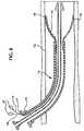
- FIGS. 1-3illustrate a stent delivery device 10 in various stages of deployment having one or more retrievable radiopaque markers 14 disposed on an implantable endoprosthesis 16 .
- the retrievable radiopaque markers 14are disposed on the endoprosthesis 16 preferably before loading into the outer tube of a delivery device 10 .
- a proximal end 14a of the retrievable radiopaque marker 14may be attached at portion 8 which is on the outside surface of the inner tube of a delivery device 10 and an area proximal of the proximal end of the implantable endoprosthesis 16 .
- Other attachment areasare also possible on the delivery device 10 .
- Attachment of the proximal end 14a of the retrievable radiopaque marker 14 to the delivery devicemay be made by mechanical (e.g., clamp or frictional contact on the surface, interweaving to components in the device, or tying), thermal (e.g. metal or polymer welding), or chemical (e.g., adhesive or gel bond) fastening systems.
- a predetermined length of the retrievable radiopaque marker 14may be gathered at or around portion 8 to allow the implantable endoprosthesis 16 to deploy from the delivery device 10 .
- the retrievable radiopaque marker 14may be disposed on the implantable endoprosthesis 16 , be disposed in a channel or lumen of the delivery device 10, and exit out a port 17 in the hub 19 and be attached to handle 21 .
- the handle 21may be a ring or a similar shape device adapted to be grasped and aid in retrieval and manipulation of the retrievable radiopaque marker 14 or straight wire 18 .
- the markerscan be segregated into types; retrievable temporary and discrete permanent markers.
- a retrievable temporary markeris generally a strand or strands of material having radiopacity which is loosely or removably incorporated within the implantable device and which can be removed from the device sometime after implantation by pulling on a free end of the marker or by having the marker extend beyond the device to an attachment point on the delivery system or extend through the delivery system and out of the body where it can be grabbed and pulled free of the implant.
- a discrete permanent markeris generally a strand of material having radiopacity which is securely attached to the implantable device and does not significantly extend away from the device.
- a retrievable temporary markeris a radiopaque strand of material loosely passed through or threaded into a braided tubular stent with an end of marker extending away from the stent and attached to the inner tube of the coaxial tube delivery system.
- the markeris used to locate the position of the stent with regard to the stricture.
- the delivery systemis normally pulled out of the body along the guidewire. The radiopaque marker would be pulled free of the stent as the delivery system is retrieved.
- An example of a discrete permanent markeris a coil, knot, or ring of tantalum wire around a feature of a stent, such as a stent wire crossing point.
- the tantalum wireis wrapped, coiled, or tied around the stent wire and thereby is permanently mechanically attached to the device.
- the tantalum wire endsare clipped off such that the marker is present as a small, tight ring around a feature of the stent.
- the stent with the attached markersis loaded and deployed from the delivery system and the markers are not retrieved when the delivery system is removed.
- the function of the retrievable radiopaque markeris to temporarily indicate on a radiographic image the location of the stent within the treatment site and the length of the expanded stent can be determined by measuring the length of the marker as it follows the stent shape if the marker was threaded along a stent wire helix or axially along a line in the stent.
- the markercan be threaded circumferentially at each end of the stent covering in a covered stent or stent-graft to indicate the location of the radiolucent covering material.
- the stent expansion during deploymentcan be observed radiographically by watching the radiopaque marker helical or circumferential strand open up as the self-expanding stent is released from its radially constrained state.
- Discrete markershave the same functional purpose as the retrievable markers, but they can be more easily used to mark the specific locations of features of interest on the stent. For example, a discrete marker can be added to the center of the stent length to aid the physician in centering the stent within the stricture. Discrete markers could be used to attach covering fabrics or films to stents to make stent grafts so that the location of the covering on the stent could be determined radiographically.
- the retrievable and discrete markerscan be made from biocompatible metal wires containing elements with relatively high atomic numbers such as titanium, tantalum, zirconium, and platinum.
- the radiopaque elementscan be added by metallurgically alloying or by making clad composite structures. Another type of marker would be to combine titanium, tantalum, zirconium, or platinum metal or oxide powder with a polymer matrix. Polyethylene or silicone are examples of biocompatible polymers that could be used as a matrix material. Combination could be performed by compounding with the polymer resin or coating.
- Organic radiopaque powders containing elements or salts or oxides of elementssuch as bromine, iodine, iodide, barium, and bismuth could be used instead of metal powders.
- a retrievable, temporary radiopaque markercan be in the form of a strand of metal or polymer containing radiopaque elements, oxides, or salts of elements with atomic numbers in the range of from about 22 to about 83 loosely threaded along a helical, circumferential, or axial orientation in an endoprosthesis such as a stent, stent-graft, graft, filter, occlusive device, and valve with a free end of the marker extending out from the endoprosthesis such that it is attached to the delivery system or passed outside of the body and the marker and is separated from the implanted endoprosthesis by pulling it free and out of the body.
- the radiopaque materialhas a linear attenuation coefficient of from about 5.46 cm -1 at 50 KeV to about 149.08 cm -1 at 50 KeV.
- a retrievable, temporary radiopaque markercan be in the form of a strand of metal or polymer containing radiopaque elements, oxides, or salts of elements with atomic numbers in the range of from about 22 to about 83 formed into a spring and disposed within an endoprosthesis such as a stent, stent-graft, graft, filter, occlusive device, and valve with a free end of the marker extending out from the endoprosthesis such that it is attached to the delivery system or passed outside of the body and the marker and is separated from the implanted endoprosthesis by pulling it free and out of the body.
- the radiopaque materialhas a linear attenuation coefficient of from about 5.46 cm -1 at 50 KeV to about 149.08 cm -1 at 50 KeV.
- a retrievable, temporary radiopaque markercan be in the form of a strand of ductile metal wire, ribbon, or braided wire containing radiopaque metallic elements with atomic numbers in the range of from about 22 to about 83, preferably titanium, tantalum, zirconium, and platinum disposed within an endoprosthesis such as a stent, stent-graft, graft, filter, occlusive device, and valve with a free end of the marker extending out from the endoprosthesis such that it is attached to the delivery system or passed outside of the body and the marker and is separated from the implanted endoprosthesis by pulling it free and out of the body.
- the radiopaque materialhas a linear attenuation coefficient of from about 5.46 cm -1 at 50 KeV to about 149.08 cm -1 at 50 KeV.
- a retrievable, temporary radiopaque markercan be in the form of a strand of ductile metal wire, ribbon, or braided wire containing radiopaque metallic elements with atomic numbers in the range of from about 22 to about 83, preferably titanium, tantalum, zirconium, and platinum coated or clad composite stainless steel or Elgiloy® wire disposed on an endoprosthesis such as a stent, stent-graft, graft, filter, occlusive device, and valve with a free end of the marker extending out from the endoprosthesis such that it is attached to the delivery system or passed outside of the body and the marker is separated from the implanted endoprosthesis by pulling it free and out of the body.
- the radiopaque materialhas a linear attenuation coefficient of from about 5.46 cm -1 at 50 KeV to about 149.08 cm -1 at 50 KeV.
- a retrievable, temporary radiopaque markercan be in the form of a strand of ductile polyethylene or silicone polymer mono-filament, ribbon or multifilament wire containing radiopaque metallic elements with atomic numbers in the range of from about 22 to about 83, preferably compounded or coated with titanium, tantalum, zirconium, and platinum metal powders or bromine, iodine, iodide, barium, and bismuth element, oxides or salts disposed within an endoprosthesis such as a stent, stent-graft, graft, filter, occlusive device, and valve with a free end of the marker extending out from the endoprosthesis such that it is attached to the delivery system or passed outside of the body and the marker and is separated from the implanted endoprosthesis by pulling it free and out of the body.
- the radiopaque materialhas a linear attenuation coefficient of from about 5.46 cm -1 at 50 KeV to about 149.08 cm
- a retrievable, temporary radiopaque markercan be in the form of a ductile polymer or metal matrix composite wire containing radiopaque metallic elements with atomic numbers in the range of from about 22 to about 83, preferably titanium, tantalum, zirconium, and platinum metal powders or bromine, iodine, iodide, barium, and bismuth element oxides or salt powders disposed within an endoprosthesis such as a stent, stent-graft, graft, filter, occlusive device, and valve with a free end of the marker extending out from the endoprosthesis such that it is attached to the delivery system or passed outside of the body and the marker and is separated from the implanted endoprosthesis by pulling it free and out of the body.
- the radiopaque materialhas a linear attenuation coefficient of from about 5.46 cm -1 at 50 KeV to about 149.08 cm -1 at 50 KeV.
- a discrete, permanent radiopaque markercan be in the form of a ductile metal wire, ribbon, or braided wire containing radiopaque metallic elements with atomic numbers in the range of from out 22 to about 83, preferably titanium, tantalum, zirconium, and platinum attached by wrapping, coiling, or tying around features within an endoprosthesis such as a stent, stent-graft, graft, filter, occlusive device, and valve such that the marker stays permanently attached by mechanical or adhesive forces to the endoprosthesis during deployment from the delivery system for the life of the implant.
- the radiopaque materialhas a linear attenuation coefficient of from about 5.46 cm -1 at 50 KeV to about 149.08 cm -1 at 50 KeV.
- a discrete, permanent radiopaque markercan be in the form of a strand of ductile metal wire, ribbon or braided wire containing radiopaque metallic elements with atomic numbers in the range of from about 22 to about 83, preferably titanium, tantalum, zirconium, and platinum coated or clad composite stainless steel or Elgiloy® wire ductile metal wire, ribbon or braided wire containing radiopaque metallic elements with atomic numbers in the range of from about 22 to about 83, preferably titanium, tantalum, zirconium, and platinum attached by wrapping, coiling, or tying around features within an endoprosthesis such as a stent, stent-graft, graft, filter, occlusive device, and valve such that the marker stays permanently attached by mechanical or adhesive forces to the endoprosthesis during deployment from the delivery system for the life of the implant.
- the radiopaque materialhas a linear attenuation coefficient of from about 5.46 cm -1 at 50 KeV to about 149.
- a discrete, permanent radiopaque markercan be in the form of a strand of ductile polyethylene or silicone polymer monofilament, ribbon or multifilament wire containing radiopaque metallic elements with atomic numbers in the range of from about 22 to about 83, preferably compounded or coated with titanium, tantalum, zirconium, and platinum metal powders or bromine, iodine, iodide, barium, and bismuth element, oxides or salts attached by wrapping, coiling or tying around features within an endoprosthesis such as a stent, stent-graft, graft, filter, occlusive device, and valve such that the marker stays permanently attached by mechanical or adhesive forces to the endoprosthesis during deployment from the delivery system for the life of the implant.
- the radiopaque materialhas a linear attenuation coefficient of from about 5.46 cm -1 at 50 KeV to about 149.08 cm -1 at 50 KeV.
- Figs 2-3illustrate an implantable endoprosthesis 16 in a body lumen 12.
- Implantable endoprostheses known in the artinclude stents, stent-grafts, grafts, filters, occlusive devices, and valves, all of which may incorporate the retrievable radiopaque marker 14 or discrete marker.
- Figs 4a-4cillustrate three alternative locations on an implantable endoprosthesis 16 for disposing the retrievable radiopaque marker 14 .
- the retrievable radiopaque marker 14may be an elongate element including a thread, filament, or ribbon such as a highly radiopaque wire relatively loosely woven into or wrapped around the inside, outside, or ends of the implantable endoprosthesis 16 .
- FIGS. 5-6illustrating the retrievable radiopaque marker 14 disposed in two alternative patterns on the implantable endoprosthesis 16 .
- FIG. 5shows the marker 14 interwoven or interbraided loosely along the longitudinal axis of the endoprosthesis 16 .
- FIG. 6shows the marker 14 disposed in a helical pattern about the implantable endoprosthesis 16 .
- Other patterns and dispositions of the marker 14 on the endoprosthesis 16are also possible.
- One or more markers 14may be temporarily disposed on the implantable endoprosthesis 16 in alternative patterns to advantageously provide temporary radiopacity to predetermined locations on the implantable endoprosthesis 16 .
- the retrievable radiopaque marker 14may be applied temporarily to one or more surfaces of the implantable endoprosthesis 16 with a relatively weak bioabsorbable adhesive or gelatin, for instance, as shown in FIGS. 4a and 4c.
- the retrievable radiopaque marker 14may be formed into a spring having spring force characteristics and be applied on the inside surface of the implantable endoprosthesis 16 as shown in FIG 4c. Spring force allows the retrievable radiopaque marker 14 to press against the interior of the implantable endoprosthesis 16 and provide temporary radiopacity thereto.
- the retrievable radiopaque marker 14may be braided to form a rope or cable.
- the retrievable radiopaque marker 14may be woven or inter-braided into the implantable endoprosthesis 16 during manufacture.
- the retrievable radiopaque marker 14may adjust with expansion of the implantable endoprosthesis 16 and thereby advantageously provides radiopacity and viewing of the implantable endoprosthesis 16 position or size during fluoroscopy.
- the delivery device 10 and the retrievable radiopaque marker 14may be removed from the body.
- one end of the retrievable radiopaque marker 14may be attached to the delivery device 10 and the other end may be disposed at predetermined locations on the implantable endoprosthesis 16 .
- the retrievable radiopaque marker 14may be pulled away from implantable endoprosthesis 16 and removed from the body.
- the retrievable radiopaque marker 14may be loosely incorporated into the implantable endoprosthesis 16 and be easily retrieved without disturbing the implantable endoprosthesis 16 or body tissue. Alternatively, the retrievable radiopaque marker 14 may remain on the implantable endoprosthesis 16 for a period of time if there is a need for follow-up angiography, and then be ultimately removed.
- FIGS. 7-8illustrating an alternative embodiment including a retrievable radiopaque marker 14 and wire 18 .
- the wire 18is used to prevent removal of the marker 14 without first removal of the wire 18 .
- the retrievable radiopaque marker 14is relatively loosely woven or inter-braided in and out of the implantable endoprosthesis 16 , and is maintained in place by another relatively straight, flexible and adjacent movable wire 18 .
- the marker 14 and wire 18may be made by various methods and materials including polymers, metals, ceramics, or similar materials.
- the wire 18may be placed inside, outside, or penetrate between filaments of the implantable endoprosthesis 16 .
- the wire 18 and retrievable radiopaque marker 14are disposed at desired predetermined areas and in various patterns on the implantable endoprosthesis 16 .
- Various combinations of the wire 18 and retrievable radiopaque marker 14are possible including multiple markers 14 or wires 18 .
- the retrievable radiopaque marker 14 and wire 18may be disposed on the implantable endoprosthesis 16 , be disposed in a channel or lumen of the delivery device 10, and exit out a port 17 in the hub 19 and be attached to handle 21 .
- the handle 21may be a ring or a similar shape device adapted to be grasped and aid in retrieval and manipulation of the retrievable radiopaque marker 14 .
- the wire 18may be removed proximally by a force which liberates the retrievable radiopaque marker 14 and allows removal thereof.
- a limited amount of interweaving or interbraiding of the retrievable radiopaque marker 14 or wire 18is generally desired in order to minimize the force required for retrieval.
- the retrievable radiopaque marker 14 or wire 18may be coated with a biocompatible material having a low coefficient of friction for ease of removal from the implantable endoprosthesis 16 .
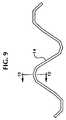
- FIG 9illustrating a retrievable radiopaque marker 14 preferably made from a relatively flexible wire, suture, filament, ribbon, braided wires, or combinations thereof including radiopaque material such as a metal, metallic alloy, or polymer containing a material that is highly radiopaque.
- radiopaque materialsuch as a metal, metallic alloy, or polymer containing a material that is highly radiopaque.
- FIGS. 10a-10eillustrate alternative cross-sectional embodiments of the retrievable radiopaque marker 14 taken through the line 10-10 of FIG. 9.
- FIG. 10ashows a substantially solid member
- FIG. 10bshows a hollow member
- FIG. 10cshows a member having pores extending radially into the member
- FIG. 10dshows a rectangular or ribbon member
- FIG. 10eshows a braided hollow member.
- Fig. 10emay also be a substantially solid braided member.
- a composite radiopaque marker 14may be made from materials coated or compounded with a radiopaque substance such as iodine, zirconium oxide, barium sulfate, bismuth trioxide, or a related oxide or salt substance.
- Composite radiopaque materialsmay be a radiopaque material containing at least one element having an atomic number, preferably higher than about 22.
- Another radiopaque marker 14may include gold, platinum, metal, tantalum, metallic alloy, or a polymer containing a radiopaque filler such as barium sulfate, bismuth trioxide, iodine, iodide, or like materials.
- FIGS. 11a-11cillustrating alternative embodiments of a portion of the retrievable radiopaque marker 14 .
- the retrievable radiopaque marker 14may have at least one hollow portion 15 which extends throughout the marker 14 for temporary or permanent containment of a retrievable radiopaque material.
- a radiopaque core 13 as shown in Fig. 11cmay be disposed in and retrieved from a hollow portion 15 in the retrievable radiopaque marker 14 .
- One end of the radiopaque core 13may be attached to the delivery device 10 by a wire or the like and removed from the retrievable radiopaque marker 14 and body lumen by a force originating from outside the body.
- the outside case of the marker 14may remain disposed on the implantable endoprosthesis 16 or be removed therefrom.
- the temporary radiopaque core 13may be solid or include a casing surrounding a solid, gel, powder or combination thereof and be held in place with a relatively weak bioabsorbable adhesive gelatin, friction, or by other mechanical or chemical means known in the art in a hollow 15 , cavity, or porous portion.
- the temporary radiopaque core 13preferably is made of a radiopaque material that has a linear attenuation coefficient of from about 5.46 cm -1 at 50 KeV to about 149.08 cm -1 at 50 KeV and is adapted to be removably attachable in at least one hollow 15 , cavity, or porous portion in the marker 14 .
- the core 13may remain in the hollow 15 , cavity, or porous portion of the marker 14 and be removed when the marker 14 is retrieved from the body.
- one or more closed cavities within the marker 14 or pores on the surface as shown in Fig 10c or pores extending through to a hollow or cavity portion within the marker 14may be utilised for temporary or permanent containment of retrievable radiopaque materials or be utilised for a passageway for dispersal of the radiopaque materials contained in the marker 14 into the body.
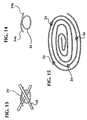
- Fig. 12illustrates discrete radiopaque markers 24 made by forming small rings or coils of radiopaque wire around features of the implantable endoprosthesis 16 .
- Relatively small and discrete wire loop (pigtail) radiopaque markers 24are shown at the wire crossing points on the tubular braid.
- Fig. 13illustrates the detail bounded by the dashed-line circle in Fig. 12 showing a radiopaque wire loop marker 24 around one implantable endoprosthesis 16 wire crossing point.
- FIG. 14illustrates the marker 24 of FIG. 12 and FIG. 13 and shows wire ends 24a , 24b which simply pass over each other to form an enclosed loop or overlap.
- the discrete radiopaque markers 24may be plastically or elastically deformable.
- the markers 24may be springs or spring-like for attachment purposes.
- the ends 24a , 24bmay be tied, knotted, crimped, spot welded, or bent.
- the markers 24may be relatively small and comprise a single loop or pigtail of wire around one filament crossing point, filament, an embolization coil, or the like.
- the marker 24is preferably made of a biocompatible radiopaque material that is ductile including pure tantalum, platinum, gold, zirconium, niobium, titanium, stainless steel, or combinations thereof.
- the marker 24may be a pig-tail, coil, or knot design and is preferably formed from an elongate member such as a wire and shaped accordingly onto the implantable endoprosthesis 16 .
- the marker 24advantageously allows custom marking of the implantable endoprosthesis 16 without the need to acquire preformed marker bands or to devise a complicated manufacturing operation such as swaging, threading, or braiding.
- the discrete radiopaque markers 24may be easily and quickly added to the implantable endoprosthesis 16 . Also, only small, specific sites are marked by the marker 24 so a minimum amount of foreign body material would be added to the implantable endoprosthesis 16 .
- the discrete radiopaque markers 24may be used on an implantable endoprosthesis 16 made of a bioabsorbable polymer including polylactide.
- the markers 14 , 24should preferably be smaller than the size of the element in the implantable endoprosthesis 16.
- the size of the markers 14, 24is also dependent on the type of radiopaque material used. For example, tantalum wire (0.006" (.15 mm) diameter, hard drawn) may be used. The smaller diameter wire fits through most weaves, is deformable, and may be cut to size.
- FIGS. 12-13illustrating discrete markers 24 looped one or more times about a filament or filament crossing point to prevent release therefrom.
- the ends 24a , 24bare clipped and positioned to lie in a plane parallel to the longitudinal axis of the implantable endoprosthesis 16 .
- the marker 24may be disposed on one or more filament crossing or every other filament crossing point around the circumference of the braid in one circular transverse plane.
- the markers 24may be positioned to form one or more circumferential rings on the implantable endoprosthesis 16 Alternatively, the markers 24 may be positioned along an embolization occlusion coil intravascular device or filament at predetermined locations as illustrated in Fig. 15.
- the marker 24may be plastically deformed and the marker ends 24a , 24b may be looped one or more times about a portion of the implantable endoprosthesis 16 and then pulled to provide a snug disposition.
- the ends 24a , 24bmay then be tied, twisted, knotted, welded or adhesively connected together and thereafter clipped and positioned to lie in an unobtrusive low-profile position.
- the retrievable radiopaque marker 14 and discrete radiopaque marker 24may be constructed using a number of methods and materials, in a wide variety of sizes and styles for the greater efficiency and convenience of a user.
- a bioabsorbable markerthat may advantageously be used in conjunction with the present invention is disclosed in J. Stinson's United States Patent Publication No. 6174330 entitled “Bioabsorbable Marker Having Radiopaque Constituents and Method of Using Same", filed concurrently herewith, and commonly assigned to the assignee of this application.
- a bioabsorbable stentmay advantageously be used in conjunction with the present invention is disclosed in J. Stinson's United States Patent Publication No.5980564 entitled “Bioabsorbable Implantable Endoprosthesis With Reservoir and Method of Using Same", filed concurrently herewith, and commonly assigned to the assignee of this application.
Landscapes
- Health & Medical Sciences (AREA)
- Engineering & Computer Science (AREA)
- Biomedical Technology (AREA)
- Cardiology (AREA)
- Oral & Maxillofacial Surgery (AREA)
- Transplantation (AREA)
- Heart & Thoracic Surgery (AREA)
- Vascular Medicine (AREA)
- Life Sciences & Earth Sciences (AREA)
- Animal Behavior & Ethology (AREA)
- General Health & Medical Sciences (AREA)
- Public Health (AREA)
- Veterinary Medicine (AREA)
- Media Introduction/Drainage Providing Device (AREA)
- Prostheses (AREA)
Description
- This invention relates generally to an endoprosthesis such as a stent provided with a retrievable radiopaque marker.
- Implantable endoprostheses including stents, stent-grafts, and grafts are usedin percutaneous transluminal coronary angioplasty and in other medical proceduresto repair and support diseased or damaged arteries and body lumens. Grafts areimplanted to cover or bridge leaks or dissections in vessels. Stent-grafts are stentswhich generally have a porous coating attachment and may be implanted bypercutaneous transluminal angioplasty. Unsupported grafts are porous tubes whichare typically implanted by surgical cut-down.
- In order to visualize the passage and placement of the implantableendoprosthesis in arteries and body lumens, many surgical procedures are performedunder fluoroscopy. The surgical delivery device and implantable endoprosthesismay be visualized if they are radiopaque and offer radiographic contrast relative tothe body. For example, X-ray radiation may be used to visualize surgical deliverydevices and deployment of the implant in the body. Also, radiographic contrastsolution may be injected into the body lumen so that the lumen may be seen in thefluoroscopic image.
- In order for an implantable endoprosthesis to be radiopaque, it must be madefrom a material possessing radiographic density higher than a surrounding hosttissue and have sufficient thickness to affect the transmission of x-rays to producecontrast in the image. Reference is made to the clad composite stent shown inUnited States Patent No. 5,630,840. An implantable endoprosthesis may be madeof metals including tantalum or platinum having relatively high radiographicdensities. Other metals such as stainless steel, superalloys, nitinol, and titaniumhaving lower radiographic densities may also be used. Reference is made to implantable devices shown in United States Patents Nos. 4,655,771; 4,954,126; and5,061,275.
- An implantable polymeric endoprosthesis is generally radiolucent and does notpossess sufficient radiographic density to be easily imaged by fluoroscopy. To improve theimaging of polymeric materials, polymers may be mixed with radiopaque filler materialsprior to molding or extruding in order to enhance the radiographic density. However, adisadvantage of using fillers with polymers is that changes in the properties of the polymermay occur. For example, the addition of fillers may reduce the strength or ductility of thepolymer.
- There is a need for an improved radiopaque marker for use in medical devices,particularly in temporary medical devices having low radiopacity. The need to improve theradiopacity of a relatively low radiopaque implantable endoprosthesis or improve imaging inlow radiopaque conditions is particularly important for surgery, micro-surgery, neurosurgery,and conventional angioplasty procedures performed under fluoroscopy. Physiciansare constantly being challenged to place small implants at remote intraluminal locations.
- Various devices having radiopaque markers are shown in United States Patents Nos.4,447,239; 5,423,849; and 5,354,257. Additional prior art includes WO 94/15534A, WO97/21403A, US 5342,348 and WO 95/21592A.
- Accordingly, there is a need for retrievable radiopaque markers for use inimplantable endoprostheses to improve radiopacity and the locatability of endoprostheses invarious medical procedures.
- According to a first aspect of the present invention, there is provided an implantableendoprosthesis and radiopaque marker system comprising:
- an implantable endoprosthesis adapted to be disposed in a body lumen; and
- an elongate marker having at least one radiopaque portion including a radiopaquematerial, characterised by means for removing the marker from the endoprosthesis when theendoprosthesis isin vivo, said marker being removably attached to the endoprosthesis.
- According to a second aspect of the present invention, there is provided a processfor modifying an implantable endoprosthesis to temporarily enhance a visualization of theendoprosthesis during and after an implantation thereof in a body lumen, comprising:
- providing a body implantable endoprosthesis;
- providing at least one marker, each marker having at least one radiopaque portionincluding a radiopaque material; and
- prior to an implantation of the endoprosthesis, removably attaching the at least onemarker to the implantable endoprosthesis in a manner that facilitates a removal of themarker from the endoprosthesis when the endoprosthesis isin vivo after an implantationthereof, and providing a means for effecting said removal.
- Providing temporary radiopacity is especially advantageous for implantableendoprostheses having little or no radiopacity. The markers allow radiographicidentification of one or more locations of interest on an implantable endoprosthesis. Thelocations of interest may include one or more covered or coated regions.
- Alternative embodiments include threading the markers adjacent a helicalstrand in the implantable endoprosthesis, circumferentially around the implantableendoprosthesis, in a straight line in the axial direction of the implantableendoprosthesis, or disposing the wire in the form of pigtail-shaped rings, coils, orknots around filament crossing points in the implantable endoprosthesis.
- Temporary retrievable radiopaque markers in the fabric or coveringmaterials of an implantable endoprosthesis are advantageous for indicating thelocation of the fabric or covering during implantation. After implantation, thetemporary retrievable radiopaque marker may be retrieved so as not to effect thefunction of the endoprosthesis.
- A disadvantage of some permanent radiopaque markers is that they maycompromise structural integrity, may not be biocompatible or biostable, and may bemore thrombogenic than the implantable endoprosthesis.
- The temporary retrievable radiopaque marker of the present inventionadvantageously allows most any implantable endoprosthesis to have temporaryradiopacity over a predetermined portion of its structure, and assists with properpositioning and locatability of the implantable endoprosthesis in a body lumen.
- Use of temporary retrievable radiopaque markers on an implantableendoprosthesis is advantageous because the radiopaque property may be presentonly for a desired time period. Generally, radiopacity is most desirable duringplacement of the implant. Once the implantable endoprosthesis is implanted, it maybe more desirable to image the device with techniques such as ultrasound, magneticresonance, and endoscopy and avoid further radiation exposure to the patient.Temporary radiopacity may be made by incorporating non-integral, retrievableradiopaque constituents into the implant. Thus, light metals, thin radiopaquemetals, polymers, and ceramics may be utilized for a wide range of properties andflexibility in design of the endoprosthesis.
- Attenuation is the change in the number of photons in the incident x-raybeam due to the interaction with an absorber. To image an object implanted in the body, it would be desirable to have the object attenuate x-rays more than bodytissue, bone, and fat so that the difference in contrast will be obvious in aradiograph. The difficulty in selecting a radiopaque material for surgical implants isthat the material must have desirable radiographic characteristics andbiocompatibility.
- In order to make an implant more radiopaque, a substance which absorbsmore x-rays can be deposited on or mixed in with the implant material. If theimplant absorbs more x-rays than the surrounding medium (for example tissue inthe body), it will be visible as a sharp change in contrast on an x-ray film orfluoroscopy image.
- The fraction of x-ray energy transmitted through the absorber isquantitatively predicted by the following equation described inThe Physics ofRadiology, Fourth Ed., H. Johns, J. Cunningham, 1983, pp. 137-142.
- N = number of photons transmitted through x
- N0 = number of photons in the incident beam
- µ = linear attenuation coefficient of the absorber
- x = absorber thickness
- N/N0 would be the fraction of incident x-ray energy that is transmittedthrough the absorber. A more radiopaque material would have a lesser fraction oftransmitted energy than a more radiolucent material. Therefore, to enhance theradiopacity of a material, such as the marker material, it would be desirable to selecta material with high x-ray absorbing capability to minimize the fraction oftransmitted energy. This radiopacity capability is proportional to the linearattenuation coefficient and the thickness of the absorber material. The higher theattenuation coefficient of the absorber material for a given thickness, the moreradiopaque the absorber will be. The attenuation produced by an absorber is dependent upon the number of electrons and atoms present in the absorber. One way ofquantifying this absorption characteristic is with the atomic attenuation coefficient which isdirectly proportional to the linear attenuation coefficient and the atomic number of theabsorber element. Radiopacity is therefore generally proportional to the atomic number(number of electrons in the atom) of the material. Candidate materials for enhancing theradiopacity of surgical implants would have higher atomic numbers than the elementspresent in the body and would have to be biocompatible. The atomic number must besufficiently high so that relatively small thickness of absorber material can be used in thebody. Reference is also made to linear attenuation coefficient described in United StatesPatent No. 5,628,787. Reference is made to Table 1 which describes a number of elementsand their respective atomic numbers and certain linear attenuation coefficients.
Element or Material Atomic Number or Effective Atomic Number Linear Attenuation Coefficient at 50 KeV, cm-1 hydrogen 1 .000028 carbon 6 .417 fat 6.46 .193 water 7.51 .2245 muscle 7.64 .233 air 7.78 .000247 nitrogen 7 .000228 oxygen 8 .000280 bone 12.31 .573 titanium 22 5.46 iron 26 15.42 cobalt 27 18.94 bromine 35 13.29 zirconium 40 40.04 iodine 53 60.76 barium 56 49.68 tantalum 73 94.95 platinum 78 149.08 gold 79 140.12 lead 82 91.17 bismuth 83 82.12 nickel 28 21.98 - The elements hydrogen, oxygen, carbon, and nitrogen are commonly found in thebody and in polymers, so elements with higher atomic numbers than these should enhancethe radiopacity of a polymer implant or marker. Tantalum, zirconium, titanium, barium,bismuth, and iodine are known to be non-toxic in certain concentrations and thus arecandidate elements for enhancing radiopacity of a polymer marker in an implant. Theseelements can be added to the polymer in various loading percentages and the thresholdabove which the loading causes unsatisfactory changes in the polymer characteristics can bedetermined through material and device testing. The elements which can be added inquantities sufficient to enhance radiopacity and maintain an acceptable level of polymerproperties and which are biocompatible could be utilised in markers. The biocompatibleelements with a range of atomic numbers from about 22 to about 83 and having linearattenuation coefficients in the range from about 5.46 to about 149.08 cm-1 at 50 KeV shouldprovide enough enhancement in radiopacity without excessive thickness being necessary tobe useful in markers. These elements would include at least titanium, vanadium,chromium, iron, cobalt, nickel, copper, bromine, zirconium, niobium, molybdenum, silver,iodine, barium, tantalum, tungsten, platinum, gold, and bismuth. The preferred metallicelements for biocompatibility and radiopacity are titanium, zirconium, tantalum, andplatinum. The preferred organic elements for biocompatibility and radiopacity are bromine,iodine, barium, and bismuth. Especially preferred elements are tantalum, platinum,barium, and bismuth because of their high atomic numbers and biocompatibility (atomicnumbers from 56 to 83 and linear attenuation coefficients from 49.68 to 149.08).Tantalum and platinum are used as stent components and barium sulphate and bismuthtrioxide are used as radiopaque enhancements for polymer catheters.
- In sum, the invention relates to an implantable endoprosthesis and radiopaquemarker system. The system includes an implantable endoprosthesis adapted to be disposedin a body lumen and at least one elongate marker. The marker has a proximal end, a distal end, a thickness, and at least one radiopaque portion.The radiopaque portion includes a radiopaque material. The marker is removably attachedto at least a portion of the implantable endoprosthesis and is removable from theendoprosthesis when the endoprosthesis isin vivo. The radiopaque material may be at leastpartially dispersed from the marker over time. The radiopaque material may have a linearattenuation coefficient of from about 5.46 cm-1 at 50 KeV to about 149.08 cm-1 at 50 KeV.The thickness of the marker may range from about 20 microns to about 500 microns and theradiopaque material may have at least one element with an atomic number of from about 22to about 83. The marker may include an oxide or salt material having at least one elementwith an atomic number of from about 22 to about 83. The marker may include bariumsulphate, bismuth trioxide, iodine, iodide, titanium oxide, zirconium oxide, gold, platinum,silver, tantalum, niobium, stainless steel or combinations thereof. The marker may becoated or alloyed with a radiopaque material that has a linear attenuation coefficient of fromabout 5.46 cm-1 at 50 KeV to about 149.08 cm-1 at 50 KeV. The marker may cross at leastone portion of the implantable endoprosthesis. The marker may be a wire, mono-filament,multi-filament, ribbon, suture, spring, or combinations thereof. The marker may includemetals, polymers, copolymers, ceramics, or combinations thereof. The marker may includeat least one hollow, cavity, or porous portion. The marker may include at least one hollow,cavity, or porous portion therein adapted to receive the radiopaque material removablyattached therein. The proximal end of the marker may be connected to at least one of theimplantable endoprosthesis delivery device or a handle. The proximal end of the markermay have a hook, knob, ring, or eyelet attached thereto. The marker system may include adelivery device wherein the implantable endoprosthesis and marker are disposed in thedelivery device and adapted for implantation into a body lumen. The implantableendoprosthesis may include a stent, stent-graft, graft, filter, occlusive device, or valve. Themarker system may include at least one elongate wire removably attached to the implantableendoprosthesis wherein the marker crosses at least a portion of the implantable endoprosthesis and crosses the at least oneelongate wire.
- The invention also relates to an implantable endoprosthesis and radiopaque markersystem. The marker system includes an implantable endoprosthesis adapted to be disposedin a body lumen and at least one elongate marker. The marker is removably attached to animplantable endoprosthesis. The marker has a proximal end, a distal end, a thickness, atleast one hollow, cavity, or porous portion, and at least one radiopaque material having alinear attenuation coefficient of from about 5.46 cm-1 at 50 KeV to about 149.08 cm-1 at 50KeV, wherein the radiopaque material is removably attached to at least one of the hollow,cavity, or porous portions. The radiopaque portion may include a liquid, solid, powder,gel, wire, mono-filament, multi-filament, pellet, particle, or combinations thereof.
- The invention also relates to a method of marking an implantable endoprosthesisincluding removably-attaching at least one elongate marker having a proximal and distal endto a portion of an implantable endoprosthesis to form an assembly. The marker includes atleast one radiopaque material having a linear attenuation coefficient of from about 5.46 cm-1at 50 KeV to about 149.08 cm-1 at 50 KeV; disposing the implantable endoprosthesis andmarker assembly in a delivery system; inserting the delivery system in a body lumen;deploying the implantable endoprosthesis and marker assembly from the delivery systeminto the body lumen; and removing at least a portion of marker from the implantableendoprosthesis.The method may further include removably-attachingat least one wire to at least a portion of the implantable endoprosthesis and crossing the wire or the elongate marker over the other such that one ofthe marker or the wire requires removal prior to removal of the other from the implantableendoprosthesis.
- The invention also relates to an implantable endoprosthesis and radiopaque markersystem. The marker system includes an implantable endoprosthesis having a tubular andradially expandable structure adapted to be disposed in a body lumen and at least oneelongate marker. The marker is removably attached to the implantable endoprosthesis. Themarker includes a radiopaque material having a linear attenuation coefficient of from about5.46 cm-1 at 50 KeV to about 149.08 cm-1 at 50 KeV, a proximal end, a distal end, and athickness. The radiopaque material disperses into the body whenin vivo. The implantableendoprosthesis may include an axially flexible structure including a plurality of the elongateelements which are interwoven in a braid-like configuration.
- The invention also relates to a temporary radiopaque marker. The marker includesan elongate marker having a proximal end, a distal end, an average thickness of from about20 microns to about 500 microns, and includes a radiopaque material having a linearattenuation coefficient of from about 5.46 cm-1 at 50 KeV to about 149.08 cm-1 at 50 KeV.The marker is adapted to be removably attached to an implantable endoprosthesis. Theproximal end of the marker may include a hook, knob, or eyelet.
- Still other objects and advantages of the present invention and methods ofconstruction of the same will become readily apparent to those skilled in the artfrom the following detailed description, wherein only the preferred embodimentsare shown and described, simply by way of illustration of the best modecontemplated of carrying out the invention. As will be realized, the invention iscapable of other and different embodiments and methods of construction, and itsseveral details are capable of modification in various obvious respects, all withoutdeparting from the scope of the claims. Accordingly, the drawing and description are to beregarded as illustrative in nature, and not as restrictive.
- Figures 12 to 15 do not disclose the present invention
- FIG. 1 is a side view of an implantable endoprosthesis delivery systemincluding a retrievable radiopaque marker disposed on an implantableendoprosthesis;
- FIG. 2 is a side view of the delivery system and a deployed retrievableradiopaque marker and implantable endoprosthesis in a body lumen;
- FIG. 3 is a side view of one possible arrangement of a retrievable radiopaquemarker being retrieved from a deployed implantable endoprosthesis in a bodylumen;
- FIGS. 4a, 4b, and 4c are cross-sectional views of three alternative markerdispositions on an implantable endoprosthesis at section 4-4 of FIG. 2;
- FIG. 5 is a side view of a retrievable radiopaque marker disposed on animplantable endoprosthesis;
- FIG. 6 is a side view of a retrievable radiopaque marker disposed in a helicalpattern about the perimeter of an implantable endoprosthesis;
- FIG. 7 is a side view illustrating one possible arrangement of a straight wireand retrievable radiopaque marker disposed on an implantable endoprosthesis;
- FIG. 8 is a side view of a delivery device illustrating one arrangement of awire and retrievable radiopaque marker;
- FIG. 9 is a side view of a relatively flexible retrievable radiopaque marker;
- FIGS. 10a-10e are cross-sectional views of five alternative radiopaquemarkers at section 10-10 of FIG. 9;
- FIGS. 11a-11c are side views of three alternative radiopaque markers;
- FIG. 12 is a side view illustrating one possible arrangement of discreteradiopaque markers disposed on an implantable endoprosthesis;
- FIG. 13 is the detail bounded by the dashed-line circle in FIG. 12 illustratinga radiopaque marker disposed around one implantable endoprosthesis wire crossingpoint;
- FIG. 14 is a side view illustrating a discrete radiopaque marker; and
- FIG. 15 illustrates the discrete radiopaque marker positioned on anembolization occlusion coil intravascular device.
- Reference is made to FIGS. 1-3 which illustrate a
stent delivery device 10 invarious stages of deployment having one or more retrievableradiopaque markers 14disposed on animplantable endoprosthesis 16. The retrievableradiopaque markers 14 are disposed on theendoprosthesis 16 preferably before loading into the outertube of adelivery device 10. Reference is made to a delivery device shown inUnited States Patent No. 5,026,377. - As shown in FIG. 1, a
proximal end 14a of the retrievableradiopaquemarker 14 may be attached atportion 8 which is on the outside surface of the inner tube of adelivery device 10 and an area proximal of the proximal end of theimplantable endoprosthesis 16. Other attachment areas are also possible on thedelivery device 10. Attachment of theproximal end 14a of the retrievableradiopaque marker 14 to the delivery device may be made by mechanical (e.g.,clamp or frictional contact on the surface, interweaving to components in the device,or tying), thermal (e.g. metal or polymer welding), or chemical (e.g., adhesive or gelbond) fastening systems. A predetermined length of the retrievableradiopaquemarker 14 may be gathered at or aroundportion 8 to allow theimplantableendoprosthesis 16 to deploy from thedelivery device 10. Alternatively, asillustrated in FIG. 8, the retrievableradiopaque marker 14 may be disposed on theimplantable endoprosthesis 16, be disposed in a channel or lumen of thedeliverydevice 10, and exit out aport 17 in thehub 19 and be attached to handle21. Thehandle 21 may be a ring or a similar shape device adapted to be grasped and aid inretrieval and manipulation of the retrievableradiopaque marker 14 orstraight wire 18. Once theimplantable endoprosthesis 16 is implanted, the retrievableradiopaquemarker 14 may be removed proximally from the body by a force in the proximaldirection transmitted to thehandle 21. Table 2 lists preferred embodiments of theinvention.
- For description purposes, the markers can be segregated intotypes; retrievable temporary and discrete permanent markers. A retrievabletemporary marker is generally a strand or strands of material having radiopacitywhich is loosely or removably incorporated within the implantable device andwhich can be removed from the device sometime after implantation by pulling on afree end of the marker or by having the marker extend beyond the device to anattachment point on the delivery system or extend through the delivery system andout of the body where it can be grabbed and pulled free of the implant. A discretepermanent marker is generally a strand of material having radiopacity which issecurely attached to the implantable device and does not significantly extend awayfrom the device.
- An example of a retrievable temporary marker is a radiopaque strand ofmaterial loosely passed through or threaded into a braided tubular stent with an endof marker extending away from the stent and attached to the inner tube of thecoaxial tube delivery system. As the stent is deployed from the delivery system themarker is used to locate the position of the stent with regard to the stricture. Afterstent deployment, the delivery system is normally pulled out of the body along theguidewire. The radiopaque marker would be pulled free of the stent as the deliverysystem is retrieved.
- An example of a discrete permanent marker is a coil, knot, or ring oftantalum wire around a feature of a stent, such as a stent wire crossing point. Thetantalum wire is wrapped, coiled, or tied around the stent wire and thereby ispermanently mechanically attached to the device. The tantalum wire ends areclipped off such that the marker is present as a small, tight ring around a feature ofthe stent. The stent with the attached markers is loaded and deployed from thedelivery system and the markers are not retrieved when the delivery system isremoved.
- The function of the retrievable radiopaque marker is to temporarily indicateon a radiographic image the location of the stent within the treatment site and the length of the expanded stent can be determined by measuring the length of themarker as it follows the stent shape if the marker was threaded along a stent wirehelix or axially along a line in the stent. The marker can be threadedcircumferentially at each end of the stent covering in a covered stent or stent-graft toindicate the location of the radiolucent covering material. The stent expansionduring deployment can be observed radiographically by watching the radiopaquemarker helical or circumferential strand open up as the self-expanding stent isreleased from its radially constrained state.
- Discrete markers have the same functional purpose as the retrievablemarkers, but they can be more easily used to mark the specific locations of featuresof interest on the stent. For example, a discrete marker can be added to the center ofthe stent length to aid the physician in centering the stent within the stricture.Discrete markers could be used to attach covering fabrics or films to stents to makestent grafts so that the location of the covering on the stent could be determinedradiographically.
- The retrievable and discrete markers can be made from biocompatible metalwires containing elements with relatively high atomic numbers such as titanium,tantalum, zirconium, and platinum. The radiopaque elements can be added bymetallurgically alloying or by making clad composite structures. Another type ofmarker would be to combine titanium, tantalum, zirconium, or platinum metal oroxide powder with a polymer matrix. Polyethylene or silicone are examples ofbiocompatible polymers that could be used as a matrix material. Combination couldbe performed by compounding with the polymer resin or coating. Organicradiopaque powders containing elements or salts or oxides of elements such asbromine, iodine, iodide, barium, and bismuth could be used instead of metalpowders.
- Exemples 7 to 9 are not examples of the presentinvention.
- A retrievable, temporary radiopaque marker can be in the form of a strand ofmetal or polymer containing radiopaque elements, oxides, or salts of elements with atomic numbers in the range of from about 22 to about 83 loosely threaded along a helical,circumferential, or axial orientation in an endoprosthesis such as a stent, stent-graft, graft,filter, occlusive device, and valve with a free end of the marker extending out from theendoprosthesis such that it is attached to the delivery system or passed outside of the bodyand the marker and is separated from the implanted endoprosthesis by pulling it free and outof the body. The radiopaque material has a linear attenuation coefficient of from about 5.46cm-1 at 50 KeV to about 149.08 cm-1 at 50 KeV.
- A retrievable, temporary radiopaque marker can be in the form of a strand of metalor polymer containing radiopaque elements, oxides, or salts of elements with atomicnumbers in the range of from about 22 to about 83 formed into a spring and disposed withinan endoprosthesis such as a stent, stent-graft, graft, filter, occlusive device, and valve witha free end of the marker extending out from the endoprosthesis such that it is attached to thedelivery system or passed outside of the body and the marker and is separated from theimplanted endoprosthesis by pulling it free and out of the body. The radiopaque materialhas a linear attenuation coefficient of from about 5.46 cm-1 at 50 KeV to about 149.08 cm-1at 50 KeV.
- A retrievable, temporary radiopaque marker can be in the form of a strand of ductilemetal wire, ribbon, or braided wire containing radiopaque metallic elements with atomicnumbers in the range of from about 22 to about 83, preferably titanium, tantalum,zirconium, and platinum disposed within an endoprosthesis such as a stent, stent-graft,graft, filter, occlusive device, and valve with a free end of the marker extending out fromthe endoprosthesis such that it is attached to the delivery system or passed outside of thebody and the marker and is separated from the implanted endoprosthesis by pulling it freeand out of the body. The radiopaque material has a linear attenuation coefficient of from about 5.46 cm-1 at 50 KeV to about149.08 cm-1 at 50 KeV.
- A retrievable, temporary radiopaque marker can be in the form of a strand of ductilemetal wire, ribbon, or braided wire containing radiopaque metallic elements with atomicnumbers in the range of from about 22 to about 83, preferably titanium, tantalum,zirconium, and platinum coated or clad composite stainless steel or Elgiloy® wire disposedon an endoprosthesis such as a stent, stent-graft, graft, filter, occlusive device, and valvewith a free end of the marker extending out from the endoprosthesis such that it is attachedto the delivery system or passed outside of the body and the marker is separated from theimplanted endoprosthesis by pulling it free and out of the body. The radiopaque materialhas a linear attenuation coefficient of from about 5.46 cm-1 at 50 KeV to about 149.08 cm-1at 50 KeV.
- A retrievable, temporary radiopaque marker can be in the form of a strand of ductilepolyethylene or silicone polymer mono-filament, ribbon or multifilament wire containingradiopaque metallic elements with atomic numbers in the range of from about 22 to about83, preferably compounded or coated with titanium, tantalum, zirconium, and platinummetal powders or bromine, iodine, iodide, barium, and bismuth element, oxides or saltsdisposed within an endoprosthesis such as a stent, stent-graft, graft, filter, occlusive device,and valve with a free end of the marker extending out from the endoprosthesis such that it isattached to the delivery system or passed outside of the body and the marker and isseparated from the implanted endoprosthesis by pulling it free and out of the body. Theradiopaque material has a linear attenuation coefficient of from about 5.46 cm-1 at 50 KeVto about 149.08 cm-1 at 50 KeV.
- A retrievable, temporary radiopaque marker can be in the form of a ductile polymeror metal matrix composite wire containing radiopaque metallic elements with atomicnumbers in the range of from about 22 to about 83, preferably titanium, tantalum,zirconium, and platinum metal powders or bromine, iodine, iodide, barium, and bismuthelement oxides or salt powders disposed within an endoprosthesis such as a stent, stent-graft,graft, filter, occlusive device, and valve with a free end of the marker extending outfrom the endoprosthesis such that it is attached to the delivery system or passed outside ofthe body and the marker and is separated from the implanted endoprosthesis by pulling itfree and out of the body. The radiopaque material has a linear attenuation coefficient offrom about 5.46 cm-1 at 50 KeV to about 149.08 cm-1 at 50 KeV.
- A discrete, permanent radiopaque marker can be in the form of a ductile metal wire,ribbon, or braided wire containing radiopaque metallic elements with atomic numbers in therange of from out 22 to about 83, preferably titanium, tantalum, zirconium, and platinumattached by wrapping, coiling, or tying around features within an endoprosthesis such as astent, stent-graft, graft, filter, occlusive device, and valve such that the marker stayspermanently attached by mechanical or adhesive forces to the endoprosthesis duringdeployment from the delivery system for the life of the implant. The radiopaque materialhas a linear attenuation coefficient of from about 5.46 cm-1 at 50 KeV to about 149.08 cm-1at 50 KeV.
- A discrete, permanent radiopaque marker can be in the form of a strand of ductilemetal wire, ribbon or braided wire containing radiopaque metallic elements with atomicnumbers in the range of from about 22 to about 83, preferably titanium, tantalum,zirconium, and platinum coated or clad composite stainless steel or Elgiloy® wire ductilemetal wire, ribbon or braided wire containing radiopaque metallic elements with atomic numbers in the range of from about 22 to about 83,preferably titanium, tantalum, zirconium, and platinum attached by wrapping, coiling, ortying around features within an endoprosthesis such as a stent, stent-graft, graft, filter,occlusive device, and valve such that the marker stays permanently attached by mechanicalor adhesive forces to the endoprosthesis during deployment from the delivery system for thelife of the implant. The radiopaque material has a linear attenuation coefficient of fromabout 5.46 cm-1 at 50 KeV to about 149.08 cm-1 at 50 KeV.
- A discrete, permanent radiopaque marker can be in the form of a strand of ductilepolyethylene or silicone polymer monofilament, ribbon or multifilament wire containingradiopaque metallic elements with atomic numbers in the range of from about 22 to about83, preferably compounded or coated with titanium, tantalum, zirconium, and platinummetal powders or bromine, iodine, iodide, barium, and bismuth element, oxides or saltsattached by wrapping, coiling or tying around features within an endoprosthesis such as astent, stent-graft, graft, filter, occlusive device, and valve such that the marker stayspermanently attached by mechanical or adhesive forces to the endoprosthesis duringdeployment from the delivery system for the life of the implant. The radiopaque materialhas a linear attenuation coefficient of from about 5.46 cm-1 at 50 KeV to about 149.08 cm-1at 50 KeV.
- Figs 2-3 illustrate an
implantable endoprosthesis 16 in abody lumen 12.Implantable endoprostheses known in the art include stents, stent-grafts, grafts, filters,occlusive devices, and valves, all of which may incorporate the retrievableradiopaquemarker 14 or discrete marker. - Figs 4a-4c illustrate three alternative locations on an
implantable endoprosthesis 16for disposing the retrievableradiopaque marker 14. The retrievableradiopaque marker 14may be an elongate element including a thread, filament, or ribbon such as a highly radiopaque wire relatively loosely woven into orwrapped around the inside, outside, or ends of theimplantable endoprosthesis 16. - Reference is made to FIGS. 5-6 illustrating the retrievable
radiopaquemarker 14 disposed in two alternative patterns on theimplantable endoprosthesis 16. FIG. 5 shows themarker 14 interwoven or interbraided loosely along thelongitudinal axis of theendoprosthesis 16. FIG. 6 shows themarker 14 disposed ina helical pattern about theimplantable endoprosthesis 16. Other patterns anddispositions of themarker 14 on theendoprosthesis 16 are also possible. One ormore markers 14 may be temporarily disposed on theimplantable endoprosthesis 16in alternative patterns to advantageously provide temporary radiopacity topredetermined locations on theimplantable endoprosthesis 16. - The retrievable
radiopaque marker 14 may be applied temporarily to one ormore surfaces of theimplantable endoprosthesis 16 with a relatively weakbioabsorbable adhesive or gelatin, for instance, as shown in FIGS. 4a and 4c.Alternatively, the retrievableradiopaque marker 14 may be formed into a springhaving spring force characteristics and be applied on the inside surface of theimplantable endoprosthesis 16 as shown in FIG 4c. Spring force allows theretrievableradiopaque marker 14 to press against the interior of theimplantableendoprosthesis 16 and provide temporary radiopacity thereto. - The retrievable
radiopaque marker 14 may be braided to form a rope orcable. The retrievableradiopaque marker 14 may be woven or inter-braided into theimplantable endoprosthesis 16 during manufacture. - As the
implantable endoprosthesis 16 is deployed from thedelivery device 10, the retrievableradiopaque marker 14 may adjust with expansion of theimplantable endoprosthesis 16 and thereby advantageously provides radiopacity andviewing of theimplantable endoprosthesis 16 position or size during fluoroscopy.Once theimplantable endoprosthesis 16 is fully deployed, thedelivery device 10and the retrievableradiopaque marker 14 may be removed from the body. Forexample, one end of the retrievableradiopaque marker 14 may be attached to thedelivery device 10 and the other end may be disposed at predetermined locations ontheimplantable endoprosthesis 16. As thedelivery device 10 is withdrawn, theretrievableradiopaque marker 14 may be pulled away fromimplantableendoprosthesis 16 and removed from the body. The retrievableradiopaque marker 14 may be loosely incorporated into theimplantable endoprosthesis 16 and be easilyretrieved without disturbing theimplantable endoprosthesis 16 or body tissue.Alternatively, the retrievableradiopaque marker 14 may remain on theimplantableendoprosthesis 16 for a period of time if there is a need for follow-up angiography,and then be ultimately removed. - Reference is made to FIGS. 7-8 illustrating an alternative embodimentincluding a retrievable
radiopaque marker 14 andwire 18. Thewire 18 is used toprevent removal of themarker 14 without first removal of thewire 18. Theretrievableradiopaque marker 14 is relatively loosely woven or inter-braided in andout of theimplantable endoprosthesis 16, and is maintained in place by anotherrelatively straight, flexible and adjacentmovable wire 18. Themarker 14 andwire 18 may be made by various methods and materials including polymers, metals,ceramics, or similar materials. - The
wire 18 may be placed inside, outside, or penetrate between filaments oftheimplantable endoprosthesis 16. Thewire 18 and retrievableradiopaque marker 14 are disposed at desired predetermined areas and in various patterns on theimplantable endoprosthesis 16. Various combinations of thewire 18 and retrievableradiopaque marker 14 are possible includingmultiple markers 14 orwires 18. Asillustrated in FIG. 8, the retrievableradiopaque marker 14 andwire 18 may bedisposed on theimplantable endoprosthesis 16, be disposed in a channel or lumen ofthedelivery device 10, and exit out aport 17 in thehub 19 and be attached to handle21. Thehandle 21 may be a ring or a similar shape device adapted to be graspedand aid in retrieval and manipulation of the retrievableradiopaque marker 14. Oncetheimplantable endoprosthesis 16 is implanted, thewire 18 may be removed proximally by a force which liberates the retrievableradiopaque marker 14 andallows removal thereof. - A limited amount of interweaving or interbraiding of the retrievable
radiopaque marker 14 orwire 18 is generally desired in order to minimize the forcerequired for retrieval. The retrievableradiopaque marker 14 orwire 18 may becoated with a biocompatible material having a low coefficient of friction for ease ofremoval from theimplantable endoprosthesis 16. - Reference is made to FIG 9 illustrating a retrievable
radiopaque marker 14preferably made from a relatively flexible wire, suture, filament, ribbon, braidedwires, or combinations thereof including radiopaque material such as a metal,metallic alloy, or polymer containing a material that is highly radiopaque. - FIGS. 10a-10e illustrate alternative cross-sectional embodiments of theretrievable
radiopaque marker 14 taken through the line 10-10 of FIG. 9. FIG. 10ashows a substantially solid member; FIG 10b shows a hollow member; FIG. 10cshows a member having pores extending radially into the member; FIG. 10d showsa rectangular or ribbon member; and FIG. 10e shows a braided hollow member.Fig. 10e may also be a substantially solid braided member. - A composite
radiopaque marker 14 may be made from materials coated orcompounded with a radiopaque substance such as iodine, zirconium oxide, bariumsulfate, bismuth trioxide, or a related oxide or salt substance. Composite radiopaquematerials may be a radiopaque material containing at least one element having anatomic number, preferably higher than about 22. Anotherradiopaque marker 14may include gold, platinum, metal, tantalum, metallic alloy, or a polymer containinga radiopaque filler such as barium sulfate, bismuth trioxide, iodine, iodide, or likematerials. - Reference is made to FIGS. 11a-11c illustrating alternative embodiments ofa portion of the retrievable
radiopaque marker 14. The retrievableradiopaquemarker 14 may have at least onehollow portion 15 which extends throughout themarker 14 for temporary or permanent containment of a retrievable radiopaque material. For example, aradiopaque core 13 as shown in Fig. 11c may be disposed in andretrieved from ahollow portion 15 in the retrievableradiopaque marker 14. One end of theradiopaque core 13 may be attached to thedelivery device 10 by a wire or the like andremoved from the retrievableradiopaque marker 14 and body lumen by a force originatingfrom outside the body. The outside case of themarker 14 may remain disposed on theimplantable endoprosthesis 16 or be removed therefrom. The temporaryradiopaque core 13may be solid or include a casing surrounding a solid, gel, powder or combination thereofand be held in place with a relatively weak bioabsorbable adhesive gelatin, friction, or byother mechanical or chemical means known in the art in a hollow15, cavity, or porousportion. The temporaryradiopaque core 13 preferably is made of a radiopaque materialthat has a linear attenuation coefficient of from about 5.46 cm-1 at 50 KeV to about 149.08cm-1 at 50 KeV and is adapted to be removably attachable in at least one hollow15, cavity,or porous portion in themarker 14. Alternatively, thecore 13 may remain in the hollow15, cavity, or porous portion of themarker 14 and be removed when themarker 14 isretrieved from the body. In alternative embodiments, one or more closed cavities within themarker 14 or pores on the surface as shown in Fig 10c or pores extending through to ahollow or cavity portion within the marker14 (not shown) may be utilised for temporary orpermanent containment of retrievable radiopaque materials or be utilised for a passagewayfor dispersal of the radiopaque materials contained in themarker 14 into the body. - Fig. 12 illustrates discrete
radiopaque markers 24 made by forming small rings orcoils of radiopaque wire around features of theimplantable endoprosthesis 16. Relativelysmall and discrete wire loop (pigtail)radiopaque markers 24 are shown at the wire crossingpoints on the tubular braid. - Fig. 13 illustrates the detail bounded by the dashed-line circle in Fig. 12 showing aradiopaque
wire loop marker 24 around oneimplantable endoprosthesis 16 wire crossingpoint. - FIG. 14 illustrates the
marker 24 of FIG. 12 and FIG. 13 and shows wireends24a,24b which simply pass over each other to form an enclosed loop oroverlap. The discreteradiopaque markers 24 may be plastically or elasticallydeformable. Themarkers 24 may be springs or spring-like for attachment purposes.Alternatively, theends markers 24 may be relatively small and comprise a single loop or pigtailof wire around one filament crossing point, filament, an embolization coil, or thelike. Themarker 24 is preferably made of a biocompatible radiopaque material thatis ductile including pure tantalum, platinum, gold, zirconium, niobium, titanium,stainless steel, or combinations thereof. - The
marker 24 may be a pig-tail, coil, or knot design and is preferablyformed from an elongate member such as a wire and shaped accordingly onto theimplantable endoprosthesis 16. Themarker 24 advantageously allows custommarking of theimplantable endoprosthesis 16 without the need to acquire preformedmarker bands or to devise a complicated manufacturing operation such as swaging,threading, or braiding. The discreteradiopaque markers 24 may be easily andquickly added to theimplantable endoprosthesis 16. Also, only small, specific sitesare marked by themarker 24 so a minimum amount of foreign body material wouldbe added to theimplantable endoprosthesis 16. The discreteradiopaque markers 24may be used on animplantable endoprosthesis 16 made of a bioabsorbable polymerincluding polylactide. - The
markers implantable endoprosthesis 16. The size of themarkers - Reference is made to FIGS. 12-13 illustrating
discrete markers 24 loopedone or more times about a filament or filament crossing point to prevent releasetherefrom. The ends24a,24b are clipped and positioned to lie in a plane parallel to the longitudinal axis of theimplantable endoprosthesis 16. Themarker 24 may be disposedon one or more filament crossing or every other filament crossing point around thecircumference of the braid in one circular transverse plane. Themarkers 24 may bepositioned to form one or more circumferential rings on theimplantable endoprosthesis 16Alternatively, themarkers 24 may be positioned along an embolization occlusion coilintravascular device or filament at predetermined locations as illustrated in Fig. 15. Themarker 24 may be plastically deformed and the marker ends24a,24b may be looped one ormore times about a portion of theimplantable endoprosthesis 16 and then pulled to providea snug disposition. The ends24a,24b may then be tied, twisted, knotted, welded oradhesively connected together and thereafter clipped and positioned to lie in an unobtrusivelow-profile position. - It will be evident from considerations of the foregoing that the retrievable
radiopaquemarker 14 and discreteradiopaque marker 24 may be constructed using a number ofmethods and materials, in a wide variety of sizes and styles for the greater efficiency andconvenience of a user. - A bioabsorbable marker that may advantageously be used in conjunction with thepresent invention is disclosed in J. Stinson's United States Patent Publication No. 6174330entitled "Bioabsorbable Marker Having Radiopaque Constituents and Method of UsingSame", filed concurrently herewith, and commonly assigned to the assignee of thisapplication.
- A bioabsorbable stent may advantageously be used in conjunction with the presentinvention is disclosed in J. Stinson's United States Patent Publication No.5980564 entitled"Bioabsorbable Implantable Endoprosthesis With Reservoir and Method of Using Same",filed concurrently herewith, and commonly assigned to the assignee of this application.
- Another bioabsorbable stent that may advantageously be used in conjunction with thepresent invention is disclosed in J Stinson's United States Patent Publication No. 6245103entitled "Bioabsorbable Self-Expanding Stent", filed concurrently herewith, and commonly assigned to the assignee of thisapplication.
- The above described embodiments of the invention are merely descriptive ofits principles and are not to be considered limiting. Further modifications of theinvention herein disclosed will occur to those skilled in the respective arts and allsuch modifications are deemed to be within the scope of the invention as defined bythe following claims.

Claims (29)
- An implantable endoprosthesis and radiopaque marker system comprising:an implantable endoprosthesis (16) adapted to be disposed in a body lumen (12); andan elongate marker (14) having at least one radiopaque portion including aradiopaque material,characterised by means for removing the marker (14) from theendoprosthesis (16) when the endoprosthesis isin vivo, said marker being removablyattached to the endoprosthesis.
- A system according to claim 1, wherein the radiopaque material includes at least oneelement having an atomic number of at least 22 and at most 83.
- A system according to claim 2, wherein the radiopaque material includes the elementin a form selected from the group consisting of:a metal;a metallic alloy including the element;an oxide including the element; anda salt including the element.
- A system according to claim 2, wherein the marker includes a polymer matrixcombined with a powder, and the powder includes the element.
- A system according to claim 1, wherein the radiopaque material has a linearattenuation coefficient of at least about 10cm-1 at 50 KeV.
- A system according to claim 1, wherein the marker (14) has a thickness of fromabout 20 microns to about 500 microns.
- A system according to claim 1 wherein, the radiopaque material is selected from thegroup of materials consisting of: barium sulfate, bismuth trioxide, iodine, iodide, titaniumoxide, zirconium oxide, gold, platinum, silver, tantalum, niobium, stainless steel, andcombinations thereof.
- A system according to claim 1, wherein the radiopaque portion of the marker (14) isprovided as a coating.
- A system according to claim 1, wherein the marker (14) is provided in a formselected from the group consisting of: wires, monofilaments, multifilaments, ribbons,sutures, springs, and combinations thereof.
- A system according to claim 1, wherein the marker includes at least one hollowportion (15).
- A system according to claim 10, wherein the at least one hollow portion (15) isadapted to receive the radiopaque material.
- A system according to claim 1, further including a delivery device (10) adapted for adelivery of the endoprosthesis (16) to the body lumen (12) and a withdrawal from the bodylumen after an implantation of the endoprosthesis within the body lumen; and
wherein the marker (14) further is attached to the delivery device (16) whereby saidwithdrawal of the delivery device removes the marker from the endoprosthesis. - A system according to claim 1, wherein the marker (14) is loosely woven into theendoprosthesis (16).
- A system according to claim 1, wherein the marker (14) is formed as a spring, andwhen so removably attached is retained within the endoprosthesis by a spring force.
- A system according to claim 1, further including an adhesive for temporarilysecuring the marker (14) to the endoprosthesis (16).
- A system according to claim 1, further including a wire (18), engaged with theendoprosthesis (16) and with the marker (14), for removably attaching the marker to theendoprosthesis.
- A system according to claim 16, wherein the wire (18) is so engaged in a mannerthat requires a removal of the wire from the endoprosthesis (16) before removing themarker (14) from the endoprosthesis.
- A process for modifying an implantable endoprosthesis (16) to temporarily enhance avisualization of the endoprosthesis during and after an implantation thereof in a body lumen(12), comprising:providing a body implantable endoprosthesis (16);providing at least one marker (14), each marker having at least one radiopaqueportion including a radiopaque material; andprior to an implantation of the endoprosthesis, removably attaching the at least onemarker (14) to the implantable endoprosthesis (16) in a manner that facilitates a removal ofthe marker from the endoprosthesis when the endoprosthesis isin vivo after an implantationthereof, and providing a means for effecting said removal.
- A process according to claim 18, wherein the attaching of the at least one marker(14) to the endoprosthesis (16) comprises using a mode of attachment selected from thegroup of modes consisting of:weaving the marker into the endoprosthesis;providing the marker as a spring having a spring force, and using the spring force toretain the marker within the endoprosthesis; andapplying the marker to the endoprosthesis using an adhesive.
- A process according to claim 18, further including, after attaching the at least onemarker (14) to the endoprosthesis (16), mounting the endoprosthesis releasably to a deliverydevice (10).
- A process according to claim 20, further including securing the marker (14) to thedelivery device (10), to enable a removal of the marker (14) from the endoprosthesis bywithdrawing the delivery device from the endoprosthesis after its implantation.
- A system according to claim 1, wherein the marker (14) comprises a marker portionfree of the endoprosthesis (16) when the marker is removably attached thereto and therebyprovides said means for removing the marker, and wherein the marker is removable fromthe endoprosthesis by pulling said marker portion away from the endoprosthesis.
- A system according to claim 22, wherein said marker portion comprises a proximalend of the marker (14), and the means for removing the marker from the endoprosthesis(16) comprise a component at the proximal end of the marker for facilitating a pulling of theproximal end away from the endoprosthesis.
- A system according to claim 23, wherein the component comprises a handle (21).
- A system according to claim 22, further including a delivery device (10) adapted fora delivery of the endoprosthesis (16) to the body lumen (12) followed by a withdrawal of thedelivery device from the body lumen, said portion of the marker (14) being attached withrespect to the delivery device whereby the withdrawal of the delivery device removes themarker from the endoprosthesis.
- A system according to claim 1, wherein the radiopaque material is adapted to be atleast partially dispersed from the marker (14) into the body when the endoprosthesis (16) isin vivo.
- A process according to claim 18, wherein said providing a means for effecting saidremoval comprises attaching the at least one marker (14) to the endoprosthesis (16) in amanner such that a marker portion of the marker (14) is free of the endoprosthesis (16),whereby the marker is removable from the endoprosthesis by pulling said marker portionaway from the endoprosthesis.
- A process according to claim 27, wherein said providing a means for effecting saidremoval further comprises providing a handle (21) at said marker portion.
- A process according to claim 18, further including using a delivery device (10) tofacilitate said implantation, wherein said providing a means for effecting said removalcomprises securing a marker portion of the marker (14) with respect to the delivery device,whereby a withdrawal of the delivery device after implantation effects said removal.
Priority Applications (1)
| Application Number | Priority Date | Filing Date | Title |
|---|---|---|---|
| EP04078111AEP1532943B1 (en) | 1997-08-01 | 1998-06-08 | Radiopaque markers and methods of using the same |
Applications Claiming Priority (2)
| Application Number | Priority Date | Filing Date | Title |
|---|---|---|---|
| US905821 | 1997-08-01 | ||
| US08/905,821US6340367B1 (en) | 1997-08-01 | 1997-08-01 | Radiopaque markers and methods of using the same |
Related Child Applications (1)
| Application Number | Title | Priority Date | Filing Date |
|---|---|---|---|
| EP04078111ADivisionEP1532943B1 (en) | 1997-08-01 | 1998-06-08 | Radiopaque markers and methods of using the same |
Publications (3)
| Publication Number | Publication Date |
|---|---|
| EP0894481A2 EP0894481A2 (en) | 1999-02-03 |
| EP0894481A3 EP0894481A3 (en) | 1999-11-24 |
| EP0894481B1true EP0894481B1 (en) | 2005-05-25 |
Family
ID=25421532
Family Applications (2)
| Application Number | Title | Priority Date | Filing Date |
|---|---|---|---|
| EP04078111AExpired - LifetimeEP1532943B1 (en) | 1997-08-01 | 1998-06-08 | Radiopaque markers and methods of using the same |
| EP98201863AExpired - LifetimeEP0894481B1 (en) | 1997-08-01 | 1998-06-08 | Radiopaque markers |
Family Applications Before (1)
| Application Number | Title | Priority Date | Filing Date |
|---|---|---|---|
| EP04078111AExpired - LifetimeEP1532943B1 (en) | 1997-08-01 | 1998-06-08 | Radiopaque markers and methods of using the same |
Country Status (5)
| Country | Link |
|---|---|
| US (3) | US6340367B1 (en) |
| EP (2) | EP1532943B1 (en) |
| JP (1) | JP4246289B2 (en) |
| CA (2) | CA2428667C (en) |
| DE (2) | DE69830281T2 (en) |
Cited By (1)
| Publication number | Priority date | Publication date | Assignee | Title |
|---|---|---|---|---|
| US10058436B2 (en) | 2012-10-25 | 2018-08-28 | Arterial Remodeling Technologies Sa | Radiopaque marker for bioresorbable stents |
Families Citing this family (382)
| Publication number | Priority date | Publication date | Assignee | Title |
|---|---|---|---|---|
| US6240616B1 (en) | 1997-04-15 | 2001-06-05 | Advanced Cardiovascular Systems, Inc. | Method of manufacturing a medicated porous metal prosthesis |
| US10028851B2 (en) | 1997-04-15 | 2018-07-24 | Advanced Cardiovascular Systems, Inc. | Coatings for controlling erosion of a substrate of an implantable medical device |
| US8172897B2 (en) | 1997-04-15 | 2012-05-08 | Advanced Cardiovascular Systems, Inc. | Polymer and metal composite implantable medical devices |
| US6776792B1 (en) | 1997-04-24 | 2004-08-17 | Advanced Cardiovascular Systems Inc. | Coated endovascular stent |
| US5980564A (en)* | 1997-08-01 | 1999-11-09 | Schneider (Usa) Inc. | Bioabsorbable implantable endoprosthesis with reservoir |
| US6340367B1 (en)* | 1997-08-01 | 2002-01-22 | Boston Scientific Scimed, Inc. | Radiopaque markers and methods of using the same |
| US6174330B1 (en)* | 1997-08-01 | 2001-01-16 | Schneider (Usa) Inc | Bioabsorbable marker having radiopaque constituents |
| US7637948B2 (en) | 1997-10-10 | 2009-12-29 | Senorx, Inc. | Tissue marking implant |
| US8668737B2 (en) | 1997-10-10 | 2014-03-11 | Senorx, Inc. | Tissue marking implant |
| US6270464B1 (en)* | 1998-06-22 | 2001-08-07 | Artemis Medical, Inc. | Biopsy localization method and device |
| US6159165A (en)* | 1997-12-05 | 2000-12-12 | Micrus Corporation | Three dimensional spherical micro-coils manufactured from radiopaque nickel-titanium microstrand |
| US6626939B1 (en) | 1997-12-18 | 2003-09-30 | Boston Scientific Scimed, Inc. | Stent-graft with bioabsorbable structural support |
| US20020058882A1 (en)* | 1998-06-22 | 2002-05-16 | Artemis Medical, Incorporated | Biopsy localization method and device |
| US6368345B1 (en)* | 1998-09-30 | 2002-04-09 | Edwards Lifesciences Corporation | Methods and apparatus for intraluminal placement of a bifurcated intraluminal garafat |
| US8382821B2 (en)* | 1998-12-03 | 2013-02-26 | Medinol Ltd. | Helical hybrid stent |
| US20060122691A1 (en)* | 1998-12-03 | 2006-06-08 | Jacob Richter | Hybrid stent |
| US20070219642A1 (en)* | 1998-12-03 | 2007-09-20 | Jacob Richter | Hybrid stent having a fiber or wire backbone |
| US20040267349A1 (en)* | 2003-06-27 | 2004-12-30 | Kobi Richter | Amorphous metal alloy medical devices |
| US20060178727A1 (en)* | 1998-12-03 | 2006-08-10 | Jacob Richter | Hybrid amorphous metal alloy stent |
| US6371904B1 (en)* | 1998-12-24 | 2002-04-16 | Vivant Medical, Inc. | Subcutaneous cavity marking device and method |
| US6356782B1 (en)* | 1998-12-24 | 2002-03-12 | Vivant Medical, Inc. | Subcutaneous cavity marking device and method |
| US9669113B1 (en) | 1998-12-24 | 2017-06-06 | Devicor Medical Products, Inc. | Device and method for safe location and marking of a biopsy cavity |
| US7018401B1 (en) | 1999-02-01 | 2006-03-28 | Board Of Regents, The University Of Texas System | Woven intravascular devices and methods for making the same and apparatus for delivery of the same |
| US6725083B1 (en) | 1999-02-02 | 2004-04-20 | Senorx, Inc. | Tissue site markers for in VIVO imaging |
| US8498693B2 (en) | 1999-02-02 | 2013-07-30 | Senorx, Inc. | Intracorporeal marker and marker delivery device |
| US6862470B2 (en) | 1999-02-02 | 2005-03-01 | Senorx, Inc. | Cavity-filling biopsy site markers |
| US7651505B2 (en) | 2002-06-17 | 2010-01-26 | Senorx, Inc. | Plugged tip delivery for marker placement |
| US7983734B2 (en) | 2003-05-23 | 2011-07-19 | Senorx, Inc. | Fibrous marker and intracorporeal delivery thereof |
| US8361082B2 (en) | 1999-02-02 | 2013-01-29 | Senorx, Inc. | Marker delivery device with releasable plug |
| US9820824B2 (en) | 1999-02-02 | 2017-11-21 | Senorx, Inc. | Deployment of polysaccharide markers for treating a site within a patent |
| US20090030309A1 (en) | 2007-07-26 | 2009-01-29 | Senorx, Inc. | Deployment of polysaccharide markers |
| US6287335B1 (en)* | 1999-04-26 | 2001-09-11 | William J. Drasler | Intravascular folded tubular endoprosthesis |
| US6575991B1 (en) | 1999-06-17 | 2003-06-10 | Inrad, Inc. | Apparatus for the percutaneous marking of a lesion |
| DE10004832A1 (en) | 2000-01-31 | 2001-08-16 | Ethicon Gmbh | Flat implant with X-ray visible elements |
| US8088060B2 (en)* | 2000-03-15 | 2012-01-03 | Orbusneich Medical, Inc. | Progenitor endothelial cell capturing with a drug eluting implantable medical device |
| US20160287708A9 (en)* | 2000-03-15 | 2016-10-06 | Orbusneich Medical, Inc. | Progenitor Endothelial Cell Capturing with a Drug Eluting Implantable Medical Device |
| US9522217B2 (en)* | 2000-03-15 | 2016-12-20 | Orbusneich Medical, Inc. | Medical device with coating for capturing genetically-altered cells and methods for using same |
| US20050271701A1 (en)* | 2000-03-15 | 2005-12-08 | Orbus Medical Technologies, Inc. | Progenitor endothelial cell capturing with a drug eluting implantable medical device |
| US6494894B2 (en)* | 2000-03-16 | 2002-12-17 | Scimed Life Systems, Inc. | Coated wire |
| US6520952B1 (en)* | 2000-03-23 | 2003-02-18 | Neich Medical Co., Ltd. | Ceramic reinforced catheter |
| US6628982B1 (en)* | 2000-03-30 | 2003-09-30 | The Regents Of The University Of Michigan | Internal marker device for identification of biological substances |
| US7875283B2 (en)* | 2000-04-13 | 2011-01-25 | Advanced Cardiovascular Systems, Inc. | Biodegradable polymers for use with implantable medical devices |
| US8109994B2 (en) | 2003-01-10 | 2012-02-07 | Abbott Cardiovascular Systems, Inc. | Biodegradable drug delivery material for stent |
| US6527801B1 (en)* | 2000-04-13 | 2003-03-04 | Advanced Cardiovascular Systems, Inc. | Biodegradable drug delivery material for stent |
| US20030114918A1 (en)* | 2000-04-28 | 2003-06-19 | Garrison Michi E. | Stent graft assembly and method |
| EP1309289A2 (en) | 2000-08-18 | 2003-05-14 | Atritech, Inc. | Expandable implant devices for filtering blood flow from atrial appendages |
| US7402173B2 (en)* | 2000-09-18 | 2008-07-22 | Boston Scientific Scimed, Inc. | Metal stent with surface layer of noble metal oxide and method of fabrication |
| US7101391B2 (en)* | 2000-09-18 | 2006-09-05 | Inflow Dynamics Inc. | Primarily niobium stent |
| US6478815B1 (en)* | 2000-09-18 | 2002-11-12 | Inflow Dynamics Inc. | Vascular and endoluminal stents |
| US6863685B2 (en) | 2001-03-29 | 2005-03-08 | Cordis Corporation | Radiopacity intraluminal medical device |
| US6783793B1 (en) | 2000-10-26 | 2004-08-31 | Advanced Cardiovascular Systems, Inc. | Selective coating of medical devices |
| US7776310B2 (en)* | 2000-11-16 | 2010-08-17 | Microspherix Llc | Flexible and/or elastic brachytherapy seed or strand |
| US6514193B2 (en)* | 2000-11-16 | 2003-02-04 | Microspherix Llc | Method of administering a therapeutically active substance |
| CA2659518A1 (en) | 2000-11-20 | 2002-05-30 | Senorx, Inc. | Tissue site markers for in vivo imaging |
| WO2002070167A1 (en)* | 2001-03-05 | 2002-09-12 | Idev Technologies, Inc. | Methods for securing strands of woven medical devices |
| US6551352B2 (en)* | 2001-05-03 | 2003-04-22 | Bionx Implants, Inc. | Method for attaching axial filaments to a self expanding stent |
| US20020188314A1 (en)* | 2001-06-07 | 2002-12-12 | Microvena Corporation | Radiopaque distal embolic protection device |
| US8197535B2 (en) | 2001-06-19 | 2012-06-12 | Cordis Corporation | Low profile improved radiopacity intraluminal medical device |
| US6565659B1 (en) | 2001-06-28 | 2003-05-20 | Advanced Cardiovascular Systems, Inc. | Stent mounting assembly and a method of using the same to coat a stent |
| US6585755B2 (en)* | 2001-06-29 | 2003-07-01 | Advanced Cardiovascular | Polymeric stent suitable for imaging by MRI and fluoroscopy |
| US7989018B2 (en) | 2001-09-17 | 2011-08-02 | Advanced Cardiovascular Systems, Inc. | Fluid treatment of a polymeric coating on an implantable medical device |
| US7285304B1 (en) | 2003-06-25 | 2007-10-23 | Advanced Cardiovascular Systems, Inc. | Fluid treatment of a polymeric coating on an implantable medical device |
| US6863683B2 (en) | 2001-09-19 | 2005-03-08 | Abbott Laboratoris Vascular Entities Limited | Cold-molding process for loading a stent onto a stent delivery system |
| US20030078618A1 (en)* | 2001-10-19 | 2003-04-24 | Fey Kate E. | System and method for removing implanted devices |
| US20030114919A1 (en)* | 2001-12-10 | 2003-06-19 | Mcquiston Jesse | Polymeric stent with metallic rings |
| US7691461B1 (en)* | 2002-04-01 | 2010-04-06 | Advanced Cardiovascular Systems, Inc. | Hybrid stent and method of making |
| US7008979B2 (en)* | 2002-04-30 | 2006-03-07 | Hydromer, Inc. | Coating composition for multiple hydrophilic applications |
| US9561123B2 (en) | 2002-08-30 | 2017-02-07 | C.R. Bard, Inc. | Highly flexible stent and method of manufacture |
| US6878162B2 (en)* | 2002-08-30 | 2005-04-12 | Edwards Lifesciences Ag | Helical stent having improved flexibility and expandability |
| US20040054413A1 (en)* | 2002-09-16 | 2004-03-18 | Howmedica Osteonics Corp. | Radiovisible hydrogel intervertebral disc nucleus |
| US7001422B2 (en)* | 2002-09-23 | 2006-02-21 | Cordis Neurovascular, Inc | Expandable stent and delivery system |
| US7331986B2 (en)* | 2002-10-09 | 2008-02-19 | Boston Scientific Scimed, Inc. | Intraluminal medical device having improved visibility |
| US6814746B2 (en) | 2002-11-01 | 2004-11-09 | Ev3 Peripheral, Inc. | Implant delivery system with marker interlock |
| US20060036158A1 (en) | 2003-11-17 | 2006-02-16 | Inrad, Inc. | Self-contained, self-piercing, side-expelling marking apparatus |
| WO2004045480A2 (en) | 2002-11-18 | 2004-06-03 | Inrad, Inc. | Apparatus for implanting a preloaded localization wire |
| US20040111146A1 (en)* | 2002-12-04 | 2004-06-10 | Mccullagh Orla | Stent-graft attachment |
| US8435550B2 (en) | 2002-12-16 | 2013-05-07 | Abbot Cardiovascular Systems Inc. | Anti-proliferative and anti-inflammatory agent combination for treatment of vascular disorders with an implantable medical device |
| US7758881B2 (en) | 2004-06-30 | 2010-07-20 | Advanced Cardiovascular Systems, Inc. | Anti-proliferative and anti-inflammatory agent combination for treatment of vascular disorders with an implantable medical device |
| US7455687B2 (en)* | 2002-12-30 | 2008-11-25 | Advanced Cardiovascular Systems, Inc. | Polymer link hybrid stent |
| US20040143317A1 (en)* | 2003-01-17 | 2004-07-22 | Stinson Jonathan S. | Medical devices |
| US7172624B2 (en)* | 2003-02-06 | 2007-02-06 | Boston Scientific Scimed, Inc. | Medical device with magnetic resonance visibility enhancing structure |
| US20040193208A1 (en)* | 2003-03-27 | 2004-09-30 | Scimed Life Systems, Inc. | Radiopaque embolic protection filter membrane |
| US20050283042A1 (en)* | 2003-03-28 | 2005-12-22 | Steve Meyer | Cardiac harness having radiopaque coating and method of use |
| US7877133B2 (en) | 2003-05-23 | 2011-01-25 | Senorx, Inc. | Marker or filler forming fluid |
| US7186789B2 (en)* | 2003-06-11 | 2007-03-06 | Advanced Cardiovascular Systems, Inc. | Bioabsorbable, biobeneficial polyester polymers for use in drug eluting stent coatings |
| US20040254637A1 (en)* | 2003-06-16 | 2004-12-16 | Endotex Interventional Systems, Inc. | Sleeve stent marker |
| US9039755B2 (en) | 2003-06-27 | 2015-05-26 | Medinol Ltd. | Helical hybrid stent |
| US9155639B2 (en)* | 2009-04-22 | 2015-10-13 | Medinol Ltd. | Helical hybrid stent |
| US20050033157A1 (en)* | 2003-07-25 | 2005-02-10 | Klein Dean A. | Multi-modality marking material and method |
| US7479157B2 (en)* | 2003-08-07 | 2009-01-20 | Boston Scientific Scimed, Inc. | Stent designs which enable the visibility of the inside of the stent during MRI |
| DE20314393U1 (en)* | 2003-09-16 | 2004-03-04 | Campus Medizin & Technik Gmbh | Medical device for inserting into organs, e.g. catheter, provided with helical x=ray opaque region |
| US20050064223A1 (en)* | 2003-09-22 | 2005-03-24 | Bavaro Vincent Peter | Polymeric marker with high radiopacity |
| US20050065437A1 (en)* | 2003-09-24 | 2005-03-24 | Scimed Life Systems, Inc. | Medical device with markers for magnetic resonance visibility |
| US7198675B2 (en) | 2003-09-30 | 2007-04-03 | Advanced Cardiovascular Systems | Stent mandrel fixture and method for selectively coating surfaces of a stent |
| US20050085895A1 (en)* | 2003-10-15 | 2005-04-21 | Scimed Life Systems, Inc. | RF-based markers for MRI visualization of medical devices |
| US20050273002A1 (en) | 2004-06-04 | 2005-12-08 | Goosen Ryan L | Multi-mode imaging marker |
| US8014849B2 (en)* | 2003-11-21 | 2011-09-06 | Stryker Corporation | Rotational markers |
| JP2007512919A (en)* | 2003-12-04 | 2007-05-24 | ザ ブリガム アンド ウィメンズ ホスピタル インコーポレイテッド | Aortic annuloplasty ring |
| US7955313B2 (en)* | 2003-12-17 | 2011-06-07 | Boston Scientific Scimed, Inc. | Composite catheter braid |
| EP2529699B1 (en)* | 2003-12-23 | 2014-01-29 | Sadra Medical, Inc. | Repositionable heart valve |
| US8287584B2 (en) | 2005-11-14 | 2012-10-16 | Sadra Medical, Inc. | Medical implant deployment tool |
| US8828078B2 (en) | 2003-12-23 | 2014-09-09 | Sadra Medical, Inc. | Methods and apparatus for endovascular heart valve replacement comprising tissue grasping elements |
| US7959666B2 (en) | 2003-12-23 | 2011-06-14 | Sadra Medical, Inc. | Methods and apparatus for endovascularly replacing a heart valve |
| US7329279B2 (en) | 2003-12-23 | 2008-02-12 | Sadra Medical, Inc. | Methods and apparatus for endovascularly replacing a patient's heart valve |
| US11278398B2 (en) | 2003-12-23 | 2022-03-22 | Boston Scientific Scimed, Inc. | Methods and apparatus for endovascular heart valve replacement comprising tissue grasping elements |
| US8603160B2 (en) | 2003-12-23 | 2013-12-10 | Sadra Medical, Inc. | Method of using a retrievable heart valve anchor with a sheath |
| US8840663B2 (en) | 2003-12-23 | 2014-09-23 | Sadra Medical, Inc. | Repositionable heart valve method |
| US8579962B2 (en) | 2003-12-23 | 2013-11-12 | Sadra Medical, Inc. | Methods and apparatus for performing valvuloplasty |
| US8182528B2 (en) | 2003-12-23 | 2012-05-22 | Sadra Medical, Inc. | Locking heart valve anchor |
| US20120041550A1 (en) | 2003-12-23 | 2012-02-16 | Sadra Medical, Inc. | Methods and Apparatus for Endovascular Heart Valve Replacement Comprising Tissue Grasping Elements |
| US7381219B2 (en) | 2003-12-23 | 2008-06-03 | Sadra Medical, Inc. | Low profile heart valve and delivery system |
| US20050137694A1 (en) | 2003-12-23 | 2005-06-23 | Haug Ulrich R. | Methods and apparatus for endovascularly replacing a patient's heart valve |
| US9526609B2 (en) | 2003-12-23 | 2016-12-27 | Boston Scientific Scimed, Inc. | Methods and apparatus for endovascularly replacing a patient's heart valve |
| US20050137687A1 (en) | 2003-12-23 | 2005-06-23 | Sadra Medical | Heart valve anchor and method |
| US8343213B2 (en) | 2003-12-23 | 2013-01-01 | Sadra Medical, Inc. | Leaflet engagement elements and methods for use thereof |
| US7780725B2 (en) | 2004-06-16 | 2010-08-24 | Sadra Medical, Inc. | Everting heart valve |
| US7445631B2 (en) | 2003-12-23 | 2008-11-04 | Sadra Medical, Inc. | Methods and apparatus for endovascularly replacing a patient's heart valve |
| US9005273B2 (en) | 2003-12-23 | 2015-04-14 | Sadra Medical, Inc. | Assessing the location and performance of replacement heart valves |
| DE10361942A1 (en)* | 2003-12-24 | 2005-07-21 | Restate Patent Ag | Radioopaque marker for medical implants |
| US7563324B1 (en) | 2003-12-29 | 2009-07-21 | Advanced Cardiovascular Systems Inc. | System and method for coating an implantable medical device |
| US7854756B2 (en) | 2004-01-22 | 2010-12-21 | Boston Scientific Scimed, Inc. | Medical devices |
| US7632299B2 (en)* | 2004-01-22 | 2009-12-15 | Boston Scientific Scimed, Inc. | Medical devices |
| US7761138B2 (en)* | 2004-03-12 | 2010-07-20 | Boston Scientific Scimed, Inc. | MRI and X-ray visualization |
| US20050214339A1 (en)* | 2004-03-29 | 2005-09-29 | Yiwen Tang | Biologically degradable compositions for medical applications |
| EP1744796B1 (en)* | 2004-04-20 | 2015-03-18 | Genzyme Corporation | Surgical mesh-like implant |
| US7553377B1 (en) | 2004-04-27 | 2009-06-30 | Advanced Cardiovascular Systems, Inc. | Apparatus and method for electrostatic coating of an abluminal stent surface |
| US8617234B2 (en)* | 2004-05-25 | 2013-12-31 | Covidien Lp | Flexible vascular occluding device |
| US8623067B2 (en) | 2004-05-25 | 2014-01-07 | Covidien Lp | Methods and apparatus for luminal stenting |
| US8118864B1 (en)* | 2004-05-25 | 2012-02-21 | Endovascular Technologies, Inc. | Drug delivery endovascular graft |
| CA2565106C (en)* | 2004-05-25 | 2013-11-05 | Chestnut Medical Technologies, Inc. | Flexible vascular occluding device |
| CA2758946C (en) | 2004-05-25 | 2014-10-21 | Tyco Healthcare Group Lp | Vascular stenting for aneurysms |
| US20060206200A1 (en) | 2004-05-25 | 2006-09-14 | Chestnut Medical Technologies, Inc. | Flexible vascular occluding device |
| US8568469B1 (en) | 2004-06-28 | 2013-10-29 | Advanced Cardiovascular Systems, Inc. | Stent locking element and a method of securing a stent on a delivery system |
| US8241554B1 (en) | 2004-06-29 | 2012-08-14 | Advanced Cardiovascular Systems, Inc. | Method of forming a stent pattern on a tube |
| US8778256B1 (en) | 2004-09-30 | 2014-07-15 | Advanced Cardiovascular Systems, Inc. | Deformation of a polymer tube in the fabrication of a medical article |
| US7971333B2 (en) | 2006-05-30 | 2011-07-05 | Advanced Cardiovascular Systems, Inc. | Manufacturing process for polymetric stents |
| US7731890B2 (en) | 2006-06-15 | 2010-06-08 | Advanced Cardiovascular Systems, Inc. | Methods of fabricating stents with enhanced fracture toughness |
| US8747879B2 (en) | 2006-04-28 | 2014-06-10 | Advanced Cardiovascular Systems, Inc. | Method of fabricating an implantable medical device to reduce chance of late inflammatory response |
| US8747878B2 (en) | 2006-04-28 | 2014-06-10 | Advanced Cardiovascular Systems, Inc. | Method of fabricating an implantable medical device by controlling crystalline structure |
| US9283099B2 (en) | 2004-08-25 | 2016-03-15 | Advanced Cardiovascular Systems, Inc. | Stent-catheter assembly with a releasable connection for stent retention |
| US20060074480A1 (en) | 2004-09-01 | 2006-04-06 | Pst, Llc | Stent and method for manufacturing the stent |
| US20060060266A1 (en)* | 2004-09-01 | 2006-03-23 | Pst, Llc | Stent and method for manufacturing the stent |
| US7229471B2 (en) | 2004-09-10 | 2007-06-12 | Advanced Cardiovascular Systems, Inc. | Compositions containing fast-leaching plasticizers for improved performance of medical devices |
| ATE545385T1 (en)* | 2004-09-20 | 2012-03-15 | Cook Medical Technologies Llc | ANTITHROMBUS FILTER WITH IMPROVED IDENTIFICATION CHARACTERISTICS |
| US8043553B1 (en) | 2004-09-30 | 2011-10-25 | Advanced Cardiovascular Systems, Inc. | Controlled deformation of a polymer tube with a restraining surface in fabricating a medical article |
| US8173062B1 (en) | 2004-09-30 | 2012-05-08 | Advanced Cardiovascular Systems, Inc. | Controlled deformation of a polymer tube in fabricating a medical article |
| US7875233B2 (en) | 2004-09-30 | 2011-01-25 | Advanced Cardiovascular Systems, Inc. | Method of fabricating a biaxially oriented implantable medical device |
| US20060111649A1 (en)* | 2004-11-19 | 2006-05-25 | Scimed Life Systems, Inc. | Catheter having improved torque response and curve retention |
| US8419656B2 (en) | 2004-11-22 | 2013-04-16 | Bard Peripheral Vascular, Inc. | Post decompression marker introducer system |
| US8409111B2 (en)* | 2004-11-22 | 2013-04-02 | Bard Peripheral Vascular, Inc. | Removable localizing wire |
| US7632307B2 (en) | 2004-12-16 | 2009-12-15 | Advanced Cardiovascular Systems, Inc. | Abluminal, multilayer coating constructs for drug-delivery stents |
| US7727273B2 (en)* | 2005-01-13 | 2010-06-01 | Boston Scientific Scimed, Inc. | Medical devices and methods of making the same |
| DE102005003632A1 (en) | 2005-01-20 | 2006-08-17 | Fraunhofer-Gesellschaft zur Förderung der angewandten Forschung e.V. | Catheter for the transvascular implantation of heart valve prostheses |
| US9301862B2 (en) | 2005-01-28 | 2016-04-05 | Boston Scientific Scimed, Inc. | Stent retrieval member and devices and methods for retrieving or repositioning a stent |
| DE102005007341B4 (en)* | 2005-02-17 | 2007-10-11 | Kasprzak, Piotr, Dr. | Stent prosthesis for vascular surgery, especially in the area of the aortic arch |
| US20060201601A1 (en)* | 2005-03-03 | 2006-09-14 | Icon Interventional Systems, Inc. | Flexible markers |
| JP5523700B2 (en) | 2005-04-04 | 2014-06-18 | フレキシブル ステンティング ソリューションズ,インク. | Flexible stent |
| US7381048B2 (en) | 2005-04-12 | 2008-06-03 | Advanced Cardiovascular Systems, Inc. | Stents with profiles for gripping a balloon catheter and molds for fabricating stents |
| WO2006111964A2 (en) | 2005-04-18 | 2006-10-26 | Denx, Advanced Medical Systems Ltd. | Methods and apparatus for dental implantation |
| US10357328B2 (en) | 2005-04-20 | 2019-07-23 | Bard Peripheral Vascular, Inc. and Bard Shannon Limited | Marking device with retractable cannula |
| US7962208B2 (en) | 2005-04-25 | 2011-06-14 | Cardiac Pacemakers, Inc. | Method and apparatus for pacing during revascularization |
| US20060259126A1 (en)* | 2005-05-05 | 2006-11-16 | Jason Lenz | Medical devices and methods of making the same |
| US8215957B2 (en)* | 2005-05-12 | 2012-07-10 | Robert Shelton | Dental implant placement locator and method of use |
| EP1887974B1 (en) | 2005-05-13 | 2018-04-25 | Boston Scientific Limited | Integrated stent repositioning and retrieval loop |
| US7291166B2 (en) | 2005-05-18 | 2007-11-06 | Advanced Cardiovascular Systems, Inc. | Polymeric stent patterns |
| AU2005332044B2 (en) | 2005-05-25 | 2012-01-19 | Covidien Lp | System and method for delivering and deploying and occluding device within a vessel |
| US20060276910A1 (en)* | 2005-06-01 | 2006-12-07 | Jan Weber | Endoprostheses |
| US7622070B2 (en) | 2005-06-20 | 2009-11-24 | Advanced Cardiovascular Systems, Inc. | Method of manufacturing an implantable polymeric medical device |
| EP1741469A1 (en)* | 2005-07-08 | 2007-01-10 | Engineers & Doctors Wallstén Medical A/S | Method of guiding an irradiation equipment |
| US7867277B1 (en) | 2005-07-15 | 2011-01-11 | Nuvasive Inc. | Spinal fusion implant and related methods |
| US8623088B1 (en) | 2005-07-15 | 2014-01-07 | Nuvasive, Inc. | Spinal fusion implant and related methods |
| US20070021811A1 (en)* | 2005-07-19 | 2007-01-25 | Cardiac Pacemakers, Inc. | Medical device including radiopaque polymer coated coil and method therefor |
| US8187159B2 (en) | 2005-07-22 | 2012-05-29 | Biocompatibles, UK | Therapeutic member including a rail used in brachytherapy and other radiation therapy |
| US7736293B2 (en) | 2005-07-22 | 2010-06-15 | Biocompatibles Uk Limited | Implants for use in brachytherapy and other radiation therapy that resist migration and rotation |
| US7658880B2 (en) | 2005-07-29 | 2010-02-09 | Advanced Cardiovascular Systems, Inc. | Polymeric stent polishing method and apparatus |
| US7297758B2 (en) | 2005-08-02 | 2007-11-20 | Advanced Cardiovascular Systems, Inc. | Method for extending shelf-life of constructs of semi-crystallizable polymers |
| US7476245B2 (en) | 2005-08-16 | 2009-01-13 | Advanced Cardiovascular Systems, Inc. | Polymeric stent patterns |
| AU2006282828B2 (en) | 2005-08-23 | 2013-01-31 | Smith & Nephew, Inc | Telemetric orthopaedic implant |
| US9248034B2 (en) | 2005-08-23 | 2016-02-02 | Advanced Cardiovascular Systems, Inc. | Controlled disintegrating implantable medical devices |
| US20070055359A1 (en)* | 2005-08-26 | 2007-03-08 | Messer Stephen C | Vascular graft marker |
| US7712606B2 (en) | 2005-09-13 | 2010-05-11 | Sadra Medical, Inc. | Two-part package for medical implant |
| US7815682B1 (en)* | 2005-09-24 | 2010-10-19 | Nuvasive, Inc. | Spinal fusion implant and related methods |
| EP1769774A1 (en) | 2005-10-03 | 2007-04-04 | Noureddine Frid | Radiopaque endoprostheses |
| US8052658B2 (en)* | 2005-10-07 | 2011-11-08 | Bard Peripheral Vascular, Inc. | Drug-eluting tissue marker |
| US9131999B2 (en) | 2005-11-18 | 2015-09-15 | C.R. Bard Inc. | Vena cava filter with filament |
| US7867547B2 (en) | 2005-12-19 | 2011-01-11 | Advanced Cardiovascular Systems, Inc. | Selectively coating luminal surfaces of stents |
| US20070213813A1 (en) | 2005-12-22 | 2007-09-13 | Symetis Sa | Stent-valves for valve replacement and associated methods and systems for surgery |
| US20070156230A1 (en) | 2006-01-04 | 2007-07-05 | Dugan Stephen R | Stents with radiopaque markers |
| US7951185B1 (en) | 2006-01-06 | 2011-05-31 | Advanced Cardiovascular Systems, Inc. | Delivery of a stent at an elevated temperature |
| JP2009526572A (en) | 2006-02-14 | 2009-07-23 | アンギオメット ゲゼルシャフト ミット ベシュレンクテル ハフツング ウント コムパニー メディツィンテヒニク コマンデイトゲゼルシャフト | Highly flexible stent and manufacturing method |
| EP1988851A2 (en) | 2006-02-14 | 2008-11-12 | Sadra Medical, Inc. | Systems and methods for delivering a medical implant |
| US8152833B2 (en)* | 2006-02-22 | 2012-04-10 | Tyco Healthcare Group Lp | Embolic protection systems having radiopaque filter mesh |
| US9849216B2 (en) | 2006-03-03 | 2017-12-26 | Smith & Nephew, Inc. | Systems and methods for delivering a medicament |
| WO2007126931A2 (en) | 2006-03-31 | 2007-11-08 | Ev3 Inc. | Embolic protection devices having radiopaque markers |
| US7964210B2 (en) | 2006-03-31 | 2011-06-21 | Abbott Cardiovascular Systems Inc. | Degradable polymeric implantable medical devices with a continuous phase and discrete phase |
| US7655031B2 (en) | 2006-04-28 | 2010-02-02 | Codman & Shurtleff, Inc. | Stent delivery system with improved retraction member |
| US8690935B2 (en)* | 2006-04-28 | 2014-04-08 | DePuy Synthes Products, LLC | Stent delivery system with threaded engagement and method |
| US8003156B2 (en) | 2006-05-04 | 2011-08-23 | Advanced Cardiovascular Systems, Inc. | Rotatable support elements for stents |
| US7604627B2 (en)* | 2006-05-11 | 2009-10-20 | Kourosh Kojouri | Nasopharyngeal sheath for nasogastric intubation |
| US8535368B2 (en)* | 2006-05-19 | 2013-09-17 | Boston Scientific Scimed, Inc. | Apparatus for loading and delivering a stent |
| US7761968B2 (en) | 2006-05-25 | 2010-07-27 | Advanced Cardiovascular Systems, Inc. | Method of crimping a polymeric stent |
| US20130325107A1 (en) | 2006-05-26 | 2013-12-05 | Abbott Cardiovascular Systems Inc. | Stents With Radiopaque Markers |
| US7951194B2 (en) | 2006-05-26 | 2011-05-31 | Abbott Cardiovascular Sysetms Inc. | Bioabsorbable stent with radiopaque coating |
| US7959940B2 (en) | 2006-05-30 | 2011-06-14 | Advanced Cardiovascular Systems, Inc. | Polymer-bioceramic composite implantable medical devices |
| US7842737B2 (en) | 2006-09-29 | 2010-11-30 | Abbott Cardiovascular Systems Inc. | Polymer blend-bioceramic composite implantable medical devices |
| US8343530B2 (en) | 2006-05-30 | 2013-01-01 | Abbott Cardiovascular Systems Inc. | Polymer-and polymer blend-bioceramic composite implantable medical devices |
| US8486135B2 (en) | 2006-06-01 | 2013-07-16 | Abbott Cardiovascular Systems Inc. | Implantable medical devices fabricated from branched polymers |
| US8034287B2 (en) | 2006-06-01 | 2011-10-11 | Abbott Cardiovascular Systems Inc. | Radiation sterilization of medical devices |
| US8603530B2 (en) | 2006-06-14 | 2013-12-10 | Abbott Cardiovascular Systems Inc. | Nanoshell therapy |
| US8048448B2 (en) | 2006-06-15 | 2011-11-01 | Abbott Cardiovascular Systems Inc. | Nanoshells for drug delivery |
| US8535372B1 (en) | 2006-06-16 | 2013-09-17 | Abbott Cardiovascular Systems Inc. | Bioabsorbable stent with prohealing layer |
| US8333000B2 (en) | 2006-06-19 | 2012-12-18 | Advanced Cardiovascular Systems, Inc. | Methods for improving stent retention on a balloon catheter |
| US8017237B2 (en) | 2006-06-23 | 2011-09-13 | Abbott Cardiovascular Systems, Inc. | Nanoshells on polymers |
| US9072820B2 (en) | 2006-06-26 | 2015-07-07 | Advanced Cardiovascular Systems, Inc. | Polymer composite stent with polymer particles |
| US8128688B2 (en) | 2006-06-27 | 2012-03-06 | Abbott Cardiovascular Systems Inc. | Carbon coating on an implantable device |
| US7794776B1 (en) | 2006-06-29 | 2010-09-14 | Abbott Cardiovascular Systems Inc. | Modification of polymer stents with radiation |
| US7740791B2 (en) | 2006-06-30 | 2010-06-22 | Advanced Cardiovascular Systems, Inc. | Method of fabricating a stent with features by blow molding |
| US20080008654A1 (en)* | 2006-07-07 | 2008-01-10 | Boston Scientific Scimed, Inc. | Medical devices having a temporary radiopaque coating |
| US7823263B2 (en) | 2006-07-11 | 2010-11-02 | Abbott Cardiovascular Systems Inc. | Method of removing stent islands from a stent |
| US7998404B2 (en) | 2006-07-13 | 2011-08-16 | Advanced Cardiovascular Systems, Inc. | Reduced temperature sterilization of stents |
| US7757543B2 (en) | 2006-07-13 | 2010-07-20 | Advanced Cardiovascular Systems, Inc. | Radio frequency identification monitoring of stents |
| US7794495B2 (en) | 2006-07-17 | 2010-09-14 | Advanced Cardiovascular Systems, Inc. | Controlled degradation of stents |
| USD741488S1 (en) | 2006-07-17 | 2015-10-20 | Nuvasive, Inc. | Spinal fusion implant |
| US7886419B2 (en) | 2006-07-18 | 2011-02-15 | Advanced Cardiovascular Systems, Inc. | Stent crimping apparatus and method |
| DE102006033399B4 (en)* | 2006-07-19 | 2009-04-09 | Jotec Gmbh | Marker system and delivery system for such a marker system |
| US8016879B2 (en) | 2006-08-01 | 2011-09-13 | Abbott Cardiovascular Systems Inc. | Drug delivery after biodegradation of the stent scaffolding |
| US20080033522A1 (en)* | 2006-08-03 | 2008-02-07 | Med Institute, Inc. | Implantable Medical Device with Particulate Coating |
| US9173733B1 (en) | 2006-08-21 | 2015-11-03 | Abbott Cardiovascular Systems Inc. | Tracheobronchial implantable medical device and methods of use |
| US20080085293A1 (en)* | 2006-08-22 | 2008-04-10 | Jenchen Yang | Drug eluting stent and therapeutic methods using c-Jun N-terminal kinase inhibitor |
| US8414637B2 (en)* | 2006-09-08 | 2013-04-09 | Boston Scientific Scimed, Inc. | Stent |
| US7923022B2 (en) | 2006-09-13 | 2011-04-12 | Advanced Cardiovascular Systems, Inc. | Degradable polymeric implantable medical devices with continuous phase and discrete phase |
| US7875069B2 (en)* | 2006-09-21 | 2011-01-25 | Boston Scientific Scimed, Inc. | Stent with support element |
| MX2009004291A (en) | 2006-10-22 | 2009-09-07 | Idev Technologies Inc | Methods for securing strand ends and the resulting devices. |
| WO2008051749A2 (en) | 2006-10-23 | 2008-05-02 | C. R. Bard, Inc. | Breast marker |
| US9579077B2 (en) | 2006-12-12 | 2017-02-28 | C.R. Bard, Inc. | Multiple imaging mode tissue marker |
| US8099849B2 (en) | 2006-12-13 | 2012-01-24 | Abbott Cardiovascular Systems Inc. | Optimizing fracture toughness of polymeric stent |
| WO2008076973A2 (en) | 2006-12-18 | 2008-06-26 | C.R.Bard Inc. | Biopsy marker with in situ-generated imaging properties |
| EP4005537A1 (en)* | 2007-02-12 | 2022-06-01 | C.R. Bard Inc. | Highly flexible stent and method of manufacture |
| US8333799B2 (en) | 2007-02-12 | 2012-12-18 | C. R. Bard, Inc. | Highly flexible stent and method of manufacture |
| US20080208308A1 (en)* | 2007-02-27 | 2008-08-28 | Medtronic Vascular, Inc. | High Temperature Oxidation-Reduction Process to Form Porous Structures on a Medical Implant |
| US8545548B2 (en) | 2007-03-30 | 2013-10-01 | DePuy Synthes Products, LLC | Radiopaque markers for implantable stents and methods for manufacturing the same |
| DE102007015670A1 (en)* | 2007-03-31 | 2008-10-02 | Biotronik Vi Patent Ag | Stent with radially expandable body |
| US8262723B2 (en) | 2007-04-09 | 2012-09-11 | Abbott Cardiovascular Systems Inc. | Implantable medical devices fabricated from polymer blends with star-block copolymers |
| US7896915B2 (en) | 2007-04-13 | 2011-03-01 | Jenavalve Technology, Inc. | Medical device for treating a heart valve insufficiency |
| US7810223B2 (en)* | 2007-05-16 | 2010-10-12 | Boston Scientific Scimed, Inc. | Method of attaching radiopaque markers to intraluminal medical devices, and devices formed using the same |
| US7829008B2 (en) | 2007-05-30 | 2010-11-09 | Abbott Cardiovascular Systems Inc. | Fabricating a stent from a blow molded tube |
| US7959857B2 (en) | 2007-06-01 | 2011-06-14 | Abbott Cardiovascular Systems Inc. | Radiation sterilization of medical devices |
| US8293260B2 (en) | 2007-06-05 | 2012-10-23 | Abbott Cardiovascular Systems Inc. | Elastomeric copolymer coatings containing poly (tetramethyl carbonate) for implantable medical devices |
| US8202528B2 (en) | 2007-06-05 | 2012-06-19 | Abbott Cardiovascular Systems Inc. | Implantable medical devices with elastomeric block copolymer coatings |
| US8425591B1 (en) | 2007-06-11 | 2013-04-23 | Abbott Cardiovascular Systems Inc. | Methods of forming polymer-bioceramic composite medical devices with bioceramic particles |
| US8048441B2 (en) | 2007-06-25 | 2011-11-01 | Abbott Cardiovascular Systems, Inc. | Nanobead releasing medical devices |
| US7901452B2 (en) | 2007-06-27 | 2011-03-08 | Abbott Cardiovascular Systems Inc. | Method to fabricate a stent having selected morphology to reduce restenosis |
| US7955381B1 (en) | 2007-06-29 | 2011-06-07 | Advanced Cardiovascular Systems, Inc. | Polymer-bioceramic composite implantable medical device with different types of bioceramic particles |
| US7988723B2 (en) | 2007-08-02 | 2011-08-02 | Flexible Stenting Solutions, Inc. | Flexible stent |
| US9393137B2 (en) | 2007-09-24 | 2016-07-19 | Boston Scientific Scimed, Inc. | Method for loading a stent into a delivery system |
| US7691125B2 (en)* | 2007-10-04 | 2010-04-06 | Wilson-Cook Medical Inc. | System and method for forming a stent of a desired length at an endoluminal site |
| US9101698B2 (en)* | 2007-12-05 | 2015-08-11 | Abbott Cardiovascular Systems Inc. | Bioabsorbable stent with radiopaque layer and method of fabrication |
| US9668775B2 (en)* | 2008-06-03 | 2017-06-06 | Jeffrey Scott Smith | Pedicle screw |
| FR2926215B1 (en)* | 2008-01-14 | 2010-01-01 | Balt Extrusion | SYSTEM FOR PREVENTING ANEVISM OR THE LIKE IN A BLOOD VESSEL |
| WO2009099767A2 (en) | 2008-01-31 | 2009-08-13 | C.R. Bard, Inc. | Biopsy tissue marker |
| US9044318B2 (en) | 2008-02-26 | 2015-06-02 | Jenavalve Technology Gmbh | Stent for the positioning and anchoring of a valvular prosthesis |
| BR112012021347A2 (en) | 2008-02-26 | 2019-09-24 | Jenavalve Tecnology Inc | stent for positioning and anchoring a valve prosthesis at an implantation site in a patient's heart |
| US20090264985A1 (en)* | 2008-04-17 | 2009-10-22 | Medtronic Vascular, Inc. | Branch Vessel Suture Stent System and Method |
| DE102008033170A1 (en)* | 2008-07-15 | 2010-01-21 | Acandis Gmbh & Co. Kg | A braided mesh implant and method of making such an implant |
| US9408649B2 (en)* | 2008-09-11 | 2016-08-09 | Innovasis, Inc. | Radiolucent screw with radiopaque marker |
| US9327061B2 (en) | 2008-09-23 | 2016-05-03 | Senorx, Inc. | Porous bioabsorbable implant |
| US9149376B2 (en) | 2008-10-06 | 2015-10-06 | Cordis Corporation | Reconstrainable stent delivery system |
| EP3238661B1 (en) | 2008-10-10 | 2019-05-22 | Boston Scientific Scimed, Inc. | Medical devices and delivery systems for delivering medical devices |
| BRPI0920250A2 (en) | 2008-10-15 | 2016-11-22 | Smith & Nephew Inc | composite internal fasteners |
| US20100137908A1 (en)* | 2008-12-01 | 2010-06-03 | Zimmer Spine, Inc. | Dynamic Stabilization System Components Including Readily Visualized Polymeric Compositions |
| EP4215147A3 (en) | 2008-12-30 | 2023-10-18 | C. R. Bard, Inc. | Marker delivery device for tissue marker placement |
| NZ596179A (en)* | 2009-04-29 | 2014-05-30 | Cleveland Clinic Foundation | Apparatus and method for replacing a diseased cardiac valve |
| KR101063429B1 (en)* | 2009-04-30 | 2011-09-08 | 신경민 | Drawstring structure for stent removal |
| CA2762811C (en)* | 2009-05-20 | 2023-03-21 | Arsenal Medical, Inc. | Self-expandable medical device comprising polymeric strands and coatings thereon |
| US9265633B2 (en) | 2009-05-20 | 2016-02-23 | 480 Biomedical, Inc. | Drug-eluting medical implants |
| US9014787B2 (en) | 2009-06-01 | 2015-04-21 | Focal Therapeutics, Inc. | Bioabsorbable target for diagnostic or therapeutic procedure |
| US9386942B2 (en) | 2009-06-26 | 2016-07-12 | Cianna Medical, Inc. | Apparatus, systems, and methods for localizing markers or tissue structures within a body |
| EP3106089B1 (en)* | 2009-06-26 | 2020-12-02 | Cianna Medical, Inc. | System for localizing markers or tissue structures within a body |
| DE102009037739A1 (en)* | 2009-06-29 | 2010-12-30 | Be Innovative Gmbh | Percutaneously implantable valve stent, device for its application and method for producing the valve stent |
| US8828040B2 (en)* | 2009-07-07 | 2014-09-09 | Thomas G. Goff | Device and methods for delivery and transfer of temporary radiopaque element |
| US9433439B2 (en)* | 2009-09-10 | 2016-09-06 | Innovasis, Inc. | Radiolucent stabilizing rod with radiopaque marker |
| USD731063S1 (en) | 2009-10-13 | 2015-06-02 | Nuvasive, Inc. | Spinal fusion implant |
| MX2012005150A (en)* | 2009-11-03 | 2012-08-17 | Large Bore Closure L L C | Closure device. |
| US8808353B2 (en) | 2010-01-30 | 2014-08-19 | Abbott Cardiovascular Systems Inc. | Crush recoverable polymer scaffolds having a low crossing profile |
| US8568471B2 (en) | 2010-01-30 | 2013-10-29 | Abbott Cardiovascular Systems Inc. | Crush recoverable polymer scaffolds |
| US8801712B2 (en)* | 2010-03-08 | 2014-08-12 | Innovasis, Inc. | Radiolucent bone plate with radiopaque marker |
| WO2011136963A1 (en) | 2010-04-30 | 2011-11-03 | Boston Scientific Scimed, Inc. | Duodenal metabolic stent |
| US8920486B2 (en) | 2010-05-18 | 2014-12-30 | RBKPark, LLC | Medical device |
| US10856978B2 (en) | 2010-05-20 | 2020-12-08 | Jenavalve Technology, Inc. | Catheter system |
| WO2011147849A1 (en) | 2010-05-25 | 2011-12-01 | Jenavalve Technology Inc. | Prosthetic heart valve and transcatheter delivered endoprosthesis comprising a prosthetic heart valve and a stent |
| AU2010210022B1 (en)* | 2010-08-05 | 2011-09-08 | Cook Incorporated | Stent graft having a marker and a reinforcing and marker ring |
| AU2011300644B2 (en) | 2010-09-10 | 2015-08-20 | Symetis Sa | Valve replacement devices and a system comprising the valve replacement device and a delivery device therefor |
| PL2649122T3 (en)* | 2010-12-10 | 2017-02-28 | Dsm Ip Assets B.V. | Hppe member and method of making a hppe member |
| JP2014508559A (en) | 2010-12-30 | 2014-04-10 | ボストン サイエンティフィック サイムド,インコーポレイテッド | Multi-stage open stent design |
| WO2012106657A2 (en) | 2011-02-04 | 2012-08-09 | Concentric Medical, Inc. | Vascular and bodily duct treatment devices and methods |
| EP2680797B1 (en) | 2011-03-03 | 2016-10-26 | Boston Scientific Scimed, Inc. | Low strain high strength stent |
| WO2012119037A1 (en) | 2011-03-03 | 2012-09-07 | Boston Scientific Scimed, Inc. | Stent with reduced profile |
| EP2688516B1 (en) | 2011-03-21 | 2022-08-17 | Cephea Valve Technologies, Inc. | Disk-based valve apparatus |
| EP2520251A1 (en) | 2011-05-05 | 2012-11-07 | Symetis SA | Method and Apparatus for Compressing Stent-Valves |
| US8998976B2 (en) | 2011-07-12 | 2015-04-07 | Boston Scientific Scimed, Inc. | Coupling system for medical devices |
| US8726483B2 (en) | 2011-07-29 | 2014-05-20 | Abbott Cardiovascular Systems Inc. | Methods for uniform crimping and deployment of a polymer scaffold |
| US9131926B2 (en) | 2011-11-10 | 2015-09-15 | Boston Scientific Scimed, Inc. | Direct connect flush system |
| US8940014B2 (en) | 2011-11-15 | 2015-01-27 | Boston Scientific Scimed, Inc. | Bond between components of a medical device |
| US8951243B2 (en) | 2011-12-03 | 2015-02-10 | Boston Scientific Scimed, Inc. | Medical device handle |
| US9277993B2 (en) | 2011-12-20 | 2016-03-08 | Boston Scientific Scimed, Inc. | Medical device delivery systems |
| US9510945B2 (en) | 2011-12-20 | 2016-12-06 | Boston Scientific Scimed Inc. | Medical device handle |
| WO2013112547A1 (en) | 2012-01-25 | 2013-08-01 | Boston Scientific Scimed, Inc. | Valve assembly with a bioabsorbable gasket and a replaceable valve implant |
| US20130289389A1 (en) | 2012-04-26 | 2013-10-31 | Focal Therapeutics | Surgical implant for marking soft tissue |
| US9233015B2 (en) | 2012-06-15 | 2016-01-12 | Trivascular, Inc. | Endovascular delivery system with an improved radiopaque marker scheme |
| US9883941B2 (en) | 2012-06-19 | 2018-02-06 | Boston Scientific Scimed, Inc. | Replacement heart valve |
| US9301831B2 (en) | 2012-10-30 | 2016-04-05 | Covidien Lp | Methods for attaining a predetermined porosity of a vascular device |
| US9452070B2 (en) | 2012-10-31 | 2016-09-27 | Covidien Lp | Methods and systems for increasing a density of a region of a vascular device |
| US9943427B2 (en) | 2012-11-06 | 2018-04-17 | Covidien Lp | Shaped occluding devices and methods of using the same |
| WO2014081940A1 (en) | 2012-11-21 | 2014-05-30 | Trustees Of Boston University | Tissue markers and uses thereof |
| US9157174B2 (en) | 2013-02-05 | 2015-10-13 | Covidien Lp | Vascular device for aneurysm treatment and providing blood flow into a perforator vessel |
| US8870948B1 (en) | 2013-07-17 | 2014-10-28 | Cephea Valve Technologies, Inc. | System and method for cardiac valve repair and replacement |
| EP3034580B1 (en) | 2013-08-13 | 2020-04-29 | DIC Corporation | Resin composition and molded article |
| CN105491978A (en) | 2013-08-30 | 2016-04-13 | 耶拿阀门科技股份有限公司 | Radially collapsible frame for a prosthetic valve and method for manufacturing such a frame |
| USD715942S1 (en) | 2013-09-24 | 2014-10-21 | C. R. Bard, Inc. | Tissue marker for intracorporeal site identification |
| USD715442S1 (en) | 2013-09-24 | 2014-10-14 | C. R. Bard, Inc. | Tissue marker for intracorporeal site identification |
| USD716450S1 (en) | 2013-09-24 | 2014-10-28 | C. R. Bard, Inc. | Tissue marker for intracorporeal site identification |
| USD716451S1 (en) | 2013-09-24 | 2014-10-28 | C. R. Bard, Inc. | Tissue marker for intracorporeal site identification |
| CA2955956C (en) | 2014-07-25 | 2022-10-18 | Focal Therapeutics, Inc. | Implantable devices and techniques for oncoplastic surgery |
| US9901445B2 (en) | 2014-11-21 | 2018-02-27 | Boston Scientific Scimed, Inc. | Valve locking mechanism |
| WO2016093877A1 (en) | 2014-12-09 | 2016-06-16 | Cephea Valve Technologies, Inc. | Replacement cardiac valves and methods of use and manufacture |
| WO2016115375A1 (en) | 2015-01-16 | 2016-07-21 | Boston Scientific Scimed, Inc. | Displacement based lock and release mechanism |
| US9861477B2 (en) | 2015-01-26 | 2018-01-09 | Boston Scientific Scimed Inc. | Prosthetic heart valve square leaflet-leaflet stitch |
| WO2016126524A1 (en) | 2015-02-03 | 2016-08-11 | Boston Scientific Scimed, Inc. | Prosthetic heart valve having tubular seal |
| US9788942B2 (en) | 2015-02-03 | 2017-10-17 | Boston Scientific Scimed Inc. | Prosthetic heart valve having tubular seal |
| US9999527B2 (en) | 2015-02-11 | 2018-06-19 | Abbott Cardiovascular Systems Inc. | Scaffolds having radiopaque markers |
| US10285809B2 (en) | 2015-03-06 | 2019-05-14 | Boston Scientific Scimed Inc. | TAVI anchoring assist device |
| US10426617B2 (en) | 2015-03-06 | 2019-10-01 | Boston Scientific Scimed, Inc. | Low profile valve locking mechanism and commissure assembly |
| US10080652B2 (en) | 2015-03-13 | 2018-09-25 | Boston Scientific Scimed, Inc. | Prosthetic heart valve having an improved tubular seal |
| EP3270825B1 (en) | 2015-03-20 | 2020-04-22 | JenaValve Technology, Inc. | Heart valve prosthesis delivery system |
| US10709555B2 (en) | 2015-05-01 | 2020-07-14 | Jenavalve Technology, Inc. | Device and method with reduced pacemaker rate in heart valve replacement |
| AU2016262564B2 (en) | 2015-05-14 | 2020-11-05 | Cephea Valve Technologies, Inc. | Replacement mitral valves |
| WO2018136959A1 (en) | 2017-01-23 | 2018-07-26 | Cephea Valve Technologies, Inc. | Replacement mitral valves |
| EP3294220B1 (en) | 2015-05-14 | 2023-12-06 | Cephea Valve Technologies, Inc. | Cardiac valve delivery devices and systems |
| US9700443B2 (en) | 2015-06-12 | 2017-07-11 | Abbott Cardiovascular Systems Inc. | Methods for attaching a radiopaque marker to a scaffold |
| US10195392B2 (en) | 2015-07-02 | 2019-02-05 | Boston Scientific Scimed, Inc. | Clip-on catheter |
| WO2017004377A1 (en) | 2015-07-02 | 2017-01-05 | Boston Scientific Scimed, Inc. | Adjustable nosecone |
| US10136991B2 (en) | 2015-08-12 | 2018-11-27 | Boston Scientific Scimed Inc. | Replacement heart valve implant |
| US10179041B2 (en) | 2015-08-12 | 2019-01-15 | Boston Scientific Scimed Icn. | Pinless release mechanism |
| US10779940B2 (en) | 2015-09-03 | 2020-09-22 | Boston Scientific Scimed, Inc. | Medical device handle |
| US11076916B2 (en) | 2015-12-23 | 2021-08-03 | Rhode Island Hospital | Thermal accelerant compositions and methods of use |
| JP7287783B2 (en) | 2015-12-23 | 2023-06-06 | ロード アイランド ホスピタル | Thermal accelerator composition and method of use |
| US10342660B2 (en) | 2016-02-02 | 2019-07-09 | Boston Scientific Inc. | Tensioned sheathing aids |
| US10052185B2 (en) | 2016-02-12 | 2018-08-21 | Covidien Lp | Vascular device marker attachment |
| US10265089B2 (en) | 2016-02-12 | 2019-04-23 | Covidien Lp | Vascular device visibility |
| US10022255B2 (en) | 2016-04-11 | 2018-07-17 | Idev Technologies, Inc. | Stent delivery system having anisotropic sheath |
| US10245136B2 (en) | 2016-05-13 | 2019-04-02 | Boston Scientific Scimed Inc. | Containment vessel with implant sheathing guide |
| US10583005B2 (en) | 2016-05-13 | 2020-03-10 | Boston Scientific Scimed, Inc. | Medical device handle |
| WO2017195125A1 (en) | 2016-05-13 | 2017-11-16 | Jenavalve Technology, Inc. | Heart valve prosthesis delivery system and method for delivery of heart valve prosthesis with introducer sheath and loading system |
| US10201416B2 (en) | 2016-05-16 | 2019-02-12 | Boston Scientific Scimed, Inc. | Replacement heart valve implant with invertible leaflets |
| WO2017218877A1 (en) | 2016-06-17 | 2017-12-21 | Cephea Valve Technologies, Inc. | Cardiac valve delivery devices and systems |
| MX2019003838A (en)* | 2016-10-04 | 2019-06-24 | Shobayashi Yasuhiro | Flexible stent. |
| AU2018203053B2 (en) | 2017-01-23 | 2020-03-05 | Cephea Valve Technologies, Inc. | Replacement mitral valves |
| WO2018138658A1 (en) | 2017-01-27 | 2018-08-02 | Jenavalve Technology, Inc. | Heart valve mimicry |
| EP3573575A1 (en) | 2017-05-22 | 2019-12-04 | Boston Scientific Scimed, Inc. | Devices and methods of use with devices having a radiopaque filament |
| US10828154B2 (en) | 2017-06-08 | 2020-11-10 | Boston Scientific Scimed, Inc. | Heart valve implant commissure support structure |
| EP3661458A1 (en) | 2017-08-01 | 2020-06-10 | Boston Scientific Scimed, Inc. | Medical implant locking mechanism |
| US10939996B2 (en) | 2017-08-16 | 2021-03-09 | Boston Scientific Scimed, Inc. | Replacement heart valve commissure assembly |
| US11219502B2 (en) | 2017-09-11 | 2022-01-11 | Medtronic Advanced Energy, Llc | Transformative shape-memory polymer tissue cavity marker devices, systems and deployment methods |
| CA3030162C (en)* | 2018-01-15 | 2023-05-02 | Mariner Endosurgery Inc. | Organ retention device and system, and use of same for laparoscopic surgery |
| US11246625B2 (en) | 2018-01-19 | 2022-02-15 | Boston Scientific Scimed, Inc. | Medical device delivery system with feedback loop |
| EP3740160A2 (en) | 2018-01-19 | 2020-11-25 | Boston Scientific Scimed Inc. | Inductance mode deployment sensors for transcatheter valve system |
| US11324567B2 (en) | 2018-02-01 | 2022-05-10 | Medtronic Advanced Energy, Llc | Expandable tissue cavity marker devices, systems and deployment methods |
| EP3749252A1 (en) | 2018-02-07 | 2020-12-16 | Boston Scientific Scimed, Inc. | Medical device delivery system with alignment feature |
| EP3758651B1 (en) | 2018-02-26 | 2022-12-07 | Boston Scientific Scimed, Inc. | Embedded radiopaque marker in adaptive seal |
| DE102018110591B4 (en)* | 2018-05-03 | 2022-11-03 | Acandis Gmbh | Medical device with a fibrin coating, system and set with such a device and method of manufacture |
| CN112399836B (en) | 2018-05-15 | 2024-10-15 | 波士顿科学国际有限公司 | Replacement heart valve commissure assembly |
| US11241310B2 (en) | 2018-06-13 | 2022-02-08 | Boston Scientific Scimed, Inc. | Replacement heart valve delivery device |
| JP7085010B2 (en) | 2018-09-13 | 2022-06-15 | オリンパス株式会社 | Stent |
| CN113329716A (en)* | 2018-11-16 | 2021-08-31 | 微仙美国有限公司 | Radiopaque vascular prosthesis |
| US11241312B2 (en) | 2018-12-10 | 2022-02-08 | Boston Scientific Scimed, Inc. | Medical device delivery system including a resistance member |
| EP3924009A4 (en) | 2019-02-14 | 2022-11-16 | Videra Surgical Inc. | MARKER FOR ONCOLOGICAL AND OTHER PROCEDURES |
| US11439504B2 (en) | 2019-05-10 | 2022-09-13 | Boston Scientific Scimed, Inc. | Replacement heart valve with improved cusp washout and reduced loading |
| US12133796B2 (en)* | 2020-05-08 | 2024-11-05 | St. Jude Medical, Cardiology Division, Inc. | Prosthetic heart valve with radiopaque elements |
| AU2021287905A1 (en) | 2020-06-09 | 2023-01-19 | Arsenal Medical, Inc. | Embolic compositions and methods |
| US11291565B1 (en)* | 2021-04-22 | 2022-04-05 | Vascular Graft Solutions Ltd. | Asymmetric external support for stabilizing a vein graft used in a coronary arterial bypass graft (CABG) procedure, and applications thereof |
| WO2023070084A1 (en)* | 2021-10-22 | 2023-04-27 | Videra Surgical Inc. | Auto contourable radiopaque fiducial marker without artifact |
| WO2024102411A1 (en) | 2022-11-09 | 2024-05-16 | Jenavalve Technology, Inc. | Catheter system for sequential deployment of an expandable implant |
Family Cites Families (92)
| Publication number | Priority date | Publication date | Assignee | Title |
|---|---|---|---|---|
| US4202349A (en) | 1978-04-24 | 1980-05-13 | Jones James W | Radiopaque vessel markers |
| DE2910749C2 (en) | 1979-03-19 | 1982-11-25 | Dr. Eduard Fresenius, Chemisch-pharmazeutische Industrie KG, 6380 Bad Homburg | Catheter with contrast stripes |
| SE424401B (en) | 1979-06-06 | 1982-07-19 | Bowald S | BLODKERLSPROTES |
| US4354257A (en) | 1980-05-23 | 1982-10-12 | Fairchild Camera And Instrument Corporation | Sense amplifier for CCD memory |
| US4475972A (en) | 1981-10-01 | 1984-10-09 | Ontario Research Foundation | Implantable material |
| SE445884B (en) | 1982-04-30 | 1986-07-28 | Medinvent Sa | DEVICE FOR IMPLANTATION OF A RODFORM PROTECTION |
| JPS6198254A (en) | 1984-10-19 | 1986-05-16 | ザ・ベントリー―ハリス・マニュファクチュアリング・カンパニー | Prosthetic stent |
| US4787391A (en) | 1985-06-17 | 1988-11-29 | Elefteriades John A | Anastomotic marking device and related method |
| US4738740A (en) | 1985-11-21 | 1988-04-19 | Corvita Corporation | Method of forming implantable vascular grafts |
| US4681110A (en) | 1985-12-02 | 1987-07-21 | Wiktor Dominik M | Catheter arrangement having a blood vessel liner, and method of using it |
| US4693237A (en) | 1986-01-21 | 1987-09-15 | Hoffman Richard B | Radiopaque coded ring markers for use in identifying surgical grafts |
| SE453258B (en) | 1986-04-21 | 1988-01-25 | Medinvent Sa | ELASTIC, SELF-EXPANDING PROTEST AND PROCEDURE FOR ITS MANUFACTURING |
| US4722344A (en) | 1986-05-23 | 1988-02-02 | Critikon, Inc. | Radiopaque polyurethanes and catheters formed therefrom |
| US5024232A (en) | 1986-10-07 | 1991-06-18 | The Research Foundation Of State University Of Ny | Novel radiopaque heavy metal polymer complexes, compositions of matter and articles prepared therefrom |
| FI81498C (en) | 1987-01-13 | 1990-11-12 | Biocon Oy | SURGICAL MATERIAL OCH INSTRUMENT. |
| IT1202558B (en) | 1987-02-17 | 1989-02-09 | Alberto Arpesani | INTERNAL PROSTHESIS FOR THE REPLACEMENT OF A PART OF THE HUMAN BODY PARTICULARLY IN THE VASCULAR OPERATIONS |
| US5059211A (en) | 1987-06-25 | 1991-10-22 | Duke University | Absorbable vascular stent |
| US5527337A (en) | 1987-06-25 | 1996-06-18 | Duke University | Bioabsorbable stent and method of making the same |
| EP0431046B1 (en) | 1988-08-24 | 1995-05-03 | SLEPIAN, Marvin J. | Biodegradable polymeric endoluminal sealing |
| US5019090A (en)* | 1988-09-01 | 1991-05-28 | Corvita Corporation | Radially expandable endoprosthesis and the like |
| US5085629A (en) | 1988-10-06 | 1992-02-04 | Medical Engineering Corporation | Biodegradable stent |
| US5019085A (en)* | 1988-10-25 | 1991-05-28 | Cordis Corporation | Apparatus and method for placement of a stent within a subject vessel |
| FI85223C (en) | 1988-11-10 | 1992-03-25 | Biocon Oy | BIODEGRADERANDE SURGICAL IMPLANT OCH MEDEL. |
| CH678393A5 (en)* | 1989-01-26 | 1991-09-13 | Ulrich Prof Dr Med Sigwart | |
| DE9010130U1 (en) | 1989-07-13 | 1990-09-13 | American Medical Systems, Inc., Minnetonka, Minn. | Instrument for attaching an expansion implant |
| US5015183A (en) | 1989-08-07 | 1991-05-14 | Fenick Thomas J | Locating device and method of placing a tooth implant |
| US5133660A (en) | 1989-08-07 | 1992-07-28 | Fenick Thomas J | Device for locating the optimum position for a tooth implant |
| DE69118362T2 (en) | 1990-01-15 | 1996-09-19 | Albany International Corp., Albany, N.Y. | BRAIDED STRUCTURE |
| US5545208A (en) | 1990-02-28 | 1996-08-13 | Medtronic, Inc. | Intralumenal drug eluting prosthesis |
| US5229431A (en) | 1990-06-15 | 1993-07-20 | Corvita Corporation | Crack-resistant polycarbonate urethane polymer prostheses and the like |
| CA2038605C (en) | 1990-06-15 | 2000-06-27 | Leonard Pinchuk | Crack-resistant polycarbonate urethane polymer prostheses and the like |
| US5108421A (en) | 1990-10-01 | 1992-04-28 | Quinton Instrument Company | Insertion assembly and method of inserting a vessel plug into the body of a patient |
| US5160341A (en) | 1990-11-08 | 1992-11-03 | Advanced Surgical Intervention, Inc. | Resorbable urethral stent and apparatus for its insertion |
| US5116360A (en) | 1990-12-27 | 1992-05-26 | Corvita Corporation | Mesh composite graft |
| US5163951A (en) | 1990-12-27 | 1992-11-17 | Corvita Corporation | Mesh composite graft |
| US5354257A (en) | 1991-01-29 | 1994-10-11 | Med Institute, Inc. | Minimally invasive medical device for providing a radiation treatment |
| CA2060635A1 (en) | 1991-02-12 | 1992-08-13 | Keith D'alessio | Bioabsorbable medical implants |
| IE71172B1 (en) | 1991-03-25 | 1997-01-29 | Meadow Medicals Inc | Vascular prosthesis |
| US5383925A (en) | 1992-09-14 | 1995-01-24 | Meadox Medicals, Inc. | Three-dimensional braided soft tissue prosthesis |
| US5256158A (en) | 1991-05-17 | 1993-10-26 | Act Medical, Inc. | Device having a radiopaque marker for endoscopic accessories and method of making same |
| US5591172A (en) | 1991-06-14 | 1997-01-07 | Ams Medinvent S.A. | Transluminal implantation device |
| WO1993006792A1 (en) | 1991-10-04 | 1993-04-15 | Scimed Life Systems, Inc. | Biodegradable drug delivery vascular stent |
| US5464450A (en) | 1991-10-04 | 1995-11-07 | Scimed Lifesystems Inc. | Biodegradable drug delivery vascular stent |
| US5500013A (en) | 1991-10-04 | 1996-03-19 | Scimed Life Systems, Inc. | Biodegradable drug delivery vascular stent |
| US5366504A (en) | 1992-05-20 | 1994-11-22 | Boston Scientific Corporation | Tubular medical prosthesis |
| JP2961287B2 (en) | 1991-10-18 | 1999-10-12 | グンゼ株式会社 | Biological duct dilator, method for producing the same, and stent |
| US5282827A (en) | 1991-11-08 | 1994-02-01 | Kensey Nash Corporation | Hemostatic puncture closure system and method of use |
| US5203777A (en) | 1992-03-19 | 1993-04-20 | Lee Peter Y | Radiopaque marker system for a tubular device |
| US5591224A (en) | 1992-03-19 | 1997-01-07 | Medtronic, Inc. | Bioelastomeric stent |
| US5201757A (en) | 1992-04-03 | 1993-04-13 | Schneider (Usa) Inc. | Medial region deployment of radially self-expanding stents |
| EP0639958A1 (en) | 1992-05-08 | 1995-03-01 | Schneider (Usa) Inc. | Esophageal stent and delivery tool |
| US5177170A (en) | 1992-07-02 | 1993-01-05 | Miles Inc. | Radiopaque polyurethanes |
| JP3739411B2 (en) | 1992-09-08 | 2006-01-25 | 敬二 伊垣 | Vascular stent, manufacturing method thereof, and vascular stent device |
| US5562725A (en) | 1992-09-14 | 1996-10-08 | Meadox Medicals Inc. | Radially self-expanding implantable intraluminal device |
| DE59206251D1 (en) | 1992-10-31 | 1996-06-13 | Schneider Europ Ag | Arrangement for implanting self-expanding endoprostheses |
| US5342348A (en)* | 1992-12-04 | 1994-08-30 | Kaplan Aaron V | Method and device for treating and enlarging body lumens |
| BE1006440A3 (en) | 1992-12-21 | 1994-08-30 | Dereume Jean Pierre Georges Em | Luminal endoprosthesis AND METHOD OF PREPARATION. |
| US5346981A (en) | 1993-01-13 | 1994-09-13 | Miles Inc. | Radiopaque polyurethanes |
| US5423849A (en)* | 1993-01-15 | 1995-06-13 | Target Therapeutics, Inc. | Vasoocclusion device containing radiopaque fibers |
| WO1994016646A1 (en) | 1993-01-19 | 1994-08-04 | Schneider (Usa) Inc. | Clad composite stent |
| US5630840A (en) | 1993-01-19 | 1997-05-20 | Schneider (Usa) Inc | Clad composite stent |
| US5415546A (en) | 1993-03-23 | 1995-05-16 | Cox, Sr.; Ronald W. | Radiopaque dental composite and materials |
| US5474563A (en)* | 1993-03-25 | 1995-12-12 | Myler; Richard | Cardiovascular stent and retrieval apparatus |
| US5405402A (en) | 1993-04-14 | 1995-04-11 | Intermedics Orthopedics, Inc. | Implantable prosthesis with radiographic marker |
| US5464650A (en) | 1993-04-26 | 1995-11-07 | Medtronic, Inc. | Intravascular stent and method |
| US5320602A (en) | 1993-05-14 | 1994-06-14 | Wilson-Cook Medical, Inc. | Peel-away endoscopic retrograde cholangio pancreatography catheter and a method for using the same |
| US5498227A (en) | 1993-09-15 | 1996-03-12 | Mawad; Michel E. | Retrievable, shielded radiotherapy implant |
| US5429617A (en) | 1993-12-13 | 1995-07-04 | The Spectranetics Corporation | Radiopaque tip marker for alignment of a catheter within a body |
| US5609627A (en)* | 1994-02-09 | 1997-03-11 | Boston Scientific Technology, Inc. | Method for delivering a bifurcated endoluminal prosthesis |
| US5556413A (en) | 1994-03-11 | 1996-09-17 | Advanced Cardiovascular Systems, Inc. | Coiled stent with locking ends |
| JP2825452B2 (en) | 1994-04-25 | 1998-11-18 | アドヴァンスド カーディオヴァスキュラー システムズ インコーポレーテッド | Radiopak stent marker |
| US5629077A (en) | 1994-06-27 | 1997-05-13 | Advanced Cardiovascular Systems, Inc. | Biodegradable mesh and film stent |
| US5433727A (en) | 1994-08-16 | 1995-07-18 | Sideris; Eleftherios B. | Centering buttoned device for the occlusion of large defects for occluding |
| DK0777503T3 (en) | 1994-08-19 | 2000-03-20 | Biomat Bv | Radiopaque polymers and processes for their preparation |
| IL115755A0 (en) | 1994-10-27 | 1996-01-19 | Medinol Ltd | X-ray visible stent |
| US5628755A (en) | 1995-02-20 | 1997-05-13 | Schneider (Europe) A.G. | Balloon catheter and stent delivery system |
| US5674277A (en) | 1994-12-23 | 1997-10-07 | Willy Rusch Ag | Stent for placement in a body tube |
| US5591226A (en) | 1995-01-23 | 1997-01-07 | Schneider (Usa) Inc. | Percutaneous stent-graft and method for delivery thereof |
| US5683449A (en) | 1995-02-24 | 1997-11-04 | Marcade; Jean Paul | Modular bifurcated intraluminal grafts and methods for delivering and assembling same |
| DE69626108T2 (en) | 1995-04-14 | 2003-11-20 | Boston Scientific Ltd., St. Michael | STENTING DEVICE WITH ROLLING MEMBRANE |
| US5591199A (en) | 1995-06-07 | 1997-01-07 | Porter; Christopher H. | Curable fiber composite stent and delivery system |
| FI954565A0 (en) | 1995-09-27 | 1995-09-27 | Biocon Oy | Biologically applied polymeric material to the implant and foil preparation |
| US5725517A (en) | 1995-10-05 | 1998-03-10 | Deroyal Industries, Inc. | Absorbent woven article including radiopaque element woven therein and anchored at the ends thereof |
| US5628788A (en) | 1995-11-07 | 1997-05-13 | Corvita Corporation | Self-expanding endoluminal stent-graft |
| US5788626A (en) | 1995-11-21 | 1998-08-04 | Schneider (Usa) Inc | Method of making a stent-graft covered with expanded polytetrafluoroethylene |
| ATE372745T1 (en)* | 1995-12-14 | 2007-09-15 | Gore Enterprise Holdings Inc | Kink-resistant Stent Graft |
| US5824042A (en)* | 1996-04-05 | 1998-10-20 | Medtronic, Inc. | Endoluminal prostheses having position indicating markers |
| US6174329B1 (en)* | 1996-08-22 | 2001-01-16 | Advanced Cardiovascular Systems, Inc. | Protective coating for a stent with intermediate radiopaque coating |
| US5676146B1 (en) | 1996-09-13 | 2000-04-18 | Osteotech Inc | Surgical implant containing a resorbable radiopaque marker and method of locating such within a body |
| FI105159B (en) | 1996-10-25 | 2000-06-30 | Biocon Ltd | Surgical implant, agent or part thereof |
| US5741327A (en)* | 1997-05-06 | 1998-04-21 | Global Therapeutics, Inc. | Surgical stent featuring radiopaque markers |
| US6340367B1 (en)* | 1997-08-01 | 2002-01-22 | Boston Scientific Scimed, Inc. | Radiopaque markers and methods of using the same |
- 1997
- 1997-08-01USUS08/905,821patent/US6340367B1/ennot_activeExpired - Lifetime
- 1998
- 1998-05-27CACA002428667Apatent/CA2428667C/ennot_activeExpired - Fee Related
- 1998-05-27CACA002238830Apatent/CA2238830C/ennot_activeExpired - Fee Related
- 1998-06-08DEDE69830281Tpatent/DE69830281T2/ennot_activeExpired - Lifetime
- 1998-06-08DEDE69841130Tpatent/DE69841130D1/ennot_activeExpired - Lifetime
- 1998-06-08EPEP04078111Apatent/EP1532943B1/ennot_activeExpired - Lifetime
- 1998-06-08EPEP98201863Apatent/EP0894481B1/ennot_activeExpired - Lifetime
- 1998-06-22JPJP17521298Apatent/JP4246289B2/ennot_activeExpired - Fee Related
- 1999
- 1999-03-08USUS09/264,549patent/US6251135B1/ennot_activeExpired - Lifetime
- 2001
- 2001-11-13USUS10/008,716patent/US7083641B2/ennot_activeExpired - Fee Related
Cited By (1)
| Publication number | Priority date | Publication date | Assignee | Title |
|---|---|---|---|---|
| US10058436B2 (en) | 2012-10-25 | 2018-08-28 | Arterial Remodeling Technologies Sa | Radiopaque marker for bioresorbable stents |
Also Published As
| Publication number | Publication date |
|---|---|
| US6251135B1 (en) | 2001-06-26 |
| CA2428667A1 (en) | 1999-02-01 |
| US6340367B1 (en) | 2002-01-22 |
| EP0894481A2 (en) | 1999-02-03 |
| JP2000060975A (en) | 2000-02-29 |
| EP1532943A2 (en) | 2005-05-25 |
| EP1532943A3 (en) | 2006-05-10 |
| CA2428667C (en) | 2008-09-09 |
| DE69830281T2 (en) | 2005-10-13 |
| DE69841130D1 (en) | 2009-10-15 |
| US20020082683A1 (en) | 2002-06-27 |
| JP4246289B2 (en) | 2009-04-02 |
| US7083641B2 (en) | 2006-08-01 |
| CA2238830A1 (en) | 1999-02-01 |
| CA2238830C (en) | 2003-07-29 |
| EP1532943B1 (en) | 2009-09-02 |
| DE69830281D1 (en) | 2005-06-30 |
| EP0894481A3 (en) | 1999-11-24 |
Similar Documents
| Publication | Publication Date | Title |
|---|---|---|
| EP0894481B1 (en) | Radiopaque markers | |
| CA2238784C (en) | Bioabsorbable marker having radiopaque constituents and method of using same | |
| US20240033110A1 (en) | Vascular implant | |
| JP7438935B2 (en) | Packaging materials for surgical implants | |
| US20200138558A1 (en) | Embolic protection devices having radiopaque markers | |
| AU762169B2 (en) | Wire reinforced vascular prosthesis | |
| CA2806462C (en) | Stent graft having a marker and a reinforcing and marker ring | |
| US6371979B1 (en) | Stent delivery system | |
| WO2009042657A1 (en) | Delivery system and method for loading a stent1 | |
| CN114206274B (en) | Stent weave pattern with enhanced radiopacity |
Legal Events
| Date | Code | Title | Description |
|---|---|---|---|
| PUAI | Public reference made under article 153(3) epc to a published international application that has entered the european phase | Free format text:ORIGINAL CODE: 0009012 | |
| AK | Designated contracting states | Kind code of ref document:A2 Designated state(s):DE FR GB IT | |
| AX | Request for extension of the european patent | Free format text:AL;LT;LV;MK;RO;SI | |
| PUAL | Search report despatched | Free format text:ORIGINAL CODE: 0009013 | |
| AK | Designated contracting states | Kind code of ref document:A3 Designated state(s):AT BE CH CY DE DK ES FI FR GB GR IE IT LI LU MC NL PT SE | |
| AX | Request for extension of the european patent | Free format text:AL;LT;LV;MK;RO;SI | |
| 17P | Request for examination filed | Effective date:20000522 | |
| AKX | Designation fees paid | Free format text:DE FR GB IT | |
| 17Q | First examination report despatched | Effective date:20030702 | |
| GRAP | Despatch of communication of intention to grant a patent | Free format text:ORIGINAL CODE: EPIDOSNIGR1 | |
| RTI1 | Title (correction) | Free format text:RADIOPAQUE MARKERS | |
| GRAS | Grant fee paid | Free format text:ORIGINAL CODE: EPIDOSNIGR3 | |
| RAP1 | Party data changed (applicant data changed or rights of an application transferred) | Owner name:BOSTON SCIENTIFIC SCIMED, INC. | |
| GRAA | (expected) grant | Free format text:ORIGINAL CODE: 0009210 | |
| AK | Designated contracting states | Kind code of ref document:B1 Designated state(s):DE FR GB IT | |
| PG25 | Lapsed in a contracting state [announced via postgrant information from national office to epo] | Ref country code:IT Free format text:LAPSE BECAUSE OF FAILURE TO SUBMIT A TRANSLATION OF THE DESCRIPTION OR TO PAY THE FEE WITHIN THE PRESCRIBED TIME-LIMIT;WARNING: LAPSES OF ITALIAN PATENTS WITH EFFECTIVE DATE BEFORE 2007 MAY HAVE OCCURRED AT ANY TIME BEFORE 2007. THE CORRECT EFFECTIVE DATE MAY BE DIFFERENT FROM THE ONE RECORDED. Effective date:20050525 | |
| REG | Reference to a national code | Ref country code:GB Ref legal event code:FG4D | |
| REF | Corresponds to: | Ref document number:69830281 Country of ref document:DE Date of ref document:20050630 Kind code of ref document:P | |
| RAP2 | Party data changed (patent owner data changed or rights of a patent transferred) | Owner name:BOSTON SCIENTIFIC SCIMED, INC. | |
| ET | Fr: translation filed | ||
| PLBE | No opposition filed within time limit | Free format text:ORIGINAL CODE: 0009261 | |
| STAA | Information on the status of an ep patent application or granted ep patent | Free format text:STATUS: NO OPPOSITION FILED WITHIN TIME LIMIT | |
| 26N | No opposition filed | Effective date:20060228 | |
| PGFP | Annual fee paid to national office [announced via postgrant information from national office to epo] | Ref country code:FR Payment date:20110603 Year of fee payment:14 | |
| PGFP | Annual fee paid to national office [announced via postgrant information from national office to epo] | Ref country code:GB Payment date:20110523 Year of fee payment:14 | |
| PGFP | Annual fee paid to national office [announced via postgrant information from national office to epo] | Ref country code:DE Payment date:20110630 Year of fee payment:14 | |
| GBPC | Gb: european patent ceased through non-payment of renewal fee | Effective date:20120608 | |
| REG | Reference to a national code | Ref country code:FR Ref legal event code:ST Effective date:20130228 | |
| PG25 | Lapsed in a contracting state [announced via postgrant information from national office to epo] | Ref country code:DE Free format text:LAPSE BECAUSE OF NON-PAYMENT OF DUE FEES Effective date:20130101 Ref country code:GB Free format text:LAPSE BECAUSE OF NON-PAYMENT OF DUE FEES Effective date:20120608 Ref country code:FR Free format text:LAPSE BECAUSE OF NON-PAYMENT OF DUE FEES Effective date:20120702 | |
| REG | Reference to a national code | Ref country code:DE Ref legal event code:R119 Ref document number:69830281 Country of ref document:DE Effective date:20130101 |