EP0823633A1 - Process and apparatus for the detection of surface-antigenes or structure-characteristics of cells, particles or macromolecules - Google Patents
Process and apparatus for the detection of surface-antigenes or structure-characteristics of cells, particles or macromoleculesDownload PDFInfo
- Publication number
- EP0823633A1 EP0823633A1EP97112928AEP97112928AEP0823633A1EP 0823633 A1EP0823633 A1EP 0823633A1EP 97112928 AEP97112928 AEP 97112928AEP 97112928 AEP97112928 AEP 97112928AEP 0823633 A1EP0823633 A1EP 0823633A1
- Authority
- EP
- European Patent Office
- Prior art keywords
- fluorescence
- medium
- reaction vessels
- rotor
- light
- Prior art date
- Legal status (The legal status is an assumption and is not a legal conclusion. Google has not performed a legal analysis and makes no representation as to the accuracy of the status listed.)
- Withdrawn
Links
Images
Classifications
- G—PHYSICS
- G01—MEASURING; TESTING
- G01N—INVESTIGATING OR ANALYSING MATERIALS BY DETERMINING THEIR CHEMICAL OR PHYSICAL PROPERTIES
- G01N33/00—Investigating or analysing materials by specific methods not covered by groups G01N1/00 - G01N31/00
- G01N33/48—Biological material, e.g. blood, urine; Haemocytometers
- G01N33/50—Chemical analysis of biological material, e.g. blood, urine; Testing involving biospecific ligand binding methods; Immunological testing
- G01N33/53—Immunoassay; Biospecific binding assay; Materials therefor
- G01N33/569—Immunoassay; Biospecific binding assay; Materials therefor for microorganisms, e.g. protozoa, bacteria, viruses
- G01N33/56966—Animal cells
- G01N33/56977—HLA or MHC typing
- G—PHYSICS
- G01—MEASURING; TESTING
- G01N—INVESTIGATING OR ANALYSING MATERIALS BY DETERMINING THEIR CHEMICAL OR PHYSICAL PROPERTIES
- G01N15/00—Investigating characteristics of particles; Investigating permeability, pore-volume or surface-area of porous materials
- G01N15/04—Investigating sedimentation of particle suspensions
- G01N15/042—Investigating sedimentation of particle suspensions by centrifuging and investigating centrifugates
- G—PHYSICS
- G01—MEASURING; TESTING
- G01N—INVESTIGATING OR ANALYSING MATERIALS BY DETERMINING THEIR CHEMICAL OR PHYSICAL PROPERTIES
- G01N33/00—Investigating or analysing materials by specific methods not covered by groups G01N1/00 - G01N31/00
- G01N33/48—Biological material, e.g. blood, urine; Haemocytometers
- G01N33/50—Chemical analysis of biological material, e.g. blood, urine; Testing involving biospecific ligand binding methods; Immunological testing
- G01N33/53—Immunoassay; Biospecific binding assay; Materials therefor
- G01N33/569—Immunoassay; Biospecific binding assay; Materials therefor for microorganisms, e.g. protozoa, bacteria, viruses
- G01N33/56966—Animal cells
- G—PHYSICS
- G01—MEASURING; TESTING
- G01N—INVESTIGATING OR ANALYSING MATERIALS BY DETERMINING THEIR CHEMICAL OR PHYSICAL PROPERTIES
- G01N33/00—Investigating or analysing materials by specific methods not covered by groups G01N1/00 - G01N31/00
- G01N33/48—Biological material, e.g. blood, urine; Haemocytometers
- G01N33/50—Chemical analysis of biological material, e.g. blood, urine; Testing involving biospecific ligand binding methods; Immunological testing
- G01N33/58—Chemical analysis of biological material, e.g. blood, urine; Testing involving biospecific ligand binding methods; Immunological testing involving labelled substances
- G01N33/582—Chemical analysis of biological material, e.g. blood, urine; Testing involving biospecific ligand binding methods; Immunological testing involving labelled substances with fluorescent label
- G—PHYSICS
- G01—MEASURING; TESTING
- G01N—INVESTIGATING OR ANALYSING MATERIALS BY DETERMINING THEIR CHEMICAL OR PHYSICAL PROPERTIES
- G01N15/00—Investigating characteristics of particles; Investigating permeability, pore-volume or surface-area of porous materials
- G01N15/04—Investigating sedimentation of particle suspensions
- G01N15/042—Investigating sedimentation of particle suspensions by centrifuging and investigating centrifugates
- G01N2015/045—Investigating sedimentation of particle suspensions by centrifuging and investigating centrifugates by optical analysis
- G—PHYSICS
- G01—MEASURING; TESTING
- G01N—INVESTIGATING OR ANALYSING MATERIALS BY DETERMINING THEIR CHEMICAL OR PHYSICAL PROPERTIES
- G01N35/00—Automatic analysis not limited to methods or materials provided for in any single one of groups G01N1/00 - G01N33/00; Handling materials therefor
- G01N35/02—Automatic analysis not limited to methods or materials provided for in any single one of groups G01N1/00 - G01N33/00; Handling materials therefor using a plurality of sample containers moved by a conveyor system past one or more treatment or analysis stations
- G01N35/04—Details of the conveyor system
- G01N2035/0439—Rotary sample carriers, i.e. carousels
- G01N2035/0446—Combinations of the above
- G01N2035/0449—Combinations of the above using centrifugal transport of liquid
- G—PHYSICS
- G01—MEASURING; TESTING
- G01N—INVESTIGATING OR ANALYSING MATERIALS BY DETERMINING THEIR CHEMICAL OR PHYSICAL PROPERTIES
- G01N33/00—Investigating or analysing materials by specific methods not covered by groups G01N1/00 - G01N31/00
- G01N33/48—Biological material, e.g. blood, urine; Haemocytometers
- G01N33/483—Physical analysis of biological material
- G01N33/487—Physical analysis of biological material of liquid biological material
- G01N33/49—Blood
- G01N33/491—Blood by separating the blood components
Definitions
- the inventionrelates to a method and a device for the detection of surface antigens or structural features of cells, particles or Macromolecules.
- the translucent reaction vesselsvia a scanner with movable optics that a punctiform monochromatic excitation beam generated and a fluorescence detector that to the respective excitation location in the reaction vessel is focused, are scanned.
- the fluorescence intensityThe reaction vessels become spatially resolved detected.
- the fluorescence patterncan be detected with the CCD sensor of a camera and evaluated via a downstream computer program will.
- Another detection optionis possible via a centrifugal analysis. In doing so the fluorescence intensities above the reaction vessels, which are arranged in a radial arrangement on a rotor are, by means of a stationary or radial moving optics during the centrifugation site and / or registered time-resolved.
- the lookcreates a punctiform monochromatic Excitation beam that hits the medium inside the translucent reaction vessels is aligned. If the beam hits fluorescence-labeled components, becomes an undirected fluorescent radiation generated, which is detected by a fluorescence detector that is to the respective excitation location in the reaction vessel is focused.
- centrifugal analysiswhich is a well-known rationalization principle represents, several measurements can be carried out simultaneously and be evaluated.
- a method for detectionis known from EP 0194212 B1 an erythrocyte agglutination known in which a mixture of serum and red blood cells after incubation in a gel environment, that contains or is free of antibodies.
- This Mixturebecomes sedimentation conditions in the further course subjected to a separation of serum and red blood cells ensure contact of blood cells and antibodies in the gel and hold back agglutinated erythrocytes, while non-agglutinated erythrocytes sediment in the gel.
- Agglutinationis based on one Antigen-antibody reaction leading to cross-linking of the cells involved leads to the formation of larger ones Agglutinates that cannot pass the gel. This method is used in blood group serology.
- centrifugal analysisuses test material and reagents primarily in separate Brought chambers of a cuvette rotor system. Then they are centrifuged in the measuring chamber of the cuvette rotor system. The measurement of light transmission in the reaction batch takes place in such a way that within each the light transmission values of one rotor revolution all measuring chambers registered photometrically will. Thus, in particular through centrifugal analysis enables a significant number of same examinations in parallel next to each other to expire.
- DE 4041554 C2discloses a method for centrifugal analysis in which the sedimentation kinetics of blood cells, hemagglutinates and antigen-antibody complexes are registered under the action of centrifugal forces. Specifically, a light-optical measurement is carried out by means of a plurality of photometers, reflectometers or line diodes arranged radially in stages or via a radially movable photometer or reflectometer over the entire measuring chamber.
- surface antigen or structural features of erythrocytesare determined by an agglutination reaction using non-labeled specific antibodies. The sedimentation behavior of these cells can only be read on the basis of the inherent color of the erythrocytes.
- the latterpresupposes a radially movable optic that generates a punctiform monochromatic excitation radiation in a fluorescence-stimulating spectral range and includes a fluorescence detector that focuses on the respective excitation location in the medium and that has an emission monochromator that is only permeable in the spectral range of the fluorescent light generated , and suppresses the wavelength of the excitation beam.
- the inventionis based on the object Method and apparatus to specify which for the detection of surface antigens or structural features of cells, particles or macromolecules using specific fluorescent labeled Antibody or ligand is suitable and moreover, they can be implemented in a technically simple manner can and enables a high sample throughput.
- the objectis achieved by that fluorescent labeling of surface antigens or structural features of cells, particles or macromolecules takes place through Incubation with reagents that are fluorescently labeled Include antibodies or ligands.
- the fluorescence marked Componentsare then one layered liquid or gel-like medium, which is located in translucent reaction vessels.
- the translucent reaction vesselsare e.g. in a radial arrangement on a rotor centrifuged so that sedimentation and / or Separation of the fluorescence-labeled components results in the medium.
- you can unlabeled cells, particles or macromolecules in translucent reaction tubes for a liquid or gel-like mediumare piled up, into the fluorescently labeled antibody or ligand are solved.
- an evaluation of the spatially resolved Fluorescence intensity after centrifugation is completeis also an evaluation during the Centrifugation possible via a centrifugal analysis.
- the fluorescence intensitiesare over the Reaction vessels arranged in a radial arrangement on a Are arranged by means of a stationary rotor or radially movable optics during centrifugation registered location and / or time resolved.
- the Opticscreate a punctiform monochromatic Excitation beam that hits the medium inside of the translucent reaction vessels is.
- the beamhits fluorescence-marked Constituents, becomes an undirected fluorescent radiation generated by a fluorescence detector is recorded on the respective excitation site is focused in the reaction vessel.
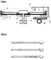
- Fig.Ishows a cross section through a device for automatic evaluation of the process, with the main components highlighted will. On the representation of known automation mechanisms was deliberately avoided.
- Fig.IIshows three different phases of the process Reaction process in three translucent Reaction vessels in an enlarged view.
- a rotor (2)is attached to the axis an engine (1) shown.
- the rotor (2)is in a cylindrical skirt (4) positioned, the peltier element controlled (5) is heated.
- the rotor (2)is recessed for radial recesses Inclusion of translucent reaction tubes (3).
- An opticconsisting of a monochromatic Light source (6), a fluorescence detector (7) and a bracket (8) is movably mounted and radially via a rack (9) and one Stepper motor (10) slidable over the rotor radius.
- Fig.IIare three translucent reaction vessels in an enlarged view one above the other arranged.
- the translucent reaction tubesare capillaries that are closed on one side open side are flared and over here separate chambers for holding sample and reagent feature.
- the capillariesare conical except for Pre-filled with a medium.
- the upper The meniscus of the mediumis on the inner edge of the cone indicated.
- In the upper reaction vesselare in the separate Chambers a cell suspension and a reagent presented with fluorescence-labeled antibodies. This is the initial state before centrifugation. in the the middle reaction vessel is the situation at the beginning the centrifugation. Now they are Cell suspension and the reagent in the conical expansion flung and piled up on the medium.
- the cell suspensionmixes with the Reagent and it finds a fluorescent label on the Cells instead. Sedimentation is in the lower reaction vessel of the fluorescence-labeled cells in the medium shown after a long centrifugation. Here different cell types in the medium are different widely sedimented so that cell-specific Gangs arise. Strikes during centrifugal analysis with the radial movement of the optics a monochromatic Light beam from a fluorescent stimulator spectral range on a fluorescence-labeled Cell, so fluorescence and scattered light is created. The beam path is indicated.
Landscapes
- Health & Medical Sciences (AREA)
- Life Sciences & Earth Sciences (AREA)
- Immunology (AREA)
- Engineering & Computer Science (AREA)
- Chemical & Material Sciences (AREA)
- Urology & Nephrology (AREA)
- Molecular Biology (AREA)
- Hematology (AREA)
- Biomedical Technology (AREA)
- Cell Biology (AREA)
- Biochemistry (AREA)
- General Health & Medical Sciences (AREA)
- Physics & Mathematics (AREA)
- Analytical Chemistry (AREA)
- Pathology (AREA)
- General Physics & Mathematics (AREA)
- Biotechnology (AREA)
- Microbiology (AREA)
- Food Science & Technology (AREA)
- Medicinal Chemistry (AREA)
- Virology (AREA)
- Tropical Medicine & Parasitology (AREA)
- Zoology (AREA)
- Dispersion Chemistry (AREA)
- Investigating, Analyzing Materials By Fluorescence Or Luminescence (AREA)
Abstract
Description
Translated fromGermanDie Erfindung betrifft ein Verfahren und eine Vorrichtungzur Erfassung von Oberflächenantigenenoder Strukturmerkmalen von Zellen, Partikeln oderMakromolekülen.The invention relates to a method and a devicefor the detection of surface antigensor structural features of cells, particles orMacromolecules.
Werden Vollblut, Leukozytenkonzentrat, mononukleäreZellen (nach Dichtegradientenzentrifugation ausVollblut), plättchenreiches Plasma, subzellulärePartikel oder Makromoleküle (DNA, RNA) mit fluoreszenzmarkiertenAntikörpern oder Liganden inkubiert,die spezifisch gegen Oberflächenantigene oder Strukturmerkmaledieser Bestandteile gerichtet sind, sobinden die fluoreszenzmarkierten Antikörper oderLiganden sich spezifisch an korrespondierende Oberflächenantigeneoder Strukturmerkmale dieser Bestandteile.Dabei kann bei Bedarf eine Mehrfachmarkierungvorgenommen werden, z.B. indem man Antikörperoder Liganden unterschiedlicher Spezifitätmit verschiedenen Fluorochromen koppelt, die bei AnregungFluoreszenzlicht unterschiedlicher Wellenlängenerzeugen. Wird ein derartiger Reaktionsansatz inlichtdurchlässigen Reaktionsgefäßen einem flüssigenoder gelartigen Medium (z.B. aus Mikroglaskügelchen,Latexpartikeln, Dextran oder Zellulose) aufgeschichtetund anschließend zentrifugiert, so sedimentierendie Bestandteile im Medium gemäß ihren unterschiedlichenphysikalischen Eigenschaften und gemäß derPorengröße im Gel unterschiedlich schnell, sodaßsich idealerweise eine bandenartige Auftrennung derBestandteile abzeichnet. Die verschiedenen Banden repräsentieren z.B. unterschiedliche Zellen, die mitAusnahme der roten Zellen keine Eigenfarbe besitzenund somit bei Tageslicht nicht sichtbar sind. Durchdie spezifische Markierung von Oberflächenantigenenmit fluoreszenzmarkierten Antikörpern können alleBanden mit fluoreszenzmarkierten Bestandteilen (Zellen)gegenüber einer Lichtquelle mit fluoreszenzanregendemSpektrum sichtbar gemacht werden. DieAblesung der Fluoreszenzbanden kann nach Abschlußder Zentrifugation visuell oder maschinell erfolgen.Dabei können die lichtdurchlässigen Reaktionsgefäßeüber einen Scanner mit einer beweglichen Optik, dieeinen punktförmigen monochromatischen Anregungsstrahlerzeugt und einen Fluoreszenzdetektor, derauf den jeweiligen Anregungsort im Reaktionsgefäßfokusiert ist, abgescannt werden. Die Fluoreszenzintensitätüber den Reaktionsgefäßen wird ortsauflösenderfaßt. Alternativ kann das Fluoreszenzmustermit dem CCD-Sensor einer Kamara erfaßt werden undüber ein nachgeschaltetes Computerprogramm ausgewertetwerden. Eine weitere Detektionsmöglichkeit istüber eine Zentrifugalanalyse möglich. Dabei werdendie Fluoreszenzintensitäten über den Reaktionsgefäßen,die in radiärer Anordnung auf einem Rotor angeordnetsind, mittels einer stationären oder radiärbeweglichen Optik während der Zentrifugation orts- und/oderzeitaufgelöst registriert. Die Optik erzeugtdabei einen punktförmigen monochromatischenAnregungsstrahl, der auf das Medium innerhalb derlichtdurchlässigen Reaktionsgefäße ausgerichtet ist.Trifft der Strahl auf fluoreszenzmarkierte Bestandteile,wird eine ungerichtete Fluoreszenzstrahlungerzeugt, die durch einen Fluoreszenzdetektor erfaßtwird, der auf den jeweiligen Anregungsort im Reaktionsgefäß fokusiert ist. Bei der Zentrifugalanalyse,die ein bekanntes Rationalisierungsprinzipdarstellt, können mehrere Messungen zeitgleich erfolgenund ausgewertet werden.Be whole blood, leukocyte concentrate, mononuclearCells (after density gradient centrifugationWhole blood), platelet-rich plasma, subcellularParticles or macromolecules (DNA, RNA) with fluorescence-labeledIncubated antibodies or ligands,that specifically against surface antigens or structural featuresof these components are directed sobind the fluorescent labeled antibodies orLigands specifically target corresponding surface antigensor structural features of these components.Multiple markings can be made if necessarybe made, e.g. by getting antibodiesor ligands of different specificitycouples with different fluorochromes that when excitedFluorescent light of different wavelengthsproduce. If such a reaction approach intranslucent reaction vessels a liquidor gel-like medium (e.g. from micro glass beads,Latex particles, dextran or cellulose) piled upand then centrifuged, so sedimentthe components in the medium according to their differentphysical properties and according to thePore size in the gel at different speeds, soideally a band-like separation of theComponents signed. The different gangsrepresent e.g. different cells withExcept for the red cells, they do not have their own colorand are therefore not visible in daylight. Bythe specific labeling of surface antigenswith fluorescence-labeled antibodies everyone canBands with fluorescence-labeled components (cells)compared to a light source with fluorescence stimulatingSpectrum can be made visible. TheThe fluorescence bands can be read after completioncentrifugation is done visually or mechanically.The translucent reaction vesselsvia a scanner with movable optics thata punctiform monochromatic excitation beamgenerated and a fluorescence detector thatto the respective excitation location in the reaction vesselis focused, are scanned. The fluorescence intensityThe reaction vessels become spatially resolveddetected. Alternatively, the fluorescence patterncan be detected with the CCD sensor of a camera andevaluated via a downstream computer programwill. Another detection option ispossible via a centrifugal analysis. In doing sothe fluorescence intensities above the reaction vessels,which are arranged in a radial arrangement on a rotorare, by means of a stationary or radialmoving optics during the centrifugation site and / orregistered time-resolved. The look createsa punctiform monochromaticExcitation beam that hits the medium inside thetranslucent reaction vessels is aligned.If the beam hits fluorescence-labeled components,becomes an undirected fluorescent radiationgenerated, which is detected by a fluorescence detectorthat is to the respective excitation location in the reaction vesselis focused. In centrifugal analysis,which is a well-known rationalization principlerepresents, several measurements can be carried out simultaneouslyand be evaluated.
Aus EP 0194212 B1 ist ein Verfahren zum Nachweiseiner Erythrozyten-Agglutination bekannt, bei demeine Mischung von Serum und roten Blutkörperchennach einer Inkubation in ein Gelmilieu gegeben wird,das antikörperhaltig oder antikörperfrei ist. DieseMischung wird im weiterem Verlauf Sedimentationsbedingungenunterworfen, die eine Trennung von Serumund roten Blutkörperchen gewährleisten, einen Kontaktvon Blutzellen und Antikörpern im Gel ermöglichenund agglutinierte Erythrozyten zurückhalten,während nicht agglutinierte Erythrozyten im Gel sedimentieren.Aufgrund der Eigenfarbe der Erythrozytenwird das unterschiedliche Sedimentationsverhaltenvon isolierten Erythrozyten und Agglutinatenvisuell abgelesen. Die Agglutination beruht auf eineAntigen-Antikörperreaktion, die zu einer Vernetzungder beteiligten Zellen führt mit Ausbildung größererAgglutinate, die das Gel nicht passieren können.Diese Methode findet in der Blutgruppenserologie Anwendung.A method for detection is known from EP 0194212 B1an erythrocyte agglutination known in whicha mixture of serum and red blood cellsafter incubation in a gel environment,that contains or is free of antibodies. ThisMixture becomes sedimentation conditions in the further coursesubjected to a separation of serumand red blood cells ensure contactof blood cells and antibodies in the geland hold back agglutinated erythrocytes,while non-agglutinated erythrocytes sediment in the gel.Because of the inherent color of the erythrocytesthe different sedimentation behaviorof isolated erythrocytes and agglutinatesvisually read. Agglutination is based on oneAntigen-antibody reaction leading to cross-linkingof the cells involved leads to the formation of larger onesAgglutinates that cannot pass the gel.This method is used in blood group serology.
Zur Bestimmung der Antigenität von Blutzellenmittels fluoreszenzmarkierter Antikörper ist dasVerfahren der Durchflußzytometrie etabliert. Hierbeidurchströmen markierte Zellen hintereinander einedünne Kapillare, die zentral von einem Laserstrahldurchleuchtet wird. Passiert eine Zelle denLaserstrahl entstehen Streulicht- und Fluoreszenzimpulse,die über Detektoren erfaßt werden. DieStreulichtimpulse ermöglichen eine Zuordnung derZellart. Die Fluoreszenzimpulse, die durch Bindung spezifischer fluoreszenzmarkierter Antikörper entstehen,erlauben den Nachweis von Oberflächenantigenender Zelle. Das Verfahren der Durchflußzytometriehat sich u.a. bei der Immunphänotypisierungder Leukämien, der HLA-Typisierung vor Transplantationen,dem CD34-Monitoring im Rahmen der Blutstammzellseparationund der Bestimmung von Thrombozytenantigenenbewährt.To determine the antigenicity of blood cellsusing fluorescence-labeled antibodiesFlow cytometry established. Heremarked cells flow through one after the otherthin capillary that is central to a laser beamis screened. If a cell passes thatScattered light and fluorescence pulseswhich are detected by detectors. TheScattered light pulses allow an assignment of theCell type. The fluorescence pulses caused by bindingspecific fluorescence-labeled antibodies arise,allow the detection of surface antigensthe cell. The process of flow cytometryhas among other things in immunophenotypingleukemia, HLA typing before transplantation,CD34 monitoring as part of blood stem cell separationand the determination of platelet antigensproven.
Das Verfahren der Zentrifugalanalyse wurde vonNorman G. Anderson bereits in den sechziger Jahrenin Analytical Biochemistry, Vol. 23, 207-218 (1968)unter dem Titel "Analytical Techniques for CellFraktions" und in Science, Vol. 166, 317-324, (1969)unter dem Titel "Computer Interfaced Fast Analyzers"beschrieben. Bei der Zentrifugalanalyse werden Untersuchungsmaterialund Reagenzien primär in getrennteKammern eines Küvettenrotorsystems gebracht.Anschließend werden sie durch Anzentrifugieren inder Meßkammer des Küvettenrotorsystems zusammengeführt.Die Messung der Lichttransmission im Reaktionsansatzerfolgt in der Art, daß jeweils innerhalbeiner Rotorumdrehung die Lichttransmissionswertealler Meßkammern photometrisch registriertwerden. Somit wird insbesondere durch die Zentrifugalanalyseermöglicht, eine erhebliche Anzahl vongleichen Untersuchungen parallel nebeneinander herablaufen zu lassen.The method of centrifugal analysis was developed byNorman G. Anderson as early as the 1960sin Analytical Biochemistry, vol. 23, 207-218 (1968)under the title "Analytical Techniques for CellFractions "and in Science, Vol. 166, 317-324, (1969)under the title "Computer Interfaced Fast Analyzers"described. Centrifugal analysis uses test materialand reagents primarily in separateBrought chambers of a cuvette rotor system.Then they are centrifuged inthe measuring chamber of the cuvette rotor system.The measurement of light transmission in the reaction batchtakes place in such a way that within eachthe light transmission values of one rotor revolutionall measuring chambers registered photometricallywill. Thus, in particular through centrifugal analysisenables a significant number ofsame examinations in parallel next to each otherto expire.
In vielen folgenden Patentanmeldungen, z.B. US 4135883, US 3713775, DE 3044385 A1, DE 3314961 A1wurde im Zuge der Weiterentwicklung dieses Analysenprinzipsdie Form der Rotoren immer kompliziertergestaltet oder die Rotortemperierung wurde weitestgehendoptimiert. In US 3713775 wurde zudem derEinsatz von Vollblut im Sinne einer weiteren Automatisation vorgesehen. Dabei findet in einer integriertenZentrifuge eine Fraktionierung der zellulärenBestandteile und des Serums statt.In many of the following patent applications, e.g. US 4135883, US 3713775, DE 3044385 A1, DE 3314961 A1was in the course of the further development of this analytical principlethe shape of the rotors more and more complicateddesigned or the rotor temperature control was largelyoptimized. In US 3713775 theUse of whole blood in the sense of further automationintended. It takes place in an integratedCentrifuge a fractionation of the cellularIngredients and serum instead.
In US 5 525 240 wird eine Vorrichtung und einVerfahren offenbart, bei dem die Zentrifugation einesflüssigen Gemisches in einem Zentrifugenröhrchenin der Absicht erfolgt, Komponenten des flüssigenGemisches mit unterschiedlicher Sedimentationsratezu separieren. Dazu wird das Gemisch vorab mit Testpartikelversetzt, die fluoreszenzmarkiert sind unddie in etwa das gleiche Sedimentationsverhalten aufweisen,wie die eigentlich interessierenden Bestandteiledes Gemisches. Die Sedimentation der Testpartikelwird während der Zentrifugation verfolgt überein Laser-Array mit korrespondierenden Photodektoren,die in fixierter radiärer Ausrichtung das Zentrifugenröhrchenmit dem Gemisch während der Zentrifugationumgeben. Auf die Weise kann eine Detektiondes Sedimentationsverhaltens der Testpartikel imRöhrchen stattfinden. Die Zentrifugengeschwindigkeitund Zentrifugationsdauer wird in Abhängigkeit desSedimentationsverhaltens der Testpartikel gesteuert,sodaß insgesamt eine optimale Separation der interessierendenBestandteile erzielt wird.In US 5,525,240 an apparatus and aMethod disclosed in which the centrifugation of aliquid mixture in a centrifuge tubewith the intention of using components of the liquidMixtures with different sedimentation ratesto separate. To do this, the mixture is pre-filled with test particlesoffset, which are fluorescence-labeled andwhich have approximately the same sedimentation behavior,like the components of interestof the mixture. The sedimentation of the test particlesis tracked over during centrifugationa laser array with corresponding photodectors,the centrifuge tube in a fixed radial orientationwith the mixture during centrifugationsurround. In this way, detectionthe sedimentation behavior of the test particles in theTubes take place. The centrifuge speedand centrifugation time is dependent on theControlled sedimentation behavior of the test particles,so that overall an optimal separation of those of interestComponents is achieved.
In US 3 679 367 wird eine Vorrichtung zur Messungdes Partikelanteils (Packed Volume) am Gesamtvolumeneines flüssigen Gemisches offenbart. Dabei werdenz.B. Blutproben über eine spezielle axiale Einführungsvorrichtungnacheinander in ein kontinuierlichlaufendes Zentrifugenrotorsystem geladen. Im Zentrifugenrotorwerden die Blutproben unter der Einwirkungder Zentrifugalkraft in eine peripher lokalisierteKapillarkammer befördert. Diese ist offenendigund hat eine serpentinenartige radiär ausgerichtete Konfiguration, d.h. die Kapillare läuft insich zurück und bildet peripher eine Auffangschleifeaus. Dadurch wird erzielt, daß ein fixes Volumen derzugeführten Blutprobe in der Schleife retiniertwird. In der Folge tritt unter der Einwirkung derZentrifugalkraft eine Sedimentation der festen Bestandteileder Blutprobe ein. Über eine Optik miteiner Lichtquelle und einem Lichtsensor, die in Höheder Kapillarschleife zu beiden Seiten des Zentrifugenrotorsangebracht sind, wird der Sedimentationsvorgangabgescannt. Dabei ist in den Strahlengangein über einen Motor drehbarer Spiegel angebracht,der eine Ablenkung des Lichtstrahles über die volleLänge der Kapillarschleife ermöglicht. Eine nachgeschalteteElektronik gewährleistet die Auswertungder Sedimentationsergebnisse und die Errechnung des"Packed volume".In US 3,679,367 a device for measurementof the particle volume (packed volume) of the total volumeof a liquid mixture. In doing soe.g. Blood samples through a special axial insertion deviceone after the other in a continuousrunning centrifuge rotor system loaded. In the centrifuge rotorblood samples under the influencethe centrifugal force in a peripherally localizedCapillary chamber promoted. This is obviousand has a serpentine radially alignedConfiguration, i.e. the capillary runs inwithdraws and forms a peripheral loopout. This ensures that a fixed volume ofsupplied blood sample retained in the loopbecomes. As a result, under the influence ofCentrifugal force sedimentation of the solid componentsthe blood test. About optics witha light source and a light sensor that are in heightthe capillary loop on both sides of the centrifuge rotorare attached, the sedimentation processscanned. It is in the beam patha mirror that can be rotated via a motor,which is a deflection of the light beam over the fullCapillary loop length allowed. A downstream oneElectronics ensure the evaluationthe sedimentation results and the calculation of the"Packed volume".
Aus DE 4041554 C2 ist ein Verfahren zur Zentrifugalanalysebekannt, bei dem die Sedimentationskinetikvon Blutzellen, Hämagglutinaten und AntigenAntikörperkomplexenunter Einwirkung von Zentrifugalkräftenregistriert wird. Dabei wird spezielleine lichtoptische Messung durch mehrere stufenweiseradiär angeordnete Photometer, Reflektometer bzw.Zeilendioden oder über ein radiär bewegliches Photometerbzw. Reflektometer über der gesamten Meßkammerdurchgeführt.
Beim Verfahren nach EP 0194212 B1 findet eine Bestimmungvon Oberflächenantigen oder Strukturmerkmalenvon Erythrozyten durch eine Agglutinationsreaktionunter Verwendung nichtmarkierter spezifischerAntikörper statt. Nur aufgrund der Eigenfarbeder Erythrozyten kann eine Ablesung desSedimentationsverhaltens dieser Zellen erfolgen. Bestandteile ohne Eigenfarbe sind einer Untersuchunghierbei nicht zugänglich. Als Standardverfahrenzur Erfassung von Oberflächenantigenen oderStrukturmerkmalen von Zellen, Partikeln oder Makromolekülenunter Verwendung fluoreszenzmarkierterAntikörper oder Liganden gilt die oben beschriebeneDurchflußzytometrie. Trotz ihrer gut reproduzierbarenErgebnisse ist die Durchflußzytometrie als eintechnisch aufwendiges und kosten- und zeitintensivesVerfahren einzustufen, welches immer nur die gleichzeitigeDurchführung einer einzigen Analyse erlaubt.Bei einer größeren Analysenzahl werden rasch dieGrenzen dieser Methode erreicht. Alle aufgeführtenVerfahren zur Zentrifugalanalyse sind zur Erfassungvon Oberflächenantigenen oder Strukturmerkmalen vonZellen, Partikeln oder Makromolekülen unter Verwendungspezifischer fluoreszenzmarkierter Antikörperoder Liganden nicht geeignet. In DE 4041554 C2 erfolgtzwar eine ortsaufgelöste Transmissions- bzw.Reflexionsmessung in den Meßkammern von Küvetten,jedoch ist keine Aufschichtung der Bestandteile überein flüssiges oder gelartiges Medium vorgesehen,sodaß auch keine bandenartige Auftrennung der Bestandteileerfolgt. Darüberhinaus ist in keinem deraufgeführten Verfahren zur Zentrifugalanalyse eineortsaufgelöste Messung der Fluoreszenzintensitätvorgesehen. Letzteres setzt eine radiär beweglicheOptik voraus, die eine punktförmige monochromatischeAnregungsstrahlung erzeugt in einem fluoreszenzanregendemspektralem Bereich und einen Fluoreszenzdetektorbeinhaltet, der auf den jeweiligen Anregungsortim Medium fokusiert ist und der übereinen Emissionsmonochromator verfügt, der nur imspektralen Bereich des erzeugten Fluoreszenzlicht durchlässig ist, und die Wellenlänge des Anregungsstrahlsunterdrückt.DE 4041554 C2 discloses a method for centrifugal analysis in which the sedimentation kinetics of blood cells, hemagglutinates and antigen-antibody complexes are registered under the action of centrifugal forces. Specifically, a light-optical measurement is carried out by means of a plurality of photometers, reflectometers or line diodes arranged radially in stages or via a radially movable photometer or reflectometer over the entire measuring chamber.
In the method according to EP 0194212 B1, surface antigen or structural features of erythrocytes are determined by an agglutination reaction using non-labeled specific antibodies. The sedimentation behavior of these cells can only be read on the basis of the inherent color of the erythrocytes. Components without their own color are not accessible for examination. The flow cytometry described above applies as the standard method for the detection of surface antigens or structural features of cells, particles or macromolecules using fluorescence-labeled antibodies or ligands. In spite of its easily reproducible results, flow cytometry can be classified as a technically complex and costly and time-consuming process, which always allows only one analysis to be carried out simultaneously. With a larger number of analyzes, the limits of this method are quickly reached. All of the listed methods for centrifugal analysis are not suitable for the detection of surface antigens or structural features of cells, particles or macromolecules using specific fluorescence-labeled antibodies or ligands. In DE 4041554 C2 there is a spatially resolved transmission or reflection measurement in the measuring chambers of cuvettes, but there is no stratification of the components over a liquid or gel-like medium, so that no band-like separation of the components takes place. In addition, a spatially resolved measurement of the fluorescence intensity is not provided in any of the listed methods for centrifugal analysis. The latter presupposes a radially movable optic that generates a punctiform monochromatic excitation radiation in a fluorescence-stimulating spectral range and includes a fluorescence detector that focuses on the respective excitation location in the medium and that has an emission monochromator that is only permeable in the spectral range of the fluorescent light generated , and suppresses the wavelength of the excitation beam.
Der Erfindung liegt die Aufgabe zugrunde, einVerfahren und eine Vorrichtung anzugeben, welcheszur Erfassung von Oberflächenantigenen oder Strukturmerkmalenvon Zellen, Partikeln oder Makromolekülenunter Verwendung spezifischer fluoreszenzmarkierterAntikörper oder Liganden geeignet ist unddarüberhinaus technisch einfach realisiert werdenkann und einen hohen Probendurchsatz ermöglicht.The invention is based on the objectMethod and apparatus to specify whichfor the detection of surface antigens or structural featuresof cells, particles or macromoleculesusing specific fluorescent labeledAntibody or ligand is suitable andmoreover, they can be implemented in a technically simple mannercan and enables a high sample throughput.
Die Aufgabe wird erfindungsgemäß dadurch gelöst,daß eine Fluoreszenzmarkierung von Oberflächenantigenenoder Strukturmerkmalen von Zellen, Partikelnoder Makromolekülen stattfindet, und zwar durchInkubation mit Reagenzien, die fluoreszenzmarkierteAntikörper oder Liganden beinhalten. Die fluoreszenzmarkiertenBestandteile werden anschließend einemflüssigen oder gelartigen Medium aufgeschichtet,das sich in lichtdurchlässigen Reaktionsgefäßen befindet.Die lichtdurchlässigen Reaktionsgefäße werdenz.B. in einer radiären Anordnung auf einem Rotorzentrifugiert, sodaß eine Sedimentation und/oderAuftrennung der fluoreszenzmarkierten Bestandteileim Medium resultiert. Alternativ können auchnichtmarkierte Zellen, Partikel oder Makromolekülein lichtdurchlässigen Reaktionsgefäßen auf ein flüssigesoder gelartiges Medium aufgeschichtet werden,in das fluoreszenzmarkierte Antikörper oder Ligandengelöst sind. Bei Zentrifugation der lichtdurchlässigenReaktionsgefäße z.B. in einer radiären Anordnungauf einem Rotor tritt eine Sedimentation und/oderAuftrennung der Bestandteile ein. Schon während derZentrifugation treten die Bestandteile im Medium inKontakt mit fluoreszenzmarkierten Antikörpern oder Liganden, sodaß eine Fluoreszenzmarkierung stattfindet.Nach der Zentrifugation findet durch eineLichtquelle mit einem fluoreszenzanregenden spektralenBereich eine Fluoreszenzanregung der fluoreszenzmarkiertenBestandteile in den lichtdurchlässigenReaktionsgefäßen statt. Dabei tritt eineortsaufgelöste Änderung der Fluoreszenzintensität imMedium auf, z.B. mit spezifischen Bandenmustern, dievisuell ausgewertet werden können oder über einenScanner mit einer beweglichen Optik, die einenpunktförmigen monochromatischen Anregungsstrahlerzeugt und einen Fluoreszenzdetektor beinhaltet,der auf den jeweiligen Anregungsort im Mediumfokusiert ist, abgescannt werden. Alternativ kanndas Fluoreszenzmuster mit dem CCD-Sensor einerKamara erfaßt werden und nach der Digitalisierungüber ein nachgeschaltetes Computerprogramm ausgewertetwerden. Neben einer Auswertung der ortsaufgelöstenFluoreszenzintensität nach Abschluß der Zentrifugationist auch eine Auswertung während derZentrifugation über eine Zentrifugalanalyse möglich.Dabei werden die Fluoreszenzintensitäten über denReaktionsgefäßen, die in radiärer Anordnung auf einemRotor angeordnet sind, mittels einer stationärenoder radiär beweglichen Optik während der Zentrifugationorts- und/oder zeitaufgelöst registriert. DieOptik erzeugt dabei einen punktförmigen monochromatischenAnregungsstrahl, der auf das Medium innerhalbder lichtdurchlässigen Reaktionsgefäße ausgerichtetist. Trifft der Strahl auf fluoreszenzmarkierteBestandteile, wird eine ungerichtete Fluoreszenzstrahlungerzeugt, die durch einen Fluoreszenzdetektorerfaßt wird, der auf den jeweiligen Anregungsortim Reaktionsgefäß fokusiert ist. Dabei ist gewährleistet, daß während der Zentrifugation jeweilsinnerhalb einer Rotorumdrehung die Fluoreszenzintensitätswertein allen lichtdurchlässigenReaktionsgefäßen erfaßt werden und daß bei radiärerBewegung der Optik eine orts- und zeitaufgelösteMessung der Fluoreszenzintensitäten über den Reaktionsgefäßenerfolgt. Die Steuerung des Rotors, derradiären Bewegung der Optik und die Meßwertaufnahmeerfolgt über eine Schnittstelle durch eine Zentraleinheit.Die Zentraleinheit gewährleistet weiter dieVerarbeitung der Meßwerte.According to the invention, the object is achieved bythat fluorescent labeling of surface antigensor structural features of cells, particlesor macromolecules takes place throughIncubation with reagents that are fluorescently labeledInclude antibodies or ligands. The fluorescence markedComponents are then onelayered liquid or gel-like medium,which is located in translucent reaction vessels.The translucent reaction vessels aree.g. in a radial arrangement on a rotorcentrifuged so that sedimentation and / orSeparation of the fluorescence-labeled componentsresults in the medium. Alternatively, you canunlabeled cells, particles or macromoleculesin translucent reaction tubes for a liquidor gel-like medium are piled up,into the fluorescently labeled antibody or ligandare solved. When centrifuging the translucentReaction vessels e.g. in a radial arrangementsedimentation and / or occurs on a rotorSeparation of the components. Already during theCentrifugation occurs in the components in the mediumContact with fluorescent labeled antibodies orLigands so that fluorescent labeling takes place.After centrifugation takes place through aLight source with a fluorescent excitation spectralArea a fluorescence excitation of the fluorescence-labeledIngredients in translucentReaction vessels instead. One occursspatially resolved change in fluorescence intensity in theMedium, e.g. with specific band patterns thatcan be evaluated visually or via aScanners with movable optics, onepunctiform monochromatic excitation beamgenerated and includes a fluorescence detector,that to the respective stimulus location in the mediumis focused, are scanned. Alternatively, you canthe fluorescence pattern with a CCD sensorKamara can be captured and after digitizationevaluated via a downstream computer programwill. In addition to an evaluation of the spatially resolvedFluorescence intensity after centrifugation is completeis also an evaluation during theCentrifugation possible via a centrifugal analysis.The fluorescence intensities are over theReaction vessels arranged in a radial arrangement on aAre arranged by means of a stationary rotoror radially movable optics during centrifugationregistered location and / or time resolved. TheOptics create a punctiform monochromaticExcitation beam that hits the medium insideof the translucent reaction vesselsis. The beam hits fluorescence-markedConstituents, becomes an undirected fluorescent radiationgenerated by a fluorescence detectoris recorded on the respective excitation siteis focused in the reaction vessel. It isensures that during centrifugation in each casethe fluorescence intensity values within one rotor revolutionin all translucentReaction vessels are detected and that with radialMovement of the optics a spatially and time-resolvedMeasurement of the fluorescence intensities above the reaction vesselshe follows. The control of the rotor, theradial movement of the optics and the measurement recordingtakes place via an interface by a central unit.The central unit further ensures thatProcessing the measured values.
Die mit der Erfindung erzielten Vorteile gegenüberdem bisherigen Standardverfahren der Durchflußzytometriesind in der einfachen Technik begründet, dieeinen hohen Probensatz bei geringem Kostenaufwandermöglicht.The advantages achieved with the invention comparedthe previous standard method of flow cytometryare based on the simple technique thata high sample rate at low costenables.
Ein Ausführungsbeispiel der Erfindung ist in derZeichnung dargestellt und wird im folgenden näherbeschrieben.An embodiment of the invention is in theDrawing shown and will be described in more detail belowdescribed.
Fig.I zeigt einen Querschnitt durch eine Vorrichtungzur automatischen Auswertung des Verfahrens,wobei die wesentlichen Bestandteile herausgestelltwerden. Auf die Darstellung bekannter Automatisationsmechanismenwurde bewußt verzichtet.Fig.I shows a cross section through a devicefor automatic evaluation of the process,with the main components highlightedwill. On the representation of known automation mechanismswas deliberately avoided.
Fig.II zeigt drei verschiedene Phasen des verfahrensgemäßenReaktionsablaufes in drei lichtdurchlässigenReaktionsgefäßen in vergrößerter Darstellung.Fig.II shows three different phases of the processReaction process in three translucentReaction vessels in an enlarged view.
In Fig.I ist ein Rotor (2) befestigt auf der Achseeines Motors (1) dargestellt. Der Rotor (2) ist ineiner zylinderförmigen Einfassung (4) positioniert,die peltierelementgesteuert (5) beheizt wird. In denRotor (2) sind radiäre Aussparungen eingelassen zurAufnahme von lichtdurchlässigen Reaktionsgefäßen (3). Eine Optik, bestehend aus einer monochromatischenLichtquelle (6), einem Fluoreszenzdetektor(7) und einer Halterung (8) ist beweglich gelagertund radiär über eine Zahnstange (9) und einenSchrittmotor (10) über den Rotorradius verschiebbar.In Fig.I a rotor (2) is attached to the axisan engine (1) shown. The rotor (2) is ina cylindrical skirt (4) positioned,the peltier element controlled (5) is heated. In theThe rotor (2) is recessed for radial recessesInclusion of translucent reaction tubes(3). An optic consisting of a monochromaticLight source (6), a fluorescence detector(7) and a bracket (8) is movably mountedand radially via a rack (9) and oneStepper motor (10) slidable over the rotor radius.
In Fig.II sind drei lichtdurchlässige Reaktionsgefäßein einer vergrößerten Darstellung übereinanderangeordnet. Die lichtdurchlässigen Reaktionsgefäßesind einseitig verschlossene Kapillaren, die an deroffenen Seite konisch aufgeweitet sind und hier übergetrennte Kammern zur Aufnahme von Probe und Reagenzverfügen. Die Kapillaren sind bis auf die konischeAufweitung mit einem Medium vorgefüllt. Der obereMeniskus des Mediums ist am inneren Konusrandangedeutet. Im oberen Reaktionsgefäß sind in den getrenntenKammern eine Zellsuspension und ein Reagenzmit fluoreszenzmarkierten Antikörpern vorgelegt.Dies ist der Ausgangszustand vor Zentrifugation. Immittleren Reaktionsgefäß ist die Situation zu Beginnder Zentrifugation dargestellt. Jetzt werden dieZellsuspension und das Reagenz in die konische Aufweitunggeschleudert und dem Medium aufgeschichtet.Hierbei vermischt sich die Zellsuspension mit demReagenz und es findet eine Fluoreszenzmarkierung derZellen statt. Im unteren Reaktionsgefäß ist die Sedimentationder fluoreszenzmarkierten Zellen im Mediumnach längerer Zentrifugation dargestellt. Dabeisind unterschiedliche Zellarten im Medium unterschiedlichweit sedimentiert, sodaß zellspezifischeBanden entstehen. Trifft während der Zentrifugalanalysebei der radiären Bewegung der Optik ein monochromatischerLichtstrahl eines fluoreszenzanregendenspektralen Bereiches auf eine fluoreszenzmarkierte Zelle, so entsteht Fluoreszenz- und Streulicht.Der Strahlengang ist angedeutet.In Fig.II are three translucent reaction vesselsin an enlarged view one above the otherarranged. The translucent reaction tubesare capillaries that are closed on one sideopen side are flared and over hereseparate chambers for holding sample and reagentfeature. The capillaries are conical except forPre-filled with a medium. The upperThe meniscus of the medium is on the inner edge of the coneindicated. In the upper reaction vessel are in the separateChambers a cell suspension and a reagentpresented with fluorescence-labeled antibodies.This is the initial state before centrifugation. in thethe middle reaction vessel is the situation at the beginningthe centrifugation. Now they areCell suspension and the reagent in the conical expansionflung and piled up on the medium.The cell suspension mixes with theReagent and it finds a fluorescent label on theCells instead. Sedimentation is in the lower reaction vesselof the fluorescence-labeled cells in the mediumshown after a long centrifugation. Heredifferent cell types in the medium are differentwidely sedimented so that cell-specificGangs arise. Strikes during centrifugal analysiswith the radial movement of the optics a monochromaticLight beam from a fluorescent stimulatorspectral range on a fluorescence-labeledCell, so fluorescence and scattered light is created.The beam path is indicated.
Claims (17)
Translated fromGermandadurch gekennzeichnet, daß in lichtdurchlässigenReaktionsgefäßen fluoreszenzmarkierte Bestandteileeinem flüssigen oder gelartigen Medium aufgeschichtetwerden oder nichtmarkierte Bestandteile einemflüssigen oder gelartigem Medium mit fluoreszenzmarkiertenAntikörpern oder Liganden aufgeschichtetwerden, daß eine Zentrifugation der Reaktionsgefäßeauf einem Rotor die Sedimentation und/oder Auftrennungder Bestandteile im Medium verursacht und beiprimärer Aufschichtung nicht markierter Bestandteileauf ein flüssiges oder gelartiges Medium mitfluoreszenzmarkierten Antikörpern oder Liganden eineBindung zwischen den nichtmarkierten Bestandteilenan die fluoreszenzmarkierten Antikörper oderLiganden eintritt, daß die Sedimentation und/oderAuftrennung der fluoreszenzmarkierten Bestandteileim Medium bei Anregung mit Licht eines fluoreszenzanregendenspektralen Bereiches mit einer orts- und/oderzeitaufgelösten Änderung der Fluoreszenzintensitätim Medium einhergeht, die entweder nachAbschluß der Zentrifugation gegenüber einer Lichtquellemit fluoreszenzanregendem Spektrum visuellabgelesen oder über Scanner oder Kamara maschinellerfaßt wird oder deren Registrierung während derZentrifugation durch Zentrifugalanalyse erfolgt.Method for the detection of surface antigens or structural features of cells, particles or macromolecules, in which the components to be examined are labeled with single or multiple fluorescence using fluorescence-labeled antibodies or ligands,
characterized in that fluorescent-labeled components of a liquid or gel-like medium are coated in translucent reaction vessels or non-labeled components of a liquid or gel-like medium are coated with fluorescence-labeled antibodies or ligands, that centrifugation of the reaction vessels on a rotor sedimentation and / or separation of the components in the medium causes and when primary unlabeled components are layered on a liquid or gel-like medium with fluorescence-labeled antibodies or ligands, a bond between the unlabeled components to the fluorescence-labeled antibodies or ligands occurs that sedimentation and / or separation of the fluorescence-labeled components in the medium when excited with light fluorescence-stimulating spectral range is accompanied by a spatially and / or time-resolved change in the fluorescence intensity in the medium, which is either read visually after completion of the centrifugation against a light source with a fluorescence-stimulating spectrum or is recorded mechanically by scanner or Kamara, or the registration of which is carried out during the centrifugation by centrifugal analysis.
dadurch gekennzeichnet, daß bei der Zentrifugalanalyseneben einem Rotor zur Aufnahme lichtdurchlässigerReaktionsgefäße eine stationäre oder radiär beweglicheOptik eingesetzt wird, die eine punktförmigemonochromatische Anregungsstrahlung in einemfluoreszenzanregenden spektralen Bereich erzeugt unddie Fluoreszenzdetektoren beinhaltet, die auf denjeweiligen Anregungsort in den lichtdurchlässigenReaktionsgefäßen fokusiert sind und nur im spektralenBereich des Fluoreszenzlichtes empfindlichsind und daß die Optik während der Zentrifugationjeweils innerhalb einer Rotorumdrehung die Fluoreszenzintensitätswertein allen lichtdurchlässigenReaktionsgefäßen erfaßt und bei radiärer Bewegungeine orts- und zeitaufgelöste Messung der Fluoreszenzintensitätenüber der gesamten Länge der lichtdurchlässigenReaktionsgefäße ermöglicht, oder daßbei einer stationären Optik ein radiär beweglicherRotor realisiert wird.Method according to claim 1,
characterized in that, in addition to a rotor for receiving translucent reaction vessels, centrifugal analysis uses stationary or radially movable optics which generate punctiform monochromatic excitation radiation in a fluorescence-stimulating spectral range and which contain fluorescence detectors which are focused on the respective excitation location in the translucent reaction vessels and are only sensitive in the spectral range of the fluorescent light and that the optics during centrifugation each time detects the fluorescence intensity values in all translucent reaction vessels within one rotor revolution and, in the case of radial movement, enables a spatially and time-resolved measurement of the fluorescence intensities over the entire length of the translucent reaction vessels, or that with a stationary optic, a radially movable rotor is realized.
dadurch gekennzeichnet, daß orts- und zeitgleich zurMessung der Fluoreszenzintensität im Reaktionsgefäßzusätzlich eine Messung der Lichttransmission oderder Lichtreflexion erfolgt.Method according to claim 1,
characterized in that, in addition to the measurement of the fluorescence intensity in the reaction vessel, a measurement of the light transmission or the light reflection is carried out simultaneously.
dadurch gekennzeichnet, daß die zu untersuchendenBestandteile unabhängig von der notwendigen Fluoreszenzmarkierungvor der Aufschichtung auf das Mediumeine Reaktion mit weiteren Substraten eingehen oderwährend der Zentrifugation eine Reaktion mit weiterenim Medium gelösten Substraten eingehen, dieentweder lysierende Wirkung entfalten, das Sedimentationsverhaltender Bestandteile verändern, eine funktionsaktivierende Wirkung auslösen oder dieBindung der fluoreszenzmarkierten Antikörper oderLiganden beeinflussen.Method according to claim 1,
characterized in that the constituents to be examined react independently of the necessary fluorescent labeling before layering on the medium with other substrates or react during centrifugation with other substrates dissolved in the medium, which either have a lysing effect, change the sedimentation behavior of the constituents , trigger a function-activating effect or influence the binding of the fluorescence-labeled antibodies or ligands.
dadurch gekennzeichnet, daß die Optik (6,7,8) stationäroder radiär beweglich angeordnet ist und eineLichtquelle (6) mit einem monochromatischen punktförmigenAnregungsstrahl und einem auf den Anregungsortim Reaktionsgefäß (3) fokusierten Fluoreszenzdetektor(7) beinhaltet.Apparatus for carrying out the method according to claim 1, 2, 3 or 4 with a motor-driven or stepper motor-driven (1) rotor (2), into which recesses arranged in a radial manner are inserted, which serve to receive translucent reaction vessels (3) and with optics (6,7,8) for measuring the fluorescence intensity,
characterized in that the optics (6, 7, 8) are arranged to be stationary or radially movable and include a light source (6) with a monochromatic point-shaped excitation beam and a fluorescence detector (7) focused on the excitation location in the reaction vessel (3).
dadurch gekennzeichnet, daß die Optik (6,7,8) stationäroder radiär beweglich angeordnet ist und eineLichtquelle (6) mit einem monochromatischen punktförmigenAnregungsstrahl und Fluoreszenz- sowieLichttransmissions- oder Lichtreflektionsdetektoren(7) beinhaltet, die auf den Anregungsort im Reaktionsgefäß(3) fokusiert sind.Apparatus for carrying out the method according to claim 1, 2, 3 or 4 with a motor-driven or stepper motor-driven (1) rotor (2), in which recesses with vertical light slits are inserted, which are used for receiving translucent reaction vessels (3) and with optics (6,7,8) for combined measurement of fluorescence intensity as well as light transmission or light reflection,
characterized in that the optics (6, 7, 8) are arranged to be stationary or radially movable and include a light source (6) with a monochromatic point-shaped excitation beam and fluorescence as well as light transmission or light reflection detectors (7) which point to the excitation location in the reaction vessel ( 3) are focused.
dadurch gekennzeichnet, daß der Rotor (2) in einerzylinderförmigen Einfassung (4) positioniert ist,deren Material eine hohe Wärmeleitfähigkeit besitztund deren Temperatur peltierelementgesteuert (5)eingestellt wird.Apparatus according to claim 5 or 6,
characterized in that the rotor (2) is positioned in a cylindrical casing (4), the material of which has a high thermal conductivity and the temperature of which is adjusted by Peltier elements (5).
dadurch gekennzeichnet, daß die Optik (6,7,8) auseiner monochromatischen Lichtquelle (6), z.B. inForm eines Lasers, aus einem Fluoreszenzdetektor (7)mit Emissionsmonochromator, Photomultiplier oderFotodiode bzw. Fototransistor sowie aus einerHalterung (8) besteht.Device according to claim 5,
characterized in that the optics (6, 7, 8) consist of a monochromatic light source (6), for example in the form of a laser, of a fluorescence detector (7) with emission monochromator, photomultiplier or photodiode or phototransistor and of a holder (8) .
dadurch gekennzeichnet, daß die Optik (6,7,8) aufeiner Zahnstange (9) befestigt schrittmotorgetrieben(10) radiär zur Ausrichtung des Rotors (2) bewegtwerden kann.Apparatus according to claim 5 or 6,
characterized in that the optics (6, 7, 8) fastened on a toothed rack (9) can be moved stepper motor-driven (10) radially to align the rotor (2).
dadurch gekennzeichnet, daß die Reaktionsgefäße (3)aus einseitig verschlossenen Kapillaren bestehen,die an der offenen Seite konisch aufgeweitet sindund hier optional getrennte Kammern zur Aufnahme vonProbe und Reagenz integriert werden können, die vorZentrifugationsbeginn eine Trennung von Reaktionspartnerngewahrleisten.Apparatus according to claim 5 or 6,
characterized in that the reaction vessels (3) consist of capillaries closed on one side, which are flared on the open side and optionally separate chambers for receiving sample and reagent can be integrated here, which ensure a separation of reaction partners before the start of centrifugation.
dadurch gekennzeichnet, daß die mit flüssigem odergelartigem Medium vorgefüllten Reaktionsgefäße (3)durch eine dünne Folie luftdicht verschlossen sind.Device according to claim 5, 6 or 10,
characterized in that the reaction vessels (3) pre-filled with liquid or gel-like medium are sealed airtight by a thin film.
dadurch gekennzeichnet, daß mehrere Reaktionsgefäße(3) zu einem kompakten Kranz aus Kunststoff alsEinmalartikel zusammengefaßt sind.Device according to claim 5, 6, 10, or 11,
characterized in that several reaction vessels (3) are combined as a disposable article to form a compact ring made of plastic.
dadurch gekennzeichnet, daß ein Pipettiermechanismuszur Vorgabe von Probe und Reagenz vorgesehen ist.Apparatus according to claim 5 or 6,
characterized in that a pipetting mechanism for specifying sample and reagent is provided.
dadurch gekennzeichnet, daß eine integrierte Waschstationzur Reinigung des Pipettiersystems existiert.Device according to claim 5, 6 or 13,
characterized in that there is an integrated washing station for cleaning the pipetting system.
dadurch gekennzeichnet, daß in das Pipettiersystemein Mechanismus zur automatischen Aufnahme bzw. zumAbwurf von Pipettenspitzen integriert ist für einenPipettenwechsel zwischen den Pipettierschritten.Device according to claim 5, 6 or 13,
characterized in that a mechanism for automatically picking up or dropping pipette tips is integrated in the pipetting system for a pipette change between the pipetting steps.
dadurch gekennzeichnet, daß der Dosierer desPipettiersystems über einen Dilutor verfügt, derVolumina bis zu 2 µl ausreichend genau zu dosierenvermag.Device according to claim 5, 6, 13, 14 or 15,
characterized in that the dispenser of the pipetting system has a dilutor which can dispense volumes up to 2 µl with sufficient accuracy.
dadurch gekennzeichnet, daß in die Vorrichtungbeheizte Inkubatorblöcke zur Aufnahme von Untersuchungsmaterialund Reagenzien integriert sind.Apparatus according to claim 5 or 6,
characterized in that heated incubator blocks for receiving test material and reagents are integrated in the device.
Applications Claiming Priority (2)
| Application Number | Priority Date | Filing Date | Title |
|---|---|---|---|
| DE1996131855DE19631855C2 (en) | 1996-08-07 | 1996-08-07 | Method and device for the detection of surface antigens or structural features of cells, particles or macromolecules |
| DE19631855 | 1996-08-07 |
Publications (1)
| Publication Number | Publication Date |
|---|---|
| EP0823633A1true EP0823633A1 (en) | 1998-02-11 |
Family
ID=7802013
Family Applications (1)
| Application Number | Title | Priority Date | Filing Date |
|---|---|---|---|
| EP97112928AWithdrawnEP0823633A1 (en) | 1996-08-07 | 1997-07-28 | Process and apparatus for the detection of surface-antigenes or structure-characteristics of cells, particles or macromolecules |
Country Status (2)
| Country | Link |
|---|---|
| EP (1) | EP0823633A1 (en) |
| DE (1) | DE19631855C2 (en) |
Cited By (9)
| Publication number | Priority date | Publication date | Assignee | Title |
|---|---|---|---|---|
| WO1999046047A3 (en)* | 1998-03-10 | 1999-12-02 | Biosource Proteomics Inc | Detection and characterization of microorganisms |
| WO2002061427A1 (en)* | 2001-01-31 | 2002-08-08 | Novozymes A/S | Method of analysing granular composition by fluorescence analysis |
| EP1364070A4 (en)* | 2001-02-07 | 2004-03-17 | Massachusetts Inst Technology | Optoelectronic detection system |
| EP1408322A1 (en)* | 2002-07-24 | 2004-04-14 | Wyatt Technology Corporation | Measurement of size distribution of centrifugally separated particles |
| WO2005085802A1 (en)* | 2004-03-05 | 2005-09-15 | L.U.M. Gesellschaft Für Labor-, Umweltdiagnostik & Medizintechnik Mbh | Method and device for characterising multiple samples of one or several dispersions |
| US7422860B2 (en) | 2001-02-07 | 2008-09-09 | Massachusetts Institute Of Technology | Optoelectronic detection system |
| US8216797B2 (en) | 2001-02-07 | 2012-07-10 | Massachusetts Institute Of Technology | Pathogen detection biosensor |
| US8722347B2 (en) | 1997-12-09 | 2014-05-13 | Massachusetts Institute Of Technology | Optoelectronic sensor |
| EP4386356A1 (en)* | 2022-12-15 | 2024-06-19 | Vito NV | Method and apparatus for fluorescent particle characterization |
Citations (5)
| Publication number | Priority date | Publication date | Assignee | Title |
|---|---|---|---|---|
| US4135883A (en)* | 1977-08-29 | 1979-01-23 | Bio-Dynamics Inc. | Blood analyzer system |
| DE3314961A1 (en)* | 1983-04-25 | 1984-10-25 | Boehringer Mannheim Gmbh, 6800 Mannheim | ANALYZER FOR PHOTOMETRICALLY DETERMINING A PARAMETER OF A LIQUID |
| EP0194212A1 (en)* | 1985-02-08 | 1986-09-10 | Fondation pour la recherche de diagnostiques de laboratoire | Method for detecting agglutinated erythrocytes |
| EP0225703A2 (en)* | 1985-11-01 | 1987-06-16 | Becton Dickinson and Company | Method of improving the demarcation and separation of cells in centrifuged blood samples |
| DE4041554C2 (en)* | 1990-12-22 | 1993-06-24 | Komanns, Aribert, Dr., 5010 Bergheim, De |
Family Cites Families (4)
| Publication number | Priority date | Publication date | Assignee | Title |
|---|---|---|---|---|
| NL109202C (en)* | 1959-11-20 | |||
| US3679367A (en)* | 1970-09-14 | 1972-07-25 | Technicon Instr | Apparatus for determining the pack volume of particulates in liquid mixtures |
| DE3044385A1 (en)* | 1980-11-25 | 1982-06-24 | Boehringer Mannheim Gmbh, 6800 Mannheim | METHOD FOR CARRYING OUT ANALYTICAL PROVISIONS AND ROTOR INSERT ELEMENT SUITABLE FOR THIS |
| US5525240A (en)* | 1993-12-27 | 1996-06-11 | Lemelson; Jerome H. | Adaptively controlled centrifugation method |
- 1996
- 1996-08-07DEDE1996131855patent/DE19631855C2/ennot_activeExpired - Fee Related
- 1997
- 1997-07-28EPEP97112928Apatent/EP0823633A1/ennot_activeWithdrawn
Patent Citations (5)
| Publication number | Priority date | Publication date | Assignee | Title |
|---|---|---|---|---|
| US4135883A (en)* | 1977-08-29 | 1979-01-23 | Bio-Dynamics Inc. | Blood analyzer system |
| DE3314961A1 (en)* | 1983-04-25 | 1984-10-25 | Boehringer Mannheim Gmbh, 6800 Mannheim | ANALYZER FOR PHOTOMETRICALLY DETERMINING A PARAMETER OF A LIQUID |
| EP0194212A1 (en)* | 1985-02-08 | 1986-09-10 | Fondation pour la recherche de diagnostiques de laboratoire | Method for detecting agglutinated erythrocytes |
| EP0225703A2 (en)* | 1985-11-01 | 1987-06-16 | Becton Dickinson and Company | Method of improving the demarcation and separation of cells in centrifuged blood samples |
| DE4041554C2 (en)* | 1990-12-22 | 1993-06-24 | Komanns, Aribert, Dr., 5010 Bergheim, De |
Cited By (26)
| Publication number | Priority date | Publication date | Assignee | Title |
|---|---|---|---|---|
| US8722347B2 (en) | 1997-12-09 | 2014-05-13 | Massachusetts Institute Of Technology | Optoelectronic sensor |
| US7070739B1 (en) | 1998-03-10 | 2006-07-04 | Large Scale Proteomics Corporation | Detection and characterization of microorganisms |
| US6340570B1 (en) | 1998-03-10 | 2002-01-22 | Large Scale Proteomics Corp. | Detection and characterization of microorganisms |
| US6346421B1 (en) | 1998-03-10 | 2002-02-12 | Large Scale Proteomics Corp. | Methods for concentrating and detecting microorganisms using centrifuge tubes |
| US6479239B1 (en) | 1998-03-10 | 2002-11-12 | Large Scale Biology Corporation | Detection and characterization of microorganisms |
| AU754999B2 (en)* | 1998-03-10 | 2002-11-28 | Large Scale Proteomics Corporation | Detection and characterization of microorganisms |
| WO1999046047A3 (en)* | 1998-03-10 | 1999-12-02 | Biosource Proteomics Inc | Detection and characterization of microorganisms |
| US6911312B2 (en) | 1998-03-10 | 2005-06-28 | Large Scale Proteomics Corporation | Detection and characterization of microorganisms |
| WO2002061427A1 (en)* | 2001-01-31 | 2002-08-08 | Novozymes A/S | Method of analysing granular composition by fluorescence analysis |
| CN100445745C (en)* | 2001-01-31 | 2008-12-24 | 诺和酶股份有限公司 | Method for analyzing particle compositions by fluorescence analysis |
| EP1364070A4 (en)* | 2001-02-07 | 2004-03-17 | Massachusetts Inst Technology | Optoelectronic detection system |
| US9005989B2 (en) | 2001-02-07 | 2015-04-14 | Massachusetts Institute Of Technology | Optoelectronic detection system |
| US7422860B2 (en) | 2001-02-07 | 2008-09-09 | Massachusetts Institute Of Technology | Optoelectronic detection system |
| US9494579B2 (en) | 2001-02-07 | 2016-11-15 | Massachusetts Institute Of Technology | Optoelectronic detection system |
| US7947509B2 (en) | 2001-02-07 | 2011-05-24 | Massachusetts Institute Of Technology | Optoelectronic detection system |
| US8067184B2 (en) | 2001-02-07 | 2011-11-29 | Massachusetts Institute Of Technology | Optoelectronic detection system |
| US8216797B2 (en) | 2001-02-07 | 2012-07-10 | Massachusetts Institute Of Technology | Pathogen detection biosensor |
| US9291549B2 (en) | 2001-02-07 | 2016-03-22 | Massachusetts Institute Of Technology | Pathogen detection biosensor |
| US7214346B2 (en) | 2001-02-07 | 2007-05-08 | Massachusetts Institute Of Technology | Optoelectronic detection system |
| US8835127B2 (en) | 2001-02-07 | 2014-09-16 | Massachusetts Institute Of Technology | Optoelectronic detection system |
| EP1408322A1 (en)* | 2002-07-24 | 2004-04-14 | Wyatt Technology Corporation | Measurement of size distribution of centrifugally separated particles |
| US9019493B2 (en) | 2004-03-05 | 2015-04-28 | L.U.M. Gmbh | Method and device for the characterization of multiple samples of one or various dispersions |
| US8265882B2 (en) | 2004-03-05 | 2012-09-11 | L.U.M. Gesellschaft Fur Labor-Umweltdiagnostic & Medizintechnik Mbh | Method and device for characterisation of multiple samples of one or various dispersions |
| WO2005085802A1 (en)* | 2004-03-05 | 2005-09-15 | L.U.M. Gesellschaft Für Labor-, Umweltdiagnostik & Medizintechnik Mbh | Method and device for characterising multiple samples of one or several dispersions |
| EP4386356A1 (en)* | 2022-12-15 | 2024-06-19 | Vito NV | Method and apparatus for fluorescent particle characterization |
| WO2024126526A1 (en)* | 2022-12-15 | 2024-06-20 | Vito Nv | Method and apparatus for fluorescent particle characterization |
Also Published As
| Publication number | Publication date |
|---|---|
| DE19631855C2 (en) | 1998-10-15 |
| DE19631855A1 (en) | 1998-02-12 |
Similar Documents
| Publication | Publication Date | Title |
|---|---|---|
| AU754999B2 (en) | Detection and characterization of microorganisms | |
| US4421860A (en) | Homogeneous fluoroimmunoassay involving autocorrelation processing of optically sensed signals | |
| US5028545A (en) | Biospecific multianalyte assay method | |
| US7248360B2 (en) | Polychronic laser scanning system and method of use | |
| US6831735B2 (en) | Analytical method and device using disc cytometry | |
| JP2009204616A (en) | Apparatus for reading signals generated from resonance light scattering particle labels | |
| JPH0754324B2 (en) | Test agent for measuring antigen and / or antibody in liquid sample | |
| US4407964A (en) | Homogeneous fluoroimmunoassay involving sensing radiation for forward and back directions | |
| WO2002021108A2 (en) | Method for detecting molecules or chemical reactions by determining variation of conductance | |
| EP0160568B1 (en) | Methods and apparatus for analysis of particles and cells | |
| WO1997035189A1 (en) | Fluorescent reporter beads for fluid analysis | |
| DE19631855C2 (en) | Method and device for the detection of surface antigens or structural features of cells, particles or macromolecules | |
| JPS6039533A (en) | Spectrochemical analysis device with microplate | |
| JP2001272404A (en) | Antigen/antibody reaction by fluorescent correlation spectroscopy | |
| CA1259065A (en) | Fluid handling apparatus and method | |
| EP0300037A1 (en) | Cell screening methods. | |
| US5120979A (en) | Apparatus and method for analysis of a sample medium in a gap between a tube and a float | |
| JPH0465654A (en) | cell analyzer | |
| CN102455281B (en) | High flux multipurpose liquid sample rapid photoelectric analyzer | |
| CN212459331U (en) | Time-resolved flow type fluorescence detection and analysis device | |
| WO2014063912A1 (en) | Device and method for optically analyzing a material | |
| US20220355298A1 (en) | A microfluidic analyser | |
| DE4041554C2 (en) | ||
| Walton et al. | Microvolume laser scanning cytometry platform for biological marker discovery | |
| CN112041686A (en) | Multiple system for simultaneously performing biochemical examination and blood examination and multiple disk therefor |
Legal Events
| Date | Code | Title | Description |
|---|---|---|---|
| PUAI | Public reference made under article 153(3) epc to a published international application that has entered the european phase | Free format text:ORIGINAL CODE: 0009012 | |
| AK | Designated contracting states | Kind code of ref document:A1 Designated state(s):AT CH FR GB IT LI NL | |
| 17P | Request for examination filed | Effective date:19980707 | |
| AKX | Designation fees paid | Free format text:AT CH FR GB IT LI NL | |
| RBV | Designated contracting states (corrected) | Designated state(s):AT CH FR GB IT LI NL | |
| REG | Reference to a national code | Ref country code:DE Ref legal event code:8566 | |
| STAA | Information on the status of an ep patent application or granted ep patent | Free format text:STATUS: THE APPLICATION IS DEEMED TO BE WITHDRAWN | |
| 18D | Application deemed to be withdrawn | Effective date:20000201 |