EP0809997B1 - Medical device with biomolecule-coated surface graft matrix - Google Patents
Medical device with biomolecule-coated surface graft matrixDownload PDFInfo
- Publication number
- EP0809997B1 EP0809997B1EP97303222AEP97303222AEP0809997B1EP 0809997 B1EP0809997 B1EP 0809997B1EP 97303222 AEP97303222 AEP 97303222AEP 97303222 AEP97303222 AEP 97303222AEP 0809997 B1EP0809997 B1EP 0809997B1
- Authority
- EP
- European Patent Office
- Prior art keywords
- graft matrix
- surface graft
- matrix
- collagen
- biomolecules
- Prior art date
- Legal status (The legal status is an assumption and is not a legal conclusion. Google has not performed a legal analysis and makes no representation as to the accuracy of the status listed.)
- Expired - Lifetime
Links
- 239000011159matrix materialSubstances0.000titleclaimsdescription154
- 238000000034methodMethods0.000claimsdescription40
- NIXOWILDQLNWCW-UHFFFAOYSA-N2-Propenoic acidNatural productsOC(=O)C=CNIXOWILDQLNWCW-UHFFFAOYSA-N0.000claimsdescription36
- 239000000243solutionSubstances0.000claimsdescription33
- SMZOUWXMTYCWNB-UHFFFAOYSA-N2-(2-methoxy-5-methylphenyl)ethanamineChemical compoundCOC1=CC=C(C)C=C1CCNSMZOUWXMTYCWNB-UHFFFAOYSA-N0.000claimsdescription32
- 238000005859coupling reactionMethods0.000claimsdescription28
- 238000010168coupling processMethods0.000claimsdescription27
- 239000000178monomerSubstances0.000claimsdescription25
- 230000008878couplingEffects0.000claimsdescription24
- 239000000203mixtureSubstances0.000claimsdescription15
- 125000003178carboxy groupChemical group[H]OC(*)=O0.000claimsdescription14
- 238000007654immersionMethods0.000claimsdescription10
- 239000007864aqueous solutionSubstances0.000claimsdescription8
- 239000003146anticoagulant agentSubstances0.000claimsdescription6
- 239000003242anti bacterial agentSubstances0.000claimsdescription4
- 108090000765processed proteins & peptidesProteins0.000claimsdescription4
- 238000005406washingMethods0.000claimsdescription4
- 150000001413amino acidsChemical class0.000claimsdescription3
- 239000002260anti-inflammatory agentSubstances0.000claimsdescription3
- 229940121363anti-inflammatory agentDrugs0.000claimsdescription3
- 229960004676antithrombotic agentDrugs0.000claimsdescription3
- 239000003102growth factorSubstances0.000claimsdescription3
- 102000004196processed proteins & peptidesHuman genes0.000claimsdescription3
- 102000004169proteins and genesHuman genes0.000claimsdescription3
- 108090000623proteins and genesProteins0.000claimsdescription3
- OYHQOLUKZRVURQ-NTGFUMLPSA-N(9Z,12Z)-9,10,12,13-tetratritiooctadeca-9,12-dienoic acidChemical compoundC(CCCCCCC\C(=C(/C\C(=C(/CCCCC)\[3H])\[3H])\[3H])\[3H])(=O)OOYHQOLUKZRVURQ-NTGFUMLPSA-N0.000claimsdescription2
- WBYWAXJHAXSJNI-VOTSOKGWSA-M.beta-Phenylacrylic acidNatural products[O-]C(=O)\C=C\C1=CC=CC=C1WBYWAXJHAXSJNI-VOTSOKGWSA-M0.000claimsdescription2
- JAHNSTQSQJOJLO-UHFFFAOYSA-N2-(3-fluorophenyl)-1h-imidazoleChemical compoundFC1=CC=CC(C=2NC=CN=2)=C1JAHNSTQSQJOJLO-UHFFFAOYSA-N0.000claimsdescription2
- 108090000695CytokinesProteins0.000claimsdescription2
- 102000004127CytokinesHuman genes0.000claimsdescription2
- CERQOIWHTDAKMF-UHFFFAOYSA-NMethacrylic acidChemical compoundCC(=C)C(O)=OCERQOIWHTDAKMF-UHFFFAOYSA-N0.000claimsdescription2
- OFOBLEOULBTSOW-UHFFFAOYSA-NPropanedioic acidNatural productsOC(=O)CC(O)=OOFOBLEOULBTSOW-UHFFFAOYSA-N0.000claimsdescription2
- DTOSIQBPPRVQHS-PDBXOOCHSA-Nalpha-linolenic acidChemical compoundCC\C=C/C\C=C/C\C=C/CCCCCCCC(O)=ODTOSIQBPPRVQHS-PDBXOOCHSA-N0.000claimsdescription2
- 235000020661alpha-linolenic acidNutrition0.000claimsdescription2
- LDHQCZJRKDOVOX-NSCUHMNNSA-Ncrotonic acidChemical compoundC\C=C\C(O)=OLDHQCZJRKDOVOX-NSCUHMNNSA-N0.000claimsdescription2
- BEFDCLMNVWHSGT-UHFFFAOYSA-NethenylcyclopentaneChemical compoundC=CC1CCCC1BEFDCLMNVWHSGT-UHFFFAOYSA-N0.000claimsdescription2
- 229960004488linolenic acidDrugs0.000claimsdescription2
- KQQKGWQCNNTQJW-UHFFFAOYSA-Nlinolenic acidNatural productsCC=CCCC=CCC=CCCCCCCCC(O)=OKQQKGWQCNNTQJW-UHFFFAOYSA-N0.000claimsdescription2
- VZCYOOQTPOCHFL-UPHRSURJSA-Nmaleic acidChemical compoundOC(=O)\C=C/C(O)=OVZCYOOQTPOCHFL-UPHRSURJSA-N0.000claimsdescription2
- 239000011976maleic acidSubstances0.000claimsdescription2
- 229940098895maleic acidDrugs0.000claimsdescription2
- LVHBHZANLOWSRM-UHFFFAOYSA-Nmethylenebutanedioic acidNatural productsOC(=O)CC(=C)C(O)=OLVHBHZANLOWSRM-UHFFFAOYSA-N0.000claimsdescription2
- 239000004334sorbic acidSubstances0.000claimsdescription2
- 229940075582sorbic acidDrugs0.000claimsdescription2
- 235000010199sorbic acidNutrition0.000claimsdescription2
- VZCYOOQTPOCHFL-UHFFFAOYSA-Ntrans-butenedioic acidNatural productsOC(=O)C=CC(O)=OVZCYOOQTPOCHFL-UHFFFAOYSA-N0.000claimsdescription2
- WBYWAXJHAXSJNI-VOTSOKGWSA-Ntrans-cinnamic acidChemical compoundOC(=O)\C=C\C1=CC=CC=C1WBYWAXJHAXSJNI-VOTSOKGWSA-N0.000claimsdescription2
- LDHQCZJRKDOVOX-UHFFFAOYSA-Ntrans-crotonic acidNatural productsCC=CC(O)=OLDHQCZJRKDOVOX-UHFFFAOYSA-N0.000claimsdescription2
- 125000000391vinyl groupChemical group[H]C([*])=C([H])[H]0.000claimsdescription2
- 229920002554vinyl polymerPolymers0.000claimsdescription2
- 108010035532CollagenProteins0.000description67
- 102000008186CollagenHuman genes0.000description67
- 229920001436collagenPolymers0.000description67
- CEAZRRDELHUEMR-URQXQFDESA-NGentamicinChemical compoundO1[C@H](C(C)NC)CC[C@@H](N)[C@H]1O[C@H]1[C@H](O)[C@@H](O[C@@H]2[C@@H]([C@@H](NC)[C@@](C)(O)CO2)O)[C@H](N)C[C@@H]1NCEAZRRDELHUEMR-URQXQFDESA-N0.000description38
- 229930182566GentamicinNatural products0.000description38
- 229960002518gentamicinDrugs0.000description38
- 229920002873PolyethyleniminePolymers0.000description27
- 238000004458analytical methodMethods0.000description25
- 239000000523sampleSubstances0.000description25
- 238000004833X-ray photoelectron spectroscopyMethods0.000description22
- 239000000463materialSubstances0.000description21
- HTTJABKRGRZYRN-UHFFFAOYSA-NHeparinChemical compoundOC1C(NC(=O)C)C(O)OC(COS(O)(=O)=O)C1OC1C(OS(O)(=O)=O)C(O)C(OC2C(C(OS(O)(=O)=O)C(OC3C(C(O)C(O)C(O3)C(O)=O)OS(O)(=O)=O)C(CO)O2)NS(O)(=O)=O)C(C(O)=O)O1HTTJABKRGRZYRN-UHFFFAOYSA-N0.000description19
- 229920000669heparinPolymers0.000description19
- 229960002897heparinDrugs0.000description19
- XLYOFNOQVPJJNP-UHFFFAOYSA-NwaterSubstancesOXLYOFNOQVPJJNP-UHFFFAOYSA-N0.000description17
- 210000001519tissueAnatomy0.000description16
- HEMHJVSKTPXQMS-UHFFFAOYSA-MSodium hydroxideChemical compound[OH-].[Na+]HEMHJVSKTPXQMS-UHFFFAOYSA-M0.000description15
- 239000010410layerSubstances0.000description15
- 239000000126substanceSubstances0.000description14
- 239000000758substrateSubstances0.000description13
- -1antibodiesSubstances0.000description12
- 239000008177pharmaceutical agentSubstances0.000description12
- 150000002500ionsChemical class0.000description11
- 239000004814polyurethaneSubstances0.000description11
- IJGRMHOSHXDMSA-UHFFFAOYSA-NAtomic nitrogenChemical compoundN#NIJGRMHOSHXDMSA-UHFFFAOYSA-N0.000description10
- 230000015572biosynthetic processEffects0.000description10
- 210000004369bloodAnatomy0.000description10
- 239000008280bloodSubstances0.000description10
- 230000002829reductive effectEffects0.000description10
- NHJVRSWLHSJWIN-UHFFFAOYSA-N2,4,6-trinitrobenzenesulfonic acidChemical compoundOS(=O)(=O)C1=C([N+]([O-])=O)C=C([N+]([O-])=O)C=C1[N+]([O-])=ONHJVRSWLHSJWIN-UHFFFAOYSA-N0.000description9
- KFZMGEQAYNKOFK-UHFFFAOYSA-NIsopropanolChemical compoundCC(C)OKFZMGEQAYNKOFK-UHFFFAOYSA-N0.000description9
- FAPWRFPIFSIZLT-UHFFFAOYSA-MSodium chlorideChemical compound[Na+].[Cl-]FAPWRFPIFSIZLT-UHFFFAOYSA-M0.000description9
- 238000006243chemical reactionMethods0.000description9
- LOKCTEFSRHRXRJ-UHFFFAOYSA-Idipotassium trisodium dihydrogen phosphate hydrogen phosphate dichlorideChemical compoundP(=O)(O)(O)[O-].[K+].P(=O)(O)([O-])[O-].[Na+].[Na+].[Cl-].[K+].[Cl-].[Na+]LOKCTEFSRHRXRJ-UHFFFAOYSA-I0.000description9
- 239000000975dyeSubstances0.000description9
- 239000002953phosphate buffered salineSubstances0.000description9
- HRPVXLWXLXDGHG-UHFFFAOYSA-NAcrylamideChemical compoundNC(=O)C=CHRPVXLWXLXDGHG-UHFFFAOYSA-N0.000description8
- RYECOJGRJDOGPP-UHFFFAOYSA-NEthylureaChemical compoundCCNC(N)=ORYECOJGRJDOGPP-UHFFFAOYSA-N0.000description8
- 208000015181infectious diseaseDiseases0.000description8
- 238000011068loading methodMethods0.000description8
- 230000035699permeabilityEffects0.000description8
- 238000012360testing methodMethods0.000description8
- 229910052799carbonInorganic materials0.000description7
- 230000008569processEffects0.000description7
- 238000001228spectrumMethods0.000description7
- 238000005033Fourier transform infrared spectroscopyMethods0.000description6
- 201000004283Shwachman-Diamond syndromeDiseases0.000description6
- 125000000129anionic groupChemical group0.000description6
- 230000000844anti-bacterial effectEffects0.000description6
- 150000001732carboxylic acid derivativesChemical class0.000description6
- 239000000539dimerSubstances0.000description6
- 230000010354integrationEffects0.000description6
- 229920000642polymerPolymers0.000description6
- MIIIXQJBDGSIKL-UHFFFAOYSA-N2-morpholin-4-ylethanesulfonic acid;hydrateChemical compoundO.OS(=O)(=O)CCN1CCOCC1MIIIXQJBDGSIKL-UHFFFAOYSA-N0.000description5
- 229920002125Sokalan®Polymers0.000description5
- 239000004599antimicrobialSubstances0.000description5
- 239000008366buffered solutionSubstances0.000description5
- 150000001875compoundsChemical class0.000description5
- 238000004132cross linkingMethods0.000description5
- 229910052757nitrogenInorganic materials0.000description5
- 230000004044responseEffects0.000description5
- 239000011780sodium chlorideSubstances0.000description5
- OKTJSMMVPCPJKN-UHFFFAOYSA-NCarbonChemical compound[C]OKTJSMMVPCPJKN-UHFFFAOYSA-N0.000description4
- UIIMBOGNXHQVGW-UHFFFAOYSA-MSodium bicarbonateChemical compound[Na+].OC([O-])=OUIIMBOGNXHQVGW-UHFFFAOYSA-M0.000description4
- QTBSBXVTEAMEQO-UHFFFAOYSA-Nacetic acidSubstancesCC(O)=OQTBSBXVTEAMEQO-UHFFFAOYSA-N0.000description4
- 125000003277amino groupChemical group0.000description4
- 239000012620biological materialSubstances0.000description4
- XMPZTFVPEKAKFH-UHFFFAOYSA-Pceric ammonium nitrateChemical compound[NH4+].[NH4+].[Ce+4].[O-][N+]([O-])=O.[O-][N+]([O-])=O.[O-][N+]([O-])=O.[O-][N+]([O-])=O.[O-][N+]([O-])=O.[O-][N+]([O-])=OXMPZTFVPEKAKFH-UHFFFAOYSA-P0.000description4
- 230000001419dependent effectEffects0.000description4
- YNPKJCSIKJCODK-UHFFFAOYSA-Ndisodium boric acid hydrogen borate decahydrateChemical compoundO.O.O.O.O.O.O.O.O.O.[Na+].[Na+].OB(O)O.OB(O)O.OB(O)O.OB([O-])[O-]YNPKJCSIKJCODK-UHFFFAOYSA-N0.000description4
- 230000000694effectsEffects0.000description4
- 238000011156evaluationMethods0.000description4
- PCHJSUWPFVWCPO-UHFFFAOYSA-NgoldChemical compound[Au]PCHJSUWPFVWCPO-UHFFFAOYSA-N0.000description4
- 239000010931goldSubstances0.000description4
- 229910052737goldInorganic materials0.000description4
- 239000007943implantSubstances0.000description4
- 238000001727in vivoMethods0.000description4
- 229910052760oxygenInorganic materials0.000description4
- 239000001301oxygenSubstances0.000description4
- 229920000083poly(allylamine)Polymers0.000description4
- 229920002635polyurethanePolymers0.000description4
- HELHAJAZNSDZJO-UHFFFAOYSA-Lsodium tartrateChemical compound[Na+].[Na+].[O-]C(=O)C(O)C(O)C([O-])=OHELHAJAZNSDZJO-UHFFFAOYSA-L0.000description4
- 238000010186stainingMethods0.000description4
- 238000005211surface analysisMethods0.000description4
- 239000002344surface layerSubstances0.000description4
- HNONEKILPDHFOL-UHFFFAOYSA-Mtolonium chlorideChemical compound[Cl-].C1=C(C)C(N)=CC2=[S+]C3=CC(N(C)C)=CC=C3N=C21HNONEKILPDHFOL-UHFFFAOYSA-M0.000description4
- 230000002792vascularEffects0.000description4
- QFVHZQCOUORWEI-UHFFFAOYSA-N4-[(4-anilino-5-sulfonaphthalen-1-yl)diazenyl]-5-hydroxynaphthalene-2,7-disulfonic acidChemical compoundC=12C(O)=CC(S(O)(=O)=O)=CC2=CC(S(O)(=O)=O)=CC=1N=NC(C1=CC=CC(=C11)S(O)(=O)=O)=CC=C1NC1=CC=CC=C1QFVHZQCOUORWEI-UHFFFAOYSA-N0.000description3
- 241000894006BacteriaSpecies0.000description3
- 229910014033C-OHInorganic materials0.000description3
- 229910014570C—OHInorganic materials0.000description3
- FEWJPZIEWOKRBE-JCYAYHJZSA-NDextrotartaric acidChemical compoundOC(=O)[C@H](O)[C@@H](O)C(O)=OFEWJPZIEWOKRBE-JCYAYHJZSA-N0.000description3
- 102000009123FibrinHuman genes0.000description3
- 108010073385FibrinProteins0.000description3
- BWGVNKXGVNDBDI-UHFFFAOYSA-NFibrin monomerChemical compoundCNC(=O)CNC(=O)CNBWGVNKXGVNDBDI-UHFFFAOYSA-N0.000description3
- GRYLNZFGIOXLOG-UHFFFAOYSA-NNitric acidChemical compoundO[N+]([O-])=OGRYLNZFGIOXLOG-UHFFFAOYSA-N0.000description3
- 238000013459approachMethods0.000description3
- QVGXLLKOCUKJST-UHFFFAOYSA-Natomic oxygenChemical compound[O]QVGXLLKOCUKJST-UHFFFAOYSA-N0.000description3
- 230000004071biological effectEffects0.000description3
- 239000000872bufferSubstances0.000description3
- 125000002843carboxylic acid groupChemical group0.000description3
- 210000004027cellAnatomy0.000description3
- 239000003153chemical reaction reagentSubstances0.000description3
- 239000011248coating agentSubstances0.000description3
- 238000000576coating methodMethods0.000description3
- 238000010586diagramMethods0.000description3
- 230000002349favourable effectEffects0.000description3
- 229950003499fibrinDrugs0.000description3
- 230000000977initiatory effectEffects0.000description3
- 230000003993interactionEffects0.000description3
- 229920002521macromoleculePolymers0.000description3
- 230000007246mechanismEffects0.000description3
- 239000012528membraneSubstances0.000description3
- 230000004048modificationEffects0.000description3
- 238000012986modificationMethods0.000description3
- 229910017604nitric acidInorganic materials0.000description3
- 230000035515penetrationEffects0.000description3
- 238000004626scanning electron microscopyMethods0.000description3
- 241000894007speciesSpecies0.000description3
- 238000001356surgical procedureMethods0.000description3
- 229940095064tartrateDrugs0.000description3
- RDEIXVOBVLKYNT-VQBXQJRRSA-N(2r,3r,4r,5r)-2-[(1s,2s,3r,4s,6r)-4,6-diamino-3-[(2r,3r,6s)-3-amino-6-(1-aminoethyl)oxan-2-yl]oxy-2-hydroxycyclohexyl]oxy-5-methyl-4-(methylamino)oxane-3,5-diol;(2r,3r,4r,5r)-2-[(1s,2s,3r,4s,6r)-4,6-diamino-3-[(2r,3r,6s)-3-amino-6-(aminomethyl)oxan-2-yl]oChemical compoundOS(O)(=O)=O.O1C[C@@](O)(C)[C@H](NC)[C@@H](O)[C@H]1O[C@@H]1[C@@H](O)[C@H](O[C@@H]2[C@@H](CC[C@@H](CN)O2)N)[C@@H](N)C[C@H]1N.O1C[C@@](O)(C)[C@H](NC)[C@@H](O)[C@H]1O[C@@H]1[C@@H](O)[C@H](O[C@@H]2[C@@H](CC[C@H](O2)C(C)N)N)[C@@H](N)C[C@H]1N.O1[C@H](C(C)NC)CC[C@@H](N)[C@H]1O[C@H]1[C@H](O)[C@@H](O[C@@H]2[C@@H]([C@@H](NC)[C@@](C)(O)CO2)O)[C@H](N)C[C@@H]1NRDEIXVOBVLKYNT-VQBXQJRRSA-N0.000description2
- 229920001817AgarPolymers0.000description2
- BTBUEUYNUDRHOZ-UHFFFAOYSA-NBorateChemical compound[O-]B([O-])[O-]BTBUEUYNUDRHOZ-UHFFFAOYSA-N0.000description2
- 206010064687Device related infectionDiseases0.000description2
- 102000016942ElastinHuman genes0.000description2
- 108010014258ElastinProteins0.000description2
- LFQSCWFLJHTTHZ-UHFFFAOYSA-NEthanolChemical compoundCCOLFQSCWFLJHTTHZ-UHFFFAOYSA-N0.000description2
- 102000018233Fibroblast Growth FactorHuman genes0.000description2
- 108050007372Fibroblast Growth FactorProteins0.000description2
- PMMYEEVYMWASQN-DMTCNVIQSA-NHydroxyprolineChemical compoundO[C@H]1CN[C@H](C(O)=O)C1PMMYEEVYMWASQN-DMTCNVIQSA-N0.000description2
- DGAQECJNVWCQMB-PUAWFVPOSA-MIlexoside XXIXChemical compoundC[C@@H]1CC[C@@]2(CC[C@@]3(C(=CC[C@H]4[C@]3(CC[C@@H]5[C@@]4(CC[C@@H](C5(C)C)OS(=O)(=O)[O-])C)C)[C@@H]2[C@]1(C)O)C)C(=O)O[C@H]6[C@@H]([C@H]([C@@H]([C@H](O6)CO)O)O)O.[Na+]DGAQECJNVWCQMB-PUAWFVPOSA-M0.000description2
- 102000014150InterferonsHuman genes0.000description2
- 108010050904InterferonsProteins0.000description2
- ONIBWKKTOPOVIA-BYPYZUCNSA-NL-ProlineChemical compoundOC(=O)[C@@H]1CCCN1ONIBWKKTOPOVIA-BYPYZUCNSA-N0.000description2
- 108010085895LamininProteins0.000description2
- 102000007547LamininHuman genes0.000description2
- KMNTUASVUKNVJS-UHFFFAOYSA-NPonceau S (acid form)Chemical compoundOC1=C(S(O)(=O)=O)C=C2C=C(S(O)(=O)=O)C=CC2=C1N=NC(C(=C1)S(O)(=O)=O)=CC=C1N=NC1=CC=C(S(O)(=O)=O)C=C1KMNTUASVUKNVJS-UHFFFAOYSA-N0.000description2
- ONIBWKKTOPOVIA-UHFFFAOYSA-NProlineNatural productsOC(=O)C1CCCN1ONIBWKKTOPOVIA-UHFFFAOYSA-N0.000description2
- 241000700159RattusSpecies0.000description2
- PXIPVTKHYLBLMZ-UHFFFAOYSA-NSodium azideChemical compound[Na+].[N-]=[N+]=[N-]PXIPVTKHYLBLMZ-UHFFFAOYSA-N0.000description2
- 108060008682Tumor Necrosis FactorProteins0.000description2
- RBFRSIRIVOFKDR-UHFFFAOYSA-N[C].[N].[O]Chemical compound[C].[N].[O]RBFRSIRIVOFKDR-UHFFFAOYSA-N0.000description2
- 230000002378acidificating effectEffects0.000description2
- 230000004913activationEffects0.000description2
- 230000010933acylationEffects0.000description2
- 238000005917acylation reactionMethods0.000description2
- 239000008272agarSubstances0.000description2
- 230000009435amidationEffects0.000description2
- 238000007112amidation reactionMethods0.000description2
- 125000000539amino acid groupChemical group0.000description2
- 229940127219anticoagulant drugDrugs0.000description2
- 238000003556assayMethods0.000description2
- 210000004204blood vesselAnatomy0.000description2
- 150000001718carbodiimidesChemical class0.000description2
- 125000002091cationic groupChemical group0.000description2
- 239000003795chemical substances by applicationSubstances0.000description2
- 229920001577copolymerPolymers0.000description2
- 230000007423decreaseEffects0.000description2
- 238000013461designMethods0.000description2
- BNIILDVGGAEEIG-UHFFFAOYSA-Ldisodium hydrogen phosphateChemical compound[Na+].[Na+].OP([O-])([O-])=OBNIILDVGGAEEIG-UHFFFAOYSA-L0.000description2
- 229910000397disodium phosphateInorganic materials0.000description2
- FGJLAJMGHXGFDE-UHFFFAOYSA-Ldisodium;2,3-dihydroxybutanedioate;dihydrateChemical compoundO.O.[Na+].[Na+].[O-]C(=O)C(O)C(O)C([O-])=OFGJLAJMGHXGFDE-UHFFFAOYSA-L0.000description2
- PMMYEEVYMWASQN-UHFFFAOYSA-Ndl-hydroxyprolineNatural productsOC1C[NH2+]C(C([O-])=O)C1PMMYEEVYMWASQN-UHFFFAOYSA-N0.000description2
- 239000003814drugSubstances0.000description2
- 229940079593drugDrugs0.000description2
- 229920002549elastinPolymers0.000description2
- 238000000921elemental analysisMethods0.000description2
- 230000032050esterificationEffects0.000description2
- 238000005886esterification reactionMethods0.000description2
- 238000002474experimental methodMethods0.000description2
- 229920001002functional polymerPolymers0.000description2
- 210000003714granulocyteAnatomy0.000description2
- 210000003709heart valveAnatomy0.000description2
- 229960002591hydroxyprolineDrugs0.000description2
- 238000002513implantationMethods0.000description2
- 238000000338in vitroMethods0.000description2
- 238000010348incorporationMethods0.000description2
- 229940047124interferonsDrugs0.000description2
- 238000002955isolationMethods0.000description2
- 150000002605large moleculesChemical class0.000description2
- 210000002540macrophageAnatomy0.000description2
- 238000012544monitoring processMethods0.000description2
- GUAQVFRUPZBRJQ-UHFFFAOYSA-Nn-(3-aminopropyl)-2-methylprop-2-enamideChemical compoundCC(=C)C(=O)NCCCNGUAQVFRUPZBRJQ-UHFFFAOYSA-N0.000description2
- 230000007935neutral effectEffects0.000description2
- BASFCYQUMIYNBI-UHFFFAOYSA-NplatinumChemical compound[Pt]BASFCYQUMIYNBI-UHFFFAOYSA-N0.000description2
- 229920001296polysiloxanePolymers0.000description2
- 238000002360preparation methodMethods0.000description2
- 230000001737promoting effectEffects0.000description2
- 208000011354prosthesis-related infectious diseaseDiseases0.000description2
- 235000018102proteinsNutrition0.000description2
- 239000002994raw materialSubstances0.000description2
- 239000012488sample solutionSubstances0.000description2
- 229910052708sodiumInorganic materials0.000description2
- 239000011734sodiumSubstances0.000description2
- 229910000030sodium bicarbonateInorganic materials0.000description2
- 238000007920subcutaneous administrationMethods0.000description2
- 229920001059synthetic polymerPolymers0.000description2
- 238000005011time of flight secondary ion mass spectroscopyMethods0.000description2
- FGMPLJWBKKVCDB-UHFFFAOYSA-Ntrans-L-hydroxy-prolineNatural productsON1CCCC1C(O)=OFGMPLJWBKKVCDB-UHFFFAOYSA-N0.000description2
- 102000003390tumor necrosis factorHuman genes0.000description2
- KIUKXJAPPMFGSW-DNGZLQJQSA-N(2S,3S,4S,5R,6R)-6-[(2S,3R,4R,5S,6R)-3-Acetamido-2-[(2S,3S,4R,5R,6R)-6-[(2R,3R,4R,5S,6R)-3-acetamido-2,5-dihydroxy-6-(hydroxymethyl)oxan-4-yl]oxy-2-carboxy-4,5-dihydroxyoxan-3-yl]oxy-5-hydroxy-6-(hydroxymethyl)oxan-4-yl]oxy-3,4,5-trihydroxyoxane-2-carboxylic acidChemical compoundCC(=O)N[C@H]1[C@H](O)O[C@H](CO)[C@@H](O)[C@@H]1O[C@H]1[C@H](O)[C@@H](O)[C@H](O[C@H]2[C@@H]([C@@H](O[C@H]3[C@@H]([C@@H](O)[C@H](O)[C@H](O3)C(O)=O)O)[C@H](O)[C@@H](CO)O2)NC(C)=O)[C@@H](C(O)=O)O1KIUKXJAPPMFGSW-DNGZLQJQSA-N0.000description1
- PAWQVTBBRAZDMG-UHFFFAOYSA-N2-(3-bromo-2-fluorophenyl)acetic acidChemical compoundOC(=O)CC1=CC=CC(Br)=C1FPAWQVTBBRAZDMG-UHFFFAOYSA-N0.000description1
- XHZPRMZZQOIPDS-UHFFFAOYSA-N2-Methyl-2-[(1-oxo-2-propenyl)amino]-1-propanesulfonic acidChemical compoundOS(=O)(=O)CC(C)(C)NC(=O)C=CXHZPRMZZQOIPDS-UHFFFAOYSA-N0.000description1
- QKNYBSVHEMOAJP-UHFFFAOYSA-N2-amino-2-(hydroxymethyl)propane-1,3-diol;hydron;chlorideChemical compoundCl.OCC(N)(CO)COQKNYBSVHEMOAJP-UHFFFAOYSA-N0.000description1
- VIWPPFXRYOIYFA-UHFFFAOYSA-N3-(ethyliminomethylideneamino)propane-1,1-diamineChemical compoundCCN=C=NCCC(N)NVIWPPFXRYOIYFA-UHFFFAOYSA-N0.000description1
- QTBSBXVTEAMEQO-UHFFFAOYSA-MAcetateChemical compoundCC([O-])=OQTBSBXVTEAMEQO-UHFFFAOYSA-M0.000description1
- 229920002126Acrylic acid copolymerPolymers0.000description1
- 102000009027AlbuminsHuman genes0.000description1
- 108010088751AlbuminsProteins0.000description1
- 208000034309Bacterial disease carrierDiseases0.000description1
- 241000208199Buxus sempervirensSpecies0.000description1
- 108060005980CollagenaseProteins0.000description1
- 102000029816CollagenaseHuman genes0.000description1
- 238000011537Coomassie blue stainingMethods0.000description1
- 108020004414DNAProteins0.000description1
- 229920000045Dermatan sulfatePolymers0.000description1
- 108090000790EnzymesProteins0.000description1
- 102000004190EnzymesHuman genes0.000description1
- IAYPIBMASNFSPL-UHFFFAOYSA-NEthylene oxideChemical compoundC1CO1IAYPIBMASNFSPL-UHFFFAOYSA-N0.000description1
- 208000020545Exposure to communicable diseaseDiseases0.000description1
- 108010049003FibrinogenProteins0.000description1
- 102000008946FibrinogenHuman genes0.000description1
- 108010067306FibronectinsProteins0.000description1
- 102000016359FibronectinsHuman genes0.000description1
- 238000001157Fourier transform infrared spectrumMethods0.000description1
- 108010010803GelatinProteins0.000description1
- SXRSQZLOMIGNAQ-UHFFFAOYSA-NGlutaraldehydeChemical compoundO=CCCCC=OSXRSQZLOMIGNAQ-UHFFFAOYSA-N0.000description1
- 241000193159Hathewaya histolyticaSpecies0.000description1
- 229920002971Heparan sulfatePolymers0.000description1
- WOBHKFSMXKNTIM-UHFFFAOYSA-NHydroxyethyl methacrylateChemical compoundCC(=C)C(=O)OCCOWOBHKFSMXKNTIM-UHFFFAOYSA-N0.000description1
- 108090000723Insulin-Like Growth Factor IProteins0.000description1
- 102000015696InterleukinsHuman genes0.000description1
- 108010063738InterleukinsProteins0.000description1
- 241001465754MetazoaSpecies0.000description1
- 101100172132Mus musculus Eif3a geneProteins0.000description1
- 108010025020Nerve Growth FactorProteins0.000description1
- 102000015336Nerve Growth FactorHuman genes0.000description1
- IOVCWXUNBOPUCH-UHFFFAOYSA-NNitrous acidChemical compoundON=OIOVCWXUNBOPUCH-UHFFFAOYSA-N0.000description1
- 239000004952PolyamideSubstances0.000description1
- 239000004698PolyethyleneSubstances0.000description1
- 239000004743PolypropyleneSubstances0.000description1
- 239000004793PolystyreneSubstances0.000description1
- 101000781681Protobothrops flavoviridis Disintegrin triflavinProteins0.000description1
- 229910001260Pt alloyInorganic materials0.000description1
- 102000013275SomatomedinsHuman genes0.000description1
- 241000191967Staphylococcus aureusSpecies0.000description1
- 229910001347StelliteInorganic materials0.000description1
- NINIDFKCEFEMDL-UHFFFAOYSA-NSulfurChemical compound[S]NINIDFKCEFEMDL-UHFFFAOYSA-N0.000description1
- 229910001069Ti alloyInorganic materials0.000description1
- 229910010380TiNiInorganic materials0.000description1
- RTAQQCXQSZGOHL-UHFFFAOYSA-NTitaniumChemical compound[Ti]RTAQQCXQSZGOHL-UHFFFAOYSA-N0.000description1
- 108010031318VitronectinProteins0.000description1
- 102100035140VitronectinHuman genes0.000description1
- GCCVOHSAZKNSDJ-UHFFFAOYSA-N[C].[N].[O].[Na]Chemical compound[C].[N].[O].[Na]GCCVOHSAZKNSDJ-UHFFFAOYSA-N0.000description1
- WAIPAZQMEIHHTJ-UHFFFAOYSA-N[Cr].[Co]Chemical compound[Cr].[Co]WAIPAZQMEIHHTJ-UHFFFAOYSA-N0.000description1
- 238000002835absorbanceMethods0.000description1
- 150000001242acetic acid derivativesChemical class0.000description1
- 239000002253acidSubstances0.000description1
- 239000000061acid fractionSubstances0.000description1
- 229920006322acrylamide copolymerPolymers0.000description1
- 230000009471actionEffects0.000description1
- 150000001298alcoholsChemical class0.000description1
- AVJBPWGFOQAPRH-FWMKGIEWSA-Nalpha-L-IdopA-(1->3)-beta-D-GalpNAc4SChemical compoundCC(=O)N[C@H]1[C@H](O)O[C@H](CO)[C@H](OS(O)(=O)=O)[C@@H]1O[C@H]1[C@H](O)[C@@H](O)[C@H](O)[C@H](C(O)=O)O1AVJBPWGFOQAPRH-FWMKGIEWSA-N0.000description1
- 230000004075alterationEffects0.000description1
- 150000001408amidesChemical class0.000description1
- 235000021120animal proteinNutrition0.000description1
- 229940088710antibiotic agentDrugs0.000description1
- 229940127090anticoagulant agentDrugs0.000description1
- 239000000427antigenSubstances0.000description1
- 102000036639antigensHuman genes0.000description1
- 108091007433antigensProteins0.000description1
- 238000002617apheresisMethods0.000description1
- 238000003491arrayMethods0.000description1
- 230000001580bacterial effectEffects0.000description1
- 230000008901benefitEffects0.000description1
- 238000004166bioassayMethods0.000description1
- 230000009141biological interactionEffects0.000description1
- 210000001124body fluidAnatomy0.000description1
- 210000000988bone and boneAnatomy0.000description1
- 244000309466calfSpecies0.000description1
- 239000002775capsuleSubstances0.000description1
- 150000001735carboxylic acidsChemical class0.000description1
- 230000000747cardiac effectEffects0.000description1
- 239000003054catalystSubstances0.000description1
- 230000001413cellular effectEffects0.000description1
- 239000001913celluloseSubstances0.000description1
- 229920002678cellulosePolymers0.000description1
- 239000000919ceramicSubstances0.000description1
- 230000008859changeEffects0.000description1
- 150000001793charged compoundsChemical class0.000description1
- 238000007385chemical modificationMethods0.000description1
- AHICWQREWHDHHF-UHFFFAOYSA-Nchromium;cobalt;iron;manganese;methane;molybdenum;nickel;silicon;tungstenChemical compoundC.[Si].[Cr].[Mn].[Fe].[Co].[Ni].[Mo].[W]AHICWQREWHDHHF-UHFFFAOYSA-N0.000description1
- 238000004140cleaningMethods0.000description1
- 230000015271coagulationEffects0.000description1
- 238000005345coagulationMethods0.000description1
- 229960002424collagenaseDrugs0.000description1
- 238000010276constructionMethods0.000description1
- 238000011109contaminationMethods0.000description1
- 238000007334copolymerization reactionMethods0.000description1
- 239000013078crystalSubstances0.000description1
- 230000003247decreasing effectEffects0.000description1
- 230000032798delaminationEffects0.000description1
- 238000001212derivatisationMethods0.000description1
- 229940051593dermatan sulfateDrugs0.000description1
- 229960000633dextran sulfateDrugs0.000description1
- 238000000502dialysisMethods0.000description1
- 235000019800disodium phosphateNutrition0.000description1
- 238000004821distillationMethods0.000description1
- 238000012377drug deliveryMethods0.000description1
- 238000001035dryingMethods0.000description1
- 229920001971elastomerPolymers0.000description1
- 238000001493electron microscopyMethods0.000description1
- 238000001962electrophoresisMethods0.000description1
- 229910000701elgiloys (Co-Cr-Ni Alloy)Inorganic materials0.000description1
- 229940088598enzymeDrugs0.000description1
- 230000001747exhibiting effectEffects0.000description1
- 229940012952fibrinogenDrugs0.000description1
- 239000012530fluidSubstances0.000description1
- 229920002313fluoropolymerPolymers0.000description1
- 239000004811fluoropolymerSubstances0.000description1
- 230000006870functionEffects0.000description1
- 125000000524functional groupChemical group0.000description1
- 239000007789gasSubstances0.000description1
- 239000008273gelatinSubstances0.000description1
- 229920000159gelatinPolymers0.000description1
- 235000019322gelatineNutrition0.000description1
- 235000011852gelatine dessertsNutrition0.000description1
- 229910052732germaniumInorganic materials0.000description1
- GNPVGFCGXDBREM-UHFFFAOYSA-Ngermanium atomChemical compound[Ge]GNPVGFCGXDBREM-UHFFFAOYSA-N0.000description1
- 239000011521glassSubstances0.000description1
- 229910021397glassy carbonInorganic materials0.000description1
- 239000005556hormoneSubstances0.000description1
- 229940088597hormoneDrugs0.000description1
- 229920002674hyaluronanPolymers0.000description1
- 229960003160hyaluronic acidDrugs0.000description1
- 239000000017hydrogelSubstances0.000description1
- 229910052739hydrogenInorganic materials0.000description1
- 230000007062hydrolysisEffects0.000description1
- 238000006460hydrolysis reactionMethods0.000description1
- 230000003100immobilizing effectEffects0.000description1
- 230000009851immunogenic responseEffects0.000description1
- 230000006872improvementEffects0.000description1
- 238000011534incubationMethods0.000description1
- 230000002458infectious effectEffects0.000description1
- 230000005764inhibitory processEffects0.000description1
- 229910052500inorganic mineralInorganic materials0.000description1
- 229940047122interleukinsDrugs0.000description1
- 238000007918intramuscular administrationMethods0.000description1
- 230000009545invasionEffects0.000description1
- 238000011835investigationMethods0.000description1
- 238000010884ion-beam techniqueMethods0.000description1
- 230000005865ionizing radiationEffects0.000description1
- 239000003446ligandSubstances0.000description1
- 230000000670limiting effectEffects0.000description1
- 229910001338liquidmetalInorganic materials0.000description1
- 230000033001locomotionEffects0.000description1
- 210000004698lymphocyteAnatomy0.000description1
- 238000004519manufacturing processMethods0.000description1
- 238000005259measurementMethods0.000description1
- 230000003458metachromatic effectEffects0.000description1
- 229910052751metalInorganic materials0.000description1
- 239000002184metalSubstances0.000description1
- 150000002739metalsChemical class0.000description1
- 238000000386microscopyMethods0.000description1
- 235000010755mineralNutrition0.000description1
- 239000011707mineralSubstances0.000description1
- 210000003205muscleAnatomy0.000description1
- 239000013642negative controlSubstances0.000description1
- 210000005036nerveAnatomy0.000description1
- 229940053128nerve growth factorDrugs0.000description1
- 102000039446nucleic acidsHuman genes0.000description1
- 108020004707nucleic acidsProteins0.000description1
- 150000007523nucleic acidsChemical class0.000description1
- 210000000056organAnatomy0.000description1
- 239000011368organic materialSubstances0.000description1
- 230000008520organizationEffects0.000description1
- 229910000489osmium tetroxideInorganic materials0.000description1
- TWNQGVIAIRXVLR-UHFFFAOYSA-Noxo(oxoalumanyloxy)alumaneChemical compoundO=[Al]O[Al]=OTWNQGVIAIRXVLR-UHFFFAOYSA-N0.000description1
- 238000006385ozonation reactionMethods0.000description1
- 239000002245particleSubstances0.000description1
- 238000005289physical depositionMethods0.000description1
- 229910052697platinumInorganic materials0.000description1
- 229920002401polyacrylamidePolymers0.000description1
- 229920000058polyacrylatePolymers0.000description1
- 229920002647polyamidePolymers0.000description1
- 229920000768polyaminePolymers0.000description1
- 229920000515polycarbonatePolymers0.000description1
- 239000004417polycarbonateSubstances0.000description1
- 229920000728polyesterPolymers0.000description1
- 229920000570polyetherPolymers0.000description1
- 229920000573polyethylenePolymers0.000description1
- 229920001195polyisoprenePolymers0.000description1
- 229920000098polyolefinPolymers0.000description1
- 229920001155polypropylenePolymers0.000description1
- 229920002223polystyrenePolymers0.000description1
- 229920001343polytetrafluoroethylenePolymers0.000description1
- 229920006264polyurethane filmPolymers0.000description1
- 229920000915polyvinyl chloridePolymers0.000description1
- 229920000036polyvinylpyrrolidonePolymers0.000description1
- 235000013855polyvinylpyrrolidoneNutrition0.000description1
- 239000013641positive controlSubstances0.000description1
- 238000012545processingMethods0.000description1
- 230000000069prophylactic effectEffects0.000description1
- 230000001681protective effectEffects0.000description1
- 239000011546protein dyeSubstances0.000description1
- 238000000746purificationMethods0.000description1
- 239000002296pyrolytic carbonSubstances0.000description1
- 239000012925reference materialSubstances0.000description1
- 230000008439repair processEffects0.000description1
- 230000001846repelling effectEffects0.000description1
- 239000011347resinSubstances0.000description1
- 229920005989resinPolymers0.000description1
- 230000000717retained effectEffects0.000description1
- 239000011163secondary particleSubstances0.000description1
- BTIHMVBBUGXLCJ-OAHLLOKOSA-NseliciclibChemical compoundC=12N=CN(C(C)C)C2=NC(N[C@@H](CO)CC)=NC=1NCC1=CC=CC=C1BTIHMVBBUGXLCJ-OAHLLOKOSA-N0.000description1
- 230000035945sensitivityEffects0.000description1
- 229920002379silicone rubberPolymers0.000description1
- 229910052709silverInorganic materials0.000description1
- 239000004332silverSubstances0.000description1
- 235000017557sodium bicarbonateNutrition0.000description1
- AJPJDKMHJJGVTQ-UHFFFAOYSA-Msodium dihydrogen phosphateChemical compound[Na+].OP(O)([O-])=OAJPJDKMHJJGVTQ-UHFFFAOYSA-M0.000description1
- BBMHARZCALWXSL-UHFFFAOYSA-Msodium dihydrogenphosphate monohydrateChemical compoundO.[Na+].OP(O)([O-])=OBBMHARZCALWXSL-UHFFFAOYSA-M0.000description1
- 229910000162sodium phosphateInorganic materials0.000description1
- 239000007787solidSubstances0.000description1
- 238000001179sorption measurementMethods0.000description1
- 229910001220stainless steelInorganic materials0.000description1
- 230000003068static effectEffects0.000description1
- 229940037312stearamideDrugs0.000description1
- 150000003431steroidsChemical class0.000description1
- 230000000638stimulationEffects0.000description1
- 238000003756stirringMethods0.000description1
- 239000011550stock solutionSubstances0.000description1
- 229910052717sulfurInorganic materials0.000description1
- 239000011593sulfurSubstances0.000description1
- 239000000725suspensionSubstances0.000description1
- 238000003786synthesis reactionMethods0.000description1
- 239000003826tabletSubstances0.000description1
- 238000010998test methodMethods0.000description1
- 238000002042time-of-flight secondary ion mass spectrometryMethods0.000description1
- 239000003106tissue adhesiveSubstances0.000description1
- 229940075469tissue adhesivesDrugs0.000description1
- 239000010936titaniumSubstances0.000description1
- 229910052719titaniumInorganic materials0.000description1
- 229950003937toloniumDrugs0.000description1
- 238000000108ultra-filtrationMethods0.000description1
- 238000009281ultraviolet germicidal irradiationMethods0.000description1
- 239000011782vitaminSubstances0.000description1
- 229940088594vitaminDrugs0.000description1
- 235000013343vitaminNutrition0.000description1
- 229930003231vitaminNatural products0.000description1
- 108010047303von Willebrand FactorProteins0.000description1
- 102100036537von Willebrand factorHuman genes0.000description1
- 229960001134von willebrand factorDrugs0.000description1
- 229920003169water-soluble polymerPolymers0.000description1
- 239000001993waxSubstances0.000description1
- 239000002023woodSubstances0.000description1
- 230000029663wound healingEffects0.000description1
Images


Classifications
- A—HUMAN NECESSITIES
- A61—MEDICAL OR VETERINARY SCIENCE; HYGIENE
- A61L—METHODS OR APPARATUS FOR STERILISING MATERIALS OR OBJECTS IN GENERAL; DISINFECTION, STERILISATION OR DEODORISATION OF AIR; CHEMICAL ASPECTS OF BANDAGES, DRESSINGS, ABSORBENT PADS OR SURGICAL ARTICLES; MATERIALS FOR BANDAGES, DRESSINGS, ABSORBENT PADS OR SURGICAL ARTICLES
- A61L33/00—Antithrombogenic treatment of surgical articles, e.g. sutures, catheters, prostheses, or of articles for the manipulation or conditioning of blood; Materials for such treatment
- A61L33/06—Use of macromolecular materials
- A61L33/12—Polypeptides, proteins or derivatives thereof, e.g. degradation products thereof
- A61L33/128—Other specific proteins or polypeptides not covered by A61L33/122 - A61L33/126
- A—HUMAN NECESSITIES
- A61—MEDICAL OR VETERINARY SCIENCE; HYGIENE
- A61L—METHODS OR APPARATUS FOR STERILISING MATERIALS OR OBJECTS IN GENERAL; DISINFECTION, STERILISATION OR DEODORISATION OF AIR; CHEMICAL ASPECTS OF BANDAGES, DRESSINGS, ABSORBENT PADS OR SURGICAL ARTICLES; MATERIALS FOR BANDAGES, DRESSINGS, ABSORBENT PADS OR SURGICAL ARTICLES
- A61L27/00—Materials for grafts or prostheses or for coating grafts or prostheses
- A61L27/14—Macromolecular materials
- A61L27/22—Polypeptides or derivatives thereof, e.g. degradation products
- A—HUMAN NECESSITIES
- A61—MEDICAL OR VETERINARY SCIENCE; HYGIENE
- A61L—METHODS OR APPARATUS FOR STERILISING MATERIALS OR OBJECTS IN GENERAL; DISINFECTION, STERILISATION OR DEODORISATION OF AIR; CHEMICAL ASPECTS OF BANDAGES, DRESSINGS, ABSORBENT PADS OR SURGICAL ARTICLES; MATERIALS FOR BANDAGES, DRESSINGS, ABSORBENT PADS OR SURGICAL ARTICLES
- A61L27/00—Materials for grafts or prostheses or for coating grafts or prostheses
- A61L27/28—Materials for coating prostheses
- A61L27/34—Macromolecular materials
- A—HUMAN NECESSITIES
- A61—MEDICAL OR VETERINARY SCIENCE; HYGIENE
- A61L—METHODS OR APPARATUS FOR STERILISING MATERIALS OR OBJECTS IN GENERAL; DISINFECTION, STERILISATION OR DEODORISATION OF AIR; CHEMICAL ASPECTS OF BANDAGES, DRESSINGS, ABSORBENT PADS OR SURGICAL ARTICLES; MATERIALS FOR BANDAGES, DRESSINGS, ABSORBENT PADS OR SURGICAL ARTICLES
- A61L27/00—Materials for grafts or prostheses or for coating grafts or prostheses
- A61L27/50—Materials characterised by their function or physical properties, e.g. injectable or lubricating compositions, shape-memory materials, surface modified materials
- A61L27/54—Biologically active materials, e.g. therapeutic substances
- A—HUMAN NECESSITIES
- A61—MEDICAL OR VETERINARY SCIENCE; HYGIENE
- A61L—METHODS OR APPARATUS FOR STERILISING MATERIALS OR OBJECTS IN GENERAL; DISINFECTION, STERILISATION OR DEODORISATION OF AIR; CHEMICAL ASPECTS OF BANDAGES, DRESSINGS, ABSORBENT PADS OR SURGICAL ARTICLES; MATERIALS FOR BANDAGES, DRESSINGS, ABSORBENT PADS OR SURGICAL ARTICLES
- A61L29/00—Materials for catheters, medical tubing, cannulae, or endoscopes or for coating catheters
- A61L29/04—Macromolecular materials
- A61L29/044—Proteins; Polypeptides; Degradation products thereof
- A—HUMAN NECESSITIES
- A61—MEDICAL OR VETERINARY SCIENCE; HYGIENE
- A61L—METHODS OR APPARATUS FOR STERILISING MATERIALS OR OBJECTS IN GENERAL; DISINFECTION, STERILISATION OR DEODORISATION OF AIR; CHEMICAL ASPECTS OF BANDAGES, DRESSINGS, ABSORBENT PADS OR SURGICAL ARTICLES; MATERIALS FOR BANDAGES, DRESSINGS, ABSORBENT PADS OR SURGICAL ARTICLES
- A61L29/00—Materials for catheters, medical tubing, cannulae, or endoscopes or for coating catheters
- A61L29/08—Materials for coatings
- A61L29/085—Macromolecular materials
- A—HUMAN NECESSITIES
- A61—MEDICAL OR VETERINARY SCIENCE; HYGIENE
- A61L—METHODS OR APPARATUS FOR STERILISING MATERIALS OR OBJECTS IN GENERAL; DISINFECTION, STERILISATION OR DEODORISATION OF AIR; CHEMICAL ASPECTS OF BANDAGES, DRESSINGS, ABSORBENT PADS OR SURGICAL ARTICLES; MATERIALS FOR BANDAGES, DRESSINGS, ABSORBENT PADS OR SURGICAL ARTICLES
- A61L29/00—Materials for catheters, medical tubing, cannulae, or endoscopes or for coating catheters
- A61L29/14—Materials characterised by their function or physical properties, e.g. lubricating compositions
- A61L29/16—Biologically active materials, e.g. therapeutic substances
- A—HUMAN NECESSITIES
- A61—MEDICAL OR VETERINARY SCIENCE; HYGIENE
- A61L—METHODS OR APPARATUS FOR STERILISING MATERIALS OR OBJECTS IN GENERAL; DISINFECTION, STERILISATION OR DEODORISATION OF AIR; CHEMICAL ASPECTS OF BANDAGES, DRESSINGS, ABSORBENT PADS OR SURGICAL ARTICLES; MATERIALS FOR BANDAGES, DRESSINGS, ABSORBENT PADS OR SURGICAL ARTICLES
- A61L31/00—Materials for other surgical articles, e.g. stents, stent-grafts, shunts, surgical drapes, guide wires, materials for adhesion prevention, occluding devices, surgical gloves, tissue fixation devices
- A61L31/04—Macromolecular materials
- A61L31/043—Proteins; Polypeptides; Degradation products thereof
- A—HUMAN NECESSITIES
- A61—MEDICAL OR VETERINARY SCIENCE; HYGIENE
- A61L—METHODS OR APPARATUS FOR STERILISING MATERIALS OR OBJECTS IN GENERAL; DISINFECTION, STERILISATION OR DEODORISATION OF AIR; CHEMICAL ASPECTS OF BANDAGES, DRESSINGS, ABSORBENT PADS OR SURGICAL ARTICLES; MATERIALS FOR BANDAGES, DRESSINGS, ABSORBENT PADS OR SURGICAL ARTICLES
- A61L31/00—Materials for other surgical articles, e.g. stents, stent-grafts, shunts, surgical drapes, guide wires, materials for adhesion prevention, occluding devices, surgical gloves, tissue fixation devices
- A61L31/08—Materials for coatings
- A61L31/10—Macromolecular materials
- A—HUMAN NECESSITIES
- A61—MEDICAL OR VETERINARY SCIENCE; HYGIENE
- A61L—METHODS OR APPARATUS FOR STERILISING MATERIALS OR OBJECTS IN GENERAL; DISINFECTION, STERILISATION OR DEODORISATION OF AIR; CHEMICAL ASPECTS OF BANDAGES, DRESSINGS, ABSORBENT PADS OR SURGICAL ARTICLES; MATERIALS FOR BANDAGES, DRESSINGS, ABSORBENT PADS OR SURGICAL ARTICLES
- A61L31/00—Materials for other surgical articles, e.g. stents, stent-grafts, shunts, surgical drapes, guide wires, materials for adhesion prevention, occluding devices, surgical gloves, tissue fixation devices
- A61L31/14—Materials characterised by their function or physical properties, e.g. injectable or lubricating compositions, shape-memory materials, surface modified materials
- A61L31/16—Biologically active materials, e.g. therapeutic substances
- A—HUMAN NECESSITIES
- A61—MEDICAL OR VETERINARY SCIENCE; HYGIENE
- A61L—METHODS OR APPARATUS FOR STERILISING MATERIALS OR OBJECTS IN GENERAL; DISINFECTION, STERILISATION OR DEODORISATION OF AIR; CHEMICAL ASPECTS OF BANDAGES, DRESSINGS, ABSORBENT PADS OR SURGICAL ARTICLES; MATERIALS FOR BANDAGES, DRESSINGS, ABSORBENT PADS OR SURGICAL ARTICLES
- A61L33/00—Antithrombogenic treatment of surgical articles, e.g. sutures, catheters, prostheses, or of articles for the manipulation or conditioning of blood; Materials for such treatment
- A61L33/0005—Use of materials characterised by their function or physical properties
- A61L33/0011—Anticoagulant, e.g. heparin, platelet aggregation inhibitor, fibrinolytic agent, other than enzymes, attached to the substrate
- A61L33/0029—Anticoagulant, e.g. heparin, platelet aggregation inhibitor, fibrinolytic agent, other than enzymes, attached to the substrate using an intermediate layer of polymer
- A—HUMAN NECESSITIES
- A61—MEDICAL OR VETERINARY SCIENCE; HYGIENE
- A61L—METHODS OR APPARATUS FOR STERILISING MATERIALS OR OBJECTS IN GENERAL; DISINFECTION, STERILISATION OR DEODORISATION OF AIR; CHEMICAL ASPECTS OF BANDAGES, DRESSINGS, ABSORBENT PADS OR SURGICAL ARTICLES; MATERIALS FOR BANDAGES, DRESSINGS, ABSORBENT PADS OR SURGICAL ARTICLES
- A61L33/00—Antithrombogenic treatment of surgical articles, e.g. sutures, catheters, prostheses, or of articles for the manipulation or conditioning of blood; Materials for such treatment
- A61L33/0005—Use of materials characterised by their function or physical properties
- A61L33/0011—Anticoagulant, e.g. heparin, platelet aggregation inhibitor, fibrinolytic agent, other than enzymes, attached to the substrate
- A61L33/0041—Anticoagulant, e.g. heparin, platelet aggregation inhibitor, fibrinolytic agent, other than enzymes, attached to the substrate characterised by the choice of an antithrombatic agent other than heparin
- A—HUMAN NECESSITIES
- A61—MEDICAL OR VETERINARY SCIENCE; HYGIENE
- A61L—METHODS OR APPARATUS FOR STERILISING MATERIALS OR OBJECTS IN GENERAL; DISINFECTION, STERILISATION OR DEODORISATION OF AIR; CHEMICAL ASPECTS OF BANDAGES, DRESSINGS, ABSORBENT PADS OR SURGICAL ARTICLES; MATERIALS FOR BANDAGES, DRESSINGS, ABSORBENT PADS OR SURGICAL ARTICLES
- A61L33/00—Antithrombogenic treatment of surgical articles, e.g. sutures, catheters, prostheses, or of articles for the manipulation or conditioning of blood; Materials for such treatment
- A61L33/0076—Chemical modification of the substrate
- A61L33/0088—Chemical modification of the substrate by grafting of a monomer onto the substrate
- A—HUMAN NECESSITIES
- A61—MEDICAL OR VETERINARY SCIENCE; HYGIENE
- A61L—METHODS OR APPARATUS FOR STERILISING MATERIALS OR OBJECTS IN GENERAL; DISINFECTION, STERILISATION OR DEODORISATION OF AIR; CHEMICAL ASPECTS OF BANDAGES, DRESSINGS, ABSORBENT PADS OR SURGICAL ARTICLES; MATERIALS FOR BANDAGES, DRESSINGS, ABSORBENT PADS OR SURGICAL ARTICLES
- A61L2300/00—Biologically active materials used in bandages, wound dressings, absorbent pads or medical devices
- Y—GENERAL TAGGING OF NEW TECHNOLOGICAL DEVELOPMENTS; GENERAL TAGGING OF CROSS-SECTIONAL TECHNOLOGIES SPANNING OVER SEVERAL SECTIONS OF THE IPC; TECHNICAL SUBJECTS COVERED BY FORMER USPC CROSS-REFERENCE ART COLLECTIONS [XRACs] AND DIGESTS
- Y10—TECHNICAL SUBJECTS COVERED BY FORMER USPC
- Y10S—TECHNICAL SUBJECTS COVERED BY FORMER USPC CROSS-REFERENCE ART COLLECTIONS [XRACs] AND DIGESTS
- Y10S530/00—Chemistry: natural resins or derivatives; peptides or proteins; lignins or reaction products thereof
- Y10S530/81—Carrier - bound or immobilized peptides or proteins and the preparation thereof, e.g. biological cell or cell fragment as carrier
- Y10S530/812—Peptides or proteins is immobilized on, or in, an organic carrier
- Y10S530/815—Carrier is a synthetic polymer
- Y10S530/816—Attached to the carrier via a bridging agent
Definitions
- the present inventionrelates to the field of medical devices. More particularly, the present invention relates to methods of manufacturing a surface graft matrix.
- An alternative approachis to focus on the implant itself, and consequently on modification of the device to enhance infection-resistance by providing surfaces on the device that promote appropriate integration of the surrounding tissue(s) with the device surface.
- the underlying conceptis that when rapid colonization and integration of the device surface with tissue cells is encouraged, the implant surface will be protected from bacterial colonization.
- One method of promoting tissue integrationis through the use of collagen immobilized on the surface of the device because collagen materials promote a favorable tissue response. They provide a more physiological, isotropic environment that has been shown to promote the organization of different cell types into three-dimensional tissue-like structure. See, for example, Akita et al., Cell Tissue Res. , 274, 91-95 (1993); and Berthod et al., Biomaterials, 14 , 749-754 (1993). Implant studies have demonstrated that collagen-immobilization promotes favourable integration of tissue(s) with the implanted material. See, for example, Shimizu et al., Biomat., Med. Dev., Artif. Org.
- One method of coating synthetic polymers with collageninvolves a physical deposition of collagen, such that a laminar material results, as disclosed by Shimizu et al., Biomat., Med. Dev., Artif. Org. , 5 , 49-66 (1977).
- One drawback to this methodis that the collagen materials are prone to delamination in a moisture-abundant environment such as that typically experienced by implanted medical devices.
- Another method of providing a collagen-coated deviceinvolves covalently coupling collagen to a synthetic polymer substrate, as disclosed by Okada et al., Biomaterials and Clinical Applications , Elsevier Science Publishers B.V., Amsterdam, The Netherlands, pp. 465-470, 1987.
- the methodincludes graft copolymerization of acrylic acid, after which collagen is covalently coupled to the grafted poly(acrylic acid) chains, resulting in a blend-like matrix of collagen and poly(acrylic acid) chains.
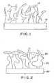
- This constructionis schematically depicted in Figure 1, where the poly(acrylic acid) chains 12 are grafted to the surface 10 of a device.
- Collagen 14is contained within the matrix formed by the chains 12.
- biological activitymay be reduced as proper expression and accessibility are hampered.
- a method of modifying the surface of a medical devicecomprising:
- Figure 1is a schematic cross-sectional diagram of a prior art surface graft matrix incorporating collagen.
- Figure 2is a schematic cross-sectional diagram of a surface graft matrix coated with a biomolecule sheath.
- Figure 3is a schematic cross-sectional diagram of a surface graft matrix coated with a biomolecule sheath, where the surface graft matrix is loaded with a pharmaceutical agent.
- Figure 4is a line drawing of the surface appearance of a surface graft matrix coated with collagen in a process according to the present invention.
- the present inventionprovides methods of covalently coupling a majority of one or more biomolecules in the outer portion of a surface graft matrix on a medical device.
- the surface graft matrixis preferably formed by surface grafting carboxyl-functional monomers, optionally in combination with vinyl monomers having no carboxyl functionality (COOH).
- COOHcarboxyl functionality
- the surface graft matrixcan result from the surface grafting of acrylic acid and acrylamide monomers, as disclosed in U.S. Patent Application Serial No. 08/553,206, filed November 7, 1995, entitled "Intramuscular Stimulation Lead With Enhanced Infection Resistance.”
- the surface graft matrixtypically has an anionic character.
- a surface graft matrix that exhibits reduced permeability to medium-sized to large moleculesis formed on the surface of a medical device, thereby providing the ability to isolate a majority of the subsequently coupled biomolecules to the outer portion of the surface graft matrix.
- the permeability of the surface graft matrixis reduced by treatment of the surface graft matrix at reduced pH levels, preferably where the pH of the solution is less than the pKa of the surface graft matrix.
- the surface graft matrixcan be loaded with a pharmaceutical agent after the biomolecules are in place in the outer portion of the surface graft matrix.
- this loadingoccurs as a result of ionic interaction of the surface graft matrix with the pharmaceutical agent.
- a surface graft matrix exhibiting reduced permeability to medium-sized to large moleculesis formed on the surface of a medical device.
- the permeability of the graft matrixis reduced by treatment of the surface graft matrix at reduced pH levels, preferably where the pH of the solution is less than the pKa of the surface graft matrix.
- linker moleculeswhich may be biomolecules, referred to herein as intermediate biomolecules
- the relative impermeability of the graft matrixis maintained, restricting a majority of the linker molecules to the outer portion of the graft matrix.
- linker moleculese.g., intermediate biomolecules
- the linker moleculescan then be used to covalently couple a majority of second biomolecules (referred to herein as primary biomolecules) in the outer portion of the surface graft matrix.
- This second coupling stepis also preferably performed in solutions having pH values less than the pKa of the underlying surface graft matrix, thereby maintaining its relative impermeability to the biomolecules located in the outer portion of the surface graft matrix.
- the term "medical device”may be defined as a device that has surfaces that contact tissue, blood, or other bodily fluids in the course of their operation, which fluids are later used in patients. This can include, for example, extracorporeal devices for use in surgery such as blood oxygenators, blood pumps, blood sensors, tubing used to carry blood, and the like which contact blood that is returned to the patient.
- the termcan also include endoprostheses implanted in blood contact in a human or animal body such as vascular grafts, stents, pacemaker leads, heart valves, and the like that are implanted in blood vessels or in the heart.
- the termcan further include devices for temporary intravascular use such as catheters, guide wires, and the like that are placed in blood vessels or the heart for purposes of monitoring or repair.
- the termcan also include nerve electrodes, muscle electrodes, implantable pulse generators, implantable drug pumps, and defibrillators.
- biomoleculeincludes any biocompatible and/or biologically active molecule (i.e., "primary biomolecule") or an intermediate biomolecule (linker molecule) to which one or more primary biomolecules can be coupled. Unless otherwise indicated, the term “biomolecule” as used herein will be understood to include both primary and intermediate biomolecules.
- biomoleculeis collagen, a biocompatible molecule that exists in many types.
- Types of biomolecules that can be coupled to the surface graft matrix in accordance with the present inventioninclude, but are not limited to, antithrombotic agents, antibacterial agents, anti-inflammatory agents, growth factors, cytokines, naturally occurring or synthetically prepared proteins, peptides, amino acids, and mixtures thereof.
- biomolecules that can be coupled to the surface graft matrixinclude, but are not limited to, albumin, fibrinogen, laminin, vitronectin, fibronectin, RGD-containing peptides, heparin, heparin sulfate, fibroblast growth factors (FGF), insulin-like growth factor, nerve growth factor, interferons (IFN), tumor necrosis factors (TNF), interleukins, gelatin, elastin, fibrin, von Willebrand factor, dermatan sulfate, hyaluronic acid, dextran sulfate, and mixtures thereof.
- FGFfibroblast growth factors
- IFNinterferons
- TNFtumor necrosis factors
- biomoleculesmay be neutral or charged at the conditions employed during covalent coupling.
- biomoleculesmay be coupled to the surface graft matrix directly (i.e., through the carboxyl groups), or through well-known coupling chemistries, such as, for example, esterification, amidation, and acylation.
- These biomoleculesare typically the primary biomolecules, although certain of them can be used as the intermediate biomolecules.
- the outer sheath of biomoleculestypically includes a plurality of biomolecules, although it could include polymerized biomolecules that technically form one macromolecule.
- linker moleculeswhich may or may not be biomolecules, in connection with the present invention typically involves covalently coupling a majority of the linker molecules in the outer portion of the surface graft matrix.
- the linker moleculescan provide the surface graft matrix with a number of functionally active groups that can be used to covalently couple one or more primary biomolecules.
- the linker moleculesmay be coupled to the surface graft matrix directly (i.e., through the carboxyl groups), or through well-known coupling chemistries, such as, for example, esterification, amidation, and acylation.
- the linker moleculeis at least a di- or tri-amine functional compound that is coupled to the surface graft matrix through the direct formation of amide bonds, and provides amine-functional groups that are available for reaction with the primary biomolecule.
- the linker moleculeis a polyamine functional polymer such as polyethyleneimine (PEI) or polyallylamine (PALLA). Mixtures of these polymers can also be used. These molecules contain a plurality of pendant amine-functional groups that can be used to surface-immobilize one or more primary biomolecules.
- PEIpolyethyleneimine
- PALLApolyallylamine
- Figure 2is a schematic cross-sectional view of a portion of the surface of a medical device 20, depicting that the biomolecules 24 are covalently coupled to a surface graft matrix 22 in a manner such that a majority of the biomolecules 24 are located or immobilized in the outer portion of the surface graft matrix 22 located on the medical device 20.
- the immobilization of the biomolecules 24 as depicted in Figure 2differs from the prior art depicted in Figure 1 in that a majority of the biomolecules 24 are located on or near the outer surface of the surface graft matrix 22, not generally dispersed throughout the structure of the matrix as depicted in Figure 1. This surface isolation of a majority of the biomolecules is advantageous because it allows a less disturbed expression of the biomolecules, so that biological activity is retained at a significantly higher level.
- the depth of the outer portion of the surface graft matrix in which a majority of the biomolecules are immobilizedis primarily dependent on the type of biomolecule immobilized and the reaction conditions employed. Typically, a majority of the biomolecules will be immobilized in the surface graft matrix within a depth of about 10 nm or less. For example, if collagen is the biomolecule immobilized in the outer portion of the surface graft matrix, the depth at which a majority of the collagen molecules are immobilized is about 7 nm or less.
- the immobilization approach of the present inventionmay prohibit movement of the coupled biomolecules into the graft matrix. This will especially be the case with immobilization of anionic biomolecules, such as the anti-coagulant heparin which will be repelled by the underlying anionic surface graft matrix.
- the matrixcan be loaded with a pharmaceutical agent for subsequent release to effect a desired response in the patient.
- the pharmaceutical agent capacity of the matrixcan be increased as compared to those surface graft matrix materials that allow complete penetration of biomolecules. This provides yet another advantage of the present invention.
- Pharmaceutical agents that can be used in connection with the present inventioninclude, but are not limited to, antimicrobial agents, antibacterial agents, anticoagulant agents, antithrombotic agents, platelet agents, and anti-inflammatory agents.
- Other useful pharmaceutical agentscan include, but are not limited to, dyes which act as biological ligands, steroids, enzymes, catalysts, hormones, growth factors, drugs, vitamins, antibodies, antigens, nucleic acids, peptides, DNA & RNA segments, and mixtures thereof.
- these pharmaceutical agentsare hydrophilic, positively charged compounds.
- a medical device 120incorporating a biomolecule 124 on a surface graft matrix 122 loaded with a desired pharmaceutical agent 126
- duplicate biological activitiescan be provided to improve the in vivo performance of the medical device.
- the biomolecules 124can be, for example, collagen which will interact with the surrounding tissue to provide a favorable tissue integration.
- specific desired body mechanismsmay be activated, or, in the case of antimicrobials, a protective mode of action is exhibited during the initial vulnerable period before the medical device/tissue interface is stabilized and when random colonization by bacteria might occur.
- the surfaceexhibits "bi-biofunctional" characteristics, i.e., two biofunctional activities including: a) promoting rapid tissue integration into the surface of the device, and b) releasing a pharmaceutical agent, such as an antimicrobial agent to reduce the risk of infection around an implanted device.
- a pharmaceutical agentsuch as an antimicrobial agent
- Processes according to the present inventiontypically begin with the formation of a surface graft matrix on the surface of a medical device.
- the surface grafting methodinvolves the covalent surface grafting of a polymer, preferably water soluble polymer, based on carboxyl-functional monomers, including, but not limited to, acrylic acid, methacrylic acid, itaconic acid, trans -cinnamic acid, crotonic acid, linoleic acid, linolenic acid, maleic acid, sorbic acid, and mixtures thereof onto a substrate material.
- the carboxyl-functional surface graft matrixalso may be obtained through chemical modification of non-carboxyl-functional monomers.
- ceric ion initiationis a preferred method to graft monomers to substrate surfaces, other grafting techniques may be used as well.
- Known examples of other initiation methodsinclude corona discharge, UV irradiation, ozonization and ionizing radiation (e.g., 60 Co, X-rays, high energy electrons, plasma gas discharge, etc.).
- the substrates that can be modified by the method of the present inventioninclude metals such as titanium/titanium alloys, TiNi (shape memory/super elastic), aluminum oxide, platinum/platinum alloys, stainless steels, MP35N, elgiloy, haynes 25, stellite, pyrolytic carbon, silver or glassy carbon; polymers such as polyamides, polycarbonates, polyethers, polyesters, polyolefins including polyethylenes or polypropylenes, polystyrenes, polyurethanes, polyvinyl chlorides, polyvinylpyrrolidones, silicone elastomers, fluoropolymers, polyacrylates, polyisoprenes, polytetrafluoroethylenes, and rubber; minerals or ceramics such as hydroxapatite; human or animal protein or tissue such as bone, skin, teeth, collagen, laminin, elastin or fibrin; organic materials such as wood, cellulose, or compressed carbon; and other materials such as glass, or
- Substrates made using these materialscan be coated or uncoated, and derivatized (e.g., modified to include reactive functional groups) or underivatized.
- the substrateis polyurethane, to which the carboxyl-functional surface graft matrix can be directly coupled without any preactivation of the substrate surface.
- the substrateis a biomaterial for use in a number of medical devices such as vascular grafts, aortic grafts, arterial, venous, or vascular tubing, vascular stents, dialysis membranes, tubing, or connectors, blood oxygenator tubing or membranes, ultrafiltration membranes, intra-aortic balloons, blood bags, catheters, sutures, soft or hard tissue prostheses, synthetic prostheses, prosthetic heart valves, tissue adhesives, cardiac pacemaker leads, artificial organs, endotracheal tubes, lenses for the eye such as contact or intraocular lenses, blood handling equipment, apheresis equipment, diagnostic and monitoring catheters and sensors, biosensors, dental devices, drug delivery systems, or bodily implants of any kind.
- medical devicessuch as vascular grafts, aortic grafts, arterial, venous, or vascular tubing, vascular stents, dialysis membranes, tubing, or connectors, blood oxygenator tubing or membranes, ultrafiltration
- a polymer surface graft of acrylic acidis one preferred embodiment to be used for subsequent covalent coupling of one or more biomolecules to enclose the surface graft matrix.
- the surface graft matrixis preferably formed by surface grafting of the monomers acrylic acid and acrylamide in ratios that allow for later manipulation of the graft matrix.
- sufficient acrylic acid (or other carboxylic-functional monomer)should be present so as not to interfere with the mechanism of reducing the permeability of the surface graft matrix to provide for immobilization of a majority of the biomolecules in the outer portion of the surface graft matrix.
- acrylic acidis used to prepare the surface graft matrix in an amount of about 20-100 wt-%, based on the total weight of the monomers used to prepare the surface graft matrix. More preferably, acrylic acid is used in an amount of about 50-90 wt-%, and most preferably, in an amount of about 65-75 wt-%. These weight percentages are also applicable to other carboxyl-functional monomers. Incorporation of other vinyl-functional monomers that do not include carboxyl groups (COOH) into the surface graft is possible, but is limited to the extent that they interfere with the mechanism of reducing the permeability of the surface graft matrix to provide for isolation of a majority of the biomolecules in the outer portion of the surface graft matrix.
- COOHcarboxyl groups
- Various vinyl-functional monomerscan be incorporated to form a copolymer surface graft, such as acrylamide (Aam), N-(3-aminopropyl) methacrylamide (APMA), 2-hydroxyethyl methacrylate (HEMA), and 2-acrylamido-2-methylpropane sulfonic acid (AMPS).
- Acrylamideis the most preferred monomeric compound to be incorporated in addition to acrylic acid monomer as the structure and molecular weight of acrylamide are close to those of acrylic acid.
- a low pH immersion processis used to produce the surface graft matrix with the desired impermeability to provide for immobilization of a majority of the biomolecules in the outer portion of the surface graft matrix.
- a low pH solutionBy immersing the surface graft matrix in a low pH solution, the formation of carboxylic acid dimers and intra-polymer crosslinking in the surface graft matrix is provided.
- the bond strength of acetic acid dimersis approximately equal to 55-60 kJ/mole, as disclosed by Potter, Jr., et al., J. Phys. Chem.
- intrapolymer crosslinking within a poly(carboxylic acid) graft matrixwill be of significant strength.
- the formation of intrapolymer crosslinksis characterized by an obvious sticky feel of the surface grafted material.
- This sticky feelis generally indicative of the cohesive forces of the surface graft matrix.
- the intrapolymer crosslinkingreduces the permeability/accessibility of medium-sized to large, even polycationic, compounds into the surface graft matrix.
- a subsequent process of covalently coupling a majority of biomolecules in the outer portion of the surface graft matrixis also carried out at a pH that is less than the pKa of the surface graft matrix. This results in immobilization of the biomolecules such that a surface layer of primarily biomolecules, i.e., a sheath, is formed that substantially encloses the surface graft matrix. This additionally allows for more complete loading of the surface graft matrix with a pharmaceutical agent for subsequent release in vivo. Although it is preferred that the immersion process and the biomolecule coupling process be carried out sequentially, they could be carried out simultaneously.
- the pKa value of the surface graft matrixcan be determined through FT-IR analysis, according to the method of Azeez et al., J. Appl. Polym. Sci. , 58 , 1741-1749 (1995). Using this method, the pKa of a 100% acrylic acid graft matrix is 6.3, which is in accordance with the findings of 4.9-6.7 disclosed by Park et al., Pharm. Res. , 4 , 457-464 (1987) on acrylic acid/acrylamide copolymer hydrogels.
- the pKa valuesare generally dependent on the ionic strength of the environment and the fraction of acrylic acid in the copolymer. Typically, an increase in ionic strength decreases the pKa, whereas an increase in acrylic acid fraction increases the pKa.
- the pH of the solutions in which the surface graft matrix is treated to reduce permeability and in which biomolecules are attachedare typically no greater than about 5.5.
- the pHis no greater than about 5, and more preferably, no greater than about 4.5.
- the pHis at least about 2, and more preferably, at least about 3.
- the methods of the present inventionpreferably involve surface grafting of carboxyl-functional monomers through a covalent interaction to a substrate at an acidic pH, preferably at a pH of less than about 5.5; washing the substrate with the surface graft matrix thereon in an aqueous solution having a pH greater than the pKa of the surface graft matrix (typically at a neutral pH) to allow for the removal of free monomers, oligomers, or polymers; immersing the substrate with the surface graft matrix thereon in a solution having a pH that is less than the pKa of the surface graft matrix; and covalently coupling a majority of the biomolecules in the outer portion of the surface graft matrix in a solution having a pH that is less than the pKa of the surface graft matrix.
- Polyurethane (PU) film materialwas made from 2363-55D PELLETHANE resin (Dow Chemical, Midland, MI, USA) by Medtronic Promeon (Minneapolis, MN, USA). Ceric(IV)ammonium nitrate, nitric acid (65%), sodium phosphate monobasic monohydrate, sodium phosphate dibasic, sodium chloride, and sodium azide were all obtained from Merck-Schuchardt (Darmstadt, Germany). Acrylic acid, MES monohydrate, di-sodium tartrate, N-hydroxysuccimide (NHS), 3-ethyl-1-(diaminopropyl)-carbodiimide (EDC), and sodium hydrogencarbonate, were obtained from Aldrich Chemie (Bornem, Belgium).
- Acrylamide(99+%; electrophoresis grade) was obtained from Acros Chimica (Geel, Belgium).
- Collagentype I; from calf skin
- TNBS2,4,6-trinitrobenzenesulfonic acid
- Coomassie Bluewas obtained from Pierce Europe BV (Oud Beijerland, The Netherlands).
- Collagenase(EC 3.4.24.3; from Clostridium histolyticum; type IA, 550 units/mg solid), and Tris-HCl were obtained from Sigma Chemie (Bornem, Belgium); di-sodium tetraborate decahydrate from Sigma Chemie (Borneum, Belgium); Toluidine Blue O dye from Sigma Chemie; Ponceau S dye from Sigma Chemie; SDS from Sigma Chemie; and gentamicin sulfate from Sigma Chemie.
- Acrylic acidwas purified by conventional distillation. All other reagents were of reagent grade or higher and used without further purification.
- XPSX-Ray Photoelectron Spectroscopy
- Time of Flight Secondary Ion Mass Spectometry (ToF-SIMS) spectrawere acquired using a VG IX23S instrument based on the Poschenreider design and equipped with a pulsed liquid metal ion source. A 30 keV Ga + primary ion beam was used at an incident angle of 38° to the surface normal. The secondary ions were accelerated to 5 keV for the analysis by applying a sample bias. For each sample, both positive and negative secondary ion spectra were collected using a total primary ion dose that did not exceed 2 x 10 11 ions cm -2 for static SIMS, such that the analyzed surfaces were effectively undamaged as a result of the ToFSIMS studies.
- FEG-SEM testswere carried out on a JEOL JSM 6301-F Field-Emission-Gun SEM operated at 2 kV after the samples were sputter coated with gold (2-4 nm) using an Edwards 5150B Sputter Coater.
- Extruded PELLETHANE 55D polyurethane filmswere ultrasonically cleaned in isopropyl alcohol (IPA) for 15 minutes prior to ceric ion initiated surface grafting. Immediately after the IPA-cleaning samples were dried in a forced air oven at 50-60°C for approximately 5 minutes. FT-IR investigation has demonstrated that 15 minutes IPA-treatment is sufficient to remove any surface contamination that originates from processing aides, such as bis-stearamide waxes, that may interfere with the grafting process.
- processing aidessuch as bis-stearamide waxes
- an aqueous grafting solutionwas prepared that was composed of 40% by weight acrylic acid monomer concentration (100 wt-% acrylic acid), 6 mM of ceric ammonium nitrate (CAN) and 0.06 M nitric acid (HNO 3 ). Prior to grafting, the grafting solution was treated to remove excess air by exposure to reduced pressure (18 mm Hg ⁇ 5 mm Hg) for a maximum of 2 minutes.
- Grafted samples(10 x 1cm strips) were prepared by placing the cleaned and dried samples in an appropriate volume (25-30 ml) of the grafting solution. Grafting was allowed to continue for 15-20 minutes at 30°C, while stirring the solution.
- the sampleswere rinsed in deionized (DI) water to stop the grafting process as well as to clean the surface graft matrix formed.
- DIdeionized
- a 0.1 M tartrate solutiondi -sodium tartrate
- XPSX-Ray Photoelectron Spectroscopy
- the carboxylic acid groupsare mainly ionized. This is confirmed by the presence of sodium (Table 1) and the prevalence of the COOX chemical state (Table 2). In the ionized state, carboxylic acids will not be capable of forming the dimer, i.e., the group that is essential for physically crosslinking the graft matrix. In contrast, at pH ⁇ pKa the carboxylic acid groups are hydrogenated and thus capable of forming that dimer-group. The hydrogenated state is confirmed by the absence of sodium (Table 1) and the prevalence of the COOH chemical state (Table 2).
- the surface graft matrixcan be made impermeable for medium-sized to large (even polycationic) molecules by formation of carboxylic acid dimers to induce physical crosslinking. This was confirmed in an experiment that studied the effect of pH on the amount of the (polycationic) antimicrobial drug gentamicin that could be (ionically) immobilized.
- the surface grafted sampleswere gentamicin loaded and the amount of gentamicin loaded was determined. The difference in the gentamicin content before and after sample immersion was determined and used as a measure for the amount of gentamicin loaded into the samples.
- the TNBS derivatization reactionwas allowed to proceed for 25-30 minutes at room temperature, after which the absorbance at 415 nm was measured, while 595 nm was used as the reference wavelength (BioRad Model 3550, 96 wells microplate reader, Veenendaal, The Netherlands).
- the pHfalls below the pKa of the surface graft matrix, the amount of gentamicin that could be immobilized was drastically reduced to become zero in the pH range from 3 to 4.
- MES4-morpholine-ethanesulfonic acid monohydrate, Aldrich
- the sampleswere immersed in a buffered solution containing 0.5 mg/ml collagen (type I).
- the solutionwas buffered in the pH range 4.0-4.5 with 0.02 M MES.
- the collagen immobilization reactionwas continued for at least 20 hours.
- the collagen-immobilized sampleswere rinsed in DI water, an aqueous 0.15M NaCl solution in DI water, and DI water again. Samples were dried at ambient conditions by air exposure. The immobilization of collagen was identified by two separate staining techniques. TNBS-staining confirmed the presence of amine-functional groups.
- TNBS stainingwas performed by immersing a 4 mm disc in 1 ml of aqueous 4% by weight NaHCO 3 . To this solution 1 ml of aqueous 0.5% by weight TNBS was added, after which the reaction was allowed to continue for 2 hours at 40°C. Finally, the sample was extensively rinsed in DI water and allowed to dry. A similar surface grafted disc, but not used for collagen immobilization, was used as the control. The difference in dye uptake was obvious visually. Considering the surface modification chemistry, these groups could only be derived from immobilized collagen. Coomassie Blue protein dye also was used as a analytical tool to verify the presence of immobilized collagen in the outer portion of the surface graft matrix.
- Coomassie Blue stainingwas performed by immersing a 4 mm disc in 1 ml of Coomassie Blue for 30 minutes. Thereafter, the sample was extensively rinsed in DI water and allowed to dry. A similar surface grafted disc, but not used for collagen immobilization, was used as the control. The difference in dye uptake was obvious visually.
- the processed surfaceswere examined by FEG-SEM (Field Emission Gun Scanning Electron Microscope) operated at 2 kV; prior to SEM-analysis surfaces were sputter-coated with gold (2-4 nm).
- FEG-SEMField Emission Gun Scanning Electron Microscope
- the extruded 55D PELLETHANE polyurethane materialis a flat material.
- the acrylic acid grafted materialexhibits a permeable matrix-like structure. Subsequent immobilization of collagen seems to have covered this surface matrix with a superimposed surface-layer.
- the velvet-like appearance of this surface layeris depicted in Figure 4.
- FT-IR spectroscopyAnother analytical technique that was used to confirm the immobilization of collagen was FT-IR spectroscopy. This technique allows for analysis of the top 0.2-1 :m surface layer of processed samples. The FT-IR spectra of collagen raw material, acrylic acid grafted 55D, and collagen-immobilized samples were compared. The spectrum of the collagen-immobilized sample obviously contained features of both the acrylic acid graft and the collagen raw material. Most characteristic was the rise of the collagen-related peaks at approximately 1635 nm and 1660 nm. This confirmed the presence of collagen in the surface top layer.
- the outermost surface carbonis enriched in C-C, C-H and C-O(H) bonding.
- the nitrogenis enhanced in C-NH-C bonding and the oxygen is enriched in C-OH bonding.
- the latter observationsuggests that the collagen-immobilization is accompanied by a reorientation of the collagen molecules.
- analysis depth ( ⁇ ) NH-CO -C-NH-C- 15 77 23 30 77 23 70 92 8 collagen reference 70 30
- ToF-SIMSTime-of-Flight Secondary Ion Mass Spectrometry
- the results of the ToF-SIMS testingalso confirmed the presence of a collagen top layer, as the sample spectra displayed a rich array of N-containing signals. In addition to nonspecific peptide/protein characteristics, the spectra exhibit rich arrays of N-containing signals which are more diagnostic of particular amino acid residues.
- the collagen top layerdisplayed an outermost surface chemistry which differs to that of the collagen reference. While N-containing species are clearly present, there appears to be a higher relative proportion of species containing C/H and C/H/O for the collagen coating compared to the collagen reference. This indicates either an incomplete collagen top layer or a very thin collagen top layer, or some reorientation of the collagen molecules due to the immobilization.
- Collagen-immobilized sampleswere prepared as previously described in Example 2 above (100 wt-% acrylic acid monomer used to prepare the surface graft matrix). The in vivo performance was investigated and compared to that of acrylic acid grafted 55D (55D-AA) and plain 55D samples (55D) as discussed below.
- 55D-CChad a similar or thinner tissue capsule compared to 55D and 55D-AA. These results may indicate that 55D-CC promotes a faster wound healing response as a result from its biological interaction with the body's responses. It appears that fibrin formation at the surface of 55D-CC plays a major role, which indicates that 55D-CC may promote coagulation.
- linker moleculescovalently coupled to both the surface graft matrix and the biomolecules.
- linker moleculesinclude, e.g., the amine-functional polymers polyethyleneimine (PEI) or polyallylamine (PALLA). These compounds contain pendant amine-functional groups that can be used to surface-immobilize a majority of the biomolecules in the outer portion of a surface graft matrix.
- PEIwas surface-immobilized to substantially enclose the surface graft matrix.
- the PEIwas subsequently used to surface-immobilize the anti-coagulant drug heparin.
- a 0.05 M tartrate solutiondi-sodium tartrate
- heparin couplingAfter heparin coupling, representative samples were stained with the cationic dye Toluidine Blue O (TB). The presence of heparin is denoted by a metachromatic shift from blue to violet. While the primary graft alone demonstrated a blueish, dark violet color after TB-exposure, the heparinized surface demonstrated an obvious shift to light violet. This shift in color suggests successful surface-coupling of heparin. PS-uptake obviously decreased as a consequence of the heparin-coupling; this confirms presence of heparin as well as coupling of heparin reduces the cationic nature of the surface.
- Toluidine Blue OToluidine Blue O
- peaksare characteristic of the S-O stretch of the sulfur acid groups, in this case originating from the immobilized heparin molecules.
- the observed change in the 1530-1730 cm -1 regioncan be assigned to the surface-immobilized PEI; for example, the 1560 cm -1 peak is characteristic for N-H (in plane) bend of amides. Additional surface analysis was performed by XPS. The heparin-immobilized sample exhibited significant heparin concentrations at the graft surface.
- the gentamicin solution used for loading the surface grafted samplewas analyzed to determine its gentamicin content before and after loading. This was done using a TNBS Assay in which the gentamicin-containing solution was adjusted to pH 9 by addition of 0.1 M borate, after which 25 ml 0.03 M aqueous TNBS was added per ml of sample-solution. The difference in gentamicin content before and after sample immersion was determined and used as a measure for the amount of gentamicin loaded. The amount of gentamicin loaded was expressed as ⁇ g/cm 2 and is reported below in Table 12.
- Gentamicin releasewas performed by immersion of gentamicin loaded samples in phosphate buffered saline (PBS, pH 7.4) at 37°C; a volume-to-surface ratio of 1:1 (ml:cm 2 ) typically was used throughout the experiment. At desired time points, the samples were withdrawn from the solution and immersed in fresh PBS. Solution samples were analyzed for their gentamicin content by means of the TNBS Assay discussed above.
- PBSphosphate buffered saline
- Heparin couplingdid not significantly influence the gentamicin release profile. After an initial burst, gentamicin progressively released until completion in an approximate 2 weeks.
- test materialswere applied (8 mm discs); Genta-neo-sensitab (Rosco Diagnostica, Taastrup, Denmark), a gentamicin-loaded tablet, was used as the positive control. Surface grafted, heparinized samples without gentamicin were applied as negative controls. Subsequently, the agar plate was incubated overnight at 37°C. The following day the plate was removed from the incubator and the bacteria free zone around each sample was determined. The regions of bacterial growth and inhibition are obvious visually.
Landscapes
- Health & Medical Sciences (AREA)
- Life Sciences & Earth Sciences (AREA)
- Epidemiology (AREA)
- Veterinary Medicine (AREA)
- Public Health (AREA)
- General Health & Medical Sciences (AREA)
- Animal Behavior & Ethology (AREA)
- Chemical & Material Sciences (AREA)
- Surgery (AREA)
- Medicinal Chemistry (AREA)
- Engineering & Computer Science (AREA)
- Hematology (AREA)
- Transplantation (AREA)
- Oral & Maxillofacial Surgery (AREA)
- Dermatology (AREA)
- Molecular Biology (AREA)
- Biomedical Technology (AREA)
- Heart & Thoracic Surgery (AREA)
- Vascular Medicine (AREA)
- Materials Engineering (AREA)
- Materials For Medical Uses (AREA)
- Treatments Of Macromolecular Shaped Articles (AREA)
Description
- The present invention relates to the field ofmedical devices. More particularly, the presentinvention relates to methods of manufacturing a surfacegraft matrix.
- Infection has become the most serious complicationassociated with the application of implantable medicaldevices. Treatment of these infections is difficult andmost often necessitates removal of the implanted device.Technological refinements in materials and design andincreasing surgical experience has lowered the incidenceof infectious complications; however, infection remainsa continuing cause of morbidity and mortality.Approaches to reduce device-related infections initiallywere focused on improvements of the surgical technique,including modification of the operating room area andthe use of prophylactic antibiotics at the time ofsurgery. Despite the introduction of these meticulousaseptic measures the occurrence of device-relatedinfections could not be completely eliminated.
- An alternative approach is to focus on the implantitself, and consequently on modification of the deviceto enhance infection-resistance by providing surfaces onthe device that promote appropriate integration of thesurrounding tissue(s) with the device surface. Theunderlying concept is that when rapid colonization andintegration of the device surface with tissue cells isencouraged, the implant surface will be protected frombacterial colonization.
- One method of promoting tissue integration isthrough the use of collagen immobilized on the surfaceof the device because collagen materials promote afavorable tissue response. They provide a morephysiological, isotropic environment that has been shownto promote the organization of different cell types intothree-dimensional tissue-like structure. See, forexample, Akita et al.,Cell Tissue Res.,274, 91-95(1993); and Berthod et al.,Biomaterials,14, 749-754(1993). Implant studies have demonstrated thatcollagen-immobilization promotes favourable integrationof tissue(s) with the implanted material. See, forexample, Shimizu et al.,Biomat., Med. Dev., Artif.Org.,6, 375-391 (1978); Kinoshita et al.,Biomaterials,14, 209-215 (1993); and Okada et al.,J.Biomed. Mater. Res.,27, 1509-1518 (1993).
- One method of coating synthetic polymers withcollagen involves a physical deposition of collagen,such that a laminar material results, as disclosed byShimizu et al.,Biomat., Med. Dev., Artif. Org.,5, 49-66(1977). One drawback to this method is that thecollagen materials are prone to delamination in amoisture-abundant environment such as that typicallyexperienced by implanted medical devices.
- Another method of providing a collagen-coateddevice involves covalently coupling collagen to asynthetic polymer substrate, as disclosed by Okada etal.,Biomaterials and Clinical Applications, ElsevierScience Publishers B.V., Amsterdam, The Netherlands, pp.465-470, 1987. The method includes graftcopolymerization of acrylic acid, after which collagenis covalently coupled to the grafted poly(acrylic acid)chains, resulting in a blend-like matrix of collagen andpoly(acrylic acid) chains. This construction isschematically depicted in Figure 1, where the poly(acrylic acid)
chains 12 are grafted to thesurface 10 of a device.Collagen 14 is contained within thematrix formed by thechains 12. Thus, the collagen isnot located primarily at the surface. Thus, biologicalactivity may be reduced as proper expression andaccessibility are hampered. - According to the present invention, there isprovided a method of modifying the surface of a medicaldevice comprising:
- (a) forming a surface graft matrix comprisingcarboxyl-functional groups on the surface of a medicaldevice;
- (b) washing the surface graft matrix with anaqueous solution having a pH that is less than the pKaof the surface graft matrix; and
- (c) covalently coupling one or more biomoleculesto the outer portion of the surface graft matrix byimmersion in an aqueous solution having a pH that isless than the pKa of the surface graft matrix, such thata majority of the biomolecules are located in the outerportion of the surface graft matrix.
- Preferred embodiments of the invention will now bedescribed, by way of example only, with reference to theaccompanying drawings.
- Figure 1 is a schematic cross-sectional diagram ofa prior art surface graft matrix incorporating collagen.Figure 2 is a schematic cross-sectional diagram of asurface graft matrix coated with a biomolecule sheath.Figure 3 is a schematic cross-sectional diagram of asurface graft matrix coated with a biomolecule sheath,where the surface graft matrix is loaded with apharmaceutical agent.
- Figure 4 is a line drawing of the surfaceappearance of a surface graft matrix coated with collagen in a process according to the presentinvention.
- The present invention provides methods ofcovalently coupling a majority of one or morebiomolecules in the outer portion of a surface graftmatrix on a medical device. The surface graft matrix ispreferably formed by surface grafting carboxyl-functionalmonomers, optionally in combination withvinyl monomers having no carboxyl functionality (COOH).For example, the surface graft matrix can result fromthe surface grafting of acrylic acid and acrylamidemonomers, as disclosed in U.S. Patent Application SerialNo. 08/553,206, filed November 7, 1995, entitled"Intramuscular Stimulation Lead With Enhanced InfectionResistance." As a result of the carboxyl functionality,the surface graft matrix typically has an anioniccharacter.
- In one method according to the present invention, asurface graft matrix that exhibits reduced permeabilityto medium-sized to large molecules is formed on thesurface of a medical device, thereby providing theability to isolate a majority of the subsequentlycoupled biomolecules to the outer portion of the surfacegraft matrix. The permeability of the surface graftmatrix is reduced by treatment of the surface graftmatrix at reduced pH levels, preferably where the pH ofthe solution is less than the pKa of the surface graftmatrix. By carrying out biomolecule coupling reactionsin solutions in which the pH is less than the pKa of thesurface graft matrix, the relative impermeability of thesurface graft matrix is maintained, restricting amajority of the biomolecules to the outer portion of thesurface graft matrix. That restriction results in a"sheath" of biomolecule(s) over the surface graftmatrix.
- In another method according to the presentinvention, the surface graft matrix can be loaded with a pharmaceutical agent after the biomolecules are in placein the outer portion of the surface graft matrix.Preferably, this loading occurs as a result of ionicinteraction of the surface graft matrix with thepharmaceutical agent.
- In another method according to the presentinvention, a surface graft matrix exhibiting reducedpermeability to medium-sized to large molecules isformed on the surface of a medical device. Thepermeability of the graft matrix is reduced by treatmentof the surface graft matrix at reduced pH levels,preferably where the pH of the solution is less than thepKa of the surface graft matrix. By carrying outsubsequent coupling reactions of one or more linkermolecules (which may be biomolecules, referred to hereinas intermediate biomolecules) in solutions with a pHless than the pKa of the surface graft matrix, therelative impermeability of the graft matrix ismaintained, restricting a majority of the linkermolecules to the outer portion of the graft matrix.That restriction results in a "sheath" of linkermolecules (e.g., intermediate biomolecules) in the outerportion of the surface graft matrix. The linkermolecules (e.g., intermediate biomolecules) can then beused to covalently couple a majority of secondbiomolecules (referred to herein as primarybiomolecules) in the outer portion of the surface graftmatrix. This second coupling step is also preferablyperformed in solutions having pH values less than thepKa of the underlying surface graft matrix, therebymaintaining its relative impermeability to thebiomolecules located in the outer portion of the surfacegraft matrix.
- As used herein, the term "medical device" may bedefined as a device that has surfaces that contacttissue, blood, or other bodily fluids in the course oftheir operation, which fluids are later used in patients. This can include, for example, extracorporealdevices for use in surgery such as blood oxygenators,blood pumps, blood sensors, tubing used to carry blood,and the like which contact blood that is returned to thepatient. The term can also include endoprosthesesimplanted in blood contact in a human or animal bodysuch as vascular grafts, stents, pacemaker leads, heartvalves, and the like that are implanted in blood vesselsor in the heart. The term can further include devicesfor temporary intravascular use such as catheters, guidewires, and the like that are placed in blood vessels orthe heart for purposes of monitoring or repair. Theterm can also include nerve electrodes, muscleelectrodes, implantable pulse generators, implantabledrug pumps, and defibrillators.
- As used herein, the term "biomolecule" includes anybiocompatible and/or biologically active molecule (i.e.,"primary biomolecule") or an intermediate biomolecule(linker molecule) to which one or more primarybiomolecules can be coupled. Unless otherwiseindicated, the term "biomolecule" as used herein will beunderstood to include both primary and intermediatebiomolecules.
- An example of a biomolecule is collagen, abiocompatible molecule that exists in many types. Typesof biomolecules that can be coupled to the surface graftmatrix in accordance with the present invention include,but are not limited to, antithrombotic agents,antibacterial agents, anti-inflammatory agents, growthfactors, cytokines, naturally occurring or syntheticallyprepared proteins, peptides, amino acids, and mixturesthereof. Specific examples of biomolecules that can becoupled to the surface graft matrix include, but are notlimited to, albumin, fibrinogen, laminin, vitronectin,fibronectin, RGD-containing peptides, heparin, heparinsulfate, fibroblast growth factors (FGF), insulin-likegrowth factor, nerve growth factor, interferons (IFN), tumor necrosis factors (TNF), interleukins, gelatin,elastin, fibrin, von Willebrand factor, dermatansulfate, hyaluronic acid, dextran sulfate, and mixturesthereof. These biomolecules may be neutral or chargedat the conditions employed during covalent coupling.They may be coupled to the surface graft matrix directly(i.e., through the carboxyl groups), or through well-knowncoupling chemistries, such as, for example,esterification, amidation, and acylation. Thesebiomolecules are typically the primary biomolecules,although certain of them can be used as the intermediatebiomolecules. It will be understood that the outersheath of biomolecules typically includes a plurality ofbiomolecules, although it could include polymerizedbiomolecules that technically form one macromolecule.The use of linker molecules, which may or may not bebiomolecules, in connection with the present inventiontypically involves covalently coupling a majority of thelinker molecules in the outer portion of the surfacegraft matrix. After covalent coupling to the surfacegraft matrix, the linker molecules can provide thesurface graft matrix with a number of functionallyactive groups that can be used to covalently couple oneor more primary biomolecules. The linker molecules maybe coupled to the surface graft matrix directly (i.e.,through the carboxyl groups), or through well-knowncoupling chemistries, such as, for example,esterification, amidation, and acylation. Preferably,the linker molecule is at least a di- or tri-aminefunctional compound that is coupled to the surface graftmatrix through the direct formation of amide bonds, andprovides amine-functional groups that are available forreaction with the primary biomolecule. More preferably,the linker molecule is a polyamine functional polymersuch as polyethyleneimine (PEI) or polyallylamine(PALLA). Mixtures of these polymers can also be used.These molecules contain a plurality of pendant amine-functional groups that can be used to surface-immobilizeone or more primary biomolecules.
- Figure 2 is a schematic cross-sectional view of aportion of the surface of a
medical device 20, depictingthat thebiomolecules 24 are covalently coupled to asurface graft matrix 22 in a manner such that a majorityof thebiomolecules 24 are located or immobilized in theouter portion of thesurface graft matrix 22 located onthemedical device 20. The immobilization of thebiomolecules 24 as depicted in Figure 2 differs from theprior art depicted in Figure 1 in that a majority of thebiomolecules 24 are located on or near the outer surfaceof thesurface graft matrix 22, not generally dispersedthroughout the structure of the matrix as depicted inFigure 1. This surface isolation of a majority of thebiomolecules is advantageous because it allows a lessdisturbed expression of the biomolecules, so thatbiological activity is retained at a significantlyhigher level. - The depth of the outer portion of the surface graftmatrix in which a majority of the biomolecules areimmobilized is primarily dependent on the type ofbiomolecule immobilized and the reaction conditionsemployed. Typically, a majority of the biomoleculeswill be immobilized in the surface graft matrix within adepth of about 10 nm or less. For example, if collagenis the biomolecule immobilized in the outer portion ofthe surface graft matrix, the depth at which a majorityof the collagen molecules are immobilized is about 7 nmor less.
- The immobilization approach of the presentinvention may prohibit movement of the coupledbiomolecules into the graft matrix. This willespecially be the case with immobilization of anionicbiomolecules, such as the anti-coagulant heparin whichwill be repelled by the underlying anionic surface graftmatrix.
- As discussed above, after the covalent coupling ofa majority of the biomolecules in the outer portion ofthe surface graft matrix, the matrix can be loaded witha pharmaceutical agent for subsequent release to effecta desired response in the patient. By immobilizing themajority of the biomolecules in the outer portion of thesurface graft matrix, the pharmaceutical agent capacityof the matrix can be increased as compared to thosesurface graft matrix materials that allow completepenetration of biomolecules. This provides yet anotheradvantage of the present invention.
- Pharmaceutical agents that can be used inconnection with the present invention include, but arenot limited to, antimicrobial agents, antibacterialagents, anticoagulant agents, antithrombotic agents,platelet agents, and anti-inflammatory agents. Otheruseful pharmaceutical agents can include, but are notlimited to, dyes which act as biological ligands,steroids, enzymes, catalysts, hormones, growth factors,drugs, vitamins, antibodies, antigens, nucleic acids,peptides, DNA & RNA segments, and mixtures thereof. Formost effective incorporation into the surface graftmatrix, these pharmaceutical agents are hydrophilic,positively charged compounds.
- Referring now to Figure 3, in a
medical device 120incorporating abiomolecule 124 on asurface graftmatrix 122 loaded with a desiredpharmaceutical agent 126, duplicate biological activities can be provided toimprove thein vivo performance of the medical device.Thebiomolecules 124 can be, for example, collagen whichwill interact with the surrounding tissue to provide afavorable tissue integration. By release of thepharmaceutical agent 126, specific desired bodymechanisms may be activated, or, in the case ofantimicrobials, a protective mode of action is exhibitedduring the initial vulnerable period before the medicaldevice/tissue interface is stabilized and when random colonization by bacteria might occur. As a result, thesurface exhibits "bi-biofunctional" characteristics,i.e., two biofunctional activities including: a)promoting rapid tissue integration into the surface ofthe device, and b) releasing a pharmaceutical agent,such as an antimicrobial agent to reduce the risk ofinfection around an implanted device. - Processes according to the present inventiontypically begin with the formation of a surface graftmatrix on the surface of a medical device. The surfacegrafting method involves the covalent surface graftingof a polymer, preferably water soluble polymer, based oncarboxyl-functional monomers, including, but not limitedto, acrylic acid, methacrylic acid, itaconic acid,trans-cinnamic acid, crotonic acid, linoleic acid,linolenic acid, maleic acid, sorbic acid, and mixturesthereof onto a substrate material. The carboxyl-functionalsurface graft matrix also may be obtainedthrough chemical modification of non-carboxyl-functionalmonomers. For example, basic hydrolysis of apoly(acrylamide) surface will introduce carboxyl-functionalmoieties, similar to what occurs when acrylicacid is copolymerized with acrylamide. However thesurface graft matrix is prepared, as long as it hascarboxyl-functional groups (COOH) it can be used toattach biomolecules as described herein.
- Surface grafting is preferably initiated by cericion methods as discussed U.S. Patent Nos. 5,229,172and/or 5,344,455. While ceric ion initiation is apreferred method to graft monomers to substratesurfaces, other grafting techniques may be used as well.Known examples of other initiation methods includecorona discharge, UV irradiation, ozonization andionizing radiation (e.g.,60Co, X-rays, high energyelectrons, plasma gas discharge, etc.).
- The substrates that can be modified by the methodof the present invention include metals such as titanium/titanium alloys, TiNi (shape memory/superelastic), aluminum oxide, platinum/platinum alloys,stainless steels, MP35N, elgiloy, haynes 25, stellite,pyrolytic carbon, silver or glassy carbon; polymers suchas polyamides, polycarbonates, polyethers, polyesters,polyolefins including polyethylenes or polypropylenes,polystyrenes, polyurethanes, polyvinyl chlorides,polyvinylpyrrolidones, silicone elastomers,fluoropolymers, polyacrylates, polyisoprenes,polytetrafluoroethylenes, and rubber; minerals orceramics such as hydroxapatite; human or animal proteinor tissue such as bone, skin, teeth, collagen, laminin,elastin or fibrin; organic materials such as wood,cellulose, or compressed carbon; and other materialssuch as glass, or the like. Substrates made using thesematerials can be coated or uncoated, and derivatized(e.g., modified to include reactive functional groups)or underivatized. Preferably, the substrate ispolyurethane, to which the carboxyl-functional surfacegraft matrix can be directly coupled without anypreactivation of the substrate surface.
- Surface grafting can be used to modify substratesof any shape or form including tubular, sheet, rod andarticles of proper shape. Preferably, the substrate isa biomaterial for use in a number of medical devicessuch as vascular grafts, aortic grafts, arterial,venous, or vascular tubing, vascular stents, dialysismembranes, tubing, or connectors, blood oxygenatortubing or membranes, ultrafiltration membranes, intra-aorticballoons, blood bags, catheters, sutures, soft orhard tissue prostheses, synthetic prostheses, prostheticheart valves, tissue adhesives, cardiac pacemaker leads,artificial organs, endotracheal tubes, lenses for theeye such as contact or intraocular lenses, bloodhandling equipment, apheresis equipment, diagnostic andmonitoring catheters and sensors, biosensors, dentaldevices, drug delivery systems, or bodily implants of any kind.
- A polymer surface graft of acrylic acid is onepreferred embodiment to be used for subsequent covalentcoupling of one or more biomolecules to enclose thesurface graft matrix. The surface graft matrix ispreferably formed by surface grafting of the monomersacrylic acid and acrylamide in ratios that allow forlater manipulation of the graft matrix. Typically,sufficient acrylic acid (or other carboxylic-functionalmonomer) should be present so as not to interfere withthe mechanism of reducing the permeability of thesurface graft matrix to provide for immobilization of amajority of the biomolecules in the outer portion of thesurface graft matrix. Preferably, acrylic acid is usedto prepare the surface graft matrix in an amount ofabout 20-100 wt-%, based on the total weight of themonomers used to prepare the surface graft matrix. Morepreferably, acrylic acid is used in an amount of about50-90 wt-%, and most preferably, in an amount of about65-75 wt-%. These weight percentages are alsoapplicable to other carboxyl-functional monomers.Incorporation of other vinyl-functional monomers that donot include carboxyl groups (COOH) into the surfacegraft is possible, but is limited to the extent thatthey interfere with the mechanism of reducing thepermeability of the surface graft matrix to provide forisolation of a majority of the biomolecules in the outerportion of the surface graft matrix. Various vinyl-functionalmonomers can be incorporated to form acopolymer surface graft, such as acrylamide (Aam), N-(3-aminopropyl)methacrylamide (APMA), 2-hydroxyethylmethacrylate (HEMA), and 2-acrylamido-2-methylpropanesulfonic acid (AMPS). Acrylamide is the most preferredmonomeric compound to be incorporated in addition toacrylic acid monomer as the structure and molecularweight of acrylamide are close to those of acrylic acid.
- After covalent coupling of the surface graft matrix to the substrate surface (which may or may not becarried out in acidic media), as exemplified in theexamples below, a low pH immersion process is used toproduce the surface graft matrix with the desiredimpermeability to provide for immobilization of amajority of the biomolecules in the outer portion of thesurface graft matrix. By immersing the surface graftmatrix in a low pH solution, the formation of carboxylicacid dimers and intra-polymer crosslinking in thesurface graft matrix is provided. The bond strength ofacetic acid dimers is approximately equal to 55-60kJ/mole, as disclosed by Potter, Jr., et al.,J. Phys.Chem.,59, 250-254 (1955). This indicates thatintrapolymer crosslinking within a poly(carboxylic acid)graft matrix will be of significant strength. Theformation of intrapolymer crosslinks is characterized byan obvious sticky feel of the surface grafted material.
- This sticky feel is generally indicative of thecohesive forces of the surface graft matrix. Theintrapolymer crosslinking reduces thepermeability/accessibility of medium-sized to large,even polycationic, compounds into the surface graftmatrix.
- A subsequent process of covalently coupling amajority of biomolecules in the outer portion of thesurface graft matrix is also carried out at a pH that isless than the pKa of the surface graft matrix. Thisresults in immobilization of the biomolecules such thata surface layer of primarily biomolecules, i.e., asheath, is formed that substantially encloses thesurface graft matrix. This additionally allows for morecomplete loading of the surface graft matrix with apharmaceutical agent for subsequent releasein vivo.Although it is preferred that the immersion process andthe biomolecule coupling process be carried outsequentially, they could be carried out simultaneously.The pKa value of the surface graft matrix can be determined through FT-IR analysis, according to themethod of Azeez et al.,J. Appl. Polym. Sci.,58, 1741-1749(1995). Using this method, the pKa of a 100%acrylic acid graft matrix is 6.3, which is in accordancewith the findings of 4.9-6.7 disclosed by Park et al.,Pharm. Res.,4, 457-464 (1987) on acrylicacid/acrylamide copolymer hydrogels. The pKa values aregenerally dependent on the ionic strength of theenvironment and the fraction of acrylic acid in thecopolymer. Typically, an increase in ionic strengthdecreases the pKa, whereas an increase in acrylic acidfraction increases the pKa.
- Typically, the pH of the solutions in which thesurface graft matrix is treated to reduce permeabilityand in which biomolecules are attached are typically nogreater than about 5.5. Preferably, the pH is nogreater than about 5, and more preferably, no greaterthan about 4.5. Preferably, the pH is at least about 2,and more preferably, at least about 3. Some specificsolution pH values are described in connection with theexamples presented below.
- Specifically, the methods of the present inventionpreferably involve surface grafting of carboxyl-functionalmonomers through a covalent interaction to asubstrate at an acidic pH, preferably at a pH of lessthan about 5.5; washing the substrate with the surfacegraft matrix thereon in an aqueous solution having a pHgreater than the pKa of the surface graft matrix(typically at a neutral pH) to allow for the removal offree monomers, oligomers, or polymers; immersing thesubstrate with the surface graft matrix thereon in asolution having a pH that is less than the pKa of thesurface graft matrix; and covalently coupling a majorityof the biomolecules in the outer portion of the surfacegraft matrix in a solution having a pH that is less thanthe pKa of the surface graft matrix.
- Methods and medical devices according to the present invention will be described in connection withthe following non-limiting examples.
- Polyurethane (PU) film material was made from 2363-55DPELLETHANE resin (Dow Chemical, Midland, MI, USA) byMedtronic Promeon (Minneapolis, MN, USA).Ceric(IV)ammonium nitrate, nitric acid (65%), sodiumphosphate monobasic monohydrate, sodium phosphatedibasic, sodium chloride, and sodium azide were allobtained from Merck-Schuchardt (Darmstadt, Germany).Acrylic acid, MES monohydrate, di-sodium tartrate, N-hydroxysuccimide(NHS), 3-ethyl-1-(diaminopropyl)-carbodiimide(EDC), and sodium hydrogencarbonate, wereobtained from Aldrich Chemie (Bornem, Belgium).Acrylamide (99+%; electrophoresis grade) was obtainedfrom Acros Chimica (Geel, Belgium). Collagen (type I;from calf skin), and 2,4,6-trinitrobenzenesulfonic acid(TNBS; 1M in water) were obtained from Fluka (Buchs,Switzerland). Coomassie Blue was obtained from PierceEurope BV (Oud Beijerland, The Netherlands). Collagenase(EC 3.4.24.3; from Clostridium histolyticum; type IA,550 units/mg solid), and Tris-HCl were obtained fromSigma Chemie (Bornem, Belgium); di-sodium tetraboratedecahydrate from Sigma Chemie (Borneum, Belgium);Toluidine Blue O dye from Sigma Chemie; Ponceau S dyefrom Sigma Chemie; SDS from Sigma Chemie; and gentamicinsulfate from Sigma Chemie.
- Acrylic acid was purified by conventionaldistillation. All other reagents were of reagent gradeor higher and used without further purification.Phosphate buffered saline (PBS) was prepared from0.0085M Na2HPO4, 0.0015M NaH2PO4.H2O, and 0.15M NaCl; pH =7.4.
- All Fourier Transform Infrared (FT-IR) analysesdiscussed herein were carried out on a BioRad DigilabFTS-60 spectrophotometer equipped with a vertical ATRdevice; a germanium crystal was used as the internalreflection element under a 45E end-face angle.
- All samples evaluated by electron microscopy werefirst sputter-coated with gold (2-4 nm) using an EdwardsS150B Sputter Coater. Evaluation of the materials wascarried out on a JEOL JSM 6400 Scanning ElectronMicroscope (SEM) operated at 15 kV.
- All X-Ray Photoelectron Spectroscopy (XPS) wasperformed at the Center for Surface and MaterialAnalysis (CSMA Ltd., Manchester, UK) using a Fisons (VG)Surface Science Instruments M-Probe XPS instrument, withvariation in take-off angle (TOA) and using 200 Wmonochromatized AlKα X-rays focused into an ellipticalspot size of 400 µm x 1000 µm. Survey scan analysis andhigh resolution analysis of C 1s, O 1s, and N 1s regionswere recorded. All spectra are referenced to the C 1speak at 285.0 eV binding energy. Composition tableswere derived for each surface by peak area measurementfollowed by the use of Scofield sensitivity factors.High resolution data were subject to linear backgroundsubtraction prior to peak synthesis using the instrumentsoftware.
- Time of Flight Secondary Ion Mass Spectometry (ToF-SIMS)spectra were acquired using a VG IX23S instrumentbased on the Poschenreider design and equipped with apulsed liquid metal ion source. A 30 keV Ga+ primary ionbeam was used at an incident angle of 38° to the surfacenormal. The secondary ions were accelerated to 5 keVfor the analysis by applying a sample bias. For eachsample, both positive and negative secondary ion spectrawere collected using a total primary ion dose that didnot exceed 2 x 10¹¹ ions cm-2 for static SIMS, such thatthe analyzed surfaces were effectively undamaged as a result of the ToFSIMS studies. Field Emission GunScanning Electron Microscope (FEG-SEM) tests werecarried out on a JEOL JSM 6301-F Field-Emission-Gun SEMoperated at 2 kV after the samples were sputter coatedwith gold (2-4 nm) using an Edwards 5150B SputterCoater.
- Extruded PELLETHANE 55D polyurethane films wereultrasonically cleaned in isopropyl alcohol (IPA) for 15minutes prior to ceric ion initiated surface grafting.Immediately after the IPA-cleaning samples were dried ina forced air oven at 50-60°C for approximately 5minutes. FT-IR investigation has demonstrated that 15minutes IPA-treatment is sufficient to remove anysurface contamination that originates from processingaides, such as bis-stearamide waxes, that may interferewith the grafting process.
- Meanwhile, an aqueous grafting solution wasprepared that was composed of 40% by weight acrylic acidmonomer concentration (100 wt-% acrylic acid), 6 mM ofceric ammonium nitrate (CAN) and 0.06 M nitric acid(HNO3). Prior to grafting, the grafting solution wastreated to remove excess air by exposure to reducedpressure (18 mm Hg ± 5 mm Hg) for a maximum of 2minutes.
- Grafted samples (10 x 1cm strips) were prepared byplacing the cleaned and dried samples in an appropriatevolume (25-30 ml) of the grafting solution. Graftingwas allowed to continue for 15-20 minutes at 30°C, whilestirring the solution.
- Following grafting, the samples were rinsed indeionized (DI) water to stop the grafting process aswell as to clean the surface graft matrix formed.
- Thorough clean-up of the grafted samples wasperformed in a phosphate buffered saline (PBS) solution,pH = 7.4, for 16-18 hours at 50-60°C.
- To induce dimer formation, the 55D PELLETHANEsamples that were surface grafted with acrylic acidmonomer only were immersed in a 0.1 M tartrate solution(di-sodium tartrate), buffered at pH = 3.0 (which issignificantly below the pKa of the graft matrix,typically about pH 6) for four hours at roomtemperature.
- The formation of carboxylic acid dimers wasconfirmed with FT-IR spectroscopy. Two peaks areimportant; the peak that is representative forcarboxylic acid in the ionized state [COO-] located atapproximately 1560 nm; and the peak that isrepresentative for carboxylic acid in a dimer-configurationlocated at approximately 1700 nm. Theenlarged peak intensity and peak area at approximately1700 nm by treatment at pH = 3.0 (<< pKa) is obvious andindicative for dimer formation.
- Surface analysis by means of X-Ray PhotoelectronSpectroscopy (XPS) allows for a quantitative elementalanalysis of the surface 1.5 to 6 nm top layer. XPSconfirmed the difference in chemical state of thecarboxylic acid dependent on the pH. In the tablesbelow the results of the XPS analysis of AA-grafted 55D(100 wt-% acrylic acid) are displayed.
Surface composition (in atom%) according to XPS. Sample carbon nitrogen oxygen sodium AA-grafted 55D (pH > pKa) 69.2 1.5 24.7 4.6 AA-grafted 55D (pH < pKa) 71.1 0.4 28.5 - - Carbon chemical states according to XPS (in%). sample C-C C-N C-O COOX (ionized) COOH AA-grafted 55D (pH > pKa) 73.2 - - 9.4 14.9 2.5 AA-grafted 55D (pH < pKa) 69.6 - - 8.3 0.7 21.4 - At pH > pKa the carboxylic acid groups are mainlyionized. This is confirmed by the presence of sodium(Table 1) and the prevalence of the COOX chemical state(Table 2). In the ionized state, carboxylic acids willnot be capable of forming the dimer, i.e., the groupthat is essential for physically crosslinking the graftmatrix. In contrast, at pH < pKa the carboxylic acidgroups are hydrogenated and thus capable of forming thatdimer-group. The hydrogenated state is confirmed by theabsence of sodium (Table 1) and the prevalence of theCOOH chemical state (Table 2).
- As discussed, the surface graft matrix can be madeimpermeable for medium-sized to large (evenpolycationic) molecules by formation of carboxylic aciddimers to induce physical crosslinking. This wasconfirmed in an experiment that studied the effect of pHon the amount of the (polycationic) antimicrobial druggentamicin that could be (ionically) immobilized.
- It was observed that a strongly anionic surfacegraft could be manipulated such that a polycationiccompound, such as gentamicin, could be prevented fromentering the polymeric matrix of the surface graft.When the pH falls below the pKa of the surface graftmatrix the amount of gentamicin that could beimmobilized was drastically reduced to become zero in the pH-range from 3 to 4.
- Gentamicin stock solutions were prepared that werebuffered at different pH values. Typically, thesolutions contained 0.01 M of the desired buffer agent;the solution pH was adjusted to the desired pH bydropwise addition of either 1 N NaOH or 1 N HCl. The pHrange extended from pH = 3 to pH = 9. The solutionswere prepared as follows:
pH Preparation 3 0.01 M di-sodium tartrate dihydrate + 1 N HCl 4 0.01 M di-sodium tartrate dihydrate + 1 N HCl 5 0.01 M MES monohydrate + 1 N NaOH 6 0.01 M MES monohydrate + 1 N NaOH 7 0.01 M MES monohydrate + 1 N NaOH 8 0.01 M di-sodium tetraborate decahydrate + 1 N HCl 9 0.01 M di-sodium tetraborate decahydrate + 1 N HCl - After immersion of the surface grafted samples inthe corresponding buffered solutions without gentamicin,the surface grafted samples were gentamicin loaded andthe amount of gentamicin loaded was determined. Thedifference in the gentamicin content before and aftersample immersion was determined and used as a measurefor the amount of gentamicin loaded into the samples.
- Gentamicin solutions were adjusted to pH = 9 byaddition of 0.1 M borate buffer (0.1 Mdi-sodiumtetraborate decahydrate, pH = 9.2), after which 25 µl0.03 M aqueous TNBS was added per ml of sample solution.The TNBS derivatization reaction was allowed to proceed for 25-30 minutes at room temperature, after which theabsorbance at 415 nm was measured, while 595 nm was usedas the reference wavelength (BioRad Model 3550, 96 wellsmicroplate reader, Veenendaal, The Netherlands). Asindicated above, when the pH falls below the pKa of thesurface graft matrix, the amount of gentamicin thatcould be immobilized was drastically reduced to becomezero in the pH range from 3 to 4.
- 55D PELLETHANE polyurethane samples were surfacegrafted with acrylic acid monomer as discussed inExample 1. Subsequently, the surface grafted sampleswere immersed in a 0.1 M tartrate solution (di-sodiumtartrate), buffered at pH = 3.0 for four hours at roomtemperature. After a triplicate rinse in DI water,samples were immersed in a buffered solution containing0.01 M EDC and 0.01 M NHS. The solution was buffered inthe pH range 4.0-4.5 with 0.02 M MES (4-morpholine-ethanesulfonicacid monohydrate, Aldrich) buffer. Thecarbodiimide activation reaction was allowed to continuefor 5 minutes at room temperature. Immediately aftercarbodiimide activation of the carboxylic acid groups asdiscussed in Example 1, the samples were immersed in abuffered solution containing 0.5 mg/ml collagen (typeI). The solution was buffered in the pH range 4.0-4.5with 0.02 M MES. The collagen immobilization reactionwas continued for at least 20 hours. Subsequently, thecollagen-immobilized samples were rinsed in DI water, anaqueous 0.15M NaCl solution in DI water, and DI wateragain. Samples were dried at ambient conditions by airexposure. The immobilization of collagen was identifiedby two separate staining techniques. TNBS-staining confirmed the presence of amine-functional groups. TNBSstaining was performed by immersing a 4 mm disc in 1 mlof aqueous 4% by weight NaHCO3. To this solution 1 ml ofaqueous 0.5% by weight TNBS was added, after which thereaction was allowed to continue for 2 hours at 40°C.Finally, the sample was extensively rinsed in DI waterand allowed to dry. A similar surface grafted disc, butnot used for collagen immobilization, was used as thecontrol. The difference in dye uptake was obviousvisually. Considering the surface modificationchemistry, these groups could only be derived fromimmobilized collagen. Coomassie Blue protein dye alsowas used as a analytical tool to verify the presence ofimmobilized collagen in the outer portion of the surfacegraft matrix. Coomassie Blue staining was performed byimmersing a 4 mm disc in 1 ml of Coomassie Blue for 30minutes. Thereafter, the sample was extensively rinsedin DI water and allowed to dry. A similar surfacegrafted disc, but not used for collagen immobilization,was used as the control. The difference in dye uptakewas obvious visually.
- Additionally, the processed surfaces were examinedby FEG-SEM (Field Emission Gun Scanning ElectronMicroscope) operated at 2 kV; prior to SEM-analysissurfaces were sputter-coated with gold (2-4 nm). Theextruded 55D PELLETHANE polyurethane material is a flatmaterial. The acrylic acid grafted material exhibits apermeable matrix-like structure. Subsequentimmobilization of collagen seems to have covered thissurface matrix with a superimposed surface-layer. Thevelvet-like appearance of this surface layer is depictedin Figure 4.
- Another analytical technique that was used toconfirm the immobilization of collagen was FT-IRspectroscopy. This technique allows for analysis of thetop 0.2-1 :m surface layer of processed samples. The FT-IR spectra of collagen raw material, acrylic acidgrafted 55D, and collagen-immobilized samples werecompared. The spectrum of the collagen-immobilizedsample obviously contained features of both the acrylicacid graft and the collagen raw material. Mostcharacteristic was the rise of the collagen-relatedpeaks at approximately 1635 nm and 1660 nm. Thisconfirmed the presence of collagen in the surface toplayer.
- Surface analysis by means of XPS allows for aquantitative elemental analysis of the surface 1.5-10 nmtop layer dependent on the take-off angle (TOA). XPSanalysis was performed at a 35° turn-off angle (TOA),which accounts for an approximate 3.0 nm analysis depth.The results of those analyses are presented in Tables 4-7below.
Surface composition (in atom%) according to XPS. Sample carbon oxygen nitrogen silicone collagen reference 69.1 17.5 11.7 1.8 collagen immobilized 67.9 18.9 11.8 1.4 Carbon chemical states derived from XPS analysis (in %). Functional grouping collagen reference collagen immobilized C-C 47 48 C-N 23 24 C-O 12 11 N-C=O 13 17 O-C=O 5 -- Oxygen chemical states derived from XPS analysis (in %). functional grouping collagen reference collagen immobilized C=O 39 35 NH-C=O 38 38 C-OH 17 23 H2O 6 4 Nitrogen chemical states derived from XPS analysis (in %). functional grouping collagen reference collagen immobilized NH-C=O 70 77 -C-NH-C 30 23 - As indicated in these results, XPS demonstrated thatthe collagen was well coupled to the surface graft matrix.The immobilized collagen and collagen reference spectrawere very similar, as is displayed in the tables above.This indicates that the surface portion (3 nm) primarilyconsists of collagen, confirming that surface modeldepicted in Figure 2 is correct.
By variation of the take-off angle (TOA) the analysis depthcan be varied, and thus more information could be obtainedregarding the thickness of the collagen top layer. In thetables below the surface composition as well as the derivedchemical functionalities are displayed of collagen-immobilizedsamples as a function of the analysis depth.Surface composition (in atom%) according to XPS. TOA analysis depth (Å) carbon nitrogen oxygen silicone 15° 15 71.4 9.1 17.3 2.2 35° 30 67.9 11.8 18.9 1.4 90° 70 64.9 13.6 20.7 0.7 - Major changes were detected in the 7 nm surface toplayer. Both oxygen and nitrogen decrease towards the outersurface, especially from 3 nm depth outwards, whereby thenitrogen concentration drops more steeply.
- In the tables below the derived chemicalfunctionalities are displayed. The outermost surface carbonis enriched in C-C, C-H and C-O(H) bonding. The nitrogenis enhanced in C-NH-C bonding and the oxygen is enriched inC-OH bonding. This suggests that the collagen amino acidsproline andhydroxyproline are likely to be present inincreased concentrations at the outermost surface. Thelatter observation suggests that the collagen-immobilizationis accompanied by a reorientation of thecollagen molecules.
Carbon chemical states derived from XPS analysis (in %). analysis depth (Å) C-C / C-H C-N C-O N-C=O O-C=O 15 53 18 14 15 -- 30 48 24 11 17 -- 70 41 22 16 17 5 collagen reference 47 23 12 13 5 Oxygen chemical states derived from XPS analysis (in %). analysis depth (Å) C=O NH-C=O C-OH H2O 15 21 36 36 6 30 35 38 23 4 70 38 37 21 4 collagen reference 39 38 17 6 Nitrogen chemical states derived from XPS analysis (in %). analysis depth (Å) NH-C=O -C-NH-C- 15 77 23 30 77 23 70 92 8 collagen reference 70 30 - The tests indicated the presence of water at alldepths. The absolute water concentration appears to stayconstant with depth, indicating that it is an integral partof the collagen structure. Carboxyl functionality becamesignificant below 60 Å Collagen reference materialexhibited a similar concentration of carboxyl functionalityto that found on the collagen coating at deepest analysisdepth, i.e., 70 Å. It is expected that with furtherincreasing analysis depth an intermediate layer consistingof a blend of collagen and poly(acrylic acid) chains willprecede the primarily acrylic acid based surface graft matrix.
- The described results of the above XPS study are infull agreement with the surface model depicted in Figure 2.The study demonstrated the presence of a discrete collagentop layer of at least 70 Å thickness. Since at a depth of60 Å the concentration of carboxyl functionality started toincrease progressively, it is expected that with increasingdepths the collagen layer will start to lose its integrityand will shift into a blend of collagen and thepoly(acrylic acid) making up the surface graft matrix. Atsome depth in the surface graft matrix, it is expected thatsubstantially no collagen will be found within the surfacegraft matrix.
- In addition to the above described surface analysistechniques, high mass resolution Time-of-Flight SecondaryIon Mass Spectrometry (ToF-SIMS) was also used toinvestigate the surface of processed collagen-immobilizedsamples. In ToF-SIMS, a sample surface is bombarded with aprimary beam of energetic particles, normally ions. Thisresults in the emission of a range of secondary particles,including positively and negatively charged atomic andmolecular species. These secondary ions are subsequentlymass analyzed to provide elemental and detailed chemicalstructure information.
- The results of the ToF-SIMS testing also confirmedthe presence of a collagen top layer, as the sample spectradisplayed a rich array of N-containing signals. Inaddition to nonspecific peptide/protein characteristics,the spectra exhibit rich arrays of N-containing signalswhich are more diagnostic of particular amino acidresidues. The collagen top layer displayed an outermostsurface chemistry which differs to that of the collagenreference. While N-containing species are clearly present,there appears to be a higher relative proportion of speciescontaining C/H and C/H/O for the collagen coating comparedto the collagen reference. This indicates either anincomplete collagen top layer or a very thin collagen top layer, or some reorientation of the collagen molecules dueto the immobilization.
- The overlap between XPS and ToF-SIMS is emphasizedby the similar observation that the amino acid residuesproline andhydroxyproline are found in increasedconcentrations at the surface.
- Collagen-immobilized samples (55D-CC) were prepared aspreviously described in Example 2 above (100 wt-% acrylicacid monomer used to prepare the surface graft matrix).Thein vivo performance was investigated and compared tothat of acrylic acid grafted 55D (55D-AA) and plain 55Dsamples (55D) as discussed below.
- Male Albino-Oxford rats (bred at University ofGroningen, Groningen, the Netherlands) of approximatelythree months of age were ether-anaesthetized. The back wasshaved and disinfected with 70% ethanol. Six subcutaneouspockets were created to the right and left of three midlineincisions. Discs (3 mm diameter) of 55D, 55D-AA, and 55D-CCwere implanted in the subcutaneous pockets. Thereafter,the incisions were closed with one suture.
Six rats were used and sacrificed atday 1, 2, 5, 10, andat week 3 and 6 after implantation. At the specific timepoints the discs were carefully excised with surroundingtissue. All explanted specimens were immersion-fixed in 2%(v/v) glutaraldehyde in PBS (pH 7.4) during at least 24hours at 4°C. Post-fixation was performed with 1% OsO4 and1.5% K4Fe(CN)6 in PBS; subsequently, the specimens weredehydrated in graded alcohols and embedded in EPON 812(runschwig chemie, Amsterdam, The Netherlands). Prior tolight microscopy (LM) evaluation of semithin sections (1µm) were prepared and typically stained with Toluidine Bluefor one minute, after which the dye was washed off. - The results indicated that 55D-CC induced a moreintense tissue response immediately after implantation than55D and 55D-AA. This resulted in thicker layers ofmacrophages and granulocytes which could also be observedat day 2 and 5. This intense macrophage/granulocytereaction apparently is not an immunogenic reaction, sinceat later time points no significant invasion of lymphocytesin the material-tissue interface was observed.
- The initial effect of an increased cellular reactiondoes not continue indefinitely, i.e., 55D-CC had a similaror thinner tissue capsule compared to 55D and 55D-AA.These results may indicate that 55D-CC promotes a fasterwound healing response as a result from its biologicalinteraction with the body's responses. It appears thatfibrin formation at the surface of 55D-CC plays a majorrole, which indicates that 55D-CC may promote coagulation.
- As discussed above, a majority of the desiredbiomolecules may be coupled in the outer portion of theprimary surface graft matrix through linker moleculescovalently coupled to both the surface graft matrix and thebiomolecules. Examples of linker molecules useful inconnection with the present invention include, e.g., theamine-functional polymers polyethyleneimine (PEI) orpolyallylamine (PALLA). These compounds contain pendantamine-functional groups that can be used to surface-immobilizea majority of the biomolecules in the outerportion of a surface graft matrix.
- In this example, PEI was surface-immobilized tosubstantially enclose the surface graft matrix. The PEIwas subsequently used to surface-immobilize the anti-coagulantdrug heparin.
- 55D PELLETHANE samples were surface grafted withacrylic acid monomer as discussed in Example 1 using 100 wt-% acrylic acid monomer and 70 wt-% acrylic acid monomerwith 30 wt-% acrylamide monomer. Subsequently, the surfacegrafted samples were immersed in a 0.05 M tartrate solution(di-sodium tartrate), buffered at pH = 3.0 for 30 minutesat room temperature. After a triplicate rinse in DI water,samples were immersed in a solution of 0.1% by weight PEIand 0.01M EDC (pH = 4) for 15 minutes.
- Surface staining techniques were done to verifywhether PEI coupling was successfully performed. Uptake ofthe anionic dye Ponceau S (PS) is indicative for thepresence of PEI. The grafted surface by itself was used asthe control. The difference in dye-uptake was obviousvisually. To demonstrate that the PEI was covalentlycoupled, a sample was immersed in 1 wt-% aqueous SDS for 3days at 60°C; because of the strong interaction between PEIand SDS, loosely bound PEI will be desorbed from thesurface. The control surface had been exposed to PEI inthe absence of the coupling reagents EDC and NHS, so thatin this case presence of PEI could only be accounted for bysimple adsorption. While before SDS-treatment thedifference in PS-uptake between the two materials was notthat notable; after SDS-treatment the sample with adsorbedPEI did not show any PS uptake anymore. In contrast, thesample with covalently immobilized PEI did not showsignificant alteration from the original dye uptake.
- After the PEI-coupled samples were rinsed in 1 wt-%SDS and DI water to remove adsorbed PEI, coupling ofnitrous acid degraded (NAD-)heparin (obtained from CARMEDA,Stockholm, Sweden) was performed. The "PEI-capped" samplewas immersed in a 0.2 M acetate buffered solution (pH =4.6) of 1 mg/ml NAD-heparin and 1 mM NaCNBH3. The reactionwas allowed to proceed for 2 hours at 50°C. Finally, theprocessed samples were cleaned by a triplicate rinse in 1 MNaCl and DI water; samples were dried in air at roomtemperature.
- After heparin coupling, representative samples werestained with the cationic dye Toluidine Blue O (TB). The presence of heparin is denoted by a metachromatic shiftfrom blue to violet. While the primary graft alonedemonstrated a blueish, dark violet color after TB-exposure,the heparinized surface demonstrated an obviousshift to light violet. This shift in color suggestssuccessful surface-coupling of heparin. PS-uptakeobviously decreased as a consequence of the heparin-coupling;this confirms presence of heparin as well ascoupling of heparin reduces the cationic nature of thesurface.
- SEM analysis confirmed that a majority of the PEI islocated within the outer portion of the surface graftmatrix. The processed samples were examined by SEMoperated at 15 kV; prior to SEM analysis surfaces weresputter-coated with gold. Due to damage of the processedspecimen, which may have been caused by a poor dryingprocedure, several surface faults were induced thatrevealed a sheath enclosing the primary surface graftmatrix. At the surface faults, the structure of theprimary surface graft matrix can be well recognized.Samples were also investigated by means of FT-IRspectroscopy. Heparinization via the PEI-intermediatelayer resulted in the appearance of additional peaks in the1000-1060 cm-1 region. These peaks are characteristic ofthe S-O stretch of the sulfur acid groups, in this caseoriginating from the immobilized heparin molecules. Theobserved change in the 1530-1730 cm-1 region can be assignedto the surface-immobilized PEI; for example, the 1560 cm-1peak is characteristic for N-H (in plane) bend of amides.Additional surface analysis was performed by XPS. Theheparin-immobilized sample exhibited significant heparinconcentrations at the graft surface.
- The above results demonstrated that active heparin canbe coupled to the PEI-sheath; this was confirmed in aninvitro bioassay. Because of their location in the outerlayers of the surface graft matrix, it is expected that theimmobilized heparin molecules will be more readily accessible and thus exhibit an enhanced bioactivity. Thisis suggested to be due to the repelling nature of theunderlying anionic surface graft matrix, which wouldprevent penetration of the heparin molecules into thesurface graft matrix.
- It was demonstrated (Table 12) that the PEI-immobilizationand subsequent heparin coupling did notsignificantly affect the ability of the surface graftedmaterial to load the (polycationic) antimicrobial agentgentamicin. Gentamicin loading was performed by immersionof the samples in a 0.01 M MES buffered solution of 0.5mg/ml gentamicin base (provided as gentamicin sulfate fromSigma Chemie, Bornem, Belguim), adjusted to pH 6.0, bydropwise addition of 1 N NaOH, for 30 minutes. Afterrinsing with DI water, the samples were dried by airexposure at ambient temperature.
- The gentamicin solution used for loading the surfacegrafted sample was analyzed to determine its gentamicincontent before and after loading. This was done using aTNBS Assay in which the gentamicin-containing solution wasadjusted to pH 9 by addition of 0.1 M borate, after which25 ml 0.03 M aqueous TNBS was added per ml of sample-solution.The difference in gentamicin content before andafter sample immersion was determined and used as a measurefor the amount of gentamicin loaded. The amount ofgentamicin loaded was expressed as µg/cm2 and is reportedbelow in Table 12.
- The gentamicin loading suggests that a majority of theimmobilized PEI can be found in the outer portion of thesurface graft matrix, i.e., the PEI has formed a relativelythin outer sheath on the matrix. If the PEI was dispersedthroughout the surface graft matrix, it would be expected to diminish the capacity of the surface graft matrix toload gentamicin, as it would neutralize much of thenegative charge of the surface graft matrix due topenetration of the PEI molecules into the graft matrix
Gentamicin loading capacity (mg/cm2) of surface grafted, and surface grafted + heparanized 55D PELLETHANE; effect of graft matrix composition(n = 3). sample grafted sample (PUgraft) grafted/heparanized sample (PUhep) XAA = 1 113 ± 24 108 ± 14 XAA=0.7 38 ± 7 24 ± 5 - Gentamicin release was performed by immersion ofgentamicin loaded samples in phosphate buffered saline(PBS, pH 7.4) at 37°C; a volume-to-surface ratio of 1:1(ml:cm2) typically was used throughout the experiment.At desired time points, the samples were withdrawn fromthe solution and immersed in fresh PBS. Solutionsamples were analyzed for their gentamicin content bymeans of the TNBS Assay discussed above.
- Heparin coupling did not significantly influencethe gentamicin release profile. After an initial burst,gentamicin progressively released until completion in anapproximate 2 weeks.
- Evaluation of in vitro antibacterial activity ofgentamicin loaded samples was evaluated. A strain ofStaphylococcus aureus (code PW230693), obtained from anactual electrode-associated infection, was used for theantibacterial activity testing. This strain wasdetermined to be gentamicin-sensitive.
- Surface grafted, heparinized 55D Pellethane sampleswere sterilized by exposure to ethylene oxide. Theantibacterial activity was determined by a "zone-of-inhibition"test. An isosensitest agar plate (Oxoid Ltd., Hampshire, UK) was seeded with bacteria, for whichtypically a suspension of ± 104S.aureus/ml saline was
- used. Subsequently, the test materials were applied (8mm discs); Genta-neo-sensitab (Rosco Diagnostica,Taastrup, Denmark), a gentamicin-loaded tablet, was usedas the positive control. Surface grafted, heparinizedsamples without gentamicin were applied as negativecontrols. Subsequently, the agar plate was incubatedovernight at 37°C. The following day the plate wasremoved from the incubator and the bacteria free zonearound each sample was determined. The regions ofbacterial growth and inhibition are obvious visually.
- The results of the antibacterial activity testingis shown in Table 13. After the incubation period azone-of-inhibition was measured that was approximately360 mm2 for the grafted, gentamicin loaded sample (PUGS)and 350 mm2 for the grafted/heparinized and gentamicinloaded sample (PUhep/GS). The non-loaded materialsdemonstrated no antibacterial activity.
Antibacterial activity testing of gentamicin-releasing materials; comparison between grafted, and grafted/heparinized surfaces (n = 2). Sample zone-of-inhibition (mm2) grafted (Pugraft) 0 grafted + gentamicin (PUGS) 363 ± 24 grafted/heparinized (PUhep) 0 grafted/heparinized + gentamicin (Puhep/GS) 347 ± 47 Genta-neo-sensitab control 617 ± 63
Claims (8)
- A method of modifying the surface of amedical device comprising:(a) forming a surface graft matrix comprisingcarboxyl-functional groups on the surface of a medicaldevice;(b) washing the surface graft matrix with anaqueous solution having a pH that is less than the pKaof the surface graft matrix; and(c) covalently coupling one or more biomoleculesto the outer portion of the surface graft matrix byimmersion in an aqueous solution having a pH that isless than the pKa of the surface graft matrix, such thata majority of the biomolecules are located in the outerportion of the surface graft matrix.
- A method of modifying the surface of amedical device comprising:(a) forming a surface graft matrix comprisingcarboxyl-functional groups on the surface of a medicaldevice;(b) washing the surface graft matrix with anaqueous solution having a pH that is less than the pKaof the surface graft matrix;(c) covalently coupling one or more linkermolecules to the surface graft matrix by immersion in anaqueous solution having a pH that is less than the pKaof the surface graft matrix, such that a majority of thelinker molecules are located in the outer portion of thesurface graft matrix; and(d) covalently coupling one or more biomoleculesto the linker molecules.
- A method according to claim 1 or 2, whereinthe pH of the solutions in steps b and c is no greaterthan 5.5.
- A method according to claim 1, 2 or 3,wherein the step of forming a surface graft matrixfurther comprises forming the surface graft matrix in asolution having a pH less than the pKa of the surfacegraft matrix.
- A method according to claim 4, furthercomprising rinsing the formed surface graft matrix in anaqueous solution having a pH greater than the pKa of thesurface graft matrix.
- A method according to any of claims 1 to 5,wherein the biomolecule is selected from the groupconsisting of antithrombotic agents, antibacterialagents, anti-inflammatory agents, growth factors,cytokines, naturally occurring or synthetically preparedproteins, peptides, and amino acids, and mixturesthereof.
- A method according to any of claims 1 to 6,wherein the surface graft matrix is formed fromcarboxyl-functional monomers selected from the groupconsisting of acrylic acid, methacrylic acid, itaconicacid, trans-cinnamic acid, crotonic acid, linoleic acid,linolenic acid, maleic acid, sorbic acid, and mixturesthereof.
- A method according to any of claims 1 to 6,wherein the surface graft matrix is formed fromcarboxyl-functional monomers and vinyl monomers havingno carboxyl functionality.
Applications Claiming Priority (2)
| Application Number | Priority Date | Filing Date | Title |
|---|---|---|---|
| US08/656,614US5811151A (en) | 1996-05-31 | 1996-05-31 | Method of modifying the surface of a medical device |
| US656614 | 1996-05-31 |
Publications (3)
| Publication Number | Publication Date |
|---|---|
| EP0809997A2 EP0809997A2 (en) | 1997-12-03 |
| EP0809997A3 EP0809997A3 (en) | 1999-12-22 |
| EP0809997B1true EP0809997B1 (en) | 2003-01-15 |
Family
ID=24633806
Family Applications (1)
| Application Number | Title | Priority Date | Filing Date |
|---|---|---|---|
| EP97303222AExpired - LifetimeEP0809997B1 (en) | 1996-05-31 | 1997-05-12 | Medical device with biomolecule-coated surface graft matrix |
Country Status (6)
| Country | Link |
|---|---|
| US (2) | US5811151A (en) |
| EP (1) | EP0809997B1 (en) |
| JP (1) | JPH1052488A (en) |
| AU (1) | AU713357B2 (en) |
| CA (1) | CA2206147A1 (en) |
| DE (1) | DE69718377T2 (en) |
Cited By (2)
| Publication number | Priority date | Publication date | Assignee | Title |
|---|---|---|---|---|
| US7081129B2 (en) | 1998-02-09 | 2006-07-25 | Boston Scientific Santa Rosa Corp. | Endovascular graft |
| AU2008221492B2 (en)* | 2007-02-28 | 2013-09-05 | Alcon Inc. | Coated medical implants and lenses |
Families Citing this family (240)
| Publication number | Priority date | Publication date | Assignee | Title |
|---|---|---|---|---|
| US6063405A (en)* | 1995-09-29 | 2000-05-16 | L.A.M. Pharmaceuticals, Llc | Sustained release delivery system |
| US6562781B1 (en)* | 1995-11-30 | 2003-05-13 | Hamilton Civic Hospitals Research Development Inc. | Glycosaminoglycan-antithrombin III/heparin cofactor II conjugates |
| US6491965B1 (en) | 1995-11-30 | 2002-12-10 | Hamilton Civic Hospitals Research Development, Inc. | Medical device comprising glycosaminoglycan-antithrombin III/heparin cofactor II conjugates |
| US7045585B2 (en)* | 1995-11-30 | 2006-05-16 | Hamilton Civic Hospital Research Development Inc. | Methods of coating a device using anti-thrombin heparin |
| US6033719A (en)* | 1996-04-25 | 2000-03-07 | Medtronic, Inc. | Method for covalent attachment of biomolecules to surfaces of medical devices |
| FR2758990B1 (en)* | 1996-09-19 | 1999-05-28 | Hospal Ind | APPARATUS FOR THE TREATMENT OF BLOOD BY EXTRACORPOREAL CIRCULATION AND MANUFACTURING METHOD |
| EP0860213A3 (en)* | 1997-01-03 | 2002-10-16 | Therapol SA | Bioactive coating on surfaces |
| US6776792B1 (en)* | 1997-04-24 | 2004-08-17 | Advanced Cardiovascular Systems Inc. | Coated endovascular stent |
| US6146771A (en) | 1997-07-01 | 2000-11-14 | Terumo Cardiovascular Systems Corporation | Process for modifying surfaces using the reaction product of a water-insoluble polymer and a polyalkylene imine |
| US6221425B1 (en)* | 1998-01-30 | 2001-04-24 | Advanced Cardiovascular Systems, Inc. | Lubricious hydrophilic coating for an intracorporeal medical device |
| JP3936058B2 (en)* | 1998-03-12 | 2007-06-27 | 株式会社メニコン | Contact lens solution |
| US6258371B1 (en)* | 1998-04-03 | 2001-07-10 | Medtronic Inc | Method for making biocompatible medical article |
| US6248127B1 (en)* | 1998-08-21 | 2001-06-19 | Medtronic Ave, Inc. | Thromboresistant coated medical device |
| US6596401B1 (en) | 1998-11-10 | 2003-07-22 | C. R. Bard Inc. | Silane copolymer compositions containing active agents |
| AU1617800A (en)* | 1998-11-12 | 2000-05-29 | Polymer Biosciences, Inc. | Hemostatic polymer useful for rapid blood coagulation and hemostasis |
| US6340465B1 (en) | 1999-04-12 | 2002-01-22 | Edwards Lifesciences Corp. | Lubricious coatings for medical devices |
| US6328762B1 (en)* | 1999-04-27 | 2001-12-11 | Sulzer Biologics, Inc. | Prosthetic grafts |
| US6309660B1 (en) | 1999-07-28 | 2001-10-30 | Edwards Lifesciences Corp. | Universal biocompatible coating platform for medical devices |
| US6159531A (en)* | 1999-08-30 | 2000-12-12 | Cardiovasc, Inc. | Coating having biological activity and medical implant having surface carrying the same and method |
| DE60025532T2 (en)* | 1999-10-22 | 2006-09-14 | Reiley, Mark A., Peidmont | ANKLE PROSTHESIS |
| US6673116B2 (en)* | 1999-10-22 | 2004-01-06 | Mark A. Reiley | Intramedullary guidance systems and methods for installing ankle replacement prostheses |
| US8496712B2 (en) | 1999-10-22 | 2013-07-30 | Inbone Technologies, Inc. | Systems and methods for installing ankle replacement prostheses |
| ATE285207T1 (en)* | 1999-10-22 | 2005-01-15 | Archus Orthopedics Inc | FACET ARTHROPLASTY DEVICES |
| US7691145B2 (en)* | 1999-10-22 | 2010-04-06 | Facet Solutions, Inc. | Prostheses, systems and methods for replacement of natural facet joints with artificial facet joint surfaces |
| US7674293B2 (en)* | 2004-04-22 | 2010-03-09 | Facet Solutions, Inc. | Crossbar spinal prosthesis having a modular design and related implantation methods |
| US8187303B2 (en) | 2004-04-22 | 2012-05-29 | Gmedelaware 2 Llc | Anti-rotation fixation element for spinal prostheses |
| US6974478B2 (en)* | 1999-10-22 | 2005-12-13 | Archus Orthopedics, Inc. | Prostheses, systems and methods for replacement of natural facet joints with artificial facet joint surfaces |
| US6811567B2 (en)* | 1999-10-22 | 2004-11-02 | Archus Orthopedics Inc. | Facet arthroplasty devices and methods |
| US6733513B2 (en) | 1999-11-04 | 2004-05-11 | Advanced Bioprosthetic Surfaces, Ltd. | Balloon catheter having metal balloon and method of making same |
| US7736687B2 (en) | 2006-01-31 | 2010-06-15 | Advance Bio Prosthetic Surfaces, Ltd. | Methods of making medical devices |
| US6936066B2 (en)* | 1999-11-19 | 2005-08-30 | Advanced Bio Prosthetic Surfaces, Ltd. | Complaint implantable medical devices and methods of making same |
| US6537310B1 (en) | 1999-11-19 | 2003-03-25 | Advanced Bio Prosthetic Surfaces, Ltd. | Endoluminal implantable devices and method of making same |
| US7235092B2 (en)* | 1999-11-19 | 2007-06-26 | Advanced Bio Prosthetic Surfaces, Ltd. | Guidewires and thin film catheter-sheaths and method of making same |
| US8458879B2 (en)* | 2001-07-03 | 2013-06-11 | Advanced Bio Prosthetic Surfaces, Ltd., A Wholly Owned Subsidiary Of Palmaz Scientific, Inc. | Method of fabricating an implantable medical device |
| US7300457B2 (en) | 1999-11-19 | 2007-11-27 | Advanced Bio Prosthetic Surfaces, Ltd. | Self-supporting metallic implantable grafts, compliant implantable medical devices and methods of making same |
| US6849085B2 (en) | 1999-11-19 | 2005-02-01 | Advanced Bio Prosthetic Surfaces, Ltd. | Self-supporting laminated films, structural materials and medical devices manufactured therefrom and method of making same |
| US7195641B2 (en) | 1999-11-19 | 2007-03-27 | Advanced Bio Prosthetic Surfaces, Ltd. | Valvular prostheses having metal or pseudometallic construction and methods of manufacture |
| US6379383B1 (en) | 1999-11-19 | 2002-04-30 | Advanced Bio Prosthetic Surfaces, Ltd. | Endoluminal device exhibiting improved endothelialization and method of manufacture thereof |
| US10172730B2 (en) | 1999-11-19 | 2019-01-08 | Vactronix Scientific, Llc | Stents with metallic covers and methods of making same |
| US6726718B1 (en)* | 1999-12-13 | 2004-04-27 | St. Jude Medical, Inc. | Medical articles prepared for cell adhesion |
| US6291428B1 (en) | 1999-12-20 | 2001-09-18 | The Hospital For Special Surgery | Peptides which promote bone-forming cell attraction and adhesion |
| US6579539B2 (en) | 1999-12-22 | 2003-06-17 | C. R. Bard, Inc. | Dual mode antimicrobial compositions |
| US7604663B1 (en) | 1999-12-30 | 2009-10-20 | St. Jude Medical, Inc. | Medical devices with polymer/inorganic substrate composites |
| US6814756B1 (en)* | 2000-02-04 | 2004-11-09 | Gary K. Michelson | Expandable threaded arcuate interbody spinal fusion implant with lordotic configuration during insertion |
| EP1645248B8 (en)* | 2000-02-04 | 2010-06-16 | Warsaw Orthopedic, Inc. | Expandable interbody spinal fusion implant having pivotally attached blocker |
| US6743253B2 (en)* | 2000-02-29 | 2004-06-01 | Biomod Surfaces | Polyurethane-sealed biocompatible device and method for its preparation |
| US7163712B2 (en)* | 2000-03-03 | 2007-01-16 | Duke University | Microstamping activated polymer surfaces |
| US6444254B1 (en)* | 2000-03-03 | 2002-09-03 | Duke University | Microstamping activated polymer surfaces |
| US6695865B2 (en) | 2000-03-20 | 2004-02-24 | Advanced Bio Prosthetic Surfaces, Ltd. | Embolic protection device |
| US6444217B1 (en)* | 2000-04-25 | 2002-09-03 | University Of Washington | Drug delivery devices, and methods of use |
| US20030114918A1 (en)* | 2000-04-28 | 2003-06-19 | Garrison Michi E. | Stent graft assembly and method |
| US6520984B1 (en) | 2000-04-28 | 2003-02-18 | Cardiovasc, Inc. | Stent graft assembly and method |
| US6451050B1 (en) | 2000-04-28 | 2002-09-17 | Cardiovasc, Inc. | Stent graft and method |
| US9566148B2 (en) | 2000-05-12 | 2017-02-14 | Vactronix Scientific, Inc. | Self-supporting laminated films, structural materials and medical devices manufactured therefrom and methods of making same |
| US20080177310A1 (en)* | 2000-10-20 | 2008-07-24 | Archus Orthopedics, Inc. | Facet arthroplasty devices and methods |
| US6783793B1 (en) | 2000-10-26 | 2004-08-31 | Advanced Cardiovascular Systems, Inc. | Selective coating of medical devices |
| WO2002038080A2 (en) | 2000-11-07 | 2002-05-16 | Advanced Bio Prosthetic Surfaces, Ltd. | Endoluminal stent, self-fupporting endoluminal graft and methods of making same |
| US8372139B2 (en)* | 2001-02-14 | 2013-02-12 | Advanced Bio Prosthetic Surfaces, Ltd. | In vivo sensor and method of making same |
| US7083642B2 (en)* | 2000-12-22 | 2006-08-01 | Avantec Vascular Corporation | Delivery of therapeutic capable agents |
| US6986772B2 (en)* | 2001-03-01 | 2006-01-17 | Michelson Gary K | Dynamic lordotic guard with movable extensions for creating an implantation space posteriorly in the lumbar spine |
| DE60224850T2 (en)* | 2001-02-04 | 2009-01-22 | Warsaw Orthopedic, Inc., Warsaw | Instrumentation for introducing and positioning an expandable intervertebral fusion implant |
| AU2002247230B9 (en) | 2001-03-01 | 2007-05-10 | Warsaw Orthopedic, Inc. | Dynamic lordotic guard with movable extensions for creating an implantation space posteriorly in the lumbar spine and method for use thereof |
| US6896680B2 (en)* | 2001-03-01 | 2005-05-24 | Gary K. Michelson | Arcuate dynamic lordotic guard with movable extensions for creating an implantation space posteriorly in the lumbar spine |
| US6764505B1 (en) | 2001-04-12 | 2004-07-20 | Advanced Cardiovascular Systems, Inc. | Variable surface area stent |
| US6660034B1 (en)* | 2001-04-30 | 2003-12-09 | Advanced Cardiovascular Systems, Inc. | Stent for increasing blood flow to ischemic tissues and a method of using the same |
| US7862495B2 (en) | 2001-05-31 | 2011-01-04 | Advanced Cardiovascular Systems, Inc. | Radiation or drug delivery source with activity gradient to minimize edge effects |
| US7186256B2 (en)* | 2001-06-04 | 2007-03-06 | Warsaw Orthopedic, Inc. | Dynamic, modular, single-lock anterior cervical plate system having assembleable and movable segments |
| JP4283665B2 (en)* | 2001-06-04 | 2009-06-24 | ウォーソー・オーソペディック・インコーポレーテッド | Dynamic plate for anterior cervical spine with movable segments |
| US7097645B2 (en)* | 2001-06-04 | 2006-08-29 | Sdgi Holdings, Inc. | Dynamic single-lock anterior cervical plate system having non-detachably fastened and moveable segments |
| CA2443429C (en) | 2001-06-04 | 2010-08-10 | Gary Karlin Michelson | Anterior cervical plate system having vertebral body engaging anchors, connecting plate, and method for installation thereof |
| US7044952B2 (en)* | 2001-06-06 | 2006-05-16 | Sdgi Holdings, Inc. | Dynamic multilock anterior cervical plate system having non-detachably fastened and moveable segments |
| US7041105B2 (en)* | 2001-06-06 | 2006-05-09 | Sdgi Holdings, Inc. | Dynamic, modular, multilock anterior cervical plate system having detachably fastened assembleable and moveable segments |
| US6565659B1 (en) | 2001-06-28 | 2003-05-20 | Advanced Cardiovascular Systems, Inc. | Stent mounting assembly and a method of using the same to coat a stent |
| US6787179B2 (en)* | 2001-06-29 | 2004-09-07 | Ethicon, Inc. | Sterilization of bioactive coatings |
| US6656216B1 (en) | 2001-06-29 | 2003-12-02 | Advanced Cardiovascular Systems, Inc. | Composite stent with regioselective material |
| WO2003008006A1 (en)* | 2001-07-19 | 2003-01-30 | Dempsey, Donald, J. | Bioactive surface for titanium implants |
| DE60217505T2 (en)* | 2001-07-26 | 2007-11-15 | Avantec Vascular Corp., Sunnyvale | Devices for delivery of therapeutic agents with a variable release profile |
| US20030055493A1 (en)* | 2001-09-19 | 2003-03-20 | Erin Carpenter | Enhancement of stent radiopacity using anchors and tags |
| US7371258B2 (en)* | 2001-10-26 | 2008-05-13 | St. Jude Medical, Inc. | Valved prosthesis with porous substrate |
| NL1019316C2 (en)* | 2001-11-06 | 2003-05-07 | Tno | A vascular prosthesis. |
| DE10295443B4 (en)* | 2001-11-23 | 2018-10-18 | Feg Textiltechnik Forschungs- Und Entwicklungsgesellschaft Mbh | Textile implant with surface modification, and corresponding method for surface modification |
| JP2005514105A (en)* | 2001-12-31 | 2005-05-19 | アレス ラボラトリーズ エルエルシー | Hemostasis composition and method for controlling bleeding |
| US20030161938A1 (en)* | 2002-02-22 | 2003-08-28 | Bo Johnson | Composition and method for coating medical devices |
| US6837903B2 (en)* | 2002-03-22 | 2005-01-04 | Clemson University | Vascular biomaterial devices and methods |
| US20030195610A1 (en)* | 2002-04-04 | 2003-10-16 | Herrmann Robert A. | Processes for producing polymer coatings through surface polymerization |
| US20030205538A1 (en) | 2002-05-03 | 2003-11-06 | Randel Dorian | Methods and apparatus for isolating platelets from blood |
| US7992725B2 (en) | 2002-05-03 | 2011-08-09 | Biomet Biologics, Llc | Buoy suspension fractionation system |
| US7374678B2 (en) | 2002-05-24 | 2008-05-20 | Biomet Biologics, Inc. | Apparatus and method for separating and concentrating fluids containing multiple components |
| US7832566B2 (en) | 2002-05-24 | 2010-11-16 | Biomet Biologics, Llc | Method and apparatus for separating and concentrating a component from a multi-component material including macroparticles |
| WO2003099412A1 (en) | 2002-05-24 | 2003-12-04 | Biomet Manufacturing Corp. | Apparatus and method for separating and concentrating fluids containing multiple components |
| US7845499B2 (en) | 2002-05-24 | 2010-12-07 | Biomet Biologics, Llc | Apparatus and method for separating and concentrating fluids containing multiple components |
| US20060278588A1 (en) | 2002-05-24 | 2006-12-14 | Woodell-May Jennifer E | Apparatus and method for separating and concentrating fluids containing multiple components |
| ES2254725T3 (en)* | 2002-06-29 | 2006-06-16 | Aquanova German Solubilisate Technologies (Agt) Gmbh | CONCENTRATES OF ISOFLAVONES AND METHODS FOR PREPARATION. |
| US7598224B2 (en) | 2002-08-20 | 2009-10-06 | Biosurface Engineering Technologies, Inc. | Dual chain synthetic heparin-binding growth factor analogs |
| US7166574B2 (en) | 2002-08-20 | 2007-01-23 | Biosurface Engineering Technologies, Inc. | Synthetic heparin-binding growth factor analogs |
| US8227411B2 (en)* | 2002-08-20 | 2012-07-24 | BioSurface Engineering Technologies, Incle | FGF growth factor analogs |
| US7981862B2 (en) | 2003-08-19 | 2011-07-19 | Biosurface Engineering Technologies, Inc. | Composition comprising BMP-2 amplifier/co-activator for enhancement of osteogenesis |
| AU2003270817B2 (en) | 2002-09-26 | 2009-09-17 | Vactronix Scientific, Llc | High strength vacuum deposited nitionol alloy films, medical thin film graft materials and method of making same |
| US20060147332A1 (en) | 2004-12-30 | 2006-07-06 | Howmedica Osteonics Corp. | Laser-produced porous structure |
| AU2003261497B2 (en) | 2002-11-08 | 2009-02-26 | Howmedica Osteonics Corp. | Laser-produced porous surface |
| US7169178B1 (en) | 2002-11-12 | 2007-01-30 | Advanced Cardiovascular Systems, Inc. | Stent with drug coating |
| US20040220534A1 (en)* | 2003-04-29 | 2004-11-04 | Martens Paul W. | Medical device with antimicrobial layer |
| US20040230304A1 (en)* | 2003-05-14 | 2004-11-18 | Archus Orthopedics Inc. | Prostheses, tools and methods for replacement of natural facet joints with artifical facet joint surfaces |
| US7608104B2 (en)* | 2003-05-14 | 2009-10-27 | Archus Orthopedics, Inc. | Prostheses, tools and methods for replacement of natural facet joints with artifical facet joint surfaces |
| US20040230201A1 (en)* | 2003-05-14 | 2004-11-18 | Archus Orthopedics Inc. | Prostheses, tools and methods for replacement of natural facet joints with artifical facet joint surfaces |
| DE10328816A1 (en)* | 2003-06-21 | 2005-01-05 | Biotronik Meß- und Therapiegeräte GmbH & Co. Ingenieurbüro Berlin | Implantable stimulation electrode with a coating to increase tissue compatibility |
| US7074238B2 (en)* | 2003-07-08 | 2006-07-11 | Archus Orthopedics, Inc. | Prostheses, tools and methods for replacement of natural facet joints with artificial facet joint surfaces |
| US7198675B2 (en) | 2003-09-30 | 2007-04-03 | Advanced Cardiovascular Systems | Stent mandrel fixture and method for selectively coating surfaces of a stent |
| US20050131406A1 (en)* | 2003-12-15 | 2005-06-16 | Archus Orthopedics, Inc. | Polyaxial adjustment of facet joint prostheses |
| US20050147735A1 (en)* | 2003-12-23 | 2005-07-07 | Lowery Michael D. | Lubricious, biocompatible coatings for medical devices |
| US7563324B1 (en) | 2003-12-29 | 2009-07-21 | Advanced Cardiovascular Systems Inc. | System and method for coating an implantable medical device |
| US7414028B1 (en)* | 2004-02-04 | 2008-08-19 | Biosurface Engineering Technologies, Inc. | Growth factor analogs |
| US20080227696A1 (en)* | 2005-02-22 | 2008-09-18 | Biosurface Engineering Technologies, Inc. | Single branch heparin-binding growth factor analogs |
| US20060024347A1 (en)* | 2004-02-10 | 2006-02-02 | Biosurface Engineering Technologies, Inc. | Bioactive peptide coatings |
| US7528105B1 (en) | 2004-02-10 | 2009-05-05 | Biosurface Engineering Technologies | Heterodimeric chain synthetic heparin-binding growth factor analogs |
| US7671012B2 (en) | 2004-02-10 | 2010-03-02 | Biosurface Engineering Technologies, Inc. | Formulations and methods for delivery of growth factor analogs |
| CA2555583A1 (en)* | 2004-02-20 | 2005-09-09 | Biosurface Engineering Technologies, Inc. | Positive modulator of bone morphogenic protein-2 |
| MXPA06009770A (en)* | 2004-02-28 | 2007-02-20 | Hemoteq Gmbh | Biocompatible coating, method, and use of medical surfaces. |
| WO2006055186A2 (en) | 2004-10-25 | 2006-05-26 | Archus Orthopedics, Inc. | Spinal prosthesis having a modular design |
| US7051451B2 (en)* | 2004-04-22 | 2006-05-30 | Archus Orthopedics, Inc. | Facet joint prosthesis measurement and implant tools |
| US20080082171A1 (en)* | 2004-04-22 | 2008-04-03 | Kuiper Mark K | Crossbar spinal prosthesis having a modular design and systems for treating spinal pathologies |
| US7406775B2 (en)* | 2004-04-22 | 2008-08-05 | Archus Orthopedics, Inc. | Implantable orthopedic device component selection instrument and methods |
| US7914556B2 (en)* | 2005-03-02 | 2011-03-29 | Gmedelaware 2 Llc | Arthroplasty revision system and method |
| US7553377B1 (en) | 2004-04-27 | 2009-06-30 | Advanced Cardiovascular Systems, Inc. | Apparatus and method for electrostatic coating of an abluminal stent surface |
| US20070093833A1 (en)* | 2004-05-03 | 2007-04-26 | Kuiper Mark K | Crossbar spinal prosthesis having a modular design and related implantation methods |
| AU2005277363A1 (en)* | 2004-08-18 | 2006-03-02 | Fsi Acquisition Sub, Llc | Adjacent level facet arthroplasty devices, spine stabilization systems, and methods |
| US20060041311A1 (en)* | 2004-08-18 | 2006-02-23 | Mcleer Thomas J | Devices and methods for treating facet joints |
| CA2579374A1 (en) | 2004-09-07 | 2006-03-30 | Archemix Corp. | Aptamers to von willebrand factor and their use as thrombotic disease therapeutics |
| US7566701B2 (en)* | 2004-09-07 | 2009-07-28 | Archemix Corp. | Aptamers to von Willebrand Factor and their use as thrombotic disease therapeutics |
| WO2006029258A2 (en)* | 2004-09-07 | 2006-03-16 | Archemix Corp. | Aptamer medicinal chemistry |
| US20060079895A1 (en)* | 2004-09-30 | 2006-04-13 | Mcleer Thomas J | Methods and devices for improved bonding of devices to bone |
| US20060085075A1 (en)* | 2004-10-04 | 2006-04-20 | Archus Orthopedics, Inc. | Polymeric joint complex and methods of use |
| JP4570445B2 (en)* | 2004-11-04 | 2010-10-27 | 独立行政法人科学技術振興機構 | Method for producing an indwelling medical device having a hybrid complex on its surface |
| EP1827528A2 (en)* | 2004-11-26 | 2007-09-05 | Novik, Shai | Chelating and binding chemicals to a medical implant |
| US7632307B2 (en) | 2004-12-16 | 2009-12-15 | Advanced Cardiovascular Systems, Inc. | Abluminal, multilayer coating constructs for drug-delivery stents |
| WO2006102443A2 (en)* | 2005-03-22 | 2006-09-28 | Archus Orthopedics, Inc. | Minimally invasive spine restoration systems, devices, methods and kits |
| US8496686B2 (en) | 2005-03-22 | 2013-07-30 | Gmedelaware 2 Llc | Minimally invasive spine restoration systems, devices, methods and kits |
| US8053078B2 (en) | 2005-04-11 | 2011-11-08 | Abbott Medical Optics Inc. | Medical devices having soft, flexible lubricious coatings |
| US8257438B2 (en)* | 2005-04-12 | 2012-09-04 | Warsaw Orthopedic, Inc. | Methods and devices for preserving motion in an articulating prosthetic disc |
| US7182783B2 (en)* | 2005-04-25 | 2007-02-27 | Sdgi Holdings, Inc. | Selectively expandable composite structures for spinal arthroplasty |
| JP4613867B2 (en)* | 2005-05-26 | 2011-01-19 | ソニー株式会社 | Content processing apparatus, content processing method, and computer program |
| DE102005032691A1 (en)* | 2005-07-06 | 2007-01-18 | Biotronik Vi Patent Ag | Medical implant (e.g. stent) has a peroxidase covalently attached to its surface |
| US8048297B2 (en) | 2005-08-23 | 2011-11-01 | Biomet Biologics, Llc | Method and apparatus for collecting biological materials |
| US7771590B2 (en)* | 2005-08-23 | 2010-08-10 | Biomet Manufacturing Corp. | Method and apparatus for collecting biological materials |
| JP5523709B2 (en) | 2005-11-14 | 2014-06-18 | バイオメット・3アイ・エルエルシー | Method of depositing discrete nanoparticles on an implant surface |
| US8728387B2 (en) | 2005-12-06 | 2014-05-20 | Howmedica Osteonics Corp. | Laser-produced porous surface |
| US7867547B2 (en) | 2005-12-19 | 2011-01-11 | Advanced Cardiovascular Systems, Inc. | Selectively coating luminal surfaces of stents |
| US8690957B2 (en) | 2005-12-21 | 2014-04-08 | Warsaw Orthopedic, Inc. | Bone graft composition, method and implant |
| WO2007095393A2 (en)* | 2006-02-15 | 2007-08-23 | Massachusetts Institute Of Technology | Medical devices and coatings with non-leaching antimicrobial peptides |
| US20070233256A1 (en)* | 2006-03-15 | 2007-10-04 | Ohrt John A | Facet and disc arthroplasty system and method |
| US20070248653A1 (en)* | 2006-04-20 | 2007-10-25 | Cochrum Kent C | Hemostatic compositions and methods for controlling bleeding |
| US20070254002A1 (en)* | 2006-04-26 | 2007-11-01 | Sheng-Qian Wu | Biocompatible devices coated with activated protein C |
| US8003156B2 (en) | 2006-05-04 | 2011-08-23 | Advanced Cardiovascular Systems, Inc. | Rotatable support elements for stents |
| US8567609B2 (en) | 2006-05-25 | 2013-10-29 | Biomet Biologics, Llc | Apparatus and method for separating and concentrating fluids containing multiple components |
| US7820172B1 (en) | 2006-06-01 | 2010-10-26 | Biosurface Engineering Technologies, Inc. | Laminin-derived multi-domain peptides |
| US20070288021A1 (en)* | 2006-06-07 | 2007-12-13 | Howmedica Osteonics Corp. | Flexible joint implant |
| US8603530B2 (en) | 2006-06-14 | 2013-12-10 | Abbott Cardiovascular Systems Inc. | Nanoshell therapy |
| US8048448B2 (en) | 2006-06-15 | 2011-11-01 | Abbott Cardiovascular Systems Inc. | Nanoshells for drug delivery |
| US8017237B2 (en) | 2006-06-23 | 2011-09-13 | Abbott Cardiovascular Systems, Inc. | Nanoshells on polymers |
| US8702755B2 (en)* | 2006-08-11 | 2014-04-22 | Gmedelaware 2 Llc | Angled washer polyaxial connection for dynamic spine prosthesis |
| WO2008097901A1 (en)* | 2007-02-02 | 2008-08-14 | Tornier, Inc. | System and method for repairing tendons and ligaments |
| WO2008122034A1 (en)* | 2007-04-02 | 2008-10-09 | Ension, Inc. | Process for preparing a substrate coated with a biomolecule |
| US9693841B2 (en) | 2007-04-02 | 2017-07-04 | Ension, Inc. | Surface treated staples, sutures and dental floss and methods of manufacturing the same |
| JP5479319B2 (en) | 2007-04-12 | 2014-04-23 | バイオメット・バイオロジックス・リミテッド・ライアビリティ・カンパニー | Buoy suspension fractionation system |
| US8328024B2 (en) | 2007-04-12 | 2012-12-11 | Hanuman, Llc | Buoy suspension fractionation system |
| WO2008150495A2 (en)* | 2007-06-01 | 2008-12-11 | Archemix Corp. | Vwf aptamer formulations and methods for use |
| US8133553B2 (en)* | 2007-06-18 | 2012-03-13 | Zimmer, Inc. | Process for forming a ceramic layer |
| US8309521B2 (en) | 2007-06-19 | 2012-11-13 | Zimmer, Inc. | Spacer with a coating thereon for use with an implant device |
| US8048441B2 (en) | 2007-06-25 | 2011-11-01 | Abbott Cardiovascular Systems, Inc. | Nanobead releasing medical devices |
| WO2009014718A1 (en) | 2007-07-24 | 2009-01-29 | Porex Corporation | Porous laser sintered articles |
| WO2009023276A1 (en)* | 2007-08-15 | 2009-02-19 | The Board Of Trustees Of The Leland Stanford Junior University | Sequential coupling of biomolecule layers to polymers |
| US9247973B2 (en) | 2007-09-28 | 2016-02-02 | DePuy Synthes Products, Inc. | Anti-microbial implant |
| CA2697239C (en)* | 2007-10-01 | 2015-03-24 | Exogenesis Corporation | Method and system for coating a surface of a medical device with a therapeutic agent and drug eluting medical devices made thereby |
| US8608049B2 (en)* | 2007-10-10 | 2013-12-17 | Zimmer, Inc. | Method for bonding a tantalum structure to a cobalt-alloy substrate |
| US20110230973A1 (en)* | 2007-10-10 | 2011-09-22 | Zimmer, Inc. | Method for bonding a tantalum structure to a cobalt-alloy substrate |
| TWI391485B (en)* | 2007-10-31 | 2013-04-01 | Forward Electronics Co Ltd | Method for biomolecule immobilization |
| WO2009075788A1 (en)* | 2007-12-05 | 2009-06-18 | Semprus Biociences Corporation | Synthetic non-fouling amino acids |
| WO2009085096A2 (en)* | 2007-12-05 | 2009-07-09 | Semprus Biosciences Corporation | Non-leaching, non-fouling antimicrobial coatings |
| US20090187256A1 (en)* | 2008-01-21 | 2009-07-23 | Zimmer, Inc. | Method for forming an integral porous region in a cast implant |
| WO2009097218A1 (en) | 2008-01-28 | 2009-08-06 | Biomet 3I, Llc | Implant surface with increased hydrophilicity |
| US20090198286A1 (en)* | 2008-02-05 | 2009-08-06 | Zimmer, Inc. | Bone fracture fixation system |
| EP2259774B1 (en) | 2008-02-27 | 2012-12-12 | Biomet Biologics, LLC | Methods and compositions for delivering interleukin-1 receptor antagonist |
| US8337711B2 (en) | 2008-02-29 | 2012-12-25 | Biomet Biologics, Llc | System and process for separating a material |
| CN102056619A (en) | 2008-04-18 | 2011-05-11 | 科尔普兰特有限公司 | Methods of Producing and Using Procollagen |
| WO2009158238A2 (en) | 2008-06-16 | 2009-12-30 | University Of Rochester | Fibroblast growth factor (fgf) analogs and uses thereof |
| DE102008040787A1 (en)* | 2008-07-28 | 2010-02-04 | Biotronik Vi Patent Ag | Biocorrodible implant with a coating containing a hydrogel |
| DE102008040786A1 (en)* | 2008-07-28 | 2010-02-04 | Biotronik Vi Patent Ag | Biocorrodible implant with a coating containing a drug-carrying polymer matrix |
| EP3255085B1 (en)* | 2008-09-19 | 2020-06-10 | 3M Innovative Properties Co. | Ligand graft functionalized substrates |
| WO2010065958A1 (en)* | 2008-12-05 | 2010-06-10 | Semprus Biosciences Corp. | Layered non-fouling, antimicrobial, antithrombogenic coatings |
| BRPI0922266A2 (en)* | 2008-12-05 | 2019-09-24 | Semprus Biosciences Corp | antithrombogenic, antimicrobial, antifouling graft compositions |
| US8187475B2 (en) | 2009-03-06 | 2012-05-29 | Biomet Biologics, Llc | Method and apparatus for producing autologous thrombin |
| US8313954B2 (en) | 2009-04-03 | 2012-11-20 | Biomet Biologics, Llc | All-in-one means of separating blood components |
| US9011800B2 (en) | 2009-07-16 | 2015-04-21 | Biomet Biologics, Llc | Method and apparatus for separating biological materials |
| JP5464424B2 (en)* | 2010-01-15 | 2014-04-09 | 国立大学法人信州大学 | Flameproof fiber, flameproof fiber product, flameproof film, and method for producing the same |
| US8641418B2 (en) | 2010-03-29 | 2014-02-04 | Biomet 3I, Llc | Titanium nano-scale etching on an implant surface |
| US8591391B2 (en) | 2010-04-12 | 2013-11-26 | Biomet Biologics, Llc | Method and apparatus for separating a material |
| US8632838B2 (en) | 2010-06-09 | 2014-01-21 | Semprus Biosciences Corporation | Articles having non-fouling surfaces and processes for preparing the same including pretreatment of articles |
| CN103037913B (en) | 2010-06-09 | 2016-03-09 | 森普鲁斯生物科学公司 | Antifouling, antimicrobial, antithromboticly pick out type compositions |
| WO2011156590A2 (en) | 2010-06-09 | 2011-12-15 | Semprus Biosciences Corp. | Non-fouling, anti-microbial, anti-thrombogenic graft compositions |
| MX382876B (en) | 2010-07-30 | 2025-03-13 | Alcon Inc | SILICONE HYDROGEL LENSES WITH WATER-RICH SURFACES. |
| US9861814B2 (en) | 2010-12-23 | 2018-01-09 | Medtronic, Inc. | Medical electrical lead having biological surface and methods of making and using same |
| EP2717933B1 (en)* | 2011-06-13 | 2019-05-01 | Dentsply IH AB | Collagen coated article |
| EP2535062A1 (en)* | 2011-06-13 | 2012-12-19 | Dentsply IH AB | Collagen coated article |
| EP2766750B1 (en) | 2011-10-12 | 2016-02-03 | Novartis AG | Method for making uv-absorbing ophthalmic lenses by coating |
| MX2014007203A (en) | 2011-12-14 | 2015-04-14 | Semprus Biosciences Corp | Imbibing process for contact lens surface modification. |
| EP2791211A4 (en) | 2011-12-14 | 2015-08-05 | Semprus Biosciences Corp | Multistep uv process to create surface modified contact lenses |
| WO2013090780A1 (en) | 2011-12-14 | 2013-06-20 | Semprus Biosciences Corp. | Surface modified contact lenses |
| US9120119B2 (en) | 2011-12-14 | 2015-09-01 | Semprus Biosciences Corporation | Redox processes for contact lens modification |
| JP2015502437A (en) | 2011-12-14 | 2015-01-22 | センプラス・バイオサイエンシーズ・コーポレイションSemprus Biosciences Corp. | Silicone hydrogel contact lenses modified with lanthanides or transition metal oxidants |
| US9364896B2 (en) | 2012-02-07 | 2016-06-14 | Medical Modeling Inc. | Fabrication of hybrid solid-porous medical implantable devices with electron beam melting technology |
| EP2828100B1 (en) | 2012-03-20 | 2018-05-16 | Biomet 3i, LLC | Surface treatment for an implant surface |
| US9180010B2 (en) | 2012-04-06 | 2015-11-10 | Howmedica Osteonics Corp. | Surface modified unit cell lattice structures for optimized secure freeform fabrication |
| US9135374B2 (en) | 2012-04-06 | 2015-09-15 | Howmedica Osteonics Corp. | Surface modified unit cell lattice structures for optimized secure freeform fabrication |
| US9642956B2 (en) | 2012-08-27 | 2017-05-09 | Biomet Biologics, Llc | Apparatus and method for separating and concentrating fluids containing multiple components |
| EP2890414B1 (en) | 2012-08-29 | 2019-01-16 | Cardiac Pacemakers, Inc. | Enhanced low friction coating for medical leads and methods of making |
| US9119956B2 (en) | 2012-11-21 | 2015-09-01 | Cardiac Pacemakers, Inc. | Medical electrodes with layered coatings |
| US10338408B2 (en) | 2012-12-17 | 2019-07-02 | Novartis Ag | Method for making improved UV-absorbing ophthalmic lenses |
| US9545301B2 (en) | 2013-03-15 | 2017-01-17 | Covidien Lp | Coated medical devices and methods of making and using same |
| US9950035B2 (en) | 2013-03-15 | 2018-04-24 | Biomet Biologics, Llc | Methods and non-immunogenic compositions for treating inflammatory disorders |
| US10143725B2 (en) | 2013-03-15 | 2018-12-04 | Biomet Biologics, Llc | Treatment of pain using protein solutions |
| US10208095B2 (en) | 2013-03-15 | 2019-02-19 | Biomet Manufacturing, Llc | Methods for making cytokine compositions from tissues using non-centrifugal methods |
| US20140271589A1 (en) | 2013-03-15 | 2014-09-18 | Biomet Biologics, Llc | Treatment of collagen defects using protein solutions |
| US9895418B2 (en) | 2013-03-15 | 2018-02-20 | Biomet Biologics, Llc | Treatment of peripheral vascular disease using protein solutions |
| US9320592B2 (en) | 2013-03-15 | 2016-04-26 | Covidien Lp | Coated medical devices and methods of making and using same |
| WO2015009431A1 (en)* | 2013-07-19 | 2015-01-22 | Rarecyte, Inc. | Solution and method for adhering suspension components to a substrate |
| US9668890B2 (en) | 2013-11-22 | 2017-06-06 | Covidien Lp | Anti-thrombogenic medical devices and methods |
| US9795721B2 (en)* | 2013-11-28 | 2017-10-24 | Toray Industries, Inc. | Antithrombotic material |
| MY180543A (en) | 2013-12-17 | 2020-12-01 | Novartis Ag | A silicone hydrogel lens with a crosslinked hydrophilic coating |
| CA2948613A1 (en)* | 2014-05-09 | 2015-11-12 | Dci Donor Services, Inc. | Tissue graft storage solutions and systems |
| EP2985043A1 (en)* | 2014-08-14 | 2016-02-17 | Institut d'Investigació Biomèdica de Bellvitge (IDIBELL) | Process for manufacturing a customizable medical device and device obtained by said process |
| WO2016032926A1 (en) | 2014-08-26 | 2016-03-03 | Novartis Ag | Method for applying stable coating on silicone hydrogel contact lenses |
| WO2016089966A1 (en)* | 2014-12-02 | 2016-06-09 | Rarecyte, Inc. | Solution and method for adhering suspension components to a substrate |
| US9789228B2 (en) | 2014-12-11 | 2017-10-17 | Covidien Lp | Antimicrobial coatings for medical devices and processes for preparing such coatings |
| JP6788796B2 (en)* | 2015-03-31 | 2020-11-25 | 東レ株式会社 | Antithrombotic metal material |
| MY184638A (en) | 2015-12-15 | 2021-04-13 | Alcon Inc | Method for applying stable coating on silicone hydrogel contact lenses |
| JP2020515699A (en)* | 2017-03-29 | 2020-05-28 | コバロン テクノロジーズ リミテッド | Coating composition, polymer coating, and method |
| KR102101936B1 (en)* | 2017-10-19 | 2020-04-17 | 서울대학교병원 | Composition for suface modification of medical implants and suface modified medical implants using the same |
| US11029446B2 (en) | 2017-12-13 | 2021-06-08 | Alcon Inc. | Method for producing MPS-compatible water gradient contact lenses |
| CN111569160B (en)* | 2020-05-26 | 2021-05-11 | 四川大学 | Multifunctional scaffold coating based on recombinant human collagen and preparation method thereof |
| WO2024006522A1 (en)* | 2022-07-01 | 2024-01-04 | New York University | Synthetic supracrestal dental implant fibers |
Family Cites Families (6)
| Publication number | Priority date | Publication date | Assignee | Title |
|---|---|---|---|---|
| NL8701337A (en)* | 1987-06-09 | 1989-01-02 | Sentron V O F | SUBSTRATE PROVIDED WITH A BLOOD COMPATIBLE SURFACE OBTAINED BY COUPLING WITH THE SURFACE OF A PHYSIOLOGICALLY ACTIVE SUBSTANCE WITH AN INHIBITORY INFLUENCE ON THE FORMATION OF BLOOD CLOTS AND / OR CONTAINED FROM HARMFOLIC CIRCULARS. |
| US5080924A (en)* | 1989-04-24 | 1992-01-14 | Drexel University | Method of making biocompatible, surface modified materials |
| US5344455A (en)* | 1992-10-30 | 1994-09-06 | Medtronic, Inc. | Graft polymer articles having bioactive surfaces |
| US5229172A (en)* | 1993-01-19 | 1993-07-20 | Medtronic, Inc. | Modification of polymeric surface by graft polymerization |
| US5308641A (en)* | 1993-01-19 | 1994-05-03 | Medtronic, Inc. | Biocompatibility of solid surfaces |
| US5350800A (en)* | 1993-01-19 | 1994-09-27 | Medtronic, Inc. | Method for improving the biocompatibility of solid surfaces |
- 1996
- 1996-05-31USUS08/656,614patent/US5811151A/ennot_activeExpired - Lifetime
- 1997
- 1997-03-07USUS08/813,470patent/US5866113A/ennot_activeExpired - Lifetime
- 1997-05-12DEDE69718377Tpatent/DE69718377T2/ennot_activeExpired - Lifetime
- 1997-05-12EPEP97303222Apatent/EP0809997B1/ennot_activeExpired - Lifetime
- 1997-05-21JPJP9130612Apatent/JPH1052488A/enactivePending
- 1997-05-26CACA002206147Apatent/CA2206147A1/ennot_activeAbandoned
- 1997-05-26AUAU23619/97Apatent/AU713357B2/ennot_activeCeased
Cited By (5)
| Publication number | Priority date | Publication date | Assignee | Title |
|---|---|---|---|---|
| US7081129B2 (en) | 1998-02-09 | 2006-07-25 | Boston Scientific Santa Rosa Corp. | Endovascular graft |
| US7615071B2 (en) | 1998-02-09 | 2009-11-10 | Trivascular2, Inc. | Endovascular graft |
| US8361136B2 (en) | 1998-02-09 | 2013-01-29 | Trivascular, Inc. | Endovascular graft |
| US8801769B2 (en) | 1998-02-09 | 2014-08-12 | Trivascular, Inc. | Endovascular graft |
| AU2008221492B2 (en)* | 2007-02-28 | 2013-09-05 | Alcon Inc. | Coated medical implants and lenses |
Also Published As
| Publication number | Publication date |
|---|---|
| EP0809997A2 (en) | 1997-12-03 |
| AU713357B2 (en) | 1999-12-02 |
| US5811151A (en) | 1998-09-22 |
| JPH1052488A (en) | 1998-02-24 |
| CA2206147A1 (en) | 1997-11-30 |
| EP0809997A3 (en) | 1999-12-22 |
| DE69718377D1 (en) | 2003-02-20 |
| US5866113A (en) | 1999-02-02 |
| DE69718377T2 (en) | 2003-10-23 |
| AU2361997A (en) | 1997-12-04 |
Similar Documents
| Publication | Publication Date | Title |
|---|---|---|
| EP0809997B1 (en) | Medical device with biomolecule-coated surface graft matrix | |
| US5821343A (en) | Oxidative method for attachment of biomolecules to surfaces of medical devices | |
| US5728420A (en) | Oxidative method for attachment of glycoproteins to surfaces of medical devices | |
| US5945319A (en) | Periodate oxidative method for attachment of biomolecules to medical device surfaces | |
| EP0596615B1 (en) | Articles having bioactive surfaces | |
| US5891506A (en) | Oxidative method for attachment of glycoproteins or glycopeptides to surfaces of medical devices | |
| US6143354A (en) | One-step method for attachment of biomolecules to substrate surfaces | |
| JP2828433B2 (en) | Intramuscular stimulation lead with enhanced infection resistance | |
| US5308641A (en) | Biocompatibility of solid surfaces | |
| US5925552A (en) | Method for attachment of biomolecules to medical devices surfaces | |
| US5607475A (en) | Biocompatible medical article and method | |
| US20020120333A1 (en) | Method for coating medical device surfaces | |
| JP3694322B2 (en) | Biocompatible medical products and methods |
Legal Events
| Date | Code | Title | Description |
|---|---|---|---|
| PUAI | Public reference made under article 153(3) epc to a published international application that has entered the european phase | Free format text:ORIGINAL CODE: 0009012 | |
| AK | Designated contracting states | Kind code of ref document:A2 Designated state(s):DE FR GB IT NL SE | |
| RIN1 | Information on inventor provided before grant (corrected) | Inventor name:FOUACHE, BENEDICTE Inventor name:CAHALAN, PATRICK T. Inventor name:CAHALAN, LINDA L. Inventor name:VERHOEVEN, MICHEL Inventor name:HENDRIKS, MARC | |
| PUAL | Search report despatched | Free format text:ORIGINAL CODE: 0009013 | |
| AK | Designated contracting states | Kind code of ref document:A3 Designated state(s):DE FR GB IT NL SE | |
| RIC1 | Information provided on ipc code assigned before grant | Free format text:6A 61L 27/00 A, 6A 61L 29/00 B, 6A 61L 31/00 B, 6A 61L 33/00 B | |
| 17P | Request for examination filed | Effective date:20000131 | |
| 17Q | First examination report despatched | Effective date:20010319 | |
| GRAG | Despatch of communication of intention to grant | Free format text:ORIGINAL CODE: EPIDOS AGRA | |
| GRAG | Despatch of communication of intention to grant | Free format text:ORIGINAL CODE: EPIDOS AGRA | |
| GRAH | Despatch of communication of intention to grant a patent | Free format text:ORIGINAL CODE: EPIDOS IGRA | |
| GRAH | Despatch of communication of intention to grant a patent | Free format text:ORIGINAL CODE: EPIDOS IGRA | |
| GRAA | (expected) grant | Free format text:ORIGINAL CODE: 0009210 | |
| AK | Designated contracting states | Kind code of ref document:B1 Designated state(s):DE FR GB IT NL SE | |
| REG | Reference to a national code | Ref country code:GB Ref legal event code:FG4D | |
| REF | Corresponds to: | Ref document number:69718377 Country of ref document:DE Date of ref document:20030220 Kind code of ref document:P | |
| REG | Reference to a national code | Ref country code:SE Ref legal event code:TRGR | |
| PGFP | Annual fee paid to national office [announced via postgrant information from national office to epo] | Ref country code:SE Payment date:20030506 Year of fee payment:7 | |
| ET | Fr: translation filed | ||
| PLBE | No opposition filed within time limit | Free format text:ORIGINAL CODE: 0009261 | |
| STAA | Information on the status of an ep patent application or granted ep patent | Free format text:STATUS: NO OPPOSITION FILED WITHIN TIME LIMIT | |
| 26N | No opposition filed | Effective date:20031016 | |
| PG25 | Lapsed in a contracting state [announced via postgrant information from national office to epo] | Ref country code:SE Free format text:LAPSE BECAUSE OF NON-PAYMENT OF DUE FEES Effective date:20040513 | |
| EUG | Se: european patent has lapsed | ||
| PG25 | Lapsed in a contracting state [announced via postgrant information from national office to epo] | Ref country code:IT Free format text:LAPSE BECAUSE OF NON-PAYMENT OF DUE FEES;WARNING: LAPSES OF ITALIAN PATENTS WITH EFFECTIVE DATE BEFORE 2007 MAY HAVE OCCURRED AT ANY TIME BEFORE 2007. THE CORRECT EFFECTIVE DATE MAY BE DIFFERENT FROM THE ONE RECORDED. Effective date:20050512 | |
| PGFP | Annual fee paid to national office [announced via postgrant information from national office to epo] | Ref country code:NL Payment date:20120530 Year of fee payment:16 Ref country code:DE Payment date:20120529 Year of fee payment:16 | |
| PGFP | Annual fee paid to national office [announced via postgrant information from national office to epo] | Ref country code:GB Payment date:20120525 Year of fee payment:16 Ref country code:FR Payment date:20120607 Year of fee payment:16 | |
| REG | Reference to a national code | Ref country code:NL Ref legal event code:V1 Effective date:20131201 | |
| GBPC | Gb: european patent ceased through non-payment of renewal fee | Effective date:20130512 | |
| PG25 | Lapsed in a contracting state [announced via postgrant information from national office to epo] | Ref country code:DE Free format text:LAPSE BECAUSE OF NON-PAYMENT OF DUE FEES Effective date:20131203 | |
| REG | Reference to a national code | Ref country code:DE Ref legal event code:R119 Ref document number:69718377 Country of ref document:DE Effective date:20131203 | |
| PG25 | Lapsed in a contracting state [announced via postgrant information from national office to epo] | Ref country code:NL Free format text:LAPSE BECAUSE OF NON-PAYMENT OF DUE FEES Effective date:20131201 | |
| REG | Reference to a national code | Ref country code:FR Ref legal event code:ST Effective date:20140131 | |
| PG25 | Lapsed in a contracting state [announced via postgrant information from national office to epo] | Ref country code:GB Free format text:LAPSE BECAUSE OF NON-PAYMENT OF DUE FEES Effective date:20130512 | |
| PG25 | Lapsed in a contracting state [announced via postgrant information from national office to epo] | Ref country code:FR Free format text:LAPSE BECAUSE OF NON-PAYMENT OF DUE FEES Effective date:20130531 |