EP0712602B1 - Apparatus for measuring concentration of hemoglobin - Google Patents
Apparatus for measuring concentration of hemoglobinDownload PDFInfo
- Publication number
- EP0712602B1 EP0712602B1EP95402541AEP95402541AEP0712602B1EP 0712602 B1EP0712602 B1EP 0712602B1EP 95402541 AEP95402541 AEP 95402541AEP 95402541 AEP95402541 AEP 95402541AEP 0712602 B1EP0712602 B1EP 0712602B1
- Authority
- EP
- European Patent Office
- Prior art keywords
- light
- hemoglobin
- concentration
- light intensity
- blood
- Prior art date
- Legal status (The legal status is an assumption and is not a legal conclusion. Google has not performed a legal analysis and makes no representation as to the accuracy of the status listed.)
- Expired - Lifetime
Links
- 108010054147HemoglobinsProteins0.000titleclaimsdescription29
- 102000001554HemoglobinsHuman genes0.000titleclaimsdescription29
- 239000008280bloodSubstances0.000claimsdescription24
- 210000004369bloodAnatomy0.000claimsdescription24
- 210000004204blood vesselAnatomy0.000claimsdescription23
- 238000001514detection methodMethods0.000claimsdescription15
- 238000003384imaging methodMethods0.000claimsdescription8
- 238000012545processingMethods0.000claimsdescription7
- 210000001367arteryAnatomy0.000description5
- 210000003462veinAnatomy0.000description5
- 238000005259measurementMethods0.000description3
- 230000003287optical effectEffects0.000description3
- 239000013307optical fiberSubstances0.000description3
- 238000012937correctionMethods0.000description2
- 229910052736halogenInorganic materials0.000description2
- 150000002367halogensChemical class0.000description2
- 238000000034methodMethods0.000description2
- 239000000523sampleSubstances0.000description2
- 241000124008MammaliaSpecies0.000description1
- 238000010521absorption reactionMethods0.000description1
- 239000011521glassSubstances0.000description1
- 108010036302hemoglobin ASProteins0.000description1
- 238000000338in vitroMethods0.000description1
- 238000001727in vivoMethods0.000description1
- 238000012544monitoring processMethods0.000description1
- 238000009966trimmingMethods0.000description1
- WFKWXMTUELFFGS-UHFFFAOYSA-NtungstenChemical compound[W]WFKWXMTUELFFGS-UHFFFAOYSA-N0.000description1
- 229910052721tungstenInorganic materials0.000description1
- 239000010937tungstenSubstances0.000description1
Images


Classifications
- A—HUMAN NECESSITIES
- A61—MEDICAL OR VETERINARY SCIENCE; HYGIENE
- A61B—DIAGNOSIS; SURGERY; IDENTIFICATION
- A61B5/00—Measuring for diagnostic purposes; Identification of persons
- A61B5/145—Measuring characteristics of blood in vivo, e.g. gas concentration or pH-value ; Measuring characteristics of body fluids or tissues, e.g. interstitial fluid or cerebral tissue
- A61B5/1455—Measuring characteristics of blood in vivo, e.g. gas concentration or pH-value ; Measuring characteristics of body fluids or tissues, e.g. interstitial fluid or cerebral tissue using optical sensors, e.g. spectral photometrical oximeters
Definitions
- the present inventionrelates to an apparatus for analyzing blood in a non-invasive manner. More particularly, it relates to an apparatus for measuring a concentration of hemoglobin in blood without collecting or isolating it.
- a conventional apparatus for measuring in vivo a concentration of hemoglobinis utilized in applying a plurality of light beams each having a different wavelength to a living body, substituting the values thus obtained with respect to each wavelength, in the equation of modified Lambert-Beer's law as below to determine a concentration C of hemoglobin or an amount ⁇ C of change of the concentration of hemoglobin as solutions of simultaneous equations (see, for example, WO-A-93/11701 or Japanese Unexamined Patent Publication No. HEI 2(1990)-95262).
- the above conventional apparatushas suffered from such problems that a light source is required which applies a plurality of light beams each having different wavelength individually upon a living body and that the values obtained upon measurement need to be calibrated depending on an intensity of the incident light.
- an object of the present inventionis to provide an apparatus for measuring a concentration of hemoglobin using a light source having one wavelength (or one wavelength band) and eliminating the need of calibrating the concentration depending on an intensity of incident light.
- the present inventionprovides an apparatus for measuring a concentration of hemoglobin according to claim 1.
- the apparatusis characterized by measuring a concentration of hemoglobin in blood present in a living body in non-invasive manner.
- the living bodyis that of mammals including human bodies.
- the detection region referred to as in the light application means for applying light to the detection region containing the blood vessel present in the living bodyis meant a predetermined region containing the blood vessel that is present as it is in the living body, and is not meant part of the living body surgically removed in vitro.
- the thickness of the blood vessel contained in a subject regionis not limited, but capillary or small arteries and veins located as near as a skin are preferable to produce a good result in reproduction.
- blood information obtained in capillary or small arteries and veinscan be translated into information on thick vessels (medium-size or large arteries and veins).
- the light intensity of the body tissueis meant a light intensity emitted from a portion of the body tissue which thus is a light intensity transmitted through or reflected from the body tissue, and by the light of intensity of blood is meant likewise.
- a continuous or an intermittent light sourcemay be used;
- the continuous light source that continuously applies light to the detection regionincludes a laser, a halogen lamp or a tungsten lamp while the intermittent light source that applies light intermittently to the detection region includes a pulse laser (for example, 7000 series manufactured by Spectra-Physics Co., Ltd.) and a flush lamp (for example, DSX series manufactured by Sugawara Laboratories, Inc., Japan).
- a pulse laserfor example, 7000 series manufactured by Spectra-Physics Co., Ltd.
- a flush lampfor example, DSX series manufactured by Sugawara Laboratories, Inc., Japan
- the light application meansmay comprise (1) an optical fiber, (2) a reflector, (3) a lens, (4) a slit or the like, all of which allow light from the light source to be properly directed to the detection region.
- appropriate combinationssuch as those of (1) and (2); (1) and (3); (1), (2) and (3); (1), (2), (3) and (4); (2) and (3); and (2), (3) and (4) may be incorporated in the light application means.
- the reflectormay be replaced with a prism.
- the light application meansmay include a polarizing means for applying polarizing light to the detection region.
- An example of the imaging means of the present inventionis a general CCD image sensor.
- the imaging meansmay comprise an optical fiber, various kinds of reflectors, a polarizing element, a lens of each kind, a prism, a slit or a filter, and preferably comprises, if the reflection light from the detection region is faint, an image intensifier.
- the imaging meansmay further comprise polarizing means for removing unnecessary scattered light emitted from the detection region.
- the imaging meansmay comprise, in its signal processing system, a video signal processing circuit for processing, as video signals, output signals from each pixel of the CCD image sensor while supplying scanning signals to the CCD image sensor, and a VTR or a laser disc recorder for recording the video signals.
- a video signal processing circuitfor processing, as video signals, output signals from each pixel of the CCD image sensor while supplying scanning signals to the CCD image sensor, and a VTR or a laser disc recorder for recording the video signals.
- the light application means and imaging meansmay be a commercially available video microscope system.
- the analysis meansmay be a commercially available personal computer (for example, Power Mac manufactured by Apple Computer Inc.).
- the analysis meansfurther comprises blood vessel diameter detecting means for detecting a diameter of the blood vessel obtained in the captured image and calculating means for calculating the concentration of hemoglobin based on the detected diameter of the blood vessel.
- the light intensity detecting meansmay comprise means for examining a frequency distribution on intensity of pixels in the captured image and means for detecting, when the frequency distribution shows peak values at intensity A and B (A ⁇ B), the luminance A as the light intensity of blood and the intensity B as the light intensity of the body tissue.
- the light intensity detecting means of the present inventiondetects the light intensity I T of the blood tissue and the light intensity I B of body from the image obtained with the imaging means.
- Drepresents a thickness of a blood layer which is a constant determined by the diameter of the blood vessel. This value is determined depending on the diameter obtained in the image.
- ⁇is a constant determined depending on wavelength of light incident upon the detection region.
- the calculating meanscan calculate the concentration C of hemoglobin from the equation (4).
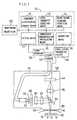
- FIG. 1is a view showing an essential part of the embodiment of the present invention.
- the plate 28substantially serves as a light-emitting face such that a real image is formed thereon via an optical system comprising lenses 30 and 32 and a dichroic mirror 34, a real image 36, the real image traversing blood vessels 12 that exist inside of a skin surface 16 of a living body.
- the filter 23needs to have a center wavelength of 550nm and a half width of 40nm.
- the plate 28may be a light diffusing board, for example that of a frost type manufactured by Sigma Optical Materials Co., Ltd.
- the region of the real image 36 on the plate 28 including the blood vessels 12is referred to as a region V.
- the reflection light from the region Vis received in a CCD 40 via the beam splitter 34 and a lens 38.
- the filter 23, the diffuser 26, the plate 28, the lenses 30, 32 and 38, the beam splitter 34 and the CCD 40are all accommodated in a probe 58.
- the probe 58has a tip 59 allowed to closely contact the skin surface 16 via a transparent plate 66 made of plastic or glass to give a stable image free from being shifted.
- Image signals output from each pixel of the CCD 40are processed by a video signal processing circuit 46.
- the video signal processing circuit 46successively forms one frame image per one thirtieth second, and the frame images thus formed are sequentially recorded in a video recorder (for example, a laser disk recorder) 50.
- a video recorderfor example, a laser disk recorder
- Reference numeral 70denotes analysis means for analyzing blood flowing through the blood vessels 12 contained in the detection region V by processing the captured image.
- the analysis means 70can be, for example, a commercially available personal computer.
- the analysis means 70comprises cutting means 71 for cutting (trimming) a predetermined region of the image output from a video recorder 50, frequency distribution forming means 72 for forming a frequency distribution curve (histogram) on intensity of each pixel of the cut predetermined region, light intensity detecting means 73 for detecting, when a frequency distribution curve shows maximum values at A and B (A ⁇ B), the intensity A as a light intensity I B of blood and the intensity B as a light intensity I T of a body tissue, blood vessel diameter detecting means 74 for detecting, with the light intensities, the diameter D of the blood vessel contained in the predetermined region of the cut image, hemoglobin concentration calculating means 75 for calculating the concentration of hemoglobin based on the light intensities I B and I T and the diameter D of the blood vessel, and correction means 76 for correcting, if necessary, the concentration of hemoglobin thus calculated.
- Each image or histogram formed in the analysis means 70is monitored by a monitoring television 80.
- the flowchart of FIG.2shows the procedure of calculation of the concentration of hemoglobin using the analysis means 70 which will be explained hereinafter
- the analysis means 70processes a plurality of frames or fields of images recorded in time series in the video recorder 50, by reading them sequentially.
- a frame of an image IM containing a blood vessel BLis cut as shown in FIG. 3 (step S1), followed by cutting a predetermined region AR (having, for example, 10 X 100 pixels) as shown in FIG. 4 (step S2).
- a distribution (histogram) of frequency on intensity of pixels of the predetermined region ARis prepared as shown in FIG. 5 (step S3), followed by detecting the lower intensity value as the light intensity I B of blood and the higher intensity value as the light intensity I T of the body tissue on the two maximum values of intensity (step S4).
- step S9The above operation of S1 to S8 is repeated by the number of a plurality of frames, i.e., by the number F of frames (step S9), followed by calculating the mean value C av by dividing Ct by the number F of frames (step S10), and correcting the mean value C av obtained in capillary and small arteries and veins such that it is translated into the value corresponding to medium-size and large arteries and veins, thereby calculating the concentration of hemoglobin HGB (step S11).
- this operationis conducted based on a correction function experimentally obtained.
- Blood vessels of the lips of a human beingwere imaged using the apparatus shown in FIG. 1, followed by measuring an intensity of scattered light I T of a body tissue and an intensity of scattered light I B of blood.
- the imagingwas performed such that incident light was applied on the blood vessels contained in the lips of the same human being, with the amount of the incident light varied at six stages.
- FIG. 6shows the characteristics of the light intensity I T of blood and the light intensity I B of the body tissue when the amount of incident light applied on the blood was varied.
- the concentration of hemoglobincan be measured using a light source having one wavelength (or one wavelength band), and eliminating the need of calibrating a concentration of hemoglobin depending on a light intensity.
Landscapes
- Health & Medical Sciences (AREA)
- Physics & Mathematics (AREA)
- Life Sciences & Earth Sciences (AREA)
- Medical Informatics (AREA)
- Surgery (AREA)
- Biophysics (AREA)
- Pathology (AREA)
- Engineering & Computer Science (AREA)
- Biomedical Technology (AREA)
- Heart & Thoracic Surgery (AREA)
- Spectroscopy & Molecular Physics (AREA)
- Molecular Biology (AREA)
- Optics & Photonics (AREA)
- Animal Behavior & Ethology (AREA)
- General Health & Medical Sciences (AREA)
- Public Health (AREA)
- Veterinary Medicine (AREA)
- Measurement Of The Respiration, Hearing Ability, Form, And Blood Characteristics Of Living Organisms (AREA)
- Investigating Or Analysing Materials By Optical Means (AREA)
- Length Measuring Devices By Optical Means (AREA)
- Investigating Or Analysing Biological Materials (AREA)
Description
- The present invention relates to an apparatus for analyzingblood in a non-invasive manner. Moreparticularly, it relates to an apparatus for measuring aconcentration of hemoglobin in blood without collecting or isolatingit.
- A conventional apparatus for measuring in vivo aconcentration of hemoglobin is utilized in applying a plurality oflight beams each having a different wavelength to a living body,substituting the values thus obtained with respect to eachwavelength, in the equation of modified Lambert-Beer's law asbelow to determine a concentration C of hemoglobin or an amount ΔCof change of the concentration of hemoglobin as solutions ofsimultaneous equations (see, for example, WO-A-93/11701 or Japanese UnexaminedPatent Publication No. HEI 2(1990)-95262). The equation is:
- The above conventional apparatus, however, has suffered fromsuch problems that a light source is required which applies aplurality of light beams each having different wavelengthindividually upon a living body and that the values obtained uponmeasurement need to be calibrated depending on an intensity of theincident light.
- The present invention has been conceived in view of theabove circumstances. Thus, an object of the present invention is toprovide an apparatus for measuring a concentration ofhemoglobin using a light source having one wavelength (or onewavelength band) and eliminating the need of calibrating the concentrationdepending on an intensity of incident light.
- The present invention provides an apparatus formeasuring a concentration of hemoglobin according to claim 1.
- FIG. 1 is a view showing the structure of an embodiment of thepresent invention.
- FIG. 2 is a flowchart showing the operation of the embodiment.
- FIG. 3 is a view explaining an example of a light intensityimage obtained in the embodiment.
- FIG. 4 is a view explaining a cut region of the image shown inFIG. 3.
- FIG. 5 is a histogram showing an example of a light intensitydistribution of the image captured in the embodiment.
- FIG. 6 is a graph showing the characteristics exhibited onmeasurement in the embodiment.
- The present invention will be detailed in conjunctionwith a preferred embodiment, which is not intended to limit the scope of the present invention.
- The apparatus is characterized by measuring a concentrationof hemoglobin in blood present in a living body in non-invasivemanner. The living body is that of mammals including human bodies.
- By the detection region referred to as in the light applicationmeans for applying light to the detection region containing the bloodvessel present in the living body is meant a predetermined regioncontaining the blood vessel that is present as it is in the living body,and is not meant part of the living body surgically removed in vitro.
- The thickness of the blood vessel contained in a subject regionis not limited, but capillary or small arteries and veins located asnear as a skin are preferable to produce a good result inreproduction. Incidentally, blood information obtained in capillaryor small arteries and veins can be translated into information onthick vessels (medium-size or large arteries and veins).
- By the light intensity of the body tissue is meant a lightintensity emitted from a portion of the body tissue which thus is alight intensity transmitted through or reflected from the bodytissue, and by the light of intensity of blood is meant likewise.
- In the light application means of the present invention, eithera continuous or an intermittent light source may be used; thecontinuous light source that continuously applies light to thedetection region includes a laser, a halogen lamp or a tungsten lampwhile the intermittent light source that applies light intermittentlyto the detection region includes a pulse laser (for example, 7000series manufactured by Spectra-Physics Co., Ltd.) and a flush lamp(for example, DSX series manufactured by Sugawara Laboratories, Inc., Japan).
- In addition to the above light source, preferably thelight application means may comprise (1) an optical fiber, (2) areflector, (3) a lens, (4) a slit or the like, all of which allow lightfrom the light source to be properly directed to the detection region.Alternatively, appropriate combinations such as those of (1) and (2);(1) and (3); (1), (2) and (3); (1), (2), (3) and (4); (2) and (3); and (2),(3) and (4) may be incorporated in the light application means.
- In this case, the reflector may be replaced with a prism. Inparticular, the light application means may include a polarizingmeans for applying polarizing light to the detection region.
- An example of the imaging means of the present invention is ageneral CCD image sensor.
- In an optical system for allowing the reflection light from thedetection region to be directed to the CCD image sensor, the imagingmeans may comprise an optical fiber, various kinds of reflectors, apolarizing element, a lens of each kind, a prism, a slit or a filter,and preferably comprises, if the reflection light from the detectionregion is faint, an image intensifier. The imaging means may furthercomprise polarizing means for removing unnecessary scattered lightemitted from the detection region.
- Desirably, the imaging means may comprise, in its signalprocessing system, a video signal processing circuit for processing,as video signals, output signals from each pixel of the CCD imagesensor while supplying scanning signals to the CCD image sensor,and a VTR or a laser disc recorder for recording the video signals.
- The light application means and imaging means may be a commercially available video microscope system.
- The analysis means may be a commercially available personalcomputer (for example, Power Mac manufactured by Apple ComputerInc.).
- The analysis means further comprises blood vessel diameterdetecting means for detecting a diameter of the blood vesselobtained in the captured image and calculating means for calculatingthe concentration of hemoglobin based on the detected diameter ofthe blood vessel.
- Preferably the calculating means comprises means forcalculating the concentration C of hemoglobin from the followingequation:
- Preferably the light intensity detecting means may comprisemeans for examining a frequency distribution on intensity of pixelsin the captured image and means for detecting, when the frequencydistribution shows peak values at intensity A and B (A < B), theluminance A as the light intensity of blood and the intensity B as thelight intensity of the body tissue.
- The light intensity detecting means of the presentinvention detects the light intensity IT of the blood tissue and thelight intensity IB of body from the image obtained with the imagingmeans.
- By the way, C=0 is obtained with respect to a portion of the body tissue. Then the equation (1) produces the following equation:
- With respect to blood, the equation is given:
- Subtracting the equation (2) from the equation (3) produces thefollowing equation:
- Here D represents a thickness of a blood layer which is aconstant determined by the diameter of the blood vessel. This valueis determined depending on the diameter obtained in the image. Inaddition, ε is a constant determined depending on wavelength of lightincident upon the detection region.
- Accordingly, the calculating means can calculate theconcentration C of hemoglobin from the equation (4).
- These and other objects of the present application will becomemore readily apparent from the detailed description givenhereinafter.
- FIG. 1 is a view showing an essential part of the embodimentof the present invention.
- Light emitted from a
light source 22 of a halogen lamp isapplied to adiffuser 26 via anoptical fiber 24 and afilter 23. Thelight is scattered by thediffuser 26 and uniformly applied to aplate 28. Thus, theplate 28 substantially serves as a light-emitting face such that a real image is formed thereon via an opticalsystemcomprising lenses dichroic mirror 34, areal image 36, the real image traversingblood vessels 12 that exist inside of askin surface 16 of a living body. Thefilter 23 needs to have a centerwavelength of 550nm and a half width of 40nm. Theplate 28 may bea light diffusing board, for example that of a frost typemanufactured by Sigma Optical Materials Co., Ltd. - The region of the
real image 36 on theplate 28 including theblood vessels 12 is referred to as a region V. The reflection lightfrom the region V is received in aCCD 40 via thebeam splitter 34and alens 38. - The
filter 23, thediffuser 26, theplate 28, thelenses beam splitter 34 and theCCD 40 are all accommodatedin aprobe 58. Theprobe 58 has atip 59 allowed to closely contacttheskin surface 16 via atransparent plate 66 made of plastic orglass to give a stable image free from being shifted. Imagesignals output from each pixel of theCCD 40 are processed by avideosignal processing circuit 46. The videosignal processingcircuit 46 successively forms one frame image per one thirtiethsecond, and the frame images thus formed are sequentiallyrecorded in a video recorder (for example, a laser disk recorder) 50. Reference numeral 70 denotes analysis means for analyzingblood flowing through theblood vessels 12 contained in thedetection region V by processing the captured image. The analysismeans 70 can be, for example, a commercially available personalcomputer.- The analysis means 70 comprises cutting means 71 for cutting (trimming) a predetermined region of the image output from a
videorecorder 50, frequencydistribution forming means 72 for forming afrequency distribution curve (histogram) on intensity of each pixelof the cut predetermined region, lightintensity detecting means 73for detecting, when a frequency distribution curve shows maximumvalues at A and B (A < B), the intensity A as a light intensity IB ofblood and the intensity B as a light intensity IT of a body tissue,blood vessel diameter detecting means 74 for detecting, with thelight intensities, the diameter D of the blood vessel contained in thepredetermined region of the cut image, hemoglobin concentrationcalculating means 75 for calculating the concentration ofhemoglobin based on the light intensities IB and IT and the diameterD of the blood vessel, and correction means 76 for correcting, ifnecessary, the concentration of hemoglobin thus calculated. Eachimage or histogram formed in the analysis means 70 is monitored byamonitoring television 80. - The flowchart of FIG.2 shows the procedure of calculation ofthe concentration of hemoglobin using the analysis means 70 whichwill be explained hereinafter
- The analysis means 70 processes a plurality of frames orfields of images recorded in time series in the
video recorder 50, byreading them sequentially. - A frame of an image IM containing a blood vessel BL is cut asshown in FIG. 3 (step S1), followed by cutting a predeterminedregion AR (having, for example, 10
X 100 pixels) as shown in FIG. 4(step S2). - A distribution (histogram) of frequency on intensity of pixels of the predetermined region AR is prepared as shown in FIG. 5 (stepS3), followed by detecting the lower intensity value as the lightintensity IB of blood and the higher intensity value as the lightintensity IT of the body tissue on the two maximum values ofintensity (step S4).
- The calculation of r=log (IT/IB) is performed (step S5),followed by determining the diameter D of the blood vessel BLfrom the image (step S6), performing the calculation of C=r/εD(step S7), and adding the value to Ct which is already calculated(step S8).
- The above operation of S1 to S8 is repeated by the number of aplurality of frames, i.e., by the number F of frames (step S9),followed by calculating the mean value Cav by dividing Ct by thenumber F of frames (step S10), and correcting the mean value Cavobtained in capillary and small arteries and veins such that it istranslated into the value corresponding to medium-size and largearteries and veins, thereby calculating the concentration ofhemoglobin HGB (step S11). In practice, this operation is conductedbased on a correction function experimentally obtained.
- Blood vessels of the lips of a human being were imaged usingthe apparatus shown in FIG. 1, followed by measuring an intensityof scattered light IT of a body tissue and an intensity of scatteredlight IB of blood.
- The imaging was performed such that incident light wasapplied on the blood vessels contained in the lips of the samehuman being, with the amount of the incident light varied at six stages.
- FIG. 6 shows the characteristics of the light intensity IT ofblood and the light intensity IB of the body tissue when the amountof incident light applied on the blood was varied.
- From FIG. 6, it is understood that the light intensity IB ofblood over the light intensity IT of the body tissue is constantregardless of change on the amount of incident light, thuseliminating the need of calibrating the measured concentration ofhemoglobin with respect to the intensity of the incident light.
- An examination was made on the correlation between aconcentration of hemoglobin HGB value X measured by a bloodanalyzer and a concentration of hemoglobin HGB value Ycalculated in accordance with the present invention with respectto nine subjects having a hemoglobin value of 14 to 18 [g/dI]. Thecorrelation coefficient was 0.861, thus proving that the apparatusof the present invention is sufficiently practical.
- In accordance with the present invention, the concentrationof hemoglobin can be measured using a light source having onewavelength (or one wavelength band), and eliminating the need ofcalibrating a concentration of hemoglobin depending on a lightintensity.
Claims (3)
- An apparatus for measuring a concentration of hemoglobincomprising:light application means (22, 23, 24, 26, 28, 30, 32, 34) forapplying light to a detection region (V) containing a blood vessel (12) presentin a living body, said light application means comprising a filter (23) having adetermined wavelength or wavelength band,imaging means (38, 40) for capturing an image of thedetection region (V) to which the light is applied, andanalysis means for analyzing the concentration ofhemoglobin in blood flowing through the blood vessel by processing thecaptured image, the analysis means (70) comprising:light intensity detection means (73) for detecting a lightintensity of a body tissue and a light intensity of blood using the capturedimage,blood vessel diameter detecting means (74) for detecting thediameter of the blood vessel (12) obtained in the captured image,calculating means (75) for calculating the concentration ofhemoglobin from a ratio of the light intensity of the body tissue relative to thelight intensity of blood thus detected, the detected blood vessel diameter andthe wavelength of light incident upon the detection region.
- The apparatus for measuring the concentration ofhemoglobin of claim 1, wherein the calculating means (70) comprises means(75) for calculating a concentration C of hemoglobin from the followingequation:
- The apparatus for measuring the concentration ofhemoglobin of claim 1, wherein the light intensity detecting means (73)comprises means (72) for examining a frequency distribution on intensity ofpixels in the captured image and means for detecting an intensity A as thelight intensity of blood and an intensity B as the light intensity of the bodytissue when the frequency distribution shows peak values at the intensities Aand B, where A < B.
Applications Claiming Priority (3)
| Application Number | Priority Date | Filing Date | Title |
|---|---|---|---|
| JP280936/94 | 1994-11-15 | ||
| JP28093694 | 1994-11-15 | ||
| JP28093694AJP3562847B2 (en) | 1994-11-15 | 1994-11-15 | Hemoglobin concentration measuring device |
Publications (3)
| Publication Number | Publication Date |
|---|---|
| EP0712602A2 EP0712602A2 (en) | 1996-05-22 |
| EP0712602A3 EP0712602A3 (en) | 1996-05-29 |
| EP0712602B1true EP0712602B1 (en) | 2003-08-27 |
Family
ID=17631992
Family Applications (1)
| Application Number | Title | Priority Date | Filing Date |
|---|---|---|---|
| EP95402541AExpired - LifetimeEP0712602B1 (en) | 1994-11-15 | 1995-11-14 | Apparatus for measuring concentration of hemoglobin |
Country Status (4)
| Country | Link |
|---|---|
| US (1) | US5722398A (en) |
| EP (1) | EP0712602B1 (en) |
| JP (1) | JP3562847B2 (en) |
| DE (1) | DE69531588T2 (en) |
Cited By (1)
| Publication number | Priority date | Publication date | Assignee | Title |
|---|---|---|---|---|
| CN101371800B (en)* | 2007-08-23 | 2012-05-30 | 西门子公司 | Sensor capable of certifying substance in living body |
Families Citing this family (37)
| Publication number | Priority date | Publication date | Assignee | Title |
|---|---|---|---|---|
| US7047064B1 (en) | 1995-07-13 | 2006-05-16 | Lucid, Inc. | Microscopic imaging apparatus and method |
| US5983120A (en)* | 1995-10-23 | 1999-11-09 | Cytometrics, Inc. | Method and apparatus for reflected imaging analysis |
| KR100350022B1 (en)* | 1995-12-27 | 2002-12-26 | 시스멕스 가부시키가이샤 | Non-Invasive Blood Test Device |
| US6424852B1 (en)* | 1996-10-18 | 2002-07-23 | Lucid, Inc. | System for confocal imaging within dermal tissue |
| US6745067B1 (en)* | 1998-09-14 | 2004-06-01 | Lucid, Inc. | System for marking the locations of imaged tissue with respect to the surface of the tissue |
| US5974338A (en)* | 1997-04-15 | 1999-10-26 | Toa Medical Electronics Co., Ltd. | Non-invasive blood analyzer |
| US6064898A (en) | 1998-09-21 | 2000-05-16 | Essential Medical Devices | Non-invasive blood component analyzer |
| US6305804B1 (en) | 1999-03-25 | 2001-10-23 | Fovioptics, Inc. | Non-invasive measurement of blood component using retinal imaging |
| US6363269B1 (en) | 1999-12-17 | 2002-03-26 | Datex-Ohmeda, Inc. | Synchronized modulation/demodulation method and apparatus for frequency division multiplexed spectrophotometric system |
| US6594513B1 (en)* | 2000-01-12 | 2003-07-15 | Paul D. Jobsis | Method and apparatus for determining oxygen saturation of blood in body organs |
| WO2001078589A1 (en)* | 2000-04-14 | 2001-10-25 | Fovioptics, Inc. | Non-invasive measurement of blood components using retinal imaging |
| AU2001290574A1 (en)* | 2000-08-25 | 2002-03-04 | Cytometrics, Llc | Method and apparatus for measuring the hemoglobin concentration and/or hematocrit in whole blood using diffuse light |
| EP1335665A2 (en)* | 2000-11-15 | 2003-08-20 | Cytometrics, LLC. | Measuring haematocrit in blood vessels |
| US20040024296A1 (en)* | 2001-08-27 | 2004-02-05 | Krotkov Eric P. | System, method and computer program product for screening a spectral image |
| US6650915B2 (en) | 2001-09-13 | 2003-11-18 | Fovioptics, Inc. | Non-invasive measurement of blood analytes using photodynamics |
| US20050010091A1 (en)* | 2003-06-10 | 2005-01-13 | Woods Joe W. | Non-invasive measurement of blood glucose using retinal imaging |
| DK1494579T3 (en)* | 2002-04-02 | 2011-11-14 | Yeda Res & Dev | Characterization of moving objects in a stationary background |
| US6895264B2 (en)* | 2002-08-26 | 2005-05-17 | Fovioptics Inc. | Non-invasive psychophysical measurement of glucose using photodynamics |
| JP4236950B2 (en)* | 2003-02-13 | 2009-03-11 | シスメックス株式会社 | Non-invasive living body measurement device |
| JP4537681B2 (en)* | 2003-09-24 | 2010-09-01 | 株式会社東芝 | Blood flow analyzer |
| DE102004017130B4 (en)* | 2004-04-02 | 2006-01-19 | Imedos Gmbh | Method for measuring the vessel diameter of optically accessible blood vessels |
| WO2006100685A2 (en)* | 2005-03-25 | 2006-09-28 | Cnoga Holdings Ltd | Optical sensor device and image processing unit for measuring chemical concentrations, chemical saturations and biophysical parameters |
| CN101212928B (en)* | 2005-05-06 | 2011-02-09 | 耶德研究和发展有限公司 | Imaging and analysis of movement of erythrocytes |
| ES2883201T3 (en)* | 2006-04-08 | 2021-12-07 | Hoffmann La Roche | Optical data analysis with the help of histograms |
| EP1905351B1 (en)* | 2006-09-29 | 2016-10-26 | Sysmex Corporation | Nonivasive living body measuring device and noninvasive living body measuring method |
| JP4963921B2 (en)* | 2006-09-29 | 2012-06-27 | シスメックス株式会社 | Non-invasive living body measurement device |
| EP2172154A4 (en)* | 2007-07-31 | 2013-09-25 | Sysmex Corp | Noninvasive biometric device and noninvasive biometric method |
| US8403862B2 (en)* | 2007-12-20 | 2013-03-26 | Yeda Research And Development Co. Ltd. | Time-based imaging |
| JP5137606B2 (en)* | 2008-02-12 | 2013-02-06 | シスメックス株式会社 | Non-invasive blood component measuring apparatus, method and computer program for non-invasively measuring blood components |
| GB0814419D0 (en) | 2008-08-08 | 2008-09-10 | Health Smart Ltd | Blood analysis |
| JP2010246578A (en)* | 2009-04-10 | 2010-11-04 | Utsunomiya Univ | Method and apparatus for testing blood cholesterol |
| US8488111B2 (en) | 2011-04-15 | 2013-07-16 | Constitution Medical, Inc. | Measuring volume and constituents of cells |
| TWM499642U (en)* | 2013-10-18 | 2015-04-21 | Star Techn Inc | Test probe card |
| JP2017018483A (en)* | 2015-07-14 | 2017-01-26 | オリンパス株式会社 | Medical probe |
| EP3383245B1 (en) | 2015-11-30 | 2024-01-10 | Technion Research & Development Foundation Limited | Hemoglobin measurement from a single vessel |
| WO2020095739A1 (en)* | 2018-11-06 | 2020-05-14 | ソニー株式会社 | Information processing device, information processing method, and program |
| CN110584678B (en)* | 2019-09-06 | 2024-05-10 | 广东宝莱特医用科技股份有限公司 | Method and device for measuring blood volume change rate |
Family Cites Families (4)
| Publication number | Priority date | Publication date | Assignee | Title |
|---|---|---|---|---|
| US4998533A (en)* | 1986-07-15 | 1991-03-12 | Winkelman James W | Apparatus and method for in vivo analysis of red and white blood cell indices |
| JPH0295262A (en) | 1988-09-30 | 1990-04-06 | Shimadzu Corp | Hemoglobin measuring method and measuring device |
| WO1992021283A1 (en)* | 1991-06-06 | 1992-12-10 | Somanetics Corporation | Optical cerebral oximeter |
| US5277181A (en)* | 1991-12-12 | 1994-01-11 | Vivascan Corporation | Noninvasive measurement of hematocrit and hemoglobin content by differential optical analysis |
- 1994
- 1994-11-15JPJP28093694Apatent/JP3562847B2/ennot_activeExpired - Lifetime
- 1995
- 1995-11-14DEDE69531588Tpatent/DE69531588T2/ennot_activeExpired - Lifetime
- 1995-11-14EPEP95402541Apatent/EP0712602B1/ennot_activeExpired - Lifetime
- 1995-11-14USUS08/557,573patent/US5722398A/ennot_activeExpired - Lifetime
Cited By (1)
| Publication number | Priority date | Publication date | Assignee | Title |
|---|---|---|---|---|
| CN101371800B (en)* | 2007-08-23 | 2012-05-30 | 西门子公司 | Sensor capable of certifying substance in living body |
Also Published As
| Publication number | Publication date |
|---|---|
| US5722398A (en) | 1998-03-03 |
| JP3562847B2 (en) | 2004-09-08 |
| EP0712602A2 (en) | 1996-05-22 |
| EP0712602A3 (en) | 1996-05-29 |
| JPH08140961A (en) | 1996-06-04 |
| DE69531588T2 (en) | 2004-06-24 |
| DE69531588D1 (en) | 2003-10-02 |
Similar Documents
| Publication | Publication Date | Title |
|---|---|---|
| EP0712602B1 (en) | Apparatus for measuring concentration of hemoglobin | |
| EP0641542B1 (en) | Non-invasive blood analyzer | |
| JP3364323B2 (en) | Non-invasive blood analyzer | |
| US5769076A (en) | Non-invasive blood analyzer and method using the same | |
| US5741213A (en) | Apparatus for analyzing blood | |
| US4998533A (en) | Apparatus and method for in vivo analysis of red and white blood cell indices | |
| JP3566756B2 (en) | Non-invasive blood analyzer and method | |
| US5791345A (en) | Non-invasive blood analyzer | |
| US5333610A (en) | Absorption spectrum determining method and spectrometric measuring apparatus for light-diffusive object using the method | |
| US20050043597A1 (en) | Optical vivo probe of analyte concentration within the sterile matrix under the human nail | |
| EA001936B1 (en) | Method and apparatus for reflected imaging analysis | |
| EP0714628B1 (en) | Non-invasive blood analyzer | |
| JPH05293108A (en) | Method for detecting aberration of skin, especially melanoma, and device therefor | |
| KR19990076821A (en) | Non-Invasive Blood Test Device | |
| FR2768043A1 (en) | METHOD AND DEVICE FOR MEASURING CONCENTRATIONS OF BLOOD COMPONENTS | |
| US20060142662A1 (en) | Analysis apparatus and method comprising auto-focusing means | |
| US6343228B1 (en) | Method and apparatus for fluorescence imaging of tissue | |
| JPS61159936A (en) | Spectral image pick-up apparatus of biological tissue | |
| US7756569B2 (en) | Method for measuring the vessel diameter of optically accessible blood vessels | |
| JP2000300568A (en) | Imaging apparatus for peripheral blood vessel | |
| Lompado et al. | Multispectral confocal scanning laser ophthalmoscope for retinal vessel oximetry | |
| WO1988000447A1 (en) | In vivo analysis of red blood cell indices | |
| EP0868143B1 (en) | Apparatus for detecting malignancies in living tissue | |
| JP2826916B2 (en) | Method and apparatus for detecting porphyrin in subject | |
| JPH04319345A (en) | Photo-tomographic imaging system |
Legal Events
| Date | Code | Title | Description |
|---|---|---|---|
| PUAI | Public reference made under article 153(3) epc to a published international application that has entered the european phase | Free format text:ORIGINAL CODE: 0009012 | |
| PUAL | Search report despatched | Free format text:ORIGINAL CODE: 0009013 | |
| AK | Designated contracting states | Kind code of ref document:A2 Designated state(s):DE FR GB IT | |
| AK | Designated contracting states | Kind code of ref document:A3 Designated state(s):DE FR GB IT | |
| 17P | Request for examination filed | Effective date:19961119 | |
| RAP1 | Party data changed (applicant data changed or rights of an application transferred) | Owner name:ISHIHARA, KEN Owner name:SYSMEX CORPORATION | |
| 17Q | First examination report despatched | Effective date:20010814 | |
| RTI1 | Title (correction) | Free format text:APPARATUS FOR MEASURING CONCENTRATION OF HEMOGLOBIN | |
| GRAH | Despatch of communication of intention to grant a patent | Free format text:ORIGINAL CODE: EPIDOS IGRA | |
| RTI1 | Title (correction) | Free format text:APPARATUS FOR MEASURING CONCENTRATION OF HEMOGLOBIN | |
| RTI1 | Title (correction) | Free format text:APPARATUS FOR MEASURING CONCENTRATION OF HEMOGLOBIN | |
| GRAH | Despatch of communication of intention to grant a patent | Free format text:ORIGINAL CODE: EPIDOS IGRA | |
| GRAA | (expected) grant | Free format text:ORIGINAL CODE: 0009210 | |
| GRAL | Information related to payment of fee for publishing/printing deleted | Free format text:ORIGINAL CODE: EPIDOSDIGR3 | |
| GRAS | Grant fee paid | Free format text:ORIGINAL CODE: EPIDOSNIGR3 | |
| AK | Designated contracting states | Designated state(s):DE FR GB IT | |
| REG | Reference to a national code | Ref country code:GB Ref legal event code:FG4D | |
| REF | Corresponds to: | Ref document number:69531588 Country of ref document:DE Date of ref document:20031002 Kind code of ref document:P | |
| ET | Fr: translation filed | ||
| PLBE | No opposition filed within time limit | Free format text:ORIGINAL CODE: 0009261 | |
| STAA | Information on the status of an ep patent application or granted ep patent | Free format text:STATUS: NO OPPOSITION FILED WITHIN TIME LIMIT | |
| 26N | No opposition filed | Effective date:20040528 | |
| PGFP | Annual fee paid to national office [announced via postgrant information from national office to epo] | Ref country code:IT Payment date:20061130 Year of fee payment:12 | |
| PG25 | Lapsed in a contracting state [announced via postgrant information from national office to epo] | Ref country code:IT Free format text:LAPSE BECAUSE OF NON-PAYMENT OF DUE FEES Effective date:20071114 | |
| PGFP | Annual fee paid to national office [announced via postgrant information from national office to epo] | Ref country code:DE Payment date:20141111 Year of fee payment:20 Ref country code:GB Payment date:20141112 Year of fee payment:20 Ref country code:FR Payment date:20141110 Year of fee payment:20 | |
| REG | Reference to a national code | Ref country code:DE Ref legal event code:R071 Ref document number:69531588 Country of ref document:DE | |
| REG | Reference to a national code | Ref country code:GB Ref legal event code:PE20 Expiry date:20151113 | |
| PG25 | Lapsed in a contracting state [announced via postgrant information from national office to epo] | Ref country code:GB Free format text:LAPSE BECAUSE OF EXPIRATION OF PROTECTION Effective date:20151113 |