EP0661029B1 - Apparatus for ultrasonic medical treatment with optimum ultrasonic irradiation control - Google Patents
Apparatus for ultrasonic medical treatment with optimum ultrasonic irradiation controlDownload PDFInfo
- Publication number
- EP0661029B1 EP0661029B1EP94120831AEP94120831AEP0661029B1EP 0661029 B1EP0661029 B1EP 0661029B1EP 94120831 AEP94120831 AEP 94120831AEP 94120831 AEP94120831 AEP 94120831AEP 0661029 B1EP0661029 B1EP 0661029B1
- Authority
- EP
- European Patent Office
- Prior art keywords
- ultrasonic
- ultrasonic wave
- generation source
- wave generation
- ultrasonic waves
- Prior art date
- Legal status (The legal status is an assumption and is not a legal conclusion. Google has not performed a legal analysis and makes no representation as to the accuracy of the status listed.)
- Expired - Lifetime
Links
- 238000010438heat treatmentMethods0.000claimsdescription11
- 230000000694effectsEffects0.000claimsdescription9
- 230000001678irradiating effectEffects0.000claimsdescription5
- 230000006378damageEffects0.000claimsdescription3
- 238000002604ultrasonographyMethods0.000description21
- 206010028980NeoplasmDiseases0.000description14
- 230000007850degenerationEffects0.000description12
- 238000000034methodMethods0.000description12
- 239000000523sampleSubstances0.000description8
- 238000010586diagramMethods0.000description7
- 206010020843HyperthermiaDiseases0.000description6
- 201000011510cancerDiseases0.000description6
- 230000036031hyperthermiaEffects0.000description6
- 230000008878couplingEffects0.000description4
- 238000010168coupling processMethods0.000description4
- 238000005859coupling reactionMethods0.000description4
- 238000002474experimental methodMethods0.000description4
- 238000004364calculation methodMethods0.000description3
- 230000008859changeEffects0.000description3
- 239000012530fluidSubstances0.000description3
- 230000020169heat generationEffects0.000description3
- 210000000056organAnatomy0.000description3
- 238000004088simulationMethods0.000description3
- 238000010792warmingMethods0.000description3
- XLYOFNOQVPJJNP-UHFFFAOYSA-NwaterSubstancesOXLYOFNOQVPJJNP-UHFFFAOYSA-N0.000description3
- 102000004190EnzymesHuman genes0.000description2
- 108090000790EnzymesProteins0.000description2
- 238000006243chemical reactionMethods0.000description2
- 238000004925denaturationMethods0.000description2
- 230000036425denaturationEffects0.000description2
- 230000002779inactivationEffects0.000description2
- 238000002686lithotriptorMethods0.000description2
- 102000004169proteins and genesHuman genes0.000description2
- 108090000623proteins and genesProteins0.000description2
- 230000004083survival effectEffects0.000description2
- 206010057040Temperature intoleranceDiseases0.000description1
- 230000002238attenuated effectEffects0.000description1
- 230000005540biological transmissionEffects0.000description1
- 230000017531blood circulationEffects0.000description1
- 230000036760body temperatureEffects0.000description1
- 239000007822coupling agentSubstances0.000description1
- 238000011161developmentMethods0.000description1
- 239000003814drugSubstances0.000description1
- 230000005674electromagnetic inductionEffects0.000description1
- 238000004880explosionMethods0.000description1
- 230000006870functionEffects0.000description1
- 230000008543heat sensitivityEffects0.000description1
- 238000003384imaging methodMethods0.000description1
- 235000015110jelliesNutrition0.000description1
- 239000008274jellySubstances0.000description1
- 238000005259measurementMethods0.000description1
- 230000004048modificationEffects0.000description1
- 238000012986modificationMethods0.000description1
- 230000036632reaction speedEffects0.000description1
Images


Classifications
- A—HUMAN NECESSITIES
- A61—MEDICAL OR VETERINARY SCIENCE; HYGIENE
- A61N—ELECTROTHERAPY; MAGNETOTHERAPY; RADIATION THERAPY; ULTRASOUND THERAPY
- A61N7/00—Ultrasound therapy
- A61N7/02—Localised ultrasound hyperthermia
- A—HUMAN NECESSITIES
- A61—MEDICAL OR VETERINARY SCIENCE; HYGIENE
- A61B—DIAGNOSIS; SURGERY; IDENTIFICATION
- A61B17/00—Surgical instruments, devices or methods
- A61B2017/00017—Electrical control of surgical instruments
- A61B2017/00022—Sensing or detecting at the treatment site
- A61B2017/00106—Sensing or detecting at the treatment site ultrasonic
Definitions
- the present inventionrelates to an ultrasonic medical treatment apparatus for applying a medical treatment to a tumor and the like within a living body by using ultrasonic waves. More particularly, the invention relates to an ultrasonic medical treatment apparatus, comprising an applicator having an ultrasonic wave generation source for irradiating ultrasonic waves to a treatment target portion within a body to be examined; control means for controlling the applicator; and an energy obtaining means.
- an ultrasonic medical treatment apparatusis disclosed in EP-A-0 162 735.
- EP-A-0 170 416discloses an ultrasound hyperthermia apparatus having a tomographic ultrasound probe for obtaining a tomographic image and heating applicator for heating a tumor portion.
- a position detectoris provided to detect the positional relationship of the tomographic ultrasound probe and the heating applicator with respect to a living organism and a hot spot detector detects the position of a focusing point, i.e., a hot spot of the ultrasound radiated from the heating applicator in accordance with position data from the position detector, thereby generating an image signal indicating the hot spot.
- MITMinimum Invasive Treatment
- this MITis a lithotriptor apparatus for applying a lithotripsy treatment to a calculus non-invasively, which has revolutionized the medical treatment of the urological calculus.
- this lithotriptor apparatusmany different types of the intense ultrasonic wave generation source are available, including an underwater discharge type, an electromagnetic induction type, a micro-explosion type, and a piezoelectric type.
- the piezoelectric typehas many advantageous features such as that the focal point is small, that no articles of consumption is required, that the pressure of the intense ultrasonic waves can be controlled freely, and that the focal point position can be controlled freely by a phase control of driving voltages to be applied to a plurality of piezoelectric transducer elements, in spite of a drawback that the pressure of the intense ultrasonic waves is rather small.
- a phase control of driving voltages to be applied to a plurality of piezoelectric transducer elementsin spite of a drawback that the pressure of the intense ultrasonic waves is rather small.
- this MITis also attracting attentions in the field of the cancer treatment.
- its medical treatmentis still largely relying on the surgical operations today and there are many incidences of severely damaging an original function and/or an outward appearance of an organ with a cancer, such that a heavy burden would be left on the patient even if it is possible to prolong a life of a patient.
- QOLQuality Of Life
- This hyperthermia treatment methodis a medical treatment method in which cancer cells are selectively killed by warming and maintaining a diseased portion at a temperature over 42.5 °C, utilizing different heat sensitivities of tumor tissues and normal tissues.
- a scheme of warminga scheme using the electromagnetic waves such as microwaves has been developed first, but in this scheme, it is difficult to warm a tumor at a deep part of a body selectively due to the electrical property of the living body, so that the satisfactory medical treatment result cannot be expected for a tumor at a part deeper than 5 cm. For this reason, for a treatment of a tumor at a deep part, a warming scheme using the ultrasonic energy having superior focusing and depth transmission properties has been proposed (Japanese Patent Application Laid Open No. 61-13955).
- this type of the medical treatment methodstill has a drawback in that, as a very high energy is to be concentrated in a localized region, there is a possibility for causing the side effect such as a degeneration due to heat generation and a mechanical destruction of tissues when the acoustic characteristic changes at a tissue boundary surface of an organ or the like located in the ultrasonic wave passing region.
- the present inventionrelates to an ultrasonic medical treatment apparatus according to claim 1.
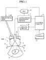
- FIG. 1one embodiment of an apparatus for the ultrasonic medical treatment according to the present invention will be described in detail.
- the apparatushas a configuration as shown in Fig. 1, in which an applicator 100 comprises a piezoelectric element group 1 formed by one or a plurality of piezoelectric transducer elements for irradiating intense treatment ultrasonic waves, and a water bag 2 filled with a coupling fluid for leading the intense ultrasonic waves from the piezoelectric element group 1.
- the applicator 100is mounted on a body surface 3S of the patient 3, with an ultrasonic jelly (not shown) applied between the water bag 2 and a skin of the body surface 3S to secure the contact.
- the driving circuit 8drives the piezoelectric element group 1 to irradiate the intense ultrasonic waves toward the focal point 5.
- the applicator 100itself can be moved by a mechanical arm 10 under a control of a control circuit 11 according to data entered by an operator through a console 12.
- the applicator 100is further equipped with an ultrasound probe 6 for transmitting and receiving imaging ultrasonic waves, according to which ultrasound images are reconstructed as the B mode images by an ultrasound diagnostic device 7 and displayed on a CRT 9.
- control circuit 11carries out the optimum control of the ultrasonic wave irradiation time as follows.
- a position of the focal point 5 in a depth direction from the body surface 3Sis measured by using the ultrasound diagnostic image displayed on the CRT 9, and that position is specified as a reference point from the console 12. Then, at a time of irradiating the intense ultrasonic waves while scanning the focal point 5 thoroughly over the entire diseased portion 4 by controlling the mechanical arm 10 from the control circuit 11, as the depth of the focal point 5 varies depending on the position of the focal point 5, the irradiation is carried out while controlling the driving time of the driving circuit 8 from the control circuit 11 according to the depth of the focal point 5.
- the optimum driving timeis determined as follows.
- the burning at the body surface 3Sis caused by the difference in the acoustic characteristic between the skin and the coupling fluid or the internal tissues, and the attenuation due to the skin tissue can be considered as proportional to the frequency f, so that the level of degeneration at the body surface 3S depends on a product of the ultrasonic energy density W S and the frequency f, and it can be deduced that the burning (thermal degeneration) at the body surface 3S will not be caused by the continuous ultrasonic waves irradiation for a period of time less than or equal to t S satisfying the following equation (1).
- W S ⁇ fI S ⁇ t S ⁇ f ⁇ 124 [MHz ⁇ J/cm 2 ]
- the ultrasonic wave intensity Is at the body surface 3Scan be expressed by the following equation (2).
- IsPk/S
- r Sd ⁇ /2 ⁇ R 2 - ( ⁇ /2) 2 ⁇ 1/2
- the optimum irradiation time for the intense ultrasonic wavescan be determined to be a longer time t 1 for a deeper portion A 1 of the diseased portion 4 and a shorter time t 2 for a shallower portion A 2 .
- the focal point ultrasonic wave intensity in a non-attenuative mediumdepends on the focusing level (i.e., the opening diameter ⁇ [cm] and the focal point distance R [cm]) and the frequency f [MHz] of the ultrasonic waves, as indicated in the following equations (7) to (9).
- the intensity and the irradiation time of the ultrasonic waves to be irradiatedcan be determined as shown in Figs. 4A and 4B, respectively, and the temperatures at the focal point 5a or 5b and the body surface 3S are going to change as indicated in Figs. 5A and 5B, respectively.
- a time t kindicates a time determined from the Arrhenius' equation and the survival rate curve which indicates a sufficient time for heating at a temperature T or above in order to kill the cells.
- the focal point peak ultrasonic wave intensity I Pcan be determined, and the minimum irradiation time t P required for obtaining the temperature rise of T °C at the focal point 5 and the maximum irradiation time t S required for not causing the burning (thermal degeneration) at the body surface 3S can be obtained from the above equations (1) and (4).
- the focusing level and the driving frequency of the piezoelectric transducer element and the focal point depth at a time of mounting the applicator 100 on the body surface 3Sshould preferably be determined such that t P ⁇ t S .
- the intense ultrasonic wavesare irradiated only for the minimum irradiation time t P plus the time t k for maintaining the target temperature which is determined by the Arrhenius' equation, as indicated in Figs. 4A and 4B.
- This optimum irradiation timevaries according to the depth of the focal point position when the focal point 5 is scanned over the entire diseased portion 4 thoroughly, so that it is calculated and controlled at the control circuit 11 every time the focal point position is changed. Else, it is also possible to set up the irradiation plan according to the shape of the diseased portion 4 at a time of the initial positioning, store the optimum irradiation time pre-calculated for each planned irradiation position in a memory (not shown), and carry out the medical treatment according to the stored values.
- an apparatus for the ultrasonic medical treatmentcapable of optimally controlling the irradiation time of the intense ultrasonic waves so as to achieve the sufficient medical treatment effect at the diseased portion while preventing the damaging (side effect) such as the burning at the body surface.
- the essence of the apparatus and methodlies in the optimum control of the ultrasonic energy to be irradiated to a tissue boundary surface such as a body surface of a body to be examined where the acoustic impedance changes within the ultrasonic wave passing region, so that instead of controlling the irradiation time as described in the above embodiment, it is also possible to control the focal point ultrasonic wave intensity, i.e., the driving power, for the predetermined irradiation time according to the above equations (1) and (4).
- the ultrasonic wave intensity I S at the body surfaceaccurately by a simulation in which spherical waves that can be irradiated from a plurality of acoustic points assumed to be arranged at equal intervals on a spherical shell surface of the piezoelectric transducer are superposed along time axis or frequency axis to simulate the actual ultrasonic wave irradiation, for example.
- the boundary surface of the body surface and the coupling fluidcan be recognized on the ultrasound image as a high echo region, so that it is also possible to specify the boundary surface recognized on the ultrasound image by an auxiliary input device such as a light pen on the displayed ultrasound image, as indicated in Fig. 6, or to apply an image recognition for extracting the boundary surface closest to the ultrasound probe 6 by using an appropriate reflection wave threshold.
- an auxiliary input devicesuch as a light pen on the displayed ultrasound image, as indicated in Fig. 6, or to apply an image recognition for extracting the boundary surface closest to the ultrasound probe 6 by using an appropriate reflection wave threshold.
- the area of the boundary surface within the intense ultrasonic wave passing regioncan be determined directly from the displayed ultrasound images, and used as the body surface passing area S in the calculation of the ultrasonic wave intensity I S at the body surface.
- the MRI device or the CT devicemay be utilized to acquire the 3D images of the patient in obtaining the body surface passing area S.
- the focal point peak ultrasonic wave intensity I Pit is possible to obtain this focal point peak ultrasonic wave intensity I P easily by a simulation in which spherical waves that can be irradiated from a plurality of acoustic points assumed to be arranged at equal intervals on a piezoelectric transducer surface are superposed while taking the attenuation into account, so as to simulate the actual ultrasonic wave irradiation, for example. In this manner, the accurate intensity distribution can be obtained regardless of the shape of the piezoelectric transducer. Moreover, it is also possible to deal with the focal point scanning using the two dimensional array in this manner.
- the ultrasonic energy density at the body surface and the focal point peak ultrasonic wave intensitycan be calculated from the relative position of the applicator and the diseased portion within the patient and the phase control values of the piezoelectric transducer elements similarly as in the embodiment described above.
Landscapes
- Health & Medical Sciences (AREA)
- Engineering & Computer Science (AREA)
- Biomedical Technology (AREA)
- Nuclear Medicine, Radiotherapy & Molecular Imaging (AREA)
- Radiology & Medical Imaging (AREA)
- Life Sciences & Earth Sciences (AREA)
- Animal Behavior & Ethology (AREA)
- General Health & Medical Sciences (AREA)
- Public Health (AREA)
- Veterinary Medicine (AREA)
- Thermotherapy And Cooling Therapy Devices (AREA)
- Surgical Instruments (AREA)
- Ultra Sonic Daignosis Equipment (AREA)
Description
- The present invention relates to an ultrasonic medicaltreatment apparatus for applying a medical treatment to atumor and the like within a living body by using ultrasonicwaves. More particularly, the invention relates to anultrasonic medical treatment apparatus, comprisingan applicator having an ultrasonic wave generation sourcefor irradiating ultrasonic waves to a treatment target portionwithin a body to be examined;control means for controlling the applicator;and an energy obtaining means. Such atreatment apparatus is disclosed in EP-A-0 162 735.
- EP-A-0 170 416 discloses an ultrasound hyperthermiaapparatus having a tomographic ultrasound probe for obtaininga tomographic image and heating applicator for heating a tumorportion. A position detector is provided to detect thepositional relationship of the tomographic ultrasound probeand the heating applicator with respect to a living organismand a hot spot detector detects the position of a focusingpoint, i.e., a hot spot of the ultrasound radiated from theheating applicator in accordance with position data from theposition detector, thereby generating an image signalindicating the hot spot.
- In recent years, a type of medical treatment calledMIT (Minimum Invasive Treatment) has been attractingattentions in various fields of medicine.
- One example of this MIT is a lithotriptor apparatusfor applying a lithotripsy treatment to a calculus non-invasively,which has revolutionized the medical treatmentof the urological calculus. For this lithotriptorapparatus, many different types of the intense ultrasonicwave generation source are available, including anunderwater discharge type, an electromagnetic inductiontype, a micro-explosion type, and a piezoelectric type.Among these, the piezoelectric type has many advantageousfeatures such as that the focal point is small, that noarticles of consumption is required, that the pressure ofthe intense ultrasonic waves can be controlled freely, andthat the focal point position can be controlled freely by aphase control of driving voltages to be applied to aplurality of piezoelectric transducer elements, in spite ofa drawback that the pressure of the intense ultrasonicwaves is rather small. (See Japanese Patent ApplicationLaid Open No. 60-145131 and U.S. Patent No. 4,526,168 forfurther details.)
- On the other hand, this MIT is also attractingattentions in the field of the cancer treatment. Inparticular, in a case of a cancer, its medical treatment isstill largely relying on the surgical operations today andthere are many incidences of severely damaging an originalfunction and/or an outward appearance of an organ with acancer, such that a heavy burden would be left on thepatient even if it is possible to prolong a life of apatient. In view of such current situations, there arestrong demands in this field for a development of lessinvasive medical treatment method and apparatus taking theQOL (Quality Of Life) into account properly.
- In such a recent trend, the so called hyperthermiatreatment method has been attracting large attentions as amethod of medical treatment of a malignant tumor, i.e. acancer. This hyperthermia treatment method is a medicaltreatment method in which cancer cells are selectivelykilled by warming and maintaining a diseased portion at atemperature over 42.5 °C, utilizing different heatsensitivities of tumor tissues and normal tissues. As for ascheme of warming, a scheme using the electromagnetic wavessuch as microwaves has been developed first, but in thisscheme, it is difficult to warm a tumor at a deep part of abody selectively due to the electrical property of theliving body, so that the satisfactory medical treatmentresult cannot be expected for a tumor at a part deeper than5 cm. For this reason, for a treatment of a tumor at a deeppart, a warming scheme using the ultrasonic energy havingsuperior focusing and depth transmission properties hasbeen proposed (Japanese Patent Application Laid Open No.61-13955).
- There has also been a report of a medical treatmentmethod in which the above described hyperthermia treatmentmethod is further developed to heat a tumor portion at atemperature over 80 °C by focusing the ultrasonic waves generated by the piezoelectric transducer elements to adiseased portion so as to kill the tumor tissuesinstantaneously by causing the thermal degeneration (G.Vallancien et al., Progress in Urology, Vol. 1, pp. 84-88,1991).
- In this type of the medical treatment method, unlikethe conventional hyperthermia treatment, there is a need toirradiate the ultrasonic waves thoroughly by scanning thefocal point over the entire region in which the tumorexists, because very intense ultrasonic waves are going tobe injected into a very localized region in a vicinity ofthe focal point. In particular, in a case of irradiatingthe ultrasonic waves as intense as several thousand W/cm2,the change of the acoustic characteristic due to thecavitation or the thermal degeneration of the diseasedportion produced in conjunction with the ultrasonic waveirradiation is expected to pose a serious problem, and canpossibly be a cause of the undesirable side effect, but themethod of intense ultrasonic wave irradiation for resolvingthis problem has already been proposed by the presentinventor and others (Japanese Patent Application No. 4-43603).
- However, this type of the medical treatment methodstill has a drawback in that, as a very high energy is tobe concentrated in a localized region, there is apossibility for causing the side effect such as adegeneration due to heat generation and a mechanicaldestruction of tissues when the acoustic characteristicchanges at a tissue boundary surface of an organ or thelike located in the ultrasonic wave passing region. This isparticularly prominent at a coupling surface of an acousticcoupling agent of the ultrasonic wave generation source anda body surface of the living body, and in addition, theacoustic characteristics of the skin and the internaltissues are also expected to be different, so that there is a possibility for the side effect such as a burning to becaused on the organ in the ultrasonic wave passing region orthe body surface by the intense ultrasonic wave irradiation.In fact, the degeneration of the body surface due to theultrasonic wave irradiation over an extended period of timehad been confirmed in an animal experiment conducted by thepresent inventor and others.
- Consequently, there appears to be a need for optimallycontrolling the irradiation intensity and time of the intenseultrasonic waves at the tissue boundary surface, but such anoptimum control scheme has not been available heretofore.
- It is therefore an object of the present invention toprovide an apparatus for the ultrasonic medicaltreatment capable of optimally controlling the irradiationintensity and time of the intense ultrasonic waves.
- The present invention relates to an ultrasonic medicaltreatment apparatus according to claim 1.
- Other features and advantages of the present inventionwill become apparent from the following description, given by way ofexample and in conjunction with the accompanying drawings, in which :-
- Fig. 1 is a schematic block diagram of one embodimentof an ultrasonic medical treatment apparatus according tothe present invention.
- Figs. 2A and 2B are diagrams for explaining adetermination of an upper limit for the ultrasonic waveirradiation time in the apparatus of Fig. 1.
- Fig. 3 is a diagram for explaining a determination ofa lower limit for the ultrasonic wave irradiation time inthe apparatus of Fig. 1.
- Figs. 4A and 4B are diagrams of the ultrasonic waveirradiation time and power for two focal point positionsshown in Fig. 3.
- Figs. 5A and 5B are graphs of temperature rise forcases of Figs. 4A and 4B.
- Fig. 6 is an illustration of an ultrasound imagedisplay for explaining one possible manner of specifying aboundary surface in the apparatus of Fig. 1.
- Fig. 7 is a diagram for explaining one possible mannerof operating an ultrasound probe in the apparatus of Fig. 1for obtaining a body surface passing area.
- Fig. 8 is a diagram for explaining another possiblemanner of operating an ultrasound probe in the apparatus ofFig. 1 for obtaining a body surface passing area.
- Fig. 9 is a diagram of piezoelectric transducerelements in a phased array configuration for explaining onepossible modification in the apparatus of Fig. 1.
- Referring now to Fig. 1, one embodiment ofan apparatus for the ultrasonic medical treatmentaccording to the present invention will be described indetail.
- In this embodiment, the apparatus has a configurationas shown in Fig. 1, in which an applicator 100 comprises apiezoelectric element group 1 formed by one or a pluralityof piezoelectric transducer elements for irradiatingintense treatment ultrasonic waves, and a water bag 2filled with a coupling fluid for leading the intenseultrasonic waves from the piezoelectric element group 1. Ata time of the medical treatment, the applicator 100 ismounted on a body surface 3S of the patient 3, with anultrasonic jelly (not shown) applied between the water bag2 and a skin of the body surface 3S to secure the contact.Then, after a focal point 5 of the intense ultrasonic wavesis set on a diseased portion 4 within the patient 3, thedriving circuit 8 drives the piezoelectric element group 1to irradiate the intense ultrasonic waves toward the focalpoint 5. The applicator 100 itself can be moved by amechanical arm 10 under a control of a control circuit 11according to data entered by an operator through a console12.
- In addition, it is also possible to set up a desiredmedical treatment range on the image of the diseased portion 4 obtained in advance, and scan and treat thismedical treatment range according to a prescribedprocedure. To this end, at a center of the piezoelectricelement group 1, the applicator 100 is further equippedwith an ultrasound probe 6 for transmitting and receivingimaging ultrasonic waves, according to which ultrasoundimages are reconstructed as the B mode images by anultrasound diagnostic device 7 and displayed on a CRT 9.
- In this configuration of Fig. 1, the control circuit11 carries out the optimum control of the ultrasonic waveirradiation time as follows.
- First, at a time of the initial positioning, aposition of the focal point 5 in a depth direction from thebody surface 3S is measured by using the ultrasounddiagnostic image displayed on the CRT 9, and that positionis specified as a reference point from the console 12.Then, at a time of irradiating the intense ultrasonic waveswhile scanning the focal point 5 thoroughly over the entirediseased portion 4 by controlling the mechanical arm 10from the control circuit 11, as the depth of the focalpoint 5 varies depending on the position of the focal point5, the irradiation is carried out while controlling thedriving time of the driving circuit 8 from the controlcircuit 11 according to the depth of the focal point 5.
- Here, the optimum driving time is determined asfollows.
- According to the result of the experiment conducted bythe present inventor and others, it is known that, for theconstant frequency (f = 1.65 [MHz]), the burning (thermaldegeneration) at the body surface 3S depends on theultrasonic energy density
- The burning at the body surface 3S is caused by thedifference in the acoustic characteristic between the skinand the coupling fluid or the internal tissues, and theattenuation due to the skin tissue can be considered asproportional to the frequency f, so that the level ofdegeneration at the body surface 3S depends on a product ofthe ultrasonic energy density WS and the frequency f, andit can be deduced that the burning (thermal degeneration)at the body surface 3S will not be caused by the continuousultrasonic waves irradiation for a period of time less thanor equal to tS satisfying the following equation (1).
- Here, as shown in Fig. 2A, when the opening diameterof the piezoelectric element group 1 is Φ [cm], the focalpoint distance is R [cm], the focal point depth from thebody surface 3S is d [cm], and the piezoelectric transducerelement of the electro-acoustic conversion efficiency k isto be driven by a power P [W], the ultrasonic waveintensity Is at the body surface 3S can be expressed by thefollowing equation (2).
- Thus, it is possible to obtain the time t for whichthe intense ultrasonic wave irradiation can be made withoutcausing the burning at the body surface 3S according to theabove equations (1) to (3). For example, as indicated inFig. 2B, the optimum irradiation time for the intenseultrasonic waves can be determined to be a longer time t1for a deeper portion A1 of the diseased portion 4 and ashorter time t2 for a shallower portion A2.
- On the other hand, there is also a condition requiredfor killing the cells of the diseased portion 4, which isdetermined as follows.
- According to the result of the experiment conducted bythe present inventor and others, for the frequency f = 1.65[MHz], the temperature rise of approximately 11.5 °C wasobtained by the irradiation with the focal point peakultrasonic wave intensity
- Now, speaking from a viewpoint of the heat generationand the medical treatment effect at the focal point, it canbe said that most cells can be killed within severalseconds (typically 3 to 6 seconds) by the heating at atemperature over 60 °C according to the Arrhenius' equation(a formula for a change of reaction speed for the proteindenaturation and the enzyme inactivation due totemperature) and the survival rate curve of a treatmenttarget cell with respect to the protein denaturation andthe enzyme inactivation.
- Then, in order to heat the deep part of the body at a temperature over 60 °C for instance, assuming the bodytemperature to be approximately 35 °C, the temperature riseof over 25 °C is necessary so that, according to the aboveequation (4), it is necessary to satisfy the followingequation (5).
- Moreover, in order to heat at a temperature over 85 °Cfor which the living body tissue can be killedinstantaneously by the thermal degeneration, thetemperature rise of over 50 °C is necessary so that it isnecessary to satisfy the following equation (6).
- In other words, in order to kill the living bodytissue by the thermal degeneration in 1 second with thefrequency f = 1.5 [MHz], it is necessary to have the focalpoint peak ultrasonic wave intensity IP of over 2133[W/cm2], which is nearly in agreement with the result ofthe experiment conducted by the present inventor andothers.
- Here, the focal point ultrasonic wave intensity in anon-attenuative medium depends on the focusing level (i.e.,the opening diameter ⊘ [cm] and the focal point distance R[cm]) and the frequency f [MHz] of the ultrasonic waves, asindicated in the following equations (7) to (9).
- Focal point peak intensity [W/cm2]
- Average focal point intensity [W/cm2]
- First nodal focal point radius [cm]
- On the other hand, the living body tissue is anattenuative medium with an attenuation rate α of about -0.5[dB/MHz/cm], so that when the focal point 5 of the intenseultrasonic waves is at a depth of d [cm] from the bodysurface 3S, the focal point peak ultrasonic wave intensityIP within the living body is given by:
- When a value obtained from this equation (11) whichaccounts for the attenuation part is substituted into IP ofthe above equation (4), it is possible to determine thenecessary irradiation time from the shape of thepiezoelectric transducer element and the driving frequencyand power.
- For example, for a focal point 5a at a lesser distancefrom the body surface 3S and a focal point 5b at a greaterdistance from the body surface 3S as indicated in Fig. 3,the intensity and the irradiation time of the ultrasonicwaves to be irradiated can be determined as shown in Figs.4A and 4B, respectively, and the temperatures at the focalpoint 5a or 5b and the body surface 3S are going to change as indicated in Figs. 5A and 5B, respectively. In Figs. 4Aand 4B, a time tk indicates a time determined from theArrhenius' equation and the survival rate curve whichindicates a sufficient time for heating at a temperature Tor above in order to kill the cells.
- Thus, when the focusing level, the frequency, and thepower of the ultrasonic waves are determined, the focalpoint peak ultrasonic wave intensity IP can be determined,and the minimum irradiation time tP required for obtainingthe temperature rise of T °C at the focal point 5 and themaximum irradiation time tS required for not causing theburning (thermal degeneration) at the body surface 3S canbe obtained from the above equations (1) and (4). Here, thefocusing level and the driving frequency of thepiezoelectric transducer element and the focal point depthat a time of mounting the applicator 100 on the bodysurface 3S should preferably be determined such that tP <tS.
- Furthermore, it is preferable to determine thesevalues within such ranges that the burst time tB (=irradiation time of one irradiation) satisfies thefollowing relationship.
- Here, in order to set the irradiation time as short aspossible, the intense ultrasonic waves are irradiated onlyfor the minimum irradiation time tP plus the time tk formaintaining the target temperature which is determined bythe Arrhenius' equation, as indicated in Figs. 4A and 4B.
- This optimum irradiation time varies according to thedepth of the focal point position when the focal point 5 isscanned over the entire diseased portion 4 thoroughly, sothat it is calculated and controlled at the control circuit11 every time the focal point position is changed. Else, it is also possible to set up the irradiation plan accordingto the shape of the diseased portion 4 at a time of theinitial positioning, store the optimum irradiation timepre-calculated for each planned irradiation position in amemory (not shown), and carry out the medical treatmentaccording to the stored values.
- As described, according to this embodiment, it ispossible to provide an apparatus for theultrasonic medical treatment capable of optimallycontrolling the irradiation time of the intense ultrasonicwaves so as to achieve the sufficient medical treatmenteffect at the diseased portion while preventing thedamaging (side effect) such as the burning at the bodysurface.
- It is to be noted here that the essence of the apparatusand method lies in the optimum control of the ultrasonicenergy to be irradiated to a tissue boundary surface suchas a body surface of a body to be examined where theacoustic impedance changes within the ultrasonic wavepassing region, so that instead of controlling theirradiation time as described in the above embodiment, itis also possible to control the focal point ultrasonic waveintensity, i.e., the driving power, for the predeterminedirradiation time according to the above equations (1) and(4).
- Also, instead of the simple proportionalitycalculation of the ultrasonic wave intensity IS at the bodysurface according to the focusing level (i.e., the openingdiameter ⊘ and the focal point distance R) and the focalpoint depth as in the embodiment described above, it isalso possible to actually measure the amount of irradiatedultrasonic energy at that tissue boundary surface by usinga suitable measurement device.
- Similarly, it is also possible to obtain theultrasonic wave intensity IS at the body surface accurately by a simulation in which spherical waves that can beirradiated from a plurality of acoustic points assumed tobe arranged at equal intervals on a spherical shell surfaceof the piezoelectric transducer are superposed along timeaxis or frequency axis to simulate the actual ultrasonicwave irradiation, for example.
- Moreover, the boundary surface of the body surface andthe coupling fluid can be recognized on the ultrasoundimage as a high echo region, so that it is also possible tospecify the boundary surface recognized on the ultrasoundimage by an auxiliary input device such as a light pen onthe displayed ultrasound image, as indicated in Fig. 6, orto apply an image recognition for extracting the boundarysurface closest to the ultrasound probe 6 by using anappropriate reflection wave threshold. In these cases, byrotating the ultrasound probe 6 as shown in Fig. 7 orchanging an inclination angle of the ultrasound probe 6 asshown in Fig. 8, the area of the boundary surface withinthe intense ultrasonic wave passing region can bedetermined directly from the displayed ultrasound images,and used as the body surface passing area S in thecalculation of the ultrasonic wave intensity IS at the bodysurface. Here, instead of utilizing the ultrasounddiagnostic device as in the above embodiment, the MRIdevice or the CT device may be utilized to acquire the 3Dimages of the patient in obtaining the body surface passingarea S.
- Also, instead of the approximate calculation of thefocal point peak ultrasonic wave intensity IP as in theembodiment described above, it is possible to obtain thisfocal point peak ultrasonic wave intensity IP easily by asimulation in which spherical waves that can be irradiatedfrom a plurality of acoustic points assumed to be arrangedat equal intervals on a piezoelectric transducer surfaceare superposed while taking the attenuation into account, so as to simulate the actual ultrasonic wave irradiation,for example. In this manner, the accurate intensitydistribution can be obtained regardless of the shape of thepiezoelectric transducer. Moreover, it is also possible todeal with the focal point scanning using the twodimensional array in this manner.
- Also, instead of deriving conditions for theultrasonic wave irradiation in a short time by ignoring thethermal conduction as in the embodiment described above,for the heating by the ultrasonic wave irradiation in anorder of several seconds, it is also possible to simulatethe heat generation in the living body more accurately byanalytically solving the living body heat transportequation ignoring the blood flow term. (See, B. E. Billard,K Hynynen et al.: "Effects of Physical Parameters on HighTemperature Ultrasound Hyperthermia" Ultrasound Med. Biol.,Vol. 16, No. 4, pp. 409-420, 1990 for detail.) By usingsuch a simulation, it becomes possible to conduct anaccurate control of the irradiation time and power.
- Furthermore, instead of moving the ultrasonic wavefocal point by mechanically moving the entire applicator asin the embodiment described above, it is also possible torealize the focal point scanning by controlling the drivingphases of a plurality of piezoelectric transducer elementsin the phased array configuration as shown in Fig. 9. Inthis case, the ultrasonic energy density at the bodysurface and the focal point peak ultrasonic wave intensitycan be calculated from the relative position of theapplicator and the diseased portion within the patient andthe phase control values of the piezoelectric transducerelements similarly as in the embodiment described above. Inaddition, it is also possible to store the pre-calculatedvalues for the ultrasonic energy density at the bodysurface and the focal point peak ultrasonic wave intensityin a table memory, and carry out the optimum irradiation control as described above by reading out the necessarystored data from this table memory according to the phasecontrol values of the piezoelectric transducer elements atthe focal point depth with respect to the body surface.
Claims (10)
- An ultrasonic medical treatment apparatus, comprising:an applicator (1) having an ultrasonic wave generationsource for irradiating ultrasonic waves to a treatment targetportion within a body to be examined (3);control means (8, 11) for controlling theultrasonic wave generation source; andenergy obtaining means (6, 7, 11) arranged to obtain anamount of ultrasonic energy of the ultrasonic waves irradiated at atissue boundary surface of the body to be examined (3);the control means (8, 11) being arranged to controlthe ultrasonic wave generation source in response to the amountof energy obtained by said energy obtaining meanssuch that the ultrasonic energy at the tissue boundary surfaceobtained by the energy obtaining means (6, 7, 11) becomes lessthan a prescribed threshold.
- The apparatus of claim 1, wherein the control means (8,11) is arranged to determine the prescribed threshold on thebasis of heating of the tissue boundary surface due to theultrasonic waves irradiated from the ultrasonic wavegeneration source.
- The apparatus of claim 2, wherein the control means (8,11) is arranged to determine the prescribed threshold as alimit for the ultrasonic waves irradiated from the ultrasonicwave generation source to cause damage on the tissue boundarysurface due to heating by the ultrasonic waves.
- The apparatus of claim 1, wherein the energy obtainingmeans (6, 7, 11) is arranged to obtain an amount of the ultrasonic energy of the ultrasonic waves irradiated at a body surface (3S) ofthe body to be examined (3).
- The apparatus of claim 1, wherein the control means (8,11) is arranged to control the ultrasonic wave generationsource in the applicator (1) by controlling at least one of adriving power and an ultrasonic wave irradiation time of theultrasonic wave generation source in the applicator (1).
- The apparatus of claim 1, wherein the tissue boundarysurface is a body surface (3S) of the body to be examined (3),and the control means (8, 11) is arranged to control theultrasonic wave generation source in the applicator (1) bycontrolling at least one of a driving power and an ultrasonicwave irradiation time of the ultrasonic wave generation sourcein the applicator (1) to satisfy the following condition:
- The apparatus of claim 1, wherein the energy obtainingmeans (6, 7, 11) is arranged to calculate an amount of the ultrasonicenergy irradiated at the tissue boundary surface according toa focusing level, a frequency, and a focal point depth withrespect to the tissue boundary surface of the ultrasonic wavesirradiated from the ultrasonic wave generation source.
- The apparatus of claim 1, wherein the energy obtainingmeans (6, 7, 11) is also arranged to obtain an amount of the ultrasonicenergy of the ultrasonic waves irradiated at a focal point ofthe ultrasonic waves, and the control means (8, 11) isarranged to control the ultrasonic wave generation source inthe applicator (1) such that an amount of the ultrasonic energy at thetissue boundary surface obtained by the energy obtaining means(6, 7, 11) becomes less than a first prescribed threshold andan amount of the ultrasonic energy at the focal point obtained by theenergy obtaining means (6, 7, 11) becomes greater than asecond prescribed threshold.
- The apparatus of claim 8, wherein the control means (8,11) is arranged to determine the first prescribed threshold inview of heating of the tissue boundary surface due to theultrasonic waves irradiated from the ultrasonic wavegeneration source, and the second prescribed threshold in viewof a medical treatment effect at the focal point due to theultrasonic waves irradiated from the ultrasonic wavegeneration source.
- The apparatus of claim 9, wherein the control means (8,11) is arranged to determine the first prescribed threshold asa limit for the ultrasonic waves irradiated from theultrasonic wave generation source to cause a damage on thetissue boundary surface due to heating by the ultrasonicwaves, and the second prescribed threshold as a limit for theultrasonic waves irradiated from the ultrasonic wavegeneration source to achieve a sufficient medical treatmenteffect on the treatment target portion located at the focalpoint of the ultrasonic waves.
Applications Claiming Priority (3)
| Application Number | Priority Date | Filing Date | Title |
|---|---|---|---|
| JP33680693 | 1993-12-28 | ||
| JP5336806AJPH07184907A (en) | 1993-12-28 | 1993-12-28 | Ultrasonic therapy equipment |
| JP336806/93 | 1993-12-28 |
Publications (2)
| Publication Number | Publication Date |
|---|---|
| EP0661029A1 EP0661029A1 (en) | 1995-07-05 |
| EP0661029B1true EP0661029B1 (en) | 1999-10-20 |
Family
ID=18302863
Family Applications (1)
| Application Number | Title | Priority Date | Filing Date |
|---|---|---|---|
| EP94120831AExpired - LifetimeEP0661029B1 (en) | 1993-12-28 | 1994-12-28 | Apparatus for ultrasonic medical treatment with optimum ultrasonic irradiation control |
Country Status (4)
| Country | Link |
|---|---|
| US (1) | US5643179A (en) |
| EP (1) | EP0661029B1 (en) |
| JP (1) | JPH07184907A (en) |
| DE (1) | DE69421256T2 (en) |
Cited By (26)
| Publication number | Priority date | Publication date | Assignee | Title |
|---|---|---|---|---|
| US8166332B2 (en) | 2005-04-25 | 2012-04-24 | Ardent Sound, Inc. | Treatment system for enhancing safety of computer peripheral for use with medical devices by isolating host AC power |
| US8235909B2 (en) | 2004-05-12 | 2012-08-07 | Guided Therapy Systems, L.L.C. | Method and system for controlled scanning, imaging and/or therapy |
| US8409097B2 (en) | 2000-12-28 | 2013-04-02 | Ardent Sound, Inc | Visual imaging system for ultrasonic probe |
| US8460193B2 (en) | 2004-10-06 | 2013-06-11 | Guided Therapy Systems Llc | System and method for ultra-high frequency ultrasound treatment |
| US8480585B2 (en) | 1997-10-14 | 2013-07-09 | Guided Therapy Systems, Llc | Imaging, therapy and temperature monitoring ultrasonic system and method |
| US8535228B2 (en) | 2004-10-06 | 2013-09-17 | Guided Therapy Systems, Llc | Method and system for noninvasive face lifts and deep tissue tightening |
| US8636665B2 (en) | 2004-10-06 | 2014-01-28 | Guided Therapy Systems, Llc | Method and system for ultrasound treatment of fat |
| US8641622B2 (en) | 2004-10-06 | 2014-02-04 | Guided Therapy Systems, Llc | Method and system for treating photoaged tissue |
| US8663112B2 (en) | 2004-10-06 | 2014-03-04 | Guided Therapy Systems, Llc | Methods and systems for fat reduction and/or cellulite treatment |
| US8708935B2 (en) | 2004-09-16 | 2014-04-29 | Guided Therapy Systems, Llc | System and method for variable depth ultrasound treatment |
| US8715186B2 (en) | 2009-11-24 | 2014-05-06 | Guided Therapy Systems, Llc | Methods and systems for generating thermal bubbles for improved ultrasound imaging and therapy |
| US8764687B2 (en) | 2007-05-07 | 2014-07-01 | Guided Therapy Systems, Llc | Methods and systems for coupling and focusing acoustic energy using a coupler member |
| US8858471B2 (en) | 2011-07-10 | 2014-10-14 | Guided Therapy Systems, Llc | Methods and systems for ultrasound treatment |
| US8915870B2 (en) | 2004-10-06 | 2014-12-23 | Guided Therapy Systems, Llc | Method and system for treating stretch marks |
| US9011336B2 (en) | 2004-09-16 | 2015-04-21 | Guided Therapy Systems, Llc | Method and system for combined energy therapy profile |
| US9011337B2 (en) | 2011-07-11 | 2015-04-21 | Guided Therapy Systems, Llc | Systems and methods for monitoring and controlling ultrasound power output and stability |
| US9114247B2 (en) | 2004-09-16 | 2015-08-25 | Guided Therapy Systems, Llc | Method and system for ultrasound treatment with a multi-directional transducer |
| US9149658B2 (en) | 2010-08-02 | 2015-10-06 | Guided Therapy Systems, Llc | Systems and methods for ultrasound treatment |
| US9216276B2 (en) | 2007-05-07 | 2015-12-22 | Guided Therapy Systems, Llc | Methods and systems for modulating medicants using acoustic energy |
| US9241683B2 (en) | 2006-10-04 | 2016-01-26 | Ardent Sound Inc. | Ultrasound system and method for imaging and/or measuring displacement of moving tissue and fluid |
| US9263663B2 (en) | 2012-04-13 | 2016-02-16 | Ardent Sound, Inc. | Method of making thick film transducer arrays |
| US9421029B2 (en) | 2004-10-06 | 2016-08-23 | Guided Therapy Systems, Llc | Energy based hyperhidrosis treatment |
| US9427601B2 (en) | 2004-10-06 | 2016-08-30 | Guided Therapy Systems, Llc | Methods for face and neck lifts |
| US9504446B2 (en) | 2010-08-02 | 2016-11-29 | Guided Therapy Systems, Llc | Systems and methods for coupling an ultrasound source to tissue |
| US9566454B2 (en) | 2006-09-18 | 2017-02-14 | Guided Therapy Systems, Llc | Method and sysem for non-ablative acne treatment and prevention |
| US11590370B2 (en) | 2004-09-24 | 2023-02-28 | Guided Therapy Systems, Llc | Rejuvenating skin by heating tissue for cosmetic treatment of the face and body |
Families Citing this family (135)
| Publication number | Priority date | Publication date | Assignee | Title |
|---|---|---|---|---|
| US5895356A (en)* | 1995-11-15 | 1999-04-20 | American Medical Systems, Inc. | Apparatus and method for transurethral focussed ultrasound therapy |
| AU4233397A (en)* | 1996-08-21 | 1998-03-06 | Brigham And Women's Hospital | Methods and apparatus for delivery of noninvasive ultrasound brain therapy through intact skull |
| WO1998052465A1 (en) | 1997-05-23 | 1998-11-26 | Transurgical, Inc. | Mri-guided therapeutic unit and methods |
| US6113559A (en)* | 1997-12-29 | 2000-09-05 | Klopotek; Peter J. | Method and apparatus for therapeutic treatment of skin with ultrasound |
| US6385474B1 (en) | 1999-03-19 | 2002-05-07 | Barbara Ann Karmanos Cancer Institute | Method and apparatus for high-resolution detection and characterization of medical pathologies |
| US6540700B1 (en)* | 1998-10-26 | 2003-04-01 | Kabushiki Kaisha Toshiba | Ultrasound treatment apparatus |
| US20050240170A1 (en)* | 1999-10-25 | 2005-10-27 | Therus Corporation | Insertable ultrasound probes, systems, and methods for thermal therapy |
| JP2003513691A (en)* | 1999-10-25 | 2003-04-15 | シーラス、コーポレイション | Use of focused ultrasound to seal blood vessels |
| US6626855B1 (en)* | 1999-11-26 | 2003-09-30 | Therus Corpoation | Controlled high efficiency lesion formation using high intensity ultrasound |
| WO2001045550A2 (en)* | 1999-12-23 | 2001-06-28 | Therus Corporation | Ultrasound transducers for imaging and therapy |
| US8256430B2 (en) | 2001-06-15 | 2012-09-04 | Monteris Medical, Inc. | Hyperthermia treatment and probe therefor |
| US6964647B1 (en) | 2000-10-06 | 2005-11-15 | Ellaz Babaev | Nozzle for ultrasound wound treatment |
| US6601581B1 (en) | 2000-11-01 | 2003-08-05 | Advanced Medical Applications, Inc. | Method and device for ultrasound drug delivery |
| US6618620B1 (en) | 2000-11-28 | 2003-09-09 | Txsonics Ltd. | Apparatus for controlling thermal dosing in an thermal treatment system |
| US6533803B2 (en) | 2000-12-22 | 2003-03-18 | Advanced Medical Applications, Inc. | Wound treatment method and device with combination of ultrasound and laser energy |
| US6761729B2 (en) | 2000-12-22 | 2004-07-13 | Advanced Medicalapplications, Inc. | Wound treatment method and device with combination of ultrasound and laser energy |
| US7347855B2 (en)* | 2001-10-29 | 2008-03-25 | Ultrashape Ltd. | Non-invasive ultrasonic body contouring |
| EP1362223B1 (en)* | 2001-01-03 | 2008-05-21 | Ultrashape Inc. | Non-invasive ultrasonic body contouring |
| US8235919B2 (en) | 2001-01-12 | 2012-08-07 | Celleration, Inc. | Ultrasonic method and device for wound treatment |
| US7914470B2 (en) | 2001-01-12 | 2011-03-29 | Celleration, Inc. | Ultrasonic method and device for wound treatment |
| US6960173B2 (en)* | 2001-01-30 | 2005-11-01 | Eilaz Babaev | Ultrasound wound treatment method and device using standing waves |
| US6623444B2 (en) | 2001-03-21 | 2003-09-23 | Advanced Medical Applications, Inc. | Ultrasonic catheter drug delivery method and device |
| US6478754B1 (en) | 2001-04-23 | 2002-11-12 | Advanced Medical Applications, Inc. | Ultrasonic method and device for wound treatment |
| US7211044B2 (en)* | 2001-05-29 | 2007-05-01 | Ethicon Endo-Surgery, Inc. | Method for mapping temperature rise using pulse-echo ultrasound |
| US7846096B2 (en) | 2001-05-29 | 2010-12-07 | Ethicon Endo-Surgery, Inc. | Method for monitoring of medical treatment using pulse-echo ultrasound |
| US20030069502A1 (en) | 2001-05-29 | 2003-04-10 | Makin Inder Raj. S. | Ultrasound feedback in medically-treated patients |
| CN1164341C (en)* | 2001-11-05 | 2004-09-01 | 北京源德生物医学工程股份有限公司 | Focusing ultrasonic source |
| US7285092B2 (en)* | 2002-12-18 | 2007-10-23 | Barbara Ann Karmanos Cancer Institute | Computerized ultrasound risk evaluation system |
| EP1551303A4 (en)* | 2002-05-16 | 2009-03-18 | Karmanos B A Cancer Inst | COMBINED DIAGNOSTIC METHOD AND SYSTEM AND ULTRASONIC TREATMENT SYSTEM INCLUDING NON-INVASIVE THERMOMETRY, CONTROL AND AUTOMATION OF ABLATION |
| JP4243499B2 (en)* | 2002-06-11 | 2009-03-25 | 富士通株式会社 | Bonded substrate manufacturing apparatus and bonded substrate manufacturing method |
| JP2005530548A (en)* | 2002-06-25 | 2005-10-13 | ウルトラシェイプ インコーポレイティド | Apparatus and method effective for body aesthetics |
| US6926672B2 (en) | 2002-12-18 | 2005-08-09 | Barbara Ann Karmanos Cancer Institute | Electret acoustic transducer array for computerized ultrasound risk evaluation system |
| US6837854B2 (en) | 2002-12-18 | 2005-01-04 | Barbara Ann Karmanos Cancer Institute | Methods and systems for using reference images in acoustic image processing |
| US8088067B2 (en)* | 2002-12-23 | 2012-01-03 | Insightec Ltd. | Tissue aberration corrections in ultrasound therapy |
| JP3982817B2 (en)* | 2003-03-07 | 2007-09-26 | 株式会社東芝 | Image processing apparatus and image processing method |
| US20030191396A1 (en)* | 2003-03-10 | 2003-10-09 | Sanghvi Narendra T | Tissue treatment method and apparatus |
| US7611462B2 (en)* | 2003-05-22 | 2009-11-03 | Insightec-Image Guided Treatment Ltd. | Acoustic beam forming in phased arrays including large numbers of transducer elements |
| US7662114B2 (en)* | 2004-03-02 | 2010-02-16 | Focus Surgery, Inc. | Ultrasound phased arrays |
| US20050240105A1 (en)* | 2004-04-14 | 2005-10-27 | Mast T D | Method for reducing electronic artifacts in ultrasound imaging |
| US7494467B2 (en) | 2004-04-16 | 2009-02-24 | Ethicon Endo-Surgery, Inc. | Medical system having multiple ultrasound transducers or an ultrasound transducer and an RF electrode |
| WO2005107601A2 (en)* | 2004-05-06 | 2005-11-17 | Focus Surgery, Inc. | Method and apparatus for the selective treatment of tissue |
| US7883468B2 (en) | 2004-05-18 | 2011-02-08 | Ethicon Endo-Surgery, Inc. | Medical system having an ultrasound source and an acoustic coupling medium |
| US7951095B2 (en) | 2004-05-20 | 2011-05-31 | Ethicon Endo-Surgery, Inc. | Ultrasound medical system |
| US7473250B2 (en) | 2004-05-21 | 2009-01-06 | Ethicon Endo-Surgery, Inc. | Ultrasound medical system and method |
| US7806839B2 (en) | 2004-06-14 | 2010-10-05 | Ethicon Endo-Surgery, Inc. | System and method for ultrasound therapy using grating lobes |
| US7699780B2 (en)* | 2004-08-11 | 2010-04-20 | Insightec—Image-Guided Treatment Ltd. | Focused ultrasound system with adaptive anatomical aperture shaping |
| US8409099B2 (en) | 2004-08-26 | 2013-04-02 | Insightec Ltd. | Focused ultrasound system for surrounding a body tissue mass and treatment method |
| US11883688B2 (en)* | 2004-10-06 | 2024-01-30 | Guided Therapy Systems, Llc | Energy based fat reduction |
| US9694212B2 (en) | 2004-10-06 | 2017-07-04 | Guided Therapy Systems, Llc | Method and system for ultrasound treatment of skin |
| US9827449B2 (en) | 2004-10-06 | 2017-11-28 | Guided Therapy Systems, L.L.C. | Systems for treating skin laxity |
| US11235179B2 (en) | 2004-10-06 | 2022-02-01 | Guided Therapy Systems, Llc | Energy based skin gland treatment |
| US8690779B2 (en) | 2004-10-06 | 2014-04-08 | Guided Therapy Systems, Llc | Noninvasive aesthetic treatment for tightening tissue |
| US11724133B2 (en) | 2004-10-07 | 2023-08-15 | Guided Therapy Systems, Llc | Ultrasound probe for treatment of skin |
| US11207548B2 (en) | 2004-10-07 | 2021-12-28 | Guided Therapy Systems, L.L.C. | Ultrasound probe for treating skin laxity |
| US7452357B2 (en) | 2004-10-22 | 2008-11-18 | Ethicon Endo-Surgery, Inc. | System and method for planning treatment of tissue |
| US7833221B2 (en)* | 2004-10-22 | 2010-11-16 | Ethicon Endo-Surgery, Inc. | System and method for treatment of tissue using the tissue as a fiducial |
| CN101146574A (en)* | 2005-02-06 | 2008-03-19 | 超形态公司 | Non-thermal acoustic tissue modification |
| US8038631B1 (en)* | 2005-06-01 | 2011-10-18 | Sanghvi Narendra T | Laparoscopic HIFU probe |
| US20070016039A1 (en) | 2005-06-21 | 2007-01-18 | Insightec-Image Guided Treatment Ltd. | Controlled, non-linear focused ultrasound treatment |
| US7785277B2 (en)* | 2005-06-23 | 2010-08-31 | Celleration, Inc. | Removable applicator nozzle for ultrasound wound therapy device |
| US7713218B2 (en) | 2005-06-23 | 2010-05-11 | Celleration, Inc. | Removable applicator nozzle for ultrasound wound therapy device |
| US20070038096A1 (en)* | 2005-07-06 | 2007-02-15 | Ralf Seip | Method of optimizing an ultrasound transducer |
| US20070010805A1 (en)* | 2005-07-08 | 2007-01-11 | Fedewa Russell J | Method and apparatus for the treatment of tissue |
| US20070213616A1 (en) | 2005-10-20 | 2007-09-13 | Thomas Anderson | Systems and methods for arteriotomy localization |
| JP5087007B2 (en) | 2005-11-23 | 2012-11-28 | インサイテック・リミテッド | Hierarchical switching ultra high density ultrasonic array |
| US8235901B2 (en)* | 2006-04-26 | 2012-08-07 | Insightec, Ltd. | Focused ultrasound system with far field tail suppression |
| US8562547B2 (en) | 2006-06-07 | 2013-10-22 | Eliaz Babaev | Method for debriding wounds |
| US7431704B2 (en) | 2006-06-07 | 2008-10-07 | Bacoustics, Llc | Apparatus and method for the treatment of tissue with ultrasound energy by direct contact |
| US20080039724A1 (en)* | 2006-08-10 | 2008-02-14 | Ralf Seip | Ultrasound transducer with improved imaging |
| JP2010501287A (en)* | 2006-08-25 | 2010-01-21 | ババエヴ,エイラズ | Portable ultrasound device for wound treatment |
| US7559905B2 (en)* | 2006-09-21 | 2009-07-14 | Focus Surgery, Inc. | HIFU probe for treating tissue with in-line degassing of fluid |
| WO2008079379A1 (en)* | 2006-12-22 | 2008-07-03 | Celleration, Inc. | Apparatus to prevent applicator re-use |
| US8491521B2 (en) | 2007-01-04 | 2013-07-23 | Celleration, Inc. | Removable multi-channel applicator nozzle |
| WO2008085911A1 (en)* | 2007-01-04 | 2008-07-17 | Celleration, Inc. | Removable multi-channel applicator nozzle |
| US8870771B2 (en)* | 2007-05-04 | 2014-10-28 | Barbara Ann Karmanos Cancer Institute | Method and apparatus for categorizing breast density and assessing cancer risk utilizing acoustic parameters |
| US10201324B2 (en) | 2007-05-04 | 2019-02-12 | Delphinus Medical Technologies, Inc. | Patient interface system |
| US20150174388A1 (en) | 2007-05-07 | 2015-06-25 | Guided Therapy Systems, Llc | Methods and Systems for Ultrasound Assisted Delivery of a Medicant to Tissue |
| US8235902B2 (en) | 2007-09-11 | 2012-08-07 | Focus Surgery, Inc. | System and method for tissue change monitoring during HIFU treatment |
| US8251908B2 (en) | 2007-10-01 | 2012-08-28 | Insightec Ltd. | Motion compensated image-guided focused ultrasound therapy system |
| EP2227147A1 (en) | 2007-11-21 | 2010-09-15 | Focus Surgery, Inc. | Method of diagnosis and treatment of tumors using high intensity focused ultrasound |
| US12102473B2 (en) | 2008-06-06 | 2024-10-01 | Ulthera, Inc. | Systems for ultrasound treatment |
| KR20110091832A (en) | 2008-06-06 | 2011-08-12 | 얼테라, 인크 | Tissue Imaging and Treatment Systems |
| US8425424B2 (en) | 2008-11-19 | 2013-04-23 | Inightee Ltd. | Closed-loop clot lysis |
| US8617073B2 (en) | 2009-04-17 | 2013-12-31 | Insightec Ltd. | Focusing ultrasound into the brain through the skull by utilizing both longitudinal and shear waves |
| US9623266B2 (en) | 2009-08-04 | 2017-04-18 | Insightec Ltd. | Estimation of alignment parameters in magnetic-resonance-guided ultrasound focusing |
| US9289154B2 (en)* | 2009-08-19 | 2016-03-22 | Insightec Ltd. | Techniques for temperature measurement and corrections in long-term magnetic resonance thermometry |
| WO2011024074A2 (en) | 2009-08-26 | 2011-03-03 | Insightec Ltd. | Asymmetric phased-array ultrasound transducer |
| US8295912B2 (en) | 2009-10-12 | 2012-10-23 | Kona Medical, Inc. | Method and system to inhibit a function of a nerve traveling with an artery |
| US9119951B2 (en) | 2009-10-12 | 2015-09-01 | Kona Medical, Inc. | Energetic modulation of nerves |
| US9174065B2 (en) | 2009-10-12 | 2015-11-03 | Kona Medical, Inc. | Energetic modulation of nerves |
| US20160059044A1 (en) | 2009-10-12 | 2016-03-03 | Kona Medical, Inc. | Energy delivery to intraparenchymal regions of the kidney to treat hypertension |
| US8517962B2 (en) | 2009-10-12 | 2013-08-27 | Kona Medical, Inc. | Energetic modulation of nerves |
| US8469904B2 (en) | 2009-10-12 | 2013-06-25 | Kona Medical, Inc. | Energetic modulation of nerves |
| US11998266B2 (en) | 2009-10-12 | 2024-06-04 | Otsuka Medical Devices Co., Ltd | Intravascular energy delivery |
| US8986211B2 (en) | 2009-10-12 | 2015-03-24 | Kona Medical, Inc. | Energetic modulation of nerves |
| US20110118600A1 (en) | 2009-11-16 | 2011-05-19 | Michael Gertner | External Autonomic Modulation |
| US8986231B2 (en) | 2009-10-12 | 2015-03-24 | Kona Medical, Inc. | Energetic modulation of nerves |
| US20110092880A1 (en) | 2009-10-12 | 2011-04-21 | Michael Gertner | Energetic modulation of nerves |
| EP2489034B1 (en) | 2009-10-14 | 2016-11-30 | Insightec Ltd. | Mapping ultrasound transducers |
| US8368401B2 (en) | 2009-11-10 | 2013-02-05 | Insightec Ltd. | Techniques for correcting measurement artifacts in magnetic resonance thermometry |
| US9144403B2 (en)* | 2010-02-12 | 2015-09-29 | Delphinus Medical Technologies, Inc. | Method of characterizing the pathological response of tissue to a treatment plan |
| CN102869301B (en) | 2010-02-12 | 2016-06-29 | 戴尔菲纳斯医疗科技公司 | The method characterizing the tissue of patient |
| US9852727B2 (en) | 2010-04-28 | 2017-12-26 | Insightec, Ltd. | Multi-segment ultrasound transducers |
| US8932237B2 (en) | 2010-04-28 | 2015-01-13 | Insightec, Ltd. | Efficient ultrasound focusing |
| US9981148B2 (en) | 2010-10-22 | 2018-05-29 | Insightec, Ltd. | Adaptive active cooling during focused ultrasound treatment |
| US8857438B2 (en) | 2010-11-08 | 2014-10-14 | Ulthera, Inc. | Devices and methods for acoustic shielding |
| CN102284141B (en)* | 2011-05-31 | 2014-04-02 | 郎鸿志 | Carotid plaque ultrasonic treatment apparatus |
| US12402802B2 (en) | 2011-08-31 | 2025-09-02 | Insightec Ltd. | Avoiding MRI-interference with co-existing systems |
| EP2866723A4 (en) | 2012-06-27 | 2016-12-14 | Monteris Medical Corp | GUIDED THERAPY BY IMAGE OF A FABRIC |
| US9763641B2 (en) | 2012-08-30 | 2017-09-19 | Delphinus Medical Technologies, Inc. | Method and system for imaging a volume of tissue with tissue boundary detection |
| US9510802B2 (en) | 2012-09-21 | 2016-12-06 | Guided Therapy Systems, Llc | Reflective ultrasound technology for dermatological treatments |
| CN104027893B (en) | 2013-03-08 | 2021-08-31 | 奥赛拉公司 | Apparatus and method for multifocal ultrasound therapy |
| US10123770B2 (en) | 2013-03-13 | 2018-11-13 | Delphinus Medical Technologies, Inc. | Patient support system |
| WO2014146022A2 (en) | 2013-03-15 | 2014-09-18 | Guided Therapy Systems Llc | Ultrasound treatment device and methods of use |
| WO2015080901A1 (en) | 2013-11-26 | 2015-06-04 | Celleration Inc. | Systems and methods for producing and delivering ultrasonic therapies for wound treatment and healing |
| US10675113B2 (en) | 2014-03-18 | 2020-06-09 | Monteris Medical Corporation | Automated therapy of a three-dimensional tissue region |
| US20150265353A1 (en) | 2014-03-18 | 2015-09-24 | Monteris Medical Corporation | Image-guided therapy of a tissue |
| US9433383B2 (en) | 2014-03-18 | 2016-09-06 | Monteris Medical Corporation | Image-guided therapy of a tissue |
| WO2015160708A1 (en) | 2014-04-18 | 2015-10-22 | Ulthera, Inc. | Band transducer ultrasound therapy |
| US10143443B2 (en) | 2014-05-05 | 2018-12-04 | Delphinus Medical Technologies, Inc. | Method for representing tissue stiffness |
| WO2016007920A1 (en)* | 2014-07-11 | 2016-01-14 | New York University | Three dimensional tactile feedback system |
| US10743837B2 (en) | 2014-08-04 | 2020-08-18 | Delphinus Medical Technologies, Inc. | Ultrasound waveform tomography method and system |
| US10285667B2 (en) | 2014-08-05 | 2019-05-14 | Delphinus Medical Technologies, Inc. | Method for generating an enhanced image of a volume of tissue |
| US10925579B2 (en) | 2014-11-05 | 2021-02-23 | Otsuka Medical Devices Co., Ltd. | Systems and methods for real-time tracking of a target tissue using imaging before and during therapy delivery |
| US10327830B2 (en) | 2015-04-01 | 2019-06-25 | Monteris Medical Corporation | Cryotherapy, thermal therapy, temperature modulation therapy, and probe apparatus therefor |
| ES2939604T3 (en) | 2016-01-18 | 2023-04-25 | Ulthera Inc | Compact ultrasonic device having an annular ultrasonic array peripherally electrically connected to a flexible printed circuit board |
| PL3981466T3 (en) | 2016-08-16 | 2023-11-20 | Ulthera, Inc. | Systems and methods for cosmetic ultrasound treatment of skin |
| TWI797235B (en) | 2018-01-26 | 2023-04-01 | 美商奧賽拉公司 | Systems and methods for simultaneous multi-focus ultrasound therapy in multiple dimensions |
| US11944849B2 (en) | 2018-02-20 | 2024-04-02 | Ulthera, Inc. | Systems and methods for combined cosmetic treatment of cellulite with ultrasound |
| US12377293B2 (en) | 2019-07-15 | 2025-08-05 | Ulthera, Inc. | Systems and methods for measuring elasticity with imaging of ultrasound multi-focus shearwaves in multiple dimensions |
| KR102320038B1 (en)* | 2019-12-06 | 2021-11-01 | 한국과학기술연구원 | Apparatus and method for precise mechanical tissue ablation using pressure modulated focused ultrasound |
| FR3108854B1 (en) | 2020-04-02 | 2022-09-16 | Edap Tms France | Therapy device for the treatment of tissues by the emission of deported crossed focused ultrasonic waves |
| CN111755112B (en)* | 2020-06-29 | 2024-02-02 | 湖南揽月医疗科技有限公司 | Ultrasonic control method, ultrasonic control device, electronic equipment and ultrasonic treatment head |
| KR102486572B1 (en) | 2021-01-05 | 2023-01-11 | (주)아이엠지티 | Focused ultrasound apparatus and method for treatment sequence of focused ultrasound using the same |
| CN116251305B (en)* | 2023-05-10 | 2023-09-01 | 深圳半岛医疗有限公司 | Ultrasonic transduction unit output power control method, apparatus and readable storage medium |
Family Cites Families (10)
| Publication number | Priority date | Publication date | Assignee | Title |
|---|---|---|---|---|
| DE3119295A1 (en) | 1981-05-14 | 1982-12-16 | Siemens AG, 1000 Berlin und 8000 München | DEVICE FOR DESTROYING CONCRETE IN BODIES |
| DE3300121A1 (en)* | 1982-01-07 | 1983-07-14 | Technicare Corp., 80112 Englewood, Col. | METHOD AND DEVICE FOR IMAGING AND THERMALLY TREATING TISSUE BY MEANS OF ULTRASOUND |
| FR2556582B1 (en) | 1983-12-14 | 1986-12-19 | Dory Jacques | ULTRASONIC PULSE APPARATUS FOR DESTROYING CALCULATIONS |
| US5150712A (en)* | 1983-12-14 | 1992-09-29 | Edap International, S.A. | Apparatus for examining and localizing tumors using ultra sounds, comprising a device for localized hyperthermia treatment |
| US5143073A (en)* | 1983-12-14 | 1992-09-01 | Edap International, S.A. | Wave apparatus system |
| US5150711A (en)* | 1983-12-14 | 1992-09-29 | Edap International, S.A. | Ultra-high-speed extracorporeal ultrasound hyperthermia treatment device |
| USRE33590E (en)* | 1983-12-14 | 1991-05-21 | Edap International, S.A. | Method for examining, localizing and treating with ultrasound |
| FR2563725B1 (en)* | 1984-05-03 | 1988-07-15 | Dory Jacques | APPARATUS FOR EXAMINING AND LOCATING ULTRASONIC TUMORS WITH A LOCALIZED HYPERTHERMAL TREATMENT DEVICE |
| FR2563652B1 (en)* | 1984-04-25 | 1986-07-25 | Bignier Schmid Laurent | METHOD FOR MANUFACTURING A DOUBLE WALL ENVELOPE CONTAINING A NEUTRON ABSORBING SCREEN FOR THE TRANSPORT AND STORAGE OF RADIOACTIVE MATERIAL |
| US4620546A (en) | 1984-06-30 | 1986-11-04 | Kabushiki Kaisha Toshiba | Ultrasound hyperthermia apparatus |
- 1993
- 1993-12-28JPJP5336806Apatent/JPH07184907A/enactivePending
- 1994
- 1994-12-28EPEP94120831Apatent/EP0661029B1/ennot_activeExpired - Lifetime
- 1994-12-28DEDE69421256Tpatent/DE69421256T2/ennot_activeExpired - Lifetime
- 1994-12-28USUS08/365,149patent/US5643179A/ennot_activeExpired - Lifetime
Cited By (38)
| Publication number | Priority date | Publication date | Assignee | Title |
|---|---|---|---|---|
| US9272162B2 (en) | 1997-10-14 | 2016-03-01 | Guided Therapy Systems, Llc | Imaging, therapy, and temperature monitoring ultrasonic method |
| US8480585B2 (en) | 1997-10-14 | 2013-07-09 | Guided Therapy Systems, Llc | Imaging, therapy and temperature monitoring ultrasonic system and method |
| US8409097B2 (en) | 2000-12-28 | 2013-04-02 | Ardent Sound, Inc | Visual imaging system for ultrasonic probe |
| US8235909B2 (en) | 2004-05-12 | 2012-08-07 | Guided Therapy Systems, L.L.C. | Method and system for controlled scanning, imaging and/or therapy |
| US8708935B2 (en) | 2004-09-16 | 2014-04-29 | Guided Therapy Systems, Llc | System and method for variable depth ultrasound treatment |
| US9114247B2 (en) | 2004-09-16 | 2015-08-25 | Guided Therapy Systems, Llc | Method and system for ultrasound treatment with a multi-directional transducer |
| US9011336B2 (en) | 2004-09-16 | 2015-04-21 | Guided Therapy Systems, Llc | Method and system for combined energy therapy profile |
| US11590370B2 (en) | 2004-09-24 | 2023-02-28 | Guided Therapy Systems, Llc | Rejuvenating skin by heating tissue for cosmetic treatment of the face and body |
| US8535228B2 (en) | 2004-10-06 | 2013-09-17 | Guided Therapy Systems, Llc | Method and system for noninvasive face lifts and deep tissue tightening |
| US8915870B2 (en) | 2004-10-06 | 2014-12-23 | Guided Therapy Systems, Llc | Method and system for treating stretch marks |
| US8663112B2 (en) | 2004-10-06 | 2014-03-04 | Guided Therapy Systems, Llc | Methods and systems for fat reduction and/or cellulite treatment |
| US8641622B2 (en) | 2004-10-06 | 2014-02-04 | Guided Therapy Systems, Llc | Method and system for treating photoaged tissue |
| US9421029B2 (en) | 2004-10-06 | 2016-08-23 | Guided Therapy Systems, Llc | Energy based hyperhidrosis treatment |
| US9427600B2 (en) | 2004-10-06 | 2016-08-30 | Guided Therapy Systems, L.L.C. | Systems for treating skin laxity |
| US9427601B2 (en) | 2004-10-06 | 2016-08-30 | Guided Therapy Systems, Llc | Methods for face and neck lifts |
| US8672848B2 (en) | 2004-10-06 | 2014-03-18 | Guided Therapy Systems, Llc | Method and system for treating cellulite |
| US8636665B2 (en) | 2004-10-06 | 2014-01-28 | Guided Therapy Systems, Llc | Method and system for ultrasound treatment of fat |
| US9533175B2 (en) | 2004-10-06 | 2017-01-03 | Guided Therapy Systems, Llc | Energy based fat reduction |
| US9283409B2 (en) | 2004-10-06 | 2016-03-15 | Guided Therapy Systems, Llc | Energy based fat reduction |
| US9440096B2 (en) | 2004-10-06 | 2016-09-13 | Guided Therapy Systems, Llc | Method and system for treating stretch marks |
| US9522290B2 (en) | 2004-10-06 | 2016-12-20 | Guided Therapy Systems, Llc | System and method for fat and cellulite reduction |
| US9283410B2 (en) | 2004-10-06 | 2016-03-15 | Guided Therapy Systems, L.L.C. | System and method for fat and cellulite reduction |
| US8460193B2 (en) | 2004-10-06 | 2013-06-11 | Guided Therapy Systems Llc | System and method for ultra-high frequency ultrasound treatment |
| US8868958B2 (en) | 2005-04-25 | 2014-10-21 | Ardent Sound, Inc | Method and system for enhancing computer peripheral safety |
| US8166332B2 (en) | 2005-04-25 | 2012-04-24 | Ardent Sound, Inc. | Treatment system for enhancing safety of computer peripheral for use with medical devices by isolating host AC power |
| US9566454B2 (en) | 2006-09-18 | 2017-02-14 | Guided Therapy Systems, Llc | Method and sysem for non-ablative acne treatment and prevention |
| US9241683B2 (en) | 2006-10-04 | 2016-01-26 | Ardent Sound Inc. | Ultrasound system and method for imaging and/or measuring displacement of moving tissue and fluid |
| US9216276B2 (en) | 2007-05-07 | 2015-12-22 | Guided Therapy Systems, Llc | Methods and systems for modulating medicants using acoustic energy |
| US8764687B2 (en) | 2007-05-07 | 2014-07-01 | Guided Therapy Systems, Llc | Methods and systems for coupling and focusing acoustic energy using a coupler member |
| US9039617B2 (en) | 2009-11-24 | 2015-05-26 | Guided Therapy Systems, Llc | Methods and systems for generating thermal bubbles for improved ultrasound imaging and therapy |
| US9345910B2 (en) | 2009-11-24 | 2016-05-24 | Guided Therapy Systems Llc | Methods and systems for generating thermal bubbles for improved ultrasound imaging and therapy |
| US8715186B2 (en) | 2009-11-24 | 2014-05-06 | Guided Therapy Systems, Llc | Methods and systems for generating thermal bubbles for improved ultrasound imaging and therapy |
| US9504446B2 (en) | 2010-08-02 | 2016-11-29 | Guided Therapy Systems, Llc | Systems and methods for coupling an ultrasound source to tissue |
| US9149658B2 (en) | 2010-08-02 | 2015-10-06 | Guided Therapy Systems, Llc | Systems and methods for ultrasound treatment |
| US9452302B2 (en) | 2011-07-10 | 2016-09-27 | Guided Therapy Systems, Llc | Systems and methods for accelerating healing of implanted material and/or native tissue |
| US8858471B2 (en) | 2011-07-10 | 2014-10-14 | Guided Therapy Systems, Llc | Methods and systems for ultrasound treatment |
| US9011337B2 (en) | 2011-07-11 | 2015-04-21 | Guided Therapy Systems, Llc | Systems and methods for monitoring and controlling ultrasound power output and stability |
| US9263663B2 (en) | 2012-04-13 | 2016-02-16 | Ardent Sound, Inc. | Method of making thick film transducer arrays |
Also Published As
| Publication number | Publication date |
|---|---|
| DE69421256D1 (en) | 1999-11-25 |
| DE69421256T2 (en) | 2000-03-16 |
| JPH07184907A (en) | 1995-07-25 |
| EP0661029A1 (en) | 1995-07-05 |
| US5643179A (en) | 1997-07-01 |
Similar Documents
| Publication | Publication Date | Title |
|---|---|---|
| EP0661029B1 (en) | Apparatus for ultrasonic medical treatment with optimum ultrasonic irradiation control | |
| US8409099B2 (en) | Focused ultrasound system for surrounding a body tissue mass and treatment method | |
| US6488639B1 (en) | Frequency adjustment in high intensity focused ultrasound treatment apparatus | |
| US8298162B2 (en) | Skin and adipose tissue treatment by nonfocalized opposing side shock waves | |
| JP3848572B2 (en) | Device for occluding anatomic tissue | |
| AU702507B2 (en) | Apparatus and method for transurethral focussed ultrasound therapy | |
| US7722539B2 (en) | Treatment of unwanted tissue by the selective destruction of vasculature providing nutrients to the tissue | |
| KR100505823B1 (en) | A High Intensity Focused Ultrasound System For Scanning And Curing Tumor | |
| US5762066A (en) | Multifaceted ultrasound transducer probe system and methods for its use | |
| EP0614651B1 (en) | Ultrasonic wave medical treatment apparatus suitable for use under guidance of magnetic resonance imaging | |
| US8376946B2 (en) | Method and apparatus for combined diagnostic and therapeutic ultrasound system incorporating noninvasive thermometry, ablation control and automation | |
| US6106517A (en) | Surgical instrument with ultrasound pulse generator | |
| US6500133B2 (en) | Apparatus and method for producing high intensity focused ultrasonic energy for medical applications | |
| EP1449563A1 (en) | Externally-applied high intensity focused ultrasound (hifu) for therapeutic treatment | |
| US20030018256A1 (en) | Therapeutic ultrasound system | |
| JPH06205836A (en) | Catheter for disease tissue treatment | |
| JP2002503128A (en) | In-tissue ultrasonic applicator for high heat | |
| WO1998032379A1 (en) | An ultrasound system and methods utilizing same | |
| JPH10127678A (en) | Ultrasonic diagnostic treatment system | |
| JP3369504B2 (en) | Ultrasound therapy equipment | |
| Fahey et al. | ARFI imaging of thermal lesions in ex vivo and in vivo soft tissues | |
| JP3764235B2 (en) | Ultrasonic therapy device | |
| Chauhan et al. | High-intensity-focused-ultrasound (HIFU) induced homeostasis and tissue ablation | |
| JPH06285107A (en) | Ultrasonic therapy equipment | |
| JPH06261909A (en) | Ultrasonic therapy equipment |
Legal Events
| Date | Code | Title | Description |
|---|---|---|---|
| PUAI | Public reference made under article 153(3) epc to a published international application that has entered the european phase | Free format text:ORIGINAL CODE: 0009012 | |
| 17P | Request for examination filed | Effective date:19941228 | |
| AK | Designated contracting states | Kind code of ref document:A1 Designated state(s):DE FR | |
| 17Q | First examination report despatched | Effective date:19971001 | |
| GRAG | Despatch of communication of intention to grant | Free format text:ORIGINAL CODE: EPIDOS AGRA | |
| GRAG | Despatch of communication of intention to grant | Free format text:ORIGINAL CODE: EPIDOS AGRA | |
| GRAH | Despatch of communication of intention to grant a patent | Free format text:ORIGINAL CODE: EPIDOS IGRA | |
| GRAH | Despatch of communication of intention to grant a patent | Free format text:ORIGINAL CODE: EPIDOS IGRA | |
| GRAA | (expected) grant | Free format text:ORIGINAL CODE: 0009210 | |
| AK | Designated contracting states | Kind code of ref document:B1 Designated state(s):DE FR | |
| REF | Corresponds to: | Ref document number:69421256 Country of ref document:DE Date of ref document:19991125 | |
| ET | Fr: translation filed | ||
| PLBE | No opposition filed within time limit | Free format text:ORIGINAL CODE: 0009261 | |
| STAA | Information on the status of an ep patent application or granted ep patent | Free format text:STATUS: NO OPPOSITION FILED WITHIN TIME LIMIT | |
| 26N | No opposition filed | ||
| PGFP | Annual fee paid to national office [announced via postgrant information from national office to epo] | Ref country code:FR Payment date:20130107 Year of fee payment:19 | |
| PGFP | Annual fee paid to national office [announced via postgrant information from national office to epo] | Ref country code:DE Payment date:20121231 Year of fee payment:19 | |
| REG | Reference to a national code | Ref country code:DE Ref legal event code:R119 Ref document number:69421256 Country of ref document:DE | |
| REG | Reference to a national code | Ref country code:FR Ref legal event code:ST Effective date:20140829 | |
| REG | Reference to a national code | Ref country code:DE Ref legal event code:R119 Ref document number:69421256 Country of ref document:DE Effective date:20140701 | |
| PG25 | Lapsed in a contracting state [announced via postgrant information from national office to epo] | Ref country code:DE Free format text:LAPSE BECAUSE OF NON-PAYMENT OF DUE FEES Effective date:20140701 | |
| PG25 | Lapsed in a contracting state [announced via postgrant information from national office to epo] | Ref country code:FR Free format text:LAPSE BECAUSE OF NON-PAYMENT OF DUE FEES Effective date:20131231 |