EP0637997B1 - Analysis based on flow restriction - Google Patents
Analysis based on flow restrictionDownload PDFInfo
- Publication number
- EP0637997B1 EP0637997B1EP93910889AEP93910889AEP0637997B1EP 0637997 B1EP0637997 B1EP 0637997B1EP 93910889 AEP93910889 AEP 93910889AEP 93910889 AEP93910889 AEP 93910889AEP 0637997 B1EP0637997 B1EP 0637997B1
- Authority
- EP
- European Patent Office
- Prior art keywords
- flow
- sample
- fluid
- substrate
- fractal
- Prior art date
- Legal status (The legal status is an assumption and is not a legal conclusion. Google has not performed a legal analysis and makes no representation as to the accuracy of the status listed.)
- Expired - Lifetime
Links
- 238000004458analytical methodMethods0.000titleabstractdescription16
- 239000000758substrateSubstances0.000claimsabstractdescription61
- 238000000034methodMethods0.000claimsabstractdescription40
- 230000027455bindingEffects0.000claimsabstractdescription33
- 239000007787solidSubstances0.000claimsabstractdescription10
- 230000019100sperm motilityEffects0.000claimsabstractdescription9
- 239000012530fluidSubstances0.000claimsdescription70
- 239000012491analyteSubstances0.000claimsdescription48
- 238000001514detection methodMethods0.000claimsdescription24
- 229910052710siliconInorganic materials0.000claimsdescription17
- 239000010703siliconSubstances0.000claimsdescription17
- 230000002776aggregationEffects0.000claimsdescription12
- 239000002245particleSubstances0.000claimsdescription12
- 238000005054agglomerationMethods0.000claimsdescription11
- 230000003287optical effectEffects0.000claimsdescription10
- 238000004891communicationMethods0.000claimsdescription7
- 230000012010growthEffects0.000claimsdescription7
- 239000004971Cross linkerSubstances0.000claimsdescription3
- 230000001939inductive effectEffects0.000claimsdescription3
- 230000007246mechanismEffects0.000claimsdescription2
- 239000002344surface layerSubstances0.000claimsdescription2
- 230000000694effectsEffects0.000claims2
- 238000004220aggregationMethods0.000claims1
- 230000004899motilityEffects0.000abstractdescription4
- 230000005012migrationEffects0.000abstractdescription3
- 238000013508migrationMethods0.000abstractdescription3
- 230000002860competitive effectEffects0.000abstractdescription2
- 230000004720fertilizationEffects0.000abstract2
- 238000000338in vitroMethods0.000abstract2
- 239000000523sampleSubstances0.000description85
- 210000004027cellAnatomy0.000description24
- 238000012360testing methodMethods0.000description17
- XUIMIQQOPSSXEZ-UHFFFAOYSA-NSiliconChemical compound[Si]XUIMIQQOPSSXEZ-UHFFFAOYSA-N0.000description15
- 238000003556assayMethods0.000description15
- 239000004020conductorSubstances0.000description9
- 238000003752polymerase chain reactionMethods0.000description9
- 239000003153chemical reaction reagentSubstances0.000description8
- 239000011521glassSubstances0.000description8
- 210000000582semenAnatomy0.000description8
- 102000040430polynucleotideHuman genes0.000description7
- 108091033319polynucleotideProteins0.000description7
- 239000002157polynucleotideSubstances0.000description7
- 230000008569processEffects0.000description7
- 239000007788liquidSubstances0.000description6
- 238000004519manufacturing processMethods0.000description6
- 238000012544monitoring processMethods0.000description6
- 230000001413cellular effectEffects0.000description5
- 229920002521macromoleculePolymers0.000description5
- 239000000463materialSubstances0.000description5
- 238000005459micromachiningMethods0.000description5
- 239000000126substanceSubstances0.000description5
- 230000004075alterationEffects0.000description4
- 239000011324beadSubstances0.000description4
- 239000013060biological fluidSubstances0.000description4
- 238000005516engineering processMethods0.000description4
- 230000008901benefitEffects0.000description3
- 230000008859changeEffects0.000description3
- 239000000356contaminantSubstances0.000description3
- 238000002474experimental methodMethods0.000description3
- 230000033001locomotionEffects0.000description3
- 239000002609mediumSubstances0.000description3
- 244000005700microbiomeSpecies0.000description3
- 239000000203mixtureSubstances0.000description3
- 241000894006BacteriaSpecies0.000description2
- 102000014914Carrier ProteinsHuman genes0.000description2
- 208000031481Pathologic ConstrictionDiseases0.000description2
- 230000009471actionEffects0.000description2
- 230000004913activationEffects0.000description2
- 108091008324binding proteinsProteins0.000description2
- 239000012472biological sampleSubstances0.000description2
- 210000004369bloodAnatomy0.000description2
- 239000008280bloodSubstances0.000description2
- 230000007910cell fusionEffects0.000description2
- 238000006243chemical reactionMethods0.000description2
- 238000011109contaminationMethods0.000description2
- 238000013461designMethods0.000description2
- 230000002708enhancing effectEffects0.000description2
- 230000006870functionEffects0.000description2
- 238000011534incubationMethods0.000description2
- 230000003834intracellular effectEffects0.000description2
- 210000000265leukocyteAnatomy0.000description2
- 239000012528membraneSubstances0.000description2
- 229920000642polymerPolymers0.000description2
- 230000035945sensitivityEffects0.000description2
- 238000000926separation methodMethods0.000description2
- 239000007790solid phaseSubstances0.000description2
- 230000009870specific bindingEffects0.000description2
- 230000036262stenosisEffects0.000description2
- 208000037804stenosisDiseases0.000description2
- 208000035143Bacterial infectionDiseases0.000description1
- 108020003215DNA ProbesProteins0.000description1
- 239000003298DNA probeSubstances0.000description1
- 102000004856LectinsHuman genes0.000description1
- 108090001090LectinsProteins0.000description1
- 206010028980NeoplasmDiseases0.000description1
- 238000010222PCR analysisMethods0.000description1
- 238000002944PCR assayMethods0.000description1
- 239000012807PCR reagentSubstances0.000description1
- 108010006785Taq PolymeraseProteins0.000description1
- RTAQQCXQSZGOHL-UHFFFAOYSA-NTitaniumChemical compound[Ti]RTAQQCXQSZGOHL-UHFFFAOYSA-N0.000description1
- 208000036142Viral infectionDiseases0.000description1
- 241000700605VirusesSpecies0.000description1
- 238000010521absorption reactionMethods0.000description1
- 230000030120acrosome reactionEffects0.000description1
- 230000004520agglutinationEffects0.000description1
- 238000007818agglutination assayMethods0.000description1
- 230000003321amplificationEffects0.000description1
- 238000012801analytical assayMethods0.000description1
- 239000000427antigenSubstances0.000description1
- 102000025171antigen binding proteinsHuman genes0.000description1
- 108091000831antigen binding proteinsProteins0.000description1
- 102000036639antigensHuman genes0.000description1
- 108091007433antigensProteins0.000description1
- 238000013459approachMethods0.000description1
- 238000003149assay kitMethods0.000description1
- 230000001580bacterial effectEffects0.000description1
- 208000022362bacterial infectious diseaseDiseases0.000description1
- 244000052616bacterial pathogenSpecies0.000description1
- 210000000601blood cellAnatomy0.000description1
- 230000023555blood coagulationEffects0.000description1
- 210000001124body fluidAnatomy0.000description1
- 239000010839body fluidSubstances0.000description1
- 201000011510cancerDiseases0.000description1
- 238000004113cell cultureMethods0.000description1
- 230000010261cell growthEffects0.000description1
- 230000006037cell lysisEffects0.000description1
- 238000005229chemical vapour depositionMethods0.000description1
- 150000001875compoundsChemical class0.000description1
- 238000010276constructionMethods0.000description1
- 230000002596correlated effectEffects0.000description1
- 230000000875corresponding effectEffects0.000description1
- 238000004132cross linkingMethods0.000description1
- 230000001934delayEffects0.000description1
- 238000005137deposition processMethods0.000description1
- 238000011161developmentMethods0.000description1
- 238000000635electron micrographMethods0.000description1
- 210000002919epithelial cellAnatomy0.000description1
- 210000003743erythrocyteAnatomy0.000description1
- 238000005530etchingMethods0.000description1
- 210000002950fibroblastAnatomy0.000description1
- GNBHRKFJIUUOQI-UHFFFAOYSA-NfluoresceinChemical compoundO1C(=O)C2=CC=CC=C2C21C1=CC=C(O)C=C1OC1=CC(O)=CC=C21GNBHRKFJIUUOQI-UHFFFAOYSA-N0.000description1
- 239000001963growth mediumSubstances0.000description1
- 239000000383hazardous chemicalSubstances0.000description1
- 239000013056hazardous productSubstances0.000description1
- 238000010438heat treatmentMethods0.000description1
- 210000001822immobilized cellAnatomy0.000description1
- 238000003018immunoassayMethods0.000description1
- 208000015181infectious diseaseDiseases0.000description1
- 230000036512infertilityEffects0.000description1
- 230000003993interactionEffects0.000description1
- 238000002955isolationMethods0.000description1
- 239000002523lectinSubstances0.000description1
- 239000003446ligandSubstances0.000description1
- 230000031700light absorptionEffects0.000description1
- 230000002934lysing effectEffects0.000description1
- 238000002844meltingMethods0.000description1
- 230000008018meltingEffects0.000description1
- 230000002906microbiologic effectEffects0.000description1
- 230000004089microcirculationEffects0.000description1
- 238000004377microelectronicMethods0.000description1
- 238000002156mixingMethods0.000description1
- 238000003199nucleic acid amplification methodMethods0.000description1
- 108020004707nucleic acidsProteins0.000description1
- 102000039446nucleic acidsHuman genes0.000description1
- 150000007523nucleic acidsChemical class0.000description1
- 239000002773nucleotideSubstances0.000description1
- 125000003729nucleotide groupChemical group0.000description1
- 230000037361pathwayEffects0.000description1
- 238000006116polymerization reactionMethods0.000description1
- 229920001343polytetrafluoroethylenePolymers0.000description1
- -1polytetrafluoroethylenesPolymers0.000description1
- 230000035755proliferationEffects0.000description1
- 102000004169proteins and genesHuman genes0.000description1
- 108090000623proteins and genesProteins0.000description1
- 230000005180public healthEffects0.000description1
- 239000000376reactantSubstances0.000description1
- 230000001105regulatory effectEffects0.000description1
- 230000008054signal transmissionEffects0.000description1
- 241000894007speciesSpecies0.000description1
- 239000000934spermatocidal agentSubstances0.000description1
- 238000004528spin coatingMethods0.000description1
- 238000003860storageMethods0.000description1
- 238000010998test methodMethods0.000description1
- 229910052719titaniumInorganic materials0.000description1
- 239000010936titaniumSubstances0.000description1
- 239000003053toxinSubstances0.000description1
- 231100000765toxinToxicity0.000description1
- 210000004881tumor cellAnatomy0.000description1
- 230000009385viral infectionEffects0.000description1
- 230000000007visual effectEffects0.000description1
- 238000011179visual inspectionMethods0.000description1
- 239000002699waste materialSubstances0.000description1
- XLYOFNOQVPJJNP-UHFFFAOYSA-NwaterSubstancesOXLYOFNOQVPJJNP-UHFFFAOYSA-N0.000description1
- 238000007704wet chemistry methodMethods0.000description1
- 238000004857zone meltingMethods0.000description1
Images


Classifications
- G—PHYSICS
- G01—MEASURING; TESTING
- G01N—INVESTIGATING OR ANALYSING MATERIALS BY DETERMINING THEIR CHEMICAL OR PHYSICAL PROPERTIES
- G01N33/00—Investigating or analysing materials by specific methods not covered by groups G01N1/00 - G01N31/00
- G01N33/48—Biological material, e.g. blood, urine; Haemocytometers
- G01N33/50—Chemical analysis of biological material, e.g. blood, urine; Testing involving biospecific ligand binding methods; Immunological testing
- G01N33/5005—Chemical analysis of biological material, e.g. blood, urine; Testing involving biospecific ligand binding methods; Immunological testing involving human or animal cells
- G01N33/5091—Chemical analysis of biological material, e.g. blood, urine; Testing involving biospecific ligand binding methods; Immunological testing involving human or animal cells for testing the pathological state of an organism
- B—PERFORMING OPERATIONS; TRANSPORTING
- B01—PHYSICAL OR CHEMICAL PROCESSES OR APPARATUS IN GENERAL
- B01D—SEPARATION
- B01D61/00—Processes of separation using semi-permeable membranes, e.g. dialysis, osmosis or ultrafiltration; Apparatus, accessories or auxiliary operations specially adapted therefor
- B01D61/14—Ultrafiltration; Microfiltration
- B01D61/18—Apparatus therefor
- B—PERFORMING OPERATIONS; TRANSPORTING
- B01—PHYSICAL OR CHEMICAL PROCESSES OR APPARATUS IN GENERAL
- B01D—SEPARATION
- B01D71/00—Semi-permeable membranes for separation processes or apparatus characterised by the material; Manufacturing processes specially adapted therefor
- B01D71/02—Inorganic material
- B01D71/0213—Silicon
- B—PERFORMING OPERATIONS; TRANSPORTING
- B01—PHYSICAL OR CHEMICAL PROCESSES OR APPARATUS IN GENERAL
- B01F—MIXING, e.g. DISSOLVING, EMULSIFYING OR DISPERSING
- B01F35/00—Accessories for mixers; Auxiliary operations or auxiliary devices; Parts or details of general application
- B01F35/71—Feed mechanisms
- B01F35/717—Feed mechanisms characterised by the means for feeding the components to the mixer
- B01F35/7182—Feed mechanisms characterised by the means for feeding the components to the mixer with means for feeding the material with a fractal or tree-type distribution in a surface
- B—PERFORMING OPERATIONS; TRANSPORTING
- B01—PHYSICAL OR CHEMICAL PROCESSES OR APPARATUS IN GENERAL
- B01J—CHEMICAL OR PHYSICAL PROCESSES, e.g. CATALYSIS OR COLLOID CHEMISTRY; THEIR RELEVANT APPARATUS
- B01J19/00—Chemical, physical or physico-chemical processes in general; Their relevant apparatus
- B01J19/0046—Sequential or parallel reactions, e.g. for the synthesis of polypeptides or polynucleotides; Apparatus and devices for combinatorial chemistry or for making molecular arrays
- B—PERFORMING OPERATIONS; TRANSPORTING
- B01—PHYSICAL OR CHEMICAL PROCESSES OR APPARATUS IN GENERAL
- B01J—CHEMICAL OR PHYSICAL PROCESSES, e.g. CATALYSIS OR COLLOID CHEMISTRY; THEIR RELEVANT APPARATUS
- B01J19/00—Chemical, physical or physico-chemical processes in general; Their relevant apparatus
- B01J19/0093—Microreactors, e.g. miniaturised or microfabricated reactors
- B—PERFORMING OPERATIONS; TRANSPORTING
- B01—PHYSICAL OR CHEMICAL PROCESSES OR APPARATUS IN GENERAL
- B01L—CHEMICAL OR PHYSICAL LABORATORY APPARATUS FOR GENERAL USE
- B01L3/00—Containers or dishes for laboratory use, e.g. laboratory glassware; Droppers
- B01L3/50—Containers for the purpose of retaining a material to be analysed, e.g. test tubes
- B01L3/502—Containers for the purpose of retaining a material to be analysed, e.g. test tubes with fluid transport, e.g. in multi-compartment structures
- B01L3/5027—Containers for the purpose of retaining a material to be analysed, e.g. test tubes with fluid transport, e.g. in multi-compartment structures by integrated microfluidic structures, i.e. dimensions of channels and chambers are such that surface tension forces are important, e.g. lab-on-a-chip
- B—PERFORMING OPERATIONS; TRANSPORTING
- B01—PHYSICAL OR CHEMICAL PROCESSES OR APPARATUS IN GENERAL
- B01L—CHEMICAL OR PHYSICAL LABORATORY APPARATUS FOR GENERAL USE
- B01L7/00—Heating or cooling apparatus; Heat insulating devices
- B01L7/52—Heating or cooling apparatus; Heat insulating devices with provision for submitting samples to a predetermined sequence of different temperatures, e.g. for treating nucleic acid samples
- B01L7/525—Heating or cooling apparatus; Heat insulating devices with provision for submitting samples to a predetermined sequence of different temperatures, e.g. for treating nucleic acid samples with physical movement of samples between temperature zones
- A—HUMAN NECESSITIES
- A61—MEDICAL OR VETERINARY SCIENCE; HYGIENE
- A61B—DIAGNOSIS; SURGERY; IDENTIFICATION
- A61B17/00—Surgical instruments, devices or methods
- A61B17/04—Surgical instruments, devices or methods for suturing wounds; Holders or packages for needles or suture materials
- B—PERFORMING OPERATIONS; TRANSPORTING
- B01—PHYSICAL OR CHEMICAL PROCESSES OR APPARATUS IN GENERAL
- B01D—SEPARATION
- B01D2325/00—Details relating to properties of membranes
- B01D2325/02—Details relating to pores or porosity of the membranes
- B01D2325/028—Microfluidic pore structures
- B—PERFORMING OPERATIONS; TRANSPORTING
- B01—PHYSICAL OR CHEMICAL PROCESSES OR APPARATUS IN GENERAL
- B01F—MIXING, e.g. DISSOLVING, EMULSIFYING OR DISPERSING
- B01F25/00—Flow mixers; Mixers for falling materials, e.g. solid particles
- B01F25/40—Static mixers
- B01F25/41—Mixers of the fractal type
- B—PERFORMING OPERATIONS; TRANSPORTING
- B01—PHYSICAL OR CHEMICAL PROCESSES OR APPARATUS IN GENERAL
- B01J—CHEMICAL OR PHYSICAL PROCESSES, e.g. CATALYSIS OR COLLOID CHEMISTRY; THEIR RELEVANT APPARATUS
- B01J2219/00—Chemical, physical or physico-chemical processes in general; Their relevant apparatus
- B01J2219/00274—Sequential or parallel reactions; Apparatus and devices for combinatorial chemistry or for making arrays; Chemical library technology
- B01J2219/00583—Features relative to the processes being carried out
- B01J2219/0059—Sequential processes
- B—PERFORMING OPERATIONS; TRANSPORTING
- B01—PHYSICAL OR CHEMICAL PROCESSES OR APPARATUS IN GENERAL
- B01J—CHEMICAL OR PHYSICAL PROCESSES, e.g. CATALYSIS OR COLLOID CHEMISTRY; THEIR RELEVANT APPARATUS
- B01J2219/00—Chemical, physical or physico-chemical processes in general; Their relevant apparatus
- B01J2219/00274—Sequential or parallel reactions; Apparatus and devices for combinatorial chemistry or for making arrays; Chemical library technology
- B01J2219/00718—Type of compounds synthesised
- B01J2219/0072—Organic compounds
- B01J2219/00722—Nucleotides
- B—PERFORMING OPERATIONS; TRANSPORTING
- B01—PHYSICAL OR CHEMICAL PROCESSES OR APPARATUS IN GENERAL
- B01J—CHEMICAL OR PHYSICAL PROCESSES, e.g. CATALYSIS OR COLLOID CHEMISTRY; THEIR RELEVANT APPARATUS
- B01J2219/00—Chemical, physical or physico-chemical processes in general; Their relevant apparatus
- B01J2219/00781—Aspects relating to microreactors
- B01J2219/00819—Materials of construction
- B01J2219/00824—Ceramic
- B01J2219/00828—Silicon wafers or plates
- B—PERFORMING OPERATIONS; TRANSPORTING
- B01—PHYSICAL OR CHEMICAL PROCESSES OR APPARATUS IN GENERAL
- B01J—CHEMICAL OR PHYSICAL PROCESSES, e.g. CATALYSIS OR COLLOID CHEMISTRY; THEIR RELEVANT APPARATUS
- B01J2219/00—Chemical, physical or physico-chemical processes in general; Their relevant apparatus
- B01J2219/00781—Aspects relating to microreactors
- B01J2219/00819—Materials of construction
- B01J2219/00833—Plastic
- B—PERFORMING OPERATIONS; TRANSPORTING
- B01—PHYSICAL OR CHEMICAL PROCESSES OR APPARATUS IN GENERAL
- B01J—CHEMICAL OR PHYSICAL PROCESSES, e.g. CATALYSIS OR COLLOID CHEMISTRY; THEIR RELEVANT APPARATUS
- B01J2219/00—Chemical, physical or physico-chemical processes in general; Their relevant apparatus
- B01J2219/00781—Aspects relating to microreactors
- B01J2219/00889—Mixing
- B—PERFORMING OPERATIONS; TRANSPORTING
- B01—PHYSICAL OR CHEMICAL PROCESSES OR APPARATUS IN GENERAL
- B01L—CHEMICAL OR PHYSICAL LABORATORY APPARATUS FOR GENERAL USE
- B01L2200/00—Solutions for specific problems relating to chemical or physical laboratory apparatus
- B01L2200/14—Process control and prevention of errors
- B01L2200/143—Quality control, feedback systems
- B01L2200/146—Employing pressure sensors
- B—PERFORMING OPERATIONS; TRANSPORTING
- B01—PHYSICAL OR CHEMICAL PROCESSES OR APPARATUS IN GENERAL
- B01L—CHEMICAL OR PHYSICAL LABORATORY APPARATUS FOR GENERAL USE
- B01L2300/00—Additional constructional details
- B01L2300/06—Auxiliary integrated devices, integrated components
- B01L2300/0627—Sensor or part of a sensor is integrated
- B01L2300/0645—Electrodes
- B—PERFORMING OPERATIONS; TRANSPORTING
- B01—PHYSICAL OR CHEMICAL PROCESSES OR APPARATUS IN GENERAL
- B01L—CHEMICAL OR PHYSICAL LABORATORY APPARATUS FOR GENERAL USE
- B01L2400/00—Moving or stopping fluids
- B01L2400/04—Moving fluids with specific forces or mechanical means
- B01L2400/0475—Moving fluids with specific forces or mechanical means specific mechanical means and fluid pressure
- B01L2400/0487—Moving fluids with specific forces or mechanical means specific mechanical means and fluid pressure fluid pressure, pneumatics
- B—PERFORMING OPERATIONS; TRANSPORTING
- B01—PHYSICAL OR CHEMICAL PROCESSES OR APPARATUS IN GENERAL
- B01L—CHEMICAL OR PHYSICAL LABORATORY APPARATUS FOR GENERAL USE
- B01L3/00—Containers or dishes for laboratory use, e.g. laboratory glassware; Droppers
- B01L3/50—Containers for the purpose of retaining a material to be analysed, e.g. test tubes
- B01L3/502—Containers for the purpose of retaining a material to be analysed, e.g. test tubes with fluid transport, e.g. in multi-compartment structures
- B01L3/5027—Containers for the purpose of retaining a material to be analysed, e.g. test tubes with fluid transport, e.g. in multi-compartment structures by integrated microfluidic structures, i.e. dimensions of channels and chambers are such that surface tension forces are important, e.g. lab-on-a-chip
- B01L3/502707—Containers for the purpose of retaining a material to be analysed, e.g. test tubes with fluid transport, e.g. in multi-compartment structures by integrated microfluidic structures, i.e. dimensions of channels and chambers are such that surface tension forces are important, e.g. lab-on-a-chip characterised by the manufacture of the container or its components
- B—PERFORMING OPERATIONS; TRANSPORTING
- B01—PHYSICAL OR CHEMICAL PROCESSES OR APPARATUS IN GENERAL
- B01L—CHEMICAL OR PHYSICAL LABORATORY APPARATUS FOR GENERAL USE
- B01L3/00—Containers or dishes for laboratory use, e.g. laboratory glassware; Droppers
- B01L3/50—Containers for the purpose of retaining a material to be analysed, e.g. test tubes
- B01L3/502—Containers for the purpose of retaining a material to be analysed, e.g. test tubes with fluid transport, e.g. in multi-compartment structures
- B01L3/5027—Containers for the purpose of retaining a material to be analysed, e.g. test tubes with fluid transport, e.g. in multi-compartment structures by integrated microfluidic structures, i.e. dimensions of channels and chambers are such that surface tension forces are important, e.g. lab-on-a-chip
- B01L3/502753—Containers for the purpose of retaining a material to be analysed, e.g. test tubes with fluid transport, e.g. in multi-compartment structures by integrated microfluidic structures, i.e. dimensions of channels and chambers are such that surface tension forces are important, e.g. lab-on-a-chip characterised by bulk separation arrangements on lab-on-a-chip devices, e.g. for filtration or centrifugation
- B—PERFORMING OPERATIONS; TRANSPORTING
- B01—PHYSICAL OR CHEMICAL PROCESSES OR APPARATUS IN GENERAL
- B01L—CHEMICAL OR PHYSICAL LABORATORY APPARATUS FOR GENERAL USE
- B01L9/00—Supporting devices; Holding devices
- B01L9/52—Supports specially adapted for flat sample carriers, e.g. for plates, slides, chips
- B01L9/527—Supports specially adapted for flat sample carriers, e.g. for plates, slides, chips for microfluidic devices, e.g. used for lab-on-a-chip
- C—CHEMISTRY; METALLURGY
- C40—COMBINATORIAL TECHNOLOGY
- C40B—COMBINATORIAL CHEMISTRY; LIBRARIES, e.g. CHEMICAL LIBRARIES
- C40B40/00—Libraries per se, e.g. arrays, mixtures
- C40B40/04—Libraries containing only organic compounds
- C40B40/06—Libraries containing nucleotides or polynucleotides, or derivatives thereof
Definitions
- This inventionrelates generally to methods and apparatus for conducting analyses. More particularly, the invention relates to the design and construction of small, typically single-use, modules capable of rapidly determining the presence of an analyte in a fluid sample.
- Shoji et al.reported the use of a miniature blood gas analyser fabricated on a silicon wafer.
- Shoji et al.Sensors and Actuators , 15 :101-107 (1988).
- Sato et al.reported a cell fusion technique using micromechanical silicon devices. Sato et al., Sensors and Actuators , A21-A23 :948-953 (1990).
- Ciba Corning Diagnostics Corp.USA has manufactured a microprocessor-controlled laser photometer for detecting blood clotting.
- Micromachining technologyoriginated in the microelectronics industry. Angell et al., Scientific American , 248 :44-55 (1983). Micromachining technology has enabled the manufacture of microengineered devices having structural elements with minimal dimensions ranging from tens of micrometers (the dimensions of biological cells) to nanometers (the dimensions of some biological macromolecules). This scale is referred to herein as "mesoscale”. Most experiments involving mesoscale structures have involved studies of micromechanics, i.e., mechanical motion and flow properties. The potential capability of mesoscale structures has not been exploited fully in the life sciences.
- EP-A-0483117discloses a capillary flow device including a chamber, a capillary and a reagent, such as a labelled antibody, which reacts with an analyte to produce a detectable signal, such as light absorption or emission, or a flow rate change.
- the capillaryprovides the sole driving force for movement of liquid through the device without the use of pumps or the like.
- the inventionprovides analytical systems that can analyze microvolumes of sample and produce analytical results rapidly.
- easily mass produced, disposable, small (e.g., less than 1 cc in volume) devicesare provided having mesoscale functional elements capable of rapid, automated analyses of preselected molecular or cellular analytes, in a range of applications.
- the inventionprovides a family of such devices that individually can be used to implement a range of rapid tests, e.g., tests for bacterial or viral infection, sperm motility, blood parameters, contaminants in food, water, or body fluids, and the like.
- the inventionprovides a family of analytical assay protocols for detecting the presence of an analyte wherein the information indicative of a positive assay is obtained by measuring directly or indirectly alteration of flow properties of fluid flowing through a restricted passage.
- the present inventionprovides methods and apparatus for detecting the presence of an analyte in a fluid sample as set out in claims 1 and 20.
- the inventionprovides a device comprising a solid substrate, typically on the order of a few millimeters thick and approximately a 0.2 to 2.0 centimeters square, microfabricated to define a sample inlet port and a mesoscale flow system.
- the inventionprovides a method wherein a sample fluid is passed through the mesoscale flow system, and the analyte induced restriction or blockage of flow through the system is detected as a positive indication of the presence of the analyte.
- the mesoscale flow systemincludes a primary sample flow channel, extending from the inlet port, and a fractal region, in fluid communication with the primary flow channel, comprising bifurcations leading to plural secondary flow channels.
- the term "mesoscale”is used herein to define flow passages having cross-sectional dimensions on the order of approximately 0.1 ⁇ m to 500 ⁇ m, with preferred widths on the order of 2.0 to 500 ⁇ m, more preferably 3 - 100 ⁇ m. For many applications, channels of 5 - 50 ⁇ m widths will be useful. Chambers in the substrates often may have larger dimensions, e.g., widths and lengths of 1 - 5 mm. Preferred depths are on the order of 0.1 to 100 ⁇ m, typically 2 - 50 ⁇ m.
- the fractal regiontypically further comprises junctions, in fluid communication with the secondary flow channels, leading to a third flow channel.
- the fractal regionmay comprise equal numbers of bifurcations and junctions disposed serially along the direction of flow.
- the branching channels in the fractal regionprogressively decrease in cross-sectional area at each bifurcation and increase at each junction.
- the fractal flow regionis very sensitive to the flow properties of a sample.
- Meansmay be provided in the device for inducing flow of the sample through the flow system.
- Meansalso may be provided in the device for detecting changes in flow properties, such as restriction or blockage of flow, induced by the presence of an analyte.
- the devices and methods of the inventionmay be used to implement a variety of automated, sensitive and rapid tests including analyses for the presence of particular types of cells or macromolecules, for monitoring reactions or cell growth, or for conducting sperm motility testing.
- the solid substratecomprises a chip containing the mesoscale flow system.
- the mesoscale flow systemmay be designed and fabricated from silicon and other solid substrates using established micromachining methods.
- the mesoscale flow systems in the devicesmay be constructed by microfabricating flow channels and one or more fractal regions into the surface of the substrate, and then adhering a cover, e.g., a transparent glass cover, over the surface.
- the devicestypically are designed on a scale suitable to analyze microvolumes ( ⁇ 5 ⁇ L) of sample, introduced into the flow system through an inlet port defined, e.g., by a hole communicating with the flow system through the substrate or the cover. Analytes present in very low concentrations (e.g., nanogram quantities) can be rapidly detected ( ⁇ 10 minutes). After an assay is complete, the devices can be discarded.
- a specific binding moietymay be provided in the mesoscale flow system, e.g., in the fractal region, to enhance restriction or blockage of sample flow through the flow system.
- the binding moietiesmay comprise particles which bind with a component of the sample to induce detectable particle agglomeration.
- the binding moietymay be immobilized on the internal surfaces of the mesoscale flow system, so that binding induces stenosis of the passage.
- the chipstypically will be used with an appliance which contains a nesting site for holding the chip, and which mates one or more input ports on the chip with one or more flow lines in the appliance.
- a fluid samplee.g., a biological fluid sample, suspected to contain a particular analyte, such as a cellular contaminant, or toxin
- the chipis placed in the appliance and a pump, e.g., in the appliance, is actuated to force the sample through the flow system.
- a samplemay be injected into the chip by the appliance.
- the samplealso may enter the flow system simply by capillary action through an inlet port.
- the presence of a preselected analyte in a fluid samplemay be detected by sensing analyte - induced changes in sample fluid flow properties, such as changes in the pressure or electrical conductivity, at different points in the flow system.
- analyte induced restriction or blockage of flow in the mesoscale flow systeme.g., in the fractal region
- pressure detectorse.g., in the appliance used in combination with the device.
- analyte-induced changes in conductivity in a region of the flow system caused by introduction of a sample fluidmay be readily detected through electrical conductivity sensors in contact with the flow system.
- the presence of analytemay cause clogging of a restricted flow passage, and beyond the passage, the absence of liquid can be detected by measuring conductivity.
- the appliancealso may include electrical contacts in the nesting region which mate with contacts integrated into the structure of the chip to, e.g., receive electrical signals indicative of a pressure reading, conductivity, or the like, sensed in some region of the flow system to indicate flow restriction, as a positive indication of the presence of the analyte.
- Analyte induced changes in flow properties of a sample fluidalso may be detected optically, e.g., through a transparent or translucent window, such as a transparent cover over the flow system, or through a translucent section of the substrate itself.
- the appliancemay include sensing equipment, such as a spectrophotometer, capable of detecting analyte induced changes in flow properties of a sample through an optical window in a chip.
- a devicemay include two or more separated flow systems, e.g., fed by a common inlet port, each with different binding moieties in, e.g., different fractal detection regions, to enable the detection of two or more analytes simultaneously.
- the devicemay also comprise a control flow system so that data from the sample region and the control region may be detected and compared.
- the devicescan provide rapid clinical tests for the detection of, e.g., pathogenic bacteria, or viruses, or to test, e.g., the motility of a sperm sample.
- the inventionprovides methods and devices for use in a wide range of possible assays.
- Assaysmay be completed rapidly, and at the conclusion of the assay the chip can be discarded, which advantageously prevents contamination between samples, entombs potentially biologically hazardous material, and provides an inexpensive, microsample analysis.
- Feature Benefit FlexibilityNo limits to the number of chip designs or applications available. Reproducible Allows reliable, standardized, mass production of chips. Low Cost Production Allows competitive pricing with existing systems. Disposable nature for single-use processes. Small Size No bulky instrumentation required. Lends itself to portable units and systems designed for use in non-conventional lab environments. Minimal storage and shipping costs. Microscale Minimal sample and reagent volumes required. Reduces reagent costs, especially for more expensive, specialized test procedures. Allows simplified instrumentation schemes.
- Sterility Chipscan be sterilized for use in microbiological assays and other procedures requiring clean environments. Sealed System Minimizes biohazards. Ensures process integrity. Multiple Circuit Capabilities Can perform multiple processes or analyses on a single chip. Allows panel assays. Multiple Detector Capabilities Expands capabilities for assay and process monitoring to virtually any system. Allows broad range of applications. Reuseable Chips Reduces per process cost to the user for certain applications.
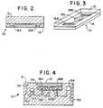
- FIGURE 1is a magnified plan view of device 10 according to the invention that comprises substrate 14 microfabricated with ports 16, mesoscale flow channel 20, and a fractally bifurcating system of flow channels 40.
- FIGURE 2is a longitudinal cross sectional view of the device shown in Figure 1.
- FIGURE 3is a perspective view of the device of Figure 1.
- FIGURE 4is a schematic cross sectional view of an analytical device 10 nested within an appliance 50, which is used to support the device 10 and to regulate and detect the pressure of sample fluids in device 10.
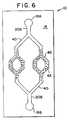
- FIGURE 5is a schematic plan view of a substrate 14 microfabricated with a fractally bifurcating system of flow channels 40 symmetrically disposed on the substrate, and tapering to a narrower diameter towards the center of the fractal system.
- FIGURE 6is a schematic plan view of device 10 that includes substrate 14 microfabricated with entry ports 16, mesoscale flow channel 20, and a fractally bifurcating system of flow channels 40, provided with beads 42 to enhance flow restriction and agglomeration in the fractal.
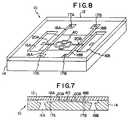
- FIGURE 7is a schematic longitudinal cross-sectional view of a device according to the invention which includes electrical conductors 17 and 18 for measuring conductivity of fluids in the device.
- FIGURE 8is a perspective view of the device shown in Figure 7.
- FIGURE 9is a schematic plan view of a multitest apparatus constructed in accordance with the invention.
- FIGURE 10is a schematic plan view of an analytical device fabricated with a series of mesoscale chambers suitable for implementing a variety of functions including cell sorting, cell lysing, PCR analysis, and detection of PCR products in the fractal region 40.
- FIGURE 11is a schematic plan view of device 10 according to the invention that includes substrate 14 microfabricated with ports 16, mesoscale flow channels 20, and a pair of fractal flow channels 40.
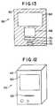
- FIGURE 12is a schematic perspective view of an apparatus 60 used in combination with device 10 for viewing the contents of device 10.
- FIGURE 13is a schematic cross sectional view of the apparatus 60 of Figure 12.
- the inventionprovides methods and apparatus for detecting the presence of an analyte in a fluid sample.
- the inventionprovides a device comprising a solid substrate, typically on the order of a few millimeters thick and 0.2 to 2.0 centimeters square, microfabricated to define a sample inlet port and a mesoscale flow system.
- a sample fluidis passed through the mesoscale flow system, and the analyte induced restriction or blockage of flow through the system is detected as a positive indication of the presence of the analyte.
- Analytical devices having mesoscale flow channels and fractal regionscan be designed and fabricated in large quantities from a solid substrate material. They can be sterilized easily. Silicon is a preferred substrate material because of the well-developed technology permitting its precise and efficient fabrication, but other materials may be used including polymers such as polytetrafluoroethylenes.
- the sample inlet and other ports, the mesoscale flow system, including the sample flow channel(s), the fractal region(s), and other functional elements,may be fabricated inexpensively in large quantities from a silicon substrate by any of a variety of micromachining methods known to those skilled in the art.
- micromachining methodsinclude film deposition processes such as spin coating and chemical vapor deposition, laser fabrication or photolithographic techniques such as UV or X-ray processes, or etching methods which may be performed by either wet chemical processes or plasma processes. (See, e.g., Manz et al., Trends in Analytical Chemistry , 10 : 144-149 (1991)).
- Flow channels of varying widths and depthscan be fabricated with mesoscale dimensions.
- the silicon substrate containing a fabricated mesoscale flow channelmay be covered and sealed with a thin anodically bonded glass cover. Other clear or opaque cover materials may be used.
- two silicon substratescan be sandwiched, or a silicon substrate can be sandwiched between two glass covers.
- the use of a transparent coverresults in a window which facilitates dynamic viewing of the channel contents, and allows optical probing of the mesoscale flow system either visually or by machine.
- Other fabrication approachesmay be used.
- electron micrographs of biological structuressuch as circulatory networks may be used as masks for fabricating mesoscale flow systems on the substrate.
- Mesoscale flow systemsmay be fabricated in a range of sizes and conformations.
- the flow systemcomprises a fractal region including bifurications leading to plural secondary channels.
- flow restriction in the mesoscale flow systemserves as a positive indicator of the presence of an analyte.
- the capacity of the devicesis very small and therefore the amount of sample fluid required for an analysis is low.
- the volume of each grooveis 10 -3 ⁇ L and the total volume of the 500 grooves is 0.5 ⁇ L.
- the low volume of the mesoscale flow systemsallows assays to be performed on very small amounts of a liquid sample ( ⁇ 10 ⁇ L).
- the volume of the flow systemtypically will be ⁇ 5 ⁇ L, and the volume of individual channels, chambers, or other functional elements are often less than 1 ⁇ l, e.g., in the nanoliter or picoliter range.
- the mesoscale flow systems of the devicesmay be microfabricated with microliter volumes, or alternatively nanoliter volumes or less, which advantageously limits the amount of sample and/or reagent fluids required for the assay.
- flow channels having mesoscale dimensionsAn important consequence and advantage of employing flow channels having mesoscale dimensions is that alterations in the flow properties of macromolecules, particles, and cells entrained or dissolved in aqueous liquids within the channels is easily influenced by stenosis, i.e., narrowing of the flow channels, and easily detected.
- the provision of the fractal regionserves to simplify alteration in flow.
- a sample suspected to be contaminated with bacteriacan be cultured in the device and the presence of a multiplicity of the organism can be detected by determining whether fluid can be forced through the system at a given pressure. Where no bacteria is present, fluid would flow easily; a large number of cells would serve to partially or totally occlude the fractal region.
- accretion of macromolecules onto specific binding proteins immobilized on the walls of the flow channelis sufficient to inhibit liquid flow through the channel provided its dimensions are small enough.
- the presence of a target polynucleotide in a polynucleotide samplemay be indicated by flowing the contents of a chamber after a suitable number of PCR cycles through a fractal region, as the viscosity of a solution laden with a large amount of polynucleotides will be larger than a solution of nucleotides.
- the device 10may include a silicon substrate 14 microfabricated with ports 16, primary sample flow channel 20A, and a fractal system of flow channels 40.
- the portsmay be microfabricated with mesoscale or larger dimensions.
- the fractal region 40in this case comprises equal numbers of bifurcations and junctions, disposed serially along the direction of flow through the fractal region, leading to a third flow channel 20B.
- the substrate 14is covered with a clear glass or plastic window 12 to close the channels.
- a fluid sampleenters the device through inlet port 16A and flow channel 20A, and then flows through the fractal region 40 to flow channel 20B and port 16B.
- the fractal region 40is very sensitive to the flow properties of a sample. Restriction or blockage of flow of a sample through the fractal region 40 can serve as an indicator of the presence of an analyte in the sample and may be detected, e.g., optically through the window 12.
- the fractal system 40may be fabricated on a silicon substrate with reduced dimensions at each bifurcation, providing sequentially narrower flow channels, as illustrated schematically in Figure 5.
- Figure 5shows device 10, which comprises substrate 14 microfabricated with fractal flow channels 40, which have a reduced cross-sectional area relative to the primary flow channel 20A and the third flow channel 20B.
- a sample fluidenters the device 10 through inlet port 16A and channel 20A, and then flows through the fractal region 40 to flow channel 20B and port 16B.
- Fluid flow through this fractal region 40is very sensitive to changes in fluid viscosity and to the development of flow restriction caused, for example, by the proliferation of cells, or the agglomeration of cells, particles, or macromolecular complexes that may be present in a sample.
- the fractal systemmay be microfabricated with a complex series of bifurcations, as illustrated schematically in Figure 11, to enhance sensitivity to flow restriction.
- Device 10 in Figure 11includes a pair of fractally bifurcating flow channels 40A and 40B.
- the fractal flow channel 40Ais constructed with sequentially narrower flow channels towards the center of the fractal, thereby enhancing sensitivity to flow restriction.
- the analytical devices containing the mesoscale flow systemcan be used in combination with an appliance for delivering and receiving fluids to and from the devices, such as appliance 50 shown schematically in Figure 4, which incorporates a nesting site 58 for holding the device 10, and for registering ports, e.g., ports 16 on the device 10, with a flow line 56 in the appliance.
- appliance 50shown schematically in Figure 4
- pump 52is actuated to force the sample into port 16A of device 10, flow channel 20A, and the fractal region 40.
- the samplemay be injected into the device, or may enter the flow system simply by capillary action.
- the flow systems of the devicesmay be filled to a hydraulically full volume, and the appliance may be utilized to direct the flow of fluid in the mesoscale flow system by means, e.g., of valves located in the device or the appliance.
- the analytical devicesalso may be utilized in combination with an appliance for viewing the contents of the mesoscale channels in the devices.
- the appliancein one embodiment may comprise a microscope for viewing the contents of the mesoscale channels in the devices.
- a cameramay be included in the appliance, as illustrated in the appliance 60 shown schematically in Figures 12 and 13.
- the appliance 60is provided with a housing 62, a viewing screen 64 and a slot 66 for inserting a chip into the appliance.
- the appliance 60also includes a video camera 68, an optical system 70, and a tilt mechanism 72 for holding device 10, and allowing the placement and angle of device 10 to be adjusted manually.
- the optical system 70may include a lens system for magnifying the channel contents, as well as a light source.
- the video camera 68 and screen 64allow analyte induced change in sample fluid properties, such as flow properties or color, to be monitored visually, and optionally recorded using the appliance.
- Changes in sample flow properties in the flow system, induced by the presence of an analyte in the sample,can be detected by any of a number of methods including monitoring the pressure or electrical conductivity of sample fluids in selected regions of the flow system in the device as disclosed herein.
- Analyte induced changes in flow propertiesalso may be detected by optical detection through a transparent cover or a translucent section of the substrate itself, either visually or by machine.
- Devicessuch as valves, mesoscale pressure sensors, and other mechanical sensors can be fabricated directly on the silicon substrate and can be mass-produced according to well established technologies. Angell et al., Scientific American , 248 :44-55 (1983). Pressure sensors and other detection means also may be provided in an appliance utilized in combination with the device.
- analyte induced flow restrictioncan be detected by monitoring the pressure of sample fluids entering and exiting the mesoscale flow system.
- Figure 4shows schematically, as an example, device 10, which is nested within appliance 50, which includes two pressure detectors 54 for detecting flow pressure of fluids entering and exiting device 10 through ports 16.
- a mesoscale pressure sensormay be fabricated directly on the silicon substrate and connected via electrical contacts to the appliance. Angell et al., Scientific American , 248 :44-55 (1983). Analyte induced changes in flow properties in the flow system, such as flow restriction, thus may be detected as a pressure change indicative of a positive result.
- Other detectorsmay be utilized, such as conventional flow detectors. The movement of magnetic beads entrained in the fluid can be detected easily as an indication of flow restriction.
- electrical conductorsmay be fabricated in the substrate of the devices to enable transmission of signals indicative of alterations in fluid flow properties, induced by the presence of the analyte, and sensed in different regions of the flow system.
- Electrical conductors in the substratemay be mated through contacts to the electrical conductors in an appliance, used in combination with the device.
- the electrical conductors in the devicecarry signals from pressure or electrical conductivity sensors enabling the detection of the conductivity or pressure of fluid in the flow systems.
- analyte induced clogging of the fractal region 40may be detected by a conventional conductivity probe 17 whose output is indicative of the presence or absence of aqueous fluid in the outflow channel.
- the conductivity or other probecould also be fabricated within the fractal region 40.
- the substratemay be microfabricated with a control region such that output from the sample flow region and the control region may be detected and compared, thereby enhancing the accuracy of the assay.
- the flow properties between sample fluid entering and exiting the flow systemcan be detected and compared in order to detect analyte induced changes in flow properties of a sample.
- the conductivitymay be measured in the device 10 shown schematically in Figures 7 and 8.
- Device 10includes the silicon substrate 14 on which are microfabricated inlet ports 16 and flow channel 20. The substrate is covered by a translucent window 12.
- a sample fluidenters device 10 through port 16A and sample channel 20A, and then flows through the fractal region 40 to channel 20B and port 16B.
- Device 10is microfabricated with electrical conductor 18A in electrical contact with fluid channel 20A, for detecting the conductivity of fluid centering the fractal region region 40.
- the devicealso includes electrical conductor 18B, in electrical contact with flow channel 20B, for detecting the conductivity of fluid exiting the fractal region 40.
- the conductors 18are connected to contacts 17 which extend through to the bottom of the substrate.
- the contacts 17can be fabricated by known techniques, e.g., by thermal gradient zone melting. (See Zemel et al., in: Fundamentals and Applications of Chemical Sensors , D. Schuetzle and R.
- Device 10may be nested in an appliance such as appliance 50, shown in Figure 4, capable of detecting conductivity changes through the contacts 17. Changes in conductivity can be correlated with changes in fluid properties, such as fluid pressure, induced by the presence of an analyte, in the fluid sample. Blockage in the fractal will prevent liquid from reaching channel 20B, and the conductivity across the gap in conductor 18B will be low.
- Analyte induced changes in flow properties of a sample in the flow systemsalso may be detected optically, e.g., with a microscope, through a transparent cover over the flow system, or through a transparent region of the substrate itself.
- the appliancemay include sensing equipment, such as a spectrophotometer, to assist in the optical detection of changes in flow properties due to the presence of the analyte.
- the mesoscale flow systemmay comprise a binding moiety, capable of binding the analyte, thereby to enhance flow restriction.
- the binding moietymay be immobilized on the surface of the flow channels, or on a solid phase reactant such as a bead.
- the binding moietymay comprise, e.g., an antigen binding protein, a DNA probe, or one of a ligand/receptor pair.
- the binding moietymay also comprise a crosslinker, such as a chemical reagent or a protein, capable of crosslinking of a specific cell subpopulation.
- the binding moietymay be immobilized on the surface of the mesoscale flow channels by, e.g., physical absorption onto the channel surfaces, or by chemical activation of the surface and subsequent attachment of biomolecules to the activated surface. Techniques available in the art may be utilized for the chemical activation of silaceous channel surfaces, and for the subsequent attachment of a binding moiety to the surfaces. (See, e.g., Haller in: Solid Phase Biochemistry , W.H. Scouten, Ed., John Wiley, New York, pp 535-597 (1983); and Mandenius et al., Anal. Biochem. , 137 :106-114 (1984), and Anal. Biochem. , 170 :68-72 (1988)).
- the detection of a cellular or chemical analytecan be implemented by selecting the appropriate binding moiety.
- Flow restrictionmay be enhanced by the binding of the analyte to the binding moiety, immobilized on the surface of the mesoscale flow system, i.e., by the build-up of a macromolecular surface layer on the surface of the flow system.
- the binding moietymay comprise a particle capable of inducing detectable agglomeration of an analyte in the mesoscale flow system.
- particles 42 coated with binding protein specific for a given analytemay be provided in the fractal region 40 to promote analyte-induced agglomeration of fluid in the fractal region.
- a binding moietysuch as an antibody may be immobilized on an inert bead, and may be utilized to induce agglomeration.
- Agglomeration in the fractal regionmay be detected optically through a window, e.g., disposed over the fractal region. Agglomeration may also be detected by, e.g., detecting pressure or conductivity changes of the sample fluid as noted below.
- the substratemay be fabricated to include a control region in the flow system, e.g., a region which is identical in geometry to the test region, but does not include binding moieties. Sample directed to both the detection and control regions exhibit different flow properties which may be detected and compared.
- the devicesprovide a mesoscale fractal flow system, which readily allows the growth of organisms in a culture to be monitored on the basis of flow restriction, due to changes in fluid viscosity.
- the fractal regionmay include an extensive series of equal numbers of bifurcations and junctions disposed serially along the direction of flow of sample through the region, as schematically illustrated in Figure 11.
- Flow restrictionmay be detected, e.g., optically, after a short incubation. The presence and growth of an organism in a sample will influence the flow characteristics within the fractal.
- One or more sensorssuch as pressure or conductivity sensors, may be utilized to detect pressure changes due to changes in fluid properties caused by the presence of an organism in the fractal region.
- the migration of sperm in the mesoscale flow systems of the devicescan serve as an indication of sperm motility.
- the substratemay be disposed, e.g., in an appliance, at an angle with respect to a horizontal plane, to provide an incline for the travel of a sperm sample, to further enhance the detection of the motility.
- Reagents capable of binding to a spermmay be provided in the flow system.
- the devicesmay be utilized to assess, e.g., a spermicidal agent, the binding properties of a sperm sample, or to conduct sperm counts.
- the devicesmay be used to implement a variety of automated, sensitive and rapid analyses based on flow restriction including analyses of cells or macromolecules, or for monitoring cell culture growth.
- the devicesmay be fabricated with two or more mesoscale flow systems which comprise, e.g., two or more different fractal regions, containing, e.g., binding moieties for different analytes, allowing two or more assays to be conducted simultaneously. At the conclusion of the assay the devices typically are discarded.
- the use of disposable deviceseliminates contamination among samples. The sample at all times can remain entombed, and the low volume simplifies waste disposal.
- Sperm motilityis tested in the chip 10 shown schematically in Figure 5.
- a sample of semen⁇ 2 ⁇ L
- the chip 10is placed on top of the semen sample such that the port 16A is positioned on the semen sample.
- the progress of individual spermatozoa into port 16A, through channel 20A and fractal region 40is monitored using a microscope.
- the experimental resultsmay be compared with results previously established for a healthy sperm sample to provide a test of sperm motility.
- the growth of an organismis monitored in the device shown schematically in Figure 5.
- the fractal pattern of mesoscale flow paths 40 in the substrate 14are filled via inlet port 16A with 2 ⁇ L of a mixture of growth medium which has been inoculated with a sample of a test specimen.
- the deviceis sealed and incubated for 60 minutes at 37°C. Growth is detected by visual inspection using a microscope or by determining the flow properties of the channel system, e.g., via the electrical conductivity probe 17. The absence of flow indicates growth and consequent blockage of the fractal system.
- Fractal detection chamber 40Aprovides a test for leucocytes and comprises immobilized antibody to common leukocyte antigen.
- Fractal detection chamber 40Bprovides a test for sperm antibodies and contains immobilized antibody to human IgG, IgA or IgM.
- Fractal detection chamber 40Cprovides a test for acrosome reaction and contains fluorescein labeled lectin. Flow restriction due to agglutination in the chambers may be detected, e.g., by optical detection through a glass cover disposed over the substrate. After the assay is complete, the device is discarded.
- Figure 10depicts schematically a device 10 including substrate 14 used to detect the presence of a target nucleic acid within a subpopulation of cells in a mixture in a biological fluid sample.
- Microfabricated on device 10is a mesoscale flow path 20 which includes a cell separation chamber 22A, a cell lysis chamber 22B, a filter region 28, a polymerase chain reaction (PCR) chamber comprising sections 22C and 22D, and a fractal detection region 40.
- the mesoscale flow system 20is also provided with fluid entry/exit ports 16A, 16B, 16C and 16D.
- the deviceis used in combination with an appliance, such as appliance 50, shown in Figure 4.
- the applianceis provided with fluid paths mated to ports 16 in the device, and valves allowing the ports 16 to be mechanically closed and opened.
- the appliancealso includes pump 52 for regulating the flow of sample fluid through the device.
- the appliancefurther includes means for heating the PCR reaction chamber sections 22C and 22D in the device.
- valves in the applianceare used to close ports 16C and 16D, while ports 16A and 16B are open.
- a sample containing a mixture of cellsis directed to the sample inlet port 16A by the pump 52 in the appliance, and flows through the mesoscale flow path 20 to separation chamber 22A.
- Chamber 22Acontains binding moieties immobilized on the wall of the chamber which selectively bind to a surface molecule on a desired type of cell in the sample. Remaining cellular components exit the substrate via port 16B.
- flow with bufferis continued, to wash and assure isolation of the cell population.
- port 16Bis closed and 16C is opened. Flow is then increased sufficiently to dislodge the immobilized cells. Flow is continued, forcing cells through membrane piercing protrusions 24 in chamber 22B, which tear open the cells releasing intracellular material.
- Sample flowcontinues past filter 28, which filters off large cellular membrane components and other debris, to mesoscale PCR chamber section 22C, which is connected to PCR chamber section 22D by flow channel 20B.
- Taq polymerase, primers and other reagents required for the PCR assaynext are added to section 22D through port 16C from a mated port and flow path in the appliance, permitting mixing of the intracellular soluble components from the separated subpopulation of cells and the PCR reagents.
- a pump in the appliance connected via port 16Bis used to cycle the PCR sample and reagents through flow channel 20B between sections 22C and 22D, set at 94°C and 65°C respectively, to implement plural polynucleotide melting and polymerization cycles, allowing the amplification of product polynucleotide.
- the valves in the appliancenext are used to close port 16C and to open port 16D.
- the pump in the appliance connected to port 16Bis then used to direct the amplified polynucleotide isolated from the cell population to the fractal detection region 40.
- Flow restriction in the fractal region 40caused by the presence of amplified polynucleotide product, serves as a positive indicator of the presence of the target DNA or RNA in the cells, and is detected optically through a glass cover disposed over the detection region.
- a fractal channelwas filled with HTF-BSA medium, and semen were introduced simultaneously via the holes at each end of the channel.
- Spermwere observed migrating towards the center of the channel (or fractal channel) and eventually passing as they migrated towards the hole at the opposite end of the channel.
Landscapes
- Chemical & Material Sciences (AREA)
- Health & Medical Sciences (AREA)
- Life Sciences & Earth Sciences (AREA)
- Chemical Kinetics & Catalysis (AREA)
- Engineering & Computer Science (AREA)
- Molecular Biology (AREA)
- Immunology (AREA)
- General Health & Medical Sciences (AREA)
- Hematology (AREA)
- Urology & Nephrology (AREA)
- Biomedical Technology (AREA)
- Biochemistry (AREA)
- Clinical Laboratory Science (AREA)
- Analytical Chemistry (AREA)
- Organic Chemistry (AREA)
- Biotechnology (AREA)
- Food Science & Technology (AREA)
- Medicinal Chemistry (AREA)
- Physics & Mathematics (AREA)
- Microbiology (AREA)
- Cell Biology (AREA)
- Tropical Medicine & Parasitology (AREA)
- General Physics & Mathematics (AREA)
- Pathology (AREA)
- Physiology (AREA)
- Inorganic Chemistry (AREA)
- Water Supply & Treatment (AREA)
- Dispersion Chemistry (AREA)
- Apparatus Associated With Microorganisms And Enzymes (AREA)
- Measuring Or Testing Involving Enzymes Or Micro-Organisms (AREA)
- Investigating Or Analysing Biological Materials (AREA)
- Optical Measuring Cells (AREA)
- External Artificial Organs (AREA)
- Investigating Or Analyzing Materials By The Use Of Electric Means (AREA)
- Steroid Compounds (AREA)
- Sampling And Sample Adjustment (AREA)
- Immobilizing And Processing Of Enzymes And Microorganisms (AREA)
- Devices For Use In Laboratory Experiments (AREA)
- Investigating Or Analysing Materials By The Use Of Chemical Reactions (AREA)
- Pharmaceuticals Containing Other Organic And Inorganic Compounds (AREA)
Abstract
Description
| Feature | Benefit |
| Flexibility | No limits to the number of chip designs or applications available. |
| Reproducible | Allows reliable, standardized, mass production of chips. |
| Low Cost Production | Allows competitive pricing with existing systems. Disposable nature for single-use processes. |
| Small Size | No bulky instrumentation required. Lends itself to portable units and systems designed for use in non-conventional lab environments. Minimal storage and shipping costs. |
| Microscale | Minimal sample and reagent volumes required. Reduces reagent costs, especially for more expensive, specialized test procedures. Allows simplified instrumentation schemes. |
| Sterility | Chips can be sterilized for use in microbiological assays and other procedures requiring clean environments. |
| Sealed System | Minimizes biohazards. Ensures process integrity. |
| Multiple Circuit Capabilities | Can perform multiple processes or analyses on a single chip. Allows panel assays. |
| Multiple Detector Capabilities | Expands capabilities for assay and process monitoring to virtually any system. Allows broad range of applications. |
| Reuseable Chips | Reduces per process cost to the user for certain applications. |
Claims (32)
- A device for detecting the presence of ananalyte in a fluid sample, the device comprising:a solid substrate microfabricated to define:a sample inlet port; anda mesoscale flow system comprising:a primary sample flow channelextending from said inlet port; anda fractal region, in fluidcommunication with said primary flow channel; and,means for detecting a flow property of a fluidsample in said flow system as an indication of thepresence of an analyte in the fluid sample; characterisedby said fractal region comprising multiple bifurcationseach bifurcation leading to plural, distinct secondaryflow channels for amplifying the effect of occlusion ofsaid flow system or increase in viscosity within saidsubstrate.
- The device of claim 1 wherein said fractalregion further comprises junctions in fluid communicationwith said secondary flow channels leading to a third flowchannel in said mesoscale flow system.
- The device of claim 1 or claim 2 furthercomprising pump means for inducing flow of said samplethrough said mesoscale flow system.
- The device of claim 2 or claim 3 wherein saidfractal region comprises equal numbers of bifurcationsand junctions disposed serially along the direction offlow of a sample through said region.
- The device of any one of claims 2 to 4 wherein each saidsecondary channel in said fractal region has a reducedcross-sectional area relative to said primary flowchannel and said third flow channel.
- The device of any one of the preceding claims wherein said means fordetecting comprises means for detecting a fluid flowproperty in said third flow channel.
- The device of any one of claims 1 to 5 wherein said means fordetecting comprises means for detecting and comparing afluid flow property in said primary sample flow channelwith a fluid flow property in said third flow channel.
- The device of any one of the preceding claimswherein said means for detecting comprises:(a) an electrical detection means; or,(b) a magnetic detection means; or,(c) means defining an optical path to said fractalregion; or,(d) means for detecting the growth of an organismin said flow system.
- The device of any one of the preceding claimswherein fluid pressure or fluid conductivity is detected.
- The device claim 1 further comprising abinding moiety disposed within said fractal region forbinding a component of said sample.
- The device of claim 10 wherein said bindingmoiety comprises particles which bind with a componentof said sample to induce particle agglomeration.
- The device of any one of the preceding claims wherein said substratedefines a plurality of said flow systems.
- The device of any one of the preceding claims wherein the sample is asperm sample and wherein flow of sperm through thefractal region provides an indication of spermmotility.
- The device of claim 13, wherein said substrateis disposed at an angle with respect to a horizontalplane.
- The device of any one of the preceding claimswherein said solidsubstrate comprises microfabricated silicon.
- The device of any one of the preceding claims,further comprising anappliance for use in combination with said substrate,said appliance comprising:means for holding said substrate;fluid input means interfitting with the inlet porton said substrate; andpump means for passing fluid through the flowsystem of said substrate when it is held in saidholding means.
- The device of any one of claims 1 to 7 and 9 to 16wherein said means fordetecting comprises an appliance for use in combinationwith said substrate, said appliance comprising:means for holding said substrate; andoptical means for viewing the contents of saidmesoscale flow system in said substrate.
- The device of claim 17 wherein said opticalmeans comprises magnifying optics and a video camera,and wherein said appliance further comprises:a tilt mechanism for manually adjusting the angleand location of the device; anda video screen for viewing the contents of saidflow system.
- The device ofany one of the precedingclaims wherein said flow systemincludes a control fractal region permitting comparisonof flow of said sample in said fractal region and saidcontrol region.
- A method for detecting the presence of ananalyte in a fluid sample, the method comprising thesteps of:(i) providing a device comprising:a solid substrate microfabricated to define:a sample inlet port; anda mesoscale flow system comprising:a primary sample flow channelextending from said inlet port; anda fractal region, in fluidcommunication with said primary flow channel, comprisingmultiple bifurcations each bifurcation leading to plural,distinct secondary flow channels for amplifying theeffect of occlusion of said flow system or increase inviscosity within said substrate;(ii) passing a fluid sample suspected tocontain an analyte through said fractal region in saidmesoscale flow system;(iii) detecting the restriction or blockage offlow of the fluid sample through said system; and(iv) correlating the detected restriction orblockage of flow, or lack thereof, to the presence orabsence of an analyte in said sample.
- The method of claim 20, wherein said flowsystem comprises a binding moiety, capable of bindingsaid analyte in said sample, to promote said restrictionor blockage of flow through said system.
- The method of claim 21 wherein said bindingmoiety is disposed on particles which bind with saidanalyte in said sample to induce particle agglomeration,thereby to promote said restriction or blockage of flow.
- The method of claim 21 or claim 22wherein said analyte isa cell population in said sample;said binding moiety comprises a crosslinker ofcells in said population; andsaid restriction of flow is caused by crosslinker-inducedcell aggregation.
- The method of any one of claims 21 to 23 wherein said bindingmoiety is immobilized within said flow system.
- The method of any one of claims 20 to 23 wherein flow isrestricted by the build-up of a macromolecular surfacelayer on a surface of said flow system.
- The method of any one of claims 20 to 25 wherein said fractalregion, in the device provided in step (i), furthercomprises junctions in fluid communication with saidsecondary flow channels leading to a third channel insaid flow system.
- The method ofany one of claims 20 to 26wherein, in step (iii),restriction or blockage is detected electrically or optically.
- The method of any one of claims 20 to 27 wherein said fractalregion further comprises a binding moiety for binding acomponent of said sample.
- The method of any one of claims 20 to 28 wherein said fractalregion contains particles which bind with a componentof said sample to induce particle agglomeration.
- The method of any one of claims 20 to 29 wherein saidsubstrate, provided in step (i), further comprises acontrol region in fluid communication with said sampleinlet port; and
wherein, in step (iii), flow of said sample in saidfractal region and said control region is detected andcompared. - The method of any one of claims 20 to 30 wherein the analytecomprises a replicable procaryotic organism; and
wherein, in step (iii), the restriction or blockageof flow of said organism through said flow systemserves as an indication of the presence of saidorganism. - The method ofany one of claims 20 to 31wherein, in step (ii),a pump is utilized to deliver said sample through saidflow system.
Applications Claiming Priority (11)
| Application Number | Priority Date | Filing Date | Title |
|---|---|---|---|
| US87770292A | 1992-05-01 | 1992-05-01 | |
| US87770192A | 1992-05-01 | 1992-05-01 | |
| US87766292A | 1992-05-01 | 1992-05-01 | |
| US07/877,536US5304487A (en) | 1992-05-01 | 1992-05-01 | Fluid handling in mesoscale analytical devices |
| US877661 | 1992-05-01 | ||
| US877662 | 1992-05-01 | ||
| US07/877,661US5296375A (en) | 1992-05-01 | 1992-05-01 | Mesoscale sperm handling devices |
| US877536 | 1992-05-01 | ||
| US877701 | 1992-05-01 | ||
| US877702 | 1992-05-01 | ||
| PCT/US1993/004016WO1993022054A1 (en) | 1992-05-01 | 1993-04-29 | Analysis based on flow restriction |
Publications (2)
| Publication Number | Publication Date |
|---|---|
| EP0637997A1 EP0637997A1 (en) | 1995-02-15 |
| EP0637997B1true EP0637997B1 (en) | 1998-07-01 |
Family
ID=27542279
Family Applications (5)
| Application Number | Title | Priority Date | Filing Date |
|---|---|---|---|
| EP93910890AExpired - LifetimeEP0639223B1 (en) | 1992-05-01 | 1993-04-29 | Microfabricated sperm handling devices |
| EP93910907AExpired - LifetimeEP0637999B1 (en) | 1992-05-01 | 1993-04-29 | Polynucleotide amplification analysis using a microfabricated device |
| EP93910889AExpired - LifetimeEP0637997B1 (en) | 1992-05-01 | 1993-04-29 | Analysis based on flow restriction |
| EP93910887AExpired - LifetimeEP0637996B1 (en) | 1992-05-01 | 1993-04-29 | Microfabricated detection structures |
| EP93910891AExpired - LifetimeEP0637998B2 (en) | 1992-05-01 | 1993-04-29 | Fluid handling in microfabricated analytical devices |
Family Applications Before (2)
| Application Number | Title | Priority Date | Filing Date |
|---|---|---|---|
| EP93910890AExpired - LifetimeEP0639223B1 (en) | 1992-05-01 | 1993-04-29 | Microfabricated sperm handling devices |
| EP93910907AExpired - LifetimeEP0637999B1 (en) | 1992-05-01 | 1993-04-29 | Polynucleotide amplification analysis using a microfabricated device |
Family Applications After (2)
| Application Number | Title | Priority Date | Filing Date |
|---|---|---|---|
| EP93910887AExpired - LifetimeEP0637996B1 (en) | 1992-05-01 | 1993-04-29 | Microfabricated detection structures |
| EP93910891AExpired - LifetimeEP0637998B2 (en) | 1992-05-01 | 1993-04-29 | Fluid handling in microfabricated analytical devices |
Country Status (10)
| Country | Link |
|---|---|
| EP (5) | EP0639223B1 (en) |
| JP (5) | JP3298882B2 (en) |
| AT (5) | ATE140025T1 (en) |
| AU (5) | AU674685B2 (en) |
| CA (5) | CA2134478C (en) |
| DE (5) | DE69322774T2 (en) |
| ES (2) | ES2127276T3 (en) |
| GR (2) | GR3025037T3 (en) |
| HK (1) | HK16897A (en) |
| WO (5) | WO1993022053A1 (en) |
Cited By (5)
| Publication number | Priority date | Publication date | Assignee | Title |
|---|---|---|---|---|
| US6007690A (en)* | 1996-07-30 | 1999-12-28 | Aclara Biosciences, Inc. | Integrated microfluidic devices |
| US6056860A (en)* | 1996-09-18 | 2000-05-02 | Aclara Biosciences, Inc. | Surface modified electrophoretic chambers |
| US7288405B2 (en) | 2001-04-25 | 2007-10-30 | Cornell Research Foundation, Inc. | Devices and methods for pharmacokinetic-based cell culture system |
| US8647861B2 (en) | 2008-07-16 | 2014-02-11 | Children's Medical Center Corporation | Organ mimic device with microchannels and methods of use and manufacturing thereof |
| US9725687B2 (en) | 2011-12-09 | 2017-08-08 | President And Fellows Of Harvard College | Integrated human organ-on-chip microphysiological systems |
Families Citing this family (393)
| Publication number | Priority date | Publication date | Assignee | Title |
|---|---|---|---|---|
| US5547839A (en) | 1989-06-07 | 1996-08-20 | Affymax Technologies N.V. | Sequencing of surface immobilized polymers utilizing microflourescence detection |
| US7297313B1 (en) | 1991-08-31 | 2007-11-20 | The Regents Of The University Of California | Microfabricated reactor, process for manufacturing the reactor, and method of amplification |
| WO1993022053A1 (en)* | 1992-05-01 | 1993-11-11 | Trustees Of The University Of Pennsylvania | Microfabricated detection structures |
| US5744366A (en)* | 1992-05-01 | 1998-04-28 | Trustees Of The University Of Pennsylvania | Mesoscale devices and methods for analysis of motile cells |
| US6156270A (en)* | 1992-05-21 | 2000-12-05 | Biosite Diagnostics, Inc. | Diagnostic devices and apparatus for the controlled movement of reagents without membranes |
| US6905882B2 (en) | 1992-05-21 | 2005-06-14 | Biosite, Inc. | Diagnostic devices and apparatus for the controlled movement of reagents without membranes |
| US6767510B1 (en) | 1992-05-21 | 2004-07-27 | Biosite, Inc. | Diagnostic devices and apparatus for the controlled movement of reagents without membranes |
| US5639423A (en)* | 1992-08-31 | 1997-06-17 | The Regents Of The University Of Calfornia | Microfabricated reactor |
| FR2709250B1 (en)* | 1993-08-23 | 1995-10-13 | Ccd Laboratoire | Device for selecting sperm. |
| DE4405004A1 (en)* | 1994-02-17 | 1995-08-24 | Rossendorf Forschzent | Chemical micro-analyzer |
| CA2143365A1 (en)* | 1994-03-14 | 1995-09-15 | Hugh V. Cottingham | Nucleic acid amplification method and apparatus |
| US5725831A (en)* | 1994-03-14 | 1998-03-10 | Becton Dickinson And Company | Nucleic acid amplification apparatus |
| US6287850B1 (en) | 1995-06-07 | 2001-09-11 | Affymetrix, Inc. | Bioarray chip reaction apparatus and its manufacture |
| DE4420732A1 (en)* | 1994-06-15 | 1995-12-21 | Boehringer Mannheim Gmbh | Device for the treatment of nucleic acids from a sample |
| US5707799A (en)* | 1994-09-30 | 1998-01-13 | Abbott Laboratories | Devices and methods utilizing arrays of structures for analyte capture |
| DE4435107C1 (en)* | 1994-09-30 | 1996-04-04 | Biometra Biomedizinische Analy | Miniaturized flow thermal cycler |
| USRE43097E1 (en) | 1994-10-13 | 2012-01-10 | Illumina, Inc. | Massively parallel signature sequencing by ligation of encoded adaptors |
| US5641400A (en)* | 1994-10-19 | 1997-06-24 | Hewlett-Packard Company | Use of temperature control devices in miniaturized planar column devices and miniaturized total analysis systems |
| CN1088385C (en)* | 1994-10-22 | 2002-07-31 | 研究中心实验室(有限) | Method and apapratus for diffusive transfer between immiscible fluids |
| CN1087960C (en)* | 1994-10-22 | 2002-07-24 | 研究中心实验室(有限) | Method and apparatus for diffusive transfer between immiscible fluids |
| DE4438785C2 (en)* | 1994-10-24 | 1996-11-07 | Wita Gmbh Wittmann Inst Of Tec | Microchemical reaction and analysis unit |
| US5585069A (en)* | 1994-11-10 | 1996-12-17 | David Sarnoff Research Center, Inc. | Partitioned microelectronic and fluidic device array for clinical diagnostics and chemical synthesis |
| US5603351A (en) | 1995-06-07 | 1997-02-18 | David Sarnoff Research Center, Inc. | Method and system for inhibiting cross-contamination in fluids of combinatorial chemistry device |
| KR100314996B1 (en)* | 1994-11-10 | 2002-01-15 | 윌리암 제이. 버크 | Liquid distribution system |
| ATE212064T1 (en)* | 1994-11-14 | 2002-02-15 | Univ Pennsylvania | DEVICE FOR AMPLIFICATION OF POLYNUCLEOTIDES ON THE MESOM SCALE |
| JP3220158B2 (en) | 1994-11-14 | 2001-10-22 | トラスティーズ・オブ・ザ・ユニバーシティ・オブ・ペンシルベニア | Mesoscale sample pretreatment devices and systems for analyte determination and processing |
| DE19507638C2 (en)* | 1995-03-04 | 1997-09-25 | Danfoss As | Analyzer |
| DE19519015C1 (en)* | 1995-05-24 | 1996-09-05 | Inst Physikalische Hochtech Ev | Miniaturised multi-chamber thermo-cycler for polymerase chain reaction |
| US5776674A (en)* | 1995-06-05 | 1998-07-07 | Seq, Ltd | Chemical biochemical and biological processing in thin films |
| AU6541596A (en)* | 1995-06-16 | 1997-01-15 | University Of Washington | Microfabricated differential extraction device and method |
| TW293783B (en)* | 1995-06-16 | 1996-12-21 | Ciba Geigy Ag | |
| US6454945B1 (en) | 1995-06-16 | 2002-09-24 | University Of Washington | Microfabricated devices and methods |
| US5716852A (en)* | 1996-03-29 | 1998-02-10 | University Of Washington | Microfabricated diffusion-based chemical sensor |
| EP0871539B1 (en) | 1995-06-16 | 2002-02-20 | University of Washington | Tangential flow planar microfabricated fluid filter |
| US6524532B1 (en)* | 1995-06-20 | 2003-02-25 | The Regents Of The University Of California | Microfabricated sleeve devices for chemical reactions |
| US5589136A (en)* | 1995-06-20 | 1996-12-31 | Regents Of The University Of California | Silicon-based sleeve devices for chemical reactions |
| US5856174A (en)* | 1995-06-29 | 1999-01-05 | Affymetrix, Inc. | Integrated nucleic acid diagnostic device |
| US5872010A (en)* | 1995-07-21 | 1999-02-16 | Northeastern University | Microscale fluid handling system |
| US6132580A (en) | 1995-09-28 | 2000-10-17 | The Regents Of The University Of California | Miniature reaction chamber and devices incorporating same |
| EP1577010A3 (en)* | 1995-12-05 | 2005-11-16 | Tecan Trading AG | Microsystem platform and its use |
| CA2239613A1 (en)* | 1995-12-05 | 1997-06-12 | Alec Mian | Devices and methods for using centripetal acceleration to drive fluid movement in a microfluidics system with on-board informatics |
| US6709869B2 (en) | 1995-12-18 | 2004-03-23 | Tecan Trading Ag | Devices and methods for using centripetal acceleration to drive fluid movement in a microfluidics system |
| AU1360897A (en)* | 1996-01-17 | 1997-08-11 | Flinders Technologies Pty Ltd | Solid phase enrichment of intact cells using intracellular constituents |
| DE59700294D1 (en)* | 1996-01-23 | 1999-09-02 | Novartis Ag | DEVICE AND METHOD FOR SYNTHETIZING MACROMOLECULES |
| US6541213B1 (en) | 1996-03-29 | 2003-04-01 | University Of Washington | Microscale diffusion immunoassay |
| US5948684A (en) | 1997-03-31 | 1999-09-07 | University Of Washington | Simultaneous analyte determination and reference balancing in reference T-sensor devices |
| US7235406B1 (en) | 1996-04-03 | 2007-06-26 | Applera Corporation | Nucleic acid analysis device |
| JP4912517B2 (en)* | 1996-04-03 | 2012-04-11 | アプライド バイオシステムズ リミテッド ライアビリティー カンパニー | Devices and methods for detection of multiple analytes |
| US6825047B1 (en) | 1996-04-03 | 2004-11-30 | Applera Corporation | Device and method for multiple analyte detection |
| US7244622B2 (en) | 1996-04-03 | 2007-07-17 | Applera Corporation | Device and method for multiple analyte detection |
| US5942443A (en) | 1996-06-28 | 1999-08-24 | Caliper Technologies Corporation | High throughput screening assay systems in microscale fluidic devices |
| DE69610410T2 (en)* | 1996-05-23 | 2001-05-10 | Qiagen Gmbh | Use of an osmolyte to reduce or break noncovalent bonds of biological molecules to inert surfaces |
| US6150094A (en)* | 1996-05-23 | 2000-11-21 | Qiagen Gmbh | Use of an osmolyte for reducing or abolishing no-covalent interactions of biological molecules to inert surfaces |
| US5964239A (en)* | 1996-05-23 | 1999-10-12 | Hewlett-Packard Company | Housing assembly for micromachined fluid handling structure |
| WO1997047390A1 (en) | 1996-06-14 | 1997-12-18 | University Of Washington | Absorption-enhanced differential extraction device |
| WO1997048818A1 (en)* | 1996-06-17 | 1997-12-24 | The Board Of Trustees Of The Leland Stanford Junior University | Thermocycling apparatus and method |
| NZ333346A (en) | 1996-06-28 | 2000-03-27 | Caliper Techn Corp | High-throughput screening assay systems in microscale fluidic devices |
| US6074827A (en) | 1996-07-30 | 2000-06-13 | Aclara Biosciences, Inc. | Microfluidic method for nucleic acid purification and processing |
| US6143248A (en)* | 1996-08-12 | 2000-11-07 | Gamera Bioscience Corp. | Capillary microvalve |
| AU4236597A (en)* | 1996-08-27 | 1998-03-19 | Visible Genetics Inc. | Apparatus and method for performing sequencing of nucleic acid polymers |
| JP2002503334A (en) | 1996-09-04 | 2002-01-29 | テクニカル ユニバーシティ オブ デンマーク | Microflow system for particle separation and analysis |
| GB9618595D0 (en)* | 1996-09-06 | 1996-10-16 | Central Research Lab Ltd | Reaction cell |
| WO1998022803A1 (en)* | 1996-11-18 | 1998-05-28 | Fraunhofer-Gesellschaft zur Förderung der angewandten Forschung e.V. | Micromechanical transmission measuring cell |
| DE19648441A1 (en)* | 1996-11-22 | 1998-05-28 | Abb Patent Gmbh | Analyzer |
| DE19648695C2 (en) | 1996-11-25 | 1999-07-22 | Abb Patent Gmbh | Device for the automatic and continuous analysis of liquid samples |
| AU5895898A (en) | 1996-12-20 | 1998-07-17 | Gamera Bioscience Corporation | An affinity binding-based system for detecting particulates in a fluid |
| SE9700205D0 (en)* | 1997-01-24 | 1997-01-24 | Peter Lindberg | Integrated microfluidic element |
| FR2758884B1 (en)* | 1997-01-30 | 1999-04-02 | Bio Merieux | METHOD FOR ISOLATING, IN PARTICULAR DETECTING OR QUANTIFYING AN ANALYTE IN A MEDIUM |
| PL335482A1 (en)* | 1997-02-28 | 2000-04-25 | Burstein Lab | Laboratory in an optical disk |
| EP1959255A3 (en)* | 1997-04-04 | 2008-09-24 | Caliper Life Sciences, Inc. | Closed-loop biochemical analyzers |
| US6143496A (en) | 1997-04-17 | 2000-11-07 | Cytonix Corporation | Method of sampling, amplifying and quantifying segment of nucleic acid, polymerase chain reaction assembly having nanoliter-sized sample chambers, and method of filling assembly |
| JP2002503336A (en)* | 1997-05-16 | 2002-01-29 | アルバータ リサーチ カウンシル | Micro-distribution system and method of using the same |
| US6632619B1 (en) | 1997-05-16 | 2003-10-14 | The Governors Of The University Of Alberta | Microfluidic system and methods of use |
| US5998224A (en)* | 1997-05-16 | 1999-12-07 | Abbott Laboratories | Magnetically assisted binding assays utilizing a magnetically responsive reagent |
| JP3469585B2 (en) | 1997-05-23 | 2003-11-25 | ガメラ バイオサイエンス コーポレイション | Apparatus and method for using centripetal acceleration to drive flow motion in microfluidics systems |
| AU736321B2 (en)* | 1997-05-23 | 2001-07-26 | Lynx Therapeutics, Inc. | System and apparatus for sequential processing of analytes |
| US6632399B1 (en) | 1998-05-22 | 2003-10-14 | Tecan Trading Ag | Devices and methods for using centripetal acceleration to drive fluid movement in a microfluidics system for performing biological fluid assays |
| US6156273A (en) | 1997-05-27 | 2000-12-05 | Purdue Research Corporation | Separation columns and methods for manufacturing the improved separation columns |
| EP1004015B1 (en) | 1997-08-15 | 2004-01-02 | Alexion Pharmaceuticals, Inc. | Apparatus for performing assays at reaction sites |
| US6102068A (en)* | 1997-09-23 | 2000-08-15 | Hewlett-Packard Company | Selector valve assembly |
| JP2001518624A (en) | 1997-09-26 | 2001-10-16 | ユニバーシティ・オブ・ワシントン | Simultaneous particle separation and chemical reactions |
| US7115884B1 (en)* | 1997-10-06 | 2006-10-03 | Trustees Of Tufts College | Self-encoding fiber optic sensor |
| WO1999024828A1 (en)* | 1997-11-12 | 1999-05-20 | The Perkin-Elmer Corporation | Serpentine electrophoresis channel with self-correcting bends |
| US6488894B1 (en)* | 1997-11-19 | 2002-12-03 | Biognosis Gmbh | Device for sequential discharge of flowable reagents |
| US6251343B1 (en)* | 1998-02-24 | 2001-06-26 | Caliper Technologies Corp. | Microfluidic devices and systems incorporating cover layers |
| CA2230653A1 (en) | 1998-02-27 | 1999-08-27 | The Governors Of The University Of Alberta | Microchip based enzymatic analysis |
| WO1999049319A1 (en)* | 1998-03-25 | 1999-09-30 | Oestergaard Steen | Micro system and method for field manipulation of particles |
| GB9808836D0 (en)* | 1998-04-27 | 1998-06-24 | Amersham Pharm Biotech Uk Ltd | Microfabricated apparatus for cell based assays |
| US6761816B1 (en) | 1998-06-23 | 2004-07-13 | Clinical Micro Systems, Inc. | Printed circuit boards with monolayers and capture ligands |
| US7087148B1 (en) | 1998-06-23 | 2006-08-08 | Clinical Micro Sensors, Inc. | Binding acceleration techniques for the detection of analytes |
| US6132685A (en)* | 1998-08-10 | 2000-10-17 | Caliper Technologies Corporation | High throughput microfluidic systems and methods |
| DE19846958C2 (en)* | 1998-08-19 | 2001-06-13 | Fraunhofer Ges Forschung | Method for manufacturing a device for the transport of very small quantities of liquid |
| US6245227B1 (en) | 1998-09-17 | 2001-06-12 | Kionix, Inc. | Integrated monolithic microfabricated electrospray and liquid chromatography system and method |
| EP1113850A4 (en) | 1998-09-17 | 2006-02-01 | Advion Biosciences Inc | Integrated monolithic microfabricated electrospray and liquid chromatography system and method |
| DE19846466A1 (en)* | 1998-10-08 | 2000-04-27 | Ghs Gesundheits Service Ag | Analysis method for the simultaneous determination of parameters from different media |
| JP2002526103A (en) | 1998-10-08 | 2002-08-20 | アストラゼネカ・アクチエボラーグ | Micro-assembled cell injector |
| US7510841B2 (en) | 1998-12-28 | 2009-03-31 | Illumina, Inc. | Methods of making and using composite arrays for the detection of a plurality of target analytes |
| US6633031B1 (en) | 1999-03-02 | 2003-10-14 | Advion Biosciences, Inc. | Integrated monolithic microfabricated dispensing nozzle and liquid chromatography-electrospray system and method |
| DE19910392B4 (en)* | 1999-03-05 | 2005-03-17 | Clondiag Chip Technologies Gmbh | Micro column reactor |
| US6193647B1 (en) | 1999-04-08 | 2001-02-27 | The Board Of Trustees Of The University Of Illinois | Microfluidic embryo and/or oocyte handling device and method |
| US6605475B1 (en) | 1999-04-16 | 2003-08-12 | Perspective Biosystems, Inc. | Apparatus and method for sample delivery |
| US6375817B1 (en) | 1999-04-16 | 2002-04-23 | Perseptive Biosystems, Inc. | Apparatus and methods for sample analysis |
| US6942771B1 (en) | 1999-04-21 | 2005-09-13 | Clinical Micro Sensors, Inc. | Microfluidic systems in the electrochemical detection of target analytes |
| US7312087B2 (en) | 2000-01-11 | 2007-12-25 | Clinical Micro Sensors, Inc. | Devices and methods for biochip multiplexing |
| US7332326B1 (en) | 1999-05-14 | 2008-02-19 | Tecan Trading Ag | Centripetally-motivated microfluidics system for performing in vitro hybridization and amplification of nucleic acids |
| US8080380B2 (en) | 1999-05-21 | 2011-12-20 | Illumina, Inc. | Use of microfluidic systems in the detection of target analytes using microsphere arrays |
| US8481268B2 (en) | 1999-05-21 | 2013-07-09 | Illumina, Inc. | Use of microfluidic systems in the detection of target analytes using microsphere arrays |
| CA2373198C (en)* | 1999-05-28 | 2011-10-11 | Cepheid | Cartridge for conducting a chemical reaction |
| US6818185B1 (en) | 1999-05-28 | 2004-11-16 | Cepheid | Cartridge for conducting a chemical reaction |
| AU5494900A (en) | 1999-06-18 | 2001-01-09 | Gamera Bioscience Corporation | Devices and methods for the performance of miniaturized homogeneous assays |
| JP2001004628A (en)* | 1999-06-18 | 2001-01-12 | Kanagawa Acad Of Sci & Technol | Immune analyzer and immunoassay method |
| DE19928410C2 (en)* | 1999-06-22 | 2002-11-28 | Agilent Technologies Inc | Device housing with a device for operating a laboratory microchip |
| US6706519B1 (en) | 1999-06-22 | 2004-03-16 | Tecan Trading Ag | Devices and methods for the performance of miniaturized in vitro amplification assays |
| DE19933458B4 (en)* | 1999-07-15 | 2015-08-20 | Eppendorf Ag | Equipment and systems for handling liquid samples |
| WO2001007159A2 (en) | 1999-07-28 | 2001-02-01 | Genset | Integration of biochemical protocols in a continuous flow microfluidic device |
| DE19935433A1 (en)* | 1999-08-01 | 2001-03-01 | Febit Ferrarius Biotech Gmbh | Microfluidic reaction carrier |
| US6319719B1 (en)* | 1999-10-28 | 2001-11-20 | Roche Diagnostics Corporation | Capillary hematocrit separation structure and method |
| US6875619B2 (en)* | 1999-11-12 | 2005-04-05 | Motorola, Inc. | Microfluidic devices comprising biochannels |
| US6361958B1 (en)* | 1999-11-12 | 2002-03-26 | Motorola, Inc. | Biochannel assay for hybridization with biomaterial |
| JP2001145486A (en)* | 1999-11-19 | 2001-05-29 | Natl Inst Of Advanced Industrial Science & Technology Meti | Small volume chemical reaction equipment for multiple samples |
| CA2290731A1 (en) | 1999-11-26 | 2001-05-26 | D. Jed Harrison | Apparatus and method for trapping bead based reagents within microfluidic analysis system |
| US6432290B1 (en) | 1999-11-26 | 2002-08-13 | The Governors Of The University Of Alberta | Apparatus and method for trapping bead based reagents within microfluidic analysis systems |
| US6613561B1 (en) | 1999-11-26 | 2003-09-02 | Olympus Optical Co., Ltd. | High-density capillary array for reaction and detection of fluid |
| US6627882B2 (en) | 1999-12-30 | 2003-09-30 | Advion Biosciences, Inc. | Multiple electrospray device, systems and methods |
| DE10001116C2 (en)* | 2000-01-13 | 2002-11-28 | Meinhard Knoll | Device and method for the optical or electrochemical quantitative determination of chemical or biochemical substances in liquid samples |
| WO2001053819A1 (en) | 2000-01-18 | 2001-07-26 | Advion Biosciences, Inc. | Separation media, multiple electrospray nozzle system and method |
| US7241423B2 (en)* | 2000-02-03 | 2007-07-10 | Cellular Process Chemistry, Inc. | Enhancing fluid flow in a stacked plate microreactor |
| DE10006214A1 (en) | 2000-02-11 | 2001-08-16 | Roche Diagnostics Gmbh | System for simple nucleic acid analysis |
| DE10010587A1 (en)* | 2000-03-03 | 2001-09-06 | Roche Diagnostics Gmbh | System for the determination of analyte concentrations in body fluids |
| BR0110768A (en)* | 2000-05-12 | 2003-03-11 | Univ Illinois | Microfluidic channel embryo and / or oocyte handling, analysis and biological evaluation |
| DE10035911A1 (en)* | 2000-07-21 | 2002-02-07 | Abb Research Ltd | Method and sensor for monitoring liquids |
| GB2366793B (en)* | 2000-09-13 | 2005-03-09 | Imperial College | Chemical processing system and method |
| CA2423630C (en)* | 2000-10-03 | 2011-12-06 | Minerva Biotechnologies Corporation | Magnetic in situ dilution |
| DE50110742D1 (en)* | 2000-10-25 | 2006-09-28 | Boehringer Ingelheim Micropart | Microstructured platform for studying a liquid |
| AU2002241602A1 (en)* | 2000-11-16 | 2002-06-11 | Burstein Technologies, Inc. | Methods and apparatus for detecting and quantifying lymphocytes with optical biodiscs |
| JP4002720B2 (en)* | 2000-11-22 | 2007-11-07 | 独立行政法人科学技術振興機構 | Single cell long-term culture microscope |
| US20020196435A1 (en)* | 2000-11-22 | 2002-12-26 | Cohen David Samuel | Apparatus and methods for separating agglutinants and disperse particles |
| US7054258B2 (en) | 2000-12-08 | 2006-05-30 | Nagaoka & Co., Ltd. | Optical disc assemblies for performing assays |
| US7079468B2 (en) | 2000-12-08 | 2006-07-18 | Burstein Technologies, Inc. | Optical discs for measuring analytes |
| US7091034B2 (en) | 2000-12-15 | 2006-08-15 | Burstein Technologies, Inc. | Detection system for disk-based laboratory and improved optical bio-disc including same |
| JP4797196B2 (en)* | 2001-02-14 | 2011-10-19 | 株式会社 フューエンス | Microchip |
| US6913697B2 (en) | 2001-02-14 | 2005-07-05 | Science & Technology Corporation @ Unm | Nanostructured separation and analysis devices for biological membranes |
| DE10111458B4 (en)* | 2001-03-09 | 2008-09-11 | Siemens Ag | analyzer |
| CA2441206A1 (en) | 2001-03-19 | 2002-09-26 | Gyros Ab | Characterization of reaction variables |
| JP4714366B2 (en)* | 2001-05-10 | 2011-06-29 | パナソニックエコシステムズ株式会社 | Specific microorganism weighing device |
| DE10125018A1 (en)* | 2001-05-22 | 2002-12-05 | Infineon Technologies Ag | Detecting molecules in liquid, e.g. DNA analysis, involves fixing receptor molecules to substrate and determining flow characteristics of reference liquid and liquid for analysis |
| US7141416B2 (en) | 2001-07-12 | 2006-11-28 | Burstein Technologies, Inc. | Multi-purpose optical analysis optical bio-disc for conducting assays and various reporting agents for use therewith |
| US20030138819A1 (en)* | 2001-10-26 | 2003-07-24 | Haiqing Gong | Method for detecting disease |
| US7338760B2 (en) | 2001-10-26 | 2008-03-04 | Ntu Ventures Private Limited | Sample preparation integrated chip |
| JP2005509865A (en)* | 2001-11-19 | 2005-04-14 | バースタイン テクノロジーズ,インコーポレイティド | BLOODTYPING METHOD AND DEVICE USING OPTICAL BIODISC |
| WO2003044481A2 (en)* | 2001-11-20 | 2003-05-30 | Burstein Technologies, Inc. | Optical bio-discs and microfluidic devices for analysis of cells |
| EP1461414B1 (en)* | 2001-12-31 | 2009-04-22 | The Provost Fellows and Scholars of the College of the Holy Undivided Trinity of Queen Elizabeth Near Dublin | Assembly for cell-based assays |
| DE10203211A1 (en)* | 2002-01-28 | 2003-08-14 | Siemens Ag | Micro fluid sample module has a measuring chamber and a measuring sensor |
| DE10204414A1 (en)* | 2002-02-04 | 2003-09-04 | Siemens Ag | Microfluidic system |
| JP2003225083A (en)* | 2002-02-05 | 2003-08-12 | Sony Corp | Disc-shaped culturing medium |
| JP2002357607A (en)* | 2002-03-20 | 2002-12-13 | Olympus Optical Co Ltd | Integrated reactor |
| JP2003294743A (en)* | 2002-04-03 | 2003-10-15 | Mitsubishi Heavy Ind Ltd | Flexible array, manufacturing method of the flexible array, hybridization method of the flexible array, and measuring method of the flexible array |
| FR2839660B1 (en) | 2002-05-17 | 2005-01-21 | Commissariat Energie Atomique | MICROREACTOR, PROCESS FOR PREPARING THE SAME, AND METHOD FOR REALIZING BIOCHEMICAL OR BIOLOGICAL REACTION |
| JPWO2003100086A1 (en)* | 2002-05-23 | 2005-09-22 | 富士電機ホールディングス株式会社 | Viable cell counting method and apparatus |
| EP1371419A1 (en)* | 2002-06-12 | 2003-12-17 | F. Hoffmann-La Roche AG | Method and device for detecting the presence of an analyte in a test sample |
| JP4199609B2 (en)* | 2002-07-12 | 2008-12-17 | 三菱化学株式会社 | ANALYSIS CHIP, ANALYSIS CHIP UNIT, ANALYSIS DEVICE, AND METHOD FOR PRODUCING ANALYSIS CHIP |
| WO2004008142A1 (en) | 2002-07-12 | 2004-01-22 | Mitsubishi Chemical Corporation | Analytical chip, analytical chip unit, analyzing apparatus, method of analysis using the apparatus, and method of producing the analytical chip |
| US7810380B2 (en) | 2003-03-25 | 2010-10-12 | Tearlab Research, Inc. | Systems and methods for collecting tear film and measuring tear film osmolarity |
| US8020433B2 (en) | 2003-03-25 | 2011-09-20 | Tearlab Research, Inc. | Systems and methods for a sample fluid collection device |
| EP2299255A3 (en)* | 2002-08-06 | 2014-05-14 | The Regents of the University of California | Tear film osmometry |
| US7905134B2 (en) | 2002-08-06 | 2011-03-15 | The Regents Of The University Of California | Biomarker normalization |
| CA2500392C (en) | 2002-09-27 | 2012-11-27 | The General Hospital Corporation | Microfluidic device for cell separation and uses thereof |
| DE10245845B4 (en)* | 2002-09-30 | 2006-06-29 | INSTITUT FüR MIKROTECHNIK MAINZ GMBH | Measuring chip for the use of a device for the quantitative determination of an analyte in a sample and device with this measuring chip |
| GB2395006A (en)* | 2002-10-29 | 2004-05-12 | Micro Chemical Systems Ltd | Apparatus and method for performing an assay |
| US6929945B2 (en)* | 2002-12-09 | 2005-08-16 | Advanced Fluidix Laboratories Llc | Male fertility assay method and device |
| AU2003303594A1 (en)* | 2002-12-30 | 2004-07-29 | The Regents Of The University Of California | Methods and apparatus for pathogen detection and analysis |
| US20100022414A1 (en) | 2008-07-18 | 2010-01-28 | Raindance Technologies, Inc. | Droplet Libraries |
| GB0306098D0 (en) | 2003-03-18 | 2003-04-23 | Platform Diagnostics Group Ltd | Sample testing device |
| JP2004301515A (en)* | 2003-03-28 | 2004-10-28 | Dkk Toa Corp | Active substance immobilization method |
| GB0307403D0 (en) | 2003-03-31 | 2003-05-07 | Medical Res Council | Selection by compartmentalised screening |
| GB0307428D0 (en) | 2003-03-31 | 2003-05-07 | Medical Res Council | Compartmentalised combinatorial chemistry |
| US20060078893A1 (en) | 2004-10-12 | 2006-04-13 | Medical Research Council | Compartmentalised combinatorial chemistry by microfluidic control |
| JP2004317128A (en)* | 2003-04-10 | 2004-11-11 | Kanagawa Acad Of Sci & Technol | Microchannel structure and microchip device |
| DE10320869A1 (en)* | 2003-05-09 | 2004-12-16 | Evotec Technologies Gmbh | Methods and devices for liquid treatment of suspended particles |
| EP1655363A1 (en)* | 2003-06-20 | 2006-05-10 | Nitto Denko Corporation | Cell microchip |
| US20070148048A1 (en)* | 2003-12-16 | 2007-06-28 | Jousse Fabien Frederic R M | Microfluidic device |
| FR2864625B1 (en)* | 2003-12-24 | 2006-08-25 | Rhodia Chimie Sa | METHOD AND DEVICE FOR DETERMINING THE REPRESENTATIVE CHARACTERISTICS OF PHYSICAL AND / OR CHEMICAL TRANSFORMATION IN A MICRO-REACTOR |
| KR100580639B1 (en)* | 2003-12-30 | 2006-05-16 | 삼성전자주식회사 | Fluorescence Detector for Microfluidic Detection |
| WO2005065827A1 (en)* | 2004-01-06 | 2005-07-21 | Gyros Patent Ab | Contact heating arrangement |
| KR100552706B1 (en)* | 2004-03-12 | 2006-02-20 | 삼성전자주식회사 | Nucleic Acid Amplification Method and Apparatus |
| US20050221339A1 (en) | 2004-03-31 | 2005-10-06 | Medical Research Council Harvard University | Compartmentalised screening by microfluidic control |
| JP2007236202A (en)* | 2004-04-06 | 2007-09-20 | Bussan Nanotech Research Institute Inc | Bacteria detection apparatus and bacteria detection method |
| JPWO2005098022A1 (en)* | 2004-04-06 | 2008-02-28 | 株式会社物産ナノテク研究所 | Bacteria counting method and bacteria counting apparatus |
| WO2005114166A1 (en) | 2004-05-21 | 2005-12-01 | Atonomics A/S | Surface acoustic wave sensor comprising a hydrogel |
| US20060030036A1 (en) | 2004-05-28 | 2006-02-09 | Victor Joseph | Chips for multiplex analyses |
| US7799553B2 (en) | 2004-06-01 | 2010-09-21 | The Regents Of The University Of California | Microfabricated integrated DNA analysis system |
| JP4756835B2 (en)* | 2004-07-14 | 2011-08-24 | キヤノン株式会社 | Biochemical reaction cartridge |
| JP2008513022A (en) | 2004-09-15 | 2008-05-01 | マイクロチップ バイオテクノロジーズ, インコーポレイテッド | Microfluidic device |
| JP2006087336A (en)* | 2004-09-22 | 2006-04-06 | Shimadzu Corp | Cell analyzer |
| WO2006036592A1 (en) | 2004-09-23 | 2006-04-06 | University Of Washington | Microscale diffusion immunoassay utilizing multivalent reactants |
| US7968287B2 (en) | 2004-10-08 | 2011-06-28 | Medical Research Council Harvard University | In vitro evolution in microfluidic systems |
| JP4185904B2 (en) | 2004-10-27 | 2008-11-26 | 株式会社日立ハイテクノロジーズ | Liquid transfer substrate, analysis system, and analysis method |
| GB0423885D0 (en) | 2004-10-28 | 2004-12-01 | Platform Diagnostics Ltd | Blood analysis |
| DE102004055662A1 (en)* | 2004-11-18 | 2006-06-01 | Evotec Technologies Gmbh | Microfluidic system with channel widening |
| EP1816187A4 (en)* | 2004-11-22 | 2011-08-03 | Nissui Seiyaku Co | MICROCHIP |
| TWI295730B (en)* | 2004-11-25 | 2008-04-11 | Ind Tech Res Inst | Microfluidic chip for sample assay and method thereof |
| KR100601982B1 (en) | 2005-01-20 | 2006-07-18 | 삼성전자주식회사 | Cell lysis method by heat-cooling process through endothermic reaction |
| US20070196820A1 (en) | 2005-04-05 | 2007-08-23 | Ravi Kapur | Devices and methods for enrichment and alteration of cells and other particles |
| JP5139264B2 (en)* | 2005-04-14 | 2013-02-06 | ギロス・パテント・エービー | Microfluidic device with finger valve |
| GB0508983D0 (en)* | 2005-05-03 | 2005-06-08 | Oxford Gene Tech Ip Ltd | Cell analyser |
| NZ603604A (en) | 2005-05-09 | 2014-02-28 | Theranos Inc | Point-of-care fluidic systems and uses thereof |
| JP3805352B1 (en)* | 2005-05-25 | 2006-08-02 | 株式会社エンプラス | Fluid handling device and fluid handling unit used therefor |
| US20070175768A1 (en) | 2005-06-30 | 2007-08-02 | Applera Corporation | Microfluidic systems including porous polymer electrodes |
| US8921102B2 (en) | 2005-07-29 | 2014-12-30 | Gpb Scientific, Llc | Devices and methods for enrichment and alteration of circulating tumor cells and other particles |
| JP4721414B2 (en) | 2005-08-15 | 2011-07-13 | キヤノン株式会社 | REACTION CARTRIDGE, REACTOR, AND METHOD FOR TRANSFERRING REACTION CARTRIDGE SOLUTION |
| EP1764418B1 (en) | 2005-09-14 | 2012-08-22 | STMicroelectronics Srl | Method and device for the treatment of biological samples using dielectrophoresis |
| CA2624243C (en) | 2005-09-29 | 2013-12-31 | Siemens Medical Solutions Usa, Inc. | Microfluidic chip for synthesizing radioactively labeled molecules suitable for human imaging with positron emission tomography |
| WO2007043619A1 (en)* | 2005-10-13 | 2007-04-19 | Nissui Pharmaceutical Co., Ltd. | Testing device |
| JP4753367B2 (en)* | 2005-11-25 | 2011-08-24 | 日本電子株式会社 | Organic synthesis reactor |
| US20100137163A1 (en) | 2006-01-11 | 2010-06-03 | Link Darren R | Microfluidic Devices and Methods of Use in The Formation and Control of Nanoreactors |
| KR20080096567A (en) | 2006-02-03 | 2008-10-30 | 마이크로칩 바이오테크놀로지스, 인크. | Microfluidic devices |
| US7766033B2 (en) | 2006-03-22 | 2010-08-03 | The Regents Of The University Of California | Multiplexed latching valves for microfluidic devices and processors |
| US11287421B2 (en) | 2006-03-24 | 2022-03-29 | Labrador Diagnostics Llc | Systems and methods of sample processing and fluid control in a fluidic system |
| US8900828B2 (en)* | 2006-05-01 | 2014-12-02 | Cepheid | Methods and apparatus for sequential amplification reactions |
| US8007999B2 (en) | 2006-05-10 | 2011-08-30 | Theranos, Inc. | Real-time detection of influenza virus |
| ATE540750T1 (en) | 2006-05-11 | 2012-01-15 | Raindance Technologies Inc | MICROFLUIDIC DEVICE AND METHOD |
| US9562837B2 (en) | 2006-05-11 | 2017-02-07 | Raindance Technologies, Inc. | Systems for handling microfludic droplets |
| DE102006023223B3 (en) | 2006-05-18 | 2007-11-15 | Bruker Biospin Gmbh | Apparatus for analyzing a liquid sample with a multi-lumen capillary |
| US20080050739A1 (en) | 2006-06-14 | 2008-02-28 | Roland Stoughton | Diagnosis of fetal abnormalities using polymorphisms including short tandem repeats |
| WO2007147074A2 (en) | 2006-06-14 | 2007-12-21 | Living Microsystems, Inc. | Use of highly parallel snp genotyping for fetal diagnosis |
| US8137912B2 (en) | 2006-06-14 | 2012-03-20 | The General Hospital Corporation | Methods for the diagnosis of fetal abnormalities |
| EP2589668A1 (en) | 2006-06-14 | 2013-05-08 | Verinata Health, Inc | Rare cell analysis using sample splitting and DNA tags |
| EP1886727A1 (en) | 2006-07-14 | 2008-02-13 | Roche Diagnostics GmbH | Analytical device |
| EP3536396B1 (en) | 2006-08-07 | 2022-03-30 | The President and Fellows of Harvard College | Fluorocarbon emulsion stabilizing surfactants |
| GB0617035D0 (en) | 2006-08-30 | 2006-10-11 | Inverness Medical Switzerland | Fluidic indicator device |
| US8012744B2 (en) | 2006-10-13 | 2011-09-06 | Theranos, Inc. | Reducing optical interference in a fluidic device |
| FR2907226B1 (en)* | 2006-10-13 | 2008-12-12 | Rhodia Recherches & Tech | FLUID ANALYSIS DEVICE, DEVICE FOR DETERMINING CHARACTERISTICS OF A FLUID COMPRISING THE ANALYSIS DEVICE, METHODS FOR CARRYING OUT THE METHOD, AND CORRESPONDING SCREENING METHOD |
| WO2008052138A2 (en) | 2006-10-25 | 2008-05-02 | The Regents Of The University Of California | Inline-injection microdevice and microfabricated integrated dna analysis system using same |
| CN109055495A (en) | 2006-11-06 | 2018-12-21 | 美艾利尔技术公司 | Device and method using binding member for analysis |
| US20080113391A1 (en) | 2006-11-14 | 2008-05-15 | Ian Gibbons | Detection and quantification of analytes in bodily fluids |
| US20080152543A1 (en)* | 2006-11-22 | 2008-06-26 | Hideyuki Karaki | Temperature regulation method of microfluidic chip, sample analysis system and microfluidic chip |
| JP2008136415A (en)* | 2006-12-01 | 2008-06-19 | Nikon Corp | Observation device |
| JP2010516281A (en) | 2007-01-22 | 2010-05-20 | ウェハージェン,インコーポレイテッド | High-throughput chemical reaction equipment |
| US7829032B2 (en) | 2007-01-23 | 2010-11-09 | Siemens Medical Solutions Usa, Inc. | Fully-automated microfluidic system for the synthesis of radiolabeled biomarkers for positron emission tomography |
| EP2109666A4 (en) | 2007-02-05 | 2011-09-14 | Integenx Inc | Microfluidic and nanofluidic devices, systems, and applications |
| WO2008097559A2 (en) | 2007-02-06 | 2008-08-14 | Brandeis University | Manipulation of fluids and reactions in microfluidic systems |
| US8071035B2 (en) | 2007-04-12 | 2011-12-06 | Siemens Medical Solutions Usa, Inc. | Microfluidic radiosynthesis system for positron emission tomography biomarkers |
| CN102083525A (en) | 2007-04-12 | 2011-06-01 | 美国西门子医疗解决公司 | Microfluidic radiosynthesis system for positron emission tomography biomarkers |
| WO2008130623A1 (en) | 2007-04-19 | 2008-10-30 | Brandeis University | Manipulation of fluids, fluid components and reactions in microfluidic systems |
| ES2687620T3 (en)* | 2007-05-04 | 2018-10-26 | Opko Diagnostics, Llc | Device and method for analysis in microfluidic systems |
| DE102007022915A1 (en)* | 2007-05-14 | 2008-11-20 | Stiftung Caesar Center Of Advanced European Studies And Research | Method for analyzing polymerization of analyte in fluid, particularly organic monomer, involves overflowing reaction medium before beginning of analyzing sensor surface |
| JP5859203B2 (en) | 2007-07-23 | 2016-02-10 | クロンデイアグ・ゲーエムベーハー | Assay |
| WO2009015296A1 (en) | 2007-07-24 | 2009-01-29 | The Regents Of The University Of California | Microfabricated dropley generator |
| US8158430B1 (en) | 2007-08-06 | 2012-04-17 | Theranos, Inc. | Systems and methods of fluidic sample processing |
| US8088593B2 (en) | 2007-10-02 | 2012-01-03 | Theranos, Inc. | Modular point-of-care devices, systems, and uses thereof |
| DE102007054043B4 (en)* | 2007-11-13 | 2010-02-25 | Fraunhofer-Gesellschaft zur Förderung der angewandten Forschung e.V. | Modular microfluidic functional platform and its use |
| JP5357174B2 (en)* | 2007-11-29 | 2013-12-04 | インターナショナル・ビジネス・マシーンズ・コーポレーション | Apparatus and method for detecting an analyte in a sample |
| WO2009108260A2 (en) | 2008-01-22 | 2009-09-03 | Microchip Biotechnologies, Inc. | Universal sample preparation system and use in an integrated analysis system |
| EP2108451A1 (en)* | 2008-04-11 | 2009-10-14 | Eppendorf AG | Device for causing reactions in samples |
| AU2009256064B2 (en) | 2008-06-06 | 2015-04-30 | Bionano Genomics, Inc. | Integrated nanofluidic analysis devices, fabrication methods and analysis techniques |
| JP2010004770A (en)* | 2008-06-25 | 2010-01-14 | Yuji Tsuji | Device for sperm inspection |
| US12038438B2 (en) | 2008-07-18 | 2024-07-16 | Bio-Rad Laboratories, Inc. | Enzyme quantification |
| CA2638458A1 (en) | 2008-07-31 | 2010-01-31 | Spartan Bioscience Inc. | Thermal recycling by positioning relative to fixed-temperature source |
| WO2010033578A2 (en) | 2008-09-20 | 2010-03-25 | The Board Of Trustees Of The Leland Stanford Junior University | Noninvasive diagnosis of fetal aneuploidy by sequencing |
| WO2010040103A1 (en) | 2008-10-03 | 2010-04-08 | Micronics, Inc. | Microfluidic apparatus and methods for performing blood typing and crossmatching |
| EP2350652A2 (en)* | 2008-10-10 | 2011-08-03 | Cnrs-Dae | Cell sorting device |
| US8672532B2 (en) | 2008-12-31 | 2014-03-18 | Integenx Inc. | Microfluidic methods |
| US9157550B2 (en) | 2009-01-05 | 2015-10-13 | The Board Of Trustees Of The University Of Illinois | Microfluidic systems and methods |
| EP3415235A1 (en) | 2009-03-23 | 2018-12-19 | Raindance Technologies Inc. | Manipulation of microfluidic droplets |
| DE102009016712A1 (en)* | 2009-04-09 | 2010-10-14 | Bayer Technology Services Gmbh | Disposable microfluidic test cassette for bioassay of analytes |
| EP2438154A1 (en) | 2009-06-02 | 2012-04-11 | Integenx Inc. | Fluidic devices with diaphragm valves |
| CN102803147B (en) | 2009-06-05 | 2015-11-25 | 尹特根埃克斯有限公司 | Universal sample preparation system and the purposes in integrated analysis system |
| WO2011042564A1 (en) | 2009-10-09 | 2011-04-14 | Universite De Strasbourg | Labelled silica-based nanomaterial with enhanced properties and uses thereof |
| CN105740641A (en) | 2009-10-19 | 2016-07-06 | 提拉诺斯公司 | Integrated health data capture and analysis system |
| US8584703B2 (en) | 2009-12-01 | 2013-11-19 | Integenx Inc. | Device with diaphragm valve |
| EP2517025B1 (en) | 2009-12-23 | 2019-11-27 | Bio-Rad Laboratories, Inc. | Methods for reducing the exchange of molecules between droplets |
| US10351905B2 (en) | 2010-02-12 | 2019-07-16 | Bio-Rad Laboratories, Inc. | Digital analyte analysis |
| US9399797B2 (en) | 2010-02-12 | 2016-07-26 | Raindance Technologies, Inc. | Digital analyte analysis |
| US9366632B2 (en) | 2010-02-12 | 2016-06-14 | Raindance Technologies, Inc. | Digital analyte analysis |
| CA2789425C (en) | 2010-02-12 | 2020-04-28 | Raindance Technologies, Inc. | Digital analyte analysis with polymerase error correction |
| JP2011220768A (en)* | 2010-04-07 | 2011-11-04 | Sharp Corp | Analyzer and analysis method |
| US8512538B2 (en) | 2010-05-28 | 2013-08-20 | Integenx Inc. | Capillary electrophoresis device |
| DE102011015184B4 (en) | 2010-06-02 | 2013-11-21 | Thinxxs Microtechnology Ag | Device for transporting small volumes of a fluid, in particular micropump or microvalve |
| CN103140755B (en) | 2010-07-22 | 2016-03-16 | 哈希公司 | For the lab on A Chip of alkalinity analyzing |
| WO2012024657A1 (en) | 2010-08-20 | 2012-02-23 | IntegenX, Inc. | Microfluidic devices with mechanically-sealed diaphragm valves |
| EP2606154B1 (en) | 2010-08-20 | 2019-09-25 | Integenx Inc. | Integrated analysis system |
| EP2622103B2 (en) | 2010-09-30 | 2022-11-16 | Bio-Rad Laboratories, Inc. | Sandwich assays in droplets |
| ES2381721B1 (en) | 2010-11-04 | 2013-05-06 | Universitat Autónoma De Barcelona | METHOD FOR DETERMINING THE PRODUCTION OF REACTIVE OXYGEN SPECIES IN A CELLULAR POPULATION. |
| EP2646157A1 (en)* | 2010-11-30 | 2013-10-09 | Quantumdx Group Limited | Microfluidic device for nucleic acid extraction and fractionation |
| JP5945282B2 (en) | 2011-01-21 | 2016-07-05 | セラノス, インコーポレイテッド | System and method for maximizing sample usage |
| WO2012109600A2 (en) | 2011-02-11 | 2012-08-16 | Raindance Technologies, Inc. | Methods for forming mixed droplets |
| EP2675819B1 (en) | 2011-02-18 | 2020-04-08 | Bio-Rad Laboratories, Inc. | Compositions and methods for molecular labeling |
| US8841071B2 (en) | 2011-06-02 | 2014-09-23 | Raindance Technologies, Inc. | Sample multiplexing |
| EP2714970B1 (en) | 2011-06-02 | 2017-04-19 | Raindance Technologies, Inc. | Enzyme quantification |
| JP5768537B2 (en)* | 2011-06-29 | 2015-08-26 | 大日本印刷株式会社 | Coating liquid evaluation method |
| US8658430B2 (en) | 2011-07-20 | 2014-02-25 | Raindance Technologies, Inc. | Manipulating droplet size |
| US20150136604A1 (en) | 2011-10-21 | 2015-05-21 | Integenx Inc. | Sample preparation, processing and analysis systems |
| US10865440B2 (en) | 2011-10-21 | 2020-12-15 | IntegenX, Inc. | Sample preparation, processing and analysis systems |
| EP2805164B1 (en)* | 2012-01-16 | 2017-11-08 | Koninklijke Philips N.V. | Determining a presence of target molecules in a body fluid comprising cells |
| KR101211862B1 (en)* | 2012-04-30 | 2012-12-12 | 한국기계연구원 | Apparatus for self-extracting cells using magnetic force and method for self-extracting cells using the same |
| CN103421683B (en)* | 2012-05-25 | 2015-06-17 | 曾勇 | Sperm competition fertilization dish and application method thereof |
| US9180449B2 (en) | 2012-06-12 | 2015-11-10 | Hach Company | Mobile water analysis |
| US9951386B2 (en) | 2014-06-26 | 2018-04-24 | 10X Genomics, Inc. | Methods and systems for processing polynucleotides |
| US11591637B2 (en) | 2012-08-14 | 2023-02-28 | 10X Genomics, Inc. | Compositions and methods for sample processing |
| US10273541B2 (en) | 2012-08-14 | 2019-04-30 | 10X Genomics, Inc. | Methods and systems for processing polynucleotides |
| US10323279B2 (en) | 2012-08-14 | 2019-06-18 | 10X Genomics, Inc. | Methods and systems for processing polynucleotides |
| US9701998B2 (en) | 2012-12-14 | 2017-07-11 | 10X Genomics, Inc. | Methods and systems for processing polynucleotides |
| CN113528634A (en) | 2012-08-14 | 2021-10-22 | 10X基因组学有限公司 | Microcapsule compositions and methods |
| US10400280B2 (en) | 2012-08-14 | 2019-09-03 | 10X Genomics, Inc. | Methods and systems for processing polynucleotides |
| US10221442B2 (en) | 2012-08-14 | 2019-03-05 | 10X Genomics, Inc. | Compositions and methods for sample processing |
| US10752949B2 (en) | 2012-08-14 | 2020-08-25 | 10X Genomics, Inc. | Methods and systems for processing polynucleotides |
| USD768872S1 (en) | 2012-12-12 | 2016-10-11 | Hach Company | Cuvette for a water analysis instrument |
| EP2931919B1 (en) | 2012-12-14 | 2019-02-20 | 10X Genomics, Inc. | Methods and systems for processing polynucleotides |
| US10533221B2 (en) | 2012-12-14 | 2020-01-14 | 10X Genomics, Inc. | Methods and systems for processing polynucleotides |
| US10040018B2 (en) | 2013-01-09 | 2018-08-07 | Imagine Tf, Llc | Fluid filters and methods of use |
| AU2014214682B2 (en) | 2013-02-08 | 2018-07-26 | 10X Genomics, Inc. | Polynucleotide barcode generation |
| US10032270B2 (en) | 2013-03-15 | 2018-07-24 | Richard H. Turner | System and methods for the in vitro detection of particles and soluble chemical entities in body fluids |
| EP2994750B1 (en) | 2013-05-07 | 2020-08-12 | PerkinElmer Health Sciences, Inc. | Microfluidic devices and methods for performing serum separation and blood cross-matching |
| US10395758B2 (en) | 2013-08-30 | 2019-08-27 | 10X Genomics, Inc. | Sequencing methods |
| US11901041B2 (en) | 2013-10-04 | 2024-02-13 | Bio-Rad Laboratories, Inc. | Digital analysis of nucleic acid modification |
| US10191071B2 (en) | 2013-11-18 | 2019-01-29 | IntegenX, Inc. | Cartridges and instruments for sample analysis |
| WO2015077333A1 (en) | 2013-11-20 | 2015-05-28 | Brigham And Women's Hospital , Inc. | System and method for sperm sorting |
| US9944977B2 (en) | 2013-12-12 | 2018-04-17 | Raindance Technologies, Inc. | Distinguishing rare variations in a nucleic acid sequence from a sample |
| US9824068B2 (en) | 2013-12-16 | 2017-11-21 | 10X Genomics, Inc. | Methods and apparatus for sorting data |
| EP3090063B1 (en) | 2013-12-31 | 2019-11-06 | Bio-Rad Laboratories, Inc. | Method for detection of latent retrovirus |
| CN103937658B (en)* | 2014-03-28 | 2015-11-04 | 武汉介观生物科技有限责任公司 | A kind of rare cell detection chip and application thereof |
| CN114534806B (en) | 2014-04-10 | 2024-03-29 | 10X基因组学有限公司 | Fluidic devices, systems and methods for packaging and partitioning reagents and uses thereof |
| US9861920B1 (en) | 2015-05-01 | 2018-01-09 | Imagine Tf, Llc | Three dimensional nanometer filters and methods of use |
| WO2015179098A1 (en) | 2014-05-21 | 2015-11-26 | Integenx Inc. | Fluidic cartridge with valve mechanism |
| EP3158317B8 (en) | 2014-06-18 | 2020-01-01 | Zoetis Denmark ApS | A microfluidic detection system |
| US10730047B2 (en) | 2014-06-24 | 2020-08-04 | Imagine Tf, Llc | Micro-channel fluid filters and methods of use |
| KR102755843B1 (en) | 2014-06-26 | 2025-01-15 | 10엑스 제노믹스, 인크. | Methods of analyzing nucleic acids from individual cells or cell populations |
| AU2015279617A1 (en) | 2014-06-26 | 2017-01-12 | 10X Genomics, Inc. | Analysis of nucleic acid sequences |
| US12312640B2 (en) | 2014-06-26 | 2025-05-27 | 10X Genomics, Inc. | Analysis of nucleic acid sequences |
| EP4235677A3 (en) | 2014-06-26 | 2023-11-22 | 10X Genomics, Inc. | Processes and systems for nucleic acid sequence assembly |
| JP6509330B2 (en)* | 2014-09-05 | 2019-05-08 | イマジン ティーエフ,エルエルシー | Fine structure separation filter |
| US10124275B2 (en) | 2014-09-05 | 2018-11-13 | Imagine Tf, Llc | Microstructure separation filters |
| KR20170072188A (en) | 2014-09-23 | 2017-06-26 | 티어랩 리서치, 인코포레이티드 | Systems and methods for integration of microfluidic tear collection and lateral flow analysis of analytes of interest |
| CA3000627A1 (en)* | 2014-09-30 | 2016-04-07 | Dxnow Inc. | Systems and methods for determining probative samples and isolation and quantitation of cells |
| WO2016065073A1 (en) | 2014-10-22 | 2016-04-28 | Integenx Inc. | Systems and methods for sample preparation, processing and analysis |
| CA2964472A1 (en) | 2014-10-29 | 2016-05-06 | 10X Genomics, Inc. | Methods and compositions for targeted nucleic acid sequencing |
| US9975122B2 (en) | 2014-11-05 | 2018-05-22 | 10X Genomics, Inc. | Instrument systems for integrated sample processing |
| US10221436B2 (en) | 2015-01-12 | 2019-03-05 | 10X Genomics, Inc. | Processes and systems for preparation of nucleic acid sequencing libraries and libraries prepared using same |
| US10650912B2 (en) | 2015-01-13 | 2020-05-12 | 10X Genomics, Inc. | Systems and methods for visualizing structural variation and phasing information |
| US10854315B2 (en) | 2015-02-09 | 2020-12-01 | 10X Genomics, Inc. | Systems and methods for determining structural variation and phasing using variant call data |
| JP6466193B2 (en)* | 2015-02-16 | 2019-02-06 | 国立研究開発法人産業技術総合研究所 | Hemagglutination detection apparatus and detection method |
| WO2016133929A1 (en) | 2015-02-18 | 2016-08-25 | Imagine Tf, Llc | Three dimensional filter devices and apparatuses |
| EP3259602B9 (en) | 2015-02-20 | 2021-05-19 | Takara Bio USA, Inc. | Method for rapid accurate dispensing, visualization and analysis of single cells |
| EP3936619A1 (en) | 2015-02-24 | 2022-01-12 | 10X Genomics, Inc. | Methods for targeted nucleic acid sequence coverage |
| US10697000B2 (en) | 2015-02-24 | 2020-06-30 | 10X Genomics, Inc. | Partition processing methods and systems |
| ES2966968T3 (en)* | 2015-06-22 | 2024-04-25 | Fluxergy Inc | Device to analyze a fluid sample and use of test card with it |
| WO2016209731A1 (en) | 2015-06-22 | 2016-12-29 | Fluxergy, Llc | Test card for assay and method of manufacturing same |
| US10519493B2 (en) | 2015-06-22 | 2019-12-31 | Fluxergy, Llc | Apparatus and method for image analysis of a fluid sample undergoing a polymerase chain reaction (PCR) |
| US11371091B2 (en) | 2015-06-22 | 2022-06-28 | Fluxergy, Inc. | Device for analyzing a fluid sample and use of test card with same |
| US10118842B2 (en) | 2015-07-09 | 2018-11-06 | Imagine Tf, Llc | Deionizing fluid filter devices and methods of use |
| EP3325966B1 (en) | 2015-07-20 | 2021-01-20 | Sentilus Holdco, LLC | Chips, detectors, and methods of making and using the same |
| US10479046B2 (en) | 2015-08-19 | 2019-11-19 | Imagine Tf, Llc | Absorbent microstructure arrays and methods of use |
| US10647981B1 (en) | 2015-09-08 | 2020-05-12 | Bio-Rad Laboratories, Inc. | Nucleic acid library generation methods and compositions |
| CN105203523A (en)* | 2015-09-25 | 2015-12-30 | 中国计量学院 | Microflow device based on SERS (surface enhanced Raman scattering) technology to detect specific antigens in serum |
| PT3882357T (en) | 2015-12-04 | 2022-09-05 | 10X Genomics Inc | Methods and compositions for nucleic acid analysis |
| EP3387107B1 (en) | 2015-12-11 | 2020-08-12 | Spartan Bioscience Inc. | Tube sealing system and methods for nucleic acid amplification |
| CA3009087A1 (en)* | 2016-01-22 | 2017-07-27 | The Board Of Trustees Of The Leland Stanford Junior University | A micro-fluidic device for selective sorting of highly motile and morphologically normal sperm from unprocessed semen |
| EP3414341A4 (en) | 2016-02-11 | 2019-10-09 | 10X Genomics, Inc. | SYSTEMS, METHODS, AND MEDIA FOR ASSEMBLING NOVO OF GENOME SEQUENCE DATA OVERALL |
| US11135581B2 (en) | 2016-04-29 | 2021-10-05 | Creoptix Sa | Methods and assemblies for molecule recovery |
| WO2017197343A2 (en) | 2016-05-12 | 2017-11-16 | 10X Genomics, Inc. | Microfluidic on-chip filters |
| WO2017197338A1 (en) | 2016-05-13 | 2017-11-16 | 10X Genomics, Inc. | Microfluidic systems and methods of use |
| EP3478417A4 (en)* | 2016-06-30 | 2020-01-15 | Click Diagnostics, Inc. | DEVICES AND METHODS FOR EXTRACTING NUCLEIC ACIDS |
| US11460405B2 (en) | 2016-07-21 | 2022-10-04 | Takara Bio Usa, Inc. | Multi-Z imaging and dispensing with multi-well devices |
| JP6584373B2 (en)* | 2016-08-01 | 2019-10-02 | 日本板硝子株式会社 | Reaction processing apparatus and reaction processing method |
| SG11201903603SA (en) | 2016-11-01 | 2019-05-30 | Nippon Sheet Glass Company Limited | Reaction treatment container and reaction treatment device |
| IT201700004589A1 (en)* | 2016-12-01 | 2018-06-01 | Cellply S R L | Kit and method for placing one or more fluids in a microfluidic device |
| DE102016224446A1 (en)* | 2016-12-08 | 2018-06-14 | Siemens Aktiengesellschaft | Housing structure with a cavity and a sample chamber connected thereto and method for their preparation |
| US10550429B2 (en) | 2016-12-22 | 2020-02-04 | 10X Genomics, Inc. | Methods and systems for processing polynucleotides |
| US10011872B1 (en) | 2016-12-22 | 2018-07-03 | 10X Genomics, Inc. | Methods and systems for processing polynucleotides |
| US10815525B2 (en) | 2016-12-22 | 2020-10-27 | 10X Genomics, Inc. | Methods and systems for processing polynucleotides |
| US12264411B2 (en) | 2017-01-30 | 2025-04-01 | 10X Genomics, Inc. | Methods and systems for analysis |
| CN117512066A (en) | 2017-01-30 | 2024-02-06 | 10X基因组学有限公司 | Methods and systems for droplet-based single cell barcoding |
| WO2018213774A1 (en) | 2017-05-19 | 2018-11-22 | 10X Genomics, Inc. | Systems and methods for analyzing datasets |
| CN116064732A (en) | 2017-05-26 | 2023-05-05 | 10X基因组学有限公司 | Single-cell analysis of transposase-accessible chromatin |
| US10844372B2 (en) | 2017-05-26 | 2020-11-24 | 10X Genomics, Inc. | Single cell analysis of transposase accessible chromatin |
| WO2018235766A1 (en)* | 2017-06-23 | 2018-12-27 | 日本板硝子株式会社 | Reaction processor |
| KR101899733B1 (en) | 2017-09-27 | 2018-09-17 | 재단법인 구미전자정보기술원 | Method and apparatus for detecting target substance in blood |
| CN107884573B (en)* | 2017-10-24 | 2020-03-24 | 天津大学 | Preparation and detection method of high-sensitivity visual bimodal acute myocardial infarction immunochromatographic test strip based on reverse fluorescence enhancement |
| EP3954782A1 (en) | 2017-11-15 | 2022-02-16 | 10X Genomics, Inc. | Functionalized gel beads |
| US10829815B2 (en) | 2017-11-17 | 2020-11-10 | 10X Genomics, Inc. | Methods and systems for associating physical and genetic properties of biological particles |
| JP6864609B2 (en)* | 2017-11-27 | 2021-04-28 | 株式会社日立製作所 | Optical analyzers, material manufacturing systems, material manufacturing methods, and programs |
| US20190292511A1 (en)* | 2018-03-20 | 2019-09-26 | Owl biomedical, Inc. | Microfabricated particle manipulation device |
| PL425106A1 (en)* | 2018-03-30 | 2019-10-07 | Bacteromic Spółka Z Ograniczoną Odpowiedzialnością | Microflow chip |
| SG11202009889VA (en) | 2018-04-06 | 2020-11-27 | 10X Genomics Inc | Systems and methods for quality control in single cell processing |
| TWI672136B (en)* | 2019-02-01 | 2019-09-21 | 國立清華大學 | Sperm sorter and sperm sorting method |
| DE102019106194B4 (en) | 2019-03-12 | 2020-12-03 | Surflay Nanotec Gmbh | Device for the spectroscopic determination of the binding kinetics of an analyte |
| JP6652677B2 (en)* | 2019-09-03 | 2020-02-26 | 日本板硝子株式会社 | Reaction treatment apparatus and reaction treatment method |
| JP6876162B2 (en)* | 2020-01-23 | 2021-05-26 | 日本板硝子株式会社 | Reaction processing equipment and reaction processing method |
| US12421558B2 (en) | 2020-02-13 | 2025-09-23 | 10X Genomics, Inc. | Systems and methods for joint interactive visualization of gene expression and DNA chromatin accessibility |
| US20230131184A1 (en)* | 2020-03-30 | 2023-04-27 | Hewlett-Packard Development Company, L.P. | Intermittent warming of a biologic sample including a nucleic acid |
| EP4178906A4 (en)* | 2020-07-13 | 2024-07-24 | Trustees of Dartmouth College | SYSTEMS AND METHODS FOR CELL CAPTURE, BIOMARKER DETECTION AND CONTACTLESS CELL LYSIS |
| CN115097096B (en)* | 2022-08-22 | 2022-12-13 | 天津美腾科技股份有限公司 | Obstacle avoidance detection system and method |
| KR20240094201A (en) | 2022-12-15 | 2024-06-25 | 삼성전자주식회사 | Optical monitoring device |
Family Cites Families (18)
| Publication number | Priority date | Publication date | Assignee | Title |
|---|---|---|---|---|
| US4350768A (en)* | 1980-09-19 | 1982-09-21 | Bristol Myers Company | Method for preparing single cell suspension |
| GB2131972A (en)* | 1982-12-14 | 1984-06-27 | Bums Ben Zion Hai | Channelled surface and cover for determining sperm quality |
| US4676274A (en)* | 1985-02-28 | 1987-06-30 | Brown James F | Capillary flow control |
| US4756884A (en)* | 1985-08-05 | 1988-07-12 | Biotrack, Inc. | Capillary flow device |
| US4790640A (en)* | 1985-10-11 | 1988-12-13 | Nason Frederic L | Laboratory slide |
| US4999283A (en)* | 1986-01-10 | 1991-03-12 | University Of Kentucky Research Foundation | Method for x and y spermatozoa separation |
| US4911782A (en)* | 1988-03-28 | 1990-03-27 | Cyto-Fluidics, Inc. | Method for forming a miniaturized biological assembly |
| GB2220003B (en)* | 1988-10-20 | 1993-03-10 | Wang Fu Nan | A method and device for separating motile cells from less motile cells in a mixed cell sample. |
| GB8903046D0 (en)* | 1989-02-10 | 1989-03-30 | Vale David R | Testing of liquids |
| CA2016981C (en)* | 1989-06-12 | 1994-09-27 | Mark Joseph Devaney, Jr. | Temperature control device and reaction vessel |
| GB8917963D0 (en)* | 1989-08-05 | 1989-09-20 | Scras | Apparatus for repeated automatic execution of a thermal cycle for treatment of biological samples |
| DE4028771A1 (en)* | 1989-09-14 | 1991-02-21 | Bert Sobolewski | Stripping membrane coverings from parasites by shear force - developed during turbulent flow in restricted bore |
| AU642444B2 (en)* | 1989-11-30 | 1993-10-21 | Mochida Pharmaceutical Co., Ltd. | Reaction vessel |
| IL92884A0 (en)* | 1989-12-26 | 1990-09-17 | United Med Syst Israel Ltd | Method and apparatus for measuring the motility of sperm cells |
| GB9008044D0 (en)* | 1990-04-09 | 1990-06-06 | Hatfield Polytechnic Higher Ed | Microfabricated device for biological cell sorting |
| SE470347B (en)* | 1990-05-10 | 1994-01-31 | Pharmacia Lkb Biotech | Microstructure for fluid flow systems and process for manufacturing such a system |
| EP0486117B1 (en)* | 1990-06-28 | 1996-02-07 | EDKO Trading and Representation Co., Ltd | Multiphase pharmaceutical formulations |
| WO1993022053A1 (en)* | 1992-05-01 | 1993-11-11 | Trustees Of The University Of Pennsylvania | Microfabricated detection structures |
- 1993
- 1993-04-29WOPCT/US1993/004013patent/WO1993022053A1/enactiveIP Right Grant
- 1993-04-29AUAU42227/93Apatent/AU674685B2/ennot_activeCeased
- 1993-04-29AUAU42225/93Apatent/AU680195B2/ennot_activeCeased
- 1993-04-29AUAU42223/93Apatent/AU677780B2/ennot_activeCeased
- 1993-04-29JPJP51949993Apatent/JP3298882B2/ennot_activeExpired - Lifetime
- 1993-04-29EPEP93910890Apatent/EP0639223B1/ennot_activeExpired - Lifetime
- 1993-04-29WOPCT/US1993/004016patent/WO1993022054A1/enactiveIP Right Grant
- 1993-04-29CACA002134478Apatent/CA2134478C/ennot_activeExpired - Fee Related
- 1993-04-29ATAT93910890Tpatent/ATE140025T1/ennot_activeIP Right Cessation
- 1993-04-29AUAU42226/93Apatent/AU677781B2/ennot_activeCeased
- 1993-04-29ATAT93910907Tpatent/ATE174813T1/ennot_activeIP Right Cessation
- 1993-04-29CACA002134477Apatent/CA2134477C/ennot_activeExpired - Fee Related
- 1993-04-29DEDE69322774Tpatent/DE69322774T2/ennot_activeExpired - Lifetime
- 1993-04-29ESES93910907Tpatent/ES2127276T3/ennot_activeExpired - Lifetime
- 1993-04-29WOPCT/US1993/004017patent/WO1993022421A1/enactiveIP Right Grant
- 1993-04-29ATAT93910891Tpatent/ATE140880T1/ennot_activeIP Right Cessation
- 1993-04-29EPEP93910907Apatent/EP0637999B1/ennot_activeExpired - Lifetime
- 1993-04-29JPJP5519502Apatent/JPH07506431A/enactivePending
- 1993-04-29WOPCT/US1993/004018patent/WO1993022055A2/enactiveIP Right Grant
- 1993-04-29WOPCT/US1993/004039patent/WO1993022058A1/enactiveIP Right Grant
- 1993-04-29AUAU42235/93Apatent/AU677197B2/ennot_activeCeased
- 1993-04-29CACA002134474Apatent/CA2134474C/ennot_activeExpired - Fee Related
- 1993-04-29JPJP51950493Apatent/JP3207424B2/ennot_activeExpired - Lifetime
- 1993-04-29DEDE69319427Tpatent/DE69319427T2/ennot_activeExpired - Fee Related
- 1993-04-29CACA002134476Apatent/CA2134476C/ennot_activeExpired - Fee Related
- 1993-04-29JPJP5519503Apatent/JPH07506256A/enactivePending
- 1993-04-29EPEP93910889Apatent/EP0637997B1/ennot_activeExpired - Lifetime
- 1993-04-29CACA002134475Apatent/CA2134475C/ennot_activeExpired - Fee Related
- 1993-04-29JPJP51951793Apatent/JP3558294B2/ennot_activeExpired - Lifetime
- 1993-04-29DEDE69303483Tpatent/DE69303483T2/ennot_activeExpired - Fee Related
- 1993-04-29EPEP93910887Apatent/EP0637996B1/ennot_activeExpired - Lifetime
- 1993-04-29ATAT93910889Tpatent/ATE167816T1/ennot_activeIP Right Cessation
- 1993-04-29DEDE69303898Tpatent/DE69303898T3/ennot_activeExpired - Lifetime
- 1993-04-29ESES93910887Tpatent/ES2106341T3/ennot_activeExpired - Lifetime
- 1993-04-29EPEP93910891Apatent/EP0637998B2/ennot_activeExpired - Lifetime
- 1993-04-29ATAT93910887Tpatent/ATE155711T1/ennot_activeIP Right Cessation
- 1993-04-29DEDE69312483Tpatent/DE69312483T2/ennot_activeExpired - Lifetime
- 1997
- 1997-02-13HKHK16897Apatent/HK16897A/ennot_activeIP Right Cessation
- 1997-10-15GRGR970402683Tpatent/GR3025037T3/enunknown
- 1999
- 1999-02-26GRGR990400606Tpatent/GR3029509T3/enunknown
Cited By (6)
| Publication number | Priority date | Publication date | Assignee | Title |
|---|---|---|---|---|
| US6007690A (en)* | 1996-07-30 | 1999-12-28 | Aclara Biosciences, Inc. | Integrated microfluidic devices |
| US6056860A (en)* | 1996-09-18 | 2000-05-02 | Aclara Biosciences, Inc. | Surface modified electrophoretic chambers |
| US7288405B2 (en) | 2001-04-25 | 2007-10-30 | Cornell Research Foundation, Inc. | Devices and methods for pharmacokinetic-based cell culture system |
| US8030061B2 (en) | 2001-04-25 | 2011-10-04 | Cornell Research Foundation, Inc. | Devices and methods for pharmacokinetic-based cell culture system |
| US8647861B2 (en) | 2008-07-16 | 2014-02-11 | Children's Medical Center Corporation | Organ mimic device with microchannels and methods of use and manufacturing thereof |
| US9725687B2 (en) | 2011-12-09 | 2017-08-08 | President And Fellows Of Harvard College | Integrated human organ-on-chip microphysiological systems |
Also Published As
Similar Documents
| Publication | Publication Date | Title |
|---|---|---|
| EP0637997B1 (en) | Analysis based on flow restriction | |
| US5486335A (en) | Analysis based on flow restriction | |
| US5635358A (en) | Fluid handling methods for use in mesoscale analytical devices | |
| US6551841B1 (en) | Device and method for the detection of an analyte utilizing mesoscale flow systems | |
| US5427946A (en) | Mesoscale sperm handling devices | |
| US5955029A (en) | Mesoscale polynucleotide amplification device and method | |
| US20070190641A1 (en) | Mesoscale polynucleotide amplification device and method | |
| HK1001305B (en) | Polynucleotide amplification analysis using a microfabricated device |
Legal Events
| Date | Code | Title | Description |
|---|---|---|---|
| PUAI | Public reference made under article 153(3) epc to a published international application that has entered the european phase | Free format text:ORIGINAL CODE: 0009012 | |
| 17P | Request for examination filed | Effective date:19941201 | |
| AK | Designated contracting states | Kind code of ref document:A1 Designated state(s):AT BE CH DE FR GB IE LI NL | |
| 17Q | First examination report despatched | Effective date:19950614 | |
| GRAG | Despatch of communication of intention to grant | Free format text:ORIGINAL CODE: EPIDOS AGRA | |
| GRAG | Despatch of communication of intention to grant | Free format text:ORIGINAL CODE: EPIDOS AGRA | |
| GRAG | Despatch of communication of intention to grant | Free format text:ORIGINAL CODE: EPIDOS AGRA | |
| GRAH | Despatch of communication of intention to grant a patent | Free format text:ORIGINAL CODE: EPIDOS IGRA | |
| RAP1 | Party data changed (applicant data changed or rights of an application transferred) | Owner name:TRUSTEES OF THE UNIVERSITY OF PENNSYLVANIA | |
| GRAH | Despatch of communication of intention to grant a patent | Free format text:ORIGINAL CODE: EPIDOS IGRA | |
| RBV | Designated contracting states (corrected) | Designated state(s):AT BE CH DE FR GB IE LI NL | |
| GRAA | (expected) grant | Free format text:ORIGINAL CODE: 0009210 | |
| AK | Designated contracting states | Kind code of ref document:B1 Designated state(s):AT BE CH DE FR GB IE LI NL | |
| REF | Corresponds to: | Ref document number:167816 Country of ref document:AT Date of ref document:19980715 Kind code of ref document:T | |
| REG | Reference to a national code | Ref country code:CH Ref legal event code:EP | |
| REG | Reference to a national code | Ref country code:CH Ref legal event code:NV Representative=s name:BOVARD AG PATENTANWAELTE | |
| REF | Corresponds to: | Ref document number:69319427 Country of ref document:DE Date of ref document:19980806 | |
| REG | Reference to a national code | Ref country code:IE Ref legal event code:FG4D | |
| ET | Fr: translation filed | ||
| PLBE | No opposition filed within time limit | Free format text:ORIGINAL CODE: 0009261 | |
| STAA | Information on the status of an ep patent application or granted ep patent | Free format text:STATUS: NO OPPOSITION FILED WITHIN TIME LIMIT | |
| 26N | No opposition filed | ||
| REG | Reference to a national code | Ref country code:GB Ref legal event code:IF02 | |
| PGFP | Annual fee paid to national office [announced via postgrant information from national office to epo] | Ref country code:BE Payment date:20040421 Year of fee payment:12 Ref country code:AT Payment date:20040421 Year of fee payment:12 | |
| PGFP | Annual fee paid to national office [announced via postgrant information from national office to epo] | Ref country code:IE Payment date:20040422 Year of fee payment:12 | |
| PGFP | Annual fee paid to national office [announced via postgrant information from national office to epo] | Ref country code:NL Payment date:20040428 Year of fee payment:12 | |
| PG25 | Lapsed in a contracting state [announced via postgrant information from national office to epo] | Ref country code:IE Free format text:LAPSE BECAUSE OF NON-PAYMENT OF DUE FEES Effective date:20050429 Ref country code:AT Free format text:LAPSE BECAUSE OF NON-PAYMENT OF DUE FEES Effective date:20050429 | |
| PG25 | Lapsed in a contracting state [announced via postgrant information from national office to epo] | Ref country code:BE Free format text:LAPSE BECAUSE OF NON-PAYMENT OF DUE FEES Effective date:20050430 | |
| BERE | Be: lapsed | Owner name:*TRUSTEES OF THE UNIVERSITY OF PENNSYLVANIA Effective date:20050430 | |
| PG25 | Lapsed in a contracting state [announced via postgrant information from national office to epo] | Ref country code:NL Free format text:LAPSE BECAUSE OF NON-PAYMENT OF DUE FEES Effective date:20051101 | |
| NLV4 | Nl: lapsed or anulled due to non-payment of the annual fee | Effective date:20051101 | |
| PGFP | Annual fee paid to national office [announced via postgrant information from national office to epo] | Ref country code:CH Payment date:20070425 Year of fee payment:15 | |
| BERE | Be: lapsed | Owner name:*TRUSTEES OF THE UNIVERSITY OF PENNSYLVANIA Effective date:20050430 | |
| PGFP | Annual fee paid to national office [announced via postgrant information from national office to epo] | Ref country code:DE Payment date:20080428 Year of fee payment:16 | |
| PGFP | Annual fee paid to national office [announced via postgrant information from national office to epo] | Ref country code:FR Payment date:20080425 Year of fee payment:16 | |
| REG | Reference to a national code | Ref country code:CH Ref legal event code:PL | |
| PGFP | Annual fee paid to national office [announced via postgrant information from national office to epo] | Ref country code:GB Payment date:20080428 Year of fee payment:16 | |
| PG25 | Lapsed in a contracting state [announced via postgrant information from national office to epo] | Ref country code:LI Free format text:LAPSE BECAUSE OF NON-PAYMENT OF DUE FEES Effective date:20080430 Ref country code:CH Free format text:LAPSE BECAUSE OF NON-PAYMENT OF DUE FEES Effective date:20080430 | |
| GBPC | Gb: european patent ceased through non-payment of renewal fee | Effective date:20090429 | |
| REG | Reference to a national code | Ref country code:FR Ref legal event code:ST Effective date:20091231 | |
| PG25 | Lapsed in a contracting state [announced via postgrant information from national office to epo] | Ref country code:DE Free format text:LAPSE BECAUSE OF NON-PAYMENT OF DUE FEES Effective date:20091103 | |
| PG25 | Lapsed in a contracting state [announced via postgrant information from national office to epo] | Ref country code:GB Free format text:LAPSE BECAUSE OF NON-PAYMENT OF DUE FEES Effective date:20090429 Ref country code:FR Free format text:LAPSE BECAUSE OF NON-PAYMENT OF DUE FEES Effective date:20091222 |