EP0620862B1 - Method for the identification of microorganisms by the utilization of directed and arbitrary dna amplification - Google Patents
Method for the identification of microorganisms by the utilization of directed and arbitrary dna amplificationDownload PDFInfo
- Publication number
- EP0620862B1 EP0620862B1EP92925423AEP92925423AEP0620862B1EP 0620862 B1EP0620862 B1EP 0620862B1EP 92925423 AEP92925423 AEP 92925423AEP 92925423 AEP92925423 AEP 92925423AEP 0620862 B1EP0620862 B1EP 0620862B1
- Authority
- EP
- European Patent Office
- Prior art keywords
- fragments
- sequences
- amplification
- species
- arbitrary
- Prior art date
- Legal status (The legal status is an assumption and is not a legal conclusion. Google has not performed a legal analysis and makes no representation as to the accuracy of the status listed.)
- Expired - Lifetime
Links
- 238000000034methodMethods0.000titleclaimsabstractdescription68
- 244000005700microbiomeSpecies0.000titleclaimsabstractdescription42
- 230000004544DNA amplificationEffects0.000titledescription6
- 239000012634fragmentSubstances0.000claimsabstractdescription152
- 230000003321amplificationEffects0.000claimsabstractdescription138
- 238000003199nucleic acid amplification methodMethods0.000claimsabstractdescription138
- 241000894007speciesSpecies0.000claimsabstractdescription81
- 238000009826distributionMethods0.000claimsabstractdescription12
- 239000013615primerSubstances0.000claimsdescription54
- 125000006850spacer groupChemical group0.000claimsdescription35
- 150000007523nucleic acidsChemical class0.000claimsdescription24
- 108020004707nucleic acidsProteins0.000claimsdescription20
- 102000039446nucleic acidsHuman genes0.000claimsdescription20
- 108091036078conserved sequenceProteins0.000claimsdescription16
- 241000191940StaphylococcusSpecies0.000claimsdescription15
- 238000000137annealingMethods0.000claimsdescription14
- 241000588724Escherichia coliSpecies0.000claimsdescription12
- 241000186779Listeria monocytogenesSpecies0.000claimsdescription12
- 241000191967Staphylococcus aureusSpecies0.000claimsdescription10
- 238000001962electrophoresisMethods0.000claimsdescription10
- 241000588919Citrobacter freundiiSpecies0.000claimsdescription9
- 239000003155DNA primerSubstances0.000claimsdescription9
- 241000588722EscherichiaSpecies0.000claimsdescription9
- 210000003705ribosomeAnatomy0.000claimsdescription9
- 241000588915Klebsiella aerogenesSpecies0.000claimsdescription7
- 241000186805Listeria innocuaSpecies0.000claimsdescription7
- 241000186814Listeria welshimeriSpecies0.000claimsdescription7
- 241000192086Staphylococcus warneriSpecies0.000claimsdescription7
- 239000001226triphosphateSubstances0.000claimsdescription6
- 235000011178triphosphateNutrition0.000claimsdescription6
- 241000588917Citrobacter koseriSpecies0.000claimsdescription5
- 241000588697Enterobacter cloacaeSpecies0.000claimsdescription5
- 241000588912Pantoea agglomeransSpecies0.000claimsdescription5
- 241000588770Proteus mirabilisSpecies0.000claimsdescription5
- 241001354013Salmonella enterica subsp. enterica serovar EnteritidisSpecies0.000claimsdescription5
- 241000607128Salmonella enterica subsp. enterica serovar InfantisSpecies0.000claimsdescription5
- 241001546666Salmonella enterica subsp. enterica serovar NewportSpecies0.000claimsdescription5
- 241001147691Staphylococcus saprophyticusSpecies0.000claimsdescription5
- 241000607447Yersinia enterocoliticaSpecies0.000claimsdescription5
- -1nucleosidetriphosphatesChemical class0.000claimsdescription5
- 229940098232yersinia enterocoliticaDrugs0.000claimsdescription5
- 241000588720Escherichia fergusoniiSpecies0.000claimsdescription4
- 241000186780Listeria ivanoviiSpecies0.000claimsdescription4
- 241000588767Proteus vulgarisSpecies0.000claimsdescription4
- 230000001413cellular effectEffects0.000claimsdescription4
- 230000009089cytolysisEffects0.000claimsdescription4
- 230000003544deproteinizationEffects0.000claimsdescription4
- 229940092559enterobacter aerogenesDrugs0.000claimsdescription4
- 238000002955isolationMethods0.000claimsdescription4
- 241000588733Pseudescherichia vulnerisSpecies0.000claimsdescription3
- 241000293869Salmonella enterica subsp. enterica serovar TyphimuriumSpecies0.000claimsdescription3
- 241000588717Shimwellia blattaeSpecies0.000claimsdescription3
- 239000002777nucleosideSubstances0.000claimsdescription3
- 241000192097Staphylococcus sciuriSpecies0.000claims2
- 238000006243chemical reactionMethods0.000abstractdescription16
- 239000000047productSubstances0.000description44
- 108020004414DNAProteins0.000description32
- 230000002068genetic effectEffects0.000description26
- 241000607142SalmonellaSpecies0.000description17
- 241000894006BacteriaSpecies0.000description15
- 239000000203mixtureSubstances0.000description11
- 241000186781ListeriaSpecies0.000description10
- 230000001580bacterial effectEffects0.000description8
- 230000000295complement effectEffects0.000description8
- 230000008569processEffects0.000description8
- 238000011160researchMethods0.000description7
- 239000000523sampleSubstances0.000description7
- 238000013459approachMethods0.000description6
- 230000015572biosynthetic processEffects0.000description6
- 230000000813microbial effectEffects0.000description6
- 238000000746purificationMethods0.000description6
- 108010014303DNA-directed DNA polymeraseProteins0.000description5
- 102000016928DNA-directed DNA polymeraseHuman genes0.000description5
- 108091028043Nucleic acid sequenceProteins0.000description5
- 230000008901benefitEffects0.000description5
- 230000015556catabolic processEffects0.000description5
- 238000009396hybridizationMethods0.000description5
- 239000002773nucleotideSubstances0.000description5
- 244000052769pathogenSpecies0.000description5
- 230000037452primingEffects0.000description5
- KCXVZYZYPLLWCC-UHFFFAOYSA-NEDTAChemical compoundOC(=O)CN(CC(O)=O)CCN(CC(O)=O)CC(O)=OKCXVZYZYPLLWCC-UHFFFAOYSA-N0.000description4
- 230000002255enzymatic effectEffects0.000description4
- 235000013305foodNutrition0.000description4
- 239000000499gelSubstances0.000description4
- HRPVXLWXLXDGHG-UHFFFAOYSA-NAcrylamideChemical compoundNC(=O)C=CHRPVXLWXLXDGHG-UHFFFAOYSA-N0.000description3
- 241000196324EmbryophytaSpecies0.000description3
- 238000004458analytical methodMethods0.000description3
- 238000012512characterization methodMethods0.000description3
- 230000004069differentiationEffects0.000description3
- ZMMJGEGLRURXTF-UHFFFAOYSA-Nethidium bromideChemical compound[Br-].C12=CC(N)=CC=C2C2=CC=C(N)C=C2[N+](CC)=C1C1=CC=CC=C1ZMMJGEGLRURXTF-UHFFFAOYSA-N0.000description3
- 229960005542ethidium bromideDrugs0.000description3
- 125000003729nucleotide groupChemical group0.000description3
- 102000054765polymorphisms of proteinsHuman genes0.000description3
- 238000012545processingMethods0.000description3
- 238000012216screeningMethods0.000description3
- 238000000926separation methodMethods0.000description3
- 108091032973(ribonucleotides)n+mProteins0.000description2
- HEDRZPFGACZZDS-UHFFFAOYSA-NChloroformChemical compoundClC(Cl)ClHEDRZPFGACZZDS-UHFFFAOYSA-N0.000description2
- 230000006820DNA synthesisEffects0.000description2
- LFQSCWFLJHTTHZ-UHFFFAOYSA-NEthanolChemical compoundCCOLFQSCWFLJHTTHZ-UHFFFAOYSA-N0.000description2
- 108020001027Ribosomal DNAProteins0.000description2
- FAPWRFPIFSIZLT-UHFFFAOYSA-MSodium chlorideChemical compound[Na+].[Cl-]FAPWRFPIFSIZLT-UHFFFAOYSA-M0.000description2
- 101710136739Teichoic acid poly(glycerol phosphate) polymeraseProteins0.000description2
- 241000589500Thermus aquaticusSpecies0.000description2
- 210000004027cellAnatomy0.000description2
- 210000003763chloroplastAnatomy0.000description2
- 230000001351cycling effectEffects0.000description2
- 230000003247decreasing effectEffects0.000description2
- 239000008367deionised waterSubstances0.000description2
- 229910021641deionized waterInorganic materials0.000description2
- 208000015181infectious diseaseDiseases0.000description2
- 239000013067intermediate productSubstances0.000description2
- 239000012139lysis bufferSubstances0.000description2
- 238000013507mappingMethods0.000description2
- 239000000463materialSubstances0.000description2
- 238000012986modificationMethods0.000description2
- 230000004048modificationEffects0.000description2
- 238000003752polymerase chain reactionMethods0.000description2
- 238000006116polymerization reactionMethods0.000description2
- 238000002360preparation methodMethods0.000description2
- 108090000623proteins and genesProteins0.000description2
- 229940007042proteus vulgarisDrugs0.000description2
- 108700022487rRNA GenesProteins0.000description2
- 239000011535reaction bufferSubstances0.000description2
- 238000012163sequencing techniqueMethods0.000description2
- 239000012089stop solutionSubstances0.000description2
- 239000013589supplementSubstances0.000description2
- 238000003786synthesis reactionMethods0.000description2
- XLYOFNOQVPJJNP-UHFFFAOYSA-NwaterChemical compoundOXLYOFNOQVPJJNP-UHFFFAOYSA-N0.000description2
- 10802000446516S ribosomal RNAProteins0.000description1
- QKNYBSVHEMOAJP-UHFFFAOYSA-N2-amino-2-(hydroxymethyl)propane-1,3-diol;hydron;chlorideChemical compoundCl.OCC(N)(CO)COQKNYBSVHEMOAJP-UHFFFAOYSA-N0.000description1
- 10802000509723S Ribosomal RNAProteins0.000description1
- 108020000946Bacterial DNAProteins0.000description1
- 241000588923CitrobacterSpecies0.000description1
- 241001337994Cryptococcus <scale insect>Species0.000description1
- 102000053602DNAHuman genes0.000description1
- 239000003298DNA probeSubstances0.000description1
- 241000588914EnterobacterSpecies0.000description1
- 240000005979Hordeum vulgareSpecies0.000description1
- 235000007340Hordeum vulgareNutrition0.000description1
- 102100034343IntegraseHuman genes0.000description1
- 108090000988LysostaphinProteins0.000description1
- 108010053229Lysyl endopeptidaseProteins0.000description1
- 241001465754MetazoaSpecies0.000description1
- 108010014251MuramidaseProteins0.000description1
- 102000016943MuramidaseHuman genes0.000description1
- 108010062010N-Acetylmuramoyl-L-alanine AmidaseProteins0.000description1
- 108091034117OligonucleotideProteins0.000description1
- ISWSIDIOOBJBQZ-UHFFFAOYSA-NPhenolChemical compoundOC1=CC=CC=C1ISWSIDIOOBJBQZ-UHFFFAOYSA-N0.000description1
- 244000110797Polygonum persicariaSpecies0.000description1
- 241000588769Proteus <enterobacteria>Species0.000description1
- 108010092799RNA-directed DNA polymeraseProteins0.000description1
- 240000004808Saccharomyces cerevisiaeSpecies0.000description1
- 241000272534Struthio camelusSpecies0.000description1
- 239000007983Tris bufferSubstances0.000description1
- 239000000872bufferSubstances0.000description1
- 239000007795chemical reaction productSubstances0.000description1
- 239000003795chemical substances by applicationSubstances0.000description1
- 238000011109contaminationMethods0.000description1
- 239000005549deoxyribonucleosideSubstances0.000description1
- 238000013461designMethods0.000description1
- 238000001514detection methodMethods0.000description1
- 230000014670detection of bacteriumEffects0.000description1
- 230000029087digestionEffects0.000description1
- 108010060371endo-N-acetylmuramidaseProteins0.000description1
- 238000005516engineering processMethods0.000description1
- 244000053095fungal pathogenSpecies0.000description1
- 230000006872improvementEffects0.000description1
- 238000010348incorporationMethods0.000description1
- 230000000977initiatory effectEffects0.000description1
- 239000012160loading bufferSubstances0.000description1
- 239000006166lysateSubstances0.000description1
- 239000004325lysozymeSubstances0.000description1
- 229960000274lysozymeDrugs0.000description1
- 235000010335lysozymeNutrition0.000description1
- 238000004519manufacturing processMethods0.000description1
- 244000000010microbial pathogenSpecies0.000description1
- 230000002906microbiologic effectEffects0.000description1
- 244000045947parasiteSpecies0.000description1
- 230000001717pathogenic effectEffects0.000description1
- 108091033319polynucleotideProteins0.000description1
- 102000040430polynucleotideHuman genes0.000description1
- 239000002157polynucleotideSubstances0.000description1
- 210000001236prokaryotic cellAnatomy0.000description1
- 239000011541reaction mixtureSubstances0.000description1
- 230000002441reversible effectEffects0.000description1
- 108020004418ribosomal RNAProteins0.000description1
- 210000001812small ribosome subunitAnatomy0.000description1
- 239000011780sodium chlorideSubstances0.000description1
- 238000002798spectrophotometry methodMethods0.000description1
- 238000004611spectroscopical analysisMethods0.000description1
- 238000010186stainingMethods0.000description1
- 230000002194synthesizing effectEffects0.000description1
- 238000012360testing methodMethods0.000description1
- 125000002264triphosphate groupChemical class[H]OP(=O)(O[H])OP(=O)(O[H])OP(=O)(O[H])O*0.000description1
- NLIVDORGVGAOOJ-MAHBNPEESA-Mxylene cyanolChemical compound[Na+].C1=C(C)C(NCC)=CC=C1C(\C=1C(=CC(OS([O-])=O)=CC=1)OS([O-])=O)=C\1C=C(C)\C(=[NH+]/CC)\C=C/1NLIVDORGVGAOOJ-MAHBNPEESA-M0.000description1
Images


Classifications
- C—CHEMISTRY; METALLURGY
- C12—BIOCHEMISTRY; BEER; SPIRITS; WINE; VINEGAR; MICROBIOLOGY; ENZYMOLOGY; MUTATION OR GENETIC ENGINEERING
- C12Q—MEASURING OR TESTING PROCESSES INVOLVING ENZYMES, NUCLEIC ACIDS OR MICROORGANISMS; COMPOSITIONS OR TEST PAPERS THEREFOR; PROCESSES OF PREPARING SUCH COMPOSITIONS; CONDITION-RESPONSIVE CONTROL IN MICROBIOLOGICAL OR ENZYMOLOGICAL PROCESSES
- C12Q1/00—Measuring or testing processes involving enzymes, nucleic acids or microorganisms; Compositions therefor; Processes of preparing such compositions
- C12Q1/68—Measuring or testing processes involving enzymes, nucleic acids or microorganisms; Compositions therefor; Processes of preparing such compositions involving nucleic acids
- C12Q1/6876—Nucleic acid products used in the analysis of nucleic acids, e.g. primers or probes
- C12Q1/6888—Nucleic acid products used in the analysis of nucleic acids, e.g. primers or probes for detection or identification of organisms
- C12Q1/689—Nucleic acid products used in the analysis of nucleic acids, e.g. primers or probes for detection or identification of organisms for bacteria
- C—CHEMISTRY; METALLURGY
- C12—BIOCHEMISTRY; BEER; SPIRITS; WINE; VINEGAR; MICROBIOLOGY; ENZYMOLOGY; MUTATION OR GENETIC ENGINEERING
- C12Q—MEASURING OR TESTING PROCESSES INVOLVING ENZYMES, NUCLEIC ACIDS OR MICROORGANISMS; COMPOSITIONS OR TEST PAPERS THEREFOR; PROCESSES OF PREPARING SUCH COMPOSITIONS; CONDITION-RESPONSIVE CONTROL IN MICROBIOLOGICAL OR ENZYMOLOGICAL PROCESSES
- C12Q1/00—Measuring or testing processes involving enzymes, nucleic acids or microorganisms; Compositions therefor; Processes of preparing such compositions
- C12Q1/68—Measuring or testing processes involving enzymes, nucleic acids or microorganisms; Compositions therefor; Processes of preparing such compositions involving nucleic acids
- C12Q1/6876—Nucleic acid products used in the analysis of nucleic acids, e.g. primers or probes
- C12Q1/6888—Nucleic acid products used in the analysis of nucleic acids, e.g. primers or probes for detection or identification of organisms
- C—CHEMISTRY; METALLURGY
- C12—BIOCHEMISTRY; BEER; SPIRITS; WINE; VINEGAR; MICROBIOLOGY; ENZYMOLOGY; MUTATION OR GENETIC ENGINEERING
- C12Q—MEASURING OR TESTING PROCESSES INVOLVING ENZYMES, NUCLEIC ACIDS OR MICROORGANISMS; COMPOSITIONS OR TEST PAPERS THEREFOR; PROCESSES OF PREPARING SUCH COMPOSITIONS; CONDITION-RESPONSIVE CONTROL IN MICROBIOLOGICAL OR ENZYMOLOGICAL PROCESSES
- C12Q2600/00—Oligonucleotides characterized by their use
- C12Q2600/156—Polymorphic or mutational markers
Definitions
- the present inventionrelates to the field of microbial identification using DNA amplification. More particularly, the present invention relates to the identification of species of prokaryotic organisms, based on a characterization of products generated by amplification of specific genomic regions significant to the organism. In addition by amplification of arbitrary sections of the genome in conjunction with the amplification of specific genomic regions, identification of species, serotype and strain of the microorganism is achieved.
- EP 0 332 435is directed to a method of detecting one or more variant nucleotide sequences.
- the nucleic acid sampleis contacted with a diagnostic primer substantially complementary to a diagnostic portion of a target base sequence.
- Extensiononly occurs where a terminal nucleotide of the primer is complementary to a variant or normal nucleotide of the target base sequence.
- the extension productif any is detected. This approach does not target the spacer regions interspersed between conserved rDNA sequences for amplification and subsequent analysis, as presently contemplated.
- the polymorphismsare called Random Amplified Polymorphic DNA (or RAPD) markers, and are useful to construct genetic maps in a variety of species.
- RAPDRandom Amplified Polymorphic DNA
- Welsh et al.describe the use of longer primers in an amplification process which uses low stringency conditions in the early cycles followed by higher stringency conditions in the later cycles. In both cases, bacterial strains are identified by comparison of the arbitrarily primed genomic print with predetermined reference patterns.
- Amplification with a single arbitrary primersuch as is described by Williams et al. and Welsh et al. may yield an arbitrary product pattern which possesses both common elements at the level of species and differentiated elements at the level of strain for a given species.
- a single arbitrary primer or pair of arbitrary primersmay not demonstrate this property across a broad spectrum of microorganisms.
- different species of microorganismsmay require a diverse menu of arbitrary primers to achieve common pattern elements at the level of species and differentiated elements at the level of strain.
- the present inventionrepresents an improvement over the procedures described by Williams et al. and by Welsh et al. in that it does not require screening unknown microorganisms with a battery of possible primers or making a presumptive preliminary identification.
- French Patent No. 2,636,075concerns the detection of bacteria, yeasts, parasites and other eukaryotic microorganisms in, for example, food products.
- Ribosomal RNAif any is extracted from the product, and transcribed to DNA in the presence of reverse transcriptase.
- the DNA strandis transcribed to a complementary strand in the presence of primers, and then amplified in the presence of one or more primers which define a known detectable sequence.
- the amplified sequenceis detected by electrophoresis in an acrylamide gel or by hybridization using probes which cover in part the amplified region.
- EP-A-0 395 292describes a method for generating DNA probes for distinguishing between species.
- the methodcomprises amplifying a variable region of the genome, determining the sequence of the amplified region and preparing a probe to the amplified region or to a portion thereof. For each species to be detected, the amplified region has to be sequenced and a probe specific for the amplified region prepared.
- the present inventiondoes not require a sequencing step nor the preparation of a specific probe and can be easily distinguished.
- Mullis et al., US-A-4683195, and Mullis, US-A-4683202disclose polymerase chain reactions (referred to as the PCR procedure) which can be used to amplify any specific segment of a nucleic acid.
- PCR procedurepolymerase chain reactions
- These analytical methodshave been used to detect polymorphisms through amplification of selected target DNA segments from test genomes.
- a drawback to such methodsis the requirement that a sufficient number of bases at both ends of the specific segment be known in sufficient detail so that two oligonucleotide primers can be designed which will hybridize to different strands of the target segment. It is labor intensive to obtain the necessary sequence information from target genomes in order to design the necessary primers.
- RSRibosomal Sequence
- variable sequencesas spacer regions
- rDNA sequencesregions known to contain highly conserved sequences
- these DNA sequencesmay flank the variable sequences or may otherwise be located in relation to the variable sequences.
- identificationis extended to the level of serotype and strain through modifications which facilitate the amplification of additional arbitrary regions of the microbial genome in conjunction with the amplification of the variable sequences interspersed between the highly conserved rDNA sequences.
- a feature of the present inventionis its compatibility with various other techniques common to microbiological and molecular biological efforts, including but not limited to electrophoresis and staining (as by ethidium bromide) or spectrophotometric procedures.
- the same pair of primersare used for all species of microorganism to generate amplification products. Since the sequences of these primers are highly conserved among prokaryotic organisms these primers are generically applied. Amplification from this single pair of primers generates products from conserved sequences in a known genetic locus which are characteristic of a given species. Additional products are generated by the arbitrary amplification events and are used to differentiate strains within a species. A substantial savings of time and expense is realized because the necessity for screening or presumptive identification has been eliminated.
- Described hereinis a method for the identification of the species of a microorganism. This method comprises:
- the highly conserved sequencesare highly conserved rDNA sequences.
- the fragmentsare RS fragments.
- step (a) isolationcomprises cellular lysis and deproteinization.
- step (b) amplificationmay preferably comprise annealing at least one pair of oligonucleotide primers to the highly conserved rDNA sequences.
- a nucleic acid polymerase and nucleotide triphosphatesare introduced to the primers, suitable to amplify the variable sequences interspersed between the highly conserved rDNA sequences.
- the oligonucleotide primersare annealed to the aforementioned sequences and to a plurality of arbitrary regions, ensuring suitable amplification of the arbitrary regions.
- Figure 1is a generalized schematic of the rRNA genetic locus of bacteria. This locus contains the conserved sequence regions where the priming for the amplification occurs and the spacer region between these priming sites.
- Figure 2is the visualized pattern for the amplification products of the spacer region in the rRNA genetic locus for four species from the genus Listeria including Listeria monocytogenes.
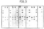
- Figure 3is the visualized pattern for the amplification products of the spacer region in the rRNA genetic locus for four serotypes from the genus Salmonell a including Salmonella typhimurim.
- Figure 4is the visualized pattern for the amplification products of the spacer region in the rRNA genetic locus for five species from the genus Staphylococcus including Staphylococcus aureus.
- Figure 5is the visualized pattern for the amplification products of the spacer region in the rRNA genetic locus for five species from the genus Escherichia including Escherichia coli.
- Figure 6is the visualized pattern for the amplification products of the spacer region in the rRNA genetic locus for eight additional species taken from four genera which are related to the pathogenic species of interest. These species are as follows: Citrobacter freundii, Citrobacter diversus, Enterobacter aerogenes, Enterobacter agglomerans, Enterobacter cloacae, Proteus mirabilis, Proteus vulgaris and Yersinia enterocolitica.
- Figure 7is the visualized pattern for the amplification products of both the spacer region in the rRNA genetic locus and the arbitrary regions from the entire microorganism genome for three species from the genus Listeria including Listeria monocytogenes.
- Figure 8is the visualized pattern for the amplification products of both the spacer region in the rRNA genetic locus and the arbitrary regions from the entire microorganism genome for three serotypes from the genus Salmonella including Salmonella typhimurim.
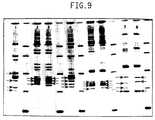
- Figure 9is the visualized pattern for the amplification products of both the spacer region in the rRNA genetic locus and the arbitrary regions from the entire microorganism genome for three species from the genus Staphylococcus including Staphylococcus aureus.
- Figure 10is the visualized pattern for the amplification products of both the spacer region in the rRNA genetic locus and arbitrary regions from the entire microorganism genome for four strains of Escherichia coli and two strains of Citrobacter freundii.
- the method described hereinis useful in identifying a wide variety of microorganisms.
- Representative but not exhaustive of the many types of organisms including both genus, species and serotype that may be elicited through the use of the present proceduresare Listeria monocytogenes, Listeria welshimeri, Listeria innocua, Listeria ivanovii, Salmonella typhimurium, Salmonella enteritidis, Salmonella newport, Salmonella infant is, Staphylococcus aureus, Staphylococcus scuiri, Staphylococcus warneri, Staphylococcus saprophyticus, Staphylococcus epidermidus, Escherichia coli, Escherichia fergusonii, Escherichia blattae, Escherichia hermanii, Escherichia vulneris, Citrobacter freundii, Citrobacter diversus, Enterobacter aerogenes, Enterobacter agglomer
- the present methodmay be applied to microorganisms in the context of a wide variety of circumstances.
- a preferred use of the present inventionis in the identification of microorganisms in food.
- research directed to microbial infections in humans, other animals and plantswould benefit from the procedure herein.
- the present inventionrelates to the identification of species of prokaryotic organisms, based on a characterization of products generated by amplification of the spacer region found between the 16S and 23S regions of the rRNA genetic locus.
- the rRNA genetic locusis a genetic unit which is found in prokaryotic cells. Significant portions of the nucleic acid sequence which make up this genetic locus are common to all prokaryotic organisms. ( Figure 1 showns a generalized schematic of this locus.) The overall relatedness of the 16S, 23S, and 5S regions of this genetic locus has been used as a tool to classify differing species of prokaryotes.
- the 16S, 23S and 5S gene sequencesare separated by spacer units. These spacer units (also referred to as spacer regions) exhibit a large degree of sequence and length variation at the levels of genus and species for prokaryotic organisms. Within a single genome of a given species there are frequently multiple rRNA genetic loci present. The spacer regions found within these loci also show a significant degree of variation in length and sequence. It has been shown that conserved sequences of the 16S region can be used as sites for amplification of nonconserved sequences within the 16S region for the purpose of determining their nucleotide composition.
- conserved regions on the 3' and 5' ends of the 16S and 23S rDNA, respectively,can be used as priming sites for the amplification of the the spacer unit contained between these two conserved regions. It is expected that amplification of such spacer regions will produce fragments whose size and number is characteristic of a given species of prokaryote.
- the 16S regions of rRNA genetic locihave been sequenced for a broad range of bacteria. A compilation of this sequence data is given in Dams et al., "Compilation of small ribosomal subunit RNA sequences", Nucleic Acids Research, Vol. 16, supplement, p. r87. This sequence data was examined for the purpose of identifying a highly conserved sequence in the 16S region immediately adjacent to the spacer region. The sequence which was chosen was GAAGTCGTAACAAGG which will be designated as P1. This sequence lies in a highly conserved region approximately 30-40 bases away from the spacer region.
- Primers for both the 16S and 23S regionswere limited to seventeen bases in length (and it is preferable to use 15-17 bases) because longer primers would extend into regions of more poorly conserved sequence. Primers which overlap regions that are not conserved among all bacteria are expected to show more variation in amplification efficiency. This would make it more difficult to specify a single set of primers and amplification conditions for all bacterial genomic DNA samples. It is possible that some sequences which are not ribosomal spacers may be amplified by the primer set of P1 and P2. This type of occurrence is expected to be rare but amplification of nonspacer sequences even under highly stringent conditions can not be ruled out.
- RS fragmentsAny of the amplified fragments produced by the high stringency amplification with primers P1 and P2 will be referred to as RS fragments. This name serves to indicate that the sequences for these primers was taken from highly conserved rDNA. sequences flanking a ribosomal spacer region.
- Ribosomal RNA genesconstitute only about 1% of the total prokaryotic genome. In order to identify species at the serotype and strain level, we need to gather additional information relating to the genomic composition of those areas located outside the rRNA genes.
- a procedure for generating arbitrary amplification fragments from genomic materialhas been demonstrated by Williams et al.
- the approachdescribes the use of a small oligonucleotide, i.e., approximately 10 bases, of arbitrary composition in a DNA amplification reaction.
- Short primersare used in order that complementary and reverse complementary sequences to the primer can be found at distances along the genome which are sufficiently small that DNA amplification can take place.
- the fragments generated in the amplification processare called Random Amplified Polymorphic DNA RAPD markers. These RAPD markers show a size distribution which is sensitive to modest differences in the genomic makeup of the DNA used in the amplification process.
- Amplification of the putative rDNA spacer regionsis achieved using 15-17 base primers and annealing temperatures of up to about 72°C (and preferably up to about 55°C) for a duration of about 1-20 (preferably about 2-10) minutes in the amplification reaction.
- Differentiation of species at the serotype and strain levelis achieved by reducing the size of the primers used in the amplification, while still maintaining the conserved element of their sequence.
- RS fragments and RAPD markersBy decreasing the size of the primers to 11 bases in length (and it is preferable to use 10-12 bases) and reducing the annealing temperature to 43-46°C for a duration of about 1-10 minutes (preferably about 5-10 minutes), it is possible to generate both RS fragments and RAPD markers from the genomic DNA using a single pair of primers. Since the RAPD markers are generated by amplification of arbitrary genomic sequences they will be referred to as AG fragments. The combined amplification of RS and AG fragments will be referred to as RS/AG amplification. The RS fragments which are produced in the amplification reaction are sufficiently distinct to permit differentiation of microorganisms at the level of species. The additional AG fragments which are generated provide sufficient additional diversity to differentiate microorganisms at the serotype and strain level.
- each RS/AG amplification patterncontains RS fragments which can be used to identify species. This means that unique strains of a given species will contain common pattern elements which will indicate that the strains are derived from a common species. When a strain is encountered which is not represented in the reference pattern database, the species of the strain can still be identified providing that the RS fragments for that species are contained in the reference pattern database.
- the 11-base primers used in the amplification reactionare a subset of the 15-base primer sequences.
- the sequence which was chosen for the 16S area adjacent to the spacerwas GAAGTCGTAAC, which will be designated as P3.
- the sequence which was chosen for the 23S area following the spacer regionwas AAGGCATCCAC, which will be designated P4.
- a high stringency amplificationis accomplished by running the reaction at the highest annealing temperature where products are reproducibly formed.
- the maximum temperature which yielded reproducible produce formationwas 55°C.
- the maximum temperature which yielded reproducible product formationwas 43°C.
- initiation sites for amplificationwill be governed by which annealing sites form the most stable primer-duplex rather than by which site has the greatest accessibility. This significantly improves the reproducibility of the pattern because the amplification sites are competing based on primer hybridization equilibria rather than on kinetic accessibility.
- the bacterial cellsare pelleted and then resuspended in lysis buffer (10 m M tris-HCl, 10 m M NaCl, 50 m M EDTA at pH 8.0).
- the cellsare subjected to an enzymatic digest comprised of 25 U Lysostaphin, 30 ug N-Acetylmuramidase, 400 ug Achromopeptidase, and 600 ug Lysozyme in a final volume of 306 ul of lysis buffer.
- the digestionis carried out for 30-45 minutes at 37°C. Lysates are extracted with chloroform/phenol, then ethanol precipitated.
- the DNAis redissolved in a tris/EDTA buffer and the final concentration is determined by spectrophotometric measurement.
- Mullisis directed to a process for amplifying any desired specific nucleic acid sequence contained in a nucleic acid or mixture thereof.
- the process of Mulliscomprises treating separate complementary strands of the nucleic acid with a molar excess of two oligonucleotide primers, and extending the primers to form complementary primer extension products which act as templates for synthesizing the desired nucleic acid sequence.
- the primers of Mullisare designed to be sufficiently complementary to different strands of each specific sequence to be amplified.
- the steps of the reactionmay be carried out stepwise or simultaneously and can be repeated as often as desired (amplification is to be performed at least twice for the present invention).
- genomic DNAis isolated by cellular lysis and deproteinization.
- Amplification of the RS fragments (and arbitrary genomic regions if applicable)comprises annealing at least one pair of oligonucleotide primers to the highly conserved rDNA sequences (and to a plurality of arbitrary regions if applicable).
- a nucleic acid polymerase and nucleotide triphosphatesare introduced to the primers.
- the procedureis carried out under conditions suitable to amplify the RS fragments (and the arbitrary regions if applicable).
- Preferred nucleic acid polymerasesare DNA polymerases, particularly those which are thermostable.
- Preferred nucleoside triphosphatesinclude deoxyribonucleoside triphosphates.
- the patternshould contain elements common to all members of the species in conjunction with elements which differentiate the unique strains within that species. This attribute makes it possible to develop a species identification of a new strain, which is not contained within the reference pattern database, providing that the common elements of that species pattern are contained within the reference database. If different strains of the same species each generate completely unique patterns with no common species elements then the patterns themselves will not identify the species of an unknown strain.
- DNA samplesare diluted to a concentration of 20 ng/ul prior to amplification.
- a 1.25 ul aliquot of the bacterial genomic DNAis combined with 2.5 ul of 10X reaction buffer, 1 ul of a dNTP mixture (5 mM ea.), 1.25 ul each of two 15-base oligonucleotide primers (P1 and P2 at 50 ng/ul) and 42 ul of deionized water.
- the primersare obtained from Research Genetics and are used without further purification. Other primers from other sources performing the same function may be used.
- This mixtureis heated to 94°C for 5 minutes and 1.3 units of a thermophilic DNA polymerase is added to the mixture.
- Acceptable polymerasesinclude Tag Polymerase, a thermostable DNA polymerase isolated from Thermus Aquaticus YT1 according to a purification procedure developed by Cetus Corporation, and available from Perkin Elmer Cetus, Norwalk, CT. Twenty-five amplification cycles are performed on an automated thermocycler. The cycle format is as follows: 1 minute at 94°C (denaturing step); 2 minute ramp to 55°C; 7 minutes at 55°C (annealing step); 2 minute ramp to 72°C and 2 minutes at 72°C (synthesis step). The final cycle is followed by an additional 7 minutes at 72°C to allow partial polymerizations to run to completion. At the end of the cycling program 1 ul of EDTA (0.5M) is added as a stop solution and the products are stored at 4°C.
- EDTA0.5M
- DNA samplesare diluted to a concentration of 20 ng/ul prior to amplification.
- a 2.5 ul aliquot of the bacterial genomic DNAis combined with 5 ul of 10X reaction buffer, 2 ul of a dNTP mixture (5 mM ea.), 2.5 ul each of two 11-base oligonucleotide primers (P3 and P4 at 50 ng/ul) and 34 ul of deionized water.
- the primersare obtained from Research Genetics and are used without further purification.
- This mixtureis heated to 94°C for 5 minutes and 2.0 units of a thermophilic DNA polymerase is added to the mixture.
- Acceptable polymerasesinclude Tag Polymerase, a thermostable DNA polymerase isolated from Thermus Aquaticus YT1 according to a purification procedure developed by Cetus Corporation, and available from Perkin Elmer Cetus, Norwalk, CT. Twenty-eight amplification cycles are performed on an automated thermocycler. The cycle format is as follows: 0.5 minutes at 94°C (denaturing step); 2.75 minute ramp to 43°C; 8 minutes at 43°C (annealing step); 3.5 minute ramp to 72°C and 2 minutes at 72°C (synthesis step). The final cycle is followed by an additional 7 minutes at 72°C to allow partial polymerizations to run to completion. At the end of the cycling program 1 ul of EDTA (0.5M) is added as a stop solution and the products are stored at 4°C.
- EDTA0.5M
- a 5 ul aliquot of the reaction mixtureis removed and combined with 2 ul of loading buffer (15% Ficol* , and 0.25% xylene cyanol).
- the mixtureis loaded onto a 4% acrylamide/bis gel (29/1) and separated by electrophoresis.
- the gelsare stained with ethidium bromide and are photographed on a UV transilluminator.
- Other techniques for visualizing the RS fragments and the AG fragmentsmay be selected.
- One such popular alternativeis the incorporation of fluorescence-labeled deoxynucleotides during amplification. Another alternative is spectroscopy.
- Figures 2-6were generated by carrying out amplification reactions using the procedure described in the section "Amplification of rDNA spacer regions” on genomic DNA samples from the specified bacteria.
- Figures 7-10were generated by carrying out amplification reactions using the procedure described in the section "Amplification of rDNA spacer regions and arbitrary genomic regions” on genomic DNA samples from the specified bacteria. Amplification products were separated on an acrylamide gel and stained with ethidium bromide.
- Bacteria representing eight different generawere chosen for amplification of RS fragments. Multiple species from the genera Listeria, and Staphylococcus, and multiple serotypes from the genus Salmonella were chosen since these represent a significant number of pathogenic microorganisms whose identification and characterization is particularly important. Five species were examined from the genus Escherichia and eight additional species taken were from four genera which are related to the pathogenic species of interest. Examples of the fragment size profiles produced in amplification reactions for these bacteria and resolved by electrophoresis are shown in Figures 2 through 6.
- RS/AGBacteria representing five different genera were chosen for the combined amplification of the RS and AG fragments (RS/AG). Multiple serotypes and strains were chosen from the genera Listeria, and Staphylococcu S. Multiple serotypes were chosen from the genus Salmonella. Four strains were examined from the genus Escherichia and two strains taken from Citrobacter freundii. Examples of the fragment size profiles produced in amplification reactions for these bacteria and resolved by electrophoresis are shown in Figures 7 through 10. The size fragments which are common to both the RS and the RS/AG amplifications for each strain are indicated by arrows in the Figures 7-10.
- the lanes containing amplifications of bacterial DNAare numbered in each figure and the strain number corresponding to that lane is given in the Figure legend.
- the unnumbered lanescontain time markers which are used to assign sizes to the amplification products.
- the sizes of the time markersare as follows: 228, 412, 693, 1331, and 2306 base pairs (bp).
- the sizes of the fragments produced in the amplificationswere calculated from the positions of these fragments relative to the time markers. The uncertainty in the calculated sizes of the amplification products is approximately 2%.
- Figure 2shows the pattern for the amplification of RS fragments for four species from the genus Listeria.
- Lanes 1-4show the patterns for four strains of Listeria monocytogenes. All four strains of L. monocytogenes show the same pair of fragments at 355 and 625 bp.
- the breakdown of remaining species of Listeriais as follows: lanes 5 and 6, L. welshimeri with fragments at 355 and 640 bp; lanes 7 and 8, L. innocua with fragments at 355 and 655 bp; lanes 9 and 10, L. ivanovii with fragments at 380 and 605 bp. There were no size fragments which were common to all species of Listeria.
- Figure 3shows the pattern for the amplification of RS fragments for four serotypes from the genus Salmonella. Patterns for four strains of Salmonella typhimurim are shown in lanes 1-4. All four strains of S. typhimurim show the same set of fragments at 480, 650, and 675 bp. The breakdown of remaining serotypes of Salmonella is as follows: lanes 5 and 6, both strains of S. enteritidis show common fragments at 480, 600, and 650 bp; lanes 7 and 8, both strains of S. newport show common fragments at 480, 570, and 610 bp and S.
- newport #707also shows an additional fragment at 510 bp; lanes 9 and 10, both strains of S. infantis show common fragments at 495, 560, 630 and 655 bp. All the serotypes of Salmonella shown contain a common fragment in the region of 480-495 bp. However, all of the serotypes tested also contain unique product size profiles.
- Figure 4shows the pattern for the amplification of RS fragments for five species from the genus Staphylococcus. Patterns for eight strains of Staphylococcus aureus are shown in lanes 1-8. The strains in lanes 1-3 all show a common pattern with fragments of 425, 465, 565, and 705 bp. The remaining five strains found in lanes 4-8 show a series of diverse patterns with only a few elements in common, e.g., fragments of 485, 570 and 620 bp can be found in three of the five strains. However, no other size of fragments are common to more than two of the five strains. The remaining species of Staphylococcus are shown in lanes 9-16.
- the intraspecies variation in the pattern of amplification of RS fragmentsis far greater for S. aureus than for the other species of Staphylococcus which were evaluated. This is an example of a case where the information in the pattern generated by the amplification of RS fragments can differentiate below the level of species.
- Other than Staphylococcus aureusall the other species of Staphylococcus were characterized by the production of a single characteristic fragment which was present at a far greater level than the other amplification products. These characteristic fragments are as follows; S. scuiri at 345 bp, S. warneri at 505 bp, S. saprophyticus at 470 bp, and S. epidermidus at 390 bp.
- the intragenic variation in patterns found within the genus Staphylococcusis also significantly greater than the variation found in the genera of Salmonella and Listeria .
- Figure 5shows the pattern for the amplification of RS fragments for five species from the genus Escherichia.
- Lanes 1-4show the patterns for four strains of Escherichia coli . All four strains of Escherichia coli show the same pair of fragments at 480 and 530 bp.
- Single examples of four additional species of Escherichiaare shown in lanes 5-8. None of these species appear to have any fragments in common with Escherichia coli although the Escherichia fergusonii in lane 6 does show a similar fragment size differential with fragments of 495 and 550 bp. There were no size fragments which were common to all species of Escherichia.
- Figure 6shows the pattern for the amplification of RS fragments for eight additional species taken from four genera which are related to the pathogenic species of interest.
- Lanes 1-4show patterns for two species of Citrobacter.
- the two strains of C. freundii shown in lanes 1 and 2both gave identical patterns with fragments of 315, 490, and 615 bp.
- the strains of C. diversus in lanes 3 and 4also gave identical patterns with fragments of 460 and 615 bp.
- Lanes 5-10show patterns for three species of Enterobacter.
- the two strains of E. aerogenes shown in lanes 5 and 6yield similar patterns with a common fragment at 465 bp.
- the other two fragmentsare 280 and 590 bp for E. aerogenes #62.
- E. aerogenes #167a similar pattern of fragments each shifted 10 bases longer is seen.
- the two strains of E. agglomerans in lanes 7 and 8show identical patterns with fragments of 450, 475 and 580 bp.
- both strains of E. cloacaeshow fragments at 310 ,465, and 580 bp.
- Lanes 11-14show patterns for two species of Proteus.
- the two strains of P. mirabilis in lanes 11 and 12show identical patterns with fragments at 505, 665 and 850 bp.
- both strains of P. vulgarisshow fragments 590, 840, 930, 1070, and 1200 bp.
- Lanes 15 and 16show patterns for two strains of Yersinia enterocolitica. Both strains show a pair of fragments at 745 and 815 bp.
- Figure 7shows the pattern for the amplification of RS and AG fragments for three species from the genus Listeria.
- Lanes 1-4show the patterns for four strains of Listeria monocytogenes. All four strains of L. monocytogenes show a common pair of fragments at 355 and 625 bp which are the same as the RS fragments observed in Figure 2 for the same strains of Listeria monocytogenes.
- the remaining components in the patternare the result of arbitrary genomic amplifications. These components distinquish three of the four L. monocytogenes strains.
- L. monocytogenes #891 and #899generate the same amplification products.
- the breakdown of remaining species of Listeriais as follows: In lanes 5 and 6 L.
- welshimerishow a common pair of fragments at 355 and 640 bp which are the same as the RS fragments observed in Figure 2 for the same strains of L. welshimeri.
- the remaining components in the patternare the result of arbitrary genomic amplifications and can be used to distinquish between the two strains of L. welshimeri .
- L. innocuashow a common pair of fragments at 355 and 655 bp which are the same as the RS fragments observed in Figure 2 for the same strains of L. innocua.
- the remaining components in the patternare the result of arbitrary genomic amplifications and can be used to distinguish between the two strains of L. innocua.
- Figure 8shows the pattern for the amplification of RS and AG fragments for three serotypes from the genus Salmonella. Patterns for four strains of Salmonella typhimurim are shown in lanes 1-4. All four strains show a similar set of fragments at 480, 650, and 675 bp which are the same as the RS fragments observed in Figure 3 for the same strains of S. typhimurim. The remaining components in the pattern are the result of arbitrary genomic amplifications. These components distinquish S. typhimurim #590 from the other three strains. The breakdown of remaining serotypes of Salmonella is as follows: In lanes 5 and 6 both strains of S.
- infantisshow common fragments at 490, 560, 630 and 655 bp which are the same as the RS fragments observed in Figure 3 for the amplifications of the same strains.
- the additional fragmentswhich are the result of arbitrary genomic amplifications, show similar patterns for both strains of S. infantis.
- both strains of S. enteritidisshow common fragments at 480, 600, and 650 bp which are the same as the RS fragments observed in Figure 3 for the amplifications of the same strains.
- the additional fragments, which are the result of arbitrary genomic amplificationscan be used to discriminate between these strains. All of the RS/AG patterns generated for the genus Salmonella were highly related. However, the individual Salmonella serotypes can be distinguished based on their unique product size profiles in the RS/AG amplifications.
- Figure 9shows the pattern for the amplification of RS and AG fragments for three species from the genus Staphylococcus. Patterns for four strains of Staphylococcus aureus are shown in lanes 1-4. The strain #807 in lane 1 shows fragments at 425, 465, 565, and 705 bp which are the same as the RS fragments observed in Figure 4 for the same strain. The remaining components in the pattern are the result of arbitrary genomic amplifications. In lanes 2 and 3 Staphylococcus aureus #1098 and #1097 both show common fragments at 565, and 605 bp with #1098 showing an additional fragment at 505 bp and #1097 showing an additional fragment at 525 bp.
- All of these fragmentsare the same as the RS fragments observed in Figure 4 for the same strains.
- the remaining components in the patternare the result of arbitrary genomic amplifications and can be used to more easily distinquish between these two strains of Staphylococcus aureus.
- the strain #795 in lane 4shows fragments at 470, 515, 565, and 610 bp which are the same as the RS fragments observed in Figure 4 for the same strain.
- the remaining components in the patternare the result of arbitrary genomic amplifications.
- the remaining species of Staphylococcusare shown in lanes 5-8. Both strains of S.
- Figure 10shows the pattern for the amplification of RS and AG fragments for four strains from Escherichia coli.
- Lanes 1-4show the patterns for four strains of Escherichia coli. All four strains of Escherichia coli show a pair of fragments at 480 and 530 bp which are the same as the RS fragments observed in Figure 5 for the amplifications of the same strains.
- the remaining components in the patternare the result of arbitrary genomic amplifications. These components can distinguish between all four strains of Escherichia coli.
- the two strains of Citrobacter freundii shown in lanes 5 and 6both exhibit patterns which. contain fragments of 315, 490, and 615 bp. These fragments correspond in size to the fragments produced in the RS amplification of C. freundii shown in Figure 6.
- the additional fragments, which are the result of arbitrary genomic amplificationscan be used to discriminate between these strains.
Landscapes
- Chemical & Material Sciences (AREA)
- Life Sciences & Earth Sciences (AREA)
- Analytical Chemistry (AREA)
- Proteomics, Peptides & Aminoacids (AREA)
- Organic Chemistry (AREA)
- Health & Medical Sciences (AREA)
- Wood Science & Technology (AREA)
- Engineering & Computer Science (AREA)
- Zoology (AREA)
- Microbiology (AREA)
- Bioinformatics & Cheminformatics (AREA)
- Molecular Biology (AREA)
- Physics & Mathematics (AREA)
- Immunology (AREA)
- Biotechnology (AREA)
- Biochemistry (AREA)
- Biophysics (AREA)
- General Engineering & Computer Science (AREA)
- General Health & Medical Sciences (AREA)
- Genetics & Genomics (AREA)
- Measuring Or Testing Involving Enzymes Or Micro-Organisms (AREA)
- Preparation Of Compounds By Using Micro-Organisms (AREA)
- Apparatus Associated With Microorganisms And Enzymes (AREA)
- Micro-Organisms Or Cultivation Processes Thereof (AREA)
Abstract
Description
- (a) isolating genomic DNA from themicroorganism, said genomic DNA comprising variablesequences interspersed between highly conservedsequences in addition to arbitrary regions along thegenome thereof;
- (b) amplifying said variable sequencesthereby producing fragments having particulardistributions in size and number;
- (c) amplifying said arbitrary regions therebyproducing Arbitrary Genomic (AG) fragments havingparticular distributions in size and numbersimultaneously with (b); and
- (d) separating said fragments and said AGfragments by size and analyzing the distribution of said fragments and said AG fragments thereby determining thespecies, serotype and strain of the microorganism.
Alternatively the oligonucleotide primers are annealedto the aforementioned sequences and to a plurality ofarbitrary regions, ensuring suitable amplification ofthe arbitrary regions.
STRAUS, NEIL A


Claims (16)
- A method for the identification of the speciesof a microorganism comprising:(a) isolating genomic DNA from themicroorganism, said genomic DNA comprising variablesequences interspersed between highly conservedsequences;(b) amplifying said variable sequencesusing only a single pair of nucleic acid primersthereby producing fragments having particulardistributions in size and number; and(c) separating said fragments by size andanalyzing the distribution of said fragments therebydetermining the species of the microorganism.
- The method of Claim 1 wherein said highlyconserved sequences are highly conserved rDNA sequences,and said fragments are Ribosomal Sequence (RS) fragments.
- The method of Claim 2 wherein said variablesequences are spacer regions.
- The method of Claim 1 wherein step (a)isolation comprises cellular lysis and deproteinization.
- The method of Claim 2 wherein step (b)comprises annealing at least one pair of oligonucleotideprimers to said highly conserved rDNA sequences, andintroducing a nucleic acid polymerase and nucleosidetriphosphates to said primers, suitable to amplify saidvariable sequences.
- The method of Claim 5 wherein said amplificationis performed at least twice.
- The method of Claim 2 wherein step (c)comprises resolving said Ribosomal Sequence (RS) fragments by electrophoresisand thereafter visualizing said Ribosomal Sequence (RS) fragments.
- The method of Claim 1 wherein in step (c) themicroorganism isListeria monocytogenes, Listeriainnocua, Listeria ivanovii, Listeria welshimeri,Salmonella typhimurium, Salmonella enteritidis,Salmonella newport, Salmonella infantis, Staphylococcusaureus, Staphylococcus sciuri, Staphylococcussaprophyticus, Staphylococcus warneri, Staphylococcusepidermidus, Escherichia coli, Escherichia blattae,Escherichia fergusonii, Escherichia hermanii,Escherichia vulneris, Citrobacter freundii, Citrobacterdiversus, Enterobacter aerogenes, Enterobacteragglomerans, Enterobacter cloacae, Proteus mirabilis,Proteus vulgari, andYersinia enterocolitica.
- A method for the identification of thespecies, serotype and strain of a microorganism,comprising:(a) isolating genomic DNA from themicroorganism, said genomic DNA comprising variablesequences interspersed between highly conservedsequences in addition to arbitrary regions along thegenome thereof;(b) amplifying said variable sequences using only a single pair of nucleic acid primersthereby producing fragments having particulardistributions in size and number;(c) amplifying said arbitrary regions therebyproducing Arbitrary Genomic (AG) fragments having particular distributionsin size and number simultaneously with (b); and(d) separating said fragments and said Arbitrary Genomic (AG)fragments by size and analyzing the distribution of saidfragments and said Arbitrary Genomic (AG) fragments thereby determining thespecies, serotype and strain of the microorganism.
- The method of Claim 9 wherein said highlyconserved sequences are highly conserved rDNA sequences,and said fragments are Ribosomal Sequence (RS) fragments.
- The method of Claim 10 wherein said variablesequences are spacer regions.
- The method of Claim 9 wherein step (a)isolation comprises cellular lysis and deproteinization.
- The method of Claim 10 wherein steps (b) and(c) comprise annealing at least one pair ofoligonucleotide primers to said highly conserved rDNAsequences and to a plurality of said arbitrary regions,and introducing a nucleic acid polymerase and nucleosidetriphosphates to said primers, suitable to amplify saidvariable sequences and said arbitrary regions.
- The method of Claim 13 wherein saidamplification is performed at least twice.
- The method of Claim 10 wherein step (d)comprises resolving said Ribosomal Sequence (RS) fragments and said Arbitrary Genomic (AG)fragments by electrophoresis and thereafter visualizingsaid Ribosomal Sequence (RS) fragments and said Arbitrary Genomic (AG) fragments.
- The method of Claim 9 wherein in step (c) themicroorganism is serotype and strain ofListeriamonocytogenes, Listeria innocua, Listeria ivanovii,Listeria welshimeri, Salmonella typhimurium, Salmonellaenteritidis, Salmonella newport, Salmonella infantis,Staphylococcus aureus, Staphylococcus sciuri,Staphylococcus saprophyticus, Staphylococcus warneri,Staphylococcus epidermidus, Escherichia coli,Escherichia blattae, Escherichia fergusonii, Escherichiahermanii, Escherichia vulneris, Citrobacter freundii,Citrobacter diversus, Enterobacter aerogenes,Enterobacter agglomerans, Enterobacter cloacae, Proteusmirabilis, Proteus vulgari, andYersinia enterocolitica.
Applications Claiming Priority (3)
| Application Number | Priority Date | Filing Date | Title |
|---|---|---|---|
| US80330291A | 1991-12-04 | 1991-12-04 | |
| US803302 | 1991-12-04 | ||
| PCT/US1992/010217WO1993011264A1 (en) | 1991-12-04 | 1992-12-02 | Method for the identification of microorganisms by the utilization of directed and arbitrary dna amplification |
Publications (2)
| Publication Number | Publication Date |
|---|---|
| EP0620862A1 EP0620862A1 (en) | 1994-10-26 |
| EP0620862B1true EP0620862B1 (en) | 1998-04-29 |
Family
ID=25186176
Family Applications (1)
| Application Number | Title | Priority Date | Filing Date |
|---|---|---|---|
| EP92925423AExpired - LifetimeEP0620862B1 (en) | 1991-12-04 | 1992-12-02 | Method for the identification of microorganisms by the utilization of directed and arbitrary dna amplification |
Country Status (13)
| Country | Link |
|---|---|
| US (1) | US5753467A (en) |
| EP (1) | EP0620862B1 (en) |
| JP (1) | JPH07501699A (en) |
| AT (1) | ATE165622T1 (en) |
| AU (1) | AU3148593A (en) |
| CA (1) | CA2125141A1 (en) |
| DE (1) | DE69225333T2 (en) |
| ES (1) | ES2114957T3 (en) |
| HK (1) | HK1006063A1 (en) |
| IL (1) | IL103935A0 (en) |
| LT (1) | LTIP1511A (en) |
| MX (1) | MX9206974A (en) |
| WO (1) | WO1993011264A1 (en) |
Cited By (27)
| Publication number | Priority date | Publication date | Assignee | Title |
|---|---|---|---|---|
| US7666592B2 (en) | 2004-02-18 | 2010-02-23 | Ibis Biosciences, Inc. | Methods for concurrent identification and quantification of an unknown bioagent |
| US7666588B2 (en) | 2001-03-02 | 2010-02-23 | Ibis Biosciences, Inc. | Methods for rapid forensic analysis of mitochondrial DNA and characterization of mitochondrial DNA heteroplasmy |
| US7714275B2 (en) | 2004-05-24 | 2010-05-11 | Ibis Biosciences, Inc. | Mass spectrometry with selective ion filtration by digital thresholding |
| US7718354B2 (en) | 2001-03-02 | 2010-05-18 | Ibis Biosciences, Inc. | Methods for rapid identification of pathogens in humans and animals |
| US7741036B2 (en) | 2001-03-02 | 2010-06-22 | Ibis Biosciences, Inc. | Method for rapid detection and identification of bioagents |
| US7781162B2 (en) | 2001-03-02 | 2010-08-24 | Ibis Biosciences, Inc. | Methods for rapid identification of pathogens in humans and animals |
| US7811753B2 (en) | 2004-07-14 | 2010-10-12 | Ibis Biosciences, Inc. | Methods for repairing degraded DNA |
| US8026084B2 (en) | 2005-07-21 | 2011-09-27 | Ibis Biosciences, Inc. | Methods for rapid identification and quantitation of nucleic acid variants |
| US8046171B2 (en) | 2003-04-18 | 2011-10-25 | Ibis Biosciences, Inc. | Methods and apparatus for genetic evaluation |
| US8071309B2 (en) | 2002-12-06 | 2011-12-06 | Ibis Biosciences, Inc. | Methods for rapid identification of pathogens in humans and animals |
| US8119336B2 (en) | 2004-03-03 | 2012-02-21 | Ibis Biosciences, Inc. | Compositions for use in identification of alphaviruses |
| US8563250B2 (en) | 2001-03-02 | 2013-10-22 | Ibis Biosciences, Inc. | Methods for identifying bioagents |
| US8871471B2 (en) | 2007-02-23 | 2014-10-28 | Ibis Biosciences, Inc. | Methods for rapid forensic DNA analysis |
| US8921047B2 (en) | 2001-06-26 | 2014-12-30 | Ibis Biosciences, Inc. | Secondary structure defining database and methods for determining identity and geographic origin of an unknown bioagent thereby |
| US8950604B2 (en) | 2009-07-17 | 2015-02-10 | Ibis Biosciences, Inc. | Lift and mount apparatus |
| US9023655B2 (en) | 2008-09-16 | 2015-05-05 | Ibis Biosciences, Inc. | Sample processing units, systems, and related methods |
| US9027730B2 (en) | 2008-09-16 | 2015-05-12 | Ibis Biosciences, Inc. | Microplate handling systems and related computer program products and methods |
| US9080209B2 (en) | 2009-08-06 | 2015-07-14 | Ibis Biosciences, Inc. | Non-mass determined base compositions for nucleic acid detection |
| US9149473B2 (en) | 2006-09-14 | 2015-10-06 | Ibis Biosciences, Inc. | Targeted whole genome amplification method for identification of pathogens |
| US9165740B2 (en) | 2009-02-12 | 2015-10-20 | Ibis Biosciences, Inc. | Ionization probe assemblies |
| US9194877B2 (en) | 2009-07-17 | 2015-11-24 | Ibis Biosciences, Inc. | Systems for bioagent indentification |
| US9393564B2 (en) | 2009-03-30 | 2016-07-19 | Ibis Biosciences, Inc. | Bioagent detection systems, devices, and methods |
| US9416409B2 (en) | 2009-07-31 | 2016-08-16 | Ibis Biosciences, Inc. | Capture primers and capture sequence linked solid supports for molecular diagnostic tests |
| US9598724B2 (en) | 2007-06-01 | 2017-03-21 | Ibis Biosciences, Inc. | Methods and compositions for multiple displacement amplification of nucleic acids |
| US9719083B2 (en) | 2009-03-08 | 2017-08-01 | Ibis Biosciences, Inc. | Bioagent detection methods |
| US9758840B2 (en) | 2010-03-14 | 2017-09-12 | Ibis Biosciences, Inc. | Parasite detection via endosymbiont detection |
| US9890408B2 (en) | 2009-10-15 | 2018-02-13 | Ibis Biosciences, Inc. | Multiple displacement amplification |
Families Citing this family (78)
| Publication number | Priority date | Publication date | Assignee | Title |
|---|---|---|---|---|
| US6395475B1 (en) | 1993-05-18 | 2002-05-28 | Florida State University | Semiautomated method for finger-printing bacterial DNA |
| EP1091004B1 (en) | 1994-06-24 | 2009-02-25 | Innogenetics N.V. | Simultaneous detection, identification and differentiation of Mycobacterium species using a hybridization assay |
| GB9420774D0 (en)* | 1994-10-14 | 1994-11-30 | Univ Wales | Method of detecting and identifying spore forming bacteria |
| US6312930B1 (en) | 1996-09-16 | 2001-11-06 | E. I. Du Pont De Nemours And Company | Method for detecting bacteria using PCR |
| DE19731292A1 (en)* | 1997-07-21 | 1999-01-28 | Biotecon Ges Fuer Biotechnologische Entwicklung & Consulting Mbh | Nucleic Acid Molecule, Kit, and Use |
| DE19739611A1 (en)* | 1997-09-09 | 1999-03-11 | Biotecon Ges Fuer Biotechnologische Entwicklung & Consulting Mbh | Nucleic acid sequences and method for the detection of bacteria of the genus Pseudomonas |
| JPH11341989A (en)* | 1998-03-31 | 1999-12-14 | Sanyo Electric Co Ltd | Dna fragment amplification, dna fragment amplifier, microorganism group measurement, microorganism group analysis and pollutant measurement |
| US6503747B2 (en) | 1998-07-14 | 2003-01-07 | University Of Hawaii | Serotype-specific probes for Listeria monocytogenes |
| AR021815A1 (en) | 1998-09-18 | 2002-08-07 | Univ Nebraska Lincoln | HIGH RESOLUTION ANALYSIS OF GENOMAS. |
| US6197514B1 (en)* | 1999-04-12 | 2001-03-06 | Becton, Dickinson And Company | Amplification and detection of Yersinia enterocolitica |
| JP2001149073A (en)* | 1999-09-16 | 2001-06-05 | Sanyo Electric Co Ltd | Method and apparatus for analyzing intestinal flora |
| DE19945916A1 (en) | 1999-09-24 | 2001-04-05 | Biotecon Diagnostics Gmbh | Nucleic acid molecules for the detection of bacteria and phylogenetic units of bacteria |
| JP3628232B2 (en)* | 2000-03-31 | 2005-03-09 | 三洋電機株式会社 | Microorganism identification method, microorganism identification device, method for creating a database for microorganism identification, and recording medium on which a microorganism identification program is recorded |
| JP2002000271A (en)* | 2000-06-28 | 2002-01-08 | Sanyo Electric Co Ltd | System, method, and database for analyzing microorganism |
| US20020086313A1 (en)* | 2000-09-25 | 2002-07-04 | Kilbane John J. | Application of bioinformatics for direct study of unculturable microorganisms |
| NZ530343A (en)* | 2001-06-22 | 2007-01-26 | Marshfield Clinic | Methods and oligonucleotides for the detection of E. coli 0157:H7 |
| US8073627B2 (en)* | 2001-06-26 | 2011-12-06 | Ibis Biosciences, Inc. | System for indentification of pathogens |
| US20040241723A1 (en)* | 2002-03-18 | 2004-12-02 | Marquess Foley Leigh Shaw | Systems and methods for improving protein and milk production of dairy herds |
| EP2390352A1 (en) | 2003-03-18 | 2011-11-30 | Quantum Genetics Ireland Limited | Systems and methods for improving protein and milk production of dairy herds |
| US8057993B2 (en) | 2003-04-26 | 2011-11-15 | Ibis Biosciences, Inc. | Methods for identification of coronaviruses |
| US8158354B2 (en) | 2003-05-13 | 2012-04-17 | Ibis Biosciences, Inc. | Methods for rapid purification of nucleic acids for subsequent analysis by mass spectrometry by solution capture |
| US7964343B2 (en) | 2003-05-13 | 2011-06-21 | Ibis Biosciences, Inc. | Method for rapid purification of nucleic acids for subsequent analysis by mass spectrometry by solution capture |
| US20120122103A1 (en) | 2003-09-11 | 2012-05-17 | Rangarajan Sampath | Compositions for use in identification of bacteria |
| US8546082B2 (en) | 2003-09-11 | 2013-10-01 | Ibis Biosciences, Inc. | Methods for identification of sepsis-causing bacteria |
| US8097416B2 (en) | 2003-09-11 | 2012-01-17 | Ibis Biosciences, Inc. | Methods for identification of sepsis-causing bacteria |
| US8163895B2 (en) | 2003-12-05 | 2012-04-24 | Ibis Biosciences, Inc. | Compositions for use in identification of orthopoxviruses |
| MXPA06009452A (en) | 2004-02-19 | 2007-03-15 | Univ Alberta | Leptin promoter polymorphisms and uses thereof. |
| US7291465B2 (en)* | 2004-02-20 | 2007-11-06 | Karaolis David K R | Method for direct detection of fungal pathogens |
| JP4788942B2 (en)* | 2004-02-23 | 2011-10-05 | 独立行政法人産業技術総合研究所 | Deoxyribozymes for specific species detection |
| US7919277B2 (en)* | 2004-04-28 | 2011-04-05 | Danisco A/S | Detection and typing of bacterial strains |
| US20050266411A1 (en) | 2004-05-25 | 2005-12-01 | Hofstadler Steven A | Methods for rapid forensic analysis of mitochondrial DNA |
| WO2006094238A2 (en) | 2005-03-03 | 2006-09-08 | Isis Pharmaceuticals, Inc. | Compositions for use in identification of adventitious viruses |
| US8084207B2 (en)* | 2005-03-03 | 2011-12-27 | Ibis Bioscience, Inc. | Compositions for use in identification of papillomavirus |
| JP5162447B2 (en)* | 2005-04-13 | 2013-03-13 | アイビス バイオサイエンシズ インコーポレイティッド | Composition for use in adenovirus identification |
| US20060240442A1 (en)* | 2005-04-20 | 2006-10-26 | Vevea Dirk N | Methods and oligonucleotides for the detection of Salmonella SP., E coli 0157:H7, and Listeria monocytogenes |
| US20060246463A1 (en)* | 2005-04-20 | 2006-11-02 | Vevea Dirk N | Methods and oligonucleotides for the detection of Salmonella SP., E coli 0157:H7, and Listeria monocytogenes |
| US10083273B2 (en) | 2005-07-29 | 2018-09-25 | Natera, Inc. | System and method for cleaning noisy genetic data and determining chromosome copy number |
| US9424392B2 (en) | 2005-11-26 | 2016-08-23 | Natera, Inc. | System and method for cleaning noisy genetic data from target individuals using genetic data from genetically related individuals |
| US11111543B2 (en) | 2005-07-29 | 2021-09-07 | Natera, Inc. | System and method for cleaning noisy genetic data and determining chromosome copy number |
| US10081839B2 (en) | 2005-07-29 | 2018-09-25 | Natera, Inc | System and method for cleaning noisy genetic data and determining chromosome copy number |
| US11111544B2 (en) | 2005-07-29 | 2021-09-07 | Natera, Inc. | System and method for cleaning noisy genetic data and determining chromosome copy number |
| ES2620431T3 (en) | 2008-08-04 | 2017-06-28 | Natera, Inc. | Methods for the determination of alleles and ploidy |
| EP2349549B1 (en) | 2008-09-16 | 2012-07-18 | Ibis Biosciences, Inc. | Mixing cartridges, mixing stations, and related kits, and system |
| US20120185176A1 (en) | 2009-09-30 | 2012-07-19 | Natera, Inc. | Methods for Non-Invasive Prenatal Ploidy Calling |
| US8932809B2 (en)* | 2010-01-19 | 2015-01-13 | Opgen, Inc. | Methods and kits for isolating nucleic acid from an organism |
| US9677118B2 (en) | 2014-04-21 | 2017-06-13 | Natera, Inc. | Methods for simultaneous amplification of target loci |
| EP2854057B1 (en) | 2010-05-18 | 2018-03-07 | Natera, Inc. | Methods for non-invasive pre-natal ploidy calling |
| US10316362B2 (en) | 2010-05-18 | 2019-06-11 | Natera, Inc. | Methods for simultaneous amplification of target loci |
| US11332785B2 (en) | 2010-05-18 | 2022-05-17 | Natera, Inc. | Methods for non-invasive prenatal ploidy calling |
| US11408031B2 (en) | 2010-05-18 | 2022-08-09 | Natera, Inc. | Methods for non-invasive prenatal paternity testing |
| US11332793B2 (en) | 2010-05-18 | 2022-05-17 | Natera, Inc. | Methods for simultaneous amplification of target loci |
| US20130123120A1 (en)* | 2010-05-18 | 2013-05-16 | Natera, Inc. | Highly Multiplex PCR Methods and Compositions |
| US11322224B2 (en) | 2010-05-18 | 2022-05-03 | Natera, Inc. | Methods for non-invasive prenatal ploidy calling |
| US20190010543A1 (en) | 2010-05-18 | 2019-01-10 | Natera, Inc. | Methods for simultaneous amplification of target loci |
| US11339429B2 (en) | 2010-05-18 | 2022-05-24 | Natera, Inc. | Methods for non-invasive prenatal ploidy calling |
| US12221653B2 (en) | 2010-05-18 | 2025-02-11 | Natera, Inc. | Methods for simultaneous amplification of target loci |
| US11326208B2 (en) | 2010-05-18 | 2022-05-10 | Natera, Inc. | Methods for nested PCR amplification of cell-free DNA |
| US10017812B2 (en) | 2010-05-18 | 2018-07-10 | Natera, Inc. | Methods for non-invasive prenatal ploidy calling |
| US12152275B2 (en) | 2010-05-18 | 2024-11-26 | Natera, Inc. | Methods for non-invasive prenatal ploidy calling |
| US11939634B2 (en) | 2010-05-18 | 2024-03-26 | Natera, Inc. | Methods for simultaneous amplification of target loci |
| AU2011348100B2 (en) | 2010-12-22 | 2016-08-25 | Natera, Inc. | Methods for non-invasive prenatal paternity testing |
| CN103703147A (en)* | 2011-07-25 | 2014-04-02 | 纳幕尔杜邦公司 | Method to amplify nucleic acids to generate fluorescence labeled fragments of conserved and arbitrary products |
| US20140100126A1 (en) | 2012-08-17 | 2014-04-10 | Natera, Inc. | Method for Non-Invasive Prenatal Testing Using Parental Mosaicism Data |
| US10262755B2 (en) | 2014-04-21 | 2019-04-16 | Natera, Inc. | Detecting cancer mutations and aneuploidy in chromosomal segments |
| US10577655B2 (en) | 2013-09-27 | 2020-03-03 | Natera, Inc. | Cell free DNA diagnostic testing standards |
| US9499870B2 (en) | 2013-09-27 | 2016-11-22 | Natera, Inc. | Cell free DNA diagnostic testing standards |
| JP6659575B2 (en) | 2014-04-21 | 2020-03-04 | ナテラ, インコーポレイテッド | Mutation detection and chromosomal segment ploidy |
| US20180173845A1 (en) | 2014-06-05 | 2018-06-21 | Natera, Inc. | Systems and Methods for Detection of Aneuploidy |
| DK3294906T3 (en) | 2015-05-11 | 2024-08-05 | Natera Inc | Methods for determining ploidy |
| EP3443119B8 (en) | 2016-04-15 | 2022-04-06 | Natera, Inc. | Methods for lung cancer detection |
| WO2018067517A1 (en) | 2016-10-04 | 2018-04-12 | Natera, Inc. | Methods for characterizing copy number variation using proximity-litigation sequencing |
| US10011870B2 (en) | 2016-12-07 | 2018-07-03 | Natera, Inc. | Compositions and methods for identifying nucleic acid molecules |
| AU2018225348A1 (en) | 2017-02-21 | 2019-07-18 | Natera, Inc. | Compositions, methods, and kits for isolating nucleic acids |
| CA3085933A1 (en) | 2017-12-14 | 2019-06-20 | Tai Diagnostics, Inc. | Assessing graft suitability for transplantation |
| US12398389B2 (en) | 2018-02-15 | 2025-08-26 | Natera, Inc. | Methods for isolating nucleic acids with size selection |
| WO2019200228A1 (en) | 2018-04-14 | 2019-10-17 | Natera, Inc. | Methods for cancer detection and monitoring by means of personalized detection of circulating tumor dna |
| US12234509B2 (en) | 2018-07-03 | 2025-02-25 | Natera, Inc. | Methods for detection of donor-derived cell-free DNA |
| US12305235B2 (en) | 2019-06-06 | 2025-05-20 | Natera, Inc. | Methods for detecting immune cell DNA and monitoring immune system |
Family Cites Families (17)
| Publication number | Priority date | Publication date | Assignee | Title |
|---|---|---|---|---|
| US4965188A (en)* | 1986-08-22 | 1990-10-23 | Cetus Corporation | Process for amplifying, detecting, and/or cloning nucleic acid sequences using a thermostable enzyme |
| US4683202A (en)* | 1985-03-28 | 1987-07-28 | Cetus Corporation | Process for amplifying nucleic acid sequences |
| US4683195A (en)* | 1986-01-30 | 1987-07-28 | Cetus Corporation | Process for amplifying, detecting, and/or-cloning nucleic acid sequences |
| CA1317535C (en)* | 1987-06-30 | 1993-05-11 | Nanibhushan Dattagupta | Assay of sequences using amplified genes |
| IE61148B1 (en)* | 1988-03-10 | 1994-10-05 | Ici Plc | Method of detecting nucleotide sequences |
| EP0335633B1 (en)* | 1988-03-28 | 1995-05-03 | Amoco Corporation | Detecting Eukaryotic Microorganisms |
| WO1989010414A1 (en)* | 1988-04-28 | 1989-11-02 | Robert Bruce Wallace | AMPLIFIED SEQUENCE POLYMORPHISMS (ASPs) |
| US4885697A (en)* | 1988-09-01 | 1989-12-05 | E. I. Du Pont De Nemours And Company | Method of identifying spectra |
| FR2636075B1 (en)* | 1988-09-07 | 1991-11-15 | Biotechnologie Ste Europ | METHOD FOR DETECTING BACTERIA, YEAST, PARASITES AND OTHER EUKARYOTS, ESPECIALLY IN FOOD PRODUCTS |
| IE81145B1 (en)* | 1989-04-20 | 2000-05-03 | Thomas Gerard Barry | Generation of specific probes for target nucleotide sequences |
| GB8909095D0 (en)* | 1989-04-21 | 1989-06-07 | Allied Colloids Ltd | Thickened aqueous compositions |
| US5192659A (en)* | 1989-08-25 | 1993-03-09 | Genetype Ag | Intron sequence analysis method for detection of adjacent and remote locus alleles as haplotypes |
| WO1991014003A2 (en)* | 1990-03-09 | 1991-09-19 | Ig Laboratories, Inc. | Characterization and analysis of polymorphic vntr loci |
| WO1992007095A1 (en)* | 1990-10-15 | 1992-04-30 | Stratagene | Arbitrarily primed polymerase chain reaction method for fingerprinting genomes |
| NL9002259A (en)* | 1990-10-17 | 1992-05-18 | Eurodiagnostics B V | METHOD FOR DETERMINING A GENOTYPE BY COMPARING THE NUCLEOTID SEQUENCE OF MEM FAMILY MEMBERS AND KIT FOR DETECTING GENETIC VARIATIONS. |
| US5437975A (en)* | 1991-02-25 | 1995-08-01 | California Institute Of Biological Research | Consensus sequence primed polymerase chain reaction method for fingerprinting genomes |
| DE69231646T2 (en)* | 1991-10-23 | 2001-06-13 | Baylor College Of Medicine, Houston | FINGERPRINT-LIKE IDENTIFICATION OF BACTERIA STRAINS BY AMPLIFICATING REPETITIVE DNA SEQUENCES |
- 1992
- 1992-12-01ILIL103935Apatent/IL103935A0/enunknown
- 1992-12-02EPEP92925423Apatent/EP0620862B1/ennot_activeExpired - Lifetime
- 1992-12-02ESES92925423Tpatent/ES2114957T3/ennot_activeExpired - Lifetime
- 1992-12-02ATAT92925423Tpatent/ATE165622T1/ennot_activeIP Right Cessation
- 1992-12-02WOPCT/US1992/010217patent/WO1993011264A1/enactiveIP Right Grant
- 1992-12-02CACA002125141Apatent/CA2125141A1/ennot_activeAbandoned
- 1992-12-02DEDE69225333Tpatent/DE69225333T2/ennot_activeExpired - Fee Related
- 1992-12-02HKHK98105121Apatent/HK1006063A1/ennot_activeIP Right Cessation
- 1992-12-02AUAU31485/93Apatent/AU3148593A/ennot_activeAbandoned
- 1992-12-02JPJP5510256Apatent/JPH07501699A/enactivePending
- 1992-12-03MXMX9206974Apatent/MX9206974A/enunknown
- 1993
- 1993-12-02LTLTIP1511Apatent/LTIP1511A/ennot_activeApplication Discontinuation
- 1996
- 1996-08-22USUS08/701,290patent/US5753467A/ennot_activeExpired - Lifetime
Cited By (38)
| Publication number | Priority date | Publication date | Assignee | Title |
|---|---|---|---|---|
| US8017743B2 (en) | 2001-03-02 | 2011-09-13 | Ibis Bioscience, Inc. | Method for rapid detection and identification of bioagents |
| US8017358B2 (en) | 2001-03-02 | 2011-09-13 | Ibis Biosciences, Inc. | Method for rapid detection and identification of bioagents |
| US9752184B2 (en) | 2001-03-02 | 2017-09-05 | Ibis Biosciences, Inc. | Methods for rapid forensic analysis of mitochondrial DNA and characterization of mitochondrial DNA heteroplasmy |
| US7718354B2 (en) | 2001-03-02 | 2010-05-18 | Ibis Biosciences, Inc. | Methods for rapid identification of pathogens in humans and animals |
| US7741036B2 (en) | 2001-03-02 | 2010-06-22 | Ibis Biosciences, Inc. | Method for rapid detection and identification of bioagents |
| US7781162B2 (en) | 2001-03-02 | 2010-08-24 | Ibis Biosciences, Inc. | Methods for rapid identification of pathogens in humans and animals |
| US9416424B2 (en) | 2001-03-02 | 2016-08-16 | Ibis Biosciences, Inc. | Methods for rapid identification of pathogens in humans and animals |
| US8017322B2 (en) | 2001-03-02 | 2011-09-13 | Ibis Biosciences, Inc. | Method for rapid detection and identification of bioagents |
| US7666588B2 (en) | 2001-03-02 | 2010-02-23 | Ibis Biosciences, Inc. | Methods for rapid forensic analysis of mitochondrial DNA and characterization of mitochondrial DNA heteroplasmy |
| US8563250B2 (en) | 2001-03-02 | 2013-10-22 | Ibis Biosciences, Inc. | Methods for identifying bioagents |
| US8921047B2 (en) | 2001-06-26 | 2014-12-30 | Ibis Biosciences, Inc. | Secondary structure defining database and methods for determining identity and geographic origin of an unknown bioagent thereby |
| US8071309B2 (en) | 2002-12-06 | 2011-12-06 | Ibis Biosciences, Inc. | Methods for rapid identification of pathogens in humans and animals |
| US9725771B2 (en) | 2002-12-06 | 2017-08-08 | Ibis Biosciences, Inc. | Methods for rapid identification of pathogens in humans and animals |
| US8046171B2 (en) | 2003-04-18 | 2011-10-25 | Ibis Biosciences, Inc. | Methods and apparatus for genetic evaluation |
| US7666592B2 (en) | 2004-02-18 | 2010-02-23 | Ibis Biosciences, Inc. | Methods for concurrent identification and quantification of an unknown bioagent |
| US9447462B2 (en) | 2004-02-18 | 2016-09-20 | Ibis Biosciences, Inc. | Methods for concurrent identification and quantification of an unknown bioagent |
| US8119336B2 (en) | 2004-03-03 | 2012-02-21 | Ibis Biosciences, Inc. | Compositions for use in identification of alphaviruses |
| US8987660B2 (en) | 2004-05-24 | 2015-03-24 | Ibis Biosciences, Inc. | Mass spectrometry with selective ion filtration by digital thresholding |
| US9449802B2 (en) | 2004-05-24 | 2016-09-20 | Ibis Biosciences, Inc. | Mass spectrometry with selective ion filtration by digital thresholding |
| US7714275B2 (en) | 2004-05-24 | 2010-05-11 | Ibis Biosciences, Inc. | Mass spectrometry with selective ion filtration by digital thresholding |
| US9873906B2 (en) | 2004-07-14 | 2018-01-23 | Ibis Biosciences, Inc. | Methods for repairing degraded DNA |
| US7811753B2 (en) | 2004-07-14 | 2010-10-12 | Ibis Biosciences, Inc. | Methods for repairing degraded DNA |
| US8026084B2 (en) | 2005-07-21 | 2011-09-27 | Ibis Biosciences, Inc. | Methods for rapid identification and quantitation of nucleic acid variants |
| US9149473B2 (en) | 2006-09-14 | 2015-10-06 | Ibis Biosciences, Inc. | Targeted whole genome amplification method for identification of pathogens |
| US8871471B2 (en) | 2007-02-23 | 2014-10-28 | Ibis Biosciences, Inc. | Methods for rapid forensic DNA analysis |
| US9598724B2 (en) | 2007-06-01 | 2017-03-21 | Ibis Biosciences, Inc. | Methods and compositions for multiple displacement amplification of nucleic acids |
| US9023655B2 (en) | 2008-09-16 | 2015-05-05 | Ibis Biosciences, Inc. | Sample processing units, systems, and related methods |
| US9027730B2 (en) | 2008-09-16 | 2015-05-12 | Ibis Biosciences, Inc. | Microplate handling systems and related computer program products and methods |
| US9165740B2 (en) | 2009-02-12 | 2015-10-20 | Ibis Biosciences, Inc. | Ionization probe assemblies |
| US9719083B2 (en) | 2009-03-08 | 2017-08-01 | Ibis Biosciences, Inc. | Bioagent detection methods |
| US9393564B2 (en) | 2009-03-30 | 2016-07-19 | Ibis Biosciences, Inc. | Bioagent detection systems, devices, and methods |
| US8950604B2 (en) | 2009-07-17 | 2015-02-10 | Ibis Biosciences, Inc. | Lift and mount apparatus |
| US9194877B2 (en) | 2009-07-17 | 2015-11-24 | Ibis Biosciences, Inc. | Systems for bioagent indentification |
| US9416409B2 (en) | 2009-07-31 | 2016-08-16 | Ibis Biosciences, Inc. | Capture primers and capture sequence linked solid supports for molecular diagnostic tests |
| US10119164B2 (en) | 2009-07-31 | 2018-11-06 | Ibis Biosciences, Inc. | Capture primers and capture sequence linked solid supports for molecular diagnostic tests |
| US9080209B2 (en) | 2009-08-06 | 2015-07-14 | Ibis Biosciences, Inc. | Non-mass determined base compositions for nucleic acid detection |
| US9890408B2 (en) | 2009-10-15 | 2018-02-13 | Ibis Biosciences, Inc. | Multiple displacement amplification |
| US9758840B2 (en) | 2010-03-14 | 2017-09-12 | Ibis Biosciences, Inc. | Parasite detection via endosymbiont detection |
Also Published As
| Publication number | Publication date |
|---|---|
| MX9206974A (en) | 1993-06-01 |
| ATE165622T1 (en) | 1998-05-15 |
| IL103935A0 (en) | 1993-05-13 |
| CA2125141A1 (en) | 1993-06-10 |
| DE69225333T2 (en) | 1998-09-24 |
| LTIP1511A (en) | 1995-06-26 |
| EP0620862A1 (en) | 1994-10-26 |
| WO1993011264A1 (en) | 1993-06-10 |
| DE69225333D1 (en) | 1998-06-04 |
| HK1006063A1 (en) | 1999-02-05 |
| US5753467A (en) | 1998-05-19 |
| AU3148593A (en) | 1993-06-28 |
| ES2114957T3 (en) | 1998-06-16 |
| JPH07501699A (en) | 1995-02-23 |
Similar Documents
| Publication | Publication Date | Title |
|---|---|---|
| EP0620862B1 (en) | Method for the identification of microorganisms by the utilization of directed and arbitrary dna amplification | |
| HK1006063B (en) | Method for the identification of microorganisms by the utilization of directed and arbitrary dna amplification | |
| US6696277B1 (en) | Arbitrarily primed polymerase chain reaction method for fingerprinting genomes | |
| US5437975A (en) | Consensus sequence primed polymerase chain reaction method for fingerprinting genomes | |
| EP0395292B1 (en) | Generation of specific probes for target nucleotide sequences | |
| EP0610396B1 (en) | Fingerprinting bacterial strains using repetitive dna sequence amplification | |
| US6054278A (en) | Ribosomal RNA gene polymorphism based microorganism identification | |
| EP0534858B1 (en) | Selective restriction fragment amplification : a general method for DNA fingerprinting | |
| EP1068352B1 (en) | Nucleic acid detection method | |
| CA2322780C (en) | Detection of fermentation-related microorganisms | |
| EP0317239A2 (en) | Method and device for improved restriction fragment length polymorphism analysis | |
| CA2246259C (en) | Genetic markers and methods for the detection of escherichia coli serotype-0157:h7 | |
| EP0948643B1 (en) | Genetic markers and methods for the detection of listeria monocytogenes and listeria spp | |
| EP0764217B1 (en) | Use of a specific marker for detecting Salmonella with PCR | |
| Swaminathan et al. | Amplification methods for epidemiologic investigations of infectious diseases | |
| KR101739754B1 (en) | SNP primer set for origin identification of sesame, method for origin identification of sesame using the same and kit comprising the same | |
| Glen | The use of DNA techniques to identify fungi | |
| Janssen | Selective restriction fragment amplification by AFLP | |
| Castrillo et al. | Molecular methods for identification and diagnosis of fungi. | |
| Raja et al. | APPLICATION OF MOLECULAR TOOLS FOR IDENTIFICATION OF BACTERIA RELEVANT TO AQUACULTURE |
Legal Events
| Date | Code | Title | Description |
|---|---|---|---|
| PUAI | Public reference made under article 153(3) epc to a published international application that has entered the european phase | Free format text:ORIGINAL CODE: 0009012 | |
| 17P | Request for examination filed | Effective date:19940524 | |
| AK | Designated contracting states | Kind code of ref document:A1 Designated state(s):AT BE CH DE DK ES FR GB GR IE IT LI LU MC NL PT SE | |
| 17Q | First examination report despatched | Effective date:19950209 | |
| GRAG | Despatch of communication of intention to grant | Free format text:ORIGINAL CODE: EPIDOS AGRA | |
| GRAG | Despatch of communication of intention to grant | Free format text:ORIGINAL CODE: EPIDOS AGRA | |
| GRAH | Despatch of communication of intention to grant a patent | Free format text:ORIGINAL CODE: EPIDOS IGRA | |
| GRAH | Despatch of communication of intention to grant a patent | Free format text:ORIGINAL CODE: EPIDOS IGRA | |
| GRAA | (expected) grant | Free format text:ORIGINAL CODE: 0009210 | |
| AK | Designated contracting states | Kind code of ref document:B1 Designated state(s):AT BE CH DE DK ES FR GB GR IE IT LI LU MC NL PT SE | |
| REF | Corresponds to: | Ref document number:165622 Country of ref document:AT Date of ref document:19980515 Kind code of ref document:T | |
| REG | Reference to a national code | Ref country code:CH Ref legal event code:EP | |
| ITF | It: translation for a ep patent filed | ||
| REG | Reference to a national code | Ref country code:CH Ref legal event code:NV Representative=s name:RITSCHER & SEIFERT PATENTANWAELTE VSP | |
| REF | Corresponds to: | Ref document number:69225333 Country of ref document:DE Date of ref document:19980604 | |
| ET | Fr: translation filed | ||
| REG | Reference to a national code | Ref country code:ES Ref legal event code:FG2A Ref document number:2114957 Country of ref document:ES Kind code of ref document:T3 | |
| REG | Reference to a national code | Ref country code:IE Ref legal event code:FG4D Free format text:80057 | |
| PGFP | Annual fee paid to national office [announced via postgrant information from national office to epo] | Ref country code:GR Payment date:19980831 Year of fee payment:7 | |
| REG | Reference to a national code | Ref country code:PT Ref legal event code:SC4A Free format text:AVAILABILITY OF NATIONAL TRANSLATION Effective date:19980430 | |
| PGFP | Annual fee paid to national office [announced via postgrant information from national office to epo] | Ref country code:PT Payment date:19980914 Year of fee payment:7 | |
| PGFP | Annual fee paid to national office [announced via postgrant information from national office to epo] | Ref country code:AT Payment date:19981102 Year of fee payment:7 | |
| PGFP | Annual fee paid to national office [announced via postgrant information from national office to epo] | Ref country code:IE Payment date:19981124 Year of fee payment:7 | |
| PGFP | Annual fee paid to national office [announced via postgrant information from national office to epo] | Ref country code:SE Payment date:19981127 Year of fee payment:7 | |
| PGFP | Annual fee paid to national office [announced via postgrant information from national office to epo] | Ref country code:GB Payment date:19981204 Year of fee payment:7 | |
| PGFP | Annual fee paid to national office [announced via postgrant information from national office to epo] | Ref country code:FR Payment date:19981209 Year of fee payment:7 | |
| PGFP | Annual fee paid to national office [announced via postgrant information from national office to epo] | Ref country code:LU Payment date:19981214 Year of fee payment:7 Ref country code:DK Payment date:19981214 Year of fee payment:7 Ref country code:DE Payment date:19981214 Year of fee payment:7 | |
| PGFP | Annual fee paid to national office [announced via postgrant information from national office to epo] | Ref country code:NL Payment date:19981218 Year of fee payment:7 | |
| PGFP | Annual fee paid to national office [announced via postgrant information from national office to epo] | Ref country code:CH Payment date:19981223 Year of fee payment:7 | |
| PGFP | Annual fee paid to national office [announced via postgrant information from national office to epo] | Ref country code:BE Payment date:19990111 Year of fee payment:7 | |
| PLBE | No opposition filed within time limit | Free format text:ORIGINAL CODE: 0009261 | |
| STAA | Information on the status of an ep patent application or granted ep patent | Free format text:STATUS: NO OPPOSITION FILED WITHIN TIME LIMIT | |
| 26N | No opposition filed | ||
| PG25 | Lapsed in a contracting state [announced via postgrant information from national office to epo] | Ref country code:MC Free format text:LAPSE BECAUSE OF NON-PAYMENT OF DUE FEES Effective date:19990630 | |
| PG25 | Lapsed in a contracting state [announced via postgrant information from national office to epo] | Ref country code:LU Free format text:LAPSE BECAUSE OF NON-PAYMENT OF DUE FEES Effective date:19991202 Ref country code:IE Free format text:LAPSE BECAUSE OF NON-PAYMENT OF DUE FEES Effective date:19991202 Ref country code:GB Free format text:LAPSE BECAUSE OF NON-PAYMENT OF DUE FEES Effective date:19991202 Ref country code:DK Free format text:LAPSE BECAUSE OF NON-PAYMENT OF DUE FEES Effective date:19991202 Ref country code:AT Free format text:LAPSE BECAUSE OF NON-PAYMENT OF DUE FEES Effective date:19991202 | |
| PG25 | Lapsed in a contracting state [announced via postgrant information from national office to epo] | Ref country code:SE Free format text:LAPSE BECAUSE OF NON-PAYMENT OF DUE FEES Effective date:19991203 | |
| PGFP | Annual fee paid to national office [announced via postgrant information from national office to epo] | Ref country code:ES Payment date:19991214 Year of fee payment:8 | |
| PG25 | Lapsed in a contracting state [announced via postgrant information from national office to epo] | Ref country code:LI Free format text:LAPSE BECAUSE OF NON-PAYMENT OF DUE FEES Effective date:19991231 Ref country code:GR Free format text:LAPSE BECAUSE OF NON-PAYMENT OF DUE FEES Effective date:19991231 Ref country code:CH Free format text:LAPSE BECAUSE OF NON-PAYMENT OF DUE FEES Effective date:19991231 Ref country code:BE Free format text:LAPSE BECAUSE OF NON-PAYMENT OF DUE FEES Effective date:19991231 | |
| BERE | Be: lapsed | Owner name:E.I. DU PONT DE NEMOURS AND CY Effective date:19991231 | |
| PG25 | Lapsed in a contracting state [announced via postgrant information from national office to epo] | Ref country code:PT Free format text:LAPSE BECAUSE OF NON-PAYMENT OF DUE FEES Effective date:20000630 | |
| PG25 | Lapsed in a contracting state [announced via postgrant information from national office to epo] | Ref country code:NL Free format text:LAPSE BECAUSE OF NON-PAYMENT OF DUE FEES Effective date:20000701 | |
| GBPC | Gb: european patent ceased through non-payment of renewal fee | Effective date:19991202 | |
| EUG | Se: european patent has lapsed | Ref document number:92925423.3 | |
| PG25 | Lapsed in a contracting state [announced via postgrant information from national office to epo] | Ref country code:FR Free format text:LAPSE BECAUSE OF NON-PAYMENT OF DUE FEES Effective date:20000831 | |
| NLV4 | Nl: lapsed or anulled due to non-payment of the annual fee | Effective date:20000701 | |
| REG | Reference to a national code | Ref country code:IE Ref legal event code:MM4A | |
| PG25 | Lapsed in a contracting state [announced via postgrant information from national office to epo] | Ref country code:DE Free format text:LAPSE BECAUSE OF NON-PAYMENT OF DUE FEES Effective date:20001003 | |
| REG | Reference to a national code | Ref country code:FR Ref legal event code:ST | |
| REG | Reference to a national code | Ref country code:PT Ref legal event code:MM4A Free format text:LAPSE DUE TO NON-PAYMENT OF FEES Effective date:20000630 | |
| PG25 | Lapsed in a contracting state [announced via postgrant information from national office to epo] | Ref country code:ES Free format text:LAPSE BECAUSE OF NON-PAYMENT OF DUE FEES Effective date:20011203 | |
| REG | Reference to a national code | Ref country code:DK Ref legal event code:EBP | |
| REG | Reference to a national code | Ref country code:ES Ref legal event code:FD2A Effective date:20020112 | |
| PG25 | Lapsed in a contracting state [announced via postgrant information from national office to epo] | Ref country code:IT Free format text:LAPSE BECAUSE OF NON-PAYMENT OF DUE FEES;WARNING: LAPSES OF ITALIAN PATENTS WITH EFFECTIVE DATE BEFORE 2007 MAY HAVE OCCURRED AT ANY TIME BEFORE 2007. THE CORRECT EFFECTIVE DATE MAY BE DIFFERENT FROM THE ONE RECORDED. Effective date:20051202 |