EP0604761A2 - Device for closing an opening in a vessel - Google Patents
Device for closing an opening in a vesselDownload PDFInfo
- Publication number
- EP0604761A2 EP0604761A2EP93118817AEP93118817AEP0604761A2EP 0604761 A2EP0604761 A2EP 0604761A2EP 93118817 AEP93118817 AEP 93118817AEP 93118817 AEP93118817 AEP 93118817AEP 0604761 A2EP0604761 A2EP 0604761A2
- Authority
- EP
- European Patent Office
- Prior art keywords
- stopper
- catheter
- collagen
- vessel
- applicator
- Prior art date
- Legal status (The legal status is an assumption and is not a legal conclusion. Google has not performed a legal analysis and makes no representation as to the accuracy of the status listed.)
- Granted
Links
Images


Classifications
- A—HUMAN NECESSITIES
- A61—MEDICAL OR VETERINARY SCIENCE; HYGIENE
- A61B—DIAGNOSIS; SURGERY; IDENTIFICATION
- A61B17/00—Surgical instruments, devices or methods
- A61B17/0057—Implements for plugging an opening in the wall of a hollow or tubular organ, e.g. for sealing a vessel puncture or closing a cardiac septal defect
Definitions
- the inventionrelates to a device for closing an opening in a vessel, for. B. an artery caused by insertion of a catheter.
- cathetersare frequently used, which are inserted transcutaneously into the luminum of the vessel and can have different outside diameters - measured in "French” - a French being about 0.3 mm . Catheters with a diameter of six to eight French are most commonly used. "Sheets" have been used more recently, which have an approximately 10 to 15 cm long plastic tube, the inside diameter of which corresponds to the outside diameter of the catheter. The wall of the catheter is very thin, about 0.1 mm. After puncturing the artery or the vessel with a thin needle, a thin metal conductor is inserted with this needle and the needle is removed. A catheter with or without a sheet is then introduced into the vessel by means of this metal conductor.
- the distance between the skin and the vesselis measured before the artery is punctured.
- a special applicatoris guided to the wall of the vessel, and if necessary, amorphous collagen is pressed in for a few minutes with this applicator in order to close the vessel opening.
- Another proposed methoduses a polymer anchor, a collagen plug, and an absorbable thread. After the catheter has been removed, the anchor is inserted with the thread into the vessel wall and the collagen stopper is fixed with this thread via the opening.
- Another disadvantageis that the location of the vessel treated in this way cannot be used for further punctures within a month.
- the second procedureis complicated and can only be performed by surgical personnel. After all, both procedures are time-consuming and costly, so that the costs for the aids, in particular in countries with a low level of medical care can exceed the effort for a one-day hospital stay.
- the object of the inventionis to provide a device with which a z. B. caused by inserting a catheter vessel wall can be closed reliably and in a simple inexpensive manner to avoid rebleeding.
- the inventionis based on the finding that the catheter lying in the vessel can serve as a guide for a resorbable closure element which is placed on a free catheter section outside the patient's skin and can be displaced by suitable aids on the catheter in the tissue channel of the catheter up to the vessel opening can.
- the material of the closure elementshould have volume-increasing properties, so that the cavity formed after the catheter has been pulled out is filled and a secure seal is also achieved between the outer wall of the closure member and the edge of the vessel opening.
- a resorbable collagen stopperas a closure member which has a continuous V-shaped longitudinal slot which is wider at the outer end of the stopper than at its inner end section.
- the inside clear width of the collagen stopperie its inside diameter, corresponds to the outside diameter of the respective one Catheters.
- a hollow cylindrical componenthereinafter referred to as the applicator, is used, the shape and wall thickness of which correspond to that of the collagen stopper and which can also have a longitudinal slot V-shaped in the axial direction, the expansion of which is greater at the outer end than at the inner end end section near the patient.
- the device according to the inventionresults in a number of serious advantages for the patient and also for medical treatment.
- the early and secure closure of the vascular opening within a few minutessignificantly reduces the risk of complications caused by subsequent bleeding, e.g. B. infections that previously required regular surgery and longer inpatient treatment of the patient.
- the long-term edition of cumbersome pressure padsis not necessary.
- considerable savingscan be achieved with the device, which consists of only a few simple individual parts, because the treating physician only has to spend a small amount of time to insert and fix the device, and the inpatient treatment can be dispensed with or at least considerably shortened.
- the device for closing a vascular openinghas a tubular resorbable collagen stopper, which consists of two layers of compressed collagen foams.
- An inner layer 6is made of a relatively softer and therefore faster swelling foam and forms about 80% of the total wall thickness of the plug 4.
- An outer layer 7consists of a more hardened and therefore slower swelling foam and forms about 20% the total wall thickness. Both layers 6, 7 are pressed firmly against each other.
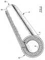
- the tubular two-layer collagen stopper 4has an inner diameter which corresponds to the outer diameter of a flexible catheter 3 (FIG. 1) or a rigid short catheter 16, which is called a sheet.
- the collagen stopper 4has a side slot 8 which extends the entire length and is in the form of a narrow V, the expansion of this V at the outer end A of the stopper with respect to the vessel 1 lies and extends at most over a quarter of the outer circumference.
- the other end B of the stopper 4is moderately sharpened in order to facilitate its penetration into the channel of the catheter 3 leading from the skin 2 to the vessel 1.
- the collagen stopper 4is sufficiently rigid, in particular due to its outer layer 7.
- the swelling of the collagen foambegins only through the action of the body fluid, i.e. H. of the blood and the tissue fluid, the inner layer 6 swelling first in about 30 s and later in about 2 minutes also the outer layer 7. This two-phase swelling process ensures reliable and simple pressing of the collagen stopper 4 up to the vessel wall 1, because the outer layer 7 maintains its stiffness for a sufficient period of time.
- a second part of the deviceis made of a rigid biologically inert and sterilizable material, e.g. B. made of plastic, stainless steel and. Like., Formed, which has substantially the same shape and wall thickness as the collagen stopper 4.
- the tubular applicator 5also has a V-shaped longitudinal slot 10, the larger widening of which is located at the outer end A with respect to the vessel 1 and does not extend over a quarter of the circumference of this outer end A.
- an outer collar or bead 9is formed, which is used to support the fingers, for. B. a thumb, when pushing in the collagen stopper 4 along the catheter 3 to the vessel wall.
- FIGS. 4 to 6The variant shown in FIGS. 4 to 6 is used with an outer short catheter 16, ie with a sheet, which is considerably shorter and is not directly connected to a pump or another corresponding device. Therefore, a profile ring 11 as an applicator can be pushed onto the short catheter 16 from its end before it is used on the patient. Since the dimensions of the applicator could be difficult to handle, it consists of two parts, namely the profile ring 11 which can be placed on the catheter 16 and which has a sufficiently deep annular groove 12 on its outer circumference. Before use, a longitudinally slotted ring 14 is pressed laterally into this annular groove 12 and is fastened to one end of a protruding projection 13. As a result, the thumb can be applied to the catheter 16 via the profile ring 11 by means of a link 15 provided at the free end of the attachment 13 sufficient pressure is generated and after application the profile ring 11 can be pulled out of the tissue.
- the handling of the device according to the invention for closing a vessel or an opening in a vessel wallis as follows.
- a collagen stopper 4is placed on its freely projecting section by widening its V-shaped longitudinal slot 8 - starting with the end A - pressed on the side.
- the arrangement of a relatively dimensionally stable applicator 5 according to FIGS. 1 and 3is carried out by pressing it onto the adjacent outer section of the catheter 3. Subsequently, by pressing the thumb against the annular bead 9 on the end, the applicator 5 together with the collagen stopper 4 advanced on the catheter 3 until the collagen stopper 4 has penetrated into the vessel 1 with its front end part.
- the catheter 3is pulled out of the vessel 1, the thumb pressure on the applicator 5 and thus also on the collagen stopper 4 being maintained in order to hold the collagen stopper 4 in place and to prevent it from being pulled out of the vessel 1.
- the collagen stopper 4is wetted with blood or tissue fluid, so that its thicker inner layer 6 swells first and closes the interior. With the desired delay, the outer layer 7 of the collagen stopper 4 also begins to swell, as a result of which a sealing effect is achieved between the edge of the opening and the outer wall of the collagen stopper becomes.
- the thumb pressure on the applicator 5should expediently be maintained for about 5 minutes in order to secure the position of the collagen stopper 4 during the swelling process.
- a foamis produced in a known manner from rindermine. No hardening agent is added to the foam intended for the inner layer; 0.2% glutaraldehyde or methylglyoxal is added as dry matter to the outer layer 7 before lyophilization. After the foam has been formed, the foam intended for the inner layer 6 is rolled up in a suitable layer thickness onto a rigid core made of stainless steel and pressed to 20% of the original volume. After this structure has stabilized, a predetermined layer of the hardened collagen foam is rolled up and pressed to 10-20% of the original volume. After stabilization of the outer layer and mutual fixation of the two layers 6, 7, the wall thickness in the dry state is less than approximately 2 mm.
- the outside diameter of the rigid corecorresponds to the outside diameter of the catheter 3 or 16 for which the respective collagen stopper 4 is intended.
- a longitudinal slit 8is cut out on the core in the two layers using a special cutting tool, and the collagen stopper 4 is then removed from the core.
- the length of the collagen stopper 4should be about 2 cm, with z. B. Obese patients can also use significantly longer collagen plugs 4 can. Together with a rigid applicator 5 of appropriate internal diameter, both parts are packaged, labeled accordingly, and sterilized by radiation.
- a foamis produced in a known manner from rindermine.
- cores made of stainless steel with an outer diameter corresponding to the inside diameter of the collagen stopper to be producedare placed in the middle of a preferably 10 mm thick collagen layer, and the lyophylization is carried out.
- the foamis cut lengthwise between the cores so that the foam layer surrounding the individual core is practically uniform.
- This foamis pressed onto the core to 10-20% of its original volume. Cores with this pressed foam are applied in the same way in the middle of a layer of the harder collagen mass before the lyophylization, this layer not exceeding the thickness of 6 mm.
- the foamis again cut into an elongated shape so that the layer of the hardened foam has a practically uniform thickness over the entire circumference of the first layer. Then this layer is pressed to 10% of its original volume. After the shape has been completely stabilized, the V-shaped longitudinal slot is produced with a special cutting tool in the two-layer resorbable collagen stopper and the finished collagen stopper 4 is removed from the core.
- a thromboplastic substancee.g. B. human thrombin
- driedin the proportion of 100 units for 1 g of dry collagen or 1% chitosan, based on dry, homogeneously mixed.
- An antibacterial agente.g. B. Neomycin, homogenized in a proportion of 0.5 g for 1 g of dry material.
- the preparation and preparation of the collagen stopperis the same as in Examples 1 to 4. However, a collagen obtained from bovine lichens is used as the starting material instead of bark pulp.
- the outer layer 7Since the main purpose of the outer layer 7 is to maintain the stability of the two-layer resorbable collagen stopper 4 during the time required for insertion up to the vessel wall and possibly for swelling, a thicker inner layer 6 of collagen foam is formed by pressing on the core, which has about 90% of the total wall thickness. The outer layer 7 is then by repeated application of the hardened collagen mass or by winding a film made from this mass. Then the entire structure is dried thoroughly.
- the stopper 4can also be made of another suitable material that should be resorbable and can increase its volume in a targeted manner under certain circumstances.
- the plugcan consist of only one layer or more than two layers and have a slightly conical shape.
- meanscan be provided on the outer wall of the stopper, which prevent penetration too deep into the vessel and thus prevent the risk of the entire vessel being closed. These funds can e.g. B. as profiles of the outer wall, as annular beads z. B. made of the rapidly swelling softer foam, etc.
Landscapes
- Health & Medical Sciences (AREA)
- Surgery (AREA)
- Life Sciences & Earth Sciences (AREA)
- Medical Informatics (AREA)
- Animal Behavior & Ethology (AREA)
- Engineering & Computer Science (AREA)
- Biomedical Technology (AREA)
- Heart & Thoracic Surgery (AREA)
- Cardiology (AREA)
- Molecular Biology (AREA)
- Nuclear Medicine, Radiotherapy & Molecular Imaging (AREA)
- General Health & Medical Sciences (AREA)
- Public Health (AREA)
- Veterinary Medicine (AREA)
- Materials For Medical Uses (AREA)
- Media Introduction/Drainage Providing Device (AREA)
- Cookers (AREA)
- Sampling And Sample Adjustment (AREA)
- Surgical Instruments (AREA)
Abstract
Translated fromGerman
Description
Translated fromGermanDie Erfindung betrifft eine Vorrichtung zum Verschließen einer Öffnung in einem Gefäß, z. B. einer Arterie, die durch Einführen eines Katheters verursacht wurde.The invention relates to a device for closing an opening in a vessel, for. B. an artery caused by insertion of a catheter.
Bei der Diagnostik und Therapie von Gefäß-, Herz- und anderer Erkrankungen werden häufig Katheter eingesetzt, die transkutan in das Luminum des Gefäßes eingeführt werden und verschiedene Außendurchmesser - gemessen in "French" - haben können, wobei ein French etwa 0,3 mm ist. Am häufigsten werden Katheter mit einem Durchmesser von sechs bis acht French angewandt. In neuerer Zeit werden sog. "Sheets" verwendet, die ein etwa 10 bis 15 cm langes Kunststoffrohr besitzen, dessen lichte Weite dem Außendurchmesser des Katheters entspricht. Die Wand des Katheters ist sehr dünn, etwa 0,1 mm. Dabei wird nach der Punktion der Arterie bzw. des Gefäßes mit einer dünnen Nadel ein dünner Metalleiter mit dieser Nadel eingeführt und die Nadel wird entfernt. Mittels dieses Metalleiters wird dann in das Gefäß ein Katheter mit oder ohne Sheet eingeführt.In the diagnosis and therapy of vascular, cardiac and other diseases, catheters are frequently used, which are inserted transcutaneously into the luminum of the vessel and can have different outside diameters - measured in "French" - a French being about 0.3 mm . Catheters with a diameter of six to eight French are most commonly used. "Sheets" have been used more recently, which have an approximately 10 to 15 cm long plastic tube, the inside diameter of which corresponds to the outside diameter of the catheter. The wall of the catheter is very thin, about 0.1 mm. After puncturing the artery or the vessel with a thin needle, a thin metal conductor is inserted with this needle and the needle is removed. A catheter with or without a sheet is then introduced into the vessel by means of this metal conductor.
Nach der Beendigung der Untersuchung oder der Therapie wird der Katheter abgenommen, wobei in der Wand des Gefäßes eine Öffnung von entsprechenden Abmessungen verbleibt. Da in etwa 90 % aller Fälle eine Arterie punktiert wird, treten häufig durch den Druck in den Arterien markante Nachblutungen auf. Eine spontane Verschließung der Öffnung durch Blutgerinnsel wird erreicht, indem auf die Arterie in diesem Bereich etwa 20 Minuten lang ein manueller Druck ausgeübt wird. Ferner soll der Patient während der folgenden 24 Stunden liegen, wobei nach Punktionen z. B. im Leistenbereich ein gepolstertes Gewicht von etwa 1 bis 2 kg, z. B. ein Sandsack, auf die punktierte Stelle gelegt wird. Dieses Verfahren hat u. a. den Nachteil, daß der Patient unter ärztlicher Überwachung liegen muß, was eine kostspielige stationäre Behandlung im Krankenhaus bedeutet. Ferner können Komplikationen eintreten, wenn eine Nachblutung nicht vollständig und dauerhaft unterbunden werden kann, was einen chirurgischen Eingriff mit einer unmittelbaren Sutur der Gefäßwand erforderlich macht. In weniger häufigen Fällen ist die Kompression zu intensiv, wobei das gebildete Gerinnsel nicht nur die Öffnung in der Gefäßwand sperrt, sondern auch das Gefäß selbst teilweise oder vollständig verschließt. Auch in diesem Fall ist ein chirurgischer Eingriff unerläßlich. In beiden Fällen verlängert sich die stationäre Behandlung mit den Nachteilen einer Blockade von Klinikbetten und eines erheblichen Kostenanstiegs.After the end of the examination or therapy, the catheter is removed, an opening of appropriate dimensions remaining in the wall of the vessel. Since an artery is punctured in approximately 90% of all cases, marked rebleeding often occurs due to the pressure in the arteries. A spontaneous occlusion of the opening by blood clots is achieved by applying manual pressure on the artery in this area for about 20 minutes. Furthermore, the patient should lie during the following 24 hours. B. in the groin area a padded weight of about 1 to 2 kg, z. B. a sandbag is placed on the dotted spot. This method has the disadvantage, among other things, that the patient has to be under medical supervision, which means an expensive inpatient treatment in the hospital. Furthermore, complications can arise if subsequent bleeding cannot be completely and permanently prevented, which necessitates a surgical intervention with an immediate suture of the vessel wall. In less frequent cases, the compression is too intense, whereby the clot formed not only blocks the opening in the vessel wall, but also partially or completely closes the vessel itself. Surgical intervention is also essential in this case. In both cases Inpatient treatment is prolonged with the disadvantages of blockage of hospital beds and a considerable increase in costs.
Gemäß einem im Versuchsstadium befindlichen Vorschlag zur Überwindung dieser Probleme wird vor der Punktion der Arterie der Abstand der Haut zum Gefäß gemessen. Nach dem Abnehmen des Katheters wird nach den Angaben des Meßgerätes ein besonderer Applikator zur Wand des Gefäßes geführt und mit diesem Applikator wird ggf. amorphes Kollagen einige Minuten lang eingepreßt, um die Gefäßöffnung zu verschließen.According to a proposal at the experimental stage to overcome these problems, the distance between the skin and the vessel is measured before the artery is punctured. After the catheter has been removed, a special applicator is guided to the wall of the vessel, and if necessary, amorphous collagen is pressed in for a few minutes with this applicator in order to close the vessel opening.
Bei einem anderen vorgeschlagenen Verfahren werden ein Anker aus Polymer, ein Kollagen-Stöpsel und ein resorbierbarer Faden verwendet. Nach Entfernen des Katheters wird der Anker mit dem Faden in die Gefäßwand eingeführt und mit diesem Faden wird der Kollagen-Stöpsel über die Öffnung fixiert.Another proposed method uses a polymer anchor, a collagen plug, and an absorbable thread. After the catheter has been removed, the anchor is inserted with the thread into the vessel wall and the collagen stopper is fixed with this thread via the opening.
Der Hauptnachteil beider Verfahren besteht darin, daß sie erst nach der Entfernung des Katheters oder Sheets angewandt werden können und Blutungen während ihrer Durchführung nicht unterbunden werden können, so daß die Gefahr einer Hämatombildung besteht.The main disadvantage of both methods is that they can only be used after the catheter or sheet has been removed and bleeding cannot be prevented during their implementation, so that there is a risk of hematoma formation.
Nachteilig ist ferner, daß die Stelle des so behandelten Gefäßes innerhalb eines Monats nicht für weitere Punktionen herangezogen werden kann. Das zweite Verfahren ist kompliziert und kann nur von chirurgischem Fachpersonal durchgeführt werden. Schließlich sind beide Verfahren zeit- und kostenaufwendig, so daß insbesondere in Ländern mit niedrigem Niveau des Sanitätswesens die Kosten für die Hilfsmittel den Aufwand für einen eintägigen Klinikaufenthalt übersteigen können.Another disadvantage is that the location of the vessel treated in this way cannot be used for further punctures within a month. The second procedure is complicated and can only be performed by surgical personnel. After all, both procedures are time-consuming and costly, so that the costs for the aids, in particular in countries with a low level of medical care can exceed the effort for a one-day hospital stay.
Aufgabe der Erfindung ist es, eine Vorrichtung zu schaffen, mit der eine z. B. durch Einführen eines Katheters verursachte Gefäßwand zuverlässig und auf einfache kostengünstige Weise verschlossen werden kann, um Nachblutungen zu vermeiden.The object of the invention is to provide a device with which a z. B. caused by inserting a catheter vessel wall can be closed reliably and in a simple inexpensive manner to avoid rebleeding.
Diese Aufgabe wird durch die im Patentanspruch 1 angegebenen Merkmale gelöst.This object is achieved by the features specified in claim 1.
Der Erfindung liegt die Erkenntnis zugrunde, daß der im Gefäß liegende Katheter als Führungsmittel für ein resorbierbares Verschlußelement dienen kann, das auf einem freien Katheterabschnitt außerhalb der Haut des Patienten plaziert wird und durch geeignete Hilfsmittel auf dem Katheter im Gewebekanal des Katheters bis zur Gefäßöffnung verschoben werden kann. Um die Gefäßöffnung nach Herausziehen des Katheters sicher zu verschließen, sollte das Material des Verschlußelements volumenvergrößernde Eigenschaften haben, damit der nach dem Herausziehen des Katheters entstandene Hohlraum ausgefüllt und auch eine sichere Abdichtung zwischen der Außenwand des Verschlußglieds und dem Rand der Gefäßöffnung erzielt wird.The invention is based on the finding that the catheter lying in the vessel can serve as a guide for a resorbable closure element which is placed on a free catheter section outside the patient's skin and can be displaced by suitable aids on the catheter in the tissue channel of the catheter up to the vessel opening can. In order to securely close the vessel opening after the catheter has been pulled out, the material of the closure element should have volume-increasing properties, so that the cavity formed after the catheter has been pulled out is filled and a secure seal is also achieved between the outer wall of the closure member and the edge of the vessel opening.
Verwirklicht ist dieses Konzept mittels eines resorbierbaren Kollagen-Stöpsels als Verschlußglied, der einen durchgehenden V-förmigen Längsschlitz aufweist, der am äußeren Ende des Stöpsels weiter als an seinem inneren Endabschnitt ist. Die innere lichte Weite des Kollagen-Stöpsels, d. h. sein Innendurchmesser, entspricht dem Außendurchmesser des jeweiligen Katheters. Zum manuellen Verschieben des Kollagen-Stöpsels dient ein im folgenden als Applikator bezeichnetes hohlzylindrisches Bauteil, dessen Form und Wandstärke denjenigen des Kollagen-Stöpsels entsprechen und der ebenfalls einen in Axialrichtung V-förmigen durchgehenden Längsschlitz aufweisen kann, dessen Aufweitung am äußeren Ende größer als am inneren patientennahen Endabschnitt ist.This concept is implemented by means of a resorbable collagen stopper as a closure member which has a continuous V-shaped longitudinal slot which is wider at the outer end of the stopper than at its inner end section. The inside clear width of the collagen stopper, ie its inside diameter, corresponds to the outside diameter of the respective one Catheters. For the manual displacement of the collagen stopper, a hollow cylindrical component, hereinafter referred to as the applicator, is used, the shape and wall thickness of which correspond to that of the collagen stopper and which can also have a longitudinal slot V-shaped in the axial direction, the expansion of which is greater at the outer end than at the inner end end section near the patient.
Vorteilhafte Weiterbildungen und Ausgestaltungen der erfindungsgemäßen Vorrichtung sind Gegenstand der Unteransprüche, wobei in einem weiteren Anspruch ein bevorzugtes Verfahren zur Herstellung eines Verschlußgliedes angegeben ist.Advantageous further developments and refinements of the device according to the invention are the subject of the dependent claims, a preferred method for producing a closure member being specified in a further claim.
Durch die erfindungsgemäße Vorrichtung ergeben sich eine Reihe von gravierenden Vorteilen für den Patienten und auch für die ärztliche Behandlung. Der frühzeitige und sichere Verschluß der Gefäßöffnung innerhalb weniger Minuten verringert wesentlich die Gefahr von durch Nachblutungen verursachten Komplikationen, z. B. Infektionen, die bisher regelmäßig chirurgische Eingriffe und eine längere stationäre Behandlung des Patienten erforderlich machten. Ferner erübrigt sich die langzeitliche Auflage von beschwerlichen Druckpolstern. Neben den gravierenden Erleichterungen für den Patienten lassen sich erhebliche Kosteneinsparungen mit der aus nur wenigen einfachen Einzelteilen bestehenden Vorrichtung erzielen, weil nur ein geringer Zeitaufwand des behandelnden Arztes zum Einführen und Fixieren der Vorrichtung aufzubringen ist und die stationäre Behandlung entfallen oder zumindest erheblich abgekürzt werden kann.The device according to the invention results in a number of serious advantages for the patient and also for medical treatment. The early and secure closure of the vascular opening within a few minutes significantly reduces the risk of complications caused by subsequent bleeding, e.g. B. infections that previously required regular surgery and longer inpatient treatment of the patient. Furthermore, the long-term edition of cumbersome pressure pads is not necessary. In addition to the major relief for the patient, considerable savings can be achieved with the device, which consists of only a few simple individual parts, because the treating physician only has to spend a small amount of time to insert and fix the device, and the inpatient treatment can be dispensed with or at least considerably shortened.
Im folgenden werden bevorzugte Ausführungsbeispiele der Erfindung anhand der Zeichnung im einzelnen erläutert. Es zeigen:
- Fig. 1
- einen in ein Gefäß eingeführten Katheter mit einer Vorrichtung gemäß der Erfindung;
- Fig. 2
- schematisch einen zweischichtigen resorbierbaren Kollagen-Stöpsel;
- Fig. 3
- einen starren Applikator aus inertem Werkstoff;
- Fig. 4
- schematisch einen zur Anwendung mit einem Sheet geeigneten zweiteiligen Applikator;
- Fig. 5
- teilgeschnitten einen profilierten Ring
und - Fig. 6
- schematisch einen Teil eines Ansatzes zum Ansetzen des Applikators nach Fig. 4.
- Fig. 1
- a catheter inserted into a vessel with a device according to the invention;
- Fig. 2
- schematically a two-layer resorbable collagen stopper;
- Fig. 3
- a rigid applicator made of inert material;
- Fig. 4
- schematically a two-part applicator suitable for use with a sheet;
- Fig. 5
- partially cut a profiled ring
and - Fig. 6
- schematically shows part of an approach for attaching the applicator according to FIG. 4.
Die Vorrichtung zum Verschließen einer Gefäßöffnung weist einen rohrförmigen resorbierbaren Kollagen-Stöpsel auf, der aus zwei Schichten von komprimierten Kollagen-Schäumen besteht. Eine innere Schicht 6 ist aus einem relativ weicheren und daher schneller aufquellenden Schaum gefertigt und bildet etwa 80 % der gesamten Wandstärke des Stöpsels 4. Eine äußere Schicht 7 besteht aus einem stärker ausgehärteten und daher langsamer aufquellenden Schaum und bildet etwa 20 % der gesamten Wandstärke. Beide Schichten 6, 7 sind fest gegeneinandergedrückt. Der rohrförmige zweischichtige Kollagen-Stöpsel 4 hat einen Innendurchmesser, der dem Außendurchmesser eines flexiblen Katheters 3 (Fig. 1) oder eines starren kurzen Katheters 16, der Sheet genannt wird, entspricht.The device for closing a vascular opening has a tubular resorbable collagen stopper, which consists of two layers of compressed collagen foams. An
Wie aus Fig. 2 ersichtlich, hat der Kollagen-Stöpsel 4 einen seitlichen Schlitz 8, der sich über die gesamte Länge erstreckt und die Form eines schmalen V hat, wobei die Aufweitung dieses V an dem äußeren Ende A des Stöpsels mit Bezug auf das Gefäß 1 liegt und sich höchstens über ein Viertel des Außenumfangs erstreckt. Das andere Ende B des Stöpsels 4 ist mäßig geschärft, um sein Eindringen in den von der Haut 2 zum Gefäß 1 führenden Kanal des Katheters 3 zu erleichtern. Vor dem Einführen ist der Kollagen-Stöpsel 4 insbesondere durch seine Außenschicht 7 ausreichend starr. Die Aufquellung des Kollagen-Schaumes beginnt erst durch Einwirkung der Körperflüssigkeit, d. h. des Blutes und der Gewebeflüssigkeit, wobei zuerst die innere Schicht 6 in ca. 30 s und später innerhalb von etwa 2 min auch die äußere Schicht 7 aufquellen. Dieser zweiphasige Quellvorgang gewährleistet ein zuverlässiges und einfaches Einpressen des Kollagen-Stöpsels 4 bis unmittelbar zur Gefäßwand 1, weil die äußere Schicht 7 eine ausreichende Zeitspanne ihre Formsteifigkeit beibehält.As can be seen in Fig. 2, the
Ein zweiter Teil der Vorrichtung wird von einem in Fig. 3 dargestellten Applikator 5 aus starrem biologisch inerten und sterilisierbaren Werkstoff, z. B. aus Kunststoff, rostfreiem Stahl u. dgl., gebildet, der im wesentlichen eine gleiche Form und Wandstärke wie der Kollagen-Stöpsel 4 hat.A second part of the device is made of a rigid biologically inert and sterilizable material, e.g. B. made of plastic, stainless steel and. Like., Formed, which has substantially the same shape and wall thickness as the
Auch der rohrförmige Applikator 5 weist einen V-förmigen Längsschlitz 10 auf, dessen größere Aufweitung sich am äußeren Ende A mit Bezug auf das Gefäß 1 befindet und sich nicht über ein Viertel des Umfangs dieses äußeren Endes A erstreckt. An diesem äußeren Ende A ist ein äußerer Ringbund bzw. Wulst 9 ausgebildet, der zur Abstützung der Finger, z. B. eines Daumens, beim Eindrücken des Kollagen-Stöpsels 4 entlang des Katheters 3 bis zur Gefäßwand dient.The
Für jeden Außendurchmesser der Katheter 3 oder 16 gibt es Kollagen-Stöpsel 4 und Applikatoren 5 mit jeweils angepaßtem Innendurchmesser. Beide Teile 4 und 5 werden in zweifacher sicherer Verpackung und durch Bestrahlung sterilisert ausgeliefert.For each outer diameter of the
Die in Fig. 4 bis 6 dargestellte Variante wird mit einem äußeren kurzen Katheter 16, d. h. mit einem Sheet, eingesetzt, der wesentlich kürzer ist und nicht unmittelbar an eine Pumpe oder eine andere entsprechende Vorrichtung angeschlossen wird. Daher kann vor seiner Anwendung am Patienten ein Profilring 11 als Applikator auf den Kurzkatheter 16 von seinem Ende her aufgeschoben werden. Da die Abmessungen des Applikators bei der Handhabung Schwierigkeiten bereiten könnten, besteht dieser aus zwei Teilen, nämlich aus dem auf den Katheter 16 aufsetzbaren Profilring 11, der an seinem Außenumfang eine ausreichend tiefe Ringnut 12 aufweist. Vor der Anwendung wird in diese Ringnut 12 ein längsgeschlitzter Ring 14 seitlich eingedrückt, der am einen Ende eines abstehenden Ansatzes 13 befestigt ist. Dadurch kann über den Profilring 11 auf den Katheter 16 mittels eines am freien Ende des Ansatzes 13 vorgesehenen Glieds 15 vom Daumen ein ausreichender Druck erzeugt werden und nach der Applikation kann der Profilring 11 aus dem Gewebe herausgezogen werden.The variant shown in FIGS. 4 to 6 is used with an outer
Die Handhabung der erfindungsgemäßen Vorrichtung zum Verschließen eines Gefäßes bzw. einer Öffnung in einer Gefäßwand ist folgende.The handling of the device according to the invention for closing a vessel or an opening in a vessel wall is as follows.
Vor dem Entfernen eines durch eine Punktion durch die Haut 2a und das Gewebe 2b in ein Blutgefäß 1, insbesondere in eine Arterie, eingeführten Katheters 3 wird auf dessen frei herausragenden Abschnitt ein Kollagen-Stöpsel 4 durch Aufweiten seines V-förmigen Längsschlitzes 8 - beginnend mit dem Ende A - seitlich aufgedrückt. Auf gleiche Weise erfolgt die Anordnung eines relativ formsteifen Applikators 5 nach Fig. 1 und 3 durch seitliches Aufdrücken auf den angrenzenden äußeren Abschnitt des Katheters 3. Anschließend wird durch Drücken des Daumens gegen den endseitigen Ringwulst 9 der Applikator 5 zusammen mit dem Kollagen-Stöpsel 4 auf dem Katheter 3 so weit vorgeschoben, bis der Kollagen-Stöpsel 4 mit seinem vorderen Endteil in das Gefäß 1 eingedrungen ist. Danach wird der Katheter 3 aus dem Gefäß 1 herausgezogen, wobei der Daumendruck auf den Applikator 5 und damit auch auf den Kollagen-Stöpsel 4 aufrechterhalten wird, um den Kollagen-Stöpsel 4 in seiner Lage festzuhalten und sein Herausziehen aus dem Gefäß 1 zu verhindern. Bereits während und insbesondere nach dem Herausziehen des Katheters 3 wird der Kollagen-Stöpsel 4 mit Blut bzw. Gewebeflüssigkeit benetzt, so daß zuerst seine dickere Innenschicht 6 aufquillt und den Innenraum verschließt. Mit der gewünschten Verzögerung beginnt auch die Außenschicht 7 des Kollagen-Stöpsels 4 aufzuquellen, wodurch eine Dichtwirkung zwischen dem Rand der Öffnung und der Außenwand des Kollagen-Stöpsels erreicht wird. Zweckmäßig sollte der Daumendruck auf den Applikator 5 etwa 5 min aufrechterhalten werden, um die Lage des Kollagen-Stöpsels 4 während des Quellvorgangs zu sichern.Before removing a
Im folgenden werden einige Beispiele zur Herstellung der erfindungsgemäßen Vorrichtung erläutert.Some examples for the production of the device according to the invention are explained below.
Aus Rindleimstoff wird ein Schaum in bekannter Weise erzeugt. In den für die innere Schicht bestimmten Schaumstoff wird kein Härtemittel zugegeben, in den für die äußere Schicht 7 wird vor der Lyophylisation 0,2 % Glutaraldehyd oder Methylglyoxal als Trockengut zugegeben. Nach Bildung des Schaumes wird der für die innere Schicht 6 bestimmte Schaum in angemessener Schichtdicke auf einen starren Kern aus rostfreiem Stahl aufgerollt und auf 20 % des ursprünglichen Volumens gepreßt. Nach der Stabilisation dieses Gebildes wird eine vorbestimmte Schicht des gehärteten Kollagen-Schaums aufgerollt und auf 10 - 20 % des ursprünglichen Volumens gepreßt. Nach der Stabilisation der Außenschicht und gegenseitiger Fixation der beiden Schichten 6, 7 liegt die Wandstärke im trockenen Zustand unter etwa 2 mm. Der Außendurchmesser des starren Kerns entspricht dem Außendurchmesser des Katheters 3 bzw. 16, für den der jeweilige Kollagen-Stöpsel 4 bestimmt ist. Auf dem Kern wird in den beiden Schichten mit einem speziellen Schneidewerkzeug ein Längsschlitz 8 ausgeschnitten und anschließend wird der Kollagen-Stöpsel 4 vom Kern entfernt. Zweckmäßig sollte die Länge des Kollagen-Stöpsels 4 etwa 2 cm betragen, wobei für besondere Anwendungen z. B. bei fettleibigen Patienten auch wesentliche längere Kollagen-Stöpsel 4 Verwendung finden können. Zusammen mit einem starren Applikator 5 von entsprechendem Innendurchmesser werden beide Teile verpackt, entsprechend bezeichnet und durch Bestrahlung sterilisiert.A foam is produced in a known manner from rindermine. No hardening agent is added to the foam intended for the inner layer; 0.2% glutaraldehyde or methylglyoxal is added as dry matter to the
Aus Rindleimstoff wird ein Schaum in bekannter Weise erzeugt. Vor der Lyophylisation werden in einen Behälter, in welchem die Lyophylisation ausgeführt wird, in die Mitte einer vorzugsweise 10 mm dicken Kollagenschicht Kerne aus rostfreiem Stahl mit einem dem Innendurchmesser des herzustellenden Kollagen-Stöpsels entsprechenden Außendurchmesser plaziert und die Lyophylisation wird durchgeführt. Danach wird der Schaum in Längsrichtung zwischen den Kernen so zerschnitten, daß die den einzelnen Kern umgebende Schaumschicht praktisch gleichmäßig ist. Dieser Schaum wird auf den Kern auf 10 - 20 % seines ursprünglichen Volumens gepreßt. Kerne mit diesem gepreßten Schaum werden vor der Lyophylisation in gleicher Weise in der Mitte einer Schicht aus der härteren Kollagen-Masse angebracht, wobei diese Schicht die Dicke von 6 mm nicht überschreitet. Nach Beendigung der Lyophylisation wird der Schaum wieder länglich zerschnitten, so daß die Schicht des gehärteten Schaumes auf dem gesamten Umfang der ersten Schicht praktisch gleichmäßige Dicke hat. Dann wird diese Schicht auf 10 % ihres ursprünglichen Volumens gepreßt. Nach vollkommener Stabilisation der Form wird der V-förmige Längsschlitz mit einem speziellen Schneidwerkzeug im zweischichtigen resorbierbaren Kollagen-Stöpsel hergestellt und der fertige Kollagen-Stöpsel 4 wird vom Kern abgenommen.A foam is produced in a known manner from rindermine. Before the lyophylization, in a container in which the lyophylization is carried out, cores made of stainless steel with an outer diameter corresponding to the inside diameter of the collagen stopper to be produced are placed in the middle of a preferably 10 mm thick collagen layer, and the lyophylization is carried out. Then the foam is cut lengthwise between the cores so that the foam layer surrounding the individual core is practically uniform. This foam is pressed onto the core to 10-20% of its original volume. Cores with this pressed foam are applied in the same way in the middle of a layer of the harder collagen mass before the lyophylization, this layer not exceeding the thickness of 6 mm. After the lyophylization has ended, the foam is again cut into an elongated shape so that the layer of the hardened foam has a practically uniform thickness over the entire circumference of the first layer. Then this layer is pressed to 10% of its original volume. After the shape has been completely stabilized, the V-shaped longitudinal slot is produced with a special cutting tool in the two-layer resorbable collagen stopper and the
In den Rindleimstoff wird vor der Lyophylisation ein thromboplastisch wirkender Stoff, z. B. menschlicher Thrombin, getrocknet, im Anteil von 100 Einheiten für 1 g Trockengut Kollagen oder 1 % Chitosan, bezogen auf Trockengut, homogen eingemischt.Before the lyophilization, a thromboplastic substance, e.g. B. human thrombin, dried, in the proportion of 100 units for 1 g of dry collagen or 1% chitosan, based on dry, homogeneously mixed.
In den Rindleimstoff wird vor der Lyophylisation ein antibakteriell wirkendes Mittel, z. B. Neomycin, im Anteil von 0,5 g für 1 g Trockengut einhomogenisiert.An antibacterial agent, e.g. B. Neomycin, homogenized in a proportion of 0.5 g for 1 g of dry material.
Die Herstellung und Zubereitung des Kollagen-Stöpsels ist die gleiche wie in den Beispielen 1 bis 4. Als Ausgangsmaterial wird jedoch statt Rindleimstoff ein aus Rindflechsen erhaltenes Kollagen eingesetzt.The preparation and preparation of the collagen stopper is the same as in Examples 1 to 4. However, a collagen obtained from bovine lichens is used as the starting material instead of bark pulp.
Da der Hauptzweck der äußeren Schicht 7 im Aufrechterhalten der Stabilität des zweischichtigen resorbierbaren Kollagen-Stöpsels 4 während der zum Einführen bis zur Gefäßwand und ggf. zum Aufquellen erforderlichen Zeit besteht, wird eine dickere Innenschicht 6 aus Kollagen-Schaum durch Pressen auf dem Kern gebildet, der etwa 90 % der gesamten Wandstärke besitzt. Die Außenschicht 7 wird dann durch wiederholte Applikation der ausgehärteten Kollagen-Masse oder durch Aufwickeln eines aus dieser Masse gebildeten Films hergestellt. Dann wird das gesamte Gebilde gründlich getrocknet.Since the main purpose of the
Die Erfindung ist nicht auf die dargestellte Vorrichtung beschränkt. So kann der Stöpsel 4 auch aus einem anderen geeigneten Material bestehen, das resorbierbar sein sollte und unter bestimmten Umständen gezielt sein Volumen vergrößern kann. Ferner kann der Stöpsel nur aus einer Schicht oder auch aus mehr als zwei Schichten bestehen und eine geringfügig kegelige Form haben. Schließlich können an der Außenwand des Stöpsels Mittel vorgesehen sein, die ein zu tiefes Eindringen in das Gefäß und damit die Gefahr eines Verschlusses des gesamten Gefäßes verhindern. Diese Mittel können z. B. als Profilierungen der Außenwand, als Ringwulste z. B. aus dem schnell quellenden weicheren Schaumstoff usw. ausgeführt sein.The invention is not limited to the device shown. For example, the
Claims (10)
Translated fromGermandadurch gekennzeichnet,
daß der Stöpsel (4) aus mindestens einem komprimierten Schaumstoff auf der Basis eines fibrillären Eiweißstoffes, wie insbesondere Kollagen, besteht.Device according to claim 1,
characterized,
that the stopper (4) consists of at least one compressed foam based on a fibrillar protein, such as collagen in particular.
dadurch gekennzeichnet,
daß der rohrförmige Stöpsel (4) einen vorzugsweise V-förmigen Längsschlitz (8) und an seinem vorderen Ende eine schneidenförmige Abschrägung aufweist.Device according to claim 1 or 2,
characterized,
that the tubular plug (4) has a preferably V-shaped longitudinal slot (8) and at its front end a cutting bevel.
dadurch gekennzeichnet,
daß der Stöpsel (4) aus einer dünneren formsteiferen äußeren Schaumstoffschicht (7) und einer inneren dickeren weicheren Schaumstoffschicht (6) besteht, die schneller als die äußere Schaumstoffschicht (7) aufquillt.Device according to one of claims 1 to 3,
characterized,
that the plug (4) from a thinner rigid outer foam layer (7) and an inner thicker softer foam layer (6), which swells faster than the outer foam layer (7).
dadurch gekennzeichnet,
daß der Stöpsel (4) kegelförmig ausgebildet ist, wobei sein außenseitiges Ende A höchstens 20 % breiter als sein inneres Ende ist.Device according to one of claims 1 to 4,
characterized,
that the plug (4) is conical, its outer end A being at most 20% wider than its inner end.
dadurch gekennzeichnet,
daß die Außen- und Innendurchmesser des Stöpsels (4) und des Applikators (5) aufeinander und auf den Außendurchmesser des Katheters (3, 16) abgestimmt sind.Device according to one of claims 1 to 5,
characterized,
that the outer and inner diameters of the stopper (4) and the applicator (5) are matched to one another and to the outer diameter of the catheter (3, 16).
dadurch gekennzeichnet,
daß der rohr- bzw. hülsenförmige Applikator (5) aus Kunststoff, Edelstahl od. dgl. besteht und einen vorzugsweise V-förmigen durchgehenden Längsschlitz (10) sowie an seinem äußeren Ende einen Ringwulst (9) aufweist.Device according to one of claims 1 to 6,
characterized,
that the tubular or sleeve-shaped applicator (5) is made of plastic, stainless steel or the like and has a preferably V-shaped continuous longitudinal slot (10) and an annular bead (9) at its outer end.
dadurch gekennzeichnet,
daß der Applikator (11 bis 15) mehrteilig ausgebildet ist und einen auf das Katheter (16) aufschiebbaren Ring (11) zum Verschieben des Stöpsels (4) aufweist, an dem Bauteile (13, 14, 15) zum Aufbringen und Übertragen des für die Verschiebung notwendigen Drucks fixierbar ist.Device according to one of claims 1 to 6,
characterized,
that the applicator (11 to 15) is constructed in several parts and has a ring (11) which can be pushed onto the catheter (16) for displacing the plug (4), on the components (13, 14, 15) for applying and transferring the for Shift necessary pressure is fixable.
dadurch gekennzeichnet,
daß der Schaumstoff des Stöpsels (4) eine thrombogen wirkende Substanz, einen RTG-Kontraststoff und/oder ein Antibiotikum enthält.Device according to one of claims 1 to 8,
characterized,
that the foam of the stopper (4) contains a thrombogenic substance, an RTG contrast material and / or an antibiotic.
dadurch gekennzeichnet,
daß auf einem stabförmigen Kern von vorgegebenem Durchmesser mindestens eine Schicht aus einem quell- bzw. schaumfähigen fibrillären Eiweißstoff, z. B. auf Kollagenbasis, in vorbestimmter Wandstärke aufgebracht wird,
daß diese Schicht(en) auf dem Kern zu einer vorbestimmten Wandstärke radial formhaltig komprimiert und getrocknet wird und
daß dieser rohrförmige Körper ggf. nach Herstellung eines V-förmigen Längsschlitzes in die Stöpsel von vorgegebener Länge unterteilt wird.Method for producing a plug for a device according to one of claims 1 to 9,
characterized,
that on a rod-shaped core of a given diameter at least one layer of a swellable or foamable fibrillar protein, for. B. is applied on a collagen basis in a predetermined wall thickness,
that this layer (s) on the core to a predetermined wall thickness is compressed radially and dried and
that this tubular body is divided into plugs of a predetermined length, if necessary after making a V-shaped longitudinal slot.
Applications Claiming Priority (2)
| Application Number | Priority Date | Filing Date | Title |
|---|---|---|---|
| CS923455ACZ281454B6 (en) | 1992-11-23 | 1992-11-23 | Aid for non-surgical closing of a hole in a vessel wall |
| CS3455/92 | 1992-11-23 |
Publications (3)
| Publication Number | Publication Date |
|---|---|
| EP0604761A2true EP0604761A2 (en) | 1994-07-06 |
| EP0604761A3 EP0604761A3 (en) | 1995-04-19 |
| EP0604761B1 EP0604761B1 (en) | 1998-10-28 |
Family
ID=5374671
Family Applications (1)
| Application Number | Title | Priority Date | Filing Date |
|---|---|---|---|
| EP93118817AExpired - LifetimeEP0604761B1 (en) | 1992-11-23 | 1993-11-23 | Device for closing an opening in a vessel |
Country Status (6)
| Country | Link |
|---|---|
| US (1) | US5522840A (en) |
| EP (1) | EP0604761B1 (en) |
| JP (1) | JPH0747131A (en) |
| AT (1) | ATE172626T1 (en) |
| CZ (1) | CZ281454B6 (en) |
| DE (1) | DE59309099D1 (en) |
Cited By (3)
| Publication number | Priority date | Publication date | Assignee | Title |
|---|---|---|---|---|
| EP0818178A3 (en)* | 1996-07-09 | 1998-12-09 | X-Site, L.L.C. | Anchoring device for sealing percutaneous punctures in vessels |
| WO2002100245A2 (en) | 2001-06-08 | 2002-12-19 | Morris Innovative Research, Inc. | Method and apparatus for sealing access |
| US7993365B2 (en) | 2001-06-08 | 2011-08-09 | Morris Innovative, Inc. | Method and apparatus for sealing access |
Families Citing this family (154)
| Publication number | Priority date | Publication date | Assignee | Title |
|---|---|---|---|---|
| US5609628A (en)* | 1995-04-20 | 1997-03-11 | Keranen; Victor J. | Intravascular graft and catheter |
| US6162192A (en) | 1998-05-01 | 2000-12-19 | Sub Q, Inc. | System and method for facilitating hemostasis of blood vessel punctures with absorbable sponge |
| US6071300A (en)* | 1995-09-15 | 2000-06-06 | Sub-Q Inc. | Apparatus and method for percutaneous sealing of blood vessel punctures |
| US6183497B1 (en) | 1998-05-01 | 2001-02-06 | Sub-Q, Inc. | Absorbable sponge with contrasting agent |
| US8716227B2 (en) | 1996-08-23 | 2014-05-06 | Cook Biotech Incorporated | Graft prosthesis, materials and methods |
| US6666892B2 (en)* | 1996-08-23 | 2003-12-23 | Cook Biotech Incorporated | Multi-formed collagenous biomaterial medical device |
| PL331765A1 (en)* | 1996-08-23 | 1999-08-02 | Cook Biotech Inc | Trnsplant prosthesis, materials and methods |
| US6117168A (en)* | 1996-12-31 | 2000-09-12 | Scimed Life Systems, Inc. | Multilayer liquid absorption and deformation devices |
| US7625352B1 (en) | 1998-05-01 | 2009-12-01 | Sub-Q, Inc. | Depth and puncture control for system for hemostasis of blood vessel |
| US20010045575A1 (en)* | 1998-05-01 | 2001-11-29 | Mark Ashby | Device and method for facilitating hemostasis of a biopsy tract |
| US6315753B1 (en)* | 1998-05-01 | 2001-11-13 | Sub-Q, Inc. | System and method for facilitating hemostasis of blood vessel punctures with absorbable sponge |
| US6183496B1 (en) | 1998-11-02 | 2001-02-06 | Datascope Investment Corp. | Collapsible hemostatic plug |
| US8882850B2 (en)* | 1998-12-01 | 2014-11-11 | Cook Biotech Incorporated | Multi-formed collagenous biomaterial medical device |
| CA2319447C (en) | 1998-12-01 | 2010-01-26 | Washington University | Embolization device |
| JP4271375B2 (en)* | 1999-02-10 | 2009-06-03 | サブ−キュー・インコーポレーテッド | Device and method for facilitating hemostasis in a biopsy duct |
| US6964685B2 (en) | 1999-06-22 | 2005-11-15 | The Brigham And Women's Hospital, Inc. | Biologic replacement for fibrin clot |
| US6110184A (en)* | 1999-08-04 | 2000-08-29 | Weadock; Kevin S. | Introducer with vascular sealing mechanism |
| US7695492B1 (en) | 1999-09-23 | 2010-04-13 | Boston Scientific Scimed, Inc. | Enhanced bleed back system |
| US6984219B2 (en) | 1999-09-23 | 2006-01-10 | Mark Ashby | Depth and puncture control for blood vessel hemostasis system |
| US8758400B2 (en) | 2000-01-05 | 2014-06-24 | Integrated Vascular Systems, Inc. | Closure system and methods of use |
| US7842068B2 (en) | 2000-12-07 | 2010-11-30 | Integrated Vascular Systems, Inc. | Apparatus and methods for providing tactile feedback while delivering a closure device |
| US6942674B2 (en) | 2000-01-05 | 2005-09-13 | Integrated Vascular Systems, Inc. | Apparatus and methods for delivering a closure device |
| US6461364B1 (en) | 2000-01-05 | 2002-10-08 | Integrated Vascular Systems, Inc. | Vascular sheath with bioabsorbable puncture site closure apparatus and methods of use |
| US6391048B1 (en) | 2000-01-05 | 2002-05-21 | Integrated Vascular Systems, Inc. | Integrated vascular device with puncture site closure component and sealant and methods of use |
| US9579091B2 (en) | 2000-01-05 | 2017-02-28 | Integrated Vascular Systems, Inc. | Closure system and methods of use |
| US6197042B1 (en)* | 2000-01-05 | 2001-03-06 | Medical Technology Group, Inc. | Vascular sheath with puncture site closure apparatus and methods of use |
| JP2003520830A (en)* | 2000-01-25 | 2003-07-08 | エドワーズ ライフサイエンシーズ コーポレイション | Delivery system for treatment of restenosis and anastomotic intimal hyperplasia |
| US6540735B1 (en) | 2000-05-12 | 2003-04-01 | Sub-Q, Inc. | System and method for facilitating hemostasis of blood vessel punctures with absorbable sponge |
| DE60144328D1 (en) | 2000-09-08 | 2011-05-12 | Abbott Vascular Inc | Surgical clamp |
| US7201725B1 (en) | 2000-09-25 | 2007-04-10 | Sub-Q, Inc. | Device and method for determining a depth of an incision |
| US6626918B1 (en) | 2000-10-06 | 2003-09-30 | Medical Technology Group | Apparatus and methods for positioning a vascular sheath |
| US6623510B2 (en) | 2000-12-07 | 2003-09-23 | Integrated Vascular Systems, Inc. | Closure device and methods for making and using them |
| US7905900B2 (en) | 2003-01-30 | 2011-03-15 | Integrated Vascular Systems, Inc. | Clip applier and methods of use |
| US7806904B2 (en) | 2000-12-07 | 2010-10-05 | Integrated Vascular Systems, Inc. | Closure device |
| US7211101B2 (en) | 2000-12-07 | 2007-05-01 | Abbott Vascular Devices | Methods for manufacturing a clip and clip |
| US6695867B2 (en) | 2002-02-21 | 2004-02-24 | Integrated Vascular Systems, Inc. | Plunger apparatus and methods for delivering a closure device |
| US8690910B2 (en) | 2000-12-07 | 2014-04-08 | Integrated Vascular Systems, Inc. | Closure device and methods for making and using them |
| US6623509B2 (en)* | 2000-12-14 | 2003-09-23 | Core Medical, Inc. | Apparatus and methods for sealing vascular punctures |
| US8083768B2 (en) | 2000-12-14 | 2011-12-27 | Ensure Medical, Inc. | Vascular plug having composite construction |
| US6890343B2 (en) | 2000-12-14 | 2005-05-10 | Ensure Medical, Inc. | Plug with detachable guidewire element and methods for use |
| US6846319B2 (en) | 2000-12-14 | 2005-01-25 | Core Medical, Inc. | Devices for sealing openings through tissue and apparatus and methods for delivering them |
| US6896692B2 (en) | 2000-12-14 | 2005-05-24 | Ensure Medical, Inc. | Plug with collet and apparatus and method for delivering such plugs |
| WO2002087636A1 (en)* | 2001-03-12 | 2002-11-07 | Sub-Q, Inc. | Methods for sterilizing cross-linked gelatin compositions |
| US8187625B2 (en)* | 2001-03-12 | 2012-05-29 | Boston Scientific Scimed, Inc. | Cross-linked gelatin composition comprising a wetting agent |
| US6863680B2 (en)* | 2001-11-08 | 2005-03-08 | Sub-Q, Inc. | System and method for delivering hemostasis promoting material to a blood vessel puncture site by fluid pressure |
| US7008440B2 (en) | 2001-11-08 | 2006-03-07 | Sub-Q, Inc. | System and method for delivering hemostasis promoting material to a blood vessel puncture site by fluid pressure |
| US7029489B1 (en) | 2001-05-18 | 2006-04-18 | Sub-Q, Inc. | System and method for delivering hemostasis promoting material to a blood vessel puncture site |
| IES20010547A2 (en) | 2001-06-07 | 2002-12-11 | Christy Cummins | Surgical Staple |
| US8465516B2 (en)* | 2001-07-26 | 2013-06-18 | Oregon Health Science University | Bodily lumen closure apparatus and method |
| US9861517B2 (en) | 2001-07-26 | 2018-01-09 | Cook Medical Technologies Llc | Vessel closure member, delivery apparatus, and method of inserting the member |
| US7192436B2 (en)* | 2001-11-08 | 2007-03-20 | Sub-Q, Inc. | Pledget-handling system and method for delivering hemostasis promoting material to a blood vessel puncture site by fluid pressure |
| US7037322B1 (en) | 2001-11-08 | 2006-05-02 | Sub-Q, Inc. | System and method for delivering hemostasis promoting material to a blood vessel puncture with a staging tube |
| US7025748B2 (en) | 2001-11-08 | 2006-04-11 | Boston Scientific Scimed, Inc. | Sheath based blood vessel puncture locator and depth indicator |
| US7037323B2 (en)* | 2001-11-08 | 2006-05-02 | Sub-Q, Inc. | Pledget-handling system and method for delivering hemostasis promoting material to a blood vessel puncture site by fluid pressure |
| TWI264301B (en)* | 2002-03-11 | 2006-10-21 | Ind Tech Res Inst | Multi-channel bioresorbable nerve regeneration conduit and preparation method for the same |
| IES20030424A2 (en) | 2002-06-04 | 2003-12-10 | Robert Stevenson | Blood vessel closure clip and delivery device |
| US20040102730A1 (en)* | 2002-10-22 | 2004-05-27 | Davis Thomas P. | System and method for facilitating hemostasis of blood vessel punctures with absorbable sponge |
| US7955353B1 (en) | 2002-11-04 | 2011-06-07 | Sub-Q, Inc. | Dissolvable closure device |
| US7455680B1 (en) | 2002-11-04 | 2008-11-25 | Boston Scientific Scimed, Inc. | Apparatus and method for inhibiting blood loss |
| US8317821B1 (en) | 2002-11-04 | 2012-11-27 | Boston Scientific Scimed, Inc. | Release mechanism |
| US7108710B2 (en) | 2002-11-26 | 2006-09-19 | Abbott Laboratories | Multi-element biased suture clip |
| US8709038B2 (en)* | 2002-12-20 | 2014-04-29 | Boston Scientific Scimed, Inc. | Puncture hole sealing device |
| US20040122349A1 (en)* | 2002-12-20 | 2004-06-24 | Lafontaine Daniel M. | Closure device with textured surface |
| US8398656B2 (en) | 2003-01-30 | 2013-03-19 | Integrated Vascular Systems, Inc. | Clip applier and methods of use |
| US8758398B2 (en) | 2006-09-08 | 2014-06-24 | Integrated Vascular Systems, Inc. | Apparatus and method for delivering a closure element |
| US7857828B2 (en) | 2003-01-30 | 2010-12-28 | Integrated Vascular Systems, Inc. | Clip applier and methods of use |
| US8905937B2 (en) | 2009-02-26 | 2014-12-09 | Integrated Vascular Systems, Inc. | Methods and apparatus for locating a surface of a body lumen |
| US8821534B2 (en) | 2010-12-06 | 2014-09-02 | Integrated Vascular Systems, Inc. | Clip applier having improved hemostasis and methods of use |
| US8202293B2 (en) | 2003-01-30 | 2012-06-19 | Integrated Vascular Systems, Inc. | Clip applier and methods of use |
| US7942897B2 (en)* | 2003-07-10 | 2011-05-17 | Boston Scientific Scimed, Inc. | System for closing an opening in a body cavity |
| US20050020899A1 (en)* | 2003-07-25 | 2005-01-27 | Rubicor Medical, Inc. | Post-biopsy cavity treatmetn implants and methods |
| US7744852B2 (en) | 2003-07-25 | 2010-06-29 | Rubicor Medical, Llc | Methods and systems for marking post biopsy cavity sites |
| US7537788B2 (en) | 2003-07-25 | 2009-05-26 | Rubicor Medical, Inc. | Post-biopsy cavity treatment implants and methods |
| US7645229B2 (en)* | 2003-09-26 | 2010-01-12 | Armstrong David N | Instrument and method for endoscopic visualization and treatment of anorectal fistula |
| US20050085853A1 (en)* | 2003-10-15 | 2005-04-21 | Forsberg Andrew T. | Collagen delivery assembly with blood perfusion holes |
| US8852229B2 (en) | 2003-10-17 | 2014-10-07 | Cordis Corporation | Locator and closure device and method of use |
| US7361183B2 (en)* | 2003-10-17 | 2008-04-22 | Ensure Medical, Inc. | Locator and delivery device and method of use |
| US7875043B1 (en) | 2003-12-09 | 2011-01-25 | Sub-Q, Inc. | Cinching loop |
| EP2193749B1 (en) | 2004-01-21 | 2017-03-01 | Cook Medical Technologies LLC | Implantable graft to close a fistula |
| IES20040368A2 (en) | 2004-05-25 | 2005-11-30 | James E Coleman | Surgical stapler |
| US8926654B2 (en) | 2005-05-04 | 2015-01-06 | Cordis Corporation | Locator and closure device and method of use |
| US8088144B2 (en) | 2005-05-04 | 2012-01-03 | Ensure Medical, Inc. | Locator and closure device and method of use |
| US7897167B2 (en)* | 2005-06-21 | 2011-03-01 | Cook Incorporated | Implantable graft to close a fistula |
| US8926633B2 (en) | 2005-06-24 | 2015-01-06 | Abbott Laboratories | Apparatus and method for delivering a closure element |
| US8313497B2 (en) | 2005-07-01 | 2012-11-20 | Abbott Laboratories | Clip applier and methods of use |
| US20070060895A1 (en) | 2005-08-24 | 2007-03-15 | Sibbitt Wilmer L Jr | Vascular closure methods and apparatuses |
| US8920442B2 (en) | 2005-08-24 | 2014-12-30 | Abbott Vascular Inc. | Vascular opening edge eversion methods and apparatuses |
| US9456811B2 (en) | 2005-08-24 | 2016-10-04 | Abbott Vascular Inc. | Vascular closure methods and apparatuses |
| US20070129757A1 (en)* | 2005-12-02 | 2007-06-07 | Cook Incorporated | Devices, systems, and methods for occluding a defect |
| AU2007208243B2 (en) | 2006-01-25 | 2013-09-19 | Children's Medical Center Corporation | Methods and procedures for ligament repair |
| US8808310B2 (en) | 2006-04-20 | 2014-08-19 | Integrated Vascular Systems, Inc. | Resettable clip applier and reset tools |
| US8556930B2 (en) | 2006-06-28 | 2013-10-15 | Abbott Laboratories | Vessel closure device |
| USD611144S1 (en) | 2006-06-28 | 2010-03-02 | Abbott Laboratories | Apparatus for delivering a closure element |
| WO2008024920A1 (en)* | 2006-08-24 | 2008-02-28 | Wilson-Cook Medical Inc. | Devices and methods for occluding a fistula |
| CA2664866C (en)* | 2006-09-28 | 2018-08-14 | Children's Medical Center Coporation | Methods and collagen products for tissue repair |
| US8226681B2 (en) | 2007-06-25 | 2012-07-24 | Abbott Laboratories | Methods, devices, and apparatus for managing access through tissue |
| US9308068B2 (en) | 2007-12-03 | 2016-04-12 | Sofradim Production | Implant for parastomal hernia |
| US8893947B2 (en) | 2007-12-17 | 2014-11-25 | Abbott Laboratories | Clip applier and methods of use |
| US20090157101A1 (en) | 2007-12-17 | 2009-06-18 | Abbott Laboratories | Tissue closure system and methods of use |
| US7841502B2 (en) | 2007-12-18 | 2010-11-30 | Abbott Laboratories | Modular clip applier |
| US9282965B2 (en) | 2008-05-16 | 2016-03-15 | Abbott Laboratories | Apparatus and methods for engaging tissue |
| US8118832B1 (en) | 2008-06-16 | 2012-02-21 | Morris Innovative, Inc. | Method and apparatus for sealing access |
| WO2009156866A2 (en) | 2008-06-27 | 2009-12-30 | Sofradim Production | Biosynthetic implant for soft tissue repair |
| US8398676B2 (en) | 2008-10-30 | 2013-03-19 | Abbott Vascular Inc. | Closure device |
| US8858594B2 (en) | 2008-12-22 | 2014-10-14 | Abbott Laboratories | Curved closure device |
| US8323312B2 (en) | 2008-12-22 | 2012-12-04 | Abbott Laboratories | Closure device |
| US9173644B2 (en) | 2009-01-09 | 2015-11-03 | Abbott Vascular Inc. | Closure devices, systems, and methods |
| US9486191B2 (en) | 2009-01-09 | 2016-11-08 | Abbott Vascular, Inc. | Closure devices |
| US9414820B2 (en) | 2009-01-09 | 2016-08-16 | Abbott Vascular Inc. | Closure devices, systems, and methods |
| US9089311B2 (en) | 2009-01-09 | 2015-07-28 | Abbott Vascular Inc. | Vessel closure devices and methods |
| US20100179589A1 (en) | 2009-01-09 | 2010-07-15 | Abbott Vascular Inc. | Rapidly eroding anchor |
| US20100185234A1 (en) | 2009-01-16 | 2010-07-22 | Abbott Vascular Inc. | Closure devices, systems, and methods |
| US20110054492A1 (en) | 2009-08-26 | 2011-03-03 | Abbott Laboratories | Medical device for repairing a fistula |
| FR2949688B1 (en) | 2009-09-04 | 2012-08-24 | Sofradim Production | FABRIC WITH PICOTS COATED WITH A BIORESORBABLE MICROPOROUS LAYER |
| US8303624B2 (en) | 2010-03-15 | 2012-11-06 | Abbott Cardiovascular Systems, Inc. | Bioabsorbable plug |
| US8758399B2 (en) | 2010-08-02 | 2014-06-24 | Abbott Cardiovascular Systems, Inc. | Expandable bioabsorbable plug apparatus and method |
| US8603116B2 (en) | 2010-08-04 | 2013-12-10 | Abbott Cardiovascular Systems, Inc. | Closure device with long tines |
| US8617184B2 (en) | 2011-02-15 | 2013-12-31 | Abbott Cardiovascular Systems, Inc. | Vessel closure system |
| FR2972626B1 (en) | 2011-03-16 | 2014-04-11 | Sofradim Production | PROSTHETIC COMPRISING A THREE-DIMENSIONAL KNIT AND ADJUSTED |
| US9149276B2 (en) | 2011-03-21 | 2015-10-06 | Abbott Cardiovascular Systems, Inc. | Clip and deployment apparatus for tissue closure |
| US8556932B2 (en) | 2011-05-19 | 2013-10-15 | Abbott Cardiovascular Systems, Inc. | Collapsible plug for tissue closure |
| WO2012174234A2 (en) | 2011-06-14 | 2012-12-20 | Cook Medical Technologies Llc | Fistula closure devices and methods |
| FR2977789B1 (en) | 2011-07-13 | 2013-07-19 | Sofradim Production | PROSTHETIC FOR UMBILIC HERNIA |
| FR2977790B1 (en) | 2011-07-13 | 2013-07-19 | Sofradim Production | PROSTHETIC FOR UMBILIC HERNIA |
| US9549715B2 (en) | 2011-08-09 | 2017-01-24 | Cook Regentec Llc | Vial useable in tissue extraction procedures |
| US9526603B2 (en) | 2011-09-30 | 2016-12-27 | Covidien Lp | Reversible stiffening of light weight mesh |
| US9332976B2 (en) | 2011-11-30 | 2016-05-10 | Abbott Cardiovascular Systems, Inc. | Tissue closure device |
| US8758427B2 (en) | 2011-12-02 | 2014-06-24 | Vascular Solutions, Inc. | Elongated expandable member for occluding varicose veins |
| FR2985170B1 (en) | 2011-12-29 | 2014-01-24 | Sofradim Production | PROSTHESIS FOR INGUINAL HERNIA |
| FR2985271B1 (en) | 2011-12-29 | 2014-01-24 | Sofradim Production | KNITTED PICOTS |
| EP2809340B1 (en) | 2012-02-01 | 2021-11-24 | Children's Medical Center Corporation | Biomaterial for articular cartilage maintenance and treatment of arthritis |
| FR2994185B1 (en) | 2012-08-02 | 2015-07-31 | Sofradim Production | PROCESS FOR THE PREPARATION OF A POROUS CHITOSAN LAYER |
| FR2995778B1 (en) | 2012-09-25 | 2015-06-26 | Sofradim Production | ABDOMINAL WALL REINFORCING PROSTHESIS AND METHOD FOR MANUFACTURING THE SAME |
| FR2995788B1 (en) | 2012-09-25 | 2014-09-26 | Sofradim Production | HEMOSTATIC PATCH AND PREPARATION METHOD |
| FR2995779B1 (en) | 2012-09-25 | 2015-09-25 | Sofradim Production | PROSTHETIC COMPRISING A TREILLIS AND A MEANS OF CONSOLIDATION |
| AU2013322268B2 (en) | 2012-09-28 | 2017-08-31 | Sofradim Production | Packaging for a hernia repair device |
| US20140148839A1 (en)* | 2012-11-27 | 2014-05-29 | Dusan Pavcnik | Bodily Lumen Closure Apparatus and Method |
| US9364209B2 (en) | 2012-12-21 | 2016-06-14 | Abbott Cardiovascular Systems, Inc. | Articulating suturing device |
| US9757495B2 (en) | 2013-02-01 | 2017-09-12 | Children's Medical Center Corporation | Collagen scaffolds |
| FR3006581B1 (en) | 2013-06-07 | 2016-07-22 | Sofradim Production | PROSTHESIS BASED ON TEXTILE FOR LAPAROSCOPIC PATHWAY |
| FR3006578B1 (en) | 2013-06-07 | 2015-05-29 | Sofradim Production | PROSTHESIS BASED ON TEXTILE FOR LAPAROSCOPIC PATHWAY |
| EP3000432B1 (en) | 2014-09-29 | 2022-05-04 | Sofradim Production | Textile-based prosthesis for treatment of inguinal hernia |
| EP3000433B1 (en) | 2014-09-29 | 2022-09-21 | Sofradim Production | Device for introducing a prosthesis for hernia treatment into an incision and flexible textile based prosthesis |
| EP3029189B1 (en) | 2014-12-05 | 2021-08-11 | Sofradim Production | Prosthetic porous knit, method of making same and hernia prosthesis |
| EP3059255B1 (en) | 2015-02-17 | 2020-05-13 | Sofradim Production | Method for preparing a chitosan-based matrix comprising a fiber reinforcement member |
| US9943314B2 (en) | 2015-04-14 | 2018-04-17 | Teleflex Innovations S.À.R.L. | Magnetically-driven delivery assembly and method |
| EP3085337B1 (en) | 2015-04-24 | 2022-09-14 | Sofradim Production | Prosthesis for supporting a breast structure |
| ES2676072T3 (en) | 2015-06-19 | 2018-07-16 | Sofradim Production | Synthetic prosthesis comprising a knitted fabric and a non-porous film and method of forming it |
| EP3195830B1 (en) | 2016-01-25 | 2020-11-18 | Sofradim Production | Prosthesis for hernia repair |
| CA3024196A1 (en) | 2016-07-06 | 2018-01-11 | Children's Medical Center Corporation | Indirect method of articular tissue repair |
| EP3312325B1 (en) | 2016-10-21 | 2021-09-22 | Sofradim Production | Method for forming a mesh having a barbed suture attached thereto and the mesh thus obtained |
| EP3398554B1 (en) | 2017-05-02 | 2025-06-25 | Sofradim Production | Prosthesis for inguinal hernia repair |
| EP3653171B1 (en) | 2018-11-16 | 2024-08-21 | Sofradim Production | Implants suitable for soft tissue repair |
| US12064330B2 (en) | 2020-04-28 | 2024-08-20 | Covidien Lp | Implantable prothesis for minimally invasive hernia repair |
Family Cites Families (14)
| Publication number | Priority date | Publication date | Assignee | Title |
|---|---|---|---|---|
| US2127903A (en)* | 1936-05-05 | 1938-08-23 | Davis & Geck Inc | Tube for surgical purposes and method of preparing and using the same |
| EP0048246B1 (en)* | 1980-03-27 | 1986-05-28 | National Research Development Corporation | Antimicrobial surgical implants |
| US4837285A (en)* | 1984-03-27 | 1989-06-06 | Medimatrix | Collagen matrix beads for soft tissue repair |
| US4778467A (en)* | 1984-04-25 | 1988-10-18 | The University Of Utah | Prostheses and methods for promoting nerve regeneration and for inhibiting the formation of neuromas |
| FR2625897A1 (en)* | 1988-01-14 | 1989-07-21 | Lefebvre Jean Marie | Vascular obturation device |
| US5254105A (en)* | 1988-05-26 | 1993-10-19 | Haaga John R | Sheath for wound closure caused by a medical tubular device |
| US4997439A (en)* | 1989-01-26 | 1991-03-05 | Chen Fusen H | Surgical closure or anastomotic device |
| US5047024A (en)* | 1989-08-30 | 1991-09-10 | Glassman Jacob A | Tampon construction |
| DE3928677C2 (en)* | 1989-08-30 | 1998-05-14 | Kimberly Clark Gmbh | Tampon for medical or hygienic purposes and process for its manufacture |
| US5061274A (en)* | 1989-12-04 | 1991-10-29 | Kensey Nash Corporation | Plug device for sealing openings and method of use |
| US5192300A (en)* | 1990-10-01 | 1993-03-09 | Quinton Instrument Company | Insertion assembly and method of inserting a vessel plug into the body of a patient |
| DE4132855A1 (en)* | 1991-10-02 | 1993-04-08 | Wisap Gmbh | INSTRUMENT FOR APPLICATION OF AN ADHAESION PROPHYLAXE FOR ENDOSCOPIC INTERVENTIONS |
| US5290310A (en)* | 1991-10-30 | 1994-03-01 | Howmedica, Inc. | Hemostatic implant introducer |
| US5334216A (en)* | 1992-12-10 | 1994-08-02 | Howmedica Inc. | Hemostatic plug |
- 1992
- 1992-11-23CZCS923455Apatent/CZ281454B6/enunknown
- 1993
- 1993-11-12USUS08/151,712patent/US5522840A/ennot_activeExpired - Fee Related
- 1993-11-22JPJP5292240Apatent/JPH0747131A/enactivePending
- 1993-11-23DEDE59309099Tpatent/DE59309099D1/ennot_activeExpired - Fee Related
- 1993-11-23ATAT93118817Tpatent/ATE172626T1/ennot_activeIP Right Cessation
- 1993-11-23EPEP93118817Apatent/EP0604761B1/ennot_activeExpired - Lifetime
Cited By (4)
| Publication number | Priority date | Publication date | Assignee | Title |
|---|---|---|---|---|
| EP0818178A3 (en)* | 1996-07-09 | 1998-12-09 | X-Site, L.L.C. | Anchoring device for sealing percutaneous punctures in vessels |
| WO2002100245A2 (en) | 2001-06-08 | 2002-12-19 | Morris Innovative Research, Inc. | Method and apparatus for sealing access |
| EP1392181A4 (en)* | 2001-06-08 | 2007-07-04 | Morris Innovative Res Inc | Method and apparatus for sealing access |
| US7993365B2 (en) | 2001-06-08 | 2011-08-09 | Morris Innovative, Inc. | Method and apparatus for sealing access |
Also Published As
| Publication number | Publication date |
|---|---|
| EP0604761B1 (en) | 1998-10-28 |
| EP0604761A3 (en) | 1995-04-19 |
| CZ281454B6 (en) | 1996-10-16 |
| US5522840A (en) | 1996-06-04 |
| ATE172626T1 (en) | 1998-11-15 |
| JPH0747131A (en) | 1995-02-21 |
| CZ345592A3 (en) | 1994-06-15 |
| DE59309099D1 (en) | 1998-12-03 |
Similar Documents
| Publication | Publication Date | Title |
|---|---|---|
| EP0604761B1 (en) | Device for closing an opening in a vessel | |
| DE69225720T2 (en) | HEMOSTATIC SEALING SYSTEM OF A POINT | |
| DE69932612T2 (en) | DEVICE FOR THE FACILITATED HEMOSTASE OF A BIOPSI TRACT | |
| DE69115052T2 (en) | Vascular plug insertion device. | |
| DE60200515T2 (en) | closure device | |
| DE69325290T2 (en) | HEMOSTATIC DEVICE FOR CLOSING CUTS | |
| DE69826982T2 (en) | Hemostatic puncture closure device | |
| DE69326146T2 (en) | CASE FOR CLOSING A WIND CAUSED BY A MEDICAL TUBULAR DEVICE | |
| DE69123817T2 (en) | Device for sealing puncture wounds | |
| DE69431395T2 (en) | Device for treating wounds | |
| DE69211150T2 (en) | INTRODUCTION DEVICE FOR HEMOSTATIC IMPLANT | |
| DE69722477T2 (en) | Surgical suture insertion device | |
| DE69738212T2 (en) | INTRODUCTION DEVICE FOR INTRODUCING A HEMOSTATIC CLOSURE INTO ONE SECTION | |
| DE68923617T2 (en) | DEVICE FOR SEALING A PERCUTANEOUS Vascular Puncture. | |
| DE69703863T2 (en) | Anchoring device for sealing vascular punctures | |
| DE4306850C1 (en) | Implant, especially for sealing trocar insertion points | |
| DE60309030T2 (en) | DEVICE FOR CLOSING VESSEL WALLS | |
| DE69121917T2 (en) | DEVICE FOR ATTACHING A DRAIN | |
| DE69837970T2 (en) | DEVICE FOR PERCUTANEOUS, HEMOSTATIC, SURGICAL SEAMS | |
| DE69733835T2 (en) | SYSTEM FOR CLOSING A VASCULAR WOUNCE | |
| DE60204021T2 (en) | CLOSING DEVICE FOR A VESSEL AND APPLICATION DEVICE | |
| DE69627107T2 (en) | Device for closing vascular wounds | |
| EP0654976A1 (en) | Use of and process for the introduction of fibrin sealant into a puncture channel. | |
| CH684307A5 (en) | Extension of linear and tubular tissue. | |
| DE2546283A1 (en) | USE FOR APPLICATION PREFERABLY IN VASCULAR SURGERY |
Legal Events
| Date | Code | Title | Description |
|---|---|---|---|
| PUAI | Public reference made under article 153(3) epc to a published international application that has entered the european phase | Free format text:ORIGINAL CODE: 0009012 | |
| AK | Designated contracting states | Kind code of ref document:A2 Designated state(s):AT BE CH DE DK ES FR GB IT LI NL SE | |
| PUAL | Search report despatched | Free format text:ORIGINAL CODE: 0009013 | |
| AK | Designated contracting states | Kind code of ref document:A3 Designated state(s):AT BE CH DE DK ES FR GB IT LI NL SE | |
| 17P | Request for examination filed | Effective date:19951012 | |
| 17Q | First examination report despatched | Effective date:19970725 | |
| GRAG | Despatch of communication of intention to grant | Free format text:ORIGINAL CODE: EPIDOS AGRA | |
| GRAG | Despatch of communication of intention to grant | Free format text:ORIGINAL CODE: EPIDOS AGRA | |
| GRAH | Despatch of communication of intention to grant a patent | Free format text:ORIGINAL CODE: EPIDOS IGRA | |
| GRAH | Despatch of communication of intention to grant a patent | Free format text:ORIGINAL CODE: EPIDOS IGRA | |
| GRAA | (expected) grant | Free format text:ORIGINAL CODE: 0009210 | |
| AK | Designated contracting states | Kind code of ref document:B1 Designated state(s):AT BE CH DE DK ES FR GB IT LI NL SE | |
| PG25 | Lapsed in a contracting state [announced via postgrant information from national office to epo] | Ref country code:NL Free format text:LAPSE BECAUSE OF FAILURE TO SUBMIT A TRANSLATION OF THE DESCRIPTION OR TO PAY THE FEE WITHIN THE PRESCRIBED TIME-LIMIT Effective date:19981028 Ref country code:ES Free format text:THE PATENT HAS BEEN ANNULLED BY A DECISION OF A NATIONAL AUTHORITY Effective date:19981028 | |
| REF | Corresponds to: | Ref document number:172626 Country of ref document:AT Date of ref document:19981115 Kind code of ref document:T | |
| REG | Reference to a national code | Ref country code:CH Ref legal event code:EP | |
| PG25 | Lapsed in a contracting state [announced via postgrant information from national office to epo] | Ref country code:AT Free format text:LAPSE BECAUSE OF NON-PAYMENT OF DUE FEES Effective date:19981123 | |
| PGFP | Annual fee paid to national office [announced via postgrant information from national office to epo] | Ref country code:GB Payment date:19981127 Year of fee payment:6 | |
| PG25 | Lapsed in a contracting state [announced via postgrant information from national office to epo] | Ref country code:LI Free format text:LAPSE BECAUSE OF NON-PAYMENT OF DUE FEES Effective date:19981130 Ref country code:CH Free format text:LAPSE BECAUSE OF NON-PAYMENT OF DUE FEES Effective date:19981130 Ref country code:BE Free format text:LAPSE BECAUSE OF NON-PAYMENT OF DUE FEES Effective date:19981130 | |
| PGFP | Annual fee paid to national office [announced via postgrant information from national office to epo] | Ref country code:FR Payment date:19981130 Year of fee payment:6 | |
| REF | Corresponds to: | Ref document number:59309099 Country of ref document:DE Date of ref document:19981203 | |
| GBT | Gb: translation of ep patent filed (gb section 77(6)(a)/1977) | Effective date:19981218 | |
| RAP2 | Party data changed (patent owner data changed or rights of a patent transferred) | Owner name:MEDICOL SCIENCES SPOL. S R.O. | |
| PG25 | Lapsed in a contracting state [announced via postgrant information from national office to epo] | Ref country code:SE Free format text:LAPSE BECAUSE OF FAILURE TO SUBMIT A TRANSLATION OF THE DESCRIPTION OR TO PAY THE FEE WITHIN THE PRESCRIBED TIME-LIMIT Effective date:19990128 Ref country code:DK Free format text:LAPSE BECAUSE OF FAILURE TO SUBMIT A TRANSLATION OF THE DESCRIPTION OR TO PAY THE FEE WITHIN THE PRESCRIBED TIME-LIMIT Effective date:19990128 | |
| PGFP | Annual fee paid to national office [announced via postgrant information from national office to epo] | Ref country code:DE Payment date:19990128 Year of fee payment:6 | |
| ET | Fr: translation filed | ||
| NLT2 | Nl: modifications (of names), taken from the european patent patent bulletin | Owner name:MEDICOL SCIENCES SPOL. S R.O. | |
| NLV1 | Nl: lapsed or annulled due to failure to fulfill the requirements of art. 29p and 29m of the patents act | ||
| BERE | Be: lapsed | Owner name:GOLLIEZ BIO-TEC LTD Effective date:19981130 | |
| REG | Reference to a national code | Ref country code:CH Ref legal event code:PL | |
| PLBE | No opposition filed within time limit | Free format text:ORIGINAL CODE: 0009261 | |
| STAA | Information on the status of an ep patent application or granted ep patent | Free format text:STATUS: NO OPPOSITION FILED WITHIN TIME LIMIT | |
| 26N | No opposition filed | ||
| PG25 | Lapsed in a contracting state [announced via postgrant information from national office to epo] | Ref country code:GB Free format text:LAPSE BECAUSE OF NON-PAYMENT OF DUE FEES Effective date:19991123 | |
| GBPC | Gb: european patent ceased through non-payment of renewal fee | Effective date:19991123 | |
| PG25 | Lapsed in a contracting state [announced via postgrant information from national office to epo] | Ref country code:FR Free format text:LAPSE BECAUSE OF NON-PAYMENT OF DUE FEES Effective date:20000731 | |
| PG25 | Lapsed in a contracting state [announced via postgrant information from national office to epo] | Ref country code:DE Free format text:LAPSE BECAUSE OF NON-PAYMENT OF DUE FEES Effective date:20000901 | |
| REG | Reference to a national code | Ref country code:FR Ref legal event code:ST | |
| PG25 | Lapsed in a contracting state [announced via postgrant information from national office to epo] | Ref country code:IT Free format text:LAPSE BECAUSE OF NON-PAYMENT OF DUE FEES;WARNING: LAPSES OF ITALIAN PATENTS WITH EFFECTIVE DATE BEFORE 2007 MAY HAVE OCCURRED AT ANY TIME BEFORE 2007. THE CORRECT EFFECTIVE DATE MAY BE DIFFERENT FROM THE ONE RECORDED. Effective date:20051123 |